EP0202743B1 - Assay for salicylate and apparatus for performing same - Google Patents
Assay for salicylate and apparatus for performing same Download PDFInfo
- Publication number
- EP0202743B1 EP0202743B1 EP86302476A EP86302476A EP0202743B1 EP 0202743 B1 EP0202743 B1 EP 0202743B1 EP 86302476 A EP86302476 A EP 86302476A EP 86302476 A EP86302476 A EP 86302476A EP 0202743 B1 EP0202743 B1 EP 0202743B1
- Authority
- EP
- European Patent Office
- Prior art keywords
- salicylate
- catechol
- hydroxylase
- sample
- concentration
- Prior art date
- Legal status (The legal status is an assumption and is not a legal conclusion. Google has not performed a legal analysis and makes no representation as to the accuracy of the status listed.)
- Expired - Lifetime
Links
- YGSDEFSMJLZEOE-UHFFFAOYSA-M salicylate Chemical compound OC1=CC=CC=C1C([O-])=O YGSDEFSMJLZEOE-UHFFFAOYSA-M 0.000 title claims abstract description 52
- 229960001860 salicylate Drugs 0.000 title claims abstract description 49
- 238000003556 assay Methods 0.000 title claims abstract description 10
- YCIMNLLNPGFGHC-UHFFFAOYSA-N catechol Chemical compound OC1=CC=CC=C1O YCIMNLLNPGFGHC-UHFFFAOYSA-N 0.000 claims abstract description 60
- 238000000034 method Methods 0.000 claims abstract description 31
- 102000004190 Enzymes Human genes 0.000 claims abstract description 18
- 108090000790 Enzymes Proteins 0.000 claims abstract description 18
- 238000006243 chemical reaction Methods 0.000 claims abstract description 16
- 238000002848 electrochemical method Methods 0.000 claims abstract description 10
- 239000007788 liquid Substances 0.000 claims abstract description 5
- 108010070996 Salicylate 1-monooxygenase Proteins 0.000 claims description 24
- 210000004369 blood Anatomy 0.000 claims description 8
- 239000008280 blood Substances 0.000 claims description 8
- 102000004169 proteins and genes Human genes 0.000 claims description 7
- 108090000623 proteins and genes Proteins 0.000 claims description 7
- 210000002966 serum Anatomy 0.000 claims description 6
- 241000589513 Burkholderia cepacia Species 0.000 claims description 4
- 241000589516 Pseudomonas Species 0.000 claims description 4
- 238000004255 ion exchange chromatography Methods 0.000 claims description 4
- 241000894007 species Species 0.000 claims description 4
- 241000894006 Bacteria Species 0.000 claims description 3
- 150000001450 anions Chemical class 0.000 claims description 3
- 210000002381 plasma Anatomy 0.000 claims description 3
- 210000001124 body fluid Anatomy 0.000 claims description 2
- 239000010839 body fluid Substances 0.000 claims description 2
- 238000004811 liquid chromatography Methods 0.000 claims description 2
- 238000004587 chromatography analysis Methods 0.000 claims 1
- 238000001514 detection method Methods 0.000 claims 1
- 230000005518 electrochemistry Effects 0.000 abstract 1
- 239000000243 solution Substances 0.000 description 19
- 239000007853 buffer solution Substances 0.000 description 11
- 229930027945 nicotinamide-adenine dinucleotide Natural products 0.000 description 10
- BOPGDPNILDQYTO-NNYOXOHSSA-N nicotinamide-adenine dinucleotide Chemical compound C1=CCC(C(=O)N)=CN1[C@H]1[C@H](O)[C@H](O)[C@@H](COP(O)(=O)OP(O)(=O)OC[C@@H]2[C@H]([C@@H](O)[C@@H](O2)N2C3=NC=NC(N)=C3N=C2)O)O1 BOPGDPNILDQYTO-NNYOXOHSSA-N 0.000 description 9
- 238000002484 cyclic voltammetry Methods 0.000 description 8
- BSYNRYMUTXBXSQ-UHFFFAOYSA-N Aspirin Chemical compound CC(=O)OC1=CC=CC=C1C(O)=O BSYNRYMUTXBXSQ-UHFFFAOYSA-N 0.000 description 6
- 229960001138 acetylsalicylic acid Drugs 0.000 description 6
- QVGXLLKOCUKJST-UHFFFAOYSA-N atomic oxygen Chemical compound [O] QVGXLLKOCUKJST-UHFFFAOYSA-N 0.000 description 6
- 229910052760 oxygen Inorganic materials 0.000 description 6
- 239000001301 oxygen Substances 0.000 description 6
- 239000000872 buffer Substances 0.000 description 5
- 230000000694 effects Effects 0.000 description 5
- 238000005259 measurement Methods 0.000 description 5
- 230000003647 oxidation Effects 0.000 description 5
- 238000007254 oxidation reaction Methods 0.000 description 5
- 239000000758 substrate Substances 0.000 description 5
- XLYOFNOQVPJJNP-UHFFFAOYSA-N water Chemical compound O XLYOFNOQVPJJNP-UHFFFAOYSA-N 0.000 description 5
- WOAHJDHKFWSLKE-UHFFFAOYSA-N 1,2-benzoquinone Chemical compound O=C1C=CC=CC1=O WOAHJDHKFWSLKE-UHFFFAOYSA-N 0.000 description 4
- BAWFJGJZGIEFAR-NNYOXOHSSA-O NAD(+) Chemical compound NC(=O)C1=CC=C[N+]([C@H]2[C@@H]([C@H](O)[C@@H](COP(O)(=O)OP(O)(=O)OC[C@@H]3[C@H]([C@@H](O)[C@@H](O3)N3C4=NC=NC(N)=C4N=C3)O)O2)O)=C1 BAWFJGJZGIEFAR-NNYOXOHSSA-O 0.000 description 4
- 238000011088 calibration curve Methods 0.000 description 4
- 239000012153 distilled water Substances 0.000 description 4
- 239000000463 material Substances 0.000 description 4
- 238000000746 purification Methods 0.000 description 4
- 239000000126 substance Substances 0.000 description 4
- 238000002835 absorbance Methods 0.000 description 3
- 239000003153 chemical reaction reagent Substances 0.000 description 3
- 229940079593 drug Drugs 0.000 description 3
- 239000003814 drug Substances 0.000 description 3
- 238000012544 monitoring process Methods 0.000 description 3
- 229920000447 polyanionic polymer Polymers 0.000 description 3
- CURLTUGMZLYLDI-UHFFFAOYSA-N Carbon dioxide Chemical compound O=C=O CURLTUGMZLYLDI-UHFFFAOYSA-N 0.000 description 2
- 108010074633 Mixed Function Oxygenases Proteins 0.000 description 2
- 102000008109 Mixed Function Oxygenases Human genes 0.000 description 2
- PMZURENOXWZQFD-UHFFFAOYSA-L Sodium Sulfate Chemical compound [Na+].[Na+].[O-]S([O-])(=O)=O PMZURENOXWZQFD-UHFFFAOYSA-L 0.000 description 2
- PXIPVTKHYLBLMZ-UHFFFAOYSA-N Sodium azide Chemical compound [Na+].[N-]=[N+]=[N-] PXIPVTKHYLBLMZ-UHFFFAOYSA-N 0.000 description 2
- ABBQHOQBGMUPJH-UHFFFAOYSA-M Sodium salicylate Chemical compound [Na+].OC1=CC=CC=C1C([O-])=O ABBQHOQBGMUPJH-UHFFFAOYSA-M 0.000 description 2
- 239000002253 acid Substances 0.000 description 2
- 231100000570 acute poisoning Toxicity 0.000 description 2
- PNEYBMLMFCGWSK-UHFFFAOYSA-N aluminium oxide Inorganic materials [O-2].[O-2].[O-2].[Al+3].[Al+3] PNEYBMLMFCGWSK-UHFFFAOYSA-N 0.000 description 2
- 230000000202 analgesic effect Effects 0.000 description 2
- 230000003110 anti-inflammatory effect Effects 0.000 description 2
- 239000002221 antipyretic Substances 0.000 description 2
- 230000015572 biosynthetic process Effects 0.000 description 2
- 150000001875 compounds Chemical class 0.000 description 2
- PCHJSUWPFVWCPO-UHFFFAOYSA-N gold Chemical compound [Au] PCHJSUWPFVWCPO-UHFFFAOYSA-N 0.000 description 2
- 229910052737 gold Inorganic materials 0.000 description 2
- 239000010931 gold Substances 0.000 description 2
- 230000000977 initiatory effect Effects 0.000 description 2
- 238000005342 ion exchange Methods 0.000 description 2
- 238000011002 quantification Methods 0.000 description 2
- 150000003839 salts Chemical class 0.000 description 2
- 239000011550 stock solution Substances 0.000 description 2
- 230000001225 therapeutic effect Effects 0.000 description 2
- 238000010626 work up procedure Methods 0.000 description 2
- BLFRQYKZFKYQLO-UHFFFAOYSA-N 4-aminobutan-1-ol Chemical compound NCCCCO BLFRQYKZFKYQLO-UHFFFAOYSA-N 0.000 description 1
- OKTJSMMVPCPJKN-UHFFFAOYSA-N Carbon Chemical compound [C] OKTJSMMVPCPJKN-UHFFFAOYSA-N 0.000 description 1
- 238000009007 Diagnostic Kit Methods 0.000 description 1
- MYMOFIZGZYHOMD-UHFFFAOYSA-N Dioxygen Chemical compound O=O MYMOFIZGZYHOMD-UHFFFAOYSA-N 0.000 description 1
- XLRHXNIVIZZOON-WFUPGROFSA-L Flavin adenine dinucleotide disodium Chemical compound [Na+].[Na+].C1=NC2=C(N)N=CN=C2N1[C@@H]([C@H](O)[C@@H]1O)O[C@@H]1COP([O-])(=O)OP([O-])(=O)OC[C@@H](O)[C@@H](O)[C@@H](O)CN1C2=NC(=O)NC(=O)C2=NC2=C1C=C(C)C(C)=C2 XLRHXNIVIZZOON-WFUPGROFSA-L 0.000 description 1
- 241001071861 Lethrinus genivittatus Species 0.000 description 1
- 125000003047 N-acetyl group Chemical group 0.000 description 1
- FAPWRFPIFSIZLT-UHFFFAOYSA-M Sodium chloride Chemical compound [Na+].[Cl-] FAPWRFPIFSIZLT-UHFFFAOYSA-M 0.000 description 1
- LGDAGYXJBDILKZ-UHFFFAOYSA-N [2-methyl-1,1-dioxo-3-(pyridin-2-ylcarbamoyl)-1$l^{6},2-benzothiazin-4-yl] 2,2-dimethylpropanoate Chemical compound CC(C)(C)C(=O)OC=1C2=CC=CC=C2S(=O)(=O)N(C)C=1C(=O)NC1=CC=CC=N1 LGDAGYXJBDILKZ-UHFFFAOYSA-N 0.000 description 1
- XJLXINKUBYWONI-DQQFMEOOSA-N [[(2r,3r,4r,5r)-5-(6-aminopurin-9-yl)-3-hydroxy-4-phosphonooxyoxolan-2-yl]methoxy-hydroxyphosphoryl] [(2s,3r,4s,5s)-5-(3-carbamoylpyridin-1-ium-1-yl)-3,4-dihydroxyoxolan-2-yl]methyl phosphate Chemical compound NC(=O)C1=CC=C[N+]([C@@H]2[C@H]([C@@H](O)[C@H](COP([O-])(=O)OP(O)(=O)OC[C@@H]3[C@H]([C@@H](OP(O)(O)=O)[C@@H](O3)N3C4=NC=NC(N)=C4N=C3)O)O2)O)=C1 XJLXINKUBYWONI-DQQFMEOOSA-N 0.000 description 1
- 229940068372 acetyl salicylate Drugs 0.000 description 1
- 239000003513 alkali Substances 0.000 description 1
- 125000000129 anionic group Chemical group 0.000 description 1
- 239000000730 antalgic agent Substances 0.000 description 1
- 230000001754 anti-pyretic effect Effects 0.000 description 1
- 229940125716 antipyretic agent Drugs 0.000 description 1
- 206010003246 arthritis Diseases 0.000 description 1
- 239000002585 base Substances 0.000 description 1
- 235000021170 buffet Nutrition 0.000 description 1
- 239000001569 carbon dioxide Substances 0.000 description 1
- 229910002092 carbon dioxide Inorganic materials 0.000 description 1
- 230000003197 catalytic effect Effects 0.000 description 1
- 238000006555 catalytic reaction Methods 0.000 description 1
- 238000013375 chromatographic separation Methods 0.000 description 1
- 230000004087 circulation Effects 0.000 description 1
- 125000004122 cyclic group Chemical group 0.000 description 1
- 238000006114 decarboxylation reaction Methods 0.000 description 1
- 230000001419 dependent effect Effects 0.000 description 1
- 238000011161 development Methods 0.000 description 1
- 238000009792 diffusion process Methods 0.000 description 1
- 229910001882 dioxygen Inorganic materials 0.000 description 1
- ZPWVASYFFYYZEW-UHFFFAOYSA-L dipotassium hydrogen phosphate Chemical compound [K+].[K+].OP([O-])([O-])=O ZPWVASYFFYYZEW-UHFFFAOYSA-L 0.000 description 1
- QRGNQKGQENGQSE-WUEGHLCSSA-L disodium;[[(2r,3s,4r,5r)-5-(6-aminopurin-9-yl)-3,4-dihydroxyoxolan-2-yl]methoxy-oxidophosphoryl] [(2r,3s,4r,5r)-5-(3-carbamoyl-4h-pyridin-1-yl)-3,4-dihydroxyoxolan-2-yl]methyl phosphate Chemical compound [Na+].[Na+].C1=CCC(C(=O)N)=CN1[C@H]1[C@H](O)[C@H](O)[C@@H](COP([O-])(=O)OP([O-])(=O)OC[C@@H]2[C@H]([C@@H](O)[C@@H](O2)N2C3=NC=NC(N)=C3N=C2)O)O1 QRGNQKGQENGQSE-WUEGHLCSSA-L 0.000 description 1
- 238000000835 electrochemical detection Methods 0.000 description 1
- 238000006911 enzymatic reaction Methods 0.000 description 1
- 238000001030 gas--liquid chromatography Methods 0.000 description 1
- 210000001035 gastrointestinal tract Anatomy 0.000 description 1
- 239000011521 glass Substances 0.000 description 1
- 229910021397 glassy carbon Inorganic materials 0.000 description 1
- 239000010439 graphite Substances 0.000 description 1
- 229910002804 graphite Inorganic materials 0.000 description 1
- 230000002440 hepatic effect Effects 0.000 description 1
- 238000004128 high performance liquid chromatography Methods 0.000 description 1
- 230000033444 hydroxylation Effects 0.000 description 1
- 238000005805 hydroxylation reaction Methods 0.000 description 1
- 239000012535 impurity Substances 0.000 description 1
- 238000011534 incubation Methods 0.000 description 1
- 210000004185 liver Anatomy 0.000 description 1
- 230000007774 longterm Effects 0.000 description 1
- 239000000203 mixture Substances 0.000 description 1
- 230000004048 modification Effects 0.000 description 1
- 238000012986 modification Methods 0.000 description 1
- 150000002989 phenols Chemical class 0.000 description 1
- 239000008363 phosphate buffer Substances 0.000 description 1
- 239000008055 phosphate buffer solution Substances 0.000 description 1
- 238000005498 polishing Methods 0.000 description 1
- GNSKLFRGEWLPPA-UHFFFAOYSA-M potassium dihydrogen phosphate Chemical compound [K+].OP(O)([O-])=O GNSKLFRGEWLPPA-UHFFFAOYSA-M 0.000 description 1
- 238000004393 prognosis Methods 0.000 description 1
- 238000001742 protein purification Methods 0.000 description 1
- 238000012207 quantitative assay Methods 0.000 description 1
- -1 salicylate ions Chemical class 0.000 description 1
- 239000012898 sample dilution Substances 0.000 description 1
- 230000035945 sensitivity Effects 0.000 description 1
- 238000000926 separation method Methods 0.000 description 1
- 239000002002 slurry Substances 0.000 description 1
- 229960004025 sodium salicylate Drugs 0.000 description 1
- 229910052938 sodium sulfate Inorganic materials 0.000 description 1
- 235000011152 sodium sulphate Nutrition 0.000 description 1
- 238000002798 spectrophotometry method Methods 0.000 description 1
- 238000003756 stirring Methods 0.000 description 1
- 238000010408 sweeping Methods 0.000 description 1
- 230000001839 systemic circulation Effects 0.000 description 1
- LENZDBCJOHFCAS-UHFFFAOYSA-N tris Chemical compound OCC(N)(CO)CO LENZDBCJOHFCAS-UHFFFAOYSA-N 0.000 description 1
- 238000000108 ultra-filtration Methods 0.000 description 1
- 238000002525 ultrasonication Methods 0.000 description 1
Images


Classifications
-
- C—CHEMISTRY; METALLURGY
- C12—BIOCHEMISTRY; BEER; SPIRITS; WINE; VINEGAR; MICROBIOLOGY; ENZYMOLOGY; MUTATION OR GENETIC ENGINEERING
- C12N—MICROORGANISMS OR ENZYMES; COMPOSITIONS THEREOF; PROPAGATING, PRESERVING, OR MAINTAINING MICROORGANISMS; MUTATION OR GENETIC ENGINEERING; CULTURE MEDIA
- C12N9/00—Enzymes; Proenzymes; Compositions thereof; Processes for preparing, activating, inhibiting, separating or purifying enzymes
- C12N9/0004—Oxidoreductases (1.)
- C12N9/0071—Oxidoreductases (1.) acting on paired donors with incorporation of molecular oxygen (1.14)
- C12N9/0073—Oxidoreductases (1.) acting on paired donors with incorporation of molecular oxygen (1.14) with NADH or NADPH as one donor, and incorporation of one atom of oxygen 1.14.13
-
- C—CHEMISTRY; METALLURGY
- C12—BIOCHEMISTRY; BEER; SPIRITS; WINE; VINEGAR; MICROBIOLOGY; ENZYMOLOGY; MUTATION OR GENETIC ENGINEERING
- C12Q—MEASURING OR TESTING PROCESSES INVOLVING ENZYMES, NUCLEIC ACIDS OR MICROORGANISMS; COMPOSITIONS OR TEST PAPERS THEREFOR; PROCESSES OF PREPARING SUCH COMPOSITIONS; CONDITION-RESPONSIVE CONTROL IN MICROBIOLOGICAL OR ENZYMOLOGICAL PROCESSES
- C12Q1/00—Measuring or testing processes involving enzymes, nucleic acids or microorganisms; Compositions therefor; Processes of preparing such compositions
- C12Q1/26—Measuring or testing processes involving enzymes, nucleic acids or microorganisms; Compositions therefor; Processes of preparing such compositions involving oxidoreductase
-
- C—CHEMISTRY; METALLURGY
- C12—BIOCHEMISTRY; BEER; SPIRITS; WINE; VINEGAR; MICROBIOLOGY; ENZYMOLOGY; MUTATION OR GENETIC ENGINEERING
- C12Y—ENZYMES
- C12Y114/00—Oxidoreductases acting on paired donors, with incorporation or reduction of molecular oxygen (1.14)
- C12Y114/13—Oxidoreductases acting on paired donors, with incorporation or reduction of molecular oxygen (1.14) with NADH or NADPH as one donor, and incorporation of one atom of oxygen (1.14.13)
- C12Y114/13001—Salicylate 1-monooxygenase (1.14.13.1)
-
- Y—GENERAL TAGGING OF NEW TECHNOLOGICAL DEVELOPMENTS; GENERAL TAGGING OF CROSS-SECTIONAL TECHNOLOGIES SPANNING OVER SEVERAL SECTIONS OF THE IPC; TECHNICAL SUBJECTS COVERED BY FORMER USPC CROSS-REFERENCE ART COLLECTIONS [XRACs] AND DIGESTS
- Y10—TECHNICAL SUBJECTS COVERED BY FORMER USPC
- Y10S—TECHNICAL SUBJECTS COVERED BY FORMER USPC CROSS-REFERENCE ART COLLECTIONS [XRACs] AND DIGESTS
- Y10S435/00—Chemistry: molecular biology and microbiology
- Y10S435/817—Enzyme or microbe electrode
Definitions
- the present invention is concerned with an assay for salicylate and with apparatus for performing the said assay.
- Aspirin N-acetyl salicylate
- Aspirin N-acetyl salicylate
- the drug is readily absorbed from the gastrointestinal tract into the portal circulation and is rapidly hydrolysed by hepatic enzymes, largely during its first pass through the liver, to yield free salicylate.
- aspirin as a short term analgesic/antipyretic agent produces relatively low levels of salicylate in the serum (30-100 mg/I ; 0.22-0.73 mM) and consequently monitoring of such levels is not normally necessary.
- This assay is based on the reaction of phenols with the Folin-Ciocalteau reagent in strong alkali solution to produce a blue colour which can be measured spectrophotometrically/colorimetr ically (M. J. H. Smith & J. M. Talbot, Brit. J. Exp. Path. (1950), 31, 65).
- This method however requires the initial removal of protein from the serum samples and is not very specific for salicylate resulting in high background values.
- a method for the assay of salicylate which comprises the steps of :
- liquid sample is contacted with an electrode poised at a suitable potential for the direct electrochemical measurement of the catechol.
- hydroxylation and simultaneous decarboxylation of salicylate to yield catechol may be catalysed by any suitable enzyme of the type defined as EC 1.14.13.1 and named as salicylate hydroxylase (otherwise known as salicylate 1- monooxygenase) by the International Union of Biochemistry (Enzyme Nomenclature, 1978, Academic Press, New York (1979).
- the enzyme is usually a salicylate hydroxylase isolated from a bacterium, which is preferably a species of Pseudomonas, most especially Pseudomonas cepacia (ATCC 29351) or Pseudomonas cepacia (ATCC 29352).
- Such an enzyme material is preferably purified by ion-exchange chromatography, e.g. on an ion-exchange anion column.
- ion-exchange chromatography e.g. on an ion-exchange anion column.
- Fast protein ion exchange chromatography on a Polyanion SI column (Pharmacia) is of particular value.
- Any dissolved salicylate sample is susceptible of treatment in accordance with the invention.
- the sample comprises whole blood. It may, possibly, comprise plasma, serum, or any other like body fluid.
- liquid sample may conveniently be contacted with an electrode having at its surface a layer comprising the said enzyme in admixture with NADPH.
- a blood sample is applied onto the sensor. If the blood sample contains salicylate and the second substrate of the hydroxylase enzyme (NADH) is available for the enzyme (i.e. on the electrode) catalytic current is generated by the product (catechol) at the electrode surface. The potential is poised to oxidise the catechol and the current is measured. Cyclic voltammetric studies of catechol and other compounds is reported in Anst. J. Chem., 1983, 36, 885-894.
- NADH hydroxylase enzyme
- Such electrodes themselves when configured as a throw-away strip, and analytic equipment for salicylate, usable for whole blood samples, having such an electrode located therein also constitute aspects of the present invention.
- catechol (2) is converted into the orthoquinone (3) at the electrode surface and at a suitable oxidising potential.
- the removal of electrons from the catechol (2) results in the formation of ortho-quinone (3) or a derivative thereof, and may be employed both as a qualitative indicator of the presence of the catechol and hence the salicylate, and as a quantitative assay for the catechol and hence as an indirect measure of the concentration of salicylate at the electrode surface.
- a buffer solution was prepared from potassium di-hydrogen phosphate (1.77 g; Analar from Britisch Drug House (BDH) and di-potassium hydrogen phosphate (19.6 g ; Analar from BDH), which were dissolved in distilled water, adjusted to pH 7.6 and made up to a final volume of 1 litre.
- Catechol from Sigma Chemical Company was dissolved in such a buffer solution and degassed under reduced pressure immediately prior to use.
- the electrodes were made of a range of different materials, especially gold and glassy carbon, most especially pyrolytic graphite.
- the electrodes were polished between runs using a slurry of 0.3 ⁇ m alumina (BDH) made up with water. The object of this polishing was to remove impurities and oxidation products from the surface of the electrode.
- the alumina was removed from the electrode surface by ultrasonication.
- Cyclic voltammograms were produced from a range of solutions by sweeping the potential difference from zero to + 500 mV and back down to - 100 mV vs. S.C.E.
- the potential applied was controlled by a potentiostat (from Jaytron Inst. A.S. Scientific. Abingdon) using a scan rate of 50 mV/s.
- the oxidation current produced was recorded on a Gould Series 60000 Chart Recorder in which the X-axis recorded the applied potential and the Y-axis recorded the current produced.
- a cyclic voltammogram of catechol (at 10 mM final concentration) is shown in Figure 3.
- Salicylate sodium salt (GOLD LABEL and available in the marketplace from Aldrich) was dissolved in the phosphate buffer to give a final concentration of 0.1 M.
- NADH disodium salt (Grade II : from Boehringer Mannheim) was dissolved in buffer solution to give a final concentration of 0.2 M.
- Salicylate hydroxylase from the Sigma Chemical Company was resuspended in distilled water to give a stock solution of 20 units/ml (based on the manufacturers information and unit definition).
- the electrodes used were identical to those described above with reference to Example 1.
- the cell contained 52 ⁇ l of NADH solution (0.2 M, as above), 60 ⁇ l of salicylate hydroxylase solution and 428 ⁇ l of buffer solution.
- Cyclic voltammograms were recorded both in the absence and in the presence of the substrate (60 ⁇ l salicylate). In order to ensure that the reaction progressed, each sample was incubated at 37°C for 2 minutes prior to initiating the scan.
- the stirred solutions comprised: 140 ⁇ l of NADH solution, 100 ⁇ l of salicylate hydroxylase solution and 760 ⁇ l of buffer solution.
- Steady state electrochemical measurements were made in the presence of increasing amounts of salicylate solution to produce a calibration curve for salicylate and is shown in Figure 4.
- the current measured was obtained by poising the electrode at + 250 mV vs. S.C.E. 2 minutes after addition of the sample.
- This calibration curve can be used in conjuction with direct readings of unknown samples in order to determine the salicylate ion.
- a buffer solution was prepared from Trizma base (2.42 g; Sigma Chemical Company) dissolved in distilled water, adjusted to pH 7.5 and made up to a final volume of 1 litre. This buffer solution (buffer A) is used to apply the enzyme sample to the ion exchange column.
- a second buffer solution was prepared (buffer B). Buffer B was essentially the same as buffer A but also contains 150 mM sodium sulphate (BDH). This buffer is used to elute the enzyme from the anionic column.
- Salicylate hydroxylase (from GDS Technology Inc.) was resuspended in buffer solution A to give a stock solution of 50 units/ml (18 mg protein/ml) based on the manufacturer's information and definition of activity and units.
- Protein purification was performed on a complete Pharmacia FPLC (Trade Mark) system.
- a Pharmacia Polyanion SI column HR5/5) was equilibrated with buffet A.
- the enzyme solution (16 Oul) was applied to. the column at a flow rate of 1 ml min- 1.
- the sample was eluted from the column using a preprogrammed gradient (see Figure 6).
- Fractions (1 ml) were collected in the FRAC-100 fraction collector (Pharmacia) and were assayed for enzyme activity as detailed in Example 2. Enzyme activity was present in fractions 20 and 21 and was associated with a protein peak. The profile of the chromatographic separation is shown in Figure 6.
- the specific activity of the enzyme was in excess of 10 units/mg usually 14 to 15 units/mg.
- Sodium salicylate and NADH were obtained from the same sources as detailed in Example 2 and dissolved in 0.9 % saline to give final concentrations of 20 mM. These two solutions were mixed in various proportions to give a range of salicylate concentrations in 10 mM NADH.
- BES N,N'-Bis(2-hydroxyethyl-2-aminoethane sulphonide acid ; 32.0 g from BDH
- sodium azide 0.5 g ; from BDH
- FAD disodium salt 85 mg ; from BDG
- the purified salicylate hydroxylase was ul- traconcentrated using an Amicon ultrafiltration cell containing a 10,000 molecular weight cut-off filter and the buffer was concentrated to 520 units/ml.
- Dry strip electrodes were prepared.
Landscapes
- Chemical & Material Sciences (AREA)
- Life Sciences & Earth Sciences (AREA)
- Organic Chemistry (AREA)
- Health & Medical Sciences (AREA)
- Zoology (AREA)
- Wood Science & Technology (AREA)
- Engineering & Computer Science (AREA)
- Genetics & Genomics (AREA)
- Bioinformatics & Cheminformatics (AREA)
- General Health & Medical Sciences (AREA)
- General Engineering & Computer Science (AREA)
- Biochemistry (AREA)
- Molecular Biology (AREA)
- Microbiology (AREA)
- Biotechnology (AREA)
- Proteomics, Peptides & Aminoacids (AREA)
- Biomedical Technology (AREA)
- Immunology (AREA)
- Physics & Mathematics (AREA)
- Biophysics (AREA)
- Medicinal Chemistry (AREA)
- Analytical Chemistry (AREA)
- Measuring Or Testing Involving Enzymes Or Micro-Organisms (AREA)
- Steroid Compounds (AREA)
- Investigating Or Analyzing Materials By The Use Of Magnetic Means (AREA)
- Diaphragms For Electromechanical Transducers (AREA)
- Investigating Or Analyzing Materials By The Use Of Ultrasonic Waves (AREA)
- Organic Low-Molecular-Weight Compounds And Preparation Thereof (AREA)
- Investigating Or Analyzing Non-Biological Materials By The Use Of Chemical Means (AREA)
Abstract
Description
- The present invention is concerned with an assay for salicylate and with apparatus for performing the said assay.
- Aspirin (N-acetyl salicylate) is a popularly used medication. The drug is readily absorbed from the gastrointestinal tract into the portal circulation and is rapidly hydrolysed by hepatic enzymes, largely during its first pass through the liver, to yield free salicylate.
- The normal half-life of aspirin in the blood is approximately 25 mins (J. N. Buskin et al., Clin. Chem. (1982), 28 1200) and it has been further found that most of the absorbed aspirin reaches the systemic circulation as free salicylate anion. This salicylate anion is believed responsible for the analgesic, antipyretic and anti-inflammatory properties of the ingested aspirin (G. Levy, Brit. J. Clin. Pharmacol. (1980), 10, 2855).
- The use of aspirin as a short term analgesic/antipyretic agent produces relatively low levels of salicylate in the serum (30-100 mg/I ; 0.22-0.73 mM) and consequently monitoring of such levels is not normally necessary. However, the use of aspirin in long term anti-inflammatory doses, - such as in the treatment of arthritis, produces much higher salicylate concentrations and it is, therefore, desirable to regularly monitor these levels especially within the therapeutic range of 20 to 300 mg/l; 0.15-2.19 mM; (A. K. Done, Pediatrics (1980), 26, 800).
- The monitoring of salicylate levels is also required in cases of acute poisoning (either accidental or intentional) where the serum concentration can exceed 600 mg/I (4.38 mM). In the case of acute poisoning prognosis and therapeutic intervention are generally dependent on the salicylate ion concentration.
- These clinical requirements have led to the development of a variety of methods for monitoring serum salicylate levels such as are briefly described below :
- a) Reaction with Folin-Ciocalteau Reagent
- This assay is based on the reaction of phenols with the Folin-Ciocalteau reagent in strong alkali solution to produce a blue colour which can be measured spectrophotometrically/colorimetr ically (M. J. H. Smith & J. M. Talbot, Brit. J. Exp. Path. (1950), 31, 65). This method however requires the initial removal of protein from the serum samples and is not very specific for salicylate resulting in high background values.
- b) Reaction with Ferric Salts
- A variety of methods are based on the formation of a purple coloured complex when salicylate ions react with ferric salts and mercuric chlorise in dilute acid (P. Trinder, Biochem. J. (1954), 57, 301 ; Lancer Salicylate Rapid Stat Diagnostic Kit, U.S. Patent 3,915,643 ; J. H. Eckfeldt & K. M. Nelson, Clin. Chem. (1983), 29, 839).
- These procedures require an initial sample workup procedure in order to precipitate protein and other material. High background values occur for controls containing no salicylate due to interference from a variety of compounds, normally present in the body (E. S. Kang et al., Clin. Chem. (1983), 29, 1012).
- c) Direct Spectrophotometric/Fluorophotometric Methods
- Known methods for estimation of salicylate also include a direct ultraviolet spectrophotometric method (L. Williams et al., J. Lab. Clin. Med. (1959), 53, 156), and a fluorophotometric method (A. Saltzman, J. Biol. Chem. (1948), 174, 399). A major disadvantage of such procedures is the need for expensive and bulky laboratory equipment.
- d) Liquid-Chromatographic Methods
- The quantification of plasma salicylate levels has been achieved using gas liquid-chromatography (L. J. Walter et al., J. Pharm. Sci. (1974), 63, 1755) and high performance liquid-chromatography (J. N. Buskin et al., Clin. Chem. (1982), 28, 1200) with far more specificity and sensitivity than the aforementioned methods. However, these liquid-chromatographic procedures require highly skilled laboratory technicians as well a large investment in laboratory equipment.
- e) Enzymic Procedures
- More recently, methods have been developed for the measurement of salicylate levels which offer the specificity of an enzymic procedure and do not require any initial sample workup (R. W. Longenecker et al., Clin. Chem. (1984), 30, 1369 and K. You and J. A. Bittikofer, Clin. Chem. (1984), 30, 1549). Generally, such methods are indirectly photometric and measure the decrease in absorbance at 340 nm due to oxidation of one of the substrates for the assay enzyme, namely NAD(P)H, and consequently still require skilled technical staff and the purchase of expensive laboratory equipment.
- One method, however, suggested in
Canadian Patent 1 185 155, envisages measurement of the progress of the enzyme reaction by measurement of the oxygen consumed by the reaction. This method, while an electrochemical method, suffers by being subtractive from the initial oxygen content, which can vary, and which must first be ascertained to ensure that usable readings are obtained. - It moreover requires sample dilution, because of the limited oxygen tension within the buffer solution, and hence still requires skilled manipulation. Moreover, a system sealed against inward diffusion from atmospheric oxygen must be used.
- Also, since this earlier proposal measures the oxygen product, it can be sensitive to other oxygen-utilising species in the assay system.
- In comparison with
Canadian Patent 1 185 155, where oxygen consumed by the enzymatically catalysed reaction is electrochemically detected, an article in Anal Chim Acta 158 (1984) 357-362, describes electrochemical detection of the carbon dioxide producted by the reaction. - It is one aim of the present invention to provide a rapid electrochemical method for the quantification of salicylate in whole blood which in contrast to the above electrochemical methods can be performed by relatively unskilled persons and without the need for bulky and expensive laboratory equipment.
- According to the present invention there is provided a method for the assay of salicylate which comprises the steps of :
- a) providing a throwaway strip sensor comprising an electrode having at its surface at least one layer comprising salicylate hydroxylase and NAD(P)H ;
- b) contacting a liquid sample to be assayed for the salicylate with said sensor, the salicylate hydroxylase catalysing the conversion of salicylate into catechol ;
- c) measuring the concentration of the catechol in the contacted sample by direct electrochemical measurement of the catechol ; and
- d) relating the measured catechol concentration to the concentration of salicylate in the sample.
- Conveniently the liquid sample is contacted with an electrode poised at a suitable potential for the direct electrochemical measurement of the catechol.
- The hydroxylation and simultaneous decarboxylation of salicylate to yield catechol may be catalysed by any suitable enzyme of the type defined as EC 1.14.13.1 and named as salicylate hydroxylase (otherwise known as salicylate 1- monooxygenase) by the International Union of Biochemistry (Enzyme Nomenclature, 1978, Academic Press, New York (1979).
- The enzyme is usually a salicylate hydroxylase isolated from a bacterium, which is preferably a species of Pseudomonas, most especially Pseudomonas cepacia (ATCC 29351) or Pseudomonas cepacia (ATCC 29352).
- Such an enzyme material is preferably purified by ion-exchange chromatography, e.g. on an ion-exchange anion column. Fast protein ion exchange chromatography on a Polyanion SI column (Pharmacia) is of particular value.
- Any dissolved salicylate sample is susceptible of treatment in accordance with the invention.
- Conveniently, however, the sample comprises whole blood. It may, possibly, comprise plasma, serum, or any other like body fluid.
- In carrying out the method the liquid sample may conveniently be contacted with an electrode having at its surface a layer comprising the said enzyme in admixture with NADPH.
- In one mode of use, a blood sample is applied onto the sensor. If the blood sample contains salicylate and the second substrate of the hydroxylase enzyme (NADH) is available for the enzyme (i.e. on the electrode) catalytic current is generated by the product (catechol) at the electrode surface. The potential is poised to oxidise the catechol and the current is measured. Cyclic voltammetric studies of catechol and other compounds is reported in Anst. J. Chem., 1983, 36, 885-894.
- Such electrodes themselves when configured as a throw-away strip, and analytic equipment for salicylate, usable for whole blood samples, having such an electrode located therein also constitute aspects of the present invention.
- In order that the invention may be better explained, it will be described by way of example and with reference to the accompanying drawings wherein:
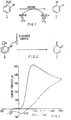
- Fig. 1 is a postulated reaction scheme for the enzymic reaction.
- Fig. 2 is a postulated reaction scheme for the reaction at the electrode surface.
- Fig. 3 shows a cyclic voltammogram of catechol at 10 mM final concentration.
- Fig. 4 shows a calibration curve for salicylate obtained by a series of steady state electrochemical measurements in the presence of increasing amounts of salicylate solution.
- Fig. 5 shows a cyclic voltammogram of NADH solution, salicylate hydroxylase solution and buffer solution, both in the absence and in the presence of salicylate.
- Fig. 6 shows the separation profile of Pseudomonad proteins by FPLC using ion exchange chromatography to obtain pure salicylate hydroxylase ; and
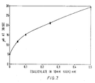
- Fig. 7 shows a dry strip electrode response to salicylate.
- As shown in Figure 1, the enzymic conversion of salicylate (1) to catechol (2) appears to be unidirectional and to occur in the presence of NAD(P)H and molecular oxygen.
- As shown in Figure 2, in this scheme catechol (2) is converted into the orthoquinone (3) at the electrode surface and at a suitable oxidising potential.
- The removal of electrons from the catechol (2) results in the formation of ortho-quinone (3) or a derivative thereof, and may be employed both as a qualitative indicator of the presence of the catechol and hence the salicylate, and as a quantitative assay for the catechol and hence as an indirect measure of the concentration of salicylate at the electrode surface.
- A buffer solution was prepared from potassium di-hydrogen phosphate (1.77 g; Analar from Britisch Drug House (BDH) and di-potassium hydrogen phosphate (19.6 g ; Analar from BDH), which were dissolved in distilled water, adjusted to pH 7.6 and made up to a final volume of 1 litre. Catechol (from Sigma Chemical Company) was dissolved in such a buffer solution and degassed under reduced pressure immediately prior to use.
- The electrodes were made of a range of different materials, especially gold and glassy carbon, most especially pyrolytic graphite. The electrodes were polished between runs using a slurry of 0.3 µm alumina (BDH) made up with water. The object of this polishing was to remove impurities and oxidation products from the surface of the electrode. The alumina was removed from the electrode surface by ultrasonication.
- Cyclic voltammograms were produced from a range of solutions by sweeping the potential difference from zero to + 500 mV and back down to - 100 mV vs. S.C.E. The potential applied was controlled by a potentiostat (from Jaytron Inst. A.S. Scientific. Abingdon) using a scan rate of 50 mV/s.
- The oxidation current produced was recorded on a Gould Series 60000 Chart Recorder in which the X-axis recorded the applied potential and the Y-axis recorded the current produced. A cyclic voltammogram of catechol (at 10 mM final concentration) is shown in Figure 3.
- Salicylate sodium salt (GOLD LABEL and available in the marketplace from Aldrich) was dissolved in the phosphate buffer to give a final concentration of 0.1 M.
- NADH disodium salt (Grade II : from Boehringer Mannheim) was dissolved in buffer solution to give a final concentration of 0.2 M.
- Salicylate hydroxylase (from the Sigma Chemical Company) was resuspended in distilled water to give a stock solution of 20 units/ml (based on the manufacturers information and unit definition).
- The electrodes used were identical to those described above with reference to Example 1.
- The solution of salicylate hydroxylase (from Sigma Chemical Company) was routinely assayed at 37'C by following the decrease in absorbance at 340 nm (due to the oxidation of one of the substrates, NADH).
- To a 1 ml glass cuvette was added 10 µl of salicylate solution, 10 µl of NADH (0.02 M solution) and 977.5 >1 of phosphate buffer solution. The cuvette was placed in a Pye Unicam SP8-400 spectrophotometer which had been thermostat- ted at 37'C. After the addition of salicylate hydroxylase solution, the decrease in absorbance was followed at 340 nm. It is known that one unit of enzyme will convert one µmole of salicylate and NADH to catechol and NAD+ per minute at pH 7.6 and at a working temperature of 37°C.
- In the cyclic voltammograms the cell contained 52 µl of NADH solution (0.2 M, as above), 60 µl of salicylate hydroxylase solution and 428 µl of buffer solution.
- Cyclic voltammograms were recorded both in the absence and in the presence of the substrate (60 µl salicylate). In order to ensure that the reaction progressed, each sample was incubated at 37°C for 2 minutes prior to initiating the scan.
- Such a cyclic voltammogram is shown in figure 5, and it will be noted that the addition of the salicylate substrate to the incubation mixture prior to initiation of the scan, results in a marked change in the profile of the curve obtained.
- In steady state measurements the current produced upon application of a fixed potential to a stirred solution was measured on the Y-axis of the chart recorder using the X-axis as a time base. The potential was poised at + 250 mV vs. SCE at 37°C after allowing 2 minutes for the system to come to equilibrium. Stirring of the solutions ensures that the layer of material close to the electrode and which is available for oxidation is replenished and thus the current produced at the electrode does not decay due to exhaustion of reagents.
- The stirred solutions comprised: 140 µl of NADH solution, 100 µl of salicylate hydroxylase solution and 760 µl of buffer solution. Steady state electrochemical measurements were made in the presence of increasing amounts of salicylate solution to produce a calibration curve for salicylate and is shown in Figure 4. The current measured was obtained by poising the electrode at + 250 mV vs. S.C.E. 2 minutes after addition of the sample. This calibration curve can be used in conjuction with direct readings of unknown samples in order to determine the salicylate ion.
- A buffer solution was prepared from Trizma base (2.42 g; Sigma Chemical Company) dissolved in distilled water, adjusted to pH 7.5 and made up to a final volume of 1 litre. This buffer solution (buffer A) is used to apply the enzyme sample to the ion exchange column. A second buffer solution was prepared (buffer B). Buffer B was essentially the same as buffer A but also contains 150 mM sodium sulphate (BDH). This buffer is used to elute the enzyme from the anionic column.
- Salicylate hydroxylase (from GDS Technology Inc.) was resuspended in buffer solution A to give a stock solution of 50 units/ml (18 mg protein/ml) based on the manufacturer's information and definition of activity and units.
- Protein purification was performed on a complete Pharmacia FPLC (Trade Mark) system. A Pharmacia Polyanion SI column (HR5/5) was equilibrated with buffet A. The enzyme solution (16 Oul) was applied to. the column at a flow rate of 1 ml min-1. The sample was eluted from the column using a preprogrammed gradient (see Figure 6).
- Fractions (1 ml) were collected in the FRAC-100 fraction collector (Pharmacia) and were assayed for enzyme activity as detailed in Example 2. Enzyme activity was present in
fractions 20 and 21 and was associated with a protein peak. The profile of the chromatographic separation is shown in Figure 6. - The specific activity of the enzyme was in excess of 10 units/mg usually 14 to 15 units/mg.
- The purification of salycylate hydroxylase has been scaled up using polyanion SI-17 um packed into large column (1.6 cm x 45 cm). Similar activities have been reported in the literature using several purification steps. (You, K. S. & Roe, C. R., Anal. Biochem. (1981, 114, 177; Kamin, H. et al., Methods in Enzymology (1978), 53, 527). We believe this method has many advantages over existing purification protocols.
- Sodium salicylate and NADH were obtained from the same sources as detailed in Example 2 and dissolved in 0.9 % saline to give final concentrations of 20 mM. These two solutions were mixed in various proportions to give a range of salicylate concentrations in 10 mM NADH.
- BES (N,N'-Bis(2-hydroxyethyl-2-aminoethane sulphonide acid ; 32.0 g from BDH), sodium azide (0.5 g ; from BDH) and FAD disodium salt (85 mg ; from BDG) were dissolved in distilled water, adjusted to pH7 and made up to a final volume of 1 litre.
- The purified salicylate hydroxylase was ul- traconcentrated using an Amicon ultrafiltration cell containing a 10,000 molecular weight cut-off filter and the buffer was concentrated to 520 units/ml.
- Dry strip electrodes were prepared.
- Fixed potential studies were carried out at room temperature as described in Example 2 with the modification that the potential was poised immediately after the sample was applied. The calibration curve for salicylate is shown in Figure 7.
Claims (11)
Priority Applications (1)
| Application Number | Priority Date | Filing Date | Title |
|---|---|---|---|
| AT86302476T ATE51415T1 (en) | 1985-04-03 | 1986-04-03 | DETECTION OF SALICYLATE AND DEVICE FOR CARRYING OUT THE SAME. |
Applications Claiming Priority (2)
| Application Number | Priority Date | Filing Date | Title |
|---|---|---|---|
| GB8508677 | 1985-04-03 | ||
| GB858508677A GB8508677D0 (en) | 1985-04-03 | 1985-04-03 | Assay for salicylate |
Publications (2)
| Publication Number | Publication Date |
|---|---|
| EP0202743A1 EP0202743A1 (en) | 1986-11-26 |
| EP0202743B1 true EP0202743B1 (en) | 1990-03-28 |
Family
ID=10577124
Family Applications (1)
| Application Number | Title | Priority Date | Filing Date |
|---|---|---|---|
| EP86302476A Expired - Lifetime EP0202743B1 (en) | 1985-04-03 | 1986-04-03 | Assay for salicylate and apparatus for performing same |
Country Status (8)
| Country | Link |
|---|---|
| US (1) | US4777132A (en) |
| EP (1) | EP0202743B1 (en) |
| JP (1) | JPH0650299B2 (en) |
| AT (1) | ATE51415T1 (en) |
| AU (1) | AU586235B2 (en) |
| CA (1) | CA1253568A (en) |
| DE (1) | DE3669888D1 (en) |
| GB (1) | GB8508677D0 (en) |
Families Citing this family (6)
| Publication number | Priority date | Publication date | Assignee | Title |
|---|---|---|---|---|
| US5362630A (en) * | 1981-07-28 | 1994-11-08 | Duke University | Isolation of pseudomonas salicylate hydroxlase and its use for the identification and quantitation of salicylate in body fluids |
| US5320946A (en) * | 1990-07-05 | 1994-06-14 | Eastman Kodak Company | Method and element for assay of catechol and catechol generating substances |
| US5460970A (en) * | 1993-05-18 | 1995-10-24 | Summa Health System | Separation of acetaldehyde-induced hemoglobin (Hb A1-AcH) |
| GB9416002D0 (en) * | 1994-08-08 | 1994-09-28 | Univ Cranfield | Fluid transport device |
| DE19619056C2 (en) * | 1996-03-04 | 2002-01-17 | Frieder Scheller | Method and sensor for the enzymatic-electrochemical determination of substrates NAD · + · - and NAD (P) · + · -dependent dehydrogenases |
| WO2000042421A1 (en) * | 1999-01-15 | 2000-07-20 | Competitive Technologies, Inc. | Method for screening for type b trichothecene mycotoxins |
Family Cites Families (9)
| Publication number | Priority date | Publication date | Assignee | Title |
|---|---|---|---|---|
| JPS5643358A (en) * | 1979-09-18 | 1981-04-22 | Tokuyama Soda Co Ltd | Pigment |
| DE3046741A1 (en) * | 1980-12-11 | 1982-07-15 | Boehringer Mannheim Gmbh, 6800 Mannheim | DETECTION OF NAD (P) H OR SALICYLATE |
| JPS57197458A (en) * | 1981-05-20 | 1982-12-03 | Yanagimoto Seisakusho:Kk | Catechol amine analysing apparatus |
| CA1185155A (en) * | 1981-07-28 | 1985-04-09 | Kwan-Sa You | Isolation of pseudomonas salicylate hydroxylase and its use for the identification and quantitation of salicylate in body fluids |
| JPS5926048A (en) * | 1982-08-03 | 1984-02-10 | Toshiba Corp | Sample inspection unit |
| JPS5982082A (en) * | 1982-10-29 | 1984-05-11 | Matsushita Electric Works Ltd | Apparatus for determining glucose concentration |
| AU564495B2 (en) * | 1983-05-05 | 1987-08-13 | Medisense Inc. | Nadp-nadph energy linked enzyme cascade assay |
| AU564494B2 (en) * | 1983-05-05 | 1987-08-13 | Medisense Inc. | Enzyme cascade energy coupling assay |
| GB8326696D0 (en) * | 1983-10-05 | 1983-11-09 | Health Lab Service Board | Estimation of salicylates |
-
1985
- 1985-04-03 GB GB858508677A patent/GB8508677D0/en active Pending
-
1986
- 1986-04-02 CA CA000505648A patent/CA1253568A/en not_active Expired
- 1986-04-03 EP EP86302476A patent/EP0202743B1/en not_active Expired - Lifetime
- 1986-04-03 AT AT86302476T patent/ATE51415T1/en not_active IP Right Cessation
- 1986-04-03 AU AU55638/86A patent/AU586235B2/en not_active Ceased
- 1986-04-03 DE DE8686302476T patent/DE3669888D1/en not_active Expired - Fee Related
- 1986-04-03 US US06/847,955 patent/US4777132A/en not_active Expired - Lifetime
- 1986-04-03 JP JP61075651A patent/JPH0650299B2/en not_active Expired - Fee Related
Also Published As
| Publication number | Publication date |
|---|---|
| AU5563886A (en) | 1986-10-09 |
| ATE51415T1 (en) | 1990-04-15 |
| US4777132A (en) | 1988-10-11 |
| GB8508677D0 (en) | 1985-05-09 |
| EP0202743A1 (en) | 1986-11-26 |
| CA1253568A (en) | 1989-05-02 |
| JPH0650299B2 (en) | 1994-06-29 |
| DE3669888D1 (en) | 1990-05-03 |
| JPS6258157A (en) | 1987-03-13 |
| AU586235B2 (en) | 1989-07-06 |
Similar Documents
| Publication | Publication Date | Title |
|---|---|---|
| Nanjo et al. | Enzyme electrode sensing oxygen for uric acid in serum and urine | |
| Chen et al. | A simple hydrogenase-linked assay for ferredoxin and flavodoxin | |
| Kiang et al. | Enzymic determination of nitrate: electrochemical detection after reduction with nitrate reductase and nitrite reductase | |
| Kawashima et al. | Potentiometric enzyme electrode for uric acid | |
| EP0135092B1 (en) | Method for determination of ammonia | |
| EP0202743B1 (en) | Assay for salicylate and apparatus for performing same | |
| CA1085279A (en) | Measurement of alcohol levels in body fluids | |
| EP0094161A1 (en) | Method for determining glucose content of fluid | |
| KAKUNO et al. | Electron and proton transport in Rhodospirillum rubrum chromatophores | |
| Wright et al. | Simultaneous determination of hydroxymethylbilane synthase and uroporphyrinogen III synthase in erythrocytes by high-performance liquid chromatography | |
| US5306413A (en) | Assay apparatus and assay method | |
| Romette et al. | L-glutamine enzyme electrode for on-line mammalian cell culture process control | |
| EP0264815A1 (en) | Methods for selective measurement of amino acids | |
| Clark Jr et al. | Rapid electroenzymatic measurement of lactate in microsamples of spinal fluid | |
| IWAI et al. | Electric microassays of glucose, uric acid and cholesterol using peroxidase adsorbed on a carbon electrode | |
| Rokosh et al. | A modification of isocitrate and malate dehydrogenase assays for use in crude cell free extracts | |
| Della Ciana et al. | Highly sensitive amperometric enzyme immunoassay for α-fetoprotein in human serum | |
| Ngo et al. | Amperometric determination of picomolar levels of flavin adenine dinucleotide by cyclic oxidation-reduction in apo-glucose oxidase system | |
| Power et al. | Electrochemical assay for catecholase activity of mushroom tyrosinase | |
| US5188941A (en) | Enzymatic determination of theophylline | |
| Guilbault | Enzymatic glucose electrodes | |
| Fernandes et al. | Use of sorghum seed tissue as a biocatalyst in a stirred reactor for oxalic acid determination | |
| CA1339794C (en) | Enzymatic determination of theophylline | |
| Yomo et al. | Enzymatic method for measuring the absolute value of oxygen concentration | |
| AU626724B2 (en) | Enzymatic determination of theophylline |
Legal Events
| Date | Code | Title | Description |
|---|---|---|---|
| PUAI | Public reference made under article 153(3) epc to a published international application that has entered the european phase |
Free format text: ORIGINAL CODE: 0009012 |
|
| AK | Designated contracting states |
Kind code of ref document: A1 Designated state(s): AT BE CH DE FR GB IT LI LU NL SE |
|
| 17P | Request for examination filed |
Effective date: 19870116 |
|
| RAP1 | Party data changed (applicant data changed or rights of an application transferred) |
Owner name: MEDISENSE, INC. |
|
| 17Q | First examination report despatched |
Effective date: 19881027 |
|
| GRAA | (expected) grant |
Free format text: ORIGINAL CODE: 0009210 |
|
| ITF | It: translation for a ep patent filed | ||
| AK | Designated contracting states |
Kind code of ref document: B1 Designated state(s): AT BE CH DE FR GB IT LI LU NL SE |
|
| PG25 | Lapsed in a contracting state [announced via postgrant information from national office to epo] |
Ref country code: BE Effective date: 19900328 Ref country code: AT Effective date: 19900328 |
|
| REF | Corresponds to: |
Ref document number: 51415 Country of ref document: AT Date of ref document: 19900415 Kind code of ref document: T |
|
| PG25 | Lapsed in a contracting state [announced via postgrant information from national office to epo] |
Ref country code: LU Free format text: LAPSE BECAUSE OF NON-PAYMENT OF DUE FEES Effective date: 19900430 |
|
| REF | Corresponds to: |
Ref document number: 3669888 Country of ref document: DE Date of ref document: 19900503 |
|
| ET | Fr: translation filed | ||
| PLBE | No opposition filed within time limit |
Free format text: ORIGINAL CODE: 0009261 |
|
| STAA | Information on the status of an ep patent application or granted ep patent |
Free format text: STATUS: NO OPPOSITION FILED WITHIN TIME LIMIT |
|
| 26N | No opposition filed | ||
| ITTA | It: last paid annual fee | ||
| EAL | Se: european patent in force in sweden |
Ref document number: 86302476.6 |
|
| REG | Reference to a national code |
Ref country code: GB Ref legal event code: IF02 |
|
| PGFP | Annual fee paid to national office [announced via postgrant information from national office to epo] |
Ref country code: GB Payment date: 20020315 Year of fee payment: 17 |
|
| PGFP | Annual fee paid to national office [announced via postgrant information from national office to epo] |
Ref country code: NL Payment date: 20020325 Year of fee payment: 17 |
|
| PGFP | Annual fee paid to national office [announced via postgrant information from national office to epo] |
Ref country code: FR Payment date: 20020404 Year of fee payment: 17 |
|
| PGFP | Annual fee paid to national office [announced via postgrant information from national office to epo] |
Ref country code: SE Payment date: 20020405 Year of fee payment: 17 |
|
| PGFP | Annual fee paid to national office [announced via postgrant information from national office to epo] |
Ref country code: DE Payment date: 20020430 Year of fee payment: 17 |
|
| PGFP | Annual fee paid to national office [announced via postgrant information from national office to epo] |
Ref country code: CH Payment date: 20020618 Year of fee payment: 17 |
|
| PG25 | Lapsed in a contracting state [announced via postgrant information from national office to epo] |
Ref country code: GB Free format text: LAPSE BECAUSE OF NON-PAYMENT OF DUE FEES Effective date: 20030403 |
|
| PG25 | Lapsed in a contracting state [announced via postgrant information from national office to epo] |
Ref country code: SE Free format text: LAPSE BECAUSE OF NON-PAYMENT OF DUE FEES Effective date: 20030404 |
|
| PG25 | Lapsed in a contracting state [announced via postgrant information from national office to epo] |
Ref country code: LI Free format text: LAPSE BECAUSE OF NON-PAYMENT OF DUE FEES Effective date: 20030430 Ref country code: CH Free format text: LAPSE BECAUSE OF NON-PAYMENT OF DUE FEES Effective date: 20030430 |
|
| PG25 | Lapsed in a contracting state [announced via postgrant information from national office to epo] |
Ref country code: NL Free format text: LAPSE BECAUSE OF NON-PAYMENT OF DUE FEES Effective date: 20031101 Ref country code: DE Free format text: LAPSE BECAUSE OF NON-PAYMENT OF DUE FEES Effective date: 20031101 |
|
| GBPC | Gb: european patent ceased through non-payment of renewal fee |
Effective date: 20030403 |
|
| NLV4 | Nl: lapsed or anulled due to non-payment of the annual fee |
Effective date: 20031101 |
|
| EUG | Se: european patent has lapsed | ||
| REG | Reference to a national code |
Ref country code: CH Ref legal event code: PL |
|
| PG25 | Lapsed in a contracting state [announced via postgrant information from national office to epo] |
Ref country code: FR Free format text: LAPSE BECAUSE OF NON-PAYMENT OF DUE FEES Effective date: 20031231 |
|
| REG | Reference to a national code |
Ref country code: FR Ref legal event code: ST |
|
| PG25 | Lapsed in a contracting state [announced via postgrant information from national office to epo] |
Ref country code: IT Free format text: LAPSE BECAUSE OF NON-PAYMENT OF DUE FEES Effective date: 20050403 |