EP0179152B1 - Diagnostic system for the detection of cytomegalovirus - Google Patents
Diagnostic system for the detection of cytomegalovirus Download PDFInfo
- Publication number
- EP0179152B1 EP0179152B1 EP85902741A EP85902741A EP0179152B1 EP 0179152 B1 EP0179152 B1 EP 0179152B1 EP 85902741 A EP85902741 A EP 85902741A EP 85902741 A EP85902741 A EP 85902741A EP 0179152 B1 EP0179152 B1 EP 0179152B1
- Authority
- EP
- European Patent Office
- Prior art keywords
- hybridoma
- cmv
- cells
- cytomegalovirus
- monoclonal
- Prior art date
- Legal status (The legal status is an assumption and is not a legal conclusion. Google has not performed a legal analysis and makes no representation as to the accuracy of the status listed.)
- Expired - Lifetime
Links
Images

Classifications
-
- C—CHEMISTRY; METALLURGY
- C07—ORGANIC CHEMISTRY
- C07K—PEPTIDES
- C07K16/00—Immunoglobulins [IG], e.g. monoclonal or polyclonal antibodies
- C07K16/08—Immunoglobulins [IG], e.g. monoclonal or polyclonal antibodies against material from viruses
- C07K16/081—DNA viruses
- C07K16/085—Orthoherpesviridae (F), e.g. pseudorabies virus or Epstein-Barr virus
- C07K16/089—Cytomegalovirus
-
- Y—GENERAL TAGGING OF NEW TECHNOLOGICAL DEVELOPMENTS; GENERAL TAGGING OF CROSS-SECTIONAL TECHNOLOGIES SPANNING OVER SEVERAL SECTIONS OF THE IPC; TECHNICAL SUBJECTS COVERED BY FORMER USPC CROSS-REFERENCE ART COLLECTIONS [XRACs] AND DIGESTS
- Y10—TECHNICAL SUBJECTS COVERED BY FORMER USPC
- Y10S—TECHNICAL SUBJECTS COVERED BY FORMER USPC CROSS-REFERENCE ART COLLECTIONS [XRACs] AND DIGESTS
- Y10S435/00—Chemistry: molecular biology and microbiology
- Y10S435/81—Packaged device or kit
-
- Y—GENERAL TAGGING OF NEW TECHNOLOGICAL DEVELOPMENTS; GENERAL TAGGING OF CROSS-SECTIONAL TECHNOLOGIES SPANNING OVER SEVERAL SECTIONS OF THE IPC; TECHNICAL SUBJECTS COVERED BY FORMER USPC CROSS-REFERENCE ART COLLECTIONS [XRACs] AND DIGESTS
- Y10—TECHNICAL SUBJECTS COVERED BY FORMER USPC
- Y10S—TECHNICAL SUBJECTS COVERED BY FORMER USPC CROSS-REFERENCE ART COLLECTIONS [XRACs] AND DIGESTS
- Y10S530/00—Chemistry: natural resins or derivatives; peptides or proteins; lignins or reaction products thereof
- Y10S530/808—Materials and products related to genetic engineering or hybrid or fused cell technology, e.g. hybridoma, monoclonal products
-
- Y—GENERAL TAGGING OF NEW TECHNOLOGICAL DEVELOPMENTS; GENERAL TAGGING OF CROSS-SECTIONAL TECHNOLOGIES SPANNING OVER SEVERAL SECTIONS OF THE IPC; TECHNICAL SUBJECTS COVERED BY FORMER USPC CROSS-REFERENCE ART COLLECTIONS [XRACs] AND DIGESTS
- Y10—TECHNICAL SUBJECTS COVERED BY FORMER USPC
- Y10S—TECHNICAL SUBJECTS COVERED BY FORMER USPC CROSS-REFERENCE ART COLLECTIONS [XRACs] AND DIGESTS
- Y10S530/00—Chemistry: natural resins or derivatives; peptides or proteins; lignins or reaction products thereof
- Y10S530/866—Chemistry: natural resins or derivatives; peptides or proteins; lignins or reaction products thereof involving immunoglobulin or antibody fragment, e.g. fab', fab, fv, fc, heavy chain or light chain
Definitions
- the present invention relates to a diagnostic system for the detection of cytomegalovirus, and in particular to mammalian monoclonal antibodies that immunoreact with a viral antigen or ligand associated with cells infected by cytomegalovirus to detect the presence of cytomegalovirus in a biological sample.
- Cytomegalic inclusion disease CID
- CMV cytomegalovirus
- CMV chronic myeloma
- CMV chronic myeloma
- Cytomegalovirus (CMV) infection is caused by a species-specific agent with the physio-chemical and electron microscopic characteristics of a herpesvirus.
- Human CMV was first isolated in fibroblastic tissue culture in the mid-1950's from infants with CID and from the adenoid tissue of schoolage children. A cytopathic effect was noted in tissue culture which was characterized by large intranuclear inclusions. The propagation of the virus provided the basis for the development of specific serologic tests as will be described.
- Cytomegalovirus infection is worldwide in distribution. Cytomegalovirus, however, does not induce a highly communicable infection. Thus, a substantial number of individuals remain Susceptible to the infection in adult life. CMV has been isolated from saliva, the upper respiratory tract, urine, milk, cervical secretions, semen, feces and circulating leukocytes in blood. The virus is probably transferred by intimate contact with an infected individual or by infusion of blood from a asymptomatic blood donor.
- CMV mononucleosis a type of cytomegalovirus infection has been described in patients who have received blood transfusions. This condition, referred to as post-transfusion CMV mononucleosis, occurs two to four weeks after the administration of blood.
- cytomegalovirus infection has also been associated with autoimmune hemolytic disease, ulcerative lesions of the gastrointestinal tract, post-transplantation pneumonia and thrombocytopenic purpura. For the majority of CMV infections acquired after birth, however, recovery is without significant complications.
- cytomegalovirus infection in a patient with mononucleosis-like symptoms can be established by virus isolation from vesicular lesions.
- Antibodies produced in response to infection with a CMV can be detected by neutralization (NT), complement-fixation (CF), immunofluorescence and platelet-agglutination (PA) procedures.
- NT neutralization
- CF complement-fixation
- PA platelet-agglutination
- the present invention relates to a method of assaying the presence of cytomegalovirus in a fluid sample, using mammalian monoclonal receptors.
- the monoclonal receptors are produced by hybridomas formed by the fusion of cells from a myeloma cell line and splenocytes that produce antibodies that react with a viral antigen induced in mammalian peripheral blood cell lymphocytes by a cytomegalovirus.
- a murine monoclonal receptor is produced by hybridoma ATCC HB 8554.
- This monoclonal receptor, designated "L-14" was formed by fusion of cells from mouse myeloma line P3X63-Ag8.653 and murine splenic cells from a mouse previously immunized with an isolate containing CMV-infected cells.
- murine monoclonal receptors of this invention are produced by hybridomas ATCC HB 8622, ATCC HB 8623 and ATCC HB 8624. These monoclonal receptors designated "K-25", “E-28” and "A-33"; respectively, were also formed by fusion of cells from mouse myeloma line P3X63-Ag8.653 and murine splenic cells from a mouse previously immunized with an isolate containing CMV-infected cells.
- monoclonal antibodies to cytomegalovirus are useful probes for studying cytomegalovirus infection, and for developing assays for measuring levels of cytomegalovirus in biological fluids and the association of such levels with disease.
- one embodiment of this invention relates to a method for assaying for the presence of a viral antigen or ligand associated with cytomegalovirus.
- This method uses, as an active ingredient, an effective amount of the above-described monoclonal receptors (antibodies).
- an effective amount of those antibodies is admixed with a predetermined amount of a biological sample containing a particular viral antigen associated with cytomegalovirus, a complex is formed by an immunological reaction. The presence of the complex can be determined by a label or indicating means.
- Hybridomas that produce the above-described monoclonal receptors may be prepared by (i) immunizing a mammal with cytomegalovirus-infected cells; (ii) removing the spleen from the mammal and making a suspension of the splenocytes or spleen cells; (iii) fusing the spleen cells with mammalian myeloma cells in the presence of a cell fusion promoter; (iv) diluting and culturing the fused cells in separate wells or containers in a medium that will not support growth of the unfused myeloma cells; (v) evaluating the supernatant in each well containing a hybridoma for the presence of a receptor to a viral antigen to cytomegalovirus; and (vi) selecting and cloning the desired hybridoma that produces a monoclonal receptor to the viral antigen.
- the above-described monoclonal receptors may be prepared by culturing one of the hybridomas described herein in a suitable medium and recovering the receptor from the medium containing the hybridoma.
- a further method of preparing the above-described monoclonal receptors comprises injecting into a mammal one of the hybridomas described herein and recovering the receptor from the malignant ascites or serum of the mammal.
- hybridomas and the monoclonal receptors produced therefrom described herein are identified by the designations "L-14, "K-25”, “E-28” and “A-33", the particular material referred to being apparent from the context.
- Hybridoma L-14 was deposited on May 2, 1984 at the American Type Culture Collection, Rockville, Maryland and was given the ATCC accession number HB 8554.
- Hybridomas K-25, E-28 and A-33 were deposited on September 27, 1984 at the American Type Culture Collection, Rockville, Maryland and were given the ATCC accession numbers HB 8622, HB 8623 and HB 8624, respectively.
- the monoclonal receptors react specifically with CMV-infected cells.
- monoclonal receptor L-14 reacts with a protein that appears early after CMV infection (within 3 hours) and which remains localized to the cell nucleous throughout the infectious cycle.
- Monoclonal receptors K-25, E-28 and A-33 react with a similar specificity.
- the present monoclonal antibodies can be used to rapidly and specifically detect CMV infection in tissue culture, blood samples and other body components.
- receptor is used herein to mean a biologically active molecule that binds to a ligand.
- the receptor molecules of the present invention are substantially intact antibodies or idiotype-containing polyamide portions of an antibody.
- Biological activity of a receptor molecule is evidenced by the binding of the receptor to its antigenic ligand upon their admixture in an aqueous medium, at least at physiological pH values and ionic strengths.
- the receptors also bind to the antigenic ligand within a pH value range of about 5 to about 9, and at ionic strengths such as that of distilled water to that of about one molar sodium chloride.
- Idiotype-containing polyamide portions are those portions of antibody molecules that include the idiotype and bind to the ligand. Such portions include the Fab, Fab' and F(ab')2 fragments prepared from antibodies by well-known enzymatic cleavage techniques. See for example, U.S. Patent No. 4,342,566 to Theofilopoulos and Dixon. Inasmuch as the antibodies from which idiotype-containing polyamides are obtained are described as raised against or induced by immunogens, idiotype-containing polyamide receptors will also be discussed as being "raised” or "induced” with the understanding that a cleavage step is required to obtain an idiotype-containing polyamide from an antibody. Intact antibodies are preferred, however, and will be utilized as illustrative of the receptor molecules of this invention.
- the receptors useful on the present invention are monoclonal antibodies.
- a "monoclonal antibody” is a receptor produced by clones of a single cell called a hybridoma that secretes but one kind of receptor molecule.
- the hybridoma cell is fused from an antibody-producing cell and a myeloma cell or other self-perpetuating cell line.
- monclonal antibodies of the present invention are well known. Such receptors were first described by Kohler and Milstein, Nature , 256 , 495 (1975), which is incorporated herein by reference. Monoclonal antibodies are typically obtained from hybridoma tissue cultures or from ascites fluid obtained from mammals into which the hybridoma tissue was introduced. Both methods are described herein.
- CMV can infect human peripheral blood lymphocytes (PBL) including T and B cells, natural killer cells and monocytes.
- PBL peripheral blood lymphocytes
- the infection was abortive which means that expression of the CMV genome was limited to the synthesis of the immediate-early viral polypeptides or ligands, expression of late CMV gene products was not evident and infectious virus was not produced.
- PHA phytohemagglutinin
- PHA is a mitosis-stimulating substance (a mitogen) which, upon addition to a medium containing lymphocytes, prompts nondividing lymphocytes to grow, differentiate and proliferate.
- latent state or “latent infection”, as used herein, means that the virus cannot be detected in tissues or secretions by conventional cell-culture assays but nevertheless persists in a non-replicating state or at an undetectable, possibly intermittent, level of replication.
- the lymphocytes employed to form the hybridomas of the present invention may be derived from any mammal, such as a primate, rodent (e.g., mouse or rat), rabbit, guinea pig, cow, dog, sheep, pig or the like.
- the host may be sensitized by injection of the immunogen, in this instance CMV-infected cells, followed by a booster injection, and then isolation of the spleen.
- the myeloma cell line be from the same species as the lymphocytes. Therefore, fused hybrids such as mouse-mouse hybrids [Shulman et al., Nature , 276 , 269 (1978)] or rat-rat hybrids [Galfre et al., Nature , 277 , 131 (1979)] are typically utilized. However, some rat-mouse hybrids have also been successfully used in forming hybridomas [Goding, "Production of Monoclonal Antibodies by Cell Fusion," in Antibody as a Tool , Marchalonis et al. eds., John Wiley & Sons Ltd., p. 273 (1982)].
- Suitable myeloma lines for use in the present invention include MPC-11 (ATCC CRL 167), P3X63-Ag8.653 (ATCC CRL 1580), Sp2/O-Ag14 (ATCC CRL 1581), P3 X 63 Ag8U.1 (ATCC CRL 1597), Y3-Agl.2.3. (deposited at Collection Nationale de Cultures de Microorganisms, Paris, France, number I-078) and P3X63Ag8 (ATCC TIB 9).
- Myeloma line P3X63-Ag8.653 is preferred for use in the present invention.
- the hybridoma cells that are ultimately produced may be cultured following usual in vitro tissue culture techniques for such cells as are well-known. More preferably, the hybridoma cells are cultured in animals using similarly well-known techniques with the monoclonal receptors being obtained from the ascites fluid so generated.
- the animals used for generation of the ascites fluid are typically BALB/c mice bred in the mouse colony of the Scripps Clinic and Research Foundation, La Jolla, California, however, when animals other than mice are used for preparation of the hybridomas, mice or that animal type can be used for the production of ascites fluid.
- Peripheral blood lymphocytes removed from the blood of healthy human donors by density gradient centrifugation on Ficoll-Pague (Sigma Chemical Co., St. Louis, MO) were infected at a multiplicity of 0.01 to 1.0 with CMV recently isolated from patients with various CMV syndromes (Table 1), or with stocks of plaque-purified laboratory strain AD-169.
- the recent isolates were propagated in human foreskin fibroblasts for less than twelve passages in RPMI-1640 medium plus 5 percent fetal calf serum. Because virus from patient isolates is predominantly associated with the cell matrix (Weller, id. ), infected or mock-infected fibroblasts were sonicated, and this material was cultured with PBL for up to 4 days.
- Immediate-early polypeptides were probed with a monoclonal antibody designated "E-3” which binds to the major 72 dalton immediate-early protein [Goldstein et al., Infection and Immunity , 38 , 273 (1982)]. Similar staining was obtained with another monoclonal antibody designated "L14” which binds in infected fibroblasts to a nuclear antigen synthesized within 3 hours post infection, long before virus-induced cytopathic effect.
- G-10 A monoclonal antibody designated "G-10", also specific to a CMV-induced fibroblast antigen present early in infection, did not bind to infected PBL in the same preparation. G-10 reacts with a 60 dalton protein that is synthesized in infected fibroblasts before viral cytopathic effect develops. The fine specificity of this antibody will enable a determination of the blockade in expression of the viral genome.
- C-5" a monoclonal antibody designated "C-5" that is specific for a late CMV polypeptide [Goldstein et al., id. ], no mature virions were visible by electron microscopy and there was no evidence of infectious virus in cocultivation assays.
- PBL can be infected by CMV; the infection is abortive and CMV gene expression is restricted.
- immediate-early gene expression in PBL infected with 6 of 7 low passage human isolates (Table 1).
- laboratory strain AD 169 (Table 1), was a weak expression of an immediate-early polypeptide demonstrated, and that occurred in less than 1 percent of PBL. Whether this represents a differential cell tropism among individual CMV strains or an adaptation of laboratory strains of virus to growth in fibroblasts, or both of these factors, is not yet clear.
- Epstein-Barr virus (EBV).
- EBV Epstein-Barr virus
- PBL preincubated with the virus either by erythrocyte rosetting [Moretta et al., J. Immunol. , 122 , 984 (1979)] or with monoclonal antibody and the fluorescence activated cell sorter [Oldstone et al., Virology , 127 , 426 (1983)] were separated.
- Two-color immunofluorescence technique [Dutko et al., J. Exp. Med. , 154 , 1636 (1981) and Dutko et al., J. Cell. Biochem. , Supp.
- T-lymphocyte 8B (1984)] revealed immediate-early protein synthesis in (a) 1 to 15 percent of T-lymphocytes defined by the monoclonal antibody markers OKT3 (pan T) [ATCC CRL 8001], OKT4 (helper) [ATCC CRL 8002], OKT8 (suppressor-cytotoxic) [ATCC CRL 8014]; (b) monocytes marked by Mo2, (c) natural killer cells labeled with Leu-7 and (d) B-lymphocytes identified with antibody to surface immunoglobulin. The number of cells expressing immediate-early antigens varied with the clinical isolate used for infection.
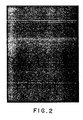
- FIG 1 illustrates CMV infected T-lymphocytes that were positively selected by rosetting techniques and then probed with the immediate-early antigen specific monoclonal antibody L-14. Similar results were obtained with the immediate-early antigen specific probe E-3, but (as shown in Figure 2) not with C-5, a monoclonal antibody that detects a late CMV gene product.
- Virus transcription in these different models may be blocked by a common mechanism, and DeMarchi has provided evidence suggesting that productive and nonproductive infection may differ at the level at which some of these early transcripts associate with polysomes [ Virology , 129 , 287 (1983)].
- CMV can abortively infect lymphocytes and alter some of their functions.
- abortively infected, transfused PBL may be a source of the virus in some patients, after evolution from abortive to full replication by a mechanism that is not yet fully understood.
- Monoclonal receptor L-14 was produced by immunization with a CMV isolate using the standard hybridoma technology of Kohler et al., Nature , 256 , 495 (1975). Specifically, BALB/c mice were immunized by intraperitoneal injection with about 5 x 10⁶ HCMV (human cytomegalovirus) AD 169-infected human fibroblast cells in complete Freund's adjuvant. Three weeks later, the mice were again injected in a like manner. After an additional three weeks, the mice were immunized intravenously with the AD 169-infected cells in phosphate buffered saline (PBS) on three consecutive days. The mice were then sacrificed.
- PBS phosphate buffered saline
- the spleens were removed from the mice, pooled and a single cell suspension was made. The spleen cells were then fused with an equal number of P3X63-Ag8.653 myeloma cells in the presence of a cell fusion promoter (polyethylene glycol 2000).
- the hybridoma that produces Mab L-14 was selected by seeding the spleen cells in 96-well plates and by growth in Dulbecco's modified Eagle medium (DMEM) containing 10 percent fetal calf serum (FCS), hypoxanthine, aminopterin and thymidine (i.e., HAT medium) which does not support growth of the unfused myeloma cells.
- DMEM Dulbecco's modified Eagle medium
- FCS fetal calf serum
- hypoxanthine fetal calf serum
- aminopterin aminopterin
- thymidine i.e., HAT medium
- Monoclonal receptors K-25, E-28 and A-33 were produced in the same manner as described above with reference to monoclonal receptor L-14.
- the monoclonal receptors of the present invention may be produced by introducing, as by injection, the particular hybridoma into the peritoneal cavity of a mammal such as a mouse.
- a mammal such as a mouse.
- syngenic or semi-syngenic mammals are used, as described in U.S. Patent 4,361,549, the disclosure of which is incorporated herein by reference.
- the introduction of the hybridoma causes formation of antibody-producing hybridomas after a suitable period of growth, e.g. 1-2 weeks, and results in a high concentration of the receptor being produced that can be recovered from the bloodstream and peritoneal exudate (ascites) of the host mouse.
- the host mice also have normal receptors in their blood and ascites, the concentration of normal receptors is only about five percent that of the monoclonal receptor concentration.
- the particular monoclonal receptor present in the hybridoma supernatant can be used without purification or the receptor can be recovered from the ascites or serum of the mouse using standard techniques such as affinity chromatography using AD 169-infected cells bound to an immunosorbant such Sepharose 6B or 4B (Pharmacia Fine Chemicals, Piscataway, NJ), followed by elution from the immunosorbant using an acidic buffer such as glycine hydrochloride at a pH value of about 2.5.
- Exemplary diagnostic reagent systems include enzyme-linked immunosorbent assays (ELISA) wherein the indicator group is an enzyme such as horseradish peroxidase that is bound to an antibody, or radioimmunoassays in which the indicating group is a radioactive element such as 125I present in the antibody.
- ELISA enzyme-linked immunosorbent assays
- the indicator group is an enzyme such as horseradish peroxidase that is bound to an antibody
- radioimmunoassays in which the indicating group is a radioactive element such as 125I present in the antibody.
- a diagnostic system for assaying for the presence of a viral antigen associated with cytomegalovirus comprises a monoclonal antibody that immunoreacts with an admixed sample to be assayed to form an immunoreactant whose presence is signalled by an indicating means.
- the indicating means can include enzyme-linked second antibodies that are raised to antibodies of the same class and from the same species of animal as the above first named antibodies.
- the indicating means signals the immunoreaction by binding to the first named antibodies present in the immunoreactant. In this system, the signal is indicated by the reaction of the linked enzyme with an added substrate.
- the indicating means can also include a radioactive element bonded to the antibodies.
- the first and second antibodies when admixed in predetermined amounts in the presence of a predetermined amount of body component or culture supernatant to be assayed, provide an amount of immunoreaction that is signalled by the indicating means.
- the amount of the immunoreaction is different from a known immunoreaction amount when cytomegalovirus-infected cells are not present in the body component.
- EIA enzyme immunoassay
- the ELISA test was the first to be developed and is patterned after the standard competitive radioimmunoassay (RIA) procedure.
- RIA radioimmunoassay
- an unknown quantity of antigen is reacted with an excess of enzyme-labeled antibody, and then a solid-phase antigen for the labeled antibody is added. Centrifugation removes the excess labeled antibody molecules that reacted with the solid-phase antigen, leaving enzymic activity in the soluble phase. The enzyme actively associated with the soluble phase is thereafter measured, and thereby provides a measure of the antigen concentration in the unknown sample.
- the sandwich technique relies on the multivalence of antigen and its capacity to bind simultaneously with two different antibody molecules.
- the first antibody molecule is usually a solid-phase reactant. It is used in excess to ensure binding (complexation) of all the antigen molecules in the unknown sample.
- an excess of enzyme-labeled antibody is added and incubated with the complex resulting from the first admixture.
- the labeled antibody then combines with the available determinants on the antigen. Uncombined labeled antibody is removed by washing, and the enzyme activity of the bound label is determined.
- the amount of enzyme bound to the complex is an indirect measure of the amount of antigen in the assayed sample.
- the principal indicating group or label is an enzyme such as horseradish peroxidase (HRP) or glucose oxidase
- additional reagents are required to visualize the fact that an immune reaction has occurred and the antibody-antigen complex has formed.
- additional reagents for HRP include hydrogen peroxide and an oxidation dye precursor such as diaminobenzidine.
- An additional reagent useful with glucose oxidase is 2,2'-azino-di-(3-ethyl-benzthiazoline-6-sulfonic acid) (ABTS).
- indicating group or “label” are used herein to include single atoms and molecules that are linked to the receptor or used separately, and whether those atoms or molecules are used alone or in conjunction with additional reagents. Such indicating groups or labels are themselves well-known in immunochemistry.
- An indicating group or label is preferably supplied along with the receptor and may be packaged therewith or packaged separately. Additional reagents such as hydrogen peroxide and diaminobenzidine may also be included in the system when an indicating group such as HRP is utilized. Such materials are readily available in commerce, as are many indicating groups, and need not be supplied along with the diagnostic system. In addition, some reagents such as hydrogen peroxide decompose on standing, or are otherwise short-lived like some radioactive elements, and are better supplied by the end-user.
- CMV-infected cells can be detected by cytoplasmic staining of fixed tissue sections (immunohistology or immunofluorescence) or by use of a fluorescence-activated cell sorter (flow cytofluorometry).
- Flow cytofluorometry using a fluorescence-activated cell sorter is one method of separating T and B lymphocytes.
- FACS fluorescence-activated cell sorter
- droplets are generated by ultrasonic vibration in a small nozzle in such a way that each droplet contains a single cell tagged with a fluorescent label, such as FITC (fluorescein isothiocyanate) TRITC (tetramethyl rhodamine isothiocyanate).
- FITC fluorescein isothiocyanate
- TRITC tetramethyl rhodamine isothiocyanate
- the characteristic signals from individual cells are then analyzed to determine whether the cell meets certain preselected criteria. If it does, the droplet containing the cell is electrically charged and then deflected and separated from the main stream as it passes through an electric field.
- appropriate fluorescent antibodies to either T or B cells, one can separate one or the other cell population from a cell suspension.
- Target cells to be assayed were washed and resuspended in phosphate buffered saline (PBS) at pH 7.4, and were then plated in flat-bottom polyvinyl microtiter plates (Dynatech, Alexandria, VA) at about 5 X 10⁴ cells per well using 50 microliters of sample composition. The plates were then incubated overnight at 37 degrees C in a dry oven. The dried plates were stored at 4 degrees C until use.
- PBS phosphate buffered saline
- hybridoma supernatants were diluted 1:2 in washing buffer containing 0.1 percent BSA as diluent. Fifty microliters of diluted hybridoma supernatants were thereafter added to each well and incubated for 1 hour at 4 degrees C on a gyroshaker to contact the monoclonal antibody-containing supernatant (for example, MabL-14) with the assayed cells and to bind the receptor to its viral antigen.
- monoclonal antibody-containing supernatant for example, MabL-14
- the substrate used to assay bound peroxidase activity was prepared just prior to use and consisted of 400 microgram/ml o-phenylenediamine (Sigma Chemical Co., St. Louis, MO) in 80 mM citrate-phosphate buffer, pH 6.0, containing 0.12 percent hydrogen peroxide. After two final washes, 50 microliters of substrate solution was added to each well and color was allowed to develop for 15 minutes in the dark. Color development was stopped by adding 25 microliters of 4 molar (M) sulfuric acid to each well and the optical absorbance at 492 nanometers (nm) was measured with a Multiskan ELISA plate reader.
- M 4 molar
- Unfixed tissue culture cells (106) were washed in PBS and then centrifuged in an Eppendorf centrifuge. After removing the washing fluid, the cells were resuspended in 50 microliters of Mab L-14 supernatant, for example, to contact the cells with the antibody. The cell-antibody admixture thus formed was maintained for a period of 45 minutes on ice to bind the receptors to the cells, and was then centrifuged (200 x g) through 2 ml FCS.
Landscapes
- Chemical & Material Sciences (AREA)
- Health & Medical Sciences (AREA)
- Organic Chemistry (AREA)
- Life Sciences & Earth Sciences (AREA)
- Proteomics, Peptides & Aminoacids (AREA)
- Immunology (AREA)
- Biochemistry (AREA)
- Biophysics (AREA)
- General Health & Medical Sciences (AREA)
- Genetics & Genomics (AREA)
- Medicinal Chemistry (AREA)
- Molecular Biology (AREA)
- Virology (AREA)
- Tropical Medicine & Parasitology (AREA)
- Preparation Of Compounds By Using Micro-Organisms (AREA)
- Investigating Or Analyzing Materials By The Use Of Ultrasonic Waves (AREA)
- Peptides Or Proteins (AREA)
- Micro-Organisms Or Cultivation Processes Thereof (AREA)
- Medicines Containing Antibodies Or Antigens For Use As Internal Diagnostic Agents (AREA)
- Geophysics And Detection Of Objects (AREA)
- Apparatus For Making Beverages (AREA)
- Control Of Combustion (AREA)
- Examining Or Testing Airtightness (AREA)
- Emergency Alarm Devices (AREA)
- Measuring Or Testing Involving Enzymes Or Micro-Organisms (AREA)
Priority Applications (1)
| Application Number | Priority Date | Filing Date | Title |
|---|---|---|---|
| AT85902741T ATE71141T1 (de) | 1984-05-04 | 1985-05-03 | Diagnosesystem zum nachweis von cytomegalovirus. |
Applications Claiming Priority (4)
| Application Number | Priority Date | Filing Date | Title |
|---|---|---|---|
| US607387 | 1984-05-04 | ||
| US06/607,387 US4818678A (en) | 1984-05-04 | 1984-05-04 | Diagnostic system for the detection of cytomegalovirus |
| US06/726,454 US4783399A (en) | 1984-05-04 | 1985-04-29 | Diagnostic system for the detection of cytomegalovirus |
| US726454 | 1985-04-29 |
Publications (3)
| Publication Number | Publication Date |
|---|---|
| EP0179152A1 EP0179152A1 (en) | 1986-04-30 |
| EP0179152A4 EP0179152A4 (en) | 1988-03-07 |
| EP0179152B1 true EP0179152B1 (en) | 1992-01-02 |
Family
ID=27085503
Family Applications (1)
| Application Number | Title | Priority Date | Filing Date |
|---|---|---|---|
| EP85902741A Expired - Lifetime EP0179152B1 (en) | 1984-05-04 | 1985-05-03 | Diagnostic system for the detection of cytomegalovirus |
Country Status (13)
| Country | Link |
|---|---|
| US (1) | US4783399A (da) |
| EP (1) | EP0179152B1 (da) |
| JP (1) | JPH0646196B2 (da) |
| AT (1) | ATE71141T1 (da) |
| AU (1) | AU596073B2 (da) |
| CA (1) | CA1339515C (da) |
| DE (1) | DE3585075D1 (da) |
| DK (1) | DK173584B1 (da) |
| IE (1) | IE58445B1 (da) |
| IL (1) | IL75094A (da) |
| NZ (1) | NZ211989A (da) |
| PH (1) | PH23125A (da) |
| WO (1) | WO1985005123A1 (da) |
Families Citing this family (21)
| Publication number | Priority date | Publication date | Assignee | Title |
|---|---|---|---|---|
| US5194256A (en) * | 1984-08-21 | 1993-03-16 | The Board Of Trustees Of The Leland Sanford Junior University | Purified human cytomegalovirus protein |
| US5248768A (en) * | 1986-11-24 | 1993-09-28 | The Children's Hospital, Incorporated | Immunogenic glycoproteins of human cytomegalovirus |
| US5153311A (en) * | 1986-11-24 | 1992-10-06 | The Children's Hospital, Incorporated | Immunogenic glycoproteins of human cytomegalovirus gCII |
| US5126130A (en) * | 1986-11-24 | 1992-06-30 | The Childrens Hospital Incorporated | Monoclonal antibodies reactive with specific antigenic sites on human cytomegalovirus glycoprotein a |
| US4908305A (en) * | 1987-06-14 | 1990-03-13 | The University Of Maryland | Competitive elisa for determination of neutralizing IBDV antibody |
| JP2607712B2 (ja) | 1988-01-29 | 1997-05-07 | カイロン コーポレイション | 組換えcmv中和タンパク |
| US5547834A (en) * | 1988-01-29 | 1996-08-20 | Chiron Corporation | Recombinant CMV neutralizing proteins |
| US5180813A (en) * | 1989-03-24 | 1993-01-19 | University Of Iowa Research Foundation | Early envelope glycoprotein of human cytomegalovirus (hmcv) and monoclonal antibodies to the glycoproteins |
| WO1991005876A1 (en) * | 1989-10-20 | 1991-05-02 | Children's Biomedical Research Institute | Human cytomegalovirus-specific monoclonal antibody cocktail |
| US5750106A (en) * | 1993-01-28 | 1998-05-12 | Novartis Ag | Human monoclonal antibodies to cytomegalovirus |
| RU2123188C1 (ru) * | 1997-07-08 | 1998-12-10 | Московский областной научно-исследовательский клинический институт | Способ прогнозирования развития цитомегаловирусной инфекции у реципиентов почечных аллотрансплантатов |
| CN1942483B (zh) | 2004-04-13 | 2012-09-26 | 弗·哈夫曼-拉罗切有限公司 | 抗p型选凝素抗体 |
| US8945565B2 (en) | 2006-12-01 | 2015-02-03 | Selexys Pharmaceuticals Corporation | Methods of treating inflammatory or thrombotic conditions with anti-P-selectin antibodies |
| US20110212096A1 (en) * | 2006-12-01 | 2011-09-01 | Scott Rollins | Anti-p-selectin antibodies and methods of their use and identification |
| SI2662091T1 (sl) | 2006-12-01 | 2019-01-31 | Novartis Ag | Protitelesa proti P-selektinu in postopki za njihovo uporabo za zdravljenje vnetnih bolezni |
| GB0700133D0 (en) | 2007-01-04 | 2007-02-14 | Humabs Llc | Human cytomegalovirus neutralising antibodies and use thereof |
| US7947274B2 (en) | 2007-01-04 | 2011-05-24 | Humabs, LLC. | Human cytomegalovirus neutralising antibodies and use thereof |
| US8124093B2 (en) | 2008-07-16 | 2012-02-28 | Institute For Research In Biomedicine | Human cytomegalovirus neutralizing antibodies and use thereof |
| KR20140019035A (ko) | 2008-09-19 | 2014-02-13 | 에프. 호프만-라 로슈 아게 | 신규한 항체 제형 |
| EP3595645A1 (en) | 2017-03-15 | 2020-01-22 | INSERM (Institut National de la Santé et de la Recherche Médicale) | Pharmaceutical compositions for the treatment of thrombosis in patients suffering from a myeloproliferative neoplasm |
| EP3642631B1 (en) | 2017-06-20 | 2022-03-30 | INSERM (Institut National de la Santé et de la Recherche Médicale) | Methods for identifying whether patients with acute decompensated heart failure (adhf) exhibit a hypercoagulable state |
Family Cites Families (4)
| Publication number | Priority date | Publication date | Assignee | Title |
|---|---|---|---|---|
| US4334016A (en) * | 1980-06-19 | 1982-06-08 | The Wistar Institute Of Anatomy And Biology | Human osteogenic sarcoma cell line and use thereof for immunofluorescent antibody test |
| US4444878A (en) * | 1981-12-21 | 1984-04-24 | Boston Biomedical Research Institute, Inc. | Bispecific antibody determinants |
| FR2543570B1 (fr) * | 1983-03-31 | 1985-08-09 | Pasteur Institut | Anticorps monoclonaux anticytomegalovirus humains, hybridomes secreteurs de ces anticorps et polypeptides porteurs d'un determinant antigenique sequentiel de cytomegalovirus humains |
| GB8404368D0 (en) * | 1984-02-20 | 1984-03-28 | Cogent Ltd | Monoclonal antibodies to human cytomegalovirus |
-
1985
- 1985-04-29 US US06/726,454 patent/US4783399A/en not_active Expired - Lifetime
- 1985-05-03 JP JP60502210A patent/JPH0646196B2/ja not_active Expired - Lifetime
- 1985-05-03 EP EP85902741A patent/EP0179152B1/en not_active Expired - Lifetime
- 1985-05-03 IL IL75094A patent/IL75094A/xx unknown
- 1985-05-03 CA CA000480681A patent/CA1339515C/en not_active Expired - Lifetime
- 1985-05-03 WO PCT/US1985/000795 patent/WO1985005123A1/en not_active Ceased
- 1985-05-03 AT AT85902741T patent/ATE71141T1/de not_active IP Right Cessation
- 1985-05-03 IE IE111585A patent/IE58445B1/en not_active IP Right Cessation
- 1985-05-03 DE DE8585902741T patent/DE3585075D1/de not_active Expired - Lifetime
- 1985-05-03 AU AU43519/85A patent/AU596073B2/en not_active Expired
- 1985-05-06 NZ NZ211989A patent/NZ211989A/xx unknown
- 1985-05-07 PH PH32234A patent/PH23125A/en unknown
-
1986
- 1986-01-03 DK DK198600024A patent/DK173584B1/da not_active IP Right Cessation
Non-Patent Citations (1)
| Title |
|---|
| See also WO 8505123 * |
Also Published As
| Publication number | Publication date |
|---|---|
| DE3585075D1 (de) | 1992-02-13 |
| DK2486A (da) | 1986-01-03 |
| IE851115L (en) | 1985-11-04 |
| JPH0646196B2 (ja) | 1994-06-15 |
| AU596073B2 (en) | 1990-04-26 |
| US4783399A (en) | 1988-11-08 |
| NZ211989A (en) | 1989-04-26 |
| AU4351985A (en) | 1985-11-28 |
| DK173584B1 (da) | 2001-04-02 |
| DK2486D0 (da) | 1986-01-03 |
| PH23125A (en) | 1989-05-05 |
| IE58445B1 (en) | 1993-09-22 |
| JPS61502026A (ja) | 1986-09-18 |
| IL75094A (en) | 1992-01-15 |
| CA1339515C (en) | 1997-10-28 |
| EP0179152A1 (en) | 1986-04-30 |
| ATE71141T1 (de) | 1992-01-15 |
| EP0179152A4 (en) | 1988-03-07 |
| WO1985005123A1 (en) | 1985-11-21 |
Similar Documents
| Publication | Publication Date | Title |
|---|---|---|
| EP0179152B1 (en) | Diagnostic system for the detection of cytomegalovirus | |
| Goldstein et al. | Monoclonal antibodies to cytomegalovirus: rapid identification of clinical isolates and preliminary use in diagnosis of cytomegalovirus pneumonia | |
| US4535057A (en) | Immunoassay employing monoclonal herpes simplex antibody and biotin-avidin detection system | |
| US4716104A (en) | Detecting presence of HCMV-specific IgM | |
| AU735981B2 (en) | Peptide reagent for the detection of human cytomegalovirus (CMV) | |
| US4818678A (en) | Diagnostic system for the detection of cytomegalovirus | |
| JP4957974B2 (ja) | エプスタイン・バールウイルスのペプチド及びこれらのペプチドに対する抗体 | |
| CA1240937A (en) | Monoclonal igm antibodies and method of preparation | |
| JP3506252B2 (ja) | Htlv−i/htlv−iiの分析及び方法 | |
| Amadei et al. | Kinetic study of the development and localization of human cytomegalovirus-induced antigens using monoclonal antibodies | |
| US5556746A (en) | Antibodies specific for the group antigen of astroviruses | |
| Forghani et al. | Production of monoclonal antibodies to human IgM for assay of viral IgM antibodies | |
| US4572896A (en) | Monoclonal antibodies to herpes simplex virus type I polypeptides | |
| Landini et al. | The immune response to human cytomegalovirus-induced early nuclear and early membrane antigens and its possible clinical significance | |
| US6103878A (en) | Antibody to carboxy-terminus of human herpesvirus 6 immediate early protein | |
| WO1989009789A1 (en) | Human monoclonal antibodies against rabies virus | |
| Collins | Virus-ligand interactions of OC43 coronavirus with cell membranes | |
| CA1224141A (en) | Immunoassay to determine the presence of herpes simplex virus antigen | |
| CA2259967C (en) | Peptide reagent for the detection of human cytomegalovirus (cmv) | |
| Collins | Department of Microbiology | |
| Tackaberry | Anti-idiotypic studies of a neutralizing epitope on the glycoprotein B complex of human cytomegalovirus. | |
| HK1001461B (en) | Epstein-barr virus peptides and antibodies against these peptides |
Legal Events
| Date | Code | Title | Description |
|---|---|---|---|
| PUAI | Public reference made under article 153(3) epc to a published international application that has entered the european phase |
Free format text: ORIGINAL CODE: 0009012 |
|
| AK | Designated contracting states |
Kind code of ref document: A1 Designated state(s): AT BE CH DE FR GB IT LI LU NL SE |
|
| 17P | Request for examination filed |
Effective date: 19860424 |
|
| RIN1 | Information on inventor provided before grant (corrected) |
Inventor name: RICE, GEORGE Inventor name: OLDSTONE, MICHAEL, B., A. |
|
| A4 | Supplementary search report drawn up and despatched |
Effective date: 19880307 |
|
| 17Q | First examination report despatched |
Effective date: 19900420 |
|
| GRAA | (expected) grant |
Free format text: ORIGINAL CODE: 0009210 |
|
| AK | Designated contracting states |
Kind code of ref document: B1 Designated state(s): AT BE CH DE FR GB IT LI LU NL SE |
|
| REF | Corresponds to: |
Ref document number: 71141 Country of ref document: AT Date of ref document: 19920115 Kind code of ref document: T |
|
| ITF | It: translation for a ep patent filed | ||
| REF | Corresponds to: |
Ref document number: 3585075 Country of ref document: DE Date of ref document: 19920213 |
|
| ET | Fr: translation filed | ||
| PLBE | No opposition filed within time limit |
Free format text: ORIGINAL CODE: 0009261 |
|
| STAA | Information on the status of an ep patent application or granted ep patent |
Free format text: STATUS: NO OPPOSITION FILED WITHIN TIME LIMIT |
|
| 26N | No opposition filed | ||
| EPTA | Lu: last paid annual fee | ||
| EAL | Se: european patent in force in sweden |
Ref document number: 85902741.9 |
|
| REG | Reference to a national code |
Ref country code: GB Ref legal event code: IF02 |
|
| PGFP | Annual fee paid to national office [announced via postgrant information from national office to epo] |
Ref country code: LU Payment date: 20040427 Year of fee payment: 20 |
|
| PGFP | Annual fee paid to national office [announced via postgrant information from national office to epo] |
Ref country code: GB Payment date: 20040428 Year of fee payment: 20 |
|
| PGFP | Annual fee paid to national office [announced via postgrant information from national office to epo] |
Ref country code: NL Payment date: 20040505 Year of fee payment: 20 |
|
| PGFP | Annual fee paid to national office [announced via postgrant information from national office to epo] |
Ref country code: SE Payment date: 20040506 Year of fee payment: 20 |
|
| PGFP | Annual fee paid to national office [announced via postgrant information from national office to epo] |
Ref country code: FR Payment date: 20040510 Year of fee payment: 20 |
|
| PGFP | Annual fee paid to national office [announced via postgrant information from national office to epo] |
Ref country code: AT Payment date: 20040512 Year of fee payment: 20 |
|
| PGFP | Annual fee paid to national office [announced via postgrant information from national office to epo] |
Ref country code: DE Payment date: 20040513 Year of fee payment: 20 |
|
| PGFP | Annual fee paid to national office [announced via postgrant information from national office to epo] |
Ref country code: CH Payment date: 20040517 Year of fee payment: 20 |
|
| PGFP | Annual fee paid to national office [announced via postgrant information from national office to epo] |
Ref country code: BE Payment date: 20040715 Year of fee payment: 20 |
|
| PG25 | Lapsed in a contracting state [announced via postgrant information from national office to epo] |
Ref country code: GB Free format text: LAPSE BECAUSE OF EXPIRATION OF PROTECTION Effective date: 20050502 |
|
| PG25 | Lapsed in a contracting state [announced via postgrant information from national office to epo] |
Ref country code: NL Free format text: LAPSE BECAUSE OF EXPIRATION OF PROTECTION Effective date: 20050503 |
|
| REG | Reference to a national code |
Ref country code: GB Ref legal event code: PE20 |
|
| BE20 | Be: patent expired |
Owner name: *SCRIPPS CLINIC AND RESEARCH FOUNDATION Effective date: 20050503 |
|
| REG | Reference to a national code |
Ref country code: CH Ref legal event code: PL |
|
| NLV7 | Nl: ceased due to reaching the maximum lifetime of a patent |
Effective date: 20050503 |
|
| EUG | Se: european patent has lapsed | ||
| BE20 | Be: patent expired |
Owner name: *SCRIPPS CLINIC AND RESEARCH FOUNDATION Effective date: 20050503 |