EP0177357A1 - cDNA-Klone, die für Polypeptide kodieren, die Murin-Interleukin-2-Aktivität zeigen - Google Patents
cDNA-Klone, die für Polypeptide kodieren, die Murin-Interleukin-2-Aktivität zeigen Download PDFInfo
- Publication number
- EP0177357A1 EP0177357A1 EP85307094A EP85307094A EP0177357A1 EP 0177357 A1 EP0177357 A1 EP 0177357A1 EP 85307094 A EP85307094 A EP 85307094A EP 85307094 A EP85307094 A EP 85307094A EP 0177357 A1 EP0177357 A1 EP 0177357A1
- Authority
- EP
- European Patent Office
- Prior art keywords
- polypeptide
- cell
- activity
- sequence
- dna
- Prior art date
- Legal status (The legal status is an assumption and is not a legal conclusion. Google has not performed a legal analysis and makes no representation as to the accuracy of the status listed.)
- Withdrawn
Links
Images

Classifications
-
- C—CHEMISTRY; METALLURGY
- C12—BIOCHEMISTRY; BEER; SPIRITS; WINE; VINEGAR; MICROBIOLOGY; ENZYMOLOGY; MUTATION OR GENETIC ENGINEERING
- C12N—MICROORGANISMS OR ENZYMES; COMPOSITIONS THEREOF; PROPAGATING, PRESERVING, OR MAINTAINING MICROORGANISMS; MUTATION OR GENETIC ENGINEERING; CULTURE MEDIA
- C12N15/00—Mutation or genetic engineering; DNA or RNA concerning genetic engineering, vectors, e.g. plasmids, or their isolation, preparation or purification; Use of hosts therefor
- C12N15/09—Recombinant DNA-technology
- C12N15/63—Introduction of foreign genetic material using vectors; Vectors; Use of hosts therefor; Regulation of expression
- C12N15/79—Vectors or expression systems specially adapted for eukaryotic hosts
- C12N15/80—Vectors or expression systems specially adapted for eukaryotic hosts for fungi
- C12N15/81—Vectors or expression systems specially adapted for eukaryotic hosts for fungi for yeasts
-
- C—CHEMISTRY; METALLURGY
- C07—ORGANIC CHEMISTRY
- C07K—PEPTIDES
- C07K14/00—Peptides having more than 20 amino acids; Gastrins; Somatostatins; Melanotropins; Derivatives thereof
- C07K14/435—Peptides having more than 20 amino acids; Gastrins; Somatostatins; Melanotropins; Derivatives thereof from animals; from humans
- C07K14/52—Cytokines; Lymphokines; Interferons
- C07K14/54—Interleukins [IL]
- C07K14/55—IL-2
-
- C—CHEMISTRY; METALLURGY
- C12—BIOCHEMISTRY; BEER; SPIRITS; WINE; VINEGAR; MICROBIOLOGY; ENZYMOLOGY; MUTATION OR GENETIC ENGINEERING
- C12N—MICROORGANISMS OR ENZYMES; COMPOSITIONS THEREOF; PROPAGATING, PRESERVING, OR MAINTAINING MICROORGANISMS; MUTATION OR GENETIC ENGINEERING; CULTURE MEDIA
- C12N15/00—Mutation or genetic engineering; DNA or RNA concerning genetic engineering, vectors, e.g. plasmids, or their isolation, preparation or purification; Use of hosts therefor
- C12N15/09—Recombinant DNA-technology
- C12N15/10—Processes for the isolation, preparation or purification of DNA or RNA
- C12N15/1096—Processes for the isolation, preparation or purification of DNA or RNA cDNA Synthesis; Subtracted cDNA library construction, e.g. RT, RT-PCR
Definitions
- This invention relates generally to the application of recombinant DNA technology to elucidate the control mechanisms of the mammalian immune response and, more particularly, to the isolation of deoxyribonucleic acid (DNA) clones coding for polypeptides exhibiting murine interleukin-2 activity.
- DNA deoxyribonucleic acid
- Recombinant DNA technology refers generally to the technique of integrating genetic information from a donor source into vectors for subsequent processing, such as through introduction into a host, whereby the transferred genetic information is copied and/or expressed in the new environment.
- the genetic information exists in the form of complementary DNA (cDNA) derived from messenger RNA (mRNA) coding for a desired protein product.
- cDNA complementary DNA
- mRNA messenger RNA
- the carrier is frequently a plasmid having the capacity to incorporate cDNA for later replication in a host and, in some cases, actually to control expression of the cDNA and thereby direct synthesis of the encoded product in the host.
- proinsulin Naber, S. et al., Gene 21: 95-104 [1983]
- interferons Simon, L. et al., Proc. Nat. Acad. Sci. U.S.A., 80: 2059-2062 [1983]
- Derynck R. et al., Nucl. Acids Res.
- lymphokines soluble proteins
- Lymphokines apparently mediate cellular activities in a variety of ways. They have been shown to have the ability to support the proliferation and growth of various lymphocytes and, indeed, are thought to play a crucial role in the basic differentiation of pluripotential hematopoietic stem cells into the vast number of progenitors of the diverse cellular lineages responsible for the immune response.
- Cell lineages important in this response include two classes of lymphocytes: B cells, which can produce and secrete immunoglobulins (proteins with the capability of recognizing and binding to foreign matter to effect its removal); and T cells (of various subsets) that induce or suppress B cells and some of the other cells (including other T cells) making up the immune network.
- lymphokines Some of the better characterized lymphokines are the so-called interleukins, e.g., lymphocyte activating factor (LAF), which is released from macrophages and can induce replication of thymocytes and peripheral T cells (Mizel, S. et al., J. Immunol. 120: 1497-1503 [1978]), T cell growth factor (TCGF), which was initially detected in conditioned media from lectin-stimulated lymphocytes (Morgan, D. et al., Science 193: 1007-1008 [1976]), and mast cell growth factor (MCGF), which was found in lectin-stimulated T cell clones (Nabel, G. et al., Nature 291: 332-334 [1981]).
- LAF lymphocyte activating factor
- TCGF T cell growth factor
- MCGF mast cell growth factor
- interleukin-1 interleukin-1
- TCGF interleukin-2
- MCGF interleukin-3
- IL-2 growth factor 2
- IL-2's prime function is almost certainly the stimulation and maintenance of proliferation of most T cell subsets.
- the removal of IL-2 from proliferating T cells results in their death within a few hours (Ruscette, F. et al., J. Immunol. 123: 2928-2931 [1977]).
- IL-2 has also been shown to be one of the lymphokines responsible for cytotoxic T lymphocyte generation, as well as differentiation and induction of function.
- IL-2's function is not restricted to being just a growth factor for activated T cells.
- the secretion by T cells of y-interferon and B cell growth factors appears to be induced by IL-2 (Torres, B. et al., J. Immunol. 126: 1120-1134 [1982] and Howard, M. et al., J. Exp. Med. 158: 2024-2039 [1983]).
- IL-2 may be at the center of the lymphokine-mediated immune response (for a detailed description of IL-2 activities, see Farrar, J. et al., Immunol. Rev. 63: 129-166 [1982]).
- IL-2 preparations almost undoubtedly contain some residual mitogen, as well as contaminating proteins from the IL-2-producing cell line.
- the development of murine T cell hybridomas producing IL-2 has mitigated the mitogen contamination, but the problem remains that most, if not all, murine IL-2 preparations contain other immunological proteins. These proteins can influence assay results, and thus interfere with the unequivocable determination of the precise range of IL-2 activities.
- IL-2 the molecular properties of IL-2 remain uncertain. Although it presently appears that the molecular weight of IL-2 is approximately 30-35,000 daltons (Shaw, J. et al., J. Immunol. 120: 1967-1973 [1978]), at least one investigator believes that murine IL-2 is a dimer made up of two 16,000 dalton components (Caplan, B. et al., J. Immunol. 126: 1351-1354 [1981]). Translation in Xenopus laevis oocytes of size- fractionated mRNA (indicating that one or more murine mRNA species of about 1000 to 1100 nucleotides encode a protein exhibiting IL-2 activity) has shed some light on the question (Bleackley, R. et al., J. Immunol. 127: 2432-2435 [1981]), but research has still been slowed by the absence of coding sequences and means for producing large quantities of the desired protein.
- the present invention provides cDNA clones coding for polypeptides exhibiting murine IL-2 activity.
- the cDNA sequence can be integrated into various vectors, which in turn can direct the synthesis of the corresponding polypeptides in a variety of hosts, including eukaryotic cells, such as mammalian cells in culture.
- the invention also provides a process for producing a polypeptide exhibiting murine interleukin-2 activity, which comprises the steps of:
- the cDNA sequences are derived from an mRNA sequence coding for the polypeptides, and the host is an organism such as a eukaryotic, e.g. mammalian, cell transfected or transformed with the vector.
- the vector preferably comprises also a second nucleotide sequence capable of controlling expression of the nucleotide sequence coding for the polypeptide.
- This second sequence coding can include a promoter sequence, one or more intron sequences and a polyadenylation sequence, to permit, respectively, transcription, splicing and polyadenylation of the nucleotide sequence coding for the polypeptide.
- the vector when the host is a mammalian cell, such as a COS-7 (monkey kidney) cell, the vector contains the promoter sequence of the simian virus 40 (SV40) early region promoter and the polyadenylation sequence of the SV40 late region polyadenylation sequence.
- SV40 simian virus 40
- the mouse cDNA sequence is capable of hybridizing with other DNA sequences, such as DNA coding for other mammalian growth factors from a cDNA or genomic library. It is noted that the described cDNA sequences seem to contain information for a leader sequence.
- polypeptides of the present invention are capable of enhancing growth of murine T cell and other cells, particularly in cultures in vitro.
- Suitable compositions for sustaining IL-2 receptive cell lines can be prepared by adding the polypeptides (preparations of which are essentially free of other murine growth factors) to well known cell growth media formulations.
- cDNA clones are provided that code for polypeptides exhibiting IL-2 activity.
- the cDNA sequences have been incorporated into replicable expression vectors, and the vectors transfected into an appropriate host (e.g. a mammalian cell culture), the expressed polypeptide or polypeptides have the ability to allow the expansion of T cells and their related lineages.
- the murine IL-2 cDNA contains a single open- reading frame consisting of 168 codons. Downstream of the putative initiation codon is a region rich in hydrophobic amino acids. Including the asparagine codon at amino acid position 19, there is a stretch of nine continuous amino acids identical to that predicted for human IL-2. The alanine residue in this sequence coincides with the NH 2 -terminal amino acid deduced for human IL-2 (Stern, A. et al., Proc. Nat. Acad. Sci. U.S.A. 81: 871-875[1984]).
- mature murine IL-2 begins with an alanine residue, and the preceeding 20 amino acids constitute a leader region, which is subject to removal by proteolytic processing.
- mature murine IL-2 would consist of about 149 amino acids, with a calculated molecular weight of approximately 16,000 daltons (unglycosylated).
- cDNA clones of this invention can direct the synthesis of biologically active murine IL-2. Addition of this expressed cloned gene product to cultures of mouse T cells allows expansion of receptive cells and/or their maintenance in culture. The expressed polypeptides can exhibit the assorted activities associated with murine IL-2.
- cDNAs of the present invention A variety of methods may be used to prepare the cDNAs of the present invention.
- total mRNA is extracted (e.g., as reported by Berger, S. et al., Biochemistry 18: 5143-5149 [1979]) from cells (e.g. a hybrid cell line) producing polypeptides exhibiting murine IL-2 activity.
- the double-stranded cDNAs from this total mRNA can be constructed by using primer- initiated reverse transcription (Verma, I., Biochim. Biophys. Acta, 473: 1-38 [1977]) to make first the complement of each mRNA sequence, and then by priming for second strand synthesis (Land, H.
- the cDNAs can be cloned by joining them to suitable plasmid or bacteriophage vectors (Rougeon, F. et al., Nucleic Acids Res., 2, 2365-2378 [1975] or Scherer, G. et al., Dev. Biol. 86, 438-447 [1981]) through complementary homopolymeric tails (Efstratiadis, A. et al., Cell, 10, 571-585 [1977]) or cohesive ends created with linker segments containing appropriate restriction sites (Seeburg, P.
- a preferred method of obtaining the full-length cloned cDNAs of this invention is the procedure developed by H. Okayama and P. Berg (Mol. and Cell. Biol., 2: 161-170 [1982]).
- This method has the advantage of placing the cDNA inserts in a bacterial cloning vector at a position whereby the cDNA can also be directly translated and processed in mammalian cells. Briefly, the first cDNA strand is primed by polydeoxythymidylic acid covalently joined to one end of a linear plasmid vector DNA. The plasmid vector is then cyclized with a linker DNA segment that bridges one end of the plasmid to the 5' end of the cDNA coding sequence.
- the cDNA can be expressed in vitro in COS-7 mouse (kidney) cells without further modification.
- SV40 Simian Virus 40
- linker containing a modified SV40 late region intron See generally Okayama, H. and Berg, P., Mol. and Cell. Biol., 3: 280-289 [1983] and Jolly, D. et al., Proc. Nat. Acad. Sci. U.S.A., 80: 477-481 [1983].
- the cDNA clones are collected, and random pools are checked for the presence of the desired cDNAs by hybrid selection, translation, and assay (e.g. by measuring murine IL-2 activity, the existence of antigenic determinants, or other biological activities). Pools positive by these criteria can then be probed with an appropriate subtracted probe, e.g., cDNA from a B cell line and/or uninduced T cell line. Thereafter, the positive, probed pools are divided into individual clones which are tested by transfection into a suitable host (such as a mammalian cell culture), and the host cup p rnatant assayed for the desired activity (e.g. TCGF activity). Positive clones are then sequenced.
- a suitable host such as a mammalian cell culture
- the desired cDNA clones can be detected and isolated by hybridization screening with appropriate mRNA samples (Heindell, H. et al., Cell, 15: 43-54 [1978]).
- the cDNA libraries can be screened by hybrid selection (Harpold, M. et al., Nucleic Acid Res., 5: 2039-2053 [1978] or Parnes, J. et al., Proc. Nat. Acad. Sci. U.S.A., 78: 2253-2257 [1981]) or in Xenopus oocytes (Aurdon, J., Nature, 233: 177-182 [1971]).
- the assay cell line and other lines will be considered first, followed by general descriptions of the procedures for isolating mRNA coding for a protein exhibiting murine IL-2 activity; the construction of a cDNA library containing the cDNA sequences; isolation of full-length cDNA clones in a plasmid vector and subsequent expression in mammalian cells; subcloning and expression in bacteria and yeast; and purification and formulation. A more detailed description of the entire experimental process will follow thereafter.
- Any of a large -variety of different cells may be used as sources for murine IL-2 activity and in the assay thereof (see, e.g., Robert-Guroff, M. et al., "T-Cell Growth Factor”; Growth and Maturation Factors, ed. Guroff, G., New York: John Wiley and Sons, pgs. 267-308 at 287 [1984] and U.S. Patent Nos. 4,404,280 and 4,407,945).
- a preferred source is LB2-1 (ATCC accession number CRL-8629), but the mouse T lymphoma EL-4 (Farrar, J . et al., J. Immunol.
- the LB2-1 cell line was derived from a C57BL/6 mouse immunized with chicken red blood cells (CRBC) of the MHC genotype B 2 /B 2 . This line can be stimulated by B 2 /B 2 CRBC in the presence of syngeneic spleen cells, by allogeneic spleen cells bearing the a or d m/s haplotype, or by ConA.
- LB2-1 can be grown on alternating cycles of antigen plus spleen stimulator cells followed by several days of IL-2- dependent proliferation (see, Clayberger, C. et al., J. Exp. Med. 157: 1906-1919 [1983]).
- preferred cell lines for use in connection with murine IL-2 activity assays are the HT-2 line and those developed as described by Watson, J. (J. E xp. Med. 157: 1906-1919 [1983]).
- Mouse thymocytes (Disabato, G. et al., Cell. Immunol. 17: 494-504 [1975]), cloned cytotoxic T cell lines (Morgan, D. et al., Science 193: 1007-1008 [1976]), and other murine IL-2 responsive lines are also suitable.
- the HT-2 line may be grown in complete medium, which includes RPMI 1640, 10% fetal bovine serum, 0.05 mM 2-mercaptoethanol (2-ME) and partially purified spleen cell supernatant.
- a colorimetric proliferation assay is preferably utilized (Mosmann, T., J. Immunol. Methods 65: 55-63 [1983]).
- the microassay procedure of Gillis, S. et al. J. of Immunol. 120: 2027-2032 [1978]
- indirect assays e.g., Granelli-Pipeno, A. et al., J. Exp. Med. 154: 422-430 [1981]
- other well known assay systems are suitable.
- Units of TCGF can be calculated by determining the dilution of factor required to give 50% of the maximum stimulation, with one unit defined as the amount of IL-2 required to give 50% of the maximum signal using 2,000 HT-2 cells in a volume of 0.01 ml.
- Total cellular mRNA can be isolated by a variety of well-known methods (e.g., Przybla, A. et al., J. Biol. Chem. 254: 2154-2158 [1979]), but the preferred is the guanidinium-thiocyanate extraction procedure of Chirgwin et al. (Biochemistry, 18: 5294-5299 [1979]). If this method is used, approximately 10 ug of polyA + mRNA, selected on columns of oligo (dT) cellulose, is obtained from 1-2 x 10 8 activated T cells, such as LB2-1.
- the cDNA library from the polyA + mRNA can best be constructed using the pcDV1 vector-primer and the pLl linker fragment [available from P-L Biochemicals Inc., Milwaukee, WI) according to procedures which result in greatly enriched full-length copies of mRNA transcripts (e.g. Okayama, H. and Berg, P., Mol. Cell Biol., 2, 161-170 [1982] and Mol. Cell Biol., 3, 280-289 [1983]).
- the plasmid vector which contains SV40 early promoter and SV40 RNA processing signals, is designed to promote expression of the cloned cDNA segment in mammalian cells.
- the cyclized vector-cDNA preparation is transformed into a competent bacterial cell, such as E. coli MC1061 cells (Casadaban, M. and Cohen, S., J. Mol. Biol., 138: 179-207 [1980]) using calcium chloride (Cohen, S. et al., Proc. Nat. Acad. Sci. U.S.A., 69: 2110-2114 [1972]).
- E. coli MC1061 cells Casadaban, M. and Cohen, S., J. Mol. Biol., 138: 179-207 [1980]
- calcium chloride Cohen, S. et al., Proc. Nat. Acad. Sci. U.S.A., 69: 2110-2114 [1972].
- 5 ⁇ g of polyA + RNA from ConA-stimulated Cl - Ly 1 + 2 - /9 cells about 1.5 x 10 6 independent transformants are obtained.
- the gel is sliced into 7 sections corresponding to cDNA insert sizes of 0 to 1, 1 to 2, 2 to 3, 3 to 4, 4 to 5, 5 to 6, and more than 6 kilobases (kb). DNA is extracted from each slice, recyclized with T4 DNA ligase, and used to transform MC1061. All nucleotide sequencing can be performed according to the procedure of Maxam, A. and Gilbert, W. (Methods Enzymol., 65: 499-560 [1980]).
- Prokaryotes such as E. coli are very suitable for expression of the polypeptides of the present invention (see, for example, U.S. patent numbers 4,338,397 and 4,411,994), provided glycosylation is not desired.
- promoters should be utilized, such as the ⁇ -lactamase (penicillinase) and lactose promoter systems (Chang et al., Nature, 275: 615 [1978]; Itakura et al., Science, 198: 1056 [1977]; Goeddel et al., Nature 281: 544 [1979] or a tryptophan (trp) promoter system (Goeddel et al., Nucleic Acids Res., 8: 4057 [1980]) in conjunction with Shine-Delgarno sequences.
- yeast vectors include the promoters for 3-phosphoglycerate kinase (Hitzeman et al., J. Biol. Chem., 255: 12073-12080 [1980]) or other glycolytic enzymes (Hess et al., Adv. Enzyme Reg., 7: 149-167 [1969]; Holland et al., Biochemistry, 17: 4900-4907 [1978]).
- Other promoters that have the additional advantage of transcription controlled by growth conditions may be used. Basically any plasmid vector containing a yeast-compatible promoter, an origin of replication and termination sequences is suitable.
- the preferred method of making murine IL-2 employing the cDNAs of the present invention utilizes the yeast mating pheromone a-factor secretory pathways (Julius, D. et al., Cell 32: 839-852 [1983]).
- S. cerevisiae secretes mating-type specific oligopeptide pheromones.
- MATa cells secrete a-factor, which induces the growth arrest of MATa cells at Gl phase of the cell cycle (Thorner, J., "The Molecular Biology of the Yeast Saccharomyces", Cold Spring Harbor Laboratory, NY [1981]; see particularly pages 143-180).
- the a-factor is initially synthesized as a larger precursor molecule consisting of an NH 2 -termina1 signal sequence of about amino acids, followed by an additional 60 amino acids leader sequence and ending with four identical tandem repeats of the mature a-factor sequence.
- the repeats are separated from each other by six or eight amino acids spacers (Lys-Arg-Glu-Ala-Glu-Ala and Lys-Arg-Glu-Ala-Glu-[or Asp-]-Ala-Glu-Ala). This prepro-a-factor is cleaved at several specific sites.
- the first processing is the cleavage of the COOH-terminal side of the Lys-Arg pair of the spacer sequence catalysed by the KEX2 product (Julius et al., Cell 37: 1075-1089 [1984]).
- Carboxypeptidase-B like enzyme cleaves at the NH 2 -terminal side of the Lys-Arg pair.
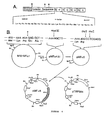
- the final step is the removal of Glu-Ala or Asp-Ala pairs by diaminopeptidase, which is encoded by the STE13 (See Figure 4A).
- Brake, J. et al. Proc. Nat. Acad. Sci. U.S.A. 81: 4642-4646 [1984] have shown that the fusion of the sequence encoding mature human proteins (including human IL-2) to the first processing site allowed secretion of such proteins.
- a general yeast expression vector designated pMF-alpha-8, containing the alpha factor promoter and downstream leader sequence in conjunction with other elements, has been deposited with the ATCC (accession number 40140). It can be constructed as follows (See Figure 4B): A 1.7 kb EcoRI fragment carrying the MFal gene (Kurjan, J. and Hershowitz, I., Cell. 30: 933-943 [1982]) is cloned into the EcoRI restriction site of M13mp8 (Viera, J. and Messing, J., Gene 19: 259-268 (11982]).
- the synthetic oligonucleotide TCTTTTATCCAAAGATACCC is hybridized to the single strand M13-MFa1 DNA and the oligonucleotide primer extended by DNA polymerase I Klenow fragment.
- Sl nuclease treatment the DNA is cleaved with EcoRI, and the fragment carrying the MFal promoter and leader sequence is cloned into the EcoRI and filled-in HindIII restriction sites of pUC8 (Viera, J. and Messing, J. abovej.
- One plasmid with the desired structure can be isolated (designated pMFa4Al in Figure 4B).
- the pMFa4Al is cleaved with HindIII and partially filled in with DNA polymerase I Klenow fragment in the presence of dATP and dGTP.
- the DNA is treated with mung bean nuclease, and the oligonucleotide linker GCCTCGAGGC is attached.
- the resultant plasmid (designated pMFa5 in Figure 4B) will have a StuI cleavage site immediately after the arginine codon, followed by the XhoI restriction site.
- An S.cerevisiae-E.coli shuttle vector (pTRP584) can be constructed as follows: the PstI-XbaI fragment carrying 2 ⁇ m plasmid replication origin (Broach, J.
- cDNA clones encoding for murine IL-2 may then be readily inserted into the pMFa8 vector and subsequently transformed in yeast for IL-2 production.
- the PstI-BamHl fragment carrying the entire IL-2 cDNA is transfered from pcD-IL-2 into the PstI-BamHl sites of pUC9 (Viera, J. and Messing, J. above) and cleaved with HinPl and SmaI. SmaI cleaves downstream of the cDNA insert.
- the fragment is treated with DNA polymerase I Klenow fragment in the presence of dCTP and treated with mung bean nuclease.
- This plasmid DNA (carrying the TRP1 gene) can be introduced into yeast cells by the lithium acetate method (Ito, H. et al., J. Bacteriol. 153: 163-168 [1983]) and transformants are selected in synthetic medium lacking tryptophan. Transformants are then grown in a common medium supplemented with 0.5% casamino acids. For harvesting, the yeast cells, resuspended in phosphate-buffered-saline (PBS) containing 1 mM PMSF, are disintegrated by vigorous shaking with acid washed glass beads. Clear supernatant is obtained by centrifugation at 10,000 rpm for 15 min.
- PBS phosphate-buffered-saline
- cell cultures derived from multicellular organisms may also be used as hosts.
- useful host cell lines are HeLa cells, Chinese hamster ovary cell lines, and baby hamster kidney cell lines.
- Expression vectors for such cells ordinarily include, as necessary, an origin of replication, a promoter located in front of the gene to be expressed, along with any required ribosome binding sites, RNA splice sites, polyadenylation sites, and transcriptional terminator sequences.
- the expression vector often has control functions provided by viral material.
- commonly used promoters are derived from polyoma, Adenovirus 2, and most frequently SV-40. (See, e.g., U.S. patent No. 4,399,216 and Gheysen, D. and Fiers, W., J. of Mol. and Appl. Genetics 1: 385-394 [1982].)
- the murine IL-2 polypeptides expressed in E.coli, in yeast or in other cells can be purified according to standard procedures of the art, including ammonium sulfate precipitation, fractionation column chromatography (e.g., ion exchange, gel filtration, electrophoresis, affinity chromatography, etc.) and ultimately crystallization (see generally "Enzyme purification and Related Techniques", Methods in Enzymology, 22: 233-577 [1977]).
- the polypeptides of the invention may be used for research purposes, e.g., as a supplement to cell growth media (e.g., minimum essential medium Eagle, Iscove's modified Dulbecco Medium or RPMI 1640; available from Sigma Chemical Company, St. Louis, MO and GIBCO Division, Chagrin Falls, OH) and as an antigenic substance for eliciting specific immunoglobulins useful in immunoassays, immunofluorescent stainings, etc. (See generally "Immunological Methods", Vols. I & II, Eds. Lefkovits, I. and Pernis, B., Academic Press, New York, N.Y. [1979 & 1981]; and "Handbook of Experimental Immunology", ed. Weir, D., Blackwell Scientific Publications, St. Louis, MO [1978].)
- cell growth media e.g., minimum essential medium Eagle, Iscove's modified Dulbecco Medium or RPMI 1640; available from Sigma Chemical Company, St
- yeast cultures containing cDNAs of the present invention are utilized to prepare murine IL-2, purification preferably is accomplished as follows. Up to one liter of 12 hr culture fluid is diluted with an equal volume of 0.2% trifluoroacetic acid (TFA) and 40% CH 3 CN, and pumped directly onto a 4.6 X 75 mm propyl column (Ultrapare RPSC, Altex, Berkeley, CA) previously equilibrated with 0.1% TFA, 20% CH 3 CN. Sample application can be at a flow rate of about 1 ml/min at 4°C.
- TFA trifluoroacetic acid
- the column is connected to a conventional HPLC system at room temperature, and further washed at 0.5 ml/min until the absorbance monitored at 214 nM reaches baseline. A gradient of increasing solvent concentration is then applied (0.5% CH 3 CN per min), and 1 ml fractions collected.
- the murine IL-2 elutes about 65 min after the start of the gradient in a peak well separated from other proteins. Analysis of the peak fractions on silver- stained Laemmli gels (Laemmli, U., Nature 227: 680-685
- phenol-CHC1 3 water-saturated 1:1 phenol-CHC1 3 (hereafter referred to as phenol-CHC1 3 ) and ethanol precipitation.
- the reaction mixture (38 ul) contained sodium cacodylate-30 mM Tris.HCl pH 6.8 as buffer, with 1 mM CoCl 2 , 0.1 mM dithiothreitol, 0.25 mM dTTP, the K pnI endonuclease-digested DNA, and 68 U of the terminal deoxynucleotidyl transferase (P-L Biochemicals, Inc., Milwaukee, WI).
- dT deoxythymidylate
- the large fragment containing the SV40 polyadenylation site and the pB R 322 origin of replication and ampicillin- resistance gene, was purified by agarose (1%) gel electrophoresis and recovered from the gel by a modification of the glass powder method (Vogelstein, B. & Gillespie, D., Proc. Nat. Acad. Sci. 76: 615-619 [1979]).
- the dT-tailed DNA was further purified by absorption and elution from an oligo (dA)-cellulose column as follows: The DNA was dissolved in 1 ml of 10 mM Tris.HCl pH 7.3 buffer containing 1 mM EDTA and 1 M NaCl, cooled at 0°C, and applied to an oligo (dA)-cellulose column (0.6 by 2.5 cm) equilibrated with the same buffer at 0°C. The column was washed with the same buffer at 0°C and eluted with water at room temperature. The eluted DNA was precipitated with ethanol and dissolved in 10 mM Tris. HC1 pH 7.3 with 1 mM EDTA.
- the oligo (dG) tailed linker DNA was prepared by digesting 75 ug of pLl DNA with 20 U of PstI endonuclease in 450 ul containing 6 mM Tris.HCl pH 7.4, 6 mM MgCl 2 , 6 mM 2-ME, 50 mM NaCl, and 0.01 mg of BSA per ml. After 16 hr at 30°C the reaction mixture was extracted with phenol-CHC1 3 and the DNA was precipitated with alcohol.
- Tails of 10 to 15 deoxyguanylate (dG) residues were then added per end with 46 U of terminal deoxynucleotidyl transferase in the same reaction mixture (38 ul) as described above, except that 0.1 mM dGTP replaced dTTP.
- the mixture was extracted with phenol-CHC1 3 , and after the DNA was precipitated with ethanol it was digested with 35 U of HindIII endonuclease in 50 ul containing 20 mM Tris.HCl pH 7.4, 7 mM MgCl 2 , 60 mM NaCl, and 0.1 mg of BSA at 37°C for 4 hr.
- the small oligo (dG)-tailed linker DNA was purified by agarose gel (1.8%) electrophoresis and recovered as described above.
- Step 1 cDNA synthesis.
- the reaction mixture (10 ⁇ l) contained 50 mM Tris.HCl pH 8.3, 8 mM MgCl 2 , 30 mM KC1, 0.3 mM dithiothreitol, 2 mM each dATP, dTTP, dGTP, and dCTP, 20 ⁇ Ci 32 P-dCTP (3000 Ci/mmole), 6 ⁇ g polyA + RNA from Con-A induced LB2-1, 60 units RNasin (Biotec, Inc., Madison, WI), and 2 ⁇ g of the vector-primer DNA (15 pmol of primer end), and 45 U of reverse transcriptase.
- the reaction was incubated 60 min at 42°C - and then stopped by the addition of 1 ul of 0.25 M ETDA (pH 8.0) and 0.5 ul of 10% SDS; 40 ⁇ l of phenol-CHC1 3 was added, and the solution was blended vigorously in a Vortex mixer and then centrifuged. After adding 40 ul of 4 M ammonium acetate and 160 ul of ethanol to the aqueous phase, the solution was chilled with dry ice for 15 min., warmed to room temperature with gentle shaking to dissolve unreacted deoxynucleoside triphosphates that had precipitated during chilling, and centrifuged for 10 min. in an Eppendorf microfuge.
- the pellet was dissolved in 10 ul of lOmM Tris.HCl pH 7.3 and 1 mM EDTA, mixed with 10 ul of 4 M ammonium acetate, and reprecipitated with 40 ul of ethanol, a procedure which removes more than 99% of unreacted deoxynucleoside triphosphates.
- the pellet was rinsed with ethanol.
- Step 2 Oligodeoxycytidylate [oligo (dC)] addition.
- the pellet containing the plasmid- cDNA:mRNA was dissolved in 20 ⁇ l of 140 mM sodium cacodylate-30 mM Tris.HCl pH 6.8 buffer containing 1 mM CoCl 2 , 0.1 mM dithiothreitol, 0.2 ug of poly(A), 70 uM dC TP , 5 uCi 32 P-dCTP, 3000 Ci/mmole, and 60 U of terminal deoxynucleotidyl transferase. The reaction was carried out at 37°C for 5 min.
- Step 3 HindIII endonuclease digestion.
- the pellet was dissolved in 30 ul of buffer containing 20 mM Tris. HCl pH 7.4, 7 mM MgCl 2 , 60 mM NaCl, and 0.1 mg of BSA per ml and then digested with 10 U of HindIII endonuclease for 2 hr at 37°C.
- the reaction was terminated with 3 ul of 0.25 M EDTA (pH 8.0) and 1.5 ⁇ l of 10% SDS and, after extraction with phenol-CHC1 3 followed by the addition of 30 ul of 4 M ammonium acetate, the DNA was precipitated with 120 ul of ethanol.
- the pellet was rinsed with ethanol and then dissolved in 10 ul of 10 mM Tris.HCl (pH 7.3) and 1 mM EDTA, and 3 ul of ethanol was added to prevent freezing during storage at -20°C.
- Step 4 Cyclization mediated by the oligo (dG)-tailed linker DNA.
- a 9 ul sample of the HindIII endonuclease-digested oligo (dC)-tailed cDNA:mRNA plasmid (about 90% of the sample) was incubated in a mixture (90 ul) containing 10 mM Tris.HCl pH 7.5, 1 mM EDTA, 0.1 M NaCl, and 1.8 pmol of the oligo (dG)-tailed linker DNA at 65°C for 5 min, shifted to 42°C for 60 min, and then cooled to 0°C.
- the mixture (90 ul) was adjusted to a volume of 900 ul containing 20 mM Tris.
- Step 5 Replacement of RNA strand by DNA.
- the ligation mixture was adjusted to contain 40 uM of each of the four deoxynucleoside triphosphates, 0.15 mM B-NAD, 4 ug of additional E. coli DNA ligase, 16 U of E. coli DNA polymerase I (PolI,) and 9 U of E. coli RNase H. This mixture (960 ul) was incubated successively at 12°C and room temperature for 1 hr each to promote optimal repair synthesis and nick translation by Poll.
- Step 6 Transformation of E. coli. Transformation was carried out using minor modifications of the procedure described by Cohen et al. (Proc. Nat. Acad. Sci. U.S.A., 69: 2110-2114 [1972]). E. coli K-12 strain MC1061 (Casadaban, M. and Cohen, S., J. Mol. Biol. 138: 179-207 [1980]) was grown to 0.5 absorbancy unit at 600 nm at 37°C in 20 ml of L-broth. The cells were collected by centrifugation, suspended in 10 ml of 10 mM Tris.HCl pH 7.3 containing 50 mM CaCl 2 , and centrifuged at 0°C for 5 min.
- the cells were resuspended in 2 ml of the above buffer and incubated again at 0°C for 5 min.; then, 0.2 ml of the cell suspensions was mixed with 0.1 ml of the DNA solution (step 5) and incubated at 0°C for 15 min. Next the cells were kept at 37°C for 2 min. and thereafter at room temperature for 10 min.; then 0.5 ml of L-broth was added, and the culture was incubated at 37°C for 30 min., mixed with 2.5 ml of L-broth soft agar at 42°C, and spread over L-broth agar containing 50 ug of ampicillin per ml. After incubation at 37°C for 12 to 24 hr, individual colonies were picked with sterile tooth-picks. In all, approximately 1 x 10 5 independent cDNA clones were generated.
- plasmid DNA representing the entire cDNA library (pcD-X DNA) was digested separately with the restriction enzymes SalI, HindIII, and ClaI to linearize the plasmid.
- the restricted DNAs were size- fractionated on a 1% agarose gel to separate plasmids having different size cDNA inserts. Segments were excised from the gel representing plasmids with cDNA inserts of the following size ranges:
- DNA was eluted from each gel slice using the glass powder method of Vogelstein and Gillespie (Proc. Nat.
- a library of about 10 5 independent transformations was obtained for the fraction containing cDNA inserts 1-2 kb in 15 length.
- a collection of 10 4 independent clones were picked at random from the sublibrary enriched for cDNA inserts of 1-2 kb and propagated individually in wells of microtiter dishes containing 200 ul L broth with ampicillin at 50 ug/ml and dimethyl sulfoxide at 7%.
- Pools containing 48 cDNA clones were prepared from the microtiter cultures. 58 such pools were grown up in 1 liter cultures of L-broth containing 100 ⁇ g/ml ampicillin. Plasmid DNA was isolated from each culture and purified by twice banding through CsCl gradients.
- the DNA representing each pool was transfected into COS-7 monkey cells as follows.
- the plates were incubated for four hours at 37°C, then the DNA-containing medium was removed, and the plates were washed twice with 2 ml of serum-free DME.
- DME containing 150 uM chloroquine was added back to the plates which were then incubated for an additional 3 hrs at 37°C.
- the plates were washed once with DME, and then DME containing 4% fetal calf serum, 2 mM glutamine, penicillin and streptomycin was added.
- the cells were then incubated for 72 hrs at 37°C.
- the growth medium was collected and assayed for murine IL-2 activity as described above.
- group 6 The murine IL-2 activity (see Table II below).
- Group 33 was then subdivided into 8 pools, each containing 6 of the original pools. Only one of these subpools (group c) was positive in the transfection assay.
- Each of the plasmids in group c was transfected individually into COS-7 cells. Only one clone, designated MT-18, was active in producing TCGF activity.
- the pools of 48 cDNA clones were transformed from the microtiter trays to nitrocellulose filters placed on plates of L-agar + 100 ug/ml ampicillin. These plates were incubated overnight at 37°C. The bacterial colonies were lysed and the DNA bound to the filters as described in Maniatis, T., et al. (above).
- the nick-translated PstI-HindIII fragment from MT-18 was hybridized with each of the 58 filters representing the pools.
- the hybridizations were performed in 6XSSPE (0.18 M NaCl, 1mM EDTA, and 10 mM NaH 2 P0 4 [pH 7.9]), 50% formamide, 100 ug/ml E. coli tRNA, 0.1% SDS, at 42°C overnight.
- the filters were washed once at room temperature in 2X SSPE 0.1% SDS, and then twice with 0.2X SSPE at 60°C. Following incubation of the filters, 15 individual clones were found to hybridize with the probe.
- Plasmid DNA from each of these clones was prepared as described above, and transfected into COS-7 cells. Of these clones, only three, contained in pools 6 ( MT -1), 40 (MT-20), and 56 (MT-28) produced high levels of murine IL-2 activity (see Table II). Restriction analysis showed that the clones share essentially the same structure.
- a plasmid (pcD-IL-2) carrying a substantially full-length cDNA insert is shown in Figure 2, and an E. coli bacterium (MC1061) carrying the plasmid has been deposited with the ATCC (accession number 39892).
- transcription of the 816 bp cDNA insert contained in the pcD expression vector from the SV40 early promoter is indicated by the arrow.
- the locations of the splice donor and acceptor sites are shown.
- the polyadenylation signal, also derived from SV40 is located at the 3'-end of the cDNA insert.
- the IL-2 coding region in the cDNA insert is heavily shaded while the non-coding regions are lightly shaded.
- the remainder of the vector sequences are derived from pBR322, including the B-lactamase gene (Amp R ) and the origin of replication.
- the homologies between the mouse IL-2 cDNA and human IL-2 cDNA are also surprising. Overall, there is about 70% homology between the two IL-2 cDNA sequences (Brutlag, D. et al., Nucleic Acids Res. 10: 279-294 [1981]). In particular, the regions covered by nucleotide positions 1-125, 229-354, and 416-680 share extensive homology with the corresponding regions of human IL-2 cDNA. However, the trinucleotide CAG sequence, which is repeated twelve times within the mouse IL-2 cDNA coding region, is not present in any human IL-2 cDNA isolated to date.
- the alone library in the pcD expression vector enabled the identification of complete cDNA clones by direct expression in mammalian cells. Specifically, complete mouse IL-2 cDNA clones were directly identified by transfecting COS-7 cells with randomly picked cDNA clones, and measuring the TCGF activity secreted into the cell supernatant. These results indicate that the identification of full-length cDNA clones of lymphokines or hormones may be achieved solely on the basis of detection of a functional polypeptide in eukaryotic cells. Identification of relevant cDNA clones based on the functional expression of the gene offers advantages to hybrid selection procedures.
- the invention further provides a method of identifying individual nucleotide sequences coding for a polypeptide, said method comprising the stepsof:
- the cDNA clones of the present invention provide accurate and complete sequence data on murine IL-2.
- the invention also provides to those skilled in the art means for producing significant quantities of the factor (essentially free from other hematopoietic factors) for the improved in vitro maintenance of T cells and other hematopoietic cells. Further, the information gleaned from the cDNA clones increases understanding of the mammalian immune response, enhancing experimental research capabilities.
Applications Claiming Priority (2)
| Application Number | Priority Date | Filing Date | Title |
|---|---|---|---|
| US65818384A | 1984-10-05 | 1984-10-05 | |
| US658183 | 1984-10-05 |
Publications (1)
| Publication Number | Publication Date |
|---|---|
| EP0177357A1 true EP0177357A1 (de) | 1986-04-09 |
Family
ID=24640235
Family Applications (1)
| Application Number | Title | Priority Date | Filing Date |
|---|---|---|---|
| EP85307094A Withdrawn EP0177357A1 (de) | 1984-10-05 | 1985-10-03 | cDNA-Klone, die für Polypeptide kodieren, die Murin-Interleukin-2-Aktivität zeigen |
Country Status (4)
| Country | Link |
|---|---|
| EP (1) | EP0177357A1 (de) |
| JP (1) | JPS61119197A (de) |
| AU (1) | AU599891B2 (de) |
| IL (1) | IL76574A0 (de) |
Cited By (2)
| Publication number | Priority date | Publication date | Assignee | Title |
|---|---|---|---|---|
| EP0267795A2 (de) * | 1986-11-14 | 1988-05-18 | Schering Biotech Corporation | Muteine des Maus-Interleukin-2 |
| WO2001040311A1 (fr) * | 1999-11-30 | 2001-06-07 | Shionogi & Co., Ltd. | Proteine fusionnee de chimiokine slc-il2 |
Citations (3)
| Publication number | Priority date | Publication date | Assignee | Title |
|---|---|---|---|---|
| EP0091539A1 (de) * | 1982-03-31 | 1983-10-19 | Ajinomoto Co., Inc. | Das Polypeptid Interleukin-2 kodierendes Gen, rekombinante, dieses Gen enthaltende DNA, diese rekombinante DNA aufweisende Zelllinien und Verfahren zur Herstellung von Interleukin-2 unter Verwendung der genannten Zellen |
| EP0118977A1 (de) * | 1983-02-08 | 1984-09-19 | Biogen, Inc. | DNA-Sequenzen, rekombinante DNA-Moleküle und Verfahren zur Herstellung von Human-Interleukin-2 ähnlichen Polypeptiden |
| WO1985002863A1 (en) * | 1983-12-23 | 1985-07-04 | The Australian National University | CLONING OF cDNA AND EXPRESSION OF MURINE-INTERLEUKIN-3 |
Family Cites Families (2)
| Publication number | Priority date | Publication date | Assignee | Title |
|---|---|---|---|---|
| AU556353B2 (en) * | 1982-12-15 | 1986-10-30 | Ajinomoto Co., Inc. | Interleukin-2 polypeptide |
| PT79506B (en) * | 1984-08-08 | 1986-09-08 | Quidel | T-cell growth factor |
-
1985
- 1985-10-03 EP EP85307094A patent/EP0177357A1/de not_active Withdrawn
- 1985-10-04 AU AU48355/85A patent/AU599891B2/en not_active Ceased
- 1985-10-04 IL IL76574A patent/IL76574A0/xx unknown
- 1985-10-04 JP JP60221684A patent/JPS61119197A/ja active Pending
Patent Citations (3)
| Publication number | Priority date | Publication date | Assignee | Title |
|---|---|---|---|---|
| EP0091539A1 (de) * | 1982-03-31 | 1983-10-19 | Ajinomoto Co., Inc. | Das Polypeptid Interleukin-2 kodierendes Gen, rekombinante, dieses Gen enthaltende DNA, diese rekombinante DNA aufweisende Zelllinien und Verfahren zur Herstellung von Interleukin-2 unter Verwendung der genannten Zellen |
| EP0118977A1 (de) * | 1983-02-08 | 1984-09-19 | Biogen, Inc. | DNA-Sequenzen, rekombinante DNA-Moleküle und Verfahren zur Herstellung von Human-Interleukin-2 ähnlichen Polypeptiden |
| WO1985002863A1 (en) * | 1983-12-23 | 1985-07-04 | The Australian National University | CLONING OF cDNA AND EXPRESSION OF MURINE-INTERLEUKIN-3 |
Cited By (3)
| Publication number | Priority date | Publication date | Assignee | Title |
|---|---|---|---|---|
| EP0267795A2 (de) * | 1986-11-14 | 1988-05-18 | Schering Biotech Corporation | Muteine des Maus-Interleukin-2 |
| EP0267795A3 (de) * | 1986-11-14 | 1990-07-25 | Schering Biotech Corporation | Muteine des Maus-Interleukin-2 |
| WO2001040311A1 (fr) * | 1999-11-30 | 2001-06-07 | Shionogi & Co., Ltd. | Proteine fusionnee de chimiokine slc-il2 |
Also Published As
| Publication number | Publication date |
|---|---|
| AU599891B2 (en) | 1990-08-02 |
| IL76574A0 (en) | 1986-02-28 |
| AU4835585A (en) | 1986-04-10 |
| JPS61119197A (ja) | 1986-06-06 |
Similar Documents
| Publication | Publication Date | Title |
|---|---|---|
| EP0138133B1 (de) | cDNA-Klone, codierend für Polypeptide mit der Wirksamkeit eines nicht-zellinienspezifischen Wachstumsfaktors (multi-CSF) und/oder Mastzellenwachstumsfaktors (MCGF) | |
| Miyatake et al. | Structure of the chromosomal gene for granulocyte‐macrophage colony stimulating factor: comparison of the mouse and human genes. | |
| JP2568394B2 (ja) | 哺乳動物インターロイキン−4活性を示すポリペプチドをコードするヌクレオチド配列からなる核酸 | |
| EP0202300B1 (de) | Cdna-klone, kodierend für peptide mit der wirksamkeit eines menschlichen granulocyte-, makrophage- und eosinophilzellenwachstumsfaktors | |
| EP0282185A1 (de) | Human-Interleukin-3 und seine Muteine | |
| HU202287B (en) | Process for producing human immune interferon | |
| CA1335717C (en) | Human granulocyte-macrophage colony stimulating factor and muteins thereof | |
| EP0188864B1 (de) | Für menschliches Interleukin-1 alpha codierende DNA und die dazu korrespondierende Aminosäurenkette, Vektoren und Wirte, die eine solche DNA enthalten und deren Herstellung | |
| US4798789A (en) | cDNA clones coding for polypeptides exhibiting murine interleukin-2 activity | |
| US5951973A (en) | Use of interleukin-4 (IL-4) to treat rheumatoid arthritis | |
| EP0177357A1 (de) | cDNA-Klone, die für Polypeptide kodieren, die Murin-Interleukin-2-Aktivität zeigen | |
| Krämmer et al. | A plasmodial α-tubulin cDNA from Physarum polycephalum: nucleotide sequence and comparative analysis | |
| AU618143B2 (en) | Cdna clones coding for polypeptides exhibiting multi- lineage cellular growth factor activity and/or mast cell growth factor activity | |
| AU626530B2 (en) | Human granulocyte-macrophage colony stimulating factor and muteins thereof |
Legal Events
| Date | Code | Title | Description |
|---|---|---|---|
| PUAI | Public reference made under article 153(3) epc to a published international application that has entered the european phase |
Free format text: ORIGINAL CODE: 0009012 |
|
| AK | Designated contracting states |
Kind code of ref document: A1 Designated state(s): AT BE CH DE FR GB IT LI LU NL SE |
|
| 17P | Request for examination filed |
Effective date: 19860923 |
|
| 17Q | First examination report despatched |
Effective date: 19880624 |
|
| RAP3 | Party data changed (applicant data changed or rights of an application transferred) |
Owner name: SCHERING BIOTECH CORPORATION |
|
| STAA | Information on the status of an ep patent application or granted ep patent |
Free format text: STATUS: THE APPLICATION IS DEEMED TO BE WITHDRAWN |
|
| 18D | Application deemed to be withdrawn |
Effective date: 19920505 |
|
| RIN1 | Information on inventor provided before grant (corrected) |
Inventor name: LEE, FRANK DON Inventor name: ARAI, KEN-ICHI Inventor name: YOKOTA, TAKASHI |