EP0176352A2 - Differenzmessverfahren zum Nachweis von Krebs in menschlichem Gewebe mittels magnetischer Kernresonanz - Google Patents
Differenzmessverfahren zum Nachweis von Krebs in menschlichem Gewebe mittels magnetischer Kernresonanz Download PDFInfo
- Publication number
- EP0176352A2 EP0176352A2 EP85306794A EP85306794A EP0176352A2 EP 0176352 A2 EP0176352 A2 EP 0176352A2 EP 85306794 A EP85306794 A EP 85306794A EP 85306794 A EP85306794 A EP 85306794A EP 0176352 A2 EP0176352 A2 EP 0176352A2
- Authority
- EP
- European Patent Office
- Prior art keywords
- nmr
- breast
- tissue
- water
- equation
- Prior art date
- Legal status (The legal status is an assumption and is not a legal conclusion. Google has not performed a legal analysis and makes no representation as to the accuracy of the status listed.)
- Granted
Links
Images

Classifications
-
- G—PHYSICS
- G01—MEASURING; TESTING
- G01R—MEASURING ELECTRIC VARIABLES; MEASURING MAGNETIC VARIABLES
- G01R33/00—Arrangements or instruments for measuring magnetic variables
- G01R33/20—Arrangements or instruments for measuring magnetic variables involving magnetic resonance
- G01R33/28—Details of apparatus provided for in groups G01R33/44 - G01R33/64
- G01R33/32—Excitation or detection systems, e.g. using radio frequency signals
- G01R33/34—Constructional details, e.g. resonators, specially adapted to MR
- G01R33/34046—Volume type coils, e.g. bird-cage coils; Quadrature bird-cage coils; Circularly polarised coils
- G01R33/34053—Solenoid coils; Toroidal coils
-
- A—HUMAN NECESSITIES
- A61—MEDICAL OR VETERINARY SCIENCE; HYGIENE
- A61B—DIAGNOSIS; SURGERY; IDENTIFICATION
- A61B8/00—Diagnosis using ultrasonic, sonic or infrasonic waves
- A61B8/08—Clinical applications
- A61B8/0825—Clinical applications for diagnosis of the breast, e.g. mammography
-
- A—HUMAN NECESSITIES
- A61—MEDICAL OR VETERINARY SCIENCE; HYGIENE
- A61B—DIAGNOSIS; SURGERY; IDENTIFICATION
- A61B8/00—Diagnosis using ultrasonic, sonic or infrasonic waves
- A61B8/40—Positioning of patients, e.g. means for holding or immobilising parts of the patient's body
- A61B8/406—Positioning of patients, e.g. means for holding or immobilising parts of the patient's body using means for diagnosing suspended breasts
-
- G—PHYSICS
- G01—MEASURING; TESTING
- G01R—MEASURING ELECTRIC VARIABLES; MEASURING MAGNETIC VARIABLES
- G01R33/00—Arrangements or instruments for measuring magnetic variables
- G01R33/20—Arrangements or instruments for measuring magnetic variables involving magnetic resonance
- G01R33/44—Arrangements or instruments for measuring magnetic variables involving magnetic resonance using nuclear magnetic resonance [NMR]
- G01R33/48—NMR imaging systems
- G01R33/50—NMR imaging systems based on the determination of relaxation times, e.g. T1 measurement by IR sequences; T2 measurement by multiple-echo sequences
-
- G—PHYSICS
- G01—MEASURING; TESTING
- G01R—MEASURING ELECTRIC VARIABLES; MEASURING MAGNETIC VARIABLES
- G01R33/00—Arrangements or instruments for measuring magnetic variables
- G01R33/20—Arrangements or instruments for measuring magnetic variables involving magnetic resonance
- G01R33/28—Details of apparatus provided for in groups G01R33/44 - G01R33/64
- G01R33/38—Systems for generation, homogenisation or stabilisation of the main or gradient magnetic field
- G01R33/383—Systems for generation, homogenisation or stabilisation of the main or gradient magnetic field using permanent magnets
-
- G—PHYSICS
- G01—MEASURING; TESTING
- G01R—MEASURING ELECTRIC VARIABLES; MEASURING MAGNETIC VARIABLES
- G01R33/00—Arrangements or instruments for measuring magnetic variables
- G01R33/20—Arrangements or instruments for measuring magnetic variables involving magnetic resonance
- G01R33/44—Arrangements or instruments for measuring magnetic variables involving magnetic resonance using nuclear magnetic resonance [NMR]
- G01R33/48—NMR imaging systems
- G01R33/483—NMR imaging systems with selection of signals or spectra from particular regions of the volume, e.g. in vivo spectroscopy
- G01R33/4833—NMR imaging systems with selection of signals or spectra from particular regions of the volume, e.g. in vivo spectroscopy using spatially selective excitation of the volume of interest, e.g. selecting non-orthogonal or inclined slices
- G01R33/4835—NMR imaging systems with selection of signals or spectra from particular regions of the volume, e.g. in vivo spectroscopy using spatially selective excitation of the volume of interest, e.g. selecting non-orthogonal or inclined slices of multiple slices
Definitions
- This method is intended to enhance data information obtained by NMR interrogation of human tissue, particularly with a view of locating small tumors.
- an apparatus is disclosed which enables the sequential segmented interrogation of the human breast with a particular goal of locating relatively small tumors. Enhancement of that data is important to enable small tumors to be located. It is assumed that large or qross tumors can be located by other procedures. However, early detection enhances the chance of recovery. Early detection enables less radical curative procedures to be utilized.
- the method of this disclosure enhances the signal component traceable to the tumor, reducing the other signal components and thereby focusing on the signal component derivative from prospective tumors. In that manner, signal levels above the millivolt range can thus be obtained, and are therefore more readily located in the NMR signal to enable the output signal to be processed. This reduces the size of the minimum tumor which can be located through the procedure and method of this disclosure.
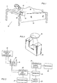
- This procedure contemplates the successive NMR interrogation of slices of the human tissues, particularly the female breast for the express purpose of obtaining individual signals from each slice. So to speak, a specified thickness is determined by the magnetic field gradient to thereby segment the female breast into a number of slices. Each interrogation NMR procedure obtains an output signal for each slice. The magnetic field is changed, relocating the magnetic field intensity H o requisite to obtain a proper NMR response. Assume for purposes of discussion and description that this slice is a thickness that is more or less uniform from slice to slice and is approximately one millimeter. This is a reasonable scale factor for the procedure described herein. That is, the slice of space within the magnetic field where the field intensity is H o is about one millimeter thick.
- each slice is preferably thin and typically in the range of the preferred dimension of about one millimeter. This can also be varied and is dependent on a number of factors including scale factors.
- Equation (1) was based on a slow exchange.
- T i is much greater than the lifetime in phase i
- Equation (2) the NMR signal voltage as a function of time
- Simplifications can be made from the general statements given above by reducing n to two, the typical case involving NMR interrogation of the female breast in detection of water which is bound in normal tissue. This permits development of a relatively exact expression.
- This exact expression relates to tissue water which can be generally described as tightly bound and loosely bound water.
- the relaxation time for water in the cancerous tissue is longer than the relaxation time for the healthy tissue. Because the tissue is made of water in various binding phases, and because there is exchange between the two phases of water in both healthy tissue and cancerous tissue, the NMR signal is given either by Equation (1) for slow water exchange or by Equations (2) and (3) for a rapid water exchange.
- Equation (1) Equation (1)
- Equation (4) is an equation for the spin-spin relaxation time signal which is v 2 (t) while Equation (5) can be written for the spin-lattice relaxation time v 1 (t). This equation follows the same general form as Equation (4).
- Equation (6) Assuming water in healthy tissue is in two binding phases, the hydrogen transient NMR signal is given by Equation (6):
- Equation (7) the hydrogen transient NMR system involving a three component system where one component is cancerous tissue and the other two components are healthy tissue is given by Equation (7):
- Equation (6) represents a signal from a layer of healthy tissue while the signal of Equation (7) represents a layer having cancerous tissue.
- Equation (6) represents a difference signal given hy Equation (A):
- Equation (R) Equation (9) where the difference signal is given by:
- Equation (9) The first and second terms of Equation (9) have similar coefficients and hence their difference becomes relatively small. That is, they are relatively small, observing the subtractive factors.
- the third term is by its nature a small coefficient.
- Equation (10) it will be observed that certain limits are approached. These limits reduce the signal resulting from similarities while accenting or increasing the relative difference (see Equation 10).
- the difference signal contains only the cancerous component.
- the difference signal showing decreased similarities and increased differences, provides an accent between adjacent layers from sequential NMR hydrogen transient signals.
- a digitized signal format that is, the variable signal is in digital form
- a first layer has a 5 volt signal
- the adjacent layer will have a signal of 5.25 volts maximum (recalling the assumption of not more than 5% volumetric variation)
- the subtraction of these two signals will provide a resultant output differential of up to 0.25 volts; digitizing accuracy should be added and this typically represents about 0.01 differential.
- a normal signal representing up to 5% size change (without cancerous tissue) at the most will be about 0.25 volts ⁇ 0.15 volts.
- a cancer is present and represents about 1%. of the volume.
- 1% of the 5 volt signal is about 0.05 volts.
- a signal level of about 0.05 volts (indicative of cancer) when confronted with a variation of 0.25 plus or minus 0.01 volt as a result of digitizing and geometric change on subtraction yields a cancerous tissue signal which is about 5/30 or 16% of the output differential signal.
- the signal to inoise ratio of the subtracted signal is around 30 to 1.
- Equation (6) and (7) provide values for the constants in Equation (6) and (7).
- the constants are:
- Equation (9) the values obtained from Equation (9) for the first and second coefficients are:
- Equation (14)
- N 150.
- the time required to obtain each data point is one second, a relatively slow repetition rate. 150 data points can be obtained. This yields 149 difference signals.
- the signals are obtained by subtracting consecutive or adjacent data entries as N approaches 150.
- the presentation of data is perhaps best implemented on a white to gray scale on an oscilloscope presentation. The difference signal from adjacent pixels enhances the differences as described above so that the enhanced image will more readily show cancerous tissue.
- the location of the cancer can then be readily determined.
- a cancer which represents 10% of a given slice or layer.
- signal amplitude is markedly increased and a signal to noise ratio is improved even further.
- the signal from healthy breast tissue may have two parts, one from the hydrogen in the tissue and one from the hydrogen in the water.
- cancerous tissue is present (along with healthy tissue) there are two values of T 2 for water, one for the water in the healthy tissue T 2h and a second for the water in the cancerous tissue T 2c . It has been found experimentally, on the average, that T 2c equals 2T 2h .
- Equation (15) The echo signal from a thin layer of a breast with cancerous tissue will have a voltage amplitude given by Equation (15): where All is a voltage proportional to the number of hydrogen nuclei in the volume of the water in the healthy tissue, and A 21 is the voltage proportional to the number of hydrogen nuclei in the volume of the water in the cancerous tissue.
- Equation (17) Equation (17)
- Equation (16) A 12 is a voltage proportional to the number of hydrogen nuclei in the water in the volume of the n layer without cancer adjacent to n-1 layer.
- Equation (18) This difference technique involves the subtraction of Equation (18) from Equation (16).
- Equation (IR) is subtracted from Equation (17) the result is Equation (19): with equal volumes in adjacent layers (n and n-1), the healthy component equals the cancer component. If it is assumed that the volume of the n-1 layer slice with the cancer is up to 5% greater than the layer without the cancer, then from Equation (18) one obtains Equation (20):
- Equation (21) When Equation (20) is subtracted from Equation (17), or when the signals from adjacent layers are subtracted, the result is Equation (21):
- Equation (21) the ratio of the two signal components is now 1/5 whereas before subtraction they were 1/99 in Equation (17).
- the values of T 2h and T 2C given in the literature are 0.05 and 0.10 seconds respectively. When these values are used, Equation (17) gives a straight-line to graph beyond 0.7 second where the cancerous tissue signal component becomes readily apparent.
- the difference signal of Equation (21) gives a graph with the cancer component evident beyond 0.3 second because the cancer signal component is then larger.
- NMR interrogation is not limited to a single pulse; the interrogation pulse sequence can be a dual pulse sequence with specific spacing between the two pulses.
- the voltage amplitude of the echo following a 90° - 180° dual-pulse sequence is directly proportional to M o , the nuclear magnetization, and to an exponential with a time constant is T 2 . Equation (22) is: where is the spacing between the 90° and 180° pulses and k 1 is the detection constant.
- the nuclear magnetization M o is X n H O where X n is the nuclear volume susceptibility and H o is the applied magnetic field.
- Equation (23) where N 0 is the total number of nuclei per unit volume, is the nuclear magnetic movement, k is the Boltzmann constant and T is the absolute temperature.
- Equation (24) the echo voltage amplitude is Equation (24): thus it is seen that the echo voltage is directly proportional to the number of detected nuclei per unit volume.
- the echo voltage output signal is verification that tumor mass is proportional to signal amplitude and hence signal size. This is significant in establishing minimum tumor size for NMR detection.
- minimum size tumor which can be detected using this technique. Assume tht the N layers of the breast tested by this procedure are right cylinders and hence have a volume of r 2 d where r is radius and thickness is represented by d.
- a small cancer with a cross-sectional area of 0.78 cm 2 has a volume of the cancer in the layer of 0.78d cm 3 .
- the layer volume then is 78d while the cancer volume is 0.78d, making the cancer to be one percent of the layer volume if r is assumed to be 5 cm. In this instance, a cancer of this size is significantly small that early detection markedly increases the probabilities of full health protection.
- Equation (6) water in both healthy and cancerous tissue was assumed.
- the hydrogen NMR response from both types of tissue is the talisman yielding NMR response for detection.
- Water in tissue is in differing binding phases. There is exchange between these phases, and, in general, if P i denotes the fraction of water present in phase number i, if the relaxation time for this phase is T i , if there is a slow exchange (T i the lifetime in phase number i), then the NMR signal voltage as a function of time is Equation (25):
- Equation (28) The hydrogen transient NMR signal from healthy tissue with one water component is Equation (28): and that from cancerous tissue with two water binding components (simplified from Equation 7) is given by Equation (29):
- the NMR signal of Equation (28) is from the layer of tissue adjacent to the cancerous layer giving the NMR signal of Equation (29), and the equations are subtracted to obtain a difference signal or v c2 - v h2 .
- the difference signal is Equation (30):
- the difference signal decreases the similarities between the hydrogen transient NMR signals from healthy and cancerous tissue while increasing the relative difference between them. In the limit where the similarities are the same, the difference signal will contain only the cancerous component.
- the use of the difference signal because it decreases similarities and increases differences, turns the hydrogen transient NMR signals from layers of tissue into displays or the differences between adjacent layers.
- the graph of the difference signals between adjacent layers, as a function of location from 0 to N layers gives an indication of the beginning and ending of a cancerous inclusion in one or more layers.
- the difference signal will not give an image of the cancerous inclusion but will indicate in which layer or layers it resides for an enchanced screening indication. This ; difference indication is much less complicated and less expensive for tumor screening.
Landscapes
- Physics & Mathematics (AREA)
- Health & Medical Sciences (AREA)
- Life Sciences & Earth Sciences (AREA)
- Medical Informatics (AREA)
- Surgery (AREA)
- Pathology (AREA)
- Radiology & Medical Imaging (AREA)
- Engineering & Computer Science (AREA)
- Biomedical Technology (AREA)
- Heart & Thoracic Surgery (AREA)
- Biophysics (AREA)
- Molecular Biology (AREA)
- Nuclear Medicine, Radiotherapy & Molecular Imaging (AREA)
- Animal Behavior & Ethology (AREA)
- General Health & Medical Sciences (AREA)
- Public Health (AREA)
- Veterinary Medicine (AREA)
- General Physics & Mathematics (AREA)
- Condensed Matter Physics & Semiconductors (AREA)
- High Energy & Nuclear Physics (AREA)
- Investigating Or Analysing Biological Materials (AREA)
- Magnetic Resonance Imaging Apparatus (AREA)
Applications Claiming Priority (2)
| Application Number | Priority Date | Filing Date | Title |
|---|---|---|---|
| US65495784A | 1984-09-26 | 1984-09-26 | |
| US654957 | 1984-09-26 |
Publications (3)
| Publication Number | Publication Date |
|---|---|
| EP0176352A2 true EP0176352A2 (de) | 1986-04-02 |
| EP0176352A3 EP0176352A3 (en) | 1988-09-21 |
| EP0176352B1 EP0176352B1 (de) | 1991-07-17 |
Family
ID=24626910
Family Applications (1)
| Application Number | Title | Priority Date | Filing Date |
|---|---|---|---|
| EP19850306794 Expired EP0176352B1 (de) | 1984-09-26 | 1985-09-24 | Differenzmessverfahren zum Nachweis von Krebs in menschlichem Gewebe mittels magnetischer Kernresonanz |
Country Status (3)
| Country | Link |
|---|---|
| EP (1) | EP0176352B1 (de) |
| JP (1) | JPS61119254A (de) |
| DE (1) | DE3583476D1 (de) |
Family Cites Families (2)
| Publication number | Priority date | Publication date | Assignee | Title |
|---|---|---|---|---|
| US3789832A (en) * | 1972-03-17 | 1974-02-05 | R Damadian | Apparatus and method for detecting cancer in tissue |
| JPS6024463A (ja) * | 1983-07-20 | 1985-02-07 | Toshiba Corp | 核磁気共鳴映像法 |
-
1985
- 1985-09-24 DE DE8585306794T patent/DE3583476D1/de not_active Expired - Lifetime
- 1985-09-24 EP EP19850306794 patent/EP0176352B1/de not_active Expired
- 1985-09-26 JP JP60213607A patent/JPS61119254A/ja active Pending
Also Published As
| Publication number | Publication date |
|---|---|
| DE3583476D1 (de) | 1991-08-22 |
| EP0176352A3 (en) | 1988-09-21 |
| JPS61119254A (ja) | 1986-06-06 |
| EP0176352B1 (de) | 1991-07-17 |
Similar Documents
| Publication | Publication Date | Title |
|---|---|---|
| EP0205136B1 (de) | Apparat und Verfahren zur Messung und Abbildung eines Flüssigkeitsflusses | |
| Soher et al. | Automated spectral analysis III: application to in vivo proton MR spectroscopy and spectroscopic imaging | |
| Mackay et al. | In vivo visualization of myelin water in brain by magnetic resonance | |
| US6366091B1 (en) | Nuclear magnetic resonance imaging method and apparatus therefor | |
| US10215827B2 (en) | Method to measure tissue texture using NMR spectroscopy to identify the chemical species of component textural elements in a targeted region of tissue | |
| EP3648662B1 (de) | Verfahren zur messung der gewebetextur unter verwendung von nmr-spektroskopie zur identifizierung chemischer spezies von komponentenstrukturelementen in einem zielgerichteten gewebebereich | |
| EP0132358B1 (de) | Abbildungsverfahren und Vorrichtung zur Erhaltung von Bildern bei NMR-Anwendung | |
| EP0100183A2 (de) | Kernmagnetische Resonanzmethode und Vorrichtung | |
| US4567440A (en) | Vivo P-31 NMR imaging of phosphorus metabolites | |
| Pykett et al. | Techniques and approaches to proton NMR imaging of the head | |
| Johnson et al. | Quantitative magnetic resonance imaging in rectal carcinoma | |
| US5068610A (en) | Mri method and device for fast determination of the transverse relaxation time constant t2 | |
| US5394872A (en) | Method of magnetic resonance imaging | |
| Hickey et al. | A method for the clinical measurement of relaxation times in magnetic resonance imaging | |
| EP0176352B1 (de) | Differenzmessverfahren zum Nachweis von Krebs in menschlichem Gewebe mittels magnetischer Kernresonanz | |
| US4912050A (en) | Process for the screening of cancer using nuclear magnetic resonance | |
| AU600931B2 (en) | Process for the detection of cancer using nuclear magnetic resonance | |
| US4694249A (en) | Method for recording the nuclear magnetic resonance for use in VNMR tomography | |
| Young et al. | The design of a multiple inversion recovery sequence for T1 measurement | |
| US20050020904A1 (en) | System and method for the detection of brain iron using magnetic resonance imaging | |
| Thomsen et al. | Magnetic resonance: in vivo tissue characterization of the testes in patients with carcinoma‐in‐situ of the testis and healthy subjects | |
| Posin et al. | Variable magnetic resonance imaging parameters: effect on detection and characterization of lesions. | |
| PT99981A (pt) | Processo para a deteccao diagnostico e localizacao de cancro em pacientes por ressonancia magnetica nuclear duma amostra de liquido | |
| Rudin et al. | Visualization and quantification of transplanted dunning prostate tumors in rats using magnetic resonance imaging | |
| Hardy | 5099846 Method and apparatus for video presentation from a variety of scanner imaging sources |
Legal Events
| Date | Code | Title | Description |
|---|---|---|---|
| PUAI | Public reference made under article 153(3) epc to a published international application that has entered the european phase |
Free format text: ORIGINAL CODE: 0009012 |
|
| AK | Designated contracting states |
Kind code of ref document: A2 Designated state(s): CH DE FR GB IT LI |
|
| PUAL | Search report despatched |
Free format text: ORIGINAL CODE: 0009013 |
|
| AK | Designated contracting states |
Kind code of ref document: A3 Designated state(s): CH DE FR GB IT LI |
|
| 17P | Request for examination filed |
Effective date: 19890315 |
|
| 17Q | First examination report despatched |
Effective date: 19900827 |
|
| GRAA | (expected) grant |
Free format text: ORIGINAL CODE: 0009210 |
|
| AK | Designated contracting states |
Kind code of ref document: B1 Designated state(s): CH DE FR GB IT LI |
|
| REF | Corresponds to: |
Ref document number: 3583476 Country of ref document: DE Date of ref document: 19910822 |
|
| PGFP | Annual fee paid to national office [announced via postgrant information from national office to epo] |
Ref country code: FR Payment date: 19910906 Year of fee payment: 7 |
|
| PGFP | Annual fee paid to national office [announced via postgrant information from national office to epo] |
Ref country code: GB Payment date: 19910913 Year of fee payment: 7 |
|
| PGFP | Annual fee paid to national office [announced via postgrant information from national office to epo] |
Ref country code: CH Payment date: 19910923 Year of fee payment: 7 |
|
| PGFP | Annual fee paid to national office [announced via postgrant information from national office to epo] |
Ref country code: DE Payment date: 19910930 Year of fee payment: 7 |
|
| ET | Fr: translation filed | ||
| ITF | It: translation for a ep patent filed | ||
| PLBE | No opposition filed within time limit |
Free format text: ORIGINAL CODE: 0009261 |
|
| STAA | Information on the status of an ep patent application or granted ep patent |
Free format text: STATUS: NO OPPOSITION FILED WITHIN TIME LIMIT |
|
| 26N | No opposition filed | ||
| PG25 | Lapsed in a contracting state [announced via postgrant information from national office to epo] |
Ref country code: GB Effective date: 19920924 |
|
| PG25 | Lapsed in a contracting state [announced via postgrant information from national office to epo] |
Ref country code: LI Effective date: 19920930 Ref country code: CH Effective date: 19920930 |
|
| GBPC | Gb: european patent ceased through non-payment of renewal fee |
Effective date: 19920924 |
|
| PG25 | Lapsed in a contracting state [announced via postgrant information from national office to epo] |
Ref country code: FR Effective date: 19930528 |
|
| REG | Reference to a national code |
Ref country code: CH Ref legal event code: PL |
|
| PG25 | Lapsed in a contracting state [announced via postgrant information from national office to epo] |
Ref country code: DE Effective date: 19930602 |
|
| REG | Reference to a national code |
Ref country code: FR Ref legal event code: ST |