EP0165013A2 - Schnelles Anschauungsverfahren für Gelelektrophorese - Google Patents
Schnelles Anschauungsverfahren für Gelelektrophorese Download PDFInfo
- Publication number
- EP0165013A2 EP0165013A2 EP85303978A EP85303978A EP0165013A2 EP 0165013 A2 EP0165013 A2 EP 0165013A2 EP 85303978 A EP85303978 A EP 85303978A EP 85303978 A EP85303978 A EP 85303978A EP 0165013 A2 EP0165013 A2 EP 0165013A2
- Authority
- EP
- European Patent Office
- Prior art keywords
- support medium
- silver
- gel
- deionized water
- separated
- Prior art date
- Legal status (The legal status is an assumption and is not a legal conclusion. Google has not performed a legal analysis and makes no representation as to the accuracy of the status listed.)
- Withdrawn
Links
- 238000012800 visualization Methods 0.000 title claims description 17
- 238000001502 gel electrophoresis Methods 0.000 title description 3
- 102000004169 proteins and genes Human genes 0.000 claims abstract description 69
- 108090000623 proteins and genes Proteins 0.000 claims abstract description 68
- 238000000034 method Methods 0.000 claims abstract description 47
- 229920001222 biopolymer Polymers 0.000 claims abstract description 24
- 229920002401 polyacrylamide Polymers 0.000 claims abstract description 13
- 102000039446 nucleic acids Human genes 0.000 claims abstract description 10
- 108020004707 nucleic acids Proteins 0.000 claims abstract description 10
- 150000007523 nucleic acids Chemical class 0.000 claims abstract description 10
- QTBSBXVTEAMEQO-UHFFFAOYSA-N Acetic acid Chemical compound CC(O)=O QTBSBXVTEAMEQO-UHFFFAOYSA-N 0.000 claims description 45
- 239000000243 solution Substances 0.000 claims description 44
- OKKJLVBELUTLKV-UHFFFAOYSA-N Methanol Chemical compound OC OKKJLVBELUTLKV-UHFFFAOYSA-N 0.000 claims description 42
- SQGYOTSLMSWVJD-UHFFFAOYSA-N silver(1+) nitrate Chemical compound [Ag+].[O-]N(=O)=O SQGYOTSLMSWVJD-UHFFFAOYSA-N 0.000 claims description 42
- 230000008569 process Effects 0.000 claims description 22
- 239000012528 membrane Substances 0.000 claims description 21
- XLYOFNOQVPJJNP-UHFFFAOYSA-N water Chemical compound O XLYOFNOQVPJJNP-UHFFFAOYSA-N 0.000 claims description 19
- KRKNYBCHXYNGOX-UHFFFAOYSA-N citric acid Chemical compound OC(=O)CC(O)(C(O)=O)CC(O)=O KRKNYBCHXYNGOX-UHFFFAOYSA-N 0.000 claims description 18
- 229910001961 silver nitrate Inorganic materials 0.000 claims description 18
- 239000008367 deionised water Substances 0.000 claims description 15
- 229910021641 deionized water Inorganic materials 0.000 claims description 15
- 238000001962 electrophoresis Methods 0.000 claims description 15
- 239000000020 Nitrocellulose Substances 0.000 claims description 13
- FJWGYAHXMCUOOM-QHOUIDNNSA-N [(2s,3r,4s,5r,6r)-2-[(2r,3r,4s,5r,6s)-4,5-dinitrooxy-2-(nitrooxymethyl)-6-[(2r,3r,4s,5r,6s)-4,5,6-trinitrooxy-2-(nitrooxymethyl)oxan-3-yl]oxyoxan-3-yl]oxy-3,5-dinitrooxy-6-(nitrooxymethyl)oxan-4-yl] nitrate Chemical compound O([C@@H]1O[C@@H]([C@H]([C@H](O[N+]([O-])=O)[C@H]1O[N+]([O-])=O)O[C@H]1[C@@H]([C@@H](O[N+]([O-])=O)[C@H](O[N+]([O-])=O)[C@@H](CO[N+]([O-])=O)O1)O[N+]([O-])=O)CO[N+](=O)[O-])[C@@H]1[C@@H](CO[N+]([O-])=O)O[C@@H](O[N+]([O-])=O)[C@H](O[N+]([O-])=O)[C@H]1O[N+]([O-])=O FJWGYAHXMCUOOM-QHOUIDNNSA-N 0.000 claims description 13
- 229920001220 nitrocellulos Polymers 0.000 claims description 13
- FAPWRFPIFSIZLT-UHFFFAOYSA-M Sodium chloride Chemical compound [Na+].[Cl-] FAPWRFPIFSIZLT-UHFFFAOYSA-M 0.000 claims description 12
- 239000000203 mixture Substances 0.000 claims description 8
- 239000011780 sodium chloride Substances 0.000 claims description 6
- 238000003384 imaging method Methods 0.000 claims description 4
- 238000002264 polyacrylamide gel electrophoresis Methods 0.000 claims description 4
- 230000006872 improvement Effects 0.000 claims description 2
- 239000007864 aqueous solution Substances 0.000 claims 4
- 229910052709 silver Inorganic materials 0.000 abstract description 70
- 239000004332 silver Substances 0.000 abstract description 70
- BQCADISMDOOEFD-UHFFFAOYSA-N Silver Chemical compound [Ag] BQCADISMDOOEFD-UHFFFAOYSA-N 0.000 abstract description 63
- 239000000499 gel Substances 0.000 abstract description 63
- -1 silver ions Chemical class 0.000 abstract description 8
- 230000035945 sensitivity Effects 0.000 abstract description 7
- 238000000926 separation method Methods 0.000 abstract description 3
- 108091005461 Nucleic proteins Proteins 0.000 abstract 1
- 238000012822 chemical development Methods 0.000 description 18
- 239000012634 fragment Substances 0.000 description 17
- 239000002609 medium Substances 0.000 description 13
- 238000010186 staining Methods 0.000 description 13
- WSFSSNUMVMOOMR-UHFFFAOYSA-N Formaldehyde Chemical compound O=C WSFSSNUMVMOOMR-UHFFFAOYSA-N 0.000 description 12
- PEDCQBHIVMGVHV-UHFFFAOYSA-N Glycerine Chemical compound OCC(O)CO PEDCQBHIVMGVHV-UHFFFAOYSA-N 0.000 description 9
- 239000000126 substance Substances 0.000 description 9
- 239000003638 chemical reducing agent Substances 0.000 description 8
- 238000011161 development Methods 0.000 description 8
- 230000009467 reduction Effects 0.000 description 8
- CDBYLPFSWZWCQE-UHFFFAOYSA-L Sodium Carbonate Chemical compound [Na+].[Na+].[O-]C([O-])=O CDBYLPFSWZWCQE-UHFFFAOYSA-L 0.000 description 7
- DBMJMQXJHONAFJ-UHFFFAOYSA-M Sodium laurylsulphate Chemical compound [Na+].CCCCCCCCCCCCOS([O-])(=O)=O DBMJMQXJHONAFJ-UHFFFAOYSA-M 0.000 description 6
- 229920000936 Agarose Polymers 0.000 description 5
- 208000002109 Argyria Diseases 0.000 description 5
- LFQSCWFLJHTTHZ-UHFFFAOYSA-N Ethanol Natural products CCO LFQSCWFLJHTTHZ-UHFFFAOYSA-N 0.000 description 5
- NKLPQNGYXWVELD-UHFFFAOYSA-M coomassie brilliant blue Chemical compound [Na+].C1=CC(OCC)=CC=C1NC1=CC=C(C(=C2C=CC(C=C2)=[N+](CC)CC=2C=C(C=CC=2)S([O-])(=O)=O)C=2C=CC(=CC=2)N(CC)CC=2C=C(C=CC=2)S([O-])(=O)=O)C=C1 NKLPQNGYXWVELD-UHFFFAOYSA-M 0.000 description 5
- 238000005286 illumination Methods 0.000 description 5
- 230000001603 reducing effect Effects 0.000 description 5
- HRPVXLWXLXDGHG-UHFFFAOYSA-N Acrylamide Chemical compound NC(=O)C=C HRPVXLWXLXDGHG-UHFFFAOYSA-N 0.000 description 4
- 241000588724 Escherichia coli Species 0.000 description 4
- DHMQDGOQFOQNFH-UHFFFAOYSA-N Glycine Chemical compound NCC(O)=O DHMQDGOQFOQNFH-UHFFFAOYSA-N 0.000 description 4
- 238000004458 analytical method Methods 0.000 description 4
- 210000004027 cell Anatomy 0.000 description 4
- 230000000694 effects Effects 0.000 description 4
- BDAGIHXWWSANSR-UHFFFAOYSA-N methanoic acid Natural products OC=O BDAGIHXWWSANSR-UHFFFAOYSA-N 0.000 description 4
- 239000002244 precipitate Substances 0.000 description 4
- 230000004044 response Effects 0.000 description 4
- QFVHZQCOUORWEI-UHFFFAOYSA-N 4-[(4-anilino-5-sulfonaphthalen-1-yl)diazenyl]-5-hydroxynaphthalene-2,7-disulfonic acid Chemical compound C=12C(O)=CC(S(O)(=O)=O)=CC2=CC(S(O)(=O)=O)=CC=1N=NC(C1=CC=CC(=C11)S(O)(=O)=O)=CC=C1NC1=CC=CC=C1 QFVHZQCOUORWEI-UHFFFAOYSA-N 0.000 description 3
- VEXZGXHMUGYJMC-UHFFFAOYSA-M Chloride anion Chemical compound [Cl-] VEXZGXHMUGYJMC-UHFFFAOYSA-M 0.000 description 3
- KWYHDKDOAIKMQN-UHFFFAOYSA-N N,N,N',N'-tetramethylethylenediamine Chemical compound CN(C)CCN(C)C KWYHDKDOAIKMQN-UHFFFAOYSA-N 0.000 description 3
- 108010058846 Ovalbumin Proteins 0.000 description 3
- 108010065081 Phosphorylase b Proteins 0.000 description 3
- 229910021607 Silver chloride Inorganic materials 0.000 description 3
- 101710162629 Trypsin inhibitor Proteins 0.000 description 3
- 229940122618 Trypsin inhibitor Drugs 0.000 description 3
- 239000012670 alkaline solution Substances 0.000 description 3
- 239000000959 ampholyte mixture Substances 0.000 description 3
- 238000000376 autoradiography Methods 0.000 description 3
- 239000003153 chemical reaction reagent Substances 0.000 description 3
- 238000009792 diffusion process Methods 0.000 description 3
- 239000000463 material Substances 0.000 description 3
- 239000011159 matrix material Substances 0.000 description 3
- 230000003287 optical effect Effects 0.000 description 3
- 229940092253 ovalbumin Drugs 0.000 description 3
- 238000004321 preservation Methods 0.000 description 3
- 230000002285 radioactive effect Effects 0.000 description 3
- HKZLPVFGJNLROG-UHFFFAOYSA-M silver monochloride Chemical compound [Cl-].[Ag+] HKZLPVFGJNLROG-UHFFFAOYSA-M 0.000 description 3
- 229910000029 sodium carbonate Inorganic materials 0.000 description 3
- 239000002753 trypsin inhibitor Substances 0.000 description 3
- OSWFIVFLDKOXQC-UHFFFAOYSA-N 4-(3-methoxyphenyl)aniline Chemical compound COC1=CC=CC(C=2C=CC(N)=CC=2)=C1 OSWFIVFLDKOXQC-UHFFFAOYSA-N 0.000 description 2
- 108091003079 Bovine Serum Albumin Proteins 0.000 description 2
- 208000034454 F12-related hereditary angioedema with normal C1Inh Diseases 0.000 description 2
- 239000004471 Glycine Substances 0.000 description 2
- VEXZGXHMUGYJMC-UHFFFAOYSA-N Hydrochloric acid Chemical compound Cl VEXZGXHMUGYJMC-UHFFFAOYSA-N 0.000 description 2
- QIGBRXMKCJKVMJ-UHFFFAOYSA-N Hydroquinone Chemical compound OC1=CC=C(O)C=C1 QIGBRXMKCJKVMJ-UHFFFAOYSA-N 0.000 description 2
- 108090000942 Lactalbumin Proteins 0.000 description 2
- 102000004407 Lactalbumin Human genes 0.000 description 2
- 239000007983 Tris buffer Substances 0.000 description 2
- 239000003929 acidic solution Substances 0.000 description 2
- 230000005540 biological transmission Effects 0.000 description 2
- 239000001045 blue dye Substances 0.000 description 2
- 229940098773 bovine serum albumin Drugs 0.000 description 2
- UDSAIICHUKSCKT-UHFFFAOYSA-N bromophenol blue Chemical compound C1=C(Br)C(O)=C(Br)C=C1C1(C=2C=C(Br)C(O)=C(Br)C=2)C2=CC=CC=C2S(=O)(=O)O1 UDSAIICHUKSCKT-UHFFFAOYSA-N 0.000 description 2
- 230000001413 cellular effect Effects 0.000 description 2
- 239000003599 detergent Substances 0.000 description 2
- 239000012895 dilution Substances 0.000 description 2
- 238000010790 dilution Methods 0.000 description 2
- 239000003814 drug Substances 0.000 description 2
- ZMMJGEGLRURXTF-UHFFFAOYSA-N ethidium bromide Chemical compound [Br-].C12=CC(N)=CC=C2C2=CC=C(N)C=C2[N+](CC)=C1C1=CC=CC=C1 ZMMJGEGLRURXTF-UHFFFAOYSA-N 0.000 description 2
- 229960005542 ethidium bromide Drugs 0.000 description 2
- 235000019253 formic acid Nutrition 0.000 description 2
- 208000016861 hereditary angioedema type 3 Diseases 0.000 description 2
- 230000003993 interaction Effects 0.000 description 2
- 238000012417 linear regression Methods 0.000 description 2
- 239000006166 lysate Substances 0.000 description 2
- 230000003647 oxidation Effects 0.000 description 2
- 238000007254 oxidation reaction Methods 0.000 description 2
- JRKICGRDRMAZLK-UHFFFAOYSA-L peroxydisulfate Chemical compound [O-]S(=O)(=O)OOS([O-])(=O)=O JRKICGRDRMAZLK-UHFFFAOYSA-L 0.000 description 2
- 229920003023 plastic Polymers 0.000 description 2
- 239000004033 plastic Substances 0.000 description 2
- KMUONIBRACKNSN-UHFFFAOYSA-N potassium dichromate Chemical compound [K+].[K+].[O-][Cr](=O)(=O)O[Cr]([O-])(=O)=O KMUONIBRACKNSN-UHFFFAOYSA-N 0.000 description 2
- 239000006228 supernatant Substances 0.000 description 2
- 210000001519 tissue Anatomy 0.000 description 2
- 238000000539 two dimensional gel electrophoresis Methods 0.000 description 2
- 241000894006 Bacteria Species 0.000 description 1
- 102000003846 Carbonic anhydrases Human genes 0.000 description 1
- 108090000209 Carbonic anhydrases Proteins 0.000 description 1
- 102000009265 Cerebrospinal Fluid Proteins Human genes 0.000 description 1
- 108010073496 Cerebrospinal Fluid Proteins Proteins 0.000 description 1
- 238000011537 Coomassie blue staining Methods 0.000 description 1
- KCXVZYZYPLLWCC-UHFFFAOYSA-N EDTA Chemical compound OC(=O)CN(CC(O)=O)CCN(CC(O)=O)CC(O)=O KCXVZYZYPLLWCC-UHFFFAOYSA-N 0.000 description 1
- WQZGKKKJIJFFOK-GASJEMHNSA-N Glucose Natural products OC[C@H]1OC(O)[C@H](O)[C@@H](O)[C@@H]1O WQZGKKKJIJFFOK-GASJEMHNSA-N 0.000 description 1
- 235000010469 Glycine max Nutrition 0.000 description 1
- 244000068988 Glycine max Species 0.000 description 1
- 229920005479 Lucite® Polymers 0.000 description 1
- GRYLNZFGIOXLOG-UHFFFAOYSA-N Nitric acid Chemical compound O[N+]([O-])=O GRYLNZFGIOXLOG-UHFFFAOYSA-N 0.000 description 1
- 102000007999 Nuclear Proteins Human genes 0.000 description 1
- 108010089610 Nuclear Proteins Proteins 0.000 description 1
- 239000004677 Nylon Substances 0.000 description 1
- FOIXSVOLVBLSDH-UHFFFAOYSA-N Silver ion Chemical compound [Ag+] FOIXSVOLVBLSDH-UHFFFAOYSA-N 0.000 description 1
- 229920004890 Triton X-100 Polymers 0.000 description 1
- 239000013504 Triton X-100 Substances 0.000 description 1
- 230000002378 acidificating effect Effects 0.000 description 1
- 230000009471 action Effects 0.000 description 1
- 238000013019 agitation Methods 0.000 description 1
- VREFGVBLTWBCJP-UHFFFAOYSA-N alprazolam Chemical compound C12=CC(Cl)=CC=C2N2C(C)=NN=C2CN=C1C1=CC=CC=C1 VREFGVBLTWBCJP-UHFFFAOYSA-N 0.000 description 1
- 150000001413 amino acids Chemical class 0.000 description 1
- WQZGKKKJIJFFOK-VFUOTHLCSA-N beta-D-glucose Chemical compound OC[C@H]1O[C@@H](O)[C@H](O)[C@@H](O)[C@@H]1O WQZGKKKJIJFFOK-VFUOTHLCSA-N 0.000 description 1
- 239000013060 biological fluid Substances 0.000 description 1
- 230000015572 biosynthetic process Effects 0.000 description 1
- KGBXLFKZBHKPEV-UHFFFAOYSA-N boric acid Chemical compound OB(O)O KGBXLFKZBHKPEV-UHFFFAOYSA-N 0.000 description 1
- 239000004327 boric acid Substances 0.000 description 1
- 210000004556 brain Anatomy 0.000 description 1
- 239000000872 buffer Substances 0.000 description 1
- 239000007853 buffer solution Substances 0.000 description 1
- 229920002301 cellulose acetate Polymers 0.000 description 1
- 230000002759 chromosomal effect Effects 0.000 description 1
- 230000000052 comparative effect Effects 0.000 description 1
- 239000013078 crystal Substances 0.000 description 1
- 238000001514 detection method Methods 0.000 description 1
- 238000000502 dialysis Methods 0.000 description 1
- 229940079593 drug Drugs 0.000 description 1
- 230000009977 dual effect Effects 0.000 description 1
- 239000000975 dye Substances 0.000 description 1
- 239000000839 emulsion Substances 0.000 description 1
- 238000009472 formulation Methods 0.000 description 1
- 239000011521 glass Substances 0.000 description 1
- 239000003365 glass fiber Substances 0.000 description 1
- 239000008103 glucose Substances 0.000 description 1
- 229910001385 heavy metal Inorganic materials 0.000 description 1
- 239000004615 ingredient Substances 0.000 description 1
- 238000001155 isoelectric focusing Methods 0.000 description 1
- LFEUVBZXUFMACD-UHFFFAOYSA-H lead(2+);trioxido(oxo)-$l^{5}-arsane Chemical compound [Pb+2].[Pb+2].[Pb+2].[O-][As]([O-])([O-])=O.[O-][As]([O-])([O-])=O LFEUVBZXUFMACD-UHFFFAOYSA-H 0.000 description 1
- 230000007774 longterm Effects 0.000 description 1
- 238000012423 maintenance Methods 0.000 description 1
- 239000003550 marker Substances 0.000 description 1
- 239000006151 minimal media Substances 0.000 description 1
- 238000012986 modification Methods 0.000 description 1
- 230000004048 modification Effects 0.000 description 1
- ZIUHHBKFKCYYJD-UHFFFAOYSA-N n,n'-methylenebisacrylamide Chemical compound C=CC(=O)NCNC(=O)C=C ZIUHHBKFKCYYJD-UHFFFAOYSA-N 0.000 description 1
- 230000001537 neural effect Effects 0.000 description 1
- 229910017604 nitric acid Inorganic materials 0.000 description 1
- 229920001778 nylon Polymers 0.000 description 1
- 239000008188 pellet Substances 0.000 description 1
- 239000004926 polymethyl methacrylate Substances 0.000 description 1
- 229920001184 polypeptide Polymers 0.000 description 1
- 239000011148 porous material Substances 0.000 description 1
- 238000002360 preparation method Methods 0.000 description 1
- 108090000765 processed proteins & peptides Proteins 0.000 description 1
- 102000004196 processed proteins & peptides Human genes 0.000 description 1
- 239000000047 product Substances 0.000 description 1
- 238000001799 protein solubilization Methods 0.000 description 1
- 230000007925 protein solubilization Effects 0.000 description 1
- 238000004445 quantitative analysis Methods 0.000 description 1
- 230000005855 radiation Effects 0.000 description 1
- 238000011160 research Methods 0.000 description 1
- 239000012146 running buffer Substances 0.000 description 1
- 229920006395 saturated elastomer Polymers 0.000 description 1
- 150000003378 silver Chemical class 0.000 description 1
- 230000002269 spontaneous effect Effects 0.000 description 1
- 238000007447 staining method Methods 0.000 description 1
- 239000012859 tissue stain Substances 0.000 description 1
- LENZDBCJOHFCAS-UHFFFAOYSA-N tris Chemical compound OCC(N)(CO)CO LENZDBCJOHFCAS-UHFFFAOYSA-N 0.000 description 1
- 230000000007 visual effect Effects 0.000 description 1
- 238000001262 western blot Methods 0.000 description 1
- 239000011240 wet gel Substances 0.000 description 1
Images



Classifications
-
- G—PHYSICS
- G01—MEASURING; TESTING
- G01N—INVESTIGATING OR ANALYSING MATERIALS BY DETERMINING THEIR CHEMICAL OR PHYSICAL PROPERTIES
- G01N27/00—Investigating or analysing materials by the use of electric, electrochemical, or magnetic means
- G01N27/26—Investigating or analysing materials by the use of electric, electrochemical, or magnetic means by investigating electrochemical variables; by using electrolysis or electrophoresis
- G01N27/416—Systems
- G01N27/447—Systems using electrophoresis
- G01N27/44704—Details; Accessories
- G01N27/44717—Arrangements for investigating the separated zones, e.g. localising zones
- G01N27/44721—Arrangements for investigating the separated zones, e.g. localising zones by optical means
- G01N27/44726—Arrangements for investigating the separated zones, e.g. localising zones by optical means using specific dyes, markers or binding molecules
-
- Y—GENERAL TAGGING OF NEW TECHNOLOGICAL DEVELOPMENTS; GENERAL TAGGING OF CROSS-SECTIONAL TECHNOLOGIES SPANNING OVER SEVERAL SECTIONS OF THE IPC; TECHNICAL SUBJECTS COVERED BY FORMER USPC CROSS-REFERENCE ART COLLECTIONS [XRACs] AND DIGESTS
- Y10—TECHNICAL SUBJECTS COVERED BY FORMER USPC
- Y10S—TECHNICAL SUBJECTS COVERED BY FORMER USPC CROSS-REFERENCE ART COLLECTIONS [XRACs] AND DIGESTS
- Y10S436/00—Chemistry: analytical and immunological testing
- Y10S436/905—Photochemical activation of reactions
-
- Y—GENERAL TAGGING OF NEW TECHNOLOGICAL DEVELOPMENTS; GENERAL TAGGING OF CROSS-SECTIONAL TECHNOLOGIES SPANNING OVER SEVERAL SECTIONS OF THE IPC; TECHNICAL SUBJECTS COVERED BY FORMER USPC CROSS-REFERENCE ART COLLECTIONS [XRACs] AND DIGESTS
- Y10—TECHNICAL SUBJECTS COVERED BY FORMER USPC
- Y10T—TECHNICAL SUBJECTS COVERED BY FORMER US CLASSIFICATION
- Y10T436/00—Chemistry: analytical and immunological testing
- Y10T436/14—Heterocyclic carbon compound [i.e., O, S, N, Se, Te, as only ring hetero atom]
- Y10T436/142222—Hetero-O [e.g., ascorbic acid, etc.]
- Y10T436/143333—Saccharide [e.g., DNA, etc.]
-
- Y—GENERAL TAGGING OF NEW TECHNOLOGICAL DEVELOPMENTS; GENERAL TAGGING OF CROSS-SECTIONAL TECHNOLOGIES SPANNING OVER SEVERAL SECTIONS OF THE IPC; TECHNICAL SUBJECTS COVERED BY FORMER USPC CROSS-REFERENCE ART COLLECTIONS [XRACs] AND DIGESTS
- Y10—TECHNICAL SUBJECTS COVERED BY FORMER USPC
- Y10T—TECHNICAL SUBJECTS COVERED BY FORMER US CLASSIFICATION
- Y10T436/00—Chemistry: analytical and immunological testing
- Y10T436/25—Chemistry: analytical and immunological testing including sample preparation
Definitions
- the present invention is related to visualization of separated proteins and nucleic acids in suitable support medium. More particularly, the present invention is related to sensitive, simple and rapid visualization of biopolymer, preferably protein and nucleic acid, separated either on polyacrylamide gel or in thin membranes such as cellulose nitrate and the like.
- Coomassie blue stain the most commonly employed protein stain, takes hours to perform and it lacks the sensitivity to detect proteins present in low concentration in biological fluids or tissues. Sensitivities achieved with heavy metal stains or fluorescent stains, on the other hand, were found to -be less than, or at best, equivalent to Coomassie blue (about 10 ng of protein). Merril et al., Electrophoresis 1982, pp 327-342. Recently, more than a hundred-fold increase in sensitivity over that obtained with Coomassie staining was achieved by adapting a histological silver tissue stain for use with polyacrylamide gels. de Olmos, Brain, Behav. Evol. 2, 213-237 (1969), Switzer, et al Anal Biochem.
- Histological silver stains were also adapted to visualize proteins separated by electrophoresis independently by two other groups; Kerenyi and Gallyas developed a stain for cerebrospinal fluid proteins separated on agarose Clin. Chim. Acta 38, 465-467 (1972), Clin. Chim. Acta 47 425-436 (1973) and Hubbell et al used a silver stain which was specific for nucleolar proteins Cell Biology Int t l Rep. 3, 615-622 (1979), Lischwe, et al Life Sciences 25 701-708 (1979).
- the Kerenyi and Gallyas stain did not achieve widespread acceptance for a number of reasons. It was a histological stain adapted for use in agarose; it did not work well in polyacrylamide and it produced numerous staining artifacts. Merril, et al Anal. Bioehem 110, 201-207 (1981), Verheecke, J. Neurol. 209, 59-63 (1975). In addition, there was a report of its quantitative irreprodueibility. Verheecke, J. Neurol. 209, 59-63 (1975).
- Peats has improved the Kerenyi stain performance in agarose resulting in a stain with linear protein concentration response from 0.2 ng/mn2 to 2.5 ng/nm 2 and a reduction of artifacts.
- Recent histological silver stains have in general become more complex as empirical al-terations have been made in the procedures to limit the staining to specific cells or subcellular struetures.
- photographic image development has remained relatively simple, consisting of two steps: selective reduction of activated silver ions, followed by the removal of nonreduced silver ions.
- the selective reduction of silver ions may be performed by chemical reducing agents, chemical development, or by the use of light (photodevelopment).
- an object of the present invention to provide a sensitive, simple and rapidly developing visualization system for any substance or biopolymer that affects the rate of oxidation or reduction of silver relative to the support medium based on the photodevelopment of ionic silver to form a visible image in a suitable separation medium or on a suitable matrix.
- Dilutions of this initial solution used were 100 x; 200 x; 400 x; 800 x; 1,600 x; 3,200 x; 6,400 x; 12-,800 x; 25,6.00 x.
- the term "visualization system” as employed herein includes all those ingredients and components which are essential for photoimaging of the electrophoretically separated entities, e.g. proteins, nucleic acids and the like.
- support medium refers to any medium suitable for loading the entities which need to be separated by electrophoresis or other separating techniques. Examples of such supporting medium include agarose, polyaerylamide gels, discs, plates, thin membranes and the like. Examples of thin membranes include dire cellulose nitrate, cellulose acetate, nylon, glass or glass fibre and similar matrices.
- Illumination includes exposure of the support medium loaded with isolated entities to uniform visible or ultraviolet or any other suitable light. Such illumination may be continuous, intermittant or a combination of light-dark cycle including exposure to room light. Illumination with shorter wavelength (higher energy) radiation, e.g. ultra violet (UV) is more efficient in producing the image. However, short wavelength UV often causes the background to appear rapidly and darken, thereby soon obscuring the biopolymers.
- shorter wavelength radiation e.g. ultra violet (UV)
- UV ultra violet
- Acrylamide, N,N'-methylenebisaerylamide, sodium dodecyl sulfate (SDS), N,N,N',N'-tetramethylethylenediamine (TEMED), arrmonium persulfate, Biolyte carrier ampholytes, agarose and Coomassie brilliant Blue R-250 were purchased from Bio-Rad Corporation.
- 2-Mereapto- ethanol was obtained from Eastman Kodak Co.
- Tris base, glycine and citric acid were from Sigma Chem. Co.
- Silver nitrate, glycerol, sodium chloride, acetic acid, methanol and anhydrous sodium carbonate were obtained from J. T. Baker & Co.
- Silver nitrate containing radioactive silver (110MAg, 116Ci/M) was obtained from Amersham Radio- chemicals Inc. Purified molecular weight marker protein kits were obtained from Pharmacia Pharmaceuticals. ⁇ X174 DNA HAE III fragments were from Bethesda Research Laboratory.
- Protein standards used in the dilu- tional studies were: phosphorylase b, bovine serum albumin, ovalbumin, carbonic anydrase, soybean trypsin inhibitor, and a-lactalbumin.
- E. cola proteins were obtained from bacteria grown in M56 minimal media supplemented with amino acids and glucose.
- E. coli were grown to a density of 2 x 10 8 cells/ml and then centrifuged 2 min at 12,500 X g. The supernatant was discarded and the pellet resuspended in an equal volume of protein solubilization solution (see infra) and heated to 95°C for 5 min. The mixture was chilled on ice and again centrifuged at 12,500 x g for 2 min.
- E. coli lysate protein concentrations were determined by the method of Lowry et al., J. Biol. Chem. 193, 265-275 (1951) which is incorporated herein by reference.
- Polyacrylamide electrophoresis was performed in the presence of 1% SDS in a 10% polyacrylamide gel.
- the discontinuous buffer system described by Laemmli, Nature, 227, 680-685 (1970) which is incorporated herein by reference was used to separate proteins by molecular weight. Proteins were placed in wells cast into the stacking gel. Bromophenol blue dye was used for tracking and the current was turned off when the dye front reached 2 mm from the bottom of the gel.
- Two-dimensional electrophoresis was performed according to O t Farrel, J. Biol. Chem. 250, 4007-4021 (1975) which is incorporated herein by reference, using isoelectric focusing (4:1 mixture of carrier ampholytes of pH range 5-7 and 3-10) in the first dimension and electrophoresis through a uniform gel of 10% acrylamide in the second dimension.
- Protein samples were solubilized in a solution containing 2% w/v SDS, 5% v/v mer- eaptoethanol, 20% v/v glycerol, 2% v/v carrier ampholytes, pH 3-10, and 2% v/v Triton X-100.
- Polyacrylamide gel electrophoresis was performed in the presence of 0.2% SDS in 0.8mm thick slab gels of 5% acrylamide. The ratio of acrylamide to N,N'-methylene bis acrylamide was 29:1. Gels and running buffer were prepared with 0.34 M tris, 0.44 M boric acid and 16 mM EDTA. Gels were polymerized with amnonium persulfate and TEMED and were prepared with 1 em wide wells. The DNA applied was ⁇ X174, HAE III restriction digest fragments. Electrophoresis was carried out at 10 mA/gel until a bromophenol blue dye front reached the bottom of the gel.
- Quantitation was performed by photographing stained gels next to a calibrated photographic density standard (from the National Bureau of Standards) with 35 mm Tri-X film (Eastman Kodak).
- a uniform light source a 160-W fluorescent grid lamp with a clear lucite diffusion screen (Model T-12, Aristo Grid Lamp Products, Inc.) was used for illumination.
- Photographic images were scanned at a resolution of 100 ⁇ m with an Optronies 1000HS scanning densitometer at the 0-2 optical density setting.
- Image densities were normalized for variations in photography and densitometric digitization with the calibrated photographic density standards. Density in this analysis is defined as it is in photographic usage; that is: one unit of density is a density that permits the transmission of one percent of the incident light.
- the density scale used is logarithmic so that a density of 2 permits transmission of only a tenth of a percent of the light. About 6.5 ng of silver per mm 2 of photographic gel emulsion corresponds to a density of one unit.
- Proteins visualized on the gels by silver staining were quantitatively analyzed by outlining each digitized protein image with a polygon, adjusted so that it closely surrounded each protein band. The densities of all of the picture elements within the polygon were measured and the average density computed and multiplied by the total area bounded by the polygon.
- the digitized images were enhanced by expanding the range of luminous intensity within a subject of the total gray scale for each area of a gel that was of specific interest.
- Polyacrylamide electrophoresis gels were fixed for 20 minutes in a solution of 50% v/v methanol and 10% v/v acetic acid, followed by rehydration for 20 minutes in a solution containing 10% ethanol and 5% acetic acid (v/v). Gels were then soaked for 5 min in a solution containing 0.0034M potassium dichromate and 0.0032 N nitric acid, followed by inmersion in 0.012 M silver nitrate for 20 min. The gels were then rinsed with agitation in a solution containing 0.28M sodium carbonate and 0.5 ml of formaldehyde (37% commercial formaldehyde) per liter.
- This last step required at least two changes of the solution to prevent precipitated silver salts from adsorbing to the surface of the gel.
- Development was usually stopped when a slightly yellowish background appeared with a solution of 3% v/v acetic acid for 5 min.
- Gels were then washed twice for 10 min with water. They were then stored in water or soaked in 3% glycerol for 10 min and then dried between dialysis membrane (Bio-Rad) under a vacuum at 80-82°C for 3 h. This latter procedure results in a transparency which is relatively permanent.
- Reagent concentrations and procedure times given above have been optimized for 0.8 inn thick PAGE gels. Thicker gels require adjustments of reagent concentrations and timing.
- Electrophoretic gels were fixed for 5 minutes with about 200 ml of a 50% v/v methanol, 10% v/v acetic acid, 40% v/v of deionized water solution, containing 2% w/v citric acid and 0.2% w/v sodium chloride.
- the gels were then rinsed briefly with about 200 ml of deionized water to remove surface chloride and placed in about 200 ml of a solution containing 50% methanol v/v, 10% v/v acetic acid and 40% v/v of deionized water containing 2% w/v silver nitrate.
- the gels immersed in this solution were then illuminated by placing them about 2.5 cm from a uniform light source, e.g.
- Image preservation of photodevelopment stained gels is difficult. Storage of silver stained gels in the dark is sufficient only for temporary preservation. Long term preservation may be achieved by three rinses in a solution containing 5% v/v acetic acid and 50% v/v methanol (10 minutes per rinse) followed by storage in a fresh solution of similar composition. Archival storage of the image is better achieved by photographing the gel.
- Table 1 and Figure 1 compare the sensitivity achieved with the photodevelopment silver stain of the present invention, Coomassie Brilliant Blue R-250, and a "chemical development" silver stain.
- Purified proteins were serially diluted and electrophoresed on three equivalent polyacrylamide gels.
- the dilution range was chosen to span the region from the lower limits of protein concentrations reported for Coomassie Brilliant Blue R-250 (10 ng of protein) to the lower limits reported for silver stains (0.1 ng).
- the gels were stained with Coomassie Brilliant Blue R-250 or silver.
- the gels were photographed at timed intervals during silver staining and the gel images and their photographic images were scanned both by eye and by computer "contrast stretch" enhancement technique mentioned supra.
- the photodevelopment silver stain is more sensitive than Coomassie Brilliant Blue R-250, by a factor of 10 but less sensitive than the chemical development stain (Fig. 1).
- Coomassie blue revealed only 12 faintly stained spots (not illustrated) in this region. From comparative studies of specific regions of two dimensional gels, the photodevelopment stain is in genral 72% as efficient as the chemical development stain for the visualization of proteins from complex cellular l y sates.
- DNA fragments separated in 5% polyacrylamide gels produce a negative image with the photodevelopment silver stain.
- Thi silver stain has approximately the same sensitivity as ethidium bromide.
- the chemical development silver stains have proven to be more sensitive than ethidium bromide and give a positive image with DNA and RNA.
- Image development with the physical development silver stain progresses from an early phase with a whitish gray background to an image with a gray- ish-brown background. Contrasts obtained between the negatively stained DNA bands and the whitish-gray background are relatively poor during the first 10 minutes of image development.
- bands containing DNA with the highest molecular weight DNA 207 K to 904 K
- the smaller fragments 188 K to 48 K
- the high molecular weight DNA also began to form a positive image, just prior to background darkening.
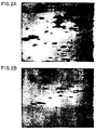
- Proteins transblotted onto nitrocellulose membranes were detectable as negative images ( Figure 4) within 10 minutes by staining with the photodevelopment silver stain of the present invention.
- Figure 4 Proteins transblotted onto nitrocellulose membranes were detectable as negative images ( Figure 4) within 10 minutes by staining with the photodevelopment silver stain of the present invention.
- the bands could be seen by eye, the contrast between the bands and the background is marginal and not easily photographed. If the cellulose nitrate is taken off the light box and left for 1 hour in the silver nitrate solution, the contrast improves and as little as 0.5 ng of protein can be visualized.
- the present invention has now made it possible to use a single solution with a photodevelopment stain.
- the stain as taught by the present invention permits the maintenance of silver ions in a relatively stable acidic solution eliminating the need for an alkaline reducing solution.
- Single solution photodevelopment silver stain of the present invention has two major advantages over two solution chemical development silver stains.
- pH gradient effects are eliminated.
- one solution the acidic silver nitrate
- the interactions of these solutions create complex pH gradients within the gel.
- a single solution photodevelopment stain does not have such diffusion effects, and this results in a reduction of staining artifacts due to variations in gel thickness or by the use of plastic gel "backings".
- biopolymers bound to ultra-thin supporting membranes stain poorly with the "chemical stains” as they retain very little silver nitrate when they are transferred into the alkaline solution for image development. Since the photodevelopment stain contains the silver ions in the image developing solution, it permits the visualization of biopolymers bound to thin membranes.
- Fixation solutions have a dual role in both methods: first, they "fix'' or retard diffusion of the biopolymer from the gel or membrane, and second, they wash substances that might interfere with staining (such as reducing agents or detergents) from the gel or membrane. Electrophoretic systems which employ reducing agents and or detergents often require more than one "fixation” wash to clear the gel of these substances.
- the fixation solution in the photodevelopment method also has an additional role of impregnating the gel with chloride ions which are important for the process of the invention described herein.
- a particularly advantageous aspect of the present invention is that the separated biopolymers can be visually detected almost instantaneously as soon as the separation matrix, e.g. polyacrylamide gel, is immersed in the photoimaging solution of the present invention.
- a fine bluish-white AgCl precipitate formed as a result of the interaction of silver with chloride ions permits immediate visualization of the biopolymer so long as the biopolymer is present in sufficient quantity, usually in micro or nanogram concentration.
- the whitish- blue precipitate is formed except in regions occupied by the biopolymer. These regions remain clear until illumination. Then they darken first.
- the method of the present invention is applicable to staining and visualization of any substance including biopolymer that affects the rate of oxidation or reduction of silver relative to the support medium and is present either in isolated form or in thin tiss.ue sections, smears and the like, e.g. in chromosomal banding. All that is necessary for initial visualization is the formation of silver chloride precipitate.
Landscapes
- Health & Medical Sciences (AREA)
- Life Sciences & Earth Sciences (AREA)
- Chemical & Material Sciences (AREA)
- Physics & Mathematics (AREA)
- Molecular Biology (AREA)
- Analytical Chemistry (AREA)
- Chemical Kinetics & Catalysis (AREA)
- Electrochemistry (AREA)
- Spectroscopy & Molecular Physics (AREA)
- Biochemistry (AREA)
- General Health & Medical Sciences (AREA)
- General Physics & Mathematics (AREA)
- Immunology (AREA)
- Pathology (AREA)
- Investigating Or Analysing Biological Materials (AREA)
- Measuring Or Testing Involving Enzymes Or Micro-Organisms (AREA)
- Investigating Or Analysing Materials By The Use Of Chemical Reactions (AREA)
Applications Claiming Priority (2)
| Application Number | Priority Date | Filing Date | Title |
|---|---|---|---|
| US06/618,949 US4555490A (en) | 1984-06-08 | 1984-06-08 | Rapid visualization system for gel electrophoresis |
| US618949 | 2000-07-19 |
Publications (1)
| Publication Number | Publication Date |
|---|---|
| EP0165013A2 true EP0165013A2 (de) | 1985-12-18 |
Family
ID=24479808
Family Applications (1)
| Application Number | Title | Priority Date | Filing Date |
|---|---|---|---|
| EP85303978A Withdrawn EP0165013A2 (de) | 1984-06-08 | 1985-06-05 | Schnelles Anschauungsverfahren für Gelelektrophorese |
Country Status (6)
| Country | Link |
|---|---|
| US (1) | US4555490A (de) |
| EP (1) | EP0165013A2 (de) |
| JP (1) | JPS61502352A (de) |
| AU (1) | AU4494185A (de) |
| DK (1) | DK60686D0 (de) |
| WO (1) | WO1986000136A1 (de) |
Cited By (2)
| Publication number | Priority date | Publication date | Assignee | Title |
|---|---|---|---|---|
| EP0330120A3 (en) * | 1988-02-24 | 1990-09-12 | Hitachi, Ltd. | Fluorescence detection type electrophoresis apparatus |
| US5860176A (en) * | 1997-09-24 | 1999-01-19 | Norberg; William J. | Pediatric critical care transport system |
Families Citing this family (33)
| Publication number | Priority date | Publication date | Assignee | Title |
|---|---|---|---|---|
| GB8415998D0 (en) * | 1984-06-22 | 1984-07-25 | Janssen Pharmaceutica Nv | Staining method |
| US4703016A (en) * | 1986-05-05 | 1987-10-27 | The United States Of America As Represented By The Department Of Health And Human Services | Silver stain for rapid, quantitative detection of polypeptides and nucleic acids |
| US4946794A (en) * | 1986-12-18 | 1990-08-07 | Protein Databases, Inc. | Visualization of proteins on electrophoresis gels using planar dyes |
| US5283196A (en) * | 1988-01-12 | 1994-02-01 | The United States Of America As Represented By The Department Of Health And Human Services | Polyacrylamide gels with improved detection of protein |
| US5292665A (en) * | 1989-03-15 | 1994-03-08 | The United States Of America As Represented By The Department Of Health And Human Services | Catalyst for preparing polyacrylamide gel which improves the detection of biomaterials by silver staining |
| US6416952B1 (en) | 1989-06-07 | 2002-07-09 | Affymetrix, Inc. | Photolithographic and other means for manufacturing arrays |
| US5143854A (en) | 1989-06-07 | 1992-09-01 | Affymax Technologies N.V. | Large scale photolithographic solid phase synthesis of polypeptides and receptor binding screening thereof |
| US6309822B1 (en) | 1989-06-07 | 2001-10-30 | Affymetrix, Inc. | Method for comparing copy number of nucleic acid sequences |
| US6551784B2 (en) | 1989-06-07 | 2003-04-22 | Affymetrix Inc | Method of comparing nucleic acid sequences |
| US5547839A (en) * | 1989-06-07 | 1996-08-20 | Affymax Technologies N.V. | Sequencing of surface immobilized polymers utilizing microflourescence detection |
| US6919211B1 (en) | 1989-06-07 | 2005-07-19 | Affymetrix, Inc. | Polypeptide arrays |
| US6346413B1 (en) | 1989-06-07 | 2002-02-12 | Affymetrix, Inc. | Polymer arrays |
| US5424186A (en) | 1989-06-07 | 1995-06-13 | Affymax Technologies N.V. | Very large scale immobilized polymer synthesis |
| US6955915B2 (en) | 1989-06-07 | 2005-10-18 | Affymetrix, Inc. | Apparatus comprising polymers |
| US5800992A (en) | 1989-06-07 | 1998-09-01 | Fodor; Stephen P.A. | Method of detecting nucleic acids |
| US5744101A (en) | 1989-06-07 | 1998-04-28 | Affymax Technologies N.V. | Photolabile nucleoside protecting groups |
| US6406844B1 (en) | 1989-06-07 | 2002-06-18 | Affymetrix, Inc. | Very large scale immobilized polymer synthesis |
| US6506558B1 (en) | 1990-03-07 | 2003-01-14 | Affymetrix Inc. | Very large scale immobilized polymer synthesis |
| US6074818A (en) * | 1990-08-24 | 2000-06-13 | The University Of Tennessee Research Corporation | Fingerprinting of nucleic acids, products and methods |
| EP0834575B1 (de) | 1990-12-06 | 2001-11-28 | Affymetrix, Inc. (a Delaware Corporation) | Identifizierung von Nukleinsäuren in Proben |
| WO1992017611A1 (en) * | 1991-03-28 | 1992-10-15 | The University Of Tennessee Research Corporation | Dna silver staining |
| US6468740B1 (en) | 1992-11-05 | 2002-10-22 | Affymetrix, Inc. | Cyclic and substituted immobilized molecular synthesis |
| JP2932228B2 (ja) * | 1993-03-31 | 1999-08-09 | 日立ソフトウエアエンジニアリング株式会社 | デンシトメータ |
| US5863504A (en) * | 1995-03-16 | 1999-01-26 | Bio-Rad Laboratories, Inc. | Fluorescence imaging instrument utilizing fish |
| US5595878A (en) * | 1995-06-02 | 1997-01-21 | Boron Biologicals, Inc. | Detection of biopolymers and biooligomers with boron hydride labels |
| US6319720B1 (en) | 1998-10-13 | 2001-11-20 | Ewald M. Wondrak | Process for fast visualization of protein |
| US6545264B1 (en) | 1998-10-30 | 2003-04-08 | Affymetrix, Inc. | Systems and methods for high performance scanning |
| ES2252263T3 (es) * | 2000-07-25 | 2006-05-16 | The Government Of The Usa, As Represented By The Secretary, Department Of Health And Human Services | Bacteriofago que tiene una gama de hospedadores multiple. |
| US20060000272A1 (en) * | 2004-06-30 | 2006-01-05 | Beat Neuenschwander | Thermal flow sensor having an asymmetric design |
| US11167247B2 (en) * | 2017-02-15 | 2021-11-09 | Nanolc-12, Llc | Length-based separation of carbon nanotubes |
| US11079387B2 (en) | 2018-04-12 | 2021-08-03 | Zahra Borzooeian | Length-based carbon nanotube ladders |
| US11353424B2 (en) | 2018-04-12 | 2022-06-07 | Nano LC-12, LLC | Length-based carbon nanotube ladders |
| CA3215393A1 (en) * | 2021-04-23 | 2022-10-27 | Peter Galen | Improved detection of hemoglobin and other compounds by electrophoresis |
Family Cites Families (1)
| Publication number | Priority date | Publication date | Assignee | Title |
|---|---|---|---|---|
| US4405720A (en) * | 1981-03-04 | 1983-09-20 | The United States Of America As Represented By The Department Of Health And Human Services | Silver stains for protein in gels |
-
1984
- 1984-06-08 US US06/618,949 patent/US4555490A/en not_active Expired - Fee Related
-
1985
- 1985-06-05 EP EP85303978A patent/EP0165013A2/de not_active Withdrawn
- 1985-06-06 JP JP60502841A patent/JPS61502352A/ja active Pending
- 1985-06-06 AU AU44941/85A patent/AU4494185A/en not_active Abandoned
- 1985-06-06 WO PCT/US1985/001059 patent/WO1986000136A1/en not_active Ceased
-
1986
- 1986-02-07 DK DK60686A patent/DK60686D0/da not_active Application Discontinuation
Cited By (2)
| Publication number | Priority date | Publication date | Assignee | Title |
|---|---|---|---|---|
| EP0330120A3 (en) * | 1988-02-24 | 1990-09-12 | Hitachi, Ltd. | Fluorescence detection type electrophoresis apparatus |
| US5860176A (en) * | 1997-09-24 | 1999-01-19 | Norberg; William J. | Pediatric critical care transport system |
Also Published As
| Publication number | Publication date |
|---|---|
| US4555490A (en) | 1985-11-26 |
| WO1986000136A1 (en) | 1986-01-03 |
| DK60686A (da) | 1986-02-07 |
| JPS61502352A (ja) | 1986-10-16 |
| AU4494185A (en) | 1986-01-10 |
| DK60686D0 (da) | 1986-02-07 |
Similar Documents
| Publication | Publication Date | Title |
|---|---|---|
| US4555490A (en) | Rapid visualization system for gel electrophoresis | |
| Merril et al. | A rapid sensitive silver stain for polypeptides in polyacrylamide gels | |
| EP0073247B1 (de) | Silberfarbstoffe für proteine in gelen | |
| Clarke | Simplified “disc”(polyacrylamide gel) electrophoresis | |
| Goldman et al. | Silver staining of DNA in polyacrylamide gels: linearity and effect of fragment size | |
| US4434234A (en) | Method and kit for silver staining substances supported in matrix | |
| Merril et al. | A photodevelopment silver stain for the rapid visualization of proteins separated on polyacrylamide gels | |
| EP0328579B1 (de) | Elektrophoretisches gel hoher auflösung sowie methode zur abscheidung von serumproteinen | |
| EP0246873B1 (de) | Bestandteil für Elektrophorese | |
| US4722777A (en) | Improvement of electrophoretic element using polyacrylamide gel | |
| Merril et al. | " Ultrasensitive" silver stains: their use exemplified in the study of normal human cerebrospinal fluid proteins separated by two-dimensional electrophoresis. | |
| CA2091944A1 (en) | Analysis of hemoglobin variants by capillary zone electrophoresis | |
| EP0115436B1 (de) | Medium für die Elektrophorese | |
| US6555382B2 (en) | Solution for fast visualization of protein | |
| Wallace et al. | Visualization of protein—SDS complexes in polyacrylamide gels by chilling | |
| EP0162657B1 (de) | Elektrophoretischer Bestandteil | |
| Jackowski et al. | Fluorescamine staining of nonhistone chromatin proteins as revealed by two-dimensional polyacrylamide gel electrophoresis | |
| US4946794A (en) | Visualization of proteins on electrophoresis gels using planar dyes | |
| Rubin et al. | [12] Two-dimensional polyacrylamide gel electrophoresis of membrane proteins | |
| US5039619A (en) | Method for detecting characteristic markers of disease in biological fluids | |
| US5192407A (en) | Means and method of detection in chemical separation procedures | |
| JPH0634602A (ja) | ゲル電気泳動法 | |
| Edström | Determination of organic compounds below the microgram range. Ribonucleic acid and its constituents | |
| EP0162693A2 (de) | Elektrophoretischer Bestandteil | |
| US20060144708A1 (en) | In-gel fluorescent protein staining technique |
Legal Events
| Date | Code | Title | Description |
|---|---|---|---|
| PUAI | Public reference made under article 153(3) epc to a published international application that has entered the european phase |
Free format text: ORIGINAL CODE: 0009012 |
|
| AK | Designated contracting states |
Designated state(s): CH DE FR GB LI NL SE |
|
| STAA | Information on the status of an ep patent application or granted ep patent |
Free format text: STATUS: THE APPLICATION IS DEEMED TO BE WITHDRAWN |
|
| 18D | Application deemed to be withdrawn |
Effective date: 19870701 |
|
| RIN1 | Information on inventor provided before grant (corrected) |
Inventor name: MERRIL, CARL RONALD |