EP0164992A2 - Nitrogen fixation regulator genes - Google Patents
Nitrogen fixation regulator genes Download PDFInfo
- Publication number
- EP0164992A2 EP0164992A2 EP85303925A EP85303925A EP0164992A2 EP 0164992 A2 EP0164992 A2 EP 0164992A2 EP 85303925 A EP85303925 A EP 85303925A EP 85303925 A EP85303925 A EP 85303925A EP 0164992 A2 EP0164992 A2 EP 0164992A2
- Authority
- EP
- European Patent Office
- Prior art keywords
- gene
- promoter
- recited
- activating
- recombinant dna
- Prior art date
- Legal status (The legal status is an assumption and is not a legal conclusion. Google has not performed a legal analysis and makes no representation as to the accuracy of the status listed.)
- Withdrawn
Links
Images


Classifications
-
- C—CHEMISTRY; METALLURGY
- C12—BIOCHEMISTRY; BEER; SPIRITS; WINE; VINEGAR; MICROBIOLOGY; ENZYMOLOGY; MUTATION OR GENETIC ENGINEERING
- C12N—MICROORGANISMS OR ENZYMES; COMPOSITIONS THEREOF; PROPAGATING, PRESERVING, OR MAINTAINING MICROORGANISMS; MUTATION OR GENETIC ENGINEERING; CULTURE MEDIA
- C12N15/00—Mutation or genetic engineering; DNA or RNA concerning genetic engineering, vectors, e.g. plasmids, or their isolation, preparation or purification; Use of hosts therefor
Definitions
- Prokaryotes can use a wide variety of nitrogen compounds as sole sources of cellular nitrogen. This variety includes ammonia, dinitrogen and nitrate among the inorganic compounds, and proline, arginine and glutamine among complex organic compounds. Each species can utilize a different array of nitrogen compounds. Glutamine, glutamate and aspartate are the key nitrogen compounds in intermediary metabolism. The latter two are the starting compounds of many pathways of amino acid biosynthesis and serve as amino group donors in many reactions. In all other cases the amino group is donated by glutamine. The major enzyme required for the assimilation of ammonia produced by N 2 fixation is glutamine synthetase, which catalyses the reaction: Glutamate + NH 3 + ATP ----> glutamine + ADP + Pi.
- glutamate dehydrogenase is also found. Utilization of the assimilated ammonia depends on the activity of glutamate synthase catalyzing: Glutamine + 2-ketoglutarate + NADPH ----> 2 glutamate + NADP +
- Biological nitrogen fixation can be achieved by a variety of microorganisms and occurs through the induction of an enzyme complex, nitrogenase, which converts atmospheric nitrogen to ammonia.
- This conversion occurs in a group of physiologically diverse prokaryotes, including facultative anaerobes (e.g., Klebsiella pneumoniae and Rhodospirillum rubrum), obligate anaerobes (e.g., Clostridium pasteurianum), obligate aerobes (e.g., Azotobacter vinelandii) and some strains of blue-green algae (e.g., Anabaena cylindrica) (Sprent, J. I.
- free-living bacteria e.g., Klebsiella pneumoniae and the photosynthetic blue-green bacteria
- Biological nitrogen fixation in the root nodules of leguminous plants is a major component of world food production (Burris, R. H. (1980) In Free Living Systems and Chemical Models; Nitrogen fixation, Newton, W. E., Orne-Johnson, W. H., eds. Vol 1 Baltimore, University Park Press, pp. 7-16).
- Rhizobia nitrogenase synthesis is repressed and is only induced after the symbiotic relationship has been established. Furthermore, some Rhizobium species only interact with a narrow range of plant species, whereas other species interact with a wide range.
- Bacteria bind to the emerging plant root hairs and invade the root tissue through the formation of an infection thread.
- the plant responds to this infection by the development of a highly differentiated root nodule.
- These nodules are the site of synthesis of the nitrogenase complex.
- the fixed nitrogen is exported into the plant tissue and assimilated by the plant derived enzymes (Scott, D. B., Farnden, K. J. F. and Robertson, J. G. (1976) Nature 263:703,705).
- Rhizobium symbioses are confined to leguminous plants. Furthermore, Rhizobium strains which fix nitrogen in association with the agriculturally- important temperate legumes are usually restricted in their host range to.a single legume genus. However, some strains of Rhizobium have been isolated which can fix nitrogen in a diverse group of legume species but can also form an effective symbiosis with non-legumes.
- nifH, nifD and nifK encode the structural proteins of the nitrogenase enzyme complex (viz. the Fe-protein subunit (dinitrogenase reductase) and the a- and ⁇ -subunits of the Mo-Fe protein (dinitrogenase) respectively.
- Dinitrogenase is an a 2B2 tetramer in which the two non-identical a and ⁇ subunits have similar molecular weights of 55,000 to 60,000.
- Dinitrogenase reductase is a dimer of two identical subunits each having a molecular weight around 35,000. These genes are linked on the same operon in K. pneumoniae and are transcribed from a promoter adjacent to the nifH gene. A similar situation (nifHDK) was found in two fast-growing Rhizobia, R. meliloti (Ruvkun, G. B., et al. (1982) Cell 29:551-559) and R. leguminosarum (Schetgens, T. M. P.
- Rhizobium leguminosarum PRE symbiotic genes p. 699, In C. Veeger and W. E. Newton (eds.) Advances in nitrogen fixation research. Martinus Nijhoff/Dr. W. Junk Publishers, The Hague).
- nifDK forms one operon and that nifH is located elsewhere on the genome (Fuhrmann, M. and H. Hennecke (1982) Mol. Gen. Genet. 187:419-425).
- nifH region was found not to be linked to nifD (Scott, K. F., et al. (1983) DNA 2:141-148). Yet a different arrangement was detected in the cyanobacterium Anabaena sp. 7120, in which nifHD is separated from nifK (Rice, D., et al. (1982) J. Biol. Chem. 257:13157-13163).
- the remainder of symbiotic genes contain information required for bacterial attachment, root hair curling, initiation and development of nodules and establishment of symbiotic relationships.
- regulatory sequences such as promoters, operators, attenuators, and ribosome binding sites are found adjacent to the coding regions. These regulatory sequences control the expression of the structural genes, i.e., the coding sequences downstream in the 3'-direction of the DNA reading strand.
- DNA recombination requires the success of a number of different features.
- Third, the conversion of coding information into useful products, such as storage proteins and hormones, must occur in an environment where they are not subsequently degraded.
- Rhizobium In bacteria of the genus Rhizobium, nitrogenase synthesis is normally repressed under free-living conditions and is induced only within a complex symbiosis formed mostly with leguminous plants.
- R. trifolii is an example of a fast-growing Rhizobium with a narrow host range and cannot normally be induced to fix nitrogen in culture.
- a Parasponia Rhizobium species has been isolated and this species is a slow-growing organism with a very broad host range capable of an effective symbiotic relationship with a broad variety of tropical legumes as well as the non-legume Parasponia (Ulmaceae) (Trinick, M. J. (1980) J. Appl. Bacteriol. 49:39-53).
- Rhizobium can be induced to fix nitrogen in culture although the level of this fixation is about 100-fold less than can be obtained from the free-living bacterium Klebsiella pneumoniae.
- Other slow-growing Rhizobia include the commercially significant R. japonicum, which nodulates soybeans.
- the genetics of biological nitrogen fixation have been well characterized in the free-living organism Klebsiella pneumoniae.
- the structural genes for nitrogenase (nifH, nifD and nifK encoding the Fe-protein subunit and the a and s subunits of the Mo-Fe protein, respectively) have been mapped both genetically and physically (Kennedy, C. et al. (1981) In Current Perspectives in Nitrogen Fixation (eds. Gibson, A. H. and W. E. Newton) Australian Acad. Science, Canberra, pp. 146-156; and Reidel, G. E., Ausubel, F. M. and F. M. Cannon (1979) Proc. Nat. Acad. Sci. U.S.A.
- the larger protein contains molybdenum, iron and acid-labile sulfur, and carries the binding site for nitrogen and contains two subunit proteins a- and B -coded by the nifD and nifK genes respectively.
- the smaller protein contains iron and acid-labile sulfur, and is required for the reduction of the dinitrogenase and for the binding of MgATP used in this reduction.
- the dinitrogenase reductase is coded by the nifH gene.
- the region of the nif operon coding for dinitrogenase reductase and dinitrogenase a-subunit shows homology at the nucleic acid sequence level with the corresponding sequences in at least 19 other bacterial strains (Ruvkun, G. B. and F. M. Ausubel (1980) Proc. Nat. Acad. Sci. U.S.A. 77:191-195).
- this conservation of structure is generally true, significant differences between nitrogenases from different organisms also exist as can be shown by variable stability following purification and by the fact that active hybrid complexes do not form in all cases (Eady, R. R. and B. E. Smith (1979) supra).
- a DNA fragment carrying the Klebsiella pneumoniae nifK, nifD and nifH genes has been isolated from the nif - strain UNF841(Tn5::nifK) (Cannon, F.C. et al. (1979) Mol. Gen. Genet. 174:59-66) and cloned into the Escherichia coli plasmid pBR325.
- the nucleotide sequences of the nifH gene and of 622 nucleotides of the nifD gene were determined (Sundaresan, V. and F. M. Ausubel (1981) J. Biol. Chem. 256:2808-2812; Scott, K. F., Rolfe, B. G.
- meliloti genome confirmed that the cloned fragment carries nif specific genes (Ruvkun, G. B. et al. (1980) Cold Spring Harb. Symp. Quant. Biol. 45:492-497; Ruvkun, G. B. et al. (1982) Cell 29:551-559).
- This cloned R. meliloti fragment has been analysed by the minicell technique and it was demonstrated that the nifH gene of R. meliloti was expressed in minicells of E. coli (Weber, G. and A. Puhler (1982) Plant Mol. Biol. 1:305-320). In free living R. meliloti, the nifH gene is not expressed.
- nifK and nifD are presented as hybrid genes.
- the indicated promoters (p) were identified in E. coli.
- the nifH as well as the fixA promoter can be activated by the K. pneumoniae nifAgp protein.
- the abbreviations used in the restriction map are: C, ClaI; E, EcoRI; H, HindIII; P, PstI; S, SmaI; and X, Xhol.
- plasmid pRmW54 was obtained by cloning a 3.5 kb HindIII fragment of pRm29D5 (Ruvkun, G. B. et al. (1982) Cell 29:551-559) into the E. coli vector plasmid pAC177-C.
- pRm29D5 covers an 18 kb fragment adjacent to and surrounding the genes for nitrogenase (nif) and it was cloned from the genome of R. meliloti.
- the K. pneumoniae nifA probe (Fig. 2) was used in hybridization experiments to detect any complementary DNA fragment in pRmW54, pRmR3 and pRmW52.
- Figure 2 the coding regions of the K. pneumoniae nifA and nifL genes are shown in relation to the restriction map (Puhler, A. and W. Klipp (1981) In: Biology of inorganic nitrogen and sulfur; ed. Bothe, H. and A. Trebst; Berlin, Heidelberg).
- the two rectangles represent the restriction fragments which were used as radioactive DNA probes for the hybridization experiments with R. meliloti DNA carrying fix genes.
- the restriction endonucleases used were: E, EcoRI; S, Smal; P, PstI and H, HindIII. Restriction fragments of the R. meliloti plasmids pRmW54, pRmR3 and pRmW52 were separated on agarose gels and blotted to nitrocellulose filters (Fig. 3a). This figure gives the results of Southern hybridization of R. meliloti fix DNA with the nifA and nifL specific DNA probes of K. pneumoniae following agarose gel electrophoresis of R.
- Lane 1 an EcoRI-HindIII digest of the EcoRI-HindIII fragment (shown in Fig. 2) subcloned in pUC8 is given; Lane 2, X -DNA digested with HindIII and EcoRI as molecular weight standards; Lane 3, pRmW54 DNA digested with BamHI and BglII; Lane 4, pRmR3 DNA digested with HindIII and XhoI; and Lane 5, pRmW52 DNA digested with BamHI and Xhol. These filters were hybridized with radioactive labelled K. pneumoniae nifA fragments (Fig.
- FIG. 3b This figure 3b illustrates an autoradiograph of nitrocellulose filters after hybridization with the K. pneumoniae nifA probe in 6xSSC at 55°C. Such conditions of hybridization are referred to herein as "stringent conditions".
- stringent conditions Such conditions of hybridization are referred to herein as "stringent conditions”.
- the 1.8 kb BamHI fragment and the adjacent 0.9 kb BamHI-BglII of pRmW54 are shown in Lane 3 (Fig. 3b) and they show homology to the K. pneumoniae nifA probe.
- the strong hybridization with other bands is due to vector-vector homology or to the K. p neumoniae control DNA shown in Lane 1.
- the hybridizing fragments are summarized in Figure 4.
- Figure 6 shows a restriction map of plasmid pGW6 (see Table 2) and also the results of a control experiment in which the Rhizobium meliloti nifH promoter was activated by the Klebsiella pneumoniae nifA gene product.
- the plasmid pGW6 contains an EcoRI-SmaI fragment of pRmR2 subcloned into pMC1403.
- the plasmid vector pMC1403 allows translational fusion to the eighth codon of the E. coli lacZ gene.
- s-galactosidase activity is detectable if a second plasmid constitutively expressing the K. pneumoniae nifA gene is present in the same cell.
- Plasmid pGW7 (Table 2) is a derivative of pGW6 where the reading frame of the fusion gene was corrected at the BamHI site by Sl nuclease digestion.
- Figure 7 the activation of the Rhizobium meliloti nifH promoter in pGW7 by the fixD gene product of R. meliloti is documented.
- the B -galactosidase activity of E. coli strain ET 8894 containing several plasmid combinations was measured. The results of six independent experiments are presented as black columns and the mean values are given as cross-lines.
- the plasmid combinations were: (1) pAc177-C + pGW7; (2) pRmW54 + pGW7; (3) pRmW54::Tn5-1 + pGW7; (4) pRmW54::Tn5-2 + pGW7; (5) pRmW54::Tn5-5 + pGW7; (6) pRmW54::Tn5-7 + pGW7; (7) pRmW54::Tn5-8 + pGW7; and (8) pRmW54::Tn5-9 + pGW7.
- the plasmids are also shown schematically on the left side of the drawing, p stands for the aphI promoter (i.e., the kanamycin resistance gene) of the vector plasmid pAC177-C and the double line represents the R. meliloti HindIII restriction fragment of pRmW54.
- the triangles give the positions of the various Tn5 insertions and the black horizontal arrows show the fixD coding region. Note that the activation of the R. meliloti nifH promoter (compared to the control No. 1) was not detectable when Tn5 was in the fixD region.
- the nifL probe of K. pneumomae did not hybridize to pRmW54, pRmR3 and pRmW52 (Table 2).
- the fixD gene of R. meliloti was detected by expression of pRmW54 in E. coli minicells using methods already described in detail (Puhler, A. and W. Klipp (1981) In: Biology of inorganic nitrogen and sulfur; Bothe, H. and A. Trebst eds. Berlin, Heidelberg; and Weber, G. and A. Puhler (1982) Plant Mol. Biol. 1:305-320).
- plasmid pRmW54 synthesized two polypeptides of 68K and 66K molecular weight (Fig. 8).
- the experimental results disclosed in the present invention have defined the boundaries of the fixD gene of R, meliloti and have demonstrated that the protein of the fixD gene is responsible for the activation of the R. meliloti nifHDK and fixABC gene promoters leading in turn to the expression of the genes whose products are required for nitrogen fixation.
- the expression of the fixD gene requires that R. meliloti must first be in a symbiotic association with a specific host plant: R. meliloti does not fix nitrogen when living in a free state.
- R. meliloti does not fix nitrogen even when in a symbiotic association with the same host plant if there is a substantial quantity of nitrate already present in the soil.
- a genetic manipulation of the fixD gene so that it is consistently expressed either while free living or while in symbiotic association with a specific host plant growing in soil containing high nitrate would therefore be enormous useful in the fixation of nitrogen.
- the high nitrate content of a rich soil would not be decreased when an alfalfa-R. meliloti symbiotic association was present, but, to the contrary, the soil would become even richer in nitrates.
- the use of nif derepressed strains enables symbiotic nitrogen fixation to start earlier in nodule development and to last longer than in nodules with wild type Rhizobia.
- Rhizobium strains have a big advantage when a poor symbiotic association with other plant species leads to ineffective nodulation by wild type Rhizobia.
- ineffective nodulation is that a special plant signal normally necessary for transcription of nif genes is absent.
- the nif derepressed strains described herein have the ability to fix nitrogen without such a plant signal.
- Rhizobium strains can be induced to grow in close association with roots of economically important non-leguminous plants. Since the nif derepressed Rhizobium strains can fix nitrogen in large amounts while in the free living state, such an association between nif derepressed Rhizobia and the roots of important non-leguminous agricultural species has far reaching utility.
- the invention described here teaches how to make and use such nif derepressed Rhizobium strains.
- the present invention discloses another useful application of the fixD promoter region.
- the promoter of the fixD gene remains inactive and it is only when a symbiotic reiationship is established that the fixD gene is activated--most probably in direct or indirect response to a signal from the plant.
- a gene i.e., a gene whose expression is only desired when a symbiotic relationship has been established between a Rhizobium species and a plant, is the expression of the gene coding for an insecticidal protein such as the crystalline toxic protein of Bacillus thuringiensis (see Examples 3 and 4).
- the M13 cloning and sequencing vectors mp8, mp9, mp10 and mpll were used for subcloning of the HindIII 3.5 kb insert of plasmid p R mW54.
- the HindIII fragment was cleaved from the vector by digestion with HindIII and then separated and isolated by agarose gel electrophoresis.
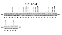
- HindIII fragment was isolated as described (supra), digested with the restriction enzymes EcoRI, BamHI, SalI and Xhol and subcloned into a pUC8 sequencing vector (Vieira, J. and J. Messing (1982) Gene 13:259-268). Determination of the sequence around several restriction sites of the fragment ( Figure 9) enabled further alignment and confirmation of the data obtained from the dideoxy sequencing. The combined results of the two sequencing methods yielded the sequence of 2127 bp of the 3.5 kb HindIII fragment ( Figure 10).
- the large open reading frame which represents the fixD gene of R. meliloti started with an ATG 415 nucleotides from the HindIII site and spanned 1628 nucleotides.
- the coding region of fixD determined by the minicell procedure contained the following series of restriction sites: Xhol, EcoRI and BamHI. The same series of restriction sites was also present in the open reading frame.
- Example 2 Activation of the Rhizobium meliloti nifH promoter by the R. meliloti fixD gene products in Escherichia coli
- Example 3 Expression of foreign genes under the control of a fixD promoter DNA region.
- a synthetic DNA primer which is complementary to the DNA sequence immediately upstream (i.e., 5'- to the ATG translation initiation codon) of the Rhizobium meliloti fixD coding sequence.
- the synthetic DNA primer has the sequence 5'-CAGAGCCTTTGATATGCTGTA-3'.
- the HindIII-Xhol DNA subclone of pRmW54 in pAC177-C (designated pRmW69 - Table 2) is then excised, purified, subcloned into a single stranded DNA phage and transformed into E. coli JMI03 (Fig. 11). This HindIII-XhoI subclone contains the complete promoter sequence of R.
- meliloti fixD gene and 82 nucleotides at the 5'-end of the fixD coding sequence.
- the subcloned fragment is amplified therein and single stranded templates (ca. lug) are recovered from the supernatant following centrifugation of the bacterial host.
- a 10-fold excess of the synthetic DNA primer in the presence of the four deoxynucleotide triphosphates (one of which is radioactive) and DNA polymerase I (Klenow fragment) is now used as a primer on this fixD template to generate double stranded DNA (dsDNA) (Fig. 12).
- the mixture is incubated for a period (e.g., 15-45 minutes) at an appropriate temperature (e.g., 25°-37°C) during which period the complementary strand is extended beyond the HindIII site.
- the 468 nucleotide region between the HindIII site and the ATG initiation codon contains the complete promoter region of the fixD gene of R. meliloti.
- the remaining single stranded DNA is then removed by digestion with 51 nuclease (Fig. 12).
- EcoRI linkers GGAATTCC
- the fragments are separated by agarose gel electrophoresis and the fragment containing the promoter sequence is eluted and cloned into the wide host range plasmid pSUP204 (Fig. 13), which has previously been restricted by the restriction enzyme EcoRI.
- the resulting recombinant plasmid is termed pRm-fixD-P/SS204.
- a suitable E. coli host strain e.g., 17-1, which is restriction negative, i.e., r -
- partial cleavage with EcoRI allows the addition of any foreign structural gene or foreign DNA fragment into the linearized plasmid downstream from the fixD promoter fragment.
- a foreign structural gene of foreign DNA fragment is herein defined as any structural gene or DNA fragment not found naturally under the direct or indirect control of a fixD promoter.
- the gene coding for the crystalline toxic protein of Bacillus thuringiensis can be inserted resulting in a "composite" recombinant.
- a composite recombinant is herein defined as a recombinant DNA plasmid containing a vector, a promoter sequence and any foreign DNA whose expression is under the control of said promoter sequence.
- Example 4 Insertion of the bacterial toxin gene from Bacillus thuringiensis into the recombinant plasmid pRmfixD-P/SS204
- Recombinant plasmids containing inserts of the gene encoding the toxic crystal protein of B. thuringiensis are obtained using the techniques described (Wong, H. C., Schnepf, H. E. and H. R. Whiteley (1983) J. Biol. Chem. 258:1960-1967).
- the recombinant plasmid pESl (ATCC Number 31995) consisting of the plasmid vector pBR322 and DNA homologous to the 30, 32 and 37 megadal- ton plasmids, as well as DNA homologous to linearized forms of the very large plasmids of B. thuringiensis is partially cleaved with EcoRI to give linear molecules.
- a probe for the toxic crystal protein gene is isolated and radioactively labelled as previously described (Wong, H. C, et al. (1983) supra). The restriction fragments are separated by agarose gel electrophoresis and the labelled probe is found to hybridize to one fragment of approximately 15 kilobases (kb). This fragment includes the EcoRI fragments D and F (Wong, H. C. et al. (1983) supra). The 15 kb fragment is then cloned into M13mp8 or M13 mp9 according to standard procedure (Messing, J. and J. Vieira (1982) Gene 19:269-276) and transformed into E.
- the single stranded DNA from the extruded phage particles is purified and replicated in vitro by use of a synthetic primer (5'-TGTTATCCATGGGTTACCTCC-3') (The general method of site specific mutagenesis is described in Zoller, M. J. and M. Smith (1982) Nucleic Acids Research 10:6487-6500).
- the resulting double stranded recombinant plasmid is then transformed back into E. coli JM103 and amplified.
- the amplified double stranded plasmid DNA is purified from the E. coli JM103 cells and cleaved with the restriction endonucleases NcoI and Aval.
- Ncol cleaves at the site of the synthetic primer (which is the initiation site of the toxic crystal protein gene) and Aval cleaves at a site which is downstream from the 3'- end of the toxic crystal protein gene.
- the overhangs are then filled in to blunt ends (Maniatis, T., Jeffrey, A. and D. G. Kleid (1975) Proc. Nat. Acad. Sci. U.S.A. 72:1184-1188).
- pRmfixD-P/SS204 recombinant plasmid which is derived from pSUP204 (Fig. 13) is cleaved with EcoRI and the overhangs filled in to blunt ends. HindIII linkers are then added to both the B. thuringiensis toxic crystal protein gene fragment and to the pRmfixD-P/SS204 recombinant. Following the HindIII digestion of both components, the toxic crystal protein gene and the pRmfixD-P/SS204 recombinant plasmid are ligated together to give a pRmfixD-P/SS204 - B. thuringiensis toxic crystal protein gene composite. The mixture is transformed into a suitable E. coli host, e.g.
- Plasmids are isolated from individual colonies and the orientation determined by restriction mapping. A colony containing a plasmid with the correct orientation is then conjugated to Rhizobium meliloti and the plasmid is transferred as already described (Example 7). The production of mRNA and/or the toxic crystal protein is monitored as already described (Wong, et al., supra).
- Example 5 Construction of a recombinant plasmid containing the fixD gene of R. meliloti under control of a constitutive kanamycin resistance gene promoter
- Transposon Tn5 is a DNA element of 5.7 kilobases (kb) in length, consisting of 1.5 kb inverted repeat sequences flanking a 2.7 kb central region. Encoded within the inverted repeats are the functions required for transposition.
- the central region of the transposon carries a gene conferring resistance to the antibiotic kanamycin (Km r ).
- neomycin phosphotransferase gene (alternatively named aminoglycoside phosphotransferase gene- aph) is deleted by digestion with the restriction enzymes BglII (a BglII site is present 36 nucleotides upstream from the translational start codon ATG) and Sail (a SalI site is present 342 nucleotides downstream from the TGA termination codon) (see Fig. 15).
- the sequence of the kanamycin resistance gene has been previously shown and is reproduced herein as Figure 15 (Beck, E. et al. (1982) Gene 19:327- 3 36).
- the nucleotide sequence of Tn5 DNA from position 1401 to position 270 0 is shown.
- the aph coding region is boxed by solid lines. Some restriction sites are indicated in the sequence, as well as at the end of the left inverted repeat (closing bracket ]) and the promoter region. Such a deletion leaves the promoter region of the neomycin phosphotransferase gene (neo) intact.
- plasmid pRmW54 was obtained by cloning a 3.5 kb HindIII fragment of pRm29D5 (Ruvkun, G. B. et al. (1982) Cell 29:551-559) into the E. coli vector plasmid pAC177-C (Table 2). After transformation into a suitable host strain and amplification, the recombinant plasmid is linearized by partial digestion with the restriction endonuclease HindIII. Linearization occurs either on the upstream side of the fixD gene or on the downstream side. These two alternatives can be distinguished by the use of restriction maps.
- pSUP1011-Tn5-neopro-fixD Such a construction comprises the pSUP1011 suicide vector and a Tn5 transposon. Between the inverted repeats of the Tn5, the neomycin phosphotransferase promoter (neo pro ) is included and, in addition, the fixD structural gene positioned so that it is under the control of neopro. This construction is herein defined as pSUP1011-Tn5-neopro-fixD.
- Example 6 Construction of a fusion gene and product protein of fixD and aminoglycoside phosphotransferase
- the Xhol-HindIII subfragment of pRmW54 was cloned into pAC177-C (Table 2). From the restriction mapping experiment (Example 1) it was known that the XhoI site was located within the fixD coding region. On the other hand, the Xhol restriction site of the vector plasmid pAC177-C was located in the aphI gene (kanamycin resistance gene). In order to obtain the correct reading frame of the fusion polypeptide, the XhoI site was treated with Sl nuclease in one case and with DNA polymerase I in the other. A strongly expressed 60K fusion polypeptide was obtained after treatment with DNA polymerase I. The recombinant plasmid described herein which contains such a constitutively expressed fixD gene is referred to as pRmW69 (Table 2).
- Example 7 Introduction of DNA sequences into the genome of gram-negative organisms other than E. coli
- This example is based on the following general principles. Two basic components are employed. These are: (1) a suicide vector and (2) a transposon.
- Suicide vectors are plasmid molecules which replicate stably in one bacterial host (in this case E. coli) but fail to replicate in a different bacterial species (e.g., Rhizobium meliloti).
- Transposons are genetic elements which are able to move (translocate) from one location to another in DNA. The translocation process is mediated by gene products encoded on the transposon and is dependent on the integrity of repeated sequences (directly or indirectly repeated) located at each end of the transposon. Transposons generally carry a gene (or genes) encoding resistance to one or more antibiotics.
- the vector contains the inserted fixD gene under the control of a kanamycin resistance promoter pSUP1011-Tn5-neopro fixD (see Example 5) and is transformed (introduced) into Escherichia coli strain SM10.
- This type of recombinant plasmid comprising a suicide vector, a transposon and a constitutively expressed structural gene is herein defined as a "constitutive gene transfer system".
- This strain is capable of mobilizing (Mob+) pSUP1011 derivatives (recombinant plasmids) into other gram negative bacteria) (Simon, R., Priefer, U . and A. Puhler (1983) Proc.
- the E. coli SM10 derivative carrying the desired pSUP1011 recombinant, is mixed with cells of R. meliloti carrying a symbiotic (Sym) plasmid on the surface of a nutrient agar plate.
- the plate is incubated for a suitable period (4-16 hours) at 29-30° (optimum temperature for Rhizobium meliloti) and during this time cells of each type come into physical contact (conjugation) and the pSUP1011 recombinant derivative is transferred from E.
- the cell mixture is washed off the plate and spread on an agar plate which is selective for organisms which can only grow if they are capable of constitutive nitrogen fixation.
- the resultant colonies which can constitutively fix nitrogen will be derivatives of R. meliloti in which the cloned DNA fragment, within Tn5, will be inserted at some point in the genome. Selection for ability to constitutively fix nitrogen insures maintenance of the inserted DNA.
- E. coli C600 (pRmW54) was placed on deposit with the American Type Culture Collection, 12301 Parklawn Drive, Rockville, MD 20852 on June 4, 1984, Accession No. 39722.
- Example 8 Enhanced Expression of Foreign Gene Under nif Promoter Control Resulting from Enhanced Expression of fixD Gene Products
- ⁇ -galactosidase was placed under nifHDK promoter control as described (pGW7, Example 2, Fig. 6). The amount of ⁇ -galactosidase synthesized under nifHDK control was measured using E, coli ET8894 (Table 1) containing pGW7. Synthesis of active s-galactosidase in this system depends on activation of the nifHDK promoter. Introduction of pRmW54 (expressing fixD as a fusion protein under control of the aphi) leads to a significant increase in a-galactosidase activity over control levels (Table 3).
- the described experiment was designed to test whether a-galactosidase expression could be enhanced by increasing the levels of fixD gene product, and whether the level of fixD gene product could be increased by shortening the distance, or number of amino acids, between the aphI promoter and the start of the fixD gene.
- the resulting digestion mixture was religated after S1 nuclease digestion, and the mixture was used to transform a culture of E. coli ET8894 (pGW7). After incubation for 48 hours at 30°C, the transformants were plated on agar plates supplemented with ampicillin, chloramphenicol, glutamine and X-gal (5-bromo-4-chloro-3-indolyl- ⁇ -D galactoside, a chromogenic ⁇ -galactosidase substrate). Although E. coli ET8894 (pGW7, pRmW54) produces detectable ⁇ -galactosidase activity, the activity is low and incubation on X-gal plates yields white colonies.
- XhoI fragments were sequenced, after cloning them into the Sail site of the phage M13mp8. Nucleotide sequence determination of these deletions, which were contained within the XhoI fragments, revealed that all the nifHDK activated proteins were fusion polypeptides, with the exception of pRmW541-5. The structures are shown diagrammatically in Fig. 19.
- Fig. 19 diagrams the structure of the pRmW541 deletion plasmids, and the resulting level of activation in ET8894 (pGW7).
- the black closed circle indicates the aphI promoter.
- the open rectangles represent the APH-I part of the translated polypeptide.
- the incomplete black triangle represents the FIX-D part.
- Crosshatched regions indicated translated open reading frame regions generated by the construction, which are expressed only in the fusion polypeptides.
- the numbers in the boxes represent the amino acid residues.
- the plated right-hand end of the hatched region of pRmW541-5 indicates a translation termination signal.
- the numbers on top of the FIX-D part indicate the codon in which the fusion occurred.
- the right-hand column shows the s-galactosidase activity of each plasmid in ET8894 (pGW7).
- Control experiments have shown that activation of the nifHDK promoter by the fixD gene product was strictly dependent on the presence of an intact glnF gene in the host organism.
- the glnF gene also termed ntrA, is a positive regulatory gene for glutamine synthetase, and is active in ET8894.
Landscapes
- Health & Medical Sciences (AREA)
- Life Sciences & Earth Sciences (AREA)
- Engineering & Computer Science (AREA)
- Genetics & Genomics (AREA)
- Wood Science & Technology (AREA)
- Biomedical Technology (AREA)
- Organic Chemistry (AREA)
- Biotechnology (AREA)
- General Engineering & Computer Science (AREA)
- Chemical & Material Sciences (AREA)
- Zoology (AREA)
- Bioinformatics & Cheminformatics (AREA)
- Biophysics (AREA)
- Microbiology (AREA)
- Plant Pathology (AREA)
- Molecular Biology (AREA)
- Biochemistry (AREA)
- General Health & Medical Sciences (AREA)
- Physics & Mathematics (AREA)
- Micro-Organisms Or Cultivation Processes Thereof (AREA)
- Preparation Of Compounds By Using Micro-Organisms (AREA)
Abstract
Description
- Biological nitrogen fixation in the root nodules of leguminous plants is a major component of world food production and therefore practical applications of this field are of major interest.
- Prokaryotes can use a wide variety of nitrogen compounds as sole sources of cellular nitrogen. This variety includes ammonia, dinitrogen and nitrate among the inorganic compounds, and proline, arginine and glutamine among complex organic compounds. Each species can utilize a different array of nitrogen compounds. Glutamine, glutamate and aspartate are the key nitrogen compounds in intermediary metabolism. The latter two are the starting compounds of many pathways of amino acid biosynthesis and serve as amino group donors in many reactions. In all other cases the amino group is donated by glutamine. The major enzyme required for the assimilation of ammonia produced by N2 fixation is glutamine synthetase, which catalyses the reaction:
Glutamate + NH3 + ATP ----> glutamine + ADP + Pi. - At high NH4 + concentrations (> 1mM) glutamate dehydrogenase is also found. Utilization of the assimilated ammonia depends on the activity of glutamate synthase catalyzing:
Glutamine + 2-ketoglutarate + NADPH ----> 2 glutamate + NADP+ - Since ATP is hydrolysed, these reactions have a favorable equilibrium and allow the use of ammonia in the medium or ammonia derived enzymatically from other nitrogen sources (Meers, J., Tempest, D. and C. Brown (1970) J. Gen. Microbiol. 64:187-194). The formation of ammonia is thus a key step in the biological nitrogen cycle.
- Biological nitrogen fixation can be achieved by a variety of microorganisms and occurs through the induction of an enzyme complex, nitrogenase, which converts atmospheric nitrogen to ammonia. This conversion occurs in a group of physiologically diverse prokaryotes, including facultative anaerobes (e.g., Klebsiella pneumoniae and Rhodospirillum rubrum), obligate anaerobes (e.g., Clostridium pasteurianum), obligate aerobes (e.g., Azotobacter vinelandii) and some strains of blue-green algae (e.g., Anabaena cylindrica) (Sprent, J. I. (1979) The biology of nitrogen fixing organisms, London, McGraw-Hill, pp. 8-11). While this enzyme complex is common to all characterized nitrogen fixing organisms, the conditions under which it is expressed vary considerably between species (Burns, R. C., Hardy, R. W. F. (1975): Nitrogen fixation in bacteria and higher plants, Springer-Verlag, Berlin). The first stages of nitrogen fixation, conversion of nitrogen into ammonia, are achieved symbiotically in the root nodules of leguminous plants which contain the nitrogen- fixing bacteria of the genus Rhizobium. Some non-leguminous plants, e.g., alder, also have interactions with symbiotic bacteria which are nitrogen fixers. In addition, free-living bacteria, e.g., Klebsiella pneumoniae and the photosynthetic blue-green bacteria, also fix nitrogen. Biological nitrogen fixation in the root nodules of leguminous plants is a major component of world food production (Burris, R. H. (1980) In Free Living Systems and Chemical Models; Nitrogen fixation, Newton, W. E., Orne-Johnson, W. H., eds.
Vol 1 Baltimore, University Park Press, pp. 7-16). - The symbiotic association between plants and bacteria of the genus Rhizobium is the result of a complex interaction between the bacterium and its host, requiring the expression of both bacterial and plant genes in a tightly coordinated manner (Vincent, J. M. (1980) In Symbiotic Associations and Cyanobacteria, Nitrogen Fixation Vol. 2 (W. E. Newton, W. H. Orne-Johnson, eds. Baltimore, University Park Press pp. 103-129; and Verma, D. P. S., Legocki, R. P. and S. Auger (1981) In Current Perspectives in Nitrogen Fixation (A. H. Gibson, W. E. Newton, eds.) Canberra: Australian Academy of Science, pp. 205-208). In free-living Rhizobia, nitrogenase synthesis is repressed and is only induced after the symbiotic relationship has been established. Furthermore, some Rhizobium species only interact with a narrow range of plant species, whereas other species interact with a wide range.
- Bacteria bind to the emerging plant root hairs and invade the root tissue through the formation of an infection thread. The plant responds to this infection by the development of a highly differentiated root nodule. These nodules are the site of synthesis of the nitrogenase complex. Following nitrogen fixation, the fixed nitrogen is exported into the plant tissue and assimilated by the plant derived enzymes (Scott, D. B., Farnden, K. J. F. and Robertson, J. G. (1976) Nature 263:703,705).
- Most Rhizobium symbioses are confined to leguminous plants. Furthermore, Rhizobium strains which fix nitrogen in association with the agriculturally- important temperate legumes are usually restricted in their host range to.a single legume genus. However, some strains of Rhizobium have been isolated which can fix nitrogen in a diverse group of legume species but can also form an effective symbiosis with non-legumes.
- Despite the ability of certain plants to induce nitrogenase activity in a symbiotic relationship with some species of Rhizobium, the genetic analysis of biological nitrogen fixation has previously been confined to free living nitrogen fixing organisms, in particular Klebsiella pneumoniae. There are 17 linked nitrogen fixation (nif) genes arranged in at least 7 transcriptional units in the nif cluster of Klebsiella (Kennedy, C., Cannon, F., Cannon, M., Dixon, R., Hill, S., Jensen, J., Kumar, S., McLean, P., Merrick, M., Robson, R. and Postgate, J. (1981) In Current Perspectives in Nitrogen Fixation (A. H. Gibson, W. E. Newton, eds.) Canberra: Australian Academy of Science, pp. 146-156; and Reidel, G. E., Ausubel, F. M. and F. M. Cannon (1979) Proc. Nat. Acad. Sci. U.S.A. 76:2866-2870). Three of these genes, nifH, nifD and nifK encode the structural proteins of the nitrogenase enzyme complex (viz. the Fe-protein subunit (dinitrogenase reductase) and the a- and β-subunits of the Mo-Fe protein (dinitrogenase) respectively. Dinitrogenase is an a2B2 tetramer in which the two non-identical a and β subunits have similar molecular weights of 55,000 to 60,000. Dinitrogenase reductase is a dimer of two identical subunits each having a molecular weight around 35,000. These genes are linked on the same operon in K. pneumoniae and are transcribed from a promoter adjacent to the nifH gene. A similar situation (nifHDK) was found in two fast-growing Rhizobia, R. meliloti (Ruvkun, G. B., et al. (1982) Cell 29:551-559) and R. leguminosarum (Schetgens, T. M. P. et al. (1984) Identification and analysis of the expression of Rhizobium leguminosarum PRE symbiotic genes, p. 699, In C. Veeger and W. E. Newton (eds.) Advances in nitrogen fixation research. Martinus Nijhoff/Dr. W. Junk Publishers, The Hague). In the slow-growing R. japonicum, it has been found that nifDK forms one operon and that nifH is located elsewhere on the genome (Fuhrmann, M. and H. Hennecke (1982) Mol. Gen. Genet. 187:419-425). A similar observation was made with another member of the slow-growing rhizobia, Rhizobium sp. Parasponia: a nifH region was found not to be linked to nifD (Scott, K. F., et al. (1983) DNA 2:141-148). Yet a different arrangement was detected in the cyanobacterium Anabaena sp. 7120, in which nifHD is separated from nifK (Rice, D., et al. (1982) J. Biol. Chem. 257:13157-13163). The remainder of symbiotic genes contain information required for bacterial attachment, root hair curling, initiation and development of nodules and establishment of symbiotic relationships. In addition, regulatory sequences such as promoters, operators, attenuators, and ribosome binding sites are found adjacent to the coding regions. These regulatory sequences control the expression of the structural genes, i.e., the coding sequences downstream in the 3'-direction of the DNA reading strand.
- The discovery and study of plasmids, restriction enzymes, ligases and other enzymes involved in DNA synthesis has led to the rapidly developing field of genetic engineering. Use of these techniques has made it possible to transfer DNA across species boundaries, either from eukaryotic to prokaryotic organisms or vice versa. Alternatively, it has been possible to synthesize nucleotide sequences and to incorporate these synthetic sequences into living organisms where they have been expressed. For example, expression in E. coli has been obtained with DNA sequences coding for mouse dihydrofolate reductase (Chang, A. C. Y., Nunberg, J. H., Kaufman, R. K., Ehrlich, H. A., Schimke, R. T. and Cohen, S. N. (1978) Nature 275:617-624) and for hepatitis B virus antigen (Burrell, C. J., Mackay, P., Greenaway, P. J., Hofschneider, P. H. and K. Murray (1979) Nature 279:43-47). Two mammal hormones have also been produced in bacteria by use of synthetic DNA (Itakura, K., Hirose, T., Crea, R., Riggs, A. D., Heynecker, H. L., Bolivar, F., and H. W. Boyer (1977) Science 198:1056; and Goeddel, D. B., Kleid, D. G., Bolivar, F., Heynecker, H. L., Yansura, D. G., Crea, R., Hirose, T., Kraszewski, A., Itakura, K. and A. D. Riggs (1979) Proc. Nat. Acad. Sci. U.S.A. 76:106).
- The practical application of DNA recombination requires the success of a number of different features. First, it must be possible to recognize the DNA fragment coding for the canpound of interest and it must be possible to isolate that DNA fragment. Second, it is necessary to understand the mechanisms which control the expression of the information on that DNA fragment and to be able to transfer that information to the control of regulatory sequences which will maximize the productive capabilities of that information. This increased productive capacity could be by rearrangement of coding information and regulatory information within the same organism or between different organisms. The organisms involved may be prokaryotic or eukaryotic. Third, the conversion of coding information into useful products, such as storage proteins and hormones, must occur in an environment where they are not subsequently degraded.
- In bacteria of the genus Rhizobium, nitrogenase synthesis is normally repressed under free-living conditions and is induced only within a complex symbiosis formed mostly with leguminous plants. R. trifolii is an example of a fast-growing Rhizobium with a narrow host range and cannot normally be induced to fix nitrogen in culture. In contrast, a Parasponia Rhizobium species has been isolated and this species is a slow-growing organism with a very broad host range capable of an effective symbiotic relationship with a broad variety of tropical legumes as well as the non-legume Parasponia (Ulmaceae) (Trinick, M. J. (1980) J. Appl. Bacteriol. 49:39-53). Parasponia Rhizobium can be induced to fix nitrogen in culture although the level of this fixation is about 100-fold less than can be obtained from the free-living bacterium Klebsiella pneumoniae. Other slow-growing Rhizobia include the commercially significant R. japonicum, which nodulates soybeans.
- The genetics of biological nitrogen fixation have been well characterized in the free-living organism Klebsiella pneumoniae. The structural genes for nitrogenase (nifH, nifD and nifK encoding the Fe-protein subunit and the a and s subunits of the Mo-Fe protein, respectively) have been mapped both genetically and physically (Kennedy, C. et al. (1981) In Current Perspectives in Nitrogen Fixation (eds. Gibson, A. H. and W. E. Newton) Australian Acad. Science, Canberra, pp. 146-156; and Reidel, G. E., Ausubel, F. M. and F. M. Cannon (1979) Proc. Nat. Acad. Sci. U.S.A. 76:2866-2870). Cloned DNA fragments carrying these sequences have been shown, by Southern blot analysis, to hybridize to homologous sequences in a wide range of nitrogen fixing organisms, including Rhizobium (Ruvkun, G. B. and F. M. Ausubel (1980) Proc. Nat. Acad. Sci. U.S.A. 77:191-195).
- In spite of the ecological diversity of nitrogen fixing organisms, the physiological structure of the nitrogenase enzyme complex appears to be very conserved. In all cases where the enzyme complex has been purified, two proteins are present. The larger protein (dinitrogenase) contains molybdenum, iron and acid-labile sulfur, and carries the binding site for nitrogen and contains two subunit proteins a- and B-coded by the nifD and nifK genes respectively. The smaller protein (dinitrogenase reductase) contains iron and acid-labile sulfur, and is required for the reduction of the dinitrogenase and for the binding of MgATP used in this reduction. The dinitrogenase reductase is coded by the nifH gene. Chemical and spectral analyses of the purified protein components support a conservation of protein structure between organisms (Scott, K. F., Rolfe, B. G. and J. Shine (1981) J. Mol. Appl. Genet. 1:71-81). In some cases the structures are sufficiently similar to allow formation of active hybrid enzymes between purified components, e.g., Azotobacter vinelandii and Klebsiella pneumoniae (Eady, R. R. and B. E. Smith (1979) In: A treatise on dinitrogen fixation I, II, eds. Hardy, R. W., Bottomley, F. and R. C. Burns, New York, Wiley Press pp. 399-490). Not surprisingly, therefore, the region of the nif operon coding for dinitrogenase reductase and dinitrogenase a-subunit (nifH and nifD) shows homology at the nucleic acid sequence level with the corresponding sequences in at least 19 other bacterial strains (Ruvkun, G. B. and F. M. Ausubel (1980) Proc. Nat. Acad. Sci. U.S.A. 77:191-195). Although this conservation of structure is generally true, significant differences between nitrogenases from different organisms also exist as can be shown by variable stability following purification and by the fact that active hybrid complexes do not form in all cases (Eady, R. R. and B. E. Smith (1979) supra).
- A DNA fragment carrying the Klebsiella pneumoniae nifK, nifD and nifH genes has been isolated from the nif- strain UNF841(Tn5::nifK) (Cannon, F.C. et al. (1979) Mol. Gen. Genet. 174:59-66) and cloned into the Escherichia coli plasmid pBR325. The nucleotide sequences of the nifH gene and of 622 nucleotides of the nifD gene were determined (Sundaresan, V. and F. M. Ausubel (1981) J. Biol. Chem. 256:2808-2812; Scott, K. F., Rolfe, B. G. and J. Shine (1981) supra). In addition, the DNA sequence of the nifH gene from Anabaena 7120 has been determined (Mevarech, M., Rice, D. and R. Haselkorn (1980) Proc. Nat. Acad. Sci. U.S.A. 77:6476-6480). A comparison of the two sequences demonstrates two interesting features: (1) There is very little homology between the two sequences at the nucleotide sequence level although a few stretches (up to 25bp) are conserved, accounting for the observed interspecies homology of the nif genes (Ruvkun, G. B. and F. M. Ausubel (1980) supra); (2) In general, the promoter regions show very little sequence homology with the exception of a short region likely to be involved in common functions, e.g., RNA polymerase recognition.
- In contrast, a comparison of the amino acid sequences of the dinitrogenase reductase and of the first 207 amino acids of the a-subunit of dinitrogenase of the two species and of another species show a much greater conservatism. The three species used in this comparison are Klebsiella pneumoniae (Kp); Anabaena 7120 (Ab); and Clostridium pasteurianum (Cp) (Tanaka, M., Haniu, M., Yasunobu, T. and L. Mortenson (1977) J. Biol. Chem. 252:7093-7100). The Kp and Cp proteins share 67% amino acid sequence homology, Kp and Ab proteins share 71% homology, and the Cp and Ab proteins share 63%. This amino acid sequence homology is not spread evenly throughout the protein. Some regions are virtually identical--90% to 95% homology), while other regions are only weakly conserved (30-35% homology). The structural conservation appears to be centered around the five cysteine residues common to all three Fe proteins. These cysteine residues are believed to be ligands to the active center.
- Comparison of the N-terminal amino acid sequence of the a-subunit of dinitrogenase from Cp and Kp shows very little sequence homology in this region. This is in contrast to the very high conservation of amino acid sequence seen in the amino terminal region of the Fe protein. What little homology exists between Cp and Kp a-subunits is confined to regions around cysteine residues, as in the Fe proteins. These homologous regions are thought to be involved in the catalytic functions of the nitrogenase enzyme complex. Therefore, this structural conservatism is thought not to be the result of recent evolution and dispersal of the nif genes (Postgate, J. R. (1974) Sym. Soc. Gen. Microbiol. 24:263-292) but, rather, is postulated to be related to a conservation of function.
- The isolation of Klebsiella pneumoniae DNA which codes for the structural genes of the nitrogenase complex (Ruvkun, G. B. and Ausubel, F. M. (1980) Proc. Nat. Acad. Sci. USA 77:191-195) has facilitated the identification of the corresponding structural genes of Rhizobium meliloti. The R. meliloti genes were found on an EcoRI fragment which was cloned in E. coli plasmid vectors (Ruvkun, G. B. and F. M. Ausubel (1981) Nature 289:85-88). Further studies using fragment specific mutagenesis in E. coli and transfer of the mutations to the R. meliloti genome confirmed that the cloned fragment carries nif specific genes (Ruvkun, G. B. et al. (1980) Cold Spring Harb. Symp. Quant. Biol. 45:492-497; Ruvkun, G. B. et al. (1982) Cell 29:551-559). This cloned R. meliloti fragment has been analysed by the minicell technique and it was demonstrated that the nifH gene of R. meliloti was expressed in minicells of E. coli (Weber, G. and A. Puhler (1982) Plant Mol. Biol. 1:305-320). In free living R. meliloti, the nifH gene is not expressed. Further experiments have identified a number of R. meliloti genes involved in symbiotic nitrogen fixation and a preliminary map of R. meliloti nif and fix genes was published (Puhler, A. et al. (1983) In: Advances in Nitrogen Fixation Research (ed. by Veeger, Newton). The Hague, Boston, Lancaster). This map contains the coding regions of the following R. meliloti genes nifK, nifH, nifD, fixA, fixB and fixC (Figure 1). This figure give a preliminary restriction map of the Rhizobium meliloti nif and fix genes and their promoters. In addition, the coding regions of the various nif and fix genes are shown by black arrows. The direction of transcription and translation is also shown by the arrows. nifK and nifD are presented as hybrid genes. The indicated promoters (p) were identified in E. coli. The nifH as well as the fixA promoter can be activated by the K. pneumoniae nifAgp protein. The abbreviations used in the restriction map are: C, ClaI; E, EcoRI; H, HindIII; P, PstI; S, SmaI; and X, Xhol.
- Of special interest is the transcriptional regulation of the R. meliloti fix/nif region. In Figure 1, two promoters are indicated; one is located next to nifH and the other is next to fixA. For both the nifH promoter and the fixA promoter it has been demonstrated that they can be activated in E. coli by the Klebsiella nifA gene product (Puhler, A. et al. (1983) see supra). The nifH and the fixA promoter are reading in opposite directions. Sundaresan, V. et al. [(1983) Nature 301:728-732] and Sundaresan, V. et al. [(1983) Proc. Nat. Acad. Sci. USA 80:4030-4034] identified and sequenced the nifH promoter and found some homology to the K. pneumoniae nifH promoter. Corbin, D. et al. [(1983) Proc. Nat. Acad. Sci. USA 80:3005-3009] also identified the fixA promoter as well as the nifH promoter of R. meliloti. In contrast, the K. pneumoniae nifH promoter cannot be activated by the E. coli glnG gene product, whereas the R. meliloti nifH promoter can be activated by the E. coli glnG gene product. These observations imply that the activator and/or the promoter of nifH in Klebsiella pneumoniae and Rhizobium meliloti are different. Indeed, no endogenous activator of the nifH and fixA genes of R. meliloti was known.
- The isolation and characterization of a gene which activates nitrogen fixing genes of Rhizobium meliloti when that bacterium is in a symbiotic relationship with a plant is described. A method of altering the nature of this gene from an inducible state to a constitutive state is presented. In addition, it is possible to combine the promoter of the activating gene with the coding regions of other genes. This recombination is useful when the expression of that gene is only desirable during a symbiotic relationship between the bacterium and the plant.
- For both the nifH promoter and the fixA promoter of R. meliloti, it has been previously demonstrated that they can be activated in E. coli by the Klebsiella nifA gene product (Puhler, A. et al. (1983) supra). In the present study, the E. coli strains used in the isolation and identification of the R. meliloti fixD gene are listed (Table 1). Initially, plasmid pRmW54 was obtained by cloning a 3.5 kb HindIII fragment of pRm29D5 (Ruvkun, G. B. et al. (1982) Cell 29:551-559) into the E. coli vector plasmid pAC177-C. pRm29D5 covers an 18 kb fragment adjacent to and surrounding the genes for nitrogenase (nif) and it was cloned from the genome of R. meliloti. The K. pneumoniae nifA probe (Fig. 2) was used in hybridization experiments to detect any complementary DNA fragment in pRmW54, pRmR3 and pRmW52. In Figure 2, the coding regions of the K. pneumoniae nifA and nifL genes are shown in relation to the restriction map (Puhler, A. and W. Klipp (1981) In: Biology of inorganic nitrogen and sulfur; ed. Bothe, H. and A. Trebst; Berlin, Heidelberg). The two rectangles represent the restriction fragments which were used as radioactive DNA probes for the hybridization experiments with R. meliloti DNA carrying fix genes. The restriction endonucleases used were: E, EcoRI; S, Smal; P, PstI and H, HindIII. Restriction fragments of the R. meliloti plasmids pRmW54, pRmR3 and pRmW52 were separated on agarose gels and blotted to nitrocellulose filters (Fig. 3a). This figure gives the results of Southern hybridization of R. meliloti fix DNA with the nifA and nifL specific DNA probes of K. pneumoniae following agarose gel electrophoresis of R. meliloti and K. pneumomae restriction fragments. The results of various digestions are shown in the lanes of Figure 3 as follows:
Lane 1, an EcoRI-HindIII digest of the EcoRI-HindIII fragment (shown in Fig. 2) subcloned in pUC8 is given;Lane 2, X-DNA digested with HindIII and EcoRI as molecular weight standards;Lane 3, pRmW54 DNA digested with BamHI and BglII;Lane 4, pRmR3 DNA digested with HindIII and XhoI; andLane 5, pRmW52 DNA digested with BamHI and Xhol. These filters were hybridized with radioactive labelled K. pneumoniae nifA fragments (Fig. 3b). This figure 3b illustrates an autoradiograph of nitrocellulose filters after hybridization with the K. pneumoniae nifA probe in 6xSSC at 55°C. Such conditions of hybridization are referred to herein as "stringent conditions". The 1.8 kb BamHI fragment and the adjacent 0.9 kb BamHI-BglII of pRmW54 are shown in Lane 3 (Fig. 3b) and they show homology to the K. pneumoniae nifA probe. The strong hybridization with other bands is due to vector-vector homology or to the K. pneumoniae control DNA shown inLane 1. The hybridizing fragments are summarized in Figure 4. - In addition (Figure 3c) an autoradiograph of the various fragments transferred to nitrocellulose filters is shown when the K. pneumoniae nifL probe was used for hybridization in 6ssC at 55°C. It can be seen that there is no specific hybridization to the nifL probe. The hybridizing R, meliloti gene region was covered by the 1.8 kb BamHI fragment, which contained the fixD gene (Figures 3 and 4). To a lesser extent, an adjacent 0.9 kb BamHI-BglII fragment was also hybridizing. It was found that this lesser hybridization was caused by the fact that the nifD gene extends over to a slight extent from the 1.8 kb BamHI fragment to the 0.9 kb BamHI-BglII fragment.
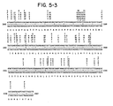
- In Figure 4, a restriction map of the R. meliloti fix gene region with fragments hybridizing to the nifA probe is shown. A black rectangle means strong hybridization, a hatched rectangle means weak hybridization and a white rectangle means no hybridization. This conclusion was confirmed by the nucleotide sequence of the coding region (Figure 5) and by activation experiments (Figures 6 and 7).
- Figure 6 shows a restriction map of plasmid pGW6 (see Table 2) and also the results of a control experiment in which the Rhizobium meliloti nifH promoter was activated by the Klebsiella pneumoniae nifA gene product. The plasmid pGW6 contains an EcoRI-SmaI fragment of pRmR2 subcloned into pMC1403. The plasmid vector pMC1403 allows translational fusion to the eighth codon of the E. coli lacZ gene. s-galactosidase activity is detectable if a second plasmid constitutively expressing the K. pneumoniae nifA gene is present in the same cell. Plasmid pGW7 (Table 2) is a derivative of pGW6 where the reading frame of the fusion gene was corrected at the BamHI site by Sl nuclease digestion. In Figure 7, the activation of the Rhizobium meliloti nifH promoter in pGW7 by the fixD gene product of R. meliloti is documented. The B-galactosidase activity of E. coli strain ET 8894 containing several plasmid combinations was measured. The results of six independent experiments are presented as black columns and the mean values are given as cross-lines. The plasmid combinations were: (1) pAc177-C + pGW7; (2) pRmW54 + pGW7; (3) pRmW54::Tn5-1 + pGW7; (4) pRmW54::Tn5-2 + pGW7; (5) pRmW54::Tn5-5 + pGW7; (6) pRmW54::Tn5-7 + pGW7; (7) pRmW54::Tn5-8 + pGW7; and (8) pRmW54::Tn5-9 + pGW7. The plasmids are also shown schematically on the left side of the drawing, p stands for the aphI promoter (i.e., the kanamycin resistance gene) of the vector plasmid pAC177-C and the double line represents the R. meliloti HindIII restriction fragment of pRmW54. The triangles give the positions of the various Tn5 insertions and the black horizontal arrows show the fixD coding region. Note that the activation of the R. meliloti nifH promoter (compared to the control No. 1) was not detectable when Tn5 was in the fixD region. In addition it was shown that the nifL probe of K. pneumomae did not hybridize to pRmW54, pRmR3 and pRmW52 (Table 2).
- The fixD gene of R. meliloti was detected by expression of pRmW54 in E. coli minicells using methods already described in detail (Puhler, A. and W. Klipp (1981) In: Biology of inorganic nitrogen and sulfur; Bothe, H. and A. Trebst eds. Berlin, Heidelberg; and Weber, G. and A. Puhler (1982) Plant Mol. Biol. 1:305-320). In E. coli minicells plasmid pRmW54 synthesized two polypeptides of 68K and 66K molecular weight (Fig. 8).
- In Figure 8, the restriction map of the HindIII fragment from pRmR29D5 (Table 2) is shown on the top and also the position of the several Tn5 insertions (
arrows 1 through 9). Restriction enzymes used are denoted as follows: H, HindIII; B, BamHI; X, Xhol; E, EcoRI; Bg, BglII and P, Pstl. The promoter of the aphI gene (kanamycin resistance gene of the vector plasmid pAC177-C) is indicated by p. The mapping shown in Figure 8 is in good agreement with the fact that in minicells containing pRmW54::Tn5-1 to pRmW54::Tn5-4, the 68K/66K polypeptides were not detectable in SDS-acrylamide gels. In their place, lower molecular weight polypeptides were found indicating that in pRmW54::Tn5-1 to pRmW54::Tn5-4 the Tn5 insertions were within the coding region of the fixD gene. Using several Tn5 insertions in the cloned HindIII fragment the coding region, designated fixD was again localized to the 1.8 kb BamHI fragment (Fig. 3 and 4). Especially, in the case of two Tn5 inserts (pRmW54::Tn5-2 and pRmW54::Tn5-4) putative truncated polypeptides were detected and this finding was in good agreement with the postulated coding region. Evidently, the 68K and 66K molecular weight polypeptides were both encoded by the same DNA fragment and may have been caused by the usage of different ATG-start codons. Both polypeptides are expressed on a very low level in E. coli minicells, but this low level is not due to a different codon usage in R. meliloti and E. coli, since the same coding region is very well expressed in the form of a fusion polypeptide with an E. coli vector gene, i.e., aminoglycoside phosphotransferase (aphI) (Example 6). - Thus, the experimental results disclosed in the present invention have defined the boundaries of the fixD gene of R, meliloti and have demonstrated that the protein of the fixD gene is responsible for the activation of the R. meliloti nifHDK and fixABC gene promoters leading in turn to the expression of the genes whose products are required for nitrogen fixation. However, the expression of the fixD gene requires that R. meliloti must first be in a symbiotic association with a specific host plant: R. meliloti does not fix nitrogen when living in a free state. Furthermore, R. meliloti does not fix nitrogen even when in a symbiotic association with the same host plant if there is a substantial quantity of nitrate already present in the soil. A genetic manipulation of the fixD gene so that it is consistently expressed either while free living or while in symbiotic association with a specific host plant growing in soil containing high nitrate would therefore be immensely useful in the fixation of nitrogen. Firstly the high nitrate content of a rich soil would not be decreased when an alfalfa-R. meliloti symbiotic association was present, but, to the contrary, the soil would become even richer in nitrates. In addition, the use of nif derepressed strains enables symbiotic nitrogen fixation to start earlier in nodule development and to last longer than in nodules with wild type Rhizobia.
- Secondly, nif derepressed Rhizobium strains have a big advantage when a poor symbiotic association with other plant species leads to ineffective nodulation by wild type Rhizobia. A likely explanation of ineffective nodulation is that a special plant signal normally necessary for transcription of nif genes is absent. The nif derepressed strains described herein have the ability to fix nitrogen without such a plant signal.
- Thirdly, Rhizobium strains can be induced to grow in close association with roots of economically important non-leguminous plants. Since the nif derepressed Rhizobium strains can fix nitrogen in large amounts while in the free living state, such an association between nif derepressed Rhizobia and the roots of important non-leguminous agricultural species has far reaching utility. The invention described here teaches how to make and use such nif derepressed Rhizobium strains.
- The present invention discloses another useful application of the fixD promoter region. when R. melitoti exists in the free living condition, the promoter of the fixD gene remains inactive and it is only when a symbiotic reiationship is established that the fixD gene is activated--most probably in direct or indirect response to a signal from the plant. It is often useful to incorporate a gene into a Rhizobium species but under such conditions that it is only expressed when the Rhizobia become symbiotically associated with the plant. This objective can be achieved in a genetic recombination by placing the coding region of a desirable gene under the control of the R. meliloti fixD gene promoter region. An example of such a gene, i.e., a gene whose expression is only desired when a symbiotic relationship has been established between a Rhizobium species and a plant, is the expression of the gene coding for an insecticidal protein such as the crystalline toxic protein of Bacillus thuringiensis (see Examples 3 and 4).
- The two main techniques which exist for DNA sequence analysis are the chemical degradation method (Maxam, A. W. and W. Gilbert (1977) Proc. Nat. Acad. Sci. USA 74:560-564) and the dideoxy-sequencing method (Sanger, F. et al. (1977) Proc. Nat. Acad. Sci. USA 74:5463-5468). Both methods were used to determine the primary structure of the fixD gene of R. meliloti.
- The M13 cloning and sequencing vectors mp8, mp9, mp10 and mpll (Messing, J. and Vieira, J. (1982) Gene 19:269-276; Norrander, J. et al. (1983) Gene 26:101-106) were used for subcloning of the HindIII 3.5 kb insert of plasmid pRmW54. For this purpose the HindIII fragment was cleaved from the vector by digestion with HindIII and then separated and isolated by agarose gel electrophoresis. For a non-random sequencing approach, the DNA was cleaved with various restriction enzymes (EcoRI, BamHI, HindIII, Sail, Xhol) and subcloned into the appropriate restriction sites of the M13 sequencing vectors. "Shotgun" cloning was carried out by digestion of the HindIII fragment with the restriction endonucleases Sau3A, HpaII and TagI. The resulting fragments were again cloned into M13 sequencing vectors. Recombinant clones were sequenced by the method of Sanger but slightly modified (Messing, J, et al. (1981) Nucleic Acids Res. 9:309-321). The strategy for dideoxy sequencing is presented (Figure 9).
- To implement the sequencing strategy, subclones of a DNA fragment containing the fixD coding region were generated and sequenced by a non-random and "shotgun" approach. In Figure 9, the position and extension of the subclones are presented in relation to the final restriction map of the fragment. The arrows (Figure 9) indicate the direction of sequencing and the rectangle represents the only open reading frame of the analysed DNA fragment.
- Restriction mapping of the HindIII fragment revealed the presence of several convenient restriction sites suitable for sequencing by the chemical method (Maxam, A. M. and W. Gilbert (1977) supra) (Figure 9). The HindIII fragment was isolated as described (supra), digested with the restriction enzymes EcoRI, BamHI, SalI and Xhol and subcloned into a pUC8 sequencing vector (Vieira, J. and J. Messing (1982) Gene 13:259-268). Determination of the sequence around several restriction sites of the fragment (Figure 9) enabled further alignment and confirmation of the data obtained from the dideoxy sequencing. The combined results of the two sequencing methods yielded the sequence of 2127 bp of the 3.5 kb HindIII fragment (Figure 10). Inspection of the six possible reading frames of the nucleotide sequence revealed that only one frame was large enough to code the fixD polypeptide. All other reading frames were closed by termination codons at many places. The large open reading frame which represents the fixD gene of R. meliloti started with an ATG 415 nucleotides from the HindIII site and spanned 1628 nucleotides. The coding region of fixD determined by the minicell procedure contained the following series of restriction sites: Xhol, EcoRI and BamHI. The same series of restriction sites was also present in the open reading frame. In addition, the molecular weight determination of the polypeptide encoded by the open reading frame resulted in a value of 59K, comparable to the imprecise molecular weight determination of 66/68K determined by the minicell procedure. An overall map of the fix genes and the nif genes is shown (Fig. 11)
- The hypothesis that the polypeptide double band of 66/68K detected in the minicell procedure is caused by different ATG start codons is supported by the nucleotide sequence. In addition to the ATG start codon at nucleotide 415, further ATG codons are located at positions 469 and 535.
- Following the hybridization experiments which localized the fixD gene on pRmW54, it was demonstrated that the R. meliloti fixD gene products activated the R. meliloti nifH promoter in E. coli. For these experiments plasmid pGW6, which contains the R. meliloti nifD gene fused on a translational level to a plasmid borne lacZ gene, was constructed (Fig. 6). Control experiments with plasmid pWK131 (Table 2) which constitutively expressed the K. pneumoniae nifA gene showed that the nifD-lacZ fusion of pGW6 was activated. The activation was detected by measuring the B-galactosidase activity (Figure 6). In spite of the differences in activation of the R. meliloti nifH gene and the K. pneumoniae nifH gene by the glnG gene of E. coli, it was possible that the R. meliloti fixD gene product might activate the R. meliloti nifH promoter and be detectable in the same manner. For the actual experiment, an E. coli strain ET8894 (Table 1) which carries an IS1 insertion in the chromosomal lacZ gene and a deletion in the gin regulatory region, was used. This strain was transformed with plasmid pGW7 and plasmid pRmW54. The results (Figure 7) were as follows: ET8894 containing the nifD::lacZ fusion (pGW7) and the constitutively expressed fixD gene (pRmW54) had a 1.5-fold higher 8-galactosidase activity compared to the control strain ET8894 containing the plasmid pAC177-C (Table 3; Figure 7). The significance of this activation on a very low level was shown by the use of Tn5 insertions in the cloned fixD gene. Only Tn5 insertions in the coding region resulted in the abolishment of the activation (Table 3; Figure 7).
- Construct a synthetic DNA primer which is complementary to the DNA sequence immediately upstream (i.e., 5'- to the ATG translation initiation codon) of the Rhizobium meliloti fixD coding sequence. The synthetic DNA primer has the sequence 5'-CAGAGCCTTTGATATGCTGTA-3'. The HindIII-Xhol DNA subclone of pRmW54 in pAC177-C (designated pRmW69 - Table 2) is then excised, purified, subcloned into a single stranded DNA phage and transformed into E. coli JMI03 (Fig. 11). This HindIII-XhoI subclone contains the complete promoter sequence of R. meliloti fixD gene and 82 nucleotides at the 5'-end of the fixD coding sequence. The subcloned fragment is amplified therein and single stranded templates (ca. lug) are recovered from the supernatant following centrifugation of the bacterial host. A 10-fold excess of the synthetic DNA primer in the presence of the four deoxynucleotide triphosphates (one of which is radioactive) and DNA polymerase I (Klenow fragment) is now used as a primer on this fixD template to generate double stranded DNA (dsDNA) (Fig. 12). The mixture is incubated for a period (e.g., 15-45 minutes) at an appropriate temperature (e.g., 25°-37°C) during which period the complementary strand is extended beyond the HindIII site. The 468 nucleotide region between the HindIII site and the ATG initiation codon contains the complete promoter region of the fixD gene of R. meliloti. The remaining single stranded DNA is then removed by digestion with 51 nuclease (Fig. 12). EcoRI linkers (GGAATTCC) are then ligated to the dsDNA fragments followed by digestion with EcoRI (Fig. 13). The fragments are separated by agarose gel electrophoresis and the fragment containing the promoter sequence is eluted and cloned into the wide host range plasmid pSUP204 (Fig. 13), which has previously been restricted by the restriction enzyme EcoRI. The resulting recombinant plasmid is termed pRm-fixD-P/SS204. Following transformation and amplification in a suitable E. coli host strain, e.g., 17-1, which is restriction negative, i.e., r-, partial cleavage with EcoRI allows the addition of any foreign structural gene or foreign DNA fragment into the linearized plasmid downstream from the fixD promoter fragment. A foreign structural gene of foreign DNA fragment is herein defined as any structural gene or DNA fragment not found naturally under the direct or indirect control of a fixD promoter. For example, the gene coding for the crystalline toxic protein of Bacillus thuringiensis can be inserted resulting in a "composite" recombinant. A composite recombinant is herein defined as a recombinant DNA plasmid containing a vector, a promoter sequence and any foreign DNA whose expression is under the control of said promoter sequence.
- Recombinant plasmids containing inserts of the gene encoding the toxic crystal protein of B. thuringiensis are obtained using the techniques described (Wong, H. C., Schnepf, H. E. and H. R. Whiteley (1983) J. Biol. Chem. 258:1960-1967). The recombinant plasmid pESl (ATCC Number 31995) consisting of the plasmid vector pBR322 and DNA homologous to the 30, 32 and 37 megadal- ton plasmids, as well as DNA homologous to linearized forms of the very large plasmids of B. thuringiensis is partially cleaved with EcoRI to give linear molecules. These partial cleavage products are further restricted by the enzyme Aval. The digestion conditions are as recommended by the manufacturer. A probe for the toxic crystal protein gene is isolated and radioactively labelled as previously described (Wong, H. C, et al. (1983) supra). The restriction fragments are separated by agarose gel electrophoresis and the labelled probe is found to hybridize to one fragment of approximately 15 kilobases (kb). This fragment includes the EcoRI fragments D and F (Wong, H. C. et al. (1983) supra). The 15 kb fragment is then cloned into M13mp8 or M13 mp9 according to standard procedure (Messing, J. and J. Vieira (1982) Gene 19:269-276) and transformed into E. coli JM103. The single stranded DNA from the extruded phage particles is purified and replicated in vitro by use of a synthetic primer (5'-TGTTATCCATGGGTTACCTCC-3') (The general method of site specific mutagenesis is described in Zoller, M. J. and M. Smith (1982) Nucleic Acids Research 10:6487-6500). The resulting double stranded recombinant plasmid is then transformed back into E. coli JM103 and amplified. The amplified double stranded plasmid DNA is purified from the E. coli JM103 cells and cleaved with the restriction endonucleases NcoI and Aval. Ncol cleaves at the site of the synthetic primer (which is the initiation site of the toxic crystal protein gene) and Aval cleaves at a site which is downstream from the 3'- end of the toxic crystal protein gene. The overhangs are then filled in to blunt ends (Maniatis, T., Jeffrey, A. and D. G. Kleid (1975) Proc. Nat. Acad. Sci. U.S.A. 72:1184-1188).
- Finally the pRmfixD-P/SS204 recombinant plasmid which is derived from pSUP204 (Fig. 13) is cleaved with EcoRI and the overhangs filled in to blunt ends. HindIII linkers are then added to both the B. thuringiensis toxic crystal protein gene fragment and to the pRmfixD-P/SS204 recombinant. Following the HindIII digestion of both components, the toxic crystal protein gene and the pRmfixD-P/SS204 recombinant plasmid are ligated together to give a pRmfixD-P/SS204 - B. thuringiensis toxic crystal protein gene composite. The mixture is transformed into a suitable E. coli host, e.g. K802, 17-1 or RR1. Plasmids are isolated from individual colonies and the orientation determined by restriction mapping. A colony containing a plasmid with the correct orientation is then conjugated to Rhizobium meliloti and the plasmid is transferred as already described (Example 7). The production of mRNA and/or the toxic crystal protein is monitored as already described (Wong, et al., supra).
- The source of the kanamycin resistance gene is the transposon Tn5 and the suicide vector pSUP1011 (Simon, R., Priefer, U. and A. Puhler (1981) Proc. of Bielefeld Symposium, Springer-Verlag, West Germany) (see Fig. 14). Transposon Tn5 is a DNA element of 5.7 kilobases (kb) in length, consisting of 1.5 kb inverted repeat sequences flanking a 2.7 kb central region. Encoded within the inverted repeats are the functions required for transposition. The central region of the transposon carries a gene conferring resistance to the antibiotic kanamycin (Kmr). The coding region of the neomycin phosphotransferase gene (neo) (alternatively named aminoglycoside phosphotransferase gene- aph) is deleted by digestion with the restriction enzymes BglII (a BglII site is present 36 nucleotides upstream from the translational start codon ATG) and Sail (a SalI site is present 342 nucleotides downstream from the TGA termination codon) (see Fig. 15).
- The sequence of the kanamycin resistance gene has been previously shown and is reproduced herein as Figure 15 (Beck, E. et al. (1982) Gene 19:327- 336). The nucleotide sequence of Tn5 DNA from
position 1401 to position 2700 is shown. The aph coding region is boxed by solid lines. Some restriction sites are indicated in the sequence, as well as at the end of the left inverted repeat (closing bracket ]) and the promoter region. Such a deletion leaves the promoter region of the neomycin phosphotransferase gene (neo) intact. The single stranded overlaps generated by these two restriction endonucleases are converted to blunt ends and HindIII linkers are added, followed by digestion with HindIII restriction endonuclease. This construction is herein defined as pSUP1011-HindIII (Fig. 16). - In parallel, plasmid pRmW54 was obtained by cloning a 3.5 kb HindIII fragment of pRm29D5 (Ruvkun, G. B. et al. (1982) Cell 29:551-559) into the E. coli vector plasmid pAC177-C (Table 2). After transformation into a suitable host strain and amplification, the recombinant plasmid is linearized by partial digestion with the restriction endonuclease HindIII. Linearization occurs either on the upstream side of the fixD gene or on the downstream side. These two alternatives can be distinguished by the use of restriction maps. Those linearizations where the cut occurs upstream (i.e., 5' to the reading strand of the fixD gene) are retained and treated with the exonuclease BAL31 for a sufficient period to produce a blunt end at an optimal distance from the ATG initiation codon of the R. meliloti fixD gene. The linearized recombinant which is thus shortened towards the 5'-terminus of the fixD gene is then further restricted with BglII. A single BglII site is present downstream (i.e., 3'- from the TGA termination codon of the reading strand) from the termination codon of the fixD gene (Figure 17). This fragment which contains the fixD structural gene is converted to blunt ends and HindIII linkers are added. Following digestion with HindIII, the fragment containing the fixD gene is purified and ligated into the linearized pSUP1011-HindIII (supra) (Figure 18). Such a construction comprises the pSUP1011 suicide vector and a Tn5 transposon. Between the inverted repeats of the Tn5, the neomycin phosphotransferase promoter (neopro) is included and, in addition, the fixD structural gene positioned so that it is under the control of neopro. This construction is herein defined as pSUP1011-Tn5-neopro-fixD.
- In order to obtain such a fusion gene and protein product, the Xhol-HindIII subfragment of pRmW54 was cloned into pAC177-C (Table 2). From the restriction mapping experiment (Example 1) it was known that the XhoI site was located within the fixD coding region. On the other hand, the Xhol restriction site of the vector plasmid pAC177-C was located in the aphI gene (kanamycin resistance gene). In order to obtain the correct reading frame of the fusion polypeptide, the XhoI site was treated with Sl nuclease in one case and with DNA polymerase I in the other. A strongly expressed 60K fusion polypeptide was obtained after treatment with DNA polymerase I. The recombinant plasmid described herein which contains such a constitutively expressed fixD gene is referred to as pRmW69 (Table 2).
- This example is based on the following general principles. Two basic components are employed. These are: (1) a suicide vector and (2) a transposon.
- Suicide vectors are plasmid molecules which replicate stably in one bacterial host (in this case E. coli) but fail to replicate in a different bacterial species (e.g., Rhizobium meliloti).
- Transposons are genetic elements which are able to move (translocate) from one location to another in DNA. The translocation process is mediated by gene products encoded on the transposon and is dependent on the integrity of repeated sequences (directly or indirectly repeated) located at each end of the transposon. Transposons generally carry a gene (or genes) encoding resistance to one or more antibiotics.
- The vector contains the inserted fixD gene under the control of a kanamycin resistance promoter pSUP1011-Tn5-neopro fixD (see Example 5) and is transformed (introduced) into Escherichia coli strain SM10. This type of recombinant plasmid comprising a suicide vector, a transposon and a constitutively expressed structural gene is herein defined as a "constitutive gene transfer system". (This strain is capable of mobilizing (Mob+) pSUP1011 derivatives (recombinant plasmids) into other gram negative bacteria) (Simon, R., Priefer, U. and A. Puhler (1983) Proc. of Bielefeld Symposium, Springer-Verlag, West Germany). The resultant transformants are screened by the Grunstein and Hogness colony hybridization procedure (Grunstein, M. and D. S. Hogness (1975) Proc. Nat. Acad. Sci. USA 72:3961ff) to detect those containing the desired cloned DNA fragment.
- Introduction of the cloned DNA fragment into the genome of any gram-negative bacterium (e.g., Rhizobium meliloti) is achieved via a process called bacterial conjugation. The E. coli SM10 derivative, carrying the desired pSUP1011 recombinant, is mixed with cells of R. meliloti carrying a symbiotic (Sym) plasmid on the surface of a nutrient agar plate. The plate is incubated for a suitable period (4-16 hours) at 29-30° (optimum temperature for Rhizobium meliloti) and during this time cells of each type come into physical contact (conjugation) and the pSUP1011 recombinant derivative is transferred from E. coli to R. meliloti. The cell mixture is washed off the plate and spread on an agar plate which is selective for organisms which can only grow if they are capable of constitutive nitrogen fixation. The resultant colonies which can constitutively fix nitrogen will be derivatives of R. meliloti in which the cloned DNA fragment, within Tn5, will be inserted at some point in the genome. Selection for ability to constitutively fix nitrogen insures maintenance of the inserted DNA.
- At this stage, it is unknown whether the DNA fragment, within Tn5, has been transferred to the chromosome of R. meliloti or to one of its several plasmids. This uncertainty can be resolved by visualization of the plasmids and the bacterial chromosome by ethidium bromide staining after horizontal agarose gel electrophoresis (Djordjevic, M. A. et al. (1982) J. Bacteriol. 151:560-568). In either case, the transferred DNA fragment is fuctional, since the fixD gene product acts in trans to activate nitrogen fixation.
- E. coli C600 (pRmW54) was placed on deposit with the American Type Culture Collection, 12301 Parklawn Drive, Rockville, MD 20852 on June 4, 1984, Accession No. 39722.
- Expression of β-galactosidase was placed under nifHDK promoter control as described (pGW7, Example 2, Fig. 6). The amount of β-galactosidase synthesized under nifHDK control was measured using E, coli ET8894 (Table 1) containing pGW7. Synthesis of active s-galactosidase in this system depends on activation of the nifHDK promoter. Introduction of pRmW54 (expressing fixD as a fusion protein under control of the aphi) leads to a significant increase in a-galactosidase activity over control levels (Table 3). The described experiment was designed to test whether a-galactosidase expression could be enhanced by increasing the levels of fixD gene product, and whether the level of fixD gene product could be increased by shortening the distance, or number of amino acids, between the aphI promoter and the start of the fixD gene.
- A series of deletions was constructed in which the number of amino acids lying in the coding region between the aphI promoter and the fixD coding region was reduced. First, the larger of the two HindIII-BglII fragments of pRmW54 was cloned into pACYC177-C (also termed pAC177-C herein), previously cut with HindIII and BamHI. The resulting plasmid, designated pRmW541, contained a unique HindIII site located between the aphI promoter and the fixD coding region. Deletions were introduced by cutting pRmW541 with HindIII and digesting for various times with exonuclease Ba131. The resulting digestion mixture was religated after S1 nuclease digestion, and the mixture was used to transform a culture of E. coli ET8894 (pGW7). After incubation for 48 hours at 30°C, the transformants were plated on agar plates supplemented with ampicillin, chloramphenicol, glutamine and X-gal (5-bromo-4-chloro-3-indolyl-β-D galactoside, a chromogenic β-galactosidase substrate). Although E. coli ET8894 (pGW7, pRmW54) produces detectable β-galactosidase activity, the activity is low and incubation on X-gal plates yields white colonies. Several transformants from the deletion mixture yielded blue colonies, indicating qualitatively enhanced expression of β-galactosidase. Plasmids were isolated from strains with higher activity and retransformed into E. coli ET8894 (pGW7). The β-galactosidase activity was quantitatively measured with results shown in Fig. 19. The structure of each of each of the pRmW541 deletion plasmids was further analyzed by sequencing XhoI fragments of the various pRmW541 isolates. An endonuclease Xhol site exists within the fixD gene and another is located just upstream from the translation start of the aphI coding region (See Fig. 10). The XhoI fragments were sequenced, after cloning them into the Sail site of the phage M13mp8. Nucleotide sequence determination of these deletions, which were contained within the XhoI fragments, revealed that all the nifHDK activated proteins were fusion polypeptides, with the exception of pRmW541-5. The structures are shown diagrammatically in Fig. 19.
- Fig. 19 diagrams the structure of the pRmW541 deletion plasmids, and the resulting level of activation in ET8894 (pGW7). For each of the plasmids the concluded polypeptide structure is shown schematically. The black closed circle indicates the aphI promoter. The open rectangles represent the APH-I part of the translated polypeptide. The incomplete black triangle represents the FIX-D part. Crosshatched regions indicated translated open reading frame regions generated by the construction, which are expressed only in the fusion polypeptides. The numbers in the boxes represent the amino acid residues. The plated right-hand end of the hatched region of pRmW541-5 indicates a translation termination signal. The numbers on top of the FIX-D part indicate the codon in which the fusion occurred. The right-hand column shows the s-galactosidase activity of each plasmid in ET8894 (pGW7).
- The effect of the deletions on the expression of the FIX-D polypeptides was also measured. Deletions in plasmid pRmW541 which cause a high level of activation of the nifHDK promoter also lead to stronger expression of the FIX-D polypeptide.
- The results demonstrate that deletions in the region between the aphI promoter and the fixD gene made it possible to increase the level of APH-I-FIX-D fusion protein synthesized and to increase the activation of the nifHDK- lacZ fusion. Control experiments have shown that activation of the nifHDK promoter by the fixD gene product was strictly dependent on the presence of an intact glnF gene in the host organism. The glnF gene, also termed ntrA, is a positive regulatory gene for glutamine synthetase, and is active in ET8894.


Claims (19)


thereby activating fixation of dinitrogen by activating expression of said nitrogen fixation genes.
Applications Claiming Priority (2)
| Application Number | Priority Date | Filing Date | Title |
|---|---|---|---|
| US61658684A | 1984-06-04 | 1984-06-04 | |
| US616586 | 1984-06-04 |
Publications (2)
| Publication Number | Publication Date |
|---|---|
| EP0164992A2 true EP0164992A2 (en) | 1985-12-18 |
| EP0164992A3 EP0164992A3 (en) | 1987-12-16 |
Family
ID=24470131
Family Applications (1)
| Application Number | Title | Priority Date | Filing Date |
|---|---|---|---|
| EP85303925A Withdrawn EP0164992A3 (en) | 1984-06-04 | 1985-06-04 | Nitrogen fixation regulator genes |
Country Status (3)
| Country | Link |
|---|---|
| EP (1) | EP0164992A3 (en) |
| JP (1) | JPS61282077A (en) |
| AU (2) | AU580064B2 (en) |
Cited By (12)
| Publication number | Priority date | Publication date | Assignee | Title |
|---|---|---|---|---|
| EP0211661A1 (en) * | 1985-08-07 | 1987-02-25 | Lubrizol Genetics Inc. | Nif Promoter of fast-growing rhizobium japonicum |
| WO1987004182A1 (en) * | 1985-12-30 | 1987-07-16 | Nielsen Sven Erik | Rhizobia transformants which symbiotically fixes nitrogen in non-legumes, a material for treating seeds of a non-legume plant, non-legume seeds, a non-legume plant and a method for producing rhizobia transconjugants |
| EP0242892A1 (en) * | 1986-02-28 | 1987-10-28 | Rijksuniversiteit Leiden | Process for activating rhizobium nodulation promoters |
| EP0164245A3 (en) * | 1984-06-08 | 1987-12-09 | Lubrizol Genetics Inc. | Nif promoter of rhizobium japonicum |
| EP0255340A1 (en) * | 1986-07-28 | 1988-02-03 | The National Development Corporation Limited | The use of Dicarboxylic acid transport genes |
| EP0256682A3 (en) * | 1986-08-04 | 1988-11-30 | Imperial Chemical Industries Plc | Plant treatment |
| EP0245931A3 (en) * | 1986-03-27 | 1989-04-12 | Lubrizol Genetics Inc. | Nodulation inducing factors |
| EP0339830A3 (en) * | 1988-04-14 | 1990-01-17 | Biotechnica International, Inc. | Improved biological nitrogen fixation |
| WO1991001377A1 (en) * | 1989-07-21 | 1991-02-07 | Biological Research Center Of The Hungarian Academy Of Sciences | Procedure for the isolation of nitrogen fixing bacterial strains mutated in the nitrogen control of symbiotic nodulation genes |
| US5077209A (en) * | 1987-07-27 | 1991-12-31 | National Development Corporation | Dicarboxylic acid transport genes |
| US5308616A (en) * | 1990-08-06 | 1994-05-03 | The Johns Hopkins University | Mutant strain of Bradyrhizobium japonicum |
| US6548289B1 (en) | 1984-12-28 | 2003-04-15 | Land O'lakes, Inc. | Biological nitrogen fixation |
Families Citing this family (2)
| Publication number | Priority date | Publication date | Assignee | Title |
|---|---|---|---|---|
| AU580064B2 (en) * | 1984-06-04 | 1988-12-22 | Lubrizol Genetics Inc. | Nitrogen fixation regulator genes |
| JP6489676B2 (en) * | 2014-03-17 | 2019-03-27 | 国立大学法人名古屋大学 | Biological nitrogen fixation regulator and its use |
Family Cites Families (5)
| Publication number | Priority date | Publication date | Assignee | Title |
|---|---|---|---|---|
| RO80052B (en) * | 1977-11-08 | 1985-03-30 | Genetech | Process for preparing a recombinant cloning vehicle for transforming a bacterial host in order to convert it in heterologous polypeptide generator |
| NZ208557A (en) * | 1983-06-22 | 1988-11-29 | Lubrizol Genetics Inc | Recombinant nif (nitrogen fixation) promoters - foreign structural gene combinations, plasmids, transformants and method of expressing the structural gene in plants |
| AU580064B2 (en) * | 1984-06-04 | 1988-12-22 | Lubrizol Genetics Inc. | Nitrogen fixation regulator genes |
| ZA853861B (en) * | 1984-06-08 | 1986-01-29 | Agrigenetics Res Ass | Nif promotor of rhizobium japonicum |
| EP0195285A3 (en) * | 1985-02-28 | 1987-12-16 | The University Of Georgia Research Foundation, Inc. | Molecular cloning of the delta-endotoxin gene of bacillus thurigiensis var. israelensis |
-
1985
- 1985-06-03 AU AU43260/85A patent/AU580064B2/en not_active Ceased
- 1985-06-04 EP EP85303925A patent/EP0164992A3/en not_active Withdrawn
- 1985-06-04 JP JP60122361A patent/JPS61282077A/en active Pending
-
1988
- 1988-10-05 AU AU23418/88A patent/AU603261B2/en not_active Ceased
Cited By (13)
| Publication number | Priority date | Publication date | Assignee | Title |
|---|---|---|---|---|
| EP0164245A3 (en) * | 1984-06-08 | 1987-12-09 | Lubrizol Genetics Inc. | Nif promoter of rhizobium japonicum |
| US6548289B1 (en) | 1984-12-28 | 2003-04-15 | Land O'lakes, Inc. | Biological nitrogen fixation |
| EP0211661A1 (en) * | 1985-08-07 | 1987-02-25 | Lubrizol Genetics Inc. | Nif Promoter of fast-growing rhizobium japonicum |
| WO1987004182A1 (en) * | 1985-12-30 | 1987-07-16 | Nielsen Sven Erik | Rhizobia transformants which symbiotically fixes nitrogen in non-legumes, a material for treating seeds of a non-legume plant, non-legume seeds, a non-legume plant and a method for producing rhizobia transconjugants |
| EP0237683A1 (en) * | 1985-12-30 | 1987-09-23 | Sven-Erik Nielsen | Rhizobia which symbiotically fixes nitrogen in non-legumes, a material for treating seeds of a non legume seed, a non-legume seed, a non-legume plant and a method for producing Rhizobia transconjugants |
| EP0242892A1 (en) * | 1986-02-28 | 1987-10-28 | Rijksuniversiteit Leiden | Process for activating rhizobium nodulation promoters |
| EP0245931A3 (en) * | 1986-03-27 | 1989-04-12 | Lubrizol Genetics Inc. | Nodulation inducing factors |
| EP0255340A1 (en) * | 1986-07-28 | 1988-02-03 | The National Development Corporation Limited | The use of Dicarboxylic acid transport genes |
| EP0256682A3 (en) * | 1986-08-04 | 1988-11-30 | Imperial Chemical Industries Plc | Plant treatment |
| US5077209A (en) * | 1987-07-27 | 1991-12-31 | National Development Corporation | Dicarboxylic acid transport genes |
| EP0339830A3 (en) * | 1988-04-14 | 1990-01-17 | Biotechnica International, Inc. | Improved biological nitrogen fixation |
| WO1991001377A1 (en) * | 1989-07-21 | 1991-02-07 | Biological Research Center Of The Hungarian Academy Of Sciences | Procedure for the isolation of nitrogen fixing bacterial strains mutated in the nitrogen control of symbiotic nodulation genes |
| US5308616A (en) * | 1990-08-06 | 1994-05-03 | The Johns Hopkins University | Mutant strain of Bradyrhizobium japonicum |
Also Published As
| Publication number | Publication date |
|---|---|
| JPS61282077A (en) | 1986-12-12 |
| AU603261B2 (en) | 1990-11-08 |
| AU2341888A (en) | 1989-01-12 |
| AU580064B2 (en) | 1988-12-22 |
| EP0164992A3 (en) | 1987-12-16 |
| AU4326085A (en) | 1985-12-12 |
Similar Documents
| Publication | Publication Date | Title |
|---|---|---|
| US4782022A (en) | Nitrogen fixation regulator genes | |
| Thōny et al. | The symbiotic nitrogen fixation regulatory operon (fixRnifA) of Bradyrhizobium japonicum is expressed aerobically and is subject to a novel, nifA-independent type of activation | |
| Ditta et al. | The nifA gene of Rhizobium meliloti is oxygen regulated | |
| Black et al. | Sequences and characterization of hupU and hupV genes of Bradyrhizobium japonicum encoding a possible nickel-sensing complex involved in hydrogenase expression | |
| Scott | Conserved nodulation genes from the non–legume symbiont Bradyrhizobium sp.(Parasponia) | |
| Nieuwkoop et al. | A locus encoding host range is linked to the common nodulation genes of Bradyrhizobium japonicum | |
| EP0164992A2 (en) | Nitrogen fixation regulator genes | |
| Fuhrmann et al. | Rhizobium japonicum nitrogenase Fe protein gene (nifH) | |
| Alvarez-Morales et al. | Expression of Rhizobium japonicum nifH and nifDK operons can be activated by the Klebsiella pneumoniae nifA protein but not by the product of ntrC | |
| EP0130047A1 (en) | Recombinant DNA molecules | |
| Bozouklian et al. | Cloning and characterization of the glnA gene of Azospirillum brasilense Sp7 | |
| Reyes-González et al. | Expanding the regulatory network that controls nitrogen fixation in Sinorhizobium meliloti: elucidating the role of the two-component system hFixL-FxkR | |
| US4803165A (en) | Nif promoter of fast-growing rhizobium japonicum | |
| EP0164245A2 (en) | Nif promoter of rhizobium japonicum | |
| Palacios et al. | Differential expression of hydrogen uptake (hup) genes in vegetative and symbiotic cells of Rhizobium leguminosarum | |
| Theroux et al. | Gene encoding the 5.7-kilodalton chlorosome protein of Chloroflexus aurantiacus: regulated message levels and a predicted carboxy-terminal protein extension | |
| Feingold et al. | Organization and nucleotide sequence analysis of an rRNA and tRNA gene cluster from Caulobacter crescentus | |
| US5001061A (en) | nifD promoter of Bradyrhizobium | |
| CN111349649B (en) | Method for gene editing of agaricus bisporus and application | |
| Gubler et al. | The Bradyrhizobium japonicum fixBCX operon: identification of fixX and of a 5’mRNA region affecting the level of the fixSCX transcript | |
| Legocki et al. | Expression of β-galactosidase controlled by a nitrogenase promoter in stem nodules of Aeschynomene scabra | |
| JPH01225483A (en) | Recombinant plasmid | |
| US5008194A (en) | nifH promoters of Bradyrhizobium | |
| Schlüter et al. | A homolog of the Rhizobium meliloti nitrogen fixation gene fixN is involved in the production of a microaerobically induced oxidase activity in the phytopathogenic bacterium Agrobacterium tumefaciens | |
| JPH0587234B2 (en) |
Legal Events
| Date | Code | Title | Description |
|---|---|---|---|
| PUAI | Public reference made under article 153(3) epc to a published international application that has entered the european phase |
Free format text: ORIGINAL CODE: 0009012 |
|
| AK | Designated contracting states |
Designated state(s): AT BE CH DE FR GB IT LI LU NL SE |
|
| RAP1 | Party data changed (applicant data changed or rights of an application transferred) |
Owner name: LUBRIZOL GENETICS INC. |
|
| PUAL | Search report despatched |
Free format text: ORIGINAL CODE: 0009013 |
|
| AK | Designated contracting states |
Kind code of ref document: A3 Designated state(s): AT BE CH DE FR GB IT LI LU NL SE |
|
| 17P | Request for examination filed |
Effective date: 19880518 |
|
| 17Q | First examination report despatched |
Effective date: 19900724 |
|
| RAP1 | Party data changed (applicant data changed or rights of an application transferred) |
Owner name: LUBRIZOL GENETICS INC. |
|
| STAA | Information on the status of an ep patent application or granted ep patent |
Free format text: STATUS: THE APPLICATION IS DEEMED TO BE WITHDRAWN |
|
| 18D | Application deemed to be withdrawn |
Effective date: 19920225 |
|
| RIN1 | Information on inventor provided before grant (corrected) |
Inventor name: REILAENDER, HELMUT Inventor name: WEBER, GERHARD Inventor name: PUHLER, ALFRED |