EP0164876B1 - Polynukleotidanalyse durch Verschiebung unter Verwendung eines Rekombinationsproteins und Reagenzsatz - Google Patents
Polynukleotidanalyse durch Verschiebung unter Verwendung eines Rekombinationsproteins und Reagenzsatz Download PDFInfo
- Publication number
- EP0164876B1 EP0164876B1 EP19850303176 EP85303176A EP0164876B1 EP 0164876 B1 EP0164876 B1 EP 0164876B1 EP 19850303176 EP19850303176 EP 19850303176 EP 85303176 A EP85303176 A EP 85303176A EP 0164876 B1 EP0164876 B1 EP 0164876B1
- Authority
- EP
- European Patent Office
- Prior art keywords
- polynucleotide
- probe
- labeled
- labeled polynucleotide
- nucleotide sequence
- Prior art date
- Legal status (The legal status is an assumption and is not a legal conclusion. Google has not performed a legal analysis and makes no representation as to the accuracy of the status listed.)
- Expired
Links
Images


Classifications
-
- C—CHEMISTRY; METALLURGY
- C12—BIOCHEMISTRY; BEER; SPIRITS; WINE; VINEGAR; MICROBIOLOGY; ENZYMOLOGY; MUTATION OR GENETIC ENGINEERING
- C12Q—MEASURING OR TESTING PROCESSES INVOLVING ENZYMES, NUCLEIC ACIDS OR MICROORGANISMS; COMPOSITIONS OR TEST PAPERS THEREFOR; PROCESSES OF PREPARING SUCH COMPOSITIONS; CONDITION-RESPONSIVE CONTROL IN MICROBIOLOGICAL OR ENZYMOLOGICAL PROCESSES
- C12Q1/00—Measuring or testing processes involving enzymes, nucleic acids or microorganisms; Compositions therefor; Processes of preparing such compositions
- C12Q1/68—Measuring or testing processes involving enzymes, nucleic acids or microorganisms; Compositions therefor; Processes of preparing such compositions involving nucleic acids
- C12Q1/6813—Hybridisation assays
- C12Q1/6816—Hybridisation assays characterised by the detection means
- C12Q1/6823—Release of bound markers
-
- C—CHEMISTRY; METALLURGY
- C12—BIOCHEMISTRY; BEER; SPIRITS; WINE; VINEGAR; MICROBIOLOGY; ENZYMOLOGY; MUTATION OR GENETIC ENGINEERING
- C12Q—MEASURING OR TESTING PROCESSES INVOLVING ENZYMES, NUCLEIC ACIDS OR MICROORGANISMS; COMPOSITIONS OR TEST PAPERS THEREFOR; PROCESSES OF PREPARING SUCH COMPOSITIONS; CONDITION-RESPONSIVE CONTROL IN MICROBIOLOGICAL OR ENZYMOLOGICAL PROCESSES
- C12Q1/00—Measuring or testing processes involving enzymes, nucleic acids or microorganisms; Compositions therefor; Processes of preparing such compositions
- C12Q1/68—Measuring or testing processes involving enzymes, nucleic acids or microorganisms; Compositions therefor; Processes of preparing such compositions involving nucleic acids
- C12Q1/6813—Hybridisation assays
- C12Q1/6827—Hybridisation assays for detection of mutation or polymorphism
-
- C—CHEMISTRY; METALLURGY
- C12—BIOCHEMISTRY; BEER; SPIRITS; WINE; VINEGAR; MICROBIOLOGY; ENZYMOLOGY; MUTATION OR GENETIC ENGINEERING
- C12Q—MEASURING OR TESTING PROCESSES INVOLVING ENZYMES, NUCLEIC ACIDS OR MICROORGANISMS; COMPOSITIONS OR TEST PAPERS THEREFOR; PROCESSES OF PREPARING SUCH COMPOSITIONS; CONDITION-RESPONSIVE CONTROL IN MICROBIOLOGICAL OR ENZYMOLOGICAL PROCESSES
- C12Q1/00—Measuring or testing processes involving enzymes, nucleic acids or microorganisms; Compositions therefor; Processes of preparing such compositions
- C12Q1/68—Measuring or testing processes involving enzymes, nucleic acids or microorganisms; Compositions therefor; Processes of preparing such compositions involving nucleic acids
- C12Q1/6813—Hybridisation assays
- C12Q1/6832—Enhancement of hybridisation reaction
-
- C—CHEMISTRY; METALLURGY
- C12—BIOCHEMISTRY; BEER; SPIRITS; WINE; VINEGAR; MICROBIOLOGY; ENZYMOLOGY; MUTATION OR GENETIC ENGINEERING
- C12Q—MEASURING OR TESTING PROCESSES INVOLVING ENZYMES, NUCLEIC ACIDS OR MICROORGANISMS; COMPOSITIONS OR TEST PAPERS THEREFOR; PROCESSES OF PREPARING SUCH COMPOSITIONS; CONDITION-RESPONSIVE CONTROL IN MICROBIOLOGICAL OR ENZYMOLOGICAL PROCESSES
- C12Q1/00—Measuring or testing processes involving enzymes, nucleic acids or microorganisms; Compositions therefor; Processes of preparing such compositions
- C12Q1/68—Measuring or testing processes involving enzymes, nucleic acids or microorganisms; Compositions therefor; Processes of preparing such compositions involving nucleic acids
- C12Q1/6876—Nucleic acid products used in the analysis of nucleic acids, e.g. primers or probes
Definitions
- regions of the probe may be of various naturally occurring or synthesized sequences which do not participate in the-hybridization reaction with the target nucleotider sequence, but which may play an important role in the present invention, e.g., by serving as a site for attachment to a support or by providing some degree of separation between the support and the region to which the target nucleotide sequence binds, if desired.
- the binding may be (and preferably is) perfect, in the sense that each nucleotide in the sequence finds its correct complementary binding partner (e.g., dAto dT) in the target nucleotide sequence or may contain some mismatches.
- dAto dT complementary binding partner
- the overall target binding region includes the initial binding region and most or (preferably) all of the labeled polynucleotide binding region (LBR in the Figures and in the discussion below).
- Non-specific covalent linkages include linkages between the substrate and free bases along the chain via moieties such as m-aminobenzyloxy methyl (ABM), m-diazobenzyloxy methyl (DBM) or o-aminophenylthioether (APT).
- moieties such as m-aminobenzyloxy methyl (ABM), m-diazobenzyloxy methyl (DBM) or o-aminophenylthioether (APT).
- Still other spacer molecules can contain a functional moiety such as phenyl ketone which will react directly with a support having hydrazine moieties, forming a resultant hydrazone.
- the probe further may be noncovalently linked to the support by interaction of some portion of the probe with affinity reagents that are adsorbed or covalently bound to the support.
- affinity reagents that are adsorbed or covalently bound to the support.
- Examples include (1) immobilization on the support of a short single-stranded polynucleotide which can hybridize to some portion of the probe polynucleotide not overlapping with the region in the probe which is capable of binding to the target nucleotide sequence and (2) binding of a chemically modified probe polynucleotide carrying one or more avidin or biotin moieties to a support having biotin or avidin moieties, respectively, adsorbed or covalently bound to the support.
- the latter method is based on the high affinity (K diSS approximately 10- 15 M) of the small molecule biotin for the protein avidin (or streptavidin).
- the present invention is not limited with regard to the spacings between the point or points of attachment of the probe to a support and the region of the probe polynucleotide which binds specifically to the target nucleotide sequence, it is preferable that this spacing be sufficiently large for the hybridization between target nucleotide sequence and target binding region of the probe polynucleotide to occur such that the target binding region of the probe has sufficient, and preferably maximal obtainable freedom of movement during hybridization.
- the pairing between the labeled polynucleotide and the probe polynucleotide will generally occur over a smaller number of bases than the pairing between the target nucleotide sequence and the probe.
- the bases of the probe polynucleotide to which the labeled polynucleotide specifically binds are a subset of the bases of the probe later binding to the target nucleotide sequence of the sample nucleic acid, and thus represent a portion of what is called above the target binding region of the probe.
- One or more detectable tags may be generally located (using conventional techniques) at one or more of several points along the labeled polynucleotide (especially if the tag is a radionuclide or biotin or the like), only at one end or only at one specific internal location on the labeled polynucleotide (e.g., at a purine or pyrimidine base not involved in base pairing to the probe polynucleotide).
- the tag is preferably present or concentrated on or within such unpaired region.
- the ion identities and concentrations during displacement can, in some such cases, more closely resemble those desired for the enzymatic reaction stage (part of the detecting step) such that fewer manipulations can be required before the detecting step.
- the tag may be an apoenzyme, co-enzyme, enzymatic modifier, enzymatic cofactor or the like, with the other necessary reagents usually added after displacement (and, in certain embodiments, after separation) along with the appropriate enzymatic substrate.
- these other reagents may be present in solution during the contacting (displacement) step (b) described above.
- Multiple detectable tags can be added in manufacturing the labeled polynucleotide such as by using a terminal deoxynucleotidyl transferase enzyme and sufficient concentrations of all appropriate natural or modified deoxynucleotidyl triphosphates.
- Multiple labeled polynucleotides e.g., one containing the enzyme (or apoenzyme) and one containing the coenzyme, can also be used.
- One form of attachment of an enzyme to the labeled polynucleotide is via affinity reagents, e.g., streptavidin-enzyme conjugate bound to biotin located in the polynucleotide chain.
- detectable tags especially if remote from the pairing region of the labeled polynucleotide to the probe, will have little or no effect on the strength of base pairing between the labeled polynucleotide and the probe polynucleotide, as evidenced (for testing purposes) by little or no diminution of the reagent complex melting temperature and, more importantly, by negligible effects on the hybridization reaction between any target nucleotide sequence and the probe polynucleotide.
- Some forms of labeling may have an appreciable effect on reagent complex stability. Such effect generally will be to destabilize the labeled polynucleotide/probe polynucleotide binding (and thus lower its melting temperature). That effect may be somewhat beneficial in speeding up displacement, but can cause increases in non-specific dissociation or "fall-off" of the labeled polynucleotide.
- Such non-specific "fall-off” can usually be reduced, however, by lowering the temperature during the displacement step such as into the physiological range, increasing the length of the labeled polynucleotide binding region, or other such modification of the physicochemical properties of the system.
- the formation of such an immobilized complex will be followed by washing off unbound labeled nucleotide, and the conditions of such washing may be designed to also remove labeled polynucleotides that are only slightly bound (e.g. through less than about fifteen complementary bases elsewhere on the probe, instead of through the larger number of complementing bases at the desired binding site) or are absorbed to the support.
- Probe polynucleotides and complexes of probe polynucleotide with labeled polynucleotide that are only marginally attached to the support may also be removed during this washing step.
- the washing should preferably be under sufficient conditions and for a sufficient time to substantially eliminate the non-specific background signal due to labeled polynucleotides (with or without probe polynucleotide) separating from the support independently of specific displacement during the displacement step of the present method.
- Such separation may also be based upon the double stranded nature of at least one portion of the reagent complex (at the labeled polynucleotide binding region of the probe) where such a double stranded region is not likely to be present in either the labeled polynucleotide or probe polynucleotide, both of which would be expected to, be in single-stranded form, except for very small internal binding regions.
- This property renders reagent complexes separable from unbound labeled polynucleotides by, e.g., affinity chromatography using double-strand specific anti-nucleic acid antibodies or hydroxylapatite chromatography.
- Unique restriction enzyme cleavage sites e.g., the M13mp7 polylinker or modification thereof, located outside of the inverted repeats and the initial binding region, could be cleaved to release the cloned insert from the single-stranded M13 vector backbone (cf. G.A. Ricca et al, Proc. Nat. Acad. Sci. U.S.A., vol. 79, pp. 724-728 (1982)).
- An additional small inverted repeat sequence, containing a restriction enzyme cleavage site e.g., the M13mp7 polylinker
- a system for the regeneration of ATP can be included, especially with lower ATP concentrations.
- a weight ratio of rec A protein relative to the reagent complex of at least 1:1 can be desirable, with a weight ratio of 5:1 or greater being preferred and of 20:1 or greater being more preferred.
- E. coli rec A protein is preferably present in a ratio of at least 1 monomer of rec A protein per 2 to 3 nucleotides total single stranded DNA.
- the E. coli rec A protein can be packaged together with the reagent complex in the method described herein since, in general, it does not promote spontaneous dissociation of the labeled polynucleotide from probe polynucleotide even at physiological temperatures. It is contemplated, however, that the E. coli rec A protein or a concentrate of rec A cofactors or both can also be a separately packaged reagent or reagents which are added in any sequence to the reagent complex before, during or, less preferably, after admixture with the sample. It is preferred that the E.
- the actual contacting or displacement step with sample material potentially containing nucleic acids that include the target nucleotide sequence will normally be under conditions of temperature, ionic strength, recombination protein concentration, cofactor concentration, and time less stringent (and thus less conducive to uncoupling of the labeled polynucleotide) than the above washing step.
- a desirable temperature range during the contacting step is from about 15°C to about 60°C, depending upon the solution ionic strength and other additives affecting melting temperature; the most efficient temperatures will be one at which a maximum or near maximum rate of hybridization of sample nucleic acids to probe occurs.
- coli rec A protein and a buffer such as Tris-HCI can be used to establish and maintain such pH during the reaction.
- a volume-excluding polymer which is non-ionic or anionic such as a poly(ethylene oxide) may be present during this step to further enhance hybridization, as described more fully in EP-A-167238.
- E. coli single stranded binding protein may also be present.
- cofactors such as E. coli single stranded binding protein
- Proportions, amounts and concentrations of reagent complex, recombination protein and cofactors are not independently critical, but it is generally desired that the total hybridization mixture of sample nucleic acid and reagent complex be as concentrated as feasible. In most instances, probe polynucleotides bearing binding sites for a target nucleotide sequence will be expected to be present in ten-fold or more molar excess (possibly hundred-fold or more excess) of any anticipated level of target nucleotide sequence in the sample.
- the sample itself may include nucleic acids which preferably should be completely or partly in solution (separated from membranes and the like) and in single-stranded form for the hybridization step of the assay.
- the presence of the complement of the target nucleotide sequence could represent an interference.
- This interference is likely to be minor in at least the preferred forms of the invention; in hybridizations involving immobilization of the probe selectively (before or after displacement), displaced labeled polynucleotide will be and will remain in the solution phase and can be subsequently determined, whether or not such displaced labeled polynucleotide has rehybridized with complementary segments of the sample nucleic acid. In many solution hybridizations this inferference also may be minimized by kinetic effects.
- the interaction can be beween two types of tag moieties.
- One type of detectable tag can be on labeled polynucleotides hybridized to immobilized probe polynucleotides at one location on a solid support.
- a second type of tag may be on labeled polynucleotides which are hybridized to immobilized probe polynucleotides at a remote location on the same solid support.
- the second type of tag may also be otherwise directly attached to some remote location of the probe or of the same labeled polynucleotide or of the solid support.
- the two different tags can interact only if at least one labeled polynucleotide (tag) is displaced.
- Such interaction is especially applicable to apoenzyme with coenzyme: e.g., apoglucose oxidase with flavin adenine dinucleotide (FAD).
- FAD flavin adenine dinucleotide
- the present process and reagents are applicable to detecting genetic disorders or variations primarily when a multi-base nucleotide deletion, insertion, substitution or transposition is involved in distinguishing the target nucleotide sequence from any other sequence present in samples intended to be read as negative for the target sequence.
- the present invention is applicable to genetic disorders due to single base mutations, if at all, the complement of the substituted base or other point of mutation is desirably part of the target binding region of the probe polynucleotide, with the location of that base within the region likely to affect the selectivity of the method. Changes or differences in the expression, activation or rearrangement of structural genes, regulatory genes or oncogenes can be detected by the present method.
- E. coli rec A protein In the presence, however, of recombination protein, other mechanisms may also occur.
- the addition of E. coli rec A protein to the strand displacement reaction may enhance the overall release of the labeled polynucleotide by enhancing the rate of hybridization of analyte DNA to the initial target binding region IBR in the displacement complex; displacement of the labeled polynucleotide would then occur as described above.
- E. coli rec A protein also catalyzes strand exchange reactions, and it is not clear at this time whether reactions are enhanced by rec A catalysis of just the initial hybridization event, by the additional catalysis of strand exchange between analyte DNA and reagent complex, or by both catalytic mechanisms.
- Such mutation or substitution may include the substitution of a natural or chemically modified nucleotide for a given natural nucleotide such as the following: G for A (to produce the opposing pair dG-rU, dG-dT, rG-dT or rG-rU), A for G, 5-methylcytosine for C, 5-hydroxymethylcytosine for C, gentibiosyl-5-hydroxymethylcytosine for C, or 5-bromouracil for A.
- G for A to produce the opposing pair dG-rU, dG-dT, rG-dT or rG-rU
- a for G 5-methylcytosine for C
- 5-hydroxymethylcytosine for C gentibiosyl-5-hydroxymethylcytosine for C
- 5-bromouracil for A.
- such mutations involve the substitution of one naturally occurring nucleotide for another nucleotide.
- the substitution involves the substitution of a pyrimidine for a purine, leading to a mismatched pair that has become a pyrimidine-pyrimidine pair.
- pyrimidines occupy less space than purines, such individual pyrimidine-pyrimidine mismatches can have a minimal effect upon adjacent nucleotide pairings.
- Purine-pyrimidine mispairings (for example, A being positioned opposite to C, or T or U being positioned opposite to G) are somewhat more space filling, but are still generally less space filling than the positioning of two purines (A-A, A-G or G-G) apposite each other.
- Insertions in the labeled polynucleotide will generally form a single-stranded loop or cruciform structure of the labeled polynucleotide; deletions therein will generally form a single-stranded loop or cruciform structure of the probe polynucleotide.
- substitutions, deletions or insertions will have the effect of destabilizing the binding between the labeled polynucleotide L and the probe polynucleotide P -such that the displacement of the labeled polynucleotide may be favored. It is important, however, to avoid to the extent possible nonspecific displacement of the labeled polynucleotide L from the probe polynucleotide P in the absence of the target nucleotide sequence.
- probe polynucleotide extend beyond this point in a sequence of a nonspecific nature.
- a probe polynucleotide may be used to have a probe polynucleotide continue beyond this pairing region and extend to a point of attachment of a tag (different from the tag T on labeled polynucleotide L) which is to be released subsequently by other techniques.
- the two tags may interact, with the interaction being detected as a part of the read-out. It may be preferred in certain embodiments of the invention that the labeled binding region be near or at the end of the target binding region nearest the support.
- Probe polynucleotide P beyond the LBR that is, for example, for the 125 bases beyond base 3300 in Figure 1F
- a region such as bases 3375-3425
- the sample polynucleotide G could bind both at bases 3000-3249 of the probe polynucleotide P and at bases 3450-3500 of the probe polynucleotide P and overlay the labeled polynucleotide L.
- Such a structure represented generally as a "triple-stranded" region or D loop (see C. Green and C. Tibbetts, Nucleic Acids Research, vol. 9, No. 8, pp. 1905-18 (1981), especially page 1912), might not cause displacement of the labeled polynucleotide L in certain topologies and thus would represent a potential specific binding event without a signal generation. While displacement at bases 3250-3300 of the probe might still be possible (and might even proceed from both ends), the loss of free movement of the sample strand G due to binding at its bases 9750-9800 may reduce the probability of complete displacement, especially when one considers the helical structures actually involved.
- the M13 + segments of the probe are non-complementary (actually equal or homologous) to M13 + regions of the labeled polynucleotide. If the circular phage is cleaved non-specifically (e.g., by partial cleavage with restriction enzyme Hae III; c.f. R. W. Blakesley and R. D. Wells, Nature, vol. 257, pp. 421-422 (1975)), linear strands will be created having segment A positioned at different points relative to the ends, but generally with a portion of the M13 + strand on both sides of segment A.
- cleavage will also potentially produce shortened strands (due to two or more cleavages in the M13 + region) and could also produce strands with region A split between the two ends (due to cleavage in region A); the latter condition can result in failure to form a complete and fully operative target binding region. So long as region A is small relative to the length of the M13' strand (7.2 kilobases; J. Viera & J. Messing, Gene, vol.
- the biotinated displaced labeled polynucleotides of Figure 2 or related embodiments could be concentrated from the liquid phase, e.g., with a strepavidin column, before readout using, e.g., horseradish peroxidase linked to strepavidin and colorimetric, fluorimetric or chemiluminescent readouts based upon the peroxidase activity.
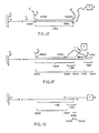
- FIG 3A illustrates a fourth embodiment of the present invention with the reagent complex entirely in solution (no support is present).
- the probe polynucleotide P has a target binding region (TBR) including a subregion, the labeled polynucleotide binding region (LBR), in which bases are bound to complementary bases of the labeled polynucleotide (L).
- the labeled polynucleotide (L) contains a fluorescent tag (F) held in the complex in close proximity to a quencher moiety (Q) attached to the probe (P).
- F fluorescent tag
- Q quencher moiety
- These moieties F and Q are sufficiently close in the complex of Figure 3A for any signal produced by stimulation of the fluorescent tag (F) by radiation to be absorbed by the quencher moiety (Q).
- An exemplary pair of F and Q are fluoroscein with rhodamine. See M. Cobb and S. Gotcher, Am. J. Med. Tech., Vol. 48,
- Figure 3B illustrates the reagent complex of Figure 3A after contact by a sample G containing the base sequence complementary to the entire target binding sequence (TBR).
- TBR target binding sequence
- the tag (F) is now sufficiently far from quencher moiety (Q) to produce a detectable signal on stimulation that is not quenched (except in those rare cases where another probe P happens to be located such that its label Q is in close proximity to tag F). Accordingly, the stimulation of fluorescent tag (F) and measurement of unquenched signals can serve as a quantitative detection method for displaced labeled polynucleotides without a separation step having been performed.
- FIG. 3D A modification in the embodiment of Figures 3A-3C in which the labeled polynucleotide L and probe polynucleotide P (bearing, respectively, labels F and Q) are part of the same molecule is illustrated in Figure 3D.
- a site X is shown which contains an inverted repeat sequence a portion of which is recognizable by a restriction enzyme. Cleavage by such an enzyme can be used to sever the covalent linkage between TBR and L (although this severing is not required when the read-out is based upon the F-Q interaction). Such cleavage can also be used as a first step in creating a site for attachment of the probe to a support, such attachment to occur before or after displacement.
- the overall geometry of Figure 3D may thus be used in synthesizing reagent complexes as a single molecule for various embodiments, including those of Figures 1A-1E.
- Figure 4 illustrates a complex in which the probe strand P 3 is a circular single-stranded polynucleotide non-specifically adsorbed on support S.
- a polynucleotide can be produced by insertion of a target binding region (TBR), into a single-stranded bacteriophage such as M13 through suitable cloning procedures.
- TBR target binding region
- the labeled polynucleotide L here contains a segment A' 1 complementary only in a portion LBR of segment TBR.
- Figure 6 illustrates a complex similar to the embodiment of Figure 1A in that the probe strand P 5 contains a target binding region TBR near or at its free end.
- a number of portions of the TBR segment are each base paired to a complementary region of a labeled polynucleotide; segment A' 3 of labeled polynucleotide L 3 to subsequent A3, etc.
- Probe P 5 contains a target binding region TBR divided into subsegments A 1 and A 2 , together forming the initial binding region, as well as eight additional subsegments (A3 thru A 10 ), each forming a labeled polynucleotide binding region.
- binding of segment A' of the sample polynucleotide G (as shown in Figure 1 E) will displace all of the labeled polynucleotides L 3 , L 4 , L 5 , L 6 , L 7 , L 8 , L 9 and L 10 and thus produce an enhanced signal compared to the situation shown in Figure 1A.
- the final labeled polynucleotide a labeled 175 base long single stranded molecule, was then purified on a denaturing agarose gel by electrophoresis.
- the purified labeled polynucleotide was hybridized to the above full-length M13mp9-albumin DNA strand which serves as the probe polynucleotide; the probe used was either circular or had been linearized with Bam HI.
- Unhybridized labeled polynucleotide was removed from the reagent complex by passage over two successive Sepharose@ CL-4B columns.
- Model competitor DNAs used in strand displacement experiments were either the complementary DNA strand to the human albumin sequence of the M13mp9-albumin clone inserted in the vector M13mp8 (and hereafter designated as M13mp8-albumin), or a denatured double-stranded 840 base pair Eco RI-Bgl human DNA fragment which is colinear with the insert in the M13mp9-albumin clone.
- the displacement reactions were carried out by incubating approximately 10 nanograms of the reagent complex containing a circular or linear probe polynucleotide with 10 or 50 nanograms of competitor DNA.
- the reactions were stopped and the DNA deproteinated by adding chemical reagents to raise the final sample solution concentrations to 20mM EDTA, 0.4% SDS and 0.4 mg/mi proteinase K and incubating at 37°C for an additional 30 minutes.
- the amount of displacement was assayed by separating the reagent complex from displaced strands on a 2% agarose gel by electrophoresis, and autoradiographing the gel overnight.
- a 10-fofd molar excess of this competitor when linearized resulted in the displacement of only about 10% of the labeled polynucleotide even in the presence of rec A protein.
- Increasing the competitor DNA concentration to a 40-fold molar excess resulted in displacement of up to approximately 40% of the labeled polynucleotide. It is likely that this reaction was less efficient (with this embodiment of separation and detection) than strand displacement induced by single-stranded competitor DNA due to interferences from reannealing of the two self-complementary strands of the competitor DNA (or of the inactive competitor strand with displaced labelled polynucleotide).
- a reagent complex was prepared and purified as described in Example 1 using the mp9albumin- polylinker template and the labeled 175 nucleotide primer-extended DNA described in Example 1, except that the circular probe DNA of the reagent complex was not cleaved with Bam HI. All displacement reactions were done in 20mM Tris, pH 7.5, 25 mM MgC1 2 , 0.1 mg/ml BSA, and 0.3 mM DTT in a final volume of 25 ul. The ATP concentration was varied in this experiment to determine the effect of this parameter.
- This inhibition may be due to low levels of ADP contamination in the ATP preparation, or to rapid accumulation of ADP in the sample from hydrolysis of ATP by rec A protein.
- Analogous ADP inhibition of rec A activity has been previously described in M. Cox et al., J. Biol. Chem., vol. 258, pp. 2586-2592 (1983); see also Shibata et al., J. BioJ. Chem., vol. 256, pp. 7565-7572 (1981).
- a reagent complex was prepared using the linearized reagent complex described in Example 1.
- the model competitor DNA (M13mp8-albumin DNA) was again the polynucleotide strand complementary to the human albumin insert cloned into M13 (part of the reagent complex). All displacement reactions were carried out essentially as in Example 2, except that the amounts of analyte DNA, ATP, and rec A protein as well as the time period of incubation were varied. Approximately 10 ng of reagent complex were used in each reaction sample. After the addition of ATP, competitor DNA (equimolar or 1 x, or 10-fold molar excess or 10x) and rec A protein, samples were incubated at 37°C for 15, 30 or 60 minutes.
- the probe used in this experiment was prepared by subcloning the 650 base pair Eco RI-Sal I fragment from pBR322 into the M13mp8 vector. Also inserted into this bacteriophage at the Eco RI site is the polylinkerfrom M13mp7 which is a small inverted repeat containing several restriction enzyme cleavage sites. Single stranded circular M13mp8pBR6PL6 was isolated and digested with Bam HI, an enzyme which cuts within the hairpin loop of the polylinker and linearizes the single stranded DNA.
- a 32p end-labeled polynucleotide was synthesized by kinasing an oligonulceotide which is complementary to the insert in mp8pBR6PL6. Primer extension and cleavage with EcoRl resulted in a 350 base end-labelled DNA fragment extending from the BamHl site to the EcoRl site in mp8pBR6PL6. This labeled polynucleotide was then purified by electrophoresis on a denaturing agarose gel.
- Strand displacement reactions were carried out essentially as in Example 1, except that all reaction mixtures contained 10 mM ATP.
- varying amounts of E. coli rec A protein were added and allowed to preincubate with the displacement complex at 37°C for 2 minutes. Competitor DNA was then added and the reactions proceeded until they were halted after 15 or 60 minutes (only these two times of incubation were assayed) and were deproteinated as described in Example 1.
- the addition of 10 ng of competitor DNA to a displacement reaction mixture containing an equimolar amount of reagent complex resulted in the displacement of approximately 20% of the labeled polynucleotide in 60 minutes in the presence of 15 or 30 pmoles of E.
- the displacement from this reagent probe complex may be less efficient than that in Examples 2 or 3 due to one or more of the several differences in the geometry of the several reagent complex constructs (involving different absolute and relative lengths of initial binding region, different positioning of the labeled polynucleotide binding region at the 5' end of or interior of the target binding region or the size of the labeled polynucleotide binding region). It was in limited parallel runs to those of this Example 4 that the addition of single-stranded binding protein from E. coli (0.5 pmoles) appeared to inhibit the appearance of displaced labeled polynucleotide mediated by E. coli rec A protein.
- M13mp9 bacteriophage recombinant molecules used in this and the following Examples 6-14 as probe DNA had a 1.1 kb nucleotide insert (Pstl-BmmHl fragment) from pBR322 oriented with the Bam HI site located in the 3' direction relative to the Pstl site.
- the circular M13mp9 clone 11-16 DNA was linearized and fragmented before hybridization; this was done by incubating the DNA with the restriction enzyme Hinfl (about 2 units per ug DNA) at 37°C for 3-4 hours.
- the labeled polynucleotide used in this Example is one completely complementary to the 27 nucleotides at the 3' end of the pBR322 insert in M13mp9 clone 11-16 (i.e., adjacent to the BamHl recognition site).
- nucleotide sequence of the region of the probe DNA which is completely homologous to the labeled polynucleotide and the sequence of the labeled polynucleotide are shown in Table A above (probe and oligomer L1).
- the 27-mer polynucleotide was ratioisotopically end-labeled with 32p using T4 polynucleotide kinase. Unincorporated gamma-[3zP] ATP was then removed from labeled oligonucleotide preparation by chromatography on a Whatman DE52 column.
- a DNA-DNA hybrid (reagent complex) was formed between the 27 nucleotide oligomer (labeled polynucleotide) and the M13mp9 clone 11-16 recombinant DNA (probe molecule).
- Hybridization to produce this reagent complex was carried out by mixing the probe DNA with a molar excess (usually five fold) of labeled polynucleotide in 5x SSC (750 mM sodium chloride. 75 mM sodium citrate). This mixture was incubated at 50-55°C for 15-50 minutes before reagent complex was then separated from unhybridized labeled polynucleotide on a Sepharose@ 4B column.
- simulated sample or analyte DNA containing target nucleotide sequences was introduced into a nucleic acid strand displacement reaction mixture under varying conditions as described below.
- the analyte DNA also referred to as competitor DNA in this Example and subsequent Examples
- M13mp8 clone 20 DNA which had a DNA insert consisting of the complementary strand to the pBR322 insert in bacteriophage M13mp9 clone 11-16 as discussed above.
- reaction mixtures were separated by electrophoresis on a 15% polyacrylamide gel to distinguish unbound (free and presumably displaced) labeled polynucleotide from any reagent complex molecules.
- the gel was autoradiographed and the distribution of 32 P label in the resolved sample bands was determined by densitometry using a Shimadzu CS930 plate scanner.
- rec A protein can increase the extent of unbound polynucleotide observed following strand displacement occurring due to the presence of specific target polynucleotides.
- Example 5 Comparing Example 5 (and those Examples that follow) with Examples 1-4, one can see a demonstration of the beneficial effects of E. coli rec A protein with both long (up to 275 base) and short (27 base) labeled polynucleotide binding regions. In addition, in Example 5 (and those that follow) no proteinase digestion step was conducted at the conclusion of the displacement reaction.
- Example 5 compares polynucleotide strand displacement at 37°C with and without rec A protein using two different types of analytical gel. Another experiment was conducted using the reagent complex preparation and the reaction conditions of Example 5 to examine the need for functional E. coli rec A in such strand displacement reactions and to compare strand displacement at 37°C using rec A protein to displacement under more conventional nucleic acid hybridization conditions (55°C in 2x SSC). The latter temperature is approximately T m -13'C for the reagent complex and is consistent with theoretical prediction of a temperature at or near the optimum for effective DNA-DNA strand hybridization in a simple salt buffer.
- reaction mixtures with different contents were prepared and incubated at 37°C as described in Example 5; samples for analysis were removed at 30 minute intervals beginning at 0 minutes and ending at 2.5 hours. Both the probe polynucleotide in the reagent complex and the competitor DNA were linearized with Hinf I. The concentration of E. coli rec A protein was a 40-fold weight excess over the reagent complex in all samples.
Landscapes
- Chemical & Material Sciences (AREA)
- Life Sciences & Earth Sciences (AREA)
- Organic Chemistry (AREA)
- Proteomics, Peptides & Aminoacids (AREA)
- Health & Medical Sciences (AREA)
- Zoology (AREA)
- Engineering & Computer Science (AREA)
- Wood Science & Technology (AREA)
- Analytical Chemistry (AREA)
- Biochemistry (AREA)
- Bioinformatics & Cheminformatics (AREA)
- Molecular Biology (AREA)
- Immunology (AREA)
- Biotechnology (AREA)
- Physics & Mathematics (AREA)
- Biophysics (AREA)
- Microbiology (AREA)
- General Engineering & Computer Science (AREA)
- General Health & Medical Sciences (AREA)
- Genetics & Genomics (AREA)
- Chemical Kinetics & Catalysis (AREA)
- Measuring Or Testing Involving Enzymes Or Micro-Organisms (AREA)
- Investigating Or Analysing Biological Materials (AREA)
- Saccharide Compounds (AREA)
- Peptides Or Proteins (AREA)
Claims (15)
wobei die potentielle Basenpaarbindung zwischen der Zielnucleotidsequenz und dem Prüfpolynucleotid in der Lage ist, das markierte Polynucleotid aus dem Reagenzkomplex zu verdrängen,
Priority Applications (1)
| Application Number | Priority Date | Filing Date | Title |
|---|---|---|---|
| AT85303176T ATE33151T1 (de) | 1984-05-07 | 1985-05-03 | Polynukleotidanalyse durch verschiebung unter verwendung eines rekombinationsproteins und reagenzsatz. |
Applications Claiming Priority (4)
| Application Number | Priority Date | Filing Date | Title |
|---|---|---|---|
| US607885 | 1984-05-07 | ||
| US06/607,885 US4766062A (en) | 1984-05-07 | 1984-05-07 | Displacement polynucleotide assay method and polynucleotide complex reagent therefor |
| US68430584A | 1984-12-20 | 1984-12-20 | |
| US684305 | 1984-12-20 |
Publications (2)
| Publication Number | Publication Date |
|---|---|
| EP0164876A1 EP0164876A1 (de) | 1985-12-18 |
| EP0164876B1 true EP0164876B1 (de) | 1988-03-23 |
Family
ID=27085615
Family Applications (1)
| Application Number | Title | Priority Date | Filing Date |
|---|---|---|---|
| EP19850303176 Expired EP0164876B1 (de) | 1984-05-07 | 1985-05-03 | Polynukleotidanalyse durch Verschiebung unter Verwendung eines Rekombinationsproteins und Reagenzsatz |
Country Status (4)
| Country | Link |
|---|---|
| EP (1) | EP0164876B1 (de) |
| JP (1) | JPH0632638B2 (de) |
| CA (1) | CA1254114A (de) |
| DE (1) | DE3561955D1 (de) |
Families Citing this family (24)
| Publication number | Priority date | Publication date | Assignee | Title |
|---|---|---|---|---|
| JP2609238B2 (ja) * | 1985-05-02 | 1997-05-14 | ジエネテイツクス・インスチチユ−ト・インコ−ポレ−テツド | 標的ヌクレオチド配列の検定に有用な試薬複合体を作製するための方法および核酸構築物 |
| US4767699A (en) * | 1985-05-02 | 1988-08-30 | Allied Corporation | Diagnostic reagent, kit and method employing polynucleotide displacement, separation, enzymatic cleavage and adenosine phosphate detection |
| US4795701A (en) * | 1985-07-17 | 1989-01-03 | Allied Corporation | Homogeneous polynucleotide displacement assay method kit and reagent complex |
| EP0219842A1 (de) * | 1985-10-23 | 1987-04-29 | Allied Corporation | Nukleinsäuretest mittels Segmentpaarhemmung oder -konkurrenz |
| WO1987003911A1 (en) * | 1985-12-17 | 1987-07-02 | Genetics Institute, Inc. | Displacement polynucleotide method and reagent complex |
| EP0232967B1 (de) * | 1986-01-10 | 1993-04-28 | Amoco Corporation | Kompetitiver homogener Test |
| US4900659A (en) * | 1986-01-30 | 1990-02-13 | Enzo Biochem, Inc. | Nucleotide sequence composition and method for detection of Neisseria gonorrhoeae and method for screening for a nucleotide sequence that is specific for a genetically distinct group |
| US7087742B1 (en) | 1986-11-24 | 2006-08-08 | Gen-Probe Incorporated | Oligonucleotide probes for the detection and/or quantitation of non-viral organisms |
| US7090972B1 (en) | 1986-11-24 | 2006-08-15 | Gen-Probe Incorporated | Methods for determining the presence of non-viral organisms in a sample |
| DE3888653T2 (de) * | 1987-12-21 | 1994-07-07 | Applied Biosystems | Verfahren und Testsatz zum Nachweis einer Nukleinsäure-Sequenz. |
| US6326136B1 (en) * | 1988-04-01 | 2001-12-04 | Digene Corporation | Macromolecular conjugate made using unsaturated aldehydes |
| US7172863B1 (en) | 1988-12-09 | 2007-02-06 | Gen-Probe Incorporated | Nucleic acid probes and methods for detecting Neisseria gonorrhoeae |
| JPH03123500A (ja) * | 1989-10-06 | 1991-05-27 | Canon Inc | 核酸の分別方法 |
| GB9002625D0 (en) * | 1990-02-06 | 1990-04-04 | Univ Singapore | Human leukocyte antigen typing |
| CA2037349C (en) | 1990-03-26 | 2008-06-17 | James G. Wetmur | Branch migration of nucleotides |
| US5273881A (en) * | 1990-05-07 | 1993-12-28 | Daikin Industries, Ltd. | Diagnostic applications of double D-loop formation |
| JP2745816B2 (ja) * | 1990-05-07 | 1998-04-28 | ダイキン工業株式会社 | ダブルdループ形成の診断応用 |
| DE69214243T2 (de) * | 1991-09-23 | 1997-02-06 | Pfizer | Verfahren für die Detektion von spezifische mRNS und DNS in Zellen |
| EP0614988A1 (de) * | 1993-02-24 | 1994-09-14 | Hoechst Aktiengesellschaft | Verfahren für den Nachweis von schnell-hybridierenden einzelsträngigen Polynukleotiden |
| DE69633941T2 (de) * | 1995-12-22 | 2005-11-24 | Dade Behring Marburg Gmbh | Nachweis von unterschieden in nukleinsäuren |
| US20020012916A1 (en) * | 1998-10-14 | 2002-01-31 | Gundling Gerard J | A method of reducing contamination in an essay vessel |
| US6232104B1 (en) | 1999-08-17 | 2001-05-15 | Dade Behring Inc. | Detection of differences in nucleic acids by inhibition of spontaneous DNA branch migration |
| US7005265B1 (en) | 2002-06-20 | 2006-02-28 | Wenhong Fan | Nonenzymatic catalytic signal amplification for nucleic acid hybridization assays |
| JP4984990B2 (ja) * | 2006-03-28 | 2012-07-25 | 富士通株式会社 | 機能性分子の製造方法 |
Family Cites Families (6)
| Publication number | Priority date | Publication date | Assignee | Title |
|---|---|---|---|---|
| US4302204A (en) * | 1979-07-02 | 1981-11-24 | The Board Of Trustees Of Leland Stanford Junior University | Transfer and detection of nucleic acids |
| US4358535A (en) * | 1980-12-08 | 1982-11-09 | Board Of Regents Of The University Of Washington | Specific DNA probes in diagnostic microbiology |
| CA1190838A (en) * | 1981-07-17 | 1985-07-23 | Cavit Akin | Homogeneous nucleic acid hybridization diagnostics by non-radiative energy transfer |
| US4395486A (en) * | 1981-08-19 | 1983-07-26 | Medical College Of Ga. Research Inst., Inc. | Method for the direct analysis of sickle cell anemia |
| GB8306426D0 (en) * | 1983-03-09 | 1983-04-13 | Malcolm A D B | Detecting polynucleotide sequence |
| CA1222680A (en) * | 1983-07-05 | 1987-06-09 | Nanibhushan Dattagupta | Testing dna samples for particular nucleotide sequences |
-
1985
- 1985-05-02 CA CA000480559A patent/CA1254114A/en not_active Expired
- 1985-05-03 DE DE8585303176T patent/DE3561955D1/de not_active Expired
- 1985-05-03 EP EP19850303176 patent/EP0164876B1/de not_active Expired
- 1985-05-07 JP JP60096554A patent/JPH0632638B2/ja not_active Expired - Lifetime
Also Published As
| Publication number | Publication date |
|---|---|
| JPH0632638B2 (ja) | 1994-05-02 |
| JPS60244300A (ja) | 1985-12-04 |
| EP0164876A1 (de) | 1985-12-18 |
| DE3561955D1 (en) | 1988-04-28 |
| CA1254114A (en) | 1989-05-16 |
Similar Documents
| Publication | Publication Date | Title |
|---|---|---|
| EP0164876B1 (de) | Polynukleotidanalyse durch Verschiebung unter Verwendung eines Rekombinationsproteins und Reagenzsatz | |
| US4766062A (en) | Displacement polynucleotide assay method and polynucleotide complex reagent therefor | |
| EP0167238B1 (de) | Verfahren zur Polynukleotidanalyse durch Verschiebung und Polynukleotidkomplexreagenz dafür | |
| US5102784A (en) | Restriction amplification assay | |
| Shuman | DNA strand transfer reactions catalyzed by vaccinia topoisomerase I. | |
| US5660988A (en) | Cycling probe cleavage detection of nucleic acid sequences | |
| US6013439A (en) | Detection of differences in nucleic acids | |
| US5210015A (en) | Homogeneous assay system using the nuclease activity of a nucleic acid polymerase | |
| US4725537A (en) | Assay, reagent and kit employing nucleic acid strand displacement and restriction endonuclease cleavage | |
| EP0549107A1 (de) | Verfahren zur Herstellung eines Polynukleotids zum Gebrauch in "single-primer" Amplifizierung und Phosphorothioat-enthaltende Oligonukleotide als Primer in Nukleinsäureamplifizierung | |
| JP2002536981A (ja) | 核酸標的配列の存在を測定する方法およびその応用 | |
| JP2002515737A (ja) | 核酸の侵入的開裂 | |
| JPH09512428A (ja) | リゾルベース開裂による突然変異の検出 | |
| JPH11510369A (ja) | 鋳型依存性生産物の形成による核酸の検出 | |
| JPH022934A (ja) | テンプレート−依存性核酸プロ−ブ再構成を用いたアッセイ | |
| CA2308368C (en) | Specific and sensitive nucleic acid detection method | |
| US5538872A (en) | Method of preparing nucleotide probes using a bridging complement | |
| JPH11506932A (ja) | 点変異の検出及びインビトロ突然変異誘発のための核酸修復酵素方法 | |
| WO2005083114A1 (en) | Method, kit and system for enhanced nested pcr | |
| US6492120B1 (en) | Nucleic acid hybridization assay utilizing tricyclic target and signal amplification | |
| WO2000043543A1 (en) | Detection of differences between polynucleotides | |
| US20070248965A1 (en) | Compositions and methods for the detection of a nucleic acid using circular probes in a cleavage reaction | |
| JPS62143700A (ja) | 対合セグメント抑制または競合による核酸アツセイ法 | |
| WO1993010264A1 (en) | Restriction amplification assay | |
| JPH08299A (ja) | 標的核酸に対する相補鎖核酸のハイブリダイゼーションの促進法及びこれを用いた核酸の検出法 |
Legal Events
| Date | Code | Title | Description |
|---|---|---|---|
| PUAI | Public reference made under article 153(3) epc to a published international application that has entered the european phase |
Free format text: ORIGINAL CODE: 0009012 |
|
| AK | Designated contracting states |
Designated state(s): AT BE CH DE FR GB IT LI NL SE |
|
| 17P | Request for examination filed |
Effective date: 19860117 |
|
| 17Q | First examination report despatched |
Effective date: 19860901 |
|
| GRAA | (expected) grant |
Free format text: ORIGINAL CODE: 0009210 |
|
| AK | Designated contracting states |
Kind code of ref document: B1 Designated state(s): AT BE CH DE FR GB IT LI NL SE |
|
| PG25 | Lapsed in a contracting state [announced via postgrant information from national office to epo] |
Ref country code: NL Effective date: 19880323 Ref country code: LI Effective date: 19880323 Ref country code: CH Effective date: 19880323 Ref country code: BE Effective date: 19880323 Ref country code: AT Effective date: 19880323 |
|
| REF | Corresponds to: |
Ref document number: 33151 Country of ref document: AT Date of ref document: 19880415 Kind code of ref document: T |
|
| ITF | It: translation for a ep patent filed |
Owner name: GUZZI E RAVIZZA S.R.L. |
|
| PG25 | Lapsed in a contracting state [announced via postgrant information from national office to epo] |
Ref country code: SE Effective date: 19880331 |
|
| REF | Corresponds to: |
Ref document number: 3561955 Country of ref document: DE Date of ref document: 19880428 |
|
| ET | Fr: translation filed | ||
| REG | Reference to a national code |
Ref country code: CH Ref legal event code: PL |
|
| NLV1 | Nl: lapsed or annulled due to failure to fulfill the requirements of art. 29p and 29m of the patents act | ||
| PLBE | No opposition filed within time limit |
Free format text: ORIGINAL CODE: 0009261 |
|
| STAA | Information on the status of an ep patent application or granted ep patent |
Free format text: STATUS: NO OPPOSITION FILED WITHIN TIME LIMIT |
|
| 26N | No opposition filed | ||
| REG | Reference to a national code |
Ref country code: GB Ref legal event code: 732 |
|
| ITTA | It: last paid annual fee | ||
| PGFP | Annual fee paid to national office [announced via postgrant information from national office to epo] |
Ref country code: GB Payment date: 19980522 Year of fee payment: 14 |
|
| PGFP | Annual fee paid to national office [announced via postgrant information from national office to epo] |
Ref country code: DE Payment date: 19980528 Year of fee payment: 14 |
|
| PGFP | Annual fee paid to national office [announced via postgrant information from national office to epo] |
Ref country code: FR Payment date: 19980529 Year of fee payment: 14 |
|
| PG25 | Lapsed in a contracting state [announced via postgrant information from national office to epo] |
Ref country code: GB Free format text: LAPSE BECAUSE OF NON-PAYMENT OF DUE FEES Effective date: 19990503 |
|
| GBPC | Gb: european patent ceased through non-payment of renewal fee |
Effective date: 19990503 |
|
| PG25 | Lapsed in a contracting state [announced via postgrant information from national office to epo] |
Ref country code: FR Free format text: LAPSE BECAUSE OF NON-PAYMENT OF DUE FEES Effective date: 20000131 |
|
| PG25 | Lapsed in a contracting state [announced via postgrant information from national office to epo] |
Ref country code: DE Free format text: LAPSE BECAUSE OF NON-PAYMENT OF DUE FEES Effective date: 20000301 |
|
| REG | Reference to a national code |
Ref country code: FR Ref legal event code: ST |