EP0154734B1 - Immediate ligand detection assay, a test kit and its formation - Google Patents
Immediate ligand detection assay, a test kit and its formation Download PDFInfo
- Publication number
- EP0154734B1 EP0154734B1 EP84301769A EP84301769A EP0154734B1 EP 0154734 B1 EP0154734 B1 EP 0154734B1 EP 84301769 A EP84301769 A EP 84301769A EP 84301769 A EP84301769 A EP 84301769A EP 0154734 B1 EP0154734 B1 EP 0154734B1
- Authority
- EP
- European Patent Office
- Prior art keywords
- reactant
- fluorescer
- ligand
- radiolabelled
- support particles
- Prior art date
- Legal status (The legal status is an assumption and is not a legal conclusion. Google has not performed a legal analysis and makes no representation as to the accuracy of the status listed.)
- Expired
Links
- 239000003446 ligand Substances 0.000 title claims abstract description 100
- 238000003556 assay Methods 0.000 title claims abstract description 41
- 238000012360 testing method Methods 0.000 title claims abstract description 18
- 238000001514 detection method Methods 0.000 title claims description 12
- 230000015572 biosynthetic process Effects 0.000 title 1
- 239000000376 reactant Substances 0.000 claims abstract description 149
- 230000002285 radioactive effect Effects 0.000 claims abstract description 30
- 239000002245 particle Substances 0.000 claims description 46
- 238000000034 method Methods 0.000 claims description 43
- IAZDPXIOMUYVGZ-UHFFFAOYSA-N Dimethylsulphoxide Chemical compound CS(C)=O IAZDPXIOMUYVGZ-UHFFFAOYSA-N 0.000 claims description 22
- XLYOFNOQVPJJNP-UHFFFAOYSA-N water Substances O XLYOFNOQVPJJNP-UHFFFAOYSA-N 0.000 claims description 22
- 238000009739 binding Methods 0.000 claims description 21
- 230000027455 binding Effects 0.000 claims description 20
- 239000012736 aqueous medium Substances 0.000 claims description 15
- 230000005855 radiation Effects 0.000 claims description 14
- 239000002904 solvent Substances 0.000 claims description 11
- 239000000725 suspension Substances 0.000 claims description 9
- 239000007900 aqueous suspension Substances 0.000 claims description 4
- VUPXKQHLZATXTR-UHFFFAOYSA-N 2,4-diphenyl-1,3-oxazole Chemical group C=1OC(C=2C=CC=CC=2)=NC=1C1=CC=CC=C1 VUPXKQHLZATXTR-UHFFFAOYSA-N 0.000 claims description 3
- 230000003213 activating effect Effects 0.000 claims description 3
- 230000004913 activation Effects 0.000 claims description 3
- 239000012530 fluid Substances 0.000 claims description 3
- 230000002860 competitive effect Effects 0.000 claims description 2
- 238000004806 packaging method and process Methods 0.000 claims 1
- 239000011324 bead Substances 0.000 abstract description 82
- 239000007864 aqueous solution Substances 0.000 abstract description 8
- 239000011368 organic material Substances 0.000 description 27
- 239000000427 antigen Substances 0.000 description 23
- 108091007433 antigens Proteins 0.000 description 23
- 102000036639 antigens Human genes 0.000 description 23
- 229940027941 immunoglobulin g Drugs 0.000 description 18
- 102000004169 proteins and genes Human genes 0.000 description 17
- 108090000623 proteins and genes Proteins 0.000 description 17
- 239000000243 solution Substances 0.000 description 12
- 239000000463 material Substances 0.000 description 10
- 229920002684 Sepharose Polymers 0.000 description 9
- 229920000126 latex Polymers 0.000 description 7
- 239000002953 phosphate buffered saline Substances 0.000 description 7
- 238000001556 precipitation Methods 0.000 description 7
- DHMQDGOQFOQNFH-UHFFFAOYSA-N Glycine Chemical compound NCC(O)=O DHMQDGOQFOQNFH-UHFFFAOYSA-N 0.000 description 6
- PXIPVTKHYLBLMZ-UHFFFAOYSA-N Sodium azide Chemical compound [Na+].[N-]=[N+]=[N-] PXIPVTKHYLBLMZ-UHFFFAOYSA-N 0.000 description 6
- ATDGTVJJHBUTRL-UHFFFAOYSA-N cyanogen bromide Chemical compound BrC#N ATDGTVJJHBUTRL-UHFFFAOYSA-N 0.000 description 6
- 238000002372 labelling Methods 0.000 description 6
- 239000004816 latex Substances 0.000 description 6
- 238000005406 washing Methods 0.000 description 6
- 108090000790 Enzymes Proteins 0.000 description 5
- 102000004190 Enzymes Human genes 0.000 description 5
- -1 antibodies Substances 0.000 description 5
- 238000012875 competitive assay Methods 0.000 description 5
- 239000005556 hormone Substances 0.000 description 5
- 229940088597 hormone Drugs 0.000 description 5
- 230000005764 inhibitory process Effects 0.000 description 5
- 239000000700 radioactive tracer Substances 0.000 description 5
- 239000002901 radioactive waste Substances 0.000 description 5
- 238000003127 radioimmunoassay Methods 0.000 description 5
- 239000000126 substance Substances 0.000 description 5
- 239000003153 chemical reaction reagent Substances 0.000 description 4
- 239000012847 fine chemical Substances 0.000 description 4
- 229940098197 human immunoglobulin g Drugs 0.000 description 4
- 229920000136 polysorbate Polymers 0.000 description 4
- 238000000926 separation method Methods 0.000 description 4
- 238000012546 transfer Methods 0.000 description 4
- 239000004471 Glycine Substances 0.000 description 3
- 239000004793 Polystyrene Substances 0.000 description 3
- YZCKVEUIGOORGS-NJFSPNSNSA-N Tritium Chemical compound [3H] YZCKVEUIGOORGS-NJFSPNSNSA-N 0.000 description 3
- 230000004520 agglutination Effects 0.000 description 3
- 238000007818 agglutination assay Methods 0.000 description 3
- 239000000872 buffer Substances 0.000 description 3
- 238000006243 chemical reaction Methods 0.000 description 3
- 230000005284 excitation Effects 0.000 description 3
- 238000011534 incubation Methods 0.000 description 3
- XMBWDFGMSWQBCA-YPZZEJLDSA-N iodane Chemical compound [125IH] XMBWDFGMSWQBCA-YPZZEJLDSA-N 0.000 description 3
- 229940044173 iodine-125 Drugs 0.000 description 3
- 238000005259 measurement Methods 0.000 description 3
- 239000002609 medium Substances 0.000 description 3
- 150000002894 organic compounds Chemical class 0.000 description 3
- 229920002223 polystyrene Polymers 0.000 description 3
- 238000011160 research Methods 0.000 description 3
- 229910052722 tritium Inorganic materials 0.000 description 3
- OKTJSMMVPCPJKN-UHFFFAOYSA-N Carbon Chemical compound [C] OKTJSMMVPCPJKN-UHFFFAOYSA-N 0.000 description 2
- 108010010803 Gelatin Proteins 0.000 description 2
- VEXZGXHMUGYJMC-UHFFFAOYSA-N Hydrochloric acid Chemical compound Cl VEXZGXHMUGYJMC-UHFFFAOYSA-N 0.000 description 2
- 108060003951 Immunoglobulin Proteins 0.000 description 2
- XEEYBQQBJWHFJM-AKLPVKDBSA-N Iron-59 Chemical compound [59Fe] XEEYBQQBJWHFJM-AKLPVKDBSA-N 0.000 description 2
- 241000283973 Oryctolagus cuniculus Species 0.000 description 2
- OAICVXFJPJFONN-UHFFFAOYSA-N Phosphorus Chemical compound [P] OAICVXFJPJFONN-UHFFFAOYSA-N 0.000 description 2
- 239000002202 Polyethylene glycol Substances 0.000 description 2
- UIIMBOGNXHQVGW-UHFFFAOYSA-M Sodium bicarbonate Chemical compound [Na+].OC([O-])=O UIIMBOGNXHQVGW-UHFFFAOYSA-M 0.000 description 2
- FAPWRFPIFSIZLT-UHFFFAOYSA-M Sodium chloride Chemical compound [Na+].[Cl-] FAPWRFPIFSIZLT-UHFFFAOYSA-M 0.000 description 2
- 125000004429 atom Chemical group 0.000 description 2
- 239000003599 detergent Substances 0.000 description 2
- 230000000694 effects Effects 0.000 description 2
- 238000005189 flocculation Methods 0.000 description 2
- 230000016615 flocculation Effects 0.000 description 2
- 239000000499 gel Substances 0.000 description 2
- 239000008273 gelatin Substances 0.000 description 2
- 229920000159 gelatin Polymers 0.000 description 2
- 235000019322 gelatine Nutrition 0.000 description 2
- 235000011852 gelatine desserts Nutrition 0.000 description 2
- 102000018358 immunoglobulin Human genes 0.000 description 2
- 229940072221 immunoglobulins Drugs 0.000 description 2
- 238000011835 investigation Methods 0.000 description 2
- 239000002207 metabolite Substances 0.000 description 2
- 238000012544 monitoring process Methods 0.000 description 2
- 230000009871 nonspecific binding Effects 0.000 description 2
- 230000003287 optical effect Effects 0.000 description 2
- 229920001223 polyethylene glycol Polymers 0.000 description 2
- 229910052761 rare earth metal Inorganic materials 0.000 description 2
- 150000002910 rare earth metals Chemical class 0.000 description 2
- 238000002791 soaking Methods 0.000 description 2
- CIOAGBVUUVVLOB-UHFFFAOYSA-N strontium atom Chemical group [Sr] CIOAGBVUUVVLOB-UHFFFAOYSA-N 0.000 description 2
- TUSDEZXZIZRFGC-UHFFFAOYSA-N 1-O-galloyl-3,6-(R)-HHDP-beta-D-glucose Natural products OC1C(O2)COC(=O)C3=CC(O)=C(O)C(O)=C3C3=C(O)C(O)=C(O)C=C3C(=O)OC1C(O)C2OC(=O)C1=CC(O)=C(O)C(O)=C1 TUSDEZXZIZRFGC-UHFFFAOYSA-N 0.000 description 1
- PENWAFASUFITRC-UHFFFAOYSA-N 2-(4-chlorophenyl)imidazo[2,1-a]isoquinoline Chemical compound C1=CC(Cl)=CC=C1C1=CN(C=CC=2C3=CC=CC=2)C3=N1 PENWAFASUFITRC-UHFFFAOYSA-N 0.000 description 1
- HRPVXLWXLXDGHG-UHFFFAOYSA-N Acrylamide Chemical compound NC(=O)C=C HRPVXLWXLXDGHG-UHFFFAOYSA-N 0.000 description 1
- 229920000936 Agarose Polymers 0.000 description 1
- 241000894006 Bacteria Species 0.000 description 1
- 208000019838 Blood disease Diseases 0.000 description 1
- OKTJSMMVPCPJKN-NJFSPNSNSA-N Carbon-14 Chemical group [14C] OKTJSMMVPCPJKN-NJFSPNSNSA-N 0.000 description 1
- 239000001263 FEMA 3042 Substances 0.000 description 1
- SXRSQZLOMIGNAQ-UHFFFAOYSA-N Glutaraldehyde Chemical compound O=CCCCC=O SXRSQZLOMIGNAQ-UHFFFAOYSA-N 0.000 description 1
- 206010020751 Hypersensitivity Diseases 0.000 description 1
- 238000000636 Northern blotting Methods 0.000 description 1
- LRBQNJMCXXYXIU-PPKXGCFTSA-N Penta-digallate-beta-D-glucose Natural products OC1=C(O)C(O)=CC(C(=O)OC=2C(=C(O)C=C(C=2)C(=O)OC[C@@H]2[C@H]([C@H](OC(=O)C=3C=C(OC(=O)C=4C=C(O)C(O)=C(O)C=4)C(O)=C(O)C=3)[C@@H](OC(=O)C=3C=C(OC(=O)C=4C=C(O)C(O)=C(O)C=4)C(O)=C(O)C=3)[C@H](OC(=O)C=3C=C(OC(=O)C=4C=C(O)C(O)=C(O)C=4)C(O)=C(O)C=3)O2)OC(=O)C=2C=C(OC(=O)C=3C=C(O)C(O)=C(O)C=3)C(O)=C(O)C=2)O)=C1 LRBQNJMCXXYXIU-PPKXGCFTSA-N 0.000 description 1
- 239000004743 Polypropylene Substances 0.000 description 1
- 229920001213 Polysorbate 20 Polymers 0.000 description 1
- 238000002105 Southern blotting Methods 0.000 description 1
- 101000582398 Staphylococcus aureus Replication initiation protein Proteins 0.000 description 1
- 239000008351 acetate buffer Substances 0.000 description 1
- WFDIJRYMOXRFFG-UHFFFAOYSA-N acetic anhydride Substances CC(=O)OC(C)=O WFDIJRYMOXRFFG-UHFFFAOYSA-N 0.000 description 1
- 230000007815 allergy Effects 0.000 description 1
- 238000004458 analytical method Methods 0.000 description 1
- 230000000844 anti-bacterial effect Effects 0.000 description 1
- 230000001580 bacterial effect Effects 0.000 description 1
- 239000003899 bactericide agent Substances 0.000 description 1
- 238000004166 bioassay Methods 0.000 description 1
- 239000002981 blocking agent Substances 0.000 description 1
- 230000000903 blocking effect Effects 0.000 description 1
- 210000001124 body fluid Anatomy 0.000 description 1
- 239000010839 body fluid Substances 0.000 description 1
- 239000007853 buffer solution Substances 0.000 description 1
- 239000007975 buffered saline Substances 0.000 description 1
- 150000001718 carbodiimides Chemical class 0.000 description 1
- 150000001721 carbon Chemical group 0.000 description 1
- 229910052799 carbon Inorganic materials 0.000 description 1
- 239000013522 chelant Substances 0.000 description 1
- 239000003795 chemical substances by application Substances 0.000 description 1
- VDQQXEISLMTGAB-UHFFFAOYSA-N chloramine T Chemical compound [Na+].CC1=CC=C(S(=O)(=O)[N-]Cl)C=C1 VDQQXEISLMTGAB-UHFFFAOYSA-N 0.000 description 1
- 239000011248 coating agent Substances 0.000 description 1
- 238000000576 coating method Methods 0.000 description 1
- 150000001875 compounds Chemical class 0.000 description 1
- 239000000470 constituent Substances 0.000 description 1
- 238000012258 culturing Methods 0.000 description 1
- 230000003247 decreasing effect Effects 0.000 description 1
- 230000001419 dependent effect Effects 0.000 description 1
- 238000003745 diagnosis Methods 0.000 description 1
- LOKCTEFSRHRXRJ-UHFFFAOYSA-I dipotassium trisodium dihydrogen phosphate hydrogen phosphate dichloride Chemical compound P(=O)(O)(O)[O-].[K+].P(=O)(O)([O-])[O-].[Na+].[Na+].[Cl-].[K+].[Cl-].[Na+] LOKCTEFSRHRXRJ-UHFFFAOYSA-I 0.000 description 1
- 201000010099 disease Diseases 0.000 description 1
- 208000037265 diseases, disorders, signs and symptoms Diseases 0.000 description 1
- 229940079593 drug Drugs 0.000 description 1
- 239000003814 drug Substances 0.000 description 1
- 238000000295 emission spectrum Methods 0.000 description 1
- 238000002474 experimental method Methods 0.000 description 1
- MHMNJMPURVTYEJ-UHFFFAOYSA-N fluorescein-5-isothiocyanate Chemical compound O1C(=O)C2=CC(N=C=S)=CC=C2C21C1=CC=C(O)C=C1OC1=CC(O)=CC=C21 MHMNJMPURVTYEJ-UHFFFAOYSA-N 0.000 description 1
- 230000005251 gamma ray Effects 0.000 description 1
- 231100001261 hazardous Toxicity 0.000 description 1
- 239000000383 hazardous chemical Substances 0.000 description 1
- 231100000206 health hazard Toxicity 0.000 description 1
- 208000014951 hematologic disease Diseases 0.000 description 1
- 208000018706 hematopoietic system disease Diseases 0.000 description 1
- 208000006454 hepatitis Diseases 0.000 description 1
- 231100000283 hepatitis Toxicity 0.000 description 1
- 108091008039 hormone receptors Proteins 0.000 description 1
- 125000004435 hydrogen atom Chemical group [H]* 0.000 description 1
- 230000002209 hydrophobic effect Effects 0.000 description 1
- 230000001900 immune effect Effects 0.000 description 1
- 210000000987 immune system Anatomy 0.000 description 1
- 208000015181 infectious disease Diseases 0.000 description 1
- 229910010272 inorganic material Inorganic materials 0.000 description 1
- 239000011147 inorganic material Substances 0.000 description 1
- 238000004020 luminiscence type Methods 0.000 description 1
- 238000004519 manufacturing process Methods 0.000 description 1
- 238000002156 mixing Methods 0.000 description 1
- 239000000203 mixture Substances 0.000 description 1
- 238000012806 monitoring device Methods 0.000 description 1
- 230000010807 negative regulation of binding Effects 0.000 description 1
- 239000003960 organic solvent Substances 0.000 description 1
- 229920002401 polyacrylamide Polymers 0.000 description 1
- 229920000515 polycarbonate Polymers 0.000 description 1
- 239000004417 polycarbonate Substances 0.000 description 1
- 235000010486 polyoxyethylene sorbitan monolaurate Nutrition 0.000 description 1
- 239000000256 polyoxyethylene sorbitan monolaurate Substances 0.000 description 1
- 229920001155 polypropylene Polymers 0.000 description 1
- 230000001376 precipitating effect Effects 0.000 description 1
- 230000035935 pregnancy Effects 0.000 description 1
- 230000002028 premature Effects 0.000 description 1
- 238000003908 quality control method Methods 0.000 description 1
- 238000004445 quantitative analysis Methods 0.000 description 1
- 239000000941 radioactive substance Substances 0.000 description 1
- 239000011541 reaction mixture Substances 0.000 description 1
- 239000012487 rinsing solution Substances 0.000 description 1
- 238000003345 scintillation counting Methods 0.000 description 1
- 238000004062 sedimentation Methods 0.000 description 1
- 230000035945 sensitivity Effects 0.000 description 1
- 210000002966 serum Anatomy 0.000 description 1
- 229910000030 sodium bicarbonate Inorganic materials 0.000 description 1
- 235000017557 sodium bicarbonate Nutrition 0.000 description 1
- 239000011780 sodium chloride Substances 0.000 description 1
- 239000007787 solid Substances 0.000 description 1
- 239000007790 solid phase Substances 0.000 description 1
- 238000001179 sorption measurement Methods 0.000 description 1
- 229910052712 strontium Inorganic materials 0.000 description 1
- 239000000758 substrate Substances 0.000 description 1
- 230000008961 swelling Effects 0.000 description 1
- 230000002194 synthesizing effect Effects 0.000 description 1
- LRBQNJMCXXYXIU-NRMVVENXSA-N tannic acid Chemical compound OC1=C(O)C(O)=CC(C(=O)OC=2C(=C(O)C=C(C=2)C(=O)OC[C@@H]2[C@H]([C@H](OC(=O)C=3C=C(OC(=O)C=4C=C(O)C(O)=C(O)C=4)C(O)=C(O)C=3)[C@@H](OC(=O)C=3C=C(OC(=O)C=4C=C(O)C(O)=C(O)C=4)C(O)=C(O)C=3)[C@@H](OC(=O)C=3C=C(OC(=O)C=4C=C(O)C(O)=C(O)C=4)C(O)=C(O)C=3)O2)OC(=O)C=2C=C(OC(=O)C=3C=C(O)C(O)=C(O)C=3)C(O)=C(O)C=2)O)=C1 LRBQNJMCXXYXIU-NRMVVENXSA-N 0.000 description 1
- 229940033123 tannic acid Drugs 0.000 description 1
- 235000015523 tannic acid Nutrition 0.000 description 1
- 229920002258 tannic acid Polymers 0.000 description 1
- 239000002699 waste material Substances 0.000 description 1
- 238000001262 western blot Methods 0.000 description 1
Images
Classifications
-
- G—PHYSICS
- G01—MEASURING; TESTING
- G01N—INVESTIGATING OR ANALYSING MATERIALS BY DETERMINING THEIR CHEMICAL OR PHYSICAL PROPERTIES
- G01N33/00—Investigating or analysing materials by specific methods not covered by groups G01N1/00 - G01N31/00
- G01N33/48—Biological material, e.g. blood, urine; Haemocytometers
- G01N33/50—Chemical analysis of biological material, e.g. blood, urine; Testing involving biospecific ligand binding methods; Immunological testing
- G01N33/53—Immunoassay; Biospecific binding assay; Materials therefor
- G01N33/531—Production of immunochemical test materials
- G01N33/532—Production of labelled immunochemicals
- G01N33/533—Production of labelled immunochemicals with fluorescent label
-
- G—PHYSICS
- G01—MEASURING; TESTING
- G01N—INVESTIGATING OR ANALYSING MATERIALS BY DETERMINING THEIR CHEMICAL OR PHYSICAL PROPERTIES
- G01N33/00—Investigating or analysing materials by specific methods not covered by groups G01N1/00 - G01N31/00
- G01N33/48—Biological material, e.g. blood, urine; Haemocytometers
- G01N33/50—Chemical analysis of biological material, e.g. blood, urine; Testing involving biospecific ligand binding methods; Immunological testing
- G01N33/53—Immunoassay; Biospecific binding assay; Materials therefor
- G01N33/531—Production of immunochemical test materials
- G01N33/532—Production of labelled immunochemicals
- G01N33/534—Production of labelled immunochemicals with radioactive label
-
- G—PHYSICS
- G01—MEASURING; TESTING
- G01N—INVESTIGATING OR ANALYSING MATERIALS BY DETERMINING THEIR CHEMICAL OR PHYSICAL PROPERTIES
- G01N33/00—Investigating or analysing materials by specific methods not covered by groups G01N1/00 - G01N31/00
- G01N33/48—Biological material, e.g. blood, urine; Haemocytometers
- G01N33/50—Chemical analysis of biological material, e.g. blood, urine; Testing involving biospecific ligand binding methods; Immunological testing
- G01N33/53—Immunoassay; Biospecific binding assay; Materials therefor
- G01N33/536—Immunoassay; Biospecific binding assay; Materials therefor with immune complex formed in liquid phase
- G01N33/542—Immunoassay; Biospecific binding assay; Materials therefor with immune complex formed in liquid phase with steric inhibition or signal modification, e.g. fluorescent quenching
Definitions
- the present invention relates to an assay for detecting the presence of small amounts of organic material, and more particularly to an assay to detect the presence of organic materials by monitoring the light energy produced when radioactive organic molecules of interest biochemically and specifically bind to a binding structure.
- organic material such as antigens, antibodies, hormones, metabolites, enzymes, and drugs.
- detecting the presence or absence of metabolites, hormones, or other organic factors in serum or in other body fluids may be useful in the diagnosis of many clinical conditions, such as pregnancy, infection, blood disorders, hepatitis, etc.
- the detection and quantitative analysis of organic agents is often required in immunological, biological, chemical, or other types of scientific and medical research.
- assays for organic materials are utilized in quality control procedures for the production of chemicals and in monitoring the pollution of water.
- precipitation and agglutination assays are of numerous chemical and biological assays.
- the organic material interacts with a reactant to form a complex that falls out of solution.
- the organic substance of interest cross-links an insoluble reactant to cause the reactant to flocculate.
- Optical scattering techniques are commonly used to measure the flocculation.
- a drawback of precipitation and agglutination assays is that they are not as sensitive as radioimmunoassays.
- optical techniques have been developed to improve the sensitivity of these assays, these techniques require specialized equipment and analysis.
- Another type of known assay for organic materials involves labeling either the organic material or a reactant thereto with a radioactive, fluorescent or other type of tracer substance to ascertain the extent to which the organic material has coupled with its reactant.
- Radioimmunoassay is one of the most common types of these "tracer" assays. Radioimmunoassay involves combining a known amount of radiolabeled organic material with a sample containing an unknown amount of unlabeled organic material of interest together with a specific antibody that binds indiscriminantly to the labeled and unlabeled organic materials to form a complex. After an incubation period, the unbound organic materials are separated from the bound organic materials, typically by precipitation of the complex with polyethylene glycol, adsorption of the unbound material with activated charcoal or utilization of solid-phase reagents. Then the radioactivity of either of these two fractions is measured. A certain amount of the labeled and unlabeled organic material will be bound to the reactant, with the amount of the bound labeled organic material being inversely related to the quantity of unlabeled inorganic material present in the sample being tested.
- a drawback of the radioimmunoassay is that the procedures for separating the bound organic materials from the unbound require a significant number of time-consuming operations that are often complicated and expensive.
- the separation procedures involve repeatedly washing the complex of organic material and antibody with a rinsing solution and/or centrifuging the mixture to remove the unbound organic material from the reactant, thereby generating radioactive waste material with each washing.
- This waste material is not only expensive to dispose of, but also presents a potential health hazard to persons handling the material, including during the separation procedures.
- the organic material may be labeled with an appropriate fluorescer, such as fluorescein isothiocyanate.
- an appropriate fluorescer such as fluorescein isothiocyanate.
- the extent to which the fluorescer labeled organic materials bound to a specific reactant can be examined under a light microscope with a suitable light source and filters to provide incident light of the proper wavelength to cause fluorescence.
- a tracer assay that utilizes both fluorescence and radioactive substances is disclosed by U.S. Patent 4,000,252 wherein a known quantity of radiolabeled antigen and a sample containing an unknown amount of unlabeled antigen are placed within an immunoscintillation cell.
- An insolubilized or solid phosphor which is chemically or physically associated with an antibody to the antigens, is also added to the cell.
- the unbound antigens are washed from the cell and then the luminescence emitted by the phosphor due to activation from the radioactive energy from the bound labeled antigens is measured inside a scintillation counter. Removal of the unbound antibodies from the cell requires several washing procedures that are not only time consuming, but also produce significant quantities of radioactive waste material.
- tritiated (radioactive) latex particles coated with antigen and polystyrene scintillant particles coated with the same antigen were placed in an aqueous medium with a sample containing an unknown quantity of a corresponding antibody.
- the number of tritiated antigen coated latex particles linked to the antigen coated scintillant particles is related to the concentration of antibody present. Also, when the two particles are linked together by the antibody, the radioactive energy from the tritiated particles initiates scintillation within the scintillant particles. Scintillations are then measured by an appropriate detector, with the detected level of scintillation being indicative of the quantity of the antibody present.
- US-A-4382074 is a continuation-in-part of US-A-4271139 and describes a similar agglutination assay with an alternative embodiment of adding free antigen to the sample to compete with the antigen- coated particles for the antibody to be assayed.
- US ⁇ A ⁇ 4259313 discloses polymeric latices which have fluorescent rare earth chelates incorporated into the latex beads.
- the beads are added to a solution of the rare earth chelate in a water-miscible solvent, and then water added to the latex to transfer the hydrophobic fluorescer into the beads.
- a particular object of the present invention is to provide an assay of equivalent accuracy to present techniques, but which does not require highly skilled personnel or large amounts of time to perform using standard commercially available equipment.
- a further particular, but highly important object of the present invention is to provide an assay procedure that produces only a minimum volume of radioactive waste and requires only a minimum amount of handling of hazardous substances.
- An additional particular object of the present invention is to provide an assay that can be used to rapidly test a large number of samples.
- Another particular object of the present invention is to provide an assay that utilizes water as a suspension medium.
- a process for the detection of a radiolabelled reactant comprising:
- a second aspect of the invention relates to a competitive reactant assay process, comprising:
- a third aspect of the present invention relates to a test kit for the detection of a ligand reactant that biochemically and specifically binds to a ligand in an aqueous medium with all of the components of the test kit together in the aqueous medium, said test kit comprising:
- the objects are achieved in accordance with the present invention by providing a test kit and assay procedure which produces light energy at a level related to the amount of organic material present in a sample being tested.
- the light energy is produced by a fluorescer which is integrated into support bodies in the form of beads or other structures.
- the support bodies are coated with a ligand that is capable of specifically binding to the organic material or reactant of interest.
- the reactant is radiolabelled.
- the sample containing the radiolabelled reactant is mixed in an aqueous solution with the support bodies, causing the reactant to bind to the ligand.
- the level of the light energy produced is related to the amount of reactant that is bound to the ligand, which in turn, is indicative of the amount of reactant present in the sample being tested.
- the present invention may also be utilized as a competitive assay to determine the amount of a reactant contained in a sample.
- a known amount of the reactant is radiolabeled.
- the reactant of interest in the sample being tested remains unlabeled.
- Both the labeled and unlabeled reactant are capable of specifically binding with the ligand.
- both the labeled and unlabeled organic materials are placed in an aqueous solution, together with the support bodies that have been impregnated with the fluorescer and coated with the ligand. Since the ligand does not favor either the labeled or unlabeled reactant, the reactants bind to the ligand in proportion to their relative amounts present in the aqueous solution.
- the radiolabeled reactant that binds to the ligand is brought in close enough proximity to the support bodies to cause the radiation energy emitted thereby to activate the fluorescer integrated into the support bodies.
- the level of light energy produced by the fluorescer is inversely proportioned to the quantity of unlabeled reactant present in the sample.
- a unique technique for integrating the fluorescer into the support bodies. Initially, the support bodies are soaked in a solvent for the fluorescer which is miscible in water to dehydrate the bodies. Thereafter, the bodies are placed in a solution composed of the fluorescer and solvent so that the fluorescer is integrated and/or adsorbed into the bodies. Then, the bodies are removed from the solvent and then placed in an aqueous solution which causes precipitation of the fluorescer within the bodies, thereby locking the fluorescer therein.
- the fluorescer is integrated within the interior of the bodies so that the radiolabeled reactant is placed in very close proximity to the fluorescer upon binding to the ligand, which is disposed on the exterior of the bodies.
- support bodies or particles in the form of beads or other structures are impregnated and/or coated with a material capable of fluorescence when excited by radioactive energy.
- the beads are coated with a ligand that is capable of specifically binding to a reactant of interest by covalently linking or directly attaching the ligand to the beads.
- the beads are then mixed in a water-based solution containing the reactant and has been radiolabeled.
- the fluorescer integrated into the beads is placed in close enough physical relationship to the reactant to allow the radiation energy emitted from the reactant to activate the fluorescer thereby causing the fluorescer to emit light energy.
- the level of light energy emitted which is indicative of the extent to which the ligand is bound to its reactant, may be conveniently measured with a scintillation counter or other monitoring device employing a photomultiplier tube.
- the radiation energy emitted by the radiolabeled reactant molecules has a very limited range of travel in water.
- the reactant molecules that have not bound to the ligand are, for the most part, located too far away from the ligand to enable the radiation energy emitted from these unbound reactant molecules to reach the fluorescer in the support structure, i.e., beads. Since there is very little likelihood of chance excitation of the fluorescer by the radioactive energy of these unbound reactant molecules, the reactant molecules that have not bound to the ligand need not be separated from the ligand-reactant complexes prior to scintillation counting of the light energy emitted by the fluorescer excited by radioactive energy from the reactant molecules that have bound to the ligand. Thus, the traditional laborious and potentially hazardous procedure of separating the unbound reactant from the ligand-reactant complexes is eliminated.
- the present invention may also be used in conjunction with a competitive assay procedure.
- the support bodies having a fluorescer integrated therein and coated with an appropriate ligand, are placed in an aqueous solution containing a known quantity of radiolabeled reactant and a sample containing an unknown amount of the same, but unlabeled reactant. Since the ligand does not favor binding to either the labeled or unlabeled reactant over the other, the amount of labeled reactant binding to the ligand will be inversely proportional to the quantity of unlabeled reactant present in the sample.
- the radiolabeled reactant molecules that are bound to the ligand are in close enough proximity to the beads to allow the radiation energy emitted by the labeled reactant to bombard the fluorescer integrated into the beads.
- the detected level of fluorescent energy therefore, is a reflection of the proportion of the radiolabeled reactant which actually binds to the ligand.
- the level of fluorescent energy may be measured will all of the components of the assay still present in the vial.
- the time required to complete the assay is limited only by the ligand-reactant binding reaction rate of the system under investigation.
- the ligand is bonded to and the fluorescer is integrated with a structural support, such as beads.
- a structural support such as beads.
- Various types of beads may be utilized, such as polyacrylamide, acrylamide, agarose, polystyrene, polypropylene, polycarbonate or Sepharose° 4B beads (from Pharmacia Fine Chemicals, Uppsala, Sweden).
- the present invention also may be carried out with other shapes or types of support structures, for instance latex particles, as long as the ligand molecules can be covalently or otherwise attached thereto and a fluorescer integrated therewith.
- Beads such as the Sepharose * 4B beads noted above, are commercially available in an activated state. Compounds, such as cyanogen bromide, are incorporated in the beads to covalently bind with certain ligands. The process by which the ligand is bound to the beads is dependent on the type of bead and the particular ligand employed. For instance, for Sepharose® 4B beads activated with cyanogen-bromide, a ligand in the form of Staphylococcus aureus protein A or an antibody to a specific reactant may be bound to the beads by placing the beads in a solution containing the protein A or antibody and an appropriate buffer.
- a fluorescer is integrated within the beads to give off light energy when radiolabeled reactant is brought in close enough proximity to the fluorescer to cause excitation thereof, i.e., by binding to the ligand on the bead surface.
- fluorescers may be used; however, since the process of the present invention takes place in an aqueous solution, the fluorescer must be insoluble in water so that it does not dissociate from the beads during the assay procedure.
- the fluorescer employed must be excitable to a higher energy state by the particular wavelength of the radioactive energy rays emitted by the radiolabeled reactant, and also must release sufficient light energy when returning to its normal energy state to be detected by a scintillation counter or other detection device utilizing a photomultiplier tube.
- An example of a fluorescer that has been found to meet these requirements for use with radioactive energy in the form of beta rays or auger electrons is diphenyloxazole (hereinafter "PPO").
- the present invention involves a novel process for integrating a fluorescer into a support structure, such as beads. Since the fluorescer is insoluble in water, an appropriate transfer medium, in which the fluorescer is soluble, must be used to incorporate the fluorescer into the beads. Moreover, the transfer medium itself must be miscible with water.
- the novel method of the present invention for integrating the fluorescer into the beads includes soaking the beads in an appropriate transfer solvent for the fluorescer that is miscible with water thereby to dehydrate the beads. Thereafter, the beads are incubated in a solution of the fluorescer and solvent. The fluorescer, which is in solution, is absorbed into the bead. The excess solvent is then discarded and the fluorescer is precipitated, adsorbed and/or integrated inside the bead by adding water or a buffered saline solution. Precipitating the fluorescer within the beads locks the fluorescer therein.
- the beads are washed to remove the excess precipitated fluorescer and then resuspended in a solution containing both a detergent, such as Tween@ 20, to prevent the beads from sticking together and gelatin to bind to any sites on the surface of the beads that either have not been blocked by the previously employed blocking solution or bound to a ligand. This lowers the nonspecific binding tendency of the beads.
- a bactericide such as sodium azide, can be applied to the beads to prevent the growth of bacteria thereon.
- DMSO dimethyl sulfoxide
- the process of the present invention requires the use of radiolabeled reactants.
- the radiolabeled reactants are biologically and chemically identical to an unlabled reactant, with the exception that the labeled reactants emit radioactive energy due to the decaying of the radioactive isotope present.
- labeling can be accomplished by replacing one of the atoms of the reactant molecules with a corresponding radioactive isotope.
- a hydrogen atom could be replaced with: tritium, 3 H; a carbon atom replaced with carbon-14, 14 C; or a strontium atom replaced with strontium-38, 38 Sr.
- an isotope may be added to the reactant molecule.
- radioactive isotopes in common use include: iodine-125, 1251; and iron-59, 59 Fe.
- labeling can be carried out by culturing the organism with an appropriate radiolabeled precurser, such as methionine-35 ( 35 S), to cause the organism to incorporate the isotope into its products.
- radiolabeled precurser such as methionine-35 ( 35 S)
- 35 S methionine-35
- Many reactants such as antigens, antibodies, hormones, hormone receptors, enzymes, or enzyme cofactors, are readily available in radiolabeled form from various commercial sources.
- Radioactive isotopes used to label the reactant have only a limited range in water so that, for the most part, only the radioactive energy from the labeled reactant that binds to the ligand actually activates the fluorescer. If PPO is used as a fluorescer, applicant has found that isotopes that emit either beta rays or auger electrons from gamma ray emissions fulfill this requirement. PPO does not fluoresce from the gamma rays themselves which have a longer range of travel than beta rays or auger electrons.
- the assay process of the present invention may be utilized in conjunction with any ligand-reactant combination or system that specifically binds together and in which the reactant may be radiolabeled without affecting its specificity for the ligand.
- ligand-reactant combinations include antibodies and their corresponding antigens. Either the antibody or antigen may be attached to the bead or other type of support structure to function as the ligand, with the corresponding antigen or antibody serving as the reactant.
- Another ligand-reactant system may be composed or protein A and corresponding immunoglobulins. The need to ascertain the presence of antigens, antibodies, and immunoglobulins exists in many clinical and research settings, especially in the detection of diseases and allergies and in investigations of the immune system.
- Additional ligand-reactant systems with which the present invention is especially useful include: 1) lectins-glycoproteins; 2) biotin-avidin; 3) hormone receptor-hormone; 4) enzyme-substrate or cofactor; 5) RNA-DNA; and 6) DNA-DNA. It is to be understood that in the present invention either element may serve as the ligand or reactant.
- the present invention also may be of particular value in conducting enzyme kinetic studies.
- the enzyme may serve as a ligand to bind with the radiolabeled reactant. Since all of the reagents of the assay system are always present together in the same vial and because the reagents are suspended in an aqueous-based buffer rather than an organic solvent, kinetic experiments may be conveniently carried out by simply measuring the light energy emitted from the same vial at different time intervals to determine the reaction rate of the reagents. Also; in the RNA-DNA system, the commonly used Northern, Southern and Western Blot tests may be advantageously replaced with the assay of the present invention.
- cyanogen-bromide activated Sepharose m 4B beads obtained from Pharmacia Fine Chemicals, Uppsala, Sweden
- Staphyloccus aureus cowan strain 1 protein A Pharmacia Fine Chemicals, Uppsala, Sweden
- anti-immunoglobulin-G antibody This is accomplished by swelling and washing one gram of cyanogen-bromide activated Sepharose® 4B beads in one millimolar hydrochloric acid.
- a fluorescer in the form of PPO is next incorporated into the beads. This is accomplished by dehydrating the coated beads by soaking them in DMSO for 15 minutes to remove any water in the beads that would cause premature precipitation of the PPO since PPO is insoluble in water. This step is repeated twice more and then the excess solvent removed by sedimentation.
- the beads are next incubated for 30 minutes at room temperature in a 20 to 40 percent weight by volume solution of PPO in DMSO. After incubation, the excess solution is discarded and the PPO precipitated inside the beads by adding ten volumes of either phosphate buffered saline (hereinafter "PBS") or water. The suspension is then washed five times in PBS to remove the excess precipitated PPO which is not bound inside the beads.
- PBS phosphate buffered saline
- the beads are resuspended at a final concentration of ten percent volume/volume in PBS supplemented with 0.5 percent volume/volume Tween 20 (Registered Trade Mark) as a detergent to help prevent the beads from sticking together and 0.1 percent weight/volume gelatin to bind to the remaining sites on the beads which were not blocked by reaction with protein A, immunoglobulin-G antibody or the glycine. This minimizes any nonspecific binding of radiolabeled reactant to the beads.
- sodium azide, NaN 3 in an amount of 0.01 percent weight/volume, can be added to prevent bacterial growth.
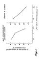
- Example 2 is identical with Example 1, except that the protein A or anti-immunoglobulin-G antibody coated beads were mixed with various concentrations of human immunoglobulin-G labeled with 125 I to a specific activity of 8 x 10 4 counts per minute per millimolar by the Chloramine T method.
- the protein A or anti-immunoglobulin-G antibody coated beads were placed in scintillation vials with various concentrations of 121 1 labeled human immunoglobulin-G and then the vials were placed directly into a scintillation counter to measure the resulting level of photon emission. The results of these tests are shown in Table II for 5-minute and 60-minute incubation periods. These data confirm that the assay can be measured immediately after mixing labeled reactant with PPO-impregnated ligand bound beads.
- Cyanogen-bromide activated Sepharose a 4B beads (Pharmacia Fine Chemicals, Uppsala, Sweden) are coated with either protein A (0.1 milligrams per milliliter of gel) or anti-immunoglobulin-G antibody (1.5 milligrams per milliliter of gel) in the manner set forth in Example I. Also, the Sepharose 4B beads are impregnated with the fluorescer PPO in the manner discussed in Example I. Fifty-microliter quantities of the prepared beads are mixed together in scintillation vials with 0.5 micrograms of 125 I-human immunoglobin-G and increasing concentrations of unlabeled human immunoglobulin-G. The samples are then brought up to three milliliters each by adding PBS supplemented with 0.5 percent volume/volume of Tween®. The sample is then immediately counted for photon emission levels.
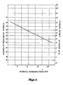
- This Example involves the use of the present invention to develop a standard inhibition curve for the detection of thyroxin.
- Cyanogen-bromide activated Sepharose a 4B beads were coated with protein A and then impregnated with PPO in the manner described in Example I.
- Two hundred microliters of the prepared beads were pipetted into individual scintillation vials together with 400 microliters of a rabbit anti-thyroxin antiserum (obtained from Abbott Laboratories, Chicago, IL). Twenty-five microliters of various concentrations of unlabeled thyroxin (obtained from Abbott Laboratories, Chicago, IL) were added to each vial.
- This standard inhibition curve can be used to determine the amount of thyroxin present in an unknown sample by using the same protocol described above in Example III to determine the percent of radiolabeled thyroxin that is inhibited from binding on the beads. From this value, the amount of thyroxin present in the sample may be conveniently read from the curve.
Landscapes
- Health & Medical Sciences (AREA)
- Immunology (AREA)
- Life Sciences & Earth Sciences (AREA)
- Engineering & Computer Science (AREA)
- Molecular Biology (AREA)
- Biomedical Technology (AREA)
- Chemical & Material Sciences (AREA)
- Hematology (AREA)
- Urology & Nephrology (AREA)
- Biotechnology (AREA)
- Biochemistry (AREA)
- Cell Biology (AREA)
- Food Science & Technology (AREA)
- Medicinal Chemistry (AREA)
- Physics & Mathematics (AREA)
- Analytical Chemistry (AREA)
- Microbiology (AREA)
- General Health & Medical Sciences (AREA)
- General Physics & Mathematics (AREA)
- Pathology (AREA)
- Investigating Or Analysing Biological Materials (AREA)
- Investigating Or Analysing Materials By The Use Of Chemical Reactions (AREA)
- Saccharide Compounds (AREA)
Abstract
Description
- The present invention relates to an assay for detecting the presence of small amounts of organic material, and more particularly to an assay to detect the presence of organic materials by monitoring the light energy produced when radioactive organic molecules of interest biochemically and specifically bind to a binding structure.
- In clinical applications, in research and in industry, a need exists to detect the presence of minute amounts of organic material, such as antigens, antibodies, hormones, metabolites, enzymes, and drugs. In clinical situations, detecting the presence or absence of metabolites, hormones, or other organic factors in serum or in other body fluids may be useful in the diagnosis of many clinical conditions, such as pregnancy, infection, blood disorders, hepatitis, etc. The detection and quantitative analysis of organic agents is often required in immunological, biological, chemical, or other types of scientific and medical research. In industry, assays for organic materials are utilized in quality control procedures for the production of chemicals and in monitoring the pollution of water.
- Of the numerous chemical and biological assays that have been developed to detect organic materials, of relevance to the present invention are precipitation and agglutination assays. In a typical precipitation assay, the organic material interacts with a reactant to form a complex that falls out of solution. In agglutination reactions, the organic substance of interest cross-links an insoluble reactant to cause the reactant to flocculate. Optical scattering techniques are commonly used to measure the flocculation. A drawback of precipitation and agglutination assays is that they are not as sensitive as radioimmunoassays. Also, although optical techniques have been developed to improve the sensitivity of these assays, these techniques require specialized equipment and analysis.
- Another type of known assay for organic materials involves labeling either the organic material or a reactant thereto with a radioactive, fluorescent or other type of tracer substance to ascertain the extent to which the organic material has coupled with its reactant.
- Radioimmunoassay is one of the most common types of these "tracer" assays. Radioimmunoassay involves combining a known amount of radiolabeled organic material with a sample containing an unknown amount of unlabeled organic material of interest together with a specific antibody that binds indiscriminantly to the labeled and unlabeled organic materials to form a complex. After an incubation period, the unbound organic materials are separated from the bound organic materials, typically by precipitation of the complex with polyethylene glycol, adsorption of the unbound material with activated charcoal or utilization of solid-phase reagents. Then the radioactivity of either of these two fractions is measured. A certain amount of the labeled and unlabeled organic material will be bound to the reactant, with the amount of the bound labeled organic material being inversely related to the quantity of unlabeled inorganic material present in the sample being tested.
- A drawback of the radioimmunoassay is that the procedures for separating the bound organic materials from the unbound require a significant number of time-consuming operations that are often complicated and expensive. The separation procedures involve repeatedly washing the complex of organic material and antibody with a rinsing solution and/or centrifuging the mixture to remove the unbound organic material from the reactant, thereby generating radioactive waste material with each washing. By the time that the separation process has been completed, significant volumes of radioactive waste material are produced. This waste material is not only expensive to dispose of, but also presents a potential health hazard to persons handling the material, including during the separation procedures.
- In an assay utilizing fluorescence, the organic material may be labeled with an appropriate fluorescer, such as fluorescein isothiocyanate. The extent to which the fluorescer labeled organic materials bound to a specific reactant can be examined under a light microscope with a suitable light source and filters to provide incident light of the proper wavelength to cause fluorescence.
- An example of a particular fluorescence technique is disclosed in U.S. Patent 4,161,515 wherein an unknown organic compound, whose presence is being investigated, is mixed with: 1) a known quantity of antibody against the organic compound; 2) an organic analog, having a fluorescer bound thereto, which competes with the unknown organic compound for the antibody; and 3) an antibody for the fluorescer. The competition between the unkown organic material and the known analog-fluorescer effectively reduces the concentration of the antibody, thereby causing more of the fluorescer-antibody to combine with the analog-fluorescer. This, in turn, causes a corresponding change in the emission spectrum of the fluorescer.
- A tracer assay that utilizes both fluorescence and radioactive substances is disclosed by U.S. Patent 4,000,252 wherein a known quantity of radiolabeled antigen and a sample containing an unknown amount of unlabeled antigen are placed within an immunoscintillation cell. An insolubilized or solid phosphor, which is chemically or physically associated with an antibody to the antigens, is also added to the cell. The unbound antigens are washed from the cell and then the luminescence emitted by the phosphor due to activation from the radioactive energy from the bound labeled antigens is measured inside a scintillation counter. Removal of the unbound antibodies from the cell requires several washing procedures that are not only time consuming, but also produce significant quantities of radioactive waste material.
- Assays that combine tracer techniques with agglutination are disclosed by U.S. Patents 4,108,972 and 4,271,139. In the '972 patent, microscopic carrier particles, each containing a fluorescent tracer material, and a biological reactant to the antibody or antigen being investigated, are placed in suspension. When the antigen or antibody is added to the suspension, it binds to the reactant coating to cause flocculation of the carrier particles. The flocculated material is then separated from the suspension fluid and other constituents by numerous washings. Thereafter, the flocculated material is dissolved and then assayed by fluorescence techniques to determine the quantity of organic material present.
- In the '139 patent, tritiated (radioactive) latex particles coated with antigen and polystyrene scintillant particles coated with the same antigen were placed in an aqueous medium with a sample containing an unknown quantity of a corresponding antibody. The number of tritiated antigen coated latex particles linked to the antigen coated scintillant particles is related to the concentration of antibody present. Also, when the two particles are linked together by the antibody, the radioactive energy from the tritiated particles initiates scintillation within the scintillant particles. Scintillations are then measured by an appropriate detector, with the detected level of scintillation being indicative of the quantity of the antibody present. Addition of an unknown quantity of non-radioactive antigen then competes with the antigen coated bead binding to the antibody, thereby reducing bead agglutination, scintillation and signal. A drawback of this particular assay is that it requires both tritiated latex particles and polystyrene scintillant particles to be coated with antigen, which increases the expense and complexity of the assay. In addition, relative to the standard radioimmunoassay discussed above, extremely large suspension volumes are required for the assay to operate properly. The assay process of the '139 patent also requires the availability of relatively pure antigen to be used to bind to the two types of carrier particles. Relatively pure samples of antigen are both expensive and difficult to obtain.
- US-A-4382074 is a continuation-in-part of US-A-4271139 and describes a similar agglutination assay with an alternative embodiment of adding free antigen to the sample to compete with the antigen- coated particles for the antibody to be assayed.
- US―A―4259313 discloses polymeric latices which have fluorescent rare earth chelates incorporated into the latex beads. The beads are added to a solution of the rare earth chelate in a water-miscible solvent, and then water added to the latex to transfer the hydrophobic fluorescer into the beads.
- Thus, it is a principal object of the present invention to provide an accurate, inexpensive, and rapid assay procedure and test kit for detecting the presence of extremely small amounts of organic materials.
- A particular object of the present invention is to provide an assay of equivalent accuracy to present techniques, but which does not require highly skilled personnel or large amounts of time to perform using standard commercially available equipment.
- A further particular, but highly important object of the present invention, is to provide an assay procedure that produces only a minimum volume of radioactive waste and requires only a minimum amount of handling of hazardous substances.
- An additional particular object of the present invention is to provide an assay that can be used to rapidly test a large number of samples.
- Another particular object of the present invention is to provide an assay that utilizes water as a suspension medium.
- Therefore, according to a first aspect of the present invention there is provided a process for the detection of a radiolabelled reactant comprising:
- a) placing in an aqueous suspension a plurality of support particles impregnated with a fluorescer and to which ligands have been attached;
- b) adding to the suspension fluid a radiolabelled reactant capable of specifically biochemically binding to the ligand, said radiolabelled reactant emitting radiation energy capable of activating the fluorescer upon the binding of the reactant to the ligand, the radiolabelled reactant then being disposed in close enough proximity to the support particles to cause the radiation energy from the radiolabelled reactant to activate the fluorescer to produce light energy, whereas any radiolabelled reactant that does not bind to the ligand is generally too far removed from the support particles to enable the radioactive energy to activate the fluorescer; and
- c) measuring the light energy emitted by the fluorescer with the entire quantities of the support particles, bound and unbound radiolabelled reactant together in aqueous suspension.
- A second aspect of the invention relates to a competitive reactant assay process, comprising:
- a) combining together in an aqueous medium:
- a sample to be assayed containing an unknown amount of cold ligand reactant;
- a known quantity of radiolabelled ligand reactant; and
- a plurality of support particles having fluorescer integrated therewith capable of emitting photons when activated by the radiation energy emitted by the radiolabelled ligand reactant and having ligand attached to the outer surface thereof, said ligand capable of indiscriminately binding with both said cold and radiolabelled ligand reactant whereupon the binding of the ligand with the radiolabelled ligand reactant positions the radiolabelled ligand reactant close enough to the support particles to activate the fluorescer to emit photons through the aqueous medium, whereas the unbound radiolabelled ligand reactant is generally positioned too far away from the support particles to enable the radioactive energy emitted thereby to activate the fluorescer; and
- b) measuring the photons emitted by the fluorescer with the entire quantities of cold ligand reactant containing sample, bound and unbound radiolabelled ligand reactant and support particles combined together in the aqueous medium.
- A third aspect of the present invention relates to a test kit for the detection of a ligand reactant that biochemically and specifically binds to a ligand in an aqueous medium with all of the components of the test kit together in the aqueous medium, said test kit comprising:
- a) support particles, preferably in a predetermined quantity, having a fluorescer incorporated therewith and the ligand attached thereto; and
- b) a radiolabelled reactant, preferably in a predetermined quantity, having substantially the same specificity for the ligand as the ligand reactant; said support particles being such that when said kit is in use any of said radiolabelled reactant bound to the ligand is positioned close enough to the fluorescer to activate the fluorescer by the radioactive energy transmitted through the aqueous medium from the radioactive reactant at a level corresponding to the proportion of the radiolabelled reactant to the ligand reactant present in the aqueous medium but if not bound to the ligand is too far away from the fluorescer to result in other than background levels of fluorescer activation.
- The objects are achieved in accordance with the present invention by providing a test kit and assay procedure which produces light energy at a level related to the amount of organic material present in a sample being tested. The light energy is produced by a fluorescer which is integrated into support bodies in the form of beads or other structures. The support bodies are coated with a ligand that is capable of specifically binding to the organic material or reactant of interest. When the present invention is used as a direct assay, the reactant is radiolabelled. Then, the sample containing the radiolabelled reactant is mixed in an aqueous solution with the support bodies, causing the reactant to bind to the ligand. This places the radiolabelled reactant in close enough physical proximity to the support bodies to cause the radiation energy emitted from the radiolabelled reactant to activate the fluorescer integrated into the support bodies, thereby causing the fluorescer to emit light energy. The level of the light energy produced is related to the amount of reactant that is bound to the ligand, which in turn, is indicative of the amount of reactant present in the sample being tested.
- The present invention may also be utilized as a competitive assay to determine the amount of a reactant contained in a sample. In this situation, a known amount of the reactant is radiolabeled. The reactant of interest in the sample being tested remains unlabeled. Both the labeled and unlabeled reactant are capable of specifically binding with the ligand. In the assay process, both the labeled and unlabeled organic materials are placed in an aqueous solution, together with the support bodies that have been impregnated with the fluorescer and coated with the ligand. Since the ligand does not favor either the labeled or unlabeled reactant, the reactants bind to the ligand in proportion to their relative amounts present in the aqueous solution. However, only the radiolabeled reactant that binds to the ligand is brought in close enough proximity to the support bodies to cause the radiation energy emitted thereby to activate the fluorescer integrated into the support bodies. Thus, the level of light energy produced by the fluorescer is inversely proportioned to the quantity of unlabeled reactant present in the sample.
- In both the direct and competitive assays, radiation energy emitted by the radiolabeled reactant, which is capable of activating the fluorescer, has a limited range of travel in water. Thus, the reactant that has not bound to the ligand is too far away from the support structures to permit the radiation energy emitted therefrom to reach the fluorescer. Thus, since only the radiolabeled reactant that actually binds to the ligand is responsible for causing the fluorescer to emit light energy, the radioactive reactant that has not bound to the ligand need not be separated from the ligand-reactant complex prior to measuring the level of light energy emitted by the fluorescer. Thereby, as a consequence of the present invention, the laborious and time-consuming procedure of separating the unbound labeled reactant from the bound complexes by centrifuge, precipitation, washing and other procedures is eliminated, as are the large quantities of radioactive waste material produced by these separation techniques.
- In a further aspect of the present invention, a unique technique is provided for integrating the fluorescer into the support bodies. Initially, the support bodies are soaked in a solvent for the fluorescer which is miscible in water to dehydrate the bodies. Thereafter, the bodies are placed in a solution composed of the fluorescer and solvent so that the fluorescer is integrated and/or adsorbed into the bodies. Then, the bodies are removed from the solvent and then placed in an aqueous solution which causes precipitation of the fluorescer within the bodies, thereby locking the fluorescer therein. By this technique, the fluorescer is integrated within the interior of the bodies so that the radiolabeled reactant is placed in very close proximity to the fluorescer upon binding to the ligand, which is disposed on the exterior of the bodies.
- In accordance with the present invention, support bodies or particles in the form of beads or other structures are impregnated and/or coated with a material capable of fluorescence when excited by radioactive energy. The beads are coated with a ligand that is capable of specifically binding to a reactant of interest by covalently linking or directly attaching the ligand to the beads. The beads are then mixed in a water-based solution containing the reactant and has been radiolabeled. Upon binding of the radiolabeled reactant to the ligand, the fluorescer integrated into the beads is placed in close enough physical relationship to the reactant to allow the radiation energy emitted from the reactant to activate the fluorescer thereby causing the fluorescer to emit light energy. The level of light energy emitted, which is indicative of the extent to which the ligand is bound to its reactant, may be conveniently measured with a scintillation counter or other monitoring device employing a photomultiplier tube.
- The radiation energy emitted by the radiolabeled reactant molecules has a very limited range of travel in water. The reactant molecules that have not bound to the ligand are, for the most part, located too far away from the ligand to enable the radiation energy emitted from these unbound reactant molecules to reach the fluorescer in the support structure, i.e., beads. Since there is very little likelihood of chance excitation of the fluorescer by the radioactive energy of these unbound reactant molecules, the reactant molecules that have not bound to the ligand need not be separated from the ligand-reactant complexes prior to scintillation counting of the light energy emitted by the fluorescer excited by radioactive energy from the reactant molecules that have bound to the ligand. Thus, the traditional laborious and potentially hazardous procedure of separating the unbound reactant from the ligand-reactant complexes is eliminated.
- The present invention may also be used in conjunction with a competitive assay procedure. In this instance, the support bodies, having a fluorescer integrated therein and coated with an appropriate ligand, are placed in an aqueous solution containing a known quantity of radiolabeled reactant and a sample containing an unknown amount of the same, but unlabeled reactant. Since the ligand does not favor binding to either the labeled or unlabeled reactant over the other, the amount of labeled reactant binding to the ligand will be inversely proportional to the quantity of unlabeled reactant present in the sample. Prior to the assaying of a particular sample, different known amounts of unlabeled reactant are mixed together in individual vials with constant amounts of radiolabeled reactant and with a fixed quantity of ligand coated beads. The level of fluorescent energy generated by excitation of the fluorescer from the radiolabeled reactant that has bound to the ligand is measured for each vial containing a known amount of the unlabeled reactant. From the results of these measurements, a standard curve may be prepared depicting the level of fluorescent energy measured per quantity of unlabeled reactant present. Then, when a particular sample containing an unknown amount of unlabeled reactant is assayed, the concentration of the unlabeled reactant in the sample may be determined from the standard curve once the level of fluorescent energy being emitted is measured.
- As in the direct assay procedure described above, in the competitive assay, only the radiolabeled reactant molecules that are bound to the ligand are in close enough proximity to the beads to allow the radiation energy emitted by the labeled reactant to bombard the fluorescer integrated into the beads. The detected level of fluorescent energy, therefore, is a reflection of the proportion of the radiolabeled reactant which actually binds to the ligand. As a consequence, there is no need to wash the beads or otherwise attempt to remove the unbound radioactive reactant from the ligand; instead, the level of fluorescent energy may be measured will all of the components of the assay still present in the vial. Moreover, the time required to complete the assay is limited only by the ligand-reactant binding reaction rate of the system under investigation.
- As noted above, the ligand is bonded to and the fluorescer is integrated with a structural support, such as beads. Various types of beads may be utilized, such as polyacrylamide, acrylamide, agarose, polystyrene, polypropylene, polycarbonate or Sepharose° 4B beads (from Pharmacia Fine Chemicals, Uppsala, Sweden). The present invention also may be carried out with other shapes or types of support structures, for instance latex particles, as long as the ligand molecules can be covalently or otherwise attached thereto and a fluorescer integrated therewith.
- Beads, such as the Sepharose* 4B beads noted above, are commercially available in an activated state. Compounds, such as cyanogen bromide, are incorporated in the beads to covalently bind with certain ligands. The process by which the ligand is bound to the beads is dependent on the type of bead and the particular ligand employed. For instance, for Sepharose® 4B beads activated with cyanogen-bromide, a ligand in the form of Staphylococcus aureus protein A or an antibody to a specific reactant may be bound to the beads by placing the beads in a solution containing the protein A or antibody and an appropriate buffer. Thereafter, the excess protein A or antibody is washed away and the remaining active sites on the beads to which no protein A or antibody had attached are blocked with an appropriate blocking agent, such as glycine. This prevents the reactant of interest and others from binding directly to the beads, rather than to the ligand. Other techniques for bonding a ligand to beads include the use of carbodiimide coupler, tannic acid, glutaraldehyde and polyethylene glycol.
- As noted above, in accordance with the present invention, a fluorescer is integrated within the beads to give off light energy when radiolabeled reactant is brought in close enough proximity to the fluorescer to cause excitation thereof, i.e., by binding to the ligand on the bead surface. Various types of fluorescers may be used; however, since the process of the present invention takes place in an aqueous solution, the fluorescer must be insoluble in water so that it does not dissociate from the beads during the assay procedure. Also, the fluorescer employed must be excitable to a higher energy state by the particular wavelength of the radioactive energy rays emitted by the radiolabeled reactant, and also must release sufficient light energy when returning to its normal energy state to be detected by a scintillation counter or other detection device utilizing a photomultiplier tube. An example of a fluorescer that has been found to meet these requirements for use with radioactive energy in the form of beta rays or auger electrons is diphenyloxazole (hereinafter "PPO").
- The present invention involves a novel process for integrating a fluorescer into a support structure, such as beads. Since the fluorescer is insoluble in water, an appropriate transfer medium, in which the fluorescer is soluble, must be used to incorporate the fluorescer into the beads. Moreover, the transfer medium itself must be miscible with water.
- The novel method of the present invention for integrating the fluorescer into the beads includes soaking the beads in an appropriate transfer solvent for the fluorescer that is miscible with water thereby to dehydrate the beads. Thereafter, the beads are incubated in a solution of the fluorescer and solvent. The fluorescer, which is in solution, is absorbed into the bead. The excess solvent is then discarded and the fluorescer is precipitated, adsorbed and/or integrated inside the bead by adding water or a buffered saline solution. Precipitating the fluorescer within the beads locks the fluorescer therein. Next, the beads are washed to remove the excess precipitated fluorescer and then resuspended in a solution containing both a detergent, such as Tween@ 20, to prevent the beads from sticking together and gelatin to bind to any sites on the surface of the beads that either have not been blocked by the previously employed blocking solution or bound to a ligand. This lowers the nonspecific binding tendency of the beads. Finally, a bactericide, such as sodium azide, can be applied to the beads to prevent the growth of bacteria thereon.
- Applicant has found that dimethyl sulfoxide (hereinafter "DMSO") may be utilized as a transferring solvent if PPO is used as a fluor. PPO is soluble in DMSO, and DMSO is miscible in water. Also, the DMSO does not hinder the ability of the PPO to precipitate within the beads when subjected to an aqueous solution.
- The process of the present invention requires the use of radiolabeled reactants. The radiolabeled reactants are biologically and chemically identical to an unlabled reactant, with the exception that the labeled reactants emit radioactive energy due to the decaying of the radioactive isotope present.
- The technique used for labeling of the reactant varies with the type of radioactive isotope employed. For instance, labeling can be accomplished by replacing one of the atoms of the reactant molecules with a corresponding radioactive isotope. A hydrogen atom could be replaced with: tritium, 3H; a carbon atom replaced with carbon-14, 14C; or a strontium atom replaced with strontium-38,38Sr. In another labeling process, rather than replacing the atoms of the reactant with a radioactive isotope, an isotope may be added to the reactant molecule. Such radioactive isotopes in common use include: iodine-125, 1251; and iron-59, 59Fe. In situations in which biological organisms or parts of those organisms are capable of synthesizing proteins, labeling can be carried out by culturing the organism with an appropriate radiolabeled precurser, such as methionine-35 (35S), to cause the organism to incorporate the isotope into its products. Many reactants, such as antigens, antibodies, hormones, hormone receptors, enzymes, or enzyme cofactors, are readily available in radiolabeled form from various commercial sources.
- Radioactive isotopes used to label the reactant have only a limited range in water so that, for the most part, only the radioactive energy from the labeled reactant that binds to the ligand actually activates the fluorescer. If PPO is used as a fluorescer, applicant has found that isotopes that emit either beta rays or auger electrons from gamma ray emissions fulfill this requirement. PPO does not fluoresce from the gamma rays themselves which have a longer range of travel than beta rays or auger electrons.
- The assay process of the present invention may be utilized in conjunction with any ligand-reactant combination or system that specifically binds together and in which the reactant may be radiolabeled without affecting its specificity for the ligand. Examples of such ligand-reactant combinations include antibodies and their corresponding antigens. Either the antibody or antigen may be attached to the bead or other type of support structure to function as the ligand, with the corresponding antigen or antibody serving as the reactant. Another ligand-reactant system may be composed or protein A and corresponding immunoglobulins. The need to ascertain the presence of antigens, antibodies, and immunoglobulins exists in many clinical and research settings, especially in the detection of diseases and allergies and in investigations of the immune system.
- Additional ligand-reactant systems with which the present invention is especially useful include: 1) lectins-glycoproteins; 2) biotin-avidin; 3) hormone receptor-hormone; 4) enzyme-substrate or cofactor; 5) RNA-DNA; and 6) DNA-DNA. It is to be understood that in the present invention either element may serve as the ligand or reactant.
- The present invention also may be of particular value in conducting enzyme kinetic studies. The enzyme may serve as a ligand to bind with the radiolabeled reactant. Since all of the reagents of the assay system are always present together in the same vial and because the reagents are suspended in an aqueous-based buffer rather than an organic solvent, kinetic experiments may be conveniently carried out by simply measuring the light energy emitted from the same vial at different time intervals to determine the reaction rate of the reagents. Also; in the RNA-DNA system, the commonly used Northern, Southern and Western Blot tests may be advantageously replaced with the assay of the present invention.
- To use the assay of the present invention to detect the presence of immunoglobulin-G, cyanogen-bromide activated Sepharosem 4B beads (obtained from Pharmacia Fine Chemicals, Uppsala, Sweden) are covalently coated with Staphyloccus aureus cowan strain 1 protein A (Pharmacia Fine Chemicals, Uppsala, Sweden) or with anti-immunoglobulin-G antibody. This is accomplished by swelling and washing one gram of cyanogen-bromide activated Sepharose® 4B beads in one millimolar hydrochloric acid. Then, two milliliters of the washed beads are placed in a solution composed of either two milligrams of protein A or 15 milligrams of the immunoglobulin-G fraction of a rabbit antiserum to the human immunoglobulin-G heavy chain, together with sodium bicarbonate buffer, pH 8.3, containing 0.5 molar saline. The suspension is incubated for two hours at room temperature and then the excess protein is washed away by centrifuging. Thereafter, the remaining active sites on the beads are blocked with 0.2 molar glycine. The beads are next washed with acetate buffer and bicarbonate buffer.
- A fluorescer in the form of PPO is next incorporated into the beads. This is accomplished by dehydrating the coated beads by soaking them in DMSO for 15 minutes to remove any water in the beads that would cause premature precipitation of the PPO since PPO is insoluble in water. This step is repeated twice more and then the excess solvent removed by sedimentation. The beads are next incubated for 30 minutes at room temperature in a 20 to 40 percent weight by volume solution of PPO in DMSO. After incubation, the excess solution is discarded and the PPO precipitated inside the beads by adding ten volumes of either phosphate buffered saline (hereinafter "PBS") or water. The suspension is then washed five times in PBS to remove the excess precipitated PPO which is not bound inside the beads. Thereafter, the beads are resuspended at a final concentration of ten percent volume/volume in PBS supplemented with 0.5 percent volume/volume Tween 20 (Registered Trade Mark) as a detergent to help prevent the beads from sticking together and 0.1 percent weight/volume gelatin to bind to the remaining sites on the beads which were not blocked by reaction with protein A, immunoglobulin-G antibody or the glycine. This minimizes any nonspecific binding of radiolabeled reactant to the beads. Lastly, sodium azide, NaN3 in an amount of 0.01 percent weight/volume, can be added to prevent bacterial growth.
- In a direct assay for immunoglobulin-G, 50 microliters of the protein A or anti-immunoglobulin-G antibody coated beads are mixed together in scintillation vials with various concentrations of immunoglobulin-G labeled to a specific activity of 5 x 10¹² counts per minute per millimole with 3H-acetic anhydride. PBS supplemented with 0.5 percent volume/volume Tween@ is added to the vials to bring the total volume in each vial up to a total of 3.0 milliliters. Then, the light energy produced by the bombardment of the PPO with the beta rays from the radiolabeled immunoglobulin-G is directly measured by a scintillation counter. The results of the assay wherein beads coated with protein A were utilized, shown in Table I, indicate that increasingly higher counts per minute were obtained by increasing the concentration of tritium-labeled immunoglobulin-G in the sample. PPO-impregnated Sepharose@ 4B beads that were not coated with either a protein or anti-immunoglobulin-G antibody were used as a control, as also shown in Table I.

- This Example is identical with Example 1, except that the protein A or anti-immunoglobulin-G antibody coated beads were mixed with various concentrations of human immunoglobulin-G labeled with125I to a specific activity of 8 x 10⁴ counts per minute per millimolar by the Chloramine T method. The protein A or anti-immunoglobulin-G antibody coated beads were placed in scintillation vials with various concentrations of 1211 labeled human immunoglobulin-G and then the vials were placed directly into a scintillation counter to measure the resulting level of photon emission. The results of these tests are shown in Table II for 5-minute and 60-minute incubation periods. These data confirm that the assay can be measured immediately after mixing labeled reactant with PPO-impregnated ligand bound beads.

- Cyanogen-bromide activated Sepharosea 4B beads (Pharmacia Fine Chemicals, Uppsala, Sweden) are coated with either protein A (0.1 milligrams per milliliter of gel) or anti-immunoglobulin-G antibody (1.5 milligrams per milliliter of gel) in the manner set forth in Example I. Also, the Sepharose 4B beads are impregnated with the fluorescer PPO in the manner discussed in Example I. Fifty-microliter quantities of the prepared beads are mixed together in scintillation vials with 0.5 micrograms of 125I-human immunoglobin-G and increasing concentrations of unlabeled human immunoglobulin-G. The samples are then brought up to three milliliters each by adding PBS supplemented with 0.5 percent volume/volume of Tween®. The sample is then immediately counted for photon emission levels.
- From the photon emission level measurements, standard inhibition curves were plotted in terms of the percent of inhibition of binding of the radiolabeled immunoglobin-G caused by different quantities of unlabeled immunoglobulin-G. The inhibition curves using both anti-immunoglobulin-G antibody and protein A as ligands are shown in FIGURE 1. A sample containing an unknown amount of immunoglobulin-G can now be measured for percent of inhibition of the radiolabeled reactant and from this measurement the quantity of immunoglobulin-G present can be determined by using FIGURE 1.
- This Example involves the use of the present invention to develop a standard inhibition curve for the detection of thyroxin. Cyanogen-bromide activated Sepharosea 4B beads were coated with protein A and then impregnated with PPO in the manner described in Example I. Two hundred microliters of the prepared beads were pipetted into individual scintillation vials together with 400 microliters of a rabbit anti-thyroxin antiserum (obtained from Abbott Laboratories, Chicago, IL). Twenty-five microliters of various concentrations of unlabeled thyroxin (obtained from Abbott Laboratories, Chicago, IL) were added to each vial. Next, 100 microliters of thyroxin labeled with the isotope iodine-125 (obtained from Abbott Laboratories, Chicago, IL) were added to the vials. Lastly, the volume of the reaction mixture in each vial was brought up to three milliliters by the addition of PBS supplemented with 0.5 percent volume/volume of Tween®. The photon emission level in each vial was then measured in a scintillation counter and the results plotted in FIGURE 2 on a probability-log format. As shown in FIGURE 2, as the volume of unlabeled thyroxin added to the vials increased, the proportion of labeled thyroxin that bound to the beads decreased as expected. This standard inhibition curve can be used to determine the amount of thyroxin present in an unknown sample by using the same protocol described above in Example III to determine the percent of radiolabeled thyroxin that is inhibited from binding on the beads. From this value, the amount of thyroxin present in the sample may be conveniently read from the curve.
- As will be apparent to those skilled in the art to which the invention is addressed, the present invention may be embodied in forms other than those specifically disclosed above without departing from the spirit or essential characteristics of the invention. The particular embodiments of the immediate ligand detection assay method, described above, are therefore to be considered in all respects as illustrative and not restrictive.
Claims (10)
Priority Applications (4)
| Application Number | Priority Date | Filing Date | Title |
|---|---|---|---|
| EP84301769A EP0154734B1 (en) | 1984-03-15 | 1984-03-15 | Immediate ligand detection assay, a test kit and its formation |
| DE8484301769T DE3483099D1 (en) | 1984-03-15 | 1984-03-15 | TEST FOR IMMEDIATELY DETECTING LIGANDS, TEST SET AND ITS PRODUCTION. |
| AT84301769T ATE56096T1 (en) | 1984-03-15 | 1984-03-15 | TEST FOR INSTANT DETECTION OF LIGANDS, TEST KIT AND ITS PREPARATION. |
| JP59052452A JPH0654313B2 (en) | 1984-03-15 | 1984-03-21 | Immediate ligand detection analysis method and detection test kit used therefor |
Applications Claiming Priority (1)
| Application Number | Priority Date | Filing Date | Title |
|---|---|---|---|
| EP84301769A EP0154734B1 (en) | 1984-03-15 | 1984-03-15 | Immediate ligand detection assay, a test kit and its formation |
Publications (2)
| Publication Number | Publication Date |
|---|---|
| EP0154734A1 EP0154734A1 (en) | 1985-09-18 |
| EP0154734B1 true EP0154734B1 (en) | 1990-08-29 |
Family
ID=8192592
Family Applications (1)
| Application Number | Title | Priority Date | Filing Date |
|---|---|---|---|
| EP84301769A Expired EP0154734B1 (en) | 1984-03-15 | 1984-03-15 | Immediate ligand detection assay, a test kit and its formation |
Country Status (4)
| Country | Link |
|---|---|
| EP (1) | EP0154734B1 (en) |
| JP (1) | JPH0654313B2 (en) |
| AT (1) | ATE56096T1 (en) |
| DE (1) | DE3483099D1 (en) |
Cited By (14)
| Publication number | Priority date | Publication date | Assignee | Title |
|---|---|---|---|---|
| US6017496A (en) | 1995-06-07 | 2000-01-25 | Irori | Matrices with memories and uses thereof |
| US6977141B2 (en) | 1998-02-10 | 2005-12-20 | Vicuron Pharmaceuticals Inc. | Direct adsorption scintillation assay for measuring enzyme activity and assaying biochemical processes |
| US7863289B2 (en) | 2006-12-21 | 2011-01-04 | Plexxikon, Inc. | Compounds and methods for kinase modulation, and indications therefor |
| US7872018B2 (en) | 2006-12-21 | 2011-01-18 | Plexxikon, Inc. | Compounds and methods for kinase modulation, and indications therefor |
| US7893075B2 (en) | 2006-11-22 | 2011-02-22 | Plexxikon, Inc. | Compounds modulating c-fms and/or c-kit activity and uses therefor |
| US7947708B2 (en) | 2004-06-17 | 2011-05-24 | Plexxikon, Inc. | Compounds modulating C-kit activity |
| US8067434B2 (en) | 2003-12-19 | 2011-11-29 | Plexxikon Inc. | Compounds and methods for development of Ret modulators |
| US8268858B2 (en) | 2006-12-21 | 2012-09-18 | Plexxikon Inc. | Compounds and methods for kinase modulation, and indications therefor |
| US8415469B2 (en) | 2005-06-22 | 2013-04-09 | Plexxikon Inc. | Compounds and methods for kinase modulation, and indications therefor |
| US8865735B2 (en) | 2011-02-21 | 2014-10-21 | Hoffman-La Roche Inc. | Solid forms of a pharmaceutically active substance |
| US9096593B2 (en) | 2009-11-06 | 2015-08-04 | Plexxikon Inc. | Compounds and methods for kinase modulation, and indications therefor |
| US9150570B2 (en) | 2012-05-31 | 2015-10-06 | Plexxikon Inc. | Synthesis of heterocyclic compounds |
| US9447089B2 (en) | 2009-04-03 | 2016-09-20 | Plexxikon Inc. | Compositions and uses thereof |
| US9469640B2 (en) | 2007-07-17 | 2016-10-18 | Plexxikon Inc. | Compounds and methods for kinase modulation, and indications therefor |
Families Citing this family (36)
| Publication number | Priority date | Publication date | Assignee | Title |
|---|---|---|---|---|
| US4868103A (en) * | 1986-02-19 | 1989-09-19 | Enzo Biochem, Inc. | Analyte detection by means of energy transfer |
| GB8610426D0 (en) * | 1986-04-29 | 1986-06-04 | Leaback D H | Bound assay components |
| US4943525A (en) * | 1987-11-02 | 1990-07-24 | Bioventures, Inc. | Simultaneous immunoassay for the determination of antigens and antibodies |
| SE8804657D0 (en) * | 1988-12-27 | 1988-12-27 | Gerald Potter Colin | SUPPORT STRUCTURE FOR USE IN A PROXIMITY ASSEY |
| JP2000501171A (en) * | 1995-09-15 | 2000-02-02 | メルク エンド カンパニー インコーポレーテッド | High-throughput assays using fusion proteins |
| EP1195604A1 (en) | 2000-09-29 | 2002-04-10 | Warner-Lambert Company | Methods and compositions for screening modulators of lipid kinases |
| SK142004A3 (en) | 2001-06-11 | 2004-06-08 | Applied Research Systems | Scintillation proximity assays for aminoglycoside binding molecules |
| SE0104101D0 (en) | 2001-12-05 | 2001-12-05 | Astrazeneca Ab | New assay |
| SE0104102D0 (en) | 2001-12-05 | 2001-12-05 | Astrazeneca Ab | New assay |
| JP4845730B2 (en) | 2003-07-17 | 2011-12-28 | プレキシコン,インコーポレーテッド | PPAR active compounds |
| US7348338B2 (en) | 2003-07-17 | 2008-03-25 | Plexxikon, Inc. | PPAR active compounds |
| US7794713B2 (en) | 2004-04-07 | 2010-09-14 | Lpath, Inc. | Compositions and methods for the treatment and prevention of hyperproliferative diseases |
| CA2565965A1 (en) | 2004-05-06 | 2006-07-27 | Plexxikon, Inc. | Pde4b inhibitors and uses therefor |
| US7927820B2 (en) | 2004-07-20 | 2011-04-19 | Janssen Pharmaceutica Nv | Assay systems and methods for detecting molecules that interact with membrane channels |
| EP1786813A2 (en) | 2004-09-03 | 2007-05-23 | Plexxikon, Inc. | Bicyclic heteroaryl pde4b inhibitors |
| AU2005311925A1 (en) | 2004-11-30 | 2006-06-08 | Plexxikon, Inc. | Indole derivatives for use as PPAR PPAR active compounds |
| MX2007014377A (en) | 2005-05-17 | 2008-02-06 | Plexxikon Inc | Pyrrol (2,3-b) pyridine derivatives protein kinase inhibitors. |
| JP4652902B2 (en) | 2005-06-23 | 2011-03-16 | 株式会社日立ソリューションズ | Stablely preserved carrier microparticles |
| US7862812B2 (en) | 2006-05-31 | 2011-01-04 | Lpath, Inc. | Methods for decreasing immune response and treating immune conditions |
| PE20090159A1 (en) | 2007-03-08 | 2009-02-21 | Plexxikon Inc | INDOL-PROPIONIC ACID DERIVED COMPOUNDS AS PPARs MODULATORS |
| US8329724B2 (en) | 2009-08-03 | 2012-12-11 | Hoffmann-La Roche Inc. | Process for the manufacture of pharmaceutically active compounds |
| PL2672967T3 (en) | 2011-02-07 | 2019-04-30 | Plexxikon Inc | Compounds and methods for kinase modulation, and indications therefor |
| SG194847A1 (en) | 2011-05-17 | 2013-12-30 | Plexxikon Inc | Kinase modulation and indications therefor |
| WO2016164641A1 (en) | 2015-04-08 | 2016-10-13 | Plexxikon Inc. | Compounds and methods for kinase modulation, and indications therefor |
| US10829484B2 (en) | 2015-07-28 | 2020-11-10 | Plexxikon Inc. | Compounds and methods for kinase modulation, and indications therefor |
| MX2018006856A (en) | 2015-12-07 | 2018-08-01 | Plexxikon Inc | Compounds and methods for kinase modulation, and indications therefor. |
| TW201815766A (en) | 2016-09-22 | 2018-05-01 | 美商普雷辛肯公司 | Compounds and methods for IDO and TDO modulation, and indications therefor |
| WO2018136202A2 (en) | 2016-12-23 | 2018-07-26 | Plexxikon Inc. | Compounds and methods for cdk8 modulation and indications therefor |
| US10428067B2 (en) | 2017-06-07 | 2019-10-01 | Plexxikon Inc. | Compounds and methods for kinase modulation |
| CN113710668B (en) | 2019-04-09 | 2025-01-07 | Opna生物公司 | Condensed azine for EP300 or CBP regulation and indications thereof |
| US12509444B2 (en) | 2019-12-06 | 2025-12-30 | Plexxikon Inc. | Compounds and methods for CD73 modulation and indications therefor |
| US20210315869A1 (en) | 2020-04-02 | 2021-10-14 | Plexxikon Inc. | Compounds and methods for csk modulation and indications therefor |
| AU2021261383A1 (en) | 2020-04-23 | 2022-11-17 | Opna Bio SA | Compounds and methods for CD73 modulation and indications therefor |
| WO2022061251A1 (en) | 2020-09-18 | 2022-03-24 | Plexxikon Inc. | Compounds and methods for kras modulation and indications therefor |
| WO2022240966A1 (en) | 2021-05-11 | 2022-11-17 | Opna Immuno-Oncology Sa | Compounds and methods for yap/tead modulation and indications therefor |
| WO2024044570A1 (en) | 2022-08-22 | 2024-02-29 | Iambic Therapeutics, Inc. | Compounds and methods for modulating her2 |
Family Cites Families (6)
| Publication number | Priority date | Publication date | Assignee | Title |
|---|---|---|---|---|
| US4000252A (en) * | 1974-01-04 | 1976-12-28 | Kenneth Kosak | Immunoscintillation cell |
| SE7811630L (en) * | 1977-11-17 | 1979-05-18 | Welsh Nat School Med | METHOD OF DETECTION OR QUANTITATION OF SUBSTANCES USING LABELING TECHNIQUES |
| US4271139A (en) * | 1978-03-27 | 1981-06-02 | Hiram Hart | Scintillation proximity assay |
| US4382074A (en) * | 1978-03-27 | 1983-05-03 | Hiram Hart | Scintillation proximity assay |
| US4259313A (en) * | 1978-10-18 | 1981-03-31 | Eastman Kodak Company | Fluorescent labels |
| CA1185177A (en) * | 1981-07-17 | 1985-04-09 | Larry E. Morrison | Non-radiative energy transfer immunochemical technique |
-
1984
- 1984-03-15 EP EP84301769A patent/EP0154734B1/en not_active Expired
- 1984-03-15 DE DE8484301769T patent/DE3483099D1/en not_active Expired - Lifetime
- 1984-03-15 AT AT84301769T patent/ATE56096T1/en not_active IP Right Cessation
- 1984-03-21 JP JP59052452A patent/JPH0654313B2/en not_active Expired - Lifetime
Cited By (19)
| Publication number | Priority date | Publication date | Assignee | Title |
|---|---|---|---|---|
| US6017496A (en) | 1995-06-07 | 2000-01-25 | Irori | Matrices with memories and uses thereof |
| US6977141B2 (en) | 1998-02-10 | 2005-12-20 | Vicuron Pharmaceuticals Inc. | Direct adsorption scintillation assay for measuring enzyme activity and assaying biochemical processes |
| US8067434B2 (en) | 2003-12-19 | 2011-11-29 | Plexxikon Inc. | Compounds and methods for development of Ret modulators |
| US7947708B2 (en) | 2004-06-17 | 2011-05-24 | Plexxikon, Inc. | Compounds modulating C-kit activity |
| US8415469B2 (en) | 2005-06-22 | 2013-04-09 | Plexxikon Inc. | Compounds and methods for kinase modulation, and indications therefor |
| US8404700B2 (en) | 2006-11-22 | 2013-03-26 | Plexxikon Inc. | Compounds modulating c-fms and/or c-kit activity and uses therefor |
| US8722702B2 (en) | 2006-11-22 | 2014-05-13 | Plexxikon Inc. | Compounds modulating c-fms and/or c-kit activity and uses therefor |
| US9487515B2 (en) | 2006-11-22 | 2016-11-08 | Plexxikon Inc. | Compounds modulating c-fms and/or c-kit activity and uses therefor |
| US9169250B2 (en) | 2006-11-22 | 2015-10-27 | Plexxikon Inc. | Compounds modulating c-fms and/or c-kit activity and uses therefor |
| US7893075B2 (en) | 2006-11-22 | 2011-02-22 | Plexxikon, Inc. | Compounds modulating c-fms and/or c-kit activity and uses therefor |
| US8461169B2 (en) | 2006-11-22 | 2013-06-11 | Plexxikon Inc. | Compounds modulating c-fms and/or c-kit activity |
| US7863289B2 (en) | 2006-12-21 | 2011-01-04 | Plexxikon, Inc. | Compounds and methods for kinase modulation, and indications therefor |
| US7872018B2 (en) | 2006-12-21 | 2011-01-18 | Plexxikon, Inc. | Compounds and methods for kinase modulation, and indications therefor |
| US8268858B2 (en) | 2006-12-21 | 2012-09-18 | Plexxikon Inc. | Compounds and methods for kinase modulation, and indications therefor |
| US9469640B2 (en) | 2007-07-17 | 2016-10-18 | Plexxikon Inc. | Compounds and methods for kinase modulation, and indications therefor |
| US9447089B2 (en) | 2009-04-03 | 2016-09-20 | Plexxikon Inc. | Compositions and uses thereof |
| US9096593B2 (en) | 2009-11-06 | 2015-08-04 | Plexxikon Inc. | Compounds and methods for kinase modulation, and indications therefor |
| US8865735B2 (en) | 2011-02-21 | 2014-10-21 | Hoffman-La Roche Inc. | Solid forms of a pharmaceutically active substance |
| US9150570B2 (en) | 2012-05-31 | 2015-10-06 | Plexxikon Inc. | Synthesis of heterocyclic compounds |
Also Published As
| Publication number | Publication date |
|---|---|
| DE3483099D1 (en) | 1990-10-04 |
| JPH0654313B2 (en) | 1994-07-20 |
| JPS60196668A (en) | 1985-10-05 |
| ATE56096T1 (en) | 1990-09-15 |
| EP0154734A1 (en) | 1985-09-18 |
Similar Documents
| Publication | Publication Date | Title |
|---|---|---|
| EP0154734B1 (en) | Immediate ligand detection assay, a test kit and its formation | |
| US4568649A (en) | Immediate ligand detection assay | |
| US4478817A (en) | Detecting or quantifying substances using labelling techniques | |
| US4000252A (en) | Immunoscintillation cell | |
| US4604364A (en) | Bioluminescent tracer composition and method of use in immunoassays | |
| EP0617286B1 (en) | Biospecific solid phase carrier | |
| US4859583A (en) | Chemiluminescent immunochemical technique for low molecular weight antigens | |
| US4067959A (en) | Indirect solid surface test for antigens or antibodies | |
| Udenfriend et al. | Scintillation proximity radioimmunoassay utilizing 125I-labeled ligands. | |
| US4626513A (en) | Method and apparatus for ligand detection | |
| EP0191575B1 (en) | Homogeneous fluorescence immunoassay using a light absorbing material | |
| US3853987A (en) | Immunological reagent and radioimmuno assay | |
| US4923819A (en) | Time-resolved fluorescence immunoassay | |
| EP0736178B1 (en) | Method of assay | |
| EP0478626B1 (en) | Detecting or quantifying multiple analytes using labelling techniques | |
| JPH0670630B2 (en) | Analytical method | |
| US4436826A (en) | Tagged immunoassay | |
| US4231750A (en) | Methods for performing chemical assays using fluorescence and photon counting | |
| CA2179823C (en) | Method of assay using two distinguishable types of particles | |
| US4588698A (en) | Encapsulated scintillators for measuring the concentration of tritiated solutes | |
| Stenman et al. | Monitoring the LH surge by ultrarapid and highly sensitive immunofluorometric assay | |
| US5202269A (en) | Method for immunochemical determination of hapten | |
| WO1991008489A1 (en) | Scintillation proximity radioimmunoassay using solid scintillator support body | |
| JPH0792460B2 (en) | Kit for detecting microorganisms associated with periodontal disease using surfactant mixture as extraction composition and method for detecting the same | |
| JPS61128168A (en) | Immunological analysis |
Legal Events
| Date | Code | Title | Description |
|---|---|---|---|
| PUAI | Public reference made under article 153(3) epc to a published international application that has entered the european phase |
Free format text: ORIGINAL CODE: 0009012 |
|
| AK | Designated contracting states |
Designated state(s): AT BE CH DE FR GB IT LI NL SE |
|
| 17P | Request for examination filed |
Effective date: 19860203 |
|
| 17Q | First examination report despatched |
Effective date: 19871203 |
|
| GRAA | (expected) grant |
Free format text: ORIGINAL CODE: 0009210 |
|
| AK | Designated contracting states |
Kind code of ref document: B1 Designated state(s): AT BE CH DE FR GB IT LI NL SE |
|
| REF | Corresponds to: |
Ref document number: 56096 Country of ref document: AT Date of ref document: 19900915 Kind code of ref document: T |
|
| REF | Corresponds to: |
Ref document number: 3483099 Country of ref document: DE Date of ref document: 19901004 |
|
| ITF | It: translation for a ep patent filed | ||
| ET | Fr: translation filed | ||
| ITTA | It: last paid annual fee | ||
| PLBE | No opposition filed within time limit |
Free format text: ORIGINAL CODE: 0009261 |
|
| STAA | Information on the status of an ep patent application or granted ep patent |
Free format text: STATUS: NO OPPOSITION FILED WITHIN TIME LIMIT |
|
| 26N | No opposition filed | ||
| EAL | Se: european patent in force in sweden |
Ref document number: 84301769.0 |
|
| REG | Reference to a national code |
Ref country code: GB Ref legal event code: IF02 |
|
| PGFP | Annual fee paid to national office [announced via postgrant information from national office to epo] |
Ref country code: SE Payment date: 20030306 Year of fee payment: 20 |
|
| PGFP | Annual fee paid to national office [announced via postgrant information from national office to epo] |
Ref country code: FR Payment date: 20030310 Year of fee payment: 20 |
|
| PGFP | Annual fee paid to national office [announced via postgrant information from national office to epo] |
Ref country code: GB Payment date: 20030312 Year of fee payment: 20 Ref country code: AT Payment date: 20030312 Year of fee payment: 20 |
|
| PGFP | Annual fee paid to national office [announced via postgrant information from national office to epo] |
Ref country code: CH Payment date: 20030314 Year of fee payment: 20 |
|
| PGFP | Annual fee paid to national office [announced via postgrant information from national office to epo] |
Ref country code: NL Payment date: 20030327 Year of fee payment: 20 Ref country code: DE Payment date: 20030327 Year of fee payment: 20 |
|
| PGFP | Annual fee paid to national office [announced via postgrant information from national office to epo] |
Ref country code: BE Payment date: 20030514 Year of fee payment: 20 |
|
| PG25 | Lapsed in a contracting state [announced via postgrant information from national office to epo] |
Ref country code: LI Free format text: LAPSE BECAUSE OF EXPIRATION OF PROTECTION Effective date: 20040314 Ref country code: GB Free format text: LAPSE BECAUSE OF EXPIRATION OF PROTECTION Effective date: 20040314 Ref country code: CH Free format text: LAPSE BECAUSE OF EXPIRATION OF PROTECTION Effective date: 20040314 |
|
| PG25 | Lapsed in a contracting state [announced via postgrant information from national office to epo] |
Ref country code: NL Free format text: LAPSE BECAUSE OF EXPIRATION OF PROTECTION Effective date: 20040315 Ref country code: AT Free format text: LAPSE BECAUSE OF EXPIRATION OF PROTECTION Effective date: 20040315 |
|
| BE20 | Be: patent expired |
Owner name: *IMMUNEX CORP. Effective date: 20040315 |
|
| REG | Reference to a national code |
Ref country code: GB Ref legal event code: PE20 |
|
| REG | Reference to a national code |
Ref country code: CH Ref legal event code: PL |
|
| NLV7 | Nl: ceased due to reaching the maximum lifetime of a patent |
Effective date: 20040315 |
|
| EUG | Se: european patent has lapsed |