EP0138591A2 - Verfahren zur Aussiebung von abnormalen roten Zellen - Google Patents
Verfahren zur Aussiebung von abnormalen roten Zellen Download PDFInfo
- Publication number
- EP0138591A2 EP0138591A2 EP84306988A EP84306988A EP0138591A2 EP 0138591 A2 EP0138591 A2 EP 0138591A2 EP 84306988 A EP84306988 A EP 84306988A EP 84306988 A EP84306988 A EP 84306988A EP 0138591 A2 EP0138591 A2 EP 0138591A2
- Authority
- EP
- European Patent Office
- Prior art keywords
- pulse
- cell
- developing
- variation
- combining
- Prior art date
- Legal status (The legal status is an assumption and is not a legal conclusion. Google has not performed a legal analysis and makes no representation as to the accuracy of the status listed.)
- Withdrawn
Links
- 238000000034 method Methods 0.000 title claims description 21
- 238000012216 screening Methods 0.000 title claims description 4
- 206010061548 Red blood cell abnormality Diseases 0.000 title claims description 3
- 230000005856 abnormality Effects 0.000 claims abstract description 12
- 238000005286 illumination Methods 0.000 claims abstract description 5
- 210000004027 cell Anatomy 0.000 claims description 68
- 238000004458 analytical method Methods 0.000 claims description 5
- 238000013480 data collection Methods 0.000 claims description 3
- 230000003287 optical effect Effects 0.000 claims description 3
- 210000003743 erythrocyte Anatomy 0.000 description 9
- 238000005259 measurement Methods 0.000 description 9
- 210000004369 blood Anatomy 0.000 description 6
- 239000008280 blood Substances 0.000 description 6
- 238000011156 evaluation Methods 0.000 description 6
- 230000002159 abnormal effect Effects 0.000 description 4
- 238000000684 flow cytometry Methods 0.000 description 3
- 238000011835 investigation Methods 0.000 description 3
- 206010002536 Anisocytosis Diseases 0.000 description 2
- 206010035774 Poikilocytosis Diseases 0.000 description 2
- 238000004364 calculation method Methods 0.000 description 2
- 201000010099 disease Diseases 0.000 description 2
- 208000037265 diseases, disorders, signs and symptoms Diseases 0.000 description 2
- 230000000694 effects Effects 0.000 description 2
- 238000012545 processing Methods 0.000 description 2
- LFQSCWFLJHTTHZ-UHFFFAOYSA-N Ethanol Chemical compound CCO LFQSCWFLJHTTHZ-UHFFFAOYSA-N 0.000 description 1
- 208000015710 Iron-Deficiency Anemia Diseases 0.000 description 1
- 208000000682 Megaloblastic Anemia Diseases 0.000 description 1
- 206010027540 Microcytosis Diseases 0.000 description 1
- 208000031845 Pernicious anaemia Diseases 0.000 description 1
- FAPWRFPIFSIZLT-UHFFFAOYSA-M Sodium chloride Chemical compound [Na+].[Cl-] FAPWRFPIFSIZLT-UHFFFAOYSA-M 0.000 description 1
- 208000002903 Thalassemia Diseases 0.000 description 1
- 230000006978 adaptation Effects 0.000 description 1
- 210000000601 blood cell Anatomy 0.000 description 1
- FDJOLVPMNUYSCM-WZHZPDAFSA-L cobalt(3+);[(2r,3s,4r,5s)-5-(5,6-dimethylbenzimidazol-1-yl)-4-hydroxy-2-(hydroxymethyl)oxolan-3-yl] [(2r)-1-[3-[(1r,2r,3r,4z,7s,9z,12s,13s,14z,17s,18s,19r)-2,13,18-tris(2-amino-2-oxoethyl)-7,12,17-tris(3-amino-3-oxopropyl)-3,5,8,8,13,15,18,19-octamethyl-2 Chemical compound [Co+3].N#[C-].N([C@@H]([C@]1(C)[N-]\C([C@H]([C@@]1(CC(N)=O)C)CCC(N)=O)=C(\C)/C1=N/C([C@H]([C@@]1(CC(N)=O)C)CCC(N)=O)=C\C1=N\C([C@H](C1(C)C)CCC(N)=O)=C/1C)[C@@H]2CC(N)=O)=C\1[C@]2(C)CCC(=O)NC[C@@H](C)OP([O-])(=O)O[C@H]1[C@@H](O)[C@@H](N2C3=CC(C)=C(C)C=C3N=C2)O[C@@H]1CO FDJOLVPMNUYSCM-WZHZPDAFSA-L 0.000 description 1
- 238000001446 dark-field microscopy Methods 0.000 description 1
- 230000007812 deficiency Effects 0.000 description 1
- 238000010586 diagram Methods 0.000 description 1
- 239000012530 fluid Substances 0.000 description 1
- 229940014144 folate Drugs 0.000 description 1
- OVBPIULPVIDEAO-LBPRGKRZSA-N folic acid Chemical compound C=1N=C2NC(N)=NC(=O)C2=NC=1CNC1=CC=C(C(=O)N[C@@H](CCC(O)=O)C(O)=O)C=C1 OVBPIULPVIDEAO-LBPRGKRZSA-N 0.000 description 1
- 239000011724 folic acid Substances 0.000 description 1
- 235000019152 folic acid Nutrition 0.000 description 1
- 239000011521 glass Substances 0.000 description 1
- 230000002489 hematologic effect Effects 0.000 description 1
- 238000009652 hydrodynamic focusing Methods 0.000 description 1
- 230000010354 integration Effects 0.000 description 1
- 238000012886 linear function Methods 0.000 description 1
- 206010025382 macrocytosis Diseases 0.000 description 1
- 231100001016 megaloblastic anemia Toxicity 0.000 description 1
- 239000004005 microsphere Substances 0.000 description 1
- 230000000877 morphologic effect Effects 0.000 description 1
- 239000002245 particle Substances 0.000 description 1
- 230000001575 pathological effect Effects 0.000 description 1
- 230000007170 pathology Effects 0.000 description 1
- 230000035479 physiological effects, processes and functions Effects 0.000 description 1
- 238000004513 sizing Methods 0.000 description 1
- 239000011780 sodium chloride Substances 0.000 description 1
- 239000012798 spherical particle Substances 0.000 description 1
- 239000003981 vehicle Substances 0.000 description 1
- 239000011715 vitamin B12 Substances 0.000 description 1
- 239000002023 wood Substances 0.000 description 1
Images


Classifications
-
- G—PHYSICS
- G01—MEASURING; TESTING
- G01N—INVESTIGATING OR ANALYSING MATERIALS BY DETERMINING THEIR CHEMICAL OR PHYSICAL PROPERTIES
- G01N15/00—Investigating characteristics of particles; Investigating permeability, pore-volume, or surface-area of porous materials
- G01N15/10—Investigating individual particles
- G01N15/14—Electro-optical investigation, e.g. flow cytometers
- G01N15/1456—Electro-optical investigation, e.g. flow cytometers without spatial resolution of the texture or inner structure of the particle, e.g. processing of pulse signals
-
- G01N2015/012—
Definitions
- This invention relates to automated blood cell analysis, and more particularly to methods for automatically screening for abnormalities in red blood cells.
- red blood cells Accurate sizing of red blood cells is an important measurement in the determination of various pathologic hematologic conditions.
- Normal red cell diameter is typically 7.5 - 8.3 p.
- Iron deficiency anemias and thalassemias result in microcytosis, where red cell diameters may be as small as 6 ⁇ .
- MCV mean corpuscular volume
- TOF time-of-flight
- a commercially available clinical optical flow cytometry instrument such as the one available from Ortho Diagnostic Systems Inc. of Raritan, NJ and Westwood, MA, the present applicants, under the trade name ELT-8) as many as thirty-three relevant parameters may be indexed.
- ELT-8 the trade name of the present invention.
- each cell is passed through a zone of focused illumination in conventional fashion, and narrow angle scatter is noted for each.
- a pulse voltage proportional to scatter, as a function of time
- these pulses are measured individually, and the respective parameters are accumulated and analyzed.
- the results of the analysis i.e., coefficients of variation of the data collections
- pulse shape is expressed as a ratio of pulse width (e.g., from 50% of peak on the rise to 50% of peak on the fall) to peak, and collections of these pulse shape characteristics are analyzed selectively for coefficient of variation and mean. These two, together with coefficients of variation of the pulse area and pulse width collections, represent separate factors conveying varying amounts of information regarding abnormality.
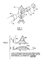
- Fig. 1 hereof shows a relevant portion of the system disclosed by H ansen, which in conventional parlance incorporates the principles of dark field microscopy. In turn, these principles are employed to advantage in the aforementioned Ortho ELT-8 brand flow cytometry instrument.
- preferred embodiments of the present invention are discussed in the context of such an instrument, but it is to be understood that the principles of the present invention may be employed to equal advantage in any of the numerous commercially available cell counting and cell analysis sytems which provide operation for which the present methods are appropriate.
- a source of illumination 101 is focused by lens apparatus 102 onto a zone 103 in a flow cell 104.
- the flow cell 104 is oriented transversely to the focused illumination.
- hydrodynamic focusing insures that cells of a sample are marched rapidly individually through the zone, and are individually rapidly illuminated. It is highly desirable but not absolutely necessary that the dimension of the zone 103 in the direction of flow be generally smaller than the dimension of the cells in the flow direction.
- the illuminating intensity in the zone has a Gaussian distribution.
- the detector 108 includes photoelectric means for developing an intensity (i.e., voltage) versus time depiction of the passing cell. See Fig. 2 which illustrates such waveforms.
- pulse width shall refer to the width, or time, between that time at which the pulse first reaches 50% of peak, to that time at which it drops below 50% of peak.
- alternative definitions may be employed, but the 50%-50% definition is employed in preferred embodiments of the present invention.
- no changes to the ELT-8 optics are made.
- Additional electronics well within the capability of those of ordinary skill, may be used to analyze the red blood cell pulse shapes and to create pulse width and pulse shape histograms on the ELT-8 data handler.
- computer software embodying Fig. 3 hereof will properly analyze pulse width and pulse shape histograms as well as provide new information from the RBC histogram of pulse area previously displayed on the E L T -8.
- the primary cell features that relate to these parameters, as shown in Fig. 2 are thought to be:
- cell morphology is developed utilizing both a shape sensitive parameter and a shape insensitive parameter, on a cell by cell basis.
- the ratio of pulse width (itself a function of time of flight) to pulse peak provides:such an indicium of pulse shape.
- Other combinations may, however, be employed, for example the ratio of pulse rise slope (e.g., the upswing time between 50% and 90% of peak) to pulse area or peak, or the ratio of pulse width to pulse area.
- cells flow through a focused laser beam.
- the dimension of the beam in the direction of cell flow is approximately 7 ⁇ m, which is smaller than most red blood cells.
- the light intensity in the beam is not uniform, but rather is peaked in the center and falls off in a Gaussian distribution.
- As cells pass through this focused strip of laser light they are in effect scanned by the laser beam. Since all cells flow at substantially the same speed in the fluid, a short cell spends a short time passing through the.laser beam, and a longer cell spends a longer time in the laser beam. This time spent in the laser beam is reflected in the width of the light scatter pulse. See Figure 2.
- the pulse width is a measure of the length of the longest dimension of a cell. If a cell is compact (e.g., spherical), it produces a taller, narrower pulse as in Figure 2a. If the cell is longer and thinner, a shorter, wider pulse is produced as in Figure 2b.
- the ratio of the width of the pulse to the peak height of the pulse is a good measure of the gross shape of the cell.
- the integrated area under the pulse is a good measure of cell volume, and is relatively independent of cell shape. In a normal individual, red blood cells have a well defined biconcave shape and a narrow range of sizes.
- poikilocytosis The presence of significant numbers of cells with other than biconcave shape (e.g., spherical, spiked spherical, elongated, and sickle-shaped) is a condition referred to as poikilocytosis. If a wide range of cell sizes is present, the condition is known as anisocytosis. The presence of significant poikilocytosis or anisocytosis is often indicative of disease, and blood films are examined for these conditions during the "differential white cell count.”
- histogram shall have the following meaning.
- a histogram is a plot or graph of the value of a parameter versus the number of occurrences of events with particular values of the parameter.
- the horizontal axis of the graph is divided into a number of equally sized and equally spaced segments or channels. Each segment or channel represents a particular range for the value of the parameter.
- the vertical axis of the graph represents the number of occurrences of events with each particular range of parameter values. Usually a continuous range of values, say 0 to 100, is divided into equal sized segments.
- Figure 4 shows examples of histograms.
- an index can be derived which indicates whether or not a blood sample contains significant numbers of abnormally shaped or sized red blood cells. Since small numbers of very abnormal cells may affect the pulse parameter histograms in a way similar to the effect of large numbers of less abnormal cells, the red cell abnormality or morphology index ("RCMI") in accordance with the present invention will not necessarily correlate numerically with the percentages of abnormal cells found in a morphological evaluation of a blood smear. There are also distinct differences in the condition of red cells evaluated in the ELT-8, in. which cells are suspended in saline during evaluation, as compared with the manual evaluation, in which cells are dried onto a glass slide and fixed with alcohol.
- the instrument is calibrated by analyzing normal blood samples, preferably on the ELT-8, and determining the mean and standard deviation of each parameter for normal blood samples.
- the RCMI is computed as follows for all but parameter (3), the mean of pulse shape.
- the value of a parameter is compared with the normal value for that parameter. If the parameter is less than two normal standard deviations from the normal mean, no value is assigned the RCMI from the parameter. If the parameter is more than two normal standard deviations away from the normal mean, a value is assigned as Note that the "-2" term results from adoption of the 2a deviation criteria. Clearly, different statistical criteria will result in comparable variation of the foregoing algorithm.
- Fig. 3 shows in flow chart form a preferred embodiment of the methods of the present invention.
- a representative signal pulse is produced at step 302.
- parallel processes are conducted, one for pulse peak measurement, at 304, and one for pulse area measurement, as by integration, at step 305.
- the respective values, cell after cell, are summed at 306 and collected in the form of a histogram.
- this histogram is analyzed at step 307, to determine the coefficient of variation, the area-sensitive parameter utilized for RCMI evaluation.
- the area-CV is evaluated relative to a "normal” range, and if it is, pre-selected (i.e. the "yes” exit 309), no further computation will include the area-cv.
- the width- cv, shape mean, and shape-cv values are compared with predetermined ranges designated "normal” for each. As is the case of the area-cv, previously discussed, occurrence within range results in exit via the "yes" leg, and no further dispositive action.
- the RCMI compute step 321 is the calculation of the total R CMI index.
- the developed RCMI may be evaluated relative to a preset threshold: if above the threshold, further study conventionally through manual methods will be required, but if not, that laborious process is obviated.
- Fig. 4 illustrates three histograms produced in accordance with steps 313, 314, and 306 of Fig. 3. Each may thereupon be processed for suitable data required. That is, coefficient of variation for all three, and mean for the pulse area histogram, result therefrom.
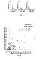
- Fig. 5 illustrates actual data correlation of the principles of the present invention with prior art manual techniques.
- RCMI is plotted on the ordinate
- the manual evaluation is plotted on the abscissa.
- Fig. 5 For purposes of analysis of Fig. 5, let it be assumed that the manual index of abnormality is 10%, a realistic number, -and that the RCMI index of 1 or greater indicates a degree of morphology worthy of detailed investigation (i.e., the "yes" exit of step 322 in Fig. 3).
- the truth table of Fig. 5 demonstrates the correspondence and significance of the data set forth on the Fig. 5 graph.
Applications Claiming Priority (2)
| Application Number | Priority Date | Filing Date | Title |
|---|---|---|---|
| US54222183A | 1983-10-14 | 1983-10-14 | |
| US542221 | 1983-10-14 |
Publications (2)
| Publication Number | Publication Date |
|---|---|
| EP0138591A2 true EP0138591A2 (de) | 1985-04-24 |
| EP0138591A3 EP0138591A3 (de) | 1986-02-12 |
Family
ID=24162842
Family Applications (1)
| Application Number | Title | Priority Date | Filing Date |
|---|---|---|---|
| EP84306988A Withdrawn EP0138591A3 (de) | 1983-10-14 | 1984-10-12 | Verfahren zur Aussiebung von abnormalen roten Zellen |
Country Status (3)
| Country | Link |
|---|---|
| EP (1) | EP0138591A3 (de) |
| JP (1) | JPS60107565A (de) |
| AU (1) | AU3418584A (de) |
Cited By (6)
| Publication number | Priority date | Publication date | Assignee | Title |
|---|---|---|---|---|
| EP0335001A2 (de) * | 1988-03-30 | 1989-10-04 | Toa Medical Electronics Co., Ltd. | Vorrichtung zur Bestimmung von Teilchen |
| EP0361504A2 (de) * | 1988-09-30 | 1990-04-04 | Toa Medical Electronics Co., Ltd. | Vorrichtung zur Teilchenanalyse und Verfahren zur Bestimmung des Kernverschiebungsindex |
| EP0487356A2 (de) * | 1990-11-22 | 1992-05-27 | Satake Corporation | Verfahren und Vorrichtung zur Analyse des Schleifgrades eines Granulats |
| EP0515100A1 (de) * | 1991-05-14 | 1992-11-25 | Toa Medical Electronics Co., Ltd. | Gerät zur Messung von Zellen im Urin |
| EP0949498A3 (de) * | 1998-04-08 | 2003-04-16 | Sysmex Corporation | Vorrichtung und Verfahren zur Unterscheidung von Erythrozyten in Urin |
| CN113034479A (zh) * | 2021-03-31 | 2021-06-25 | 武汉智博见微医疗科技有限公司 | Aa、mds、ma的分类方法、设备及可读存储介质 |
Citations (5)
| Publication number | Priority date | Publication date | Assignee | Title |
|---|---|---|---|---|
| US4021117A (en) * | 1975-08-07 | 1977-05-03 | Hildegard Gohde | Process for automatic counting and measurement of particles |
| EP0008874A1 (de) * | 1978-08-18 | 1980-03-19 | Ortho Diagnostic Systems Inc. | Verfahren und Einrichtung zum Unterscheiden der roten Blutzellen von den Blutplättchen |
| EP0009307A1 (de) * | 1978-09-06 | 1980-04-02 | Ortho Diagnostic Systems Inc. | Verfahren und Vorrichtung zur Diskriminierung zwischen Blutplättchen und roten Blutzellen in einer Blutprobe |
| US4263508A (en) * | 1979-04-20 | 1981-04-21 | Research Corporation | Pulse edge measurement for determining particle dimensional characteristics |
| US4325483A (en) * | 1979-08-20 | 1982-04-20 | Ortho Diagnostics, Inc. | Method for detecting and controlling flow rates of the droplet forming stream of an electrostatic particle sorting apparatus |
-
1984
- 1984-10-12 AU AU34185/84A patent/AU3418584A/en not_active Abandoned
- 1984-10-12 JP JP59212808A patent/JPS60107565A/ja active Pending
- 1984-10-12 EP EP84306988A patent/EP0138591A3/de not_active Withdrawn
Patent Citations (5)
| Publication number | Priority date | Publication date | Assignee | Title |
|---|---|---|---|---|
| US4021117A (en) * | 1975-08-07 | 1977-05-03 | Hildegard Gohde | Process for automatic counting and measurement of particles |
| EP0008874A1 (de) * | 1978-08-18 | 1980-03-19 | Ortho Diagnostic Systems Inc. | Verfahren und Einrichtung zum Unterscheiden der roten Blutzellen von den Blutplättchen |
| EP0009307A1 (de) * | 1978-09-06 | 1980-04-02 | Ortho Diagnostic Systems Inc. | Verfahren und Vorrichtung zur Diskriminierung zwischen Blutplättchen und roten Blutzellen in einer Blutprobe |
| US4263508A (en) * | 1979-04-20 | 1981-04-21 | Research Corporation | Pulse edge measurement for determining particle dimensional characteristics |
| US4325483A (en) * | 1979-08-20 | 1982-04-20 | Ortho Diagnostics, Inc. | Method for detecting and controlling flow rates of the droplet forming stream of an electrostatic particle sorting apparatus |
Non-Patent Citations (1)
| Title |
|---|
| REVIEW OF SCIENTIFIC INSTRUMENTS, vol. 49, no. 12, December 1978, pages 1617-1621; New York, US; W.G. EISERT et al.: "Internal calibration to absolute values in flowthrough particle size analysis" * |
Cited By (11)
| Publication number | Priority date | Publication date | Assignee | Title |
|---|---|---|---|---|
| EP0335001A2 (de) * | 1988-03-30 | 1989-10-04 | Toa Medical Electronics Co., Ltd. | Vorrichtung zur Bestimmung von Teilchen |
| EP0335001A3 (de) * | 1988-03-30 | 1990-09-19 | Toa Medical Electronics Co., Ltd. | Vorrichtung zur Bestimmung von Teilchen |
| EP0361504A2 (de) * | 1988-09-30 | 1990-04-04 | Toa Medical Electronics Co., Ltd. | Vorrichtung zur Teilchenanalyse und Verfahren zur Bestimmung des Kernverschiebungsindex |
| EP0361504A3 (de) * | 1988-09-30 | 1991-05-15 | Toa Medical Electronics Co., Ltd. | Vorrichtung zur Teilchenanalyse und Verfahren zur Bestimmung des Kernverschiebungsindex |
| EP0487356A2 (de) * | 1990-11-22 | 1992-05-27 | Satake Corporation | Verfahren und Vorrichtung zur Analyse des Schleifgrades eines Granulats |
| EP0487356A3 (en) * | 1990-11-22 | 1992-07-08 | Satake Corporation | Method of and apparatus for analyzing granule grinding degrees |
| EP0515100A1 (de) * | 1991-05-14 | 1992-11-25 | Toa Medical Electronics Co., Ltd. | Gerät zur Messung von Zellen im Urin |
| US5684584A (en) * | 1991-05-14 | 1997-11-04 | Toa Medical Electronics Co., Ltd. | Apparatus for analyzing cells in urine |
| EP0949498A3 (de) * | 1998-04-08 | 2003-04-16 | Sysmex Corporation | Vorrichtung und Verfahren zur Unterscheidung von Erythrozyten in Urin |
| CN113034479A (zh) * | 2021-03-31 | 2021-06-25 | 武汉智博见微医疗科技有限公司 | Aa、mds、ma的分类方法、设备及可读存储介质 |
| CN113034479B (zh) * | 2021-03-31 | 2022-06-03 | 武汉智博见微医疗科技有限公司 | Aa、mds、ma的分类方法、设备及可读存储介质 |
Also Published As
| Publication number | Publication date |
|---|---|
| EP0138591A3 (de) | 1986-02-12 |
| JPS60107565A (ja) | 1985-06-13 |
| AU3418584A (en) | 1985-04-18 |
Similar Documents
| Publication | Publication Date | Title |
|---|---|---|
| US4596464A (en) | Screening method for red cell abnormality | |
| EP0140616B1 (de) | Methode und Vorrichtung zur Bestimmung des Volumens und des Brechungsindexes von Teilchen | |
| US6630990B2 (en) | Optical method and apparatus for red blood cell differentiation on a cell-by-cell basis, and simultaneous analysis of white blood cell differentiation | |
| US4453266A (en) | Method and apparatus for measuring mean cell volume of red blood cells | |
| US7390662B2 (en) | Method and apparatus for performing platelet measurement | |
| US6184978B1 (en) | Method and apparatus for verifying uniform flow of a fluid sample through a flow cell and distribution on a slide | |
| CA1135979A (en) | Automated method and apparatus for classification of cells with application to the diagnosis of anemia | |
| WO1996011448A1 (en) | Improved accuracy in cell mitosis analysis | |
| EP0633462A2 (de) | Dreifarben Durchflusszytometrie mit automatischer Auswertung | |
| WO1994029800A9 (en) | Three-color flow cytometry with automatic gating function | |
| EP0138591A2 (de) | Verfahren zur Aussiebung von abnormalen roten Zellen | |
| CN111274949B (zh) | 一种基于结构分析的血液病白细胞散点图相似度分析方法 | |
| US5735274A (en) | Apparatus and method for analyzing particles including shifting a judgement region | |
| Pappas et al. | Reticulocyte counting by flow cytometry. A comparison with manual methods | |
| JP3504030B2 (ja) | 粒子判定基準の決定方法およびその装置並びにその判定基準を用いた粒子分析装置 | |
| JPH05322882A (ja) | 血液分析装置 | |
| JP2000046723A (ja) | 血小板機能検査方法 | |
| Leeuwen et al. | A Short Evaluation of a New Haematological Cell Counter—The Cell-Dyn 3000-Following a Modified Tentative NCCLS-Procedure | |
| Lewis | Automation in haematology-present and future trends | |
| Lewis | New developments in haematology | |
| Lewis | Automated differential leucocyte counting: Present status and future trends | |
| CA2104156A1 (en) | Method and apparatus for fluorescence pulse area/peak size parameter measurement for cell analysis using whole blood | |
| JPH04332847A (ja) | 白血球分析装置 | |
| Parks et al. | Collection, display and analysis of flow cytometry data |
Legal Events
| Date | Code | Title | Description |
|---|---|---|---|
| PUAI | Public reference made under article 153(3) epc to a published international application that has entered the european phase |
Free format text: ORIGINAL CODE: 0009012 |
|
| AK | Designated contracting states |
Designated state(s): BE DE FR GB IT NL |
|
| PUAL | Search report despatched |
Free format text: ORIGINAL CODE: 0009013 |
|
| AK | Designated contracting states |
Designated state(s): BE DE FR GB IT NL |
|
| STAA | Information on the status of an ep patent application or granted ep patent |
Free format text: STATUS: THE APPLICATION IS DEEMED TO BE WITHDRAWN |
|
| 18D | Application deemed to be withdrawn |
Effective date: 19861013 |
|
| RIN1 | Information on inventor provided before grant (corrected) |
Inventor name: KANE, RICHARD L. Inventor name: PRICE, BRANDON J. Inventor name: GERSHMAN, RUSSELL J. Inventor name: HOFFMAN, ROBERT A. |