EP0117262A1 - Processes and devices for examinations on immobilised biological material - Google Patents
Processes and devices for examinations on immobilised biological material Download PDFInfo
- Publication number
- EP0117262A1 EP0117262A1 EP83101863A EP83101863A EP0117262A1 EP 0117262 A1 EP0117262 A1 EP 0117262A1 EP 83101863 A EP83101863 A EP 83101863A EP 83101863 A EP83101863 A EP 83101863A EP 0117262 A1 EP0117262 A1 EP 0117262A1
- Authority
- EP
- European Patent Office
- Prior art keywords
- carrier
- biological material
- plate
- frozen
- parts
- Prior art date
- Legal status (The legal status is an assumption and is not a legal conclusion. Google has not performed a legal analysis and makes no representation as to the accuracy of the status listed.)
- Granted
Links
Images


Classifications
-
- G—PHYSICS
- G01—MEASURING; TESTING
- G01N—INVESTIGATING OR ANALYSING MATERIALS BY DETERMINING THEIR CHEMICAL OR PHYSICAL PROPERTIES
- G01N33/00—Investigating or analysing materials by specific methods not covered by groups G01N1/00 - G01N31/00
- G01N33/48—Biological material, e.g. blood, urine; Haemocytometers
- G01N33/50—Chemical analysis of biological material, e.g. blood, urine; Testing involving biospecific ligand binding methods; Immunological testing
- G01N33/53—Immunoassay; Biospecific binding assay; Materials therefor
- G01N33/543—Immunoassay; Biospecific binding assay; Materials therefor with an insoluble carrier for immobilising immunochemicals
- G01N33/551—Immunoassay; Biospecific binding assay; Materials therefor with an insoluble carrier for immobilising immunochemicals the carrier being inorganic
- G01N33/552—Glass or silica
-
- G—PHYSICS
- G01—MEASURING; TESTING
- G01N—INVESTIGATING OR ANALYSING MATERIALS BY DETERMINING THEIR CHEMICAL OR PHYSICAL PROPERTIES
- G01N1/00—Sampling; Preparing specimens for investigation
- G01N1/28—Preparing specimens for investigation including physical details of (bio-)chemical methods covered elsewhere, e.g. G01N33/50, C12Q
- G01N1/286—Preparing specimens for investigation including physical details of (bio-)chemical methods covered elsewhere, e.g. G01N33/50, C12Q involving mechanical work, e.g. chopping, disintegrating, compacting, homogenising
-
- G—PHYSICS
- G01—MEASURING; TESTING
- G01N—INVESTIGATING OR ANALYSING MATERIALS BY DETERMINING THEIR CHEMICAL OR PHYSICAL PROPERTIES
- G01N1/00—Sampling; Preparing specimens for investigation
- G01N1/28—Preparing specimens for investigation including physical details of (bio-)chemical methods covered elsewhere, e.g. G01N33/50, C12Q
- G01N1/42—Low-temperature sample treatment, e.g. cryofixation
-
- G—PHYSICS
- G01—MEASURING; TESTING
- G01N—INVESTIGATING OR ANALYSING MATERIALS BY DETERMINING THEIR CHEMICAL OR PHYSICAL PROPERTIES
- G01N1/00—Sampling; Preparing specimens for investigation
- G01N1/28—Preparing specimens for investigation including physical details of (bio-)chemical methods covered elsewhere, e.g. G01N33/50, C12Q
- G01N1/286—Preparing specimens for investigation including physical details of (bio-)chemical methods covered elsewhere, e.g. G01N33/50, C12Q involving mechanical work, e.g. chopping, disintegrating, compacting, homogenising
- G01N2001/2873—Cutting or cleaving
-
- G—PHYSICS
- G01—MEASURING; TESTING
- G01N—INVESTIGATING OR ANALYSING MATERIALS BY DETERMINING THEIR CHEMICAL OR PHYSICAL PROPERTIES
- G01N2474/00—Immunochemical assays or immunoassays characterised by detection mode or means of detection
- G01N2474/20—Immunohistochemistry assay
-
- Y—GENERAL TAGGING OF NEW TECHNOLOGICAL DEVELOPMENTS; GENERAL TAGGING OF CROSS-SECTIONAL TECHNOLOGIES SPANNING OVER SEVERAL SECTIONS OF THE IPC; TECHNICAL SUBJECTS COVERED BY FORMER USPC CROSS-REFERENCE ART COLLECTIONS [XRACs] AND DIGESTS
- Y10—TECHNICAL SUBJECTS COVERED BY FORMER USPC
- Y10T—TECHNICAL SUBJECTS COVERED BY FORMER US CLASSIFICATION
- Y10T436/00—Chemistry: analytical and immunological testing
- Y10T436/25—Chemistry: analytical and immunological testing including sample preparation
Definitions
- the present invention describes a rational method and devices for investigations on immobilized biological material.
- Antibodies that are not directed against antigens in the frozen section do not bind and are washed off.
- antibodies obtained from animals and labeled with a fluorescent substance are applied, which are directed against human antibodies (fluorescence-labeled antihuman serum). They bind to human antibodies that may be attached to the frozen section and cannot be washed off. If the patient's serum contained antibodies against the examined tissue, then the fluorescent dye bound to the corresponding tissue structures can now be detected in the fluorescence microscope.
- Tissue from patients is also often examined to determine whether antibodies have bound to certain structures in vivo.
- frozen sections of this tissue are made and immediately brought together with a fluorescence-labeled antihuman serum in a direct immunofluorescence test; the first step of the indirect immunofluorescence test described above is omitted.
- the structures containing antibodies fluoresce in the washed preparation.
- the cover slip should float on the glycerin. If too much glycerin is dripped on, the cover slip will slip and the microscope will become contaminated. That's why you dab off excess glycerin. Sometimes you remove too much glycerin: capillary force pulls the coverslip firmly onto the slide and crushes the frozen section, or you inadvertently move the cover glass against the standard slide and destroys the frozen section. The same happens if you want to place the frozen section in the focal plane of the lens when microscoping and come too close to the lens with the cover slip. A positive result can appear to be negative. Sera from different patients can be examined simultaneously on a slide. This summarizes workflows and makes the testing process easier.
- the organ pieces available are so small that only very few frozen sections can be obtained from them, which are not sufficient for the various examinations to be carried out.
- the structures of interest are often represented several times on these cuts. One can then try to divide the finished loose frozen sections fresh or after freeze-drying, but the desired structures can hardly be recognized by the loose cut and the manipulation is very difficult.
- a particular difficulty with biochemical tests on frozen sections is that they often adhere poorly to the support during incubations. They often swim off in whole or in part. If glass slides are used, they must therefore be carefully cleaned before applying the frozen sections (chromosulfuric acid, ethanol, acetone; Storch, W., see above).
- the glass surface is coated with glycerin and gelatin or with egg white (Romeis, B .: “Microscopic Technology", Oldenbourg-Verlag Kunststoff, Vienna, 1968). This procedure is not a major improvement and is rejected by many investigators.
- tissues such as the lungs, intestinal mucosa and especially for fat-rich tissues (e.g. pancreas, adrenal medulla), there is currently no reliable method for holding unfixed frozen sections firmly on their support, especially if the incubations take a long time and if they have to be washed thoroughly .
- the object of the invention is to find out an improved method with which tests on immobilized biological material can be carried out and to create devices therefor.
- the invention solves this problem. With the help of the invention it is possible to bind biological material to a surface and to investigate it rationally using biochemical methods or to use it for rational biochemical investigations.
- the inventor sticks to the example of immunofluorescence on frozen sections of biological tissue.
- the invention is also suitable for many applications in other areas of immunology (fluorescence immunoassay, radioimmunoassay, enzyme immunoassay), histochemistry, microbiology, clinical chemistry and hormone chemistry.
- the biological material is first placed on the surface of a support 1 on which it is allowed to adhere.
- One or more carriers or parts 1a of the carriers are then glued onto a plate 2 or with others
- the biological material would now be particularly sensitive to damage or drying out, without further measures being taken, because it would be raised above the level of the plate by the carrier and adhesive layer. Immunofluorescence tests could not be carried out using the conventional method: the cover slip to be placed at the end would lie directly on the exposed one sensitive frozen sections and crushed them.
- the supports are arranged on the plate in such a way that the biological material is largely protected against damage and in particular does not come into direct contact with solid material during the examination, and that it does not dry during or after the examination where it is undesirable.
- the carriers are fastened for this purpose, for example, in depressions 5 in the plate 2 (FIGS. 4, 6).
- a distance ⁇ h ⁇ 0 of the biological material to an opposite surface can also be set by other geometric, mechanical or other means (Fig. 13), e.g. B. to a cover slip 8. If the biological material is to be assessed with a microscope or with another aid, this distance .DELTA.h must be set so that the biological material can be placed in the focal plane of the lens or in a plane that the Can reach the sensor of the tool used.
- Freeze sections can be chemically bonded to the support in accordance with the invention in such a way that reactive chemical groups 9 "are coupled to the surface of the support before the freeze sections are applied (FIG. 5).
- freeze sections produced in a conventional manner can also be used for the invention that have not been chemically bound to carriers.
- the test results are secured by the arrangement protecting the biological material.
- the results are reproducible, the proportion of unsuccessful tests has dropped from 5 - 10% to almost 0%.
- the frozen sections are no longer so easily damaged during transport and during the tests. They can also be covered with glycerin by inexperienced people, the coverslip can no longer destroy them, even if you press firmly on them.
- the reaction field 3 lies in a lower level (depression 5) than the environment immediately adjacent to the reaction field, and the surface of the carrier 1a does not protrude beyond the level of the environment.
- Histological specimens thin sections of biological tissue
- first coat a slide with it and fix it together with the slide on a plate in a second, independent step (plate for histochemical examinations slide). You can e.g. B.
- glass pieces 3 mm x 3 mm x 0.2 mm
- the glass pieces can then be glued quickly and easily onto a plate, if necessary using an automatic machine. Or one spreads the biological material on a carrier, connects it to the carrier, divides the carrier and then glues the fragments on.
- the coated carriers take up little space and are not destroyed at low temperatures. They are therefore more suitable for deep-freeze preservation than, for example, standard glass slides. You can fill frozen section fragments (approx. 2 mm x 2 mm x 0.2 mm; as a carrier e.g. cover glass material) in PVC tubes or in glass ampoules, weld them and store them in liquid nitrogen at - 196 C for years, e.g. . B. 1,000 pieces in a 1 ml glass ampoule.
- Frozen sections can be easily fragmented according to the invention because they stick to a carrier that is divisible.
- the invention is therefore particularly well suited for studies in which the reagents are very expensive, such as monoclonal antibodies, or in which little sample material is available, e.g. B. when searching for the production of monoclonal antibodies suitable plasmocytoma cells.
- Example 1 (FIG. 5): Production of activated glass supports. A sufficient number of cover glasses, 0.2 mm thick, were cleaned in chromic sulfuric acid for 3 days and then rinsed with distilled water for a sufficient time.
- coverslips were immersed for 3 hours at room temperature in a 2% solution of aminoethylaminopropyltrimethoxysilane in ethanol / water 1/1 (vol.), Washed 3 times for 1 minute in absolute ethanol and dried with compressed air. Overnight polymerization at 70 ° C, then immerse for 3 hours at room temperature in a 5% aqueous solution of freshly filtered glutardialdehyde. Sufficient in dist. Rinse water, dry. The surface of the cover glasses now had self-reacting aldehyde groups 9, to which proteins from the frozen sections could bind covalently via free amino groups. The coverslips were kept at room temperature for 6 months without noticeably losing their activity.
- Freshly prepared 5 / human to strong frozen sections 14 liver were brought to a material produced according to Example 1, cover glass 1, it could coat the entire surface of one side of the cover glass.
- the frozen sections were allowed to thaw and dry. In the area of the frozen sections, fields were scratched into the cover slip with a diamond tip, then it was broken. Part of the frozen section fragments 1a was set aside for example 3, the rest were distributed in polyvinyl chloride tubes, the tubes were welded closed and stored in liquid nitrogen.
- tissue areas of interest can be selected, unimportant areas and areas of poor cut quality are discarded.
- the cover slip can be scratched before the frozen sections are applied. You can also place a frozen section on glass fragments that are close together, thaw it and let it dry, and then attach the fragments to a plate. Plastics, e.g. B. films of reactive groups with polymethyl methacrylate. Fragments can be punched or cut from the foils, if necessary using a LASER.
- Example 3 Carrying out an immunofluorescence.
- the sera of the sick were examined for antibodies against cell nuclei.
- the frozen section fragments 1a produced in Example 2 were glued onto reaction fields 3a of a plate 2.
- the frozen sections were at a level 0.1 mm lower than the surface of the strips 6 present on the plate 2 (thickness 0.3 mm).
- Dilutions 11 of the sera were dropped onto a reagent carrier 7.
- the frozen sections adhering to the supports 1a were immersed in the drops for 30 minutes at room temperature, then washed in P H -buffered saline (PBS), first discretely with the aid of a second reagent support 7, then in a beaker. This was followed by the second incubation of the frozen sections with fluorescence for 30 minutes.
- PBS P H -buffered saline
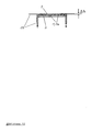
- Plates with any number of reaction fields can be used, e.g. B. with one (Fig. 9) or with 48 (Fig. 8).
- the carrier with the frozen sections can also be glued to the bottom of a flow-through cell 14 (Fig. 12), the top of this flow-through cell is a cover glass 8, which is arranged at a safe distance ⁇ > 0 from the frozen sections, but can be viewed with a microscope still allows.
- the flow-through cuvette is simply filled with the serum dilution or with the fluorescence-labeled antihuman serum, and for washing, the P H buffered saline solution is pumped through continuously.
- the closed arrangement has the advantage that the frozen sections do not dry out so easily during the test and that laboratory personnel are less at risk of infectious sera.
- Fig. 13 shows that a distance ⁇ necessary for the protection of the biological material located on the surface of the carrier 1a can be generated by spacers which are connected to a plate opposite the plate 2 (reagent carrier 7, cover glass 8).
- Fig. 14 shows a disc-shaped "plate” 2, to which frozen section fragments 1a were glued.
- the "plate” was made from a 0.3 mm thick round cover glass onto which two 0.3 mm thick strips 6 were glued. During the incubations, the “plate” floats on the serum dilution or on the fluorescence-labeled antihuman serum, which have been dripped onto the reaction field 3 of the reagent carrier 7. When microscoping, the "plate” is held on the cover glass 8 by capillary force, between the frozen sections and that P H coverslip was placed -gepuffertes glycerol.
Abstract
Description
Die vorliegende Erfindung beschreibt ein rationelles Verfahren und Vorrichtungen für Untersuchungen an unbeweglich gemachtem biologischen Material.The present invention describes a rational method and devices for investigations on immobilized biological material.
Der Stand der Technik und die Wirkungsweise der Erfindung sollen am Beispiel der Immunfluoreszenzuntersuchung an Gefrierschnitten dargestellt werden. Mit dieser Untersuchung können bei manchen Kranken Antikörper gegen körpereigenes Gewebe nachgewiesen werden. Die Methode wurde von Coons und anderen eingeführt (Coons, A. H., Creech, H. J., Jones, R. N., Proc. Soc. Exp. Biol. N. Y. 47, 1941, Ss. 200 ff): Man bringt einen Gefrierschnitt gesunden Gewebes auf eine Glasoberfläche und läßt ihn auftauen und antrocknen. .Wie in Abb. 1 dargestellt, wird er mit dem verdünnten Serum eines Kranken überschichtet. Enthält das Serum Antikörper gegen die Antigene des Gewebes, dann bleiben sie am Gefrierschnitt hängen. Antikörper, die nicht gegen Antigene des Gefrierschnitts gerichtet sind, binden sich nicht und werden abgewaschen. In einem zweiten Schritt werden aus Tieren gewonnene, mit einem fluoreszierenden Stoff markierte Antikörper aufgebracht, die gegen menschliche Antikörper gerichtet sind (Fluoreszenz-markiertes Antihumanserum). Sie binden sich an ggf. am Gefrierschnitt hängende menschliche Antikörper und können nicht abgewaschen werden. Enthielt das Serum des Kranken Antikörper gegen das untersuchte Gewebe, dann läßt sich jetzt im Fluoreszenzmikroskop der an die entsprechenden Gewebestrukturen gebundene Fluoreszenzfarbstoff nachweisen.The prior art and the mode of action of the invention are to be illustrated using the example of immunofluorescence examination on frozen sections. This test can be used to detect antibodies against the body's own tissue in some patients. The method was introduced by Coons and others (Coons, AH, Creech, HJ, Jones, RN, Proc. Soc. Exp. Biol. NY 47, 1941, pp. 200 ff): A frozen section of healthy tissue is placed on a glass surface and let it thaw and dry. As shown in Fig. 1, it is overlaid with a patient's diluted serum. If the serum contains antibodies against the antigens of the tissue, they get stuck on the frozen section. Antibodies that are not directed against antigens in the frozen section do not bind and are washed off. In a second step, antibodies obtained from animals and labeled with a fluorescent substance are applied, which are directed against human antibodies (fluorescence-labeled antihuman serum). They bind to human antibodies that may be attached to the frozen section and cannot be washed off. If the patient's serum contained antibodies against the examined tissue, then the fluorescent dye bound to the corresponding tissue structures can now be detected in the fluorescence microscope.
Häufig wird auch Gewebe von Kranken untersucht, um festzustellen, ob sich in vivo an bestimmte Strukturen Antikörper gebunden haben. Dazu werden Gefrierschnitte von diesem Gewebe angefertigt und in einem direkten Immunfluoreszenztest unmittelbar mit einem Fluoreszenz-markierten Antihumanserum zusammengebracht, der erste Schritt des oben beschriebenen indirekten Immunfluoreszenztests entfällt. Die Antikörper enthaltenden Strukturen fluoreszieren im gewaschenen Präparat.Tissue from patients is also often examined to determine whether antibodies have bound to certain structures in vivo. For this purpose, frozen sections of this tissue are made and immediately brought together with a fluorescence-labeled antihuman serum in a direct immunofluorescence test; the first step of the indirect immunofluorescence test described above is omitted. The structures containing antibodies fluoresce in the washed preparation.
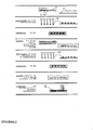
Es ist üblich, die Gefrierschnitte auf Standardobjektträger aus Glas zu montieren, und zwar wird in der Regel für jeden Gefrierschnitt ein Standardobjektträger genommen. Eine eingehende Beschreibung des Tests findet sich z. B. bei Storch (Storch, W.: "Immunfluoreszenzfibel", Fischer-Verlag Jena, 1979). Der Test birgt einige Fehlerquellen, setzt viel Übung voraus und verlangt einen großen Arbeits- und Materialaufwand. Oft kommt es vor, daß die Tropfen verlaufen und die Gefrierschnitte dann antrocknen (wenn die Gefrierschnitte während des Tests trocken werden, lassen sich die Ergebnisse meistens nicht mehr verwerten). Vorsichtshalber macht man daher große Tropfen und vergeudet Reagenzien. Vor dem Mikroskopieren überschichtet man den Gefrierschnitt mit PH-gepuffertem Glycerin und legt ein Deckglas darüber. Das Deckglas sollte auf dem Glycerin schwimmen. Wird zuviel Glycerin aufgetropft, dann verrutscht das Deckglas und das Mikroskop wird verunreinigt. Deshalb tupft man überschüssiges Glycerin ab. Manchmal entfernt man dabei zuviel Glycerin: Dann wird durch Kapillarkraft das Deckglas fest an den Objektträger gezogen und zerquetscht den Gefrierschnitt, oder man verschiebt das Deckglas versehentlich gegen den Standardobjektträger und zerstört den Gefrierschnitt dabei. Das gleiche passiert, wenn man beim Mikroskopieren den Gefrierschnitt in die Brennebene des Objektivs stellen will und dabei mit dem Deckglas zu nahe an das Objektiv kommt. Ein positives Ergebnis kann dadurch scheinbar negativ werden. Die Seren verschiedener Kranker können gleichzeitig auf einem Objektträger untersucht werden. Dadurch werden Arbeitsabläufe zusammengefaßt, und das Testverfahren wird einfacher. Man muß zuvor mehrere Gefrierschnitte nebeneinander aufbringen und darauf achten, daß sich die Seren untereinander nicht vermischen. Das wird erleichtert durch eine Aufteilung in Reaktionsfelder 3, die voneinander durch eine wasserabstoßende Beschichtung 4 des Objektträgers abgesetzt sind (0'Neill, P., Johnson, G. D.; Ann. N. Y. Acad. Sci. 177, 446-452, 1971; US-PS 3 736 042; EP-OS 79 103 987.8) oder durch Farbringe (Räisänen, S. und andere, J. clin. Pathol. 33, 95-96, 1980). Je mehr Seren auf einem Objektträger untersucht werden, umso geringer wird für jede Einzeluntersuchung der präparatorische Aufwand während des Tests. Mit der oben beschriebenen Technik können aber nicht mehr als zwanzig Einzeluntersuchungen nebeneinander auf demselben Objektträger durchgeführt werden, vor allem weil die Reaktionsfelder nacheinander betropft werden und sich dann unterschiedliche Inkubationszeiten für die einzelnen Analysen ergeben und weil einige Präparate austrocknen und unbrauchbar werden, während die anderen mit dem Fluoreszenz-markierten Antihumanserum' betropft werden. Für die Untersuchung sehr vieler Seren nebeneinander, beispielsweise 96 auf einem Objektträger (Platte Abb. 2), bieten sich die Methoden nach Stöcker (EP-OS 79 103 987.8; DE-OS 3 107 964) an: Auf zwei Platten befinden sich in kongruenter Anordnung hydrophile Reaktionsfelder, die von einer wasserabstoßenden Schicht umgeben sind. Auf die Reaktionsfelder der einen Platte werden Gefrierschnitte montiert, auf die Reaktionsfelder der anderen Platte tropft man Proben, z. B. Serumverdünnungen oder das Fluoreszenzmarkierte Antihumanserum. Beide Platten werden dann in ein Gestell gelegt, und zwar so, daß die Gefrierschnitte in die flüssigen Proben eintauchen. Alle Gefrierschnitte einer Platte inkubieren gleich lange, und kein Gefrierschnitt trocknet während des Tests an, auch nicht beim Aufbringen des Fluoreszenz-markierten Antihumanserums. Die rationelle Methode zur Untersuchung vieler Seren auf einer Platte hat sich bisher in der Immunfluoreszenzdiagnostik nur dort bewährt, wo suspendierfähige Antigene verwendet werden können, weil man sie auf die Reaktionsfelder tropfen kann, z. B. die Erreger der Lues oder der Toxoplasmose. Wer mit der Technik des Gefrierschneidens vertraut ist, weiß, wie mühsam es ist, mit den bisher bekannten Verfahren 96 Gefrierschnitte sauber und gleichmäßig auf die Reaktionsfelder einer Platte zu montieren. Bei einer industriellen Fertigung müßte man mit einer hohen Ausschußziffer rechnen.It is common to mount the frozen sections on standard glass slides, usually one standard slide is used for each frozen section. A detailed description of the test can be found e.g. B. with Storch (Storch, W .: "Immunofluorescence primer", Fischer-Verlag Jena, 1979). The test harbors some sources of error, requires a lot of practice and requires a lot of work and materials. It often happens that the drops run and the frozen sections then dry (if the frozen sections become dry during the test, the results can usually not be used). As a precaution, large drops are made and reagents are wasted. Before microscopy, the frozen section is covered with PH-buffered glycerin and a cover glass is placed over it. The cover slip should float on the glycerin. If too much glycerin is dripped on, the cover slip will slip and the microscope will become contaminated. That's why you dab off excess glycerin. Sometimes you remove too much glycerin: capillary force pulls the coverslip firmly onto the slide and crushes the frozen section, or you inadvertently move the cover glass against the standard slide and destroys the frozen section. The same happens if you want to place the frozen section in the focal plane of the lens when microscoping and come too close to the lens with the cover slip. A positive result can appear to be negative. Sera from different patients can be examined simultaneously on a slide. This summarizes workflows and makes the testing process easier. You have to apply several frozen sections next to each other beforehand and make sure that the sera do not mix with each other. This is facilitated by a division into
Häufig ist es nötig, in einem Serum nach Antikörpern gegen verschiedene Antigene zu suchen: Mehrere suspendierfähige Antigene können dazu nebeneinander auf ein Reaktionsfeld getropft werden. Nach dem Antrocknen und ggf. Fixieren werden sie gemeinsam mit einem Tropfen Serum-Verdünnung überschichtet (Wang, S. P., Excerpta Medica, Amsterdam, 273-288, 1971). Will man Antikörper gegen verschiedene Gewebe nachweisen, stellt man mehrere Gefrierschnitte zu einem "Bunten Schnitt" zusammen, den man mit einem großen Tropfen Probe oder Reagenz überschichtet. Man kann dazu die Gefrierschnitte für jedes Gewebe einzeln herstellen und unterschiedliche Gefrierschnitte nebeneinander auf ein Reaktionsfeld legen. Oder man friert mehrere Stückchen verschiedener Gewebe in einer wäßrigen Lösung von Carboxymethylcellulose gemeinsam nebeneinander ein und schneidet und montiert sie gleichzeitig (z. B. Nairn, R. C.: "Fluorescent Protein Tracing", Churchill Livingstone Edinburgh, 1976). Man kann nur wenige Organstückchen zusammen in einem Block schneiden, und die Organstückchen müssen genau "zurechtgetrimmt" werden. Dazu ist viel Geschick nötig und es geht Gewebe dabei verloren. Unterschiedlich zu fixierende Gefrierschnitte können nicht im selben "Bunten Schnitt" nebeneinander liegen.It is often necessary to search for antibodies against various antigens in a serum: several suspendable antigens can be dripped onto a reaction field side by side. After drying and, if necessary, fixing, they are overlaid with a drop of serum dilution (Wang, S.P., Excerpta Medica, Amsterdam, 273-288, 1971). If you want to detect antibodies against different tissues, you put several frozen sections together in a "colorful section", which you cover with a large drop of sample or reagent. The frozen sections for each tissue can be made individually and different frozen sections placed side by side on a reaction field. Or freeze several pieces of different tissue together in an aqueous solution of carboxymethyl cellulose and cut and assemble them simultaneously (e.g. Nairn, R.C .: "Fluorescent Protein Tracing", Churchill Livingstone Edinburgh, 1976). You can only cut a few organ pieces together in a block, and the organ pieces must be "trimmed" exactly. This requires a lot of skill and tissue is lost. Freeze sections to be fixed differently cannot lie next to each other in the same "colorful section".
Manchmal steht nur sehr wenig zu untersuchendes Serum oder Fluoreszenz-markiertes Antihumanserum zur Verfügung. Dann versucht man, den Testansatz so klein wie möglich zu halten. Voraussetzung dafür sind kleine Gefrierschnitte. Man schneidet das Gewebe auf die gewünschte Größe zurecht und nimmt dabei in Kauf, daß viel Material verlorengeht.Sometimes there is very little serum to be tested or fluorescent-labeled antihuman serum available. Then you try to keep the test approach as small as possible. This requires small frozen sections. You cut the fabric is trimmed to the desired size and accepts that a lot of material is lost.
Untersucht man ungleichmäßig im Gewebe verteilte Strukturen, z. B. Langerhans'sche Inseln der Bauchspeicheldrüse oder Glomeruli der Niere, dann muß man große Schnitte anfertigen um möglichst sicher zu sein, daß man die gewünschten Strukturen beim Mikroskopieren vorfindet. Es wäre zu schwierig, einzelne Inseln oder Glomeruli aus dem losen Gefrierschnitt herauszuschneiden.If one examines structures distributed unevenly in the tissue, e.g. B. Langerhans islands of the pancreas or glomeruli of the kidney, then you have to make large sections to be as sure as possible that you will find the desired structures when microscoping. It would be too difficult to cut individual islands or glomeruli out of the loose frozen section.
Manchmal sind die zur Verfügung stehenden Organstückchen so klein, daß man nur sehr wenige Gefrierschnitte daraus gewinnen kann, die für die verschiedenen durchzuführenden Untersuchungen nicht ausreichen. Die interessierenden Strukturen sind aber auf diesen Schnitten oft mehrfach vertreten. Man kann dann versuchen, die fertigen losen Gefrierschnitte frisch oder nach Gefriertrocknung zu teilen, die gewünschten Strukturen sind aber am losen Schnitt kaum zu erkennen und die Manipulation ist sehr schwierig. Eine besondere Schwierigkeit bei biochemischen Untersuchungen an Gefrierschnitten besteht darin, daß sie während der Inkubationen häufig nur schlecht auf der Unterlage haften. Oft schwimmen sie ganz oder teilweise ab. Werden Glasobjektträger verwendet, müssen sie daher vor dem Aufbringen der Gefrierschnitte sorgfältig gereinigt werden (Chromschwefelsäure, Ethanol, Aceton; Storch, W., siehe oben). In einigen Labors überzieht man die Glasoberfläche mit Glycerin und Gelatine oder mit Hühnereiweiß (Romeis, B.: "Mikroskopische Technik", Oldenbourg-Verlag München, Wien, 1968). Dieses Verfahren ist keine wesentliche Verbesserung und wird von vielen Untersuchern abgelehnt. Für einige Gewebe, wie Lunge, Darmschleimhaut und besonders für fettreiche Gewebe (z. B. Pankreas, Nebennierenmark) gibt es bisher kein zuverlässiges Verfahren, unfixierte Gefrierschnitte fest auf ihrer Unterlage zu halten, besonders wenn die Inkubationen lange dauern und wenn gründlich gewaschen werden muß. Man kann einen Gefrierschnitt bei - 30 °C kurz in Methylenchlorid oder in ein anderes organisches Lösemittel tauchen und das Fett herauslösen, wodurch die Haftung verbessert wird (Stöcker, W., nicht publ.). Dabei werden aber in einigen Fällen Antigene oder Enzyme zerstört, ebenso durch sonst in der Histochemie gebräuchliche Fixierverfahren. In der Immunhistochemie verwendet man daher vorwiegend unfixiertes Gewebe (Wick, G. und andere: "Immunflourescence", Medizinische Verlagsgesellschaft Marburg/Lahn, 1978). Nur wenn das Antigen in Wasser löslich ist, wird der Gefrierschnitt fixiert, z. B. bei der Schilddrüse, deren Kolloid durch Behandlung mit absolutem Methanol unlöslich wird. Es sind bereits sehr viele Verfahren bekannt, mit denen organisches Material an aktivierte Oberflächen gebunden werden kann. Insbesondere bemüht man sich darum, Enzyme, Antigene und Antikörper an Festkörpern zu immobilisieren (z. B. Ternynck, T., Avrameas, S., FEBS-Letters 23, 24-28, 1972; Guesdon, J. L. und andere, J. Immunol. Meth. 21, 59-63, 1978; DT-AS 2 102 514; DE-OS 2 740 008; DE-OS 2 749 317; DE-PS 2 905 657). Es wurde auch schon beschrieben, in Polyacrylamid eingebettetes Gewebe zu schneiden und die Schnitte chemisch an eine mit reaktiven Gruppen versehene Oberfläche zu binden (Hausen, P., Dreyer, C., Stain Technol. 56, 287-293, 1981). Dabei ging man davon aus, daß sich das Polyacrylamid bindet und nicht das Gewebe. Daß sich ein Gefrierschnitt nicht eingebetteten Gewebes, der auf einer unvorbehandelten Oberfläche nicht ausreichend haftet, über reaktive Gruppen der aktivierten Oberfläche fest mit ihr verbinden kann, wurde von den Autoren offenbar nicht erkannt, und es gibt in der Fachliteratur keine Berichte darüber.Sometimes the organ pieces available are so small that only very few frozen sections can be obtained from them, which are not sufficient for the various examinations to be carried out. The structures of interest are often represented several times on these cuts. One can then try to divide the finished loose frozen sections fresh or after freeze-drying, but the desired structures can hardly be recognized by the loose cut and the manipulation is very difficult. A particular difficulty with biochemical tests on frozen sections is that they often adhere poorly to the support during incubations. They often swim off in whole or in part. If glass slides are used, they must therefore be carefully cleaned before applying the frozen sections (chromosulfuric acid, ethanol, acetone; Storch, W., see above). In some laboratories, the glass surface is coated with glycerin and gelatin or with egg white (Romeis, B .: "Microscopic Technology", Oldenbourg-Verlag Munich, Vienna, 1968). This procedure is not a major improvement and is rejected by many investigators. For some tissues, such as the lungs, intestinal mucosa and especially for fat-rich tissues (e.g. pancreas, adrenal medulla), there is currently no reliable method for holding unfixed frozen sections firmly on their support, especially if the incubations take a long time and if they have to be washed thoroughly . You can be one Briefly immerse the frozen section at - 30 ° C in methylene chloride or another organic solvent and remove the fat, which improves the adhesion (Stöcker, W., not publ.). In some cases, however, antigens or enzymes are destroyed, as well as by fixation methods commonly used in histochemistry. Therefore, unfixed tissue is predominantly used in immunohistochemistry (Wick, G. and others: "Immunflourescence", medical publishing house Marburg / Lahn, 1978). Only when the antigen is soluble in water is the frozen section fixed, e.g. B. in the thyroid gland, the colloid of which becomes insoluble by treatment with absolute methanol. A large number of processes are already known with which organic material can be bound to activated surfaces. In particular, efforts have been made to immobilize enzymes, antigens and antibodies on solids (e.g. Ternynck, T., Avrameas, S., FEBS-Letters 23, 24-28, 1972; Guesdon, JL and others, J. Immunol Meth. 21, 59-63, 1978; DT-
Aufgabe der Erfindung ist es, ein verbessertes Verfahren herauszufinden, mit dem Untersuchungen an unbeweglich gemachtem biologischen Material durchgeführt werden können, und Vorrichtungen dafür zu schaffen.The object of the invention is to find out an improved method with which tests on immobilized biological material can be carried out and to create devices therefor.
Die Erfindung löst diese Aufgabe. Mit Hilfe der Erfindung gelingt es, biologisches Material an eine Oberfläche zu binden und es mit biochemischen Methoden rationell zu untersuchen oder es für rationelle biochemische Untersuchungen einzusetzen. Bei der Beschreibung hält sich der Erfinder an das Beispiel der Immunfluoreszenz an Gefrierschnitten biologischen Gewebes. Die Erfindung eignet sich aber auch für viele Anwendungen aus anderen Bereichen der Immunologie (Fluoreszenzimmunoassay, Radioimmunoassay, Enzymimmunoassay), der Histochemie, der Mikrobiologie, der klinischen Chemie und der Hormonchemie.The invention solves this problem. With the help of the invention it is possible to bind biological material to a surface and to investigate it rationally using biochemical methods or to use it for rational biochemical investigations. In the description, the inventor sticks to the example of immunofluorescence on frozen sections of biological tissue. However, the invention is also suitable for many applications in other areas of immunology (fluorescence immunoassay, radioimmunoassay, enzyme immunoassay), histochemistry, microbiology, clinical chemistry and hormone chemistry.
Die Erfindung wird im folgenden anhand mehrerer Zeichnungen näher erläutert. Es zeigen:
- Abb. 1: eine schematische Darstellung des indirekten Immunfluoreszenztests,
- Abb. 2: eine ebene Platte mit 96
hydrophilen Reaktionsfeldern 3,deren Umgebung 4 hydrophob beschichtet ist, - Abb. 3: die rationelle Herstellung einer Vielzahl von Gefrierschnitt-Fragmenten 1a mit Hilfe eines teilbaren Trägers 1,
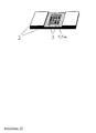
- Abb. 4:
eine Platte 2 mit auf ihr befestigtem Träger 1a, in perspektivischer Draufsicht (a), im Querschnitt (b) und mit Deckglas im Querschnitt (c), - Abb. 5: eine schematische Darstellung, wie ein Gefrierschnitt chemisch an einen Träger gekoppelt werden kann,
- Abb. 6: eine Platte 2 (a) mit auf ihr befestigten Trägern 1a und einem dazugehörenden Reagenzträger 7 (b)
mit hydrophilen Reaktionsfeldern 3 und hydrophober Umgebung 4, - Abb. 7: eine schematische Darstellung des indirekten Immunfluoreszenztests gemäß der Erfindung,
- Abb. 8: eine Platte 2 (a) mit 48 auf ihr befestigten Trägern 1a, ihre Rückseite (b), den dazugehörenden Reagenzträger 7 (c) und beide während einer Inkubation im Querschnitt (d),
- Abb. 9: eine Platte 2 (a)
mit 1 auf ihr befestigten Träger 1a, ihre Rückseite (b), den dazugehörenden Reagenzträger 7 (c) und beide während einer Inkubation im Querschnitt (d), - Abb.10:
eine Platte 2 mit mehreren verschiedene Gefrierschnitte aufweisenden Trägern 1a auf einem Reaktionsfeld 3 ("Bunter Schnitt"), - Abb.11: eine Platte 2 (a)
mit 4 "Bunten Schnitten", ihre Rückseite (b), den dazugehörenden Reagenzträger 7 (c) und beide während einer Inkubation im Querschnitt (d), - Abb.12: eine Durchflußküvette, deren Boden durch eine
Platte 2 gebildet wird, auf der mehrere Träger 1a mit Gefrierschnitten befestigt sind (ebenfalls ein "Bunter Schnitt"), - Abb.13:
eine Platte 2mit 5 auf ihr befestigten Trägern 1a und dazu gehörendem Reagenzträger 7 während einer Inkubation, im Querschnitt (a), dieselbe Platte mit Deckglas 8 (b), - Abb.14: eine scheibchenförmige "Platte" 2 (a), ihre Rückseite mit Griffknopf (b), den dazugehörenden Reagenzträger7(c), beide während einer Inkubation im Querschnitt (d) und, die "Platte" eingedeckt mit Glycerin und durch Kapillarkraft an einem Deckglas gehalten, unter dem Mikroskop.
- Fig. 1: a schematic representation of the indirect immunofluorescence test,
- Fig. 2: a flat plate with 96 hydrophilic reaction fields 3, the
surroundings 4 of which are coated hydrophobically, - 3: the rational production of a large number of frozen section fragments 1a with the aid of a
divisible carrier 1, - Fig. 4: a
plate 2 with a support 1a attached to it, in a perspective top view (a), in cross section (b) and with a cover glass in cross section (c), - Fig. 5: a schematic representation of how a frozen section can be chemically coupled to a carrier,
- Fig. 6: a plate 2 (a) with carriers 1a attached to it and an associated reagent carrier 7 (b) with hydrophilic reaction fields 3 and
hydrophobic surroundings 4, - Fig. 7: a schematic representation of the indirect immunofluorescence test according to the invention,
- Fig. 8: a plate 2 (a) with 48 carriers 1a attached to it, its back (b), the associated reagent carrier 7 (c) and both during an incubation in cross section (d),
- Fig. 9: a plate 2 (a) with 1 carrier 1a attached to it, its back (b), the associated reagent carrier 7 (c) and both during an incubation in cross section (d),
- Fig.10: a
plate 2 with several different frozen sections 1a on a reaction field 3 ("colorful section"), - Fig.11: a plate 2 (a) with 4 "colored sections", its back (b), the associated reagent carrier 7 (c) and both during an incubation in cross section (d),
- Fig.12: a flow-through cell, the bottom of which is formed by a
plate 2, on which several supports 1a with frozen sections are fastened (also a "colorful section"), - Fig. 13: a
plate 2 with 5 carriers 1a attached to it and the associatedreagent carrier 7 during an incubation, in cross section (a), the same plate with cover glass 8 (b), - Fig.14: a disc-shaped "plate" 2 (a), its back with knob (b), the associated reagent holder7 (c), both during an incubation in cross-section (d) and, the "plate" covered with glycerin and by capillary force held on a coverslip under the microscope.
Mitteln auf ihr befestigt. Auf einer planen Platte wäre jetzt, ohne daß weitere Vorkehrungen getroffen würden, das biologische Material besonders gegen Beschädigungen oder gegen Austrocknen empfindlich, weil es durch Träger und Klebeschicht über das Niveau der Platte gehoben würde. Immunfluoreszenztests ließen sich nach der herkömmlichen Methode so nicht durchführen: Das zum Schluß aufzulegende Deckglas läge direkt auf den exponierten empfindlichen Gefrierschnitten und zerquetschte sie. Die Träger werden aber auf der Platte so angeordnet, daß das biologische Material gegen eine Beschädigung weitgehend geschützt ist und insbesondere während der Untersuchung keinen direkten Kontakt mit festem Material bekommt, und daß es während oder nach der Untersuchung nicht trocknet, wo es unerwünscht ist. Erfindungsgemäß werden die Träger zu diesem Zweck beispielsweise in Vertiefungen 5 der Platte 2 befestigt (Abb. 4, 6). Dadurch wird für das biologische Material zur obersten Plattenebene ein sicherer Abstand Δh ≧ 0 gewährleistet. Anstelle durch ein Versenken der Träger kann auch durch andere geometrische, mechanische oder andere Mittel (Abb. 13) ein Abstand Δh ≧ 0 des biologischen Materials zu einer gegenüberliegenden Oberfläche eingestellt werden, z. B. zu einem Deckglas 8. Wenn das biologische Material mit dem Mikroskop oder mit einem anderen Hilfsmittel beurteilt werden soll, muß dieser Abstand Δh so festgelegt werden, daß das biologische Material in die Brennebene des Objektivs gestellt werden kann oder in eine Ebene, die der Meßfühler des verwendeten Hilfsmittels erreichen kann.Means attached to it. On a flat plate, the biological material would now be particularly sensitive to damage or drying out, without further measures being taken, because it would be raised above the level of the plate by the carrier and adhesive layer. Immunofluorescence tests could not be carried out using the conventional method: the cover slip to be placed at the end would lie directly on the exposed one sensitive frozen sections and crushed them. However, the supports are arranged on the plate in such a way that the biological material is largely protected against damage and in particular does not come into direct contact with solid material during the examination, and that it does not dry during or after the examination where it is undesirable. According to the invention, the carriers are fastened for this purpose, for example, in
Gefrierschnitte können gemäß der Erfindung chemisch an den Träger gebunden werden, dergestalt, daß an die Oberfläche des Trägers vor dem Aufbringen der Gefrierschnitte reaktionsfähige chemische Gruppen 9"gekoppelt werden (Abb. 5). Für die Erfindung können aber auch in herkömmlicher Weise hergestellte Gefrierschnitte benutzt werden, die nicht chemisch an Träger gebunden wurden.Freeze sections can be chemically bonded to the support in accordance with the invention in such a way that
Durch die das biologische Material schützende Anordnung werden die Testresultate abgesichert. Die Ergebnisse sind reproduzierbarer, der Anteil nicht gelungener Tests ist von 5 - 10 % auf nahezu 0 % abgesunken. Die Gefrierschnitte werden beim Transport und während der Tests nicht mehr so leicht beschädigt. Sie können auch von Ungeübten mit Glycerin eingedeckt werden, das Deckglas kann sie nicht mehr zerstören, auch dann nicht, wenn man fest darauf drückt.The test results are secured by the arrangement protecting the biological material. The results are reproducible, the proportion of unsuccessful tests has dropped from 5 - 10% to almost 0%. The frozen sections are no longer so easily damaged during transport and during the tests. They can also be covered with glycerin by inexperienced people, the coverslip can no longer destroy them, even if you press firmly on them.
Bei der in Abb. 4 dargestellten Anordnung liegt das Reaktionsfeld 3 in einer tieferen Ebene (Vertiefung 5) als die dem Reaktionsfeld unmittelbar benachbarte Umgebung, und auch die Oberfläche des Trägers 1a ragt nicht über die Ebene der Umgebung hinaus. Dadurch wird das Verlaufen der Serumverdünnung, des Fluoreszenz-markierten Antihumanserums oder des Glycerins verhindert, und die Gefrierschnitte trocknen nicht aus. Histologische Präparate (Dünnschnitte biologischer Gewebe), insbesondere Gefrierschnitte, oder Präparate aus anderen an Oberflächen gebundenen biologischen Materialien können mit geringem Aufwand in großer Zahl und in guter Qualität hergestellt werden: Es ist manchmal besser, das biologische Material nicht direkt auf eine Platte zu bringen, sondern erst einen Träger damit zu beschichten und es in einem davon unabhängigen zweiten Schritt zusammen mit dem Träger auf einer Platte zu befestigen (Platte bei histochemischen Untersuchungen = Objektträger). Man kann z. B. in einem kleinen Gefäß mit 250 ml Inhalt gleichzeitig zehntausende Glasstückchen (3 mm x 3 mm x 0,2 mm) beschichten. Die Glasstückchen können dann schnell und einfach auf eine Platte geklebt werden, ggf. durch einen Automaten. Oder man breitet das biologische Material auf einem Träger aus, läßt es sich mit dem Träger verbinden, zerteilt den Träger und klebt dann die Fragmente auf.In the arrangement shown in Fig. 4, the
Die beschichteten Träger nehmen wenig Platz in Anspruch und werden bei tiefen Temperaturen nicht zerstört. Sie eignen sich daher besser für eine Tiefkühlkonservierung als zum Beispiel Standardobjektträger aus Glas. Man kann Gefrierschnitt-Fragmente (ca. 2 mm x 2 mm x 0,2 mm; als Träger z. B. Deckglasmaterial) in PVC-Schläuche oder in Glasampullen füllen, diese zuschweißen und in flüssigem Stickstoff bei - 196 C jahrelang aufbewahren, z. B. 1 000 Stück in einer 1 ml fassenden Glasampulle.The coated carriers take up little space and are not destroyed at low temperatures. They are therefore more suitable for deep-freeze preservation than, for example, standard glass slides. You can fill frozen section fragments (approx. 2 mm x 2 mm x 0.2 mm; as a carrier e.g. cover glass material) in PVC tubes or in glass ampoules, weld them and store them in liquid nitrogen at - 196 C for years, e.g. . B. 1,000 pieces in a 1 ml glass ampoule.
Es ist ein Kunststück, aus einem losen Gefrierschnitt einen interessierenden Bezirk passender Größe herauszutrennen. Wird der Gefrierschnitt durch einen Träger stabilisiert, geht das sehr leicht. Für Mikroanalysen kann man aus einem 5 mm x 5 mm großen Gefrierschnitt, der auf einem Deckglas haftet, innerhalb fünf Minuten 100 Miniatur-Gefrierschnitte herstellen. Gefrierschnitte einzelner Glomeruli können leicht aus einem auf einem Deckglas haftenden Gefrierschnitt der Niere isoliert werden. Das Gewebe wird besser ausgenutzt, weil nicht unnötig viele Glomeruli für jeden einzelnen Test verbraucht werden. Für Anteile der Gefrierschnitte, die keine Glomeruli enthalten, werden keine Reagenzien vergeudet. Die Erfindung eignet sich deshalb auch besonders gut für Untersuchungen, bei denen die Reagenzien sehr teuer sind, wie monoklonale Antikörper, oder bei denen wenig Probenmaterial zur Verfügung steht, z. B. beim Heraussuchen für die Produktion monoklonaler Antikörper geeigneter Plasmocytom-Zellen.It is a feat to separate an interesting area of appropriate size from a loose frozen section. If the frozen section is stabilized by a carrier, it is very easy. For microanalysis, 100 miniature frozen sections can be made from a 5 mm x 5 mm frozen section that adheres to a coverslip within five minutes. Frozen sections of individual glomeruli can easily be isolated from a frozen section of the kidney adhering to a cover slip. The tissue is better utilized because it does not consume an unnecessarily large number of glomeruli for each individual test. No reagents are wasted on portions of frozen sections that do not contain glomeruli. The invention is therefore particularly well suited for studies in which the reagents are very expensive, such as monoclonal antibodies, or in which little sample material is available, e.g. B. when searching for the production of monoclonal antibodies suitable plasmocytoma cells.
Im Gegensatz zu den bisher bekannten Verfahren lassen sich jetzt viele Gefrierschnitte leicht auf einer Platte mit vielen Reaktionsfeldern befestigen (Abb. 2, 8). Der Erfinder verwendet für Versuche Platten mit 96 Reaktionsfeldern, die jeweils einen Gefrierschnitt aufweisen.In contrast to the previously known methods, many frozen sections can now be easily attached to a plate with many reaction fields (Fig. 2, 8). For experiments, the inventor uses plates with 96 reaction fields, each of which has a frozen section.
Ebenso einfach ist es, "Bunte Schnitte" herzustellen. Die Zusammensetzung der "Bunten Schnitte" kann von Test zu Test beliebig variiert werden. Man kann Gewebe gleichzeitig nebeneinander untersuchen, die unterschiedlich vorbehandelt werden müssen, z. B. fixierte und unfixierte Gefrierschnitte der Schilddrüse: Die Gefrierschnitte werden auf dem Träger fixiert, bevor dieser fragmentiert und auf die Platte geklebt wird. Es können daher auch Fixierverfahren angewendet werden, die die hydrophobe Beschichtung der Platte angreifen.It is just as easy to create "colorful cuts". The composition of the "colorful cuts" can be varied from test to test. Tissues that have to be pretreated differently can be examined simultaneously, e.g. B. Fixed and unfixed frozen sections of the thyroid gland: The frozen sections are fixed on the carrier before it is fragmented and glued to the plate. Fixing methods which attack the hydrophobic coating of the plate can therefore also be used.
Durch die chemische Aktivierung der Trägeroberfläche haften die Gefrierschnitte überraschend fest, und es kommt nicht mehr vor, daß sie während der Inkubationen oder während der Waschvorgänge abschwimmen, nicht einmal dann, wenn man sie mehrere Tage lang in der Flüssigkeit hält oder wenn man sie lange und kräftig überspült. Auch gefriergetrocknete lose Schnitte binden sich in wäßrigem Milieu an den aktivierten Träger. Für Miniaturanalysen der Erfindung war es notwendig, die Haftung der Gefrierschnitte zu verbessern, weil sich die Gefrierschnitte von kleineren Trägerfragmenten häufig ablösten. Beispiele sollen die Wirkungsweise der Erfindung darstellen: Beispiel 1 (Abb. 5): Herstellung aktivierter Träger aus Glas Eine ausreichende Anzahl Deckgläser, Stärke 0,2 mm, wurde 3 Tage lang in Chromschwefelsäure gereinigt und danach ausreichend lange mit destilliertem Wasser gespült. Dann wurden die Deckgläser 3 Stunden lang bei Zimmertemperatur in eine 2 %ige Lösung Aminoäthylaminopropyltrimethoxysilan in Ethanol/Wasser 1/1 (Vol.) getaucht, 3 mal 1 Minute lang in absolutem Ethanol gewaschen und mit Preßluft getrocknet. Über Nacht Querpolymerisation bei 70 °C, dann 3 Stunden bei Zimmertemperatur eintauchen in eine 5 %ige wäßrige Lösung frisch filtrierten Glutardialdehyds. Ausreichend in dest. Wasser spülen, trocknen. Die Oberfläche der Deckgläser wies jetzt selbstreagierende Aldehydgruppen 9 auf, an die sich Proteine der Gefrierschnitte über freie Aminogruppen kovalent binden konnten. Die Deckgläser wurden 6 Monate lang bei Zimmertemperatur aufbewahrt, ohne daß sie ihre Aktivität merkbar verloren.Due to the chemical activation of the carrier surface, they stick Frozen sections are surprisingly firm, and they no longer float away during incubations or washes, not even if you keep them in the liquid for several days or if you wash them vigorously for a long time. Freeze-dried loose cuts also bind to the activated carrier in an aqueous medium. For miniature analyzes of the invention, it was necessary to improve the adhesion of the frozen sections, because the frozen sections frequently detached from smaller carrier fragments. Examples are intended to illustrate the mode of operation of the invention: Example 1 (FIG. 5): Production of activated glass supports. A sufficient number of cover glasses, 0.2 mm thick, were cleaned in chromic sulfuric acid for 3 days and then rinsed with distilled water for a sufficient time. Then the coverslips were immersed for 3 hours at room temperature in a 2% solution of aminoethylaminopropyltrimethoxysilane in ethanol /
Frisch hergestellte, 5/um starke Gefrierschnitte 14 menschlicher Leber wurden auf ein nach Beispiel 1 hergestelltes Deckglas 1 gebracht, man konnte die ganze Oberfläche einer Seite des Deckglases beschichten. Man ließ die Gefrierschnitte auftauen und antrocknen. Im Bereich der Gefrierschnitte wurden in das Deckglas mit einer Diamantspitze Felder eingeritzt, dann wurde es gebrochen. Ein Teil der Gefrierschnitt-Fragmente 1a wurde für das Beispiel 3 beiseite gelegt, der Rest in Schläuche aus Polyvinylchlorid verteilt, die Schläuche wurden zugeschweißt und in flüssigem Stickstoff aufgehoben.Freshly prepared 5 / human to strong
Es ist einfacher und geht mehrfach schneller, auf diese Weise aus einem großen Gefrierschnitt 50 Präparate herzustellen (Zeitaufwand 1/4 Minute) und dann auf eine Platte zu kleben (5 Minuten), als mit dem Gefrierschnitt-Mikrotom 50 Gefrierschnitte von einem kleinen Gewebestück zu schneiden und auf Standardobjektträger zu bringen (30 Minuten). Das Gewebe wird auch besser ausgenutzt, weil das beim Präparieren eines kleinen Gewebestückchens verlorengehende Material erhalten bleibt.It is easier and quicker to produce 50 preparations from a large frozen section in this way (time required 1/4 minute) and then to glue them onto a plate (5 minutes) than with the frozen section microtome 50 frozen sections from a small piece of tissue cut and place on standard slides (30 minutes). The tissue is also better utilized because the material lost when preparing a small piece of tissue is preserved.
Mit Hilfe eines Lupenmikroskops können interessierende Gewebebereiche herausgesucht werden, unwichtige Bereiche und Bereiche schlechter Schnittqualität werden verworfen.With the help of a magnifying microscope, tissue areas of interest can be selected, unimportant areas and areas of poor cut quality are discarded.
Das Deckglas kann schon vor dem Aufbringen der Gefrierschnitte geritzt werden. Man kann einen Gefrierschnitt auch auf eng nebeneinanderliegende Glasfragmente legen, ihn auftauen und trocknen lassen und die Fragmente dann auf einer Platte befestigen. Als Träger eignen sich auch Kunststoffe, z. B. Folien aus mit reaktiven Gruppen versehenem Polymethylmethacrylat. Aus den Folien können Fragmente gestanzt oder geschnitten werden, ggf. mit Hilfe eines LASER.The cover slip can be scratched before the frozen sections are applied. You can also place a frozen section on glass fragments that are close together, thaw it and let it dry, and then attach the fragments to a plate. Plastics, e.g. B. films of reactive groups with polymethyl methacrylate. Fragments can be punched or cut from the foils, if necessary using a LASER.
Beispiel 3 (Abb. 6 und 7): Durchführung eines Immunfluoreszenz-Im nachfolgenden Test wurden die Seren Kranker auf Antikörper gegen Zellkerne untersucht. Dazu wurden die im Beispiel 2 hergestellten Gefrierschnitt-Fragmente 1a auf Reaktionsfelder 3a einer Platte 2 geklebt. Die Gefrierschnitte lagen in einem um 0,1 mm tieferen Niveau als die Oberfläche der auf der Platte 2 vorhandenen Leisten 6 (Stärke 0,3 mm). Verdünnungen 11 der Seren wurden auf einen Reagenzträger 7 getropft. Die auf den Trägern 1a haftenden Gefrierschnitte wurden 30 Minuten bei Zimmertemperatur in die Tropfen getaucht, danach in PH-gepufferter Kochsalzlösung (Phosphate-buffered saline, PBS) gewaschen, zuerst diskret mit Hilfe eines zweiten Reagenzträgers 7, dann in einem Becherglas. Es folgte für 30 Minuten die zweite Inkubation der Gefrierschnitte mit Fluoreszenz-Example 3 (Figs. 6 and 7): Carrying out an immunofluorescence. In the following test, the sera of the sick were examined for antibodies against cell nuclei. For this purpose, the frozen section fragments 1a produced in Example 2 were glued onto reaction fields 3a of a
markiertem Antihumanserum 12. Nach einer weiteren Waschprozedur wurde PH-gepuffertes Glycerin aufgetragen und ein Deckglas darübergelegt. Betrachtung der Gefrierschnitte mit dem Fluoreszenzmikroskop. Bei der Bestimmung der Antikörper gegen Zellkerne in 200 verschiedenen Seren wurden die gleichen Resultate erzielt wie mit der herkömmlichen Methode. Der Arbeitsaufwand war mit der neuen Methode 10 mal und der Reagenzienverbrauch 3 mal geringer als mit der alten.labeled
Es können Platten mit beliebig vielen Reaktionsfeldern benutzt werden, z. B. mit einem (Abb. 9) oder mit 48 (Abb. 8).Plates with any number of reaction fields can be used, e.g. B. with one (Fig. 9) or with 48 (Fig. 8).
Auf jedes Reaktionsfeld können mehrere Träger mit Gefrierschnitten verschiedener Gewebe geklebt werden ("Bunter Schnitt" Abb. 10, 11, 14): Ein Reaktionsfeld pro Platte (Abb. 10, 14), mehrere Reaktionsfelder pro Platte (Abb. 11).Several carriers with frozen sections of different tissues can be glued to each reaction field ("Colorful section" Fig. 10, 11, 14): One reaction field per plate (Fig. 10, 14), several reaction fields per plate (Fig. 11).
Die Träger mit den Gefrierschnitten können auch auf den Boden einer Durchflußküvette 14 geklebt werden (Abb. 12), als Oberseite dieser Durchflußküvette dient ein Deckglas 8, das in einem sicheren Abstand Δℏ>0 zu den Gefrierschnitten angeordnet ist, aber eine Betrachtung mit dem Mikroskop noch zuläßt. Zur Inkubation wird die Durchflußküvette einfach mit der Serum- - Verdünnung oder mit dem Fluoreszenz-markierten Antihumanserum gefüllt, zum Waschen wird die PH-gepufferte Kochsalzlösung kontinuierlich durchgepumpt. Die geschlossene Anordnung hat den Vorteil, daß die Gefrierschnitte während des Tests nicht so leicht austrocknen und daß das Laborpersonal weniger durch infektiöse Seren gefährdet ist.The carrier with the frozen sections can also be glued to the bottom of a flow-through cell 14 (Fig. 12), the top of this flow-through cell is a
Die Abb. 13 zeigt, daß ein für den Schutz des sich auf der Oberfläche des Trägers 1a befindlichen biologischen Materials notwendiger Abstand Δℏ durch Abstandhalter erzeugt werden kann, die mit einer der Platte 2 gegenüberliegenden Platte (Reagenzträger 7, Deckglas 8) verbunden sind.Fig. 13 shows that a distance Δℏ necessary for the protection of the biological material located on the surface of the carrier 1a can be generated by spacers which are connected to a plate opposite the plate 2 (
In Abb. 14 wird eine scheibchenförmige "Platte" 2 dargestellt, auf die Gefrierschnitt-Fragmente 1a geklebt wurden. Die "Platte" wurde aus einem 0,3 mm starken runden Deckglas gefertigt, auf das zwei 0,3 mm starke Leisten 6 geklebt wurden. Bei den Inkubationen schwimmt die "Platte" auf der Serumverdünnung oder auf dem Fluoreszenz-markierten Antihumanserum, die auf das Reaktionsfeld 3 des Reagenzträgers 7 getropft wurden.,Beim Mikroskopieren wird die "Platte" am Deckglas 8 durch Kapillarkraft festgehalten, zwischen die Gefrierschnitte und das Deckglas wurde PH-gepuffertes Glycerin gebracht.Fig. 14 shows a disc-shaped "plate" 2, to which frozen section fragments 1a were glued. The "plate" was made from a 0.3 mm thick round cover glass onto which two 0.3 mm
Claims (12)
bei dem eine oder mehrere Untersuchungen nebeneinander auf einer Platte (2) ablaufen und
bei dem das biologische Material gegen eine Beschädigung weitgehend geschützt ist,
dadurch gekennzeichnet,
daß das biologische Material zunächst auf der Oberfläche eines oder mehrerer Träger (1) zur Haftung gebracht wird und danach die Träger oder Teile (1a) der Träger zusammen mit dem biologischen Material auf einer Platte befestigt werden, wobei die das biologische Material tragende Oberfläche so angeordnet wird, daß es keinen direkten Kontakt mit festen Gegenständen bekommt und daß es während der Untersuchung nicht austrocknet, wo das unerwünscht ist.1. Procedure for investigations on immobilized biological material with general biochemical and with histochemical methods, in particular methods of enzyme, immune and hormone chemistry,
in which one or more examinations run side by side on a plate (2) and
in which the biological material is largely protected against damage,
characterized,
that the biological material is first adhered to the surface of one or more supports (1) and then the supports or parts (1a) of the supports are fastened together with the biological material on a plate, the surface carrying the biological material being so arranged that it does not come into direct contact with solid objects and that it does not dry out during the examination, where this is undesirable.
dadurch gekennzeichnet,
daß die Träger (1) oder die Teile (1a) der Träger so auf der Platte (2) befestigt werden, daß ihre das biologische Material tragende Oberfläche um den Abstand Δh≧ 0 unter der obersten Ebene der Platte (2) oder mit der Platte verbundener Mittel (6) angeordnet ist.2. The method according to claim 1, in which the biological material is protected against direct contact with solid objects during an examination,
characterized,
that the carrier (1) or the parts (1a) of the carrier are attached to the plate (2) so that their surface carrying the biological material by the distance Δh ≧ 0 below the top level of the plate (2) or with the plate connected means (6) is arranged.
dadurch gekennzeichnet,
daß die Träger (1) oder die Teile '(1a) der Träger auf Reaktionsfeldern (3) der Platte (2) befestigt werden, die rundum von Bereichen der Platte umgeben sind, die die Träger oder die Teile der Träger um den Abstand Δh ≧ 0 überragen.3. The method according to claim 2, in which the biological material is largely protected against direct contact with solid objects and against drying out during an examination,
characterized,
that the carrier (1) or the parts' (1a) of the carrier are attached to reaction fields (3) of the plate (2) which are surrounded all around by areas of the plate which the carrier or the parts of the carrier by the distance Δh ≧ Surpass 0.
dadurch gekennzeichnet,
daß die Platte (2) als Boden einer Durchflußküvette (14) ausgebildet ist, auf der ein oder mehrere Träger (1) oder Teile (1a) der Träger befestigt sind, daß deren das biologische Material tragende Oberfläche einen Abstand Δh>0 zu einer gegenüberliegenden Wand aufweist und daß die für die Untersuchung verwendeten Proben- und Reagenzienlösungen kontinuierlich oder intermittierend durch die Durchflußküvette geleitet werden.4. The method according to claim 1, in which the biological material is largely protected against direct contact with solid objects and against drying out during an examination,
characterized,
that the plate (2) is designed as the bottom of a flow-through cell (14) on which one or more supports (1) or parts (1a) of the supports are attached, that their surface carrying the biological material has a distance Δh> 0 to an opposite one Wall and that the sample and reagent solutions used for the investigation are passed continuously or intermittently through the flow cell.
dadurch gekennzeichnet,
daß das biologische Material auf einem teilbaren Träger zur Haftung gebracht wird,
daß dann der Träger zusammen mit dem auf ihm haftenden biologischen Material in beliebig große Teile (1a) zerteilt wird und die Teile auf einer Platte (2) befestigt werden.5. A method of manufacturing surface-mounted biological material for use according to one or more of claims 1 to 4,
characterized,
that the biological material is adhered to a divisible carrier,
that the carrier is then cut together with the biological material adhering to it into parts of any size (1a) and the parts are attached to a plate (2).
dadurch gekennzeichnet,
daß als biologisches Material histologische Dünnschnitte biologischen Gewebes verwendet werden.6. Method for histochemical examinations according to one of claims 1 to 4,
characterized,
that histological thin sections of biological tissue are used as biological material.
dadurch gekennzeichnet,
daß Dünnschnitte auf einem teilbaren Träger zur Haftung gebracht werden und daß dann der Träger zusammen mit den auf ihm haftenden Dünnschnitten in beliebig große Teile (1a) zerlegt wird.7. A process for the preparation of specimens adhering to histological thin sections according to claim 5 for use according to one or more of claims 1 to 4 and for isolating selected structures from thin sections,
characterized,
that thin sections are made to adhere to a separable carrier and that the carrier is then broken down into arbitrarily large parts (1a) together with the thin sections adhering to it.
daß als Träger Glasscheiben benutzt werden.8. A process for the preparation of specimens adhering to surfaces of histological thin sections according to claim 7, characterized in that
that glass panes are used as a carrier.
dadurch gekennzeichnet,
daß an die Oberfläche des Trägers 1 vor dem Aufbringen der Gefrierschnitte reaktionsfähige Gruppen gekoppelt werden, die mit den Gefrierschnitten reagieren und sie chemisch binden.9. A method for improving the adhesion of the thin sections of frozen biological tissue in general histochemistry and in the method according to one or more of claims 6, 7 or 8,
characterized,
that reactive groups which react with the frozen sections and bind them chemically are coupled to the surface of the carrier 1 before the frozen sections are applied.
daß als Träger Glas (15) oder ein anderes mit Silanen sich verbindendes Material verwendet wird, daß man an den Träger ein mindestens eine Aminogruppe enthaltendes Silan (16) koppelt und im Anschluß daran an dieses Silan einen Dialdehyd, und daß man darauf den Gefrierschnitt bringt.10. A method for improving the adhesion of the thin sections of frozen biological tissue according to claim 9, characterized in that
that as a carrier glass (15) or another with silanes connecting material is used, that a silane (16) containing at least one amino group is coupled to the support and then a dialdehyde following this silane, and that the frozen section is brought thereon.
daß die Träger (1) oder Teile (1a) der Träger nach der Beschichtung mit dem biologischen"Mäterial in Schläuchen, Ampullen oder ähnlichem eingefroren gelagert sind.12. The apparatus according to claim 11, characterized in
that the carrier (1) or parts (1a) of the carrier are stored frozen in tubes, ampoules or the like after being coated with the biological material.
Priority Applications (5)
| Application Number | Priority Date | Filing Date | Title |
|---|---|---|---|
| DE8383101863T DE3376359D1 (en) | 1983-02-25 | 1983-02-25 | Processes and devices for examinations on immobilised biological material |
| EP83101863A EP0117262B1 (en) | 1983-02-25 | 1983-02-25 | Processes and devices for examinations on immobilised biological material |
| AT83101863T ATE33717T1 (en) | 1983-02-25 | 1983-02-25 | METHODS AND DEVICES FOR INVESTIGATIONS ON IMMOBILIZED BIOLOGICAL MATERIAL. |
| US06/582,394 US4647543A (en) | 1983-02-25 | 1984-02-24 | Process for analyses to be carried out on immobilized biological tissue |
| JP59035100A JPS59210364A (en) | 1983-02-25 | 1984-02-25 | Method and device for inspecting fixed living body |
Applications Claiming Priority (1)
| Application Number | Priority Date | Filing Date | Title |
|---|---|---|---|
| EP83101863A EP0117262B1 (en) | 1983-02-25 | 1983-02-25 | Processes and devices for examinations on immobilised biological material |
Publications (2)
| Publication Number | Publication Date |
|---|---|
| EP0117262A1 true EP0117262A1 (en) | 1984-09-05 |
| EP0117262B1 EP0117262B1 (en) | 1988-04-20 |
Family
ID=8190315
Family Applications (1)
| Application Number | Title | Priority Date | Filing Date |
|---|---|---|---|
| EP83101863A Expired EP0117262B1 (en) | 1983-02-25 | 1983-02-25 | Processes and devices for examinations on immobilised biological material |
Country Status (5)
| Country | Link |
|---|---|
| US (1) | US4647543A (en) |
| EP (1) | EP0117262B1 (en) |
| JP (1) | JPS59210364A (en) |
| AT (1) | ATE33717T1 (en) |
| DE (1) | DE3376359D1 (en) |
Cited By (11)
| Publication number | Priority date | Publication date | Assignee | Title |
|---|---|---|---|---|
| EP0238190A2 (en) * | 1986-02-12 | 1987-09-23 | Beckman Research Institute of the City of Hope | Multi-tumour tissue slices, methods for their production and testing the tissues in such slices |
| EP0291153A1 (en) * | 1987-03-31 | 1988-11-17 | Fisher Scientific Company | Microscope slide and slide assembly |
| DE4337528A1 (en) * | 1993-11-04 | 1995-07-20 | Lothar Bode | Determn of condition of skin tissue by image analysis |
| DE19545920A1 (en) * | 1995-12-08 | 1997-06-12 | Reimann Hans Juergen Prof Dr D | Use of bioptic material for in vitro diagnosis of, e.g. allergies |
| WO2007134814A1 (en) * | 2006-05-19 | 2007-11-29 | Hartmut Merz | Device and method for the automated and reproducible production of cell or tissue samples that are to be analyzed and are arranged on object supports |
| WO2007140889A1 (en) * | 2006-06-09 | 2007-12-13 | Euroimmun Medizinische Labordiagnostika Ag | Process for obtaining perfect macro- and microarrays by combining preselected coated solid phase fragments |
| EP2191893A1 (en) | 2008-11-19 | 2010-06-02 | Euroimmun Medizinische Labordiagnostika AG | Analysis method and devices for biological reactions between a liquid and a solid phase |
| WO2014009067A1 (en) | 2012-07-11 | 2014-01-16 | Euroimmun Medizinische Labordiagnostika Ag | Device and method for incubating patient samples |
| DE102012013678A1 (en) * | 2012-07-11 | 2014-01-16 | Euroimmun Medizinische Labordiagnostika Ag | Method and analysis device for the microscopic examination of a tissue section or a cell smear |
| DE102014001481A1 (en) | 2013-10-28 | 2015-04-30 | Euroimmun Medizinische Labordiagnostika Ag | Improved apparatus and method for reactions between a solid and a liquid phase |
| EP3260864A1 (en) | 2016-06-21 | 2017-12-27 | Euroimmun Medizinische Labordiagnostika AG | A diagnostic coincubation assay |
Families Citing this family (45)
| Publication number | Priority date | Publication date | Assignee | Title |
|---|---|---|---|---|
| JPS62127642A (en) * | 1985-11-28 | 1987-06-09 | Wakunaga Pharmaceut Co Ltd | Slide glass |
| GB8813628D0 (en) * | 1988-06-09 | 1988-07-13 | Shandon Southern Prod | Immunostaining |
| US5002377A (en) * | 1988-07-07 | 1991-03-26 | City Of Hope | Multi-specimen slides for immunohistologic procedures |
| GB8915759D0 (en) * | 1989-07-10 | 1989-08-31 | Shandon Scient Ltd | Cell block preparation |
| CA2051173A1 (en) * | 1990-09-13 | 1992-03-14 | Takeji Suzuki | Artificial soil and process for producing the same |
| US6905882B2 (en) * | 1992-05-21 | 2005-06-14 | Biosite, Inc. | Diagnostic devices and apparatus for the controlled movement of reagents without membranes |
| US6767510B1 (en) * | 1992-05-21 | 2004-07-27 | Biosite, Inc. | Diagnostic devices and apparatus for the controlled movement of reagents without membranes |
| US7524456B1 (en) * | 1992-05-21 | 2009-04-28 | Biosite Incorporated | Diagnostic devices for the controlled movement of reagents without membranes |
| US5843644A (en) * | 1994-03-01 | 1998-12-01 | The United States Of America As Represented By The Secretary Of The Department Of Health And Human Services | Isolation of cellular material under microscopic visualization using an adhesive/extraction reagent tipped probe |
| US6251467B1 (en) | 1994-03-01 | 2001-06-26 | The United States Of America As Represented By The Department Of Health And Human Services | Isolation of cellular material under microscopic visualization |
| US5843657A (en) * | 1994-03-01 | 1998-12-01 | The United States Of America As Represented By The Department Of Health And Human Services | Isolation of cellular material under microscopic visualization |
| US5550033A (en) * | 1994-09-26 | 1996-08-27 | Krumdieck; Carlos | Mold plunger and method for embedding tissue samples |
| CA2414062A1 (en) * | 2000-06-22 | 2001-12-27 | Clinomics Laboratories, Inc. | Frozen tissue microarrays and methods for using the same |
| NZ526793A (en) * | 2001-01-02 | 2006-02-24 | Supachill Technologies Pty Ltd | Method and system for preparing tissue samples for histological pathological examination |
| US6656380B2 (en) | 2001-10-16 | 2003-12-02 | Supachill Technologies Pty. Ltd. | Super-coolable composition having long-duration phase change capability, process for preparation of same, process for super-cooling same and articles comprising same |
| US6939032B2 (en) * | 2001-10-25 | 2005-09-06 | Erie Scientific Company | Cover slip mixing apparatus |
| US6681581B2 (en) | 2001-11-20 | 2004-01-27 | Supachill Technologies Pty. Ltd. | Pre-conditioned solute for use in cryogenic processes |
| US7943093B2 (en) * | 2001-12-12 | 2011-05-17 | Erie Scientific Company | Cover slip |
| US7648678B2 (en) * | 2002-12-20 | 2010-01-19 | Dako Denmark A/S | Method and system for pretreatment of tissue slides |
| CA2543782A1 (en) * | 2003-10-23 | 2005-05-06 | Georgetown University | Method for two- and three-dimensional microassembly of patterns and structures |
| DE102006048264B4 (en) * | 2006-10-12 | 2011-03-24 | Bundesrepublik Deutschland, vertreten durch das Bundesministerium der Verteidigung, vertreten durch das Bundesamt für Wehrtechnik und Beschaffung | Method for detecting specific nucleic acid sequences |
| JP5715125B2 (en) | 2010-05-28 | 2015-05-07 | オリンパス株式会社 | Cell sorting device, cell sorting system, and cell sorting method |
| CN103221800B (en) | 2010-11-19 | 2015-07-15 | 奥林巴斯株式会社 | Method for preparing biological sample |
| EP2711683B1 (en) | 2011-05-20 | 2016-08-03 | Olympus Corporation | Method of manufacturing base sheet |
| US9851349B2 (en) | 2011-06-29 | 2017-12-26 | Leavitt Medical, Inc. | Matrix for receiving a tissue sample and use thereof |
| WO2013077297A1 (en) | 2011-11-24 | 2013-05-30 | オリンパス株式会社 | Cell sorting device and cell sorting method |
| WO2013077337A1 (en) * | 2011-11-25 | 2013-05-30 | オリンパス株式会社 | Tissue segmentation apparatus, cell sorting apparatus, cell sorting system, tissue display system, substrate, extendible member, tissue segmentation method, and cell sorting method |
| EP2863231A1 (en) | 2013-10-17 | 2015-04-22 | Institut D'Investigaciones Biomédiques August Pi i Sunyer | Diagnostic method for detecting a GABA(A) related autoimmune disease and related subject-matter |
| EP2905622A1 (en) | 2014-02-07 | 2015-08-12 | Institut D'Investigaciones Biomédiques August Pi i Sunyer | Diagnosis of a neurological disease |
| EP2952898B1 (en) | 2014-06-06 | 2018-08-08 | Euroimmun Medizinische Labordiagnostika AG | Diagnosis of a neurological disease |
| HUE052493T2 (en) | 2014-11-04 | 2021-04-28 | Euroimmun Medizinische Labordiagnostika Ag | A novel diagnostically relevant autoantibody |
| EP3026434B1 (en) | 2014-11-25 | 2022-01-05 | Euroimmun Medizinische Labordiagnostika AG | A novel diagnostically relevant autoantibody |
| ES2748117T3 (en) | 2015-04-22 | 2020-03-13 | Euroimmun Medizinische Labordiagnostika Ag | Diagnosis of a new autoimmune disease |
| EP3101424B1 (en) | 2015-06-04 | 2023-01-04 | Euroimmun Medizinische Labordiagnostika AG | Diagnosis of a neuroautoimmune disease |
| EP3293520B1 (en) | 2016-09-09 | 2019-11-06 | Euroimmun Medizinische Labordiagnostika AG | A method for the production of an polypeptide |
| EP3392656A1 (en) | 2017-04-20 | 2018-10-24 | Euroimmun Medizinische Labordiagnostika AG | Diagnosis of a neuroautoimmune disease |
| US10725035B2 (en) | 2017-06-16 | 2020-07-28 | Euroimmun Medizinische Labordiagnostika Ag | Diagnosis of a neuroautoimmune disease |
| EP3591400B1 (en) * | 2018-07-02 | 2023-03-01 | Euroimmun Medizinische Labordiagnostika AG | Coated solid-phase fragments and its production |
| EP3629021A1 (en) | 2018-09-26 | 2020-04-01 | Euroimmun Medizinische Labordiagnostika AG | Diagnosis of a neuroautoimmune disease |
| SG10201908826UA (en) | 2018-10-22 | 2020-05-28 | Euroimmun Medizinische Labordiagnostika Ag | Diagnosis of blistering autoimmune diseases |
| EP3667322A1 (en) | 2018-12-14 | 2020-06-17 | Euroimmun Medizinische Labordiagnostika AG | Serological detection of plasmodium antibodies |
| AU2020200382A1 (en) | 2019-01-24 | 2020-08-13 | Euroimmun Medizinische Labordiagnostika Ag | Serological detection of Plasmodium antibodies |
| US11644463B2 (en) | 2019-08-30 | 2023-05-09 | Euroimmun Medizinische Labordiagnostika Ag | Detection of an autoantibody |
| US11835519B2 (en) | 2019-12-17 | 2023-12-05 | Euroimmun Medizinische Labordiagnostika Ag | Autoantibodies to Septin-7 and diagnosis of neurological disease |
| EP3964818A1 (en) * | 2020-09-04 | 2022-03-09 | Leica Mikrosysteme GmbH | Preparation of a sample for high pressure freezing |
Citations (3)
| Publication number | Priority date | Publication date | Assignee | Title |
|---|---|---|---|---|
| US3736042A (en) * | 1971-05-05 | 1973-05-29 | Clinical Sciences Inc | Microscope slide assembly |
| DD145133A1 (en) * | 1979-07-24 | 1980-11-19 | Guenter Schwartze | ACCESSORIES FOR IMMUNOFLUORESCENCE MICROSCOPIC EXAMINATIONS |
| DE3107964A1 (en) * | 1981-03-03 | 1982-09-16 | Winfried Dr.med. 2419 Rondeshagen Stöcker | Frame for a microanalysis system |
Family Cites Families (12)
| Publication number | Priority date | Publication date | Assignee | Title |
|---|---|---|---|---|
| US2965219A (en) * | 1959-03-16 | 1960-12-20 | Johannes A G Rhodin | Transparent sample holder for pathological specimens and the like |
| DE1133598B (en) * | 1961-03-16 | 1962-07-19 | Merck Ag E | Inclusion of histological preparations |
| US3556633A (en) * | 1969-01-17 | 1971-01-19 | Winifred Liu Mutschmann | Specimen carrying slide with runoff trough |
| DE2050672C3 (en) * | 1970-10-15 | 1975-02-06 | Phywe Ag, 3400 Goettingen | Flow cell for microscopic photometric measurement of particles suspended in a liquid |
| DE2508637C3 (en) * | 1975-02-28 | 1979-11-22 | Max-Planck-Gesellschaft Zur Foerderung Der Wissenschaften E.V., 3400 Goettingen | Arrangement for the optical measurement of blood gases |
| JPS5217087A (en) * | 1975-07-30 | 1977-02-08 | Hitachi Ltd | High pressure liquid chromatograph |
| JPS6057127B2 (en) * | 1976-09-28 | 1985-12-13 | 富士写真フイルム株式会社 | magnetic recording medium |
| DE2939582A1 (en) * | 1979-09-29 | 1981-04-09 | Hoechst Ag, 6000 Frankfurt | METHOD AND Embedding System For Embedding Tissue Samples |
| ATE6548T1 (en) * | 1979-10-16 | 1984-03-15 | Winfried Dr. Med. Stoecker | DEVICE FOR PERFORMING MICROANALYZES. |
| US4387164A (en) * | 1980-11-05 | 1983-06-07 | Fmc Corporation | Method and apparatus for chemical analysis using reactive reagents dispersed in soluble film |
| US4404181A (en) * | 1981-10-15 | 1983-09-13 | Cambridge Chemical Products, Inc. | Extended-life tissue fixative composition and method of using the same |
| US4435508A (en) * | 1981-11-20 | 1984-03-06 | Gabridge Michael G | Tissue culture vessel |
-
1983
- 1983-02-25 AT AT83101863T patent/ATE33717T1/en not_active IP Right Cessation
- 1983-02-25 DE DE8383101863T patent/DE3376359D1/en not_active Expired
- 1983-02-25 EP EP83101863A patent/EP0117262B1/en not_active Expired
-
1984
- 1984-02-24 US US06/582,394 patent/US4647543A/en not_active Expired - Lifetime
- 1984-02-25 JP JP59035100A patent/JPS59210364A/en active Pending
Patent Citations (3)
| Publication number | Priority date | Publication date | Assignee | Title |
|---|---|---|---|---|
| US3736042A (en) * | 1971-05-05 | 1973-05-29 | Clinical Sciences Inc | Microscope slide assembly |
| DD145133A1 (en) * | 1979-07-24 | 1980-11-19 | Guenter Schwartze | ACCESSORIES FOR IMMUNOFLUORESCENCE MICROSCOPIC EXAMINATIONS |
| DE3107964A1 (en) * | 1981-03-03 | 1982-09-16 | Winfried Dr.med. 2419 Rondeshagen Stöcker | Frame for a microanalysis system |
Non-Patent Citations (1)
| Title |
|---|
| S. RAISANEN, I. RANTALA, H. HELIN "Journal of Clinical Pathology", Band 33, 1980 Seiten 95,96 * |
Cited By (17)
| Publication number | Priority date | Publication date | Assignee | Title |
|---|---|---|---|---|
| EP0238190A3 (en) * | 1986-02-12 | 1989-08-23 | Beckman Research Institute Of The City Of Hope | Multi-tumour tissue slices, methods for their production and testing the tissues in such slices |
| EP0238190A2 (en) * | 1986-02-12 | 1987-09-23 | Beckman Research Institute of the City of Hope | Multi-tumour tissue slices, methods for their production and testing the tissues in such slices |
| EP0291153A1 (en) * | 1987-03-31 | 1988-11-17 | Fisher Scientific Company | Microscope slide and slide assembly |
| US5002736A (en) * | 1987-03-31 | 1991-03-26 | Fisher Scientific Co. | Microscope slide and slide assembly |
| DE4337528A1 (en) * | 1993-11-04 | 1995-07-20 | Lothar Bode | Determn of condition of skin tissue by image analysis |
| DE19545920A1 (en) * | 1995-12-08 | 1997-06-12 | Reimann Hans Juergen Prof Dr D | Use of bioptic material for in vitro diagnosis of, e.g. allergies |
| US8597936B2 (en) | 2006-05-19 | 2013-12-03 | Harmut Merz | Device and method for the automated and reproducible production of cell or tissue samples that are to be analyzed and are arranged on object supports |
| WO2007134814A1 (en) * | 2006-05-19 | 2007-11-29 | Hartmut Merz | Device and method for the automated and reproducible production of cell or tissue samples that are to be analyzed and are arranged on object supports |
| WO2007140889A1 (en) * | 2006-06-09 | 2007-12-13 | Euroimmun Medizinische Labordiagnostika Ag | Process for obtaining perfect macro- and microarrays by combining preselected coated solid phase fragments |
| EP2191893A1 (en) | 2008-11-19 | 2010-06-02 | Euroimmun Medizinische Labordiagnostika AG | Analysis method and devices for biological reactions between a liquid and a solid phase |
| WO2014009067A1 (en) | 2012-07-11 | 2014-01-16 | Euroimmun Medizinische Labordiagnostika Ag | Device and method for incubating patient samples |
| DE102012013680A1 (en) | 2012-07-11 | 2014-01-16 | Euroimmun Medizinische Labordiagnostika Ag | Device and method for incubation of patient samples |
| DE102012013678A1 (en) * | 2012-07-11 | 2014-01-16 | Euroimmun Medizinische Labordiagnostika Ag | Method and analysis device for the microscopic examination of a tissue section or a cell smear |
| DE102014001481A1 (en) | 2013-10-28 | 2015-04-30 | Euroimmun Medizinische Labordiagnostika Ag | Improved apparatus and method for reactions between a solid and a liquid phase |
| WO2015062715A1 (en) * | 2013-10-28 | 2015-05-07 | Euroimmun Medizinische Labordiagnostika Ag | Improved device and method for reactions between a solid and a liquid phase |
| US9977040B2 (en) | 2013-10-28 | 2018-05-22 | Euroimmun Medizinische Labordiagnostika Ag | Device and method for reactions between a solid and a liquid phase |
| EP3260864A1 (en) | 2016-06-21 | 2017-12-27 | Euroimmun Medizinische Labordiagnostika AG | A diagnostic coincubation assay |
Also Published As
| Publication number | Publication date |
|---|---|
| US4647543A (en) | 1987-03-03 |
| EP0117262B1 (en) | 1988-04-20 |
| JPS59210364A (en) | 1984-11-29 |
| ATE33717T1 (en) | 1988-05-15 |
| DE3376359D1 (en) | 1988-05-26 |
Similar Documents
| Publication | Publication Date | Title |
|---|---|---|
| EP0117262B1 (en) | Processes and devices for examinations on immobilised biological material | |
| DE2902026C3 (en) | Biological vessel | |
| DE69532729T2 (en) | Method and device for mixing liquids | |
| DE2536572C2 (en) | Diagnostic method for detecting the presence or absence of a selected antigen or antibody in a biological fluid sample | |
| DE3606124C2 (en) | Colorimetric test device for the determination of a specific substance in a liquid medium, and method for using the device | |
| DE69917779T2 (en) | CYTOLOGICAL AND HISTOLOGICAL FIXING COMPOSITION AND METHOD OF USE | |
| EP1277055B1 (en) | Biochip for the archiving and medical laboratory analysis of biological sample material | |
| DE2214473A1 (en) | Microscope slide system and process for its manufacture | |
| DE2930706A1 (en) | METHOD FOR DETECTING POTENTIAL-SPECIFIC ANTIBODY | |
| DE60111904T2 (en) | BIOSENSOR AND METHOD FOR THE PRODUCTION THEREOF | |
| DE2005543A1 (en) | Lens carrier | |
| DE2455369B2 (en) | METHOD OF MANUFACTURING AN OBJECT CARRIER | |
| DE60025277T2 (en) | BY ACCORDING TO MICROWAVES, ACCELERATED ELISA TEST | |
| EP0018435A1 (en) | Microanalytic device | |
| EP0008720B1 (en) | Diagnostic agent and method for the detection of mitochondrial or nuclear antibodies | |
| JPH10206301A (en) | Method for making biological specimen for observation by electron microscope, and biological specimen card | |
| DE2541031A1 (en) | Prepackaged antigens and antibodies - in stable freeze dried form on a carrier enclosed in a vacuum sealed foil | |
| DE2835296A1 (en) | DEVICE FOR PRODUCING CONTROL SOLUTIONS | |
| EP1718948B1 (en) | Method for producing solid-phase bonded bioreagents | |
| EP0848818A1 (en) | Stain and capillary slide to detect animal and plant cells | |
| DE2614192C2 (en) | Analysis device and analysis method | |
| JPH022398A (en) | Method and means for examination of collagenase | |
| Van Cleave et al. | Use of trisodium phosphate in microscopical technic | |
| DE2509895A1 (en) | Proteins immobilization on plastics surfaces - by swelling the surface with a solvent, applying aq-organic protein soln, and evaporating excess solvent | |
| DE19857936C1 (en) | In vitro diagnosis of allergy, useful e.g. for early detection of food allergy in babies, by applying whole blood to support carrying allergens and anticoagulant |
Legal Events
| Date | Code | Title | Description |
|---|---|---|---|
| PUAI | Public reference made under article 153(3) epc to a published international application that has entered the european phase |
Free format text: ORIGINAL CODE: 0009012 |
|
| AK | Designated contracting states |
Designated state(s): AT BE CH DE FR GB IT LI LU NL SE |
|
| RBV | Designated contracting states (corrected) |
Designated state(s): AT BE CH DE FR GB IT LI NL SE |
|
| 17P | Request for examination filed |
Effective date: 19850227 |
|
| RAP1 | Party data changed (applicant data changed or rights of an application transferred) |
Owner name: STOECKER, WINFRIED, DR. MED. |
|
| 17Q | First examination report despatched |
Effective date: 19861014 |
|
| GRAA | (expected) grant |
Free format text: ORIGINAL CODE: 0009210 |
|
| AK | Designated contracting states |
Kind code of ref document: B1 Designated state(s): AT BE CH DE FR GB IT LI NL SE |
|
| REF | Corresponds to: |
Ref document number: 33717 Country of ref document: AT Date of ref document: 19880515 Kind code of ref document: T |
|
| REF | Corresponds to: |
Ref document number: 3376359 Country of ref document: DE Date of ref document: 19880526 |
|
| ITF | It: translation for a ep patent filed |
Owner name: ING. A. GIAMBROCONO & C. S.R.L. |
|
| GBT | Gb: translation of ep patent filed (gb section 77(6)(a)/1977) | ||
| ET | Fr: translation filed | ||
| PLBE | No opposition filed within time limit |
Free format text: ORIGINAL CODE: 0009261 |
|
| STAA | Information on the status of an ep patent application or granted ep patent |
Free format text: STATUS: NO OPPOSITION FILED WITHIN TIME LIMIT |
|
| 26N | No opposition filed | ||
| ITTA | It: last paid annual fee | ||
| PGFP | Annual fee paid to national office [announced via postgrant information from national office to epo] |
Ref country code: SE Payment date: 19940119 Year of fee payment: 12 |
|
| PGFP | Annual fee paid to national office [announced via postgrant information from national office to epo] |
Ref country code: AT Payment date: 19940217 Year of fee payment: 12 |
|
| PGFP | Annual fee paid to national office [announced via postgrant information from national office to epo] |
Ref country code: NL Payment date: 19940228 Year of fee payment: 12 |
|
| EAL | Se: european patent in force in sweden |
Ref document number: 83101863.5 |
|
| PG25 | Lapsed in a contracting state [announced via postgrant information from national office to epo] |
Ref country code: AT Effective date: 19950225 |
|
| PG25 | Lapsed in a contracting state [announced via postgrant information from national office to epo] |
Ref country code: SE Effective date: 19950226 |
|
| PG25 | Lapsed in a contracting state [announced via postgrant information from national office to epo] |
Ref country code: NL Effective date: 19950901 |
|
| NLV4 | Nl: lapsed or anulled due to non-payment of the annual fee |
Effective date: 19950901 |
|
| EUG | Se: european patent has lapsed |
Ref document number: 83101863.5 |
|
| REG | Reference to a national code |
Ref country code: GB Ref legal event code: IF02 |
|
| PGFP | Annual fee paid to national office [announced via postgrant information from national office to epo] |
Ref country code: GB Payment date: 20020123 Year of fee payment: 20 |
|
| PGFP | Annual fee paid to national office [announced via postgrant information from national office to epo] |
Ref country code: FR Payment date: 20020130 Year of fee payment: 20 |
|
| PGFP | Annual fee paid to national office [announced via postgrant information from national office to epo] |
Ref country code: BE Payment date: 20020205 Year of fee payment: 20 |
|
| PGFP | Annual fee paid to national office [announced via postgrant information from national office to epo] |
Ref country code: CH Payment date: 20020226 Year of fee payment: 20 |
|
| PGFP | Annual fee paid to national office [announced via postgrant information from national office to epo] |
Ref country code: DE Payment date: 20020408 Year of fee payment: 20 |
|
| PG25 | Lapsed in a contracting state [announced via postgrant information from national office to epo] |
Ref country code: LI Free format text: LAPSE BECAUSE OF EXPIRATION OF PROTECTION Effective date: 20030224 Ref country code: GB Free format text: LAPSE BECAUSE OF EXPIRATION OF PROTECTION Effective date: 20030224 Ref country code: CH Free format text: LAPSE BECAUSE OF EXPIRATION OF PROTECTION Effective date: 20030224 |
|
| BE20 | Be: patent expired |
Owner name: *STOCKER WINFRIED DR. MED. Effective date: 20030225 |
|
| REG | Reference to a national code |
Ref country code: GB Ref legal event code: PE20 Effective date: 20030224 |
|
| REG | Reference to a national code |
Ref country code: CH Ref legal event code: PL |