EP0112552B1 - Use of pyrylium or thiapyrylium compounds as biological stains - Google Patents
Use of pyrylium or thiapyrylium compounds as biological stains Download PDFInfo
- Publication number
- EP0112552B1 EP0112552B1 EP83112910A EP83112910A EP0112552B1 EP 0112552 B1 EP0112552 B1 EP 0112552B1 EP 83112910 A EP83112910 A EP 83112910A EP 83112910 A EP83112910 A EP 83112910A EP 0112552 B1 EP0112552 B1 EP 0112552B1
- Authority
- EP
- European Patent Office
- Prior art keywords
- cells
- compound
- pyrylium
- dye
- staining
- Prior art date
- Legal status (The legal status is an assumption and is not a legal conclusion. Google has not performed a legal analysis and makes no representation as to the accuracy of the status listed.)
- Expired
Links
- 150000001875 compounds Chemical class 0.000 title claims description 53
- WVIICGIFSIBFOG-UHFFFAOYSA-N pyrylium Chemical compound C1=CC=[O+]C=C1 WVIICGIFSIBFOG-UHFFFAOYSA-N 0.000 title claims description 23
- 210000004027 cell Anatomy 0.000 claims description 53
- -1 amino, styryl Chemical group 0.000 claims description 22
- 238000000034 method Methods 0.000 claims description 16
- 238000010186 staining Methods 0.000 claims description 15
- 125000003118 aryl group Chemical group 0.000 claims description 13
- 229910052739 hydrogen Inorganic materials 0.000 claims description 9
- 239000001257 hydrogen Substances 0.000 claims description 9
- 210000000265 leukocyte Anatomy 0.000 claims description 9
- 125000000217 alkyl group Chemical group 0.000 claims description 7
- 125000003710 aryl alkyl group Chemical group 0.000 claims description 5
- 125000004435 hydrogen atom Chemical class [H]* 0.000 claims description 5
- 150000001450 anions Chemical class 0.000 claims description 3
- 210000001995 reticulocyte Anatomy 0.000 claims description 3
- 239000012472 biological sample Substances 0.000 claims description 2
- 125000005678 ethenylene group Chemical group [H]C([*:1])=C([H])[*:2] 0.000 claims description 2
- 230000003458 metachromatic effect Effects 0.000 claims description 2
- 229910052760 oxygen Inorganic materials 0.000 claims description 2
- 229910052717 sulfur Inorganic materials 0.000 claims description 2
- BFMYDTVEBKDAKJ-UHFFFAOYSA-L disodium;(2',7'-dibromo-3',6'-dioxido-3-oxospiro[2-benzofuran-1,9'-xanthene]-4'-yl)mercury;hydrate Chemical compound O.[Na+].[Na+].O1C(=O)C2=CC=CC=C2C21C1=CC(Br)=C([O-])C([Hg])=C1OC1=C2C=C(Br)C([O-])=C1 BFMYDTVEBKDAKJ-UHFFFAOYSA-L 0.000 claims 1
- 239000000975 dye Substances 0.000 description 20
- 239000003855 balanced salt solution Substances 0.000 description 9
- 238000002360 preparation method Methods 0.000 description 8
- 210000004369 blood Anatomy 0.000 description 7
- 239000008280 blood Substances 0.000 description 7
- OKKJLVBELUTLKV-UHFFFAOYSA-N Methanol Chemical compound OC OKKJLVBELUTLKV-UHFFFAOYSA-N 0.000 description 6
- 210000002950 fibroblast Anatomy 0.000 description 5
- 150000003839 salts Chemical class 0.000 description 5
- 125000004432 carbon atom Chemical group C* 0.000 description 4
- 230000004069 differentiation Effects 0.000 description 4
- 210000004698 lymphocyte Anatomy 0.000 description 4
- 210000000440 neutrophil Anatomy 0.000 description 4
- 210000001519 tissue Anatomy 0.000 description 4
- UHOVQNZJYSORNB-UHFFFAOYSA-N Benzene Chemical compound C1=CC=CC=C1 UHOVQNZJYSORNB-UHFFFAOYSA-N 0.000 description 3
- 239000012980 RPMI-1640 medium Substances 0.000 description 3
- 210000000805 cytoplasm Anatomy 0.000 description 3
- 210000003979 eosinophil Anatomy 0.000 description 3
- 239000002609 medium Substances 0.000 description 3
- 238000002409 microspectrofluorometry Methods 0.000 description 3
- 238000012758 nuclear staining Methods 0.000 description 3
- 125000001997 phenyl group Chemical group [H]C1=C([H])C([H])=C(*)C([H])=C1[H] 0.000 description 3
- 239000000243 solution Substances 0.000 description 3
- CXWXQJXEFPUFDZ-UHFFFAOYSA-N tetralin Chemical compound C1=CC=C2CCCCC2=C1 CXWXQJXEFPUFDZ-UHFFFAOYSA-N 0.000 description 3
- YBYIRNPNPLQARY-UHFFFAOYSA-N 1H-indene Chemical compound C1=CC=C2CC=CC2=C1 YBYIRNPNPLQARY-UHFFFAOYSA-N 0.000 description 2
- 108091003079 Bovine Serum Albumin Proteins 0.000 description 2
- RGSFGYAAUTVSQA-UHFFFAOYSA-N Cyclopentane Chemical compound C1CCCC1 RGSFGYAAUTVSQA-UHFFFAOYSA-N 0.000 description 2
- 229920002307 Dextran Polymers 0.000 description 2
- CEAZRRDELHUEMR-URQXQFDESA-N Gentamicin Chemical compound O1[C@H](C(C)NC)CC[C@@H](N)[C@H]1O[C@H]1[C@H](O)[C@@H](O[C@@H]2[C@@H]([C@@H](NC)[C@@](C)(O)CO2)O)[C@H](N)C[C@@H]1N CEAZRRDELHUEMR-URQXQFDESA-N 0.000 description 2
- 229930182566 Gentamicin Natural products 0.000 description 2
- UFHFLCQGNIYNRP-UHFFFAOYSA-N Hydrogen Chemical compound [H][H] UFHFLCQGNIYNRP-UHFFFAOYSA-N 0.000 description 2
- ZDXPYRJPNDTMRX-VKHMYHEASA-N L-glutamine Chemical compound OC(=O)[C@@H](N)CCC(N)=O ZDXPYRJPNDTMRX-VKHMYHEASA-N 0.000 description 2
- 229930182816 L-glutamine Natural products 0.000 description 2
- UFWIBTONFRDIAS-UHFFFAOYSA-N Naphthalene Chemical compound C1=CC=CC2=CC=CC=C21 UFWIBTONFRDIAS-UHFFFAOYSA-N 0.000 description 2
- 241000283973 Oryctolagus cuniculus Species 0.000 description 2
- DPKHZNPWBDQZCN-UHFFFAOYSA-N acridine orange free base Chemical compound C1=CC(N(C)C)=CC2=NC3=CC(N(C)C)=CC=C3C=C21 DPKHZNPWBDQZCN-UHFFFAOYSA-N 0.000 description 2
- 125000003277 amino group Chemical group 0.000 description 2
- DZBUGLKDJFMEHC-UHFFFAOYSA-N benzoquinolinylidene Natural products C1=CC=CC2=CC3=CC=CC=C3N=C21 DZBUGLKDJFMEHC-UHFFFAOYSA-N 0.000 description 2
- 230000010261 cell growth Effects 0.000 description 2
- 238000005119 centrifugation Methods 0.000 description 2
- 238000009826 distribution Methods 0.000 description 2
- 239000012894 fetal calf serum Substances 0.000 description 2
- 238000000684 flow cytometry Methods 0.000 description 2
- 239000007850 fluorescent dye Substances 0.000 description 2
- 229960002518 gentamicin Drugs 0.000 description 2
- 239000011521 glass Substances 0.000 description 2
- 210000000224 granular leucocyte Anatomy 0.000 description 2
- 230000002489 hematologic effect Effects 0.000 description 2
- 150000002431 hydrogen Chemical class 0.000 description 2
- 238000011835 investigation Methods 0.000 description 2
- 239000000463 material Substances 0.000 description 2
- 125000002496 methyl group Chemical group [H]C([H])([H])* 0.000 description 2
- 239000000203 mixture Substances 0.000 description 2
- 210000001616 monocyte Anatomy 0.000 description 2
- 210000004940 nucleus Anatomy 0.000 description 2
- 238000000746 purification Methods 0.000 description 2
- 239000000523 sample Substances 0.000 description 2
- 125000005504 styryl group Chemical group 0.000 description 2
- 125000001424 substituent group Chemical group 0.000 description 2
- 239000000725 suspension Substances 0.000 description 2
- 238000005406 washing Methods 0.000 description 2
- QGKMIGUHVLGJBR-UHFFFAOYSA-M (4z)-1-(3-methylbutyl)-4-[[1-(3-methylbutyl)quinolin-1-ium-4-yl]methylidene]quinoline;iodide Chemical compound [I-].C12=CC=CC=C2N(CCC(C)C)C=CC1=CC1=CC=[N+](CCC(C)C)C2=CC=CC=C12 QGKMIGUHVLGJBR-UHFFFAOYSA-M 0.000 description 1
- KEIFWROAQVVDBN-UHFFFAOYSA-N 1,2-dihydronaphthalene Chemical compound C1=CC=C2C=CCCC2=C1 KEIFWROAQVVDBN-UHFFFAOYSA-N 0.000 description 1
- 125000004201 2,4-dichlorophenyl group Chemical group [H]C1=C([H])C(*)=C(Cl)C([H])=C1Cl 0.000 description 1
- 125000004204 2-methoxyphenyl group Chemical group [H]C1=C([H])C(*)=C(OC([H])([H])[H])C([H])=C1[H] 0.000 description 1
- 125000004189 3,4-dichlorophenyl group Chemical group [H]C1=C([H])C(Cl)=C(Cl)C([H])=C1* 0.000 description 1
- 125000003762 3,4-dimethoxyphenyl group Chemical group [H]C1=C([H])C(OC([H])([H])[H])=C(OC([H])([H])[H])C([H])=C1* 0.000 description 1
- 125000004860 4-ethylphenyl group Chemical group [H]C1=C([H])C(=C([H])C([H])=C1*)C([H])([H])C([H])([H])[H] 0.000 description 1
- 125000004203 4-hydroxyphenyl group Chemical group [H]OC1=C([H])C([H])=C(*)C([H])=C1[H] 0.000 description 1
- 125000004172 4-methoxyphenyl group Chemical group [H]C1=C([H])C(OC([H])([H])[H])=C([H])C([H])=C1* 0.000 description 1
- XHVULKQHRQZNMW-UHFFFAOYSA-N 5H-benzocycloheptene Chemical compound C1C=CC=CC2=CC=CC=C12 XHVULKQHRQZNMW-UHFFFAOYSA-N 0.000 description 1
- NALREUIWICQLPS-UHFFFAOYSA-N 7-imino-n,n-dimethylphenothiazin-3-amine;hydrochloride Chemical compound [Cl-].C1=C(N)C=C2SC3=CC(=[N+](C)C)C=CC3=NC2=C1 NALREUIWICQLPS-UHFFFAOYSA-N 0.000 description 1
- XJGFWWJLMVZSIG-UHFFFAOYSA-N 9-aminoacridine Chemical compound C1=CC=C2C(N)=C(C=CC=C3)C3=NC2=C1 XJGFWWJLMVZSIG-UHFFFAOYSA-N 0.000 description 1
- LSNNMFCWUKXFEE-UHFFFAOYSA-M Bisulfite Chemical compound OS([O-])=O LSNNMFCWUKXFEE-UHFFFAOYSA-M 0.000 description 1
- CPELXLSAUQHCOX-UHFFFAOYSA-M Bromide Chemical compound [Br-] CPELXLSAUQHCOX-UHFFFAOYSA-M 0.000 description 1
- 208000011691 Burkitt lymphomas Diseases 0.000 description 1
- GDOPTJXRTPNYNR-UHFFFAOYSA-N CC1CCCC1 Chemical compound CC1CCCC1 GDOPTJXRTPNYNR-UHFFFAOYSA-N 0.000 description 1
- VEXZGXHMUGYJMC-UHFFFAOYSA-M Chloride anion Chemical compound [Cl-] VEXZGXHMUGYJMC-UHFFFAOYSA-M 0.000 description 1
- KRKNYBCHXYNGOX-UHFFFAOYSA-K Citrate Chemical compound [O-]C(=O)CC(O)(CC([O-])=O)C([O-])=O KRKNYBCHXYNGOX-UHFFFAOYSA-K 0.000 description 1
- XDTMQSROBMDMFD-UHFFFAOYSA-N Cyclohexane Chemical compound C1CCCCC1 XDTMQSROBMDMFD-UHFFFAOYSA-N 0.000 description 1
- SNRUBQQJIBEYMU-UHFFFAOYSA-N Dodecane Natural products CCCCCCCCCCCC SNRUBQQJIBEYMU-UHFFFAOYSA-N 0.000 description 1
- KRHYYFGTRYWZRS-UHFFFAOYSA-M Fluoride anion Chemical compound [F-] KRHYYFGTRYWZRS-UHFFFAOYSA-M 0.000 description 1
- WQZGKKKJIJFFOK-GASJEMHNSA-N Glucose Natural products OC[C@H]1OC(O)[C@H](O)[C@@H](O)[C@@H]1O WQZGKKKJIJFFOK-GASJEMHNSA-N 0.000 description 1
- 206010025323 Lymphomas Diseases 0.000 description 1
- 239000006146 Roswell Park Memorial Institute medium Substances 0.000 description 1
- VYPSYNLAJGMNEJ-UHFFFAOYSA-N Silicium dioxide Chemical compound O=[Si]=O VYPSYNLAJGMNEJ-UHFFFAOYSA-N 0.000 description 1
- QAOWNCQODCNURD-UHFFFAOYSA-L Sulfate Chemical compound [O-]S([O-])(=O)=O QAOWNCQODCNURD-UHFFFAOYSA-L 0.000 description 1
- 239000002250 absorbent Substances 0.000 description 1
- 230000002745 absorbent Effects 0.000 description 1
- 238000010521 absorption reaction Methods 0.000 description 1
- 239000002253 acid Substances 0.000 description 1
- 125000003545 alkoxy group Chemical group 0.000 description 1
- 125000005036 alkoxyphenyl group Chemical group 0.000 description 1
- 125000002947 alkylene group Chemical group 0.000 description 1
- 229960001441 aminoacridine Drugs 0.000 description 1
- 125000000129 anionic group Chemical group 0.000 description 1
- 125000001204 arachidyl group Chemical group [H]C([*])([H])C([H])([H])C([H])([H])C([H])([H])C([H])([H])C([H])([H])C([H])([H])C([H])([H])C([H])([H])C([H])([H])C([H])([H])C([H])([H])C([H])([H])C([H])([H])C([H])([H])C([H])([H])C([H])([H])C([H])([H])C([H])([H])C([H])([H])[H] 0.000 description 1
- 125000004104 aryloxy group Chemical group 0.000 description 1
- 210000003651 basophil Anatomy 0.000 description 1
- 125000001797 benzyl group Chemical group [H]C1=C([H])C([H])=C(C([H])=C1[H])C([H])([H])* 0.000 description 1
- WQZGKKKJIJFFOK-VFUOTHLCSA-N beta-D-glucose Chemical compound OC[C@H]1O[C@@H](O)[C@H](O)[C@@H](O)[C@@H]1O WQZGKKKJIJFFOK-VFUOTHLCSA-N 0.000 description 1
- 239000003535 biological staining Substances 0.000 description 1
- 230000005540 biological transmission Effects 0.000 description 1
- 125000000319 biphenyl-4-yl group Chemical group [H]C1=C([H])C([H])=C([H])C([H])=C1C1=C([H])C([H])=C([*])C([H])=C1[H] 0.000 description 1
- 210000001185 bone marrow Anatomy 0.000 description 1
- 125000000484 butyl group Chemical group [H]C([*])([H])C([H])([H])C([H])([H])C([H])([H])[H] 0.000 description 1
- 230000024245 cell differentiation Effects 0.000 description 1
- 210000003855 cell nucleus Anatomy 0.000 description 1
- 239000006285 cell suspension Substances 0.000 description 1
- 238000012512 characterization method Methods 0.000 description 1
- 239000003153 chemical reaction reagent Substances 0.000 description 1
- 239000008119 colloidal silica Substances 0.000 description 1
- 238000012303 cytoplasmic staining Methods 0.000 description 1
- 125000002704 decyl group Chemical group [H]C([H])([H])C([H])([H])C([H])([H])C([H])([H])C([H])([H])C([H])([H])C([H])([H])C([H])([H])C([H])([H])C([H])([H])* 0.000 description 1
- 239000008121 dextrose Substances 0.000 description 1
- 201000010099 disease Diseases 0.000 description 1
- 208000037265 diseases, disorders, signs and symptoms Diseases 0.000 description 1
- 239000012153 distilled water Substances 0.000 description 1
- 125000003438 dodecyl group Chemical group [H]C([H])([H])C([H])([H])C([H])([H])C([H])([H])C([H])([H])C([H])([H])C([H])([H])C([H])([H])C([H])([H])C([H])([H])C([H])([H])C([H])([H])* 0.000 description 1
- 125000001495 ethyl group Chemical group [H]C([H])([H])C([H])([H])* 0.000 description 1
- 230000005284 excitation Effects 0.000 description 1
- 239000012847 fine chemical Substances 0.000 description 1
- 238000005194 fractionation Methods 0.000 description 1
- 230000012010 growth Effects 0.000 description 1
- 125000005059 halophenyl group Chemical group 0.000 description 1
- DMEGYFMYUHOHGS-UHFFFAOYSA-N heptamethylene Natural products C1CCCCCC1 DMEGYFMYUHOHGS-UHFFFAOYSA-N 0.000 description 1
- 125000003187 heptyl group Chemical group [H]C([*])([H])C([H])([H])C([H])([H])C([H])([H])C([H])([H])C([H])([H])C([H])([H])[H] 0.000 description 1
- 125000004051 hexyl group Chemical group [H]C([H])([H])C([H])([H])C([H])([H])C([H])([H])C([H])([H])C([H])([H])* 0.000 description 1
- XMBWDFGMSWQBCA-UHFFFAOYSA-N hydrogen iodide Chemical compound I XMBWDFGMSWQBCA-UHFFFAOYSA-N 0.000 description 1
- QAOWNCQODCNURD-UHFFFAOYSA-M hydrogensulfate Chemical compound OS([O-])(=O)=O QAOWNCQODCNURD-UHFFFAOYSA-M 0.000 description 1
- 238000005470 impregnation Methods 0.000 description 1
- 238000000338 in vitro Methods 0.000 description 1
- 238000011065 in-situ storage Methods 0.000 description 1
- 230000002401 inhibitory effect Effects 0.000 description 1
- 230000002132 lysosomal effect Effects 0.000 description 1
- 210000003712 lysosome Anatomy 0.000 description 1
- 230000001868 lysosomic effect Effects 0.000 description 1
- 125000002960 margaryl group Chemical group [H]C([*])([H])C([H])([H])C([H])([H])C([H])([H])C([H])([H])C([H])([H])C([H])([H])C([H])([H])C([H])([H])C([H])([H])C([H])([H])C([H])([H])C([H])([H])C([H])([H])C([H])([H])C([H])([H])C([H])([H])[H] 0.000 description 1
- 239000011159 matrix material Substances 0.000 description 1
- 125000001434 methanylylidene group Chemical group [H]C#[*] 0.000 description 1
- 210000003470 mitochondria Anatomy 0.000 description 1
- 239000012120 mounting media Substances 0.000 description 1
- 125000001421 myristyl group Chemical group [H]C([*])([H])C([H])([H])C([H])([H])C([H])([H])C([H])([H])C([H])([H])C([H])([H])C([H])([H])C([H])([H])C([H])([H])C([H])([H])C([H])([H])C([H])([H])C([H])([H])[H] 0.000 description 1
- 125000001624 naphthyl group Chemical group 0.000 description 1
- YCWSUKQGVSGXJO-NTUHNPAUSA-N nifuroxazide Chemical group C1=CC(O)=CC=C1C(=O)N\N=C\C1=CC=C([N+]([O-])=O)O1 YCWSUKQGVSGXJO-NTUHNPAUSA-N 0.000 description 1
- 210000000948 non-nucleated cell Anatomy 0.000 description 1
- 125000001196 nonadecyl group Chemical group [H]C([*])([H])C([H])([H])C([H])([H])C([H])([H])C([H])([H])C([H])([H])C([H])([H])C([H])([H])C([H])([H])C([H])([H])C([H])([H])C([H])([H])C([H])([H])C([H])([H])C([H])([H])C([H])([H])C([H])([H])C([H])([H])C([H])([H])[H] 0.000 description 1
- 125000001400 nonyl group Chemical group [H]C([*])([H])C([H])([H])C([H])([H])C([H])([H])C([H])([H])C([H])([H])C([H])([H])C([H])([H])C([H])([H])[H] 0.000 description 1
- 125000002347 octyl group Chemical group [H]C([*])([H])C([H])([H])C([H])([H])C([H])([H])C([H])([H])C([H])([H])C([H])([H])C([H])([H])[H] 0.000 description 1
- 125000003854 p-chlorophenyl group Chemical group [H]C1=C([H])C(*)=C([H])C([H])=C1Cl 0.000 description 1
- 125000000913 palmityl group Chemical group [H]C([*])([H])C([H])([H])C([H])([H])C([H])([H])C([H])([H])C([H])([H])C([H])([H])C([H])([H])C([H])([H])C([H])([H])C([H])([H])C([H])([H])C([H])([H])C([H])([H])C([H])([H])C([H])([H])[H] 0.000 description 1
- 125000002958 pentadecyl group Chemical group [H]C([*])([H])C([H])([H])C([H])([H])C([H])([H])C([H])([H])C([H])([H])C([H])([H])C([H])([H])C([H])([H])C([H])([H])C([H])([H])C([H])([H])C([H])([H])C([H])([H])C([H])([H])[H] 0.000 description 1
- 125000001147 pentyl group Chemical group C(CCCC)* 0.000 description 1
- VLTRZXGMWDSKGL-UHFFFAOYSA-M perchlorate Inorganic materials [O-]Cl(=O)(=O)=O VLTRZXGMWDSKGL-UHFFFAOYSA-M 0.000 description 1
- VLTRZXGMWDSKGL-UHFFFAOYSA-N perchloric acid Chemical compound OCl(=O)(=O)=O VLTRZXGMWDSKGL-UHFFFAOYSA-N 0.000 description 1
- KHIWWQKSHDUIBK-UHFFFAOYSA-N periodic acid Chemical compound OI(=O)(=O)=O KHIWWQKSHDUIBK-UHFFFAOYSA-N 0.000 description 1
- 210000005259 peripheral blood Anatomy 0.000 description 1
- 239000011886 peripheral blood Substances 0.000 description 1
- 229920003023 plastic Polymers 0.000 description 1
- 229920000036 polyvinylpyrrolidone Polymers 0.000 description 1
- 235000013855 polyvinylpyrrolidone Nutrition 0.000 description 1
- 239000001267 polyvinylpyrrolidone Substances 0.000 description 1
- 125000002924 primary amino group Chemical group [H]N([H])* 0.000 description 1
- 125000001436 propyl group Chemical group [H]C([*])([H])C([H])([H])C([H])([H])[H] 0.000 description 1
- 230000005855 radiation Effects 0.000 description 1
- 125000000467 secondary amino group Chemical class [H]N([*:1])[*:2] 0.000 description 1
- 239000013049 sediment Substances 0.000 description 1
- 210000001082 somatic cell Anatomy 0.000 description 1
- 238000007447 staining method Methods 0.000 description 1
- 125000004079 stearyl group Chemical group [H]C([*])([H])C([H])([H])C([H])([H])C([H])([H])C([H])([H])C([H])([H])C([H])([H])C([H])([H])C([H])([H])C([H])([H])C([H])([H])C([H])([H])C([H])([H])C([H])([H])C([H])([H])C([H])([H])C([H])([H])C([H])([H])[H] 0.000 description 1
- 239000000126 substance Substances 0.000 description 1
- BDHFUVZGWQCTTF-UHFFFAOYSA-M sulfonate Chemical compound [O-]S(=O)=O BDHFUVZGWQCTTF-UHFFFAOYSA-M 0.000 description 1
- 150000003512 tertiary amines Chemical group 0.000 description 1
- 125000000383 tetramethylene group Chemical group [H]C([H])([*:1])C([H])([H])C([H])([H])C([H])([H])[*:2] 0.000 description 1
- 239000003104 tissue culture media Substances 0.000 description 1
- JOXIMZWYDAKGHI-UHFFFAOYSA-M toluene-4-sulfonate Chemical compound CC1=CC=C(S([O-])(=O)=O)C=C1 JOXIMZWYDAKGHI-UHFFFAOYSA-M 0.000 description 1
- 125000002889 tridecyl group Chemical group [H]C([*])([H])C([H])([H])C([H])([H])C([H])([H])C([H])([H])C([H])([H])C([H])([H])C([H])([H])C([H])([H])C([H])([H])C([H])([H])C([H])([H])C([H])([H])[H] 0.000 description 1
- 125000002948 undecyl group Chemical group [H]C([*])([H])C([H])([H])C([H])([H])C([H])([H])C([H])([H])C([H])([H])C([H])([H])C([H])([H])C([H])([H])C([H])([H])C([H])([H])[H] 0.000 description 1
- XLYOFNOQVPJJNP-UHFFFAOYSA-N water Chemical compound O XLYOFNOQVPJJNP-UHFFFAOYSA-N 0.000 description 1
Classifications
-
- G—PHYSICS
- G01—MEASURING; TESTING
- G01N—INVESTIGATING OR ANALYSING MATERIALS BY DETERMINING THEIR CHEMICAL OR PHYSICAL PROPERTIES
- G01N33/00—Investigating or analysing materials by specific methods not covered by groups G01N1/00 - G01N31/00
- G01N33/48—Biological material, e.g. blood, urine; Haemocytometers
- G01N33/50—Chemical analysis of biological material, e.g. blood, urine; Testing involving biospecific ligand binding methods; Immunological testing
- G01N33/5005—Chemical analysis of biological material, e.g. blood, urine; Testing involving biospecific ligand binding methods; Immunological testing involving human or animal cells
Definitions
- the present invention relates to biological stains.
- U.S. Patent 3,684,377 describes a composition for the enumeration and differentiation of leucocytes.
- a suspension of fresh whole blood in a solution of Acridine Orange (Color Index 46005) having a pH factor and osmolality within normal physiological ranges for human blood is subjected to radiation from a blue laser.
- White cells are distinguished by detecting the resultant green fluorescence emitted by the stained nuclei of the leucocytes and differentiated by the amplitude of the red fluorescence emitted.
- U.S. Patent 4,232,121 describes a method for selecting methine dyes, especially cyanine dyes, that inhibit the growth of cells. Dyes containing a pyrylium nucleus are mentioned as being useful, among others. In the present work it has also been noted that the compounds described herein are capable of inhibiting the growth of cells, especially somatic cells.
- the object of the present invention is to provide biological staining methods.
- This object is accomplished by a method for distinguishing cells in a biological sample by staining with a dye, characterized in that said dye is a pyrylium or thiapyrylium compound as described in claim 1.
- Pyrylium and thiapyrylium compounds are useful as both vital and fixed cell stains and then can be used without a wash step. They are especially useful for differentiation of biological cells and tissues. These dyes provide a wide range of absorption maxima in the visible range and many of them are fluorescent and exhibit a wide range of wavelengths that can be used for excitation and emission. Of these compounds, the preferred ones are metachromatic fluorochromes and produce unique staining of the cell nucleus and cytoplasm, which stain red and green, respectively.
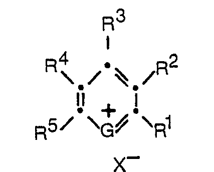
- the pyrylium and thiapyrylium dyes can be represented by the formula: wherein
- R 1 , R 3 , and R 5 in the above formula can be the same or different and each can represent a hydrogen, an alkyl group, an aryl group, an aralkyl group, an amino group, a styryl group or a bis(diaryl)vinylene group.
- these groups can be substituted, if desired, with additional groups that will not interfere with the staining capacity of the compounds.
- they may contain substituents such as alkyl, aryl, alkoxy, aryloxy, amino, substituted amino, and the like.
- substituents such as alkyl, aryl, alkoxy, aryloxy, amino, substituted amino, and the like.
- R 1 , R 3 , and/or R 5 are alkyl, it is preferred that they be alkyl of from 1 to 20 carbon atoms, i.e. methyl, ethyl, propyl, butyl, pentyl, hexyl, heptyl, octyl, nonyl, decyl, undecyl, dodecyl, tridecyl, tetradecyl, pentadecyl, hexadecyl, heptadecyl, octadecyl, nonadecyl, eicosyl, or an isomer of the foregoing. More preferably, the alkyl groups are chosen from among those groups having from 1 to 4 carbon atoms. It is most preferred that such an alkyl substituent be methyl.
- an aryl group of from 6 to 20 carbon atoms is preferred, for example, 1) phenyl, 2) 4-biphenyl, 3) naphthyl, 4) alkphenyl, such as 4-ethylphenyl, 4-propylphenyl, 5) alkoxyphenyl, e.g.
- the aryl group be phenyl or substituted phenyl and the alkylene group through which it is attached to the pyrylium or thiapyrylium ring have from 1 to 4 carbon atoms, e.g. methylene, ethylene, propylene, or butylene. It is most preferred that such an aralkyl group be benzyl.
- the group when the group is an amine group, it can be either a primary, secondary, or tertiary amine group.
- R 2 can be hydrogen or taken together with either R 1 or R 3 can represent the elements needed to complete an unsubstituted or substituted aromatic or carbocyclic ring system.
- R 4 can be hydrogen or can be taken together with either R 3 or R 5 to represent the elements needed to complete an unsubstituted or substituted aromatic or carbocyclic ring system, such as benzene, naphthalene, dihydronaphthalene, tetralin, indene, benzocycloheptadiene, cyclohexane, and cyclopentane.
- X- in the above structural formula is an anionic function including such anions as perchlorate, tetrafluoroborate, paratoluenesulfonate, sulfonate, periodate, chloride, bromide, fluoride, iodide, sulfate, bisulfate, bisulfite, chloroaluminate, and chloroferrate.
- Dextran T70, Ficoll-Pacque, and Percoll were purchased from Pharmacia Fine Chemicals, Piscataway, NJ.
- ACD acid, citrate, dextrose B-D 4606 prefilled blood collection tubes were purchased from VWR Scientific, Rochester, NY.
- Fetal calf serum, Raji cells (Burkitt's lymphoma), L-glutamine, and Roswell Park Memorial Institute 1640 medium [RPMI 1640 (Flow Laboratories, Inc., Product Catalog I, p. 126, 1977 description of composition medium)] were purchased from Flow Laboratories, McLean, VA.
- PermountTM mounting medium was purchased from Fisher Scientific, Rochester, NY.
- Gentamicin was purchased from Schering Corp., Union, NJ.
- Costar 96-well plates were purchased from Rochester Scientific, Rochester, NY.
- Fibroblasts (GM 1381) were purchased from Cell Repository, Camden, NJ, or American Type Culture Collection, Rockville, MD.
- Shandon CytospinTM Cytocentrifuge was purchased from Shandon Southern Instruments, Inc., Sewickley, PA. All other chemicals were reagent grade, unless otherwise noted, and were obtained from Eastman Kodak Company, Rochester, NY.
- Leukocyte-rich layers were purified from blood of healthy adult donors (taken in ACD tubes) by adding 1.5 mL of Dextran T70 (6% in balanced salt solution, BSS) to a 10 mL tube of blood. This was allowed to sediment for approximately 1 hour after which the cells were washed 3 times in BSS. Cells were counted and adjusted to a final density of 10 6 cells/mL.
- Leukocytes were further fractionated following a procedure by Olofsson (Scandinavian Journal of Haemetology, 24:254, 1980), which resulted in a significant enrichment of the less abundant cell classes.
- the fractionation was carried out as follows: Percoll (colloidal silica, clad with biocompatible polyvinyl pyrrolidone) and a balanced salt solution (BSS) were mixed in varying proportions and layered in a tube to produce an 11-step density gradient (1.01-1.12 g/cc).
- a suspension of leukocytes (buffy coat) was applied to the top of the tube and sedimented at 1,600 xg for 20 minutes. Each cell class was detectable at its unique density step.
- Raji cells were also prepared for staining. An aliquot was removed from the continuous culture and centrifuged to remove old media. Fresh Roswell Park Memorial Institute Medium 1640 (RPMI 1640), supplemented with 10% fetal calf serum, L-glutamine, and gentamicin, was added. Cells were counted and adjusted to a density of 10 6 viable cells/mL.
- RPMI 1640 Fresh Roswell Park Memorial Institute Medium 1640
- Fibroblasts were seeded on cover slips (22 mm 2 ) in tissue culture plates (35 mm 2 ) containing tissue culture medium. After 3-4 days of growth, when monolayers of fibroblasts were confluent, the medium was removed from the plates and replaced with 2 mL of 10- 6 molar dye solution (10- 3 molar dye/methanol solution diluted with BSS). The plates were incubated at 37°C for 10 minutes. The cover slips were removed, rinsed with BSS, and air dried. I he cover slips were then mounted, cell side down, on glass slides with glycerol-BSS (9:1).
- Purified polymorphonuclear leukocyte preparations were obtained by layering the buffy coats described above on the Ficoll-Pacque, centrifuging for 30 minutes at 400 RCF (rotational centrifugal force) and washing three times with BSS. Cells were counted and adjusted to a density of 10 6 cells mL.
- washed cell suspension from above was placed in the sample well of a Shandon Cytospin Cytocentrifuge and centrifuged at maximum speed for 10 minutes.
- the fixed cell preparations were air dried and stained for 1-2 minutes with 10- 3 molar dye (in methanol) and washed with distilled water.
- the method of the present invention for distinguishing cells can conveniently be carried out using conventional laboratory glassware, e.g., test tubes or glass slides.
- the dye can be incorporated into a matrix of absorbent material, such as a filter paper strip or a porous plastic lamina, by impregnation or otherwise, to yield a test element to which a cell sample can be applied.
- a matrix of absorbent material such as a filter paper strip or a porous plastic lamina
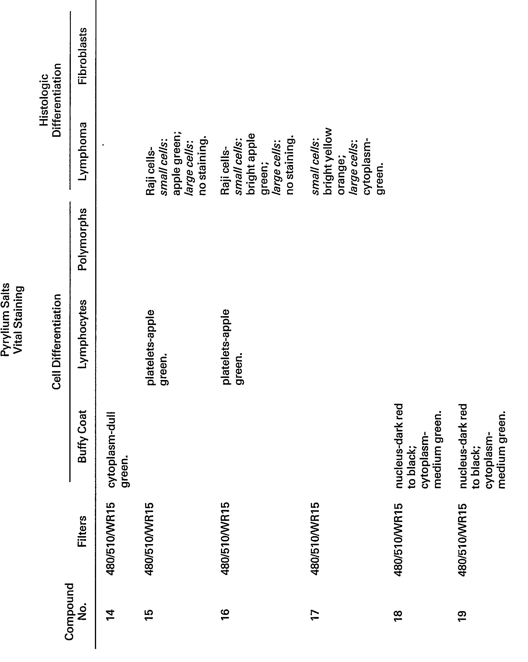
- Pyrylium salt #1 a preferred compound exhibited marked metachromasia, i.e., leukocyte nuclear and cytoplasmic staining were observed at -620 nm (red) and -550 nm (green), respectively. Platelets and the filamentous cytoplasm of fibroblasts stained green. * WR 15 has maximum transmission beyond 530 nm.
- Pyrylium salt #1 was evaluated as a stain for differentiating human leukocytes. Fractions of these cells, separated on a Percoll @ density gradient, as described above, were stained with the above-mentioned compound and evaluated by microspectrofluorometry and flow cytometry.
- Fraction 2 contained two distinct cell populations, the eosinophils and the neutrophils.
- the eosinophils exhibited appreciably lower green fluorescence than the neutrophils.
- Fraction 3 contained only neutrophils.
- Percoll cell fractions 7 and 8 were also evaluated by microspectrofluorometry. The distributions of fluorescent intensities showed that fraction 7 was pure lymphocytes, while fraction 8 included a lymphocyte population and an appreciable enrichment in monocytes.
- Unfractionated human leukocytes were stained with pyrylium salt #1, resulting in three-peak distributions in each of the following when determined by flow cytometry: the green (-550 nm) vs the red (-620 nm), red vs light scatter and green vs light scatter domains.
- Polymorphonuclear leukocytes were fractionated, fixed, and then stained, as described above, in "Methods", with pyrylium and thiapyrylium salts.
- thiapyrylium salts tested also showed bright and selective nuclear fluorescent staining (#26 and 27).
- Other thiapyrylium salts showed weaker, but also selective, nuclear staining (#22 and 36) as well as staining of the entire cell (#37 and 38).
Landscapes
- Health & Medical Sciences (AREA)
- Life Sciences & Earth Sciences (AREA)
- Immunology (AREA)
- Engineering & Computer Science (AREA)
- Molecular Biology (AREA)
- Chemical & Material Sciences (AREA)
- Biomedical Technology (AREA)
- Urology & Nephrology (AREA)
- Hematology (AREA)
- Biotechnology (AREA)
- Analytical Chemistry (AREA)
- Cell Biology (AREA)
- Tropical Medicine & Parasitology (AREA)
- Food Science & Technology (AREA)
- Medicinal Chemistry (AREA)
- Physics & Mathematics (AREA)
- Microbiology (AREA)
- Biochemistry (AREA)
- General Health & Medical Sciences (AREA)
- General Physics & Mathematics (AREA)
- Pathology (AREA)
- Investigating Or Analysing Biological Materials (AREA)
- Measuring Or Testing Involving Enzymes Or Micro-Organisms (AREA)
- Sampling And Sample Adjustment (AREA)
Description
- The present invention relates to biological stains.
- The staining of biological cells and tissues with dyes, especially fluorochromic dyes, in order to differentiate one from another or simply to render them more easily observable under a microscope or other sensing means is well known in the art. Many of these dyes interact with the DNA or the RNA ofthe cell, or both, yielding products that fluoresce at different wavelengths, thereby being distinguishable. By such means it is possible to differentiate, for example, among the five types of peripheral blood leucocytes: neutrophils, eosinophils, basophils, lymphocytes, and monocytes; between cancerous and normal cells; and between mature and immature cells. Such differentiation enables the cytologist to diagnose certain disease states.
- U.S. Patent 3,684,377 describes a composition for the enumeration and differentiation of leucocytes. A suspension of fresh whole blood in a solution of Acridine Orange (Color Index 46005) having a pH factor and osmolality within normal physiological ranges for human blood is subjected to radiation from a blue laser. White cells are distinguished by detecting the resultant green fluorescence emitted by the stained nuclei of the leucocytes and differentiated by the amplitude of the red fluorescence emitted.
- Blum, R. S., Glade, P. R., and Chessin, L. N., "Euchrysine, A Supravital Fluorescent Lysosomal Stain: Technic and Application for Hematologic Investigation," Blood, 33(1):87-99, 1969 describes methods for the preparation of Euchrysine (Color Index 46040), an aminoacridine fluorescent supravital dye, into a form suitable for hematologic investigation and its use in the characterization of lysosomes in human peripheral blood, bone marrow, and established lymphoid cell lines (maintained in vitro).
- The dyes employed in the above references, Acridine Orange or Euchrysine, have disadvantages in that they produce undesirable background fluorescence.
- The use of certain styryl dyes as biological stains has been described in U.K. Published Application 2,074,340A and in Bereiter-Hahn, "Dimethylaminostyrylmethylpyridiniumiodine (DASPMI) as a Fluorescent Probe for Mitochondria In Situ", Biochimica et Biophysica Acta, 423:1-14, 1976.
- Further, U.S. Patent 4,232,121 describes a method for selecting methine dyes, especially cyanine dyes, that inhibit the growth of cells. Dyes containing a pyrylium nucleus are mentioned as being useful, among others. In the present work it has also been noted that the compounds described herein are capable of inhibiting the growth of cells, especially somatic cells.
- The object of the present invention is to provide biological staining methods.
- This object is accomplished by a method for distinguishing cells in a biological sample by staining with a dye, characterized in that said dye is a pyrylium or thiapyrylium compound as described in claim 1.
- Pyrylium and thiapyrylium compounds are useful as both vital and fixed cell stains and then can be used without a wash step. They are especially useful for differentiation of biological cells and tissues. These dyes provide a wide range of absorption maxima in the visible range and many of them are fluorescent and exhibit a wide range of wavelengths that can be used for excitation and emission. Of these compounds, the preferred ones are metachromatic fluorochromes and produce unique staining of the cell nucleus and cytoplasm, which stain red and green, respectively.
- The pyrylium and thiapyrylium dyes can be represented by the formula:

- G is O or S;
- R1, R3, and R5 are independently selected from the group consisting of hydrogen, alkyl, aryl, aralkyl, amino, styryl and bis(diaryl)vinylene,
- R2 is hydrogen or, taken together with either R1 or R3, represents the elements needed to complete an aromatic or a carbocyclic ring system;
- R4 is hydrogen or, taken together with either R3 or R5, represents the elements needed to complete an aromatic or a carbocyclic ring system; and
- X- is an anion.
- The pyrylium and thiapyrylium dyes employed in the practice of this invention and methods for their preparation are known in the art, as described in, for example, U.S. Patent 3,141,770; U.S. Patent 3,148,067; U.S. Patent 3,250,615; U.S. Patent 3,579,345; U.S. Patent 3,822,270; U.S. Patent 3,938,994; and U.S. Patent 4,173,473.
- As stated above, R1, R3, and R5 in the above formula can be the same or different and each can represent a hydrogen, an alkyl group, an aryl group, an aralkyl group, an amino group, a styryl group or a bis(diaryl)vinylene group.
- A person skilled in the art will understand that these groups can be substituted, if desired, with additional groups that will not interfere with the staining capacity of the compounds. For example, they may contain substituents such as alkyl, aryl, alkoxy, aryloxy, amino, substituted amino, and the like. Generally, it is preferable, where such substituents are used, to choose those that are not strongly electron withdrawing.
- Where R1, R3, and/or R5 are alkyl, it is preferred that they be alkyl of from 1 to 20 carbon atoms, i.e. methyl, ethyl, propyl, butyl, pentyl, hexyl, heptyl, octyl, nonyl, decyl, undecyl, dodecyl, tridecyl, tetradecyl, pentadecyl, hexadecyl, heptadecyl, octadecyl, nonadecyl, eicosyl, or an isomer of the foregoing. More preferably, the alkyl groups are chosen from among those groups having from 1 to 4 carbon atoms. It is most preferred that such an alkyl substituent be methyl.
- Where the group is aryl, an aryl group of from 6 to 20 carbon atoms is preferred, for example, 1) phenyl, 2) 4-biphenyl, 3) naphthyl, 4) alkphenyl, such as 4-ethylphenyl, 4-propylphenyl, 5) alkoxyphenyl, e.g. 4-ethoxyphenyl, 4-methoxyphenyl, 4-amyloxyphenyl, 2-hexoxyphenyl, 2-methoxyphenyl, 2-amyloxyphenyl, 3,4-dimethoxyphenyl, 6) ro-hydroxyalkoxyphenyl, e.g., 2-hydroxyethoxyphenyl, 3-hydroxyethoxyphenyl, 7) 4-hydroxyphenyl, halophenyl, e.g., 3,4-dichlorophenyl, 3,4-dibromophenyl, 4-chlorophenyl, 2,4-dichlorophenyl, 8) aminophenyl, e.g., 4-diethylaminophenyl, 4-dimethylaminophenyl, 4-dibutylaminophenyl, 4-dioctylaminophenyl.
- Where the group is aralkyl, it is preferred that the aryl group be phenyl or substituted phenyl and the alkylene group through which it is attached to the pyrylium or thiapyrylium ring have from 1 to 4 carbon atoms, e.g. methylene, ethylene, propylene, or butylene. It is most preferred that such an aralkyl group be benzyl.
- When the group is an amine group, it can be either a primary, secondary, or tertiary amine group.
- In the above structural formula for the pyrylium and thiapyrylium dyes employed in the practice of this invention, R2 can be hydrogen or taken together with either R1 or R3 can represent the elements needed to complete an unsubstituted or substituted aromatic or carbocyclic ring system. R4 can be hydrogen or can be taken together with either R3 or R5 to represent the elements needed to complete an unsubstituted or substituted aromatic or carbocyclic ring system, such as benzene, naphthalene, dihydronaphthalene, tetralin, indene, benzocycloheptadiene, cyclohexane, and cyclopentane.
- X- in the above structural formula is an anionic function including such anions as perchlorate, tetrafluoroborate, paratoluenesulfonate, sulfonate, periodate, chloride, bromide, fluoride, iodide, sulfate, bisulfate, bisulfite, chloroaluminate, and chloroferrate.
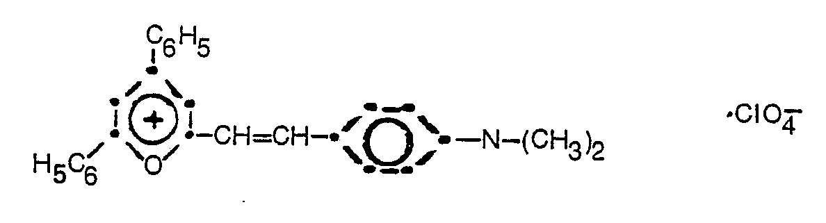
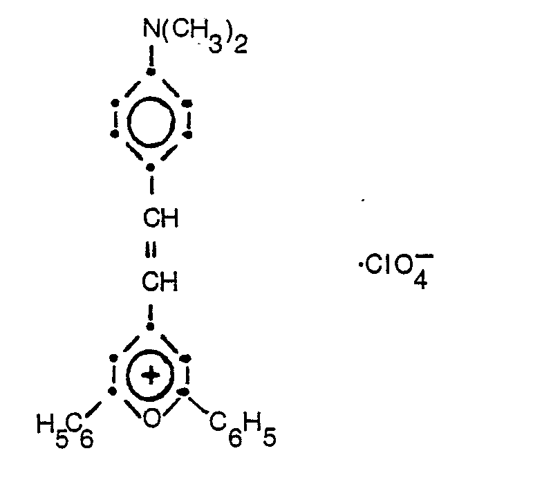
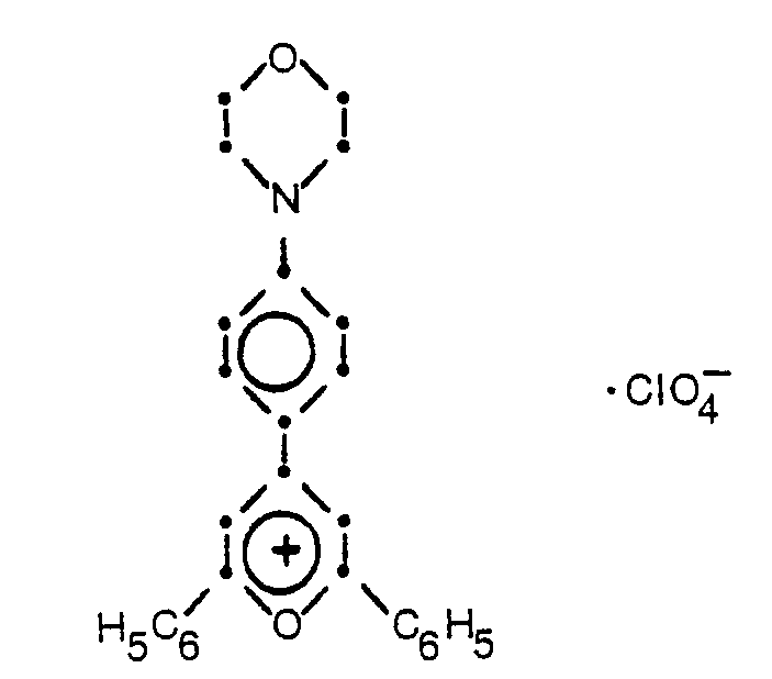
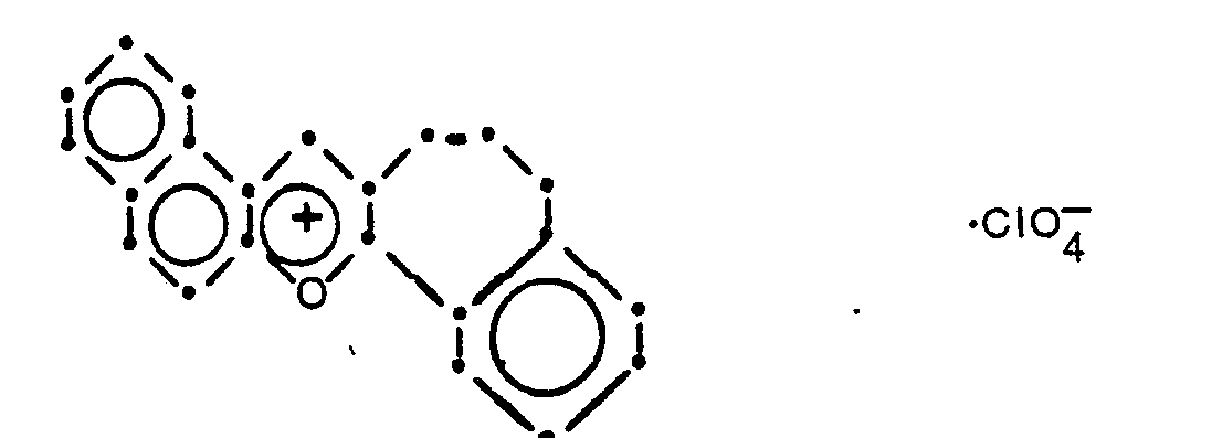
- The following structural formulae illustrate compounds that have been found to be especially useful in the practice of the present invention. These compounds and the results obtained with them are described further in the examples below.
-
- Compound No. 1:

- Compound No. 2:

- Compound No. 3:

- Compound No.4:
- Compound No. 5:
- Compound No. 6:

- Compound No. 7:

- Compound No. 8:
- Compound No. 9:

- Compound No. 10:

- Compound No. 11:

- Compound No. 12:

- Compound No. 13:

- Compound No. 14:

- Compound No. 15:

- Compound No. 16:

- Compound No. 17:

- Compound No. 18:

- Compound No. 19:

- Compound No. 20:

- Compound No. 21:
-

- Compound No. 22:

- Compound No. 23:

- Compound No. 24:

- Compound No. 25:

- Compound No. 26:

- Compound No. 27:

- Compound No. 28:

- Compound No. 29:

- Compound No. 30:

- Compound No. 31:

- Compound No. 32:

- Compound No. 33:
- Compound No. 34:

- Compound No. 35:

- Compound No. 36:

- Compound No. 37:

- In the following examples, Dextran T70, Ficoll-Pacque, and Percoll were purchased from Pharmacia Fine Chemicals, Piscataway, NJ. ACD (acid, citrate, dextrose B-D 4606) prefilled blood collection tubes were purchased from VWR Scientific, Rochester, NY. Fetal calf serum, Raji cells (Burkitt's lymphoma), L-glutamine, and Roswell Park Memorial Institute 1640 medium [RPMI 1640 (Flow Laboratories, Inc., Product Catalog I, p. 126, 1977 description of composition medium)] were purchased from Flow Laboratories, McLean, VA. Permount™ mounting medium was purchased from Fisher Scientific, Rochester, NY. Gentamicin was purchased from Schering Corp., Union, NJ. Costar 96-well plates were purchased from Rochester Scientific, Rochester, NY. Fibroblasts (GM 1381) were purchased from Cell Repository, Camden, NJ, or American Type Culture Collection, Rockville, MD. Shandon CytospinTM Cytocentrifuge was purchased from Shandon Southern Instruments, Inc., Sewickley, PA. All other chemicals were reagent grade, unless otherwise noted, and were obtained from Eastman Kodak Company, Rochester, NY.
- Leukocyte-rich layers (buffy coats) were purified from blood of healthy adult donors (taken in ACD tubes) by adding 1.5 mL of Dextran T70 (6% in balanced salt solution, BSS) to a 10 mL tube of blood. This was allowed to sediment for approximately 1 hour after which the cells were washed 3 times in BSS. Cells were counted and adjusted to a final density of 106 cells/mL.
- Leukocytes were further fractionated following a procedure by Olofsson (Scandinavian Journal of Haemetology, 24:254, 1980), which resulted in a significant enrichment of the less abundant cell classes. The fractionation was carried out as follows: Percoll (colloidal silica, clad with biocompatible polyvinyl pyrrolidone) and a balanced salt solution (BSS) were mixed in varying proportions and layered in a tube to produce an 11-step density gradient (1.01-1.12 g/cc). A suspension of leukocytes (buffy coat) was applied to the top of the tube and sedimented at 1,600 xg for 20 minutes. Each cell class was detectable at its unique density step.
- Raji cells were also prepared for staining. An aliquot was removed from the continuous culture and centrifuged to remove old media. Fresh Roswell Park Memorial Institute Medium 1640 (RPMI 1640), supplemented with 10% fetal calf serum, L-glutamine, and gentamicin, was added. Cells were counted and adjusted to a density of 106 viable cells/mL.
- Two hundred microliters of buffy coat cells, fractionated leukocytes, or Raji cells at 106 cells/mL was mixed with 200 ilL of 10-5 molar dye. The cells were allowed to stand at room temperature for 15 minutes, and then slides were prepared by centrifugation in a Shandon Cytospin Cytocentrifuge for 5 minutes at 1500 RPM. After centrifugation, slides were removed and allowed to dry. Cover slips were placed over cells using Permount medium.
- Fibroblasts were seeded on cover slips (22 mm2) in tissue culture plates (35 mm2) containing tissue culture medium. After 3-4 days of growth, when monolayers of fibroblasts were confluent, the medium was removed from the plates and replaced with 2 mL of 10-6 molar dye solution (10-3 molar dye/methanol solution diluted with BSS). The plates were incubated at 37°C for 10 minutes. The cover slips were removed, rinsed with BSS, and air dried. I he cover slips were then mounted, cell side down, on glass slides with glycerol-BSS (9:1).
- Purified polymorphonuclear leukocyte preparations were obtained by layering the buffy coats described above on the Ficoll-Pacque, centrifuging for 30 minutes at 400 RCF (rotational centrifugal force) and washing three times with BSS. Cells were counted and adjusted to a density of 106 cells mL.
- Two hundred microliters of washed cell suspension from above was placed in the sample well of a Shandon Cytospin Cytocentrifuge and centrifuged at maximum speed for 10 minutes. The fixed cell preparations were air dried and stained for 1-2 minutes with 10-3 molar dye (in methanol) and washed with distilled water.
- Stained cell preparations were examined under a Zeiss Universal epifluorescence microscope. The filters used for evaluating the vitally stained cells are listed below and are indicated for each compound in Table I.

- The method of the present invention for distinguishing cells can conveniently be carried out using conventional laboratory glassware, e.g., test tubes or glass slides. Alternatively, the dye can be incorporated into a matrix of absorbent material, such as a filter paper strip or a porous plastic lamina, by impregnation or otherwise, to yield a test element to which a cell sample can be applied. Such techniques are well-known to those skilled in the art.
- Cells from buffy coat preparations and from tissue lines, e.g., lymphoma and fibroblast lines, were vitally stained, as described above, with pyrylium and thiapyrylium compounds, as listed in Tables I and II. The staining results are also listed in Tables I and II.
- Pyrylium salt #1, a preferred compound exhibited marked metachromasia, i.e., leukocyte nuclear and cytoplasmic staining were observed at -620 nm (red) and -550 nm (green), respectively. Platelets and the filamentous cytoplasm of fibroblasts stained green.
*WR 15 has maximum transmission beyond 530 nm.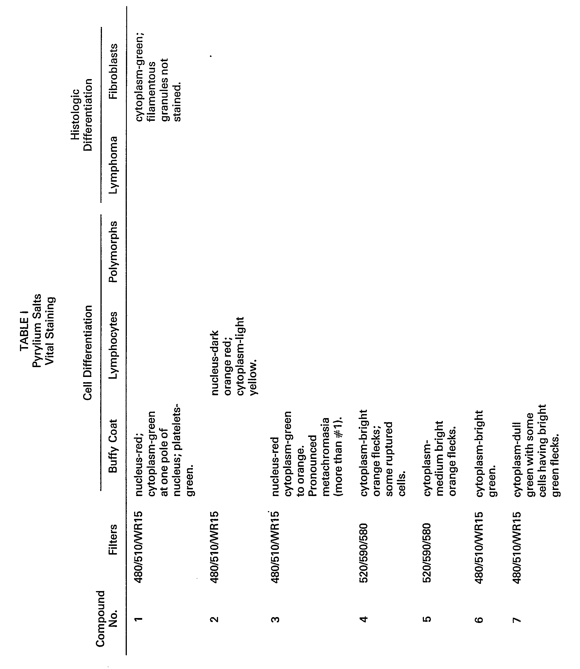


- Pyrylium salt #1 was evaluated as a stain for differentiating human leukocytes. Fractions of these cells, separated on a Percoll@ density gradient, as described above, were stained with the above-mentioned compound and evaluated by microspectrofluorometry and flow cytometry.
- Fraction 2 contained two distinct cell populations, the eosinophils and the neutrophils. The eosinophils exhibited appreciably lower green fluorescence than the neutrophils. Fraction 3 contained only neutrophils.
- Percoll cell fractions 7 and 8 were also evaluated by microspectrofluorometry. The distributions of fluorescent intensities showed that fraction 7 was pure lymphocytes, while fraction 8 included a lymphocyte population and an appreciable enrichment in monocytes.
- Unfractionated human leukocytes were stained with pyrylium salt #1, resulting in three-peak distributions in each of the following when determined by flow cytometry: the green (-550 nm) vs the red (-620 nm), red vs light scatter and green vs light scatter domains.
- Rabbit blood was fractionated on a Percoll density gradient described above. Fraction 2, obtained at 1.095 g/cc, was enriched in polychromatic cells (relative to normocytes) as shown by Wright's staining of a cytocentrifuge preparation of the cells. Cell density was adjusted to 106 cells/mL as described above and the cells stained with 10-5 molar pyrylium salt #1 in a balanced salt solution, without Ca++ and Mg++, for 15 minutes at room temperature. Microscopic examination of cytocentrifuge preparations of the stained cells showed a population of non-nucleated cells that exhibited filamentous projections that fluoresced green when excited with light at 480 nm. Counterstaining the same slide with Wright's Giemsa stain resulted in gray-violet-stained reticulocytes, which confirmed the above observations.
- Polymorphonuclear leukocytes were fractionated, fixed, and then stained, as described above, in "Methods", with pyrylium and thiapyrylium salts.
- The staining properties of these compounds are summarized in Table III. At least three of the pyrylium salts (#13, 28, and 11) were extremely bright and showed specific nuclear fluorescent stains. Compounds #29 and #30 showed weaker nuclear fluorescence. In general, pyrylium salts, showing bright nuclear staining, contained methoxyphenyl groups. In addition to staining the nucleus, certain of the pyrylium salts stained either the entire cell (#31 and 14) or the cell cytoplasm (#32, 15, 12, 33, 34, and 35).
- Certain of the thiapyrylium salts tested also showed bright and selective nuclear fluorescent staining (#26 and 27). Other thiapyrylium salts showed weaker, but also selective, nuclear staining (#22 and 36) as well as staining of the entire cell (#37 and 38).

Claims (7)
Applications Claiming Priority (2)
| Application Number | Priority Date | Filing Date | Title |
|---|---|---|---|
| US06/452,260 US4555396A (en) | 1982-12-22 | 1982-12-22 | Use of pyrylium and thiapyrylium compounds as biological stains |
| US452260 | 1982-12-22 |
Publications (3)
| Publication Number | Publication Date |
|---|---|
| EP0112552A2 EP0112552A2 (en) | 1984-07-04 |
| EP0112552A3 EP0112552A3 (en) | 1985-10-16 |
| EP0112552B1 true EP0112552B1 (en) | 1989-03-01 |
Family
ID=23795762
Family Applications (1)
| Application Number | Title | Priority Date | Filing Date |
|---|---|---|---|
| EP83112910A Expired EP0112552B1 (en) | 1982-12-22 | 1983-12-21 | Use of pyrylium or thiapyrylium compounds as biological stains |
Country Status (5)
| Country | Link |
|---|---|
| US (1) | US4555396A (en) |
| EP (1) | EP0112552B1 (en) |
| JP (1) | JPS59133460A (en) |
| CA (1) | CA1194766A (en) |
| DE (1) | DE3379283D1 (en) |
Families Citing this family (14)
| Publication number | Priority date | Publication date | Assignee | Title |
|---|---|---|---|---|
| US4886744A (en) * | 1985-04-25 | 1989-12-12 | Polaroid Corporation | Fluorescent conjugates and biological diagnostic assay system |
| US4783401A (en) * | 1986-10-31 | 1988-11-08 | Smithkline Beckman Corporation | Viable cell labelling |
| US4774250A (en) * | 1987-04-02 | 1988-09-27 | Dana Farber Cancer Institute | Composition and method for treating differentiated carcinoma or melanoma cells with thiapyrylium dyes |
| ATE145337T1 (en) * | 1988-05-02 | 1996-12-15 | Phanos Tech Inc | COMPOUNDS, COMPOSITIONS AND METHODS FOR BONDING BIO-AFFECTION SUBSTANCES TO SURFACE MEMBRANES OF BIO-PARTICLES |
| EP0599337B1 (en) * | 1992-11-27 | 2006-03-08 | Canon Kabushiki Kaisha | Method for detection of nucleic acid and probe therefor |
| JP3247001B2 (en) * | 1992-12-21 | 2002-01-15 | キヤノン株式会社 | Method for detecting double-stranded nucleic acid using pyrylium compound, probe containing pyrylium compound and method for detecting target nucleic acid using the same, novel pyrylium compound |
| US5670315A (en) * | 1993-09-13 | 1997-09-23 | Canon Kabushiki Kaisha | Nucleic acid determination employing pyryilium dye |
| JPH07233065A (en) | 1993-12-27 | 1995-09-05 | Canon Inc | Photochemotherapeutic drug containing pyrylium salt or pyrilium-like salt |
| US5723288A (en) * | 1994-05-06 | 1998-03-03 | The University Of North Carolina At Chapel Hill | Method of fluorescent detection of nucleic acids and cytoskeleton elements using bis-dicationic aryl furans, and kits useful therefor |
| DE69532255D1 (en) * | 1994-05-26 | 2004-01-22 | Canon Kk | Method for the detection of a target substance in a sample using pyrylium compound |
| DE69718268D1 (en) | 1996-10-03 | 2003-02-13 | Canon Kk | Methods for the detection of target nucleic acid, methods for their quantification and pyrylium compounds for chemiluminescence analysis |
| ES2222825B1 (en) * | 2003-07-23 | 2005-12-16 | Universidad Politecnica De Valencia | METHOD FOR THE DETECTION OF NUCLEOFILIC CHEMICAL SPECIES. |
| CA2859985C (en) | 2011-12-21 | 2020-11-03 | The Regents Of The University Of Colorado | Anti-cancer compounds targeting ral gtpases and methods of using the same |
| EP3166609B1 (en) | 2014-07-10 | 2020-03-11 | The Regents of The University of Colorado, A Body Corporate | 6-amino-1,3-dimethyl-4-(4-(trifluoromethyl)phenyl)-1,4-dihydropyrano [2,3-c]pyrazole-5-carbonitrile and related compounds as ral gtpase inhibitors for treating cancer metastasis |
Family Cites Families (21)
| Publication number | Priority date | Publication date | Assignee | Title |
|---|---|---|---|---|
| FR1222952A (en) * | 1958-04-29 | 1960-06-14 | Univ Kansas Res Foundation | Fluorescent dyes |
| BE626528A (en) * | 1961-10-23 | |||
| US3148067A (en) * | 1962-06-22 | 1964-09-08 | Eastman Kodak Co | Photographic silver halide emulsions stabilized with pyrylium compounds |
| US3271257A (en) * | 1963-10-10 | 1966-09-06 | Jr Hervy E Averette | Cytodiagnosis of ruptured fetal membranes |
| US3497690A (en) * | 1967-09-21 | 1970-02-24 | Bausch & Lomb | Method and apparatus for classifying biological cells by measuring the size and fluorescent response thereof |
| US3579345A (en) * | 1967-10-02 | 1971-05-18 | Eastman Kodak Co | Direct positive emulsions sensitized with pyrylium and/or thiapyrlium salts |
| US3684377A (en) * | 1970-07-13 | 1972-08-15 | Bio Physics Systems Inc | Method for analysis of blood by optical analysis of living cells |
| US3822270A (en) * | 1971-08-09 | 1974-07-02 | Eastman Kodak Co | Pyrylium dyes having a fused,rigidized nitrogen-containing ring |
| US3938994A (en) * | 1972-03-17 | 1976-02-17 | Eastman Kodak Company | Pyrylium dyes for electrophotographic composition and element |
| US4094745A (en) * | 1973-06-22 | 1978-06-13 | John Scholefield | Method of staining microscopic organisms |
| US3883247A (en) * | 1973-10-30 | 1975-05-13 | Bio Physics Systems Inc | Method for fluorescence analysis of white blood cells |
| US4025349A (en) * | 1974-03-18 | 1977-05-24 | Eastman Kodak Company | Silver halide photographic elements spectrally sensitized with an acetylenic analog of cyanine or merocyanine dyes |
| CH613523A5 (en) * | 1975-06-27 | 1979-09-28 | Inst Nat Sante Rech Med | Method for displaying basophils |
| US4173473A (en) * | 1977-07-06 | 1979-11-06 | Eastman Kodak Company | Radiation sensitive compositions containing pyrylium compounds |
| DE2754403A1 (en) * | 1977-12-07 | 1979-06-13 | Basf Ag | METHINE DYES |
| EP0004061B1 (en) * | 1978-03-09 | 1982-05-19 | MERCK PATENT GmbH | Method for determining leukaemic cells |
| JPS54133483A (en) * | 1978-04-10 | 1979-10-17 | Nobuo Sakuse | Colored fluorescent material and its manufacture |
| US4232121A (en) * | 1978-09-25 | 1980-11-04 | Eastman Kodak Company | Process for selecting methine dyes which inhibit cell growth |
| US4226868A (en) * | 1978-09-25 | 1980-10-07 | Eastman Kodak Company | Processes for inhibiting the growth of sea urchin eggs |
| DE2916433A1 (en) * | 1979-04-24 | 1980-11-13 | Hoechst Ag | CHROMOPHORE CEPHALOSPORINE AND METHOD FOR THE PRODUCTION THEREOF |
| CA1155041A (en) * | 1980-04-21 | 1983-10-11 | Michael E. Jolley | Fluorescent nucleic acid stains |
-
1982
- 1982-12-22 US US06/452,260 patent/US4555396A/en not_active Expired - Lifetime
-
1983
- 1983-03-28 CA CA000424618A patent/CA1194766A/en not_active Expired
- 1983-12-21 EP EP83112910A patent/EP0112552B1/en not_active Expired
- 1983-12-21 DE DE8383112910T patent/DE3379283D1/en not_active Expired
- 1983-12-22 JP JP58241095A patent/JPS59133460A/en active Pending
Also Published As
| Publication number | Publication date |
|---|---|
| US4555396A (en) | 1985-11-26 |
| EP0112552A2 (en) | 1984-07-04 |
| DE3379283D1 (en) | 1989-04-06 |
| EP0112552A3 (en) | 1985-10-16 |
| CA1194766A (en) | 1985-10-08 |
| JPS59133460A (en) | 1984-07-31 |
Similar Documents
| Publication | Publication Date | Title |
|---|---|---|
| EP0112552B1 (en) | Use of pyrylium or thiapyrylium compounds as biological stains | |
| CA1191079A (en) | Metachromatic dye sorption means for differential determination of developmental stages of neutrophilic granulocytic cells and other leukocytes | |
| CA2024166C (en) | Compounds and reagent compositions and their use in the quantitative determination of reticulocytes in whole blood | |
| US4581223A (en) | Individual leukocyte determination by means of differential metachromatic dye sorption | |
| DE68918004T2 (en) | Method for the analysis of cell components in a liquid. | |
| US4882284A (en) | Method for quantitating and differentiating white blood cells | |
| Rabellino et al. | Human megakaryocytes. I. Characterization of the membrane and cytoplasmic components of isolated marrow megakaryocytes. | |
| JP3783808B2 (en) | Leukocyte classification and counting reagent | |
| US5958776A (en) | Method for classifying and counting immature leukocytes | |
| CA1205365A (en) | Metachromatic dye sorption and fluorescent light emissive means for determination of developmental stages of neutrophilic granulocytic cells and other leukocytes | |
| JPH06100596B2 (en) | Method for classifying leukocytes by flow cytometry | |
| US4840784A (en) | Use of pyrylium and thiapyrylium compounds as biological stains | |
| EP0004061B1 (en) | Method for determining leukaemic cells | |
| McIntire et al. | Flow cytometric analysis of DNA in cells obtained from deparaffinized formalin‐fixed lymphoid tissues | |
| US5407794A (en) | Oxazine stained lymphocytes and method | |
| CA1289042C (en) | Method of identifying polymethine stained lymphocyte subpopulations and compositions thereof | |
| Ronot et al. | Assessment of cell viability in mammalian cell lines | |
| Grawé | Flow cytometric analysis of micronuclei in erythrocytes | |
| DE68922548T2 (en) | Fluorescent dyes. | |
| Kass | Identification of lymphocyte subpopulations with a polymethine dye. | |
| Brüvere et al. | Fluorescent characteristics of blood leukocytes of patients with malignant and nonmalignant diseases | |
| Sakata | Reagent characteristics in the XE-2100 NRBC channel | |
| Rousselle et al. | Flow cytometric analysis of DNA content of living and fixed cells: A comparative study using various fixatives | |
| Hoshino | Heterogeneity of Tumor Cell DNA Content¹ | |
| KR19980042627A (en) | Classification method of glazed leukocyte |
Legal Events
| Date | Code | Title | Description |
|---|---|---|---|
| PUAI | Public reference made under article 153(3) epc to a published international application that has entered the european phase |
Free format text: ORIGINAL CODE: 0009012 |
|
| AK | Designated contracting states |
Designated state(s): DE FR GB IT |
|
| PUAL | Search report despatched |
Free format text: ORIGINAL CODE: 0009013 |
|
| AK | Designated contracting states |
Designated state(s): DE FR GB IT |
|
| 17P | Request for examination filed |
Effective date: 19851210 |
|
| 17Q | First examination report despatched |
Effective date: 19870506 |
|
| GRAA | (expected) grant |
Free format text: ORIGINAL CODE: 0009210 |
|
| AK | Designated contracting states |
Kind code of ref document: B1 Designated state(s): DE FR GB IT |
|
| REF | Corresponds to: |
Ref document number: 3379283 Country of ref document: DE Date of ref document: 19890406 |
|
| ITF | It: translation for a ep patent filed | ||
| ET | Fr: translation filed | ||
| PLBE | No opposition filed within time limit |
Free format text: ORIGINAL CODE: 0009261 |
|
| STAA | Information on the status of an ep patent application or granted ep patent |
Free format text: STATUS: NO OPPOSITION FILED WITHIN TIME LIMIT |
|
| 26N | No opposition filed | ||
| ITTA | It: last paid annual fee | ||
| REG | Reference to a national code |
Ref country code: GB Ref legal event code: 732E |
|
| PGFP | Annual fee paid to national office [announced via postgrant information from national office to epo] |
Ref country code: FR Payment date: 19971209 Year of fee payment: 15 |
|
| PGFP | Annual fee paid to national office [announced via postgrant information from national office to epo] |
Ref country code: GB Payment date: 19971212 Year of fee payment: 15 |
|
| PGFP | Annual fee paid to national office [announced via postgrant information from national office to epo] |
Ref country code: DE Payment date: 19971230 Year of fee payment: 15 |
|
| PG25 | Lapsed in a contracting state [announced via postgrant information from national office to epo] |
Ref country code: GB Free format text: LAPSE BECAUSE OF NON-PAYMENT OF DUE FEES Effective date: 19981221 |
|
| GBPC | Gb: european patent ceased through non-payment of renewal fee |
Effective date: 19981221 |
|
| PG25 | Lapsed in a contracting state [announced via postgrant information from national office to epo] |
Ref country code: FR Free format text: LAPSE BECAUSE OF NON-PAYMENT OF DUE FEES Effective date: 19990831 |
|
| REG | Reference to a national code |
Ref country code: FR Ref legal event code: ST |
|
| PG25 | Lapsed in a contracting state [announced via postgrant information from national office to epo] |
Ref country code: DE Free format text: LAPSE BECAUSE OF NON-PAYMENT OF DUE FEES Effective date: 19991001 |