EP0107509B1 - Protein a material and preparation thereof - Google Patents
Protein a material and preparation thereof Download PDFInfo
- Publication number
- EP0107509B1 EP0107509B1 EP83306500A EP83306500A EP0107509B1 EP 0107509 B1 EP0107509 B1 EP 0107509B1 EP 83306500 A EP83306500 A EP 83306500A EP 83306500 A EP83306500 A EP 83306500A EP 0107509 B1 EP0107509 B1 EP 0107509B1
- Authority
- EP
- European Patent Office
- Prior art keywords
- protein
- amino acid
- dna
- nucleotide sequence
- coding
- Prior art date
- Legal status (The legal status is an assumption and is not a legal conclusion. Google has not performed a legal analysis and makes no representation as to the accuracy of the status listed.)
- Expired - Lifetime
Links
- 108090000623 proteins and genes Proteins 0.000 title claims abstract description 94
- 102000004169 proteins and genes Human genes 0.000 title claims abstract description 85
- 239000000463 material Substances 0.000 title claims abstract description 46
- 238000002360 preparation method Methods 0.000 title description 8
- 239000002773 nucleotide Substances 0.000 claims abstract description 36
- 125000003729 nucleotide group Chemical group 0.000 claims abstract description 36
- 230000027455 binding Effects 0.000 claims abstract description 25
- 239000013598 vector Substances 0.000 claims abstract description 5
- 108020004414 DNA Proteins 0.000 claims description 67
- 239000013612 plasmid Substances 0.000 claims description 40
- 239000012634 fragment Substances 0.000 claims description 22
- 241000588724 Escherichia coli Species 0.000 claims description 18
- 102100035971 Molybdopterin molybdenumtransferase Human genes 0.000 claims description 16
- 101710119577 Molybdopterin molybdenumtransferase Proteins 0.000 claims description 16
- 108091028043 Nucleic acid sequence Proteins 0.000 claims description 16
- 244000005700 microbiome Species 0.000 claims description 8
- 108020004511 Recombinant DNA Proteins 0.000 claims description 6
- 239000000203 mixture Substances 0.000 claims description 6
- 102000053602 DNA Human genes 0.000 claims description 4
- 238000012546 transfer Methods 0.000 claims description 4
- 239000012620 biological material Substances 0.000 claims description 2
- 238000004519 manufacturing process Methods 0.000 claims description 2
- 230000000813 microbial effect Effects 0.000 claims description 2
- 125000003275 alpha amino acid group Chemical group 0.000 claims 12
- 210000002925 A-like Anatomy 0.000 abstract description 13
- 238000010367 cloning Methods 0.000 abstract description 6
- 238000003786 synthesis reaction Methods 0.000 abstract description 4
- 230000015572 biosynthetic process Effects 0.000 abstract description 3
- 230000004913 activation Effects 0.000 abstract description 2
- 238000002405 diagnostic procedure Methods 0.000 abstract description 2
- 238000012512 characterization method Methods 0.000 abstract 1
- 210000004027 cell Anatomy 0.000 description 25
- 150000001413 amino acids Chemical group 0.000 description 20
- 102100032157 Adenylate cyclase type 10 Human genes 0.000 description 17
- 239000000499 gel Substances 0.000 description 16
- KCXVZYZYPLLWCC-UHFFFAOYSA-N EDTA Chemical compound OC(=O)CN(CC(O)=O)CCN(CC(O)=O)CC(O)=O KCXVZYZYPLLWCC-UHFFFAOYSA-N 0.000 description 12
- 239000000243 solution Substances 0.000 description 11
- LFQSCWFLJHTTHZ-UHFFFAOYSA-N Ethanol Chemical compound CCO LFQSCWFLJHTTHZ-UHFFFAOYSA-N 0.000 description 10
- 238000005119 centrifugation Methods 0.000 description 9
- 238000000605 extraction Methods 0.000 description 9
- 239000006166 lysate Substances 0.000 description 9
- 108091008146 restriction endonucleases Proteins 0.000 description 8
- HEDRZPFGACZZDS-UHFFFAOYSA-N Chloroform Chemical compound ClC(Cl)Cl HEDRZPFGACZZDS-UHFFFAOYSA-N 0.000 description 6
- HEMHJVSKTPXQMS-UHFFFAOYSA-M Sodium hydroxide Chemical compound [OH-].[Na+] HEMHJVSKTPXQMS-UHFFFAOYSA-M 0.000 description 6
- 239000000872 buffer Substances 0.000 description 6
- 238000012360 testing method Methods 0.000 description 6
- ISWSIDIOOBJBQZ-UHFFFAOYSA-N Phenol Chemical compound OC1=CC=CC=C1 ISWSIDIOOBJBQZ-UHFFFAOYSA-N 0.000 description 5
- 239000007984 Tris EDTA buffer Substances 0.000 description 5
- ZMMJGEGLRURXTF-UHFFFAOYSA-N ethidium bromide Chemical compound [Br-].C12=CC(N)=CC=C2C2=CC=C(N)C=C2[N+](CC)=C1C1=CC=CC=C1 ZMMJGEGLRURXTF-UHFFFAOYSA-N 0.000 description 5
- 229960005542 ethidium bromide Drugs 0.000 description 5
- 238000000034 method Methods 0.000 description 5
- 229920006395 saturated elastomer Polymers 0.000 description 5
- 239000006228 supernatant Substances 0.000 description 5
- 229920000936 Agarose Polymers 0.000 description 4
- 108091026890 Coding region Proteins 0.000 description 4
- 239000000020 Nitrocellulose Substances 0.000 description 4
- 108091034117 Oligonucleotide Proteins 0.000 description 4
- 238000003556 assay Methods 0.000 description 4
- 230000004071 biological effect Effects 0.000 description 4
- 238000006243 chemical reaction Methods 0.000 description 4
- 239000011536 extraction buffer Substances 0.000 description 4
- 229920001220 nitrocellulos Polymers 0.000 description 4
- 239000008188 pellet Substances 0.000 description 4
- 238000011160 research Methods 0.000 description 4
- 239000000725 suspension Substances 0.000 description 4
- 230000009466 transformation Effects 0.000 description 4
- 102100033215 DNA nucleotidylexotransferase Human genes 0.000 description 3
- 108010008286 DNA nucleotidylexotransferase Proteins 0.000 description 3
- 102000004190 Enzymes Human genes 0.000 description 3
- 108090000790 Enzymes Proteins 0.000 description 3
- 206010028980 Neoplasm Diseases 0.000 description 3
- 239000004098 Tetracycline Substances 0.000 description 3
- 239000007983 Tris buffer Substances 0.000 description 3
- 238000000246 agarose gel electrophoresis Methods 0.000 description 3
- 238000004458 analytical method Methods 0.000 description 3
- 238000000137 annealing Methods 0.000 description 3
- 238000010276 construction Methods 0.000 description 3
- 230000009089 cytolysis Effects 0.000 description 3
- 230000029087 digestion Effects 0.000 description 3
- VHJLVAABSRFDPM-QWWZWVQMSA-N dithiothreitol Chemical compound SC[C@@H](O)[C@H](O)CS VHJLVAABSRFDPM-QWWZWVQMSA-N 0.000 description 3
- 229940088598 enzyme Drugs 0.000 description 3
- 238000000159 protein binding assay Methods 0.000 description 3
- 239000001488 sodium phosphate Substances 0.000 description 3
- 229910000162 sodium phosphate Inorganic materials 0.000 description 3
- 235000011008 sodium phosphates Nutrition 0.000 description 3
- 229960002180 tetracycline Drugs 0.000 description 3
- 229930101283 tetracycline Natural products 0.000 description 3
- 235000019364 tetracycline Nutrition 0.000 description 3
- 150000003522 tetracyclines Chemical class 0.000 description 3
- LENZDBCJOHFCAS-UHFFFAOYSA-N tris Chemical compound OCC(N)(CO)CO LENZDBCJOHFCAS-UHFFFAOYSA-N 0.000 description 3
- RYFMWSXOAZQYPI-UHFFFAOYSA-K trisodium phosphate Chemical compound [Na+].[Na+].[Na+].[O-]P([O-])([O-])=O RYFMWSXOAZQYPI-UHFFFAOYSA-K 0.000 description 3
- 229920001817 Agar Polymers 0.000 description 2
- UXVMQQNJUSDDNG-UHFFFAOYSA-L Calcium chloride Chemical compound [Cl-].[Cl-].[Ca+2] UXVMQQNJUSDDNG-UHFFFAOYSA-L 0.000 description 2
- 229910021580 Cobalt(II) chloride Inorganic materials 0.000 description 2
- 238000007400 DNA extraction Methods 0.000 description 2
- RTZKZFJDLAIYFH-UHFFFAOYSA-N Diethyl ether Chemical compound CCOCC RTZKZFJDLAIYFH-UHFFFAOYSA-N 0.000 description 2
- ZGTMUACCHSMWAC-UHFFFAOYSA-L EDTA disodium salt (anhydrous) Chemical compound [Na+].[Na+].OC(=O)CN(CC([O-])=O)CCN(CC(O)=O)CC([O-])=O ZGTMUACCHSMWAC-UHFFFAOYSA-L 0.000 description 2
- WQZGKKKJIJFFOK-GASJEMHNSA-N Glucose Natural products OC[C@H]1OC(O)[C@H](O)[C@@H](O)[C@@H]1O WQZGKKKJIJFFOK-GASJEMHNSA-N 0.000 description 2
- DHMQDGOQFOQNFH-UHFFFAOYSA-N Glycine Chemical compound NCC(O)=O DHMQDGOQFOQNFH-UHFFFAOYSA-N 0.000 description 2
- TWRXJAOTZQYOKJ-UHFFFAOYSA-L Magnesium chloride Chemical compound [Mg+2].[Cl-].[Cl-] TWRXJAOTZQYOKJ-UHFFFAOYSA-L 0.000 description 2
- CSNNHWWHGAXBCP-UHFFFAOYSA-L Magnesium sulfate Chemical compound [Mg+2].[O-][S+2]([O-])([O-])[O-] CSNNHWWHGAXBCP-UHFFFAOYSA-L 0.000 description 2
- 229910021380 Manganese Chloride Inorganic materials 0.000 description 2
- GLFNIEUTAYBVOC-UHFFFAOYSA-L Manganese chloride Chemical compound Cl[Mn]Cl GLFNIEUTAYBVOC-UHFFFAOYSA-L 0.000 description 2
- FAPWRFPIFSIZLT-UHFFFAOYSA-M Sodium chloride Chemical compound [Na+].[Cl-] FAPWRFPIFSIZLT-UHFFFAOYSA-M 0.000 description 2
- 241000191967 Staphylococcus aureus Species 0.000 description 2
- 229930006000 Sucrose Natural products 0.000 description 2
- CZMRCDWAGMRECN-UGDNZRGBSA-N Sucrose Chemical compound O[C@H]1[C@H](O)[C@@H](CO)O[C@@]1(CO)O[C@@H]1[C@H](O)[C@@H](O)[C@H](O)[C@@H](CO)O1 CZMRCDWAGMRECN-UGDNZRGBSA-N 0.000 description 2
- 239000002253 acid Substances 0.000 description 2
- 150000007513 acids Chemical class 0.000 description 2
- 239000008272 agar Substances 0.000 description 2
- 239000011543 agarose gel Substances 0.000 description 2
- 239000000427 antigen Substances 0.000 description 2
- 102000036639 antigens Human genes 0.000 description 2
- 108091007433 antigens Proteins 0.000 description 2
- 239000008346 aqueous phase Substances 0.000 description 2
- 230000001580 bacterial effect Effects 0.000 description 2
- 230000000903 blocking effect Effects 0.000 description 2
- AIYUHDOJVYHVIT-UHFFFAOYSA-M caesium chloride Chemical compound [Cl-].[Cs+] AIYUHDOJVYHVIT-UHFFFAOYSA-M 0.000 description 2
- 239000001110 calcium chloride Substances 0.000 description 2
- 229910001628 calcium chloride Inorganic materials 0.000 description 2
- 201000011510 cancer Diseases 0.000 description 2
- 229940041514 candida albicans extract Drugs 0.000 description 2
- 239000006285 cell suspension Substances 0.000 description 2
- 210000002421 cell wall Anatomy 0.000 description 2
- 229960005091 chloramphenicol Drugs 0.000 description 2
- WIIZWVCIJKGZOK-RKDXNWHRSA-N chloramphenicol Chemical compound ClC(Cl)C(=O)N[C@H](CO)[C@H](O)C1=CC=C([N+]([O-])=O)C=C1 WIIZWVCIJKGZOK-RKDXNWHRSA-N 0.000 description 2
- OGGXGZAMXPVRFZ-UHFFFAOYSA-M dimethylarsinate Chemical compound C[As](C)([O-])=O OGGXGZAMXPVRFZ-UHFFFAOYSA-M 0.000 description 2
- 238000001962 electrophoresis Methods 0.000 description 2
- 238000001976 enzyme digestion Methods 0.000 description 2
- 239000008103 glucose Substances 0.000 description 2
- ZXEKIIBDNHEJCQ-UHFFFAOYSA-N isobutanol Chemical compound CC(C)CO ZXEKIIBDNHEJCQ-UHFFFAOYSA-N 0.000 description 2
- 239000011565 manganese chloride Substances 0.000 description 2
- 239000013642 negative control Substances 0.000 description 2
- 239000013641 positive control Substances 0.000 description 2
- SCVFZCLFOSHCOH-UHFFFAOYSA-M potassium acetate Chemical compound [K+].CC([O-])=O SCVFZCLFOSHCOH-UHFFFAOYSA-M 0.000 description 2
- 238000001556 precipitation Methods 0.000 description 2
- 108090000765 processed proteins & peptides Proteins 0.000 description 2
- 239000000047 product Substances 0.000 description 2
- 238000000746 purification Methods 0.000 description 2
- 230000001105 regulatory effect Effects 0.000 description 2
- 150000003839 salts Chemical class 0.000 description 2
- 238000012216 screening Methods 0.000 description 2
- 230000035945 sensitivity Effects 0.000 description 2
- 238000000926 separation method Methods 0.000 description 2
- 239000005720 sucrose Substances 0.000 description 2
- 210000004881 tumor cell Anatomy 0.000 description 2
- 238000005406 washing Methods 0.000 description 2
- 239000012138 yeast extract Substances 0.000 description 2
- 235000014469 Bacillus subtilis Nutrition 0.000 description 1
- 241000283707 Capra Species 0.000 description 1
- 102000012410 DNA Ligases Human genes 0.000 description 1
- 108010061982 DNA Ligases Proteins 0.000 description 1
- 238000001712 DNA sequencing Methods 0.000 description 1
- 108010067770 Endopeptidase K Proteins 0.000 description 1
- 229920001917 Ficoll Polymers 0.000 description 1
- 108010010803 Gelatin Proteins 0.000 description 1
- 239000004471 Glycine Substances 0.000 description 1
- 108060003951 Immunoglobulin Proteins 0.000 description 1
- 102000003960 Ligases Human genes 0.000 description 1
- 108090000364 Ligases Proteins 0.000 description 1
- 108090000988 Lysostaphin Proteins 0.000 description 1
- 102000016943 Muramidase Human genes 0.000 description 1
- 108010014251 Muramidase Proteins 0.000 description 1
- 108010062010 N-Acetylmuramoyl-L-alanine Amidase Proteins 0.000 description 1
- PVNIIMVLHYAWGP-UHFFFAOYSA-N Niacin Chemical compound OC(=O)C1=CC=CN=C1 PVNIIMVLHYAWGP-UHFFFAOYSA-N 0.000 description 1
- 108700026244 Open Reading Frames Proteins 0.000 description 1
- 241000364057 Peoria Species 0.000 description 1
- 108010021757 Polynucleotide 5'-Hydroxyl-Kinase Proteins 0.000 description 1
- 102000008422 Polynucleotide 5'-hydroxyl-kinase Human genes 0.000 description 1
- 240000004808 Saccharomyces cerevisiae Species 0.000 description 1
- 229920005654 Sephadex Polymers 0.000 description 1
- 239000012507 Sephadex™ Substances 0.000 description 1
- 229920002684 Sepharose Polymers 0.000 description 1
- 241000187747 Streptomyces Species 0.000 description 1
- JZRWCGZRTZMZEH-UHFFFAOYSA-N Thiamine Natural products CC1=C(CCO)SC=[N+]1CC1=CN=C(C)N=C1N JZRWCGZRTZMZEH-UHFFFAOYSA-N 0.000 description 1
- 229920004890 Triton X-100 Polymers 0.000 description 1
- 239000013504 Triton X-100 Substances 0.000 description 1
- 238000010521 absorption reaction Methods 0.000 description 1
- 239000011149 active material Substances 0.000 description 1
- 229910052921 ammonium sulfate Inorganic materials 0.000 description 1
- 230000001093 anti-cancer Effects 0.000 description 1
- 238000011394 anticancer treatment Methods 0.000 description 1
- 239000002246 antineoplastic agent Substances 0.000 description 1
- 244000309464 bull Species 0.000 description 1
- RBHJBMIOOPYDBQ-UHFFFAOYSA-N carbon dioxide;propan-2-one Chemical compound O=C=O.CC(C)=O RBHJBMIOOPYDBQ-UHFFFAOYSA-N 0.000 description 1
- 125000003178 carboxy group Chemical group [H]OC(*)=O 0.000 description 1
- 108010079058 casein hydrolysate Proteins 0.000 description 1
- 230000007541 cellular toxicity Effects 0.000 description 1
- 239000013611 chromosomal DNA Substances 0.000 description 1
- 238000003776 cleavage reaction Methods 0.000 description 1
- 239000013599 cloning vector Substances 0.000 description 1
- 239000000470 constituent Substances 0.000 description 1
- 230000003013 cytotoxicity Effects 0.000 description 1
- 231100000135 cytotoxicity Toxicity 0.000 description 1
- RGWHQCVHVJXOKC-SHYZEUOFSA-J dCTP(4-) Chemical compound O=C1N=C(N)C=CN1[C@@H]1O[C@H](COP([O-])(=O)OP([O-])(=O)OP([O-])([O-])=O)[C@@H](O)C1 RGWHQCVHVJXOKC-SHYZEUOFSA-J 0.000 description 1
- HAAZLUGHYHWQIW-KVQBGUIXSA-N dGTP Chemical compound C1=NC=2C(=O)NC(N)=NC=2N1[C@H]1C[C@H](O)[C@@H](COP(O)(=O)OP(O)(=O)OP(O)(O)=O)O1 HAAZLUGHYHWQIW-KVQBGUIXSA-N 0.000 description 1
- 229960003964 deoxycholic acid Drugs 0.000 description 1
- 230000001419 dependent effect Effects 0.000 description 1
- 238000001514 detection method Methods 0.000 description 1
- 229910000397 disodium phosphate Inorganic materials 0.000 description 1
- 230000000694 effects Effects 0.000 description 1
- 238000009585 enzyme analysis Methods 0.000 description 1
- 238000012869 ethanol precipitation Methods 0.000 description 1
- 238000001400 expression cloning Methods 0.000 description 1
- 239000000284 extract Substances 0.000 description 1
- 239000008273 gelatin Substances 0.000 description 1
- 229920000159 gelatin Polymers 0.000 description 1
- 235000019322 gelatine Nutrition 0.000 description 1
- 235000011852 gelatine desserts Nutrition 0.000 description 1
- 102000034238 globular proteins Human genes 0.000 description 1
- 108091005896 globular proteins Proteins 0.000 description 1
- AWUCVROLDVIAJX-UHFFFAOYSA-N glycerol 1-phosphate Chemical compound OCC(O)COP(O)(O)=O AWUCVROLDVIAJX-UHFFFAOYSA-N 0.000 description 1
- 238000010438 heat treatment Methods 0.000 description 1
- 102000018358 immunoglobulin Human genes 0.000 description 1
- 239000012535 impurity Substances 0.000 description 1
- 238000000338 in vitro Methods 0.000 description 1
- 229940035429 isobutyl alcohol Drugs 0.000 description 1
- 239000007788 liquid Substances 0.000 description 1
- 239000004325 lysozyme Substances 0.000 description 1
- 229960000274 lysozyme Drugs 0.000 description 1
- 235000010335 lysozyme Nutrition 0.000 description 1
- 229910001629 magnesium chloride Inorganic materials 0.000 description 1
- 229910052943 magnesium sulfate Inorganic materials 0.000 description 1
- 238000012423 maintenance Methods 0.000 description 1
- 238000005259 measurement Methods 0.000 description 1
- 230000001404 mediated effect Effects 0.000 description 1
- 239000002609 medium Substances 0.000 description 1
- 238000002156 mixing Methods 0.000 description 1
- 238000012986 modification Methods 0.000 description 1
- 230000004048 modification Effects 0.000 description 1
- 229940045641 monobasic sodium phosphate Drugs 0.000 description 1
- 229910000402 monopotassium phosphate Inorganic materials 0.000 description 1
- 229910000403 monosodium phosphate Inorganic materials 0.000 description 1
- 235000019799 monosodium phosphate Nutrition 0.000 description 1
- 235000001968 nicotinic acid Nutrition 0.000 description 1
- 229960003512 nicotinic acid Drugs 0.000 description 1
- 239000011664 nicotinic acid Substances 0.000 description 1
- 239000006916 nutrient agar Substances 0.000 description 1
- -1 pH 8.0) Chemical compound 0.000 description 1
- 210000005105 peripheral blood lymphocyte Anatomy 0.000 description 1
- 150000002989 phenols Chemical class 0.000 description 1
- 238000013492 plasmid preparation Methods 0.000 description 1
- 239000013600 plasmid vector Substances 0.000 description 1
- 229920001184 polypeptide Polymers 0.000 description 1
- 235000011056 potassium acetate Nutrition 0.000 description 1
- 238000002203 pretreatment Methods 0.000 description 1
- 125000002924 primary amino group Chemical group [H]N([H])* 0.000 description 1
- 102000004196 processed proteins & peptides Human genes 0.000 description 1
- 230000000644 propagated effect Effects 0.000 description 1
- 239000003531 protein hydrolysate Substances 0.000 description 1
- 239000012460 protein solution Substances 0.000 description 1
- 238000011084 recovery Methods 0.000 description 1
- 230000010076 replication Effects 0.000 description 1
- 239000012146 running buffer Substances 0.000 description 1
- 230000007017 scission Effects 0.000 description 1
- 238000012163 sequencing technique Methods 0.000 description 1
- 238000013207 serial dilution Methods 0.000 description 1
- 210000002966 serum Anatomy 0.000 description 1
- 230000035939 shock Effects 0.000 description 1
- 239000011780 sodium chloride Substances 0.000 description 1
- FHHPUSMSKHSNKW-SMOYURAASA-M sodium deoxycholate Chemical compound [Na+].C([C@H]1CC2)[C@H](O)CC[C@]1(C)[C@@H]1[C@@H]2[C@@H]2CC[C@H]([C@@H](CCC([O-])=O)C)[C@@]2(C)[C@@H](O)C1 FHHPUSMSKHSNKW-SMOYURAASA-M 0.000 description 1
- AJPJDKMHJJGVTQ-UHFFFAOYSA-M sodium dihydrogen phosphate Chemical compound [Na+].OP(O)([O-])=O AJPJDKMHJJGVTQ-UHFFFAOYSA-M 0.000 description 1
- 239000011877 solvent mixture Substances 0.000 description 1
- 241000894007 species Species 0.000 description 1
- 238000010186 staining Methods 0.000 description 1
- 239000000758 substrate Substances 0.000 description 1
- 235000019157 thiamine Nutrition 0.000 description 1
- KYMBYSLLVAOCFI-UHFFFAOYSA-N thiamine Chemical compound CC1=C(CCO)SCN1CC1=CN=C(C)N=C1N KYMBYSLLVAOCFI-UHFFFAOYSA-N 0.000 description 1
- 229960003495 thiamine Drugs 0.000 description 1
- 239000011721 thiamine Substances 0.000 description 1
- 238000011282 treatment Methods 0.000 description 1
- PIEPQKCYPFFYMG-UHFFFAOYSA-N tris acetate Chemical compound CC(O)=O.OCC(N)(CO)CO PIEPQKCYPFFYMG-UHFFFAOYSA-N 0.000 description 1
- GPRLSGONYQIRFK-MNYXATJNSA-N triton Chemical compound [3H+] GPRLSGONYQIRFK-MNYXATJNSA-N 0.000 description 1
- 238000012800 visualization Methods 0.000 description 1
- 238000003260 vortexing Methods 0.000 description 1
- DGVVWUTYPXICAM-UHFFFAOYSA-N β‐Mercaptoethanol Chemical compound OCCS DGVVWUTYPXICAM-UHFFFAOYSA-N 0.000 description 1
Classifications
-
- C—CHEMISTRY; METALLURGY
- C07—ORGANIC CHEMISTRY
- C07K—PEPTIDES
- C07K14/00—Peptides having more than 20 amino acids; Gastrins; Somatostatins; Melanotropins; Derivatives thereof
- C07K14/195—Peptides having more than 20 amino acids; Gastrins; Somatostatins; Melanotropins; Derivatives thereof from bacteria
- C07K14/305—Peptides having more than 20 amino acids; Gastrins; Somatostatins; Melanotropins; Derivatives thereof from bacteria from Micrococcaceae (F)
- C07K14/31—Peptides having more than 20 amino acids; Gastrins; Somatostatins; Melanotropins; Derivatives thereof from bacteria from Micrococcaceae (F) from Staphylococcus (G)
-
- A—HUMAN NECESSITIES
- A61—MEDICAL OR VETERINARY SCIENCE; HYGIENE
- A61K—PREPARATIONS FOR MEDICAL, DENTAL OR TOILETRY PURPOSES
- A61K38/00—Medicinal preparations containing peptides
Definitions
- Protein A is a constituent of the cell wall of the bacterium Staphylococcus aureus.
- One form has a reported molecular weight of 42,000 and is a major component (1.7% of the total cell protein) of the cell wall.
- Measurements of frictional ratio and intrinsic viscosity of protein A in comparison to most globular proteins suggest that its shape is relatively elongated.
- Controlled trypsinization of the molecule reveals 4 homologous peptide domains (designated in order from the N-terminus as D, A, B, C), each of which can bind one molecule of IgG at the Fc region.
- Sensitized peripheral blood lymphocytes normally responsible for cytotoxicity of tumor cells, are hypothesized to be inhibited in this function by serum blocking factors which are presumed to consist of specific antigens, antibodies, antiglobulins, and immune complexes. See Barnes, B.C. (1981) Cancer Bull. 33:278. These "blocking" factors can be removed from sera of tumor-bearers by absorption to Staphlococ- cus aureus, Cowan I cells which contain protein A, and thus allow cell-mediated tumor cell toxicity to proceed in in vitro test systems. See Steele, G., Ankerst, J., and Sjogren, H. (1974) Int. J. Cancer 14:83. Protein A also activates polyclonal antibody synthesis independent of its IgG binding activity. See Sjodahl, J. and Moller, G (1979) Scand. J. Immunol. 10:593.
- plasmids comprising a novel nucleotide sequence coding for the amino acid sequence of protein A material and the known plasmid vector pBR322.
- the sequence of this novel oligonucleotide follows as SEQUENCE I.
- the entire sequence is contained in plasmid pAC37 (NRRL B-15127).
- Plasmid pAC37-6 (NRRL B-15131) contains the same entire sequence except for the last 209 nucleotide bases.
- the last six nucleotide bases of pAC37-6 code for the Pstl recognition sequence, i.e., CTGCAG.
- nucleotide sequence and subfragments thereof enable persons in the art, for the first time, to obtain cloned nucleotide sequences coding for protein A material and subfragments of protein A material.
- Protein A material of the subject invention can be used in the same manner as Protein A, disclosed above.
- the invention provides not only a recombinant DNA molecule comprising (a) a nucleotide sequence coding for the amino acid sequence of a Protein A material, characterised in that said nucleotide sequence and said amino acid sequence are as in Sequence I or (b) a nucleotide sequence coding for the amino acid sequence of other Protein A materials having an additional IgG-binding domain equivalent to domain E of Sequence I; but also a recombinant DNA molecule comprising: (a) a nucleotide sequence coding for the amino acid sequence of domain E of a Protein A material, characterised in that said nucleotide sequence and said amino acid sequence are as follows: or; (b) a nucleotide sequence coding for the amino acid sequence of other Protein A materials having an additional IgG-binding domain equivalent to said domain E.
- the invention also provides a micro-organism comprising a recombinant plasmid containing a nucleotide sequence defined according to the invention, or fragments thereof, said fragments containing domain E or said additional IgG-binding domain equivalent to domain E.
- the nucleotide sequence may be absent the last 209 nucleotide bases of Sequence I.
- the invention also provides a DNA transfer vector comprising a nucleotide sequence encoding an amino acid sequence, said amino acid sequence being defined according to the invention; and a micro-organism transferred by such a DNA transfer vector.
- the invention also provides a micro-organism comprising a recombinant plasmid containing a mixture of nucleotide sequences of domains D, A, B and C as defined in Sequence I coding for amino acid sequences of Protein A material characterised in that said mixture of nucleotide sequences also exhibits a domain E as defined in Sequence I or other additional IgG-binding domain equivalent to domain E.
- the invention also provides an essentially pure biological material having a nucleotide sequence coding for the amino acid sequence of a Protein A material, said nucleotide sequence being defined according to the invention.
- the invention also provides a process for preparing a Protein A material, which comprises expression of cloned DNA as defined according to the invention, coding for said Protein A material, in a suitable microbial host, and recovering said Protein A material.
- Cloning of the DNA sequences coding for protein A material was initiated by construction of a gene bank comprising DNA sequences of the SAC genome (S. aureus, Cowan I, SAC, ATCC 12598). This was accomplished by G-C tailing using blunt-end SAC DNA fragments generated by Haelll+Alul partial restriction digestion as substrate. Digestion of 250 /1.g SAC DNA in 400 ⁇ l 50 mM Tris-HCI, pH 7.5; 5 mM MgCI; 1 mM dithiothreitol (DTT) with 150 units Haelll and 200 units Alul (12 min., 37 C) generated a broad size range of DNA fragments (2-10 kilobase pairs [kb]).
- the coding sequences of protein A should comprise 1.1-1.2 kb of DNA.
- larger fragments, 3-6 kb were used for construction of the SAC gene bank. This DNA was extracted from a preparative agarose gel, tailed with 15-20 C residues with terminal transferase, and annealed to G-tailed, Pstl-digested pBR322. Transformation of E.
- coli MS371 cells with the resulting recombinant DNA, G-tailed plasmid DNA alone, or uncut pBR322 yielded transformation efficiencies of 2.0 x 10 4 , 5.0 x 10 2 , and 2.0 x 10 6 transformants per ⁇ g plasmid DNA, respectively. Approximately 7.0 x 10 3 transformants were picked onto fresh tetracycline plates for screening.
- Mini-lysate plasmid DNA preparations for 10 randomly picked transformants were digested with Pstl and the sizes of the resulting DNA fragments analyzed by agarose gel electrophoresis. The results indicated that (1) 9 of 10 transformants carried recombinant DNA plasmids, (2) 7 of 9 recombinant plasmids had both Pstl restriction sites regenerated by the G-C tailing procedure, and (3) the average insert length was approximately 3.0 kb.
- the cloning vehicles embodying the invention are useful to make available for the first time and to increase the supply of the gene coding for molecules with protein A-like biological activity by replication of a transformed host. With this abundance of the desired gene, levels of protein A expression necessary to make protein A-like material available at a lower cost can be predicted.
- Staphylococcus aureus, Cowan I (SAC, ATCC 12598) and Woods 46 (SAW, ATCC 10832) strains were obtained from the American Type Culture Collection, Rockville, Maryland, Both strains were grown (liquid or 1.5% agar plates) in Penassay medium (5 mg/ml Casitone, 2.5 mg/ml yeast extract, 2.5 mg/ml 3- glycerophosphate, 4 mg/ml niacin, 2 mg/ml thiamine-HCI) under standard conditions.
- Penassay medium 5 mg/ml Casitone, 2.5 mg/ml yeast extract, 2.5 mg/ml 3- glycerophosphate, 4 mg/ml niacin, 2 mg/ml thiamine-HCI
- E. coli MS371 was propagated in L-broth (5 g/I NaCI, 10 g/I bactotryptone, 5 g/1 yeast extract).
- L-broth 5 g/I NaCI, 10 g/I bactotryptone, 5 g/1 yeast extract.
- plasmid DNA preparation cells containing plasmids of interest were grown in M-9 media (49 mM Na 2 HP0 4 , 17 mM KH 2 P0 4 , 8.6 mM NaCI, 18.7 mM NH 4 CI, 0.1 mM CaCl 2 , 1 mM MgSO 4 ⁇ 7 H20, 0.4% glucose, 0.4% casamino acids, 2 mg/ml thiamine).
- the frozen cell suspension was allowed to thaw at 37° C, 50 mg/ml lysostaphin (Sigma Chemical Co., St. Louis, Mo.) was added, and the suspension incubated at 37° C, 15 min. Protease K (40 mg/ml) and SDS (0.5%) were added and the mixture incubated at 37 C, 1 hour.
- the lysate was then extracted with phenol:chloroform (1:1) saturated with DNA extraction buffer.
- the SAC DNA solution was adjusted to 0.95 g/ml CsCI and banded by centrifugation (44K rpm, 46 hours, 23 C with a Beckman Ti60 rotor). The DNA was then harvested with a syringe and 21 g needle by side puncture.
- the DNA was dialyzed against TE buffer (10 mM Tris-HCI: 1 mM EDTA, pH 8.0), phenol:chloroform-extracted as before, and precipitated twice with 2 volumes ethanol. Yields of SAC DNA ranged between 700-800 mg DNA per gram wet weight of cells.
- restriction endonucleases were purchased from Bethesda Research Laboratories, Bethesda, Md. or New England Biolabs, Beverly, Mass. Unless otherwise indicated, restriction digests, described herein, were carried out at DNA concentrations of 100-400 ⁇ g/ml, 2-4 units enzyme per ⁇ g DNA, 2-3 hours, 37° C, in buffer systems recommended for each enzyme by the respective company.
- Agarose gel electrophoresis was carried out using a 2X Tris-acetate gel buffer (80 mM Tris-HCI, pH 8.0; 40 mM NaC 2 H 3 0 2 ; 36 mM NaCI; 2 mM Na 2 EDTA) in the gel and 1X buffer for the run.
- Analytical gels were routinely run as "submarine gels" in a horizontal gel box.
- Preparative gels were routinely run in an EC Model 470 gel box. DNA bands were visualized by ethidium bromide (EtBr) post-staining (0.5 mg/ml in 1X gel buffer) and use of a U.V. transilluminator Model TM-36 from Ultra-Violet Products, Inc., San Gabriel, Ca.
- Extraction of DNA from preparative agarose gels was initiated by visualization of the positions of EtBr- stained bands of a single gel lane.
- Gel slices containing DNA fragments of interest were diced manually and passed through a 20 g needle with 12-2 volumes DNA gel extraction buffer (0.5 M NH 4 C 2 H 3 0 2 , 10 mM EDTA, 10 mM Mg(C 2 H 3 0 2 ) 2 , 0.1% SDS).
- An equal volume of phenol saturated with 1 mM NH 4 C 2 H 3 0 2 , 10 mM EDTA was added and extraction carried out in eppendorf tubes on a rotary shaker at 23°C overnight. The tubes were then placed on ice for 30 min.
- Target SAC DNA fragments of average 3-5 kb length were tailed with 15-20 C residues in a 30 ⁇ l reaction under the following conditions: 4-5 ⁇ g SAC DNA, 20 ⁇ M dCTP, 200 mM K/cacodylate, 1 mM CoCl 2 , 1 mM ⁇ -SH, 4.5 units terminal deoxynucleotidyl transferase; 37° C, 12 min. Termination of the reaction and treatment of C-tailed SAC DNA was carried out as described above.
- Annealing of plasmid and target SAC DNA was initiated by mixing 2.5 ⁇ g plasmid and 4.0 ⁇ g target SAC DNA in 300 ⁇ l 10 mM Tris-HCI, pH 8.0; 1 mM EDTA; 100 mM NaCI; and heating for 10 min. at 68° C. The annealing solution was then allowed to incubate 1 hour at 55° C, 1 hour at 23° C, and was stored at 4° C until needed.
- T 4 DNA ligase (Bethesda Research Laboratories); 66 ⁇ M ATP; 66 mM Tris-HCI, pH 7.6; 6.6 mM MgCl 2 ; 10 mM dithiothreitol; at 12° C, 12-16 hours.
- the cells were pelleted (5 min. 5K rpm, 5°C in a JA20 rotor in a Beckman J2-20 centrifuge), resuspended in half the original volume of ice-cold 50 mM MnCl 2 ; 10 mM NaC 2 H 3 0 2 , pH 5.6; and allowed to stand at 0°C, 20 min. Following pelleting of the cells as above, they were resuspended in ice-cold 100 mM MnCl 2 ; 75 mM CaCl 2 ; 10 mM NaC 2 H 3 0 2 , pH 5.6.
- a 0.1 ml aliquot of cells was mixed with 10 ⁇ l DNA transformation solution and allowed to sit on ice 40 min. The cells were then subjected to heat shock (2.5 min., 25-30 C) and 1.5 ⁇ l 2.0 M Tris-HCI, pH 7.4, and 0.5 ml L-broth per 0.1 ml cell aliquot were added. The cells were then plated in 15-25 ⁇ l aliquots on 1.5% agar L-broth plates supplemented with 10 ⁇ g/ml tetracycline (Sigma) and incubated overnight at 37°C. Transformation efficiencies of 1.0 x 10 7 colonies per ⁇ g pBR322 DNA were routinely observed.
- Mini-lysate plasmid preparation was initiated by addition of 1 ml of fresh overnight culture to 9 ml L-broth, supplemented with 1% glucose and allowed to grow with shaking at 37°C to an OD 550 of 1.0. Chloramphenicol was then added to 150 ⁇ g/ml and the culture incubated for 12-16 hours at 37 C. The cells were then pelleted by centrifugation (5 min., 3K rpm, 23° C in an RC-3 centrifuge), resuspended in ice-cold TE buffer, and transferred to a 1.5 ml eppendorf tube to be repelleted by centrifugation.
- the resulting cell pellet was resuspended in 50 mM Tris-HCI, pH 8.0; 50 mM EDTA; 15% sucrose (wt/vol) by vortexing.
- 10 ⁇ l of 10% SDS were added and incubated at 70°C, 10 min.
- 62.5 ⁇ l ice-cold 4 M potassium acetate was added and the lysate allowed to stand for at least 2 hours on ice.
- the supernatant volume was adjusted to 0.5 ml with H 2 0 and the DNA precipitated with 2 volumes absolute ethanol.
- the DNA was then resuspended in 100 ⁇ l TE, the salt adjusted to 0.1 M with NaCI, and re-precipitated with two volumes ethanol prior to restriction enzyme analysis.
- Preparation of cleared lysates was initiated by suspending the cell pellet in 6.25 ml per liter original culture of 25% sucrose in 50 mM Tris-HCI, pH 8.0, and then adding 1.5 ml of a freshly made 10 mg/ml lysozyme (Sigma) solution. After continuous swirling of the suspension on ice for 5 min., 1.25 ml of 0.5 M Na 2 EDTA, pH 8.0, was added and swirling of the suspension on ice continued for 5 min.
- the plasmid DNA band on the gradient is visualized with a U.V. lamp and harvested with a syringe by side puncture using a 21 g needle. Removal of EtBr is carried out by repeated isobutyl alcohol extraction. The plasmid solution is then dialyzed overnight against TE buffer, the salt concentration adjusted to 0.1 M NaCl and precipitation of DNA carried out with 2 volumes of absolute ethanol.
- Lysis of filter-bound cells was accomplished by laying the filters (colony side up) on sheets of Whatman 3MM filter paper saturated with 0.5 M NaOH and allowing lysis to proceed for 10 min. at 23 C. Following lysis, the filters were blot dried and neutralized on filter paper saturated with 1.0 M Tris-HCI, pH 7.0. The filters were again blot dried and pre-treated with protein binding solution (10 mM Tris-HCI, pH 7.0; 100 mM NaCI; 5 mM EDTA; 0.13% NP40; 0.1% SDS; 0.1% sodium deoxycholate; 0.2% Ficoll 400: 0.3% gelatin) for 4-6 hours at 23 C on a rotary platform shaker.
- protein binding solution (10 mM Tris-HCI, pH 7.0; 100 mM NaCI; 5 mM EDTA; 0.13% NP40; 0.1% SDS; 0.1% sodium deoxycholate; 0.2% Ficoll 400: 0.3% gelatin
- the filters were transferred to a 1-liter beaker containing a 4.5 ml/filter protein-binding solution. Binding of 125 I-IgG (Goat anti-rabbit, New England Nuclear, Boston, MA) was carried out by the addition of 5 x 10 6 cpm/ml 125 I-IgG to the beaker and allowing binding to occur at 4°C overnight with constant rotary shaking. Washing of the filters was accomplished by repeated washing with 500 ml protein-binding solutions: the first wash carried out at 4°C, and 2-3 additional washes carried out at 23 C. The washed filters were then dried by blotting and detection of 125 I-IgG binding accomplished by radioautography, using Kodak XAR-5 film and two DuPont Cronex Lightning-Plus enhancement screens.
- 125 I-IgG Goat anti-rabbit, New England Nuclear, Boston, MA
- pAc37 plasmid DNA Restriction endonuclease analysis of pAc37 plasmid DNA indicated the presence of Pstl insert fragments of 3.1, 2.3, 1.9, and 0.65 kb length.
- pAc37 plasmid DNA was digested with Pstl, re-ligated with T4 ligase, and used to transform E. coli MS371 cells. The resulting transformants were screened by the 121 I-IgG-binding assay as described in Example 12.
- the position of the sequences coding for the B-C junction of the protein A-like material within the insert made it likely that the 1.9 kb insert of pAc37-6 plasmid DNA contained most of the sequences coding for the protein A gene, including the ribosome binding site, and 5' regulatory sequences.
- E. coli MS371 (pAc37-6) is lysed with 0.1 N NaOH and centrifuged. The supernatant is removed and 25 mM monobasic sodium phosphate is added and the solution adjusted to pH 7.0 with 1 M HCI. The protein solution is dialyzed against 25 mM sodium phosphate pH 7.0, then clarified by centrifugation.
- the solution is applied to an IgG-Sepharose column (30 ml bed volume per 1.3 gm of protein) and the column washed with 0.1 M sodium phosphate pH 7.0 until no protein, as determined by A 28 o, elutes from the column.
- Protein A material is eluted with 0.1 M glycine" HCI.
- the purified protein is concentrated by precipitation with 80% saturated (NH 4 ) 2 S0 4 , dialyzed versus 10 mM sodium phosphate pH 7.0, and stored frozen.
- Restriction enzymes can be used to cleave the nucleotide sequence coding for protein A material in order to isolate essentially pure subfragments of the coding region that are capable of coding for amino acid sequences with biological activities similar to those of protein A. For example, cleavage of pAc37-6 DNA with Rsal restriction endonuclease will yield an oligonucleotide that is 1,199 nucleotides long and that codes for a polypeptide containing domains E, D, A, B, and C.
- Digestion with other restriction enzymes such as Hinfl, or a combination of enzymes such as Hindlll and Sau3A, can be used to generate essentially pure, well-defined oligonuclectide subfragments that code for amino acid sequences with biological activities similar to those of protein A.
- the desired oligonucleotide subfragments are isolated in their essentially pure form by preparative agarose gel electrophoresis as follows: Agarose is dissolved to 1% in 2x E buffer (0.08 M Tris ⁇ HCl, pH 7.8; 0.01 M NaC 2 H 3 0 2 ; 0.002 M EDTA) and poured into a Bio-Rad (Richmond, Ca) slab gel apparatus. Samples are dissolved in 10 mM Tris ⁇ Cl, pH 8.0; 0.1 mM EDTA and the samples are run at constant power with 2x E running buffer.
- one lane is cut from the gel, stained with ethidium bromide (0.5 ⁇ gm/ml) and the DNA bands visualized under ultraviolet light.
- the band of interest is cut from the rest of the gel and macerated before passing it through a 20 gauge needle.
- An equal weight of extraction buffer (10 mM Tris ⁇ HCl, pH 8.0; 2 mM EDTA; 1 M NaCI) is then added and mixed with the gel. The mixture is incubated at 47° C for 16 hours and the agarose pelleted at 100,000 x g for 1 hour. The supernatant is then made 30 agm/ml in tRNA and extracted with phenol until no agarose is visible at the interface. The DNA is then ether extracted and ethanol precipitated. Gel buffers and extraction procedures can be varied by one skilled in the art to recover the desired DNA fragments.
- the oligonucleotide coding for the amino acid sequence can be synthesized chemically.
- the coding region, or the entire coding region for a Protein A-like molecule can be synthesized and isolated in their essentially pure forms; this includes those regions of the coding sequence coding for domains E, D, A, B and C.
- Domain E alone, or in various combinations with any of Domains D, A, B and C, is useful in the same manner as Protein A to bind IgG in diagnostic test systems, as described previously.
- the essentially pure nucleotide sequences coding for Protein A material or for biologically active subfragments of Protein A material, isolated and synthesized as described in Examples 16 and 17, respectively, can be ligated into appropriate restriction enzyme sites in an expression cloning vector. If necessary, sites can be added to nucleotide sequences using linker molecules. (See, for example, Norris, K.E., et al. [1979] Gene 7: 355-362). The ligated DNA can then be used to transform a host organism. Previous work by others has shown that expression of the cloned nucleotide sequence would be expected. (See, for example, Doel, M.T. et al. [1980] Nuc. Acids Res.
- Plasmids pAc37 and pAc37-6 have been deposited in an E. coli host in the permanent collection of the Northern Regional Research Laboratory (NRRL), U.S. Department of Agriculture, Peoria, Illinois, U.S.A. Their accession numbers in this repository are as follows:
- Plasmid pBR322 is a known and available plasmid. It is maintained in the E. coli host ATCC 37017. Purified pBR322 DNA can be obtained as described in Bolivar, F., Rodriquez, R.L., Greene, P.J., Betlach, M.C., Heyneker, H.L., Boyer, H.W., Crosa, J.H., and Falkow, S. (1977) Gene 2:95-113; and Sutcliffe, J.G. (1978) Nucleic Acids Res. 5:2721-2728.
- NRRL B-15127, NRRL B-15131, and NRRL B-15129 are deposited in accordance with European Rule 28.
- E. coli MS371 hosts which can be used instead of E. coli MS371, for example, B. subtilis, Streptomyces species, and yeast.
Landscapes
- Chemical & Material Sciences (AREA)
- Organic Chemistry (AREA)
- Health & Medical Sciences (AREA)
- Medicinal Chemistry (AREA)
- Proteomics, Peptides & Aminoacids (AREA)
- Biophysics (AREA)
- General Health & Medical Sciences (AREA)
- Genetics & Genomics (AREA)
- Life Sciences & Earth Sciences (AREA)
- Molecular Biology (AREA)
- Biochemistry (AREA)
- Gastroenterology & Hepatology (AREA)
- Preparation Of Compounds By Using Micro-Organisms (AREA)
- Micro-Organisms Or Cultivation Processes Thereof (AREA)
- Peptides Or Proteins (AREA)
- Saccharide Compounds (AREA)
- Medicines That Contain Protein Lipid Enzymes And Other Medicines (AREA)
Abstract
Description
- Protein A is a constituent of the cell wall of the bacterium Staphylococcus aureus. One form has a reported molecular weight of 42,000 and is a major component (1.7% of the total cell protein) of the cell wall. See Bjork, (1972) Eur. J. Biochem. 29:579. Measurements of frictional ratio and intrinsic viscosity of protein A in comparison to most globular proteins suggest that its shape is relatively elongated. Controlled trypsinization of the molecule reveals 4 homologous peptide domains (designated in order from the N-terminus as D, A, B, C), each of which can bind one molecule of IgG at the Fc region. See Sjodahl, J. (1977) Eur. J. Biochem. 73:343 and Sjodahl, J. (1977) Eur. J. Biochem. 78:471. The relative binding efficiency of protein A is dependent upon a number of factors, including pH, species, class, and subclass of IgG. Because of its ability to bind to IgG without significantly affecting the affinity of immunoglobulin for antigen, protein A is widely used as an immunoabsorbent in a variety of diagnostic and basic research test systems. See U.S. Patent 4,322,274 the entire disclosure of which is incorporated herein by reference. Recent interest in applications of protein A has centered around its possible clinical use in anti-cancer treatment. Sensitized peripheral blood lymphocytes, normally responsible for cytotoxicity of tumor cells, are hypothesized to be inhibited in this function by serum blocking factors which are presumed to consist of specific antigens, antibodies, antiglobulins, and immune complexes. See Barnes, B.C. (1981) Cancer Bull. 33:278. These "blocking" factors can be removed from sera of tumor-bearers by absorption to Staphlococ- cus aureus, Cowan I cells which contain protein A, and thus allow cell-mediated tumor cell toxicity to proceed in in vitro test systems. See Steele, G., Ankerst, J., and Sjogren, H. (1974) Int. J. Cancer 14:83. Protein A also activates polyclonal antibody synthesis independent of its IgG binding activity. See Sjodahl, J. and Moller, G (1979) Scand. J. Immunol. 10:593.
- Extensive testing of protein A as an anticancer agent has been inhibited by the high cost of the material and by the presence of impurities in some protein A preparations. Should the cost of protein A preparations be significantly reduced and the purity improved, then further clinical testing of protein A for anticancer uses would proceed more rapidly.
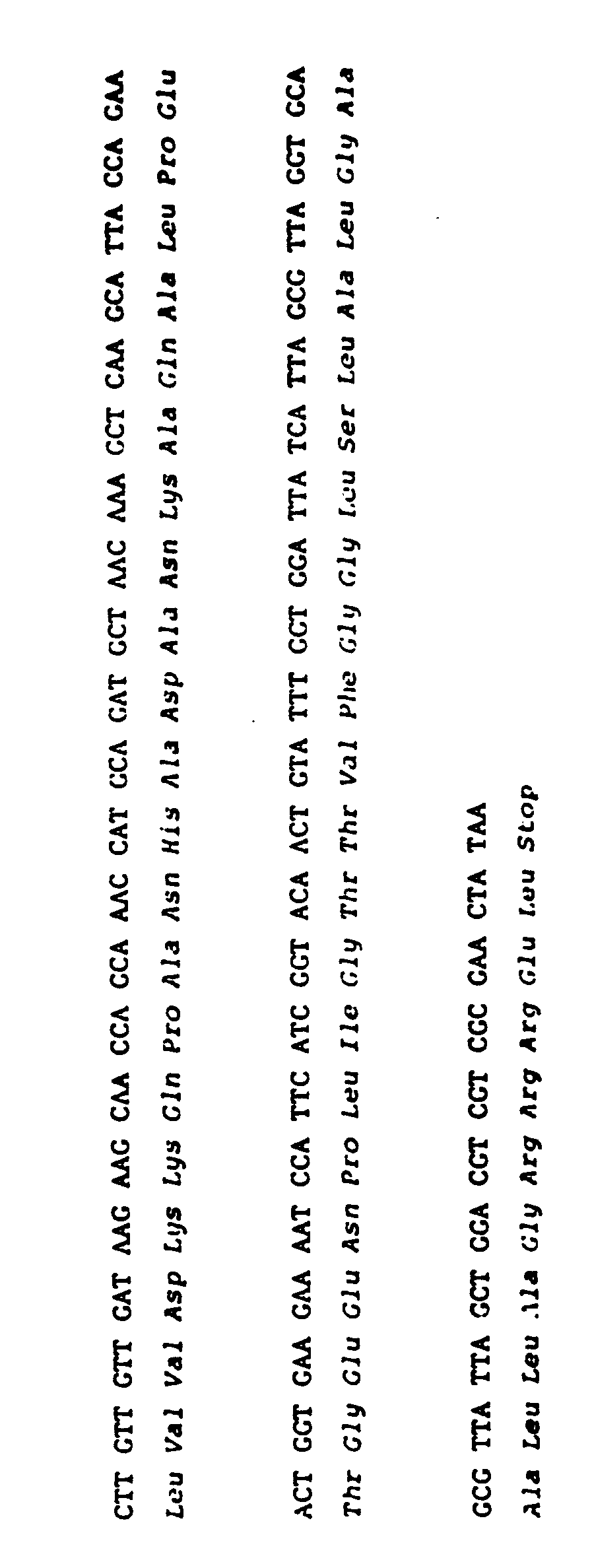
- Disclosed herein are recombinant plasmids comprising a novel nucleotide sequence coding for the amino acid sequence of protein A material and the known plasmid vector pBR322. The sequence of this novel oligonucleotide follows as SEQUENCE I. The entire sequence is contained in plasmid pAC37 (NRRL B-15127). Plasmid pAC37-6 (NRRL B-15131) contains the same entire sequence except for the last 209 nucleotide bases. The last six nucleotide bases of pAC37-6 code for the Pstl recognition sequence, i.e., CTGCAG.
- The following sequence discloses for the first time, surprisingly, an additional IgG-binding domain designated domain E near the amino terminal end of the protein A material. This domain is neither disclosed nor suggested by the prior art. In addition, an unexpectedly large carboxy terminal coding sequence has been discovered, which may constitute the region responsible for activation of polyclonal antibody synthesis.
- The disclosed nucleotide sequence and subfragments thereof enable persons in the art, for the first time, to obtain cloned nucleotide sequences coding for protein A material and subfragments of protein A material.


- Having the above data, those skilled in the art can readily appreciate the identity of other equivalent nucleotide sequences coding for molecules with substantially the same Protein A-like biological activity. Thus, the scope of the subject invention includes not only the specific nucleotide sequence depicted above, but also all equivalent nucleotide sequences coding for the amino acid sequence of other Protein A materials having an additional IgG-binding domain equivalent to domain E of Sequence I above.
- Also included are subfragments of the Protein A material which have the property of binding to IgG at the Fc region. The Protein A material of the subject invention, and subfragments thereof, can be used in the same manner as Protein A, disclosed above.
- Thus the invention provides not only a recombinant DNA molecule comprising (a) a nucleotide sequence coding for the amino acid sequence of a Protein A material, characterised in that said nucleotide sequence and said amino acid sequence are as in Sequence I or (b) a nucleotide sequence coding for the amino acid sequence of other Protein A materials having an additional IgG-binding domain equivalent to domain E of Sequence I; but also a recombinant DNA molecule comprising: (a) a nucleotide sequence coding for the amino acid sequence of domain E of a Protein A material, characterised in that said nucleotide sequence and said amino acid sequence are as follows:or; (b) a nucleotide sequence coding for the amino acid sequence of other Protein A materials having an additional IgG-binding domain equivalent to said domain E.

- The invention also provides a micro-organism comprising a recombinant plasmid containing a nucleotide sequence defined according to the invention, or fragments thereof, said fragments containing domain E or said additional IgG-binding domain equivalent to domain E.
- The nucleotide sequence may be absent the last 209 nucleotide bases of Sequence I.
- The invention also provides a DNA transfer vector comprising a nucleotide sequence encoding an amino acid sequence, said amino acid sequence being defined according to the invention; and a micro-organism transferred by such a DNA transfer vector.
- The invention also provides a micro-organism comprising a recombinant plasmid containing a mixture of nucleotide sequences of domains D, A, B and C as defined in Sequence I coding for amino acid sequences of Protein A material characterised in that said mixture of nucleotide sequences also exhibits a domain E as defined in Sequence I or other additional IgG-binding domain equivalent to domain E.
- The invention also provides an essentially pure biological material having a nucleotide sequence coding for the amino acid sequence of a Protein A material, said nucleotide sequence being defined according to the invention.
- The invention also provides a process for preparing a Protein A material, which comprises expression of cloned DNA as defined according to the invention, coding for said Protein A material, in a suitable microbial host, and recovering said Protein A material.
- There now follows a description of embodiments of the invention. This description, which is illustrative of product and process aspects of the invention, is given by way of example only, and not by way of limitation of the invention.
- Cloning of the DNA sequences coding for protein A material was initiated by construction of a gene bank comprising DNA sequences of the SAC genome (S. aureus, Cowan I, SAC, ATCC 12598). This was accomplished by G-C tailing using blunt-end SAC DNA fragments generated by Haelll+Alul partial restriction digestion as substrate. Digestion of 250 /1.g SAC DNA in 400 µl 50 mM Tris-HCI, pH 7.5; 5 mM MgCI; 1 mM dithiothreitol (DTT) with 150 units Haelll and 200 units Alul (12 min., 37 C) generated a broad size range of DNA fragments (2-10 kilobase pairs [kb]). From the published 42,000 molecular weight, it was estimated that the coding sequences of protein A should comprise 1.1-1.2 kb of DNA. To maximize the probability of obtaining a recombinant insert containing both the protein A coding sequences and adjacent regulatory sequences, larger fragments, 3-6 kb, were used for construction of the SAC gene bank. This DNA was extracted from a preparative agarose gel, tailed with 15-20 C residues with terminal transferase, and annealed to G-tailed, Pstl-digested pBR322. Transformation of E. coli MS371 cells with the resulting recombinant DNA, G-tailed plasmid DNA alone, or uncut pBR322 yielded transformation efficiencies of 2.0 x 10⁴, 5.0 x 10², and 2.0 x 10⁶ transformants per µg plasmid DNA, respectively. Approximately 7.0 x 10³ transformants were picked onto fresh tetracycline plates for screening.
- Mini-lysate plasmid DNA preparations for 10 randomly picked transformants were digested with Pstl and the sizes of the resulting DNA fragments analyzed by agarose gel electrophoresis. The results indicated that (1) 9 of 10 transformants carried recombinant DNA plasmids, (2) 7 of 9 recombinant plasmids had both Pstl restriction sites regenerated by the G-C tailing procedure, and (3) the average insert length was approximately 3.0 kb.
- The cloning vehicles embodying the invention are useful to make available for the first time and to increase the supply of the gene coding for molecules with protein A-like biological activity by replication of a transformed host. With this abundance of the desired gene, levels of protein A expression necessary to make protein A-like material available at a lower cost can be predicted.
- In the following Examples all percentages are by weight and all solvent mixture proportions are by volume unless otherwise noted.
- Staphylococcus aureus, Cowan I (SAC, ATCC 12598) and Woods 46 (SAW, ATCC 10832) strains were obtained from the American Type Culture Collection, Rockville, Maryland, Both strains were grown (liquid or 1.5% agar plates) in Penassay medium (5 mg/ml Casitone, 2.5 mg/ml yeast extract, 2.5 mg/ml 3- glycerophosphate, 4 mg/ml niacin, 2 mg/ml thiamine-HCI) under standard conditions.
- E. coli MS371 was propagated in L-broth (5 g/I NaCI, 10 g/I bactotryptone, 5 g/1 yeast extract). For plasmid DNA preparation, cells containing plasmids of interest were grown in M-9 media (49 mM Na2HP04, 17 mM KH2P04, 8.6 mM NaCI, 18.7 mM NH4CI, 0.1 mM CaCl2, 1 mM MgSO4 · 7 H20, 0.4% glucose, 0.4% casamino acids, 2 mg/ml thiamine).
- Overnight cultures of SAC were diluted 1:100 with Penassay broth and allowed to grow to OD6oo = 0.6. The cells were pelleted by centrifugation (5K rpm, 10 min., 2°C with a Beckman JA10 rotor), resuspended in 20 volumes DNA extraction buffer (0.1 M NaCI; 50 mM EDTA; 10 mM Tris-HCI, pH 8.0), and frozen in a dry ice-acetone bath.
- The frozen cell suspension was allowed to thaw at 37° C, 50 mg/ml lysostaphin (Sigma Chemical Co., St. Louis, Mo.) was added, and the suspension incubated at 37° C, 15 min. Protease K (40 mg/ml) and SDS (0.5%) were added and the mixture incubated at 37 C, 1 hour. The lysate was then extracted with phenol:chloroform (1:1) saturated with DNA extraction buffer. The SAC DNA solution was adjusted to 0.95 g/ml CsCI and banded by centrifugation (44K rpm, 46 hours, 23 C with a Beckman Ti60 rotor). The DNA was then harvested with a syringe and 21 g needle by side puncture. The DNA was dialyzed against TE buffer (10 mM Tris-HCI: 1 mM EDTA, pH 8.0), phenol:chloroform-extracted as before, and precipitated twice with 2 volumes ethanol. Yields of SAC DNA ranged between 700-800 mg DNA per gram wet weight of cells.
- All restriction endonucleases were purchased from Bethesda Research Laboratories, Bethesda, Md. or New England Biolabs, Beverly, Mass. Unless otherwise indicated, restriction digests, described herein, were carried out at DNA concentrations of 100-400 µg/ml, 2-4 units enzyme per µg DNA, 2-3 hours, 37° C, in buffer systems recommended for each enzyme by the respective company.
- Agarose gel electrophoresis was carried out using a 2X Tris-acetate gel buffer (80 mM Tris-HCI, pH 8.0; 40 mM NaC2H302; 36 mM NaCI; 2 mM Na2EDTA) in the gel and 1X buffer for the run. Analytical gels were routinely run as "submarine gels" in a horizontal gel box. Preparative gels were routinely run in an EC Model 470 gel box. DNA bands were visualized by ethidium bromide (EtBr) post-staining (0.5 mg/ml in 1X gel buffer) and use of a U.V. transilluminator Model TM-36 from Ultra-Violet Products, Inc., San Gabriel, Ca.
- Extraction of DNA from preparative agarose gels was initiated by visualization of the positions of EtBr- stained bands of a single gel lane. Gel slices containing DNA fragments of interest were diced manually and passed through a 20 g needle with 12-2 volumes DNA gel extraction buffer (0.5 M NH4C2H302, 10 mM EDTA, 10 mM Mg(C2H302)2, 0.1% SDS). An equal volume of phenol saturated with 1 mM NH4C2H302, 10 mM EDTA was added and extraction carried out in eppendorf tubes on a rotary shaker at 23°C overnight. The tubes were then placed on ice for 30 min. prior to separation of the aqueous phase by microcen- trifugation. Extraction of the aqueous phase with the saturated phenol solution was repeated 3-4 times, followed by chloroform extraction and ethanol precipitation. Routine recovery of DNA fragments smaller than 15 kb was about 40%.
- Construction of recombinant plasmids was facilitated by G-C tailing (Stein, I., Catterall, J., Woo, S., Means, A., O'Malley, B. [1978] Biochemistry. 17:5763). Pstl-digested and agarose gel-purified pBR322 DNA was tailed with approximately 14 G residues in a 100 µl reaction under the following conditions: 100 µg/ml DNA, 20 µM dGTP, 200 mM K/cacodylate, 1 mM CoCl2, 1 mM β-mercaptoethanol (β-SH), 15 units terminal deoxynucleotidyl transferase (P.L. Biochemicals, Inc., Milwaukee, Wis.) 37° C, 30 min. The reaction was terminated by the addition of 2 µl 100 mM EDTA, 2 µl 5 M NaCI, 2 µl 20% SDS and phenol:chloroform (1:1) extraction. The resulting G-tailed plasmid DNA was passed over a G-50 Sephadex column and precipitated with ethanol.
- Target SAC DNA fragments of average 3-5 kb length were tailed with 15-20 C residues in a 30 µl reaction under the following conditions: 4-5 µg SAC DNA, 20 µM dCTP, 200 mM K/cacodylate, 1 mM CoCl2, 1 mM β-SH, 4.5 units terminal deoxynucleotidyl transferase; 37° C, 12 min. Termination of the reaction and treatment of C-tailed SAC DNA was carried out as described above. Annealing of plasmid and target SAC DNA was initiated by mixing 2.5 µg plasmid and 4.0 µg target SAC DNA in 300 µl 10 mM Tris-HCI, pH 8.0; 1 mM EDTA; 100 mM NaCI; and heating for 10 min. at 68° C. The annealing solution was then allowed to incubate 1 hour at 55° C, 1 hour at 23° C, and was stored at 4° C until needed.
- Ligation of staggered-end DNA fragments was carried out with 100-200 units/ml T4 DNA ligase (Bethesda Research Laboratories); 66 µM ATP; 66 mM Tris-HCI, pH 7.6; 6.6 mM MgCl2; 10 mM dithiothreitol; at 12° C, 12-16 hours.
- Fresh overnight cultures were diluted 1:100 in L-broth and allowed to grow at 37° C with shaking to OD600 =0.1-0.15. The cells were pelleted (5 min. 5K rpm, 5°C in a JA20 rotor in a Beckman J2-20 centrifuge), resuspended in half the original volume of ice-cold 50 mM MnCl2; 10 mM NaC2H302, pH 5.6; and allowed to stand at 0°C, 20 min. Following pelleting of the cells as above, they were resuspended in ice-cold 100 mM MnCl2; 75 mM CaCl2; 10 mM NaC2H302, pH 5.6. A 0.1 ml aliquot of cells was mixed with 10 µl DNA transformation solution and allowed to sit on ice 40 min. The cells were then subjected to heat shock (2.5 min., 25-30 C) and 1.5 µl 2.0 M Tris-HCI, pH 7.4, and 0.5 ml L-broth per 0.1 ml cell aliquot were added. The cells were then plated in 15-25 µl aliquots on 1.5% agar L-broth plates supplemented with 10 µg/ml tetracycline (Sigma) and incubated overnight at 37°C. Transformation efficiencies of 1.0 x 10⁷ colonies per µg pBR322 DNA were routinely observed.
- Mini-lysate plasmid preparation was initiated by addition of 1 ml of fresh overnight culture to 9 ml L-broth, supplemented with 1% glucose and allowed to grow with shaking at 37°C to an OD550 of 1.0. Chloramphenicol was then added to 150 µg/ml and the culture incubated for 12-16 hours at 37 C. The cells were then pelleted by centrifugation (5 min., 3K rpm, 23° C in an RC-3 centrifuge), resuspended in ice-cold TE buffer, and transferred to a 1.5 ml eppendorf tube to be repelleted by centrifugation. The resulting cell pellet was resuspended in 50 mM Tris-HCI, pH 8.0; 50 mM EDTA; 15% sucrose (wt/vol) by vortexing. To the cell suspension, 10 µl of 10% SDS were added and incubated at 70°C, 10 min. To the resulting lysate, 62.5 µl ice-cold 4 M potassium acetate was added and the lysate allowed to stand for at least 2 hours on ice. Following centrifugation the supernatant volume was adjusted to 0.5 ml with H20 and the DNA precipitated with 2 volumes absolute ethanol. The DNA was then resuspended in 100 µl TE, the salt adjusted to 0.1 M with NaCI, and re-precipitated with two volumes ethanol prior to restriction enzyme analysis.
- Overnight 25 ml cultures were grown in L-broth supplemented with 10 µg/ml tetracycline. To one liter M-9 media, 5 ml of the overnight culture were added and allowed to grow at 37° C in a rotary incubator (200 rpm) until an OD6oo value of 0.6 was reached. 250 mg/liter chloramphenicol (Sigma) was then added and the culture allowed to shake for 12-16 hours at 37 C. The cells were then harvested by centrifugation (6000 rpm, 20 min., 2 C in Beckman JA-10 rotor), and the pellets washed once with ice-cold TE buffer. The washed pellets were then either frozen at -60 C or immediately extracted. Preparation of cleared lysates was initiated by suspending the cell pellet in 6.25 ml per liter original culture of 25% sucrose in 50 mM Tris-HCI, pH 8.0, and then adding 1.5 ml of a freshly made 10 mg/ml lysozyme (Sigma) solution. After continuous swirling of the suspension on ice for 5 min., 1.25 ml of 0.5 M Na2EDTA, pH 8.0, was added and swirling of the suspension on ice continued for 5 min. Ten ml of a 10X Triton (10 ml 10% Triton X-100; 125 ml 0.5 M EDTA, pH 8.0; 50 ml 1.0 M Tris-HCI, pH 8.0; and 800 ml H20) per liter original culture volume was added and the suspension swirled on ice for 15 min. The lysate was then subjected to centrifugation (19K rpm, 4° C, 30 min. in a JA-20 rotor) and the supernatant transferred to a volumetric cylinder. 0.95 g/ml CsCl was dissolved in the supernatant and 1/10 the volume of 10 mg/ml EtBr in TE buffer was added. Separation of plasmid and chromosomal DNA was accomplished by centrifugation with a Beckman Ti 50.2 rotor (23 C, 44K rpm for 24 hours followed by 36 hours at 38K rpm).
- The plasmid DNA band on the gradient is visualized with a U.V. lamp and harvested with a syringe by side puncture using a 21 g needle. Removal of EtBr is carried out by repeated isobutyl alcohol extraction. The plasmid solution is then dialyzed overnight against TE buffer, the salt concentration adjusted to 0.1 M NaCl and precipitation of DNA carried out with 2 volumes of absolute ethanol.
- Expression of protein A-like activity in bacterial colonies was detected by binding of 125I-IgG to colony lysates immobilized on nitrocellulose filters. Recombinant plasmid-bearing E. coli, SAC (positive control) and SAW (negative control) cells were picked and streaked onto nutrient agar plates and allowed to grow overnight. Nitrocellulose filter discs (BA85, 87 mm, Schleicher and Schuell, Keene, N.H.) were carefully laid on the plates to absorb the underlying colonies, and the filters lifted and allowed to dry by blotting on Whatman 3MM paper. Lysis of filter-bound cells was accomplished by laying the filters (colony side up) on sheets of Whatman 3MM filter paper saturated with 0.5 M NaOH and allowing lysis to proceed for 10 min. at 23 C. Following lysis, the filters were blot dried and neutralized on filter paper saturated with 1.0 M Tris-HCI, pH 7.0. The filters were again blot dried and pre-treated with protein binding solution (10 mM Tris-HCI, pH 7.0; 100 mM NaCI; 5 mM EDTA; 0.13% NP40; 0.1% SDS; 0.1% sodium deoxycholate; 0.2% Ficoll 400: 0.3% gelatin) for 4-6 hours at 23 C on a rotary platform shaker. After pre-treatment, the filters were transferred to a 1-liter beaker containing a 4.5 ml/filter protein-binding solution. Binding of 125I-IgG (Goat anti-rabbit, New England Nuclear, Boston, MA) was carried out by the addition of 5 x 10⁶ cpm/ml 125I-IgG to the beaker and allowing binding to occur at 4°C overnight with constant rotary shaking. Washing of the filters was accomplished by repeated washing with 500 ml protein-binding solutions: the first wash carried out at 4°C, and 2-3 additional washes carried out at 23 C. The washed filters were then dried by blotting and detection of 125I-IgG binding accomplished by radioautography, using Kodak XAR-5 film and two DuPont Cronex Lightning-Plus enhancement screens.
- DNA sequence determination was carried out with minor modification of procedures described by Maxam and Gilbert (Maxam, A. and Gilbert, W. [1977] Proc. Nat'l. Acad. Sci. USA, 74:560) and Heidecker et al. (Heidecker, G., Messing, J., and Gronenborn, B. [1980] Gene 10:69).
- To test for the expression of sequences coding for protein A material within the recombinant SAC gene bank, the colonies from 50 plates of 52 colonies each (2,600) were lifted on nitrocellulose discs and assayed for 125I-IgG binding. Filters containing SAC and SAW colonies were included in the assay as positive and negative controls, respectively. To assess the sensitivity of the assay, serial dilutions of purified protein A (Pharmacia, Piscataway, N.J.) were spotted onto a nitrocellulose disc and assayed in parallel with the test filters. The routine sensitivity for the assay was found to vary over a range of 1.0 to 0.01 ng with purified protein A. Filters containing SAC and SAW cells yielded positive and negative autoradiographic signals, respectively. A single transformant colony bound significant 125I-IgG in this and subsequent assays. This colony was picked for further analysis. The plasmid contained in this colony was designated pAC37.
- Restriction endonuclease analysis of pAc37 plasmid DNA indicated the presence of Pstl insert fragments of 3.1, 2.3, 1.9, and 0.65 kb length. pAc37 plasmid DNA was digested with Pstl, re-ligated with T4 ligase, and used to transform E. coli MS371 cells. The resulting transformants were screened by the 121I-IgG-binding assay as described in Example 12.
- Of 322 transformants, 10 positive 1251-IgG-binding colonies were obtained and were found to have recombinant plasmids containing a 1.9 kb Pstl insert. When recombinant plasmid DNAs from 12 randomly picked non-125I-IgG-binding transformant colonies were analyzed they were found to contain one or more Pstl fragments from pAc37, but not a 1.9 kb fragment. It was concluded that at least a portion of the protein A-like coding sequences reside within a 1.9 kb Pstl fragment of pAc37. One positive colony containing a recombinant plasmid with a single 1.9 kb insert, designated pAc37-6, was picked for further analysis.
- Final determination of the presence of protein A-like coding sequences within the Pstl 1.9 kb fragment of pAc37-6 DNA was accomplished by DNA sequence determination. The pAc37-6 DNA was digested with Hindlll, labeled with y32 P-ATP and polynucleotide kinase, and subsequently digested with Pstl. Sequence determination of a portion of the 0.6 kb Hindlll/Pstl fragment indicated sequence colinearity with the known amino acid sequence of the B-C junction of the protein A molecule. The position of the sequences coding for the B-C junction of the protein A-like material within the insert made it likely that the 1.9 kb insert of pAc37-6 plasmid DNA contained most of the sequences coding for the protein A gene, including the ribosome binding site, and 5' regulatory sequences.
- E. coli MS371 (pAc37-6) is lysed with 0.1 N NaOH and centrifuged. The supernatant is removed and 25 mM monobasic sodium phosphate is added and the solution adjusted to pH 7.0 with 1 M HCI. The protein solution is dialyzed against 25 mM sodium phosphate pH 7.0, then clarified by centrifugation.
- The solution is applied to an IgG-Sepharose column (30 ml bed volume per 1.3 gm of protein) and the column washed with 0.1 M sodium phosphate pH 7.0 until no protein, as determined by A28o, elutes from the column.
- Protein A material is eluted with 0.1 M glycine" HCI. The purified protein is concentrated by precipitation with 80% saturated (NH4)2S04, dialyzed versus 10 mM sodium phosphate pH 7.0, and stored frozen.
- The purification of protein A material from E. coli MS371 (pAc37), NRRL B-15127 can be accomplished by using the procedure described above.
- Restriction enzymes can be used to cleave the nucleotide sequence coding for protein A material in order to isolate essentially pure subfragments of the coding region that are capable of coding for amino acid sequences with biological activities similar to those of protein A. For example, cleavage of pAc37-6 DNA with Rsal restriction endonuclease will yield an oligonucleotide that is 1,199 nucleotides long and that codes for a polypeptide containing domains E, D, A, B, and C. Digestion with other restriction enzymes such as Hinfl, or a combination of enzymes such as Hindlll and Sau3A, can be used to generate essentially pure, well-defined oligonuclectide subfragments that code for amino acid sequences with biological activities similar to those of protein A.
- The desired oligonucleotide subfragments are isolated in their essentially pure form by preparative agarose gel electrophoresis as follows: Agarose is dissolved to 1% in 2x E buffer (0.08 M Tris·HCl, pH 7.8; 0.01 M NaC2H302; 0.002 M EDTA) and poured into a Bio-Rad (Richmond, Ca) slab gel apparatus. Samples are dissolved in 10 mM Tris·Cl, pH 8.0; 0.1 mM EDTA and the samples are run at constant power with 2x E running buffer.
- After electrophoresis, one lane is cut from the gel, stained with ethidium bromide (0.5 µgm/ml) and the DNA bands visualized under ultraviolet light. The band of interest is cut from the rest of the gel and macerated before passing it through a 20 gauge needle. An equal weight of extraction buffer (10 mM Tris˙HCl, pH 8.0; 2 mM EDTA; 1 M NaCI) is then added and mixed with the gel. The mixture is incubated at 47° C for 16 hours and the agarose pelleted at 100,000 x g for 1 hour. The supernatant is then made 30 agm/ml in tRNA and extracted with phenol until no agarose is visible at the interface. The DNA is then ether extracted and ethanol precipitated. Gel buffers and extraction procedures can be varied by one skilled in the art to recover the desired DNA fragments.
- Once the nucleotide sequence coding for a particular amino acid sequence has been determined, i.e., by cloning and sequencing as shown in previous examples, then the oligonucleotide coding for the amino acid sequence can be synthesized chemically. (See, for example, Edge, M.D.., et al. [1981] Nature 292: 756-762). Thus, subfragments of the coding region, or the entire coding region for a Protein A-like molecule, can be synthesized and isolated in their essentially pure forms; this includes those regions of the coding sequence coding for domains E, D, A, B and C.
- Domain E alone, or in various combinations with any of Domains D, A, B and C, is useful in the same manner as Protein A to bind IgG in diagnostic test systems, as described previously.
- The essentially pure nucleotide sequences coding for Protein A material or for biologically active subfragments of Protein A material, isolated and synthesized as described in Examples 16 and 17, respectively, can be ligated into appropriate restriction enzyme sites in an expression cloning vector. If necessary, sites can be added to nucleotide sequences using linker molecules. (See, for example, Norris, K.E., et al. [1979] Gene 7: 355-362). The ligated DNA can then be used to transform a host organism. Previous work by others has shown that expression of the cloned nucleotide sequence would be expected. (See, for example, Doel, M.T. et al. [1980] Nuc. Acids Res. 8: 4575-4592; Roberts T., et al. [1979] Proc. Nat. Acad. Sci. 76: 760-764; Guarente, L., et al. [1980] Cell 20: 543-553.) The biologically active material that is expressed can then be purified as described in Example 15.
- Plasmids pAc37 and pAc37-6 have been deposited in an E. coli host in the permanent collection of the Northern Regional Research Laboratory (NRRL), U.S. Department of Agriculture, Peoria, Illinois, U.S.A. Their accession numbers in this repository are as follows:
- E. coli MS371 (pAc37)--NRRL B-15127: Deposited on August 18, 1982
- E. coli MS371 (pAc37-6)--NRRL B-15131: Deposited on August 18, 1982
- E. coli MS371--NRRL B-15129: Deposited on August 18, 1982
- Plasmid pBR322 is a known and available plasmid. It is maintained in the E. coli host ATCC 37017. Purified pBR322 DNA can be obtained as described in Bolivar, F., Rodriquez, R.L., Greene, P.J., Betlach, M.C., Heyneker, H.L., Boyer, H.W., Crosa, J.H., and Falkow, S. (1977) Gene 2:95-113; and Sutcliffe, J.G. (1978) Nucleic Acids Res. 5:2721-2728.
- NRRL B-15127, NRRL B-15131, and NRRL B-15129, are deposited in accordance with European Rule 28.
- There are other, known, hosts which can be used instead of E. coli MS371, for example, B. subtilis, Streptomyces species, and yeast.
- Also, it is within the skill of those in the art to vary the conditions required to grow cells, extract DNA, perform restriction enzyme digestions, electrophorese DNA fragments, tail and anneal plasmid and insert DNA, ligate DNA, transform E. coli cells, prepare plasmid DNA, perform an IgG-binding assay, prepare protein lysates, electrophorese proteins, and sequence DNA.
Claims (12)


Priority Applications (1)
| Application Number | Priority Date | Filing Date | Title |
|---|---|---|---|
| AT83306500T ATE71657T1 (en) | 1982-10-27 | 1983-10-26 | PROTEIN-A MATERIAL AND ITS MANUFACTURE. |
Applications Claiming Priority (2)
| Application Number | Priority Date | Filing Date | Title |
|---|---|---|---|
| US436955 | 1982-10-27 | ||
| US06/436,955 US5151350A (en) | 1982-10-27 | 1982-10-27 | Cloned genes encoding recombinant protein a |
Publications (3)
| Publication Number | Publication Date |
|---|---|
| EP0107509A2 EP0107509A2 (en) | 1984-05-02 |
| EP0107509A3 EP0107509A3 (en) | 1986-03-26 |
| EP0107509B1 true EP0107509B1 (en) | 1992-01-15 |
Family
ID=23734479
Family Applications (1)
| Application Number | Title | Priority Date | Filing Date |
|---|---|---|---|
| EP83306500A Expired - Lifetime EP0107509B1 (en) | 1982-10-27 | 1983-10-26 | Protein a material and preparation thereof |
Country Status (6)
| Country | Link |
|---|---|
| US (1) | US5151350A (en) |
| EP (1) | EP0107509B1 (en) |
| JP (2) | JPH0811068B2 (en) |
| AT (1) | ATE71657T1 (en) |
| CA (1) | CA1307483C (en) |
| DE (1) | DE3382495D1 (en) |
Families Citing this family (75)
| Publication number | Priority date | Publication date | Assignee | Title |
|---|---|---|---|---|
| US4737544A (en) * | 1982-08-12 | 1988-04-12 | Biospecific Technologies, Inc. | Biospecific polymers |
| US4691009A (en) * | 1984-12-26 | 1987-09-01 | Repligen Corporation | Hybrid proteins produced by an ultrahigh prokaryotic expression system |
| SE459503B (en) * | 1985-05-03 | 1989-07-10 | Excorim Kommanditbolag | HYBRID DNA MOLECULE CONTAINING DNA SEQUENCE CODING FOR PROTEIN G AND PROCEDURE FOR PRODUCING PROTEIN G |
| DK528285D0 (en) * | 1985-11-15 | 1985-11-15 | Novo Industri As | PEPTIDES AND PREPARATIONS CONTAINING THESE |
| EP0234592A1 (en) * | 1986-02-28 | 1987-09-02 | Teijin Limited | Plasmid containing DNA fragment coding for human immunoglobulin G Fc region protein and use thereof for production of said protein |
| SE459662B (en) * | 1986-03-21 | 1989-07-24 | Pharmacia Ab | HYBRID DNA MOLECULE CODING FOR A PROTEIN WITH THE SAME SPECIFICITY AS PROTEIN G, PLASMID CONTAINING ITS SAME AND PROCEDURE FOR PRODUCING THE PROTEIN |
| US4876194A (en) * | 1986-07-22 | 1989-10-24 | Hightech Receptor Ab | Protein L and subfragments thereof, with immunoglobulin binding activity, a process for preparing thereof, reagent kit, pharmaceutical composition and a peptococcus magnus strain |
| JPS6348297A (en) * | 1986-08-18 | 1988-02-29 | Sanraku Inc | Fc-bonding protein and microbial strain producing same |
| EP0289129A3 (en) * | 1987-03-26 | 1990-10-10 | Repligen Corporation | High purity protein a preparation |
| US5084559A (en) * | 1987-03-27 | 1992-01-28 | Repligen Corporation | Protein a domain mutants |
| US5084398A (en) * | 1987-11-20 | 1992-01-28 | Creative Biomolecules | Selective removal of immune complexes |
| DE3887750T2 (en) * | 1987-11-20 | 1994-09-15 | Creative Biomolecules Inc | SELECTIVE REMOVAL OF IMMUNE COMPLEXES. |
| US5243040A (en) * | 1987-11-20 | 1993-09-07 | Creative Biomolecules | DNA encoding a protein which enables selective removal of immune complexes |
| DE68907388T2 (en) * | 1988-07-22 | 1994-01-05 | Imre Corp | Purified Protein A compositions and process for their preparation. |
| JP2871709B2 (en) * | 1988-11-21 | 1999-03-17 | 住友製薬株式会社 | Novel protein H having immunoglobulin G binding activity, gene encoding the protein, and method for producing the protein |
| JPH02308791A (en) * | 1989-05-24 | 1990-12-21 | Sekisui Chem Co Ltd | Polypeptide capable of bonding to immunoglobulin fc fragment and having luciferase activity |
| GB9013872D0 (en) * | 1990-06-21 | 1990-08-15 | Inst Tecnologico Y Cientifico | Use of protein a and polypeptide components thereof in the treatment of tumours |
| CA2096953A1 (en) * | 1990-11-26 | 1992-05-27 | Anthony Atkinson | Immunoglobulin-binding proteins and recombinant dna molecules coding therefor |
| JP2519136B2 (en) * | 1991-07-25 | 1996-07-31 | オリエンタル酵母工業株式会社 | Molecular weight marker |
| JP2584697B2 (en) * | 1991-08-23 | 1997-02-26 | オリエンタル酵母工業株式会社 | Recombinant protein A for IgG purification |
| EP0550771B1 (en) * | 1991-07-25 | 1999-09-29 | Oriental Yeast Co., Ltd. | Immunoglobulin-binding artificial protein |
| GB9526356D0 (en) * | 1995-12-22 | 1996-02-21 | Smithkline Beecham Plc | Novel compounds |
| US6001600A (en) * | 1996-12-20 | 1999-12-14 | Smithkline Beecham Plc | DNA encoding tests polypeptides |
| GB9526359D0 (en) * | 1995-12-22 | 1996-02-21 | Smithkline Beecham Plc | Novel compounds |
| GB9600955D0 (en) * | 1996-01-17 | 1996-03-20 | Smithkline Beecham Plc | Novel compounds |
| US7432365B1 (en) | 1998-01-27 | 2008-10-07 | O'donnell Michael E | DNA molecules encoding beta clamp proteins of gram positive bacteria |
| WO1999037661A1 (en) * | 1998-01-27 | 1999-07-29 | The Rockefeller University | Dna replication proteins of gram positive bacteria and their use to screen for chemical inhibitors |
| AU6749900A (en) * | 1999-07-29 | 2001-02-19 | Rockefeller University, The | Dna replication proteins of gram positive bacteria and their use to screen for chemical inhibitors |
| US7098383B2 (en) * | 2000-12-19 | 2006-08-29 | Sembiosys Genetics Inc. | Methods for the production of multimeric immunoglobulins, and related compositions |
| US20030167524A1 (en) * | 2000-12-19 | 2003-09-04 | Rooijen Gijs Van | Methods for the production of multimeric protein complexes, and related compositions |
| BR0116220A (en) * | 2000-12-19 | 2003-09-23 | Sembiosys Genetics Inc | Methods for the production of multimeric proteins and related compositions |
| US20090137416A1 (en) | 2001-01-16 | 2009-05-28 | Regeneron Pharmaceuticals, Inc. | Isolating Cells Expressing Secreted Proteins |
| US20060179514A1 (en) * | 2001-07-05 | 2006-08-10 | Sembiosys Genetics, Inc. | Methods for the production of multimeric protein complexes, and related compositions |
| US9453251B2 (en) | 2002-10-08 | 2016-09-27 | Pfenex Inc. | Expression of mammalian proteins in Pseudomonas fluorescens |
| JP2005112827A (en) * | 2003-10-10 | 2005-04-28 | National Institute Of Advanced Industrial & Technology | Antibody affinity carrier |
| SE0400501D0 (en) * | 2004-02-27 | 2004-02-27 | Amersham Biosciences Ab | Antibody purification |
| CA2552639C (en) * | 2004-02-27 | 2012-05-01 | Ge Healthcare Bio-Sciences Ab | A process for the purification of antibodies |
| EP1767640A4 (en) | 2004-07-06 | 2008-03-26 | Kaneka Corp | Process for producing protein a-like protein with use of brevibacillus genus bacaterium |
| EP1774017B1 (en) | 2004-07-26 | 2013-05-15 | Pfenex Inc. | Process for improved protein expression by strain engineering |
| BRPI0518216C8 (en) | 2004-10-21 | 2017-03-21 | Ge Healthcare Bio Sciences Ab | chromatography binder, methods of preparing a separation matrix, and separating one or more antibodies from one or more other compounds into a liquid sample, separation matrix, and disposable chromatography column. |
| US8728828B2 (en) | 2004-12-22 | 2014-05-20 | Ge Healthcare Bio-Sciences Ab | Purification of immunoglobulins |
| EP1746103A1 (en) | 2005-07-20 | 2007-01-24 | Applied NanoSystems B.V. | Bifunctional protein anchors |
| US7375188B2 (en) * | 2005-07-29 | 2008-05-20 | Mallinckrodt Baker, Inc. | Vegetarian protein a preparation and methods thereof |
| CA2664530C (en) | 2006-09-29 | 2015-11-17 | Ge Healthcare Bio-Sciences Ab | Chromatography ligand comprising domain c from staphyloccocus aureus protein a for antibody isolation |
| EP2099932A4 (en) * | 2006-12-06 | 2009-11-18 | Repligen Corp | Nucleic acids encoding recombinant protein a |
| US9580719B2 (en) | 2007-04-27 | 2017-02-28 | Pfenex, Inc. | Method for rapidly screening microbial hosts to identify certain strains with improved yield and/or quality in the expression of heterologous proteins |
| US9394571B2 (en) | 2007-04-27 | 2016-07-19 | Pfenex Inc. | Method for rapidly screening microbial hosts to identify certain strains with improved yield and/or quality in the expression of heterologous proteins |
| US8828905B2 (en) * | 2007-05-30 | 2014-09-09 | Kaneka Corporation | Porous base matrix having formyl group, adsorbent using the porous base matrix, method for production of the porous base matrix, and method for production of the adsorbent |
| SG149759A1 (en) * | 2007-07-10 | 2009-02-27 | Millipore Corp | Media for affinity chromatography |
| US8592555B2 (en) | 2008-08-11 | 2013-11-26 | Emd Millipore Corporation | Immunoglobulin-binding proteins with improved specificity |
| ES2391931T3 (en) * | 2008-09-15 | 2012-12-03 | Emd Millipore Corporation | Procedures for quantifying protein filtration from protein-based affinity chromatography resins |
| EP2339339A4 (en) | 2008-09-25 | 2016-10-12 | Jsr Corp | Filler for affinity chromatography |
| CN103816870B (en) | 2008-12-03 | 2016-11-23 | 株式会社钟化 | Porous carrier containing formoxyl, the adsorbate using this porous carrier and their manufacture method |
| SG195555A1 (en) | 2008-12-24 | 2013-12-30 | Emd Millipore Corp | Caustic stable chromatography ligands |
| EP3281947B1 (en) | 2009-04-03 | 2020-02-12 | The University of Chicago | Compositions and methods related to protein a (spa) variants |
| EP2562178B1 (en) | 2010-03-31 | 2015-05-06 | JSR Corporation | Filler for affinity chromatography |
| ES2655701T3 (en) | 2010-07-02 | 2018-02-21 | The University Of Chicago | Compositions and methods related to protein A (SpA) variants |
| WO2012124145A1 (en) | 2011-03-15 | 2012-09-20 | オムロン株式会社 | Computation unit, assistance unit, assistance program, recording medium storing assistance program, and operation method in assistance device |
| SG10201604559TA (en) | 2011-06-08 | 2016-07-28 | Emd Millipore Corp | Chromatography matrices including novel staphylococcus aureus protein a based ligands |
| WO2014038686A1 (en) | 2012-09-10 | 2014-03-13 | 株式会社カネカ | Adsorbent |
| IN2015DN02055A (en) | 2012-09-17 | 2015-08-14 | Grace W R & Co | |
| TW202423993A (en) * | 2012-11-14 | 2024-06-16 | 美商再生元醫藥公司 | Recombinant cell surface capture proteins |
| EP3059251B1 (en) | 2013-10-15 | 2019-04-03 | Kaneka Corporation | Method for manufacturing porous cellulose beads |
| US10040053B2 (en) | 2013-10-15 | 2018-08-07 | Kaneka Corporation | Production method for porous cellulose beads, and adsorbent employing same |
| US10189007B2 (en) | 2013-10-15 | 2019-01-29 | Kaneka Corporation | Method for producing porous cellulose beads and adsorbent employing same |
| EP3094390B1 (en) | 2014-01-16 | 2021-07-07 | W.R. Grace & CO. - CONN. | Affinity chromatography media and chromatography devices |
| US20240100450A9 (en) * | 2014-01-17 | 2024-03-28 | Repligen Corporation | Sterilizing chromatography columns |
| WO2015109246A1 (en) * | 2014-01-17 | 2015-07-23 | Repligen Corporation | Sterilizing chromatography columns |
| EP3137209B1 (en) | 2014-05-02 | 2022-10-05 | W.R. Grace & CO. - CONN. | Functionalized support material and methods of making and using functionalized support material |
| SG10201911134QA (en) | 2015-06-05 | 2020-01-30 | Grace W R & Co | Adsorbent bioprocessing clarification agents and methods of making and using the same |
| US11884705B2 (en) | 2017-09-25 | 2024-01-30 | Jsr Corporation | Immunoglobulin binding protein, and affinity support using same |
| WO2019059399A1 (en) | 2017-09-25 | 2019-03-28 | Jsr株式会社 | Immunoglobulin binding protein, and affinity support using same |
| WO2019244961A1 (en) | 2018-06-20 | 2019-12-26 | Jsr株式会社 | Affinity carrier using mutant vhh antibody |
| WO2020040307A1 (en) | 2018-08-24 | 2020-02-27 | Jsr株式会社 | Immunoglobulin-binding protein, and affinity carrier using same |
| AU2020273471B2 (en) | 2019-05-14 | 2026-05-07 | The University Of Chicago | Methods and compositions comprising Staphylococcus protein A (SpA) variants |
Family Cites Families (7)
| Publication number | Priority date | Publication date | Assignee | Title |
|---|---|---|---|---|
| US4293652A (en) * | 1979-05-25 | 1981-10-06 | Cetus Corporation | Method for synthesizing DNA sequentially |
| US4338397A (en) * | 1980-04-11 | 1982-07-06 | President And Fellows Of Harvard College | Mature protein synthesis |
| US4322274A (en) * | 1980-08-28 | 1982-03-30 | Wilson Gregory B | Method of diagnosing cystic fibrosis patients and asymptomatic carrier of the cystic fibrosis gene |
| SE8204810L (en) * | 1982-08-23 | 1984-02-24 | Pharmacia Fine Chemicals Ab | HYBRID DNA MOLECULE, TRANSFORMED MICROORGANISM AND PROCEDURE TO MAKE PROTEIN A |
| SE8204811L (en) * | 1982-08-23 | 1984-02-24 | Sven Lofdahl | DNA FRAGMENT INCLUDING A SIGNAL SEQUENCE |
| DE3314812A1 (en) * | 1983-04-23 | 1984-10-25 | Battelle-Institut E.V., 6000 Frankfurt | AGENT FOR ANTIGENT / ANTIBODY DETECTION |
| US4617266A (en) * | 1983-04-28 | 1986-10-14 | Genex Corporation | Production of Protein A |
-
1982
- 1982-10-27 US US06/436,955 patent/US5151350A/en not_active Expired - Lifetime
-
1983
- 1983-10-26 DE DE8383306500T patent/DE3382495D1/en not_active Expired - Lifetime
- 1983-10-26 CA CA000439706A patent/CA1307483C/en not_active Expired - Lifetime
- 1983-10-26 EP EP83306500A patent/EP0107509B1/en not_active Expired - Lifetime
- 1983-10-26 AT AT83306500T patent/ATE71657T1/en not_active IP Right Cessation
- 1983-10-27 JP JP58201871A patent/JPH0811068B2/en not_active Expired - Lifetime
-
1995
- 1995-07-24 JP JP7187019A patent/JPH08107795A/en active Pending
Non-Patent Citations (1)
| Title |
|---|
| Eur. J. Biochem. 78, pp. 471-490 (1977) * |
Also Published As
| Publication number | Publication date |
|---|---|
| JPH08107795A (en) | 1996-04-30 |
| ATE71657T1 (en) | 1992-02-15 |
| US5151350A (en) | 1992-09-29 |
| JPS59113890A (en) | 1984-06-30 |
| EP0107509A2 (en) | 1984-05-02 |
| CA1307483C (en) | 1992-09-15 |
| JPH0811068B2 (en) | 1996-02-07 |
| DE3382495D1 (en) | 1992-02-27 |
| EP0107509A3 (en) | 1986-03-26 |
Similar Documents
| Publication | Publication Date | Title |
|---|---|---|
| US5151350A (en) | Cloned genes encoding recombinant protein a | |
| Hjelm et al. | Immunologically active and structurally similar fragments of protein A from Staphylococcus aureus | |
| EP0571538B1 (en) | Structural gene of pneumococcal protein | |
| AU708061B2 (en) | OMP26 antigen from haemophilus influenzae | |
| JP2981286B2 (en) | Expression of tetanus toxin fragment C | |
| EP0408178A1 (en) | Immobilized protein G variants and the use thereof | |
| CA1332366C (en) | Leukotoxin gene of pasteurella haemolytica | |
| Chu et al. | Cloning and sequence analysis of the muramidase-2 gene from Enterococcus hirae | |
| EP0235410B1 (en) | Prokaryotic expression system | |
| Martinez et al. | A single amino acid substitution responsible for altered flagellar morphology | |
| WO1987006939A1 (en) | Process for isolating and purifying p. falciparum cs protein vaccine expressed in recombinant e. coli | |
| EP0372352B1 (en) | Aequorin fused with a protein having a specific-binding activity, its preparation, its purification and detection method by its use | |
| EP0293391A1 (en) | Cloned streptococcal genes encoding protein g and their use to construct recombinant microorganisms to produce protein g | |
| CN113480622A (en) | Method for preparing and purifying recombinant pneumolysin | |
| CN103360497A (en) | Novel antitumor fusion protein vaccine, and preparation method and application thereof | |
| CN114380897B (en) | Method for renaturation of mycobacterium tuberculosis inclusion body protein and special renaturation buffer solution thereof | |
| CN112941058B (en) | A recombinant Clostridium histolyticum type II collagenase and its preparation method and application | |
| EP0244147A2 (en) | Purification process for hybrid proteins | |
| CN108379559A (en) | Application of the grass carp interferon 1 in preparing antibacterials | |
| McCorquodale et al. | Gene D5 product of bacteriophage T5: DNA-binding protein affecting DNA replication and late gene expression | |
| Gerlach et al. | Isolation and characterization of a mitogen characteristic of group A streptococci (Streptococcus pyogenes) | |
| US20040253654A1 (en) | Streptococcus uberis adhesion molecule | |
| JPH04148685A (en) | Cyclic plasmid | |
| CN101991864B (en) | A kind of Leptospira interrogans DNA vaccine and its construction method and application | |
| CN110898217B (en) | Streptococcus pneumoniae vaccine and preparation method thereof |
Legal Events
| Date | Code | Title | Description |
|---|---|---|---|
| PUAI | Public reference made under article 153(3) epc to a published international application that has entered the european phase |
Free format text: ORIGINAL CODE: 0009012 |
|
| AK | Designated contracting states |
Designated state(s): AT BE CH DE FR GB IT LI LU NL SE |
|
| PUAL | Search report despatched |
Free format text: ORIGINAL CODE: 0009013 |
|
| AK | Designated contracting states |
Kind code of ref document: A3 Designated state(s): AT BE CH DE FR GB IT LI LU NL SE |
|
| 17P | Request for examination filed |
Effective date: 19860819 |
|
| 17Q | First examination report despatched |
Effective date: 19880622 |
|
| RAP1 | Party data changed (applicant data changed or rights of an application transferred) |
Owner name: REPLIGEN CORPORATION |
|
| GRAA | (expected) grant |
Free format text: ORIGINAL CODE: 0009210 |
|
| AK | Designated contracting states |
Kind code of ref document: B1 Designated state(s): AT BE CH DE FR GB IT LI LU NL SE |
|
| REF | Corresponds to: |
Ref document number: 71657 Country of ref document: AT Date of ref document: 19920215 Kind code of ref document: T |
|
| ET | Fr: translation filed | ||
| REF | Corresponds to: |
Ref document number: 3382495 Country of ref document: DE Date of ref document: 19920227 |
|
| ITF | It: translation for a ep patent filed | ||
| PLBE | No opposition filed within time limit |
Free format text: ORIGINAL CODE: 0009261 |
|
| STAA | Information on the status of an ep patent application or granted ep patent |
Free format text: STATUS: NO OPPOSITION FILED WITHIN TIME LIMIT |
|
| 26N | No opposition filed | ||
| EPTA | Lu: last paid annual fee | ||
| EAL | Se: european patent in force in sweden |
Ref document number: 83306500.6 |
|
| REG | Reference to a national code |
Ref country code: GB Ref legal event code: IF02 |
|
| PGFP | Annual fee paid to national office [announced via postgrant information from national office to epo] |
Ref country code: SE Payment date: 20021004 Year of fee payment: 20 |
|
| PGFP | Annual fee paid to national office [announced via postgrant information from national office to epo] |
Ref country code: FR Payment date: 20021008 Year of fee payment: 20 |
|
| PGFP | Annual fee paid to national office [announced via postgrant information from national office to epo] |
Ref country code: AT Payment date: 20021011 Year of fee payment: 20 |
|
| PGFP | Annual fee paid to national office [announced via postgrant information from national office to epo] |
Ref country code: GB Payment date: 20021023 Year of fee payment: 20 |
|
| PGFP | Annual fee paid to national office [announced via postgrant information from national office to epo] |
Ref country code: LU Payment date: 20021029 Year of fee payment: 20 |
|
| PGFP | Annual fee paid to national office [announced via postgrant information from national office to epo] |
Ref country code: NL Payment date: 20021031 Year of fee payment: 20 Ref country code: DE Payment date: 20021031 Year of fee payment: 20 |
|
| PGFP | Annual fee paid to national office [announced via postgrant information from national office to epo] |
Ref country code: CH Payment date: 20021101 Year of fee payment: 20 |
|
| PGFP | Annual fee paid to national office [announced via postgrant information from national office to epo] |
Ref country code: BE Payment date: 20021219 Year of fee payment: 20 |
|
| PG25 | Lapsed in a contracting state [announced via postgrant information from national office to epo] |
Ref country code: LI Free format text: LAPSE BECAUSE OF EXPIRATION OF PROTECTION Effective date: 20031025 Ref country code: GB Free format text: LAPSE BECAUSE OF EXPIRATION OF PROTECTION Effective date: 20031025 Ref country code: CH Free format text: LAPSE BECAUSE OF EXPIRATION OF PROTECTION Effective date: 20031025 |
|
| PG25 | Lapsed in a contracting state [announced via postgrant information from national office to epo] |
Ref country code: NL Free format text: LAPSE BECAUSE OF EXPIRATION OF PROTECTION Effective date: 20031026 Ref country code: LU Free format text: LAPSE BECAUSE OF EXPIRATION OF PROTECTION Effective date: 20031026 Ref country code: AT Free format text: LAPSE BECAUSE OF EXPIRATION OF PROTECTION Effective date: 20031026 |
|
| BE20 | Be: patent expired |
Owner name: *REPLIGEN CORP. Effective date: 20031026 |
|
| REG | Reference to a national code |
Ref country code: CH Ref legal event code: PL |
|
| REG | Reference to a national code |
Ref country code: GB Ref legal event code: PE20 |
|
| NLV7 | Nl: ceased due to reaching the maximum lifetime of a patent |
Effective date: 20031026 |
|
| EUG | Se: european patent has lapsed |