EP0103469B1 - Immunological procedure for quantifying substances - Google Patents
Immunological procedure for quantifying substances Download PDFInfo
- Publication number
- EP0103469B1 EP0103469B1 EP83305277A EP83305277A EP0103469B1 EP 0103469 B1 EP0103469 B1 EP 0103469B1 EP 83305277 A EP83305277 A EP 83305277A EP 83305277 A EP83305277 A EP 83305277A EP 0103469 B1 EP0103469 B1 EP 0103469B1
- Authority
- EP
- European Patent Office
- Prior art keywords
- reaction
- labelled
- component
- antibody
- immune
- Prior art date
- Legal status (The legal status is an assumption and is not a legal conclusion. Google has not performed a legal analysis and makes no representation as to the accuracy of the status listed.)
- Expired
Links
Images
Classifications
-
- G—PHYSICS
- G01—MEASURING; TESTING
- G01N—INVESTIGATING OR ANALYSING MATERIALS BY DETERMINING THEIR CHEMICAL OR PHYSICAL PROPERTIES
- G01N33/00—Investigating or analysing materials by specific methods not covered by groups G01N1/00 - G01N31/00
- G01N33/48—Biological material, e.g. blood, urine; Haemocytometers
- G01N33/50—Chemical analysis of biological material, e.g. blood, urine; Testing involving biospecific ligand binding methods; Immunological testing
- G01N33/53—Immunoassay; Biospecific binding assay; Materials therefor
- G01N33/536—Immunoassay; Biospecific binding assay; Materials therefor with immune complex formed in liquid phase
- G01N33/542—Immunoassay; Biospecific binding assay; Materials therefor with immune complex formed in liquid phase with steric inhibition or signal modification, e.g. fluorescent quenching
-
- Y—GENERAL TAGGING OF NEW TECHNOLOGICAL DEVELOPMENTS; GENERAL TAGGING OF CROSS-SECTIONAL TECHNOLOGIES SPANNING OVER SEVERAL SECTIONS OF THE IPC; TECHNICAL SUBJECTS COVERED BY FORMER USPC CROSS-REFERENCE ART COLLECTIONS [XRACs] AND DIGESTS
- Y10—TECHNICAL SUBJECTS COVERED BY FORMER USPC
- Y10S—TECHNICAL SUBJECTS COVERED BY FORMER USPC CROSS-REFERENCE ART COLLECTIONS [XRACs] AND DIGESTS
- Y10S436/00—Chemistry: analytical and immunological testing
- Y10S436/80—Fluorescent dyes, e.g. rhodamine
-
- Y—GENERAL TAGGING OF NEW TECHNOLOGICAL DEVELOPMENTS; GENERAL TAGGING OF CROSS-SECTIONAL TECHNOLOGIES SPANNING OVER SEVERAL SECTIONS OF THE IPC; TECHNICAL SUBJECTS COVERED BY FORMER USPC CROSS-REFERENCE ART COLLECTIONS [XRACs] AND DIGESTS
- Y10—TECHNICAL SUBJECTS COVERED BY FORMER USPC
- Y10S—TECHNICAL SUBJECTS COVERED BY FORMER USPC CROSS-REFERENCE ART COLLECTIONS [XRACs] AND DIGESTS
- Y10S436/00—Chemistry: analytical and immunological testing
- Y10S436/805—Optical property
Definitions
- This invention relates to methods designed for use in the analysis, assay or localisation of proteins, polypeptides, haptens and other substances of biological interest.
- the invention utilises immune complex formation as a measure of the amount of analyte present by means of the use of antigens, haptens or antibodies labelled with chemiluminescent molecules.
- chemiluminescent molecules In the past such biological molecules have been labelled using radioactive isotopes which suffer problems of instability, insensitivity, inconvenient quantification and disposal.
- chemiluminescent labels are non hazardous, stable and can be detected with high sensitivity using simple photon counting equipment.
- the term chemiluminescence is used to distinguish this phenomenon from other forms of luminescence, e.g.
- the concentration of immune complexes, and hence of the analyte is determined by changes in certain physicochemical parameters of the chemiluminescent reaction of such labelled species upon formation of immune complexes.
- these changes are involved with the rate of the chemiluminescent reaction, notably the formation of the excited states or their subsequent decay. It is a particular and important feature of the invention that quantitation of the analyte concentration does not require prior separation of analyte-bound and unbound labelled immunoreactant.
- chemiluminescence is the phenomenon observed when the vibronically excited product of an exoergic reaction reverts to its ground state with photonic emission.
- Such reactions are usually of an oxidative nature and may or may not involve a catalytic component.
- luminol requires the presence of a catalyst for the chemiluminescent reaction, whereas acridinium esters do not require a catalyst.
- a chemiluminescent reaction consists of two stages, these being the formation of excited states and their subsequent decay to their unex- cited or "ground” states.
- the rate of decay is proportional to the concentration of molecules in the excited states.
- the decay rate coefficient of such a reaction which acts to equate the aforementioned proportionality is thus first order and exponentially relates the concentration of the excited states to a particular increment of time which constitutes part of the total lifetime of the reaction.
- the rate of formation of excited states is an inherently more complex relationship than is the simple decay of excited states.
- the rate of formation is a function of the excited state precursor concentration (i.e. the concentration of the molecular luminescent label before being excited) and also of the concentration of the other rectants in the system.
- the excitation rate coefficient in this instance is a compound of a number of kinetic and thermodynamic parameters pertaining to the reactions which are involved in the formation of the excited states. Thus a change in such parameters such as may be experienced by prior chemical or physical interactions of the chemiluminescent molecule may be reflected as a change in excited state formation rate.
- the period of observation may be limited to a matter of seconds compared with periods of minutes in the cited prior document.
- GB-A-2044449 another immunoassay procedure is described, which again relies on transfer of energy from a chemiluminescent molecule to a fluorophor, and in this document also there is no suggestion of the advantages of confining the observations to the excitation phase of the immune complex formation.
- GB-A-2008247 is another example of an immunoassay procedure, but again there is no description or suggestion of examining the time course of the light emission so as to relate the reaction to the degree of immune complex formation.
- the invention consists in a method of quantifying a substance of biological interest by a homogeneous immunoassay in which a first component of an immune reaction in the form of an antigen, hapten, or antibody, is linked with one or more components of a chemiluminescent reaction, and the other component of the immune reaction, the analyte, is complexed therewith to cause entropic and/or enthalpic changes in a subsequent light emitting reaction as compared with the equivalent reaction of uncomplexed labelled component, the reactions being observed and compared to provide information on the immune complex formation, characterised in that the light or photon emission of each reaction is observed and measured during the excitation phase of the reaction where the intensity is rising.
- the intensity of light emission or the rate of photonic emission or the rate of change of intensity of emission are observed and compared with the equivalent properties of the uncomplexed component.
- the timing of the peak of the emission from the reaction of the complexed labelled component is measured, the timing of the peak of the emission from the reaction of the uncomplexed labelled component is measured, and the measurements are compared whereby the substance of interest is quantified.
- a graphical representation is firstly obtained of the function relating either the intensity orthe rate of photonic emission (and hence the excited state concentration) to the reaction time following initiation of the light-emitting reaction of the labelled immunoreactant.
- a graph is also prepared for labelled immunogen which has previously been incubated with the corresponding analyte of interest. Changes in the slope of the relationships at any given point corresponding to one of the variables ofthefunctions are a measure of the analyte concentration and can be compared with those of a series of known analyte concentrations.
- the labelled immunoreactant is a labelled antigen or hapten and is used in a competitive binding system
- the kinetic and thermodynamic parameters of the unbound labelled immunoreactant are compared with those of labelled immunoreactant previously incubated with analyte and antibody or binding protein.
- the changes in kinetic and thermodynamic parameters which result in changes in the rate of photon emission will be inversely proportional to the concentration of analyte.
- the labelled immunoreactant is a labelled antibody, this may be used in an analogous competitive system for the measurement of antibodies in biological fluids such as serum.
- the labelled antibodies would be used in molar excess over the analyte present.
- the excess could be 500% but would preferably be no more than 100%, more preferably no more than 20%.
- the kinetic changes observed are amplified by increasing the size of the immune complexes formed by the use of immobilized antibody. In this situation the change in the rate of excitation is enhanced when the size of the antigen antibody complex is enlarged by the presence of polymerized antibody or antibody linked to a solid support such as cellulose.
- the quantity of analyte is reflected by a change in the graphical representation of the function relating excited state concentration (or the rate of change of excited state concentration or further derivation thereof) to the reaction time.
- These parameters are a function of the photonic emission of the chemiluminescent label which is quantified using photon counting apparatus.
- Several parameters of the function or its derivatives may be monitored so as to provide a measure of analyte concentration, for example the slope of the function at any given point on the reaction profile, the amplitude at any point, the integration of any part of the reaction profile, or the time taken to reach a peak or a preselected value, or any combination of several properties.
- the chemiluminescent labels are acridinium salts, luminol, dioxetanes, bis- oxalates, fluorescein, pyrogallol, lucigenin, lophine, photoproteins, or derivatives of these compounds which can be associated with antigens, antibodies and haptens so as to provide chemiluminescent immunoreactants.
- this invention is not restricted to the measurement of small molecules or haptens but may be applied to the quantitation of all antigenic compounds including drugs (e.g. barbiturates, salicylates, phenytoin, morphine, heroin, methotrexate, digoxin), steroids (e.g. testosterone, progesterone, oestradiol, oestriol, cortisol, aldosterone, vitamin D), cyclic neucleotides, thyroxine, triiodothyronine or proteins (e.g.
- drugs e.g. barbiturates, salicylates, phenytoin, morphine, heroin, methotrexate, digoxin
- steroids e.g. testosterone, progesterone, oestradiol, oestriol, cortisol, aldosterone, vitamin D
- cyclic neucleotides thyroxine, triiodothyronine or proteins
- the invention provides a method for the quantitation of substances of biological interest by a homogeneous immunoassay procedure in which antigens, antibodies or haptens are labelled with chemiluminescent molecules, the physico-chemical properties of which are modified in a measurable dependent way upon immune complex formation.
- a homogeneous assay may be defined as one in which the properties of the label are changed in a dose- dependent or concentration-dependent way on formation of the immune complex, thus avoiding the need for separation of the bound and free analyte fractions.
- a series of solutions of AFP standard in sera are made and a known volume, preferably 50 ul, and hence a known quantity of AFP is placed in a series of assay tubes.
- Identical volumes of unknown sera to be investigated are placed in another series of assay tubes and 0.1 M phosphate buffer, pH 6.3 (100 ⁇ l) placed in all tubes.
- Antibodies to AFP (polyclonal or monoclonal) labelled with acridinium ester and preferably having a specific activity of 2x10° luminescent counts per nanogram are added to each tube such that they are present in excess over the AFP analyte.
- the tubes are incubated at room temperature preferably for 30 min, each tube is placed sequentially in a luminometer and the photons emitted upon initiation of the chemiluminescent reaction counted for 5 seconds or less.
- the rate of analysis of the tubes is adjusted so as to match the rate of addition of reagents prior to incubation.
- the chemiluminescent reaction is initiated by injection preferably of 200 pl of an aqueous solution containing 0.5 M sodium hydroxide and 0.1% (VN) of "100 volume" (i.e. 30% weight/volume) hydrogen peroxide.
- VN aqueous solution containing 0.5 M sodium hydroxide and 0.1% (VN) of "100 volume" (i.e. 30% weight/volume) hydrogen peroxide.
- VN aqueous solution containing 0.5 M sodium hydroxide and 0.1% (VN) of "100 volume" (i.e. 30% weight/volume) hydrogen peroxide.
- the rate of formation of excited states i.e. the rate
- the total number of photons emitted over a one second time period is recorded, and in another process the relationship which expresses the rate of photonic emission as a function of time is differentiated to enable calculation of the initial rate of reaction.
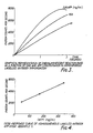
- the results corresponding to the samples having unknown analyte concentrations are compared with those obtained from a series of standards of known amounts of analyte by constructing a calibration curve from the latter examples of which are given in Figures 3 and 4.
- Figure 3 is a graphical representation of chemiluminescent reaction rate as a function of time and AFP concentration in homogeneous labelled antibody immunoassay.
- the three curves represent three different AFP concentrations, at 328, 164 and 41 ng/ml.
- Figure 4 is a defined dose-response curve showing how the count rate varies linearly with the concentration.
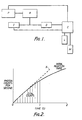
- FIG. 1 A typical apparatus is illustrated diagrammatically in Figure 1 where P is a photon counter, R is a data reduction unit, is an integrator, D -is a differentiator, C is a computer, L is a printer, M is a disk drive.
- Figure 2 illustrates a typical-graph illustrating photon count per second plotted against time, and illustrating the way in which the actual emission rate varies, (the amplitude at any point on the curve) and also the rate of change (the slope of the curve at any point) and the overall count (the integrated area under the curve).
- the line X indicates the slope of the initial reaction rate. It will be seen that other characteristics can be derived or deduced therefrom such as the "time to peak", or the total count up to 0.5 s or any other selected interval.
- Standard and unknown sera (50 pl) are incubated with the purified AFP labelled with acridinium ester molecules to a specific activity of 2x10 5 luminescent counts per nanogram and with polyclonal or monoclonal anti AFP antibodies at a concentration which is known to bind 50% of the labelled AFP after four hours at room temperature.
- the tubes are incubated at a final assay volume of 200 pl (0.1 M phosphate buffer, 0.15 M sodium chloride pH 6.3) for four hours at room temperature.
- the mixtures are then measured luminometrically by injection of alkaline hydrogen peroxide as before. The rate of excited state formation observed for the samples is compared with that of the standards and the AFP content quantified by interpolating as in
- This assay is based on the principle that T 3 with acridinium ester linked through the amino group will not react with naturally occurring binding proteins (e.g. thyroxine binding globulin) but binds to antibody.
- the assay for free T 3 is carried out as follows. Standards or unknown samples containing T 3 (50 pl) are diluted to 100 pl with HEPES buffer (0.01 M, pH 7.4). 50 ⁇ l labelled T 3 are added followed by 50 ⁇ l antibody solution. During a 30 minute reaction label is bound to antibody. This binding in inhibited by free T 3 which competes for antibody binding sites. After the reaction period, therefore, luminescence is measured following addition of alkaline hydrogen perioxide in the manner described. The rate of excited state formation in samples is compared with that of standards and the free T 3 concentration quantified by interpolation as in Example 1.
- Standard and unknown sera (50 ⁇ l) are incubated with purified insulin labelled with acridinium ester molecules and with pre-precipitated insulin antibodies. These represent high molecular weight complexes for enhanced change of rate of the chemiluminescent reaction as described above.
- the mixture is incubated for 3 hours at room temperature, in 200 ⁇ l 0.01 M phosphate buffer (pH 6.3) and the tubes analysed luminometrically. The reaction rates observed for the unknown samples are compared with those of the standards and hence the insulin concentrations determined by interpolation.
- a solublised thyroid membrane preparation in PBS pH 6.3) containing 0.1% HSA is incubated with 100 III of serum IgG fraction to be investigated.
- the amount of membrane being sufficient to bind 10-30% of the labelled thyrotrophin (TSH) being used.
- acridinium ester labelled TSH is added and the mixture incubated for an additional 1 hour period.
- the assay tubes are measured luminometrically as in Example 1 and the rates of emission compared with that of a normal serum IgG preparation to establish the presence or absence of pathlogically significant amounts of thyroid stimulating antibodies.
Landscapes
- Health & Medical Sciences (AREA)
- Immunology (AREA)
- Life Sciences & Earth Sciences (AREA)
- Engineering & Computer Science (AREA)
- Molecular Biology (AREA)
- Biomedical Technology (AREA)
- Chemical & Material Sciences (AREA)
- Hematology (AREA)
- Urology & Nephrology (AREA)
- Biotechnology (AREA)
- Microbiology (AREA)
- Cell Biology (AREA)
- Food Science & Technology (AREA)
- Medicinal Chemistry (AREA)
- Physics & Mathematics (AREA)
- Analytical Chemistry (AREA)
- Biochemistry (AREA)
- General Health & Medical Sciences (AREA)
- General Physics & Mathematics (AREA)
- Pathology (AREA)
- Investigating Or Analysing Materials By The Use Of Chemical Reactions (AREA)
Description
- This invention relates to methods designed for use in the analysis, assay or localisation of proteins, polypeptides, haptens and other substances of biological interest. The invention utilises immune complex formation as a measure of the amount of analyte present by means of the use of antigens, haptens or antibodies labelled with chemiluminescent molecules. In the past such biological molecules have been labelled using radioactive isotopes which suffer problems of instability, insensitivity, inconvenient quantification and disposal. In contrast chemiluminescent labels are non hazardous, stable and can be detected with high sensitivity using simple photon counting equipment. In the context Qf this invention the term chemiluminescence is used to distinguish this phenomenon from other forms of luminescence, e.g. fluorescence and phosphorescence, and is taken to include the related phenomenon of bioluminescence. In a further aspect of the invention the concentration of immune complexes, and hence of the analyte, is determined by changes in certain physicochemical parameters of the chemiluminescent reaction of such labelled species upon formation of immune complexes.
- In some preferred forms of the invention these changes are involved with the rate of the chemiluminescent reaction, notably the formation of the excited states or their subsequent decay. It is a particular and important feature of the invention that quantitation of the analyte concentration does not require prior separation of analyte-bound and unbound labelled immunoreactant.
- Many chemical species are capable of chemiluminescence, which is the phenomenon observed when the vibronically excited product of an exoergic reaction reverts to its ground state with photonic emission. Such reactions are usually of an oxidative nature and may or may not involve a catalytic component. For example luminol requires the presence of a catalyst for the chemiluminescent reaction, whereas acridinium esters do not require a catalyst. Broadly stated, a chemiluminescent reaction consists of two stages, these being the formation of excited states and their subsequent decay to their unex- cited or "ground" states. For any given excited state which yields a corresponding ground state arising from energy loss by photonic emission, the rate of decay is proportional to the concentration of molecules in the excited states. The decay rate coefficient of such a reaction, which acts to equate the aforementioned proportionality is thus first order and exponentially relates the concentration of the excited states to a particular increment of time which constitutes part of the total lifetime of the reaction.
- By comparison the rate of formation of excited states is an inherently more complex relationship than is the simple decay of excited states. The rate of formation is a function of the excited state precursor concentration (i.e. the concentration of the molecular luminescent label before being excited) and also of the concentration of the other rectants in the system. The excitation rate coefficient in this instance is a compound of a number of kinetic and thermodynamic parameters pertaining to the reactions which are involved in the formation of the excited states. Thus a change in such parameters such as may be experienced by prior chemical or physical interactions of the chemiluminescent molecule may be reflected as a change in excited state formation rate. Further, such a change will also be reflected by the relationship between the number of photons emitted or the rate at which they are emitted and an increment of time over which the reaction is taking place. There are unique advantages to be gained from utilizing the excitation phase to quantify a chemiluminescent reaction. Firstly, -it is extremely rapid-accurate measurements can be made within 1 second or less. Secondly, because of the short time scale there is a low background which increases the sensitivity of detection, and thirdly the use of a rate measurement reduces interference due to inner filter effects.
- It has been proposed in GB-A-2018424 to carry out an immunoassay technique involving a form of chemiluminescent label, and making observations of the light emission carried out over a period of time. In this case, however, the assay process was a chemically induced fluorescent immunoassay, which may be described as an energy transfer assay. The procedure essentially involved quenching of the light emission and the results were manifestations of this quenching effect, the observations being carried out over part only of the total period of light emission. This whole procedure was totally different from the assay of the present invention, which does not involve quenching and relies on the very rapid reaction rate during the excitation phase when the immune complex is being formed. In the present invention the period of observation may be limited to a matter of seconds compared with periods of minutes in the cited prior document. In GB-A-2044449 another immunoassay procedure is described, which again relies on transfer of energy from a chemiluminescent molecule to a fluorophor, and in this document also there is no suggestion of the advantages of confining the observations to the excitation phase of the immune complex formation.
- GB-A-2008247 is another example of an immunoassay procedure, but again there is no description or suggestion of examining the time course of the light emission so as to relate the reaction to the degree of immune complex formation.
- Broadly stated the invention consists in a method of quantifying a substance of biological interest by a homogeneous immunoassay in which a first component of an immune reaction in the form of an antigen, hapten, or antibody, is linked with one or more components of a chemiluminescent reaction, and the other component of the immune reaction, the analyte, is complexed therewith to cause entropic and/or enthalpic changes in a subsequent light emitting reaction as compared with the equivalent reaction of uncomplexed labelled component, the reactions being observed and compared to provide information on the immune complex formation, characterised in that the light or photon emission of each reaction is observed and measured during the excitation phase of the reaction where the intensity is rising.
- Preferably the intensity of light emission or the rate of photonic emission or the rate of change of intensity of emission are observed and compared with the equivalent properties of the uncomplexed component.
- In an alternative procedure according to the invention the timing of the peak of the emission from the reaction of the complexed labelled component is measured, the timing of the peak of the emission from the reaction of the uncomplexed labelled component is measured, and the measurements are compared whereby the substance of interest is quantified.
- Preferably, a graphical representation is firstly obtained of the function relating either the intensity orthe rate of photonic emission (and hence the excited state concentration) to the reaction time following initiation of the light-emitting reaction of the labelled immunoreactant. A graph is also prepared for labelled immunogen which has previously been incubated with the corresponding analyte of interest. Changes in the slope of the relationships at any given point corresponding to one of the variables ofthefunctions are a measure of the analyte concentration and can be compared with those of a series of known analyte concentrations. If the labelled immunoreactant is a labelled antigen or hapten and is used in a competitive binding system, the kinetic and thermodynamic parameters of the unbound labelled immunoreactant are compared with those of labelled immunoreactant previously incubated with analyte and antibody or binding protein. The changes in kinetic and thermodynamic parameters which result in changes in the rate of photon emission will be inversely proportional to the concentration of analyte. If the labelled immunoreactant is a labelled antibody, this may be used in an analogous competitive system for the measurement of antibodies in biological fluids such as serum. In a preferred aspect of the invention the labelled antibodies would be used in molar excess over the analyte present. The excess could be 500% but would preferably be no more than 100%, more preferably no more than 20%. In a. further aspect of the invention the kinetic changes observed are amplified by increasing the size of the immune complexes formed by the use of immobilized antibody. In this situation the change in the rate of excitation is enhanced when the size of the antigen antibody complex is enlarged by the presence of polymerized antibody or antibody linked to a solid support such as cellulose.
- Preferably, the quantity of analyte is reflected by a change in the graphical representation of the function relating excited state concentration (or the rate of change of excited state concentration or further derivation thereof) to the reaction time. These parameters are a function of the photonic emission of the chemiluminescent label which is quantified using photon counting apparatus. Several parameters of the function or its derivatives may be monitored so as to provide a measure of analyte concentration, for example the slope of the function at any given point on the reaction profile, the amplitude at any point, the integration of any part of the reaction profile, or the time taken to reach a peak or a preselected value, or any combination of several properties.
- Preferably the chemiluminescent labels are acridinium salts, luminol, dioxetanes, bis- oxalates, fluorescein, pyrogallol, lucigenin, lophine, photoproteins, or derivatives of these compounds which can be associated with antigens, antibodies and haptens so as to provide chemiluminescent immunoreactants.
- In contrast to existing homogeneous immunoassay techniques using enzyme or fluorescent labels, this invention is not restricted to the measurement of small molecules or haptens but may be applied to the quantitation of all antigenic compounds including drugs (e.g. barbiturates, salicylates, phenytoin, morphine, heroin, methotrexate, digoxin), steroids (e.g. testosterone, progesterone, oestradiol, oestriol, cortisol, aldosterone, vitamin D), cyclic neucleotides, thyroxine, triiodothyronine or proteins (e.g. insulin, growth hormone, parathyroid hormone, thyroid stimulating hormone, prolactin, adrenocor- ticotrophic hormone, alphafetoprotein, ferritin, human immunoglobulin (Ig) rabbit IgG, mouse IgG, guinea pig IgG, sheep IgG, interferon, components of the complement system and bacterial and viral antigens)
- Thus itwill be seen that the invention provides a method for the quantitation of substances of biological interest by a homogeneous immunoassay procedure in which antigens, antibodies or haptens are labelled with chemiluminescent molecules, the physico-chemical properties of which are modified in a measurable dependent way upon immune complex formation.
- In the context of the invention a homogeneous assay may be defined as one in which the properties of the label are changed in a dose- dependent or concentration-dependent way on formation of the immune complex, thus avoiding the need for separation of the bound and free analyte fractions.
- The invention may be performed in various different ways and a number of examples will now be described in detail by way of illustration.
- A series of solutions of AFP standard in sera are made and a known volume, preferably 50 ul, and hence a known quantity of AFP is placed in a series of assay tubes. Identical volumes of unknown sera to be investigated are placed in another series of assay tubes and 0.1 M phosphate buffer, pH 6.3 (100 µl) placed in all tubes. Antibodies to AFP (polyclonal or monoclonal) labelled with acridinium ester and preferably having a specific activity of 2x10° luminescent counts per nanogram are added to each tube such that they are present in excess over the AFP analyte. The tubes are incubated at room temperature preferably for 30 min, each tube is placed sequentially in a luminometer and the photons emitted upon initiation of the chemiluminescent reaction counted for 5 seconds or less. The rate of analysis of the tubes is adjusted so as to match the rate of addition of reagents prior to incubation. The chemiluminescent reaction is initiated by injection preferably of 200 pl of an aqueous solution containing 0.5 M sodium hydroxide and 0.1% (VN) of "100 volume" (i.e. 30% weight/volume) hydrogen peroxide. The rate of formation of excited states (i.e. the rate of photon emission over a suitable time period) is measured for standards and unknowns using photon counting equipment..This may be done in several different ways. In one process the total number of photons emitted over a one second time period is recorded, and in another process the relationship which expresses the rate of photonic emission as a function of time is differentiated to enable calculation of the initial rate of reaction. In either case the results corresponding to the samples having unknown analyte concentrations are compared with those obtained from a series of standards of known amounts of analyte by constructing a calibration curve from the latter examples of which are given in Figures 3 and 4.
- Figure 3 is a graphical representation of chemiluminescent reaction rate as a function of time and AFP concentration in homogeneous labelled antibody immunoassay. The three curves represent three different AFP concentrations, at 328, 164 and 41 ng/ml.
- Figure 4 is a defined dose-response curve showing how the count rate varies linearly with the concentration.
- A typical apparatus is illustrated diagrammatically in Figure 1 where P is a photon counter, R is a data reduction unit, is an integrator, D -is a differentiator, C is a computer, L is a printer, M is a disk drive. Figure 2 illustrates a typical-graph illustrating photon count per second plotted against time, and illustrating the way in which the actual emission rate varies, (the amplitude at any point on the curve) and also the rate of change (the slope of the curve at any point) and the overall count (the integrated area under the curve). In Figure 2 the line X indicates the slope of the initial reaction rate. It will be seen that other characteristics can be derived or deduced therefrom such as the "time to peak", or the total count up to 0.5 s or any other selected interval.
- Standard and unknown sera (50 pl) are incubated with the purified AFP labelled with acridinium ester molecules to a specific activity of 2x10⁵ luminescent counts per nanogram and with polyclonal or monoclonal anti AFP antibodies at a concentration which is known to bind 50% of the labelled AFP after four hours at room temperature. The tubes are incubated at a final assay volume of 200 pl (0.1 M phosphate buffer, 0.15 M sodium chloride pH 6.3) for four hours at room temperature. The mixtures are then measured luminometrically by injection of alkaline hydrogen peroxide as before. The rate of excited state formation observed for the samples is compared with that of the standards and the AFP content quantified by interpolating as in
- Standards and unknown samples (50 pl) containing T3 are diluted to 100 µl with HEPES buffer (0.01 M, pH 7.4) containing 0.25% merthiolate. Fifty µl T3 labelled with acridinium ester are added followed by 50 µl antibody to T3. After 30 minutes reaction the luminescence is measured as described in Example 2 following injection of alkaline hydrogen peroxide. The rate of excited state formation observed in the samples is compared with that of standards and the T3 content quantified by interpolation as in Example 1.
- This assay is based on the principle that T3 with acridinium ester linked through the amino group will not react with naturally occurring binding proteins (e.g. thyroxine binding globulin) but binds to antibody. The assay for free T3 is carried out as follows. Standards or unknown samples containing T3 (50 pl) are diluted to 100 pl with HEPES buffer (0.01 M, pH 7.4). 50 µl labelled T3 are added followed by 50 µl antibody solution. During a 30 minute reaction label is bound to antibody. This binding in inhibited by free T3 which competes for antibody binding sites. After the reaction period, therefore, luminescence is measured following addition of alkaline hydrogen perioxide in the manner described. The rate of excited state formation in samples is compared with that of standards and the free T3 concentration quantified by interpolation as in Example 1.
- Standard and unknown sera (50 µl) are incubated with purified insulin labelled with acridinium ester molecules and with pre-precipitated insulin antibodies. These represent high molecular weight complexes for enhanced change of rate of the chemiluminescent reaction as described above. The mixture is incubated for 3 hours at room temperature, in 200 µl 0.01 M phosphate buffer (pH 6.3) and the tubes analysed luminometrically. The reaction rates observed for the unknown samples are compared with those of the standards and hence the insulin concentrations determined by interpolation.
- A solublised thyroid membrane preparation in PBS pH 6.3) containing 0.1% HSA is incubated with 100 III of serum IgG fraction to be investigated. The amount of membrane being sufficient to bind 10-30% of the labelled thyrotrophin (TSH) being used.
- Following incubation at room temperature for 1 hour, acridinium ester labelled TSH is added and the mixture incubated for an additional 1 hour period. The assay tubes are measured luminometrically as in Example 1 and the rates of emission compared with that of a normal serum IgG preparation to establish the presence or absence of pathlogically significant amounts of thyroid stimulating antibodies.
Claims (9)
Applications Claiming Priority (2)
| Application Number | Priority Date | Filing Date | Title |
|---|---|---|---|
| GB8225933 | 1982-09-10 | ||
| GB8225933 | 1982-09-10 |
Publications (3)
| Publication Number | Publication Date |
|---|---|
| EP0103469A2 EP0103469A2 (en) | 1984-03-21 |
| EP0103469A3 EP0103469A3 (en) | 1984-12-05 |
| EP0103469B1 true EP0103469B1 (en) | 1989-02-08 |
Family
ID=10532841
Family Applications (1)
| Application Number | Title | Priority Date | Filing Date |
|---|---|---|---|
| EP83305277A Expired EP0103469B1 (en) | 1982-09-10 | 1983-09-09 | Immunological procedure for quantifying substances |
Country Status (4)
| Country | Link |
|---|---|
| US (1) | US4761382A (en) |
| EP (1) | EP0103469B1 (en) |
| DE (1) | DE3379179D1 (en) |
| GB (1) | GB2129553B (en) |
Families Citing this family (12)
| Publication number | Priority date | Publication date | Assignee | Title |
|---|---|---|---|---|
| NL8703075A (en) * | 1987-12-18 | 1989-07-17 | Nederlanden Staat | ACRIDINUM COMPOUNDS AS A CHEMILUMINESCENT MARKING. |
| GB8820887D0 (en) * | 1988-09-06 | 1988-10-05 | Atomic Energy Authority Uk | Detecting selected species |
| GB8916806D0 (en) * | 1989-07-22 | 1989-09-06 | Univ Wales Medicine | Modified proteins |
| US5683888A (en) * | 1989-07-22 | 1997-11-04 | University Of Wales College Of Medicine | Modified bioluminescent proteins and their use |
| JPH06504374A (en) * | 1990-12-28 | 1994-05-19 | アボット・ラボラトリーズ | Simultaneous determination of multiple analytes using time-resolved heterogeneous chemiluminescence assays |
| DE69518182T2 (en) * | 1994-04-11 | 2001-03-22 | Kyowa Medex Co. Ltd., Tokio/Tokyo | Method for determining a substance |
| US5783453A (en) * | 1995-06-29 | 1998-07-21 | Chiron Diagnostics Corporation | Non-separation specific binding chemiluminescent assay |
| GB2312746B (en) * | 1996-04-24 | 2000-07-19 | Molecular Light Technology Lim | Detection of an analyte in a Water Immiscible Solvent |
| US6294333B1 (en) | 1998-12-31 | 2001-09-25 | Ingeneus Corp. | Fluorescent intensity assay for protein or peptide binding to nucleic acids |
| US6645733B1 (en) | 1999-06-25 | 2003-11-11 | Ingeneus Corporation | Fluorescent intensity method for assaying binding between proteins or peptides |
| CN101201354B (en) * | 2006-12-14 | 2011-11-02 | 北京科美东雅生物技术有限公司 | Thyroxine chemiluminescence immune analysis quantitative measuring reagent kit and method for preparing the same |
| CN116930478A (en) * | 2022-04-02 | 2023-10-24 | 迈克生物股份有限公司 | Thyroxine fluorescent conjugate and preparation method and application thereof |
Family Cites Families (9)
| Publication number | Priority date | Publication date | Assignee | Title |
|---|---|---|---|---|
| GB1578275A (en) * | 1977-06-14 | 1980-11-05 | Maier C | Procedure for the assay of pharmacologically immunologically and biochemically active compounds in biological fluids |
| GB2008247B (en) * | 1977-11-17 | 1982-12-15 | Welsh Nat School Med | Detecting or quantifying substances using labelling techniques |
| US4220450A (en) * | 1978-04-05 | 1980-09-02 | Syva Company | Chemically induced fluorescence immunoassay |
| US4238195A (en) * | 1979-01-18 | 1980-12-09 | Miles Laboratories, Inc. | Fluorescer-labeled specific binding assays |
| US4280815A (en) * | 1979-06-18 | 1981-07-28 | Technicon Instruments Corporation | Electrochemiluminescent immunoassay and apparatus therefor |
| US4276051A (en) * | 1980-01-28 | 1981-06-30 | Coulter Electronics, Inc. | System and program for chemical reaction observation with a moving photometer |
| DE3132491A1 (en) * | 1980-08-22 | 1982-07-01 | Laboratorium Prof. Dr. Rudolf Berthold, 7547 Wildbad | METHOD FOR PERFORMING ANALYTICAL DETERMINATIONS BY MEANS OF THE CHEMILUMINESCENCE METHOD AND APPLICATION OF THE METHOD FOR IMMUNASSAYS |
| GB2086042B (en) * | 1980-08-22 | 1983-11-30 | Berthold Lab Prof R | Process for carrying out analytical determinations by means of chemiluminescence and the use of the process for immunoassay |
| CA1170568A (en) * | 1980-12-22 | 1984-07-10 | Bernd Frenzel | Process for determining antigens, antibodies and their complexes |
-
1983
- 1983-09-09 EP EP83305277A patent/EP0103469B1/en not_active Expired
- 1983-09-09 DE DE8383305277T patent/DE3379179D1/en not_active Expired
- 1983-09-09 GB GB08324249A patent/GB2129553B/en not_active Expired
-
1987
- 1987-02-05 US US07/013,215 patent/US4761382A/en not_active Expired - Lifetime
Also Published As
| Publication number | Publication date |
|---|---|
| GB8324249D0 (en) | 1983-10-12 |
| US4761382A (en) | 1988-08-02 |
| EP0103469A2 (en) | 1984-03-21 |
| EP0103469A3 (en) | 1984-12-05 |
| GB2129553B (en) | 1986-06-18 |
| GB2129553A (en) | 1984-05-16 |
| DE3379179D1 (en) | 1989-03-16 |
Similar Documents
| Publication | Publication Date | Title |
|---|---|---|
| US4478817A (en) | Detecting or quantifying substances using labelling techniques | |
| US4626513A (en) | Method and apparatus for ligand detection | |
| US5656207A (en) | Detecting or quantifying multiple analytes using labelling techniques | |
| US5464741A (en) | Palladium (II) octaethylporphine alpha-isothiocyanate as a phosphorescent label for immunoassays | |
| EP0191575B1 (en) | Homogeneous fluorescence immunoassay using a light absorbing material | |
| US6087188A (en) | Two-site immunoassay for an antibody with chemiluminescent label and biotin bound ligand | |
| Smith et al. | A review of fluoroimmunoassay and immunofluorometric assay | |
| US8956823B2 (en) | Anti-antibody reagent | |
| EP0103469B1 (en) | Immunological procedure for quantifying substances | |
| EP0116454A2 (en) | Enhanced luminescent or luminometric assay | |
| WO1999030131A1 (en) | One-step fluorescent immunosensor test | |
| US5389523A (en) | Liposome immunoanalysis by flow injection assay | |
| GB2233450A (en) | Detecting or quantifying multiple analytes | |
| JP3583441B2 (en) | Use of phycobiliprotein-binding peptide conjugate as a fluorescent tracer | |
| EP0669000B1 (en) | Two-site immunoassay for an antibody with chemiluminescent label and biotin bound ligand | |
| EP0522677A1 (en) | Method of measuring concentration of immunoreactant using electrochemiluminescence | |
| JPH09504094A (en) | Method for assaying immunological substances using magnetic latex particles and non-magnetic particles | |
| US4052504A (en) | Assay for thyroxine binding globulin | |
| US5202269A (en) | Method for immunochemical determination of hapten | |
| US5045479A (en) | Continuous flow competitive assay with reference system | |
| JP7488812B2 (en) | Chemiluminescence immunoassay method and system and reagent kit using said method | |
| USH1018H (en) | Immunological method for the determination of free substances having hapten properties | |
| AU595899B2 (en) | Method for diagnostic immunoassay by solid phase separation | |
| KR880700270A (en) | Single-phase method for detecting and / or measuring a sample in a medium in which the sample may exist using light emission of the sample | |
| JPH0672882B2 (en) | Immunological methods for detection and quantification of substances |
Legal Events
| Date | Code | Title | Description |
|---|---|---|---|
| PUAI | Public reference made under article 153(3) epc to a published international application that has entered the european phase |
Free format text: ORIGINAL CODE: 0009012 |
|
| AK | Designated contracting states |
Designated state(s): BE CH DE FR IT LI NL |
|
| PUAL | Search report despatched |
Free format text: ORIGINAL CODE: 0009013 |
|
| AK | Designated contracting states |
Designated state(s): BE CH DE FR IT LI NL |
|
| 17P | Request for examination filed |
Effective date: 19841112 |
|
| 17Q | First examination report despatched |
Effective date: 19860129 |
|
| R17C | First examination report despatched (corrected) |
Effective date: 19870309 |
|
| GRAA | (expected) grant |
Free format text: ORIGINAL CODE: 0009210 |
|
| AK | Designated contracting states |
Kind code of ref document: B1 Designated state(s): BE CH DE FR IT LI NL |
|
| REF | Corresponds to: |
Ref document number: 3379179 Country of ref document: DE Date of ref document: 19890316 |
|
| ITF | It: translation for a ep patent filed | ||
| RAP4 | Party data changed (patent owner data changed or rights of a patent transferred) |
Owner name: THE WELSH NATIONAL SCHOOL OF MEDICINE |
|
| ET | Fr: translation filed | ||
| PLBE | No opposition filed within time limit |
Free format text: ORIGINAL CODE: 0009261 |
|
| STAA | Information on the status of an ep patent application or granted ep patent |
Free format text: STATUS: NO OPPOSITION FILED WITHIN TIME LIMIT |
|
| 26N | No opposition filed | ||
| ITTA | It: last paid annual fee | ||
| PGFP | Annual fee paid to national office [announced via postgrant information from national office to epo] |
Ref country code: FR Payment date: 20020912 Year of fee payment: 20 |
|
| PGFP | Annual fee paid to national office [announced via postgrant information from national office to epo] |
Ref country code: CH Payment date: 20020925 Year of fee payment: 20 |
|
| PGFP | Annual fee paid to national office [announced via postgrant information from national office to epo] |
Ref country code: NL Payment date: 20020930 Year of fee payment: 20 |
|
| PGFP | Annual fee paid to national office [announced via postgrant information from national office to epo] |
Ref country code: BE Payment date: 20021022 Year of fee payment: 20 |
|
| PGFP | Annual fee paid to national office [announced via postgrant information from national office to epo] |
Ref country code: DE Payment date: 20021118 Year of fee payment: 20 |
|
| PG25 | Lapsed in a contracting state [announced via postgrant information from national office to epo] |
Ref country code: LI Free format text: LAPSE BECAUSE OF EXPIRATION OF PROTECTION Effective date: 20030908 Ref country code: CH Free format text: LAPSE BECAUSE OF EXPIRATION OF PROTECTION Effective date: 20030908 |
|
| PG25 | Lapsed in a contracting state [announced via postgrant information from national office to epo] |
Ref country code: NL Free format text: LAPSE BECAUSE OF EXPIRATION OF PROTECTION Effective date: 20030909 |
|
| BE20 | Be: patent expired |
Owner name: THE *WELSH NATIONAL SCHOOL OF MEDICINE Effective date: 20030909 |
|
| REG | Reference to a national code |
Ref country code: CH Ref legal event code: PL |
|
| NLV7 | Nl: ceased due to reaching the maximum lifetime of a patent |
Effective date: 20030909 |