EP0103469A2 - Immunological procedure for quantifying substances - Google Patents
Immunological procedure for quantifying substances Download PDFInfo
- Publication number
- EP0103469A2 EP0103469A2 EP83305277A EP83305277A EP0103469A2 EP 0103469 A2 EP0103469 A2 EP 0103469A2 EP 83305277 A EP83305277 A EP 83305277A EP 83305277 A EP83305277 A EP 83305277A EP 0103469 A2 EP0103469 A2 EP 0103469A2
- Authority
- EP
- European Patent Office
- Prior art keywords
- reaction
- labelled
- component
- antibody
- immune
- Prior art date
- Legal status (The legal status is an assumption and is not a legal conclusion. Google has not performed a legal analysis and makes no representation as to the accuracy of the status listed.)
- Granted
Links
- 238000000034 method Methods 0.000 title claims abstract description 18
- 239000000126 substance Substances 0.000 title claims abstract description 8
- 230000001900 immune effect Effects 0.000 title 1
- 238000006243 chemical reaction Methods 0.000 claims abstract description 34
- 239000012491 analyte Substances 0.000 claims abstract description 20
- 239000000427 antigen Substances 0.000 claims abstract description 11
- 102000036639 antigens Human genes 0.000 claims abstract description 11
- 108091007433 antigens Proteins 0.000 claims abstract description 11
- 230000008859 change Effects 0.000 claims abstract description 11
- 230000008105 immune reaction Effects 0.000 claims abstract description 6
- 238000003018 immunoassay Methods 0.000 claims abstract description 6
- 206010053614 Type III immune complex mediated reaction Diseases 0.000 claims abstract description 4
- 230000016178 immune complex formation Effects 0.000 claims abstract description 4
- 230000009471 action Effects 0.000 claims abstract description 3
- 239000000376 reactant Substances 0.000 claims description 3
- 230000009137 competitive binding Effects 0.000 claims description 2
- 239000007787 solid Substances 0.000 claims description 2
- 210000000987 immune system Anatomy 0.000 claims 1
- 230000005281 excited state Effects 0.000 description 17
- 238000003556 assay Methods 0.000 description 14
- 230000015572 biosynthetic process Effects 0.000 description 12
- 102100023635 Alpha-fetoprotein Human genes 0.000 description 11
- NOESYZHRGYRDHS-UHFFFAOYSA-N insulin Chemical compound N1C(=O)C(NC(=O)C(CCC(N)=O)NC(=O)C(CCC(O)=O)NC(=O)C(C(C)C)NC(=O)C(NC(=O)CN)C(C)CC)CSSCC(C(NC(CO)C(=O)NC(CC(C)C)C(=O)NC(CC=2C=CC(O)=CC=2)C(=O)NC(CCC(N)=O)C(=O)NC(CC(C)C)C(=O)NC(CCC(O)=O)C(=O)NC(CC(N)=O)C(=O)NC(CC=2C=CC(O)=CC=2)C(=O)NC(CSSCC(NC(=O)C(C(C)C)NC(=O)C(CC(C)C)NC(=O)C(CC=2C=CC(O)=CC=2)NC(=O)C(CC(C)C)NC(=O)C(C)NC(=O)C(CCC(O)=O)NC(=O)C(C(C)C)NC(=O)C(CC(C)C)NC(=O)C(CC=2NC=NC=2)NC(=O)C(CO)NC(=O)CNC2=O)C(=O)NCC(=O)NC(CCC(O)=O)C(=O)NC(CCCNC(N)=N)C(=O)NCC(=O)NC(CC=3C=CC=CC=3)C(=O)NC(CC=3C=CC=CC=3)C(=O)NC(CC=3C=CC(O)=CC=3)C(=O)NC(C(C)O)C(=O)N3C(CCC3)C(=O)NC(CCCCN)C(=O)NC(C)C(O)=O)C(=O)NC(CC(N)=O)C(O)=O)=O)NC(=O)C(C(C)CC)NC(=O)C(CO)NC(=O)C(C(C)O)NC(=O)C1CSSCC2NC(=O)C(CC(C)C)NC(=O)C(NC(=O)C(CCC(N)=O)NC(=O)C(CC(N)=O)NC(=O)C(NC(=O)C(N)CC=1C=CC=CC=1)C(C)C)CC1=CN=CN1 NOESYZHRGYRDHS-UHFFFAOYSA-N 0.000 description 10
- MHAJPDPJQMAIIY-UHFFFAOYSA-N Hydrogen peroxide Chemical compound OO MHAJPDPJQMAIIY-UHFFFAOYSA-N 0.000 description 8
- RXNXLAHQOVLMIE-UHFFFAOYSA-N phenyl 10-methylacridin-10-ium-9-carboxylate Chemical compound C12=CC=CC=C2[N+](C)=C2C=CC=CC2=C1C(=O)OC1=CC=CC=C1 RXNXLAHQOVLMIE-UHFFFAOYSA-N 0.000 description 6
- 102000004877 Insulin Human genes 0.000 description 5
- 108090001061 Insulin Proteins 0.000 description 5
- 229940125396 insulin Drugs 0.000 description 5
- 238000005259 measurement Methods 0.000 description 4
- 210000002966 serum Anatomy 0.000 description 4
- HEMHJVSKTPXQMS-UHFFFAOYSA-M Sodium hydroxide Chemical compound [OH-].[Na+] HEMHJVSKTPXQMS-UHFFFAOYSA-M 0.000 description 3
- 150000001875 compounds Chemical class 0.000 description 3
- 230000000694 effects Effects 0.000 description 3
- 230000005284 excitation Effects 0.000 description 3
- 238000002347 injection Methods 0.000 description 3
- 239000007924 injection Substances 0.000 description 3
- 238000004020 luminiscence type Methods 0.000 description 3
- 239000000203 mixture Substances 0.000 description 3
- 239000008363 phosphate buffer Substances 0.000 description 3
- 210000001685 thyroid gland Anatomy 0.000 description 3
- 108060003951 Immunoglobulin Proteins 0.000 description 2
- RJKFOVLPORLFTN-LEKSSAKUSA-N Progesterone Chemical compound C1CC2=CC(=O)CC[C@]2(C)[C@@H]2[C@@H]1[C@@H]1CC[C@H](C(=O)C)[C@@]1(C)CC2 RJKFOVLPORLFTN-LEKSSAKUSA-N 0.000 description 2
- FAPWRFPIFSIZLT-UHFFFAOYSA-M Sodium chloride Chemical compound [Na+].[Cl-] FAPWRFPIFSIZLT-UHFFFAOYSA-M 0.000 description 2
- MUMGGOZAMZWBJJ-DYKIIFRCSA-N Testostosterone Chemical compound O=C1CC[C@]2(C)[C@H]3CC[C@](C)([C@H](CC4)O)[C@@H]4[C@@H]3CCC2=C1 MUMGGOZAMZWBJJ-DYKIIFRCSA-N 0.000 description 2
- AUYYCJSJGJYCDS-LBPRGKRZSA-N Thyrolar Chemical compound IC1=CC(C[C@H](N)C(O)=O)=CC(I)=C1OC1=CC=C(O)C(I)=C1 AUYYCJSJGJYCDS-LBPRGKRZSA-N 0.000 description 2
- 102000011923 Thyrotropin Human genes 0.000 description 2
- 108010061174 Thyrotropin Proteins 0.000 description 2
- DZBUGLKDJFMEHC-UHFFFAOYSA-N acridine Chemical class C1=CC=CC2=CC3=CC=CC=C3N=C21 DZBUGLKDJFMEHC-UHFFFAOYSA-N 0.000 description 2
- 238000004458 analytical method Methods 0.000 description 2
- 102000023732 binding proteins Human genes 0.000 description 2
- 108091008324 binding proteins Proteins 0.000 description 2
- 239000003054 catalyst Substances 0.000 description 2
- 230000001419 dependent effect Effects 0.000 description 2
- 238000001514 detection method Methods 0.000 description 2
- 231100000673 dose–response relationship Toxicity 0.000 description 2
- 230000005283 ground state Effects 0.000 description 2
- JYGXADMDTFJGBT-VWUMJDOOSA-N hydrocortisone Chemical compound O=C1CC[C@]2(C)[C@H]3[C@@H](O)C[C@](C)([C@@](CC4)(O)C(=O)CO)[C@@H]4[C@@H]3CCC2=C1 JYGXADMDTFJGBT-VWUMJDOOSA-N 0.000 description 2
- 102000018358 immunoglobulin Human genes 0.000 description 2
- 238000011534 incubation Methods 0.000 description 2
- 230000000977 initiatory effect Effects 0.000 description 2
- HWYHZTIRURJOHG-UHFFFAOYSA-N luminol Chemical compound O=C1NNC(=O)C2=C1C(N)=CC=C2 HWYHZTIRURJOHG-UHFFFAOYSA-N 0.000 description 2
- 239000012528 membrane Substances 0.000 description 2
- BQJCRHHNABKAKU-KBQPJGBKSA-N morphine Chemical compound O([C@H]1[C@H](C=C[C@H]23)O)C4=C5[C@@]12CCN(C)[C@@H]3CC5=CC=C4O BQJCRHHNABKAKU-KBQPJGBKSA-N 0.000 description 2
- 238000002360 preparation method Methods 0.000 description 2
- 230000008569 process Effects 0.000 description 2
- 102000004169 proteins and genes Human genes 0.000 description 2
- 108090000623 proteins and genes Proteins 0.000 description 2
- WQGWDDDVZFFDIG-UHFFFAOYSA-N pyrogallol Chemical compound OC1=CC=CC(O)=C1O WQGWDDDVZFFDIG-UHFFFAOYSA-N 0.000 description 2
- 230000035484 reaction time Effects 0.000 description 2
- 230000035945 sensitivity Effects 0.000 description 2
- 238000000926 separation method Methods 0.000 description 2
- 239000000243 solution Substances 0.000 description 2
- 230000004936 stimulating effect Effects 0.000 description 2
- XUIIKFGFIJCVMT-UHFFFAOYSA-N thyroxine-binding globulin Natural products IC1=CC(CC([NH3+])C([O-])=O)=CC(I)=C1OC1=CC(I)=C(O)C(I)=C1 XUIIKFGFIJCVMT-UHFFFAOYSA-N 0.000 description 2
- PROQIPRRNZUXQM-UHFFFAOYSA-N (16alpha,17betaOH)-Estra-1,3,5(10)-triene-3,16,17-triol Natural products OC1=CC=C2C3CCC(C)(C(C(O)C4)O)C4C3CCC2=C1 PROQIPRRNZUXQM-UHFFFAOYSA-N 0.000 description 1
- VOXZDWNPVJITMN-ZBRFXRBCSA-N 17β-estradiol Chemical compound OC1=CC=C2[C@H]3CC[C@](C)([C@H](CC4)O)[C@@H]4[C@@H]3CCC2=C1 VOXZDWNPVJITMN-ZBRFXRBCSA-N 0.000 description 1
- RNIPJYFZGXJSDD-UHFFFAOYSA-N 2,4,5-triphenyl-1h-imidazole Chemical compound C1=CC=CC=C1C1=NC(C=2C=CC=CC=2)=C(C=2C=CC=CC=2)N1 RNIPJYFZGXJSDD-UHFFFAOYSA-N 0.000 description 1
- JKMHFZQWWAIEOD-UHFFFAOYSA-N 2-[4-(2-hydroxyethyl)piperazin-1-yl]ethanesulfonic acid Chemical compound OCC[NH+]1CCN(CCS([O-])(=O)=O)CC1 JKMHFZQWWAIEOD-UHFFFAOYSA-N 0.000 description 1
- PQSUYGKTWSAVDQ-UHFFFAOYSA-N Aldosterone Natural products C1CC2C3CCC(C(=O)CO)C3(C=O)CC(O)C2C2(C)C1=CC(=O)CC2 PQSUYGKTWSAVDQ-UHFFFAOYSA-N 0.000 description 1
- PQSUYGKTWSAVDQ-ZVIOFETBSA-N Aldosterone Chemical compound C([C@@]1([C@@H](C(=O)CO)CC[C@H]1[C@@H]1CC2)C=O)[C@H](O)[C@@H]1[C@]1(C)C2=CC(=O)CC1 PQSUYGKTWSAVDQ-ZVIOFETBSA-N 0.000 description 1
- 108010032595 Antibody Binding Sites Proteins 0.000 description 1
- 241000700199 Cavia porcellus Species 0.000 description 1
- XUIIKFGFIJCVMT-GFCCVEGCSA-N D-thyroxine Chemical compound IC1=CC(C[C@@H](N)C(O)=O)=CC(I)=C1OC1=CC(I)=C(O)C(I)=C1 XUIIKFGFIJCVMT-GFCCVEGCSA-N 0.000 description 1
- LTMHDMANZUZIPE-AMTYYWEZSA-N Digoxin Natural products O([C@H]1[C@H](C)O[C@H](O[C@@H]2C[C@@H]3[C@@](C)([C@@H]4[C@H]([C@]5(O)[C@](C)([C@H](O)C4)[C@H](C4=CC(=O)OC4)CC5)CC3)CC2)C[C@@H]1O)[C@H]1O[C@H](C)[C@@H](O[C@H]2O[C@@H](C)[C@H](O)[C@@H](O)C2)[C@@H](O)C1 LTMHDMANZUZIPE-AMTYYWEZSA-N 0.000 description 1
- BVTJGGGYKAMDBN-UHFFFAOYSA-N Dioxetane Chemical class C1COO1 BVTJGGGYKAMDBN-UHFFFAOYSA-N 0.000 description 1
- 102000004190 Enzymes Human genes 0.000 description 1
- 108090000790 Enzymes Proteins 0.000 description 1
- 102000008857 Ferritin Human genes 0.000 description 1
- 108050000784 Ferritin Proteins 0.000 description 1
- 238000008416 Ferritin Methods 0.000 description 1
- 108010051696 Growth Hormone Proteins 0.000 description 1
- 102000018997 Growth Hormone Human genes 0.000 description 1
- 239000007995 HEPES buffer Substances 0.000 description 1
- GVGLGOZIDCSQPN-PVHGPHFFSA-N Heroin Chemical compound O([C@H]1[C@H](C=C[C@H]23)OC(C)=O)C4=C5[C@@]12CCN(C)[C@@H]3CC5=CC=C4OC(C)=O GVGLGOZIDCSQPN-PVHGPHFFSA-N 0.000 description 1
- 102000014150 Interferons Human genes 0.000 description 1
- 108010050904 Interferons Proteins 0.000 description 1
- FBOZXECLQNJBKD-ZDUSSCGKSA-N L-methotrexate Chemical compound C=1N=C2N=C(N)N=C(N)C2=NC=1CN(C)C1=CC=C(C(=O)N[C@@H](CCC(O)=O)C(O)=O)C=C1 FBOZXECLQNJBKD-ZDUSSCGKSA-N 0.000 description 1
- 102000006830 Luminescent Proteins Human genes 0.000 description 1
- 108010047357 Luminescent Proteins Proteins 0.000 description 1
- 241000283973 Oryctolagus cuniculus Species 0.000 description 1
- 102000003982 Parathyroid hormone Human genes 0.000 description 1
- 108090000445 Parathyroid hormone Proteins 0.000 description 1
- 241001494479 Pecora Species 0.000 description 1
- CXOFVDLJLONNDW-UHFFFAOYSA-N Phenytoin Chemical compound N1C(=O)NC(=O)C1(C=1C=CC=CC=1)C1=CC=CC=C1 CXOFVDLJLONNDW-UHFFFAOYSA-N 0.000 description 1
- 108010057464 Prolactin Proteins 0.000 description 1
- 102100024819 Prolactin Human genes 0.000 description 1
- 102000002248 Thyroxine-Binding Globulin Human genes 0.000 description 1
- 108010000259 Thyroxine-Binding Globulin Proteins 0.000 description 1
- 229930003316 Vitamin D Natural products 0.000 description 1
- QYSXJUFSXHHAJI-XFEUOLMDSA-N Vitamin D3 Natural products C1(/[C@@H]2CC[C@@H]([C@]2(CCC1)C)[C@H](C)CCCC(C)C)=C/C=C1\C[C@@H](O)CCC1=C QYSXJUFSXHHAJI-XFEUOLMDSA-N 0.000 description 1
- 229960002478 aldosterone Drugs 0.000 description 1
- 102000013529 alpha-Fetoproteins Human genes 0.000 description 1
- 108010026331 alpha-Fetoproteins Proteins 0.000 description 1
- 125000003277 amino group Chemical group 0.000 description 1
- 230000000890 antigenic effect Effects 0.000 description 1
- 239000007864 aqueous solution Substances 0.000 description 1
- 230000001580 bacterial effect Effects 0.000 description 1
- 229940125717 barbiturate Drugs 0.000 description 1
- -1 barbiturates Chemical class 0.000 description 1
- 230000027455 binding Effects 0.000 description 1
- 239000013060 biological fluid Substances 0.000 description 1
- 238000005415 bioluminescence Methods 0.000 description 1
- 230000029918 bioluminescence Effects 0.000 description 1
- 239000000872 buffer Substances 0.000 description 1
- 238000004364 calculation method Methods 0.000 description 1
- 238000011088 calibration curve Methods 0.000 description 1
- 230000003197 catalytic effect Effects 0.000 description 1
- 229920002678 cellulose Polymers 0.000 description 1
- 239000001913 cellulose Substances 0.000 description 1
- 239000003153 chemical reaction reagent Substances 0.000 description 1
- 239000013626 chemical specie Substances 0.000 description 1
- 230000002860 competitive effect Effects 0.000 description 1
- 230000004154 complement system Effects 0.000 description 1
- 230000009918 complex formation Effects 0.000 description 1
- 125000004122 cyclic group Chemical group 0.000 description 1
- 238000009795 derivation Methods 0.000 description 1
- 229960002069 diamorphine Drugs 0.000 description 1
- LTMHDMANZUZIPE-PUGKRICDSA-N digoxin Chemical compound C1[C@H](O)[C@H](O)[C@@H](C)O[C@H]1O[C@@H]1[C@@H](C)O[C@@H](O[C@@H]2[C@H](O[C@@H](O[C@@H]3C[C@@H]4[C@]([C@@H]5[C@H]([C@]6(CC[C@@H]([C@@]6(C)[C@H](O)C5)C=5COC(=O)C=5)O)CC4)(C)CC3)C[C@@H]2O)C)C[C@@H]1O LTMHDMANZUZIPE-PUGKRICDSA-N 0.000 description 1
- 229960005156 digoxin Drugs 0.000 description 1
- LTMHDMANZUZIPE-UHFFFAOYSA-N digoxine Natural products C1C(O)C(O)C(C)OC1OC1C(C)OC(OC2C(OC(OC3CC4C(C5C(C6(CCC(C6(C)C(O)C5)C=5COC(=O)C=5)O)CC4)(C)CC3)CC2O)C)CC1O LTMHDMANZUZIPE-UHFFFAOYSA-N 0.000 description 1
- 229940079593 drug Drugs 0.000 description 1
- 239000003814 drug Substances 0.000 description 1
- 229930182833 estradiol Natural products 0.000 description 1
- PROQIPRRNZUXQM-ZXXIGWHRSA-N estriol Chemical compound OC1=CC=C2[C@H]3CC[C@](C)([C@H]([C@H](O)C4)O)[C@@H]4[C@@H]3CCC2=C1 PROQIPRRNZUXQM-ZXXIGWHRSA-N 0.000 description 1
- GNBHRKFJIUUOQI-UHFFFAOYSA-N fluorescein Chemical compound O1C(=O)C2=CC=CC=C2C21C1=CC=C(O)C=C1OC1=CC(O)=CC=C21 GNBHRKFJIUUOQI-UHFFFAOYSA-N 0.000 description 1
- 239000000122 growth hormone Substances 0.000 description 1
- 231100001261 hazardous Toxicity 0.000 description 1
- 229940088597 hormone Drugs 0.000 description 1
- 239000005556 hormone Substances 0.000 description 1
- 229960000890 hydrocortisone Drugs 0.000 description 1
- 230000002163 immunogen Effects 0.000 description 1
- 229940072221 immunoglobulins Drugs 0.000 description 1
- 230000010354 integration Effects 0.000 description 1
- 229940079322 interferon Drugs 0.000 description 1
- 230000004807 localization Effects 0.000 description 1
- KNJDBYZZKAZQNG-UHFFFAOYSA-N lucigenin Chemical compound [O-][N+]([O-])=O.[O-][N+]([O-])=O.C12=CC=CC=C2[N+](C)=C(C=CC=C2)C2=C1C1=C(C=CC=C2)C2=[N+](C)C2=CC=CC=C12 KNJDBYZZKAZQNG-UHFFFAOYSA-N 0.000 description 1
- 229960000485 methotrexate Drugs 0.000 description 1
- 229960005181 morphine Drugs 0.000 description 1
- 230000001590 oxidative effect Effects 0.000 description 1
- 229960001319 parathyroid hormone Drugs 0.000 description 1
- 239000000199 parathyroid hormone Substances 0.000 description 1
- 229960002036 phenytoin Drugs 0.000 description 1
- 230000010399 physical interaction Effects 0.000 description 1
- 229920001184 polypeptide Polymers 0.000 description 1
- 239000002243 precursor Substances 0.000 description 1
- 102000004196 processed proteins & peptides Human genes 0.000 description 1
- 108090000765 processed proteins & peptides Proteins 0.000 description 1
- 239000000186 progesterone Substances 0.000 description 1
- 229960003387 progesterone Drugs 0.000 description 1
- 229940097325 prolactin Drugs 0.000 description 1
- 229940079877 pyrogallol Drugs 0.000 description 1
- 238000011002 quantification Methods 0.000 description 1
- 230000002285 radioactive effect Effects 0.000 description 1
- 230000009467 reduction Effects 0.000 description 1
- 150000003873 salicylate salts Chemical class 0.000 description 1
- 150000003384 small molecules Chemical class 0.000 description 1
- 239000011780 sodium chloride Substances 0.000 description 1
- 241000894007 species Species 0.000 description 1
- 150000003431 steroids Chemical class 0.000 description 1
- 229960003604 testosterone Drugs 0.000 description 1
- RTKIYNMVFMVABJ-UHFFFAOYSA-L thimerosal Chemical compound [Na+].CC[Hg]SC1=CC=CC=C1C([O-])=O RTKIYNMVFMVABJ-UHFFFAOYSA-L 0.000 description 1
- 229950001470 thyrotrophin Drugs 0.000 description 1
- 229940034208 thyroxine Drugs 0.000 description 1
- 229940035722 triiodothyronine Drugs 0.000 description 1
- 230000003612 virological effect Effects 0.000 description 1
- 235000019166 vitamin D Nutrition 0.000 description 1
- 239000011710 vitamin D Substances 0.000 description 1
- 150000003710 vitamin D derivatives Chemical class 0.000 description 1
- 229940046008 vitamin d Drugs 0.000 description 1
Images

Classifications
-
- G—PHYSICS
- G01—MEASURING; TESTING
- G01N—INVESTIGATING OR ANALYSING MATERIALS BY DETERMINING THEIR CHEMICAL OR PHYSICAL PROPERTIES
- G01N33/00—Investigating or analysing materials by specific methods not covered by groups G01N1/00 - G01N31/00
- G01N33/48—Biological material, e.g. blood, urine; Haemocytometers
- G01N33/50—Chemical analysis of biological material, e.g. blood, urine; Testing involving biospecific ligand binding methods; Immunological testing
- G01N33/53—Immunoassay; Biospecific binding assay; Materials therefor
- G01N33/536—Immunoassay; Biospecific binding assay; Materials therefor with immune complex formed in liquid phase
- G01N33/542—Immunoassay; Biospecific binding assay; Materials therefor with immune complex formed in liquid phase with steric inhibition or signal modification, e.g. fluorescent quenching
-
- Y—GENERAL TAGGING OF NEW TECHNOLOGICAL DEVELOPMENTS; GENERAL TAGGING OF CROSS-SECTIONAL TECHNOLOGIES SPANNING OVER SEVERAL SECTIONS OF THE IPC; TECHNICAL SUBJECTS COVERED BY FORMER USPC CROSS-REFERENCE ART COLLECTIONS [XRACs] AND DIGESTS
- Y10—TECHNICAL SUBJECTS COVERED BY FORMER USPC
- Y10S—TECHNICAL SUBJECTS COVERED BY FORMER USPC CROSS-REFERENCE ART COLLECTIONS [XRACs] AND DIGESTS
- Y10S436/00—Chemistry: analytical and immunological testing
- Y10S436/80—Fluorescent dyes, e.g. rhodamine
-
- Y—GENERAL TAGGING OF NEW TECHNOLOGICAL DEVELOPMENTS; GENERAL TAGGING OF CROSS-SECTIONAL TECHNOLOGIES SPANNING OVER SEVERAL SECTIONS OF THE IPC; TECHNICAL SUBJECTS COVERED BY FORMER USPC CROSS-REFERENCE ART COLLECTIONS [XRACs] AND DIGESTS
- Y10—TECHNICAL SUBJECTS COVERED BY FORMER USPC
- Y10S—TECHNICAL SUBJECTS COVERED BY FORMER USPC CROSS-REFERENCE ART COLLECTIONS [XRACs] AND DIGESTS
- Y10S436/00—Chemistry: analytical and immunological testing
- Y10S436/805—Optical property
Definitions
- This invention relates to methods designed for use in the analysis, assay or localisation of proteins, polypeptides, haptens and other substances of biological interest.
- the invention utilises immune complex formation as a measure of the amount of analyte present by means of the use of antigens, haptens or antibodies labelled with chemiluminescent molecules.
- chemiluminescent molecules In the past such biological molecules have been labelled using radioactive isotopes which suffer problems of instability, insensitivity, inconvenient quantification and disposal.
- chemiluminescent labels are non hazardous, stable and can be detected with high sensitivity using simple photon counting equipment.
- the term chemiluminescence is used to distinguish this phenomenon from other forms of luminescence, e.g.
- the concentration of immune complexes, and hence of the analyte is determined by changes in certain physicochemical parameters of the chemiluminescent reaction of such labelled species upon formation of immune complexes.
- these changes are involved with the rate of the chemiluminescent reaction, notably the formation of the excited states or their subsequent decay. It is a particular and important feature of the invention that quantitation of the analyte concentration does not require prior separation of analyte-bound and unbound labelled immunoreactant.
- chemiluminescence is the phenomenon observed when the vibronically excited product of an exoergic reaction reverts to its ground state with photonic emission. Such reactions are usually of an oxidative nature and may or may not involve a catalytic component.
- Luminol requires the presence of a catalyst for the chemiluminescent reaction, whereas acridinium esters do not require a catalyst.
- a chemiluminescent reaction consists of two stages, these being the formation of excited states and their subsequent decay to their unexcited or "ground” states. For any given excited state which yields a corresponding ground state arising from energy loss by photonic emission, the rate of decay is proportional to the concentration of molecules in the excited states.
- the decay, rate coefficient of such a reaction which acts to equate the aforementioned proportionality is thus first order and exponentially relates the concentration of the excited states to a particular increment of time which constitutes part of the total lifetime of the reaction.
- the rate of formation of excited states is an inherently more complex relationship than is the simple decay of excited states.
- the rate of formation is a function of the excited state precursor concentration (i.e. the concentration of the molecular luminescent label before being excited) and also of the concentration of the other reactants in the system.
- the excitation rate coefficient in this instance is a compound of a number of kinetic and thermodynamic parameters pertaining to the reactions which are involved in the formation of the excited states. Thus a change in such parameters such as may be experienced by prior chemical or physical interactions of the chemiluminescent molecule may be reflected as a change in excited state formation rate.
- the invention consists in a method of detecting, analysing, quantifying, or locating a substance of biological interest by an immunoassay in which a first component of an immune reaction in the form of an antigen, hapten, or antibody, is linked with one or more components of a chemiluminescent reaction, and the other component of the immune reaction, the analyte, is complexed therewith to cause entropic and/or ethalpic changes in a subsequent light emitting action as compared with the reaction of uncomplexed labelled component, the reaction being observed and compared to provide information on the immune complex formation.
- the intensity of light emission or the rate of photonic emission or the rate of change of intensity of emission are observed compared with the equivalent properties of the uncomplexed component.
- a graphical representation is firstly obtained of the function relating either the intensity or the rate of photonic emission (and hence the excited state concentration) to the reaction time following initiation of the light-emitting reaction of the labelled immunoreactant.
- a graph is also prepared for labelled immunogen which has previously been incubated with the corresponding analyte of interest. Changes in the slope of the relationships at any given point corresponding to one of the variables of the functions are a measure of the analyte concentration and can be compared with those of a series of known analyte concentrations.
- the labelled immunoreactant is a labelled antigen or hapten and is used in a competitive binding system
- the kinetic and thermodynamic parameters of the unbound labelled immunoreactant are compared with those of labelled immunoreactant previously incubated with analyte and antibody or binding protein.
- the changes in kinetic and thermodynamic parameters which result in changes in the rate of photon emission will be inversly proportional to the concentration of analyte.
- the labelled immunoreactant is a labelled antibody, this may be used in an analogous competitive system for the measurement of antibodies in biological fluids such as serum.
- the labelled antibodies would be used in molar excess over the analyte present.
- the excess could be 500% but would preferably be no more than 100%, more preferably no more than 20%.
- the kinetic changes observed are amplified by increasing the size of the immune complexes formed by the use of immobilized antibody. In this situation the change in the rate of excitation is enhanced when the size of the antigen antibody complex is enlarged by the presence of polymerized antibody or antibody linked to a solid support such as cellulose.
- the quantity of analyte is reflected by a change in the graphical representation of the function relating excited state concentration (or the rate of change of excited state concentration or further derivation thereof) to the reaction time.
- These parameters are a function of the photonic emission of the chemiluminescent label which is quantified using photon counting apparatus.
- Several parameters of the function or its derivatives may be monitored so as to provide a measure of analyte concentration, for example the slope of the function at any given point on the reaction profile, the amplitude at any point, the integration of any part of the reaction profile, or the time taken to reach a peak or a preselected value, or any combination of several properties.
- the chemiluminescent labels are acridinium salts, luminol, dioxetanes, bis-oxalates, fluorescein, pyrogallol, lucigenin, lophine, photoproteins, or derivates of these compounds which can be associated with antigens, antibodies and haptens so as to provide chemiluminescent immuno- reactants.
- this invention is not restricted to the measurement of small molecules or haptens but may be applied to the quantitation of all antigenic compounds including drugs (e.g. barbiturates, salicylates, phenytoin, morphine, heroin, methotrexate, digoxin), steroids (e.g. testosterone, progesterone, oestradiol, oestriol, cortisol, aldosterone, vitamin D), cyclic nucleotides, thyroxine, triiodothyronine or proteins (e.g.
- drugs e.g. barbiturates, salicylates, phenytoin, morphine, heroin, methotrexate, digoxin
- steroids e.g. testosterone, progesterone, oestradiol, oestriol, cortisol, aldosterone, vitamin D
- cyclic nucleotides thyroxine, triiodothyronine or proteins (e.g.
- Ig human immunoglobulin
- the invention provides a method for the detection or quantitation of substances of biological interest by a homogeneous immunoassay procedure in which antigens, antibodies or haptens are labelled with chemiluminescent molecules, the physico-chemical properties of which are modified in a measurable dependent way upon immune, complex formation.
- a homogeneous assay may be defined as one in which the properties of the label are changed in a dose-dependent or concentration-dependent way on formation of the immune complex, thus avoiding the need for separation of the bound and free analyte fractions.
- a series of solutions ofAFP standard in sera are made and a known volume, preferably 50pl, and hence a known quantity of AFP is placed in a series of assay tubes.
- Identical volumes of unknown sera to be investigated are placed in another series of assay tubes and O.1M phosphate buffer, pH6.3 (100pl) placed in all tubes.
- Antibodies to AFP polyclonal or monoclonal
- labelled with acridinium ester and preferably having a specific activity of 2 x 1 0 5 luminescent counts per nanogram are added to each tube such that they are present in excess of AFP analyte.
- the tubes are incubated at room temperature preferably for 30 mins, each tube is placed sequentially in a luminometer and the photons emitted upon initiation of the chemiluminescent reaction counted for 5 seconds or less.
- the rate of analysis of the tubes is adjusted so as to match the rate of addition of reagents prior to incubation.
- the chemiluminescent reaction is initiated by injection preferably of 200pl of an aqueous solution containing 0.5M sodium hydroxide and 0.1% (V/V) of "100 volume" (i.e. 30% weight/volume) hydrogen peroxide.
- the rate of formation of excited states i.e. the rate of photon emission over a suitable time period
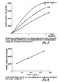
- Figure 3 is a graphical representation of chemiluminescent reaction rate as a function of time and AFP concentration in homogeneous labelled antibody immunoassay.
- the three curves represent three different AFP concentrations, at 328, 164 and 41 ng/ml.
- Figure 4 is a defined dose-response curve showing how the count rate varies linearly with the concentration.
- FIG. 1 A typical apparatus is illustrated diagrammatically in Figure 1 where P is a photon counter, R is a data reduction unit, I is an integrator, D is a differentiator, C is a computer, L is a printer, M is a disk drive.
- Figure 2 illustrates a typical- graph illustrating photon count per second plotted against time, and illustrating the way in which the actual emission rate varies,(the amplitude at any point on the curve) and also the rate of change (the slope of the curve at any point) and the overall count (the integrated area under the curve).
- the line X indicates the slope of the initial reaction rate. It will be seen that other characteristics can be derived or deduced therefrom such as the "time to peak", or the total count up to 0.5 secs. or any other selected interval.
- Standard and unknown sera (50pl) are incubated with the purified AFP labelled with acridinium ester molecules to a specific activity of 2 x 1 0 5 luminescent counts per nanogram and with polyclonal or monoclonal anti AFP antibodies at a concentration which is known to bind 50% of the labelled AFP after four hours at room temperature.
- the tubes are incubated at a final assay volume of 200pl (0.1M phosphate buffer, 0.15M sodium chloride pH6.3) for four hours at room temperature.
- the mixtures are then measured luminometrically by injection of alkaline hydrogen peroxide as before. The rate of excited state formation observed for the samples is compared with that of the standards and the AFP content quantified by interpolating as in Example 1.
- This assay is based on the principle that T 3 with acridinium ester linked through the amino group will not react with naturally occurring binding proteins (e.g. thyroxine binding globulin) but binds to antibody.
- the assay for free T 3 is carried out as follows. Standards or unknown samples containing T 3 (50 ⁇ l) are diluted to 100 ⁇ l withHEPES buffer (0.01M, pH 7.4). 50ul labelled T 3 are added followed by 50 ⁇ l antibody solution. During a 30 minute reaction label is bound to antibody. This binding in inhibited by free T 3 which competes for antibody binding sites. After the reaction period, therefore, luminescence is measured following addition of alkaline hydrogen peroxide in the manner described. The rate of excited state formation in samples is compared with that of standards and the free T 3 concentration quantified by interpolation as in example 1.
- Standard and unknown sera (50 ⁇ l) are incubated with purified insulin labelled with acridinium ester molecules and with pre-precipitated insulin antibodies. These represent high molecular weight complexes for enhanced change of rate of the chemiluminescent reaction as described above.
- the mixture is incubated for 3 hours at room temperature, in 200pl 0.01M phosphate buffer (pH6.3) and the tubes analysed luminometrically. The reaction rates observed for the unknown samples are compared with those of the standards and hence the insulin concentrations determined by interpolation.
- a solublised thyroid membrane preparation in PBS (pH6.3) containing 0.1% HSA is incubated with 100pl of serum IgG fraction to be investigated.
- the amount of membrane being sufficient to bind 10-30% of the labelled thyrotrophin (TSH) being used.
- acridinium ester labelled TSH is added and the mixture incubated for an additional 1 hour period.
- the assay tubes are measured luminometrically as in example 1 and the rates of emission compared with that of a normal serum IgG preparation to establish the presence or absence of pathologically significant amounts of thyroid stimulating antibodies.
Landscapes
- Health & Medical Sciences (AREA)
- Immunology (AREA)
- Life Sciences & Earth Sciences (AREA)
- Engineering & Computer Science (AREA)
- Molecular Biology (AREA)
- Biomedical Technology (AREA)
- Chemical & Material Sciences (AREA)
- Hematology (AREA)
- Urology & Nephrology (AREA)
- Biotechnology (AREA)
- Microbiology (AREA)
- Cell Biology (AREA)
- Food Science & Technology (AREA)
- Medicinal Chemistry (AREA)
- Physics & Mathematics (AREA)
- Analytical Chemistry (AREA)
- Biochemistry (AREA)
- General Health & Medical Sciences (AREA)
- General Physics & Mathematics (AREA)
- Pathology (AREA)
- Investigating Or Analysing Materials By The Use Of Chemical Reactions (AREA)
Abstract
Description
- This invention relates to methods designed for use in the analysis, assay or localisation of proteins, polypeptides, haptens and other substances of biological interest. The invention utilises immune complex formation as a measure of the amount of analyte present by means of the use of antigens, haptens or antibodies labelled with chemiluminescent molecules. In the past such biological molecules have been labelled using radioactive isotopes which suffer problems of instability, insensitivity, inconvenient quantification and disposal. In contrast chemiluminescent labels are non hazardous, stable and can be detected with high sensitivity using simple photon counting equipment. In the context of this invention the term chemiluminescence is used to distinguish this phenomenon from other forms of luminescence, e.g. fluorescence, phosphorescence, etc., and is taken to include the related phenomenon of bioluminescence. In a further aspect of the invention the concentration of immune complexes, and hence of the analyte, is determined by changes in certain physicochemical parameters of the chemiluminescent reaction of such labelled species upon formation of immune complexes.
- In some preferred forms of the invention these changes are involved with the rate of the chemiluminescent reaction, notably the formation of the excited states or their subsequent decay. It is a particular and important feature of the invention that quantitation of the analyte concentration does not require prior separation of analyte-bound and unbound labelled immunoreactant.
- Many chemical species are capable of chemiluminescence, which is the phenomenon observed when the vibronically excited product of an exoergic reaction reverts to its ground state with photonic emission. Such reactions are usually of an oxidative nature and may or may not involve a catalytic component. For example Luminol requires the presence of a catalyst for the chemiluminescent reaction, whereas acridinium esters do not require a catalyst. Broadly stated, a chemiluminescent reaction consists of two stages, these being the formation of excited states and their subsequent decay to their unexcited or "ground" states. For any given excited state which yields a corresponding ground state arising from energy loss by photonic emission, the rate of decay is proportional to the concentration of molecules in the excited states. The decay, rate coefficient of such a reaction, which acts to equate the aforementioned proportionality is thus first order and exponentially relates the concentration of the excited states to a particular increment of time which constitutes part of the total lifetime of the reaction.
- By comparison the rate of formation of excited states is an inherently more complex relationship than is the simple decay of excited states. The rate of formation is a function of the excited state precursor concentration (i.e. the concentration of the molecular luminescent label before being excited) and also of the concentration of the other reactants in the system. The excitation rate coefficient in this instance is a compound of a number of kinetic and thermodynamic parameters pertaining to the reactions which are involved in the formation of the excited states. Thus a change in such parameters such as may be experienced by prior chemical or physical interactions of the chemiluminescent molecule may be reflected as a change in excited state formation rate. Further, such a change will also be reflected by the relationship between the number of photons emitted or the rate at which they are emitted and an increment of time over which the reaction is taking place. There are unique advantages to be gained from utilizing the excitation phase to quantify a chemiluminescent reaction. Firstly, it is extremely rapid - accurate measurements can be made within 1 second or less. Secondly, because of the short time scale there is a low background which increases the sensitivity of detection, and thirdly the use of a rate measurement reduces interference due to inner filter effects.
- Broadly stated the invention consists in a method of detecting, analysing, quantifying, or locating a substance of biological interest by an immunoassay in which a first component of an immune reaction in the form of an antigen, hapten, or antibody, is linked with one or more components of a chemiluminescent reaction, and the other component of the immune reaction, the analyte, is complexed therewith to cause entropic and/or ethalpic changes in a subsequent light emitting action as compared with the reaction of uncomplexed labelled component, the reaction being observed and compared to provide information on the immune complex formation. Preferably the intensity of light emission or the rate of photonic emission or the rate of change of intensity of emission are observed compared with the equivalent properties of the uncomplexed component.
- Preferably, a graphical representation is firstly obtained of the function relating either the intensity or the rate of photonic emission (and hence the excited state concentration) to the reaction time following initiation of the light-emitting reaction of the labelled immunoreactant. A graph is also prepared for labelled immunogen which has previously been incubated with the corresponding analyte of interest. Changes in the slope of the relationships at any given point corresponding to one of the variables of the functions are a measure of the analyte concentration and can be compared with those of a series of known analyte concentrations. If the labelled immunoreactant is a labelled antigen or hapten and is used in a competitive binding system, the kinetic and thermodynamic parameters of the unbound labelled immunoreactant are compared with those of labelled immunoreactant previously incubated with analyte and antibody or binding protein. The changes in kinetic and thermodynamic parameters which result in changes in the rate of photon emission will be inversly proportional to the concentration of analyte. If the labelled immunoreactant is a labelled antibody, this may be used in an analogous competitive system for the measurement of antibodies in biological fluids such as serum. In a preferred aspect of the invention the labelled antibodies would be used in molar excess over the analyte present. The excess could be 500% but would preferably be no more than 100%, more preferably no more than 20%. In a further aspect of the invention the kinetic changes observed are amplified by increasing the size of the immune complexes formed by the use of immobilized antibody. In this situation the change in the rate of excitation is enhanced when the size of the antigen antibody complex is enlarged by the presence of polymerized antibody or antibody linked to a solid support such as cellulose.
- Preferably, the quantity of analyte is reflected by a change in the graphical representation of the function relating excited state concentration (or the rate of change of excited state concentration or further derivation thereof) to the reaction time. These parameters are a function of the photonic emission of the chemiluminescent label which is quantified using photon counting apparatus. Several parameters of the function or its derivatives may be monitored so as to provide a measure of analyte concentration, for example the slope of the function at any given point on the reaction profile, the amplitude at any point, the integration of any part of the reaction profile, or the time taken to reach a peak or a preselected value, or any combination of several properties.
- Preferably the chemiluminescent labels are acridinium salts, luminol, dioxetanes, bis-oxalates, fluorescein, pyrogallol, lucigenin, lophine, photoproteins, or derivates of these compounds which can be associated with antigens, antibodies and haptens so as to provide chemiluminescent immuno- reactants.
- In contrast to existing homogeneous immunoassay techniques using enzyme or fluorescent labels, this invention is not restricted to the measurement of small molecules or haptens but may be applied to the quantitation of all antigenic compounds including drugs (e.g. barbiturates, salicylates, phenytoin, morphine, heroin, methotrexate, digoxin), steroids (e.g. testosterone, progesterone, oestradiol, oestriol, cortisol, aldosterone, vitamin D), cyclic nucleotides, thyroxine, triiodothyronine or proteins (e.g. insulin, growth hormone, parathyroid hormone, thyroid stimulating hormone, prolactin, adrenocortotrophic hormone, alphafetoprotein, ferritin, human immunoglobulin (Ig). rabbit IgG, mouse IgG, guinea pig IgG, sheep IgG, interferon, components of the complement system and bacterial and viral antigens).
- Thus it will be seen that the invention provides a method for the detection or quantitation of substances of biological interest by a homogeneous immunoassay procedure in which antigens, antibodies or haptens are labelled with chemiluminescent molecules, the physico-chemical properties of which are modified in a measurable dependent way upon immune, complex formation.
- In the context of the invention a homogeneous assay may be defined as one in which the properties of the label are changed in a dose-dependent or concentration-dependent way on formation of the immune complex, thus avoiding the need for separation of the bound and free analyte fractions.
- The invention may be performed in various different ways and a number of examples will now be described in detail by way of illustration.
- 1. Homogeneous assay of human α1-fetoprotein (AFP) using labelled antibodies.
- A series of solutions ofAFP standard in sera are made and a known volume, preferably 50pl, and hence a known quantity of AFP is placed in a series of assay tubes. Identical volumes of unknown sera to be investigated are placed in another series of assay tubes and O.1M phosphate buffer, pH6.3 (100pl) placed in all tubes. Antibodies to AFP (polyclonal or monoclonal), labelled with acridinium ester and preferably having a specific activity of 2 x 10⁵ luminescent counts per nanogram are added to each tube such that they are present in excess of AFP analyte. The tubes are incubated at room temperature preferably for 30 mins, each tube is placed sequentially in a luminometer and the photons emitted upon initiation of the chemiluminescent reaction counted for 5 seconds or less. The rate of analysis of the tubes is adjusted so as to match the rate of addition of reagents prior to incubation. The chemiluminescent reaction is initiated by injection preferably of 200pl of an aqueous solution containing 0.5M sodium hydroxide and 0.1% (V/V) of "100 volume" (i.e. 30% weight/volume) hydrogen peroxide. The rate of formation of excited states (i.e. the rate of photon emission over a suitable time period) is measured for standards and unknowns using photon counting equipment. This may be done in several different ways. In one process the total number of photons emitted over a one second time period is recorded., and in another process the relationship which expresses the rate of photonic emission as a function of time is differentiated to enable calculation of the initial rate of reaction. In either case the results corresponding to the samples having unknown analyte concentrations are compared with those obtained from a series of standards of known amounts of analyte by constructing a calibration curve from the latter examples of which are given in Figures 3 and 4.
- Figure 3 is a graphical representation of chemiluminescent reaction rate as a function of time and AFP concentration in homogeneous labelled antibody immunoassay. The three curves represent three different AFP concentrations, at 328, 164 and 41 ng/ml.
- Figure 4 is a defined dose-response curve showing how the count rate varies linearly with the concentration.
- A typical apparatus is illustrated diagrammatically in Figure 1 where P is a photon counter, R is a data reduction unit, I is an integrator, D is a differentiator, C is a computer, L is a printer, M is a disk drive. Figure 2 illustrates a typical- graph illustrating photon count per second plotted against time, and illustrating the way in which the actual emission rate varies,(the amplitude at any point on the curve) and also the rate of change (the slope of the curve at any point) and the overall count (the integrated area under the curve). In Figure 2 the line X indicates the slope of the initial reaction rate. It will be seen that other characteristics can be derived or deduced therefrom such as the "time to peak", or the total count up to 0.5 secs. or any other selected interval.
- 2. Homogeneous assay of humanα1-fetoprotein (AFP) using labelled antigens.
- Standard and unknown sera (50pl) are incubated with the purified AFP labelled with acridinium ester molecules to a specific activity of 2 x 10⁵ luminescent counts per nanogram and with polyclonal or monoclonal anti AFP antibodies at a concentration which is known to bind 50% of the labelled AFP after four hours at room temperature. The tubes are incubated at a final assay volume of 200pl (0.1M phosphate buffer, 0.15M sodium chloride pH6.3) for four hours at room temperature. The mixtures are then measured luminometrically by injection of alkaline hydrogen peroxide as before. The rate of excited state formation observed for the samples is compared with that of the standards and the AFP content quantified by interpolating as in Example 1.
- 3. Homogeneous Assay of Triiodothyronine (T3).
- Standards and unknown samples (50µl) containing T3 are diluted to 100 µl with HEPES buffer (0.01M, pH 7.4) containing 0.25% merthiolate. Fifty µl T3 labelled with acridinium ester are added followed by 50µl antibody to T3. After 30 minutes reaction the luminescence is measured as described in example 2 following injection of alkaline hydrogen peroxide. The rate of excited state formation observed in the samples is compared with that of standards and the T3 content quantified by interpolation as in example 1.
- 4. Homogeneous Assay of Free T3.
- This assay is based on the principle that T3 with acridinium ester linked through the amino group will not react with naturally occurring binding proteins (e.g. thyroxine binding globulin) but binds to antibody. The assay for free T3 is carried out as follows. Standards or unknown samples containing T3 (50 µl) are diluted to 100 µl withHEPES buffer (0.01M, pH 7.4). 50ul labelled T3 are added followed by 50µl antibody solution. During a 30 minute reaction label is bound to antibody. This binding in inhibited by free T3 which competes for antibody binding sites. After the reaction period, therefore, luminescence is measured following addition of alkaline hydrogen peroxide in the manner described. The rate of excited state formation in samples is compared with that of standards and the free T3 concentration quantified by interpolation as in example 1.
- 5. Homogeneous assay of insulin.
- Standard and unknown sera (50µl) are incubated with purified insulin labelled with acridinium ester molecules and with pre-precipitated insulin antibodies. These represent high molecular weight complexes for enhanced change of rate of the chemiluminescent reaction as described above. The mixture is incubated for 3 hours at room temperature, in 200pl 0.01M phosphate buffer (pH6.3) and the tubes analysed luminometrically. The reaction rates observed for the unknown samples are compared with those of the standards and hence the insulin concentrations determined by interpolation.
- 6. Homogeneous assay of serum thyroid stimulating immunoglobulins.
- A solublised thyroid membrane preparation in PBS (pH6.3) containing 0.1% HSA is incubated with 100pl of serum IgG fraction to be investigated. The amount of membrane being sufficient to bind 10-30% of the labelled thyrotrophin (TSH) being used.
- Following incubation at room temperature for 1 hour, acridinium ester labelled TSH is added and the mixture incubated for an additional 1 hour period. The assay tubes are measured luminometrically as in example 1 and the rates of emission compared with that of a normal serum IgG preparation to establish the presence or absence of pathologically significant amounts of thyroid stimulating antibodies.
Claims (9)
Applications Claiming Priority (2)
| Application Number | Priority Date | Filing Date | Title |
|---|---|---|---|
| GB8225933 | 1982-09-10 | ||
| GB8225933 | 1982-09-10 |
Publications (3)
| Publication Number | Publication Date |
|---|---|
| EP0103469A2 true EP0103469A2 (en) | 1984-03-21 |
| EP0103469A3 EP0103469A3 (en) | 1984-12-05 |
| EP0103469B1 EP0103469B1 (en) | 1989-02-08 |
Family
ID=10532841
Family Applications (1)
| Application Number | Title | Priority Date | Filing Date |
|---|---|---|---|
| EP83305277A Expired EP0103469B1 (en) | 1982-09-10 | 1983-09-09 | Immunological procedure for quantifying substances |
Country Status (4)
| Country | Link |
|---|---|
| US (1) | US4761382A (en) |
| EP (1) | EP0103469B1 (en) |
| DE (1) | DE3379179D1 (en) |
| GB (1) | GB2129553B (en) |
Cited By (5)
| Publication number | Priority date | Publication date | Assignee | Title |
|---|---|---|---|---|
| GB2223096A (en) * | 1988-09-06 | 1990-03-28 | Atomic Energy Authority Uk | Detecting a selected species |
| WO1991001305A1 (en) * | 1989-07-22 | 1991-02-07 | University Of Wales College Of Medicine | Modified bioluminescent proteins and their use |
| US5521103A (en) * | 1987-12-18 | 1996-05-28 | Mochida Pharmaceutical Co., Ltd. | Acridinium compounds as chemiluminogenic label |
| US5683888A (en) * | 1989-07-22 | 1997-11-04 | University Of Wales College Of Medicine | Modified bioluminescent proteins and their use |
| CN101201354B (en) * | 2006-12-14 | 2011-11-02 | 北京科美东雅生物技术有限公司 | Thyroxine chemiluminescence immune analysis quantitative measuring reagent kit and method for preparing the same |
Families Citing this family (7)
| Publication number | Priority date | Publication date | Assignee | Title |
|---|---|---|---|---|
| JPH06504374A (en) * | 1990-12-28 | 1994-05-19 | アボット・ラボラトリーズ | Simultaneous determination of multiple analytes using time-resolved heterogeneous chemiluminescence assays |
| DE69518182T2 (en) * | 1994-04-11 | 2001-03-22 | Kyowa Medex Co. Ltd., Tokio/Tokyo | Method for determining a substance |
| US5783453A (en) * | 1995-06-29 | 1998-07-21 | Chiron Diagnostics Corporation | Non-separation specific binding chemiluminescent assay |
| GB2312746B (en) * | 1996-04-24 | 2000-07-19 | Molecular Light Technology Lim | Detection of an analyte in a Water Immiscible Solvent |
| US6294333B1 (en) | 1998-12-31 | 2001-09-25 | Ingeneus Corp. | Fluorescent intensity assay for protein or peptide binding to nucleic acids |
| US6645733B1 (en) | 1999-06-25 | 2003-11-11 | Ingeneus Corporation | Fluorescent intensity method for assaying binding between proteins or peptides |
| CN116930478A (en) * | 2022-04-02 | 2023-10-24 | 迈克生物股份有限公司 | Thyroxine fluorescent conjugate and preparation method and application thereof |
Family Cites Families (9)
| Publication number | Priority date | Publication date | Assignee | Title |
|---|---|---|---|---|
| GB1578275A (en) * | 1977-06-14 | 1980-11-05 | Maier C | Procedure for the assay of pharmacologically immunologically and biochemically active compounds in biological fluids |
| GB2008247B (en) * | 1977-11-17 | 1982-12-15 | Welsh Nat School Med | Detecting or quantifying substances using labelling techniques |
| US4220450A (en) * | 1978-04-05 | 1980-09-02 | Syva Company | Chemically induced fluorescence immunoassay |
| US4238195A (en) * | 1979-01-18 | 1980-12-09 | Miles Laboratories, Inc. | Fluorescer-labeled specific binding assays |
| US4280815A (en) * | 1979-06-18 | 1981-07-28 | Technicon Instruments Corporation | Electrochemiluminescent immunoassay and apparatus therefor |
| US4276051A (en) * | 1980-01-28 | 1981-06-30 | Coulter Electronics, Inc. | System and program for chemical reaction observation with a moving photometer |
| DE3132491A1 (en) * | 1980-08-22 | 1982-07-01 | Laboratorium Prof. Dr. Rudolf Berthold, 7547 Wildbad | METHOD FOR PERFORMING ANALYTICAL DETERMINATIONS BY MEANS OF THE CHEMILUMINESCENCE METHOD AND APPLICATION OF THE METHOD FOR IMMUNASSAYS |
| GB2086042B (en) * | 1980-08-22 | 1983-11-30 | Berthold Lab Prof R | Process for carrying out analytical determinations by means of chemiluminescence and the use of the process for immunoassay |
| CA1170568A (en) * | 1980-12-22 | 1984-07-10 | Bernd Frenzel | Process for determining antigens, antibodies and their complexes |
-
1983
- 1983-09-09 EP EP83305277A patent/EP0103469B1/en not_active Expired
- 1983-09-09 DE DE8383305277T patent/DE3379179D1/en not_active Expired
- 1983-09-09 GB GB08324249A patent/GB2129553B/en not_active Expired
-
1987
- 1987-02-05 US US07/013,215 patent/US4761382A/en not_active Expired - Lifetime
Cited By (8)
| Publication number | Priority date | Publication date | Assignee | Title |
|---|---|---|---|---|
| US5521103A (en) * | 1987-12-18 | 1996-05-28 | Mochida Pharmaceutical Co., Ltd. | Acridinium compounds as chemiluminogenic label |
| US6018047A (en) * | 1987-12-18 | 2000-01-25 | Zomer; Gijsbert | Acridinium compounds as chemiluminogenic label |
| GB2223096A (en) * | 1988-09-06 | 1990-03-28 | Atomic Energy Authority Uk | Detecting a selected species |
| WO1991001305A1 (en) * | 1989-07-22 | 1991-02-07 | University Of Wales College Of Medicine | Modified bioluminescent proteins and their use |
| US5683888A (en) * | 1989-07-22 | 1997-11-04 | University Of Wales College Of Medicine | Modified bioluminescent proteins and their use |
| US6440665B1 (en) | 1989-07-22 | 2002-08-27 | University Of Wales College Of Medicine | Modified bioluminescent proteins and their use |
| US6492500B1 (en) | 1989-07-22 | 2002-12-10 | University Of Wales College Of Medicine | Modified bioluminescent proteins and their use |
| CN101201354B (en) * | 2006-12-14 | 2011-11-02 | 北京科美东雅生物技术有限公司 | Thyroxine chemiluminescence immune analysis quantitative measuring reagent kit and method for preparing the same |
Also Published As
| Publication number | Publication date |
|---|---|
| GB8324249D0 (en) | 1983-10-12 |
| US4761382A (en) | 1988-08-02 |
| EP0103469B1 (en) | 1989-02-08 |
| EP0103469A3 (en) | 1984-12-05 |
| GB2129553B (en) | 1986-06-18 |
| GB2129553A (en) | 1984-05-16 |
| DE3379179D1 (en) | 1989-03-16 |
Similar Documents
| Publication | Publication Date | Title |
|---|---|---|
| US4626513A (en) | Method and apparatus for ligand detection | |
| CA1217121A (en) | Enhanced luminescent or luminometric assay | |
| US4478817A (en) | Detecting or quantifying substances using labelling techniques | |
| US4680275A (en) | Homogeneous fluorescence immunoassay using a light absorbing material | |
| FI93781B (en) | Biospecific multiparametric method of determination | |
| US5656207A (en) | Detecting or quantifying multiple analytes using labelling techniques | |
| US5464741A (en) | Palladium (II) octaethylporphine alpha-isothiocyanate as a phosphorescent label for immunoassays | |
| US4923819A (en) | Time-resolved fluorescence immunoassay | |
| US4761382A (en) | Immunological procedure for detecting or quantifying substances | |
| US5389523A (en) | Liposome immunoanalysis by flow injection assay | |
| WO1999030131A1 (en) | One-step fluorescent immunosensor test | |
| AU1228583A (en) | Enhanced luminescent and luminometric assay | |
| Mathis et al. | Homogeneous immunoassays using rare earth cryptates and time resolved fluorescence: principles and specific advantages for tumor markers | |
| GB2233450A (en) | Detecting or quantifying multiple analytes | |
| EP0222341A1 (en) | A method for immunoassay and reagents therefor | |
| EP0522677A1 (en) | Method of measuring concentration of immunoreactant using electrochemiluminescence | |
| AU682478B2 (en) | Two-site immunoassay for an antibody with chemiluminescent label and biotin bound ligand | |
| EP0640216A1 (en) | Separation method | |
| US4052504A (en) | Assay for thyroxine binding globulin | |
| US5391479A (en) | Method for determining total analyte concentration in a sample having both free and bound analyte | |
| US5202269A (en) | Method for immunochemical determination of hapten | |
| US5045479A (en) | Continuous flow competitive assay with reference system | |
| JP7488812B2 (en) | Chemiluminescence immunoassay method and system and reagent kit using said method | |
| USH1018H (en) | Immunological method for the determination of free substances having hapten properties | |
| US4242322A (en) | Methods and materials for detecting antigens and antibodies |
Legal Events
| Date | Code | Title | Description |
|---|---|---|---|
| PUAI | Public reference made under article 153(3) epc to a published international application that has entered the european phase |
Free format text: ORIGINAL CODE: 0009012 |
|
| AK | Designated contracting states |
Designated state(s): BE CH DE FR IT LI NL |
|
| PUAL | Search report despatched |
Free format text: ORIGINAL CODE: 0009013 |
|
| AK | Designated contracting states |
Designated state(s): BE CH DE FR IT LI NL |
|
| 17P | Request for examination filed |
Effective date: 19841112 |
|
| 17Q | First examination report despatched |
Effective date: 19860129 |
|
| R17C | First examination report despatched (corrected) |
Effective date: 19870309 |
|
| GRAA | (expected) grant |
Free format text: ORIGINAL CODE: 0009210 |
|
| AK | Designated contracting states |
Kind code of ref document: B1 Designated state(s): BE CH DE FR IT LI NL |
|
| REF | Corresponds to: |
Ref document number: 3379179 Country of ref document: DE Date of ref document: 19890316 |
|
| ITF | It: translation for a ep patent filed | ||
| RAP4 | Party data changed (patent owner data changed or rights of a patent transferred) |
Owner name: THE WELSH NATIONAL SCHOOL OF MEDICINE |
|
| ET | Fr: translation filed | ||
| PLBE | No opposition filed within time limit |
Free format text: ORIGINAL CODE: 0009261 |
|
| STAA | Information on the status of an ep patent application or granted ep patent |
Free format text: STATUS: NO OPPOSITION FILED WITHIN TIME LIMIT |
|
| 26N | No opposition filed | ||
| ITTA | It: last paid annual fee | ||
| PGFP | Annual fee paid to national office [announced via postgrant information from national office to epo] |
Ref country code: FR Payment date: 20020912 Year of fee payment: 20 |
|
| PGFP | Annual fee paid to national office [announced via postgrant information from national office to epo] |
Ref country code: CH Payment date: 20020925 Year of fee payment: 20 |
|
| PGFP | Annual fee paid to national office [announced via postgrant information from national office to epo] |
Ref country code: NL Payment date: 20020930 Year of fee payment: 20 |
|
| PGFP | Annual fee paid to national office [announced via postgrant information from national office to epo] |
Ref country code: BE Payment date: 20021022 Year of fee payment: 20 |
|
| PGFP | Annual fee paid to national office [announced via postgrant information from national office to epo] |
Ref country code: DE Payment date: 20021118 Year of fee payment: 20 |
|
| PG25 | Lapsed in a contracting state [announced via postgrant information from national office to epo] |
Ref country code: LI Free format text: LAPSE BECAUSE OF EXPIRATION OF PROTECTION Effective date: 20030908 Ref country code: CH Free format text: LAPSE BECAUSE OF EXPIRATION OF PROTECTION Effective date: 20030908 |
|
| PG25 | Lapsed in a contracting state [announced via postgrant information from national office to epo] |
Ref country code: NL Free format text: LAPSE BECAUSE OF EXPIRATION OF PROTECTION Effective date: 20030909 |
|
| BE20 | Be: patent expired |
Owner name: THE *WELSH NATIONAL SCHOOL OF MEDICINE Effective date: 20030909 |
|
| REG | Reference to a national code |
Ref country code: CH Ref legal event code: PL |
|
| NLV7 | Nl: ceased due to reaching the maximum lifetime of a patent |
Effective date: 20030909 |