EP0070153B1 - Untersuchung von Zellen durch Elektrophorese - Google Patents
Untersuchung von Zellen durch Elektrophorese Download PDFInfo
- Publication number
- EP0070153B1 EP0070153B1 EP82303588A EP82303588A EP0070153B1 EP 0070153 B1 EP0070153 B1 EP 0070153B1 EP 82303588 A EP82303588 A EP 82303588A EP 82303588 A EP82303588 A EP 82303588A EP 0070153 B1 EP0070153 B1 EP 0070153B1
- Authority
- EP
- European Patent Office
- Prior art keywords
- lymphocytes
- cells
- electrophoretic
- mouse
- electrophoretic mobility
- Prior art date
- Legal status (The legal status is an assumption and is not a legal conclusion. Google has not performed a legal analysis and makes no representation as to the accuracy of the status listed.)
- Expired
Links
- 238000001962 electrophoresis Methods 0.000 title claims description 9
- 210000004698 lymphocyte Anatomy 0.000 claims description 56
- 210000004027 cell Anatomy 0.000 claims description 40
- 238000000034 method Methods 0.000 claims description 24
- 230000002093 peripheral effect Effects 0.000 claims description 13
- 241000282414 Homo sapiens Species 0.000 claims description 11
- 241001465754 Metazoa Species 0.000 claims description 9
- 210000005259 peripheral blood Anatomy 0.000 claims description 5
- 239000011886 peripheral blood Substances 0.000 claims description 5
- 230000004962 physiological condition Effects 0.000 claims description 5
- 210000003563 lymphoid tissue Anatomy 0.000 claims description 3
- 239000000725 suspension Substances 0.000 claims description 3
- 210000001541 thymus gland Anatomy 0.000 claims description 3
- 210000001165 lymph node Anatomy 0.000 claims description 2
- 210000000952 spleen Anatomy 0.000 claims description 2
- 241000699666 Mus <mouse, genus> Species 0.000 description 44
- 230000037230 mobility Effects 0.000 description 41
- 206010028980 Neoplasm Diseases 0.000 description 32
- 238000002054 transplantation Methods 0.000 description 10
- 239000007758 minimum essential medium Substances 0.000 description 8
- 230000005684 electric field Effects 0.000 description 5
- 239000000243 solution Substances 0.000 description 4
- 210000004881 tumor cell Anatomy 0.000 description 4
- 239000012981 Hank's balanced salt solution Substances 0.000 description 3
- 241000699670 Mus sp. Species 0.000 description 3
- 241001494479 Pecora Species 0.000 description 3
- 208000006268 Sarcoma 180 Diseases 0.000 description 3
- 210000004369 blood Anatomy 0.000 description 3
- 239000008280 blood Substances 0.000 description 3
- 201000011510 cancer Diseases 0.000 description 3
- 238000007796 conventional method Methods 0.000 description 3
- 230000001472 cytotoxic effect Effects 0.000 description 3
- 210000003743 erythrocyte Anatomy 0.000 description 3
- 238000000338 in vitro Methods 0.000 description 3
- 206010006187 Breast cancer Diseases 0.000 description 2
- 208000026310 Breast neoplasm Diseases 0.000 description 2
- 208000003468 Ehrlich Tumor Carcinoma Diseases 0.000 description 2
- 239000007900 aqueous suspension Substances 0.000 description 2
- 230000003111 delayed effect Effects 0.000 description 2
- 239000012997 ficoll-paque Substances 0.000 description 2
- 229920000669 heparin Polymers 0.000 description 2
- 208000020816 lung neoplasm Diseases 0.000 description 2
- 239000002609 medium Substances 0.000 description 2
- 241000894007 species Species 0.000 description 2
- 239000000126 substance Substances 0.000 description 2
- 238000012360 testing method Methods 0.000 description 2
- 208000000461 Esophageal Neoplasms Diseases 0.000 description 1
- 229920001917 Ficoll Polymers 0.000 description 1
- DGAQECJNVWCQMB-PUAWFVPOSA-M Ilexoside XXIX Chemical compound C[C@@H]1CC[C@@]2(CC[C@@]3(C(=CC[C@H]4[C@]3(CC[C@@H]5[C@@]4(CC[C@@H](C5(C)C)OS(=O)(=O)[O-])C)C)[C@@H]2[C@]1(C)O)C)C(=O)O[C@H]6[C@@H]([C@H]([C@@H]([C@H](O6)CO)O)O)O.[Na+] DGAQECJNVWCQMB-PUAWFVPOSA-M 0.000 description 1
- 206010058467 Lung neoplasm malignant Diseases 0.000 description 1
- 206010030155 Oesophageal carcinoma Diseases 0.000 description 1
- 208000015634 Rectal Neoplasms Diseases 0.000 description 1
- 208000005718 Stomach Neoplasms Diseases 0.000 description 1
- 230000002159 abnormal effect Effects 0.000 description 1
- 230000004075 alteration Effects 0.000 description 1
- YVPYQUNUQOZFHG-UHFFFAOYSA-N amidotrizoic acid Chemical compound CC(=O)NC1=C(I)C(NC(C)=O)=C(I)C(C(O)=O)=C1I YVPYQUNUQOZFHG-UHFFFAOYSA-N 0.000 description 1
- 238000010171 animal model Methods 0.000 description 1
- 239000007864 aqueous solution Substances 0.000 description 1
- 230000007969 cellular immunity Effects 0.000 description 1
- 238000005119 centrifugation Methods 0.000 description 1
- 238000002405 diagnostic procedure Methods 0.000 description 1
- 238000007865 diluting Methods 0.000 description 1
- 201000004101 esophageal cancer Diseases 0.000 description 1
- 230000002349 favourable effect Effects 0.000 description 1
- 206010017758 gastric cancer Diseases 0.000 description 1
- 230000005484 gravity Effects 0.000 description 1
- ZFGMDIBRIDKWMY-PASTXAENSA-N heparin Chemical compound CC(O)=N[C@@H]1[C@@H](O)[C@H](O)[C@@H](COS(O)(=O)=O)O[C@@H]1O[C@@H]1[C@@H](C(O)=O)O[C@@H](O[C@H]2[C@@H]([C@@H](OS(O)(=O)=O)[C@@H](O[C@@H]3[C@@H](OC(O)[C@H](OS(O)(=O)=O)[C@H]3O)C(O)=O)O[C@@H]2O)CS(O)(=O)=O)[C@H](O)[C@H]1O ZFGMDIBRIDKWMY-PASTXAENSA-N 0.000 description 1
- 238000005286 illumination Methods 0.000 description 1
- 210000002865 immune cell Anatomy 0.000 description 1
- 230000036039 immunity Effects 0.000 description 1
- 238000011534 incubation Methods 0.000 description 1
- 238000011081 inoculation Methods 0.000 description 1
- 239000007788 liquid Substances 0.000 description 1
- 239000006194 liquid suspension Substances 0.000 description 1
- 201000005202 lung cancer Diseases 0.000 description 1
- 208000037841 lung tumor Diseases 0.000 description 1
- 238000005259 measurement Methods 0.000 description 1
- 238000013508 migration Methods 0.000 description 1
- 230000005012 migration Effects 0.000 description 1
- 239000011259 mixed solution Substances 0.000 description 1
- 238000002156 mixing Methods 0.000 description 1
- 239000000203 mixture Substances 0.000 description 1
- 108090000765 processed proteins & peptides Proteins 0.000 description 1
- 206010038038 rectal cancer Diseases 0.000 description 1
- 201000001275 rectum cancer Diseases 0.000 description 1
- 229910052708 sodium Inorganic materials 0.000 description 1
- 239000011734 sodium Substances 0.000 description 1
- 201000011549 stomach cancer Diseases 0.000 description 1
- 239000010414 supernatant solution Substances 0.000 description 1
- 230000002992 thymic effect Effects 0.000 description 1
- 238000005406 washing Methods 0.000 description 1
Images


Classifications
-
- G—PHYSICS
- G01—MEASURING; TESTING
- G01N—INVESTIGATING OR ANALYSING MATERIALS BY DETERMINING THEIR CHEMICAL OR PHYSICAL PROPERTIES
- G01N27/00—Investigating or analysing materials by the use of electric, electrochemical, or magnetic means
- G01N27/26—Investigating or analysing materials by the use of electric, electrochemical, or magnetic means by investigating electrochemical variables; by using electrolysis or electrophoresis
- G01N27/416—Systems
- G01N27/447—Systems using electrophoresis
Definitions
- This invention relates to a method for examining cells, particularly lymphocytes, by eiec- trophoresis.
- US-A-4212650 discloses an in vitro diagnostic method which comprises incubating lymphocytes with a synthetic peptide of high basicity whereby the behaviour of the lymphocytes in an electric field is modified in a detectable manner, subjecting the incubation mixture to an electric field and measuring the migration speed of the lymphocytes in the electric field.
- the present invention provides a method of examining lymphocytes in peripheral blood or lymphatic tissues such as the thymus, spleen or lymph nodes, comprising taking the lymphocytes to be examined from peripheral blood or lymphatic tissues of a human or animal, suspending these lymphocytes in a medium corresponding to physiological conditions at a cell concentration of 0.5 to 20x 1 06 per ml and an ionic strength of 0.11 to 0.21, measuring the electrophoretic mobility of these lymphocytes, directly on the suspension thereof thus prepared, by an automatic electrophoresis apparatus and comparing the electrophoretic mobility of the lymphocytes under examination with that of lymphocytes of a standard, for example normal host-cells.
- An automatic electrophoresis apparatus suitable for use in the invention typically possesses the following features:
- an automatic microscope apparatus for electrophoresis (Parmoquant-II, made by Karl-Zeiss Co., East Germany, hereinafter abbreviated as PQ-II) used in the Examples below may be operated so as to measure exactly the average electrophoretic mobility of each cell among a large number of cells by reversing the electric field and a mobility histogram is obtained.
- concentration of the cells in the specimen (aqueous suspension of the cells) is adjusted to 0.5 to 20x106 cells/ml. and the ionic strength of the aqueous suspension of the cells is adjusted to 0.11 to 0.21, preferably, 0.13 to 0.17. It will of course be appreciated that the lymphocytes are examined after having been taken from the human or animal body.
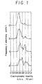
- Pattern (a) represents a normal mouse, (b) the mouse 5 days after transplantation into the mouse of the tumor, (c) the mouse 10 days after transplantation into the mouse of the tumor and (d) the mouse 15 days after transplantation into the mouse of the tumor.
- the electrophoretic mobility (cm/sN/cm) is the abscissa and the frequency (arbitrary units) is the ordinate.
- a mean value of mobilities, a mobility of a main peak, an average mobility of cells showing a mobility within a predetermined range and a fraction of the number of cells showing a mobility within a predetermined range to the total number of the cells may be used.
- the average mobilities of the larger peak and the smaller peak in the electrophoretic pattern of the lymphocytes of the normal mouse shown in Figure 1 (a) were 0.52 and 0.95 cm/s/V/cm, respectively.
- those of the lymphocytes of the mouse 5, 10 and 15 days after transplantation were 0.58 and 0.97 in (b), 0.62 and 0.98 in (c) and 0.68 and 1.03 in (d), respectively.
- Figures 2 (1) to (4) of the accompanying drawings the days after tumor inoculation is taken as the abscissa and the weight of tumor, the increment of the foot pad thickness, the value of productivity of antibody (titer) and the cytotoxic activity of lymphocytes are taken in the ordinate of Figure 2 (1), (2), (3) and (4), respectively.
- the abnormal values appear from the 5th to 10th day, corresponding to the appearance of the change in the electrophoretic pattern.
- the ratio falls within the range of 1.05 to 8.00:1. Further, the ratio of the average mobility of a peak in the tumor-bearing host to the corresponding mobility of the normal host-cells is in the range of 1.02 to 1.60:1
- the results obtained by the method of examination of the invention correspond to the results of determination of immunity by conventional methods.
- the method for examining cells according to the invention is, however, a superior and quicker method than the conventional methods.
- the number of lymphocytes of the lower electrophoretic mobility in tumor-bearing host increases by 4-70% as compared with that of normal host as shown in Examples below.
- the ratio falls within the range of 0.30 to 0.95:1. Further, the ratio of the average mobility of a peak in the tumor-bearing host to the corresponding mobility of the normal host-cells is in the range of 0.60 to 1.00:1.
- the method of the invention is useful in examining immune cells not only of animals but also of human beings, particularly in diagnosing cancer.
- the mouse After transplanting 1 x 10 6 cells of Sarcoma-180 to the left axillary site of an 8 week old female ICR mouse, the mouse was sacrificed on the 25th day to extirpate the thymic cells. The cells were isolated by a pincette and passed through a mesh to be isolated. After subjecting the isolated cells to hypotonic treatment for removing erythrocytes and washing the thus treated cells twice with Eagle's MEM (minimum essential medium), the cells were suspended in Eagle's MEM at a concentration of 10 6 to 10 7 cells/ml.
- Eagle's MEM minimum essential medium
- a specimen of peripheral blood was collected from the mouse, using an injector with its wall surface moistened with a solution containing 2.5 units (per ml of blood specimen) of sodium heparinate.
- HBSS Hanks' balanced salt solution
- the diluted specimen was placed while forming a layer of about 5 ml on 3 ml of Ficoll-Paque solution (Grade: Ficoll 400, made by Pharmacia Fine Chemical Co., a mixed solution of sodium diatriazoate; specific gravity of 1.077) in a test tube without disturbing the interface between the two solutions. Then the contents of the test tube were subjected to centrifugation for 30 min at 400 G.
- the lymphocytes at the interface of the diluted specimen and the Ficoll-Paque solution were collected by a Pasteur-pipette.
- lymphocytes were washed twice with HBSS centrifugally, each time for 10 min at 260 G, and then further washed centrifugally with Eagle's MEM for 10 min at 180 G. After discarding the supernatant solution, the remaining lymphocytes were dispersed in Eagle's MEM at a concentration of the cells of 5 to 10x106 cells/ ml.
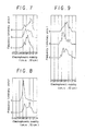
- the histograms a and b in Figure 3 of the accompanying drawings are the electrophoretic patterns of the thymocytes from the normal mouse and the tumor-bearing mouse, respectively, and those of a and b in Figure 4 of the accompanying drawings are the electrophoretic patterns of the peripheral lymphocytes from the normal mouse and the tumor-bearing mouse, respectively.
- the electrophoretic pattern of the thymocytes of the normal mouse shows a two-peaked pattern with a larger peak at a mobility of 0.6 cm/s/V/cm and a smaller peak at a mobility of 0.9 cmlsN/cm, while that of the tumor-bearing mouse shows a pattern with a broadened peak of a mobility which shifted toward the higher mobility as seen in Figure 3 b.
- the electrophoretic pattern of the peripheral lymphocytes of the tumor-bearing mouse shows a broader peak pattern and the fraction of lymphocytes with lower mobility to the total number of lymphocytes increases as compared to those of the normal mouse (in Figure 4 a).
- the electrophoretic patterns of the thymocytes of tumor-bearing animal show respectively, a broad peak and the fraction of lymphocytes with a higher mobility increases as compared to the patterns of that of the normal mouse.
- the thymocytes and the peripheral lymphocytes were collected from the mouse (on the sixth day after transplantation of MH-134 tumor cells, and on the eighth day after transplantation of MM-102 tumor cells) as in Example 1, and subjected to electrophoretic examination as in Example 1 to obtain the results shown in Figures 6 and 7 of the accompanying drawings wherein the electrophoretic patterns a, b and c are those, respectively, of the normal mouse, the mouse with the MH-134 tumor and the mouse with the MM-102 tumor.
- Pattern a in Figure 8 is that of the thymocytes of the normal mouse and pattern b in Figure 8 is that of the Lewis lung tumor-bearing mouse, and patterns a, b, and c in Figure 9 are those of the peripheral lymphocytes of the normal mouse, the Ca-755 tumor-bearing mouse and the B-16 tumour-bearing mouse, respectively.
- Example 5 seven specimens of human peripheral lymphocytes were taken from a male patient of colonal cancer of age 49 (a), a male patient of rectal cancer of age 68 (b), a male patient of stomach cancer of age 72 (c), a female patient of breast cancer of age 50 (d), a male patient of recurring esophageal cancer of age 49 (e), a female patient of recurring breast cancer of age 50 (f) and a male patient of recurring pan- creatoncus of age 62 (g) as in Example 5. After suspending in Eagle's MEM, the specimens were examined electrophoretically as in Example 5. The results are shown in Figure 11 of the accompanying drawings as electrophoretic patterns a to g corresponding to the letters of the respective patients.
Landscapes
- Health & Medical Sciences (AREA)
- Life Sciences & Earth Sciences (AREA)
- Molecular Biology (AREA)
- Chemical & Material Sciences (AREA)
- Biochemistry (AREA)
- Electrochemistry (AREA)
- Physics & Mathematics (AREA)
- Analytical Chemistry (AREA)
- Chemical Kinetics & Catalysis (AREA)
- General Health & Medical Sciences (AREA)
- General Physics & Mathematics (AREA)
- Immunology (AREA)
- Pathology (AREA)
- Micro-Organisms Or Cultivation Processes Thereof (AREA)
- Measuring Or Testing Involving Enzymes Or Micro-Organisms (AREA)
- Investigating Or Analysing Biological Materials (AREA)
Claims (8)
Applications Claiming Priority (2)
| Application Number | Priority Date | Filing Date | Title |
|---|---|---|---|
| JP56107399A JPS589060A (ja) | 1981-07-09 | 1981-07-09 | 電気泳動法による細胞の検査方法 |
| JP107399/81 | 1981-07-09 |
Publications (2)
| Publication Number | Publication Date |
|---|---|
| EP0070153A1 EP0070153A1 (de) | 1983-01-19 |
| EP0070153B1 true EP0070153B1 (de) | 1987-01-28 |
Family
ID=14458155
Family Applications (1)
| Application Number | Title | Priority Date | Filing Date |
|---|---|---|---|
| EP82303588A Expired EP0070153B1 (de) | 1981-07-09 | 1982-07-08 | Untersuchung von Zellen durch Elektrophorese |
Country Status (7)
| Country | Link |
|---|---|
| US (1) | US4632743A (de) |
| EP (1) | EP0070153B1 (de) |
| JP (1) | JPS589060A (de) |
| CA (1) | CA1179632A (de) |
| DD (1) | DD202600A5 (de) |
| DE (1) | DE3275321D1 (de) |
| ES (1) | ES514611A0 (de) |
Families Citing this family (5)
| Publication number | Priority date | Publication date | Assignee | Title |
|---|---|---|---|---|
| US4707237A (en) * | 1982-09-09 | 1987-11-17 | Ciba Corning Diagnostics Corp. | System for identification of cells by electrophoresis |
| JPS60205263A (ja) * | 1984-03-30 | 1985-10-16 | Kureha Chem Ind Co Ltd | 電気泳動法による細胞の検査方法 |
| CA2012379C (en) * | 1989-04-24 | 2000-01-25 | Gary W. Slater | Processes for the preparation and separation of macromolecules |
| AU2002251286A1 (en) * | 2001-04-12 | 2002-10-28 | Imperial College Innovations Limited | Diagnosis and treatment of cancer:ii |
| CN100412236C (zh) | 2002-03-13 | 2008-08-20 | 三菱化学株式会社 | 镀金液及镀金方法 |
Family Cites Families (13)
| Publication number | Priority date | Publication date | Assignee | Title |
|---|---|---|---|---|
| US3140714A (en) * | 1962-06-28 | 1964-07-14 | Cordis Corp | Blood separation method |
| US3988230A (en) * | 1973-12-31 | 1976-10-26 | Medac Gesellschaft Fur Klinische Spezial Praparate Mbh | Chamber and process for crossed immunoelectro-phoresis |
| US3984533A (en) * | 1975-11-13 | 1976-10-05 | General Electric Company | Electrophoretic method of detecting antigen-antibody reaction |
| SE7609263L (sv) * | 1976-08-20 | 1978-02-21 | Wadsworth Charlies | Forfaringssett for faststellande av kvantiteten av viss i en biologisk vetska loslig komponent, samt apparat for utovande av forfarandet |
| JPS5332093A (en) * | 1976-09-06 | 1978-03-25 | Olympus Optical Co Ltd | Automatic electrophoresis apparatus |
| GB1542835A (de) * | 1976-11-24 | 1979-03-28 | ||
| DE2750521A1 (de) * | 1977-11-11 | 1979-05-17 | Behringwerke Ag | Diagnostisches verfahren |
| US4268268A (en) * | 1978-04-21 | 1981-05-19 | Blum Alvin S | Method and apparatus for characterization of cells, particles, and liquids |
| US4225405A (en) * | 1978-08-16 | 1980-09-30 | Lawson Rommom L | Process for separation and collection of viable female and male spermatozoa |
| JPS5952979B2 (ja) * | 1978-12-06 | 1984-12-22 | ベブ・カ−ル・ツアイス・イエ−ナ | 粒子の電気泳動の移動度を測定する方法 |
| US4239612A (en) * | 1979-02-28 | 1980-12-16 | Pen Kem, Inc. | Automatic electrophoresis apparatus |
| GB2054839B (en) * | 1979-06-14 | 1983-04-27 | Secr Defence | Determining electrophoretic mobility of cells |
| US4326934A (en) * | 1979-12-31 | 1982-04-27 | Pohl Herbert A | Continuous dielectrophoretic cell classification method |
-
1981
- 1981-07-09 JP JP56107399A patent/JPS589060A/ja active Pending
-
1982
- 1982-06-29 CA CA000406283A patent/CA1179632A/en not_active Expired
- 1982-07-08 EP EP82303588A patent/EP0070153B1/de not_active Expired
- 1982-07-08 DD DD82241504A patent/DD202600A5/de not_active IP Right Cessation
- 1982-07-08 DE DE8282303588T patent/DE3275321D1/de not_active Expired
- 1982-07-09 ES ES514611A patent/ES514611A0/es active Granted
-
1984
- 1984-04-25 US US06/568,951 patent/US4632743A/en not_active Expired - Fee Related
Also Published As
| Publication number | Publication date |
|---|---|
| DD202600A5 (de) | 1983-09-21 |
| CA1179632A (en) | 1984-12-18 |
| US4632743A (en) | 1986-12-30 |
| ES8305933A1 (es) | 1983-04-16 |
| JPS589060A (ja) | 1983-01-19 |
| DE3275321D1 (en) | 1987-03-05 |
| EP0070153A1 (de) | 1983-01-19 |
| ES514611A0 (es) | 1983-04-16 |
Similar Documents
| Publication | Publication Date | Title |
|---|---|---|
| Berg et al. | Genetics of the Lp system | |
| Seibert et al. | Variation in protein and polysaccharide content of sera in the chronic diseases, tuberculosis, sarcoidosis, and carcinoma | |
| EP0070153B1 (de) | Untersuchung von Zellen durch Elektrophorese | |
| Downie et al. | Antibody response in non-haemorrhagic smallpox patients | |
| Volk et al. | Electrophoretic and chemical serum protein fractions in pulmonary tuberculosis | |
| Smith et al. | Electrophoretic mobility distributions of normal human T and B lymphocytes and of peripheral blood lymphoblasts in acute lymphocytic leukemia: effects of neuraminidase and of solvent ionic strength | |
| VOLK et al. | Protein profile in multiple sclerosis | |
| Boyland et al. | The electrophoretic behaviour of normal and pathological human sera in relation to the polarographic serum test for cancer | |
| Rassam et al. | Comparative diagnostic study of kala azar | |
| OA08331A (fr) | Anticorps monoclonaux, lignées de cellules et trousses les contenant pour diagnostiquer les épithéliomas de poumons humains ne comportant pas de petites cellules; | |
| Price et al. | Disc electrophoresis on polyacrylamide gels of serum mucoids of individuals with selected chronic diseases | |
| CA1254830A (en) | Examining cells by electrophoresis | |
| Schubert et al. | Preparative electrophoretic separation of normal and neoplastic human bone marrow cells | |
| AU585850B2 (en) | Optical detection of malign proliferation | |
| Glaves et al. | The macrophage electrophoretic mobility test: results on carcinoma of the colon and rectum | |
| RU2176794C2 (ru) | Способ иммунодиагностики инфекций | |
| Schütt et al. | Biomedical and clinical applications of automated single cell electrophoresis | |
| NO783783L (no) | Diagnostisk fremgangsmaate. | |
| EP0250216A2 (de) | Verfahren zur Bestimmung eines Mucoprotein-Vektors des Blutserums | |
| Harrison | Vitamin B12 levels in erythrocytes in normal subjects and in pernicious anaemia | |
| RU2168175C1 (ru) | Способ диагностики инфекций | |
| Agathos et al. | Transient velocity sedimentation at unit gravity of human erythrocytes | |
| SU1655468A1 (ru) | Способ диагностики стадий печеночной недостаточности | |
| Lees et al. | Analysis of soluble proteins in comedones | |
| Karppinen et al. | Red cell and platelet electrophoresis in coronary heart disease |
Legal Events
| Date | Code | Title | Description |
|---|---|---|---|
| PUAI | Public reference made under article 153(3) epc to a published international application that has entered the european phase |
Free format text: ORIGINAL CODE: 0009012 |
|
| AK | Designated contracting states |
Designated state(s): CH DE FR GB IT LI |
|
| 17P | Request for examination filed |
Effective date: 19830705 |
|
| GRAA | (expected) grant |
Free format text: ORIGINAL CODE: 0009210 |
|
| AK | Designated contracting states |
Kind code of ref document: B1 Designated state(s): CH DE FR GB IT LI |
|
| REF | Corresponds to: |
Ref document number: 3275321 Country of ref document: DE Date of ref document: 19870305 |
|
| ITF | It: translation for a ep patent filed | ||
| ET | Fr: translation filed | ||
| PLBE | No opposition filed within time limit |
Free format text: ORIGINAL CODE: 0009261 |
|
| STAA | Information on the status of an ep patent application or granted ep patent |
Free format text: STATUS: NO OPPOSITION FILED WITHIN TIME LIMIT |
|
| 26N | No opposition filed | ||
| GBPC | Gb: european patent ceased through non-payment of renewal fee |
Free format text: 5221, PAGE 909 |
|
| PGFP | Annual fee paid to national office [announced via postgrant information from national office to epo] |
Ref country code: FR Payment date: 19890712 Year of fee payment: 8 |
|
| PGFP | Annual fee paid to national office [announced via postgrant information from national office to epo] |
Ref country code: CH Payment date: 19890713 Year of fee payment: 8 |
|
| ITTA | It: last paid annual fee | ||
| PGFP | Annual fee paid to national office [announced via postgrant information from national office to epo] |
Ref country code: DE Payment date: 19890831 Year of fee payment: 8 |
|
| PGFP | Annual fee paid to national office [announced via postgrant information from national office to epo] |
Ref country code: GB Payment date: 19900626 Year of fee payment: 9 |
|
| PG25 | Lapsed in a contracting state [announced via postgrant information from national office to epo] |
Ref country code: LI Effective date: 19900731 Ref country code: CH Effective date: 19900731 |
|
| REG | Reference to a national code |
Ref country code: CH Ref legal event code: PL |
|
| PG25 | Lapsed in a contracting state [announced via postgrant information from national office to epo] |
Ref country code: FR Effective date: 19910329 |
|
| PG25 | Lapsed in a contracting state [announced via postgrant information from national office to epo] |
Ref country code: DE Effective date: 19910403 |
|
| REG | Reference to a national code |
Ref country code: FR Ref legal event code: ST |
|
| PG25 | Lapsed in a contracting state [announced via postgrant information from national office to epo] |
Ref country code: GB Effective date: 19910708 |
|
| GBPC | Gb: european patent ceased through non-payment of renewal fee |