DE69918089T2 - BTK INHIBITORS AND METHODS OF IDENTIFICATION AND USE - Google Patents
BTK INHIBITORS AND METHODS OF IDENTIFICATION AND USE Download PDFInfo
- Publication number
- DE69918089T2 DE69918089T2 DE69918089T DE69918089T DE69918089T2 DE 69918089 T2 DE69918089 T2 DE 69918089T2 DE 69918089 T DE69918089 T DE 69918089T DE 69918089 T DE69918089 T DE 69918089T DE 69918089 T2 DE69918089 T2 DE 69918089T2
- Authority
- DE
- Germany
- Prior art keywords
- btk
- cells
- lfm
- fas
- cell
- Prior art date
- Legal status (The legal status is an assumption and is not a legal conclusion. Google has not performed a legal analysis and makes no representation as to the accuracy of the status listed.)
- Expired - Fee Related
Links
- 229940124291 BTK inhibitor Drugs 0.000 title abstract description 33
- 238000000034 method Methods 0.000 title abstract description 25
- 108010029445 Agammaglobulinaemia Tyrosine Kinase Proteins 0.000 claims description 394
- 102000001714 Agammaglobulinaemia Tyrosine Kinase Human genes 0.000 claims description 393
- 210000003719 b-lymphocyte Anatomy 0.000 claims description 47
- 230000000694 effects Effects 0.000 claims description 38
- 239000000203 mixture Substances 0.000 claims description 21
- 239000001257 hydrogen Substances 0.000 claims description 20
- 229910052739 hydrogen Inorganic materials 0.000 claims description 20
- 125000000217 alkyl group Chemical group 0.000 claims description 19
- 150000003839 salts Chemical class 0.000 claims description 17
- 208000037265 diseases, disorders, signs and symptoms Diseases 0.000 claims description 15
- -1 pyrrolidino, piperidino, morpholino Chemical group 0.000 claims description 15
- 125000002887 hydroxy group Chemical group [H]O* 0.000 claims description 14
- 125000003118 aryl group Chemical group 0.000 claims description 13
- 125000001997 phenyl group Chemical group [H]C1=C([H])C([H])=C(*)C([H])=C1[H] 0.000 claims description 13
- 125000003545 alkoxy group Chemical group 0.000 claims description 12
- 239000003814 drug Substances 0.000 claims description 12
- 201000010099 disease Diseases 0.000 claims description 11
- 125000001424 substituent group Chemical group 0.000 claims description 11
- 125000004423 acyloxy group Chemical group 0.000 claims description 9
- 125000002924 primary amino group Chemical group [H]N([H])* 0.000 claims description 9
- 125000001589 carboacyl group Chemical group 0.000 claims description 7
- 125000004435 hydrogen atom Chemical group [H]* 0.000 claims description 7
- 125000004093 cyano group Chemical group *C#N 0.000 claims description 6
- 238000004519 manufacturing process Methods 0.000 claims description 6
- 125000000876 trifluoromethoxy group Chemical group FC(F)(F)O* 0.000 claims description 6
- UFHFLCQGNIYNRP-UHFFFAOYSA-N Hydrogen Chemical compound [H][H] UFHFLCQGNIYNRP-UHFFFAOYSA-N 0.000 claims description 5
- 125000002023 trifluoromethyl group Chemical group FC(F)(F)* 0.000 claims description 5
- 125000004191 (C1-C6) alkoxy group Chemical group 0.000 claims description 4
- 208000023275 Autoimmune disease Diseases 0.000 claims description 4
- 208000035475 disorder Diseases 0.000 claims description 4
- 210000003630 histaminocyte Anatomy 0.000 claims description 4
- 125000000449 nitro group Chemical group [O-][N+](*)=O 0.000 claims description 4
- 229910052757 nitrogen Inorganic materials 0.000 claims description 4
- 208000010110 spontaneous platelet aggregation Diseases 0.000 claims description 4
- 125000002496 methyl group Chemical group [H]C([H])([H])* 0.000 claims description 3
- IJGRMHOSHXDMSA-UHFFFAOYSA-N nitrogen Substances N#N IJGRMHOSHXDMSA-UHFFFAOYSA-N 0.000 claims description 3
- QJGQUHMNIGDVPM-UHFFFAOYSA-N nitrogen group Chemical group [N] QJGQUHMNIGDVPM-UHFFFAOYSA-N 0.000 claims description 3
- 230000035755 proliferation Effects 0.000 claims description 3
- 125000005505 thiomorpholino group Chemical group 0.000 claims description 3
- 206010002199 Anaphylactic shock Diseases 0.000 claims description 2
- 206010020751 Hypersensitivity Diseases 0.000 claims description 2
- 230000007815 allergy Effects 0.000 claims description 2
- 208000003455 anaphylaxis Diseases 0.000 claims description 2
- 125000006273 (C1-C3) alkyl group Chemical group 0.000 claims 5
- 125000001475 halogen functional group Chemical group 0.000 claims 2
- 125000006274 (C1-C3)alkoxy group Chemical group 0.000 claims 1
- 125000005913 (C3-C6) cycloalkyl group Chemical group 0.000 claims 1
- 208000026935 allergic disease Diseases 0.000 claims 1
- 239000008194 pharmaceutical composition Substances 0.000 abstract description 4
- 210000004027 cell Anatomy 0.000 description 255
- 101100044298 Drosophila melanogaster fand gene Proteins 0.000 description 106
- 101150064015 FAS gene Proteins 0.000 description 106
- 101100335198 Pneumocystis carinii fol1 gene Proteins 0.000 description 106
- UVSVTDVJQAJIFG-VURMDHGXSA-N LFM-A13 Chemical compound C\C(O)=C(/C#N)C(=O)NC1=CC(Br)=CC=C1Br UVSVTDVJQAJIFG-VURMDHGXSA-N 0.000 description 100
- 230000006907 apoptotic process Effects 0.000 description 87
- 230000002950 deficient Effects 0.000 description 78
- 150000001875 compounds Chemical class 0.000 description 71
- 108091000080 Phosphotransferase Proteins 0.000 description 43
- 239000003112 inhibitor Substances 0.000 description 42
- 102000020233 phosphotransferase Human genes 0.000 description 42
- 108090000623 proteins and genes Proteins 0.000 description 39
- 230000014509 gene expression Effects 0.000 description 34
- 101001009087 Homo sapiens Tyrosine-protein kinase HCK Proteins 0.000 description 31
- 102100027389 Tyrosine-protein kinase HCK Human genes 0.000 description 31
- 230000003197 catalytic effect Effects 0.000 description 29
- 238000005160 1H NMR spectroscopy Methods 0.000 description 28
- 108020004414 DNA Proteins 0.000 description 26
- 102000037865 fusion proteins Human genes 0.000 description 25
- 108020001507 fusion proteins Proteins 0.000 description 25
- 239000006166 lysate Substances 0.000 description 25
- 206010025323 Lymphomas Diseases 0.000 description 24
- 230000003993 interaction Effects 0.000 description 24
- 102000004169 proteins and genes Human genes 0.000 description 24
- 208000032839 leukemia Diseases 0.000 description 23
- 238000001262 western blot Methods 0.000 description 23
- OGWKCGZFUXNPDA-XQKSVPLYSA-N vincristine Chemical compound C([N@]1C[C@@H](C[C@]2(C(=O)OC)C=3C(=CC4=C([C@]56[C@H]([C@@]([C@H](OC(C)=O)[C@]7(CC)C=CCN([C@H]67)CC5)(O)C(=O)OC)N4C=O)C=3)OC)C[C@@](C1)(O)CC)CC1=C2NC2=CC=CC=C12 OGWKCGZFUXNPDA-XQKSVPLYSA-N 0.000 description 22
- 229960004528 vincristine Drugs 0.000 description 22
- OGWKCGZFUXNPDA-UHFFFAOYSA-N vincristine Natural products C1C(CC)(O)CC(CC2(C(=O)OC)C=3C(=CC4=C(C56C(C(C(OC(C)=O)C7(CC)C=CCN(C67)CC5)(O)C(=O)OC)N4C=O)C=3)OC)CN1CCC1=C2NC2=CC=CC=C12 OGWKCGZFUXNPDA-UHFFFAOYSA-N 0.000 description 22
- BLTCBVOJNNKFKC-QUDYQQOWSA-N N-acetylsphingosine Chemical compound CCCCCCCCCCCCC\C=C\[C@@H](O)[C@H](CO)NC(C)=O BLTCBVOJNNKFKC-QUDYQQOWSA-N 0.000 description 21
- 238000003032 molecular docking Methods 0.000 description 21
- 239000003795 chemical substances by application Substances 0.000 description 20
- 102000004022 Protein-Tyrosine Kinases Human genes 0.000 description 19
- 108090000412 Protein-Tyrosine Kinases Proteins 0.000 description 19
- 238000013467 fragmentation Methods 0.000 description 19
- 238000006062 fragmentation reaction Methods 0.000 description 19
- QTBSBXVTEAMEQO-UHFFFAOYSA-N Acetic acid Chemical compound CC(O)=O QTBSBXVTEAMEQO-UHFFFAOYSA-N 0.000 description 18
- 230000001404 mediated effect Effects 0.000 description 18
- 101000611023 Homo sapiens Tumor necrosis factor receptor superfamily member 6 Proteins 0.000 description 17
- 102100040403 Tumor necrosis factor receptor superfamily member 6 Human genes 0.000 description 17
- 230000002424 anti-apoptotic effect Effects 0.000 description 17
- 230000001640 apoptogenic effect Effects 0.000 description 17
- 239000013078 crystal Substances 0.000 description 17
- 238000002330 electrospray ionisation mass spectrometry Methods 0.000 description 17
- 101000997835 Homo sapiens Tyrosine-protein kinase JAK1 Proteins 0.000 description 16
- 101000934996 Homo sapiens Tyrosine-protein kinase JAK3 Proteins 0.000 description 16
- 102100025387 Tyrosine-protein kinase JAK3 Human genes 0.000 description 16
- XJMOSONTPMZWPB-UHFFFAOYSA-M propidium iodide Chemical compound [I-].[I-].C12=CC(N)=CC=C2C2=CC=C(N)C=C2[N+](CCC[N+](C)(CC)CC)=C1C1=CC=CC=C1 XJMOSONTPMZWPB-UHFFFAOYSA-M 0.000 description 16
- 102100033438 Tyrosine-protein kinase JAK1 Human genes 0.000 description 15
- 239000013504 Triton X-100 Substances 0.000 description 14
- 229920004890 Triton X-100 Polymers 0.000 description 14
- LOKCTEFSRHRXRJ-UHFFFAOYSA-I dipotassium trisodium dihydrogen phosphate hydrogen phosphate dichloride Chemical compound P(=O)(O)(O)[O-].[K+].P(=O)(O)([O-])[O-].[Na+].[Na+].[Cl-].[K+].[Cl-].[Na+] LOKCTEFSRHRXRJ-UHFFFAOYSA-I 0.000 description 14
- 230000006870 function Effects 0.000 description 14
- 239000003446 ligand Substances 0.000 description 14
- 239000007788 liquid Substances 0.000 description 14
- 239000002953 phosphate buffered saline Substances 0.000 description 14
- 230000008685 targeting Effects 0.000 description 14
- OUYCCCASQSFEME-QMMMGPOBSA-N L-tyrosine Chemical compound OC(=O)[C@@H](N)CC1=CC=C(O)C=C1 OUYCCCASQSFEME-QMMMGPOBSA-N 0.000 description 13
- 230000000875 corresponding effect Effects 0.000 description 13
- 230000035772 mutation Effects 0.000 description 13
- 101710175625 Maltose/maltodextrin-binding periplasmic protein Proteins 0.000 description 12
- 241000283973 Oryctolagus cuniculus Species 0.000 description 12
- 102000014400 SH2 domains Human genes 0.000 description 12
- 108050003452 SH2 domains Proteins 0.000 description 12
- 238000000021 kinase assay Methods 0.000 description 12
- 230000000861 pro-apoptotic effect Effects 0.000 description 12
- 239000000243 solution Substances 0.000 description 12
- YDNKGFDKKRUKPY-JHOUSYSJSA-N C16 ceramide Natural products CCCCCCCCCCCCCCCC(=O)N[C@@H](CO)[C@H](O)C=CCCCCCCCCCCCCC YDNKGFDKKRUKPY-JHOUSYSJSA-N 0.000 description 11
- 101000864342 Homo sapiens Tyrosine-protein kinase BTK Proteins 0.000 description 11
- CRJGESKKUOMBCT-VQTJNVASSA-N N-acetylsphinganine Chemical compound CCCCCCCCCCCCCCC[C@@H](O)[C@H](CO)NC(C)=O CRJGESKKUOMBCT-VQTJNVASSA-N 0.000 description 11
- 102000001253 Protein Kinase Human genes 0.000 description 11
- FAPWRFPIFSIZLT-UHFFFAOYSA-M Sodium chloride Chemical compound [Na+].[Cl-] FAPWRFPIFSIZLT-UHFFFAOYSA-M 0.000 description 11
- 239000011324 bead Substances 0.000 description 11
- 229940106189 ceramide Drugs 0.000 description 11
- ZVEQCJWYRWKARO-UHFFFAOYSA-N ceramide Natural products CCCCCCCCCCCCCCC(O)C(=O)NC(CO)C(O)C=CCCC=C(C)CCCCCCCCC ZVEQCJWYRWKARO-UHFFFAOYSA-N 0.000 description 11
- 239000000499 gel Substances 0.000 description 11
- 230000001939 inductive effect Effects 0.000 description 11
- 230000002401 inhibitory effect Effects 0.000 description 11
- VVGIYYKRAMHVLU-UHFFFAOYSA-N newbouldiamide Natural products CCCCCCCCCCCCCCCCCCCC(O)C(O)C(O)C(CO)NC(=O)CCCCCCCCCCCCCCCCC VVGIYYKRAMHVLU-UHFFFAOYSA-N 0.000 description 11
- 108060006633 protein kinase Proteins 0.000 description 11
- 206010028980 Neoplasm Diseases 0.000 description 10
- 238000002474 experimental method Methods 0.000 description 10
- 230000002349 favourable effect Effects 0.000 description 10
- 102000005962 receptors Human genes 0.000 description 10
- 108020003175 receptors Proteins 0.000 description 10
- 230000035945 sensitivity Effects 0.000 description 10
- 241000701447 unidentified baculovirus Species 0.000 description 10
- 102000010170 Death domains Human genes 0.000 description 9
- 108050001718 Death domains Proteins 0.000 description 9
- IAZDPXIOMUYVGZ-UHFFFAOYSA-N Dimethylsulphoxide Chemical compound CS(C)=O IAZDPXIOMUYVGZ-UHFFFAOYSA-N 0.000 description 9
- 241000287828 Gallus gallus Species 0.000 description 9
- 230000015572 biosynthetic process Effects 0.000 description 9
- 229940079593 drug Drugs 0.000 description 9
- 102000053446 human BTK Human genes 0.000 description 9
- 238000003119 immunoblot Methods 0.000 description 9
- 230000005764 inhibitory process Effects 0.000 description 9
- 239000012528 membrane Substances 0.000 description 9
- 238000002360 preparation method Methods 0.000 description 9
- 241000283707 Capra Species 0.000 description 8
- 108090000538 Caspase-8 Proteins 0.000 description 8
- 102000004091 Caspase-8 Human genes 0.000 description 8
- 102000005720 Glutathione transferase Human genes 0.000 description 8
- 108010070675 Glutathione transferase Proteins 0.000 description 8
- PEDCQBHIVMGVHV-UHFFFAOYSA-N Glycerine Chemical compound OCC(O)CO PEDCQBHIVMGVHV-UHFFFAOYSA-N 0.000 description 8
- WYURNTSHIVDZCO-UHFFFAOYSA-N Tetrahydrofuran Chemical compound C1CCOC1 WYURNTSHIVDZCO-UHFFFAOYSA-N 0.000 description 8
- 108060008682 Tumor Necrosis Factor Proteins 0.000 description 8
- 230000004913 activation Effects 0.000 description 8
- 238000006243 chemical reaction Methods 0.000 description 8
- 230000034994 death Effects 0.000 description 8
- 239000012634 fragment Substances 0.000 description 8
- 229910052736 halogen Inorganic materials 0.000 description 8
- 150000002367 halogens Chemical class 0.000 description 8
- VHOGYURTWQBHIL-UHFFFAOYSA-N leflunomide Chemical group O1N=CC(C(=O)NC=2C=CC(=CC=2)C(F)(F)F)=C1C VHOGYURTWQBHIL-UHFFFAOYSA-N 0.000 description 8
- 238000002844 melting Methods 0.000 description 8
- 230000008018 melting Effects 0.000 description 8
- 102000003390 tumor necrosis factor Human genes 0.000 description 8
- 239000012130 whole-cell lysate Substances 0.000 description 8
- 208000024893 Acute lymphoblastic leukemia Diseases 0.000 description 7
- 101150030812 BTK gene Proteins 0.000 description 7
- LFQSCWFLJHTTHZ-UHFFFAOYSA-N Ethanol Chemical compound CCO LFQSCWFLJHTTHZ-UHFFFAOYSA-N 0.000 description 7
- 108050002855 Histone-lysine N-methyltransferase 2A Proteins 0.000 description 7
- 102100022103 Histone-lysine N-methyltransferase 2A Human genes 0.000 description 7
- VEXZGXHMUGYJMC-UHFFFAOYSA-N Hydrochloric acid Chemical compound Cl VEXZGXHMUGYJMC-UHFFFAOYSA-N 0.000 description 7
- REFJWTPEDVJJIY-UHFFFAOYSA-N Quercetin Chemical group C=1C(O)=CC(O)=C(C(C=2O)=O)C=1OC=2C1=CC=C(O)C(O)=C1 REFJWTPEDVJJIY-UHFFFAOYSA-N 0.000 description 7
- 238000003556 assay Methods 0.000 description 7
- 239000000872 buffer Substances 0.000 description 7
- 239000013592 cell lysate Substances 0.000 description 7
- 239000002552 dosage form Substances 0.000 description 7
- 239000013604 expression vector Substances 0.000 description 7
- 239000000463 material Substances 0.000 description 7
- 239000002243 precursor Substances 0.000 description 7
- 150000003384 small molecules Chemical class 0.000 description 7
- 239000002904 solvent Substances 0.000 description 7
- XLYOFNOQVPJJNP-UHFFFAOYSA-N water Substances O XLYOFNOQVPJJNP-UHFFFAOYSA-N 0.000 description 7
- 208000014697 Acute lymphocytic leukaemia Diseases 0.000 description 6
- 241000588724 Escherichia coli Species 0.000 description 6
- XEKOWRVHYACXOJ-UHFFFAOYSA-N Ethyl acetate Chemical compound CCOC(C)=O XEKOWRVHYACXOJ-UHFFFAOYSA-N 0.000 description 6
- 241000238631 Hexapoda Species 0.000 description 6
- 101100058683 Homo sapiens BTK gene Proteins 0.000 description 6
- KFSLWBXXFJQRDL-UHFFFAOYSA-N Peracetic acid Chemical compound CC(=O)OO KFSLWBXXFJQRDL-UHFFFAOYSA-N 0.000 description 6
- 208000006664 Precursor Cell Lymphoblastic Leukemia-Lymphoma Diseases 0.000 description 6
- 230000000692 anti-sense effect Effects 0.000 description 6
- 230000003034 chemosensitisation Effects 0.000 description 6
- MLIREBYILWEBDM-UHFFFAOYSA-N cyanoacetic acid Chemical compound OC(=O)CC#N MLIREBYILWEBDM-UHFFFAOYSA-N 0.000 description 6
- 238000011161 development Methods 0.000 description 6
- 230000018109 developmental process Effects 0.000 description 6
- RWSXRVCMGQZWBV-WDSKDSINSA-N glutathione Chemical compound OC(=O)[C@@H](N)CCC(=O)N[C@@H](CS)C(=O)NCC(O)=O RWSXRVCMGQZWBV-WDSKDSINSA-N 0.000 description 6
- 229940002612 prodrug Drugs 0.000 description 6
- 239000000651 prodrug Substances 0.000 description 6
- 230000001105 regulatory effect Effects 0.000 description 6
- 239000011780 sodium chloride Substances 0.000 description 6
- 239000007787 solid Substances 0.000 description 6
- 238000010186 staining Methods 0.000 description 6
- 238000003786 synthesis reaction Methods 0.000 description 6
- 239000013598 vector Substances 0.000 description 6
- 229920000856 Amylose Polymers 0.000 description 5
- 108091008794 FGF receptors Proteins 0.000 description 5
- DHMQDGOQFOQNFH-UHFFFAOYSA-N Glycine Chemical compound NCC(O)=O DHMQDGOQFOQNFH-UHFFFAOYSA-N 0.000 description 5
- 101000850748 Homo sapiens Tumor necrosis factor receptor type 1-associated DEATH domain protein Proteins 0.000 description 5
- ZDXPYRJPNDTMRX-VKHMYHEASA-N L-glutamine Chemical compound OC(=O)[C@@H](N)CCC(N)=O ZDXPYRJPNDTMRX-VKHMYHEASA-N 0.000 description 5
- 102100037787 Protein-tyrosine kinase 2-beta Human genes 0.000 description 5
- 239000012722 SDS sample buffer Substances 0.000 description 5
- 108010009978 Tec protein-tyrosine kinase Proteins 0.000 description 5
- 102100033081 Tumor necrosis factor receptor type 1-associated DEATH domain protein Human genes 0.000 description 5
- 150000001413 amino acids Chemical class 0.000 description 5
- 210000004369 blood Anatomy 0.000 description 5
- 239000008280 blood Substances 0.000 description 5
- 201000011510 cancer Diseases 0.000 description 5
- 238000001514 detection method Methods 0.000 description 5
- 239000006185 dispersion Substances 0.000 description 5
- 230000002209 hydrophobic effect Effects 0.000 description 5
- 238000000338 in vitro Methods 0.000 description 5
- 231100000252 nontoxic Toxicity 0.000 description 5
- 230000003000 nontoxic effect Effects 0.000 description 5
- 229920002401 polyacrylamide Polymers 0.000 description 5
- 230000007115 recruitment Effects 0.000 description 5
- 238000011160 research Methods 0.000 description 5
- 230000019491 signal transduction Effects 0.000 description 5
- 230000011664 signaling Effects 0.000 description 5
- 239000000758 substrate Substances 0.000 description 5
- 239000006228 supernatant Substances 0.000 description 5
- 230000001225 therapeutic effect Effects 0.000 description 5
- OUYCCCASQSFEME-UHFFFAOYSA-N tyrosine Natural products OC(=O)C(N)CC1=CC=C(O)C=C1 OUYCCCASQSFEME-UHFFFAOYSA-N 0.000 description 5
- 239000003981 vehicle Substances 0.000 description 5
- BDNKZNFMNDZQMI-UHFFFAOYSA-N 1,3-diisopropylcarbodiimide Chemical compound CC(C)N=C=NC(C)C BDNKZNFMNDZQMI-UHFFFAOYSA-N 0.000 description 4
- PAYRUJLWNCNPSJ-UHFFFAOYSA-N Aniline Chemical compound NC1=CC=CC=C1 PAYRUJLWNCNPSJ-UHFFFAOYSA-N 0.000 description 4
- XKRFYHLGVUSROY-UHFFFAOYSA-N Argon Chemical compound [Ar] XKRFYHLGVUSROY-UHFFFAOYSA-N 0.000 description 4
- 201000010717 Bruton-type agammaglobulinemia Diseases 0.000 description 4
- 102000004127 Cytokines Human genes 0.000 description 4
- 108090000695 Cytokines Proteins 0.000 description 4
- 208000029462 Immunodeficiency disease Diseases 0.000 description 4
- 241000699670 Mus sp. Species 0.000 description 4
- 239000002033 PVDF binder Substances 0.000 description 4
- 102000000395 SH3 domains Human genes 0.000 description 4
- 108050008861 SH3 domains Proteins 0.000 description 4
- XUIMIQQOPSSXEZ-UHFFFAOYSA-N Silicon Chemical compound [Si] XUIMIQQOPSSXEZ-UHFFFAOYSA-N 0.000 description 4
- 108091023040 Transcription factor Proteins 0.000 description 4
- 102000040945 Transcription factor Human genes 0.000 description 4
- 108060008683 Tumor Necrosis Factor Receptor Proteins 0.000 description 4
- XSQUKJJJFZCRTK-UHFFFAOYSA-N Urea Chemical compound NC(N)=O XSQUKJJJFZCRTK-UHFFFAOYSA-N 0.000 description 4
- 208000016349 X-linked agammaglobulinemia Diseases 0.000 description 4
- QPMSXSBEVQLBIL-CZRHPSIPSA-N ac1mix0p Chemical compound C1=CC=C2N(C[C@H](C)CN(C)C)C3=CC(OC)=CC=C3SC2=C1.O([C@H]1[C@]2(OC)C=CC34C[C@@H]2[C@](C)(O)CCC)C2=C5[C@]41CCN(C)[C@@H]3CC5=CC=C2O QPMSXSBEVQLBIL-CZRHPSIPSA-N 0.000 description 4
- 239000011543 agarose gel Substances 0.000 description 4
- 239000000427 antigen Substances 0.000 description 4
- 102000036639 antigens Human genes 0.000 description 4
- 108091007433 antigens Proteins 0.000 description 4
- 239000002246 antineoplastic agent Substances 0.000 description 4
- 230000035578 autophosphorylation Effects 0.000 description 4
- 239000002775 capsule Substances 0.000 description 4
- 239000003153 chemical reaction reagent Substances 0.000 description 4
- 210000000805 cytoplasm Anatomy 0.000 description 4
- 230000009977 dual effect Effects 0.000 description 4
- 238000004520 electroporation Methods 0.000 description 4
- 230000002255 enzymatic effect Effects 0.000 description 4
- 235000019441 ethanol Nutrition 0.000 description 4
- 238000001914 filtration Methods 0.000 description 4
- 238000001114 immunoprecipitation Methods 0.000 description 4
- 238000001727 in vivo Methods 0.000 description 4
- 238000001802 infusion Methods 0.000 description 4
- 210000004698 lymphocyte Anatomy 0.000 description 4
- 230000004048 modification Effects 0.000 description 4
- 238000012986 modification Methods 0.000 description 4
- 238000012900 molecular simulation Methods 0.000 description 4
- 229920002981 polyvinylidene fluoride Polymers 0.000 description 4
- 239000000843 powder Substances 0.000 description 4
- 235000005875 quercetin Nutrition 0.000 description 4
- 230000005855 radiation Effects 0.000 description 4
- 239000000523 sample Substances 0.000 description 4
- 229910052710 silicon Inorganic materials 0.000 description 4
- 239000010703 silicon Substances 0.000 description 4
- 235000000346 sugar Nutrition 0.000 description 4
- 239000003826 tablet Substances 0.000 description 4
- YLQBMQCUIZJEEH-UHFFFAOYSA-N tetrahydrofuran Natural products C=1C=COC=1 YLQBMQCUIZJEEH-UHFFFAOYSA-N 0.000 description 4
- 238000010361 transduction Methods 0.000 description 4
- 230000026683 transduction Effects 0.000 description 4
- 102000003298 tumor necrosis factor receptor Human genes 0.000 description 4
- HLIXWNXSLCQZJO-UHFFFAOYSA-N 5,7-dihydroxy-8-propanoyl-4-propylchromen-2-one Chemical compound CCC(=O)C1=C(O)C=C(O)C2=C1OC(=O)C=C2CCC HLIXWNXSLCQZJO-UHFFFAOYSA-N 0.000 description 3
- 229920000936 Agarose Polymers 0.000 description 3
- 108091003079 Bovine Serum Albumin Proteins 0.000 description 3
- 108020004635 Complementary DNA Proteins 0.000 description 3
- KCXVZYZYPLLWCC-UHFFFAOYSA-N EDTA Chemical compound OC(=O)CN(CC(O)=O)CCN(CC(O)=O)CC(O)=O KCXVZYZYPLLWCC-UHFFFAOYSA-N 0.000 description 3
- WSFSSNUMVMOOMR-UHFFFAOYSA-N Formaldehyde Chemical compound O=C WSFSSNUMVMOOMR-UHFFFAOYSA-N 0.000 description 3
- 108010010803 Gelatin Proteins 0.000 description 3
- 108010024636 Glutathione Proteins 0.000 description 3
- 206010061598 Immunodeficiency Diseases 0.000 description 3
- 241000124008 Mammalia Species 0.000 description 3
- OKKJLVBELUTLKV-UHFFFAOYSA-N Methanol Chemical compound OC OKKJLVBELUTLKV-UHFFFAOYSA-N 0.000 description 3
- DNIAPMSPPWPWGF-UHFFFAOYSA-N Propylene glycol Chemical compound CC(O)CO DNIAPMSPPWPWGF-UHFFFAOYSA-N 0.000 description 3
- ZVOLCUVKHLEPEV-UHFFFAOYSA-N Quercetagetin Natural products C1=C(O)C(O)=CC=C1C1=C(O)C(=O)C2=C(O)C(O)=C(O)C=C2O1 ZVOLCUVKHLEPEV-UHFFFAOYSA-N 0.000 description 3
- HWTZYBCRDDUBJY-UHFFFAOYSA-N Rhynchosin Natural products C1=C(O)C(O)=CC=C1C1=C(O)C(=O)C2=CC(O)=C(O)C=C2O1 HWTZYBCRDDUBJY-UHFFFAOYSA-N 0.000 description 3
- CZMRCDWAGMRECN-UGDNZRGBSA-N Sucrose Chemical compound O[C@H]1[C@H](O)[C@@H](CO)O[C@@]1(CO)O[C@@H]1[C@H](O)[C@@H](O)[C@H](O)[C@@H](CO)O1 CZMRCDWAGMRECN-UGDNZRGBSA-N 0.000 description 3
- 229930006000 Sucrose Natural products 0.000 description 3
- 210000001744 T-lymphocyte Anatomy 0.000 description 3
- 235000010724 Wisteria floribunda Nutrition 0.000 description 3
- WETWJCDKMRHUPV-UHFFFAOYSA-N acetyl chloride Chemical compound CC(Cl)=O WETWJCDKMRHUPV-UHFFFAOYSA-N 0.000 description 3
- 239000012346 acetyl chloride Substances 0.000 description 3
- 239000002253 acid Substances 0.000 description 3
- 239000004480 active ingredient Substances 0.000 description 3
- 230000001270 agonistic effect Effects 0.000 description 3
- 238000004458 analytical method Methods 0.000 description 3
- 238000003782 apoptosis assay Methods 0.000 description 3
- 125000004429 atom Chemical group 0.000 description 3
- 239000002585 base Substances 0.000 description 3
- 230000008901 benefit Effects 0.000 description 3
- 229910002091 carbon monoxide Inorganic materials 0.000 description 3
- 239000000969 carrier Substances 0.000 description 3
- 239000000460 chlorine Substances 0.000 description 3
- 230000000052 comparative effect Effects 0.000 description 3
- 239000002299 complementary DNA Substances 0.000 description 3
- 238000001218 confocal laser scanning microscopy Methods 0.000 description 3
- 238000004624 confocal microscopy Methods 0.000 description 3
- 230000021615 conjugation Effects 0.000 description 3
- 239000012043 crude product Substances 0.000 description 3
- 125000000753 cycloalkyl group Chemical group 0.000 description 3
- 230000001086 cytosolic effect Effects 0.000 description 3
- 239000000975 dye Substances 0.000 description 3
- 238000001962 electrophoresis Methods 0.000 description 3
- 239000012091 fetal bovine serum Substances 0.000 description 3
- 102000052178 fibroblast growth factor receptor activity proteins Human genes 0.000 description 3
- 239000008273 gelatin Substances 0.000 description 3
- 229920000159 gelatin Polymers 0.000 description 3
- 235000019322 gelatine Nutrition 0.000 description 3
- 235000011852 gelatine desserts Nutrition 0.000 description 3
- 229960003180 glutathione Drugs 0.000 description 3
- 235000011187 glycerol Nutrition 0.000 description 3
- 206010073071 hepatocellular carcinoma Diseases 0.000 description 3
- 230000006801 homologous recombination Effects 0.000 description 3
- 125000001165 hydrophobic group Chemical group 0.000 description 3
- 210000000987 immune system Anatomy 0.000 description 3
- 230000003053 immunization Effects 0.000 description 3
- 230000007813 immunodeficiency Effects 0.000 description 3
- MWDZOUNAPSSOEL-UHFFFAOYSA-N kaempferol Natural products OC1=C(C(=O)c2cc(O)cc(O)c2O1)c3ccc(O)cc3 MWDZOUNAPSSOEL-UHFFFAOYSA-N 0.000 description 3
- 210000000265 leukocyte Anatomy 0.000 description 3
- 239000002502 liposome Substances 0.000 description 3
- 230000036210 malignancy Effects 0.000 description 3
- 239000003550 marker Substances 0.000 description 3
- 230000007246 mechanism Effects 0.000 description 3
- 239000002609 medium Substances 0.000 description 3
- 230000008506 pathogenesis Effects 0.000 description 3
- 230000035699 permeability Effects 0.000 description 3
- 239000013612 plasmid Substances 0.000 description 3
- 229920001223 polyethylene glycol Polymers 0.000 description 3
- 239000002244 precipitate Substances 0.000 description 3
- 239000000047 product Substances 0.000 description 3
- 238000000746 purification Methods 0.000 description 3
- 229960001285 quercetin Drugs 0.000 description 3
- 238000001953 recrystallisation Methods 0.000 description 3
- 210000002966 serum Anatomy 0.000 description 3
- 210000003491 skin Anatomy 0.000 description 3
- 239000011734 sodium Substances 0.000 description 3
- 238000002415 sodium dodecyl sulfate polyacrylamide gel electrophoresis Methods 0.000 description 3
- 229910000104 sodium hydride Inorganic materials 0.000 description 3
- 239000000126 substance Substances 0.000 description 3
- 239000005720 sucrose Substances 0.000 description 3
- 238000001308 synthesis method Methods 0.000 description 3
- 238000013518 transcription Methods 0.000 description 3
- 230000035897 transcription Effects 0.000 description 3
- CMQBEEJBMHOLBS-YFHOEESVSA-N (Z)-N-(3-chlorophenyl)-2-cyano-3-hydroxybut-2-enamide Chemical compound C\C(O)=C(/C#N)C(=O)NC1=CC=CC(Cl)=C1 CMQBEEJBMHOLBS-YFHOEESVSA-N 0.000 description 2
- JJJXXHCDSQSIFQ-YFHOEESVSA-N (z)-2-cyano-3-hydroxy-n-[4-(trifluoromethoxy)phenyl]but-2-enamide Chemical compound C\C(O)=C(/C#N)C(=O)NC1=CC=C(OC(F)(F)F)C=C1 JJJXXHCDSQSIFQ-YFHOEESVSA-N 0.000 description 2
- QKNYBSVHEMOAJP-UHFFFAOYSA-N 2-amino-2-(hydroxymethyl)propane-1,3-diol;hydron;chloride Chemical compound Cl.OCC(N)(CO)CO QKNYBSVHEMOAJP-UHFFFAOYSA-N 0.000 description 2
- STQGQHZAVUOBTE-UHFFFAOYSA-N 7-Cyan-hept-2t-en-4,6-diinsaeure Natural products C1=2C(O)=C3C(=O)C=4C(OC)=CC=CC=4C(=O)C3=C(O)C=2CC(O)(C(C)=O)CC1OC1CC(N)C(O)C(C)O1 STQGQHZAVUOBTE-UHFFFAOYSA-N 0.000 description 2
- 102000007469 Actins Human genes 0.000 description 2
- 108010085238 Actins Proteins 0.000 description 2
- CIWBSHSKHKDKBQ-JLAZNSOCSA-N Ascorbic acid Chemical compound OC[C@H](O)[C@H]1OC(=O)C(O)=C1O CIWBSHSKHKDKBQ-JLAZNSOCSA-N 0.000 description 2
- 102000019260 B-Cell Antigen Receptors Human genes 0.000 description 2
- 108010012919 B-Cell Antigen Receptors Proteins 0.000 description 2
- 208000004736 B-Cell Leukemia Diseases 0.000 description 2
- 102100024222 B-lymphocyte antigen CD19 Human genes 0.000 description 2
- WKBOTKDWSSQWDR-UHFFFAOYSA-N Bromine atom Chemical compound [Br] WKBOTKDWSSQWDR-UHFFFAOYSA-N 0.000 description 2
- 108010076667 Caspases Proteins 0.000 description 2
- 102000011727 Caspases Human genes 0.000 description 2
- ZEOWTGPWHLSLOG-UHFFFAOYSA-N Cc1ccc(cc1-c1ccc2c(n[nH]c2c1)-c1cnn(c1)C1CC1)C(=O)Nc1cccc(c1)C(F)(F)F Chemical compound Cc1ccc(cc1-c1ccc2c(n[nH]c2c1)-c1cnn(c1)C1CC1)C(=O)Nc1cccc(c1)C(F)(F)F ZEOWTGPWHLSLOG-UHFFFAOYSA-N 0.000 description 2
- 102000000844 Cell Surface Receptors Human genes 0.000 description 2
- 108010001857 Cell Surface Receptors Proteins 0.000 description 2
- 229920002261 Corn starch Polymers 0.000 description 2
- 101000800505 Drosophila melanogaster Muscle-specific homeobox protein tinman Proteins 0.000 description 2
- 206010059866 Drug resistance Diseases 0.000 description 2
- 102000001301 EGF receptor Human genes 0.000 description 2
- 108060006698 EGF receptor Proteins 0.000 description 2
- 102000004190 Enzymes Human genes 0.000 description 2
- 108090000790 Enzymes Proteins 0.000 description 2
- LYCAIKOWRPUZTN-UHFFFAOYSA-N Ethylene glycol Chemical compound OCCO LYCAIKOWRPUZTN-UHFFFAOYSA-N 0.000 description 2
- 102000044168 Fibroblast Growth Factor Receptor Human genes 0.000 description 2
- 102100023593 Fibroblast growth factor receptor 1 Human genes 0.000 description 2
- 101710182386 Fibroblast growth factor receptor 1 Proteins 0.000 description 2
- 229930091371 Fructose Natural products 0.000 description 2
- RFSUNEUAIZKAJO-ARQDHWQXSA-N Fructose Chemical compound OC[C@H]1O[C@](O)(CO)[C@@H](O)[C@@H]1O RFSUNEUAIZKAJO-ARQDHWQXSA-N 0.000 description 2
- 239000005715 Fructose Substances 0.000 description 2
- 101000980825 Homo sapiens B-lymphocyte antigen CD19 Proteins 0.000 description 2
- 108010001127 Insulin Receptor Proteins 0.000 description 2
- 102100036721 Insulin receptor Human genes 0.000 description 2
- 101150069380 JAK3 gene Proteins 0.000 description 2
- ZQISRDCJNBUVMM-UHFFFAOYSA-N L-Histidinol Natural products OCC(N)CC1=CN=CN1 ZQISRDCJNBUVMM-UHFFFAOYSA-N 0.000 description 2
- ZQISRDCJNBUVMM-YFKPBYRVSA-N L-histidinol Chemical compound OC[C@@H](N)CC1=CNC=N1 ZQISRDCJNBUVMM-YFKPBYRVSA-N 0.000 description 2
- KDXKERNSBIXSRK-YFKPBYRVSA-N L-lysine Chemical compound NCCCC[C@H](N)C(O)=O KDXKERNSBIXSRK-YFKPBYRVSA-N 0.000 description 2
- 241001529936 Murinae Species 0.000 description 2
- 108700020796 Oncogene Proteins 0.000 description 2
- 229930012538 Paclitaxel Natural products 0.000 description 2
- ISWSIDIOOBJBQZ-UHFFFAOYSA-N Phenol Chemical compound OC1=CC=CC=C1 ISWSIDIOOBJBQZ-UHFFFAOYSA-N 0.000 description 2
- 208000008691 Precursor B-Cell Lymphoblastic Leukemia-Lymphoma Diseases 0.000 description 2
- 239000012980 RPMI-1640 medium Substances 0.000 description 2
- 108020004511 Recombinant DNA Proteins 0.000 description 2
- 229940124639 Selective inhibitor Drugs 0.000 description 2
- VYPSYNLAJGMNEJ-UHFFFAOYSA-N Silicium dioxide Chemical compound O=[Si]=O VYPSYNLAJGMNEJ-UHFFFAOYSA-N 0.000 description 2
- KEAYESYHFKHZAL-UHFFFAOYSA-N Sodium Chemical compound [Na] KEAYESYHFKHZAL-UHFFFAOYSA-N 0.000 description 2
- 238000002105 Southern blotting Methods 0.000 description 2
- 102100027654 Transcription factor PU.1 Human genes 0.000 description 2
- 239000007983 Tris buffer Substances 0.000 description 2
- 238000010521 absorption reaction Methods 0.000 description 2
- 102000035181 adaptor proteins Human genes 0.000 description 2
- 108091005764 adaptor proteins Proteins 0.000 description 2
- 239000002156 adsorbate Substances 0.000 description 2
- 239000000556 agonist Substances 0.000 description 2
- 150000001448 anilines Chemical class 0.000 description 2
- 150000001450 anions Chemical class 0.000 description 2
- 229910052786 argon Inorganic materials 0.000 description 2
- 229910052790 beryllium Inorganic materials 0.000 description 2
- 238000010256 biochemical assay Methods 0.000 description 2
- 230000037396 body weight Effects 0.000 description 2
- 239000012267 brine Substances 0.000 description 2
- GDTBXPJZTBHREO-UHFFFAOYSA-N bromine Substances BrBr GDTBXPJZTBHREO-UHFFFAOYSA-N 0.000 description 2
- 229910052794 bromium Inorganic materials 0.000 description 2
- 239000006227 byproduct Substances 0.000 description 2
- 210000004899 c-terminal region Anatomy 0.000 description 2
- 238000004364 calculation method Methods 0.000 description 2
- 239000004202 carbamide Substances 0.000 description 2
- 238000005119 centrifugation Methods 0.000 description 2
- 230000008859 change Effects 0.000 description 2
- 238000012512 characterization method Methods 0.000 description 2
- 238000002038 chemiluminescence detection Methods 0.000 description 2
- 230000000973 chemotherapeutic effect Effects 0.000 description 2
- 229940044683 chemotherapy drug Drugs 0.000 description 2
- OSASVXMJTNOKOY-UHFFFAOYSA-N chlorobutanol Chemical compound CC(C)(O)C(Cl)(Cl)Cl OSASVXMJTNOKOY-UHFFFAOYSA-N 0.000 description 2
- 239000002131 composite material Substances 0.000 description 2
- 238000010276 construction Methods 0.000 description 2
- 239000008120 corn starch Substances 0.000 description 2
- 229940127089 cytotoxic agent Drugs 0.000 description 2
- 230000001472 cytotoxic effect Effects 0.000 description 2
- STQGQHZAVUOBTE-VGBVRHCVSA-N daunorubicin Chemical compound O([C@H]1C[C@@](O)(CC=2C(O)=C3C(=O)C=4C=CC=C(C=4C(=O)C3=C(O)C=21)OC)C(C)=O)[C@H]1C[C@H](N)[C@H](O)[C@H](C)O1 STQGQHZAVUOBTE-VGBVRHCVSA-N 0.000 description 2
- 230000001419 dependent effect Effects 0.000 description 2
- 239000003599 detergent Substances 0.000 description 2
- 238000010586 diagram Methods 0.000 description 2
- 235000014113 dietary fatty acids Nutrition 0.000 description 2
- BGRWYRAHAFMIBJ-UHFFFAOYSA-N diisopropylcarbodiimide Natural products CC(C)NC(=O)NC(C)C BGRWYRAHAFMIBJ-UHFFFAOYSA-N 0.000 description 2
- 229940088598 enzyme Drugs 0.000 description 2
- ZMMJGEGLRURXTF-UHFFFAOYSA-N ethidium bromide Chemical compound [Br-].C12=CC(N)=CC=C2C2=CC=C(N)C=C2[N+](CC)=C1C1=CC=CC=C1 ZMMJGEGLRURXTF-UHFFFAOYSA-N 0.000 description 2
- 229960005542 ethidium bromide Drugs 0.000 description 2
- 230000005284 excitation Effects 0.000 description 2
- 108010052621 fas Receptor Proteins 0.000 description 2
- 102000018823 fas Receptor Human genes 0.000 description 2
- 239000000194 fatty acid Substances 0.000 description 2
- 229930195729 fatty acid Natural products 0.000 description 2
- 239000000796 flavoring agent Substances 0.000 description 2
- GNBHRKFJIUUOQI-UHFFFAOYSA-N fluorescein Chemical compound O1C(=O)C2=CC=CC=C2C21C1=CC=C(O)C=C1OC1=CC(O)=CC=C21 GNBHRKFJIUUOQI-UHFFFAOYSA-N 0.000 description 2
- MHMNJMPURVTYEJ-UHFFFAOYSA-N fluorescein-5-isothiocyanate Chemical compound O1C(=O)C2=CC(N=C=S)=CC=C2C21C1=CC=C(O)C=C1OC1=CC(O)=CC=C21 MHMNJMPURVTYEJ-UHFFFAOYSA-N 0.000 description 2
- 235000013305 food Nutrition 0.000 description 2
- 235000013355 food flavoring agent Nutrition 0.000 description 2
- 235000003599 food sweetener Nutrition 0.000 description 2
- 230000004927 fusion Effects 0.000 description 2
- 230000012010 growth Effects 0.000 description 2
- 239000001963 growth medium Substances 0.000 description 2
- 125000005843 halogen group Chemical group 0.000 description 2
- 125000001072 heteroaryl group Chemical group 0.000 description 2
- 238000002744 homologous recombination Methods 0.000 description 2
- 239000005457 ice water Substances 0.000 description 2
- 230000036039 immunity Effects 0.000 description 2
- 238000002649 immunization Methods 0.000 description 2
- 238000011534 incubation Methods 0.000 description 2
- 238000002347 injection Methods 0.000 description 2
- 239000007924 injection Substances 0.000 description 2
- 238000003780 insertion Methods 0.000 description 2
- 230000037431 insertion Effects 0.000 description 2
- NOESYZHRGYRDHS-UHFFFAOYSA-N insulin Chemical compound N1C(=O)C(NC(=O)C(CCC(N)=O)NC(=O)C(CCC(O)=O)NC(=O)C(C(C)C)NC(=O)C(NC(=O)CN)C(C)CC)CSSCC(C(NC(CO)C(=O)NC(CC(C)C)C(=O)NC(CC=2C=CC(O)=CC=2)C(=O)NC(CCC(N)=O)C(=O)NC(CC(C)C)C(=O)NC(CCC(O)=O)C(=O)NC(CC(N)=O)C(=O)NC(CC=2C=CC(O)=CC=2)C(=O)NC(CSSCC(NC(=O)C(C(C)C)NC(=O)C(CC(C)C)NC(=O)C(CC=2C=CC(O)=CC=2)NC(=O)C(CC(C)C)NC(=O)C(C)NC(=O)C(CCC(O)=O)NC(=O)C(C(C)C)NC(=O)C(CC(C)C)NC(=O)C(CC=2NC=NC=2)NC(=O)C(CO)NC(=O)CNC2=O)C(=O)NCC(=O)NC(CCC(O)=O)C(=O)NC(CCCNC(N)=N)C(=O)NCC(=O)NC(CC=3C=CC=CC=3)C(=O)NC(CC=3C=CC=CC=3)C(=O)NC(CC=3C=CC(O)=CC=3)C(=O)NC(C(C)O)C(=O)N3C(CCC3)C(=O)NC(CCCCN)C(=O)NC(C)C(O)=O)C(=O)NC(CC(N)=O)C(O)=O)=O)NC(=O)C(C(C)CC)NC(=O)C(CO)NC(=O)C(C(C)O)NC(=O)C1CSSCC2NC(=O)C(CC(C)C)NC(=O)C(NC(=O)C(CCC(N)=O)NC(=O)C(CC(N)=O)NC(=O)C(NC(=O)C(N)CC=1C=CC=CC=1)C(C)C)CC1=CN=CN1 NOESYZHRGYRDHS-UHFFFAOYSA-N 0.000 description 2
- 238000001990 intravenous administration Methods 0.000 description 2
- 125000001449 isopropyl group Chemical group [H]C([H])([H])C([H])(*)C([H])([H])[H] 0.000 description 2
- 125000005647 linker group Chemical group 0.000 description 2
- 239000007937 lozenge Substances 0.000 description 2
- 210000002751 lymph Anatomy 0.000 description 2
- 239000004325 lysozyme Substances 0.000 description 2
- 229960000274 lysozyme Drugs 0.000 description 2
- HQKMJHAJHXVSDF-UHFFFAOYSA-L magnesium stearate Chemical compound [Mg+2].CCCCCCCCCCCCCCCCCC([O-])=O.CCCCCCCCCCCCCCCCCC([O-])=O HQKMJHAJHXVSDF-UHFFFAOYSA-L 0.000 description 2
- 239000011159 matrix material Substances 0.000 description 2
- 125000001360 methionine group Chemical group N[C@@H](CCSC)C(=O)* 0.000 description 2
- 244000005700 microbiome Species 0.000 description 2
- 239000002480 mineral oil Substances 0.000 description 2
- 235000010446 mineral oil Nutrition 0.000 description 2
- 239000003068 molecular probe Substances 0.000 description 2
- 210000004940 nucleus Anatomy 0.000 description 2
- 230000003287 optical effect Effects 0.000 description 2
- 239000012044 organic layer Substances 0.000 description 2
- 230000002611 ovarian Effects 0.000 description 2
- 230000001590 oxidative effect Effects 0.000 description 2
- 229910052760 oxygen Inorganic materials 0.000 description 2
- 125000004430 oxygen atom Chemical group O* 0.000 description 2
- 229960001592 paclitaxel Drugs 0.000 description 2
- 230000001575 pathological effect Effects 0.000 description 2
- 230000007170 pathology Effects 0.000 description 2
- 239000000546 pharmaceutical excipient Substances 0.000 description 2
- YBYRMVIVWMBXKQ-UHFFFAOYSA-N phenylmethanesulfonyl fluoride Chemical compound FS(=O)(=O)CC1=CC=CC=C1 YBYRMVIVWMBXKQ-UHFFFAOYSA-N 0.000 description 2
- 230000026731 phosphorylation Effects 0.000 description 2
- 238000006366 phosphorylation reaction Methods 0.000 description 2
- 239000006187 pill Substances 0.000 description 2
- 229920000729 poly(L-lysine) polymer Polymers 0.000 description 2
- 230000003389 potentiating effect Effects 0.000 description 2
- 239000003755 preservative agent Substances 0.000 description 2
- 230000008569 process Effects 0.000 description 2
- 125000001436 propyl group Chemical group [H]C([*])([H])C([H])([H])C([H])([H])[H] 0.000 description 2
- 108010008929 proto-oncogene protein Spi-1 Proteins 0.000 description 2
- 239000011541 reaction mixture Substances 0.000 description 2
- 230000002829 reductive effect Effects 0.000 description 2
- 230000008844 regulatory mechanism Effects 0.000 description 2
- 238000002390 rotary evaporation Methods 0.000 description 2
- 239000012312 sodium hydride Substances 0.000 description 2
- HPALAKNZSZLMCH-UHFFFAOYSA-M sodium;chloride;hydrate Chemical compound O.[Na+].[Cl-] HPALAKNZSZLMCH-UHFFFAOYSA-M 0.000 description 2
- 238000001228 spectrum Methods 0.000 description 2
- 238000003756 stirring Methods 0.000 description 2
- 238000003860 storage Methods 0.000 description 2
- UCSJYZPVAKXKNQ-HZYVHMACSA-N streptomycin Chemical compound CN[C@H]1[C@H](O)[C@@H](O)[C@H](CO)O[C@H]1O[C@@H]1[C@](C=O)(O)[C@H](C)O[C@H]1O[C@@H]1[C@@H](NC(N)=N)[C@H](O)[C@@H](NC(N)=N)[C@H](O)[C@H]1O UCSJYZPVAKXKNQ-HZYVHMACSA-N 0.000 description 2
- 238000005556 structure-activity relationship Methods 0.000 description 2
- 238000006467 substitution reaction Methods 0.000 description 2
- 239000004094 surface-active agent Substances 0.000 description 2
- 239000003765 sweetening agent Substances 0.000 description 2
- 239000006188 syrup Substances 0.000 description 2
- 235000020357 syrup Nutrition 0.000 description 2
- RCINICONZNJXQF-MZXODVADSA-N taxol Chemical compound O([C@@H]1[C@@]2(C[C@@H](C(C)=C(C2(C)C)[C@H](C([C@]2(C)[C@@H](O)C[C@H]3OC[C@]3([C@H]21)OC(C)=O)=O)OC(=O)C)OC(=O)[C@H](O)[C@@H](NC(=O)C=1C=CC=CC=1)C=1C=CC=CC=1)O)C(=O)C1=CC=CC=C1 RCINICONZNJXQF-MZXODVADSA-N 0.000 description 2
- 238000012360 testing method Methods 0.000 description 2
- 238000001890 transfection Methods 0.000 description 2
- 238000012546 transfer Methods 0.000 description 2
- URAYPUMNDPQOKB-UHFFFAOYSA-N triacetin Chemical compound CC(=O)OCC(OC(C)=O)COC(C)=O URAYPUMNDPQOKB-UHFFFAOYSA-N 0.000 description 2
- 230000001960 triggered effect Effects 0.000 description 2
- LENZDBCJOHFCAS-UHFFFAOYSA-N tris Chemical compound OCC(N)(CO)CO LENZDBCJOHFCAS-UHFFFAOYSA-N 0.000 description 2
- 238000011144 upstream manufacturing Methods 0.000 description 2
- 235000015112 vegetable and seed oil Nutrition 0.000 description 2
- 239000008158 vegetable oil Substances 0.000 description 2
- GCSZJMUFYOAHFY-SDQBBNPISA-N (1z)-1-(3-ethyl-5-hydroxy-1,3-benzothiazol-2-ylidene)propan-2-one Chemical compound C1=C(O)C=C2N(CC)\C(=C\C(C)=O)SC2=C1 GCSZJMUFYOAHFY-SDQBBNPISA-N 0.000 description 1
- 0 *C1=CCC=C1 Chemical compound *C1=CCC=C1 0.000 description 1
- QBPXGTUBZXXKJO-UHFFFAOYSA-N 1,2-oxazole-4-carboxamide Chemical class NC(=O)C=1C=NOC=1 QBPXGTUBZXXKJO-UHFFFAOYSA-N 0.000 description 1
- JKMHFZQWWAIEOD-UHFFFAOYSA-N 2-[4-(2-hydroxyethyl)piperazin-1-yl]ethanesulfonic acid Chemical compound OCC[NH+]1CCN(CCS([O-])(=O)=O)CC1 JKMHFZQWWAIEOD-UHFFFAOYSA-N 0.000 description 1
- KPGXRSRHYNQIFN-UHFFFAOYSA-N 2-oxoglutaric acid Chemical compound OC(=O)CCC(=O)C(O)=O KPGXRSRHYNQIFN-UHFFFAOYSA-N 0.000 description 1
- WEVYNIUIFUYDGI-UHFFFAOYSA-N 3-[6-[4-(trifluoromethoxy)anilino]-4-pyrimidinyl]benzamide Chemical compound NC(=O)C1=CC=CC(C=2N=CN=C(NC=3C=CC(OC(F)(F)F)=CC=3)C=2)=C1 WEVYNIUIFUYDGI-UHFFFAOYSA-N 0.000 description 1
- ZCYVEMRRCGMTRW-UHFFFAOYSA-N 7553-56-2 Chemical compound [I] ZCYVEMRRCGMTRW-UHFFFAOYSA-N 0.000 description 1
- QTBSBXVTEAMEQO-UHFFFAOYSA-M Acetate Chemical compound CC([O-])=O QTBSBXVTEAMEQO-UHFFFAOYSA-M 0.000 description 1
- HRPVXLWXLXDGHG-UHFFFAOYSA-N Acrylamide Chemical compound NC(=O)C=C HRPVXLWXLXDGHG-UHFFFAOYSA-N 0.000 description 1
- 208000000230 African Trypanosomiasis Diseases 0.000 description 1
- GUBGYTABKSRVRQ-XLOQQCSPSA-N Alpha-Lactose Chemical compound O[C@@H]1[C@@H](O)[C@@H](O)[C@@H](CO)O[C@H]1O[C@@H]1[C@@H](CO)O[C@H](O)[C@H](O)[C@H]1O GUBGYTABKSRVRQ-XLOQQCSPSA-N 0.000 description 1
- 108010011485 Aspartame Proteins 0.000 description 1
- 235000015178 Asplenium bulbiferum subsp bulbiferum Nutrition 0.000 description 1
- 244000178908 Asplenium bulbiferum subsp bulbiferum Species 0.000 description 1
- 241000416162 Astragalus gummifer Species 0.000 description 1
- 208000025324 B-cell acute lymphoblastic leukemia Diseases 0.000 description 1
- 208000010839 B-cell chronic lymphocytic leukemia Diseases 0.000 description 1
- 208000037914 B-cell disorder Diseases 0.000 description 1
- 208000025321 B-lymphoblastic leukemia/lymphoma Diseases 0.000 description 1
- BVKZGUZCCUSVTD-UHFFFAOYSA-M Bicarbonate Chemical compound OC([O-])=O BVKZGUZCCUSVTD-UHFFFAOYSA-M 0.000 description 1
- 108010039209 Blood Coagulation Factors Proteins 0.000 description 1
- 102000015081 Blood Coagulation Factors Human genes 0.000 description 1
- 101000964894 Bos taurus 14-3-3 protein zeta/delta Proteins 0.000 description 1
- 206010006187 Breast cancer Diseases 0.000 description 1
- 208000026310 Breast neoplasm Diseases 0.000 description 1
- 208000023611 Burkitt leukaemia Diseases 0.000 description 1
- 208000011691 Burkitt lymphomas Diseases 0.000 description 1
- 108010038940 CD48 Antigen Proteins 0.000 description 1
- 102100036008 CD48 antigen Human genes 0.000 description 1
- 102000014914 Carrier Proteins Human genes 0.000 description 1
- 108090000426 Caspase-1 Proteins 0.000 description 1
- 102100024308 Ceramide synthase Human genes 0.000 description 1
- 241000282693 Cercopithecidae Species 0.000 description 1
- ZAMOUSCENKQFHK-UHFFFAOYSA-N Chlorine atom Chemical compound [Cl] ZAMOUSCENKQFHK-UHFFFAOYSA-N 0.000 description 1
- KRKNYBCHXYNGOX-UHFFFAOYSA-K Citrate Chemical compound [O-]C(=O)CC(O)(CC([O-])=O)C([O-])=O KRKNYBCHXYNGOX-UHFFFAOYSA-K 0.000 description 1
- 108091026890 Coding region Proteins 0.000 description 1
- 208000011231 Crohn disease Diseases 0.000 description 1
- CMSMOCZEIVJLDB-UHFFFAOYSA-N Cyclophosphamide Chemical compound ClCCN(CCCl)P1(=O)NCCCO1 CMSMOCZEIVJLDB-UHFFFAOYSA-N 0.000 description 1
- 229940123780 DNA topoisomerase I inhibitor Drugs 0.000 description 1
- 229940124087 DNA topoisomerase II inhibitor Drugs 0.000 description 1
- WEAHRLBPCANXCN-UHFFFAOYSA-N Daunomycin Natural products CCC1(O)CC(OC2CC(N)C(O)C(C)O2)c3cc4C(=O)c5c(OC)cccc5C(=O)c4c(O)c3C1 WEAHRLBPCANXCN-UHFFFAOYSA-N 0.000 description 1
- 108010049207 Death Domain Receptors Proteins 0.000 description 1
- 102000009058 Death Domain Receptors Human genes 0.000 description 1
- FEWJPZIEWOKRBE-JCYAYHJZSA-N Dextrotartaric acid Chemical compound OC(=O)[C@H](O)[C@@H](O)C(O)=O FEWJPZIEWOKRBE-JCYAYHJZSA-N 0.000 description 1
- 235000019739 Dicalciumphosphate Nutrition 0.000 description 1
- 239000006144 Dulbecco’s modified Eagle's medium Substances 0.000 description 1
- 102000004533 Endonucleases Human genes 0.000 description 1
- 108010042407 Endonucleases Proteins 0.000 description 1
- 102000009024 Epidermal Growth Factor Human genes 0.000 description 1
- 241000283074 Equus asinus Species 0.000 description 1
- 241001248537 Eurema daira Species 0.000 description 1
- 108700024394 Exon Proteins 0.000 description 1
- 102000001690 Factor VIII Human genes 0.000 description 1
- 108010054218 Factor VIII Proteins 0.000 description 1
- 229940089514 Fas agonist Drugs 0.000 description 1
- PXGOKWXKJXAPGV-UHFFFAOYSA-N Fluorine Chemical compound FF PXGOKWXKJXAPGV-UHFFFAOYSA-N 0.000 description 1
- 238000001157 Fourier transform infrared spectrum Methods 0.000 description 1
- 230000005526 G1 to G0 transition Effects 0.000 description 1
- 102100038073 General transcription factor II-I Human genes 0.000 description 1
- 101710144827 General transcription factor II-I Proteins 0.000 description 1
- 239000004471 Glycine Substances 0.000 description 1
- 102000004269 Granulocyte Colony-Stimulating Factor Human genes 0.000 description 1
- 108010017080 Granulocyte Colony-Stimulating Factor Proteins 0.000 description 1
- 229920000084 Gum arabic Polymers 0.000 description 1
- 241000282412 Homo Species 0.000 description 1
- 101000824278 Homo sapiens Acyl-[acyl-carrier-protein] hydrolase Proteins 0.000 description 1
- 101001045846 Homo sapiens Histone-lysine N-methyltransferase 2A Proteins 0.000 description 1
- 101000946860 Homo sapiens T-cell surface glycoprotein CD3 epsilon chain Proteins 0.000 description 1
- 101000997832 Homo sapiens Tyrosine-protein kinase JAK2 Proteins 0.000 description 1
- 101000851007 Homo sapiens Vascular endothelial growth factor receptor 2 Proteins 0.000 description 1
- 108010001336 Horseradish Peroxidase Proteins 0.000 description 1
- DGAQECJNVWCQMB-PUAWFVPOSA-M Ilexoside XXIX Chemical compound C[C@@H]1CC[C@@]2(CC[C@@]3(C(=CC[C@H]4[C@]3(CC[C@@H]5[C@@]4(CC[C@@H](C5(C)C)OS(=O)(=O)[O-])C)C)[C@@H]2[C@]1(C)O)C)C(=O)O[C@H]6[C@@H]([C@H]([C@@H]([C@H](O6)CO)O)O)O.[Na+] DGAQECJNVWCQMB-PUAWFVPOSA-M 0.000 description 1
- 108060003951 Immunoglobulin Proteins 0.000 description 1
- 108010021625 Immunoglobulin Fragments Proteins 0.000 description 1
- 102000008394 Immunoglobulin Fragments Human genes 0.000 description 1
- 102000004877 Insulin Human genes 0.000 description 1
- 108090001061 Insulin Proteins 0.000 description 1
- 108090000978 Interleukin-4 Proteins 0.000 description 1
- 108010063738 Interleukins Proteins 0.000 description 1
- 102000015696 Interleukins Human genes 0.000 description 1
- 108091092195 Intron Proteins 0.000 description 1
- ONIBWKKTOPOVIA-BYPYZUCNSA-N L-Proline Chemical compound OC(=O)[C@@H]1CCCN1 ONIBWKKTOPOVIA-BYPYZUCNSA-N 0.000 description 1
- QIVBCDIJIAJPQS-VIFPVBQESA-N L-tryptophane Chemical compound C1=CC=C2C(C[C@H](N)C(O)=O)=CNC2=C1 QIVBCDIJIAJPQS-VIFPVBQESA-N 0.000 description 1
- GUBGYTABKSRVRQ-QKKXKWKRSA-N Lactose Natural products OC[C@H]1O[C@@H](O[C@H]2[C@H](O)[C@@H](O)C(O)O[C@@H]2CO)[C@H](O)[C@@H](O)[C@H]1O GUBGYTABKSRVRQ-QKKXKWKRSA-N 0.000 description 1
- GDBQQVLCIARPGH-UHFFFAOYSA-N Leupeptin Natural products CC(C)CC(NC(C)=O)C(=O)NC(CC(C)C)C(=O)NC(C=O)CCCN=C(N)N GDBQQVLCIARPGH-UHFFFAOYSA-N 0.000 description 1
- WHXSMMKQMYFTQS-UHFFFAOYSA-N Lithium Chemical compound [Li] WHXSMMKQMYFTQS-UHFFFAOYSA-N 0.000 description 1
- 108010002481 Lymphocyte Specific Protein Tyrosine Kinase p56(lck) Proteins 0.000 description 1
- 102000036243 Lymphocyte Specific Protein Tyrosine Kinase p56(lck) Human genes 0.000 description 1
- 208000031422 Lymphocytic Chronic B-Cell Leukemia Diseases 0.000 description 1
- 208000030289 Lymphoproliferative disease Diseases 0.000 description 1
- 102000007651 Macrophage Colony-Stimulating Factor Human genes 0.000 description 1
- 108010046938 Macrophage Colony-Stimulating Factor Proteins 0.000 description 1
- OFOBLEOULBTSOW-UHFFFAOYSA-L Malonate Chemical compound [O-]C(=O)CC([O-])=O OFOBLEOULBTSOW-UHFFFAOYSA-L 0.000 description 1
- 241000100287 Membras Species 0.000 description 1
- 244000246386 Mentha pulegium Species 0.000 description 1
- 235000016257 Mentha pulegium Nutrition 0.000 description 1
- 235000004357 Mentha x piperita Nutrition 0.000 description 1
- 241001465754 Metazoa Species 0.000 description 1
- AFVFQIVMOAPDHO-UHFFFAOYSA-N Methanesulfonic acid Chemical compound CS(O)(=O)=O AFVFQIVMOAPDHO-UHFFFAOYSA-N 0.000 description 1
- 229920000168 Microcrystalline cellulose Polymers 0.000 description 1
- 102000016943 Muramidase Human genes 0.000 description 1
- 108010014251 Muramidase Proteins 0.000 description 1
- 241000699666 Mus <mouse, genus> Species 0.000 description 1
- 241000238367 Mya arenaria Species 0.000 description 1
- 102100038895 Myc proto-oncogene protein Human genes 0.000 description 1
- 101710135898 Myc proto-oncogene protein Proteins 0.000 description 1
- 108010062010 N-Acetylmuramoyl-L-alanine Amidase Proteins 0.000 description 1
- 229910002651 NO3 Inorganic materials 0.000 description 1
- 229930193140 Neomycin Natural products 0.000 description 1
- NHNBFGGVMKEFGY-UHFFFAOYSA-N Nitrate Chemical compound [O-][N+]([O-])=O NHNBFGGVMKEFGY-UHFFFAOYSA-N 0.000 description 1
- 208000015914 Non-Hodgkin lymphomas Diseases 0.000 description 1
- 108091093105 Nuclear DNA Proteins 0.000 description 1
- 108091008606 PDGF receptors Proteins 0.000 description 1
- 229930040373 Paraformaldehyde Natural products 0.000 description 1
- 241001494479 Pecora Species 0.000 description 1
- 229930182555 Penicillin Natural products 0.000 description 1
- JGSARLDLIJGVTE-MBNYWOFBSA-N Penicillin G Chemical compound N([C@H]1[C@H]2SC([C@@H](N2C1=O)C(O)=O)(C)C)C(=O)CC1=CC=CC=C1 JGSARLDLIJGVTE-MBNYWOFBSA-N 0.000 description 1
- 108010002747 Pfu DNA polymerase Proteins 0.000 description 1
- 102000004422 Phospholipase C gamma Human genes 0.000 description 1
- 108010056751 Phospholipase C gamma Proteins 0.000 description 1
- 101100124346 Photorhabdus laumondii subsp. laumondii (strain DSM 15139 / CIP 105565 / TT01) hisCD gene Proteins 0.000 description 1
- 206010035226 Plasma cell myeloma Diseases 0.000 description 1
- 102000011653 Platelet-Derived Growth Factor Receptors Human genes 0.000 description 1
- 102100030264 Pleckstrin Human genes 0.000 description 1
- 239000002202 Polyethylene glycol Substances 0.000 description 1
- ZLMJMSJWJFRBEC-UHFFFAOYSA-N Potassium Chemical compound [K] ZLMJMSJWJFRBEC-UHFFFAOYSA-N 0.000 description 1
- ONIBWKKTOPOVIA-UHFFFAOYSA-N Proline Natural products OC(=O)C1CCCN1 ONIBWKKTOPOVIA-UHFFFAOYSA-N 0.000 description 1
- 102000001708 Protein Isoforms Human genes 0.000 description 1
- 108010029485 Protein Isoforms Proteins 0.000 description 1
- 102000003923 Protein Kinase C Human genes 0.000 description 1
- 108090000315 Protein Kinase C Proteins 0.000 description 1
- 102000012515 Protein kinase domains Human genes 0.000 description 1
- 108050002122 Protein kinase domains Proteins 0.000 description 1
- 240000001536 Prunus fruticosa Species 0.000 description 1
- 102000007056 Recombinant Fusion Proteins Human genes 0.000 description 1
- 108010008281 Recombinant Fusion Proteins Proteins 0.000 description 1
- 208000025747 Rheumatic disease Diseases 0.000 description 1
- 108010017324 STAT3 Transcription Factor Proteins 0.000 description 1
- 102000004495 STAT3 Transcription Factor Human genes 0.000 description 1
- 229920002684 Sepharose Polymers 0.000 description 1
- 229920001800 Shellac Polymers 0.000 description 1
- 108050007496 Shikimate kinase 2 Proteins 0.000 description 1
- 240000006394 Sorghum bicolor Species 0.000 description 1
- 102000011971 Sphingomyelin Phosphodiesterase Human genes 0.000 description 1
- 108010061312 Sphingomyelin Phosphodiesterase Proteins 0.000 description 1
- QAOWNCQODCNURD-UHFFFAOYSA-L Sulfate Chemical compound [O-]S([O-])(=O)=O QAOWNCQODCNURD-UHFFFAOYSA-L 0.000 description 1
- 102100027208 T-cell antigen CD7 Human genes 0.000 description 1
- 102100035794 T-cell surface glycoprotein CD3 epsilon chain Human genes 0.000 description 1
- 239000000365 Topoisomerase I Inhibitor Substances 0.000 description 1
- 239000000317 Topoisomerase II Inhibitor Substances 0.000 description 1
- 229920001615 Tragacanth Polymers 0.000 description 1
- 108700029229 Transcriptional Regulatory Elements Proteins 0.000 description 1
- 101710150448 Transcriptional regulator Myc Proteins 0.000 description 1
- 241000223105 Trypanosoma brucei Species 0.000 description 1
- 102000004243 Tubulin Human genes 0.000 description 1
- 108090000704 Tubulin Proteins 0.000 description 1
- 102100033444 Tyrosine-protein kinase JAK2 Human genes 0.000 description 1
- 102100021125 Tyrosine-protein kinase ZAP-70 Human genes 0.000 description 1
- 206010057362 Underdose Diseases 0.000 description 1
- 102100033177 Vascular endothelial growth factor receptor 2 Human genes 0.000 description 1
- JXLYSJRDGCGARV-WWYNWVTFSA-N Vinblastine Natural products O=C(O[C@H]1[C@](O)(C(=O)OC)[C@@H]2N(C)c3c(cc(c(OC)c3)[C@]3(C(=O)OC)c4[nH]c5c(c4CCN4C[C@](O)(CC)C[C@H](C3)C4)cccc5)[C@@]32[C@H]2[C@@]1(CC)C=CCN2CC3)C JXLYSJRDGCGARV-WWYNWVTFSA-N 0.000 description 1
- 108010046882 ZAP-70 Protein-Tyrosine Kinase Proteins 0.000 description 1
- 230000005856 abnormality Effects 0.000 description 1
- 239000002250 absorbent Substances 0.000 description 1
- 230000002745 absorbent Effects 0.000 description 1
- 235000010489 acacia gum Nutrition 0.000 description 1
- 239000000205 acacia gum Substances 0.000 description 1
- 238000009825 accumulation Methods 0.000 description 1
- 230000002378 acidificating effect Effects 0.000 description 1
- 150000007513 acids Chemical class 0.000 description 1
- 230000009471 action Effects 0.000 description 1
- 230000003213 activating effect Effects 0.000 description 1
- 239000002671 adjuvant Substances 0.000 description 1
- 239000000443 aerosol Substances 0.000 description 1
- 238000001042 affinity chromatography Methods 0.000 description 1
- 238000000246 agarose gel electrophoresis Methods 0.000 description 1
- 150000001298 alcohols Chemical class 0.000 description 1
- 235000010443 alginic acid Nutrition 0.000 description 1
- 239000000783 alginic acid Substances 0.000 description 1
- 229920000615 alginic acid Polymers 0.000 description 1
- 229960001126 alginic acid Drugs 0.000 description 1
- 150000004781 alginic acids Chemical class 0.000 description 1
- 229910052783 alkali metal Inorganic materials 0.000 description 1
- 150000001340 alkali metals Chemical class 0.000 description 1
- 229910052784 alkaline earth metal Inorganic materials 0.000 description 1
- 150000001342 alkaline earth metals Chemical class 0.000 description 1
- 229940100198 alkylating agent Drugs 0.000 description 1
- 239000002168 alkylating agent Substances 0.000 description 1
- SHGAZHPCJJPHSC-YCNIQYBTSA-N all-trans-retinoic acid Chemical compound OC(=O)\C=C(/C)\C=C\C=C(/C)\C=C\C1=C(C)CCCC1(C)C SHGAZHPCJJPHSC-YCNIQYBTSA-N 0.000 description 1
- AWUCVROLDVIAJX-UHFFFAOYSA-N alpha-glycerophosphate Natural products OCC(O)COP(O)(O)=O AWUCVROLDVIAJX-UHFFFAOYSA-N 0.000 description 1
- 229910052782 aluminium Inorganic materials 0.000 description 1
- XAGFODPZIPBFFR-UHFFFAOYSA-N aluminium Chemical compound [Al] XAGFODPZIPBFFR-UHFFFAOYSA-N 0.000 description 1
- PNEYBMLMFCGWSK-UHFFFAOYSA-N aluminium oxide Inorganic materials [O-2].[O-2].[O-2].[Al+3].[Al+3] PNEYBMLMFCGWSK-UHFFFAOYSA-N 0.000 description 1
- 150000001412 amines Chemical class 0.000 description 1
- 125000000539 amino acid group Chemical group 0.000 description 1
- AVKUERGKIZMTKX-NJBDSQKTSA-N ampicillin Chemical compound C1([C@@H](N)C(=O)N[C@H]2[C@H]3SC([C@@H](N3C2=O)C(O)=O)(C)C)=CC=CC=C1 AVKUERGKIZMTKX-NJBDSQKTSA-N 0.000 description 1
- 229960000723 ampicillin Drugs 0.000 description 1
- 238000010171 animal model Methods 0.000 description 1
- 230000000844 anti-bacterial effect Effects 0.000 description 1
- 230000000843 anti-fungal effect Effects 0.000 description 1
- 230000000719 anti-leukaemic effect Effects 0.000 description 1
- 230000000690 anti-lymphoma Effects 0.000 description 1
- 230000001857 anti-mycotic effect Effects 0.000 description 1
- 229940121375 antifungal agent Drugs 0.000 description 1
- 239000000063 antileukemic agent Substances 0.000 description 1
- 239000004599 antimicrobial Substances 0.000 description 1
- 239000002543 antimycotic Substances 0.000 description 1
- 230000005735 apoptotic response Effects 0.000 description 1
- 239000007864 aqueous solution Substances 0.000 description 1
- 229940072107 ascorbate Drugs 0.000 description 1
- 235000010323 ascorbic acid Nutrition 0.000 description 1
- 239000011668 ascorbic acid Substances 0.000 description 1
- IAOZJIPTCAWIRG-QWRGUYRKSA-N aspartame Chemical compound OC(=O)C[C@H](N)C(=O)N[C@H](C(=O)OC)CC1=CC=CC=C1 IAOZJIPTCAWIRG-QWRGUYRKSA-N 0.000 description 1
- 239000000605 aspartame Substances 0.000 description 1
- 235000010357 aspartame Nutrition 0.000 description 1
- 229960003438 aspartame Drugs 0.000 description 1
- 239000000305 astragalus gummifer gum Substances 0.000 description 1
- 239000012298 atmosphere Substances 0.000 description 1
- QVGXLLKOCUKJST-UHFFFAOYSA-N atomic oxygen Chemical compound [O] QVGXLLKOCUKJST-UHFFFAOYSA-N 0.000 description 1
- 230000005784 autoimmunity Effects 0.000 description 1
- 238000000376 autoradiography Methods 0.000 description 1
- 238000013320 baculovirus expression vector system Methods 0.000 description 1
- 150000007514 bases Chemical class 0.000 description 1
- 230000009286 beneficial effect Effects 0.000 description 1
- 229940050390 benzoate Drugs 0.000 description 1
- WPYMKLBDIGXBTP-UHFFFAOYSA-N benzoic acid Chemical compound OC(=O)C1=CC=CC=C1 WPYMKLBDIGXBTP-UHFFFAOYSA-N 0.000 description 1
- 125000002619 bicyclic group Chemical group 0.000 description 1
- 239000011230 binding agent Substances 0.000 description 1
- 108091008324 binding proteins Proteins 0.000 description 1
- 238000004166 bioassay Methods 0.000 description 1
- 230000003115 biocidal effect Effects 0.000 description 1
- 239000003124 biologic agent Substances 0.000 description 1
- 230000033228 biological regulation Effects 0.000 description 1
- 238000004061 bleaching Methods 0.000 description 1
- 239000010836 blood and blood product Substances 0.000 description 1
- 239000003114 blood coagulation factor Substances 0.000 description 1
- 230000036765 blood level Effects 0.000 description 1
- 229940125691 blood product Drugs 0.000 description 1
- 239000001045 blue dye Substances 0.000 description 1
- 229940098773 bovine serum albumin Drugs 0.000 description 1
- 239000006189 buccal tablet Substances 0.000 description 1
- 150000001669 calcium Chemical class 0.000 description 1
- 239000011575 calcium Substances 0.000 description 1
- 239000001506 calcium phosphate Substances 0.000 description 1
- 230000009702 cancer cell proliferation Effects 0.000 description 1
- 229940041514 candida albicans extract Drugs 0.000 description 1
- 238000001460 carbon-13 nuclear magnetic resonance spectrum Methods 0.000 description 1
- 150000005323 carbonate salts Chemical class 0.000 description 1
- 125000003178 carboxy group Chemical group [H]OC(*)=O 0.000 description 1
- 150000001735 carboxylic acids Chemical class 0.000 description 1
- 230000030833 cell death Effects 0.000 description 1
- 230000011712 cell development Effects 0.000 description 1
- 230000003915 cell function Effects 0.000 description 1
- 230000003833 cell viability Effects 0.000 description 1
- 108091092356 cellular DNA Proteins 0.000 description 1
- 230000001413 cellular effect Effects 0.000 description 1
- 235000010980 cellulose Nutrition 0.000 description 1
- 229920002678 cellulose Polymers 0.000 description 1
- 239000007795 chemical reaction product Substances 0.000 description 1
- 230000035572 chemosensitivity Effects 0.000 description 1
- 239000007958 cherry flavor Substances 0.000 description 1
- 229910052801 chlorine Inorganic materials 0.000 description 1
- 229960004926 chlorobutanol Drugs 0.000 description 1
- 238000013375 chromatographic separation Methods 0.000 description 1
- 238000004587 chromatography analysis Methods 0.000 description 1
- 230000001684 chronic effect Effects 0.000 description 1
- 208000032852 chronic lymphocytic leukemia Diseases 0.000 description 1
- 239000004927 clay Substances 0.000 description 1
- 238000010367 cloning Methods 0.000 description 1
- 238000000576 coating method Methods 0.000 description 1
- 230000000295 complement effect Effects 0.000 description 1
- 229940126214 compound 3 Drugs 0.000 description 1
- 230000001010 compromised effect Effects 0.000 description 1
- 238000012790 confirmation Methods 0.000 description 1
- 239000013068 control sample Substances 0.000 description 1
- 230000002596 correlated effect Effects 0.000 description 1
- 230000008878 coupling Effects 0.000 description 1
- 238000010168 coupling process Methods 0.000 description 1
- 238000005859 coupling reaction Methods 0.000 description 1
- 239000012228 culture supernatant Substances 0.000 description 1
- 125000001995 cyclobutyl group Chemical group [H]C1([H])C([H])([H])C([H])(*)C1([H])[H] 0.000 description 1
- 125000000113 cyclohexyl group Chemical group [H]C1([H])C([H])([H])C([H])([H])C([H])(*)C([H])([H])C1([H])[H] 0.000 description 1
- 125000001511 cyclopentyl group Chemical group [H]C1([H])C([H])([H])C([H])([H])C([H])(*)C1([H])[H] 0.000 description 1
- 229960004397 cyclophosphamide Drugs 0.000 description 1
- 125000001559 cyclopropyl group Chemical group [H]C1([H])C([H])([H])C1([H])* 0.000 description 1
- 238000004163 cytometry Methods 0.000 description 1
- 231100000433 cytotoxic Toxicity 0.000 description 1
- 210000001151 cytotoxic T lymphocyte Anatomy 0.000 description 1
- 229960000975 daunorubicin Drugs 0.000 description 1
- 230000006735 deficit Effects 0.000 description 1
- 230000003111 delayed effect Effects 0.000 description 1
- NEFBYIFKOOEVPA-UHFFFAOYSA-K dicalcium phosphate Chemical compound [Ca+2].[Ca+2].[O-]P([O-])([O-])=O NEFBYIFKOOEVPA-UHFFFAOYSA-K 0.000 description 1
- 229940038472 dicalcium phosphate Drugs 0.000 description 1
- 229910000390 dicalcium phosphate Inorganic materials 0.000 description 1
- 230000004069 differentiation Effects 0.000 description 1
- 238000009792 diffusion process Methods 0.000 description 1
- 230000029087 digestion Effects 0.000 description 1
- 108010061814 dihydroceramide desaturase Proteins 0.000 description 1
- 239000007884 disintegrant Substances 0.000 description 1
- 239000002612 dispersion medium Substances 0.000 description 1
- 231100000673 dose–response relationship Toxicity 0.000 description 1
- 230000003828 downregulation Effects 0.000 description 1
- 238000004043 dyeing Methods 0.000 description 1
- 230000000547 effect on apoptosis Effects 0.000 description 1
- 239000012636 effector Substances 0.000 description 1
- 229920001971 elastomer Polymers 0.000 description 1
- 230000005684 electric field Effects 0.000 description 1
- 210000001671 embryonic stem cell Anatomy 0.000 description 1
- 150000002148 esters Chemical class 0.000 description 1
- BEFDCLMNVWHSGT-UHFFFAOYSA-N ethenylcyclopentane Chemical compound C=CC1CCCC1 BEFDCLMNVWHSGT-UHFFFAOYSA-N 0.000 description 1
- 125000001495 ethyl group Chemical group [H]C([H])([H])C([H])([H])* 0.000 description 1
- VJJPUSNTGOMMGY-MRVIYFEKSA-N etoposide Chemical compound COC1=C(O)C(OC)=CC([C@@H]2C3=CC=4OCOC=4C=C3[C@@H](O[C@H]3[C@@H]([C@@H](O)[C@@H]4O[C@H](C)OC[C@H]4O3)O)[C@@H]3[C@@H]2C(OC3)=O)=C1 VJJPUSNTGOMMGY-MRVIYFEKSA-N 0.000 description 1
- 229960005420 etoposide Drugs 0.000 description 1
- 210000003527 eukaryotic cell Anatomy 0.000 description 1
- 238000001704 evaporation Methods 0.000 description 1
- 230000008020 evaporation Effects 0.000 description 1
- 230000007717 exclusion Effects 0.000 description 1
- 229960000301 factor viii Drugs 0.000 description 1
- 150000004665 fatty acids Chemical class 0.000 description 1
- 150000002191 fatty alcohols Chemical class 0.000 description 1
- 238000011049 filling Methods 0.000 description 1
- 239000000706 filtrate Substances 0.000 description 1
- 238000000684 flow cytometry Methods 0.000 description 1
- 238000001943 fluorescence-activated cell sorting Methods 0.000 description 1
- 239000011737 fluorine Substances 0.000 description 1
- 229910052731 fluorine Inorganic materials 0.000 description 1
- 238000009472 formulation Methods 0.000 description 1
- 238000004108 freeze drying Methods 0.000 description 1
- 125000000524 functional group Chemical group 0.000 description 1
- 239000007903 gelatin capsule Substances 0.000 description 1
- 238000007429 general method Methods 0.000 description 1
- 230000002068 genetic effect Effects 0.000 description 1
- ZDXPYRJPNDTMRX-UHFFFAOYSA-N glutamine Natural products OC(=O)C(N)CCC(N)=O ZDXPYRJPNDTMRX-UHFFFAOYSA-N 0.000 description 1
- 229960002743 glutamine Drugs 0.000 description 1
- 125000005908 glyceryl ester group Chemical group 0.000 description 1
- 239000001087 glyceryl triacetate Substances 0.000 description 1
- 235000013773 glyceryl triacetate Nutrition 0.000 description 1
- 150000002334 glycols Chemical class 0.000 description 1
- 210000003714 granulocyte Anatomy 0.000 description 1
- 238000003306 harvesting Methods 0.000 description 1
- 210000003958 hematopoietic stem cell Anatomy 0.000 description 1
- 102000034345 heterotrimeric G proteins Human genes 0.000 description 1
- 108091006093 heterotrimeric G proteins Proteins 0.000 description 1
- 125000003707 hexyloxy group Chemical group [H]C([H])([H])C([H])([H])C([H])([H])C([H])([H])C([H])([H])C([H])([H])O* 0.000 description 1
- 101150113423 hisD gene Proteins 0.000 description 1
- 230000013632 homeostatic process Effects 0.000 description 1
- 235000001050 hortel pimenta Nutrition 0.000 description 1
- 102000049285 human KMT2A Human genes 0.000 description 1
- 210000005260 human cell Anatomy 0.000 description 1
- 238000009396 hybridization Methods 0.000 description 1
- 150000002431 hydrogen Chemical group 0.000 description 1
- WGCNASOHLSPBMP-UHFFFAOYSA-N hydroxyacetaldehyde Natural products OCC=O WGCNASOHLSPBMP-UHFFFAOYSA-N 0.000 description 1
- 238000003384 imaging method Methods 0.000 description 1
- 208000026278 immune system disease Diseases 0.000 description 1
- 102000018358 immunoglobulin Human genes 0.000 description 1
- 230000016784 immunoglobulin production Effects 0.000 description 1
- 239000012133 immunoprecipitate Substances 0.000 description 1
- 239000003018 immunosuppressive agent Substances 0.000 description 1
- 229940125721 immunosuppressive agent Drugs 0.000 description 1
- 230000002637 immunotoxin Effects 0.000 description 1
- 229940051026 immunotoxin Drugs 0.000 description 1
- 239000002596 immunotoxin Substances 0.000 description 1
- 231100000608 immunotoxin Toxicity 0.000 description 1
- 230000006882 induction of apoptosis Effects 0.000 description 1
- 239000003701 inert diluent Substances 0.000 description 1
- 239000003978 infusion fluid Substances 0.000 description 1
- 239000004615 ingredient Substances 0.000 description 1
- 239000007972 injectable composition Substances 0.000 description 1
- 229910052500 inorganic mineral Inorganic materials 0.000 description 1
- 229940125396 insulin Drugs 0.000 description 1
- 230000002452 interceptive effect Effects 0.000 description 1
- 229940047122 interleukins Drugs 0.000 description 1
- 239000000543 intermediate Substances 0.000 description 1
- 230000003834 intracellular effect Effects 0.000 description 1
- 238000007918 intramuscular administration Methods 0.000 description 1
- 238000010253 intravenous injection Methods 0.000 description 1
- 238000011835 investigation Methods 0.000 description 1
- 239000011630 iodine Substances 0.000 description 1
- 229910052740 iodine Inorganic materials 0.000 description 1
- BPHPUYQFMNQIOC-NXRLNHOXSA-N isopropyl beta-D-thiogalactopyranoside Chemical compound CC(C)S[C@@H]1O[C@H](CO)[C@H](O)[C@H](O)[C@H]1O BPHPUYQFMNQIOC-NXRLNHOXSA-N 0.000 description 1
- 230000009916 joint effect Effects 0.000 description 1
- 210000003292 kidney cell Anatomy 0.000 description 1
- 239000008101 lactose Substances 0.000 description 1
- GDBQQVLCIARPGH-ULQDDVLXSA-N leupeptin Chemical compound CC(C)C[C@H](NC(C)=O)C(=O)N[C@@H](CC(C)C)C(=O)N[C@H](C=O)CCCN=C(N)N GDBQQVLCIARPGH-ULQDDVLXSA-N 0.000 description 1
- 108010052968 leupeptin Proteins 0.000 description 1
- 230000000670 limiting effect Effects 0.000 description 1
- XMGQYMWWDOXHJM-UHFFFAOYSA-N limonene Chemical compound CC(=C)C1CCC(C)=CC1 XMGQYMWWDOXHJM-UHFFFAOYSA-N 0.000 description 1
- 229910052744 lithium Inorganic materials 0.000 description 1
- 239000006210 lotion Substances 0.000 description 1
- 239000000314 lubricant Substances 0.000 description 1
- 206010025135 lupus erythematosus Diseases 0.000 description 1
- 230000001589 lymphoproliferative effect Effects 0.000 description 1
- 235000010335 lysozyme Nutrition 0.000 description 1
- 235000019359 magnesium stearate Nutrition 0.000 description 1
- 101150106875 malE gene Proteins 0.000 description 1
- 208000008585 mastocytosis Diseases 0.000 description 1
- 230000001785 maturational effect Effects 0.000 description 1
- SGDBTWWWUNNDEQ-LBPRGKRZSA-N melphalan Chemical compound OC(=O)[C@@H](N)CC1=CC=C(N(CCCl)CCCl)C=C1 SGDBTWWWUNNDEQ-LBPRGKRZSA-N 0.000 description 1
- 229960001924 melphalan Drugs 0.000 description 1
- QSHDDOUJBYECFT-UHFFFAOYSA-N mercury Chemical compound [Hg] QSHDDOUJBYECFT-UHFFFAOYSA-N 0.000 description 1
- 229910052753 mercury Inorganic materials 0.000 description 1
- 235000010270 methyl p-hydroxybenzoate Nutrition 0.000 description 1
- OSWPMRLSEDHDFF-UHFFFAOYSA-N methyl salicylate Chemical compound COC(=O)C1=CC=CC=C1O OSWPMRLSEDHDFF-UHFFFAOYSA-N 0.000 description 1
- 238000002493 microarray Methods 0.000 description 1
- 235000019813 microcrystalline cellulose Nutrition 0.000 description 1
- 239000008108 microcrystalline cellulose Substances 0.000 description 1
- 229940016286 microcrystalline cellulose Drugs 0.000 description 1
- 238000001000 micrograph Methods 0.000 description 1
- 238000003801 milling Methods 0.000 description 1
- 239000011707 mineral Substances 0.000 description 1
- 230000009456 molecular mechanism Effects 0.000 description 1
- 230000000877 morphologic effect Effects 0.000 description 1
- 210000000066 myeloid cell Anatomy 0.000 description 1
- 201000000050 myeloid neoplasm Diseases 0.000 description 1
- WIKZORYEIOWOIY-UHFFFAOYSA-N n-[3-bromo-4-(trifluoromethoxy)phenyl]-2-cyano-3-hydroxybut-2-enamide Chemical compound CC(O)=C(C#N)C(=O)NC1=CC=C(OC(F)(F)F)C(Br)=C1 WIKZORYEIOWOIY-UHFFFAOYSA-N 0.000 description 1
- 210000004897 n-terminal region Anatomy 0.000 description 1
- 239000013642 negative control Substances 0.000 description 1
- 229960004927 neomycin Drugs 0.000 description 1
- 230000001613 neoplastic effect Effects 0.000 description 1
- 239000012299 nitrogen atmosphere Substances 0.000 description 1
- 239000003921 oil Substances 0.000 description 1
- 235000019198 oils Nutrition 0.000 description 1
- 239000002674 ointment Substances 0.000 description 1
- 230000005868 ontogenesis Effects 0.000 description 1
- 239000007968 orange flavor Substances 0.000 description 1
- 210000000056 organ Anatomy 0.000 description 1
- 150000007524 organic acids Chemical class 0.000 description 1
- 235000005985 organic acids Nutrition 0.000 description 1
- 239000001301 oxygen Substances 0.000 description 1
- 229920002866 paraformaldehyde Polymers 0.000 description 1
- 239000002245 particle Substances 0.000 description 1
- 239000006072 paste Substances 0.000 description 1
- 239000011049 pearl Substances 0.000 description 1
- 239000008188 pellet Substances 0.000 description 1
- 229940049954 penicillin Drugs 0.000 description 1
- 239000002304 perfume Substances 0.000 description 1
- 230000002093 peripheral effect Effects 0.000 description 1
- 239000002831 pharmacologic agent Substances 0.000 description 1
- 230000000144 pharmacologic effect Effects 0.000 description 1
- 229960003742 phenol Drugs 0.000 description 1
- JLXXLCJERIYMQG-UHFFFAOYSA-N phenylcyanamide Chemical class N#CNC1=CC=CC=C1 JLXXLCJERIYMQG-UHFFFAOYSA-N 0.000 description 1
- 210000004214 philadelphia chromosome Anatomy 0.000 description 1
- PHEDXBVPIONUQT-RGYGYFBISA-N phorbol 13-acetate 12-myristate Chemical compound C([C@]1(O)C(=O)C(C)=C[C@H]1[C@@]1(O)[C@H](C)[C@H]2OC(=O)CCCCCCCCCCCCC)C(CO)=C[C@H]1[C@H]1[C@]2(OC(C)=O)C1(C)C PHEDXBVPIONUQT-RGYGYFBISA-N 0.000 description 1
- 239000002644 phorbol ester Substances 0.000 description 1
- 125000002467 phosphate group Chemical group [H]OP(=O)(O[H])O[*] 0.000 description 1
- 230000010399 physical interaction Effects 0.000 description 1
- 230000035790 physiological processes and functions Effects 0.000 description 1
- 230000036470 plasma concentration Effects 0.000 description 1
- 108010026735 platelet protein P47 Proteins 0.000 description 1
- 229920005862 polyol Polymers 0.000 description 1
- 150000003077 polyols Chemical class 0.000 description 1
- 239000013641 positive control Substances 0.000 description 1
- 229910052700 potassium Inorganic materials 0.000 description 1
- 239000011591 potassium Substances 0.000 description 1
- 229920001592 potato starch Polymers 0.000 description 1
- 208000017426 precursor B-cell acute lymphoblastic leukemia Diseases 0.000 description 1
- 230000002335 preservative effect Effects 0.000 description 1
- 230000002265 prevention Effects 0.000 description 1
- 210000001948 pro-b lymphocyte Anatomy 0.000 description 1
- 108090000765 processed proteins & peptides Chemical group 0.000 description 1
- 230000001737 promoting effect Effects 0.000 description 1
- 235000010232 propyl p-hydroxybenzoate Nutrition 0.000 description 1
- 238000000159 protein binding assay Methods 0.000 description 1
- 230000004850 protein–protein interaction Effects 0.000 description 1
- 238000000425 proton nuclear magnetic resonance spectrum Methods 0.000 description 1
- 238000010379 pull-down assay Methods 0.000 description 1
- 125000002294 quinazolinyl group Chemical class N1=C(N=CC2=CC=CC=C12)* 0.000 description 1
- 230000009396 radiation induced apoptosis Effects 0.000 description 1
- 150000003254 radicals Chemical class 0.000 description 1
- 230000009467 reduction Effects 0.000 description 1
- 230000027425 release of sequestered calcium ion into cytosol Effects 0.000 description 1
- 229930002330 retinoic acid Natural products 0.000 description 1
- 125000006413 ring segment Chemical group 0.000 description 1
- 229920006395 saturated elastomer Polymers 0.000 description 1
- 238000013077 scoring method Methods 0.000 description 1
- 238000012216 screening Methods 0.000 description 1
- 238000000926 separation method Methods 0.000 description 1
- 238000012163 sequencing technique Methods 0.000 description 1
- 239000012679 serum free medium Substances 0.000 description 1
- 239000004208 shellac Substances 0.000 description 1
- ZLGIYFNHBLSMPS-ATJNOEHPSA-N shellac Chemical compound OCCCCCC(O)C(O)CCCCCCCC(O)=O.C1C23[C@H](C(O)=O)CCC2[C@](C)(CO)[C@@H]1C(C(O)=O)=C[C@@H]3O ZLGIYFNHBLSMPS-ATJNOEHPSA-N 0.000 description 1
- 229940113147 shellac Drugs 0.000 description 1
- 235000013874 shellac Nutrition 0.000 description 1
- 239000013605 shuttle vector Substances 0.000 description 1
- 239000000377 silicon dioxide Substances 0.000 description 1
- 238000004467 single crystal X-ray diffraction Methods 0.000 description 1
- AWUCVROLDVIAJX-GSVOUGTGSA-N sn-glycerol 3-phosphate Chemical compound OC[C@@H](O)COP(O)(O)=O AWUCVROLDVIAJX-GSVOUGTGSA-N 0.000 description 1
- 239000000344 soap Substances 0.000 description 1
- 229910052708 sodium Inorganic materials 0.000 description 1
- 239000008247 solid mixture Substances 0.000 description 1
- 239000012265 solid product Substances 0.000 description 1
- 239000007892 solid unit dosage form Substances 0.000 description 1
- 238000000527 sonication Methods 0.000 description 1
- 235000010199 sorbic acid Nutrition 0.000 description 1
- 239000004334 sorbic acid Substances 0.000 description 1
- 229940075582 sorbic acid Drugs 0.000 description 1
- 239000007921 spray Substances 0.000 description 1
- 238000012289 standard assay Methods 0.000 description 1
- 238000010561 standard procedure Methods 0.000 description 1
- 239000007858 starting material Substances 0.000 description 1
- 210000000130 stem cell Anatomy 0.000 description 1
- 230000001954 sterilising effect Effects 0.000 description 1
- 238000004659 sterilization and disinfection Methods 0.000 description 1
- 229960005322 streptomycin Drugs 0.000 description 1
- 125000003011 styrenyl group Chemical class [H]\C(*)=C(/[H])C1=C([H])C([H])=C([H])C([H])=C1[H] 0.000 description 1
- 238000007920 subcutaneous administration Methods 0.000 description 1
- 229940086735 succinate Drugs 0.000 description 1
- KDYFGRWQOYBRFD-UHFFFAOYSA-L succinate(2-) Chemical compound [O-]C(=O)CCC([O-])=O KDYFGRWQOYBRFD-UHFFFAOYSA-L 0.000 description 1
- 150000008163 sugars Chemical class 0.000 description 1
- 239000000725 suspension Substances 0.000 description 1
- 229920001059 synthetic polymer Polymers 0.000 description 1
- 230000009897 systematic effect Effects 0.000 description 1
- 239000000454 talc Substances 0.000 description 1
- 229910052623 talc Inorganic materials 0.000 description 1
- 229940095064 tartrate Drugs 0.000 description 1
- 229940124597 therapeutic agent Drugs 0.000 description 1
- 238000002560 therapeutic procedure Methods 0.000 description 1
- 239000002562 thickening agent Substances 0.000 description 1
- RTKIYNMVFMVABJ-UHFFFAOYSA-L thimerosal Chemical compound [Na+].CC[Hg]SC1=CC=CC=C1C([O-])=O RTKIYNMVFMVABJ-UHFFFAOYSA-L 0.000 description 1
- 229940033663 thimerosal Drugs 0.000 description 1
- 230000036962 time dependent Effects 0.000 description 1
- 210000001519 tissue Anatomy 0.000 description 1
- JOXIMZWYDAKGHI-UHFFFAOYSA-N toluene-4-sulfonic acid Chemical compound CC1=CC=C(S(O)(=O)=O)C=C1 JOXIMZWYDAKGHI-UHFFFAOYSA-N 0.000 description 1
- 238000011200 topical administration Methods 0.000 description 1
- 230000000699 topical effect Effects 0.000 description 1
- UCFGDBYHRUNTLO-QHCPKHFHSA-N topotecan Chemical compound C1=C(O)C(CN(C)C)=C2C=C(CN3C4=CC5=C(C3=O)COC(=O)[C@]5(O)CC)C4=NC2=C1 UCFGDBYHRUNTLO-QHCPKHFHSA-N 0.000 description 1
- 229960000303 topotecan Drugs 0.000 description 1
- 238000002054 transplantation Methods 0.000 description 1
- 230000017105 transposition Effects 0.000 description 1
- 229960001727 tretinoin Drugs 0.000 description 1
- 229960002622 triacetin Drugs 0.000 description 1
- 125000000165 tricyclic carbocycle group Chemical group 0.000 description 1
- 125000002264 triphosphate group Chemical group [H]OP(=O)(O[H])OP(=O)(O[H])OP(=O)(O[H])O* 0.000 description 1
- 239000012137 tryptone Substances 0.000 description 1
- 210000004881 tumor cell Anatomy 0.000 description 1
- 108091005990 tyrosine-phosphorylated proteins Proteins 0.000 description 1
- 125000001493 tyrosinyl group Chemical group [H]OC1=C([H])C([H])=C(C([H])=C1[H])C([H])([H])C([H])(N([H])[H])C(*)=O 0.000 description 1
- 238000001291 vacuum drying Methods 0.000 description 1
- 229960003048 vinblastine Drugs 0.000 description 1
- JXLYSJRDGCGARV-XQKSVPLYSA-N vincaleukoblastine Chemical compound C([C@@H](C[C@]1(C(=O)OC)C=2C(=CC3=C([C@]45[C@H]([C@@]([C@H](OC(C)=O)[C@]6(CC)C=CCN([C@H]56)CC4)(O)C(=O)OC)N3C)C=2)OC)C[C@@](C2)(O)CC)N2CCC2=C1NC1=CC=CC=C21 JXLYSJRDGCGARV-XQKSVPLYSA-N 0.000 description 1
- AQTQHPDCURKLKT-JKDPCDLQSA-N vincristine sulfate Chemical compound OS(O)(=O)=O.C([C@@H](C[C@]1(C(=O)OC)C=2C(=CC3=C([C@]45[C@H]([C@@]([C@H](OC(C)=O)[C@]6(CC)C=CCN([C@H]56)CC4)(O)C(=O)OC)N3C=O)C=2)OC)C[C@@](C2)(O)CC)N2CCC2=C1NC1=CC=CC=C21 AQTQHPDCURKLKT-JKDPCDLQSA-N 0.000 description 1
- 229960002110 vincristine sulfate Drugs 0.000 description 1
- 235000012431 wafers Nutrition 0.000 description 1
- 238000005406 washing Methods 0.000 description 1
- 239000001993 wax Substances 0.000 description 1
- 239000009637 wintergreen oil Substances 0.000 description 1
- 239000012138 yeast extract Substances 0.000 description 1
Classifications
-
- C—CHEMISTRY; METALLURGY
- C07—ORGANIC CHEMISTRY
- C07D—HETEROCYCLIC COMPOUNDS
- C07D311/00—Heterocyclic compounds containing six-membered rings having one oxygen atom as the only hetero atom, condensed with other rings
- C07D311/02—Heterocyclic compounds containing six-membered rings having one oxygen atom as the only hetero atom, condensed with other rings ortho- or peri-condensed with carbocyclic rings or ring systems
- C07D311/04—Benzo[b]pyrans, not hydrogenated in the carbocyclic ring
- C07D311/06—Benzo[b]pyrans, not hydrogenated in the carbocyclic ring with oxygen or sulfur atoms directly attached in position 2
- C07D311/08—Benzo[b]pyrans, not hydrogenated in the carbocyclic ring with oxygen or sulfur atoms directly attached in position 2 not hydrogenated in the hetero ring
- C07D311/18—Benzo[b]pyrans, not hydrogenated in the carbocyclic ring with oxygen or sulfur atoms directly attached in position 2 not hydrogenated in the hetero ring substituted otherwise than in position 3 or 7
-
- A—HUMAN NECESSITIES
- A61—MEDICAL OR VETERINARY SCIENCE; HYGIENE
- A61P—SPECIFIC THERAPEUTIC ACTIVITY OF CHEMICAL COMPOUNDS OR MEDICINAL PREPARATIONS
- A61P35/00—Antineoplastic agents
-
- A—HUMAN NECESSITIES
- A61—MEDICAL OR VETERINARY SCIENCE; HYGIENE
- A61P—SPECIFIC THERAPEUTIC ACTIVITY OF CHEMICAL COMPOUNDS OR MEDICINAL PREPARATIONS
- A61P35/00—Antineoplastic agents
- A61P35/02—Antineoplastic agents specific for leukemia
-
- A—HUMAN NECESSITIES
- A61—MEDICAL OR VETERINARY SCIENCE; HYGIENE
- A61P—SPECIFIC THERAPEUTIC ACTIVITY OF CHEMICAL COMPOUNDS OR MEDICINAL PREPARATIONS
- A61P37/00—Drugs for immunological or allergic disorders
- A61P37/02—Immunomodulators
- A61P37/06—Immunosuppressants, e.g. drugs for graft rejection
-
- A—HUMAN NECESSITIES
- A61—MEDICAL OR VETERINARY SCIENCE; HYGIENE
- A61P—SPECIFIC THERAPEUTIC ACTIVITY OF CHEMICAL COMPOUNDS OR MEDICINAL PREPARATIONS
- A61P37/00—Drugs for immunological or allergic disorders
- A61P37/08—Antiallergic agents
-
- A—HUMAN NECESSITIES
- A61—MEDICAL OR VETERINARY SCIENCE; HYGIENE
- A61P—SPECIFIC THERAPEUTIC ACTIVITY OF CHEMICAL COMPOUNDS OR MEDICINAL PREPARATIONS
- A61P43/00—Drugs for specific purposes, not provided for in groups A61P1/00-A61P41/00
-
- C—CHEMISTRY; METALLURGY
- C07—ORGANIC CHEMISTRY
- C07C—ACYCLIC OR CARBOCYCLIC COMPOUNDS
- C07C255/00—Carboxylic acid nitriles
- C07C255/01—Carboxylic acid nitriles having cyano groups bound to acyclic carbon atoms
- C07C255/23—Carboxylic acid nitriles having cyano groups bound to acyclic carbon atoms containing cyano groups and carboxyl groups, other than cyano groups, bound to the same unsaturated acyclic carbon skeleton
-
- C—CHEMISTRY; METALLURGY
- C07—ORGANIC CHEMISTRY
- C07C—ACYCLIC OR CARBOCYCLIC COMPOUNDS
- C07C317/00—Sulfones; Sulfoxides
- C07C317/26—Sulfones; Sulfoxides having sulfone or sulfoxide groups and nitrogen atoms, not being part of nitro or nitroso groups, bound to the same carbon skeleton
- C07C317/32—Sulfones; Sulfoxides having sulfone or sulfoxide groups and nitrogen atoms, not being part of nitro or nitroso groups, bound to the same carbon skeleton with sulfone or sulfoxide groups bound to carbon atoms of six-membered aromatic rings of the carbon skeleton
- C07C317/34—Sulfones; Sulfoxides having sulfone or sulfoxide groups and nitrogen atoms, not being part of nitro or nitroso groups, bound to the same carbon skeleton with sulfone or sulfoxide groups bound to carbon atoms of six-membered aromatic rings of the carbon skeleton having sulfone or sulfoxide groups and amino groups bound to carbon atoms of six-membered aromatic rings being part of the same non-condensed ring or of a condensed ring system containing that ring
- C07C317/38—Sulfones; Sulfoxides having sulfone or sulfoxide groups and nitrogen atoms, not being part of nitro or nitroso groups, bound to the same carbon skeleton with sulfone or sulfoxide groups bound to carbon atoms of six-membered aromatic rings of the carbon skeleton having sulfone or sulfoxide groups and amino groups bound to carbon atoms of six-membered aromatic rings being part of the same non-condensed ring or of a condensed ring system containing that ring with the nitrogen atom of at least one amino group being part of any of the groups, X being a hetero atom, Y being any atom, e.g. N-acylaminosulfones
- C07C317/40—Y being a hydrogen or a carbon atom
Landscapes
- Chemical & Material Sciences (AREA)
- Organic Chemistry (AREA)
- Health & Medical Sciences (AREA)
- Life Sciences & Earth Sciences (AREA)
- General Health & Medical Sciences (AREA)
- Veterinary Medicine (AREA)
- Chemical Kinetics & Catalysis (AREA)
- General Chemical & Material Sciences (AREA)
- Medicinal Chemistry (AREA)
- Nuclear Medicine, Radiotherapy & Molecular Imaging (AREA)
- Public Health (AREA)
- Pharmacology & Pharmacy (AREA)
- Animal Behavior & Ethology (AREA)
- Immunology (AREA)
- Engineering & Computer Science (AREA)
- Bioinformatics & Cheminformatics (AREA)
- Oncology (AREA)
- Pulmonology (AREA)
- Hematology (AREA)
- Transplantation (AREA)
- Pharmaceuticals Containing Other Organic And Inorganic Compounds (AREA)
- Nitrogen Condensed Heterocyclic Rings (AREA)
- Medicines That Contain Protein Lipid Enzymes And Other Medicines (AREA)
- Plural Heterocyclic Compounds (AREA)
- Acyclic And Carbocyclic Compounds In Medicinal Compositions (AREA)
- Organic Low-Molecular-Weight Compounds And Preparation Thereof (AREA)
- Pyrane Compounds (AREA)
Abstract
Description
Gebiet der ErfindungTerritory of invention
Diese Erfindung betrifft die Verwendung von Inhibitoren der Tyrosinkinase-Familie und insbesondere auf Inhibitoren der Brutons-Tyrosinkinase (BTK).These This invention relates to the use of inhibitors of the tyrosine kinase family and in particular to inhibitors of Brutons Tyrosine Kinase (BTK).
Hintergrund der Erfindungbackground the invention
Apoptose ist eine gängige Art des eukaryontischen Zelltods, der durch eine induzierbare Kaskade biochemischer Events ausgelöst wird, die zu einer Aktivierung von Endonukleasen führen, welche die Kern-DNA in Fragmente mit Oligonukleosom-Länge spalten. Einige der biochemischen Events, die zum apoptotischen Zelltod beitragen, wie auch positive und negative Regulatoren der Apoptose wurden kürzlich identifiziert (Whyllie, A. et al., (1980), Int. Rev. Cytol. 68, 251-305; Steller, H. (1995), Science 267, 1445-1449; Fraser, A., Evan, G. (1996), Cell 85, 781-784; und Korsmeyer, S.J. (1995), Trends Genet. 11, 101-105). Apoptose spielt in der Entwicklung und der Aufrechterhaltung eines funktionierenden Immunsystems eine zentrale Rolle, indem sie die rechtzeitige Selbstzerstörung von autoreaktiven unreifen und reifen Lymphozyten wie auch von auftretenden neoplastischen Zielzellen durch cytotoxische T-Zellen sicherstellt.apoptosis is a common one Type of eukaryotic cell death caused by an inducible cascade biochemical events triggered which leads to activation of endonucleases which the nuclear DNA into fragments of oligonucleosome length columns. Some of the biochemical events leading to apoptotic cell death contribute as well as positive and negative regulators of apoptosis were recently (Whyllie, A. et al., (1980) Int. Rev. Cytol. 251-305; Steller, H. (1995), Science 267, 1445-1449; Fraser, A., Evan, G. (1996), Cell 85, 781-784; and Korsmeyer, S.J. (1995), Trends Genet. 11, 101-105). Apoptosis plays in the development and the Maintaining a functioning immune system is central Role, by the timely self-destruction of autoreactive immature and mature lymphocytes as well as occurring neoplastic Ensuring target cells by cytotoxic T cells.
Zusätzlich zu den vorteilhaften Wirkungen, die mit einer Apoptose verbunden sind, trägt eine unpassende Apoptose zur Pathogenese und Arzneimittelresistenz humaner Leukämie und Lymphome bei (Cohen, J.J. et al. (1992), Annu. Rev. Immunol. 10, 267-293; Linette, G.P. und Korsmeyer, S.J. (1994), Curr. Opin. Cell Biol. 6, 509-815 und Thompson, C.B. (1995), Science 367, 1456-1462). Somit sind Agenzien, die zum Modulieren der Apoptose nützlich sind, potentiell als therapeutische Agenzien zur Behandlung von Krankheiten einsetzbar, bei denen eine unpassende Apoptose impliziert ist. Als Resultat gibt es einen beträchtlichen Umfang an laufenden Forschungen, die sich mit der Identifizierung von molekularen Regulatoren der Apoptose beschäftigen, und es besteht ein fortdauernder Bedarf an neuen Agenzien (z.B. chemische oder biologische) und neuen therapeutischen Verfahren, die zur Modulierung der Apoptose einsetzbar sind. Solche Agenzien und Verfahren können zur Behandlung von Krebs (z.B. Leukämie und Lymphom) oder Immunkrankheiten bei Säugetieren nützlich sein. Sie können auch als pharmakologische Werkzeuge zur Verwendung in in vitro- oder in vivo-Studien einsetzbar sein, um das Verständnis der molekularen Apoptose-Basis zu verstärken (z.B. das pro-apoptotische gegenüber dem anti-apoptischen Regulatorsignal) sowie das Verständnis für die Pathogenese von humanen lymphoiden Malignitäten zu verstärken.In addition to the beneficial effects associated with apoptosis, wears a inappropriate apoptosis for the pathogenesis and drug resistance of human leukemia and lymphomas (Cohen, J.J., et al., 1992, Annu., Rev. Immunol. 10, 267-293; Linette, G.P. and Korsmeyer, S.J. (1994), Curr. Opin. Cell Biol. 6, 509-815 and Thompson, C.B. (1995), Science 367, 1456-1462). Thus, agents useful for modulating apoptosis are potentially as therapeutic agents for the treatment of diseases can be used in which an inappropriate apoptosis is implicated. When Result, there is a considerable Scope of ongoing research that deals with identification of molecular regulators of apoptosis, and it persists Continuing need for new agents (e.g., chemical or biological) and new therapeutic methods used to modulate apoptosis can be used. Such agents and methods can be used to treat cancer (e.g., leukemia and lymphoma) or immune diseases in mammals. You can also as pharmacological tools for use in in vitro or In vivo studies can be used to understand the molecular apoptosis base to reinforce (e.g., the pro-apoptotic across from the anti-apoptotic regulatory signal) as well as the understanding of the pathogenesis of human lymphoid malignancies to reinforce.
WO-A-9117748 offenbart die Verwendung von Isoxazol-4-carboxamid-Derivaten und Hydroxyalkyliden-cyanoacetamid für die Behandlung von Krebserkrankungen und rheumatischen Erkrankungen. DE-A-2555685 beschreibt Cyansäureanilid-Derivate und die Herstellung derselben. EP-A-537742 offenbart Styrol-Derivate, die Inhibitoraktivität auf Tyrosinspezifische Proteinkinase und Inhibitoraktivität auf die Krebszellproliferation haben. Ghosh et al., Clinical Cancer Research, November 1998, Bd. 4, 2657-68 offenbaren α-Cyano-β-hydroxy-β-methyl-N-[4-(trifluormethoxy)phenyl]propenamid zur Inhibierung der epidermalen Wachstumsfaktorrezeptor-Tyrosinkinase mit wirksamer cytotoxischer Aktivität gegen Brustkrebszellen.WO-A-9117748 discloses the use of isoxazole-4-carboxamide derivatives and hydroxyalkylidene-cyanoacetamide for the Treatment of cancers and rheumatic diseases. DE-A-2555685 describes cyanic acid anilide derivatives and the Production of the same. EP-A-537742 discloses styrene derivatives which inhibitory activity on tyrosine specific protein kinase and inhibitory activity on the Have cancer cell proliferation. Ghosh et al., Clinical Cancer Research, November 1998, Vol. 4, 2657-68 disclose α-cyano-β-hydroxy-β-methyl-N- [4- (trifluoromethoxy) phenyl] propenamide for the inhibition of epidermal growth factor receptor tyrosine kinase with effective cytotoxic activity against breast cancer cells.
BTK ist ein Apoptose-Regulator mit zweifacher Funktion, der eine strahlungsinduzierte Apoptose fördert, aber eine Fasaktivierte Apoptose in B-Zellen inhibiert (Uckum, F.M. Commentary: Burton's Tyrosin Kinase (BTK) as a Dual-Function Regulator of Apoptosis; siehe auch Uckun, F.M. et al., BTK as a Mediator of Radiation-induced Apoptosis in DT-40 Lymphoma B-Zellen, Science 273: 1096-1100 (1996)). BTK wirkt in pro-apoptotischer Weise, wenn B-Zellen reaktiven Sauerstoffzwischenprodukten ausgesetzt sind, und zwar mindestens teilweise durch "Downregulierung" der anti-apoptotischen Aktivität des STAT3-Stranskriptionsfaktors. Dagegen assoziiert BTK mit dem Todesrezeptors Fas, verschlechtert seine Wechselwirkung mit FADD, das für die Rekrutierung und Aktivierung von FLICE durch Fas während des apoptotischen Signals essentiell ist, wodurch der Zusammenbau eines pro-apoptotischen Tod-induzierenden Signalkomplexes (DISC = death inducing signaling complex) nach Fas-Ligation verhindert wird.BTK is a dual-function apoptosis regulator that is radiation-induced Promotes apoptosis, but inhibits Fas-activated apoptosis in B cells (Uckum, F.M. Commentary: Burton's Tyrosine kinase (BTK) as a dual-function regulator of apoptosis; see also Uckun, F.M. et al., BTK as a mediator of radiation-induced Apoptosis in DT-40 Lymphoma B-cells, Science 273: 1096-1100 (1996)). BTK works in pro-apoptotic Way, when B cells exposed to reactive oxygen intermediates are, at least in part, "downregulated" by the anti-apoptotic activity of the STAT3 transcription factor. In contrast, BTK associates with the death receptor Fas, worsening its interaction with FADD, which is responsible for recruitment and activation from FLICE through Fas during of the apoptotic signal is essential, reducing the assembly of a pro-apoptotic death-inducing signal complex (DISC = death inducing signaling complex) after Fas ligation becomes.
Zusammenfassung der ErfindungSummary the invention
Die
vorliegende Erfindung stellt die Verwendung von Verbindungen der
Formel (I) bereit: 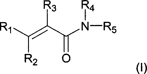
R1 für (C1-3)-Alkyl, (C3-6)-Cycloalkyl,
Phenyl oder NRaRb steht;
R2 für
Hydroxy, (C1-6)-Alkoxy, (C1-6)-Alkanoyloxy,
Amino-(C2-5)-alkoxy, Hydroxy-(C2-5)-alkoxy,
Amino-(C2-5)-alkanoyloxy oder Hydroxy-(C2-5)-alkanoyloxy
steht;
R3 für Cyano oder (C1-3)-Alkanoyl
steht;
R4 für Hydrogen, (C1-3)-Alkyl,
Hydroxy-(C2-5)-alkyl oder Amino-(C2-5)-alkyl steht;
R5 für durch
-S(O)2Rc oder Halogen
und mindestens einen anderen Substituenten substituiertes Phenyl
steht;
Ra und Rb jeweils
unabhängig
voneinander für
Hydrogen oder (C1-3)-Alkyl stehen oder Ra und Rb zusammen mit
dem Stickstoff, mit welchem sie verbunden sind, für Pyrrolidino,
Piperidino, Morpholino oder Thiomorpholino stehen;
wobei Aryl
oder Heteroaryl für
R1 und R5 gegebenenfalls
mit einem oder mehreren (z.B. 1, 2 oder 3) Substituenten, die unabhängig voneinander
ausgewählt
sind unter Halogen, Nitro, Cyano, Hydroxy, Trifluormethyl, Trifluormethoxy,
(C1-3)-Alkoxy,
(C1-3)-Alkyl, (C1-3)-Alkanoyl,
-S(O)2Rc oder NRaRb, wobei Rc für
(C1-3)-Alkyl oder Aryl steht, substituiert
ist, oder ein pharmazeutisch akzeptables Salz davon, wie es in Anspruch
1 beschrieben ist.The present invention provides the use of compounds of formula (I):
R 1 is (C 1-3 ) alkyl, (C 3-6 ) cycloalkyl, phenyl or NR a R b ;
R 2 is hydroxy, (C 1-6 ) alkoxy, (C 1-6 ) alkanoyloxy, amino (C 2-5 ) alkoxy, hydroxy (C 2-5 ) alkoxy, amino (C 2 -5 ) alkanoyloxy or hydroxy- (C 2-5 ) alkanoyloxy;
R 3 is cyano or (C 1-3 ) alkanoyl;
R 4 is hydrogen, (C 1-3 ) alkyl, hydroxy (C 2-5 ) alkyl or amino (C 2-5 ) alkyl;
R 5 is phenyl substituted by -S (O) 2 R c or halogen and at least one other substituent;
R a and R b are each independently hydrogen or (C 1-3 ) alkyl or R a and R b, together with the nitrogen to which they are attached, are pyrrolidino, piperidino, morpholino or thiomorpholino;
where aryl or heteroaryl for R 1 and R 5 is optionally substituted by one or more (eg 1, 2 or 3) substituents which are independently selected from halogen, nitro, cyano, hydroxy, trifluoromethyl, trifluoromethoxy, (C 1-3 ) - Alkoxy, (C 1-3 ) alkyl, (C 1-3 ) alkanoyl, -S (O) 2 R c or NR a R b , wherein R c is (C 1-3 ) alkyl or aryl, or a pharmaceutically acceptable salt thereof as described in claim 1.
Die Verbindungen der Erfindung sind so entwickelt, daß sie in ein zusammengesetztes Bindungstaschenmodell der BTK-Domäne passen, ein Molekülvolumen haben, das kleiner ist als das Volumen der Bindungstasche (z.B. weniger als etwa 600 Å3) und vorzugsweise ein Volumen haben, das etwa 2/3 des Volumens der Tasche, z.B. etwa 400 Å3, haben. Am vorteilhaftesten sind die Inhibitoren der Erfindung so entwickelt, daß sie den Raum der Bindungstasche ausfüllen und mit den Resten der Tasche für eine verstärkte Bindung wechselwirken.The compounds of the invention are designed to fit into a composite binding pocket model of the BTK domain, have a molecular volume less than the volume of the binding pocket (eg, less than about 600 Å 3 ), and preferably have a volume of about 2 / 3 of the volume of the bag, eg about 400 Å 3 . Most advantageously, the inhibitors of the invention are designed to fill the space of the binding pocket and interact with the remainder of the pocket for enhanced binding.
Die Erfindung stellt die Verwendung einer Verbindung der Formel (I) zur Herstellung eines Medikaments zur Behandlung eines pathologischen Zustandes oder einer Erkrankung bei einem Säuger, z.B. einem Menschen, der/die mit BTK-Aktivität in Verbindung steht, bereit. Diese Zustände oder Erkrankungen sind unter Störungen der B-Zell-Lymphozytenproliferation/Autoimmunerkrankungen, Mastzellenstörungen, Zuständen, die mit einer ungeeigneten Blutplättchenaggregation einhergehen, und Abstoßung von Xenoplantaten ausgewählt.The Invention provides the use of a compound of formula (I) for the manufacture of a medicament for the treatment of a pathological Condition or disease in a mammal, e.g. a human, the one with BTK activity communicates, ready. These conditions or diseases are under disturbances B cell lymphocyte proliferation / autoimmune diseases, Mast cell disorders, States that with inappropriate platelet aggregation go along, and rejection selected by xenoplants.
Kurze Beschreibung der FigurenShort description the figures
Detaillierte Beschreibung der Erfindungdetailed Description of the invention
Der Fas/APO-1 (CD95)-Zelloberflächenrezeptor, eine Mitglied der Tumornekrosefaktor (TNF)-Rezeptorfamilie, ist einer der Hauptregulatoren der Apoptose in einer Vielzahl von Zelltypen. Funktionelle Fas-Abnormalitäten waren mit pathologischen Zuständen der Immunsystemhomöostase verbunden, einschlließlich lymphoproliferativer Störungen, Immundefekten und Autoimmunität (Rieux-Laucat et al. (1995), Science 268, 1347-1349; und Fischer, G.H. (1995), Cell 81, 935-946). Eine Ligation des Zelloberflächen-Fas-Moleküls induziert rasch und dramatisch in vielen, aber nicht in allen Fas-positiven Zelltypen eine Apoptose (Nagata, S. (1997), Cell 88, 355-365). DT-40 ist eine Hühner-Lymphom-B-Zellinie, die verwendet wurde, um den molekularen Mechanismus von durch Strahlung induzierter Apoptose zu klären (Uckun, F.M. et al. (1996), Science 273, 1096-1100). Trotz ihrer reichlichen Oberflächenexpression von Fas sind DT-40-Zellen ähnlich wie humane B-Zellen-Vorläufer-Leukämiezellen gegenüber den cytotoxischen Wirkungen einer Fas-Ligation resistent, was die Existenz von wirksamen negativen Regulatoren der Fas-vermittelten Apoptose anzeigt. Bruton-Tyrosin-Kinase (BTK), ein Mitglied der BTK/Tec-Familie von Protein-Tyrosin-Kinasen (PTKs), ist eine cytoplasmische PTK, die bei Signaltransduktionswegen involviert ist, welche Wachstum und Differenzierung von Lymphzellen der B-Zellinie regulieren (Rawlings, D.J. und Witte, O.N. (1994), Immunol. Rev. 138, 105-119; Kurosaki, T. (1997), Curr. Opin. Immunol. 9, 309-318; und Uckun, F.M. (1998), Biochemical Pharmacology et al., 56, 683-691). BTK ist an Signaltransduktionswegen beteiligt, die durch Bindung einer Vielzahl extrazellulärer Liganden an ihre Zelloberflächenrezeptoren initiiert werden: nach Ligation von B-Zell-Antigenrezeptoren (BCR) ist ein BTK-Aktivierung durch die gemeinsame Wirkung der PTKs Lys und Syk (Kurosaki, T. (1997), Curr. Opin. Immunol. 9, 309-318) zur Induzierung einer Phospholipase C-γ2-vermittelten Calcium- Mobilisierung notwendig (Kurosaki, T. (1997), Curr. Opin. Immunol. 9, 309-318). Mutationen in humanen BTK-Gen sind der Grund für X-verknüpfte Agammaglobulinämie (XLA), eine männliche Immundefizienzkrankheit, die durch Fehlen von reifen, Immunglobulin-produzierenden, peripheren B-Zellen charakterisiert ist (Tsukada, S. et al. (1993), Cell 72, 279-290; und Vetrie, D. et al. (1993), Nature 361, 226-233). Bei Mäusen wurden Mutationen im BTK-Gen als Grund für murine X-verknüpfte Immundefizienz (Xid) identifiziert (Rawlings, D.J. et al. (1993), Science 261, 358-361).Of the Fas / APO-1 (CD95) cell surface receptor, a member of the tumor necrosis factor (TNF) receptor family one of the major regulators of apoptosis in a variety of cell types. Functional Fas abnormalities were with pathological conditions the immune system homeostasis connected, inclusive lymphoproliferative disorders, Immunodeficiencies and autoimmunity (Rieux-Laucat et al (1995), Science 268, 1347-1349; and Fischer, G. H. (1995), Cell 81, 935-946). Induced ligation of the cell surface Fas molecule fast and dramatic in many but not all Fas positive Cell types an apoptosis (Nagata, S. (1997), Cell 88, 355-365). DT-40 is one Chicken's lymphoma B cell line, which was used to study the molecular mechanism of radiation to clarify induced apoptosis (Uckun, F.M., et al., (1996) Science 273, 1096-1100). Despite their abundant surface expression of Fas are similar to DT-40 cells such as human B cell precursor leukemia cells across from the cytotoxic effects of Fas ligation resistant, which Existence of effective negative regulators of Fas-mediated apoptosis displays. Bruton tyrosine kinase (BTK), a member of the BTK / Tec family of protein tyrosine kinases (PTKs), is a cytoplasmic PTK, involved in signal transduction pathways, which growth and differentiation of lymph cells of the B cell line (Rawlings, D.J. and Witte, O.N. (1994), Immunol. Rev. 138, 105-119; Kurosaki, T. (1997), Curr. Opin. Immunol. 9, 309-318; and Uckun, F.M. (1998), Biochemical Pharmacology et al., 56, 683-691). BTK is at signal transduction pathways involved by binding a variety of extracellular ligands to their cell surface receptors to be initiated: after ligation of B cell antigen receptors (BCRs) a BTK activation through the joint action of the PTKs Lys and Syk (Kurosaki, T. (1997), Curr. Opin. Immunol. 9, 309-318) for inducing a phospholipase C-γ2 mediated Calcium mobilization necessary (Kurosaki, T. (1997), Curr. Opin., Immunol., 9, 309-318). Mutations in human BTK gene are the cause of X-linked agammaglobulinemia (XLA), a male Immunodeficiency disease caused by the absence of mature, immunoglobulin-producing, peripheral B cells (Tsukada, S. et al. (1993), Cell 72, 279-290; and Vetrie, D. et al. (1993), Nature 361, 226-233). In mice were Mutations in the BTK gene identified as a cause of murine X-linked immunodeficiency (Xid) (Rawlings, D.J., et al., (1993), Science 261, 358-361).
BTK hat sich als Inhibitor des Fas/APO-1 Tod-induzierenden Signalkomplexes (DISC) in B-Lymphozyten bzw. Lymphzellen erwiesen (Vassilev, A., et al. (1998), J. Biol. chem., 274, 1646-1656). Außerdem wurde derzeit festgestellt, daß BTK Ceramid- und Vincristin-induzierte Apoptose verhindert (vorliegende Studie). Das Schicksal von Leukämie/Lymphom-Zellen kann im Gleichgewicht zwischen den entgegengesetzten pro-apoptotischen Wirkungen von Kaspasen, die durch DISC aktiviert werden, und einem "upstream" wirkenden anti-apoptotischen Regulationsmechanismus, der BTK und/oder ihre Substrat involviert, liegen (Vassilev, A. et al. (1998), J. Biol. Chem., 274, 1646-1656). Inhibitoren von BTK verstärken wahrscheinlich die Arzneimittelempfindlichkeit der Zellen der B- (z.B. Leukämie/Lymphom)-Zellinie. Auf diese Weise können pharmakologische Agenzien mit BTK-Modulatoraktivität als chemosensibilisierende Modulatoraktivität als chemosensibilisierende Agenzien zur Behandlung von BTK-exprimierenden Malignitäten oder Krankheiten, die durch Proliferation und Antikörperproduktion von BTK-exprimierenden B-Zellen verursacht werden, und als B-Zellen rekonstituierende Mittel bei humoralen Immundefekten mit verringerter Anzahl oder mit Fehlen von B-Zellen eingesetzt werden. Außerdem wären BTK-modulierende Agenzien als immunsupprimierende Agenzien zur Verhinderung einer hyperakuten Organabstoßung bei Transplantation, welche durch B-Zellen gesteuert wird, Autoimmunkrankheiten und Konversion der Immunität auf Arzneimittel (z.B. Antikörper oder biologische Präparate) oder Blutprodukten (z.B. Koagulationsfaktoren, z.B. Faktor VIII) bei Patienten, die Antikörper gegen solche Agenzien entwickeln, nützlich.BTK has been shown to inhibit the Fas / APO-1 death inducing signaling complex (DISC) in B lymphocytes and lymphoid cells, respectively (Vassilev, A., et al., (1998) J. Biol. Chem., 274, 1646- 1656). In addition, BTK has been found to prevent ceramide- and vincristine-induced apoptosis (present study). The fate of leukemia / lymphoma cells may be in balance between the opposite pro-apoptotic effects of caspases activated by DISC and an upstream anti-apoptotic regulatory mechanism involving BTK and / or its substrate (Vassilev , A. et al., (1998) J. Biol. Chem., 274, 1646-1656). Inhibitors of BTK are likely to increase the drug sensitivity of B (eg leukemia / lymphoma) cell line cells. In this way, pharmacological agents having BTK modulator activity as chemosensitizing modulator activity may be chemosensitized agents for the treatment of BTK-expressing malignancies or diseases caused by proliferation and antibody production of BTK-expressing B cells, and used as B-cell reconstituting agents in humoral immunodeficiencies with a reduced number or absence of B-cells. In addition, BTK modulating agents would be useful as immunosuppressive agents for preventing hyperacute organ rejection in transplantation controlled by B cells, autoimmune diseases, and conversion of immunity to drugs (eg, antibodies or biological preparations) or blood products (eg, coagulation factors, eg, factor VIII) Patients who develop antibodies to such agents are useful.
Identifizierung von Inhibitoren für BTKidentification of inhibitors for BTK
Der wirksame und selektive BTK-Inhibitor LFM-13 und andere BTK-Inhibitoren wurden unter Verwendung des dreidimensionalen Homologiemodells der in Beispiel 2 beschriebenen Kinasedomäne identifiziert. Unter Verwendung dieses Modells und der Größe und der Kontaktinformation, die in Beispielen 1 und 3 bereitgestellt wird, wurden zusätzliche BTK-Inhibitoren entwickelt und untersucht. Unter Verwendung dieses Modells und dieser Methode können andere Verbindungen, die günstigerweise mit der Bindungstasche wechselwirken, identifiziert werden, ebenso wie Verbindungen, die über andere verwandte Kinasen selektiv an BTK binden werden. Eine feste Bindung oder eine gute Einpassung in das Bindungstaschenmodell korreliert mit wirksamer BTK-Inhibitoraktivität.Of the effective and selective BTK inhibitor LFM-13 and other BTK inhibitors were using the three - dimensional homology model of identified in Example 2 described kinase domain. Under use this model and the size and the Contact information provided in Examples 1 and 3 were additional BTK inhibitors are being developed and studied. Using this Model and this method can other compounds, conveniently interact with the binding pocket, as well like connections that over other related kinases will selectively bind to BTK. A solid Binding or a good fit in the binding pocket model correlates with effective BTK inhibitory activity.
Die Fähigkeit eines Agenzes, die anti-apoptischen Wirkungen von BTK zu inhibieren, kann unter Verwendung von Assays, die auf dem Fachgebiet bekannt sind, oder unter Verwendung von Assays, die in den hierin angeführten Beispielen offenbart sind, gemessen werden. Unter Verwendung der Modellierungsinformation und der hierin beschriebenen Screens wie auch anderer Informationen, die auf dem Fachgebiet bekannt sind, kann man Agenzien identifizieren, die BTK-inhibierende Eigenschaften besitzen.The ability of an agent to inhibit the anti-apoptotic effects of BTK, can be done using assays known in the art or using assays described in the examples provided herein disclosed are measured. Using the modeling information and the screens described herein as well as other information, known in the art, one can identify agents the BTK inhibiting Own properties.
Inhibitoren von BTK können auch solche Inhibitoren umfassen, die durch rekombinante DNA-Verfahren produziert werden, z.B. Antisense-Moleküle und Transkriptionsinhibitoren. Anfängliche Untersuchungen über btk-Transkription haben bewiesen, daß eine Expression des btk-Gens durch die kombinierte Wirkung von Transkriptionsfaktoren der Sp1- und PU.1-Familie reguliert wird (S. Muller et al. Oncogene, 1996, 13, 1955-1964; und A. Himmelmann et al. Blood, 1996, 87, 1036-1044). Transkriptionsregulatorelemente wurden innerhalb des ersten und des zehnten Introns des btk-Gens identifiziert und jüngere Untersuchungen zeigen, daß eine Regulation der btk-Genexpression mehrere Transkriptionsfaktoren involviert (J. Rohrer, M.E. Conley, Blood, 1998, 91, 214-221). Neue Agenzien, die die Aktivität dieser Transkriptionsfaktoren beeinträchtigen, wären auch als Modulatoren apoptotischer Signale in Behandlungsprogrammen einsetzbar. Die Machbarkeit einer Regulierung der btk-Genexpression in humanen hämatopoetischen Zellen wurde bereits durch die Fähigkeit von Retinsäure, eine brk-Expression in myeloischen Zellen zu erhöhen, und die Fähigkeit von Phorbolester wie auch von TGF-1, die btk-Expression in B-Zellen zu verringern, bewiesen (C.I.E. Smith et al., J. Immunol. 1994, 152, 557-565).inhibitors BTK can also include such inhibitors by recombinant DNA methods produced, e.g. Antisense molecules and transcription inhibitors. Initial studies on btk transcription have proved that one Expression of the btk gene by the combined action of transcription factors the Sp1 and PU.1 family (S. Muller et al., Oncogene, 1996, 13, 1955-1964; and A. Himmelmann et al. Blood, 1996, 87, 1036-1044). Transcriptional regulatory elements were within the first and tenth introns of the btk gene identified and younger Investigations show that a Regulation of btk gene expression multiple transcription factors (J. Rohrer, M. E. Conley, Blood, 1998, 91, 214-221). New agents that control the activity of this Affect transcription factors, would be too can be used as modulators of apoptotic signals in treatment programs. The feasibility of regulating btk gene expression in human hematopoietic Cells have already been through the ability of retinoic acid, to increase brk expression in myeloid cells, and ability of both phorbol ester and TGF-1, btk expression in B cells Smith, et al., J. Immunol., 1994, 152, 557-565).
Somit kann eines oder mehrerer der rekombinanten DNA-Verfahren verwendet werden, um eine BTK-Expression zu inhibieren und die hierin diskutierten therapeutischen Wirkungen zu induzieren. Solche Verfahren umfassen zum Beispiel Antisense-Sequenzen von Transkriptionsinhibitoren.Consequently For example, one or more of the recombinant DNA methods may be used to express BTK to inhibit and the therapeutic effects discussed herein to induce. Such methods include, for example, antisense sequences of transcription inhibitors.
Beispielsweise können BTK-Antisense-Konstrukte direkt gegen eine BTK-Expression gerichtet sein. Alternativ kann das Antisense-Konstrukt gegen BTK-Regulationssequenzen, z.B. Antisense-Sequenzen zur Sp1- und PU.1-Familie von Transkriptionsfaktoren, gerichtet sein. Vorzugsweise zielen die Antisense-Konstrukte auf Tumorzellen.For example can BTK antisense constructs directed directly against BTK expression be. Alternatively, the antisense construct can be raised against BTK regulatory sequences, e.g. Antisense sequences to the Sp1 and PU.1 family of transcription factors, be directed. Preferably, the antisense constructs target Tumor cells.
Verbindungenlinks
Verbindungen der Formel (I) sind spezifische BTK-Inhibitoren, die bevorzugt an die BTK-Modelltasche, die in den Beispielen beschrieben wird, binden und die wirksame BTK-Inhibitoraktivität haben.links of the formula (I) are specific BTK inhibitors, which are preferred bind the BTK model bag described in the Examples and have the effective BTK inhibitory activity.
Die
Verbindungen der Formel (I) sind: 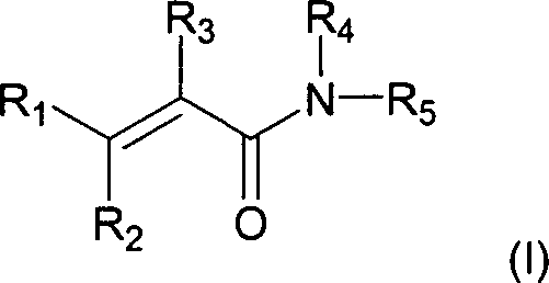
R1 für (C1-3)-Alkyl, (C3-6)-Cycloalkyl,
Phenyl oder NRaRb steht;
R2 für
Hydroxy, (C1-6)-Alkoxy, (C1-6)-Alkanoyloxy,
Amino-(C2-5)-alkoxy, Hydroxy-(C2-5)-alkoxy,
Amino-(C2-5)-alkanoyloxy
oder Hydroxy-(C2-5)-alkanoyloxy steht;
R3 für
Cyano oder (C1-3)-Alkanoyl steht;
R4 für
Wasserstoff, (C1-3)-Alkyl, Hydroxy-(C2-5)-alkyl oder Amino-(C2-5)-alkyl
steht;
R5 für durch -S(O)2Rc oder Halogen und mindestens einen anderen
Substituenten substituiertes Phenyl steht;
Ra und
Rb jeweils unabhängig voneinander für Wasserstoff
(bzw. Hydrogen) oder (C1-3)-Alkyl stehen;
oder Ra und Rb zusammen
mit dem Stickstoff, mit welchem sie verbunden sind, für Pyrrolidino,
Piperidino, Morpholino oder Thiomorpholino stehen;
wobei Aryl
oder Heteroaryl als R1 und R5 gegebenenfalls
substituiert ist durch einen oder mehrere (z.B. 1, 2 oder 3) Substituenten,
die unabhängig
voneinander ausgewählt
sind unter Halogen, Nitro, Cyano, Hydroxy, Trifluormethyl, Trifluormethoxy,
(C1-3)-Alkoxy, (C1-3)-Alkyl,
(C1-3)-Alkanoyl,
-S(O)2Rc oder NRaRb, wobei Rc für (C1-3)-Alkyl oder Aryl steht;
oder ein
pharmazeutisch akzeptables Salz davon.The compounds of the formula (I) are:
R 1 is (C 1-3 ) alkyl, (C 3-6 ) cycloalkyl, phenyl or NR a R b ;
R 2 is hydroxy, (C 1-6 ) alkoxy, (C 1-6 ) alkanoyloxy, amino (C 2-5 ) alkoxy, hydroxy (C 2-5 ) alkoxy, amino (C 2-6) alkoxy, 5) alkanoyloxy or hydroxy- (C 2-5 ) alkanoyloxy;
R 3 is cyano or (C 1-3 ) alkanoyl;
R 4 is hydrogen, (C 1-3 ) alkyl, hydroxy (C 2-5 ) alkyl or amino (C 2-5 ) alkyl;
R 5 is phenyl substituted by -S (O) 2 R c or halogen and at least one other substituent;
R a and R b are each independently hydrogen (or hydrogen) or (C 1-3 ) alkyl; or R a and R b together with the nitrogen to which they are attached represent pyrrolidino, piperidino, morpholino or thiomorpholino;
wherein aryl or heteroaryl as R 1 and R 5 is optionally substituted by one or more (eg 1, 2 or 3) substituents independently selected from halo, nitro, cyano, hydroxy, trifluoromethyl, trifluoromethoxy, (C 1-3 ) Alkoxy, (C 1-3 ) alkyl, (C 1-3 ) alkanoyl, -S (O) 2 R c or NR a R b , wherein R c is (C 1-3 ) alkyl or aryl stands;
or a pharmaceutically acceptable salt thereof.
Besonders
nützliche
Verbindungen der Formel (I) umfassen:
α-Cyano-β-hydroxy-β-methyl-N-(2,5-dibromphenyl)propenamid;
α-Cyano-β-hydroxy-β-methyl-N-[4-(methylsulfonyl)phenyl]propenamid;
α-Cyano-β-hydroxy-β-methyl-N-[3-(methylsulfonyl)phenyl]propenamid;
α-Cyano-β-hydroxy-β-methyl-N-[3-brom-4-(trifluormethoxy)phenyl]-propenamid;
α-Cyano-β-hydroxy-β-methyl-N-(2,4-dibromphenyl)propenamid;
α-Cyano-β-hydroxy-β-methyl-N-(2,4-dichlorphenyl)propenamid;
α-Cyano-β-hydroxy-β-methyl-N-(2,5-dichlorphenyl)propenamid;
oder
α-Cyano-β-hydroxy-β-methyl-N-(3,4-dichlorphenyl)propenamid;
oder
pharmazeutisch akzeptable Salze davon.Particularly useful compounds of formula (I) include:
α-cyano-β-hydroxy-β-methyl-N- (2,5-dibromophenyl) propenamide;
α-cyano-β-hydroxy-β-methyl-N- [4- (methylsulfonyl) phenyl] propenamide;
α-cyano-β-hydroxy-β-methyl-N- [3- (methylsulfonyl) phenyl] propenamide;
α-cyano-β-hydroxy-β-methyl-N- [3-bromo-4- (trifluoromethoxy) phenyl] -propenamid;
α-cyano-β-hydroxy-β-methyl-N- (2,4-dibromophenyl) propenamide;
α-cyano-β-hydroxy-β-methyl-N- (2,4-dichlorophenyl) propenamide;
α-cyano-β-hydroxy-β-methyl-N- (2,5-dichlorophenyl) propenamide; or
α-cyano-β-hydroxy-β-methyl-N- (3,4-dichlorophenyl) propenamide; or
pharmaceutically acceptable salts thereof.
Definitionen:definitions:
Die folgenden Definitionen werden, soweit nichts anderes beschrieben wird, hierin verwendet: Halogen ist Fluor, Chlor, Brom oder Iod. Alkyl, Alkoxy, usw. bezeichnen sowohl geradkettige als auch verzweigte Gruppen; Bezugnahme auf ein einzelnes Isomer, z.B. "Propyl", umfaßt nur das geradkettige Isomer, ein verzweigtkettiges Isomer, z.B. "Isopropyl" wird spezifisch bezeichnet. Aryl bezeichnet eine Phenyl-Gruppe oder eine bicyclische oder tricyclische carbocyclische Gruppe, die etwa 9 bis 12 Ringatome hat, wobei mindestens ein Ring aromatisch ist.The The following definitions will be used unless otherwise stated Halogen is fluorine, chlorine, bromine or iodine. Alkyl, alkoxy, etc. designate both straight-chain and branched groups; Reference to a single isomer, e.g. "Propyl" includes only the straight chain isomer, a branched-chain isomer, e.g. "Isopropyl" is specifically referred to. Aryl denotes a phenyl group or a bicyclic or tricyclic carbocyclic group, which has about 9 to 12 ring atoms, at least one ring being aromatic is.
Dem Fachmann auf diesem Gebiet wird klar sein, daß Verbindungen der Formel (I), die ein chirales Zentrum haben, in optisch aktiven und racemischen Formen existieren können und isoliert werden können. Einige Verbindungen können Polymorphismus aufweisen. Es ist einzusehen, daß die vorliegende Erfindung die Verwendung einer beliebigen racemischen, optisch aktiven, polymorphen oder stereoisomeren Form oder Gemischen davon, einer Verbindung der Formel (I), die die nützlichen hierin beschriebenen Eigenschaften besitzt, umfaßt, wobei es auf dem Fachgebiet bekannt ist, wie optisch aktive Formen hergestellt werden (zum Beispiel durch Trennung der racemischen Form durch Umkristallisationstechniken, durch Synthese aus optisch aktiven Ausgangsmaterialien, durch chirale Synthese oder durch chromatographische Trennung unter Verwendung einer chiralen stationären Phase) und wie die BTK-inhibierende Aktivität zu bestimmen ist, wobei die hierin beschriebenen Standardassays verwendet werden oder andere ähnliche Assays, die auf dem Fachgebiet gut bekannt sind, verwendet werden.the It will be clear to a person skilled in the art that compounds of the formula (I) which have a chiral center, in optically active and racemic Forms can exist and can be isolated. Some connections can Have polymorphism. It will be appreciated that the present invention the use of any racemic, optically active, polymorphic or stereoisomeric form or mixtures thereof, of a compound of the formula (I), which are the useful ones It has in the art It is known how to make optically active forms (for example by Separation of the racemic form by recrystallization techniques, by synthesis from optically active starting materials, by chiral Synthesis or by chromatographic separation using a chiral stationary Phase) and how to determine the BTK inhibitory activity, the standard assays described herein or other similar ones Assays that are well known in the art can be used.
Spezifische und bevorzugte werte, die unten für Substituenten und Bereiche aufgelistet sind, dienen lediglich der Erläuterung; sie schließen andere definierte Werte oder andere Werte innerhalb definierter Bereiche für die Reste und Substituenten nicht aus.specific and preferred values below for substituents and ranges listed are merely illustrative; they close others defined values or other values within defined ranges for the Radicals and substituents are not sufficient.
Spezifischerweise kann (C1-3)-Alkyl Methyl, Ethyl, Propyl oder Isopropyl sein; (C3-6)-Cycloalkyl kann Cyclopropyl, Cyclobutyl, Cyclopentyl oder Cyclohexyl sein; (C1-3)-Alkoxy kann Methoxy, Ethoxy, Propoxy, Isopropropoxy sein; (C1-6)-Alkoxy kann Methoxy, Ethoxy, Propoxy, Isopropropoxy, Butoxy, Isobutoxy, sek-Butoxy, Pentoxy, 3-Pentoxy oder Hexyloxy sein.Specifically, (C 1-3 ) alkyl may be methyl, ethyl, propyl or isopropyl; (C 3-6 ) cycloalkyl may be cyclopropyl, cyclobutyl, cyclopentyl or cyclohexyl; (C 1-3 ) alkoxy may be methoxy, ethoxy, propoxy, isopropoxy; (C 1-6 ) -Alkoxy may be methoxy, ethoxy, propoxy, isopropoxy, butoxy, isobutoxy, sec-butoxy, pentoxy, 3-pentoxy or hexyloxy.
Spezifische und bevorzugte Bedeutungenspecific and preferred meanings
Eine spezifische Bedeutung für R1 ist (C1-3)-Alkyl oder (C3-6)-Cycloalkyl.A specific meaning for R 1 is (C 1-3 ) -alkyl or (C 3-6 ) -cycloalkyl.
Eine spezifische Bedeutung für R2 ist Hydroxy.A specific meaning for R 2 is hydroxy.
Eine spezifische Bedeutung für R3 ist Cyano.A specific meaning for R 3 is cyano.
Eine spezifische Bedeutung für R4 ist Wasserstoff.A specific meaning for R 4 is hydrogen.
Eine spezifische Bedeutung für R5 ist Phenyl, substituiert mit Halogen und substituiert mit 1, 2 oder 3 anderen Substituenten, die unabhängig aus Halogen, Nitro, Cyano, Trifluormethyl, Trifluormethoxy, (C1-3)-Alkoxy, (C1-3)-Alkyl, (C1-3)-Alkanoyl und NRaRb ausgewählt sind.A specific meaning for R 5 is phenyl substituted with halogen and substituted with 1, 2 or 3 other substituents independently selected from halo, nitro, cyano, trifluoromethyl, trifluoromethoxy, (C 1-3 ) alkoxy, (C 1-3 ) Alkyl, (C 1-3 ) alkanoyl and NR a R b are selected.
Eine spezifischere Bedeutung für R1 ist (C1-3)-Alkyl.A more specific meaning for R 1 is (C 1-3 ) alkyl.
Eine spezifischere Bedeutung für R5 ist Phenyl, substituiert mit Halogen und substituiert mit 1, 2 oder 3 anderen Substituenten, die unabhängig aus Halogen, Trifluormethyl, Trifluormethoxy und (C1-3)-Alkoxy ausgewählt sind.A more specific meaning for R 5 is phenyl substituted with halogen and substituted with 1, 2 or 3 other substituents independently selected from halogen, trifluoromethyl, trifluoromethoxy and (C 1-3 ) alkoxy.
Eine spezifischere Bedeutung für R5 ist Phenyl, das mit 2 oder 3 Halogenen substituiert ist.A more specific meaning for R 5 is phenyl substituted with 2 or 3 halogens.
Eine spezifischere Bedeutung für R5 ist Phenyl, das mit zwei Brom substituiert ist.A more specific meaning for R 5 is phenyl substituted with two bromine.
Eine bevorzugt Verbindung der Formel (I) ist α-Cyano-β-hydroxy-β-methyl-N-(2,5-dibromphenyl)propenamid oder ein pharmazeutisch akzeptables Salz davon.A preferably compound of the formula (I) is α-cyano-β-hydroxy-β-methyl-N- (2,5-dibromophenyl) propenamide or a pharmaceutically acceptable salt thereof.
Therapeutische Verwendungtherapeutic use
B-Zellen und B-Zellvorläufer, die BTK exprimieren, sind in der Pathologie einer Reihe von Krankheiten und Zuständen impliziert; diese umfassen B-Zell-Malignitäten (z.B. akute Lymphoblastenleukämie, chronische lymphatische Leukämie, Nicht-Hodgkins-Lymphom, EBV-Lymphom und Myelom), andere Krebsarten, lymphoproliferative B-Zell- Störungen/Autoimmunkrankheiten (z.B. Lupus, Crohn-Krankheit und chronische oder Transplantat-gegen-Empfänger-Reaktion), Mastozytose (z.B. Allergien und anaphylaktischer Schock), Zustände, die mit unpassenden Thrombozytenaggregation in Verbindung stehen und Abstoßung von Xenotransplantaten (z.B. Herztransplantate von Schwein zu Menschen).B-cells and B-cell precursors, The BTK express are in the pathology of a number of diseases and states implies; these include B cell malignancies (e.g., acute lymphoblastic leukemia, chronic lymphocytic Leukemia, Non-Hodgkin's lymphoma, EBV-lymphoma and myeloma), other cancers, lymphoproliferative B-cell disorders / autoimmune diseases (e.g., lupus, Crohn's disease and chronic or graft versus recipient reaction), Mastocytosis (e.g., allergies and anaphylactic shock), conditions that associated with improper platelet aggregation and rejection of xenografts (e.g., heart transplants from pig to human).
Zusätzlich können die selektiven BTK-Inhibitoren verwendet werden, um andere Krankheiten zu identifizieren, in denen BTK eine Rolle spielt, und insbesondere um eine Genexpression, die durch BTK moduliert ist, zu identifizieren. Dies kann unter Verwendung von Techniken, die auf dem Fachgebiet bekannt sind, z.B. unter Verwendung von Gen-Profilierungstechniken, die ähnlich denen sind, die von A. Sehgal et al., Journal of Surgical Oncology, 1998, 67, 234-241, beschrieben werden, erfolgen. Eine Inkubation von Zellen in Gegenwart oder Abwesenheit eines BTK-Inhibitors, gefolgt von einer Profilierung der Gen-Expression in den Zellen, ist einsetzbar, um eine BTK-regulierte Gen-Expression zu identifizieren. Materialien, die zur Profilierung der Gen-Expression unter Verwendung von Atlas-cDNA-Membranen einsetzbar sind, können von CLONTECH Laboratories, Inc. 1020 East Meadow Circle, Palo Alto, CA 94303 erhalten werden. cDNA-Mikroarrays können von handelsüblichen Quellen bestellt werden oder selbst hergestellt werden.In addition, the Selective BTK inhibitors are used to treat other diseases identify in which BTK plays a role, and in particular to identify gene expression modulated by BTK. This can be done using techniques known in the art are known, e.g. using gene profiling techniques similar to those by A. Sehgal et al., Journal of Surgical Oncology, 1998, 67, 234-241 be done. An incubation of cells in the presence or absence a BTK inhibitor, followed by profiling of gene expression in the cells, is applicable to a BTK-regulated gene expression to identify. Materials that are used to profiling gene expression under Use of atlas cDNA membranes can be used by CLONTECH Laboratories, Inc. 1020 East Meadow Circle, Palo Alto, CA 94303 can be obtained. cDNA microarrays may be commercially available Sources can be ordered or self-made.
Unter Verwendung solcher Materialien nach den Anweisungen des Herstellers wurde auch entdeckt, daß BTK die Expression spezifischer Gene, z.B. MAKPAP-Kinase-Gen und c-myc-Onkogen, moduliert. Diese Aktivität legt nahe, daß BTK bei der Pathologie aller Krebsformen involviert sein kann.Under Use of such materials according to the manufacturer's instructions was also discovered that BTK the expression of specific genes, e.g. MAKPAP kinase gene and c-myc oncogene, modulated. This activity suggests that BTK be involved in the pathology of all forms of cancer.
BTK ist ein Glied der Tec-Familie von Tyrosinkinasen. Tec-Kinase wird zum Beispiel in T-Zellen exprimiert. Die BTK-Inhibitoren der Erfindung sind auch einsetzbar, um die Aktivität anderer Glieder der Tec-Kinasefamilie zu inhibieren.BTK is a member of the Tec family of tyrosine kinases. Tec kinase will be for example expressed in T cells. The BTK inhibitors The invention can also be used to control the activity of others To inhibit members of the Tec kinase family.
BTK-Inhibitoren (einschließlich Verbindungen der Formel (I), wie sie hier beschrieben sind) können verwendet werden, um Störungen zu behandeln, bei denen die Inhibierung oder Prävention einer Kinase der Tec-Familie, einschließlich BTK-Aktivität, indiziert ist. Es wurde auch festgestellt, daß BTK-Inhibitoren als chemosensibilisierende Agenzien einsetzbar sind und somit in Kombination mit anderen chemotherapeutischen Arzneimitteln, insbesondere Arzneimitteln, die eine Apoptose induzieren, einsetzbar sind. Beispiele für andere chemotherapeutische Arzneimittel, die in Kombination mit chemosensibilisierenden BTK-Inhibitoren verwendet werden können, umfassen Topoisomerase I-Inhibitoren (Camptothesin oder Topotecan), Topoisomerase II-Inhibitoren (z.B. Daunomycin und Etoposid), Alkylierungsmittel (z.B. Cyclophosphamid, Melphalan und BCNU), Tubulin-gesteuerte Mittel (z.B. Taxol und Vinblastin) und biologische Agenzien (z.B. Antikörper wie Anti-CD209-Antikörper, IDEC 8, Immuntoxine und Cytokine).BTK inhibitors (including compounds of formula (I) as described herein) can be used to treat disorders in which the inhibition or prevention of a Tec family kinase, including BTK activity, is indicated. It has also been found that BTK inhibitors are useful as chemosensitizing agents and thus can be used in combination with other chemotherapeutic drugs, particularly drugs that induce apoptosis. Examples of other chemotherapeutic drugs that can be used in combination with chemosensitizing BTK inhibitors include topoisomerase I inhibitors (camptothesin or topotecan), topoisomerase II inhibitors (eg, daunomycin and etoposide), alkylating agents (eg, cyclophosphamide, melphalan, and BC NU), tubulin-controlled agents (eg taxol and vinblastine) and biological agents (eg antibodies such as anti-CD209 antibody, IDEC 8, immunotoxins and cytokines).
Konjugation an eine Targeting-Gruppierungconjugation to a targeting grouping
Die Verbindungen der Formel (I) können zur spezifischen Abgabe an einen zu behandelnden Zelltyp durch Konjugation des BTK-Inhibitors an eine Targeting-Gruppierung targetiert werden. Targeting-Gruppierungen, die zur Konjugation mit BTK-Inhibitoren einsetzbar sind, umfassen Antikörper, Cytokine und Rezeptorliganden, die für die zu behandelnde Zelle spezifisch sind.The Compounds of formula (I) can for specific delivery to a cell type to be treated by conjugation of the BTK inhibitor are targeted to a targeting moiety. Targeting groups that can be used for conjugation with BTK inhibitors are antibodies, Cytokines and receptor ligands responsible for the cell to be treated are specific.
Der Ausdruck "Konjugat" meint eine Verbindung, die als Composite zwischen zwei oder mehr Molekülen gebildet wird. Spezifischer ausgedrückt, in der vorliegenden Erfindung ist das Chinazolin-Derivat an zellspezifische Targeting- Gruppierungen, die eine Konjugatverbindung bilden, zur effizienten und spezifischen Abgabe des Agenzes an die interessierende Zelle gebunden, z.B, kovalent gebunden.Of the Expression "conjugate" means a compound which is formed as a composite between two or more molecules. specific expressed In the present invention, the quinazoline derivative is cell-specific Targeting groups, which form a conjugate compound, to the efficient and specific Delivery of the agent bound to the cell of interest, e.g., covalent bound.
Der Ausdruck "Targeting-Gruppierung" meint ein Molekül, das dazu dient, die erfindungsgemäße Verbindung für die gewünschte Aktivität an eine spezifische Stelle abzugeben. Targeting-Gruppierungen umfassen z.B. Moleküle, die Moleküle spezifisch eine spezifische Zelloberfläche binden. Solche Targeting-Gruppierungen, die in der Erfindung einsetzbar sind, umfassen Anti-Zelloberflächen-Antigen-Antikörper. Cytokine, einschließlich Interleukine und Faktoren, wie z.B. Granulozyten/Makrophagen-stimulierender Faktor (GMCSF) sind ebenfalls spezifische Targeting-Gruppierungen, von denen bekannt ist, daß sie an spezifische Zellen binden, welche hohe Level ihrer Rezeptoren exprimieren.Of the The phrase "targeting grouping" means a molecule that has serves, the compound of the invention for the desired activity to a specific body. Targeting groupings include e.g. molecules the molecules specifically bind a specific cell surface. Such targeting groups, which are useful in the invention include anti-cell surface antigen antibodies. cytokines, including Interleukins and factors, such as Granulocyte / macrophage colony stimulating Factor (GMCSF) are also specific targeting groups, which is known to them bind to specific cells, which have high levels of their receptors express.
Besonders nützliche Targeting-Gruppierungen zum Targeting der BTK-Inhibitorverbindungen der Erfindung auf Zellen zur therapeutischen Aktivität umfassen solche Liganden, die an Tec-Kinase exprimierenden Zellen vorliegen. Zum Beispiel können Antigene, die an B-Zellen und Krebszellen der B-Zellinie, z.B. CD19, vorliegen, mit Anti-CD19-Antikörpern, z.B. B43, targetiert werden. Antikörper-Fragmente, einschließlich Einzelkettenfragmente, können verwendet werden. Natürliche Liganden für die Oberflächenantigene, z.B. CD19, können ebenfalls eingesetzt werden. Tec-Kinase exprimierende T-Zellen können z.B. auf das CD7-Antigen mit Anti-CD7-Antikörpern, beispielsweise TXU, targetiert werden. Mastzellen können über das CD48-Antigen mit Anti-CD48-Antikörpern targetiert werden. Diese und andere Zelloberflächen Antigen-Antikörper sind z.B. von Pharmingen im Handel erhältlich.Especially useful Targeting groups for targeting BTK inhibitor compounds of the invention to cells for therapeutic activity such ligands that are present on Tec kinase expressing cells. For example, you can Antigens attached to B cells and B cell line cancer cells, e.g. CD19, present, with Anti-CD19 antibodies e.g. B43, to be targeted. Antibody fragments, including single-chain fragments, can be used. natural Ligands for the surface antigens, e.g. CD19, can also be used. Tec kinase expressing T cells may be e.g. to the CD7 antigen with anti-CD7 antibodies, for example TXU, be targeted. Mast cells can be over the Targeting CD48 antigen with anti-CD48 antibodies become. These and other cell surfaces are antigen-antibody e.g. commercially available from Pharmingen.
Cytokine sind ebenfalls nützliche Targeting-Gruppierungen. T-Zellen können mit IL2 und IL7 targetiert werden; B-Zellen können mit IL-4 targetiert werden; Mastzellen können mit C-KIT, MGH, GMCSF und IL3 targetiert werden. Krebszellen, die Tec-Kinase exprimieren, können z.B. mit EGF und IGF targetiert werden.cytokines are also useful Targeting moieties. T cells can be targeted with IL2 and IL7; B cells can be targeted with IL-4; Mast cells can with C-KIT, MGH, GMCSF and IL3. Cancer cells that Tec kinase can express e.g. be targeted with EGF and IGF.
Verbindungen als Salzelinks as salts
In Fällen, in denen ein Agens ("Verbindung") ausreichend basisch oder sauer ist, um stabile, nicht-toxische Säure- oder Basensalze zu bilden, kann eine Verabreichung der Verbindung als Salz geeignet sein. Beispiele pharmazeutisch annehmbarer Salze sind Additionssalze mit organischen Säuren, die z.B. mit Säuren gebildet werden, welche ein physiologisch akzeptables Anion bilden, z.B. Tosylat, Methansulfonat, Acetat, Citrat, Malonat, Tartrat, Succinat, Benzoat, Ascorbat, α-Ketoglutarat und α-Glycerophosphat. Geeignete anorganische Salze können ebenfalls gebildet werden; diese umfassen Hydrochlorid-, Sulfat-, Nitrat-, Bicarbonat- und Carbonatsalze.In cases in which an agent ("compound") is sufficiently basic or acidic to form stable, non-toxic acid or base salts may be Administration of the compound as a salt. Examples pharmaceutically Acceptable salts are addition salts with organic acids which e.g. formed with acids which form a physiologically acceptable anion, e.g. Tosylate, methanesulfonate, acetate, citrate, malonate, tartrate, succinate, Benzoate, ascorbate, α-ketoglutarate and α-glycerophosphate. Suitable inorganic salts may be also be formed; these include hydrochloride, sulfate, Nitrate, bicarbonate and carbonate salts.
Pharmazeutisch akzeptable Salze können unter Verwendung von Standardverfahren, die auf dem Fachgebiet gutbekannt sind, erhalten werden. Beispielsweise indem eine ausreichend basische Verbindung, z.B. ein Amin, mit einer geeigneten Säure, die ein physiologisch akzeptables Anion liefert, umgesetzt wird. Alkalimetall (z.B. Natrium, Kalium oder Lithium)- oder Erdalkalimetall (z.B. Calcium)-Salze von Carbonsäuren können ebenfalls hergestellt werden.pharmaceutical acceptable salts can using standard techniques well known in the art are to be obtained. For example, by a sufficiently basic Compound, e.g. an amine, with a suitable acid, the a physiologically acceptable anion provides, is reacted. alkali metal (e.g., sodium, potassium or lithium) or alkaline earth metal (e.g. Calcium) salts of carboxylic acids can also getting produced.
Prodrug-DerivateProdrug derivatives
Die Verbindungen der Formel (I) haben daran funktionelle Gruppen gebunden, um ein Prodrug-Derivat bereitzustellen. Das Prodrug-Derivat erleichtert eine Verwendung des Arzneimittels im Körper, in dem es z.B. den Eintritt in Zellen erleichtert. Der Ausdruck "Prodrug-Gruppierung" ist eine Substitutionsgruppe, die die Verwendung einer erfindungsgemäßen Verbindung erleichtert, z.B. indem sie den Eintritt des Arzneimittels in Zellen oder die Verabreichung der Verbindung erleichtert. Die Prodrug-Gruppierung kann in vivo z.B. durch Spaltung mit Enzymen von der Verbindung abgespalten werden. Beispiele für Prodrug-Gruppierungen umfassen Phosphat-Gruppen, Peptidlinker und Zucker, wobei die Gruppierungen in vivo hydrolysiert werden können.The compounds of formula (I) have attached thereto functional groups to provide a prodrug derivative. The prodrug derivative facilitates use of the drug in the body by, for example, facilitating entry into cells. The term "prodrug moiety" is a substitution group that facilitates the use of a compound of the invention, eg, by facilitating the entry of the drug into cells or the administration of the compound. The prodrug moiety can be expressed in vivo eg by Spal tion with enzymes from the compound be cleaved. Examples of prodrug moieties include phosphate groups, peptide linkers, and sugars, which moieties can be hydrolyzed in vivo.
Pharmazeutische Formulierungenpharmaceutical formulations
Eine Verbindung kann als pharmazeutische Zusammensetzungen formuliert werden und einem Säugerwirt, z.B. einem menschlichen Patienten, in einer Vielzahl von Formen, die an die gewählte Verabreichungsroute, z.B. oral oder parenteral, auf intravenösen, intramuskulären, topischen oder subkutanen Wegen, angepaßt sind, verabreicht werden.A Compound may be formulated as pharmaceutical compositions and a mammal host, e.g. a human patient, in a variety of forms, to the chosen one Route of administration, e.g. oral or parenteral, on intravenous, intramuscular, topical or subcutaneous routes, adapted are administered.
So können Verbindungen systemisch verabreicht werden, z.B. oral in Kombination mit einem pharmazeutisch akzeptablen Vehikel, beispielsweise einem inerten Verdünnungsmittel oder einem assimilierbaren eßbaren Träger. Sie können in Gelatinekapseln mit harter oder weicher Schale eingeschlossen sein, können zu Tabletten verpreßt sein oder können direkt in das Lebensmittel der Patientennahrung eingearbeitet sein. Zur oralen therapeutischen Verabreichung kann die Verbindung mit einem oder mehreren Exzipientien kombiniert werden und in Form von einnehmbaren Tabletten, bukkalen Tabletten, Lutschbombons, Kapseln, Elixieren, Suspensionen, Sirupen, Wafern und dgl. verwendet werden. Solche Zusammensetzungen und Präparate sollten mindestens 0,1 % aktive Verbindung enthalten. Der Prozentgehalt der Zusammensetzungen und Präparate kann verändert werden und kann zweckdienlicherweise zwischen etwa 2 und 60 % des Gewichts einer gegebenen Einheitsdosierungsform betragen. Die Menge der aktiven Verbindung in derartigen therapeutisch nützlichen Zusammensetzungen ist so, daß ein wirksamer Dosierungslevel erhalten wird.So can Compounds are administered systemically, e.g. orally in combination with a pharmaceutically acceptable vehicle, for example a inert diluent or an assimilable edible Carrier. You can enclosed in hard or soft shell gelatin capsules could be compressed into tablets be or can be incorporated directly into the food of the patient's food. For oral therapeutic administration, the compound may be used with one or more excipients are combined and in the form of ingestible tablets, buccal tablets, lozenge packs, capsules, Elixirs, suspensions, syrups, wafers and the like. Such compositions and preparations should contain at least 0.1% active compound. The percentage the compositions and preparations can change and can conveniently be between about 2 and 60% of the Weight of a given unit dosage form. The amount the active compound in such therapeutically useful compositions is such that one effective dosage level is obtained.
Die Tabletten, Lutschtabletten, Pillen, Kapseln und dgl. können auch folgendes enthalten: Bindemittel, z.B. Tragacanthgummi, Akaziengummi, Maisstärke oder Gelatine; Exzipientien, z.B. Dicalciumphosphat; ein Zerfallsmittel, z.B. Maisstärke, Kartoffelstärke, Alginsäure und dgl.; ein Gleitmittel, z.B. Magnesiumstearat, und ein Süßungsmittel, wie z.B. Saccharose, Fructose, Lactose oder Aspartam, oder ein Aromamittel, z.B. Pfefferminz, Wintergrünöl oder Kirscharoma können zugesetzt werden. Wenn die Einheitsdosierungsform eine Kapsel ist, kann sie zusätzlich zu Materialien des obigen Typs einen flüssigen Träger, z.B. ein Pflanzenöl oder ein Polyethylenglykol, enthalten. Es können verschiedene andere Materialien als Beschichtungen oder zur anderweitigen Modifizierung der physikalischen Form der festen Einheitsdosierungsform vorliegen. Zum Beispiel können Tabletten, Pillen oder Kapseln mit Gelatine, Wachs, Schellack, Zucker oder dgl. überzogen sein. Ein Sirup oder Elixier kann die aktive Verbindung, Saccharose oder Fructose als Süßungsmittel, Methyl- und Propylparabene als Konservierungsstoffe, einen Farbstoff und ein Aromamittel, z.B. Kirsch- oder Orangenaroma, enthalten. Jedes Material, das bei der Herstellung einer Einheitsdosierungsform verwendet wird, sollte pharmazeutisch akzeptabel sein und in den verwendeten Mengen im wesentlichen nicht toxisch sein. Außerdem kann die aktive Verbindung in Präparate und Vorrichtungen mit verzögerter Freisetzung eingearbeitet sein.The Tablets, lozenges, pills, capsules and the like can also containing: binders, e.g. Tragacanth gum, acacia gum, corn starch or gelatin; Excipients, e.g. dicalcium phosphate; a disintegrant, e.g. Corn starch, Potato starch, alginic acid and the like; a lubricant, e.g. Magnesium stearate, and a sweetener, such as. Sucrose, fructose, lactose or aspartame, or a flavoring agent, e.g. Peppermint, wintergreen oil or cherry flavor can be added. If the unit dosage form is a capsule, can she in addition to materials of the above type, a liquid carrier, e.g. a vegetable oil or a polyethylene glycol, contain. It can various materials other than coatings or otherwise Modification of the physical form of the solid unit dosage form available. For example, you can Tablets, pills or capsules with gelatin, wax, shellac, sugar or the like be. A syrup or elixir may be the active compound, sucrose or fructose as a sweetener, Methyl and propylparabens as preservatives, a dye and a flavoring agent, e.g. Cherry or orange flavor, included. Any material used in the preparation of a unit dosage form should be pharmaceutically acceptable and in the used amounts to be substantially non-toxic. In addition, can the active compound in preparations and devices with delayed Be incorporated release.
Die Verbindung kann auch intravenös oder intraperitoneal durch Infusion oder Injektion verabreicht werden. Lösungen der Verbindung oder ihres Salzes können in Wasser, gegebenenfalls mit einem nicht-toxischen oberflächenaktiven Mittel vermischt, hergestellt werden. Dispersionen können auch in Glycerin, flüssigen Polyethylenglykolen, Triacetin und Gemischen davon und in Ölen hergestellt werden. Unter normalen Lagerungs- und Verwendungsbedingungen enthalten diese Präparate ein Konservierungsmittel, um das Wachstum von Mikroorganismen zu verhindern.The Connection can also be intravenous or intraperitoneally by infusion or injection. solutions The compound or its salt may be dissolved in water, optionally with a non-toxic surface-active Mixtures are mixed, prepared. Dispersions can also in glycerol, liquid Polyethylene glycols, triacetin and mixtures thereof and made in oils become. Included under normal conditions of storage and use these preparations a preservative to increase the growth of microorganisms prevent.
Die pharmazeutischen Dosierungsformen, die zur Injektion oder Infusion geeignet sind, können sterile wäßrige Lösungen oder Dispersionen oder sterile Pulver, die die aktive Verbindung enthalten, und die für die außerplanmäßige Herstellung von sterilen, injizierbaren Lösungen oder Dispersionen oder Infusionslösungen oder -dispersionen, gegebenenfalls in Liposome eingekapselt, angepaßt sind, umfassen. In allen Fällen sollte die endgültige Dosierungsform unter den Bedingungen der Herstellung und Lagerung steril, flüssig und stabil sein. Der flüssige Träger oder das flüssige Vehikel können ein Lösungsmittel oder ein flüssiges Dispersionsmedium sein, das z.B. Wasser, Ethanol, ein Polyol (z.B. Glycerin, Propylenglykol, flüssige Polyethylenglykole und dgl.), Pflanzenöle, nicht-toxische Glycerylester und geeignete Gemische davon umfaßt. Die geeignete Fluidität kann z.B. durch Bildung von Liposomen, durch Aufrechterhaltung der erforderlichen Partikelgröße im Fall von Dispersionen oder durch die Verwendung von oberflächenaktiven Mitteln aufrechterhalten werden. Die Verhinderung der Wirkung von Mikroorganismen kann durch verschiedene antibakterielle und antifungale Mittel, z.B. Parabene, Chlorbutanol, Phenol, Sorbinsäure, Thimerosal und dgl., erreicht werden. In vielen Fällen wird es vorteilhaft sein, daß isotonische Mittel, z.B. Zucker, Puffer oder Natriumchlorid enthalten sind. Eine verlängerte Absorption der injizierbaren Zusammensetzungen kann durch die Verwendung von Mitteln, die die Absorption verzögern, z.B. Aluminiummonostearat und Gelatine, erreicht werden.The pharmaceutical dosage forms for injection or infusion are suitable sterile aqueous solutions or Dispersions or sterile powders containing the active compound, and the for the unscheduled production from sterile, injectable solutions or dispersions or infusion solutions or dispersions, optionally encapsulated in liposomes. In all make should be the final one Dosage form under the conditions of manufacture and storage sterile, liquid and be stable. The liquid carrier or the liquid one Vehicles can a solvent or a liquid one Dispersion medium, e.g. Water, ethanol, a polyol (e.g. Glycerin, propylene glycol, liquid Polyethylene glycols and the like), vegetable oils, non-toxic glyceryl esters and suitable mixtures thereof. The suitable fluidity may e.g. by forming liposomes, by maintaining the required Particle size in the case of Dispersions or by the use of surfactants Be maintained. Preventing the effects of Microorganisms can be caused by various antibacterial and antifungal Means, e.g. Parabens, chlorobutanol, phenol, sorbic acid, thimerosal and the like., Can be achieved. In many cases, it will be advantageous that isotonic Means, e.g. Sugar, buffer or sodium chloride are included. An extended one Absorption of the injectable compositions may be through use of agents that delay absorption, e.g. , aluminum and gelatin.
Sterile injizierbare Lösungen können hergestellt werden, indem die aktive Verbindung in der erforderlichen Menge im geeigneten Lösungsmittel mit verschiedenen der oben aufgezählten anderen Ingredienzien nach Bedarf eingearbeitet wird, worauf eine Filtersterilisierung folgt. Im Fall steriler Pulver zur Herstellung steriler injizierbarer Lösungen sind die bevorzugten Herstellungsverfahren Vakuumtrocknung und die Gefriertrocknungstechniken, die zu einem Pulver der aktiven Verbindung plus einem zusätzlichen gewünschten Ingrediens, das in den vorher steril filtrierten Lösungen vorlag, führen.sterile injectable solutions can be prepared by the active compound in the required Amount in a suitable solvent with various of the other ingredients enumerated above Need is incorporated, followed by a filter sterilization. In the case of sterile powders for the preparation of sterile injectable solutions the preferred manufacturing processes vacuum drying and freeze-drying techniques, resulting in a powder of the active compound plus an additional desired Ingrediens, which was present in the previously sterile filtered solutions, to lead.
Für eine topische Verabreichung können die Verbindungen in reiner Form, d.h. wenn sie Flüssigkeiten sind, angewendet werden. Allerdings wird es im allgemeinen wünschenswert sein, sie als Zusammensetzungen oder Formulierungen in Kombination mit einem dermatologisch akzeptablen Träger, der ein Feststoff oder eine Flüssigkeit sein kann, an die Haut zu verabreichen.For a topical Administration can the compounds in pure form, i. if they are liquids, be applied. However, it generally becomes desirable be combined as compositions or formulations with a dermatologically acceptable carrier which is a solid or a liquid may be to administer to the skin.
Verwendbare feste Träger umfassen feinverteilte Feststoffe, z.B. Talk, Ton, mikrokristalline Cellulose, Siliciumdioxid, Aluminiumoxid und dgl. Verwendbare flüssige Träger umfassen Wasser, Alkohole oder Glykole oder Wasser-Alkohol/Glykol-Mischungen, in denen die erfindungsgemäßen Verbindungen in wirksamen Konzentrationen gelöst oder dispergiert werden können, gegebenenfalls mit Hilfe nicht-toxischer oberflächenaktiver Mittel. Adjuvantien, z.B. Duftstoffe und zusätzliche antimikrobielle Agenzien, können zugesetzt werden, um die Eigenschaften für eine vorgegebene Verwendung zu optimieren. Die resultierenden flüssigen Zusammensetzungen können mit Absorbens-Pads aufgetragen werden, welche verwendet werden, um Verbände und andere Umschläge zu imprägnieren, oder sie können auf den betroffenen Bereich unter Verwendung von Sprühvorrichtungen des Pump-Typs oder Aerosol-Sprühvorrichtungen aufgesprüht werden.usable solid carriers include finely divided solids, e.g. Talc, clay, microcrystalline Cellulose, silica, alumina and the like. Useful liquid carriers include Water, alcohols or glycols or water-alcohol / glycol mixtures in which the compounds of the invention dissolved in effective concentrations or can be dispersed, optionally with the aid of non-toxic surfactants. adjuvants, e.g. Perfumes and additional antimicrobial agents, can be added to the properties for a given use to optimize. The resulting liquid compositions can with Absorbent pads are applied, which are used to bandages and other envelopes to impregnate, or you can on the affected area using sprayers pump-type or aerosol spray devices sprayed become.
Verdickungsmittel wie z.B. synthetische Polymere, Fettsäuren, Fettsäuresalze und -ester, Fettalkohole, modifizierte Cellulosen oder modifizierte Mineralmaterialien können ebenfalls mit flüssigen Trägern eingesetzt werden, um verteilbare Pasten, Gele, Salben, Seifen und dgl. zur direkten Aufbringung auf die Haut des Verwenders zu bilden.thickener such as. synthetic polymers, fatty acids, fatty acid salts and esters, fatty alcohols, Modified celluloses or modified mineral materials may also be used with liquid carriers be used to redistributable pastes, gels, ointments, and soaps Like. For direct application to the skin of the user to form.
Beispiele für verwendbare dermatologische Zusammensetzungen, die verwendet werden können, um die erfindungsgemäßen Verbindungen an die Haut abzugeben, sind auf dem Fachgebiet bekannt; siehe z.B. Jacquet et al. (US-Pat. Nr. 4 608 392), Geria (US-Pat. Nr. 4 992 478), Smith et al. (US-Pat. Nr. 4 559 157) und Wortzman (US-Pat. Nr. 4 820 508).Examples for suitable dermatological compositions that can be used to treat the Compounds of the invention to the skin are known in the art; see, e.g. Jacquet et al. (U.S. Pat. No. 4,608,392), Geria (U.S. Pat. No. 4,992,478), Smith et al. (U.S. Patent No. 4,559,157) and Wortzman (U.S. Patent No. 4,820,508).
Nützliche Dosierungen der erfindungsgemäßen Verbindungen können bestimmt werden, indem ihre in vitro-Aktivität und in vivo-Aktivität in Tiermodellen verglichen wird. Verfahren zur Extrapolation wirksamer Dosierungen bei Mäusen und anderen Tieren bis zum Menschen sind auf dem Fachgebiet bekannt; siehe z.B. US-Patent Nr. 4 938 949.helpful Dosages of the compounds of the invention can be determined by their in vitro activity and in vivo activity in animal models is compared. Method for extrapolating effective dosages in mice and other animals to humans are known in the art; see, e.g. U.S. Patent No. 4,938,949.
Im allgemeinen wird die Konzentration der erfindungsgemäßen Verbindung(en) in einer flüssigen Zusammensetzung, z.B. einer Lotion, etwa 0,1 bis 25 Gew.%, vorzugsweise etwa 0,5 bis 10 Gew.% sein. Die Konzentration in einer halbfesten oder festen Zusammensetzung, z.B. einem Gel oder einem Pulver, wird etwa 0,1 bis 5 Gew.%, vorzugsweise etwa 0,5 bis 2,5 Gew.% sein.in the In general, the concentration of the compound (s) according to the invention in a liquid Composition, e.g. a lotion, about 0.1 to 25% by weight, preferably about 0.5 to 10 wt.% Be. The concentration in a semi-solid or solid composition, e.g. a gel or a powder about 0.1 to 5 wt.%, Preferably about 0.5 to 2.5 wt.% Be.
Die Menge der Verbindung oder eines aktiven Salzes oder Derivats davon, die zur Verwendung in einer Behandlung erforderlich ist, wird nicht nur mit dem besonderen Salz, das ausgewählt wurde, sondern auch mit dem Verabreichungsweg, der Natur des zu behandelnden Zustands und dem Alter und dem Zustand des Patienten variieren und wird letztlich in der Entscheidung des behandelnden Arztes oder Klinikers liegen.The Amount of the compound or an active salt or derivative thereof which is required for use in a treatment will not only with the special salt that was selected, but also with the route of administration, the nature of the condition to be treated, and The age and condition of the patient will vary and ultimately will in the decision of the attending physician or clinician.
Im allgemeinen wird allerdings eine geeignete Dosis im Bereich von etwa 0,5 bis etwa 100 mg/kg, z.B. von etwa 10 bis etwa 75 mg/kg Körpergewicht, pro Tag liegen, z.B. 3 bis etwa 50 mg pro Kilogramm Körpergewicht des Empfängers pro Tag, vorzugsweise im Bereich von 6 bis 90 mg/kg pro Tag, am vorteilhaftesten im Bereich von 15 bis 60 mg/kg/Tag sein.in the general, however, a suitable dose in the range of from about 0.5 to about 100 mg / kg, e.g. from about 10 to about 75 mg / kg Body weight, per day, e.g. 3 to about 50 mg per kilogram of body weight Recipient per day, preferably in the range of 6 to 90 mg / kg per day, on be most advantageous in the range of 15 to 60 mg / kg / day.
Die Verbindung wird zweckdienlicherweise in Einheitsdosierungsform, die z.B. 5 bis 1000 mg, zweckdienlicherweise 10 bis 750 mg, am vorteilhaftesten 50 bis 500 mg aktive Verbindung pro Einheitsdosierungsform enthält, verabreicht.The Compound is conveniently in unit dosage form, the e.g. 5 to 1000 mg, suitably 10 to 750 mg, most advantageously 50 to 500 mg of active compound per unit dosage form.
Idealerweise sollte die aktive Verbindung so verabreicht werden, daß sie Peakplasmakonzentrationen der aktiven Verbindung von etwa 0,5 bis etwa 75 μM, vorzugsweise etwa 1 bis 50 μM, am vorteilhaftesten etwa 2 bis etwa 30 μM erreicht. Dies kann z.B. durch intravenöse Injektion einer 0,05- bis 5 %-Lösung des aktiven Ingrediens, gegebenenfalls in Salzlösung, oder oral verabreicht als Bolus, der etwa 1 bis 100 mg des aktiven Ingrediens enthält, erreicht werden. Wünschenswerte Blutkonzentrationen können durch kontinuierliche Infusion unter Zuführung von etwa 0,01 bis 5,0 mg/kg/h oder durch intermittierende Infusionen, die etwa 0,4 bis 15 mg/kg des aktiven Ingrediens (der aktiven Ingredienzien) enthalten, aufrechterhalten werden.Ideally, the active compound should be administered to reach peak plasma concentrations of the active compound of about 0.5 to about 75 μM, preferably about 1 to 50 μM, most preferably about 2 to about 30 μM. This can be achieved, for example, by intravenous injection of a 0.05 to 5% solution of the active ingredient, optionally in saline, or orally administered as a bolus containing about 1 to 100 mg of the active ingredient Ingrediens contains, be achieved. Desirable blood levels may be maintained by continuous infusion to deliver about 0.01 to 5.0 mg / kg / hr or by intermittent infusions containing about 0.4 to 15 mg / kg of the active ingredient (s).
Die gewünschte Dosis kann zweckdienlicherweise in einer einzelnen Dosis oder als aufgeteilte Dosen, die in geeigneten Intervallen verabreicht werden, z.B. als zwei, drei, vier oder mehr Unterdosen pro Tag, vorliegen. Die Unterdosis selbst kann weiter aufgeteilt werden, z.B. in eine Reihe diskreter, lose beabstandeter Verabreichungen; zum Beispiel als mehrere Inhalationen aus einem Insufflator oder durch Einbringen einer Vielzahl von Tropfen in das Auge.The desired Dose may conveniently be given in a single dose or as divided doses administered at appropriate intervals e.g. as two, three, four or more sub-doses per day. The underdose itself may be further divided, e.g. in a Series of discrete, loosely spaced administrations; for example as multiple inhalations from an insufflator or by insertion a variety of drops in the eye.
Wie hierin offenbart wurde, wurde festgestellt, daß BTK-Inhibitoren als chemosensibilisierender Agenzien einsetzbar sind und damit zur Erhöhung der Empfindlichkeit einer Krebszelle für andere chemotherapeutische Mittel, die eine Apoptose begünstigen, nützlich sind. Als solche können BTK-Inhibitoren zweckdienlicherweise in Kombination mit anderen chemotherapeutischen Mitteln verabreicht werden. Zusätzlich können die pharmazeutischen Zusammensetzungen, die ein Agens umfassen, welches BTK inhibiert, außerdem ein oder mehrere anderen chemotherapeutische Agenzien, welche eine Apoptose begünstigen, umfassen.As disclosed herein, it has been found that BTK inhibitors as chemosensitizing Agents are used and thus to increase the sensitivity of a Cancer cell for other chemotherapeutic agents that promote apoptosis, useful are. As such you can BTK inhibitors Conveniently in combination with other chemotherapeutic Be administered. additionally can they pharmaceutical compositions comprising an agent which BTK inhibits, as well one or more other chemotherapeutic agents which have a Favor apoptosis, include.
Die Erfindung wird nun durch die folgenden nichtbeschränkenden Beispiele erläutert.The Invention will now be characterized by the following non-limiting Examples explained.
BEISPIELEEXAMPLES
BEISPIEL 1: BTK inhibiert FAS/APO-1 DISCExample 1: BTK inhibited FAS / APO-1 DISC
Das folgende Beispiel liefert einen biochemischen und genetischen Beweis, daß BTK in Inhibitor des Fas/APO-1 death inducing signaling complex (DISC) in Lymphozyten bzw. Lymphzellen der B-Zellinie ist. BTK assoziiert mit Fas-FADD assoziiert mit Fas über seine Kinase- und PH-Domänen und verhindert die FAS-FADD-Wechselwirkung, die für die Rekrutierung und Aktivierung des Tod-induzierenden proteolytischen Enzyms FLICE durch Fas während des apoptotischen Signals essentiell ist. Eine Behandlung von humanen Leukämie-B-Zellen mit einem wirksamen Inhibitor für BTK setzt die BTK-Fas-Assoziierung außer Kraft und sensibilisiert die Zellen gegenüber einer Fas-vermittelten Apoptose.The the following example provides a biochemical and genetic proof that BTK in inhibitor of the Fas / APO-1 death inducing signaling complex (DISC) in lymphocytes or lymph cells of the B cell line. BTK associated Fas associated with Fas-FADD its kinase and PH domains and prevents the FAS-FADD interaction necessary for recruitment and activation of the death-inducing proteolytic enzyme FLICE through Fas during of the apoptotic signal is essential. A treatment of human leukemia B cells with an effective inhibitor for BTK suspends and sensitizes the BTK-Fas association the cells opposite a Fas-mediated apoptosis.
Zellinien, Reagenzien und biochemische Assays.cell lines, Reagents and biochemical assays.
Die Entwicklung von BTK-defizienten DT-40-Lymphom-B-Zellklonen wurde bereits früher von Uckum, F.M. et al. (1996), Science 273, 1096-1100 beschrieben. Um das btk-Gen zu unterbrechen, wurden Targetingkonstrukte, die eine Neomycin-Resistenzgen-Kassette (d.h. pcBTK-neo) oder eine Histidinol-Resistenzgen-Kassette (d.h. pcBTK-hisD) enthalten, sequenziell in DT-40-Zellen transfiziert. Die Targetingvektoren, pcBTK-neo und pcBTK-hisD, wurden konstruiert, indem das 0,75 kb genomischer BglII-BamHI-Fragment, das Exons enthält, welche den humanen BTK-Aminosäureresten 91-124 entsprechen, mit einer neo- oder his-D-Kassette ersetzt wurden. pcBTK-neo wurde linearisiert und durch Elektroporation in Wildtyp-DT-40-Zellen eingeführt. Mittels Southern-Blot-Analyse unter Verwendung einer 3'-flankierenden Sonde (0,5 kb, BglII-Bgl-II-Fragment) erfolgte ein Screening. Der neo-targetierte Klon wurde erneut mit pcBTK-hisD transfiziert und mit G418 (2 mg/ml) und Histidinol (1 mg/ml) selektiert. Eine Southern-Blot-Analyse des BTK-defizienten DT-40-Klons bestätigte die homologe Rekombination an beiden btk-Loci und eine Hybridisierung mit einer neo- und hisD-Sonde zeigte an, daß der targetierte Klon eine einzelne Kopie jedes Konstrukts eingebaut hatte. Das Fehlen einer BTK-Expression in BTK-defizienten DT-40-Zellen wurde sowohl durch Immunkomplex-Kinase-Assays als auch durch Westernblot-Analyse bestätigt (Uckun, F.M. et al. (1996), Science 273, 1096-1100). Mutationen in der humanen btk-cDNA wurden durch PCR eingeführt, wobei Pfu-Polymerase (Stratagene, La Jolla, Californien, #600153) verwendet wurde, und durch Sequenzierung bestätigt. Wildtyp- und mutante btk-cDNAs wurden in pApuro-Expressionsvektor subkloniert und in BTK-defiziente Zelle elektroporiert. Die PTK-Aktivität von BTK-Immunkomplexen, wie sie durch in vitro-Autophosphorylierung gemssen wurde, wurde durch die Mutation der katalytischen Domäne aufgehoben, durch die Mutation der PH-Domäne reduziert, durch die Mutation in der SH2-Domäne allerdings nicht beeinträchtigt. Gleiche Mengen an BTK-Protein wurden durch Western-Blot-Analyse in allen BTK-defizienten DT-40-Klonen detektiert, die mit Wildtyp- oder mutierten humanen btk-Genen transfiziert waren, allerdings war kein BTK-Protein in nicht-transfizierten BTK-defizienten DT-40-Zellen nachweisbar (Uckun, F.M., et al. (1996), Science 273, 1096-1100). Die Entwicklung und Charakterisierung von LYN-defizienten-DT-40-Klonen war bereits von Uckun, F.M. et al. (1996), Science 273, 1096-1100 beschrieben worden. Zusätzlich zu diesen Hühner-Lymphom-B-Zellen wurden die folgenden humanen B-Lymphozyten-Linien verwendet: NALM-6-UM1, BTK-positive, humane B-Zell-Vorläufer (pre-B-akute Lymphoblastenleukämie)-Zellinie; RAMOS-1, BTK-defiziente, humane Burkitt's/B-Zell-Leukämielinie und KL2-BTK-positive, humane EBV-transformierte, normale B-Lymphoblastoid-Zellinien.The development of BTK-deficient DT-40 lymphoma B cell clones has been previously reported by Uckum, FM et al. (1996), Science 273, 1096-1100. To disrupt the btk gene, targeting constructs containing a neomycin resistance gene cassette (ie pcBTK-neo) or a histidinol resistance gene cassette (ie pcBTK-hisD) were transfected sequentially into DT-40 cells. The targeting vectors, pcBTK-neo and pcBTK-hisD, were constructed by coupling the 0.75 kb genomic BglII-BamHI fragment containing exons corresponding to human BTK amino acid residues 91-124 with a neo or his-D Cassette were replaced. pcBTK-neo was linearized and introduced into wildtype DT-40 cells by electroporation. Southern blot analysis using a 3 'flanking probe (0.5 kb, BglII-Bgl II fragment) was screened. The neo-targeted clone was re-transfected with pcBTK-hisD and selected with G418 (2 mg / ml) and histidinol (1 mg / ml). Southern blot analysis of the BTK-deficient DT-40 clone confirmed homologous recombination at both btk loci and hybridization with a neo and hisD probe indicated that the targeted clone had incorporated a single copy of each construct. The lack of BTK expression in BTK-deficient DT-40 cells was confirmed by both immune complex kinase assays and Western blot analysis (Uckun, FM et al., (1996) Science 273: 1096-1100). Mutations in the human btk cDNA were introduced by PCR using Pfu polymerase (Stratagene, La Jolla, California, # 600153) and confirmed by sequencing. Wild-type and mutant btk cDNAs were subcloned into pApuro expression vector and electroporated into BTK-deficient cell. The PTK activity of BTK immune complexes, as measured by in vitro autophosphorylation, was reversed by the mutation of the catalytic domain, reduced by the mutation of the PH domain, but unaffected by the mutation in the SH2 domain. Equal amounts of BTK protein were detected by Western blot analysis in all BTK-deficient DT-40 clones transfected with wild-type or mutant human btk genes, but no BTK protein was present in untransfected BTK genes. deficient DT-40 cells (Uckun, FM, et al. (1996), Science 273, 1096-1100). The development and characterization of LYN-deficient DT-40 clones has already been reported by Uckun, FM et al. (1996), Science 273, 1096-1100. In addition to these chicken lymphoma B cells, the following human B lymphocyte lines were used: NALM-6-UM1, BTK-positive, human B cell progenitor (pre-B acute lymphoblastic leukemia) cell line; RAMOS-1, BTK-deficient, human Burkitt's / B-cell leukemia mielinie and KL2-BTK-positive, human EBV-transformed, normal B-lymphoblastoid cell lines.
Antikörper, die gegen BTK, SYK und LYN gerichtet sind, wurden früher beschrieben (Uckun, F.M. et al. (1996), Science 273, 1096-1100; Dibirdik, I. et al. (1998), J. Biol. Chem. 273, 4035-4039 und Uckun, F.M. et al. (1996), J. Biol. Chem. 271, 6389-6397. Polyklonale Antikörper gegen BTK wurden durch Immunisierung von Kaninchen mit Glutathion-S-Transferase (GST)-Fusionsproteinen (Amersham Pharmacia Biotech, Arlington Heights, Illinois), die die ersten 150 Aminosäuren von BTK enthalten, erzeugt. Zusätzlich wurden die folgenden Anti-BTK-Antikörper in Western-Blots gereinigter Fusionsproteine verwendet: polyklonaler Ziege-Anti-BTK-Carboxyl-Terminus (Santa Cruz Biotechnology, Inc., Santa Cruz, Ca. Kat.# SC1107), polyklonaler Ziege-Anti-BTK-Amino-Terminus (Santa Cruz Biotechnology, Inc., Santa Cruz, Ca. Kat.# SC1108) und polyklonales Kaninchenserum gegen die Btk-SH2-SH3-Domänen (aa219-377). Polyklonaler Anti-MBP-Antikörper wurden durch Immunisierung von Kaninchen erzeugt. Die polyklonalen Kaninchen-Anti-Fas-Antikörper (sc-715, 1:1 mit sc-714 vermischt), die sowohl mit humanen wie auch mit Hühner-Fas-Proteinen kreuzreagieren, polyklonaler Ziegen-Anti-FADD (sc-1171)-Antikörper, polyklonaler Ziege-Anti-TRADD (sc-1163)-Antikörper, polyklonaler Ziege-Anti-FLICE (sc-6135) wurden von Santa Cruz Biotechnology, Inc. bezogen und entsprechen den Empfehlungen des Herstellers verwendet. Der molekulare Anti-Fas-Antikörper (F22120) wurde von Transduction Laboratories, Inc. (Lexington, KY, USA) erhalten. Immunpräzipitationen, Immunkomplex-Proteinkinase-Assays und Immunblotting unter Verwendung des verstärkten Chemilumineszenz (ECL)-Detektionssystems (Amersham Pharmacia Biotech, Arlington Heights, Illinois) wurden wie früher beschrieben durchgeführt (Uckun, F.M. et al. (1996), Science 273, 1096-1100; Dibirdik, I. et al. (1998), J. Biol. Chem., 273, 4035-4039; Uckun, F.M. et al. (1996), J. Biol. Chem. 271, 6389-6397; Mahajan, S., et al. (1995), Mol. Cell. Biol. 15, 5304-5311; Uckun, F.M. et al. (1993), J. Biol. Chem. 268, 21172-21184; Uckun, F.M. et al. (1995), Science 267, 886-91 und Uckun, F.M. et al. (1997), Blood, 89, 3769-3777). Der BTK-Inhibitor α-Cyano-β-hydroxy-β-methyl-N-(2,5-dibromphenyl)propenamid (LMA-13) wurde hergestellt, wie es in Beispiel 2 beschrieben ist.Antibodies that against BTK, SYK and LYN have been described earlier (Uckun, F.M. et al. (1996), Science 273, 1096-1100; Dibirdik, I. et al. (1998) J. Biol. Chem. 273, 4035-4039 and Uckun, F.M. et al. (1996) J. Biol. Chem. 271, 6389-6397. Polyclonal antibodies to BTK were carried through Immunization of rabbits with glutathione S-transferase (GST) fusion proteins (Amersham Pharmacia Biotech, Arlington Heights, Illinois), which is the first 150 amino acids from BTK generated. additionally were the following anti-BTK antibodies in Western blots of purified fusion proteins used: polyclonal Goat anti-BTK carboxyl terminus (Santa Cruz Biotechnology, Inc., Santa Cruz, Ca. Cat # SC1107), polyclonal goat anti-BTK amino terminus (Santa Cruz Biotechnology, Inc., Santa Cruz, Cat. # SC1108) and polyclonal rabbit serum against Btk SH2 SH3 domains (aa219-377). Polyclonal anti-MBP antibody were generated by immunization of rabbits. The polyclonal Rabbit anti-Fas antibody (sc-715, mixed 1: 1 with sc-714), containing both human and human to cross-react with chicken Fas proteins, polyclonal goat anti-FADD (sc-1171) antibody, polyclonal goat anti-TRADD (sc-1163) antibody, polyclonal Goat Anti-FLICE (sc-6135) was purchased from Santa Cruz Biotechnology, Inc. and comply with the manufacturer's recommendations. The molecular anti-Fas antibody (F22120) was purchased from Transduction Laboratories, Inc. (Lexington, KY, USA). immunoprecipitations Immune-complex protein kinase assays and immunoblotting using the enhanced chemiluminescent (ECL) detection system (Amersham Pharmacia Biotech, Arlington Heights, Ill.) like in old times described (Uckun, F.M. et al. (1996), Science 273, 1096-1100; Dibirdik, I. et al. (1998), J. Biol. Chem., 273, 4035-4039; Uckun, F.M. et al. (1996), J. Biol. Chem. 271, 6389-6397; Mahajan, S., et al. (1995), Mol. Cell. Biol. 15, 5304-5311; Uckun, F.M. et al. (1993) J. Biol. Chem. 268, 21172-21184; Uckun, F.M. et al. (1995), Science 267, 886-91 and Uckun, F.M. et al. (1997), Blood, 89, 3769-3777). The BTK inhibitor α-cyano-β-hydroxy-β-methyl-N- (2,5-dibromophenyl) propenamide (LMA-13) was prepared as described in Example 2.
Expression und Reinigung von MBP-BTK- und GST-BTK-FusionsproteinenExpression and purification MBP-BTK and GST-BTK fusion proteins
cDNAs, die für die Vollängen-BTK und ihre Kinase- oder PH-Domänen mit PCR-erzeugten 5'- und 3'-BamHI-Stellen codieren, wurden in den E. coli-Expressionsvektor pMAL-C2 mit dem IPTG-induzierbaren Ptac-Promotor kloniert, um eine Rasterfusion zwischen diesen codierenden Sequenzen und dem 3'-Ende des E. coli-malE-Gens, welches für das Maltose-Bindungsprotein (MBP) codiert, zu schaffen. cDNAs, die für die SH2-, SH3- oder SH2+SH3-Domänen mit PCR-erzeugten 5'- und 3'-BamHI-Stellen codieren, wurden in den E. coli-Expressionsvektor pGEX-2t mit dem IPTG-induzierbaren Ptac-Promotor kloniert, um eine "im Raster"-Fusion zwischen diesen codierenden Sequenz und dem 3'-Ende des E. coli-Glutathion-S-Transferase (GST)-Gen zu schaffen. Die gebildeten rekombinanten Plasmide wurden in den E. coli-Stamm DH5a transformiert. Einzelne Transformanten wurden in 5 ml Luria-Burtain (LB)-Medium (1 % Trypton, 1 % NaCl, 0,5 % Hefeextrakt), das Ampicillin (100 μg/ml) enthielt, durch Übernachtkultur bei 37°C vermehrt. Die Expression der Fusionsproteine wurde mit 10 mM IPTG induziert. Die Zellen wurden durch Zentrifugation mit 4 500 g in einer Sorvall RC5B-Zentrifuge für 10 Minuten bei 4°C geerntet, in Saccharose-Lysozym-Puffer (20 mM Tris pH 8,0, 150 mM NaCl, 10 % Saccharose. 1 mM EDTA, 20 mM Lysozym) lysiert und durch Beschallung weiter zerstört. Nach Entfernung der Zellpellets durch Zentrifugation bei 35 000 g für 1 Stunde bei 4°C, wurden GST-BTK-Fusionsproteine durch Glutathion-Sepharose-Chromatographie gereinigt (Uckun, F.M. et al. (1996), J. Biol. Chem. 271, 6389-6397), während MBP-BTK-Fusionsproteine durch Amylose-Affinitätschromatographie aus den Kulturüberständen gereinigt wurden (Hsu, D. et al. (1996), Biochemistry 35, 13871-77).cDNAs the for the full-length BTK and their kinase or PH domains PCR-generated 5'- and 3'-BamHI sites were in the E. coli expression vector pMAL-C2 with the IPTG-inducible Ptac promoter cloned to form a raster fusion between these coding sequences and the 3 'end E. coli malE gene coding for maltose binding protein (MBP). cDNAs, the for the SH2, SH3 or SH2 + SH3 domains with PCR-generated 5'- and 3'-BamHI sites were in the E. coli expression vector pGEX-2t with the IPTG-inducible Ptac promoter cloned to form an "in-frame" fusion between this coding sequence and the 3 'end of the E. coli glutathione S-transferase (GST) gene to accomplish. The recombinant plasmids formed were in the E. coli strain DH5a transformed. Individual transformants were in 5 ml Luria-Burtain (LB) medium (1% tryptone, 1% NaCl, 0.5% Yeast extract) containing ampicillin (100 μg / ml) by overnight culture at 37 ° C increased. The expression of the fusion proteins was with 10 mM IPTG induced. The cells were washed by centrifugation at 4,500 g a Sorvall RC5B centrifuge for 10 minutes at 4 ° C harvested in sucrose-lysozyme buffer (20 mM Tris pH 8.0, 150 mM NaCl, 10% sucrose. 1 mM EDTA, 20 mM lysozyme) and lysed Sonication further destroyed. To Removal of cell pellets by centrifugation at 35,000 g for 1 hour at 4 ° C, GST-BTK fusion proteins were purified by glutathione sepharose chromatography Purified (Uckun, F.M., et al., (1996), J. Biol. Chem. 271, 6389-6397), while MBP-BTK fusion proteins by amylose affinity chromatography cleaned from the culture supernatants (Hsu, D. et al. (1996), Biochemistry 35, 13871-77).
Konfokale Laser-Scanning-Mikroskopieconfocal Laser scanning microscopy
Wildtyp- und BTK-defiziente DT-40-Zellen, die mit Anti-Fas-Antikörper (1 μg/ml, 24 h bei 37°C) behandelt worden waren, wurden an mit Poly-l-Lysin überzogene Deckgläschen angeheftet und in eiskaltem (–20°C) Methanol für 15 Minuten fixiert. Nach der Fixierung wurden die Deckgläschen für 15 Minuten in Phosphat-gepufferter Salzlösung (PBS) + 0,1 % Triton X-100 gewaschen. Die Zellen wurden nach den Empfehlungen des Herstellers (Sigma, St. Louis, MO) mit einem polyklonalen Kaninchen-Anti-Tubulin-Antikörper gefärbt, um ihr Cytoplasma sichtbar zu machen. DNA wurde für 10 Minuten mit Toto-3, einem DNA-spezifischen Farbstoff, (Molecular Probes, Eugene OR) markiert, um die apoptotischen Veränderungen in den Nuclei sichtbar zu machen. BTK-MBP-elektroporierte, BTK- defiziente DT-40-Zellen und nicht-elektroporierte, BTK-defiziente DT-40-Zellen wurden mit einem Antikörper gegen Maltose-Bindungsprotein markiert. Der sekundäre Antikörper war ein Fluorescein-konjugierter Ziege-Anti-Kaninchen-Antikörper. In einigen Experimenten wurden Zellen durch konfokale Mikroskopie auf Fas-Expression untersucht. Kurz ausgedrückt, Zellen wurden an mit Poly-L-Lysin überzogen Deckgläschen angeheftet und für 40 Minuten in 2 % Paraformaldehyd in Phosphat-gepufferte Salzlösung (PBS) fixiert. Die Zellen wurden mit PBS + 115 mM Glycin gespült, um den Formaldehyd abzuschrecken und dann in PBS, die 2 % Rinderserumalbumin enthielt, (PBS+BSA) blockiert. Ein monoklonaler Antikörper, der gegen die extrazelluläre Fas-Domäne gebildet worden war, (Transduction Labs, Lexington, KY, Katalog #F22120) wurde in PBS+BSA gegeben und die Deckgläschen wurden für 40 min bei 37°C inkubiert, bevor sie wieder in PBS gespült wurden. Ein Fluorescein-markierter sekundärer Antikörper (Ziege-Anti-Maus-FITC H+L, Zymed, South San Francisco, CA, Katalog-Nr. 62-6511), verdünnt in PBS+BSA, wurde dann zu den Deckgläschen gegeben und diese wurden erneut für 40 min bei 37°C inkubiert. Nach einem weiteren Waschen wurde zelluläre DNA durch Inkubation in 1 mM TOTO-3 (Molecular Probes, Eugene, OR) für 20 min bei Raumtemperatur markiert. Deckgläschen wurden umgedreht und auf Objektträgern in Vectashield (Vector Labs, Burlinghame, CA) montiert, um eine Lichtbleichung zu verhindern, und mit Nagellack versiegelt. Die Objektträger wurden unter Verwendung eines konfokalen Laser-Scanning-Mikroskops Bio-Rad MRC 1024, montiert an ein Nikon Eclipse E-800 Standmikroskop, das zur Epifluoreszenz mit Objektiven mit hoher numerischer Blende ausgestattet war, untersucht (Uckun, F.M. et al. (1998), Clinical Cancer Research, 4, 901-912). Es wurden optische Schnitte erhalten und diese wurden unter Verwendung der Lasersharp-Software (Bio-Rad, Hercules, CA) in Stereo-Mikrophotographien umgewandelt. Repräsentative digitale Bilder wurden auf Jaz-Platten gesichert und unter Verwendung der Photoshop-Software (Adobe Systems, Mountain View CA) bearbeitet. Bilder wurden mit einem thermischen Transferdrucker von Fuji Pictography (Fuji Photo, Elmsford, NY) gedruckt. Digitale Daten wurden archiviert und auf CD-ROM gespeichert.Wild-type and BTK-deficient DT-40 cells treated with anti-Fas antibody (1 μg / ml, 24 h at 37 ° C) were attached to coverslips coated with poly-l-lysine and incubated in ice-cold (-20 ° C) methanol for 15 minutes fixed. After fixation, coverslips were washed for 15 minutes in phosphate buffered saline (PBS) + 0.1% Triton X-100. The cells were stained with a polyclonal rabbit anti-tubulin antibody according to the manufacturer's recommendations (Sigma, St. Louis, MO) to visualize their cytoplasm. DNA was labeled for 10 minutes with Toto-3, a DNA-specific dye, (Molecular Probes, Eugene OR) to visualize the apoptotic changes in the nuclei. BTK-MBP electroporated BTK-deficient DT-40 cells and non-electroporated BTK-deficient DT-40 cells were labeled with an anti-maltose binding protein antibody. The secondary antibody was a fluorescein-conjugated goat anti-rabbit antibody. In some experiments, cells were examined for Fas expression by confocal microscopy. Briefly, cells were adhered to coverslips coated with poly-L-lysine and fixed in 2% paraformaldehyde in phosphate buffered saline (PBS) for 40 minutes. The cells were rinsed with PBS + 115mM glycine to formaldehyde and then blocked in PBS containing 2% bovine serum albumin (PBS + BSA). A monoclonal antibody raised against the extracellular Fas domain (Transduction Labs, Lexington, KY, catalog # F22120) was added to PBS + BSA and the coverslips were incubated for 40 min at 37 ° C before being returned to PBS were rinsed. A fluorescein-labeled secondary antibody (goat anti-mouse FITC H + L, Zymed, South San Francisco, CA, Catalog No. 62-6511) diluted in PBS + BSA was then added to the coverslips and these were incubated again at 37 ° C for 40 min. After further washing, cellular DNA was labeled by incubation in 1 mM TOTO-3 (Molecular Probes, Eugene, OR) for 20 min at room temperature. Cover slips were inverted and mounted on slides in Vectashield (Vector Labs, Burlinghame, CA) to prevent light bleaching and sealed with nail polish. The slides were examined using a Bio-Rad MRC 1024 confocal laser scanning microscope mounted on a Nikon Eclipse E-800 stationary microscope equipped for epifluorescence with high numerical-aperture objectives (Uckun, FM et al ), Clinical Cancer Research, 4, 901-912). Optical sections were obtained and these were converted to stereo microphotographs using the Lasersharp software (Bio-Rad, Hercules, CA). Representative digital images were saved on jaz plates and processed using the Photoshop software (Adobe Systems, Mountain View CA). Images were printed using a thermal transfer printer from Fuji Pictography (Fuji Photo, Elmsford, NY). Digital data has been archived and stored on CD-ROM.
Apoptose-AssaysApoptosis assays
Um eine Apoptose zu induzieren wurden Zellen mit einem agonistischen Anti-Fas/APO-1-Antikörper (Biosource International, Camarillo, Ca., lot. 04/1295) bei Endkonzentrationen von 0,1 μg/ml, 0,5 μg/ml oder 1,0 μg/ml behandelt. Eine MC540-Bindung (als früher Marker der Apoptose) und die PI-Permeabilität (als Marker für das fortgeschrittene Apoptosestadium) wurden 24 Stunden nach Behandlung mit Anti-Fas-Antikörper, wie es früher beschrieben wurden, in DT-40-Zellen gleichzeitig gemessen (Uckun, F.M. et al. (1996), Science 273, 1096-1100). Ganze Zellen wurden mit FAC-Star Plus-Strömungscytometer (Becton Dickinson, San Jose, CA) analysiert. Alle Analysen wurden unter Verwendung einer 488 nm-Anregung durch einen Argonlaser durchgeführt. MC540- und PI-Emissionen wurden mit einem 600 nm "Short Pass"-Kaltlichtspiegel aufgespalten und ein 575 nm-Bandpaßfilter wurde vor einem Photovervielfacher angeordnet, um die MC540-Emission zu messen, und es wurde ein 635 nm-Bandpaßfilter zur PI-Emission verwendet.Around To induce apoptosis were cells with an agonist Anti-Fas / APO-1 antibody (Biosource International, Camarillo, Ca., lot. 04/1295) at final concentrations of 0.1 μg / ml, 0.5 μg / ml or 1.0 μg / ml treated. MC540 binding (as an early marker of apoptosis) and the PI permeability (as a marker for the advanced stage of apoptosis) were 24 hours after treatment with anti-fas antibodies, like it earlier simultaneously measured in DT-40 cells (Uckun, F.M. et al. (1996), Science 273, 1096-1100). Whole cells were with FAC-Star Plus flow cytometer (Becton Dickinson, San Jose, CA). All analyzes were using a 488 nm excitation by an argon laser. MC540 and PI emissions were using a 600 nm "Short Pass "-Kaltlichtspiegel split and a 575 nm bandpass filter was placed before a photomultiplier tube arranged to increase the MC540 emission and a 635 nm bandpass filter was used for PI emission.
Um eine apoptotische DNA-Fragmentierung zu detektieren, wurden DT-40-, NALM-6-UM1- und RAMOS-1-Zellen 24 Stunden nach Behandlung mit Anti-Fas geernetet. DNA wurde auf Triton-X-100-Lysaten zur Fragmentierungsanalyse hergestellt (Uckun, F.M. et al. (1996), Science 273, 1096-1100); (Uckun, F.M. et al. (1995), Science 267, 886-91 und Uckun, F.M. et al. (1998) Clinical Cancer Research, 4, 901-912). Zellen wurden in hypotonischem 10 mmol/l Tris-HCl (pH 7,4), 1 mmol/l EDTA, 0,2 % Triton-X-100-Detergens lysiert und anschießend mit 11 000 g zentrifugiert. Um eine mit Apoptose assoziierte DNA-Fragmentierung zu detektieren, wurden Überstände an einem 1,2 % Agarosegel einer Elektrophorese unterworfen und die DNA-Fragmente wurden nach Färbung mit Ethidiumbromid mit ultraviolettem Licht sichtbar gemacht. In einigen Experimenten wurden MBP-BTK-Fusionsproteine (100 μg/2,5 × 108 Zellen) in BTK-defiziente DT-40-Zellen elektroporiert (420V elektrisches Feld, 125 μF), wobei ein BioRad-Elektroporator zum Transfer sowie die Verfahren von Bergland und Starkey (Berland, D. und Starkey, J. (1991), Cytometry 12, 64-67) mit leichten Modifikationen 4 Stunden vor einer Fas-Ligation und Apoptose-Assays angewendet wurden.Around to detect apoptotic DNA fragmentation, DT-40, NALM-6-UM1 and RAMOS-1 cells 24 hours after treatment with Anti-Fas geernetet. DNA was assayed for Triton X-100 lysates for fragmentation analysis (Uckun, F.M., et al., (1996) Science 273, 1096-1100); (Uckun, F.M., et al., (1995) Science 267, 886-91 and Uckun, F.M. et al. (1998) Clinical Cancer Research, 4, 901-912). Cells were in hypotonic 10 mmol / l Tris-HCl (pH 7.4), 1 mmol / l EDTA, 0.2 % Triton X-100 detergent and then centrifuged at 11000 g. To detect an apoptosis-associated DNA fragmentation, supernatants were on a 1.2% agarose gel subjected to electrophoresis and the DNA fragments were after staining visualized with ethidium bromide with ultraviolet light. In In some experiments, MBP-BTK fusion proteins (100 μg / 2.5 × 10 8 cells) were used. into BTK-deficient DT-40 cells electroporated (420V electrical Field, 125 μF), being a BioRad electroporator on the transfer as well as the procedures of Bergland and Starkey (Berland, D. and Starkey, J. (1991), Cytometry 12, 64-67) with slight modifications 4 hours before Fas ligation and apoptosis assays applied were.
"Pull Down"-Assays mit MBP-BTK- und GST-BTK-FusionsproteinenPull-down assays with MBP-BTK and GST-BTK fusion proteins
GST-BTK-Fusionsproteine wurden nicht-kovalent an Glutathion-Agarose-Perlen (Sigma Aldrich, Inc., St. Louis, MO) gebunden und MBP-BTK-Fusionsproteine wurden nicht-kovalent an Amyloseperlen gebunden, und zwar unter Sättigungsproteinbedingungen, wie es früher bereits beschrieben worden war (Uckun, F.M. et al. (1996), J. Biol. Chem. 271, 6389-6397 und Hsu, D. et al. (1996), Biochemistry 35, 13871-77). Kurz ausgedrückt, 50 μg jedes Proteins wurden mit 50 μl der Perlen für 2 Stunden bei 4°C inkubiert. Die Perlen wurden dreimal mit 1 % Nonidet P-40-Puffer gewaschen. Nonidet P-40-Lysate von BTK-defizienten DT-40-Zellen, humanen Leukämie-B-Zellen-NALM-6-UM1-Vorläufern und humanen EBV-transformierten lymphoblastoiden KL2-Zellen wurden hergestellt, wie es beschrieben wurde (Uckun, F.M. et al. (1996), Science 273, 1096-1100 und Uckun, F.M. et al. (1996), J. Biol. Chem. 271, 6389-6397) und 500 μg des Lysats wurden mit 50 μl Fusionsprotein-gekoppelter Perlen für 2 Stunden auf Eis inkubiert. Dies Fusionsproteinadsorbate wurden mit eiskaltem 1 % Nonidet P-40-Puffer gewaschen und in reduzierendem SDS-Probenpuffer resuspendiert. Proben wurden für 5 Minuten gekocht und dann an SDS-PAGE fraktioniert.GST-BTK fusion proteins were non-covalently attached to glutathione-agarose beads (Sigma Aldrich, Inc., St. Louis, MO) and MBP-BTK fusion proteins were non-covalent bound to amylose beads under saturation protein conditions, as it used to be (Uckun, F.M., et al., (1996) J. Biol. Chem. 271, 6389-6397 and Hsu, D. et al. (1996), Biochemistry 35, 13871-77). In short, 50 μg each Protein was 50 μl the pearls for 2 hours at 4 ° C incubated. The beads were washed three times with 1% Nonidet P-40 buffer washed. Nonidet P-40 lysates of BTK-deficient DT-40 cells, human leukemia B cell NALM-6 UM1 precursors and human EBV-transformed Lymphoblastoid KL2 cells were prepared as described (Uckun, F.M. et al. (1996), Science 273, 1096-1100 and Uckun, F.M. et al. (1996), J. Biol. Chem. 271, 6389-6397) and 500 μg of the lysate were 50 μl Fusion protein-coupled beads incubated for 2 hours on ice. These fusion protein adsorbates were incubated with ice-cold 1% Nonidet P-40 buffer washed and resuspended in reducing SDS sample buffer. Samples were for 5 minutes boiled and then fractionated on SDS-PAGE.
Proteine wurden auf Immobilon-P (Millipore, Bedford, MA)-Membranen transferiert und die Membranen wurden mit Anti-Fas (Transduction Laboratories, Inc., Lexington, KY, Kat.# F22120) einem Immunblotting nach früher beschriebenen Verfahren unterzogen (Uckun, F.M. et al. (1996), Science 273, 1096-1100; Dibirdik, I. et al. (1998), J. Biol. Chem. 273, 4035-4039; Uckun, F.M. et al (1996), J. Biol. Chem. 271, 6389-6397 und Uckun, F.M. et al. (1997), Blood, 89, 3769-3777).Proteins were transferred to Immobilon-P (Millipore, Bedford, MA) membranes and the Membra Immunoblotting according to previously described methods was performed with Anti-Fas (Transduction Laboratories, Inc., Lexington, KY, cat. # F22120) (Uckun, FM et al., (1996), Science 273, 1096-1100, Dibirdik, I et al. (1998), J. Biol. Chem. 273, 4035-4039, Uckun, FM et al (1996), J. Biol. Chem. 271, 6389-6397 and Uckun, FM et al. , Blood, 89, 3769-3777).
In
einer Reihe von Experimenten, die entwickelt wurden, um die potentielle
negative Regulationsrolle von BTK in Fas-vermittlerter Apoptose zu untersuchen,
verglichen wir zuerst die Effekte einer Fas-Ligation an Wildtyp-DT-40-Zellen
mit den Effekten einer Fas-Ligation an einen BTK-defizienten Subklon
von DT-40-Zellen, der durch homologes Rekombinations-"Knockout" entwickelt worden war (Uckun, F.M.
et al. (1996), Science 273, 1096-1100). Zu diesem Zweck verwendeten
wir zuerst einen quantitativen strömungscytometrischen Apoptose-Detektionsassay (Uckun,
F.M. et al. (1996), Science 273, 1096-1100). Die MC540-Bindung und
die Propidiumiodid (PI)-Permeabilität wurde
gleichzeitig vor und nach Behandlung mit dem agonistischen Anti-Fas-Antikörper (1 μg/ml × 24 h)
gemessen. Während
nur 5,0 % der Wildtyp-DT-40-Zellen, die mit dem Anti-Fas-Antikörper behandelt
worden waren, apoptotische Veränderungen
zeigten, machten 96,3 % der BTK-defizienten DT-40-Zellen eine Apoptose
durch, wie es durch MC540-Einezlfluoreszenz
(frühe
Apoptose) oder MC540/PI-Doppelfluoreszenz
(fortgeschrittene Apoptose) nach 24 Stunden bestimmt wurde (
Die
vergleichende Untersuchung der morphologischen Merkmale von Wildtyp-
gegenüber
BTK-defizient DT-40-Zellen durch konfokale Laser-Scanning-Mikroskopie
zeigte keinen Apoptosebeweis für
Wildtypzellen nach Behandlung mit dem agonistischen Anti-Fas-Antikörper, wohingegen
BTK-defiziente Zellen eine Schrumpfung und Kernfragmentierung zeigten,
die mit Apoptose übereinstimmt
(
BTK
hat eine einzigartige N-terminale Region, die Pleckstrin-Homologie (PH)- und
Tec-Homologie (TH)-Domänen,
eine einzelne Src-Homologie-3 (SH3)-Domäne, welche die Autophosphorylierungsstelle
am Tyrosin 223 hat, eine einzelne SH2-Domäne und eine katalytische Kinase-Domäne, welche
die Transphosphorylierungsstelle am Tyrosin 551 enthält, enthält (Rawlings,
D.J., Witte, O.N. (1994) Immunol. Rev. 138, 105-119 und Kurosaki, T. (1997) Curr. Opin.
Immunol. 9, 309-318). Die PH-Domäne
von BTK wechselwirkt mit verschiedenen Isoformen der Proteinkinase
C, heterotrimeren G-Proteinen wie auch dem BAP-135-Protein (Rawlings, D.J.,
Witte, O.N. (1994), Immunol. Rev. 1380 105-119; Kurosaki, T. (1997)
Curr. Opin. Immunol. 9, 309-318; Tsukada, S. (1994), J. Biol. Chem.
269, 10217-10220 und Yang, W., Desiderio, S. (1997), Proc. Natl.
Acad. Sci. USA 94, 604-609). Es hat sich gezeigt, daß SH3-Domänen mit
prolinreichen Sequenzen anderer Proteine wechselwirken, wohingegen
SH2-Domänen
mit Tyrosin-phosphorylierten
Proteinen wechselwirken (Yang, W, Desiderio, S. (1997), Proc. Natl.
Acad. Sci. USA 94, 604-609). Allerdings wurden keine spezifischen
Proteine beschrieben, die mit den BTK-SH2- oder -SH3-Domänen in B-Lymphozyten
wechselwirken. Mutationen in der katalytischen Domäne, der
SH2-Domäne
wie auch der PH-Domäne
der BTK führen erwiesenermaßen zu maturationalen
Blöcken
in frühen
Stadien der B-Zell-Ontogenie in humanem XLA (Pawson, T., Gish, G.D.
(1992), Cell 71, 359-362 und Vihinen, M., et al. (1995) Immunol.
Today 16, 460-465). BTK-defiziente Mäuse, die durch Einführung von
PH-Domänen-
oder katalytischen Domänen-Mutationen in Embryostammzellen
erzeugt worden waren, zeigten eine fehlerhafte B-Zellen-Entwicklung
und -Funktion (Kerner, J.D., et al. (1995), Immunity 3, 301-312).
Demnach sind verschiedene Regionen von BTK für ihre physiologischen Funktionen
wichtig. Um die Beteiligung der verschiedenen BTK-Domänen bei
der negativen Regulierung der Fas-vermittelten Apoptose zu untersuchen,
führten
wird humanes Wildtyp-btk-Gen wie auch humane btk-Gene, die Mutationen
entweder in der katalytischen Domäne (Arg525 zu
Gln), der SH2-Domäne
(Arg307 zu Ala) oder der PH-domäne (Arg28 zu Cys) beherbergten, in die BTK-defizienten
DT-40-Zellen ein (Uckun, F.M. et al. (1996), Science 273, 1096-1100).
Wie in
Um
die anti-apoptotische Funktion von BTK weiter zu charakterisieren,
führten
wir ein MBP-Fusionsprotein, das Vollängen-Wildtyp-BTK enthält, 4 Stunden
vor Behandlung mit dem Anti-Fas-Antikörper durch Elektroporation
in BTK-defiziente
Zellen ein. Eine Untersuchung dieser Zellen durch konfokale Laser-Scanning-Mikroskopie
(
Die pro-apoptotischen "Downstream"-Events, die durch die Ligation von Fas oder TNF-Rezeptor-1 initiiert werden, werden derzeit belichtet (Fraser, A., Evan, G. (1996), Cell 85, 781-784; Kischkel, F.C., et al. (1995) EMBO J. 14, 5579-5588; Hu, S., Vincenz, et al. (1997), J. Biol. Chem. 272, 17255-17257; Deveraux, Q.L., et al. (1997), Nature 388, 300-304; Thome, M., et al. (1997), Nature 386, 517-521; Boldin, M.P. et al. (1995), J. Biol. Chem. 270, 7795-7798; Los, M. et al. (1995), Nature 375, 81-83; Boldin, M.P., et al. (1996), Cell 85, 803-815; und Chinnaiyan, A.M., et al. (1995), Cell 81, 505-512). Sowohl Fas als auch TNF-Rezeptor-1 enthält eine homologe intrazelluläre "Todesdomäne", die im ligandenabhängigen Zusammenbau eines pro-apoptotischen "death inducing signaling"-Komplexes (DISC) eine zentrale Rolle spielt (Kischkel, F.C. et al. (1995), EMBO J. 14, 5579-5588). Die Todesdomänen von p55-TNF-Rezeptor-1 und Fas/CD95 dienen als Docking-Stellen, die eine ligandenabhängige Rekrutierung von und eine Heteroassoziierung mit einer anderen Todesdomäne, die mehrwertige Adapterproteine enthält, vermitteln: Fas-assoziiertes Protein mit Todesdomäne (FADD) und mit Rezeptorwechselwirkendes Protein (RIP) im Fall von CD95; und TNF-Rezeptor-1-assoziiertes Todesdomänenprotein (TRADD) und RIP im Fall von TNF-Rezeptor-1 (Fraser, A., Evan, G. (1996), Cell 85, 781-784; Kischkel, F.C. et al. (1995), EMBO J. 14, 5579-5588; und Chinnaiyan, A.M. et al. (1995) Cell 81, 505-512).The pro-apoptotic "downstream" events by ligation of Fas or TNF receptor-1 can be initiated currently exposed (Fraser, A., Evan, G. (1996), Cell 85, 781-784, Kischkel, F.C. et al. (1995) EMBO J. 14, 5579-5588; Hu, S., Vincenz, et al. (1997) J. Biol. Chem. 272, 17255-17257; Deveraux, Q.L., et al. (1997) Nature 388, 300-304; Thome, M., et al. (1997), Nature 386, 517-521; Boldin, M.P. et al. (1995) J. Biol. Chem. 270, 7795-7798; Come on, M. et al. (1995), Nature 375, 81-83; Boldin, M.P., et al. (1996), Cell 85, 803-815; and Chinnaiyan, A.M., et al. (1995), Cell 81, 505-512). Both Fas as well as TNF receptor-1 a homologous intracellular "death domain" involved in ligand-dependent assembly a pro-apoptotic "death inducing signaling "complex (DISC) plays a central role (Kischkel, F.C. et al. (1995), EMBO J. 14, 5579-5588). The death domains of p55 TNF receptor-1 and Fas / CD95 serve as docking sites, the one ligand-dependent Recruitment of and a heteroassociation with another death domain, the contains multivalent adapter proteins, mediate: Fas-associated Protein with death domain (FADD) and receptor interacting protein (RIP) in the case of CD95; and TNF receptor 1-associated Death domain protein (TRADD) and RIP in the case of TNF receptor-1 (Fraser, A., Evan, G. (1996), Cell 85, 781-784; Kischkel, F.C. et al. (1995), EMBO J. 14, 5579-5588; and Chinnaiyan, A.M. et al. (1995) Cell 81, 505-512).
FADD ist der Konvergenspunkt zwischen den Fas/CD95- und TNF-Rezeptor-1-verknüpften apoptotischen Signaltransduktionspfaden. Während Fas/CD95 direkt FADD heranzieht, bindet TNF-Rezeptor-1 TRADD, welches dann als Adapter-Protein wirkt, um FADD zu rekrutieren. Die Bildung von CD95-FADD- oder TNF-Rezeptor-1-TRADD-FADD-Komplex nach Ligandenbindung ist für die Induktion einer Apoptose wichtig. Die Aneinanderfügung eines pro-apoptotischen DISC wird durch die Rekrutierung und begleitende Aktivierung der cytosolischen Caspase FLICE, einem Glied der ICE-Proteasefamilie, vervollständigt (Fraser, A., Evan, G. (1996), Cell 85, 781-784; Kischkel, F.C. et al. (1995), EMBO J. 14, 5579-5588; Hu, S., Vincenz et al. (1997), J. Biol. Chem. 272, 17255-17257; Deveraux, Q.L. et al. (1997), Nature 388, 300-304; Thome, M. et al. (1997), Nature 386, 517-521; Boldin, M.P. et al. (1995), J. Biol. Chem. 270, 7795-7798; Los, M. et al. (1995), Nature 375, 81-83; Boldin, M.P. et al. (1996), Cell 85, 803-815; und Chinnaiyan, A.M., et al. (1995) Cell 81, 505-512). Kürzlich wurde eine Reihe von Proteinen als Inhibitoren sowohl einer Fas- wie auch TNF-Rezeptor-1-induzierten Apoptose identifiziert (Fraser, A., Evan, G. (1996), Cell 85, 781-784; Kischkel, F.C. et al. (1995), EMBO J. 14, 5579-5588; Hu, S., Vincenz et al. (1997), J. Biol. Chem. 272, 17255-17257; Deveraux, Q.L. et al. (1997), Nature 388, 300-304; Thome, M. et al. (1997), Nature 386, 517-521). Diese Proteine wechselwirken direkt mit FADD oder FLICE, wodurch sie einen DISC-Zusammenbau und dessen Funktion stören. Die Todesdomäne von Fas enthält bemerkenswerterweise ein konserviertes YXXL-Motiv ähnlich den Sequenzen des Immunrezeptor-Aktivierungsmotiv auf Tyrosin-Basis (ITAM) als potentielle Bindungsstelle für SH2 enthaltende Proteine; von Fas wurde kürzlich gezeigt, daß es mit Fyn- und Lck-Kinasen als pro-apoptotische Regulatoren, die zur Induktion einer Fas-vermittelten Apoptose erforderlich sind, assoziiert (Schlottmann, K.E. et al., Leukocyte Biology 60, 546-554; und Atkinson, E.A. et al. (1996), J. Biol. Chem. 271, 5968-5971).FADD is the convergence point between the Fas / CD95 and TNF receptor 1-linked apoptotic signal transduction pathways. While Fas / CD95 directly uses FADD, TNF receptor-1 binds TRADD, which then acts as an adapter protein to recruit FADD. The formation of CD95-FADD or TNF receptor-1-TRADD-FADD complex after ligand binding is important for the induction of apoptosis. The assembly of a pro-apoptotic DISC is completed by the recruitment and concomitant activation of the cytosolic caspase FLICE, a member of the ICE protease family (Fraser, A., Evan, G. (1996), Cell 85, 781-784; Kischkel, FC et al (1995), EMBO J. 14, 5579-5588; Hu, S., Vincenz et al. (1997), J. Biol. Chem. 272, 17255-17257; Deveraux, QL et al ), Nature 388, 300-304; Thome, M. et al. (1997), Nature 386, 517-521; Boldin, MP et al. (1995) J. Biol. Chem. 270, 7795-7798; M., et al., (1995), Nature 375, 81-83, Boldin, MP et al., (1996), Cell 85, 803-815, and Chinnaiyan, AM, et al., (1995) Cell 81, 505- 512). Recently, a number of proteins have been identified as inhibitors of both Fas and TNF receptor 1-induced apoptosis (Fraser, A., Evan, G. (1996), Cell 85, 781-784; Kischkel, FC et al. (1995), EMBO J. 14, 5579-5588; Hu, S., Vincenz et al. (1997), J. Biol. Chem. 272, 17255-17257; Deveraux, QL et al. (1997), Nature 388 , 300-304; Thome, M. et al. (1997), Nature 386, 517-521). These proteins interact directly with FADD or FLICE, interfering with DISC assembly and its function. Notably, the death domain of Fas contains a conserved YXXL motif similar to the sequences of the immune system tyrosine-based zeptor activating motif (ITAM) as a potential binding site for SH2-containing proteins; Fas has recently been shown to associate with Fyn and Lck kinases as pro-apoptotic regulators required to induce Fas-mediated apoptosis (Schlottmann, KE et al., Leukocyte Biology 60, 546-554; Atkinson, EA et al., (1996) J. Biol. Chem. 271, 5968-5971).
Wir haben daher postuliert, daß BTK mit Fas wechselwirken könnte und den Zusammenbau eines proapoptotischen DISC nach Fas-Ligation verhindern könnte.We have therefore postulated that BTK could interact with Fas and assembling a pro-apoptotic DISC after Fas ligation could prevent.
Wir
untersuchten zuerst, ob BTK zu einer physikalischen Assoziierung
mit Fas und anderen DISC-Gliedern fähig ist, indem wir die Fas-,
FLICE-, FADD- und TRADD-Immunkomplexe aus den Nonidet P-40-Lysaten
von unbehandelten DT-40-Zellen auf das Vorliegen von BTK untersuchten.
BTK wurde durch Western-Blot-Analyse
in Fas (aber nicht in den anderen) Immunkomplexen durch Anti-BTK-Immunblotting
detektiert (
Um
die physiologische Bedeutung der beobachteten BTK-Fas-Assoziierung in humanen
Leukämie-B-Zell-Vorläufern zu
klären,
verglichen wir die Empfindlichkeiten der BTK-positiven, humanen NALM-6-UM1-pre-B-Leukämie-Zellinie
und der BTK-defizienten
humanen RAMOS-1-B-Zell-Leukämiezellinie gegenüber Fas-vermittelter
Apoptose. Wie in
Die
Fähigkeit
des BTK-Inhibitors LFM-A13, die BTK-Fas-Assoziierung außer Kraft zu setzen, lieferte den
Beweis, daß die
Kinaseaktivität
von BTK eine wichtige Rolle für
die Bildung der BTK-Fas-Komplexe liefert. Um weiter zu klären, ob
die Assoziierung von BTK mit Fas von ihrer Kinaseaktivität abhängt oder
nicht, untersuchten wir als nächstes
BTK-Fas-Wechselwirkungen
in BTK-defizienten DT-40-Zellen, die entweder mit humaner Wildtyp-BTK
(BTK-, rBTK[WT]) oder Kinase-inaktiver mutanter (Arg525 zu
Gln), humaner BTK (BTK-, rBTK[K-]) rekonstituiert worden waren.
Wie in
Als
nächstes
führten
wir Bindungsexperimente mit Vollängen-MBP-BTK- und verkürzten MBP-BTK- und
GST-BTK-Fusionsproteinen,
die verschiedenen BTK-Domänen
entsprechen, (
Obgleich die Kristallstruktur von Vollängen-BTK nicht beschrieben worden war, liefern die kürzlich veröffentlichten Strukturen der PH-Domäne und des BTK-Motivs (Hyvonen, M., Saraste, M. (1997), EMBO J. 16, 3396-3404) nützliche Informationen, die auf die Bindungsfähigkeit von BTK und ihrer PH-Domäne anwendbar sind. Von FADD wurde berichtet, daß es mit der cytoplasmischen Fas-Domäne wechselwirkt, die zum großen Teil aus einer Todesdomäne besteht, welche aus sechs antiparallelen α-Helices besteht, die aus den Resten 230 bis 314 zusammengebaut sind (Huang, B. et al. (1996), Nature 384, 638-641). Es wurde spekuliert, daß die YXXL-Sequenz der Fas-Todesdomäne ITAMs ähnelt und durch eine SH2-Domäne nach Tyrosinphosphorylierung oder durch andere Mechanismen erkannt wird (Schlottmann, K.E., et al. Leukocyte Biology 60, 546-554; und Atkinson, E.A., et al. (1996), J. Biol. Chem. 271, 5968-5971). Eine Analyse der Konformation dieser YXXL-Sequenz zeigt, daß sie in der Mitte einer α-Helix lokalisiert ist; und wenn keine wesentliche Konformationsänderung dieser α-Helix erfolgen würde, um den Tyrosin-Rest zugänglicher zu machen, wäre sie für eine Wechselwirkung mit einer PTK zu stark. Somit würde die strukturelle Geometrie der YXXL- Sequenz wahrscheinlich verhindern, daß Fas und BTK einen solchen Bindungsmodus entwickeln, wie der von CD3-ε ITAM/ZAP-70, wie er von Atkinson, E.A. et al. (1996), J. Biol. Chem. 271, 5968-5971 vorgeschlagen wurde. Die Unfähigkeit der BTK-SH2-Domäne zum "pull down" von Fas aus Ganzzellysaten unterstützt diese Meinung. Wie assoziiert dann BTK mit Fas? BTK und Fas können über komplementäre elektrostatische Anziehungen und Wasserstoff-Bindungswechselwirkungen, die die vorher beschriebenen geladenen Reste an den Oberfläche der α-Helices der Fas-Todesdomäne involvieren könnten, assoziieren. Diese Assoziierung könnte durch ein drittes Protein vermittelt werden, das eine Grenzfläche zwischen Fas und BTK bildet. die Bedeutung der SH2- und Kinasedomäne von BTK für ihre anti-apoptotische Funktion legt die Hypothese nahe, daß ein Tyrosin-phosphoryliertes Substrat von BTK eine solche Grenzfläche bereitstellen kann.Although the crystal structure of full-length BTK had not been described, the recently published structures of the PH domain and the BTK motif (Hyvonen, M., Saraste, M. (1997), EMBO J. 16, 3396-3404) useful Information that is applicable to the binding ability of BTK and its PH domain. FADD reported that it was with the cytoplasmic Fas domain interacts, in large part from a death domain consisting of six antiparallel α-helices consisting of the Residues 230 to 314 are assembled (Huang, B. et al. (1996), Nature 384, 638-641). It has been speculated that the YXXL sequence is similar to the Fas death domain ITAMs, and through an SH2 domain detected after tyrosine phosphorylation or by other mechanisms (Schlottmann, K.E., et al., Leukocyte Biology 60, 546-554; and Atkinson, E.A., et al. (1996), J. Biol. Chem. 271, 5968-5971). An analysis The conformation of this YXXL sequence shows that it localizes in the middle of an α-helix is; and if no substantial conformational change of this α-helix would occur the tyrosine residue more accessible to do that would be she for one Interaction with a PTK too strong. Thus, the structural geometry the YXXL sequence probably prevent Fas and BTK develop such a binding mode as that of CD3-ε ITAM / ZAP-70, as described by Atkinson, E.A. et al. (1996), J. Biol. Chem. 271, 5968-5971 has been. The inability the BTK SH2 domain to "pull down" Fas from whole cell lysates supports this opinion. How does BTK associate with Fas? BTK and Fas can have complementary electrostatic Attractions and hydrogen-bonding interactions, the previously described charged residues on the surface of the α-helices of the Fas death domain could associate. This association could be due to a third protein which forms an interface between Fas and BTK. the importance of BTK's SH2 and kinase domain for their anti-apoptotic Function suggests the hypothesis that a tyrosine phosphorylated Substrate of BTK can provide such an interface.
Die Fähigkeit von BTK, die pro-apoptotischen Effekte einer Fas-Ligation zu inhibieren weckt die Hypothese, daß eine Apoptose von sich entwickelnden B-Zellvorläufern während einer normalen humanen B-Zellontogenese durch Fas und BTK reziprok reguliert werden kann. Das Fehlen von BTK oder Mutationen in ihren Kinase-, PH- und SH2-Domänen könnte zu einem unpassenden apoptotischen Zelltod von pre-B-Zellen führen, was im XLA-Phänotyp resultiert (Rawlings, D.J., Witte, O.N. (1994), Immunol. Rev. 138, 105-119; Tsukada, S. et al. (1993), Cell 72, 279-290 und Vetrie, D. (1993) Nature 361, 226-233). Eine unpassende Apoptose kann der Pathogenese wie auch der Arzneimittelresistenz humaner Leukämie und Lymphomen zugrundeliegen, was die Apoptosekontrolle zu einem wichtigen potentiellen Ziel für eine therapeutische Intervention macht. Das Schicksal von Leukämie/Lymphom-Zellen, die chemotherapeutischen Mitteln (z.B. Vincristin, Daunorubicin oder Taxol) ausgesetzt sind, kann auf dem Gleichgewicht zwischen den entgegengesetzten pro-apoptotischen Wirkungen von durch DISC aktivierten Kaspasen und einem negativen "Upstream"-Regulationsmechanismus, der BTK und/oder seine Substrate involviert, basieren. Daher können BTK-Inhibitoren wahrscheinlich die Arzneimittelempfindlichkeit von Leukämie/Lymphom-Zellen der B-Linie verstärken.The ability of BTK to inhibit the pro-apoptotic effects of Fas ligation raises the hypothesis that a Apoptosis of developing B-cell precursors during normal human B cellontogenesis can be reciprocally regulated by Fas and BTK. The lack of BTK or mutations in their kinase, PH, and SH2 domains might be too cause an inappropriate apoptotic cell death of pre-B cells, which in the XLA phenotype results (Rawlings, D.J., Witte, O.N. (1994), Immunol., Rev. 138, 105-119; Tsukada, S. et al. (1993), Cell 72, 279-290 and Vetrie, D. (1993) Nature 361, 226-233). An inappropriate apoptosis can be the Pathogenesis as well as the drug resistance of human leukemia and Underlying lymphoma, making apoptosis control an important potential goal for makes a therapeutic intervention. The fate of leukemia / lymphoma cells, the chemotherapeutic Are exposed to agents (e.g., vincristine, daunorubicin or taxol), may be on the balance between the opposite pro-apoptotic effects DISC-activated caspases and a negative "upstream" regulatory mechanism, the BTK and / or its substrates involved. Therefore, BTK inhibitors probably the drug sensitivity of leukemia / lymphoma cells reinforce the B-line.
Beispiel 2: Synthese spezifischer Leflunomid-MetabolitenanalogaExample 2: Synthesis of Specific Leflunomide metabolite analogs
Chemiechemistry
Alle Chemikalien wurden von Aldrich (Milwaukee, WI) bezogen und wurde ohne weitere Reinigung eingesetzt. Außer, wenn es angegeben ist, war jeder Reaktionsbehälter mit einem Kautschukseptum gesichert und wurde die Reaktion unter Stickstoffatmosphäre durchgeführt. 1H- und 13C-NMR-Spektren wurden an einem Varian Mercury 300-Spektrometer (Palo Alto, CA) bei Umgebungstemperatur in dem spezifizierten Lösungsmittel erhalten. Schmelzpunkte wurden unter Verwendung einer Fisher-Johns-Schmelzpunktapparatur bestimmt und sind unkorrigiert. FT-IR-Spektren wurden an einem Nicolet Protege 460-Spektrometer (Madison, WI) aufgezeichnet. GC/MS-Spektren wurden mit einem HP 6890-GC-System (Palo Alto, CA), das mit einem massenselektiven Detektor HP 5973 ausgestattet war, erhalten.All chemicals were purchased from Aldrich (Milwaukee, WI) and used without further purification. Unless stated, each reaction vessel was secured with a rubber septum and the reaction was conducted under a nitrogen atmosphere. 1 H and 13 C NMR spectra were recorded on a Varian Mercury 300 spectrometer (Palo Alto, CA) at ambient temperature in the specified solvent. Melting points were determined using a Fisher-Johns melting point apparatus and are uncorrected. FT-IR spectra were recorded on a Nicolet Protege 460 spectrometer (Madison, WI). GC / MS spectra were obtained with an HP 6890 GC system (Palo Alto, CA) equipped with a HP 5973 mass selective detector.
MS(EI)-Spektren wurden an einem HP Series 1100 LL/MSD erhalten.MS (EI) spectra were obtained on an HP Series 1100 LL / MSD.
Das
allgemeine Syntheseschema für
die Herstellung von LFM und LFM-A1 bis LFM-A14 ist in
Allgemeine SyntheseverfahrenGeneral synthesis method
1,3-Diisopropylcarbodiimid (1,75 g; 13,9 mmol) wurde zu einer Lösung von Cyanessigsäure 1 (1,70 g; 20,0 mmol) und dem gewünschten substituierten Anilin 2 (12,6 mmol) in Tetrahydrofuran (25 ml) bei 0°C gegeben. Das Gemisch wurde für 12 Stunden bei Raumtemperatur gerührt. Das resultierende Harnstoffpräzipitat (Reaktionsnebenprodukt) wurde durch Filtration entfernt und das Filtrat wurde zwischen Ethylacetat und 0,5 N HCl verteilt. Die organische Schicht wurde anschließend zweimal mit Salzlösung gewaschen, über wasserfreiem Na2SO4 getrocknet und durch Rotationsverdampfung konzentriert. Das rohe feste Produkt wurde aus Ethylalkohol umkristallisiert, wodurch reines 3 erhalten wurde. Siehe Kuo, E.A. et al. (1996), J. Med. Chem. 39(23), 4608-21 und Sjogren, E.R., et al. (1991), J. Med. Chem. 34, 3295-3301.1,3-Diisopropylcarbodiimide (1.75 g, 13.9 mmol) was added to a solution of cyanoacetic acid 1 (1.70 g, 20.0 mmol) and the desired substituted aniline 2 (12.6 mmol) in tetrahydrofuran (25 ml) at 0 ° C. The mixture was stirred for 12 hours at room temperature. The resulting urea precipitate (reaction by-product) was removed by filtration and the filtrate was partitioned between ethyl acetate and 0.5 N HCl. The organic layer was then washed twice with brine, dried over anhydrous Na 2 SO 4 , and concentrated by rotary evaporation. The crude solid product was recrystallized from ethyl alcohol to give pure 3. See Kuo, EA et al. (1996) J. Med. Chem. 39 (23), 4608-21 and Sjogren, ER, et al. (1991) J. Med. Chem. 34, 3295-3301.
Natriumhydrid (0,93 g; 60 % in Mineralöl; 23,2 mmol) wurde langsam zu der Lösung von 3 (12,0 mmol) in Tetrahydrofuran (40 ml) bei 0°C gegeben. Nach 30-minütigem Rühren bei 0°C wurde Acetylchlorid (1,04 g; 13,2 mmol) dem Reaktionsgemisch zugesetzt. Die Reaktion wurde für eine weitere Stunde bei Raumtemperatur fortgesetzt und durch Zusatz von Essigsäure (2 ml) abgeschreckt. Das Gemisch wurde in Eiswasser (100 ml), das 2,5 ml Salzsäure enthielt, gegossen, um das Rohprodukt auszufällen, welches durch Filtration gesammelt wurde und mit Wasser gewaschen wurde. Das rohe Endprodukt wurde durch Umkristallisation erhalten.sodium hydride (0.93 g, 60% in mineral oil; 23.2 mmol) slowly became the solution of 3 (12.0 mmol) in tetrahydrofuran (40 ml) at 0 ° C given. After 30 minutes stir at 0 ° C Acetyl chloride (1.04 g, 13.2 mmol) was added to the reaction mixture. The reaction was for continued for another hour at room temperature and by addition of acetic acid (2 ml) quenched. The mixture was poured into ice-water (100 ml) 2.5 ml of hydrochloric acid poured to precipitate the crude product by filtration was collected and washed with water. The raw end product was obtained by recrystallization.
Physikalische Datenphysical dates
In den folgenden Abschnitten werden die physikalischen Daten aller anderen Verbindungen als LFM-A13 (d.h. LFM, LFM-A1 bis LFM-A12 und LFM-A14) lediglich zu Vergleichszwecken aufgeführt:In The following sections are the physical data of all compounds other than LFM-A13 (i.e., LFM, LFM-A1 to LFM-A12 and LFM-A14) for comparison purposes only:
α-Cyano-β-hydroxy-β-methyl-N-[4-(trifluormethyl)phenyl]propenamid (LFM)α-cyano-β-hydroxy-β-methyl-N- [4- (trifluoromethyl) phenyl] propenamide (LFM)
- Smp.: 230-233°C;Mp: 230-233 ° C;
- IR (KBr): 3303, 2218, 1600 und 1555 cm–1;IR (KBr): 3303, 2218, 1600 and 1555 cm -1 ;
- 1H-NMR (DMSO-d6) δ: 11,01 (s, 1H, NH), 7,75 (d, J=8,4Hz, 2H, ArH), 7,64 (d, J=8,4Hz, 2H, ArH), 2,22 (s, 3H, CH3); 1 H-NMR (DMSO-d 6 ) δ: 11.01 (s, 1H, NH), 7.75 (d, J = 8.4Hz, 2H, ArH), 7.64 (d, J = 8, 4Hz, 2H, ArH), 2.22 (s, 3H, CH 3);
- GC/MS m/z 270 (M+), 161, 142, 111.GC / MS m / z 270 (M + ), 161, 142, 111.
α-Cyano-β-hydroxy-β-methyl-N-(4-bromphenyl)propenamid (LFM-A1)α-cyano-β-hydroxy-β-methyl-N- (4-bromophenyl) propenamide (LFM-A1)
- Smp.: 213-214°C;Mp: 213-214 ° C;
- IR (KBr): 3288, 2228, 1615, 1555 cm–1;IR (KBr): 3288, 2228, 1615, 1555 cm -1 ;
- 1H-NMR (DMSO-d6) δ: 10,51 (s, 1H, NH), 7,49 (s, 4H, ArH), 2,25 (s, 3H, CH3);1 H-NMR (DMSO-d 6 ) δ: 10.51 (s, 1H, NH), 7.49 (s, 4H, ArH), 2.25 (s, 3H, CH 3 );
- MS (EI) m/z 280 (M+).MS (EI) m / z 280 (M + ).
α-Cyano-β-hydroxy-β-methyl-N-(4-chlorphenyl)propenamid (LFM-A2)α-cyano-β-hydroxy-β-methyl-N- (4-chlorophenyl) propenamide (LFM-A2)
- Smp.: 209-211°C;Mp: 209-211 ° C;
- IR (KBr): 3298, 2223, 1598 und 1552 cm–1;IR (KBr): 3298, 2223, 1598 and 1552 cm -1 ;
- 1H-NMR (DMSO-d6) δ: 10,48 (s, 1H, NH), 7,54 (d, J=8,7Hz, 2H, ArH), 7,45 (s br, 1H, OH), 7,36 (d, J=8,7Hz, 2H, ArH), 2,25 (s, 3H, CH3); 1 H-NMR (DMSO-d 6 ) δ: 10.48 (s, 1H, NH), 7.54 (d, J = 8.7Hz, 2H, ArH), 7.45 (s br, 1H, OH ), 7.36 (d, J = 8.7 Hz, 2H, ArH), 2.25 (s, 3H, CH 3);
- MS (CI) m/z 236 (M+), 129, 127.MS (CI) m / z 236 (M + ), 129, 127.
α-Cyano-β-hydroxy-β-methyl-N-(4-fluorphenyl)propenamid (LFM-A3)α-cyano-β-hydroxy-β-methyl-N- (4-fluorophenyl) propenamide (LFM-A3)
- Smp.: 165-166°C;Mp: 165-166 ° C;
- IR (KBr): 3298, 2218, 1610 und 1560 cm–1;IR (KBr): 3298, 2218, 1610 and 1560 cm -1 ;
- 1H-NMR (DMSO-d6) δ: 10,33 (s, 1H, NH), 7,80 (s br, 1H, OH), 7,53 (m, 2H, ArH), 7,16 (m, 2H, ArH), 2,26 (s, 3H, CH3); 1 H-NMR (DMSO-d 6 ) δ: 10.33 (s, 1H, NH), 7.80 (s br, 1H, OH), 7.53 (m, 2H, ArH), 7.16 ( m, 2H, ArH), 2.26 (s, 3H, CH 3 );
- GC/MS m/z 220 (M+), 111.GC / MS m / z 220 (M + ), 111.
α-Cyano-β-hydroxy-β-methyl-N-[2-(trifluormethyl)phenyl]propenamid (LFM-A4)α-cyano-β-hydroxy-β-methyl-N- [2- (trifluoromethyl) phenyl] propenamide (LFM-A4)
- Smp.: 61-63°C;Mp: 61-63 ° C;
- IR (KBr): 3435, 2209, 1619, 1952 und 1548 cm–1;IR (KBr): 3435, 2209, 1619, 1952 and 1548 cm -1 ;
- 1H-NMR (DMSO-d6) δ: 10,99 (s, 1H, NH), 8,03 (d, J=7,5Hz, 1H, ArH), 7,67 (d, J=7,5Hz, 1H, ArH), 7,60 (dd, J=7,5, 7,5Hz, 1H, ArH), 7,29 (dd, J=7,5, 7,5Hz, 1H, ArH), 5,71 (s br, 1H, OH), 2,20 (s, 3H, CH3); 1 H-NMR (DMSO-d 6 ) δ: 10.99 (s, 1H, NH), 8.03 (d, J = 7.5Hz, 1H, ArH), 7.67 (d, J = 7, 5Hz, 1H, ArH), 7.60 (dd, J = 7.5, 7.5Hz, 1H, ArH), 7.29 (dd, J = 7.5, 7.5Hz, 1H, ArH), 5 , 71 (s br, 1H, OH), 2.20 (s, 3H, CH 3 );
- GC/MS m/z 270 (M+), 161, 141, 114.GC / MS m / z 270 (M + ), 161, 141, 114.
α-Cyano-β-hydroxy-β-methyl-N-(2-bromphenyl)propenamid (LFM-A5)α-cyano-β-hydroxy-β-methyl-N- (2-bromophenyl) propenamide (LFM-A5)
- Smp.: 98-100°C;Mp: 98-100 ° C;
- IR (KBr): 3351, 2214, 1609, 1585 und 1536 cm–1;IR (KBr): 3351, 2214, 1609, 1585 and 1536 cm -1 ;
- 1H-NMR (DMSO-d6) δ: 10,76 (s, 1H, NH), 8,06 (dd, J=8,1, 1,5Hz, 1H, ArH), 7,62 (dd, J=8,1, 1,5Hz, 1H, ArH), 7,03 (m, 1H, ArH), 6,60 (s br, 1H, OH), 2,22 (s, 3H, CH3); 1 H-NMR (DMSO-d 6 ) δ: 10.76 (s, 1H, NH), 8.06 (dd, J = 8.1, 1.5Hz, 1H, ArH), 7.62 (dd, J = 8.1, 1.5Hz, 1H, ArH), 7.03 (m, 1H, ArH), 6.60 (s br, 1H, OH), 2.22 (s, 3H, CH 3 );
- MS (EI) m/z 280 (M+), 173, 171.MS (EI) m / z 280 (M + ), 173, 171.
α-Cyano-β-hydroxy-β-methyl-N-(2-chlorphenyl)propenamid (LFM-A6)α-cyano-β-hydroxy-β-methyl-N- (2-chlorophenyl) propenamide (LFM-A6)
- Smp.: 93-94°C;Mp: 93-94 ° C;
- IR (KBr): 3372, 2208, 1644, 1621 und 1587 cm–1;IR (KBr): 3372, 2208, 1644, 1621 and 1587 cm -1 ;
- 1H-NMR (DMSO-d6) δ: 10,96 (s, 1H, NH), 8,16 (d, J=8,1Hz, 1H, ArH), 7,46 (dd, J=7,5, 1,5Hz, 1H, ArH), 7,29 (m, 1H, ArH), 7,08 (m, 1H, ArH), 2,22 (s, 3H, CH3); 1 H-NMR (DMSO-d 6 ) δ: 10.96 (s, 1H, NH), 8.16 (d, J = 8.1Hz, 1H, ArH), 7.46 (dd, J = 7, 5, 1.5 Hz, 1H, ArH), 7.29 (m, 1H, ArH), 7.08 (m, 1H, ArH), 2.22 (s, 3H, CH 3);
- MS (CI) m/z 236 (M+), 129, 127.MS (CI) m / z 236 (M + ), 129, 127.
α-Cyano-β-hydroxy-β-methyl-N-(2-fluorphenyl)propenamid (LFM-A7)α-cyano-β-hydroxy-β-methyl-N- (2-fluorophenyl) propenamide (LFM-A7)
- Smp.: 118-119°C;Mp: 118-119 ° C;
- IR (KBr): 3409, 2212, 1613, 1591 und 1532 cm–1;IR (KBr): 3409, 2212, 1613, 1591 and 1532 cm -1 ;
- 1H-NMR (DMSO-d6) δ: 10,70 (s, 1H, NH), 7,91 (m, 1H, ArH), 7,23 (m, 1H, ArH), 7,13 (m, 2H, ArH), 7,10 (s br, 1H, OH), 2,22 (s, 3H, CH3); 1 H-NMR (DMSO-d 6) δ: 10.70 (s 1H, NH), 7.91 (m, 1H, ArH), 7.23 (m, 1H, ArH), 7.13 (m , 2H, ArH), 7.10 (s br, 1H, OH), 2.22 (s, 3H, CH 3 );
- GC/MS m/z 220 (M+), 111.GC / MS m / z 220 (M + ), 111.
α-Cyano-β-hydroxy-β-methyl-N-[3-(trifluormethyl)phenyl]propenamid (LFM-A8)α-cyano-β-hydroxy-β-methyl-N- [3- (trifluoromethyl) phenyl] propenamide (LFM-A8)
- Smp.: 182-184°C;Mp: 182-184 ° C;
- IR (KBr): 3303, 2218, 1619 und 1572 cm–1;IR (KBr): 3303, 2218, 1619 and 1572 cm -1 ;
- 1H-NMR (DMSO-d6) δ: 10,79 (s, 1H, NH), 8,05 (s, br, 1H, OH), 8,04 (s, 1H, ArH), 7,75 (d, J=8,1Hz, 1H, ArH), 7,53 (dd, J=8,1, 7,5Hz, 1H, ArH), 7,42 (d, J=7,5Hz, 1H, ArH), 2,24 (s, 3H, CH3); 1 H-NMR (DMSO-d 6) δ: 10.79 (s, 1H, NH), 8.05 (s, br, 1H, OH), 8.04 (s, 1H, ArH), 7.75 (d, J = 8.1Hz, 1H, ArH), 7.53 (dd, J = 8.1, 7.5Hz, 1H, ArH), 7.42 (d, J = 7.5Hz, 1H, ArH ), 2.24 (s, 3H, CH 3 );
- GC/MS m/z 270 (M+), 161.GC / MS m / z 270 (M + ), 161.
α-Cyano-β-hydroxy-β-methyl-N-(3-bromphenyl)propenamid (LFM-A9)α-cyano-β-hydroxy-β-methyl-N- (3-bromophenyl) propenamide (LFM A9)
- Smp.: 184-185°C;Mp: 184-185 ° C;
- IR (KBr): 3303, 2228, 1610, 1595 und 1550 cm 1;IR (KBr): 3303, 2228, 1610, 1595 and 1550 cm 1;
- 1H-NMR (DMSO-d6) δ: 10,56 (s, 1H, NH), 7,89 (m, 1H, NH), 7,89 (m, 1H, ArH), 7,47 (m, 1H, ArH), 7,28 (m, 2H, ArH), 6,37 (s br, 1H, OH), 2,26 (s, 3H, CH3); 1 H-NMR (DMSO-d 6) δ: 10.56 (s 1H, NH), 7.89 (m, 1H, NH), 7.89 (m, 1H, ArH), 7.47 (m , 1H, ArH), 7.28 (m, 2H, ArH), 6.37 (s br, 1H, OH), 2.26 (s, 3H, CH 3 );
- MS (EI) m/z 282 (M++H, 81Br), 280 (M++H, 79Br), 173, 171.MS (EI) m / z 282 (M + + H, 81 Br), 280 (M + + H, 79 Br), 173, 171.
α-Cyano-β-hydroxy-β-methyl-N-(3-chlorphenyl)propenamid (LFM-10)α-cyano-β-hydroxy-β-methyl-N- (3-chlorophenyl) propenamide (LFM-10)
- Smp.: 184-187°C;Mp: 184-187 ° C;
- IR (KBr): 3293, 2221, 1610, 1595 und 1557 cm–1;IR (KBr): 3293, 2221, 1610, 1595 and 1557 cm -1 ;
- 1H-NMR (DMSO-d6) Δ: 10,61 (s, 1H, NH), 7,76 (m, 1H, ArH), 7,42 (m, 1H, ArH), 7,33 (dd, J=8,1, 8,4Hz, 1H, ArH), 7,16 (m, 1H, ArH), 6,84 (s br, 1H, OH), 2,25 (s, 3H, CH3); 1 H-NMR (DMSO-d 6 ) Δ: 10.61 (s, 1H, NH), 7.76 (m, 1H, ArH), 7.42 (m, 1H, ArH), 7.33 (d , J = 8.1, 8.4Hz, 1H, ArH), 7.16 (m, 1H, ArH), 6.84 (s br, 1H, OH), 2.25 (s, 3H, CH 3 ) ;
- MS (CI) m/z 239 (M++H, 37Cl), 237 (M++H 35Cl), 129, 127.MS (CI) m / z 239 (M + + H, 37 Cl), 237 (M + + H 35 Cl), 129, 127.
α-Cyano-β-hydroxy-β-methyl-N-(3-fluorphenyl)propenamid (LFM-A11)α-cyano-β-hydroxy-β-methyl-N- (3-fluorophenyl) propenamide (LFM-A11)
- Smp.: 136-138°C;Mp: 136-138 ° C;
- IR (KBr): 3297, 2221, 1613, 1597 und 1567 cm–1;IR (KBr): 3297, 2221, 1613, 1597 and 1567 cm -1 ;
- 1H-NMR (DMSO-d6) δ: 10,54 (s, 1H, NH), 7,54 (m, 1H, ArH), 7,33 (m, 2H, ArH), 6,93 (m, 1H, ArH), 2,27 (s, 3H, CH3); 1 H-NMR (DMSO-d 6) δ: 10.54 (s, 1H NH) 7.54 (m, 1H, ArH,), 7.33, (m, 2H, ArH), 6.93 (m , 1H, ArH), 2.27 (s, 3H, CH 3 );
- GC/MS m/z 220 (M+), 111.GC / MS m / z 220 (M + ), 111.
α-Cyano-β-hydroxy-β-methyl-N-[4-(trifluormethoxy)phenyl]propenamid (LFM-A12)α-cyano-β-hydroxy-β-methyl-N- [4- (trifluoromethoxy) phenyl] propenamide (LFM-A12)
- Smp.: 182-183°C;Mp: 182-183 ° C;
- IR (KBr): 3308, 2213, 1625 und 1580 cm–1;IR (KBr): 3308, 2213, 1625 and 1580 cm -1 ;
- 1H-NMR (DMSO-d6) δ: 10,57 (s, 1H, NH), 7,90 (s br, 1H, OH), 7,64 (d, J=8,7Hz, 2H, ArH), 7,32 (d, J=8,7Hz, 2H, ArH), 2,25 (s, 3H, CH3); 1 H-NMR (DMSO-d 6 ) δ: 10.57 (s, 1H, NH), 7.90 (s br, 1H, OH), 7.64 (d, J = 8.7Hz, 2H, ArH ), 7.32 (d, J = 8.7 Hz, 2H, ArH), 2.25 (s, 3H, CH 3);
- GC/MS m/z 286 (M+), 177, 108.GC / MS m / z 286 (M + ), 177, 108.
α-Cyano-β-hydroxy-β-methyl-N-(2,5-dibromphenyl)propenamid (LFM-A13)α-cyano-β-hydroxy-β-methyl-N- (2,5-dibromophenyl) propenamide (LFM-A13)
- Smp.: 148-150°C;Mp: 148-150 ° C;
- IR (KBr): 3353, 2211, 1648 und 1590 cm–1;IR (KBr): 3353, 2211, 1648 and 1590 cm -1 ;
- 1H-NMR (DMSO-d6) δ: 11,41 (s, 1H, NH), 8,57 (d, J=2,4Hz, 1H, ArH), 7,55 (d, J=8,7Hz, 1H, ArH), 7,14 (dd, J=8,7, 2,4Hz, 1H, ArH), 7,10 (s br, 1H, OH), 2,17 (s, 3H, CH3); 1 H-NMR (DMSO-d 6 ) δ: 11.41 (s, 1H, NH), 8.57 (d, J = 2.4Hz, 1H, ArH), 7.55 (d, J = 8, 7Hz, 1H, ArH), 7.14 (dd, J = 8.7, 2.4Hz, 1H, ArH), 7.10 (s br, 1H, OH), 2.17 (s, 3H, CH 3 );
- MS (EI) m/z 362 (M++4), 360 (M++2), 358 (M+), 253, 251, 249, 150.MS (EI) m / z 362 (M + + 4), 360 (M + + 2), 358 (M + ), 253, 251, 249, 150.
α-Cyano-β-hydroxy-β-methyl-N-(phenyl)propenamid (LFM-A14)α-cyano-β-hydroxy-β-methyl-N- (phenyl) propenamide (LFM-A14)
- Smp.: 134-135°C;Mp .: 134-135 ° C;
- IR (KBr): 3281, 2214, 1605, 1579 und 1554 cm–1;IR (KBr): 3281, 2214, 1605, 1579 and 1554 cm -1 ;
- 1H-NMR (DMSO-d6) δ: 10,33 (s, 1H, NH), 7,51 (d, J=7,5Hz, 2H, ArH), 7,40 (s br, 1H, OH), 7,31 (dd, J=7,5, 7,5Hz, 2H, ArH), 7,11 (m, 1H, ArH), 2,26 (s, 3H, CH3); 1 H-NMR (DMSO-d 6) δ: 10.33 (s, 1H, NH), 7.51 (d, J = 7.5 Hz, 2H, ArH), 7.40 (br s, 1H, OH ), 7.31 (dd, J = 7.5, 7.5 Hz, 2H, ArH), 7.11 (m, 1H, ArH), 2.26 (s, 3H, CH 3);
- GM/MS m/z 202 (M+), 93.GM / MS m / z 202 (M + ), 93.
Unter Verwendung von Verfahren ähnlich dem oben identifizierten allgemeinen Verfahren wurden auch die folgenden Verbindungen der Formel (I) hergestellt.Under Use of procedures similar The above-identified general method also became the following Compounds of formula (I) prepared.
α-Cyano-β-hydroxy-β-methyl-N-[4-(methylsulfonyl)phenyl]propenamid,
hergestellt durch Oxidation der entsprechenden 4-Methylthio-Verbindung
mit Peressigsäure
in Essigsäure;
Smp.:
205-206°C;
1H-NMR (DMSO-d6) δ: 11,26 (s,
1), 7,81 (d, 2), 7,76 (d, 2), 3,15 (s, 3), 2,19 (s, 3);
IR
(KBr): 3309, 2225, 1643 und 1586 cm–1;
MS
(EI) m/z 281,0 (M+H+), 172,1.α-cyano-β-hydroxy-β-methyl-N- [4- (methylsulfonyl) phenyl] propenamide prepared by oxidizing the corresponding 4-methylthio compound with peracetic acid in acetic acid;
Mp: 205-206 ° C;
1 H-NMR (DMSO-d 6 ) δ: 11.26 (s, 1), 7.81 (d, 2), 7.76 (d, 2), 3.15 (s, 3), 2, 19 (s, 3);
IR (KBr): 3309, 2225, 1643 and 1586 cm -1 ;
MS (EI) m / z 281.0 (M + H + ), 172.1.
α-Cyano-β-hydroxy-β-methyl-N-[3-(methylsulfonyl)phenyl]propenamid,
hergestellt durch Oxidation der entsprechenden 3-Methylthio-Verbindung
mit Peressigsäure
in Essigsäure;
Smp.:
213-214°C;
1H-NMR (DMSO-d6) δ: 10,98 (s,
1), 8,18 (m, 1), 7,82 (m, 1), 7,60 (m, 2), 3,18 (s, 3), 2,23 (s,
3);
IR (KBr): 3278, 2231, 1607 und 1555 cm–1;
MS
(EI) m/z 281,0 (M+H+), 172,1.α-cyano-β-hydroxy-β-methyl-N- [3- (methylsulfonyl) phenyl] propenamide prepared by oxidizing the corresponding 3-methylthio compound with peracetic acid in acetic acid;
Mp: 213-214 ° C;
1 H-NMR (DMSO-d 6 ) δ: 10.98 (s, 1), 8.18 (m, 1), 7.82 (m, 1), 7.60 (m, 2), 3, 18 (s, 3), 2.23 (s, 3);
IR (KBr): 3278, 2231, 1607 and 1555 cm -1 ;
MS (EI) m / z 281.0 (M + H + ), 172.1.
α-Cyano-β-hydroxy-β-methyl-N-(3-brom-4-(trifluormethoxy)phenyl]propenamid;α-cyano-β-hydroxy-β-methyl-N- (3-bromo-4- (trifluoromethoxy) phenyl] propenamide;
- Smp.: 178-179°C;Mp: 178-179 ° C;
- 1H-NMR (DMSO-d6) δ: 11,03 (s, 1), 8,12 (d, 1), 7,55 (dd, 1), 7,45 (m, 1), 5,53 (s, 1), 2,20 (s, 3); 1 H-NMR (DMSO-d 6 ) δ: 11.03 (s, 1), 8.12 (d, 1), 7.55 (dd, 1), 7.45 (m, 1), 5, 53 (s, 1), 2.20 (s, 3);
- IR (KBr): 3304, 2350, 1620 und 1602 cm–1;IR (KBr): 3304, 2350, 1620 and 1602 cm -1 ;
- MS (EI) m/z 365,0 (M+H+), 255,9.MS (EI) m / z 365.0 (M + H + ), 255.9.
α-Cyano-β-hydroxy-β-methyl-N-(2,4-dibromphenyl)propenamid;α-cyano-β-hydroxy-β-methyl-N- (2,4-dibromophenyl) propenamide;
- Smp.: 181-181°C;Mp: 181-181 ° C;
- 1H-NMR (DMSO-d6) δ: 11,03 (s, 1), 8,14 (m, 1), 7,84 (d, 1), 7,51 (dd, 1), 5,65 (s, 1), 2,20 (s, 3); 1 H-NMR (DMSO-d 6 ) δ: 11.03 (s, 1), 8.14 (m, 1), 7.84 (d, 1), 7.51 (dd, 1), 5, 65 (s, 1), 2.20 (s, 3);
- IR (KBr): 3360, 2205, 1591 und 1530 cm–1;IR (KBr): 3360, 2205, 1591 and 1530 cm -1 ;
- MS (EI) m/z 361,0 (M+H+), 249,9.MS (EI) m / z 361.0 (M + H + ), 249.9.
α-Cyano-β-hydroxy-β-methyl-N-(2,4-dichlorphenyl)propenamid;α-cyano-β-hydroxy-β-methyl-N- (2,4-dichlorophenyl) propenamide;
- Smp.: 143-144°C;Mp: 143-144 ° C;
- 1H-NMR (DMSO-d6) δ: 11,23 (s, 1), 8,29 (m, 1), 7,60 (d, 1), 7,35 (dd, 1), 5,17 (s, 1), 2,18 (s, 3); 1 H-NMR (DMSO-d 6 ) δ: 11.23 (s, 1), 8.29 (m, 1), 7.60 (d, 1), 7.35 (dd, 1), 5, 17 (s, 1), 2,18 (s, 3);
- IR (KBr): 3371, 2210, 1601 und 1540 cm–1;IR (KBr): 3371, 2210, 1601 and 1540 cm -1 ;
- MS (EI) m/z 271,0 (M+H+), 162,0.MS (EI) m / z 271.0 (M + H + ), 162.0.
α-Cyano-β-hydroxy-β-methyl-N-(2,5-dichlorphenyl)propenamid;α-cyano-β-hydroxy-β-methyl-N- (2,5-dichlorophenyl) propenamide;
- Smp.: 143-144°C;Mp: 143-144 ° C;
- 1H-NMR (DMSO-d6) δ: 11,70 (s, 1), 8,51 (d, 1), 7,45 (d, 1), 7,05 (dd, 1), 4,97 (s, 1), 2,14 (s, 3); 1 H-NMR (DMSO-d 6 ) δ: 11.70 (s, 1), 8.51 (d, 1), 7.45 (d, 1), 7.05 (dd, 1), 4, 97 (s, 1), 2.14 (s, 3);
- IR (KBr): 3370, 2215, 1581 und 1528 cm–1;IR (KBr): 3370, 2215, 1581 and 1528 cm -1 ;
- MS (EI) m/z 271,0 (M+H+), 162,0.MS (EI) m / z 271.0 (M + H + ), 162.0.
α-Cyano-β-hydroxy-β-methyl-N-(3,4-dichlorphenyl)propenamid; α- cyano-β-hydroxy-β-methyl-N- (3,4-dichlorophenyl) propenamide;
- Smp.: 216-217°C;Mp: 216-217 ° C;
- 1H-NMR (DMSO-d6) δ: 10,74 (s, 1), 7,95 (m, 1), 7,54 (m, 1), 7,47 (m, 1), 5,64 (s, 1), 2,23 (s, 3); 1 H-NMR (DMSO-d 6 ) δ: 10.74 (s, 1), 7.95 (m, 1), 7.54 (m, 1), 7.47 (m, 1), 5, 64 (s, 1), 2.23 (s, 3);
- IR (KBr): 3319, 2225 und 1612 cm–1;IR (KBr): 3319, 2225 and 1612 cm -1 ;
- MS (EI) m/z 271,0 (M+H+), 162,0.MS (EI) m / z 271.0 (M + H + ), 162.0.
Unter Verwendung eines BTK-Inhibierungs-Assays ähnlich dem in Tabelle 1 beschriebenen, wurde festgestellt, daß die obigen Verbindungen als BTK-Inhibitoren wirken und im allgemeinen IC50-Werte von 20 μm/ml oder weniger haben. Es wurde auch festgestellt, daß die Verbindungen Zellen für eine Apoptose mit Vincristin und Ceramid sensibilisieren, und zwar unter Verwendung eines Verfahrens ähnlich dem in Beispiel 3 beschriebenen.Using a BTK inhibition assay similar to that described in Table 1, it has been found that the above compounds act as BTK inhibitors and generally have IC 50 values of 20μm / ml or less. It has also been found that the compounds sensitize cells for apoptosis with vincristine and ceramide using a method similar to that described in Example 3.
Beispiel 3:Example 3:
Das folgende Beispiel liefert Informationen darüber, wie spezifische Inhibitoren für BTK entwickelt werden können. Es wird ein Homologiemodell für die Kinasedomäne von BTK offenbart, die für die Entwicklung und Bestätigung von BTK-Inhibitoren nützlich ist. Die Verwendung des Homologiemodells zur Identifizierung spezifischer Verbindungen, einschließlich eines neuen Leflunomid-Metabolitenanalogons, das ein wirksamer und selektiver BTK-Inhibitor ist, wird ebenfalls offenbart.The The following example provides information about how specific inhibitors for BTK can be developed. It becomes a homology model for the kinase domain revealed by BTK, which for the development and confirmation of BTK inhibitors useful is. The use of the homology model to identify specific Compounds including a new leflunomide metabolite analogue that is effective and is selective BTK inhibitor is also disclosed.
In einem Ansatz, wirksame Inhibitoren der anti-apoptotischen Tyrosinkinase BTK als Mittel gegen Leukämie mit Apoptosefördernden und chemosensibilisierenden Eigenschaften zu entwickeln, wurde ein dreidimensionales Homologiemodell der BTK-Kinasedomäne konstruiert, wie es nachfolgend vollständiger beschrieben wird. Modellierungsstudien zeigten eine deutliche rechteckige Bindungstasche in der Nähe der Hinge-Region der BTK-Kinasedomäne, wobei Leu460-, Tyr476-, Arg525- und Asp539-Reste die Ecken des Rechtecks besetzen. Die Abmessungen dieses Rechtecks sind etwa 18 Å × 8 Å × 9 Å × 17 Å und die Dicke der Tasche ist etwa 7 Å.In an attempt to develop potent inhibitors of BTK anti-apoptotic tyrosine kinase as anti-leukemic agents with apoptosis-promoting and chemosensitizing properties, a three-dimensional homology model of the BTK kinase domain was constructed, as described more fully below. Modeling studies revealed a distinct rectangular binding pocket near the hinge region of the BTK kinase domain, with Leu 460 , Tyr 476 , Arg 525 , and Asp 539 residues occupying the corners of the rectangle. The dimensions of this rectangle are about 18 Å × 8 Å × 9 Å × 17 Å and the thickness of the pocket is about 7 Å.
Es wurden weiterentwickelte Docking-Verfahren für die rationale Planung von Leflunomid-Metaboliten(LFM)-Analoga mit einer hohen Wahrscheinlichkeit, günstigerweise an die katalytische Stelle in der Kinasedomäne von BTK zu binden, verwendet. Die Verbindung LFM-A13, für die wir einen Ki-Wert von 1,4 μM errechneten, inhibierte humane BTK in vitro mit einem IC50-Wert von 17,2 ± 0,8 μM. Entsprechend inhibierte LFM-A13 rekombinante BTK, die in einem Baculovirus-Expressionsvektorsystem exprimiert wurde, mit einem IC50-Wert von 2,5 μM. Die energetisch günstige Position von LFM-A13 in der Bindungstasche ist so, daß ihr aromatischer Ring nahe an Tyr476 ist und ihre Substituentengruppe zwischen den Resten Arg525 und Asp539 angeordnet ist. Außerdem ist LFM-A13 zu vorteilhaften Wasserstoff-Bindungswechselwirkungen mit BTK über Asp539- und Arg525-Reste fähig.Advanced docking procedures have been used for rational planning of leflunomide metabolite (LFM) analogs with a high probability of favorably binding to the catalytic site in the BTK kinase domain. The compound LFM-A13, for which we calculated a K i value of 1.4 μM, inhibited human BTK in vitro with an IC 50 value of 17.2 ± 0.8 μM. Similarly, LFM-A13 inhibited recombinant BTK expressed in a baculovirus expression vector system with an IC 50 of 2.5 μM. The energetically favorable position of LFM-A13 in the binding pocket is such that its aromatic ring is close to Tyr 476 and its substituent group is located between the residues Arg 525 and Asp 539 . In addition, LFM-A13 is capable of favorable hydrogen-bonding interactions with BTK via Asp 539 and Arg 525 residues.
Es wurde auch entdeckt, daß LFM-A13 zusätzlich zu seiner deutlichen Wirksamkeit in BTK-Kinaseassays ein hochspezifischer Inhibitor für BTK ist. Selbst bei Konzentrationen in einer Höhe von 100 μg/ml (–278 μM) beeinträchtigt dieser neue Inhibitor die enzymatische Aktivität von anderen Proteintyrosinkinasen, einschließlich JAK1-, JAK3-, HCK-, EGF-Rezeptorkinase (EGFR) und Insulin-Rezeptorkinase (IRK) nicht.It it was also discovered that LFM-A13 additionally to its marked effectiveness in BTK kinase assays a highly specific Inhibitor for BTK is. Even at concentrations as high as 100 μg / ml (-278 μM), this new inhibitor is compromised the enzymatic activity of other protein tyrosine kinases, including JAK1, JAK3, HCK, EGF receptor kinase (EGFR) and insulin receptor kinase (IRK) not.
Entsprechend der anti-apoptotischen Funktion von BTK verstärkt eine Behandlung von BTK+B-Linien-Leukämiezellen mit LFM-A13 ihre Empfindlichkeit gegenüber Ceramid- oder Vincristin-induzierter Apoptose.According to the anti-apoptotic function of BTK, treatment of BTK + B-line leukemia cells with LFM-A13 enhances their sensitivity to ceramide- or vincristine-induced apoptosis.
Kristallstrukturen von Leflunomid-Metabolit und seinen AnalogaCrystal structures of Leflunomide metabolite and its analogues
Der Leflunomid-Metabolit (LFM) und zwei seiner Analoga (LFM-A12, LFM-A13) wurden unter Verwendung verschiedener Lösungsmittel durch Verdampfungs- oder Flüssigkeits-Flüssigkeits-Diffusion kristallisiert. LFM und LFM-A12 wurden kristallisiert und anschließend nur zu Vergleichszwecken analysiert. Röntgendaten von Einkristallen wurden unter Verwendung eines SMART-CCD-Flächendetektors (Bruker Analytical X-ray Systems, Madison, WI) mit MoKI-Strahlung (F = 0,7107 Å) gesammelt. Die Raumgruppe wurde auf der Basis systematischer Absenzen und Intensitätsstatistiken bestimmt. Eine Lösung mit direkten Methoden lieferte die meisten der Nicht-Wasserstoffatome aus der Elektronendichtekarte. Mehrere Vollmatrix-kleinste Quadrate/Differenz-Fourier-Zyklen wurden durchgeführt, um die restlichen Nicht-Wasserstoffatome zu lokalisieren. Alle Nicht-Wasserstoffatome wurden mit anisotropen thermischen Parametern näher bestimmt. Wasserstoffatome wurden in idealen Positionen angeordnet und als "riding"-Atome mit relativen isotropen Temperaturfaktoren plaziert. Die Struktur wurde unter Verwendung von Vollmatrixkleinste Quadrate an F2 verfeinert. Die Kristallstrukturberechnungen wurden unter Verwendung eines Silicon Graphics INDY R4400-SC-Computers (Silicon Graphics Inc., Mountain View, CA) oder eines Pentium-Computers unter Verwendung der SHELXTL V 5.0-Programmfolge (Sheldrick, G., 5.0 Ed., Bruker Analytical X-ray Systems, Madison, WI) durchgeführt.Of the Leflunomide metabolite (LFM) and two of its analogues (LFM-A12, LFM-A13) using different solvents by evaporation or liquid-liquid diffusion crystallized. LFM and LFM-A12 were crystallized and then only analyzed for comparison. X-ray data of single crystals were measured using a SMART CCD area detector (Bruker Analytical X-ray Systems, Madison, WI) with MoKI radiation (F = 0.7107 Å). The space group was based on systematic absences and intensity statistics certainly. A solution with direct methods yielded most of the non-hydrogen atoms from the electron density map. Multiple full matrix least squares / difference Fourier cycles have been performed, to locate the remaining non-hydrogen atoms. All non-hydrogen atoms were specified with anisotropic thermal parameters. Were hydrogen atoms placed in ideal positions and placed as "riding" atoms with relative isotropic temperature factors. The structure was made using full matrix least squares refined at F2. The crystal structure calculations were carried out Using a Silicon Graphics INDY R4400-SC computer (Silicon Graphics Inc., Mountain View, CA) or a Pentium computer using SHELXTL V 5.0 program suite (Sheldrick, G., 5.0 Ed., Bruker Analytical X-ray Systems, Madison, WI).
Konstruktion des Homologiemodells für die Kinasedomäne von BTKConstruction of the homology model for the kinase domain from BTK
Ein Homologiemodell von BTK wurde konstruiert, indem die Kristallstrukturen homologer Kinasedomänen der Proteinkinasen HCK, FGFR, IRK und cAPK verwendet wurden (Sicheri, F., Moarefi, I. und Kuriyan, J. (1997), Nature 385 (6617), 602-9; Mohammadi, M., et al. (1997), Science 276 (5314), 955-60; Hubbard, S.R. (1997), The E. M. B. 0. Journal 16 (18), 5572-5581; und Zheng J. et al. (1993), Acta Cryst. D49, 362-365). Die Homologiemodellierung von BTK wurde durchgeführt, indem zunächst die Proteinsequenz von BTK (Swiss-Prot # Q06187, Univ. of Geneva, Genf, Schweiz) von GenBank (National Center for Biotechnology Information, Bethesda, MD) erhalten wurde. Als nächstes wurde die vernünftigste Sequenzorientierung zwischen der BTK-Kinase und einer Koordinatenmatrize bestimmt. Dies geschah, indem zuerst die CI-Koordinaten der Kinasedomänen von HCK, FGFR, IRK und cAPK unter Verwendung des InsightII-Programms ((1996), Molecular Simulations, Inc., San Diego, CA) übereinandergelegt wurden, um den besten Gesamtstrukturvergleich bereitzustellen. Alle vier Sequenzen wurden dann auf der Basis des Übereinanderlegens ihrer Strukturen (Aminosäuresequenzen wurden zueinander ausgerichtet, wenn ihre CI-Positionen zueinander beabstandet waren) ausgerichtet bzw. orientiert. Die Sequenzorientierung trägt solchen Merkmalen wie Schleifen in einem Protein, das sich von den anderen Proteinsequenzen unterscheidet, Rechnung. Die strukturelle Übereinanderlagerung erfolgte unter Verwendung des Homology module of the Insight II ((1996), Molecular Simulations, Inc., San Diego, CA)-Programms und eines Silicon Graphics INDIGO2-Computers (Silicon Graphics Inc., Mountain View, CA). Die Sequenzorientierung wurde manuell auf der Basis der früher beschriebenen Betrachtungen eingestellt und produzierte für jede übereinandergelegte CI-Position ein Sequenzvariationsprofil. Das Sequenzvariationsprofil diente als Basis für den nächsten Arbeitsschritt, der eine Sequenzorientierung aller vier Proteine mit BTK-Kinase war. Bei diesem Arbeitsgang wurde die Sequenz der BTK-Kinase in das Programm eingelesen und manuell mit den vier bekannten Kinaseproteinen auf der Basis des vorstehend beschriebenen Sequenzvariationsprofils orientiert. Als nächstes wurde ein Satz von 3D-Koordinaten der BTK-Kinasesequenz zugeordnet, wobei die 3D-Koordinaten von HCK als Matrize verwendet wurden und das Homologiemodul innerhalb des Insight II-Programms ((1996), Molecular Simulations, Inc., San Diego, CA) verwendet wurde. Die Koordinaten für eine Schleifenregion, in der eine Sequenzinsertion auftritt (für HCK ohne die Schleife) wurde aus einer begrenzten Anzahl von Möglichkeiten ausgewählt, die automatisch durch das Programm erzeugt wurden und manuell auf eine idealere Geometrie unter Verwendung des Programms CHAIN (Sack, J.S. (1988), J. Mol. Graphics 6, 244-245) eingestellt wurden. Schließlich wurde das konstruierte BTK-Modell einer Energieminimierung unter Verwendung des Xplor-Programms (Brunger, A.T. (1992), New Haven, CT) unterworfen, so daß eine sterische Spannung, die während des Modellbauprozesses eingeführt wurde, entspannt werden konnte. Das Modell wurde auf ungünstige sterische Kontakte durchgemustert und bei Bedarf wurden solche Seitenketten entweder durch Verwendung einer Rotamer-Bibliothek-Datenbank oder durch manuelles Drehen der entsprechenden Seitenketten remoduliert. Das fertige Homologiemodell der BTK-Kinasedomäne hatte nach Energieminimierung eine RMS-Abweichung von 0,01 Å von idealen Bindungslängen und von 2,2° von idealen Bindungswinkeln. Das Homologiemodell von BTK wurde dann in Verbindung mit Modellkoordinaten von LFM und seinen Analoga (die später mit Kristallstrukturen verglichen wurden) für Modellierungsstudien der BTK/Inhibitor-Komplexe verwendet.A homology model of BTK was constructed using the crystal structures of homologous kinase domains of the protein kinases HCK, FGFR, IRK and cAPK (Sicheri, F., Moarefi, I. and Kuriyan, J. (1997) Nature 385 (6617), 602- 9, Mohammadi, M., et al., (1997), Science 276 (5314), 955-60, Hubbard, SR (1997), EMB 0. Journal 16 (18), 5572-5581, and Zheng J. et (1993), Acta Cryst. D49, 362-365). Homology modeling of BTK was performed by first obtaining the protein sequence from BTK (Swiss-Prot # Q06187, University of Geneva, Geneva, Switzerland) from GenBank (National Center for Biotechnology Information, Bethesda, MD). Next, the most reasonable sequence orientation between the BTK kinase and a coordinate template was determined. This was done by first superimposing the CI coordinates of the kinase domains of HCK, FGFR, IRK and cAPK using the Insight II program ((1996), Molecular Simulations, Inc., San Diego, CA) to provide the best overall comparison. All four sequences were then aligned based on the superposition of their structures (amino acid sequences aligned with each other when their CI positions were spaced apart). The sequence orientation accommodates such features as loops in a protein different from the other protein sequences. Structural superposition was done using the Homology module of the Insight II ((1996), Molecular Simulations, Inc., San Diego, CA) program and a Silicon Graphics INDIGO2 computer (Silicon Graphics Inc., Mountain View, CA). The sequence orientation was set manually based on the considerations described earlier and produced a sequence variation profile for each superimposed CI position. The sequence variation profile served as the basis for the next step, which was a sequence orientation of all four proteins with BTK kinase. In this procedure, the sequence of BTK kinase was read into the program and manually oriented with the four known kinase proteins based on the sequence variation profile described above. Next, a set of 3D coordinates was assigned to the BTK kinase sequence using the 3D coordinates of HCK as the template and the homology module within the Insight II program ((1996), Molecular Simulations, Inc., San Diego, CA. ) has been used. The coordinates for a loop region in which a sequence insertion occurs (for HCK without the loop) has been selected from a limited number of possibilities that were automatically generated by the program and manually set to a more ideal geometry using the CHAIN (Sack, JS) program. 1988), J. Mol. Graphics 6, 244-245). Finally, the engineered BTK model was subjected to energy minimization using the Xplor program (Brunger, AT (1992), New Haven, CT) so that a steric strain introduced during the modeling process could be relaxed. The model was screened for unfavorable steric contacts and, if necessary, such side chains were prepared either by using a rotamer library database or remodulated by manually rotating the corresponding side chains. The final homology model of the BTK kinase domain, after energy minimization, had an RMS deviation of 0.01 Å from ideal bond lengths and 2.2 ° from ideal bond angles. BTK's homology model was then used in conjunction with model coordinates of LFM and its analogues (which were later compared to crystal structures) for modeling studies of BTK / inhibitor complexes.
Dockingverfahren unter Verwendung des Homologiemodells der BTK-KinasedomäneDocking under Using the homology model of the BTK kinase domain
Eine Modellierung der BTK/LFM-Analogon-Komplexe erfolgte unter Verwendung des Dockingmoduls im Programm INSIGHT II und unter Verwendung der Affinitätsprogrammfolge für ein automatisches Docking eines Liganden an den Rezeptor. BTK im Komplex mit LFM oder LFM-A12 wurde lediglich zu Vergleichszwecken modelliert. Energie-minimierte Koordinaten wurden für jedes LFM-Molekül erzeugt und interaktiv in der ATP-Bindungsstelle von BTK auf der Basis der Quercetin-Position in HCK/Quercitin-Kristallstruktur verknüpft (Sicheri, F., et al., J. (1997), Nature 385 (6617), 602-9). Es wurden die Wasserstoffatome an der Kinasedomäne von BTK gebildet und vor Beginn des Dockingverfahrens wurden dem Rezeptor und dem Liganden Potentiale zugeordnet. Das Dockingverfahren im Insight II-Programm verwendete das CVFF-Kraftfeld und eine Monte Carlo-Suchstrategie, um nach verknüpften Strukturen zu suchen und diese zu beurteilen. Während die Koordinaten für die Masse des Rezeptors fixiert gehalten wurden, wurde eine definierte Region der Bindungsstelle relaxieren gelassen, wodurch das Protein sich auf die Bindung verschiedener Inhibitoren einstellen konnte. Innerhalb eines Abstands von 5 Å vom Inhibitor wurde ein Bindungssatz definiert, wobei sich Reste innerhalb dieses Abstands in energetisch günstige Positionen verschieben und/oder rotieren konnten, um den Liganden anzupassen. Es wurde eine Anordnung definiert, die aus dem Rezeptor- und Inhibitormolekül besteht und ein Docking wurde unter Verwendung des festgelegten Dockingmodus durchgeführt. Es wurden Berechnungen, die hydrophobe und hydrophile Wechselwirkungen nähern, verwendet, um die zehn besten Dockingpositionen für jedes LFM-Analogon in der katalytischen BTK-Stelle zu bestimmen. Die verschiedenen Docking-Positionen jedes LFM-Analogon wurden qualitativ beurteilt, wobei Ludi (Bohm, H.J. (1992), J. Comput. Aided. Mol. Des. (6(6), 593-606 und Bohm, H.J. (1994), J. Comput. Aided Mol. Des. 8(3), 243-56) in INSIGHT II verwendet wurde, das eingesetzt wurde, um eine Bindungskonstante (Ki) für jede Verbindung zu bestimmen, um ihre relativen Bindungsfähigkeiten und die vorausgesagte BTK-Inhibierung zu beurteilen. Die Ki-Trends für die LFM-Analoga wurden mit dem Trend der experimentell bestimmten Tyrosinkinase-Inhibierungs-IC50-Werten für die Verbindungen verglichen, um die Struktur-Aktivitäts-Beziehungen (SAR) zu klären, welche die Wirksamkeit von LFM-Analoga bestimmen.Modeling of the BTK / LFM analog complexes was done using the docking module in the INSIGHT II program and using the affinity program sequence for automatic docking of a ligand to the receptor. BTK in complex with LFM or LFM-A12 was modeled for comparative purposes only. Energy-minimized coordinates were generated for each LFM molecule and interactively linked in the ATP binding site of BTK based on the quercetin position in HCK / quercitin crystal structure (Sicheri, F., et al., J. (1997), Nature 385 (6617), 602-9). The hydrogen atoms were formed at the kinase domain of BTK and potentials were assigned to the receptor and ligand prior to the docking procedure. The docking process in the Insight II program used the CVFF force field and a Monte Carlo search strategy to search for and evaluate linked structures. While the coordinates for the mass of the receptor were kept fixed, a defined region of the binding site was allowed to relax, allowing the protein to adjust for the binding of various inhibitors. Within a distance of 5 Å from the inhibitor, a bond set was defined, whereby residues within this distance could shift into energetic favorable positions and / or rotate to match the ligand. An assembly consisting of the receptor and inhibitor molecule was defined and docking was performed using the fixed docking mode. Calculations approximating hydrophobic and hydrophilic interactions were used to determine the ten best docking positions for each LFM analog in the BTK catalytic site. The various docking positions of each LFM analog were assessed qualitatively, with Ludi (Bohm, HJ (1992), J. Comput. Aided, Mol. Des. (6 (6), 593-606 and Bohm, HJ (1994), J. Comput. Aided Mol. Des. 8 (3), 243-56) was used in INSIGHT II which was used to determine a binding constant (K i ) for each compound to determine its relative binding capabilities and predicted BTK The K i trends for the LFM analogs were compared to the trend of experimentally determined tyrosine kinase inhibition IC 50 values for the compounds to clarify the structure-activity relationships (SAR) demonstrating efficacy of LFM analogs.
Rekombinante Baculovirus-Konstruktion und Protein-ExpressionRecombinant baculovirus construction and protein expression
Sf21 (IPLB-SF2I-AE)-Zellen (Vassilev, A., et al. (1998), J. Biol. Chem., 274, 1646-1656), die aus dem Eierstockgewebe von Spodotera frugiperda stammen, wurden von Invitrogen erhalten und bei 26-28°C in Grace's-Insektenzellmedium, ergänzt mit 10 % FBS und 1,0 % Antibiotikum/Antimykotikum, (GIBCO-BRL) gehalten. Stammzellen wurden mit 0,2 bis 1,6 × 10⁶/ml in 600 ml Gesamtkulturvolumen in 1 l-Bellco-Zentrifugenkolben bei 60 bis 90 Upm gehalten. Die Zellebensfähigkeit wurde zu 95 bis 100 % aufrechterhalten, was durch Tryptanblau-Farbstoffausschluß bestimmt wurde.Sf21 (IPLB-SF2I-AE) cells (Vassilev, A., et al. (1998), J. Biol. Chem., 274, 1646-1656) derived from the ovarian tissue of Spodotera frugiperda were obtained from Invitrogen and maintained at 26-28 ° C in Grace's insect cell medium supplemented with 10% FBS and 1.0% antibiotic / antimycotic (GIBCO-BRL). Stem cells were maintained at 0.2 to 1.6 x 10 6 / ml in 600 ml total culture volume in 1 liter Bellco centrifuge flasks at 60 to 90 rpm. Cell viability was maintained at 95 to 100%, as determined by tryptan blue dye exclusion.
Rekombinantes Baculovirus, das das murine BTK-Gen enthielt, wurde wie beschrieben konstruiert (Vassilev A., et al. (1998), J. Biol. Chem. 274, 1646-1656). Kurz ausgedrückt, das Gen, das für BTK codiert, wurde aus pBluescript SKII+-Vektor (Stratagene, La Jolla, CA)-Verdau mit BamHI ausgeschnitten und dieses Fragment wurde dann in pFastBac1 (Gibco-BRL) ligiert. Der resultierende Vektor, pFastBacI-BTK, wurde dann verwendet, um das rekombinate Baculovirus durch ortsspezifische Transposition in E. coli-DH10Bac-Zellen (Gibco-BRL), die einen Baculovirus-Shuttle-Vektor beherbergen (bacmid), bMON14272, zu erzeugen. Die resultierende rekombinante bacmid-DNA wurde durch Transfektion nach dem Liposomen-vermittelten Standardverfahren unter Verwendung von Cellfectin-Reagens (Gibco-BRL) in Insektenzellen eingeführt. Vier Tage später wurden die Transfektionsüberstände für eine anschließende Plaque-Reinigung geerntet und wie oben analysiert. Kinase-tote BTK wurde gebildet wie es beschrieben ist (Vassilev, A. et al. (1998), J. Biol. Chem., 274, 1646-1656) und in den Baculovirus-Expressionsvektor kloniert, wie es oben für die Wildtyp-BTK beschrieben wurde. Baculovirus-Expressionsvektoren für JAK1- und JAK3-Kinase wurden konstruiert und in Insektenzellen eingeführt, wie es vorher beschrieben wurde (Goodman, P.A. et al. (1998), J. Biol. Chem. 273, 17742-48).Recombinant baculovirus containing the murine BTK gene was constructed as described (Vassilev A., et al. (1998), J. Biol. Chem. 274, 1646-1656). Briefly, the gene encoding BTK was excised from pBluescript SKII + vector (Stratagene, La Jolla, CA) digestion with BamHI, and this fragment was then ligated into pFastBac1 (Gibco-BRL). The resulting vector, pFastBacI-BTK, was then used to generate the recombinant baculovirus by site-directed transposition in E. coli DH10Bac cells (Gibco-BRL) harboring a baculovirus shuttle vector (bacmid), bMON14272. The resulting recombinant bacmid DNA was introduced into insect cells by transfection by the standard liposome-mediated method using Cellfectin reagent (Gibco-BRL). Four days later, the transfection supernatants were harvested for subsequent plaque purification and analyzed as above. Kinase-dead BTK was formed as described (Vassilev, A. et al., 1998, J. Biol. Chem., 274, 1646-1656) and cloned into the baculovirus expression vector as described above for the wild-type BTK has been described. Baculovirus expression vectors for JAK1 and JAK3 kinase were constructed and introduced into insect cells as previously described (Goodman, PA et al., 1998, J. Biol. Chem. 273, 17742-48).
Immunpräzipitation von rekombinanten Proteinen aus Insektenzellenimmunoprecipitation of recombinant proteins from insect cells
Sf21-Zellen wurden mit einem Baculovirus-Expressionsvektor für BTK, JAK1 oder JAK3 infiziert, wie es in der Kurzbeschreibung der Figuren angegeben ist. Zellen wurden geerntet, lysiert (10 mM Tris pH 6,7, 100 mM NaCl, 1 % Nonidet P-40, 10 % Glycerin, 50 mM NaF, 100 mM Na3V04, 50 μg/ml Phenylmethylsulfonylfluorid, 10 μg/ml aprotonin, μg/ml Leupeptin); und die Kinasen wurden aus den Lysaten immunpräzipitiert, wie es in der Literatur beschrieben ist (Vassilev, A., et al. (1999), J. Biol. Chem. 274, 1646-1656). Antikörper, die für Immunpräzipitationen aus Insektenzellen verwendet wurden, sind folgende: polyklonale Kaninchen-Anti-BTK-Serum-Antikörper (Mahajan, S. et al. (1995), Mol. Cell. Biol. 15, 5304-11), polyklonale Kaninchen-Anti-JAK1 (HR-785)-Antikörper, Kat.# sc-277, polyklonales Kaninchen-IgG, affinitätsgereinigt, 0,1 mg/ml, Santa Cruz Biotechnology und polyklonaler Kaninchen-Anti-JAK3-Antikörper (C-21, Kat.# sc-513, polyklonales Kaninchen-IgG, affinitätsgereinigt, 0,2 mg/ml, Santa Cruz Biotechnology). Nachdem die immunpräzipitierten Tyrosinkinasen für 1 Stunde den Testverbindungen ausgesetzt worden waren, wurden Kinase-Assays, wie detailliert anderswo beschrieben, durchgeführt (Mahajan, S. et al. (1995), Mol. Cell. Biol. 15, 5304-11 und Uckun, F.M. et al. (1996), Science 22, 1096-1100). Die Immunpräzipitate wurden einer Western-Blot-Analyse unterzogen, wie es vorher beschrieben wurde (Vassilev, A. et al. (1998), J. Biol. Chem. 274, 1646-1656).Sf21 cells were infected with a baculovirus expression vector for BTK, JAK1 or JAK3 as indicated in the brief description of the figures. Cells were harvested, lysed (10 mM Tris pH 6.7, 100 mM NaCl, 1% Nonidet P-40, 10% glycerol, 50 mM NaF, 100 mM Na3V04, 50 μg / ml phenylmethylsulfonyl fluoride, 10 μg / ml aprotonin, μg / ml leupeptin); and the kinases were immunoprecipitated from the lysates as described in the literature (Vassilev, A., et al. (1999), J. Biol. Chem. 274, 1646-1656). Antibodies used for immunoprecipitations from insect cells are as follows: polyclonal rabbit anti-BTK serum antibodies (Mahajan, S. et al. (1995), Mol. Cell. Biol. 15, 5304-11), polyclonal rabbits Anti-JAK1 (HR-785) antibody, cat # sc-277, polyclonal rabbit IgG, affinity purified, 0.1 mg / ml, Santa Cruz Biotechnology and polyclonal rabbit anti-JAK3 antibody (C-21, Cat # sc-513, polyclonal rabbit IgG, affinity purified, 0.2 mg / ml, Santa Cruz Biotechnology). After the immunoprecipitated tyrosine kinases had been exposed to the test compounds for 1 hour, kinase assays were performed as described in detail elsewhere in the literature (Mahajan, S. et al., (1995), Mol. Cell. Biol. 15, 5304-11 and Uckun. FM et al., (1996) Science 22, 1096-1100). The immunoprecipitates were subjected to Western blot analysis as previously described (Vassilev, A. et al. (1998), J. Biol. Chem. 274, 1646-1656).
Zellinien, Reagenzien und biochemische Assays Die Entwicklung und Charakterisierung der DT-40-Lymphom-B-Zellinie wie auch der BTK-defizienten DT-40 und ihrer Derivate, die mit Wildtyp- oder mutanter humaner BTK rekonstituiert worden waren, wurde bereits früher beschrieben (Uckun, F.M. et al. (1996), Science 22, 1096-1100). Gleiche Mengen an BTK-Protein wurden in allen BTK-defizienten DT-40-Klonen, die mit Wildtyp- oder mutierten humanen BTK-Genen transfiziert worden waren, nachgewiesen, allerdings war kein BTK-Protein in den nicht-transfizierten BTK-defizienten DT-40-Zellen nachweisbar (Uckun, F.M. et al. (1996), Science 22, 1096-1100). Alle Zellinien, die von der Hühner-B-Zellinie DT-40 stammten, wurden in RPMI 1640-Medium, ergänzt mit 10 hitzeinaktiviertem fötalem Rinderserum, 1 % hitzeinaktiviertem Hühnerserum, 2 mM Glutamin, Penicillin und Streptomycin, gehalten. Zellen wurden bei 37°C in wassergesättigter Atmosphäre mit 5 % CO2 wachsen gelassen. Die BTK-positiven, humanen B-Abstammungs-Leukämiezellinien NALM-6 und ALL-1 wurden in RPMI 1640-Medium, ergänzt mit 10 % hitzeinaktiviertem fötalem Rinderserum, gehalten (Uckun, F.M. et al. (1995), Science 267, 886-91). Die Affennieren-Zellinie COS-7 und die humane Hepatom-Zellinie HepG2 wurden von der ATCC erhalten.Cell Lines, Reagents, and Biochemical Assays The development and characterization of the DT-40 lymphoma B cell line as well as the BTK-deficient DT-40 and its derivatives reconstituted with wild-type or mutant human BTK has been described previously ( Uckun, FM et al., (1996), Science 22, 1096-1100). Equal amounts of BTK protein were detected in all BTK-deficient DT-40 clones transfected with wild-type or mutant human BTK genes, but no BTK protein was present in the non-transfected BTK-deficient DT- 40 cells detectable (Uckun, FM et al. (1996), Science 22, 1096-1100). All cell lines derived from the chicken B cell line DT-40 were maintained in RPMI 1640 medium supplemented with 10% heat-inactivated fetal bovine serum, 1% heat inactivated chicken serum, 2mM glutamine, penicillin and streptomycin. Cells were grown at 37 ° C in a water-saturated atmosphere with 5% CO 2 . The BTK-positive human B-lineage leukemia cell lines NALM-6 and ALL-1 were maintained in RPMI 1640 medium supplemented with 10% heat-inactivated fetal bovine serum (Uckun, FM et al., (1995) Science 267, 886- 91). Monkey kidney cell line COS-7 and human hepatoma cell line HepG2 were obtained from ATCC.
Antikörper die gegen BTK, JAK1, JAK3 und HCK gerichtet sind, wurden früher bereits beschrieben (Mahajan, S. et al. (1995), Mol. Cell. Biol. 15, 5304-11; Vassilev, A. et al. (1998), J. Biol. Chem , 274, 1646-1656: Goodman, P.A. et al. (1998), J. Biol. Chem. 273, 17742-48; und Uckun, F.M. et al. (1996), Science 22, 1096-1100). Polyklonale Antikörper gegen BTK wurden durch Immunisierung von Kaninchen mit Glutathion S- Transferase (GST)-Fusionsproteinen (Pharmacia Biotech Inc.), die die ersten 150 Aminosäuren von BTK enthalten, gebildet. Der monoklonale Anti-Fas-Antikörper (F22120) wurde von Transduction Laboratories, Inc. (Lexington, KY) erhalten. Immunpräzipitationen, Immunkomplex-Proteinkinase-Assays und Immunblotting unter Verwendung des ECL-Chemilumineszenz-Detektionssystems (Amersham Life Sciences) wurden durchgeführt, wie es in der Literatur beschrieben ist (Mahajan, S. et al. (1995), Mol. Cell. Biol. 15, 5304-11; Vassilev, A. et al. (1998), J. Biol. Chem., 274, 1646-1656: Goodman, P.A. et al. (1998), J. Biol. Chem. 273, 17742-48; und Uckun, F.M. et al. (1996), Science 22, 1096-1100). Nach der Elektrophorese wurden die Kinasegele auf Whatman 3M-Filterpapier getrocknet und einem Phosphoimaging an einem molekularen Bildgeber (Bio-Rad, Hercules, CA) sowie einer Autoradiographie auf Film unterzogen. In ähnlicher Weise wurden alle chemilumineszenten BTK-Western-Blots einem dreidimensionalem densitometrischen Scanning unterzogen, wobei der Molecular Imager und ein Imaging Densitometer unter Verwendung der Molecular Analyst/Macintosh-Version 2.1-Software nach den Beschreibungen des Herstellers (Bio-Rad) verwendet wurden. Für jede Wirkstoffkonzentration wurde ein BTK-Kinase-Aktivitätsindex bestimmt, in dem die Verhältnisse der Kinaseaktivität in Phosphorimager-Einheiten (PIU) und die Dichte der Proteinbanden in densitometrischen Scanningeinheiten (DSU) mit denen der Basislinienprobe verglichen wurden und die folgende Formel verwendet wurde: Aktivitätsindex = [PIU der Kinasebande/DSU der BTK-Proteinbande]Testprobe: [PIU der Kinasebande/DSU der BTK-Proteinbande]Basislinienkontrollprobe. GST-IGα wurde manchmal als exogenes Substrat für BTK-Immunkomplex-Proteinkinase-Assays verwendet, wie es beschrieben wurde. (Mahajan, S. et al. (1995), Mol. Cell. Biol. 15, 5304-11). Meerrettichperoxidase-konjugierte Schaf-Anti-Maus-, Esel-Anti-Kaninchen-sekundärer Antikörper und ECL-Reagenzien wurden von Amersham (Oakbrook, IL) bezogen. Für Insulin- Rezeptorkinase (IRK)-Assays wurden humane HepG2-Hepatomzellen, die zu etwa 80 % Konfluenz gwachsen waren, einmal mit serumfreiem DMEM gewaschen und für 3 Stunden bei 37°C in einem CO2-Inkubator ausgehungert. Anschließend wurden die Zellen mit Insulin (Eli Lilly, Kat.# CP-410; 10 Einheiten/ml/10 × 106 Zellen) für 10 Minuten bei Raumtemperatur stimuliert. Nach diesem IRK-Aktivierungsschritt wurden die Zellen einemal mit serumfreiem Medium gewaschen, in NP-40-Puffer lysiert und IRK wurde aus den Lysaten mit einem Anti-IRb-Antikörper (Santa Cruz, Kat.# sc-711, polyklonales IgG) immunpräzipitiert. Vor Durchführung der Immunkomplexkinase-Assays wurden die Perlen mit dem Kinasepuffer (30 mM Hepes pH 7,4, 30 mM NaCl, 8 mM MgCl2 und 4 mM MnCl2) äquilibriert.Antibodies directed against BTK, JAK1, JAK3 and HCK have previously been described (Mahajan, S. et al., (1995) Mol. Cell Biol. 15, 5304-11; Vassilev, A. et al. (1998 Chem., 274, 1646-1656: Goodman, PA et al., 1998, J. Biol. Chem. 273, 17742-48, and Uckun, FM et al., (1996), Science 22; 1096 to 1100). Polyclonal antibodies to BTK were generated by immunizing rabbits with glutathione S-transferase (GST) fusion proteins (Pharmacia Biotech Inc.) containing the first 150 amino acids of BTK. The monoclonal anti-Fas antibody (F22120) was obtained from Transduction Laboratories, Inc. (Lexington, KY). Immunoprecipitations, immunocomplex protein kinase assays, and immunoblotting using the ECL chemiluminescence detection system (Amersham Life Sciences) were performed as described in the literature (Mahajan, S. et al., (1995) Mol. Cell. Biol. 15, 5304-11; Vassilev, A. et al., (1998) J. Biol. Chem., 274, 1646-1656: Goodman, PA et al. (1998) J. Biol. Chem. 273, 17742- 48, and Uckun, FM et al., (1996) Science 22, 1096-1100). After electrophoresis, the kinase gels were dried on Whatman 3M filter paper and subjected to phosphoimaging on a molecular imager (Bio-Rad, Hercules, CA) and autoradiography on film. Similarly, all chemiluminescent BTK Western blots were subjected to three-dimensional densitometric scanning using the Molecular Imager and Imaging Densitometer using the Molecular Analyst / Macintosh Version 2.1 software as described by the manufacturer (Bio-Rad). For each drug concentration, a BTK kinase activity index was determined by comparing the ratios of kinase activity in phosphorimager units (PIU) and density of protein bands in densitometric scanning units (DSU) to those of the baseline sample using the following formula: Activity Index = [PIU of kinase band / DSU of BTK protein band] test sample: [PIU of kinase band / DSU of BTK protein band] baseline control sample. GST-IGα has sometimes been used as an exogenous substrate for BTK immune complex protein kinase assays as described. (Mahajan, S. et al., (1995) Mol. Cell. Biol. 15, 5304-11). Horseradish peroxidase-conjugated sheep anti-mouse, donkey anti-rabbit secondary antibody and ECL reagents were purchased from Amersham (Oakbrook, IL). For insulin receptor kinase (IRK) assays, human HepG2 hepatoma cells grown to about 80% confluence were washed once with serum-free DMEM and starved for 3 hours at 37 ° C in a CO 2 incubator. Subsequently, the cells were stimulated with insulin (Eli Lilly, cat # CP-410, 10 units / ml / 10x10 6 cells) for 10 minutes at room temperature. After this IRK activation step, the cells were washed once with serum-free medium, lysed in NP-40 buffer and IRK was immunoprecipitated from the lysates with an anti-IRb antibody (Santa Cruz, Cat. # Sc-711, polyclonal IgG). Prior to performing the immunocomplex kinase assays, the beads were equilibrated with the kinase buffer (30 mM Hepes pH 7.4, 30 mM NaCl, 8 mM MgCl 2 and 4 mM MnCl 2 ).
Für HCK-Kinase-Assays verwendeten wir HCK-transfizierte COS-7-Zellen. Die Klonierung und Expression von HCK in COS-7-Zellen wurde früher beschrieben (Saouaf, S.J., et al. (1995), J. Biol. Chem. 270, 27072-8). Das pSV7c-HCK-Plasmid wurde in 2 × 10⁶ COS-7-Zellen transfiziert, wobei Lipofectamin (GIBCO/BRL) verwendet wurde; die Zellen wurden 48 Stunden später geerntet. Die Zellen wurden in NP-40-Puffer lysiert und HCK wurde aus den Ganzzelllysaten mit einem Anti-HCK-Antikörper immunpräzipitiert.For HCK kinase assays, we used HCK-transfected COS-7 cells. The cloning and expression of HCK in COS-7 cells has been described previously (Saouaf, SJ, et al., (1995) J. Biol. Chem. 270, 27072-8). The pSV7c HCK plasmid was transfected into 2x10 6 COS-7 cells using Lipofectamine (GIBCO / BRL); the cells were harvested 48 hours later. The cells were lysed in NP-40 buffer and HCK was immunoprecipitated from the whole cell lysates with an anti-HCK antibody.
Apoptose-AssaysApoptosis assays
Um eine Apoptose zu induzieren, wurden Zellen mit einem agonistischen Anti-Fas/APO-1-Antikörper (Bender MedSystems, City/State, Lot. 04/1295) mit 0,1 μg/ml und 0,5 μg/ml Endkonzentrationen, mit Vincristin (Vincristinsulfat, USP; Pharmacia, NDC 0013-7466-86, Lot VCB019) mit 10 ng/ml und 100 ng/ml als Endkonzentrationen oder C2-Ceramid (Biomol, Lot M8107) mit 10 μM, 50 μM und/oder 100 μM als Endkonzentrationen behandelt. Die MC540-Bindung (als früher Marker für Apoptose) und die PI-Permeabilität (als Marker von Apoptose im fortgeschrittenen Stadium) wurden in DT-40-Zellen gleichzeitig 24 Stunden nach Behandlung mit C2-Ceramid, Anti-Fas oder Vincristin gemessen, wie es in der Literatur beschrieben ist (Uckun, F.M. et al. (1996), Science 22, 1096-1100). Ganze Zellen wurden unter Verwendung eines FAC-Star Plus-Strömungs-Cytometers (Becton Dickinson, San Jose, CA) analysiert. Alle Analysen wurden unter Verwendung einer 488 nm-Anregung auf einem Argonlaser durchgeführt. MC540- und PI-Emissionen wurden mit einem 600 nm-"Short Pass"-Kaltlichtspiegel aufgespalten und ein 575 nm-Bandpassfilter wurde vor einer Photomultiplier-Röhre angeordnet, um die MC540-Emission zu messen, und ein 635 nm-Bandpassfilter wurde für die PI-Emission verwendet. Um die Effekte des BTK-Inhibitors auf eine Ceramid-induzierte Apoptose in BCR-ABLpositive humane ALL-Zellinie ALL-1 zu untersuchen, wurden Zellen für 4 Stunden bei 37°C mit 10 μM C2-Ceramid in Gegenwart oder Abwesenheit des Inhibitors (200 μM LFM-A13) behandelt. Anschließend wurden die Zellen gewaschen, mit PI und MC540 gefärbt und die apoptotischen Fraktionen wurden durch Multiparameter-Strömungscytometrie bestimmt, wie es in (Uckun, F.M. et al. (1996), Science 22, 1096-1100) beschrieben ist.Around To induce apoptosis, cells were treated with an agonist Anti-Fas / APO-1 antibody (Bender MedSystems, City / State, Lot. 04/1295) with 0.1 μg / ml and 0.5 μg / ml final concentrations, with vincristine (vincristine sulfate, USP; Pharmacia, NDC 0013-7466-86, Lot VCB019) with 10 ng / ml and 100 ng / ml as final concentrations or C2 ceramide (Biomol, Lot M8107) with 10 μM, 50 μM and / or 100 μM as final concentrations treated. MC540 binding (as an early marker for apoptosis) and the PI permeability (as markers of advanced apoptosis) were in DT-40 cells at the same time 24 hours after treatment with C2-ceramide, Anti-Fas or Vincristine measured as described in the literature (Uckun, F.M. et al. (1996), Science 22, 1096-1100). Whole cells were grown using a FAC-Star Plus flow cytometer (Becton Dickinson, San Jose, CA). All analyzes were using 488 nm excitation on an argon laser. MC540- and PI emissions were measured with a 600 nm "short pass" cold mirror split and a 575 nm bandpass filter was placed in front of a photomultiplier tube, to measure the MC540 emission, and became a 635 nm bandpass filter for the PI emission used. To study the effects of the BTK inhibitor on a ceramide-induced To study apoptosis in BCR-ABL positive human ALL cell line ALL-1 were cells for 4 hours at 37 ° C with 10 μM C2 ceramide in the presence or absence of the inhibitor (200 μM LFM-A13) treated. Subsequently the cells were washed, stained with PI and MC540 and the apoptotic fractions were analyzed by multiparameter flow cytometry determined as described in (Uckun, F. M. et al. (1996), Science 22, 1096-1100). is described.
Um eine apoptotische DNA-Fragmentierung zu detektieren, wurden DT-40-Zellen 24 Stunden nach Behandlung mit Anti-Fas, C2-Ceramid oder Vincristin geerntet. In ähnlicher Weise wurden B18.2-, NALM-6- und ALL-1-Zellen mit LFM-A13 (100 μM), Vincristin (VCR) (10 ng/ml), C2-Ceramid (C2-CER) (10 μM), LFM-A13 (100 μM) und VCR (10 ng/ml), LFM-A13 (100 μM) + C2-CER (10 μM) für 24 Stunden bei 37°C behandelt. DNA wurde aus Triton-X-100-Lysaten zur Fragmentierungsanalyse hergestellt (Uckun, F.M. et al. (1996), Science 22, 1096-1100). Kurz ausgedrückt, Zellen wurden in hypotonischen 10 mmol/l Tris-HCl (pH 7,4), 1 mmol/1 EDTA, 0,2 % Triton-X-100-Detergens lysiert und anschließend mit 11 000 g zentrifugiert. Um eine mit Apoptose assoziierte DNA- Fragmentierung zu detektieren, wurden Überstände an 1,2 Agarosegel einer Elektrophorese unterworfen und die DNA-Fragmente wurden nach Färben mit Ethidiumbromid durch ultraviolettes Licht sichtbar gemacht.Around to detect apoptotic DNA fragmentation became DT-40 cells 24 hours after treatment with anti-Fas, C2-ceramide or vincristine harvested. In similar Way, B18.2, NALM-6 and ALL-1 cells were incubated with LFM-A13 (100 μM), vincristine (VCR) (10 ng / ml), C2-ceramide (C2-CER) (10 μM), LFM-A13 (100 μM) and VCR (10 ng / ml), LFM-A13 (100 μM) + C2-CER (10 μM) for 24 hours at 37 ° C treated. DNA was prepared from Triton X-100 lysates for fragmentation analysis (Uckun, F.M., et al., (1996) Science 22, 1096-1100). In short, Cells were dissolved in hypotonic 10 mmol / l Tris-HCl (pH 7.4), 1 mmol / l EDTA, 0.2% Triton X-100 detergent lysed and then centrifuged at 11,000 g. To a DNA fragmentation associated with apoptosis detected supernatants at 1.2 Subjected to electrophoresis and the DNA fragments were agarose gel after dyeing visualized with ethidium bromide by ultraviolet light.
BTK ist ein anti-apoptotisches EnzymBTK is one anti-apoptotic enzyme
Die
anti-apoptotische Aktivität
von BTK wurde beurteilt, indem die Effekte der Apoptose-induzierenden Agenzien
C2-Ceramid, Vincristin
und monoklonaler Anti-Fas-Antikörper
auf Wildtyp-DT-40-Hüher-B-Lymphomzellen
mit denen auf einen BTK-defizienten
Subklon von DT-40-Zellen, die durch homologes Rekombinations-"Knockout"-entwickelt worden
waren, verglichen wurden (Uckun, F.M. et al. (1996), Science 22, 1096-1100).
Es wurde gezeigt, daß Ceramid,
das Produkt von Ceramidsynthase und Sphingomyelinase, als zweiter
Messenger wirkt, der Membran-induzierte apoptotische Signale, einschließlich der
Fas-vermittelten und TNF-Rezeptor-vermittelten Todessignale, zu
stromabwärts
gelegenen Effektoren überträgt (Enari,
M., Hase, A. und Nagana, S. 81995), EMBO J. 14, 5201-5208). Die
Verwendung von Vincristin, einem üblicherweise verwendeten Anti-Leukämie/Lymphom-Arzneimittel,
führt zu
einer zeitabhängigen
Akkumulierung von Ceramid in behandelten Zellen, die zur Apoptose
führt.
An Agarosegelen zeigte DNA aus Triton-X-100-Lysaten von Anti-Fas-behandelten,
BTK-defizienten
DT-40-Zellen ein leiterartiges Fragmentierungsmuster, das mit Apoptose übereinstimmt,
wohingegen in Wildtyp-DT-40-Zellen keine DNA-Fragmentierung beobachtet
wurde (
Um
die Beiteiligung der verschiedenen Domänen von BTK bei ihrer anti-apoptotischen
Funktion zu untersuchen, führten
wird humanes Wildtyp-BTK-Gen wie auch humane BTK-Gene die Mutationen
entweder in der katalytischen Domäne (Arg525 zu
Gln), SH2-Domäne
(Arg307 zu Ala) oder PH-Domäne (Arg28 zu Cys) beherbergten, in die BTK-defizienten
DT-40-Zellen ein (Enari, M. Hase, A. und Nagata, S. (1995), EMBO
J. 14, 5201-5208).
Wie in
Homologiemodell der BTK-Kinase-DomäneHomology model the BTK kinase domain
Die
dreidimensionalen BTK-Koordinaten, die in den Protein/Inhibitor-Modellierungsstudien
verwendet wurden, wurden auf der Basis einer strukturellen Orientierung
mit den bekannten Kristallstrukturen für vier Proteinkinasedomänen (Kinasedomänen von
HCK, FGFR, IRK und cAPK) konstruiert, wie es oben detailliert beschrieben
wurde (Sicheri, F., Moarefi, I. und Kuriyan, J. (1997), Nature 385
(6617), 602-9; Mohammadi, M. et al. (1997), Science 276 (5314),
955-60; Hubbard, S.R. (1997), The E.M.B.O. Journal 16 (18), 5572- 5581; Zheng, J. et
al. (1993), Acta Cryst. D-49, 362-365). Die modellierte BTK-Kinase-Domäne (
Während die meisten Reste der katalytischen Stelle der BTK-Kinase-Domäne im Vergleich zu anderen Tyrosinkinasen konserviert werden, wurden wenige spezifische Variationen beobachtet. Die Reste Asn526 und Asp539 (gegenüber der Hinge-Region) sind in EGFR, IRK, HCK und BTK konserviert. Der Rest Thr474 in der Hinge-Region ändert sich in IRK, JAK1 und JAK3 in Met und der Rest Tyr476 in der Hinge-Region ändert sich in EGFR und IRK in Leu. Der Rest Ser538 von BTK wird in anderen Kinasen nicht konserviert, sondern ändert sich in JAK1 und IRK zu Gly, in EGFR zu Thr und in FGF-Rezeptor, in JAK3 und HCK in Ala. Eine Region der Bindungsstelle enthält Cys481 in BTK, das hydrophober ist als der entsprechende Rest der PDGF-Rezeptors (Asp), des FGF-Rezeptors (Asn) und von IRK (Asp). Diese Reste identifizieren Differenzen, die eine Basis zur Entwicklung selektiver Inhibitoren der BTK-Kinase-Domäne liefern.While most residues of the catalytic site of the BTK kinase domain are conserved compared to other tyrosine kinases, few specific variations have been observed. The residues Asn 526 and Asp 539 (opposite the hinge region) are conserved in EGFR, IRK, HCK and BTK. The residue Thr 474 in the hinge region changes to IRK, JAK1 and JAK3 to Met and the remainder Tyr 476 in the hinge region changes to EGFR and IRK to Leu. The residue Ser 538 from BTK is not conserved in other kinases but changes to Gly in JAK1 and IRK, to EGFR to Thr and FGF receptor, to JAK3 and HCK to Ala. One region of the binding site contains Cys481 in BTK, which is more hydrophobic than the remainder of the PDGF receptor (Asp), the FGF receptor (Asn) and IRK (Asp). These residues identify differences that provide a basis for the development of selective inhibitors of the BTK kinase domain.
Konzept auf Strukturbasis und Synthese von LFM-Analoga mit starker BTK-InhibitoraktivitätConcept based on structure and synthesis of LFM analogs with strong BTK inhibitory activity
In Modellierungsstudien mit dem Ziel, LFM-Analoga mit hoher Wahrscheinlichkeit, günstigerweise an die katalytische Stelle der BTK-Kinase-Domäne zu binden, entschieden wir uns, die bestimmten Ki-Werte zu beurteilen, die die vorausgesagten Bindungswechselwirkungen zwischen dem Inhibitor und den Resten in der katalytischen Stelle von BTK quantitativ angeben. Jedes der kleinen Moleküle der LFM-Analoga wurde in der katalytischen Stelle der BTK-Kinase-Domäne einsezlen modelliert, wobei ein weiterentwickeltes Dockingverfahren angewendet wurde (siehe experimentelles Verfahren, das oben beschrieben wurde). Die Quercetin-Position in der HCK-Kristallstruktur (Sicheri et al., 1997, Nature 382:602) wurde als Matrize verwendet, um einen vernünftigen Ausgangspunkt für das Dockingverfahren zu erhalten. Die verschiedenen verknüpften Positionen jedes LFM-Analogon wurden quantitativ beurteilt, wobei ein Punktbewertungsverfahren verwendet wurde, danach wurden sie mit den IC50-Werten der Verbindungen in zellfreien BTK-Inhibierungs-Assays verglichen. Tabelle 1 zeigt die Wechselwirkungspunktbewertungen, die berechneten Ki-Werte und gemessen IC50-Werte für LFM und seine Analoga mit BTK, die Daten für LFM, LFM-A1 bis LFM-A12 und LFM-A14, die lediglich zu Vergleichszwecken angegeben sind.In modeling studies aiming to favorably bind LFM analogues to the catalytic site of the BTK kinase domain, we decided to evaluate the particular K i values that would predict the predicted binding interactions between the inhibitor and the residues Quantify in the catalytic site of BTK. Each of the small molecules of the LFM analogs was single-modeled in the catalytic site of the BTK kinase domain using an advanced docking procedure (see experimental procedure described above). The quercetin position in the HCK crystal structure (Sicheri et al., 1997, Nature 382: 602) was used as a template to generate a reasonable starting point for the docking procedure. The various linked positions of each LFM analog were quantified using a scoring method, then compared to the IC 50 values of the compounds in cell-free BTK inhibition assays. Table 1 shows the interaction point scores, the calculated K i values and measured IC 50 values for LFM and its analogs with BTK, the data for LFM, LFM-A1 to LFM-A12 and LFM-A14, which are given for comparison purposes only.
Die
Inhibitoren in unseren Moedllierungsstudien waren zwischen zwei
Regionen der am stärksten
hydrophoben Reste angeordnet. Die Region oberhalb des gebundenen
Inhibitors bestand aus den Resten Leu408, Val416 und Lys430 und
die Reste unterhalb des gebundenen Inhibitors umfaßten die
BTK-Reste Leu528, Ser538, Gly480 und Cys481. Von
allen beschriebenen Verbindungen, die in unseren Modellierungsstudien
beurteilt wurden (Tabelle 1), sagten wir voraus, daß die Verbindung
LFM-A13 die stärkste
Bindung an BTK liefern würde. Die
Positionen der kritischen Reste in der aktiven Stelle der BTK und
die Docking-Position der Verbindung LFM-A13 ist in
Ein Vergleich von LFM, LFM-A12 und LFM-A13 bezüglich der aktiven BTK-Stelle zeigt, daß, obgleich der aromatische Teil der drei Moleküle grob in derselben Region ist (was auch für die anderen inaktiven LFM-Analoga gilt), ist die Seitenkette von LFM-A13 von denen der anderen weggeneigt und liegt zwischen den Resten Asp539 und Arg525. Diese Rotation resultiert wahrscheinlich aus einer günstigen Orientierung der 2,5-Dibrom-Gruppen von LFM-A13. Diese leicht geneigte Position und die größeren Br-Gruppen bieten zwei Vorteile für die Wechselwirkung von LFM-A13 mit der aktiven Stelle der Reste von BTK.A comparison of LFM, LFM-A12 and LFM-A13 with respect to the active BTK site shows that, although the aromatic part of the three molecules is coarse in the same region (which also applies to the other inactive LFM analogs), the side chain is from LFM-A13 away from those of the other and lies between the remains Asp 539 and Arg 525 . This rotation probably results from a favorable orientation of the 2,5-dibromo groups of LFM-A13. This slightly inclined position and the larger Br groups offer two advantages for the interaction of LFM-A13 with the active site of residues of BTK.
Der erste Vorteil ist der, daß LFM-A13 fähig ist, zwei Wasserstoffbindungen mit den Resten Asp539 und Arg525 der aktiven Stelle zu bilden, wohingegen die inaktiven LFM-Analoga nur eine Wasserstoffbindung mit jeweils Thr474 oder Asp539 von BTK bilden. Der zweite Vorteil für die Bindung ist der höhere Kontaktbereich von LFM-A13 mit den Resten der aktiven Stelle von BTK im Vergleich zu den anderen 12 inaktiven LFM-Analoga, was zu einer stärkeren hydrophoben Wechselwirkung für LFM-A13 führt. Diese Merkmal wird durch die entsprechend höhere lipophile Punktbewertung für LFM-A13 in Tabelle 1 wiedergegeben.The first advantage is that LFM-A13 is capable of forming two hydrogen bonds with the active site residues Asp 539 and Arg 525 , whereas the inactive LFM analogs form only one hydrogen bond with Thr 474 or Asp 539 of BTK, respectively. The second advantage for binding is the higher contact area of LFM-A13 with the active site residues of BTK compared to the other 12 inactive LFM analogs, resulting in a stronger hydrophobic interaction for LFM-A13. This feature is represented by the corresponding higher lipophilic score for LFM-A13 in Table 1.
Die Resultate aus den oben diskutierten Modellierungsstudien stützten die Hypothese, daß LFM-A13 starke BTK-Inhibitor-Aktivität aufweisen würden. Um diese Hypothese zu untersuchen und den Voraussagewert des beschriebenen BTK-Homologiemodells zu bestätigen, synthetisierten wird LFM-A13, LFM und 11 andere LFM-Analoga, die in Tabelle 1 aufgelistet sind. Wiederum werden LFM und alle anderen Analoga als LFM-A13 lediglich zu Vergleichszwecken synthetisiert und analysiert. Die Strukturen von LFM, LFM-A12 und LFM-A13 wurden durch Einkristall-Röntgenbeugung bestimmt. Es wurde festgestellt, daß alle Strukturen eine planare Konformation haben und alle Bindungslängen und -winkel im erweiterten Bereich lagen.The Results from the modeling studies discussed above supported the Hypothesis that LFM-A13 strong Have BTK inhibitor activity would. To investigate this hypothesis and the predictive value of the described BTK homology model to confirm LFM-A13, LFM and 11 other LFM analogs are synthesized are listed in Table 1. Again, LFM and everyone else Analogs synthesized as LFM-A13 for comparative purposes only and analyzed. The structures of LFM, LFM-A12 and LFM-A13 were by single crystal X-ray diffraction certainly. It was found that all structures are planar Have conformation and all bond lengths and angles in the extended Area lay.
Spezifische Inhibierung von BTK durch LFM-A13Specific inhibition from BTK by LFM-A13
Zellfreie Immunkomplex-Kinase-Assays wurden verwendet, um die Wirkungen von LFM und 12 LFM-Analoga auf die enzymatische Aktivität von humanter BTK, die aus B18-2-Zellen (Uckun, F.M. et al. (1996), Science 22, 1096-1100) (d.h. BTK-defizienten DT-40-Hühner-Lymphom-B-Zellen, die mit humanem Wildtyp-BTK-Gen rekonstituiert worden waren) immunpräzipitiert worden waren. Wie in Tabelle 1 gezeigt ist, wies nur LFM-A13 deutliche BTK-Inhibitoraktivität mit einem IC50-Wert von 6,2 ± 0,3 μg/ml (= 17,2 ± 0,8 μM) auf. Keine der anderen Verbindungen, die nur zum Vergleich enthalten sind, inhibierte BTK selbst bei hohen Konzentrationen wie 100 μg/ml (d.h. im Bereich von 349 μM für LFM-A12 bis 495 μM für LFM-A14).Cell-free immunocomplex kinase assays were used to assess the effects of LFM and 12 LFM analogs on the enzymatic activity of human BTK derived from B18-2 cells (Uckun, FM et al. (1996), Science 22, 1096). 1100) (ie, BTK-deficient DT-40 chicken lymphoma B cells that had been reconstituted with human wild-type BTK gene) had been immunoprecipitated. As shown in Table 1, only LFM-A13 had marked BTK inhibitory activity with an IC 50 value of 6.2 ± 0.3 μg / ml (= 17.2 ± 0.8 μM). None of the other compounds included for comparison inhibited BTK even at high concentrations, such as 100 μg / ml (ie in the range of 349 μM for LFM-A12 to 495 μM for LFM-A14).
LFM-A13
war ebenfalls gegen rekombinante BTK, exprimiert in einem Baculovirus-Vektor-Expressionssystem,
mit einem IC50-Wert von 0,9 Tg/ml (–2,5 TM,
Strukturelle Basis für die BTK-Spezifität von LFM-A13 Biologische Assays haben gezeigt, daß LFM-A13 ein selektiver Inhibitor von BTK ist, wohingegen es ein schlechter Inhibitor für EGFR, HCK, JAK1, JAK3 und IRK ist. Um diese Selektivität zu bewerten, konstruierten wir ein Homologiemodell für EGFR, JAK1 und JAK3 unter Verwendung der homologen Kristallstrukturkoordinaten der Proteinkinasen IRK, HCK und cAPK als Matrize. Die Modelle wurden dann verwendet, um die Bindung kleiner Moleküle, z.B. LFM-A13 in den katalytischen Stellen dieser Kinasen zu untersuchen und um besser zu verstehen, wie LFM-A13 BTK inhibieren kann, nicht aber EGFR, IRK, JAK1, JAK3 pder HCK inhibiert. Diese Studien identifizierten drei Faktoren, die zur Spezifität von LFM-A13 für BTK beitragen können.structural base for the BTK specificity by LFM-A13 Biological assays have shown that LFM-A13 is a selective inhibitor BTK, whereas it is a poor inhibitor of EGFR, HCK, JAK1, JAK3 and IRK is. To evaluate this selectivity constructed we are a homology model for EGFR, JAK1 and JAK3 using the homologous crystal structure coordinates the protein kinases IRK, HCK and cAPK as template. The models were then used to detect the binding of small molecules, e.g. LFM-A13 in the catalytic To study these kinases and to better understand how LFM-A13 can inhibit BTK, but not EGFR, IRK, JAK1, JAK3 HCK is inhibited. These studies identified three factors that for specificity from LFM-A13 for BTK can contribute.
Das
kleine Molekül
LFM-A13 wurde in den Kinase-Domänen
von IRK, HCK, JAK3 und EGFR gebunden. Tabelle 2 zeigt die Wechselwirkungspunktbewertungen,
berechnete Ki-Werte und gemessene IC50-Daten für LFM-A13 mit diesen Kinasen.
Wir pstulieren, daß die
Selektivität
von LFM-A13 für
BTK aus günstigen Wechselwirkungen
mit den Resten der katalytischen Stelle von BTK resultiert, welche
in den anderen untersuchten Kinasen nicht vorliegen. Es gibt einige
Reste in der aktiven BTK-Stelle, die sich von denen anderer PTKs
unterscheiden. Diese Differenzen sind in
Es
wird angenommen, daß die
Unterschieden in den Resten, die in Positionen A, B und C in
Außerdem verstärkt ein
hydrophober Rest in Position B von BTK die hydrophobe Wechselwirkung
des LFM-A13-Moleküls
mit dem Rezeptor, eine Wechselwirkung, die in EGFR (rot,
LFM-A13
verstärkt
die Empfindlichkeit akuter Lymphoblastenleukämie (ALL)-Zellen mit B-Abstammung
für eine
Ceramid- und Vincristin-induzierte Apoptose Patienten mit Philadelphia-Chromosom
(Ph+)-ALL haben nach intensiven Multimodalitätsbehandlungsprogrammen ein schlechtes
Ergebnis. Das Versagen einer Behandlung bei diesen Patienten könnte überwunden
werden, wenn die apoptotische Schwelle ihrer Leukämiezellen
gesenkt werden könnten.
Wir versuchten zu bestimmen, ob LFM-A13 durch Inhibierung der anti-apoptotischen
Tyrosinkinase-BTK die Empfindlichkeit der der Ph+ALL-Zellinie ALL-1
für C2-Ceramid ändern könnte. Wie
in
Beispiel 4: Modifikationen an DDE LFM-A13 zur BTK-InhibierungExample 4: Modifications to DDE LFM-A13 for BTK inhibition
Das folgende Beispiel beschreibt, wie das obige Homologiemodell und Informationen bezüglich Differenzen zwischen der Bindungsdomäne von BTK und anderen Proteintyrosinkinasen verwendet wurden, um zusätzliche Verbindungen der Formel (I), die spezifische BTK-Inhibitoren sind, zu identifizieren.The The following example describes how the above homology model and Information regarding Differences between the binding domain of BTK and other protein tyrosine kinases were used to additional Compounds of formula (I) which are specific BTK inhibitors, to identify.
Strukturelle
und chemische Merkmale von LFM-Analoga, die vorgeschlagen werden,
um die Bindung an die katalytische BTK-Stelle zu unterstützen, werden unten beschrieben
und sind in den
Tabelle 3 zeigt die Differenzen bei dem Rest an der ATP-Bindungsstelle zwischen den zehn PTKs: EGFR, Btk, Hck, Jak1, Jak3, IR, FGFR1, PDGFR9, VEGFR2 und Src.table Figure 3 shows the differences in the residue at the ATP binding site between the ten PTKs: EGFR, Btk, Hck, Jak1, Jak3, IR, FGFR1, PDGFR9, VEGFR2 and Src.
Tabelle
3 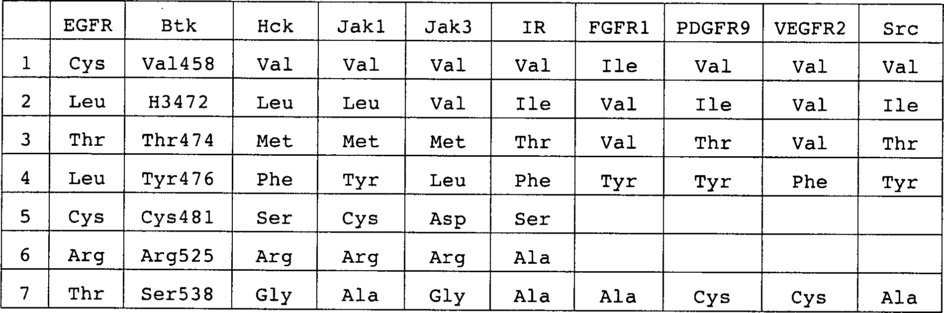
Ein
Vergleich der Inhibitorbindungstasche von BTK mit der von EGFR,
Hck, Jak1, Jak3, FGFR1, PDGFRβ,
VEGVF2 und Src zeigt, daß SER
538 in der ATP-Bindungsspalte von BTK einzigartig ist (Tabelle 3)
und ein Ziel für
ein spezifisches BTK-Inhibitorenkonzept
darstellt. Liganden, die so konzipiert sind, daß mit diesem Rest wechselwirken,
sollten eine erhöhte
Spezifität
für BTK
haben.
Beispiel 5: Zweite Generation an LFM-AnalogaExample 5: Second generation on LFM analogs
Das folgende Beispiel beschreibt wie neue Verbindungen der Formel (I), die so konzipiert sind, daß sie auf SER 538 abzielen, geplant und hergestellt werden.The The following example describes how new compounds of the formula (I) that are designed to be on SER 538 aimed, planned and manufactured.
Unter
Verwendung der in den 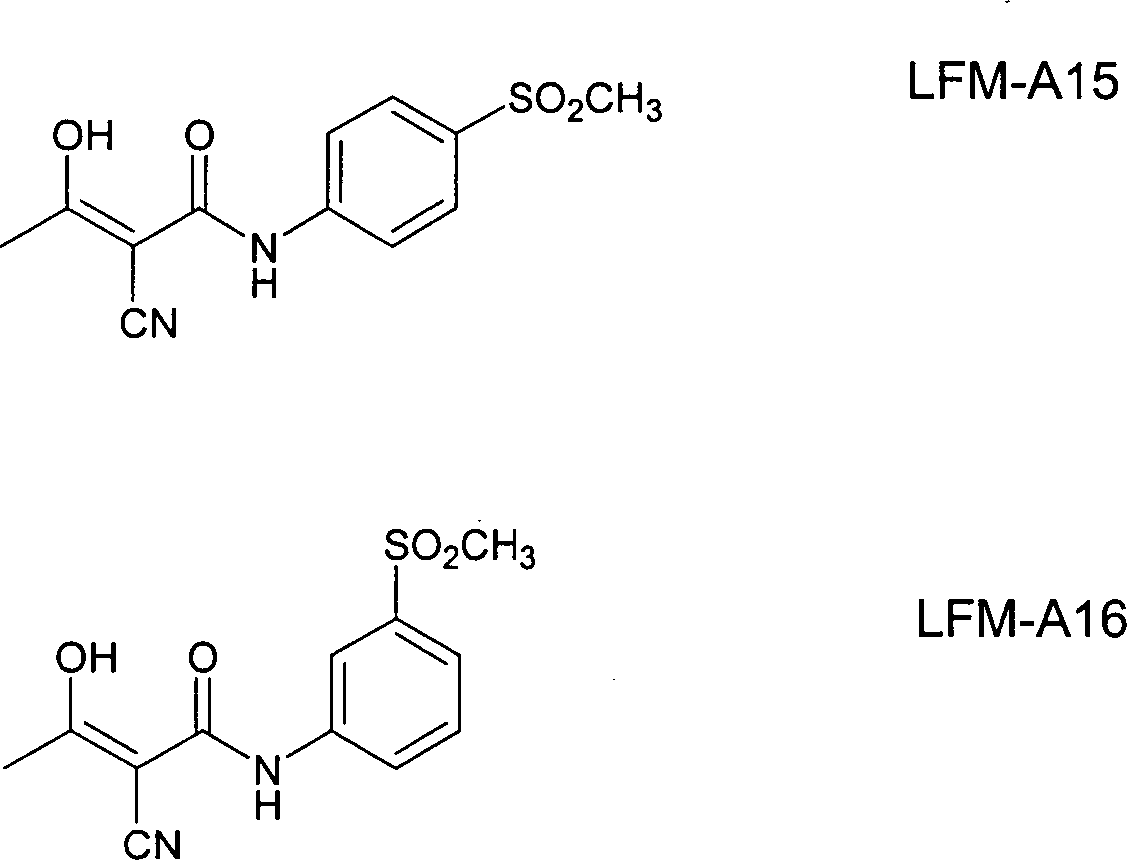
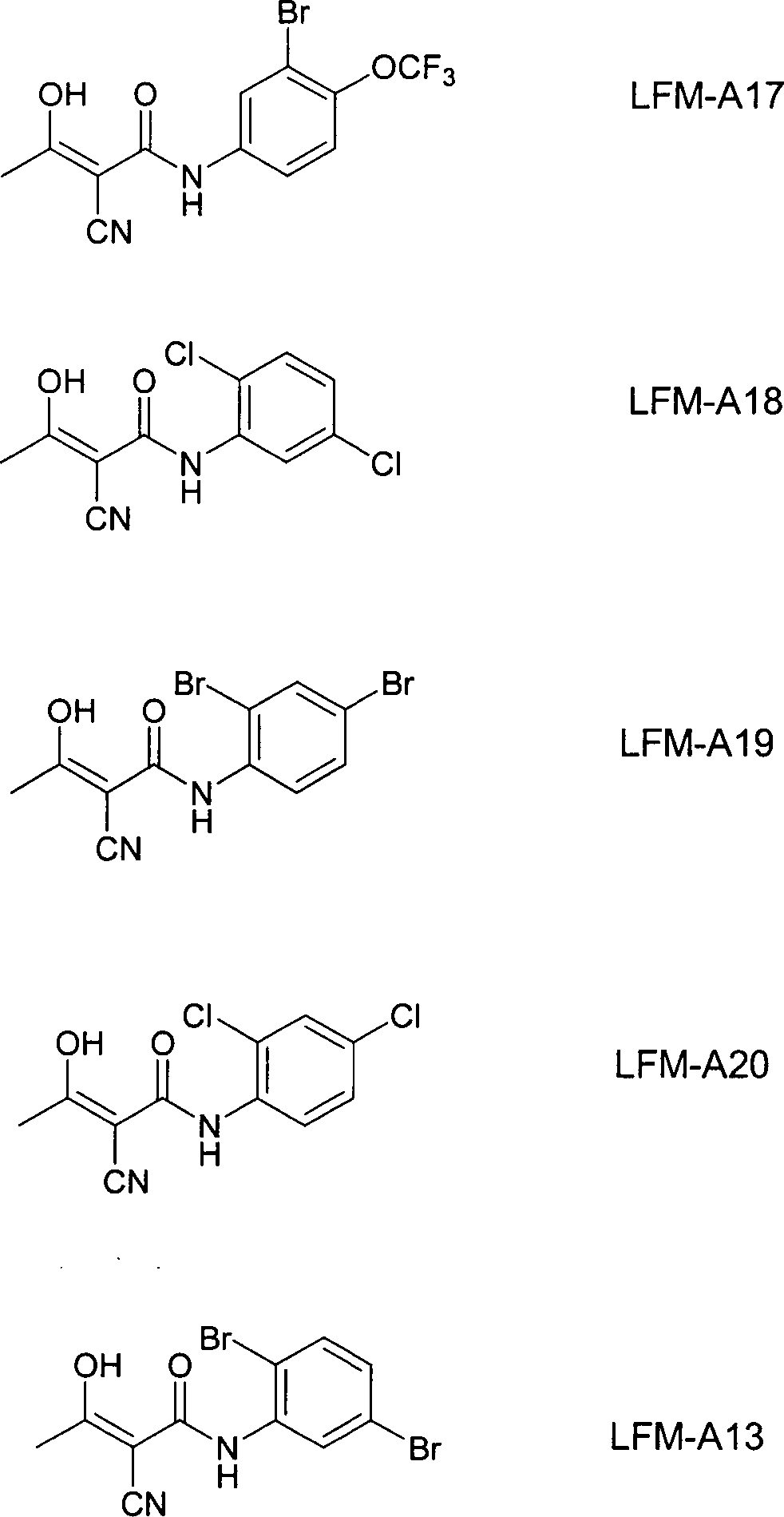
Das
folgende Syntheseschema und das in 
Syntheseverfahren Asynthesis method A
1,3-Diisopropylcarbodiimid (1,75 g; 13,9 mmol) wurde zu einer Lösung von Cyanessigsäure 1 (1,70 g; 20,0 mmol) und des gewünschten substituierten Anilin 2 (12,6 mmol) in Tetrahydrofuran (25 ml) bei 0°C gegeben. Das Gemisch wurde für 12 Stunden bei Raumtemperatur gerührt. Das Harnstoffpräzipitat (Reaktionsnebenprodukt) wurde durch Filtration entfernt und zwischen Ethylacetat und 0,5 N HCl verteilt. Die organische Schicht wurde nacheinander zweimal mit Salzlösung gewaschen, über wasserfreiem Na2SO4 getrocknet und durch Rotationsverdampfung konzentriert. Schließlich wurde das feste Rohprodukt aus Ethylalkohol umkristallisiert, wobei reine 3 erhalten wurde. Natriumhydrid (0,93 g; 60%ige in Mineralöl; 23,2 mmol) wurde langsam zu der Lösung von 3 (12,0 mmol) in Tetrahydrofuran (40 ml) bei 0°C gegeben. Nach Rühren für 30 Minuten bei 0°C wurde Acetylchlorid (1,04 g; 13,2 mmol) zu dem Reaktionsgemisch gegeben. Die Reaktion wurde für eine weitere Stunde fortgesetzt und wurde dann durch Zusatz von Essigsäure (2 ml) abgeschreckt. Das Gemisch wurde in Eiswasser (100 ml), das 2,5 ml Salzsäure enthielt, gegossen, um das Rohprodukt auszufällen, welches dann durch Filtration gesammelt wurde und mit Wasser gewaschen wurde. Das reine Produkt wurde durch Umkristallisation erhalten.1,3-Diisopropylcarbodiimide (1.75 g, 13.9 mmol) was added to a solution of cyanoacetic acid 1 (1.70 g, 20.0 mmol) and the desired substituted aniline 2 (12.6 mmol) in tetrahydrofuran (25 ml) at 0 ° C. The mixture was stirred for 12 hours at room temperature. The urea precipitate (reaction by-product) was removed by filtration and partitioned between ethyl acetate and 0.5 N HCl. The organic layer was washed successively twice with brine, dried over anhydrous Na 2 SO 4 , and concentrated by rotary evaporation. Finally, the solid crude product was recrystallized from ethyl alcohol to give pure 3. Sodium hydride (0.93 g, 60% in mineral oil, 23.2 mmol) was added slowly to the solution of 3 (12.0 mmol) in tetrahydrofuran (40 mL) at 0 ° C. After stirring for 30 minutes at 0 ° C, acetyl chloride (1.04 g, 13.2 mmol) was added to the reaction mixture. The reaction was for a continued for an additional hour and then quenched by the addition of acetic acid (2 ml). The mixture was poured into ice-water (100 ml) containing 2.5 ml of hydrochloric acid to precipitate the crude product, which was then collected by filtration and washed with water. The pure product was obtained by recrystallization.
Syntheseverfahren Bsynthesis method B
α-Cyano-β-hydroxy-β-methyl-N-[(methylthio)phenyl]propenamid (2,48 g, 10,0 mmol) wurde in Essigsäure (150 ml) gelöst und Peressigsäure (8,6 ml einer 32 Gew.%igen Lösung in Essigsäure) wurde zugesetzt. Das Gemisch wurde über Nacht bei Raumtemperatur gerührt und Wasser (75 ml) wurde zugesetzt. Das Präzipitat wurde filtriert und mit Wasser gewaschen. Das reine Produkt wurde durch Umkristallisieren erhalten.α-cyano-β-hydroxy-β-methyl-N - [(methylthio) phenyl] propenamide (2.48 g, 10.0 mmol) was dissolved in acetic acid (150 ml) and peracetic acid (8.6 ml of a 32% by weight solution in acetic acid) was added. The mixture became overnight at room temperature touched and water (75 ml) was added. The precipitate was filtered and washed with water. The pure product was recrystallized receive.
Physikalischen Daten für die synthetisierten Verbindungen:physical Data for the synthesized compounds:
α-Cyano-β-hydroxy-β-methyl-N-[4-(methylsulfonyl)phenyl]propenamidα-cyano-β-hydroxy-β-methyl-N- [4- (methylsulfonyl) phenyl] propenamide
- Schmelzpunkt: 205-206°C.Melting point: 205-206 ° C.
- 1H-NMR (DMSO-d6) δ: 11,26 (s, 1H, NH), 7,81 (d, J=9,0Hz, 2H, ArH), 7,76 (d, 9,0Hz, 2H, ArH), 3,15 (s, 3H, SO2CH3). 1 H-NMR (DMSO-d 6 ) δ: 11.26 (s, 1H, NH), 7.81 (d, J = 9.0Hz, 2H, ArH), 7.76 (d, 9.0Hz, 2H, ArH), 3.15 (s, 3H, SO 2 CH 3 ).
- IR (KBr): 3309, 2225, 1643 und 1586 cm–1.IR (KBr): 3309, 2225, 1643 and 1586 cm -1 .
- sMS (EI): m/z 281,0 (M+H+), 172,1.sMS (EI): m / z 281.0 (M + H + ), 172.1.
α-Cyano-β-hydroxy-β-methyl-N-[3-(methylsulfonyl)phenyl]propenamidα-cyano-β-hydroxy-β-methyl-N- [3- (methylsulfonyl) phenyl] propenamide
- Schmelzpunkt: 213-21406°C.Melting point: 213-21406 ° C.
- 1H-NMR (DMSO-d6) δ: 10,98 (s, 1H, NH), 8,18 (m, 1H, ArH), 7,82 (m, 1H, ArH), 7,60 (m, 2H, ArH), 3,18 (s, 3H, SO2CH3). 1 H-NMR (DMSO-d 6) δ: 10.98 (s 1H, NH), 8.18 (m, 1H, ArH), 7.82 (m, 1H, ArH), 7.60 (m , 2H, ArH), 3.18 (s, 3H, SO 2 CH 3 ).
- IR (KBr): 3278, 2231, 1607 und 1555 cm–1.IR (KBr): 3278, 2231, 1607 and 1555 cm -1 .
- MS (EI): m/z 281,0 (M+H+), 172,1.MS (EI): m / z 281.0 (M + H + ), 172.1.
α-Cyano-β-hydroxy-β-methyl-N-[3-brom-4-(trifluormethoxy)phenyl]propenamidα-cyano-β-hydroxy-β-methyl-N- [3-bromo-4- (trifluoromethoxy) phenyl] propenamide
- Schmelzpunkt: 178-179°C.Melting point: 178-179 ° C.
- 1H-NMR (DMSO-d6) δ: 11,03 (s, 1H, NH), 8,12 (d, J+2,4Hz, 1H, ArH), 7,55 (dd, J=2,4, 9,0Hz, 1H, ArH), 7,45 (m, 1H, ArH), 5,53 (s br, 1H, OH), 2,20 (s, 3H, CH3). 1 H-NMR (DMSO-d 6 ) δ: 11.03 (s, 1H, NH), 8.12 (d, J + 2.4Hz, 1H, ArH), 7.55 (dd, J = 2, 4, 9.0Hz, 1H, ArH), 7.45 (m, 1H, ArH), 5.53 (s br, 1H, OH), 2.20 (s, 3H, CH 3 ).
- IR (KBr): 3304, 2350, 1620 und 1602 cm–1.IR (KBr): 3304, 2350, 1620 and 1602 cm -1 .
- MS (EI): m/z 365,0 (M+H+), 225,9.MS (EI): m / z 365.0 (M + H + ), 225.9.
α-Cyano-β-hydroxy-β-methyl-N-(2,4-dibromphenyl)propenamidα-cyano-β-hydroxy-β-methyl-N- (2,4-dibromophenyl) propenamide
- Schmelzpunkt: 181-182°C.Melting point: 181-182 ° C.
- 1H-NMR (DMSO-d6) δ: 11,03 (s, 1H, NH), 8,14 (m, 1H, ArH), 7,84 (d, J=2,4Hz, 1H, ArH), 7,51 (dd, J=2,4, 9,0Hz, 1H, ArH), 5,65 (s br, 1H, OH), 2,20 (s, 3H, CH3). 1 H-NMR (DMSO-d 6 ) δ: 11.03 (s, 1H, NH), 8.14 (m, 1H, ArH), 7.84 (d, J = 2.4Hz, 1H, ArH) , 7.51 (dd, J = 2.4, 9.0Hz, 1H, ArH), 5.65 (s br, 1H, OH), 2.20 (s, 3H, CH 3 ).
- IR (KBr): 3360, 2205, 1591 und 1530 cm–1.IR (KBr): 3360, 2205, 1591 and 1530 cm -1 .
- MS (EI): m/z 361,0 (M+H+), 249,9.MS (EI): m / z 361.0 (M + H + ), 249.9.
α-Cyano-β-hydroxy-β-methyl-N-(2,4-Dichlorphenyl)propenamidα-cyano-β-hydroxy-β-methyl-N- (2,4-dichlorophenyl) propenamide
- Schmelzpunkt: 143-144°C.Melting point: 143-144 ° C.
- 1H-NMR (DMSO-d6) δ: 11,23 (s, 1H, NH), 8,29 (m, 1H, ArH), 7,6 (d, J=2,4Hz, 1H, ArH), 7,35 (dd, J=2,4, 9,0Hz, 1H, ArH), 5,17 (s br, 1H, OH), 2,18 (s, 3H, CH3). 1 H-NMR (DMSO-d 6 ) δ: 11.23 (s, 1H, NH), 8.29 (m, 1H, ArH), 7.6 (d, J = 2.4Hz, 1H, ArH) , 7.35 (dd, J = 2.4, 9.0Hz, 1H, ArH), 5.17 (s br, 1H, OH), 2.18 (s, 3H, CH 3 ).
- IR (KBr): 3371, 2210, 1601 und 1540 cm–1.IR (KBr): 3371, 2210, 1601 and 1540 cm -1 .
- MS (EI): m/z 271,0 (M+H+), 162,0.MS (EI): m / z 271.0 (M + H + ), 162.0.
α-Cyano-β-hydroxy-β-methyl-N-(2,5-dichlorphenyl)propenamidα-cyano-β-hydroxy-β-methyl-N- (2,5-dichlorophenyl) propenamide
- Schmelzpunkt: 143-144°C.Melting point: 143-144 ° C.
- 1H-NMR (DMSO-d6) δ: 11,70 (s, 1H, NH), 8,51 (d, J=3,0Hz, 1H, ArH), 7,45 (d, J=8,7Hz, 1H, ArH), 7,05 (dd, J=3,0, 8,7Hz, 1H, ArH), 4,97 (s br, 1H, OH), 2,14 (s, 3H, CH3). 1 H-NMR (DMSO-d 6 ) δ: 11.70 (s, 1H, NH), 8.51 (d, J = 3.0Hz, 1H, ArH), 7.45 (d, J = 8, 7Hz, 1H, ArH), 7.05 (dd, J = 3.0, 8.7Hz, 1H, ArH), 4.97 (s br, 1H, OH), 2.14 (s, 3H, CH 3 ).
- IR (KBr): 3402, 2205, 1627 und 1606 cm–1.IR (KBr): 3402, 2205, 1627 and 1606 cm -1 .
- MS (EI): m/z 271 (M+H+), 162,0.MS (EI): m / z 271 (M + H + ), 162.0.
Tabelle
4. Wechselwirkungs-Punktbewertungen, geschätzte Ki-Werte und gemessene
IC50-Daten für LFM-Analoga mit BTK 
Die zweite Generation der LFM-Analoga wurde bezüglich der Wechselwirkung mit dem BTK-Bindungstaschenmodell beurteilt und auch bezüglich der Inhibitoraktivität gegenüber BTK beurteilt, wie es im Beispiel oben beschrieben ist. Die Daten sind in Tabelle 4 angegeben und beweisen, daß diese neuen Verbindungen starke Inhibitoren für BTK sind.The second generation of LFM analogs has been reported to interact with assessed the BTK binding pocket model and also with respect to the inhibitory activity across from BTK judges, as described in the example above. The data are given in Table 4 and prove that these new compounds strong inhibitors for BTK are.
Beispiel 7: Neue BTK-Inhibitor-KonzepteExample 7: New BTK inhibitor concepts
Von diesen Verbindungen wird infolge ihres erhöhten Volumens und der Gruppen, die dazu bestimmt sind, mit dem Bindungstaschenrest in Wechselwirkung zu treten, erwartet, daß sie eine starke BTK-Inhibitoraktivität haben.From these compounds, due to their increased volume and groups, which are intended to interact with the binding pocket residue It is expected that they will a strong BTK inhibitor activity to have.

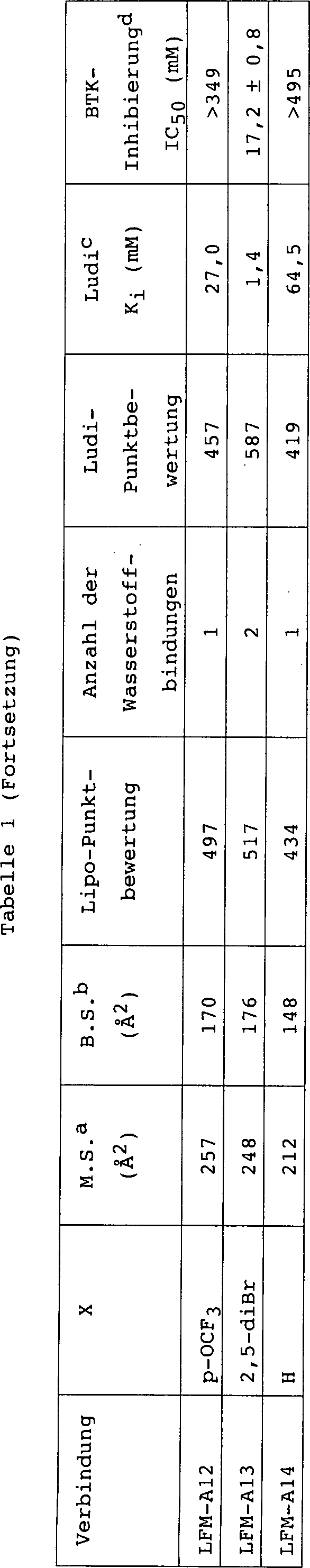
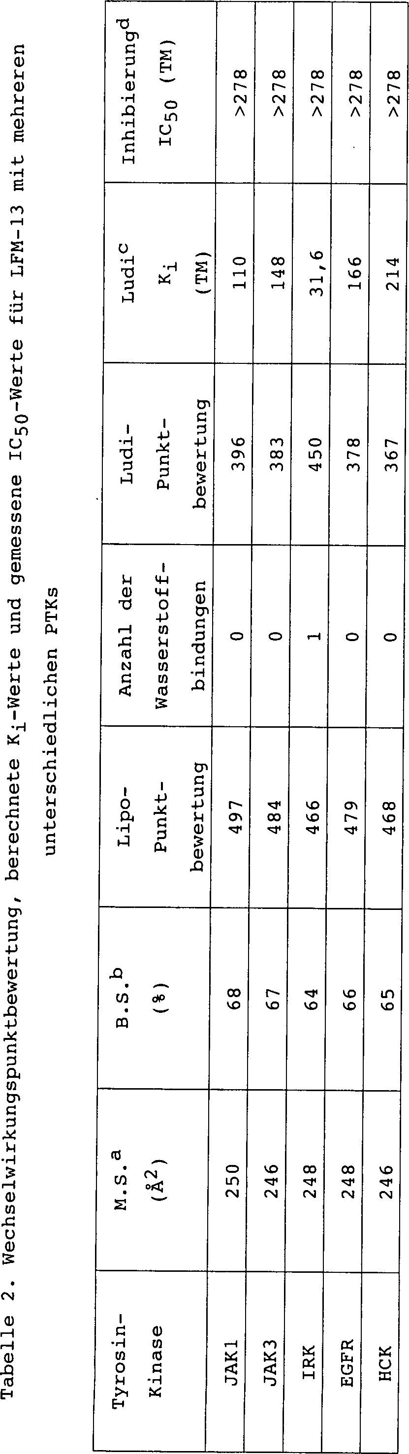
Claims (8)
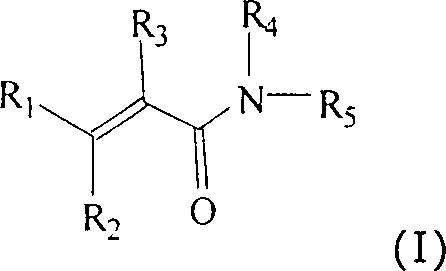
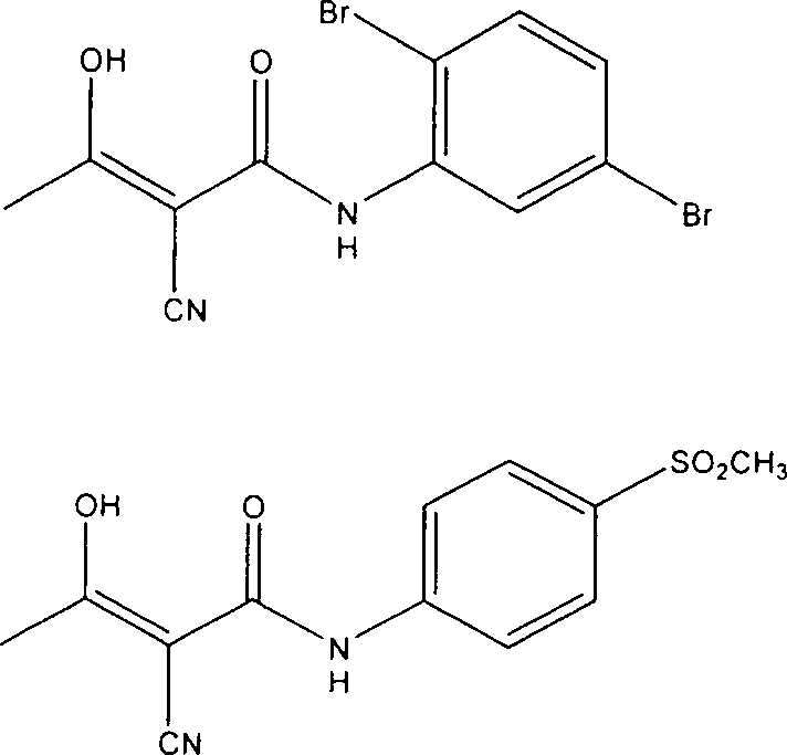
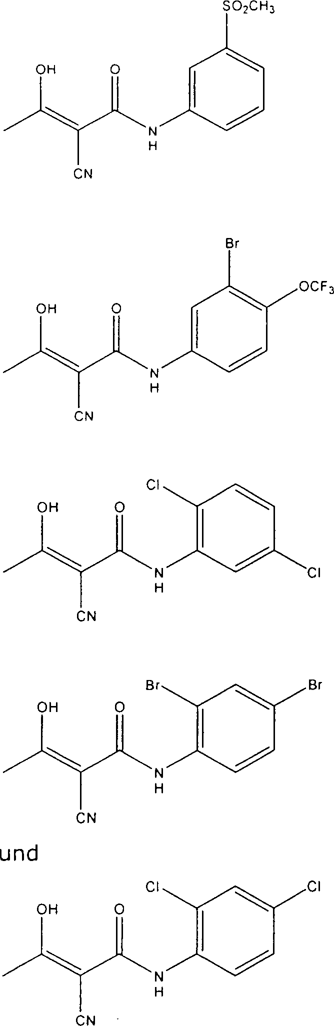
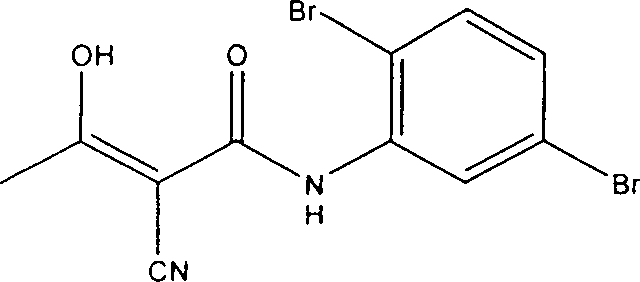
Applications Claiming Priority (5)
| Application Number | Priority Date | Filing Date | Title |
|---|---|---|---|
| US8209498P | 1998-04-17 | 1998-04-17 | |
| US82094P | 1998-04-17 | ||
| US09/273,191 US6303652B1 (en) | 1998-08-21 | 1999-03-19 | BTK inhibitors and methods for their identification and use |
| US273191 | 1999-03-19 | ||
| PCT/US1999/008556 WO1999054286A2 (en) | 1998-04-17 | 1999-04-19 | Btk inhibitors and methods for their identification and use |
Publications (2)
| Publication Number | Publication Date |
|---|---|
| DE69918089D1 DE69918089D1 (en) | 2004-07-22 |
| DE69918089T2 true DE69918089T2 (en) | 2005-07-14 |
Family
ID=26767041
Family Applications (1)
| Application Number | Title | Priority Date | Filing Date |
|---|---|---|---|
| DE69918089T Expired - Fee Related DE69918089T2 (en) | 1998-04-17 | 1999-04-19 | BTK INHIBITORS AND METHODS OF IDENTIFICATION AND USE |
Country Status (13)
| Country | Link |
|---|---|
| EP (1) | EP1071658B1 (en) |
| JP (1) | JP2002512216A (en) |
| KR (1) | KR20010042804A (en) |
| AT (1) | ATE269295T1 (en) |
| AU (1) | AU3653099A (en) |
| CA (1) | CA2328962A1 (en) |
| DE (1) | DE69918089T2 (en) |
| ES (1) | ES2222705T3 (en) |
| HU (1) | HUP0102661A2 (en) |
| IL (1) | IL139080A0 (en) |
| MX (1) | MXPA00010150A (en) |
| NO (1) | NO20005224L (en) |
| WO (1) | WO1999054286A2 (en) |
Families Citing this family (56)
| Publication number | Priority date | Publication date | Assignee | Title |
|---|---|---|---|---|
| US6303652B1 (en) | 1998-08-21 | 2001-10-16 | Hughes Institute | BTK inhibitors and methods for their identification and use |
| US6306897B1 (en) | 1999-03-19 | 2001-10-23 | Parker Hughes Institute | Calanolides for inhibiting BTK |
| SI1230232T1 (en) | 1999-11-05 | 2004-08-31 | Cytovia, Inc. | Substituted 4h-chromene and analogs as activators of caspases and inducers of apoptosis and the use thereof |
| CA2390857A1 (en) * | 1999-11-30 | 2001-06-14 | Fatih M. Uckun | Inhibitors of collagen-induced platelet aggregation |
| US6589992B2 (en) | 1999-11-30 | 2003-07-08 | Parker Hughes Institute | Inhibiting collagen-induced platelet aggregation |
| GB0005345D0 (en) * | 2000-03-06 | 2000-04-26 | Mathilda & Terence Kennedy Ins | Methods of treating sepsis septic shock and inflammation |
| AU2001236361A1 (en) * | 2000-10-20 | 2002-05-06 | Parker Hughes Institute | Treatment of asthma with lfm analogues |
| CA2426508A1 (en) * | 2000-10-23 | 2002-05-16 | Bristol-Myers Squibb Company | Modulators of bruton's tyrosine kinase and bruton's tyrosine kinase intermediates and methods for their identification and use in the treatment and prevention of osteoporosis and related disease states |
| JP2004528311A (en) * | 2001-03-07 | 2004-09-16 | テリック,インコーポレイテッド | Substituted diaryl ureas as stimulators of Fas-mediated apoptosis |
| DE10112924A1 (en) * | 2001-03-13 | 2002-10-02 | Erich Eigenbrodt | 1-butanoic acid derivatives, pharmaceutical compositions containing such derivatives and uses of such derivatives |
| WO2002072527A2 (en) * | 2001-03-13 | 2002-09-19 | Protagen Aktiengesellschaft | 1-butyric acid derivatives such as carbomethoxypropionyl cyanide or leflunomide derivatives, and the therapeutic use thereof |
| EP1392683B1 (en) | 2001-05-16 | 2009-12-02 | Cytovia, Inc. | Substituted 4h-chromenes and analogs as activators of caspases and inducers of apoptosis and their use as anticancer agents |
| US6858607B1 (en) | 2001-05-16 | 2005-02-22 | Cytovia, Inc. | 7,8-fused 4H-chromene and analogs as activators of caspases and inducers of apoptosis and the use thereof |
| EP1392283A4 (en) | 2001-05-16 | 2004-10-20 | Cytovia Inc | COUMARINS AND SUBSTITUTED QUINOLINES USED AS CASPASE ACTIVATORS |
| AU2002329000A1 (en) * | 2001-09-24 | 2003-04-07 | University Of Aarhus | Methods for diagnosis and treatment of diseases associated with altered expression of pik3r1 |
| JP2005531566A (en) | 2002-05-16 | 2005-10-20 | サイトビア インコーポレイティッド | Substituted 4-aryl-4H-pyrrolo [2,3-H] chromenes and analogs and their use as caspase activators and apoptosis inducers |
| EP1513515A2 (en) | 2002-05-16 | 2005-03-16 | Cytovia, Inc. | Substituted 4h-chromenes, 2h-chromenes, chromans and analogs as activators of caspases and inducers of apoptosis and the use thereof |
| JP2005538165A (en) * | 2002-09-06 | 2005-12-15 | シェボ ビオテック アクティエン ゲゼルシャフト | Compound for regulating glycolytic enzyme and / or aminotransferase complex |
| AR045037A1 (en) | 2003-07-10 | 2005-10-12 | Aventis Pharma Sa | TETRAHIDRO-1H-PIRAZOLO [3,4-C] SUBSTITUTED PYRIDINS, COMPOSITIONS THAT CONTAIN THEM AND ITS USE. |
| SG142305A1 (en) * | 2004-10-19 | 2008-05-28 | Aventis Pharma Inc | Use of (z)-2-cyano-3-hydroxy-but-2-enoic acid-(4'- trifluoromethylphenyl)-amide for treating inflammatory bowel disease |
| EP2270200A3 (en) * | 2006-09-11 | 2011-07-13 | CGI Pharmaceuticals, Inc. | Kinase inhibitors, and methods of using and identifying kinase inhibitors |
| EA018573B1 (en) | 2006-09-22 | 2013-09-30 | Фармасайкликс, Инк. | BLUTON TYROSINKINASE INHIBITORS |
| US8232085B2 (en) * | 2007-03-14 | 2012-07-31 | Bionsil S.R.L. | Isoform of bruton's tyrosine kinase (BTK) protein |
| US8809273B2 (en) | 2007-03-28 | 2014-08-19 | Pharmacyclics, Inc. | Inhibitors of Bruton's tyrosine kinase |
| EP2205564B1 (en) * | 2007-10-23 | 2014-07-30 | F. Hoffmann-La Roche AG | Novel kinase inhibitors |
| JP5479105B2 (en) * | 2007-11-05 | 2014-04-23 | 国立大学法人佐賀大学 | Novel ubiquitin-binding small molecule |
| US8389757B2 (en) | 2008-06-05 | 2013-03-05 | Alembic Ltd. | Process for preparing teriflunomide |
| CN105362277A (en) | 2008-07-16 | 2016-03-02 | 药品循环有限公司 | Inhibitors of bruton's tyrosine kinase for the treatment of solid tumors |
| US9040714B2 (en) | 2010-04-05 | 2015-05-26 | Mannkind Corporation | IRE-1α inhibitors |
| CA3007787C (en) | 2010-06-03 | 2020-03-10 | Pharmacyclics Llc | The use of inhibitors of bruton's tyrosine kinase (btk) |
| BR112014000653A2 (en) | 2011-07-13 | 2017-02-14 | Pharmacyclics Inc | bruton tyrosine kinase inhibitors |
| US8377946B1 (en) | 2011-12-30 | 2013-02-19 | Pharmacyclics, Inc. | Pyrazolo[3,4-d]pyrimidine and pyrrolo[2,3-d]pyrimidine compounds as kinase inhibitors |
| WO2013148603A1 (en) | 2012-03-27 | 2013-10-03 | Takeda Pharmaceutical Company Limited | Cinnoline derivatives as as btk inhibitors |
| EA201492082A1 (en) | 2012-06-04 | 2015-03-31 | Фармасайкликс, Инк. | CRYSTAL FORMS OF BLUTON TYROSINKINASE INHIBITOR |
| CA2879570A1 (en) | 2012-07-24 | 2014-01-30 | Pharmacyclics, Inc. | Mutations associated with resistance to inhibitors of bruton's tyrosine kinase (btk) |
| MX2015006168A (en) | 2012-11-15 | 2015-08-10 | Pharmacyclics Inc | Pyrrolopyrimidine compounds as kinase inhibitors. |
| CN105189737B (en) * | 2012-11-28 | 2019-02-12 | 康宁股份有限公司 | Cell Culture Media for Enhanced Hepatocyte Function |
| JO3377B1 (en) | 2013-03-11 | 2019-03-13 | Takeda Pharmaceuticals Co | Pyridinyl and fused pyridinyl triazolone derivatives |
| RU2634723C2 (en) * | 2013-04-02 | 2017-11-03 | Ф. Хоффманн-Ля Рош Аг | Bruton's tyrosine kinase inhibitors |
| TWI649081B (en) | 2013-08-02 | 2019-02-01 | 製藥公司 | Method for treating solid tumors |
| EP2832358A1 (en) * | 2013-08-02 | 2015-02-04 | Bionsil S.r.l. | Pharmaceutical kit for use in the treatment of colon and colorectal cancer |
| ES2709509T3 (en) | 2013-08-12 | 2019-04-16 | Pharmacyclics Llc | Procedures for the treatment of cancer amplified by HER2 |
| KR20160062103A (en) | 2013-09-30 | 2016-06-01 | 파마싸이클릭스 엘엘씨 | Inhibitors of bruton's tyrosine kinase |
| US9908872B2 (en) | 2013-12-13 | 2018-03-06 | Dana-Farber Cancer Institute, Inc. | Methods to treat lymphoplasmacytic lymphoma |
| JP6879740B2 (en) | 2013-12-13 | 2021-06-02 | ダナ−ファーバー キャンサー インスティテュート, インコーポレイテッド | How to Treat Lymphatic Plasma Cell Lymphoma |
| EP3119910A4 (en) | 2014-03-20 | 2018-02-21 | Pharmacyclics LLC | Phospholipase c gamma 2 and resistance associated mutations |
| CN105272882B (en) * | 2014-07-23 | 2019-05-31 | 欣凯医药化工中间体(上海)有限公司 | A kind of easy method for preparing Tai Rui meter Te of environmental protection |
| CN106999494A (en) | 2014-08-01 | 2017-08-01 | 药品循环有限公司 | Inhibitors of Bruton's tyrosine kinase |
| JP2017523206A (en) | 2014-08-07 | 2017-08-17 | ファーマサイクリックス エルエルシー | New formulation of breton-type tyrosine kinase inhibitor |
| JP6788583B2 (en) | 2014-10-22 | 2020-11-25 | ダナ−ファーバー キャンサー インスティテュート, インコーポレイテッド | Thiazolyl-containing compounds for treating proliferative disorders |
| IL315294A (en) | 2015-03-03 | 2024-10-01 | Pharmacyclics Llc | Pharmaceutical formulations of proton tyrosine kinase inhibitor |
| ES2886080T3 (en) | 2016-03-31 | 2021-12-16 | Takeda Pharmaceuticals Co | Isoquinolinyl triazolone complexes |
| JP2020510624A (en) | 2016-12-12 | 2020-04-09 | マルチビア インコーポレイテッド | Methods and compositions comprising viral gene therapy and immune checkpoint inhibitors for the treatment and prevention of cancer and infectious diseases |
| BR112021018168B1 (en) | 2019-03-21 | 2023-11-28 | Onxeo | PHARMACEUTICAL COMPOSITION, COMBINATION AND KIT COMPRISING A DBAIT MOLECULE AND A KINASE INHIBITOR FOR THE TREATMENT OF CANCER |
| CA3159348A1 (en) | 2019-11-08 | 2021-05-14 | Inserm (Institut National De La Sante Et De La Recherche Medicale) | Methods for the treatment of cancers that have acquired resistance to kinase inhibitors |
| WO2021148581A1 (en) | 2020-01-22 | 2021-07-29 | Onxeo | Novel dbait molecule and its use |
Family Cites Families (11)
| Publication number | Priority date | Publication date | Assignee | Title |
|---|---|---|---|---|
| DE2555685A1 (en) * | 1975-12-11 | 1977-06-23 | Hoechst Ag | Hydroxyethylidene-cyano-acetanilides - with antiinflammatory and analgesic activity |
| WO1991017748A1 (en) * | 1990-05-18 | 1991-11-28 | Hoechst Aktiengesellschaft | Isoxazole-4-carboxamides and hydroxyalkylidene-cyanoacetamides, drugs containing these compounds and use of such drugs |
| CA2080554A1 (en) * | 1991-10-15 | 1993-04-16 | Mitsubishi Chemical Corporation | Styrene derivatives |
| GB9200275D0 (en) * | 1992-01-08 | 1992-02-26 | Roussel Lab Ltd | Chemical compounds |
| WO1994014789A1 (en) * | 1992-12-23 | 1994-07-07 | Smithkline Beecham Corporation | Coumarin derivatives as retroviral inhibitors |
| GB9322781D0 (en) * | 1993-11-04 | 1993-12-22 | Roussel Lab Ltd | Aromatic amides |
| US5700823A (en) * | 1994-01-07 | 1997-12-23 | Sugen, Inc. | Treatment of platelet derived growth factor related disorders such as cancers |
| US5489697A (en) * | 1994-08-03 | 1996-02-06 | Medichem Research, Inc. | Method for the preparation of (+)-calanolide A and intermediates thereof |
| US5608085A (en) * | 1995-02-27 | 1997-03-04 | The University Of Tennessee Research Corporation | Synthesis of optically active calanolides A and B and enantiomers and related compounds |
| WO1996031206A2 (en) * | 1995-04-07 | 1996-10-10 | Warner-Lambert Company | Flavones and coumarins as agents for the treatment of atherosclerosis |
| IT1289238B1 (en) * | 1996-12-10 | 1998-09-29 | Bio S S P A Ora Bio S S R L | PHARMACEUTICAL COMPOSITIONS FOR THE TREATMENT OF VIRAL INFECTIONS INCLUDING A 4 ARILCUMARIN |
-
1999
- 1999-04-19 CA CA002328962A patent/CA2328962A1/en not_active Abandoned
- 1999-04-19 AU AU36530/99A patent/AU3653099A/en not_active Abandoned
- 1999-04-19 EP EP99918673A patent/EP1071658B1/en not_active Expired - Lifetime
- 1999-04-19 WO PCT/US1999/008556 patent/WO1999054286A2/en not_active Ceased
- 1999-04-19 DE DE69918089T patent/DE69918089T2/en not_active Expired - Fee Related
- 1999-04-19 ES ES99918673T patent/ES2222705T3/en not_active Expired - Lifetime
- 1999-04-19 MX MXPA00010150A patent/MXPA00010150A/en unknown
- 1999-04-19 JP JP2000544627A patent/JP2002512216A/en active Pending
- 1999-04-19 KR KR1020007011551A patent/KR20010042804A/en not_active Ceased
- 1999-04-19 AT AT99918673T patent/ATE269295T1/en not_active IP Right Cessation
- 1999-04-19 HU HU0102661A patent/HUP0102661A2/en unknown
- 1999-04-19 IL IL13908099A patent/IL139080A0/en unknown
-
2000
- 2000-10-17 NO NO20005224A patent/NO20005224L/en not_active Application Discontinuation
Also Published As
| Publication number | Publication date |
|---|---|
| WO1999054286A3 (en) | 2000-05-04 |
| KR20010042804A (en) | 2001-05-25 |
| NO20005224L (en) | 2000-12-18 |
| HUP0102661A2 (en) | 2001-11-28 |
| DE69918089D1 (en) | 2004-07-22 |
| ATE269295T1 (en) | 2004-07-15 |
| NO20005224D0 (en) | 2000-10-17 |
| AU3653099A (en) | 1999-11-08 |
| IL139080A0 (en) | 2001-11-25 |
| MXPA00010150A (en) | 2002-05-14 |
| ES2222705T3 (en) | 2005-02-01 |
| CA2328962A1 (en) | 1999-10-28 |
| WO1999054286A2 (en) | 1999-10-28 |
| JP2002512216A (en) | 2002-04-23 |
| EP1071658A2 (en) | 2001-01-31 |
| EP1071658B1 (en) | 2004-06-16 |
Similar Documents
| Publication | Publication Date | Title |
|---|---|---|
| DE69918089T2 (en) | BTK INHIBITORS AND METHODS OF IDENTIFICATION AND USE | |
| US6294575B1 (en) | BTK inhibitors and methods for their identification and use | |
| JP7530390B2 (en) | Novel EGFR inhibitor | |
| DE69924500T2 (en) | quinazoline derivatives | |
| US6313130B1 (en) | JAK-3 inhibitors for treating allergic disorders | |
| EP1163243B1 (en) | Calanolides for inhibiting btk | |
| US10954198B2 (en) | Benzylideneguanidine derivatives and therapeutic use for the treatment of protein misfolding diseases | |
| US6355678B1 (en) | Inhibitors of the EGF-receptor tyrosine kinase and methods for their use | |
| KR20180069732A (en) | Pharmaceutical composition for prevention and treatment of DYRK related diseases comprising phenanthrene-lactam compounds | |
| US9975853B2 (en) | Tetrahydroisoquinolinone derivatives and their use in the inhibition of the HSP70 protein |
Legal Events
| Date | Code | Title | Description |
|---|---|---|---|
| 8364 | No opposition during term of opposition | ||
| 8339 | Ceased/non-payment of the annual fee |