CN85100552B - Method for receiving x-ray raster sample pattern using x-ray - Google Patents

Method for receiving x-ray raster sample pattern using x-ray Download PDF

Info

Publication number
CN85100552B
CN85100552B CN85100552A CN85100552A CN85100552B CN 85100552 B CN85100552 B CN 85100552B CN 85100552 A CN85100552 A CN 85100552A CN 85100552 A CN85100552 A CN 85100552A CN 85100552 B CN85100552 B CN 85100552B
Authority
CN
China
Prior art keywords
ray
machine
raster
image
grating
Prior art date
Legal status (The legal status is an assumption and is not a legal conclusion. Google has not performed a legal analysis and makes no representation as to the accuracy of the status listed.)
Expired
Application number
CN85100552A
Other languages
Chinese (zh)
Other versions
CN85100552A (en
Inventor
诸葛向彬
康善存
陈洪山
Current Assignee (The listed assignees may be inaccurate. Google has not performed a legal analysis and makes no representation or warranty as to the accuracy of the list.)
Zhejiang University ZJU
Original Assignee
Zhejiang University ZJU
Priority date (The priority date is an assumption and is not a legal conclusion. Google has not performed a legal analysis and makes no representation as to the accuracy of the date listed.)
Filing date
Publication date
Application filed by Zhejiang University ZJU filed Critical Zhejiang University ZJU
Priority to CN85100552A priority Critical patent/CN85100552B/en
Publication of CN85100552A publication Critical patent/CN85100552A/en
Publication of CN85100552B publication Critical patent/CN85100552B/en
Expired legal-status Critical Current

Links

Images

Landscapes

  • Image Processing (AREA)
  • Analysing Materials By The Use Of Radiation (AREA)
  • Image Analysis (AREA)

Abstract

抽样图的方法一种利用X光机直接获取X光光栅抽样图的方法。其特征是利用摄影机(X光机,X光荧光缩影机等),在原底片[2]的药膜面上安装一块紧贴的Ronchi光栅[1],直接对X光进行光栅抽样。Ronchi光栅的位置固定不变。拍片时只需曝光一次。当你得到一张底片时,人体内肉眼看不到的器官图象信息也就编码完毕。可减少一道工序。使图象的光栅抽样支术变得十分简单,省时,省力,省钱。

The sampling image method is a method of directly obtaining the X-ray raster sampling image by using an X-ray machine. It is characterized in that a camera (X-ray machine, X-ray fluorescence miniature machine, etc.) is used to install a close-fitting Ronchi grating [1] on the film surface of the original negative film [2] to directly sample the X-ray grating. The position of the Ronchi grating is fixed. Only one exposure is required for filming. When you get a negative, the image information of the invisible organs in the human body is also encoded. One process can be reduced. The image raster sampling technique becomes very simple, saving time, labor and money.

Description

A kind of method of utilizing the direct common bluebeard of X-ray machine to get X-ray raster sampling label
The invention belongs to the white light information treatment technology of image.
General optical image pseudo-color coding technique, all must give a soot-and-whitewash is arranged earlier, make an egative film (negative film) with this soot-and-whitewash through imaging method or after directly duplicating method carries out pseudocolor encoding, exposure, flushing then, use this egative film (negative film) to be put into then and carry out filtering Jie sign indicating number acquisition color image on the optical information processing system.The shortcoming of this treatment technology is can not directly be put into pending soot-and-whitewash to carry out filtering Jie sign indicating number on the optical information processing system, obtains color image, needs to increase the operation of producing an egative film of encoding together.The present invention has done improved at above-mentioned shortcoming exactly.
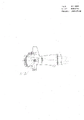
Accompanying drawing is to be that the X-ray machine that utilizes of example directly obtains the synoptic diagram of raster sampling drawing method with the x-ray fluorescence machine microcosm.(1) Ronchi grating for increasing newly among the figure, (2) are egative film.
Be furnished with a HD-70mm automatic photograph machine on the homemade X-ray machine, when taking pictures, the X-ray that sees through human body is mapped on the fluorescent screen, and a branch of fluorescent (visible light) that sends is divided into two bundles by the prism of a plated film.One beam intensity is that 10% fluorescent (visible light) enters TV camera, the dynamic change of Direct observation patient intracorporeal organ on the gamma camera screen, one beam intensity is that 90% fluorescent (visible light) is injected the automatic photograph machine and taken pictures (automatic photograph machine optical system diagram slightly), and its egative film specification is 70 * 70mm 2Common fluorescent panchromatic film.Time shutter is divided into five grades: 1/second, 2/second, 3/second, 4/second, 5/second, can clap one separately, and also can take continuously.
X-ray fluorescence machine microcosm also is that the X-ray that sees through human body is squeezed into fluorescent screen, sends to project the exposure of fluorescent epitome egative film again behind the fluorescent (visible light) and take (as Figure of description).Chang Yong fluorescent spot film specification is 100 * 100mm clinically 2, also can be a little bit smaller.
Main points of the present invention are to utilize former video camera (X-ray machine, x-ray fluorescence machine microcosm etc.), in the automatic photograph machine of X-ray machine on the position of dress fluorescent panchromatic plate (egative film exposure position).Or adorn in the x-ray fluorescence machine microcosm on the position of fluorescent epitome egative film, a corresponding Ronchi grating of size is installed, the medicine face of grating and the medicine face of egative film are close together.The position of Ronchi grating can immobilize.In X-ray machine when film making, can only need exposure once, at this moment the organ picture information that is invisible to the naked eye of inside of human body also just coding finish, the egative film that develops by the darkroom is an image through raster sampling afterwards.This egative film can directly be put into and carry out filtering Jie sign indicating number on the optical information processing system, just can obtain false color image.
The invention has the advantages that takes pictures and encode carries out simultaneously, finishes simultaneously.When you obtained a photographic negative, the picture information in the egative film had encoded.Do not need pending image handled again and be made into other egative film, reduce by a procedure significantly, make the raster sampling technology of image become very simple, save time, laborsaving, economical, make this technology more be imbued with practicality, Practical significance is bigger on clinical medicine.

Claims (3)

1, a kind of raster sampling method is characterized in that utilizing video camera (X-ray machine, x-ray fluorescence machine microcosm etc.), and a Ronchi grating (1) of being close to is installed on the medicine face of original negative (2), directly X-ray is carried out raster sampling.
2, according to the described a kind of raster sampling method of claim 1, the position that it is characterized in that the Ronchi grating is changeless.
3,, with the same usually, only need exposure once when it is characterized in that video camera (X-ray machine, x-ray fluorescence machine microcosm etc.) film making according to claim 1 and 2 described a kind of raster sampling methods.
CN85100552A 1985-04-01 1985-04-01 Method for receiving x-ray raster sample pattern using x-ray Expired CN85100552B (en)

Priority Applications (1)

Application Number Priority Date Filing Date Title
CN85100552A CN85100552B (en) 1985-04-01 1985-04-01 Method for receiving x-ray raster sample pattern using x-ray

Applications Claiming Priority (1)

Application Number Priority Date Filing Date Title
CN85100552A CN85100552B (en) 1985-04-01 1985-04-01 Method for receiving x-ray raster sample pattern using x-ray

Publications (2)

Publication Number Publication Date
CN85100552A CN85100552A (en) 1986-03-10
CN85100552B true CN85100552B (en) 1988-02-10

Family

ID=4791254

Family Applications (1)

Application Number Title Priority Date Filing Date
CN85100552A Expired CN85100552B (en) 1985-04-01 1985-04-01 Method for receiving x-ray raster sample pattern using x-ray

Country Status (1)

Country Link
CN (1) CN85100552B (en)

Also Published As

Publication number Publication date
CN85100552A (en) 1986-03-10

Similar Documents

Publication Publication Date Title
Tuddenham Visual search, image organization, and reader error in roentgen diagnosis: studies of the psychophysiology of roentgen image perception Memorial Fund lecture
Gustafson et al. Microaquaria for time-lapse cinematographic studies of morphogenesis in swimming larvae and observations on sea urchin gastrulation
Moore et al. Ultra high speed photography in laryngeal physiology
ATE234043T1 (en) ULTRASONIC IMAGING DEVICE AND METHOD FOR BREAST EXAMINATION
RU2095762C1 (en) Method for recording and displaying of three- dimensional picture of object and device for recording and displaying of three-dimensional picture of object
CN85100552B (en) Method for receiving x-ray raster sample pattern using x-ray
JPS6164232A (en) Apparatus for detecting and classifying characteristics of skin surface shape
US3428393A (en) Optical dissector
CN214906773U (en) Oral cavity scanner
JPS62180241A (en) Tomographic image observing device
CN110547752A (en) Endoscope system, mixed light source, video acquisition device and image processor
EP0378542A4 (en) Boundary setting devices and method
TW493070B (en) Fluorescent particle imaging device
JPS53122489A (en) Cancer diagnosis apparatus by fluorescent polarization photometric microscope
CN206470192U (en) A kind of arrowband ultraviolet induced fluorescence imaging device
Woolley A method for determining the three‐dimensional form of active flagella, using two‐colour darkground illumination
Hester et al. Deep narrow band imagery of the diffuse ISM in M33
Forman et al. Video intensification microscopy (VIM) as an aid in routine fluorescence microscopy
Höurzeler Blue light endoscopy
SU1309961A1 (en) Method of radiodiagnosis of pathologic changes
GB1373715A (en) Optical systems
CN208421509U (en) A kind of 3D imaging system based on endogenous optical signal
CN1057910A (en) Scanning multispectral microscopic imaging method and device thereof
Engel II. PHOTOGRAPHY IN MEDICINE
Benatar et al. Three-dimensional magnetic resonance image representation using reflection holography

Legal Events

Date Code Title Description
C06 Publication
PB01 Publication
C10 Entry into substantive examination
SE01 Entry into force of request for substantive examination
C13 Decision
GR02 Examined patent application
C14 Grant of patent or utility model
GR01 Patent grant
C19 Lapse of patent right due to non-payment of the annual fee
CF01 Termination of patent right due to non-payment of annual fee