CN1965760A - synchrotron radiation X-ray phase contrasting computed tomography and experimental method thereof - Google Patents
synchrotron radiation X-ray phase contrasting computed tomography and experimental method thereof Download PDFInfo
- Publication number
- CN1965760A CN1965760A CN 200510086904 CN200510086904A CN1965760A CN 1965760 A CN1965760 A CN 1965760A CN 200510086904 CN200510086904 CN 200510086904 CN 200510086904 A CN200510086904 A CN 200510086904A CN 1965760 A CN1965760 A CN 1965760A
- Authority
- CN
- China
- Prior art keywords
- sample
- image
- phase contrast
- turntable
- forming
- Prior art date
- Legal status (The legal status is an assumption and is not a legal conclusion. Google has not performed a legal analysis and makes no representation as to the accuracy of the status listed.)
- Granted
Links
Images

Landscapes
- Analysing Materials By The Use Of Radiation (AREA)
Abstract
The invention relates to a computer tomography imaging technique, especially a synchronous radiation X-ray phase contrast CT imager and test method, wherein it is formed by monochromator crystal, sample table, analysis crystal, ionize room, and image detector; the sample table is formed by three rotations and two translations; the method comprises that S1 , finding the image conditions of phase contrasts in different image modes; S2, obtaining phase contrast CT test data; S3, rebuilding the test data.
Description
Technical field
The present invention relates to the computer tomography technical field, particularly a kind of synchrotron radiation X-ray phase contrast CT image-forming device and experimental technique.
Background technology
Find that from roentgen in 1895 the radioscopy imaging based on absorbing contrast because can nondestructively obtain the internal structural information of sample, has become important detection means in fields such as medical diagnosis and investigations of materials in so far more than 100 year of X ray.Particularly behind people's invention X ray CTs (computerized tomography) such as 20th century intermediary and later stages A.M.Cormack and G.N.Hounsfield, CT has become the indispensable means of medical diagnosis at present.But no matter be radioscopy imaging or traditional CT, become image contrast all to be based on the absorption difference of sample X ray.For being for the biologic soft tissue of main component with the light element, because the light element material is very little to the absorption of hard X ray, and the absorption difference between the different light element material is just littler, so can only obtain the lower absorption picture of contrast.Therefore,,, still can not produce contrast, also can't see the CONSTRUCTED SPECIFICATION (as infantile tumour, blood capillary etc.) of biologic soft tissue inside the interior detail micro structure of biologic soft tissue even improved spatial resolution based on the conventional CT of absorption difference.This has greatly limited X ray and has absorbed the application of formation method aspect biologic soft tissue research.
In recent years, along with the appearance of X ray phase contrast imaging technique, people have had further understanding to the image-forming mechanism of X ray.The phase contrast imaging is to pass the variation of phase place behind the object and the technology of imaging by the record X ray, and (λ≤0.2nm), the phase term δ of light element is than absorbing big 3~4 order of magnitude of β at the hard X ray wave band.Therefore, the phase contrast imaging has higher sensitivity and resolution than the imaging of traditional X-ray radiation absorption, more help in the light element scope research to the material internal structural information, and might be in the certain effect of performance aspect the early diagnosis of the internal structure details of biological sample and some disease, this has great importance to correlational study biological, the medical science aspect.
Though theoretical and experiment shows that all the imaging of X ray phase contrast can reach higher contrast and resolution, it remains two-dimensional imaging, and what be sample perpendicular to each aspect projection image of beam direction is overlapping, can't obtain the three dimensional structure of sample.Therefore, the phase contrast imaging technique that will have high contrast and resolution is applied to three-dimensional imaging, carries out synchrotron radiation X-ray phase contrast CT image-forming method and applied research is a problem that presses for solution.
Fig. 1 is two kinds of imaging pattern light path sketch maps of extensive use in the phase contrast imaging.
Synchrotron radiation X-ray phase contrast imaging device generally is made up of monochromator crystal, sample platform, analyzing crystal, ionization chamber and detector.
The monochromator crystal is the homogeneous X-ray device that incident " white light " X ray is carried out monochromatization, acquisition smaller bandwidth, the monochromator crystal is generally selected silicon or germanium monocrystal for use, by adjusting the angle (Bragg angle) of monochromator crystal diffraction face and incident " white light " X ray, can select the wavelength (energy) of outgoing X ray.Analyzing crystal generally is the crystal with monochromator crystal same type.Strengthen in the phase place contrast imaging pattern at diffraction, analyzing crystal is the very high angle wave filter of precision; And in coaxial phase contrast imaging pattern, analyzing crystal is the deflector of homogeneous X-ray.In device of the present invention, monochromator crystal and analyzing crystal all are fixed in the corresponding rotating shaft, and the turntable of rotating shaft is realized by corresponding speed reduction gearing by motor.The corner of rotating shaft is differentiated the halfwidth (FWHM) that is far smaller than the twin crystal rocking curve, to satisfy the requirement of carrying out imaging at the different parts of rocking curve.The X ray that with the energy is 10KeV is an example, the halfwidth of the crystalline twin crystal rocking curve of Si (111) is about 8 rads, in device of the present invention, the rotation precision of monochromator crystal and analyzing crystal rotating shaft can reach 0.05 rad, is enough to satisfy the needs in the imaging of rocking curve different parts.
The main effect of ionization chamber is the intensity of the X ray of real-time monitoring analysis crystal outgoing.The photoelectric current that the X ray that ionization chamber can be recorded by microelectrometer produces shows.Present two aspects of the concrete acting body of ionization chamber: 1) do not place sample, when the pivot analysis crystal, the intensity from analyzing crystal outgoing X ray by the ionization chamber record can be measured the twin crystal rocking curve, and definite analyzing crystal diffraction surfaces is with respect to the relative angular position of monochromator crystal diffraction face, to satisfy the requirement of out of phase contrast imaging pattern.When 2) placing sample and experimentize,, can determine the influence that the X ray intensity decreases is brought in time of exposure and the experimentation, for successive image date processing and normalization provide foundation by the Strength Changes from analyzing crystal outgoing X ray of ionization chamber record.
Imaging detector be generally the two-dimensional surface detector, requirement is to be convenient to the digitized of image and to have certain spatial resolution.Imaging detector is convenient to image digitazation and can be carried out synchronously or subsequent treatment image easily.Simultaneously, in order to satisfy the requirement of imaging resolution, detector must have enough spatial resolution.
Summary of the invention
The object of the present invention is to provide a kind of synchrotron radiation X-ray phase contrast CT image-forming device and experimental technique.
In the experiment of synchrotron radiation phase contrast CT image-forming, because light source is fixed, follow-up experimental provision must be according to light source position and direction adjustment, so when carrying out the phase contrast CT experiment, have only the rotation of sample by experiment to finish.
Monochromator crystal, sample rotary table, analyzing crystal, ionization chamber and imaging detector have been formed X ray phase contrast CT experimental provision of the present invention.
In above-mentioned X ray phase contrast imaging CT experimental provision, in conjunction with computer can realize monochromator crystal, analyzing crystal and sample platform turntable, the ionization chamber data are read and the full automation of process such as the shooting of record, imaging and record carries out.
Strengthen in the phase contrast CT image-forming experiment at diffraction, sample rotary table is placed between monochromator crystal and the analyzing crystal, and the twin crystal rocking curve that records according to ionization chamber is determined the angle between analyzing crystal and the monochromator crystal.Analyzing crystal that fixedly mixes up and monochromator crystal, after specific angle of sample rotary table (turntable 1) revolution platform, the diffraction that utilizes imaging detector to obtain a sample strengthens projection image, it is arbitrarily angled that the scope of sample rotary table can be 180 °, 360 ° or other, and the diffraction that finally obtains corresponding number strengthens two-dimensional projection's picture (two-dimensional projection is as the number=sample rotary table number of degrees/turntable step-length).According to the correlation theory and the corresponding CT algorithm of diffraction enhanced imaging, the two-dimensional projection's picture that is obtained to be handled, the diffraction that can obtain sample strengthens the phase contrast CT layer image.
In coaxial phase contrast CT image-forming experiment, the sample platform is placed on two crystalline back, and two crystal are formed double-crystal monochromator, and detector is placed on can be along on the automatic straight line slide unit that slides of X ray optical path direction.According to the Bragg angle of crystal diffraction, can select to shine the X ray wavelength on the sample; According to the twin crystal rocking curve that ionization chamber records, can regulate two crystalline diffraction surfaces parallel.According to coaxial phase contrast imaging theory, select suitable sample-detector distance, can on imaging detector, obtain coaxial phase contrast two-dimensional projection picture.Gather two-dimensional projection's picture in each angle position simultaneously according to the particular step size rotary sample, sample can collect complete projection image's data after rotating whole predetermined turntable scope.According to the correlation theory and the corresponding CT algorithm of coaxial phase contrast imaging, the two-dimensional projection's picture that is obtained is handled, can obtain the coaxial phase contrast CT layer image of sample.
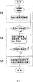
In device of the present invention, after determining the time of exposure under the good different image-forming conditions, the input sample needs the total angle (sample sweep limits) and the turntable step-length of turntable in program, after having determined path that data deposit on computers, the whole data collection process can be moved automatically until data acquisition and finish.The motor process of program is: gather step-length of two-dimensional projection's picture and deposit → sample rotation → gather two-dimensional projection's picture once more and deposit → sample continues to rotate a step-length, so repeatedly, rotate up to sample and to finish whole sweep limits.Fig. 3 is a phase contrast CT image-forming course of experimental work sketch map of the present invention.
Based on device of the present invention, synchrotron radiation X-ray phase contrast CT image-forming experimental work pattern mainly contains:
The groundwork pattern that diffraction strengthens the phase contrast CT image-forming experiment has following four kinds:
1) the sample shaft parallel is in rotating shaft of monochromator crystal and analyzing crystal rotating shaft, and this kind pattern is achieved by adjusting turntable 2,
2) the sample rotating shaft is perpendicular to monochromator crystal and analyzing crystal rotating shaft, and this kind pattern is by being achieved turntable 2 half-twists on a last pattern basis,
3) monochromator crystal and analyzing crystal tuning mode, the monochromator crystal is parallel fully with the analyzing crystal diffraction surfaces,
4) there are a very little angle in monochromator crystal and analyzing crystal off resonance pattern, the diffraction surfaces of monochromator crystal and analyzing crystal;
The groundwork pattern of coaxial phase contrast CT image-forming experiment has following two kinds:
1) this kind pattern need pre-determine the optimum distance between sample and the detector, carries out the CT imaging experiment then under the constant condition of sample-detector distance,
2) choose several different sample-detector distance, carry out the data acquisition of CT imaging experiment at the different distance place respectively, utilize several sets of data that obtained in date processing, to obtain the optimum angle contrast then.
Under out of phase contrast CT image-forming experimental work pattern, phase contrast CT image-forming experiment flow of the present invention.Its concrete steps are:
Step S1, the phase contrast image-forming condition is definite under the different imaging patterns:
(1) strengthen in the phase contrast CT image-forming experiment at diffraction, the relation of the relevant position on required per sample image-forming condition and the twin crystal rocking curve is determined the angle between monochromator crystal and analyzing crystal,
(2) in the experiment of coaxial phase contrast CT image-forming, the distance of determining suitable sample and detector according to different samples to be obtaining best imaging effect or to obtain required information,
(3) determine respective imaging time of exposure under the different condition according to the different parameters of X ray light intensity, detector etc.;
Step S2, the phase contrast CT image-forming experimental data acquisition, under the determined image-forming condition of step S1, after the moving specific angle of sample rotary table (1) revolution, utilize imaging detector to obtain the phase contrast projection image of a sample, the scope of sample rotary table can be 180 °, 360 ° or other are arbitrarily angled, the final phase contrast two-dimensional projection picture that obtains corresponding number, in described device, after determining the time of exposure under the good different image-forming conditions, the input total angle that need rotate of sample and rotate step-length in program, determined path that data deposit on computers after, the whole data collection process can be moved automatically until data acquisition and finish;
Step S3, the reconstruction of phase contrast imaging experiment data, the correlation theory that strengthens phase place contrast imaging or coaxial phase contrast imaging with diffraction is handled the phase contrast CT layer image that can rebuild sample as the algorithm basis to the two-dimensional projection's picture that is obtained.
Description of drawings
Fig. 1 is two kinds of imaging pattern light path sketch maps of extensive use in the phase contrast imaging.
Fig. 2 is the sample rotary table sketch map among the present invention.
Fig. 3 is a synchrotron radiation X-ray phase contrast CT image-forming method flow diagram of the present invention.
The specific embodiment
Fig. 1 is two kinds of imaging pattern light path sketch maps of extensive use in the phase contrast imaging.Wherein, Fig. 1 (a) is coaxial phase contrast CT image-forming experiment index path, and Fig. 1 (b) is that diffraction strengthens phase contrast CT image-forming experiment index path.
In synchrotron radiation phase contrast imaging experiment, because light source is fixed, follow-up experimental provision must be according to light source position and direction adjustment, so in phase contrast CT image-forming CT experiment, the collection of data for projection has only the rotation of sample by experiment rather than the rotation of light source to finish.
Synchrotron radiation X-ray phase contrast CT image-forming device of the present invention, described device comprises by monochromator crystal, sample rotary table, analyzing crystal, ionization chamber and imaging detector and forming, strengthen in the phase contrast CT image-forming at diffraction, monochromator crystal, sample rotary table, analyzing crystal, ionization chamber and imaging detector sequence arrangement are on light path; In coaxial phase contrast CT image-forming, monochromator crystal, analyzing crystal, sample rotary table, ionization chamber and imaging detector are sequentially arranged on the light path; The effect of ionization chamber is the monitoring equipment as X ray intensity, and the X ray light intensity that ionization chamber records shows by microelectrometer in real time with the form of photoelectric current.
Fig. 2 is the sample rotary table sketch map among the present invention.Turntable 1, turntable 2, turntable 3 correspond respectively to ω 1, ω 2 and ω 3, P1 and P2 in translation 1, the translation 2 difference corresponding diagram.
Sample rotary table of the present invention is made up of three turntables and two translations, and in three turntables, the effect of turntable 1 (ω 1) is rotational fixation sample on turntable 1, is the main turntable in the CT experimentation; Turntable 2 (ω 1) be used for adjusting sample with respect to from the orientation of the X ray of monochromator crystal outgoing to satisfy the needs of different image-forming conditions, because turntable 1 is fixed on the turntable 2, so during turntable 2 turntables, the sample that is fixed on the turntable 1 will be with turntable 2 changes (for example, can realize that by turntable 2 the sample rotating shaft becomes vertical turntable by horizontal revolving stage) with respect to the orientation of incident X-rays; Turntable 3 (ω 3) is used for adjusting the orientation of the relative incident X-rays of sample platform to guarantee the normal incidence of X ray to sample, during turntable 3 turntables, be fixed in turntable 2 on the turntable 3 with commentaries on classics, be fixed on turntable 1 on the turntable 2 simultaneously with changeing to realize the normal incidence of X ray to sample, in two translations, translation P1 is in order to realize the adjusting of sample platform with respect to the X ray height and position, and translation P2 is in order to realize the adjusting of sample platform with respect to the X ray horizontal level.
In addition, be the influence of the strong scattering avoiding synchrotron radiation X-ray to incide bringing on the metal parts, the central authorities of turntable 2 are designed to an enough big X ray passage by the position of X ray; The turning cylinder of sample rotary table (turntable 1) will have sufficiently high concentric stability to satisfy the requirement of CT imaging resolution.
Fig. 3 is a synchrotron radiation X-ray phase contrast imaging CT method flow, and its concrete steps are as follows:
Step S1, the phase contrast image-forming condition is definite under the different imaging patterns:
(1) strengthen in the phase contrast CT image-forming experiment at diffraction, the relation of the relevant position on required per sample image-forming condition and the twin crystal rocking curve is determined the angle between monochromator crystal and analyzing crystal,
(2) in the experiment of coaxial phase contrast CT image-forming, the distance of determining suitable sample and detector according to different samples to be obtaining best imaging effect or to obtain required information,
(3) determine respective imaging time of exposure under the different condition according to the different parameters of X ray light intensity, detector etc.;
Step S2, the phase contrast CT image-forming experimental data acquisition, under the determined image-forming condition of step S1, after the moving specific angle of sample rotary table (1) revolution, utilize imaging detector to obtain the phase contrast projection image of a sample, the scope of sample rotary table can be 180 °, 360 ° or other are arbitrarily angled, the final phase contrast two-dimensional projection picture that obtains corresponding number, in described device, after determining the time of exposure under the good different image-forming conditions, the input total angle that need rotate of sample and rotate step-length in program, determined path that data deposit on computers after, the whole data collection process can be moved automatically until data acquisition and finish;
Step S3, the reconstruction of phase contrast imaging experiment data, the correlation theory that strengthens phase place contrast imaging or coaxial phase contrast imaging with diffraction is handled the phase contrast CT layer image that can rebuild sample as the algorithm basis to the two-dimensional projection's picture that is obtained.
Claims (5)
1. synchrotron radiation X-ray phase contrast CT image-forming device, described device is made up of monochromator crystal, sample rotary table, analyzing crystal, ionization chamber and imaging detector, strengthen in the phase contrast CT image-forming experiment at diffraction, monochromator crystal, sample rotary table, analyzing crystal, ionization chamber and imaging detector sequence arrangement are on light path; In coaxial phase contrast CT image-forming experiment, monochromator crystal, analyzing crystal, sample rotary table, ionization chamber and imaging detector are sequentially arranged on the light path; The effect of ionization chamber is the monitoring equipment as X ray intensity, and the X ray light intensity that ionization chamber records shows by microelectrometer in real time with the form of photoelectric current.
2. synchrotron radiation X-ray phase contrast CT image-forming device according to claim 1, it is characterized in that, sample rotary table is made up of three turntables and two translations, in three turntables, the effect of turntable (1) is to realize being fixed on the rotation that turntable (1) is gone up sample, is the main turntable in the CT imaging experiment process; Turntable (2) be used for adjusting sample with respect to from the orientation of the X ray of monochromator crystal outgoing to satisfy the needs of different image-forming conditions, because turntable (1) is fixed on the turntable (2), so when turntable (2) rotates, the sample that is fixed on the turntable (1) will change with turntable (2) with respect to the orientation of incident X-rays; Turntable (3) is used for adjusting the orientation of the relative incident X-rays of sample platform to guarantee the normal incidence of X ray to sample, when turntable (3) rotates, be fixed in turntable (2) on the turntable (3) with commentaries on classics, be fixed on turntable (1) on the turntable (2) simultaneously with changeing to realize the normal incidence of X ray to sample, in two translations, translation P1 is in order to realize the adjusting of sample platform with respect to the X ray height and position, and translation P2 is in order to realize the adjusting of sample platform with respect to the X ray horizontal level.
3. synchrotron radiation X-ray phase contrast CT image-forming device according to claim 1 and 2 is characterized in that, by automatic control, can satisfy diffraction and strengthen phase contrast CT image-forming and the experiment needs of coaxial phase contrast CT image-forming under different condition; Simultaneously, this device can be realized whole automatizatioies of whole data collection process in conjunction with computer.
4. the method for a synchrotron radiation X-ray phase contrast CT image-forming, its concrete steps are as follows:
Step S1, the phase contrast image-forming condition is definite under the different imaging patterns:
(1) strengthen in the phase contrast CT image-forming experiment at diffraction, the relation of the relevant position on required per sample image-forming condition and the twin crystal rocking curve is determined the angle between monochromator crystal and analyzing crystal,
(2) in the experiment of coaxial phase contrast CT image-forming, the distance of determining suitable sample and detector according to different samples to be obtaining best imaging effect or to obtain required information,
(3) determine respective imaging time of exposure under the different condition according to the different parameters of X ray light intensity, detector etc.;
Step S2, the phase contrast CT image-forming experimental data acquisition, under the determined image-forming condition of step S1, after the moving specific angle of sample rotary table (1) revolution, utilize imaging detector to obtain the phase contrast projection image of a sample, the scope of sample rotary table can be 180 °, 360 ° or other are arbitrarily angled, the final phase contrast two-dimensional projection picture that obtains corresponding number, in described device, after determining the time of exposure under the good different image-forming conditions, the input total angle that need rotate of sample and rotate step-length in program, determined path that data deposit on computers after, the whole data collection process can be moved automatically until data acquisition and finish;
Step S3, the reconstruction of phase contrast imaging experiment data, the correlation theory that strengthens phase place contrast imaging or coaxial phase contrast imaging with diffraction is handled the phase contrast CT layer image that can rebuild sample as the algorithm basis to the two-dimensional projection's picture that is obtained.
5. synchrotron radiation X-ray phase contrast CT image-forming method according to claim 4 is characterized in that, can realize multiple phase contrast CT image-forming pattern experiment
The main pattern that diffraction strengthens the phase contrast CT image-forming experiment has following four kinds:
1) the sample shaft parallel is in rotating shaft of monochromator crystal and analyzing crystal rotating shaft, and this kind pattern is passed through to adjust turntable 2 and realized,
2) the sample rotating shaft is perpendicular to monochromator crystal and analyzing crystal rotating shaft, and this kind pattern can be by being achieved turntable 2 half-twists on a last pattern basis,
3) monochromator crystal and analyzing crystal tuning mode, the monochromator crystal is parallel fully with the analyzing crystal diffraction surfaces,
4) there are a very little angle in monochromator crystal and analyzing crystal off resonance pattern, the diffraction surfaces of monochromator crystal and analyzing crystal;
The groundwork pattern of coaxial phase contrast CT image-forming experiment has following two kinds:
1) carry out coaxial phase contrast imaging CT experiment at optimum sample-detector distance place, this kind pattern need pre-determine the optimum distance between sample and the detector, carries out the CT imaging experiment then under the constant condition of sample-detector distance,
2) choose several different sample-detector distance, carry out the data acquisition of CT imaging experiment at the different distance place respectively, utilize several sets of data that obtained in date processing, to obtain the optimum angle contrast then.
Priority Applications (1)
| Application Number | Priority Date | Filing Date | Title |
|---|---|---|---|
| CNB2005100869041A CN100457040C (en) | 2005-11-17 | 2005-11-17 | synchrotron radiation X-ray phase contrasting computed tomography and experimental method thereof |
Applications Claiming Priority (1)
| Application Number | Priority Date | Filing Date | Title |
|---|---|---|---|
| CNB2005100869041A CN100457040C (en) | 2005-11-17 | 2005-11-17 | synchrotron radiation X-ray phase contrasting computed tomography and experimental method thereof |
Publications (2)
| Publication Number | Publication Date |
|---|---|
| CN1965760A true CN1965760A (en) | 2007-05-23 |
| CN100457040C CN100457040C (en) | 2009-02-04 |
Family
ID=38074863
Family Applications (1)
| Application Number | Title | Priority Date | Filing Date |
|---|---|---|---|
| CNB2005100869041A Expired - Fee Related CN100457040C (en) | 2005-11-17 | 2005-11-17 | synchrotron radiation X-ray phase contrasting computed tomography and experimental method thereof |
Country Status (1)
| Country | Link |
|---|---|
| CN (1) | CN100457040C (en) |
Cited By (11)
| Publication number | Priority date | Publication date | Assignee | Title |
|---|---|---|---|---|
| CN102413767A (en) * | 2009-03-02 | 2012-04-11 | 罗切斯特大学 | Methods and apparatus for differential phase-contrast fan beam ct, cone-beam ct and hybrid cone-beam ct |
| CN103356208A (en) * | 2012-04-01 | 2013-10-23 | 中国科学院高能物理研究所 | Two-dimensional imaging system and method for medical test of human body |
| CN101952900B (en) * | 2008-02-14 | 2013-10-23 | 皇家飞利浦电子股份有限公司 | X-ray detector for phase contrast imaging |
| US8971488B2 (en) | 2008-12-01 | 2015-03-03 | The University Of North Carolina At Chapel Hill | Systems and methods for detecting an image of an object using multi-beam imaging from an X-ray beam having a polychromatic distribution |
| US8976925B2 (en) | 2006-02-27 | 2015-03-10 | University Of Rochester | Method and apparatus of cone beam CT dynamic imaging |
| CN105513057A (en) * | 2015-11-30 | 2016-04-20 | 首都医科大学 | Tumor auxiliary diagnosis method |
| US9364191B2 (en) | 2013-02-11 | 2016-06-14 | University Of Rochester | Method and apparatus of spectral differential phase-contrast cone-beam CT and hybrid cone-beam CT |
| CN105675638A (en) * | 2016-03-24 | 2016-06-15 | 西安交通大学 | Universality efficient synchrotron radiation visual representation method of crystal microstructure |
| CN109215094A (en) * | 2017-09-22 | 2019-01-15 | 上海联影医疗科技有限公司 | phase contrast image generating method and system |
| CN111896566A (en) * | 2020-07-20 | 2020-11-06 | 上海交通大学 | Device and method for increasing imaging range of synchrotron radiation light source |
| CN112179926A (en) * | 2020-09-24 | 2021-01-05 | 首都师范大学 | Coaxial CT-based phase-absorption inversion and material quantitative imaging method |
Family Cites Families (5)
| Publication number | Priority date | Publication date | Assignee | Title |
|---|---|---|---|---|
| US5259013A (en) * | 1991-12-17 | 1993-11-02 | The United States Of America As Represented By The Secretary Of Commerce | Hard x-ray magnification apparatus and method with submicrometer spatial resolution of images in more than one dimension |
| JPH09187455A (en) * | 1996-01-10 | 1997-07-22 | Hitachi Ltd | Phase type x-ray ct apparatus |
| DE69730550T2 (en) * | 1996-03-29 | 2005-11-10 | Hitachi, Ltd. | Phase contrast X-ray imaging system |
| JP2000193609A (en) * | 1998-12-24 | 2000-07-14 | Hamamatsu Photonics Kk | Radiation tomograph |
| EP1451563A1 (en) * | 2001-11-05 | 2004-09-01 | Vanderbilt University | Phase-contrast enhanced computed tomography |
-
2005
- 2005-11-17 CN CNB2005100869041A patent/CN100457040C/en not_active Expired - Fee Related
Cited By (15)
| Publication number | Priority date | Publication date | Assignee | Title |
|---|---|---|---|---|
| US8976925B2 (en) | 2006-02-27 | 2015-03-10 | University Of Rochester | Method and apparatus of cone beam CT dynamic imaging |
| US8576983B2 (en) | 2008-02-14 | 2013-11-05 | Koninklijke Philips N.V. | X-ray detector for phase contrast imaging |
| CN101952900B (en) * | 2008-02-14 | 2013-10-23 | 皇家飞利浦电子股份有限公司 | X-ray detector for phase contrast imaging |
| US8971488B2 (en) | 2008-12-01 | 2015-03-03 | The University Of North Carolina At Chapel Hill | Systems and methods for detecting an image of an object using multi-beam imaging from an X-ray beam having a polychromatic distribution |
| CN102413767A (en) * | 2009-03-02 | 2012-04-11 | 罗切斯特大学 | Methods and apparatus for differential phase-contrast fan beam ct, cone-beam ct and hybrid cone-beam ct |
| CN102413767B (en) * | 2009-03-02 | 2015-07-22 | 罗切斯特大学 | Methods and apparatus for differential phase-contrast fan beam ct, cone-beam ct and hybrid cone-beam ct |
| CN103356208B (en) * | 2012-04-01 | 2015-01-14 | 中国科学院高能物理研究所 | Two-dimensional imaging system and method for medical test of human body |
| CN103356208A (en) * | 2012-04-01 | 2013-10-23 | 中国科学院高能物理研究所 | Two-dimensional imaging system and method for medical test of human body |
| US9364191B2 (en) | 2013-02-11 | 2016-06-14 | University Of Rochester | Method and apparatus of spectral differential phase-contrast cone-beam CT and hybrid cone-beam CT |
| CN105513057A (en) * | 2015-11-30 | 2016-04-20 | 首都医科大学 | Tumor auxiliary diagnosis method |
| CN105675638A (en) * | 2016-03-24 | 2016-06-15 | 西安交通大学 | Universality efficient synchrotron radiation visual representation method of crystal microstructure |
| CN105675638B (en) * | 2016-03-24 | 2017-04-05 | 西安交通大学 | A kind of synchrotron radiation visual representation method of crystal microscopic structure |
| CN109215094A (en) * | 2017-09-22 | 2019-01-15 | 上海联影医疗科技有限公司 | phase contrast image generating method and system |
| CN111896566A (en) * | 2020-07-20 | 2020-11-06 | 上海交通大学 | Device and method for increasing imaging range of synchrotron radiation light source |
| CN112179926A (en) * | 2020-09-24 | 2021-01-05 | 首都师范大学 | Coaxial CT-based phase-absorption inversion and material quantitative imaging method |
Also Published As
| Publication number | Publication date |
|---|---|
| CN100457040C (en) | 2009-02-04 |
Similar Documents
| Publication | Publication Date | Title |
|---|---|---|
| CN100457040C (en) | synchrotron radiation X-ray phase contrasting computed tomography and experimental method thereof | |
| Salomé et al. | A synchrotron radiation microtomography system for the analysis of trabecular bone samples | |
| Deng et al. | First X-ray fluorescence CT experimental results at the SSRF X-ray imaging beamline | |
| KR20040111005A (en) | Radiographic apparatus | |
| He et al. | Fundamentals of two-dimensional X-ray diffraction (XRD2) | |
| CN112964738B (en) | Industrial CT rapid scanning system and method | |
| CN105806858A (en) | CT detection method and CT device | |
| CN104132953A (en) | Dual-energy X-ray phase-contrast imaging device and implementation method thereof | |
| JP2013514527A (en) | Method and apparatus for performing X-ray analysis of sample | |
| WO1995004268A1 (en) | Three-dimensional microtomographic analysis system | |
| JP2005021675A (en) | Tomograph apparatus | |
| CN105852895B (en) | The information extracting method of the hard X ray grating interferometer of single exposure | |
| CN101467889B (en) | Grating shearing phase contrast CT image-forming data acquisition and reconstruction method | |
| CN101056584A (en) | Ct method for the examination of a cyclically moving object | |
| Rivers et al. | Recent developments in computed tomography at GSECARS | |
| WO2022247234A1 (en) | Cone-beam x-ray fluorescence imaging method and system, and terminal and storage medium | |
| CN1500443A (en) | Radiographic apparatus and method | |
| CN113533392B (en) | Combined scanning CL imaging method | |
| CN109709127A (en) | Low scattered x-ray fluorescence CT imaging method | |
| JP2003344316A (en) | Reconstitution method of inclined three-dimensional x- ray ct image | |
| CN111380883A (en) | Double-source angle resolution type X-ray diffraction analysis and tomography coupling device | |
| CN1176370C (en) | X-ray fluorescence holographic tomography device | |
| JP3676249B2 (en) | Crystal observation method and apparatus using X-ray diffraction | |
| JPH0862159A (en) | Tomographic apparatus | |
| CN1216271C (en) | X-ray double-frequency holographic interferometer |
Legal Events
| Date | Code | Title | Description |
|---|---|---|---|
| C06 | Publication | ||
| PB01 | Publication | ||
| C10 | Entry into substantive examination | ||
| SE01 | Entry into force of request for substantive examination | ||
| C14 | Grant of patent or utility model | ||
| GR01 | Patent grant | ||
| CF01 | Termination of patent right due to non-payment of annual fee |
Granted publication date: 20090204 Termination date: 20141117 |
|
| EXPY | Termination of patent right or utility model |