CN1440424A - Antibody selective for tumor necrosis factor-related apoptosis-inducing ligand receptor and use thereof - Google Patents
Antibody selective for tumor necrosis factor-related apoptosis-inducing ligand receptor and use thereof Download PDFInfo
- Publication number
- CN1440424A CN1440424A CN01812038A CN01812038A CN1440424A CN 1440424 A CN1440424 A CN 1440424A CN 01812038 A CN01812038 A CN 01812038A CN 01812038 A CN01812038 A CN 01812038A CN 1440424 A CN1440424 A CN 1440424A
- Authority
- CN
- China
- Prior art keywords
- cell
- antibody
- tra
- trail
- sequence
- Prior art date
- Legal status (The legal status is an assumption and is not a legal conclusion. Google has not performed a legal analysis and makes no representation as to the accuracy of the status listed.)
- Granted
Links
Images


Classifications
-
- C—CHEMISTRY; METALLURGY
- C07—ORGANIC CHEMISTRY
- C07K—PEPTIDES
- C07K16/00—Immunoglobulins [IGs], e.g. monoclonal or polyclonal antibodies
- C07K16/18—Immunoglobulins [IGs], e.g. monoclonal or polyclonal antibodies against material from animals or humans
- C07K16/28—Immunoglobulins [IGs], e.g. monoclonal or polyclonal antibodies against material from animals or humans against receptors, cell surface antigens or cell surface determinants
- C07K16/2866—Immunoglobulins [IGs], e.g. monoclonal or polyclonal antibodies against material from animals or humans against receptors, cell surface antigens or cell surface determinants against receptors for cytokines, lymphokines, interferons
-
- A—HUMAN NECESSITIES
- A61—MEDICAL OR VETERINARY SCIENCE; HYGIENE
- A61K—PREPARATIONS FOR MEDICAL, DENTAL OR TOILETRY PURPOSES
- A61K39/00—Medicinal preparations containing antigens or antibodies
- A61K39/395—Antibodies; Immunoglobulins; Immune serum, e.g. antilymphocytic serum
- A61K39/39533—Antibodies; Immunoglobulins; Immune serum, e.g. antilymphocytic serum against materials from animals
- A61K39/39558—Antibodies; Immunoglobulins; Immune serum, e.g. antilymphocytic serum against materials from animals against tumor tissues, cells, antigens
-
- A—HUMAN NECESSITIES
- A61—MEDICAL OR VETERINARY SCIENCE; HYGIENE
- A61K—PREPARATIONS FOR MEDICAL, DENTAL OR TOILETRY PURPOSES
- A61K39/00—Medicinal preparations containing antigens or antibodies
- A61K39/395—Antibodies; Immunoglobulins; Immune serum, e.g. antilymphocytic serum
-
- A—HUMAN NECESSITIES
- A61—MEDICAL OR VETERINARY SCIENCE; HYGIENE
- A61P—SPECIFIC THERAPEUTIC ACTIVITY OF CHEMICAL COMPOUNDS OR MEDICINAL PREPARATIONS
- A61P1/00—Drugs for disorders of the alimentary tract or the digestive system
- A61P1/04—Drugs for disorders of the alimentary tract or the digestive system for ulcers, gastritis or reflux esophagitis, e.g. antacids, inhibitors of acid secretion, mucosal protectants
-
- A—HUMAN NECESSITIES
- A61—MEDICAL OR VETERINARY SCIENCE; HYGIENE
- A61P—SPECIFIC THERAPEUTIC ACTIVITY OF CHEMICAL COMPOUNDS OR MEDICINAL PREPARATIONS
- A61P13/00—Drugs for disorders of the urinary system
- A61P13/12—Drugs for disorders of the urinary system of the kidneys
-
- A—HUMAN NECESSITIES
- A61—MEDICAL OR VETERINARY SCIENCE; HYGIENE
- A61P—SPECIFIC THERAPEUTIC ACTIVITY OF CHEMICAL COMPOUNDS OR MEDICINAL PREPARATIONS
- A61P15/00—Drugs for genital or sexual disorders; Contraceptives
-
- A—HUMAN NECESSITIES
- A61—MEDICAL OR VETERINARY SCIENCE; HYGIENE
- A61P—SPECIFIC THERAPEUTIC ACTIVITY OF CHEMICAL COMPOUNDS OR MEDICINAL PREPARATIONS
- A61P17/00—Drugs for dermatological disorders
-
- A—HUMAN NECESSITIES
- A61—MEDICAL OR VETERINARY SCIENCE; HYGIENE
- A61P—SPECIFIC THERAPEUTIC ACTIVITY OF CHEMICAL COMPOUNDS OR MEDICINAL PREPARATIONS
- A61P21/00—Drugs for disorders of the muscular or neuromuscular system
- A61P21/04—Drugs for disorders of the muscular or neuromuscular system for myasthenia gravis
-
- A—HUMAN NECESSITIES
- A61—MEDICAL OR VETERINARY SCIENCE; HYGIENE
- A61P—SPECIFIC THERAPEUTIC ACTIVITY OF CHEMICAL COMPOUNDS OR MEDICINAL PREPARATIONS
- A61P25/00—Drugs for disorders of the nervous system
-
- A—HUMAN NECESSITIES
- A61—MEDICAL OR VETERINARY SCIENCE; HYGIENE
- A61P—SPECIFIC THERAPEUTIC ACTIVITY OF CHEMICAL COMPOUNDS OR MEDICINAL PREPARATIONS
- A61P29/00—Non-central analgesic, antipyretic or antiinflammatory agents, e.g. antirheumatic agents; Non-steroidal antiinflammatory drugs [NSAID]
-
- A—HUMAN NECESSITIES
- A61—MEDICAL OR VETERINARY SCIENCE; HYGIENE
- A61P—SPECIFIC THERAPEUTIC ACTIVITY OF CHEMICAL COMPOUNDS OR MEDICINAL PREPARATIONS
- A61P3/00—Drugs for disorders of the metabolism
- A61P3/08—Drugs for disorders of the metabolism for glucose homeostasis
- A61P3/10—Drugs for disorders of the metabolism for glucose homeostasis for hyperglycaemia, e.g. antidiabetics
-
- A—HUMAN NECESSITIES
- A61—MEDICAL OR VETERINARY SCIENCE; HYGIENE
- A61P—SPECIFIC THERAPEUTIC ACTIVITY OF CHEMICAL COMPOUNDS OR MEDICINAL PREPARATIONS
- A61P31/00—Antiinfectives, i.e. antibiotics, antiseptics, chemotherapeutics
- A61P31/12—Antivirals
-
- A—HUMAN NECESSITIES
- A61—MEDICAL OR VETERINARY SCIENCE; HYGIENE
- A61P—SPECIFIC THERAPEUTIC ACTIVITY OF CHEMICAL COMPOUNDS OR MEDICINAL PREPARATIONS
- A61P31/00—Antiinfectives, i.e. antibiotics, antiseptics, chemotherapeutics
- A61P31/12—Antivirals
- A61P31/14—Antivirals for RNA viruses
- A61P31/18—Antivirals for RNA viruses for HIV
-
- A—HUMAN NECESSITIES
- A61—MEDICAL OR VETERINARY SCIENCE; HYGIENE
- A61P—SPECIFIC THERAPEUTIC ACTIVITY OF CHEMICAL COMPOUNDS OR MEDICINAL PREPARATIONS
- A61P35/00—Antineoplastic agents
-
- A—HUMAN NECESSITIES
- A61—MEDICAL OR VETERINARY SCIENCE; HYGIENE
- A61P—SPECIFIC THERAPEUTIC ACTIVITY OF CHEMICAL COMPOUNDS OR MEDICINAL PREPARATIONS
- A61P37/00—Drugs for immunological or allergic disorders
-
- A—HUMAN NECESSITIES
- A61—MEDICAL OR VETERINARY SCIENCE; HYGIENE
- A61P—SPECIFIC THERAPEUTIC ACTIVITY OF CHEMICAL COMPOUNDS OR MEDICINAL PREPARATIONS
- A61P37/00—Drugs for immunological or allergic disorders
- A61P37/02—Immunomodulators
-
- A—HUMAN NECESSITIES
- A61—MEDICAL OR VETERINARY SCIENCE; HYGIENE
- A61P—SPECIFIC THERAPEUTIC ACTIVITY OF CHEMICAL COMPOUNDS OR MEDICINAL PREPARATIONS
- A61P37/00—Drugs for immunological or allergic disorders
- A61P37/02—Immunomodulators
- A61P37/04—Immunostimulants
-
- A—HUMAN NECESSITIES
- A61—MEDICAL OR VETERINARY SCIENCE; HYGIENE
- A61P—SPECIFIC THERAPEUTIC ACTIVITY OF CHEMICAL COMPOUNDS OR MEDICINAL PREPARATIONS
- A61P37/00—Drugs for immunological or allergic disorders
- A61P37/02—Immunomodulators
- A61P37/06—Immunosuppressants, e.g. drugs for graft rejection
-
- A—HUMAN NECESSITIES
- A61—MEDICAL OR VETERINARY SCIENCE; HYGIENE
- A61P—SPECIFIC THERAPEUTIC ACTIVITY OF CHEMICAL COMPOUNDS OR MEDICINAL PREPARATIONS
- A61P37/00—Drugs for immunological or allergic disorders
- A61P37/08—Antiallergic agents
-
- A—HUMAN NECESSITIES
- A61—MEDICAL OR VETERINARY SCIENCE; HYGIENE
- A61P—SPECIFIC THERAPEUTIC ACTIVITY OF CHEMICAL COMPOUNDS OR MEDICINAL PREPARATIONS
- A61P43/00—Drugs for specific purposes, not provided for in groups A61P1/00-A61P41/00
-
- A—HUMAN NECESSITIES
- A61—MEDICAL OR VETERINARY SCIENCE; HYGIENE
- A61P—SPECIFIC THERAPEUTIC ACTIVITY OF CHEMICAL COMPOUNDS OR MEDICINAL PREPARATIONS
- A61P5/00—Drugs for disorders of the endocrine system
- A61P5/18—Drugs for disorders of the endocrine system of the parathyroid hormones
-
- A—HUMAN NECESSITIES
- A61—MEDICAL OR VETERINARY SCIENCE; HYGIENE
- A61P—SPECIFIC THERAPEUTIC ACTIVITY OF CHEMICAL COMPOUNDS OR MEDICINAL PREPARATIONS
- A61P7/00—Drugs for disorders of the blood or the extracellular fluid
- A61P7/06—Antianaemics
-
- A—HUMAN NECESSITIES
- A61—MEDICAL OR VETERINARY SCIENCE; HYGIENE
- A61P—SPECIFIC THERAPEUTIC ACTIVITY OF CHEMICAL COMPOUNDS OR MEDICINAL PREPARATIONS
- A61P9/00—Drugs for disorders of the cardiovascular system
- A61P9/10—Drugs for disorders of the cardiovascular system for treating ischaemic or atherosclerotic diseases, e.g. antianginal drugs, coronary vasodilators, drugs for myocardial infarction, retinopathy, cerebrovascula insufficiency, renal arteriosclerosis
-
- C—CHEMISTRY; METALLURGY
- C07—ORGANIC CHEMISTRY
- C07K—PEPTIDES
- C07K16/00—Immunoglobulins [IGs], e.g. monoclonal or polyclonal antibodies
- C07K16/18—Immunoglobulins [IGs], e.g. monoclonal or polyclonal antibodies against material from animals or humans
- C07K16/28—Immunoglobulins [IGs], e.g. monoclonal or polyclonal antibodies against material from animals or humans against receptors, cell surface antigens or cell surface determinants
- C07K16/2878—Immunoglobulins [IGs], e.g. monoclonal or polyclonal antibodies against material from animals or humans against receptors, cell surface antigens or cell surface determinants against the NGF-receptor/TNF-receptor superfamily, e.g. CD27, CD30, CD40, CD95
-
- C—CHEMISTRY; METALLURGY
- C07—ORGANIC CHEMISTRY
- C07K—PEPTIDES
- C07K16/00—Immunoglobulins [IGs], e.g. monoclonal or polyclonal antibodies
- C07K16/18—Immunoglobulins [IGs], e.g. monoclonal or polyclonal antibodies against material from animals or humans
- C07K16/28—Immunoglobulins [IGs], e.g. monoclonal or polyclonal antibodies against material from animals or humans against receptors, cell surface antigens or cell surface determinants
- C07K16/30—Immunoglobulins [IGs], e.g. monoclonal or polyclonal antibodies against material from animals or humans against receptors, cell surface antigens or cell surface determinants from tumour cells
-
- G—PHYSICS
- G01—MEASURING; TESTING
- G01N—INVESTIGATING OR ANALYSING MATERIALS BY DETERMINING THEIR CHEMICAL OR PHYSICAL PROPERTIES
- G01N33/00—Investigating or analysing materials by specific methods not covered by groups G01N1/00 - G01N31/00
- G01N33/48—Biological material, e.g. blood, urine; Haemocytometers
- G01N33/50—Chemical analysis of biological material, e.g. blood, urine; Testing involving biospecific ligand binding methods; Immunological testing
- G01N33/68—Chemical analysis of biological material, e.g. blood, urine; Testing involving biospecific ligand binding methods; Immunological testing involving proteins, peptides or amino acids
- G01N33/6863—Cytokines, i.e. immune system proteins modifying a biological response such as cell growth proliferation or differentiation, e.g. TNF, CNF, GM-CSF, lymphotoxin, MIF or their receptors
-
- A—HUMAN NECESSITIES
- A61—MEDICAL OR VETERINARY SCIENCE; HYGIENE
- A61K—PREPARATIONS FOR MEDICAL, DENTAL OR TOILETRY PURPOSES
- A61K39/00—Medicinal preparations containing antigens or antibodies
- A61K2039/505—Medicinal preparations containing antigens or antibodies comprising antibodies
-
- C—CHEMISTRY; METALLURGY
- C07—ORGANIC CHEMISTRY
- C07K—PEPTIDES
- C07K2317/00—Immunoglobulins specific features
- C07K2317/10—Immunoglobulins specific features characterized by their source of isolation or production
-
- C—CHEMISTRY; METALLURGY
- C07—ORGANIC CHEMISTRY
- C07K—PEPTIDES
- C07K2317/00—Immunoglobulins specific features
- C07K2317/20—Immunoglobulins specific features characterized by taxonomic origin
- C07K2317/24—Immunoglobulins specific features characterized by taxonomic origin containing regions, domains or residues from different species, e.g. chimeric, humanized or veneered
-
- C—CHEMISTRY; METALLURGY
- C07—ORGANIC CHEMISTRY
- C07K—PEPTIDES
- C07K2317/00—Immunoglobulins specific features
- C07K2317/70—Immunoglobulins specific features characterized by effect upon binding to a cell or to an antigen
- C07K2317/73—Inducing cell death, e.g. apoptosis, necrosis or inhibition of cell proliferation
-
- C—CHEMISTRY; METALLURGY
- C07—ORGANIC CHEMISTRY
- C07K—PEPTIDES
- C07K2319/00—Fusion polypeptide
-
- C—CHEMISTRY; METALLURGY
- C07—ORGANIC CHEMISTRY
- C07K—PEPTIDES
- C07K2319/00—Fusion polypeptide
- C07K2319/30—Non-immunoglobulin-derived peptide or protein having an immunoglobulin constant or Fc region, or a fragment thereof, attached thereto
Abstract
An antibody of the invention interacts with human DR5 to produce agonistic or antagonistic effects downstream of the receptor including inhibition of cell proliferation and apoptosis. Nucleic acid sequences and amino acid sequences of anti-DR5 antibodies have been elucidated and vectors and cells containing and expressing these sequences have been generated. Methods and uses for the antibodies are detailed including treatment of apoptosis-related disease and treatment of dysregulated cell growth.
Description
Invention field
The present invention relates to can be specifically in conjunction with the antibody of a kind of tumor types necrosin (being called " TNF " hereinafter) related apoptosis-inducing ligand (being called " TRAIL " hereinafter) acceptor, more particularly, relate in vivo the monoclonal antibody with the cell apoptosis of the described single type acceptor of external evoked expression, also relate to therapy based on it.
Background of invention
TRAIL is a member of protein TNF family, and described family also comprises TNF-α and Fas part (1).These protein are effective apoptosis inducers.Up to now, 5 kinds of TRAIL acceptors have been identified, wherein two kinds-DR4 (TRAIL-R1) and DR5 (TRAIL-R2) (2-7) can the transducer cell apoptotic signals, and other three kinds-DcR1 (TRAIL-R3), DcR2 (TRAIL-R4) and osteoprotegerun (OPG) transducer cell apoptotic signal (8-12) not.All 5 kinds of TRAIL acceptors are shared remarkable homology in the ligand binding domains outside its born of the same parents.Similar to Fas with TNF acceptor I (being called " TNFRI " hereinafter), the intracellular region section of DR4 and DR5 all contains a death domain, by relating to the approach transducer cell apoptotic signal (6,7) of Fas associated death domain protein (being called " FADD " hereinafter) and caspase 8.Except that the transducer cell apoptotic signal, DR4 and DR5 acceptor also can activate the approach (6,7) that relates to NF κ b.
The biological function of verified TRAIL comprises that TRAIL can optionally induce conversion tumour cell apoptosis, and normal cell has relevant antagonism (13-15) to the apoptosis that TRAIL mediates.This selective prompting gives TRAIL by system in animal model and do not induce remarkable toxicity to prove, and is opposite with the Fas part, the giving of TRAIL relevant with very low-level toxicity (13).Therefore, having proposed TRAIL is a kind of effective cell death inducer, and it may be the suitable curative that is used for the treatment of cancer and other disease relevant with abnormal cell proliferation.Also having proposed TRAIL is a kind of effective cell death inducer that may be applicable to treatment autoimmune disease and inflammatory diseases.Verified, the necrocytosis of the apoptosis participating in activation inductive T cell of TRAIL mediation, thereby as a kind of replacement mechanism (16,17) of Fas part.The apoptosis of TRAIL mediation also may work (18) aspect inducing T cell and other inflammatory cell apoptosis, and works at the killing activity (19-21) of NK cell and immunoloregulation function (22, the 23) aspect of dendritic cell.Therefore, the apoptosis of TRAIL mediation also may work aspect immunity privilege and the immunosurveillance.
The TRAIL receptor system is complicated, comprises at least two kinds of death receptors (DR4 and DR5) and at least two kinds of acellular apoptosis acceptors (DcR1 and DcR2).All these acceptors are not only enjoyed the homoamino acid sequence homology, and show the binding affinity similar to TRAIL (2-12).DcR1 and DcR2 receptor competition combine with TRAIL and the not ability of cell death inducing prompting, and they may be as the bait acceptor of blocking-up or adjusting TRAIL ligand activity.In addition, the bait receptor level that is reported that the cell expressing of unconverted is higher than the transformant expression levels.Therefore proposed, the difference of described death receptor and bait receptor expression is regulated and may be represented the crucial regulatory mechanism of decision cell to the apoptotic susceptibility of TRAIL mediation, but in default of receptor specific antibody (2).Although broad research expression and the function of DR4 and DR5, hindered progress owing to lacking the receptor-specific monoclonal antibody.The cell surface expression of DR5 is not on the books as yet.Existing report, produced one group of anti-TRAIL receptor antibody, described antibody can be at external evoked melanoma cells apoptosis, but only is immobilized to promote cell death inducing when crosslinked at described antibody, and in some cases, described cell need be cultivated (24) with dactinomycin.Several anti-DR5 antibody (24) have been produced.Yet these previous anti-DR5 monoclonal antibodies that produce are external even under crosslinked condition, and its apoptosis-inducing activity is also very low.Do not report activity in vivo.These antibody have been used for checking the cell surface expression (24) of TRAIL acceptor.Therefore, need to combine with cell surface receptor and need not crosslinked or immobilized monoclonal antibody with external all induced strong all kinds abnormal cellss (comprising tumour cell) apoptosis in vivo to every kind of concrete TRAIL receptor-selective.This antibody may not only provide the potential curative, and provides a kind of diagnostic tool for the functional analysis of TRAIL acceptor.Need induce every species specific antibody among acceptor DR4 and the DR5 for death especially.
In the generation or development of numerous disease, common situation is that described cell is not eliminated.In many autoimmune diseases and inflammatory conditions, the active cells of survival is attacked healthy tissues or cell.In addition, the proliferative lymphadenitis of tumorigenic progress and rheumatoid arthritis forms is characterized as unchecked cell proliferation.Therefore, insufficient apoptosis causes morbidity, and application cell apoptosis induction ligand or agonistic monoclonal antibodies strengthen apoptosis, are considered to a kind of possible therapeutic strategy of eliminating those undesired cells.
For example, rheumatoid arthritis (being called " RA " hereinafter) is a kind of common mankind itself's immunological disease.The understanding to the RA physiopathology at present is, the inflammatory response in the initial described joint of autoimmunization T cell and B cell, and this orders about the synovial cell and breeds too much.Because the synovial cell breeds too much, the excessive generation of metalloprotease (being called " MMP " hereinafter), this further causes the aggressiveness of distinctive cartilage of RA and bone to destroy (25).Therefore, control inflammatory synovial cell to breed too much be one step of key among the treatment RA.Causing the synovial cell to breed too much molecular mechanism does not still understand.Although breed too much synovial cell is nonmalignant and non-conversion, many research promptings, and they and transformant are enjoyed some common feature (46).The feature of these cells of so-called " it seems the synovial cell who has transformed " is that fine and close rough surfaced endoplasmic reticulum, a large amount of irregular cell nuclear and normal spindle cell skeletons change.Propose, mixing of the gene of oncogene and viral source may be the main triggering thing (46) that RA synovial cell has transformed outward appearance.
At least two aspect promptings of RA, the apoptosis of dysregulation may be the major cause that causes disease process, and it may be a kind of effective therapy that apoptotic treatment is brought out: activating T cell fails to exhaust prompting, there is defective in the activation inductive necrocytosis of these T cells, the necrocytosis of this activation inductive is the apoptosis of a kind of Fas of relating to mediation and the apoptotic process of TRAIL mediation, and the too much character of RA synovial cell's propagation is the factor that works late period in the RA physiopathology.In fact, show, give the inflammatory joint, in human RA animal model tax transgenic mice, suppress the generation (26) of chronic arthritis anti-Fas antibody.In addition, pass through effectively prevention collagen protein inductive sacroiliitis (27) of the local transduction of adenovirus carrier with the fas ligand gene.All observe apoptosis by strengthening the Fas mediation in both cases to inflammatory synovial cell inhibition of proliferation.Although the Fas part is a kind of strong apoptosis inducers in RA synovial cell, use the ligand-mediated apoptosis of Fas as human therapy owing to its lethality liver toxicity is restricted.Therefore, the apoptosis of TRAIL receptor-inducible represented a kind of the treatment RA aspect than the safer more effective therapy of Fas part inductive apoptosis.
The apoptosis of TRAIL receptor-inducible also represent a kind of the treatment cancer aspect than the safer more effective therapy of Fas part inductive apoptosis.The apoptosis of known TRAIL mediation is induced the apoptosis that transforms tumour cell specifically, and does not influence normal cell.Verified, system gives the tripolymer soluble TRAIL and do not cause toxicity in laboratory animal, but can induce disappear (13,28) of transplantation tumor.Its potentiality as the accessory treatment of traditional remedies are emphasized by nearest discovery, described being found to be, the expression of DR5 and breast cancer cell are strengthened by radiation the apoptotic susceptibility of TRAIL inductive, and prompting is in conjunction with radiation, the effect of TRAIL be increased in cancer therapy (29).
In addition, the gene of coding TRAIL acceptor DR5 has been mapped to karyomit(e) 8p21-22, and this is a kind of locus (30) of high frequency sudden change in some cancer cells.Be reported that at least two kinds of tumour cell-small cell lung cancers (31) and incidence cancer (32) show in the DR5 gene death domain sudden change is arranged.Therefore, in cancer research, need anti-DR5 antibody, cancer is taken place and Influence and Development to measure the variation of acceptor epi-position.In addition, when being used in combination with other biomarker of cancer early detection and be used as the tumor invasion predictor, the functional of TRAIL receptor mutation will prove a kind of useful clinical diagnosis instrument.
Summary of the invention
Disclose a kind of TRAIL of identification acceptor DR5 and induced the antibody of DR5 express cell apoptosis in vivo.Identification DR5 is also disclosed but the antibody of nonrecognition DR4, DcR1 or DcR2.Specifically described the monoclonal antibody of producing in detail with hybridoma.
Method provided by the invention make can by with cellular exposure in therapeutic dose can with DR5 bonded antibody, inhibition cell proliferation.Also disclose a kind of medicinal compositions, described medicinal compositions comprises effective anti-DR5 monoclonal antibody, a kind of pharmaceutically acceptable carrier of therapeutic dose and the container of described antibody and described carrier is housed.The present invention also provides the antibody of identification DR5 to make the application of the apoptotic curative of cell selective of abnormal cells or dysregulation in preparation.
Antibody of the present invention and tumour necrosis factor ligand receptor (for example DR4, DR5, DcR1, DcR2 and OPG) interact, the apoptosis of this acceptor of abduction delivering.Disclose a kind of antibody of the present invention, described antibody can selective binding excitability or antagonism tumour necrosis factor ligand receptor epi-position.
The invention provides the therapy that is used for the apoptosis relative disease, described therapy adopts a kind of like this method, and described method comprises that the target tissue that will suffer from the apoptosis relative disease is exposed to the antibody of the present invention of therapeutic dose.
A kind of fusion rotein has also been described, described fusion rotein comprise one have at least 10 bases, with immunoglobulin (Ig) or its fragment coupling, can in curee's body, bring out the antigenicity TRAIL receptor amino acid sequence of immunne response.
The invention provides a kind of gene therapy method, wherein use the TRAIL receptor nucleic acids sequence transfection target cell in the expression vector, make described TRAIL expression of receptor on described target cell.Then described target cell is exposed to the antibody of the described TRAIL acceptor of selective binding.
The heavy chain immunoglobulin of encoding D R5 antibodies selective and the nucleotide sequence and the aminoacid sequence of light chain are provided.Also describe the carrier that comprises nucleotide sequence of the present invention in detail and with carrier transformed host cells of the present invention.
The invention provides a kind of host cell of producing humanization TRA-8.
Described a kind of method of producing humanization DR5 antibody, wherein the nucleotide sequence with coding Humanized immunoglobulin light chain and Humanized immunoglobulin heavy chain transforms the host, will transform the host then and hatch preset time.
Also described a kind of method that suppresses cell proliferation, described method comprises makes target cell contact with the humanization DR5 antibody of medicinal significant quantity.
The commercial reagents that is used for inducing cell death box is provided, and described test kit comprises a kind of DR5 being had optionally humanization TRA-8 antibody; Described antibody is packaged in the suitable containers and has operation instruction.
The accompanying drawing summary
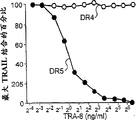
The sign of Fig. 1 .TRA-8.(a) binding specificity of TRA-8: western blot analysis (last figure): the recombination fusion protein of the TNFR family that surveys with TRA-8 or anti-human IgG.The 1st swimming lane: DR5/hIgG1 fusion rotein (immunogen); The 2nd swimming lane: DR4/hIgG1 (TRAIL-R1); The 3rd swimming lane: DR5/hIgG1; The 4th swimming lane: TRAIL-R3 (DcR-1)/hIgG1; The 5th swimming lane: TRAIL-R4 (DcR-2)/hIgG1; The 6th swimming lane: CD95/hIgG1; The 7th swimming lane: soluble TNF RI.Elisa assay (figure below): except the hole count of the number coupling western blotting in described hole, hole 8, hole 8 is mouse DR5/hIgG1 fusion roteins.(b) soluble TRAIL and TRA-8 are active with combining of DR5 and DR4: elisa plate is incubated with TRAIL or TRA-8 then with DR5/hIgG1 (left figure) or DR4/hIgG1 (middle graph) bag quilt.(c) flow cytometry of DR5 surface expression.Cos-7 cell with the pcDNA3 expression vector transfection that contains total length DR5 cDNA (solid histogram), DR4 cDNA (hollow histogram, solid line) or empty carrier (hollow histogram, dotted line).After the transfection 48 hours, cell dyeed with TRA-8, then the anti-mouse IgG1 dyeing of puting together with PE.(d) the original position immunohistochemical reaction of DR5: with the Cytospin slide of the Cos-7 cell of DR5 expression vector or control vector transfection after transfection 48 hours with TRA-8 dyeing, (e) killing activity of TRA-8: the Jurkat cell is hatched with the TRA-8 of prescribed concentration.After incubated overnight, get rid of mensuration by ATPLite, MTT and PI and measure cell survival.The result that ATPLite and MTT measure represents that with the per-cent of substratum contrast the result that PI measures represents with the per-cent of PI negative cells.(f) caspase activatory western blot analysis: the Jurkat cell is hatched the specified time with 500ng/ml TRA-8.Cell pyrolysis liquid separates by 15%SDS-PAGE, is made into trace, surveys with anti-caspase antibody.Arrow is represented the subunit through cutting of every kind of caspase.(g) caspase suppress to measure: with the Jurkat cell under the situation of the appointment caspase inhibitor that has various concentration with 50ng/ml TRA-8 overnight incubation.Measure the measurement cell survival by ATPLite.
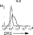
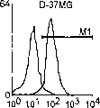
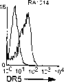
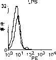
The cell surface expression of Fig. 2 .DR5 and the apoptotic susceptibility that DR5 is mediated.To hatch with TRA-8 or mouse IgG1 isotype control antibodies from normal T cell and B cell, T cell (a and a '), neurospongioma (b and b '), prostate cancer cell (c) and B cell (d) clone of peripheral blood fresh separated, the goat anti-mouse IgG 1 of puting together with PE is hatched then.Hollow histogram is represented described isotype antibody contrast, and solid histogram is represented TRA-8 dyeing.After soluble TRAIL (open circles) or TRA-8 (filled circles) overnight incubation, measure the measurement apoptosis by ATPLite, as shown in a, b ' and d.
Fig. 3 a ' .T clone U937 is hatched with TRA-8 or mouse IgG1 isotype control antibodies.After soluble TRAIL (open circles) or TRA-8 (filled circles) overnight incubation, measure the measurement apoptosis by ATPLite.
Fig. 3. glioma cell line (b) and prostate cancer cell line (c) are hatched with TRA-8 or mouse IgG1 isotype control antibodies.After soluble TRAIL (open circles) or TRA-8 (filled circles) overnight incubation, measure the measurement apoptosis by ATPLite.
Fig. 4 is a series of graphic representations, shows that people Jurkat cell is at (A) antibody type TRA-1, the TRA-8 that are exposed to prescribed concentration and TRA-10 and (B) cell survival after being exposed to TRAIL in the presence of the antibody type of the present invention shown in Fig. 4 of fixed concentration A;
The expression of Fig. 5 .DR5 in healthy tissues and cancerous tissue: healthy tissues homogenate and cancerous tissue homogenate are surveyed with TRA-8, show by chemoluminescence then.(a) the proteic western blot analysis of DR5 in the healthy tissues: the 1st swimming lane: liver, the 2nd swimming lane: brain, the 3rd swimming lane: lung, the 4th swimming lane: kidney, the 5th swimming lane: spleen, the 6th swimming lane: testis.The 7th swimming lane: ovary, the 8th swimming lane: heart, the 9th swimming lane: pancreas.B. the proteic western blot analysis of DR5 in the cancerous tissue.Detection comprises the cancerous tissue trace that derives from following cancer: ovary (the 1st swimming lane), lung (the 2nd swimming lane), liver (the 3rd swimming lane), rectum (the 4th swimming lane), uterine neck (the 5th swimming lane), skin (the 6th swimming lane), testis (the 7th swimming lane), Tiroidina (the 8th swimming lane), uterus (the 10th swimming lane), stomach (the 11st swimming lane), laryngopharynx (the 12nd swimming lane) and pancreas (the 13rd swimming lane).The original position immunohistochemistry of health adult tissue (c) and cancerous tissue (d).Freezing microtome section TRA-8 immunostaining.
The tumor promotion extremely of Fig. 6 .TRA-8.Give SCID mouse hypodermic inoculation 1321N1 cell.Give mouse vein the 2nd day injection potion 100 μ g TRA-8 (a) behind the tumor inoculation or behind tumor inoculation beginning injection in 7 days three dose of 100 μ g TRA-8 (b).Tumor growth passes through weight determination, and uses H﹠amp; E dyeing carrying out histological examination.Photo has shown the great-hearted tumor growth in the control mice, and does not have described tumor growth (c, last figure) in the mouse of TRA-8 treatment, has also shown the H﹠amp of tumour; E dye (c, figure below).Give SCID mouse mainline 10
6The Jurkat cell, treat with potion TRA-8 injection back the 2nd day then.After 7 days, the results splenocyte, with anti-people CD3 antibody staining, and by flow cytometry (d) or by immunohistochemistry (e) analysis.
Fig. 7 has shown the expression of cell surface DR5 among RA (A) and OA (B) synovial cell.1 * 10
6Former being commissioned to train supported the TRA-8 dyeing of synovial cell with affinity purification, goat anti-mouse IgG 1 antibody staining of puting together with PE then.Analyze 10,000 viable cell by FACSvantage.
Fig. 8 is a series of graphic representations, has shown that it is the apoptotic cell survival that becomes with TRAIL and TRA-8 concentration that TRA-8 (filled circles) with the recombinant soluble TRAIL (open circles) of various concentration or affinity purification induces RA (A) and OA (B) synovial cell's representative strains.Cell survival is represented with respect to the per-cent of the cpm of untreated cell with the cpm through the treatment cell.
Fig. 9 is a series of graphic representations, has shown the apoptotic caspase dependency of RA synovial cell DR5 mediation.RA synovial cell (RA512) is hatched with 50ng/ml soluble Fas part (square hollow), anti-Fas antibody (CH-11) (solid squares), soluble TRAIL (open circles) or anti-DR5 antibody (TRA-8) (filled circles) under the situation of the caspase inhibitor that has various concentration.After overnight incubation, measure cell survival by ATPLite.
Figure 10 A shows the variation analysis of NF κ b activated running gel.The RA1016 cell is hatched the specified time with 20ng/ml TNF-α, 50ng/ml soluble TRAIL or 50ng/ml TRA-8, carries out electrophoresis then.Figure 10 B and 10C are the graphic representations that shows that MMP-1 and MMP-3 produce.1 * 10
6The specified RA synovial cell of/ml is hatched with TNF-α (open circles), TRAIL (hollow triangle) or the TRA-8 (filled circles) of prescribed concentration.After the overnight incubation, collect culture supernatant.Measure the level of MMP in the culture supernatant by ELISA.
Figure 11 .TRA-8 is inducing hepatocyte toxicity not.(a) normal liver tissue is not expressed DR5.The paraffin section for preparing the cytospin prepared product of two kinds of normal liver tissues, a kind of hepatocellular carcinoma tissue and HepG2 cell is for H﹠amp; E dyeing, corresponding freezing microtome section dyes with TRA-8.(b) flow cytometry of DR5 cell surface expression.Dye with TRA-8, anti-Fas antibody (DX2) or isotype control antibodies from the liver cell and the HepG2 cell of two kinds of normal liver tissues and a routine hepatocellular carcinoma separate tissue.Solid histogram is represented TRA-8 or DX2 dyeing, and hollow histogram is corresponding isotype contrast.
Figure 12. be TRAIL but not TRA-8 inducing hepatocyte toxicity.Fresh normal liver cell is remained in the hepatocyte culture medium (Hepatocyte Culture Medium).(a) in specified time point, add linking agent or TRA-8 inducing hepatocyte apoptosis with 1 μ g/ml soluble TRAIL.Cell survival is measured by ATPLite.The result represents with the viable cell per-cent of comparing with the substratum contrast.Shade band indication TRAIL, and dark band is represented TRA-8.(b) hepatocellular pyknotic nucleus (condensed nuclei) passes through flow cytometry then with Hoechst 33352 dyeing.(c) cycloheximide is to the apoptotic influence of liver cell.Liver cell is in control medium or be added with in the substratum of 1 μ g/ml TRAIL or TRA-8 having (solid strip) or do not exist under the situation of (hollow band) 1 μ g/ml cycloheximide and cultivated 8 hours.Cell survival is measured by ATPLite.The result represents with three parts of mean value ± SEM that repeat culture of two experiments.(d) normal liver cell is to the comparison of the apoptotic susceptibility of DR5 and Fas mediation.The liver cell of fresh separated hatched 6 hours with soluble TRAIL, TRA-8, soluble Fas L or the anti-Fas mAb CH11 of prescribed concentration.Cell survival is measured by ATPLite and is measured.The result represents with the viable cell per-cent of comparing with the substratum contrast.For normal liver cell, represent the mean value ± SEM of 4 normal individuals.The hepatocellular carcinoma cells and the HepG2 cell that derive from a patient are represented with three parts of mean values that repeat culture.
Figure 13 .TRAIL induces hepatitis.Give inoculation 10 in the B6 mouse vein
9Pfu adenovirus carrier, described vector encoded " Tet-on " are transcribed the total length people TRAIL under the element control.TRAIL expresses and usually induces by the Fourth Ring of prescribed dose.(a) rna blot analysis that people TRAIL expresses in the liver.The inoculation carrier was also induced back 24 hours with tsiklomitsin, isolated total RNA from liver, and personnel selection TRAIL cDNA or beta-actin are surveyed.(b) serum level of AST.TRAIL transduceed back 24 hours, measured the AST serum level.(c) the hepatocellular necrocytosis of the adenovirus carrier infection of TRAIL mediation: give inoculation tsiklomitsin induction type adenovirus carrier in the B6 mouse vein.Inoculate back 48 hours, separate the liver cell that derives from the inoculation mouse and do not inoculate control mice, its TRAIL with prescribed concentration is hatched 8 hours (left figure).Hepatocellular cell survival is measured by ATPLite and is measured.After 48 hours, give the mouse mainline 10 μ g soluble human TRAIL of the above-mentioned adenovirus carrier of inoculation.Behind injection TRAIL 24 hours, measure AST serum level (right figure).The histologic analysis of (d and e) TRAIL inductive liver injury.Collect liver when transduceing back 24 hours (d) or 7 days (e) with TRAIL.Paraffin section is carried out H﹠amp; E dyeing is taken pictures with 100X (last figure) and 400X (figure below).
Figure 14 is a series of graphic representations, has shown by the flow cytometry of resting cell (hollow) and active cells (shade) to measure, from the activating T cell of human PBMC's purifying and the DR5 of B cell expressing increase level.
Figure 15 is the viability graphic representation that becomes with TRA-8 concentration that stimulates active cells that 48 hours purifying T cell and B cell and the Ficoll-Paque by different densities collect and parent cell with anti-CD3 or anti-μ shown in Figure 14.Viability is measured by ATPLite and is measured.
Figure 16 is histogram and flow cytometry graphic representation, has shown that the CD3 in the gate lymphocyte population of the NOD/SCID mouse that the NK cell of having injected human PBMC and TRA-8 or IgG (contrast) is exhausted expresses.
Figure 17 has shown the CD3 and the painted cell Photomicrograph of TUNEL of the mouse boosting tissue of describing in detail among the embodiment 13.
Figure 18 has shown chronic molten Lymphocytic leukemia (CCL) and the normal cytotoxicity graphic representation of human B cell in the presence of TRA-8, BISVIII and combination thereof.
Detailed Description Of The Invention
Can not scavenger-cell be because in the apoptosis-inducing system, have with comprise as an illustration described part, acceptor or born of the same parents in the relevant defective of the defective of the expression of Molecular regulator and effector molecule or function. The method that the invention provides a kind of rectification of defects type apoptosis-inducing system and set forth concrete defective in the given deficient cell apoptosis inducible system.
The present invention relates to the new monoclonal antibody of a class, described monoclonal antibody has and external selective apoptotic induction activity interior for concrete TRAIL acceptor (comprising DR5, DR4, DcR1 and DcR2) body. The present invention can be used as for the reagent of Apoptosis signal research usefulness and effectively for the cell of expressing the TRAIL acceptor curative of (comprise as an illustration the cancer cell of wide class, described Apoptosis system gone to regulate and the abnormality proliferation synovial cell of autoimmunity disease). Aspect particular type TRAIL acceptor, specificity is being arranged according to antibody of the present invention, although homology is arranged between them. Antibody of the present invention provides the targeted cells apoptosis of the cell of only expressing target TRAIL acceptor, and perhaps the TRAIL Apoptosis of the cell of target acceptor is expressed in blocking-up.
Anti-DR5 monoclonal antibody of the present invention can be used as apoptotic external effective inducer and the interior effectively inducer of apoptotic body of expressing DR5. The humanization fragment CDR sequence of being transplanted to the humanized antibody skeleton shows similar Apoptosis characteristic with fusion Anti-DR5 antibody of the present invention.
Up to now, not yet obtain being combined with cell surface DR5 and apoptotic monoclonal antibody of abduction delivering DR5 all in the external and external situation that is lacking crosslinking agent. Present invention resides in the animal model (for example heterograft animal) of disease or in vivo effectively as the Anti-DR5 antibody of curative. Although shown in vivo effective inducing apoptosis of tumour cell of soluble TRAIL, killing activity be it seems very low, and usually needs heavy dose and repeat administration (13). According to one of a series of Anti-DR5 antibodies of the present invention-TRA-8 the genetically modified animal of carrier DR5 be medicinal effectively, and also can be applicable to set up can be for the model of research DR5 and TRAIL effect.
According to the present invention, from animal used as test results for the TRAIL acceptor produce according to antibody of the present invention. By according to the present invention with described antibody humanization, to keep receptor-binding activity, and in the human subject body, bringing out immune response that weaken and the treatment tolerance, the anti-TRAIL receptor antibody of humanization of the present invention can be used as therapeutic activator or the antagonist of given TRAIL acceptor. The present invention is effectively as the interior therapeutic medicine, because do not need described anti-TRAIL receptor antibody is not carried out secondary cross-linking.
The present invention may extend to outside the single anti-TRAIL receptor antibody with excitability or Antagonism Apoptosis effect. But, make two or more anti-TRAIL receptor antibodies contact or contact in vivo with curee's body tissue external with cell culture, to produce Synergistic treatment. For example, to be U937 and Molt-4 be exposed to the anti-DR4 antibody of excitability and Anti-DR5 antibody responds to collaborative for glioma cell line U87 and hematopoietic cell, only shows limited success and only be exposed to the excitability Anti-DR5 antibody aspect cell death inducing.
In addition, when antibody during specifically in conjunction with one of bait acceptor-DcR1, DcR2 or OPG, the anti-TRAIL receptor antibody of Antagonism especially can be applicable among the present invention. Use and optionally seal the bait acceptor according to antibody of the present invention and in the cell type of expressing the bait acceptor, have and make described TRAIL binding equilibrium to the effect of TRAIL receptor change that can the transducer cell apoptotic signal. Therefore, in according to another kind of therapeutic alliance of the present invention, the bait receptor binding antibodies makes express cell to the excitability Apoptosis sense sensitization of transduction TRAIL receptors bind.
In another embodiment, the invention provides a kind of excitability epi-position of given TRAIL acceptor and method of Antagonism epi-position illustrated. In addition, according to the present invention, by the monoclonal antibody of utilizing one group to have respectively different variable regions or CDR district, illustrate polymorphism relevant with given TRAIL acceptor between the individuality. One group of monoclonal antibody that has characterized provides the ability that limits excitability and Antagonism epi-position and polymorphism. Therefore, in curee's screening that a group can be applicable to drug discovery and/or disease tendency according to monoclonal antibody of the present invention.
An again embodiment of the present invention relates to the fusion that comprises with the antigenicity fragment of the TRAIL acceptor of immunoglobulin (Ig), polypeptide or its fragment coupling. The TRAIL receptor fragments be defined as contain enough numbers base to bring out the immunogenic response for the natural TRAIL acceptor of expressing on curee's cell surface. The TRAIL acceptor merges fragment and comprises at least 10 amino acid. Domain-immunoglobulin fusion proteins or its fragment are defined as comprising natural or synthetic proteins or the polypeptide section that amino acid base with enough numbers is replied with the immunogenicity cascade that activates in curee's body in this article. The immunogene of the present invention that comprises the fusion of TRAIL receptor fragments and immunoglobulin fragment coupling can be used as the interior therapeutic medicine that brings out anti-TRAIL receptor antibody at curee's internal in-situ.
In yet another embodiment, the present invention can be effectively as gene therapy. Aspect a gene therapy of the present invention, the target cell carrier transfection of carrying corresponding to the expressible nucleotide sequence of TRAIL acceptor. Described carrier is conventional, and according to target cell the sensitiveness of described carrier is selected. Gene therapy vector comprises adenovirus vector pAdCMV5 as an illustration. When target cell or target tissue are expressed the TRAIL acceptor of described transfection, make described cell or tissue be exposed to antibody according to the TRAIL acceptor of the described transfection of specific binding of the present invention. People will appreciate that described anti-TRAIL receptor antibody is according to required treatment results, or agonistic antibody or antagonistic antibodies.
Antibody of the present invention is combined also effective with sensitizing agent. Sensitizing agent used herein is defined as any stimulation that comprises cell death inducing, comprises ultraviolet light, specifically comprises organic molecule, heavy metal and the Kinds of Free Radicals of bisindole maleimide.
Aspect treating malignant tumor, TRA-8 can induce most of TRAIL responsive type apoptosis of tumor cells in caspase dependency mode when lacking secondary crosslinking.TRA-8 shows and kills tumor promotion in the strong body.TRA-8 induces the ability of most of TRAIL sensitivity cell apoptosis, has confirmed that independent DR5 is enough to trigger apoptosis.Most of tumor cells expression cell surface DR5 that this paper describes in detail, and its susceptibility to the necrocytosis of TRA-8 inductive is suitable with its susceptibility to TRAIL, and expression DR5 is the apoptotic main death receptor of TRAIL mediation in most of tumour cells.Therefore, normal cell and cancer cells are working aspect the apoptotic selectivity of TRAIL mediation to the differentially expressed of DR5.TRA-8 walks around described bait acceptor, and induces the apoptosis of TRAIL mediation.Yet only minority TRAIL resistance tumor cell is to the TRA-8 sensitivity, shows that described bait acceptor be it seems not play an important role aspect the apoptosis resistance of TRAIL mediation at tumour cell.
Disappear and do not cause toxicity 3,4,22 though previous research has shown TRAIL induced tumor that system in animal body gives soluble form, as indicated in this paper, the people TRAIL of film combining form induces liver injury in the mouse body.Yet less than the susceptibility to the Fas part, and TRAIL do not have lethality to normal liver cell in vivo, proved that the liver toxicity of TRAIL is more much smaller than Fas part to the susceptibility of TRAIL inductive damage.Therefore, increment gives TRAIL and can be applicable in the cancer therapy gradually.
As detailed in this article, show that normal liver cell does not have the DR5 protein expression of conspicuous level, and relevant with the anti-TRA-8 inductive of liver cell apoptosis.DR5 and monoclonal antibody be crosslinked to be not enough to make up the death receptor that can trigger apoptotic homologous polymerization form.The marmoset experiment does not demonstrate the liver toxicity that TRA-8 gives.Therefore, the anti-DR5 antibody of agonistic monoclonal might have more selectivity and safer than soluble TRAIL as curative.
As screening assay, the present invention is highly suitable for detecting the little malignant cell bunch that still shows the normal cell form.With the original position cell section dyeing of the human cancer cell of lung cancer, prostate cancer and liver cancer identifying cancer cells easily will comprising of mark according to antibody of the present invention.Observe with the normal cell of same type and compare, these cancer cells are expressed very high-caliber DR5.Therefore, the present invention can be used as the sensitive screening method of early stage malignant tumour in the tissue that comprises lung, prostate gland and liver at least.This paper describes a kind of methods of treatment that is used to suppress with the abnormal cell proliferation of the disease-related of for example pernicious cancer and Lymphocytic leukemia in detail.
This paper describes the present invention in detail, the anti-people DR5 monoclonal antibody of called after TRA-8 especially, and its ATCC preserving number is PTA-1428.People will appreciate that, are extendible fully about detailed technology and the result of the anti-people DR5 of excitability monoclonal antibody TRA-8, are applicable to antagonism DR5 antibody and with the antibody that produces at DR4, DcR1 and DcR2 of excitability and the effect of antagonism mode.
The expression level of apoptosis acceptor (for example Fas) the not necessarily susceptibility with described cell pair cell apoptosis is relevant.For the apoptosis of TRAIL mediation, pointed out the bait receptor expression of TRAIL to influence the susceptibility of described cell.In addition, pointed out aspect FADD and the effective transducer cell apoptotic signal of caspase 8 approach, DR5 is certain relevant with DR4.The availability of the anti-DR5 monoclonal antibody of excitability, feasible adjusting and the relativity in the apoptosis of TRAIL mediation thereof that can illustrate the DR5 signal.Described cell is to the apoptotic susceptibility and its comparison to the apoptotic susceptibility of TRAIL mediation of TRA-8 mediation, provides about the effect of DR5 in the apoptosis that TRAIL mediates and the understanding in depth of mechanism that may influence susceptibility.
This advantage generally may extend to the anti-DR5 antibody of humanization of the present invention.By the known technology that in following examples, describes in detail, prepare a molecular cloning of anti-DR-5 antibody.Recombinant DNA method (33) can be used for making up the nucleotide sequence of coding monoclonal antibody molecule or its antigen binding domain hereinto effectively.
The invention enables to make up the anti-TRAIL receptor antibody of humanization, wherein said humanized antibody unlikely induces human anti-mouse antibody's (being called " HAMA " hereinafter) to reply (34), and still has effective antibody mediated effect subfunction.This paper used term " people " and " humanization " aspect antibody relate to and are expected at any antibody that brings out the weak immunogenic response of treatment tolerance in the human subject body.
The invention provides a kind of anti-DR5 antibody, the anti-DR5 antibody of humanization, TRA-8 heavy chain and light chain immunoglobulin (Ig) and humanization heavy chain and light chain immunoglobulin (Ig).Some truncate of these albumen or gene can be carried out the regulatory function or the enzyme function of described complete sequence albumen or gene.For example, the nucleotide sequence of proteins encoded can be expressed change by replacement, interpolation, disappearance or poly, with albumen or the gene that functional equivalent is provided.Because the degeneracy of nucleic acid coding sequence, coding can be used to implement the present invention with other sequence of the essentially identical aminoacid sequence of naturally occurring albumen.These include but not limited to comprise that thereby the encode complete nucleotide sequence of aforementioned polypeptides or its part, the replacement by the different codons of functional equivalent amino-acid residue in the described sequence of coding produces reticent the variation and reformed nucleotide sequence.By standard method (" CurrentMethods in Sequence Comparison and Analysis; " MacromoleculeSequencing and Synthesis, selected Methods and Applications, pp.127-149,1998, Alan R.Liss Inc.) calculates, expection can tolerate 25% sequence homology variation at the most according to the nucleotide sequence of immunoglobulin (Ig) of the present invention, as long as this varient constitutes the potent antibodies of identification TRAIL acceptor DR5.For example, the one or more amino-acid residues in the peptide sequence can be had similar polarity, be replaced as the reticent another kind of amino acid that changes of functional equivalent deposits yields.Aminoacid replacement in the described sequence can be under described amino acid be selected other member of classification.For example, nonpolar (hydrophobicity) amino acid comprises L-Ala, leucine, Isoleucine, Xie Ansuan, proline(Pro), phenylalanine, tryptophane and methionine(Met).Polar neutral amino acid comprises glycine, Serine, Threonine, halfcystine, tyrosine, l-asparagine and glutamine.Positively charged (alkalescence) amino acid comprises arginine, Methionin and Histidine.Electronegative (acidity) amino acid comprises aspartic acid and L-glutamic acid.Be also included within the scope of the invention have translate duration or afterwards difference modify protein or its fragment or the derivative of (for example by glycosylation, proteolytic cleavage, be connected etc.) with antibody molecule or other cell ligand.In addition, can carry out the recombinant vectors that code book is invented the nucleotide sequence of anti-DR5 antibody engineered, to revise the processing or the expression of carrier.
In addition, the nucleotide sequence of coding inhibitor can be at external or vivo mutations, to produce and/or to destroy translation sequences, homing sequence and/or terminator sequence, or produce variation in the coding region and/or form new restriction endonuclease site or destroy existing described site, with the external modification of further promotion.Can use any induced-mutation technique known in the art, include but not limited to external site-directed mutagenesis, J.Biol.Chem.253:6551, application Tab joint (Pharmacia) or the like.
The X-ray crystallography data show that the folding general cylindrical structural that forms a kind of length of antibody immunoglobulin contains two-layer respectively by 3 or 4 antiparallel b-pleated sheets that the b-chain is formed.In the variable region, flock together from three rings in each V district of H chain and L chain, form an antigen binding site.Each ring in these rings is called complementarity-determining region (CDR).The variability of CDR is the highest in the aminoacid sequence of described antibody.The variable region part that is not CDR part is called " framework region " (" FR " district), generally works keeping the CDR configuration aspects.All CDR that preferably will derive from given antibody are transplanted in the receptor antibody, so that keep the land for TRAIL acceptor epi-position district.It is effective hereinto that expection is transplanted to the part among whole CDR in the donor.People understand, and transplant generally the residue in an amino acid or a district to be replaced with another kind of amino-acid residue.Yet, especially shift when district sometimes, can add as required or delete or replace one or more residues, such disappearance and insertion and suitable replacement and inversion are in the technical scope of this area.
For example the L chain by will resisting the TRAIL acceptor monoclonal antibody and each CDR of H chain subunit are transplanted in the corresponding CDR district of people's antibody, thereby will obtain antibody of the present invention effectively at the mouse monoclonal antibody humanization of TRAIL acceptor.
Adopt known technology, made up the antibody fragment that contains described molecule idiotype, described fragment also is effective hereinto.For example, this class fragment comprises anti-TRAIL acceptor (AB ') 2 fragments that can be by producing with the described antibody molecule of gastric pepsin digestion, the TRAIL receptor antibody AB ' fragment that produces by the described TRAIL acceptor of reduction (AB ') 2 segmental disulphide bridgeses and as an illustration by handle the antibody fragment of described antibody molecule generation with papoid and reductive agent.
Specifically, by personnel selection DR5 immune mouse, the splenocyte or the lymph-node cell that will derive from described mouse subsequently can obtain hybridoma with the murine myeloma cell fusion, cultivate described hybridoma, can obtain anti-DR5 monoclonal antibody TRA-8.
MONOCLONAL ANTIBODIES SPECIFIC FOR comprises the steps: as an illustration
A) purifying biological macromole is as antigen;
B) adopting the described antigen initial immunity animal of injection, give described animal bloodletting, and
Analyze antibody titer so that determine when that the preparation antibody producing is thin after the taking-up spleen
Born of the same parents;
C) preparation myeloma cell;
D) antibody producing cells and myeloma cell are merged;
E) hybridoma of required antibody is produced in selection;
F) preparation single cell clone (clone);
G) randomly, cultivate described hybridoma, or allow and transplanted hybridoma
Growth of animal, to prepare described monoclonal antibody on a large scale; With
H) so biological activity and the specificity or analysis of the monoclonal antibody of preparation of test
The factor of character characteristic.
Describe the method that is used for anti-DR5 Monoclonal Antibody in detail below with reference to above-mentioned steps.The method for preparing antibody of the present invention only is the explanation preparation method, is not to be restrictive.Can adopt other known method, perhaps adopt the method for revising, for example utilize antibody producing cells, and do not utilize splenocyte and myelomatosis.
(a) antigenic preparation
Can effectively obtain by following steps: by utilizing ADENO-Quest test kit (QuantumBiotechnologies Inc. as described antigenic recombinant protein (being called " recombinant human DR5 " hereinafter), Canada), expression vector pAdDR5-IgG transfection QBI-293A cell with the fusion rotein (referring to PTA-1428) in the Fc district that comprises people DR5 ectodomain and human IgG1's antibody (being called " IgG " hereinafter) makes it express described fusion rotein, collects and the partial purification expression product.The DNA of plasmid pAdDR5-IgG by the fusion rotein of will encode people DR5 and human IgG inserts to make up among the pAdCMV5 and forms, and this plasmid is a kind of expression vector that is used for zooblast.Other material for example DNA, described carrier and the described host of encoding D R5 is effective hereinto.
People DR5 and the IgG fusion rotein produced in the culture supernatant with the QBI-293A cell of carrier pAdDR5-IgG transfection are by A albumen-Sepharose affinity chromatography or G albumen-Sepharose affinity chromatography or adopt Resource Q post (trade mark; Pharmacia) ion-exchange chromatography comes partial purification.
Perhaps, the purifying DR5 that derives from human cell line's cytolemma is used as antigen.In addition, because the primary structure of DR5 is known (referring to PTA-1428), so can pass through currently known methods, for example the Sanger forensic chemistry synthesizes the peptide that comprises SEQ ID No.1 aminoacid sequence, and used as antigen.
(b) preparation of antibody producing cells
With with adjuvant (for example Fu Shi fully or Freund or aluminum oxide) blended step (a) in the immunogen immune mouse that produces.Other suitable laboratory animal comprises rat, cavy, rabbit, dog, chicken, horse, pig, ox and sheep as an illustration.
The suitable route of administration of immunization experiment animal comprises subcutaneous injection, peritoneal injection, intravenous injection, intradermal injection and intramuscularly approach, preferred subcutaneous injection and peritoneal injection.
Immunity is randomly carried out by potion or with the several repeated doses of appropriate intervals (preferably 1-5 week).Monitoring is through the serum antibody titer of the animal of immunity, has enough the animal of high antibody titer and selects as the antibody producing cells source.Selection has the animal that height tires makes that the efficient of down-stream is higher.The cell that is used for merging is subsequently generally gathered in the crops from 3-5 days the animal in last immunity back.
The method of analyzing antibody titer comprises various well-known technology, for example radioimmunoassay (being called " RIA " hereinafter), solid-phase enzyme immunoassay (being called " ELISA " hereinafter), fluorescence antibody are measured and passive hemagglutination is measured, since detection sensitivity, fast, accurately and the potentiality of automatization, preferred RIA and ELISA.
The mensuration of antibody titer can for example be undertaken by following ELISA.At first, purified or partially purified DR5 is adsorbed to solid phase (for example 96 hole elisa plates) surface, (for example bovine serum albumin (BSA) sealing does not combine any residual surface of DR5 as yet to use the albumen irrelevant with antigen then.After the washing, hole surface is contacted with the mice serum sample of serial dilution, make that the anti-DR5 antibody in the sample can combine with described antigen.Adding is marked anti-mouse antibodies as the enzyme of second antibody, to combine with mouse antibodies.After the washing, add enzyme substrates, antibody titer is estimated in the variation of the absorbancy due to the colour developing that causes by measure because substrate variation etc.
(c) myeloma cell's preparation
The cell of setting up mouse cell lines of must controlling oneself is originated as the myeloma cell, comprises that the 8-azaguanine resistance mouse myeloma strain that for example derives from BALB/c is P3X63Ag8U.1 (P3-U1) (35), P3/NSI/1-Ag4-1 (NS-1) (36), Sp2/0-Ag14 (SP-2) (37), P3X63Ag8.653 (653) (38) and P3X63Ag8 (X63) (39).With selected clone sequential transfer to suitable medium for example in the 8-azaguanine substratum.8-azaguanine substratum comprises improved DulbeccoShi substratum of IscoveShi (being called " IMDM " hereinafter) or the improved Eagle substratum of DulbeccoShi (being called " DMEM " hereinafter).In the RPMI-1640 substratum, replenish glutamine, 2 mercapto ethanol, gentamicin, foetal calf serum (being called " FCS " hereinafter) and 8-azaguanine.Then, before fusion 3-4 days, with cell transfer to normal substratum for example contain 10%FCS the ASF104 substratum (Ajinomoto, K.K.) in, to guarantee can to obtain at least 2 * 10 the same day merging
7Cell.
(d) cytogamy
The lymphocyte and the plasma cell that derive from any suitable part of described animal are the progenitor cells of producing described antibody.Lymphocyte or plasma cell source comprise spleen, lymphoglandula, peripheral blood or any its suitable combination as an illustration, and splenocyte is the most frequently used source.
After the booster shots, from mouse, take out the tissue that has antibody producing cells the last time with predetermined antibody titer.The appropriate technology that is used for splenocyte at present and merges the myeloma cell that step c) prepares utilizes polyoxyethylene glycol.
Described integration technology comprises with serum free medium (for example RPMI 1640) or phosphate buffered saline(PBS) (being called " PBS " hereinafter) washing splenocyte and myeloma cell, make splenocyte and myeloma cell's number ratio be about 5: 1 to 10: 1, centrifugal then.In abandoning supernatant and after making sedimentation cell fully loose, drip serum free medium and mixing that 1ml contains 50% (w/v) polyoxyethylene glycol (m.w.1,000-4,000).Subsequently, slowly add the 10ml serum free medium, centrifugal then.Abandoning supernatant once more, with sedimentary cell suspension in the HAT substratum (being called " HAT " hereinafter) of an amount of solution that contains xanthoglobulin, aminopterin and thymidine and mouse interleukin-2 (being called " IL-2 " hereinafter).Suspension is joined in each hole of culture plate (also abbreviating " plate " hereinafter as), then at 5%v/v CO
2Under existing, in 37 ℃ of about 2 weeks of cultivation, can suitably replenish interpolation to the HAT substratum.
(e) selection of hybridoma
When the myelomatosis strain of adopting is anti-8-azaguanine, promptly during its hypoxanthine guanine phosphoribosyltransferase (HGPRT) defectiveness, any nonfused myeloma cell and any myelomatosis-myelomatosis syzygy all can not be survived in the HAT substratum.On the other hand, antibody producing cells each other syzygy and antibody producing cells and myeloma cell's hybridoma can survive the life-span that the former is only limited.Therefore, cultured continuously causes only selecting required hybridoma in the HAT substratum.
It is colony that the hybridoma of gained is cultivated, and then it is transferred to the HAT substratum (in the HT substratum) that lacks aminopterin.After this, take out the culture supernatant of equal portions, tire to measure anti-Fas antibody by for example ELISA.When above-mentioned fusion rotein is used as ELISA antigen, the also essential clone who eliminates production and human IgG1's Fc district specificity bonded antibody.Whether this clone's existence can for example adopt Fas-IgG1 or IgG1 to be confirmed by ELISA as antigen.
(f) clone
Then, will adopt with the middle method similar methods of measuring antibody titer of step (b) to show the hybridoma of producing specific antibody, transfer in another plate for the clone.Suitable cloning process comprises: limiting dilution assay, and wherein, make each hole of plate contain a cell with the hybridoma dilution, cultivate then; Soft agar assay is wherein cultivated the back and is reclaimed colony in soft agar medium; Adopt micromanipulator to separate unicellular for cultured method; " sorting-clone (sort-a-clone) " wherein separates unicellular by cell sorter.
For each hole that demonstrates certain antibody titer, will repeat 2-4 time according to clone's program of for example limiting dilution assay, select to have clone that stabilization of antibodies tires as anti-DR5 monoclonal antibody production hybridoma.The hybridoma of producing anti-mouse DR5 antibody is by selecting with the similarity method that obtains anti-DR5 monoclonal antibody production clone.
Mouse-mouse hybridoma TRA-8 as antibody of the present invention basis is preserved in American type culture collection (American Type CultureCollection) on March 1st, 2000, and preserving number is PTA-1428.Therefore, when utilizing mouse-mouse hybridoma TRA-8 or any hybridoma that other has been set up to prepare antibody, can adopt the program of saving step (a)-(f), beginning to be prepared from following steps (g).
(g) cultivate hybridoma with the preparation monoclonal antibody
To cultivate in normal substratum by the hybridoma that the clone obtains then, but not cultivate in the HT substratum., perhaps cultivate by rolling flask culture with big culturing bottle, carry out large scale culturing by turn.Results derive from the supernatant liquor of large scale culturing then, adopt appropriate method example gel well known to those skilled in the art to filter and carry out purifying, to obtain the anti-DR5 monoclonal antibody as antibody of the present invention basis.Hybridoma also can be in the growth of homology mouse (for example BALB/c mouse or nu/nu mouse) intraperitoneal, to obtain to contain the ascites of a large amount of anti-DR5 monoclonal antibodies.Can be easily with commercially available monoclonal antibody purification kit (for example, MAbTrap GII Kit; Pharmacia) antibody that comes purifying to gather in the crops.
As above Zhi Bei monoclonal antibody has high degree of specificity to people DR5.
(h) mensuration of monoclonal antibody
The suitable authentication method of the isotype of described monoclonal antibody and subclass comprises Ouchterlony method, ELISA and RIA.Preferably use the commercial reagent box to identify, for example use Mouse Typer Kit (trade mark; BioRad).
Can carry out proteinic quantitative by the Folin-Lowry method or by calculating (1.4 (OD280)=immunoglobulin (Ig) 1mg/ml) based on the 280nm absorbancy.
The evaluation of the epi-position that described monoclonal antibody is discerned is following to be carried out.At first, the various piece structure of the molecule discerned of preparation monoclonal antibody.Described part-structure is by wherein with in the suitable expression plasmid of the DNA insertion of the method for the various piece peptide of the described molecule of the synthetic preparation of known oligopeptides synthetic technology or the required part of polypeptide of wherein will encode, express in suitable host (for example intestinal bacteria) and prepare with the method for producing described peptide then.Generally speaking, for reaching above-mentioned target, these two kinds of methods usually are used in combination.For example, by the gene engineering of having set up, can prepare a series of polypeptide that begin suitably to reduce length from the C-terminal or the N-terminal of described antigen protein.By determining described fragment and described antibody response, obtain to be bordering on the described epi-position of ideal position.
By synthesizing various less oligopeptides or its mutant corresponding to described peptide with the oligopeptides synthetic technology of having set up, with measure institute's peptide to as the binding characteristic of the anti-DR5 monoclonal antibody for preparing antibody of the present invention basis and described peptide and described monoclonal antibody to antigen bonded competitive inhibition, can identify described epi-position more subtly.Can be easily with the commercial reagent box for example SPOTs Kit (Genosys Biotechnologies Inc.) with based on a series of multipin peptide synthetic agent boxes (Chiron Corp.) of multipin synthesis method, obtains miscellaneous oligopeptides.
Antibody of the present invention has following various functional performance a)-f), every specific character by for example hereinafter described method confirmed.
A) TRA-8 combines with the specificity of the cell of expressing human DR5
A specific characteristic of the present invention is the ability in conjunction with cell surface DR5.This flow cytometry by the cell of expression DR5 proves.At first, adopt COS-7 cell, prove the specific cell surface bonding of DR5 with the full-length cDNA transfection of coding people DR5.Specifically, TRA-8 only discerns the COS-7 cell with the DR5 transfection, and the nonrecognition COS-7 cell of the carrier transfection of empty map carrier or encoding D R4.Secondly, the human malignancies cell of test three kinds of different sources-hematopoietic lineages, neurospongioma and prostate cancers.These transform the cell surface DR5 that the great majority in the tumour cell are expressed conspicuous level, though the expression level variation is very big.The 3rd, check two groups of people's synovioblasts of former generation that derive from RA patient and OA patient.Compare with the OA cell, all RA synovial cells all express remarkable higher levels of DR5.
B) human malignant lesion is apoptotic external evoked when shortage is crosslinked
Between the cells in vitro incubation period, with the antibody of various concentration, especially TRA-8, measure (ATPLite) by cell survival, measure the antibody recognition TRAIL acceptor that produces according to the present invention and directly induce the apoptotic ability of human malignant lesion.Most of tumour cells are to TRA-8 inductive apoptosis sensitivity.For some cell, TRA-8 demonstrates strong apoptosis-inducing activity, and for example TRA-8 can induce people Jurkat apoptosis in the pg/ml level.Importantly, TRA-8 inductive apoptosis does not need crosslinked, and in most cells, TRA-8 shows when having toughener than the stronger apoptosis-inducing activity of reorganization soluble TRAIL.
C) kill tumor promotion in the body of TRA-8
Assessment TRA-8 kills tumor promotion in two kinds of SCID/ human tumor cells models.At first, give inoculation human leukemia Jurkat cell in the SCID mouse vein, with potion (100 μ g) TRA-8 treatment.The result shows, measures through the flow cytometry and the original position immunohistochemical staining of Jurkat cell, and by treating with TRA-8, the Jurkat cells that great majority are transplanted are eliminated from peripheral blood and spleen.Secondly, subcutaneous vaccination people astrocytoma cell 1321N1 in the SCID mouse treats the mouse that has tumour with potion TRA-8.Determine that according to tumour size and histologic analysis the growth of the 1321N1 cell of transplanting is subjected to remarkable inhibition in the mouse of TRA-8 treatment.
D) identify RA synovial cell with TRA-8
Tested from 8 RA patients and 4 isolating former generation synovial cells' of OA patient DR5 cell surface expression.TRA-8 can make all RA cell dyeing positives, but negative for all OA cell dyeings.Therefore, by the surface expression that detects DR5 with TRA-8, can distinguish RA and OA.
E) TRA-8 is to RA synovioblast apoptosis induced
Exist under the situation of various concentration TRA-8 between external incubation period, measuring, measuring the ability that TRA-8 induces RA synovial cell's apoptosis by cell survival.All RA cells all show the susceptibility of 100ng/ml TRA-8 high level to medium level.On the contrary, all OA cells all have resistance to TRA-8 inductive apoptosis basically.Importantly, TRA-8 shows than soluble TRAIL and adds toughener better to RA synovial cell's apoptosis-inducing activity.In addition, (CH-11) compares with anti-Fas antibody, and TRA-8 shows the selectivity stronger to RA synovial cell.
F) TRA-8 does not induce the generation of MMP in RA synovial cell
Because TRA-8 is the same with TNF-α, can in RA synovial cell, induce NF-kb to activate, so measured TRA-8 produces MMP1 and MMP3 to the synovial cell influence.Though TNF-α induces the dose-dependently of MMP to increase, TRA-8 can not induce any MMP to produce, and under some concentration, TRA-8 reduces the generation of MMP among the RA synovial cell slightly.
G) TRA-8 induces multiple caspase activation
Because caspase plays a crucial role, induce caspase activatory ability so in people Jurkat cell, measure TRA-8 in cell death inducing.When the Jurkat cell, is determined by western blot analysis and caspase cutting analysis when TRA-8 is hatched with low dosage (50ng/ml), to hatching back 15 minutes, observe the activation of caspase 8, caspase 9 and caspase 3 early.With regard to caspase activatory time, quantity and intensity, antibody of the present invention comprises that illustrative antibody TRA-8 shows than the much better activity of any other known apoptosis-inducing antibody (for example anti-people Fas antibody (CH-11)).
Therefore, antibody of the present invention is the material with the selective induction pathogenic cell apoptosis characteristic as effect (a) and (g).Therefore, it can be used as prophylactic agent and the curative that is used for improper survival of cell or the improper propagation relative disease of cell, and described disease for example is attributable to those diseases that the apoptosis system comprises the dysregulation of Fas/Fas Fas lignand system.
For example human leukemia cell line Jurkat (American type culture collection preserving number TIB-152 (American TypeCulture No.TIB-152)) and astrocytoma cell are 1321N1 by culturing cell in adding the substratum of test sample, and measure the measurement viability by for example ATPLite, confirm the apoptotic ability of antibody induction of the present invention.
Antibody of the present invention, especially to the mankind's immunogenicity and people's antibody anti-DR5 antibody much at one, can be used as prophylactic agent or curative with improper survival of cell or improper propagation relative disease, described disease comprises those diseases that are attributable to apoptosis system dysregulation in the autoimmune disease, and described disease comprises systemic lupus erythematous as an illustration, Hashimoto's disease (Hashimoto ' s disease), rheumatoid arthritis, graft versus host disease (GVH disease), xerodermosteosis, pernicious anemia, Addison disease (Addison disease), scleroderma, goodpasture's syndrome (Goodpasture ' s syndrome), regional ileitis, autoimmune hemolytic anemia, infertility, myasthenia gravis, multiple sclerosis, basedow's disease (Basedow ' s disease), thrombopenic purpura, insulin-dependent diabetes, transformation reactions, idiopathic disease, arteriosclerosis, myocarditis, myocardosis, glomerulonephritis, hypoplastic anemia, repulsion after the organ transplantation and lung, prostate gland, liver, ovary, many malignant tumours of Lymphoid tissue and breast tissue.
This prophylactic agent or curative can give with various forms.Suitable mode of administration comprises oral administration, for example gives by tablet, capsule, granule, powder and syrup, perhaps by parenteral admin, for example by injection, instillation and suppository administration.
Described antibody or curative can oral administration, in the rectum, brain pond, in the Intraventricular, encephalic, sheath, intravaginal, parenteral (intravenously, intramuscular or subcutaneous), local (powder, ointment or drops), by peritoneal injection, through skin, by sucking or giving as cheek or nasal spray.The actual institute expense of described antibody or curative will change according to the curee, depend on the position of severity, tumour of curee's age, body weight, general health situation, disease to be treated and size, used particular compound, mode of administration etc.The normal experiment that those skilled in the art only provide with this paper can be determined appropriate vol.Antibody typical single doses weight range is 0.1-10,000 microgram, preferably 1-100 microgram.The scope of typical antibody concentration is the every milliliter of 0.2-2000 nanogram that gives in the carrier.
Planned mode of administration, described antibody or curative can be in the medicinal compositionss of solid, semisolid or liquid dosage form, described formulation is tablet, suppository, pill, capsule, powder, liquid or suspension agent for example, is preferably to be applicable to the unit dosage that once gives exact dosage desired.Described composition will comprise the selected matrix (substrate) and the pharmaceutically acceptable carrier of significant quantity, can comprise other medicines, medicinal reagent, carrier or thinner in addition.So-called " pharmaceutically acceptable " is meant that not to be biology desirable or at the undesirable material of others, described material can give individuality with selected matrix, and does not cause significant undesirable biological effect or any other interaction between component to be contained in harmful mode and the described medicinal compositions.
The composition that is applicable to parenteral injection can comprise physiologically acceptable sterile aqueous or non-aqueous solution, dispersion, suspension or emulsion and be applicable to and is redeveloped into the sterilized powder of aseptic injection with solution or dispersion.The example of suitable water-based or nonaqueous carrier, thinner, solvent or solvent comprises water, ethanol, polyalcohols (propylene glycol, polyoxyethylene glycol, glycerine etc.), its suitable mixture, vegetables oil (for example sweet oil) and injection organic ester, for example ethyl oleate.Can for example utilize dressing for example Yelkin TTS, by under the situation of dispersion, keeping required particle diameter and utilizing tensio-active agent, can keep suitable flowability.
These compositions also can contain auxiliary material, for example sanitas, wetting agent, emulsifying agent and dispersion agent.With various antibacterium medicines and antifungal drug for example parabens, butylene-chlorohydrin, phenol, Sorbic Acid etc., guarantee to prevent action of microorganisms.Also may need to comprise isotonic agent, for example sugar, sodium-chlor etc.The prolongation of injectable drug form absorbs to utilize and postpones the reagent that absorbs for example monostearate aluminium and gelatin reach.
The solid dosage that is used for oral administration comprises capsule, tablet, pill, powder and granule.In this class solid dosage, active compound and at least a inertia conventional excipients (or carrier) for example Trisodium Citrate or Lin Suanergai or following material are mixed: (a) weighting agent or extender, starch for example, lactose, sucrose, glucose, N.F,USP MANNITOL and silicic acid, (b) tackiness agent, carboxymethyl cellulose for example, alginate, gelatin, polyvinylpyrrolidone, sucrose and gum arabic, (c) wetting agent, glycerine for example, (d) disintegrating agent, agar for example, lime carbonate, yam starch or tapioca (flour), alginic acid, some composition silicate and yellow soda ash, (e) solution retarding agent (retarder), paraffin for example, (f) absorption enhancer, for example quaternary ammonium compound, (g) wetting agent, for example hexadecanol and monostearin, (h) absorption agent, for example kaolin and wilkinite, (i) lubricant, for example talcum powder, calcium stearate, Magnesium Stearate, solid polyethylene glycol, sodium lauryl sulphate or its mixture.With regard to capsule, tablet and pill, described formulation also can comprise buffer reagent.
The solids composition of similar type also can be as utilizing such as the weighting agent in the soft hard-filled gelatin capsule of the vehicle of lactose and high molecular weight polyethylene glycol etc.
Solid dosage such as tablet, dragee, capsule, pill and granule can prepare with dressing and housing, for example enteric coating and other Drug coating known in the art.They can contain opalizer, can be such compositions also, make them discharge one or more described active compounds in certain part of enteron aisle with delayed mode.The example of operable embedding composition is polymeric material and wax.If suitable, described active compound also can be for having the micro-capsule form of one or more above-mentioned vehicle.
The liquid dosage form that is used for oral administration comprises pharmaceutically acceptable emulsion, solution, suspensoid, syrup and elixir.Remove described active ingredient beyond the region of objective existence, described liquid dosage form can contain this area inert diluent commonly used for example water or other solvent, solubilizing agent and emulsifying agent, for example ethanol, Virahol, ethyl-carbonate, ethyl acetate, phenylcarbinol, peruscabin, propylene glycol, 1, the fatty acid ester of 3-butyleneglycol, dimethyl formamide, oil, particularly oleum gossypii seminis, peanut oil, maize germ, sweet oil, Viscotrol C and sesame oil, glycerine, tetrahydrofurfuryl alcohol, polyoxyethylene glycol and anhydro sorbitol or the mixture of these materials etc.
Except that this class inert diluent, described composition can also comprise auxiliary material, for example wetting agent, emulsifying agent and suspension agent, sweeting agent, correctives and spices.
Suspensoid removes and contains described active ingredient beyond the region of objective existence, can also contain suspension agent, for example mixture of ethoxylation isooctadecanol, polyoxyethylene sorbitol and Isosorbide Dinitrate, Microcrystalline Cellulose, aluminium hydroxide (aluminium metahydroxide), wilkinite, agar and tragakanta or these materials etc.
The composition that is used for rectal administration is suppository preferably, described suppository can be by mixing compound of the present invention with suitable non-irritating excipient or carrier (for example theobroma oil, polyoxyethylene glycol or suppository wax), described vehicle or carrier are solid at normal temperatures, but be liquid under body temperature, therefore be dissolved in rectum or the vaginal canal and discharge described active compound.
The formulation that is used for the topical administration The compounds of this invention comprises ointment, powder, sprays and inhalation.As required, described activeconstituents mixes with physiologically acceptable carrier and any sanitas, buffer reagent or propellent under aseptic condition.Ophthalmic preparation, ophthalmic ointment, powder and solution also are considered to be within the purview.
Term used herein " pharmacy acceptable salt, ester, acid amides and prodrug " is meant those carboxylate salts, amino acid addition salt, ester, acid amides and the prodrug of the The compounds of this invention in rational medical judgment scope, they are applicable to patient tissue and contact, and there are not over-drastic toxicity, pungency, anaphylaxis etc., match with rational interests/risk-ratio, purposes for its plan is effectively, also is meant the zwitterionic form of The compounds of this invention when suitable.Term " salt " is meant the nontoxic relatively inorganic and organic acid addition salt of The compounds of this invention.These salt can prepare during the last separation and purification of described compound on the spot, perhaps by independent purifying compounds and appropriate organic or inorganic acid reaction with free alkali form, separate the salt that generates thus then.Exemplary salt comprises hydrobromate, hydrochloride, vitriol, hydrosulfate, nitrate, acetate, oxalate, valerate, oleate, palmitate, stearate, lauroleate, borate, benzoate, lactic acid salt, phosphoric acid salt, tosylate, Citrate trianion, maleate, fumarate, succinate, tartrate, naphthoate, mesylate, glucose enanthate, Lactobionate, mesylate and dodecane sulfonate etc.These can comprise based on basic metal and alkaline-earth metal for example positively charged ion and nontoxic ammonium, quaternary ammonium and the amine positively charged ion of sodium, lithium, potassium, calcium, magnesium etc., include but not limited to ammonium, tetramethyl-ammonium, tetraethyl ammonium, methylamine, dimethylamine, Trimethylamine 99, triethylamine, ethamine etc.(referring to for example S.M.Barge etc., " Pharmaceutical Salts, " J.Pharm.Sci., 1977,66:1-19, the document is attached to herein by reference.)
Term " prodrug " is meant in vivo and is for example transformed the compound that produces the following formula parent compound rapidly easily by hydrolysis in the blood.At T.Higuchi and V.Stella, " Pro-drugsas Novel Delivery Systems; " A.C.S.Symposium Series Vol.14 and Bioreversible Carriers in Drug Design, Edward B.Roche writes, AmericanPharmaceutical Association and Pergamon Press provides comprehensive argumentation in 1987.
Target cell is a kind of zooblast, and described animal comprises the mankind, non-human primates, rat, mouse, cavy, rabbit, goat, sheep, ox, horse, chicken, pig, marmoset and ferret as an illustration.
In addition, antibody of the present invention or curative can exist with pharmaceutically acceptable solvent (for example water, ethanol etc.) with non-solventization and solvation form.Generally speaking, for the object of the invention, the solvation form is considered to be equal to the non-solvent form.
By known technology antibody purification molecule, described technology comprises amino the absorption or amino affinity chromatography, for example high pressure liquid chromatography or its combination of chromatographic technique as an illustration.
Another aspect of the present invention comprises and is used for anti-TRAIL receptor antibody of biological activity or the anti-TRAIL receptor antibody of humanization are passed to vertebrate pharmaceutical preparation.Pharmaceutical preparation comprises anti-TRAIL receptor antibody or its fragment, a kind of pharmaceutically acceptable carrier and container that described carrier and described antibody are housed with sterile manner of medicinal significant quantity.
In a preferred embodiment of the invention, the anti-DR5 antibody of medicinal significant quantity suppresses cell proliferation by contacting with target cell.The medicinal significant quantity of the humanized antibody of the antibody of identification DR5 or identification DR5 is to give the individual amount that is enough to cause required effect.The required effect that gives the DR5 identification antibody of medicinal significant quantity comprises target cell death, to the growth-inhibiting of target cell, stimulate DR5, combine and improve NF κ B level or activity in the target cell with DR5.Target cell is the cell of expressing DR5, comprises excrescent cell and tumour as an illustration, for example papilloma and wart; Breast cancer, colorectal carcinoma, hepatoma, leukemia, lung cancer, melanoma, myelomatosis, osteosarcoma, ovarian cancer, carcinoma of the pancreas, prostate cancer, incidence cancer, thyroid carcinoma, uterus carcinoma and brain tumor be astrocytoma for example.In vivo, described target cell is a cell of suffering from the individuality of pathologic conditions, and described illness comprises those illnesss of the unusual or dysregulation of wherein cell proliferation, for example malignant tumour or innocent tumour and rheumatoid arthritis.
In another preferred embodiment, described target cell is the contact treatment medicine also.
Curative is compound or a composition of effectively alleviating pathologic conditions.An illustrative example of curative comprises antineoplastic compound.
Antineoplastic compound is compound or the composition that effectively suppresses the growth of misgrowth cell or make cessation of growth cessation.The medicinal significant quantity of antineoplastic compound is to give the amount that growth that individual being enough to make the misgrowth cell is suppressed or blocks.The illustrative example of antineoplastic compound comprises bleomycin, carboplatin, Chlorambucil, cis-platinum, colchicine, endoxan, daunorubicin, dactinomycin, stilboestrol Dx, Etoposide, 5 FU 5 fluorouracil, floxuridine, melphalan, methotrexate, mitomycin, Ismipur, teniposide, 6-thioguanine, vincristine(VCR) and vinealeucoblastine(VLB).Other example of antineoplastic compound and curative can find in following document: The Merck Manua of Diagnosis and Therapy, the 15th edition, Berkow etc. write, and 1987, Rahway, N.J. with Metabolism andAction of Anti-Cancer Drugs such as Sladek, 1987, Powis etc. write, Taylor and Francis, New York, N.Y..
Antibody of the present invention can be further and other therapies for example chemotherapy and radiotherapy combination therapy malignant tumour, therapeutic efficiency can with apoptosis inducing compounds for example bisindolylmaleimidesfor VIII strengthen.
Compare with the anti-DR5 antibody (24) of previous announcement, the apoptosis-inducing activity of illustrative TRA-8 antibody of the present invention is very strong, and can induce the Jurkat apoptosis with the pg/ml level external, and proof is killed the soluble TRAIL that tumor promotion is better than previous report in vivo.Vein gives the growth that potion TRA-8 is enough to suppress solid tumor and hematopoiesis tumour cell, then needs quite high dosage (every day, 500ug gave 10 days) with tumor regression in the soluble TRAIL inductor.Anti-TRAIL receptor antibody of the present invention be it seems the same with soluble TRAIL safe, because exemplary antibodies TRA-8 does not induce non-conversion apoptosis of fibroblasts.
Carrier of the present invention comprises the one section coding anti-DR5 antibody immunoglobulin heavy chain that effectively is connected with controlling element (for example promotor or enhanser) or the nucleotide sequence of light chain." effectively connect " is meant that the layout of the nucleotide sequence that is constituted makes and can carries out its normal function.Therefore, can instruct with controlling element that the nucleotide sequence of coded polypeptide effectively is connected and transcribe, duplicate and/or the translation of described polypeptide.Those of skill in the art will recognize that single carrier randomly comprises the heavy chain immunoglobulin of anti-DR5 antibody and the encoding sequence of light chain.
Description following examples are for method of the present invention and result are described.These embodiment comprise all aspects of the present invention, but representational method and result are described.These embodiment do not get rid of conspicuous for those skilled in the art equivalent of the present invention and variation.The antigenic preparation of embodiment 1.DR5
1.1DR5 the clone of cDNA
By following RT-PCR method, adopt following material and reaction, the proteic DNA of clones coding people DR5:
A) template
By adopting TRIzol reagent (GIBCO BRL) to extract total RNA of HeLa cell.The cDNA that obtains by the instruction manual that provides according to test kit with the first chain cDNA synthetic agent box (the First-Strand cDNA synthesis kit) (Amersham Pharmacia Biotech) is provided the template that is used for the PCR reaction.
B) PCR primer
The synthetic following Oligonucleolide primers that is used for PCR:
5’-gacgatgcccgatctactttaaggg-3’(DR5p1:SEQ?ID?No.1);
5’-ccactgggtgatgttggatggg-3’(DR5p2:SEQ?ID?No.2);
Except as otherwise noted, otherwise all oligonucleotide in these embodiments are all synthetic by Lifetechnologies.All oligonucleotide all are stored in-20 ℃ after being dissolved in distilled water.
C) PCR reaction
The composition of PCR reaction soln:
Template cDNA, 5 μ l in the 33 μ l reactants altogether
Primer DR5p1,10pmol;
Primer DR5p2,10pmol;
10 * concentrated PCR damping fluid (providing in the test kit), 10 μ l;
DNTPs (each 2.5mM), 4 μ l; With
Taq polysaccharase (Promega), 5 units.
Sterile distilled water is joined in the described solution, to cumulative volume be 100 μ l.Except as otherwise noted, otherwise dNTPs be dATP, dCTP, dGTP and dTTP etc. molar mixture (every kind of 2.5mM).
The following PCR that carries out reacts.At first with solution in 94 ℃ of heating 2 minutes, after this will be heated to 94 ℃ 30 seconds, 52 ℃ 1 minute and 72 ℃ of circulations of 3 minutes and repeat 40 times.After finishing this program, reaction soln was heated 10 minutes in 72 ℃.
So the amplification of DNA fragments that obtains separates on 1% sepharose that contains 0.25 μ g/ml ethidium bromide.To determine that with razor blade the band that contains required dna fragmentation downcuts, and therefrom reclaims described DNA with Gene Clean test kit (BIO101).(Invitrogen CA) clones described dna fragmentation to clone test kit (TACloning Kit) with TA.This step following carrying out.
The dna fragmentation that will reclaim from the PCR reaction soln mixes with 1 μ l, the 10 * ligase enzyme reaction buffer (6mM Tris-HCl (pH 7.5), 6mM magnesium chloride, 5mM sodium-chlor, 7mM beta-mercaptoethanol, 0.1mM ATP, 2mM DTT, 1mM spermidine and 0.1mg/ml bovine serum albumin) that wherein is added with 4 T4 of unit dna ligases (1 μ l) together with the 50ng pCR2.1 carrier that provides in the TA clone test kit.With aseptic deionized water the cumulative volume of mixture is transferred to 10 μ l, with the ligase enzyme solution of gained in 14 ℃ of insulations 15 hours.After this, 2 μ l ligase enzyme reaction solns are joined among the 50 μ l competence coli strain TOP10F ' that wherein add 2 μ l 0.5M beta-mercaptoethanols, this bacterial strain is provided by TA clone test kit, and make it become competence according to instruction manual, the gained mixture was kept on ice 30 minutes, then in 42 ℃ of maintenances 30 seconds, again in keeping 5 minutes on ice.Then, the substratum (being called " SOC " substratum hereinafter) that 500 μ l is contained 2%v/v Tryptones, 0.5%w/v yeast extract paste, 0.05%w/v sodium-chlor, 2.5mM Repone K, 1mM magnesium chloride and 20mM glucose joins in the culture, and mixture was hatched 1 hour in 37 ℃ of vibrations.After this, culture is applied on the L-bouillon agar flat board (1%v/v Tryptones, 0.5%w/v yeast extract paste, 0.5%w/v sodium-chlor, 0.1%w/v glucose and 0.6%w/v Bacto-agar (Difco)) that contains 100 μ g/ml penbritins.Be chosen in the dull and stereotyped amicillin resistance bacterium colony that occurs of going up, it scraped, in the L-broth culture that contains 100 μ g/ml penbritins, under 37 ℃, spend the night with the 200r.p.m. shaking culture with the platinum transfering loop.After the cultivation,, therefrom prepare plasmid DNA with alkaline process by centrifugal cell harvesting.With derive from acquisition like this plasmid EcoRI-EcoRIDR5cDNA fragment subclone to the pcDNA3 plasmid (Invitrogen, CA) in.To the total length DR5 gene sequencing among the pcDNA3, the sequence that this gene coupling has been announced.With the plasmid called after plasmid pcDNA3-DR5 that so obtains.
1.2DR5-IgG the structure of expression vector
People DR5 for the shortage membrane-spanning domain that obtains soluble form makes up a kind of expression plasmid carrier.This carrier is designed to encode and comprises the fusion rotein (41) of the people DR5 extracellular domain that merges with human IgG1 Fc DNA.The DNA that coding lacks the people DR5 of membrane-spanning domain obtains by following PCR reaction.
A) template
The template that is used for this PCR reaction is utilized pcDNA3-DR5.
B) PCR primer
The synthetic following Oligonucleolide primers that is used for PCR:
5’-gacgatgcccgatctactttaaggg-3’(DR5p1:SEQ?ID?No.1);
5’-ggatccgtggacacattcgatgtc-3’(DR5p3:SEQ?ID?No.3);
Except as otherwise noted, otherwise all oligonucleotide in these embodiments are all synthetic by Lifetechnologies.All oligonucleotide all are stored in-20 ℃ after being dissolved in distilled water.
C) PCR reaction
Carry out the PCR reaction, separate the DNA of amplification according to embodiment 1.1 (c).
The plasmid called after plasmid pCR-Δ DR5 that obtains like this.The segmental BamHI-EcoRI fragment of the coding people Fc subclone that will reclaim from pmFas-hIgG1Fc is in the BamHI and EcoRI multiple clone site of pcDNA3.The plasmid called after pcDNAFc that obtains like this.The BamHI-BamHI fragment subclone in the coding soluble human DR5 district that will reclaim from pCR-Δ DR5 in addition, is in the BamHI site of pcDNAFc plasmid.The plasmid called after plasmid pcDNA Δ DR5-Fc that obtains like this.Utilize archaeal dna polymerase Klenow fragment (GIBCO BRL), the EcoRI fragment flush endization in the coding soluble human DR5-human IgG Fc district that will from pcDNA Δ DR5-Fc plasmid, reclaim, then subclone to the shuttle vectors pAdCMV5 of BamHI cutting back flush endization (Quantum Biotechnologies Inc., Canada) in.The plasmid called after pAd Δ DR5-Fc that obtains like this.
1.3 people DR5-IgG1 Expression of Fusion Protein and purifying
With ADENO-Quest test kit (Quantum Biotechnologies Inc., Canada), according to instruction manual, with pAd Δ DR5-Fc and QBI-viral DNA (providing) cotransfection QBI-293A cell (providing) by the ADENO-Quest test kit by the ADENO-Quest test kit.Cultivate the recombinant virus plaque, and the elisa assay by supernatant liquor, screen according to the DR5-IgG Expression of Fusion Protein.Positive plaque increases in the QBI-293A cell, and is stored in-80 ℃ as viral storing solution.The QBI-293A cell of 50 plates (150mm) uses the pAdADR5-Fc recombinant virus with 10m.o.i. (infection multiplicity) transfection.Gather in the crops substratum in transfection after 48 hours.
By in DMEM (GIBCO) substratum that contains 10%v/v FCS at 500ml in 37 ℃, 5%v/v CO
2Environment was cultivated 2 days down, allowed the transfectional cell with DR5-IgG gene grow to 1 * 10
6The cell density of cell/ml.Then with culture centrifugal (1,000r.p.m., 5 minutes), and collect supernatant liquor.Reach purifying DR5-IgG from supernatant liquor under the following conditions with A albumen-Sepharose CL-4B affinity chromatography (Pharmacia):
Pillar: A albumen-Sepharose CL-4B post (post size 2ml; Pharmacia);
Elution buffer: 0.1M glycine (pH2.4), 0.15M NaCl;
Neutralization buffer: 1M Tris-HCl (pH8.5).
Sample with 20ml PBS washing 3 times, adds the 1ml elution buffer 10 times then to pillar on all supernatant liquors.Measure the optical density(OD) of each wash-out flow point (1ml).Collect second flow point to the, five flow point (OD
280〉=0.1), add 100 μ l neutralization buffer after, effluent liquid is placed at respectively in the dialysis tubing, with effluent liquid to 1 liter of PBS (pH7.5) in 4 ℃ of dialysis.Change twice of dialysis buffer liquid.
Then, the expression by DR5-IgG gene product in the elisa assay effluent liquid.At first, 100 each flow point of μ l are placed each holes of 96 hole microtest plates (Costar) respectively, in 37 ℃ of insulations 1 hour.After this, remove the solution in each hole, the PBS (being called " PBS-Tween " hereinafter) that flat board contains 0.1%v/v Tween 20 with 100 μ l/ holes washs 3 times.After the washing, add with the amount in 100 μ l/ holes and to contain after the 2%w/v bovine serum albumin PBS of (being called " BSA " hereinafter), with flat board in 37 ℃ of insulations 1 hour.After this, wash 3 times with 100 μ l/ hole PBS-Tween in each hole again, after this adds anti-human IgG1's monoclonal anti liquid solution of 100 μ l/ holes with 1000 times of PBS-Tween dilutions in each hole, and flat board is incubated 1 hour in 37 ℃ once more.Wash 3 times with 100 μ l/ hole PBS-Tween in each hole then.Amount with 100 μ l/ holes adds 3,3 ', 5 then, and 5 '-tetramethyl--p-diaminodiphenyl (being called " TMB " hereinafter) liquid substrate system (Sigma) allows flat board leave standstill 5 minutes in room temperature, adds 100 μ l/ hole 0.2N H then
2SO
4Termination reaction.Read the absorbancy of each hole 450nm, the absorbancy of usefulness 650nm is reading in contrast, estimates the concentration of binding antibody.Measure absorbancy with microtest plate readout meter (Molecular Devices).Adopt this ELISA method to confirm the generation of DR5-IgG1.Use western blot analysis, wherein use the antibody on anti-human IgG1 mAb (Sigma) the detection gel, measure the molecular weight of having expressed the DR5-IgG1 fusion rotein.The molecular weight of having expressed the DR5-IgG1 fusion rotein is about 50kDa.Analyze and assess by Coomassie blue stain mensuration by this proteic SDS-PAGE, the purity that reaches is higher than 90%.The generation of the monoclonal antibody of embodiment 2. anti-people DR5
2.1 immunity
With the people DR5/hIgG1 fusion protein immunization 6-8 of affinity purification age in week female Balb/c mouse (Jackson Laboratory, Bar Harbor, ME).For first palmula immunity, with fusion rotein (50 μ g) Freund's complete adjuvant (Difco, Detroit, MI) in emulsification.Then mouse by 4 times every other day the 50 μ g fusion roteins that give of the no adjuvant of injection strengthen.The lymphocyte and the NS-1 myeloma cell that derive from regional nodes are merged in back 3 days of last injection, and hybridoma is cultivated in the F104 substratum that replenishes 10% foetal calf serum.Select positive hybridoma by ELISA, wherein said plate is used or the Fas/hIgG1 in contrast of 1 μ g/ml DR5/hIgG1 or equivalent wraps quilt.(SouthernBiotechnology, Birmingham AL), measure the isotype of hybridoma by ELISA with one group of mouse Ig isotype specificity goat antibody.With anti-mouse IgG1 of immobilization or G albumen (Sigma), by the affinity chromatography monoclonal antibody purification.
2.2 cytogamy
After booster shots the 3rd day, take out regional nodes from mouse, place 10ml to contain serum-free RPMI 1640 substratum (GIBCO BRL) of 50 units/ml penicillin, 50 μ g/ml Streptomycin sulphates and 300 μ g/ml L-L-glutamic acid, make organ pass through screen cloth (CellStrainer with scraper then; Falcon) with fragmentation.The cell suspending liquid of centrifugal gained with precipitation regional lymph node cell, is used serum-free RPMI substratum washed cell 2 times then.To be resuspended in the serum-free RPMI substratum through the cell of washing and counting then.
Simultaneously, allow myelomatosis NS1 cell (American type culture collection TIB-18) the ASF104 substratum that contains 10%v/v FCS (Gibco BRL) (Ajinomoto, K.K.) in (" containing serum ASF substratum ") in 37 ℃, 5%v/v CO
2Under grow and unlikelyly surpass 1 * 10
8The cell density of cell/ml is with its same fragmentation, washing, resuspension and counting.
Calculating is contained 3 * 10
7The NS1 cell suspending liquid and the calculating of the amount of cell contain 3 * 10
8The splenocyte suspension of the amount of cell mixes.The mixture of centrifugal gained, and abandoning supernatant.Carry out the following steps of cytogamy, and all will contain if having time the beaker that described sedimentary plastics tubing places 37 ℃ of warm water.
1ml 50% (w/v) polyethylene glycol 1500 (Boehringer Mannheim) is slowly added in the described pipe, stir precipitation with the transfer pipet tip simultaneously.Subsequently, divide two parts slowly to add the 1ml serum-free RPMI substratum that is warmed to 37 ℃ in advance, and then add 7ml serum-free RPMI substratum.Then that the mixture of gained is centrifugal, abandoning supernatant adds the HAT substratum that 10ml contains 10%v/v FCS, uses transfer pipet tip mild stirring simultaneously.Add again after 20ml contains the HAT substratum of 10%v/v FCS, suspension is assigned to 96 porocytes with 100 μ l/ holes cultivates in the microtest plate, at 5%v/v CO
2Under the environment in 37 ℃ of cultivations.After 7 days or 8 days, reveal substratum in any hole of xanchromatic with 100 μ l/ hole fresh HAT substratum substitution tables.By limiting dilution as described below, the clone derives from the fused cell in these holes.
2.3 pass through limited dilution cloning
Take out 4-10 age in week female BALB/c mouse (derive from Japan SLC, thymus gland Inc.) is as mentioned above at screen cloth (Cell Strainer; Falcon) go up fragmentation, broken cell washs 2 times with the HT substratum that contains 10%v/vFCS.The thymocyte that will be equivalent to derive from the amount of a mouse is suspended in 30ml and contains in the HT substratum of 10%v/v FCS, to produce feeder cell suspension.The above-mentioned fused cell prepared product that obtains in embodiment 2.2 with this feeder cell suspension dilution 10-100 doubly, with the further serial dilution of feeder cell suspension, be the suspension of 5 cells/ml, 1 cell/ml and 0.5 cell/ml with preparation fused cell density.The sample of so preparation is assigned in each hole of 96 porocytes cultivation microtest plate, in 37 ℃, 5%v/v CO with 100 μ l/ holes
2Under hatched 5 days.
2.4 screening
With the plate that is surrounded by 1 μ g/ml DR5/hIgG1 or equivalent Fas/hIgG1 in contrast (41), derive from the culture supernatant of hybridoma in the growth by the ELISA screening.(Southern Biotechnology.Birmingham, AL) (Sigma, St Louis MI), detect binding antibody to the anti-mouse immuning ball protein of puting together with horseradish peroxidase (HRP) with TMB as substrate.With concentration is that the purifying DR5-IgG1 of 1 μ g/ml or equivalent Fas-hIgG1 join 96 hole ELISA/RIA STRIP PLATE (Costar is in each hole NY).In 4 ℃ of standing over night, allow described protein adsorption plate to hole surface.After this, discard the solution in each hole, wash each hole 3 times with PBS-Tween.In each hole, add 100 μ l then and contain 1% (w/v) bovine serum albumin (A3803; Sigma Chemicals Co.) PBS is hatched plate 1 hour in 37 ℃.With PBS-Tween each hole is washed 3 times more then, in each hole, add the culture supernatant that 50 μ l derive from hybridoma in every kind of growth.Then plate was hatched 1 hour in 37 ℃, wash each hole 4 times with PBS-Tween once more.After the washing, each hole adds the goat anti-mouse immune globulin antibody (SouthernBiotechnology.Birmingham of 50 μ l with the horseradish peroxidase-labeled of 1000 times of PBS dilutions, AL), once more plate was hatched 1 hour in 37 ℃, after this wash each hole 4 times with PBS-Tween.Amount with 100 μ l/ml adds 3,3 ', 5, and 5 '-tetramethyl benzidine (TMB) liquid substrate system (Sigma) allows this plate leave standstill 5 minutes in room temperature, then by adding 100 μ l/ hole 0.2N H
2SO
4Termination reaction.Measure the absorbancy (contrast for 650nm) of each hole 450nm with microtest plate readout meter (MolecularDevices), from sample, select absorbancy (450nm-650nm, OD value;>0.5) apparently higher than the absorbancy that does not add the fused cell supernatant liquor (OD numerical value; ≈ 0.03) fused cell.In addition, also, the culture supernatant that derives from hybridoma in the growth is carried out functional screening by with Jurkat cell measurement apoptosis-inducing activity.In 96 orifice plates, derive from 50 μ l and to add 50 μ l in the presence of the culture supernatant of hybridoma in the growth and contain Jurkat cell (1000 cells/well) and 5 μ M bisindolylmaleimidesfor VIII (BisVIII, Alexis, San Diego, RPMI substratum CA).With cell in moist incubator in 37 ℃ of overnight incubation.Use the ATPLite test kit,, measure apoptosis, sample is counted with TopCounter (Packard Instruments) according to cell survival according to the explanation of manufacturer (PackardInstruments).
2.5TRAIL and TRA-8 combines with the ELISA of described acceptor
Elisa plate is spent the night with 2 μ g/ml DR4-Ig or DR5-Ig fusion rotein bag.After the 3%BSA sealing, add the soluble TRAIL-FLAG or the TRA-8 of prescribed concentration, in 37 ℃ of insulations 1 hour.Put together anti-Flag antibody (Alexis) or HRP with HRP respectively and put together the combination that anti-mouse IgG1 (Southern Biotechnology) detects TRAIL or TRA-8.Reactant develops the color with the tmb substrate damping fluid, and measures with Benchmark Microplate Reader (BioRad).By (GraphPad Software, San Diego CA), estimate the Kd value by the unit point combination model of non-linear regression with GraphPad Prism software.For competitive ELISA, add 100ng/ml TRAIL-FLAG, insulation in the presence of the TRA-8 of various concentration.As above measure the combination of TRAIL.
2.6 clone
For the cell of in above embodiment 2.4, selecting, repeat in the step 5 of above embodiment 2.3 and 2.4 time, thereby make it possible to select several monospecific antibody hybridomas clones that produce debond Fas-IgG respectively in conjunction with DR5-IgG.As the result of this select procedure, obtain that called after TRA-8 and generation combine with DR5-IgG and not with the mouse-little murine hybridoma of Fas-IgG bonded antibody.This hybridoma TRA-8 is preserved in American type culture collection on March 1st, 2000, and preserving number is PTA-1428.
After with monoclonal antibody isotype test kit (Pierce) test, show that the antibody subclass that mouse-mouse hybridoma TRA-8 (abbreviating " TRA-8 " hereinafter as) produces is IgG1, κ.
With our people DR5-IgG1 fusion rotein as immunogen, 7 hybridoma clones have been obtained by preliminary ELISA screening, each clone is the DR5-IgG strong positive, but not Fas-IgG fusion rotein positive colony shows that the hybridoma that is obtained produces identification DR5 born of the same parents' outside part and the Fc antibody (data not shown) partly of nonrecognition IgG1.
2.7 western blot analysis
Be used for health adult tissue and cancerous tissue homogenate western blot analysis filter membrane available from Geno Technology (St Louis, MO).In each swimming lane, add the equal protein matter of measuring by anti-beta-actin antibody.Trace is surveyed with 1 μ g/ml TRA-8 and is spent the night, and puts together goat anti-mouse IgG 1 (Southern Biotechnology) with HRP then and surveys 1 hour in room temperature, shows by chemoluminescence then.
2.8 original position immunohistochemistry
People's tissue derives from the Tissue Procurement Center of UAB.Freezing microtome section is fixing in 70% ethanol, and the PBS solution sealing with 10% horse serum arised from incubated at room 60 minutes with 10 μ g/ml affinity purification TRA-8 one then.(Vector, Burlingame CA) manifest reactivity with having anti-mouse IgG ABC test kit as the diaminobenzidine of colorimetric substrates.
2.9caspase activatory analysis
With Jurkat cell (1 * 10
6/ ml) hatch with 500ng/ml TRA-8.Equal portions (30 μ g protein) cell pyrolysis liquid separates on 15%SDS-PAGE, suction prints on the nylon membrane, trace is with anti-caspase 8,9 and 3 antibody (BD Pharmingen, San Diego, CA) survey, put together second antibody by HRP then and survey, and manifest product through cutting by chemoluminescence.The caspase inhibitor of setting up is available from R﹠amp; D Systems (Minneapolis, MN).Every kind of caspase inhibitor is joined in the culture with prescribed concentration.Embodiment 3.TRA-8 Purification of Monoclonal Antibodies
Allow mouse-mouse hybridoma TRA-8 contain in the ASF substratum of 10%v/v FCS in 37 ℃, 5%v/v CO at 500ml
2Under cultivated 5 days, grow to 1 * 10
6The cell density of cell/ml.Then with culture centrifugal (1,000r.p.m., 5 minutes), and collect supernatant liquor.Reach purifying TRA-8 from supernatant liquor under the following conditions with G albumen-Sepharose CL-4B affinity chromatography (Pharmacia):
Pillar: G albumen-Sepharose CL-4B post (post size 2ml; Pharmacia);
Elution buffer: 0.1M glycine (pH2.4), 0.15M NaCl;
Neutralization buffer: 1M Tris-HCl (pH8.5).
Sample with 20ml PBS washing 3 times, adds 1ml volume elution buffer 10 times then to pillar on all supernatant liquors.Measure the optical density(OD) of each wash-out flow point (1ml).Collect 2-5 flow point (>OD respectively
280=0.1).
After adding 100 μ l neutralization buffer, effluent liquid is placed at respectively in the dialysis tubing, with effluent liquid to 1 liter of PBS (pH7.5) in 4 ℃ of dialysis.Change twice of dialysis buffer liquid.Adopt people DR5-IgG fusion rotein then, by the anti-DR5 antibody activity of this sample of elisa assay with above-mentioned technology preparation.Embodiment 4.DR4 antigen, DR4-IgG expression vector and anti-DR4 MONOCLONAL ANTIBODIES SPECIFIC FOR
Repeat the program of embodiment 1-3, replace template and the primer that describes in detail among the embodiment 1, obtain DR4 antigen, it is used according to embodiment 1.2-3, obtain the specific monoclonal antibody of DR4 with DR4 template cDNA and primer.The monoclonal antibody of embodiment 5. anti-DcR1 and DcR2
According to embodiment 1,, produce monoclonal antibody at bait acceptor DcR1 and DcR2 by replacing corresponding cDNA and primer to produce corresponding antigen.According to embodiment 2 and embodiment 3, DcR1 or the expression vector of DcR2 and the monoclonal antibody purification of gained have been produced with immunoglobulin G.The specificity of embodiment 6. monoclonal antibodies
Because all TRAIL acceptors and other albumen of TNFR family are enjoyed significant homology, so adopt other associated protein of two kinds of different people DR5-IgG fusion roteins and solubility recombinant forms, by western blot analysis, measured the specificity of exemplary antibodies TRA-8 to DR5.The cDNA of residue 1-180 by will deriving from DR5 born of the same parents' outside part and the cDNA of coding human IgG1 constant region merge, and have made up first kind of DR5-Ig fusion rotein.With the cDNA that merges be cloned into recombinant adenoviral vector (Quantum Biotechnogies, Inc., Montreal, Canada) in.Relative molecular weight is that the DR5/hIgG1 of the expression fusion rotein of 50kDa is with anti-human IgG affinity column (Sigma, St Louis, MO) purifying.For specific western blot analysis, second kind of recombinant human DR5/IgG1 fusion rotein (aa 52-212) and TRAIL-R1, R3 and R4 fusion rotein are available from Alexis.The people Fas of soluble form and TNFR1 are by Amgen, and (Carl doctor Edwards close friend USA) provides Inc. for Thousands Oaks, CA.Used soluble recombined human DR4, DcR1, DcR2 and TNFR1, R4 and Fas molecule are human IgG1's fusion roteins.0.5 every kind of protein of μ g separates by 10%SDS-PAGE, and suction prints on the nitrocellulose membrane.Trace with the PBS solution of 5% milk powder in room temperature sealing 1 hour, with the anti-DR5 monoclonal antibody of 1 μ g/ml purifying (clone: TRA-8) or 0.1 μ g/ml HRP put together the anti-human IgG of goat and spend the night in 4 ℃ of detections.Horseradish peroxidase (HRP) is puted together goat anti-mouse IgG as second antibody, to detect in conjunction with TRA-8.Manifest trace by chemoluminescence.
Use that (Clontech, Palo Alto CA) or the Cos-7 cell of empty carrier transfection, carry out flow cytometry with the pcDNA3 carrier that contains total length DR5 or DR4.With the full length cDNA clone of coding people TRAIL or mouse Fas part tsiklomitsin control type promotor downstream (Clontech) in the pTRE carrier.With the XhoI-HindIII fragment of pTRE-hTRAIL or pTRE-mFasL further be cloned into adenovirus shuttle vector pAdBN (Quantum Biotechnologies, Inc.) in.The described 293 host cells large fragment DNA cotransfection of linearizing pAd-TRE-hTRAIL or pAd-TRE-mFasL and adenovirus.Use from the functional human TRAIL of recombinant virus plaque or the expression of mouse Fas part
51Cr-discharge to measure and as the Jurkat screening of target.
Be used for shown in TRA-8 and Fig. 1 a immunity the DR5-IgG fusion rotein (~50kDa)-DR5, #1 reacts by force, with second kind of DR5-IgG fusion rotein shown in Fig. 1 a (~60kD)-DR5, the weak reaction of #2.TRA-8 does not have remarkable the combination with DR4, DcR1, DcR2, Fas (CD95) or TNFRI.These results show that TRA-8 identification is not that other member of this family shares but the peculiar epi-position of DR5.
Other member of TRA-8 and TNF receptor superfamily for example Fas (CD95) and TNF acceptor I does not react, absorbancy ratio according to 450nm and 650nm shows that (wherein figure below is numbered 1-7 (Fig. 1 a, the 8th hurdle of figure below), the muroid homologue of TRA-8 and DR5 does not have cross reaction yet.The combining of soluble TRAIL and TRA-8 and immobilization DR5 quite (Fig. 1 b, left figure).On the contrary, TRAIL combines with DR4, and TRA-8 does not show and combines active (Fig. 1 b, middle figure) with DR4 any.TRAIL and TRA-8 and DR5 bonded Kd value estimate to be respectively 59nM and 3nM.Importantly, TRA-8 effectively with the combining of TRAIL competition and DR5, and do not compete and the combining of DR4, shown in competitive ELISA (Fig. 1 b, right figure).These results have established the specificity of TRA-8 to people DR5.
TRA-8 can detect the cell surface expression of DR5, and flow cytometry shows with cell surface specificity with the Cos-7 cell of total length people DR5 transfection and combine, but does not combine (Fig. 1 c) with cell surface with the Cos-7 cell of DR4 or empty carrier transfection.Equally, demonstrate and reactivity with the Cos-7 cell of total length DR5 DNA transfection with the original position immunohistochemistry of TRA-8, but not with reactivity (Fig. 1 d) with the Cos-7 cell of control vector transfection.TRA-8 does not induce the apoptosis of untransfected Cos-7 cell, and the paired primer that adopts coding people DR5 carries out RT-PCR from the RNA of Cos-7 cell and shows there is not the specific PCR product.Personnel selection Jurkat cell shows as the further functional analysis of target, lacking when crosslinked TRA-8
StronglyInducing cell death, this comprises that by three kinds of cell survival different mensuration ATPLite, MTT and PI get rid of be confirmed (Fig. 1 e).Measure demonstration by ATPLite, the TRA-8 of nanogram level kills and wounds and surpasses 50% Jurkat cell.The killing activity of TRA-8 is specific for DR5, because it can be blocked by DR5-Ig, and not by DR4-Ig fusion rotein blocking-up (data not shown).Caspase 8,3,9 and 10 cutting can detect that (Fig. 1 f, the necrocytosis of Jurkat cell is suppressed (Fig. 1 g) fully by general caspase inhibitor (Z-VAD) by western blot analysis in 30 minutes after early handling the Jurkat cell to TRA-8.Caspase 8,9 and 3 various caspase inhibitor partly suppress necrocytosis, and the necrocytosis that further shows the TRA-8 mediation is mainly by caspase dependent cell apoptosis mechanism.The flow cytometry that embodiment 7. cell surface DR5 express: the main death receptor on many tumour cells, but not the main death receptor on the normal cell
Then, with COS-7 (American type culture collection No.CRL-1651) cell of expression vector that contains total length people DR5 cDNA or DR4 cDNA or empty carrier transfection in contrast, estimate TRA-8 in conjunction with the ability of the DR5 that expresses on the cell surface and the specificity of this reaction.Phycoerythrin (PE) is puted together anti-mouse IgG1 (Pharmingen) as second antibody, detects in conjunction with TRA-8.(FACSVantage) measures 1 * 10 under the following conditions with flow cytometer
4The fluorescence of cell:
Excitation wavelength: 488nm;
Detect wavelength: 600nm.
Flow cytometry shows that TRA-8 makes with about 30% dyeing in the COS-7 cell of DR5 carrier transfection, shown in the solid histogram of Fig. 1 c.This per-cent is similar with the transfection efficiency (data not shown) that adopts green fluorescent protein (GFP) analysis transfection to measure.TRA-8 does not use DR4 carrier (hollow histogram) or the dyeing of control vector (dotted line histogram) cells transfected significantly, shows that TRA-8 is specific for cell surface DR5.
Although on the mRNA level broad research DR5 in the tumour cell express (1:FEBS Lett 1998 May 1 such as Rieger J.; 427 (1): 124-8), but still have little understanding for the surface expression of DR5.1:Mol Cell Biol 2000 Jan such as Gibson S.B.; 20 (1): 205-12; 1:Clin Cancer Res 2000 Feb such as Kim K.; 6 (2): 335-46.Therefore, the availability of anti-DR5 monoclonal antibody makes us can check the surface level of DR5 and described expression is associated with the apoptotic susceptibility of cell to the TRAIL mediation.With next group cell (1 * 10
6) arise from and hatched under the room temperature 30 minutes with 10 μ g/ml affinity purification TRA-8 one, put together anti-mouse IgG1 (Pharmingen) with PE then and dyeed again 30 minutes.Analyze 10,000 viable cell under the following conditions with the FACSvantage flow cytometer:
Excitation wavelength: 488nm;
Detect wavelength: 600nm.
5 hematopoietic cell systems that tested are Jurkat, CEM-6, Molt-4, H-9 and U937 cell.Can detect DR5 at Jurkat, CEM-6, H-9 and U937 cell surface and express, and on the Molt-4 cell, almost detect less than, as shown in Fig. 2 a and 2a '.Although before described high-caliber DR5 rna expression (43), FACs analysis revealed, these cells are not expressed high-caliber surperficial DR5.These results show that the cell surface expression of DR5 is uncorrelated with the transcriptional expression of DR5, and this for this acceptor is not is unexpected.The cell surface expression level of DR5 can be that cell lineage is specific, because the most cells in hematopoiesis source is expressed low-level DR5, and most of neuroglial cytoma and prostatic cell are expressed high-caliber DR5.
Use the TRA-8 monoclonal antibody, cell surface expression by checking DR5 in one group of dissimilar human tumor cells and these cells are determined the effect of DR5 aspect the apoptosis of inducing TRAIL to mediate to the apoptotic susceptibility of TRAIL and TRA-8 mediation.Former generation periphery blood T cell cell surface DR5 of not expressing conspicuous level, and the apoptosis of TRAIL and TRA-8 mediation all had resistance (Fig. 2 a, 2a ' and 3a ').Although although all express can detected low-level relatively cell surface DR5 in 5 people T leukemia cells of all of being tested system, but wherein two clones (Jurkat and CEM-6) are extremely sensitive to the apoptosis of TRAIL and TRA-8 mediation, show that independent DR5 is enough to induce the apoptosis of these cells.Molt-4 and U937 cell are responsive to the apoptosis part of TRAIL mediation, and the apoptosis of TRA-8 mediation is had relevant antagonism, point out other TRAIL acceptor may participate in the transduction of apoptosis signal.The H-9 cell all has resistance to the apoptosis of TRAIL and TRA-8 mediation, and hint has the blocking-up of anti-apoptotic approach mediation in the born of the same parents.
One group of cell comprises people's glioblastoma clone Hs683, U251MG, D37MG, D54MG, U373MG, CH235MG, U87 and normal people's stellate cell, and these cells are provided by Yancey doctor Gillespie of the Neurosurgery Department of the University of Alabama atBirmingham.PC-3 Du154, PC3 and LnCap are provided by William doctor Grizzle of the Pathology Department of the University of Alabama atBirmingham, and William doctor Grizzle obtains described clone from American type culture collection.Human leukemia T clone, B cell lymphoma, HepG2 Jurkat (American type culture collection TIB-152) and CCRF-CEMCEM-6 (American type culture collection CCL-119); Monocyte clone U937 (American type culture collection CRL-2367) is available from American type culture collection.All above-mentioned clones are all cultivated in the RPMI 1640 that replenishes 10%FCS.People's astrocytoma cell is 1321N1 to be provided by the Richard doctor Jope close friend of Psychiatry Department of the University of Alabamaat Birmingham, cultivates in the DMEM that replenishes 5%FCS.
(San Diego, soluble recombined human TRAIL CA) is a kind of fusion rotein, by forming at the people TRAIL extracellular domain (amino-acid residue 95-281) of N-terminal and FLAG sign and one 8 amino acid whose joint peptide fusions available from Alexis Corporation.The TRAIL that indicates with the His of previous report is different, and this independent TRAIL prepared product does not induce strong apoptosis to reply in the Jurkat cell, and needs anti-FLAG antibody as linking agent, to strengthen apoptosis.This anti-FLAG antibody is also available from Alexis.
But 10 kinds of people's glioblastoma cells of all of being tested are at the DR5 of cell surface expression detection level.Great majority are expressed medium DR5 to high level, as shown in Fig. 2 b.Three of D-54MG, U373MG and CH-235MG express high-caliber DR5, and Hs-683, U251-MG, D37-MG, U87, SMK1 and six of 1321N1 are the DR5 that expresses medium level.Low-level DR5 of expression of cell lines of H-465 only.All three prostate cancer cell lines are expressed high-caliber DR5, as shown in Fig. 2 c.
The same with normal former generation T cell, the DR5 that former generation B cell is not expressed conspicuous level, and after handling, do not experience apoptosis (Fig. 2 d) with TRAIL or TRA-8.3 (SKW64, EB-3 and Raji) in 4 B lymphoma cell lines being tested expresses relative high-caliber DR5, and be all very responsive to the apoptosis of TRAIL and TRA-8 mediation.The 4th clone Daudi expresses very low-level DR5, and be much lower to the apoptosis sensitive of TRAIL or TRA-8 mediation.Although former generation, but stellate cell was not expressed the cell surface DR5 (Fig. 2 b ') of detection level, 4 glioma cell lines of all of being tested are expressed high-caliber DR5.DR5 is higher than expression level on T cell and B cell at the expression level on the neurospongioma, this is not accompanied by the apoptotic susceptibility of TRAIL and DR5 mediation significantly higher, and not necessarily the level with apoptosis of tumor cells is relevant for the cell surface expression level of prompting DR5.In all cells of being tested, all detect courier's (table 1) for measuring the RT-PCR that DR4, DR5 and DcR2 courier's level carry out.Yet, generally speaking, compare the low-level relatively DR5 of former generation normal cell expression with transforming tumour cell.The RT-PCR of table 1:TRAIL expression of receptor analyzes
*
*From cell, separate total RNA, described in method, carry out RT-PCR.The PCR product separates on 3% sepharose, and analyzes by Fluor-S MAX MultiImager System (BioRad).Numerical value is represented with the ratio with respect to beta-actin.Embodiment 8. external in malignant cell cell death inducing
| Cell | DR5 | DR4 | DcR2 |
| Former generation T cell | <0.001 | <0.001 | 0.015 |
| Jurkat | 0.10 | <0.001 | 0.21 |
| CEM-6 | 0.50 | 0.59 | 0.25 |
| Molt-4 | 0.10 | <0.001 | 0.05 |
| H-9 | 0.73 | 0.61 | 0.07 |
| Former generation B cell | <0.001 | <0.001 | 0.024 |
| SKW6.4 | 0.95 | 0.66 | 0.45 |
| EB3 | 0.40 | <0.001 | 0.35 |
| Raji | 0.55 | 0.11 | 0.45 |
| Daudi | 0.73 | 0.36 | 0.63 |
| Normal stellate cell | 0.05 | <0.001 | 0.12 |
| SH683 | 0.56 | 0.96 | 0.14 |
| U87 | 0.44 | 0.56 | 0.21 |
| D54 | 1.15 | 0.46 | 0.12 |
| 1321N1 | 0.25 | 0.35 | 0.05 |
For determine TRA-8 whether external in transformant cell death inducing, checked that all DR5 positive tumor cells are to TRA-8 or the apoptotic susceptibility of TRAIL inductive.
Target cell (1 * 10
3/ hole) soluble TRAIL in prescribed concentration adds in the presence of linking agent (Alexis) or the TRA-8, in 96 orifice plates in 37 ℃ of overnight incubation.The mensuration utilization of cell survival: (1) ATPLite test kit, according to manufacturer's explanation (Packard Instruments, Meriden, CT); (2) MTT cell proliferation/viability test kit (Sigma); Or (3) with PI with dead cell stain, and pass through flow cytometry.When cultivating end, cell dyes with 10 μ g/ml PI, and the PI negative cells is as the viable cell gate.For the analysis of hepatocellular pyknotic nucleus, cell passes through flow cytometry then with 10ng/ml Hoechst 33352 (Molecular Probes) dyeing.
Cell death inducing in 2 clones that TRA-8 antibody can be in most of people's glioblastoma clones (9/10), 3 prostate cancer cell lines and 2 clones in 4 the positive hematopoietic cells of DR5 systems.Its cell death inducing not in Molt-4 clone, the DR5 of the almost undetectable cell surface level of this expression of cell lines.Yet described cell changes quite big in described clone to the apoptotic sensitivity levels of TRA-8 mediation.
Described cell is to the apoptotic variability and the susceptibility prompting of TRA-8 antibody induction, although need the cell surface expression of the DR5 of minimum level, but the level of the cell surface expression of DR5 is the main determining factor of susceptibility not necessarily, and other factors influences this process.Although the level of the surperficial DR5 that all neuroglial cytomas are generally all expressed is significantly higher than hematopoietic cell, to compare with described hematopoietic cell, neuroglial cytoma is to the disproportionate increase of the apoptotic susceptibility of TRA-8 inductive.5 clone D-37MG, D54-MG, U373-MG, CH235-MG and 1321N1 in the described glioma cell line are to the apoptotic susceptibility height of TRA-8 inductive, suitable with them to the apoptotic susceptibility of TRAIL mediation, as shown in Fig. 3 b.2 clone-H-456 and SMK1 in the described glioma cell line are much lower to the apoptotic susceptibility of TRA-8 inductive.With regard to the H-456 cell, the surface expression of DR5 is low; Yet the surface expression of DR5 on SMK1 is similar to more responsive clone, points out other mechanism to work aspect the apoptotic susceptibility of TRAIL mediation in decision.Although all three prostate cancer cell lines are expressed high-caliber DR5, the Du145 cell is the most responsive to TRA-8 inductive apoptosis, and PC3 cell part is responsive, and the LnCAP cell has resistance fully, as shown in Fig. 3 c.In described hematopoietic cell, find that Jurkat and CEM-6 are very responsive to the apoptosis of TRA-8 mediation, as shown in Fig. 2 a, although find this two low-level DR5 of expression of cell lines.Although can detect DR5 at the U937 cell, these cells are to TRA-8 inductive apoptosis tool resistance.Equally, though but the DR5 of H-9 cell expressing detection level, the H-9 cell is to TRA-8 inductive apoptosis tool resistance.There is the regulatory Mechanism of Apoptosis that influences the DR5 mediation in these result's hints.
According to the program of embodiment 1-3, produced the anti-DR5 antibody of other surface bonding.Two other anti-DR5 antibody of called after TRA-1 and TRA-10 are studied with TRA-8,, thereby be can be used as agonist, or block the apoptosis of TRAIL mediation on the contrary, thereby can be used as antagonist with the relative capacity of definite cell death inducing.People Jurkat cell is as target, to measure the agonist activity and/or the antagonistic activity of called after TRA-1, TRA-8 and three kinds of anti-DR5 antibody of TRA-10.As shown in Figure 4, during with 2.5 μ g/ml TRA-10, TRA-1 and TRA-8 overnight incubation, cell survival is respectively about 90%, 70% and 20%.TRA-8 induces strong apoptosis to reply in the dose-dependently mode, and TRA-1 only induces medium apoptosis to reply, and TRA-10 only induces weak response.Therefore TRA-8 is accredited as the anti-DR5 antibody of excitability.In Fig. 4, show that the viability of people Jurkat cell becomes with dose-dependently with TRAIL inductive apoptosis.To in low dosage TRAIL inductive apoptosis research, TRA-10 significantly blocks the apoptosis of people Jurkat cell.Therefore, TRA-10 is accredited as the anti-DR5 antibody of antagonism.TRA-1 is preserved in American type culture collection, and preserving number is PTA-1741.TRA-10 is preserved in American type culture collection equally, and preserving number is PTA-1742.
5 clone-D-37MG, D54-MG, U373-MG, CH235-MG and 1321N1 in the described glioma cell line are equal to their apoptotic susceptibility to the TRAIL mediation to the apoptotic susceptibility of TRA-8 inductive, as shown in Fig. 3 b, show that TRAIL inductive apoptosis mainly mediates by DR5 in these cells.In addition, 2 clone-Hs683 in the described glioma cell line and U251-MG are to TRAIL inductive apoptosis tool resistance, and it is responsive to TRA-8 inductive apoptosis part, show that described bait acceptor works in these cells, and use TRA-8 antibody and walked around this regulatory mechanism.In described prostate cancer cell line, although to the apoptotic susceptibility difference of TRA-8 inductive, but this is suitable to the apoptotic susceptibility of TRAIL inductive with described cell, points out DR5 to play a major role in the apoptosis of the TRAIL of prostate cancer cell mediation once more.In described hematopoietic cell, find that Jurkat and CEM-6 are very responsive to the apoptosis of TRA-8 and TRAIL mediation.The apoptotic level of TRA-8 inductive is suitable with TRAIL inductive apoptosis, as shown in Fig. 2 a and 3a '.A clone U87 and two hematopoietic cells in the only described glioma cell line are that U937 and Molt-4 demonstrate TRAIL inductive apoptosis sensitivity, and to the apoptotic susceptibility of TRA-8 inductive lower or tool resistance.But the DR5 of a clone H-9 expression of cell lines detection level, but to TRA-8 or TRAIL inductive apoptosis tool resistance.TRA-8 inductive apoptosis needs the DR5 of minimum level to express, and the DR5 expression levels is not necessarily indicating the apoptotic susceptibility of described cell to the TRA-8 mediation; It seems that the bait acceptor works in the apoptosis of regulating the TRAIL mediation in some cell, and do not play an important role in the most cells of being tested so far; As desired, TRA-8 antibody is walked around the effect of described bait acceptor; The function mutation of DR5 acceptor may take place in transformant; And last, limiting described cell to aspect the apoptotic susceptibility of TRAIL and DR5 mediation, regulatory mechanism may be the same with described bait acceptor important or even more important in the born of the same parents.
Previous research shows, DR5 mRNA is distributed widely in the healthy tissues 7.In order to assess the expression of DR5 on protein level, in western blot analysis, with TRA-8 antibody survey one group of health adult tissue's homogenate (Geno Technology, St Louis, MO).In 9 kinds of health adult tissues, cerebral tissue is the weak positive (Fig. 5 a, the 2nd swimming lane).By the TRA-8 reactivity, in liver (the 1st swimming lane), lung (the 3rd swimming lane), kidney (the 4th swimming lane), spleen (the 5th swimming lane), testis (the 6th swimming lane), ovary (the 7th swimming lane), heart (the 8th swimming lane) or pancreas (the 9th swimming lane), do not detect DR5 albumen.On the contrary, all 13 kinds of human cancer tissues are all used TRA-8 stained positive (Fig. 5 b), and described cancerous tissue comprises ovarian cancer (the 1st swimming lane), lung cancer (the 2nd swimming lane), liver cancer (the 3rd swimming lane), the rectum cancer (the 4th swimming lane), cervical cancer (the 5th swimming lane), skin carcinoma (the 6th swimming lane), carcinoma of testis (the 7th swimming lane), thyroid carcinoma (the 8th swimming lane), uterus carcinoma (the 10th swimming lane), cancer of the stomach (the 11st swimming lane), hypopharyngeal carcinoma (the 12nd swimming lane) and carcinoma of the pancreas (the 13rd swimming lane).In addition, the original position immunohistochemistry confirmation of healthy tissues that employing TRA-8 carries out and cancerous tissue, the positive cell that scatters in the minority spleen, the expression of DR5 in NBT, lung tissue and spleen tissue can not detect (Fig. 5 c).Comprise the corresponding cancerous tissue and the TRA-8 positive reaction (Fig. 5 d) of breast infitrating ductal carcinoma, small cell lung cancer and lymphoma.In 22 kinds of cancerous tissues altogether of being checked, in 5 kinds, the 2 kinds lung cancer in 4 kinds, the 8 kinds lymphomas in 2 kinds, the 5 kinds liver cancer in 5 kinds, the 2 kinds cervical cancers in 6 kinds of breast cancers 2 kinds and the 2 kinds of prostate cancers 2 kinds with the TRA-8 reacting positive.These results are consistent with the result of flow cytometry, show that cancerous tissue is than the higher levels of DR5 albumen of normal tissue expression.Kill tumor promotion in the body of embodiment 9.TRA-8
Because a variety of causes demonstrates promising many medicines and does not demonstrate effect in vivo in vitro study.Therefore, importantly test the TRA-8 effect in the animal model in vivo.In order to finish this point, the anti-people DR5 of TRA-8 antibody is had the mouse of the human heterograft of expressing human DR5 molecule.The mouse of adopting is a 6-8 week NOD/SCID mouse in age (Jackson Laboratory), gives described mouse hypodermic inoculation people astrocytoma 1321N1 cell (1 * 10
7), or intravenous inoculation human leukemia Jurkat cell (1 * 10
6).Behind tumor inoculation the 2nd day, give inoculation TRA-8 (100 μ g) in the mouse vein.Treated back 5 days with TRA-8, according to the size and the weight determination 1321N1 tumor growth of tumor mass.The per-cent in the spleen of inoculation animal according to spleen weight and the positive Jurkat cell of people CD3 is measured the growth of Jurkat cell.Get the biopsy of tumor tissues, carry out histological examination.
Behind tumor inoculation, gave 100 μ g TRA-8 with an intravenously in 1 day and carry out early treatment, suppress the 1321N1 cell fully and form solid tumor (Fig. 6 a).1 week behind tumor inoculation, carry out treatment of late stage with three dose of 100 μ g TRA-8, tumor weight is reduced to 1/4th or lower (Fig. 6 b).Time point forms invisible (Fig. 6 c, last figure) with tumour in the animal of TRA-8 treatment in early days.Histologic analysis discloses, with tumor tissues in the animal of TRA-8 treatment significantly disappear (Fig. 6 c, figure below).Equally, TRA-8 treatment suppresses the Jurkat cell settles down in spleen, (Fig. 6 d, the 6e) that is proved as the positive Jurakt cell of CD3 in spleen rareness.The histologic analysis of institute's implantation tumour shows, have the minority tumour cell to scatter in the soft tissue of the animal that TRA-8 treats, and contrast demonstrates the formation solid tumor, as shown in Fig. 6 c.By the original position CD3 dyeing proof of the flow cytometry shown in Fig. 6 a and Fig. 6 c, in the Jurkat cell model, compare near 10% with Jurkat cell number in the control animal spleen, the Jurkat cell number is lower than 2% in the spleen of TRA-8 treatment animal.
These results have confirmed nearest real example: system gives crosslinked reorganization TRAIL and suppresses tumour grow in vivo (13).These results show that potion TRA-8 is very effective aspect the elimination interior tumor cell.
Owing in the muroid model, use anti-people's antibody, can not estimate the toxicity of TRA-8 treatment.Yet, give studies show that of TRAIL in the body, not relevant remarkable toxicity (13) with this treatment.Embodiment 10.RA synovial cell is to TRAIL and TRA-8 inductive apoptosis sensitivity
Most prior art concentrates on malignant cell about the apoptotic research of TRAIL mediation.Apoptosis according to TRAIL mediation of the present invention also can be treated for example RA of autoimmune disease and inflammatory conditions.
10.1RA the flow cytometry that cell surface DR5 expresses among the synovial cell
Deriving from former being commissioned to train of 8 kinds of RA patient with one group supports expression that synovial cells go up DR5 and supports expression ratio that synovial cells go up DR5 with 8 kinds that derive from osteoarthritis (hereinafter referred to as " OA ") patient former being commissioned to train.Described 8 kinds of former generation RA synovial cell of people culture RA-1014, RA-1016, RA-1021, RA-512, RA-707, RA-811, RA-716 and RA-929 are by doctor M.Ohtsuki (Sankyo Co.Ltd., Tokyo, Japan) close friend provides, and cultivates in the DMEM that replenishes 10%FCS, penicillin, Streptomycin sulphate and glutamine.Described 7 kinds of OA synovial cell's primary cell cultures separate from OA patient's synovial tissue by standard collagen protein enzyme process, and cultivate under the same conditions.The passage number of all primary cells all is lower than 10.As described in example 5 above, the expression by FACs assay determination DR5.
The primary culture of all RA cells is all expressed high-caliber surperficial DR5, among isolated these synovial cells from different patients expression level variation little, as shown in Fig. 7 a.On the contrary, maybe detecting, as shown in Fig. 7 b from the expression of the isolated synovial cell of OA patient surface upper surface DR5 is very low.Discovery is compared with the performance of described RA cell, and the synovial cell that SV40 transforms expresses high-caliber DR5.On the contrary, in Fig. 7 b, compare with the performance of described OA cell, unconverted inoblast is expressed low-level DR5.
10.2RA the synovial cell is to the apoptotic susceptibility of TRA-8 or TRAIL mediation
Generally speaking, isolated all synovial cells are to TRAIL and anti-DR5 antibody inductive apoptosis sensitivity from RA patient, and all OA cells are to the apoptosis tool resistance of TRAIL and anti-DR5 antibody induction, shown in Fig. 8 a, 8b.These studies show that, the cell that the preferred target of TRA-8 antibody changes, but not normal cell.In addition, relevant to the pattern of apoptotic susceptibility of TRAIL inductive or resistance with the apoptosis of anti-DR5 antibody induction, show that described synovial cell mainly utilizes DR5 to trigger the TRAIL apoptosis.
As described about described malignant cell, the apoptotic susceptibility to TRAIL or anti-DR5 antibody induction in described RA synovial cell has difference, although they express the DR5 of similar level.RA-512 and RA-707 are the most responsive, kill because surpass TRAIL or the TRA-8 that 80% cell is lower than 20ng/ml concentration.Wherein RA-1014, RA-811, RA-716 and RA929 be to TRAIL or the medium sensitivity of TRA-8, exists high density (nearly 100% necrocytosis to take place when>50ng/ml) TRAIL or TRA-8.In RA-1016 and RA1021 cell, although great majority (surpassing 60%) cell is killed by the TRAIL of low dosage or TRA-8, but under part cell was survived when having high density TRAIL or TRA-8, showing had the apoptosis tool resistance of a cell subsets to the TRAIL mediation.On the contrary, all OA cells are much lower to TRAIL and the apoptotic susceptibility of TRA-8 inductive.In OA52F and OA69F, even when having high density TRAIL or TRA-8, be no more than 60% cell and be killed.The OA72M cell is resisted TRAIL or TRA-8 inductive apoptosis fully.The synovial cell that SV40 transforms is also to TRAIL and TRA-8 inductive apoptosis sensitivity (data not shown).On the contrary, not getting transformed into fibrocyte it seems to TRAIL and TRA-8 tool resistance.
Showed before that DR5 utilized FADD/caspase 8 dependent pathway to trigger apoptosis (44).For the apoptosis that the caspase dependency DR5 that determines RA synovial cell mediates, under the situation that has specificity caspase inhibitor, the RA cell is cultivated with TRAIL or anti-DR5 antibody.In 8 kinds of caspase inhibitor being tested, caspase 6,8 and 10 inhibitor can suppress TRAIL and DR5 inductive RA synovial cell apoptosis, as shown in Figure 9, show that these three kinds of caspase participate in the apoptosis of DR5 mediation.
10.3TRA-8 or TRAIL induces among the RA synovial cell NF-κ b to activate, and do not increase the release of MMP
There is considerable evidence to support such conception of species: close ties (45) are arranged between apoptosis signal and proliferation signal.Determine that except that the apoptosis signal transduction, DR5 can also activate NF-κ b approach, and NF-κ b activates the anti-apoptotic signal of may be able to transduceing.Therefore carried out the gel fluctuation measurement.Cell stimulates the fixed time with 50ng/ml recombinant soluble TRAIL, the Fas part in the presence of the 1mg/ml toughener or 50ng/ml TRA-8.The preparation nuclear extract, with double staining [
32P] the oligo-DNA probe of mark is incubated together.(TopCount NXT, PackardInstrument Company CT) analyze the result with cyclone separating (cyclone) phosphorescence imager.After TNF-α or TRAIL were hatched, NF-κ b was activated in the time-dependent mode RA synovial cell.TRA-8 antibody can activate NF-κ b by force.On the contrary, the Fas part can not induce NF-κ b to activate, shown in Figure 10 a.
Therefore, although TRAIL and TRA-8 antibody induce strong apoptosis to reply in RA synovial cell, they also activate NF-κ b, and the activation of NF-κ b is considered to cause the short scorching effect of TNF-α in RA.Therefore, possible TRAIL is the same with TNF-α, can serve as pro-inflammatory cytokine.In order to determine that TRAIL activates with TNF-α inductive NF-κ b whether similar biological results is arranged, and measures the generation of MMP by ELISA.With the synovial cell in independent substratum or be added with overnight incubation in the substratum of 50ng/ml interleukin-11 b, 10ng/ml TNF-α, 50ng/ml TRAIL or 50ng/ml TRA-8.Level with MMP-1 and MMP-3 in the ELISA kit measurement culture supernatant.
When RA synovial cell is hatched with pro-inflammatory cytokine TNF-α or IL-1b, to compare with the substratum contrast, the generation of MMP-1, MMP-3 and MMP-13 increases, shown in Figure 10 b, 10c.On the contrary, with TRAIL or anti-DR5 antibody treatment, irrelevant with the release increase of these MMP.Embodiment 11A. can not inducing hepatocyte toxicity
Measure for 24 hour cell viabilities, from In Vitro Technology (Baltimore, MD) the fresh normal liver cell in purchase 96 orifice plates.With described liver cell culture in the hepatocyte culture medium that contains 1 μ g/ml or soluble TRAIL or TRA-8 (HepatocyteCulture Medium).Measure for 6 hours viabilities, from the fresh surgical sample that is collected in UAB TissueProcurement Center, separate normal liver cell or hepatocellular carcinoma cells.All reagent that are used for the separation of human quasi-liver cell comprise that liver cell perfusion damping fluid, digest medium, washing substratum and adherent culture base are available from Gibco.Organize slide in liver cell digest medium (Hepatocyte Digest Medium), to digest 1 hour in 37 ℃ of vibrations (50rpm).(50g 3min) gathers in the crops separated liver cell, washs substratum (Hepatocyte Washing Medium) washing 6 times with liver cell by low-speed centrifugal.The middle cultivation 6 hours of adherent culture base (Attachment Medium) of hepatocellular single-cell suspension liquid being contained 10%FCS in 96 hole Matrigel plates (BD).By with pre-warm adherent culture base washing 2 times, remove non-adherent liver cell.Adherent liver cell further with the soluble TRAIL of various concentration or FasL in the presence of linking agent or TRA-8 or CH11 hatched 6 hours.
TRAIL have at least two kinds can cell death inducing acceptor (DR4 and DR5).Crosslinkedly whether be enough to induce the normal liver cell apoptosis with what TRA-8 measured DR5 only.Original adoption TRA-8 by the original position immunohistochemistry, checks the expression of DR5 on protein level in 5 kinds of normal people's hepatic tissues and 5 kinds of liver cancer tissues.The section that derives from normal liver tissue is at H﹠amp; E when dyeing (Figure 11 a, the picture left above) does not have DR5 at TRA-8 under the situation of positive reaction (Figure 11 a, lower-left figure), demonstrates normal configuration and cellular form.On the contrary, human liver cell cancerous tissue and TRA-8 reacting positive, its reaction pattern all exists DR5 consistent with the cytolemma and the kytoplasm of cancer cells.Human hepatocellular carcinoma cell line HepG2 also is the DR5 positive.These results are consistent in described 5 kinds of normal liver tissues, and only a kind of tissue (adenoma of liver) is the DR5 feminine gender in 5 kinds of liver cancer tissues.Western blotting data consistent shown in these results and Fig. 5 a, in Fig. 5 a, about other healthy tissues, normal people's hepatic tissue is not expressed the DR5 albumen of conspicuous level.In addition, but the separated normal liver cell's who surveys with TRA-8 western blot analysis does not demonstrate the DR5 of detection level.
Cell surface expression by DR5 on the human liver cell of flow cytometry detection proves, but the normal liver cell of prepared fresh is not expressed the cell surface DR5 (Figure 11 b, the picture left above) of detection level.In cold storage or place on the normal liver cell of Short-term Culture and also do not detect cell surface DR5.On the contrary, the hepatocellular carcinoma cells of fresh separated and HepG2 cell expressing cell surface DR5.With Fas as a comparison, described normal liver cell, hepatocellular carcinoma cells and HepG2 cell are all expressed the Fas (Figure 11 b, figure below) of par.These results are consistent with the result who obtains with original position immunohistochemistry and western blotting, show that cell surface DR5 expresses at carcinous liver cell camber, and do not express in normal liver cell.By RT-PCR proof 23, in human liver cell, there is the mRNA level of DR4, DR5, DcR1 and DcR2, the prompter liver cell may be expressed very low-level DR5 albumen, and its expression level is lower than the threshold value that detects by TRA-8.
In order to determine whether inducing hepatocyte toxicity of TRA-8, checked that the normal liver cell adds the apoptotic susceptibility of linking agent inductive to TRA-8 inductive apoptosis and to soluble TRAIL.When cultivating under the situation that normal liver cell is existed high density TRAIL, (Figure 12 a) measures with MTT, and the time-dependent manner that observes cell survival reduces by ATPLite mensuration.The necrocytosis of the normal liver cell of TRAIL mediation can early observed after add TRAIL in 4 hours.Cultivated when finishing at 24 hours, the liver cell above 80% is killed by TRAIL.On the contrary, in same incubation period, TRA-8 does not induce significant necrocytosis in normal liver cell.Increased in the liver cell that TRAIL handles by the painted pyknotic nucleus of Hoechst (features of apoptosis), and in the liver cell that TRA-8 handles, do not increase (Figure 12 b).The liver cell number of apoptosis is relevant well with the cell survival reduction of measuring measurement by ATPLite, and the hepatocellular necrocytosis of prompting TRAIL inductive mediates by apoptosis.What Z-VAD can suppress TRAIL mediation has confirmed this point to hepatocellular toxicity.Because cycloheximide is a kind of effective potentiators of apoptosis, so studied the hepatocellular effect of this compound to TRAIL and TRA-8 processing.Between 4 hours incubation periods, cycloheximide significantly strengthens the hepatocellular necrocytosis of TRAIL inductive, surpasses 70% liver cell and killed (Figure 12 c) by TRAIL in the presence of cycloheximide.Yet, cycloheximide handle can not enhance hepatocyte in the necrocytosis of TRA-8 mediation.For TRA-8 inductive apoptosis in the liver cell relatively with TRAIL inductive features of apoptosis, normal liver cell and cancer cells are added linking agent with the soluble TRAIL of variable concentrations or TRA-8 is hatched.Between 6 hours incubation periods, TRAIL induces medium apoptosis to reply in normal liver cell.In the presence of 500ng/ml TRAIL, surpass 20% liver cell be killed (Figure 12 d, the picture left above).Handle normal liver cell with TRA-8 and do not bring out any significant necrocytosis at same incubation period.Opposite with normal liver cell, primary hepatocyte cancer cells (Figure 12 d, top middle graph) and HepG2 cell (Figure 12 d, top right plot) are extremely sensitive to the apoptosis of TRAIL or TRA-8 mediation.Surpass 80% hepatocellular carcinoma cells and be killed during the incubation period at 8 hours near 100% HepG2 cell.These results show, the apoptosis of the anti-fully TRA-8 mediation of normal liver cell is more much lower than liver cancer cell to the apoptotic susceptibility of TRAIL mediation.With Fas part and anti-Fas antibody (CH-11), in normal liver cell, hepatocellular carcinoma cells and HepG2 cell, the apoptotic susceptibility that Fas is mediated does not have significant difference (Figure 12 d, figure below).Comparing embodiment 11B. people film is induced hepatitis in vivo in conjunction with TRAIL
Give 8-10 age in week female B6 mouse mainline 10
9The Ad/hTRAIL of pfu and the Ad/Tet-on of equivalent.Behind the inoculation adenovirus carrier, the tsiklomitsin of in its tap water, feeding different concns for immediately mouse.Liver injury adopts AST diagnostic kit (Sigma) to measure by the AST serum level.The expression of TRAIL is measured by rna blot analysis.
For whether the TRAIL that determines film combining form induces liver injury in vivo, made up the recombinant adenoviral vector (Ad/hTRAIL) of coding total length people TRAIL, under the control that is expressed in the tsiklomitsin inducible promoter of total length people TRAIL.Give behind the B6 mouse vein inoculation Ad/hTRAIL 24 hours, prove, in liver, observe tsiklomitsin inductive people TRAIL that dosage relies on form and express that (Figure 13 a) by rna blot analysis.The tsiklomitsin dependency increase that relies on the transaminase serum level of mode by dosage once more shows the expression level of TRAIL relevant well with liver injury (Figure 13 b).Because inoculation adenovirus carrier itself may increase the apoptotic susceptibility of liver cell to the TRAIL mediation, so isolate the liver cell that derives from inoculation Ad/TRAIL mouse, the necrocytosis of test TRAIL mediation.Compare with the liver cell that derives from control mice, the hepatocellular necrocytosis that Ad/TRAIL infects does not significantly increase (Figure 13 c, left figure).In addition, Ad/TRAIL inoculation mouse does not show liver injury behind intravenous injection soluble human TRAIL increases.Therefore, the conclusion that draws is that Ad/TRAIL inductive hepatitis is that the TRAIL of high-level form membrane expresses mediation.The histologic analysis of liver slice shows, hepatocellular damage early (Figure 13 d) occurring in 24 hours after inoculate carrier, and continued at least 7 days (Figure 13 e).These pathological changes in the liver also are that tsiklomitsin is dependent, and take place in dosage dependence mode.Within the treatment of TRAIL inductive liver injury promptly 24 hours in early days, be characterized as necrotic lesion.Do not observe the infiltration of inflammatory cell in this period, but taken place hemorrhage.In the time of the 7th day, the liver injury of disperse is high-visible to the inoculation back, and the liver lobule structure is obviously disorderly, the serious sex change of liver cell, the kytoplasm of irregular cohesion and big transparent space occur, and significant apoptosis and necrosis are arranged.Monocytic extensive infiltration is the characteristic features in this period.These results show that the people TRAIL of film combining form can induce liver injury in vivo.Although people TRAIL tends to cause serious hepatitis in mouse, do not induce the lethality reaction.On the contrary, fulminant hepatitis takes place in the mouse of the similar tsiklomitsin control type carrier of inoculation coding Fas part, a large amount of apoptosis of liver cell and necrosis, and take place with tsiklomitsin dose-dependently ground in 72 hours in inoculation with severe haemorrhage and mortality ratio.Accepting 3mg/ml or more in those subgroups of the tsiklomitsin of high dosage, mortality ratio reaches 100% in 48 hours.On the contrary, no matter all mouse of accepting Ad/hTRAIL accept the tsiklomitsin of which kind of dosage, and 4 weeks still survived after inoculation.Therefore, draw thus, the TRAIL of film combining form is the hepatocellular injury inductor that a kind of effectiveness is lower than the Fas part in vivo.They are also pointed out, TRAIL may by with induce liver injury as the machine-processed different mechanism on Fas part toxicity basis.The DR5 that embodiment 12. activation human T-cells and B cellular expression levels increase
In order to determine that whether DR5 works, and checks the surface expression of DR5 on tranquillization and activating T cell and the B cell with TRA-8 in the activating T cell of TRAIL mediation and B apoptosis.The DR5 (Figure 14) that does not express conspicuous level without the human T-cell who stimulates among the PBMC.Stimulated back 48 hours with anti-CD3 or Con-A, cell surface DR5 expresses significantly to be increased.Equally, without the very low-level DR5 of B cell expressing that stimulates.Stimulate and stimulate with anti-μ, cause the cell surface expression of DR5 to increase without LPS.These results show that activating T cell and B cell are all expressed the cell surface DR5 of higher level.Cell dyes with 20 μ g/ml TRA-8 and the anti-mouse IgG1 of PE.Embodiment 13. activating T cells and B cell become to the apoptosis sensitivity of TRA-8 mediation
In order to determine activating T cell and B cell whether to the apoptosis sensitivity of TRA-8 mediation, human PBMC's T cell and B cell use anti-CD3 or anti-μ stimulated in vitro 48 hours respectively.By the parent cell that gradient centrifugation is collected viable cell and just bred, its TRA-8 with various concentration is hatched.Without the T cell that stimulates and B cell apoptosis insensitive (Figure 15) to the TRA-8 mediation.The apoptotic susceptibility moderate that T cell through stimulating fully and B cell demonstrate the TRA-8 mediation increases, and after the overnight incubation, 20% cell is killed by TRA-8.Zeng Zhi T parent cell even more responsive highly to the apoptosis of TRA-8 mediation.T parent cell above 70% may be killed by TRA-8.By contrast, the B parent cell is also more responsive to the apoptosis of TRA-8 mediation.These results show that activating T cell and B cell are to the apoptosis sensitivity of DR5 mediation.Embodiment 14.TRA-8 exhausts the activating T cell in people/SCID mouse
For anti-T cell effect in the body of determining TRA-8, give NOD/SCID mouse mainline 1 * 10
8The human PBMC.Usually, the intravital human T-cell of SCID mouse replys heterology and by fast activating.From shifting the same day, give injection 100 μ g TRA-8 or contrast IgG1 in described human PBMC/SCID mouse peritoneum, repeat injection every day, injected 3 days.Shifted back 5 days, and from spleen, isolated monocyte, with anti-people CD3 antibody staining, by flow cytometry, the described lymphocyte population of gate, and analyze the positive human T-cell of CD3.Measure in the mouse of contrast treatment with anti-people CD3 dyeing, about 30% splenic lymphocyte is the human T-cell.Yet, in the splenic lymphocyte of the mouse that TRA-8 treats, only observe a few peoples' cell (being lower than 3%) (Figure 16).Original position Histological research shows that in the spleen of control mice, the human T-cell builds the group again in spleen, by TUNEL dyeing proof, only observes the minority apoptotic cells.On the contrary, in the mice spleen of TRA-8 treatment, do not observe human T-cell alive and build the group again, but observe many apoptotic cells (Figure 17).These results prove that TRA-8 has anti-T cytoactive in vivo, and show and can use Antybody therapy GVH disease of the present invention.The antineoplaston activity of embodiment 15.TRA-8
15.1DR5 expression in human cancer tissue and clone and function
I) by expression with DR5 in the human cancer tissue of TRA-8 original position dyeing mensuration.In order to determine the whether DR5 of differentially expressed higher level of cancer cells and cancerous tissue, with TRA-8 lineup's cancerous tissue is dyeed for immunohistochemical analysis, described group cancerous tissue comprises above 20 kinds of breast cancers, 6 kinds of ovarian cancers, 5 kinds of colorectal carcinomas and 5 kinds of prostate cancers.Great majority in these cancerous tissues are expressed detectable DR5.DR5 expression level in these cancerous tissues does not wait.Generally speaking, cancerous tissue is than the higher levels of DR5 of irrelevant tissue expression.In addition, DR5 expresses obviously and the p53 sudden change is irrelevant.
Ii) expression and the function (table 2) of DR5 in the human carcinoma cell line.Checked the DR5 cell surface expression of 9 kinds of human breast cancer cell systems, 3 kinds of ovarian cancer systems, 3 kinds of colorectal carcinoma systems and 3 kinds of prostate cancer systems and external to the apoptotic susceptibility of TRA-8 inductive.3 kinds of cell surface DR5 that express variable levels in 3 kinds, the 3 kinds colorectal carcinomas systems in 7 kinds, the 3 kinds ovarian cancers systems in 9 kinds of breast cancers system 3 kinds and the 3 kinds of prostate cancers systems.In 9 kinds of breast cancer systems, 3 kinds of apoptosis to the TRA-8 mediation are very responsive, 3 kinds of medium sensitivities, the apoptosis of 3 kinds of anti-TRA-8 mediations.All 3 kinds of ovarian cancer systems are all very responsive.In 3 kinds of colorectal carcinomas system a kind is very responsive, and 2 kinds have medium sensitivity.2 kinds in 3 kinds of prostate cancers system have medium sensitivity, and a kind be resistance.
Expression and the function of table 2.DR5 in human cancer cell
Note:
1Measure by flow cytometry, cell dyes with 20 μ g/ml TRA-8, compares with control antibodies.
2Measure by ATPLite and to measure.++ ++: surpass 80% and be killed, +++: 60-80% is killed, ++: 40-60% is killed ,+: 20-40% is killed ,-do not have and kill and wound.
| Clone | The source | Express 1 | Susceptibility 2 |
| 2LMP | Breast | + | ++++ |
| LCC6 | Breast | +++ | ++++ |
| MB468 | Breast | +++ | +++ |
| MB231 | Breast | ++ | +++ |
| ZR-75-1 | Breast | +++ | ++ |
| SKBR3 | Breast | + | ++ |
| MB453 | Breast | ++ | + |
| BT474 | Breast | + | - |
| DY36T2 | Breast | - | - |
| Caov-3 | Ovary | + | ++++ |
| OVCAR-3 | Ovary | ++ | ++++ |
| Skov-3 | Ovary | + | +++ |
| WiDR | Colon | +++ | ++++ |
| HST29 | Colon | ++ | +++ |
| T84 | Colon | + | ++ |
| PC3 | Prostate gland | +++ | ++ |
| LnCap | Prostate gland | +++ | + |
| Du-145 | Prostate gland | +++ | + |
The iii) associational cells toxicity of TRA-8 and Zorubicin.In several breast cancers are, check that Zorubicin is to the apoptotic influence of TRA-8 inductive.The Zorubicin of high dosage shows additive effect.Yet, in some TRA-8 resistance system, the collaborative TRA-8 inductive apoptosis that strengthens of the Zorubicin of low dosage.
Iv) TRA-8 is active to combination in the external and body of human cancer cell.Use the TRA-8 of labelled with radioisotope.External and in the SCID of implantation tumour mouse body, checked TRA-8 to breast cancer system in conjunction with active.Be estimated as in conjunction with activity cancer cells is external: Kd numerical value is 3nM, and this was before consistent with the estimation that ELISA carries out with us, and described activity is 50 times of soluble TRAIL at least.In vivo, the TRA-8 tumor tissues that is confined to implant.
15.2 chronic molten Lymphocytic leukemia with TRA-8 treatment NOD/SCID mouse
Chronic molten Lymphocytic leukemia (CLL) is a kind of B cell malignancies of common form.Most of Malignant B cells among the CLL have ripe phenotype, and anti-many apoptosis stimulate.Expression and the function of DR5 in 5 CLL patients' of inspection the B cell.All patients' periphery B cell counting is all high, surpasses shown in 95% the CD19+B cell as having in PBMC.Compare with normal former generation B cell, the cell surface DR5 level of all patients' CLL B cell is all higher, and external more responsive to TRA-8 inductive apoptosis.What is interesting is that CLL B cell is also to bisindole maleimide VIII (BisVIII) inductive cytotoxicity sensitivity.After with TRA-8 and BisVIII combination therapy, be killed near 50% CLL B cell, and normal B cell keeps reactionless (Figure 18).CLL B cell transfer in NOD/SCID mouse body, is caused building the group again at back 5 days about 25-30%CD19+B cells of transfer in the spleen of acceptor mouse.Yet the CLL B cell of 4 patients among 5 patients has been eliminated in the acceptor SCID mice spleen in three dose of 100 μ g TRA-8 treatment fully.Therefore, the associating of independent TRA-8 or TRA-8 and other material is effective as the curative that is used for chronic molten Lymphocytic leukemia.Embodiment 16.cDNA clone
(1) mensuration of TRA-8 heavy chain and light chain N-terminal aminoacid sequence
In order to obtain the cDNA of TRA-8 heavy chain and light chain, measure heavy chain and the N-terminal aminoacid sequence of light chain and the N-terminal aminoacid sequence of cloning TRA-8 gene of TRA-8 with known technology.
The solution that 10 μ g is contained anti-people DR5 antibody TRA-8 carries out SDS-polyacrylamide gel electrophoresis (" SDS-PAGE "), and used gel strength is 12%w/v, 100V constant voltage, electrophoresis 120 minutes.Behind the electrophoresis, gel immersed among transfering buffering liquid 25mM Tris-HCl (pH9.5), 20% methyl alcohol, the 0.02%v/v SDS reach 5 minutes.After this, with blotter (KS-8451; Marysol), under 10V constant voltage, 4 ℃ of conditions, the protein content of gel is transferred to the PVDF membrane (" pvdf membrane " of pre-soaking in transfering buffering liquid; Aperture 0.45 μ m; Millipore reaches 14 hours on Japan).
After this, pvdf membrane dyeed 5 minutes in staining fluid (50%v/v methyl alcohol, 20%v/v acetate and 0.05%w/v Xylene Brilliant Cyanine G), with positioning protein matter band then with lavation buffer solution 25mM NaCl, 10mM sodium borate buffer liquid (pH8.0) washing.Pvdf membrane is with the decolouring of 90%v/v methanol aqueous solution then, is the lower band of mobility with before being positioned on the pvdf membrane band corresponding to heavy chain and is that the higher band of mobility downcuts corresponding to the band of light chain, uses deionized water wash.
With gas phase protein sequencer (PPSQ-10; Shimadzu Seisakusyo K.K.), by Edman automatization method (Edman, P. etc., (1967), Eur.J.Biochem., 1,80), measures the N-terminal aminoacid sequence of heavy chain and light chain.
N-terminal determined amino acid sequence corresponding to the band of heavy chain is: Glu-Val-Met-Leu-Val-Glu-Ser-Gly-Gly-Gly-Leu-Val-Lys-Pro-Gly-Gly-
Ser-Leu-Lys-Leu (the SEQ ID No.4 in the sequence table); And be: Asp-Ile-Val-Met-Thr-Gln-Ser-His-Lys-Phe-Met-Ser-Thr-See-Val-Gly-corresponding to the N-terminal determined amino acid sequence of the band of light chain
Asp-Arg-Val-Ser (the SEQ ID No.5 in the sequence table).
Database (the Kabat E.A. etc. of the aminoacid sequence of the antibody that these aminoacid sequences and Kabat etc. are produced, (1991), " Sequences of Proteins of Immunological InterestVol.II; " U.S.Department of Health and Human Services) relatively announcement, the heavy chain of TRA-8 (γ 1 chain) and light chain (κ chain) belong to hypotype 3d and hypotype 1 respectively.
(2) cDNA clone
According to above-mentioned discovery, the synthetic oligonucleotide primer thing, described oligonucleotide expection is hybridized with the part of the real end (very ends) that the 5 ' non-translational region and the 3 ' translation of the gene that belongs to these mouse hypotypes are distinguished.Then, by the combination (RT-PCR) of following reverse transcription and PCR, the cDNA:a of clones coding TRA-8 heavy chain and light chain) template
Extract total RNA of TRA-8 hybridoma (ATCC No.PTA-1428) with TRIzol reagent (GIBCO BRL).The template utilization that is used for PCR reaction is by the cDNA of the instruction manual acquisition adopting the first chain cDNA synthetic agent box (Amersham Pharmacia Biotech) and provide according to test kit.B) PCR primer
Synthetic following PCR Oligonucleolide primers: 5 '-cagcactgaa cacggacccc-3 ' (H5NCS1: the SEQ ID No.6 in the sequence table); 5 '-aaaggtaatt tattgagaag-3 ' (H5NCS2: the SEQ ID No.7 in the sequence table); 5 '-cctcaccatg aacttcgggc-3 ' (H5SS1: the SEQ ID No.8 in the sequence table); 5 '-ctgttgtatg cacatgagac-3 ' (H5SS2: the SEQ ID No.9 in the sequence table); 5 '-gaagtgatgc tggtggagtc-3 ' (H5CS1: the SEQ ID No.10 in the sequence table); 5 '-agtgtgaagt gatgctggtg-3 ' (H5CS2: the SEQ ID No.11 in the sequence table); 5 '-tttaccagga gagtgggaga g-3 ' (H3CR: the SEQ ID No.12 in the sequence table); 5 '-tgcagagaca gtgaccagag-3 ' (H3VR: the SEQ ID No.13 in the sequence table); 5 '-tgttcaggac cagcatgggc-3 ' (L5NCS1: the SEQ ID No.14 in the sequence table); 5 '-aagacatttt ggattctaac-3 ' (L5NCS2: the SEQ ID No.15 in the sequence table); 5 '-tatcatgaag tctttgtatg-3 ' (L5SS1: the SEQ ID No.16 in the sequence table); 5 '-gatggagaca cattctcagg-3 ' (L5SS2: the SEQ ID No.17 in the sequence table); 5 '-gacattgtga tgacccagtc-3 ' (L5CS: the SEQ ID No.18 in the sequence table); 5 '-ttaacactca ttcctgttga-3 ' (L3CR: the SEQ ID No.19 in the sequence table); 5 '-gactgggtca tcacaatgtc-3 ' (LCSR: the SEQ ID No.20 in the sequence table).
Except as otherwise noted, otherwise all oligonucleotide among these embodiment are all synthetic by PharmaciaBiotech.All oligonucleotide are stored in-20 ℃ after being dissolved in distilled water.C) composition of PCR reaction PCR reaction soln:
Template cDNA, 5 μ l in the 33 μ l reactants altogether
10 * concentrated PCR damping fluid (providing in the test kit), 10 μ l;
DNTPs (each 2.5mM), 4 μ l; With
Taq polysaccharase (promega), 5 units.
Sterile distilled water is joined in the solution cumulative volume to 100 μ l.Except as otherwise noted, otherwise dNTPs be dATP, dCTP, dGTP and dTTP etc. molar mixture (every kind of 2.5mM).
The following PCR that carries out reacts.At first with solution in 94 ℃ of heating 2 minutes, after this will be heated to 94 ℃ 30 seconds, 52 ℃ 1 minute and 72 ℃ of circulations of 3 minutes and repeat 40 times.After finishing this program, reaction soln was heated 10 minutes in 72 ℃.
So the amplification of DNA fragments that obtains separates on 1% sepharose that contains 0.25 μ g/ml ethidium bromide.To determine that with razor blade the band that contains required dna fragmentation downcuts, and therefrom reclaims described DNA with Gene Clean test kit (BIO101).Clone described dna fragmentation with pGEM-T Easy carrier (Promega).This step following carrying out.
The dna fragmentation that will reclaim from the PCR reaction soln mixes with the 1 μ l10 * ligase enzyme reaction buffer that wherein is added with 4 T4 of unit dna ligases (1 μ l) (6mM Tris-HCl (pH7.5), 6mM magnesium chloride, 5mM sodium-chlor, 7mM beta-mercaptoethanol, 0.1mM ATP, 2mM DTT, 1mM spermidine and 0.1mg/ml bovine serum albumin) with 50ng pGEM-T Easy carrier (providing in the test kit) together.With aseptic deionized water the cumulative volume of mixture is transferred to 10 μ l, with the ligase enzyme solution of gained in 14 ℃ of insulations 15 hours.After this, 2 μ l ligase enzyme reaction solns are joined the 50 μ l competence coli strain JM109 that wherein add 2 μ l 0.5M beta-mercaptoethanols (to be provided by described test kit, and make it become competence according to instruction manual), the gained mixture was kept on ice 30 minutes, then in 42 ℃ of maintenances 30 seconds, again in keeping 5 minutes on ice.Then, the substratum (being called " SOC " substratum hereinafter) that 500 μ l is contained 2%v/v Tryptones, 0.5%w/v yeast extract paste, 0.05%w/v sodium-chlor, 2.5mM Repone K, 1mM magnesium chloride and 20mM glucose joins in the culture, and mixture was hatched 1 hour in 37 ℃ of vibrations.After this, culture is applied on the L-bouillon agar flat board (1%v/v Tryptones, 0.5%w/v yeast extract paste, 0.5%w/v sodium-chlor, 0.1%w/v glucose and 0.6%w/v Bacto-agar (Difco)) that contains 100 μ g/ml penbritins.Be chosen in the dull and stereotyped amicillin resistance bacterium colony that occurs of going up, it scraped, in the L-broth culture that contains 100 μ g/ml penbritins, under 37 ℃, spend the night with the 200r.p.m. shaking culture with the platinum transfering loop.After the cultivation,, therefrom prepare plasmid DNA with alkaline process by centrifugal cell harvesting.With the plasmid of gained for TRA-8 heavy chain called after plasmid pH62, perhaps for TRA-8 light chain called after pL28.Transformed into escherichia coli bacterial strain called after intestinal bacteria (E.coli) JM109/pH62 and the e. coli jm109/pL28 that have these plasmids, this bacterial strain is according to the budapest treaty about microbial preservation, be preserved in the biological preservation of international monopoly mechanism-state-run advanced industrial Institute Of Science And Technology (National Institute ofAdvanced Industrial Science and Technology) April 20 calendar year 2001,1-1, Higashi 1 chomeTsukuba-shi, Ibaraki-ken, 305-5466, Japan, the preserving number of record are respectively FERMBP-7560 and FERM BP-7561.By dideoxy method (Sanger, F.S. etc., (1977), Proc.Natl.Acad.Sci.USA, 74:5463-5467), with 3700 DNA analysis instrument (ABI PRISM; Perkin Elmer Applied Biosystems Japan), has confirmed the nucleotide sequence of these DNA of coding TRA-8 heavy chain and light chain.
The nucleotide sequence of TRA-8 heavy chain and light chain provides with SEQ ID No.21 in the sequence table and SEQ ID No.22 respectively.The aminoacid sequence of TRA-8 heavy chain and light chain provides with SEQ ID No.23 in the sequence table and SEQ ID No.24 respectively.More than the N-terminal aminoacid sequence of TRA-8 heavy chain of Que Liing and light chain mates fully.In addition, when the amino acid sequence database with the aminoacid sequence of described heavy chain and light chain and antibody compares, determine that for heavy chain the Nucleotide numbering 58-414 among the SEQ ID No.21 constitutes the variable region, and the numbering of the Nucleotide among SEQ ID No.21 415-1392 constitutes constant region.For light chain, the Nucleotide numbering 64-387 among the SEQ ID No.22 constitutes the variable region, and the numbering of the Nucleotide among SEQ ID No.22 388-702 constitutes constant region.The position of CDR and sequence are by relatively also being illustrated with the homology of database.The aminoacid sequence of the CDR1 of TRA-8 heavy chain, CDR2 and CDR3 is shown in respectively among SEQ ID No.25, SEQ ID No.26 and the SEQ ID No.27.The aminoacid sequence of the CDR1 of TRA-8 light chain, CDR2 and CDR3 is shown in respectively among SEQ ID No.28, SEQ ID No.29 and the SEQ ID No.30.Embodiment 17. design TRA-8 antibody humanization forms
(1) the molecule modeling of TRA-8 variable region
Employing is commonly referred to as the method (Methods in Enzymology, 203,121-153, (1991)) of homology modeling, carries out the molecule modeling of TRA-8 variable region.To login primary sequence (Nuc.Acid Res.28,235-242 (2000)) in Protein Data Bank (Protein Data Bank) human normal immunoglobulin variable region (can obtain draw according to X-ray crystallography three-dimensional structure) compares with above definite TRA-8 framework region.As a result, 1NCD and 1HIL are selected as having the highest serial homology with the light chain of TRA-8 and the framework region of heavy chain respectively.By making up, produce the three-dimensional structure of framework region, to obtain " architecture model " corresponding to the 1NCD of TRA-8 light chain and heavy chain and the coordinate of 1HIL.Adopt definite classification such as Chothia, the classification of the CDR of TRA-8 is determined as follows: CDRL
1, CDRL
2, CDRH
1And CDRH
2Belong to typical classification 2,1,1,3 respectively, and CDRL
3Do not belong to any concrete typical categories.With CDRL
1, CDRL
2, CDRH
1And CDRH
2CDR ring be fixed as its corresponding typical categories inherent conformation, and be incorporated in the described architecture model.According to the classification (J.Mol.Biol., 263,800-815, (1996)) of Thornton etc., CDRL
3Be endowed a bunch 8A conformation; Adopt H3 rule (H3 rule) (FEBS letter 455,188-197 (1999)), CDRH
3Be classified as k (8) C.Then with CDRL
3And CDRH
3Corresponding conformation be incorporated in the described architecture model.
At last, carry out energy and calculate, contact, so that obtain the molecular model of possible TRA-8 variable region with regard to energy to eliminate between disadvantageous atom.(Oxford Molecular Limited Inc.) carries out said procedure with commercially available molecule modeling ABM commonly used.For the molecular model that is obtained, with software PROCHECK (J.Appl.Cryst. (1993), 26,283-291) further estimate the accuracy of described structure.
(2) aminoacid sequence of design humanization TRA-8
Carry out the structure of humanization TRA-8 antibody by the method (Proc.Natl.Acad.Sci.USA 86,10029-10033 (1989)) that is commonly referred to as the CDR transplanting.The selection of receptor antibody is based on the amino acid identity of framework region.Everyone frame sequence in the sequence of framework region among the TRA-8 and the Kabat database of antibody aminoacid sequence (Nuc.Acid Res.29,205-206 (2001)) is compared.As a result, mAB58 ' CL antibody is selected as acceptor, because the sequence homology of framework region is the highest, is 80%.The amino-acid residue of mAb58 ' CL framework region and the amino-acid residue of TRA-8 framework region are compared, identify the position of wherein using different aminoacids.Analyze the position of those residues with the three-dimensional model of the TRA-8 of above structure, according to the standard (Proc.Natl.Acad.Sci.USA 86,10029-10033 (1989)) that Queen etc. provides, selection should be transplanted to the donor residue on the acceptor.Described in following examples,, make up humanization TRA-8 sequence by some donor residues are transferred among the receptor antibody mAb8 ' CL.The structure of the expression vector of embodiment 18. described humanized antibody heavy chains
(1) carries the structure of the plasmid of humanization TRA-8 variable region of heavy chain DNA
In order to measure the activity of humanization TRA-8, following structure carries the plasmid of humanization TRA-8 heavy chain.Yet, should be realized that the humanization of TRA-8 is not limited to these embodiment.
As shown in the SEQ ID No.31 of sequence table, the humanization of mouse anti human DR5 antibody TRA-8 heavy chain amino acid sequence need be used glutamine respectively, arginine, L-Ala, glycine, glycine, l-asparagine, L-Ala, Xie Ansuan, leucine and Xie Ansuan replace No. 13 amino acid (Methionin), No. 19 amino acid (Methionin), No. 40 amino acid (Threonine), No. 42 amino acid (L-glutamic acid), No. 44 amino acid (arginine), No. 84 amino acid (Serine), No. 88 amino acid (Serine), No. 93 amino acid (methionine(Met)), No. 114 amino acid (Threonine), No. 115 amino acid (leucine).
Following structure carries the plasmid of the DNA (the SEQ ID No.31 of sequence table) of coding humanization TRA-8 variable region of heavy chain.
Make up following dna sequence dna with PCR, the formation of every kind of sequence is described as above:
Synthetic following 12 kinds of oligonucleotide:
5’-ttggataagc?ttggcttgac?ctcaccatgg?gatggagctg?tatcatcctc?ttcttggtag
caacagctac?aggtgtccac-3’(A;SEQ?ID?No.32);
5’-tctgaagtaa?tgctggtgga?gtctggggga?ggcttagtac?agcctggagg?gtccctgaga
ctctcctgtg?cagcctctgg-3’(B;SEQ?ID?No.33);
5’-attcactttc?agtagttatg?taatgtcttg?ggttcggcag?gcaccaggga?agggtctgga
gtgggttgca?accattagta-3’(C;SEQ?ID?No.34);
5’-gtggtggtag?ttacacctac?tatccagaca?gtgtgaaggg?ccgattcacc?atctccagag
acaatgccaa?gaacaccctg-3’(D;SEQ?ID?No.35);
5’-tatctgcaaa?tgaacagtct?gagagcagag?gacacggctg?tttattactg?tgcaagaagg
ggtgactcta?tgattacgac-3’(E;SEQ?ID?No.36);
5’-ggactactgg?ggccaaggga?ccctggtcac?agtctcctca?gcctc?cacc?aagggcccat
cggtc-3’(F;SEQ?ID?No.37);
5’-ctaccaagaa?gaggatgata?cagctccatc?ccatggtgag?gtcaagccaa?gcttatccaa
-3’(G;SEQ?ID?No.38);
5’-tctcagggac?cctccaggct?gtactaagcc?tcccccagac?tccaccagca?ttacttcaga
gtggacacct?gtagctgttg-3’(H;SEQ?ID?No.39);
5’-tccagaccct?tccctggtgc?ctgccgaacc?caagacatta?cataactact?gaaagtgaat
ccagaggctg?cacaggagag-3’(I;SEQ?ID?No.40);
5’-ctctggagat?ggtgaatcgg?cccttcacac?tgtctggata?gtaggtgtaa?ctaccaccac
tactaatggt?tgcaacccac-3’(J;SEQ?ID?No.41);
5’-ccttcttgca?cagtaataaa?cagccgtgtc?ctctgctctc?agactgttca?tttgcagata
cagggtgttc?ttggcattgt-3’(K;SEQ?ID?No.42);and
5’-gaccgatggg?cccttggtgg?aggctgagga?gactgtgacc?agggtccctt?ggccccagta
gtccgtcgta?atcatagagt?cacc-3’(L;SEQ?ID?No.43).
The?following?2?PCR?primers?are?synthesized?as?described?above:
5 '-ttggataagc ttggcttgac-3 ' (P1; SEQ ID No.44); With
5’-gaccgatggg?cccttggtgg?a-3’(P2;SEQ?ID?No.45).
Use a kind of PCR of combination respectively, carry out DNA synthetic of coded polypeptide chain, described polypeptide chain comprises a secretory signal sequence, a humanization TRA-8 variable region of heavy chain and is positioned at 8 amino-acid residues of IgG-CH1 district N-terminal.
Be prepared as follows described dna fragmentation.The composition of described PCR reaction soln:
Oligonucleotide A, 10pmol;
Oligonucleotide B, 10pmol;
Oligonucleotide C, 10pmol;
Oligonucleotide D, 10pmol;
Oligonucleotide E, 10pmol;
Oligonucleotide F, 10pmol;
Oligonucleotide G, 10pmol;
Oligonucleotide H, 10pmol;
Oligonucleotide I, 10pmol;
Oligonucleotide J, 10pmol;
Oligonucleotide K, 10pmol;
Oligonucleotide L, 10pmol;
Oligonucleolide primers P1,2 μ M;
Oligonucleolide primers P2,2 μ M;
10 * Pyrobest damping fluid II, 10 μ l;
The dNTP mixture, 8 μ l;
The Pyrobest archaeal dna polymerase, 0.5 μ l; With
Redistilled water is to final volume 50 μ l.
PCR reacts following carrying out.At first with solution in 94 ℃ of heating 5 minutes, will be heated to 98 ℃ 10 seconds, 55 ℃ 30 seconds and 72 ℃ of circulations of 1 minute then and repeat 7 times.After finishing this program, reaction soln was heated 15 minutes in 72 ℃.
Isopyknic phenol-chloroform (50%v/v with water saturated phenol, 48%v/v chloroform, 2%v/v primary isoamyl alcohol) is added in every kind of PCR product of 200 μ l, acutely mix 1 minute hand.After this, mixture is centrifugal with 10,000 * g, reclaim water layer, it is mixed with isopyknic chloroform-primary isoamyl alcohol (96%v/v chloroform and 4%v/v primary isoamyl alcohol), with its violent once more mixing and centrifugal, reclaim water layer with 10,000 * g.The series of steps of quoting from this paragraph is called " phenol extracting ".
Then the water layer that reclaims is carried out ethanol sedimentation.Used herein and " ethanol sedimentation " that mention comprises that 100% ethanol with the 3M sodium acetate (pH5.2) of 1/10th volumes and 2.5 times of volumes joins in the pending solution and with its mixing, the freezing described mixture of usefulness dry ice.The mixture of gained is centrifugal with 10,000 * g then, to reclaim as sedimentary DNA.
After phenol extracting and ethanol sedimentation, the DNA precipitation vacuum-drying with gained is dissolved in the redistilled water of minimum, and separates by 3% agarose gel electrophoresis.Behind the electrophoresis, gel dyes with the 1 μ g/ml ethidium bromide aqueous solution, makes to detect DNA under ultraviolet ray.With the DNA band of razor blade cutting-out corresponding to humanization TRA-8 DNA, (BIO 101, CA, USA) wash-out from gel with the GenecleanSpin test kit.After the phenol extracting, by centrifugal, carry out ethanol sedimentation then with 7,500 * g, the DNA of wash-out is concentrated, be dissolved at last in the 5 μ l distilled water.
With pGEM-T Easy carrier (Promega), extract the following clone of DNA with every kind of gained:
The dna fragmentation that from described PCR reaction, reclaims, 5 μ l;
10 * Taq polymerase buffer, 1 μ l;
The dNTP mixture, 1 μ l;
The Taq polysaccharase (5 units/ml), 1 μ l; With
Redistilled water is to final volume 10 μ l.
In 70 ℃ of reactions after 30 minutes, (Takara Shuzo Co. Ltd), uses manufacturer's scheme, connects every kind of dna solution and pGEM-T Easy carrier with dna ligation kit (DNALigation Kit) Version 2.0 at above-mentioned every kind of solution.
Be that 15 ℃ of insulations after 4 hours, are 1-2 * 10 with 2 μ l through the reaction soln and the cell density of insulation
9(Takara ShuzoCo. Ltd.) mixes the 100 μ l competence coli strain JM109 of cell/ml, and mixture was kept on ice 30 minutes, in 42 ℃ of maintenances 30 seconds, keeps on ice once more 1 minute then.Then, 500 μ l SOC substratum (2%v/v Tryptones, 0.5%w/v yeast extract paste, 0.05%w/v sodium-chlor, 2.5mM w/v Repone K, 1mM magnesium chloride and 20mM glucose) are joined in the mixture, it is vibrated again hatch 1 hour.Separate transforming bacterial strain then, described in " Molecular Cloning A Laboratory Manual ", from bacterial strain, prepare plasmid DNA.By dideoxy method (Sanger, F.S. etc., (1977), Proc.Natl.Acad.Sci.USA, 74:5463-5467), with 3700 DNA analysis instrument (ABIPRISM; Perkin Elmer Applied Biosystems Japan), confirms the nucleotide sequence of these DNA of coding humanization TRA-8 heavy chain.
Gained plasmid called after pHB14 (carrying the plasmid of the cDNA of coding humanization TRA-8 heavy chain).The transformed into escherichia coli bacterial strain called after e. coli jm109/pHB14 that has these plasmids, this bacterial strain is according to the budapest treaty about microbial preservation, be preserved in the biological preservation of international monopoly mechanism-state-run advanced industrial Institute Of Science And Technology (National Institute of Advanced Industrial Science and Technology) April 20 calendar year 2001,1-1, Higashi 1 chome Tsukuba-shi, Ibaraki-ken, 305-5466, Japan, the preserving number of record are FERM BP-7556.
(2) carry the structure of the expression plasmid of humanization TRA-8 variable region of heavy chain DNA
Following by inserting the DNA (above-mentioned clone's) of coding humanization TRA-8 heavy chain, make up the recombinant expression vector that is used for zooblast.
The plasmid pSRHHH3 (European patent application EP 0-909-816-A1) that 1 μ g is used for the expression vector of mammalian cell-carry the anti-Fas monoclonal antibody of humanization HFE7A variable region of heavy chain and human IgG1's constant region genomic dna separates by 3% agarose gel electrophoresis with Restriction Enzyme HindIII and ApaI digestion.Behind the electrophoresis, gel dyes with the 1 μ g/ml ethidium bromide aqueous solution, makes to detect DNA under ultraviolet ray.Downcut the carrier DNA band that contains human IgG1's constant region genomic dna but do not have humanization HFE7A variable region of heavy chain with razor blade, (BIO 101, CA, USA) wash-out from gel with Geneclean Spin test kit.After the phenol extracting, by centrifugal, carry out ethanol sedimentation then with 7,500 * g, the DNA of wash-out is concentrated, be dissolved at last in the 5 μ l distilled water, use the CIP dephosphorylation then.With dna ligation kit Version 2.0 (Takara Shuzo Co., Ltd.), the pHB14 dna fragmentation with the DNA that contains the humanization TRA-8 variable region of heavy chain of encoding that also digests with HindIII and ApaI through dephosphorylated plasmid (100ng) and the 1 μ g of digestion of gained is connected.With connecting mixture transformed into escherichia coli JM109, containing the LB agar plate upper berth flat board of 50 μ g/ml penbritins then then.
The transformant that will obtain with this method contains in the liquid LB substratum of 50 μ g/ml penbritins in 37 ℃ of overnight incubation at 2ml, extracts plasmid DNA with alkali-SDS method from the culture of gained subsequently.
The plasmid DNA extracted is with HindIII and ApaI digestion, through the 3%w/v agarose gel electrophoresis, exists or do not exist with the insertion fragment of the DNA that confirms coding humanization TRA-8 variable region of heavy chain.With gene sequencing instrument (ABI Prism 3700 DNA Analyzer; Applied Biosystems), confirm insertion and the direction of required dna fragmentation in described carrier by dna sequencing.The expression plasmid called after pHB14-1 of the cDNA that carries coding humanization TRA-8 heavy chain of gained.The structure of the expression vector of embodiment 19. described humanized antibody light chains
(1) is used for the structure of carrier of the TRA-8 light chain of antibody of humanization form
As shown in the SEQ ID No.46 of sequence table, when making the light-chain amino acid sequence humanization of mouse anti human DR5 antibody TRA-8, use proline(Pro) respectively, Serine, Serine, leucine, L-Ala, Threonine, Methionin, L-Ala, Serine, Serine, Serine, leucine, proline(Pro), phenylalanine, Threonine, tyrosine, glutamine, Xie Ansuan and Threonine replace No. 8 amino acid (Histidine) of TRA-8 light-chain amino acid sequence N-terminal, No. 9 amino acid (Methionin), No. 10 amino acid (phenylalanine), o.11 amino acid (methionine(Met)), No. 13 amino acid (Threonine), No. 20 amino acid (Serine), No. 42 amino acid (glutamine), No. 43 amino acid (Serine), No. 60 amino acid (aspartic acid), No. 63 amino acid (Threonine), No. 77 amino acid (l-asparagine), No. 78 amino acid (Xie Ansuan), No. 80 amino acid (Serine), No. 83 amino acid (leucine), No. 85 amino acid (aspartic acid), No. 87 amino acid (phenylalanine) and No. 99 amino acid (glycine), No. 103 amino acid (leucine) and No. 108 amino acid (L-Ala).The sequence called after LM2 of gained.
Following structure carries the expression plasmid of this type humanization light-chain amino acid sequence of anti-people DR5 antibody TRA-8.
1) is used to prepare primer synthetic of humanization TRA-8 variable region of light chain and constant region
Adopt the PCR of various combinations, the DNA of difference composite coding LM2 polypeptide chain (the SEQ ID No.46 in the sequence table), wherein every kind all is the syzygy of the anti-DR5 antibody of humanization TRA-8 variable region of light chain and people Ig light chain (κ chain) constant region.
For 7AL1P (SEQ ID No.47) and 7ALCN (SEQ ID No.48), synthesize the following Oligonucleolide primers that is used for PCR in addition:
5’-gtcccccaca?gatgcagaca?aagaacttgg?agattgggtc?atcacaatgt?caccagtgga
-3’(HKDPR11;SEQ?ID?No.49);
5’-ccaagttctt?tgtctgcatc?agtaggagac?agggtcacca?tcacctgc?-3’
(HKCDF11;SEQ?ID?No.50);
5’-agtgtgccgg?gtggatgccc?agtaaatcag?tagtttagga?gctttccctg?gtttctg?-3’
(HKCDR12;SEQ?ID?No.51);
5’-tgggcatcca?cccggcacac?tggggtceca?agcaggttta?gtggcagt?-3’
(HKCDF22;SEQ?ID?No.52);
5’-ataactacta?tattgctgac?agtaataggt?tgcaaaatcc?tccggctgca?gactagagat
Ggt-3 ' (HKCDR22; SEQ ID No.53); With
5’-cagcaatata?gcagctatcg?gacgttcggt?caaggcacca?aggtggaaat?caaacggact
gtg?-3’(HKCF12;SEQ?ID?No.54).
2) structure of plasmid pCR3.1/M2-1 (clone of humanization TRA-8 light chain)
By carrying out two-step pcr, the LM2-DNA fragment of the aminoacid sequence that limits among the SEQ ID No.46 that limit among the SEQ ID No.55 of preparation sequence table, the code sequence tabulation is inserted in the plasmid vector, and clones in intestinal bacteria.A) the first step PCR
Prepare the LM2-F1-DNA fragment under the following conditions, secretory signal sequence of this fragment coding and FRL
1The part in district, and at Hind III restriction enzyme site of 5 ' end adding.Template plasmid pHSGHM/17 and pSRPDHH obtain by adopting the method for describing among European patent application EP 0,909 816 A1.
The composition of reaction soln:
Plasmid pHSGHM17 DNA (European patent application EP 0 909 816 A1), 25ng
Oligonucleolide primers 7AL1P, 50pmol
Oligonucleolide primers HKSPR11,50pmol
The dNTPs mixture, 5 μ l
10 * PCR damping fluid, 5 μ l
AmpliTaq archaeal dna polymerase (PerkinElmer), 2.5 units
Transfer to the final volume of 50 μ l by adding reaction soln that redistilled water will have an above-mentioned composition, be used for PCR.
PCR heat condition:, after this carry out 30 thermal cyclings in 94 ℃ of heating 2 minutes: 94 ℃ 1 minute, 55 ℃ 1 minute and 72 ℃ 2 minutes, then in 72 ℃ of heating 10 minutes.
Prepare coding FRL under the following conditions
1A part, CDRL
1, FRL
2And CDRL
2The LM2-F2-DNA fragment.
The composition of reaction soln:
Plasmid pL28 DNA, 25ng
Oligonucleolide primers HKCDF11,50pmol
Oligonucleolide primers HKCDR12,50pmol
The dNTPs mixture, 5 μ l
10 * PCR damping fluid, 5 μ l
The ampliTaq archaeal dna polymerase, 2.5 units
Transfer to the final volume of 50 μ l by adding reaction soln that redistilled water will have an above-mentioned composition, be used for PCR.
PCR heat condition:, after this carry out 30 thermal cyclings in 94 ℃ of heating 2 minutes: 94 ℃ 1 minute, 55 ℃ 1 minute and 72 ℃ 2 minutes, then in 72 ℃ of heating 10 minutes.
Prepare coding CDRL under the following conditions
2, FRL
3And CDRL
3The LM2-F3-DNA fragment of a part.
The composition of reaction soln:
Plasmid pSRPDHH DNA (European patent application EP 0 909 816 A1), 25ng
Oligonucleolide primers HKCDF22,50pmol
Oligonucleolide primers HKCDR22,50pmol
The dNTPs mixture, 5 μ l
10 * PCR damping fluid, 5 μ l
The ampliTaq archaeal dna polymerase, 2.5 units
Transfer to the final volume of 50 μ l by adding reaction soln that redistilled water will have an above-mentioned composition, be used for PCR.
PCR heat condition:, after this carry out 30 thermal cyclings in 94 ℃ of heating 2 minutes: 94 ℃ 1 minute, 55 ℃ 1 minute and 72 ℃ 2 minutes, then in 72 ℃ of heating 10 minutes.
Prepare coding CDRL under the following conditions
3, FRL
4Hold the LM2-F4-DNA fragment that adds an EcoR I restriction enzyme site with constant region and 3 '.
The composition of reaction soln:
Plasmid pSRPDHH DNA, 25ng
Oligonucleolide primers HKCF12,50pmol
Oligonucleolide primers 7ALCN, 50pmol
The dNTPs mixture, 5 μ l
10 * PCR damping fluid, 5 μ l
The ampliTaq archaeal dna polymerase, 2.5 units
Transfer to the final volume of 50 μ l by adding reaction soln that redistilled water will have an above-mentioned composition, be used for PCR.
PCR heat condition:, after this carry out 30 thermal cyclings in 94 ℃ of heating 2 minutes: 94 ℃ 1 minute, 55 ℃ 1 minute and 72 ℃ 2 minutes, then in 72 ℃ of heating 10 minutes.
Behind PCR, the dna fragmentation of amplification separates by 5% polyacrylamide gel electrophoresis.Behind the electrophoresis, gel is with 1 μ g/ml ethidium bromide staining, to detect the DNA that is produced under ultraviolet ray.Downcut so detected corresponding DNA band with razor blade.B) the second step PCR
Prepare the LM2-DNA that wherein above-mentioned LM2-F1-DNA, LM2-F2-DNA, LM2-F3-DNA and LM2-F4-DNA fragment merge under the following conditions.
The composition of reaction soln:
The LM2-F1-DNA gel fragment that in the first step PCR, prepares,
The LM2-F2-DNA gel fragment that in the first step PCR, prepares,
The LM2-F3-DNA gel fragment that in the first step PCR, prepares,
The LM2-F4-DNA gel fragment that in the first step PCR, prepares,
Oligonucleolide primers 7AL1P, 50pmol
Oligonucleolide primers 7ALCN, 50pmol
The dNTPs mixture, 5.0 μ l
10 * PCR damping fluid, 5.0 μ l
The ampliTaq archaeal dna polymerase, 2.5 units
Transfer to the final volume of 50 μ l by adding reaction soln that redistilled water will have an above-mentioned composition, be used for PCR.
PCR heat condition:, after this carry out 30 thermal cyclings in 94 ℃ of heating 2 minutes: 94 ℃ 1 minute, 55 ℃ 1 minute and 72 ℃ 2 minutes, then in 72 ℃ of heating 10 minutes.
With eucaryon TA clone test kit (Invitrogen), according to manufacturer's scheme, the LM2-DNA fragment that so prepares is inserted among the plasmid pCR3.1DNA, it is imported among the competence intestinal bacteria TOP10F ' that contains in test kit.Adopt 3700 DNA analysis instrument (ABIPRISM; Perkin Elmer Applied Biosystems, Japan), by dideoxy method (Sanger, F.S. etc., (1977), Proc.Natl.Acad.Sci.USA 74:5463-5467), confirms the nucleotide sequence of these DNA of coding humanization TRA-8 light chain.
The plasmid called after pCR3.1/M2-1 of gained (carrying the plasmid of the cDNA of coding humanization TRA-8 variable region of light chain and people Ig constant region of light chain).
The plasmid pCR3.1/M2-1 that contains LM2-DNA that is obtained digests with Restriction Enzyme Hind III and EcoR I.
1 μ g cloned plasmids pHSG399 DNA uses the CIP dephosphorylation then with Restriction Enzyme Hind III and EcoR I digestion.(Takara SyuzoCo. Ltd.), connects the dephosphorylation pHSG399 DNA of gained and the LM2-DNA fragment that digests with Restriction Enzyme Hind III and EcoR I with dna ligation kit Version 2.0.With the DNA transformed into escherichia coli DH5 α that connects, be coated on the LB nutrient agar that contains 0.1mM IPTG, 0.1%X-Gal and 50 μ g/ml paraxin (final concentration) then.The white transformant that obtains is cultivated in the liquid LB substratum that contains 50 μ g/ml paraxin, according to alkali-SDS method, extracts plasmid DNA from the culture of gained.The plasmid DNA of being extracted is selected one by 1% agarose gel electrophoresis then and is carried the segmental clone of LM2-DNA with Hind III and EcoR I digestion.
As the result of said procedure, obtain to carry the segmental plasmid pHSG/M2-1-4 of fusion of humanization LM2 TRA-8 variable region of light chain and people Ig κ chain constant region.The transformed into escherichia coli bacterial strain called after bacillus coli DH 5 alpha/pHSG/M2-1-4 that has these plasmids, this bacterial strain is according to the budapest treaty about microbial preservation, be preserved in the biological preservation of international monopoly mechanism-state-run advanced industrial Institute Of Science And Technology (National Institute ofAdvanced Industrial Science and Technology) April 20 calendar year 2001,1-1, Higashi 1 chomeTsukuba-shi, Ibaraki-ken, 305-5466, Japan, the preserving number of record are FERM BP-7563.
3) structure of plasmid pSR/M2-1 (expression plasmid of humanization LM2 TRA-8 light chain)
The segmental plasmid pHSG/M2-1-4 of the fusion of carrying humanization LM2 TRA-8 variable region of light chain and people Ig κ chain constant region that is obtained digests with Restriction Enzyme Hind III and EcoR I.
1 μ g cloned plasmids pSRPDHH DNA (European patent application EP 0-909-816-A1) uses the CIP dephosphorylation then with Restriction Enzyme Hind III and EcoR I digestion.(Takara Syuzo Co. Ltd.), connects the dephosphorylation pSRPDHH DNA of gained and the HindIII-EcoR I dna fragmentation that obtains from pHSG/M2-1-4 with dna ligation kit Version 2.0.With the DNA transformed into escherichia coli DH5 α that connects, be coated on the LB agar then.The transformant that obtains is cultivated in the liquid LB substratum that contains 100 μ g/ml penbritins, according to alkali-SDS method, extracts plasmid DNA from the culture of gained.With gene sequencing instrument (ABI Prism 3700 DNA Analyzer; Applied Biosystems), confirm insertion and the direction of required dna fragmentation in the pSRPDHH carrier by dna sequencing.
The expression plasmid called after pSR/M2-1 of the cDNA that carries coding humanization TRA-8 light chain of gained.The production of embodiment 20. humanized antibodies
Adopt FUGENE6 transfection reagent method (Boehringer Mannheim Biochemica), according to the instruction manual that provides in the test kit, with the humanization TRA-8 heavy chain of above acquisition and the expression plasmid transfection COS-7 cell of humanization TRA-8 light chain, promptly derive from the clone of monkey kidney.
Allow COS-7 cell (American type culture collection No.CRL-1651) replenish 10% foetal calf serum and (be abbreviated as " FCS " hereinafter containing; Moregate) the improved Eagle substratum of DulbeccoShi (is called " D-MEM " hereinafter; Gibco BRL) culture dish (culture area: 57cm
2Sumitomo Bakelite) grows to partly in and converge (3 * 10
6Cell/ware).
Simultaneously, to mix with the cesium chloride density gradient centrifugation thing with the humanization DR5 heavy chain expression plasmid DNA (pHA15-1) of the 10 μ g/ wares (5 wares altogether) of alkali-SDS method preparation and the humanization DR5 light chain expression plasmid DNA of 10 μ g/ wares, use ethanol sedimentation then, then be suspended in the dH of 5 μ l/ wares
2Among the O.
After the D-MEM that the FUGENE6 transfection reagent of 15 μ l/ wares and 180 μ l/ wares is not had FCS mixes, with this FUGENE solution (185 μ l/ ware) and 5, the dna solution that ware contains 10 μ g/ ware humanization DR5 heavy chain expression plasmid DNA and 10 μ g/ ware humanization DR5 light chain expression plasmid DNA mixes.In incubated at room after 15 minutes, the plasmid suspension (200 μ l) of gained is joined on the previously prepared COS-7 flat board.At 5%CO
2, hatch 24 hours under 37 ℃ after, substratum is replaced by the D-MEM of no FCS.At 5%CO
2, hatch 72 hours under 37 ℃ after, reclaim culture supernatant, with the expression product in the purifying supernatant liquor.Adopt aforesaid method, make up transfection COS-7 cell with every kind of following plasmid:
(A): plasmid-free DNA
(B): pHB14-1 and pSR/M2-1 cotransfection
With culture centrifugal (1,000r.p.m., 5 minutes), collect supernatant liquor then.With supernatant liquor recentrifuge (9,800r.p.m., 15 minutes), and filter with 0.45 μ m filter membrane (ADVANTEC TOYODISMIC-25cs, Cat # 25CS045 AS).(Applied Biosystems) finishes IgG purification from filtrate under the following conditions with G albumen-POROS affinity chromatography:
HpLC:BioCAD?700E(Applied?Biosystems)
Pillar: G albumen-ID sensing post (column dimension: 2.1mmID * 30mm LD, bed
Volume: 0.1ml; Cat # 2-1002-00, Applied Biosystems)
Elution buffer: 0.1M glycine-HCl (pH2.5)
Neutralization buffer: 1M Tris-HCl (pH8.5)
Detect: 280nm
Flow velocity: 1ml/min
Flow point size: 0.5ml/0.5min
Flow point collection tube: 1.5ml polypropylene microtest tube
Temperature: 4 ℃
Behind all filtrate upper props, with 30ml PBS (Sigma, Cat # 1000-3) washing pillar.After with the elution buffer upper prop, start fraction collector.Each flow point is collected the PBS solution that microtest tube contains 55 μ l 1M NaCl, 110 μ l neutralization buffer and 74 μ l 2mg/ml bovine serum albumins (Sigma, Cat # A-7030) in advance.Collect 8-10 number flow point,, it was dialysed 1 day in 4 ℃ to 1 liter of PBS (pH7.5) with Slide-A-lyzer (Pierce, Cat # 66450).Change dialysis buffer liquid twice.
By ELISA, use antibody at anti-human IgG, carry out the confirmation of the expression of humanized antibody described in the prepared culture supernatant and the quantitative assay of described expression product.
To 96 orifice plate (MaxiSorp, Nunc) in each hole, add 100 μ l and be dissolved in adsorption-buffering liquid (0.05M sodium bicarbonate with the final concentration of 0.5 μ g/ml, 0.02% sodiumazide, pH9.6) the anti-human IgG Fc of goat specific polyclonal antibody (Kappel), with orifice plate in 37 ℃ of insulations 2 hours, so that antibody absorption.Then, described plate washs 5 times with the PBS (-) (being called " PBS-T " hereinafter) that 350 μ l contain 0.05%Tween-20 (BioRad).After the washing, add the culture supernatant of diluting, in 37 ℃ of insulations 2 hours with the D-MEM that contains 10%FCS to each hole.After washing once more with PBS-T, add the goat anti-human IgG Fc specific polyclonal antibody (Jackson Immuno ResearchLab.) of 100 μ l to each hole, in 37 ℃ of insulations 2 hours with the alkali phosphatase enzyme mark of 10,000 times of PBS-T dilutions.After washing once more with PBS-T,, add the p-nitrophenyl phosphoric acid substrate solution that derives from alkaline phosphatase substrate test kit (Alkaline PhosphataseSubstrate kit) (Bio Rad) according to the instruction manual that provides in the test kit.After 0.5-1 hour, measure the absorbancy of 405nm in 37 ℃ of insulations.In described experiment, use with the D-MEM that contains 10%FCS and be diluted to certain density immune globulin in human plasma G subclass 1 (IgG1) (BiopureAG) as the concentration reference sample of the humanization DR5 antibody that contains in the described culture supernatant.
As a result, detect expression product and purified product in the culture supernatant specifically with described anti-human IgG antibody.Human IgG antibody's amount is 8.96 μ g (800 μ l).The apoptosis-inducing activity of embodiment 21. humanized antibodies
Check the apoptosis-inducing activity of the humanization TRA-8 antibody of purifying with Jurkat cell (ATCC No.TIB-152).
The Jurkat cell in containing the RPMI1640 substratum of 10%FCS (Gibco BRL) at 5%CO
2In 37 ℃ of cultivations 3 days, the described cell of cultivating is assigned in each hole of 96 hole microtest plates (Sumitomo Bakelite) with 50 μ l/ holes under existing.By estimate the concentration of interested end product in the described nutrient solution according to the method described in the embodiment 20, the humanization TRA-8 of preparation among the embodiment 20 is transferred to the interested end product concentration of 100ng/ml with the RPMI1640 substratum that contains 10%FCS.With every kind of solution that so transfers to the expression product of 100ng/ml,, produce serial dilution by repeating 2 times of dilutions of successive with the PRMI1640 that contains 10%FCS.The humanization TRA-8 solution of every kind of dilution is joined in each hole with 50 μ l/ holes.In 37 ℃ of reactions after 12 hours, add 50 μ l contain 1mg/ml XTT (2, two [2-methoxyl group-4-nitro-5-sulfo group the phenyl]-2H-tetrazoliums of 3--5-carboxyl aniride inner salt; Sigma ChemicalCo.) 25 μ M PMS (phenazine methosulfates; Sigma Chemical Co.) (final concentration of XTT is 250 μ g/ml, and the final concentration of PMS is 5 μ M).After hatching 3 hours, utilize mitochondrial reducing power as index, measure the 450nm absorbancy in each hole so that calculate cell survival.
The viability of cell is calculated according to following formula in each hole:
Viability (%)=100 * (a-b)/(c-b) wherein " a " is the measuring result of test holes, and " b " is the measuring result in acellular hole, and " c " is the measuring result that does not add the antibody hole.
As a result, prove expression product (humanization TRA-8) cell death inducing in the cell of the antigenic T lymphoma cell line of expressing human DR5 of preparation among the embodiment 20.Embodiment 22.TRA-8 is to the reactivity of various DR5 molecules
In order to determine the reactivity of TRA-8, with the reactivity of the following inspection of activated lymphocyte TRA-8 to various DR5 molecules.
At first, get peripheral blood sample from people (30ml), marmoset (3ml) and macaque (20ml).In blood sample, add 1ml heparin (Novoheparin; Novo), then sample slowly is layered on equal-volume Ficoll-Paque PLUS solution ((Amersham Pharmacia Biotech.) proportion: 1.077, except the macaque, its proportion is 1.072) on, with 1, centrifugal 30 minutes of 700r.p.m. is so that obtain the peripheral blood lymphocytes part.This monocyte is partly used HankShi balanced salt solution washed twice, is suspended in then in the RPMI1640 substratum that contains 10%v/v FCS, to 1 * 10
6The cell density of cell/ml.(PHA-P, Sigma Chemicals Co.) add in the suspension of gained to the final concentration of 5 μ g/ml, and sample is at 5%v/v CO with phytohemagglutinin-P
2, hatched 24 hours under 37 ℃.After this, by centrifugal recovery cell, wash and be resuspended in this substratum with the RPMI1640 substratum that contains 10%v/v FCS.Then, the cell that reclaims in order to activate adds in the suspension interleukin-2 (Amersham Pharmacia Biotech.) to final concentration 10 units/ml, with it at 5%v/v CO
2, hatched 72 hours under 37 ℃.
Calculating is contained 1 * 10
6The activation prepared product of the amount of activated lymphocyte places test tube, perhaps is suspended in 50 μ l and contains among the PBS of 0.5,1,5,10 μ g/ml TRA-8, perhaps is suspended among the 50 μ l PBS.Allow the suspension of gained leave standstill on ice 1 hour, after this cell is with the PBS of 500 μ l equal portions washing 3 times, is suspended in 50 μ l then and contains among the PBS of anti-mouse IgG antibody (Bioresource) of 20 μ g/ml FITC marks.With the described cell that suspends among the 500 μ l PBS in contrast, with flow cytometer (FACSCalibur; Becton Dickinson) measures fluorescence intensity.
According to the distribution of fluorescence intensity acquisition cell count, calculate the ratio of staining cell number and total cell count.And then calculate every kind of Kd value with the ratio of TRA-8 concentration and staining cell number and total cell count.With every kind of frequency of reactivity of the activated lymphocyte of people, marmoset and macaque almost is identical.Therefore, TRA-8 can be in conjunction with various primates DR5, and the TRA-8 institute that comprises initial preparation is at people DR5.TRA-8 progressively increases Research on dose in the embodiment 23. marmoset bodies
Carry out the preliminary toxicity research that progressively increases dosage of a TRA-8 with 1 male and 1 female marmoset.Carry out three groups of intravenously administrables, each withdrawal time of 7 days at interval.The dosage setting of TRA-8 is 50,250 and 1250 μ g/ animal bodies.Back 48 hours of each treatment is collected blood from femoral vein, preparation blood plasma.(FUJI DRI-CHEM:Fuji Film MedicalCo. Ltd.) measures blood plasma aspartate aminotransferase and alanine aminotransferase activity with analyser.The extraction of whole blood all need not be anaesthetized.As a result, do not have evidence to show, in the blood plasma biochemical analysis, noticed liver injury in each treatment back.Embodiment 24.TRA-8 is to pharmaceutical research in the external and body of cancer cells
In order to determine whether TRA-8 has therapeutic efficiency in antineoplaston, with the following external killing activity of having checked TRA-8 of various cancerous cell lines.
To derive from Gibco BRL, at the RPMI1640 substratum (being used for Jurkat) that contains 10%FCS (Gibco BRL), DMEM substratum (being used for HCT-116), MEM-R (being used for WiDr) or DMEM-F12 (being used for COL2-Jck) at 5%CO
2, 37 ℃ of various cancer cells (2-8 * 10 of cultivating down
3Cell/50 μ l) is assigned in each hole of 96 hole microtest plates (SumitomoBakelite).Regulate TRA-8 with the substratum that contains 10%FCS, make that the concentration of interested end product is 100ng/ml.By repeating continuous 2 times of dilutions, produce serial dilution with described TRA-8 solution (100ng/ml) with the substratum that contains 10%FCS.The TRA-8 solution of every kind of dilution is joined in each hole with 50 μ l/ holes, hatch in 37 ℃.After 72 hours, add the 25 μ M PMS (phenazine methosulfates that 50 μ l contain 1mg/ml XTT in 37 ℃ of reactions; Sigma Chemical Co.) (final concentration of XTT is 250 μ g/ml, and the final concentration of PMS is 5 μ M).After hatching 3 hours, utilize mitochondrial reducing power as index, the 450nm absorbancy of measuring each hole is to calculate cell survival.
The viability of cell is calculated according to following formula in each hole:
Viability (%)=100 * (a-b)/(c-b) wherein " a " is the measuring result of test holes, and " b " is the measuring result in acellular hole, and " c " is the measuring result that does not add the antibody hole.
The results are shown in the following table 3.
Table 3
| Cell | ED50 (μg/ml) |
| Jurkat | 0.001-0.01 |
| HCT-116 | 0.004-0.02 |
| WiDr | 0.007-0.03 |
| COL2-Jck | 2.8 |
Under conditions in vitro, TRA-8 is to the strong cell death inducing of various cancerous cell lines.
In addition, measured the anti-tumor in vivo effect of TRA-8 in the nude mice of transplanting the WiDr cell, because TRA-8 and mouse DR5 no cross reaction.
The nude mice that the anti-people DR5 of TRA-8 antibody is had the human heterograft of expressing human DR5 molecule.Used mouse is 6 all BALb/c nude/nude mouse in age (female, as to derive from Clea Japan Inc.), and their are transplanted CCL188 WiDr (5mm
3).After tumour transplatation 1 day, mouse every day of these transplanting was by intra-articular injection TRA-8 (5 μ g/ animal body) treatment 14 times.According to measuring the WiDr growth of tumor the big or small every day of tumor mass.The results are shown in the following table 4.
Table 4
| 8 | |
| Contrast (PBS) SD TRA-8 SD | 196????249?????469?????584?????833?????1193 ±55???±77????±149???±230???±274???±419 158????97??????155?????195?????365?????530 ±78???±30????±60????±58????±91????±135 |
In this model, the size by tumour proves that all untreated animals show the visible tumor growth, and growth of tumor is suppressed in the animal of TRA-8 treatment.This result shows that TRA-8 tumour cell elimination aspect in vivo is effective.The joint study of embodiment 25.TRA-8
PC-3 PC-3 derives from U.S. tissue culture preservation center (ATCC), and containing 10% foetal calf serum (FBS, Hyclone), 1%L-glutamine-200mM (25030-149, Gibco BRL) and 0.5% penicillin streptomycin solution (PenicillinStreptomycin Solution) (P-7539, Sigma) keep in the F-12K nutritional blend (NutrientMixture) (21127-022, Gibco BRL).The RPMI1640 substratum of additional 10%FBS of use and 0.5% penicillin streptomycin solution in following experiment (MED-008, IWAKI).Collect the PC-3 cell of exponential growth by trypsin treatment, with fresh culture washing 2 times.1 day pair cell counting before experiment beginning then, with it with 5 * 10
4The density of cell/ml is resuspended in the fresh culture, with three parts of duplicate allocation to flat 96 orifice plates (3598, Corning-Coster) in, cumulative volume is 100 μ l/ holes.(169-18611, Wako) (10mg/ml) dilutes in fresh culture, joins in 96 orifice plates that contain cell with 50 μ l/ holes then will to be dissolved in the typical antitumour drug taxol (Paclitaxel) of dimethyl sulfoxide (DMSO).The final concentration of dimethyl sulfoxide (DMSO) is lower than 0.1%.At 37 ℃, 5%CO
2After hatching 24 hours under the environment, in each hole, be added in the TRA-8 that dilutes in the fresh culture.After hatching 24 hours again, in each hole, add 50 μ l and contain in the minimum essential medium (11095-098, Gibco BRL) of 1mg/ml XTT and 25 mM PMS, plate was hatched 6 hours.Use SPECTRA MAX250 (Molecular Devices) to measure OD450 then, and following calculating cell survival.
Cell survival (%)=(contain with taxol (Taxol) and/cell of TRA-8 (medicine) processing
The OD450-in hole does not contain the OD450 that cell does not contain the medicine hole yet) * 100/ (contain cell
But the OD450-that does not have the medicine hole does not contain the OD450 that cell does not contain the medicine hole yet)
The result of the mensuration of above-mentioned TRA-8 and typical antitumour drug taxol (Paclitaxel) associating is as follows.Taxol (Paclitaxel) has reduced the cell survival of PC-3 cell, but under the concentration of 200nM at the most, still has the signal that surpasses the cancer cells that 40% indication lives.It should be noted that to add the cell survival that 0.1ng/ml TRA-8 greatly reduces cancer cells, reduce to 10%, do not reduce even behind the TRA-8 that uses once this concentration, observe cell survival.This result clearly illustrates that TRA-8 works in coordination with and demonstrates anti-tumor activity when with other antitumour drug associating.The analysis of other type humanized antibody of embodiment 26.TRA-8
(1) design humanized antibody
Carry out the structure of the TRA-8 of humanization form by the method that is commonly referred to as the CDR transplanting.Described in reference embodiment 2 with mAB58 ' CL antibody as acceptor, the CDR district of TRA-8 antibody is transplanted on this receptor.In framework region, (Proc.Natl.Acad.Sci.USA 86 according to standard that Queen etc. provides, 10029-10033 (1989)), with some amino acid from or TRA-8 or people's consensus sequence be transplanted on this receptor structure humanization TRA-8 sequence as described below.
(2) carry the structure of plasmid of the variable region of heavy chain DNA of other type humanization TRA-8 or mouse TRA-8
As shown in the SEQ ID No.56 of sequence table, the H1 type humanization of mouse anti human DR5 antibody TRA-8 heavy chain amino acid sequence need be used glutamine respectively, glutamine, arginine, L-Ala, glycine, glycine, l-asparagine, L-Ala, Xie Ansuan, leucine and Xie Ansuan replace No. 3 amino acid (methionine(Met)), No. 13 amino acid (Methionin), No. 19 amino acid (Methionin), No. 40 amino acid (Threonine), No. 42 amino acid (L-glutamic acid), No. 44 amino acid (arginine), No. 84 amino acid (Serine), No. 88 amino acid (Serine), No. 93 amino acid (methionine(Met)), No. 114 amino acid (Threonine), No. 115 amino acid (leucine).
As shown in the SEQ ID No.59 of sequence table, the H3 type humanization of mouse anti human DR5 antibody TRA-8 heavy chain amino acid sequence need be used glutamine respectively, arginine, L-Ala, glycine, glycine, L-Ala, Xie Ansuan, leucine and Xie Ansuan replace No. 13 amino acid (Methionin), No. 19 amino acid (Methionin), No. 40 amino acid (Threonine), No. 42 amino acid (L-glutamic acid), No. 44 amino acid (arginine), No. 88 amino acid (Serine), No. 93 amino acid (methionine(Met)), No. 114 amino acid (Threonine), No. 115 amino acid (leucine).
As shown in the SEQ ID No.60 of sequence table, the H4 type humanization of mouse anti human DR5 antibody TRA-8 heavy chain amino acid sequence needs to replace No. 13 amino acid (Methionin), No. 19 amino acid (Methionin), No. 88 amino acid (Serine), No. 93 amino acid (methionine(Met)), No. 114 amino acid (Threonine), No. 115 amino acid (leucine) with glutamine, arginine, L-Ala, Xie Ansuan, leucine and Xie Ansuan respectively.
As shown in the SEQ ID No.61 of sequence table, carry the plasmid called after " M type " of chimeric TRA-8 variable region of heavy chain DNA.In addition, the humanization TRA-8 called after " H2 type " described in the embodiment 17 and 18.
Following structure carries the plasmid of the DNA of coding humanization or chimeric TRA-8 variable region of heavy chain.
Make up following dna sequence dna with PCR, the formation of every kind of sequence is described as above:
Synthetic following 24 kinds of oligonucleotide:
5’-ttggataagc?ttggcttgac?ctcaccatgg?gatggagctg?tatcatcctc?ttcttggtag
caacagctac?aggtgtccac?-3’(A;SEQ?ID?No.32);
5’-tctgaagtaa?tgctggtgga?gtctggggga?ggcttagtac?agcctggagg?gtccctgaga
ctctcctgtg?cagcctctgg?-3’(B;SEQ?ID?No.33);
5’-tctgaagtac?agctggtgga?gtctggggga?ggcttagtac?agcctggagg?gtccctgaga
ctctcctgtg?cagcctctgg?-3’(B2;SEQ?ID?No.57);
5’-tctgaagtaa?tgctggtgga?gtctggggga?ggcttagtaa?agcctggagg?gtccctgaaa
ctctcctgtg?cagcctctgg?-3’(B3;SEQ?ID?No.66);
5’-attcactttc?agtagttatg?taatgtcttg?ggttcggcag?gcaccaggga?agggtctgga
gtgggttgca?accattagta?-3’(C;SEQ?ID?No.34);
5’-attcactttc?agtagttatg?taatgtcttg?ggttcggcag?actccagaga?agaggctgga
gtgggttgca?accattagta?-3’(C2;SEQ?ID?No.64);
5’-gtggtggtag?ttacacctac?tatccagaca?gtgtgaaggg?ccgattcacc?atctccagag
acaatgccaa?gaacaccctg-3’(D;SEQ?ID?No.35);
5’-tatctgcaaa?tgaacagtct?gagagcagag?gacacggctg?tttattactg?tgcaagaagg
ggtgactcta?tgattacgac?-3’(E;SEQ?ID?No.36);
5’-tatctgcaaa?tgagcagtct?gagagcagag?gacacggctg?tttattactg?tgcaagaagg
ggtgactcta?tgattacgac?-3’(E2;SEQ?ID?No.62);
5’-tatctgcaaa?tgagcagtct?gagatctgag?gacacggcta?tgtattactg?tgcaagaagg
ggtgactcta?tgattacgac?-3’(E3;SEQ?ID?No.67);
5’-ggactactgg?ggccaaggga?ccctggtcac?agtctcctca?gcctccacc?aagggcccat
cggtc?-3’(F;SEQ?ID?No.37);
5’-ggactactgg?ggccaaggga?ccactctcac?agtctcctca?gcctccacc?aagggcccat
cggtc?-3’(F2;SEQ?ID?No.68);
5’-ctaccaagaa?gaggatgata?cagctccatc?ccatggtgag?gtcaagccaa?gcttatccaa
-3’(G;SEQ?ID?No.38);
5’-tctcagggac?cctccaggct?gtactaagcc?tcccccagac?tccaccagca?ttacttcaga
gtggacacct?gtagctgttg?-3’(H;SEQ?ID?No.39);
5’-tctcagggac?cctccaggct?gtactaagcc?tcccccagac?tccaccagct?gtacttcaga
gtggacacct?gtagctgttg?-3’(H2;SEQ?ID?No.58);
5’-tttcagggac?cctccaggct?ttactaagcc?tcccccagac?tccaccagca?ttacttcaga
gtggacacct?gtagctgttg?-3’(H3;SEQ?ID?No.69);
5’-tccagaccct?tccctggtgc?ctgccgaacc?caagacatta?cataactact?gaaagtgaat
ccagaggctg?cacaggagag?-3’(I;SEQ?ID?No.40);
5’-tccagcctct?tctctggagt?ctgccgaacc?caagacatta?cataactact?gaaagtgaat
ccagaggctg?cacaggagag?-3’(I2;SEQ?ID?No.65);
5’-ctctggagat?ggtgaatcgg?cccttcacac?tgtctggata?gtaggtgtaa?ctaccaccac
tactaatggt?tgcaacccac?-3’(J;SEQ?ID?No.41);
5’-ccttcttgca?cagtaataaa?cagccgtgtc?ctctgctctc?agactgttca?tttgcagata
cagggtgttc?ttggcattgt?-3’(K;SEQ?ID?No.42);
5’-ccttcttgca?cagtaataaa?cagccgtgtc?ctctgctctc?agactgttca?tttgcagata
cagggtgttc?ttggcattgt?-3’(K2;SEQ?ID?No.63);
5’-ccttcttgca?cagtaataca?tagccgtgtc?ctcagatctc?agactgctca?tttgcagata
cagggtgttc?ttggcattgt?-3’(K3;SEQ?ID?No.70);
5’-gaccgatggg?cccttggtgg?aggctgagga?gactgtgacc?agggtccctt?ggccccagta
Gtccgtcgta atcatagagt cacc-3 ' (L; SEQ ID No.43) and
5’-gaccgatggg?cccttggtgg?aggctgagga?gactgtgaga?gtggtccctt?ggccccagta
gtccgtcgta?atcatagagt?cacc?-3’(L2;SEQ?ID?No.71).
Synthetic as mentioned above following 2 kinds of PCR primers:
5 '-ttggataagc ttggcttgac-3 ' (P1; SEQ ID No.44); With
5’-gaccgatggg?cccttggtgg?a?-3’(P2;SEQ?ID?No.45).
Use a kind of PCR of combination respectively, carry out H1 type DNA synthetic of coded polypeptide chain, described polypeptide chain comprises 8 amino-acid residues of a secretory signal sequence, humanization TRA-8 variable region of heavy chain and IgG-CH1 district N-terminal.
Be prepared as follows H1 type dna fragmentation.
The composition of PCR reaction soln:
Oligonucleotide A, 10pmol;
Oligonucleotide B2,10pmol;
Oligonucleotide C, 10pmol;
Oligonucleotide D, 10pmol;
Oligonucleotide E, 10pmol;
Oligonucleotide F, 10pmol;
Oligonucleotide G, 10pmol;
Oligonucleotide H2,10pmol;
Oligonucleotide I, 10pmol;
Oligonucleotide J, 10pmol;
Oligonucleotide K, 10pmol;
Oligonucleotide L, 10pmol;
Oligonucleolide primers P1,2 μ M;
Oligonucleolide primers P2,2 μ M;
10 * Pyrobest damping fluid II, 10 μ l;
The dNTP mixture, 8 μ l;
The Pyrobest archaeal dna polymerase, 0.5 μ l; With
Redistilled water is to final volume 50 μ l.
The following PCR that carries out reacts.At first with solution in 94 ℃ of heating 5 minutes, after this repeat 7 circulations: be heated to 98 ℃ 10 seconds, 55 ℃ 30 seconds and 72 ℃ 1 minute.After finishing this program, reaction soln was heated 15 minutes in 72 ℃.
After phenol extracting and ethanol sedimentation, the DNA precipitation vacuum-drying with gained is dissolved in the redistilled water of minimum, and separates by 3% agarose gel electrophoresis.Behind the electrophoresis, gel dyes with the 1 μ g/ml ethidium bromide aqueous solution, makes to detect DNA under ultraviolet ray.With the DNA band of razor blade cutting-out corresponding to H1 type DNA, (BIO 101, CA, USA) wash-out from gel with Geneclean Spin Kit.After the phenol extracting, by centrifugal, carry out ethanol sedimentation then with 7,500 * g, the DNA of wash-out is concentrated, be dissolved at last in the 5 μ l distilled water.
Use a kind of PCR of combination respectively, carry out H3 type DNA synthetic of coded polypeptide chain, described polypeptide chain comprises 8 amino-acid residues of a secretory signal sequence, humanization TRA-8 variable region of heavy chain and IgG-CH1 district N-terminal.
Be prepared as follows H3 type dna fragmentation.
The composition of PCR reaction soln:
Oligonucleotide A, 10pmol;
Oligonucleotide B, 10pmol;
Oligonucleotide C, 10pmol;
Oligonucleotide D, 10pmol;
Oligonucleotide E2,10pmol;
Oligonucleotide F, 10pmol;
Oligonucleotide G, 10pmol;
Oligonucleotide H, 10pmol;
Oligonucleotide I, 10pmol;
Oligonucleotide J, 10pmol;
Oligonucleotide K2,10pmol;
Oligonucleotide L, 10pmol;
Oligonucleolide primers P1,2 μ M;
Oligonucleolide primers P2,2 μ M;
10 * Pyrobest damping fluid II, 10 μ l;
The dNTP mixture, 8 μ l;
The Pyrobest archaeal dna polymerase, 0.5 μ l; With
Redistilled water is to final volume 50 μ l.
The following PCR that carries out reacts.At first with solution in 94 ℃ of heating 5 minutes, after this repeat 7 circulations: be heated to 98 ℃ 10 seconds, 55 ℃ 30 seconds and 72 ℃ 1 minute.After finishing this program, reaction soln was heated 15 minutes in 72 ℃.
After phenol extracting and ethanol sedimentation, the DNA precipitation vacuum-drying with gained is dissolved in the redistilled water of minimum, and separates by 3% agarose gel electrophoresis.Behind the electrophoresis, gel dyes with the 1 μ g/ml ethidium bromide aqueous solution, makes to detect DNA under ultraviolet ray.With the DNA band of razor blade cutting-out, with Geneclean Spin Kit wash-out from gel corresponding to H3 type DNA.After the phenol extracting, by centrifugal, carry out ethanol sedimentation then with 7,500 * g, the DNA of wash-out is concentrated, be dissolved at last in the 5 μ l distilled water.
Use a kind of PCR of combination respectively, carry out H4 type DNA synthetic of coded polypeptide chain, described polypeptide chain comprises 8 amino-acid residues of a secretory signal sequence, humanization TRA-8 variable region of heavy chain and IgG-CH1 district N-terminal.
Be prepared as follows H4 type dna fragmentation.
The composition of PCR reaction soln:
Oligonucleotide A, 10pmol;
Oligonucleotide B, 10pmol;
Oligonucleotide C2,10pmol;
Oligonucleotide D, 10pmol;
Oligonucleotide E2,10pmol;
Oligonucleotide F, 10pmol;
Oligonucleotide G, 10pmol;
Oligonucleotide H, 10pmol;
Oligonucleotide I2,10pmol;
Oligonucleotide J, 10pmol;
Oligonucleotide K2,10pmol;
Oligonucleotide L, 10pmol;
Oligonucleolide primers P1,2 μ M;
Oligonucleolide primers P2,2 μ M;
10 * Pyrobest damping fluid II, 10 μ l;
The dNTP mixture, 8 μ l;
The Pyrobest archaeal dna polymerase, 0.5 μ l; With
Redistilled water is to final volume 50 μ l.
The following PCR that carries out reacts.At first with solution in 94 ℃ of heating 5 minutes, after this repeat 7 circulations: be heated to 98 ℃ 10 seconds, 55 ℃ 30 seconds and 72 ℃ 1 minute.After finishing this program, reaction soln was heated 15 minutes in 72 ℃.
After phenol extracting and ethanol sedimentation, the DNA precipitation vacuum-drying with gained is dissolved in the redistilled water of minimum, and separates by 3% agarose gel electrophoresis.Behind the electrophoresis, gel dyes with the 1 μ g/ml ethidium bromide aqueous solution, makes to detect DNA under ultraviolet ray.With the DNA band of razor blade cutting-out, with Geneclean Spin Kit wash-out from gel corresponding to H4 type DNA.After the phenol extracting, by centrifugal, carry out ethanol sedimentation then with 7,500 * g, the DNA of wash-out is concentrated, be dissolved at last in the 5 μ l distilled water.
Use a kind of PCR of combination respectively, carry out M type DNA synthetic of coded polypeptide chain, described polypeptide chain comprises 8 amino-acid residues of a secretory signal sequence, chimeric TRA-8 variable region of heavy chain and IgG-CH1 district N-terminal.
Be prepared as follows M type dna fragmentation.
The composition of PCR reaction soln:
Oligonucleotide A, 10pmol;
Oligonucleotide B3,10pmol;
Oligonucleotide C2,10pmol;
Oligonucleotide D, 10pmol;
Oligonucleotide E3,10pmol;
Oligonucleotide F2,10pmol;
Oligonucleotide G, 10pmol;
Oligonucleotide H3,10pmol;
Oligonucleotide I2,10pmol;
Oligonucleotide J, 10pmol;
Oligonucleotide K3,10pmol;
Oligonucleotide L2,10pmol;
Oligonucleolide primers P1,2 μ M;
Oligonucleolide primers P2,2 μ M;
10 * Pyrobest damping fluid II, 10 μ l;
The dNTP mixture, 8 μ l;
The Pyrobest archaeal dna polymerase, 0.5 μ l; With
Redistilled water is to final volume 50 μ l.
The following PCR that carries out reacts.At first with solution in 94 ℃ of heating 5 minutes, after this repeat 7 circulations: be heated to 98 ℃ 10 seconds, 55 ℃ 30 seconds and 72 ℃ 1 minute.After finishing this program, reaction soln was heated 15 minutes in 72 ℃.
After phenol extracting and ethanol sedimentation, the DNA precipitation vacuum-drying with gained is dissolved in the redistilled water of minimum, and separates by 3% agarose gel electrophoresis.Behind the electrophoresis, gel dyes with the 1 μ g/ml ethidium bromide aqueous solution, makes to detect DNA under ultraviolet ray.With the DNA band of razor blade cutting-out, with Geneclean Spin Kit wash-out from gel corresponding to M type DNA.After the phenol extracting, by centrifugal, carry out ethanol sedimentation then with 7,500 * g, the DNA of wash-out is concentrated, be dissolved at last in the 5 μ l distilled water.
With pGEM-T Easy carrier (Promega), the DNA of every kind of extraction of following clone's gained (H1 type, H3 type, H4 type and M type):
The dna fragmentation (H1 type, H3 type, the H4 type that from described PCR reaction, reclaim
Or M type), 5 μ l;
10 * Taq polymerase buffer, 1 μ l;
The dNTP mixture, 1 μ l;
The Taq polysaccharase (5 units/ml), 1 μ l; With
Redistilled water is to final volume 10 μ l.
In 70 ℃ of reactions after 30 minutes, (Takara Shuzo Co. Ltd), uses manufacturer's scheme, connects every kind of dna solution and pGEM-T Easy carrier with dna ligation kit Version 2.0 at above-mentioned every kind of solution.
Be that 15 ℃ of insulations after 4 hours, are 1-2 * 10 with 2 μ l through the reaction soln and the cell density of insulation
9(Takara ShuzoCo. Ltd.) mixes the 100 μ l competence coli strain JM109 of cell/ml, and mixture was kept on ice 30 minutes, in 42 ℃ of maintenances 30 seconds, keeps on ice once more 1 minute then.Then, 500 μ l SOC substratum (2%v/v Tryptones, 0.5%w/v yeast extract paste, 0.05%w/v sodium-chlor, 2.5mM w/v Repone K, 1mM magnesium chloride and 20mM glucose) are joined in the mixture, it is vibrated again hatch 1 hour.Separate transforming bacterial strain then, described in " Molecular Cloning A Laboratory Manual ", from bacterial strain, prepare plasmid DNA.By dideoxy method (Sanger, F.S. etc., (1977), Proc.Natl.Acad.Sci.USA, 74:5463-5467), with 3700 DNA analysis instrument (ABIPRISM; Perkin Elmer Applied Biosystems Japan), confirms the nucleotide sequence of this DNA of coding humanization TRA-8 or mouse TRA-8 heavy chain respectively.
Gained plasmid called after pHA15 (carrying the plasmid of the cDNA of coding H1 type humanization TRA-8 heavy chain), pHC10 (carrying the plasmid of the cDNA of coding H3 type humanization TRA-8 heavy chain), pHD21 (carrying the plasmid of the cDNA of coding H4 type humanization TRA-8 heavy chain) and pM11 (carrying the plasmid of the cDNA of the chimeric TRA-8 heavy chain of coding).The transformed into escherichia coli bacterial strain called after e. coli jm109/pHA15 that has these plasmids, e. coli jm109/pHC10, e. coli jm109/pHD21 and e. coli jm109/pM11, described bacterial strain is according to the budapest treaty about microbial preservation, be preserved in the biological preservation of international monopoly mechanism-state-run advanced industrial Institute Of Science And Technology (NationalInstitute of Advanced Industrial Science and Technology) April 20 calendar year 2001,1-1, Higashi 1chome Tsukuba-shi, Ibaraki-ken, 305-5466, Japan, the preserving number of record is respectively FERM BP-7555, FERM BP-7557, FERM BP-7558 and FERM BP-7559.
(3) carry the structure of the expression plasmid of several types humanization TRA-8 or mouse TRA-8 variable region of heavy chain DNA
Following by inserting the DNA (above-mentioned clone's) of coding H1 type, H3 type and H4 type humanization TRA-8 or the chimeric TRA-8 heavy chain of M type, make up the recombinant expression vector that is used for zooblast.
The plasmid pSRHHH3 (European patent application EP 0 909 816 A1) that 1 μ g is used for the expression vector of mammalian cell-carry the anti-Fas monoclonal antibody of humanization HFE7A variable region of heavy chain and human IgG1's constant region genomic dna separates by 3% agarose gel electrophoresis with Restriction Enzyme HindIII and ApaI digestion.Behind the electrophoresis, gel dyes with the 1 μ g/ml ethidium bromide aqueous solution, makes to detect DNA under ultraviolet ray.Downcut the carrier DNA band that contains human IgG1's constant region genomic dna but do not have humanization HFE7A variable region of heavy chain with razor blade, with Geneclean Spin Kit wash-out from gel.After the phenol extracting, by centrifugal, carry out ethanol sedimentation then with 7,500 * g, the DNA of wash-out is concentrated, be dissolved at last in the 5 μ l distilled water, use the CIP dephosphorylation then.With dna ligation kit Version 2.0 (Takara ShuzoCo., Ltd.), the dephosphorylated plasmid (100ng) through digestion with gained also is connected with pHA15, pHC10, pHD21 or the pM11 dna fragmentation of the DNA that contains coding humanization TRA-8 or chimeric TRA-8 variable region of heavy chain of ApaI digestion with HindIII with 1 μ g.With connecting mixture transformed into escherichia coli JM109, containing the LB agar plate upper berth flat board of 50 μ g/ml penbritins then then.
The transformant that will obtain with this method contains in the liquid LB substratum of 50 μ g/ml penbritins in 37 ℃ of overnight incubation at 2ml, extracts plasmid DNA with alkali-SDS method from the culture of gained subsequently.
The plasmid DNA extracted is with HindIII and ApaI digestion, through the 3%w/v agarose gel electrophoresis, exists or do not exist with the insertion fragment of the DNA that confirms coding humanization or chimeric TRA-8 variable region of heavy chain.With gene sequencing instrument (ABI Prism 3700 DNAAnalyzer; Applied Biosystems), confirm insertion and the direction of required dna fragmentation in described carrier by dna sequencing.The expression plasmid of the cDNA that carries coding humanization or chimeric TRA-8 heavy chain of gained is called after pHA15-1, pHC10-3, pHD21-1 and pM11-1 respectively.
(4) structure of the carrier of described humanization light chain
(4.1) structure of the expression vector of humanized antibody (LM1 type) light chain
As shown in the SEQ ID No.72 of sequence table, other humanization of the light-chain amino acid sequence of mouse anti human DR5 antibody TRA-8 (LM1 type) need be used glutamine respectively, proline(Pro), Serine, Serine, leucine, L-Ala, Threonine, Methionin, L-Ala, Serine, Serine, Serine, leucine, proline(Pro), phenylalanine, Threonine, tyrosine, glutamine, Xie Ansuan and Threonine replace No. 3 amino acid (Xie Ansuan) of described TRA-8 light-chain amino acid sequence N-terminal, No. 8 amino acid (Histidine), No. 9 amino acid (Methionin), No. 10 amino acid (phenylalanine), o.11 amino acid (methionine(Met)), No. 13 amino acid (Threonine), No. 20 amino acid (Serine), No. 42 amino acid (glutamine), No. 43 amino acid (Serine), No. 60 amino acid (aspartic acid), No. 63 amino acid (Threonine), No. 77 amino acid (l-asparagine), No. 78 amino acid (Xie Ansuan), No. 80 amino acid (Serine), No. 83 amino acid (leucine), No. 85 amino acid (aspartic acid), No. 87 amino acid (phenylalanine) and No. 99 amino acid (glycine), No. 103 amino acid (leucine) and No. 108 amino acid (L-Ala).The sequence called after LM1 of gained.
Following structure carries the expression plasmid of this type humanization light-chain amino acid sequence (LM1 type, the SEQ ID No.72 of sequence table) of anti-people DR5 antibody TRA-8.1) is used to prepare primer synthetic of humanization TRA-8 (LM1 type) variable region of light chain and constant region
Adopt the PCR of various combinations, the DNA of difference composite coding LM1 polypeptide chain (the SEQ ID No.72 in the sequence table), wherein every kind all is the syzygy of the anti-DR5 antibody of humanization TRA-8 light chain (LM1 type) variable region and people Ig light chain (K chain) constant region.
In addition for 7AL1P (SEQ ID No.47), 7ALCN (SEQ ID No.48), HKCDF11 (SEQ ID No.50), HKCDR12 (SEQ ID No.51), HKCDF22 (SEQ ID No.52), HKCDR22 (SEQ ID No.53) and HKCF12 (SEQ ID No.54).
The synthetic following Oligonucleolide primers that is used for PCR:
5’-gtcccccaca?gatgcagaca?aagaacttgg?agattgggtc?atctgaatgt?caccagtgga?-3’
(HKSPR12; SEQ ID No.77) structure of plasmid pCR3.1/LM1-2 (clone of humanization TRA-8 light chain LM1 type) .2)
By carrying out two-step pcr, the LM1-DNA fragment of the aminoacid sequence that limits among the SEQ ID No.72 of preparation code sequence tabulation is inserted in the plasmid vector, and clones in intestinal bacteria.A) the first step PCR
Prepare the LM1-F1-DNA fragment under the following conditions, secretory signal sequence of this fragment coding and FRL
1The part in district, and at Hind III restriction enzyme site of 5 ' end adding.Template plasmid pHSGHM17 and pSRPDHH obtain by adopting the method for describing among European patent application EP 0,909 816 A1.
The composition of reaction soln:
Plasmid pHSGHM17 DNA, 25ng
Oligonucleolide primers 7AL1P, 50pmol
Oligonucleolide primers HKSPR12,50pmol
The dNTPs mixture, 5 μ l
10 * PCR damping fluid, 5 μ l
AmpliTaq archaeal dna polymerase (PerkinElmer), 2.5 units
Transfer to the final volume of 50 μ l by adding reaction soln that redistilled water will have an above-mentioned composition, be used for PCR.
PCR heat condition:, after this carry out 30 thermal cyclings in 94 ℃ of heating 2 minutes: 94 ℃ 1 minute, 55 ℃ 1 minute and 72 ℃ 2 minutes, then in 72 ℃ of heating 10 minutes.
Prepare coding FRL under the following conditions
1A part, CDRL
1, FRL
2And CDRL
2The LM1-F2-DNA fragment.
The composition of reaction soln:
Plasmid pL28 DNA, 25ng
Oligonucleolide primers HKCDF11,50pmol
Oligonucleolide primers HKCDR12,50pmol
The dNTPs mixture, 5 μ l
10 * PCR damping fluid, 5 μ l
The ampliTaq archaeal dna polymerase, 2.5 units
Transfer to the final volume of 50 μ l by adding reaction soln that redistilled water will have an above-mentioned composition, be used for PCR.
PCR heat condition:, after this carry out 30 thermal cyclings in 94 ℃ of heating 2 minutes: 94 ℃ 1 minute, 55 ℃ 1 minute and 72 ℃ 2 minutes, then in 72 ℃ of heating 10 minutes.
Prepare coding CDRL2, FRL under the following conditions
3And CDRL
3The LM1-F3-DNA fragment of a part.
The composition of reaction soln:
Plasmid pSRPDHH DNA, 25ng
Oligonucleolide primers HKCDF22,50pmol
Oligonucleolide primers HKCDR22,50pmol
The dNTPs mixture, 5 μ l
10 * PCR damping fluid, 5 μ l
The ampliTaq archaeal dna polymerase, 2.5 units
Transfer to the final volume of 50 μ l by adding reaction soln that redistilled water will have an above-mentioned composition, be used for PCR.
PCR heat condition:, after this carry out 30 thermal cyclings in 94 ℃ of heating 2 minutes: 94 ℃ 1 minute, 55 ℃ 1 minute and 72 ℃ 2 minutes, then in 72 ℃ of heating 10 minutes.
Prepare coding CDRL under the following conditions
3, FRL
4Hold the LM1-F4-DNA fragment that adds an EcoR I restriction enzyme site with constant region and 3 '.
The composition of reaction soln:
Plasmid pSRPDHH DNA, 25ng
Oligonucleolide primers HKCF12,50pmol
Oligonucleolide primers 7ALCN, 50pmol
The dNTPs mixture, 5 μ l
10 * PCR damping fluid, 5 μ l
The ampliTaq archaeal dna polymerase, 2.5 units
Transfer to the final volume of 50 μ l by adding reaction soln that redistilled water will have an above-mentioned composition, be used for PCR.
PCR heat condition:, after this carry out 30 thermal cyclings in 94 ℃ of heating 2 minutes: 94 ℃ 1 minute, 55 ℃ 1 minute and 72 ℃ 2 minutes, then in 72 ℃ of heating 10 minutes.
Behind PCR, the dna fragmentation of amplification separates by 5% polyacrylamide gel electrophoresis.Behind the electrophoresis, gel is with 1 μ g/ml ethidium bromide staining, to detect the DNA that is produced under ultraviolet ray.Downcut so detected corresponding DNA band with razor.B) the second step PCR
Prepare the LM1-DNA that wherein above-mentioned LM1-F1-DNA, LM1-F2-DNA, LM1-F3-DNA and LM1-F4-DNA fragment merge under the following conditions.
The composition of reaction soln:
The LM1-F1-DNA gel fragment that in the first step PCR, prepares,
The LM1-F2-DNA gel fragment that in the first step PCR, prepares,
The LM1-F3-DNA gel fragment that in the first step PCR, prepares,
The LM1-F4-DNA gel fragment that in the first step PCR, prepares,
Oligonucleolide primers 7AL1P, 50pmol
Oligonucleolide primers 7ALCN, 50pmol
The dNTPs mixture, 5.0 μ l
10 * PCR damping fluid, 5.0 μ l
The ampliTaq archaeal dna polymerase, 2.5 units
Transfer to the final volume of 50 μ l by adding reaction soln that redistilled water will have an above-mentioned composition, be used for PCR.
PCR heat condition:, after this carry out 30 thermal cyclings in 94 ℃ of heating 2 minutes: 94 ℃ 1 minute, 55 ℃ 1 minute and 72 ℃ 2 minutes, then in 72 ℃ of heating 10 minutes.
With eucaryon TA clone test kit (InVitrogen), according to manufacturer's scheme, the LM1-DNA fragment that so prepares is inserted among the plasmid pCR3.1DNA, it is imported among the competence intestinal bacteria TOP10F ' that contains in test kit.Adopt 3700 DNA analysis instrument (ABIPRISM; Perkin Elmer Applied Biosystems, Japan), by dideoxy method (Sanger, F.S. etc., (1977), Proc.Natl.Acad.Sci.USA, 74:5463-5467), confirm the nucleotide sequence of these DNA of coding humanization TRA-8 (LM1 type) light chain.
The plasmid called after pCR3.1/LM1-2 of gained (carrying the plasmid of the cDNA of coding humanization TRA-8 (LM1 type) variable region of light chain and people Ig constant region of light chain).
The segmental plasmid pCR3.1/LM1-2 of LM1-DNA that contains that is obtained digests with Restriction Enzyme HINd III and EcoR I.
1 μ g cloned plasmids pHSG399 DNA uses the CIP dephosphorylation then with Restriction Enzyme Hind III and EcoR I digestion.(Takara SyuzoCo. Ltd.), connects the dephosphorylation pHSG399 DNA of gained and the LM1-DNA fragment that digests with Restriction Enzyme HindIII and EcoR I with dna ligation kit Version 2.0.With the DNA transformed into escherichia coli DH5 α that connects, be coated on the LB nutrient agar that contains 0.1mM IPTG, 0.1%X-Gal and 50 μ g/ml paraxin (final concentration) then.The white transformant that obtains is cultivated in the liquid LB substratum that contains 50 μ g/ml paraxin, according to alkali-SDS method, extracts plasmid DNA from the culture of gained.The plasmid DNA of being extracted is selected one by 1% agarose gel electrophoresis then and is carried the segmental clone of LM1-DNA with Hind III and EcoR I digestion.
As the result of said procedure, obtain to carry the segmental plasmid pHSG/M1-2-2 of fusion of humanization LM1 TRA-8 variable region of light chain and people Ig κ chain constant region.The transformed into escherichia coli bacterial strain called after bacillus coli DH 5 alpha/pHSG/M1-2-2 that has these plasmids, this bacterial strain is according to the budapest treaty about microbial preservation, be preserved in the biological preservation of international monopoly mechanism-state-run advanced industrial Institute Of Science And Technology (National Institute ofAdvanced Industrial Science and Technology) April 20 calendar year 2001,1-1, Higashi 1 chomeTsukuba-shi, Ibaraki-ken, 305-5466, Japan, the preserving number of record are FERM BP-7562.3) structure of plasmid pSR/LM1-2 (expression plasmid of humanization LM1 TRA-8 light chain)
The segmental plasmid pHSG/M1-2-2 of the fusion of carrying humanization LM1 TRA-8 variable region of light chain and people Ig κ chain constant region that is obtained digests with Restriction Enzyme Hind III and EcoR I.
1 μ g cloned plasmids pSRPDHH DNA (European patent application EP 0 909 816 A1) uses the CIP dephosphorylation then with Restriction Enzyme Hind III and EcoR I digestion.(Takara Syuzo Co. Ltd.), connects the dephosphorylation pSRPDHH DNA of gained and the Hind III-EcoR I dna fragmentation that obtains from pHSG/M1-2-2 with dna ligation kit Version 2.0.With the DNA transformed into escherichia coli DH5 α that connects, be coated on the LB agar then.The transformant that obtains is cultivated in the liquid LB substratum that contains 100 μ g/ml penbritins, according to alkali-SDS method, extracts plasmid DNA from the culture of gained.Use the gene sequencing instrument, confirm insertion and the direction of required dna fragmentation in the pSRPDHH carrier by dna sequencing.
The expression plasmid called after pSR/LM1-2 of the cDNA that carries coding humanization LM1 TRA-8 light chain of gained.
(4.2) structure of the expression vector of humanized antibody (LM3 type) light chain
As shown in the SEQ ID No.73 of sequence table, other humanization of the light-chain amino acid sequence of mouse anti human DR5 antibody TRA-8 (LM3 type) need be used proline(Pro) respectively, Serine, Serine, leucine, L-Ala, Threonine, Methionin, L-Ala, Serine, leucine, proline(Pro), phenylalanine, Threonine, tyrosine, glutamine, Xie Ansuan and Threonine replace No. 8 amino acid (Histidine) of described TRA-8 light-chain amino acid sequence N-terminal, No. 9 amino acid (Methionin), No. 10 amino acid (phenylalanine), o.11 amino acid (methionine(Met)), No. 13 amino acid (Threonine), No. 20 amino acid (Serine), No. 42 amino acid (glutamine), No. 43 amino acid (Serine), No. 77 amino acid (l-asparagine), No. 78 amino acid (Xie Ansuan), No. 80 amino acid (Serine), No. 83 amino acid (leucine), No. 85 amino acid (aspartic acid), No. 87 amino acid (phenylalanine), No. 99 amino acid (glycine), No. 103 amino acid (leucine) and No. 108 amino acid (L-Ala).The sequence called after LM3 of gained.
Following structure carries the expression plasmid of this type humanization light-chain amino acid sequence (LM3 type, the SEQ ID No.73 of sequence table) of anti-people DR5 antibody TRA-8.1) is used to prepare primer synthetic of humanization LM3 TRA-8 variable region of light chain and constant region
Adopt the PCR of various combinations, the DNA of difference composite coding LM3 polypeptide chain (the SEQ ID No.73 in the sequence table), wherein every kind all is the syzygy of the anti-DR5 antibody of humanization TRA-8 variable region of light chain and people Ig light chain (κ chain) constant region.
For 7AL1P (SEQ ID No.47) and 7ALCN (SEQ ID No.48), synthesize the following Oligonucleolide primers that is used for PCR in addition:
5’-atctagttct?cagagatgga?gacagacaca?atcctgctat?gggtgctgct?gctctgggtt?ccagg
-3’(MOD1F1;SEQ?ID?No.78);
5’-cagcacccat?agcaggattg?tgtctgtctc?catctctgag?aactagatga?gaggatgctt
cttaagctt?-3’(MOD1R1;SEQ?ID?No.79);
5’-ctccactgggt?gacattgtga?tgacccaatc?tccaagttct?ttgtctgcat?ctgtggggga?cagggtc
-3’(MOD1F22;SEQ?ID?No.80);
5’-acttggagat?tgggtcatca?caatgtcacc?agtggagcct?ggaacccaga?gcag?-3’
(MOD1R22;SEQ?ID?No.81);
5’-accatcacct?gcaaggccag?tcaggatgtg?ggtactgctg?tagcctggta?ccaacagaaa
ccaggaa?-3’(MOD1F3;SEQ?ID?No.82);
5’-tacagcagta?cccacatcct?gactggcctt?gcaggtgatg?gtgaccctgt?cccccacaga
tgcagacaaa?ga?-3’(MOD1R3;SEQ?ID?No.83);
5’-aagcacccaa?actcctcatc?tattgggcat?ccacccggca?cactggggtc?ccagataggt
ttacaggcag?t?-3’(MOD1F42;SEQ?ID?No.84);
5’-cccagtgtgc?cgggtggatg?cccaatagat?gaggagtttg?ggtgcttttc?ctggtttctg
ttggtaccag?gc?-3’(MOD1R4;SEQ?ID?No.85);
5’-gggtctggga?cagacttcac?cctcaccatc?tctagtctgc?agccggagga?ttttgcaacc?tat
-3’(MOD1F5;SEQ?ID?No.86);
5’-actagagatg?gtgagggtga?agtctgtccc?agacccactg?cctgtaaacc?tatctgggac?-3’
(MOD1R52;SEQ?ID?No.87);
5’-tactgtcagc?aatatagcag?ctatcggacg?ttcggtcaag?gcaccaaggt?ggaaatc?-3’
(MOD1F6;SEQ?ID?No.88);
5’-cgtccgatag?ctgctatatt?gctgacagta?ataggttgca?aaatcctccg?gctgcac?-3’
(MOD1R6;SEQ?ID?No.89)
5’-aaacggactg?tggctgcacc?atctgtcttc?atcttcccgc?catctgatga?g?-3’(MOD1F7;
SEQ?ID?No.90);
5’-gaagatgaag?acagatggtg?cagccacagt?ccgtttgatt?tccaccttgg?tgccttgacc?gaa?-3’
(MOD1R7; SEQ ID No.91); With
5 '-agatttcaac tgctcatcag atggcgggaa (LR17; SEQ ID No.101) structure of plasmid pCR3.1/LM3-3-44 (clone of humanization TRA-8 light chain LM3 type) .2)
By carrying out two-step pcr, the LM3-DNA fragment of the aminoacid sequence that limits among the SEQ ID No.73 of preparation code sequence tabulation is inserted in the plasmid vector, and clones in intestinal bacteria.A) the first step PCR
Prepare the LM3-F31B-DNA fragment under the following conditions, secretory signal sequence district, FRL who adds a Hind III restriction enzyme site at 5 ' end of this fragment coding
1, CDRL
1, FRL
2And CDRL
2, FRL
3, CDRL
3, FRL
4Part with constant region.
The composition of reaction soln:
Oligonucleolide primers MOD1F1,5pmol
Oligonucleolide primers MOD1R1,5pmol
Oligonucleolide primers MOD1F22,5pmol
Oligonucleolide primers MOD1R22,5pmol
Oligonucleolide primers MOD1F3,5pmol
Oligonucleolide primers MOD1R3,5pmol
Oligonucleolide primers MOD1F42,5pmol
Oligonucleolide primers MOD1R4,5pmol
Oligonucleolide primers MOD1F5,5pmol
Oligonucleolide primers MOD1R52,5pmol
Oligonucleolide primers MOD1F6,5pmol
Oligonucleolide primers MOD1R6,5pmol
Oligonucleolide primers MOD1F7,50pmol
Oligonucleolide primers MOD1R7,5pmol
Oligonucleolide primers 7AL1P, 50pmol
Oligonucleolide primers LR17,50pmol
The dNTPs mixture, 5 μ l
10 * PCR damping fluid, 5 μ l
The ampliTaq archaeal dna polymerase, 2.5 units
Transfer to the final volume of 50 μ l by adding reaction soln that redistilled water will have an above-mentioned composition, be used for PCR.
PCR heat condition:, after this carry out 30 thermal cyclings in 94 ℃ of heating 2 minutes: 94 ℃ 1 minute, 55 ℃ 1 minute and 72 ℃ 2 minutes, then in 72 ℃ of heating 10 minutes.
Preparation is coded in the LM3-F31C-DNA fragment that 3 ' end adds the constant region part of an EcoR I restriction enzyme site under the following conditions.
Method obtains template plasmid pSRPDHH described in European patent application EP 0 909 816 A1 by adopting.
The composition of reaction soln:
Plasmid pSRPDHHDNA, 25ng
Oligonucleolide primers MOD1F7,50pmol
Oligonucleolide primers 7ALCN, 50pmol
The dNTPs mixture, 5 μ l
10 * PCR damping fluid, 5 μ l
The ampliTaq archaeal dna polymerase, 2.5 units
Transfer to the final volume of 50 μ l by adding reaction soln that redistilled water will have an above-mentioned composition, be used for PCR.
PCR heat condition:, after this carry out 30 thermal cyclings in 94 ℃ of heating 2 minutes: 94 ℃ 1 minute, 55 ℃ 1 minute and 72 ℃ 2 minutes, then in 72 ℃ of heating 10 minutes.
Behind PCR, the dna fragmentation of amplification separates by 5% polyacrylamide gel electrophoresis.Behind the electrophoresis, gel is with 1 μ g/ml ethidium bromide staining, to detect the DNA that is produced under ultraviolet ray.Downcut so detected corresponding DNA band with razor.B) the second step PCR
Prepare the LM3-DNA that wherein above-mentioned LM3-F31B-DNA and LM3-F31C-DNA fragment merge under the following conditions.
The composition of reaction soln:
The LM3-F31B-DNA gel fragment that in the first step PCR, prepares,
The LM3-F31C-DNA gel fragment that in the first step PCR, prepares,
Oligonucleolide primers 7AL1P, 50pmol
Oligonucleolide primers 7ALCN, 50pmol
The dNTPs mixture, 5.0 μ l
10 * PCR damping fluid, 5.0 μ l
The ampliTaq archaeal dna polymerase, 2.5 units
Transfer to the final volume of 50 μ l by adding reaction soln that redistilled water will have an above-mentioned composition, be used for PCR.
PCR heat condition:, after this carry out 30 thermal cyclings in 94 ℃ of heating 2 minutes: 94 ℃ 1 minute, 55 ℃ 1 minute and 72 ℃ 2 minutes, then in 72 ℃ of heating 10 minutes.
With eucaryon TA clone test kit (InVitrogen), according to manufacturer's scheme, the LM3-DNA fragment that so prepares is inserted among the plasmid pCR3.1DNA, it is imported among the competence intestinal bacteria TOP10F ' that contains in test kit.Adopt 3700 DNA analysis instrument (ABIPRISM; Perkin Elmer Applied Biosystems, Japan), by dideoxy method (Sanger, F.S. etc., (1977), Proc.Natl.Acad.Sci.USA 74:5463-5467), the nucleotide sequence of these DNA of confirmation coding humanization LM3 TRA-8 light chain.
The plasmid called after pCR3.1/LM3-3-44 of gained (carrying the plasmid of the cDNA of coding humanization LM3TRA-8 variable region of light chain and people Ig constant region of light chain).
The segmental plasmid pCR3.1/LM3-3-44 of LM3-DNA that contains that is obtained digests with Restriction Enzyme Hind III and EcoR I.
1 μ g cloned plasmids pHSG399 DNA uses the CIP dephosphorylation then with Restriction Enzyme Hind III and EcoR I digestion.(Takara SyuzoCo. Ltd.), connects the dephosphorylation pHSG399 DNA of gained and the LM3-DNA fragment that digests with Restriction Enzyme Hind III and EcoR I with dna ligation kit Version 2.0.With the DNA transformed into escherichia coli DH5 α that connects, be coated on the LB nutrient agar that contains 0.1mM IPTG, 0.1%X-Gal and 50 μ g/ml paraxin (final concentration) then.The white transformant that obtains is cultivated in the liquid LB substratum that contains 50 μ g/ml paraxin, according to alkali-SDS method, extracts plasmid DNA from the culture of gained.The plasmid DNA of being extracted is selected one by 1% agarose gel electrophoresis then and is carried the segmental clone of LM3-DNA with Hind III and EcoR I digestion.
As the result of said procedure, obtain to carry the segmental plasmid pHSG/M3-3-22 of fusion of humanization LM3 TRA-8 variable region of light chain and people Ig κ chain constant region.The transformed into escherichia coli bacterial strain called after bacillus coli DH 5 alpha/pHSG/M3-3-22 that has these plasmids, this bacterial strain is according to the budapest treaty about microbial preservation, be preserved in the biological preservation of international monopoly mechanism-state-run advanced industrial Institute Of Science And Technology (National Institute ofAdvanced Industrial Science and Technology) April 20 calendar year 2001,1-1, Higashi 1 chomeTsukuba-shi, Ibaraki-ken, 305-5466, Japan, the preserving number of record are FERM BP-7564.3) structure of plasmid pSR/LM3-3-44-10 (expression plasmid of humanization LM3 TRA-8 light chain)
The segmental plasmid pHSG/M3-3-22 of the fusion of carrying humanization LM3 TRA-8 variable region of light chain and people Ig κ chain constant region that is obtained digests with Restriction Enzyme Hind III and EcoR I.
1 μ g cloned plasmids pSRPDHH DNA (European patent application EP 0 909 816 A1) uses the CIP dephosphorylation then with Restriction Enzyme Hind III and EcoR I digestion.(Takara Syuzo Co. Ltd.), connects the dephosphorylation pSRPDHH DNA of gained and the HindIII-EcoRI dna fragmentation that obtains from pHSG/M3-3-22 with dna ligation kit Version 2.0.With the DNA transformed into escherichia coli DH5 α that connects, be coated on the LB agar then.The transformant that obtains is cultivated in the liquid LB substratum that contains 100 μ g/ml penbritins, according to alkali-SDS method, extracts plasmid DNA from the culture of gained.With gene sequencing instrument (ABI Prism 3700 DNA Analyzer; Applied Biosystems), confirm insertion and the direction of required dna fragmentation in the pSRPDHH carrier by dna sequencing.
The expression plasmid called after pSR/LM3-3-44-10 of the cDNA that carries coding humanization LM3 TRA-8 light chain of gained.
(4.3) structure of the expression vector of humanized antibody (LM4 type) light chain
As shown in the SEQ ID No.74 of sequence table, other humanization of the light-chain amino acid sequence of mouse anti human DR5 antibody TRA-8 (LM4 type) need be used proline(Pro) respectively, Serine, Serine, leucine, L-Ala, Threonine, Methionin, L-Ala, Serine, leucine, proline(Pro), phenylalanine, Threonine, glutamine, Xie Ansuan and Threonine replace No. 8 amino acid (Histidine) of described TRA-8 light-chain amino acid sequence N-terminal, No. 9 amino acid (Methionin), No. 10 amino acid (phenylalanine), o.11 amino acid (methionine(Met)), No. 13 amino acid (Threonine), No. 20 amino acid (Serine), No. 42 amino acid (glutamine), No. 43 amino acid (Serine), No. 77 amino acid (l-asparagine), No. 78 amino acid (Xie Ansuan), No. 80 amino acid (Serine), No. 83 amino acid (leucine), No. 85 amino acid (aspartic acid), No. 99 amino acid (glycine), No. 103 amino acid (leucine) and No. 108 amino acid (L-Ala).The sequence called after LM4 of gained.
Following structure carries the expression plasmid of this type humanization light-chain amino acid sequence (LM4 type) (the SEQ ID No.74 of sequence table) of anti-people DR5 antibody TRA-8.1) is used to prepare primer synthetic of humanization LM4 TRA-8 variable region of light chain and constant region
Adopt the PCR of various combinations, the DNA of difference composite coding LM4 polypeptide chain (the SEQ ID No.74 in the sequence table), wherein every kind all is the syzygy of the anti-DR5 antibody of humanization TRA-8 variable region of light chain and people Ig light chain (κ chain) constant region.
In addition for 7AL1P (SEQ ID No.47), 7ALCN (SEQ ID No.48), MOD1F1 (SEQ ID No.78), MOD1R1 (SEQ ID No.79), MOD1F22 (SEQID No.80), MOD1R22 (SEQ ID No.81), MOD1F3 (SEQ ID No.82), MOD1R3 (SEQ ID No.83), MOD1F42 (SEQ ID No.84), MOD1R4 (SEQID No.85), MOD1F5 (SEQ ID No.86), MOD1R52 (SEQ ID No.87), MOD1F7 (SEQ ID No.90) and MOD1R7 (SEQ ID No.91), LR17 (SEQ IDNo.101), the synthetic following Oligonucleolide primers that is used for PCR:
5’-ttctgtcagc?aatatagcag?ctatcggacg?ttcggtcaag?gcaccaaggt?ggaaatc?-3’
(MOD1F62;SEQ?ID?No.92)
5’-cgtccgatag?ctgctatatt?gctgacagaa?ataggttgca?aaatcctccg?gctgcag?-3’
(MOD1R62; SEQ ID No.93) 2) structure of plasmid pCR3.1/LM4-5-3 (clone of humanization TRA-8 light chain LM4 type)
By carrying out two-step pcr, the LM4-DNA fragment of the aminoacid sequence that limits among the SEQ ID No.74 of preparation code sequence tabulation is inserted in the plasmid vector, and clones in intestinal bacteria.A) the first step PCR
Prepare the LM4-F41B-DNA fragment under the following conditions, secretory signal sequence district, FRL who adds a Hind III restriction enzyme site at 5 ' end of this fragment coding
1, CDRL
1, FRL
2And CDRL
2, FRL
3, CDRL
3, FRL
4Part with constant region.
The composition of reaction soln:
Oligonucleolide primers MOD1F1,5pmol
Oligonucleolide primers MOD1R1,5pmol
Oligonucleolide primers MOD1F22,5pmol
Oligonucleolide primers MOD1R22,5pmol
Oligonucleolide primers MOD1F3,5pmol
Oligonucleolide primers MOD1R3,5pmol
Oligonucleolide primers MOD1F42,5pmol
Oligonucleolide primers MOD1R4,5pmol
Oligonucleolide primers MOD1F5,5pmol
Oligonucleolide primers MOD1R52,5pmol
Oligonucleolide primers MOD1F62,5pmol
Oligonucleolide primers MOD1R62,5pmol
Oligonucleolide primers MOD1F7,50pmol
Oligonucleolide primers MOD1R7,5pmol
Oligonucleolide primers 7AL1P, 50pmol
Oligonucleolide primers LR17,50pmol
The dNTPs mixture, 5 μ l
10 * PCR damping fluid, 5 μ l
The ampliTaq archaeal dna polymerase, 2.5 units
Transfer to the final volume of 50 μ l by adding reaction soln that redistilled water will have an above-mentioned composition, be used for PCR.
PCR heat condition:, after this carry out 30 thermal cyclings in 94 ℃ of heating 2 minutes: 94 ℃ 1 minute, 55 ℃ 1 minute and 72 ℃ 2 minutes, then in 72 ℃ of heating 10 minutes.
Preparation is coded in the LM4-F41C-DNA fragment that 3 ' end adds the constant region part of an EcoR I restriction enzyme site under the following conditions.
Method obtains template plasmid pSRPDHH described in European patent application EP 0 909 816 A1 by adopting.
The composition of reaction soln:
Plasmid pSRPDHH DNA, 25ng
Oligonucleolide primers MOD1F7,50pmol
Oligonucleolide primers 7ALCN, 50pmol
The dNTPs mixture, 5 μ l
10 * PCR damping fluid, 5 μ l
The ampliTaq archaeal dna polymerase, 2.5 units
Transfer to the final volume of 50 μ l by adding reaction soln that redistilled water will have an above-mentioned composition, be used for PCR.
PCR heat condition:, after this carry out 30 thermal cyclings in 94 ℃ of heating 2 minutes: 94 ℃ 1 minute, 55 ℃ 1 minute and 72 ℃ 2 minutes, then in 72 ℃ of heating 10 minutes.
Behind PCR, the dna fragmentation of amplification separates by 5% polyacrylamide gel electrophoresis.Behind the electrophoresis, gel is with 1 μ g/ml ethidium bromide staining, to detect the DNA that is produced under ultraviolet ray.Downcut so detected corresponding DNA band with razor blade.B) the second step PCR
Prepare the LM4-DNA that wherein above-mentioned LM4-F41B-DNA and LM4-F41C-DNA fragment merge under the following conditions.
The composition of reaction soln:
The LM4-F41B-DNA gel fragment that in the first step PCR, prepares,
The LM4-F41C-DNA gel fragment that in the first step PCR, prepares,
Oligonucleolide primers 7AL1P, 50pmol
Oligonucleolide primers 7ALCN, 50pmol
The dNTPs mixture, 5.0 μ l
10 * PCR damping fluid, 5.0 μ l
The ampliTaq archaeal dna polymerase, 2.5 units
Transfer to the final volume of 50 μ l by adding reaction soln that redistilled water will have an above-mentioned composition, be used for PCR.
PCR heat condition:, after this carry out 30 thermal cyclings in 94 ℃ of heating 2 minutes: 94 ℃ 1 minute, 55 ℃ 1 minute and 72 ℃ 2 minutes, then in 72 ℃ of heating 10 minutes.
With eucaryon TA clone test kit (In Vitrogen), according to manufacturer's scheme, the LM4-DNA fragment that so prepares is inserted among the plasmid pCR3.1DNA, it is imported among the competence intestinal bacteria TOP10F ' that contains in test kit.Adopt 3700 DNA analysis instrument (ABIPRISM; Perkin Elmer Applied Biosystems, Japan), by dideoxy method (Sanger, F.S. etc., (1977), Proc.Natl.Acad.Sci.USA 74:5463-5467), the nucleotide sequence of these DNA of confirmation coding humanization LM4 TRA-8 light chain.
The plasmid called after pCR3.1/LM4-5-3 of gained (carrying the plasmid of the cDNA of coding humanization LM4 TRA-8 variable region of light chain and people Ig constant region of light chain).
The segmental plasmid pCR3.1/LM4-5-3 of LM4-DNA that contains that is obtained digests with Restriction Enzyme Hind III and EcoR I.
1 μ g cloned plasmids pHSG399 DNA uses the CIP dephosphorylation then with Restriction Enzyme Hind III and EcoR I digestion.(Takara SyuzoCo. Ltd.), connects the dephosphorylation pHSG399 DNA of gained and the LM4-DNA fragment that digests with Restriction Enzyme Hind III and EcoR I with dna ligation kit Version 2.0.With the DNA transformed into escherichia coli DH5 α that connects, be coated on the LB nutrient agar that contains 0.1mM IPTG, 0.1%X-Gal and 50 μ g/ml paraxin (final concentration) then.The white transformant that obtains is cultivated in the liquid LB substratum that contains 50 μ g/ml paraxin, according to alkali-SDS method, extracts plasmid DNA from the culture of gained.The plasmid DNA of being extracted is selected one by 1% agarose gel electrophoresis then and is carried the segmental clone of LM4-DNA with Hind III and EcoR I digestion.
As the result of said procedure, obtain to carry the segmental plasmid pHSG/M4-5-3-1 of fusion of humanization LM4 TRA-8 variable region of light chain and people Ig κ chain constant region.The transformed into escherichia coli bacterial strain called after bacillus coli DH 5 alpha/pHSG/M4-5-3-1 that has these plasmids, this bacterial strain is according to the budapest treaty about microbial preservation, be preserved in the biological preservation of international monopoly mechanism-state-run advanced industrial Institute Of Science And Technology (National Instituteof Advanced Industrial Science and Technology) April 20 calendar year 2001,1-1, Higashi 1 chomeTsukuba-shi, Ibaraki-ken, 305-5466, Japan, the preserving number of record are FERM BP-7565.3) structure of plasmid pSR/LM4-5-3-3 (expression plasmid of humanization LM4 TRA-8 light chain)
The segmental plasmid pHSG/M4-5-3-1 of the fusion of carrying humanization LM4 TRA-8 variable region of light chain and people Ig κ chain constant region that is obtained digests with Restriction Enzyme Hind III and EcoR I.
1 μ g cloned plasmids pSRPDHH DNA (European patent application EP 0 909 816 A1) uses the CIP dephosphorylation then with Restriction Enzyme Hind III and EcoR I digestion.(Takara Syuzo Co. Ltd.), connects the dephosphorylation pSRPDHH DNA of gained and the HindIII-EcoRI dna fragmentation that obtains from pHSG/M4-5-3-1 with dna ligation kit Version 2.0.With the DNA transformed into escherichia coli DH5 α that connects, be coated on the LB agar then.The transformant that obtains is cultivated in the liquid LB substratum that contains 100 μ g/ml penbritins, according to alkali-SDS method, extracts plasmid DNA from the culture of gained.With gene sequencing instrument (ABI Prism 3700 DNA Analyzer; Applied Biosystems), confirm insertion and the direction of required dna fragmentation in the pSRPDHH carrier by dna sequencing.
The expression plasmid called after pSR/LM4-5-3-3 of the cDNA that carries coding humanization LM4 TRA-8 light chain of gained.
(4.4) structure of the expression vector of humanized antibody (LM5 type) light chain
As shown in the SEQ ID No.75 of sequence table, other humanization of the light-chain amino acid sequence of mouse anti human DR5 antibody TRA-8 (LM5 type) need be used proline(Pro) respectively, Serine, Serine, leucine, L-Ala, Threonine, Methionin, L-Ala, Serine, leucine, proline(Pro), phenylalanine, Xie Ansuan and Threonine replace No. 8 amino acid (Histidine) of described TRA-8 light-chain amino acid sequence N-terminal, No. 9 amino acid (Methionin), No. 10 amino acid (phenylalanine), o.11 amino acid (methionine(Met)), No. 13 amino acid (Threonine), No. 20 amino acid (Serine), No. 42 amino acid (glutamine), No. 43 amino acid (Serine), No. 77 amino acid (l-asparagine), No. 78 amino acid (Xie Ansuan), No. 80 amino acid (Serine), No. 83 amino acid (leucine), No. 103 amino acid (leucine) and No. 108 amino acid (L-Ala).The sequence called after LM5 of gained.
Following structure carries the expression plasmid of this type humanization light-chain amino acid sequence (LM5 type) (the SEQ ID No.75 of sequence table) of anti-people DR5 antibody TRA-8.1) is used to prepare primer synthetic of humanization LM5 TRA-8 variable region of light chain and constant region
Adopt the PCR of various combinations, the DNA of difference composite coding LM5 polypeptide chain (the SEQ ID No.75 in the sequence table), wherein every kind all is the syzygy of the anti-DR5 antibody of humanization TRA-8 variable region of light chain and people Ig light chain (κ chain) constant region.
For 7AL1P (SEQ ID No.47), 7ALCN (SEQ ID No.48), MOD1F1 (SEQ ID No.78), MOD1R1 (SEQ ID No.79), MOD1F22 (SEQID No.80), MOD1R22 (SEQ ID No.81), MOD1F3 (SEQ ID No.82), MOD1R3 (SEQ ID No.83), MOD1F42 (SEQ ID No.84), MOD1R4 (SEQID No.85), MOD1R52 (SEQ ID No.87), MOD1F7 (SEQ ID No.90) and LR17 (SEQ ID No.101), synthesize the following Oligonucleolide primers that is used for PCR in addition:
5’-gggtctggga?cagaccttcac?cctcaccatc?tctagtctgc?agccggagga?ttttgcagat?tat?-3’
(MOD1F52;SEQ?ID?No.94)
5’-ttctgtcagc?aatatagcag?ctatcggacg?ttcggtggag?gcaccaaggt?ggaaatc?-3’
(MOD1F63;SEQ?ID?No.95)
5’-cgtccgatag?ctgctatatt?gctgacagaa?ataatctgca?aaatcctccg?gctgcag?-3’
(MOD1R63;SEQ?ID?No.96)
5’-gaagatgaag?acagatggtg?cagccacagt?ccgtttgatt?tccaccttgg?tgcctccacc?gaa?-3’
(MOD1R72; SEQ ID No.102) 2) structure of plasmid pCR3.1/LM5-3-42 (clone of humanization TRA-8 light chain LM5 type)
By carrying out two-step pcr, the LM5-DNA fragment of the aminoacid sequence that limits among the SEQ ID No.75 of preparation code sequence tabulation is inserted in the plasmid vector, and clones in intestinal bacteria.A) the first step PCR
Prepare the LM5-F51B-DNA fragment under the following conditions, secretory signal sequence district, FRL who adds a Hind III restriction enzyme site at 5 ' end of this fragment coding
1, CDRL
1, FRL
2, CDRL
2, FRL
3, CDRL
3, FRL
4Part with constant region.
The composition of reaction soln:
Oligonucleolide primers MOD1F1,5pmol
Oligonucleolide primers MOD1R1,5pmol
Oligonucleolide primers MOD1F22,5pmol
Oligonucleolide primers MOD1R22,5pmol
Oligonucleolide primers MOD1F3,5pmol
Oligonucleolide primers MOD1R3,5pmol
Oligonucleolide primers MOD1F42,5pmol
Oligonucleolide primers MOD1R4,5pmol
Oligonucleolide primers MOD1F52,5pmol
Oligonucleolide primers MOD1R52,5pmol
Oligonucleolide primers MOD1F63,5pmol
Oligonucleolide primers MOD1R63,5pmol
Oligonucleolide primers MOD1F7,50pmol
Oligonucleolide primers MOD1R72,5pmol
Oligonucleolide primers 7AL1P, 50pmol
Oligonucleolide primers LR17,50pmol
The dNTPs mixture, 5 μ l
10 * PCR damping fluid, 5 μ l
The ampliTaq archaeal dna polymerase, 2.5 units
Transfer to the final volume of 50 μ l by adding reaction soln that redistilled water will have an above-mentioned composition, be used for PCR.
PCR heat condition:, after this carry out 30 thermal cyclings in 94 ℃ of heating 2 minutes: 94 ℃ 1 minute, 55 ℃ 1 minute and 72 ℃ 2 minutes, then in 72 ℃ of heating 10 minutes.
Preparation is coded in the LM5-F51C-DNA fragment that 3 ' end adds the constant region part of an EcoR I restriction enzyme site under the following conditions.Method obtains template plasmid pSRPDHH described in European patent application EP 0 909 816 A1 by adopting.
The composition of reaction soln:
Plasmid pSRPDHH DNA, 25ng
Oligonucleolide primers MOD1F7,50pmol
Oligonucleolide primers 7ALCN, 50pmol
The dNTPs mixture, 5 μ l
10 * PCR damping fluid, 5 μ l
The ampliTaq archaeal dna polymerase, 2.5 units
Transfer to the final volume of 50 μ l by adding reaction soln that redistilled water will have an above-mentioned composition, be used for PCR.
PCR heat condition:, after this carry out 30 thermal cyclings in 94 ℃ of heating 2 minutes: 94 ℃ 1 minute, 55 ℃ 1 minute and 72 ℃ 2 minutes, then in 72 ℃ of heating 10 minutes.
Behind PCR, the dna fragmentation of amplification separates by 5% polyacrylamide gel electrophoresis.Behind the electrophoresis, gel is with 1 μ g/ml ethidium bromide staining, to detect the DNA that is produced under ultraviolet ray.Downcut so detected corresponding DNA band with razor blade.B) the second step PCR
Prepare the LM5-DNA that wherein above-mentioned LM5-F51B-DNA and LM5-F51C-DNA fragment merge under the following conditions.
The composition of reaction soln:
The LM5-F51B-DNA gel fragment that in the first step PCR, prepares,
The LM5-F51C-DNA gel fragment that in the first step PCR, prepares,
Oligonucleolide primers 7AL1P, 50pmol
Oligonucleolide primers 7ALCN, 50pmol
The dNTPs mixture, 5.0 μ l
10 * PCR damping fluid, 5.0 μ l
The ampliTaq archaeal dna polymerase, 2.5 units
Transfer to the final volume of 50 μ l by adding reaction soln that redistilled water will have an above-mentioned composition, be used for PCR.
PCR heat condition:, after this carry out 30 thermal cyclings in 94 ℃ of heating 2 minutes: 94 ℃ 1 minute, 55 ℃ 1 minute and 72 ℃ 2 minutes, then in 72 ℃ of heating 10 minutes.
With eucaryon TA clone test kit (In Vitrogen), according to manufacturer's scheme, the LM5-DNA fragment that so prepares is inserted among the plasmid pCR3.1DNA, it is imported among the competence intestinal bacteria TOP10F ' that contains in test kit.Adopt the DNA analysis instrument,, confirm the nucleotide sequence of these DNA of coding humanization LM5 TRA-8 light chain by dideoxy method (Sanger, F.S. etc., (1977), Proc.Natl.Acad.Sci.USA 74:5463-5467).
The plasmid called after pCR3.1/LM5-3-42 of gained (carrying the plasmid of the cDNA of coding humanization LM5TRA-8 variable region of light chain and people Ig constant region of light chain).
The segmental plasmid pCR3.1/LM5-3-42 of LM5-DNA that contains that is obtained digests with Restriction Enzyme Hind III and EcoR I.
1 μ g cloned plasmids pHSG399 DNA uses the CIP dephosphorylation then with Restriction Enzyme Hind III and EcoR I digestion.(Takara SyuzoCo. Ltd.), connects the dephosphorylation pHSG399 DNA of gained and the LM5-DNA fragment that digests with Restriction Enzyme Hind III and EcoR I with dna ligation kit Version 2.0.With the DNA transformed into escherichia coli DH5 α that connects, be coated on the LB nutrient agar that contains 0.1mM IPTG, 0.1%X-Gal and 50 μ g/ml paraxin (final concentration) then.The white transformant that obtains is cultivated in the liquid LB substratum that contains 50 μ g/ml paraxin, according to alkali-SDS method, extracts plasmid DNA from the culture of gained.The plasmid DNA of being extracted is selected one by 1% agarose gel electrophoresis then and is carried the segmental clone of LM5-DNA with Hind III and EcoR I digestion.
As the result of said procedure, obtain to carry the segmental plasmid pHSG/M5-3-27 of fusion of humanization LM5 TRA-8 variable region of light chain and people Ig κ chain constant region.3) structure of plasmid pSR/LM5-3-27-1 (expression plasmid of humanization LM5 TRA-8 light chain)
The segmental plasmid pHSG/M5-3-27 of the fusion of carrying humanization LM5 TRA-8 variable region of light chain and people Ig κ chain constant region that is obtained digests with Restriction Enzyme Hind III and EcoR I.
1 μ g cloned plasmids pSRPDHH DNA (European patent application EP 0 909 816 A1) uses the CIP dephosphorylation then with Restriction Enzyme Hind III and EcoR I digestion.(Takara Syuzo Co. Ltd.), connects the dephosphorylation pSRPDHH DNA of gained and the HindIII-EcoR I dna fragmentation that obtains from pHSG/M5-3-27 with dna ligation kit Version 2.0.With the DNA transformed into escherichia coli DH5 α that connects, be coated on the LB agar then.The transformant that obtains is cultivated in the liquid LB substratum that contains 100 μ g/ml penbritins, according to alkali-SDS method, extracts plasmid DNA from the culture of gained.With gene sequencing instrument (ABI Prism 3700 DNA Analyzer; Applied Biosystems), confirm insertion and the direction of required dna fragmentation in the pSRPDHH carrier by dna sequencing.
The expression plasmid called after pSR/LM5-3-27-1 of the cDNA that carries coding humanization LM5 TRA-8 light chain of gained.
(4.5) structure of the expression vector of humanized antibody (mosaic type) light chain
Sequence called after LM6 shown in the SEQ ID No.76 of mosaic type TRA-8 light-chain amino acid sequence-sequence table.
Following structure carries the expression plasmid of this type humanization light-chain amino acid sequence (LM6 type) (the SEQ ID No.75 of sequence table) of anti-people DR5 antibody TRA-8.1) is used to prepare primer synthetic of humanization LM6 TRA-8 variable region of light chain and constant region
Adopt the PCR of various combinations, the DNA of difference composite coding LM6 polypeptide chain (the SEQ ID No.75 in the sequence table), wherein every kind all is the syzygy of mouse anti DR5 antibody TRA-8 light chain (LM6 type) variable region and people Ig light chain (κ chain) constant region.
For 7AL1P (SEQ ID No.47) and 7ALCN (SEQ ID No.48), synthesize the following Oligonucleolide primers that is used for PCR in addition:
5’-tgatgtggac?atgaatttgt?gagactgggt?catcacaatg?tcaccagygg?a?-3’(HKSPR13;
SEQ?ID?No.97);
5’-tgggttccag?gctccactgg?tgacattgtg?atgacccagt?ctcacaaatt?c?-3’(MVF11;
SEQ?ID?No.98);
5’-aagacagatg?gtgcagccac?agcccgtttg?atttccagct?tggtgcctc?-3’(MVR11;SEQ
ID No.99); With
5’-aagctggaaa?tcaaacgggc?tgtggctgca?ccatctgtct?tcatc?-3’(MCF11;SEQ?ID
No.100) structure of plasmid pCR3.1/LM6-1-16 (clone of humanization TRA-8 light chain LM6 type) .2)
By carrying out two-step pcr, the LM6-DNA fragment of the aminoacid sequence that limits among the SEQ ID No.75 of preparation code sequence tabulation is inserted in the plasmid vector, and clones in intestinal bacteria.A) the first step PCR
Prepare the LM6-F1-DNA fragment under the following conditions, secretory signal sequence of this fragment coding and FRL
1District's part, and at Hind III restriction enzyme site of 5 ' end adding.Method obtains described in European patent application EP 0,909 816 A1 by adopting for template plasmid pHSGHM17 and pSRPDHH.
The composition of reaction soln:
Plasmid pHSGHM17 DNA, 25ng
Oligonucleolide primers 7AL1P, 50pmol
Oligonucleolide primers HKSPR13,50pmol
The dNTPs mixture, 5 μ l
10 * PCR damping fluid, 5 μ l
AmpliTaq archaeal dna polymerase (PerkinElmer), 2.5 units
Transfer to the final volume of 50 μ l by adding reaction soln that redistilled water will have an above-mentioned composition, be used for PCR.
PCR heat condition:, after this carry out 30 thermal cyclings in 94 ℃ of heating 2 minutes: 94 ℃ 1 minute, 55 ℃ 1 minute and 72 ℃ 2 minutes, then in 72 ℃ of heating 10 minutes.
Prepare a coding FRL1 part, CDRL under the following conditions
1, FRL
2, CDRL
2, FRL
3, CDRL
3, FRL
4LM6-F2-DNA fragment with the part of constant region.
The composition of reaction soln:
Plasmid pL28 DNA, 25ng
Oligonucleolide primers MVF11,50pmol
Oligonucleolide primers MVR12,50pmol
The dNTPs mixture, 5 μ l
10 * PCR damping fluid, 5 μ l
The ampliTaq archaeal dna polymerase, 2.5 units
Transfer to the final volume of 50 μ l by adding reaction soln that redistilled water will have an above-mentioned composition, be used for PCR.
PCR heat condition:, after this carry out 30 thermal cyclings in 94 ℃ of heating 2 minutes: 94 ℃ 1 minute, 55 ℃ 1 minute and 72 ℃ 2 minutes, then in 72 ℃ of heating 10 minutes.
Prepare the LM6-F3-DNA fragment under the following conditions, this fragment coding FRL4 part and constant region, and at EcoR I restriction enzyme site of 3 ' end adding.
The composition of reaction soln:
Plasmid pSRPDHHDNA, 25ng
Oligonucleolide primers MCF11,50pmol
Oligonucleolide primers 7ALCN, 50pmol
The dNTPs mixture, 5 μ l
10 * PCR damping fluid, 5 μ l
The ampliTaq archaeal dna polymerase, 2.5 units
Transfer to the final volume of 50 μ l by adding reaction soln that redistilled water will have an above-mentioned composition, be used for PCR.
PCR heat condition:, after this carry out 30 thermal cyclings in 94 ℃ of heating 2 minutes: 94 ℃ 1 minute, 55 ℃ 1 minute and 72 ℃ 2 minutes, then in 72 ℃ of heating 10 minutes.
Behind PCR, the dna fragmentation of amplification separates by 5% polyacrylamide gel electrophoresis.Behind the electrophoresis, gel is with 1 μ g/ml ethidium bromide staining, to detect the DNA that is produced under ultraviolet ray.Downcut so detected corresponding DNA band with razor blade.B) the second step PCR
Prepare the LM6-DNA that wherein above-mentioned LM6-F1-DNA, LM6-F2-DNA and LM6-F3-DNA fragment merge under the following conditions.
The composition of reaction soln:
The LM6-F1-DNA gel fragment that in the first step PCR, prepares,
The LM6-F2-DNA gel fragment that in the first step PCR, prepares,
The LM6-F3-DNA gel fragment that in the first step PCR, prepares,
Oligonucleolide primers 7AL1P, 50pmol
Oligonucleolide primers 7ALCN, 50pmol
The dNTPs mixture, 5.0 μ l
10 * PCR damping fluid, 5.0 μ l
The ampliTaq archaeal dna polymerase, 2.5 units
Transfer to the final volume of 50 μ l by adding reaction soln that redistilled water will have an above-mentioned composition, be used for PCR.
PCR heat condition:, after this carry out 30 thermal cyclings in 94 ℃ of heating 2 minutes: 94 ℃ 1 minute, 55 ℃ 1 minute and 72 ℃ 2 minutes, then in 72 ℃ of heating 10 minutes.
With eucaryon TA clone test kit (Invitrogen), according to manufacturer's scheme, the LM6-DNA fragment that so prepares is inserted among the plasmid pCR3.1DNA, it is imported among the competence intestinal bacteria TOP10F ' that contains in test kit.Adopt the DNA analysis instrument,, confirm the nucleotide sequence of these DNA of coding humanization TRA-8 light chain by dideoxy method.
The plasmid called after pCR3.1/LM6-1-16 of gained (carrying the plasmid of the cDNA of encoding murine TRA-8 variable region of light chain and people Ig constant region of light chain).
The segmental plasmid pCR3.1/LM6-1-16 of LM6-DNA that contains that is obtained digests with Restriction Enzyme Hind III and EcoR I.
1 μ g cloned plasmids pHSG399 DNA uses the CIP dephosphorylation then with Restriction Enzyme Hind III and EcoR I digestion.(Takara SyuzoCo. Ltd.), connects the dephosphorylation pHSG399 DNA of gained and the LM6-DNA fragment that digests with Restriction Enzyme Hind III and EcoR I with dna ligation kit Version 2.0.With the DNA transformed into escherichia coli DH5 α that connects, be coated on the LB nutrient agar that contains 0.1mM IPTG, 0.1%X-Gal and 50 μ g/ml paraxin (final concentration) then.The white transformant that obtains is cultivated in the liquid LB substratum that contains 50 μ g/ml paraxin, according to alkali-SDS method, extracts plasmid DNA from the culture of gained.The plasmid DNA of being extracted is selected one by 1% agarose gel electrophoresis then and is carried the segmental clone of LM6-DNA with Hind III and EcoR I digestion.
As the result of said procedure, obtain to carry the segmental plasmid pHSG/M6-1-4-1 of fusion of mouse TRA-8 variable region of light chain and people Ig κ chain constant region.The transformed into escherichia coli bacterial strain called after bacillus coli DH 5 alpha/pHSG/M6-1-4-1 that has these plasmids, this bacterial strain is according to the budapest treaty about microbial preservation, be preserved in the biological preservation of international monopoly mechanism-state-run advanced industrial Institute Of Science And Technology (National Institute ofAdvanced Industrial Science and Technology) April 20 calendar year 2001,1-1, Higashi 1 chomeTsukuba-shi, Ibaraki-ken, 305-5466, Japan, the preserving number of record are FERM BP-7566.3) structure of plasmid pSR/LM6-1-4-6 (expression plasmid of mosaic type LM6 TRA-8 light chain)
The segmental plasmid pHSG/LM6-1-4-1 of the fusion of carrying mouse TRA-8 variable region of light chain and people Ig κ chain constant region that is obtained digests with Restriction Enzyme Hind III and EcoR I.
1 μ g cloned plasmids pSRPDHH DNA uses the CIP dephosphorylation then with Restriction Enzyme Hind III and EcoR I digestion.(Takara SyuzoCo. Ltd.), connects the dephosphorylation pSRPDHH DNA of gained and the HindIII-EcoRI dna fragmentation that obtains from pHSG/LM6-1-4-1 with dna ligation kit Version 2.0.With the DNA transformed into escherichia coli DH5 α that connects, be coated on the LB agar then.The transformant that obtains is cultivated in the liquid LB substratum that contains 100 μ g/ml penbritins, according to alkali-SDS method, extracts plasmid DNA from the culture of gained.Use the gene sequencing instrument, confirm insertion and the direction of required dna fragmentation in described carrier by dna sequencing.
The expression plasmid called after pSR/LM6-1-4-6 of the cDNA that carries coding TRA-8 (mosaic type) light chain of gained.(5) humanization of several types or chimeric TRA-8 production of antibodies
Adopt FUGENE6 transfection reagent method (Boehringer Mannheim Biochemica),, carry out the transfection of COS-7 cell according to the instruction manual that provides in the test kit.
Allow COS-7 cell (American type culture collection No.CRL-1651) replenish 10% foetal calf serum and (be abbreviated as " FCS " hereinafter containing; Moregate) the improved Eagle substratum of DulbeccoShi (is called " D-MEM " hereinafter; Gibco BRL) culture dish (culture area: 57cm2; Sumitomo Bakelite) grows to partly in and converge (3 * 10
6Cell/ware).
Simultaneously, to mix with the cesium chloride density gradient centrifugation thing with the humanization DR5 heavy chain expression plasmid DNA (pHA15-1) of the 10 μ g/ wares (5 wares altogether) of alkali-SDS method preparation and the humanization DR5 light chain expression plasmid DNA of 10 μ g/ wares, use ethanol sedimentation then, then be suspended in the dH of 5 μ l/ wares
2Among the O.
After the D-MEM that the FUGENE6 transfection reagent of 15 μ l/ wares and 180 μ l/ wares is not had FCS mixed, the dna solution that this FUGENE solution (185 μ l/ ware) and 5 μ l/ wares is contained 10 μ g/ ware humanization DR5 heavy chain expression plasmid DNA and 10 μ g/ ware humanization DR5 light chain expression plasmid DNA mixed.Behind incubated at room 15 minute hands, the plasmid suspension (200 μ l) of gained is joined on the previously prepared COS-7 flat board.At 5%CO
2, hatch 24 hours under 37 ℃ after, substratum is replaced by the D-MEM of no FCS.At 5%CO
2, hatch 72 hours under 37 ℃ after, reclaim culture supernatant, with the expression product in the purifying supernatant liquor.Adopt aforesaid method, make up transfection COS-7 cell with every kind of following plasmid:
(A): pHA15-1 and pSR/LM1-2 cotransfection (H1L1)
(B): pHB14-1 and pSR/M2-1 cotransfection (H2L2)
(C): pHB14-1 and pSR/LM3-3-44-10 cotransfection (H2L3)
(D): pHB14-1 and pSR/LM4-5-3-3 cotransfection (H2L4)
(E): pHC10-3 and pSR/M2-1 cotransfection (H3L2)
(F): pHC10-3 and pSR/LM3-3-44-10 cotransfection (H3L3)
(G): pHC10-3 and pSR/LM4-5-3-3 cotransfection (H3L4)
(H): pHD21-1 and pSR/LM5-3-27-1 cotransfection (H4L5)
(I): pM11-1 and pSR/LM6-1-4-6 cotransfection (chimeric)
With culture centrifugal (3,500r.p.m., 15 minutes), collect supernatant liquor then.Supernatant liquor filters with 0.45 μ m filter membrane (ADVANTEC TOYO DISMIC-25cs, Cat # 25CS045 AS).(Applied Biosystems) finishes IgG purification from filtrate under the following conditions with G albumen-POROS affinity chromatography:
HPLC system: BioCAD 700E (Applied Biosystems)
Pillar: G albumen-ID sensing post (column dimension: 2.1mmID * 30mn LD, bed
Volume: 0.1ml; Applied Biosystems)
Elution buffer: 0.1M glycine-HCl (pH2.5)
Neutralization buffer: 1M Tris-HCl (pH8.5)
Detect: 280nm
Flow velocity: 1ml/min
Flow point size: 0.5ml/0.5min
Flow point collection tube: 1.5ml polypropylene microtest tube
Temperature: 4 ℃
Behind all filtrate upper props, with 50ml PBS (Sigma, Cat # 1000-3) washing pillar.After with the elution buffer upper prop, start fraction collector.Each flow point is collected the PBS solution that microtest tube contains 55 μ l 1M NaCl, 110 μ l neutralization buffer and 74 μ l 2mg/ml bovine serum albumins (Sigma, Cat # A-7030) in advance.Collect 7-8 number flow point.
By ELISA, use antibody at anti-human IgG, carry out the confirmation of the expression of humanized antibody described in the prepared culture supernatant and the quantitative assay of described expression product.
To 96 orifice plate (MaxiSorp, Nunc) in each hole, add 100 μ l and be dissolved in adsorption-buffering liquid (0.05M sodium bicarbonate with the final concentration of 0.5 μ g/ml, 0.02% sodiumazide, pH9.6) the anti-human IgG Fc of goat specific polyclonal antibody (Kappel), with orifice plate in 37 ℃ of insulations 2 hours, so that antibody absorption.Then, described plate washs 5 times with 350 μ l PBS-T.After the washing, add the culture supernatant of diluting, in 37 ℃ of insulations 2 hours with the D-MEM that contains 10%FCS to each hole.After washing once more with PBS-T, add the goat anti-human IgG Fc specific polyclonal antibody (JacksonImmuno Research Lab.) of 100 μ l to each hole, in 37 ℃ of insulations 2 hours with the alkali phosphatase enzyme mark of 10,000 times of PBS-T dilutions.After washing once more with PBS-T,, add the p-nitrophenyl phosphoric acid substrate solution that derives from alkaline phosphatase substrate test kit (BioRad) according to the instruction manual that provides in the test kit.After 0.5-1 hour, measure the absorbancy of 405nm in 37 ℃ of insulations.In described experiment, use and be diluted to the concentration reference sample of certain density immune globulin in human plasma G subclass 1 (IgG1) (Biopure AG) as the humanization DR5 antibody that contains in the described culture supernatant with the D-MEM that contains 10%FCS.
Therefore, detect expression product and purified product in the culture supernatant specifically with described anti-human IgG antibody.Human IgG antibody's final concentration is respectively 44.03 μ g/ml (H1L1), 39.8 μ g/ml (H2L2), 26.7 μ g/ml (H2L3), 41.0 μ g/ml (H2L4), 39.3 μ g/ml (H3L2), 24.7 μ g/ml (H3L3), 21.5 μ g/ml (H3L4), 16.7 μ g/ml (H4L5) and 18.3 μ g/ml (chimeric).(6) the apoptosis-inducing activity of several types humanized antibody or chimeric antibody
Check the apoptosis-inducing activity of the humanization TRA-8 antibody of purifying with Jurkat cell (ATCC No.TIB-152).
The Jurkat cell in containing the RPMI1640 substratum of 10%FCS (Gibco BRL) at 5%CO
2In 37 ℃ of cultivations 3 days, the described cell of cultivating is assigned in each hole of 96 hole microtest plates (Sumitomo Bakelite) with 50 μ l/ holes under existing.By estimate the concentration of interested end product in the described nutrient solution according to the method described in the embodiment 26, the humanization TRA-8 of preparation among the embodiment 26 is transferred to the interested end product concentration of 100ng/ml with the RPMI1640 substratum that contains 10%FCS.With every kind of solution that so transfers to the expression product of 100ng/ml,, produce serial dilution by repeating 2 times of dilutions of successive with the PRMI1640 that contains 10%FCS.The humanization TRA-8 solution (H1L1, H2L2, H2L3, H2L4, H3L3, H3L4 or H4L5) of every kind of dilution is joined in each hole with 50 μ l/ holes.After 12 hours, add the 25 μ M PMS (final concentration of XTT is 250 μ g/ml, and the final concentration of PMS is 5 μ M) that 50 μ l contain 1mg/ml XTT in 37 ℃ of reactions.After hatching 3 hours, utilize mitochondrial reducing power as index, measure the 450nm absorbancy in each hole so that calculate cell survival.
The viability of cell is calculated according to following formula in each hole:
Viability (%)=100 * (a-b)/(c-b) wherein " a " is the measuring result of test holes, and " b " is the measuring result in acellular hole, and " c " is the measuring result that does not add the antibody hole.
As a result, prove humanized antibody cell death inducing in the cell of the antigenic T lymphoma cell line of expressing human DR5 of being tested.
In addition, by adding taxol (Taxol),, check the apoptosis-inducing activity of humanization TRA-8 to PC-3 according to the method described in the embodiment 25.
PC-3 PC-3 (ATCC No.CRL-1435) derives from U.S. tissue culture preservation center (ATCC), and containing 10% foetal calf serum (FBS, Hyclone), 1%L-glutamine-200mM (25030-149, Gibco BRL) and 0.5% penicillin streptomycin solution (Penicillin Streptomycin Solution) (P-7539, Sigma) keep in the F-12K nutritional blend (Nutrient Mixture) (21127-022, Gibco BRL).The RPMI1640 substratum of additional 10%FBS of use and 0.5% penicillin streptomycin solution in following experiment (MED-008, IWAKI).Collect the PC-3 cell of exponential growth by trypsin treatment, with fresh culture washing 2 times.1 day pair cell counting before experiment beginning then, with it with 5 * 10
4The density of cell/ml is resuspended in the fresh culture, with three parts of duplicate allocation to flat 96 orifice plates (3598, Corning-Coster) in, cumulative volume is 100 μ l/ holes.(169-18611, Wako) (10mg/ml) dilutes in fresh culture, joins in 96 orifice plates that contain cell with 50 μ l/ holes then will to be dissolved in the typical antitumour drug taxol (Paclitaxel) of dimethyl sulfoxide (DMSO).The final concentration of dimethyl sulfoxide (DMSO) is lower than 0.1%.At 37 ℃, 5%CO
2After hatching 24 hours under the environment, in each hole, be added in the humanization TRA-8 antibody (H1L1, H2L2, H2L3, H2L4, H3L2, H3L3, H3L4 or H4L5) that dilutes in the fresh culture.After hatching 24 hours again, in each hole, add 50 μ l and contain in the minimum essential medium (11095-098, Gibco BRL) of 1mg/ml XTT and 25mM PMS, plate was hatched 6 hours.Use ARVO HTS 1420Multilabel Counter (multiple labeling counter) (Wallac Berthold) to measure OD450 then, and following calculating cell survival.
Cell survival (%)=(contain with taxol (Taxol) and humanization TRA-8 (medicine) and handle
The OD450-of cell hole do not contain the OD450 that cell does not contain the medicine hole yet) * 100/
(contain cell but the OD450-that do not have a medicine hole does not contain the OD450 that cell does not contain the medicine hole yet)
As a result, confirm the humanized antibody cell death inducing in the antigenic Human Prostate Cancer Cells of expressing human DR5 tested.
Any patent mentioned in the specification sheets or publication explanation one of ordinary skill in the art's of the present invention level.These patents and publication are attached to herein by reference, and its degree specifically and is individually pointed out to be attached to herein by reference as the publication that each is independent.
The invention is not restricted to the scope of disclosed above-described preservation thing among the embodiment or embodiment, described embodiment is used for illustrating any embodiment of several aspect of the present invention and functional equivalent within the scope of the present invention.Of the present invention various modifications except that embodiment shown and described herein are conspicuous for those skilled in the art, and these modifications fall in the scope of appended claims.
Reference
1.Wiley SR, Schooley K, Smolak PJ, Din WS, Huang CP, Nicholl JK, Sutherland GR, Smith TD, Rauch C, Immunity 1995Dec (December nineteen ninety-five) such as Smith CA; 3 (6): 673-82
2.Pan G, O ' Rourke K, Chinnaiyan AM, Gentz R, Ebner R, Ni J, DixitVM Science 1997 Apr 4 (on April 4th, 1997); 276 (5309): 111-3
3.Walczak H, Degli-Esposti MA, Johnson RS, Smolak PJ, Waugh JY, Boiani N, Timour MS, Gerhart MJ, Schooley KA, Smith CA, Goodwin RG, Rauch CT.EMBO J 1997 Sep 1 (on September 1st, 1997); 16 (17): 5386-97
4.MacFarlane M, Ahmad M, Srinivasula SM, Fernandes-Alnemri T, Cohen GM, Alnemri ES.J Biol Chem 1997 Oct 10 (on October 10th, 1997); 272 (41): 25417-20.
5.Degli-Esposti MA, Dougall WC, Smolak PJ, Waugh JY, Smith CA, Goodwin RG.Immunity 1997 Dec (in December, 1997); 7 (6): 813-20
6.Chaudhary PM, Eby M, Jasmin A, Bookwalter A, Murray J, Hood L.Immunity 1997 Dec (in December, 1997); 7 (6): 821-30
7.Schneider P, Thome M, Burns K, Bodmer JL, Hofmann K, Kataoka T, Holler N, Tschopp J.Immunity 1997 Dec (in December, 1997); 7 (6): 831-6
8.Degli-Esposti MA, Smolak PJ, Walczak H, Waugh J, Huang CP, DuBose RF, Goodwin RG, Smith CA.J Exp Med 1997 Oct 6 (on October 6th, 1997); 186 (7): 1165-70
9.Sheridan JP, Marsters SA, Pitti RM, Gurney A, Skubatch M, BaldwinD, Ramakrishnan L, Gray CL, Baker K, Wood WI, Goddard AD, Godowski P, Ashkenazi A.Science 1997 Aug 8 (on August 8th, 1997); 277 (5327): 818-21
10.Pan G, Ni J, Wei YF, Yu G, Gentz R, Dixit VM.Science 1997 Aug 8 (on August 8th, 1997); 277 (5327): 815-818
11.Marsters SA, Sheridan JP, Pitti RM, Huang A, Skubatch M, BaldwinD, Yuan J, Gurney A, Goddard AD, Godowski P, Ashkenazi A.CurrBiol 1997 Dec 1 (on December 1st, 1997); 7 (12): 1003-6
12.Emery JG, McDonnell P, Burke MB, Deen KC, Lyn S, Silverman C, Dul E, Appelbaum ER, Eichman C, DiPrinzio R, Dodds RA, James IE, Rosenberg M, Lee JC, Young PR.J Biol Chem 1998 Jun 5 (on June 5th, 1998); 273 (23): 14363-7
13.Walczak H, Miller RE, Ariail K, Gliniak B, Griffith TS, Kubin M, Chin W, Jones J, Woodward A, Le T, Smith C, Smolak P, GoodwinRG, Rauch CT, Schuh JC, Lynch DH.Nat Med 1999 Feb (in February, 1999); 5 (2): 157-63
14.Gazitt Y.Leukemia 1999 Nov (in November, 1999); 13 (11): 1817-24
15.Rieger J, Naumann U, Glaser T, Ashkenazi A, Weller M.FEBS Lett1998 May 1 (on May 1st, 1998); 427 (1): 124-8
16.Jeremias I, Herr I, Boebler T, Debatin KM.Eur J Immunol 1998 Jan (in January, 1998); 28 (1): 143-52
17.Martinez-Lorenzo MJ, Alava MA, Gamen S, Kim KJ, Chuntharapai A, Pineiro A, Naval J, Anel A.Eur J Immunol 1998 Sep (in September, 1998); 28 (9): 2714-25
18.Phillips TA, Ni J, Pan G, Ruben SM, Wei YF, Pace JL, Hunt JS.JImmunol 1999 May 15 (on May 15th, 1999); 162 (10): 6053-9
19.Kayagaki N, Yamaguchi N, Nakayama M, Takeda K, Akiba H, Tsutsui H, Okamura H, Nakanishi K, Okumura K, Yagita H.JImmunol 1999 Aug 15 (on August 15th, 1999); 163 (4): 1906-13
20.Johnsen AC, Haux J, Steinkjer B, Nonstad U, Egeberg K, Sundan A, Ashkenazi A, Espevik T.Cytokine 1999 Sep (in September, 1999); 11 (9): 664-72
21.Zamai L, Ahmad M, Bennett IM, Azzoni L, Alnemri ES, Perussia B.JExp Med 1998 Dec 21 (on December 21st, 1998); 188 (12): 2375-80
22.Fanger NA, Maliszewski CR, Schooley K, Griffith TS.J Exp Med1999 Oct 18 (on October 18th, 1999); 190 (8): 1155-64
23.Griffith TS, Wiley SR, Kubin MZ, Sedger LM, Maliszewski CR, Fanger NA.J Exp Med 1999 Apr 19 (on April 19th, 1999); 189 (8): 1343-54
24.Griffith?TS,Rauch?CT,Smolak?PJ,Waugh?JY,Boiani?N,Lynch?DH,Smith?CA,Goodwin?RG,Kubin?MZ.J.Immunology?1999?162:2597-2605
25.Albani S and Carson DA, 1997 Arthritis and allied conditions, atextbook of rheumatology, 13th edition (the 13rd edition), volume 2 (the 2nd volume), 979-992.
26.Fujisawa K, Asahara H, Okamoto K, Aono H, Hasunuma T, Kobata T, Iwakura Y, Yonehara S, Sumida T and Nishioka K.J.Clin.Invest.199698 (2): 271-278
27.Zhang H, Yang Y, Horton JL, Samoilova EB, Judge TA, Turka LA, Wilson JM and Chen be J.Clin.Invest.100 (8) Y.1997,1951-1957.
28.Roth W, Isenmann S, Naumann U, Kugler S, Bahr M, Dichgans J, Ashkenazi A, Weller M.Locoregional.Biochem Biophys ResCommun 1999 Nov 19 (on November 19th, 1999); 265 (2): 479-83
29.Chinnaiyan AM, Prasad U, Shankar S, Hamstra DA, Shanaiah M, Chenevert TL, Ross BD, Rehemtulla A.Proc.Natl.Acad.Sci.2000Feb 15 (on February 15th, 2000); 97 (4): 1754-1759
30.Arai T, Akiyama Y, Okabe S, Saito K, Iwai T, Yuasa Y.Cancer Lett1998 Nov 27 (on November 27th, 1998); 133 (2): 197-204
31.Lee SH, Shin MS, Kim HS, Lee HK, Park WS, Kim SY, Lee JH, HanSY, Park JY, Oh RR, Jang JJ, Han JY, Lee JY, Yoo NJ.Cancer Res1999 Nov 15 (on November 15th, 1999); 59 (22): 5683-5686
32.Pai?SI,Wu?GS,Ozoren?N,Wu?L,Jen?J,Sidransky?D,El-Deiry?WS.1998?Cancer?Res?Aug?15;58(16):3513-3518
33.Maniatis etc., 1982, Molecular Cloning, a Laboratory Manual, ColdSpring Harbor Laboratory, Cold Spring Harber, NY
34.Schroff etc., 1985 Cancer Res., 45,879-885.
35.Yelton, D.E. etc., 1978 Current Topics in Microbiology andImmunology, 81,1-7
36.Kohler, G. etc., 1976 European J.Immunology, 6,511-519
37.Shulman, M. etc., 1978 Nature, 276.269-270
38.Kearney, J.F. etc., 1979 J.Immunology, 123,1548-1550
39.Horibata, K. and Harris, A.W., 1975 Nature, 256,495-497
40.Sheridan JP, Marsters SA, Pitti RM, Gumey A, Skubatch M, BaldwinD, Ramakrishinan, Gray CL, Baker K, Wood WI, Goddard AD, Godowski P and Ashkenazi A, 1997 Science, 277,818-821
41.Cheng J etc., 1994 Science, 263,1759-1762
42.Bendele AM etc., 1999 Clin Exp Rheumatol, 17 (5), 553-560
43.Sheridan JP, Marsters AS, Pitti RM, Gurney A, Skubatch M, BaldwinD, Ramakrishnan L, Gray CJ, Baker K, Wood WI, Goddard AD, Godowski P and Ashkenazi be Science A.1997, and 277,818-821
44.Schneider P, Thome M, Burns K, Bodmer JL, Hofmann K, Kataoka T, Holler N, Tschopp J.Immunity, 1997 Dec (in December, 1997); 7 (6): 831-836
45.Kennedy NJ, Kataoka T, Tschopp J and Budd RC.1999 J.Exp.Med.1891-1896
46.Miiler-Ladner U, Gay RE and Gay S, 1997 Arthritis and alliedconditions, a textbook of rheumatology, 13th edition (the 13rd edition) Volume 1 (the 1st volume), 243-254
<110〉UBA ( The UAB Research Foundation )<120〉<130〉SAN-10052/22<160〉102<170〉PatentIn version 3.0<210〉1<211〉25<212〉DNA<213〉<400〉1gacgatgccc gatctacttt aaggg 25<210〉2<211〉22<212〉DNA<213〉<400〉2ccactgggtg atgttggatg gg 22<210〉3<211〉24<212〉DNA<213〉<400〉3ggatccgtgg acacattcga tgtc 24<210〉4<211〉20<212〉PRT<213〉 ( Mus musculus )<400〉4Glu Val Met Leu Val Glu Ser Gly Gly Gly Leu Val Lys Pro Gly Gly1 5 10 15Ser Leu Lys Leu。
20<210〉5<211〉20<212〉PRT<213〉house mouse (Mus musculus)<400〉5Asp Ile Val Met Thr Gln Ser His Lys Phe Met Ser Thr Ser Val Gly1 5 10 15Asp Arg Val Ser
20<210〉6<211〉20<212〉DNA<213〉<400〉6cagcactgaa cacggacccc 20<210〉7<211〉20<212〉DNA<213〉<400〉7aaaggtaatt tattgagaag 20<210〉8<211〉20<212〉DNA<213〉<400〉8cctcaccatg aacttcgggc 20<210〉9<211〉20<212〉DNA<213〉<400〉9ctgttgtatg cacatgagac 20<210〉10<211〉20<212〉DNA<213〉<400〉10gaagtgatgc tggtggagtc 20<210〉11<211〉20<212〉DNA<213〉<400〉11agtgtgaagt gatgctggtg 20<210〉12<211〉21<212〉DNA<213〉<400〉12tttaccagga gagtgggaga g 21<210〉13<211〉20<212〉DNA<213〉<400〉13tgcagagaca gtgaccagag 20<210〉14<211〉20<212〉DNA<213〉<400〉14tgttcaggac cagcatgggc 20<210〉15<211〉20<212〉DNA<213〉<400〉15aagacatttt ggattctaac 20<210〉16<211〉20<212〉DNA<213〉<400〉16tatcatgaag tctttgtatg 20<210〉17<211〉20<212〉DNA<213〉<400〉17gatggagaca cattctcagg 20<210〉18<211〉20<212〉DNA<213〉<400〉18gacattgtga tgacccagtc 20<210〉19<211〉20<212〉DNA<213〉<400〉19ttaacactca ttcctgttga 20<210〉20<211〉20<212〉DNA<213〉<400〉20gactgggtca tcacaatgtc 20<210〉21<211〉1398<212〉DNA<213〉 ( Mus musculus )<400〉21atgaacttcg ggctcagctt gattttcctt gtccttgttt taaaaggtgt ccagtgtgaa 60gtgatgctgg tggagtctgg gggaggctta gtgaagcctg gagggtccct gaaactctcc 120tgtgcagcct ctggattcac tttcagtagc tatgtaatgt cttgggttcg ccagactccg 180gagaagaggc tggagtgggt cgcaaccatt agtagtggtg gtagttacac ctactatcca 240gacagtgtga aggggcgatt caccatctcc agagacaatg ccaagaacac cctgtacctg 300caaatgagca gtctgaggtc tgaggacacg gccatgtatt actgtgcaag acggggggac 360tctatgatta cgacggacta ctggggccaa ggcaccactc tcacagtctc ctcagccaaa 420acgacacccc catctgtcta tccactggcc cctggatctg ctgcccaaac taactccatg 480gtgaccctgg gatgcctggt caagggctat ttccctgagc cagtgacagt gacctggaac 540tctggatccc tgtccagcgg tgtgcacacc ttcccagctg tcctgcagtc tgacctctac 600actctgagca gctcagtgac tgtcccctcc agcacctggc ccagcgagac cgtcacctgc 660aacgttgccc acccggccag cagcaccaag gtggacaaga aaattgtgcc cagggattgt 720ggttgtaagc cttgcatatg tacagtccca gaagtatcat ctgtcttcat cttcccccca 780aagcccaagg atgtgctcac cattactctg actcctaagg tcacgtgtgt tgtggtagac 840atcagcaagg atgatcccga ggtccagttc agctggtttg tagatgatgt ggaggtgcac 900acagctcaga cgcaaccccg ggaggagcag ttcaacagca ctttccgctc agtcagtgaa 960cttcccatca tgcaccagga ctggctcaat ggcaaggagt tcaaatgcag ggtcaacagt 1020gcagctttcc ctgcccccat cgagaaaacc atctccaaaa ccaaaggcag accgaaggct 1080ccacaggtgt acaccattcc acctcccaag gagcagatgg ccaaggataa agtcagtctg 1140acctgcatga taacagactt cttccctgaa gacattactg tggagtggca gtggaatggg 1200cagccagcgg agaactacaa gaacactcag cccatcatgg acacagatgg ctcttacttc 1260gtctacagca agctcaatgt gcagaagagc aactgggagg caggaaatac tttcacctgc 1320tctgtgttac atgagggcct gcacaaccac catactgaga agagcctctc ccactctcct 1380ggtaaaaatc actagtga 1398<210〉22<211〉705<212〉DNA<213〉 ( Mus musculus )<400〉22atgaagtctt tgtatgtgtt agtgtataca cattatctgt ttctgtttgc aggtgttgaa 60ggagacattg tgatgaccca gtctcacaaa ttcatgtcca catcagtagg agacagggtc 120agcatcacct gcaaggccag tcaggatgtg ggtactgctg tagcctggta tcaacagaaa 180ccagggcaat ctcctaaact actgatttac tgggcatcca cccggcacac tggagtccct 240gatcgcttca caggcagtgg atctgggaca gatttcactc tcaccattag caatgtgcag 300tctgaagact tggcagatta tttctgtcag caatatagca gctatcggac gttcggtgga 360ggcaccaagc tggaaatcaa acgggctgat gctgcaccaa ctgtatccat cttcccacca 420tccagtgagc agttaacatc tggaggtgcc tcagtcgtgt gcttcttgaa caacttctac 480cccaaagaca tcaatgtcaa gtggaagatt gatggcagtg aacgacaaaa tggcgtcctg 540aacagttgga ctgatcagga cagcaaagac agcacctaca gcatgagcag caccctcacg 600ttgaccaagg acgagtatga acgacataac agctatacct gtgaggccac tcacaagaca 660tcaacttcac ccattgtcaa gagcttcaac aggaatgagt gttaa 705<210〉23<211〉464<212〉PRT<213〉 ( Mus musculus )<400〉23Met Asn Phe Gly Leu Ser Leu Ile Phe Leu Val Leu Val Leu Lys Gly1 5 10 15Val Gln Cys Glu Val Met Leu Val Glu Ser Gly Gly Gly Leu Val Lys
20??????????????????25??????????????????30Pro?Gly?Gly?Ser?Leu?Lys?Leu?Ser?Cys?Ala?Ala?Ser?Gly?Phe?Thr?Phe
35??????????????????40??????????????????45Ser?Ser?Tyr?Val?Met?Ser?Trp?Val?Arg?Gln?Thr?Pro?Glu?Lys?Arg?Leu
50??????????????????55??????????????????60Glu?Trp?Val?Ala?Thr?Ile?Ser?Ser?Gly?Gly?Ser?Tyr?Thr?Tyr?Tyr?Pro65??????????????????70??????????????????75??????????????????80Asp?Ser?Val?Lys?Gly?Arg?Phe?Thr?Ile?Ser?Arg?Asp?Asn?Ala?Lys?Asn
85??????????????????90??????????????????95Thr?Leu?Tyr?Leu?Gln?Met?Ser?Ser?Leu?Arg?Ser?Glu?Asp?Thr?Ala?Met
100?????????????????105?????????????????110Tyr?Tyr?Cys?Ala?Arg?Arg?Gly?Asp?Ser?Met?Ile?Thr?Thr?Asp?Tyr?Trp
115?????????????????120?????????????????125Gly?Gln?Gly?Thr?Thr?Leu?Thr?Val?Ser?Ser?Ala?Lys?Thr?Thr?Pro?Pro
130?????????????????135?????????????????140Ser?Val?Tyr?Pro?Leu?Ala?Pro?Gly?Ser?Ala?Ala?Gln?Thr?Asn?Ser?Met145?????????????????150?????????????????155?????????????????160Val?Thr?Leu?Gly?Cys?Leu?Val?Lys?Gly?Tyr?Phe?Pro?Glu?Pro?Val?Thr
165?????????????????170?????????????????175Val?Thr?Trp?Asn?Ser?Gly?Ser?Leu?Ser?Ser?Gly?Val?His?Thr?Phe?Pro
180?????????????????185?????????????????190Ala?Val?Leu?Gln?Ser?Asp?Leu?Tyr?Thr?Leu?Ser?Ser?Ser?Val?Thr?Val
195?????????????????200?????????????????205Pro?Ser?Ser?Thr?Trp?Pro?Ser?Glu?Thr?Val?Thr?Cys?Asn?Val?Ala?His
210?????????????????215?????????????????220Pro?Ala?Ser?Ser?Thr?Lys?Val?Asp?Lys?Lys?Ile?Val?Pro?Arg?Asp?Cys225?????????????????230?????????????????235?????????????????240Gly?Cys?Lys?Pro?Cys?Ile?Cys?Thr?Val?Pro?Glu?Val?Ser?Ser?Val?Phe
245?????????????????250?????????????????255Ile?Phe?Pro?Pro?Lys?Pro?Lys?Asp?Val?Leu?Thr?Ile?Thr?Leu?Thr?Pro
260?????????????????265?????????????????270Lys?Val?Thr?Cys?Val?Val?Val?Asp?Ile?Ser?Lys?Asp?Asp?Pro?Glu?Val
275?????????????????280?????????????????285Gln?Phe?Ser?Trp?Phe?Val?Asp?Asp?Val?Glu?Val?His?Thr?Ala?Gln?Thr
290?????????????????295?????????????????300Gln?Pro?Arg?Glu?Glu?Gln?Phe?Asn?Ser?Thr?Phe?Arg?Ser?Val?Ser?Glu305?????????????????310?????????????????315?????????????????320Leu?Pro?Ile?Met?His?Gln?Asp?Trp?Leu?Asn?Gly?Lys?Glu?Phe?Lys?Cys
325?????????????????330?????????????????335Arg?Val?Asn?Ser?Ala?Ala?Phe?Pro?Ala?Pro?Ile?Glu?Lys?Thr?Ile?Ser
340?????????????????345?????????????????350Lys?Thr?Lys?Gly?Arg?Pro?Lys?Ala?Pro?Gln?Val?Tyr?Thr?Ile?Pro?Pro
355?????????????????360?????????????????365Pro?Lys?Glu?Gln?Met?Ala?Lys?Asp?Lys?Val?Ser?Leu?Thr?Cys?Met?Ile
370?????????????????375?????????????????380Thr?Asp?Phe?Phe?Pro?Glu?Asp?Ile?Thr?Val?Glu?Trp?Gln?Trp?Asn?Gly385?????????????????390?????????????????395?????????????????400Gln?Pro?Ala?Glu?Asn?Tyr?Lys?Asn?Thr?Gln?Pro?Ile?Met?Asp?Thr?Asp
405?????????????????410?????????????????415Gly?Ser?Tyr?Phe?Val?Tyr?Ser?Lys?Leu?Asn?Val?Gln?Lys?Ser?Asn?Trp
420?????????????????425?????????????????430Glu?Ala?Gly?Asn?Thr?Phe?Thr?Cys?Ser?Val?Leu?His?Glu?Gly?Leu?His
435?????????????????440?????????????????445Asn?His?His?Thr?Glu?Lys?Ser?Leu?Ser?His?Ser?Pro?Gly?Lys?Asn?His
450 455 460<210〉24<211〉234<212〉PRT<213〉house mouse (Mus musculus)<400〉24Met Lys Ser Leu Tyr Val Leu Val Tyr Thr His Tyr Leu Phe Leu Phe1,5 10 15Ala Gly Val Glu Gly Asp Ile Val Met Thr Gln Ser His Lys Phe Met
20??????????????????25??????????????????30Ser?Thr?Ser?Val?Gly?Asp?Arg?Val?Ser?Ile?Thr?Cys?Lys?Ala?Ser?Gln
35??????????????????40??????????????????45Asp?Val?Gly?Thr?Ala?Val?Ala?Trp?Tyr?Gln?Gln?Lys?Pro?Gly?Gln?Ser
50??????????????????55??????????????????60Pro?Lys?Leu?Leu?Ile?Tyr?Trp?Ala?Ser?Thr?Arg?His?Thr?Gly?Val?Pro65??????????????????70??????????????????75??????????????????80Asp?Arg?Phe?Thr?Gly?Ser?Gly?Ser?Gly?Thr?Asp?Phe?Thr?Leu?Thr?Ile
85??????????????????90??????????????????95Ser?Asn?Val?Gln?Ser?Glu?Asp?Leu?Ala?Asp?Tyr?Phe?Cys?Gln?Gln?Tyr
100?????????????????105?????????????????110Ser?Ser?Tyr?Arg?Thr?Phe?Gly?Gly?Gly?Thr?Lys?Leu?Glu?Ile?Lys?Arg
115?????????????????120?????????????????125Ala?Asp?Ala?Ala?Pro?Thr?Val?Ser?Ile?Phe?Pro?Pro?Ser?Ser?Glu?Gln
130?????????????????135?????????????????140Leu?Thr?Ser?Gly?Gly?Ala?Ser?Val?Val?Cys?Phe?Leu?Asn?Asn?Phe?Tyr145?????????????????150?????????????????155?????????????????160Pro?Lys?Asp?Ile?Asn?Val?Lys?Trp?Lys?Ile?Asp?Gly?Ser?Glu?Arg?Gln
165?????????????????170?????????????????175Asn?Gly?Val?Leu?Asn?Ser?Trp?Thr?Asp?Gln?Asp?Ser?Lys?Asp?Ser?Thr
180?????????????????185?????????????????190Tyr?Ser?Met?Ser?Ser?Thr?Leu?Thr?Leu?Thr?Lys?Asp?Glu?Tyr?Glu?Arg
195?????????????????200?????????????????205His?Asn?Ser?Tyr?Thr?Cys?Glu?Ala?Thr?His?Lys?Thr?Ser?Thr?Ser?Pro
210 215 220Ile Val Lys Ser Phe Asn Arg Asn Glu Cys225 230<210〉25<211〉5<212〉PRT<213〉 ( Mus musculus )<400〉25Ser Tyr Val Met Ser1 5<210〉26<21l〉17<212〉PRT<213〉 ( Mus musculus )<400〉26Thr Ile Ser Ser Gly Gly Ser Tyr Thr Tyr Tyr Pro Asp Ser Val Lys1 5 10 15Gly<210〉27<211〉10<212〉PRT<213〉 ( Mus musculus )<400〉27Arg Gly Asp Ser Met Ile Thr Thr Asp Tyr1 5 10<210〉28<211〉11<212〉PRT<213〉 ( Mus musculus )<400〉28Lys Ala Ser Gln Asp Val Gly Thr Ala Val Ala1 5 10<210〉29<211〉7<212〉PRT<213〉 ( Mus musculus )<400〉29Trp Ala Ser Thr Arg His Thr1 5<210〉30<211〉8<212〉PRT<213〉 ( Mus musculus )<400〉30Gln Gln Tyr Ser Ser Tyr Arg Thr1 5<210〉31<211〉119<212〉PRT<213〉<400〉31Glu Val Met Leu Val Glu Ser Gly Gly Gly Leu Val Gln Pro Gly Gly1 5 10 15Ser Leu Arg Leu Ser Cys Ala Ala Ser Gly Phe Thr Phe Ser Ser Tyr
20??????????????????25??????????????????30Val?Met?Ser?Trp?Val?Arg?Gln?Ala?Pro?Gly?Lys?Gly?Leu?Glu?Trp?Val
35??????????????????40??????????????????45Ala?Thr?Ile?Ser?Ser?Gly?Gly?Ser?Tyr?Thr?Tyr?Tyr?Pro?Asp?Ser?Val
50??????????????????55??????????????????60Lys?Gly?Arg?Phe?Thr?Ile?Ser?Arg?Asp?Asn?Ala?Lys?Asn?Thr?Leu?Tyr65??????????????????70??????????????????75??????????????????80Leu?Gln?Met?Asn?Ser?Leu?Arg?Ala?Glu?Asp?Thr?Ala?Val?Tyr?Tyr?Cys
85??????????????????90??????????????????95Ala?Arg?Arg?Gly?Asp?Ser?Met?Ile?Thr?Thr?Asp?Tyr?Trp?Gly?Gln?Gly
100?????????????????105?????????????????110Thr?Leu?Val?Thr?Val?Ser?Ser
115<210〉32<211〉80<212〉DNA<213〉<400〉32ttggataagc ttggcttgac ctcaccatgg gatggagctg tatcatcctc ttcttggtag 60caacagctac aggtgtccac 80<210〉33<211〉80<212〉DNA<213〉<400〉33tctgaagtaa tgctggtgga gtctggggga ggcttagtac agcctggagg gtccctgaga 60ctctcctgtg cagcctctgg 80<210〉34<211〉80<212〉DNA<213〉<400〉34attcactttc agtagttatg taatgtcttg ggttcggcag gcaccaggga agggtctgga 60gtgggttgca accattagta 80<210〉35<211〉80<212〉DNA<213〉<400〉35gtggtggtag ttacacctac tatccagaca gtgtgaaggg ccgattcacc atctccagag 60acaatgccaa gaacaccctg 80<210〉36<211〉80<212〉DNA<213〉<400〉36tatctgcaaa tgaacagtct gagagcagag gacacggctg tttattactg tgcaagaagg 60ggtgactcta tgattacgac 80<210〉37<211〉64<212〉DNA<213〉<400〉37ggactactgg ggccaaggga ccctggtcac agtctcctca gcctccacca agggcccatc 60ggtc 64<210〉38<211〉60<212〉DNA<213〉<400〉38ctaccaagaa gaggatgata cagctccatc ccatggtgag gtcaagccaa gcttatccaa 60<210〉39<211〉80<212〉DNA<213〉<400〉39tctcagggac cctccaggct gtactaagcc tcccccagac tccaccagca ttacttcaga 60gtggacacct gtagctgttg 80<210〉40<211〉80<212〉DNA<213〉<400〉40tccagaccct tccctggtgc ctgccgaacc caagacatta cataactact gaaagtgaat 60ccagaggctg cacaggagag 80<210〉41<211〉80<212〉DNA<213〉<400〉41ctctggagat ggtgaatcgg cccttcacac tgtctggata gtaggtgtaa ctaccaccac 60tactaatggt tgcaacccac 80<210〉42<211〉80<212〉DNA<213〉<400〉42ccttcttgca cagtaataaa cagccgtgtc ctctgctctc agactgttca tttgcagata 60cagggtgttc ttggcattgt 80<210〉43<211〉84<212〉DNA<213〉<400〉43gaccgatggg cccttggtgg aggctgagga gactgtgacc agggtccctt ggccccagta 60gtccgtcgta atcatagagt cacc 84<210〉44<211〉20<212〉DNA<213〉<400〉44ttggataagc ttggcttgac 20<210〉45<211〉21<212〉DNA<213〉<400〉45gaccgatggg cccttggtgg a 21<210〉46<211〉213<212〉PRT<213〉<400〉46Asp Ile Val Met Thr Gln Ser Pro Ser Ser Leu Ser Ala Ser Val Gly1 5 10 15Asp Arg Val Thr Ile Thr Cys Lys Ala Ser Gln Asp Val Gly Thr Ala
20??????????????????25??????????????????30Val?Ala?Trp?Tyr?Gln?Gln?Lys?Pro?Gly?Lys?Ala?Pro?Lys?Leu?Leu?Ile
35??????????????????40??????????????????45Tyr?Trp?Ala?Ser?Thr?Arg?His?Thr?Gly?Val?Pro?Ser?Arg?Phe?Ser?Gly
50??????????????????55??????????????????60Ser?Gly?Ser?Gly?Thr?Asp?Phe?Thr?Leu?Thr?Ile?Ser?Ser?Leu?Gln?Pro65??????????????????70??????????????????75??????????????????80Glu?Asp?Phe?Ala?Thr?Tyr?Tyr?Cys?Gln?Gln?Tyr?Ser?Ser?Tyr?Arg?Thr
85??????????????90??????????????????????95Phe?Gly?Gln?Gly?Thr?Lys?Val?Glu?Ile?Lys?Arg?Thr?Val?Ala?Ala?Pro
100?????????????????105?????????????????110Ser?Val?Phe?Ile?Phe?Pro?Pro?Ser?Asp?Glu?Gln?Leu?Lys?Ser?Gly?Thr
115?????????????????120?????????????????125Ala?Ser?Val?Val?Cys?Leu?Leu?Asn?Asn?Phe?Tyr?Pro?Arg?Glu?Ala?Lys
130?????????????????135?????????????????140Val?Gln?Trp?Lys?Val?Asp?Ash?Ala?Leu?Gln?Ser?Gly?Asn?Ser?Gln?Glu145?????????????????150?????????????????155?????????????????160Ser?Val?Thr?Glu?Gln?Asp?Ser?Lys?Asp?Ser?Thr?Tyr?Ser?Leu?Ser?Ser
165?????????????????170?????????????????175Thr?Leu?Thr?Leu?Ser?Lys?Ala?Asp?Tyr?Glu?Lys?His?Lys?Val?Tyr?Ala
180?????????????????185?????????????????190Cys?Glu?Val?Thr?His?Gln?Gly?Leu?Ser?Ser?Pro?Val?Thr?Lys?Ser?Phe
195?????????????????200?????????????????205Asn?Arg?Gly?Glu?Cys
210<210〉47<211〉34<212〉DNA<213〉<400〉47cccaagctta agaagcatcc tctcatctag ttct 34<210〉48<211〉45<212〉DNA<213〉<400〉48cccgaattct tactaacact ctcccctgtt gaagctcttt gtgac 45<210〉49<211〉60<212〉DNA<213〉<400〉49gtcccccaca gatgcagaca aagaacttgg agattgggtc atcacaatgt caccagtgga 60<210〉50<211〉48<212〉DNA<213〉<400〉50ccaagttctt tgtctgcatc agtaggagac agggtcacca tcacctgc 48<210〉51<211〉57<212〉DNA<213〉<400〉51agtgtgccgg gtggatgccc agtaaatcag tagtttagga gctttccctg gtttctg 57<210〉52<211〉48<212〉DNA<213〉<400〉52tgggcatcca cccggcacac tggggtccca agcaggttta gtggcagt 48<210〉53<211〉63<212〉DNA<213〉<400〉53ataactacta tattgctgac agtaataggt tgcaaaatcc tccggctgca gactagagat 60ggt 63<210〉54<211〉63<212〉DNA<213〉<400〉54cagcaatata gcagctatcg gacgttcggt caaggcacca aggtggaaat caaacggact 60gtg 63<210〉55<211〉711<212〉DNA<213〉<400〉55atggagacag acacaatcct gctatgggtg ctgctgctct gggttccagg ctccactggt 60gacattgtga tgacccaatc tccaagttct ttgtctgcat ctgtggggga cagggtcacc 120atcacctgca aggccagtca ggatgtgggt actgctgtag cctggtatca acagaaacca 180gggaaagctc ctaaactact gatttactgg gcatccaccc ggcacactgg ggtcccaagc 240aggtttagtg gcagtgggtc tgggacagac ttcaccctca ccatctctag tctgcagccg 300gaggattttg caacctatta ctgtcagcaa tatagtagtt atcggacgtt cggtcaaggc 360accaaggtgg aaatcaaacg gactgtggct gcaccatctg tcttcatctt cccgccatct 420gatgagcagt tgaaatctgg aactgcctct gttgtgtgcc tgctgaataa cttctatccc 480agagaggcca aagtacagtg gaaggtggat aacgccctcc aatcgggtaa ctcccaggag 540agtgtcacag agcaggacag caaggacagc acctacagcc tcagcagcac cctgacgctg 600agcaaagcag actacgagaa acacaaagtc tacgcctgcg aagtcaccca tcagggcctg 660agctcgcccg tcacaaagag cttcaacagg ggagagtgtt agtaagaatt c 711<210〉56<211〉119<212〉PRT<213〉<400〉56Glu Val Gln Leu Val Glu Ser Gly Gly Gly Leu Val Gln Pro Gly Gly1 5 10 15Ser Leu Arg Leu Ser Cys Ala Ala Ser Gly Phe Thr Phe Ser Ser Tyr
20??????????????????25??????????????????30Val?Met?Ser?Trp?Val?Arg?Gln?Ala?Pro?Gly?Lys?Gly?Leu?Glu?Trp?Val
35??????????????????40??????????????????45Ala?Thr?Ile?Ser?Ser?Gly?Gly?Ser?Tyr?Thr?Tyr?Tyr?Pro?Asp?Ser?Val
50??????????????????55??????????????????60Lys?Gly?Arg?Phe?Thr?Ile?Ser?Arg?Asp?Asn?Ala?Lys?Asn?Thr?Leu?Tyr65??????????????????70??????????????????75??????????????????80Leu?Gln?Met?Asn?Ser?Leu?Arg?Ala?Glu?Asp?Thr?Ala?Val?Tyr?Tyr?Cys
85??????????????????90??????????????????95Ala?Arg?Arg?Gly?Asp?Ser?Met?Ile?Thr?Thr?Asp?Tyr?Trp?Gly?Gln?Gly
100?????????????????105?????????????????110Thr?Leu?Val?Thr?Val?Ser?Ser
115<210〉57<211〉80<212〉DNA<213〉synthetic construct<400〉57tctgaagtac agctggtgga gtctggggga ggcttagtac agcctggagg gtccctgaga 60ctctcctgtg cagcctctgg 80<210〉58<211〉80<212〉DNA<213〉synthetic construct<400〉58tctcagggac cctccaggct gtactaagcc tcccccagac tccaccagct gtacttcaga 60gtggacacct gtagctgttg 80<210〉59<211〉119<212〉PRT<213〉synthetic construct<400〉59Glu Val Met Leu Val Glu Ser Gly Gly Gly Leu Val Gln Pro Gly Gly1,5 10 15Ser Leu Arg Leu Ser Cys Ala Ala Ser Gly Phe Thr Phe Ser Ser Tyr
20??????????????????25??????????????????30Val?Met?Ser?Trp?Val?Arg?Gln?Ala?Pro?Gly?Lys?Gly?Leu?Glu?Trp?Val
35??????????????????40??????????????????45Ala?Thr?Ile?Ser?Ser?Gly?Gly?Ser?Tyr?Thr?Tyr?Tyr?Pro?Asp?Ser?Val
50??????????????????55??????????????????60Lys?Gly?Arg?Phe?Thr?Ile?Ser?Arg?Asp?Asn?Ala?Lys?Asn?Thr?Leu?Tyr65??????????????????70??????????????????75??????????????????80Leu?Gln?Met?Ser?Ser?Leu?Arg?Ala?Glu?Asp?Thr?Ala?Val?Tyr?Tyr?Cys
85??????????????????90??????????????????95Ala?Arg?Arg?Gly?Asp?Ser?Met?Ile?Thr?Thr?Asp?Tyr?Trp?Gly?Gln?Gly
100?????????????????105?????????????????110Thr?Leu?Val?Thr?Val?Ser?Ser
115<210〉60<211〉119<212〉PRT<213〉synthetic construct<400〉60Glu Val Met Leu Val Glu Ser Gly Gly Gly Leu Val Gln Pro Gly Gly1,5 10 15Ser Leu Arg Leu Ser Cys Ala Ala Ser Gly Phe Thr Phe Ser Ser Tyr
20??????????????????25??????????????????30Val?Met?Ser?Trp?Val?Arg?Gln?Thr?Pro?Glu?Lys?Arg?Leu?Glu?Trp?Val
35??????????????????40??????????????????45Ala?Thr?Ile?Ser?Ser?Gly?Gly?Ser?Tyr?Thr?Tyr?Tyr?Pro?Asp?Ser?Val
50??????????????????55??????????????????60Lys?Gly?Arg?Phe?Thr?Ile?Ser?Arg?Asp?Asn?Ala?Lys?Asn?Thr?Leu?Tyr65??????????????????70??????????????????75??????????????????80Leu?Gln?Met?Ser?Ser?Leu?Arg?Ala?Glu?Asp?Thr?Ala?Val?Tyr?Tyr?Cys
85??????????????????90??????????????????95Ala?Arg?Arg?Gly?Asp?Ser?Met?Ile?Thr?Thr?Asp?Tyr?Trp?Gly?Gln?Gly
100?????????????????105?????????????????110Thr?Leu?Val?Thr?Val?Ser?Ser
115<210〉61<211〉119<212〉PRT<213〉synthetic construct<400〉61Glu Val Met Leu Val Glu Ser Gly Gly Gly Leu Val Lys Pro Gly Gly1,5 10 15Ser Leu Lys Leu Ser Cys Ala Ala Ser Gly Phe Thr Phe Ser Ser Tyr
20??????????????????25??????????????????30Val?Met?Ser?Trp?Val?Arg?Gln?Thr?Pro?Glu?Lys?Arg?Leu?Glu?Trp?Val
35??????????????????40??????????????????45Ala?Thr?Ile?Ser?Ser?Gly?Gly?Ser?Tyr?Thr?Tyr?Tyr?Pro?Asp?Ser?Val
50??????????????????55??????????????????60Lys?Gly?Arg?Phe?Thr?Ile?Ser?Arg?Asp?Asn?Ala?Lys?Asn?Thr?Leu?Tyr65??????????????????70??????????????????75??????????????????80Leu?Gln?Met?Ser?Ser?Leu?Arg?Ser?Glu?Asp?Thr?Ala?Met?Tyr?Tyr?Cys
85??????????????????90??????????????????95Ala?Arg?Arg?Gly?Asp?Ser?Met?Ile?Thr?Thr?Asp?Tyr?Trp?Gly?Gln?Gly
100?????????????????105?????????????????110Thr?Thr?Leu?Thr?Val?Ser?Ser
115<210〉62<211〉80<212〉DNA<213〉<400〉62tatctgcaaa tgagcagtct gagagcagag gacacggctg tttattactg tgcaagaagg 60ggtgactcta tgattacgac 80<210〉63<211〉80<212〉DNA<213〉<400〉63ccttcttgca cagtaataaa cagccgtgtc ctctgctctc agactgttca tttgcagata 60cagggtgttc ttggcattgt 80<210〉64<211〉80<212〉DNA<213〉<400〉64attcactttc agtagttatg taatgtcttg ggttcggcag actccagaga agaggctgga 60gtgggttgca accattagta 80<210〉65<211〉80<212〉DNA<213〉<400〉65tccagcctct tctctggagt ctgccgaacc caagacatta cataactact gaaagtgaat 60ccagaggctg cacaggagag 80<210〉66<211〉80<212〉DNA<213〉<400〉66tctgaagtaa tgctggtgga gtctggggga ggcttagtaa agcctggagg gtccctgaaa 60ctctcctgtg cagcctctgg 80<210〉67<211〉80<212〉DNA<213〉<400〉67tatctgcaaa tgagcagtct gagatctgag gacacggcta tgtattactg tgcaagaagg 60ggtgactcta tgattacgac 80<210〉68<211〉64<212〉DNA<213〉<400〉68ggactactgg ggccaaggga ccactctcac agtctcctca gcctccacca agggcccatc 60ggtc 64<210〉69<211〉80<212〉DNA<213〉<400〉69tttcagggac cctccaggct ttactaagcc tcccccagac tccaccagca ttacttcaga 60gtggacacct gtagctgttg 80<210〉70<211〉80<212〉DNA<213〉<400〉70ccttcttgca cagtaataca tagccgtgtc ctcagatctc agactgctca tttgcagata 60cagggtgttc ttggcattgt 80<210〉71<211〉70<212〉DNA<213〉<400〉71gaccgatggg cccttggtgg aggctgagga gactgtgaga gtggtccctt ggccccagta 60gtccgtcgta 70<210〉72<211〉212<212〉PRT<213〉<400〉72Asp Ile Gln Met Thr Gln Ser Pro Ser Ser Leu Ser Ala Ser Val Gly1 5 10 15Asp Arg Val Thr Ile Thr Cys Lys Ala Ser Gln Asp Val Gly Thr Ala
20??????????????????25??????????????????30Val?Ala?Trp?Tyr?Gln?Gln?Lys?Pro?Gly?Lys?Ala?Pro?Lys?Leu?Leu?Ile
35??????????????????40??????????????????45Tyr?Trp?Ala?Ser?Thr?Arg?His?Thr?Gly?Val?Pro?Ser?Arg?Phe?Ser?Gly
50??????????????????55??????????????????60Ser?Gly?Ser?Gly?Thr?Asp?Phe?Thr?Leu?Thr?Ile?Ser?Ser?Leu?Gln?Pro65??????????????????70??????????????????75??????????????????80Glu?Asp?Phe?Ala?Thr?Tyr?Tyr?Cys?Gln?Gln?Tyr?Ser?Ser?Tyr?Arg?Thr
85??????????????????90??????????????????95Phe?Gly?Gln?Gly?Thr?Lys?Val?Glu?Ile?Lys?Arg?Thr?Val?Ala?Ala?Pro
100?????????????????105?????????????????110Ser?Val?Phe?Ile?Phe?Pro?Pro?Ser?Asp?Glu?Gln?Leu?Lys?Ser?Gly?Thr
115?????????????????120?????????????????125Ala?Ser?Val?Val?Cys?Leu?Leu?Asn?Asn?Phe?Tyr?Pro?Arg?Glu?Ala?Lys
130?????????????????135?????????????????140Val?Gln?Trp?Lys?Val?Asp?Asn?Ala?Leu?Gln?Ser?Gly?Asn?Ser?Gln?Glu145????????????????150??????????????????155?????????????????160Ser?Val?Thr?Glu?Gln?Asp?Ser?Lys?Asp?Ser?Thr?Tyr?Ser?Leu?Ser?Ser
165?????????????????170?????????????????175Thr?Leu?Thr?Leu?Ser?Lys?Ala?Asp?Tyr?Glu?Lys?His?Lys?Val?Tyr?Ala
180?????????????????185?????????????????190Cys?Glu?Val?Thr?His?Gln?Gly?Leu?Ser?Ser?Pro?Val?Thr?Lys?Ser?Phe
195?????????????????200?????????????????205Asn?Arg?Gly?Glu
210<210〉73<211〉213<212〉PRT<213〉synthetic construct<400〉73Asp Ile Val Met Thr Gln Ser Pro Ser Ser Leu Ser Ala Ser Val Gly1,5 10 15Asp Arg Val Thr Ile Thr Cys Lys Ala Ser Gln Asp Val Gly Thr Ala
20??????????????????25??????????????????30Val?Ala?Trp?Tyr?Gln?Gln?Lys?Pro?Gly?Lys?Ala?Pro?Lys?Leu?Leu?Ile
35??????????????????40??????????????????45Tyr?Trp?Ala?Ser?Thr?Arg?His?Thr?Gly?Val?Pro?Asp?Arg?Phe?Thr?Gly
50??????????????????55??????????????????60Ser?Gly?Ser?Gly?Thr?Asp?Phe?Thr?Leu?Thr?Ile?Ser?Ser?Leu?Gln?Pro65??????????????????70??????????????????75??????????????????80Glu?Asp?Phe?Ala?Thr?Tyr?Tyr?Cys?Gln?Gln?Tyr?Ser?Ser?Tyr?Arg?Thr
85??????????????????90??????????????????95Phe?Gly?Gln?Gly?Thr?Lys?Val?Glu?Ile?Lys?Arg?Thr?Val?Ala?Ala?Pro
100?????????????????105?????????????????110Ser?Val?Phe?Ile?Phe?Pro?Pro?Ser?Asp?Glu?Gln?Leu?Lys?Ser?Gly?Thr
115?????????????????120?????????????????125Ala?Ser?Val?Val?Cys?Leu?Leu?Asn?Asn?Phe?Tyr?Pro?Arg?Glu?Ala?Lys
130?????????????????135?????????????????140Val?Gln?Trp?Lys?Val?Asp?Asn?Ala?Leu?Gln?Ser?Gly?Asn?Ser?Gln?Glu145?????????????????150?????????????????155?????????????????160Ser?Val?Thr?Glu?Gln?Asp?Ser?Lys?Asp?Ser?Thr?Tyr?Ser?Leu?Ser?Ser
165?????????????????170?????????????????175Thr?Leu?Thr?Leu?Ser?Lys?Ala?Asp?Tyr?Glu?Lys?His?Lys?Val?Tyr?Ala
180?????????????????185?????????????????190?Cys?Glu?Val?Thr?His?Gln?Gly?Leu?Ser?Ser?Pro?Val?Thr?Lys?Ser?Phe
195?????????????????200?????????????????205Asn?Arg?Gly?Glu?Cys
210<210〉74<211〉213<212〉PRT<213〉synthetic construct<400〉74Asp Ile Val Met Thr Gln Ser Pro Ser Ser Leu Ser Ala Ser Val Gly1,5 10 15Asp Arg Val Thr Ile Thr Cys Lys Ala Ser Gln Asp Val Gly Thr Ala
20??????????????????25??????????????????30Val?Ala?Trp?Tyr?Gln?Gln?Lys?Pro?Gly?Lys?Ala?Pro?Lys?Leu?Leu?Ile
35??????????????????40??????????????????45Tyr?Trp?Ala?Ser?Thr?Arg?His?Thr?Gly?Val?Pro?Asp?Arg?Phe?Thr?Gly
50??????????????????55??????????????????60Ser?Gly?Ser?Gly?Thr?Asp?Phe?Thr?Leu?Thr?Ile?Ser?Ser?Leu?Gln?Pro65??????????????????70??????????????????75??????????????????80Glu?Asp?Phe?Ala?Thr?Tyr?Phe?Cys?Gln?Gln?Tyr?Ser?Ser?Tyr?Arg?Thr
85??????????????????90??????????????????95Phe?Gly?Gln?Gly?Thr?Lys?Val?Glu?Ile?Lys?Arg?Thr?Val?Ala?Ala?Pro
100?????????????????105?????????????????110Ser?Val?Phe?Ile?Phe?Pro?Pro?Ser?Asp?Glu?Gln?Leu?Lys?Ser?Gly?Thr
115?????????????????120?????????????????125Ala?Ser?Val?Val?Cys?Leu?Leu?Asn?Asn?Phe?Tyr?Pro?Arg?Glu?Ala?Lys
130?????????????????135?????????????????140Val?Gln?Trp?Lys?Val?Asp?Asn?Ala?Leu?Gln?Ser?Gly?Asn?Ser?Gln?Glu145?????????????????150?????????????????155?????????????????160Ser?Val?Thr?Glu?Gln?Asp?Ser?Lys?Asp?Ser?Thr?Tyr?Ser?Leu?Ser?Ser
165?????????????????170?????????????????175Thr?Leu?Thr?Leu?Ser?Lys?Ala?Asp?Tyr?Glu?Lys?His?Lys?Val?Tyr?Ala
180?????????????????185?????????????????190Cys?Glu?Val?Thr?His?Gln?Gly?Leu?Ser?Ser?Pro?Val?Thr?Lys?Ser?Phe
195?????????????????200?????????????????205Asn?Arg?Gly?Glu?Cys
210<210〉75<211〉213<212〉PRT<213〉synthetic construct<400〉75Asp Ile Val Met Thr Gln Ser Pro Ser Ser Leu Ser Ala Ser Val Gly1,5 10 15Asp Arg Val Thr Ile Thr Cys Lys Ala Ser Gln Asp Val Gly Thr Ala
20??????????????????25??????????????????30Val?Ala?Trp?Tyr?Gln?Gln?Lys?Pro?Gly?Lys?Ala?Pro?Lys?Leu?Leu?Ile
35??????????????????40??????????????????45Tyr?Trp?Ala?Ser?Thr?Arg?His?Thr?Gly?Val?Pro?Asp?Arg?Phe?Thr?Gly
50??????????????????55??????????????????60Ser?Gly?Ser?Gly?Thr?Asp?Phe?Thr?Leu?Thr?Ile?Ser?Ser?Leu?Gln?Pro65??????????????????70??????????????????75??????????????????80Glu?Asp?Phe?Ala?Asp?Tyr?Phe?Cys?Gln?Gln?Tyr?Ser?Ser?Tyr?Arg?Thr
85??????????????????90??????????????????95Phe?Gly?Gly?Gly?Thr?Lys?Val?Glu?Ile?Lys?Arg?Thr?Val?Ala?Ala?Pro
100?????????????????105?????????????????110Ser?Val?Phe?Ile?Phe?Pro?Pro?Ser?Asp?Glu?Gln?Leu?Lys?Ser?Gly?Thr
115?????????????????120?????????????????125Ala?Ser?Val?Val?Cys?Leu?Leu?Asn?Asn?Phe?Tyr?Pro?Arg?Glu?Ala?Lys
130?????????????????135?????????????????140Val?Gln?Trp?Lys?Val?Asp?Ash?Ala?Leu?Gln?Ser?Gly?Asn?Ser?Gln?Glu145?????????????????150?????????????????155?????????????????160Ser?Val?Thr?Glu?Gln?Asp?Ser?Lys?Asp?Ser?Thr?Tyr?Ser?Leu?Ser?Ser
165?????????????????170?????????????????175Thr?Leu?Thr?Leu?Ser?Lys?Ala?Asp?Tyr?Glu?Lys?His?Lys?Val?Tyr?Ala
180?????????????????185?????????????????190Cys?Glu?Val?Thr?His?Gln?Gly?Leu?Ser?Ser?Pro?Val?Thr?Lys?Ser?Phe
195?????????????????200?????????????????205Asn?Arg?Gly?Glu?Cys
210<210〉76<211〉213<212〉PRT<213〉synthetic construct<400〉76Asp Ile Val Met Thr Gln Ser His Lys Phe Met Ser Thr Ser Val Gly1,5 10 15Asp Arg Val Ser Ile Thr Cys Lys Ala Ser Gln Asp Val Gly Thr Ala
20??????????????????25??????????????????30Val?Ala?Trp?Tyr?Gln?Gln?Lys?Pro?Gly?Gln?Ser?Pro?Lys?Leu?Leu?Ile
35??????????????????40??????????????????45Tyr?Trp?Ala?Ser?Thr?Arg?His?Thr?Gly?Val?Pro?Asp?Arg?Phe?Thr?Gly
50??????????????????55??????????????????60Ser?Gly?Ser?Gly?Thr?Asp?Phe?Thr?Leu?Thr?Ile?Ser?Asn?Val?Gln?Ser65??????????????????70??????????????????75??????????????????80Glu?Asp?Leu?Ala?Asp?Tyr?Phe?Cys?Gln?Gln?Tyr?Ser?Ser?Tyr?Arg?Thr
85??????????????????90??????????????????95Phe?Gly?Gly?Gly?Thr?Lys?Leu?Glu?Ile?Lys?Arg?Ala?Val?Ala?Ala?Pro
100?????????????????105?????????????????110Ser?Val?Phe?Ile?Phe?Pro?Pro?Ser?Asp?Glu?Gln?Leu?Lys?Ser?Gly?Thr
115?????????????????120?????????????????125Ala?Ser?Val?Val?Cys?Leu?Leu?Asn?Asn?Phe?Tyr?Pro?Arg?Glu?Ala?Lys
130?????????????????135?????????????????140Val?Gln?Trp?Lys?Val?Asp?Asn?Ala?Leu?Gln?Ser?Gly?Asn?Ser?Gln?Glu145?????????????????150?????????????????155?????????????????160Ser?Val?Thr?Glu?Gln?Asp?Ser?Lys?Asp?Gly?Thr?Tyr?Ser?Leu?Ser?Ser
165?????????????????170?????????????????175Thr?Leu?Thr?Leu?Ser?Lys?Ala?Asp?Tyr?Glu?Lys?His?Lys?Val?Tyr?Ala
180?????????????????185?????????????????190Cys?Glu?Val?Thr?His?Gln?Gly?Leu?Ser?Ser?Pro?Val?Thr?Lys?Ser?Phe
195?????????????????200?????????????????205Asn?Arg?Gly?Glu?Cys
210<210〉77<211〉60<212〉DNA<213〉<400〉77gtcccccaca gatgcagaca aagaacttgg agattgggtc atctgaatgt caccagtgga 60<210〉78<211〉65<212〉DNA<213〉<400〉78atctagttct cagagatgga gacagacaca atcctgctat gggtgctgct gctctgggtt 60ccagg 65<210〉79<211〉69<212〉DNA<213〉<400〉79cagcacccat agcaggattg tgtctgtctc catctctgag aactagatga gaggatgctt 60cttaagctt 69<210〉80<211〉67<212〉DNA<213〉<400〉80ctccactggt gacattgtga tgacccaatc tccaagttct ttgtctgcat ctgtggggga 60cagggtc 67<210〉81<211〉54<212〉DNA<213〉<400〉81acttggagat tgggtcatca caatgtcacc agtggagcct ggaacccaga gcag 54<210〉82<211〉67<212〉DNA<213〉<400〉82accatcacct gcaaggccag tcaggatgtg ggtactgctg tagcctggta ccaacagaaa 60ccaggaa 67<210〉83<211〉72<212〉DNA<213〉<400〉83tacagcagta cccacatcct gactggcctt gcaggtgatg gtgaccctgt cccccacaga 60tgcagacaaa ga 72<210〉84<211〉71<212〉DNA<213〉<400〉84aagcacccaa actcctcatc tattgggcat ccacccggca cactggggtc ccagataggt 60ttacaggcag t 71<210〉85<211〉72<212〉DNA<213〉<400〉85cccagtgtgc cgggtggatg cccaatagat gaggagtttg ggtgcttttc ctggtttctg 60ttggtaccag gc 72<210〉86<211〉63<212〉DNA<213〉<400〉86gggtctggga cagacttcac cctcaccatc tctagtctgc agccggagga ttttgcaacc 60tat 63<210〉87<211〉60<212〉DNA<213〉<400〉87actagagatg gtgagggtga agtctgtccc agacccactg cctgtaaacc tatctgggac 60<210〉88<211〉57<212〉DNA<213〉<400〉88tactgtcagc aatatagcag ctatcggacg ttcggtcaag gcaccaaggt ggaaatc 57<210〉89<211〉57<212〉DNA<213〉<400〉89cgtccgatag ctgctatatt gctgacagta ataggttgca aaatcctccg gctgcac 57<210〉90<211〉51<212〉DNA<213〉<400〉90aaacggactg tggctgcacc atctgtcttc atcttcccgc catctgatga g 51<210〉91<211〉63<212〉DNA<213〉<400〉91gaagatgaag acagatggtg cagccacagt ccgtttgatt tccaccttgg tgccttgacc 60gaa 63<210〉92<211〉57<212〉DNA<213〉<400〉92ttctgtcagc aatatagcag ctatcggacg ttcggtcaag gcaccaaggt ggaaatc 57<210〉93<211〉57<212〉DNA<213〉<400〉93cgtccgatag ctgctatatt gctgacagaa ataggttgca aaatcctccg gctgcag 57<210〉94<211〉63<212〉DNA<213〉<400〉94gggtctggga cagacttcac cctcaccatc tctagtctgc agccggagga ttttgcagat 60tat 63<210〉95<211〉57<212〉DNA<213〉<400〉95ttctgtcagc aatatagcag ctatcggacg ttcggtggag gcaccaaggt ggaaatc 57<210〉96<211〉57<212〉DNA<213〉<400〉96cgtccgatag ctgctatatt gctgacagaa ataatctgca aaatcctccg gctgcag 57<210〉97<211〉51<212〉DNA<213〉<400〉97tgatgtggac atgaatttgt gagactgggt catcacaatg tcaccagtgg a 51<210〉98<211〉51<212〉DNA<213〉<400〉98tgggttccag gctccactgg tgacattgtg atgacccagt ctcacaaatt c 51<210〉99<211〉49<212〉DNA<213〉<400〉99aagacagatg gtgcagccac agcccgtttg atttccagct tggtgcctc 49<210〉100<211〉45<212〉DNA<213〉<400〉100aagctggaaa tcaaacgggc tgtggctgca ccatctgtct tcatc 45<210〉101<211〉30<212〉DNA<213〉<400〉101agatttcaac tgctcatcag atggcgggaa 30<210〉102<211〉63<212〉DNA<213〉<400〉102gaagatgaag acagatggtg cagccacagt ccgtttgatt tccaccttgg tgcctccacc 60gaa 63
Claims (63)
1. antibody of discerning TRAIL acceptor DR5, wherein said antibody have in vivo to expressing the apoptosis-inducing activity of DR5 cell.
2. discern TRAIL acceptor DR5 but the antibody of nonrecognition TRAIL acceptor DR4, DcR1 or DcR2 for one kind.
3. claim 1 or 2 antibody, wherein said antibody is monoclonal antibody.
4. claim 1,2 or 3 antibody, the antigen of wherein said antibody is people DR5.
5. claim 1,2,3 or 4 antibody, wherein said cell is the synovial cell of rheumatoid arthritis.
6. claim 1,2,3 or 4 antibody, wherein said cell is a malignant cell.
7. one kind is the monoclonal antibody of mouse-mouse hybridoma TRA-8 production of PTA-1428 by the ATCC preserving number.
8. the cellular exposure that method that suppresses cell proliferation, described method comprise the steps: to express DR5 in therapeutic dose can with the suitable carrier of each described antibody among described DR5 bonded antibody and a kind of claim 1-7.
9. each composition among the claim 1-7, described composition comprises: therapeutic dose effectively at the monoclonal antibody of DR5; With a kind of pharmaceutically acceptable carrier.
10. the pharmacology composition of claim 9, wherein said therapeutic dose is every dose of 0.1-10,000 microgram.
11. the composition of claim 10, the concentration that exists of wherein said monoclonal antibody is every milliliter of described carrier 0.2 nanogram to 2000 nanogram.
12. each antibody is in the application of the curative of the selective apoptotic that is used for abnormal cells or dysregulation cell aspect preparing among the claim 1-7.
13. one kind and the interactional immobilized antibody of tumour necrosis factor ligand receptor, described acceptor is selected from DR4, DR5, DrR1, DrR2 and OPG, and described antibody is cell death inducing in the cell of expressing described acceptor.
14. an immobilized antibody, described antibody can the selective binding excitabilities or the epi-position of antagonism tumour necrosis factor ligand receptor.
15. a method for the treatment of the apoptosis relative disease, described method comprises the steps:
The target tissue of expressing described apoptosis relative disease is exposed to the claim 1 of therapeutic dose or 13 first antibody.
16. the method for claim 15, described method also comprise the steps: to give medicinal significant quantity with the synergistic curative of described first antibody.
17. the method for claim 16, wherein said curative is a bisindolylmaleimidesfor.
18. the method for claim 15, described method also comprises the steps:
Described tissue is exposed to the second antibody of the claim 13 of therapeutic dose, wherein said first antibody and described second antibody collaborative with described tissue interaction.
19. the method for claim 15, described method also comprises the steps:
Before described tissue is exposed to described first antibody or during, described tissue is exposed to sensitiser.
20. a fusion rotein, described fusion rotein comprises:
An antigenicity TRAIL receptor amino acid sequence, described aminoacid sequence has at least 10 bases, and with the following part coupling
An immunoglobulin (Ig) or its fragment, described immunoglobulin (Ig) or its fragment can be brought out immunne response in curee's body.
21. a gene therapy method, described method comprises the steps:
With comprising the carrier transfection target cell that to express TRAIL receptor nucleic acids sequence;
On described cell, express TRAIL acceptor by the sequence encoding of described TRAIL receptor nucleic acids; With
With the antibody of described cellular exposure in the described TRAIL acceptor of selective binding.
22. each antibody among the claim 1-7, wherein said antibody is humanized antibody.
23. a nucleotide sequence, described nucleotide sequence comprise can selectivity the encoding sequence of heavy chain immunoglobulin of antibody of identification DR5.
24. the nucleotide sequence of the claim 23 of Nucleotide 1-1398, described nucleotide sequence are described to SEQ ID NO:21.
25. an aminoacid sequence, described aminoacid sequence comprise the encoding sequence of the TRA-8 heavy chain immunoglobulin of the antibody that can discern DR5.
26. the aminoacid sequence of claim 25, wherein said encoding sequence contain a complete open reading-frame (ORF), and are described to the residue 1-464 of SEQ ID NO:23.
27. a nucleotide sequence, described nucleotide sequence comprise the encoding sequence of the TRA-8 light chain immunoglobulin (Ig) of the antibody that can discern DR5.
28. the nucleotide sequence of claim 27, described nucleotide sequence are described to the Nucleotide 1-705 of SEQ IDNO:22.
29. an aminoacid sequence, described aminoacid sequence comprise the encoding sequence of the TRA-8 light chain immunoglobulin (Ig) of the antibody that can discern DR5.
30. the aminoacid sequence of claim 29, wherein said encoding sequence contain a complete open reading-frame (ORF), and are described to the residue 1-234 of SEQ ID NO:24.
31. one kind has antibody selective to people DR5, described antibody comprises a humanization heavy chain immunoglobulin and a humanization light chain immunoglobulin (Ig).
32. an aminoacid sequence, described aminoacid sequence comprise the encoding sequence of humanization heavy chain immunoglobulin of the antibody of claim 30.
33. the aminoacid sequence of claim 32, described aminoacid sequence are described to the residue 1-119 of SEQ IDNO:31.
34. an aminoacid sequence, described aminoacid sequence comprise the encoding sequence of humanization TRA-8 light chain immunoglobulin (Ig) of the antibody of claim 30.
35. the aminoacid sequence of claim 34, described aminoacid sequence are described to the residue 1-213 of SEQ IDNO:46.
36. a nucleotide sequence, described nucleotide sequence comprise the encoding sequence of humanization light chain immunoglobulin (Ig) of the antibody of claim 30.
37. the nucleotide sequence of claim 36, described nucleotide sequence are described to the Nucleotide 1-711 of SEQ IDNO:55.
38. a carrier, described carrier comprises:
(a) at least one nucleotide sequence in the claim 23 or 28; With
(b) regulatory element, described regulatory element effectively is connected with described nucleotide sequence, makes described nucleotides sequence be listed among the host and is transcribed and translate.
39. the carrier of claim 38, described carrier is selected from: virus and plasmid.
40. the carrier of claim 39, wherein said virus is selected from: retrovirus, adenovirus, adeno associated virus, simplexvirus and slow virus.
41. a plasmid, described plasmid comprise the heavy chain of antibody of the claim 31 of encoding or the nucleotide sequence of light chain Humanized immunoglobulin.
42. carrier transformed host cells with claim 38.
43. plasmid transformed host cells with claim 41.
44. the host cell of claim 43, wherein said plasmid is selected from: pHB14, pHB14-1, pHSG/M2-1-4, pCR3.1/M2-1 and pSR/M2-1.
45. host cell of producing the humanized antibody of claim 22.
46. a method of producing humanization DR5 antibody, described method comprises the steps:
(a) transform the host with the first at least a carrier in coding Humanized immunoglobulin light chain or the Humanized immunoglobulin heavy chain, if and described first carrier lacks described light chain or described heavy chain, then also transform described host with light chain that is lacked in described first carrier of coding or second carrier of heavy chain; With
(b) described conversion host is hatched preset time length, make to produce described people DR5 antibody.
47. the method for claim 46, wherein said host is selected from: eukaryotic cell and prokaryotic cell prokaryocyte.
48. the method for claim 46, wherein said eukaryotic cell are a kind of mammalian cells.
49. the method for claim 47, wherein said mammalian cell is a cells in vivo.
50. the method for claim 48, wherein said mammalian cell is an isolated cells.
51. the method for claim 48, wherein said mammalian cell is a cell in vitro.
52. a method that suppresses cell proliferation, described method comprise the step that target cell is contacted with the humanization DR5 antibody of medicinal significant quantity.
53. the method for claim 52, wherein said target cell are to be selected from following a kind of human cancer cell: breast cancer, ovarian cancer, colorectal carcinoma, hematopoietic cancer, prostate cancer, lymphoma, lung cancer, neurospongioma and liver cancer.
54. the method for claim 52, described method also comprise the step that described target cell is contacted with the curative of medicinal significant quantity.
55. the method for claim 54, wherein said curative are a kind of antineoplastic compound.
56. the method for claim 55, wherein said antineoplastic compound is selected from: taxol (Paclitaxel), taxol (Taxol), cycloheximide, carboplatin, Chlorambucil, cis-platinum, colchicine, endoxan, daunorubicin, dactinomycin, stilboestrol, Dx, Etoposide, 5 FU 5 fluorouracil, floxuridine, melphalan, methotrexate, mitomycin, Ismipur, teniposide, 6-thioguanine, vincristine(VCR) and vinealeucoblastine(VLB).
57. the method for claim 52, wherein said target cell are a kind of human cells.
58. the method for claim 52, wherein said target cell are a kind of non-human cells, described non-human origin of cell is in being selected from following animal: non-human primates, rat, mouse, rabbit, goat, sheep, ox, pig, Ma Heji.
59. the method for claim 52, wherein said target cell is a cells in vivo.
60. the method for claim 52, wherein said target cell is an isolated cells.
61. the method for claim 52, wherein said target cell is a cell in vitro.
62. a commercial reagents box that is used for inducing cell death, described test kit comprise a kind of conduct effectively at the curative of expressing DR5 to people DR5 tool humanized antibody optionally, described humanized antibody and operation instruction are packaged together in the suitable containers.
63. a method that improves NF κ B level in the cell, described method comprises the steps:
Give the DR5 selectivity identification antibody of medicinal significant quantity.
Applications Claiming Priority (2)
| Application Number | Priority Date | Filing Date | Title |
|---|---|---|---|
| US20134400P | 2000-05-02 | 2000-05-02 | |
| US60/201,344 | 2000-05-02 |
Related Child Applications (1)
| Application Number | Title | Priority Date | Filing Date |
|---|---|---|---|
| CN200910137028.9A Division CN101585881B (en) | 2000-05-02 | 2001-05-02 | An antibody selective for a tumor necrosis factor-related apoptosis-inducing ligand receptor and uses thereof |
Publications (2)
| Publication Number | Publication Date |
|---|---|
| CN1440424A true CN1440424A (en) | 2003-09-03 |
| CN100497388C CN100497388C (en) | 2009-06-10 |
Family
ID=22745456
Family Applications (2)
| Application Number | Title | Priority Date | Filing Date |
|---|---|---|---|
| CNB018120385A Expired - Lifetime CN100497388C (en) | 2000-05-02 | 2001-05-02 | Antibody selective for tumor necrosis factor-related apoptosis-inducing ligand receptor and use thereof |
| CN200910137028.9A Expired - Lifetime CN101585881B (en) | 2000-05-02 | 2001-05-02 | An antibody selective for a tumor necrosis factor-related apoptosis-inducing ligand receptor and uses thereof |
Family Applications After (1)
| Application Number | Title | Priority Date | Filing Date |
|---|---|---|---|
| CN200910137028.9A Expired - Lifetime CN101585881B (en) | 2000-05-02 | 2001-05-02 | An antibody selective for a tumor necrosis factor-related apoptosis-inducing ligand receptor and uses thereof |
Country Status (28)
| Country | Link |
|---|---|
| US (6) | US7244429B2 (en) |
| EP (4) | EP2368910B1 (en) |
| JP (4) | JP4156238B2 (en) |
| KR (2) | KR20060092292A (en) |
| CN (2) | CN100497388C (en) |
| AT (1) | ATE426615T1 (en) |
| AU (2) | AU2001259366B2 (en) |
| BG (3) | BG109275A (en) |
| BR (1) | BR0110547A (en) |
| CA (1) | CA2407965C (en) |
| CY (1) | CY1109179T1 (en) |
| CZ (2) | CZ304614B6 (en) |
| DE (1) | DE60138097D1 (en) |
| DK (1) | DK1287035T3 (en) |
| EE (1) | EE05548B1 (en) |
| ES (1) | ES2323448T3 (en) |
| HK (3) | HK1050372A1 (en) |
| HU (2) | HU230399B1 (en) |
| IL (1) | IL152605A0 (en) |
| MX (1) | MXPA02010823A (en) |
| NO (2) | NO329843B1 (en) |
| NZ (1) | NZ522881A (en) |
| PL (1) | PL211733B1 (en) |
| PT (1) | PT1287035E (en) |
| RU (1) | RU2298013C2 (en) |
| TW (1) | TWI318983B (en) |
| WO (1) | WO2001083560A1 (en) |
| ZA (1) | ZA200209230B (en) |
Cited By (9)
| Publication number | Priority date | Publication date | Assignee | Title |
|---|---|---|---|---|
| WO2006017961A1 (en) * | 2004-08-19 | 2006-02-23 | The Institute Of Basic Medical Sciences Of Chinese Academy Of Medical Sciences | Monoclonal antibody(ad5-10) against tumor necrosis factor-related apoptosis-inducing ligand receptor dr5, the producing method and uses thereof |
| CN103074425A (en) * | 2012-12-29 | 2013-05-01 | 深圳市第三人民医院 | CD263 gene application |
| CN103282495A (en) * | 2010-10-29 | 2013-09-04 | 第一三共株式会社 | Novel anti-dr5 antibody |
| CN104710533A (en) * | 2013-12-12 | 2015-06-17 | 中国科学院深圳先进技术研究院 | sDR5-Fc fusion protein and uses thereof |
| CN105061604A (en) * | 2015-08-19 | 2015-11-18 | 河南大学 | S DR5-Fc fusion protein mutant and application thereof |
| CN106924735A (en) * | 2015-12-29 | 2017-07-07 | 上海交通大学医学院附属瑞金医院 | Purposes of the receptoroid activator of dopamine 1 in anti-tumor medicine is prepared |
| CN108251443A (en) * | 2018-01-23 | 2018-07-06 | 深圳市人民医院 | A kind of TNFRSF10C recombinant plasmids, preparation method and applications |
| CN109477838A (en) * | 2016-05-24 | 2019-03-15 | 蔚山大学校产学协力团 | Active inspection method using the NK cell of receptor synergistic activity and the diagnostic method with the disease of the activity relationships of NK cell using it |
| CN110637032A (en) * | 2017-03-21 | 2019-12-31 | 东亚St株式会社 | anti-DR 5 antibodies and uses thereof |
Families Citing this family (72)
| Publication number | Priority date | Publication date | Assignee | Title |
|---|---|---|---|---|
| US7528239B1 (en) | 1997-02-13 | 2009-05-05 | Immunex Corporation | Receptor that binds trail |
| US6872568B1 (en) | 1997-03-17 | 2005-03-29 | Human Genome Sciences, Inc. | Death domain containing receptor 5 antibodies |
| DE69836956T2 (en) | 1997-03-17 | 2007-10-31 | Human Genome Sciences, Inc. | RECEPTOR 5 CONTAINING A DOMAIN RESPONSIBLE FOR CELL DEATH |
| AU4817200A (en) * | 1999-05-04 | 2000-11-17 | Human Genome Sciences, Inc. | Death domain containing receptor 5 |
| TWI318983B (en) | 2000-05-02 | 2010-01-01 | Uab Research Foundation | An antibody selective for a tumor necrosis factor-related apoptosis-inducing ligand receptor and uses thereof |
| US7279160B2 (en) | 2000-05-02 | 2007-10-09 | The Uab Research Foundation | Combinations of DR5 antibodies and other therapeutic agents |
| US7476383B2 (en) | 2000-05-02 | 2009-01-13 | The Uab Research Foundation | Antibody selective for DR4 and uses thereof |
| US20060062786A1 (en) * | 2000-11-08 | 2006-03-23 | Human Genome Sciences, Inc. | Antibodies that immunospecifically bind to TRAIL receptors |
| US20030228309A1 (en) * | 2000-11-08 | 2003-12-11 | Theodora Salcedo | Antibodies that immunospecifically bind to TRAIL receptors |
| AU2003222427B8 (en) | 2000-11-17 | 2010-04-29 | Vascular Biogenics Ltd. | Promoters exhibiting endothelial cell specificity and methods of using same |
| US8071740B2 (en) * | 2000-11-17 | 2011-12-06 | Vascular Biogenics Ltd. | Promoters exhibiting endothelial cell specificity and methods of using same for regulation of angiogenesis |
| CA2447602C (en) * | 2001-05-18 | 2011-11-01 | Kirin Beer Kabushiki Kaisha | Anti-trail-r antibodies |
| US7361341B2 (en) * | 2001-05-25 | 2008-04-22 | Human Genome Sciences, Inc. | Methods of treating cancer using antibodies that immunospecifically bind to trail receptors |
| US7348003B2 (en) | 2001-05-25 | 2008-03-25 | Human Genome Sciences, Inc. | Methods of treating cancer using antibodies that immunospecifically bind to TRAIL receptors |
| US20050214209A1 (en) * | 2001-05-25 | 2005-09-29 | Human Genome Sciences, Inc. | Antibodies that immunospecifically bind to TRAIL receptors |
| US7064189B2 (en) | 2001-05-25 | 2006-06-20 | Human Genome Sciences, Inc. | Antibodies that immunospecifically bind to trail receptors |
| US20050129616A1 (en) * | 2001-05-25 | 2005-06-16 | Human Genome Sciences, Inc. | Antibodies that immunospecifically bind to TRAIL receptors |
| US20090226429A1 (en) * | 2001-05-25 | 2009-09-10 | Human Genome Sciences, Inc. | Antibodies That Immunospecifically Bind to TRAIL Receptors |
| JP4173446B2 (en) | 2001-10-19 | 2008-10-29 | ヴァスキュラー バイオジェニックス リミテッド | Polynucleotide constructs and pharmaceutical compositions and methods for targeted down-regulation of angiogenesis and anti-cancer therapy |
| ES2357225T3 (en) * | 2001-11-01 | 2011-04-20 | Uab Research Foundation | COMBINATIONS OF ANTI-DR5 ANTIBODIES AND ANTI-DR4 ANTIBODIES AND OTHER THERAPEUTIC AGENTS. |
| BRPI0213846B8 (en) * | 2001-11-01 | 2021-05-25 | Uab Research Foundation | composition comprising an antibody that specifically binds a trail receptor dr5 and one or more therapeutic agents |
| JP2005516958A (en) * | 2001-12-20 | 2005-06-09 | ヒューマン ジノーム サイエンシーズ, インコーポレイテッド | Antibodies that immunospecifically bind to TRAIL receptors |
| EP2075256A2 (en) | 2002-01-14 | 2009-07-01 | William Herman | Multispecific binding molecules |
| DE10210427A1 (en) * | 2002-03-09 | 2003-10-09 | Hans Konrad Mueller-Hermelink | Human monoclonal antibody |
| EP1521776A2 (en) * | 2002-07-04 | 2005-04-13 | Oncomab Gmbh | Neoplasm specific antibodies and uses thereof |
| AU2003297579A1 (en) * | 2002-11-27 | 2004-06-23 | Irm Llc | Methods and compositions for inducing apoptosis in cancer cells |
| AU2008201431B2 (en) * | 2002-11-27 | 2011-11-24 | Irm, Llc | Methods and compositions for inducing Apoptosis in cancer cells |
| DE10311248A1 (en) | 2003-03-14 | 2004-09-30 | Müller-Hermelink, Hans Konrad, Prof. Dr. | Human monoclonal antibody |
| DE10353175A1 (en) | 2003-11-14 | 2005-06-16 | Müller-Hermelink, Hans Konrad, Prof. Dr. | Human monoclonal antibody with fatliquoring effect |
| EP1531162A1 (en) | 2003-11-14 | 2005-05-18 | Heinz Vollmers | Adenocarcinoma specific antibody SAM-6, and uses thereof |
| KR20060132006A (en) * | 2004-03-23 | 2006-12-20 | 비오겐 아이덱 엠에이 아이엔씨. | Receptor coupling agents and therapeutic uses thereof |
| ATE544781T1 (en) * | 2004-12-13 | 2012-02-15 | Cephalon Australia Vic Pty Ltd | OSTEOPROTEGER INVARIANT PROTEINS |
| EP1846034A4 (en) | 2005-02-02 | 2010-11-10 | Uab Research Foundation | Agents and methods related to reducing resistance to apoptosis-inducing death receptor agonists |
| US8029783B2 (en) * | 2005-02-02 | 2011-10-04 | Genentech, Inc. | DR5 antibodies and articles of manufacture containing same |
| JP5004154B2 (en) * | 2005-04-06 | 2012-08-22 | 一般財団法人化学及血清療法研究所 | Recombinant anti-botulinum neurotoxin antibody |
| US8901281B2 (en) | 2005-06-17 | 2014-12-02 | Merck Sharp & Dohme Corp. | ILT3 binding molecules and uses therefor |
| PE20071101A1 (en) * | 2005-08-31 | 2007-12-21 | Amgen Inc | POLYPEPTIDES AND ANTIBODIES |
| CN101300273B (en) * | 2005-08-31 | 2013-05-22 | 安姆根有限公司 | TRAIL recipient 2 Polypeptides and antibodies |
| US8168181B2 (en) | 2006-02-13 | 2012-05-01 | Alethia Biotherapeutics, Inc. | Methods of impairing osteoclast differentiation using antibodies that bind siglec-15 |
| WO2007132949A1 (en) | 2006-05-16 | 2007-11-22 | National Institute Of Advanced Industrial Science And Technology | High secretion production method of protein |
| EP2041180B8 (en) * | 2006-06-19 | 2014-03-05 | Liquidating Trust | Ilt3 binding molecules and uses therefor |
| KR100847010B1 (en) * | 2006-07-05 | 2008-07-17 | 아주대학교산학협력단 | 5 5 antibody binding specifically to dr5 and composition for preventing or treating a cancer comprising the same |
| KR100804126B1 (en) | 2007-02-09 | 2008-02-19 | 아주대학교산학협력단 | 1 Monovalent Human Monoclonal Antibody Specifically Binding to Receptor of Tumor Necrosis Factor-Related Apoptosis-Inducing Ligand and Antigen-Binding Fragment Thereof |
| PE20090765A1 (en) * | 2007-06-08 | 2009-07-10 | Irm Llc | METHODS AND COMPOSITIONS TO INDUCE APOPTOSIS IN CANCER CELLS |
| EP2177230A4 (en) * | 2007-08-09 | 2011-04-27 | Daiichi Sankyo Co Ltd | Immunoliposome inducing apoptosis into cell expressing death domain-containing receptor |
| WO2009057813A1 (en) | 2007-10-31 | 2009-05-07 | National Institute Of Advanced Industrial Science And Technology | Method for high-level secretion and production of protein |
| WO2010047509A2 (en) * | 2008-10-24 | 2010-04-29 | 아주대학교 산학협력단 | Anti-dr5 antibody with improved affinity and stability, and composition for cancer prevention or treatment including same |
| EP2480230A4 (en) * | 2009-09-24 | 2015-06-10 | Seattle Genetics Inc | Dr5 ligand drug conjugates |
| US9068019B2 (en) | 2009-10-19 | 2015-06-30 | Hanall Biopharma Co., Ltd. | Modified human tumor necrosis factor receptor-1 polypeptide or fragment thereof, and method for preparing same |
| CA2780130A1 (en) | 2009-11-05 | 2011-05-12 | The Uab Research Foundation | Treating basal-like genotype cancers |
| US9238069B2 (en) | 2009-12-16 | 2016-01-19 | The United States Of America, As Represented By The Secretary, Department Of Health And Human Services | Method of sensitizing cancer cells to the cytotoxic effects of death receptor ligands in cancer treatment |
| US9120855B2 (en) | 2010-02-10 | 2015-09-01 | Novartis Ag | Biologic compounds directed against death receptor 5 |
| EP2534170B1 (en) | 2010-02-12 | 2017-04-19 | Pharmascience Inc. | Iap bir domain binding compounds |
| WO2011116344A2 (en) | 2010-03-18 | 2011-09-22 | The Uab Research Foundation | Targeting cancer stem cells |
| EP3138581B1 (en) | 2011-03-17 | 2019-01-02 | The University of Birmingham | Re-directed immunotherapy |
| CA2834776A1 (en) * | 2011-05-03 | 2012-11-08 | Genentech, Inc. | Therapeutic apo2l/trail polypeptides and death receptor agonist antibodies |
| EP2875051B1 (en) | 2012-07-19 | 2019-02-20 | Daiichi Sankyo Company, Limited | Anti-siglec-15 antibodies |
| EP2877493B1 (en) * | 2012-07-25 | 2018-03-21 | Celldex Therapeutics, Inc. | Anti-kit antibodies and uses thereof |
| US10035847B2 (en) | 2013-10-02 | 2018-07-31 | The Rockefeller University | Amyloid protofibril antibodies and methods of use thereof |
| WO2016118641A1 (en) | 2015-01-20 | 2016-07-28 | Igm Biosciences, Inc. | Tumor necrosis factor (tnf) superfamily receptor binding molecules and uses thereof |
| CN107250161A (en) | 2015-01-26 | 2017-10-13 | 宏观基因有限公司 | Include the multivalent molecule of DR5 binding structural domains |
| CN109195626B (en) | 2015-10-30 | 2022-09-13 | 银河生物技术有限责任公司 | Antibodies that bind to death receptor 4 and death receptor 5 |
| US10849912B2 (en) | 2016-02-09 | 2020-12-01 | The United States Of America, As Represented By The Secretary, Department Of Health And Human Services | Method of sensitizing cancer cells to the cytotoxic effects of apoptosis inducing ligands in cancer treatment |
| EP4043492A1 (en) | 2016-03-01 | 2022-08-17 | Yissum Research Development Company of the Hebrew University of Jerusalem Ltd. | Antibodies specific to human poliovirus receptor (pvr) |
| WO2018138297A1 (en) | 2017-01-27 | 2018-08-02 | Kymab Limited | Anti-opg antibodies |
| MX2019014407A (en) | 2017-06-07 | 2020-02-05 | Genmab Bv | Therapeutic antibodies based on mutated igg hexamers. |
| JP2020534332A (en) * | 2017-09-22 | 2020-11-26 | イミュノジェン, インコーポレイテッド | Separation of triple light chain antibody using cation exchange chromatography |
| WO2019100194A1 (en) * | 2017-11-21 | 2019-05-31 | 深圳先进技术研究院 | Anti-dr5 antibody and preparation method therefor and use thereof |
| WO2019100193A1 (en) * | 2017-11-21 | 2019-05-31 | 深圳先进技术研究院 | Anti-dr5 antibody and preparation method therefor and use thereof |
| CN117126279A (en) | 2018-03-20 | 2023-11-28 | 鸿运华宁(杭州)生物医药有限公司 | GIPR antibody and fusion protein of GIPR antibody and GLP-1, and pharmaceutical composition and application thereof |
| CA3120800A1 (en) | 2018-12-17 | 2020-06-25 | Revitope Limited | Twin immune cell engager |
| WO2022154664A1 (en) | 2021-01-15 | 2022-07-21 | Stichting Het Nederlands Kanker Instituut-Antoni van Leeuwenhoek Ziekenhuis | Inducers of senescence, in combination with a selective death receptor 5 (dr5) agonist, for use in a method of treating cancer |
Family Cites Families (41)
| Publication number | Priority date | Publication date | Assignee | Title |
|---|---|---|---|---|
| US4816567A (en) * | 1983-04-08 | 1989-03-28 | Genentech, Inc. | Recombinant immunoglobin preparations |
| IL162181A (en) | 1988-12-28 | 2006-04-10 | Pdl Biopharma Inc | A method of producing humanized immunoglubulin, and polynucleotides encoding the same |
| US6812327B1 (en) | 1996-10-25 | 2004-11-02 | Human Genome Sciences, Inc. | Neutrokine-alpha polypeptides |
| US6433147B1 (en) * | 1997-01-28 | 2002-08-13 | Human Genome Sciences, Inc. | Death domain containing receptor-4 |
| DE69837806T3 (en) * | 1997-01-28 | 2012-01-05 | Human Genome Sciences, Inc. | "DEATH-DOMAIN" -INTERDENTING RECEPTOR 4 (DR4), A MEMBER OF THE TNF-RECEPTOR SUPERFAMILY, BINDING ON TRAIL (APO-2L) |
| US6072047A (en) * | 1997-02-13 | 2000-06-06 | Immunex Corporation | Receptor that binds trail |
| US6313269B1 (en) * | 1997-03-14 | 2001-11-06 | Smithkline Beecham Corporation | Tumor necrosis factor related receptor, TR6 |
| DE69836956T2 (en) * | 1997-03-17 | 2007-10-31 | Human Genome Sciences, Inc. | RECEPTOR 5 CONTAINING A DOMAIN RESPONSIBLE FOR CELL DEATH |
| US6872568B1 (en) * | 1997-03-17 | 2005-03-29 | Human Genome Sciences, Inc. | Death domain containing receptor 5 antibodies |
| IL123888A0 (en) | 1997-04-01 | 1998-10-30 | Sankyo Co | Anti-fas antibodies |
| AU7126498A (en) | 1997-04-16 | 1998-11-11 | Millennium Pharmaceuticals, Inc. | Tumor necrosis factor receptor related proteins tango-63d and tango-63e |
| US6342369B1 (en) * | 1997-05-15 | 2002-01-29 | Genentech, Inc. | Apo-2-receptor |
| JP2001511653A (en) | 1997-05-15 | 2001-08-14 | ジェネンテク,インコーポレイテッド | Apo-2 receptor |
| WO1999002653A1 (en) | 1997-07-11 | 1999-01-21 | Trustees Of The University Of Pennsylvania | Nucleic acid encoding a novel chemotherapy-induced protein, and methods of use |
| JP2001510042A (en) * | 1997-07-15 | 2001-07-31 | イミュネックス・コーポレーション | TRAIL receptor |
| EP0934421A1 (en) | 1997-08-06 | 1999-08-11 | Laboratorio Medinfar-Produtos Farmaceuticos LDA. | DNA INTEGRATION INTO "MYCOBACTERIUM spp." GENOME BY TRANS-COMPLEMENTATION USING A SITE-SPECIFIC INTEGRATION SYSTEM |
| AU8861898A (en) * | 1997-08-13 | 1999-03-08 | Ufe Solar Gmbh | Sorption trap arrangement and method for storing heat |
| EP1003864A1 (en) * | 1997-08-15 | 2000-05-31 | Idun Pharmaceuticals, Inc. | Trail receptors, nucleic acids encoding the same, and methods of use thereof |
| AU9376498A (en) | 1997-09-05 | 1999-03-22 | University Of Washington | Tumor necrosis factor family receptors and ligands, encoding nucleic acids and related binding agents |
| JP4357738B2 (en) | 1997-09-12 | 2009-11-04 | アポテック エール エ デ− エス.アー. | TRAIN, a cysteine-rich receptor |
| US7105640B2 (en) * | 1997-10-17 | 2006-09-12 | Genentech, Inc. | Anti-pro792 antibodies |
| PT1053256E (en) | 1998-01-26 | 2011-09-28 | Genentech Inc | Antibodies to death receptor 4 (dr4) and uses thereof |
| US20040120947A1 (en) | 1998-01-26 | 2004-06-24 | Genentech, Inc. | DR4 antibodies and uses thereof |
| US6252050B1 (en) * | 1998-06-12 | 2001-06-26 | Genentech, Inc. | Method for making monoclonal antibodies and cross-reactive antibodies obtainable by the method |
| ES2421720T3 (en) * | 1999-04-12 | 2013-09-05 | Genentech Inc | Homologues of the tumor necrosis factor and nucleic acids that encode them |
| AU4817200A (en) | 1999-05-04 | 2000-11-17 | Human Genome Sciences, Inc. | Death domain containing receptor 5 |
| AU4984300A (en) | 1999-05-06 | 2000-11-21 | Vishva M Dixit | Death domain containing receptor 4 |
| JP4589586B2 (en) | 1999-05-28 | 2010-12-01 | ジェネンテック, インコーポレイテッド | DR4 antibody and its use |
| WO2000075191A2 (en) | 1999-06-09 | 2000-12-14 | Genentech, Inc. | Apo-2L RECEPTOR AGONIST AND CPT-11 SYNERGISM |
| US6294546B1 (en) | 1999-08-30 | 2001-09-25 | The Broad Of Trustees Of The Leland Stanford Junior University | Uses of diterpenoid triepoxides as an anti-proliferative agent |
| IL148266A0 (en) | 1999-09-15 | 2002-09-12 | Genentech Inc | Apo-2 receptor antibodies |
| LT2857516T (en) | 2000-04-11 | 2017-09-11 | Genentech, Inc. | Multivalent antibodies and uses therefor |
| US7476383B2 (en) * | 2000-05-02 | 2009-01-13 | The Uab Research Foundation | Antibody selective for DR4 and uses thereof |
| US7279160B2 (en) * | 2000-05-02 | 2007-10-09 | The Uab Research Foundation | Combinations of DR5 antibodies and other therapeutic agents |
| TWI318983B (en) | 2000-05-02 | 2010-01-01 | Uab Research Foundation | An antibody selective for a tumor necrosis factor-related apoptosis-inducing ligand receptor and uses thereof |
| US20030228309A1 (en) * | 2000-11-08 | 2003-12-11 | Theodora Salcedo | Antibodies that immunospecifically bind to TRAIL receptors |
| WO2002079377A2 (en) | 2000-11-08 | 2002-10-10 | Human Genome Sciences, Inc. | Antibodies that immunospecifically bind to trail receptors |
| US6965023B2 (en) * | 2000-11-17 | 2005-11-15 | The Burnham Institute | Death domain proteins |
| EP2192130A1 (en) | 2001-07-03 | 2010-06-02 | Genentech, Inc. | Human DR4 antibodies and uses thereof |
| ES2357225T3 (en) | 2001-11-01 | 2011-04-20 | Uab Research Foundation | COMBINATIONS OF ANTI-DR5 ANTIBODIES AND ANTI-DR4 ANTIBODIES AND OTHER THERAPEUTIC AGENTS. |
| BRPI0213846B8 (en) | 2001-11-01 | 2021-05-25 | Uab Research Foundation | composition comprising an antibody that specifically binds a trail receptor dr5 and one or more therapeutic agents |
-
2001
- 2001-05-01 TW TW090110398A patent/TWI318983B/en not_active IP Right Cessation
- 2001-05-02 KR KR1020067015043A patent/KR20060092292A/en not_active Application Discontinuation
- 2001-05-02 AU AU2001259366A patent/AU2001259366B2/en not_active Expired
- 2001-05-02 MX MXPA02010823A patent/MXPA02010823A/en active IP Right Grant
- 2001-05-02 KR KR1020027014707A patent/KR100817967B1/en active IP Right Grant
- 2001-05-02 PT PT01932876T patent/PT1287035E/en unknown
- 2001-05-02 EP EP10011253.1A patent/EP2368910B1/en not_active Expired - Lifetime
- 2001-05-02 CN CNB018120385A patent/CN100497388C/en not_active Expired - Lifetime
- 2001-05-02 HU HU0500800A patent/HU230399B1/en unknown
- 2001-05-02 CA CA2407965A patent/CA2407965C/en not_active Expired - Lifetime
- 2001-05-02 CZ CZ2002-3917A patent/CZ304614B6/en not_active IP Right Cessation
- 2001-05-02 WO PCT/US2001/014151 patent/WO2001083560A1/en active Application Filing
- 2001-05-02 EE EEP200200621A patent/EE05548B1/en unknown
- 2001-05-02 EP EP01932876A patent/EP1287035B1/en not_active Expired - Lifetime
- 2001-05-02 JP JP2001580185A patent/JP4156238B2/en not_active Expired - Lifetime
- 2001-05-02 CN CN200910137028.9A patent/CN101585881B/en not_active Expired - Lifetime
- 2001-05-02 AU AU5936601A patent/AU5936601A/en active Pending
- 2001-05-02 DK DK01932876T patent/DK1287035T3/en active
- 2001-05-02 EP EP08021292.1A patent/EP2065401B1/en not_active Expired - Lifetime
- 2001-05-02 DE DE60138097T patent/DE60138097D1/en not_active Expired - Lifetime
- 2001-05-02 IL IL15260501A patent/IL152605A0/en active IP Right Grant
- 2001-05-02 PL PL366195A patent/PL211733B1/en not_active IP Right Cessation
- 2001-05-02 BR BR0110547-7A patent/BR0110547A/en not_active Application Discontinuation
- 2001-05-02 US US10/275,180 patent/US7244429B2/en not_active Expired - Lifetime
- 2001-05-02 ES ES01932876T patent/ES2323448T3/en not_active Expired - Lifetime
- 2001-05-02 CZ CZ2006-291A patent/CZ2006291A3/en not_active IP Right Cessation
- 2001-05-02 HU HU0400951A patent/HU229417B1/en not_active IP Right Cessation
- 2001-05-02 RU RU2002132255/13A patent/RU2298013C2/en active
- 2001-05-02 NZ NZ522881A patent/NZ522881A/en not_active IP Right Cessation
- 2001-05-02 AT AT01932876T patent/ATE426615T1/en active
- 2001-05-02 EP EP08021291.3A patent/EP2065400B1/en not_active Expired - Lifetime
-
2002
- 2002-11-01 NO NO20025253A patent/NO329843B1/en not_active IP Right Cessation
- 2002-11-13 ZA ZA200209230A patent/ZA200209230B/en unknown
- 2002-11-14 BG BG109275A patent/BG109275A/en unknown
- 2002-11-14 BG BG107275A patent/BG65929B1/en unknown
-
2003
- 2003-04-09 HK HK03102507.9A patent/HK1050372A1/en not_active IP Right Cessation
-
2005
- 2005-04-11 JP JP2005113535A patent/JP3892466B2/en not_active Expired - Lifetime
- 2005-08-23 BG BG109275A patent/BG66153B1/en unknown
- 2005-09-06 NO NO20054145A patent/NO338228B1/en not_active IP Right Cessation
-
2006
- 2006-11-22 JP JP2006315093A patent/JP2007091749A/en active Pending
-
2007
- 2007-06-08 US US11/760,491 patent/US7790165B2/en not_active Expired - Fee Related
-
2008
- 2008-07-29 JP JP2008194948A patent/JP4575975B2/en not_active Expired - Lifetime
-
2009
- 2009-06-17 HK HK09105423.7A patent/HK1126506A1/en not_active IP Right Cessation
- 2009-06-24 CY CY20091100656T patent/CY1109179T1/en unknown
-
2010
- 2010-06-24 US US12/822,732 patent/US8067001B2/en not_active Expired - Fee Related
-
2011
- 2011-10-10 US US13/269,811 patent/US8329180B2/en not_active Expired - Lifetime
-
2012
- 2012-03-15 HK HK12102637.1A patent/HK1162524A1/en not_active IP Right Cessation
- 2012-09-17 US US13/621,383 patent/US8715668B2/en not_active Expired - Fee Related
-
2014
- 2014-03-12 US US14/206,797 patent/US9700618B2/en not_active Expired - Lifetime
Cited By (13)
| Publication number | Priority date | Publication date | Assignee | Title |
|---|---|---|---|---|
| WO2006017961A1 (en) * | 2004-08-19 | 2006-02-23 | The Institute Of Basic Medical Sciences Of Chinese Academy Of Medical Sciences | Monoclonal antibody(ad5-10) against tumor necrosis factor-related apoptosis-inducing ligand receptor dr5, the producing method and uses thereof |
| CN103282495A (en) * | 2010-10-29 | 2013-09-04 | 第一三共株式会社 | Novel anti-dr5 antibody |
| CN103282495B (en) * | 2010-10-29 | 2017-06-09 | 第一三共株式会社 | New Anti-DR5 antibody |
| CN103074425A (en) * | 2012-12-29 | 2013-05-01 | 深圳市第三人民医院 | CD263 gene application |
| CN104710533A (en) * | 2013-12-12 | 2015-06-17 | 中国科学院深圳先进技术研究院 | sDR5-Fc fusion protein and uses thereof |
| CN105061604A (en) * | 2015-08-19 | 2015-11-18 | 河南大学 | S DR5-Fc fusion protein mutant and application thereof |
| CN106924735A (en) * | 2015-12-29 | 2017-07-07 | 上海交通大学医学院附属瑞金医院 | Purposes of the receptoroid activator of dopamine 1 in anti-tumor medicine is prepared |
| CN109477838A (en) * | 2016-05-24 | 2019-03-15 | 蔚山大学校产学协力团 | Active inspection method using the NK cell of receptor synergistic activity and the diagnostic method with the disease of the activity relationships of NK cell using it |
| CN109477838B (en) * | 2016-05-24 | 2022-06-07 | 蔚山大学校产学协力团 | Method for examining activity of NK cell utilizing receptor synergistic activity and diagnostic method using same |
| CN110637032A (en) * | 2017-03-21 | 2019-12-31 | 东亚St株式会社 | anti-DR 5 antibodies and uses thereof |
| CN110637032B (en) * | 2017-03-21 | 2023-05-12 | 东亚St株式会社 | anti-DR 5 antibodies and uses thereof |
| US11840574B2 (en) | 2017-03-21 | 2023-12-12 | Dong-A St Co., Ltd. | Anti-DR5 antibody and use thereof |
| CN108251443A (en) * | 2018-01-23 | 2018-07-06 | 深圳市人民医院 | A kind of TNFRSF10C recombinant plasmids, preparation method and applications |
Also Published As
Similar Documents
| Publication | Publication Date | Title |
|---|---|---|
| CN1440424A (en) | Antibody selective for tumor necrosis factor-related apoptosis-inducing ligand receptor and use thereof | |
| CN1630516A (en) | Combinations of antibodies selective for a tumor necrosis factor-related apoptosis-inducing ligand receptor and other therapeutic agents | |
| CN100482276C (en) | Antibody selective for a tumor necrosis factor-related apoptosis-inducing ligand receptor and uses thereof | |
| US20090208483A1 (en) | Antibody selective for dr4 and uses thereof | |
| AU2001259366A1 (en) | An antibody selective for a tumor necrosis factor-related apoptosis-inducing ligand receptor and uses thereof | |
| AU2002350121A1 (en) | An antibody selective for a tumor necrosis factor-related apoptosis-inducing ligand receptor and uses thereof | |
| CN1266714A (en) | Humanized antibody |
Legal Events
| Date | Code | Title | Description |
|---|---|---|---|
| C06 | Publication | ||
| PB01 | Publication | ||
| C10 | Entry into substantive examination | ||
| SE01 | Entry into force of request for substantive examination | ||
| C14 | Grant of patent or utility model | ||
| GR01 | Patent grant | ||
| CX01 | Expiry of patent term |
Granted publication date: 20090610 |
|
| CX01 | Expiry of patent term |