EP1531162A1 - Adenocarcinoma specific antibody SAM-6, and uses thereof - Google Patents

Adenocarcinoma specific antibody SAM-6, and uses thereof Download PDF

Info

Publication number
EP1531162A1
EP1531162A1 EP03026161A EP03026161A EP1531162A1 EP 1531162 A1 EP1531162 A1 EP 1531162A1 EP 03026161 A EP03026161 A EP 03026161A EP 03026161 A EP03026161 A EP 03026161A EP 1531162 A1 EP1531162 A1 EP 1531162A1
Authority
EP
European Patent Office
Prior art keywords
cell
polypeptide
adenocarcinoma
seq
carcinoma
Prior art date
Legal status (The legal status is an assumption and is not a legal conclusion. Google has not performed a legal analysis and makes no representation as to the accuracy of the status listed.)
Withdrawn
Application number
EP03026161A
Other languages
German (de)
French (fr)
Inventor
Heinz Vollmers
Hans Konrad Müller-Hermelink
Current Assignee (The listed assignees may be inaccurate. Google has not performed a legal analysis and makes no representation or warranty as to the accuracy of the list.)
Patrys Ltd
Original Assignee
Mueller-Hermelink Hans Konrad
Vollmers Heinz Peter
Priority date (The priority date is an assumption and is not a legal conclusion. Google has not performed a legal analysis and makes no representation as to the accuracy of the date listed.)
Filing date
Publication date
Application filed by Mueller-Hermelink Hans Konrad, Vollmers Heinz Peter filed Critical Mueller-Hermelink Hans Konrad
Priority to EP03026161A priority Critical patent/EP1531162A1/en
Priority to AU2004289821A priority patent/AU2004289821B2/en
Priority to PCT/EP2004/012970 priority patent/WO2005047332A1/en
Priority to CA2545454A priority patent/CA2545454C/en
Priority to AT04797921T priority patent/ATE554109T1/en
Priority to EP10182086A priority patent/EP2341077A1/en
Priority to US10/579,290 priority patent/US8163552B2/en
Priority to EP04797921A priority patent/EP1709083B1/en
Priority to JP2006543403A priority patent/JP2008507951A/en
Publication of EP1531162A1 publication Critical patent/EP1531162A1/en
Priority to US13/428,832 priority patent/US8741296B2/en
Withdrawn legal-status Critical Current

Links

Images

Classifications

    • CCHEMISTRY; METALLURGY
    • C07ORGANIC CHEMISTRY
    • C07KPEPTIDES
    • C07K16/00Immunoglobulins [IGs], e.g. monoclonal or polyclonal antibodies
    • C07K16/18Immunoglobulins [IGs], e.g. monoclonal or polyclonal antibodies against material from animals or humans
    • C07K16/28Immunoglobulins [IGs], e.g. monoclonal or polyclonal antibodies against material from animals or humans against receptors, cell surface antigens or cell surface determinants
    • C07K16/30Immunoglobulins [IGs], e.g. monoclonal or polyclonal antibodies against material from animals or humans against receptors, cell surface antigens or cell surface determinants from tumour cells
    • C07K16/3076Immunoglobulins [IGs], e.g. monoclonal or polyclonal antibodies against material from animals or humans against receptors, cell surface antigens or cell surface determinants from tumour cells against structure-related tumour-associated moieties
    • AHUMAN NECESSITIES
    • A61MEDICAL OR VETERINARY SCIENCE; HYGIENE
    • A61PSPECIFIC THERAPEUTIC ACTIVITY OF CHEMICAL COMPOUNDS OR MEDICINAL PREPARATIONS
    • A61P35/00Antineoplastic agents
    • AHUMAN NECESSITIES
    • A61MEDICAL OR VETERINARY SCIENCE; HYGIENE
    • A61KPREPARATIONS FOR MEDICAL, DENTAL OR TOILETRY PURPOSES
    • A61K39/00Medicinal preparations containing antigens or antibodies
    • A61K2039/505Medicinal preparations containing antigens or antibodies comprising antibodies
    • CCHEMISTRY; METALLURGY
    • C07ORGANIC CHEMISTRY
    • C07KPEPTIDES
    • C07K2317/00Immunoglobulins specific features
    • C07K2317/20Immunoglobulins specific features characterized by taxonomic origin
    • C07K2317/21Immunoglobulins specific features characterized by taxonomic origin from primates, e.g. man
    • CCHEMISTRY; METALLURGY
    • C07ORGANIC CHEMISTRY
    • C07KPEPTIDES
    • C07K2317/00Immunoglobulins specific features
    • C07K2317/70Immunoglobulins specific features characterized by effect upon binding to a cell or to an antigen
    • C07K2317/73Inducing cell death, e.g. apoptosis, necrosis or inhibition of cell proliferation
    • CCHEMISTRY; METALLURGY
    • C07ORGANIC CHEMISTRY
    • C07KPEPTIDES
    • C07K2317/00Immunoglobulins specific features
    • C07K2317/70Immunoglobulins specific features characterized by effect upon binding to a cell or to an antigen
    • C07K2317/77Internalization into the cell

Definitions

  • the present invention is related to the field of cancer diagnosis and treatment and, more specifically, to the identification of polypeptides, such as antibodies, useful in the diagnosis, detection, monitoring, and treatment of neoplasms in a mammal, e.g., a human.
  • stage III or IV an advanced stage
  • stage III or IV the overall 5-year survival rate for these women with advanced stage ovarian cancer has remained constant over the past 30 years, at approximately 15%.
  • women diagnosed with cancer confined to the ovary (stage I) have an overall 5-year survival rate approaching 90%.
  • neoplasms e.g. adenocarcinoma of the lung, squamous cell lung carcinoma, intestinal type gastric carcinoma, diffuse type gastric carcinoma, adenocarcinoma of the colon, adenocarcinoma of the prostate, squamous cell carcinoma of the esophagus, adenocarcinoma of the esophagus, lobular carcinoma of the breast, ductal carcinoma of the breast, adenocarcinoma of the pancreas, adenocarcinoma of the ovary, or adenocarcinoma of the uterus) as this would increase the chance of treating the neoplasm and, thereby, lead to an improved prognosis for long-term survival.
  • neoplasms e.g. adenocarcinoma of the lung, squamous cell lung carcinoma, intestinal type gastric carcinoma, diffuse type gastric carcinoma, adenocarcinoma of the colon, a
  • SAM-6 which reacts with an epitope specific for neoplastic cells.
  • This polypeptide is not only an excellent diagnostic tool, but also can inhibit cell proliferation, induce the intracellular accumulation of lipids and apoptosis of the neoplastic cells to which it binds. These characteristic result in a treatment for neoplastic diseases that lacks the side-effects of many existing therapeutics.
  • the present invention features polypeptides, such as monoclonal antibodies that may be used in the diagnosis and treatment of a neoplasm.
  • the invention features a purified polypeptide that binds to neoplastic cells, wherein said polypeptide has an amino acid sequence substantially identical to the sequence of SEQ ID NO 1 and SEQ iD NO 3, and wherein said polypeptide specifically binds to BXPC-3 (ATCC Accession No. CRL-1687), 23132/87 (DSMZ Accession No. ACC 201), COLO-206F (DSMZ Accession No. ACC 21), COLD-699 (DSMZ Accession No. ACC 196), and LOU-NH91 (DSMZ Accession No. ACC 393) cells and not to non-neoplastic cells.
  • the invention features a purified polypeptide that binds to neoplastic cells, wherein said polypeptide has an amino acid sequence substantially identical to the sequence of SEQ ID NO 1 and SEQ ID NO 3, and wherein said polypeptide specifically binds to BXPC-3 (ATCC Accession No. CRL-1687), 23132/87 (DSMZ Accession No. ACC 201), COLO-206F (DSMZ Accession No. ACC 21). COLO-699 (DSMZ Accession No. ACC 196) and LOU-NH91 (DSMZ Accession No.
  • neoplastic cell is a adenocarcinoma of the lung, squamous cell lung carcinoma, intestinal type gastric carcinoma, diffuse type gastric carcinoma, adenocarcinoma of the colon, adenocarcinoma of the prostate, squamous cell carcinoma of the esophagus, adenocarcinoma of the esophagus, adenocarcinoma of the esophagus lobular carcinoma of the breast, ductal carcinoma of the breast, adenocarcinoma of the pancreas, adenocarcinoma of the ovary, and adenocarcinoma of the uterus cell.
  • the invention features a purified polypeptide that binds to neoplastic cells, wherein said polypeptide has an amino acid sequence substantially identical to the sequence of SEQ ID NO 1 and SEQ ID NO 3, and wherein said polypeptide specifically binds to a adenocarcinoma of the lung, squamous cell lung carcinoma, intestinal type gastric carcinoma, diffuse type gastric carcinoma, adenocarcinoma of the colon, adenocarcinoma of the prostate, squamous cell carcinoma of the esophagus, adenocarcinoma of the esophagus, lobular carcinoma of the breast, ductal carcinoma of the breast, adenocarcinoma of the pancreas, adenocarcinoma of the ovary, and adenocarcinoma of the uterus cell and not to a non-neoplastic cell.
  • the polypeptide inhibits cell proliferation when bound to a neoplastic cell, but does not inhibit cell proliferation of a non-neoplastic cell.
  • the polypeptide induces the intracellular accumulation of lipids when bound to a neoplastic cell, but does not induce the intracellular accumulation of lipids in a non-neoplastic cell.
  • the polypeptide induces apoptosis of a neoplastic cell to which it binds, but does not induce apoptosis of a non-neoplastic cell.
  • the polypeptide includes an antibody or a functional fragment thereof.
  • the functional fragment may be selected from the group consisting of V L , V H , F V , F C , Fab, Fab', and F(ab') 2 .
  • the functional fragment may include a fragment that is substantially identical to the sequence of SEQ ID NOS: 1 and/or 3, or may include a fragment of the sequence of SEQ ID NOS:1 and/or 3.
  • the complementarity-determining regions (CDRs) of the polypeptides nucleic acid sequence comprises a nucleic acid sequences that are substantially identical to nucleotides 67-99 (CDR1), 145-165 (CDR2) and 262-288 (CDR3) of SEQ ID NO 2 of the variable region of the light chain (V L ). While the complementarity-determining regions (CDRs) of the polypeptides nucleic acid sequence comprises a nucleic acid sequences that are substantially identical to nucleotides 91-105 (CDR1), 148-198 (CDR2) and 295-330 (CDR3) of SEQ ID NO 4 of the variable region of the heavy chain (V H ).
  • the polypeptide is produced by the hybridoma cell line, which was deposited at the DSMZ (Deutsche Sammlung von Mikrorganismen and Zellkulturen GmbH in Braunschweig, Germany) as "SAM-6" on the 7 th of November 2003.
  • DSMZ Deutsche Sammlung von Mikrorganismen and Zellkulturen GmbH in Braunschweig, Germany
  • the fourth aspect of the invention features a purified polypeptide that includes the amino acid sequence of SEQ ID NO1; or the amino acid sequence of SEQ ID NO:3.
  • the invention features a purified polypeptide that includes the amino acid sequence of SEQ ID NOS:1 and 3.
  • the sixth aspect of the invention features a purified polypeptide comprising the amino acid sequence of SEQ ID NO:1 and SEQ ID NO: 3 as expressed by the Hybridoma cell line with, which was deposited at the DSMZ (Deutsche Sammlung von Mikrorganismen and Zellkulturen GmbH in Braunschweig, Germany) as "SAM-6" on the 7 th of November 2003.
  • DSMZ Deutsche Sammlung von Mikrorganismen and Zellkulturen GmbH in Braunschweig, Germany
  • the complementartity-determining regions (CDRs) of the polypeptides sequence comprises a amino acid sequences that are substantially identical to the amino acid sequences Ser-Gly-Asp-Lys-Leu-Gly-Asp-Lys-Tyr-Ala-Cys (CDR1), Gln-Asp-Ser-Lys-Arg-Pro-Ser (CDR2) and Gln-Ala-Trp-Asp-Ser-Ser-Ile-Val-Val (CDR3) of SEQ ID NO 1 of the variable region of the light chain (V L ).
  • CDRs complementarity - determining regions of the polypeptides amino acid sequence
  • CDR1 amino acid sequence Ser-Tyr-Ala-Met-His
  • CDR2 Val-Ile-Ser-Tyr-Asp-Gly-Ser-Asn-Lys-Tyr-Tyr-Ala-Asp-Ser-Val-Lys-Gly
  • CDR3 Asp-Arg-Leu-Ala-Val-Ala-Gly-Lys-Thr-Phe-Asp-Tyr
  • the polypeptide is an antibody, such as a monoclonal antibody, e.g., a human monoclonal antibody.
  • the invention features a cell that expresses the polypeptide of the first aspect; in the eighth aspect, the invention features a cell that expresses the polypeptide of the second aspect; and in the nineth aspect, the invention features a cell that expresses the polypeptide of the third aspect.
  • the invention features a cell that expresses a polypeptide that comprises a sequence that is substantially identical to the amino acid sequence of SEQ ID NO:1.
  • the invention features a cell that expresses a polypeptide that comprises a sequence that is substantially identical to the amino acid sequence of SEQ ID NO:3.
  • the invention features a cell that expresses a polypeptide that includes a sequence that is substantially identical to the amino acid sequence of SEQ ID NO: or 3, and in desirable embodiments of this aspect, the polypeptide includes the sequence of SEQ ID NO:1 or 3, or both SEQ ID NO: and 3.
  • the invention features a method of generating the cell according to the sixth embodiment of the seventh aspect.
  • This method involves the steps of: (a) contacting lymphocytes with a heteromyeloma cell line under conditions that result in the fusion of a lymphocyte with a heteromyeloma cell, where the fusion results in a hybridoma, (b) determining whether said hybridoma produces a polypeptide that inhibits proliferation in a neoplastic cell to which it binds, but does not inhibit proliferation in a non-neoplastic cell and, (c) determining whether the hybridoma produces a polypeptide that specifically binds to at least one of BXPC-3 (ATCC Accession No.
  • the invention features a method of generating the cell of the eighth aspect.
  • This method involves the steps of: (a) contacting lymphocytes with a heteromyeloma cell line under conditions that result in the fusion of a lymphocyte with a heteromyeloma cell, where the fusion results in a hybridoma, (b) determining whether said hybridoma produces a polypeptide that induces intracellular accumulation of lipids in a neoplastic cell to which it binds, but does not induce intracellular accumulation of lipids in a non-neoplastic cell and (c) determining whether the hybridoma produces a polypeptide that specifically binds to at least one of BXPC-3 (ATCC Accession No.
  • the invention features a method of generating the cell of the nineth aspect. This method involves the steps of: (a) contacting lymphocytes with a heteromyeloma cell line under conditions that result in the fusion of a lymphocyte with a heteromyeloma cell, where the fusion results in a hybridoma, (b) determining whether said hybridoma produces a polypeptide that induces apoptosis of a neoplastic cell to which it binds, but does not induce apoptosis of a non-neoplastic cell, and (c) determining whether the hybridoma produces a polypeptide that specifically binds to at least one of BXPC-3 (ATCC Accession No.
  • the invention features a use of the purified polypeptide of any one of the first six aspects of the invention in a method of diagnosing a neoplasm in a mammal, e.g., a human.
  • This method involves the steps of: (a) contacting a cell or tissue sample of the mammal with the purified polypeptide of any one of the first thirteen aspects of the invention, and (b) detecting whether the purified polypeptide binds to the cell or tissue sample, where binding of the purified polypeptide to the cell or tissue sample is indicative of the mammal having a neoplasm.
  • the neoplasm is a adenocarcinoma of the lung, squamous cell lung carcinoma, intestinal type gastric carcinoma, diffuse type gastric carcinoma, adenocarcinoma of the colon, adenocarcinoma of the prostate, squamous cell carcinoma of the esophagus, adenocarcinoma of the esophagus, lobular carcinoma of the breast, ductal carcinoma of the breast, adenocarcinoma of the pancreas, adenocarcinoma of the ovary, or adenocarcinoma of the uterus.
  • the polypeptide is an antibody or the polypeptide is conjugated to a detectable agent selected from the group consisting of a radionuclide, a fluorescent marker, an enzyme, a cytotoxin, a cytokine, and a growth inhibitor.
  • a detectable agent selected from the group consisting of a radionuclide, a fluorescent marker, an enzyme, a cytotoxin, a cytokine, and a growth inhibitor.
  • the polypeptide may be conjugated to a protein purification tag, e.g., a cleavable protein purification tag.
  • the invention features a use of the purified polypeptide of any one of the first six aspects of the invention in a method of treating a proliferative disorder in a mammal, e.g., a human.
  • This method involves the step of contacting a cell sample with the purified polypeptide of any one of the first seven aspects, where binding of the purified polypeptide to the cell results in the reduction in proliferation of the cell.
  • the proliferative disorder is a adenocarcinoma of the lung, squamous cell lung carcinoma, intestinal type gastric carcinoma, diffuse type gastric carcinoma, adenocarcinoma of the colon, adenocarcinoma of the prostate, squamous cell carcinoma of the esophagus, adenocarcinoma of the esophagus, lobular carcinoma of the breast, ductal carcinoma of the breast, adenocarcinoma of the pancreas, adenocarcinoma of the ovary, and adenocarcinoma of the uterus.
  • the polypeptide is an antibody or the polypeptide is conjugated to a detectable agent selected from the group consisting of a radionuclide, a fluorescent marker, an enzyme, a cytotoxin, a cytokine, and a growth inhibitor.
  • the detectable agent is capable of inducing apoptosis of the cell.
  • the polypeptide may be conjugated to a protein purification tag, e.g., a protein purification tag that is cleavable.
  • the invention features a use of the purified polypeptide of any one of the first six aspects of the invention in a method of treating a proliferative disorder in a mammal, e.g., a human.
  • This method involves the step of contacting a cell with the purified polypeptide of any one of the first seven aspects of the invention, where binding of the purified polypeptide to the cell results in the intracellular accumulation of lipids in said cell.
  • the proliferative disorder is a stomach adenocarcinoma, colorectal adenocarcinoma, squamous cell lung carcinoma, lung adenocarcinoma, squamous cell carcinoma of the esophagus, adenocarcinoma of the pancreas, urothel carcinoma of the urinary bladder, renal cell carcinoma of the kidney, adenocarcinoma of the prostate, ductal carcinoma of the breast, lobular carcinoma of the breast, adenocarcinoma of the ovary, adenocarcinoma of the endometrium, and adenocarcinoma of the uterus.
  • the polypeptide is an antibody or the polypeptide is conjugated to a detectable agent selected from the group consisting of a radionuclide, a fluorescent marker, an enzyme, a cytotoxin, a cytokine, and a growth inhibitor.
  • the detectable agent is capable of inhibiting cell proliferation of the cell.
  • the polypeptide may be conjugated to a protein purification tag, e.g., a protein purification tag that is cleavable.
  • the invention features a use of the purified polypeptide of any one of the first six aspects of the invention in a method of treating a proliferative disorder in a mammal, e.g., a human.
  • This method involves the step of contacting a cell with the purified polypeptide of any one of the first seven aspects of the invention, where binding of the purified polypeptide to the cell results in the induction of apoptosis of said cell.
  • the proliferative disorder is a stomach adenocarcinoma, colorectal adenocarcinoma, squamous cell lung carcinoma, lung adenocarcinoma, squamous cell carcinoma of the esophagus, adenocarcinoma of the pancreas, urothel carcinoma of the urinary bladder, renal cell carcinoma of the kidney, adenocarcinoma of the prostate, ductal carcinoma of the breast, lobular carcinoma of the breast, adenocarcinoma of the ovary, adenocarcinoma of the endometrium, and adenocarcinoma of the uterus.
  • the polypeptide is an antibody or the polypeptide is conjugated to a detectable agent selected from the group consisting of a radionuclide, a fluorescent marker, an enzyme, a cytotoxin, a cytokine, and a growth inhibitor.
  • the detectable agent is capable of inhibiting cell proliferation of the cell.
  • the polypeptide may be conjugated to a protein purification tag, e.g., a protein purification tag that is cleavable.
  • the invention features the treatment of neoplastic cells in the human body with a medicament that contains the purified polypeptide of any one of the first six aspects of the invention in a pharmaceutically acceptable carrier for the production of a medicament that inhibits cell proliferation.
  • the invention features the treatment of neoplastic cells in the human body with a medicament that contains the purified polypeptide of any one of the first six aspects of the invention in a pharmaceutically acceptable carrier for the production of a medicament that induces intracellular accumulation of lipids.
  • the invention features the treatment of neoplastic cells in the human body with a medicament that contains the purified polypeptide of any one of the first six aspects of the invention in a pharmaceutically acceptable carrier for the production of a medicament that induces apotose.
  • the invention features the treatment of neoplastic cells in the human body with a medicament that contains the purified polypeptide of any one of the first six aspects of the invention in a pharmaceutically acceptable carrier for the production of a medicament that inhibits all proliferation and induces the intracellular accumulation of lipids and induces apoptosis.
  • the invention features a diagnostic agent that contains the purified polypeptide of any one of the first six aspects of the invention.
  • the twenty-fifth aspect of the invention features an isolated nucleic acid molecule that contains the sequence of SEQ lD NO:2 or SEQ ID NO:4.
  • the invention features a vector, for instance, a plasmid or viral expression vector, containing the nucleic acid molecule of the twenty-fifth aspect.
  • the vector may be contained in a cell, such as a mammalian, e.g., a human, cell.
  • detecttable agent is meant a compound that is linked to a diagnostic agent to facilitate detection. Such a “detectable agent” may be covalently or non-covalently linked to a diagnostic agent. In addition, the linkage may be direct or indirect. Examples of “detectable agents” include, protein purification tags, cytotoxins, enzymes, paramagnetic labels, enzyme substrates, co-factors, enzymatic inhibitors, dyes, radionuclides, chemiluminescent labels, fluorescent markers, growth inhibitors, cytokines, antibodies, and biotin.
  • a “diagnostic agent” is meant a compound that may be used to detect a neoplastic cell by employing any one of the assays described herein as well as any other method that is standard in the art.
  • a diagnostic agent may include, for example, an antibody which specifically binds to at least one of the following cells: BXPC-3 (ATCC Accession No. CRL-1687), 23132/87 (DSMZ Accession No. ACC 201), COLO-206F (DSMZ Accession No. ACC 21), COLO-699 (DSMZ Accession No. ACC 196) or LOU-NH91 (DSMZ Accession No. ACC 393) but not to non-neoplastic cells.
  • a “diagnostic agent” may inhibit cell proliferation, induce apoptosis, or both only when it is bound to a neoplastic cell, but not a non-neoplastic cell.
  • neoplastic cells that may be detected with such a "diagnostic agent” include adenocarcinoma of the lung, squamous cell lung carcinoma, intestinal type gastric carcinoma, diffuse type gastric carcinoma, adenocarcinoma of the colon, adenocarcinoma of the prostate, squamous cell carcinoma of the esophagus, adenocarcinoma of the esophagus, lobular carcinoma of the breast, ductal carcinoma of the breast, adenocarcinoma of the pancreas, adenocarcinoma of the ovary, or adenocarcinoma of the uterus.
  • adenocarcinoma of the lung squamous cell lung carcinoma, intestinal type gastric carcinoma, diffuse type gastric carcinoma, adenocarcinoma of the colon, adenocarcinoma of the prostate, squamous cell carcinoma of the esophagus, adenocarcinoma of
  • a "diagnostic agent” may include, for example, peptides, polypeptides, synthetic organic molecules, naturally-occurring organic molecules, nucleic acid molecules, and components thereof, as well as one or more detectable agent covalently or non-covalently linked to the diagnostic agent.
  • a “functional fragment,” as used herein in reference to polypeptide, is meant a fragment that retains at least one biological activity of the full-length polypeptide. Examples of such a biological activity are the ability to specifically bind an antigen, induce apoptosis, and/or inhibit cell proliferation. These biological activities may be determined, for example, using any one of the assays described herein.
  • a "functional fragment” has an amino acid sequence that is substantially identical to a fragment, e.g., 5, 10, 15, 20, 15, 30, 50, 75, or 100 contiguous amino acids, of the amino acid sequence of SEQ ID NO:1 or 3.
  • a "functional fragment” is identical to a fragment of the sequence of SEQ ID NO: 1 or 3.
  • Such a "functional fragment” may contain 5, 10, 15, 20, 15, 30, 50, 75, or 100 contiguous amino acids of SEQ ID NO: 1 or 3, or may be the entire amino acid sequence of SEQ ID NO: 1 or 3.
  • the immunoglobulin's hypervariable segments are meant. This term considers that V L ,and V H regions are not uniformly variable; rather most of their amino acid variations are concentrated into three short hypervariable sequences, which are essential for the secificity of the antibody.
  • the identification of the CDRs was supported by BLAST-Software (Altschul, Stephen F., Thomas L. Madden, Alejandro A. Schulffer, Jinghui Zhang, Zheng Zhang, Webb Miller, and David J. Lipman (1997), "Grapped BLAST and PSI-BLAST: a new gerenation of protein database search programs", Nucleic Acids Res. 25:3389-3402. (NCBI database)).
  • a “hybridoma,” as used herein, is any cell that is artificially created by the fusion of a normal cell such as an activated lymphocyte with a neoplastic cell, e.g., a myeloma.
  • the hybrid cell which results from the fusion of at least two cells, may produce a monoclonal antibody or T cell product identical to those produced by the immunologically-competent parent.
  • these cells like the neoplastic parent, are immortal.
  • Inhibiting cell proliferation refers to a reduction in the rate of cell division of a cell in comparison with the normal rate of cell division of that type of cell. Inhibition of cell proliferation may be assayed using a number of methods standard in the art, for example, the MTT cell proliferation assay described herein, BrdU incorporation, and 3 H thymidine uptake. Such assays are described, for example, in Ausubel et al ., Current Protocols in Molecular Biology, Wiley Interscience, New York, 2001; and Sambrook et al., Molecular Cloning: A Laboratory Manual, Cold Spring Harbor Laboratory, N.Y., 1989. Desirably, the inhibition of cell proliferation is 20%, 40%, 50%, or 75%.
  • the inhibition of cell proliferation is 80%, 90%, 95%, or even a complete inhibition of cell proliferation.
  • "Intracellular accumulation of lipids" as refer to herein means increasing concentration of intracellular lipids in comparison to the normal concentration of lipids in that type of cell. Accumulation of intracellular lipid accumulation may be assayed using a number of methods standard in the art for example Sudan III staining of neutral lipids described herein or staining with the fluorescence stain Nile Red (Greenspan, P., Mayer, E.P., and Fowler, D. Nile Red: A Selective Fluorescent Stain for Intracellular Lipid Droplets. J. Cell Biol. 100, 965-973, 1985).
  • “Inducing apoptosis,” as used herein, refers to the appearance of characteristics in a cell that are well defined in the art (see, e.g., Wyllie et al., Br. J. Cancer 80 Suppl. 1:34-37, 1999; Kerr et al., Br. J. Cancer 26:239-257, 1972). These characteristics include morphological characteristics, such as membrane blebbing, DNA condensation, as well as changes in F-actin content, mitochondrial mass, and membrane potential.
  • the induction of apoptosis may be assayed using a number of methods standard in the art, for example, a cell death ELISA, TUNEL staining, DNA stains, e.g., Hoechst 33258, and staining with various vital dyes such as acridine orange, Mito Tracker Red® staining (Molecular Probes, Eugene, OR), and Annexin V® staining (Becton Dickinson, NJ).
  • inducing apoptosis refers to an increase in the number of cells undergoing apoptosis when compared with a control cell population. For instance, the increase of apoptosis may be 10%, 20%, 40%, 50%, or 75%. In desirable embodiments, the induction of apoptosis results in an increase of apoptosis that is 2-fold, 3-fold, 10-fold, or even 100-fold over that seen in a control cell population.
  • a “neoplastic cell,” as used herein, refers to a cell which is undergoing cell division, not undergoing apoptosis, or both, under inappropriate conditions.
  • a “neoplastic cell” may undergo cell division when a corresponding non-neoplastic cell does not undergo cell division, or, alternatively, a “neoplastic cell” may not respond to normal cell-cycle checkpoint controls.
  • proliferative disease refers to any disorder that results in the abnormal proliferation of a cell.
  • proliferative diseases are various types of neoplasms, such as adenocarcinoma of the lung, squamous cell lung carcinoma, intestinal type gastric carcinoma, diffuse type gastric carcinoma, adenocarcinoma of the colon, adenocarcinoma of the prostate, squamous cell carcinoma of the esophagus, adenocarcinoma of the esophagus, lobular carcinoma of the breast, ductal carcinoma of the breast, adenocarcinoma of the pancreas, adenocarcinoma of the ovary, or adenocarcinoma of the uterus.
  • proliferative diseases may also be the result of the cell becoming infected with a transforming virus.
  • a “protein purification tag,” as used herein, is a peptide, e.g., an epitope tag, that is covalently or non-covalently added to a protein to aid in the purification of the protein. Desirably such peptides bind with high affinity to an antibody or to another peptide such as biotin or avidin.
  • epitope tags include His-tags, HA-tags, FLAG®-tags, and c-Myc-tags.
  • any epitope that is recognized by an antibody also may be used as a protein purification tag.
  • Protein purification tags may be cleaved from a protein, for example, by using an enzyme, e.g., thrombin, or a chemical, e.g., cyanogen bromide.
  • an antibody e.g., the SAM-6 human monoclonal antibody, that specifically binds to BXPC-3 (ATCC Accession No. CRL-1687), 23132/87 (DSMZ Accession No. ACC 201), COLO-206F (DSMZ Accession No. ACC 21), COLO-699 (DSMZ Accession No. ACC 196) or LOU-NH91 (DSMZ Accession No. ACC 393), or BXPC-3 (ATCC Accession No.
  • CRL-1687 cells desirably has an affinity for its antigen that is least 2-fold, 5-fold, 10-fold, 30-fold, or 100-fold greater than for an equal amount of any other antigen, including related antigens. Binding of a polypeptide to another polypeptide may be determined as described herein, and by any number of standard methods in the art, e.g., Western analysis, ELISA, or co-immunoprecipitation.
  • substantially identical is meant a polypeptide or nucleic acid exhibiting at least 75%, 80%, 85%, or 90% identity to a reference amino acid (e.g., the sequence of SEQ ID NO:1 or 3 or nucleic acid sequence (e.g., the sequence of SEQ ID NO:2 or 4.
  • the polypeptide or nucleic acid sequence is at least 95%, 98%, 99%, or 100% identical to a reference amino acid or nucleic acid sequence.
  • the length of comparison sequences will generally be at least 5, 10, or 15 amino acids and desirably at least 20 or 25 contiguous amino acids.
  • the length of comparison sequences is at least 30, 50, 75, 90, 95, or 100 contiguous amino acids, or even the full-length amino acid sequence.
  • the length of comparison sequences will generally be at least 15, 30, or 45 contiguous nucleotides, and desirably at least 60 contiguous nucleotides.
  • the length of comparison sequences is at least 75, 150, 225, 270, 285, or 300 contiguous nucleotides, or even the full-length nucleotide sequence.
  • Sequence identity may be measured using sequence analysis software on the default setting (e.g., Sequence Analysis Software Package of the Genetics Computer Group, University of Wisconsin Biotechnology Center, 1710 University Avenue, Madison, WI 53705). Such software may match similar sequences by assigning degrees of homology to various substitutions, deletions, and other modifications. Conservative substitutions typically include substitutions within the following groups: glycine, alanine, valine, isoleucine, leucine; aspartic acid, glutamic acid, asparagine, glutamine; serine, threonine; lysine, arginine; and phenylalanine, tyrosine.
  • Multiple sequences may also be aligned using the Clustal W(1.4) program (produced by Julie D. Thompson and Toby Gibson of the European Molecular Biology Laboratory, Germany and Desmond Higgins of European Bioinformatics Institute, Cambridge, UK) by setting the pairwise alignment mode to "slow," the pairwise alignment parameters to include an open gap penalty of 10.0 and an extend gap penalty of 0.1, as well as setting the similarity matrix to "blosum.”
  • the multiple alignment parameters may include an open gap penalty of 10.0, an extend gap penalty of 0.1, as well as setting the similarity matrix to "blosum,” the delay divergent to 40%, and the gap distance to 8.
  • a factor is substantially pure when it is at least 50%, by weight, free from proteins, antibodies, and naturally-occurring organic molecules with which it is naturally associated, or in reference to a nucleic acid molecule, is free from the nucleic acid sequences that naturally flank the sequence of the nucleic acid molecule.
  • the factor is at least 75%, more desirably, at least 90%, and most desirably, at least 99%, by weight, pure.
  • a substantially pure factor may be obtained by chemical synthesis, separation of the factor from natural sources, or production of the factor in a recombinant host cell that does not naturally produce the factor.
  • Proteins, vesicles, and organelles may be purified by one skilled in the art using standard techniques, such as those described by Ausubel et al. ( Current Protocols in Molecular Biology, Wiley Interscience, New York, 2001).
  • the factor is desirably at least 2, 5, or 10 times as pure as the starting material, as measured using polyacrylamide gel electrophoresis, column chromatography, optical density, HPLC analysis, or Western analysis (Ausubel et al., Current Protocols in Molecular Biology, Wiley Interscience, New York, 2001).
  • Desirable methods of purification include immunoprecipitation, column chromatography such as immunoaffinity chromatography and nickel affinity columns, magnetic bead immunoaffinity purification, and panning with a plate-bound antibody.
  • the present invention features polypeptides, such as antibodies, and their use in the treatment and diagnosis of neoplasms.
  • polypeptides such as antibodies
  • SAM-6 human monoclonal antibody
  • the SAM-6 monoclonal antibody or fragments thereof, that are specific for the antigen recognized by these polypeptides may be used in a variety of methods for diagnosing and treating a neoplasm.
  • Antibodies play an essential role in maintaining the health of an individual.
  • antibodies are present in serum and bind to and help eliminate diverse pathogens such as bacteria, viruses, and toxins.
  • Antibodies consist of Y-shaped protein structures built from two heavy chains and two light chains. Each chain has a modular construction: each light chain consists of two domains, and each heavy chain has at least four domains.
  • the antigen binding site is fashioned by one domain from the heavy chain (V H domain) and one domain from the light chain (V L domain).
  • small antigen binding fragments can be prepared by linking these two domains, either associated non-covalently, or covalently via disulphide bonds or a peptide linker.
  • the antigen binding domains are more variable in amino acid sequence than the other domains of the antibody, and are therefore termed variable (V) domains, in contrast to the constant (C) domains.
  • V variable
  • C constant domains.
  • the constant domains of the antibody are responsible for triggering antibody effector mechanisms, such as complement lysis and cell-mediated killing.
  • Antibodies are made by B-lymphocytes in a process involving gene rearrangement. During the development of these cells, the genes encoding the variable domains are assembled from genetic elements. In the case of the V H domains there are three elements, the un-rearranged V H gene, D segment, and J H segment. In the case of the V L domains, there are two elements, the un-rearranged V L (V Lambda or V Kappa) gene and the J L (J Lambda or J Kappa) segment. Random combination of these gene segments and random combination of the rearranged V H and V L domains generate a large repertoire of antibodies, capable of binding to a large diversity of equally diverse antigens.
  • the presently claimed polypeptide is any agent that binds to BXPC-3, 23132/87, COLO-206F, COLO-699 and LOU-NH91, but does not bind to non-neoplastic cells.
  • the polypeptide may be an antibody, such as a human monoclonal antibody (e.g.,SAM-6), or a functional fragment thereof.
  • SAM-6 human monoclonal antibody
  • the polypeptide of the invention can exclusively bind to both neoplastic tissues and neoplastic cells, but not to non-neoplastic tissue or cells.
  • the polypeptide also may induce apoptosis of a neoplastic cell to which it binds, but not in a non-neoplastic cell, or, alternatively, the polypeptide may inhibit proliferation of the neoplastic cell it binds to, but not in a non-neoplastic cell. Desirably, the polypeptide can simultaneously induce apoptosis and inhibit proliferation of neoplastic cells, but not of non-neoplastic cells. Such a polypeptide is, therefore, useful for the detection, monitoring, prevention, and treatment of cancers in mammals.
  • Exemplary cancers amenable to the methods of the current invention include colorectal cancer, ovarian carcinoma, squamous cell lung carcinoma, small cell lung carcinoma, lobular and ductal mammary carcinomas, melanoma, breast cancer, lung cancer, such as lung adenocarcinomas, gastric cancer, pancreatic cancer, such as pancreatic adenocarcinomas, glioma, sarcomas, gastrointestinal cancer, brain tumor, esophageal cancer, such as esophagial squamous cell carcinomas, stomach cancer, osteosarcoma , fibrosarcomas, urinary bladder cancer, prostate cancer, such as prostate adenocarcinomas, renal cancer, ovarian cancer, testicular cancer, endometrial cancer, cervical cancer, uterine adenocarcinomas, Hodgkin's disease, lymphomas, and leukemias.
  • lung cancer such as lung adenocarcinomas, gastric cancer
  • Such polypeptides are particularly useful for the detection and treatment of adenocarcinoma of the lung, squamous cell lung carcinoma, intestinal type gastric carcinoma, diffuse type gastric carcinoma, adenocarcinoma of the colon, adenocarcinoma of the prostate, squamous cell carcinoma of the esophagus, adenocarcinoma of the esophagus, lobular carcinoma of the breast, ductal carcinoma of the breast, adenocarcinoma of the pancreas, adenocarcinoma of the ovary, or adenocarcinoma of the uterus.
  • polypeptides according to the claimed invention can be produced by any method known in the art for small scale, large scale, or commercial production of polypeptides.
  • a monoclonal antibody such as SAM-6
  • hybridoma cell lines are typically generated by the fusion of spleen lymphocytes or lymph node lymphocytes derived from patients having a neoplasm, such as stomach carchinoma, colon carcinoma or a pancreatic carcinoma, with a heteromyeloma cell line.
  • heteromyeloma cell lines include, for example, HAB-1 (Vollmers et al, Cancer 74:1525-1532, 1994), CB-F7 (Delvig et al., Hum.
  • Antibodies Hybridomas 6:42-46, 1995 K6H6B5 (Delvig et al., Hum. Antibodies Hybridomas 6:42-46,1995), H7NS.934 (Delvig et al., Hum. Antibodies Hybridomas 6:42-46, 1995), SHM-D33 (Bron et al., Proc. Natl. Acad. Sci. USA 81:3214-3217, 1984), and B6B11 (Borisova et al., Vopr. Virusol. 44:172-174, 1999).
  • the ability to generate human monoclonal antibodies from lymphocytes of cancer patients allows the isolation of antibodies that are generated by an immune response in the cancer patient to the tumor.
  • lymph nodes or spleen are surgically removed from a patient having cancer, such as colon carcinoma or a pancreatic carcinoma.
  • Lymphocytes may be prepared as cell suspensions by mechanical means and subsequently fused at, for example, a 1:2 or 1:3 ratio with a heteromyeloma cell line under conditions that result in cell fusion.
  • the heteromyeloma cell line HAB-1 which is generated by the fusion of a human lymphocyte with the mouse myeloma NS-0, may be used for this purpose.
  • hybridomas are generally stable in growth and antibody production in standard and mass cultures (flasks, miniPerm, fermenters, etc.) for several months. Levels of antibody production typically range between 0.01-0.1 mg/mL in flasks and between 0.1-0.5 mg/mL in miniPerm.
  • Cell fusion may be achieved by any method known in the art, and includes, for example, the use of 40% polyethyleneglycol.
  • Hybridomas may be cultured in media containing HAT (Hypovanthin-aminopterin-thymidin) and after four weeks, supernatants may be screened for antibody production using an ELISA assay. Positive clones may then be tested in attachment inhibition and binding assays using commercially available tumor cell lines. Positive clones further may be tested using immunoperoxidase staining of tumor and normal tissues. Thus, clones may be selected on the basis of their reactivity with autologous and allogeneic neoplastic cells.
  • HAT Hypovanthin-aminopterin-thymidin
  • the antibody may be purified from mass cultures with use of cation-exchange, hydrophobic interaction, size exclusion, or affinity chromatograhpy, as well as a combination of these methods as described, for example, by Vollmers et al. (Oncology Reports 5:35-40, 1998).
  • additional functional and immunohistochemical tests of the antibodies produced by the trioma may be performed.
  • the antibodies produced by the hybridoma can be tested for their ability to induce apoptosis, inhibit cellular proliferation, or both, relative to untreated control cells.
  • the antibodies can also be tested for their ability to specifically bind the neoplastic cell lines like BXPC-3, 23132/87, COLO-206F, COLO-699 or LOU-NH91, relative to non-neoplastic cells.
  • the polypeptide including an antibody, or a fragment thereof, may be produced by the expression of the polypeptide or antibody in a host cell such as E. coli or yeast, e.g., S. cerevisiae.
  • an antibody of the invention may be identified as follows. A nucleic acid sequence encoding an antibody, or a fragment thereof, may be inserted into filamentous bacteriophage to generate libraries of approximately 10 7 or more antibodies. Each phage expresses an antibody on its surface that is encoded by the nucleic acid it contains. Antibodies of the invention may thus be screened and detected by functional and histochemical assays as described herein, and such genes may be subsequently selected and expressed in E.coli. This system is described, for example, in U.S. Patent No. 5,876,691.
  • Antibodies, or functional fragments thereof may also be generated using, for example, direct synthesis using recombinant methods. These methods are standard in the art. For example, a nucleic acid sequence may be amplified using the polymerase chain reaction (PCR). The PCR technique is known in the art and is described, for example in U.S. Patent No. 4,683,195. Using standard methods, and as described herein, the sequence of a monoclonal antibody expressed by a hybridoma may be obtained and functional fragments of the antibody may be amplified. For example, whole RNA may be isolated from a hybridoma expressing a tumor-specific monoclonal antibody.
  • PCR polymerase chain reaction
  • cDNA may then be generated from the RNA using reverse transcriptase and the cDNAs which contain the functional fragments of the variable regions of the heavy and light chains may be amplified using PCR.
  • the PCR products may then be purified and cloned into expression vectors, e.g., plasmid or viral vectors.
  • expression vectors e.g., plasmid or viral vectors.
  • Many standard vectors are available and the selection of the appropriate vector will depend on, for example, the size of the DNA inserted into the vector and the host cell to be transformed with the vector.
  • Amino acid sequence variants of a polypeptide such as an antibody, e.g., a SAM-6 antibody
  • a polypeptide such as an antibody, e.g., a SAM-6 antibody
  • Such variants include, for example, deletions from, or insertions or substitutions of, residues within the amino acid sequence of the SAM-6 antibody.
  • deletion, insertion, and substitution can be made to arrive at the final construct, provided that the final construct possesses the desired characteristics, e.g., the ability to induce apoptosis of a neoplastic cell, but not a non-neoplastic cell, or the ability to inhibit the proliferation of a neoplastic cell, but not a non-neoplastic cell.
  • the amino acid changes also may alter post-translational processes of an antibody, such as changing the number or position of glycosylation sites, altering the membrane anchoring characteristics, or modifying its susceptibility to proteolytic cleavage.
  • the location of the mutation site and the nature of the mutation will depend on characteristic(s) to be modified.
  • the sites for mutation can be modified individually or in series, e.g., by substituting first with conservative amino acid choices and then with more radical selections depending upon the results achieved, or deleting the target residue.
  • a useful method for identification of specific residues or regions for mutagenesis in a polypeptide is called "alanine scanning mutagenesis" and is described, for example, by Cunningham and Wells (Science 244:1081-1085, 1989).
  • a residue or group of target residues are identified (e.g., charged residues such as arg, asp, his, lys, and glu) and replaced by a neutral or negatively charged amino acid (most desirably alanine or polyalanine) to affect the interaction of the amino acids with the surrounding aqueous environment in or outside the cell.
  • the domains demonstrating functional sensitivity to the substitutions then are refined by introducing further or other variants at or for the sites of substitution.
  • the site for introducing an amino acid sequence variation is predetermined, the nature of the mutation need not be predetermined.
  • alanine scanning or random mutagenesis may be conducted at the target codon or region and the expressed variants are screened for, e.g., the ability to induce apoptosis of a neoplastic cell and not a non-neoplastic cell, or to inhibit the proliferation of a neoplastic cell and not a non-neoplastic cell.
  • the sites of greatest interest for substitutional mutagenesis include sites identified as affecting the biological activity of a polypeptide. These sites, especially those falling within a sequence of at least three other identically conserved sites, may be substituted in a relatively conservative manner. For instance, ala may be substituted with val, leu, or ile; arg may be substituted with lys, gln, or asn; asn may be substituted with gln, his, lys, or arg; asp may be substituted with glu; cys may be substituted with ser; gln may be substituted with asn; glu may be substituted with asp; gly may be substituted with pro; his may be substituted with asn, gln, lys, or arg; ile may be substituted with leu, val, met, ala, or phe; leu may be substituted with ile, val, met, ala, or phe; lys may be substituted with arg,
  • the claimed polypeptide such as an antibody (e.g., monoclonal antibody, such as SAM-6), or a fragment thereof, may be linked to a detectable agent to facilitate the purification of the polypeptide as well as the diagnosis, monitoring, or treatment of cancer in a mammal in need thereof.
  • a detectable agent e.g., monoclonal antibody, such as SAM-6
  • suitable detectable agent will depend on the intended use of the polypeptide and will be apparent to those of ordinary skill in the art.
  • Detectable agents according to the claimed invention include, for example, protein purification tags, cytotoxins, enzymes, paramagnetic labels, enzyme substrates, co-factors, enzyme inhibitors, dyes, radionuclides, chemiluminescent labels, fluorescent markers, growth inhibitors, and biotin.
  • a protein purification tag may be conjugated to the polypeptide of the invention, to facilitate isolation of the polypeptide.
  • tags that can be used include His-tags, HA-tags, FLAG®-tags, and c-Myc tags.
  • An enzymatic or chemical cleavage site may be engineered between the polypeptide and the tag moiety so that the tag can be removed following purification.
  • Suitable toxins include diphtheria toxin, Pseudomonas exotoxin A, ricin, and cholera toxin.
  • suitable enzyme labels include malate hydrogenase, staphylococcal nuclease, delta-5-steroid isomerase, alcohol dehydrogenase, alphaglycerol phosphate dehydrogenase, triose phosphate isomerase, peroxidase, alkaline phosphatase, asparaginase, glucose oxidase, beta-galactosidase, ribonuclease, urease, catalase, glucose-6-phosphate dehydrogenase, glucoamylase, and acetylcholinesterase.
  • suitable radioisotopic labels include 3 H, 125 I, 131 I, 32 P, 35 S , and 14 C.
  • the radioisotope will emit in the 10-5,000 kev range, more desirably 100-500 kev.
  • Paramagnetic isotopes may also be conjugated to the polypeptide and used in vivo for the diagnosis and treatment of cancer.
  • the use of such conjugated antibodies may be for in vivo nuclear magnetic resonance imaging.
  • Such a method has previously been described (see, for example, Schaefer et al., JACC 14:472-480, 1989; Shreve et al., Magn. Reson. Med. 3:336-340,1986; Wolf, Physiol. Chem. Phys. Med. NMR 16:93-95, 1984; Wesbey et al., Physiol. Chem. Phys. Med.
  • the radiolabeled antibody may also be used in radioimmunoguided surgery (RIGS), which involves the surgical removal of any tissue the labeled antibody binds to.
  • RGS radioimmunoguided surgery
  • the labeled antibody guides the surgeon towards neoplastic tissue by distinguishing it from non-neoplastic tissue.
  • Radiolabels useful for tumor imaging are preferably short-lived radioisotopes.
  • radioactive metals with half-lives ranging from 1 hour to 11.4 days are available for conjugation to antibodies, such as scandium-47 (3.4 days), gallium-67 (2.8 days), gallium-68 (68 minutes), technetium-99m (6 hours), indium-111 (3.2 days), and radium-223 (11.4 days), of which gallium-67, technetium-99m, and indium-111 are preferable for gamma camera imaging, gallium-68 is preferable for positron emission tomography, and scandium-47 and radium-223 (and other alpha-emitting radionuclides) are preferable for tumor therapy.
  • fluorescent markers examples include fluorescein, isothiocyalate, rhodamine, phycoerythrin, phycocyanin, allophycocyanin, ophthaldehyde, and fluorescamine.
  • chemiluminescent markers include a luminal label, isoluminal label, aromatic acridinium ester label, imidazole label, acridinium salt label, oxalate ester label, luciferin label, luciferase label, and aequorin label.
  • Conjugation of these detectable agents to the claimed polypeptides can be accomplished using standard techniques commonly known in the art. Typical antibody conjugation techniques are described by Kennedy et al. (Clin. Chim. Acta 70, 1-31, 1976) and Schurs et al. (Clin. Chim. Acta 81, 1-40 , 1977) and include, for example, the glutaraldehyde method, the periodate method, the dimaleimide method, the m-maleimidobenzyl-N-hydroxy-succinimide ester method. Antibodies may be radiolabeled by any of several techniques known to the art, described, for example, in U.S. patent No. 4,444,744. All of these methods are incorporated by reference herein.
  • mixtures of different or the same labeled polypeptides specific to different antigens or different epitopes of the same antigen associated with the same or different tumor or tumor cell types may be used.
  • Such a combination may enhance detection, localization and/or therapy in certain cases, and can also increase the range of a broad screen for more than one neoplasm or type of neoplasm.
  • the polypeptide of the invention may induce apoptosis of neoplastic cells, inhibit cellular proliferation of neoplastic cells, or both, the polypeptide may in addition be conjugated to an agent that kills neoplastic cells or that inhibits their proliferation.
  • the targeting ability of the polypeptide such as an antibody or fragment thereof, results in the delivery of the cytotoxic or anti-proliferative agent to the tumor to enhance the destruction of the tumor.
  • the polypeptide therefore may be used for the treatment and prevention of cancer in a mammal, such as a human patient.
  • the cytotoxic agent linked to the polypeptide may be any agent that destroys or damages a tumor cell or tumor to which the polypeptide has bound. Examples of such agents include chemotherapeutic agents or radioisotopes, enzymes which activates a pro-drug, or a cytokine.
  • chemotherapeutic agents include, for example, taxol, mithramycin, deoxyco-formycin, mitomycin-C, L-asparaginase, interferons (especially IFN-alpha), etoposide, teniposide, anthracyclines (e.g., daunomycin and doxorubicin), methotrexate, vindesine, neocarzinostatin, cis-platinum, chlorambucil, cytosine arabinoside, 5-fluorouridine, melphalan, ricin, and calicheamicin.
  • the chemotherapeutic agents may be conjugated to the antibody using conventional methods known in the art.
  • Suitable radioisotopes for use as cytotoxic agents are also known to those skilled in the art and include, for example, 131 I, or an astatine such as 211 At. These isotopes may be attached to the polypeptide, either covalently or non-covalently, using conventional techniques known in the art.
  • the cytotoxic agent may also be an enzyme, which activates a pro-drug.
  • ADEPT antibody-directed enzyme pro-drug therapy
  • the polypeptide-enzyme conjugate may be administered to the patient and allowed to localize in the region of the tumor to be treated.
  • the pro-drug is then administered to the patient such that conversion to the cytotoxic drug is localized in the region of the tumor to be treated under the influence of the localized enzyme.
  • An exemplary enzyme is bacterial carboxypeptidase G2 (CPG2) the use of which is described in, for example, WO 88/07378.
  • the polypeptide-enzyme conjugate may, if desired, be modified in accordance with the teaching of WO 89/00427, such as to accelerate its clearance from areas of the body that are not in the vicinity of a neoplasm.
  • the polypeptide-enzyme conjugate may also be used in accordance with WO 89/00427, for example, by providing an additional component, which inactivates the enzyme in areas of the body that are not in the vicinity of the tumor.
  • the cytotoxic agent conjugated to the claimed polypeptide may also be a cytokine such as interleukin-2 (IL-2), interleukin-4 (IL-4), or tumor necrosis factor alpha (TNF-alpha).
  • IL-2 interleukin-2
  • IL-4 interleukin-4
  • TNF-alpha tumor necrosis factor alpha
  • the polypeptide targets the cytokine to the tumor so that the cytokine mediates damage to or destruction of the tumor without affecting other tissues.
  • the cytokine may be fused to the polypeptide at the DNA level using conventional recombinant DNA techniques.
  • any inhibitor of cell proliferation e.g., genistein, tamoxifen, or cyclophosphamide, may be conjugated with a polypeptide of the invention.
  • the administration of the claimed polypeptide to a patient be limited to a particular mode of administration, dosage, or frequency of dosing; the present invention contemplates all modes of administration, including intramuscular, intravenous, intraperitoneal, intravesicular, intraarticular, intralesional, subcutaneous, or any other route sufficient to provide a dose adequate to decrease the number of neoplastic cells by inducing apoptosis of neoplastic cells, by inhibiting proliferation of tumor cells, or both.
  • the compound(s) may be administered to the patient in a single dose or in multiple doses.
  • the doses may be separated from one another by, for example, one day, two days, one week, two weeks, or one month.
  • the polypeptide e.g., a monoclonal antibody, such as SAM-6) may be administered once a week for, e.g., 2, 3, 4, 5, 6, 7, 8, 10, 15, 20, or more weeks.
  • specific dosage regimes should be adjusted over time according to the individual need and the professional judgment of the person administering or supervising the administration of the compositions.
  • the precise dose will vary dependent on the polypeptide used, the density, on the tumor surface, of the ligand to which the polypeptide binds, and the rate of clearance of the polypeptide.
  • the dosage of the SAM-6 antibody can be increased if the lower dose does not provide sufficient anti-neoplastic activity.
  • the dosage of the SAM-6 antibody can be decreased if the neoplasm is cleared from the patient.
  • a therapeutically effective amount of the claimed polypeptide such as a monoclonal antibody or a fragment thereof, may be, for example, in the range of about 0.1 mg to 50 mg/kg body weight/day or 0.70 mg to 350 mg/kg body weight/week.
  • a therapeutically effective amount is in the range of about 0.50 mg to 20.0 mg/kg, and more desirably in the range of about 0.50 mg to 15.0 mg/kg for example, about 0.2, 0.3, 0.5, 1.0, 1.5, 2.0, 2.5, 3.0, 3.5, 4.0, 4.5, 5.0, 5.5, 6.0, 7.0, 8.0, 8.5, 9.0, 10.0, 11.0, 12.0, 13.0, 14.0, or 15.0 mg/kg body weight administered daily, every other day, or twice a week.
  • a suitable dose is an amount of the polypeptide that, when administered as described above, is capable of inducing apoptosis, and is at least 20% above the basal (i.e., untreated) level.
  • an appropriate dosage and treatment regimen provides the active compound(s) in an amount sufficient to provide therapeutic and/or prophylactic benefit.
  • a response can be monitored by establishing an improved clinical outcome (e.g., more frequent remissions, complete or partial, or longer disease-free survival) in treated patients as compared to non-treated patients.
  • the administration of the polypeptide can induce neoplastic cell apoptosis by at least 20%, 40%, 50%, or 75% above that of an untreated control as measured by any standard assay known in the art. More desirably, apoptosis is induced by 80%, 90%, 95%, or even 100% above that of an untreated control.
  • the administration of the polypeptide can inhibit neoplastic cell proliferation by at least 20%, 40%, 50%, or 75% below that of an untreated control as measured by any standard assay known in the art. More desirably, proliferation is inhibited by 80%, 90%, 95%, or even 100% below that of an untreated control. Most desirably, the polypeptide can simultaneously inhibit proliferation and induce apoptosis of neoplastic cells relative to untreated control cells. Such responses can be monitored by any standard technique known in the art. In general, for pharmaceutical compositions, the amount of antibody present in a dose ranges from about 25 ⁇ g to 5 mg per kg of host. Suitable dose sizes will vary with the size of the patient, but will typically range from about 0.1 mL to about 5 mL.
  • the claimed polypeptide may be administered by any suitable means that results in a concentration having anti-neoplastic properties upon reaching the target region.
  • the polypeptide may be contained in any appropriate amount in any suitable carrier substance, and is generally present in an amount of 1-95% by weight of the total weight of the composition.
  • the composition may be provided in a dosage form that is suitable for parenteral (e.g., subcutaneous, intravenous, intramuscular, or intraperitoneal) administration route.
  • the pharmaceutical compositions may be formulated according to conventional pharmaceutical practice (see, e.g., Remington: The Science and Practice of Pharmacy (20th ed.), ed. A.R. Gennaro, Lippincott Williams & Wilkins, 2000 and Encyclopedia of Pharmaceutical Technology, eds. J. Swarbrick and J. C. Boylan, 1988-1999, Marcel Dekker, New York).
  • the pharmaceutical composition may be administered parenterally by injection, infusion or implantation (subcutaneous, intravenous, intramuscular, intraperitoneal, or the like) in dosage forms, formulations, or via suitable delivery devices or implants containing conventional, non-toxic pharmaceutically acceptable carriers and adjuvants.
  • the neoplastic cells are in direct contact with the blood (e.g., leukemias), or if the tumor is only accessible by the bloodstream then the intravenous (I.V.) route may be used.
  • the polypeptide may be directly administered into the cavity rather than into the blood stream.
  • the formulation and preparation of such compositions are well known to those skilled in the art of pharmaceutical formulation. Formulations can be found in Remington: The Science and Practice of Pharmacy, supra.
  • the present invention is directed to a method for detecting or diagnosing a neoplasm in a mammal, preferably a human patient.
  • a mammal preferably a human patient.
  • any neoplasm in which administration of the claimed polypeptide causes an induction in apoptosis or a reduction in proliferation are amenable to the methods of this invention.
  • the claimed polypeptides are particularly useful since they are specific to neoplasms or neoplastic cells, but not normal cells or tissue. Accordingly, this polypeptide can bind to neoplastic cells within the tumor, but not the normal surrounding tissue, thus allowing the detection, the treatment, or both, of a neoplasm in a mammal. For instance, one may use a polypeptide of the invention to determine if a biopsy removed the entire tumor by verifying that no cells bound by the polypeptide remain in the patient or, by verifying that tumor removed from the patient is entirely surrounded by cells that are not bound by the polypeptide.
  • neoplastic markers may be assayed within a given sample or individual.
  • polypeptides such as antibodies or functional fragments specific for different antigens may be combined within a single assay, or in multiple assays.
  • multiple primers or probes specific to neoplasms may be used concurrently. The selection of markers may be based on routine experiments to determine combinations that results in optimal sensitivity.
  • the diagnosis of a neoplasm in a mammal involves obtaining a biological sample from the mammal (e.g., human patient), contacting such sample with the polypeptide of the invention (e.g., a monoclonal antibody, such as SAM-6), detecting in the sample the level of reactivity or binding of the polypeptide to neoplastic cells relative to a control sample, which corresponds to non-neoplastic cells derived from healthy tissue from the mammal in which the cancer is being diagnosed or from another patient known not to have neoplasm.
  • the methods of this invention are particularly useful for the detection of early stage tumors or metastases, which are otherwise undetectable.
  • the methods of this invention may also be used to monitor progression of a neoplasm in a mammal.
  • the polypeptide described herein therefore may be used as a marker for the progression of a neoplasm.
  • the assays described below, which are used for the diagnosis of a neoplasm may be performed over time, and the change in the level of reactive polypeptide(s) evaluated. For example, the assays may be performed every 24-72 hours for a period of 6 months to 1 year, and thereafter performed as needed.
  • a neoplasm is progressing in those patients in whom the level of bound polypeptide detected increases over time.
  • the neoplasm is not progressing when the level of bound polypeptide either remains constant or decreases with time.
  • the polypeptide of the invention may also be used to determine the presence of tumor cells in the mammal following tumor resection by surgical intervention to determine whether the tumor has been completely removed from the mammal.
  • the polypeptide is linked to a detectable agent, which facilitates detection, or measurement of polypeptide reactivity.
  • the biological sample is any biological material, which may contain neoplastic cells and include, for example, blood, saliva, tissue, serum, mucus, sputum, urine, or tears.
  • the biological sample may also be a tissue section, which may be fixed tissue, fresh tissue, or frozen tissues.
  • a neoplasm is detected or diagnosed in the mammal from which the sample was obtained if there is an increase in the level of reactivity of the antibody with the biological sample over the control sample. Such increase is at least 10%, 20%, 30%, 40%, 50%, or more than 50% over control levels.
  • the level of binding or reactivity can be determined by any method known in the art and is described in further detail below.
  • the diagnosis of neoplasms using the claimed polypeptide may be performed by any method known to those of ordinary skill in the art for using a binding agent to detect polypeptide markers in a sample. See, e.g., Harlow and Lane, Antibodies: A Laboratory Manual, Cold Spring Harbor Laboratory, 1988.
  • the polypeptide may be used for enzyme-linked immunosorbent assay (ELISA), Western blotting or in situ detection of tumor cells in a tissue sample.
  • ELISA assay typically involves the use of the polypeptide, such as an antibody, immobilized on a solid support to bind to the tumor cells in the biological sample.
  • the bound tumor cell may then be detected using a detection reagent that contains a reporter group and that specifically binds to the antibody/tumor cell complex.
  • detection reagents include, for example, any binding agent that specifically binds to the antibody, such as an anti-immunoglobulin, protein G, protein A, or a lectin.
  • a competitive assay may be utilized, in which the polypeptide is an antibody and in which the antigens, to which the antibody is specific to is labeled with a reporter group and allowed to bind to the immobilized antibody after incubation of the antibody with the biological sample.
  • Diagnosis of a neoplasm in a patient may also be determined by a two-antibody sandwich assay. This assay may be performed by first contacting an antibody that has been immobilized on a solid support, commonly the well of a microtiter plate, with the sample, such that polypeptides within the sample are allowed to bind to the immobilized antibody. Unbound sample is then removed from the immobilized polypeptide-antibody complexes and a detection reagent (preferably a second antibody capable of binding to a different site on the polypeptide) containing a reporter group is added.
  • a detection reagent preferably a second antibody capable of binding to a different site on the polypeptide
  • the amount of detection reagent that remains bound to the solid support is then determined using a method appropriate for the specific reporter group. For example, to determine the presence or absence of a neoplasm, such as colorectal adenocarcinoma, the signal detected from the reporter group that remains bound to the solid support is generally compared to a signal that corresponds to a predetermined cut-off value.
  • the cut-off value for the detection of a neoplasm is the average mean signal obtained when the antibody is incubated with samples from patients without a neoplasm.
  • the method employed for detecting the reporter group depends upon the nature of the reporter group. For radioactive groups, scintillation counting or autoradiographic methods may be used. Spectroscopic methods may be used to detect dyes, luminescent groups and fluorescent groups. Biotin may be detected using avidin, coupled to a different reporter group (commonly a radioactive or fluorescent group or an enzyme). Enzyme reporter groups may generally be detected by the addition of substrate (generally for a defined period of time), followed by spectroscopic or other analysis of the reaction products.
  • the polypeptides of the invention may also be employed histologically for in situ detection or quantitative determination of tumor cells, for example, by immunofluorescence or immunoelectron microscopy.
  • In situ detection or determination may be accomplished by removing a tissue specimen from a patient and allowing a labeled antibody to bind to any tumor cell in the specimen. Using such a procedure not only allows the detection of neoplastic cells in a sample, but also allows for the determination of their spatial distribution.
  • the biological sample can be a smear of biological material containing neoplastic cells on a slide, and the detection of neoplastic cells in the biological material is achieved by examining the smear with a microscope or by fluocytometry.
  • the antibody of the invention may also be used in vivo for detecting and localizing a neoplasm.
  • a method may involve injecting a mammal, desirably a human subject, parenterally with a polypeptide of the invention, such as SAM-6, which has been labeled with a detectable agent, and is described, for instance, in U.S. Patent No. 4,444,744.
  • the polypeptide can be radiolabeled with a pharmacologically inert radioisotope and administered to the patient.
  • the activity of the radioisotope can be detected in the mammal using a photoscanning device, and an increase in activity relative to a control reflects the detection and localization of a neoplasm.
  • the present invention also features methods for treating neoplasms in a mammal, desirably a human patient.
  • the method generally involves the administration of a biologically effective amount of the polypeptide of the invention to the patient.
  • the polypeptide is typically administered to the mammal by means of injection using any routes of administration such as by intrathecal, subcutaneous, submucosal, or intracavitary injection as well as for intravenous or intraarterial injection.
  • the polypeptide may be injected systemically, for example, by the intravenous injection of the polypeptide such as the SAM-6 antibody into the patient's bloodstream or alternatively, the polypeptide can be directly injected at the site of the neoplasm or at a location in proximity to the neoplastic cells.
  • binding of the polypeptide of the invention to neoplastic cells results in an induction in apoptosis, a reduction in cellular proliferation, or both relative to the control sample.
  • the antibodies may also activate the complement pathway, which ultimately causes holes to be punctured into the cellular membrane, resulting in cell death.
  • the polypeptides may also be conjugated to drugs or toxins as described above. Once attached to the cell surface, the conjugate may be engulfed into the cell cytoplasm where cell enzymes cleave, and, thus, activate or free the drugs or toxins from the conjugate. Once released, the drugs or toxins damage the cell and irreversibly induce cell death. With respect to radiolabeled antibodies, binding to neoplastic cells and the resulting emission of radiation, at a short distance from the cell DNA, produces damage to the latter thus inducing cell death in the next replication round.
  • a higher dose of labeled antibody is injected.
  • Injection may be intravenous, intraarterial, intralymphatic, intrathecal, or intracavitary, and may be repeated more than once.
  • Radiolabeled polypeptides or polypeptide mixtures may be advantageous for some therapies to administer multiple, divided doses of radiolabeled polypeptides or polypeptide mixtures, e.g., in the range of 20-120 mCi (70 kg patient), thus providing higher cell-killing doses to the neoplasm without usually effecting a proportional increase in radiation of normal tissues
  • Labeled polypeptides are advantageously used as a primary therapeutic treatment, but may also be used in combination with other anti-neoplastic therapies, e.g., radiation and chemotherapy, and as an adjunct to surgery.
  • the administration of such conjugated polypeptides is particularly useful in the case where small metastases cannot be surgically removed.
  • Chemotherapeutic agents and/or radiation and/or surgical removal of the neoplasm can optionally be combined with any of the methods of the present invention.
  • Classes of compounds that can be used as the chemotherapeutic agent include: alkylating agents, antimetabolites, natural products and their derivatives, hormones and steroids (including synthetic analogs), and synthetics.
  • alkylating agents e.g., nitrogen mustards, ethylenimine derivatives, alkyl sulfonates, nitrosoureas and triazenes
  • alkylating agents include Uracil mustard, Chlormethine, Cyclophosphamide (Cytoxan®), Ifosfamide, Melphalan, Chlorambucil, Pipobroman, Triethylene-melamine, Triethylenethiophosphoramine, Busulfan, Carmustine, Lomustine, Streptozocin, dacarbazine, and Temozolomide.
  • Antimetabolites may include, for example, Methotrexate, 5-Fluorouracil, Floxuridine, Cytarabine, 6-Mercaptopurine, 6-Thioguanine, Fludarabine phosphate, Pentostatine, and Gemcitabine.
  • Natural products and their derivatives may also be used and include, for example, Vinblastine, Vincristine, Vindesine, Bleomycin, Dactinomycin, Daunorubicin, Doxorubicin, Epirubicin, Idarubicin, paclitaxel (paclitaxel is commercially available as Taxol, Mithramycin, Deoxyco-formycin, Mitomycin-C, L-Asparaginase, Interferons (especially IFN-alpha), Etoposide, and Teniposide.
  • Hormones and steroids include, for example, 17-alpha-Ethinylestradiol, Diethylstilbestrol, Testosterone, Prednisone, Fluoxymesterone, Dromostanolone propionate, Testolactone, Megestrolacetate, Tamoxifen, Methylprednisolone, Methyltestosterone, Prednisolone, Triamcinolone, Chlorotrianisene, Hydroxyprogesterone, Aminoglutethimide, Estramustine, Medroxyprogesteroneacetate, Leuprolide, Flutamide, Toremifene, or Zoladex.
  • Exemplary synthetics include Cisplatin, Carboplatin, Hydroxyurea, Amsacrine, Procarbazine, Mitotane, Mitoxantrone, Levamisole, and Hexamethylmelamine.
  • BXPC-3 pancreatic adenocarcinoma
  • 23132/87 gasteric adenocarcinoma
  • COLO-206F colon carcinoma
  • COLO-699 lung adenocarcinoma
  • LOU-NH91 lung squamous cell carcinoma
  • the cell lines were cultured in RPM1-1640 media (PAA, Vienna, Austria) supplemented with 10% fetal calf serum (FCS), 2mM glutamine and penicillinlstreptomycin (both 1%) and incubated in a humidified, 5% CO 2 atmosphere at 37°C.
  • FCS fetal calf serum
  • 2mM glutamine penicillinlstreptomycin
  • HAB-1 heteromyeloma We immortalized lymphocytes by fusing them to the HAB-1 heteromyeloma as follows.
  • HAB-1 heteromyeloma cells twice with RPMI 1640 (PAA, Vienna, Austria) without additives and centrifuged the cells for 5 minutes at 1500 rpm.
  • RPMI 1640 PAA, Vienna, Austria
  • Both the HAB-1 and the lymphocyte cell pellets were resuspended in 10 ml RPMI 1640 without additives and were counted in a Neubauer cell counting chamber.
  • RPMI 1640 HAT HAT supplement
  • FCS 1% glutamine
  • triomas generated are viable and approximately 50% secrete immunoglobulins. Positive clones were tested immunohistochemically on autologous tumor tissue sections and clones that showed a positive reaction were subsequently re-cloned.
  • RNASE Kit from Qiagen.
  • Total RNA may also be prepared using methods standard in the art, e.g., those described in Krenn et al. (Clin. Exp. Immunol. 115:168-175, 1999).
  • cDNA synthesis from total RNA obtained from hybridoma cell lines SAM-6 was performed with 5 ⁇ g total RNA using Gibco BRL (Eggenstein, Germany) M-MLV Reverse Transcriptase according to the manufacturer's instructions.
  • V H and V L genes The amplification of V H and V L genes was carried out in a 25 ⁇ l volume with 1.75 mM MgCl 2 , 0.4 pM primer, 200 ⁇ M of each dNTP, and 1 U Taq polymerase (MBI Fermentas, St. Leon-Rot, Germany).
  • the PCR-products were amplified using the following cycle profiles: 95°C for 2 min, followed by 35 cycles of 94°C for 30 sec; 65°C for 30 sec (for VH3 and VH4 primers), 60°C for VH1, VH2, VH5, VH6 and 52°C for VL primers respectively; a final extension at 72°C for 4 min.
  • the PCR products were purified using gel electrophoresis through 2% agarose (Roth, Düsseldorf, Germany) followed by gel extraction of the PCR product using a Jetsorb gel extraction kit (Genomed, Bad Oeynhausen, Germany). The PCR products were then cloned using the pCR-Script Amp SK + cloning kit (Stratagene, Heidelberg, Germany). Ten positive clones were sequenced using the DyeDeoxy termination cycle sequencing kit (Applied BioSystems Inc., Rothstadt, Germany) and analysed with an ABIPrism373 automated DNA sequencer (both strands were sequenced using T3 and T7 primers). The sequences were analysed using the DNASIS for Windows sequence comparison software and the GenBank and IMGTN-QUEST databases. The International Immunogenetics ("IMGT”) database is coordinated by Marie-Paule Lefranc at the Universite adjoin, Montpellier, France.
  • IMGT International Immunogenetics
  • Paraffin-embedded human tissues were sectioned (2 ⁇ m), the paraffin was removed as follows:
  • the supernatant was centrifuged for 40 minutes at 100,000 x g in a swing-out rotor to pellet the membranes. After washing the pellet with hypotonic buffer, the pellet was resuspended in membrane lysis buffer (50 mM HEPES pH 7.4, 0.1 mM EDTA, 10% glycerol, and 1% Triton X-100). Complete protease inhibitor (Boehringer, Mannheim, Germany) also was added to all solutions.
  • Adherent growing stomach carcinoma cell line 23132/87 was incubated with 10 ⁇ g/ml SAM-6 antibody or unrelated human control IgM for the indicated periods of time. Then the slides were fixed with 2,5% glutaraldehyde (electron microscopy) or 6,25% glutaraldehyde in Soerensen buffer pH 7,4 (for raster electron microscopy) and prepared for microscopical analysis. Morphology of cells was investigated with scanning electron microscopy and transmission electron microscopy.
  • stomach carcinoma cells 23132/87 were grown on glass slides. Adherent cells were incubated for 48 h with antibody SAM-6 (30 ⁇ g/ml). After two washing steps with PBS cells were fixed for 5 min with 60% isopropanol. Before use, a 60% solution of the Sudan III stock (0,5% Sudan III in 100% isopropanol) was matured overnight, filtered and added to the fixed cells. After 15 min cells were washed with distilled H2O, differentiated in 60% isopropanol, washed again and then counterstained for 6 min with Mayers Hemalaun. Finally cells were rinsed with water for 10 min, washed with distilled H2O and mounted with glycerol gelatin.
  • Nile red A Selective Fluorescent Stain for Intracellular Lipid Droplets. J. Cell Biol. 100, 965-973, 1985. Briefly, stomach carcinoma cells 23132/87 were grown on glass plates and adherent cells were incubated with SAM-6 antibody (30g/ml) for 48 h.
  • the SAM-6 monoclonal antibody expressing hybridoma by fusing lymphocytes obtained from the spleen or lymph nodes of a cancer patient with the heteromyeloma cell line HAB-1 (Faller, et al., Br. J. Cancer 62:595-598, 1990).
  • the lymphoid sources were not pre-selected in terms of the age or sex of the patient.
  • the resultant cell is a type of hybridoma known as a trioma, as it is the fusion of three cells. Like normal B-lymphocytes, this trioma has to ability to produce antibodies.
  • the specificity of the antibody is determined by the specificity of the original lymphocyte from the patient that was used to generate the trioma.
  • the hybridoma supernatants were screened for antibody production using an ELISA assay. Following ELISA, antibodies were primarily tested immunohistochemically against their autologous tumor for tumor specific reactivity.
  • the SAM-6 antibody was generated from the stomach of a patient with adenocarcinoma.
  • the amino acid sequence (SEQ ID NO:1) and the nucleic acid sequence (SEQ ID NO:2) of the variable region of the light chain of human monoclonal antibody SAM-6 are shown in Figure 8a and 8b; the amino acid sequence (SEQ ID NO:3) and the nucleic acid sequence (SEQ ID NO:4) of the variable region of the heavy chain of human monoclonal antibody SAM-6 are shown in Figure 9a and 9b. In 8b and 9b different complementarity- determining regions (CDRs) are indicated.
  • CDRs complementarity- determining regions
  • the complementartity-determining regions (CDRs) of the polypeptides sequence comprises a amino acid sequences that are substantially identical to the amino acid sequences Ser-Gly-Asp-Lys-Leu-Gly-Asp-Lys-Tyr-Ala-Cys (CDR1), Gln-Asp-Ser-Lys-Arg-Pro-Ser (CDR2) and Gln-Ala-TrpAsp-Ser-Ser-Ile-Val-Val (CDR3) of SEQ ID NO 1 of the variable region of the light chain (V L ).
  • CDR1 Ser-Tyr-Ala-Met-His
  • CDR2 Val-Ile-Ser-Tyr-Asp-Gly-Ser-Asn-Lys-Tyr-Tyr-Ala-Asp-Ser-Val-Lys-Gly
  • Antibodies that are specific for tumor cells and not for normal tissue were further characterized. First, we tested these antibodies against the same types of tumors from different patients. We then tested these antibodies against tumors of other organs and, finally, against normal tissues. Using these assays, we identified the human SAM-6 monoclonal antibodies.
  • the tumor reactive antibodys generated and described in this study is of the IgM/ ⁇ isotype (see Table 1). Origin of Monoclonal IgM Antibodies and Clinical Data of Cancer Patients Antibody Organ Tumor-Type Tumor- stage Tumor-grade Age Sex Source Of Lymphocytes lg Class SAM-6 Stomach Adenocarcinoma T2N2 G3 51 M Spleen IgM/ ⁇
  • V H and V L genes were amplified, cloned and sequenced. The sequences were compared with germ-line sequences in the IMGT N -QUEST database to identify the most homologous germ-line genes and to detect somatic mutations. The results are represented in Table 2. Characterization of Variable Heavy and Light Chain Regions of Monoclonal IgM Antibodies Heavy chain Light chain Antibody Germ-tine Gene Homology (%) R/S Frame R/S CDR Germ-line gene Homology (%) R/S Frame R/S CDR SAM-6 IgHV3-30.3*01 100 0/0 0/0 IgLV3-1*01 99.6 1/0 0/0
  • the degree of identity of the nucleotide sequence of the V L segment to the most homologous V L germ-line gene is again high. The data indicate that the SAM-6 antibody belongs to the family of naturally occurring, non-affinity matured antibodies.
  • the SAM-6 antibody shows reactivity with different tumor tissues.
  • Reaction Pattern of the Monoclonal IgM SAM-6 Antibody on Tumor Tissues Tissue Carcinoma Type Pos Neg Esophagus Squamous cell 3 0 Adeno (Barrett) 4 0 Stomach Adeno (diffuse) 4 0 Adeno (intestinal) 3 0 Colon Adeno 3 0 Pancreas Adeno (ductal) 3 0 Lung Adeno 3 0 Squamous cell 3 1 Breast Invasive (ductal) 4 0 invasive (lobular) 4 0 Ovary Adeno 3 0 Uterus Adeno 4 0 Prostate Adeno 5 2
  • the positive reaction of antibody SAM-6 was not restricted to adenocarcinoma of the stomach as clear positive reactions were observed, among others, on invasive lobular carcinoma of the breast (Fig. 1A), adenocarcinoma of the colon (Fig. 1B), and squamous cell carcinoma of the esophagus (Fig. 1C).
  • the positive control antibody used in these experiments was a mouse monoclonal antibody against human cytokeratin 5/6 ("CK 5/6;" Dako A/S, Denmark) or a mouse monoclonal antibody against human cytokeratin ("CAM 5.2;” Becton Dickinson, New Jersey).
  • a number of assays standard in the art may be used to determine if an antibody induces apoptosis of a cell.
  • the cell death detection ELISA is based on a quantitative sandwich-enzyme-immunoassay principle using mouse monoclonal antibodies directed against DNA and histones, respectively. This assay allows the specific determination of mono- and oligo-nucleosomes which are released into the cytoplasm of cells which die from apoptosis.
  • 1 x 10 4 tumor cells (BXPC-3 and 23132/87) were plated on 96-well plates and incubated in presence of different concentrations of the human IgM-antibodies for 24 hours at 37°C and 7% CO 2 in an CO 2 incubator. Depleted cell culture supernatant with unrelated IgM antibodies served as negative control. After the incubation period, the cells were centrifuged for 10 minutes and the supernatants were removed. The resulting cell pellets were then incubated with lysis-buffer for 30 minutes at room temperature.
  • Antibody-induced apoptosis was measured by determining the color intensity of the green precipitate that it formed as a result of this reaction using an ELISA reader at a wavelength of 405 nm in comparison to ABTSTM solution as a blank (reference wavelength of approximately 490 nm). Based on this color intensity, we calculated the level of the antibody-induced apoptosis.
  • the Y-axis in this figure is the difference between the absorbance at 415 nm and at the 490 nm reference wavelength (A 415 -A 490 ) and the negative control is RPMI 1640 medium.
  • the concentration of the SAM-6 antibody was either 4 ⁇ g/ml in supernatant.
  • Cell proliferation may be assayed by a number of methods that are standard in the art, for example, by the reduction of tetrazolium salts.
  • the yellow tetrazolium salt 3-(4,5-dimethylthiazol-2-yl) - 2,5-diphenyltetrazolium bromide (“MTT”) (Sigma, St. Louis, MO), is reduced by metabolically active cells, in part by the action of mitochondrial dehydogenase enzymes to generate reducing equivalents such as NADH and NADPH.
  • the resulting intracellular purple formazan can be solubilized and quantified by spectrophotometric means.
  • the MTT cell proliferation assay measures the rate of cell proliferation and, when metabolic events lead to apoptosis, the reduction in cell viability.
  • MTT assay we trypsinized cells (23132/87) and resuspended the cells in 10 ml of RPMI-1460 medium contains 10% Fetal Calf Serum (FCS), 1% glutamine, and 1% penicillin/streptomycin (complete medium). The cells were then counted and diluted to 1 x 10 6 cells/ml. 50 ⁇ l of this suspension were pipetted into wells of a 96-well plate, resulting in approximately 5 x 10 4 cells/well. The first row of wells was left empty. We then added 50 ⁇ l of the antibody diluted in complete medium to each well. The 96-well plate was then incubated for 24 hours in a 37°C incubator.
  • FCS Fetal Calf Serum
  • glutamine glutamine
  • penicillin/streptomycin complete medium
  • MTT solution 5 mg/ml in PBS
  • 50 ⁇ l MTT solution 5 mg/ml in PBS
  • the 96-well plate was incubated for 30 minutes at 37°C and centrifuged for 5 minutes at 800 g.
  • the supernatant was aspirated, 150 ⁇ l of dimethylsulphoxide (DMSO) were added to each well, and the cell pellet was resuspended.
  • Absorption was determined at a wavelength of 540 nm and at a reference wavelength of 690 nm in an ELISA reader.
  • the SAM-6 antibody inhibited cell proliferation of the tumor cell line, while the controls with depleted cell culture supernatant remained unchanged (see the following figure).
  • a patient suspected of having a neoplasm such as a colorectal carcinoma, may be given a dose of radioiodinated SAM-6 antibody, or another tumor-specific polypeptide, and radiolabeled unspecific antibody using the methods described herein. Localization of the tumor for imaging may be effected according to the procedure of Goldenberg et al. (N. Engl. J. Med., 298:1384, 1978). By I.V. an infusion of equal volumes of solutions of 131 I- SAM-6 antibody and Tc-99m-labeled unspecific antibody may be administered to a patient.
  • the patient Prior to administration of the reagents I.V., the patient is typically pre-tested for hypersensitivity to the antibody preparation (unlabeled) or to antibody of the same species as the antibody preparation.
  • the antibody preparation unlabeled
  • antibody of the same species Prior to administration of the reagents I.V., the patient is typically pre-tested for hypersensitivity to the antibody preparation (unlabeled) or to antibody of the same species as the antibody preparation.
  • Lugol's solution is administered orally, beginning one or more days before injection of the radioiodinated antibody, at a dose of 5 drops twice or three-times daily. Images of various body regions and views may be taken at 4, 8, and 24 hours after injection of the labeled preparations.
  • the neoplasm e.g., a colorectal carcinoma
  • the neoplasm is detected by gamma camera imaging with subtraction of the Tc-99m counts from those of 131 I, as described for 131 I - labeled anti-CEA antibody and Tc- 99m-labeled human serum albumin by DeLand et al. (Cancer Res. 40:3046, 1980).
  • imaging is usually clear and improves with time up to the 24 hour scans.
  • a patient diagnosed with a neoplasm may be treated with the polypeptides of the invention as follows.
  • Lugol's solution may be administered, e.g., 7 drops 3 times daily, to the patient.
  • a therapeutic dose of 131 I-SAM-6 antibody may be administered to the patient.
  • a 131 I dose of 50 mCi may be given weekly for 3 weeks, and then repeated at intervals adjusted on an individual basis, e.g., every three months, until hematological toxicity interrupts the therapy.
  • the exact treatment regimen is generally determined by the attending physician or person supervising the treatment.
  • the radioiodinated antibodies may be administered as slow I.V. infusions in 50 ml of sterile physiological saline.
  • a reduction in the size of the primary tumor and metastases may be noted, particularly after the second therapy cycle, or 10 weeks after onset of therapy.
  • a patient diagnosed with a neoplasm may be treated with solutions of 131 I- SAM-6, 10 B- SAM-6 and a Tc-99m labeled unspecific antibody.
  • An amount of 131 I-labeled SAM-6 antibody (in 50 ml of sterile physiological saline) sufficient to provide 100 mCi of 131 I activity based on a 70 kg patient weight may be administered to the patient. This dosage is equal to 3.3 mg of an antibody having 40-80 Boron atoms and 8-16 Boron-10 atoms per antibody molecule.
  • the neoplasm is first precisely localized using the procedure of Example 6.
  • Lugol's solution should be continuously administered to the patient, as in the previous example.
  • a well-collimated beam of thermal neutrons may then be focused on the defined tumor locations.
  • an antitumor agent such as a chemotherapeutic agent, may also be administered to the patient.

Abstract

The present invention features a polypeptide, such as an antibody, and its use in the treatment and diagnosis of neoplasms. <IMAGE>

Description

Background of the invention
The present invention is related to the field of cancer diagnosis and treatment and, more specifically, to the identification of polypeptides, such as antibodies, useful in the diagnosis, detection, monitoring, and treatment of neoplasms in a mammal, e.g., a human.
Although recent advances in the medical field have significantly improved the rate of survival among cancer patients, a large number of cancer-related deaths still could be prevented by the early diagnosis of the tumor. Accordingly, at the time of initial diagnosis, an alarming number of patients have already reached late stages of the disease.
Approximately 75% of women are diagnosed with ovarian cancer after the disease has already reached an advanced stage (stage III or IV) because the symptoms of ovarian cancer are often vague or "silent." Despite aggressive surgical intervention and new chemotherapeutic regimens, the overall 5-year survival rate for these women with advanced stage ovarian cancer has remained constant over the past 30 years, at approximately 15%. Conversely, women diagnosed with cancer confined to the ovary (stage I) have an overall 5-year survival rate approaching 90%.
Clearly, there is a need for the early and improved detection and treatment of neoplasms (e.g. adenocarcinoma of the lung, squamous cell lung carcinoma, intestinal type gastric carcinoma, diffuse type gastric carcinoma, adenocarcinoma of the colon, adenocarcinoma of the prostate, squamous cell carcinoma of the esophagus, adenocarcinoma of the esophagus, lobular carcinoma of the breast, ductal carcinoma of the breast, adenocarcinoma of the pancreas, adenocarcinoma of the ovary, or adenocarcinoma of the uterus) as this would increase the chance of treating the neoplasm and, thereby, lead to an improved prognosis for long-term survival.
Summary of the Invention
We have discovered a polypeptide named SAM-6 which reacts with an epitope specific for neoplastic cells. This polypeptide is not only an excellent diagnostic tool, but also can inhibit cell proliferation, induce the intracellular accumulation of lipids and apoptosis of the neoplastic cells to which it binds. These characteristic result in a treatment for neoplastic diseases that lacks the side-effects of many existing therapeutics.
The present invention features polypeptides, such as monoclonal antibodies that may be used in the diagnosis and treatment of a neoplasm. Accordingly in the first aspect the invention features a purified polypeptide that binds to neoplastic cells, wherein said polypeptide has an amino acid sequence substantially identical to the sequence of SEQ ID NO 1 and SEQ iD NO 3, and wherein said polypeptide specifically binds to BXPC-3 (ATCC Accession No. CRL-1687), 23132/87 (DSMZ Accession No. ACC 201), COLO-206F (DSMZ Accession No. ACC 21), COLD-699 (DSMZ Accession No. ACC 196), and LOU-NH91 (DSMZ Accession No. ACC 393) cells and not to non-neoplastic cells.
In a second aspect, the invention features a purified polypeptide that binds to neoplastic cells, wherein said polypeptide has an amino acid sequence substantially identical to the sequence of SEQ ID NO 1 and SEQ ID NO 3, and wherein said polypeptide specifically binds to BXPC-3 (ATCC Accession No. CRL-1687), 23132/87 (DSMZ Accession No. ACC 201), COLO-206F (DSMZ Accession No. ACC 21). COLO-699 (DSMZ Accession No. ACC 196) and LOU-NH91 (DSMZ Accession No. ACC 393) cells and not to non-neoplastic cells, and wherein said neoplastic cell is a adenocarcinoma of the lung, squamous cell lung carcinoma, intestinal type gastric carcinoma, diffuse type gastric carcinoma, adenocarcinoma of the colon, adenocarcinoma of the prostate, squamous cell carcinoma of the esophagus, adenocarcinoma of the esophagus, adenocarcinoma of the esophagus lobular carcinoma of the breast, ductal carcinoma of the breast, adenocarcinoma of the pancreas, adenocarcinoma of the ovary, and adenocarcinoma of the uterus cell.
In the third aspect, the invention features a purified polypeptide that binds to neoplastic cells, wherein said polypeptide has an amino acid sequence substantially identical to the sequence of SEQ ID NO 1 and SEQ ID NO 3, and wherein said polypeptide specifically binds to a adenocarcinoma of the lung, squamous cell lung carcinoma, intestinal type gastric carcinoma, diffuse type gastric carcinoma, adenocarcinoma of the colon, adenocarcinoma of the prostate, squamous cell carcinoma of the esophagus, adenocarcinoma of the esophagus, lobular carcinoma of the breast, ductal carcinoma of the breast, adenocarcinoma of the pancreas, adenocarcinoma of the ovary, and adenocarcinoma of the uterus cell and not to a non-neoplastic cell.
In a desirable embodiment of the first three aspects of the invention, the polypeptide inhibits cell proliferation when bound to a neoplastic cell, but does not inhibit cell proliferation of a non-neoplastic cell.
In a second desirable embodiment of the first three aspects of the invention, the polypeptide induces the intracellular accumulation of lipids when bound to a neoplastic cell, but does not induce the intracellular accumulation of lipids in a non-neoplastic cell.
In a third desirable embodiment of the first three aspects of the invention, the polypeptide induces apoptosis of a neoplastic cell to which it binds, but does not induce apoptosis of a non-neoplastic cell.
In a forth desirable embodiments of the first three aspects of the invention, the polypeptide includes an antibody or a functional fragment thereof. For example, the functional fragment may be selected from the group consisting of VL, VH, FV, FC, Fab, Fab', and F(ab')2. In addition, the functional fragment may include a fragment that is substantially identical to the sequence of SEQ ID NOS: 1 and/or 3, or may include a fragment of the sequence of SEQ ID NOS:1 and/or 3.
In a fifth desirable embodiment of the first three aspects of the invention the complementarity-determining regions (CDRs) of the polypeptides nucleic acid sequence comprises a nucleic acid sequences that are substantially identical to nucleotides 67-99 (CDR1), 145-165 (CDR2) and 262-288 (CDR3) of SEQ ID NO 2 of the variable region of the light chain (VL). While the complementarity-determining regions (CDRs) of the polypeptides nucleic acid sequence comprises a nucleic acid sequences that are substantially identical to nucleotides 91-105 (CDR1), 148-198 (CDR2) and 295-330 (CDR3) of SEQ ID NO 4 of the variable region of the heavy chain (VH).
In a sixth desirable embodiment of the first three aspects of the invention the polypeptide is produced by the hybridoma cell line, which was deposited at the DSMZ (Deutsche Sammlung von Mikrorganismen and Zellkulturen GmbH in Braunschweig, Germany) as "SAM-6" on the 7th of November 2003.
The fourth aspect of the invention features a purified polypeptide that includes the amino acid sequence of SEQ ID NO1; or the amino acid sequence of SEQ ID NO:3.
In the fifth aspect, the invention features a purified polypeptide that includes the amino acid sequence of SEQ ID NOS:1 and 3.
The sixth aspect of the invention features a purified polypeptide comprising the amino acid sequence of SEQ ID NO:1 and SEQ ID NO: 3 as expressed by the Hybridoma cell line with, which was deposited at the DSMZ (Deutsche Sammlung von Mikrorganismen and Zellkulturen GmbH in Braunschweig, Germany) as "SAM-6" on the 7th of November 2003.
In a first embodiment of the first six aspects of the invention the complementartity-determining regions (CDRs) of the polypeptides sequence comprises a amino acid sequences that are substantially identical to the amino acid sequences Ser-Gly-Asp-Lys-Leu-Gly-Asp-Lys-Tyr-Ala-Cys (CDR1), Gln-Asp-Ser-Lys-Arg-Pro-Ser (CDR2) and Gln-Ala-Trp-Asp-Ser-Ser-Ile-Val-Val (CDR3) of SEQ ID NO 1 of the variable region of the light chain (VL). While the complementarity - determining regions (CDRs) of the polypeptides amino acid sequence comprises a amino acid sequences that are substantially identical to amino acid sequence Ser-Tyr-Ala-Met-His (CDR1), Val-Ile-Ser-Tyr-Asp-Gly-Ser-Asn-Lys-Tyr-Tyr-Ala-Asp-Ser-Val-Lys-Gly (CDR2) and Asp-Arg-Leu-Ala-Val-Ala-Gly-Lys-Thr-Phe-Asp-Tyr (CDR3) of SEQ ID No 3 of the variable region of the heavy chain ID No 3 of the variable region of the heavy chain (VH).
In a second desirable embodiment of the first six aspects of the invention, the polypeptide is an antibody, such as a monoclonal antibody, e.g., a human monoclonal antibody.
In the seventh aspect, the invention features a cell that expresses the polypeptide of the first aspect; in the eighth aspect, the invention features a cell that expresses the polypeptide of the second aspect; and in the nineth aspect, the invention features a cell that expresses the polypeptide of the third aspect.
In the tenth aspect the invention features a cell that expresses a polypeptide that comprises a sequence that is substantially identical to the amino acid sequence of SEQ ID NO:1. In the eleventh aspect the invention features a cell that expresses a polypeptide that comprises a sequence that is substantially identical to the amino acid sequence of SEQ ID NO:3. In the twelfth aspect, the invention features a cell that expresses a polypeptide that includes a sequence that is substantially identical to the amino acid sequence of SEQ ID NO: or 3, and in desirable embodiments of this aspect, the polypeptide includes the sequence of SEQ ID NO:1 or 3, or both SEQ ID NO: and 3.
In the thirteenth aspect, the invention features a method of generating the cell according to the sixth embodiment of the seventh aspect. This method involves the steps of: (a) contacting lymphocytes with a heteromyeloma cell line under conditions that result in the fusion of a lymphocyte with a heteromyeloma cell, where the fusion results in a hybridoma, (b) determining whether said hybridoma produces a polypeptide that inhibits proliferation in a neoplastic cell to which it binds, but does not inhibit proliferation in a non-neoplastic cell and, (c) determining whether the hybridoma produces a polypeptide that specifically binds to at least one of BXPC-3 (ATCC Accession No. CRL-1687), 23132/87 (DSMZ Accession No. ACC 201), COLO-206F (DSMZ Accession No. ACC 21), COLD-699 (DSMZ Accession No. ACC 196) and LOU-NH91 (DSMZ Accession No. ACC 393) cells and not to non-neoplastic cells.
In the fourteenth aspect, the invention features a method of generating the cell of the eighth aspect. This method involves the steps of: (a) contacting lymphocytes with a heteromyeloma cell line under conditions that result in the fusion of a lymphocyte with a heteromyeloma cell, where the fusion results in a hybridoma, (b) determining whether said hybridoma produces a polypeptide that induces intracellular accumulation of lipids in a neoplastic cell to which it binds, but does not induce intracellular accumulation of lipids in a non-neoplastic cell and (c) determining whether the hybridoma produces a polypeptide that specifically binds to at least one of BXPC-3 (ATCC Accession No. CRL-1687), 23132/87 (DSMZ Accession No. ACC 201), COLO-206F (DSMZ Accession No. ACC 21), COLO-699 (DSMZ Accession No. ACC 196) and LOU-NH91 (DSMZ Accession No. ACC 393) cells and not to non-neoplastic cells.
In the fifteenth aspect, the invention features a method of generating the cell of the nineth aspect. This method involves the steps of: (a) contacting lymphocytes with a heteromyeloma cell line under conditions that result in the fusion of a lymphocyte with a heteromyeloma cell, where the fusion results in a hybridoma, (b) determining whether said hybridoma produces a polypeptide that induces apoptosis of a neoplastic cell to which it binds, but does not induce apoptosis of a non-neoplastic cell, and (c) determining whether the hybridoma produces a polypeptide that specifically binds to at least one of BXPC-3 (ATCC Accession No. CRL-1687), 23132/87 (DSMZ Accession No. ACC 201), COLO-206F (DSMZ Accession No. ACC 21), COLO-699 (DSMZ Accession No. ACC 196) and LOU-NH91 (DSMZ Accession No. ACC 393) cells and not to non-neoplastic cells.
In a sixteenth aspect, the invention features a use of the purified polypeptide of any one of the first six aspects of the invention in a method of diagnosing a neoplasm in a mammal, e.g., a human. This method involves the steps of: (a) contacting a cell or tissue sample of the mammal with the purified polypeptide of any one of the first thirteen aspects of the invention, and (b) detecting whether the purified polypeptide binds to the cell or tissue sample, where binding of the purified polypeptide to the cell or tissue sample is indicative of the mammal having a neoplasm.
In desirable embodiments of the sixteenth aspect of the invention, the neoplasm is a adenocarcinoma of the lung, squamous cell lung carcinoma, intestinal type gastric carcinoma, diffuse type gastric carcinoma, adenocarcinoma of the colon, adenocarcinoma of the prostate, squamous cell carcinoma of the esophagus, adenocarcinoma of the esophagus, lobular carcinoma of the breast, ductal carcinoma of the breast, adenocarcinoma of the pancreas, adenocarcinoma of the ovary, or adenocarcinoma of the uterus. In further desirable embodiments of this aspect, the polypeptide is an antibody or the polypeptide is conjugated to a detectable agent selected from the group consisting of a radionuclide, a fluorescent marker, an enzyme, a cytotoxin, a cytokine, and a growth inhibitor. Further, the polypeptide may be conjugated to a protein purification tag, e.g., a cleavable protein purification tag.
In the seventeenth aspect, the invention features a use of the purified polypeptide of any one of the first six aspects of the invention in a method of treating a proliferative disorder in a mammal, e.g., a human. This method involves the step of contacting a cell sample with the purified polypeptide of any one of the first seven aspects, where binding of the purified polypeptide to the cell results in the reduction in proliferation of the cell.
In desirable embodiments of the seventeenth aspect of the invention, the proliferative disorder is a adenocarcinoma of the lung, squamous cell lung carcinoma, intestinal type gastric carcinoma, diffuse type gastric carcinoma, adenocarcinoma of the colon, adenocarcinoma of the prostate, squamous cell carcinoma of the esophagus, adenocarcinoma of the esophagus, lobular carcinoma of the breast, ductal carcinoma of the breast, adenocarcinoma of the pancreas, adenocarcinoma of the ovary, and adenocarcinoma of the uterus. In further desirable embodiments of this aspect, the polypeptide is an antibody or the polypeptide is conjugated to a detectable agent selected from the group consisting of a radionuclide, a fluorescent marker, an enzyme, a cytotoxin, a cytokine, and a growth inhibitor. Desirably, the detectable agent is capable of inducing apoptosis of the cell. In addition, the polypeptide may be conjugated to a protein purification tag, e.g., a protein purification tag that is cleavable.
In the eighteenth aspect, the invention features a use of the purified polypeptide of any one of the first six aspects of the invention in a method of treating a proliferative disorder in a mammal, e.g., a human. This method involves the step of contacting a cell with the purified polypeptide of any one of the first seven aspects of the invention, where binding of the purified polypeptide to the cell results in the intracellular accumulation of lipids in said cell.
in desirable embodiments of the eighteenth aspect of the invention, the proliferative disorder is a stomach adenocarcinoma, colorectal adenocarcinoma, squamous cell lung carcinoma, lung adenocarcinoma, squamous cell carcinoma of the esophagus, adenocarcinoma of the pancreas, urothel carcinoma of the urinary bladder, renal cell carcinoma of the kidney, adenocarcinoma of the prostate, ductal carcinoma of the breast, lobular carcinoma of the breast, adenocarcinoma of the ovary, adenocarcinoma of the endometrium, and adenocarcinoma of the uterus. In further desirable embodiments of this aspect, the polypeptide is an antibody or the polypeptide is conjugated to a detectable agent selected from the group consisting of a radionuclide, a fluorescent marker, an enzyme, a cytotoxin, a cytokine, and a growth inhibitor. Desirably, the detectable agent is capable of inhibiting cell proliferation of the cell. In addition, the polypeptide may be conjugated to a protein purification tag, e.g., a protein purification tag that is cleavable.
In the nineteenth aspect, the invention features a use of the purified polypeptide of any one of the first six aspects of the invention in a method of treating a proliferative disorder in a mammal, e.g., a human. This method involves the step of contacting a cell with the purified polypeptide of any one of the first seven aspects of the invention, where binding of the purified polypeptide to the cell results in the induction of apoptosis of said cell.
In desirable embodiments of the nineteenth aspect of the invention, the proliferative disorder is a stomach adenocarcinoma, colorectal adenocarcinoma, squamous cell lung carcinoma, lung adenocarcinoma, squamous cell carcinoma of the esophagus, adenocarcinoma of the pancreas, urothel carcinoma of the urinary bladder, renal cell carcinoma of the kidney, adenocarcinoma of the prostate, ductal carcinoma of the breast, lobular carcinoma of the breast, adenocarcinoma of the ovary, adenocarcinoma of the endometrium, and adenocarcinoma of the uterus. In further desirable embodiments of this aspect, the polypeptide is an antibody or the polypeptide is conjugated to a detectable agent selected from the group consisting of a radionuclide, a fluorescent marker, an enzyme, a cytotoxin, a cytokine, and a growth inhibitor. Desirably, the detectable agent is capable of inhibiting cell proliferation of the cell. In addition, the polypeptide may be conjugated to a protein purification tag, e.g., a protein purification tag that is cleavable.
In a twentieth aspect the invention features the treatment of neoplastic cells in the human body with a medicament that contains the purified polypeptide of any one of the first six aspects of the invention in a pharmaceutically acceptable carrier for the production of a medicament that inhibits cell proliferation.
In a twenty-first aspect the invention features the treatment of neoplastic cells in the human body with a medicament that contains the purified polypeptide of any one of the first six aspects of the invention in a pharmaceutically acceptable carrier for the production of a medicament that induces intracellular accumulation of lipids.
In a twenty-second aspect the invention features the treatment of neoplastic cells in the human body with a medicament that contains the purified polypeptide of any one of the first six aspects of the invention in a pharmaceutically acceptable carrier for the production of a medicament that induces apotose.
In the twenty-third aspect the invention features the treatment of neoplastic cells in the human body with a medicament that contains the purified polypeptide of any one of the first six aspects of the invention in a pharmaceutically acceptable carrier for the production of a medicament that inhibits all proliferation and induces the intracellular accumulation of lipids and induces apoptosis.
In the twenty-fourth aspect, the invention features a diagnostic agent that contains the purified polypeptide of any one of the first six aspects of the invention.
The twenty-fifth aspect of the invention features an isolated nucleic acid molecule that contains the sequence of SEQ lD NO:2 or SEQ ID NO:4.
In the twenty-sixth aspect, the invention features a vector, for instance, a plasmid or viral expression vector, containing the nucleic acid molecule of the twenty-fifth aspect. Furthermore, the vector may be contained in a cell, such as a mammalian, e.g., a human, cell.
Definitions
By "detectable agent" is meant a compound that is linked to a diagnostic agent to facilitate detection. Such a "detectable agent" may be covalently or non-covalently linked to a diagnostic agent. In addition, the linkage may be direct or indirect. Examples of "detectable agents" include, protein purification tags, cytotoxins, enzymes, paramagnetic labels, enzyme substrates, co-factors, enzymatic inhibitors, dyes, radionuclides, chemiluminescent labels, fluorescent markers, growth inhibitors, cytokines, antibodies, and biotin.
By a "diagnostic agent" is meant a compound that may be used to detect a neoplastic cell by employing any one of the assays described herein as well as any other method that is standard in the art. A diagnostic agent may include, for example, an antibody which specifically binds to at least one of the following cells: BXPC-3 (ATCC Accession No. CRL-1687), 23132/87 (DSMZ Accession No. ACC 201), COLO-206F (DSMZ Accession No. ACC 21), COLO-699 (DSMZ Accession No. ACC 196) or LOU-NH91 (DSMZ Accession No. ACC 393) but not to non-neoplastic cells. In addition, a "diagnostic agent" may inhibit cell proliferation, induce apoptosis, or both only when it is bound to a neoplastic cell, but not a non-neoplastic cell.
Examples of neoplastic cells that may be detected with such a "diagnostic agent" include adenocarcinoma of the lung, squamous cell lung carcinoma, intestinal type gastric carcinoma, diffuse type gastric carcinoma, adenocarcinoma of the colon, adenocarcinoma of the prostate, squamous cell carcinoma of the esophagus, adenocarcinoma of the esophagus, lobular carcinoma of the breast, ductal carcinoma of the breast, adenocarcinoma of the pancreas, adenocarcinoma of the ovary, or adenocarcinoma of the uterus. Moreover, a "diagnostic agent" may include, for example, peptides, polypeptides, synthetic organic molecules, naturally-occurring organic molecules, nucleic acid molecules, and components thereof, as well as one or more detectable agent covalently or non-covalently linked to the diagnostic agent.
By a "functional fragment," as used herein in reference to polypeptide, is meant a fragment that retains at least one biological activity of the full-length polypeptide. Examples of such a biological activity are the ability to specifically bind an antigen, induce apoptosis, and/or inhibit cell proliferation. These biological activities may be determined, for example, using any one of the assays described herein.
Examples of functional fragments of an antibody are VL, VH, FV, FC, Fab, Fab', or F(ab')2 fragments (see, e.g., Huston et al., Cell Biophys. 22:189-224, 1993; and Harlow and Lane, Antibodies: A Laboratory Manual, Cold Spring Harbor Laboratory, 1988). Desirably, a "functional fragment" has an amino acid sequence that is substantially identical to a fragment, e.g., 5, 10, 15, 20, 15, 30, 50, 75, or 100 contiguous amino acids, of the amino acid sequence of SEQ ID NO:1 or 3. In more desirable embodiments, a "functional fragment" is identical to a fragment of the sequence of SEQ ID NO: 1 or 3. Such a "functional fragment" may contain 5, 10, 15, 20, 15, 30, 50, 75, or 100 contiguous amino acids of SEQ ID NO: 1 or 3, or may be the entire amino acid sequence of SEQ ID NO: 1 or 3.
By "complementarity-determining regions", as used herein, the immunoglobulin's hypervariable segments are meant. This term considers that VL,and VH regions are not uniformly variable; rather most of their amino acid variations are concentrated into three short hypervariable sequences, which are essential for the secificity of the antibody. The identification of the CDRs was supported by BLAST-Software (Altschul, Stephen F., Thomas L. Madden, Alejandro A. Schäffer, Jinghui Zhang, Zheng Zhang, Webb Miller, and David J. Lipman (1997), "Grapped BLAST and PSI-BLAST: a new gerenation of protein database search programs", Nucleic Acids Res. 25:3389-3402. (NCBI database)).
A "hybridoma," as used herein, is any cell that is artificially created by the fusion of a normal cell such as an activated lymphocyte with a neoplastic cell, e.g., a myeloma. The hybrid cell, which results from the fusion of at least two cells, may produce a monoclonal antibody or T cell product identical to those produced by the immunologically-competent parent. In addition, these cells, like the neoplastic parent, are immortal.
"Inhibiting cell proliferation," as used herein, refers to a reduction in the rate of cell division of a cell in comparison with the normal rate of cell division of that type of cell. Inhibition of cell proliferation may be assayed using a number of methods standard in the art, for example, the MTT cell proliferation assay described herein, BrdU incorporation, and 3H thymidine uptake. Such assays are described, for example, in Ausubel et al., Current Protocols in Molecular Biology, Wiley Interscience, New York, 2001; and Sambrook et al., Molecular Cloning: A Laboratory Manual, Cold Spring Harbor Laboratory, N.Y., 1989. Desirably, the inhibition of cell proliferation is 20%, 40%, 50%, or 75%. In desirable embodiments, the inhibition of cell proliferation is 80%, 90%, 95%, or even a complete inhibition of cell proliferation. "Intracellular accumulation of lipids" as refer to herein means increasing concentration of intracellular lipids in comparison to the normal concentration of lipids in that type of cell. Accumulation of intracellular lipid accumulation may be assayed using a number of methods standard in the art for example Sudan III staining of neutral lipids described herein or staining with the fluorescence stain Nile Red (Greenspan, P., Mayer, E.P., and Fowler, D. Nile Red: A Selective Fluorescent Stain for Intracellular Lipid Droplets. J. Cell Biol. 100, 965-973, 1985).
"Inducing apoptosis," as used herein, refers to the appearance of characteristics in a cell that are well defined in the art (see, e.g., Wyllie et al., Br. J. Cancer 80 Suppl. 1:34-37, 1999; Kerr et al., Br. J. Cancer 26:239-257, 1972). These characteristics include morphological characteristics, such as membrane blebbing, DNA condensation, as well as changes in F-actin content, mitochondrial mass, and membrane potential. The induction of apoptosis may be assayed using a number of methods standard in the art, for example, a cell death ELISA, TUNEL staining, DNA stains, e.g., Hoechst 33258, and staining with various vital dyes such as acridine orange, Mito Tracker Red® staining (Molecular Probes, Eugene, OR), and Annexin V® staining (Becton Dickinson, NJ). As used herein "inducing apoptosis" refers to an increase in the number of cells undergoing apoptosis when compared with a control cell population. For instance, the increase of apoptosis may be 10%, 20%, 40%, 50%, or 75%. In desirable embodiments, the induction of apoptosis results in an increase of apoptosis that is 2-fold, 3-fold, 10-fold, or even 100-fold over that seen in a control cell population.
A "neoplastic cell," as used herein, refers to a cell which is undergoing cell division, not undergoing apoptosis, or both, under inappropriate conditions. For example, a "neoplastic cell" may undergo cell division when a corresponding non-neoplastic cell does not undergo cell division, or, alternatively, a "neoplastic cell" may not respond to normal cell-cycle checkpoint controls.
A "proliferative disease," as used herein, refers to any disorder that results in the abnormal proliferation of a cell. Specific examples of proliferative diseases are various types of neoplasms, such as adenocarcinoma of the lung, squamous cell lung carcinoma, intestinal type gastric carcinoma, diffuse type gastric carcinoma, adenocarcinoma of the colon, adenocarcinoma of the prostate, squamous cell carcinoma of the esophagus, adenocarcinoma of the esophagus, lobular carcinoma of the breast, ductal carcinoma of the breast, adenocarcinoma of the pancreas, adenocarcinoma of the ovary, or adenocarcinoma of the uterus. However, proliferative diseases may also be the result of the cell becoming infected with a transforming virus.
A "protein purification tag," as used herein, is a peptide, e.g., an epitope tag, that is covalently or non-covalently added to a protein to aid in the purification of the protein. Desirably such peptides bind with high affinity to an antibody or to another peptide such as biotin or avidin. Commercially available examples of epitope tags include His-tags, HA-tags, FLAG®-tags, and c-Myc-tags. However, any epitope that is recognized by an antibody also may be used as a protein purification tag. See, for example, Ausubel et al., Current Protocols in Molecular Biology, Wiley Interscience, New York, 2001; and Sambrook et al., Molecular Cloning: A Laboratory Manual, Cold Spring Harbor Laboratory, N.Y., (1989). Protein purification tags may be cleaved from a protein, for example, by using an enzyme, e.g., thrombin, or a chemical, e.g., cyanogen bromide.
By "specifically recognize," as used herein in reference to a polypeptide, e.g., an antibody, is meant an increased affinity of a polypeptide for a particular protein, e.g., an antigen, relative to an equal amount of any other protein. For example, an antibody, e.g., the SAM-6 human monoclonal antibody, that specifically binds to BXPC-3 (ATCC Accession No. CRL-1687), 23132/87 (DSMZ Accession No. ACC 201), COLO-206F (DSMZ Accession No. ACC 21), COLO-699 (DSMZ Accession No. ACC 196) or LOU-NH91 (DSMZ Accession No. ACC 393), or BXPC-3 (ATCC Accession No. CRL-1687) cells desirably has an affinity for its antigen that is least 2-fold, 5-fold, 10-fold, 30-fold, or 100-fold greater than for an equal amount of any other antigen, including related antigens. Binding of a polypeptide to another polypeptide may be determined as described herein, and by any number of standard methods in the art, e.g., Western analysis, ELISA, or co-immunoprecipitation.
By "substantially identical" is meant a polypeptide or nucleic acid exhibiting at least 75%, 80%, 85%, or 90% identity to a reference amino acid (e.g., the sequence of SEQ ID NO:1 or 3 or nucleic acid sequence (e.g., the sequence of SEQ ID NO:2 or 4. In desirable embodiments, the polypeptide or nucleic acid sequence is at least 95%, 98%, 99%, or 100% identical to a reference amino acid or nucleic acid sequence. For polypeptides, the length of comparison sequences will generally be at least 5, 10, or 15 amino acids and desirably at least 20 or 25 contiguous amino acids. In more desirable embodiments, the length of comparison sequences is at least 30, 50, 75, 90, 95, or 100 contiguous amino acids, or even the full-length amino acid sequence. For nucleic acids, the length of comparison sequences will generally be at least 15, 30, or 45 contiguous nucleotides, and desirably at least 60 contiguous nucleotides. In more desirable embodiments, the length of comparison sequences is at least 75, 150, 225, 270, 285, or 300 contiguous nucleotides, or even the full-length nucleotide sequence.
Sequence identity may be measured using sequence analysis software on the default setting (e.g., Sequence Analysis Software Package of the Genetics Computer Group, University of Wisconsin Biotechnology Center, 1710 University Avenue, Madison, WI 53705). Such software may match similar sequences by assigning degrees of homology to various substitutions, deletions, and other modifications. Conservative substitutions typically include substitutions within the following groups: glycine, alanine, valine, isoleucine, leucine; aspartic acid, glutamic acid, asparagine, glutamine; serine, threonine; lysine, arginine; and phenylalanine, tyrosine.
Multiple sequences may also be aligned using the Clustal W(1.4) program (produced by Julie D. Thompson and Toby Gibson of the European Molecular Biology Laboratory, Germany and Desmond Higgins of European Bioinformatics Institute, Cambridge, UK) by setting the pairwise alignment mode to "slow," the pairwise alignment parameters to include an open gap penalty of 10.0 and an extend gap penalty of 0.1, as well as setting the similarity matrix to "blosum." In addition, the multiple alignment parameters may include an open gap penalty of 10.0, an extend gap penalty of 0.1, as well as setting the similarity matrix to "blosum," the delay divergent to 40%, and the gap distance to 8.
By "purified" or "isolated" is meant separated from other components that naturally accompany it. Typically, a factor is substantially pure when it is at least 50%, by weight, free from proteins, antibodies, and naturally-occurring organic molecules with which it is naturally associated, or in reference to a nucleic acid molecule, is free from the nucleic acid sequences that naturally flank the sequence of the nucleic acid molecule. Desirably, the factor is at least 75%, more desirably, at least 90%, and most desirably, at least 99%, by weight, pure. A substantially pure factor may be obtained by chemical synthesis, separation of the factor from natural sources, or production of the factor in a recombinant host cell that does not naturally produce the factor. Proteins, vesicles, and organelles may be purified by one skilled in the art using standard techniques, such as those described by Ausubel et al. (Current Protocols in Molecular Biology, Wiley Interscience, New York, 2001). The factor is desirably at least 2, 5, or 10 times as pure as the starting material, as measured using polyacrylamide gel electrophoresis, column chromatography, optical density, HPLC analysis, or Western analysis (Ausubel et al., Current Protocols in Molecular Biology, Wiley Interscience, New York, 2001). Desirable methods of purification include immunoprecipitation, column chromatography such as immunoaffinity chromatography and nickel affinity columns, magnetic bead immunoaffinity purification, and panning with a plate-bound antibody.
Other features and advantages of the invention will be apparent from the following Detailed Description, the Drawings, and the Claims.
Brief Description of the Drawings
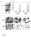
  • Figure 1 shows immunohistochemical staining with antibody SAM-6 on tumor tissue. To investigate the specificity of SAM-6 antibody paraffin sections (2 µm) were incubated with antibody SAM-6 at a concentration of 4 µg/ml and unrelated human control with the same isotype in similar concentration. For morphological analysis one sample was in addition stained with Hematoxilin/Eosin (H&E). Individual images of Figure 1 show: A, invasive lobular carcinoma of the breast; B, adenocarcinoma of the colon; C, esophageal squamous cell carcinoma (Original magnification x 200). The images in Figure 1 show that antibody SAM-6 reacts only with tumor cells, whereas the tissues surrounding the malignant areas are not stained.
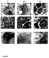
  • Figure 2 shows immunohistochemical staining with antibody SAM-6 on normal tissue. Paraffin sections (2 µm) were incubated with antibody SAM-6 at a concentration of 4 µg/ml. For morphological analysis one sample was stained in addition with Hematoxilin/Eosin (H&E). Individual images of Figure 2 show: A, lung; B, uterus; C, colon; D, testis (Original magnification x 200). Due to the absence of staining with healthy tissue it can be clearly stated that SAM-6 is binding to a receptor specifically expressed on malignant tissue.
  • Figure 3 covers specificity and functional analysis of the SAM-6 antibody by western blotting, apoptosis assay and morphological analysis. Individual images of Figure 3 show: A, membrane protein extracts from stomach carcinoma cell line 23132/87 and panceras carcinoma cell line BXPC-3 were blotted on nitrocellulose and stained with antibody SAM-6. B, Apoptotic activity of antibody SAM-6 was investigated by Cell Death Detection ELISAPLUS. Stomach carcinoma cell line 23132/87 and panceras carcinoma cell line BXPC-3 were incubated with antibody SAM-6 and isotype control in a concentration of 4 µg/ml for 48 h. Amounts of apoptotic cells were determined photospectrometrically at 415 nm and reference wave length 490 nm. C, antibody induced changes of tumor cell morphology. According to Fig. 3A SAM-6 binds to a membrane molecule with a molecular weight of about 140 kDa. The plot in Fig. 3B illustrates that SAM-6 induces apoptosis of the two tested cell types, stomach and pancreas carcinoma cells. In Fig. 3C the morphological changes of antibody SAM-6 induced apoptosis is shown on stomach carcinoma and on pancreas carcinoma cells. Untreated tumor cells grow in homogenous mono-layers. Afer treatment with antibody SAM-6 the cells become more spindle-shape and flat, more polarized with more pronounced cytoplasmic elongations. A loss of cell cell contacts and adhesion could be observed already after 48 hours. (Decrease in cell number is caused by apoptosis because cells get in solution as a result of lost adhesion.)
  • Figure 4 shows images of SAM-6 antibody induced apoptotic cells by scanning electron microscopy. This technique allows to study morphological and extra-cellular apoptotic effects of cells. For the experiment shown stomach carcinoma cell line 23132/87 was incubated with antibody SAM-6 or isotype control at a concentration of 10 µm/m) for the indicated periods of time. Samples were proceeded for scanning electron microscopy and analyzed by ZEISS DSM 962 at different time points. Individual images of Figure 4 show: A, B, C, isotype control antibody. D, E, F, SAM-6 antibody, bar indicates 20 µm. Magnification x 3800, bar indicates 20 µm. G, H, I, magnification of SAM-6 apoptotic effects; G, Stress fibers x 7000, bar indicates 10 µm; H, nucleus swelling, x 20000, bar indicates 2 µm; I, apoptonic bodies, x 40000, bar indicates µm. As shown in Fig. 4 initial morphological changes of SAM-6 treated cells after 2h include the formation of stress fibers (Fig. 4D, E) and a slight reduction of cell-cell contacts. After 24 h drastic morphological changes are observed. Cell-cell contacts are infinitely low (Fig. 4E), cells are either enlarged or condensed the nuclei are swelled (Fig. 4H) and the formation of apoptotic bodies is increased. The most dramatic effects are observed after 48 h. Numerous structural plasma membrane alterations are observed in the apoptotic cells: loss of cellular adhesion, smoothing, shrinkage and out-pouching of membrane segments are recognized as markers associated with cell injury and death. Most important, on the shrunken tumor cells, huge packages of membrane vesicles, apoptotic bodies, are clustered (Fig. 4F). (The formation of smooth-surface apoptotic bodies, as shown at the higher magnification, is due to the fact that in contrast to the in vivo recycling by phagocytotic cells, in vitro the membrane vesicles remain sitting on the dead cells.)
  • Figure 5 shows the results of transmission electron microscopy (TEM) experiment. To investigate the intracellular apoptotic effects transmission electron microscopic studies with SAM-6 on stomach carcinoma cells were performed. After 24 h, a drastic change in cell and nuclei shape is observed (Fig. 5E). Cells are enlarged, the cell volume at this stage is not reduced. The cells become spindle-shape, more polarized with more pronounced cytoplasmic elongations. The size of the nuclei is increased, they have a smooth surface and have lost the typical irregular and incised form seen in the control. Most importantly, after 24 h a dramatic accumulation of lipid vesicles in the cytoplasm is clearly visible (Fig. 5E). In almost each of the investigated tumor cells fatty acid depositions can be seen near the nuclei. After 48 h SAM-6 treated cells have reached the final stage of apoptosis (Fig. 5F). The most important structural changes include the disappearance of cell-cell contacts, cell shrinkage, high condensation of nuclei and degradation of plasma and nuclear membranes. The higher magnifications show a cluster of lipid vesicles accumulated in tumor cells (Fig. 5G), nuclear membrane degradation (Fig. 5H) and formation of apoptotic bodies from the cell surface of two tumor cells (Fig. 5I).
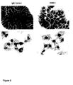
  • Figure 6 shows results of Sudan III staining experiments. To examine the antibody-induced lipid accumulation a staining with Sudan III was performed. This dye is specific for the detection of neutral lipids and fatty acids. Fig. 6 shows the obtained data after 48 h of incubation on gastric cancer cells and on pancreas carcinoma cells, either with antibody SAM-6 or unrelated human control IgM. The gastric carcinoma cell line 23132/87 clearly shows an antibody-induced accumulation of neutral lipids when treated with antibody SAM-6 (Fig. 6A). The cells treated with unrelated human control IgM do not exhibit similar intracellular changes. The same results were observed with the pancreas carcinoma cell line BXPC-3 (Fig. 6B).
  • Figure 7 shows the results of Nile Red staining experiments. Cellular lipids can also be visualized by staining with the fluorescence stain Nile Red. Here, non-polar or neutral lipids stain yellow-gold and polar lipids stain dark red when investigated at specific wavelengths (26, 27). Stomach cancer cells (23132/87) were incubated for 48 h with antibody SAM-6 and investigated for lipid accumulation. Fluorescence was measured at 488 nm for neutral lipids and at 543 nm for polar lipids. Fig. 7A and D show yellow staining for non-polar, neutral lipids, Fig. 7B and E red staining for polar lipids, and Fig. 7C and F an overlay of both. As expected, an intense yellow fluorescence stain for neutral lipids in the SAM-6 treated cells can be seen after 48 h (Fig. 7D). An increase is visible for SAM-6 treated cells stained for polar lipids (Fig. 7E), compared to the control (Fig. 7B), indicating a higher amount of membranes proteins. Since the antibody SAM-6 induces apoptosis, the higher amount of polar lipids is most likely the result of more membranes vesicle formation, namely apoptotic bodies. In the overlay, seen in Fig. 7C and F, polar lipids are seen in red and neutral lipids are in yellow and some are in orange, as expected. Although the red fluorescence of Nile red is very intense and there might be a possible red spill-over into the yellow-gold fluorescence measurement, a clear distinction can be made between the neutral and polar lipid staining. Taken together these results show, in addition to the Sudan stain, that the SAM-6 antibody induces neutral lipid accumulation in cancer cells.
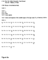
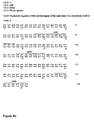
  • Figure 8a and 8b are the sequence protocols of the amino acid sequence (SEQ ID NO:1) (8a) and the nucleic acid sequence (SEQ ID NO:2) (8b) of the variable region of the light chain (VL) of human monoclonal antibody SAM-6.
  • Figure 9a and 9b are the amino acid sequence (SEQ ID NO:3) (9a) and the nucleic acid sequence (SEQ ID NO:4) (9b) of the variable region of the heavy chain (VH) of human monoclonal antibody SAM-6.
  • Detailed Description
    The present invention features polypeptides, such as antibodies, and their use in the treatment and diagnosis of neoplasms. We have characterized a human monoclonal antibody (SAM-6) that specifically recognizes a number of carcinomas. Not only does this monoclonal antibody recognize these neoplasms, but, upon binding to a cell, it can induce apoptosis of neoplastic cells, inhibit their proliferation, or even both. Thus, the SAM-6 monoclonal antibody or fragments thereof, that are specific for the antigen recognized by these polypeptides, may be used in a variety of methods for diagnosing and treating a neoplasm.
    Antibodies and Polypeptides
    Antibodies play an essential role in maintaining the health of an individual. In particular, antibodies are present in serum and bind to and help eliminate diverse pathogens such as bacteria, viruses, and toxins. Antibodies consist of Y-shaped protein structures built from two heavy chains and two light chains. Each chain has a modular construction: each light chain consists of two domains, and each heavy chain has at least four domains. The antigen binding site is fashioned by one domain from the heavy chain (VH domain) and one domain from the light chain (VL domain). indeed, small antigen binding fragments can be prepared by linking these two domains, either associated non-covalently, or covalently via disulphide bonds or a peptide linker. The antigen binding domains are more variable in amino acid sequence than the other domains of the antibody, and are therefore termed variable (V) domains, in contrast to the constant (C) domains. The constant domains of the antibody are responsible for triggering antibody effector mechanisms, such as complement lysis and cell-mediated killing.
    Antibodies are made by B-lymphocytes in a process involving gene rearrangement. During the development of these cells, the genes encoding the variable domains are assembled from genetic elements. In the case of the VH domains there are three elements, the un-rearranged VH gene, D segment, and JH segment. In the case of the VL domains, there are two elements, the un-rearranged VL (V Lambda or V Kappa) gene and the JL (J Lambda or J Kappa) segment. Random combination of these gene segments and random combination of the rearranged VH and VL domains generate a large repertoire of antibodies, capable of binding to a large diversity of equally diverse antigens.
    In general, the presently claimed polypeptide is any agent that binds to BXPC-3, 23132/87, COLO-206F, COLO-699 and LOU-NH91, but does not bind to non-neoplastic cells. The polypeptide may be an antibody, such as a human monoclonal antibody (e.g.,SAM-6), or a functional fragment thereof. Overall, the polypeptide of the invention can exclusively bind to both neoplastic tissues and neoplastic cells, but not to non-neoplastic tissue or cells. The polypeptide also may induce apoptosis of a neoplastic cell to which it binds, but not in a non-neoplastic cell, or, alternatively, the polypeptide may inhibit proliferation of the neoplastic cell it binds to, but not in a non-neoplastic cell. Desirably, the polypeptide can simultaneously induce apoptosis and inhibit proliferation of neoplastic cells, but not of non-neoplastic cells. Such a polypeptide is, therefore, useful for the detection, monitoring, prevention, and treatment of cancers in mammals. Exemplary cancers amenable to the methods of the current invention include colorectal cancer, ovarian carcinoma, squamous cell lung carcinoma, small cell lung carcinoma, lobular and ductal mammary carcinomas, melanoma, breast cancer, lung cancer, such as lung adenocarcinomas, gastric cancer, pancreatic cancer, such as pancreatic adenocarcinomas, glioma, sarcomas, gastrointestinal cancer, brain tumor, esophageal cancer, such as esophagial squamous cell carcinomas, stomach cancer, osteosarcoma , fibrosarcomas, urinary bladder cancer, prostate cancer, such as prostate adenocarcinomas, renal cancer, ovarian cancer, testicular cancer, endometrial cancer, cervical cancer, uterine adenocarcinomas, Hodgkin's disease, lymphomas, and leukemias. Such polypeptides are particularly useful for the detection and treatment of adenocarcinoma of the lung, squamous cell lung carcinoma, intestinal type gastric carcinoma, diffuse type gastric carcinoma, adenocarcinoma of the colon, adenocarcinoma of the prostate, squamous cell carcinoma of the esophagus, adenocarcinoma of the esophagus, lobular carcinoma of the breast, ductal carcinoma of the breast, adenocarcinoma of the pancreas, adenocarcinoma of the ovary, or adenocarcinoma of the uterus.
    Production
    The polypeptides according to the claimed invention can be produced by any method known in the art for small scale, large scale, or commercial production of polypeptides. For example, a monoclonal antibody, such as SAM-6, may be produced by hybridoma cell lines. Such cell lines are typically generated by the fusion of spleen lymphocytes or lymph node lymphocytes derived from patients having a neoplasm, such as stomach carchinoma, colon carcinoma or a pancreatic carcinoma, with a heteromyeloma cell line. Exemplary heteromyeloma cell lines include, for example, HAB-1 (Vollmers et al, Cancer 74:1525-1532, 1994), CB-F7 (Delvig et al., Hum. Antibodies Hybridomas 6:42-46, 1995), K6H6B5 (Delvig et al., Hum. Antibodies Hybridomas 6:42-46,1995), H7NS.934 (Delvig et al., Hum. Antibodies Hybridomas 6:42-46, 1995), SHM-D33 (Bron et al., Proc. Natl. Acad. Sci. USA 81:3214-3217, 1984), and B6B11 (Borisova et al., Vopr. Virusol. 44:172-174, 1999). The ability to generate human monoclonal antibodies from lymphocytes of cancer patients allows the isolation of antibodies that are generated by an immune response in the cancer patient to the tumor.
    Typically, portions of the lymph nodes or spleen are surgically removed from a patient having cancer, such as colon carcinoma or a pancreatic carcinoma. Lymphocytes may be prepared as cell suspensions by mechanical means and subsequently fused at, for example, a 1:2 or 1:3 ratio with a heteromyeloma cell line under conditions that result in cell fusion. For instance, the heteromyeloma cell line HAB-1, which is generated by the fusion of a human lymphocyte with the mouse myeloma NS-0, may be used for this purpose.
    Following the fusion of the lymphocytes derived from the cancer patient with the heteromyeloma cell line, an antibody producing hybridoma or trioma is generated. Once constructed, hybridomas are generally stable in growth and antibody production in standard and mass cultures (flasks, miniPerm, fermenters, etc.) for several months. Levels of antibody production typically range between 0.01-0.1 mg/mL in flasks and between 0.1-0.5 mg/mL in miniPerm. Cell fusion may be achieved by any method known in the art, and includes, for example, the use of 40% polyethyleneglycol. Hybridomas may be cultured in media containing HAT (Hypovanthin-aminopterin-thymidin) and after four weeks, supernatants may be screened for antibody production using an ELISA assay. Positive clones may then be tested in attachment inhibition and binding assays using commercially available tumor cell lines. Positive clones further may be tested using immunoperoxidase staining of tumor and normal tissues. Thus, clones may be selected on the basis of their reactivity with autologous and allogeneic neoplastic cells. The antibody may be purified from mass cultures with use of cation-exchange, hydrophobic interaction, size exclusion, or affinity chromatograhpy, as well as a combination of these methods as described, for example, by Vollmers et al. (Oncology Reports 5:35-40, 1998). Following the production of antibodies, additional functional and immunohistochemical tests of the antibodies produced by the trioma may be performed. For example, the antibodies produced by the hybridoma can be tested for their ability to induce apoptosis, inhibit cellular proliferation, or both, relative to untreated control cells. The antibodies can also be tested for their ability to specifically bind the neoplastic cell lines like BXPC-3, 23132/87, COLO-206F, COLO-699 or LOU-NH91, relative to non-neoplastic cells.
    Alternatively, the polypeptide, including an antibody, or a fragment thereof, may be produced by the expression of the polypeptide or antibody in a host cell such as E. coli or yeast, e.g., S. cerevisiae. For example, an antibody of the invention may be identified as follows. A nucleic acid sequence encoding an antibody, or a fragment thereof, may be inserted into filamentous bacteriophage to generate libraries of approximately 107 or more antibodies. Each phage expresses an antibody on its surface that is encoded by the nucleic acid it contains. Antibodies of the invention may thus be screened and detected by functional and histochemical assays as described herein, and such genes may be subsequently selected and expressed in E.coli. This system is described, for example, in U.S. Patent No. 5,876,691.
    Antibodies, or functional fragments thereof, may also be generated using, for example, direct synthesis using recombinant methods. These methods are standard in the art. For example, a nucleic acid sequence may be amplified using the polymerase chain reaction (PCR). The PCR technique is known in the art and is described, for example in U.S. Patent No. 4,683,195. Using standard methods, and as described herein, the sequence of a monoclonal antibody expressed by a hybridoma may be obtained and functional fragments of the antibody may be amplified. For example, whole RNA may be isolated from a hybridoma expressing a tumor-specific monoclonal antibody. cDNA may then be generated from the RNA using reverse transcriptase and the cDNAs which contain the functional fragments of the variable regions of the heavy and light chains may be amplified using PCR. The PCR products may then be purified and cloned into expression vectors, e.g., plasmid or viral vectors. Many standard vectors are available and the selection of the appropriate vector will depend on, for example, the size of the DNA inserted into the vector and the host cell to be transformed with the vector.
    Isolation of Amino Acid Variants of a Polypeptide
    Amino acid sequence variants of a polypeptide, such as an antibody, e.g., a SAM-6 antibody, can be prepared by introducing appropriate nucleotide changes into the DNA encoding the antibody, or by in vitro synthesis of the desired polypeptide. Such variants include, for example, deletions from, or insertions or substitutions of, residues within the amino acid sequence of the SAM-6 antibody. Any combination of deletion, insertion, and substitution can be made to arrive at the final construct, provided that the final construct possesses the desired characteristics, e.g., the ability to induce apoptosis of a neoplastic cell, but not a non-neoplastic cell, or the ability to inhibit the proliferation of a neoplastic cell, but not a non-neoplastic cell. The amino acid changes also may alter post-translational processes of an antibody, such as changing the number or position of glycosylation sites, altering the membrane anchoring characteristics, or modifying its susceptibility to proteolytic cleavage.
    In designing amino acid sequence variants of a polypeptide, such as an antibody, the location of the mutation site and the nature of the mutation will depend on characteristic(s) to be modified. The sites for mutation can be modified individually or in series, e.g., by substituting first with conservative amino acid choices and then with more radical selections depending upon the results achieved, or deleting the target residue.
    A useful method for identification of specific residues or regions for mutagenesis in a polypeptide is called "alanine scanning mutagenesis" and is described, for example, by Cunningham and Wells (Science 244:1081-1085, 1989). Here, a residue or group of target residues are identified (e.g., charged residues such as arg, asp, his, lys, and glu) and replaced by a neutral or negatively charged amino acid (most desirably alanine or polyalanine) to affect the interaction of the amino acids with the surrounding aqueous environment in or outside the cell. The domains demonstrating functional sensitivity to the substitutions then are refined by introducing further or other variants at or for the sites of substitution. Thus, while the site for introducing an amino acid sequence variation is predetermined, the nature of the mutation need not be predetermined. For instance, to optimize the performance of a mutation at a given site, alanine scanning or random mutagenesis may be conducted at the target codon or region and the expressed variants are screened for, e.g., the ability to induce apoptosis of a neoplastic cell and not a non-neoplastic cell, or to inhibit the proliferation of a neoplastic cell and not a non-neoplastic cell.
    The sites of greatest interest for substitutional mutagenesis include sites identified as affecting the biological activity of a polypeptide. These sites, especially those falling within a sequence of at least three other identically conserved sites, may be substituted in a relatively conservative manner. For instance, ala may be substituted with val, leu, or ile; arg may be substituted with lys, gln, or asn; asn may be substituted with gln, his, lys, or arg; asp may be substituted with glu; cys may be substituted with ser; gln may be substituted with asn; glu may be substituted with asp; gly may be substituted with pro; his may be substituted with asn, gln, lys, or arg; ile may be substituted with leu, val, met, ala, or phe; leu may be substituted with ile, val, met, ala, or phe; lys may be substituted with arg, gln, or asn; met may be substituted with leu, phe, or ile; phe may be substituted with leu, val, ile, or ala; pro may be substituted with gly; ser may be substituted with thr; thr may be substituted with ser; trp may be substituted with tyr; tyr may be substituted with trp, phe, thr, or ser; and val may be substituted with ile, leu, met, or phe.
    Conjugation of the Antibody with a Detectable Agent
    If desired, the claimed polypeptide such as an antibody (e.g., monoclonal antibody, such as SAM-6), or a fragment thereof, may be linked to a detectable agent to facilitate the purification of the polypeptide as well as the diagnosis, monitoring, or treatment of cancer in a mammal in need thereof. The selection of suitable detectable agent will depend on the intended use of the polypeptide and will be apparent to those of ordinary skill in the art. Detectable agents according to the claimed invention include, for example, protein purification tags, cytotoxins, enzymes, paramagnetic labels, enzyme substrates, co-factors, enzyme inhibitors, dyes, radionuclides, chemiluminescent labels, fluorescent markers, growth inhibitors, and biotin.
    A protein purification tag may be conjugated to the polypeptide of the invention, to facilitate isolation of the polypeptide. Examples of tags that can be used include His-tags, HA-tags, FLAG®-tags, and c-Myc tags. An enzymatic or chemical cleavage site may be engineered between the polypeptide and the tag moiety so that the tag can be removed following purification. Suitable toxins include diphtheria toxin, Pseudomonas exotoxin A, ricin, and cholera toxin. Examples of suitable enzyme labels include malate hydrogenase, staphylococcal nuclease, delta-5-steroid isomerase, alcohol dehydrogenase, alphaglycerol phosphate dehydrogenase, triose phosphate isomerase, peroxidase, alkaline phosphatase, asparaginase, glucose oxidase, beta-galactosidase, ribonuclease, urease, catalase, glucose-6-phosphate dehydrogenase, glucoamylase, and acetylcholinesterase. Examples of suitable radioisotopic labels include 3H, 125I, 131I, 32P, 35S , and 14C. Desirably, the radioisotope will emit in the 10-5,000 kev range, more desirably 100-500 kev. Paramagnetic isotopes may also be conjugated to the polypeptide and used in vivo for the diagnosis and treatment of cancer. The use of such conjugated antibodies may be for in vivo nuclear magnetic resonance imaging. Such a method has previously been described (see, for example, Schaefer et al., JACC 14:472-480, 1989; Shreve et al., Magn. Reson. Med. 3:336-340,1986; Wolf, Physiol. Chem. Phys. Med. NMR 16:93-95, 1984; Wesbey et al., Physiol. Chem. Phys. Med. NMR 16:145-155, 1984; and Runge et al., Invest. Radiol. 19:408-415, 1984). Alternatively, the radiolabeled antibody may also be used in radioimmunoguided surgery (RIGS), which involves the surgical removal of any tissue the labeled antibody binds to. Thus, the labeled antibody guides the surgeon towards neoplastic tissue by distinguishing it from non-neoplastic tissue. Radiolabels useful for tumor imaging are preferably short-lived radioisotopes. Various radioactive metals with half-lives ranging from 1 hour to 11.4 days are available for conjugation to antibodies, such as scandium-47 (3.4 days), gallium-67 (2.8 days), gallium-68 (68 minutes), technetium-99m (6 hours), indium-111 (3.2 days), and radium-223 (11.4 days), of which gallium-67, technetium-99m, and indium-111 are preferable for gamma camera imaging, gallium-68 is preferable for positron emission tomography, and scandium-47 and radium-223 (and other alpha-emitting radionuclides) are preferable for tumor therapy.
    Examples of suitable fluorescent markers include fluorescein, isothiocyalate, rhodamine, phycoerythrin, phycocyanin, allophycocyanin, ophthaldehyde, and fluorescamine. Examples of chemiluminescent markers include a luminal label, isoluminal label, aromatic acridinium ester label, imidazole label, acridinium salt label, oxalate ester label, luciferin label, luciferase label, and aequorin label. Those of ordinary skill in the art would know of other suitable labels, which may be employed in accordance with the present invention. Conjugation of these detectable agents to the claimed polypeptides such as monoclonal antibodies, or fragments thereof, can be accomplished using standard techniques commonly known in the art. Typical antibody conjugation techniques are described by Kennedy et al. (Clin. Chim. Acta 70, 1-31, 1976) and Schurs et al. (Clin. Chim. Acta 81, 1-40 , 1977) and include, for example, the glutaraldehyde method, the periodate method, the dimaleimide method, the m-maleimidobenzyl-N-hydroxy-succinimide ester method. Antibodies may be radiolabeled by any of several techniques known to the art, described, for example, in U.S. patent No. 4,444,744. All of these methods are incorporated by reference herein.
    In all aspects of the present invention, it is understood that mixtures of different or the same labeled polypeptides specific to different antigens or different epitopes of the same antigen associated with the same or different tumor or tumor cell types may be used. Such a combination may enhance detection, localization and/or therapy in certain cases, and can also increase the range of a broad screen for more than one neoplasm or type of neoplasm.
    Polypeptides Conjugated to Anti-Tumor Agents
    Although the polypeptide of the invention may induce apoptosis of neoplastic cells, inhibit cellular proliferation of neoplastic cells, or both, the polypeptide may in addition be conjugated to an agent that kills neoplastic cells or that inhibits their proliferation. The targeting ability of the polypeptide, such as an antibody or fragment thereof, results in the delivery of the cytotoxic or anti-proliferative agent to the tumor to enhance the destruction of the tumor. The polypeptide therefore may be used for the treatment and prevention of cancer in a mammal, such as a human patient. The cytotoxic agent linked to the polypeptide may be any agent that destroys or damages a tumor cell or tumor to which the polypeptide has bound. Examples of such agents include chemotherapeutic agents or radioisotopes, enzymes which activates a pro-drug, or a cytokine.
    Suitable chemotherapeutic agents are known to those skilled in the art and include, for example, taxol, mithramycin, deoxyco-formycin, mitomycin-C, L-asparaginase, interferons (especially IFN-alpha), etoposide, teniposide, anthracyclines (e.g., daunomycin and doxorubicin), methotrexate, vindesine, neocarzinostatin, cis-platinum, chlorambucil, cytosine arabinoside, 5-fluorouridine, melphalan, ricin, and calicheamicin. The chemotherapeutic agents may be conjugated to the antibody using conventional methods known in the art.
    Suitable radioisotopes for use as cytotoxic agents are also known to those skilled in the art and include, for example, 131I, or an astatine such as 211At. These isotopes may be attached to the polypeptide, either covalently or non-covalently, using conventional techniques known in the art.
    Alternatively, the cytotoxic agent may also be an enzyme, which activates a pro-drug. This allows the conversion of an inactive pro-drug to its active, cytotoxic form at the tumor site and is called "antibody-directed enzyme pro-drug therapy" (ADEPT). Thus, the polypeptide-enzyme conjugate may be administered to the patient and allowed to localize in the region of the tumor to be treated. The pro-drug is then administered to the patient such that conversion to the cytotoxic drug is localized in the region of the tumor to be treated under the influence of the localized enzyme. An exemplary enzyme is bacterial carboxypeptidase G2 (CPG2) the use of which is described in, for example, WO 88/07378. The polypeptide-enzyme conjugate may, if desired, be modified in accordance with the teaching of WO 89/00427, such as to accelerate its clearance from areas of the body that are not in the vicinity of a neoplasm. The polypeptide-enzyme conjugate may also be used in accordance with WO 89/00427, for example, by providing an additional component, which inactivates the enzyme in areas of the body that are not in the vicinity of the tumor.
    As another alternative, the cytotoxic agent conjugated to the claimed polypeptide may also be a cytokine such as interleukin-2 (IL-2), interleukin-4 (IL-4), or tumor necrosis factor alpha (TNF-alpha). The polypeptide targets the cytokine to the tumor so that the cytokine mediates damage to or destruction of the tumor without affecting other tissues. The cytokine may be fused to the polypeptide at the DNA level using conventional recombinant DNA techniques.
    In addition, any inhibitor of cell proliferation. e.g., genistein, tamoxifen, or cyclophosphamide, may be conjugated with a polypeptide of the invention.
    Dosage
    With respect to the therapeutic methods of the invention, it is not intended that the administration of the claimed polypeptide to a patient be limited to a particular mode of administration, dosage, or frequency of dosing; the present invention contemplates all modes of administration, including intramuscular, intravenous, intraperitoneal, intravesicular, intraarticular, intralesional, subcutaneous, or any other route sufficient to provide a dose adequate to decrease the number of neoplastic cells by inducing apoptosis of neoplastic cells, by inhibiting proliferation of tumor cells, or both. The compound(s) may be administered to the patient in a single dose or in multiple doses. When multiple doses are administered, the doses may be separated from one another by, for example, one day, two days, one week, two weeks, or one month. For example, the polypeptide (e.g., a monoclonal antibody, such as SAM-6) may be administered once a week for, e.g., 2, 3, 4, 5, 6, 7, 8, 10, 15, 20, or more weeks. It is to be understood that, for any particular subject, specific dosage regimes should be adjusted over time according to the individual need and the professional judgment of the person administering or supervising the administration of the compositions. The precise dose will vary dependent on the polypeptide used, the density, on the tumor surface, of the ligand to which the polypeptide binds, and the rate of clearance of the polypeptide. For example, the dosage of the SAM-6 antibody can be increased if the lower dose does not provide sufficient anti-neoplastic activity. Conversely, the dosage of the SAM-6 antibody can be decreased if the neoplasm is cleared from the patient.
    While the attending physician ultimately will decide the appropriate amount and dosage regimen, a therapeutically effective amount of the claimed polypeptide, such as a monoclonal antibody or a fragment thereof, may be, for example, in the range of about 0.1 mg to 50 mg/kg body weight/day or 0.70 mg to 350 mg/kg body weight/week. Desirably a therapeutically effective amount is in the range of about 0.50 mg to 20.0 mg/kg, and more desirably in the range of about 0.50 mg to 15.0 mg/kg for example, about 0.2, 0.3, 0.5, 1.0, 1.5, 2.0, 2.5, 3.0, 3.5, 4.0, 4.5, 5.0, 5.5, 6.0, 7.0, 8.0, 8.5, 9.0, 10.0, 11.0, 12.0, 13.0, 14.0, or 15.0 mg/kg body weight administered daily, every other day, or twice a week.
    For example, a suitable dose is an amount of the polypeptide that, when administered as described above, is capable of inducing apoptosis, and is at least 20% above the basal (i.e., untreated) level.
    In general, an appropriate dosage and treatment regimen provides the active compound(s) in an amount sufficient to provide therapeutic and/or prophylactic benefit. Such a response can be monitored by establishing an improved clinical outcome (e.g., more frequent remissions, complete or partial, or longer disease-free survival) in treated patients as compared to non-treated patients. According to this invention, the administration of the polypeptide can induce neoplastic cell apoptosis by at least 20%, 40%, 50%, or 75% above that of an untreated control as measured by any standard assay known in the art. More desirably, apoptosis is induced by 80%, 90%, 95%, or even 100% above that of an untreated control. Alternatively, the administration of the polypeptide can inhibit neoplastic cell proliferation by at least 20%, 40%, 50%, or 75% below that of an untreated control as measured by any standard assay known in the art. More desirably, proliferation is inhibited by 80%, 90%, 95%, or even 100% below that of an untreated control. Most desirably, the polypeptide can simultaneously inhibit proliferation and induce apoptosis of neoplastic cells relative to untreated control cells. Such responses can be monitored by any standard technique known in the art. In general, for pharmaceutical compositions, the amount of antibody present in a dose ranges from about 25 µg to 5 mg per kg of host. Suitable dose sizes will vary with the size of the patient, but will typically range from about 0.1 mL to about 5 mL.
    Formulation of Pharmaceutical Compositions
    The claimed polypeptide may be administered by any suitable means that results in a concentration having anti-neoplastic properties upon reaching the target region. The polypeptide may be contained in any appropriate amount in any suitable carrier substance, and is generally present in an amount of 1-95% by weight of the total weight of the composition. The composition may be provided in a dosage form that is suitable for parenteral (e.g., subcutaneous, intravenous, intramuscular, or intraperitoneal) administration route. The pharmaceutical compositions may be formulated according to conventional pharmaceutical practice (see, e.g., Remington: The Science and Practice of Pharmacy (20th ed.), ed. A.R. Gennaro, Lippincott Williams & Wilkins, 2000 and Encyclopedia of Pharmaceutical Technology, eds. J. Swarbrick and J. C. Boylan, 1988-1999, Marcel Dekker, New York).
    The pharmaceutical composition may be administered parenterally by injection, infusion or implantation (subcutaneous, intravenous, intramuscular, intraperitoneal, or the like) in dosage forms, formulations, or via suitable delivery devices or implants containing conventional, non-toxic pharmaceutically acceptable carriers and adjuvants. If the neoplastic cells are in direct contact with the blood (e.g., leukemias), or if the tumor is only accessible by the bloodstream then the intravenous (I.V.) route may be used. In cases in which tumors grow in confined spaces such as the pleural cavity or the peritoneal cavity, the polypeptide may be directly administered into the cavity rather than into the blood stream. The formulation and preparation of such compositions are well known to those skilled in the art of pharmaceutical formulation. Formulations can be found in Remington: The Science and Practice of Pharmacy, supra.
    Diagnosis and Monitoring Cancer Progression
    As discussed above, the present invention is directed to a method for detecting or diagnosing a neoplasm in a mammal, preferably a human patient. Typically, any neoplasm in which administration of the claimed polypeptide causes an induction in apoptosis or a reduction in proliferation are amenable to the methods of this invention.
    The claimed polypeptides are particularly useful since they are specific to neoplasms or neoplastic cells, but not normal cells or tissue. Accordingly, this polypeptide can bind to neoplastic cells within the tumor, but not the normal surrounding tissue, thus allowing the detection, the treatment, or both, of a neoplasm in a mammal. For instance, one may use a polypeptide of the invention to determine if a biopsy removed the entire tumor by verifying that no cells bound by the polypeptide remain in the patient or, by verifying that tumor removed from the patient is entirely surrounded by cells that are not bound by the polypeptide.
    It is understood that to improve the sensitivity of detection, multiple neoplastic markers may be assayed within a given sample or individual. Thus, polypeptides such as antibodies or functional fragments specific for different antigens may be combined within a single assay, or in multiple assays. Further, multiple primers or probes specific to neoplasms may be used concurrently. The selection of markers may be based on routine experiments to determine combinations that results in optimal sensitivity.
    In Vitro Detection of a Neoplasm
    In general, the diagnosis of a neoplasm in a mammal involves obtaining a biological sample from the mammal (e.g., human patient), contacting such sample with the polypeptide of the invention (e.g., a monoclonal antibody, such as SAM-6), detecting in the sample the level of reactivity or binding of the polypeptide to neoplastic cells relative to a control sample, which corresponds to non-neoplastic cells derived from healthy tissue from the mammal in which the cancer is being diagnosed or from another patient known not to have neoplasm. Thus, the methods of this invention are particularly useful for the detection of early stage tumors or metastases, which are otherwise undetectable. Accordingly, in addition to diagnosing a neoplasm in a patient, the methods of this invention may also be used to monitor progression of a neoplasm in a mammal. The polypeptide described herein therefore may be used as a marker for the progression of a neoplasm. For this purpose, the assays described below, which are used for the diagnosis of a neoplasm, may be performed over time, and the change in the level of reactive polypeptide(s) evaluated. For example, the assays may be performed every 24-72 hours for a period of 6 months to 1 year, and thereafter performed as needed. In general, a neoplasm is progressing in those patients in whom the level of bound polypeptide detected increases over time. In contrast, the neoplasm is not progressing when the level of bound polypeptide either remains constant or decreases with time. Alternatively, as is noted above, the polypeptide of the invention may also be used to determine the presence of tumor cells in the mammal following tumor resection by surgical intervention to determine whether the tumor has been completely removed from the mammal.
    Desirably, the polypeptide is linked to a detectable agent, which facilitates detection, or measurement of polypeptide reactivity. The biological sample is any biological material, which may contain neoplastic cells and include, for example, blood, saliva, tissue, serum, mucus, sputum, urine, or tears. The biological sample may also be a tissue section, which may be fixed tissue, fresh tissue, or frozen tissues. A neoplasm is detected or diagnosed in the mammal from which the sample was obtained if there is an increase in the level of reactivity of the antibody with the biological sample over the control sample. Such increase is at least 10%, 20%, 30%, 40%, 50%, or more than 50% over control levels. The level of binding or reactivity can be determined by any method known in the art and is described in further detail below.
    In Vitro Diagnostic Assays
    The diagnosis of neoplasms using the claimed polypeptide may be performed by any method known to those of ordinary skill in the art for using a binding agent to detect polypeptide markers in a sample. See, e.g., Harlow and Lane, Antibodies: A Laboratory Manual, Cold Spring Harbor Laboratory, 1988. For example, the polypeptide may be used for enzyme-linked immunosorbent assay (ELISA), Western blotting or in situ detection of tumor cells in a tissue sample. For example, the ELISA assay typically involves the use of the polypeptide, such as an antibody, immobilized on a solid support to bind to the tumor cells in the biological sample. The bound tumor cell may then be detected using a detection reagent that contains a reporter group and that specifically binds to the antibody/tumor cell complex. Such detection reagents include, for example, any binding agent that specifically binds to the antibody, such as an anti-immunoglobulin, protein G, protein A, or a lectin. Alternatively, a competitive assay may be utilized, in which the polypeptide is an antibody and in which the antigens, to which the antibody is specific to is labeled with a reporter group and allowed to bind to the immobilized antibody after incubation of the antibody with the biological sample. The extent to which components of the sample inhibit the binding of the labeled antigens to the antibody is indicative of the reactivity of the sample with the immobilized antibody. Diagnosis of a neoplasm in a patient may also be determined by a two-antibody sandwich assay. This assay may be performed by first contacting an antibody that has been immobilized on a solid support, commonly the well of a microtiter plate, with the sample, such that polypeptides within the sample are allowed to bind to the immobilized antibody. Unbound sample is then removed from the immobilized polypeptide-antibody complexes and a detection reagent (preferably a second antibody capable of binding to a different site on the polypeptide) containing a reporter group is added. The amount of detection reagent that remains bound to the solid support is then determined using a method appropriate for the specific reporter group. For example, to determine the presence or absence of a neoplasm, such as colorectal adenocarcinoma, the signal detected from the reporter group that remains bound to the solid support is generally compared to a signal that corresponds to a predetermined cut-off value. The cut-off value for the detection of a neoplasm is the average mean signal obtained when the antibody is incubated with samples from patients without a neoplasm.
    The method employed for detecting the reporter group depends upon the nature of the reporter group. For radioactive groups, scintillation counting or autoradiographic methods may be used. Spectroscopic methods may be used to detect dyes, luminescent groups and fluorescent groups. Biotin may be detected using avidin, coupled to a different reporter group (commonly a radioactive or fluorescent group or an enzyme). Enzyme reporter groups may generally be detected by the addition of substrate (generally for a defined period of time), followed by spectroscopic or other analysis of the reaction products.
    The polypeptides of the invention may also be employed histologically for in situ detection or quantitative determination of tumor cells, for example, by immunofluorescence or immunoelectron microscopy. In situ detection or determination may be accomplished by removing a tissue specimen from a patient and allowing a labeled antibody to bind to any tumor cell in the specimen. Using such a procedure not only allows the detection of neoplastic cells in a sample, but also allows for the determination of their spatial distribution. As another example, the biological sample can be a smear of biological material containing neoplastic cells on a slide, and the detection of neoplastic cells in the biological material is achieved by examining the smear with a microscope or by fluocytometry.
    In Vivo Detection of a Neoplasm
    Alternatively, the antibody of the invention may also be used in vivo for detecting and localizing a neoplasm. Such a method may involve injecting a mammal, desirably a human subject, parenterally with a polypeptide of the invention, such as SAM-6, which has been labeled with a detectable agent, and is described, for instance, in U.S. Patent No. 4,444,744. For example, the polypeptide can be radiolabeled with a pharmacologically inert radioisotope and administered to the patient. The activity of the radioisotope can be detected in the mammal using a photoscanning device, and an increase in activity relative to a control reflects the detection and localization of a neoplasm.
    Treatment
    In addition to the diagnosis and monitoring of neoplasms in mammals, the present invention also features methods for treating neoplasms in a mammal, desirably a human patient. The method generally involves the administration of a biologically effective amount of the polypeptide of the invention to the patient. The polypeptide is typically administered to the mammal by means of injection using any routes of administration such as by intrathecal, subcutaneous, submucosal, or intracavitary injection as well as for intravenous or intraarterial injection. Thus, the polypeptide may be injected systemically, for example, by the intravenous injection of the polypeptide such as the SAM-6 antibody into the patient's bloodstream or alternatively, the polypeptide can be directly injected at the site of the neoplasm or at a location in proximity to the neoplastic cells.
    In general, and as discussed above, binding of the polypeptide of the invention to neoplastic cells results in an induction in apoptosis, a reduction in cellular proliferation, or both relative to the control sample. Alternatively, the antibodies may also activate the complement pathway, which ultimately causes holes to be punctured into the cellular membrane, resulting in cell death.
    If desired, the polypeptides may also be conjugated to drugs or toxins as described above. Once attached to the cell surface, the conjugate may be engulfed into the cell cytoplasm where cell enzymes cleave, and, thus, activate or free the drugs or toxins from the conjugate. Once released, the drugs or toxins damage the cell and irreversibly induce cell death. With respect to radiolabeled antibodies, binding to neoplastic cells and the resulting emission of radiation, at a short distance from the cell DNA, produces damage to the latter thus inducing cell death in the next replication round. For example, after a neoplasm has been detected and localized in a subject, a higher dose of labeled antibody, generally from 25 to 250 mCi for 131I, and preferably from 50 nCi to 150 mCi per dose, based on a 70 kg patient weight, is injected. Injection may be intravenous, intraarterial, intralymphatic, intrathecal, or intracavitary, and may be repeated more than once. It may be advantageous for some therapies to administer multiple, divided doses of radiolabeled polypeptides or polypeptide mixtures, e.g., in the range of 20-120 mCi (70 kg patient), thus providing higher cell-killing doses to the neoplasm without usually effecting a proportional increase in radiation of normal tissues
    Therapy using labeled polypeptides is advantageously used as a primary therapeutic treatment, but may also be used in combination with other anti-neoplastic therapies, e.g., radiation and chemotherapy, and as an adjunct to surgery. The administration of such conjugated polypeptides is particularly useful in the case where small metastases cannot be surgically removed.
    Combination of a Polypeptide with other Anti-Neoplastic Therapies
    Chemotherapeutic agents and/or radiation and/or surgical removal of the neoplasm can optionally be combined with any of the methods of the present invention. Classes of compounds that can be used as the chemotherapeutic agent include: alkylating agents, antimetabolites, natural products and their derivatives, hormones and steroids (including synthetic analogs), and synthetics. Examples of alkylating agents (e.g., nitrogen mustards, ethylenimine derivatives, alkyl sulfonates, nitrosoureas and triazenes) include Uracil mustard, Chlormethine, Cyclophosphamide (Cytoxan®), Ifosfamide, Melphalan, Chlorambucil, Pipobroman, Triethylene-melamine, Triethylenethiophosphoramine, Busulfan, Carmustine, Lomustine, Streptozocin, Dacarbazine, and Temozolomide. Antimetabolites (including folic acid antagonists, pyrimidine analogs, purine analogs and adenosine deaminase inhibitors) may include, for example, Methotrexate, 5-Fluorouracil, Floxuridine, Cytarabine, 6-Mercaptopurine, 6-Thioguanine, Fludarabine phosphate, Pentostatine, and Gemcitabine. Natural products and their derivatives (including vinca alkaloids, antitumor antibiotics, enzymes, lymphokines and epipodophyllotoxins) may also be used and include, for example, Vinblastine, Vincristine, Vindesine, Bleomycin, Dactinomycin, Daunorubicin, Doxorubicin, Epirubicin, Idarubicin, paclitaxel (paclitaxel is commercially available as Taxol, Mithramycin, Deoxyco-formycin, Mitomycin-C, L-Asparaginase, Interferons (especially IFN-alpha), Etoposide, and Teniposide. Hormones and steroids (including synthetic analogs) include, for example, 17-alpha-Ethinylestradiol, Diethylstilbestrol, Testosterone, Prednisone, Fluoxymesterone, Dromostanolone propionate, Testolactone, Megestrolacetate, Tamoxifen, Methylprednisolone, Methyltestosterone, Prednisolone, Triamcinolone, Chlorotrianisene, Hydroxyprogesterone, Aminoglutethimide, Estramustine, Medroxyprogesteroneacetate, Leuprolide, Flutamide, Toremifene, or Zoladex. Exemplary synthetics (including inorganic complexes such as platinum coordination complexes) include Cisplatin, Carboplatin, Hydroxyurea, Amsacrine, Procarbazine, Mitotane, Mitoxantrone, Levamisole, and Hexamethylmelamine.
    Methods and dosages for the safe and effective administration of most of these chemotherapeutic agents are known to those skilled in the art. In addition, their administration is described in the standard literature. For example, the administration of many of the chemotherapeutic agents is described in the "Physicians' Desk Reference" (PDR), e.g., 1996 edition (Medical Economics Company, Montvale, N.J. 07645-1742, USA), the disclosure of which is incorporated herein by reference.
    The following examples are provided for the purpose of illustrating the invention and should not be construed as limiting.
    Example 1 Materials and Methods Cell Culture
    In this study the following human cell lines were used: BXPC-3 (pancreatic adenocarcinoma), 23132/87(gasteric adenocarcinoma), COLO-206F (colon carcinoma), COLO-699 (lung adenocarcinoma) and LOU-NH91 (lung squamous cell carcinoma). The cell lines were cultured in RPM1-1640 media (PAA, Vienna, Austria) supplemented with 10% fetal calf serum (FCS), 2mM glutamine and penicillinlstreptomycin (both 1%) and incubated in a humidified, 5% CO2 atmosphere at 37°C. For the assays described, cells were grown to sub-confluency, detached with trypsin/EDTA and washed twice with phosphate-buffered saline (PBS) before use.
    Producing Hybridomas
    We immortalized lymphocytes by fusing them to the HAB-1 heteromyeloma as follows. We washed HAB-1 heteromyeloma cells twice with RPMI 1640 (PAA, Vienna, Austria) without additives and centrifuged the cells for 5 minutes at 1500 rpm. We then thawed frozen lymphocytes obtained from either the spleen or the lymph nodes and we washed these cells twice with RPMI 1640 without additives and centrifuged these cells at 1500 rpm for 5 minutes. Both the HAB-1 and the lymphocyte cell pellets were resuspended in 10 ml RPMI 1640 without additives and were counted in a Neubauer cell counting chamber. We washed the cells again, added the HAB-1 cells and the lymphocytes together in a ratio of 1:2 to 1:3, mixed them, and centrifuged the mixture for 8 minutes at 1500 rpm. We pre-warmed Polyethylene Glycol 1500 (PEG) to 37°C and carefully let the PEG run drop-wise onto the pellet while slightly rotating the 50 ml tube. Next, we gently resuspended the pellet and rotated the tube for exactly 90 seconds in a 37°C waterbath. We washed the cells twice with a full 10 ml pipette of RPMI without additives and centrifuged the cells for 5 minutes at 1500 rpm. We added 1 ml of RPMI 1640 with HAT supplement (PAA, Vienna, Austria) and 10% FCS, 1% glutamine, and 1% penicillin/streptomycin ("RPMI 1640 HAT") into each well of a 24-well plate. The cell pellet was dissolved in RPMI 1640 HAT and 0.5 ml of the cells was added to each well of the 24-well plate. We then placed the 24-well plates into a 37°C incubator and changed the RPMI 1640 HAT medium weekly. After four to six weeks, the cell culture supernatants were screened for antibody production in an enzyme-linked immunosorbent assay (ELISA).
    Using this protocol, approximately 80% to 90% of the triomas generated are viable and approximately 50% secrete immunoglobulins. Positive clones were tested immunohistochemically on autologous tumor tissue sections and clones that showed a positive reaction were subsequently re-cloned.
    cDNA Synthesis and RT-PCR
    To obtain the sequence of the antibody, we isolated whole RNA from the trioma using the RNASE Kit from Qiagen. Total RNA may also be prepared using methods standard in the art, e.g., those described in Krenn et al. (Clin. Exp. Immunol. 115:168-175, 1999). cDNA synthesis from total RNA obtained from hybridoma cell lines SAM-6 was performed with 5 µg total RNA using Gibco BRL (Eggenstein, Germany) M-MLV Reverse Transcriptase according to the manufacturer's instructions. The amplification of VH and VL genes was carried out in a 25 µl volume with 1.75 mM MgCl2, 0.4 pM primer, 200 µM of each dNTP, and 1 U Taq polymerase (MBI Fermentas, St. Leon-Rot, Germany). The PCR-products were amplified using the following cycle profiles: 95°C for 2 min, followed by 35 cycles of 94°C for 30 sec; 65°C for 30 sec (for VH3 and VH4 primers), 60°C for VH1, VH2, VH5, VH6 and 52°C for VL primers respectively; a final extension at 72°C for 4 min.
    Sequencing the Antibody
    The PCR products were purified using gel electrophoresis through 2% agarose (Roth, Karlsruhe, Germany) followed by gel extraction of the PCR product using a Jetsorb gel extraction kit (Genomed, Bad Oeynhausen, Germany). The PCR products were then cloned using the pCR-Script Amp SK+ cloning kit (Stratagene, Heidelberg, Germany). Ten positive clones were sequenced using the DyeDeoxy termination cycle sequencing kit (Applied BioSystems Inc., Weiterstadt, Germany) and analysed with an ABIPrism373 automated DNA sequencer (both strands were sequenced using T3 and T7 primers). The sequences were analysed using the DNASIS for Windows sequence comparison software and the GenBank and IMGTN-QUEST databases. The International Immunogenetics ("IMGT") database is coordinated by Marie-Paule Lefranc at the Universite Montpellier, Montpellier, France.
    lmmunhistochemical staining of paraffin sections
    Paraffin-embedded human tissues were sectioned (2µm), the paraffin was removed as follows:
    Deparaffinisation:
    • Xylene 1 5 min
    • Xylene 2 5 min
    • 100% Ethanol 1 5 min
    • 100% Ethanol 2 5 min
    • Methanol (70ml) + H2O2 (500µl) 5 min
    • 90% Ethanol 1 3 min
    • 90% Ethanol 2 3 min
    • 80% Ethanol 1 3 min
    • 80% Ethanol 2 3min
    • 70% Ethanol 1 3 min
    • 70% Ethanol 2 3 min
    • wash once with Tris/NaCl
    • cook: 300 ml dest. H2O in a pressure cooker add Citric acid into the inset and cook for 5 min
    • block 15 min with BSA/PBS, 150µl per microscope slides
    • wash once with Tris/NaCl
    • first antibody: 150µl per microscope slides, incubate for 2,5h in a humidified chamber. at 37°C,
    • wash three times with Tris/NaCl
    • second antibody: 150µl per microscope slides ,incubate for 45 min in a humidified chamber at room temperature (700µl PBS + 300µl AB-Plasma + 20µl antibody)
    • wash three times with Tris/NaCl
    • put for 10 min in PBS
    • incubate 10 min with diaminobenzidine(0.05%) -hydrogen peroxide (0.02%): 150µl per microscope slides
    • wash three times with H2O, then wash once with dest. H2O
    • put for 5 min into hematoxylin
    • put 10-15 min under running tap water
    • wash with dest. H2O
    • cover with glycerol gelatine
    Preparation of tumor cell membrane extracts
    Isolation of membrane proteins from tumor cells was performed as described using standard methods in the art, as described, for example, in Hensel et al. (Int. J. Cancer 81:229-235, 1999). In particular, confluent tumor cells (BXPC-3 and 23132/87) were washed twice with PBS, harvested with a cell scraper, centrifuged, and resuspended in hypotonic buffer (20mM HEPES, 3mM KCI, 3mM MgCl2) and incubated for 15 minutes on ice. The cells were then sonicated for 5 minutes and the nuclei were pelleted by centrifugation at 10,000 x g for 10 min. The supernatant was centrifuged for 40 minutes at 100,000 x g in a swing-out rotor to pellet the membranes. After washing the pellet with hypotonic buffer, the pellet was resuspended in membrane lysis buffer (50 mM HEPES pH 7.4, 0.1 mM EDTA, 10% glycerol, and 1% Triton X-100). Complete protease inhibitor (Boehringer, Mannheim, Germany) also was added to all solutions.
    Western blotting
    Western blots were preformed using standard techniques as described, for example, in Hensel et al. (Int. J. Cancer 81:229-235, 1999). In short, blotted nitrocellulose membranes were blocked with PBS containing 3% low fat milk powder, followed by incubation for 1 hour with 20-40µg of SAM-6 human IgM antibodies or unrelated human control IgM (ChromPure IgM, Dianova). The secondary antibody (peroxidase-coupled rabbit anti-human IgM antibody 1:1,000, Dianova) was detected with the SUPERSIGNAL chemiluminescence kit from Pierce (KMF, St. Augustin, Germany).
    Ultra-structural studies
    Adherent growing stomach carcinoma cell line 23132/87 was incubated with 10µg/ml SAM-6 antibody or unrelated human control IgM for the indicated periods of time. Then the slides were fixed with 2,5% glutaraldehyde (electron microscopy) or 6,25% glutaraldehyde in Soerensen buffer pH 7,4 (for raster electron microscopy) and prepared for microscopical analysis. Morphology of cells was investigated with scanning electron microscopy and transmission electron microscopy.
    Sudan III staining
    For intracellular lipid staining stomach carcinoma cells 23132/87 were grown on glass slides. Adherent cells were incubated for 48 h with antibody SAM-6 (30µg/ml). After two washing steps with PBS cells were fixed for 5 min with 60% isopropanol. Before use, a 60% solution of the Sudan III stock (0,5% Sudan III in 100% isopropanol) was matured overnight, filtered and added to the fixed cells. After 15 min cells were washed with distilled H2O, differentiated in 60% isopropanol, washed again and then counterstained for 6 min with Mayers Hemalaun. Finally cells were rinsed with water for 10 min, washed with distilled H2O and mounted with glycerol gelatin.
    Nile red staining
    The neutral lipid staining with the phenoxazine dye Nile red was performed as described before (Greenspan, P., Mayer, E.P., and Fowler, D. Nile Red: A Selective Fluorescent Stain for Intracellular Lipid Droplets. J. Cell Biol. 100, 965-973, 1985). Briefly, stomach carcinoma cells 23132/87 were grown on glass plates and adherent cells were incubated with SAM-6 antibody (30g/ml) for 48 h. Cells were then fixed with 1,5% glutaraldehyde for 5 min, washed with HEPES buffer and incubated in a 1:200 dilution of Nile Red in HEPES buffer (stock solution 1mg/ml Nile red in aceton). After an additional washing step with HEPES buffer cell nuclei were stained with DAPI (dilution 1:1000 in water) for 8 min. Cells were then washed again and mounted with Fluoromount-G (SOUTHERN Biotechnology Ass., Inc., USA). Fluorescence analysis was performed with Leica TCS SP2 confocal laser microscope. Polar lipids are stained dark red (543 nm), neutral lipids are stained yellow (488 nm) and cell nuclei appear blue (350 nm).
    Example 2 Generation of the Cell Line Expressing the SAM-6 Monoclonal Antibody
    As described above, we obtained the SAM-6 monoclonal antibody expressing hybridoma by fusing lymphocytes obtained from the spleen or lymph nodes of a cancer patient with the heteromyeloma cell line HAB-1 (Faller, et al., Br. J. Cancer 62:595-598, 1990). The lymphoid sources were not pre-selected in terms of the age or sex of the patient. The resultant cell is a type of hybridoma known as a trioma, as it is the fusion of three cells. Like normal B-lymphocytes, this trioma has to ability to produce antibodies. The specificity of the antibody is determined by the specificity of the original lymphocyte from the patient that was used to generate the trioma.
    The hybridoma supernatants were screened for antibody production using an ELISA assay. Following ELISA, antibodies were primarily tested immunohistochemically against their autologous tumor for tumor specific reactivity. The SAM-6 antibody was generated from the stomach of a patient with adenocarcinoma.
    The amino acid sequence (SEQ ID NO:1) and the nucleic acid sequence (SEQ ID NO:2) of the variable region of the light chain of human monoclonal antibody SAM-6 are shown in Figure 8a and 8b; the amino acid sequence (SEQ ID NO:3) and the nucleic acid sequence (SEQ ID NO:4) of the variable region of the heavy chain of human monoclonal antibody SAM-6 are shown in Figure 9a and 9b. In 8b and 9b different complementarity- determining regions (CDRs) are indicated. The complementartity-determining regions (CDRs) of the polypeptides sequence comprises a amino acid sequences that are substantially identical to the amino acid sequences Ser-Gly-Asp-Lys-Leu-Gly-Asp-Lys-Tyr-Ala-Cys (CDR1), Gln-Asp-Ser-Lys-Arg-Pro-Ser (CDR2) and Gln-Ala-TrpAsp-Ser-Ser-Ile-Val-Val (CDR3) of SEQ ID NO 1 of the variable region of the light chain (VL). While the complementarity -determining regions (CDRs) of the polypeptides amino acid sequence comprises a amino acid sequences that are substantially identical to amino acid sequence Ser-Tyr-Ala-Met-His (CDR1), Val-Ile-Ser-Tyr-Asp-Gly-Ser-Asn-Lys-Tyr-Tyr-Ala-Asp-Ser-Val-Lys-Gly (CDR2) and Asp-Arg-Leu-Ala-Val-Ala-Gly-Lys-Thr-Phe-Asp-Tyr (CDR3) of SEQ ID No 3 of the variable region of the light chain (VH).
    Example 3 lmmunohistochemical Characterization of an Antibody
    To characterize the monoclonal antibody secreted by a hybridoma, we tested the antibody against a panel of normal and tumor tissues using an immunoperoxidase assay as described in the materials and methods. This assay provided us with an overview of which tissues were stained by the antibody and of the distribution of the antigen.
    Antibodies that are specific for tumor cells and not for normal tissue were further characterized. First, we tested these antibodies against the same types of tumors from different patients. We then tested these antibodies against tumors of other organs and, finally, against normal tissues. Using these assays, we identified the human SAM-6 monoclonal antibodies. The tumor reactive antibodys generated and described in this study is of the IgM/λ isotype (see Table 1).
    Origin of Monoclonal IgM Antibodies and Clinical Data of Cancer Patients
    Antibody Organ Tumor-Type Tumor- stage Tumor-grade Age Sex Source Of Lymphocytes lg Class
    SAM-6 Stomach Adenocarcinoma T2N2 G3 51 M Spleen IgM/λ
    To investigate the genetic origin of this human monoclonal IgM antibodys the VH and VL genes were amplified, cloned and sequenced. The sequences were compared with germ-line sequences in the IMGTN-QUEST database to identify the most homologous germ-line genes and to detect somatic mutations. The results are represented in Table 2.
    Characterization of Variable Heavy and Light Chain Regions of Monoclonal IgM Antibodies
    Heavy chain Light chain
    Antibody Germ-tine Gene Homology (%) R/S Frame R/S CDR Germ-line gene Homology (%) R/S Frame R/S CDR
    SAM-6 IgHV3-30.3*01 100 0/0 0/0 IgLV3-1*01 99.6 1/0 0/0
    The high homology (100%) of the VH region to the germ-line gene and the low R/S ratio, which is an indicator for affinity maturation of antibodies, indicates that the antibody did not underwent affinity maturation due to antigen contact. The degree of identity of the nucleotide sequence of the VL segment to the most homologous VL germ-line gene is again high. The data indicate that the SAM-6 antibody belongs to the family of naturally occurring, non-affinity matured antibodies.
    After initial testing on autologous tumors, the reaction patterns of the antibodies were investigated in greater detail using immunohistochemical staining on a variety of paraffin-embedded carcinomas and normal tissues. The SAM-6 antibody exhibited no binding activity with normal tissues (Table 3).
    Reaction Pattern of the Monoclonal SAM-6 Antibody an Normal Tissue
    Tissue SAM-6
    Esophagus -
    Stomach -
    Colon -
    Pancreas -
    Lung -
    Breast -
    Uterus -
    Thyroid gland -
    Testis -
    In contrast, the SAM-6 antibody shows reactivity with different tumor tissues.
    Reaction Pattern of the Monoclonal IgM SAM-6 Antibody on Tumor Tissues
    Tissue Carcinoma Type Pos Neg
    Esophagus Squamous cell 3 0
    Adeno (Barrett) 4 0
    Stomach Adeno (diffuse) 4 0
    Adeno (intestinal) 3 0
    Colon Adeno 3 0
    Pancreas Adeno (ductal) 3 0
    Lung Adeno 3 0
    Squamous cell 3 1
    Breast Invasive (ductal) 4 0
    invasive (lobular) 4 0
    Ovary Adeno 3 0
    Uterus Adeno 4 0
    Prostate Adeno 5 2
    The positive reaction of antibody SAM-6 was not restricted to adenocarcinoma of the stomach as clear positive reactions were observed, among others, on invasive lobular carcinoma of the breast (Fig. 1A), adenocarcinoma of the colon (Fig. 1B), and squamous cell carcinoma of the esophagus (Fig. 1C). The positive control antibody used in these experiments was a mouse monoclonal antibody against human cytokeratin 5/6 ("CK 5/6;" Dako A/S, Denmark) or a mouse monoclonal antibody against human cytokeratin ("CAM 5.2;" Becton Dickinson, New Jersey).
    To examine the antigen recognized by the antibody, Western blot analysis was performed with membrane extracts of established carcinoma cell lines. The antibody SAM-6 produced one specific band on stomach carcinoma cell line 23132/87 and pancreas adenocarcinoma cell line BXPC-3. Antibody SAM-6 reacted with membrane proteins of about 140 kDa (Fig. 3A). To rule out non-specific binding of IgM antibodies to membrane extracts, unrelated human control IgM was used as control.
    Example 4 Determining whether an Antibody Induces Apoptosis
    A number of assays standard in the art may be used to determine if an antibody induces apoptosis of a cell.
    For example, we used the CELL DEATH DETECTION ELISAPLUS (Roche, Mannheim, Germany) to analyze the extent to which the SAM-6 antibody induces apoptosis. The cell death detection ELISA is based on a quantitative sandwich-enzyme-immunoassay principle using mouse monoclonal antibodies directed against DNA and histones, respectively. This assay allows the specific determination of mono- and oligo-nucleosomes which are released into the cytoplasm of cells which die from apoptosis.
    In particular, 1 x 10⁴ tumor cells (BXPC-3 and 23132/87) were plated on 96-well plates and incubated in presence of different concentrations of the human IgM-antibodies for 24 hours at 37°C and 7% CO2 in an CO2 incubator. Depleted cell culture supernatant with unrelated IgM antibodies served as negative control. After the incubation period, the cells were centrifuged for 10 minutes and the supernatants were removed. The resulting cell pellets were then incubated with lysis-buffer for 30 minutes at room temperature. After centrifugation the supernatants were transferred into a streptavidin-coated microtiter plate (MTP) and immunoreagent (a mixture of 10% Anti-Histone-Biotin, 10% Anti-DNA-peroxidase (Anti-DNA POD) and 80% incubation buffer) added before incubation for 2 hours at room temperature on a MTP shaker at 250 rpm. Following the incubation period, unbound components were removed by a washing step with incubation buffer. POD was determined photometrically with ABTS™ as a substrate (1 ABTS™ (2,2'-Azino-di[3-ethyl-benz-thiazolin-sufonat) tablet in 5 ml substrate buffer). Antibody-induced apoptosis was measured by determining the color intensity of the green precipitate that it formed as a result of this reaction using an ELISA reader at a wavelength of 405 nm in comparison to ABTS™ solution as a blank (reference wavelength of approximately 490 nm). Based on this color intensity, we calculated the level of the antibody-induced apoptosis. These experiments clearly showed that SAM-6, induces apoptosis in carcinoma cells after 48 hours of incubation (Fig. 3B).
    The Y-axis in this figure is the difference between the absorbance at 415 nm and at the 490 nm reference wavelength (A415-A490) and the negative control is RPMI 1640 medium. The concentration of the SAM-6 antibody was either 4 µg/ml in supernatant.
    Example 5 Determining whether an Antibody Inhibits Cell Proliferation
    Cell proliferation may be assayed by a number of methods that are standard in the art, for example, by the reduction of tetrazolium salts. The yellow tetrazolium salt 3-(4,5-dimethylthiazol-2-yl) - 2,5-diphenyltetrazolium bromide ("MTT") (Sigma, St. Louis, MO), is reduced by metabolically active cells, in part by the action of mitochondrial dehydogenase enzymes to generate reducing equivalents such as NADH and NADPH. The resulting intracellular purple formazan can be solubilized and quantified by spectrophotometric means. The MTT cell proliferation assay measures the rate of cell proliferation and, when metabolic events lead to apoptosis, the reduction in cell viability.
    For the MTT assay, we trypsinized cells (23132/87) and resuspended the cells in 10 ml of RPMI-1460 medium contains 10% Fetal Calf Serum (FCS), 1% glutamine, and 1% penicillin/streptomycin (complete medium). The cells were then counted and diluted to 1 x 10⁶ cells/ml. 50 µl of this suspension were pipetted into wells of a 96-well plate, resulting in approximately 5 x 10⁴ cells/well. The first row of wells was left empty. We then added 50 µl of the antibody diluted in complete medium to each well. The 96-well plate was then incubated for 24 hours in a 37°C incubator. After the incubation period, 50 µl MTT solution (5 mg/ml in PBS) were added to each well. The 96-well plate was incubated for 30 minutes at 37°C and centrifuged for 5 minutes at 800 g. The supernatant was aspirated, 150 µl of dimethylsulphoxide (DMSO) were added to each well, and the cell pellet was resuspended. Absorption was determined at a wavelength of 540 nm and at a reference wavelength of 690 nm in an ELISA reader.
    After 24 hours, the SAM-6 antibody inhibited cell proliferation of the tumor cell line, while the controls with depleted cell culture supernatant remained unchanged (see the following figure).
    Figure 00630001
    MTT Proliferation Assay; Cell line: 23132/87, Control: RPMI-1640, Antibody: SAM-6 (concentration 4µg/ml), Time of incubation: 24h
    Example 6 In Vivo Imaging of a Neoplasm
    A patient suspected of having a neoplasm, such as a colorectal carcinoma, may be given a dose of radioiodinated SAM-6 antibody, or another tumor-specific polypeptide, and radiolabeled unspecific antibody using the methods described herein. Localization of the tumor for imaging may be effected according to the procedure of Goldenberg et al. (N. Engl. J. Med., 298:1384, 1978). By I.V. an infusion of equal volumes of solutions of 131I- SAM-6 antibody and Tc-99m-labeled unspecific antibody may be administered to a patient. Prior to administration of the reagents I.V., the patient is typically pre-tested for hypersensitivity to the antibody preparation (unlabeled) or to antibody of the same species as the antibody preparation. To block thyroid uptake of 131I, Lugol's solution is administered orally, beginning one or more days before injection of the radioiodinated antibody, at a dose of 5 drops twice or three-times daily. Images of various body regions and views may be taken at 4, 8, and 24 hours after injection of the labeled preparations. If present, the neoplasm, e.g., a colorectal carcinoma, is detected by gamma camera imaging with subtraction of the Tc-99m counts from those of 131I, as described for 131I - labeled anti-CEA antibody and Tc- 99m-labeled human serum albumin by DeLand et al. (Cancer Res. 40:3046, 1980). At 8 hours after injection, imaging is usually clear and improves with time up to the 24 hour scans.
    Example 7 Treatment of a Neoplasm Using Labeled Antibody Mixtures
    A patient diagnosed with a neoplasm, for example, a female patient diagnosed with a breast carcinoma, may be treated with the polypeptides of the invention as follows. Lugol's solution may be administered, e.g., 7 drops 3 times daily, to the patient. Subsequently, a therapeutic dose of 131I-SAM-6 antibody may be administered to the patient. For example, a 131I dose of 50 mCi may be given weekly for 3 weeks, and then repeated at intervals adjusted on an individual basis, e.g., every three months, until hematological toxicity interrupts the therapy. The exact treatment regimen is generally determined by the attending physician or person supervising the treatment. The radioiodinated antibodies may be administered as slow I.V. infusions in 50 ml of sterile physiological saline. After the third injection dose, a reduction in the size of the primary tumor and metastases may be noted, particularly after the second therapy cycle, or 10 weeks after onset of therapy.
    Example 8 Treatment Using Conjugated Antibodies
    A patient diagnosed with a neoplasm, for example, a female patient with breast cancer that has metastasized to the chest and lungs, may be treated with solutions of 131I- SAM-6, 10B- SAM-6 and a Tc-99m labeled unspecific antibody. An amount of 131I-labeled SAM-6 antibody (in 50 ml of sterile physiological saline) sufficient to provide 100 mCi of 131I activity based on a 70 kg patient weight may be administered to the patient. This dosage is equal to 3.3 mg of an antibody having 40-80 Boron atoms and 8-16 Boron-10 atoms per antibody molecule. The neoplasm is first precisely localized using the procedure of Example 6. In addition, Lugol's solution should be continuously administered to the patient, as in the previous example. A well-collimated beam of thermal neutrons may then be focused on the defined tumor locations. Irradiation with an external neutron beam dose of 400-800 rads, delivered in a period of from 8-20 min, is effected for each tumor locus, and is optionally repeated with administration of the tumor-locating antibody, with or without the radiolabel, at intervals adjusted on an individual basis, but usually not exceeding a total dose of 3200 rads unless simultaneous external irradiation therapy is indicated. If desired, in addition to this therapy, an antitumor agent, such as a chemotherapeutic agent, may also be administered to the patient.
    Other Embodiments
    While the invention has been described in connection with specific embodiments thereof, it will be understood that it is capable of further modifications and this application is intended to cover any variations, uses, or adaptations of the invention following, in general, the principles of the invention and including such departures from the present disclosure come within known or customary practice within the art to which the invention pertains and may be applied to the essential features hereinbefore set forth.
    Annex to the application documents - subsequently filed sequences listing
    Figure 00670001
    Figure 00680001
    Figure 00690001
    Figure 00700001

    Claims (74)

    1. A purified polypeptide that binds to neoplastic cells, wherein said polypeptide has an amino acid sequence substantially identical to the sequence of SEQ ID NO 1 and/or SEQ ID NO 3, and wherein said polypeptide specifically binds to BXPC-3 (ATCC Accession No. CRL-1687), 23132/87 (DSMZ Accession No. ACC 201), COLO-206F (DSMZ Accession No. ACC 21), COLO-699 (DSMZ Accession No. ACC 196), and LOU-NH91 (DSMZ Accession No. ACC 393) cells and not to non-neoplastic cells.
    2. A purified polypeptide that binds to neoplastic cells, wherein said polypeptide has an amino acid sequence substantially identical to the sequence of SEQ ID NO 1 and/or SEQ ID NO 3, and wherein said polypeptide specifically binds to BXPC-3 (ATCC Accession No. CRL-1687), 23132/87 (DSMZ Accession No. ACC 201), COLO-206F (DSMZ Accession No. ACC 21), COLO-699 (DSMZ Accession No. ACC 196) and LOU-NH91 (DSMZ Accession No. ACC 393) cells and not to non-neoplastic cells, and wherein said neoplastic cell is a adenocarcinoma of the lung, squamous cell lung carcinoma, intestinal type gastric carcinoma, diffuse type gastric carcinoma, adenocarcinoma of the colon, adenocarcinoma of the prostate, squamous cell carcinoma of the esophagus, adenocarcinoma of the esophagus, adenocarcinoma of the esophagus lobular carcinoma of the breast, ductal carcinoma of the breast, adenocarcinoma of the pancreas, adenocarcinoma of the ovary, and adenocarcinoma of the uterus cell.
    3. A purified polypeptide that binds to neoplastic cells, wherein said polypeptide has an amino acid sequence substantially identical to the sequence of SEQ ID NO 1 and/or SEQ ID NO 3, and wherein said polypeptide specifically binds to a adenocarcinoma of the lung, squamous cell lung carcinoma, intestinal type gastric carcinoma, diffuse type gastric carcinoma, adenocarcinoma of the colon, adenocarcinoma of the prostate, squamous cell carcinoma of the esophagus, adenocarcinoma of the esophagus, lobular carcinoma of the breast, ductal carcinoma of the breast, adenocarcinoma of the pancreas, adenocarcinoma of the ovary, and adenocarcinoma of the uterus cell and not to a non-neoplastic cell.
    4. The purified polypeptide of claim 1, 2 or 3, wherein said polypeptide inhibits cell proliferation when bound to a neoplastic cell, but does not inhibit cell proliferation of a non-neoplastic cell.
    5. The purified polypeptide of claim 1, 2 or 3, wherein said polypeptide induces the intracellular accumulation of lipids when bound to a neoplastic cell, but does not induce the intracellular accumulation of lipids in a non-neoplastic cell.
    6. The purified polypeptide of claim 1, 2 or 3, wherein said polypeptide induces apoptosis of a neoplastic cell to which it binds, but does not induce apoptosis of a non-neoplastic cell.
    7. The purified polypeptide of claim 1, 2 or 3 wherein said polypeptide comprises an antibody or a functional fragment thereof.
    8. The purified polypeptide of claim 7, wherein said polypeptide is a functional fragment selected from the group consisting ofVL, VH, FV, FC, Fab, Fab', and F(ab')2.
    9. The purified polypeptide of claim 8, wherein said polypeptide has an amino acid sequence of the variable region of the light chain (VL) substantially identical to SEQ ID NO 1 or/and an amino acid sequence of the variable region of the heavy chain (VH) substantially identical to SEQ ID NO 3.
    10. The purified polypeptide of claim 8, wherein said polypeptide has a nucleic acid sequence of the variable region of the light chain (VL) substantially identical to SEQ ID NO 2 or/and a nucleic amino acid sequence of the variable region of the heavy chain (VH) substantially identical to SEQ ID NO 4.
    11. The purified polypeptide of claim 8, wherein said functional fragment comprises a fragment of the sequence of SEQ ID NO:1 and SEQ ID NO:3.
    12. The purified polypeptide of claim 8, wherein said functional fragment comprises a fragment that is substantially identical to the sequence of SEQ ID NO:1 or SEQ ID NO:3.
    13. The purified polypeptide of claim 1, 2, or 3, wherein said polypeptide comprises a sequence that is substantially identical to the amino acid sequence of SEQ ID NO:1.
    14. The purified polypeptide of claim 1, 2 or 3, wherein said polypeptide comprises a sequence that is substantially identical to the amino acid sequence of SEQ ID NO:3.
    15. The purified polypeptide of claim 1, 2 or 3, wherein said polypeptide comprises a nucleic acid sequences that are substantially identical to nucleotides 67-99 (CDR1), 145-165 (CDR2) and 262-288 (CDR3) of SEQ ID NO 2.
    16. The purified polypeptide of claim 1, 2, or 3, wherein said polypeptide comprises a nucleic acid sequences that are substantially identical to nucleotides 91-105 (CDR1), 148-198 (CDR2) and 295-330 (CDR3) of SEQ ID NO 4.
    17. The purified polypeptide of claim 1, 2 or 3, wherein said polypep tide is produced by the hybridoma cell line, which was deposited at the DSMZ (Deutsche Sammlung von Mikrorganismen and Zellkulturen GmbH in Braunschweig, Germany) as "SAM-6" on the 7th of November 2003.
    18. A purified polypeptide comprising the amino acid sequence of SEQ ID NO:1.
    19. A purified polypeptide comprising the amino acid sequence of SEQ ID NO:3.
    20. A purified polypeptide comprising the amino acid sequence of SEQ ID NO:1 and/or SEQ ID NO: 3.
    21. A purified polypeptide comprising the amino acid sequence of SEQ ID NO:1 and SEQ ID NO:3 as expressed by the Hybridoma cell line, which was deposited at the DSMZ (Deutsche Sammlung von Mikrorganismen and Zellkulturen GmbH in Braunschweig, Germany) as "SAM-6" on the 7th of November 2003.
    22. A complementarity-determining region (CDR) or a functional fragment thereof comprising the amino acid sequence substantially identical to the amino acid sequence Ser-Gly-Asp-Lys-Leu-Gly-Asp-Lys-Tyr-Ala-Cys (CDR1) or Gln-Asp-Ser-Lys-Arg-Pro-Ser (CDR2) or Gln-Ala-Trp-Asp-Ser-Ser-Ile-Val-Val (CDR3) of SEQ ID NO: 1 and/or Ser-Tyr-Ala-Met-His (CDR1) or Val-Ile-Ser-Tyr-Asp-Gly-Ser-Asn-Lys-Tyr-Tyr-Ala-Asp-Ser-Val-Lys-Gly (CDR2) or Asp-Arg-Leu-Ala-Val-Ala-Gly-Lys-Thr-Phe-Asp-Tyr SEQ ID NO: 3.
    23. The purified polypeptide of claim 1, 2, 3, 4, 5, 6, 18, 19, 20 or 21, wherein said polypeptide is a monoclonal antibody.
    24. The purified polypeptide of claim 23, wherein said monoclonal antibody is a human monoclonal antibody.
    25. A cell that expresses the polypeptide of claim 1, 2 or 3.
    26. A cell that expresses a polypeptide that comprises a sequence that is substantially identical to the amino acid sequence of SEQ ID NO:1.
    27. The cell of claim 26, wherein said polypeptide comprises the sequence of SEQ ID NO:1.
    28. A cell that expresses a polypeptide that comprises a sequence that is substantially identical to the amino acid sequence of SEQ ID NO:3.
    29. The cell of claim 28, wherein said polypeptide comprises the sequence of SEQ ID NO:3.
    30. A cell that expresses a polypeptide that comprises the amino acid sequence of SEQ ID NOS:1 and 3.
    31. The cell of any one of claims 25-30, wherein said cell is a hybridoma.
    32. The Hybridoma cell line, which was deposited at the DSMZ (Deutsche Sammlung von Mikrorganismen and Zellkulturen GmbH in Braunschweig, Germany) as "SAM-6" on the 7th of November 2003.
    33. A method of generating the cell of claim 31, said method comprising the steps of:
      (a) contacting lymphocytes with a heteromyeloma cell line under conditions that result in the fusion of a lymphocyte with a heteromyeloma cell, said fusion resulting in a hybridoma,
      (b) determining whether said hybridoma produces a polypeptide that inhibits proliferation in a neoplastic cell to which it binds, but does not inhibit proliferation in a non-neoplastic cell and
      (c) determining whether said hybridoma produces a polypeptide that specifically binds to BXPC-3 (ATCC Accession No. CRL-1687), 23132/87 (DSMZ Accession No. ACC 201), COLO-206F (DSMZ Accession No. ACC 21), COLO-699 (DSMZ Accession No. ACC 196), and LOU-NH91 (DSMZ Accession No. ACC 393) cells and not to non-neoplastic cells.
    34. A method of generating the cell of claim 31, said method comprising the steps of:
      (a) contacting lymphocytes with a heteromyeloma cell line under conditions that result in the fusion of a lymphocyte with a heteromyeloma cell, said fusion resulting in a hybridoma,
      (b) determining whether said hybridoma produces a polypeptide that induces intracellular accumulation of lipids in a neoplastic cell to which it binds, but does not induce intracellular accumulation of lipids in a non-neoplastic cell and
      (c) determining whether said hybridoma produces a polypeptide that specifically binds to BXPC-3 (ATCC Accession No. CRL-1687), 23132/87 (DSMZ Accession No. ACC 201), COLO-206F (DSMZ Accession No. ACC 21), COLO-699 (DSMZ Accession No. ACC 196), and LOU-NH91 (DSMZ Accession No. ACC 393) cells and not to non-neoplastic cells.
    35. A method of generating the cell of claim 31, said method comprising the steps of:
      (a) contacting lymphocytes with a heteromyeloma cell line under conditions that result in the fusion of a lymphocyte with a heteromyeloma cell, said fusion resulting in a hybridoma,
      (b) determining whether said hybridoma produces a polypeptide that induces apoptosis of a neoplastic cell to which it binds, but does not induce apoptosis of a non-neoplastic cell, and
      (c) determining whether said hybridoma produces a polypeptide that specifically binds to BXPC-3 (ATCC Accession No. CRL-1687), 23132/87 (DSMZ Accession No. ACC 201), COLO-206F (DSMZ Accession No. ACC 21), COLO-699 (DSMZ Accession No. ACC 196), and LOU-NH91 (DSMZ Accession No. ACC 393) cells and not to non-neoplastic cells.
    36. Use of the purified polypeptide of claim 1, 2, 3, 4, 5, 6, 18, 19, 20 or 21 in a method of diagnosing a neoplasm in a mammal, said method comprising the steps of:
      (a) contacting a cell or tissue sample of said mammal with the purified polypeptide of claim 1, 2, 3, 4, 5, 6, 18, 19, 20 or 21 and
      (b) detecting whether said purified polypeptide binds to said cell or tissue sample, wherein binding of said purified polypeptide to said cell or tissue sample is indicative of said mammal having a neoplasm.
    37. The use of claim 36, wherein said mammal is a human.
    38. The use of claim 36, wherein said neoplasm is a adenocarcinoma of the lung, squamous cell lung carcinoma, intestinal type gastric carcinoma, diffuse type gastric carcinoma, adenocarcinoma of the colon, adenocarcinoma of the prostate, squamous cell carcinoma of the esophagus, adenocarcinoma of the esophagus, lobular carcinoma of the breast, ductal carcinoma of the breast, adenocarcinoma of the pancreas, adenocarcinoma of the ovary, and adenocarcinoma of the uterus cell.
    39. The use of claim 36, wherein said polypeptide is an antibody.
    40. The use of claim 36, wherein said polypeptide is conjugated to a detectable agent selected from the group consisting of a radionuclide, a fluorescent marker, an enzyme, a cytotoxin, a cytokine, and a growth inhibitor.
    41. The use of claim 36, wherein said polypeptide is conjugated to a protein purification tag.
    42. The use of claim 41, wherein said protein purification tag is cleavable.
    43. Use of the purified polypeptide of claim 1, 2, 3, 4, 5, 6, 18, 19, 20 or 21 in a method of treating a proliferative disorder in a mammal, said method comprising the step of contacting a cell or tissue sample with the purified polypeptide of claim 1, 2, 3, 4, 5, 6, 18, 19, 20 or 21, wherein binding of said purified polypeptide to said cell or tissue sample results in a reduction in proliferation of said cell or of a cell in said tissue sample.
    44. The use of claim 43, wherein said mammal is a human.
    45. The use of claim 43, wherein said proliferative disorder is a adenocarcinoma of the lung, squamous cell lung carcinoma, intestinal type gastric carcinoma, diffuse type gastric carcinoma, adenocarcinoma of the colon, adenocarcinoma of the prostate, squamous cell carcinoma of the esophagus, adenocarcinoma of the esophagus, lobular carcinoma of the breast, ductal carcinoma of the breast, adenocarcinoma of the pancreas, adenocarcinoma of the ovary, and adenocarcinoma of the uterus.
    46. The use of claim 43, wherein said polypeptide is an antibody.
    47. The use of claim 43, wherein said polypeptide is conjugated to a detectable agent selected from the group consisting of a radionuclide, a fluorescent marker, an enzyme, a cytotoxin, a cytokine, and a growth inhibitor.
    48. The use of claim 43, wherein said detectable agent is capable of inhibiting cell proliferation of said cell or tissue sample.
    49. The use of claim 43, wherein said polypeptide is conjugated to a protein purification tag.
    50. The use of claim 49, wherein said protein purification tag is cleavable.
    51. Use of the purified polypeptide of claim 1, 2, 3, 4, 5, 6, 18, 19, 20 or 21 in a method of treating a proliferative disorder in a mammal, said method comprising the step of contacting a cell or tissue sample with the purified polypeptide of claim 1, 2, 3, 4, 5, 6, 18, 19, 20 or 21, wherein binding of said purified polypeptide to said cell or tissue sample results in the intracellular accumulation of lipids of said cell or of a cell in said tissue sample.
    52. The use of claim 51, wherein said mammal is a human.
    53. The use of claim 51, wherein said proliferative disorder is a adenocarcinoma of the lung, squamous cell lung carcinoma, intestinal type gastric carcinoma, diffuse type gastric carcinoma, adenocarcinoma of the colon, adenocarcinoma of the prostate, squamous cell carcinoma of the esophagus, adenocarcinoma of the esophagus, lobular carcinoma of the breast, ductal carcinoma of the breast, adenocarcinoma of the pancreas, adenocarcinoma of the ovary, and adenocarcinoma of the uterus.
    54. The use of claim 51, wherein said polypeptide is an antibody.
    55. The use of claim 51, wherein said polypeptide is conjugated to a detectable agent selected from the group consisting of a radionuclide, a fluorescent marker, an enzyme, a cytotoxin, a cytokine, and a growth inhibitor.
    56. The use of claim 51, wherein said detectable agent is capable of inhibiting cell proliferation of said cell or tissue sample.
    57. The use of claim 51, wherein said polypeptide is conjugated to a protein purification tag.
    58. The use of claim 57, wherein said protein purification tag is cleavable.
    59. Use of the purified polypeptide of claim 1, 2, 3, 4, 5, 6, 18, 19, 20 or 21 in a method of treating a proliferative disorder in a mammal, said method comprising the step of contacting a cell or tissue sample with the purified polypeptide of claim 1, 2, 3, 4, 5, 6, 18, 19, 20 or 21, wherein binding of said purified polypeptide to said cell or tissue sample results in the induction of apoptosis of said cell or tissue sample.
    60. The use of claim 59, wherein said mammal is a human.
    61. The use of claim 59, wherein said proliferative disorder is a adenocarcinoma of the lung, squamous cell lung carcinoma, intestinal type gastric carcinoma, diffuse type gastric carcinoma, adenocarcinoma of the colon, adenocarcinoma of the prostate, squamous cell carcinoma of the esophagus, adenocarcinoma of the esophagus, lobular carcinoma of the breast, ductal carcinoma of the breast, adenocarcinoma of the pancreas, adenocarcinoma of the ovary and adenocarcinoma of the uterus
    62. The use of claim 59, wherein said polypeptide is an antibody.
    63. The use of claim 59, wherein said polypeptide is conjugated to a detectable agent selected from the group consisting of a radionuclide, a fluorescent marker, an enzyme, a cytotoxin, a cytokine, and a growth inhibitor.
    64. he use of claim 59, wherein said detectable agent is capable of inducing apoptosis of said cell or tissue sample.
    65. The use of claim 59, wherein said polypeptide is conjugated to a protein purification tag.
    66. The use of claim 65, wherein said protein purification tag is cleavable.
    67. Purified polypeptide of any one of claims 1, 2, 3, 4, 5, 6, 18, 19, 20 or 21 in a pharmaceutically acceptable carrier for the production of a medicament that inhibits cell proliferation.
    68. Purified polypeptide of any one of claims 1, 2, 3, 4, 5, 6, 18, 19, 20 or 21 in a pharmaceutically acceptable carrier for the production of a medicament that induces the intracellular accumulation of lipids.
    69. Purified polypeptide of any one of claims 1, 2, 3, 4, 5, 6, 18, 19, 20 or 21 in a pharmaceutically acceptable carrier for the production of a medicament that induces apoptosis.
    70. Purified polypeptide of any one of claims 1, 2, 3, 4, 5, 6, 18, 19, 20 or 21 in a pharmaceutical acceptable carrier for the production of a medicament that inhibits cell proliferation and induces the intracellular accumulation of lipids and induces apoptosis.
    71. A diagnostic agent comprising the purified polypeptide of any one of claims 1, 2, 3, 4, 5, 6, 18, 19, 20 or 21.
    72. An isolated nucleic acid molecule comprising the sequence of SEQ ID NO:2 or 4.
    73. A vector comprising the nucleic acid molecule of claim 72.
    74. A cell comprising the vector of claim 73.
    EP03026161A 2003-11-14 2003-11-14 Adenocarcinoma specific antibody SAM-6, and uses thereof Withdrawn EP1531162A1 (en)

    Priority Applications (10)

    Application Number Priority Date Filing Date Title
    EP03026161A EP1531162A1 (en) 2003-11-14 2003-11-14 Adenocarcinoma specific antibody SAM-6, and uses thereof
    EP10182086A EP2341077A1 (en) 2003-11-14 2004-11-12 Adenocarcinoma specific antibody SAM-6, and uses thereof
    PCT/EP2004/012970 WO2005047332A1 (en) 2003-11-14 2004-11-12 Adenocarcinoma specific antibody sam-6, and uses thereof
    CA2545454A CA2545454C (en) 2003-11-14 2004-11-12 Adenocarcinoma specific antibody sam-6, and uses thereof
    AT04797921T ATE554109T1 (en) 2003-11-14 2004-11-12 ADENOCARCINOMA SPECIFIC ANTIBODY SAM-6 AND APPLICATIONS THEREOF
    AU2004289821A AU2004289821B2 (en) 2003-11-14 2004-11-12 Adenocarcinoma specific antibody SAM-6, and uses thereof
    US10/579,290 US8163552B2 (en) 2003-11-14 2004-11-12 Adenocarcinoma specific antibody SAM-6, and uses thereof
    EP04797921A EP1709083B1 (en) 2003-11-14 2004-11-12 Adenocarcinoma specific antibody sam-6, and uses thereof
    JP2006543403A JP2008507951A (en) 2003-11-14 2004-11-12 Special antibodies for adenocarcinoma and their use
    US13/428,832 US8741296B2 (en) 2003-11-14 2012-03-23 Adenocarcinoma specific antibody SAM-6, and uses thereof

    Applications Claiming Priority (1)

    Application Number Priority Date Filing Date Title
    EP03026161A EP1531162A1 (en) 2003-11-14 2003-11-14 Adenocarcinoma specific antibody SAM-6, and uses thereof

    Publications (1)

    Publication Number Publication Date
    EP1531162A1 true EP1531162A1 (en) 2005-05-18

    Family

    ID=34429369

    Family Applications (3)

    Application Number Title Priority Date Filing Date
    EP03026161A Withdrawn EP1531162A1 (en) 2003-11-14 2003-11-14 Adenocarcinoma specific antibody SAM-6, and uses thereof
    EP10182086A Withdrawn EP2341077A1 (en) 2003-11-14 2004-11-12 Adenocarcinoma specific antibody SAM-6, and uses thereof
    EP04797921A Not-in-force EP1709083B1 (en) 2003-11-14 2004-11-12 Adenocarcinoma specific antibody sam-6, and uses thereof

    Family Applications After (2)

    Application Number Title Priority Date Filing Date
    EP10182086A Withdrawn EP2341077A1 (en) 2003-11-14 2004-11-12 Adenocarcinoma specific antibody SAM-6, and uses thereof
    EP04797921A Not-in-force EP1709083B1 (en) 2003-11-14 2004-11-12 Adenocarcinoma specific antibody sam-6, and uses thereof

    Country Status (7)

    Country Link
    US (2) US8163552B2 (en)
    EP (3) EP1531162A1 (en)
    JP (1) JP2008507951A (en)
    AT (1) ATE554109T1 (en)
    AU (1) AU2004289821B2 (en)
    CA (1) CA2545454C (en)
    WO (1) WO2005047332A1 (en)

    Cited By (2)

    * Cited by examiner, † Cited by third party
    Publication number Priority date Publication date Assignee Title
    WO2009104100A2 (en) * 2008-02-19 2009-08-27 Patrys Limited Antibody combinations, and methods of making and using same
    EP2393836A1 (en) * 2009-02-09 2011-12-14 Patrys Limited Sam-6 variants, target and methods of use

    Families Citing this family (5)

    * Cited by examiner, † Cited by third party
    Publication number Priority date Publication date Assignee Title
    AU2003249533A1 (en) 2002-07-04 2004-01-23 Oncomab Gmbh Neoplasm specific antibodies and uses thereof
    DE10311248A1 (en) 2003-03-14 2004-09-30 Müller-Hermelink, Hans Konrad, Prof. Dr. Human monoclonal antibody
    EP1531162A1 (en) 2003-11-14 2005-05-18 Heinz Vollmers Adenocarcinoma specific antibody SAM-6, and uses thereof
    DE10353175A1 (en) * 2003-11-14 2005-06-16 Müller-Hermelink, Hans Konrad, Prof. Dr. Human monoclonal antibody with fatliquoring effect
    AU2007355108B2 (en) * 2006-11-27 2013-07-11 Patrys Limited Novel glycosylated peptide target in neoplastic cells

    Citations (6)

    * Cited by examiner, † Cited by third party
    Publication number Priority date Publication date Assignee Title
    US5639863A (en) * 1994-06-21 1997-06-17 Dan; Michael D. Human monoclonal antibodies specific to cell cycle independent glioma surface antigen
    WO1999053051A2 (en) * 1998-04-09 1999-10-21 Genset 5' ests and encoded human proteins
    WO2000012562A1 (en) * 1998-08-28 2000-03-09 Genentech, Inc. HUMAN ANTI-FACTOR IX/IXa ANTIBODIES
    WO2002012502A2 (en) * 2000-08-07 2002-02-14 Centocor, Inc. Anti-tnf antibodies, compositions, methods and uses
    WO2002084277A1 (en) * 2001-04-17 2002-10-24 Abmaxis, Inc. Structure-based construction of human antibody library
    WO2003076472A2 (en) * 2002-03-09 2003-09-18 Oncomab Gmbh Neoplasm specific antibodies and uses thereof

    Family Cites Families (52)

    * Cited by examiner, † Cited by third party
    Publication number Priority date Publication date Assignee Title
    US4444744A (en) 1980-03-03 1984-04-24 Goldenberg Milton David Tumor localization and therapy with labeled antibodies to cell surface antigens
    US4683195A (en) 1986-01-30 1987-07-28 Cetus Corporation Process for amplifying, detecting, and/or-cloning nucleic acid sequences
    US5374548A (en) * 1986-05-02 1994-12-20 Genentech, Inc. Methods and compositions for the attachment of proteins to liposomes using a glycophospholipid anchor
    GB8705477D0 (en) 1987-03-09 1987-04-15 Carlton Med Prod Drug delivery systems
    DE3722647A1 (en) 1987-07-09 1989-01-19 Gerhard Ohlenschlaeger GALENIC USE OF A TRIPEPTID AS A MEDICINAL PRODUCT
    DE4107154A1 (en) 1990-10-11 1992-04-16 Boehringer Mannheim Gmbh MONOCLONAL ANTICOERPER AGAINST MELANOMA
    AU662311B2 (en) 1991-02-05 1995-08-31 Novartis Ag Recombinant antibodies specific for a growth factor receptor
    GB9105292D0 (en) 1991-03-13 1991-04-24 Sandoz Ltd Improvements in or relating to organic compounds
    US5639641A (en) * 1992-09-09 1997-06-17 Immunogen Inc. Resurfacing of rodent antibodies
    GB9324807D0 (en) 1993-12-03 1994-01-19 Cancer Res Campaign Tech Tumour antibody
    US5622701A (en) * 1994-06-14 1997-04-22 Protein Design Labs, Inc. Cross-reacting monoclonal antibodies specific for E- and P-selectin
    GB9424449D0 (en) 1994-12-02 1995-01-18 Wellcome Found Antibodies
    WO1997002479A2 (en) 1995-06-30 1997-01-23 Yale University Human monoclonal anti-tumor antibodies
    GB9601081D0 (en) 1995-10-06 1996-03-20 Cambridge Antibody Tech Specific binding members for human transforming growth factor beta;materials and methods
    EP0931147A1 (en) 1996-10-18 1999-07-28 Genentech, Inc. ANTI-ErbB2 ANTIBODIES
    PT1860187E (en) * 1997-05-15 2011-10-04 Genentech Inc Apo-2 receptor
    US6185924B1 (en) 1997-10-17 2001-02-13 Hitachi, Ltd. Gas turbine with turbine blade cooling
    EP0927757A1 (en) 1997-12-03 1999-07-07 Leadd B.V. Methods and means for inducing apoptosis by interfering with Bip-like proteins
    ATE391136T1 (en) 1998-06-18 2008-04-15 Imed Ab FAS PEPTIDES AND ANTIBODIES FOR MODULATING APOPTOSIS
    GB9822115D0 (en) * 1998-10-09 1998-12-02 King S College London Treatment of inflammatory disease
    AU772579B2 (en) 1998-12-22 2004-04-29 Patrys Limited Fragment of the gylcoprotein CD55/DAF for producing highly effective anti-tumour medicaments and method for using the same
    CA2669512A1 (en) 1999-03-25 2000-09-28 Abbott Gmbh & Co. Kg Human antibodies that bind human il-12 and methods for producing
    US7049132B1 (en) * 1999-06-28 2006-05-23 University Of Southern California Stress-responsive induction of a therapeutic agent and methods of use
    CA2589418A1 (en) * 1999-08-24 2001-03-01 Medarex, Inc. Human ctla-4 antibodies and their uses
    US6677442B1 (en) * 1999-10-29 2004-01-13 University Of Kentucky Research Foundation Nucleic acid encoding human REV1 protein
    CA2400929C (en) 2000-02-23 2011-05-31 Amgen Inc. Antagonistic selective binding agents of osteoprotegerin binding protein
    TWI318983B (en) 2000-05-02 2010-01-01 Uab Research Foundation An antibody selective for a tumor necrosis factor-related apoptosis-inducing ligand receptor and uses thereof
    US7220840B2 (en) * 2000-06-16 2007-05-22 Human Genome Sciences, Inc. Antibodies that immunospecifically bind to B lymphocyte stimulator protein
    ATE494304T1 (en) 2000-06-16 2011-01-15 Human Genome Sciences Inc IMMUNE-SPECIFIC BINDING ANTIBODIES AGAINST BLYS
    AU2001297702A1 (en) * 2000-11-08 2002-10-15 Human Genome Sciences, Inc. Antibodies that immunospecifically bind to trail receptors
    US20030105003A1 (en) * 2001-04-05 2003-06-05 Jan Nilsson Peptide-based immunization therapy for treatment of atherosclerosis and development of peptide-based assay for determination of immune responses against oxidized low density lipoprotein
    DE50214797D1 (en) 2001-07-24 2011-01-13 Debiovision Inc RECEPTOR, USE AND MOUSE ANTIBODY
    US20040048243A1 (en) * 2001-09-07 2004-03-11 Wadih Arap Methods and compositions for in vitro targeting
    US7671010B2 (en) * 2002-08-30 2010-03-02 The Board Of Regents Of The University Of Texas System Compositions and methods of use of targeting peptides for diagnosis and therapy of human cancer
    DK1461428T3 (en) 2001-12-03 2012-06-04 Alexion Pharma Inc Method for Preparing Hybrid Antibodies
    AU2003222069A1 (en) 2002-03-29 2003-10-20 Centocor, Inc. Mammalian cdr mimetibodies, compositions, methods and uses
    CN1678634A (en) * 2002-06-28 2005-10-05 多曼蒂斯有限公司 Immunoglobulin single variable antigen combination area and its opposite constituent
    AU2003249533A1 (en) 2002-07-04 2004-01-23 Oncomab Gmbh Neoplasm specific antibodies and uses thereof
    DE10230516A1 (en) 2002-07-06 2004-01-15 Müller-Hermelink, Hans Konrad, Prof. Dr. New purified polypeptide (e.g. an antibody) that induces apoptosis of a neoplastic cell, useful for diagnosing or treating a neoplasm or a proliferative disorder in mammals, including humans
    AU2002364501A1 (en) 2002-08-30 2004-03-19 Board Of Regents, The University Of Texas System Compositions and methods of use of targeting peptides for diagnosis and therapy of human cancer
    SE0302312D0 (en) * 2002-10-04 2003-08-27 Forskarpatent I Syd Ab Peptide-based passive immunization therapy for the treatment of atherosclerosis
    DE10311248A1 (en) 2003-03-14 2004-09-30 Müller-Hermelink, Hans Konrad, Prof. Dr. Human monoclonal antibody
    US20050123571A1 (en) * 2003-06-06 2005-06-09 Rossini Aldo A. Modulation of apoptosis
    WO2005045428A2 (en) 2003-10-27 2005-05-19 University Of Southern California Methods and compositions for modulating apoptosis
    NZ547260A (en) 2003-11-12 2009-07-31 Patrys Ltd Methods of identifying neoplasm-specific antibodies and uses thereof
    EP1531162A1 (en) 2003-11-14 2005-05-18 Heinz Vollmers Adenocarcinoma specific antibody SAM-6, and uses thereof
    DE10353175A1 (en) 2003-11-14 2005-06-16 Müller-Hermelink, Hans Konrad, Prof. Dr. Human monoclonal antibody with fatliquoring effect
    WO2005065418A2 (en) * 2003-12-31 2005-07-21 Board Of Regents, The University Of Texas System Compositions and methods of use of targeting peptides for diagnosis and therapy
    GB0404936D0 (en) 2004-03-04 2004-04-07 Univ London Screen
    DE102004015179A1 (en) 2004-03-25 2005-10-13 Oncomab Gmbh Antigen of the PM-2 antibody and its use
    AU2007355108B2 (en) 2006-11-27 2013-07-11 Patrys Limited Novel glycosylated peptide target in neoplastic cells
    JP2012517216A (en) 2009-02-09 2012-08-02 パトリス リミテッド SAM-6 variants, targets and uses

    Patent Citations (6)

    * Cited by examiner, † Cited by third party
    Publication number Priority date Publication date Assignee Title
    US5639863A (en) * 1994-06-21 1997-06-17 Dan; Michael D. Human monoclonal antibodies specific to cell cycle independent glioma surface antigen
    WO1999053051A2 (en) * 1998-04-09 1999-10-21 Genset 5' ests and encoded human proteins
    WO2000012562A1 (en) * 1998-08-28 2000-03-09 Genentech, Inc. HUMAN ANTI-FACTOR IX/IXa ANTIBODIES
    WO2002012502A2 (en) * 2000-08-07 2002-02-14 Centocor, Inc. Anti-tnf antibodies, compositions, methods and uses
    WO2002084277A1 (en) * 2001-04-17 2002-10-24 Abmaxis, Inc. Structure-based construction of human antibody library
    WO2003076472A2 (en) * 2002-03-09 2003-09-18 Oncomab Gmbh Neoplasm specific antibodies and uses thereof

    Non-Patent Citations (2)

    * Cited by examiner, † Cited by third party
    Title
    HENSEL FRANK ET AL: "Regulation of the new coexpressed CD55 (decay-accelerating factor) receptor on stomach carcinoma cells involved in antibody SC-1-induced apoptosis", LABORATORY INVESTIGATION, vol. 81, no. 11, November 2001 (2001-11-01), pages 1553 - 1563, XP008029819, ISSN: 0023-6837 *
    VOLLMERS H PETER ET AL: "Adjuvant therapy for gastric adenocarcinoma with the apoptosis-inducing human monoclonal antibody SC-1: First clinical and histopathological results", ONCOLOGY REPORTS, vol. 5, no. 3, May 1998 (1998-05-01), pages 549 - 552, XP009015315, ISSN: 1021-335X *

    Cited By (4)

    * Cited by examiner, † Cited by third party
    Publication number Priority date Publication date Assignee Title
    WO2009104100A2 (en) * 2008-02-19 2009-08-27 Patrys Limited Antibody combinations, and methods of making and using same
    WO2009104100A3 (en) * 2008-02-19 2010-01-14 Patrys Limited Antibody combinations, and methods of making and using same
    EP2393836A1 (en) * 2009-02-09 2011-12-14 Patrys Limited Sam-6 variants, target and methods of use
    EP2393836A4 (en) * 2009-02-09 2012-09-26 Patrys Ltd Sam-6 variants, target and methods of use

    Also Published As

    Publication number Publication date
    EP2341077A1 (en) 2011-07-06
    CA2545454C (en) 2016-09-13
    EP1709083B1 (en) 2012-04-18
    AU2004289821B2 (en) 2011-10-06
    WO2005047332A1 (en) 2005-05-26
    EP1709083A1 (en) 2006-10-11
    AU2004289821A1 (en) 2005-05-26
    US20130039849A1 (en) 2013-02-14
    US8741296B2 (en) 2014-06-03
    US8163552B2 (en) 2012-04-24
    CA2545454A1 (en) 2005-05-26
    ATE554109T1 (en) 2012-05-15
    US20080108133A1 (en) 2008-05-08
    JP2008507951A (en) 2008-03-21

    Similar Documents

    Publication Publication Date Title
    US8562995B2 (en) Neoplasm specific antibodies and uses thereof
    US8741296B2 (en) Adenocarcinoma specific antibody SAM-6, and uses thereof
    US20110076763A1 (en) Neoplasm specific antibodies and uses thereof
    US20100317826A1 (en) Neoplasm specific antibodies and uses thereof
    EP2343321A1 (en) Human antibodies specific for carcinoma
    JP2008507951A5 (en)

    Legal Events

    Date Code Title Description
    PUAI Public reference made under article 153(3) epc to a published international application that has entered the european phase

    Free format text: ORIGINAL CODE: 0009012

    17P Request for examination filed

    Effective date: 20040929

    AK Designated contracting states

    Kind code of ref document: A1

    Designated state(s): AT BE BG CH CY CZ DE DK EE ES FI FR GB GR HU IE IT LI LU MC NL PT RO SE SI SK TR

    AX Request for extension of the european patent

    Extension state: AL LT LV MK

    RAP1 Party data changed (applicant data changed or rights of an application transferred)

    Owner name: ONCOMAB GMBH

    RIN1 Information on inventor provided before grant (corrected)

    Inventor name: VOLLMERS, HEINZ

    Inventor name: MUELLER-HERMELINK, HANS KONRAD

    AKX Designation fees paid

    Designated state(s): AT BE BG CH CY CZ DE DK EE ES FI FR GB GR HU IE IT LI LU MC NL PT RO SE SI SK TR

    RAP1 Party data changed (applicant data changed or rights of an application transferred)

    Owner name: PATRYS PTY LIMITED

    STAA Information on the status of an ep patent application or granted ep patent

    Free format text: STATUS: THE APPLICATION IS DEEMED TO BE WITHDRAWN

    18D Application deemed to be withdrawn

    Effective date: 20080129