CN1234071A - Keratinocyte grouth factor -2(KGF-2 or fibroblast growth factor-12, FGF-12) - Google Patents
Keratinocyte grouth factor -2(KGF-2 or fibroblast growth factor-12, FGF-12) Download PDFInfo
- Publication number
- CN1234071A CN1234071A CN97197264A CN97197264A CN1234071A CN 1234071 A CN1234071 A CN 1234071A CN 97197264 A CN97197264 A CN 97197264A CN 97197264 A CN97197264 A CN 97197264A CN 1234071 A CN1234071 A CN 1234071A
- Authority
- CN
- China
- Prior art keywords
- polypeptide
- kgf
- amino acid
- separation
- acid sequence
- Prior art date
- Legal status (The legal status is an assumption and is not a legal conclusion. Google has not performed a legal analysis and makes no representation as to the accuracy of the status listed.)
- Pending
Links
Images


Classifications
-
- C—CHEMISTRY; METALLURGY
- C07—ORGANIC CHEMISTRY
- C07K—PEPTIDES
- C07K14/00—Peptides having more than 20 amino acids; Gastrins; Somatostatins; Melanotropins; Derivatives thereof
- C07K14/435—Peptides having more than 20 amino acids; Gastrins; Somatostatins; Melanotropins; Derivatives thereof from animals; from humans
- C07K14/475—Growth factors; Growth regulators
- C07K14/50—Fibroblast growth factors [FGF]
-
- C—CHEMISTRY; METALLURGY
- C12—BIOCHEMISTRY; BEER; SPIRITS; WINE; VINEGAR; MICROBIOLOGY; ENZYMOLOGY; MUTATION OR GENETIC ENGINEERING
- C12N—MICROORGANISMS OR ENZYMES; COMPOSITIONS THEREOF; PROPAGATING, PRESERVING, OR MAINTAINING MICROORGANISMS; MUTATION OR GENETIC ENGINEERING; CULTURE MEDIA
- C12N15/00—Mutation or genetic engineering; DNA or RNA concerning genetic engineering, vectors, e.g. plasmids, or their isolation, preparation or purification; Use of hosts therefor
- C12N15/09—Recombinant DNA-technology
- C12N15/11—DNA or RNA fragments; Modified forms thereof; Non-coding nucleic acids having a biological activity
-
- A—HUMAN NECESSITIES
- A61—MEDICAL OR VETERINARY SCIENCE; HYGIENE
- A61P—SPECIFIC THERAPEUTIC ACTIVITY OF CHEMICAL COMPOUNDS OR MEDICINAL PREPARATIONS
- A61P1/00—Drugs for disorders of the alimentary tract or the digestive system
-
- A—HUMAN NECESSITIES
- A61—MEDICAL OR VETERINARY SCIENCE; HYGIENE
- A61P—SPECIFIC THERAPEUTIC ACTIVITY OF CHEMICAL COMPOUNDS OR MEDICINAL PREPARATIONS
- A61P1/00—Drugs for disorders of the alimentary tract or the digestive system
- A61P1/02—Stomatological preparations, e.g. drugs for caries, aphtae, periodontitis
-
- A—HUMAN NECESSITIES
- A61—MEDICAL OR VETERINARY SCIENCE; HYGIENE
- A61P—SPECIFIC THERAPEUTIC ACTIVITY OF CHEMICAL COMPOUNDS OR MEDICINAL PREPARATIONS
- A61P11/00—Drugs for disorders of the respiratory system
-
- A—HUMAN NECESSITIES
- A61—MEDICAL OR VETERINARY SCIENCE; HYGIENE
- A61P—SPECIFIC THERAPEUTIC ACTIVITY OF CHEMICAL COMPOUNDS OR MEDICINAL PREPARATIONS
- A61P13/00—Drugs for disorders of the urinary system
- A61P13/02—Drugs for disorders of the urinary system of urine or of the urinary tract, e.g. urine acidifiers
-
- A—HUMAN NECESSITIES
- A61—MEDICAL OR VETERINARY SCIENCE; HYGIENE
- A61P—SPECIFIC THERAPEUTIC ACTIVITY OF CHEMICAL COMPOUNDS OR MEDICINAL PREPARATIONS
- A61P15/00—Drugs for genital or sexual disorders; Contraceptives
-
- A—HUMAN NECESSITIES
- A61—MEDICAL OR VETERINARY SCIENCE; HYGIENE
- A61P—SPECIFIC THERAPEUTIC ACTIVITY OF CHEMICAL COMPOUNDS OR MEDICINAL PREPARATIONS
- A61P17/00—Drugs for dermatological disorders
- A61P17/02—Drugs for dermatological disorders for treating wounds, ulcers, burns, scars, keloids, or the like
-
- A—HUMAN NECESSITIES
- A61—MEDICAL OR VETERINARY SCIENCE; HYGIENE
- A61P—SPECIFIC THERAPEUTIC ACTIVITY OF CHEMICAL COMPOUNDS OR MEDICINAL PREPARATIONS
- A61P17/00—Drugs for dermatological disorders
- A61P17/06—Antipsoriatics
-
- A—HUMAN NECESSITIES
- A61—MEDICAL OR VETERINARY SCIENCE; HYGIENE
- A61P—SPECIFIC THERAPEUTIC ACTIVITY OF CHEMICAL COMPOUNDS OR MEDICINAL PREPARATIONS
- A61P29/00—Non-central analgesic, antipyretic or antiinflammatory agents, e.g. antirheumatic agents; Non-steroidal antiinflammatory drugs [NSAID]
-
- A—HUMAN NECESSITIES
- A61—MEDICAL OR VETERINARY SCIENCE; HYGIENE
- A61P—SPECIFIC THERAPEUTIC ACTIVITY OF CHEMICAL COMPOUNDS OR MEDICINAL PREPARATIONS
- A61P3/00—Drugs for disorders of the metabolism
-
- A—HUMAN NECESSITIES
- A61—MEDICAL OR VETERINARY SCIENCE; HYGIENE
- A61P—SPECIFIC THERAPEUTIC ACTIVITY OF CHEMICAL COMPOUNDS OR MEDICINAL PREPARATIONS
- A61P3/00—Drugs for disorders of the metabolism
- A61P3/04—Anorexiants; Antiobesity agents
-
- A—HUMAN NECESSITIES
- A61—MEDICAL OR VETERINARY SCIENCE; HYGIENE
- A61P—SPECIFIC THERAPEUTIC ACTIVITY OF CHEMICAL COMPOUNDS OR MEDICINAL PREPARATIONS
- A61P31/00—Antiinfectives, i.e. antibiotics, antiseptics, chemotherapeutics
-
- A—HUMAN NECESSITIES
- A61—MEDICAL OR VETERINARY SCIENCE; HYGIENE
- A61P—SPECIFIC THERAPEUTIC ACTIVITY OF CHEMICAL COMPOUNDS OR MEDICINAL PREPARATIONS
- A61P31/00—Antiinfectives, i.e. antibiotics, antiseptics, chemotherapeutics
- A61P31/10—Antimycotics
-
- A—HUMAN NECESSITIES
- A61—MEDICAL OR VETERINARY SCIENCE; HYGIENE
- A61P—SPECIFIC THERAPEUTIC ACTIVITY OF CHEMICAL COMPOUNDS OR MEDICINAL PREPARATIONS
- A61P37/00—Drugs for immunological or allergic disorders
-
- A—HUMAN NECESSITIES
- A61—MEDICAL OR VETERINARY SCIENCE; HYGIENE
- A61P—SPECIFIC THERAPEUTIC ACTIVITY OF CHEMICAL COMPOUNDS OR MEDICINAL PREPARATIONS
- A61P43/00—Drugs for specific purposes, not provided for in groups A61P1/00-A61P41/00
-
- A—HUMAN NECESSITIES
- A61—MEDICAL OR VETERINARY SCIENCE; HYGIENE
- A61P—SPECIFIC THERAPEUTIC ACTIVITY OF CHEMICAL COMPOUNDS OR MEDICINAL PREPARATIONS
- A61P7/00—Drugs for disorders of the blood or the extracellular fluid
-
- A—HUMAN NECESSITIES
- A61—MEDICAL OR VETERINARY SCIENCE; HYGIENE
- A61P—SPECIFIC THERAPEUTIC ACTIVITY OF CHEMICAL COMPOUNDS OR MEDICINAL PREPARATIONS
- A61P9/00—Drugs for disorders of the cardiovascular system
- A61P9/10—Drugs for disorders of the cardiovascular system for treating ischaemic or atherosclerotic diseases, e.g. antianginal drugs, coronary vasodilators, drugs for myocardial infarction, retinopathy, cerebrovascula insufficiency, renal arteriosclerosis
-
- A—HUMAN NECESSITIES
- A61—MEDICAL OR VETERINARY SCIENCE; HYGIENE
- A61K—PREPARATIONS FOR MEDICAL, DENTAL OR TOILETRY PURPOSES
- A61K38/00—Medicinal preparations containing peptides
-
- A—HUMAN NECESSITIES
- A61—MEDICAL OR VETERINARY SCIENCE; HYGIENE
- A61K—PREPARATIONS FOR MEDICAL, DENTAL OR TOILETRY PURPOSES
- A61K48/00—Medicinal preparations containing genetic material which is inserted into cells of the living body to treat genetic diseases; Gene therapy
-
- C—CHEMISTRY; METALLURGY
- C12—BIOCHEMISTRY; BEER; SPIRITS; WINE; VINEGAR; MICROBIOLOGY; ENZYMOLOGY; MUTATION OR GENETIC ENGINEERING
- C12N—MICROORGANISMS OR ENZYMES; COMPOSITIONS THEREOF; PROPAGATING, PRESERVING, OR MAINTAINING MICROORGANISMS; MUTATION OR GENETIC ENGINEERING; CULTURE MEDIA
- C12N2799/00—Uses of viruses
- C12N2799/02—Uses of viruses as vector
- C12N2799/021—Uses of viruses as vector for the expression of a heterologous nucleic acid
- C12N2799/026—Uses of viruses as vector for the expression of a heterologous nucleic acid where the vector is derived from a baculovirus
Abstract
This invention relates to newly identified polynucleotides, polypeptides encoded by such polynucleotides, the use of such polynucleotides and polypeptides, as well as the production of such polynucleotides and polypeptides. More particularly, the polypeptide of the present invention is a Keratinocyte Growth Factor, sometimes hereinafter referred to as 'KGF-2' also formerly known as Fibroblast Growth Factor 12 (FGF-12). The invention also relates to the effect of inhibiting these polypeptides. This invention further relates to the therapeutic use of KGF-2 to promote or accelerate wound healing. This invention also relates to novel mutant forms of KGF-2 that show enhanced activity, increased stability, higher yield or better solubility.
Description
Invention field
The present invention relates to the new polynucleotides of identifying, by the polypeptide of this polynucleotide encoding, the purposes of this polynucleotides and polypeptide, and the production of this polynucleotides and polypeptide. More specifically, polypeptide of the present invention is a kind of keratinocyte growth factor, hereinafter sometimes also is referred to as " KGF-2 ", also is referred to as in the past FGF-12 (FGF-12). The present invention also relates to suppress the effect of this peptide species. The present invention also relates to the therapeutical uses that KGF-2 promotes or accelerate wound healing in addition. The present invention also relates to the new mutant forms of KGF-2, described mutant forms demonstrates increased activity, and stability increases, and output is higher or dissolubility is better. In addition, the present invention relates to the method for purifying KGF-2 polypeptide.
Background of invention
Fibroblast growth family is the growth factor extended familys that relate to soft tissue growth and regeneration. At present, it comprise several on protein level the different member of homology degree, except one of them, as if all have similarly wide short cell division spectrum, namely they can promote the propagation of multiple cell from mesoderm and neuroderm and/or promote Angiogenesis.
From growing the extremely limited expression of some stages, in contrast a plurality of tissues and the organ all over expressing, the expression pattern of this family's different members is very different. As if all members all are combined with heparin and heparin sulfate proteoglycans and glycosaminoglycan, and high concentration is in extracellular matrix. Originally, by sequence homology or factor purifying and clone, KGF is accredited as the member of FGF family. Keratinocyte growth factor (KGF) is as the mitogen of the mouse keratinocyte cell line through cultivating separated (Rubin, J.S. etc., institute of NAS newspaper, 86:802-806 (1989)). Different from other member of FGF family is: the cell that it is derived to mesenchyma does not have activity, but can stimulate epithelial growth. 194 amino acid whose polypeptide of a kind of tool of keratinocyte growth factor gene code (Finch, P.W. etc., science, 245:752-755 (1989)). 64 amino acid of N-end are unique, but the remainder of this protein and bFGF have 30% homology approximately. KGF is the most distinguished member in the FGF family. This molecule has hydrophobic burst and can effectively secrete. Posttranslational modification comprises the glycosylation that the N-in the cutting of burst and the site that is connected connects, and has produced the protein of a kind of 28KDa. The fibroblast that the keratinocyte growth factor origin comes from skin and Fetal Lung produces (Rubin etc., (1989)). The mRNA that has found keratinocyte growth factor can be at Adult kidney, expresses in colon and the ilium, but can not express (Finch, P.W. etc., science, 245:752-755 (1989)) in brain or lung. KGF presents conservative region in the FGF protein families, KGF is with high-affinity and FGF-2 receptors bind.
Wound healing is impaired to be important morbidity source, can cause as splitting coincide collapse and the cureless complication of wound. In normal individuality, be not difficult to reach the purpose of healing wounds. What form with it contrast is, heal impaired with such as diabetes, infection, immunosupress, fat and underfed several diseases relevant (Cruse, P.J. and Foord, R., surgery document (Arch. Surg), 107:206 (1973); Schrock, T.R etc., surgery record event (Ann.Surg), 177:513 (1973); Poole, G.U., Jr., surgery, 97:631 (1985); Irvin, G. L etc., U.S.'s surgery, 51:418 (1985)).
Wound repair is the interaction of complexity and the result of biological process. 3 stages of normal wound healing have been described: acute inflammation stage, extracellular matrix and collagen synthetic, and rebuild (Peacock, E.E., Jr., wound repair, the 2nd edition, WB Saunders, Philadelphia (1984)). Described process relates to keratinocyte, and fibroblast and inflammatory cell are in the interaction of injury site.
As if regeneration is subjected to the control of the specific peptide factor, and described peptide factor is regulated migration and propagation (Barrett, T.B etc., institute of NAS newspaper, the 81:6772-6774 (1985) of the cell relevant with repair process; Collins, T. etc., nature, 316:748-750 (1985)), therefore, growth factor is therapeutic agent (Rifkin likely in the treatment wound in burn and other skin disease, D.B. and Moscatelli, cell biology magazine, 109:1-6 (1989); Sporn, M.B. etc., cell biology magazine, 105:1039-1045 (1987); Pierce, G.F. etc., cellular biochemistry magazine, 45:319-326 (1991)). In the acute inflammation process, the deposition promoter of interim tissue the order of agglutination, be right after thereafter be that epithelium forms again, synthetic and the deposition of collagen, fibroblast proliferation and neovascularization, they have finally determined Model Reconstruction stage (Clark, R.A.F, J.Am.Acad.Dermatol, 13:701 (1985)). The secreted cell factor of cell that these events are subjected to growth factor and inflammatory cell or are positioned at the wound edge affects (Assoian, R.K etc., nature (Lond.) 309:804 (1984); Nemeth, G.G etc., " growth factor and to the effect of wound and union ", the other side of growth factor and wound healing be in biology and meaning clinically, New York (1988), p1-17).
Several PGFs relevant with wound healing have been identified, comprise keratinocyte growth factor (KGF) (Antioniades, H etc., institute of NAS newspaper, 88:565 (1991)), be derived from hematoblastic growth factor (PDGF) (Antioniades, H etc., institute of NAS newspaper, 88:565 (1991); Staiano-Coico, L etc., The Journal of Experimental Medicine, 178:865-878 (1993)), basic fibroblast growth factor (bFGF) (Golden, M.A etc., Journal of Clinical Investigation, 87:406 (1991)), acid fibroblast growth factor (aFGF) (Mellin, T.N etc., clinical dermatology magazine (J.Invest.Dermatol), 104:850-855 (1995)), EGF (EGF) (Whitby, D.J and Ferguson, W.J., Developmental Biology, 147:207 (1991)), transforminggrowthfactor-α (TGF-α) (Gartner, M.H etc., Surg.Forum, 42:643 (1991); Todd, R etc., American Journal of Pathology, 138:1307 (1991)), transforming growth factor-beta (TGF-β) (Wong, D. T.W etc., Journal of Clinical Investigation, 143:622 (1987)), neu differentiation factor (rNDF) (Danilenko, D.M etc., Journal of Clinical Investigation, 95:842-851 (1995)), IGF-Ⅰ (IGF-I) and IGF-Ⅱ (IGF-II) (Cromack, D.T etc., surgery research magazine, 42:622 (1987)).
Reported that the rKGF-1 in the skin can stimulate epidermal keratinocytes, the keratinocyte (Pierce, G-F etc., The Journal of Experimental Medicine, 179:831-840 (1994)) in hair follicle and the sebaceous glands.
Summary of the invention
The invention provides the nucleic acid molecules of separation, described nucleic acid molecules contains the polynucleotides of coding keratinocyte growth factor (KGF-2), and this growth factor has amino acid sequence shown in Figure 1 [SEQ ID NO:2] or is the amino acid sequence of the cDNA clones coding of preservation in the bacterial host of ATCC 75977 by the preserving number of preservation on the 16th December in 1994. Be shown in Fig. 1 [SEQ ID NO:1] by measuring the nucleotides sequence of determining through the KGF-2 of preservation clone's sequence, this sequence contains an ORFs of the polypeptide of 208 amino acid residues of encoding, initiation codon comprising the 1-3 position, the prediction targeting sequencing is about 35 or 36 amino acid residues, infers its molecular weight and is about 23.4kDa. Ripe KGF-2 amino acid sequence is shown in Fig. 1, is the amino acid residue of about 36 or 37 to 208 [SEQ ID NO:2].
Polypeptide of the present invention is accredited as the member of FGF family by supposition, more specifically, because its amino acid sequence and other member's of FGF family homology is accredited as KGF-2 with described polypeptide supposition.
According to an aspect of the present invention, provide the polypeptide KGF-2 of new maturation, and have bioactive, in diagnosis or treatment useful fragment, analog and derivative. Polypeptide of the present invention derives from the people.
According to a further aspect in the invention, provide the nucleic acid molecules of the separation of encoding human KGF-2, comprised mRNA, DNA, cDNA, genomic DNA and antisense analog thereof, and have bioactive, the diagnosis or the treatment on useful fragment.
According to a further aspect in the invention, the method of producing this peptide species by recombinant technique is provided, described recombinant technique is for using recombinant vector, as in recombinant production KGF-2 albumen, can be used as clone and the expression plasmid of reagent and the restructuring protokaryon and/or the eukaryotic host cell that contain people KGF-2 nucleotide sequence.
According to a further aspect in the invention, provide this peptide species, or the polynucleotides of this peptide species of encoding are as the method for therapeutic purposes, for example are used for stimulating the keratinocyte of epithelial cell proliferation and substrate so that wound healing, and hair follicle stimulating produces and the skin recovery is closed. KGF-2 can be clinical in stimulating wound healing, described wound comprises surgical wound, the wound of resection, the deep layer wound that relates to corium and epidermis injury, ocular tissue's wound, dental tissue wound, oral wounds, diabetic ulcer, skin ulcer, elbow ulcer, ulcer of artery, venous stasis ulcers, because heat exposes or compound causes burns, and other abnormal wound healing disease, such as uremia, malnutrition, vitamin-deficiency and with use steroids, the complication that radiotherapy is relevant with the antimetabolite systemic treatment with antineoplastic. KGF-2 can be used to promote the tare weight after the skin loss to build.
KGF-2 can be used to increase the adhesiveness of skin graft and wound bed, and stimulates the wound bed place again to form epithelium. Hereinafter listed the adhering graft type that to use KGF-2 to increase itself and wound bed: autograft, artificial skin, alloplast, the auto derma graft, Autologous epidermis graft, white graft, the Blair-Brown graft, bone graft, brephoplastic graft, skin graft, the graft of delay, dermal transplantation thing, epidermic graft, fascia graft, holostrome skin (full thickness) graft, the allograft thing, xenograft, alloplast, activated graft, cornea thin layer graft, the mesh graft, mucosal graft, Ollier-Thiersch graft, omental grafts, the sticking patch graft, base of a fruit graft, holostrome corneal graft, split-skin graft, thick-split graft. Can use KGF-2 to promote skin intensity and improve the outward appearance of aging skin.
It is believed that KGF-2 also can make hepatocellular propagation, and the epithelial propagation in lung, breast, pancreas, stomach, small intestine and the large intestine changes. KGF-2 can promote such as sebaceous cell (sebocyte), hair follicle, liver cell, II type pneumonocyte produces the epithelial cell of mucinous goblet cell and other epithelial cell and skin, lung, the propagation of the CFU-GM of contained they in liver and the intestines and stomach. KGF-2 can promote endothelial cell, the propagation of the keratinocyte of keratinocyte and substrate.
Also can use KGF-2 to reduce by radiation, the intestines toxic side effects that chemotherapy treatment or virus infections cause. KGF-2 has the cytoprotective effect to mucous membrane of small intestine. KGF-2 also can stimulate the healing of the catarrh (canker sore) that is caused by chemotherapy and virus infections.
KGF-2 also can be used for the holomorphosis to the skin defect of the whole and segment thickness that comprises burn (being hair follicle, the regrouping of sweat gland and sebaceous glands), and to the treatment such as psoriasic other skin defect. KGF-2 again forms epithelium and also can be used for treating epidermolysis bullosa by accelerating damaging part, and this disease is the adhesiveness defective of epithelium and the corium below it, can cause regular, ulceration and blister pain. KGF-2 also can be used for treating gastric duodenal ulcer, and helps the healing of ulcer place by forming scar at mucous membrane lining place and gland mucous membrane and DM lining being regenerated more quickly. Inflammatory bowel disease sick such as Crohn ' s and ulcerative colitis is respectively to cause the destroyed disease of small intestine or colorectal mucosa surface. Therefore, KGF-2 can be used for also promoting that mucomembranous surface forms the surface again, thereby helps to heal more quickly and stop the process of inflammatory bowel disease. Expection KGF-2 treatment produces whole GI mucous membrane has significant effect, can be used to protect intestinal mucosa make it away from be ingested or operation after harmful substance. KGF-2 also can be used for treating and the not enough relevant disease of the expression of KGF-2.
In addition, KGF-2 also can be used for preventing and curing the damage to lung that causes because of multiple pathologic state. Growth factor as KGF-2 can stimulate proliferation and break up, and the reparation of promotion alveolar and bronchiole epithelium is to prevent or to treat acute or chronic lung injury. For example, KGF-2 can treat the pulmonary emphysema that cause the alveolar progressive loss effectively, and by the inhalant damage of smoking and the caused bronchiole epithelium that causes of burn and alveolar necrosis. KGF-2 also can be used for stimulating propagation and the differentiation of II type pneumonocyte, and this helps to treat or prevents disease such as hyaline membrane disease and so on, described hyaline membrane disease such as infant respiratory distress syndrome and immature child's broncho-pulmonary dysplasia.
But KGF-2 is propagation and the differentiation of cell cultured supernatant also, therefore, KGF-2 can be used to alleviate or treats liver diseases and the fulminant liver diseases as being caused by cirrhosis, the pathology of the hepatic lesion that is caused by virus hepatitis and toxicant (being paracetamol, carbon tetrachloride and hepatotoxin well known in the prior art).
In addition, KGF-2 also can be used for treating or preventing the outbreak of diabetes. Be diagnosed as I type and type II diabetes recently, still keep among the patient of some islet cell functions, can use KGF-2 to keep islet function to alleviate, postpone or prevent the permanent phenomenon of disease. KGF-2 also can be used as adminicle in the islet cell transplantation process to improve or to promote the function of islet cells.
The antibody of anti-this peptide species is provided according to a further aspect in the invention.
According to a further aspect in the invention, provide nucleic acid probe, described probe contain long enough with the nucleic acid molecules of people KGF-2 sequence-specific hybridization.
According to a further aspect in the invention, provide the KGF-2 simulating peptide that can be used as the treatment peptide, thereby the KGF-2 peptide of simulation is that some are by the small peptide of the homoreceptor simulation KGF-2 protein biological activity of combination and activation KGF-2. The KGF-2 peptide of simulation also can in conjunction with and suppress the homoreceptor of KGF-2.
According to a further aspect in the invention, the antagonist of this peptide species is provided, use described antagonist can suppress the effect of this peptide species, for example reduce the scar in the wound healing process and prevent and/or treat tumor proliferation, diabetic retinopathy, rheumatoid arthritis, osteoarthritis and tumor growth. Also can use the KGF-2 antagonist for treating disease relevant with the KGF-2 overexpression.
According to a further aspect in the invention, provide for detection of with the KGF-2 nucleotide sequence in sudden change or by the relevant disease of the overexpression of the polypeptide of this sequential coding or to the diagnostic test of the neurological susceptibility of described disease.
According to a further aspect in the invention, provide this peptide species, or the polynucleotides of this peptide species of encoding are used for and scientific research the method for the external purpose that DNA is synthetic relevant with the preparation of dna vector.
Therefore, one aspect of the present invention provides a kind of nucleic acid molecules of separation, and described nucleic acid molecules contains and has the polynucleotides that are selected from following a kind of nucleotide sequence: (a) coding has the nucleotide sequence of the KGF-2 polypeptide of the complete amino acid sequence of Fig. 1 [SEQ ID NO:2]; (b) coding has the nucleotide sequence of ripe KGF-2 polypeptide of Fig. 1 the 36th or 37 to 208 s' amino acid sequence [SEQ ID NO:2]; (c) coding has the nucleotide sequence by the KGF-2 polypeptide of the complete amino acid sequence of contained cDNA clones coding in the ATCC preserving number 75977; (d) coding has the nucleotide sequence by the ripe KGF-2 polypeptide of the amino acid sequence of contained cDNA clones coding in the ATCC preserving number 75977; (e) with above-mentioned (a), (b), (c) or the nucleotide sequence of any nucleotide sequence complementation (d).
Other embodiment of the present invention comprises the nucleic acid molecules of separation, described nucleic acid molecules contains and has and above-mentioned (a), (b), (c), (d) any nucleotide sequence at least 90% is identical or (e), more preferably at least 95%, 97%, the polynucleotides of 98% or 99% identical nucleotide sequence, or under tight hybridization conditions can with above-mentioned (a), (b), (c), (d) or the polynucleotides of the multi-nucleotide hybrid (e). These polynucleotides of hybridization under tight hybridization conditions not with the multi-nucleotide hybrid that only has by A residue or the nucleotide sequence that only formed by the T residue. Other nucleic acid embodiment of the present invention relates to the nucleic acid molecules of separation, described nucleic acid molecules contains coding and has above-mentioned (a), (b), the polynucleotides of the amino acid sequence of the part that carries epi-position of a kind of KGF-2 of amino acid sequence (c) or (d).
The present invention provides the KGF-2 polypeptide that separates in addition, described polypeptide has and is selected from following amino acid sequence: (a) have 208 complete amino acid whose sequences shown in Figure 1, comprise the amino acid sequence [SEQ ID NO:2] of the KGF-2 polypeptide of targeting sequencing; (b) has the amino acid sequence [SEQ ID NO:2] (not having targeting sequencing) of the ripe KGF-2 polypeptide of Fig. 1 the 36th or 37 to 208 amino acids sequences; (c) have complete amino acid sequence by contained cDNA clones coding in the ATCC preserving number 75977, comprise the amino acid sequence of the KGF-2 polypeptide of targeting sequencing; (d) has amino acid sequence by the ripe KGF-2 polypeptide of the amino acid sequence of contained cDNA clones coding in the ATCC preserving number 75977. Polypeptide of the present invention also comprises having and above-mentioned (a), (b), (c) or amino acid sequence (d) at least 90% similar, the more preferably polypeptide of at least 95% similar amino acid sequence, and have identically with above-mentioned amino acid sequence at least 80%, more preferably at least 90% is identical, also will be more preferably 95%, the polypeptide of 97%, 98% or 99% identical amino acid sequence.
Another invention of the present invention relates to peptide or the polypeptide of the amino acid sequence of the part that carries epi-position with KGF-2 polypeptide, and described KGF-2 polypeptide has above-mentioned (a), (b), and (c) or the amino acid sequence (d). Although also comprise length among the present invention until and comprise the polypeptide that carries epi-position of any length of the whole amino acid sequence of the invention described above polypeptide, but peptide or the polypeptide of amino acid sequence with part that carries epi-position of KGF-2 polypeptide of the present invention comprises having at least 6 or 7, preferably at least 9, more preferably at least about the part of 30 amino acid to about 50 amino acid whose these class polypeptide. In another embodiment, the invention provides and have above-mentioned (a), (b), (c) or the antibody of the separation of the KGF-2 polypeptid specificity combination of the amino acid sequence (d).
The new variant of KGF-2 has been described according to a further aspect in the invention. One or more amino acid by disappearance or replacement KGF-2 can produce these variants. Natural sudden change is called as allelic variation. Allelic variation can be reticent (encoded polypeptide does not change), maybe can have the amino acid sequence through changing. In order to attempt improving or changing the feature of natural KGF-2, the available protein engineering. Can use recombinant DNA technology as known in the art to produce new polypeptide, but mutain and deletion mutant display case are such as the activity that strengthens or the stability that increases. In addition, yield purifying that can be higher they, under some purifying and condition of storage, they demonstrate preferably dissolubility at least.
In view of the teachings contained herein, these and other aspect of the present invention will be apparent to those skilled in the art.
The summary of figure
Following all figure have illustrated embodiment of the present invention, but and do not mean that the scope of the present invention that restriction comprises in claims.
Figure 1A-1C has illustrated the cDNA of polypeptide of the present invention and the amino acid sequence of inferring accordingly. Originally 35 or 36 amino acid residues represent the targeting sequencing (underscore) of inferring. Used the single-letter abbreviation for amino acid whose standard. When attempting measuring polynucleotide sequence, the inaccuracy of order-checking is a common difficult problem. Use 373 type automation DNA sequencers (Applied Biosystems company) to check order, the accuracy of prediction order-checking is greater than 97% degree of accuracy [SEQ ID NO:1].
Fig. 2 A-2D has illustrated the comparison [SEQ ID NO:13-22] of the amino acid sequence of polypeptide of the present invention and other fibroblast growth factor.
Fig. 3 A-3D has shown full length mRNA and the amino acid sequence [SEQ ID NO:23 and 24] of KGF-2 gene.
Fig. 4 A-4E has shown the analysis of KGF-2 amino acid sequence, demonstrates α, β, corner and curling district; Hydrophily and hydrophobicity; Amphipathic zone; Flexible region; Antigenicity exponential sum surface probability. In " antigenicity index-Jameson-Wolf " figure, the amino acid residue 41-109 among Fig. 1 demonstrates highly antigenic zone corresponding to KGF-2 albumen. Water repellent region (Hopp-Woods mapping) drops on the below (negative value) of median line, and find hydrophilic region (Kyte-Doolittle mapping) be positioned at the top of median line (on the occasion of, such as amino acid residue 41-109). Mapping for be whole 208 amino acid whose ORF.
Fig. 5 demonstrates relevant KGF-2 to the evaluation of the effect of diabetic mice wound closure. Measure immediately wound after injured, and the every day in continuous 5 days and the 8th day also measure wound, calculate the percentage of wound closure with following formula: [the 1st day area]-[the 8th day area]/[the 1st day area]. Use non-paired t test to carry out statistical analysis (mean value+/-SEM, n=5).
Fig. 6 demonstrates relevant KGF-2 to the evaluation of the effect of non-diabetic mouse wound closure. Measure immediately wound after injured, and the every day in continuous 5 days and the 8th day also measure wound, use following formula to calculate the percentage of wound closure: [the 1st day area]-[the 8th day area]/[the 1st day area]. Use non-paired t test to carry out statistical analysis (mean value+/-SEM, n=5).
Fig. 7 demonstrates the time course of diabetic mice wound closure. Measure immediately wound after injured, and the every day in continuous 5 days and the 8th day measure wound, numeric representation is the gross area (mm2). Use non-paired t test carry out statistical analysis (mean+/-SEM, n=5).
Fig. 8 demonstrates the time course of non-diabetic mouse wound closure. Measure immediately wound after injured, and the every day in continuous 5 days and the 8th day also measure wound, numeric representation is the gross area (mm2). Use non-paired t test to carry out statistical analysis (mean+/-SEM, n=5).
Fig. 9 demonstrates relevant KGF-2 to the histopathology evaluation of diabetic mice. Scoring is provided by blind observer, use non-paired t test carry out statistical analysis (mean+/-SEM, n=5).
Figure 10 demonstrates relevant KGF-2 to the histopathology evaluation of non-diabetic mouse, and scoring is provided by blind observer, uses non-paired t test to carry out statistical analysis (mean value+/-SEM, n=5).
Figure 11 demonstrates the keratinocyte growth to the effect of diabetic mice. Scoring is provided by blind observer. Use non-paired t test to carry out statistical analysis (mean+/-SEM, n=5).
Figure 12 demonstrates the keratinocyte growth to the effect of non-diabetic mouse. Scoring is provided by blind observer. Use non-paired t test to carry out statistical analysis (mean+/-SEM, n=5).
Figure 13 demonstrates skin proliferation to the effect of diabetic mice. Scoring is provided by blind observer, use non-paired t test carry out statistical analysis (mean+/-SEM, n=5).
Figure 14 demonstrates skin proliferation to the effect of non-diabetic mouse. Scoring is provided by blind observer. Use non-paired t test carry out statistical analysis (mean+/-SEM, n=5).
The protein that Figure 15 demonstrates dna sequence dna and expressed by the pQE60-Cys37 construct. The KGF-2 albumen of expressing contains the sequence from 208 serines of cysteine to the of the 37th, and the N end of described protein is combined with 6X (His) label.
Figure 16 demonstrates medrat to the effect of rat wound healing. Injected male SD adult rat (n=5) on the injured same day with the 5mg medrat. Animals received skin perforation wound (8mm), continuous 5 day every day is with cushioning liquid or be dissolved in the above-mentioned rat of KGF-2 Solution In The Treatment in the 50 microlitre cushioning liquid. Use through the Jameson caliper of calibration 1-5 days every days and the 8th day and to measure wound, numeric representation be the 8th day result who measures (mean value+/-SEM).
Figure 17 demonstrates KGF-2 to the effect of wound closure. Male SD adult rat (n=5) is accepted skin perforation wound (8mm), and accepts the injection of 5mg medrat on the injured same day. From the injured same day, continuous 5 day every day is with cushioning liquid or be dissolved in the above-mentioned animal of KGF-2 Solution In The Treatment in the 50 microlitre cushioning liquid. Measure wound 1-5 days every days and the 8th day, calculate wound closure with following formula: [the 8th day area]-[the 1st day area]/[the 1st day area]. The 1st day area estimation is 64mm2, this area is caused by the skin perforation. Use non-paired t test to carry out statistical analysis (mean+/-SEM).
Figure 18 demonstrates the time course of wound healing in the glucocorticoid damage model of wound healing. Male SD adult rat (n=5) was accepted skin perforation wound (8mm) at the 1st day, and continuous 5 day every day is with cushioning liquid or be dissolved in the above-mentioned animal of KGF-2 Solution In The Treatment in the 50 microlitre cushioning liquid. Animal had been accepted the injection of 5mg medrat on the injured same day. From the injured same day, use the Jameson caliper through calibrating to measure wound 1-5 days every days and the 8th day. Use non-paired t test to carry out statistical analysis (mean+/-SEM).
Figure 19 (A) demonstrate injured after the 5th day not with KGF-2 in the wound healing rat model of medrat injection to the effect of wound area. Male SD adult rat (n=5) has been accepted skin perforation wound (8mm) at the 1st day, and the injured same day and continuous 5 day thereafter every day with cushioning liquid or be dissolved in the above-mentioned animal of KGF-2 Solution In The Treatment in the 50 microlitre cushioning liquid. Use every day through the Jameson caliper of calibration and measure wound, use non-paired t test to carry out statistical analysis (mean+/-SEM). (B) PDGF-BB and the KGF-2 scoring in male SD rat (n=6). All rats have been accepted the back wound of 8mm and medrat (MP) and (17mg/kg) have damaged wound healing. Every day, PDGF-BB and the KGF-2 with buffer solution or multiple concentration treated wound. The 2nd, 4, used the Jameson caliper through calibrating to measure wound in 6,8 and 10 days. Use non-paired t test to carry out statistical analysis (mean+/-SEM).*Compare with buffer solution.**1 microgram PDGF-BB is to 1 microgram KGF-2/E3.
Figure 20 demonstrates at KGF-2 in the wound healing model of glucocorticoid damage the effect of wound distance. Male SD adult rat (n=5) has been accepted skin perforation wound (8mm), and has accepted the 17mg/kg medrat on the injured same day. Continuous 5 days thereafter every day and the 8th day with cushioning liquid or be dissolved in the above-mentioned animal of KGF-2 Solution In The Treatment in the 50 microlitre cushioning liquid. Under light microscope, use through the micrometer of calibration and measure wound distance. Use non-paired t test to carry out statistical analysis (mean+/-SEM).
Figure 21 (A) has shown the stimulation of KGF-2 to normal primary epidermal keratinocytes propagation. (B) shown the stimulation that 33 pairs of normal primary epidermal keratinocytes of KGF-2 Δ are bred. (C) shown the stimulation that 28 pairs of normal primary epidermal keratinocytes of KGF-2 Δ are bred. With the KGF-2 of normal people's primary epidermal keratinocytes and multiple concentration, KGF-2 Δ 33 or KGF-2 Δ 28 are incubated 3 days together, then all 3 experiments are added alamarBlue 16 hours, pass through O.D.570nmAnd O.D.600nmBetween the intensity of the redness that is transformed into by alamarBlue of difference detection cell. For every kind of KGF-2 albumen, in identical check-out console, comprise and contain the completely positive control of keratinocyte growth medium (KGM), and contain the negative control of keratinocyte basal medium (KBM).
Figure 22 (A) has shown the spread effect of thymidine being mixed by KGF-2 and FGF7 in the Baf3 cell of FGFR1b and FGFR2 transfection. KGF-2 (group on the right) and FGF7 (group on the left side) have been measured to by the effect of the Baf3 cell proliferation of FGFR1 ⅲ b (empty circles) or FGFR2 ⅲ b/KGFR (solid circles) transfection. Y-axis represents to mix the amount (cpm) of [3H] thymidine in the Baf3 cell DNA, and X-axis is illustrated in the KGF-2 that adds in the tissue culture medium (TCM) or the final concentration of FGF7. (B) shown the spread effect of being mixed by 33 pairs of thymidines of KGF-2 Δ in the Baf3 cell of FGFR2 ⅲ b transfection. (C) shown by KGF-2 (white line) in the Baf3 cell of FGFR2 ⅲ b transfection the spread effect that KGF-2 Δ 33 (black line) and KGF-2 Δ 28 (gray line) mix thymidine.
Figure 23 has shown DNA and the protein sequence of the optimized total length KGF-2 of Escherichia coli.
Figure 24 A and B have shown DNA and the protein sequence of the optimized ripe KGF-2 of Escherichia coli.
Figure 25 has shown the DNA of the KGF-2 disappearance construct that contains 36 to 208 in KGF-2 amino acid and the protein sequence that is encoded.
Figure 26 has shown the DNA of the KGF-2 disappearance construct that contains 63 to 208 in KGF-2 amino acid and the protein sequence that is encoded.
Figure 27 has shown the DNA of the KGF-2 disappearance construct that contains 77 to 208 in KGF-2 amino acid and the protein sequence that is encoded.
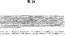
Figure 28 has shown the DNA of the KGF-2 disappearance construct that contains 93 to 208 in KGF-2 amino acid and the protein sequence that is encoded.
Figure 29 has shown the DNA of the KGF-2 disappearance construct that contains 04 to 208 of KGF-2 amino acid/11 and the protein sequence that is encoded.
Figure 30 has shown the DNA of the KGF-2 disappearance construct that contains 23 to 208 of KGF-2 amino acid/11s and the protein sequence that is encoded.
Figure 31 has shown the DNA of the KGF-2 disappearance construct that contains 38 to 208 of KGF-2 amino acid/11s and the protein sequence that is encoded.
Figure 32 has shown the DNA of the KGF-2 disappearance construct that contains 36 to 153 in KGF-2 amino acid and the protein sequence that is encoded.
Figure 33 has shown the DNA of the KGF-2 disappearance construct that contains 63 to 153 in KGF-2 amino acid and the protein sequence that is encoded.
Figure 34 has shown the DNA sequence of KGF-2 cysteine-37 to the mutation construction body of serine.
Figure 35 has shown the dna sequence dna of KGF-2 cysteine-37/ cysteine-106 to the mutation construction body of serine.
Figure 36 has shown the evaluation of the effect of 33 pairs of wound healings of KGF-2 Δ in the male SD rat (n=5). Animals received the back wound of 6mm, and continuous 4 days buffer solutions with multiple concentration, or the above-mentioned animal of KGF-2 Δ 33 treatments. Use every day through the Jameson caliper of calibration and measure wound. Use non-paired t test carry out statistical analysis (mean value+/-SE).*Compare with buffer solution.
Figure 37 has shown the evaluation of the effect of 33 pairs of wound healings of KGF-2 Δ in the normal rat. Male, SD, 250-300g, rat (n=5) has been accepted the holostrome skin back wound of 6mm. Measure wound with caliper, and began continuous 4 days the same day with KGF-2 Δ 33 and the buffer solution processing wound of multiple concentration from performing the operation, gather in the crops wound last day. Use non-paired t test to carry out statistical analysis.*With measured value and untreated comparing. # is compared measured value and buffer solution.
Figure 38 has shown the effect of 33 pairs of otch wounds of KGF-2 Δ breaking strength. Male adult SD rats (n=10) has been accepted the holostrome skin otch wound of 2.5cm at the 1st day, injured rear inner with one of buffer solution or KGF-2 (Δ 33) (Isosorbide-5-Nitrae and 10 micrograms) processing otch. Put to death animal at the 5th day, downcut the wound sample of 0.5cm to carry out conventional histology and breaking strength analysis. Apply external force across wound, use Instron skin tension meter to carry out biomethanics test, the maximum external force that suppressed by each wound. Use non-paired t test carry out statistical analysis (mean value+/-SE).
Figure 39 has shown the impact of KGF-2 (Δ 33) on otch wound epidermal thickness. Male adult SD rats (n=10) has been accepted the holostrome skin otch wound of 2.5cm at the 1st day, injured rear inner with one of buffer solution or KGF-2 (Δ 33) (Isosorbide-5-Nitrae and 10 micrograms) processing otch. Put to death animal at the 5th day, downcut the wound sample of 0.5cm to carry out conventional histology and breaking strength analysis. Determine epidermal thickness by the mean value of getting injury site 6 measurement results on every side, all measurement results are to be obtained through the section of Masson Trichrome dyeing by blind observer observation under the light microscope that uses the lens micrometer through calibrating. Use non-paired t test carry out statistical analysis (mean value+/-SE).
Figure 40 has shown that KGF-2 (Δ 33) is on the impact of epidermal thickness after the single intracutaneous injection. Male adult SD rats (n=18) was being accepted with buffer solution on the 0th day or is being dissolved in 6 intracutaneous injections of 1 and 4 microgram KGF-2 in the buffer solution of 50 microlitres. Put to death animal in 24 and 48 hours after the injection, measure by the epidermal thickness of graininess layer to the basic unit bottom. Measure about 20 times along injection site, calculate average thickness. All measurement results are to use through the lens caliper of calibration to observe the section through Masson Trichrome dyeing obtain under light microscope. Use non-paired t test carry out statistical analysis (mean value+/-SE).
Figure 41 has shown the impact of KGF-2 (Δ 33) on the BrdU scoring. The concentration that male adult SD rats (n=18) has been accepted placebo at the 0th day or has been dissolved in 50 microlitres is 6 intracutaneous injections of 1 and 4 microgram KGF-2. 24 and 48 hours execution animals after the injection. Put to death and used 5-2 '-bromodeoxyribouridine (in the 100mg/kg peritonaeum) injection animal in front 2 hours. Blind observer uses following points-scoring system to make scoring under light microscope, and: 0-3 does not have mark or by BrdU mark cell seldom; The cell of scale designation among the 4-6; The cell of the strong mark of 7-10. Use non-paired t test carry out statistical analysis (mean value+/-SE).
Figure 42 has shown the anti-inflammatory effect of the claw oedema that KGF-2 induces PAF-.
Figure 43 has shown the anti-inflammatory effect of the claw oedema that 33 couples of PAF-of KGF-2 Δ induce in the Lewis rat.
Figure 44 has shown the impact of KGF-2 Δ 33 on the radiation exposed Balb/c mouse survival of whole body. With 519 RADS irradiation Balb/c male mices (n=5), 22.1g. The every day in continuous 7 days is with buffer solution or KGF-2 (1 and 5mg/kg, subcutaneous) treatment animal in 2 days of pre-irradiation and after the irradiation.
Figure 45 has shown that KGF-2 Δ 33 is on by the impact of the body weight of irradiation mouse. With being the Balb/c male mice (n=5) of 22.1g with buffer solution or KGF-2 Δ 33 (1 and 5mg/kg) injection body weight in 2 days of 519 Rad/min pre-irradiations, weigh to animal every day, and continuously injection 7 days behind irradiation.
Figure 46 has shown the impact of KGF-2 Δ 33 on the radiation exposed Balb/c mouse survival of whole body rate. With 519 RADS irradiation Balb/c male mice (n=7), 22.1g. The every day in continuous 7 days is with buffer solution or KGF-2 (1 and 5mg/kg, subcutaneous) treatment animal in 2 days of pre-irradiation and after the irradiation.
Figure 47 has shown the effect of 33 pairs of wound healings of KGF-2 Δ in the rat model of glucocorticoid damage.
Figure 48 has shown the impact of KGF-2 Δ 33 on cell proliferation when using the BrdU marker determination.
Figure 49 has shown that KGF-2 Δ 33 is on the impact of the collagen content at the identical surgical sites place that is positioned at rat colon.
Describe in detail
According to an aspect of the present invention, the nucleic acid (polynucleotides) that separates is provided, described nucleic acid coding has the polypeptide of the amino acid sequence (SEQ ID NO:2) that Fig. 1 infers, or by being preserved in American type culture collection on December 16th, 1994,12301 Park Lawn Drive, Rockville, the ATCC preserving number of Maryland 20852 are the polypeptide of 75977 clone's cDNA coding. Nucleic acid molecules
Except as otherwise noted, use the automation DNA sequencer (such as 373 types, can derive from Applied Biosystems company) measure all nucleotide sequences of determining by the sequence of measuring this paper dna molecular, infer all amino acid sequences of the polypeptide of the dna molecule encode of being measured by this paper by translating the dna sequence dna of as above measuring. Therefore, known to any dna sequence dna of being measured by this automatic mode as those skilled in the art, any nucleotide sequence that this paper measures all may contain some mistakes. The nucleotide sequence of being measured by automatic mode is identical at least about 90% with the actual nucleotide sequence that is sequenced dna molecular, is more typically at least about 95% to identical at least about 99.9%. By other method, comprise artificial DNA PCR sequencing PCR well known in the art, can measure more accurately actual sequence. Known in the state of the art: as to compare with actual sequence, single insertion in the determined nucleotide sequence or disappearance can cause the frameshit phenomenon in this nucleotide sequence translation process, thereby make from described insertion or missing point, can be fully different from the amino acid sequence of the dna molecular actual coding that is sequenced by the amino acid sequence of determined nucleotide sequence coded supposition.
Except as otherwise noted, each " nucleotide sequence " that this paper lists represents with the sequence of deoxyribonucleotide (being abbreviated as A, G, C and T). Yet, " nucleotide sequence " of nucleic acid molecules or polynucleotides is the sequence of deoxyribonucleotide for dna molecular or polynucleotides, for RNA molecule or polynucleotides, be corresponding ribonucleotide (A, G, C and U) sequence, wherein each thymine deoxyribotide (T) in the specific deoxyribonucleotide sequence is replaced by uracil ribonucleotide (U). For example, about having the RNA molecule of the sequence of using the SEQ ID NO:1 that the deoxyribonucleotide abbreviation lists, that want to represent is each the deoxyribonucleotide A of SEQ ID NO:1 in its sequence, G or C are by corresponding ribonucleotide A, G or C replace, and the RNA molecule that each deoxyribonucleotide T has been replaced by ribonucleotide U.
" separation " nucleic acid molecules refers to the nucleic acid molecules that obtains from its natural surroundings, DNA or RNA. For example, for the purposes of the present invention, contained recombinant DNA molecules is considered to separate in the carrier. Other example of the dna molecular that separates comprises purifying in recombinant DNA molecules contained in the heterologous host cell or the solution (partial purification or basically purifying) DNA molecule. The RNA molecule that separates comprises the interior or external rna transcription of the body of dna molecular of the present invention originally. The nucleic acid molecules of separation of the present invention also comprises the synthetic this molecule that produces in addition.
The nucleic acid molecules of separation of the present invention comprises the dna molecular of the ORFs (ORF) that contains (SEQ ID NO:1) the 1-3 position initiation codon that has nucleotide sequence shown in Figure 1; The dna molecular that contains the coded sequence of ripe KGF-2 albumen shown in Figure 1 (172 or 173 last amino acid) (SEQ ID NO:2); With contain in fact from above-mentioned different sequence but because of the degeneracy of the genetic code dna molecular of encoded K GF-2 albumen still. Certainly, genetic code is well known in the art, and therefore, those skilled in the art can the above-mentioned degeneracy variant of conventional preparation.
The polynucleotides of code book invention polypeptide can derive from human prostate and Fetal Lung. The cDNA fragment initial separation of coded polypeptide subsequently, is separated the ORFs of coding full length protein from the library that derives from people's normal prostatic from the Human Fetal Lungs cDNA library that causes at random. Described reading frame is structurally relevant with FGF family, it contains the ORFs of the protein of 208 amino acid residues of encoding, and wherein approximately front 35 or 36 amino acid residues are that the targeting sequencing of inferring is so that ripe protein contains 173 or 172 amino acid. This protein shows the homology with people's keratinocyte growth factor top, has 45% homogeneity and 82% similitude in 206 amino acid whose one section sequences. Found that in the whole family of FGF conservative sequence also guards in protein of the present invention, this point is also very important.
In addition, the nested PCR result who derives from the KGF-2 cDNA in library shows that also there is other potential splicing form in KGF-2. Specifically, use flank in the primer of the N of KGF-2 ORFs end, obtained the PCR product of 0.2kb and 0.4kb by a plurality of cDNA libraries. 0.2kb size is the KGF-2 product of expection, and the 0.4kb size may be other splicing form of KGF-2. Deriving from cancer of the stomach, adult testis has been observed this 0.4kb product in the library of duodenum and pancreas.
Polynucleotides of the present invention can be the forms of RNA, also can be the forms of DNA, and described DNA comprises cDNA, genomic DNA and synthetic DNA. This DNA can be two strands or strand, and if strand then can be coding strand, also can be non-coding (antisense) chain. The coded sequence of encoding mature polypeptide can be identical with coded sequence (SEQ ID NO:1) or the coded sequence in the clone of preservation shown in Figure 1; Perhaps also can be different coded sequences, this coded sequence be encoded with the DNA (SEQ ID NO:1) of Fig. 1 or through the coded identical mature polypeptide of the cDNA of preservation because of rich remaining property or the degeneracy of genetic code.
The mature polypeptide (SEQ ID NO:2) that code pattern 1 is inferred or can be comprised by the polynucleotides through the mature polypeptide of the coded supposition of the cDNA of preservation: the coded sequence that mature polypeptide is only arranged; The coded sequence of mature polypeptide and other coded sequence such as targeting sequencing or secretion sequence or crude protein sequence; 5 ' and/or 3 ' non-coding sequence of the coded sequence of the mature polypeptide of the coded sequence of mature polypeptide (with other optional coded sequence) and non-coding sequence such as introne or supposition. In addition, obtained the mRNA of total length, it contains 5 ' and the 3 ' non-translational region (Fig. 3) of this gene.
But because order-checking error probability discussed above and for the variability of the cleavage site of targeting sequencing in the different known protein, those skilled in the art can expect by containing 208 amino acid of having an appointment through the coded real KGF-2 polypeptide of the cDNA of preservation, but can be any situations in 200-220 the Amino Acid Range; The actual targeting sequencing of this protein is about 35 or 36 amino acid, but can be any situation in 30-40 the Amino Acid Range.
Therefore, term " polynucleotides of coded polypeptide " comprises the polynucleotides that only comprise polypeptid coding sequence and comprises other coding and/or the polynucleotides of non-coding sequence.
The present invention relates to the variant of polynucleotides mentioned above in addition, and described polynucleotides variant coding has peptide more than the amino acid sequence (SEQ ID NO:2) that Fig. 1 infers or by fragment, analog and derivative through the polypeptide of preservation clone's cDNA coding. The variant of described polynucleotides can be the allele variant of the natural generation of these polynucleotides or the variant that this polynucleotides non-natural produces.
Therefore, the present invention includes coding and supposition mature polypeptide identical shown in Fig. 1 (SEQ ID NO:2) or with the polynucleotides of the coded identical supposition mature polypeptide of the cDNA that clones through preservation, and the variant of this polynucleotides, polypeptide shown in the described variant code pattern 1 (SEQ ID NO:2) or by fragment, derivative or analog through preservation clone's the coded polypeptide of cDNA. This nucleotide variants comprises the disappearance variant, replaces variant and interpolation or inserts variant.
The present invention includes the polynucleotides that coding can be used as the KGF-2 simulating peptide for the treatment of peptide. The KGF-2 peptide of simulation is some small peptides, and they can simulate the biologically active of KGF-2 albumen by the associated receptor of combination and activation KGF-2. The KGF-2 peptide of simulation also can in conjunction with and suppress the associated receptor of KGF-2. The KGF-2 acceptor includes but not limited to FGFR2 ⅲ b and FGFR1 ⅲ b. This simulating peptide can derive from such as, but be not limited to the method for phage display or combinatorial chemistry. For example, Wrighton etc., science, the method for the KGF-2 peptide of the described generation simulation of 273:458-463 (1996).
As mentioned above, the coded sequence that has of polynucleotides can be coded sequence shown in Figure 1 (SEQ ID NO:1) or through the allele variant of the natural generation of preservation clone's coded sequence. As well known in the art, allele variant is a kind of replaceable form of polynucleotide sequence, and it can have the replacement of one or more nucleotides, disappearance or add, but basically can not change the function of coded polypeptide.
The present invention also comprises polynucleotides, and wherein the coded sequence of mature polypeptide can merge with the polynucleotide sequence that helps host cell expression and secrete polypeptide (for example transporting the targeting sequencing of polypeptide as the secretion sequence functionating with control from transit cell) in identical reading frame. Polypeptide with targeting sequencing is front albumen, thereby it may have the targeting sequencing that can be cut by host cell the mature form that forms polypeptide. It is former that these polynucleotides also can encoding proteins, and described proteinogen is that ripe protein adds 5 ' extra amino acid residue. Mature protein with former sequence is proteinogen, and it is the inactive form of protein. In case former sequence is cut, remaining is exactly activated mature protein.
Therefore, for example, the protein that polynucleotides of the present invention can encoding mature, or the protein with former sequence, or both had the protein that former sequence also has presequence (targeting sequencing).
Polynucleotides of the present invention also can have the coded sequence that merges with a kind of flag sequence in frame, described flag sequence can be used for purifying polypeptide of the present invention. Be in the situation of bacterium the host, this flag sequence can be the mature polypeptide that is merged with purifying and flag sequence by the six histidine mark things that the pQE-9 carrier provides, or for example, when using mammalian hosts, during such as the COS-7 cell, flag sequence also can be hemagglutinin (HA) label. The HA label is equivalent to derive from the epi-position (Wilson, I etc., cell, 37:767 (1984)) of influenza virus hemagglutinin albumen.
Term " gene " refers to the DNA section that participates in producing polypeptide chain; It comprises before the code area and encode intervening sequence (introne) between the section (extron) of zone afterwards (targeting sequencing and tailer sequence) and each.
The hybridization probe that the fragment of full-length gene of the present invention can be used as cDNA library is with the cDNA that separates total length and separate to this gene and have sequence similarity highly or similar bioactive other cDNA. Such probe preferably has at least 30 bases, and can contain for example 50 or more base. Described probe can be used to identify the genomic clone of cloning and contain the complete genome that comprises adjusting and promoter region, extron and introne corresponding to the cDNA of total length transcript. The example of screening comprises by using known dna sequence dna synthetic oligonucleotide probe to separate the code area of this gene. Use has with oligonucleotides screening people cDNA, genomic DNA or the cDNA library through mark of the sequence of gene complementation of the present invention hybridizes to determine this probe and which member in library.
Other embodiment of the present invention comprises the nucleic acid molecules of separation, and the polynucleotides that described nucleic acid molecules contains have a nucleotide sequence that has the total length KGF-2 polypeptide of the complete amino acid sequence (targeting sequencing that comprises supposition) of Fig. 1 (SEQ ID NO:2) with (a) coding; (b) coding has a nucleotide sequence of the ripe KGF-2 polypeptide (removing the full-length polypeptide of targeting sequencing) of the about the 36th or 37 to 208 amino acids sequences among Fig. 1 (SEQ ID NO:2); (c) coding has a nucleotide sequence by the total length KGF-2 polypeptide of the complete amino acid sequence (comprising targeting sequencing) of contained cDNA clones coding in the ATCC preserving number 75977; (d) coding has a nucleotide sequence by the ripe KGF-2 polypeptide of the amino acid sequence of contained cDNA clones coding in the ATCC preserving number 75977; (e) nucleotide sequence of above-mentioned any KGF-2 analog of coding or deletion mutant; Or (f) and (a), (b), (c), (d) nucleotide sequence at least 90% of any nucleotide sequence complementation is identical or (e), and more preferably at least 95%, 96%, 97%, 98% or 99% identical nucleotide sequence.
Have with the nucleotide sequence of the encoded K GF-2 polypeptide of reference at least, for example the polynucleotides of the nucleotide sequence of 95% " identical " refer to except this polynucleotide sequence and can comprise that the nucleotide sequence of polynucleotides is identical with canonical sequence 5 point mutation of as many as in per 100 nucleotides of the nucleotide sequence of the encoded K GF-2 of reference polypeptide. In other words, in order to obtain having the polynucleotides of the nucleotide sequence identical with the nucleotide sequence at least 95% of reference, the nucleotides of as many as 5% can lack or replaced by another kind of nucleotides in the canonical sequence, and perhaps a large amount of nucleotides of 5% of the total nucleotide of as many as canonical sequence can insert in the canonical sequence. These sudden changes of canonical sequence can occur at 5 ' or 3 ' terminal position of reference nucleotide sequence, Anywhere generation that also can be between these terminal positions, they are dispersed in the nucleotides of canonical sequence separately, or are dispersed in the canonical sequence with the form of one or more contiguous set.
In the actual mechanical process, use known computer program, such as Bestfit program (Wisconsin sequence analysis program package, Unix the 8th edition, Genetics Computer Group, University Research Park, 575 Science Drive, Madison, WI53711) can routine determine whether any specific nucleic acid molecules with for example, nucleotide sequence shown in Figure 1 or through the cDNA of preservation clone's nucleotide sequence at least 90%, 95%, 96 %, 97%, 98% or 99% is identical. Bestfit uses Smith and Waterson, applied mathematics progress (Advances in Applied Mathematics), and local homology's algorithmic rule of 2:482-489 (1981) is found out the best section of homology between two sequences. When determine with Bestfit or any other series arrangement program specific sequence whether with, when for example identical according to canonical sequence of the present invention 95%, arranging certainly of parameter should be so that be that the total length of reference nucleotide sequence is calculated homogeneity percentage, and allow to exist the homology breach that reaches canonical sequence total nucleotide number 5%.
Please relate in this with nucleotide sequence shown in Fig. 1 [SEQ ID NO:1] or through the identical nucleic acid molecules of the nucleotide sequence at least 90%, 95%, 96%, 97%, 98% or 99% of preservation cDNA, no matter and whether they encode and have the polypeptide of KGF-2 activity. This be because even do not encode when having the polypeptide of KGF-2 activity when a kind of specific nucleic acid molecules, those skilled in the art still know the primer that how this nucleic acid molecules is used as hybridization probe for example or polymerase chain reaction (PCR). The purposes of nucleic acid molecules of the present invention of polypeptide with KGF-2 activity of not encoding comprises especially: (1) separates KGF-2 gene or its allele variant in the cDNA library; (2) with the stretching, extension thing in situ hybridization (such as " FISH ") of metaphase chromosome to provide the KGF-2 gene accurate chromosome mapping, example is seen Verma etc., human chromosomal: basic technology handbook, Pergamon publishing house, New York (1988); Express with the KGF-2mRNA that detects in the particular organization with the Northern engram analysis.
Yet, preferably have with nucleotide sequence shown in Fig. 1 [SEQ ID NO:1] or through the nucleotide sequence at least 90%, 95% of preservation cDNA, 96%, the nucleic acid molecules of 97%, 98% or 99% identical sequence, described nucleic acid molecules in fact codified have the polypeptide of KGF-2 protein active. " polypeptide with KGF-2 activity " refers to through special biological test and measures, show active similar to wild type KGF-2 albumen of the present invention, but activity that needn't be identical, or compare the polypeptide of the activity that strengthens to some extent with wild type KGF-2 albumen (full length protein or, be preferably ripe protein).
The detection method of KGF-2 activity for example, is hereinafter disclosed among the embodiment 10 and 11. Use these detection methods can measure the KGF-2 activity of the natural or recombinant protein of partially purified or purifying.
KGF-2 can stimulate epidermal keratinocytes rather than such as the propagation of the mesenchymal cell of fibroblast and so on. Therefore, " polypeptide with KGF-2 protein active " is included in the embodiment 10 described keratinocyte proliferation tests and shows the KGF-2 activity, and can be combined with FGF acceptor isoform 1-ⅲ b and 2-ⅲ b the polypeptide of (embodiment 11). Although what activity level needn't be with KGF-2 albumen is identical, but the activity that preferred " polypeptide with KGF-2 protein active " shows with KGF-2 albumen basic simlarity (is that candidate's polypeptide shows higher activity for the KGF-2 albumen of reference, or be not higher than 10 times, preferably be not higher than 2 times activity).
Certainly, because the degeneracy of genetic code, those skilled in the art can recognize nucleotide sequence or Fig. 1 [the SEQ ID NO that has with through the cDNA of preservation immediately; 1] nucleotide sequence shown at least 90%, 95%, 96%, 97%, the polypeptide that a large amount of nucleic acid molecules codifieds of 98% or 99% identical sequence " have the KGF-2 protein active ". In fact, the identical polypeptide because the degeneracy variant of these nucleotide sequences is all encoded, so those skilled in the art even need not to carry out above-mentioned comparative test and can understand this point. One skilled in the art will further recognize that, for this be not for the nucleic acid molecules of degeneracy variant, a great deal of is also encoded and is had the polypeptide of KGF-2 protein active. This is because those skilled in the art can grasp fully 49-Phe ,82-Ser,115-Arg,144-Met,145-Asn ,161-Arg,169-Met Human Connective tissue growth factor (being replaced by another aliphatic amino acid such as an aliphatic amino acid) unlikely or can not the appreciable impact protein function.
For example, relevant guidance example of how to carry out the 49-Phe ,82-Ser,115-Arg,144-Met,145-Asn ,161-Arg,169-Met Human Connective tissue growth factor of phenotype silence is seen: Bowie, J. U etc., " decipher the information in the protein sequence: to the tolerance of 49-Phe ,82-Ser,115-Arg,144-Met,145-Asn ,161-Arg,169-Met Human Connective tissue growth factor ", science, 247:1306-1310 (1990), wherein the author has pointed out that the research amino acid sequence has two kinds of main methods to the tolerance that changes. What first method relied on is evolutionary process, wherein suddenlys change to be accepted by natural selection or repel. Second method uses genetic engineering to import the amino acid variation at the ad-hoc location of clone gene, and selects or screen to identify to keep functional sequence. Pointed such as the author, these researchs have disclosed protein 49-Phe ,82-Ser,115-Arg,144-Met,145-Asn ,161-Arg,169-Met Human Connective tissue growth factor have been had wonderful tolerance. The author points out further which amino acid whose variation may allow in some position of protein. For example, most of hidden amino acid residues need nonpolar side chain, and the feature of surface side chains is generally seldom guarded. The reticent replacement of other this phenotype is described in Bowie, the list of references that J. U etc. (document is the same) and this paper mention.
The present invention relates to class polynucleotides in addition, when they sequence and sequence mentioned above between have at least 70%, preferably at least 90%, more preferably at least 95%, more preferably 96 % also, 97%, during 98%, 99% homogeneity, can with sequence hybridization mentioned above on. The present invention relate to particularly under stringent condition can with the polynucleotides of multi-nucleotide hybrid mentioned above. Term used herein " stringent condition " only refers to when having 95% and preferably just can hybridize during at least 97% homogeneity between the sequence at least. In a preferred embodiment, can with multi-nucleotide hybrid mentioned above on the coded polypeptide of polynucleotides can basically keep with by the cDNA of Fig. 1 (SEQ ID NO:1) or through the mature polypeptide of the cDNA of preservation coding identical biological function or activity.
The example of " tight hybridization conditions " is included in 42 ℃, containing 50% formamide, 5 * SSC (150mM NaCl, the 15mM trisodium citrate), 50mM sodium phosphate (pH7.6), 5 * Denhardt ' s solution, incubated overnight in the solution of the salmon sperm DNA through shearing of 10% dextran sulfate and 20 μ g/ml sex change is then washed filter membrane in 65 ℃ with 0.1 * SSC.
Perhaps, polynucleotides can have 20 bases at least, and preferred 30 bases more preferably have 50 bases at least, it can have homogeneity with multi-nucleotide hybrid of the present invention and with polynucleotides of the present invention as mentioned above, and it might be kept maybe can not keep activity. For example, can be with the probe of this polynucleotides as nucleotides more than the SEQ ID NO:1, for example in order to reclaim polynucleotides or as diagnostic probe or as the PCR primer.
Certainly, can with the reference polynucleotides major part of (as through the cDNA of preservation clone), be the part of 50-750nt such as length, or even also can be used as probe of the present invention with the polynucleotides with reference to multi-nucleotide hybrid of total length, can be used as probe too corresponding to the polynucleotides through the major part (if not all) of the nucleotide sequence of the cDNA of preservation or the nucleotide sequence shown in Fig. 1 [SEQ ID NO:1]. For example, the polynucleotides of " length is at least 20nt " partly refer to 20 or the nucleotides of more adjacency in the nucleotide sequence (as through the cDNA of preservation or the nucleotide sequence shown in Fig. 1 [SEQ ID NO:1]) with reference to polynucleotides. As mentioned above, pass through polymerase chain reaction (PCR) amplified target sequence thereby described part can be used as according to the probe of conventional DNA hybridization technique or as primer in diagnosis, example is seen molecular cloning, laboratory manual, the 2nd edition, Sambrook, J., Fritsch, E.F and Maniatis, T (1989) compiles, publishing house of cold spring harbor laboratory, and the document is listed this paper in as a reference in full.
Because KGF-2 cDNA clone is by preservation, its nucleotides sequence of determining is shown in Fig. 1 [SEQ ID NO:1], and the polynucleotides that generation can be hybridized with the part of KGF-2cDNA molecule should be routine techniques to those skilled in the art. For example, can use simply restriction endonuclease cutting or shear to produce the DNA parts of different sizes by ultrasonic processing KGF-2cDNA clone, these DNA partly be exactly can with the polynucleotides of the part hybridization of KGF-2cDNA molecule. Perhaps, can be according to the synthetic generation of known technology hybridization polynucleotides of the present invention. Certainly, only can with polyA sequence (3 ' terminal poly (A) sequence of KGF-2cDNA shown in Fig. 1 [SEQ ID NO:1]), or the polynucleotides of the complementary sequence hybridization of T (or U) residue be not included in for the polynucleotides of the present invention of nucleic acid moiety of the present invention hybridization because this polynucleotides can with any nucleic acid molecules that contains poly (A) sequence or its complement (such as actual what a kind of double-stranded cDNA clone that takes up an official post) hybridization on.
The present invention also provides the nucleic acid molecules of separation of the polynucleotides of the part that carries epi-position that contains encoded K GF-2 albumen in addition. Specifically, the nucleic acid molecule encoding of the separation that provides contains the polypeptide of the following amino acid residue among Fig. 1 (SEQ ID NO:2), and it is antigenicity zones of KGF-2 albumen that the inventor has determined them: 1.Gly41-Asn71:GQDMVSPEATNSSSSSFSSPSSAGRHVRSYN[SEQ ID NO:25]; 2.Lys91-Ser109:KIEKNGKVSGTKKENCPYS[SEQ ID NO:26]; [3.Asn135-Tyr164:NKKGKLYGSKEFNNDCKLKERIEENGYNTY SEQ ID NO:27]; With 4-Asn181-Ala199:NGKGAPRRGQKTRRKNTSA[SEQ ID NO:28]. Also has two other short supposition antigenicity zone, Gln74-Arg78 and Gln170-Gln175. Hereinafter will describe the method for the part that carries epi-position that produces KGF-2 in detail.
Can keep preserved material as herein described according to the fund of the budapest treaty of the relevant microbial preservation thing international endorsement that is used for patented method. These preserved materials are only for making things convenient for those skilled in the art to provide, rather than assert the regulation needs preservation according to 35U.S.C. ξ 112. Contained polynucleotide sequence and the amino acid sequence of coded polypeptide thereof all are incorporated herein by reference in preserved material, when these sequences have when conflict with sequence described herein, are as the criterion with the former. Preparation, using or sell need be through license through the material of preservation, and the application does not authorize this license. KGF-2 polypeptide and fragment
The present invention relate in addition have Fig. 1 (SEQ ID NO:2) putative amino acid sequence or have by the polypeptide through the amino acid sequence of the cDNA of preservation coding and the fragment of this peptide species, analog and derivative.
Those skilled in the art can understand, but because order-checking error probability discussed above and for the variability of the cracking site of targeting sequencing in the different known protein, contain 208 amino acid of having an appointment by the KGF-2 polypeptide through the coded reality of the cDNA of preservation, but can be any number in 200-220 the Amino Acid Range; The actual targeting sequencing of this protein is about 35 or 36 amino acid, but can be any number in 30-40 the Amino Acid Range.
Relate to the polypeptide of Fig. 1 (SEQ ID NO:2) or when the coded polypeptide of the cDNA of preservation, term " fragment ", " derivative " and " analog " refer to and kept the biological function substantially the same with this peptide species or active polypeptide. Therefore, analog comprises proteinogen, can activate this proteinogen by this proteinogen part of cracking to produce activated mature polypeptide.
Polypeptide of the present invention can be the polypeptide of restructuring, and natural polypeptide or synthetic polypeptide are preferably the polypeptide of restructuring.
The polypeptide of Fig. 1 (SEQ ID NO:2) or through the fragment of the coded polypeptide of the cDNA of preservation, derivative or analog can be following each peptide species: (ⅰ) wherein one or more amino acid residues are replaced by amino acid residue that guard or nonconservative (preferred conservative amino acid residue), this substituted amino acid residue can be also can not be the amino acid residue of being encoded by genetic code, or (ⅱ) wherein one or more amino acid residues comprise substituting group, or (ⅲ) ripe polypeptide and another kind of compound wherein, as improve the polypeptide compound of half life (such as polyethylene glycol) and merge, or (ⅳ) wherein other amino acid and mature polypeptide merge, such as targeting sequencing or secretion sequence or be used for sequence or the proteinogen sequence of purifying mature polypeptide. In view of the teachings contained herein, those skilled in the art are not difficult to grasp this fragment, derivative and analog.
Term " peptide " and " oligopeptides " are considered to synonym (people generally so think), and when at least 2 amino acid whose chains needing in the context to indicate by the peptide bond coupling, these two terms can Alternate. Use " polypeptide " word to containing 10 chains more than the amino acid residue herein. All oligopeptides of this paper and polypeptide formula or sequence are that amino terminal is to carboxyl terminal from left to right.
Some amino acid sequences that it should be recognized by those skilled in the art that the KGF-2 polypeptide can change, and structure or function that can appreciable impact protein. If this species diversity in the analytical sequence should be able to be remembered to exist in the protein to determine active vital zone. Generally speaking, can replace the residue that forms quaternary structure, as long as use the residue of exercising similar functions. Under other occasion, if change the non-important area that occurs in protein, the type of residue might be fully inessential so.
Therefore, the present invention comprises the various variations of the KGF-2 polypeptide in the KGF-2 albumen zone that basically shows the KGF-2 polypeptide active or comprise all protein portions as discussed below in addition. This mutant comprises disappearance, inserts, and inversion repeats and type replaces (be to replace another kind of hydrophilic residue with a kind of hydrophilic residue such as principle, rather than with the residue replacement of strong hydrophilicity hydrophobic residue by force). Little variation or this " neutrality " 49-Phe ,82-Ser,115-Arg,144-Met,145-Asn ,161-Arg,169-Met Human Connective tissue growth factor are generally very little to activity influence.
Usually the replacement that is considered as guarding is aliphatic amino acid Ala, Val, each other replacement among Leu and the Ile; The exchange of hydroxyl residue Ser and Thr, the exchange of acidic residues Asp and Glu, the replacement between amide residues Asn and the Gln, the exchange of alkaline residue Lys and Arg and aromatic residue Phe, the mutual replacement of Tyr.
Describe in detail as mentioned, other guide example relevant which kind of amino acid changes (namely can remarkable adverse influence not arranged to function) that be likely the phenotype silence is seen Bowie, J.U. wait people " deciphering the information in the protein sequence: to the tolerance of 49-Phe ,82-Ser,115-Arg,144-Met,145-Asn ,161-Arg,169-Met Human Connective tissue growth factor ", science, 247:1306-1310 (1990).
The present invention includes the KGF-2 simulating peptide that coding can be used as the treatment peptide. The KGF-2 peptide of simulation is some small peptides, and they simulate the biologically active of KGF-2 albumen by the associated receptor of combination and activation KGF-2. The KGF-2 peptide of simulation also can in conjunction with and suppress the associated receptor of KGF-2. The KGF-2 acceptor includes but not limited to FGFR2 ⅲ b and FGFR1 ⅲ b. This simulating peptide can derive from the method such as, but not limited to phage display or combinatorial chemistry. For example, Wrighton etc., science, the described method of 273:458-463 (1996) can be used for producing the KGF-2 peptide of simulation.
Preferably provide polypeptide of the present invention and polynucleotides with the form of separating, and preferably be purified as homogeneous.
Polypeptide of the present invention is preferably the form of separation, " polypeptide of separation " refers to the polypeptide that takes out from its natural surroundings, therefore, for the purposes of the present invention, what recombinant host cell produced and/or the polypeptide that contains is considered to separate, " polypeptide of separation " also refers to from recombinant host cell or natural origin part or the good polypeptide of purifying basically.
Polypeptide of the present invention comprise SEQ ID NO:2 polypeptide (especially ripe polypeptide) and with the polypeptide at least 90%, 95%, 96% of SEQ ID NO:2,97%, 98%, 99% similar (more preferably at least 90%, 95%, 96%, 97%, 98%, 99% is identical) polypeptide, the part that also comprises this peptide species, the described part of polypeptide (deletion mutant as mentioned below) generally contains at least 30 amino acid, more preferably at least 50 amino acid.
As everyone knows, by the sequence of the amino acid sequence of a peptide species and conservative 49-Phe ,82-Ser,115-Arg,144-Met,145-Asn ,161-Arg,169-Met Human Connective tissue growth factor and the second polypeptide relatively being determined " similitude " between two peptide species.
" the similitude percentage " of two polypeptide refers to by using Bestfit program (Wisconsin sequence analysis program package, Unix the 8th edition, Genetics Computer Group, University Research Park, 575 Science Drive, Madison, WI53711) and disappearance that be used for to determine similitude the amino acid sequence of two polypeptide of comparison is set and the similitude scoring that produces. Bestfit uses local homology's algorithmic rule of Smith and Waterson (applied mathematics progress, 2:482-489,1981) to find out the best section of similitude between two sequences.
Have with the amino acid sequence of the KGF-2 polypeptide of reference at least, for example the polypeptide of the amino acid sequence of at least 95% " identical " refers to except this peptide sequence and can comprise that the amino acid sequence of this polypeptide is identical with canonical sequence 5 amino acid whose changes of as many as in per 100 amino acid of the amino acid sequence of the KGF-2 of reference polypeptide. In other words, in order to obtain having the polypeptide of the amino acid sequence identical with the amino acid sequence at least 95% of reference, the amino acid residue of as many as 5% can lack or by another kind of 49-Phe ,82-Ser,115-Arg,144-Met,145-Asn ,161-Arg,169-Met Human Connective tissue growth factor, perhaps a large amount of amino acid of 5% of the total amino acid residue of as many as canonical sequence can insert in the canonical sequence in the canonical sequence. These variations of canonical sequence can occur in amino or the carboxyl terminal position of reference amino acid sequence, also can occur any position between described terminal position, perhaps be dispersed in separately in the residue of canonical sequence, perhaps be dispersed in the group of the one or more adjacency in the canonical sequence.
In the actual mechanical process, use known computer program, such as Bestfit program (Wisconsin sequence analysis program package, Unix the 8th edition, Genetics Computer Group, University Research Park, 575 Science Drive, Madison, WI53711) can routine determine whether any specific polypeptide with for example, the amino acid sequence shown in Fig. 1 [SEQ ID NO:2] or through coded amino acid sequence at least 90 % of the cDNA of preservation clone, 95%, 96%, 97%, 98% or 99% is identical. When using Bestfit or any other series arrangement program whether to determine specific sequence, when for example identical with canonical sequence according to the present invention 95%, certainly answer parameters being to calculate homogeneity percentage on the total length basis of reference amino acid sequence, and the homology breach that reaches canonical sequence total amino acid residue number 5% can allow.
As hereinafter describing in detail, polypeptide of the present invention can be used to produce polyclone and monoclonal antibody, and described antibody can be used in the diagnostic test of detection KGF-2 protein expression hereinafter described, or is used as activator and the antagonist of energy enhancer or inhibitor KGF-2 protein function. In addition, can be used to the yeast two hybrid system also be the KGF-2 protein-binding protein of candidate's activator of the present invention and antagonist with " catching " to this peptide species. The yeast two hybrid system is described in Fields and Song, nature, 340:245-246 (1989).
On the other hand, the invention provides peptide or the polypeptide of the part that carries epi-position that contains polypeptide of the present invention. The described epi-position of this polypeptide portion is immunogenicity or the antigenic epitopes of polypeptide of the present invention. " immunogenicity epi-position " is defined as causing in the protein part of antibody response when whole protein is immunogene. It is believed that these immunogenicity epi-positions only limit to the several positions on the molecule. On the other hand, the zone that can be combined with antibody in the protein molecule is defined as " antigenic epitopes ". The number of the immunogenicity epi-position of protein generally is less than the number of antigenic epitopes, and example is seen Geysen etc., institute of NAS newspaper, 81:3998-4002 (1993).
As for the peptide that carries antigenic epitopes (namely containing the zone that can be combined with antibody in the protein molecule) or the selection of polypeptide, general can the stimulation of relatively short synthetic peptide that those skilled in the art know a simulated albumin matter sequence part produces the antiserum that can react with the protein of partial simulation. Example is seen Sutcliffe, J.G, and Shinnick, T.M, Green, N and Learner, R.A (1983) is with the antibody of predetermined site reaction on the protein, science, 219:660-666. Often describe with the primary sequence of protein and can cause the peptide that produces proteins react serum, can identify this peptide by the simple chemical rule of a cover, they both had been not limited to the immunologic determinants zone (being the immunogenicity epi-position) of whole protein, also were not limited to amino or carboxyl terminal. Extremely hydrophobic peptide and contain 6 or still less the peptide of residue generally can not induce the antibody that can be combined with simulated albumin matter; Peptide that is long, solubility, the peptide that especially contains proline residue is normally effective. Sutcliffe etc., document is the same, p661. For example, according to the design of these guides, contain the antibody that has 18 can induce generation to react with HA1 albumen or whole virus in 20 peptides of 8-39 the residue that covers influenza virus hemagglutinin HA1 polypeptide chain sequence 75%; For the MulV polymerase, in 12 peptides 12 for rabies glycoproteins, can induce generation can precipitate the separately antibody of protein for 18 in 18 peptides.
Therefore, peptide and polypeptide that the present invention carries antigenic epitopes can be used for producing the antibody that can be combined with polypeptide of the present invention specifically, comprise monoclonal antibody. Therefore, most of hybridomas that the splenocyte of the donor by merging the peptide immunity that carries epitope of hanging oneself obtains usually can secrete can with the antibody of native protein reaction, Sutcliffe etc., document is the same, p663. The antibody that is produced by the peptide that carries antigenic epitopes or polypeptide can be used for detecting the protein of simulation, can be used for following the trail of the whereabouts in a plurality of zones of protein precursor of translating rear processing for the antibody of different peptides. Multiple quantitative or qualitative determination in simulated albumin matter, as using peptide and anti-peptide antibody in the competition assay, because verified even small peptide (according to appointment 9 amino acid) all can be in the immunoprecipitation test combination and substitute larger peptide, example is seen Wilson etc., cell, 37:767-778 (1984), p777. Anti-peptide antibody of the present invention also can be used for the protein of purifying simulation, for example by using method known in the art to carry out adsorption chromatography.
Carry at least 7 contained in the peptide of antigenic epitopes and the amino acid sequence that polypeptide preferably contains polypeptide of the present invention according to the present invention of above-mentioned guide design, more preferably at least 9, about 30 amino acid whose sequences of 15-most preferably from about. Yet, the major part that comprises polypeptid acid sequence of the present invention contains and has an appointment 30,40,50,60,70,80,90,100 or 150, or until and comprise that amino acid whose peptide or the polypeptide of any length of the whole amino acid sequences of polypeptide of the present invention also are considered to peptide or the polypeptide that the present invention carries epi-position, also can be used for inducing the antibody that produces with the simulated albumin qualitative response. The amino acid sequence that preferably carries the peptide of epi-position is selected as having good dissolubility (namely this sequence comprises relatively hydrophilic residue, and preferably avoids highly hydrophobic residue) in aqueous solvent; The sequence that particularly preferably contains proline residue.
Can be used for producing the antigenic polypeptide of KGF-2 specific antibody or the non-limitative example of peptide comprises following: 1.Gly41-Asn71:GQDMVSPEATNSSSSSFSSPSSAGRHVRSYN[SEQ ID NO:25]; 2.Lys91-Ser109:KIEKNGKVSGTKKENCPYS[SEQ ID NO:26]; [3.Asn135-Tyr164:NKKGKLYGSKEFNNDCKLKERIEENGYNTY SEQ ID NO:27]; With 4.Asn181-Ala199:NGKGAPRRGQKTRRKNTSA[SEQ ID NO:28].
Also has two other short supposition antigenicity zone, Gln74-Arg78 and Gln170-Gln175.
Any conventional method by preparation peptide or polypeptide comprises the recombination method that uses nucleic acid molecules of the present invention, can produce peptide and polypeptide that the present invention carries epi-position. For example, can will carry the amino acid sequence of short epi-position and larger polypeptide fusion, this polypeptide can be used as carrier in the process of recombinant production and purifying, can be used to produce anti-peptide antibody in immunologic process. Also can use the synthetic peptide that carries epi-position of known chemical synthesis process. For example, Houghten has described a kind of straightforward procedure of synthetic big figure peptide, represent the peptide of 248 13 different residues of 10-20mg of the single amino acids variant of a section of HA1 polypeptide such as Preparation and identification within 4 weeks (by ELISA-type binding), Houghten, R.A (1985), solid phase is synthesized the conventional method of big figure peptide fast: the specificity of the antigen-antibody interaction on each amino acid levels, institute of NAS newspaper, 82:5131-5135. The method of this " simultaneously synthetic a plurality of peptides (SMPS) " is further described in the people's such as Houghten the United States Patent (USP) 4,631,211 (1986). In the method, will be included in for each resin of the synthetic a plurality of peptides of solid phase the bag of each independent permeable solvent, with a plurality of identical repeating step included in the optimum utilization solid phase method. Artificial method can be carried out the synthetic of 500-1000 or more peptides simultaneously fully, the people such as Houghten, and document is the same, p5134.
Can use the present invention to carry peptide and the polypeptid induction antibody of epi-position according to method well-known in the art, example be seen the people such as Sutcliffe, and document is the same; The people such as Wilson, document is the same; Chow, the people such as M, institute of NAS newspaper, 82:910-914; And Bittle, the people such as F.J, general virology magazine, 66:2347-2354 (1985). Usually, available free peptide immune animal; Yet, by with peptide and macromolecular carrier, such as keyhole hemocyanin (KLH) or tetanus toxoid coupling, can improve the titre of anti-peptide antibody. For example, use as the joint of meta-maleimide benzoyl-N-hydroxysuccinimide eater (MBS), can will contain peptide and the carrier coupling of cysteine, and use more generally bridging agent such as the glutaraldehyde can be with other peptide and carrier coupling. By in the peritonaeum for example and/or intracutaneous injection contain the emulsion of have an appointment 100 microgram peptides or carrier protein and Freunds adjuvant, available free or with the peptide immune animal of carrier coupling, such as rabbit, rat and mouse. Need to for example to carry out several times booster shots about 2 weeks as the interval to provide the anti-peptide antibody titre of usefulness, can detect described titre by the ELISA test of for example using the free peptide that is adsorbed in the surface of solids. By for example peptide being adsorbed on the solid support and according to the selected antibody of method wash-out well known to those skilled in the art, thereby select anti-peptide antibody, can improve to get the anti-peptide antibody titre in the serum of the immune animal of hanging oneself.
Can identify that the present invention carries the peptide of immunogenicity epi-position, namely can cause all parts of the described protein of antibody response when whole protein is immunogene according to method well known to those skilled in the art. For example, people's (document is the same) such as Geysen discloses in the method for synthetic purity up to a hundred peptides being enough to react in EUSA simultaneously rapidly on the solid support. Then, they and holder need not be separated the interaction that can detect easily synthetic peptide and antibody. According to said method, those skilled in the art can identify the peptide of the immunogenicity epi-position that carries desired protein routinely. For example, the people such as Geysen orient the important epi-position of immunology in this protein by all 208 kinds six possible peptides of overlapping whole 213 the amino acid whose sequences of covering foot and mouth disease virus coat protein of a synthetic cover with 7 amino acid whose resolution ratio. Then, a synthetic complete cover replaces peptide, and wherein each position is used all 20 49-Phe ,82-Ser,115-Arg,144-Met,145-Asn ,161-Arg,169-Met Human Connective tissue growth factors successively in epi-position, measures the specific special acid of giving with antibody response. Therefore, can conventionally prepare the peptide analogues that the present invention carries the peptide of epi-position by the method. Further described the method for identifying the peptide of the immunogenicity epi-position that carries desired protein in the United States Patent (USP) 4,708,781 of Geysen (1987).
The United States Patent (USP) 5 of Geysen (1990), 194, described in 392 and detected or the conventional method of definite monomer (amino acid or other compound) sequence, described monomer is the homeomorphic thing (i.e. " mimotope ") with the epi-position of particular complementary position (antigen binding site) complementation of required antibody. More generally, described in the United States Patent (USP) 4,433,092 of Geysen (1989) and to have detected or the method for definite sequence monomer, described monomer is the homeomorphic thing with the part of the ligand binding site complementation of required special receptor. Similarly, Houghten, the United States Patent (USP) 5,480,971 of the people's such as R.A (1996) relevant in advance alkylating oligo peptide discloses linear C1-C
7-alkyl is series and the library of alkylating oligopeptides and this peptide in advance, and uses this oligopeptides series and library to measure the method for the sequence of preferential in advance alkylation oligopeptides of being combined with required acceptor molecule. Therefore, can conventionally prepare the non-peptide analogues that the present invention carries the peptide of epi-position by these methods.
Just as those skilled in the desired, KGF-2 polypeptide of the present invention and the above-mentioned fragment that carries epi-position thereof can partly be united the chimeric polypeptide of formation with the constant region of immunoglobulin (Ig) (IgG). These fusions are convenient to purifying and are demonstrated half life increase in the body. For example, the chimeric protein that is comprised of the regional of the first two domain of people CD4 polypeptide and mammalian immune globulin heavy chain or constant region of light chain has demonstrated this phenomenon, and (EPA 394,827; The people such as Traunecker, nature, 331:84-86 (1988)). The fusion that has the dimeric structure that is connected by disulfide bond because of IgG part aspect combination and other molecule of neutralization than the more effective (people such as Fountoulakis of the KGF-2 albumen of monomer or independent protein fragments, journal of biological chemistry, 270:3958-3964 (1995)).
According to the present invention, the new variant of KGF-2 has been described also. One or more amino acid by disappearance or replacement KGF-2 can produce these variants. Natural sudden change is called as allelic variation, and allelic variation can be reticent (coded polypeptide does not change), also can have altered amino acid sequence.
In order to attempt improving or changing the feature of natural KGF-2, the available protein engineering. Also can use recombinant DNA technology well known by persons skilled in the art to produce new polypeptide. Mutain and disappearance can demonstrate the stability of the active or increase that for example strengthens. In addition, they can higher yield be purified, and demonstrate preferably dissolubility at least under some purifying and condition of storage. Hereinafter is the example of constructed sudden change. Amino terminal and carboxyl-terminal deletion
Use recombinant DNA technology to modify a plurality of members of FGF family. In aFGF and bFGF, the molecule of the vital positively charged of Heparin-binding is substituted or lacks. Modified molecule causes the reduction of heparin binding activity, therefore, can be decreased by the amount of heparin-bounding modified molecule in the patient body, because more FGF can arrive suitable acceptor, thereby has increased effectiveness (EP 0298723).
The natural KGF-2 of saturation state is relatively unstable, and it can meet with chemistry and mechanical degradation, thereby causes bioactive loss in processing and the storage process. Natural KGF-2 is in the aqueous solution, because the temperature rising can tend to assemble meeting inactivation under acid condition.
In order to improve or change one or more characteristic of natural KGF-2, can use protein engineering. The people such as Ron, journal of biological chemistry, 268 (4): 2984-2988 (1993) still has the modified KGF albumen of heparin binding activity even reported forfeiture amino terminal 3,8 or 27 amino acid residues. Lack 3 and 8 amino acid and have whole activity. The more amino acid whose disappearances of KGF are described in PCT/IB95/00971. But the activity of the amino acid whose disappearance Enhancin of carboxyl terminal matter. An example is IFN-γ, can make active the raising reach 10 times people such as (, biotechnology magazine, 7:199-216 (1988)) Dobeli by 10 amino acid residues that lack this protein carboxyl terminal. Therefore, an aspect of of the present present invention provides the polypeptide analog of KGF-2 and the nucleotide sequence of this analog of coding, described analog shows the stability (being exposed to typical pH as working as, under heat condition or other condition of storage) of enhancing for natural KGF-2 polypeptide.
Hereinafter is particularly preferred KGF-2 polypeptide (counting starts from first amino acid (Met) in the protein): Thr (residue 36)-Ser (residue 208) Arg (65)-Ser (208) Cys (37)-Ser (208) Val (67)-Ser (208) Gln (38)-Ser (208) Ser (69)-Ser (208) Ala (39)-Ser (208) Val (77)-Ser (208) Leu (40)-Ser (208) Arg (80)-Ser (208) Gly (41)-Ser (208) Met (1), Thr (36), or Cys (37)-His (207) Gln (42)-Ser (208) Met (1), Thr (36), or Cys (37)-Val (206) Asp (43)-Ser (208) Met (1), Thr (36), or Cys (37)-Val (205) Met (44)-Ser (208) Met (1), Thr (36), or Cys (37)-Met (204) Val (45)-Ser (208) Met (1), Thr (36), or Cys (37)-Pro (203) Ser (46)-Ser (208) Met (1), Thr (36), or Cys (37)-Leu (202) Pro (47)-Ser (208) Met (1), Thr (36), or Cys (37)-Phe (201) Glu (48)-Ser (208) Met (1), Thr (36), or Cys (37)-His (200) Ala (49)-Ser (208) Met (1), Thr (36), or Cys (37)-Ala (199) Thr (50)-Ser (208) Met (1), Thr (36), or Cys (37)-Ser (198) Asn (51)-Ser (208) Met (1), Thr (36), or Cys (37)-Thr (197) Ser (52)-Ser (208) Met (1), Thr (36), or Cys (37)-Asn (196) Ser (53)-Ser (208) Met (1), Thr (36), or Cys (37)-Lys (195) Ser (54)-Ser (208) Met (1), Thr (36), or Cys (37)-Arg (194) Ser (55)-Ser (208) Met (1), Thr (36), or Cys (37)-Arg (193) Ser (56)-Ser (208) Met (1), Thr (36), or Cys (37)-Thr (192) Phe (S7)-Ser (208) Met (1), Thr (36), or Cys (37)-Lys (191) Ser (59)-Ser (208) Met (1), Thr (36), or Cys (37)-Arg (188) Ser (62)-Ser (208) Met (1), Thr (36), or Cys (37)-Arg (187) Ala (63)-Ser (208) Met (1), Thr (36), or Cys (37)-Lys (183) Gly (64)-Ser (208)
Preferred embodiment comprises N-terminal deletion Ala (63)-Ser (208) (KGF-2 Δ 28) and Ser (69)-Ser (208) (KGF-2 Δ 33). Other preferred N-end and C-terminal deletion mutant are described in specification embodiment 13 and 16 (c), and comprise Ala (39)-Ser (208); Pro (47)-Ser (208); Val (77)-Ser (208); Glu (93)-Ser (208); Glu (104)-Ser (208); Val (123)-Ser (208); And Gly (138)-Ser (208). Other preferred C-terminal deletion mutant comprises: Met (1), Thr (36), or Cys (37)-Lys (153).
Comprise also among the present invention that N-end and C-end amino acid have the deletion mutant of disappearance. This mutant comprises all associatings of above-mentioned N-terminal deletion mutant and C-terminal deletion mutant, Ala (39)-His (200) for example, Met (44)-Arg (193), Ala (63)-Lys (153), Ser (69)-Lys (153) etc. Use recombinant technique well known to those skilled in the art can carry out this associating.
Therefore, on the one hand, the invention provides the deletion mutant of N-end, this mutant comprises those front at least 38-terminal amino acid residues (namely at least Met (1)-Gln (38)) but be no more than front 147-terminal amino acid residues disappearance in Fig. 1 (SEQ ID NO:2), contains the mutant of all the other amino acid sequences shown in Fig. 1 (SEQ ID NO:2). Perhaps, disappearance comprises front at least 38-terminal amino acid residues among Fig. 1 (SEQ ID NO:2) (namely lacking at least Met (1)-Gln (38)) but is no more than front 137-terminal amino acid residues. Perhaps, disappearance comprises front at least 46-terminal amino acid residues among Fig. 1 (SEQ ID NO:2) but is no more than front 137-terminal amino acid residues. Perhaps, disappearance comprises front at least 62-terminal amino acid residues among Fig. 1 (SEQ ID NO:2) but is no more than front 137-terminal amino acid residues. Perhaps, disappearance comprises front at least 68-terminal amino acid residues among Fig. 1 (SEQ ID NO:2) but is no more than front 137-terminal amino acid residues. Perhaps, disappearance comprises front at least 76-terminal amino acid residues among Fig. 1 (SEQ ID NO:2) but is no more than front 137-terminal amino acid residues. Perhaps, disappearance comprises front at least 92-terminal amino acid residues among Fig. 1 (SEQ ID NO:2) but is no more than front 137-terminal amino acid residues. Perhaps, disappearance comprises front at least 103-terminal amino acid residues among Fig. 1 (SEQ ID NO:2) but is no more than front 137-terminal amino acid residues. Perhaps, disappearance comprises front at least 122-terminal amino acid residues among Fig. 1 (SEQ ID NO:2) but is no more than front 137-terminal amino acid residues.
Except the scope of above-mentioned N-terminal deletion mutant, the present invention also relates to all associatings of above-mentioned scope, such as front at least 62-terminal amino acid residues among Fig. 1 (SEQ ID NO:2) but be no more than the disappearance of front 68-terminal amino acid residues; At least front 62-terminal amino acid residues among Fig. 1 (SEQ ID NO:2) but be no more than the disappearance of front 76-terminal amino acid residues; At least front 62-terminal amino acid residues among Fig. 1 (SEQ ID NO:2) but be no more than the disappearance of front 92-terminal amino acid residues; At least front 62 N-terminal amino acid residues among Fig. 1 (SEQ ID NO:2) but be no more than the disappearance of front 103-terminal amino acid residues; At least front 68-terminal amino acid residues among Fig. 1 (SEQ ID NO:2) but be no more than the disappearance of front 76 N-terminal amino acid residues; At least front 68-terminal amino acid residues among Fig. 1 (SEQ ID NO:2) but be no more than the disappearance of front 92-terminal amino acid residues; At least front 68-terminal amino acid residues among Fig. 1 (SEQ ID NO:2) but be no more than the disappearance of front 103-terminal amino acid residues; At least front 46-terminal amino acid residues among Fig. 1 (SEQ ID NO:2) but be no more than the disappearance of front 62-terminal amino acid residues; At least front 46-terminal amino acid residues among Fig. 1 (SEQ ID NO:2) but be no more than the disappearance of front 68-terminal amino acid residues; At least front 46-terminal amino acid residues among Fig. 1 (SEQ ID NO:2) but be no more than the disappearance of front 76-terminal amino acid residues; Etc..
On the other hand, the invention provides the C-terminal deletion mutant. The-terminal amino acid residue of preferred described C-terminal deletion mutant is the amino acid residue 1 (Met) of Fig. 1 (SEQ ID NO:2), 36 (Thr) or 37 (Cys). This mutant comprises those last C-terminal amino acid residues (Ser (208)) but be no more than last 55 C-terminal amino acid residues disappearances (namely lacking amino acid residue Glu (154)-Ser (208)) at least in Fig. 1 (SEQ ID NO:2), contains the mutant of all the other amino acid sequences shown in Fig. 1 (SEQ ID NO:2). Perhaps, disappearance comprises among Fig. 1 (SEQ ID NO:2) at least last C-terminal amino acid residue but is no more than last 65 C-terminal amino acid residues. Perhaps, disappearance comprises last at least 10 C-terminal amino acid residues among Fig. 1 (SEQ ID NO:2) but is no more than last 55 C-terminal amino acid residues. Perhaps, disappearance comprises last at least 20 C-terminal amino acid residues among Fig. 1 (SEQ ID NO:2) but is no more than last 55 C-terminal amino acid residues. Perhaps, disappearance comprises last at least 30 C-terminal amino acid residues among Fig. 1 (SEQ ID NO:2) but is no more than last 55 C-terminal amino acid residues. Perhaps, disappearance comprises last at least 40 C-terminal amino acid residues among Fig. 1 (SEQ ID NO:2) but is no more than last 55 C-terminal amino acid residues. Perhaps, disappearance comprises last at least 50 C-terminal amino acid residues among Fig. 1 (SEQ ID NO:2) but is no more than last 55 C-terminal amino acid residues.
Except the scope of above-mentioned C-terminal deletion mutant, the present invention also relates to all associatings of above-mentioned scope, such as last C-terminal amino acid residue but be no more than the disappearance of last 10 C-terminal amino acid residues at least among Fig. 1 (SEQ ID NO:2); At least last C-terminal amino acid residue but be no more than the disappearance of last 20 C-terminal amino acid residues among Fig. 1 (SEQ ID NO:2); At least last C-terminal amino acid residue but be no more than the disappearance of last 30 C-terminal amino acid residues among Fig. 1 (SEQ ID NO:2); At least last C-terminal amino acid residue but be no more than the disappearance of last 40 C-terminal amino acid residues among Fig. 1 (SEQ ID NO:2); At least last 10 C-terminal amino acid residues among Fig. 1 (SEQ ID NO:2) but be no more than the disappearance of last 20 C-terminal amino acid residues; At least last 10 C-terminal amino acid residues among Fig. 1 (SEQ ID NO:2) but be no more than the disappearance of last 30 C-terminal amino acid residues; At least last 10 C-terminal amino acid residues among Fig. 1 (SEQ ID NO:2) but be no more than the disappearance of last 40 C-terminal amino acid residues; At least last 20 C-terminal amino acid residues among Fig. 1 (SEQ ID NO:2) but be no more than the disappearance of last 30 C-terminal amino acid residues; Etc..
On the other hand, the present invention comprises that also N-end and C-terminal residue have all lacked amino acid whose deletion mutant. This mutant comprises all associatings of above-mentioned N-terminal deletion mutant and C-terminal deletion mutant. This mutant comprises except front at least 46-terminal amino acid residues among Fig. 1 (SEQ ID NO:2) but is no more than front 137-terminal amino acid residues disappearance, at least last C-terminal amino acid residue but be no more than outside last 55 C-terminal amino acid residues disappearance among Fig. 1 (SEQ ID NO:2) contains the mutant of all the other amino acid sequences shown in Fig. 1 (SEQ ID NO:2). Perhaps, disappearance can comprise among Fig. 1 (SEQ ID NO:2) front at least 62,68,76,92,103 or 122-terminal amino acid residues but be no more than the disappearance of front 137 N-terminal amino acid residues, with among Fig. 1 last at least 10,20,30,40 or 50 C-terminal amino acid residues but be no more than the disappearance of last 55 C-terminal amino acid residues. Also comprise in addition all associatings of above-mentioned scope. 49-Phe ,82-Ser,115-Arg,144-Met,145-Asn ,161-Arg,169-Met Human Connective tissue growth factor
Another aspect of the present invention also comprises amino acid whose replacement. Natural ripe KGF-2 contains 44 charged residues, 32 positively chargeds wherein. According to the position of this residue in protein three-dimensional structure, can change the electrostatic interaction of adjacent residues with the residue of one or more these clusters of electronegative or uncharged 49-Phe ,82-Ser,115-Arg,144-Met,145-Asn ,161-Arg,169-Met Human Connective tissue growth factor, be useful for increasing protein stability and reducing protein aggregation. The gathering of protein not only can cause active forfeiture, bring problem also can for the preparation of medicine preparation, because their may be immunogenic (people such as Pinckard, clinical trial immunology, 2:331-340 (1967), the people such as Robbins, diabetes, 36:838-845 (1987), the people such as Cleland, Crit.Rev.Therapeutic Drug Carrier Systems, 10:307-377 (1993)). Any modification should be taken into account that all the electrical charge rejection that makes in the protein molecule quaternary structure minimizes. Therefore, making us especially interested is with another kind of charged amino acid with the neutral or charged amino acid of electronegative 49-Phe ,82-Ser,115-Arg,144-Met,145-Asn ,161-Arg,169-Met Human Connective tissue growth factor, the latter can cause protein with the positive charge minimizing, thereby improve the characteristic of KGF-2. This improvement comprises with natural KGF-2 albumen to be compared, and the stability of analog increases to some extent with aggtegation and decreases.
Amino acid whose replacement also can change be combined with cell surface receptor selective, the people such as Ostade, nature, described among the 361:266-268 (1993) some TNF α sudden change cause TNF α only optionally with the combination of one of two kinds of known TNF acceptors.
The KGF-2 molecule can comprise because of natural mutation or because of the one or more 49-Phe ,82-Ser,115-Arg,144-Met,145-Asn ,161-Arg,169-Met Human Connective tissue growth factors due to the manual operation, disappearance or add. Some examples that preferably suddenly change are: Ala (49) Gln, Asn (51) Ala, Ser (54) Val, Ala (63) Pro, Gly (64) Glu, Val (67) Thr, Trp (79) Val, Arg (80) Lys, Lys (87) Arg, Tyr (88) Trp, Phe (89) Tyr, Lys (91) Arg, Ser (99) Lys, Lys (102) Gln, Lys 103 (Glu), Glu (104) Met, Asn (105) Lys, Pro (107) Asn, Ser (109) Asn, Leu (111) Met, Thr (114) Arg, Glu (117) Ala, Val (120) Ile, Val (123) Ile, Ala (125) Gly, Ile (126) Val, Asn (127) Glu, Asn (127) Gln, Tyr (130) Phe, Met (134) Thr, Lys (136) Glu, Lys (137) Glu, Gly (142) Ala, Ser (143) Lys, Phe (146) Ser, Asn (148) Glu, Lys (151) Asn, Leu (152) Phe, Glu (154) Gly, Glu (154) Asp, Arg (155) Leu, Glu (157) Leu, Gly (160) His, Phe (167) Ala, Asn (168) Lys, Gln (170) Thr, Arg (174) Gly, Tyr (177) Phe, Gly (182) Gln, Ala (185) Val, Ala (185) Leu, Ala (185) Ile, Arg (187) Gln (190) Lys, Lys (195) Glu, Thr (197) Lys, Ser (198) Thr, Arg (194) G1u, Arg (194) Gln, Lys (191) Glu, Lys (191) Gln, Arg (188) Glu, Arg (188) Gln, Lys (183) Glu.
As for symbol description, for example Ala (49) the Gln Ala that refers to the 49th of Fig. 1 (SEQ ID NO:2) is replaced by Gln.
Advantageous variant is inappreciable, replaces such as folding or active conserved amino acid that can not appreciable impact protein. Hereinafter is the example that conserved amino acid well known to those skilled in the art replaces:
Aromatic series: phenylalanine, tryptophan, tyrosine
Hydrophobic: leucine, isoleucine, valine
Polarity: glutamine, asparagine
Alkalescence: arginine, lysine, histidine
Acid: aspartic acid, glutamic acid
Little: alanine, serine, threonine, methionine, glycine
Certainly, those skilled in the art can be according to comprising that above-mentioned those several factors determines the number of 49-Phe ,82-Ser,115-Arg,144-Met,145-Asn ,161-Arg,169-Met Human Connective tissue growth factor. Generally speaking, according to purpose, the replacement number of any given KGF-2 polypeptide is no more than 50,40,30,20,10,5 or 3. For example, the more terminal replacements that can improve stability of KGF-2 C-be described in above with embodiment 22 in.
By method well known to those skilled in the art, can identify among the KGF-2 the vital amino acid of function such as direct mutagenesis or alanine scanning mutagenesis (Cunningham and Wells, science, 244:1081-1085 (1989)). Rear a kind of method each residue place in molecule imports single alanine mutation, then detects the biologically active of gained mutant molecule, such as receptors bind or external and proliferation in vivo active (example is seen embodiment 10 and 11). By structural analysis, such as crystallization, nuclear magnetic resonance or photoaffinity labeling can determine that also (example is seen the people such as Smith, molecular biology magazine, 224:899-904 (1992) in conjunction with vital site to ligand-receptor; With the people such as de Vos, science, 255:306-312 (1992)).
Another aspect of the present invention is the cysteine with serine substituted amino acid the 37th and 106 and 150. The odd number of cysteine means that at least one cysteine residues can be used for intermolecular cross-linking or Cheng Jian, and described crosslinked or Cheng Jianke causes protein to take not meet the quaternary structure of needs. One or more cysteines by serine or the new KGF-2 albumen that for example alanine replaced generally be purified with soluble, the correct folding protein form of higher yield. Although be not proved, it is believed that the 106th cysteine residues is vital for function, this cysteine is high conservative in all other FGF family members.
Another aspect of the present invention is the fusion of KGF-2 and other oroteins or its fragment, as with other FGF albumen, such as KGF (FGF-7), bFGF, aFGF, FGF-5, the fusion of FGF-6 etc. or heterozygote. Reported this heterozygote of KGF (FGF-7). In disclosed PCT application number 90/08771, produced the chimeric protein that the C-end portion by front 40 amino acid residues of KGF and aFGF forms. It is reported the same target keratinocyte of this chimeric body image KGF, but lack the sensitiveness to heparin, the latter is the feature of aFGF rather than KGF. The fusion that forms with the partial fusion of immunoglobulin (Ig) (IgG) constant region often demonstrates half life in the body of increase. The chimeric protein that is comprised of the regional of front 2 domains of people CD4 polypeptide and mammalian immune globulin heavy chain or constant region of light chain has demonstrated this point (european patent application, publication number is the people such as 394827, Traunecker, nature, 331,84-86 (1988)). Fusion with dimeric structure that disulfide bond connects also can be more effectively in conjunction with independent monomer molecule people such as (, journal of biological chemistry, 270:3958-3964 (1995)) Fountoulakis. Antigenicity/hydrophilic segment of KGF-2
As described in Fig. 4 A-4E, 4 main highly hydrophilic zones are arranged in the KGF-2 albumen. Be amino acid residue Gly41-Asn71, Lys91-Ser109, Asn135-Tyr164 and Asn181-Ala199[SEQ ID NO:25-28]. Also has two other short supposition antigenicity zone, Gln74-Arg78 and Gln170-Gln175. Known hydrophilic parts mainly is positioned at the outside (surface) of protein, therefore, is convenient to these zones of antibody recognition. As if also the combination with KGF-2 and its acceptor is relevant in described zone. Derive from these regional synthetic peptides and can disturb the combination of KGF-2 and its acceptor, therefore blocked the function of protein. The synthetic peptide that derives from this protein hydrophilic parts also can be activator, namely simulates the function of KGF-2.
Therefore, the invention further relates to the polypeptide of the separation that contains the KGF-2 hydrophilic region, the length of wherein said polypeptide is no more than 150 amino acid, preferably is no more than 100,75 or 50 amino acid, this polypeptide contain one or more above-mentioned KGF-2 hydrophilic regions. Chemical modification
Can further modify KGF wild type and analog so that it contains other chemical composition of the normal part of nonprotein. The constituent of those derivatizations can be improved dissolubility, biological halflife or the suction-operated of this protein. Described constituent also can reduce or eliminate any unnecessary side effect of this protein etc. The summary example of relevant these constituents is seen REMINGTON ' S PHARMACEUTICAL SCIENCES, the 18th edition, Mack publishing company, Easton, PA (1990). Polyethylene glycol (PEG) is a kind of for the preparation for the treatment of this class chemical composition with protein. But the combination of PEG and protein has demonstrated protected protein matter and has made it to avoid the people such as protease hydrolytic, Sada, fermenting organism engineering magazine, 71:137-139 (1991). Useful several different methods is come in conjunction with some PEG constituent, and example is seen the people such as Abuchowski, " as the enzyme of medicine " (Holcerberg and Roberts compile), p367-383 (1981). A lot of disclosed patents have been described the PEG derivative and how to have been prepared their method, and example is seen the people such as Ono, United States Patent (USP) 5,342,940; The people such as Nitecki, United States Patent (USP) 5,089,261; The people such as Delgado, United States Patent (USP) 5,349,052. Usually, the PEG molecule is connected with this protein by the reactive group on the protein. Comparatively speaking, amino group is convenient to this combination such as the amino terminal of the amino group on the lysine or protein.
About every piece of document mentioning in " polypeptide and peptide " this chapter, it lists this paper in as a reference in full. Carrier and host cell
The carrier of the dna molecular that the present invention also relates to comprise that the present invention separates carries out genetic engineering modified host cell with recombinant vector, and produces the method for KGF-2 polypeptide or its fragment by recombinant technique.
Can synthesize to produce corresponding full-length polypeptide by peptide with fragment or the part of polypeptide of the present invention; Therefore, these fragments can be used as the intermediate of producing full-length polypeptide. Can synthesize the polynucleotides of total length of the present invention with fragment or the part of polynucleotides of the present invention. The present invention also relates to comprise the carrier of polynucleotides of the present invention, carry out genetic engineering modified host cell with carrier of the present invention, and the method by recombinant technique production polypeptide of the present invention.
With carrier of the present invention host cell is carried out genetic engineering modified (transduction or conversion or transfection), described carrier can be for example cloning vector or expression vector. The form of carrier can be plasmid for example, virion, bacteriophage etc. Can suitably improved to activate promoter, cultivated the host cell through transforming in the conventional nutrient medium of selection transformant or amplification KGF-2 gene. Such as temperature, the condition of culture of pH etc. is those used conditions before the host cell of selecting to express, and this should be apparent to those skilled in the art.
Can be with polynucleotides of the present invention for the production of polypeptide by recombinant technique, therefore, for example, described polynucleotides can be included in in the multiple expression vector any to express polypeptide. This carrier comprises chromosomal, achromosomal and synthetic dna sequence dna, such as the derivative of SV40; Bacterial plasmid; Phage DNA; Baculoviral; Yeast plasmid; Carrier derived from the associating of plasmid and phage DNA, viral DNA (such as vaccinia virus, adenovirus, fowlpox virus and pseudorabies virus). Yet as long as can copy in the host and survive, any other carrier all can use.
Can be by several different methods with suitable dna sequence dna insertion vector. Usually, by methods known in the art dna sequence dna is inserted suitable restriction endonuclease site. This method and other method should be well-known to those skilled in the art.
Dna sequence dna in the expression vector links to each other synthetic with mediation cDNA with suitable expression control sequenc (promoter) effectively. As for the representative example of this promoter, that should mention has: LTR or SV40 promoter, Escherichia coli lac or trp, phageλ PLPromoter and known other promoter that can control the gene expression in prokaryotic or eukaryotic or their virus. Expression vector also contains ribosome bind site and the transcription terminator that is useful on initial translation. Carrier also comprises the suitable sequence of expressing for strengthening.
In addition, expression vector preferably contains one or more selected markers so that the phenotypic character that can be used for selecting the host cell that transformed to be provided, such as dihyrofolate reductase or the neomycin resistance for eukaryotic cell culture, or the tetracycline in the Escherichia coli and amicillin resistance.
Can transform suitable host so that host expresses protein with containing suitable dna sequence dna mentioned above and suitable promoter or the carrier of control sequence.
As for suitable host's representative example, that should mention has: bacterial cell, and such as Escherichia coli, streptomycete, salmonella typhimurium; The fungal cell is such as yeast; Insect cell is such as fruit bat S2 and Spodoptera Sf9; Zooblast, such as CHO, COS or Bowes melanoma cells; Adenovirus; Plant cell etc. According to the instruction of this paper, those skilled in the art should be able to grasp the suitable host of How to choose.
More specifically, the present invention also comprises the recombinant precursor that contains one or more above broadly described sequences. These constructs contain carrier, such as plasmid or viral vectors, wherein to have inserted forward or backwards sequence of the present invention. This embodiment preferred aspect, this construct further comprises the regulating and controlling sequence that effectively links to each other with this sequence, such as promoter. A large amount of suitable carrier and promoters are well known to those skilled in the art, and commercially available. Listing following carrier is in order to give an example: bacterium: pQE70, pQE60, pQE-9 (Qiagen), pBS, pDIO, phagescript, psiX174, pBluescript SK, pbsks, pNH8A, pNH16a, pNH18A, pNH46A (Stratagene); Ptrc99a, pKK223-3, pKK233-3, pDR540, pRIT5 (Pharmacia); Eukaryotic: pWLNEO, pSV2CAT, pOG44, pXT1, pSG (Stratagene), pSVK3, pBPV, pMSG, pSVL (Pharmacia). Yet as long as can copy in the host and survive, any other plasmid or carrier all can use.
Can use CAT (CAT) carrier or other carrier with selected marker from any desirable gene, to select promoter region. Two suitable carriers are pKK232-8 and pCM7. The bacterium promoter of special name comprises the lacI promoter, lacZ promoter, T3 promoter, T7 promoter, gpt promoter, λ PR、P
LPromoter and trp promoter. Eukaryotic promoter comprises that CMV is early promoter, the HSV thymidine kinase promoter, early stage and late period the SV40 promoter, retroviral LTR promoter and Mouse Metallothionein-I promoter. Suitably the selection of carrier and promoter is well known to those skilled in the art.
In another embodiment, the present invention relates to contain the host cell of above-mentioned construct. Host cell can be higher eucaryotic cells such as mammalian cell, or eukaryotic such as the yeast cells such as low; Perhaps host cell also can be prokaryotic such as bacterial cell. Can pass through calcium phosphate transfection, the transfection of DEAE-glucan mediation, or electroporation (Davis, the people such as L, molecular biology mechanism method (1986)) imports construct in the host cell.
The gene outcome that can use in a usual manner the construct in the host cell to be encoded by recombination sequence with generation. Perhaps, also can be by the synthetic production of conventional peptide synthesizer polypeptide of the present invention.
Under the control of suitable promoter, ripe protein can be at mammalian cell, yeast, bacterium or other cells. Use also can utilize acellular translation system to produce this protein derived from the RNA of DNA construct of the present invention. Suitable clone and the expression vector that can use in protokaryon and eucaryon host are described in the people such as Sambrook, molecular cloning: laboratory manual, and the 2nd edition, the cold spring port, New York (1989), the document has been listed this paper in as a reference.
Can increase higher eucaryotic cells transcribing the DNA of code book invention polypeptide by in carrier, inserting enhancer sequence. Enhancer is the DNA element that promoter is worked to increase its cis acting of transcribing, and usually is about 10 to 300bp. Example comprises the SV40 enhancer of the 100-270bp of origin of replication side in late period, the sub-enhancer of cytomegalovirus early promoter, the polyomavirus enhancer of origin of replication side in late period and adenovirus enhancer.
For the protein with translation is secreted in endoplasmic, outer pericentral siphon space or the born of the same parents' external environment, suitable secretion signal can be mixed in the polypeptide that is expressed. Described signal can be endogenous for this polypeptide, also can be external source.
Polypeptide can be modified form, be expressed such as the form of fusion, polypeptide not only can comprise secretion signal, also can comprise other allos functional area. For example, can add other amino acid region at the N-of polypeptide end, especially charged amino acid is to improve polypeptide in host cell, in the purge process or subsequently processing and the stability in the storage process and persistence. Also can in polypeptide, add the peptide constituent so that purifying finally prepares polypeptide and can remove this class zone before. Those skilled in the art know in polypeptide add the peptide constituent with cause secretion or outside secrete, improve stability and be convenient to purifying etc., this is the routine techniques in this area. Preferred fusion contains the allos zone that derives from immunoglobulin (Ig), and this zone can be used for the solubilising acceptor. For example, among the EP-A-0464533 (Canada of the same clan 2045869) fusion that contains immunoglobulin molecules constant region various piece and another kind of human protein or its part is disclosed. Under many circumstances, the Fc part in the fusion is very favourable for treatment and diagnostic uses, and therefore, for example causing, pharmacokinetic properties makes moderate progress (EP-A0232262). On the other hand, for some application, need to be expressed in described favourable mode at fusion, disappearance is fallen the Fc part after detection and the purifying. When partly becoming treatment and the obstacle of diagnostic uses as Fc, be like this when being used as immunogene that immunity inoculation uses when fusion. For example when the screening medicine, the antagonist that to identify hIL-5 to carry out the high production rate screening test such as human protein and the Fc partial fusion of shIL-5 and so on, example is seen the people such as D.Bennett, the molecular recognition magazine, the 8th volume, the people such as 52-58 (1995) and K-Johanson, journal of biological chemistry, Vol.270, No.16, p9459-9471 (1995).
Usually, the expression vector of restructuring comprises origin of replication and allows the resistance marker of transformed host cell, such as the TRP1 gene of colibacillary ampicillin resistance gene and saccharomyces cerevisiae, and the promoter that derives from cance high-expression gene transcribing with mediation downstream configurations sequence. This promoter can get the glycolytic ferment of own coding such as glycerol 3-phosphate acid kinase (PGK) and so on, α-factor, the operon of acid phosphatase or heat shock protein etc. Allos structure sequence and translation initiation and terminator sequence, preferably with can mediate the targeting sequencing that the protein that is translated secretes to circumferential play or the extracellular matrix and be assembled together by rights. Optional heterologous sequence codified comprises fusion, the stabilisation of described feature such as expressed recombinant protein or the simplification of purge process of the N-Terminal Identification peptide of giving required feature.
By being in the structural DNA sequence of the coding desired protein in the effective reading mutually and suitable translation initiation and termination signal inserted the useful expression vector that can make up for bacterium with functional promoter. Carrier can contain one or more Phenotypic Selection marks and origin of replication keeps and provides where necessary amplification in the host with what guarantee carrier. Although also can utilize other selection, the suitable prokaryotic hosts that be used for to transform comprises Escherichia coli, bacillus subtilis, salmonella typhimurium, and pseudomonas, streptomyces and staphylococcus a plurality of kinds.
As representational but not circumscribed example, the useful expression vector that is used for bacterium can contain selected marker and the bacterium origin of replication that is purchased plasmid that derives from the genetic elements that contains known cloning vector pBR322 (ATCC37017). This commercially available carrier comprises for example pKK223-3 (Pharmacia Fine Chemicals, Uppsala, Sweden) and GEM1 (Promega Biotec, Madison, WI, USA). These pBR322 " skeleton " part and suitable promoter and the structure sequence gang of wish expression.
Transform suitable host strain and host strain is cultured to after the suitable cell density, induce selected promoter by suitable mode (such as variations in temperature or chemical induction), and cell is cultivated a period of time again.
General by centrifugal cell harvesting, by physics or chemical method smudge cells, keep the CE of gained to be further purified.
By comprising freeze thaw circulation, ultrasonic processing, Mechanical Crushing, or use the lysis agent etc. can the broken microbial cell that is used for marking protein in interior any facilitated method, these methods are well-known to those skilled in the art.
Can express recombinant protein with multiple mammalian cell culture system, the example of mammalian expression systems comprises Gluzman, cell, the described monkey kidney of 23:175 (1981) fibroblast COS-7 system, and other clone that can express the capacitive carrier, such as C127,3T3, CHO, HeLa and bhk cell system. Mammalian expression vector can contain origin of replication, suitable promoter and enhancer, and any essential ribosome bind site, polyadenylation site, donor splicing site and acceptor site, transcription terminator, and the non-transcribed sequence of 5 ' flank. Can use the dna sequence dna that derives from SV40 montage and polyadenylation site that required non transcribed genetic elements is provided.
By comprising ammonium sulfate or ethanol precipitation, acid extractants, cation or anion-exchange chromatography, the cellulose phosphate chromatography, hydrophobic interaction chromatography, affinity chromatography, the method for hydroxyapatite chromatography and agglutinin chromatography etc. reclaims from the recombinant cell culture thing and purifying KGF-2 polypeptide. In case of necessity, the refolding step that can carry out protein is finished the configuration of mature protein. At last, can use high performance liquid chroma-tography (HPLC) to finish final purification step.
Polypeptide of the present invention can be the product of natural purifying, or the product of chemical synthesis process, or is produced by recombinant technique by protokaryon or eucaryon host (such as the bacterium in cultivating, yeast, higher plant, insect and mammalian cell). Depend on host used in the recombinant method for production, polypeptide of the present invention can be glycosylated, also can be nonglycosylated. Polypeptide of the present invention also can comprise the initial methionine amino acid residue. The diagnosis of KGF-2 and treatment are used
Hereinafter used " KGF-2 " refers to the KGF-2 of total length as herein described and mature form and KGF-2 analog, derivative and mutant as herein described in the chapters and sections. The present invention also relates to the KGF-2 gene as the part of diagnostic test with detect with the KGF-2 nucleotide sequence in have the relevant disease or to the neurological susceptibility of described disease of suddenling change.
Can in detecting the KGF-2 gene, dna level carry by multiple technologies the individuality of sudden change. Diagnostic nucleic acid can derive from patient's cell, such as blood, and urine, saliva, biopsy and autopsy. Can be directly with genomic DNA for detection of, or before analysis, by using PCR (Saiki etc., nature, 324:163-166 (1986)) enzymatic amplification genomic DNA. Also RNA or cDNA can be used for identical purpose. For example, can be with identify and analyze the KGF-2 sudden change with the PCR primer of the nucleic acid complementation of encoded K GF-2. For example, by relatively amplified production and normal genotype variation in size can detect deletion and insertion. By the DNA that will increase with through radiolabeled KGF-2 RNA, perhaps can identify point mutation through radiolabeled KGF-2 antisense dna sequence hybridization. The sequence of mating fully and the double helix of mispairing can be distinguished by RNaseA digestion or the difference by melting temperature.
By can carry out the gene test based on dna sequence dna difference in the variation of the gel detection dna fragmentation electrophoretic mobility that contains or do not contain denaturant. By the insertion of becoming estranged of the visible foreword Lieque of high-resolution gel electrophoresis naked eyes. On the formamide gradient gel of sex change, can distinguish not homotactic dna fragmentation, in described gel, according to special the unwinding or the part melting temperature of different dna fragmentations, its mobility is arrested in the diverse location of gel, and (example is seen Myers etc., science, 230:1242 (1985)).
The sequence variation that also can disclose specific location by ribozyme protection test or chemical cracking method (such as Cotton etc., PNAS, USA, 85:4397-4401 (1985)) such as RNA enzyme and S1 protective effect.
Therefore; by such as hybridization; the effect of RNA enzyme protection, chemical cracking, directly the method for Southern trace of DNA order-checking or use Restriction Enzyme (such as RFLP (RFLP)) and genomic DNA and so on can detect specific dna sequence dna.
Except more conventional gel electrophoresis and dna sequencing, also can detect sudden change by the original position analysis.
The present invention also relates to the diagnostic assay for detection of the variation of KGF-2 protein level in the various tissues, because for normal control tissue sample, the overexpression detectable disease of this protein or to the existence of the neurological susceptibility of disease, described disease such as tumour. Test for detection of KGF-2 protein level in the host-derived sample is well known to those skilled in the art, and these methods comprise radioimmunoassay, and competition is in conjunction with measuring, and Western engram analysis, ELISA are measured and " sandwich " measured. ELISA measures (Coligan etc., immunology fresh approach, 1 (2), the 6th chapter, (1991)) and comprises that originally preparation is specific to the antibody of KGF-2 antigen, is preferably monoclonal antibody. The report antibody that also prepares in addition anti-this monoclonal antibody. Report is combined with detectable reagent on the antibody, such as radioactivity, fluorescence labeling or, be horseradish peroxidase in this example. In host, take a sample, with sample place can with the solid support of sample protein bound, as being incubated on the polystyrene dish. Then by being incubated with nonspecific proteins matter such as bovine serum albumin(BSA) with any free protein binding site on the covering disk, then, make any KGF-2 protein combination of combination on monoclonal antibody and the polystyrene dish. With all unconjugated monoclonal antibodies of buffer solution flush away. The report antibody that will be connected this moment with horseradish peroxidase places dish, report antibody is combined, then the unconjugated report antibody of flush away with any monoclonal antibody that is incorporated into KGF-2. Add peroxidase substrate in dish, when comparing with calibration curve, the amount that develops the color within preset time is the measurement result of the amount of the KGF-2 albumen that exists in the patient samples of given volume.
Also can use competitive assay, the antibody that wherein is specific to KGF-2 is combined with solid support, to pass at solid support through the KGF-2 of mark and host-derived sample, the amount of the label that detects by for example liquid scintillation chromatography is relevant with the amount of KGF-2 in the sample.
" sandwich " test is similar to ELISA and measures, and in " sandwich " measured, KGF-2 passed solid support, and combines with the antibody of combination on the solid support. SA is combined with KGF-2. Then make the 3rd antibody through mark that is specific to SA pass solid support and be combined with SA, determine again the size of amount.
Can be with described polypeptide, its fragment or other derivative, or their analog, or the cell of expressing them as immunogene to produce its antibody. These antibody can be for example polyclone or monoclonal antibody. The present invention also comprises the product of chimeric, strand, humanized antibody and Fab fragment or Fab expression library. Can use several different methods as known in the art to produce this antibody and fragment.
By described polypeptide directly being injected in the animal body or by to animal, being preferably the non-human animal and using described polypeptide and can obtain for the antibody that produces corresponding to the polypeptide of sequence of the present invention. Then, the antibody that so obtains can Binding peptide self. According to said method, even only also can be used for producing can be in conjunction with the antibody of whole natural polypeptides for the sequence of a fragment of coded polypeptide. Then this antibody can be used for separate this polypeptide from the tissue of expressing described polypeptide.
In order to prepare monoclonal antibody, can use any technology that the antibody that produces by the continuous cell line culture can be provided. Hybridoma technology (Kohler ﹠ Milstein for example, nature, 256:495-497 (1975)), trioma technology, human B-lymphocyte hybridoma technology (Kozbor etc., today immunology, 4:72 (1983)) with for generation of EBV hybridoma technology (Cole etc., monoclonal antibody and cancer therapy, the Alan R.Liss of human monoclonal antibodies, Inc, p77-96 (1985)).
Also be suitable for producing single-chain antibody for immunogenic polypeptide product of the present invention through describing technology (United States Patent (USP) 4,946,778) for generation of single-chain antibody. Also can use the transgenic mice expression for the humanized antibody of immunogenic polypeptide product of the present invention.
Can use polypeptide of the present invention to stimulate growth or the Angiogenesis of new blood vessel. Especially, polypeptide of the present invention can stimulate Growth of Cells and the propagation of keratinocyte. Therefore, the invention provides this peptide species, or the polynucleotides of this peptide species of encoding are used for the treatment of the method for purpose, so that wound healing, and hair follicle stimulating produces and cures the corium wound described therapeutic purposes such as the keratinocyte that stimulates epithelial cell proliferation and substrate.
As mentioned above, polypeptide of the present invention can be used to by stimulating epithelial cell proliferation to cure the corium wound. These wounds can be shallow tables, also can be very dark and relate to the infringement of corium and the epidermis of skin. Therefore, the invention provides the method that promotes wound healing, described method comprises the KGF-2 that uses effective dose to individuality.
The individuality of using KGF-2 can be to cure wound with normal speed, also can be that healing is impaired. When using for the int individuality of healing, use KGF-2 and can accelerate normal agglutination. When using for the impaired individuality of healing, use the healing that KGF-2 is convenient to wound, otherwise, wound healing will very slow or not healing. As mentioned below, a lot of miseries and disease can cause healing impaired. These miseries and disease comprise diabetes (such as type II diabetes), use simultaneously steroids and other pharmaceutical treatment, and ischaemic obstacle or injured. The steroids that has shown the infringement wound healing comprises cortisone, hydrocortisone, dexamethasone and medrat.
The on-steroidal compounds, having shown such as the Octreotide acetate can the infringement wound healing. Waddell, B. etc., U.S.'s operation, 63:446-49 (1997). It is believed that the present invention can promote to be subjected to the wound healing of the individuality of this on-steroidal pharmaceutical treatment.
Multiple growth factor has demonstrated the wound healing of the impaired individuality that can promote to heal, and example is seen Steed, D etc., J.Am.Coll.Surg, 183:61-64 (1996); Richard, J etc., diabetes maintenance (Diabetes Care), 18:64-69 (1995); Steed, D., J.Vasc. Surg, 21:71-78 (1995); Kelley, S etc., Proc.Soc.Exp.Biol., 194:320-326 (1990). These growth factors comprise somatotropin releasing factor, platelet-derived growth factor, and the fibroblast growth factor of alkalescence. Therefore, the present invention comprises that also co-administered KGF-2 and one or more can promote other growth factor or other medicament of wound healing.
The present invention also provides the method that promotes with the healing of other wound of coincideing healing in two kinds of impaired individualities of normal speed healing wounds and healing and being caused by operation. Described method is included in before anastomosis or other operation, afterwards and/or use the KGF-2 of effective dose in the process to individuality. Coincideing is the connection of two tubular structures, for example, when middle one section intestines are removed, can coincide when remaining part interconnects to reconstitute enteron aisle. Different from skin healing, the agglutination of the wound that coincide does not generally see Chu. In addition, the wound healing in the intestines and stomach can occur in uncomplicated situation fast at least; Yet complication often need to be corrected by extra operation, Thornton, F and Barbul, A, North America operation and clinical (Surg.Clin.North Am.), 77:549-573 (1997). Shown in embodiment 21 and 28, the Colonic anastomosis postoperative can cause peritonaeum to leak with the KGF-2 treatment and the shrinkage phenomenon that coincide significantly reduces. It is believed that thereby KGF-2 reduces the complication probability of happening that causes because of this process by accelerating healing process, and cause these results'.
Therefore, the present invention also provides the method for accelerating wound healing in normal speed healing wounds or the impaired individuality that heals after anastomosis or other operation, and described method comprises the KGF-2 that uses effective dose.
Also can use the differentiation of polypeptide irritation cell of the present invention, described cell such as muscle cell, cell, prostatic cell and the pneumonocyte of composition nerve fiber.
KGF-2 can be used for clinically in normal individual and the wound healing of suffering from the individual moderate stimulation wound of the disease that can induce abnormal wound healing, and described wound comprises surgical wound, the wound of resection, the deep layer wound that relates to corium and epidermis injury, ocular tissue's wound, dental tissue wound, oral wounds, diabetic's ulcer, skin ulcer, elbow ulcer, ulcer of artery, venous stasis ulcers, burn described disease such as uremia, malnutrition because of what be exposed to that heat or compound cause, vitamin-deficiency, obesity infects, immunosupress and with use steroids, radiotherapy, the complication that antineoplastic is relevant with the antimetabolite systemic treatment. KGF-2 can be used to promote the healing of the wound relevant with ischaemic and local ischemic damage, and described wound is such as the impaired and/or not enough chronic venous ulcer of leg that causes that refluxed by the venous circulation system.
Also can promote the tare weight after the skin damage to build with KGF-2. In addition, also can increase with KGF-2 tensile strength and the epidermal thickness of epidermis.
Also can increase with KGF-2 the adhesiveness of skin graft and wound bed, and stimulate the wound bed place again to form epithelium. Hereinafter listed the adhering graft type that to use KGF-2 to increase itself and wound bed: autograft, artificial skin, alloplast, the auto derma graft, Autologous epidermis graft, white graft, the Blair-Brown graft, bone graft, brephoplastic graft, skin graft, the graft of delay, dermal transplantation thing, epidermic graft, fascia graft, full thickness graft, the allograft thing, xenograft, alloplast, activated graft, cornea thin layer graft, the mesh graft, mucosal graft, Ollier-Thiersch graft, omental grafts, the sticking patch graft, base of a fruit graft, holostrome corneal graft, split-skin graft, thick-split graft. Can use KGF-2 to promote skin intensity and improve the outward appearance of aging skin.
It is believed that KGF-2 also can make hepatocellular propagation, and lung, breast, pancreas, stomach, the epithelial propagation in small intestine and the large intestine changes. KGF-2 can promote such as sebaceous cell, hair follicle, and liver cell, II type pneumonocyte produces the epithelial cell of mucinous goblet cell and so on, and other epithelial cell and skin, lung, liver, the propagation of the CFU-GM of contained they in kidney and the intestines and stomach. Therefore, but the propagation of KGF-2 cell cultured supernatant and differentiation, so KGF-2 can be used to alleviate or treat liver diseases and the fulminant liver diseases as being caused by cirrhosis, the pathological state of the hepatic lesion that is caused by virus hepatitis and toxicant (being paracetamol, carbon tetrachloride and other hepatotoxin well known in the prior art). Also can stimulate or promote with KGF-2 the regeneration of liver.
Also can reduce treatment by virus infections, radiotherapy, the intestines toxic side effects that chemotherapy or other treatment cause with KGF-2. KGF-2 has the cytoprotective effect to mucous membrane of small intestine. KGF-2 also preventability ground or therapeutic ground is used for prevention or reduces because of chemotherapy the healing of the catarrh that other medicament and virus infections cause and stimulating mucosal scorching (such as the oral cavity, oesophagus, intestines, colon, rectum and anal ulcer). Therefore, the present invention also provides the method for prevention or treatment membrane disease or pathology event, described disease or pathology event comprise ulcerative colitis, the damaged Other diseases of Crohn ' s disease and mucous membrane, and described method comprises the KGF-2 that uses effective dose. Similarly, the present invention also provides prevention or treatment oral cavity (comprise relevant with the mucous membrane of hypopharynx damage with pharynx swallow painful), oesophagus, and stomach, intestines, how coton and rectal catarrhal method is no matter and cause the medicament of described infringement or application method.
KGF-2 also can promote endothelial cell, the propagation of keratinocyte and substrate keratinocyte, and therefore, the present invention also provides the method that stimulates described cell type propagation, and described method comprises makes cell contact with the KGF-2 of effective dose. The KGF-2 that also can use effective dose to individuality is with the cell proliferation of body internal stimulus, or makes KGF-2 in external and this cells contacting.
The present invention also provides the method that promotes the urothelium healing, described method comprises the KGF-2 that uses effective dose to individuality, therefore, the invention provides and accelerate the urothelium healing or treat the multiple pathological method that relates to urothelium cell (cell of namely arranging along urethra). Comprise cathterization, operation, or the number of mechanisms of bacterium infection (as being caused sexually transmitted disease, such as the virulence factor infection of gonorrhoea) all may damage the organized layer of containing this cell.
The present invention also comprises the method that promotes the female reproductive tract organization healing, and described method comprises the KGF-2 that uses effective dose. Comprise monilial infection, trichomoniasis, the Gardner belongs to bacterium and infects, gonorrhoea, Chlamydia, the various diseases of mycoplasma infection and other sexually transmitted disease can cause the tissue damage of female reproductive tract.
Shown in embodiment 10,18 and 19, KGF-2 can stimulate epidermal keratinocytes propagation and increase epidermal thickness. Therefore, in the regeneration of intact skin; The defective of holostrome skin and segment thickness skin comprises burn (being hair follicle, the regrouping of sweat gland and sebaceous glands); With can use KGF-2 to such as the treatment of other skin defect of psoriasis and so on the time.
KGF-2 also can again form epithelium by the acceleration damaging part and be used for the treatment of epidermolysis bullosa, and this disease is the adhesiveness defective of epithelium and the corium below it, can cause regular ulceration and the blister of pain. KGF-2 also can be used for treating gastric duodenal ulcer, and can assist more quickly in the healing of mucous membrane lining place and make the gland mucous membrane and DM lining regeneration and help the healing of ulcer place. The inflammatory bowel disease such as sick and ulcerative colitis is respectively to cause small intestine or the surperficial destroyed disease of colorectal mucosa such as Crohn ' s. Therefore, KGF-2 can be used for also promoting that mucomembranous surface forms the surface again, and purpose is to heal more quickly and stop the process of inflammatory bowel disease. Expection KGF-2 treatment produces whole GI mucous membrane has significant effect, can be used to protect the impact of the harmful substance after that intestinal mucosa makes it to avoid being ingested or the operation. As mentioned above, also can promote with KGF-2 the healing of intestines or CCA. KGF-2 also can be used for treating and the not enough relevant disease of the expression of KGF-2.
In addition, KGF-2 also can be used for preventing and curing the damage to lung that causes because of multiple pathologic state. As a growth factor, KGF-2 can stimulate proliferation and break up and promote alveolar and bronchial epithelial reparation with prevention, alleviates or treats acute or chronic lung injury. For example, KGF-2 effectively treatment can cause the pulmonary emphysema of the gradual loss of alveolar, and by smoking and burn inhalant damage that cause, that can cause bronchiolar epithelium and alveolar necrosis. KGF-2 also can be used for stimulating propagation and the differentiation of II type pneumonocyte, and this helps to treat or prevents disease such as hyaline membrane disease and so on, and described hyaline membrane disease is such as the bronchopulmonary dysplasia that infant respiratory distress syndrome and immature infant are arranged.
Three reasons that cause acute renal failure are (such as heart failures) before the kidney, (such as urethral obstruction) behind endogenic (by the pnehrotoxicity of chemotherapeutic-induced) and the kidney, these reasons can cause renal tubular cell dead, the tubule chamber is blocked, be back in the glomerulus (referring to Thadhani etc. with filtrate, New England Journal of Medicine, 334:1448-1460 (1996)). Such as insulin-like growth factor l, Osteogenic Protein-1, the growth factor of HGF and EGF etc. has demonstrated has the potentiality of improving animal model middle kidney disease. Taub etc., cell factor, 5:175-179 (1993); Vukicevic etc., J.Am.Soc.Nephrol, 7:1867 (1996). But KGF-2 is propagation and the differentiation of stimulation of renal cell also, therefore can be used for alleviating or treat as acute and chronic renal failure and late period kidney disease and so on kidney disease and pathology.
Therefore KGF-2 also can stimulate propagation and the differentiation of breast tissue, can be used for promoting because of operation the healing of the breast tissue damage due to wound or the cancer.
In addition, also can use the outbreak of KGF-2 treatment or prevention diabetes. Be diagnosed as I type and type II diabetes just, but still keep among the patient of some islet cell functions, alleviate thereby can keep islet function with KGF-2, postpone or prevent the permanent performance of disease. KGF-2 also can be used as adminicle in the islet cell transplantation process to improve or to promote the function of islet cells.
In addition, the anti-inflammatory characteristics of KGF-2 is that main nosogenetic acute and chronic disease is favourable for treating wherein inflammation, and described disease includes but not limited to psoriasis, eczema, dermatitis and/or arthritis. Therefore, the invention provides prevention or alleviate inflammation and the method that relates to the disease of inflammation in the individuality, described method comprises the KGF-2 that uses effective dose.
Also can use KGF-2 to promote because of wound, the healing of the brain tissue impairment that damage causes due to operation or the chemicals is also alleviated described brain tissue impairment.
In addition, therefore the thickness owing to KGF-2 can increase epidermis can use this protein to improve aging skin, minimizing wrinkle of skin, the scar after the minimizing operation. The scar of wound tissue often relates to the hyper-proliferative of dermal fibroblast. As described in Example 10, KGF-2 does not stimulate fibroblastic propagation, and therefore, KGF-2 seemingly is specific to the mitogen of epidermal keratinocytes, and can induce wound healing, forms simultaneously minimum scar. Therefore, the invention provides the promotion wound healing, form simultaneously the method for minimum scar, described method comprises the KGF-2 that uses effective dose to individuality. Can produce the process of wound (such as aesthetic surgery, the premeditated wound of organizing accidental or that caused by sharp weapon) before, among and/or use afterwards KGF-2.
As mentioned above, KGF-2 also can stimulate the propagation of keratinocyte and hair follicle, therefore, can use KGF-2 to promote the growth of hair among bald head and the hair transplantation patient. Therefore, the present invention further provides trichogenous method, described method comprises the KGF-2 that uses the amount that is enough to the hair follicle stimulating generation.
The present invention also provides the protection individuality to avoid ionising radiation, the method for the impact of chemotherapy or antivirotic treatment, and described method comprises the KGF-2 that uses effective dose. The present invention also provides treatment because being exposed to ionising radiation in addition, the method for the histologic lesion due to chemotherapeutant or the antivirotic, and described method comprises the KGF-2 that uses effective dose. Individuality may be exposed to ionising radiation because of a lot of reasons, these reasons comprise therapeutic purposes (such as the overmedication proliferative diseases), radio isotope is released into the result in the environment by accident, or due in non-invasive medical diagnostic method (such as the X ray) process. In addition, quite a few individuality is exposed in the radioactive radon in its job site and family. Long-continued environmental exposure has been used to calculate the estimated value that descend to life expectancy, Johnson, and W and Kearfott, K, health physics, 73:312-319 (1997). Shown in embodiment 23; protein of the present invention can strengthen the survival of the animal that is exposed to radiation; therefore; can use KGF-2 to strengthen the survival rate of the individuality that suffers radiation-induced damage; the individual impact that makes it to avoid the sublethal dose radiation of protection, the treatment rate of increase radiation when treatment as hyperproliferation disease ailing.
Also can use KGF-2 protection individuality to make it to tolerate the radiation that can not tolerate under normal circumstances, the dosage of chemotherapeutic agent or antivirotic. When using in this way, or otherwise words when uses described herein, can be in radiation therapy/exposure, chemotherapy or treat with antivirotic before, afterwards and/or among use KGF-2. When treatment suffer from as excess proliferative disease late period slight illness individual the time, heavy dose of radiation and chemotherapeutant are particularly useful.
On the other hand, the invention provides prevention or treatment as radiation-induced oral cavity and injury of gastrointestinal tract, catarrh, intestines fibre modification, rectitis, radiation-induced pnemnofibrosis, radiation-induced pneumonia, radiation-induced pleura retrude, radiation-induced green blood syndrome, the method of the disease of radiation-induced myelotoxicity, described method comprises the KGF-2 that uses effective dose to individuality.
KGF-2 can use separately, also can unite use with one or more other medicament or other medicaments that can give the protective effect of radioresistance. Cytokine profiles (such as IL-1, TNF, IL-6, IL-12) has demonstrated can give this protective effect, and example is seen Neta, R. etc., The Journal of Experimental Medicine, 173:1177 (1991). In addition; IL-11 demonstrates the small intestinal cell (Du that can protect after radiation and chemotherapy Combined Treatment; X.X. etc.; blood; 83:33 (1994)) and radiation-induced chest damage (Redlich; C.A. etc., Journal of Immunology, 157:1705-1710 (1996)). Several growth factors have demonstrated the protective effect that can give for radioactive exposure, such as fibroblast growth factor and TGF-β-3, and Ding, I. etc., tumour journal, 36:337-340 (1997); Potten, C etc., Britain's cancer magazine, 75:1454-1459 (1997).
" individuality " used herein refers to animal, preferably refers to mammal (such as anthropoid cape, milk cow, horse, pig, boar, sheep, rodent, goat, dog, cat, chicken, monkey, rabbit, ferret, whale, dolphin), more preferably refers to the people.
Usually, use the burst of KGF-2 coded amino acid 1 to 35 or 36, identify the protein of secretion by the algorithmic rule of hybridization and/or computer search.
The nucleotide sequence of useful KGF-2 separates 5 ' sequence by hybridization, the plasmid that then will contain the KGF-2 gene that is subjected to its natural promoter/enhancer sequence control is used in vitro study, and purpose is endogenous cell and trans-activator virus that identifies KGF-2 gene expression.
In being designed for the experiment of identifying the peptide mimics that the KGF-2 acceptor is worked, also can be with KGF-2 albumen as positive control.
According to a further aspect in the invention, provide and be used for the polynucleotides of this peptide species or this peptide species of encoding with scientific research, DNA the is synthetic external purpose relevant with preparation dna vector and provide the diagnosis of human diseases and the method for the purpose for the treatment of.
The hybridization probe that the fragment of total length KGF-2 gene can be used as cDNA library has sequence similarity highly or similar bioactive other gene with the KGF-2 gene that separates total length with separating with described gene. Such probe generally has 20 bases at least, yet preferred probe has at least 30 bases, although probe can contain more bases, generally is no more than 50 bases. Described probe can be used to identify corresponding to the cDNA clone of total length transcript and contain and comprise and regulate and promoter region the genomic clone of the complete KGF-2 gene of extron and introne. The example of a screening comprises the code area by using known dna sequence dna synthetic oligonucleotide probe to separate the KGF-2 gene. Use has the oligonucleotides screening human cDNA library through mark with the sequence of gene complementation of the present invention, and hybridize to determine probe and which member in library in genome dna library or cDNA library.
The present invention also provides the method for identifying the acceptor of KGF-2 polypeptide. By several different methods well known by persons skilled in the art, can identify the gene of coding this receptor such as part elutriation and FACS sorting (Coligan etc., immunology fresh approach, 1 (2), the 5th chapter (1991)). The RNA of polyadenylation preparation is from the cell to the polypeptide reaction in the expression cloning that preferably carries out, and the cDNA library that produces of RNA is divided into some groups thus, and is used to rotaring redyeing COS cell or to nonreactive other cell of polypeptide. Will be being exposed in the polypeptide of mark through transfectional cell that glass slide is grown. The iodination of recognition site that can be by comprising the locus specificity protein kinase or comprise the described polypeptide of various ways mark of effect etc. After fixing and being incubated, slide is carried out radioautographic analysis. Identify positive group, the preparation subgroup, hive off and the again again transfection of method of screening in the Asia that use repeats, and final generation coding is inferred the monospecific polyclonal of acceptor.
Another alternative of identifying as acceptor can carry out with the cell membrane of expressed receptor molecule or extract product through the polypeptide of mark that light is affine to be connected. Analyze the crosslinked material of resolution by PAGE, and described material is exposed to x-ray film. Cutting-out contain polypeptide receptor through labeled complex, be separated into fragments of peptides, and carry out the protein microsequencing. Thereby use derives from the oligonucleotide probe of the amino acid sequence design one cover degeneracy of microsequencing and identifies the gene that coding is inferred acceptor with the screening cDNA library.
The invention provides screening compounds to identify the method for those compounds that can excite KGF-2 effect or blocking-up KGF-2 function. An example of this mensuration is included under the cell culture condition that keratinocyte can normally breed, with the mammal keratinocyte, the compound of wish screening and3[H] thymidine mixes mutually. Can in the situation of the compound that lacks the wish screening, carry out check experiment, and the amount that cutin forms cell proliferation during with the compound that has wish mensurations compares whether to stimulate the propagation of keratinocyte with definite this compound.
In order to screen antagonist, can in the presence of KGF-2, carry out identical test, measure the ability that this compound stops keratinocyte propagation, and measure the ability of antagonist. By measuring3The liquid scintillation chromatography that [H] thymidine mixes is determined the amount of keratinocyte propagation.
In another approach, can be in the presence of compound mammalian cell or the membrane product of expressing K GF-2 acceptor be incubated with the KGF-2 through mark, then measures compound and strengthen or block this interactional ability. Perhaps, measure in the situation of this compound and the relatively reaction of known second messenger system behind KGF-2 and the receptor interaction existing or lack. This second messenger system includes but not limited to the cAMP guanylate cyclase, ion channel or phosphoinositide hydrolysis.
The example of potential KGF-2 antagonist comprises the antibody that can be combined with this polypeptide, perhaps is oligonucleotides sometimes. Perhaps, potential KGF-2 antagonist can be the KGF-2 mutant forms, this mutant forms can with the KGF-2 receptors bind, produce second messenger's reaction but do not cause, therefore, effectively blocked the effect of KGF-2.
Another potential KGF-2 antagonist is the antisense constructs of using the antisense technology preparation. Formation or antisense DNA or the RNA of useful antisense technology by triple helix comes controlling gene to express, and these two kinds of methods are all based on the combination of polynucleotides and DNA or RNA. For example, polynucleotide sequence 5 ' the coded portion design length of use code book invention mature polypeptide is about the antisense rna oligonucleotide of 10 to 40 base-pairs. The DNA oligonucleotides be designed to relate to the gene region of transcribing complementary (triple helix, example is seen Lee etc., nucleic acids research, 6:3073 (1979); Cooney etc., science, 241:456 (1988); With Dervan etc., science, 251:1360 (1991)), thus prevent from transcribing and producing KGF-2. Antisense rna oligonucleotide is translated into KGF-2 polypeptide (antisense-Okano, neurochemistry magazine, 56:560 (1991) with cDNA hybridization and blocking-up cDNA molecule in vivo; As the oligodeoxynucleotide of the antisense inhibitor of gene expression, CRC publishing house, Boca Raton, FL (1988)). Above-mentioned oligonucleotides also can offer cell with in vivo antisence RNA or DNA, thereby suppresses the generation of KGF-2.
Potential KGF-2 antagonist comprises in conjunction with and thereby the binding site that takies the KGF-2 acceptor makes the acceptor can't be near KGF-2, and then the little molecule that is suppressed of its normal biology merit, and this micromolecular example includes but not limited to little peptide or peptide quasi-molecule.
Also can use the KGF-2 antagonist to suppress inducing new angiogenic growth or Angiogenesis in the tumour. Certain to also can being play a part equally by several pathological conditions of antagonist for treating of the present invention by the Angiogenesis that KGF-2 stimulates, described pathological condition comprises diabetic retinopathy, and to the inhibition of pathological tissue growth, such as rheumatoid arthritis.
Also can use KGF-2 antagonist for treating glomerulonephritis, the brightic glomerular epithelium cell that is characterised in that is significantly bred, and forms the cell mass that is full of the Bowman gap.
Also can suppress with antagonist excessive generation, the fibre modification after the myocardial infarction or the fiber infringement relevant with pnemnofibrosis and ISR of visible scar tissue during keloid forms after the operation. Also can use the KGF-2 antagonist for treating by other proliferative diseases that KGF-2 stimulates, comprise cancer and Kaposi sarcoma.
Also can use KGF-2 antagonist for treating keratitis, described keratitis is the chronic infiltration that is characterised in that the uvea inflammation corneal deep layer of epidermal cell proliferation.
Also can in the composition with pharmaceutically acceptable carrier hereinafter described, use antagonist.
Can be with polypeptide of the present invention, activator and antagonist and suitable pharmaceutical carrier are united and are used to make pharmaceutical composition. This composition contains the polypeptide for the treatment of effective dose, activator or antagonist and pharmaceutically acceptable carrier or excipient. Described carrier includes but not limited to salt solution, buffer salt solution, glucan, water, glycerine, ethanol and associating thereof. The pattern that preparation should be fit to use.
The present invention also provides drug packages or the kit that contains one or more containers that are full of by one or more compositions of pharmaceutical composition of the present invention. With container appended be the preparation of management medicine or biological products, the points for attention of government organs' suggestion form of using or selling, these points for attention reflect the preparation of relevant people's medication, use or sell the license that has obtained this mechanism. In addition, also can be with polypeptide of the present invention, activator and antagonist use with other curative compound.
Polypeptide with KGF-2 activity can be co-administered with the acceptable excipient of one or more pharmacy in pharmaceutical composition. Should understand when being applied to people patient, the nursing doctor can determine pharmaceutical composition of the present invention total consumption of every day in correct medical judgment scope. Any concrete patient's particular treatment effective dose level depends on many factors, comprises type and the degree of the reaction of wanting to reach, the specific combination of used other medicament (if any); Patient's age, body weight, health status, sex and diet; The time of using, the speed that the approach of using and composition are secreted outward; The time limit for the treatment of; With particular composition associating or the medicine (such as chemotherapeutant) that uses simultaneously; With the well-known other factors of medical domain. Suitable preparation known in the art sees Remington ' s Pharmaceutical Sciences (latest edition), Mack publishing company, Easton, PA.
Consider each patient's clinical condition (side effect when especially only treating with KGF-2), the site that the KGF-2 composition discharges, the method of using, the used KGF-2 composition for the treatment of is prepared and taken to the plan of using and the known other factors of practical operation personnel in the mode consistent with good medical practice. Therefore, can be identified for the KGF-2 (comprising the KGF-2 effective dose) of " effective dose " of this paper purpose by above-mentioned consideration.
Mode that can be easy, as oral, the part, intravenous, in the peritonaeum, intramuscular, subcutaneous in the artery, in the nose or intradermal routes use described pharmaceutical composition. The amount of institute's drug administration composition should be able to treat and/or prevent specific indication effectively. In most of the cases, consider the approach of using, symptom etc., the dosage of every day is about 1 microgram to 50 of per kilogram of body weight milligram. At topical in particular cases, the dosage of preferably using is about every square centimeter of 0.01 microgram to 9 milligram.
Although as mentioned above, but dosage considered in treatment, but the total medicine effective quantity scope of KGF-2 that stomach and intestine are used outward in every part of preferred dosage of general recommendations is per kilogram weight in patients 1 microgram to 100 milligram every day. If successive administration, generally with about 1 microgram/kg/hour to 50 micrograms/kg/hour dose rates, by injecting for 1-4 time every day, perhaps by with Micropump for example continuously subcutaneous perfusion use KGF-2. Also can use the packed solution of intravenous or ampuliform solution.
If continuous days is greater than a certain minimal amount (be 7 day for mouse), the KGF-2 therapeutic process that affects fibrinolytic system is seemingly the suitableeest. Observe to change the gap length that reacts after required treatment time and the treatment and seem different and different with required impact.
Also be suitable for using KGF-2 by slow-released system. The suitable example of slow releasing composition comprises the semipermeable polymers matrix of formative substance form (such as film or microcapsules). Sustained-release matrix comprises polyactide class (United States Patent (USP) 3,773,919, EP 58,481), the copolymer of Pidolidone and γ-ethyl-Pidolidone (U.Sidman etc., biopolymer, 22:547-556 (1983)), poly-(HEMA) (15:167-277 (1981) for R.Langer etc., biomedical material research magazine, and R.Langer, chemical technology, 12:98-105 (1982)), ethene vinyl-acetic ester (R.Langer, document is the same) or poly-D-(-)-3-hydroxybutyrate (EP 133,988). The KGF-2 composition of slowly-releasing also comprises the KGF-2 of liposome. Can prepare the liposome that contains KGF-2 by known method itself, described method as: DE 3,218,121; Epstein etc., institute of NAS newspaper, 82:3688-3692 (1985); Hwang etc., institute of NAS newspaper, 77:4030-4034 (1980); EP 52,322; EP 36,676; EP 88,046; EP 143,949; EP 142,641; Japanese patent application 83-118008; United States Patent (USP) 4,485,045 and 4,544,545; With EP 102,324. Generally speaking, liposome is little (about 200-800 dust) single-layer type, and wherein lipid content is regulated percentage greater than about 30 molar percentages of steroids, to reach best KGF-2 treatment.
For stomach and intestine are used outward, in one embodiment, general by with the KGF-2 of the required purity level injectable forms (solution with UD, suspension or emulsion) mix to prepare KGF-2 with pharmaceutically acceptable carrier, described carrier under used dosage and concentration to the acceptor avirulence, and compatible with other composition in the preparation. For example, preferred formulation does not comprise oxidant and known other compound harmful to polypeptide.
Usually by with KGF-2 all even the contact with liquid-carrier or the meticulous solid carrier that separates or the two nearly can prepare preparation. Then, if necessary, product is made required preparation shape. Preferred vector is the outer carriers of stomach and intestine, the solution that more preferably oozes with acceptor blood etc. The example of this carrier comprises water, salt solution, Ringer ' s solution and glucose solution. Water-free carrier and liposome such as fixing oil and ethyl oleic acid and so on also are useful in this article. Suitable preparation as known in the art sees Remington ' s Pharmaceutical Sciences (latest edition), Mack publishing company, Easton, PA.
Carrier can suitably contain the additive of minute quantity, as strengthening the material of isotonicity and chemical stability. This material is avirulent to acceptor under used dosage and concentration, comprises such as phosphate citrate, butanedioic acid, acetic acid, and the buffer solution of other organic acid or its salt; Antioxidant such as ascorbic acid and so on; The polypeptide of low-molecular-weight (being less than about 10 residues) is such as pR60 or tripeptides; Protein, such as seralbumin, gelatin, or immunoglobulin (Ig); Hydrophilic polymer is such as polyvinylpyrrolidone; Amino acid, such as glycine, glutamic acid, aspartic acid, or arginine; Monose, disaccharides and comprise the cellulose or derivatives thereof, glucose, other carbohydrate of mannose or dextrin etc.; Chelating agent such as EDTA and so on; Sugar alcohol such as sweet mellow wine or sorbierite and so on; Counter ion such as sodium and so on; And/or nonionic surface active agent, such as polysorbate, poloxalkol or PEG.
Generally under pH was about 3 to 8 condition, to be about 0.01 ug/ml to 100 mg/ml, the concentration that is preferably 0.01 ug/ml to 10 mg/ml was prepared KGF-2 in described carrier. Should understand and use above-mentioned some excipient, carrier or stabilizing agent can cause the formation of KGF-2 salt.
The KGF-2 that is used for the treatment of application should be aseptic. Filter and easily to finish degerming by passing aseptic filter membrane (such as 0.2 micron film). The KGF-2 composition for the treatment of usefulness generally is placed in the container with aseptic gateway, for example, and the intravenous solution bag, or have the bottle of the stopper that can be penetrated by hypodermic needle.
Usually, the lyophilized formulations form that KGF-2 can be used as aqueous solution or is used for recovering is stored in the container of UD or multiple dose, for example, and in the airtight ampoule bottle or bottle. An example of lyophilized formulations is that 5ml is poured into the bottle of 10ml through the moisture KGF-2 solution of 1% (w/v) of aseptic filtration, with the freeze-drying of gained mixture. Can prepare primer solution by the KGF-2 that uses antibacterial water for injection to recover freeze-drying.
Also can take medicine in order to provide active through the KGF-2 that is determined as predetermined concentration such as the RIA technology as blood take the mode arrangement of patient-specific. Therefore, the dosage arrangement that can regulate the patient reaches through RIA and is determined as with about 50-1000ng/ml, the concentration that the is preferably 150-500ng/ml level of blood that continued to flow regularly.
The method of application of pharmaceutical composition of the present invention has: oral, rectum, outside the stomach and intestine, in the brain pond, in the corium, in the vagina, in the peritonaeum, local (by powder, ointment, gel, missible oil, drops or through the sticking patch of skin), cheek, or mouthful or the nose spray delivery. " pharmaceutically acceptable carrier " refers to avirulent solid, semisolid or liquid filling agent, diluent, lapping or any type of preparation adminicle. Term used herein " stomach and intestine outer " refers to and comprises intravenous, intramuscular, and in the peritonaeum, in the breastbone, the mode of administration of subcutaneous and intra-arterial injection and perfusion etc.
According to the present invention, the KGF-2 polypeptide also can be used by expressing in vivo as activator and the antagonist of polypeptide, normal " gene therapy " said that Here it is.
Therefore, for example, can exsomatize and with the polynucleotides (DNA or RNA) of coded polypeptide the cell from the patient be transformed, then the cell through transforming be offered the patient who needs with this polypeptide treatment. This method is method known in the art. For example, can by method well known in the art, utilize the retroviral particle of the RNA that contains code book invention polypeptide that cell is transformed.
Similarly, can be by for example method well known in the art, engineered cells is to express in vivo polypeptide in vivo. Known in this area, the producer's cell that can use the retroviral particle that can produce the RNA that contains code book invention polypeptide to the patient is with engineered cells in vivo and express in vivo polypeptide. In view of instruction of the present invention, those skilled in the art can grasp to use these and other method of polypeptide of the present invention easily. For example, the expression vector that is used for engineered cells not only can be retrovirus, also can be adenovirus, and described adenovirus can be used in vivo engineered cells after uniting with suitable transmission carrier. Other example that transmits carrier comprises the carrier system based on HSV, gland relevant viral vector, and the carrier of inertia, as be coated with the iron particle of glucan.
The retrovirus that can therefrom obtain retroviral plasmid vector mentioned above includes but not limited to the Moloney murine leukemia virus, spleen necrosis virus, such as the Rous sarcoma virus, the Harvey sarcoma virus, avian leukosis virus, gibbon ape leukemia virus (gibbon ape leukemia virus), the human immunodeficiency virus, adenovirus, bone marrow proliferative sarcoma virus, and mammary tumour virus. In one embodiment, retroviral plasmid vector derives from the Moloney murine leukemia virus.
Carrier comprises one or more promoters, and operable suitable promoter includes but not limited to retroviral LTR; The SV40 promoter; And human cytomegalovirus (CMV) promoter (Miller etc., biotechnology, the 7th volume, 9:980-990 (1989)) or any other promoter (cell promoter for example, as include but not limited to histone, the eukaryotic promoter of pol III and beta-actin promoter etc.). Spendable other viral promotors includes but not limited to adenovirus promoter, thymidine kinase (TK) promoter, and B19 parvovirus promoter. In view of the teachings contained herein, the selection of suitable promoter is apparent to those skilled in the art.
The nucleotide sequence of code book invention polypeptide is subjected to the control of suitable promoter. Spendable suitable promoter includes but not limited to adenovirus promoter, such as adenovirus major late promoter; Or allogeneic promoter, such as cytomegalovirus (CMV) promoter; Respiratory Syncytial Virus(RSV) (RSV) promoter; Inducible promoter is such as MMT promoter, metallothionein promoter; The heat shock protein promoter; Albumin promoter; ApoA I promoter; People's globin promoter; Viral thymidine kinase promoter is such as the herpes simplex virus thymidine kinase promoter; Retrovirus LTR (comprising modified retrovirus LTR mentioned above); The beta-actin promoter; With the human growth hormone (HGH) promoter. Promoter also can be the natural promoter of the gene of the described polypeptide of control coding.
Can use retroviral plasmid vector transduction package cell line to form producer's clone. The example of package cell line that can be transfected includes but not limited to PES01, PA317, ψ-2, ψ-AM, PA12, T19-14X, VT-19-17-H2, ψ CRE, ψ CRIP, GP+E-86, GP+envAm12 and Miller, the people's gene therapy, the DAN clone described in the 1:5-14 (1990) (listing this paper in as a reference in full). Carrier can be by any method transduction incasing cells known in the art. Described method includes but not limited to electroporation, the application of liposome, and CaPO4Precipitation. Perhaps also retroviral plasmid vector can be wrapped in the liposome, or with the lipid coupling, then be applied to the host.
Producer's clone produces the infectious retroviral vector particle of the nucleotide sequence that contains coded polypeptide. Then can use this retroviral vector particle in vivo or external transduction eukaryotic. Can express the nucleotide sequence of coded polypeptide through the eukaryotic of transduction. The eukaryotic that can be transduceed includes but not limited to embryonic stem cell, embryo cells and candidate stem cell, liver cell, fibroblast, sarcoblast, keratinocyte, endothelial cell and bronchial epithelial cell. Chromosome is measured
Sequence of the present invention also can be used for Chromosome Identification. This sequence-specific ground target and can with each human chromosome on particular seat hybridization. In addition, at present in the urgent need to identifying the specific site on the chromosome. The chromosome marking reagent based on actual sequence data (Repeat Polymorphism) that can be used at present the marker chromosome coordinate is less. The important first step that gene those sequences are same and disease association interrelates is according to the present invention DNA to be plotted on the chromosome.
In brief, sequence can be plotted on the chromosome by prepared PCR primer (being preferably 15-25bp) by cDNA. Use the Computer Analysis of 3 ' non-translational region to select fast primer, the zone that described primer is crossed in genomic DNA is no more than an extron, thereby can not make the amplification procedure complicated. Then use these primers to contain the PCR screening of the body cell heterozygote of each human chromosome. Only have those heterozygotes that contain corresponding to the people's gene of primer can produce the fragment of amplification.
The PCR of body cell heterozygote mapping is that specific DNA is arranged in fast method on the specific chromosome. Use identical Oligonucleolide primers, by the present invention, similarly method can be carried out the location, Asia with a series of fragments that derive from specific chromosome or large genomic clone storehouse. Can be similarly be used for mapping to its chromosomal other mapping strategy and comprise in situ hybridization, use chromosome through the airflow classification of mark carry out prescreen and by with make up the hybridization of chromosome specific cDNA library and carry out preselected.
Can use cDNA clone and FISH (FISH) step of metaphase chromosome coating thing that accurate chromosome mapping is provided. The cDNA that is as short as 50 or 60 bases can use this technology, and the summary example of relevant this technology is seen Verma etc., human chromosomal: basic fundamental handbook, Pergamon publishing house, New York (1988).
In case sequence is plotted on accurate chromosomal loci, physical location and the genome data of sequence on chromosome can be interrelated, described data example is seen V.McKusick, human Mendelian inheritance (can obtain on the net by Johns Hopkins University's Welch medical library). Then identify the gene of identical chromosomal region and the relation between the disease of being plotted on by linkage analysis (the physically common heredity of adjacent gene).
Then, the essential difference of determining cDNA between the ill and not ill individuality or genome sequence. If observe sudden change in some or all of ill individualities, and all do not observe sudden change in any normal individuality, sudden change just may be the triggering factor of disease so.
The resolving power of up-to-date physical mapping and gene mapping technology shows, (supposition mapping resolving power is 10 accurately to be positioned cDNA with the chromosomal region of disease association and can be in 50 to 500 potential Disease-causing genes one6Individual base, a gene is 20kb).
Further describe the present invention with reference to the following example; Yet, should understand the present invention and be not limited to these embodiment. Except as otherwise noted, all parts or amount are weight.
For the ease of understanding the following example, the below will describe some common method and/or terms.
" plasmid " is named as: at first be the p of small letter and/or then be the capitalization the letter and/or digital. The initial plasmid of this paper or commercially available, the public can obtain easily, or can be according to known technology by available plasmid construction. In addition, with the plasmid of plasmid equivalence described herein be well known in the art, be apparent to those skilled in the art.
" digestion " of DNA refers to the Restriction Enzyme catalyze cleavage DNA that only some sequence among the DNA is worked. Multiple Restriction Enzyme used herein can be purchased, the reaction condition that it is used, and co-factor and other demand condition are well known to those skilled in the art. In order to analyze, generally in the cushioning liquid of about 20 microlitres, 1 microgram plasmid or the dna fragmentation enzyme with about 2 units is used. For the DNA isolation fragment making up plasmid, general in larger volume with enzymic digestion 5 to 50 micrograms of DNA of 20 to 250 units. The suitable buffer solution of specific limited enzyme and the amount of substrate are specified by manufacturer. Generally be about 1 hour of 37 ℃ of lower insulations, but can change temperature retention time according to the explanation of manufacturer. After the digestion, directly on polyacrylamide gel the electrophoresis product to separate required fragment.
Press Goeddel, D etc., nucleic acids research, the polyacrylamide gel of the described use 8% of 8:4057 (1980) carries out the size separation of cutting fragment.
" oligonucleotides " refer to can chemical synthesis a strand poly deoxynucleosides or the poly deoxynucleosides chain of two complementations. Described synthetic oligonucleotides does not have 5 ' phosphoric acid, therefore, exists under the kinase whose condition, can not link to each other with another oligonucleotides when not adding phosphoric acid with ATP. Synthetic oligonucleotides can link to each other with not dephosphorylized fragment.
" connection " refer between two double stranded nucleic acid fragments to form phosphodiester bond process (Maniatis, T. etc., Id., p.146). Unless stated otherwise, the dna fragmentation to be connected of the approximate equimolar amounts of per 0.5 μ g, available known buffer solution and condition are finished described connection with 10 T4 of unit dna ligases (" ligase ").
When foreign DNA had been imported in the cell membrane, then this cell was by described DNA " conversion ". For the chromosomal DNA that consists of cellular genome, foreign DNA can be integrated also can not integrated (covalently bound) between chromosomal DNA. For example for prokaryotes and yeast, foreign DNA can remain on additive type element such as the plasmid. With regard to eukaryotic, the cell of stable conversion or transfection is that wherein foreign DNA has been integrated in the chromosome so that can have passed through chromosome replication heredity to the cell of filial generation. Set up clone that the population by the daughter cell that contains foreign DNA forms or clone's the provable this ability of ability by eukaryotic. The example that transforms is referring to Graham, F.﹠Van der Eb, and A., virology (Virology), 52:456-457 (1973).
" transduction " or " transduction " phalangeal cell absorbs foreign DNA and foreign DNA is integrated into process in their chromosome. For example can or infect by transfection (phalangeal cell is in order to absorb the various technology of DNA) (DNA being transferred in the cell with virus) and finish transduction.
Bacterial expression and the purifying of embodiment 1 KGF-2
The dna sequence dna ATCC#75977 of encoded K GF-2 is at first increased with the PCR Oligonucleolide primers, and described primer is corresponding to 5 ' and the 3 ' sequence of finished KGF-2 cDNA (comprising signal peptide sequence). 5 ' Oligonucleolide primers contains sequence 5 ' CCCCACATGTGG AAATGGATACTGACACATTGTGCC3 ' (SEQ ID NO.3), this sequence contains Afl III restriction enzyme sites, and this site comprises and be subsequently 30 nucleotides of the KGF-2 coded sequence that begins from the initiation codon of inferring. 3 ' sequence, 5 ' CCCAAGCTTCCACAAAC GTTGCCTTCCTCTATGAG3 ' (SEQ ID NO.4) contains the complementary series in Hind III site, follows 26 nucleotides by KGF-2. Restriction enzyme sites on these restriction enzyme sites and the bacterial expression vector pQE-60 (Qiagen, Inc.Chatsworth, CA) is compatible. PQE-60 coding antibiotic resistance (AMpr), a bacterium origin of replication (ori), IPTG adjustment type promoter operon (P/O), ribosome bind site (RBS), 6-His mark and restriction enzyme sites. Then pQE-60 is digested with Nco I and Hind III. The sequence of amplification is connected among the pQE-60, and meets the insertion of frame ground. Then according to Sambrook, J. wait at molecular cloning: the method for describing in the laboratory operation handbook (Molecular Cloning:A Laboratory Manual) (publishing house of cold spring harbor laboratory (1989)), transform coli strain M15/rep4 (Qiagen, Inc.) with connecting mixture. M15/rep4 contains the plasmid pREP4 of multicopy, and this plasmid expression lac I repressor is also given kalamycin resistance (Kanr). Identify transformant according to its energy for growth on the LB flat board, and filter out ampicillin/kalamycin resistance bacterium colony. Isolated plasmid dna is also identified by restriction analysis. Make and contain being cloned in of desired construct and added grow overnight (O/N) in the LB fluid nutrient medium of Amp (100 μ g/ml) and Kan (25 μ g/ml). With 1: 100 to 1: 250 ratio, with O/N culture inoculation large volume culture base. Make Growth of Cells to optical density 600 (O.D.600) be 0.4-0.6. Then add IPTG (isopropyl-B-D-thiogalactoside) and reach the final concentration of 1mM. By making lac I repressor inactivation, IPTG induces the described P/O of cleaning, thereby causes the increase of gene expression. Made cytothesis long 3 to 4 hours. Then pass through centrifugal collecting cell. Cell precipitation is dissolved in the chaotropic agent 6M guanidine hydrochloride. After the clarification, under the condition of protein of can combining closely, the KGF-2 (Hochuli, E. etc., chromatography magazine, 411:177-184 (1984)) by chromatogram purifying dissolving from this solution on heparin affinity column. KGF-2 (75% is pure) is eluted from this post with high-salt buffer.
Bacterial expression and the purifying of embodiment 2 KGF-2 truncated variant
With the dna sequence dna of encoded K GF-2, at first with the amplification of PCR Oligonucleolide primers, described primer is corresponding to 5 ' and 3 ' sequence of the truncated variant of KGF-2 polypeptide for ATCC#75977. Described truncated variant contains described polypeptide and deducts described 36 amino acid whose bursts, adds a methionine and alanine residue before the cysteine residues that consists of full length protein amino acid 37. Described 5 ' Oligonucleolide primers contains sequence 5 ' CATGCCATGGCGTGCCAAGCCCTTG GTCAGGACATG3 ' (SEQ ID NO:5), and this sequence contains Nco I restriction enzyme sites, and 24 nucleotides of KGF-2 coded sequence are followed in this site. 3 ' sequence, 5 ' CCCAAGCTTCCACAAACGTTGCCTTCCTCTATGAG3 ' (SEQ ID NO:6) contains the sequence with the site complementation of Hind III, follows 26 nucleotides by the KGF-2 gene. Restriction enzyme sites on described restriction enzyme sites and the bacterial expression vector pQE-60 (Qiagen, Inc. Chatsworth, CA) is compatible. PQE-60 coding antibiotic resistance (Ampr), a bacterium origin of replication (ori), IPTG adjustment type promoter operon (P/O), ribosome bind site (RBS), 6-His mark and restriction enzyme sites. Then pQE-60 is digested with Nco I and Hind III. The sequence of amplification is connected among the pQE-60, and meets the insertion of frame ground. Then according to Sambrook, J. etc. are at molecular cloning: the method for describing in the laboratory operation handbook (publishing house of cold spring harbor laboratory (1989)) transforms coli strain M15/rep4 (Qiagen, Inc.) with connecting mixture. M15/rep4 contains the plasmid pREP4 of multicopy, and this plasmid expression lac I repressor is also given kalamycin resistance (Kanr). Identify transformant according to its energy for growth on the LB flat board, and filter out ampicillin/kalamycin resistance bacterium colony. Isolated plasmid dna is also identified by restriction analysis. Make and contain being cloned in of needed construct and added grow overnight (O/N) in the LB fluid nutrient medium of Amp (100 μ g/ml) and Kan (25 μ g/ml). With 1: 100 to 1: 250 ratio, with O/N culture inoculation large volume culture base. Make Growth of Cells to optical density 600 (O.D.600) be 0.4-0.6. Then add IPTG (isopropyl-B-D-thiogalactoside) and reach the final concentration of 1mM. By making lac I repressor inactivation, IPTG induces the described P/O of cleaning, thereby causes the increase of gene expression. Made cytothesis long 3 to 4 hours. Then pass through centrifugal collecting cell. Cell precipitation is dissolved in the chaotropic agent 6M guanidine hydrochloride. After the clarification, under the condition of protein of can combining closely, the KGF-2 (Hochuli, E. etc., chromatography magazine, 411:177-184 (1984)) by chromatogram purifying dissolving from this solution on heparin affinity column. KGF-2 is eluted from this post with high-salt buffer.
The total length of will encoding KGF-2 protein DNA sequence A TCC#75977 is with the amplification of PCR Oligonucleolide primers, and described primer is corresponding to 5 ' and 3 ' sequence of this gene:
5 ' primer contains sequence 5 ' GCGGGATCCGCCATCATGTGGAAATGGAT ACTCAC3 ' (SEQ ID NO:7) and contains BamH I restriction enzyme sites (italics), be connected to 6 nucleotides behind this site, described nucleotides is assembled into an effective translation initiation signal (Kozak in the eukaryotic, M., molecular biology magazine, 196:947-950 (1987)) and just in time behind front 17 nucleotides of KGF-2 gene (the line part is translation initiation codon " ATG ").
3 ' primer contain sequence 5 ' GCGCGGTACCACAAACGTTGCCTTCCT3 ' (SEQ ID NO:8) and contain restricted restriction endonuclease Asp718 cleavage site and with 19 nucleotides of 3 ' non-translated sequence complementation of KGF-2 gene. The sequence of separating amplification with commercial reagent box (Qiagen, Ine., Chatsworth, CA) from 1% Ago-Gel. Then this fragment is digested with restriction endonuclease BamH I and Asp718, again purifying on 1% Ago-Gel. With this fragment called after F2.
(relevant summary is referring to Summers with baculovirus expression system, M.D.﹠ Smith, G.E., baculovirus vector and insect cell cultural method handbook (A manual of methods for baculovirus vectors and insect cell culture procedures), Texas Agricultural Experimental Station Bulletin No.1555 (1987)), carrier pA2 (the pVL941 carrier of modification is hereinafter discussed) is used for expressing K GF-2 protein. This expression vector contains the strong polyhedrin promoter of autographa california nuclear polyhedrosis virus (AcMNPV), is connected to the recognition site of restriction restriction endonuclease BamH I and Asp718 after this promoter. The polyadenylation site of simian virus (SV) 40 is used for effective polyadenylation. In order to be easy to screen recombinant virus, colibacillary beta-galactosidase gene is inserted with the direction identical with the polyhedrin promoter, be connected to the polyadenylation signal of polyhedron gene after the described promoter. All there is virus sequence the both sides of polyhedrin sequence, and described virus sequence is used for the homologous recombination of the wild-type virus DNA of cell-mediated cotransfection. Can be with many other baculovirus vectors pAc373 for example, pVL941 and pAcIM1 (Luckow, V.A.﹠Summers, M.D., virology 170:31-39) replace pRG1.
This plasmid is digested with restriction enzyme BamH I and Asp718. Then use commercial reagent box (Qiagen, Inc., Chatsworth, CA) to separate this DNA from 1% Ago-Gel. With this carrier called after V2.
Be connected the T4 dna ligase to connect with plasmid V2 fragment F2. Then transform Escherichia coli HB101, by using the PCR of two clone's oligonucleotides, identify the bacterium of the plasmid (pBackGF-2) that contains band KGF-2 gene. Determine the sequence of this cloned sequence with dna sequencing.
With lipofection (Felgner etc., institute of NAS newspaper, 84:7413-7417 (1987)) with 5 μ g plasmid pBacKGF-2 and the commercially available linearisation baculoviral of 1.0 μ g (" BaculoGoldTMBaculovirus DNA ", Pharmingen, San Diego, CA) cotransfection.
In the aseptic hole of the titer plate that contains 50 μ l serum-free Grace ' s culture mediums (Life Technologies Inc., Gaithersburg, MD), with 1 μ g BaculoGoldTMBaculovirus DNA mixes with 5 μ g plasmid pBacKGF-2. After this, add 10 μ l Lipofectin and add 90 μ lGrace ' s culture mediums, mix and be incorporated in incubated at room temperature 15 minutes. Then transfection mixture is dropped in the Sf9 insect cell (ATCCCRL 1711), described Sf9 insect cell is inoculated in 35 millimeters tissue culture plate of Grace ' the s culture medium that contains 1 milliliter of serum-free. This flat board is rocked back and forth to mix the solution of new adding. Then flat board is incubated 5 hours at 27 ℃. After 5 hours, from flat board, remove transfection solution, and add 1 milliliter of Grace ' s insect culture medium of having added 10% hyclone. Flat board is returned in the incubator, continues to cultivate 4 days at 27 ℃.
After 4 days, collect supernatant, then according to finishing the plaque test to Summers with the described similar method of Smith (source is the same). As a kind of improvement, use the Ago-Gel that contains " Blue Gal " (Life Technologies Inc., Gaithersburg), can isolate easily blue plaque like this. (detailed description of " plaque test " can be cultivated and the users' guidebook of baculoviral the 9-10 page or leaf referring to the relevant insect cell that Life Technologies Inc. provides).
Behind the serial dilution 4 days, virus is added in the cell, choose with the suction nozzle of Eppendorf liquid getting device and dye blue plaque. The agar that then will contain recombinant virus is resuspended in the Eppendorf pipe that contains 200 μ l Grace ' s culture mediums. By the simple centrifugal agar of removing, utilize the supernatant that contains recombinant baculovirus to infect and be inoculated in 35 millimeters Sf9 cells in the plate. After 4 days, collect the supernatant of these culture dish, then be stored in 4 ℃.
The Sf9 cell is grown in the Grace ' s of the FBS that has added 10% heat inactivation culture medium. 2 usefulness recombinant baculovirus V-KGF-2 infect described cell with infection multiplicity (MOI). After 6 hours, remove culture medium, and change SF900 II culture medium into and subtract methionine and cysteine (Life Technologies Inc., Gaithersburg). After 42 hours, add 5 μ Ci35S methionine and 5 μ Ci35S cysteine (Amersham). Cultured cell is 16 hours again, then centrifugal collecting cell and visually observe the protein that is labeled by SDS-PAGE and autoradiograph.
Most of being used for all should carry the SV40 replication origin at the carrier of mammalian cell transient expression KGF-2 protein gene sequence. Can copy like this carrier of a lot of copies in the cell (for example COS cell) of expressing T antigen, described T antigen is to start viral DNA to synthesize required. Also any other mammal cell line can be used for this purpose.
Typical mammalian expression vector contains the promoter of mediation mRNA transcription initiation, and required signal is transcribed in protein coding sequence and termination, and the required signal of transcript polyadenylation. Other element comprises enhancer, the Kozak sequence, and flank is for the donor of RNA montage and the intervening sequence of acceptor site. With the early stage and late promoter of SV40, the LTR (LTR) of retrovirus (for example RSV, HTLVI, HIVI) and the early promoter of cytomegalovirus (CMV) can reach efficiently transcribes. Yet, also can use cell signal (for example human actin promoter). Be used for implementing suitable expression vector of the present invention and comprise for example pSVL and pMSG (Pharmacia, Uppsala, Sweden), pRSVcat (ATCC 37152), pSV2dhfr (ATCC 37146) and pBC12MI (ATCC 67109). Operable mammalian host cell comprises Hela cell, 283 cells, H9 cell and Jurkart cell, NIH 3 T 3 cells in vitro and C127 cell, the Cos1 cell, Cos7 cell and CV1 cell, cercopithecus aethiops cell, quail QC1-3 cell, mouse Lcell and Chinese hamster ovary cell.
Perhaps, can express this gene in stable clone, described clone contains this gene that is incorporated in the chromosome. Can identify and isolate transfected cell with selected marker such as dhfr, gpt, neomycin, hygromycin gene cotransfection.
Also can increase transfected gene to express a large amount of encoding proteins. DHFR (dihyrofolate reductase) is a kind of useful mark, the clone that can be used to cultivate the required gene that carries hundreds if not thousands of copies. Another useful selected marker is glutamine synthelase (GS) (Murphy etc., journal of biological chemistry, 227:277-279 (1991); Bebbington etc., biology/technology, 10:169-175 (1992)). Utilize these marks, mammalian cell is grown in selective medium and filter out the cell of maximum resistance. These clones contain the gene that is integrated into the amplification in the chromosome. Often utilize Chinese hamster ovary (CHO) the cell white matter of laying eggs next life.
Expression vector pC1 and pC4 contain strong promoter (the LTR) (Cullen etc. of Rous sarcoma virus, molecule and cell biology, 438-447 (in March, 1985)) and cmv enhancer fragment (Boshart etc., cell, 41:521-530 (1985)). MCS, restriction enzyme cleavage site BamH I for example, Xba I and Asp718 are conducive to required gene cloning. Described carrier also contains 3 ' introne and polyadenylation and the termination signal of rat proinsulin protogene. A. at COS cells restructuring KGF-2
Expression plasmid KGF-2 HA is derived from carrier pcDNA I/Amp (Invitrogen), contain in this carrier: 1) SV40 origin of replication, 2) ampicillin resistance gene, 3) Escherichia coli origin of replication, 4) be connected to CMV promoter, SV40 introne and the polyadenylation site in polylinker district. The HA mark is corresponding to an aforementioned epi-position (Wilson, I. etc., cell, 37:767 (1984)) that is derived from influenza virus hemagglutinin albumen. The HA mark is blended on the target protein, so that be easy to detect this recombinant protein with the antibody of identification HA epi-position. Dna fragmentation and the HA mark frame of whole KGF-2 precursor HA mark of encoding as one man merges, so the expression of this recombinant protein is under the guidance of CMV promoter.
The plasmid construction strategy is described in down:
Contain BamH I site with following two primers by dna sequence dna ATCC# 75977:5 ' primer 5 ' the TAACGAGGATCCGCCATCATGTGGAAATGGAT ACTGACAC3 ' (SEQ ID NO:9) that PCR makes up encoded K GF-2, after be connected to 22 nucleotides of the KGF-2 coded sequence that begins from initiation codon; 3 ' sequence, 5 ' TAAGCA CTCGAGTGAGTGTACCACCATTGGAAGAAATG3 ' (SEQ ID NO:10) contains the sequence with the site complementation of Xho I, last 26 nucleotides (not comprising terminator codon) of HA mark and KGF-2 coded sequence. Therefore, this PCR product contains BamH I site, and the KGF-2 coded sequence also is connected to an Xho I site, and one meets the HA mark that frame ground merges, and is sub with the translation stop codon that the HA mark joins. The dna fragmentation of pcr amplification is connected HA is connected with Xho I restriction enzyme with the BamH I and connects with carrier pcDNA-3, obtain pcDNA-3 ' HA-KGF-2. To connect mixture and be transformed among the coli strain XL1 Blue (Stratagene Cloning Systems, La Jolla, CA), will transform culture and be layered on the ampicillin medium flat board and screening resistance bacterium colony. From transformant, isolate DNA, whether have correct fragment by PCR and restriction analysis to detect. In order to express restructuring KGF-2, by DEAE-DEXTRAN method (Sambrook, J. is etc., molecular cloning: laboratory manual, publishing house of cold spring harbor laboratory (1989)), with described expression vector rotaring redyeing COS cell. Detect KGF-2 HA protein expression by radioactive label and immuno-precipitation (Harlow, E.﹠ Lane, D., antibody: laboratory manual, publishing house of cold spring harbor laboratory (1988)). After the transfection 2 days, cell is used35S-cysteine mark 8 hours. Then collect culture medium, and with detergent (RIPA buffer solution (150mM NaCl, 1%NP-40,0.1%SDS, 1%NP-40,0.5% DOC, 50mM Tris, pH7.5)) (Wilson, I. etc., the source is the same, 37:767 (1984)) cell lysis. Product of cell lysis and culture medium all precipitate with the HA monoclonal antibody specific. The protein of precipitation is analyzed at the 15%SDS-PAGE gel. B. utilize the CHO expression system to express and Purification of Human KGF-2 protein
Carrier pC1 is used for the KGF-2 protein expression. Plasmid pC1 is plasmid pSV2-dhfr[ATcc registration number 37146] derivative. These two plasmids all contain the mouse DHFR gene under the control of SV40 early promoter. The Chinese hamster ovary cell that does not have the dihydrofoilic acid activity or other cell with these plasmid transfections can screen in the upper growth of the selective medium of having added the chemotherapeutics methotrexate (α-MEM, Life Technologies) by making cell. Amplification DHFR gene in methotrexate (MTX) resisting cell there have been many records (referring to for example Alt, F.W., Kellems, R.M., Bertino, J.R., and Schimke, R.T., 1978, journal of biological chemistry, 253:1357-1379, Hamlin, J.L. and Ma, C.1990, biochemistry and biophysics circular, 1097:107-143, Page, MJ. and Sydenham, MA.1991, biotechnology, 9:64-68). By excessively producing of target enzyme DHFR, the cell of growing in the ever-increasing MTX of concentration has developed the resistance to this medicine, and excessively producing of described DHFR is the result of DHFR gene magnification. If the second gene is linked to each other with the DHFR gene, then this gene usually also can and be crossed and express by coamplification. This is the mode that this area is used for cultivating the clone that contains the above target gene of 1000 copies. Subsequently, after removing methotrexate, clone contains the gene that is integrated into the amplification in the chromosome.
In order to express required gene, plasmid pC1 contains the strong promoter (Cullen of the LTR (LTR) of Rous sarcoma virus, Deng, molecule and cell biology (Molecular and Cellular Biology), in March, 1985 number: 438-4470) add an isolated fragment (Boshart etc. from the enhancer of the immediate early gene of human cytomegalovirus (CMV), cell, 41:521-530,1985). Described promoter downstream is the following Single restriction enzyme cleavage site that gene is integrated: BamH I, Pvul I and Nrul. Behind these cloning sites, this plasmid contains translation stop codon in all three kinds of reading frames, is connected to thereafter 3 ' introne and the polyadenylation site of rat proinsulin protogene. Also other efficient promoter can be used for this expression, people β actin promoter for example, the early stage or late promoter of SV40 or other retrovirus be the LTR of HIV and HTLVI for example. For the polyadenylation of mRNA, for example also can use other signal from human growth hormone (HGH) or globulin gene.
With selected marker gpt for example, behind G418 or the hygromycin gene cotransfection, also can screen and carry the stable cell lines that is integrated into the gene in the chromosome. Preferably use more than one selected marker during beginning, for example G418 adds methotrexate.
Plasmid pC1 with the digestion of restriction enzyme BamH I, then by methods known in the art, is used Roll phosphatase dephosphorylation. From 1% Ago-Gel, separate again described carrier.
With the dna sequence dna ATCC No.75977 of following PCR Oligonucleolide primers amplification coding KGF-2, described Oligonucleolide primers is corresponding to 5 ' and 3 ' sequence of this gene. 5 ' primer contains sequence 5 ' TAACGAGGATCCGCCATCATGTGGAAATGGATACTGAC AC3 ' (SEQ ID NO:9), wherein contain BamH I site (underscore), after be connected to 21 bases of the KGF-2 coded sequence of Fig. 1 (SEQ ID NO:1). As described below, be inserted into an expression vector after, 5 ' end of the amplified fragments of encoding human KGF-2 provides an effective signal peptide. Such as Kozak, M. (molecular biology magazine, 196:947-950 (1987)) is described, and the useful signal that is used for the eukaryotic translation initiation suitably is inserted in the carrier part of described construct.
3 ' primer contains sequence 5 ' TAAGCAGGATCCTGAGTGTACCACCATT GGAAGAAATG 3 ' (SEQ ID NO:10), this sequence contains BamH I restriction site, after be connected to the nucleotides complementary with last 26 nucleotides (not comprising terminator codon) of the listed KGF-2 coded sequence of Fig. 1 (SEQ ID NO:1).
By above-mentioned fragment of from 1% Ago-Gel, separating amplification, with the digestion of endonuclease BamH I, and then on 1% Ago-Gel purifying.
With the fragment of separating be connected carrier and connect with the T4 dna ligase. Then transform the Escherichia coli HB101 cell, identify the bacterium that contains plasmid pC1. Determine to insert sequence and the direction of gene by dna sequencing. The transfection of CHO-DHFR cell
The Chinese hamster ovary cell that lacks active DHFR enzyme is used for transfection. With lipofection (Felgner etc., the source is the same) with 5 μ g plasmid C1 and 0.5 μ g plasmid pSV-neo cotransfection. Plasmid pSV2-neo contains dominant selected marker neo gene, and this gene is from Tn5, its a kind of enzyme of encoding, and this enzyme can be given the resistance that one group of antibiotic is comprised G418. Cell is seeded among the α minus MEM that has added 1mg/ml G418. After 2 days, cell with Trypsin Induced and be inoculated in hybridoma clone dull and stereotyped (Greiner, Germany), was then cultivated 10-14 days. During this period, with single clone's Trypsin Induced, then be inoculated in the 6 hole culture dishes that contain variable concentrations methotrexate (25nM, 50nM, 100nM, 200nM, 400nM). The clone that will grow in the methotrexate of maximum concentration transfers in the 6 new hole flat boards that contain higher concentration methotrexate (500nM, 1 μ M, 2 μ M, 5 μ M). Repeat identical process, grow until be cloned in the concentration of 100 μ m.
Analyze the expression of required gene outcome with Western engram analysis and SDS-PAGE.
In-vitro transcription and the translation of embodiment 5 restructuring KGF-2
The PCR product is derived from the cDNA that clones in the pA2 carrier, and this carrier is used for the insect cell expression of KGF-2. The primer that is used for this PCR is: 5 ' ATTAACCCTCACTAAA GGGAGGCCATGTGGAAATGGATACTGACACATTGTGCC3 ' (SEQ ID NO:11) and 5 ' CCCAAGCTTCCACAAACGTTGCCTTCCTCTA TGAG3 ' (SEQ ID NO:12).
First primer contains the sequence of the T3 promoter of ATG initiation codon 5 ' side. Second primer and KGF-2 open reading frame 3 ' terminal complementary, and the reverse complementary sequence of coding terminator codon.
With commercially available Qiagen kit purifying gained PCR product. This DNA of 0.5 μ g is used for in-vitro transcription-translation reaction as template. The Promega kit of articles for use TNT by name is finished this reaction. Utilize radiolabeled methionine to be substrate, finish test by the explanation of relevant kit, different is, only normally phaneroplasm long-pending 1/2 use reagent, reaction was carried out 1.5 hours at 33 ℃.
On the 10-15% denaturing polyacrylamide gel, electrophoretic separation 5 μ l reactants. Be 6: 3: 1 water with the gel ratio: methyl alcohol: acetate mixture is fixed 30 minutes. Then desiccant gel under heating and vacuum exposes gel 16 hours to X-ray film subsequently. This film development result shows and exists and the KGF-2 radioalbumin matter of the same size band of translating in theory that this illustrates that effectively clone's KGF-2 cDNA contains the open reading frame that coding has the protein of expection size.
Embodiment 6 expresses through gene therapy
From an experimenter, obtain fibroblast by the dissection of skin living tissue. Be placed on the tissue of gained in the tissue culture medium (TCM) and be divided into small pieces. Described tissue block is placed on the wet surface of tissue culture flasks, approximately puts 10 in each bottle. Bottle is bottom-up, tighten and at room temperature place and spend the night. After at room temperature 24 hours, blake bottle is turned around, tissue block still is fixed in drag, adds fresh culture (for example Ham ' s F12 culture medium contains 10%FBS, penicillin and streptomysin). Then it is cultivated about 1 week at 37 ℃. At this moment, add fresh culture, then change liquid once every a couple of days. After cultivating for 2 weeks, fibroblast monolayer just appears again. With described individual layer with Trypsin Induced and scrape and put into larger blake bottle.
With both sides be the pMV-7 (Kirschmeier, P.T. etc., DNA, 7:219-25 (1988)) of the long repetitive sequence of Moloney murine sarcoma virus with the digestion of EcoR I and Hind III, process with the Roll phosphatase subsequently. Linear carrier is separated and uses the bead purifying at Ago-Gel.
CDNA with the PCR primer amplification code book invention polypeptide that corresponds respectively to 5 ' and 3 ' end sequence. 5 ' primer contains EcoR I site, and 3 ' primer also contains Hind III site. Exist under the condition of T4 dna ligase, EcoR I and the Hind III fragment of the linear skeleton of equivalent Moloney murine sarcoma virus and this amplification is added together. The gained mixture remained in to make under the condition that described two fragments connect. With connecting mixture transform bacteria HB101, then the HB101 that transforms is layered on the agar that contains kanamycins, contain the required gene of correct insertion to determine carrier.
In the improved Eagles culture medium of the Dulbecco that contains 10% cow's serum (CS), penicillin and streptomysin (DMEM), facultative pA317 or GP+aml2 incasing cells are grown in tissue culture medium (TCM) be paved with density. The MSV carrier that then will contain described gene is added in this culture medium, with the described incasing cells of described carrier transduction. Now, incasing cells can produce the infectious viral particle (be called producer cell with described incasing cells this moment) that contains described gene.
Fresh culture is added in producer's cell of transduction, then from the 10cm flat board that is paved with producer's cell, collects culture medium. The culture medium that has consumed that will contain infectious viral particle to remove producer's cell of desorption, then infects fibroblast with this culture medium by the millopore Filter paper filtering. Remove culture medium the flat board from fibroblastic inferior being paved with, promptly change to the culture medium from producer's cell. Remove this culture medium, change to fresh culture. If virus titer is high, will infects all fibroblasts, and need not to screen. Very low if tire, then need use the retroviral vector with selected marker such as neo and his.
Then, with finished fibroblast separately or cytodex3 microcarrier bead grow to be paved with after, be expelled among the host. The fibroblast that now obtains produces protein.
Embodiment 7 is the KGF-2 stimulating wound healing in the diabetic mice model
In order to prove keratinocyte growth factor 2 (KGF-2) but the accelerating wound process, use the KKAy Mouse model of wound healing. The holostrome skin of db+/db+ mouse (full thickness) wound healing model is a kind of clinical relevant impaired model of reproducible wound healing that was characterized. The healing of diabetic keratopathy wound depends on the formation of granulation tissue and epithelium and forms rather than shrink (Gartner, M.H. etc., surgery research magazine, 52:389 (1992); Greenhalgh, D.G. etc., American Journal of Pathology, 136:1235 (1990)).
Diabetic animal has many type II diabetes features. With its normal heterozygosis (db+ /+m) littermate is compared, (db+/db+) mouse of isozygotying is relatively fat. Mutant diabetic mice (db+/db+) has autosomal recessive sudden change (db+) (Coleman etc., institute of NAS newspaper, 77:283-293 (1982)) at chromosome 4. Animal has voracity, polydipsia and polyuria. The blood sugar of mutant diabetic mice (db+/db+) raises, and insulin level raises or be normal, and cell-mediated immunity is suppressed (Mandel etc., Journal of Immunology, 120:1375 (1978); Debray-Sachs, M etc., clinical trial immunology, 51 (1): 1-7 (1983); Leiter etc., American Journal of Pathology, 114:46-55 (1985)). In these animals, also have peripheral neuropathy, myocardium complication and microvascular lesions, basal membrane thickening and glomerular filtration unusual (Norido, F. etc., experimental neurology, 83 (2): 221-232 (1984); Robertson etc., diabetes, 29 (1): 60-67 (1980); Giacomelli etc., laboratory research, 40 (4): 460-473 (1979); Coleman, D.L., diabetes, 31 (supplementary issue): 1-6 (1982)). These diabetic mices that isozygoty develop into hyperglycaemia, namely have insulin resistance, this and people's type II diabetes similar (Mandel etc., Journal of Immunology, 120:1375-1377 (1978)).
The feature of observing these animals shows, the healing in this model may similar to the healing of observing in people's diabetes (136:1235-1246 (1990) for Greenhalgh etc., American Journal of Pathology). This result of study shows that in the littermate of diabetic keratopathy and non-diabetic heterozygosis, KGF-2 has the strong stimulation effect to the healing of holostrome skin wound. In the animal that KGF-2 processes, can be observed increase and intradermal granulation tissue that epithelium is formed with collagenous fibril again has significant impact. External source is used growth factor can be by being drawn into inflammatory cell the formation of accelerating granulation tissue in the wound. Animal
In this research, use the female C57BL/KsJ of genetic diabetes (db+/db+) mouse and its non-diabetic (db+ /+littermate (Jackson laboratory) of m) heterozygosis. Buying the animal in 6 ages in week, was 8 ages in week when beginning one's study. Settle and supply quantity-unlimitingly separately food and water with animal. All operations is all finished with asptic technique. According to Human Genome Sciences's social cause animal feeding with utilize rule and the guide of the committee, and the raising of laboratory animal and guide for use are finished test. KGF-2
Mistake is expressed and will be for the recombined human KGF-2 purifying of researching wound healing in pQE60-Cys37, and pQE60-Cys37 is a kind of coli expression carrier system (pQE-9, Qiagen). The protein of expressing from this construct is the KGF-2 from 37 cysteine to 208 serines, and at this protein N end with 6X (His) mark (SEQ ID NO:29-30) (Figure 15). All fractions that will contain restructuring material pure more than 95% are used for described test. With containing 100mM Tris, 8.0 and the carrier preparation keratinocyte growth factor 2 of 600mM NaCl. Final concentration is 80 μ g/ml and 8 μ g/ml storing solutions. Prepare dilute solution with identical carrier from storing solution. Surgery wound
Produce wound by the method (Tsuboe, R. and Rifkin, D.B., The Journal of Experimental Medicine, 172:245-251 (1990)) of reporting in the past. In brief, on the injured same day, be dissolved in ethobrom (0.01mg/ml) in the deionized water by intraperitoneal injection, 2,2,2-ethobrom and 2-methyl-2-butanols and make Animal Anesthesia. Shave the hair of clean back part of animal, clean this piece skin with 70% ethanolic solution and iodine. Before damage, dry surgical area with sterile gauze. Then organize perforator to produce 8mm holostrome skin wound with Keyes. Skin around stretching gently immediately after the damage is to avoid the wound expansion. In process of the test, outside wound is exposed to always. From damaging the same day, carry out continuous 5 days topical therapeutic. Before the treatment, clean a wound gently with Sterile Saline and gauze.
The visual inspection wound, and after the operation same day and operation, took pictures from fixing distance and position every 2 days. By measured and also measured to determine at the 8th day the closure of wound 1-5 days every days. Measure horizontally and vertically wound with the Jameson caliper of proofreading and correct. If naked eyes do not observe granulation tissue again and wound is covered by continuous epithelium, just can think that wound has healed.
Two kinds of various dose by KGF-2 are used KGF-2, and a kind of dosage is that each wound is used 4 μ g KGF-2 in the 50 μ l carriers every day, continues 8 days, and another kind of dosage is that each wound is used 40 μ g KGF-2 in the 50 μ l carriers every day, continues 8 days. The vehicle Control group is used 50 μ l carrier solutions.
At the 8th day, (300mg/kg) practised mercy killing to animal by the intraperitoneal injection yellow Jackets. Then the skin of collecting wound and wound circumference is used for histology and immunohistochemical analysis. Tissue sample is placed on the vivisection of tissue cassette inner tissue to be used in the formalin of 10% neutral buffered between the sponge so that further processing. Experimental design
Three treated animals are assessed every group of 10 animals (5 diabetic animals, 5 non-diabetic control-animals): 1) carrier placebo, 2) KGF-24 μ g/ days, and 3) KGF-240 μ g/ days. Press this research of following design:
Wound area and closed measurement
| N | Group | Treatment |
| N=5 db+/db+ N=5 db+/+m | Carrier vector | 50μl 50μl |
| N=5 db+/db+ N=5 db+/+m | KGF-2 KGF-2 | 4μg/50μl 4μg/50μl |
| N=5 db+/db+ N=5 db+/+m | KGF-2 KGF-2 | 40μg/50μl 40μg/50μl |
By analyze the closure of wound at vertical and trunnion axis measurement area and the gross area that obtains wound. Then assess the contraction situation by definite initial wound area (the 0th day) and the difference between the area (the 8th day) after the treatment. Wound area at the 1st day is 64mm2, be equivalent to the size in corium hole. Calculate with following formula: [the 8th day aperture area]-[the 1st day aperture area]/[the 1st day aperture area] histology
In the formalin with sample stuck-at-1 0% buffering, paraffin-embedded sample blocks is cut (5 μ m) with the direction vertical with wound surface, and cut into slices with the Reichert-Hung slicer. Carry out conventional hematoxylin-eosin (H﹠E) dyeing in the section of the cross section of the wound that is divided into two. The treatment of whether assessing because of KGF-2 by the histological examination of wound has changed agglutination and the form outward appearance that is repaired skin. Described assessment comprises and determines whether to exist cell accumulation, inflammatory cell, capillary, fibroblast, epithelium to form and epidermis ripe (Greenhalgh, D.G. etc., American Journal of Pathology, 136:1235 (1990)) (table 1) again. Use the lens micrometer of correction to measure by blind observer. Immunohistochemistry
Epithelium forms again
Utilize ABC Elite detection system, with the anti-human keratin antibody of multi-clone rabbit, histotomy is carried out immunohistochemical staining. With application on human skin as positive tissue contrast, and with nonimmune IgG as negative control. With the lens micrometer of proofreading and correct, the degree that forms again by the assess wound epithelium is determined the growth of keratinocyte.
The cell proliferation mark
Utilize ABC Elite detection system, confirm PCNA/cyclin in the skin samples (PCNA) with anti-PCNA antibody (1: 50). Human colon carcinoma contrasts as the feminine gender tissue with human brain tissue as the contrast of positive tissue. Each sample comprises a section of omitting basic antibody and replacing nonimmune mouse IgG. The ordering of these sections is based on the degree of propagation, represents with the 0-8 level, and the lower reflection propagation of rank is less, and the higher reflection propagation of rank is stronger. Statistical analysis
With non-paired t check analysis test data. P value<0.05 is namely thought conspicuousness. Data are expressed as mean value ± SEM. The result
KGF-2 is on the impact of wound closure
Compare with the normal mouse of heterozygosis, it is impaired that diabetic mice demonstrates healing. In diabetes and non-diabetic animal, the dosage of each site 4 μ g KGF-2 can produce peak response (Fig. 5,6). Compare with the buffer solution control group, these results have statistical significance (p=0.002 and p<0.0001). After processing with KGF-2, in dosage was 4 μ g/ days group, average closure was 60.6%, and in dosage was 40 μ g/ days group, average closure was 34.5%. By the 8th day, the wound of buffer solution control animals only had 3.8% closure. After damage 2-5 days and the 8th day, the repeated measures that obtains from the wound with the db+/db+ mouse of KGF-2 treatment showed, compare with the buffer solution control group, and after the damage the 3rd day, total wound area (mm2) be significantly improved. This improvement is continuing always, and, when off-test, can be observed statistically significant result (Fig. 7). In the animal of the db/+m group of having accepted KGF-2, to compare with the buffer solution control group, duplicate measurements shows that also wound area has larger minimizing (Fig. 8). These results have confirmed that its wound closure rate is larger in the animal that KGF-2 processes.
KGF-2 is on the impact of histological score
At the 8th day, the histopathological evaluation result of KGF-2 in diabetes (db+/db+) model showed, compares with the buffer solution contrast, and the wound scoring has statistically significantly raising (p<0.0001). There is not significant difference between the pharmacotoxicological effect of observing with the KGF-2 of 4 μ g and 40 μ g dosage. The buffer solution control group has minimum cell accumulation, there is not the migration of granulation tissue or epithelium, and the KGF-2 of 4 μ g and 40 μ g dosage (being respectively p<0.0001﹠p=0.06) epithelium occurs and covers wound, neovascularization, granulation tissue formation and fibroblast and collagen deposition (Fig. 9).
Finish the histopathological evaluation of skin wound with the sample of hematoxylin-eosin dyeing. Scoring criteria has the 1-12 level, and expression cell accumulation in 1 minute is minimum and seldom or almost do not have granulation to form, and 12 submeters are shown with abundant fibroblast and occur, and a large amount of collagen depositions also have the new epithelium (table 1) of a large amount of covering wounds.
| Table 1 |
| Histological section's scoring |
| Mark | Standard |
| 1-3 | Do not have to accumulate to the smallest cell. There is not the migration of granulation tissue or epithelium |
| 4-6 | Thin jejune granulation, namely inflammatory cell is preponderated, but seldom fibroblast, capillary or collagen deposition are arranged. The epithelium migration is minimum. |
| 7-9 | Moderate thick granulation tissue is preponderated to more fibroblast and collagen deposition are arranged from inflammatory cell. A large amount of neovascularization. Epithelium is from being minimal to moderate migration. |
| 10-12 | The dominant thick blood vessel granulation tissue of fibroblast, a large amount of collagen depositions. Epithelium partly is covered to and covers wound fully. |
Behind the KGF-2 that uses these two kinds of dosage, assessment non-diabetic littermate shows with the buffer solution control group and compares that all measured values of assessment all do not have remarkable activity (Figure 10). The buffer solution control group demonstrates jejune granulation tissue, inflammatory cell and capillary. Its mean value is higher than the diabetes group, shows that healing is impaired in diabetes (db+/db+) mouse.
The impact that KGF-2 forms epithelium again
Determine the degree that epithelium forms again with cytokeratin (cytokeratine) immunostaining. Scoring is divided into 0 (without closed) to 8 (fully closed) level take closed degree as the basis. In the group of having accepted 4 μ g/ days dosage, to compare with the buffer solution control group, the mark that epithelium forms again has statistically significantly raising, p<0.001 (Figure 11). In this group, observe cutin and form the epidermis that cell is positioned at the covering wound of new formation. In different phase, the KGF-2 of two kinds of dosage has the mitosis outward appearance. At described two kinds of KGF-2 dosage, the epithelium that assessment non-diabetic group also is significantly increased forms rank (being respectively p=0.006 and 0.01) (Figure 12) again.
The impact of KGF-2 on cell proliferation
The explanation of PCNA immunostaining all has remarkable propagation (Figure 13) in 4 μ g and 40 μ g group. The non-diabetic winding is subjected to two kinds of dosage KGF-2 that similar result is arranged, and compares with the buffer solution control group, and these two groups all have obviously higher mark (Figure 14). Particularly the basic unit at epidermis observes epidermis propagation. In addition, at corium, particularly in hair follicle, observe the cell of highdensity PCNA-mark. Conclusion
These results show that KGF-2 stimulates the growth of elementary epidermal keratinocytes specifically. In addition, these tests show that in diabetic mice, the recombined human KGF-2 of local application has significantly accelerated the healing rate of the dermal wounds of holostrome skin cutting. Histological assessment shows that KGF-2 can induce keratinocyte propagation, induces epidermis to thicken simultaneously. Show that according to PCNA (PCNA) described propagation is positioned at the basic unit of epithelium. In the corium level, the normal configuration of skin has been rebuild in collagen deposition, fibroblast proliferation and neovascularization.
Compare with the buffer solution group of the cell that less PCNA mark is arranged, the PCNA labeled cell density on the animal of KGF-2 processing is higher, and the spread effect to keratinocyte, fibroblast and the hair follicle of corium-epidermis level has been described. The enhancing of the agglutination that causes because of KGF-2 is viewed consistent with this test. The corresponding parameter (epithelium forms and wound closure percentage again) of assessing, this effect is statistically significant. Importantly, mainly in the basic unit of epidermis than internal layer, observe the keratinocyte of PCNA mark. Corium shows as the normalization tissue that contains fibroblast, collagen and granulation tissue.
Take the measured value of every day as the basis, the activity of observing in the non-diabetic animal shows that KGF-2 produces significant pharmacological reaction to the wound closure percentage in the 8th day and the process of the test. Although compare with the buffer solution contrast, histopathological evaluation does not have significant difference, and keratinocyte growth and PCNA mark show that appreciable impact is arranged.
In a word, these presentation of results are in the damage of using the db+/db+ mouse and normal cutting wound model, KGF-2 all has remarkable activity, therefore, KGF-2 can be used for the treatment of wound, comprise surgical incision, diabetic ulcer, phlebostasis ulcer, burn and other disease of skin.
The wound healing of embodiment 8 KGF-2 mediation in the rat model of steroids damage
In various external and bodies in the system steroids to the inhibitory action of wound healing existing many records (Wahl, S.M. glucocorticoid and wound healing. " anti-inflammatory steroids effect: basis and clinical practice ", 280-302 (1989); Wahl, S.M. etc., Journal of Immunology, 115:476-481 (1975); Werb, Z. etc., The Journal of Experimental Medicine, 147:1684-1694 (1978)). Glucocorticoid suppresses wound healing by following approach: suppress Angiogenesis, reduce vasopermeability (Ebert, R.H. etc., An.Intern.Med.37:701-705 (1952)), reduce the synthetic (Beck of fibroblast proliferation and collagen, L.S. etc., growth factor, 5:295-304 (1991); Haynes, B.F. etc., Journal of Clinical Investigation, 61:703-797 (1978)) and can make the monocyte in the circulation temporarily reduce (Haynes, B.F. etc., Journal of Clinical Investigation, 61:703-797 (1978); Wahl, S.M., glucocorticoid and wound healing. " anti-inflammatory steroids effect: basis and clinical practice ", academic press, New York, 280-302 (1989)). It is phenomenon (Beck, L.S. etc., growth factor, 5:295-304 (1991) very common in the rat to hinder wound healing that general is used steroids; Haynes, B.F. etc., Journal of Clinical Investigation, 61:703-797 (1978); Wahl, S.M., glucocorticoid and wound healing " anti-inflammatory steroids effect: basis and clinical practice ", academic press, New York, 280-302 (1989); Pierce, G.F. etc., institute of NAS newspaper, 86:2229-2233 (1989)).
In order to illustrate that KGF-2 can accelerate agglutination, the skin wound of the subtend rat holostrome skin cutting repeatedly effect of local application KGF-2 is assessed, and in described rat, uses medrat by general and has damaged healing. In vitro study has shown that KGF-2 stimulates the growth of primary human epidermal keratinocytes specifically. Present embodiment is by measuring the wound breach and by Histomorphometry (histomorphometry) and immunohistochemical method, having illustrated that local application recombined human KGF-2 can accelerate the healing rate of rat holostrome skin cutting skin wound with the Jameson caliper of proofreading and correct. Histological assessment shows that KGF-2 can accelerate epithelium and form, and accelerates thus the wound reparation. Animal
Operating weight is the young bull Sprague Dawley rat (Charles River Laboratories) of 250-300 gram in the present embodiment. Purchase animal in 8 age in week, when the research beginning, animal was 9 ages in week. When damage, use the healing reaction that medrat (17mg/kg/ rat, intramuscular injection) damages rat by general. The independent arrangement also of animal subsisted and water without restriction. Finish all operations with asptic technique. According to the social cause animal feeding of human genome scientific company with utilize the rule of the committee and raising and the guide for use of guide and laboratory animal to finish test. KGF-2
Cross in pQE60-Cys37 and express and purification of recombinant human KGF-2, pQE60-Cys37 is a kind of coli expression carrier system (pQE-9, Qiagen). The protein of expressing from this construct is the KGF-2 from 37 cysteine to 208 serines, and at the N of this protein end with 6X (His) mark (Figure 15) (SEQ ID NO:29-30). All fractions that will contain restructuring material pure more than 95% are used for described test. With the carrier preparation KGF-2 that contains 1X PBS. Final concentration is 20 μ g/ml and 80 μ g/ml storing solutions. Prepare dilute solution with identical carrier from storing solution.
In the coli expression carrier system, cross and express and purifying KGF-2 Δ 28. All fractions that will contain restructuring material pure more than 95% are used for described test. With the carrier preparation KGF-2 that contains 1X PBS. Final concentration is 20 μ g/ml and 80 μ g/ml storing solutions. Prepare dilute solution with identical carrier from storing solution. Surgery damage
The method of pressing above-described embodiment 7 produces wound. Damaging that day, make Animal Anesthesia by intramuscular injection ketamine (50mg/kg) and xylazine (5mg/kg). Shave the back of clean animal, clean skin with 70% ethanol and iodine solution. Before damage, dry surgical area with sterile gauze. Utilize Keyes to organize perforator to produce the holostrome skin wound of a 8mm. In process of the test, outside wound is exposed to always. From damaging the same day, continuous 7 day local application every day substances once used medrat subsequently. Before the processing, clean a wound gently with Sterile Saline and gauze.
The visual inspection wound, and damaging the same day and treating and finish to take pictures after one section fixing distance. By measured and also measured to determine at the 8th day the closure of wound 1-5 days every days, to map. Measure horizontally and vertically wound with the Jameson range finder of proofreading and correct. If naked eyes do not observe granulation tissue again, and continuous epithelium covered wound, just can think that wound has healed.
KGF-2 with two kinds of various dose finishes dose response, and a kind of dosage is that every wound is used 1 μ g KGF-2 in the 50 μ l carriers every day, and the second dosage is that every wound is used 4 μ g KGF-2 in the 50 μ l carriers every day, all continues 5 days. The vehicle Control winding is subjected to 50 μ l 1X PBS.
At the 8th day, (300mg/kg) practised mercy killing to animal by the intraperitoneal injection yellow Jackets. Then the skin of collecting wound and wound circumference is used for histologic analysis. Tissue sample is placed on the vivisection of tissue cassette inner tissue to be used in the formalin of 10% neutral buffered between the sponge so that further processing. Experimental design
Four treated animals are assessed every group of 10 animals (use medrat for 5,5 without glucocorticoid): 1) untreated control, 2) the carrier placebo, 3) KGF-21 μ g/ days, 4) KGF-24 μ g/ days. Press this research of following design:
N organizes treatment
Process with glucocorticoid
N=5 is untreated-
N=5 carrier 50 μ l
N=5 KGF-2(1μg) 50μl
N=5 KGF-2(4μg) 50μl
Do not process with glucocorticoid
N=5 is untreated-
N=5 carrier 50 μ l
N=5 KGF-2(1μg) 50μl
N=5 KGF-2 (4 μ g) 50 μ l wound areas and closed measurement
By analyze the closure of wound at vertical and trunnion axis measurement area and the gross area that obtains wound. Then assess the contraction situation by definite initial wound area (the 0th day) and the difference between the area (the 8th day) after the treatment. Wound area at the 1st day is 64mm2, be equivalent to the size in corium hole. Calculate with following formula: [the 8th day aperture area]-[the 1st day aperture area]/[the 1st day aperture area] histology
In the formalin with sample stuck-at-1 0% buffering, paraffin-embedded sample blocks is cut (5 μ m) with the direction vertical with wound surface, and cut into slices with the Olympus slicer. Carry out conventional hematoxylin-eosin (H﹠E) dyeing in the section of the cross section of the wound that is divided into two. The treatment whether histological examination by wound can assess because of KGF-2 us has changed agglutination and the form outward appearance that is repaired skin. Measured the distance of wound breach with the lens micrometer of proofreading and correct by blind observer. Statistical analysis
With non-paired t check analysis test data. P value<0.05 has been considered as conspicuousness. Data are expressed as mean value ± SEM. The result
To compare with normal rat with showing with result that the wound closure situation of the untreated control group of different medrats compares, after damage the 8th day, the wound healing of the rat of processing through medrat was obviously impaired. In the group of injection medrat, total wound area is 58.4mm2, and in not accepting the group of glucocorticoid, be 22.4mm2(Figure 16).
KGF-2 is on the impact of wound closure
When damage, use the wound closure (p=0.002) that medrat has delayed normal rat to rat body. When off-test in the 8th day, the wound closure measured value of medrat infringement group shows, compares with untreated fish group, and wound closure is significantly accelerated (1 μ g p=0.002﹠4 μ g p=0.005) (Figure 16) statistically during with KGF-2. The wound make percent is 60.2% (p=0.002) in the group of accepting 1 μ g KGF-2, and the wound make percent is 73% (p=0.0008) in the group of accepting 4 μ g KGF-2. By comparison, the wound closure percentage of untreated fish group is 12.5%, the vehicle Control group be 28.6% (Figure 17).
From the 1st day to the 8th day, the wound closure length in the glucocorticoid group the analysis showed that, in processed group, and during with this two kinds of KGF-2 dosage, after the damage the 3rd day to the 8th day, the wound size all has significantly and reduces (Figure 18).
These results show, compare with untreated fish group, have accelerated wound closure (Figure 19 A) with the group remarkable (p=0.05) that 4 μ g KGF-2 process. Although in intact animal, be difficult to the ability (owing to recovering very fast) of evaluating protein matter or other compound accelerating wound, but be presented at KGF-2 accelerating wound in this model. The histopathological evaluation of the wound of processing with KGF-2
The tissue morphology measurement of wound breach learns and shows that the wound distance of KGF-2 processed group reduces. The wound breach of untreated fish group is 5336 μ, and the wound breach of the group of processing with 1 μ g KGF-2 is decreased to 2972 μ; Wound breach with 4 μ g KGF-2 (p=0.04) processed group is decreased to 3086 μ (Figure 20). The effect of KGF-2 Δ 28 in wound healing
In the rat model of medrat infringement, assessment KGF-2 Δ 28 and the effect of PDGF-BB in wound healing. This test is finished according to the method identical with above-mentioned relevant KGF-2 protein, and difference is KGF-2 Δ 28 protein not by the His mark, and the 2nd, 4, measurement wound healing in 6,8 and 10 days. The buffer solution carrier that is used for protein is 40mM NaOAc and 150mM NaCl, pH6.5, but be not used in " E2 " preparation of total length KGF-2. The buffer solution carrier that is used for " E2 " KGF-2 preparation is 20mM NaOAc and 400mM NaCl, pH6.4.
Result among Figure 19 B shows, compares with untreated fish group, and KGF-2 Δ 28 is accelerated wound closure statistically significantly, and the reversible medrat is on the impact of wound healing. Conclusion
Present embodiment explanation KGF-2 reversible medrat is on the impact of wound healing. Use growth factor exogenously and accelerate granulation tissue formation by inflammatory cell being attracted to wound. Take every day measured value as the basis, in not accepting the animal of medrat, also observe similar activity, show that KGF-2 has significant pharmacological reaction to the 5th day wound closure percentage. For measuring KGF-2 and other compound concerning the effectiveness of wound healing area effect, the wound healing model of the glucocorticoid of rat infringement is suitable and a reproducible model.
In a word, these results show that KGF-2 all has remarkable activity in the wound model glucocorticoid damage and normal cutting. Therefore, KGF-2 can be used for stimulating wound healing clinically, described wound comprises surgical incision, diabetic ulcer, venous stasis ulcers, burn and other unusual wound healing disease such as uremia, malnutrition, hypovitaminosis and the systemic treatment of carrying out with steroids and antineoplastic.
The Tissue distribution that embodiment 9 KGF-2mRNA express
With described methods (mentioned above) such as Sambrook, finish the Northern engram analysis to detect the expression of the gene of encoded K GF-2 protein in people's tissue. Obtain probe corresponding to KGF-2 complete open reading frame of the present invention (SEQ ID NO:1) by PCR, with described probe rediprimeTMDna marker system (Amersham Life Science) uses according to manufacturer's explanation32The P mark. Behind the mark, according to manufacturer side religious name PT1200-1, use CHROMA SPIN-100TMPost (Clontech Laboratories Inc.) purifying probe. Then the label probe of crossing with purifying detects various people's tissues, the expression of the gene of analysis of encoding KGF-2.
Obtain organizing Northern (MTN) trace from Clontech more, described all traces contain from the polyA RNA of various people's tissues (H) or the polyA RNA of human immune system tissue (IM), according to manufacturer side religious name PT1190-1, use ExpressHybTMHybridization solution (Clontech) is by all traces of probe in detecting of mark. After hybridization and the washing, trace is fixed and at-70 ℃ exposure is spent the night, according to standard method to film development.
In most people's tissue, all can be observed the main mRNA type of a kind of about 4.2kb. KGF-2mRNA is relative abundant in the heart, pancreas, placenta and ovary. A small amount of mRNA type of about 5.2kb is ubiquity also. " status " of this 5.2kb mRNA is unclear. The another kind of splicing form of possible 5.2kb transcript encoded K GF-2 or the 3rd member of KGF family. KGF-2cDNA is 4.1kb, with the in the same size of 4.2kb mRNA.
The corium keratinocyte is the cell in the epiderm skin. The growth of keratinocyte and expansion are significant process in the wound healing in the skin. Therefore, the keratinocyte proliferation test is an important indicator that stimulates the protein active of keratinocyte growth and wound healing.
But keratinocyte is difficult in growth in vitro. The keratinocyte cell line that minute quantity is only arranged. These clones have different cells and genetic defect. For fear of because of cell defect such as key growth factors acceptor lose or grow depend on key growth factors to this test bring complicated, thereby select elementary corium keratinocyte to finish this test. These elementary keratinocytes are from Clonetics, and Inc. (San Diego, CA) obtains. Use the keratinocyte proliferation test of alamarBlue
AlamarBlue is a kind of active blue dyestuff, in the time of in being added to culture medium, by Metabolism of Mitochondria. Then this dyestuff becomes redness in organizing culture supernatant. But determine the amount of orchil by the poor just direct quantitative of the optical density readings between 570nm and the 600nm. This reading has reacted cytoactive and cell quantity.
From Clonetics, Inc. buys normal elementary corium keratinocyte (CC-0255, NHEK-Neo group's). These cells are 2nd generation cells. Keratinocyte is at keratinocyte growth medium (CC-3001, KGM completely; Clonetics, Inc.) in grow to and reach 80% and be paved with. According to manufacturer's explanation, use trypsin digestion and cell. In brief, with the salting liquid washed twice of cell Hank ' s balance. 2-3ml trypsase is added in the cell, room temperature digestion about 3-5 minute. Add trypsase neutralization solution and collecting cell. With cell with 600 * g centrifugal 5 minutes, then use the culture medium of preheating, with 3000 cell/cm in room temperature2Density it is layered in the new blake bottle.
In order to carry out proliferation assay, except outermost row, with 1000-2000 keratinocyte/hole cell is layered in the complete medium of the flat 96 hole flat boards of Corning. Outwards add 200 μ l sterilized waters in the side opening. Help like this to make the fluctuation of the temperature humidity in hole to reach minimum. At 37 ℃, at 5%CO2In Growth of Cells is spent the night. With cell washing twice, then in each hole, add 100 μ l KBM with keratinocyte basal medium (CC-3101, KBM, Clonetics, Inc.). Cultivated 24 hours. Add 100 μ l with KBM serial dilution growth factor and to each hole. As positive control, use KBM as negative control with KGM. Each concentration point is used 6 holes. Cultivated 2-3 days. When cultivating end, with KBM with cell washing once, add 100 μ l are mixed with 10%v/v alamarBlue in advance in culture medium KBM. Cultivated 6-16 hour until the culture medium color of KGM positive control begins to redden. Flat board is placed directly under the plate reader, measures the value that O.D.570nm deducts O.D.600nm. The spread effect of KGF-2 Human Keratinocytes propagation as a result
In order to confirm that KGF-2 is (namely as described in above-mentioned embodiment 7 and 8, the KGF-2 that begins from amino acid Cys37) and N-terminal deletion mutant KGF-2 Δ 33 and KGF-2 Δ 28 be activated for stimulating the epidermal keratinocytes growth, with the KGF-2 protein (lot number E3) (SEQ ID NO:2) of the elementary epidermal keratinocytes of normal people and expression in escherichia coli and purifying, KGF-2 Δ 33 (lot number E1) and KGF-2 Δ 28 (lot number E2) are incubated together. KGF-2 albumen stimulates the growth of epidermal keratinocytes, and EC50 is about 5ng/ml, with identical (Figure 21 A) of FGF7/KGF-1. By contrast, other FGF can not stimulate the growth of elementary keratinocyte such as FGF-1 and FGF-2. The EC50 of KGF-2 Δ 33 is 0.2ng/ml, and the EC50 of KGF-2 Δ 28 is 2ng/ml (seeing Figure 21 B and C). Therefore, as if aspect the propagation that stimulates elementary epidermal keratinocytes, KGF-2 is the same with FGF7/KGF effective. But aspect stimulation keratinocyte propagation, KGF-2 Δ 33 is more effective than above-described embodiment 7 and 8 described " Cys (37) " KGF-2 and KGF-2 Δ 28.
Scabbing of wound tissue relates to the hyper-proliferative of dermal fibroblast. Human Keratinocytes rather than fibroblast are special for the spread effect of determining KGF-2, and mouse Balb.c.3T3 fibroblast and people's lung fibroblast are detected. In proliferation test, these two kinds of fibroblasts all do not have reaction to KGF-2. Therefore, KGF-2 looks like the specific mitogen of epidermal keratinocytes, rather than mesenchymal cell such as fibroblastic mitogen. This possibility that shows that KGF-2 causes that wound tissue scabs is very little.
In order to determine the proliferation function of which kind of FGF acceptor isoform mediation KGF-2, according to Santos-Ocampo etc., journal of biological chemistry, 271:1726-1731 (1996)) method described in detects KGF-2 to the impact of the cell of expression specificity FGF acceptor isoform. Known FGF7/KGF induces epithelial mitosis (Miki etc., science, 251:72-75 (1991)) by being combined with FGFR2 ⅲ b form and activating specifically FGFR2 ⅲ b form. Therefore, with the proliferation function of the cell detection KGF-2 that expresses one of following FGF acceptor isoform in the mitosis test: FGFR1 ⅲ b, FGFR2 ⅲ b, FGFR3 ⅲ b and FGFR4. Express the mitosis test of the cell of FGF acceptor
Press Santos-Ocampo etc., journal of biological chemistry, 271:1726-1731 (1996)) described in method, thymidine is mixed in the BaF3 cell of expression specificity FGF acceptor. In brief, the BaF3 cell of washing expression specificity FGF acceptor also is resuspended in the improved Eagle culture medium of Dubeco, and 10% NBCS is in the L-glutamine. In the culture medium of 96 hole test panel, every hole spreads about 22500 cells, and described culture medium contains 2 μ g/ml heparin. To detect reagent and be added in each hole, and make the volume in every hole reach 200 μ l. At 37 ℃ cell was cultivated 2 days. Add 1 μ Ci in the 50 μ l volumes in each hole3The H-thymidine. After 4-5 hour, filter collecting cell by glass fiber filter paper. With Wallac β dull and stereotyped scintillation counter to mixing3H-thymidine counting. The result
As pass through3The H-thymidine mixes illustrated (Figure 22 A), and these results show KGF-2 protein (having added the Thr shown in Figure 1 (36) of N terminal M et-Ser (208) (SEQ ID NO:2)) but the propagation of the Baf3 cell of intense stimulus expressing K GF acceptor FGFR2 ⅲ b isoform. Interesting is that KGF-2 only has slight spread effect to the propagation of the Baf3 cell of expression FGFR1 ⅲ b isoform. KGF-2 to the cell of the FGFR3 ⅲ b of expressed receptor or FGFR form without any effect.
FGF7/KGF stimulates the propagation of the cell of expressing K GF acceptor FGFR2 ⅲ b isoform, but does not stimulate the propagation of the cell of expressing FGFR1 ⅲ b isoform. Difference between KGF-2 and the FGF7/KGF is absorbing. In check experiment, aFGF stimulates its acceptor FGFR1 ⅲ b and ⅲ c, and bFGF stimulates its acceptor FGFR2 ⅲ c. Therefore, these results show that KGF-2 is combined with FGFR2 ⅲ b isoform and stimulates mitosis. Compare with FGF7/KGF, KGF-2 also is combined with FGFR1 ⅲ b isoform and is stimulated mitosis. 33 pairs of mitogenesis effects with the cell of specificity FGF acceptor transfection of B.KGF-2 Δ
Confirm that as above FGF or KGF-1 and-2 are all with FGF2 ⅲ b acceptor (FGFR2 ⅲ b) combination and with its activation. The proliferation function of cell detection KGF-2 Δs 33 in the mitosis test with one of following FGF acceptor isoform of expression: FGFR2 ⅲ b or FGFR2 ⅲ c (cell of 2 ⅲ c acceptor transfections is as negative control).
Above-mentioned part A finishes test according to present embodiment. In brief, in RPMI, cultivate the BaF3 cell, this RPMI contains 10% cow's serum (BCS-is not hyclone), 10% conditioned medium from WEHI3 cell (in containing the RPMI of 5%BCS, growing) culture, the 50nM beta-mercaptoethanol, L-Glu (100X storing solution 2%) and penicillin/streptomycin (100X storing solution 1%).
In order to test, with containing the RPMI culture medium of 10%BCS and 1 μ g/ml heparin with twice of BaF3 cell rinse. BaF3 cell (22000 cells/well) is layered in the 150 μ l RPMI culture mediums of 96 hole flat boards, this RPMI culture medium contains 10%BCS and 1 μ g/ml heparin. With from about concentration of 0 to 10nM, add aFGF, basic FGF, KGF-1 (HG15400) or KGF-2 protein (HG03400,03401,03410 or 03411). At 37 ℃, cell was cultivated 48 hours in the final volume of 200 μ l. All tests are all to finish in triplicate. Tritium-labeled thymidine (0.5 μ Ci) is added in each hole, and 37 ℃ of insulations 4 hours, then filter collecting cell by glass fiber filter paper. Determine the radioactivity total amount of mixing with liquid scintillation counting (LSC). Use following positive control: FGFR2 ⅲ c cell uses basic FGF and aFGF; FGFR2 ⅲ b cell uses aFGF and KGF-1. Use following negative control: basal medium (RPMI that contains 10%BCS and 1 μ g/ml heparin). The result
As using3The H-thymidine mixes illustrated (Figure 22 A-C), these results show KGF-2 (the N end is added with the Thr (36) of Met-Ser (208)), but the propagation of the BaF3 cell of KGF-2 Δ 33 and KGF-2A28 protein intense stimulus expressing K GF-2 acceptor FGFR2 ⅲ b isoform. KGF-2 protein to the cell of the FGFR2 ⅲ c form of expressing this receptor without any effect. These results show that KGF-2 protein is combined with FGFR2 ⅲ b isoform and stimulates mitosis. In addition, as if KGF-2 Δ 33 also can stimulate than KGF-2 (Thr (36)-Ser (208)) propagation of BaF3 cell better.
In order to improve the expression of total length KGF-2 in escherichia expression system, with the amino terminal the most frequently used e. coli codon of codon optimized one-tenth partly of this gene. For synthetic best KGF-2 district, synthetic 6 kinds oligonucleotides: 1-6 number (sequence is as follows). Under following condition, these overlapping oligonucleotides are used for 7 take turns the PCR reaction: 58 ℃ of 95 ℃ of annealing in 20 seconds of sex change extended in 20 seconds 72 ℃ 60 seconds
With condition same as described above, with first PCR reactant 1 μ l, the synthetic primer 6 of KGF-2 is as 3 ' primer, and 5 ' the BamH I that KGF-2 synthesizes is finished second PCR reaction as 5 ' primer through 25 circulations. To be digested with Ava II and BamH I by the product that end reaction produces. The KGF-2 construct of embodiment 1 is digested with Ava II and Hind III, and isolated fragment. These two fragments in connecting, three fragments are cloned among the pQE-9 that uses the digestion of BamH I and Hind III.
KGF-21/208: KGF-21: ATGTGGAAATGGATACTGACCCACTGCGCTTCTGCTTTCCCGCACC TGCCGGGTTGCTGCTGCTGCTGCTTCCTGCTGCTGTTC ( SEQ ID NO:31 ) KGF-22: CCGGAGAAACCATGTCCTGACCCAGAGCCTGGCAGGTAACCGGAA CAGAAGAAACCAGGAACAGCAGCAGGAAGCAGCAGCA ( SEQ ID NO:32 ) KGF-23: GGGTCAGGACATGGTTTCTCCGGAAGCTACCAACTCTTCTTCTTCTT CTTTCTCTTCTCCGTCTTCTGCTGGTCGTCACG ( SEQ ID NO:33 ) KGF-24: GGTGAAAGAGAACAGTTTACGCCAACGAACGTCACCCTGCAGGTG GTTGTAAGAACGAACGTGACGACCAGCAGAAGACGG ( SEQ ID NO:34 ) KGF-25: CGTTGGCGTAAACTGTTCTCTTTCACCAAATACTTCCTGAAAATCGA AAAAAACGGTAAAGTTTCTGGGACCAAA ( SEQ ID NO:35 ) KGF-26: TTTGGTCCCAGAAACTTTACCGTTTTTTTCGATTTTCAG ( SEQ ID NO:36 ) KGF-25’BamHⅠ: AAAGGATCCATGTGGAAATGGATACTGACCCACTGC ( SEQ ID NO:37 ) 23 ( SEQ ID NO:3839 ) 。 B. make up the ripe KGF-2 that Escherichia coli are optimized
In order further to improve the expression of KGF-2 mature form in escherichia expression system, the e. coli codon that the codon optimized one-tenth of this aminopeptidase gene end portion is the most frequently used. Corresponding with the mature form of KGF-2, make up from the clipped form of the KGF-2 of threonine 36 beginnings. With the template of the synthetic KGF-2 of the Escherichia coli of embodiment 12A as the PCR reaction, described PCR reaction with BspH I 5 ' KGF-2 as 5 ' primer (sequence that hereinafter provides), with Hind III 3 ' KGF-2 as 3 ' primer (hereinafter providing sequence). Being used in standard conditions among the embodiment 12A carries out 25 circulations and finishes amplification. Products therefrom with BspH I and the digestion of Hind III, then is cloned among the coli expression carrier pQE60 that uses the digestion of Nco I and Hind III. BspH I 5 ' KGF-2 primer: TTTCATGACTTGTCAAGCTCTGGGTCAAGATATGGTTC (SEQ ID NO:40) Hind III 3 ' KGF-2 primer: GCCCAAGCTTCCACAAACGTTGCCTTCC (SEQ ID NO:41)
In Figure 24 A, listed the clone (SEQ ID NO:42 and 43) of gained. C. make up the ripe KGF-2 that another Escherichia coli are optimized
In order further to improve the expression of KGF-2 mature form in escherichia expression system, 53 amino acid whose codons of amino terminal part of described Escherichia coli optimization gene are become other e. coli codon commonly used. For the synthetic KGF-2 district that optimizes, synthetic 6 oligonucleotides: be numbered 18062,18061,18058,18064,18059 and 18063 (hereinafter providing sequence). Under following condition, these overlapping oligonucleotides are used for 7 take turns the PCR reaction: 58 ℃ of 95 ℃ of annealing in 20 seconds of sex change extended in 20 seconds 72 ℃ 60 seconds
Finish 7 take turns synthetic after, the 5 ' primer 18169 in this district and the 3 ' primer 18060 in this whole district are added in the PCR reaction, described PCR reacts and contains 1 μ l from the initial reactant of described 6 oligonucleotides. Use following condition, this product amplification 30 taken turns: 55 ℃ of 95 ℃ of annealing in 20 seconds of sex change extended in 20 seconds 72 ℃ 60 seconds
Under condition same as described above; Take turns with primer 18066 and second PCR of 18065 beginnings react to increase 3 ' district 25 of this gene. Separating obtained product on Ago-Gel. The gel piece 10mM Tris that will contain this product; 1mM EDTA; PH7.5 dilution. Utilize each 1 μ l of each gel piece that diluted to carry out another PCR reaction again; Wherein use primer 18169 as 5 ' primer; primer 18065 is as 3 ' primer. With condition same as described above product amplification 25 is taken turns. With product EcoR I and the restricted cutting of Hind III of at last this secondary response generation, then be cloned into ( being pQE6 now ) among the pQE60 that also uses the cutting of EcoR I and Hind III. 5’: 18169 KGF25′EcoRⅠ/RBS: TCAGTGAATTCATTAAAGAGGAGAAATTAATCATGACTTGCCAGG [SEQ ID NO:44] 18062 KGF2R1: TCATGACTTGCCAGGCACTGGGTCAAGACATGGTTTCCCCGGAAGCTA [SEQ ID NO:45] 18061 KGF2R2: GCTTCAGCAGCCCATCTAGCGCAGGTCGTCACGTTCGCTCTTACAACC [SEQ ID NO:46] 18058 KGF2R3: GTTCGTTGGCGCAAACTGTTCAGCTTTACCAAGTACTTCCTGAAAATC [SEQ ID NO:47] 18066 KGF2 20bp AvaⅡ: TCGAAAAAAACGGTAAAGTTTCTGGGAC[SEQ ID NO:48] 18064 KGF2F1: GATGGGCTGCTGAAGCTAGAGCTGGAGCTGTTGGTAGCTTCCGGGG AA[SEQID NO:49] 18059 KGF2F2: AACAGTTTGCGCCAACGAACATCACCCTGTAAGTGGTTGTAAGAG [SEQ ID NO:50] 18063 KGF2F3: TTCTTGGTCCCAGAAACTTTACCGTTTTTTTCGATTTTCAGGAAGTA [SEQ ID NO:51] 18060 KGF2 AvaⅡ: TTCTTGGTCCCAGAAACTTTACCG[SEQ ID NO:52] 18065 KGF2 HindⅢ3’: AGATCAGGCTTCTATTATTATGAGTGTACCACCATTGGAAGAAAG [SEQ ID NO:53]。
Synthetic KGF-2 gene order and its corresponding amino acid sequence (SEQ ID NO:54 and 55) in Figure 24 B, have been listed.
Embodiment 13 makes up the KGF-2 deletion mutant
Optimization KGF-2 construct with embodiment 12A is template, from 5 ' terminal and 3 ' the terminal deletion mutant that makes up of KGF-2 gene. Take the gene regions that may have a negative impact to expression in escherichia coli as the basis, select disappearance. Corresponding 5 ' disappearance uses hereinafter listed primer as 5 ' primer. These primers contain the restriction site of indicating and the ATG that encodes initial methionine. KGF-2 (FGF-12) 208 amino acid 3 ' Hind III primer is used as 3 ' primer. With finishing 25 pcr amplifications of taking turns with embodiment 12 listed identical standard conditions. With 5 ' site of the restricted cutting of BspH I KGF-2 36aa/208aa deletion mutant product, with the restricted cutting of Hind III 3 ' site, then be cloned among the pQE60 that digested with BspH I and Hind III. As the 5 ' restriction enzyme, as 3 ' site restriction enzyme, all other products are carried out restricted cutting with the Hind III with the Nco I, then be cloned among the pQE60 that digested with Nco I and Hind III. For KGF-2 (FGF-12) 36aa/153aa, as 3 ' primer, use FGF-12 36aa/208aa as 5 ' primer with 128aa 3 ' Hind III. For FGF-12 62aa/153aa, as 3 ' primer, use FGF-12 62aa/208aa as 5 ' primer with 128aa 3 ' Hind III. The title of institute's DCRP show because of described disappearance produce more than first and last amino acid of peptide. For example, KGF-2 36aa/153aa represents that first amino acid of this deletion mutant is the amino acid 36 of KGF-2, and last amino acid is the amino acid/11 53 of KGF-2. In addition, as illustrated in Figure 25-33, the sequence of each mutant all is added with N-terminal M et. deletion-primers is as follows: FGF12 36aa/208aa:5 ' Bsph I GGACCCTCATGACCTGCCAGGCTCTGGGTCAGGAC[SEQ ID NO:56] FGF12 63aa/208aa:5 ' Nco I GGACAGCCATGGCTGGTCGTCACGTTCG[SEQ ID NO:57] FGF12 77aa/208aa:5 ' Nco I GGACAGCCATGGTTCGTTGGCGTAAACTG[SEQ ID NO:58] FGE12 93aa/208aa:5 ' Nco I GGACAGCCATGGAAAAAAACGGTAAAGTTTC[SEQ ID NO:59] FGF12 104aa/208aa:5 ' Nco I GGACCCCCATGGAGAACTGCCCGTAGAGC[SEQ ID NO:60] FGF12 123aa/208aa:5 ' Nco I GGACCCCCATGGTCAAAGCCATTAACAGCAAC[SEQ ID NO:61] FGF12 138aa/208aa:5 ' Nco I GGACCCCCATGGGGAAACTCTATGGCTCAAAAG [SEQ ID NO:62] FGF12 3 ' Hind III: (being used for above-mentioned all deletion clones) CTGCCCAAGCTTATTATGAGTGTACCACCATTGGAAG[SEQ ID NO:63] FGF12 36aa/153aa:5 ' Bsph I (as above-mentioned) 3 ' Hind III CTGCCCAAGCTTATTACTTCAGCTTACAGTCATTGT[SEQ ID NO:64] FGF12 63aa/153aa:5 ' Nco I and 3 ' Hind III, as above-mentioned.
In Figure 25-33, listed the sequence of gained deletion mutant. (SEQ ID NO:65-82)
Embodiment 14 makes up the cysteine mutant of KGF-2
In order to make up C-37 sudden change, with primer 5,457 5 ' Bsph I and the amplification of 5258 173aa, 3 ' Hind III be from KGF-2 (FGF-12) template of embodiment 12A. Primer 5,457 5 ' the Bsph I becomes cysteine 37 into serine. Carrying out 25 with the standard conditions of embodiment 12A takes turns and finishes amplification. Products therefrom with Bsph I and the restricted cutting of Hind III, then is cloned among the coli expression carrier pQE60 that uses the digestion of Bsph I and Hind III. (Figure 34) (SEQ ID NO:83)
For cysteine 106 is sported serine, carry out two PCR reactions in order to carry out the oligonucleotide-directed mutagenesis of this cysteine. In a reaction, with the 5 ' primer of 5453 Bsph I as this reaction, with 5,455 3 ' primers as this reaction. In second reaction, with 5456 as 5 ' primer, with 5258 Hind III as 3 ' primer. Under the listed standard conditions of embodiment 12, will react amplification 25 and take turns. With 5453 Bsph I as 5 ' primer, with the following reaction of 5258 Hind III as 3 ' primer in, with the product of each PCR of 1 μ l reaction as template. Finish 25 amplifications of taking turns with embodiment 12 listed standard conditions. Products therefrom with BspH I and the digestion of Hind III, then is cloned among the coli expression carrier pQE60 that uses the digestion of Nco I and Hind III.
Need to carry out two PCR and react to produce the C-37/C-106 mutant. Produce with primer 5457 Bsph I and 5455 and to contain cysteine 37 by 5 ' district of mutant that serine replaced, produce with primer 5456 and 5258 Hind III and contain cysteine 106 and distinguished by 3 ' of mutant that serine replaced. In second reaction, use from each and in earlier stage react each 1 μ l product of obtaining together as template, utilize 5457 Bsph I primers as 5 ' primer, produce the C-37/C-106 mutant with 5258 Hind III primers as 3 ' primer. This PCR product with Bsph I and Hind III restrictive diges-tion, then is cloned among the pQE60 that uses the digestion of Nco I and Hind III. Institute's DCRP is listed in Figure 35 (SEQ ID NO:84). The sequence of cysteine mutation primer: 5457 BspH I: GGACCCTCATGACCTCTCAGGCTCTGGGT (SEQ ID NO:85) 5456:AAGGAGAACTCTCCGTACAGC (SEQ ID NO:86) 5455:GCTGTACGGTCTGTTCTCCTT (SEQ ID NO:87), 5453 BspH I: GGACCCTCATGACCTGCCAGGCTCTGGGTCAGGAC (SEQ ID NO:88), 5258 Hind III: CTGCCCAAGCTTATTATGAGTGTACCACCATTGGAAG (SEQ ID NO:89)
Production and the purifying of embodiment 15 KGF-2 (FGF-12)
The dna sequence dna of the optimization mature protein (being the amino acid T36 to S208 of KGF-2) described in the coding embodiment 12B is cloned among the plasmid pQE-9 (Qiagen). At 37 ℃, contain among the LB of 100 μ g/ml ampicillins and 25 μ g/ml kanamycins, allow Escherichia coli (M15/rep4; Qiagen) overnight growth is to stationary phase. With 1: 50 dilution factor, contain the fresh LB culture medium of 100 μ g/ml ampicillins and 25 μ g/ml kanamycins with this culture inoculation. Make cell grow to O.D. at 37 ℃595Be 0.7, induce by adding isopropyl-β-D-thiogalactoside (IPTG) to the final concentration of 1mM. After 3-4 hour, centrifugal collecting cell, then with 5 volume buffer solutions: the ratio that 1 volume cell is stuck with paste is resuspended in cell and contains 60mM NaPO4In the buffer solution of 360mM NaCl. In Mautin Gaulin behind the smudge cells, by adding NaOH the pH of extract is transferred to 8.0, then centrifugal clarification.
The soluble extract of clarification is splined on Poros HS-50 post (2.0 * 10.0cm; PerSeptive Biosystems, Inc.), then with containing 0.5M, the 50mM NaPO of 1.0M and 1.5M NaCl4The pH8.0 stepwise elution is the protein of combination. Then use 50mM NaPO4The KGF-2 that pH6.5 will elute in the salt fraction of 1.5M dilutes 5 times, reaches the final salinity of 300mM. Make this fraction that contains KGF-2 successively by Poros HQ-20 post (2.0 * 7.0cm; PerSeptive Biosystems, Inc.), then be incorporated into Poros CM-20 post (2.0 * 9.0cm; PerSeptive Biosystems, Inc.) in. Be collected in the wash-out all fractions that contain KGF-2 (FGF-12) out under the about 750mM NaCl of about 500mM-, dilution heavily is splined on the CM-20 post again in order to concentrate. At last, at solvent resistant column (S-75; Pharmacia) upper in 40mM NaOAc pH6.5, separate this protein (E-5 criticizes) among the 150mM NaCl. Perhaps, at the upper solvent resistant column that runs of PBS (PBS, E-4 criticizes). Collection contains the fraction of KGF-2 and determines the concentration of protein with the Bio-Rad protein determination. Judge the purity of protein>90% through SDS-PAGE. At last, detect (Cape Cod Associates) definite level of endotoxin≤1Eu/mg with Limulus Amebocyte Lysate. The protein for preparing through this mode can with Heparin-binding, this is FGF family member's a sign.
Embodiment 16 A. make up N-terminal deletion mutant KGF-2 Δ 33
In order to improve KGF2 Expression in Escherichia coli level, and improve dissolubility and the stability of the KGF2 of Bacillus coli expression, removed front 68 amino acid whose disappearance variant KGF-2 Δs 33 (KGF-2aa 69-208) the KGF2 of preparation before processing. The principle that produces this disappearance variant is based on following observed result. At first, ripe KGF2 (KGF-2aa 36-208) contains odd number (3) cysteine residues, and this can assemble because intramolecular disulfide bond forms. KGF Δ 33 disappearance variants only contain 2 cysteine residues, have reduced like this possibility that intramolecular disulfide bond forms and occurs subsequently to assemble. The minimizing of assembling can cause active KGF2 protein yields to improve. The second, KGF Δ, 33 disappearance variants have been removed multifibres propylhomoserin district, as if described multifibres propylhomoserin district does not exist in KGF-1, and unimportant to activity, but may hinder described protein Expression in Escherichia coli. Therefore, remove multifibres propylhomoserin district and can improve active KGF-2 protein expression level. The 3rd, KGF Δ 33 Expression in Escherichia colis can cause the natural cracking of KGF-2 between residue 68 and 69. Therefore, expection KGF2 Δ 33 can effectively be processed and is stable in Escherichia coli.
In pQE6, make up KGF2 Δ 33
For carry out amplification that polymerase chain reaction instructs and with KGF2 Δ 33 subclones to coli expression carrier pQE6, synthetic two Oligonucleolide primers (5952 and 19138) complementary with required KGF2 district, their sequence is as follows: primer 5952:5 ' GCGGCACATGTCTTACAACCACCTGCAGGGTG 3 ' primer 19138:5 ' GGGCCCAAGCTTATGAGTGTACCACCAT 3 '
In the situation of the terminal primer of N-(5952), insert an Afl III restriction site, and in the situation of the terminal primer of C-(19138), insert a Hind III restriction site. Primer 5952 also contain one with the KGF2 code area in abutting connection with and in-frame ATG sequence so as in Escherichia coli translation clone's fragment, and primer 19138 contains and KGF2 code area adjacency and in-frame 2 terminator codons (preferentially using in Escherichia coli), can guarantee so correctly to carry out in Escherichia coli translation termination.
Using standard conditions well known to those skilled in the art, is template with the nucleotide sequence (making up in embodiment 12C) of ripe KGF-2 (aa 36-208), finishes polymerase chain reaction. Use Afl III and Hind III restrictive diges-tion and subclone in the pQE6 protein expression carrier of Nco I/Hind III digestion the amplicon of gained.
In pHE1, make up KGF2 Δ 33
For carry out amplification that polymerase chain reaction instructs and with KGF2 Δ 33 subclones to coli expression carrier pHE1, synthetic two Oligonucleolide primers (6153 and 6150) corresponding to required KGF2 district, their sequence is as follows: primer 6153:5 ' CCGGCGGATCCCATATGTCTTACAACCACCTGCAGG 3 ' primer 6150:5 ' CCGGCGGTACCTTATTATGAGTGTACCACCATTGG 3 '
In the situation of the terminal primer of N-(6153), insert a Nde I restriction site, and in the situation of the terminal primer of C-(6150), insert an Asp718 restriction site. Primer 6153 also contain one with the KGF2 code area in abutting connection with and in-frame ATG sequence so as in Escherichia coli translation clone's fragment, and primer 6150 contains and KGF2 code area adjacency and in-frame 2 terminator codons (preferentially using in Escherichia coli), can guarantee so correctly to carry out in Escherichia coli translation termination.
Using standard conditions well known to those skilled in the art; is template with the nucleotide sequence ( making up in embodiment 12C ) of ripe KGF-2 ( aa 36-208 ) , finishes polymerase chain reaction. Use Nde I and Asp718 restrictive diges-tion and subclone in the pHE1 protein expression carrier of Nde I/Asp718 digestion the amplicon of gained. KGF2Δ33: ATGTCTTACAACCACCTGCAGGGTGACGTTCGTTGGCGTAAACTGT TCTCTTTCACCAAATACTTCCTGAAATCGAAAA AAACGGTAAAGTTTCTGGGACCAAGAAGGAGAACTGCCCGTACAG CATCCTGGAGATAACATCAGTAGAAATCGGAGTTG TTGCCGTCAAAGCCATTAACAGCAACTATTACTTAGCCATGAACAA GAAGGGGAAACTCTATGGCTCAAAAGAATTTAAC AATGACTGTAAGCTGAAGGAGAGGATAGAGGAAAATGGATACAAT ACCTATGCATCATTTAACTGGCAGCATAATGGGAG GCAAATGTATGTGGCATTGAATGGAAAAGGAGCTCCAAGG AGAGGACAGAAAACACGAAGGAAAAACACCTCTGCTCACTTTCTT CCAATGGTGGTACACTCATAA KGF2Δ33: MSYNHLQGDVRWRKLFSFTKYFLKIEKNGKVSGTKKENCPYSILEITS VEIGVVAVKAINSNYYLAMNKKGKLYGSKEFN NDCKLKERIEENGYNTYASFNWQHNGRQMYVALNGKGAPRRGQKTR RKNTSAHFLPMVVHS B.KGF-2Δ33
In order to improve KGF2 Δ 33 Expression in Escherichia coli levels, the codon of complete genome is optimized in case with Escherichia coli in the codon of normal use be complementary. Codon optimized owing to having carried out in the N end region for generation of the template of KGF2 Δ 33, therefore, C end amino acid (84-208) need to be optimized.
At first, amino acid/11 72-208 is carried out codon optimized to produce KGF2 Δ 33 (s172-208). Can reach this purpose by overlapping PCR strategy. Oligonucleotides PM07 and PM08 (corresponding to amino acid/11 72-208) are combined, and by they are heated to 70 ℃ then be cooled to 37 ℃ and together annealing. Then the oligonucleotides that will anneal is as the template of Standard PC R reaction, and described Standard PC R reaction is instructed by primer PM09 and PM10. In according to standard conditions well known to those skilled in the art and another PCR reaction of carrying out for template with KGF2 Δ 33, usefulness oligonucleotides PM05 (the Pst1 site in it and the KGF2 code area is overlapping) and PM11 increase corresponding to the KGF2 district of amino acid 84-172. In the 3rd PCR reaction, the template that the product (corresponding to the not optimised amino acid 84-172 of codon) of the product (corresponding to the amino acid/11 72-208 that optimizes codon) of first PCR reaction and second PCR reaction is merged and react as the Standard PC R that oligonucleotides PM05 and PM10 instruct. With the digestion of Pst1/Hind III, and subclone replaces the corresponding not optimised district of codon effectively to the pQE6KGF2 Δ 33 of Pst1/Hind III digestion with the gained amplicon, thereby produces pQE6KGF2 Δ 33 (s172-208).
In order to finish the codon optimized of KGF2, with the synthetic gene of optimizing codon of overlapping oligonucleotides generation corresponding to KGF2 amino acid 84-172 district. At first, four oligonucleotides (PM31, PM32, PM33 and PM34) are merged and finish PCR:94 ℃ of 7 circulations, 30 seconds; 46.5 ℃, 30 seconds; With 72 ℃, 30 seconds.
According to standard method, be template with first PCR product of 1 μ l, finish second PCR reaction being instructed by primer PM35 and PM36. With the genetic fragment of optimizing codon of gained with Pst I/Sal I digestion and subclone to the pQE6KGF2 Δ 33 (s172-208) of Pst I/Sal I digestion to produce a KGF2 encoding gene of optimizing fully, pQE6KGF2 Δ 33s.
In order to make up another kind of Escherichia coli protein expression vector, utilize primer PM102 and PM130 on the PQE6KGF2 Δ 33s that KGF2 Δ 33s is carried out pcr amplification. The amplicon of gained is with Nde I and the digestion of EcoR V, and subclone is to the pHE1 expression vector (blunt ends form) with Nde I and Asp718 digestion, thereby forms pHE1 Δ 33s.
KGF2Δ33s: PM05:CAACCACCTGCAGGGTGACG PM07:AACGGTCGACAAATGTATGTGGCACTGAACGGTAAAGGTG CTCCACGTCGTGGTCAGAAAACCCGTCGTAAAAACACC PM08: GGGCCCAAGCTTAAGAGTGTACCACCATTGGCAGAAAGTGAGCAG AGGTGTTTTTACGACGGGTTTTCTGACCACG PM09:GCCACATACATTTGTCGACCGTT PM10:GGGCCCAAGCTTAAGAGTG PM11:GCCACATACATTTGTCGACCGTT PM31: CTGCAGGGTGACGTTCGTTGGCGTAAACTGTTCTCCTTCACCAAAT ACTTCCTGAAAATCGAAAAAAACGGTAAAGTTTC TGGTACCAAG PM32: AGCTTTAACAGCAACAACACCGATTTCAACGGAGGTGATTTCCAGG ATGGAGTACGGGCAGTTTTCTTTCTTGGTACCAG AAACTTTACC PM33: GGTGTTGTTGCTGTTAAAGCTATCAACTCCAACTACTACCTGGCTAT GAACAAGAAAGGTAAACTGTACGGTTCCAAAGA ATTTAACAAC PM34: GTCGACCGTTGTGCTGCCAGTTGAAGGAAGCGTAGGTGTTGTAACC GTTTTCTTCGATACGTTCTTTCAGTTTACAGTCG TTGTTAAATTCTTTGGAACC PM35:GCGGCGTCGACCGTTGTGCTGCCAG PM36:GCGGCCTGCAGGGTGACGTTCGTTGG PM102:CCGGCGGATCCCATATGTCTTACAACCACCTGCAGG PM130:CGCGCGATATCTTATTAAGAGTGTACCACCATTG KGF2Δ33 ( s172-208 ) : ATGTCTTACAACCACCTGCAGGGTGACGTTCGTTGGCGTAAACTGT TCTCCTTCACCAAATACTTCCTGAAAATCGAAAA AAACGGTAAAGTTTCTGGTACCAAGAAAGAAAACTGCCCGTACTCC ATCCTGGAAATCACCTCCGTTGAAATCGGTGTTG TTGCTGTTAAAGCTATCAACTCCAACTACTACCTGGCTATGAACAA GAAAGGTAAACTGTACGGTTCCAAAGAATTTAAC AACGACTGTAAACTGAAAGAACGTATCGAAGAAAACGGTTACAAC ACCTACGCTTCCTTCAACTGGCAGCACAACGGTCG ACAAATGTATGTGGCACTGAACGGTAAAGGTGCTCCACGTCGTGGT C A G A A A A C C C G T C G T A A A A A C A C C T CTGCTCACTTTCTGCCAATGGTGGTACACTCTTAA KGF2Δ33 ( s172-208 ) : MSYNHLQGDVRWRKLFSFTKYFLKIEKNGKVSGTKKENCPYSILEITS VEIGVVAVKAINSNYYLAMNKKGKLYGSKEFNNDCKLKERIEENGYN TYASFNWQHNGRQMYVALNGKGAPRRGQKTRRKNTSAHFLPMVVHS C.NKGF-2Δ4
In order to improve KGF2 Expression in Escherichia coli level, and dissolubility and the stability of the KGF2 of raising Bacillus coli expression, make up a kind of disappearance variant KGF-2 Δ 4 (amino acid 39-208) of from process front KGF2, having removed front 38 amino acid (comprising 37 cysteine). Because gained KGF2 deleting molecular contains the even number cysteine residues, just can reduce because intramolecular disulfide bond forms caused rendezvous problem, thereby improve the expression of reactive protein.
For carry out amplification that polymerase chain reaction instructs and with KGF2 Δ 4 subclones to Escherichia coli protein expression vector pQE6, synthetic two Oligonucleolide primers (PM61 and 19138), their base sequence is as follows: PM61:CGCGGCCATGGCTCTGGGTCAGGACATG 19138:GGGCCCAAGCTTATGAGTGTACCACCAT
In the situation of the terminal primer of N-(PM61), insert a Nco I restriction site, and in the situation of the terminal primer of C-(19138), insert a Hind III restriction site. PM61 also contains an ATG sequence consistent with KGF2 code area adjacency and frame so that the fragment that translation has been cloned in Escherichia coli, and 19138 contain with the KGF2 code area in abutting connection with and consistent 2 terminator codons of frame (in Escherichia coli preferential use), can guarantee like this in Escherichia coli, correctly to occur translation termination.
Use standard conditions well known to those skilled in the art; As template; finish polymerase chain reaction take total length KGF2 ( aa 36-208 ) ( in embodiment 12C, making up ) . With the amplicon of gained with Nco I and Hind III restrictive diges-tion and subclone to the pQE6 protein expression carrier of having used Nco I/Hind III to digest. KGF2Δ4: ATGGCTCTGGGTCAAGATATGGTTTCTCCGGAAGCTACCAACTCTT CCTCTTCCTCTTTCTCTTCCCCGTCTTCCGCTGG TCGTCACGTTCGTTCTTACAACCACCTGCAGGGTGACGTTCGTTGGC GTAAACTGTTCTCTTTCACCAAATACTTCCTGA AAATCGAAAAAAACGGTAAAGTTTCTGGGACCAAGAAGGAGAACT GCCCGTACAGCATCCTGGAGATAACATCAGTAGAA ATCGGAGTTGTTGCCGTCAAAGCCATTAACAGCAACTATTACTTAG CCATGAACAAGAAGGGGAAACTCTATGGCTCAAA AGAATTTAACAATGACTGTAAGCTGAAGGAGAGGATAGAGGAAAA TGGATACAATACCTATGCATCATTTAACTGGCAGC ATAATGGGAGGCAAATGTATGTGGCATTGAATGGAAAAGGAGCTC CAAGGAGAGGACAGAAAACACGAAGGAAAAACACC TCTGCTCACTTTCTTCCAATGGTGGTACACTCATAA KGF2Δ4: MALGQDMVSPEATNSSSSSFSSPSSAGRHVRSYNHLQGDVRWRKLFS FTKYFLKIEKNGKVSGTKKENCPYSILEITSVEIGVVAVKAINSNYYLA MNKKGKLYGSKEFNNDCKLKERIEENGYNTYASFNWQHNGRQMYV ALNGKGAPRRGQKTRRKNTSAHFLPMVVHS
Embodiment 17 wound healing that KGF-2 Δ 33 stimulates in normal rat
But in order to confirm KGF-2 Δ 33 accelerating wound, with the wound healing situation of following pattern checking cutting wound.
Get the cutting wound of a 6mm at Sprague Dawley rat (n=5) back with the Keyes skin puncher. Wound opened wide and with various concentration KGF-2 Δs 33 (at 40mM NaOAc and 150mM NaCl, in the pH6.5 buffer solution) and buffer solution (40mM NaOAc and 150mM NaCl, pH6.5) began to carry out Local treatment the same day from wound, processed continuously 4 days. Measure wound with the Jameson caliper of proofreading and correct every day. The size that represents wound with square millimeter. Measured wound in the end one day, and collect wound and further analyze. Inspected statistical analysis (mean value ± SE) with non-paired t. Evaluate parameter comprises wound closure percentage, and (1-3 represents that minimum cell accumulation is arranged to histology score, does not have granulation; 4-6 represents immature granulation, inflammatory cell, capillary; 7-9 represents granulation tissue, cell, fibroblast, new epithelium; 10-12 represents to contain the ripe corium of fibroblast, collagen and epithelium), epithelium forms and immunohistochemistry again.
After wound the 3rd day, with the 38.9mm of buffer solution contrast2Compare, (with 4 μ g, the wound size is 30.4mm to have reduced the wound size with 33 processing of KGF-2 Δ2, p=0.006; With 1 μ g, the wound size is 33.6mm2, p=0.0007). After wound the 4th day, with the 33.8mm of buffer solution contrast2Compare, (with 0.1 μ g, the wound size is 27.2mm to have reduced the wound size with 33 processing of KGF-2 Δ2, p=0.02; With 0.4 μ g, the wound size is 27.9mm2, p=0.04). After wound the 5th day, with the 25.1mm of buffer solution contrast2Compare, (with 4 μ g, the wound size is 18.1mm to have reduced the wound size with 33 processing of KGF-2 Δ2P=0.02). Referring to Figure 36.
After collecting wound on the 5th day, assess other parameter. Compare with 60.2% of buffer solution contrast, with 4 μ g KGF-2 Δs 33 improved wound closure percentage (71.2%, p=0.02). Compare with 6.4 of buffer solution contrast, using 1 μ g and 4 μ g KGF-2 Δs 33, also can to improve histology score (be 8.4, p=0.005 with 1 μ g; Be 8.5, p=0.04 with 4 μ g. ). With respect to 923 μ m of buffer solution contrast, use 1 μ g and 4 μ g KGF-2 Δs 33 improved epithelium form again (be 1389 μ m with 1 μ g, p=0.007; Be 1220 μ m, p=0.02 with 4 μ g). Referring to Figure 37.
This studies explanation, and is indicated as the minimizing by total wound area, and every daily KGF-2 Δs 33 are processed and accelerated the wound healing speed in the intact animal. In addition, the assessment that the Histological assessment of wound sample and epithelium form again shows that also in described normal rat model, KGF-2 Δ 33 has also improved Healing Rate.
In order to confirm that KGF-2 Δ 33 can improve tensile strength and the epidermal thickness of wound, carries out following test.
Cut the holostrome skin cutting wound of a 2.5cm at the back of male Sprague Dawley rat (n=8 or 9). With 3 equidistant metal skin Clip suture skin incisions. Partial smearing buffer solution (40mM NaOAc and 150mM NaCl, pH6.5) or KGF-2 Δ 33 (at 40mM NaOAc and 150mM NaCl, the solution in the pH6.5 buffer solution) when cutting. At four wound bands that 0.5cm is wide of cutting in the 5th day. Use InstronTMThe skin tension meter comes the breaking strength of study sample, and sample is used for hydroxyproline determination and histopathological analysis. Breaking strength refers to the peak power that each wound bears before splitting. Carry out statistical analysis (mean value ± SE) with non-paired t check.
In cutting skin rat model, after the cutting in wound KGF-2 Δ 33 of partial smearing breaking strength, tensile strength and epidermal thickness are significantly increased statistically. In a research, the breaking strength of the wound of processing with 1,4 and 10 μ g with KGF-2 is apparently higher than the control group (107.3g during 1 μ g, the p=0.0006 that process with buffer solution; 126.4g during 4 μ g, p<0.0001; 123.8g during 10 μ g, p<0.0001). Referring to Figure 38.
Epidermal thickness mensuration is carried out in section to horse Sen Shi three looks under light microscope. Compare with 54.8 μ of buffer solution control group, the wound of processing with KGF-2 Δ 33 shows epidermal thickness increase (60.5 μ during 1 μ g, 66.51 μ during 4 μ g, p=0.01,59.6 μ during 10 μ g). Referring to Figure 39.
These studies show that, smear the agglutination that KGF-2 improves and accelerated wound in the single wound, and the breaking strength and the epidermal thickness that show as the cutting wound increase.
After injection KGF-2 Δ 33 was described in the corium, KGF-2 Δ 33 carried out following experiment to the impact of normal rat skin.
At the 0th day, be the KGF-2 Δ 33 (at 40mM NaOAc and 150mM NaCl, the solution in the pH6.5 buffer solution) of 1 and 4 μ g to the concentration of 6 50 μ l placebos of injection or 50 μ l in male adult SD rats (n=3) corium. Put to death animal at the 24th and 48 hour, put to death front 2 hours of animal to animal injection 5-2 '-bromo-BrdU (BrdU) (100mg/kg i.p.). The epidermal thickness of mensuration bottom from the stratum granulosum to the basalis. Carry out about 20 times along the injection site and measure and calculate average thickness. Micrometer with calibration is measured horse Sen Shi three looks sections under light microscope. Under light microscope, carry out BrdU score: 0-3 with following scoring system by two blind observers and represent not have the extremely seldom cell of the BrdU mark of amount; 4-6 represents medium mark; 7-10 represents the cell of strong mark. 24 and 48 hours execution animals after the injection. Carry out statistical analysis with non-paired t check. (mean value ± SE).
Compare with 27.1 μ of buffer solution contrast, the skin that KGF-2 Δ 33 was processed in the time of 24 hours, show the epidermal thickness increase (be 32.2 μ during 1 μ g, p<0.001; Be 35.4 μ during 4 μ g, p<0.0001). Compare with 27.8 μ of buffer solution contrast, the skin that KGF-2 Δ 33 is processed showed epidermal thickness in the time of 48 hours increase (be 34.0 μ during 1 μ g, p=0.0003 is 42.4 μ during 4 μ g, p<0.0001). Referring to Figure 40. Compare with 3.33 of buffer solution contrast, the skin that KGF-2 Δ 33 was processed also showed the BrdU immunostaining in the time of 48 hours increase (being 4.37, p=0.07 during 1 μ g, is 6.85, p<0.0001 during 4 μ g). Referring to Figure 41.
These studies show that injection KGF-2 improves and accelerated the epidermal thickness increase in the skin. Therefore, KGF-2 can be used for prevention or alleviates wrinkle, the wound healing after improving aging skin and reducing scar or improve beauty treatment. In addition, KGF-2 also preventability ground be used for prevention or reduce portacaval mucositis (canker sore), because the enteritis that chemotherapy or other reagent cause.
The antiphlogistic effects of the pawl oedema that embodiment 20 KGF-2 cause PAF
For the antiphlogistic effects of KGF-2 is described, the pawl oedema inflammatory model that causes with PAF carries out following experiment.
Per 4 of lewis rat (190-210 gram) is divided into one group, foot pad to the Rat Right rear solid end carries out hypodermic injection with the solution that 120 μ l contain 2.5 nanomole PAF, contain simultaneously following reagent in this solution: 125 μ g Ckb-10 (B5), 24 μ g LPS, the Thr (36) of 73 μ g KGF-2[Fig. 1 (SEQ ID NO:2)-Ser (208), N-is terminal to be Met] or do not add protein. Left back foot is injected the buffer solution of same amount as parallel control. Face the injection PAF before, the injection PAF after 30 minutes and 90 minutes volumes with plethysmogram systematic survey rat pawl. Calculate the long-pending percentage (%) that changes of mouse corpus unguis. The test reagent of experiment 1 and experiment 2
| Group (N=4) | PAF(R.) 2.5nMol | Ckβ-10(R.) 1.04mg/ml | LPS(R.) 200μg/ml |
KGF-2(R.)
0.73mg/ | Buffer solution | |
| 1 2 3 4 | 20μl 20μl 20μl 20μl | - 100μl - - | - - 100μl - | - - - 100μl | 100μl - - - |
As shown in figure 42, as desired, right back pawl is injected separately PAF significantly increases the volume of mouse pawl after 0.5 hour (for experiment 1 or test 2, being respectively 75 or 100%); And the left back pawl of injection buffer solution or the right back pawl of injecting separately LPS or SEB only show slight oedema symptom (data do not provide). But when KGF-2 and PAF simultaneously during local injection, are compared with the mouse pawl of attacking with PAF separately, the volume of mouse pawl obviously reduces (for experiment 1 or test 2, be respectively 25 or 50%) in the last situation. When PAF is not observed weakening of pawl oedema from Ckb-10 (a kind of different protein), LPS or SEB (two kinds of inflammatory mediators) when injecting animal together. These results show that the antiphlogistic effects of KGF-2 is specific, rather than since some non-specific character of this protein cause. The impact of the pawl oedema that KGF-2 Δ 33 causes PAF in the rat
Confirm with KGF-2 Δ 33 that above-mentioned it stimulates the In vitro biological activity of keratinocyte propagation and in vivo after each experiment on the impact of wound healing, also in rat, assessed the effect of the rat pawl edema model that 33 couples of PAF of KGF-2 Δ cause. Per 4 of lewis rat (190-210 gram) is divided into one group, the foot pad of Rat Right rear solid end is carried out hypodermic injection with the solution that 120 μ l contain 2.5 nanomole PAF, also contain 210 μ g KGF-2 Δ 33 or albumin in this solution. Left back pawl is injected separately buffer solution, albumin or the KGF-2 Δ 33 of same amount as parallel control. Different intervals behind the injection PAF volume of plethysmogram systematic survey rat pawl. Calculate the long-pending percentage (%) that changes of mouse corpus unguis.
As shown in figure 43, as desired, right back pawl was injected PAF and albumin after 0.5 hour, long-pending significantly increase (75%) of corpus unguis; And the left back pawl of only injecting buffer solution, albumin or KGF-2 Δ 33 only shows slight oedema symptom. But, when with KGF-2 Δ 33 and PAF while local injection, compare with the mouse pawl of attacking with the PAF+ albumin, in the whole experimentation that continues 4 hours, the volume of mouse pawl obviously reduces (average 20%) in the last situation. These results have confirmed the antiinflammatory property of KGF-2 Δ 33. Experiment reagent
| Group (N=4) | PAF 2.5nMol | Albumin 2.1mg/ml |
KGF-2Δ33
2.1mg/ | Buffer solution | |
| 1 | 20μl | 100μl | - | - | |
| 2 | 20μl | - | 100μl | - | |
| 3 | - | 120μl | - | - | |
| 4 | - | - | 120μl | - | |
| 5 | - | - | - | 120μl |
Therefore, KGF-2 can be used for treating the acute or chronic disease that inflammation is Etiological, and described illness includes but are not limited to psoriasis, eczema and/or arthritis.
Present embodiment explanation KGF-2 Δ 33 can improve the speed that intestines are repaired in the intestines of Wistar or Sprague Dawley rat or CCA model.
Using rat in the experimental anastomosis is that a kind of fine sign is crossed, the relevant reproducible model of surgical incision healing. This model also can be used for studying long-term steroid therapy or various chemotherapy regimen to impact (the Mastboom W.J.B. etc. of quality and the speed of colon and small intestine Medicine and Surgery operation wound heal, Britain's operation magazine (Br.J.Surg.), 78:54-56 (1991), Salm R. etc., operation oncology magazine (J.Surg.Oncol.), 47:5-11, (1991), Weiber S. etc., Europe operation research (Eur.Surg.Res.), 26:173-178 (1994)). The healing of other position wound of the healing of anastomosis and health is similar. The early stage feature that heals is acute inflammation, is the synthetic of fibroblastic propagation and collagen subsequently. Collagen is moulding gradually, wound indurate (Koruda M.J. and Rolandelli, R.H., operation research magazine (J.Surg.Res.), 48:504-515 (1990)) when new collagen is synthetic. Most of postoperative complications for example coincide leak betide postoperative initial several days-during this period in the intensity of colon mainly bear sutural ability by edge of wound and guarantee. It is reported, intestines and stomach bear sutural ability 80% (Hogstrom H and the Haglund U.Acta Chir Scand 151:533-535 (1985) that descended in postoperative initial several days, Jonsson K etc., U.S.'s operation magazine (Am.J.Surg.), 145:800-803 (1983)).
Joint injection ketamine (50mg/kg) and xylazine (5mg/kg) are anaesthetized male adult SD rats (n=5) in the muscle. Open the abdominal cavity along midline incision 4cm long dozen. Downcut wide fragment and the preserving edge blood vessel of 1cm at 3cm place, distance peritonaeum echo area from left-sided colon. Carry out individual layer end-end anastomosis, sew up the continuity that linear slit 8-10 pin recovers intestines with 5-0 Vicryl counter-rotating. Then by the KGF-2 Δ 33 Local treatment anastomosis of syringe with buffer solution or 1 and 4 μ g concentration. Then make the cutting wound closure with 3-0 continuous fibers suture muscle layer and with the closed skin of surgical clip. Then every day subcutaneous administration buffer solution or 1 and the KGF-2 Δ 33 of 5mg/kg. Weighed the same day in operation, then claim seat every day once. Last process (the 5th day) practised mercy killing to animal after 24 hours. With Animal Anesthesia and carry out the barium bowel lavage, then carry out the X-actinoscopy X with fixing distance. Carry out radiometric analysis by two blind observers after the administration in the colon, the result shows the group 1 of processing with KGF-2 Δ 33) reduce by 2 at the barium leak rate of operative site) shrinkage degree of operative site reduces, and 3) increase away from the ight soil amount of operative site.
CCA radiology analysis bank ight soil amount anastomosis near-end expansion peritonaeum leaks
Contraction untreated fish group 20% 80% 80% 60% (N=5) buffer solution groups 40% 60% 80% 75% (N=5) KGF-2 Δs 33 60% 20% 100% 20% [1mg/kg] (N=5) KGF-2 Δ 33 100% 0% 75% 25% [5mg/kg] is (N=4)
The carboxyl terminal of KGF-2 is with a large amount of electric charges. The density of these charged residues might affect the stability of this protein, thereby affects dissolubility. In order to be created in mutain stable in the solution, produce a series of sudden changes in the described zone of described gene.
194R/E、194R/Q、191K/E、191K/Q,188
R/E、188R/Q,,PCR
KGF2Δ33,5952 KGF2Δ33 5’AflⅢ5’3’(
KGF-2)。AflⅢHindⅢ
,NcoⅠHindⅢpQE60
。
KGF2Δ33,194R/E:
:
5952 KGFΔ33 5’AflⅢ:
5′GCGGCACATGTCTTACAACCACCTGCAGGGTG 3′
KGF2 3’HindⅢ 194aa RE:
5′CTGCCCAAGCTTTTATGAGTGTACCACCATTGGAAGAAAGTGAGC
AGAGGTGTTTTTTTCTCGTGTTTTCTGTCC 3′
KGF2Δ33,194R/E:
ATGTCTTACAACCACCTGCAGGGTGACGTTCGTTGGCGTAAACTGT
TCTCTTTCACCAAATACTTCCTGAAAATCGAAAA
AAACGGTAAAGTTTCTGGGACCAAGAAGGAGAACTGCCCGTACAG
CATCCTGGAGATAACATCAGTAGAAATCGGAGTTG
TTGCCGTCAAAGCCATTAACAGCAACTATTACTTAGCCATGAACAA
GAAGGGGAAACTCTATGGCTCAAAAGAATTTAAC
AATGACTGTAAGCTGAAGGAGAGGATAGAGGAAAATGGATACAAT
ACCTATGCATCATTTAACTGGCAGCATAATGGGAG
GCAAATGTATGTGGCATTGAATGGAAAAGGAGCTCCAAGGAGAGG
ACAGAAAACACGAGAAAAAAACACCTCTGCTCACT
TTCTTCCAATGGTGGTACACTCATAG
KGF2Δ33,194R/E:
MSYNHLQGDVRWRKLFSFTKYFLKIEKNGKVSGTKKENCPYSILEITS
VEIGVVAVKAINSNYYLAMNKKGKLYGSKEFNNDCKLKERIEENGYN
TYASFNWQHNGRQMYVALNGKGAPRRGQKTREKNTSAHFLPMVVH
S
KGF2Δ33,194R/Q:
:
5952 KGFΔ33 5’AflⅢ:
5′GCGGCACATGTCTTACAACCACCTGCAGGGTG 3 ′
KGF2 3’HindⅢ194aa RQ:
5′CTGCCCAAGCTTTTATGAGTGTACCACCATTGGAAGAAAGTGAGC
AGAGGTGTTTTTCTGTCGTGTTTTCTGTCC 3′
KGF2Δ33,194R/Q:
ATGTCTTACAACCACCTGCAGGGTGACGTTCGTTGGCGTAAACTGT
TCTCTTTCACCAAATACTTCCTGAAAATCGAAAA
AAACGGTAAAGTTTCTGGGACCAAGAAGGAGAACTGCCCGTACAG
CATCCTGGAGATAACATCAGTAGAAATCGGAGTTG
TTGCCGTCAAAGCCATTAACAGCAACTATTACTTAGCCATGAACAA
GAAGGGGAAACTCTATGGCTCAAAAGAATTTAAC
AATGACTGTAAGCTGAAGGAGAGGATAGAGGAAAATGGATACAAT
ACCTATGCATCATTTAACTGGCAGCATAATGGGAG
GCAAATGTATGTGGCATTGAATGGAAAAGGAGCTCCAAGGAGAGG
ACAGAAAACACGACAGAAAAACACCTCTGCTCACT
TTCTTCCAATGGTGGTACACTCATAG
KGF2Δ33,194R/Q:
MSYNHLQGDVRWRKLFSFTKYFLKIEKNGKVSGTKKENCPYSILEITS
VEIGVVAVKAINSNYYLAMNKKGKLYGSKEFNNDCKLKERIEENGYN
TYASFNWQHNGRQMYVALNGKGAPRRGQKTRQKNTSAHFLPMVVH
S
KGF2Δ33,191K/E:
:
5952 KGFΔ33 5’AflⅢ:
5′GCGGCACATGTCTTACAACCACCTGCAGGGTG 3′
KGF2 3’HindⅢ 191aa KE:
5′CTGCCCAAGCTTTTATGAGTGTACCACCATTGGAAGAAAGTGAGC
AGAGGTGTTTTTCCTTCGTGTTTCCTGTCCTCTCCTTGG 3′
KGF2Δ33,191K/E:
ATGTCTTACAACCACCTGCAGGGTGACGTTCGTTGGCGTAAACTGT
TCTCTTTCACCAAATACTTCCTGAAAATCGAAAA
AAACGGTAAAGTTTCTGGGACCAAGAAGGAGAACTGCCCGTACAG
CATCCTGGAGATAACATCAGTAGAAATCGGAGTTG
TTGCCGTCAAAGCCATTAACAGCAACTATTACTTAGCCATGAACAA
GAAGGGGAAACTCTATGGCTCAAAAGAATTTAAC
AATGACTGTAAGCTGAAGGAGAGGATAGAGGAAAATGGATACAAT
ACCTATGCATCATTTAACTGGCAGCATAATGGGAG
GCAAATGTATGTGGCATTGAATGGAAAAGGAGCTCCAAGGAGAGG
ACAGGAAACACGAAGGAAAAACACCTCTGCTCACT
TTCTTCCAATGGTGGTACACTCATAG
KGF2Δ33,191K/E:
MSYNHLQGDVRWRKLFSFTKYFLKIEKNGKVSGTKKENCPYSILEITS
VEIGVVAVKAINSNYYLAMNKKGKLYGSKEFNNDCKLKERIEENGYN
TYASFNWQHNGRQMYVALNGKGAPRRGQETRRKNTSAHFLPMVVH
S
KGF2Δ33,191K/Q:
:
5952 KGFΔ33 5’AflⅢ:
5′GCGGCACATGTCTTACAACCACCTGCAGGGTG 3′
KGF2 3’HindⅢ 191aa KQ:
5′CTGCCCAAGCTTTTATGAGTGTACCACCATTGGAAGAAAGTGAGC
AGAGGTGTTTTTCCTTCGTGTCTGCTGTCCTCTCCTTGG 3′
KGF2Δ33,191K/Q:
ATGTCTTACAACCACCTGCAGGGTGACGTTCGTTGGCGTAAACTGT
TCTCTTTCACCAAATACTTCCTGAAAATCGAAAA
AAACGGTAAAGTTTCTGGGACCAAGAAGGAGAACTGCCCGTACAG
CATCCTGGAGATAACATCAGTAGAAATCGGAGTTG
TTGCCGTCAAAGCCATTAACAGCAACTATTACTTAGCCATGAACAA
GAAGGGGAAACTCTATGGCTCAAAAGAATTTAAC
AATGACTGTAAGCTGAAGGAGAGGATAGAGGAAAATGGATACAAT
ACCTATGCATCATTTAACTGGCAGCATAATGGGAG
GCAAATGTATGTGGCATTGAATGGAAAAGGAGCTCCAAGGAGAGG
ACAGCAGACACGAAGGAAAAACACCTCTGCTCACT
TTCTTCCAATGGTGGTACACTCATAG
KGF2Δ33,191K/Q:
MSYNHLQGDVRWRKLFSFTKYFLKIEKNGKVSGTKKENCPYSILEITS
VEIGVVAVKAINSNYYLAMNKKGKLYGSKEFNNDCKLKERIEENGYN
TYASFNWQHNGRQMYVALNGKGAPRRGQQTRRKNTSAHFLPMVVH
S
KGF2Δ33,188R/E:
:
5952 KGFΔ33 5’AflⅢ:
5′GCGGCACATGTCTTACAACCACCTGCAGGGTG 3′
KGF2 3’HindⅢ 188aa RE:
5′CTGCCCAAGCTTTTATGAGTGTACCACCATTGGAAGAAAGTGAGC
AGAGGTGTTTTTCCTTCGTGTTTTCTGTCCTTCCCTTGGAGCTCCTTT
3′
KGF2Δ33,188R/E:
ATGTCTTACAACCACCTGCAGGGTGACGTTCGTTGGCGTAAACTGT
TCTCTTTCACCAAATACTTCCTGAAAATCGAAAA
AAACGGTAAAGTTTCTGGGACCAAGAAGGAGAACTGCCCGTACAG
CATCCTGGAGATAACATCAGTAGAAATCGGAGTTG
TTGCCGTCAAAGCCATTAACAGCAACTATTACTTAGCCATGAACAA
GAAGGGGAAACTCTATGGCTCAAAAGAATTTAAC
AATGACTGTAAGCTGAAGGAGAGGATAGAGGAAAATGGATACAAT
ACCTATGCATCATTTAACTGGCAGCATAATGGGAG
GCAAATGTATGTGGCATTGAATGGAAAAGGAGCTCCAAGGGAAGG
ACAGAAAACACGAAGGAAAAACACCTCTGCTCACT
TTCTTCCAATGGTGGTACACTCATAG
KGF2Δ33,188R/F:
MYNHLQGDVRWRKLFSFTKYFLKIEKNGKVSGTKKENCPYSILEITSV
EIGVVAVKAINSNYYLAMNKKGKLYGSKEFNNDCKLKERIEENGYNT
YASFNWQHNGRQMYVALNGKGAPREGQKTRRKNTSAHFLPMVVHS
KGF2Δ33,188R/Q:
:
5952 KGFΔ33 5’AflⅢ:
5′GCGGCACATGTCTTACAACCACCTGCAGGGTG 3′
KGF2 3’HindⅢ 188aa RQ:
5′CTGCCCAAGCTTTTATGAGTGTACCACCATTGGAAGAAAGTGAGC
AGAGGTGTTTTTCCTTCGTGTTTTTCTGTCCCTGCCTTGGAGCTCCTTT
3′
KGF2Δ33,188R/Q:
ATGTCTTACAACCACCTGCAGGGTGACGTTCGTTGGCGTAAACTGT
TCTCTTTCACCAAATACTTCCTGAAAATCGAAAA
AAACGGTAAAGTTTCTGGGACCAAGAAGGAGAACTGCCCGTACAG
CATCCTGGAGATAACATCAGTAGAAATCGGAGTTG
TTGCCGTCAAAGCCATTAACAGCAACTATTACTTAGCCATGAACAA
GAAGGGGAAACTCTATGGCTCAAAAGAATTTAAC
AATGACTGTAAGCTGAAGGAGAGGATAGAGGAAAATGGATACAAT
ACCTATGCATCATTTAACTGGCAGCATAATGGGAG
GCAAATGTATGTGGCATTGAATGGAAAAGGAGCTCCAAGGCAGGG
ACAGAAAACACGAAGGAAAAACACCTCTGCTCACT
TTCTTCCAATGGTGGTACACTCATAG
KGF2Δ33,188R/Q:
MSYNHLQGDVRWRKLFSFTKYFLKIEKNGKVSGTKKENCPYSILEITS
VEIGVVAVKAINSNYYLAMNKKGKLYGSKEFNDCKLKERIEENGYN
TYASFNWQHNGRQMYVALNGKGAPRQGQKTRRKNTSAHFLPMVVH
S
KGF2Δ33,183K/E:
For finishing sudden change 183K/E, carry out two PCR reactions in order to this lysine is carried out oligonucleotide-directed mutagenesis. In a reaction, with 5952 KGF Δ s 33 5 ' the Afl III is 5 ' primer, adopted sequence arranged as 3 ' primer of this reaction with KGF2 183aa K to E. In second reaction, with the 5 ' primer of KGF2 5 ' 183aaK to E antisense sequences as this reaction, use KGF2 3 ' Hind III TAA terminator sequence as 3 ' primer. With the template of KGF2 Δ 33 as these reactions. The described reaction of amplification under standard conditions well known to those skilled in the art. In reaction subsequently, use each the 1 μ l of product that reacts from these PCR as template, 5 ' primer is 5453 Bsph I, 3 ' primer is 5258 Hind III. Under standard conditions well known to those skilled in the art, increase. Products therefrom with Afl III and the digestion of Hind III, then is cloned among the coli expression carrier pQE60 that uses the digestion of Nco I and Hind III. uses following primer:5952 KGF Δ s 33 5 ' the Afl III:5 ' GCGGCACATGTCTTACAACCACCTGCAGGGTG, 3 ' KGF2,5 ' 183aa K to E has adopted sequence:5 ' TTGAATGGAGAAGGAGCTCCA, 3 ' KGF2 183aa K to E antisense sequences:5 ' TGGAGCTCCTTCTCCATTCAA, 3 ' KGF2,3 ' Hind III TAA terminator sequence:5 ' CTGCCCAAGCTTTTATGAGTGTACCACCATTGG, 3 ' KGF2 Δ 33; 183K/E: ATGTCTTACAACCACCTGCAGGGTGACGTTCGTTGGCGTAAACTGT TCTCTTTCACCAAATACTTCCTGAAAATCGAAAA AAACGGTAAAGTTTCTGGGACCAAGAAGGAGAACTGCCCGTACAG CATCCTGGAGATAACATCAGTAGAAATCGGAGTTG TTGCCGTCAAAGCCATTAACAGCAACTATTACTTAGCCATGAACAA GAAGGGGAAACTCTATGGCTCAAAAGAATTTAAC AATGACTGTAAGCTGAAGGAGAGGATAGAGGAAAATGGATACAAT ACCTATGCATCATTTAACTGGCAGCATAATGGGAG GCAAATGTATGTGGCATTGAATGGAGAAGGAGCTCCAAGGAGAGG ACAGAAAACACGAAGGAAAAACACCTCTGCTCACT TTCTTCCAATGGTGGTACACTCATAG KGF2Δ33,183K/E: MSYNHLQGDVRWRKLFSFTKYFLKIEKNGKVSGTKKENCPYSILEITS VEIGVVAVKAINSNYYLAMNKKGKLYGSKEFNNDCKLKERIEENGYN TYASFNWQHNGRQMYVALNGEGAPRRGQKTRRKNTSAHFLPMVVHS
Embodiment 23 is in the Balb/c mouse, and KGF-2 is on the impact of survival after the full-body exposure
Usually ionising radiation is used for the treatment of many malignant tumours, comprise lung cancer and breast cancer, lymthoma and pelvic tumor (Ward, W.F. etc., the CRC handbook of tuberculosis animal model (CRC Handbook of Animal Models of Pulmonary Disease), CRC Press, pp.165-195 (1989)). But, radiation-induced damage (lung, intestines etc.) has limited intensity and the success (Morgan of radiation-therapy, G.W. etc., international radiation oncology biophysical journal (Int. J.Radiat.Oncol.Biol.Phys.), 31:361 (1995)). The cell cycle ratio of gastrointestinal mucosa is very fast, and to cytotoxic reagent especially sensitivity (Potten, C.S. etc., In:Cytotoxic Insult to Tissue, Churchill Livingstone, pp.105-152 (1983)). The part performance of intestines radiation injury comprises acute proctitis, intestines fibre modification, narrow or fistula formation (Anseline, D.F. etc., operation record event (Ann.Surg.), 194:716-724 (1981)). The methods for the treatment of that can protect normal configuration to avoid radiation effect but not change the radiosusceptibility of tumour is very useful in the treatment of these diseases. No matter irradiated area is much, dose of radiation is limited by the radiosusceptibility of normal structure. The postradiation complication of whole body or local body comprises pneumonia, fibre modification, gastrointestinal damage and bone marrow disease.
Behind TBI, confirmed that also the cytokine profiles that comprises IL-1, TNF, IL-6, IL-12 etc. has radiate protective action (Neta, R. etc., The Journal of Experimental Medicine (J.Exp.Med.), 173:1177 (1991)). Shown (Du, X.X. etc., blood after radiation and chemotherapy coupling; 83:33 (1994)) (Redlich, C.A. etc., Journal of Immunology and after the radiation-induced chest damage; 157:1705-1710 (1996)), IL-11 can protect small intestinal cell. Animal
All tests are all finished with BALB/c mouse. Buy the animal in 6 ages in week, when the research beginning, animal was 7 ages in week. All operations is all finished with asptic technique. Finish the research according to Human Genome Sciences's social cause animal feeding and the rule of the committee of use and raising and the guide for use of guide and laboratory animal, this committee examined and agreed with this experimental program. KGF-2
This protein is to have 141 amino acid whose human proteins, is called KGF-2A33. KGF-2A33 is the brachymemma isoform of KGF-2, does not contain front 33 n terminal residues of mature protein. In Gene cloning to a coli expression carrier with code for said proteins. To contain purity and be used for described test greater than the fraction of 95% restructuring material. With containing 40mM sodium acetate+150mM sodium chloride, the carrier preparation KGF-2 of pH6.5. With identical carrier from storing solution prepared and diluted solution. Total body radiation and experimental design
Shine mouse with 68 Mark I Shepherd Cesi μ m irradiators with 519 rads (5.19 gray(Gy)). With KGF-2 Δ subcutaneous administration 33 every day, from pre-irradiation 2 days, and irradiation lasting 7 days afterwards. Record the body weight of all mouse every day. Allow respectively organize mouse accept at random following three kinds the treatment one of: full-body exposure (total body irradiation, TBI) is with buffer solution, and TBI adds KGF-2 Δ 33 (1mg/kg sq), and TBI adds KGF-2 Δ 33 (5mg/kg sq). Carry out 2 independently tests. The result
In addition, behind TBI the 20th day, the weight of animals that KGF-2 processes increased respectively 0.9% and 5.3%. By contrast, at the 20th day, buffer solution processed group animal lost weight 4.2%. Within the identical time, the control-animal of normal non-irradiated age-matched, body weight has increased by 6.7% (Figure 45).
Animal in second research is also used 519 rads (5.19 gray(Gy)) irradiation. Pre-irradiation 2 days, with buffer solution or 1 and 5mg/kg (s.q.) KGF-2 Δ 33 process animals, after this process every day, continue 7 days. After full-body exposure the 15th day, the animal in the buffer solution group was all dead. Using has 30% survival in the animal groups of 1mg/kg KGF-2, using in the animal groups of 5mg/kg KGF-2 has 60% survival. Behind TBI the 25th day, the group of using 1mg/kg KGF-2 had 20% animals survived, and the group of using 5mg/kg KGF-2 has 50% animals survived (Figure 46). Conclusion
In a word, these results show that behind TBI, KGF-2 has protective effect. The ability that KGF-2 improves through the survival rate of the animal behind the TBI shows that KGF-2 is useful in radiation-induced damage, and can improve the treatment rate of radiation in the treatment of malignant tumour.
In order to confirm that KGF-2 can weaken the development of contact dermatitis, the mouse dermatitis model that uses myristoyl phorbol acetic acid esters (TPA) to induce. Utilizing female BALB/c and male Swiss Webster mouse in tentative dermatitis is that contact dermatitis was characterized well, relevant and model repeatably. Behind local application TPA, these mouse species can produce long-term inflammatory reaction, and described inflammatory reaction comprises local blood dynamics, vascular permeability and leukocytic local migration, these pathology change and fell inflammation shape similar (Rao etc., 1993, inflammation, 17 (6): 723; Rao etc., 1994, J.Kipid Mediators Cell Signalling, 10:213).
Take the beginning vitro data as the basis, detect the biological action of KGF-2 Δ 33 in skin, described vitro data has confirmed the ability that KGF-2 has to be stimulated the primary human epidermal keratinocytes and used the former B BaF3 of the mouse cell of FGFR isoform 2 ⅲ b transfections. Carry out preliminary test to determine to use in the corium biological action of rear KGF-2 Δ 33. After the research, study KGF-2 Δ 33 to determine its potentiality as Wound-healing agent with various wound healing models (comprising holostrome skin card punch biopsy wound and cutting wound) in the corium. The effect of KGF-2 Δ 33 in the rat wound healing model of glucocorticoid infringement
Wound healing is impaired to be an important clinical problem relevant with various diseases such as diabetes, and it is that general is used the complication that steroids or antimetabolic product cause. The known glucocorticoid of using to general in the animal model of people and tissue repair is treated meeting infringement wound healing. After using glucocorticoid, the monocyte level of having observed in the circulation decreases, and precollagen is synthetic to be suppressed. Therefore, the inflammatory phase of healing is synthetic with matrix is the key factor relevant with the complex process of tissue repair. In the present invention's research, assessed the repeatedly effect of local application KGF-2 with the skin wound of rat holostrome skin cutting, in described rat, general is used medrat and has been damaged healing.
Produce the wound of 8mm at Sprague Dawley rat (n=5/ treatment group) back, and accept medrat (in the 17mg/kg, muscle) with the infringement healing. Every day is with buffer solution or 0.1, the 0.5 and 1.5 μ g KGF-2 Local treatment wounds of 50 μ l volumes. Measured wound at the 2nd, 4,6 and 8 day with the Jameson caliper of proofreading and correct. The 6th day (data are unlisted) and the 8th day (Figure 47), to compare with the buffer solution control group, the wound closure of KGF-2 processed group significantly reduces statistically. KGF-2 Δ 33 is on the impact of wound healing in the diabetic mice model
With 6mm biopsy card punch 6 age in week body weight be that a holostrome skin wound is got at the heredity diabetes of 30-35g female (db+/db+) mouse (n=6) back of isozygotying. Wound is opened wide, and process with placebo or 0.1,0.5 and 1.5 μ g KGF-2 every day. Measure the closure of wound with the Jameson caliper. At the 10th day, animal is practised mercy killing and collects wound and carry out histologic analysis.
Compare with placebo treatment group or untreated fish group, 0.1 μ g KGF-2 can significantly improve wound closure percentage (p=0.02). Compare with placebo treatment group or untreated fish group, use 0.1 μ g KGF-2 and can improve histology score (p=0.03), compare with untreated fish group, use 0.5 μ g (p=0.01) and 1.5 μ g (p=0.05) also can improve histology score. Conclusion
Take the above results as the basis, under the condition that suffers damage as use glucocorticoid and suffering from the situation of diabetes, KGF-2 has significant activity. Therefore, KGF-2 can be used to the wound healing behind the boosting operation clinically, be used for to stimulate diabetic or the bad patient of circulating chronic ulcer (for example venous insufficiency and venous ulcer), burn and other unusual wound healing disease such as uremia, malnutrition, vitamin-deficiency and the healing of carrying out the unusual wound healing disease of systemic treatment generation with steroids and antineoplastic.
The effect of 33 pairs of oral mucosas of embodiment 26 KGF-2 Δs
The cytotoxic reagent of clinical use has inappropriate effect, and namely they can suppress for example propagation of oral mucosa of some position normal epithelial, thereby cause life-threatening disorder at mucosal barrier. We have been studied to determine that KGF-2 is in the effectiveness in described clinical district. In the catarrh model, data declaration KGF-2 has therapeutic action. The effect of 33 pairs of hamster oral mucosas of KGF-2 Δ
We set about determining whether KGF-2 can induce the propagation of normal oral mucosa epithelium. With the effect of male Golden Syrian hamster assessment KGF-2 in oral mucosa. Every day is with buffer solution or KGF-2 Δ 33 (0.1,1 and 10 μ g/ cheek), thereby processes the cheek pouch of hamster with the cheek that the volume of 100 μ l/ cheeks is locally applied to dopey hamster. Then described compound is contacted minimum 60 seconds swallows with cheek. Process after 7 days, as stated above to animal injection BrdU and kill. With the cell in the anti-BrdU antibody labeling propagation. Figure 48 shows that when processing animal with 1 μ g and 10 μ g KGF-2 Δs 33, BrdU is marked with remarkable increase (cell proliferation) (comparing with the buffer solution processing).
Induced the epithelial proliferation function of normal mucosa with the KGF-2 Local treatment. Based on these results, KGF-2 can be used for clinically avoid making up the portacaval mucositis that causes because of any chemotherapeutics (or other drug toxicity scheme), radiation therapy or chemotherapy-radiation therapy. In addition, KGF-2 can be used as treatment reagent, and it can alleviate the seriousness of the oral mucosa lesion that causes because of toxic agent (chemotherapy) or radiotherapy.
The effect of 33 pairs of rat ischaemics of embodiment 27 KGF-2 Δs wound healing
The purpose of the listed test of present embodiment is to use the ischaemic wound healing model, determines the effectiveness of KGF-2 in wound healing.
By increasing the at random belt like lobe of flesh (3 * 4cm) and partly hinder the blood supply of local skin of a holostrome skin. Produce holostrome skin wound at local skin, this wound is comprised of this musculocutaneous flap. Use 60 adult Sprague-Dawley rats, and it is divided into KGF-2 Δ 33 treatment groups and placebo at random to be used for this research (5 animal/group/time points). After wound, collected respectively wound on the the 1st, 3,5,7,10 and 15 day.
After wound the 10th day and before 15 days, the wound breaking strength between KGF-2 and the buffer solution processed group did not all have significant difference.
The result shows that after 10 days, in ischemic wound was repaired, KGF-2 can significantly improve the wound breaking strength after wound. These results also illustrate, compare with the data that obtain in normal wound healing in the past, and ischaemic has postponed agglutination in these two groups.
Described musculocutaneous flap model provides the data and information of the relevant ischaemic situation that causes because of venous return. These presentation of results KGF-2 can be used for treating chronic venous property ulcer of leg impaired because of venous return and/or that venous insufficiency causes.
The healing effect of embodiment 28 assessment KGF-2 in the rat colon anastomosis
The presentation of results of the present invention's test is in the intestines or CCA model of Wistar or Sprague Dawley rat, and KGF-2 Δ 33 can improve the speed that intestines are repaired. In addition, available this specification of a model KGF-2 and its isoform have the ability that improves stomach and intestine or colon wall and suture.
In tentative anastomosis, use rat be one characterized well, relevant and reproducible model in the surgical incision healing. This model also can be used for research in colon and small intestine, and long-term steroid therapy or various chemotherapy regimen are on impact (Mastboom, W.J.B. etc., Britain's operation magazine, the 78:54-56 (1991) of operation wound heal quality and Healing Rate; Salm, R. etc., surgical oncology magazine, 47:5-11 (1991); Weiber, S. etc., European operation research, 26:173-178 (1994)). The healing of anastomosis is similar to the wound healing at other position of health. The early stage feature that heals is that acute inflammation is arranged, subsequently fibroblast proliferation and have collagen synthetic. Collagen is finalized the design gradually, and wound is owing to synthetic indurate (Koruda, M.J. and Rolandelli, R.H., operation research magazine, the 48:504-515 (1990)) of new collagen. Most of postoperative complications as the leakage part that coincide occur in postoperative initial several days-intensity of colon mainly guarantees by the ability of edge of wound anchor suture during this period. Be reported in postoperative initial several days, the ability of intestines and stomach anchor suture has reduced by 80% (Hogstrom, H. and Haglund, U., Acta Chir.Scand.151:533-535 (1985); Jonsson, K. etc., U.S.'s operation magazine, 145:800-803 (1983)).
Coupling ketamine (50mg/kg) xylazine (5mg/kg) anesthetized rat in the muscle. In skin degerming, surgical procedure and after the operation, all animal is remained on the pad of heating in the whole process. The long midline incision of a 4cm is cut in the abdominal cavity. Excision is apart from the 1 centimetre wide left-sided colon section of peritonaeum echo area (peritoneal reflection) 3 centimetres, simultaneously the preserving edge blood vessel. Finish individual layer end-end anastomosis, use 8-0 prolene counter-rotating suture, seam 8-10 pin is to keep the continuity of intestines. Muscle layer cuts wound with 3-0 continuous fibers suturing with thread management, and skin is closed with surgical clip. Carry out clinical assessment to every animal every day, comprises whose body weight, body temperature and food consumption situation.
After the operation, immediately in subcutaneous, local, peritonaeum every day, in the muscle, use KGF-2 Δ 33 and placebo in the stomach or in the colon, and when continuing to the 7th day always animal being killed. 33 groups of untreated control, placebo and KGF-2 Δs are arranged. Front 2 hours of euthanasia is injected 100mg/kg BrdU through intraperitoneal injection to animal. Rear 24 hours of last processing (the 5th day) practises mercy killing to animal. Cut a center line wound in front peritoneal wall, take out the colon fragment of 1 centimeter length, comprise the fragment of carrying out anastomosis. The 3rd section fragment of taking out surgical site is used for total collagen analysis.
In two tests, with male adult SD rats (n=5) anesthesia and with 6-0 prolene counter-rotating suture, stitch the 8-10 pin with the individual layer end of finishing lower distal colon-end anastomosis. At anastomosis sites syringe local application buffer solution or 1 and 4 μ g KGF-2 Δs 33. Then be to be administered to animal in KGF-2 Δ 33 peritonaeums of 1mg/kg or 5mg/kg every day with buffer solution or concentration. At the 5th day animal is practised mercy killing, downcut colon and use the liquid nitrogen quenching, freeze-drying is also carried out collagen analysis. Collagen concentration is expressed as μ g collagen/mg and organizes dry weight. Inspected statistical analysis with non-paired t. Mean value ± SE. At the 5th day, with rat anesthesia and carry out the barium bowel lavage, then carry out the radiography analysis. Barium bowel lavage radiology assessment to the end of these two tests-end left-sided colon anastomosis shows the animal of processing with 1mg/kg and 5mg/kg KGF-2, and its peritonaeum leaks consistent minimizing. These data are listed in the table below. In addition, detect the breaking strength of surgical site with tensometer. Between KGF-2 Δ 33 processed group and buffer solution group, do not observe significant difference. As shown in figure 49, with respect to the buffer solution control group, use 1mg/kg KGF-2 Δ 33 (p=0.02) and 5mg/kg KGF-2 Δ 33 (p=0.004) after, significantly increase at the collagen content of surgical site.
Table
The contraction of CCA radiology analysis bank ight soil amount anastomosis*Peritonaeum leaks untreated fish group 50% 2.0 75% (n=8) buffer solution groups 57% 1.0 50% (n=7) KGF-2 Δ 33[1mg/kg] 50% 1.3 37% (n=8) KGF-2 Δ 33[5mg/kg] 77% 1.6 11% (n=9)
The contraction fraction of * coincideing: 0-does not shrink; 1-5 is minimal to serious contraction
Coupling ketamine (50mg/kg) and xylazine (5mg/kg) anesthesia adult SD rats (n=5) in the muscle. The long midline incision of a 4cm is cut in the abdominal cavity. Excision is near the 1 centimetre wide left-sided colon section of peritonaeum reflection (peritoneal reflection) 3cm, simultaneously preserving edge blood vessel. Finish individual layer end-end anastomosis, the 6-0 prolene that uses 8-10 to be interrupted reverses suture to recover the continuity of intestines. Then be the KGF-2 Local treatment anastomosis of 1 μ g and 4 μ g with buffer solution or concentration by syringe. Muscle layer cuts wound with 3-0 continuous fibers suturing with thread management, and skin is closed with surgical clip. Then be that KGF-2 Δ 33 subcutaneous administration of 1mg/kg or 5mg/kg to animal with buffer solution or concentration every day. The body weight of performing the operation and weighing to animal the rear same day and after this claiming animal every day. Processed (the 5th day) rear 24 hours the last time, animal practises mercy killing. Anesthetized animal also makes animals received barium bowel lavage, carries out x-ray analysis from fixing distance. Then downcut the anastomosis to carry out histopathology and bio-mechanical analysis.
Embodiment 29 usefulness inflammatory bowel disease model evaluation KGF-2
KGF-2 a kind ofly externally induces keratinocyte propagation and in vivo to the activated protein of various wound healing models. The purpose of this research is to determine whether effective KGF-2 is in the mouse colitis model, and described colitis is to bring out owing to unrestrictedly contacting with dextran sulfate sodium in the drinking water.
In the inflammatory bowel disease model, use 6-8 female Swiss Webster mouse (20-25g, Charles River, the Raleigh in age in week, NC), described inflammatory bowel disease is unrestrictedly to use 4% dextran sulfate sodium (DSS, 36,000-44,000MW, American International Chemistry, Natick, MA) week and causing. Every day non-enteron aisle use KGF-2 (n=10). Determine to render a service with three kinds of parameters: 1) clinical scores, with the basis that is evaluated as to excrement; 2) histology score is with the basis that is evaluated as to colon; With 3) changes of weight. Clinical scores is comprised of two parts, and total best result is 4 minutes. The consistency of ight soil is designated as: 0=is firm; The 1=pine; 2 diarrhoea. With the blood situation in the 0-2 level assessment ight soil, 0=is without blood equally; 1=indistinctly has blood; The a large amount of hemoproctias of 2=. Average mark is showing more than 3 minutes and may cause death in the group, and disease has been developed to the untreatable stage. The the 0th, 4,5,6 and 7 day record clinical scores. In order to obtain histology score, to assess the slide of ascent stage, the section of walking crosswise and descending branch colon based on the blind mode of inflammation mark (0-3) and crypts mark (0-4). Weigh in every day. Data are expressed as mean value ± SEM. Significant difference between determining to contrast with disease with non-paired Student ' s t check (*p<0.05;
**p<0.01;
***p<0.001)。
(IP) application dosage is 1,5 or the KGF-2 Δ 33 of 10mg/kg in the mouse peritoneum of processing to DSS every day, use 7 days after, KGF-2 has significantly reduced clinical scores, has namely reduced respectively 28%, 38% and 50%. Histological assessment is very parallel with the dose-dependent inhibition effect of clinical scores, uses 1,5 and 10mg/kg KGF-2 Δ 33, and histology score has significantly been reduced respectively 26%, 48% and 51%. KGF-2 has also significantly reduced relevant the losing weight of colitis of bringing out with DSS.
In second research, compare in peritonaeum every day or the relative effectivenes of subcutaneous administration KGF-2 Δ 33. When off-test in the 7th day, the animal of intraperitoneal injection KGF-2, its clinical scores has reduced by 34%, and the animal of hypodermic injection KGF-2 has reduced by 46%. Than the DSS contrast, subcutaneous administration has also reduced the degree that loses weight significantly. Based on the measurement of clinical scores and body weight, subcutaneous administration KGF-2 at least with peritonaeum in use the same effective.
Obviously, can use and describe from preamble and the described different mode of embodiment is implemented the present invention.
In the above teachings, many modifications of the present invention and to change all be feasible therefore also within the scope of the appended claims, can be used and specifically describe different modes and implement the present invention.
All the full content of document (comprising patent, patent application, magazine article, laboratory manual, book or other document) all is incorporated herein by reference.
Claims (102)
1. the polypeptide that separates, described polypeptide is selected from following member:
(a) deletion mutant of KGF-2 (KGF-2) N-end, wherein said mutant is except front at least 38-terminal amino acid residues among Fig. 1, but be no more than outside front 137-terminal amino acid residues disappearance, contain amino acid sequence shown in Figure 1;
(b) deletion mutant of KGF-2 (KGF-2) C-end, wherein said mutant is except last C-terminal amino acid residue (Ser (208)) at least among Fig. 1, but be no more than outside last 55 C-terminal amino acid residues disappearance, contain amino acid sequence shown in Figure 1, the-terminal amino acid residue of wherein said KGF-2 C-terminal deletion mutant is the amino acid residue 1 (Met) of Fig. 1,36 (Thr), or 37 (Cys);
(c) deletion mutant of KGF-2 (KGF-2) N-end and C-end, wherein said mutant is except front at least 38-terminal amino acid residues among Fig. 1, but be no more than front 137-terminal amino acid residues disappearance, with last C-terminal amino acid residue (Ser (208)) at least among Fig. 1, but be no more than outside last 55 C-terminal amino acid residues disappearance, contain amino acid sequence shown in Figure 1;
(d) has the polypeptide of the amino acid sequence identical with the amino acid sequence at least 95% of KGF-2 deletion mutant described in (a);
(e) has the polypeptide of the amino acid sequence identical with the amino acid sequence at least 95% of KGF-2 deletion mutant described in (b);
(f) has the polypeptide of the amino acid sequence identical with the amino acid sequence at least 95% of KGF-2 deletion mutant described in (c);
(g) except at least one 49-Phe ,82-Ser,115-Arg,144-Met,145-Asn ,161-Arg,169-Met Human Connective tissue growth factor, has the polypeptide of the amino acid sequence identical with the amino acid sequence of KGF-2 deletion mutant described in (a);
(h) except at least one 49-Phe ,82-Ser,115-Arg,144-Met,145-Asn ,161-Arg,169-Met Human Connective tissue growth factor, has the polypeptide of the amino acid sequence identical with the amino acid sequence of KGF-2 deletion mutant described in (b);
(i) except at least one 49-Phe ,82-Ser,115-Arg,144-Met,145-Asn ,161-Arg,169-Met Human Connective tissue growth factor, has the polypeptide of the amino acid sequence identical with the amino acid sequence of KGF-2 deletion mutant described in (c); Wherein
The polypeptide of described separation stimulates the propagation of keratinocyte.
2. the polypeptide of the separation of claim 1, wherein said polypeptide is (a).
3. the polypeptide of the separation of claim 2, wherein said mutant has front at least 46 N-terminal amino acid residues, but is no more than the disappearance of front 137-terminal amino acid residues.
4. the polypeptide of the separation of claim 3, wherein said mutant has front at least 62 N-terminal amino acid residues, but is no more than the disappearance of front 137-terminal amino acid residues.
5. the polypeptide of the separation of claim 4, wherein said mutant has front at least 68 N-terminal amino acid residues, but is no more than the disappearance of front 137-terminal amino acid residues.
6. the polypeptide of the separation of claim 5, wherein said mutant has front at least 76 N-terminal amino acid residues, but is no more than the disappearance of front 137-terminal amino acid residues.
7. the polypeptide of the separation of claim 6, wherein said mutant has front at least 92 N-terminal amino acid residues, but is no more than the disappearance of front 137-terminal amino acid residues.
8. the polypeptide of the separation of claim 7, wherein said mutant has front at least 103-terminal amino acid residues, but is no more than the disappearance of front 137-terminal amino acid residues.
9. the polypeptide of the separation of claim 8, wherein said mutant has front at least 122-terminal amino acid residues, but is no more than the disappearance of front 137-terminal amino acid residues.
10. the polypeptide of the separation of claim 9, wherein said mutant has the disappearance of front 137 N-terminal amino acid residues.
11. having, the polypeptide of the separation of claim 2, wherein said mutant be selected from following amino acid sequence shown in Figure 1: Ala (39)-Ser (208); Pro (47)-Ser (208); Ala (63)-Ser (208); Ser (69)-Ser (208); Val (77)-Ser (208); Glu (93)-Ser (208); Glu (104)-Ser (208); Val (123)-Ser (208); Gly (138)-Ser (208).
12. the polypeptide of the separation of claim 2, wherein said mutant are compared the activity of the stimulation keratinocyte growth with enhancing with wild type KGF-2.
13. the polypeptide of the separation of claim 1, wherein said polypeptide are (d).
14. the polypeptide of the separation of claim 13, wherein said amino acid sequence is identical with the amino acid sequence at least 97% of the terminal deletion mutant of KGF-2 N-described in (a).
15. the polypeptide of the separation of claim 14, wherein said amino acid sequence is identical with the amino acid sequence at least 99% of the terminal deletion mutant of KGF-2 N-described in (a).
16. the polypeptide of the separation of claim 15, wherein said amino acid sequence is identical with the amino acid sequence of the terminal deletion mutant of KGF-2 N-described in (a).
17. the polypeptide of the separation of claim 1, wherein said polypeptide are (g).
18. the polypeptide of the separation of claim 17, wherein said at least one 49-Phe ,82-Ser,115-Arg,144-Met,145-Asn ,161-Arg,169-Met Human Connective tissue growth factor is selected from following: Arg (194) Glu, Arg (194) Gln, Lys (191) Glu, Lys (191) Gln, Arg (188) Glu, Arg (188) Gln and Lys (183) Glu.
19. the polypeptide of the separation of claim 1, wherein said polypeptide are (b).
20. the polypeptide of the separation of claim 19, wherein said mutant have lacked last at least 10,20,30,40 or 50 C-end amino acids but have been no more than last 55 C-end amino acids.
21. the polypeptide of the separation of claim 20, wherein said mutant have lacked last 55 C-end amino acids.
22. the polypeptide of the separation of claim 19, wherein said mutant has and is selected from following amino acid sequence shown in Figure 1: Met (1)-Lys (153), Thr (36)-Lys (153) and Cys (37)-Lys (153).
23. the polypeptide of the separation of claim 1, wherein said polypeptide are (e).
24. the polypeptide of the separation of claim 23, wherein said amino acid sequence is identical with the amino acid sequence at least 97% of the terminal deletion mutant of KGF-2 C-described in (b).
25. the polypeptide of the separation of claim 24, wherein said amino acid sequence is identical with the amino acid sequence at least 99% of the terminal deletion mutant of KGF-2 C-described in (b).
26. the polypeptide of the separation of claim 25, wherein said amino acid sequence is identical with the amino acid sequence of the terminal deletion mutant of KGF-2 C-described in (b).
27. the polypeptide of the separation of claim 1, wherein said polypeptide are (h).
28. the polypeptide of the separation of claim 27, wherein said at least one 49-Phe ,82-Ser,115-Arg,144-Met,145-Asn ,161-Arg,169-Met Human Connective tissue growth factor is selected from following: Cys (37) Ser and Cys (106) Ser.
29. the polypeptide of the separation of claim 1, wherein said polypeptide are (c).
30. the polypeptide of the separation of claim 29, wherein said mutant have lacked among Fig. 1 front at least 46,62,68,76,92,103 or 122-terminal amino acids, but be no more than front 137-terminal amino acids, also lacked among Fig. 1 last at least 10,20,30,40 or 50 C-end amino acids, but be no more than last 55 C-end amino acids.
31. the polypeptide of the separation of claim 29, wherein said mutant has and is selected from following amino acid sequence shown in Figure 1: Ala (39)-His (200), Met (44)-Arg (193), Ala (63)-Lys (153) and Ser (69)-Lys (153).
32. the polypeptide of the separation of claim 1, wherein said polypeptide are (f).
33. the polypeptide of the separation of claim 32, wherein said amino acid sequence amino acid sequence at least 97% terminal with KGF-2 N-described in (c) and the C-terminal deletion mutant is identical.
34. the polypeptide of the separation of claim 33, wherein said amino acid sequence amino acid sequence at least 99% terminal with KGF-2 N-described in (c) and the C-terminal deletion mutant is identical.
35. the polypeptide of the separation of claim 34, wherein said amino acid sequence amino acid sequence terminal with KGF-2 N-described in (c) and the C-terminal deletion mutant is identical.
36. the polypeptide of the separation of claim 1, wherein said polypeptide are (i).
37. the polypeptide of the separation of claim 1, the described amino acid sequence of wherein said mutant are included in the terminal Met that adds of N.
38. the polypeptide of the separation of claim 1, wherein said polypeptide are the parts of fusion.
39. the polypeptide of the separation of claim 38, wherein said polypeptide merges with flag sequence.
40. the polypeptide of the separation of claim 39, wherein said flag sequence are selected from six histidine marks or hemagglutinin mark.
41. the polypeptide of the separation of claim 1, wherein said polypeptide results from or is contained in the host cell of restructuring.
42. the polypeptide of the separation of claim 41, wherein said host cell is mammalian cell.
43. the polypeptide of a separation, it is selected from the following member:
(a) KGF-2 (KGF-2) N-terminal deletion mutant, wherein said mutant is comprised of Fig. 1 amino acid sequence Ser (69)-Ser (208) basically;
(b) has the polypeptide of the amino acid sequence identical with the amino acid sequence at least 95% of (a) described KGF-2 N-terminal deletion mutant;
(c) except at least one 49-Phe ,82-Ser,115-Arg,144-Met,145-Asn ,161-Arg,169-Met Human Connective tissue growth factor, has the polypeptide of the amino acid sequence identical with the amino acid sequence of (a) described KGF-2 N-terminal deletion mutant; Wherein
The polypeptide of described separation stimulates the propagation of keratinocyte.
44. the polypeptide of the separation of claim 43, wherein said polypeptide are (a).
45. the polypeptide of the separation of claim 44, wherein said mutant are compared the activity of the stimulation keratinocyte growth with enhancing with wild type KGF-2.
46. the polypeptide of the separation of claim 43, wherein said polypeptide are (b).
47. the polypeptide of the separation of claim 46, wherein said amino acid sequence is identical with the amino acid sequence at least 97% of the terminal deletion mutant of KGF-2 N-described in (a).
48. the polypeptide of the separation of claim 47, wherein said amino acid sequence is identical with the amino acid sequence at least 99% of the terminal deletion mutant of KGF-2 N-described in (a).
49. the polypeptide of the separation of claim 48, wherein said amino acid sequence is identical with the amino acid sequence of the terminal deletion mutant of KGF-2 N-described in (a).
50. the polypeptide of the separation of claim 43, wherein said polypeptide are (c).
51. the polypeptide of the separation of claim 50, wherein said at least one 49-Phe ,82-Ser,115-Arg,144-Met,145-Asn ,161-Arg,169-Met Human Connective tissue growth factor has increased the stability of described mutant.
52. the polypeptide of the separation of claim 51, wherein said at least one 49-Phe ,82-Ser,115-Arg,144-Met,145-Asn ,161-Arg,169-Met Human Connective tissue growth factor is selected from following: Arg (194) Glu, Arg (194) Gln, Lys (191) Glu, Lys (191) Gln, Arg (188) Glu, Arg (188) Gln and Lys (183) Glu.
53. the polypeptide of the separation of claim 43, wherein said polypeptide are included in the terminal amino acid Met that adds of N.
54. the polypeptide of the separation of claim 43, wherein said polypeptide are the parts of fusion.
55. the polypeptide of the separation of claim 54, wherein said polypeptide merges with flag sequence.
56. the polypeptide of the separation of claim 55, wherein said flag sequence are selected from six histidine marks or hemagglutinin mark.
57. the polypeptide of the separation of claim 43, wherein said polypeptide results from or is contained in the host cell of restructuring.
58. the polypeptide of the separation of claim 57, wherein said host cell is mammalian cell.
59. contain the polypeptide of the separation of a hydrophilic region of KGF-2 (KGF-2), the length of wherein said peptide is no more than 150 amino acid, and contain and be selected from amino acid sequence shown in following Fig. 1: Gly (41)-Asn (71), Lys (91)-Ser (109), Asn (135)-Tyr (164) and Asn (181)-Ala (199).
60. the polypeptide of claim 59, the length of wherein said polypeptide are no more than 100 amino acid.
61. the polypeptide of claim 60, the length of wherein said polypeptide are no more than 50 amino acid.
62. the polypeptide of the separation of claim 1, wherein said polypeptide and a kind of pharmaceutically acceptable carrier or mixed with excipients.
63. the polypeptide of the separation of claim 43, wherein said polypeptide and a kind of pharmaceutically acceptable carrier or mixed with excipients.
64. the polypeptide of the separation of claim 59, wherein said polypeptide and a kind of pharmaceutically acceptable carrier or mixed with excipients.
65. the polynucleotides of the separation of the polypeptide of coding claim 1.
66. the polynucleotides of the separation of the polypeptide of coding claim 43.
67. the polynucleotides of the separation of the polypeptide of coding claim 59.
68. the polynucleotides of the separation of claim 65, wherein said polynucleotides have been optimized to be suitable at expression in escherichia coli.
69. the polynucleotides of the separation of claim 68, described polynucleotides have the nucleotide sequence of Figure 23.
70. the polynucleotides of the separation of claim 68, described polynucleotides have the nucleotide sequence of Figure 24 A.
71. the polynucleotides of the separation of claim 68, described polynucleotides have the nucleotide sequence of Figure 24 B.
72. the polynucleotides of the separation of claim 66, wherein said polynucleotides have been optimized to be suitable at expression in escherichia coli.
73. the polynucleotides of the separation of claim 72, wherein said polynucleotides have specification, the 125th page, and the nucleotide sequence shown in the 7th to 17 row.
74. prepare the method for recombinant vector, described method comprises in the nucleic acid molecules insertion vector with claim 65,66 or 67.
75. the recombinant vector that is produced by the method for claim 74.
76. prepare the method for recombinant host cell, described method comprises that the recombinant vector with claim 75 imports host cell.
77. the recombinant host cell that is produced by the method for claim 76.
78. the polypeptide of the separation of claim Isosorbide-5-Nitrae 3 or 59, described polypeptide is produced by the method that comprises the following steps:
The recombinant vector that will contain the polynucleotides of coding said polypeptide imports host cell;
Cultivate described host cell; With
Reclaim described polypeptide.
79. produce the method for polypeptide, described method comprises:
Under the condition that can express described carrier, cultivate the recombinant host cell of claim 77; With
Reclaim described polypeptide.
80. the method that stimulates the keratinocyte growth or breed, described method comprise described cell is contacted with the claim 1 of effective dose or 43 polypeptide.
81. the method for claim 80 wherein is applied to individuality with described polypeptide.
82. the method for claim 81, wherein described polypeptide being applied to individuality is for following purpose:
Prevent or improve the appearance of wrinkle or aging skin, improve skin intensity, promote that epidermis thickens, the healing after reducing scar or improving aesthetic surgery.
83. promote the method for wound healing, described method comprises to individuality uses the claim 1 of effective dose or 43 polypeptide.
84. the method for claim 83, the wound healing of wherein said individuality is impaired.
85. the method for claim 84, the infringement of wherein said wound healing is by diabetes, ischaemic obstacle or injured, steroids, non-steroidal compound, uremia, malnutrition, vitamin-deficiency, obesity infects, immunosupress, and radiation-therapy or chemotherapy cause.
86. the method for claim 83, wherein said wound is selected from following: surgical wound, and the resection wound relates to the deep layer wound of corium and epidermis injury, ocular tissue's wound, the dental tissue wound, oral wounds, diabetic's ulcer, skin ulcer, elbow ulcer, ulcer of artery, venous stasis ulcers or burn.
87. treat the method for the wound that is caused by colon or intestines and stomach (GI) operation, described method comprises to the individuality that these needs are arranged uses the claim 1 of effective dose or 43 polypeptide.
88. the method for claim 87, wherein said operation is anastomosis.
89. the method for claim 87, wherein said individuality are the impaired individualities of wound healing.
90. the method for claim 89, wherein said infringement is by diabetes, ischaemic obstacle or injured, steroids, non-steroidal compound, uremia, malnutrition, vitamin-deficiency, obesity infects, immunosupress, and radiation-therapy or chemotherapy cause.
91. the treatment or prevent catarrhal method, described method to comprise to the individuality that these needs are arranged and use the claim 1 of effective dose or 43 polypeptide.
92. the method for claim 91, wherein said catarrh is selected from the oral cavity, oesophagus, and stomach, intestines, colon, rectum or anal mucosa are scorching.
93. the method for the treatment of inflammatory bowel disease, described method comprises to the individuality that these needs are arranged uses the claim 1 of effective dose or 43 polypeptide.
94. the method for claim 93, wherein said disease are selected from ulcerative colitis or Crohn ' s is sick.
95. reduce the method for inflammation, described method comprises to the individuality that these needs are arranged uses the claim 1 of effective dose or 43 polypeptide.
96. the method for claim 95, wherein said inflammation be selected from psoriasis, eczema, dermatitis or arthritic disease or situation are relevant.
97. comprising to the individuality that these needs are arranged, trichogenous method, described method use the claim 1 of effective dose or 43 polypeptide.
98. treatment has been exposed to the tissue in the radiation or has protected the method for the tissue in the radiation to be exposed to, described method comprises to the individuality that these needs are arranged uses the claim 1 of effective dose or 43 polypeptide.
99. the method for claim 98 is wherein used described polypeptide is used for the treatment of malignant tumour in the described individuality with increase dose of radiation.
100. the method for claim 98 is wherein used described polypeptide to treat radiation-induced disease, described disease is selected from following: injury of mouth, injury of gastrointestinal tract, catarrh, intestines fibre modification, rectitis, pnemnofibrosis, pneumonia, the pleura retrude, green blood syndrome, or marrow is poisoned.
101. promote the method for urothelium healing, described method comprises to the individuality that these needs are arranged uses the claim 1 of effective dose or 43 polypeptide.
102. promote the method for female reproductive tract tissue growth or reparation, described method comprises to the individuality that these needs are arranged uses the claim 1 of effective dose or 43 polypeptide.
Applications Claiming Priority (4)
| Application Number | Priority Date | Filing Date | Title |
|---|---|---|---|
| US2385296P | 1996-08-13 | 1996-08-13 | |
| US60/023,852 | 1996-08-13 | ||
| US3904597P | 1997-02-28 | 1997-02-28 | |
| US60/039,045 | 1997-02-28 |
Publications (1)
| Publication Number | Publication Date |
|---|---|
| CN1234071A true CN1234071A (en) | 1999-11-03 |
Family
ID=26697695
Family Applications (1)
| Application Number | Title | Priority Date | Filing Date |
|---|---|---|---|
| CN97197264A Pending CN1234071A (en) | 1996-08-13 | 1997-08-13 | Keratinocyte grouth factor -2(KGF-2 or fibroblast growth factor-12, FGF-12) |
Country Status (13)
| Country | Link |
|---|---|
| EP (1) | EP0950100B1 (en) |
| JP (1) | JP2000517174A (en) |
| KR (1) | KR20000029986A (en) |
| CN (1) | CN1234071A (en) |
| AT (1) | ATE259420T1 (en) |
| AU (1) | AU739773B2 (en) |
| CA (1) | CA2263143A1 (en) |
| DE (1) | DE69727589T2 (en) |
| DK (1) | DK0950100T3 (en) |
| ES (1) | ES2212121T3 (en) |
| NZ (1) | NZ333725A (en) |
| PT (1) | PT950100E (en) |
| WO (1) | WO1998006844A1 (en) |
Cited By (8)
| Publication number | Priority date | Publication date | Assignee | Title |
|---|---|---|---|---|
| CN102727890A (en) * | 2012-07-05 | 2012-10-17 | 吉林农业大学 | Application of anti-recombinant human keratinocyte growth factor-2 antibody |
| CN103038351A (en) * | 2010-06-03 | 2013-04-10 | 昆士兰科技大学 | Vitronectin:keratinocyte growth factor chimeras |
| CN104474533A (en) * | 2014-11-24 | 2015-04-01 | 华南理工大学 | Method for preparing compound microspheres for repairing wound |
| CN104593320A (en) * | 2015-01-04 | 2015-05-06 | 许永安 | Sweat gland differential induction medium and applications thereof |
| CN107041948A (en) * | 2009-01-21 | 2017-08-15 | 北京三有利科技发展有限公司 | The application of cell growth factor in treatment of ulcerative disease and pulmonary fibrosis disease |
| CN109402130A (en) * | 2018-11-23 | 2019-03-01 | 成都中医药大学附属医院 | A kind of recombinant human horny cell growth factor-2-1 and its preparation method and application |
| CN111588837A (en) * | 2020-05-14 | 2020-08-28 | 中南大学湘雅三医院 | Preparation method of protein-nano silver compound, prepared compound and application |
| CN112675294A (en) * | 2021-03-12 | 2021-04-20 | 诺赛联合(北京)生物医学科技有限公司 | Preparation of fibroblast and application of fibroblast in plastic surgery repair |
Families Citing this family (32)
| Publication number | Priority date | Publication date | Assignee | Title |
|---|---|---|---|---|
| DK0751992T3 (en) | 1994-03-08 | 2006-03-06 | Human Genome Sciences Inc | Karendothelial growth factor 2 |
| US6734285B2 (en) | 1994-03-08 | 2004-05-11 | Human Genome Sciences, Inc. | Vascular endothelial growth factor 2 proteins and compositions |
| US7109308B1 (en) | 1994-03-08 | 2006-09-19 | Human Genome Sciences, Inc. | Antibodies to human vascular endothelial growth factor 2 |
| US6608182B1 (en) | 1994-03-08 | 2003-08-19 | Human Genome Sciences, Inc. | Human vascular endothelial growth factor 2 |
| US7186688B1 (en) | 1994-03-08 | 2007-03-06 | Human Genome Sciences, Inc. | Methods of stimulating angiogenesis in a patient by administering vascular endothelial growth factor 2 |
| US5932540A (en) | 1994-03-08 | 1999-08-03 | Human Genome Sciences, Inc. | Vascular endothelial growth factor 2 |
| US7153827B1 (en) | 1994-03-08 | 2006-12-26 | Human Genome Sciences, Inc. | Vascular endothelial growth factor 2 and methods of use |
| US6040157A (en) | 1994-03-08 | 2000-03-21 | Human Genome Sciences, Inc. | Vascular endothelial growth factor 2 |
| US6077692A (en) * | 1995-02-14 | 2000-06-20 | Human Genome Sciences, Inc. | Keratinocyte growth factor-2 |
| US7232667B2 (en) | 1995-02-14 | 2007-06-19 | Human Genome Sciences, Inc. | Keratinocyte growth factor-2 polynucleotides |
| US6693077B1 (en) | 1995-02-14 | 2004-02-17 | Human Genome Sciences, Inc. | Keratinocyte growth factor-2 |
| ATE263239T1 (en) * | 1996-10-15 | 2004-04-15 | Amgen Inc | KERATINOCYTE GROWTH FACTOR 2 (KGF-2) PRODUCTS |
| EP1473366A1 (en) * | 1996-10-15 | 2004-11-03 | Amgen Inc. | Keratinocyte growth factor-2 products |
| US6743422B1 (en) | 1996-10-15 | 2004-06-01 | Amgen, Inc. | Keratinocyte growth factor-2 products |
| AU775027C (en) * | 1996-10-15 | 2005-12-08 | Biovitrum Ab (Publ) | Keratinocyte growth factor-2 products |
| US6869927B1 (en) | 1997-12-22 | 2005-03-22 | Human Genome Sciences, Inc. | Keratinocyte growth factor-2 formulations |
| AU1905799A (en) | 1997-12-22 | 1999-07-12 | Human Genome Sciences, Inc. | Keratinocyte growth factor-2 formulations |
| WO1999041282A1 (en) * | 1998-02-13 | 1999-08-19 | Human Genome Sciences, Inc. | Therapeutic uses of keratinocyte growth factor-2 |
| AU3072099A (en) * | 1998-03-13 | 1999-09-27 | Human Genome Sciences, Inc. | Vascular endothelial growth factor 2 |
| US7223724B1 (en) | 1999-02-08 | 2007-05-29 | Human Genome Sciences, Inc. | Use of vascular endothelial growth factor to treat photoreceptor cells |
| CA2374053A1 (en) * | 1999-05-14 | 2000-11-23 | Curagen Corporation | Secreted polypeptides and corresponding polynucleotides |
| MXPA01012387A (en) * | 1999-06-02 | 2002-08-23 | Human Genome Sciences Inc | Keratinocyte growth factor-2 formulations. |
| US7273751B2 (en) | 2000-08-04 | 2007-09-25 | Human Genome Science, Inc. | Vascular endothelial growth factor-2 |
| EP1357931A2 (en) * | 2001-01-08 | 2003-11-05 | Human Genome Sciences, Inc. | Keratinocyte growth factor-2 |
| ATE470676T1 (en) | 2001-04-13 | 2010-06-15 | Human Genome Sciences Inc | ANTI-VEGF-2 ANTIBODIES |
| US7402312B2 (en) | 2001-04-13 | 2008-07-22 | Human Genome Sciences, Inc. | Antibodies to vascular endothelial growth factor 2 (VEGF-2) |
| CA2751612A1 (en) | 2009-02-06 | 2010-08-12 | The Regents Of The University Of California | Calcium-binding agents induce hair growth and/or nail growth |
| CA2928526A1 (en) | 2013-11-01 | 2015-05-07 | Spherium Biomed S.L. | Inclusion bodies for transdermal delivery of therapeutic and cosmetic agents |
| US9597274B2 (en) * | 2015-06-17 | 2017-03-21 | Avon Products, Inc. | Peptides and their use in the treatment of skin |
| KR102440312B1 (en) * | 2020-08-28 | 2022-09-05 | 한국해양과학기술원 | Thermally stable fgf7 polypeptide and use of the same |
| WO2022204331A1 (en) * | 2021-03-24 | 2022-09-29 | Combangio, Inc. | Compositions comprising branched kgf-2 derived peptides and methods for use in ocular treatment |
| EP4183796A1 (en) | 2021-11-19 | 2023-05-24 | Enantis s.r.o. | Thermostable fgf10 polypeptide or fragment thereof use thereof |
Family Cites Families (2)
| Publication number | Priority date | Publication date | Assignee | Title |
|---|---|---|---|---|
| JP3774230B2 (en) * | 1995-02-14 | 2006-05-10 | ヒューマン ジノーム サイエンシーズ, インコーポレイテッド | Keratinocyte growth factor-2 |
| ATE263239T1 (en) * | 1996-10-15 | 2004-04-15 | Amgen Inc | KERATINOCYTE GROWTH FACTOR 2 (KGF-2) PRODUCTS |
-
1997
- 1997-08-13 ES ES97937200T patent/ES2212121T3/en not_active Expired - Lifetime
- 1997-08-13 WO PCT/US1997/014112 patent/WO1998006844A1/en not_active Application Discontinuation
- 1997-08-13 PT PT97937200T patent/PT950100E/en unknown
- 1997-08-13 JP JP10509953A patent/JP2000517174A/en not_active Withdrawn
- 1997-08-13 EP EP97937200A patent/EP0950100B1/en not_active Revoked
- 1997-08-13 DE DE69727589T patent/DE69727589T2/en not_active Revoked
- 1997-08-13 CN CN97197264A patent/CN1234071A/en active Pending
- 1997-08-13 CA CA002263143A patent/CA2263143A1/en not_active Abandoned
- 1997-08-13 NZ NZ333725A patent/NZ333725A/en unknown
- 1997-08-13 AT AT97937200T patent/ATE259420T1/en not_active IP Right Cessation
- 1997-08-13 AU AU39770/97A patent/AU739773B2/en not_active Ceased
- 1997-08-13 KR KR1019997001249A patent/KR20000029986A/en not_active Application Discontinuation
- 1997-08-13 DK DK97937200T patent/DK0950100T3/en active
Cited By (9)
| Publication number | Priority date | Publication date | Assignee | Title |
|---|---|---|---|---|
| CN107041948A (en) * | 2009-01-21 | 2017-08-15 | 北京三有利科技发展有限公司 | The application of cell growth factor in treatment of ulcerative disease and pulmonary fibrosis disease |
| CN103038351A (en) * | 2010-06-03 | 2013-04-10 | 昆士兰科技大学 | Vitronectin:keratinocyte growth factor chimeras |
| CN102727890A (en) * | 2012-07-05 | 2012-10-17 | 吉林农业大学 | Application of anti-recombinant human keratinocyte growth factor-2 antibody |
| CN104474533A (en) * | 2014-11-24 | 2015-04-01 | 华南理工大学 | Method for preparing compound microspheres for repairing wound |
| CN104593320A (en) * | 2015-01-04 | 2015-05-06 | 许永安 | Sweat gland differential induction medium and applications thereof |
| CN109402130A (en) * | 2018-11-23 | 2019-03-01 | 成都中医药大学附属医院 | A kind of recombinant human horny cell growth factor-2-1 and its preparation method and application |
| CN111588837A (en) * | 2020-05-14 | 2020-08-28 | 中南大学湘雅三医院 | Preparation method of protein-nano silver compound, prepared compound and application |
| CN112675294A (en) * | 2021-03-12 | 2021-04-20 | 诺赛联合(北京)生物医学科技有限公司 | Preparation of fibroblast and application of fibroblast in plastic surgery repair |
| CN112675294B (en) * | 2021-03-12 | 2021-05-14 | 诺赛联合(北京)生物医学科技有限公司 | Preparation of fibroblast and application of fibroblast in plastic surgery repair |
Also Published As
| Publication number | Publication date |
|---|---|
| ATE259420T1 (en) | 2004-02-15 |
| AU739773B2 (en) | 2001-10-18 |
| NZ333725A (en) | 2000-10-27 |
| AU3977097A (en) | 1998-03-06 |
| JP2000517174A (en) | 2000-12-26 |
| WO1998006844A1 (en) | 1998-02-19 |
| PT950100E (en) | 2004-06-30 |
| DE69727589T2 (en) | 2004-08-05 |
| ES2212121T3 (en) | 2004-07-16 |
| DE69727589D1 (en) | 2004-03-18 |
| KR20000029986A (en) | 2000-05-25 |
| DK0950100T3 (en) | 2004-06-07 |
| EP0950100A1 (en) | 1999-10-20 |
| CA2263143A1 (en) | 1998-02-19 |
| EP0950100B1 (en) | 2004-02-11 |
Similar Documents
| Publication | Publication Date | Title |
|---|---|---|
| CN1234071A (en) | Keratinocyte grouth factor -2(KGF-2 or fibroblast growth factor-12, FGF-12) | |
| US20240100100A1 (en) | FGF21 and GLP1 DOUBLE GENE-MODIFIED MESENCHYMAL STEM CELL AND USE IN TREATING A METABOLIC DISEASE | |
| CN1211126C (en) | Vascular endothelial growth factor 2 | |
| CA2225189C (en) | Canine factor viii gene, protein and methods of use | |
| CN1299407A (en) | Vascular endothelial growth factor 2 | |
| CN1236390A (en) | Angiogenic factor and use thereof of treating cardiovascular disease | |
| CN1433318A (en) | Vascular endothelial growth factor-2 | |
| CN1202932A (en) | Therapeutic antiangiogenic compositions and methods | |
| KR20140039322A (en) | Compositions, uses and methods for treatment of metabolic disorders and diseases | |
| CN1191491A (en) | Connective tissue growth factor | |
| KR100682666B1 (en) | Anti-angiogenic proteins and methods of use thereof | |
| CZ98197A3 (en) | Use of kgf or analog thereof for preparing medicament intended for treating diabetes mellitus | |
| WO2006073052A1 (en) | Novel angiogensis inhibitor | |
| KR20010023325A (en) | Adipocyte-specific protein homologs | |
| CN1239510A (en) | Compsns. and methods for treating disease states with myeloid progenitor inhibitory factor-1(MPIF-1), monocyte colony inhibitory factor (M-CIF) and macrophage inhibitory factor-4(MIP-4) | |
| WO2020143515A1 (en) | Human hepatocyte growth factor mutant and uses thereof | |
| KR20010015869A (en) | Restin and methods of use thereof | |
| TW200427472A (en) | Device comprising VGF polypeptides | |
| CN1295579A (en) | Therapeutic uses of keratinocyte growth factor-2 | |
| JPH08508409A (en) | Mutant insulin-like growth factor I receptor subunits and methods of using them | |
| CN109136357B (en) | Method for effectively inducing vascular degeneration and application | |
| US6531576B1 (en) | Four-helical bundle protein zsig81 | |
| JP5309038B2 (en) | Angiogenesis-promoting factor | |
| EP1268798B1 (en) | Anti-angiogenic properties of vascostatin and fragments or variants thereof | |
| EP1181369B1 (en) | Four-helical bundle protein zsig81 |
Legal Events
| Date | Code | Title | Description |
|---|---|---|---|
| C06 | Publication | ||
| PB01 | Publication | ||
| C10 | Entry into substantive examination | ||
| SE01 | Entry into force of request for substantive examination | ||
| C02 | Deemed withdrawal of patent application after publication (patent law 2001) | ||
| WD01 | Invention patent application deemed withdrawn after publication |