CN1221291C - Method of tissue repair II - Google Patents
Method of tissue repair II Download PDFInfo
- Publication number
- CN1221291C CN1221291C CNB998097748A CN99809774A CN1221291C CN 1221291 C CN1221291 C CN 1221291C CN B998097748 A CNB998097748 A CN B998097748A CN 99809774 A CN99809774 A CN 99809774A CN 1221291 C CN1221291 C CN 1221291C
- Authority
- CN
- China
- Prior art keywords
- junctional complex
- protein
- laser
- junctional
- complex
- Prior art date
- Legal status (The legal status is an assumption and is not a legal conclusion. Google has not performed a legal analysis and makes no representation as to the accuracy of the status listed.)
- Expired - Fee Related
Links
Images


Classifications
-
- A—HUMAN NECESSITIES
- A61—MEDICAL OR VETERINARY SCIENCE; HYGIENE
- A61L—METHODS OR APPARATUS FOR STERILISING MATERIALS OR OBJECTS IN GENERAL; DISINFECTION, STERILISATION OR DEODORISATION OF AIR; CHEMICAL ASPECTS OF BANDAGES, DRESSINGS, ABSORBENT PADS OR SURGICAL ARTICLES; MATERIALS FOR BANDAGES, DRESSINGS, ABSORBENT PADS OR SURGICAL ARTICLES
- A61L31/00—Materials for other surgical articles, e.g. stents, stent-grafts, shunts, surgical drapes, guide wires, materials for adhesion prevention, occluding devices, surgical gloves, tissue fixation devices
- A61L31/04—Macromolecular materials
- A61L31/043—Proteins; Polypeptides; Degradation products thereof
-
- A—HUMAN NECESSITIES
- A61—MEDICAL OR VETERINARY SCIENCE; HYGIENE
- A61B—DIAGNOSIS; SURGERY; IDENTIFICATION
- A61B17/00—Surgical instruments, devices or methods, e.g. tourniquets
- A61B17/00491—Surgical glue applicators
-
- A—HUMAN NECESSITIES
- A61—MEDICAL OR VETERINARY SCIENCE; HYGIENE
- A61L—METHODS OR APPARATUS FOR STERILISING MATERIALS OR OBJECTS IN GENERAL; DISINFECTION, STERILISATION OR DEODORISATION OF AIR; CHEMICAL ASPECTS OF BANDAGES, DRESSINGS, ABSORBENT PADS OR SURGICAL ARTICLES; MATERIALS FOR BANDAGES, DRESSINGS, ABSORBENT PADS OR SURGICAL ARTICLES
- A61L31/00—Materials for other surgical articles, e.g. stents, stent-grafts, shunts, surgical drapes, guide wires, materials for adhesion prevention, occluding devices, surgical gloves, tissue fixation devices
- A61L31/14—Materials characterised by their function or physical properties, e.g. injectable or lubricating compositions, shape-memory materials, surface modified materials
-
- A—HUMAN NECESSITIES
- A61—MEDICAL OR VETERINARY SCIENCE; HYGIENE
- A61B—DIAGNOSIS; SURGERY; IDENTIFICATION
- A61B17/00—Surgical instruments, devices or methods, e.g. tourniquets
- A61B17/11—Surgical instruments, devices or methods, e.g. tourniquets for performing anastomosis; Buttons for anastomosis
- A61B17/1128—Surgical instruments, devices or methods, e.g. tourniquets for performing anastomosis; Buttons for anastomosis of nerves
-
- A—HUMAN NECESSITIES
- A61—MEDICAL OR VETERINARY SCIENCE; HYGIENE
- A61B—DIAGNOSIS; SURGERY; IDENTIFICATION
- A61B17/00—Surgical instruments, devices or methods, e.g. tourniquets
- A61B2017/00004—(bio)absorbable, (bio)resorbable, resorptive
-
- A—HUMAN NECESSITIES
- A61—MEDICAL OR VETERINARY SCIENCE; HYGIENE
- A61B—DIAGNOSIS; SURGERY; IDENTIFICATION
- A61B17/00—Surgical instruments, devices or methods, e.g. tourniquets
- A61B17/00491—Surgical glue applicators
- A61B2017/00513—Tissue soldering
-
- A—HUMAN NECESSITIES
- A61—MEDICAL OR VETERINARY SCIENCE; HYGIENE
- A61B—DIAGNOSIS; SURGERY; IDENTIFICATION
- A61B17/00—Surgical instruments, devices or methods, e.g. tourniquets
- A61B17/00491—Surgical glue applicators
- A61B2017/00513—Tissue soldering
- A61B2017/00517—Tissue soldering using laser
-
- A—HUMAN NECESSITIES
- A61—MEDICAL OR VETERINARY SCIENCE; HYGIENE
- A61B—DIAGNOSIS; SURGERY; IDENTIFICATION
- A61B17/00—Surgical instruments, devices or methods, e.g. tourniquets
- A61B17/11—Surgical instruments, devices or methods, e.g. tourniquets for performing anastomosis; Buttons for anastomosis
- A61B2017/1107—Surgical instruments, devices or methods, e.g. tourniquets for performing anastomosis; Buttons for anastomosis for blood vessels
-
- A—HUMAN NECESSITIES
- A61—MEDICAL OR VETERINARY SCIENCE; HYGIENE
- A61F—FILTERS IMPLANTABLE INTO BLOOD VESSELS; PROSTHESES; DEVICES PROVIDING PATENCY TO, OR PREVENTING COLLAPSING OF, TUBULAR STRUCTURES OF THE BODY, e.g. STENTS; ORTHOPAEDIC, NURSING OR CONTRACEPTIVE DEVICES; FOMENTATION; TREATMENT OR PROTECTION OF EYES OR EARS; BANDAGES, DRESSINGS OR ABSORBENT PADS; FIRST-AID KITS
- A61F2/00—Filters implantable into blood vessels; Prostheses, i.e. artificial substitutes or replacements for parts of the body; Appliances for connecting them with the body; Devices providing patency to, or preventing collapsing of, tubular structures of the body, e.g. stents
- A61F2/02—Prostheses implantable into the body
- A61F2/04—Hollow or tubular parts of organs, e.g. bladders, tracheae, bronchi or bile ducts
- A61F2/06—Blood vessels
- A61F2/07—Stent-grafts
-
- A—HUMAN NECESSITIES
- A61—MEDICAL OR VETERINARY SCIENCE; HYGIENE
- A61F—FILTERS IMPLANTABLE INTO BLOOD VESSELS; PROSTHESES; DEVICES PROVIDING PATENCY TO, OR PREVENTING COLLAPSING OF, TUBULAR STRUCTURES OF THE BODY, e.g. STENTS; ORTHOPAEDIC, NURSING OR CONTRACEPTIVE DEVICES; FOMENTATION; TREATMENT OR PROTECTION OF EYES OR EARS; BANDAGES, DRESSINGS OR ABSORBENT PADS; FIRST-AID KITS
- A61F2/00—Filters implantable into blood vessels; Prostheses, i.e. artificial substitutes or replacements for parts of the body; Appliances for connecting them with the body; Devices providing patency to, or preventing collapsing of, tubular structures of the body, e.g. stents
- A61F2/82—Devices providing patency to, or preventing collapsing of, tubular structures of the body, e.g. stents
-
- A—HUMAN NECESSITIES
- A61—MEDICAL OR VETERINARY SCIENCE; HYGIENE
- A61F—FILTERS IMPLANTABLE INTO BLOOD VESSELS; PROSTHESES; DEVICES PROVIDING PATENCY TO, OR PREVENTING COLLAPSING OF, TUBULAR STRUCTURES OF THE BODY, e.g. STENTS; ORTHOPAEDIC, NURSING OR CONTRACEPTIVE DEVICES; FOMENTATION; TREATMENT OR PROTECTION OF EYES OR EARS; BANDAGES, DRESSINGS OR ABSORBENT PADS; FIRST-AID KITS
- A61F2/00—Filters implantable into blood vessels; Prostheses, i.e. artificial substitutes or replacements for parts of the body; Appliances for connecting them with the body; Devices providing patency to, or preventing collapsing of, tubular structures of the body, e.g. stents
- A61F2/02—Prostheses implantable into the body
- A61F2/30—Joints
- A61F2002/30001—Additional features of subject-matter classified in A61F2/28, A61F2/30 and subgroups thereof
- A61F2002/30316—The prosthesis having different structural features at different locations within the same prosthesis; Connections between prosthetic parts; Special structural features of bone or joint prostheses not otherwise provided for
- A61F2002/30329—Connections or couplings between prosthetic parts, e.g. between modular parts; Connecting elements
- A61F2002/30451—Connections or couplings between prosthetic parts, e.g. between modular parts; Connecting elements soldered or brazed or welded
-
- A—HUMAN NECESSITIES
- A61—MEDICAL OR VETERINARY SCIENCE; HYGIENE
- A61F—FILTERS IMPLANTABLE INTO BLOOD VESSELS; PROSTHESES; DEVICES PROVIDING PATENCY TO, OR PREVENTING COLLAPSING OF, TUBULAR STRUCTURES OF THE BODY, e.g. STENTS; ORTHOPAEDIC, NURSING OR CONTRACEPTIVE DEVICES; FOMENTATION; TREATMENT OR PROTECTION OF EYES OR EARS; BANDAGES, DRESSINGS OR ABSORBENT PADS; FIRST-AID KITS
- A61F2/00—Filters implantable into blood vessels; Prostheses, i.e. artificial substitutes or replacements for parts of the body; Appliances for connecting them with the body; Devices providing patency to, or preventing collapsing of, tubular structures of the body, e.g. stents
- A61F2/82—Devices providing patency to, or preventing collapsing of, tubular structures of the body, e.g. stents
- A61F2/848—Devices providing patency to, or preventing collapsing of, tubular structures of the body, e.g. stents having means for fixation to the vessel wall, e.g. barbs
- A61F2002/8486—Devices providing patency to, or preventing collapsing of, tubular structures of the body, e.g. stents having means for fixation to the vessel wall, e.g. barbs provided on at least one of the ends
-
- A—HUMAN NECESSITIES
- A61—MEDICAL OR VETERINARY SCIENCE; HYGIENE
- A61F—FILTERS IMPLANTABLE INTO BLOOD VESSELS; PROSTHESES; DEVICES PROVIDING PATENCY TO, OR PREVENTING COLLAPSING OF, TUBULAR STRUCTURES OF THE BODY, e.g. STENTS; ORTHOPAEDIC, NURSING OR CONTRACEPTIVE DEVICES; FOMENTATION; TREATMENT OR PROTECTION OF EYES OR EARS; BANDAGES, DRESSINGS OR ABSORBENT PADS; FIRST-AID KITS
- A61F2220/00—Fixations or connections for prostheses classified in groups A61F2/00 - A61F2/26 or A61F2/82 or A61F9/00 or A61F11/00 or subgroups thereof
- A61F2220/0025—Connections or couplings between prosthetic parts, e.g. between modular parts; Connecting elements
- A61F2220/0058—Connections or couplings between prosthetic parts, e.g. between modular parts; Connecting elements soldered or brazed or welded
Abstract
The invention relates to a substantially solid biomolecular solder for joining tissue comprising a partially denatured biomolecule (e.g. protein or polypeptide). The solder can be formed into shapes e.g. tubes, to suit. The invention also relates to methods for joining tissue and methods for preparing the solder.
Description
Technical field
The present invention relates to be used to engage various inside and peripheral nervous, spinal cord and the branched method thereof of live body tubular tissue, organ and covering thereof, skin and appurtenance and health, the invention still further relates to junctional complex that is used for these methods and the method that is used to prepare this junctional complex.
Background technology
In repairing the process of biological tissue, usually with stitching thread and operating forceps seal damaged, organize the composition surface or with the tubular portion of health link together (anastomosis).
This method comprises the following steps: to place material in health, it produces some infringement to related tissue, but has kept the joint of those tissues, and the agglutination of health self will reach more nonvolatil joint simultaneously.The infringement that various grafting materials produce is different, still, even produces the fibrous tissue reaction around each stitching thread that careful this process of placement micro suture line also can be left in position in corpusculum pipe in the anastomosis process.
Yet the joint that is carried out is consuming time and the most consuming time by place those connections that each stitching thread carried out in tubular joint.In the cycling suturing district, sew up so as body interior realize that this class engages to cross the range request otch big so that for realizing the operation required inlet of fully performing the operation freely that required device and instrument carried out.Micro suture needs quite high knack.Tremulous pulse and other pipe
Fluid and the material that is suspended in wherein can flow through the open pipe of health.Tremulous pulse carries blood arrives health from heart other organ and tissue.They have 3 layers: inner special mucosa (being called inner membrance); Contain collagen protein and be connected proteic thicker middle muscle and structure sheaf (media) with elastin laminin; Skin (adventitia) with support with fibrous tissue, blood vessel and nerve that arterial function all can be provided.The internal capacity of tremulous pulse is the chamber.
For under high pressure play the pumping blood effect such as the such pipe of tremulous pulse, require them comparatively firm.In fact when the blood bolus flow out-of-date they play conveying blood pressure ripple by expansion and lax (relaxing period and systole).Engage the design that the active pipe of this class needs to consider to promote the physiologically active that blood flow is such and make the anastomosis method that described activity continues behind joint.
Arterial injury all has potential seriousness to the animal or human, and the blood of the tremulous pulse of flowing through this moment is in high pressure conditions and can occurs losing blood fast.If inner film injury, so intermediary structure sheaf are that the media contacts with blood.This will cause and a kind ofly play the wound closure effect and prevent further hemorrhage important repair mechanism by form clot on wound, this be by the collagen protein that exposes in blood and the media contact cause.
Although micro suture is that there are several defectives in it to the standard clinical recovery technique of the tremulous pulse that cuts off.The knack level of having relatively high expectations is carried out 6-12 independent stitching so that repair tremulous pulse.Because of the stayed in the body effect at the position that forms fibrous tissue of foreign body reaction sutures; And even thinking that this fibrous tissue in back that heals is still weak spot in the tremulous pulse.Can not produce the sealing of the close property of liquid although sew up, surgeon depends on the blood clotting that is caused by above-mentioned mechanism usually and comes rapid sealing blood vessels after reparation is finished.
After deliberation a large amount of laser-assisted interconnection techniques so that find a kind of more convenient technology that can not cause so many cicatrization.For the result who hits pay dirk, these technology almost need to support sutures (be used for engaging the sutures of blood vessel before laser treatment, can be removed subsequently or can not remove) all the time.In this case, the end of two blood vessels is joined to one another so that making to insert supports sutures and use the LASER HEATING tissue at joint then, makes protein solidify and be bonded to each other at this position.Used YAG and the such laser of carbon dioxide laser of mixing holmium such as infrared, this is because these technology can produce the strong wavelength that absorbs of water in being organized.On the other hand, dye solution can be applied to tissue so that improve light absorption at proper laser wavelength place.In a word, key is that the internal layer of 2 ends will be in continuous state, thereby avoids blocking or grumeleuse and promote institute to repair the mobile of smooth layer in the blood vessel.This realizes in the heavy wall blood vessel is difficult, and wherein laser energy can not be absorbed by all 3 layers of blood vessel to form with level and smooth the strong of internal layer and combine.
The gluing thing of some protein is used to repair blood vessel, such as fibrin (it can cause the blood clotting reaction and the joint of influence tissue).The defective that this class sicker may exist may be relevant with endovascular blood clotting, can partially or completely stop it to take place.
Also used the fluid albumin junctional complex of laser activation, reached abundant reparation intensity the tremulous pulse that carries high pressure blood and this junctional complex need support sutures.Fluid sicker and junctional complex tend to move organizing between the end, thereby the danger of retardance inner chamber is arranged; And be difficult to when tissue repair, accurately control and the location.In order to reach the purpose of sealing, they are applied in around the joint, then along periphery.Subsequently, these joints show subsequently and can produce blood vessel or manage thicker cicatrix narrow or retardance.
Because the difference in the denseness of sicker or fluid junctional complex, the variation of applicator device type that is used to use this class sicker or fluid junctional complex and the pressure that needs form joint are so also lack accuracy in this class technology.
The major defect of fluid junctional complex is that they can go bad fast and can change composition when they enter wet environment at present.
Similarly, existing solid junctional complex must keep dry when entering moist tremulous pulse and absorb moisture so that prevent them, thereby slackens their internal bond strength and make it lose intensity, and but, this takes place more slowly than fluid junctional complex.
The reparation of other body pipe is similar on principle.Because the structure of every kind of pipe is specific to the character of its function and content thereof, so the method for must careful selection repairing pipe makes it can not disturb the function of pipe and particularly can not disturb maintenance to tube cavity.
Peripheral nervous
Control organ and transfer to central nervous system's (CNS) the signal of telecommunication along the peripheral nervous transmission before and after the information.
Peripheral nervous has the adventitia of forming by such as the such connective tissue of collagen protein.Independently nerve tract or blended nerve tract can be protected and support to this film (epineurium).These bundle combinations with one another send to health the specific region neural axon and wrapped up by epineurium.Each aixs cylinder is all supported by the neurolemmal cell in the bundle.Neural metabolism has obtained keeping from the vascular system of neural outside and inside.
When peripheral nervous is cut off, changed its characteristic away from all aixs cylinders (away from spinal column) of wound.Even when nerve was reconnected, these aixs cylinders still continued degeneration in the distally.Usually when isolating, make and himself center on the neurolemmal cell guiding axon regeneration that aixs cylinder covers.Engaging neural this process as far as possible exactly through arranging corresponding bundle can make the sealing aixs cylinder more effectively regenerate.
Peripheral nervous can have the about 50 microns diameter range of about 1cm-.
Undertaken operation neural and other small-size anatomy by using amplifying method and special microsurgery instrument to help.Accurate neural reparation need realize on the pencil level, thereby guarantees to regenerate along the correct Shu Fasheng that leads to the original area that those aixs cylinders send to.
Current peripheral nervous recovery technique is used the micro suture art.If use three kinds or multiple micro suture thing (use is called the pin of 70 micron diameters and 30 microns line) that many intrafascicular only a branch of these process needs of micro suture that carry out are carried out prolonged operations, so this Technology Need was operated by the surgeon of specialized training.Because of sutures has penetrated the possibility that there is internal aixs cylinder infringement in all sheaths of thin nerve.The application of sutures causes occurring repairing cicatrix because of foreign body reaction.Too much cicatrix can affect the nerves function also may be relevant with neuroma.Clearly, scar tissue forms and the cicatrix maturation can influence the nerve of joint in the long run.
In that being used to separately, laser realizes working aspect the neural joint.In order to begin this process, the general iraser that shifts such as the such energy that depends on the water absorption of carbon dioxide laser that uses connects.Tissue preparation before connecting depends on and makes neurolemma overlapping.The fact of one of difficult problem that laser connects is that complete aixs cylinder tissue is under the interior pressure of nerve tract, makes that when it was cut off, aixs cylinder was outstanding.Laser treatment can cause producing the aixs cylinder material degeneration of cicatrix and proliferation of fibrous tissue thus.
Also attempt having used the protein junctional complex of laser activation, as described to above-mentioned tremulous pulse and blood vessel.In addition, owing to the combination that is difficult to control the fluid junctional complex and produce in wet environment weakens, so these repair usually too fragile as not adding the support sutures.This makes that surgery operating technology is complicated and causes other cicatrix and foreign body reaction.
Use laser to be connected to form as combination described in the prior art, to lack intensity and except the connection that is used to strengthen these joints, used micro suture thus.
In WO96/22054, instructed some way to solve the problem at least in these difficult problems.The present invention relates to another kind of solution.
Invention is described
Of the present invention aspect first in, a kind of biomolecule junctional complex is provided, it comprises being solid compositions basically with at least a of high concentration and the blended at least a biomolecule of aqueous solvent, said composition is handled so that make described biomolecule composition in the described junctional complex to small part generation degeneration and make described junctional complex to the small part drying.
Described biomolecule generally is a protein, but and estimates and the biomolecule of other natural appearance can be used as candidate.In addition, can use analog biological, biodegradable polypeptide class.Analog biology, biodegradable polypeptide class that is used for junctional complex of the present invention comprises that synthetic polypeptide class and other can form junctional complex of the present invention and can not produce other molecule of untoward reaction in carrying out process of tissue reparation.
If described biomolecule is a protein, this protein can be protein or proteinic mixture arbitrarily so, and but, preferably this protein is biodegradable in relevant host.Suitable proteinic example comprises albumin, collagen protein, fibrinogen and elastin laminin.Suitable protein generally is that those can be cross-linked into substrate and can be by the re-absorbed protein of health.If use proteinic mixture, estimate that so those mixture belong to the protein with similar denaturation temperature.An example is the mixture of albumin and collagen protein.What paid close attention to is the different albuminised purposes that comprises cattle, horse, people, rat, sheep and rabbit albumin.Can select so that reduce in patient's body immunoreation special albumin junctional complex.Estimate to exist such a case, wherein can to used albumin select so as to be complementary with patient's blood group and even the histocompatibility labelling of more specifically saying so with above-mentioned patient be complementary.
Solvent generally is a water, but can use other aqueous solvent that comprises saline, and condition is that any salt existing when degeneration etc. can not produce harmful effect to junctional complex.
Can make described junctional complex by the protein pastel, described protein pastel is by making at the spissated protein of aqueous solvent camber that generally is water.Highly spissated protein comprises the protein concentration of concentration range at 40-80%w/w.Preferred protein concentration is in the scope of 45-75%w/w.More particularly, described proteinic concentration is in the scope of 50-60%w/w.For bovine serum albumin or rat or rabbit or sheep or people's albumin, particularly preferred scope is at 50-60%.Lost water (or aqueous solvent) in the protein of initial concentration because its drying or in the course of processing, be dried.Prepared junctional complex almost can not contain or not contain fully solvent.
Preferably extinction material such as dyestuff is sneaked into junctional complex so that improve energy accumulating in junctional complex.The example of suitable dye is the indocyanine green of preferably sneaking into the concentration of 0.1-2.5%w/w scope.Other suitable dye comprises methylene blue and Fluorescein isothiocyanate.Be understandable that and extinction material be chosen to and be used to form the energy fit that relates to the tissue repair of using described junctional complex.Can sneak into described extinction material by before in described solvent, adding biomolecule, adding in the solvent and with its dissolving.
In one embodiment, the compositions of being made up of following component prepares described junctional complex:
The 55-75%w/w albumin;
45-25%w/w water;
The 0.25%w/w indocyanine green.
Described albumin can be the albumin of cattle, rabbit, people, sheep or rat.
At least the partial denaturation of described biomolecule has reduced the dissolubility of described junctional complex basically.In general, make the biomolecule degeneration in the described junctional complex extremely can guarantee that this junctional complex has the abundant degree in enough life-spans of formation restoration (this junctional complex is used for restoration) in vivo.Degeneration can advantageously change the mechanical property of described junctional complex, make when moistening it show with repair process in the similar mechanical property of tissue.Can reach the purpose of degeneration by heating, illumination, radiation, ultrasonic or chemical mode.Heat denatured generally in water environment such as in the water-bath of steam or steam under pressure, carrying out.Without wishing to be bound by theory, the present inventor thinks that water environment makes and can not dissolve and keep related " structure " water in the described biomolecule integrity to small part generation degeneration.Make described junctional complex be shaped before, can realize degeneration in the process or afterwards.
Described junctional complex can be prepared into different shape.More particularly, junctional complex of the present invention is suitable for being squeezed into cast, the form that promptly a kind of junctional complex that can not use prior art obtains easily.It can also be squeezed into part pipe (partial tubes) with bending sections, have on the described bending sections can wide extension that can be narrow open channel.Can prepare junctional complex with smooth surface or slight at least coarse surface.Coarsely help lend some impetus to contacting between tissue and the junctional complex.Coarse that outward appearance is looked like on macroscopic scale is level and smooth, and looks like coarse on microscopic scale.Cast and part cast generally have circle or oval profile and other profile of comprising square, zigzag and other geometric format also be pay close attention to.Cast junctional complex of the present invention can be taper or have uniform cross-section.Cast junctional complex of the present invention fully is applicable to neural reparation application and is specially adapted to the vascular applications that moisture is unsuitable for the prior art junctional complex.Can described junctional complex be prepared into other shape according to the needs of special applications, comprise bar shaped, sticking patch, solid cylindricality and have the hollow pipe of at least a flange-shaped end.
Can add various adjuvant in described junctional complex so that promote the quickly-healing of tissue or healing more completely, described adjuvant is fibrinogen (being used for blood vessel), somatomedin, hyaluronate sodium (viscosity that is used to improve is handled and can be healed), hormones and/or such as the such anticoagulant of heparin for example.
Can in described junctional complex, add various fibrous matters so that improve the intensity (for example collagen protein or polytetrafluoroethylene fibre (it is sold with the trade name of goretex and teflon) or ceramic fibre) of this junctional complex.Described fiber generally is the biocompatible polymer.Can or can make it that degeneration and described degeneration take place with the fibrous matter in the junctional complex by heating by chemical mode (such as with acid or hydroxide) can comprise and make combining of protein and fiber.
Described junctional complex does not need to form from start to finish evenly.In some applications, need to comprise that other part of one or more adjuvant then do not need in portion or many parts of junctional complexs.Similarly, can sneak into other part of fiber in some parts does not then need, and perhaps sneaks into different fibers in different umbers.In addition, can in the junctional complex of certain umber, sneak into a or many parts of other parts of extinction material then do not need or can sneak into the extinction material of variable concentrations in a or many parts of junctional complexs.Recognize that this class change can be particularly useful such as the junctional complex form of the so various shapings of cast to using.Furtherly, can be with the different piece degeneration of junctional complex to different degree and can make the different piece of junctional complex possess different surface crazings, another part is at least slightly coarse such as a part is level and smooth.
The graft materials that described junctional complex can be used for net, rigid body or be made by for example metal, synthetic fibers or plastics.This junctional complex is because its flexibility and can be embedded in maybe can be with its covering as all or part net, rigid body or graft materials in the space of net.In one embodiment, only it is used for the end of graft materials, net or rigid body so that realize engaging of graft, net or rigid body and suitable tissue.
The formation of this class material can comprise the step with junctional complex coextrusion or coated biologically inert loose structure (such as goretex pipe or shape).If in this embodiment, use coated step, described junctional complex can be made fluid form so at first, promptly the concentration of biomolecule is lower basically.Use described fluid solution, make it dry and also can use it again and make it dry before the partial denaturation at least.Dry run can reduce the content of solvent, makes final junctional complex denseness with identical by made the denseness that junctional complex obtains by highly concentrated solution as mentioned above.
Junctional complex of the present invention can be incorporated in the linked groups and be used operating forceps to place it in correct position by surgeon.If desired, described junctional complex can be cut into required size or shape in operation process.
The influence that the biomolecule of the partial denaturation at least in the described junctional complex has strong inherent adhesion and do not absorbed water basically.Any water absorption course that takes place works the flexible effect that improves described junctional complex rather than causes its dissolving or break.
Can described junctional complex be introduced in the linked groups with a kind of suitable wet form.In this form, described junctional complex is flexible and can not fractureing with surgical instrument cutting, extruding or when handling.
After degeneration and before using by radiation gamma for example, for example with 2000 rads/minute shine and described junctional complex can be sterilized in 50 minutes.Other suitable sterile mode comprises autoclave sterilization, steam treatment and heat treated.
The process that activates tissue bond through junctional complex causes by heating.This process can realize according to variety of way, and laser activation is the most frequently used.Because made biomolecule that partial denaturation takes place at least, thus dissolving can be prevented at least basically, thus obtain to finish the more time of complex operations.Use this junctional complex can be by the combination of laser active through overlapping tissue, promptly this junctional complex can be applied under the tissue that is engaged, on or under and on.
In aspect second, the invention provides the test kit of the shape that connects property management, part pipe and make by the junctional complex of first aspect of the present invention.This test kit can comprise cast, part cast and/or be suitable for the different big or small shapes that different surgical operations are used.The pipe of different sizes can comprise different chamber size, wall thickness and length.Estimate pipe to be cut into usually and be suitable in operation process, reaching the length of repairing purpose, will need the quantity of the different length adjusted to be reduced to bottom line thus.This test kit can comprise pipe, part pipe or the shape of being made by the junctional complex that constitutes with different biological molecules, and described junctional complex comprises that those reflect the junctional complex that need constitute with the biomolecule that the intravital histocompatibility labelling of the animal or human patient who repairs adapts.Pipe in addition, part pipe or shape can different forms provide, and comprise that a series of different adjuvant, extinction material and/or fibers and the different junctional complexs in pipe, part pipe or shape form.
In aspect the 3rd, the invention provides a kind of method for preparing the junctional complex of first aspect, this method comprise the following steps: in aqueous solvent to form one or more biomolecule highly concentrated solution, make described biomolecule to small part generation degeneration and dry this junctional complex.
In general, this method comprises the solid junctional complex is made the preferably shape of hollow pipe.Other suitable shape comprises part pipe, bar shaped, sticking patch, have the hollow pipe of a flange-shaped end or be suitable for the solid cylindricality of institute's repair tissue.
In order to form hollow pipe, by using the high-pressure extrusion method and by the diet equipment of rustless steel or other suitable biologically inert material production described junctional complex being squeezed into hollow pipe, it can have very level and smooth surface might squeeze out the shape of smooth connection thing.
Also can be by the junctional complex of injection molding preparation shaping.On the other hand, can prepare the junctional complex of extruding so that promote contacting between described junctional complex and the using tissue with at least slight rough surface.In this form, described junctional complex can have on the microcosmic and seems level and smooth surface on coarse and macroscopical.The size of pipe can be in the scope of 0.2mm-6cm diameter, and variable wall thickness can hang down to 50 μ m, and this depends on the intensity of the diameter and the described junctional complex of pipe.Be understandable that: use for the veterinary, can comprise very big animal and very little animal, even need of the requirement of bigger pipe difference in size with the various physiological pipes that are suitable for the needs reparation.Junctional complex of the present invention is suitable for accurately producing the pipe of required size.
In one embodiment, the method that is used to form the cast junctional complex comprises the following steps: to form the highly concentrated solution of at least a biomolecule in aqueous solvent; Push this solution and needn't make it dry; Make the material drying that is squeezed out; Make the material that is pushed to small part generation degeneration; Make the material drying of the extruding of partial denaturation at least; Make this material moistening; This material is cut into certain-length; Final dry this material is also with this material sterilization.
The biomolecule of initial concentration is dry or when being dried in the course of processing, it can lose aqueous solvent.In prepared junctional complex, almost can not contain or not contain fully solvent.
This method can comprise the following steps: extinction material such as a kind of dyestuff is sneaked into described junctional complex so that improve luminous energy accumulating in this junctional complex, and wherein selected extinction material is suitable for being used to form the energy that relates to the tissue repair thing that uses described junctional complex.If the mixing extinction material, so can be by extinction material being sneaked into solvent and this solution being added to mix then with realizing this purpose in the biomolecule.
Indocyanine green (for example, if junctional complex is placed on institute's joining tissue, can be made the concentration of 0.25mg/ml so; And if junctional complex is placed under institute's joining tissue, make the concentration of 2.5mg/ml so) sneak into albuminised protein pastel (approximate concentration when mixing is 60% w/w), preferably make it pass through that described protein junctional complex is immersed water-bath at (preferred about 85 ℃) under the intensification condition then and above in the steam of 100 ℃ of temperature degeneration takes place through suitable time bar (preferred 30 seconds) or with described protein junctional complex immersion use.
In general, make the biomolecule degeneration in the described junctional complex extremely can guarantee that this junctional complex has the abundant degree in enough life-spans of formation restoration (this junctional complex is used to form described restoration) in vivo.By the purpose that can reach degeneration such as heating (directly or indirectly), illumination, radiation, ultrasonic such physics mode or chemical mode.Degeneration generally in water environment such as in the water-bath of steam or steam under pressure, carrying out.If described biomolecule is a protein, can surpasses in 100 ℃ the steam suitable time bar and realize this purpose by the protein pastel being immersed the suitable time of the liquid (preferred water) of heat (for BSA preferred 30 seconds) in the temperature (for bovine serum albumin (BSA) preferred 85 ℃) that surpasses 40 ℃ down or it being immersed to use so.For people or rabbit anteserum albumin, preferably by for example under the temperature under 100 ℃-150 ℃ autoclaving carry out steam treatment, more preferably 110 ℃-130 ℃ temperature range.Suitable temperature example approximately is 120 ℃.Steam treatment was generally carried out about 10 minutes.Before junctional complex is shaped, all can realize degeneration in the process or afterwards.
This method can comprise in described junctional complex the step of adding various adjuvant, and described adjuvant is fibrinogen (being used for blood vessel), somatomedin, hyaluronate sodium (viscosity that is used to improve is handled and better healing), hormones and/or such as the such anticoagulant of heparin for example.
This method can also comprise that the step that adds various fibrous matters in described junctional complex is so that improve the intensity (for example collagen protein or polytetrafluoroethylene fibre or ceramic fibre) of this junctional complex.Described fiber generally is the biocompatible polymer.Can or can make it that degeneration and described degeneration take place with the fibrous matter in the junctional complex by heating by chemical mode (such as with acid or hydroxide) can comprise biomolecule is combined with fiber.
Can change this method and produce the junctional complex that does not have even composition from start to finish.For example, in some applications, need in a or many parts of junctional complexs, comprise that other part of one or more adjuvant then do not need.Similarly, can sneak into other part of fiber in some parts does not then need, and perhaps sneaks into different fibers in different umbers.In addition, can sneak in the junctional complex of certain umber that one or more other parts of extinction material then do not need or can in a or many parts of junctional complexs, sneak into the extinction material of variable concentrations.The extinction material that can contain gradient concentration or curve concentration in described junctional complex is to control accumulating and avoiding excessive hot histologic lesion of heat in this junctional complex.In the process of the described junctional complex of preparation or after forming the cast junctional complex,, dye solution can generate above-mentioned gradient by being painted.Furtherly, partial denaturation that can junctional complex is different is to different degree.Again furtherly, junctional complex can be prepared into and have to coarse part surface of small part and level and smooth part surface.
This method can also be included in the step of the junctional complex of sterilizing after the degeneration and before using.The suitable sterile method comprises for example with 2000 rads/minute irradiation radiation gamma method, autoclaving, heat treated method and gas sterilization of 50 minutes.
The final degeneration of described junctional complex takes place in the original position of tissue, and this process realizes by using laser or other energy, wherein absorbs energy by described junctional complex and/or tissue.
In aspect the 4th, the invention provides a kind of method of repairing biological tissue, this method comprises the step of the junctional complex realization reparation of using in first aspect.
This method can be used in animal and people patient's body, realizing repairing.
In general, this method comprises the step of using described junctional complex, the such energy of application such as laser to realize the tissue joint.If the energy is laser, so selected laser has the wavelength that is suitable for any used extinction material so that make described concentration of energy repair the position.Selected laser also should be suitable for the tissue repaired, and promptly this tissue is difficult to absorb the energy that is produced by this laser.For blood vessel, it is suitable that diode laser is used in combination with indocyanine green.The energy that is provided should be enough to make junctional complex to combine with lower-hierarchy or umbrella organisations, and will drop to bottom line to the infringement of lower-hierarchy.Used energy can change according to different tissues and can make it and realize that combining required energy output amount is complementary.
Realize every kind of bonded processing time can according to such as environmental condition, highly, humidity and the character of institute's joining tissue and this class factor of moisture of institute's joining tissue change.
In one embodiment, the invention provides a kind of method of conjugant pipe, cast junctional complex and laser fusing device that this method will be used in first aspect are used in combination.The inside that described cast junctional complex (depending on the physiological pipe of being repaired) can be used for fit tube two cut end is outside or inside and outside.Can be directly or junctional complex is carried out laser treatment changing its characteristic by the live body pipe, thus make it bonding.
This connection property management can mix extinction material so that absorb the laser beam wavelength that is used to form jointer.
Make the one side at least that connects the property management periphery attached to the inside or the outside of body tubing string face in conjunction with comprising.Can come the joint of perfect aspect pipe through the following steps: in connecting property management, place the two ends of body pipe and use energy and junctional complex is combined with lower-hierarchy through this junctional complex; Or the two ends of body pipe are placed in the described connection property management (accompanying drawing 10) and through umbrella organisations junctional complex is used energy; Or an end of body pipe placed place an end in this connection property management in the described connection property management and use energy so that realize combination.The pipe of Xiu Fuing comprises the damage section that needs are replaced if desired, the graft that has the junctional complex of using in the end at least can be engaged (accompanying drawing 9) at each end with the free end that cuts off pipe.
If tissue repair is the Guan Eryan with regard to nervous tissue or other tissue, the inclusions that wherein needs to prevent pipe suffers damage, and so particularly importantly should not make to connect to concentrate on the edge that is engaged, because this can damage the tissue of extruding.On the contrary, connecting should be horizontal for the edge of fracture place.
Junctional complex of the present invention can be used with suitable neure growth promoter with tubular, thereby provide guidance the regeneration of nerve.In this application, with the end of the nerve end insertion tube that cuts off and in position go up and connect.
Junctional complex of the present invention can also be with the tubular teleneuron that is used to cut off of band blind end, and this blind end is as the medicated cap of teleneuron, so that the auxiliary patient that sense of discomfort is arranged, this situation may be extremely serious, for example, wherein in the deformed limb excision, can not the nerve that cut off be engaged again.
If the tissue of being repaired is a kind of wide especially hollow body pipe, repair so and can be included in the biodegradable junctional complex that inserts the thin-walled hollow cylinder in the repair process in the inside of pipe, make cylinder cross over the cut-off parts of pipe.
End can also carry out through the following steps with the end reparation of joining: pull out an end of repairing the position and turn up and organize cuff and put the other end then and be implemented in engaging of stationary pipes and end on the appropriate location on this cuff managing top by pipe.Be appreciated that in this specific process, be necessary the energy is selected so that the connection that realizes conducting by umbrella organisations.
The reparation of pipe of the present invention can comprise that end and side join and hold and hold the cast reparation of joining.
The reparation that end joins with the side can be undertaken by the step of giving the bead that the x shape otch on a kind of side that is suitable for the described pipe that inserts the end is provided on the end in the pipe.The free cast end that connects property management links to each other with the pipe end that inserts x shape otch.Thereby the sealing insertion position, side that connects x shape otch around the periphery of described connection property management.Can hold with lateral with different angles and engage and thus can be according to the ledge that serves as the proper angle holding tube that form to engage.
Restorative procedure of the present invention can be used to engage different live body cast tissues, comprising: tremulous pulse, vein, lymphatic vessel, microtubule; The conduit and the pipe of body pipe such as its conduit-pancreas, liver, gallbladder, lachrymal gland, prostatic conduit and ureter, urethra, epididymis, vascular, facial canal, intestinal, bronchus and other gastrointestinal and breathing and health and brain arbitrarily.
Junctional complex that can also be by using at least a suitable shaping of repairing usefulness of the present invention and restorative procedure of the present invention is used to repair organ and covering such as liver,spleen,kidney, uterus, testis, bladder, gallbladder, correal, brain and other bladder; Covering and skin and appurtenance; And the various inside of health and peripheral nervous, spinal cord and branch thereof.
The invention provides the novel system of a kind of laser-connection-fusion, wherein need or need not control laser surgery, we prove that it is suitable for being engaged with each other so that produce function commonly used in the live body pipe of the intravital cut-out of rodent, and described pipe is tremulous pulse, vein, nerve and carrier pipe and intestinal.The cylinder structure of these cut-outs not only is engaged and is non-leakage subsequently, and they work behind joint immediately, these joints at least finally are firm and can continue for a long time in the time can using suitable sutures, they can engage in the especially short time, in addition, carry out this joint and can not run into the damage that causes by other method.This system can use and can develop it in order to carry out micro-intrusion therapy from now at present by instrument.
Brief description of drawings
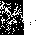
The solid protein matter post of the present invention of accompanying drawing 1 expression 2mm length and 1.1mm internal diameter and 1.3mm external diameter.
The diagram of the 4th aspect operating technology of accompanying drawing 2 expression the present invention: (A) junctional complex is pressed on the nearly pipe end and with tube wall retracts.(B) on the junctional complex distal portions, use laser energy.(C) will manage far-end slowly is enclosed within on the junctional complex of complete length.(D) use laser energy at the junctional complex near-end.
Accompanying drawing 3 is illustrated in the clamp souththern pine and opens back (A) is connected the micro anastomosis art with the laser that carry out 6 week backs (B) outward appearance.
The diagram representative of the rat aorta sutures of accompanying drawing 4 expression conduct operation back time function and the tensile strength of the identical junctional complex of laser.(time is the logarithm level)
The anastomosis that the laser that carries out on vertical face immediately after the accompanying drawing 5 expression laser irradiations connects.(masson trichrome stain (Masson ' s trichrome), (A) arrow is represented blood flow direction, the 5x amplification; (B) 50x amplification)
The junctional complex of leaving in the accompanying drawing 6 expressions 6 week back tube wall.Note the normal appearance in inner membrance and middle level.There is phagocyte in attention on the junctional complex surface.(toluidine blue, amplification 20x)
Accompanying drawing 7 expressions are poured into again and were carried out the scanning electron microscopy (vertical face) that laser connects the chamber of anastomosis in back 10 minutes.(amplification x100).
Accompanying drawing 8 is to use the schematic cross-section of the anastomosis of the blood vessel that jointing technique forms.
The schematic cross-section that the jointing technique of graft that accompanying drawing 9 expressions are inner and the 4th aspect of use the present invention forms on described graft two ends.
Accompanying drawing 10 is represented the schematic form by the joint that forms in the inner placement of body pipe connection property management of the present invention.Can externally use the junctional complex bar and coincide so that strengthen.
Implement the best approach of the present invention
1. the preparation of junctional complex
Raw material is formed:
Protein 55-75% (w/w)
Water 45-25% (w/w)
Dyestuff 0.25% (w/w)
Protein is the albumin of cattle, rabbit, people, sheep or rat.The suitable concn of bovine serum albumin comprise about 55% and people and the albuminised suitable concn of rabbit comprise about 57%.Indocyanine green is suitable dyestuff.Albumin can be available from Sigma-Aldrich Corporation.Suitable albumin prepared product comprises:
Bovine albumin A2153 V level powder (minimum 96%);
People's albumin A1653 V level powder (96-99% albumin);
Rabbit albumin A0639 V level powder;
Sheep albumin A3264 V level powder;
Horse albumin A9888 V level powder.
-sheep albumin
Indocyanine green can be available from Becton Dickinson Microbiology Systems, Maryland 21030 USA.
The albuminised special formulation of people and rabbit is as follows:
Raw material is formed:
Albumin 57.3% (w/w)
Water 42.45% (w/w)
ICG dyestuff 0.25% (w/w)
Preparation:
1. composition (accurately measure) is mixed into the pasty state form so that obtain to be used to the optimal consistency of pushing or suppressing.For example, at first by vortex come mixing water with dyestuff so that form consistent dye solution, then this solution is joined in the protein, be mixed into pastel subsequently.Blend step can be undertaken and carry out so that obtain consistent concentration for small lot (<2g gross mass) use turbine mixer by physics or mechanical system.Do not make the junctional complex drying in this stage, otherwise can cause described junctional complex frangible and be unsuitable for thus the extruding or the compacting.
2. can push described pastel in this stage, and as described belowly can obtain good product by postponing final shaping.
3. make the pastel dehydration of extruding then, increase proteinic concentration thus and make described junctional complex obtain more firm form.
4. (being 85 ℃ for the bovine albumin for example) thing that will be rigidly connected immerses in the hot water about 1 minute so that protein generation degeneration under 80-90 ℃.If personnel selection or rabbit albumin prepare this junctional complex, use steam to carry out 10 minutes (estimating temperature can be low to moderate 100 ℃ or up to 150 ℃) so in the about 120 ℃ of processing that will be correlated with down.This degenerative treatments cause described junctional complex in himself, take place in conjunction with and this junctional complex water-soluble hardly.
5. have elasticity and it can be cut into required shape with further facilitating and can not cause pressure or fracture at this stage junctional complex.Required shape comprise sheet shape, tubular, part is tubular and cylindricality.If be cut into definite shape before step 4, as long as it is too dry, this junctional complex just can be by the crystalline texture fracture that exists so; Otherwise if it is too moistening, it can be out of shape so.
Preferably with this junctional complex in this dehydration and with radiation gamma or autoclaving and be kept in the container of drying, aseptic and lucifuge in stage.
Successfully used the specified scheme of above-mentioned people or rabbit anteserum albumin prescription to be:
1. mixed protein prepared product;
2. push this prepared product;
3. make this prepared product drying;
4. under 120 ℃, this prepared product was carried out autoclaving 10 minutes.
2. restorative procedure
Realized following reparation:
Rat aorta: 1.3mm diameter
Used cylinder: 1.4mm internal diameter
1.7mm warp
2mm is long
Family's rabbit femoral
Aorta: 2mm diameter
Used cylinder: 1.6mm internal diameter
2.1mm outer warp
2mm is long
The process of conjugation tube can comprise make connect the property management periphery at least on one side attached to the inside or the outside step of body tubing string face.Can come the joint of perfect aspect pipe through the following steps: in connecting property management, place the two ends of body pipe and use energy and junctional complex is combined with lower-hierarchy through this junctional complex; Or the two ends of body pipe are placed in the described connection property management and through umbrella organisations junctional complex is used energy; Or an end of body pipe placed place an end in this connection property management in the described connection property management and use energy so that realize combination.The pipe of Xiu Fuing comprises the damage section that needs are replaced if desired, the graft that has the junctional complex of using in the end at least can be engaged with the free end that cuts off pipe at each end.
If tissue repair is the Guan Eryan with regard to nervous tissue or other tissue, the inclusions that wherein needs to prevent pipe suffers damage, and so particularly importantly should not make to connect to concentrate on the edge that is engaged, because this can damage the tissue of extruding.On the contrary, connecting should be horizontal for the edge of fracture place.
The reparation that end joins with the side can be undertaken by the step of giving the bead that the x shape otch on a kind of side that is suitable for the described pipe that inserts the end is installed on the end in the pipe.The free pipe end that connects property management links to each other with the pipe end of slotting x shape otch.Periphery around described connection property management connects the limit, side of x shape otch so that the position is inserted in sealing.Can hold with lateral with different angles and engage and thus can be according to the ledge that serves as the proper angle holding tube that form to engage.
The reparation that end joins with the side can also by give the tubular junctional complex of part provide a kind of be suitable for being enclosed within the new bonded body pipe of pipe end inboard on longitudinal cut on the step of bead carry out.The longitudinal cut side is partly pulled out, is connected around this bead partial inversion and with the outside of junctional complex ledge by the bead of junctional complex.Then the free end of side branches being moved in advance to the body pipe that connects partly goes up with bead and is connected with the main body of the tubular junctional complex of part.Then the main body with the tubular junctional complex of part is connected with the outside of main body pipe.Can hold and the lateral ledge that engages and can keep pipe thus according to many angles according to suitable angle.
Junctional complex by using at least a suitable shaping of the present invention with the energy is so that junctional complex effectively combines the reparation that can realize non-tubular tissue with tissue.
3. the description of cover method
Use special clamp to pull out proximal arterial (pipe) and go back to distance very short on it by connecting property management, used clamp has the end that is suitable for providing the surface of keeping the effect of opening mode pipe end, such as illustrated clamp in the accompanying drawing 2.The tremulous pulse that will fold back with laser exposes to and can be observed till color MC and the specific temperature, this process can make protein denaturation and make it and blood vessel wall specific engagement district around annulus in both sides or at least one side bonding.Distal artery (or pipe) is slightly stretched and on the zone of laser irradiation or in the same laser irradiated region also the outside of connection property management without laser irradiation slowly handle.Then in the same manner should the zone and cause becoming cylindricality with the tremulous pulse annular section of laser irradiation with laser irradiation.This has just finished joint.
Embodiment 1
To amount to 90 rats and be divided into two groups at random.In one group, use conventional micro suture technology to carry out anastomosis, and in two groups, use our new pattern laser interconnection technique to carry out anastomosis.In addition, each group in two groups is divided into 5 subgroups and assess in when different maturity periods (10 minutes, 1 hour, 1 day, 1 week and 6 weeks) subsequently.When these intervals, the effect and the intensity (tensile strength mensuration) of assessment anastomosis.3 in each subgroup animals through anastomosis are carried out light and the processing of electron microscopic inspection technique.
Find that all anastomosiss that carry out all are obvious and effective.Compared in used 7.2 minutes be connected anastomosis with laser activation, using the conventional average connect hours of sewing up the anastomosis that carries out is 20.6 minutes (p<0.001).Tension detection shows in the mechanicalness adhesion of incipient stage sutured anastomosis art stronger.Yet when 6 weeks, laser connects the tensile strength height of the tensile strength of anastomosis than conventional suturing skill.Junctional complex was near being absorbed fully after Histological assessment was presented at for 6 weeks.The junction of blood vessel end can not be decided by the side, chamber of tremulous pulse.
In a word, by diode laser activatory as junctional complex can heavily absorb protein can for a kind of reliable, safety and fast the arterial anastomosis art offer help, this anastomosis can be carried out after short-term study by microsurgery doctor arbitrarily, and it is faster than the stitching of routine.
In the past, the anastomosis of angiostomy and the particularly small-diameter intravascular in the simplification surgery has become an important topic.Recent open source literature has summed up the technological development situation [1] in this field since beginning this century.Reduce foreign body reaction to greatest extent at anastomotic position and become a major issue, and many authors have described the negative effect [2-6] to blood vessel wall compliance and active function generation of sutures, clamping plate and clamp.Jain has at first reported the technology [7,8] that the laser interconnection technique is used for angiostomy in 1979.Used dissimilar laser [9-12] so that potential negative effect to tissue is dropped to bottom line.Most of specification requirements of reporting have three kinds of permanent sutures that stop at least and only the laser connection are used for sealing blood vessels and mechanically mutual support blood vessels end thus.Laser is used for conjunctive tissue depends on effectively accumulating of heat because of organizing absorbing light.To plant the strong absorption band of colour solid consistent with water, hemoglobin or other tissue thus for already used Wavelength of Laser.Introducing such as indocyanine green [13,14] or the such dyestuff of Fluorescein isothiocyanate [15] can promote the accurate conveying of laser energy to target tissue.In addition, the application that has confirmed laser activation protein junctional complex has been strengthened such as the laser in the such tissue of nerve and has been connected [16-18].Our research provides a kind of rapid and reliable technology that need not sew up so that successfully the minor diameter tremulous pulse is implemented anastomosis, thereby has avoided the fibrosis of blood vessel wall by eliminating any permanent implanted device.We combine Payr in the technology of diode laser activation of protein pipe that is used to be connected the minor diameter tremulous pulse of anastomosis [19] and the fully biodegradable of report in 1900.
Material and method
In this research, use and add up to the ripe male Wistar rat (outbreeding) of 90 weight as the youth of 450-550g.This research has obtained our Institution ' s AnimalEthical Review Committe permission and has approved of.All operation processs are all in that (during at 4L/ minute oxygen, halothane is 4%, is used for causing anesthesia with halothane/oxygen mixture; When 2L/ minute oxygen, halothane is 2%, is used to keep anesthesia) general anesthesia under carry out.In operation process, keep cleaning but and non-sterile condition, use Zeiss OPMI 7 operating microscopes to undergo surgery.Implement the center line laparotomy and expose outer (infrarenal) aorta of kidney, cut peritoneum, isolate tissue and if necessary connect waist and waist ileum (ileolumbar) blood vessel.Two blood capillary clamps (Edward Weck Inc., approximation is the blood capillary blade of 1.5mm * 8.0mm, the 19mm grating) are applied to the aorta that cuts off with straight microscissors.After with normal saline washing two undesirable roots, remove excessive connective tissue, and keep complete adventitia.In 45 animals, the micro suture by routine carries out anastomosis (9/0Nylon that has 140 μ syringe needles, interrupted suture 10-12 pin) and connects by laser in remaining 45 animals carrying out anastomosis.The folder of record all processes is fixed time so that check the statistical analysis carry out subsequently with student t.Do not use part or general anticoagulant medicine, after operation, do not give animal antibiotics yet.
Laser connects
Use has the minimal energy of 250mW and the GaAlAs laser diode of 805nm wavelength (Spectra Diode Labs Inc., San Jose CA).The core (in fiber disc with the fixed digital perforate of hands (NA0.28) optical fiber) of laser irradiation and 100um diameter is complementary.Control diode current and temperature by the SDL-800 diode (led) driver.Spoil the switching manipulation diode and be set in operation process at 90mW with foot, the speckle size on the tissue location is 200 μ m diameters, with the 286W/cm on the tissue surface
2Maximum irradiance suitable.Measure laser energy with Scientech energy meter (Boulder Inc., CO USA).Total irradiation time to each annular junctional complex is about 10 seconds.
The junctional complex that uses in this research is the mixture of water, spissated bovine serum albumin and indocyanine green (ICG) dyestuff (Becton Dickinson, Maryland USA).The absorption maximum coefficient that ICG has at 805nm wavelength place is 2 * 10
5M
-1Cm
-1ICG preferably with such as the such serum proteins of albumin combines [20], thereby guarantees that heat transforms effectively so that make the degeneration of protein junctional complex.(raw material by weight is 55.40% albumin: 44.33% water: 0.27%ICG) to assign to obtain the mixture of increased protein concentration by the described one-tenth of vigorous stirring.Make this mixture form the pipe that is suitable for the rat aorta size.Make the pre-degeneration of described connection property management so that they have more pliability and chemical stability (accompanying drawing 1).
Then, when using absorbable magnesium ring, with Payr in 1900 described [19] similarly mode use described junctional complex protein pipe (accompanying drawing 2).Make the blood vessel near-end by cylinder, make it to turn up on the long edge of 1mm and can be connected with the protein cylinder by laser then, thereby further make protein denaturation contained in the junctional complex (accompanying drawing 2A, B).According to the tissue reaction that can observe by operating microscope, laser energy is by manual fiber transmission time limit regular hour (about 10 seconds/periphery) of optics.When noticing that when organizing the slightest retraction, laser spot moves to adjacent tissue, till the complete perimeter with blood vessel is connected on the protein cylinder.Two branches of two pincers are near also lentamente distal vessels being pulled to (accompanying drawing 2C) on the complete protein post then.Then use laser and combine (accompanying drawing 2D) so that between the far-end of tremulous pulse and the immediate part of described junctional complex, produce.
After taking out clamp, check by extrusion test immediately and coincide so that the assessment effect.Then every component is become 5 subgroups, 9 animals are arranged in each subgroup, reappraise (10 minutes, 1 hour, 1 day, 1 week, 6 weeks) respectively at different intervals.At the times selected place, all anastomosis are exposed again and use the extrusion test test effect.In 6 animal bodies of each subgroup, take out hematochezia pipe and bleeding from distant part pipe each other at a distance of the identical part of 5mm and carry out tensile strength and measure.(Quincy MA) connects and the other end is connected with the screw drives transducer and carries out above-mentioned steps [18] for FT30C, GrassInstruments for a end by making blood vessel and the energy transducer of calibration.In 3 animal bodies of each subgroup, clamp blood vessel, with the flushing of saline and fixative (being buffered to 5% glutaraldehyde of pH7.4) and finally take out them and be used for histological examination.For optical microscope, dye so that native protein and denatured protein are clearly made a distinction and carry out same process with toluidine blue with masson trichrome stain.Scanning electron microscope is used to study identical inner surface.
The result
All animals in operation process all survival and when checking again all coincideing all be effective.In 6 whens week, in the suture site or laser connect anastomotic position and all do not have aneurysm (accompanying drawing 3) to occur.
The average folder of sutured anastomosis art fix time be 20.6 minutes (SD2.82, SEM0.52), it obviously than 7.2 minutes of connecting that the average folder of anastomosis fixes time of laser (SD2.26, SEM0.41) long; (p<0.001; Student t check).
Tensile strength is measured and is shown: when the anastomosis that is connected with laser in a short time compares, and strong (under stress) (being respectively 134.6gm and 45.3gm) of the tensile strength of the anastomosis of stitching.Yet when 6 weeks, laser connects a little higher than sutured anastomosis art of tensile strength (being respectively 134.2gm and 103.9gm, p=0.005, student t check) (accompanying drawing 4) of anastomosis.When drawing, the anastomosis of stitching with on blood vessel wall this down the degree of engagement of endless belt break, and the blood vessel that laser connects bonded distal portion from, this may be the weak spot of coincideing.
After using laser immediately with the masson trichrome stain anastomotic position that dyes, the light microscopy that after this carries out assessment demonstrate directly with the junctional complex adjacency layer in denatured protein, and in the media of tremulous pulse, do not observe change (accompanying drawing 5).After 6 weeks, described junctional complex is almost fully heavily absorbed and can be observed continual internal layer.Healing occurs with the myofibroblast hypertrophy and can not detect anastomotic position (accompanying drawing 6) from lumen of artery.Yet, shown in accompanying drawing 3b, can on adventitia, observe the some fibre reaction, but the arteries middle level does not still demonstrate any change.After pouring into 10 minutes again, the scanning electron microscopy (SEM) that anastomotic position is carried out shows deposit certain erythrocyte on anastomotic position, and it to effect without any influence (accompanying drawing 7).
Discuss
Because JainShi has at first reported the successful Application [7] of laser energy in blood vessel is repaired, so carried out the nothing stitching angiostomy technology that a large amount of trials come Application and Development laser to connect.The advantage that laser connects has not had seepage because of the joint sealing is complete and because of there not being sutures not have foreign body reaction to obtain confirmation.Yet, the demand of keeping the near fixedly sutures of blood vessel termination has been caused having produced the laser-assisted anastomosis of term [21-15], wherein fixedly after the sutures laser is used to seal anastomotic position inserting the 3-5 kind in advance.On the other hand, by using in the tube chamber stent to guarantee that the arrangement of inner membrance carried out nothing true to nature and sewed up anastomosis [26-28].These stents mainly are designed to can absorb in tube chamber and may cause arterial thrombosis and/or thrombosis thus.The technology [18] of the reparation tubular-shaped structures of above-mentioned report has been used the protein junctional complex band that contains the indocyanine green green dye, and they are designed to absorb the laser energy and make heat concentrate on the surrounding tissue of protein junctional complex and adjacency thus.
Only closely with in the organized layer that described junctional complex contacts observing change in the tissue that heating caused that can produce because of laser.Has the pipe that is consistent with the reparation blood vessel diameter in order to obtain for successfully carrying out the internal layer arrangement that micro-angiostomy is the best of key, described protein junctional complex to be squeezed into.Finish best internal layer arrangement by using a kind of technology [19] of introducing at first in this century by Payr.This technology is by eliminating blood vessel being fixed on the Landon[29 of the ligature demand on the annulus] and by Carter[30] further develop the coronary artery surgery that is used to use the polyethylene ring.Haller[31] be reported in that to use the effective percentage of the technology of the Payr that uses the tantalum ring in the anastomosis of 4-mm diameter blood vessel be 92%.This technology has prevented that blood from contacting with the protein junctional complex, can eliminate the harm that coagulation cascade is activated like this and can produce level and smooth internal layer and arrange.The laser interconnection technique can make tissue and protein junctional complex pipe jointing by the protein denaturation in tissue and the junctional complex.
In the research in early days, will connect the coagulation necrosis that the bonded exact mechanism of generation is accredited as possible homogenize of adventitia and smooth muscle cell by laser; But, the elasticity lamella does not change [32].Connect in the research at direct laser, use electron microscopy to observe the protein denaturation of collagen fiber, inner membrance slightly breaks and endothelium heal again [33] in 10 days subsequently.To be present in destruction in the arterial wall form the van der waals bond of other tropocollagen molecule subsequently again the dehydration of collagen triple helix molecule structure be reported to possible bonding mechanism [34].Our technology is limited in change laser induced in the arterial wall in the layer that directly contacts with the protein junctional complex, thus any reduction of tube wall is minimized.Especially, as what Histological assessment confirmed, the middle level that can neither can change the near-end blood vessel with laser also can not change the middle level of distal vessels.So can advise: but use this technology only minimally change arterial wall and can not lose its mechanical property.After the anastomotic position healing, tensile strength is measured and is shown that the anastomosis that laser connects has better result than sutured anastomosis art, and this may be the result to the fiber-reactive of mesopelagic layer centre joint compound.
List of references:
1) Werker PMN, Kon M., " the reinforcement means summary of angiostomy " " thoracic surgery yearbook " be 63:S122 (Ann.Thorac.Surg.), and 1997.
2) Serure A.Withers EH, Thomson S, Morris J., " comparison of auxiliary microvascular anastomosis of carbon dioxide laser and conventional blood capillary sutured anastomosis art " " surgery forum " be 34:634 (Surg.Forum.), and 1983.
3) Lidman D, Daniels RK., " the normal agglutination of microvascular anastomosis " " plastic surgery studies magazine " be 15:103 (Scan.J.Plast.Surg.), and 1981.
4) Servant J, Ikuta Y, Harada Y., " scanning electron microscope study of microvascular anastomosis " " cerebral sursery " be 57:329 (Plast.Reconstr.Surg.), and 1976.
5) Acland RD, Trachtenberg L " histopathology of microvascular anastomosis metarteriole " " cerebral sursery " is 59:868 (Plast.Reconstr.Surg.), and 1977.
6) Dalsing MC, Packer SC, Kueppers P, Griffith SL, DavisTB., " laser and sutured anastomosis art: the generation of passive compliance and active force " " laser surgery's medical science " (Lasers Surg.Med.) 12:190,1992.
7) Jain K.K., Gorisch W., " using the little blood vessel of Nd-Yag laser repairing " " surgery's preliminary report " (A preliminary report Surgery) 51:684,1979.
8) Jain K.K., Gorisch W., " using Nd-Yag laser repairing blood capillary " " Shen is through Hua Chi " (Act a Neurochir.) be supplementary issue 28:260 (Wien), and 1979.
9) White RA, Abergel RP, Lyons R, Klein SR, Kopchok G, DweyerRM, Uitto J., " laser connects the biological agent to the blood vessel healing " " laser surgery's medical science " (Lasers Surg.Med.) 6:137,1986.
10) Kopchok GE, White RA, White GH, Fujitani R, Vlasak J, Dykhovsky L, Grundfest WS., " CO
2Be connected with the argon laser blood vessel ", " acute histology and thermodynamics relatively " " laser surgery's medical science " (Lasers Surg.Med.) 8:584,1988.
11) Nakata S, Campbell CD, Pick R, Replogle RL., " using the end of carbon dioxide laser to be connected angiostomy with side and end with end " " cardiovascular thoracic surgery magazine " be 98:57 (J.Thorac.Cardiovasc.Surg.), and 1989.
12) Lewis W J, Uribe A., " microvascular anastomosis of contact diode laser " " laryngoscope " be 103:850 (Laryngosc.), and 1993.
13) Reali UM, Gelli R, Gianotti V, Clori F, Pratesi R, PiniR., " the auxiliary microvascular anastomosis of experiment diode laser " " reduction microsurgery magazine " be 3:203 (J.Reconstr.Microsurg.), and 1993.
14) Oz, MC, Johnson JP, Paranagi S, Chuk RS, Marboe CC, Bass, LS, Nowygrod R, Treat MR., " tissue that the fibrinogen that uses indigo cyanine green dye to strengthen by near infrared diode laser carries out connects " " vascular surgery magazine " be 11:718 (J.Vasc.Surg.), and 1990.
15) Chuck RS, OZ MC, Delohery TM, Johnson JP, Bass LS, Nowygrod R, Teat MR., " the laser tissue connection that dyestuff is strengthened " " laser surgery's medical science " (Lasers Surg.Med.) 9:471,1989.
16) Bass LS, Moazami N, Avellino A, Trosaborg W, Treat MR., " feasibility study of laser junctional complex neurosuture " " SPIE journal " be 2128:472 (Proc.SPIE), and 1994.
17) Menovsky T, Beek JF, van Gemert MJC., " CO
2Laser is neural to be connected ", " the external application of best laser parameter and junctional complex " " microsurgery " (Microsurg.) 15:44,1994.
18) Lauto A, Trickett R, Malik R, Dawes JM, Owen ER., " the solid protein band that is used for the laser activation of peripheral nervous reparation ", " a kind of in vitro study " " laser surgery's medical science " (Lasers Surg.Med.) 21:134,1997.
19) Payer E., " Beitraege zur Technique der BlutgefaessundNervennaht nebst Mittellungen ueber die Verwendung einesresorblerbaren Metalles in der Chirurgle. " " clinical chemistry records " be 62:67 (Arch.Klin.Chir.), and 1900.
20) Sauda K, Imasaka T, Ishibashi N., " by the protein in the high performance liquid chroma-tography mensuration human serum " " analytical chemistry " (Analytical Chemistry) 58:2649,1986.
21) McCarthy WJ, LoCicero J, Hartz RS, Yao JST., " effect of laser-assisted anastomosis in little blood vessel: followed up a case by regular visits in a year " " surgery " be 102:319 (Surgery), and 1987.
22) Okada M, Shimizu K, Ikuta H, Horii H, Nakamura K., " using the another kind of angiostomy method of laser: test and clinical research " " laser surgery's medical science " (Lasers Surg.Med.) 7:240,1987.
23) Abrahamson DL, Shaw WW, Kamat BR, Harper A, RosenbergCR., " laser-assisted anastomosis of veins: comparative study " " reduction microsurgery magazine " (J.Reconstr. Microsurg.) 7:199,1991.
24) Kiyoshige Y, Tsuchida H, Hamasaki M, Takayanagi M, Watanabe Y., " CO
2Laser-assisted microvascular anastomosis: bio-mechanical Journal of Sex Research and clinical practice " " reduction microsurgery magazine " (J.Reconstr.Microsurg.) 7:225,1991.
25) Tang J, Godlewski G, Rouy S, Dauzat M, Juan JM, ChambettazF, Salathe R., " using the micro-arterial anastomosis art and the comparative study of noncontact diode laser " " surgery medical science is used thing " (Users Surg.Med.) 14:229,1994.
26) Jain KK., " using the nothing of Nd-YAG laser to sew up microvascular anastomosis " " microsurgery magazine " be 1:436 (J.Microsurg.), and 1980.
27) Niijima KH, Yonekawa Y, Handa H, Taki W., " using the nothing of Nd-YAG laser and water-soluble poval clamping plate to sew up microvascular anastomosis " " neurosurgery magazine " be 67:579 (J.Neurosurg.), and 1987.
28) Bass LS, Treat MR, Dzakonski C, Trokel SL. uses the nothing of THC:YAG laser to sew up microvascular anastomosis-preliminary report " " microsurgery " (Microsurg.) 10:189,1989.
29) Landon LH., " with from the method for simplifying that keeps the pipe direct transfusion " be 61:490 (JAMA), and 1913.
30) Carter EL, Roth Ej., " the intravital direct nothing of Canis familiaris L. is sewed up coronary stenting " " surgery's yearbook " be 148:212 (Ann.Surg.), and 1958.
31) Haller JD, Kripke DC, Rosenak 55, Roberts DR, Rohman M., " using the long-term results of the little angiostomy of loop technique " " surgery's yearbook " be 161:67 (Ann.Surg.), and 1965.
32) Schober R, Ulrich F, Sander T, Duerselen H, Hessel S., " change of laser induced collagen protein substructure requires the microsurgery tissue to connect " " science " be 232:1421 (Science), and 1986.
33) Godlewski G, Rouy S, Dauzat M., " ultrastructural studies that argon laser micro anastomosis postoperative arterial wall is repaired " " laser surgery's medical science " (Lasers Surg.Med.) 7:258,1987.
34) Fenner J, Martin W, Moseley H, Wheatley Dj., " as the shear strength of making a concerted effort: the mechanism that the laser-assisted tissue of proposition connects " " laser medicine science " (Lasers Med.Science) 7:39 in conjunction with the knot of tissue of temperature funtion, 1992.
Claims (17)
1. biomolecule junctional complex that is used to repair human or animal tissues or organ, it is included in the protein of partial denaturation at least in the process of this junctional complex of preparation, described protein is that those can be cross-linked into substrate and can the re-absorbed protein of health, described degeneration changes the mechanical property of described junctional complex, make when moistening it show with repair process in the similar mechanical property of tissue, described protein is moistening simultaneously, so that protein bound together, and this junctional complex dissolubility in physiological fluid under body temperature reduces.
2. the junctional complex of claim 1, wherein this junctional complex was shaped before degeneration.
3. the junctional complex of claim 1, wherein this junctional complex is shaped after degeneration.
4. junctional complex according to claim 1, wherein said protein are selected from albumin, elastin laminin, fibrinogen, collagen protein or its mixture, and wherein this mixture comprises the protein with similar denaturation temperature.
5. junctional complex according to claim 4, it comprises that further being used for improving when described junctional complex contacts with energy energy accumulates dyestuff into described junctional complex, described dyestuff is selected from indocyanine green, methylene blue and Fluorescein isothiocyanate.
6. junctional complex according to claim 4, it comprises further and is used to promote to organize fast or the adjuvant of healing more fully that described adjuvant is selected from somatomedin, hyaluronate sodium, hormone and anticoagulant.
7. junctional complex according to claim 4, it further comprises the material that is used to improve described junctional complex intensity, it is selected from polytetrafluoroethylene fibre or ceramic fibre.
8. test kit that comprises any described junctional complex of aforesaid right requirement.
9. prepare the method for the junctional complex of claim 1, this method comprises the following steps:
(a) be that protein and the aqueous solvent of 40%-80%w/w is combined to form protein paste with concentration range, described aqueous solvent comprises water and saline, and condition is that any salt existing when degeneration etc. can not produce harmful effect to junctional complex;
(b) push, suppress or cut this protein paste and become desired shape;
(c) make the dehydration of this protein paste and
(d) by the protein paste that this junctional complex immersed the time about 30 seconds in the hot water of 80 ℃ of-90 ℃ of temperature or make step (c) with about 10 minutes time of this junctional complex of steam treatment of 100 ℃ of-150 ℃ of temperature partial denaturation at least, junctional complex is moistening simultaneously, so that protein bound together, and the reservation shape of junctional complex is kept thus substantially, and junctional complex dissolubility in physiological fluid under body temperature reduces.
10. prepare the method for the junctional complex of claim 1, this method comprises the following steps:
(a) be that protein and the aqueous solvent of 40%-80%w/w is combined to form protein paste with concentration, described aqueous solvent comprises water and saline, and condition is that any salt existing when degeneration etc. can not produce harmful effect to junctional complex;
(b) by this junctional complex being immersed the time about 30 seconds in the hot water of 80 ℃ of-90 ℃ of temperature or with the about 10 minutes time chien shih protein partial denaturation at least of this junctional complex of steam treatment of 100 ℃ of-150 ℃ of temperature, junctional complex is moistening simultaneously, so that protein bound together, and this junctional complex dissolubility in physiological fluid under body temperature reduces;
(c) extruding, compacting or cutting junctional complex become desired shape.
11. one kind according to claim 9 or 10 described methods, wherein in step (a), will be used for improving the dyestuff that energy accumulates into described junctional complex and join junctional complex, concentration is 0.25%w/w-2.5%w/w, and described dyestuff is selected from indocyanine green, methylene blue or Fluorescein isothiocyanate.
12. a method according to claim 11, wherein with described solvent with described dyestuff is mixed before described protein mixes with described aqueous solvent.
13. one kind according to claim 9 or 10 described methods, wherein before making described protein generation degeneration described junctional complex are applied to net, rigid body or graft materials.
14. one kind according to claim 9 or 10 described methods, this method further is included in after the protein denaturation step with described junctional complex sterilization.
15. one kind according to claim 9 or 10 described methods, this method further is included in after the protein denaturation the exsiccant step of described junctional complex.
16. each junctional complex is used for repairing the purposes of the product of human or animal tissues or organ among the claim 1-7 in preparation.
17. the purposes of claim 15 wherein made described junctional complex moistening before being applied to tissue site to be repaired.
Applications Claiming Priority (2)
| Application Number | Priority Date | Filing Date | Title |
|---|---|---|---|
| AUPP4214A AUPP421498A0 (en) | 1998-06-18 | 1998-06-18 | Method of tissue repair |
| AUPP4214 | 1998-06-18 |
Related Child Applications (1)
| Application Number | Title | Priority Date | Filing Date |
|---|---|---|---|
| CNA2005100916076A Division CN1935269A (en) | 1998-06-18 | 1999-06-18 | Compound for tissue repair and application thereof |
Publications (2)
| Publication Number | Publication Date |
|---|---|
| CN1312727A CN1312727A (en) | 2001-09-12 |
| CN1221291C true CN1221291C (en) | 2005-10-05 |
Family
ID=3808460
Family Applications (2)
| Application Number | Title | Priority Date | Filing Date |
|---|---|---|---|
| CNB998097748A Expired - Fee Related CN1221291C (en) | 1998-06-18 | 1999-06-18 | Method of tissue repair II |
| CNA2005100916076A Pending CN1935269A (en) | 1998-06-18 | 1999-06-18 | Compound for tissue repair and application thereof |
Family Applications After (1)
| Application Number | Title | Priority Date | Filing Date |
|---|---|---|---|
| CNA2005100916076A Pending CN1935269A (en) | 1998-06-18 | 1999-06-18 | Compound for tissue repair and application thereof |
Country Status (11)
| Country | Link |
|---|---|
| US (1) | US7078378B1 (en) |
| EP (1) | EP1087795A4 (en) |
| JP (1) | JP2002518102A (en) |
| CN (2) | CN1221291C (en) |
| AU (1) | AUPP421498A0 (en) |
| CA (1) | CA2335323A1 (en) |
| HK (1) | HK1040939B (en) |
| IL (2) | IL140368A0 (en) |
| MX (1) | MXPA00012716A (en) |
| NZ (1) | NZ509099A (en) |
| WO (1) | WO1999065536A1 (en) |
Cited By (1)
| Publication number | Priority date | Publication date | Assignee | Title |
|---|---|---|---|---|
| CN112336910A (en) * | 2019-08-08 | 2021-02-09 | 南京理工大学 | ICG dye-containing laser biological tissue welding flux |
Families Citing this family (59)
| Publication number | Priority date | Publication date | Assignee | Title |
|---|---|---|---|---|
| US8215314B2 (en) * | 2000-02-11 | 2012-07-10 | The General Hospital Corporation | Photochemical tissue bonding |
| US7073510B2 (en) | 2000-02-11 | 2006-07-11 | The General Hospital Corporation | Photochemical tissue bonding |
| US7147647B2 (en) | 2002-04-26 | 2006-12-12 | Medtronic, Inc. | Sintered titanium tube for the management of spinal cord injury |
| US7846141B2 (en) | 2002-09-03 | 2010-12-07 | Bluesky Medical Group Incorporated | Reduced pressure treatment system |
| GB0224986D0 (en) | 2002-10-28 | 2002-12-04 | Smith & Nephew | Apparatus |
| US20070213768A1 (en) * | 2003-10-21 | 2007-09-13 | Irena Wasserman | Compositions and Devices Including Same Useful for Anastomosis |
| WO2005048880A2 (en) * | 2003-11-18 | 2005-06-02 | Medizinische Universität Wien | Vascular prosthesis |
| WO2005074817A1 (en) * | 2004-02-09 | 2005-08-18 | Avastra Ltd | Instruments for sutureless surgical technique |
| US8062272B2 (en) | 2004-05-21 | 2011-11-22 | Bluesky Medical Group Incorporated | Flexible reduced pressure treatment appliance |
| US10058642B2 (en) | 2004-04-05 | 2018-08-28 | Bluesky Medical Group Incorporated | Reduced pressure treatment system |
| US7909805B2 (en) | 2004-04-05 | 2011-03-22 | Bluesky Medical Group Incorporated | Flexible reduced pressure treatment appliance |
| GB0409446D0 (en) | 2004-04-28 | 2004-06-02 | Smith & Nephew | Apparatus |
| US9079762B2 (en) | 2006-09-22 | 2015-07-14 | Ethicon Endo-Surgery, Inc. | Micro-electromechanical device |
| US20080078411A1 (en) * | 2006-10-03 | 2008-04-03 | Restore Medical, Inc. | Tongue implant for sleep apnea |
| US20080078412A1 (en) * | 2006-10-03 | 2008-04-03 | Restore Medical, Inc. | Tongue implant |
| US7914552B2 (en) * | 2006-11-09 | 2011-03-29 | Ethicon Endo-Surgery, Inc. | Method of performing an end-to-end anastomosis using a stent and an adhesive |
| WO2008065464A2 (en) * | 2006-11-28 | 2008-06-05 | Universita' Degli Studi Di Padova | A method for the pulmonary aerostasis and a device for its application |
| US7713265B2 (en) | 2006-12-22 | 2010-05-11 | Ethicon Endo-Surgery, Inc. | Apparatus and method for medically treating a tattoo |
| US8273015B2 (en) | 2007-01-09 | 2012-09-25 | Ethicon Endo-Surgery, Inc. | Methods for imaging the anatomy with an anatomically secured scanner assembly |
| US8801606B2 (en) | 2007-01-09 | 2014-08-12 | Ethicon Endo-Surgery, Inc. | Method of in vivo monitoring using an imaging system including scanned beam imaging unit |
| US8216214B2 (en) | 2007-03-12 | 2012-07-10 | Ethicon Endo-Surgery, Inc. | Power modulation of a scanning beam for imaging, therapy, and/or diagnosis |
| US7995045B2 (en) | 2007-04-13 | 2011-08-09 | Ethicon Endo-Surgery, Inc. | Combined SBI and conventional image processor |
| US8626271B2 (en) | 2007-04-13 | 2014-01-07 | Ethicon Endo-Surgery, Inc. | System and method using fluorescence to examine within a patient's anatomy |
| US8251983B2 (en) * | 2007-05-14 | 2012-08-28 | The Regents Of The University Of Colorado, A Body Corporate | Laser tissue fusion of septal membranes |
| US8160678B2 (en) | 2007-06-18 | 2012-04-17 | Ethicon Endo-Surgery, Inc. | Methods and devices for repairing damaged or diseased tissue using a scanning beam assembly |
| US20080319461A1 (en) * | 2007-06-22 | 2008-12-25 | Ghent University | Sutureless vessel anastomosis method and apparatus |
| US20100174300A1 (en) * | 2007-06-22 | 2010-07-08 | Phillip Nicolas Blondeel | Sutureless vessel anastomosis method and apparatus |
| US7982776B2 (en) | 2007-07-13 | 2011-07-19 | Ethicon Endo-Surgery, Inc. | SBI motion artifact removal apparatus and method |
| US9125552B2 (en) | 2007-07-31 | 2015-09-08 | Ethicon Endo-Surgery, Inc. | Optical scanning module and means for attaching the module to medical instruments for introducing the module into the anatomy |
| US7983739B2 (en) | 2007-08-27 | 2011-07-19 | Ethicon Endo-Surgery, Inc. | Position tracking and control for a scanning assembly |
| US7925333B2 (en) | 2007-08-28 | 2011-04-12 | Ethicon Endo-Surgery, Inc. | Medical device including scanned beam unit with operational control features |
| EP2217298B1 (en) | 2007-11-21 | 2015-11-11 | T.J. Smith & Nephew Limited | Suction device and dressing |
| GB0722820D0 (en) | 2007-11-21 | 2008-01-02 | Smith & Nephew | Vacuum assisted wound dressing |
| EP3254650B1 (en) | 2007-11-21 | 2020-01-08 | Smith & Nephew plc | Wound dressing |
| GB0723875D0 (en) | 2007-12-06 | 2008-01-16 | Smith & Nephew | Wound management |
| US11253399B2 (en) | 2007-12-06 | 2022-02-22 | Smith & Nephew Plc | Wound filling apparatuses and methods |
| US20110004240A1 (en) * | 2008-02-07 | 2011-01-06 | Riken | Visible light curable adhesive |
| GB0803564D0 (en) | 2008-02-27 | 2008-04-02 | Smith & Nephew | Fluid collection |
| US8050520B2 (en) | 2008-03-27 | 2011-11-01 | Ethicon Endo-Surgery, Inc. | Method for creating a pixel image from sampled data of a scanned beam imager |
| US8332014B2 (en) | 2008-04-25 | 2012-12-11 | Ethicon Endo-Surgery, Inc. | Scanned beam device and method using same which measures the reflectance of patient tissue |
| US20110172704A1 (en) * | 2008-09-19 | 2011-07-14 | The Trustees Of The University Of Pennsylvania | Solder formulation and use in tissue welding |
| JP5988588B2 (en) * | 2009-03-10 | 2016-09-07 | ザ ジョーンズ ホプキンズ ユニバーシティThe Johns Hopkins University | Biological tissue bonding and repair device and method of use thereof |
| US9433700B2 (en) | 2009-04-27 | 2016-09-06 | Medibeacon Inc. | Tissue sealant compositions, vascular closure devices, and uses thereof |
| FR2952306B1 (en) * | 2009-11-09 | 2012-03-30 | Creaspine | AQUEOUS COMPOSITION FOR THE TREATMENT OF LASER FIBROUS CONJUNCTIVE TISSUE REPAIR |
| US9061095B2 (en) | 2010-04-27 | 2015-06-23 | Smith & Nephew Plc | Wound dressing and method of use |
| GB201011173D0 (en) | 2010-07-02 | 2010-08-18 | Smith & Nephew | Provision of wound filler |
| GB201020005D0 (en) | 2010-11-25 | 2011-01-12 | Smith & Nephew | Composition 1-1 |
| MX337627B (en) | 2010-11-25 | 2016-03-10 | Smith & Nephew | Composition i-ii and products and uses thereof. |
| US20150159066A1 (en) | 2011-11-25 | 2015-06-11 | Smith & Nephew Plc | Composition, apparatus, kit and method and uses thereof |
| US10549112B2 (en) | 2012-07-20 | 2020-02-04 | The General Hospital Corporation | Apparatus for tissue irradiation and methods and kits utilizing the same |
| US10292381B2 (en) | 2012-07-20 | 2019-05-21 | The General Hospital Corporation | Vessel treatment systems, methods, and kits |
| WO2014015274A1 (en) | 2012-07-20 | 2014-01-23 | The General Hospital Corporation | Methods for tissue passivation |
| ES2823764T3 (en) * | 2012-09-25 | 2021-05-10 | Axogen Corp | Materials and procedures for protection against neuromas |
| US20160120706A1 (en) | 2013-03-15 | 2016-05-05 | Smith & Nephew Plc | Wound dressing sealant and use thereof |
| AU2014290513B2 (en) | 2013-07-18 | 2017-07-27 | The General Hospital Corporation | Vessel treatment systems, methods, and kits |
| CN105288720B (en) * | 2015-10-19 | 2018-07-27 | 中国科学院苏州生物医学工程技术研究所 | Bonding proximate matter for vascular anastomosis |
| CN108114314A (en) * | 2017-05-03 | 2018-06-05 | 曲平波 | A kind of degradable pleura sticking patch and preparation method thereof |
| WO2021041696A1 (en) * | 2019-08-30 | 2021-03-04 | Arizona Board Of Regents On Behalf Of Arizona State University | Nerve repair using laser sealing |
| CN112294381A (en) * | 2020-10-27 | 2021-02-02 | 中南大学湘雅医院 | Sleeve device for microsurgery blood vessel and lymphatic vessel anastomosis |
Family Cites Families (9)
| Publication number | Priority date | Publication date | Assignee | Title |
|---|---|---|---|---|
| CA1205013A (en) * | 1984-03-19 | 1986-05-27 | Adolf Lindner | Method of producing a tissue adhesive |
| EP0491794A4 (en) * | 1989-09-12 | 1992-08-19 | The Trustees Of Columbia University In The City Of New York | Laser tissue welding with dye enhanced solders |
| US5156613A (en) * | 1991-02-13 | 1992-10-20 | Interface Biomedical Laboratories Corp. | Collagen welding rod material for use in tissue welding |
| US5749895A (en) * | 1991-02-13 | 1998-05-12 | Fusion Medical Technologies, Inc. | Method for bonding or fusion of biological tissue and material |
| CH681693A5 (en) * | 1991-02-28 | 1993-05-14 | Pentapharm Ag | |
| AUPN066795A0 (en) * | 1995-01-20 | 1995-02-16 | Macquarie Research Limited | Method of repair |
| US5713891A (en) * | 1995-06-02 | 1998-02-03 | Children's Medical Center Corporation | Modified solder for delivery of bioactive substances and methods of use thereof |
| WO1998016165A1 (en) * | 1996-10-16 | 1998-04-23 | Fusion Medical Technologies, Inc. | Films having improved characteristics and methods for their preparation and use |
| WO1999051282A1 (en) * | 1998-04-06 | 1999-10-14 | Cornell Research Foundation, Inc. | Composition for tissue welding and method of use |
-
1998
- 1998-06-18 AU AUPP4214A patent/AUPP421498A0/en not_active Abandoned
-
1999
- 1999-06-18 NZ NZ50909999A patent/NZ509099A/en unknown
- 1999-06-18 WO PCT/AU1999/000495 patent/WO1999065536A1/en not_active Application Discontinuation
- 1999-06-18 CA CA002335323A patent/CA2335323A1/en not_active Abandoned
- 1999-06-18 CN CNB998097748A patent/CN1221291C/en not_active Expired - Fee Related
- 1999-06-18 EP EP99927596A patent/EP1087795A4/en not_active Withdrawn
- 1999-06-18 MX MXPA00012716A patent/MXPA00012716A/en unknown
- 1999-06-18 JP JP2000554413A patent/JP2002518102A/en active Pending
- 1999-06-18 IL IL14036899A patent/IL140368A0/en not_active IP Right Cessation
- 1999-06-18 CN CNA2005100916076A patent/CN1935269A/en active Pending
- 1999-06-18 US US09/719,889 patent/US7078378B1/en not_active Expired - Fee Related
-
2000
- 2000-12-18 IL IL140368A patent/IL140368A/en unknown
-
2002
- 2002-03-12 HK HK02101911.2A patent/HK1040939B/en not_active IP Right Cessation
Cited By (2)
| Publication number | Priority date | Publication date | Assignee | Title |
|---|---|---|---|---|
| CN112336910A (en) * | 2019-08-08 | 2021-02-09 | 南京理工大学 | ICG dye-containing laser biological tissue welding flux |
| CN112336910B (en) * | 2019-08-08 | 2022-05-13 | 南京理工大学 | ICG dye-containing laser biological tissue welding flux |
Also Published As
| Publication number | Publication date |
|---|---|
| CA2335323A1 (en) | 1999-12-23 |
| CN1935269A (en) | 2007-03-28 |
| NZ509099A (en) | 2004-12-24 |
| EP1087795A4 (en) | 2006-01-11 |
| JP2002518102A (en) | 2002-06-25 |
| HK1040939A1 (en) | 2002-06-28 |
| CN1312727A (en) | 2001-09-12 |
| MXPA00012716A (en) | 2002-05-08 |
| US7078378B1 (en) | 2006-07-18 |
| AUPP421498A0 (en) | 1998-07-09 |
| IL140368A (en) | 2006-10-31 |
| IL140368A0 (en) | 2002-02-10 |
| EP1087795A1 (en) | 2001-04-04 |
| HK1040939B (en) | 2006-02-03 |
| WO1999065536A1 (en) | 1999-12-23 |
Similar Documents
| Publication | Publication Date | Title |
|---|---|---|
| CN1221291C (en) | Method of tissue repair II | |
| US20020198517A1 (en) | Gelatin based and Power-gelTM as solders for Cr4+ laser tissue welding and sealing of lung air leak and fistulas in organs | |
| CN1142749C (en) | Elastin, and elastin-based biomaterials and process | |
| US6372228B1 (en) | Method of producing elastin, elastin-based biomaterials and tropoelastin materials | |
| JP2002523136A (en) | Insertable stent and method of making and using the stent | |
| US20070244495A1 (en) | Apparatus and method for performing laser-assisted vascular anastomoses using bioglue | |
| JP2009540965A (en) | Compositions and methods for joining unbound lumens | |
| US8608760B2 (en) | Compositions and methods for joining non-conjoined lumens | |
| Ott et al. | Comparative in vitro study of tissue welding using a 808 nm diode laser and a Ho: YAG laser | |
| Lauto et al. | Sutureless nerve repair with laser-activated chitosan adhesive: a pilot in vivo study | |
| Kamiji et al. | Microvascular anastomosis using polyethylene glycol 4000 and fibrin glue | |
| Pabittei et al. | Ex vivo proof-of-concept of end-to-end scaffold-enhanced laser-assisted vascular anastomosis of porcine arteries | |
| Ott et al. | Intraluminal laser light source and external solder: in vivo evaluation of a new technique for microvascular anastomosis | |
| Schwaiger et al. | BioWeld® Tube and surgical glue for experimental sutureless venous microanastomosis | |
| WO2007030892A1 (en) | Method of tissue repair iii | |
| Puca et al. | Diode laser-assisted carotid bypass surgery: an experimental study with morphological and immunohistochemical evaluations | |
| AU748911B2 (en) | Method of producing biomaterials | |
| Xie et al. | Intraluminal albumin stent assisted laser welding for ureteral anastomosis | |
| Wright et al. | Laser‐assisted end‐to‐end bioweld® anastomosis in an ovine model | |
| Heitzer et al. | Evaluation of fibrin, cyanoacrylate, and polyurethane-based tissue adhesives in sutureless vascular anastomosis: a comparative mechanical ex vivo study | |
| AU768533B2 (en) | Method of tissue repair II | |
| Xie et al. | Laser welding with an albumin stent: experimental ureteral end-to-end anastomosis | |
| Ott et al. | Laser balloon vascular welding using a dye-enhanced albumin solder | |
| Mordon et al. | 1.9-um diode-laser-assisted anastomoses (LAMA) in reconstructive microsurgery: results of the preliminary clinical study | |
| AU767057B2 (en) | Method of producing biomaterials |
Legal Events
| Date | Code | Title | Description |
|---|---|---|---|
| C06 | Publication | ||
| PB01 | Publication | ||
| C10 | Entry into substantive examination | ||
| SE01 | Entry into force of request for substantive examination | ||
| C14 | Grant of patent or utility model | ||
| GR01 | Patent grant | ||
| C19 | Lapse of patent right due to non-payment of the annual fee | ||
| CF01 | Termination of patent right due to non-payment of annual fee |