CN116171198A - Systems, methods, and compositions for rapid early detection of infected host RNA biomarkers and early identification of human COVID-19 coronavirus infection - Google Patents
Systems, methods, and compositions for rapid early detection of infected host RNA biomarkers and early identification of human COVID-19 coronavirus infection Download PDFInfo
- Publication number
- CN116171198A CN116171198A CN202080076043.1A CN202080076043A CN116171198A CN 116171198 A CN116171198 A CN 116171198A CN 202080076043 A CN202080076043 A CN 202080076043A CN 116171198 A CN116171198 A CN 116171198A
- Authority
- CN
- China
- Prior art keywords
- host
- biomarker
- derived
- infection
- rna
- Prior art date
- Legal status (The legal status is an assumption and is not a legal conclusion. Google has not performed a legal analysis and makes no representation as to the accuracy of the status listed.)
- Pending
Links
Images


Classifications
-
- B—PERFORMING OPERATIONS; TRANSPORTING
- B01—PHYSICAL OR CHEMICAL PROCESSES OR APPARATUS IN GENERAL
- B01L—CHEMICAL OR PHYSICAL LABORATORY APPARATUS FOR GENERAL USE
- B01L3/00—Containers or dishes for laboratory use, e.g. laboratory glassware; Droppers
- B01L3/50—Containers for the purpose of retaining a material to be analysed, e.g. test tubes
- B01L3/502—Containers for the purpose of retaining a material to be analysed, e.g. test tubes with fluid transport, e.g. in multi-compartment structures
- B01L3/5023—Containers for the purpose of retaining a material to be analysed, e.g. test tubes with fluid transport, e.g. in multi-compartment structures with a sample being transported to, and subsequently stored in an absorbent for analysis
-
- B—PERFORMING OPERATIONS; TRANSPORTING
- B01—PHYSICAL OR CHEMICAL PROCESSES OR APPARATUS IN GENERAL
- B01L—CHEMICAL OR PHYSICAL LABORATORY APPARATUS FOR GENERAL USE
- B01L3/00—Containers or dishes for laboratory use, e.g. laboratory glassware; Droppers
- B01L3/50—Containers for the purpose of retaining a material to be analysed, e.g. test tubes
- B01L3/502—Containers for the purpose of retaining a material to be analysed, e.g. test tubes with fluid transport, e.g. in multi-compartment structures
- B01L3/5027—Containers for the purpose of retaining a material to be analysed, e.g. test tubes with fluid transport, e.g. in multi-compartment structures by integrated microfluidic structures, i.e. dimensions of channels and chambers are such that surface tension forces are important, e.g. lab-on-a-chip
- B01L3/50273—Containers for the purpose of retaining a material to be analysed, e.g. test tubes with fluid transport, e.g. in multi-compartment structures by integrated microfluidic structures, i.e. dimensions of channels and chambers are such that surface tension forces are important, e.g. lab-on-a-chip characterised by the means or forces applied to move the fluids
-
- C—CHEMISTRY; METALLURGY
- C12—BIOCHEMISTRY; BEER; SPIRITS; WINE; VINEGAR; MICROBIOLOGY; ENZYMOLOGY; MUTATION OR GENETIC ENGINEERING
- C12Q—MEASURING OR TESTING PROCESSES INVOLVING ENZYMES, NUCLEIC ACIDS OR MICROORGANISMS; COMPOSITIONS OR TEST PAPERS THEREFOR; PROCESSES OF PREPARING SUCH COMPOSITIONS; CONDITION-RESPONSIVE CONTROL IN MICROBIOLOGICAL OR ENZYMOLOGICAL PROCESSES
- C12Q1/00—Measuring or testing processes involving enzymes, nucleic acids or microorganisms; Compositions therefor; Processes of preparing such compositions
- C12Q1/68—Measuring or testing processes involving enzymes, nucleic acids or microorganisms; Compositions therefor; Processes of preparing such compositions involving nucleic acids
- C12Q1/6804—Nucleic acid analysis using immunogens
-
- C—CHEMISTRY; METALLURGY
- C12—BIOCHEMISTRY; BEER; SPIRITS; WINE; VINEGAR; MICROBIOLOGY; ENZYMOLOGY; MUTATION OR GENETIC ENGINEERING
- C12Q—MEASURING OR TESTING PROCESSES INVOLVING ENZYMES, NUCLEIC ACIDS OR MICROORGANISMS; COMPOSITIONS OR TEST PAPERS THEREFOR; PROCESSES OF PREPARING SUCH COMPOSITIONS; CONDITION-RESPONSIVE CONTROL IN MICROBIOLOGICAL OR ENZYMOLOGICAL PROCESSES
- C12Q1/00—Measuring or testing processes involving enzymes, nucleic acids or microorganisms; Compositions therefor; Processes of preparing such compositions
- C12Q1/68—Measuring or testing processes involving enzymes, nucleic acids or microorganisms; Compositions therefor; Processes of preparing such compositions involving nucleic acids
- C12Q1/6813—Hybridisation assays
- C12Q1/6816—Hybridisation assays characterised by the detection means
-
- C—CHEMISTRY; METALLURGY
- C12—BIOCHEMISTRY; BEER; SPIRITS; WINE; VINEGAR; MICROBIOLOGY; ENZYMOLOGY; MUTATION OR GENETIC ENGINEERING
- C12Q—MEASURING OR TESTING PROCESSES INVOLVING ENZYMES, NUCLEIC ACIDS OR MICROORGANISMS; COMPOSITIONS OR TEST PAPERS THEREFOR; PROCESSES OF PREPARING SUCH COMPOSITIONS; CONDITION-RESPONSIVE CONTROL IN MICROBIOLOGICAL OR ENZYMOLOGICAL PROCESSES
- C12Q1/00—Measuring or testing processes involving enzymes, nucleic acids or microorganisms; Compositions therefor; Processes of preparing such compositions
- C12Q1/68—Measuring or testing processes involving enzymes, nucleic acids or microorganisms; Compositions therefor; Processes of preparing such compositions involving nucleic acids
- C12Q1/6876—Nucleic acid products used in the analysis of nucleic acids, e.g. primers or probes
- C12Q1/6883—Nucleic acid products used in the analysis of nucleic acids, e.g. primers or probes for diseases caused by alterations of genetic material
-
- C—CHEMISTRY; METALLURGY
- C12—BIOCHEMISTRY; BEER; SPIRITS; WINE; VINEGAR; MICROBIOLOGY; ENZYMOLOGY; MUTATION OR GENETIC ENGINEERING
- C12Q—MEASURING OR TESTING PROCESSES INVOLVING ENZYMES, NUCLEIC ACIDS OR MICROORGANISMS; COMPOSITIONS OR TEST PAPERS THEREFOR; PROCESSES OF PREPARING SUCH COMPOSITIONS; CONDITION-RESPONSIVE CONTROL IN MICROBIOLOGICAL OR ENZYMOLOGICAL PROCESSES
- C12Q1/00—Measuring or testing processes involving enzymes, nucleic acids or microorganisms; Compositions therefor; Processes of preparing such compositions
- C12Q1/68—Measuring or testing processes involving enzymes, nucleic acids or microorganisms; Compositions therefor; Processes of preparing such compositions involving nucleic acids
- C12Q1/6876—Nucleic acid products used in the analysis of nucleic acids, e.g. primers or probes
- C12Q1/6888—Nucleic acid products used in the analysis of nucleic acids, e.g. primers or probes for detection or identification of organisms
- C12Q1/689—Nucleic acid products used in the analysis of nucleic acids, e.g. primers or probes for detection or identification of organisms for bacteria
-
- C—CHEMISTRY; METALLURGY
- C12—BIOCHEMISTRY; BEER; SPIRITS; WINE; VINEGAR; MICROBIOLOGY; ENZYMOLOGY; MUTATION OR GENETIC ENGINEERING
- C12Q—MEASURING OR TESTING PROCESSES INVOLVING ENZYMES, NUCLEIC ACIDS OR MICROORGANISMS; COMPOSITIONS OR TEST PAPERS THEREFOR; PROCESSES OF PREPARING SUCH COMPOSITIONS; CONDITION-RESPONSIVE CONTROL IN MICROBIOLOGICAL OR ENZYMOLOGICAL PROCESSES
- C12Q1/00—Measuring or testing processes involving enzymes, nucleic acids or microorganisms; Compositions therefor; Processes of preparing such compositions
- C12Q1/70—Measuring or testing processes involving enzymes, nucleic acids or microorganisms; Compositions therefor; Processes of preparing such compositions involving virus or bacteriophage
- C12Q1/701—Specific hybridization probes
-
- B—PERFORMING OPERATIONS; TRANSPORTING
- B01—PHYSICAL OR CHEMICAL PROCESSES OR APPARATUS IN GENERAL
- B01L—CHEMICAL OR PHYSICAL LABORATORY APPARATUS FOR GENERAL USE
- B01L2200/00—Solutions for specific problems relating to chemical or physical laboratory apparatus
- B01L2200/06—Fluid handling related problems
- B01L2200/0647—Handling flowable solids, e.g. microscopic beads, cells, particles
- B01L2200/0663—Stretching or orienting elongated molecules or particles
-
- B—PERFORMING OPERATIONS; TRANSPORTING
- B01—PHYSICAL OR CHEMICAL PROCESSES OR APPARATUS IN GENERAL
- B01L—CHEMICAL OR PHYSICAL LABORATORY APPARATUS FOR GENERAL USE
- B01L2300/00—Additional constructional details
- B01L2300/08—Geometry, shape and general structure
- B01L2300/0809—Geometry, shape and general structure rectangular shaped
- B01L2300/0825—Test strips
-
- B—PERFORMING OPERATIONS; TRANSPORTING
- B01—PHYSICAL OR CHEMICAL PROCESSES OR APPARATUS IN GENERAL
- B01L—CHEMICAL OR PHYSICAL LABORATORY APPARATUS FOR GENERAL USE
- B01L2400/00—Moving or stopping fluids
- B01L2400/04—Moving fluids with specific forces or mechanical means
- B01L2400/0403—Moving fluids with specific forces or mechanical means specific forces
- B01L2400/0406—Moving fluids with specific forces or mechanical means specific forces capillary forces
-
- C—CHEMISTRY; METALLURGY
- C12—BIOCHEMISTRY; BEER; SPIRITS; WINE; VINEGAR; MICROBIOLOGY; ENZYMOLOGY; MUTATION OR GENETIC ENGINEERING
- C12Q—MEASURING OR TESTING PROCESSES INVOLVING ENZYMES, NUCLEIC ACIDS OR MICROORGANISMS; COMPOSITIONS OR TEST PAPERS THEREFOR; PROCESSES OF PREPARING SUCH COMPOSITIONS; CONDITION-RESPONSIVE CONTROL IN MICROBIOLOGICAL OR ENZYMOLOGICAL PROCESSES
- C12Q1/00—Measuring or testing processes involving enzymes, nucleic acids or microorganisms; Compositions therefor; Processes of preparing such compositions
- C12Q1/68—Measuring or testing processes involving enzymes, nucleic acids or microorganisms; Compositions therefor; Processes of preparing such compositions involving nucleic acids
- C12Q1/6844—Nucleic acid amplification reactions
-
- C—CHEMISTRY; METALLURGY
- C12—BIOCHEMISTRY; BEER; SPIRITS; WINE; VINEGAR; MICROBIOLOGY; ENZYMOLOGY; MUTATION OR GENETIC ENGINEERING
- C12Q—MEASURING OR TESTING PROCESSES INVOLVING ENZYMES, NUCLEIC ACIDS OR MICROORGANISMS; COMPOSITIONS OR TEST PAPERS THEREFOR; PROCESSES OF PREPARING SUCH COMPOSITIONS; CONDITION-RESPONSIVE CONTROL IN MICROBIOLOGICAL OR ENZYMOLOGICAL PROCESSES
- C12Q2525/00—Reactions involving modified oligonucleotides, nucleic acids, or nucleotides
- C12Q2525/30—Oligonucleotides characterised by their secondary structure
- C12Q2525/301—Hairpin oligonucleotides
-
- C—CHEMISTRY; METALLURGY
- C12—BIOCHEMISTRY; BEER; SPIRITS; WINE; VINEGAR; MICROBIOLOGY; ENZYMOLOGY; MUTATION OR GENETIC ENGINEERING
- C12Q—MEASURING OR TESTING PROCESSES INVOLVING ENZYMES, NUCLEIC ACIDS OR MICROORGANISMS; COMPOSITIONS OR TEST PAPERS THEREFOR; PROCESSES OF PREPARING SUCH COMPOSITIONS; CONDITION-RESPONSIVE CONTROL IN MICROBIOLOGICAL OR ENZYMOLOGICAL PROCESSES
- C12Q2527/00—Reactions demanding special reaction conditions
- C12Q2527/101—Temperature
-
- C—CHEMISTRY; METALLURGY
- C12—BIOCHEMISTRY; BEER; SPIRITS; WINE; VINEGAR; MICROBIOLOGY; ENZYMOLOGY; MUTATION OR GENETIC ENGINEERING
- C12Q—MEASURING OR TESTING PROCESSES INVOLVING ENZYMES, NUCLEIC ACIDS OR MICROORGANISMS; COMPOSITIONS OR TEST PAPERS THEREFOR; PROCESSES OF PREPARING SUCH COMPOSITIONS; CONDITION-RESPONSIVE CONTROL IN MICROBIOLOGICAL OR ENZYMOLOGICAL PROCESSES
- C12Q2565/00—Nucleic acid analysis characterised by mode or means of detection
- C12Q2565/10—Detection mode being characterised by the assay principle
- C12Q2565/102—Multiple non-interacting labels
-
- Y—GENERAL TAGGING OF NEW TECHNOLOGICAL DEVELOPMENTS; GENERAL TAGGING OF CROSS-SECTIONAL TECHNOLOGIES SPANNING OVER SEVERAL SECTIONS OF THE IPC; TECHNICAL SUBJECTS COVERED BY FORMER USPC CROSS-REFERENCE ART COLLECTIONS [XRACs] AND DIGESTS
- Y02—TECHNOLOGIES OR APPLICATIONS FOR MITIGATION OR ADAPTATION AGAINST CLIMATE CHANGE
- Y02A—TECHNOLOGIES FOR ADAPTATION TO CLIMATE CHANGE
- Y02A50/00—TECHNOLOGIES FOR ADAPTATION TO CLIMATE CHANGE in human health protection, e.g. against extreme weather
- Y02A50/30—Against vector-borne diseases, e.g. mosquito-borne, fly-borne, tick-borne or waterborne diseases whose impact is exacerbated by climate change
Abstract
The present technology relates to systems, methods, and compositions for detecting host characteristics of pathogenic infections, and in particular to rapid detection assays configured to detect target RNA transcripts that may be biomarkers of an infection. In one embodiment, the present invention comprises systems, methods, and compositions for early detection of pathogens and/or infections in asymptomatic subjects by a novel lateral flow assay, which in a preferred embodiment may comprise a rapid self-administration test strip configured to detect one or more RNA transcript biomarkers produced by the subject's innate immune system in response to pathogens or infections.
Description
The present application claims the benefits and priorities of U.S. provisional application No. 62/895,387 filed on 9, 3, 2019 and U.S. provisional application No. 62/934,754 filed on 11, 13, 2019, and U.S. provisional application No. 63/006,570 filed on 7, 2020. The entire specification and drawings of the above-referenced application are hereby incorporated by reference in their entirety.
Statement regarding federally sponsored research
The invention is completed by government support under grant number HDTRA1-18-1-0032 granted by the national Defense Threat Reduction Agency (DTRA). The government has certain rights in this invention.
Sequence listing
The present application contains a sequence listing that has been electronically submitted in ASCII format and is hereby incorporated by reference in its entirety. The ASCII copy created at 8/30 of 2020 is named "90245.00432-Sequence-listing. Txt" and is 2476 kilobytes in size.
Technical Field
The present technology relates to systems, methods, and compositions for detecting host characteristics of pathogenic infections, and in particular to rapid detection assays configured to detect target RNA transcripts that may be biomarkers of an infection.
Background
Early detection of pathogenic microbial infection is critical for proper treatment and positive clinical outcome. However, an infected individual may remain asymptomatic for several days after infection while actively transmitting the pathogen to others. Conventional pathogen detection systems are often not effective in detecting infection until after onset of symptoms. Traditional pathogen tests include serological or antibody-based tests, bacterial/viral/fungal growth cultures, and nucleic acid-based assays, such as PCR (polymerase chain reaction). Such traditional tests are often time consuming and laborious and are only effective after the patient begins to exhibit symptoms of the infection. In addition, traditional diagnostic tests require clinical suspicion of specific pathogens, expensive laboratory equipment and trained personnel, and have increased upstream and end user costs.
For example, as highlighted in fig. 2, during a typical infection, exposure to an unknown pathogen occurs on day zero and then progresses through subsequent clinical stages of infection, as indicated by the vertically extending timeline along the left side of the figure. Standard diagnostic tests are often designed to work after the onset of symptoms when pathogens replicate in an infected human body, at which point one knows what errors are and seeks healthcare and diagnosis. However, at this point the infected person may have been infected to other people for days or weeks. Opportunities for destructive downstream effects to effect early isolation and limit unimpeded transmission of pathogens have been missed. This time delay to diagnosis may lead to poor patient outcome and continued disease transmission before the patient knows himself to be infectious.
In contrast to specialized and later developed adaptive immune responses, the first line of defense of hosts against pathogenic microorganisms is the "innate immune" response. Innate immunity of the body is a self-amplifying and nonspecific physiological response that occurs within hours of infection. Thus, the ability to detect the presence of molecules resulting from the host's innate immune response may provide the ability to rapidly detect infection at the earliest stage while the patient is still asymptomatic. Such an advancement would allow for a more effective isolation regimen, as well as improved therapeutic and clinical results.
The need for improved methods of detecting pathogens has been amplified by the epidemic of the whole-sphere coronavirus, particularly early in the infection cycle. Coronaviruses (members of the coronaviridae and coronaviridae subfamilies) are found in mammals and birds. One prominent member is Severe acute respiratory syndrome coronavirus (SARS-CoV). Another prominent coronavirus, known as the middle east respiratory syndrome coronavirus (MERS coronavirus or MERS-CoV) MERS-CoV, shares some similarities with the SARS-CoV burst. Typical symptoms of SARS, MERS and covd-19 coronavirus infection include fever, cough, shortness of breath, pneumonia, and gastrointestinal symptoms. Serious illness can lead to respiratory failure requiring mechanical ventilation and support in the intensive care unit. Both coronaviruses appear to cause more severe disease in the elderly, in people with a weak immune system, and in people with chronic diseases such as cancer, chronic lung disease, and diabetes. No vaccine or specific therapy is currently available for COVID-19. Patients diagnosed with a covd-19 coronavirus infection receive supportive treatment based solely on individual symptoms and clinical conditions.
As described below, the inventors have overcome the limitations of traditional pathogen detection systems while utilizing the host's early innate immune response (including but not limited to the interferon response) to rapidly detect RNA biomarkers indicative of infection, and in particular, of covd-19 coronavirus infection. While patients are generally asymptomatic, this rapid point-of-care diagnostic application allows for detection of infection at an early stage. This early detection is directly related to more targeted and effective therapeutic interventions as well as overall improved clinical outcome.
Disclosure of Invention
The present technology may comprise systems, methods, and compositions for early detection of pathogens and/or infections in asymptomatic subjects by a novel lateral flow assay, which in a preferred embodiment may comprise a rapid test strip configured to detect one or more RNA transcript biomarkers produced by the subject's innate immune system in response to pathogens or infections.
In another aspect, the present technology may comprise systems, methods and compositions for early detection of pathogens and/or infections in asymptomatic subjects by a novel lateral flow assay, which in a preferred embodiment may comprise a rapid test strip configured to detect one or more RNA transcript biomarkers produced by the subject's innate immune system in response to pathogens or infections and possibly present in saliva encoded according to one or more of the nucleotide sequences of SEQ ID nos. 1-444 and 657-815.
Further aspects of the invention comprise the use of one or more biomarkers for infection, and preferably pathogen infection, of a human according to the nucleotide sequences identified in SEQ ID NOS.1-444 and 657-815.
In another aspect, the present technology may comprise systems, methods, and compositions for detecting these target RNA transcripts, which may serve as biomarkers for early infection in a subject.
In another aspect, the present technology may comprise systems, methods, and compositions for detecting early infection in a subject, which may comprise at least: a lateral flow assay test strip device, which may preferably comprise a fiber-or paper-based lateral flow strip configured to allow liquid to flow by capillary action; 2) An RT-RPA (reverse transcription recombinase polymerase amplification) reaction, which may occur in a pre-prepared reaction cartridge, which may comprise a collection container configured to receive a fluid sample from a subject and pre-prepared for an RT-RPA reaction; and 3) one or more RNA biomarker transcripts, also commonly referred to as biomarkers, supplied in a fluid sample, such as one or more biomarkers encoded by nucleotide sequences identified as SEQ ID NOS.1-444 and 657-815, which in a preferred embodiment may comprise a saliva sample provided by a subject. In a preferred embodiment, RNA biomarker transcripts can be amplified in a reaction cartridge in an isothermal amplification RT-RPA reaction to form hybridized dsDNA probes with single stranded adapter sequences or dsDNA products containing 5' modifications for downstream hybridization.
Additional aspects may comprise novel conjugated reporter probes that may be coupled to hybridized dsDNA probes. In certain aspects, the novel conjugated probes can comprise GNPs, or other single reporter genes conjugated to ssDNA sequences or antibodies or antibody fragments that can bind to dsDNA probes. While further aspects of the invention may comprise novel target capture probes that can bind and form immobilized "sandwich" complex aggregates comprising an intercalating capture probe coupled to a hybridized dsDNA probe that is further coupled to a conjugated reporter probe and preferably a GNP reporter probe. In this regard, localized immobilization may facilitate the generation of a visual signal, for example, on a test strip or even a solution.
Further aspects of the invention include systems, methods, and compositions for quantifying an early host-derived infection biomarker, which may or may not be combined with quantitative data for a pathogen-specific biomarker, preferably generated by PCR, RT-PCR, or qRT-PCR. In a preferred aspect, RNA can be extracted from a biological sample provided by a subject potentially exposed or infected. The RNA can be subjected to a qRT-PCT reaction to determine pathogen biomarkers as well as host-derived infection biomarkers, and preferably the levels of host-derived RNA biomarkers present in saliva of a subject. Multiple biological samples may be obtained from one or more subjects to generate a time course of infection that reveals relative levels of pathogen and host-derived biomarkers over time. This data can be used to generate biomarker candidates for lateral flow assays to detect pathogen-specific host-derived biomarkers. This lateral flow assay may be administered to a subject in need thereof and provides an indication of the infection as well as the stage of infection by one or more specific pathogens. In a preferred aspect, the specific pathogen may comprise SARS-CoV-2, commonly known as COVID-19 coronavirus.
Further aspects of the invention may comprise one or more of the preferred embodiments set forth in the claims.
Further aspects of the invention may be evident from the description, claims and drawings provided below.
Drawings
The novel aspects, features and advantages of the present disclosure will be better understood from the following detailed description when taken in conjunction with the accompanying drawings, all of which are given by way of illustration only and not limitation of embodiments of the present disclosure, in which:
FIG. 1 (A) shows a general schematic of a lateral flow assay in one embodiment of the invention; (B) Another general overview of a lateral flow assay test strip in one embodiment of the present invention is shown.
Fig. 2 shows a representative example of an infection process.
Fig. 3 (a) shows an exemplary in vivo mouse experiment demonstrating prior art for detecting pathogen infection. In this case, a group of mice may be infected with the pathogen, and blood samples will be collected on the indicated days after infection. These samples will be used for high throughput sequencing to characterize the presence of biomarkers and also for testing to compare the invention with current state-of-the-art detection methods. Exemplary data showing the ability of the present invention to detect pathogen infection days prior to other methods is shown below. All described assays will be performed during previous in vivo experiments. (B) A timeline of hypothetical viral infections and various tests designed to detect the infections are shown.
FIG. 4 illustrates an exemplary pathogen detection device in one embodiment thereof and specifically highlights the ability of the device to be used for multiplexing. The techniques of the present invention, and in particular lateral flow assay test strips or test strips, may be adapted for use in a variety of configurations depending on the end user's purpose. (A) As a preliminary screening test, the most important parameter is sensitivity to ensure that no infected individual is inadvertently marked as "not ill" when the infected individual is actually "ill". The high sensitivity test identified nearly 100% of true positive cases of disease and had nearly 0% false negative rate. The sensitivity of RNA transcript biomarker assays can be adjusted by adding multiple test lines for different biomarkers, increasing the probability of identifying all true positives if detected in combination. (B) For clinicians evaluating patients who have symptoms in different medical environments (e.g., emergency rooms, primary care offices, assisted care institutions, field hospitals, etc.), it is important to distinguish the general categories of pathogens (i.e., virus versus bacteria versus fungi) to begin optimal early treatment before the pathogen is fully identified. The assays of the present invention can inform treatment planning and significantly reduce antibiotic use in the case of non-bacterial infections to help limit the spread of antibiotic-resistant bacteria. (C) Early studies of host signals in response to specific organisms may allow for assay configurations in which infection by specific pathogenic organisms may be identified. The microbiota tested may be specified by the needs of the end user. For example, army may be most interested in air-borne and weaponized pathogen breeds, while domestic clinics need to evaluate patients for seasonal influenza, RSV, rhinoviruses and norovirus.
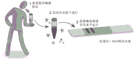
FIG. 5 illustrates the use of an exemplary pathogen detection device in one embodiment thereof. In this embodiment, the patient provides a saliva sample into a reaction cartridge, which may be represented herein as a tube container preloaded with reagents that may allow the amplification reaction to proceed at room temperature to increase biomarker concentration. Thereafter, a solution containing the amplified biomarker may be applied to the lateral flow test strip. As the fluid flows down the strip, a visible pink signal appears. In the simplest iteration of bars, one band means a negative result and two bands are equal to a positive result indicating an infection. In consumer product embodiments, the strips will be contained in a housing to facilitate interpretation of the results.
Fig. 6 (a) shows a venn plot showing that there is significant overlap in identity of RNA transcripts expressed in saliva and PBMCs (peripheral blood mononuclear cells) based on sequencing data from healthy human samples. This overlap means that transcripts present in the blood may also occur in saliva. Note that this transcript sequencing data was normalized to an average of 1000 ten thousand read coverage, and the abundance of these transcripts was not described. (B) Representative PCC (pattern correlation coefficient) plots showing the relative expression levels of RNA transcripts present in both saliva and PBMCs (two samples from the same individual). Each point in this figure represents a different transcript in the overlapping portion of the venn plot in a. Average r value = 0.64 (> 0.5) is considered to be a significant correlation). In general, PBMCs present a higher level of expression of most transcripts in saliva, and there is a subset of transcripts that are up-regulated in saliva relative to PBMCs. Because of this data, the inventors can focus on saliva as a choice of sample type, from which to identify key signals for early infection.
FIG. 7 illustrates a general method for identifying infection biomarkers in one embodiment thereof.
FIG. 8 shows an example of host RNA biomarker IFIT2 identified for infection using an in vitro transcriptome dataset. In the horizontal direction, the gene structure is shown with dark blue bars representing the coding region of the gene. In the vertical direction, the height of the peak represents the relative abundance of the indicated RNA. For each study, the "-" lanes indicate uninfected samples, while the "+" lanes indicate various types of viral infections. The abundance changes of the different studies are highlighted in different colors. In summary, the identified RNA biomarkers were up-regulated in 9 different cell types and 10 different viral infections. Upregulation of this biomarker can be detected in vitro as early as 4 hours post-infection (which precedes any observable symptoms). Additional biomarkers for the invention can be identified and selected in a similar procedure as generally described above.
Figure 9 shows qPCR of biomarker candidates in infected cells. Human lung cells (a 549) were mock-infected or infected with influenza virus (left) or vesicular stomatitis virus (VSV, right) for 24 hours. RNA was collected and quantified using qPCR. The results are shown as "fold change from simulation" and the dashed line indicates no change during infection. IFIT2 is an example of RNA as a global marker of infection, as shown in fig. 8. In this example, NEAT1 distinguishes VSV from influenza, and OAS1 distinguishes influenza from VSV.
Fig. 10 shows a schematic diagram of an optimization procedure for amplifying and detecting biomarkers from human saliva. Step 3.1, RNA from 2. Mu.L of human saliva was successfully reverse transcribed into DNA and amplified using a custom RT-RPA kit. The reaction was completed at a constant 37 ℃ in 20 minutes. Step 3.2, upon successful detection of potential biomarkers for infection, multiple primer sets of different lengths and sequences are designed to optimize biomarker amplification. The primer set resulting in the highest amplification efficiency (reflected by the intensity of the bands on the gel image) was selected for practical diagnosis. Step 3.3, modifying the selected primers from the previous step to carry adaptor sequences to allow hybridization downstream of the lateral flow assay test strip and gold nanoparticle reporter probe. After 20 minutes of RT-RPA amplification at 37 ℃, the resulting amplicon contains both the adapter sequence and the sequence from the target biomarker. The final reaction product can then be applied directly to the test strip for visualization.
FIG. 11 shows nucleic acid "sandwick" in complementary DNA binding forms aggregated for visual readout. Amplified biomarkers have double stranded DNA (dsDNA) regions flanking specific single stranded protruding adaptors. The solution with this biomarker is mixed with a gold nanoparticle reporter, which itself is conjugated to a single stranded DNA adapter complementary to the amplified biomarker's adapter and a control capture probe on nitrocellulose. Due to the mechanism of complementary DNA base pairing, when these protruding DNA adaptor strands interact in solution flowing through the membrane, they bind to ssDNA conjugated gold nanoparticles and immobilized oligonucleotide capture probes and form a dsDNA structure, forming a nucleic acid "sandwich" (fig. 4A). As more and more of these reporter amplified biomarker-capture probe sandwich forms and aggregates, a visible pink signal appears on nitrocellulose in the target detection zone (B), indicating the presence of the biomarker in the original sample. Here, the leftmost pink dot represents the complex shown in Panel A, and the second pink dot is the control, where the gold reporter alone binds to its complementary probe. This control verifies that the sample flows correctly through the strip.
FIG. 12 shows colorimetric images of a series of test strips run with 10-fold dilutions of synthetic RT-RPA product.
Figures 13A-D illustrate a lateral flow assay test strip having an easy-to-use cover in one embodiment thereof.
FIG. 14 shows a general schematic of a lateral flow assay incorporating an antibody-based capture mechanism in one embodiment of the invention.
FIG. 15 shows a general flow chart of an exemplary laboratory-based test and lateral flow test for detecting biomarkers.
FIG. 16 shows a flow chart for designing and validating primers for biomarker candidates. Such systems are described in U.S. provisional application nos. 62/934,873 and 63/006561, the disclosures of which are incorporated herein by reference with respect to fig. 16.
Fig. 17A-B: the host RNA biomarker is shown to be the gene transcript derived from the earliest immune response of the infected cell. Heat maps were generated from the disclosed RNA sequencing dataset and showed the level of expression change (left color code) of certain RNA species after infection of cultured human cells with different pathogens (top). In all cases, the comparison mimics infected (-) cells and infected (+) cells. Some SARS-CoV-2 and influenza A specific biomarkers are shown in the orange and green highlighting boxes.
Fig. 18 shows various RNA biomarkers that are up-regulated in response to different types of infection and that are detectable in human saliva. (A) Heat maps were generated from the disclosed RNA sequencing dataset and showed the level of expression change (lower color code) of certain RNA species after infection of cultured human cells with different pathogens (top). (B) In all cases, the comparison mimics infected (-) cells and infected (+) cells. Here, there are 3 patient saliva samples from the infectious ward. These saliva samples represent acute infections with fungi (patient 1; coccidioides), viruses (patient 2; varicella zoster virus) and bacteria (patient 3; E.coli). Quantitative RT-PCR was performed to measure fold change of eight biomarker RNAs relative to healthy saliva controls. Note the logarithmic scale on the Y-axis, which indicates that these biomarkers were found at 10-10,000 times higher levels in saliva of infected individuals than in saliva of healthy individuals. There are also saliva biomarkers that may be able to distinguish one type of infection from other infections, such as EGR1 that does not respond to fungal infection but is up-regulated 100,000-fold in viral infection.
FIG. 19 shows that host biomarker upregulation can be detected in multiplex RT-qPCR reactions. Human lung cells (a 549) were mock-infected or infected with influenza virus, and RNA was purified from cell lysates 24 hours post-infection. The RNA was then subjected to RT-qPCR reactions using Taqman probes and chemistry. Biomarkers indicated on the X-axis were measured in either single (black bars) or multiplex (orange bars) reactions using the primers and probes listed in table 4. The relative mRNA expression (Y-axis) was calculated by first internally normalizing the sample using the host control gene and then comparing to a mock infected sample.
Figure 20 shows that some host biomarkers up-regulated prior to viral RNA detection. Human hepatocyte cell line (Huh 7) was either mock-infected or infected with SARS-CoV-2 coronavirus. RNA was purified from cell lysates at 0, 2, 4, 8, 12, 24 and 48 hours (X-axis) post infection. The RNA was then subjected to RT-qPCR using the primers and probes listed in Table 4. The relative mRNA expression (Y-axis) was calculated by first internally normalizing the sample using the host control gene and then comparing to a mock infected sample. The left side shows a whole set of biomarkers, while the right side shows a subset of biomarkers highlighting biomarkers up-regulated early in infection (blue), late in infection (green) and host control biomarkers not up-regulated (grey). Detection of SARS-CoV-2 nucleoprotein gene (N2) is also shown in red.
FIG. 21 shows an exemplary lateral flow strip with an antibody capture protocol. The lateral flow strip was peeled off according to the schematic diagram of fig. 4. sMimic amplicons were generated to test the sensitivity of the lateral flow strip. The "excess" line captures excess anti-FITC conjugated gold nanoparticles. The "control" line captures the mock amplicon conjugated with FITC and biotin. The "test" line captures the simulated amplicons conjugated to FITC and DIG.
FIG. 22 shows Table 3, which contains primers for detecting host biomarkers of infection. A subset of candidate biomarkers is selected for primer optimization. RT-qPCR was performed using the set of primers listed to optimize primer efficiency, ct values, melting curves and log fold changes for the two host control biomarkers (RACK 1 or CALR). Expression in untreated human lung cells (a 549) was compared to interferon-treated a549 cells (a549+ifn) or influenza virus-infected a549 cells (a549+influenza).
FIG. 23 shows Table 4, which contains primers and probes for multiplex detection of host biomarkers. Based on their large fold change, a subset of candidate biomarkers from table 3 was selected. Taqman probes were designed for each primer set to be chemically compatible with Taqman fluorescence in the RT-qPCR reaction. Biomarkers are separated into triplets based on Ct values in order to be compatible with multiplexing.
FIG. 24 shows Table 5, which contains primers for amplifying host biomarkers using isothermal RT-RPA. A subset of candidate biomarkers is selected to optimize the RT-RPA response (a). Those primer sets meeting the conditions presented in FIG. 16 were then modified to contain a 5' modification (FITC, biotin, or DIG) in order to obtain compatibility with the lateral flow assay of the invention (B).
FIG. 25 shows that amplified products from the RT-RPA reaction can be detected on a lateral flow strip. (A) Lateral flow strips stripped with secondary anti-rabbit antibodies (gold nanoparticle excess line), streptavidin (control line) or anti-DIG antibodies (biomarker line) were used to resolve the indicated RT-RPA reactions. Sample # 1 contained only PBS and no RT-RPA reaction product, while all other samples contained RT-RPA reaction (20 min reaction) product. RT-RPA was performed using purified RNA from influenza infected human lung cells (A549) as template. (B) Lateral flow strips as described in FIG. A were used to confirm that the primer set itself did not produce false positive signals. The indicated primer sets were mixed with PBS at the same concentration of RT-RPA reaction and run out on the strip.
Detailed Description
The present technology may comprise systems, methods, and compositions for early detection of pathogens and/or infections in asymptomatic subjects by a novel lateral flow assay, which in a preferred embodiment may comprise a rapid self-administration test strip configured to detect one or more host RNA transcript biomarkers (encoded or non-encoded) produced by the subject's innate immune system in response to pathogens or infections and present in saliva.
As generally shown in fig. 1B, one embodiment of the present technology may comprise systems, methods, and compositions for detecting an early infection in a subject, which may comprise at least: a lateral flow assay test strip device (also referred to as a test strip or lateral flow strip), which may preferably comprise a fiber-based or paper-based lateral flow strip configured to allow liquid flow by capillary action; 2) An RT-RPA (reverse transcription recombinase polymerase amplification) reaction, which may occur in a pre-prepared reaction cartridge, which may comprise a collection container configured to receive a fluid sample from a subject and pre-prepared for an RT-RPA reaction; and 3) one or more RNA biomarker transcripts, also commonly referred to as biomarkers, are supplied in a fluid sample, which in a preferred embodiment may comprise a saliva sample provided by the subject.
Specific target RNA transcripts or biomarkers generated by the patient's immune response (typically an innate immune response or any other cellular pathway that is upregulated following infection) and found in saliva may be indicative of early infection. Thus, one embodiment of the present technology may comprise systems, methods, and compositions for detecting these target RNA transcripts, which may serve as biomarkers for early infection in a subject. However, as noted above, in this embodiment, the target RNA transcript biomarkers present in a typical fluid sample provided by a human subject are typically present in low concentrations and require detection of amplification. To overcome this physical limitation, as further shown in fig. 1B, in one embodiment of the invention, the subject may deposit a fluid sample (which in this case may comprise a saliva sample) into a reaction cartridge where the fluid sample may undergo an amplification step. In particular, a reaction cartridge may receive a fluid sample, wherein the reaction cartridge may undergo an RT-RPA reaction to amplify RNA biomarker transcripts present in the fluid sample. In this preferred embodiment, the cartridge may be preloaded with a quantity of pre-prepared protein, enzyme, salt, and other reagents that may allow for the RT-RPA reaction to proceed within the cartridge. As shown in fig. 1A, the reaction cartridge may be preloaded with primers for a target RNA biomarker transcript, which may further comprise a C3 spacer element. In another preferred embodiment, the reaction cartridge may be further preloaded with one or more conjugated reporter probes, such as conjugated Gold Nanoparticle (GNP) reporter probes.
In other embodiments, conjugated reporter probes, such as conjugated Gold Nanoparticle (GNP) reporter probes, may be pre-embedded, dried, lyophilized, or otherwise attached to a conjugate pad rather than preloaded into a reaction cartridge. This particular embodiment may allow for the production of lateral flow assay test strips having multiple pre-embedded conjugate pads with different conjugated reporter probes.
Again, as shown in fig. 1B, the fluid sample may be introduced into the reaction cartridge by the subject manually or by another automated or semi-automated process such that one or more RNA biomarker transcripts present in the fluid sample interact with the RT-RPA component, including modified primers preloaded into the reaction cartridge to facilitate the RT-RPA amplification reaction. Importantly, in this preferred embodiment, the reaction cartridge can be configured to produce the RT-RPA reaction isothermally.
In one embodiment, the cartridge may contain pre-prepared proteins, enzymes, salts and other reagents necessary to perform the RT-RPA reaction isothermally by grasping in the hand at about room temperature (about 25 ℃) or body temperature (about 37 ℃) thereby eliminating the need for laboratory equipment that is typically required to amplify nucleic acids. In a preferred embodiment, the RT-RPA reaction may be carried out in a reaction cartridge for a period of about 30 minutes or less.
As highlighted in fig. 1A, the results of such isothermal RT-RPA reactions may comprise engineered probes, in this case hybridized double stranded DNA (dsDNA) probes with target biomarker sequences (green) coupled to the protruding single stranded DNA (ssDNA) regions at their 3 'and 5' ends through C-3 spacers. In fig. 1a, a first protruding ssDNA region at the 5 'end of the dsDNA probe may comprise an annealing region (orange), while a second protruding ssDNA region shown here at the 5' end of the dsDNA probe may comprise a target capture region (blue).
Once the RT-RPA reaction is complete, the contents of the reaction cartridge may be introduced to one or more conjugated reporter probes, which in a preferred embodiment may act as visual reporter genes by producing an observable indication of, for example, the presence of target RNA biomarker transcripts in the sample. As indicated above, the conjugated reporter probes may comprise conjugated Gold Nanoparticles (GNPs) conjugated to single stranded DNA (ssDNA) molecules that are complementary to both the annealed region of the hybridized double stranded DNA molecules and the control capture probes as described below. Naturally, the use of GNPs is merely exemplary, as various metalloid nanoparticle reporters of various geometries and sizes can be incorporated into the present technology. Additional embodiments may also include one or more nonmetallic reporter probes, such as fluorescent, enzymatic, or antibody reporter genes.
Referring again to FIG. 1A, in the preferred embodiment highlighted above, this annealing zone may be protected by thiols, PEG 18 And the PolyA construct is coupled to the GNP. Notably, in this configuration, when the conjugated GNP reporter probe is concentrated in solution or in a small surface area, one or more on a lateral flow test strip as shown in fig. 13The reporting probe may provide a visual signal, which in this embodiment may comprise a colored band, shown as a red band in fig. 1B and 13.
As further shown in fig. 1B, hybridized dsDNA probes containing target dsDNA transcript sequences with annealing and target capture regions generated in the amplification reaction in the reaction cartridge can be combined with DNA conjugated GNP reporter probes. In this example, complementary regions of the hybridized DNA molecules and the DNA-conjugated GNP reporter probes can bind in the presence of an optimal running buffer, thereby forming an aggregate complex. As will be appreciated from the present disclosure, such aggregate complexes may only be formed when the intended target sequence, in this case a biomarker indicative of early infection, is present in the sample and amplified by the RT-RPA reaction located in the reaction cartridge.
Referring now to FIGS. 1A-B, in a preferred embodiment, a combined solution containing aggregate complexes formed from hybridized dsDNA probes coupled to DNA conjugated GNP reporter probes can be introduced to a lateral flow strip. In a preferred embodiment, such a combined solution may be introduced into a conjugate pad zone, preferably made of glass fibers. The combined solution may flow by capillary action through a membrane, such as a nitrocellulose membrane, to an absorbent pad area on the lateral flow strip, which may comprise a detection zone having one or more capture probes embedded in the surface of the lateral flow strip, and preferably the nitrocellulose membrane surface of the test strip. The position and orientation of the capture probes embedded in the nitrocellulose membrane of the test strip can be adjusted to optimize signal generation or sample-probe interactions. Notably, an absorbent pad region may be positioned at the distal end of the lateral flow strip to facilitate capillary flow of the sample through the detection zone.
As highlighted in fig. 1A, the capture probe may comprise an immobilized streptavidin base tetramer embedded in the nitrocellulose surface of the lateral flow strip. Such immobilized streptavidin bases can be coupled to biotin-TEG linkers, which can be further coupled to ssDNA target capture probe sequences that can be complementary to target capture regions on hybridized dsDNA probes.
Again, in the preferred embodiment shown in fig. 1A, the target capture region of the hybridized dsDNA probe can be bound to the complementary capture probe ssDNA sequence, thereby forming an immobilized "sandwich" complex aggregate comprising an intercalating capture probe coupled to a hybridized dsDNA probe further coupled to a DNA conjugated GNP reporter probe. As can be seen from fig. 1A-1B, in the presence of a biomarker of interest (i.e., a biomarker indicative of pathogen infection of a subject), the "sandwich" complex can be immobilized at discrete locations along the lateral flow strip. As described above, the GNP reporter probes of the present invention generate a red signal in solution or when immobilized on a lateral flow strip. As such, when complex aggregates of a certain concentration are captured in close proximity to each other, a visible signal within the detection zone may be generated, which in this exemplary embodiment is shown as a red-pink strip on a lateral flow strip. Such a visible signal within the detection zone may be indicative of a positive result indicating the presence of a target pathogen or an early indication of infection of the subject. Notably, this process, as generally described above, may take less than 10 minutes, and in some cases, less than 3 minutes, to run to completion and provide a discernable signal. As further shown in fig. 1A, any unbound GNP reporter probes that are not immobilized within the detection zone can continue through the lateral flow strip to the distal absorbent pad and bind to the control capture probes of the control zone immobilized on the surface of the lateral flow strip. In this way, unbound GNP reporter probes immobilized on the control zone will also generate a visible signal, providing a positive control for the system.
In alternative embodiments, the invention may comprise a lateral flow assay strip with an antibody-based capture mechanism. Similar to the lateral flow assay depicted in fig. 1A, the results of such isothermal RT-RPA reactions may comprise an amplified RPA product that may serve as a control biomarker and another amplified RPA product that may serve as an infection biomarker. Once the RT-RPA reaction is complete, the contents of the reaction cartridge may be introduced to one or more conjugated antibody reporting probes, which in a preferred embodiment may act as visual reporter genes by producing an observable indication of the presence of, for example, target RNA biomarker transcripts in the sample. More specifically, as shown in fig. 14, an isothermal RT-RPA reaction can produce at least two amplified RPA products or amplicons, a control biomarker and an infection biomarker, each having a modified 5' ssdna overhang, thereby forming a probe capture region and a target capture region, respectively. In this example, the control biomarker may comprise a dsDNA transcribed region coupled to a 5'fitc forward ssDNA oligonucleotide (green) and a 5' biotin reverse ssDNA oligonucleotide (orange). The infection biomarker of this example may comprise dsDNA transcribed regions coupled to 5'fitc forward ssDNA oligonucleotides (green and pink) and 5' dig ssDNA reverse oligonucleotides (blue).
As further shown in fig. 14, GNPs can be conjugated with anti-FITC (fluorescein isothiocyanate) antibodies and preferably anti-FITC antibodies generated in rabbits. As also shown in fig. 14, streptavidin can also be peeled off onto a membrane as generally described above to capture the control biomarker amplicon present in the amplified RPA product. In this embodiment, anti-DIG (Digoxigenin) antibodies, and preferably anti-DIG antibodies produced in mice, can also be peeled onto a lateral flow membrane to capture the infection biomarker amplicons present in the amplified RPA product.
As further shown in fig. 14, hybridized dsDNA control and infection amplicon probes generated in the amplification reaction can be combined with anti-FITC antibody conjugated GNP reporter probes. In this example, anti-FITC antibodies can bind to 5' FITC forward oligonucleotides of control and infection biomarkers, forming an aggregated complex. In this embodiment, the aggregated complex may be further introduced into the lateral flow strip of the present invention. In a preferred embodiment, such a combined solution may be introduced into a conjugate pad zone, preferably made of glass fibers. The combined solution may flow by capillary action through a membrane, such as a nitrocellulose membrane, to an absorbent pad area on the lateral flow strip, which may comprise a detection zone having one or more capture probes embedded in the surface of the lateral flow strip, and preferably the nitrocellulose membrane surface of the test strip. The position and orientation of the capture probes embedded in the nitrocellulose membrane of the test strip can be adjusted to optimize signal generation or sample-probe interactions. Notably, an absorbent pad region may be positioned at the distal end of the lateral flow strip to facilitate capillary flow of the sample through the detection zone.
As described above, the capture probe may comprise an immobilized streptavidin base tetramer embedded in the nitrocellulose surface of the lateral flow strip. Such immobilized streptavidin bases may be coupled to biotin-TEG linkers, which may be further coupled to ssDNA target capture probe sequences that may be complementary to target capture regions on hybridized dsDNA probes and preferably 5' biotin-reverse oligonucleotides. In addition, the capture probes may comprise immobilized anti-DIG antibodies that may be configured to bind to 5' DIG inverse oligonucleotides. In this configuration, control and infection biomarker amplicons can be bound to their respective locations via their respective capture probes. As described above, the GNP reporter probes of the present invention generate a red signal in solution or when immobilized on a lateral flow strip. In this way, when complex aggregates of a certain concentration are captured in close proximity to each other, a visible signal within the detection zone may be generated. Such a visible signal within the detection zone may be indicative of a positive result indicating the presence of a target pathogen or an early indication of infection of the subject. Notably, this process, as generally described above, may take less than 10 minutes, and in some cases, less than 3 minutes, to run to completion and provide a discernable signal.
As further shown in fig. 1A, any unbound GNP reporter probes that are not immobilized within the detection zone can continue through the lateral flow strip to the distal absorbent pad and bind to the anti-rabbit control capture probes immobilized to the control zone on the lateral flow strip surface configured to capture unbound antibody-conjugated GNP reporter probes. In this way, unbound GNP reporter probes immobilized on the control zone can also generate a visible signal, providing a positive control for the system.
Naturally, the system may be adapted for various practical applications. For example, the system may be modified to detect multiple biomarker RNA transcripts corresponding to multiple different capture probes at multiple detection zones on a lateral flow strip. Further, it should be noted that such probes and their designs are merely exemplary, as various different probe configurations and signals generated by the probes may be interchanged within a system as generally described herein.
For example, as shown in FIG. 4, in one embodiment, the lateral flow detection system described above may be used to detect infection of a subject with a known or unknown pathogen with varying degrees of sensitivity. In other embodiments, the lateral flow test system described above may be used to determine pathogen type, such as bacteria, viruses, or fungi. In further embodiments, the lateral flow test system described above may be used to determine a particular pathogen or serotype thereof.
In one embodiment, the present technology may comprise novel systems, methods, and compositions for detecting pathogen-specific infections in a subject in need thereof. In a preferred embodiment, the present technology may provide for detection of infection by a particular pathogen in a human subject. In this preferred embodiment, the biological sample, which may preferably comprise a saliva sample, may be provided by a subject, which may contain one or more biomarkers for a particular pathogen infection. In this embodiment, the saliva sample may be further processed, for example, by an on-site or off-site clinical laboratory, wherein RNA molecules present in the saliva sample are extracted for further testing. The extracted RNA is then subjected to a qRT-PCR process in which the biomarker of the pathogen. In an embodiment, one or more of the primer sequencers known to be directed against a component of a target pathogen may be used to identify a particular biomarker produced by the target pathogen. In this embodiment, the subject may provide multiple biological samples for RNA extraction and qRT-PCT processing in order to generate a time course of pathogen biomarkers. These multiple samples can provide a quantitative baseline progression of the target pathogen biomarker relative to the initial point of pathogen exposure to the subject. As can be appreciated from the foregoing, this process can be performed for a variety of target pathogens, and can further be performed serially using a plurality of subjects to generate a time course biomarker library for the target pathogen.
As described above, the present technology may allow detection of a host-derived biomarker that may be present in a biological sample of a subject before a virus may be detected and before any symptoms of infection may occur. In a preferred embodiment, RNA may be extracted from a biological sample, in this case a saliva sample containing host-derived infectious biomarkers and further subjected to qRT-PCR. In this embodiment, the subject may provide multiple biological samples for RNA extraction and qRT-PCT processing in order to generate a time course of host-derived biomarkers. Again, multiple samples may provide a host-derived biomarker, such as a quantified baseline progression of RNA biomarkers produced by the host innate immune response in response to the target pathogen from the initial point of exposure to the pathogen to latency. Again, as can be appreciated from the foregoing, this process can be performed for a variety of target pathogens, and can further be performed serially using a plurality of subjects to generate a library of time course host-derived biomarkers and preferably host-derived RNA biomarkers generated in response to the target pathogens. The present invention can expand the detection window for infection by a variety of pathogens by combining RNA markers from both the host innate immune response that occurs during latency and from the target pathogen itself.
In a preferred embodiment, the present technology can be used for detection of host-derived infection biomarkers for a human subject's infection with the novel coronavirus SARS-CoV-2 (COVID-19) and in particular in response to infection with the novel coronavirus SARS-CoV-2 (COVID-19) of the human subject. As described above, this example is merely illustrative of the many different pathogens that may bind to the position of the COVID-19 coronavirus. As shown in fig. 15, in this preferred embodiment, a biological sample, which may preferably comprise a saliva sample, may be provided by a subject, which may contain one or more biomarkers of a covd-19 infection. In this embodiment, the saliva sample may be further processed, for example, by an on-site or off-site clinical laboratory, wherein RNA molecules present in the saliva sample are extracted for further testing. The extracted RNA is then subjected to a qRT-PCR process in which the biomarker of the pathogen, in this case the COVID-19 coronavirus, is identified. In an example, one or more of the primer sequencers identified in Table 2 (SEQ ID NOS.469-480) below may be used to identify a particular biomarker produced by a COVID-19 coronavirus. In this embodiment, the subject may provide multiple biological samples for RNA extraction and qRT-PCT processing in order to generate a time course of pathogen biomarkers. For example, as shown in fig. 15B, multiple samples may provide a quantitative baseline progression of a pathogen biomarker relative to an initial point of exposure to a pathogen.
As described above, the present technology may allow detection of host-derived biomarkers that may be present in a biological sample of a subject, and viruses may be detected before any symptoms of infection may occur. In a preferred embodiment, RNA may be extracted from a biological sample, in this case a saliva sample containing host-derived infectious biomarkers and further subjected to qRT-PCR. In this embodiment, the subject may provide multiple biological samples for RNA extraction and qRT-PCT processing in order to generate a time course of host-derived biomarkers. For example, as shown in fig. 15B, multiple samples may provide a host-derived biomarker, such as a quantified baseline progression of RNA biomarkers produced by the host innate immune response in response to the covd-19 pathogen from an initial point of exposure to the pathogen to latency. Again, as shown in figure 15, the present invention can expand the detection window of a covd-19 coronavirus infection by combining RNA markers from both the host innate immune response that occurs during latency and from the covd-19 coronavirus itself.
Referring now to fig. 15C, in another embodiment, a lateral flow assay strip may be configured to detect one or more host-derived covd-19 infection biomarkers, and preferably host-derived covd-19 infection RNA biomarkers and covd-19 infection biomarkers. As shown in fig. 15C, the lateral flow assay strip may be configured to contain a plurality of host-derived covd-19 infectious RNA biomarkers that are sequentially located according to their prevalence during the time course of infection established by the qRT-PCR described above. In this way, the lateral flow assay strip of the present invention can not only identify subjects who have been exposed to a pathogen such as a covd-19 coronavirus, but can also contain a sequence detection line embedded with one or more biomarkers corresponding to a selected infection schedule. In this preferred embodiment, the subject may provide a biological sample, and preferably a saliva sample. The saliva sample is allowed to undergo an amplification reaction to increase the number of biomarkers and then applied to a lateral flow assay strip as generally described above. In this embodiment, the host-derived covd-19 infectious RNA biomarker may be immobilized by a target capture probe, thereby forming an immobilized aggregate complex, which in turn may again generate a visible signal as generally described above.
Notably, in this embodiment, the covd-19 biomarker may also be immobilized by a target capture probe, thereby forming an immobilized aggregate complex, which in turn may generate a visible signal separate from the host-derived RNA biomarker visual signal. In this way, the subject or healthcare worker may be able to quickly identify: 1) In this case, if the subject has been exposed to a covd-19 coronavirus; 2) If the subject is infected with covd-19 coronavirus but still in the incubation period of the viral infection cycle; 3) Approximate time since exposure to covd-19 coronavirus; 4) Infection with the covd-19 coronavirus biomarker may be infectious for a substantial period of time. As can be further appreciated, in further embodiments, the lateral flow assay strip can be further configured to identify pre-symptomatic subjects as well as asymptomatic subjects. Most importantly, the results of lateral flow assays can allow early identification of infection and facilitate appropriate isolation and contact tracking protocols.
The invention as generally described will now be more readily understood by reference to the following examples, which are included merely for purposes of illustration of certain aspects of embodiments of the invention. As will be recognized by those skilled in the art from the above teachings and the following examples, other techniques and methods may be employed in the claims and without departing from the scope of the invention as claimed and the examples are not intended to be limiting. Indeed, while the invention has been particularly shown and described with reference to preferred embodiments thereof, it will be understood by those skilled in the art that various changes in form and details may be made therein without departing from the scope of the invention encompassed by the appended claims.
Examples
Example 1: identification of target biomarkers for infection.
In one embodiment, the invention may comprise systems, methods, and compositions for identifying and using one or more RNA transcript biomarkers. As shown in FIG. 7, in a preferred embodiment, a first tissue culture experiment (left) can be established and tested to identify target RNA transcripts that may be potentially up-regulated during experimental infection and that may also be secreted from target cells. Upregulated RNAs can be used as candidate biomarkers and engineered to be compatible with the lateral flow systems generally described above. Meanwhile, RNAs from healthy and infected human saliva can be characterized in clinical trials (right) to identify RNA biomarkers of human infection. If those biomarkers have not been identified in tissue culture experiments, the biomarkers will be used to be compatible with lateral flow systems as generally described above.
Example 2: identification of early host biomarkers.
As generally shown in fig. 8, one embodiment of the invention comprises identifying early host biomarkers of infection using bioinformatic meta-analysis. To identify host nucleic acid biomarkers that are generated in response to infection at an early stage, the inventors searched publicly available transcriptome datasets. The selected data sets are for those generated using various human tissue types infected with different viruses at multiple points in time. The inventors analyzed the data sets using standardized bioinformatics channels and identified human coding and non-coding RNAs that were up-regulated in response to infection. These data summarize host RNA transcripts that were typically up-regulated in different studies. This list of typically up-regulated RNA transcripts is made up of exemplary candidate RNA transcript biomarkers. Upregulation of these RNA transcripts indicated ongoing infection (example in FIG. 1).
At the same time, the inventors also collected and sequenced RNA purified from saliva samples of healthy and clinical human participants. RNA transcripts that differ significantly between healthy and infected patients were identified and categorized by bioinformatic data analysis. These clinical data sets can then be used to filter out potential biomarkers. In summary, the final list of host RNA biomarkers may have the potential to distinguish healthy individuals from subjects infected with various pathogens (viruses, bacteria, fungi and protozoa) using saliva as a non-invasive diagnostic material.
Example 3: verification of target biomarkers.
As generally shown in fig. 9, one embodiment of the invention comprises validating a target biomarker using a quantitative Polymerase Chain Reaction (PCR) protocol. The biomarkers identified as using the methods outlined above can be further confirmed in tissue culture infection experiments. Reverse transcription quantitative PCR (RT-qPCR) of RNA allows for the up-regulation specificity of candidate biomarkers to be quantified as "fold change" in infected cells compared to uninfected cells. Such information is helpful when assessing the sensitivity of a lateral flow assay stick to detection relative to a given biomarker.
Although only six exemplary biomarker candidates are shown herein, such a list should not be construed as limiting the number of biomarkers that may be used with the invention. In fact, there may be many candidate biomarkers that can be incorporated into the invention as described herein.
Example 4: isothermal amplification of infection biomarkers from body fluid samples.
After successful verification of the RNA biomarker upregulated during in vitro infection, the target RNA biomarker may be subjected to one or more optimization processes to ensure successful isothermal amplification of the biomarker from human saliva and visualization on a lateral flow assay stick.
As generally shown in FIG. 10, isothermal one-step reverse transcription and recombinase polymerase amplification (RT-RPA, piepenburg et al, public science library Biology 2006) (FIG. 10 step 3.1) is used to confirm the presence of a target RNA transcript biomarker in a bodily fluid sample, which may comprise saliva in a preferred embodiment. RT-RPA can be customized by combining the TwistDX TwistAmp Basic RPA kit with additional RNase inhibitor, reverse transcriptase and oligonucleotide dT primers. The use of such custom reagents allows for one-step conversion from target RNA to DNA, which can then be amplified at 37 ℃ (approximately body temperature) within 10-20 minutes to enhance the signal.
As further shown in fig. 3, step 3.1, the amplicons can be separated on a 2% agarose gel and visualized by ethidium bromide staining. Compared to the positive control, RT-RPA uses as low as 2 μl human saliva as input to amplify target RNA biomarkers without additional purification. To achieve efficient amplification and detection, multiple primer sets were designed to amplify target biomarkers (fig. 10, step 3.2). The primer sets vary in length and sequence. The efficiency of amplification of target RNA for each primer set was compared based on the intensity of amplicons visualized on a 2% agarose gel while keeping the other parameters constant. In the example shown in fig. 10, the amplification efficiency of primer set # 3 is highest, although all primer sets are capable of amplifying the target biomarker. Thus, primer set #3 was further integrated into the downstream process. Finally, based on the test results of step 3.2, the optimal primer sequences are ligated with custom adaptor sequences on the 3 'and 5' ends, which may be complementary to the probe sequences on the gold nanoparticle-based probe and the target capture probe, respectively, embedded in the test strip (fig. 3, step 3.3). Primers with adaptors are then used to amplify the biomarker RNAs.
To ensure that the adaptor sequence remains single stranded after RPA amplification, the inventors introduced a three carbon chain spacer in the primer sequence (C 3 ) To preventDNA polymerase produces complementary strands of the adaptor sequence. Thus, the end product may comprise amplified hybridization DNA probes having a target dsDNA transcribed region while retaining single stranded adapter sequences for downstream hybridization.
Example 5: the amplified product was visualized using a lateral flow assay stick.
As shown in fig. 11, the primary unit of the detection assay is a membrane, which is the substrate through which a solution containing amplified biomarkers and reporter genes flows. In a preferred embodiment, the membrane may comprise one or more embedded capture probes capable of binding to complementary probes in a solution flowing through the membrane. When the capture probes bind to their corresponding amplified biomarkers or reporter genes, a signal is present indicating infection or absence of infection. A number of variables within this broad description of the assay are adjustable to enable different types of results to be expressed.
Colorimetric images of a series of test strips run with 10-fold dilutions of synthetic RT-RPA product are shown in fig. 12. In this example, the sample contains 2 μl of amplified biomarker, 10 μl of gold reporter, and 8 μl of running buffer applied to the conjugate pad of the test strip. (concentration of RT-RPA product listed with visual readout.) the solution flowed through the nitrocellulose membrane by capillary action towards the absorbent pad. Samples with amplified biomarkers above the detection limit will aggregate at the first circle of the detection zone. Whether because the excess gold reporter is not present in the initial sample or its concentration is below the detection limit, the gold reporter that does not interact with the amplified biomarker will continue to flow down the strip and accumulate at the control zone.
In the example of bar shown in (a), the negative result will show one circle on the right side, and the positive result will show two circles present (even though the intensity is weak). To enhance the intensity of the visual signal, an additional 10 μl of gold reporter gene and 8 μl of running buffer were combined and reapplied to the conjugate pad. (B) Is a color image of the same bar as in (a) for comparison. (C) In the RT-RPA reaction, different capture probes on the test strip and different adaptor primer assembly assays can be used for multiplexing.
Example 6: materials and methods.
As generally shown, in one embodiment, the lateral flow assay test strip or test strip may be formed from a nitrocellulose membrane, which may be GE Whatman-supported nitrocellulose membrane FF120HP;5cm by 0.4cm. The glass fiber conjugate pad may comprise Millipore G041"SureWick" GFCP103000,1cm x 0.4cm. The cellulose absorbent pad may comprise Millipore C083"SureWick" cellulose fiber sample pad strip CFSP173000,1cm x 0.75cm. As shown and generally described above, the conjugated GNP probes can comprise biotinylated oligonucleotide capture probes bound to streptavidin, which can then be embedded on a nitrocellulose membrane. In one example, 600. Mu.M of the oligonucleotide capture probe is incubated with 200. Mu.M streptavidin for 1 hour at room temperature. In case the capture probe now forms a complex with streptavidin, the capture probe can be diluted to different concentrations to optimize the binding conditions and signal intensity. In a preferred example, 0.5 μl of a solution containing such capture probe-streptavidin complex is pipetted onto the nitrocellulose membrane in the appropriate orientation, with the target probe placed closest to the conjugate pad and the control probe placed closest to the absorbent pad.
As described above, conjugated GNP probes or reporter genes can be coupled to one or more single stranded DNA sequences of-60 nm or 15nm or 12.5nm in diameter by salt aging methods. Just prior to running on the test strip, running buffer may be mixed with RT-RPA amplified solution product and conjugated gold nanoparticles.
The terminology used herein is for the purpose of describing embodiments and is not intended to be limiting. As used herein, the singular forms "a", "and" the "include plural referents unless the content and context clearly dictates otherwise. Thus, for example, reference to "a biomarker" may include a combination of two or more such biomarkers. Unless otherwise defined, all scientific and technical terms are to be understood to have the same meaning as commonly used in the art to which they belong. As used herein, "about" or "approximately" means within 10% of the specified concentration range or within 10% of the specified time range.
As used herein in the specification and in the claims, the phrase "and/or" should be understood to mean "either or both" of such combined elements, i.e., elements that in some cases coexist and in other cases separately. The various elements listed with "and/or" should be understood in the same manner, i.e. "one or more" of the elements so combined. In addition to the elements specifically identified by the "and/or" clause, other elements may optionally be present, whether related or unrelated to those elements specifically identified. Thus, as a non-limiting example, reference to "a and/or B" when used in conjunction with an open language such as "comprising" may refer in one embodiment to a only (optionally including elements other than B); in another embodiment, refer to B only (optionally including elements other than a); in yet another embodiment, both a and B (optionally including other elements) and the like are referred to.
The nucleic acids and/or other portions of the invention may be isolated or "extracted". As used herein, "isolated" means separated from at least some of the components normally associated therewith, whether the components are derived from a naturally occurring source or are all or partially synthetically prepared. The nucleic acids and/or other parts of the invention may be purified. As used herein, purification means separation from most other compounds or entities. The compound or moiety may be partially purified or substantially purified. Purity may be represented by gravimetric measurement and may be determined using a variety of analytical techniques, such as, but not limited to, mass spectrometry, HPLC, and the like.
The term "primer" as used herein refers to an oligonucleotide capable of acting as a point of initiation of DNA synthesis under suitable conditions. These conditions include those that induce synthesis of primer extension products complementary to the nucleic acid strand in the presence of four different nucleoside triphosphates and an agent for extension (e.g., a DNA polymerase or reverse transcriptase) in an appropriate buffer and at an appropriate temperature.
The primer is preferably single stranded DNA. The appropriate length of the primer will depend on the intended use of the primer, but typically the primer will range from about 6 to about 225 nucleotides, including intermediate ranges such as 15 to 35 nucleotides, 18 to 75 nucleotides and 25 to 150 nucleotides. Short primer molecules typically require lower temperatures to form sufficiently stable hybridization complexes with the template. The primer need not reflect the exact sequence of the template nucleic acid, but must be sufficiently complementary to hybridize to the template. The design of suitable primers for amplifying a given target sequence is well known in the art and described in the literature cited herein.
As used herein, a biomarker ("biomarker" or "marker") is a characteristic that is objectively measured and evaluated as an indicator of normal biological processes, pathogenic processes, or pharmacological responses to therapeutic interventions, consistent with the NIH biomarker definition working group (NIH Biomarker Definitions Working Group) (1998). A marker may also comprise a pattern or set of features indicative of a particular biological process. Biomarker measurements may be increased or decreased to indicate a particular biological event or process. In addition, if biomarker measurements generally change in the absence of a particular biological process, a constant measurement may indicate the occurrence of that process. In preferred embodiments, the biomarker comprises one or more RNA transcripts that may be indicative of an infection or other normal or abnormal physiological process.
As referred to herein, the terms "nucleic acid," "nucleic acid molecule," "oligonucleotide," "polynucleotide," and "nucleotide" are used interchangeably. The term relates to Deoxyribonucleotides (DNA), ribonucleotides (RNA) and polymers thereof in the form of individual fragments or in modified form as a component of a larger construct (linear or branched, single-stranded, double-stranded, triplex or hybrids thereof). The term also encompasses RNA/DNA hybrids. The polynucleotide may comprise both sense and antisense oligonucleotides or polynucleotide sequences of DNA or RNA. The DNA molecule may be, for example, but is not limited to: complementary DNA (cDNA), genomic DNA, synthetic DNA, recombinant DNA, or hybrids thereof. The RNA molecule may be, for example, but is not limited to: ssRNA or dsRNA, etc. The term further includes oligonucleotides composed of naturally occurring bases, sugars, and covalent internucleoside linkages, as well as oligonucleotides having non-naturally occurring portions that function similarly to the corresponding naturally occurring portions. The terms "nucleic acid segment" and "nucleotide sequence segment" or more generally "segment" will be understood by those skilled in the art as functional terms comprising genomic sequences, ribosomal RNA sequences, transfer RNA sequences, messenger RNA sequences, operator sequences, and smaller engineered nucleotide sequences that are encoded or may be suitable for encoding a peptide, polypeptide or protein. Unless otherwise indicated, all nucleic acid primers, such as SEQ ID NOS.445-468, are presented IN a 5 'to 3' major orientation.
As used herein, "complementary" refers to the ability of a single strand of a polynucleotide (or portion thereof) to hybridize to an antiparallel polynucleotide strand (or portion thereof) by successive base pairing between nucleotides of the antiparallel polynucleotide single strand (not interrupted by any unpaired nucleotides), thereby forming a double-stranded polynucleotide between the complementary strands. A first polynucleotide is considered to be "fully complementary" to a second polynucleotide strand if each nucleotide of the first polynucleotide forms a base pairing with a nucleotide within the complementary region of the second polynucleotide. If a nucleotide in a first polynucleotide is not paired with a corresponding nucleotide base in a second polynucleotide, the first polynucleotide is not fully complementary (i.e., partially complementary) to the second polynucleotide. The degree of complementarity between polynucleotide strands has a significant effect on the efficiency and strength of adhesion or hybridization between polynucleotide strands. This is particularly important in amplification reactions that depend on binding between polynucleotide strands. An oligonucleotide primer is "complementary" to a target polynucleotide if at least 50% (preferably 60%, more preferably 70%, 80%, still more preferably 90% or more) of the nucleotides of the primer form base pairs with nucleotides on the target polynucleotide.
As referred to herein, the term "database" refers to an organized collection of nucleotide sequence information that may be stored in digital form. In some embodiments, the database may contain any sequence information. In some embodiments, the database may comprise genomic sequences of a subject or microorganism. In some embodiments, the database may contain expressed sequence information, such as EST (expressed sequence tag) or cDNA (complementary DNA) databases. In some embodiments, the database may contain non-coding sequences (i.e., non-translated sequences) containing information about non-coding RNA genes, structured cis-regulatory elements, and self-splicing RNAs, such as RNA family sets (Rfam). In an exemplary embodiment, the database may be selected from a redundant or non-redundant GenBank database (which is a NIH gene sequence database, a collection of annotations for all publicly available DNA sequences). Exemplary databases may be selected from, but are not limited to: genBank CDS (coding sequence database), PDB (protein database), swissProt database, PIR (protein information resource) database, PRF (protein sequence) database, EMBL nucleotide sequence database, etc., or any combination thereof.
As used herein, the term "detecting" is a designation of determining the presence or absence of a microorganism in a sample. The term "detecting" also encompasses "identification" of a microorganism, i.e. determining the genus, species or strain of a microorganism according to art-recognized taxonomies and as described in the present specification. The term "detecting" further comprises the quantification of a microorganism in a sample, e.g. the copy number of a microorganism in a sample in microliters (or milliliters or liters) or micrograms (or milligrams or grams or kilograms). The term "detecting" also encompasses the identification of an infection in a subject or sample.
As used herein, the term "pathogen" refers to an organism, including microorganisms, that causes a disease in another organism (e.g., animals and plants) by directly infecting the other organism or by producing an agent that causes a disease in the other organism (e.g., bacteria that produce pathogenic toxins, etc.). As used herein, pathogens include, but are not limited to, bacteria, protozoa, fungi, nematodes, viroids and viruses, or any combination thereof, wherein each pathogen is capable of eliciting a disease of a vertebrate, including but not limited to mammals, and including but not limited to humans, alone or in conjunction with another pathogen. As used herein, the term "pathogen" also encompasses microorganisms that may not normally be pathogenic in a non-immunocompromised host.
The term "infection" as used herein refers to the presence of microorganisms in the body of a subject and/or cells of a subject. For example, a virus may infect a subject cell. Parasites (e.g., nematodes) can infect the subject's cells/body. In some embodiments, the microorganism may include a virus, a bacterium, a fungus, a parasite, or a combination thereof. According to some embodiments, the microorganism is a virus, such as a dsDNA virus (e.g., adenovirus, herpes virus, poxvirus), ssDNA virus (e.g., parvovirus), dsRNA virus (e.g., reovirus), (+) ssRNA virus (+) sense RNA (e.g., picornavirus, togavirus), (-) ssRNA virus (-) sense RNA (e.g., orthomyxovirus, rhabdovirus), ssRNA-RT virus (+) sense RNA (e.g., retrovirus) having a DNA intermediate in the life cycle, dsDNA-RT virus (e.g., hepadnavirus). In some embodiments, the microorganism is a bacterium, such as a gram-negative bacterium, a gram-positive bacterium, or the like. In some embodiments, the microorganism is a fungus, such as yeast, mold, or the like. In some embodiments, the microorganism is a parasite, such as a protozoa, helminth, and the like. In some embodiments, the microorganism-caused infection may cause disease and/or clinically detectable symptoms to the subject. In some embodiments, the infection by the microorganism may not cause clinically detectable symptoms. In some embodiments, the microorganism is a symbiotic microorganism. In further embodiments, the microorganism may include archaebacteria, protist; microcohytes (green algae), plankton and euvortex. In some embodiments, the microorganism is unicellular (unicellular/single-cell). In some embodiments, the microorganism is multicellular.
As used herein, the term "asymptomatic" refers to an individual that does not exhibit a characteristic physical symptom of infection by a given pathogen or a given combination of pathogens.
The target biomarkers of the invention can be used for diagnostic and prognostic purposes, as well as for therapeutic, drug screening, and patient stratification purposes (e.g., grouping patients into multiple "subsets" for evaluation), among other purposes described herein.
Some embodiments of the invention include detecting the level of a biomarker in a sample from a patient, wherein the presence or expression level of the biomarker is indicative of an infection or possible infection by one or more pathogens. As used herein, the term "biological sample" or "sample" includes samples from any bodily fluid or tissue. Biological samples or samples suitable for use according to the methods provided herein include, but are not limited to, blood, serum, urine, saliva, tissues, cells and organs or portions thereof. A "subject" is any organism of interest, typically a mammalian subject and preferably a human subject.
Any isothermal amplification protocol may be used according to the methods provided herein. Exemplary types of isothermal amplification include, but are not limited to, nucleic Acid Sequence Based Amplification (NASBA), loop-mediated isothermal amplification (LAMP), strand Displacement Amplification (SDA), helicase Dependent Amplification (HDA), nicking Enzyme Amplification Reaction (NEAR), signal Mediated Amplification of RNA Technology (SMART), rolling Circle Amplification (RCA), isothermal multiple displacement amplification (EVIDA), single Primer Isothermal Amplification (SPIA), recombinase Polymerase Amplification (RPA), and polymerase helical reaction (PSR, available on the world wide web from natural. In some cases, a forward primer is used to introduce a T7 promoter site into the resulting DNA template to enable transcription of the amplified RNA product by a T7RNA polymerase. In other cases, reverse primers are used to add the trigger sequence of the foothold sequence domain.
As used herein, the term "amplified" refers to a polynucleotide that is produced in an amplification reaction as a copy of a particular polynucleotide. Amplified products according to the invention may be DNA or RNA, and the amplified products may be double-stranded or single-stranded. Amplified products are also referred to herein as "amplicons". As used herein, the term "amplicon" refers to an amplification product from a nucleic acid amplification reaction. The term generally refers to the expected specific amplification product of known size generated using a given set of amplification primers.
Table 1: comparison of gold Standard test with lateral flow assay stick of the invention

Table 2: primers for detection of SARS-CoV-2 (COVID-19)


Claims (76)
1. A method of detecting a host RNA transcript biomarker, the method comprising the steps of:
-collecting a body fluid sample from a subject containing RNA transcript biomarkers;
-converting the RNA transcript biomarker into a DNA probe, such as a double-stranded DNA (dsDNA), single-stranded DNA (ssDNA) or and hybridized double-stranded DNA (dsDNA) probe, the DNA probe having:
-a dsDNA target sequence;
-a single stranded DNA (ssDNA) annealing region; and
-ssDNA target capture region;
-introducing the hybridized dsDNA probe to a DNA conjugated reporter probe, wherein the ssDNA annealing region on hybridized dsDNA probe is complementary to the ssDNA annealing region of the DNA conjugated reporter probe, such that the two probes are coupled together in solution;
-introducing the hybridized dsDNA probes and DNA conjugated reporter probe solution to a lateral flow assay test strip;
-passing the solution through at least one detection zone on the lateral flow assay test strip, wherein the detection zone contains a plurality of embedded target capture probes having ssDNA regions complementary to the ssDNA target capture regions on the hybridized dsDNA probes;
-forming an immobilized complex aggregate comprising the hybridized dsDNA probe, the DNA-conjugated reporter probe and the target capture probe by binding a complementary target capture region on the hybridized dsDNA probe to a target capture region on the target capture probe;
-allowing a plurality of immobilized complex aggregates to form in the detection zone such that a detectable signal is generated.
2. The method of claim 1, wherein the bodily fluid sample comprises a saliva sample.
3. The method of claim 1, wherein the step of converting comprises the step of converting the RNA transcript biomarker to a DNA probe by an isothermal reverse transcription recombinase polymerase amplification (RT-RPA) reaction.
4. A method according to claim 3, wherein reagents necessary to produce an isothermal reverse transcription recombinase polymerase amplification (RT-RPA) reaction are preloaded into the reaction cartridge.
5. The method of claim 1, wherein the dsDNA target sequence is coupled to the ssDNA annealing region and the ssDNA target capture region by a linker.
6. The method of claim 5, wherein the linker comprises a three carbon chain spacer (C 3 ) A linker.
7. The method of claim 1, wherein the DNA-conjugated reporter probe comprises a conjugated Gold Nanoparticle (GNP) probe.
8. The method of claim 7, wherein the conjugated (GNP) probe comprises byThiol, PEG 18 And GNPs to which the PolyA construct is coupled to the ssDNA annealing region.
9. The method of claim 1, wherein the target capture probe comprises a target capture probe having an immobilized streptavidin base tetramer coupled to a biotin-TEG linker that is further coupled to the ssDNA target capture probe sequence that is complementary to the target capture region on the hybridized streptavidin.
10. The method of any one of claims 1 and 8, wherein the lateral flow assay test strip further comprises:
-a conjugate pad in fluid communication with a membrane that allows the solution to flow by capillary action towards an absorbent pad, wherein the absorbent pad is positioned distally of the detection zone;
-a control zone, which control zone can immobilize unbound conjugated Gold Nanoparticle (GNP) probes.
11. The method of claim 10, wherein the membrane comprises a nitrocellulose membrane.
12. The method of claim 1, wherein the RNA transcript biomarker comprises at least one RNA transcript biomarker encoded by at least one nucleotide sequence selected from the group consisting of: SEQ ID NOS.1-444 and 657-815.
13. A lateral flow assay for early detection of an RNA transcript biomarker, the lateral flow assay comprising:
-a body fluid sample from a subject having a host RNA transcript biomarker;
-a reaction cartridge configured to receive a saliva sample and further configured to produce an amplified sample by an isothermal reverse transcription recombinase polymerase amplification (RT-RPA) reaction, wherein the amplified sample comprises hybridized dsDNA probes coupled to DNA-conjugated reporter probes;
-a conjugate pad configured to receive the amplified sample;
-a membrane in fluid communication with the conjugate pad and further configured to allow the solution to flow through the membrane by capillary action;
-a detection zone containing a plurality of embedded target capture probes configured to bind and immobilize the hybridized dsDNA probes;
-a control zone configured to bind and immobilize one or more unbound DNA-conjugated reporter probes; and
-an absorbent pad positioned distally of the detection zone and the control zone.
14. The lateral flow assay of claim 13, wherein the bodily fluid sample comprises a saliva sample.
15. The lateral flow assay of claim 13, wherein reagents necessary to produce the isothermal RT-RPA reaction are preloaded into the reaction cartridge.
16. The lateral flow assay of claim 13, wherein the membrane comprises a nitrocellulose membrane.
17. The lateral flow assay of claim 13, wherein said hybridized dsDNA probe comprises:
-a dsDNA target sequence;
-ssDNA annealing region; and
-ssDNA target capture region.
18. The lateral flow assay of claim 17, wherein the ssDNA annealing region on hybridized dsDNA probe is complementary to the ssDNA annealing region of the DNA-conjugated reporter probe such that both probes are coupled together in the amplified solution.
19. The lateral flow assay of claim 18, wherein said dsDNA target sequence is coupled to said ssDNA annealing region and said ssDNA target capture region by a linker.
20. The lateral flow assay of claim 19, wherein the linker comprises a three carbon chain spacer (C 3 ) A linker.
21. The lateral flow assay of claim 13, wherein the DNA-conjugated reporter probe comprises a conjugated Gold Nanoparticle (GNP) probe.
22. The lateral flow assay of claim 21, wherein the conjugated GNP probe comprises a thiol, PEG 18 And GNPs to which the PolyA construct is coupled to the ssDNA annealing region.
23. The lateral flow assay of any one of claims 13 and 17, wherein the target capture probe comprises a target capture probe having an immobilized streptavidin base tetramer coupled to a biotin-TEG linker, the biotin-TEG linker being further coupled to the ssDNA target capture probe sequence that is complementary to the target capture region on the hybridized dsDNA probe.
24. The lateral flow assay of claim 13, wherein the host RNA transcript biomarker comprises at least one RNA transcript biomarker encoded by at least one nucleotide sequence selected from the group consisting of: SEQ ID NOS.1-444 and 657-815.
25. An antibody-based lateral flow assay for early detection of an RNA transcript biomarker, the lateral flow assay comprising:
-a body fluid sample from a subject having a host RNA transcript biomarker;
-a reaction cartridge configured to receive a saliva sample and further configured to produce an amplified sample by an isothermal reverse transcription recombinase polymerase amplification (RT-RPA) reaction, wherein the amplified sample comprises hybridized dsDNA probes coupled to antibody-conjugated reporter probes;
-a conjugate pad configured to receive the amplified sample;
-a membrane in fluid communication with the conjugate pad and further configured to allow the amplified sample to flow through the membrane by capillary action;
-a detection zone containing a plurality of embedded antibody target capture probes configured to bind and immobilize the hybridized dsDNA probes;
-a control zone containing a plurality of embedded antibody target capture probes configured to bind and immobilize the hybridized dsDNA probes;
-a capture zone having an antibody configured to bind and immobilize one or more antibody DNA-conjugated reporter probes.
26. The antibody-based lateral flow assay of claim 25, wherein said body fluid sample comprises a saliva sample.
27. The antibody-based lateral flow assay of claim 25, wherein reagents necessary to produce the isothermal RT-RPA reaction are preloaded into the reaction cartridge.
28. The antibody-based lateral flow assay of claim 25, wherein said membrane comprises a nitrocellulose membrane.
29. The antibody-based lateral flow assay of claim 25, wherein said hybridized dsDNA probe comprises:
-a dsDNA target sequence;
-a 5' forward ssDNA oligonucleotide; and
-a 5' inverted ssDNA oligonucleotide.
30. The antibody-based lateral flow assay of claim 29, wherein said 5 'forward ssDNA oligonucleotide comprises a 5' fitc forward oligonucleotide.
31. The antibody-based lateral flow assay of claim 25, wherein said 5' inverted ssDNA oligonucleotide comprises a 5' dig-inverted oligonucleotide or a 5' biotin-inverted oligonucleotide.
32. The antibody-based lateral flow assay of claim 30, wherein the conjugated reporter probe comprises Gold Nanoparticles (GNPs) conjugated to antibodies that form antibody-conjugated reporter probes.
33. The antibody-based lateral flow assay of claim 32, wherein said antibody comprises an anti-FITC antibody.
34. The antibody-based lateral flow assay of claims 30 and 33, wherein said FITC antibody binds to said 5' FITC forward oligonucleotide of said hybridized dsDNA probe.
35. The antibody-based lateral flow assay of claim 25, wherein said target capture probe of said detection zone comprises an anti-DIG antibody.
36. The antibody-based lateral flow assay of claims 31 and 35, wherein said anti-DIG antibody binds to said 5' DIG inverse oligonucleotide of said hybridized dsDNA probe.
37. The antibody-based lateral flow assay of claims 25 and 31, wherein said target capture probe of said control zone comprises a target capture probe having an immobilized streptavidin base tetramer coupled to a biotin-TEG linker that can be further coupled to said 5' biotin inverse oligonucleotide.
38. The antibody-based lateral flow assay of claim 30, wherein said target capture probe of said detection zone comprises an anti-rabbit antibody.
39. The antibody-based lateral flow assay of claim 25, wherein said host RNA transcript biomarker comprises at least one RNA transcript biomarker encoded by at least one nucleotide sequence selected from the group consisting of: SEQ ID NOS.1-444 and 657-815.
40. A method of early pathogen detection, the method comprising the steps of:
-collecting a body fluid sample from a first subject;
-extracting host-derived infection biomarkers and pathogen biomarkers from the body fluid sample;
-quantifying the host-derived infection biomarker and pathogen biomarker by PCR, real-time PCR (RT-PCR) or quantitative real-time polymerase chain reaction (qRT-PCR);
-establishing a time course of the level of a host-derived infection biomarker, and optionally correlating the host-derived infection biomarker with the level of the pathogen biomarker in the body fluid sample;
-optionally repeating the above four steps at different points in time;
-collecting a body fluid sample containing host-derived infection biomarkers from a second subject;
-detecting one or more host-derived infection biomarkers associated with said pathogen infection.
41. The method of claim 40, wherein the bodily fluid sample comprises a saliva sample.
42. The method of claim 41, wherein the host-derived infectious biomarker comprises a host-derived infectious RNA biomarker.
43. The method of claim 42, wherein the pathogen biomarker comprises a pathogen biomarker selected from the group consisting of: viral pathogen biomarkers, bacterial pathogen biomarkers, and pathogen fungal biomarkers.
44. The method of claim 43, wherein the viral pathogen biomarker comprises a viral pathogen biomarker from novel coronavirus SARS-CoV-2.
45. The method of claim 40, wherein the viral pathogen biomarker from novel coronavirus SARS-CoV-2 comprises one or more biomarkers that can be amplified in a PCR reaction by a nucleotide primer according to SEQ ID NO. 469-480.
46. The method of claim 40, wherein the host-derived infectious biomarker comprises a host-derived infectious RNA biomarker, and the method further comprises the step of converting the host-derived infectious RNA biomarker into a hybridized double-stranded DNA (dsDNA) probe by an isothermal reverse transcription recombinase polymerase amplification (RT-RPA) reaction.
47. The method of claim 1, wherein the detecting step comprises the method of claims 1 to 12.
48. A method of detecting infection in a subject in need thereof, the method comprising the step of detecting at least one host-derived infectious RNA biomarker from a biological sample provided by the subject, wherein the at least one host-derived infectious RNA biomarker is selected from the group consisting of: host-derived infectious RNA biomarkers encoded by nucleotide sequences according to SEQ ID No.1-444 and 657-815.
49. The method of claim 48, wherein the detecting step comprises the method of claims 1 to 12.
50. The method of claim 48, wherein the step of detecting comprises detecting the host-derived infectious RNA biomarker comprises detecting a host-derived infectious RNA biomarker using PCR, RT-PCR, or qRT-PCR.
51. A lateral flow assay configured to detect at least one host-derived RNA biomarker from a biological sample provided by a subject, wherein the at least one host-derived RNA biomarker is selected from the group consisting of: host-derived RNA biomarkers encoded by nucleotide sequences according to SEQ ID nos. 1-444 and 657-815.
52. An assay configured to detect at least one host-derived RNA biomarker from a biological sample provided by a subject, wherein the at least one host-derived RNA biomarker is selected from the group consisting of: a host-derived RNA biomarker encoded by nucleotide sequences according to SEQ ID nos. 1-444 and 657-815, wherein the assay is a PCR assay, RT-PCR assay or qRT-PCR assay.
53. A microarray assay configured to detect at least one host-derived RNA biomarker from a biological sample provided by a subject, wherein the at least one host-derived RNA biomarker is selected from the group consisting of: host-derived RNA biomarkers encoded by nucleotide sequences according to SEQ ID nos. 1-444 and 657-815.
54. A lateral flow assay configured to detect at least one host-derived RNA biomarker indicative of a viral infection from a biological sample provided by a subject, wherein the at least one host-derived RNA biomarker indicative of a viral infection is selected from the group consisting of: IFIT2, ICAM1, ERG1, IFIH1, ISG15, CFB, CXCL10, DDX58 and IRAK2.
55. An assay configured to detect at least one host-derived RNA biomarker indicative of a viral infection from a biological sample provided by a subject, wherein the at least one host-derived RNA biomarker indicative of a viral infection is selected from the group consisting of: IFIT2, ICAM1, ERG1, IFIH1, ISG15, CFB, CXCL10, DDX58 and IRAK2, wherein said assay is a PCR assay, RT-PCR assay or qRT-PCR assay.
56. A microarray assay configured to detect at least one host-derived RNA biomarker indicative of a viral infection from a biological sample provided by a subject, wherein the at least one host-derived RNA biomarker indicative of a viral infection is selected from the group consisting of: IFIT2, ICAM1, ERG1, IFIH1, ISG15, CFB, CXCL10, DDX58 and IRAK2.
57. A method of detecting a viral infection in a subject in need thereof, the method comprising detecting at least one host-derived RNA biomarker indicated in a biological sample provided by the subject, wherein the at least one host-derived RNA biomarker indicative of a viral infection is selected from the group consisting of: IFIT2, ICAM1, ERG1, IFIH1, ISG15, CFB, CXCL10, DDX58 and IRAK2, and the biological sample is saliva.
58. A lateral flow assay configured to detect at least one host-derived RNA biomarker indicative of a SARS-CoV-2 infection from a biological sample provided by a subject, wherein the at least one host-derived RNA biomarker indicative of a viral infection is selected from the group consisting of: MX1, PARP12, IFITM2, CD68 and SERINB3.
59. An assay configured to detect at least one host-derived RNA biomarker indicative of a SARS-CoV-2 infection from a biological sample provided by a subject, wherein the at least one host-derived RNA biomarker indicative of a viral infection is selected from the group consisting of: MX1, PARP12, IFITM2, CD68 and SERINB3, wherein said assay is a PCR assay, RT-PCR assay or qRT-PCR assay.
60. A microarray assay configured to detect at least one host-derived RNA biomarker indicative of a SARS-CoV-2 infection from a biological sample provided by a subject, wherein the at least one host-derived RNA biomarker indicative of a viral infection is selected from the group consisting of: MX1, PARP12, IFITM2, CD68 and SERINB3.
61. A method of detecting a SARS-CoV-2 infection in a subject in need thereof, the method comprising detecting at least one host-derived RNA biomarker in a biological sample provided by the subject, wherein the at least one host-derived RNA biomarker indicative of a SARS-CoV-2 infection is selected from the group consisting of: MX1, PARP12, IFITM2, CD68 and SERINB3, and the biological sample is saliva.
62. A lateral flow assay configured to detect at least one host-derived RNA biomarker indicative of an influenza infection from a biological sample provided by a subject, wherein the at least one host-derived RNA biomarker indicative of a viral infection is selected from the group consisting of: PLRG1, MSC, NKG7, NME8 and MMP12.
63. An assay configured to detect at least one host-derived RNA biomarker indicative of an influenza infection from a biological sample provided by a subject, wherein the at least one host-derived RNA biomarker indicative of a viral infection is selected from the group consisting of: PLRG1, MSC, NKG7, NME8 and MMP12, wherein said assay is a PCR assay, RT-PCR assay or qRT-PCR assay.
64. A microarray assay configured to detect at least one host-derived RNA biomarker indicative of an influenza infection from a biological sample provided by a subject, wherein the at least one host-derived RNA biomarker indicative of a viral infection is selected from the group consisting of: PLRG1, MSC, NKG7, NME8 and MMP12.
65. A method of detecting influenza infection in a subject in need thereof, the method comprising detecting at least one host-derived RNA biomarker in a biological sample provided by the subject, wherein the at least one host-derived RNA biomarker indicative of influenza infection is selected from the group consisting of: PLRG1, MSC, NKG7, NME8 and MMP12, and the biological sample is saliva.
66. The method of any one of claims 51 to 65, wherein the RNA biomarker is selected from the group consisting of: host-derived RNA biomarkers encoded by nucleotide sequences according to SEQ ID nos. 1-444 and 657-815.
67. A nucleotide sequence encoding a host-derived RNA biomarker for detecting infection in a subject in need thereof, wherein the RNA biomarker is selected from the group consisting of: nucleotide sequences according to SEQ ID NOS.1-444 and 657-815.
68. A method of detecting a host-derived RNA biomarker, the method comprising:
-collecting a body fluid sample possibly containing host-derived RNA biomarkers and optionally biomarkers of viral, bacterial or fungal infection;
-identifying the host-derived RNA biomarker and optionally a transcript of a biomarker of viral, bacterial or fungal infection in the sample using a method selected from the group consisting of: PCR, RT-PCR, qPCR, transcript sequencing, lateral flow assay, hybridization assay, microarray, nucleic acid detection assay.
69. The method of claim 68, wherein the bodily fluid sample comprises a saliva sample.
70. The method of claim 69, wherein the host-derived infectious biomarker comprises a host-derived infectious RNA biomarker.
71. The method of claim 70, wherein the host-derived infectious RNA biomarker comprises a pathogen biomarker selected from the group consisting of: viral pathogen biomarkers, bacterial pathogen biomarkers, and pathogen fungal biomarkers.
72. The method of claim 71, wherein the viral pathogen biomarker comprises a viral pathogen biomarker from the novel coronavirus SARS-CoV-2.
73. The method of claim 72, wherein the viral pathogen biomarker from novel coronavirus SARS-CoV-2 comprises one or more biomarkers that can be amplified in a PCR reaction by a nucleotide primer according to SEQ ID NO. 469-480.
74. The method of claim 70, wherein the host-derived infectious biomarker comprises a host-derived infectious RNA biomarker, and the method further comprises the step of converting the host-derived infectious RNA biomarker into a hybridized double-stranded DNA (dsDNA) probe by an isothermal reverse transcription recombinase polymerase amplification (RT-RPA) reaction.
75. The method of claim 70, wherein the host-derived infectious biomarker comprises a host-derived infectious RNA biomarker selected from the group consisting of: host-derived infectious RNA biomarkers encoded by nucleotide sequences according to SEQ ID No.1-444 and 657-815.
76. The method of claim 70, wherein the viral, bacterial, or fungal infection biomarker comprises a viral, bacterial, or fungal infection RNA biomarker.
Applications Claiming Priority (7)
| Application Number | Priority Date | Filing Date | Title |
|---|---|---|---|
| US201962895387P | 2019-09-03 | 2019-09-03 | |
| US62/895,387 | 2019-09-03 | ||
| US201962934754P | 2019-11-13 | 2019-11-13 | |
| US62/934,754 | 2019-11-13 | ||
| US202063006570P | 2020-04-07 | 2020-04-07 | |
| US63/006,570 | 2020-04-07 | ||
| PCT/US2020/049290 WO2021046278A1 (en) | 2019-09-03 | 2020-09-03 | Systems, methods, and compositions for the rapid early-detection of host rna biomarkers of infection and early identification of covid-19 coronavirus infection in humans |
Publications (1)
| Publication Number | Publication Date |
|---|---|
| CN116171198A true CN116171198A (en) | 2023-05-26 |
Family
ID=74853432
Family Applications (1)
| Application Number | Title | Priority Date | Filing Date |
|---|---|---|---|
| CN202080076043.1A Pending CN116171198A (en) | 2019-09-03 | 2020-09-03 | Systems, methods, and compositions for rapid early detection of infected host RNA biomarkers and early identification of human COVID-19 coronavirus infection |
Country Status (9)
| Country | Link |
|---|---|
| US (1) | US20220259682A1 (en) |
| EP (1) | EP4025345A4 (en) |
| JP (1) | JP2022547023A (en) |
| KR (1) | KR20220054401A (en) |
| CN (1) | CN116171198A (en) |
| AU (1) | AU2020343336A1 (en) |
| CA (1) | CA3153071A1 (en) |
| IL (1) | IL291080A (en) |
| WO (1) | WO2021046278A1 (en) |
Families Citing this family (5)
| Publication number | Priority date | Publication date | Assignee | Title |
|---|---|---|---|---|
| WO2022240887A1 (en) * | 2021-05-10 | 2022-11-17 | Icahn School Of Medicine At Mount Sinai | Methods for detecting and staging cellular viral immune responses |
| CN113215314B (en) * | 2021-05-10 | 2021-12-03 | 江苏海洋大学 | Probe, primer set, kit and detection method for rapidly detecting SARS-CoV-2 by using L/RPA |
| US11513097B1 (en) | 2021-05-21 | 2022-11-29 | PERSOWN, Inc. | Methods of obtaining and using electrochemical diagnostic results |
| EP4347129A1 (en) * | 2021-05-28 | 2024-04-10 | 3M Innovative Properties Company | Sample collection device and system |
| US20230109581A1 (en) * | 2021-10-01 | 2023-04-06 | Detect, Inc. | Color reading for diagnostic tests |
Family Cites Families (15)
| Publication number | Priority date | Publication date | Assignee | Title |
|---|---|---|---|---|
| US8030000B2 (en) * | 2002-02-21 | 2011-10-04 | Alere San Diego, Inc. | Recombinase polymerase amplification |
| US20070111204A1 (en) * | 2003-06-27 | 2007-05-17 | Kathleen Delgrosso | Methods for detecting nucleic acid variations |
| NZ554895A (en) * | 2004-11-03 | 2009-06-26 | Almac Diagnostics Ltd | Transcriptome microarray technology and methods of using the same |
| JP2008518626A (en) * | 2004-11-05 | 2008-06-05 | アメリカ合衆国 | Diagnosis and prognosis of infectious disease clinical phenotypes and other physiological conditions using host gene expression biomarkers in blood |
| CN101374964B (en) * | 2005-12-09 | 2013-07-17 | 贝勒研究院 | Module-level analysis of peripheral blood leukocyte transcriptional profiles |
| EP2192197A1 (en) * | 2008-11-27 | 2010-06-02 | Vereniging voor christelijk hoger onderwijs, wetenschappelijk onderzoek en patiëntenzorg | Predicting clinical response to treatment with a soluble tnf-antagonist or tnf, or a tnf receptor agonist |
| IN2010MU00471A (en) * | 2010-02-22 | 2011-01-07 | Shivkumar Dindayalsingh Chauhan | |
| WO2015048098A1 (en) * | 2013-09-24 | 2015-04-02 | Washington University | Diagnostic methods for infectious disease using endogenous gene expression |
| WO2017058827A1 (en) * | 2015-09-29 | 2017-04-06 | Essenlix Corp. | Method of detecting an analyte in a sample |
| WO2017180909A1 (en) * | 2016-04-13 | 2017-10-19 | Nextgen Jane, Inc. | Sample collection and preservation devices, systems and methods |
| EP3504344A1 (en) * | 2016-08-24 | 2019-07-03 | Immunexpress Pty Ltd | Systemic inflammatory and pathogen biomarkers and uses therefor |
| US10329630B2 (en) * | 2016-12-09 | 2019-06-25 | The United States Of America, As Represented By The Secretary, Department Of Health And Human Services | Compositions and methods for detection and discrimination of emerging influenza virus subtypes |
| EP3607065B1 (en) * | 2017-04-06 | 2023-03-15 | Qing Wang | Method and kit for constructing nucleic acid library |
| CA3073462A1 (en) * | 2017-08-25 | 2019-02-28 | Zoetis Services Llc | A method of immobilizing a nucleic acid probe to a solid support |
| BR112020006757A2 (en) * | 2017-10-04 | 2020-10-06 | The Broad Institute Inc. | Crispr effector system based diagnostics |
-
2020
- 2020-09-03 JP JP2022514151A patent/JP2022547023A/en active Pending
- 2020-09-03 WO PCT/US2020/049290 patent/WO2021046278A1/en unknown
- 2020-09-03 CA CA3153071A patent/CA3153071A1/en active Pending
- 2020-09-03 KR KR1020227010694A patent/KR20220054401A/en unknown
- 2020-09-03 AU AU2020343336A patent/AU2020343336A1/en active Pending
- 2020-09-03 CN CN202080076043.1A patent/CN116171198A/en active Pending
- 2020-09-03 EP EP20861695.3A patent/EP4025345A4/en active Pending
-
2022
- 2022-03-02 IL IL291080A patent/IL291080A/en unknown
- 2022-03-03 US US17/686,387 patent/US20220259682A1/en active Pending
Also Published As
| Publication number | Publication date |
|---|---|
| JP2022547023A (en) | 2022-11-10 |
| WO2021046278A1 (en) | 2021-03-11 |
| EP4025345A1 (en) | 2022-07-13 |
| EP4025345A4 (en) | 2024-02-28 |
| IL291080A (en) | 2022-05-01 |
| AU2020343336A1 (en) | 2022-03-24 |
| US20220259682A1 (en) | 2022-08-18 |
| KR20220054401A (en) | 2022-05-02 |
| CA3153071A1 (en) | 2021-03-11 |
Similar Documents
| Publication | Publication Date | Title |
|---|---|---|
| CN116171198A (en) | Systems, methods, and compositions for rapid early detection of infected host RNA biomarkers and early identification of human COVID-19 coronavirus infection | |
| Fooks et al. | Emerging technologies for the detection of rabies virus: challenges and hopes in the 21st century | |
| Parida et al. | Development and evaluation of reverse transcription loop-mediated isothermal amplification assay for rapid and real-time detection of the swine-origin influenza A H1N1 virus | |
| US20120114661A1 (en) | Methods of identifying infectious disease and assays for identifying infectious disease | |
| CA2824854A1 (en) | Immunodiversity assessment method and its use | |
| Shahrajabian et al. | Different methods for molecular and rapid detection of human novel coronavirus | |
| Ravan et al. | Loop region-specific oligonucleotide probes for loop-mediated isothermal amplification–enzyme-linked immunosorbent assay truly minimize the instrument needed for detection process | |
| Boyd et al. | Development of multiplexed bead arrays for the simultaneous detection of nucleic acid from multiple viruses in bat samples | |
| AU2016377391B2 (en) | Triage biomarkers and uses therefor | |
| WO2019001187A1 (en) | Multi-liquid phase gene chip detection primer, kit and method for rapidly distinguishing five pathogens in mouse respiratory tracts | |
| CN106636454A (en) | Real-time fluorescence multi-RT-PCR (Reverse Transcription-Polymerase Chain Reaction) method for simultaneously detecting human coronaviruses 229E, OC43, NL63 and HKU1 | |
| JP2023501538A (en) | Identification of host RNA biomarkers of infection | |
| EP2499264B1 (en) | Oligonucleotides and process for detection of swine flu virus | |
| TWI402502B (en) | Genome detection system | |
| US20230374592A1 (en) | Massively paralleled multi-patient assay for pathogenic infection diagnosis and host physiology surveillance using nucleic acid sequencing | |
| US20230203602A1 (en) | Methods for detecting respiratory viral infection | |
| CN114807416A (en) | RPA-LFS detection primer probe combination of candida tropicalis and application thereof | |
| WO2020236745A1 (en) | Immunorepertoire wellness assessment systems and methods | |
| CN101864498B (en) | Isothermal amplification detection kit for bird flu H5N1 virus and detecting method thereof | |
| Nejad et al. | An outlook on coronavirus disease 2019 detection methods | |
| WO2022266946A1 (en) | Kit for detection of pathogen rna and animal or human gene rna, and application thereof | |
| WO2024015879A1 (en) | Gene expression-based identification of early lyme disease | |
| Paul et al. | A rapid point-of-care population-scale dipstick assay to identify and differentiate SARS-CoV-2 variants in COVID-19-positive patients | |
| Afshar et al. | Current and emerging technologies for the diagnosis of sars-cov-2 | |
| Yang et al. | Human mRNA in saliva can correctly identify individuals harboring acute infection |
Legal Events
| Date | Code | Title | Description |
|---|---|---|---|
| PB01 | Publication | ||
| PB01 | Publication | ||
| SE01 | Entry into force of request for substantive examination | ||
| SE01 | Entry into force of request for substantive examination |