CN116034161A - Methods and compositions for restoring STMN2 levels - Google Patents
Methods and compositions for restoring STMN2 levels Download PDFInfo
- Publication number
- CN116034161A CN116034161A CN202180040347.7A CN202180040347A CN116034161A CN 116034161 A CN116034161 A CN 116034161A CN 202180040347 A CN202180040347 A CN 202180040347A CN 116034161 A CN116034161 A CN 116034161A
- Authority
- CN
- China
- Prior art keywords
- seq
- stmn2
- tdp
- antisense oligonucleotide
- site
- Prior art date
- Legal status (The legal status is an assumption and is not a legal conclusion. Google has not performed a legal analysis and makes no representation as to the accuracy of the status listed.)
- Pending
Links
- 238000000034 method Methods 0.000 title claims abstract description 179
- 239000000203 mixture Substances 0.000 title claims abstract description 35
- 101000697510 Homo sapiens Stathmin-2 Proteins 0.000 title claims description 499
- 102100028051 Stathmin-2 Human genes 0.000 title claims description 497
- 102100040347 TAR DNA-binding protein 43 Human genes 0.000 claims abstract description 439
- 101710150875 TAR DNA-binding protein 43 Proteins 0.000 claims abstract description 427
- 208000037265 diseases, disorders, signs and symptoms Diseases 0.000 claims abstract description 133
- 230000002829 reductive effect Effects 0.000 claims abstract description 114
- 210000002569 neuron Anatomy 0.000 claims abstract description 99
- 201000010099 disease Diseases 0.000 claims abstract description 80
- 208000035475 disorder Diseases 0.000 claims abstract description 53
- 230000007170 pathology Effects 0.000 claims abstract description 26
- 238000012230 antisense oligonucleotides Methods 0.000 claims description 352
- 239000000074 antisense oligonucleotide Substances 0.000 claims description 335
- 108091034117 Oligonucleotide Proteins 0.000 claims description 311
- 108091032973 (ribonucleotides)n+m Proteins 0.000 claims description 158
- 239000003795 chemical substances by application Substances 0.000 claims description 152
- 206010002026 amyotrophic lateral sclerosis Diseases 0.000 claims description 122
- 108020000948 Antisense Oligonucleotides Proteins 0.000 claims description 98
- 108090000623 proteins and genes Proteins 0.000 claims description 97
- 230000014509 gene expression Effects 0.000 claims description 89
- 230000008488 polyadenylation Effects 0.000 claims description 81
- 230000027455 binding Effects 0.000 claims description 79
- 108020004999 messenger RNA Proteins 0.000 claims description 72
- 239000008194 pharmaceutical composition Substances 0.000 claims description 65
- 108020005067 RNA Splice Sites Proteins 0.000 claims description 47
- 102000004169 proteins and genes Human genes 0.000 claims description 43
- 208000030886 Traumatic Brain injury Diseases 0.000 claims description 42
- 208000015122 neurodegenerative disease Diseases 0.000 claims description 42
- 239000000523 sample Substances 0.000 claims description 42
- 230000009529 traumatic brain injury Effects 0.000 claims description 41
- 102000007301 ELAV-Like Protein 3 Human genes 0.000 claims description 40
- 201000011240 Frontotemporal dementia Diseases 0.000 claims description 40
- 108010008796 ELAV-Like Protein 3 Proteins 0.000 claims description 39
- 230000001105 regulatory effect Effects 0.000 claims description 39
- 230000004770 neurodegeneration Effects 0.000 claims description 28
- 108091028043 Nucleic acid sequence Proteins 0.000 claims description 22
- 208000024827 Alzheimer disease Diseases 0.000 claims description 19
- 208000018737 Parkinson disease Diseases 0.000 claims description 18
- 230000003247 decreasing effect Effects 0.000 claims description 18
- 230000002028 premature Effects 0.000 claims description 18
- 230000008439 repair process Effects 0.000 claims description 15
- 230000007823 neuropathy Effects 0.000 claims description 14
- 201000001119 neuropathy Diseases 0.000 claims description 14
- 208000033808 peripheral neuropathy Diseases 0.000 claims description 14
- 239000012825 JNK inhibitor Substances 0.000 claims description 13
- 229940118135 JNK inhibitor Drugs 0.000 claims description 13
- 229940079156 Proteasome inhibitor Drugs 0.000 claims description 13
- 239000003207 proteasome inhibitor Substances 0.000 claims description 13
- 201000008319 inclusion body myositis Diseases 0.000 claims description 10
- 230000000903 blocking effect Effects 0.000 claims description 9
- 230000007514 neuronal growth Effects 0.000 claims description 9
- 238000002965 ELISA Methods 0.000 claims description 8
- 239000013060 biological fluid Substances 0.000 claims description 8
- 238000001415 gene therapy Methods 0.000 claims description 7
- 230000001976 improved effect Effects 0.000 claims description 5
- 239000013074 reference sample Substances 0.000 claims 1
- 210000004027 cell Anatomy 0.000 description 155
- 210000002161 motor neuron Anatomy 0.000 description 105
- 238000004458 analytical method Methods 0.000 description 55
- 230000006870 function Effects 0.000 description 54
- 238000011282 treatment Methods 0.000 description 52
- 230000001965 increasing effect Effects 0.000 description 42
- 108700024394 Exon Proteins 0.000 description 38
- 241000282414 Homo sapiens Species 0.000 description 36
- 108020004459 Small interfering RNA Proteins 0.000 description 36
- 238000003559 RNA-seq method Methods 0.000 description 35
- 235000018102 proteins Nutrition 0.000 description 35
- 235000000346 sugar Nutrition 0.000 description 34
- 101100534511 Gekko japonicus STMN1 gene Proteins 0.000 description 28
- 102000004245 Proteasome Endopeptidase Complex Human genes 0.000 description 28
- 108090000708 Proteasome Endopeptidase Complex Proteins 0.000 description 28
- 230000035772 mutation Effects 0.000 description 28
- 238000003197 gene knockdown Methods 0.000 description 27
- 210000003050 axon Anatomy 0.000 description 26
- 238000012360 testing method Methods 0.000 description 25
- 230000000694 effects Effects 0.000 description 24
- 230000005764 inhibitory process Effects 0.000 description 24
- 125000003729 nucleotide group Chemical group 0.000 description 24
- 238000002474 experimental method Methods 0.000 description 23
- 238000003556 assay Methods 0.000 description 21
- 210000001519 tissue Anatomy 0.000 description 20
- 230000006378 damage Effects 0.000 description 19
- 208000027418 Wounds and injury Diseases 0.000 description 18
- 208000014674 injury Diseases 0.000 description 18
- 238000012216 screening Methods 0.000 description 18
- 210000000130 stem cell Anatomy 0.000 description 18
- 230000003376 axonal effect Effects 0.000 description 17
- 230000004069 differentiation Effects 0.000 description 17
- 238000003119 immunoblot Methods 0.000 description 17
- JLCPHMBAVCMARE-UHFFFAOYSA-N [3-[[3-[[3-[[3-[[3-[[3-[[3-[[3-[[3-[[3-[[3-[[5-(2-amino-6-oxo-1H-purin-9-yl)-3-[[3-[[3-[[3-[[3-[[3-[[5-(2-amino-6-oxo-1H-purin-9-yl)-3-[[5-(2-amino-6-oxo-1H-purin-9-yl)-3-hydroxyoxolan-2-yl]methoxy-hydroxyphosphoryl]oxyoxolan-2-yl]methoxy-hydroxyphosphoryl]oxy-5-(5-methyl-2,4-dioxopyrimidin-1-yl)oxolan-2-yl]methoxy-hydroxyphosphoryl]oxy-5-(6-aminopurin-9-yl)oxolan-2-yl]methoxy-hydroxyphosphoryl]oxy-5-(6-aminopurin-9-yl)oxolan-2-yl]methoxy-hydroxyphosphoryl]oxy-5-(6-aminopurin-9-yl)oxolan-2-yl]methoxy-hydroxyphosphoryl]oxy-5-(6-aminopurin-9-yl)oxolan-2-yl]methoxy-hydroxyphosphoryl]oxyoxolan-2-yl]methoxy-hydroxyphosphoryl]oxy-5-(5-methyl-2,4-dioxopyrimidin-1-yl)oxolan-2-yl]methoxy-hydroxyphosphoryl]oxy-5-(4-amino-2-oxopyrimidin-1-yl)oxolan-2-yl]methoxy-hydroxyphosphoryl]oxy-5-(5-methyl-2,4-dioxopyrimidin-1-yl)oxolan-2-yl]methoxy-hydroxyphosphoryl]oxy-5-(5-methyl-2,4-dioxopyrimidin-1-yl)oxolan-2-yl]methoxy-hydroxyphosphoryl]oxy-5-(6-aminopurin-9-yl)oxolan-2-yl]methoxy-hydroxyphosphoryl]oxy-5-(6-aminopurin-9-yl)oxolan-2-yl]methoxy-hydroxyphosphoryl]oxy-5-(4-amino-2-oxopyrimidin-1-yl)oxolan-2-yl]methoxy-hydroxyphosphoryl]oxy-5-(4-amino-2-oxopyrimidin-1-yl)oxolan-2-yl]methoxy-hydroxyphosphoryl]oxy-5-(4-amino-2-oxopyrimidin-1-yl)oxolan-2-yl]methoxy-hydroxyphosphoryl]oxy-5-(6-aminopurin-9-yl)oxolan-2-yl]methoxy-hydroxyphosphoryl]oxy-5-(4-amino-2-oxopyrimidin-1-yl)oxolan-2-yl]methyl [5-(6-aminopurin-9-yl)-2-(hydroxymethyl)oxolan-3-yl] hydrogen phosphate Polymers Cc1cn(C2CC(OP(O)(=O)OCC3OC(CC3OP(O)(=O)OCC3OC(CC3O)n3cnc4c3nc(N)[nH]c4=O)n3cnc4c3nc(N)[nH]c4=O)C(COP(O)(=O)OC3CC(OC3COP(O)(=O)OC3CC(OC3COP(O)(=O)OC3CC(OC3COP(O)(=O)OC3CC(OC3COP(O)(=O)OC3CC(OC3COP(O)(=O)OC3CC(OC3COP(O)(=O)OC3CC(OC3COP(O)(=O)OC3CC(OC3COP(O)(=O)OC3CC(OC3COP(O)(=O)OC3CC(OC3COP(O)(=O)OC3CC(OC3COP(O)(=O)OC3CC(OC3COP(O)(=O)OC3CC(OC3COP(O)(=O)OC3CC(OC3COP(O)(=O)OC3CC(OC3COP(O)(=O)OC3CC(OC3COP(O)(=O)OC3CC(OC3CO)n3cnc4c(N)ncnc34)n3ccc(N)nc3=O)n3cnc4c(N)ncnc34)n3ccc(N)nc3=O)n3ccc(N)nc3=O)n3ccc(N)nc3=O)n3cnc4c(N)ncnc34)n3cnc4c(N)ncnc34)n3cc(C)c(=O)[nH]c3=O)n3cc(C)c(=O)[nH]c3=O)n3ccc(N)nc3=O)n3cc(C)c(=O)[nH]c3=O)n3cnc4c3nc(N)[nH]c4=O)n3cnc4c(N)ncnc34)n3cnc4c(N)ncnc34)n3cnc4c(N)ncnc34)n3cnc4c(N)ncnc34)O2)c(=O)[nH]c1=O JLCPHMBAVCMARE-UHFFFAOYSA-N 0.000 description 16
- 239000002609 medium Substances 0.000 description 16
- 230000001537 neural effect Effects 0.000 description 16
- 230000009467 reduction Effects 0.000 description 16
- 230000008685 targeting Effects 0.000 description 16
- 230000007423 decrease Effects 0.000 description 15
- 230000004048 modification Effects 0.000 description 15
- 238000012986 modification Methods 0.000 description 15
- 239000000047 product Substances 0.000 description 15
- 238000011529 RT qPCR Methods 0.000 description 14
- 230000002776 aggregation Effects 0.000 description 14
- 238000004220 aggregation Methods 0.000 description 14
- 102000029749 Microtubule Human genes 0.000 description 13
- 108091022875 Microtubule Proteins 0.000 description 13
- 102000004243 Tubulin Human genes 0.000 description 13
- 108090000704 Tubulin Proteins 0.000 description 13
- 238000005516 engineering process Methods 0.000 description 13
- 210000004688 microtubule Anatomy 0.000 description 13
- 238000012353 t test Methods 0.000 description 13
- -1 TDP-43ASO Proteins 0.000 description 12
- 230000007547 defect Effects 0.000 description 12
- 238000012217 deletion Methods 0.000 description 12
- 230000037430 deletion Effects 0.000 description 12
- 238000001000 micrograph Methods 0.000 description 12
- 230000014511 neuron projection development Effects 0.000 description 12
- 230000035882 stress Effects 0.000 description 12
- 238000001890 transfection Methods 0.000 description 12
- 238000011870 unpaired t-test Methods 0.000 description 12
- FWBHETKCLVMNFS-UHFFFAOYSA-N 4',6-Diamino-2-phenylindol Chemical compound C1=CC(C(=N)N)=CC=C1C1=CC2=CC=C(C(N)=N)C=C2N1 FWBHETKCLVMNFS-UHFFFAOYSA-N 0.000 description 11
- WSFSSNUMVMOOMR-UHFFFAOYSA-N Formaldehyde Chemical compound O=C WSFSSNUMVMOOMR-UHFFFAOYSA-N 0.000 description 11
- 210000005056 cell body Anatomy 0.000 description 11
- 230000003828 downregulation Effects 0.000 description 11
- 238000000338 in vitro Methods 0.000 description 11
- 230000007246 mechanism Effects 0.000 description 11
- 210000002241 neurite Anatomy 0.000 description 11
- 125000003835 nucleoside group Chemical group 0.000 description 11
- 239000002773 nucleotide Substances 0.000 description 11
- 150000003384 small molecules Chemical class 0.000 description 11
- 238000010186 staining Methods 0.000 description 11
- 108020004414 DNA Proteins 0.000 description 10
- 108010055717 JNK Mitogen-Activated Protein Kinases Proteins 0.000 description 10
- 108090000292 RNA-binding protein FUS Proteins 0.000 description 10
- 238000000692 Student's t-test Methods 0.000 description 10
- 125000002619 bicyclic group Chemical group 0.000 description 10
- 230000015572 biosynthetic process Effects 0.000 description 10
- 210000001175 cerebrospinal fluid Anatomy 0.000 description 10
- 230000001086 cytosolic effect Effects 0.000 description 10
- 210000004263 induced pluripotent stem cell Anatomy 0.000 description 10
- 108090000765 processed proteins & peptides Proteins 0.000 description 10
- 238000003757 reverse transcription PCR Methods 0.000 description 10
- 238000012163 sequencing technique Methods 0.000 description 10
- 210000000278 spinal cord Anatomy 0.000 description 10
- 102000040650 (ribonucleotides)n+m Human genes 0.000 description 9
- 101000665442 Homo sapiens Serine/threonine-protein kinase TBK1 Proteins 0.000 description 9
- 241000699670 Mus sp. Species 0.000 description 9
- 102000003890 RNA-binding protein FUS Human genes 0.000 description 9
- 102100038192 Serine/threonine-protein kinase TBK1 Human genes 0.000 description 9
- 108010021188 Superoxide Dismutase-1 Proteins 0.000 description 9
- 239000000090 biomarker Substances 0.000 description 9
- 238000001943 fluorescence-activated cell sorting Methods 0.000 description 9
- 230000004807 localization Effects 0.000 description 9
- 230000001575 pathological effect Effects 0.000 description 9
- 230000008929 regeneration Effects 0.000 description 9
- 238000011069 regeneration method Methods 0.000 description 9
- 239000000243 solution Substances 0.000 description 9
- 108091033409 CRISPR Proteins 0.000 description 8
- LFQSCWFLJHTTHZ-UHFFFAOYSA-N Ethanol Chemical compound CCO LFQSCWFLJHTTHZ-UHFFFAOYSA-N 0.000 description 8
- 241001465754 Metazoa Species 0.000 description 8
- 235000019688 fish Nutrition 0.000 description 8
- 238000001727 in vivo Methods 0.000 description 8
- 239000003112 inhibitor Substances 0.000 description 8
- 239000002777 nucleoside Substances 0.000 description 8
- RXWNCPJZOCPEPQ-NVWDDTSBSA-N puromycin Chemical compound C1=CC(OC)=CC=C1C[C@H](N)C(=O)N[C@H]1[C@@H](O)[C@H](N2C3=NC=NC(=C3N=C2)N(C)C)O[C@@H]1CO RXWNCPJZOCPEPQ-NVWDDTSBSA-N 0.000 description 8
- 125000001424 substituent group Chemical group 0.000 description 8
- 208000024891 symptom Diseases 0.000 description 8
- 239000013598 vector Substances 0.000 description 8
- XLYOFNOQVPJJNP-UHFFFAOYSA-N water Substances O XLYOFNOQVPJJNP-UHFFFAOYSA-N 0.000 description 8
- 101000891092 Homo sapiens TAR DNA-binding protein 43 Proteins 0.000 description 7
- 108091030071 RNAI Proteins 0.000 description 7
- 230000035508 accumulation Effects 0.000 description 7
- 238000009825 accumulation Methods 0.000 description 7
- SHGAZHPCJJPHSC-YCNIQYBTSA-N all-trans-retinoic acid Chemical compound OC(=O)\C=C(/C)\C=C\C=C(/C)\C=C\C1=C(C)CCCC1(C)C SHGAZHPCJJPHSC-YCNIQYBTSA-N 0.000 description 7
- 239000000872 buffer Substances 0.000 description 7
- 210000003169 central nervous system Anatomy 0.000 description 7
- 238000010586 diagram Methods 0.000 description 7
- 238000002509 fluorescent in situ hybridization Methods 0.000 description 7
- 230000009368 gene silencing by RNA Effects 0.000 description 7
- 238000003365 immunocytochemistry Methods 0.000 description 7
- 239000000463 material Substances 0.000 description 7
- 230000037361 pathway Effects 0.000 description 7
- 230000004044 response Effects 0.000 description 7
- 229930002330 retinoic acid Natural products 0.000 description 7
- 125000002652 ribonucleotide group Chemical group 0.000 description 7
- 239000003765 sweetening agent Substances 0.000 description 7
- 238000003786 synthesis reaction Methods 0.000 description 7
- 230000001225 therapeutic effect Effects 0.000 description 7
- 229960001727 tretinoin Drugs 0.000 description 7
- 238000001262 western blot Methods 0.000 description 7
- 102100031181 Glyceraldehyde-3-phosphate dehydrogenase Human genes 0.000 description 6
- 108020005004 Guide RNA Proteins 0.000 description 6
- 102100038836 Superoxide dismutase [Cu-Zn] Human genes 0.000 description 6
- XSQUKJJJFZCRTK-UHFFFAOYSA-N Urea Chemical compound NC(N)=O XSQUKJJJFZCRTK-UHFFFAOYSA-N 0.000 description 6
- 229940024606 amino acid Drugs 0.000 description 6
- 235000001014 amino acid Nutrition 0.000 description 6
- 150000001413 amino acids Chemical class 0.000 description 6
- 230000001413 cellular effect Effects 0.000 description 6
- 230000008859 change Effects 0.000 description 6
- 239000002299 complementary DNA Substances 0.000 description 6
- 230000000875 corresponding effect Effects 0.000 description 6
- 108020004445 glyceraldehyde-3-phosphate dehydrogenase Proteins 0.000 description 6
- 238000002347 injection Methods 0.000 description 6
- 239000007924 injection Substances 0.000 description 6
- 102000004196 processed proteins & peptides Human genes 0.000 description 6
- 230000017854 proteolysis Effects 0.000 description 6
- 230000007111 proteostasis Effects 0.000 description 6
- 235000021092 sugar substitutes Nutrition 0.000 description 6
- 102000004219 Brain-derived neurotrophic factor Human genes 0.000 description 5
- 108090000715 Brain-derived neurotrophic factor Proteins 0.000 description 5
- 238000010354 CRISPR gene editing Methods 0.000 description 5
- 101000979001 Homo sapiens Methionine aminopeptidase 2 Proteins 0.000 description 5
- 101000969087 Homo sapiens Microtubule-associated protein 2 Proteins 0.000 description 5
- 102100024392 Insulin gene enhancer protein ISL-1 Human genes 0.000 description 5
- 102100021118 Microtubule-associated protein 2 Human genes 0.000 description 5
- 206010028980 Neoplasm Diseases 0.000 description 5
- 108010069196 Neural Cell Adhesion Molecules Proteins 0.000 description 5
- 102100027347 Neural cell adhesion molecule 1 Human genes 0.000 description 5
- 241000288906 Primates Species 0.000 description 5
- FAPWRFPIFSIZLT-UHFFFAOYSA-M Sodium chloride Chemical compound [Na+].[Cl-] FAPWRFPIFSIZLT-UHFFFAOYSA-M 0.000 description 5
- 101150014554 TARDBP gene Proteins 0.000 description 5
- 201000011510 cancer Diseases 0.000 description 5
- 150000001875 compounds Chemical class 0.000 description 5
- 230000007850 degeneration Effects 0.000 description 5
- 238000001514 detection method Methods 0.000 description 5
- 239000003937 drug carrier Substances 0.000 description 5
- 230000012010 growth Effects 0.000 description 5
- 238000012744 immunostaining Methods 0.000 description 5
- 230000006698 induction Effects 0.000 description 5
- 238000004519 manufacturing process Methods 0.000 description 5
- 230000001404 mediated effect Effects 0.000 description 5
- 230000004060 metabolic process Effects 0.000 description 5
- 102000039446 nucleic acids Human genes 0.000 description 5
- 108020004707 nucleic acids Proteins 0.000 description 5
- 150000007523 nucleic acids Chemical class 0.000 description 5
- 210000004940 nucleus Anatomy 0.000 description 5
- 230000036961 partial effect Effects 0.000 description 5
- 238000002360 preparation method Methods 0.000 description 5
- 238000000746 purification Methods 0.000 description 5
- 230000002441 reversible effect Effects 0.000 description 5
- 239000011435 rock Substances 0.000 description 5
- 230000011664 signaling Effects 0.000 description 5
- 239000000126 substance Substances 0.000 description 5
- 239000000758 substrate Substances 0.000 description 5
- 238000010200 validation analysis Methods 0.000 description 5
- PEHVGBZKEYRQSX-UHFFFAOYSA-N 7-deaza-adenine Chemical compound NC1=NC=NC2=C1C=CN2 PEHVGBZKEYRQSX-UHFFFAOYSA-N 0.000 description 4
- IGAZHQIYONOHQN-UHFFFAOYSA-N Alexa Fluor 555 Chemical compound C=12C=CC(=N)C(S(O)(=O)=O)=C2OC2=C(S(O)(=O)=O)C(N)=CC=C2C=1C1=CC=C(C(O)=O)C=C1C(O)=O IGAZHQIYONOHQN-UHFFFAOYSA-N 0.000 description 4
- 108010005939 Ciliary Neurotrophic Factor Proteins 0.000 description 4
- 102100031614 Ciliary neurotrophic factor Human genes 0.000 description 4
- 208000002339 Frontotemporal Lobar Degeneration Diseases 0.000 description 4
- 102000034615 Glial cell line-derived neurotrophic factor Human genes 0.000 description 4
- 108091010837 Glial cell line-derived neurotrophic factor Proteins 0.000 description 4
- 241000282412 Homo Species 0.000 description 4
- 241000699666 Mus <mouse, genus> Species 0.000 description 4
- 241000283984 Rodentia Species 0.000 description 4
- 125000004429 atom Chemical group 0.000 description 4
- 230000033228 biological regulation Effects 0.000 description 4
- 238000003776 cleavage reaction Methods 0.000 description 4
- 238000003745 diagnosis Methods 0.000 description 4
- 125000003843 furanosyl group Chemical group 0.000 description 4
- 238000010362 genome editing Methods 0.000 description 4
- 210000000020 growth cone Anatomy 0.000 description 4
- 238000010166 immunofluorescence Methods 0.000 description 4
- 210000003000 inclusion body Anatomy 0.000 description 4
- 238000001990 intravenous administration Methods 0.000 description 4
- HQKMJHAJHXVSDF-UHFFFAOYSA-L magnesium stearate Chemical compound [Mg+2].CCCCCCCCCCCCCCCCCC([O-])=O.CCCCCCCCCCCCCCCCCC([O-])=O HQKMJHAJHXVSDF-UHFFFAOYSA-L 0.000 description 4
- 239000003550 marker Substances 0.000 description 4
- 238000005259 measurement Methods 0.000 description 4
- 239000012528 membrane Substances 0.000 description 4
- 210000004498 neuroglial cell Anatomy 0.000 description 4
- 239000002245 particle Substances 0.000 description 4
- 230000001717 pathogenic effect Effects 0.000 description 4
- 239000013610 patient sample Substances 0.000 description 4
- 239000000546 pharmaceutical excipient Substances 0.000 description 4
- 230000008569 process Effects 0.000 description 4
- 238000012545 processing Methods 0.000 description 4
- 229950010131 puromycin Drugs 0.000 description 4
- 238000003118 sandwich ELISA Methods 0.000 description 4
- 230000007017 scission Effects 0.000 description 4
- 238000007619 statistical method Methods 0.000 description 4
- 238000002560 therapeutic procedure Methods 0.000 description 4
- 230000002103 transcriptional effect Effects 0.000 description 4
- 230000032258 transport Effects 0.000 description 4
- JKMHFZQWWAIEOD-UHFFFAOYSA-N 2-[4-(2-hydroxyethyl)piperazin-1-yl]ethanesulfonic acid Chemical compound OCC[NH+]1CCN(CCS([O-])(=O)=O)CC1 JKMHFZQWWAIEOD-UHFFFAOYSA-N 0.000 description 3
- 108020005544 Antisense RNA Proteins 0.000 description 3
- 206010003591 Ataxia Diseases 0.000 description 3
- 206010003694 Atrophy Diseases 0.000 description 3
- IAZDPXIOMUYVGZ-UHFFFAOYSA-N Dimethylsulphoxide Chemical compound CS(C)=O IAZDPXIOMUYVGZ-UHFFFAOYSA-N 0.000 description 3
- PEDCQBHIVMGVHV-UHFFFAOYSA-N Glycerine Chemical compound OCC(O)CO PEDCQBHIVMGVHV-UHFFFAOYSA-N 0.000 description 3
- DHMQDGOQFOQNFH-UHFFFAOYSA-N Glycine Chemical compound NCC(O)=O DHMQDGOQFOQNFH-UHFFFAOYSA-N 0.000 description 3
- 239000007995 HEPES buffer Substances 0.000 description 3
- 101001053263 Homo sapiens Insulin gene enhancer protein ISL-1 Proteins 0.000 description 3
- 101000697502 Homo sapiens Stathmin-3 Proteins 0.000 description 3
- 108091092195 Intron Proteins 0.000 description 3
- 108010085895 Laminin Proteins 0.000 description 3
- 101710163270 Nuclease Proteins 0.000 description 3
- DNIAPMSPPWPWGF-UHFFFAOYSA-N Propylene glycol Chemical compound CC(O)CO DNIAPMSPPWPWGF-UHFFFAOYSA-N 0.000 description 3
- 239000012083 RIPA buffer Substances 0.000 description 3
- 108091028664 Ribonucleotide Proteins 0.000 description 3
- HEMHJVSKTPXQMS-UHFFFAOYSA-M Sodium hydroxide Chemical compound [OH-].[Na+] HEMHJVSKTPXQMS-UHFFFAOYSA-M 0.000 description 3
- 208000029033 Spinal Cord disease Diseases 0.000 description 3
- 108010076089 accutase Proteins 0.000 description 3
- 239000000556 agonist Substances 0.000 description 3
- 229960000723 ampicillin Drugs 0.000 description 3
- 230000037444 atrophy Effects 0.000 description 3
- 230000004900 autophagic degradation Effects 0.000 description 3
- 230000008901 benefit Effects 0.000 description 3
- 230000002146 bilateral effect Effects 0.000 description 3
- 210000004556 brain Anatomy 0.000 description 3
- 239000004202 carbamide Substances 0.000 description 3
- 229910052799 carbon Inorganic materials 0.000 description 3
- 230000015556 catabolic process Effects 0.000 description 3
- 238000006243 chemical reaction Methods 0.000 description 3
- 238000010367 cloning Methods 0.000 description 3
- 230000009514 concussion Effects 0.000 description 3
- 238000012790 confirmation Methods 0.000 description 3
- 230000003436 cytoskeletal effect Effects 0.000 description 3
- 238000006731 degradation reaction Methods 0.000 description 3
- 230000002638 denervation Effects 0.000 description 3
- 239000003599 detergent Substances 0.000 description 3
- 239000003085 diluting agent Substances 0.000 description 3
- 238000010790 dilution Methods 0.000 description 3
- 239000012895 dilution Substances 0.000 description 3
- 230000001747 exhibiting effect Effects 0.000 description 3
- 210000001808 exosome Anatomy 0.000 description 3
- 238000010195 expression analysis Methods 0.000 description 3
- 239000000284 extract Substances 0.000 description 3
- 238000000684 flow cytometry Methods 0.000 description 3
- 238000009472 formulation Methods 0.000 description 3
- 238000001114 immunoprecipitation Methods 0.000 description 3
- 230000001771 impaired effect Effects 0.000 description 3
- 238000000099 in vitro assay Methods 0.000 description 3
- 238000001802 infusion Methods 0.000 description 3
- 230000002401 inhibitory effect Effects 0.000 description 3
- 108010090448 insulin gene enhancer binding protein Isl-1 Proteins 0.000 description 3
- 238000007918 intramuscular administration Methods 0.000 description 3
- 210000005230 lumbar spinal cord Anatomy 0.000 description 3
- 108010082117 matrigel Proteins 0.000 description 3
- 230000028161 membrane depolarization Effects 0.000 description 3
- 239000002679 microRNA Substances 0.000 description 3
- 230000003278 mimic effect Effects 0.000 description 3
- 210000005036 nerve Anatomy 0.000 description 3
- 230000005015 neuronal process Effects 0.000 description 3
- 238000010606 normalization Methods 0.000 description 3
- 238000012758 nuclear staining Methods 0.000 description 3
- 150000003833 nucleoside derivatives Chemical class 0.000 description 3
- 210000000056 organ Anatomy 0.000 description 3
- 230000008506 pathogenesis Effects 0.000 description 3
- 210000001778 pluripotent stem cell Anatomy 0.000 description 3
- 229920001184 polypeptide Polymers 0.000 description 3
- 238000010839 reverse transcription Methods 0.000 description 3
- 238000002473 ribonucleic acid immunoprecipitation Methods 0.000 description 3
- 239000002336 ribonucleotide Substances 0.000 description 3
- 210000002966 serum Anatomy 0.000 description 3
- 241000894007 species Species 0.000 description 3
- 208000002320 spinal muscular atrophy Diseases 0.000 description 3
- 230000004960 subcellular localization Effects 0.000 description 3
- 238000007920 subcutaneous administration Methods 0.000 description 3
- NCYCYZXNIZJOKI-IOUUIBBYSA-N 11-cis-retinal Chemical compound O=C/C=C(\C)/C=C\C=C(/C)\C=C\C1=C(C)CCCC1(C)C NCYCYZXNIZJOKI-IOUUIBBYSA-N 0.000 description 2
- FZWGECJQACGGTI-UHFFFAOYSA-N 2-amino-7-methyl-1,7-dihydro-6H-purin-6-one Chemical compound NC1=NC(O)=C2N(C)C=NC2=N1 FZWGECJQACGGTI-UHFFFAOYSA-N 0.000 description 2
- ICSNLGPSRYBMBD-UHFFFAOYSA-N 2-aminopyridine Chemical compound NC1=CC=CC=N1 ICSNLGPSRYBMBD-UHFFFAOYSA-N 0.000 description 2
- IDDDVXIUIXWAGJ-DDSAHXNVSA-N 4-[(1r)-1-aminoethyl]-n-pyridin-4-ylcyclohexane-1-carboxamide;dihydrochloride Chemical compound Cl.Cl.C1CC([C@H](N)C)CCC1C(=O)NC1=CC=NC=C1 IDDDVXIUIXWAGJ-DDSAHXNVSA-N 0.000 description 2
- OVONXEQGWXGFJD-UHFFFAOYSA-N 4-sulfanylidene-1h-pyrimidin-2-one Chemical compound SC=1C=CNC(=O)N=1 OVONXEQGWXGFJD-UHFFFAOYSA-N 0.000 description 2
- RYVNIFSIEDRLSJ-UHFFFAOYSA-N 5-(hydroxymethyl)cytosine Chemical compound NC=1NC(=O)N=CC=1CO RYVNIFSIEDRLSJ-UHFFFAOYSA-N 0.000 description 2
- HCGHYQLFMPXSDU-UHFFFAOYSA-N 7-methyladenine Chemical compound C1=NC(N)=C2N(C)C=NC2=N1 HCGHYQLFMPXSDU-UHFFFAOYSA-N 0.000 description 2
- 108010082775 97-kDa Golgi complex autoantigen Proteins 0.000 description 2
- MSSXOMSJDRHRMC-UHFFFAOYSA-N 9H-purine-2,6-diamine Chemical compound NC1=NC(N)=C2NC=NC2=N1 MSSXOMSJDRHRMC-UHFFFAOYSA-N 0.000 description 2
- LRFVTYWOQMYALW-UHFFFAOYSA-N 9H-xanthine Chemical compound O=C1NC(=O)NC2=C1NC=N2 LRFVTYWOQMYALW-UHFFFAOYSA-N 0.000 description 2
- 102100025514 ATP-dependent 6-phosphofructokinase, platelet type Human genes 0.000 description 2
- 101710159080 Aconitate hydratase A Proteins 0.000 description 2
- 101710159078 Aconitate hydratase B Proteins 0.000 description 2
- HJCMDXDYPOUFDY-WHFBIAKZSA-N Ala-Gln Chemical compound C[C@H](N)C(=O)N[C@H](C(O)=O)CCC(N)=O HJCMDXDYPOUFDY-WHFBIAKZSA-N 0.000 description 2
- 239000012099 Alexa Fluor family Substances 0.000 description 2
- 208000011403 Alexander disease Diseases 0.000 description 2
- 208000031277 Amaurotic familial idiocy Diseases 0.000 description 2
- 241000272525 Anas platyrhynchos Species 0.000 description 2
- 108091023037 Aptamer Proteins 0.000 description 2
- IJGRMHOSHXDMSA-UHFFFAOYSA-N Atomic nitrogen Chemical compound N#N IJGRMHOSHXDMSA-UHFFFAOYSA-N 0.000 description 2
- 241000972773 Aulopiformes Species 0.000 description 2
- 241000894006 Bacteria Species 0.000 description 2
- 241000283690 Bos taurus Species 0.000 description 2
- 101100193977 Caenorhabditis elegans rcan-1 gene Proteins 0.000 description 2
- 208000022526 Canavan disease Diseases 0.000 description 2
- 241000282421 Canidae Species 0.000 description 2
- 108090000994 Catalytic RNA Proteins 0.000 description 2
- 102000053642 Catalytic RNA Human genes 0.000 description 2
- 108091026890 Coding region Proteins 0.000 description 2
- 108020004705 Codon Proteins 0.000 description 2
- 208000020406 Creutzfeldt Jacob disease Diseases 0.000 description 2
- 208000003407 Creutzfeldt-Jakob Syndrome Diseases 0.000 description 2
- 208000010859 Creutzfeldt-Jakob disease Diseases 0.000 description 2
- KDXKERNSBIXSRK-RXMQYKEDSA-N D-lysine Chemical compound NCCCC[C@@H](N)C(O)=O KDXKERNSBIXSRK-RXMQYKEDSA-N 0.000 description 2
- 206010061818 Disease progression Diseases 0.000 description 2
- KCXVZYZYPLLWCC-UHFFFAOYSA-N EDTA Chemical compound OC(=O)CN(CC(O)=O)CCN(CC(O)=O)CC(O)=O KCXVZYZYPLLWCC-UHFFFAOYSA-N 0.000 description 2
- 241000196324 Embryophyta Species 0.000 description 2
- 102000018651 Epithelial Cell Adhesion Molecule Human genes 0.000 description 2
- 108010066687 Epithelial Cell Adhesion Molecule Proteins 0.000 description 2
- 241000233866 Fungi Species 0.000 description 2
- WQZGKKKJIJFFOK-GASJEMHNSA-N Glucose Natural products OC[C@H]1OC(O)[C@H](O)[C@@H](O)[C@@H]1O WQZGKKKJIJFFOK-GASJEMHNSA-N 0.000 description 2
- 239000004471 Glycine Substances 0.000 description 2
- 102100032563 Golgin subfamily A member 1 Human genes 0.000 description 2
- NYHBQMYGNKIUIF-UUOKFMHZSA-N Guanosine Chemical compound C1=NC=2C(=O)NC(N)=NC=2N1[C@@H]1O[C@H](CO)[C@@H](O)[C@H]1O NYHBQMYGNKIUIF-UUOKFMHZSA-N 0.000 description 2
- 208000035895 Guillain-Barré syndrome Diseases 0.000 description 2
- 101710088172 HTH-type transcriptional regulator RipA Proteins 0.000 description 2
- WZUVPPKBWHMQCE-UHFFFAOYSA-N Haematoxylin Chemical compound C12=CC(O)=C(O)C=C2CC2(O)C1C1=CC=C(O)C(O)=C1OC2 WZUVPPKBWHMQCE-UHFFFAOYSA-N 0.000 description 2
- 101000693765 Homo sapiens ATP-dependent 6-phosphofructokinase, platelet type Proteins 0.000 description 2
- 101000831940 Homo sapiens Stathmin Proteins 0.000 description 2
- MHAJPDPJQMAIIY-UHFFFAOYSA-N Hydrogen peroxide Chemical compound OO MHAJPDPJQMAIIY-UHFFFAOYSA-N 0.000 description 2
- 208000027747 Kennedy disease Diseases 0.000 description 2
- 208000009829 Lewy Body Disease Diseases 0.000 description 2
- 201000002832 Lewy body dementia Diseases 0.000 description 2
- 241000124008 Mammalia Species 0.000 description 2
- 102100023174 Methionine aminopeptidase 2 Human genes 0.000 description 2
- 108090000192 Methionyl aminopeptidases Proteins 0.000 description 2
- 206010049567 Miller Fisher syndrome Diseases 0.000 description 2
- 240000000249 Morus alba Species 0.000 description 2
- 235000008708 Morus alba Nutrition 0.000 description 2
- 101150096752 NCAM1 gene Proteins 0.000 description 2
- 108010025020 Nerve Growth Factor Proteins 0.000 description 2
- 102000007072 Nerve Growth Factors Human genes 0.000 description 2
- 206010056677 Nerve degeneration Diseases 0.000 description 2
- 208000028389 Nerve injury Diseases 0.000 description 2
- 208000012902 Nervous system disease Diseases 0.000 description 2
- 208000025966 Neurological disease Diseases 0.000 description 2
- 208000002537 Neuronal Ceroid-Lipofuscinoses Diseases 0.000 description 2
- 102000014736 Notch Human genes 0.000 description 2
- 108010070047 Notch Receptors Proteins 0.000 description 2
- 108091093105 Nuclear DNA Proteins 0.000 description 2
- 241000283973 Oryctolagus cuniculus Species 0.000 description 2
- KPKZJLCSROULON-QKGLWVMZSA-N Phalloidin Chemical compound N1C(=O)[C@@H]([C@@H](O)C)NC(=O)[C@H](C)NC(=O)[C@H](C[C@@](C)(O)CO)NC(=O)[C@H](C2)NC(=O)[C@H](C)NC(=O)[C@@H]3C[C@H](O)CN3C(=O)[C@@H]1CSC1=C2C2=CC=CC=C2N1 KPKZJLCSROULON-QKGLWVMZSA-N 0.000 description 2
- OAICVXFJPJFONN-UHFFFAOYSA-N Phosphorus Chemical compound [P] OAICVXFJPJFONN-UHFFFAOYSA-N 0.000 description 2
- 108010004729 Phycoerythrin Proteins 0.000 description 2
- 206010059440 Platelet toxicity Diseases 0.000 description 2
- 239000002202 Polyethylene glycol Substances 0.000 description 2
- 102000004257 Potassium Channel Human genes 0.000 description 2
- 102000044126 RNA-Binding Proteins Human genes 0.000 description 2
- 101710105008 RNA-binding protein Proteins 0.000 description 2
- 238000010240 RT-PCR analysis Methods 0.000 description 2
- 102100040756 Rhodopsin Human genes 0.000 description 2
- 108090000820 Rhodopsin Proteins 0.000 description 2
- FTALBRSUTCGOEG-UHFFFAOYSA-N Riluzole Chemical compound C1=C(OC(F)(F)F)C=C2SC(N)=NC2=C1 FTALBRSUTCGOEG-UHFFFAOYSA-N 0.000 description 2
- 208000021386 Sjogren Syndrome Diseases 0.000 description 2
- 108091027967 Small hairpin RNA Proteins 0.000 description 2
- DBMJMQXJHONAFJ-UHFFFAOYSA-M Sodium laurylsulphate Chemical compound [Na+].CCCCCCCCCCCCOS([O-])(=O)=O DBMJMQXJHONAFJ-UHFFFAOYSA-M 0.000 description 2
- 102100021796 Sonic hedgehog protein Human genes 0.000 description 2
- 102100024237 Stathmin Human genes 0.000 description 2
- 102100028052 Stathmin-3 Human genes 0.000 description 2
- 102100028049 Stathmin-4 Human genes 0.000 description 2
- 101150104529 Stmn2 gene Proteins 0.000 description 2
- CZMRCDWAGMRECN-UGDNZRGBSA-N Sucrose Chemical compound O[C@H]1[C@H](O)[C@@H](CO)O[C@@]1(CO)O[C@@H]1[C@H](O)[C@@H](O)[C@H](O)[C@@H](CO)O1 CZMRCDWAGMRECN-UGDNZRGBSA-N 0.000 description 2
- NINIDFKCEFEMDL-UHFFFAOYSA-N Sulfur Chemical group [S] NINIDFKCEFEMDL-UHFFFAOYSA-N 0.000 description 2
- 102000008221 Superoxide Dismutase-1 Human genes 0.000 description 2
- DHXVGJBLRPWPCS-UHFFFAOYSA-N Tetrahydropyran Chemical compound C1CCOCC1 DHXVGJBLRPWPCS-UHFFFAOYSA-N 0.000 description 2
- RYYWUUFWQRZTIU-UHFFFAOYSA-N Thiophosphoric acid Chemical group OP(O)(S)=O RYYWUUFWQRZTIU-UHFFFAOYSA-N 0.000 description 2
- 108090000848 Ubiquitin Proteins 0.000 description 2
- 102000044159 Ubiquitin Human genes 0.000 description 2
- ISAKRJDGNUQOIC-UHFFFAOYSA-N Uracil Chemical compound O=C1C=CNC(=O)N1 ISAKRJDGNUQOIC-UHFFFAOYSA-N 0.000 description 2
- DRTQHJPVMGBUCF-XVFCMESISA-N Uridine Chemical compound O[C@@H]1[C@H](O)[C@@H](CO)O[C@H]1N1C(=O)NC(=O)C=C1 DRTQHJPVMGBUCF-XVFCMESISA-N 0.000 description 2
- 241000251539 Vertebrata <Metazoa> Species 0.000 description 2
- 208000006269 X-Linked Bulbo-Spinal Atrophy Diseases 0.000 description 2
- 230000004913 activation Effects 0.000 description 2
- OIRDTQYFTABQOQ-KQYNXXCUSA-N adenosine Chemical compound C1=NC=2C(N)=NC=NC=2N1[C@@H]1O[C@H](CO)[C@@H](O)[C@H]1O OIRDTQYFTABQOQ-KQYNXXCUSA-N 0.000 description 2
- 238000004115 adherent culture Methods 0.000 description 2
- AVKUERGKIZMTKX-NJBDSQKTSA-N ampicillin Chemical compound C1([C@@H](N)C(=O)N[C@H]2[C@H]3SC([C@@H](N3C2=O)C(O)=O)(C)C)=CC=CC=C1 AVKUERGKIZMTKX-NJBDSQKTSA-N 0.000 description 2
- 230000003321 amplification Effects 0.000 description 2
- 210000004102 animal cell Anatomy 0.000 description 2
- 238000013459 approach Methods 0.000 description 2
- 230000037147 athletic performance Effects 0.000 description 2
- 230000028600 axonogenesis Effects 0.000 description 2
- 230000003542 behavioural effect Effects 0.000 description 2
- 239000012620 biological material Substances 0.000 description 2
- 229960001467 bortezomib Drugs 0.000 description 2
- GXJABQQUPOEUTA-RDJZCZTQSA-N bortezomib Chemical compound C([C@@H](C(=O)N[C@@H](CC(C)C)B(O)O)NC(=O)C=1N=CC=NC=1)C1=CC=CC=C1 GXJABQQUPOEUTA-RDJZCZTQSA-N 0.000 description 2
- 210000005013 brain tissue Anatomy 0.000 description 2
- 210000004899 c-terminal region Anatomy 0.000 description 2
- 125000004432 carbon atom Chemical group C* 0.000 description 2
- 238000004113 cell culture Methods 0.000 description 2
- 239000002458 cell surface marker Substances 0.000 description 2
- 230000005754 cellular signaling Effects 0.000 description 2
- 239000003153 chemical reaction reagent Substances 0.000 description 2
- 230000000295 complement effect Effects 0.000 description 2
- 239000003184 complementary RNA Substances 0.000 description 2
- 238000013270 controlled release Methods 0.000 description 2
- 230000002596 correlated effect Effects 0.000 description 2
- 239000006071 cream Substances 0.000 description 2
- 238000004132 cross linking Methods 0.000 description 2
- 210000000805 cytoplasm Anatomy 0.000 description 2
- 230000006743 cytoplasmic accumulation Effects 0.000 description 2
- 230000001934 delay Effects 0.000 description 2
- 230000002074 deregulated effect Effects 0.000 description 2
- 238000013461 design Methods 0.000 description 2
- 238000011161 development Methods 0.000 description 2
- 230000018109 developmental process Effects 0.000 description 2
- 230000009274 differential gene expression Effects 0.000 description 2
- 230000005750 disease progression Effects 0.000 description 2
- 238000010494 dissociation reaction Methods 0.000 description 2
- 230000005593 dissociations Effects 0.000 description 2
- 231100000673 dose–response relationship Toxicity 0.000 description 2
- 230000005782 double-strand break Effects 0.000 description 2
- 239000003814 drug Substances 0.000 description 2
- 230000004064 dysfunction Effects 0.000 description 2
- QELUYTUMUWHWMC-UHFFFAOYSA-N edaravone Chemical compound O=C1CC(C)=NN1C1=CC=CC=C1 QELUYTUMUWHWMC-UHFFFAOYSA-N 0.000 description 2
- MMXKVMNBHPAILY-UHFFFAOYSA-N ethyl laurate Chemical compound CCCCCCCCCCCC(=O)OCC MMXKVMNBHPAILY-UHFFFAOYSA-N 0.000 description 2
- 230000002964 excitative effect Effects 0.000 description 2
- 239000012530 fluid Substances 0.000 description 2
- 150000002243 furanoses Chemical group 0.000 description 2
- 230000002068 genetic effect Effects 0.000 description 2
- 238000003205 genotyping method Methods 0.000 description 2
- 230000002518 glial effect Effects 0.000 description 2
- 239000008103 glucose Substances 0.000 description 2
- 125000001475 halogen functional group Chemical group 0.000 description 2
- 125000005843 halogen group Chemical group 0.000 description 2
- 102000046472 human STMN2 Human genes 0.000 description 2
- FDGQSTZJBFJUBT-UHFFFAOYSA-N hypoxanthine Chemical compound O=C1NC=NC2=C1NC=N2 FDGQSTZJBFJUBT-UHFFFAOYSA-N 0.000 description 2
- 238000002991 immunohistochemical analysis Methods 0.000 description 2
- 238000011534 incubation Methods 0.000 description 2
- 230000001939 inductive effect Effects 0.000 description 2
- 238000003780 insertion Methods 0.000 description 2
- 230000037431 insertion Effects 0.000 description 2
- 230000003993 interaction Effects 0.000 description 2
- 230000002452 interceptive effect Effects 0.000 description 2
- 238000007917 intracranial administration Methods 0.000 description 2
- 238000007913 intrathecal administration Methods 0.000 description 2
- 238000002955 isolation Methods 0.000 description 2
- 208000017476 juvenile neuronal ceroid lipofuscinosis Diseases 0.000 description 2
- 239000007788 liquid Substances 0.000 description 2
- 239000000314 lubricant Substances 0.000 description 2
- 235000019359 magnesium stearate Nutrition 0.000 description 2
- 230000000394 mitotic effect Effects 0.000 description 2
- 208000005264 motor neuron disease Diseases 0.000 description 2
- 238000007837 multiplex assay Methods 0.000 description 2
- 210000003205 muscle Anatomy 0.000 description 2
- 230000036438 mutation frequency Effects 0.000 description 2
- 239000013642 negative control Substances 0.000 description 2
- 230000008764 nerve damage Effects 0.000 description 2
- 210000000715 neuromuscular junction Anatomy 0.000 description 2
- 201000007607 neuronal ceroid lipofuscinosis 3 Diseases 0.000 description 2
- KJONHKAYOJNZEC-UHFFFAOYSA-N nitrazepam Chemical compound C12=CC([N+](=O)[O-])=CC=C2NC(=O)CN=C1C1=CC=CC=C1 KJONHKAYOJNZEC-UHFFFAOYSA-N 0.000 description 2
- 230000006780 non-homologous end joining Effects 0.000 description 2
- 231100000252 nontoxic Toxicity 0.000 description 2
- 230000003000 nontoxic effect Effects 0.000 description 2
- 230000030147 nuclear export Effects 0.000 description 2
- 238000003199 nucleic acid amplification method Methods 0.000 description 2
- 229920001542 oligosaccharide Polymers 0.000 description 2
- 150000002482 oligosaccharides Chemical class 0.000 description 2
- 230000002018 overexpression Effects 0.000 description 2
- 125000004430 oxygen atom Chemical group O* 0.000 description 2
- 230000026792 palmitoylation Effects 0.000 description 2
- 238000000059 patterning Methods 0.000 description 2
- 239000008188 pellet Substances 0.000 description 2
- 239000000816 peptidomimetic Substances 0.000 description 2
- 230000002093 peripheral effect Effects 0.000 description 2
- 230000000144 pharmacologic effect Effects 0.000 description 2
- 125000002467 phosphate group Chemical group [H]OP(=O)(O[H])O[*] 0.000 description 2
- 229910052698 phosphorus Inorganic materials 0.000 description 2
- 239000011574 phosphorus Substances 0.000 description 2
- 231100000201 platelet toxicity Toxicity 0.000 description 2
- 229920001223 polyethylene glycol Polymers 0.000 description 2
- 229920001282 polysaccharide Polymers 0.000 description 2
- 239000005017 polysaccharide Substances 0.000 description 2
- 150000004804 polysaccharides Chemical class 0.000 description 2
- 239000013641 positive control Substances 0.000 description 2
- 230000004481 post-translational protein modification Effects 0.000 description 2
- 230000001242 postsynaptic effect Effects 0.000 description 2
- 108020001213 potassium channel Proteins 0.000 description 2
- 230000003518 presynaptic effect Effects 0.000 description 2
- 230000000750 progressive effect Effects 0.000 description 2
- 230000001737 promoting effect Effects 0.000 description 2
- 239000003531 protein hydrolysate Substances 0.000 description 2
- 150000003212 purines Chemical class 0.000 description 2
- 150000003230 pyrimidines Chemical class 0.000 description 2
- LOUPRKONTZGTKE-LHHVKLHASA-N quinidine Chemical compound C([C@H]([C@H](C1)C=C)C2)C[N@@]1[C@H]2[C@@H](O)C1=CC=NC2=CC=C(OC)C=C21 LOUPRKONTZGTKE-LHHVKLHASA-N 0.000 description 2
- 238000010814 radioimmunoprecipitation assay Methods 0.000 description 2
- 238000011084 recovery Methods 0.000 description 2
- 230000022532 regulation of transcription, DNA-dependent Effects 0.000 description 2
- 230000010410 reperfusion Effects 0.000 description 2
- 230000003252 repetitive effect Effects 0.000 description 2
- 108091092562 ribozyme Proteins 0.000 description 2
- 229960004181 riluzole Drugs 0.000 description 2
- 235000019204 saccharin Nutrition 0.000 description 2
- CVHZOJJKTDOEJC-UHFFFAOYSA-N saccharin Chemical compound C1=CC=C2C(=O)NS(=O)(=O)C2=C1 CVHZOJJKTDOEJC-UHFFFAOYSA-N 0.000 description 2
- 229940081974 saccharin Drugs 0.000 description 2
- 239000000901 saccharin and its Na,K and Ca salt Substances 0.000 description 2
- 235000019515 salmon Nutrition 0.000 description 2
- 102000042566 sideroflexin family Human genes 0.000 description 2
- 108091053616 sideroflexin family Proteins 0.000 description 2
- 239000002924 silencing RNA Substances 0.000 description 2
- 239000004055 small Interfering RNA Substances 0.000 description 2
- 150000003385 sodium Chemical class 0.000 description 2
- 239000011780 sodium chloride Substances 0.000 description 2
- 235000019333 sodium laurylsulphate Nutrition 0.000 description 2
- 239000007787 solid Substances 0.000 description 2
- 230000006641 stabilisation Effects 0.000 description 2
- 238000011105 stabilization Methods 0.000 description 2
- 238000006467 substitution reaction Methods 0.000 description 2
- 239000013589 supplement Substances 0.000 description 2
- 230000004083 survival effect Effects 0.000 description 2
- 239000000725 suspension Substances 0.000 description 2
- 230000009885 systemic effect Effects 0.000 description 2
- 239000000454 talc Substances 0.000 description 2
- 229910052623 talc Inorganic materials 0.000 description 2
- RYYWUUFWQRZTIU-UHFFFAOYSA-K thiophosphate Chemical compound [O-]P([O-])([O-])=S RYYWUUFWQRZTIU-UHFFFAOYSA-K 0.000 description 2
- 230000000699 topical effect Effects 0.000 description 2
- 231100000419 toxicity Toxicity 0.000 description 2
- 230000001988 toxicity Effects 0.000 description 2
- 238000013518 transcription Methods 0.000 description 2
- 230000035897 transcription Effects 0.000 description 2
- 230000005030 transcription termination Effects 0.000 description 2
- 230000014616 translation Effects 0.000 description 2
- 238000011311 validation assay Methods 0.000 description 2
- LNAZSHAWQACDHT-XIYTZBAFSA-N (2r,3r,4s,5r,6s)-4,5-dimethoxy-2-(methoxymethyl)-3-[(2s,3r,4s,5r,6r)-3,4,5-trimethoxy-6-(methoxymethyl)oxan-2-yl]oxy-6-[(2r,3r,4s,5r,6r)-4,5,6-trimethoxy-2-(methoxymethyl)oxan-3-yl]oxyoxane Chemical compound CO[C@@H]1[C@@H](OC)[C@H](OC)[C@@H](COC)O[C@H]1O[C@H]1[C@H](OC)[C@@H](OC)[C@H](O[C@H]2[C@@H]([C@@H](OC)[C@H](OC)O[C@@H]2COC)OC)O[C@@H]1COC LNAZSHAWQACDHT-XIYTZBAFSA-N 0.000 description 1
- RLCKHJSFHOZMDR-UHFFFAOYSA-N (3R, 7R, 11R)-1-Phytanoid acid Natural products CC(C)CCCC(C)CCCC(C)CCCC(C)CC(O)=O RLCKHJSFHOZMDR-UHFFFAOYSA-N 0.000 description 1
- 125000004169 (C1-C6) alkyl group Chemical group 0.000 description 1
- FYADHXFMURLYQI-UHFFFAOYSA-N 1,2,4-triazine Chemical class C1=CN=NC=N1 FYADHXFMURLYQI-UHFFFAOYSA-N 0.000 description 1
- DIIIISSCIXVANO-UHFFFAOYSA-N 1,2-Dimethylhydrazine Chemical compound CNNC DIIIISSCIXVANO-UHFFFAOYSA-N 0.000 description 1
- UHDGCWIWMRVCDJ-UHFFFAOYSA-N 1-beta-D-Xylofuranosyl-NH-Cytosine Natural products O=C1N=C(N)C=CN1C1C(O)C(O)C(CO)O1 UHDGCWIWMRVCDJ-UHFFFAOYSA-N 0.000 description 1
- UHUHBFMZVCOEOV-UHFFFAOYSA-N 1h-imidazo[4,5-c]pyridin-4-amine Chemical compound NC1=NC=CC2=C1N=CN2 UHUHBFMZVCOEOV-UHFFFAOYSA-N 0.000 description 1
- QSHACTSJHMKXTE-UHFFFAOYSA-N 2-(2-aminopropyl)-7h-purin-6-amine Chemical compound CC(N)CC1=NC(N)=C2NC=NC2=N1 QSHACTSJHMKXTE-UHFFFAOYSA-N 0.000 description 1
- FKEKCYXKBMKFGI-UHFFFAOYSA-N 2-(2-methylpropylamino)-3,7-dihydropurin-6-one Chemical compound N1C(NCC(C)C)=NC(=O)C2=C1N=CN2 FKEKCYXKBMKFGI-UHFFFAOYSA-N 0.000 description 1
- JRYMOPZHXMVHTA-DAGMQNCNSA-N 2-amino-7-[(2r,3r,4s,5r)-3,4-dihydroxy-5-(hydroxymethyl)oxolan-2-yl]-1h-pyrrolo[2,3-d]pyrimidin-4-one Chemical compound C1=CC=2C(=O)NC(N)=NC=2N1[C@@H]1O[C@H](CO)[C@@H](O)[C@H]1O JRYMOPZHXMVHTA-DAGMQNCNSA-N 0.000 description 1
- WKMPTBDYDNUJLF-UHFFFAOYSA-N 2-fluoroadenine Chemical compound NC1=NC(F)=NC2=C1N=CN2 WKMPTBDYDNUJLF-UHFFFAOYSA-N 0.000 description 1
- USCCECGPGBGFOM-UHFFFAOYSA-N 2-propyl-7h-purin-6-amine Chemical compound CCCC1=NC(N)=C2NC=NC2=N1 USCCECGPGBGFOM-UHFFFAOYSA-N 0.000 description 1
- 101150090724 3 gene Proteins 0.000 description 1
- RLCKHJSFHOZMDR-PWCSWUJKSA-N 3,7R,11R,15-tetramethyl-hexadecanoic acid Chemical compound CC(C)CCC[C@@H](C)CCC[C@@H](C)CCCC(C)CC(O)=O RLCKHJSFHOZMDR-PWCSWUJKSA-N 0.000 description 1
- CDOVNWNANFFLFJ-UHFFFAOYSA-N 4-[6-[4-(1-piperazinyl)phenyl]-3-pyrazolo[1,5-a]pyrimidinyl]quinoline Chemical compound C1CNCCN1C1=CC=C(C2=CN3N=CC(=C3N=C2)C=2C3=CC=CC=C3N=CC=2)C=C1 CDOVNWNANFFLFJ-UHFFFAOYSA-N 0.000 description 1
- PTJWIQPHWPFNBW-MVIOUDGNSA-N 5-Ribosyluracil Natural products O=C1C([C@@H]2[C@@H](O)[C@H](O)[C@@H](CO)O2)=CNC(=O)N1 PTJWIQPHWPFNBW-MVIOUDGNSA-N 0.000 description 1
- AGFIRQJZCNVMCW-UAKXSSHOSA-N 5-bromouridine Chemical compound O[C@@H]1[C@H](O)[C@@H](CO)O[C@H]1N1C(=O)NC(=O)C(Br)=C1 AGFIRQJZCNVMCW-UAKXSSHOSA-N 0.000 description 1
- ZLAQATDNGLKIEV-UHFFFAOYSA-N 5-methyl-2-sulfanylidene-1h-pyrimidin-4-one Chemical compound CC1=CNC(=S)NC1=O ZLAQATDNGLKIEV-UHFFFAOYSA-N 0.000 description 1
- QQJXZVKXNSFHRI-UHFFFAOYSA-N 6-Benzamidopurine Chemical compound N=1C=NC=2N=CNC=2C=1NC(=O)C1=CC=CC=C1 QQJXZVKXNSFHRI-UHFFFAOYSA-N 0.000 description 1
- KXBCLNRMQPRVTP-UHFFFAOYSA-N 6-amino-1,5-dihydroimidazo[4,5-c]pyridin-4-one Chemical compound O=C1NC(N)=CC2=C1N=CN2 KXBCLNRMQPRVTP-UHFFFAOYSA-N 0.000 description 1
- DCPSTSVLRXOYGS-UHFFFAOYSA-N 6-amino-1h-pyrimidine-2-thione Chemical compound NC1=CC=NC(S)=N1 DCPSTSVLRXOYGS-UHFFFAOYSA-N 0.000 description 1
- QNNARSZPGNJZIX-UHFFFAOYSA-N 6-amino-5-prop-1-ynyl-1h-pyrimidin-2-one Chemical compound CC#CC1=CNC(=O)N=C1N QNNARSZPGNJZIX-UHFFFAOYSA-N 0.000 description 1
- CKOMXBHMKXXTNW-UHFFFAOYSA-N 6-methyladenine Chemical compound CNC1=NC=NC2=C1N=CN2 CKOMXBHMKXXTNW-UHFFFAOYSA-N 0.000 description 1
- LOSIULRWFAEMFL-UHFFFAOYSA-N 7-deazaguanine Chemical compound O=C1NC(N)=NC2=C1CC=N2 LOSIULRWFAEMFL-UHFFFAOYSA-N 0.000 description 1
- ASUCSHXLTWZYBA-UMMCILCDSA-N 8-Bromoguanosine Chemical compound C1=2NC(N)=NC(=O)C=2N=C(Br)N1[C@@H]1O[C@H](CO)[C@@H](O)[C@H]1O ASUCSHXLTWZYBA-UMMCILCDSA-N 0.000 description 1
- HBAQYPYDRFILMT-UHFFFAOYSA-N 8-[3-(1-cyclopropylpyrazol-4-yl)-1H-pyrazolo[4,3-d]pyrimidin-5-yl]-3-methyl-3,8-diazabicyclo[3.2.1]octan-2-one Chemical class C1(CC1)N1N=CC(=C1)C1=NNC2=C1N=C(N=C2)N1C2C(N(CC1CC2)C)=O HBAQYPYDRFILMT-UHFFFAOYSA-N 0.000 description 1
- FJNCXZZQNBKEJT-UHFFFAOYSA-N 8beta-hydroxymarrubiin Natural products O1C(=O)C2(C)CCCC3(C)C2C1CC(C)(O)C3(O)CCC=1C=COC=1 FJNCXZZQNBKEJT-UHFFFAOYSA-N 0.000 description 1
- HDZZVAMISRMYHH-UHFFFAOYSA-N 9beta-Ribofuranosyl-7-deazaadenin Natural products C1=CC=2C(N)=NC=NC=2N1C1OC(CO)C(O)C1O HDZZVAMISRMYHH-UHFFFAOYSA-N 0.000 description 1
- 101710163391 ADP-ribosyl cyclase/cyclic ADP-ribose hydrolase Proteins 0.000 description 1
- 101150001232 ALS gene Proteins 0.000 description 1
- 101150102163 ATG7 gene Proteins 0.000 description 1
- 241000251468 Actinopterygii Species 0.000 description 1
- 108010085238 Actins Proteins 0.000 description 1
- 102000007469 Actins Human genes 0.000 description 1
- 229920001817 Agar Polymers 0.000 description 1
- 239000012117 Alexa Fluor 700 Substances 0.000 description 1
- GUBGYTABKSRVRQ-XLOQQCSPSA-N Alpha-Lactose Chemical compound O[C@@H]1[C@@H](O)[C@@H](O)[C@@H](CO)O[C@H]1O[C@@H]1[C@@H](CO)O[C@H](O)[C@H](O)[C@H]1O GUBGYTABKSRVRQ-XLOQQCSPSA-N 0.000 description 1
- 102100028118 Annexin A11 Human genes 0.000 description 1
- 108020005224 Arylamine N-acetyltransferase Proteins 0.000 description 1
- DCXYFEDJOCDNAF-UHFFFAOYSA-N Asparagine Natural products OC(=O)C(N)CC(N)=O DCXYFEDJOCDNAF-UHFFFAOYSA-N 0.000 description 1
- 241000416162 Astragalus gummifer Species 0.000 description 1
- 102000007371 Ataxin-3 Human genes 0.000 description 1
- 102000014461 Ataxins Human genes 0.000 description 1
- 108010078286 Ataxins Proteins 0.000 description 1
- 241000282672 Ateles sp. Species 0.000 description 1
- 102000016613 Autophagy-Related Protein 7 Human genes 0.000 description 1
- 108010092778 Autophagy-Related Protein 7 Proteins 0.000 description 1
- 241000271566 Aves Species 0.000 description 1
- 239000012583 B-27 Supplement Substances 0.000 description 1
- 206010052346 Brain contusion Diseases 0.000 description 1
- 206010048962 Brain oedema Diseases 0.000 description 1
- 206010068597 Bulbospinal muscular atrophy congenital Diseases 0.000 description 1
- 239000002126 C01EB10 - Adenosine Substances 0.000 description 1
- 238000010453 CRISPR/Cas method Methods 0.000 description 1
- OYPRJOBELJOOCE-UHFFFAOYSA-N Calcium Chemical compound [Ca] OYPRJOBELJOOCE-UHFFFAOYSA-N 0.000 description 1
- 241000282465 Canis Species 0.000 description 1
- 241000282472 Canis lupus familiaris Species 0.000 description 1
- OKTJSMMVPCPJKN-UHFFFAOYSA-N Carbon Chemical compound [C] OKTJSMMVPCPJKN-UHFFFAOYSA-N 0.000 description 1
- 206010008025 Cerebellar ataxia Diseases 0.000 description 1
- 241000282994 Cervidae Species 0.000 description 1
- 206010008748 Chorea Diseases 0.000 description 1
- 102100024297 Cilia- and flagella-associated protein 410 Human genes 0.000 description 1
- 208000010200 Cockayne syndrome Diseases 0.000 description 1
- 102100023677 Coiled-coil-helix-coiled-coil-helix domain-containing protein 10, mitochondrial Human genes 0.000 description 1
- 108020004635 Complementary DNA Proteins 0.000 description 1
- 229920002261 Corn starch Polymers 0.000 description 1
- 208000011990 Corticobasal Degeneration Diseases 0.000 description 1
- 208000011231 Crohn disease Diseases 0.000 description 1
- MIKUYHXYGGJMLM-GIMIYPNGSA-N Crotonoside Natural products C1=NC2=C(N)NC(=O)N=C2N1[C@H]1O[C@@H](CO)[C@H](O)[C@@H]1O MIKUYHXYGGJMLM-GIMIYPNGSA-N 0.000 description 1
- 102100038254 Cyclin-F Human genes 0.000 description 1
- UHDGCWIWMRVCDJ-PSQAKQOGSA-N Cytidine Natural products O=C1N=C(N)C=CN1[C@@H]1[C@@H](O)[C@@H](O)[C@H](CO)O1 UHDGCWIWMRVCDJ-PSQAKQOGSA-N 0.000 description 1
- 102100032620 Cytotoxic granule associated RNA binding protein TIA1 Human genes 0.000 description 1
- FBPFZTCFMRRESA-FSIIMWSLSA-N D-Glucitol Natural products OC[C@H](O)[C@H](O)[C@@H](O)[C@H](O)CO FBPFZTCFMRRESA-FSIIMWSLSA-N 0.000 description 1
- FBPFZTCFMRRESA-KVTDHHQDSA-N D-Mannitol Chemical compound OC[C@@H](O)[C@@H](O)[C@H](O)[C@H](O)CO FBPFZTCFMRRESA-KVTDHHQDSA-N 0.000 description 1
- FBPFZTCFMRRESA-JGWLITMVSA-N D-glucitol Chemical compound OC[C@H](O)[C@@H](O)[C@H](O)[C@H](O)CO FBPFZTCFMRRESA-JGWLITMVSA-N 0.000 description 1
- RGHNJXZEOKUKBD-SQOUGZDYSA-M D-gluconate Chemical compound OC[C@@H](O)[C@@H](O)[C@H](O)[C@@H](O)C([O-])=O RGHNJXZEOKUKBD-SQOUGZDYSA-M 0.000 description 1
- NYHBQMYGNKIUIF-UHFFFAOYSA-N D-guanosine Natural products C1=2NC(N)=NC(=O)C=2N=CN1C1OC(CO)C(O)C1O NYHBQMYGNKIUIF-UHFFFAOYSA-N 0.000 description 1
- DWJXYEABWRJFSP-XOBRGWDASA-N DAPT Chemical compound N([C@@H](C)C(=O)N[C@H](C(=O)OC(C)(C)C)C=1C=CC=CC=1)C(=O)CC1=CC(F)=CC(F)=C1 DWJXYEABWRJFSP-XOBRGWDASA-N 0.000 description 1
- 102000004163 DNA-directed RNA polymerases Human genes 0.000 description 1
- 108090000626 DNA-directed RNA polymerases Proteins 0.000 description 1
- 206010012289 Dementia Diseases 0.000 description 1
- 208000000202 Diffuse Axonal Injury Diseases 0.000 description 1
- 208000004986 Diffuse Cerebral Sclerosis of Schilder Diseases 0.000 description 1
- 241000271559 Dromaiidae Species 0.000 description 1
- 102100021238 Dynamin-2 Human genes 0.000 description 1
- 206010013887 Dysarthria Diseases 0.000 description 1
- LVGKNOAMLMIIKO-UHFFFAOYSA-N Elaidinsaeure-aethylester Natural products CCCCCCCCC=CCCCCCCCC(=O)OCC LVGKNOAMLMIIKO-UHFFFAOYSA-N 0.000 description 1
- 241000283086 Equidae Species 0.000 description 1
- 241000283074 Equus asinus Species 0.000 description 1
- 108700039887 Essential Genes Proteins 0.000 description 1
- 239000001856 Ethyl cellulose Substances 0.000 description 1
- ZZSNKZQZMQGXPY-UHFFFAOYSA-N Ethyl cellulose Chemical compound CCOCC1OC(OC)C(OCC)C(OCC)C1OC1C(O)C(O)C(OC)C(CO)O1 ZZSNKZQZMQGXPY-UHFFFAOYSA-N 0.000 description 1
- 108010077781 F-actin-binding proteins Proteins 0.000 description 1
- 229940124602 FDA-approved drug Drugs 0.000 description 1
- 241000282324 Felis Species 0.000 description 1
- 241000282326 Felis catus Species 0.000 description 1
- 101000941893 Felis catus Leucine-rich repeat and calponin homology domain-containing protein 1 Proteins 0.000 description 1
- 239000004606 Fillers/Extenders Substances 0.000 description 1
- 238000012413 Fluorescence activated cell sorting analysis Methods 0.000 description 1
- 208000001914 Fragile X syndrome Diseases 0.000 description 1
- 208000024412 Friedreich ataxia Diseases 0.000 description 1
- 241000287828 Gallus gallus Species 0.000 description 1
- 208000003098 Ganglion Cysts Diseases 0.000 description 1
- 108010010803 Gelatin Proteins 0.000 description 1
- 208000010055 Globoid Cell Leukodystrophy Diseases 0.000 description 1
- 206010019196 Head injury Diseases 0.000 description 1
- 101000768066 Homo sapiens Annexin A11 Proteins 0.000 description 1
- 101000980066 Homo sapiens Cilia- and flagella-associated protein 410 Proteins 0.000 description 1
- 101000907013 Homo sapiens Coiled-coil-helix-coiled-coil-helix domain-containing protein 10, mitochondrial Proteins 0.000 description 1
- 101000884183 Homo sapiens Cyclin-F Proteins 0.000 description 1
- 101000654853 Homo sapiens Cytotoxic granule associated RNA binding protein TIA1 Proteins 0.000 description 1
- 101000817607 Homo sapiens Dynamin-2 Proteins 0.000 description 1
- 101000957559 Homo sapiens Matrin-3 Proteins 0.000 description 1
- 101000990902 Homo sapiens Matrix metalloproteinase-9 Proteins 0.000 description 1
- 101000950695 Homo sapiens Mitogen-activated protein kinase 8 Proteins 0.000 description 1
- 101000589632 Homo sapiens N-acetylaspartate synthetase Proteins 0.000 description 1
- 101000873418 Homo sapiens P-selectin glycoprotein ligand 1 Proteins 0.000 description 1
- 101000579123 Homo sapiens Phosphoglycerate kinase 1 Proteins 0.000 description 1
- 101001117219 Homo sapiens Polymerase delta-interacting protein 3 Proteins 0.000 description 1
- 101000575639 Homo sapiens Ribonucleoside-diphosphate reductase subunit M2 Proteins 0.000 description 1
- 101100422584 Homo sapiens STMN2 gene Proteins 0.000 description 1
- 101001123846 Homo sapiens Serine/threonine-protein kinase Nek1 Proteins 0.000 description 1
- 101000697529 Homo sapiens Stathmin-4 Proteins 0.000 description 1
- 101000788548 Homo sapiens Tubulin alpha-4A chain Proteins 0.000 description 1
- 108010001336 Horseradish Peroxidase Proteins 0.000 description 1
- 238000009015 Human TaqMan MicroRNA Assay kit Methods 0.000 description 1
- 208000023105 Huntington disease Diseases 0.000 description 1
- UGQMRVRMYYASKQ-UHFFFAOYSA-N Hypoxanthine nucleoside Natural products OC1C(O)C(CO)OC1N1C(NC=NC2=O)=C2N=C1 UGQMRVRMYYASKQ-UHFFFAOYSA-N 0.000 description 1
- DGAQECJNVWCQMB-PUAWFVPOSA-M Ilexoside XXIX Chemical compound C[C@@H]1CC[C@@]2(CC[C@@]3(C(=CC[C@H]4[C@]3(CC[C@@H]5[C@@]4(CC[C@@H](C5(C)C)OS(=O)(=O)[O-])C)C)[C@@H]2[C@]1(C)O)C)C(=O)O[C@H]6[C@@H]([C@H]([C@@H]([C@H](O6)CO)O)O)O.[Na+] DGAQECJNVWCQMB-PUAWFVPOSA-M 0.000 description 1
- 208000032382 Ischaemic stroke Diseases 0.000 description 1
- 208000028226 Krabbe disease Diseases 0.000 description 1
- ONIBWKKTOPOVIA-BYPYZUCNSA-N L-Proline Chemical compound OC(=O)[C@@H]1CCCN1 ONIBWKKTOPOVIA-BYPYZUCNSA-N 0.000 description 1
- DCXYFEDJOCDNAF-REOHCLBHSA-N L-asparagine Chemical compound OC(=O)[C@@H](N)CC(N)=O DCXYFEDJOCDNAF-REOHCLBHSA-N 0.000 description 1
- GUBGYTABKSRVRQ-QKKXKWKRSA-N Lactose Natural products OC[C@H]1O[C@@H](O[C@H]2[C@H](O)[C@@H](O)C(O)O[C@@H]2CO)[C@H](O)[C@@H](O)[C@H]1O GUBGYTABKSRVRQ-QKKXKWKRSA-N 0.000 description 1
- 102000003960 Ligases Human genes 0.000 description 1
- 108090000364 Ligases Proteins 0.000 description 1
- 108060001084 Luciferase Proteins 0.000 description 1
- 239000005089 Luciferase Substances 0.000 description 1
- KDXKERNSBIXSRK-UHFFFAOYSA-N Lysine Natural products NCCCCC(N)C(O)=O KDXKERNSBIXSRK-UHFFFAOYSA-N 0.000 description 1
- 239000004472 Lysine Substances 0.000 description 1
- 241000282553 Macaca Species 0.000 description 1
- 241000282567 Macaca fascicularis Species 0.000 description 1
- 241000282560 Macaca mulatta Species 0.000 description 1
- 208000002569 Machado-Joseph Disease Diseases 0.000 description 1
- 229930195725 Mannitol Natural products 0.000 description 1
- 102100038645 Matrin-3 Human genes 0.000 description 1
- 102100030412 Matrix metalloproteinase-9 Human genes 0.000 description 1
- 208000036626 Mental retardation Diseases 0.000 description 1
- 108700011259 MicroRNAs Proteins 0.000 description 1
- 229920000168 Microcrystalline cellulose Polymers 0.000 description 1
- 102000009664 Microtubule-Associated Proteins Human genes 0.000 description 1
- 108010020004 Microtubule-Associated Proteins Proteins 0.000 description 1
- 102100037808 Mitogen-activated protein kinase 8 Human genes 0.000 description 1
- 208000026072 Motor neurone disease Diseases 0.000 description 1
- 208000001089 Multiple system atrophy Diseases 0.000 description 1
- 208000007101 Muscle Cramp Diseases 0.000 description 1
- 102100026784 Myelin proteolipid protein Human genes 0.000 description 1
- VQAYFKKCNSOZKM-IOSLPCCCSA-N N(6)-methyladenosine Chemical compound C1=NC=2C(NC)=NC=NC=2N1[C@@H]1O[C@H](CO)[C@@H](O)[C@H]1O VQAYFKKCNSOZKM-IOSLPCCCSA-N 0.000 description 1
- 239000012580 N-2 Supplement Substances 0.000 description 1
- GXCLVBGFBYZDAG-UHFFFAOYSA-N N-[2-(1H-indol-3-yl)ethyl]-N-methylprop-2-en-1-amine Chemical compound CN(CCC1=CNC2=C1C=CC=C2)CC=C GXCLVBGFBYZDAG-UHFFFAOYSA-N 0.000 description 1
- 102100032380 N-acetylaspartate synthetase Human genes 0.000 description 1
- VQAYFKKCNSOZKM-UHFFFAOYSA-N NSC 29409 Natural products C1=NC=2C(NC)=NC=NC=2N1C1OC(CO)C(O)C1O VQAYFKKCNSOZKM-UHFFFAOYSA-N 0.000 description 1
- 208000029726 Neurodevelopmental disease Diseases 0.000 description 1
- 102100031801 Nexilin Human genes 0.000 description 1
- 108010077850 Nuclear Localization Signals Proteins 0.000 description 1
- 102100034925 P-selectin glycoprotein ligand 1 Human genes 0.000 description 1
- 238000012408 PCR amplification Methods 0.000 description 1
- 238000002944 PCR assay Methods 0.000 description 1
- KJWZYMMLVHIVSU-IYCNHOCDSA-N PGK1 Chemical compound CCCCC[C@H](O)\C=C\[C@@H]1[C@@H](CCCCCCC(O)=O)C(=O)CC1=O KJWZYMMLVHIVSU-IYCNHOCDSA-N 0.000 description 1
- 241000282579 Pan Species 0.000 description 1
- 206010033799 Paralysis Diseases 0.000 description 1
- 235000019483 Peanut oil Nutrition 0.000 description 1
- 208000017493 Pelizaeus-Merzbacher disease Diseases 0.000 description 1
- 208000028361 Penetrating Head injury Diseases 0.000 description 1
- 102000035195 Peptidases Human genes 0.000 description 1
- 108091005804 Peptidases Proteins 0.000 description 1
- 108010009711 Phalloidine Proteins 0.000 description 1
- 229940122907 Phosphatase inhibitor Drugs 0.000 description 1
- 102100028251 Phosphoglycerate kinase 1 Human genes 0.000 description 1
- 208000000609 Pick Disease of the Brain Diseases 0.000 description 1
- 229920002732 Polyanhydride Polymers 0.000 description 1
- 239000004698 Polyethylene Substances 0.000 description 1
- 229920001213 Polysorbate 20 Polymers 0.000 description 1
- ZLMJMSJWJFRBEC-UHFFFAOYSA-N Potassium Chemical compound [K] ZLMJMSJWJFRBEC-UHFFFAOYSA-N 0.000 description 1
- 208000032319 Primary lateral sclerosis Diseases 0.000 description 1
- ONIBWKKTOPOVIA-UHFFFAOYSA-N Proline Natural products OC(=O)C1CCCN1 ONIBWKKTOPOVIA-UHFFFAOYSA-N 0.000 description 1
- 239000004365 Protease Substances 0.000 description 1
- 229940124158 Protease/peptidase inhibitor Drugs 0.000 description 1
- KDCGOANMDULRCW-UHFFFAOYSA-N Purine Natural products N1=CNC2=NC=NC2=C1 KDCGOANMDULRCW-UHFFFAOYSA-N 0.000 description 1
- CZPWVGJYEJSRLH-UHFFFAOYSA-N Pyrimidine Chemical compound C1=CN=CN=C1 CZPWVGJYEJSRLH-UHFFFAOYSA-N 0.000 description 1
- 238000002123 RNA extraction Methods 0.000 description 1
- 239000013614 RNA sample Substances 0.000 description 1
- 230000004570 RNA-binding Effects 0.000 description 1
- 238000011530 RNeasy Mini Kit Methods 0.000 description 1
- 208000005587 Refsum Disease Diseases 0.000 description 1
- 102100026006 Ribonucleoside-diphosphate reductase subunit M2 Human genes 0.000 description 1
- 101100184491 Saccharomyces cerevisiae (strain ATCC 204508 / S288c) MNR2 gene Proteins 0.000 description 1
- 235000019485 Safflower oil Nutrition 0.000 description 1
- 241000277331 Salmonidae Species 0.000 description 1
- 208000021811 Sandhoff disease Diseases 0.000 description 1
- 108091081021 Sense strand Proteins 0.000 description 1
- 238000012300 Sequence Analysis Methods 0.000 description 1
- 102100028751 Serine/threonine-protein kinase Nek1 Human genes 0.000 description 1
- 102000007562 Serum Albumin Human genes 0.000 description 1
- 108010071390 Serum Albumin Proteins 0.000 description 1
- 208000009415 Spinocerebellar Ataxias Diseases 0.000 description 1
- 208000036834 Spinocerebellar ataxia type 3 Diseases 0.000 description 1
- 229920002472 Starch Polymers 0.000 description 1
- 102000005465 Stathmin Human genes 0.000 description 1
- 108050003387 Stathmin Proteins 0.000 description 1
- 108050007361 Stathmin-4 Proteins 0.000 description 1
- 235000021355 Stearic acid Nutrition 0.000 description 1
- 241000271567 Struthioniformes Species 0.000 description 1
- 229930006000 Sucrose Natural products 0.000 description 1
- 241000282887 Suidae Species 0.000 description 1
- 208000005400 Synovial Cyst Diseases 0.000 description 1
- GNVMUORYQLCPJZ-UHFFFAOYSA-M Thiocarbamate Chemical compound NC([S-])=O GNVMUORYQLCPJZ-UHFFFAOYSA-M 0.000 description 1
- 229920001615 Tragacanth Polymers 0.000 description 1
- 239000007983 Tris buffer Substances 0.000 description 1
- 229920004890 Triton X-100 Polymers 0.000 description 1
- 239000013504 Triton X-100 Substances 0.000 description 1
- 102100025239 Tubulin alpha-4A chain Human genes 0.000 description 1
- 206010046298 Upper motor neurone lesion Diseases 0.000 description 1
- 230000001594 aberrant effect Effects 0.000 description 1
- 238000002679 ablation Methods 0.000 description 1
- 238000010521 absorption reaction Methods 0.000 description 1
- DPXJVFZANSGRMM-UHFFFAOYSA-N acetic acid;2,3,4,5,6-pentahydroxyhexanal;sodium Chemical compound [Na].CC(O)=O.OCC(O)C(O)C(O)C(O)C=O DPXJVFZANSGRMM-UHFFFAOYSA-N 0.000 description 1
- 239000013543 active substance Substances 0.000 description 1
- 230000001154 acute effect Effects 0.000 description 1
- 230000008649 adaptation response Effects 0.000 description 1
- 101150063416 add gene Proteins 0.000 description 1
- 239000000654 additive Substances 0.000 description 1
- 229960005305 adenosine Drugs 0.000 description 1
- 208000030597 adult Refsum disease Diseases 0.000 description 1
- 230000002411 adverse Effects 0.000 description 1
- 239000000443 aerosol Substances 0.000 description 1
- 239000008272 agar Substances 0.000 description 1
- 239000011543 agarose gel Substances 0.000 description 1
- 230000032683 aging Effects 0.000 description 1
- 235000010443 alginic acid Nutrition 0.000 description 1
- 239000000783 alginic acid Substances 0.000 description 1
- 229920000615 alginic acid Polymers 0.000 description 1
- 229960001126 alginic acid Drugs 0.000 description 1
- 150000004781 alginic acids Chemical class 0.000 description 1
- 125000003342 alkenyl group Chemical group 0.000 description 1
- 125000000304 alkynyl group Chemical group 0.000 description 1
- 208000026935 allergic disease Diseases 0.000 description 1
- WNROFYMDJYEPJX-UHFFFAOYSA-K aluminium hydroxide Chemical compound [OH-].[OH-].[OH-].[Al+3] WNROFYMDJYEPJX-UHFFFAOYSA-K 0.000 description 1
- 238000000137 annealing Methods 0.000 description 1
- 210000004960 anterior grey column Anatomy 0.000 description 1
- 239000000427 antigen Substances 0.000 description 1
- 108091007433 antigens Proteins 0.000 description 1
- 102000036639 antigens Human genes 0.000 description 1
- 239000003963 antioxidant agent Substances 0.000 description 1
- 239000007864 aqueous solution Substances 0.000 description 1
- 238000003491 array Methods 0.000 description 1
- 229960001230 asparagine Drugs 0.000 description 1
- 235000009582 asparagine Nutrition 0.000 description 1
- 201000004562 autosomal dominant cerebellar ataxia Diseases 0.000 description 1
- 230000008335 axon cargo transport Effects 0.000 description 1
- 208000013404 behavioral symptom Diseases 0.000 description 1
- 230000009286 beneficial effect Effects 0.000 description 1
- WQZGKKKJIJFFOK-VFUOTHLCSA-N beta-D-glucose Chemical compound OC[C@H]1O[C@@H](O)[C@H](O)[C@@H](O)[C@@H]1O WQZGKKKJIJFFOK-VFUOTHLCSA-N 0.000 description 1
- DRTQHJPVMGBUCF-PSQAKQOGSA-N beta-L-uridine Natural products O[C@H]1[C@@H](O)[C@H](CO)O[C@@H]1N1C(=O)NC(=O)C=C1 DRTQHJPVMGBUCF-PSQAKQOGSA-N 0.000 description 1
- 210000004369 blood Anatomy 0.000 description 1
- 239000008280 blood Substances 0.000 description 1
- 239000005388 borosilicate glass Substances 0.000 description 1
- 230000009516 brain contusion Effects 0.000 description 1
- 208000006752 brain edema Diseases 0.000 description 1
- 239000007853 buffer solution Substances 0.000 description 1
- 239000006172 buffering agent Substances 0.000 description 1
- 239000011575 calcium Substances 0.000 description 1
- 229910052791 calcium Inorganic materials 0.000 description 1
- 239000002775 capsule Substances 0.000 description 1
- 150000001720 carbohydrates Chemical class 0.000 description 1
- 239000001768 carboxy methyl cellulose Substances 0.000 description 1
- 241001233037 catfish Species 0.000 description 1
- 230000030833 cell death Effects 0.000 description 1
- 230000024245 cell differentiation Effects 0.000 description 1
- 230000010261 cell growth Effects 0.000 description 1
- 239000006285 cell suspension Substances 0.000 description 1
- 230000003833 cell viability Effects 0.000 description 1
- 238000012054 celltiter-glo Methods 0.000 description 1
- 230000007248 cellular mechanism Effects 0.000 description 1
- 239000001913 cellulose Substances 0.000 description 1
- 229920002678 cellulose Polymers 0.000 description 1
- 235000010980 cellulose Nutrition 0.000 description 1
- 229920002301 cellulose acetate Polymers 0.000 description 1
- 210000003591 cerebellar nuclei Anatomy 0.000 description 1
- 238000012512 characterization method Methods 0.000 description 1
- TZRFSLHOCZEXCC-HIVFKXHNSA-N chembl2219536 Chemical compound N1([C@H]2C[C@@H]([C@H](O2)COP(O)(=O)S[C@@H]2[C@H](O[C@H](C2)N2C3=C(C(NC(N)=N3)=O)N=C2)COP(O)(=O)S[C@@H]2[C@H](O[C@H](C2)N2C(NC(=O)C(C)=C2)=O)COP(O)(=O)S[C@@H]2[C@H](O[C@H](C2)N2C(N=C(N)C(C)=C2)=O)COP(O)(=O)S[C@@H]2[C@H](O[C@H](C2)N2C(NC(=O)C(C)=C2)=O)COP(O)(=O)S[C@@H]2[C@H](O[C@H](C2)N2C3=C(C(NC(N)=N3)=O)N=C2)COP(O)(=O)S[C@@H]2[C@H](O[C@H](C2)N2C3=NC=NC(N)=C3N=C2)COP(O)(=O)S[C@H]2[C@H]([C@@H](O[C@@H]2COP(O)(=O)S[C@H]2[C@H]([C@@H](O[C@@H]2COP(O)(=O)S[C@H]2[C@H]([C@@H](O[C@@H]2COP(O)(=O)S[C@H]2[C@H]([C@@H](O[C@@H]2COP(O)(=O)S[C@H]2[C@H]([C@@H](O[C@@H]2CO)N2C3=C(C(NC(N)=N3)=O)N=C2)OCCOC)N2C(N=C(N)C(C)=C2)=O)OCCOC)N2C(N=C(N)C(C)=C2)=O)OCCOC)N2C(NC(=O)C(C)=C2)=O)OCCOC)N2C(N=C(N)C(C)=C2)=O)OCCOC)SP(O)(=O)OC[C@H]2O[C@H](C[C@@H]2SP(O)(=O)OC[C@H]2O[C@H](C[C@@H]2SP(O)(=O)OC[C@H]2O[C@H](C[C@@H]2SP(O)(=O)OC[C@@H]2[C@H]([C@H]([C@@H](O2)N2C3=C(C(NC(N)=N3)=O)N=C2)OCCOC)SP(O)(=O)OC[C@H]2[C@@H]([C@@H]([C@H](O2)N2C(N=C(N)C(C)=C2)=O)OCCOC)SP(O)(=O)OC[C@H]2[C@@H]([C@@H]([C@H](O2)N2C3=NC=NC(N)=C3N=C2)OCCOC)SP(O)(=O)OC[C@H]2[C@@H]([C@@H]([C@H](O2)N2C(N=C(N)C(C)=C2)=O)OCCOC)SP(O)(=O)OC[C@H]2[C@H](O)[C@@H]([C@H](O2)N2C(N=C(N)C(C)=C2)=O)OCCOC)N2C(N=C(N)C(C)=C2)=O)N2C(NC(=O)C(C)=C2)=O)N2C(NC(=O)C(C)=C2)=O)C=C(C)C(N)=NC1=O TZRFSLHOCZEXCC-HIVFKXHNSA-N 0.000 description 1
- 238000007385 chemical modification Methods 0.000 description 1
- 235000013330 chicken meat Nutrition 0.000 description 1
- 208000012601 choreatic disease Diseases 0.000 description 1
- 210000000349 chromosome Anatomy 0.000 description 1
- LOUPRKONTZGTKE-UHFFFAOYSA-N cinchonine Natural products C1C(C(C2)C=C)CCN2C1C(O)C1=CC=NC2=CC=C(OC)C=C21 LOUPRKONTZGTKE-UHFFFAOYSA-N 0.000 description 1
- 239000007979 citrate buffer Substances 0.000 description 1
- 239000011248 coating agent Substances 0.000 description 1
- 229940110456 cocoa butter Drugs 0.000 description 1
- 235000019868 cocoa butter Nutrition 0.000 description 1
- 230000007278 cognition impairment Effects 0.000 description 1
- 230000001149 cognitive effect Effects 0.000 description 1
- 239000003086 colorant Substances 0.000 description 1
- 238000010835 comparative analysis Methods 0.000 description 1
- 238000000205 computational method Methods 0.000 description 1
- 239000003636 conditioned culture medium Substances 0.000 description 1
- 230000001143 conditioned effect Effects 0.000 description 1
- 239000013068 control sample Substances 0.000 description 1
- 235000005687 corn oil Nutrition 0.000 description 1
- 239000002285 corn oil Substances 0.000 description 1
- 239000008120 corn starch Substances 0.000 description 1
- 238000010219 correlation analysis Methods 0.000 description 1
- 230000001054 cortical effect Effects 0.000 description 1
- 210000003618 cortical neuron Anatomy 0.000 description 1
- 235000012343 cottonseed oil Nutrition 0.000 description 1
- 239000002385 cottonseed oil Substances 0.000 description 1
- 125000004122 cyclic group Chemical group 0.000 description 1
- UHDGCWIWMRVCDJ-ZAKLUEHWSA-N cytidine Chemical compound O=C1N=C(N)C=CN1[C@H]1[C@H](O)[C@@H](O)[C@H](CO)O1 UHDGCWIWMRVCDJ-ZAKLUEHWSA-N 0.000 description 1
- 210000000172 cytosol Anatomy 0.000 description 1
- 231100000135 cytotoxicity Toxicity 0.000 description 1
- 230000003013 cytotoxicity Effects 0.000 description 1
- 230000034994 death Effects 0.000 description 1
- 230000007812 deficiency Effects 0.000 description 1
- 230000006735 deficit Effects 0.000 description 1
- 230000003412 degenerative effect Effects 0.000 description 1
- 210000001787 dendrite Anatomy 0.000 description 1
- 229960003964 deoxycholic acid Drugs 0.000 description 1
- KXGVEGMKQFWNSR-LLQZFEROSA-N deoxycholic acid Chemical compound C([C@H]1CC2)[C@H](O)CC[C@]1(C)[C@@H]1[C@@H]2[C@@H]2CC[C@H]([C@@H](CCC(O)=O)C)[C@@]2(C)[C@@H](O)C1 KXGVEGMKQFWNSR-LLQZFEROSA-N 0.000 description 1
- 230000001419 dependent effect Effects 0.000 description 1
- 230000003831 deregulation Effects 0.000 description 1
- 230000009521 diffuse axonal injury Effects 0.000 description 1
- 238000002224 dissection Methods 0.000 description 1
- NAGJZTKCGNOGPW-UHFFFAOYSA-N dithiophosphoric acid Chemical class OP(O)(S)=S NAGJZTKCGNOGPW-UHFFFAOYSA-N 0.000 description 1
- 239000002552 dosage form Substances 0.000 description 1
- 238000012377 drug delivery Methods 0.000 description 1
- 238000007876 drug discovery Methods 0.000 description 1
- 238000011977 dual antiplatelet therapy Methods 0.000 description 1
- 230000009977 dual effect Effects 0.000 description 1
- 229950009041 edaravone Drugs 0.000 description 1
- 238000001962 electrophoresis Methods 0.000 description 1
- 230000008030 elimination Effects 0.000 description 1
- 238000003379 elimination reaction Methods 0.000 description 1
- 210000001671 embryonic stem cell Anatomy 0.000 description 1
- 230000002996 emotional effect Effects 0.000 description 1
- 230000002708 enhancing effect Effects 0.000 description 1
- 150000002148 esters Chemical class 0.000 description 1
- 229950005470 eteplirsen Drugs 0.000 description 1
- 235000019325 ethyl cellulose Nutrition 0.000 description 1
- 229920001249 ethyl cellulose Polymers 0.000 description 1
- LVGKNOAMLMIIKO-QXMHVHEDSA-N ethyl oleate Chemical compound CCCCCCCC\C=C/CCCCCCCC(=O)OCC LVGKNOAMLMIIKO-QXMHVHEDSA-N 0.000 description 1
- 229940093471 ethyl oleate Drugs 0.000 description 1
- 239000000945 filler Substances 0.000 description 1
- 238000010304 firing Methods 0.000 description 1
- 239000000796 flavoring agent Substances 0.000 description 1
- 238000002073 fluorescence micrograph Methods 0.000 description 1
- 239000006260 foam Substances 0.000 description 1
- 235000013355 food flavoring agent Nutrition 0.000 description 1
- 230000037406 food intake Effects 0.000 description 1
- 235000003599 food sweetener Nutrition 0.000 description 1
- 239000012634 fragment Substances 0.000 description 1
- 239000003205 fragrance Substances 0.000 description 1
- 208000013967 frontotemporal dementia and/or amyotrophic lateral sclerosis 1 Diseases 0.000 description 1
- 230000007849 functional defect Effects 0.000 description 1
- 238000003304 gavage Methods 0.000 description 1
- 238000001502 gel electrophoresis Methods 0.000 description 1
- 239000008273 gelatin Substances 0.000 description 1
- 229920000159 gelatin Polymers 0.000 description 1
- 235000019322 gelatine Nutrition 0.000 description 1
- 235000011852 gelatine desserts Nutrition 0.000 description 1
- 239000011521 glass Substances 0.000 description 1
- 229940050410 gluconate Drugs 0.000 description 1
- 230000000848 glutamatergic effect Effects 0.000 description 1
- ZDXPYRJPNDTMRX-UHFFFAOYSA-N glutamine Natural products OC(=O)C(N)CCC(N)=O ZDXPYRJPNDTMRX-UHFFFAOYSA-N 0.000 description 1
- 150000002334 glycols Chemical class 0.000 description 1
- 125000003630 glycyl group Chemical group [H]N([H])C([H])([H])C(*)=O 0.000 description 1
- 210000002288 golgi apparatus Anatomy 0.000 description 1
- 239000008187 granular material Substances 0.000 description 1
- 230000009643 growth defect Effects 0.000 description 1
- 239000003102 growth factor Substances 0.000 description 1
- 239000001963 growth medium Substances 0.000 description 1
- 210000004349 growth plate Anatomy 0.000 description 1
- 229940029575 guanosine Drugs 0.000 description 1
- 125000000623 heterocyclic group Chemical group 0.000 description 1
- 210000004295 hippocampal neuron Anatomy 0.000 description 1
- 230000002209 hydrophobic effect Effects 0.000 description 1
- 125000002887 hydroxy group Chemical group [H]O* 0.000 description 1
- 238000003384 imaging method Methods 0.000 description 1
- 238000011493 immune profiling Methods 0.000 description 1
- 238000003018 immunoassay Methods 0.000 description 1
- 230000002055 immunohistochemical effect Effects 0.000 description 1
- 238000003364 immunohistochemistry Methods 0.000 description 1
- 238000010874 in vitro model Methods 0.000 description 1
- 238000010348 incorporation Methods 0.000 description 1
- 239000004615 ingredient Substances 0.000 description 1
- 230000000977 initiatory effect Effects 0.000 description 1
- 238000001361 intraarterial administration Methods 0.000 description 1
- 230000003834 intracellular effect Effects 0.000 description 1
- 238000000185 intracerebroventricular administration Methods 0.000 description 1
- 238000007912 intraperitoneal administration Methods 0.000 description 1
- 238000007914 intraventricular administration Methods 0.000 description 1
- 238000011835 investigation Methods 0.000 description 1
- 230000007794 irritation Effects 0.000 description 1
- 206010023497 kuru Diseases 0.000 description 1
- 239000008101 lactose Substances 0.000 description 1
- 208000011977 language disease Diseases 0.000 description 1
- 201000010901 lateral sclerosis Diseases 0.000 description 1
- 150000002611 lead compounds Chemical class 0.000 description 1
- 230000000670 limiting effect Effects 0.000 description 1
- 239000004973 liquid crystal related substance Substances 0.000 description 1
- 244000144972 livestock Species 0.000 description 1
- 238000011068 loading method Methods 0.000 description 1
- 230000007774 longterm Effects 0.000 description 1
- 210000003141 lower extremity Anatomy 0.000 description 1
- 239000007937 lozenge Substances 0.000 description 1
- 238000004020 luminiscence type Methods 0.000 description 1
- 239000006166 lysate Substances 0.000 description 1
- VTHJTEIRLNZDEV-UHFFFAOYSA-L magnesium dihydroxide Chemical compound [OH-].[OH-].[Mg+2] VTHJTEIRLNZDEV-UHFFFAOYSA-L 0.000 description 1
- 239000000347 magnesium hydroxide Substances 0.000 description 1
- 229910001862 magnesium hydroxide Inorganic materials 0.000 description 1
- 239000000594 mannitol Substances 0.000 description 1
- 235000010355 mannitol Nutrition 0.000 description 1
- 230000035800 maturation Effects 0.000 description 1
- 230000003340 mental effect Effects 0.000 description 1
- 238000011133 mesenchymal stem cell therapy Methods 0.000 description 1
- 229920000609 methyl cellulose Polymers 0.000 description 1
- 239000001923 methylcellulose Substances 0.000 description 1
- 235000010981 methylcellulose Nutrition 0.000 description 1
- 125000000325 methylidene group Chemical group [H]C([H])=* 0.000 description 1
- YACKEPLHDIMKIO-UHFFFAOYSA-N methylphosphonic acid Chemical class CP(O)(O)=O YACKEPLHDIMKIO-UHFFFAOYSA-N 0.000 description 1
- 108091070501 miRNA Proteins 0.000 description 1
- 235000019813 microcrystalline cellulose Nutrition 0.000 description 1
- 239000008108 microcrystalline cellulose Substances 0.000 description 1
- 229940016286 microcrystalline cellulose Drugs 0.000 description 1
- 102000021160 microtubule binding proteins Human genes 0.000 description 1
- 108091011150 microtubule binding proteins Proteins 0.000 description 1
- 230000008880 microtubule cytoskeleton organization Effects 0.000 description 1
- 108091060283 mipomersen Proteins 0.000 description 1
- 229960004778 mipomersen Drugs 0.000 description 1
- 239000006082 mold release agent Substances 0.000 description 1
- 230000009456 molecular mechanism Effects 0.000 description 1
- 230000003990 molecular pathway Effects 0.000 description 1
- 238000010172 mouse model Methods 0.000 description 1
- 210000002894 multi-fate stem cell Anatomy 0.000 description 1
- 201000006417 multiple sclerosis Diseases 0.000 description 1
- 238000002887 multiple sequence alignment Methods 0.000 description 1
- 230000003274 myotonic effect Effects 0.000 description 1
- XBDUZBHKKUFFRH-UHFFFAOYSA-N n-(2-oxo-1h-pyrimidin-6-yl)benzamide Chemical compound OC1=NC=CC(NC(=O)C=2C=CC=CC=2)=N1 XBDUZBHKKUFFRH-UHFFFAOYSA-N 0.000 description 1
- FMKLITBCOZWOEX-UHFFFAOYSA-N n-(5-methyl-2-oxo-1h-pyrimidin-6-yl)benzamide Chemical compound CC1=CNC(=O)N=C1NC(=O)C1=CC=CC=C1 FMKLITBCOZWOEX-UHFFFAOYSA-N 0.000 description 1
- 210000004897 n-terminal region Anatomy 0.000 description 1
- 230000032405 negative regulation of neuron apoptotic process Effects 0.000 description 1
- 229910052754 neon Inorganic materials 0.000 description 1
- GKAOGPIIYCISHV-UHFFFAOYSA-N neon atom Chemical compound [Ne] GKAOGPIIYCISHV-UHFFFAOYSA-N 0.000 description 1
- 210000005155 neural progenitor cell Anatomy 0.000 description 1
- 210000001178 neural stem cell Anatomy 0.000 description 1
- 238000003522 neurite outgrowth assay Methods 0.000 description 1
- 230000000626 neurodegenerative effect Effects 0.000 description 1
- 230000007472 neurodevelopment Effects 0.000 description 1
- 230000000926 neurological effect Effects 0.000 description 1
- 208000018360 neuromuscular disease Diseases 0.000 description 1
- 230000006764 neuronal dysfunction Effects 0.000 description 1
- 230000008906 neuronal response Effects 0.000 description 1
- 230000006576 neuronal survival Effects 0.000 description 1
- 239000002858 neurotransmitter agent Substances 0.000 description 1
- 239000003900 neurotrophic factor Substances 0.000 description 1
- 238000007481 next generation sequencing Methods 0.000 description 1
- 229910052757 nitrogen Inorganic materials 0.000 description 1
- 125000004433 nitrogen atom Chemical group N* 0.000 description 1
- 239000012457 nonaqueous media Substances 0.000 description 1
- 102000044158 nucleic acid binding protein Human genes 0.000 description 1
- 108700020942 nucleic acid binding protein Proteins 0.000 description 1
- 238000011330 nucleic acid test Methods 0.000 description 1
- 230000030648 nucleus localization Effects 0.000 description 1
- QIQXTHQIDYTFRH-UHFFFAOYSA-N octadecanoic acid Chemical compound CCCCCCCCCCCCCCCCCC(O)=O QIQXTHQIDYTFRH-UHFFFAOYSA-N 0.000 description 1
- OQCDKBAXFALNLD-UHFFFAOYSA-N octadecanoic acid Natural products CCCCCCCC(C)CCCCCCCCC(O)=O OQCDKBAXFALNLD-UHFFFAOYSA-N 0.000 description 1
- 229920002113 octoxynol Polymers 0.000 description 1
- 239000003921 oil Substances 0.000 description 1
- 235000019198 oils Nutrition 0.000 description 1
- 239000002674 ointment Substances 0.000 description 1
- 239000004006 olive oil Substances 0.000 description 1
- 235000008390 olive oil Nutrition 0.000 description 1
- 239000003960 organic solvent Substances 0.000 description 1
- 238000007911 parenteral administration Methods 0.000 description 1
- 239000006072 paste Substances 0.000 description 1
- 239000000312 peanut oil Substances 0.000 description 1
- 102000013415 peroxidase activity proteins Human genes 0.000 description 1
- 108040007629 peroxidase activity proteins Proteins 0.000 description 1
- 239000000575 pesticide Substances 0.000 description 1
- 230000009038 pharmacological inhibition Effects 0.000 description 1
- 238000012247 phenotypical assay Methods 0.000 description 1
- 150000004713 phosphodiesters Chemical class 0.000 description 1
- 150000008298 phosphoramidates Chemical class 0.000 description 1
- 150000003014 phosphoric acid esters Chemical class 0.000 description 1
- 125000004437 phosphorous atom Chemical group 0.000 description 1
- 230000026731 phosphorylation Effects 0.000 description 1
- 238000006366 phosphorylation reaction Methods 0.000 description 1
- 230000000865 phosphorylative effect Effects 0.000 description 1
- 239000006187 pill Substances 0.000 description 1
- 229920000515 polycarbonate Polymers 0.000 description 1
- 239000004417 polycarbonate Substances 0.000 description 1
- 229920000728 polyester Polymers 0.000 description 1
- 108010040003 polyglutamine Proteins 0.000 description 1
- 229920000155 polyglutamine Polymers 0.000 description 1
- 238000006116 polymerization reaction Methods 0.000 description 1
- 229920005862 polyol Polymers 0.000 description 1
- 150000003077 polyols Chemical class 0.000 description 1
- 239000000256 polyoxyethylene sorbitan monolaurate Substances 0.000 description 1
- 235000010486 polyoxyethylene sorbitan monolaurate Nutrition 0.000 description 1
- 230000001323 posttranslational effect Effects 0.000 description 1
- 239000011591 potassium Substances 0.000 description 1
- 229910052700 potassium Inorganic materials 0.000 description 1
- 229920001592 potato starch Polymers 0.000 description 1
- 239000000843 powder Substances 0.000 description 1
- 239000003755 preservative agent Substances 0.000 description 1
- 238000000513 principal component analysis Methods 0.000 description 1
- 201000002212 progressive supranuclear palsy Diseases 0.000 description 1
- 230000001681 protective effect Effects 0.000 description 1
- 238000001243 protein synthesis Methods 0.000 description 1
- PTJWIQPHWPFNBW-GBNDHIKLSA-N pseudouridine Chemical compound O[C@@H]1[C@H](O)[C@@H](CO)O[C@H]1C1=CNC(=O)NC1=O PTJWIQPHWPFNBW-GBNDHIKLSA-N 0.000 description 1
- 230000002685 pulmonary effect Effects 0.000 description 1
- IGFXRKMLLMBKSA-UHFFFAOYSA-N purine Chemical compound N1=C[N]C2=NC=NC2=C1 IGFXRKMLLMBKSA-UHFFFAOYSA-N 0.000 description 1
- UBQKCCHYAOITMY-UHFFFAOYSA-N pyridin-2-ol Chemical compound OC1=CC=CC=N1 UBQKCCHYAOITMY-UHFFFAOYSA-N 0.000 description 1
- 238000011002 quantification Methods 0.000 description 1
- 229960001404 quinidine Drugs 0.000 description 1
- 102000005962 receptors Human genes 0.000 description 1
- 108020003175 receptors Proteins 0.000 description 1
- 230000010236 regulation of RNA splicing Effects 0.000 description 1
- 230000014891 regulation of alternative nuclear mRNA splicing, via spliceosome Effects 0.000 description 1
- 230000008844 regulatory mechanism Effects 0.000 description 1
- 230000000754 repressing effect Effects 0.000 description 1
- 238000011160 research Methods 0.000 description 1
- 230000029058 respiratory gaseous exchange Effects 0.000 description 1
- 238000012552 review Methods 0.000 description 1
- 230000028710 ribosome assembly Effects 0.000 description 1
- 210000004708 ribosome subunit Anatomy 0.000 description 1
- 235000005713 safflower oil Nutrition 0.000 description 1
- 239000003813 safflower oil Substances 0.000 description 1
- 238000007480 sanger sequencing Methods 0.000 description 1
- 230000035945 sensitivity Effects 0.000 description 1
- 238000002864 sequence alignment Methods 0.000 description 1
- 239000008159 sesame oil Substances 0.000 description 1
- 235000011803 sesame oil Nutrition 0.000 description 1
- 230000019491 signal transduction Effects 0.000 description 1
- 230000008410 smoothened signaling pathway Effects 0.000 description 1
- 239000011734 sodium Substances 0.000 description 1
- 229910052708 sodium Inorganic materials 0.000 description 1
- 235000019812 sodium carboxymethyl cellulose Nutrition 0.000 description 1
- 229920001027 sodium carboxymethylcellulose Polymers 0.000 description 1
- 238000002415 sodium dodecyl sulfate polyacrylamide gel electrophoresis Methods 0.000 description 1
- 239000002904 solvent Substances 0.000 description 1
- 239000000600 sorbitol Substances 0.000 description 1
- 239000003549 soybean oil Substances 0.000 description 1
- 235000012424 soybean oil Nutrition 0.000 description 1
- 208000020431 spinal cord injury Diseases 0.000 description 1
- 239000007921 spray Substances 0.000 description 1
- 235000019698 starch Nutrition 0.000 description 1
- 239000008117 stearic acid Substances 0.000 description 1
- 239000008174 sterile solution Substances 0.000 description 1
- 239000011550 stock solution Substances 0.000 description 1
- 238000003860 storage Methods 0.000 description 1
- 210000004281 subthalamic nucleus Anatomy 0.000 description 1
- 239000005720 sucrose Substances 0.000 description 1
- 150000008163 sugars Chemical class 0.000 description 1
- 229910052717 sulfur Inorganic materials 0.000 description 1
- 239000011593 sulfur Substances 0.000 description 1
- 230000008093 supporting effect Effects 0.000 description 1
- 230000003319 supportive effect Effects 0.000 description 1
- 239000000829 suppository Substances 0.000 description 1
- 230000001629 suppression Effects 0.000 description 1
- 230000002459 sustained effect Effects 0.000 description 1
- 238000013268 sustained release Methods 0.000 description 1
- 239000012730 sustained-release form Substances 0.000 description 1
- 230000009747 swallowing Effects 0.000 description 1
- 230000003976 synaptic dysfunction Effects 0.000 description 1
- 208000011580 syndromic disease Diseases 0.000 description 1
- 238000007910 systemic administration Methods 0.000 description 1
- 239000003826 tablet Substances 0.000 description 1
- 235000012222 talc Nutrition 0.000 description 1
- 229940124597 therapeutic agent Drugs 0.000 description 1
- 238000011200 topical administration Methods 0.000 description 1
- 231100000331 toxic Toxicity 0.000 description 1
- 230000002588 toxic effect Effects 0.000 description 1
- 239000000196 tragacanth Substances 0.000 description 1
- 235000010487 tragacanth Nutrition 0.000 description 1
- 229940116362 tragacanth Drugs 0.000 description 1
- 230000009466 transformation Effects 0.000 description 1
- 238000000844 transformation Methods 0.000 description 1
- 230000001052 transient effect Effects 0.000 description 1
- 238000013519 translation Methods 0.000 description 1
- 238000011269 treatment regimen Methods 0.000 description 1
- LENZDBCJOHFCAS-UHFFFAOYSA-N tris Chemical compound OCC(N)(CO)CO LENZDBCJOHFCAS-UHFFFAOYSA-N 0.000 description 1
- HDZZVAMISRMYHH-KCGFPETGSA-N tubercidin Chemical compound C1=CC=2C(N)=NC=NC=2N1[C@@H]1O[C@H](CO)[C@@H](O)[C@H]1O HDZZVAMISRMYHH-KCGFPETGSA-N 0.000 description 1
- 201000008827 tuberculosis Diseases 0.000 description 1
- 239000003744 tubulin modulator Substances 0.000 description 1
- 238000011144 upstream manufacturing Methods 0.000 description 1
- 229940035893 uracil Drugs 0.000 description 1
- DRTQHJPVMGBUCF-UHFFFAOYSA-N uracil arabinoside Natural products OC1C(O)C(CO)OC1N1C(=O)NC(=O)C=C1 DRTQHJPVMGBUCF-UHFFFAOYSA-N 0.000 description 1
- 229940045145 uridine Drugs 0.000 description 1
- 239000003981 vehicle Substances 0.000 description 1
- 230000028973 vesicle-mediated transport Effects 0.000 description 1
- 230000035899 viability Effects 0.000 description 1
- 230000000007 visual effect Effects 0.000 description 1
- 238000012800 visualization Methods 0.000 description 1
- 238000005406 washing Methods 0.000 description 1
- 239000001993 wax Substances 0.000 description 1
- 239000000080 wetting agent Substances 0.000 description 1
- 229940075420 xanthine Drugs 0.000 description 1
- XOOUIPVCVHRTMJ-UHFFFAOYSA-L zinc stearate Chemical compound [Zn+2].CCCCCCCCCCCCCCCCCC([O-])=O.CCCCCCCCCCCCCCCCCC([O-])=O XOOUIPVCVHRTMJ-UHFFFAOYSA-L 0.000 description 1
Images


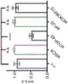


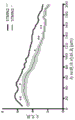


Classifications
-
- C—CHEMISTRY; METALLURGY
- C12—BIOCHEMISTRY; BEER; SPIRITS; WINE; VINEGAR; MICROBIOLOGY; ENZYMOLOGY; MUTATION OR GENETIC ENGINEERING
- C12N—MICROORGANISMS OR ENZYMES; COMPOSITIONS THEREOF; PROPAGATING, PRESERVING, OR MAINTAINING MICROORGANISMS; MUTATION OR GENETIC ENGINEERING; CULTURE MEDIA
- C12N15/00—Mutation or genetic engineering; DNA or RNA concerning genetic engineering, vectors, e.g. plasmids, or their isolation, preparation or purification; Use of hosts therefor
- C12N15/09—Recombinant DNA-technology
- C12N15/11—DNA or RNA fragments; Modified forms thereof; Non-coding nucleic acids having a biological activity
- C12N15/113—Non-coding nucleic acids modulating the expression of genes, e.g. antisense oligonucleotides; Antisense DNA or RNA; Triplex- forming oligonucleotides; Catalytic nucleic acids, e.g. ribozymes; Nucleic acids used in co-suppression or gene silencing
-
- A—HUMAN NECESSITIES
- A61—MEDICAL OR VETERINARY SCIENCE; HYGIENE
- A61P—SPECIFIC THERAPEUTIC ACTIVITY OF CHEMICAL COMPOUNDS OR MEDICINAL PREPARATIONS
- A61P25/00—Drugs for disorders of the nervous system
- A61P25/28—Drugs for disorders of the nervous system for treating neurodegenerative disorders of the central nervous system, e.g. nootropic agents, cognition enhancers, drugs for treating Alzheimer's disease or other forms of dementia
-
- C—CHEMISTRY; METALLURGY
- C12—BIOCHEMISTRY; BEER; SPIRITS; WINE; VINEGAR; MICROBIOLOGY; ENZYMOLOGY; MUTATION OR GENETIC ENGINEERING
- C12N—MICROORGANISMS OR ENZYMES; COMPOSITIONS THEREOF; PROPAGATING, PRESERVING, OR MAINTAINING MICROORGANISMS; MUTATION OR GENETIC ENGINEERING; CULTURE MEDIA
- C12N2310/00—Structure or type of the nucleic acid
- C12N2310/10—Type of nucleic acid
- C12N2310/11—Antisense
-
- C—CHEMISTRY; METALLURGY
- C12—BIOCHEMISTRY; BEER; SPIRITS; WINE; VINEGAR; MICROBIOLOGY; ENZYMOLOGY; MUTATION OR GENETIC ENGINEERING
- C12N—MICROORGANISMS OR ENZYMES; COMPOSITIONS THEREOF; PROPAGATING, PRESERVING, OR MAINTAINING MICROORGANISMS; MUTATION OR GENETIC ENGINEERING; CULTURE MEDIA
- C12N2310/00—Structure or type of the nucleic acid
- C12N2310/30—Chemical structure
- C12N2310/31—Chemical structure of the backbone
- C12N2310/315—Phosphorothioates
-
- C—CHEMISTRY; METALLURGY
- C12—BIOCHEMISTRY; BEER; SPIRITS; WINE; VINEGAR; MICROBIOLOGY; ENZYMOLOGY; MUTATION OR GENETIC ENGINEERING
- C12N—MICROORGANISMS OR ENZYMES; COMPOSITIONS THEREOF; PROPAGATING, PRESERVING, OR MAINTAINING MICROORGANISMS; MUTATION OR GENETIC ENGINEERING; CULTURE MEDIA
- C12N2310/00—Structure or type of the nucleic acid
- C12N2310/30—Chemical structure
- C12N2310/31—Chemical structure of the backbone
- C12N2310/316—Phosphonothioates
-
- C—CHEMISTRY; METALLURGY
- C12—BIOCHEMISTRY; BEER; SPIRITS; WINE; VINEGAR; MICROBIOLOGY; ENZYMOLOGY; MUTATION OR GENETIC ENGINEERING
- C12N—MICROORGANISMS OR ENZYMES; COMPOSITIONS THEREOF; PROPAGATING, PRESERVING, OR MAINTAINING MICROORGANISMS; MUTATION OR GENETIC ENGINEERING; CULTURE MEDIA
- C12N2310/00—Structure or type of the nucleic acid
- C12N2310/30—Chemical structure
- C12N2310/32—Chemical structure of the sugar
- C12N2310/323—Chemical structure of the sugar modified ring structure
- C12N2310/3231—Chemical structure of the sugar modified ring structure having an additional ring, e.g. LNA, ENA
-
- C—CHEMISTRY; METALLURGY
- C12—BIOCHEMISTRY; BEER; SPIRITS; WINE; VINEGAR; MICROBIOLOGY; ENZYMOLOGY; MUTATION OR GENETIC ENGINEERING
- C12N—MICROORGANISMS OR ENZYMES; COMPOSITIONS THEREOF; PROPAGATING, PRESERVING, OR MAINTAINING MICROORGANISMS; MUTATION OR GENETIC ENGINEERING; CULTURE MEDIA
- C12N2320/00—Applications; Uses
- C12N2320/30—Special therapeutic applications
- C12N2320/33—Alteration of splicing
Abstract
The present disclosure relates to compositions and methods for treating a disease or disorder associated with TDP pathology or reduced TDP-43 function in neuronal cells in a subject, and for identifying candidate agents that suppress or prevent inclusion of ineffective or altered STMN2RNA sequences.
Description
RELATED APPLICATIONS
The present application claims priority from U.S. provisional application number 63/133,749 filed on 1 month 4 of 2021, U.S. provisional application number 63/063,174 filed on 8 month 7 of 2020, and U.S. provisional application number 62/994,797 filed on 3 month 25 of 2020. The entire teachings of the above application are incorporated herein by reference.
Background
Amyotrophic Lateral Sclerosis (ALS) is a fatal neurodegenerative disease characterized by a selective loss of upper and lower motor neurons (1). ALS patients experience progressive paralysis and develop difficulty speaking, swallowing, and eventually breathing (2, 3), and die of the disease usually 1-5 years from the time of diagnosis. Apart from the two FDA approved drugs (4) that moderately alter disease progression, treatment of ALS is limited to supportive care. ALS is now considered to have the same clinical and pathological profile as frontotemporal dementia (FTD, the most common cause of alzheimer's disease). FTD is characterized by behavioral changes, language disorders, and loss of executive function (5) for which no effective treatment has been available. Although the etiology of most ALS and FTD cases is still unknown, pathology findings and family-based linkage studies have demonstrated that there is an overlap in the molecular pathways involved in both diseases (1, 6).
Disclosure of Invention
TDP-43 is the major nuclear DNA/RNA binding protein, which plays a functional role in transcriptional regulation, splicing, pre-microRNA processing, stress particle formation, and messenger RNA transport and stability. TDP-43 has been found to be the major component of inclusion bodies in many sporadic cases of ALS and FTD. As a response to aberrant expression of TDP-43, a decrease in STMN2 levels was seen. STMN2 (also known as SCG 10) is a modulator of microtubule stability and has been shown to encode proteins required for normal human motor neuron growth and repair. Methods and compositions for restoring or increasing STMN2 levels are described herein.
Disclosed herein are antisense oligonucleotides that specifically bind to STMN2 mRNA, pre-mRNA, or nascent RNA sequences, thereby suppressing or preventing inclusion of null (abortive) or altered STMN2 RNA sequences. In some embodiments, the antisense oligonucleotide does not bind to a polyadenylation site of the STMN2 RNA sequence. In some embodiments, when TDP-43 function is reduced or TDP pathology occurs, ineffective or altered STMN2 RNA sequences occur and abundance increases.
Also disclosed herein are antisense oligonucleotides that specifically bind to STMN2 mRNA, pre-mRNA, or nascent RNA sequences encoding a recessive exon, thereby suppressing or preventing inclusion of a recessive exon in STMN2 RNA, wherein the antisense oligonucleotides do not bind to polyadenylation sites of STMN2 mRNA, pre-mRNA, or nascent RNA sequences.
Also disclosed herein are antisense oligonucleotides that specifically bind to STMN2 mRNA, pre-mRNA, or nascent RNA sequences, wherein the antisense oligonucleotides increase STMN2 protein expression.
In some embodiments, the antisense oligonucleotide is designed to target a 5 'splice site, a 3' splice site, or a normal TDP-43 binding site. In some embodiments, the antisense oligonucleotide targets one or more splice sites. In some embodiments, the antisense oligonucleotide is designed to target a single stranded region located between the TDP-43 binding site and the polyadenylation site.
In some embodiments, the antisense oligonucleotide does not exhibit platelet toxicity.
Also disclosed herein are antisense oligonucleotides comprising a sequence selected from the group consisting of SEQ ID NOS: 37-85. In some aspects, the antisense oligonucleotide comprises a sequence selected from the group consisting of SEQ ID NOS: 37-74. In some embodiments, the antisense oligonucleotide comprises a sequence selected from the group consisting of: SEQ ID NO. 40, SEQ ID NO. 47, SEQ ID NO. 48, SEQ ID NO. 49, SEQ ID NO. 50, SEQ ID NO. 52, SEQ ID NO. 53, SEQ ID NO. 54, SEQ ID NO. 56 and SEQ ID NO. 78, or more specifically, the antisense oligonucleotide may comprise SEQ ID NO. 52. In certain embodiments, the antisense oligonucleotide comprises a sequence selected from the group consisting of SEQ ID NO. 53, SEQ ID NO. 72, and SEQ ID NO. 73, or more specifically, the antisense oligonucleotide comprises SEQ ID NO. 73 or SEQ ID NO. 53.
Also disclosed herein are pharmaceutical compositions comprising one or more antisense oligonucleotides comprising a sequence selected from the group consisting of SEQ ID NOs 37-85. In some embodiments, one or more antisense oligonucleotides comprise a sequence selected from the group consisting of SEQ ID NOS: 37-74. In some embodiments, the one or more antisense oligonucleotides comprise a sequence selected from the group consisting of: SEQ ID NO. 40, SEQ ID NO. 47, SEQ ID NO. 48, SEQ ID NO. 49, SEQ ID NO. 50, SEQ ID NO. 52, SEQ ID NO. 53, SEQ ID NO. 54, SEQ ID NO. 56 and SEQ ID NO. 78, or more specifically, one or more antisense oligonucleotides may comprise SEQ ID NO. 52. In certain embodiments, the antisense oligonucleotide comprises a sequence selected from the group consisting of SEQ ID NO. 53, SEQ ID NO. 72, and SEQ ID NO. 73, or more specifically, the antisense oligonucleotide comprises SEQ ID NO. 73 or SEQ ID NO. 53.
Disclosed herein are pharmaceutical compositions comprising multimeric oligonucleotides. The multimeric oligonucleotide comprises one or more sequences selected from the group consisting of SEQ ID NOS: 37-85. In some embodiments, the multimeric oligonucleotide comprises two or more sequences selected from the group consisting of SEQ ID NOS: 37-85. The multimeric oligonucleotide may comprise multiple copies of a sequence, or alternatively may comprise a single copy of multiple sequences.
In some embodiments, the antisense oligonucleotide suppresses or prevents inclusion of a recessive exon in the STMN2 RNA. In some embodiments, the antisense oligonucleotide specifically binds to, for example, STMN2 RNA, pre-mRNA, or nascent RNA sequences encoding a recessive exon. In some embodiments, the antisense oligonucleotide prevents or delays STMN2 protein degradation. In some embodiments, the antisense oligonucleotide increases STMN2 protein. In some embodiments, the antisense oligonucleotide is designed to target a 5 'splice site, a 3' splice site, or a normal TDP-43 binding site. In some embodiments, the antisense oligonucleotide is designed to target a single stranded region, for example, a single stranded region located between the TDP-43 binding site and the polyadenylation site. In some embodiments, the antisense oligonucleotide is designed to target a site near a recessive splice site, a site near an premature polyadenylation site, or a site located between a recessive splice site and an premature polyadenylation site. In some embodiments, the antisense oligonucleotide binds to an unstructured target region within a recessive exon. In some embodiments, the antisense oligonucleotide binds near or adjacent to a 5' splice site regulated by TDP-43. In some embodiments, the antisense oligonucleotide targets a region near the predicted TDP-43 binding site. In some embodiments, the antisense oligonucleotide targets the TDP-43 normal binding site. In some embodiments, the antisense oligonucleotide targets one or more splice sites. In some embodiments, the antisense oligonucleotide represses recessive splicing.
In some embodiments, the pharmaceutical composition comprises two or more antisense oligonucleotides, and in some aspects, three or more antisense oligonucleotides. In some embodiments, two or more antisense oligonucleotides are covalently linked. In some embodiments, one or more antisense oligonucleotides increase STMN2 protein expression.
In some embodiments, the pharmaceutical composition further comprises an agent for treating a neurodegenerative disease, an agent for treating traumatic brain injury, or an agent for treating proteasome inhibitor-induced neuropathy. In some embodiments, the pharmaceutical composition further comprises STMN2 as gene therapy. In some embodiments, the pharmaceutical composition further comprises a JNK inhibitor.
Also disclosed herein are methods of treating or reducing the likelihood of a disease or disorder associated with reduced function of TAR DNA binding protein 43 (TDP-43) in neuronal cells in a subject in need thereof. The method can include contacting the neuronal cell with an antisense oligonucleotide that corrects for reduced levels of STMN2 protein, wherein the agent does not target the polyadenylation site of the target transcript.
Also disclosed herein are methods of treating or reducing the likelihood of a disease or disorder associated with reduced function of TAR DNA binding protein 43 (TDP-43) in neuronal cells in a subject in need thereof. The method may comprise contacting the neuronal cell with an antisense oligonucleotide that increases expression of the STMN2 protein.
In some embodiments, the antisense oligonucleotide specifically binds to STMN2 RNA, pre-RNA, or nascent RNA sequences encoding a recessive exon. In some embodiments, the antisense oligonucleotide is designed to target a 5 'splice site, a 3' splice site, or a normal TDP-43 binding site. In some embodiments, the antisense oligonucleotide is designed to target a single stranded region, for example, a single stranded region located between the TDP-43 binding site and the polyadenylation site. In some embodiments, the antisense oligonucleotide is designed to target a site near a recessive splice site, a site near an premature polyadenylation site, or a site located between a recessive splice site and an premature polyadenylation site. In some embodiments, the antisense oligonucleotide binds to an unstructured target region within a recessive exon. In some embodiments, the antisense oligonucleotide binds near or adjacent to a 5' splice site regulated by TDP-43. In some embodiments, the antisense oligonucleotide targets a region near the predicted TDP-43 binding site. In some embodiments, antisense oligonucleotides are designed to target one or more splice sites. In some embodiments, the antisense oligonucleotide restores normal length or the protein encodes an STMN2 pre-mRNA or mRNA.
In some embodiments, the subject exhibits improved neuronal growth and repair. In some embodiments, the disease or disorder is a neurodegenerative disease, such as Amyotrophic Lateral Sclerosis (ALS), frontotemporal dementia (FTD), inclusion Body Myositis (IBM), parkinson's disease, or Alzheimer's disease. In some embodiments, the disease or condition is traumatic brain injury. In some embodiments, the disease or disorder is a proteasome inhibitor-induced neuropathy. In some embodiments, the disease or condition is associated with a mutant or reduced level of TDP-43 in a neuronal cell.
In some embodiments, the method further comprises administering an effective amount of a second agent to the subject. In some embodiments, the second dose is administered to treat a neurodegenerative disease or traumatic brain injury. In some embodiments, the second agent is STMN2, e.g., administered as gene therapy.
Also disclosed herein are methods of treating or reducing the likelihood of a disease or disorder associated with reduced function of TAR DNA binding protein 43 (TDP-43) in neuronal cells in a subject in need thereof. The method may comprise contacting the neuronal cell with an antisense oligonucleotide that corrects for reduced levels of STMN2 protein, wherein the antisense oligonucleotide comprises a sequence selected from the group consisting of SEQ ID NOs 37-85.
In some embodiments, the antisense oligonucleotide comprises a sequence selected from the group consisting of SEQ ID NOS: 37-74. In some embodiments, the antisense oligonucleotide comprises a sequence selected from the group consisting of: SEQ ID NO. 40, SEQ ID NO. 47, SEQ ID NO. 48, SEQ ID NO. 49, SEQ ID NO. 50, SEQ ID NO. 52, SEQ ID NO. 53, SEQ ID NO. 54, SEQ ID NO. 56 and SEQ ID NO. 78, or more specifically, the antisense oligonucleotide may comprise SEQ ID NO. 52. In certain embodiments, the antisense oligonucleotide comprises a sequence selected from the group consisting of SEQ ID NO. 53, SEQ ID NO. 72, and SEQ ID NO. 73, or more specifically, the antisense oligonucleotide comprises SEQ ID NO. 73 or SEQ ID NO. 53.
Also disclosed herein are methods of reducing the likelihood of a disease or disorder associated with reduced function of TAR DNA binding protein 43 (TDP-43) in a neuronal cell in a subject in need thereof. The method may comprise contacting the neuronal cell with one or more antisense oligonucleotides that repress or prevent the inclusion of a recessive exon in the STMN2 RNA. In some embodiments, one or more antisense oligonucleotides comprise a sequence selected from the group consisting of SEQ ID NOS: 37-85.
In some embodiments, the antisense oligonucleotide comprises a sequence selected from the group consisting of: SEQ ID NO. 40, SEQ ID NO. 47, SEQ ID NO. 48, SEQ ID NO. 49, SEQ ID NO. 50, SEQ ID NO. 52, SEQ ID NO. 53, SEQ ID NO. 54, SEQ ID NO. 56 and SEQ ID NO. 78, or more specifically SEQ ID NO. 52. In certain embodiments, the antisense oligonucleotide comprises a sequence selected from the group consisting of SEQ ID NO. 53, SEQ ID NO. 72, and SEQ ID NO. 73, or more specifically, the antisense oligonucleotide comprises SEQ ID NO. 73 or SEQ ID NO. 53.
In some embodiments, the antisense oligonucleotide specifically binds to STMN2 RNA, pre-RNA, or nascent RNA sequences encoding a recessive exon. In some embodiments, the antisense oligonucleotide is designed to target a 5 'splice site, a 3' splice site, or a normal TDP-43 binding site. In some embodiments, the antisense oligonucleotide is designed to target a single stranded region, for example, a single stranded region located between the TDP-43 binding site and the polyadenylation site. In some embodiments, the antisense oligonucleotide is designed to target a site near a recessive splice site, a site near an premature polyadenylation site, or a site located between a recessive splice site and an premature polyadenylation site. In some embodiments, the antisense oligonucleotide binds to an unstructured target region within a recessive exon. In some embodiments, the antisense oligonucleotide binds near or adjacent to a 5' splice site regulated by TDP-43. In some embodiments, the antisense oligonucleotide targets a region near the predicted TDP-43 binding site. In some embodiments, the antisense oligonucleotide targets the TDP-43 normal binding site.
In some embodiments, the disease or condition is selected from the group consisting of: amyotrophic Lateral Sclerosis (ALS), frontotemporal dementia (FTD), inclusion Body Myositis (IBM), parkinson's disease and alzheimer's disease. In some embodiments, the disease or condition is traumatic brain injury. In some embodiments, the disease or disorder is a proteasome inhibitor-induced neuropathy.
In some embodiments, the antisense oligonucleotide represses recessive splicing. In some embodiments, the antisense oligonucleotide prevents or delays STMN2 protein degradation. In some embodiments, the subject exhibits improved neuronal growth and repair.
In some embodiments, the method further comprises administering an effective amount of a second agent to the subject. In some embodiments, the second dose is administered to treat a neurodegenerative disease or traumatic brain injury.
Also disclosed herein are methods of treating or reducing the likelihood of a disease or disorder associated with reduced TAR DNA binding protein 43 (TDP-43) function in a neuronal cell in a subject in need thereof, comprising contacting the neuronal cell with a multimeric oligonucleotide that corrects for reduced STMN2 protein levels, wherein the multimeric oligonucleotide comprises two or more antisense oligonucleotides selected from the group consisting of SEQ ID NOs 37-85. In some embodiments, the multimeric oligonucleotide comprises two or more antisense oligonucleotides selected from the group consisting of SEQ ID NOS: 37-74.
Also disclosed herein are antisense oligonucleotides that correct for reduced levels of STMN2 protein, wherein the antisense oligonucleotides are designed to target unstructured regions within the recessive exons. In some embodiments, the unstructured region within the recessive exon is located between the recessive splice site and the premature polyadenylation site.
Also disclosed herein are methods of detecting altered levels of STMN2 or ELAVL3 protein in a subject. The method comprises obtaining a sample from a subject; and detecting whether the STMN2 or ELAVL3 protein level is altered. In some embodiments, the subject has amyotrophic lateral sclerosis. In some embodiments, detecting whether the STMN2 or ELAVL3 level is altered comprises determining whether the STMN2 or ELAVL3 level is decreased (e.g., using ELISA). In some embodiments, the sample is a biological fluid sample (e.g., CSF sample).
Drawings
The patent or application contains at least one drawing which is made in color. Copies of this patent or patent application publication with color drawings will be provided by the office upon request and payment of the necessary fee.
FIGS. 1A-1F show RNA sequencing of TDP-43 knockdown in hMN. FIG. 1A provides a schematic diagram showing RNAi strategy for hMN differentiation, purification and TDP-43 gene knockdown in cultured MN. FIG. 1B provides a multidimensional scaling analysis of RNA-Seq datasets obtained from two biologically independent MN differentiation and siRNA transfection experiments based on 500 maximally differentially expressed genes. FIG. 1C provides a volcanic plot showing statistically-deregulated genes in hMN treated with siTDP-43 compared to hMN treated with scrambling control. Genes identified as significant after differential expression analysis (Benjamini-Hochberg adjusted P-value cutoff of 0.05 and log fold change rate cutoff of 0) are highlighted in yellow (for up-regulated/increased abundant genes) and blue (for down-regulated/decreased abundant genes). FIG. 1D provides a scatter plot comparing the fold change in TPM values of all genes expressed in MN treated with control siRNA versus those expressed in cells treated with siTDP-43. FIGS. 1E and 1F show that a subset of 11 genes initially identified as 'hits' (significantly up-regulated (FIG. 1E) or down-regulated (FIG. 1F)) in the TDP43 gene knockdown experiment were selected for qRT-PCR validation. When the expression of these genes was analyzed by qRT-PCR (unpaired t test, P value < 0.05), a total of 9 of 11 of these genes (including TDP-43) exhibited a predicted response to TDP-43 clearance.
Fig. 2A-2J show familial ALS models. FIG. 2A provides a schematic representation of a strategy for evaluating gene expression in iPS cell-derived hMN expressing mutant TDP-43. Fig. 2B provides photomicrographs showing the morphology of neurons derived from healthy control iPS cells (11 a, 18a, 20B, 17 a) and iPS cells from patients with TARDP mutations (+/Q343R, +/G298S, +/a315T and +/M337V) cultured for 10 days. FIGS. 2C-2H provide qRT-PCR analysis of genes consistently down-regulated (FIGS. 2D-2F) or up-regulated (FIG. 2C) following TDP-43 gene knockdown in neurons differentiated from control or TDP-43 patients. (P value <0.05 without t-test). Figure 2I provides representative micrographs of control and patient neurons immunostained for TDP-43 (red), β -III tubulin (green) and counterstained with DAPI (blue). Scale bar, 100 μm. FIG. 2J provides a Person's correlation analysis comparing control neurons with TDP-43 immunostaining and DAPI fluorescence of neurons with TDP-43 mutations. Dots represent individual cells. (P value <0.05 without t-test).
Figures 3A-3I show STMN2 regulation and localization. FIG. 3A provides qRT-PCR analysis of STMN2 transcripts using two different primer sets in separate experiments. (P value <0.05 without t-test). FIG. 3B provides immunoblot analysis of TDP-43 and STMN2 protein levels after partial clearance of TDP-43 by siRNA gene knockdown. Protein levels were normalized to GAPDH and expressed relative to levels in MN treated with siRED control. FIG. 3C provides qRT-PCR analysis of STMN2 transcript analysis in GFP+ MN with Hb9 treated with siRNA targeting three ALS linked genes (TDP-43, FUS and C9ORF 72). (Deng Nite multiple comparison test (Dunnett's multiple comparison test), alpha < 0.05). FIGS. 3D-3F show the use of formaldehyde RNA immunoprecipitation to identify transcripts that bind to TDP-43. After TDP-43 immunoprecipitation (fig. 3D), qRT-PCR analysis was used to test enrichment relative to sample input TDP-43 transcript (fig. 3E) and STMN2 transcript (fig. 3F). FIG. 3G provides a micrograph of Hb9: GFP+MN immunostained for TDP-43 (red), beta-III tubulin (green) and counterstained with DAPI (blue). FIG. 3H provides a micrograph of Hb9: GFP+MN co-cultured on glia immunostained for STMN2 (red) and MAP2 green and GOLGIN97 (green). FIG. 3I provides a micrograph of Hb9: GFP+MN immunostained for STMN2 (red), MAP2 (green) and counterstained with F-actin binding protein phalloidin (white) on day 3 post-sorting. Scale bar, 5 μm.
Fig. 4A-4K show STMN2 knockout. FIG. 4A provides a schematic representation of a knockout strategy using guide RNAs (gRNAs) targeting two constitutive exons of the human STMN2 gene, namely exons 2 and 4. The inserted DNA segment (about 18 Kb) was targeted and deleted due to NHEJ (non-homologous end joining) repair of the two Double Strand Breaks (DSBs) introduced by Cas9/gRNA nuclease complex. FIGS. 4B-4D show that STMN2 knockdown was confirmed by RT-PCR analysis of genomic DNA (FIG. 4B), by immunoblot analysis (FIG. 4C) and by immunofluorescence (FIG. 4D) in HUES3 Hb 9:. Fig. 4E provides experimental strategies for evaluating the cellular effects of the lack of STMN2 in hMN. FIGS. 4F-4H show Sholl analysis of hMN with and without STMN2 and in the absence (FIG. 4G) or presence (FIG. 4H) of a ROCK inhibitor (Y-27632,10. Mu.M) that stimulates neurite outgrowth. (P value <0.05 without t-test). Fig. 4I provides experimental strategies for evaluating the cellular effects of a lack of STMN2 in hMN following axonal injury. Figures 4J to 4K show axonal regrowth following injury. Representative micrographs of hMN in microfluidic devices before and after axonometric cleavage (fig. 4J). Measurement of axon regeneration after axonectomy. (P value <0.05 without t-test).
Fig. 5A-5G show sporadic ALS models. FIG. 5A provides an experimental strategy for evaluating the effect of proteasome inhibition on TDP-43 localization in human motor neurons. FIG. 5B shows the Pelson-related analysis of TDP-43 immunostaining and DAPI fluorescence of cells treated with MG-132 (1. Mu.M). (Deng Nite multiple comparison test, alpha < 0.05). FIG. 5C provides a micrograph of HUES3 motor neurons untreated or treated with MG-132 and immunostained for TDP-43 (red), beta-III tubulin (green) and counterstained with DAPI (blue). Scale bar, 100 μm. FIG. 5D provides immunoblot analysis of TDP-43 in detergent soluble fraction (RIPA) and detergent insoluble fraction (UREA) in neurons treated with MG-132 (unpaired t test, P value < 0.05). FIG. 5E provides qRT-PCR analysis of STMN2 expression of motor neurons treated with MG-132 at indicated concentrations and durations relative to DMSO control (unpaired t test, P value < 0.05). FIG. 5F provides a diagram of RT-PCR detection strategy for STMN2 recessive exons. FIG. 5G provides a statation analysis of STMN2 recessive exons in hMN control cells treated with MG-132 (1. Mu.M).
Fig. 6A to 6H show ALS patient data. Fig. 6A-6C provide histological analysis of human adult lumbar spinal cord of post-mortem samples collected from subjects without evidence of spinal cord disease (control) (fig. 6A) or two patients diagnosed with sporadic ALS (fig. 6B-6C). Immunoreactivity to STMN2 was detected in the perinuclear region of spinal motor neurons (indicated by the arrow) but not in peripheral glial cells. STMN2 immunoreactivity scores in lumbar motor neurons from control and ALS cases were either 'strong' (as indicated by the arrows in control (fig. 6A) and sporadic ALS (fig. 6B) or 'absent' (as indicated by the arrows in sporadic ALS (fig. 6C)). Scale bar, 50 μm. Fig. 6D shows that the percentage of lumbar motor neurons with strong STMN2 immunoreactivity was significantly lower in ALS tissue samples (n=3 controls and 3 ALS cases; scoring about 40 MNs per subject; double tail t-test, P-value < 0.05). Fig. 6E-6G show gene expression analysis of STMN2 (two-tailed t-test, P-value < 0.05) from previously disclosed datasets (Rabin et al, 2009 (fig. 6E), highley et al, 2014 (fig. 6F) and D' Erchia et al, 2017). Figure 6H provides a molecular model of ALS pathogenesis.
Fig. 7A-7I show the generation of differentiated human motor neurons. FIG. 7A shows hMN differentiation, purification and culture strategies. FIG. 7B provides flow cytometry analysis of differentiated HUES3 Hb9: GFP cells. Cells not treated with RA and SHH pathway agonists were used as negative controls for gating of GFP expression. Fig. 7C-7F provide photomicrographs and quantification of purified Hb9:: gfp+ cells immunostained for Hb9 and counterstained with DAPI (fig. 7C) (scale bar = 10 μm) or immunostained for ISL1 and neuronal markers β -III tubulin and MAP2 (scale bar = 20 μm). Fig. 7G-7J show that differentiated MNs are electrophysiologically active as determined by whole cell patch clamp recordings. Fig. 7G shows that upon depolarization in voltage clamp mode, the cells exhibited a fast inward current followed by a slow outward current, indicating the expression and opening of voltage activated sodium and potassium channels, respectively. Fig. 7H shows that in current clamp mode, depolarization causes repetitive action potential firing. FIG. 7I shows that the response to rhodopsin is consistent with the expression of functional receptors for excitatory glutamatergic transmitters.
FIGS. 8A-8E show TDP-43 knockdown in cultured hMN. FIG. 8A provides RNAi strategy for TDP-43 gene knockdown in cultured MN. Fig. 8B shows phase and red fluorescence micrographs of the cultivated hMN 4 days after treatment with different sirnas conjugated to Alexa Fluor 555, including scrambled sirnas. Figure 8C provides flow cytometry analysis of hMN after treatment with different sirnas. FIG. 8D shows the relative levels of TDP-43mRNA in MN exposed to different siRNAs for 2, 4 or 6 days. The level of each sample was normalized to GAPDH and expressed relative to the non-transfected control. Figure 8E provides immunoblot analysis of hMN after RNAi treatment with indicated sirnas. Each sample was normalized using GAPDH and TDP-43 protein levels were calculated relative to a siSCR 555 treated control sample.
Fig. 9A-9C show motor neuron RNA-Seq. Fig. 9A shows a global transcription analysis of motor neurons processed as indicated by the heat map. Unsupervised clustering of expression profiles reveals that samples were isolated based on batches of motor neuron generation and analysis. FIG. 9B provides an analysis of TDP-43 transcript abundance after RNA sequencing confirmed gene knockdown (Benjamin-Huo Beige adjusted P-cut off of 0.05). FIG. 9C shows that changes in splice pattern of the POLDIP3 gene were detected due to TDP-43 knockdown, where siTDP43 treated cells showed significantly reduced and increased levels of splice variant 2 (which lacks exon 3) for subtype 1 (false discovery rate 'FDR' > 0.05).
Figure 10 shows a multipotent stem cell genotyping sequencing chromatogram of exon 6 of TARDBP in the indicated iPS cell lines to confirm heterozygous mutations in the patient cell lines.
Fig. 11A-11F show neuronal cell sorting. Fig. 11A shows that antibodies enriched on gfp+ motor neurons (quadrant 1) and GFP-cells (quadrant 3) were identified using cell surface marker screening. Fig. 11B shows that after sorting ncam+ and EpCAM-cells, high content imaging was used to determine if the sorting method could clear cultures of mitotic cells (edu+) and significantly enrich motor neurons (isl1+) and neurons (MAP 2+). N=6 different iPS cell lines. Statistical analysis was performed using a double tail judton t test (Student's t test). FIGS. 11C-11D provide qRT-PCR analysis of cultures after sorting of motor neuron marker ISL1 (FIG. 11C) and neuron marker βIII-tubulin (FIG. 11D), revealing enriched and more homogeneous cultures compared to unsorted cultures. FIG. 11E provides flow cytometry analysis of cultures differentiated from indicated healthy controls (gray) and TDP-43 mutant cell lines (red) using Phycoerythrin (PE) conjugated antibodies to EpCAM (anti-epCAM-PE) and Alexa Fluor 700 conjugated antibodies to NCAM (anti-NCAM-AF 700). FIG. 11F shows the percentage of NCAM+ cells from 4-6 independently differentiated indicated cell lines. No significant difference was observed between the mutant cell line and the control cell line in terms of the ability to produce ncam+ cells. Statistical analysis was performed using the double tail judton t test with P value <0.05.
Fig. 12A-12G show TDP-43 and STMN2 connections. FIGS. 12A-12C provide qRT-PCR validation of ALS gene down-regulation after siRNA treatment. Expression of TDP-43 (fig. 12A), FUS (fig. 12B) and C9ORF72 (fig. 12C) was evaluated (unpaired t test, P value < 0.05) for all controls used and each siRNA. FIG. 12D provides Western blot (western blot) analysis of STMN2 protein in different cell types differentiated along motor neurons. FIG. 12E shows RNA-Seq expression levels of the tubulin family of depolymerized microtubules (Stathmin) in motor neurons treated with siSCR (-) or siTDP-43 (+) oligonucleotides. Only STMN2 levels changed after TDP-43 gene knockdown. FIGS. 12F-12G show the TDP-43 binding site within the microtubule depolymerizing protein gene family (FIG. 12F) normalized to the gene length (FIG. 12G). STMN2 has the largest number of binding motifs.
Fig. 13A-13H show that STMN2 modulates neuronal growth. CRISPR-mediated STMN2 knockout in WA01 cell lines WAs confirmed by RT-PCR analysis of genomic DNA (fig. 13A), by immunoblot analysis (fig. 13B) and by immunofluorescence (fig. 13C). FIGS. 13D-13F provide Sholl analysis of hMN (FIG. 13F) with and without STMN2 and in the presence of ROCK inhibitor Y-27632 (10. Mu.M) (unpaired t test, P < 0.05). Fig. 13G-13H show axonal regrowth following injury. Representative micrographs of hMN in the microfluidic device before and after axonometric cleavage (fig. 13G). Analysis of axonal regrowth after axonectomy (unpaired t test, P value < 0.05) (fig. 13H).
Fig. 14A-14E show cell survival and proteasome activity assays. Fig. 14A-14C show that Cell Titer Glo uses ATP from metabolically active cells to generate light. (FIG. 14A) shows that there is a direct relationship between luminescence and the number of cells in culture over several orders of magnitude. Fig. 14B shows that the assay can detect differences in neuronal survival in the absence of growth factors. N=6 individual neuronal wells. (P value <0.05 without t-test). FIG. 14C shows an overview of MG-132 neuron survival experiments. FIG. 14D shows a dose response curve for motor neurons cultured with indicated concentrations of MG-132 for indicated times. N = triplicate wells. Cells were viable after 1 day of treatment at all concentrations tested and were tolerated for longer periods of time at lower concentrations. FIG. 14E shows that after cleavage by the proteasome, the substrate for luciferase is released, which allows quantitative measurement of proteasome activity. Neurons treated with MG-132 showed significantly reduced proteasome activity. N=4 individual neuronal wells (unpaired t test, P value < 0.05).
FIGS. 15A-15E show that TDP-43 modulates recessive exon splicing in hMN (FIGS. 15A-15C). Visualization of the recessive exons of PFKP (fig. 15A), ELAVL3 (fig. 15B) and STMN2 (fig. 15C) of cells treated with scrambled siRNA or sirnas targeting TDP-43 transcripts. Read coverage and splice junctions are shown for alignment with the human HG19 genome. FIGS. 15D-15E provide a graph of RT-PCR detection strategy for STMN2 recessive exons (FIG. 15D), and Mulberry sequencing of PCR products (Sanger sequencing) confirm splicing of STMN2 exon 1 to recessive exons (FIG. 15E).
Fig. 16A-16P provide recessive STMN2 transcript qPCR data for patient cerebrospinal fluid (CSF) samples. Fig. 16A-16D provide graphs summarizing patient sample data relative to healthy control standardized recessive STMN2. Fig. 16E-16M provide diagrams of details regarding individual patient samples. Fig. 16N provides a graph showing survival duration after diagnosis. Fig. 16O provides a graph showing the age of death. Fig. 16P provides a diagram showing viability (visual capability).
Fig. 17A-17C show STMN2 multiplex qPCR assays. Figure 17A shows a Q-RT PCT assay for STMN2 in a fluid. Experimental protocols are provided and show that STMN2 multiplex TaqMan assays detect recessive STMN2, normal STMN2 transcripts, and housekeeping gene RNA18S5 simultaneously. RNA can be collected from CSF-derived exosomes and then converted to cDNA to determine complete and recessive STMN2 transcripts as well as control RNA for normalization. FIG. 17B shows in vitro validation of multiplex assays in cells using ASO or using siRNA to reduce TDP-43 levels. FIG. 17C shows that the use of STMN2 multiplex qPCR assay to probed for recessive STMN2 transcript levels in cDNA samples generated from MGH CSF samples. STMN2 recessive splicing was significantly induced in ALS patients.
Fig. 18A-18D show sandwich ELISA for detection of STMN2 protein. Fig. 18A provides a schematic of STMN2 sandwich ELISA. Figure 18B shows the sensitivity of STMN2ELISA to picogram amounts. Fig. 18C shows that sandwich ELISA was validated using recombinant STMN2 protein and was able to detect picogram levels of STMN2. Fig. 18D shows that when assessed using an STMN2ELISA, STMN2 levels in the patient's cerebrospinal fluid (CSF) are reduced.
Fig. 19 provides a chart showing ALS genetics, where each gene is plotted against the year in which it was found. See Alsultan et al Degenerative Neurological and Neuromuscular disease.2016,6,49-64.
FIG. 20 shows that TDP-43 is a multifunctional nucleic acid binding protein. TDP-43 has been shown to play a role in a variety of functions including RNA splicing, miRNA processing, self-regulation of its own transcripts, RNA transport and stability, and stress particle formation. TDP-43 regulated transcripts are highly species and cell type dependent. See burati and Baralle Trends in biochem. Sci. 2012,6,237-247.
FIG. 21 provides a strategy for measuring transcriptional effects of TDP-43 clearance. The schematic shows hMN differentiation, purification and culture strategies. The strategy uses small molecules that mimic early development to transform stem cells into postmitotic neurons within 2 weeks. Various methods have been developed to sort and study neurons. siRNA techniques combined with RNA sequencing were used to identify transcripts regulated by TDP-43.
Fig. 22 shows the binding of TDP-43 to STMN2. Spinal cords were stained for STMN2 in ALS patients, and STMN2 protein reduction was observed in ALS patients based on fold enrichment relative to PGK1 (ftir). See Klim et al, nature Neuroscience, vol.22, pages 167-179 (2019).
FIG. 23 shows splice changes after TDP-43 clearance. Differential exon usage analysis was performed on RNA-seq samples from motor neurons treated with siTDP. Splice changes were observed in STMN 2.
FIG. 24 shows that TDP-43 represses the recessive exon in STMN 2. An integrated genome viewer was used to observe the way in which RNA seq reads map to the human genome (top panel reads) and the reads re-ligate between exons (splice trajectories). The graph shows the number of reads mapped to a gene region.
Figure 25 provides a summary of STMN2 splice defects. Under normal conditions, STMN2 is transcribed with all 5 exons, yielding mRNA that is translated into a 20kda STMN2 protein. Following a perturbation of TDP-43, the recessive exon intercepts the transcript so that only 17 amino acid polypeptides can be translated.
Fig. 26 shows that STMN2 consistently decreases. The overlapping of reduced transcript downregulation in the 3 human RNA seq dataset (ALS patient dataset and siTDP43 stem cell motor neuron dataset) was compared and STMN2 was the only transcript downregulation in all three datasets.
Figure 27 shows that STMN2 recessive exons are present in the spinal cord of ALS patients. Read coverage and splice junctions are shown for alignment with the human HG19 genome. Reads mapped to the human genome were observed in ALS patients, and for 5 out of 6 patients, mapped and spliced reads entered into the recessive exons, and this was not the case for the control.
FIG. 28 shows that TDP-43 clearance resulted in neurite outgrowth and axonal regrowth defects. Representative photomicrographs of hMN treated with the indicated siRNA and immunostained against β -III tubulin for Sholl analysis are provided. Sholl analysis of hMN after siRNA treatment is provided. Each line represents the average of the samples and the shading represents s.e.m. for unpaired t-test used between siTDP43 and sitsc, bilateral, P <0.05.
Fig. 29 shows a microfluidic device for studying axon regeneration. The microfluidic device includes a cell body compartment (left panel) and an axon compartment (right panel).
FIGS. 30A-30B show that TDP-43 clearance results in neurite outgrowth and axonal regrowth defects. Fig. 30A provides representative micrographs of hMN in a post-axonometric microfluidic device. Scale bar, 150 μm. FIG. 30B provides a measurement of axonal regrowth and regeneration following axonectomy (not paired with t-test, bilateral, P values <0.05, 18 h.ltoreq.0.0001, 24 h.ltoreq.0.0001, 48 h.ltoreq.0.0001, and 72 h.ltoreq.0.0001).
FIG. 31 shows that STMN2 is the c-Jun N-terminal kinase (JNK) target in the axonal degeneration pathway. JNK1 was shown to bind to and phosphorylate STMN2, and phosphorylated STMN2 degraded rapidly. See J.Eun Shin et al, PNAS 2012,109, E3696-3705.
FIG. 32 provides a strategy to determine if JNII can rescue the siTDP43 phenotype. See Klim et al, nature Neuroscience, vol.22, pages 167-179 (2019).
Fig. 33 shows JNK inhibitor (SP 600125) potentiates STMN2 levels. The STMN2 protein levels increased in JNKi-treated neurons and lower levels observed in siTDP 43-treated cells could be rescued.
Fig. 34 shows that JNKi (SP 600125) increased neurite outgrowth. Cells treated with JNKi exhibited increased neurite outgrowth.
Fig. 35 shows that JNKi (SP 600125) increased neurite outgrowth. Sholl analysis confirmed that JNKi increased neurite branching and regrowth following injury under all conditions.
Fig. 36 shows JNKi increases axonal regeneration. The microfluidic device confirmed that JNKi increased neurite branching and regrowth following injury under all conditions.
FIG. 37 provides a model of proteasome inhibition. Disruption of protein homeostasis results in mislocalization of TDP-43 and altered STMN2 levels, which can disrupt axon biology.
Fig. 38A-38B show TDP-43 positioning. TDP-43 was generally nucleated (FIG. 38A), but after compound washout, a significant loss of nuclear TDP-43 staining was observed (FIG. 38B). No cytoplasmic aggregation was observed and only loss of nuclear TDP-43 was observed.
FIG. 39 shows that TDP-43 mislocalization is reversible.
FIG. 40 shows that STMN2 transcripts were reduced after TDP-43 mislocalization. The reduction of STMN2 was even more pronounced than in cells expressing mutant TDP-43.
FIG. 41 provides a table summarizing the recent ALS genes in the different ALS and FTD queues and associated pathways, and their relative mutation frequencies. Advances in WGS and WES have led to the identification of genes carrying rare pathogenic variants: TBK1, CHCHD10, TUBA4A, MATR3, CCNF, NEK1, C21orf2, ANXA11, and TIA1.TBK1 is shown to have the highest ALS-FTD mutation frequency (3% -4%) in the different queues. See, n guyen et al, trends in Genetics,2018.
FIG. 42 shows that Atg7 and TBK1 act at different times of autophagy. See Hansen et al Nature Reviews Molecular Cell biology.2018.
FIG. 43 shows that the elimination of TBK1 has similarities to but differs from the blocking of autophagy initiation.
FIG. 44 shows that TBK1 knockout would reduce functional TDP-43 and STMN2 levels, while eliminating ATG7 would not have an effect. Loss of TBK1 induces TDP-43 pathology of motor neurons through autophagy-independent mechanisms.
Figure 45 shows that loss of TBK1 shows impaired axonal regeneration following axonal injury.
FIG. 46 shows that proteasome inhibition induces TDP-43 mislocalization in TBK1 mutant motor neurons.
Fig. 47A-47C show targeting STMN2 introns using CRISPR. CRISPR strategies for targeting STMN2 are provided, as well as genotyping of STMN2 (fig. 47A-47B). Fig. 47C provides a table summarizing CRISPR targeting strategies and genotypes of STMN 2.
Figure 48 shows that STMN2 mice were significantly smaller than Rosa26 control mice and displayed defects in the athletic performance task, and no signs of progression of these defects over time.
Figure 49 shows that STMN2 mice were significantly smaller than Rosa26 control mice and displayed defects in the athletic performance task, and no signs of progression of these defects over time.
Figure 50 shows that behavioral results, as well as total distance traveled in the open field assay, appear to be similar between the two mouse cohorts.
Figure 51 shows that STMN2 transcript levels were significantly reduced or no transcripts were present in brain tissue of the mutant cohort.
Figure 52 provides western blots of brain tissue, validating the loss or significant reduction of STMN2 protein in the mutant mouse cohort.
Fig. 53 shows that STMN2 is localized primarily to chat+ motor neurons in the ventral horn of the spinal cord of adult mice.
Figure 54 shows that the STMN2 cohort exhibits a significant reduction in stmn2+/chat+ motor neuron numbers on the ventral angle of the spinal cord.
Fig. 55 provides a graph showing the difference in organ or muscle weight between control and STMN2 mice. It shows that the lower limb muscles of STMN2 mice are lighter (see two block diagrams).
Fig. 56 provides pre-and post-synaptic staining of STMN2 Gastrocnemius (GA) and Rosa26 control Gastrocnemius (GA). The staining indicated denervation (de-innervation) in STMN 2-/-animals.
Fig. 57 shows pre-and post-synaptic staining of STMN2 Gastrocnemius (GA) and Rosa26 control Gastrocnemius (GA) indicate denervation in STMN 2-/-animals.
Fig. 58 shows that neuromuscular junction (NMJ) morphology supports active denervation in gastrocnemius of STMN2 mutants.
FIG. 59 shows that mutant TDP-43 did not show pathological mislocalization. Neuronal staining of control and ALS patients for TDP-43 showed that neuronal TDP-43 was primarily nucleated for control and ALS patients.
FIG. 60 identifies different classes of proteasome inhibitors and provides their chemical structure.
FIG. 61 shows reduced expression of full-length STMN2 in hMN after treatment with structurally different proteasome inhibitors.
FIG. 62 shows PCR assays of hMN treated with MG-132 or Bortezomib (Bortezomib). Full length STMN2 was detected in all samples as a control. The presence of transcripts containing STMN2 recessive exons was specific for those cells treated with proteasome inhibitors.
FIGS. 63A-63B show in vitro assays of TDP-43 binding to STMN2 RNA. Using genomic DNA, RNA containing the TDP-43 binding site from the recessive exon region of STMN2 was transcribed in vitro (FIG. 63A). RNA was used to evaluate whether it could pull down the IP TDP-43 protein from human neuronal protein lysate. In vitro assays showed that transcripts containing the recessive exon region pulled down TDP-43 (FIG. 63B).
FIG. 64 shows an in vitro assay of TDP-43 binding to STMN2 RNA. RNA containing the 5 'and 3' TDP-43 binding regions was transcribed in vitro, similarly as described in FIG. 63. Although 5 'and 3' transcripts may pull down some TDP-43, they are not enriched as strongly as intact recessive exons.
FIG. 65 shows the design of gRNAs for generating targeted mutant cell lines without recessive exons. The strategy was prepared to delete 105 nucleotides within the recessive exons within the STMN2 intron between exons 1 and 2. The deletion will eliminate the TDP-43 binding motif but will not affect the predicted polyadenylation site.
Fig. 66 provides confirmation of mutation status. The mutation status of the clones was analyzed using the TIDE analysis and sequence alignment with control cells was checked to obtain a more accurate view of the size and position of the deletions. A cell line contains a homozygous 105nt deletion, consistent with gel electrophoresis. The deletion eliminates the TDP-43 binding motif but does not affect the predicted polyadenylation site.
FIG. 67 shows that the TDP-43 binding site is a potential negative regulator of STMN2 expression. Three cell lines, HUES3, IG2 (Stmn 2 KO) and CN7 (recessive exon deleted) were treated with normal medium or medium +1uM MG132 for 24 hours to stress the cells. In HUES3 cells, stress conditions have 52% STMN2 mRNA expression compared to non-stress conditions. Under IG2 (Stmn 2 KO) conditions, the non-stressed cells had 13% expression, and upon stress, the expression increased to 42%. The expression level in the CN7 (recessive exon deleted) cell line was significantly higher than the other two cell lines, with no stress having 729% expression and stress having 473% expression. It was shown that expression decreased if several exons were knocked out, but increased if the TDP-43 binding site was removed.
FIGS. 68A-68B show that the deletion of the putative TDP-43 binding site results in increased STMN2 protein levels. Consistent with the gene expression data, deletion of the TDP-43 binding region within the recessive exon of STMN2 resulted in increased protein expression.
Fig. 69A-69B show conservation of STMN2 locus. Fig. 69A shows that human STMN2 is located on the long arm of chromosome 8 and is transcribed into several subtypes, typically including 5 canonical exons. The position of the recessive exon is highlighted in orange. Conservation of 100 vertebrates along a locus reveals strong conservation of exons and some intronic regions. Fig. 69B shows a higher resolution genomic view at STMN2 recessive exons (orange) for 12 primates and 2 rodents, where nucleotide resolution is combined with multiple sequence alignments. The salient features of human genes and their degree of conservation in the species list are underlined, including splice acceptor sites (water duck color), putative coding regions (yellow), stop codons (red), TDP-43 binding motif (blue), and poly a signal (purple).
Figure 70 shows a multiplex assay for detecting recessive STMN 2.
FIGS. 71A-71C show that siTDP-43 and TDP-43ASO induce STMN2 reduction and recessive exon induction. The relative expression levels of TARDBP (FIG. 71A), STMN2 exons 3-4 (FIG. 71B) and recessive STMN2 (FIG. 71C) when treated with SCR ASO, TDP ASO or siTDP are shown.
FIGS. 72A-72C show relative mRNA levels of TARDP (FIG. 72A), STMN2 (FIG. 72B) and recessive STMN2 (FIG. 72C) over a 6 day period after treatment with scrambled ASO, TDP-43ASO or SOD1 ASO.
Fig. 73 shows recessive STMN2 expression. mRNA levels expressed by recessive STMN2 after treatment with scrambled ASO, TDP-43ASO, SOD1 ASO, siTDP-43 and siRED are shown. Each treatment was applied using NeuroPorter5, neuroPorter1, RNAiMAX or LipoFecamine, with RNAiMAX being the most effective.
Fig. 74 provides a schematic diagram showing the strategy for testing STMN2 splice switching ASOs.
Fig. 75A to 75D provide schematic diagrams of ASO screening arrangement plate 1 (fig. 75A), plate 2 (fig. 75B), plate 3 (fig. 75C) and plate 4 (fig. 75D).
FIG. 76 provides the results of ASO screening with comparable cDNAs for all wells. The selected ASOs were ASOs targeting the STMN2 intron.
FIG. 77 provides the results of ASO screening, showing ASO near splice junctions represses recessive exon inclusion bodies.
Figure 78 provides the best hit for ASO screening, showing dose dependence or suppression on minimum concentration.
FIGS. 79A-79B show TDP-43 protein structure, pathogenic mutations and function. FIG. 79A shows that TDP-43 comprises six domains: an N-terminal region (aa 1-102) with a nuclear localization signal (NLS, aa 82-98); two RNA recognition motifs: RRM1 (aa 104-176) and RRM2 (aa 192-262); nuclear output signal (NES, aa 239-250); a C-terminal region (aa 274-414) encompassing the glutamine/asparagine (Q/N) -rich prion-like domain (aa 345-366); and glycine rich region (aa 366-414). 46 dominant mutations have been identified in TDP-43 in sporadic and familial ALS patients as well as rare FTLD patients, predominantly in the C-terminal glycine-rich region. Figure 79B shows that significant TDP-43 function is strongly correlated with disease pathogenesis. The most common motif identified for TDP-43 is (TG) n, which corresponds to the (UG) n RNA binding motif. Interaction with RNA allows TDP-43 to regulate pre-mRNA splicing to inhibit the inclusion of recessive exons and affect polyadenylation site selection. Cytosolic effects of TDP-43 include transport of RNA along neuronal processes and responses to stress, including those that affect protein homeostasis that can trigger TDP-43 nuclear efflux and localization to stress particles. The function of many of these basic molecules contributes to TDP-43 self-regulation, including splicing and polyadenylation.
FIGS. 80A-80B show STMN2 protein structure and function. Fig. 80A shows that STMN2 comprises two domains, which can be further subdivided: 1) An N-terminal domain containing a conserved Golgi specific sequence and two palmitoylation sites for membrane insertion, and 2) a tubulin-depolymerizing protein-like domain containing two tubulin binding repeats (TBR 1 and TBR 2) each binding to tubulin; a proline-rich domain (PRD) with two phosphorylation sites, which can be regulated by JNK to potentially modulate STMN 2's ability to interact with tubulin and promote STMN2 degradation; and a tubulin depolymerization protein N-terminal domain (SLDN) containing a peptide that inhibits polymerization of tubulin. Post-translational modifications (PTMs) identified by phosphosilteplus are labeled along the protein structure. FIG. 80B shows subcellular localization of the reported STMN2 protein. STMN2 localizes to the golgi apparatus and is found in vesicles transported throughout dendrites and axons, and is concentrated in the growth cone of developing neurons and in the tip of regenerated axons after injury.
FIG. 81 provides a proposed model of TDP-43 regulation of STMN 2. The pathological hallmark of ALS is loss of the nucleus of TDP-43 and its aggregation. We propose a model of TDP-43 regulation of STMN2, where it binds to an intron between exons 1 and 2 of STMN2 pre-mRNA. The reduced levels of TDP-43 or nuclear export results in early polyadenylation and splicing of the recessive exons, resulting in a truncated STMN2 mRNA transcript. The inactivated transcript encodes a putative 17 amino acid polypeptide, thus resulting in reduced levels of STMN2 protein. Loss of STMN2 results in reduced neurite outgrowth and axonal repair following injury.
FIG. 82 shows antisense oligonucleotides and their position relative to STMN2 sequences. Indicating sequence, chemistry and alignment of ASO with STMN2 locus. Salient features of human genes, including splice acceptor sites (water duck color), putative coding regions (yellow), stop codons (red), TDP-43 binding motif (orange), and poly a signal (purple) are highlighted. ASO highlighted in yellow has locked nucleic acid chemistry.
FIGS. 83A-83C examine the recessive exon-containing region of STMN2 pre-mRNA. FIG. 83A provides a sequence of recessive exon-containing regions of STMN2 pre-mRNA, wherein each salient feature is highlighted. FIGS. 83B-83C provide predicted RNA structures of recessive exon-containing regions of STMN2 pre-mRNA, showing that the region parts highlighted in green are unstructured and can employ different binding interactions with similar energy.
Figures 84A-84D show patient-specific induced pluripotent stem cell characterization. Fig. 84A provides a micrograph showing undifferentiated patient iPS cells. Fig. 84B provides a sequencing chromatogram of PCR products amplified from exon 8 of TBK1 in the indicated iPS cell lines, confirming non-significant heterozygous L306I non-pathological variants in the patient cell lines. Fig. 84C-84D provide photomicrographs showing motor neurons differentiated from patient iPS cells.
Fig. 85A-85B show the reduced nuclear TDP-43 observed in patient neurons. Fig. 85A provides representative micrographs of control and patient neurons immunostained for TDP-43 (red), β -III tubulin (green) and counterstained with DAPI (blue) labeled nuclei. Scale bar, 100 μm. FIG. 85B provides a Pelson-related analysis comparing TDP-43 immunostaining and DAPI fluorescence of control neurons to patients. Dots represent individual cells and are shown as mean and s.d. of at least 25 cells from n=4 control cell lines and 1 patient cell line (t test not paired, double sided, P < 0.05).
Figures 86A-86C show that patient motor neurons produce truncated STMN2 in response to TDP-43 clearance. RNA levels analyzed by qRT-PCR analysis after TDP-43 gene knockdown by siTARDBP in motor neurons differentiated from patient iPS cells. FIG. 86A shows the RNA level of TDP-43. Fig. 86B shows RNA levels of full length STMN2. Fig. 86C shows the RNA level of recessive STMN2 compared to control (sicrl).
Fig. 87A-87C show patient STMN2 locus sequencing. Fig. 87A shows the sequencing results of PCR products amplified from the first intron of STMN2 in the patient iPS cell line, aligned with the reference sequence. FIG. 87B identifies a mismatch between a patient and a reference sequence consisting of a common single nucleotide variant (SNP). Fig. 87C provides a sequencing chromatogram of PCR products amplified from the ASO targeting region of the first intron of STMN2 confirming no heterozygosity at this locus and highlighting the match of ASOs.
FIGS. 88A-88B show the levels of recessive and full-length STMN2RNA with SJ+94ASO (SEQ ID NO: 73) in motor neurons of patients. Fig. 88A shows recessive STMN2RNA levels. FIG. 88B shows full length STMN2RNA levels in motor neurons of patients after reduction of TDP-43 by SiTARDP. From left to right, neurons were incubated with 30nM, 3nM, 0.3nM or 0.03nM STMN2 targeted ASO (SJ+94) or non-targeted control ASO (NTC).
Fig. 89 shows that asosj+94 increased full length STMN2RNA after repression due to nuclear clearance of TDP43 in motor neurons of patients. qRT-PCR analysis of full-length STMN2 following proteasome inhibition with MG-132 (1. Mu.M) in patient neurons resulted in reduced STMN2 expression, which induces nuclear clearance of TDP-43. Under these conditions asosj+94 increased full length STMN2RNA compared to those treated with non-targeted control ASO (NTC).
FIG. 90 shows immunoblot analysis of STMN2 protein levels after reduction of TDP-43 by siRNA. Protein input was normalized by BCA and STMN2 levels were expressed relative to levels in hMN treated with control siRNA. Data are shown as mean and s.d. of technical replicates from n=3 independent experiments (unpaired t test, double sided, P < 0.05).
Fig. 91A-91E show that growth defects after TDP-43 clearance in motor neurons of patients can be rescued by STMN2 ASO sj+94. Fig. 91A outlines experimental strategies for evaluating the cellular effects of STMN2 recovery in hMN following axonal injury. Fig. 91B provides representative micrographs of patient motor neurons in a microfluidic device 18 hours after axonectomy. The fields of view highlighted by the red rectangle from NTC and sj+94 are magnified in images (i) and (ii), respectively. Fig. 91C shows the length of individual neurites shown as dots, as well as the mean and standard deviation. (unpaired t test, double sided). Fig. 91D provides representative micrographs of patient motor neurons in a microfluidic device 18 hours after axonectomy. The fields of view highlighted by the red rectangle from NTC and SJ-1 are magnified in images (i) and (ii), respectively. Fig. 91C shows the length of individual neurites shown as dots, as well as the mean and standard deviation. (unpaired t test, double sided).
FIG. 92 shows that the neurite outgrowth defect after TDP-43 clearance can be rescued by STMN2 ASO SJ-1, SJ+94 and SJ+101. Individual neurites are shown as dots.
FIG. 93 shows that STMN2 can be recovered in TDP-43 cleared neurons by STMN2 ASO SJ-1, SJ+94 and SJ+101.
FIG. 94 shows that cry STMN2 can be reduced by STMN2ASO SJ-1, SJ+94 and SJ+101 in TDP-43 cleared neurons.
Fig. 95A-95B show the levels of recessive and full length STMN2 RNAs with SJ-1ASO in motor neurons of patients. Fig. 95A shows recessive STMN2 RNA levels. FIG. 95B shows full-length STMN2 RNA levels after reduction of TDP-43 by SiTARDBP (SiTDP-43) in motor neurons of patients. From left to right, neurons were incubated with 30nM, 3nM, 0.3nM or 0.03nM STMN2 targeted ASO (SJ-1) or non-targeted control ASO (NTC).
FIG. 96 shows that ASO SJ-1 increases full-length STMN2 RNA after repression due to nuclear mislocalization of TDP3 in motor neurons of patients: qRT-PCR analysis of full-length STMN2 following proteasome inhibition with MG-132 (1. Mu.M) in patient neurons resulted in reduced STMN2 expression, which induced nuclear mislocalization of TDP-43. Under these conditions ASO SJ-1 increased full length STMN2 RNA compared to those treated with non-targeted control ASO (NTC).
FIG. 97 shows STMN2 protein levels measured by Western blotting after reduction of TDP-43 by siRNA in motor neurons of patients. Protein loading was normalized by total protein content and STMN2 levels were expressed relative to levels in hMN treated with control sicrl. Data are shown as mean and s.d. of technical replicates from n=3 independent experiments. Increased p-values of STMN2 levels induced by SJ-1, sj+94 and sj+101 compared to non-targeted control (NTC) are indicated above each result. The increase was significant in each case (unpaired t test, double sided, P < 0.05).
Detailed Description
The mislocalization or clearance of the RNA-binding protein TDP-43 results in reduced STMN2 expression encoding microtubule modulators. STMN2 is essential for normal axon growth and regeneration. Reduced TDP-43 function results in an ineffective or altered STMN2 RNA sequence, resulting in reduced STMN2 protein expression. STMN2 can be a promising therapeutic target and biomarker for disease risk (e.g., neurodegenerative disease).
The work described herein relates to compositions and methods for blocking or preventing the inclusion of a recessive exon in STMN2 mRNA. Inclusion of a recessive exon in STMN2 mRNA may result in truncated transcripts and proteins. In some aspects, the inclusion of a recessive exon results in early polyadenylation. STMN2 expression may be restored by repressing the recessive spliced form of STMN2 that occurs when TDP-43 becomes clamped or reduced in function, e.g., by blocking the occurrence or accumulation of the recessive form and converting it back to or restoring functional STMN2 RNA (e.g., by administration of an antisense oligonucleotide). Furthermore, the work described herein relates to compositions and methods for increasing protein synthesis of STMN2, i.e., increasing STMN2 protein expression.
Agent and pharmaceutical composition
The present disclosure encompasses agents (e.g., antisense oligonucleotides) that specifically bind to STMN2 mRNA, pre-mRNA, or nascent RNA sequences that occur when TDP-43 function is reduced or TDP pathology occurs and are increased in abundance, thereby preventing or inhibiting inclusion of ineffective or altered STMN2 RNA sequences. In some aspects, the agent prevents STMN2 protein degradation. In some aspects, the agent restores STMN2 protein levels. In some aspects, the agent represses or prevents the inclusion of a recessive exon in the STMN2 RNA. In certain aspects, the agent specifically binds to STMN2 mRNA, pre-mRNA, or nascent RNA sequences encoding a recessive exon.
In some aspects, the disclosure also encompasses agents (e.g., antisense oligonucleotides) that specifically bind to ELAVL3 mRNA, pre-mRNA, or nascent RNA sequences. ELAVL3 may be down-regulated when TDP-43 function is reduced or TDP pathology occurs. In some aspects, the agent represses or prevents recessive splicing of ELAVL 3.
In some embodiments, an agent (e.g., an antisense oligonucleotide) binds to an STMN2 RNA sequence (e.g., a null or altered STMN2 RNA sequence). In some aspects, binding of the agent to a short, null or altered STMN2 RNA sequence results in sustained production of RNA polymerase. For example, the agent may directly repress premature transcription termination at the polyadenylation site of the recessive exon, or may mimic the binding activity of TDP-43 at its target site, thereby altering transcription termination at the recessive exon. In some aspects, the agent represses or prevents the inclusion of a recessive exon in the STMN2 RNA. In some aspects, the agent prevents STMN2 protein degradation. In some aspects, the agent increases STMN2 levels (e.g., by exon skipping). In some aspects, the agent restores normal length or protein encodes STMN2 RNA (e.g., pre-mRNA or mRNA). In some aspects, the agent increases the amount or activity of STMN2 RNA. In some aspects, the agent increases protein expression of STMN 2.
The term "increased" or "increase" is used herein and generally means increasing by a statistically significant amount; for the avoidance of any doubt, the term "increased" or "increase" means an increase of at least 10% compared to a reference level, for example an increase of at least about 20%, or at least about 30%, or at least about 40%, or at least about 50%, or at least about 60%, or at least about 70%, or at least about 80%, or at least about 90%, or up to and including any increase between 100%, or 10% -100%, or an increase of at least about 2-fold, or at least about 3-fold, or at least about 4-fold, or at least about 5-fold, or at least about 10-fold, or any increase between 2-fold and 10-fold or more compared to a reference level.
In some aspects, the agent increases the amount or activity of STMN2 RNA by at least about 2-fold, at least about 3-fold, at least about 4-fold, at least about 5-fold, at least about 6-fold, at least about 7-fold, at least about 8-fold, at least about 9-fold, or at least about 10-fold. In some aspects, the agent increases STMN2 protein expression by at least about 2-fold, at least about 3-fold, at least about 4-fold, at least about 5-fold, at least about 6-fold, at least about 7-fold, at least about 8-fold, at least about 9-fold, or at least about 10-fold.
In some embodiments, the agent (e.g., antisense oligonucleotide) targets one or more sites of the STMN2 transcript, such as a 5 'splice site, a 3' splice site, a normal binding site, and/or a polyadenylation site. In some aspects, the agent targets one or more sites of the STMN2 transcript, such as a site near the 5 'splice site, a site near the 3' splice site, a site near the normal binding site, and/or a site near polyadenylation. In certain embodiments, the agent targets one or more sites, including the 5' splice site regulated by TDP-43, the TDP-43 normal binding site, and/or the recessive polyadenylation site. In some embodiments, the agent targets a single strand site. In certain embodiments, the agent targets a single strand region located between the TDP-43 binding site and the polyadenylation site. In some embodiments, the agent targets a site near a recessive splice site. In some embodiments, the agent targets a site near the premature polyadenylation site. In some embodiments, the agent targets a region between a recessive splice site and an early polyadenylation site. In some embodiments, the agent does not target or bind to a polyadenylation site. In some embodiments, the agent does not target or bind to the polyadenylation site of the STMN2 transcript. In some embodiments, the agent does not target or bind to a recessive polyadenylation site. In some aspects, the agent targets and facilitates splicing of STMN2 exon 2 to exon 1.
AGCTCCTAGGAAGCTTCAGGGCTTAAAGCTCCACTCTACTTGGACTGTACTATCAGGCCCCCAAAATGGGGGGAGCCGACAGGGAAGGACTGATTTCCATTTCAAACTGCATTCTGGTACTTTGTACTCCAGCACCATTGGCCGATCAATATTTAATGCTTGGAGATTCTGACTCTGCGGGAGTCATGTCAGGGGACCTTGGGAGCCAATCTGCTTGAGCTTCTGAGTGATAATTATTCATGGGCTCCTGCCTCTTGCTCTTTCTCTAGCACGGTCCCACTCTGCAGACTCAGTGCCTTATTCAGTCTTCTCTCTCGCTCTCTCCGCTGCTGTAGCCGGACCCTTTGCCTTCGCCACTGCTCAGCGTCTGCACATCCCTACAATGGCTAAAACAGCAATGGGACTCGGCAGAAGACCTTCGAGAGAAAGGTAGAAAATAAGAATTTGGCTCTCTGTGTGAGCATGTGTGCGTGTGTGCGAGAGAGAGAGACAGACAGCCTGCCTAAGAAGAAATGAATGTGAATGCGGCTTGTGGCACAGTTGACAAGGATGATAAATCAATAATGCAAGCTTACTATCATTTATGAATAGC(SEQ ID NO:1)。
CCTACAAGGAAAAAATGAAGGAGCTGTCCATGCTGTCACTGATCTGCTCTTGCTTTTACCCGGAACCTCGCAACATCAACATCTATACTTACGATGG(SEQ ID NO:2)。
the recessive exon may have the following sequence: GACTCGGCAGAAGACCTTCGAGAGAAAGGTAGAAAATAAGAATTTGGCTCTCTGTGTGAGCATGTGTGCGTGTGTGCGAGAGAGAGAGACAGACAGCCTGCCTAAGAAGAAATGAATGTGAATGCGGCTTGTGGCACAGTTGACAAGGATGATAAATCAATAATGCAAGCTTACTATCATTTATGAATAGC (SEQ ID NO: 3).
Exemplary types of useful agents include small organic or inorganic molecules; saccharin; an oligosaccharide; a polysaccharide; a biomacromolecule selected from the group consisting of peptides, proteins, peptide analogs and derivatives; a peptidomimetic; a nucleic acid selected from the group consisting of siRNA, shRNA, antisense RNA, ribozyme and aptamer; an extract prepared from a biological material selected from the group consisting of bacteria, plants, fungi, animal cells and animal tissues; natural or synthetic compositions; an antibody; and any combination thereof.
In some embodiments, the agent is an oligonucleotide, a protein, or a small molecule. In some embodiments, the agent comprises one or more oligonucleotides. In some aspects, the oligonucleotide is a splice switching oligonucleotide. In certain aspects, the oligonucleotide is an antisense oligonucleotide (ASO). In some embodiments, the agent is not an antisense oligonucleotide. In some embodiments, the agent is a small molecule (e.g., branaplan (Novartis) or Li Sipu blue (risdiplm, roche)) that is capable of binding to a target site (e.g., STMN2 transcript) and altering target metabolism.
In some embodiments, the agent is an oligonucleotide, a protein, or a small molecule. In some embodiments, the agent comprises one or more oligonucleotides. An agent comprising a plurality of oligonucleotides can be considered a multimeric compound. In some aspects, the agent comprises one or more copies of an oligonucleotide. In some aspects, the agent comprises one or more copies of a plurality of oligonucleotides. In some aspects, multiple oligonucleotides can be covalently linked. In some aspects, the oligonucleotide is a splice switching oligonucleotide. In certain aspects, the oligonucleotide is an antisense oligonucleotide (ASO). In some embodiments, the agent is a small molecule (e.g., brazopran (Novartis) or Li Sipu blue (Roche)) that is capable of binding to a target site (e.g., STMN2 transcript) and altering target metabolism. In some aspects, the agent does not exhibit toxicity, such as platelet toxicity.
The agent may target one or more of a 5 'splice site, a 3' splice site, a normal binding site, or a polyadenylation site. In some aspects, the agent targets one or more of a site of the STMN2 transcript proximal to the 5 'splice site, a site proximal to the 3' splice site, a site proximal to the normal binding site, and/or a site proximal to polyadenylation. In some embodiments, the agent targets a site near a recessive splice site. In some embodiments, the agent targets a site near the premature polyadenylation site. In some embodiments, the agent targets a single stranded region of the STMN2 transcript. In some embodiments, the agent targets a single strand region located between the TDP-43 binding site and the polyadenylation site. In some embodiments, the agent targets a region between a recessive splice site and an early polyadenylation site. In some aspects, the polyadenylation site is a polyadenylation site of the STMN2 transcript. In some aspects, the polyadenylation site is a polyadenylation site of a recessive exon (e.g., is a recessive polyadenylation site). In some embodiments, the agent does not target a 5 'splice site (e.g., a TDP-43 5' splice site). In some embodiments, the agent does not target a normal binding site (e.g., a normal TDP-43 binding site). In some embodiments, the agent does not target a polyadenylation site (e.g., a recessive polyadenylation site). In some aspects of the present invention,
In certain embodiments, the antisense oligonucleotide can target one or more of a 5 'splice site, a 3' splice site, a normal binding site, or a polyadenylation site. In some embodiments, the antisense oligonucleotide does not target a 5 'splice site (e.g., a TDP-43 5' splice site). In certain aspects, the antisense oligonucleotides target one or more of a site of the STMN2 transcript proximal to the 5 'splice site, a site proximal to the 3' splice site, a site proximal to the normal binding site, and/or a site proximal to polyadenylation. In some embodiments, the antisense oligonucleotide targets a single stranded region of the STMN2 transcript. In certain embodiments, the antisense oligonucleotide targets a single stranded region located between the TDP-43 binding site and the polyadenylation site. In some embodiments, the antisense oligonucleotide targets a site proximal to a recessive splice site, e.g., targets a site at recessive splice site-1. In some embodiments, the antisense oligonucleotide targets a site near the premature polyadenylation site. In some embodiments, the antisense oligonucleotide targets a region between a recessive splice site and an early polyadenylation site. In some aspects, the antisense oligonucleotide targets a region of +90 to +105, or more specifically +94 or +101, relative to the recessive splice junction. In some embodiments, the antisense oligonucleotide does not target a normal binding site (e.g., a normal TDP-43 binding site). In some embodiments, the antisense oligonucleotide does not target a polyadenylation site (e.g., a recessive polyadenylation site).
In certain embodiments, the antisense oligonucleotide comprises a sequence selected from the group consisting of SEQ ID NOS: 37-85. In some embodiments, the antisense oligonucleotide comprises a sequence selected from the group consisting of SEQ ID NOS: 37-74. In some aspects, the antisense oligonucleotide comprises a sequence selected from the group consisting of: SEQ ID NO. 40, SEQ ID NO. 47, SEQ ID NO. 48, SEQ ID NO. 49, SEQ ID NO. 50, SEQ ID NO. 52, SEQ ID NO. 53, SEQ ID NO. 54, SEQ ID NO. 56 and SEQ ID NO. 78. In certain aspects, the antisense oligonucleotide comprises SEQ ID NO. 52. In some embodiments, the antisense oligonucleotide comprises a sequence selected from the group consisting of: SEQ ID NO. 53, SEQ ID NO. 72 and SEQ ID NO. 73. In one embodiment, the antisense oligonucleotide comprises SEQ ID NO. 73. In one embodiment, the antisense oligonucleotide comprises SEQ ID NO. 53. In one embodiment, the antisense oligonucleotide comprises SEQ ID NO. 72.
Table 1 provides a list of exemplary antisense oligonucleotides, and in some cases, corresponding target sites within the STMN2 intron. The underlined bases in SEQ ID NOS.93-108 represent bases flanking the recessive splice site. The underlined bases in SEQ ID NOS.112-114 represent the binding sites for the TDP-43 protein. The oligonucleotides described herein are synthesized using a variety of chemical modifications. For example, the antisense oligonucleotides of SEQ ID NOS: 37-74 are those using MOE saccharides having the following structure:

And phosphorothioate linkages. Other modifications may also be tested.
TABLE 1 oligonucleotides


Oligonucleotides (e.g., antisense oligonucleotides) can be designed to bind to mRNA regions that prevent ribosome assembly at the 5' cap, prevent polyadenylation during mRNA maturation, or affect splicing events (Bennett and Swayze, annu. Rev. Pharmacol. Protocol, 2010; watts and Corey, j. Pathol, 2012; kole et al, nat. Rev. Drug discovery, 2012; saleh et al, in Exon skip, methods and Protocols,2012, each of which is incorporated herein by reference). In some aspects, the oligonucleotide (e.g., antisense oligonucleotide) is designed to target one or more sites, including, for example, a 5' TDP-3 splice site or a TDP-43 normal binding site. In some aspects, the oligonucleotide targets one or more splice sites. In some aspects, the oligonucleotide targets one or more of a 5' splice site regulated by TDP-43 or a TDP-43 normal binding site. In some aspects, antisense oligonucleotides are designed to not target polyadenylation sites (e.g., recessive polyadenylation sites). In some aspects, the oligonucleotide targets an unstructured region between a recessive splice site and a polyadenylation site (see fig. 83).
Antisense oligonucleotides are small DNA sequences (e.g., about 8-50 base pairs in length) that are capable of targeting RNA transcripts by Watson-crick base pairing (Watson-Crick base pairing) to reduce or improve protein expression. The oligonucleotide consists of a phosphate backbone and sugar rings. In some embodiments, the oligonucleotide is unmodified. In other embodiments, the oligonucleotide comprises one or more modifications, e.g., to improve the solubility, binding, efficacy and/or stability of the antisense oligonucleotide. The modified oligonucleotide may comprise at least one modification relative to unmodified RNA or DNA. In some embodiments, the oligonucleotide is modified to include internucleoside linkage modifications, sugar modifications, and/or nucleobase modifications. Examples of such modifications are known to those skilled in the art.
In some embodiments, the oligonucleotide is modified by substitution of at least one nucleotide with a modified nucleotide such that in vivo stability is enhanced compared to the corresponding unmodified oligonucleotide. In some aspects, the modified nucleotide is a sugar modified nucleotide. In another aspect, the modified nucleotide is a nucleobase modified nucleotide.
In some embodiments, the oligonucleotide may contain at least one modified nucleotide analog. Nucleotide analogs can be located at positions where target-specific activity (e.g., splice site selection regulatory activity) is substantially unaffected, e.g., in regions at the 5 'and/or 3' ends of the oligonucleotide molecule. In some aspects, each end may be stabilized by inclusion of a modified nucleotide analog.
In some aspects, preferred nucleotide analogs include sugar and/or backbone modified ribonucleotides (i.e., modifications that include a phosphate-sugar backbone). For example, the phosphodiester bond of a ribonucleotide may be modified to include at least one of a nitrogen or sulfur heteroatom. In preferred backbone modified ribonucleotides, the phosphate group attached to the adjacent ribonucleotide is replaced by a modified group, e.g. a phosphorothioate group. In preferred sugar modified ribonucleotides, the 2' OH-group is replaced by a group selected from H, OR, R, halo, SH, SR, NH2, NHR, NR2 OR ON, wherein R is C1-C6 alkyl, alkenyl OR alkynyl and halo is F, cl, br OR I.
In some embodiments, the modified oligonucleotide comprises one or more modified nucleosides that comprise a modified sugar moiety. In some embodiments, the modified oligonucleotide comprises one or more modified nucleosides, the modified nucleoside comprising a modified nucleobase. In some embodiments, the modified oligonucleotide comprises one or more modified internucleoside linkages. In certain embodiments, the modified oligonucleotide comprises at least two of the following: one or more modified nucleosides comprising a modified sugar moiety, one or more modified nucleosides comprising a modified nucleobase, and one or more modified internucleoside linkages. In certain embodiments, the modified oligonucleotide comprises one or more modified nucleosides that include a modified sugar moiety, one or more modified nucleosides that include a modified nucleobase, and one or more modified internucleoside linkages.
Sugar modification
In some embodiments, the modified sugar moiety is a non-bicyclic modified sugar moiety. In some embodiments, the modified sugar moiety is a bicyclic or tricyclic sugar moiety. In some embodiments, the modified sugar moiety is a sugar substitute. Such sugar substitutes may include one or more substitutions corresponding to those other types of modified sugar moieties.
In some embodiments, the modified sugar moiety is a non-bicyclic modified sugar moiety comprising a furanosyl ring having one or more substituents, none of which bridge two atoms of the furanosyl ring to form a bicyclic structure. Such non-bridging substituents may be at any position of the furanosyl group, including, but not limited to, substituents at the 2' position, 4' position, and/or 5' position. In certain embodiments, one or more non-bridging substituents of the non-bicyclic modified sugar moiety are branched.
In some embodiments, the modified sugar moiety comprises a substituent bridging two atoms of the furanosyl ring to form a second ring, thereby producing a bicyclic sugar moiety. In some aspects, the bicyclic sugar moiety comprises a bridge between the 4 'furanose ring atom and the 2' furanose ring atom.
In some aspects, the bicyclic sugar moiety and nucleosides incorporating such bicyclic sugar moiety are further defined by isomeric configurations. In some embodiments, the LNA nucleoside is in the α -L configuration. In some embodiments, the LNA nucleoside is in the β -D configuration.
In some embodiments, the oligonucleotide modification comprises a Locked Nucleic Acid (LNA) in which the 2' -hydroxyl group is attached to the 3' or 4' carbon atom of the sugar ring, thereby forming a bicyclic sugar moiety. The bond is preferably a methylene (-CH 2-) n group bridging the 2 'oxygen atom and the 4' carbon atom, where n is 1 or 2.LNA and its preparation are described in WO 98/39352 and WO 99/14226, the entire contents of which are incorporated herein by reference.
In some embodiments, the modified sugar moiety comprises one or more non-bridging sugar substituents and one or more bridging sugar substituents (e.g., a 5' -substituted sugar and a 4' -2' -bridged sugar).
In some embodiments, the modified sugar moiety is a sugar substitute. In some aspects, the oxygen atoms of the sugar moiety are replaced with, for example, sulfur, carbon, or nitrogen atoms. In some aspects, such modified sugar moieties further comprise bridging and/or non-bridging substituents as described herein. In some aspects, the sugar substitute comprises a ring having no 5 atoms. In certain aspects, the sugar substitute comprises 6-membered Tetrahydropyran (THP). In some aspects, the sugar substitute comprises a non-cyclic moiety.
Nucleobase modification
The modified oligonucleotide may comprise one or more nucleosides that comprise an unmodified nucleobase. In some embodiments, the modified oligonucleotide comprises one or more nucleosides that comprise a modified nucleobase. In some embodiments, the modified oligonucleotide comprises one or more nucleosides that are free of nucleobases.
In certain embodiments, the modified nucleobase is selected from the group consisting of: 5-substituted pyrimidines, 6-azapyrimidines, alkyl-or alkynyl-substituted pyrimidines, alkyl-substituted purines and N-2, N-6 and O-6-substituted purines. In certain embodiments, the modified nucleobase is selected from the group consisting of: 2-aminopropyladenine, 5-hydroxymethylcytosine, xanthine, hypoxanthine, 2-aminoadenine, 6-N-methylguanine, 6-N-methyladenine, 2-propyladenine, 2-thiouracil, 2-thiothymine and 2-thiocytosine, 5-propynyl (-C3/4) uracil, 5-propynyl cytosine, 6-azouracil, 6-azocytosine, 6-azothymine, 5-ribosyl uracil (pseudouracil), 4-thiouracil, 8-halo, 8-amino, 8-thiol, 8-sulfanyl, 8-hydroxy, 8-aza and other 8-substituted purines, 5-halo, in particular 5-bromo, 5-trifluoromethyl, 5-halouracil and 5-halocytosine, 7-methylguanine, 7-methyladenine, 2-F-adenine, 2-aminoadenine, 7-deazaguanine, 7-deazaadenine, 3-deazaguanine, 3-deazaadenine, 6-N-benzoyladenine, 2-N-isobutylguanine, 4-N-benzoylcytosine, 4-N-benzoyluracil, 5-methyl 4-N-benzoylcytosine, 5-methyl 4-N-benzoyluracil, universal base, hydrophobic base, promiscuous base, enlarged size bases and fluorinated bases. Other modified nucleobases include tricyclic pyrimidines, such as 1, 3-diazaphenoxazin-2-one, 1, 3-diazaphenothiazin-2-one, and 9- (2-aminoethoxy) -1, 3-diazaphenoxazin-2-one (G-clamp). Modified nucleobases can also include those in which the purine or pyrimidine base is replaced by other heterocycles, such as 7-deaza-adenine, 7-deazaguanosine, 2-aminopyridine, and 2-pyridone.
Also preferred are nucleobase modified ribonucleotides, i.e., ribonucleotides that contain at least one non-natural nucleobase that is not a natural nucleobase. Examples of modified nucleobases include, but are not limited to, uridine and/or cytidine modifications at position 5, such as 5- (2-amino) propyluridine, 5-bromouridine; adenosine and/or guanosine modified at position 8, e.g. 8-bromoguanosine; denitrifying nucleotides, such as 7-deaza-adenosine; o-alkylated and N-alkylated nucleotides, such as N6-methyladenosine. The oligonucleotide reagents of the invention may also be modified with chemical moieties that improve the in vivo pharmacological properties of the oligonucleotide reagents.
Internucleoside modifications
In some embodiments, the nucleosides of the modified oligonucleotide are linked together using any internucleoside linkage. Two main classes of internucleoside linkages are defined by the presence or absence of phosphorus atoms. Representative phosphorus-containing internucleoside linkages include, but are not limited to, phosphate esters which contain a phosphodiester linkage ("p=o") (also referred to as unmodified linkages or natural linkages), phosphotriesters, methylphosphonates, phosphoramidates, phosphorothioates ("p=s"), and phosphorodithioates ("HS-p=s").). Representative phosphorus-free internucleoside linkages include, but are not limited to, methylenemethylimino (-CH) 2 -N(CH 3 )-O-CH 2 (-), thiodiester, thiocarbamate (-O-C (=o) (NH) -S-); siloxanes (-O-SiH) 2 -O-); and N, N' -dimethylhydrazine (-CH) 2 -N(CH 3 )-N(CH 3 ) -). Modified internucleoside linkages can be used to alter, typically increase, nuclease resistance of oligonucleotides compared to the native phosphoester linkages. In certain embodiments, internucleoside linkages having chiral atoms can be prepared as a racemic mixture or as individual enantiomers. Methods for preparing phosphorus-containing and phosphorus-free internucleoside linkages are well known to those skilled in the art.
Other modifications are known to those skilled in the art and examples may be found in WO 2019/241648, US 10,307,434, US 9,045,518 and US 10,266,822, each of which is incorporated herein by reference.
The oligonucleotides may be of any size and/or chemical composition sufficient to target null or altered STMN2 RNAs. In some embodiments, the oligonucleotide is between about 5-300 nucleotides or modified nucleotides. In some aspects, the oligonucleotides are between about 10-100, 15-85, 20-70, 25-55, or 30-40 nucleotides or modified nucleotides. In certain aspects, the oligonucleotide is between about 15-35, 15-20, 20-25, 25-30, or 30-35 nucleotides or modified nucleotides.
In some embodiments, the oligonucleotide and the target RNA sequence (e.g., null or altered STMN2 RNA) have 100% sequence complementarity. In some aspects, the oligonucleotides may comprise sequence changes, such as insertions, deletions, and single point mutations, relative to the target sequence. In some embodiments, the oligonucleotide has at least 70% sequence identity or complementarity to a target RNA (e.g., STMN2 mRNA, pre-mRNA, or nascent RNA). In certain embodiments, the oligonucleotide has at least 70%, 75%, 80%, 85%, 90%, 95%, 97%, 99% or 100% sequence identity to the target sequence.
Antisense oligonucleotides targeted to null or altered STMN2 RNA sequences (e.g., STMN2 mRNA, pre-mRNA, or nascent RNA sequences) can be designed by any method known to those of skill in the art. In certain aspects, one or more oligonucleotides are synthetic.
In some embodiments, STMN2 is administered as gene therapy. In some embodiments, STMN2 is administered in combination with an agent described herein.
In some embodiments, the agent is an inhibitor of c-Jun N-terminal kinase (JNK). In some aspects, the JNK inhibitor is selected from the group consisting of: small organic or inorganic molecules; saccharin; an oligosaccharide; a polysaccharide; a biomacromolecule selected from the group consisting of peptides, proteins, peptide analogs and derivatives; a peptidomimetic; a nucleic acid selected from the group consisting of siRNA, shRNA, antisense RNA, ribozyme and aptamer; an extract prepared from a biological material selected from the group consisting of bacteria, plants, fungi, animal cells and animal tissues; natural or synthetic compositions; an antibody; and any combination thereof. In certain aspects, the agent is a small molecule inhibitor, an oligonucleotide (e.g., designed to reduce expression of JNK), or a gene therapy (e.g., designed to inhibit JNK). In some aspects, inhibiting JNK restores or increases STMN2 protein levels. In certain embodiments, the agent is a JNK-targeting oligonucleotide (e.g., an antisense oligonucleotide).
The disclosure also encompasses pharmaceutical compositions comprising agents (e.g., antisense oligonucleotides) that bind to a null or altered STMN2 RNA sequence. In some embodiments, the pharmaceutical composition comprises an agent that binds to STMN2 mRNA, pre-mRNA, or nascent RNA sequences encoding a recessive exon. In some embodiments, the pharmaceutical composition comprises an agent that prevents degradation of STMN2 protein. In some embodiments, the pharmaceutical composition comprises an agent that increases expression of the STMN2 protein, e.g., activates expression of the STMN2 protein. In some aspects, the composition comprises an oligonucleotide, a protein, or a small molecule. In some embodiments, the composition comprises an oligonucleotide (e.g., an antisense oligonucleotide), wherein the oligonucleotide specifically binds to STMN2 mRNA, pre-mRNA, or nascent RNA sequences encoding a recessive exon. In some aspects, an agent (e.g., an antisense oligonucleotide) suppresses or prevents the inclusion of a recessive exon in the STMN2 RNA. In some aspects, the agent represses recessive splicing.
In some embodiments, the pharmaceutical composition comprises an agent (e.g., an antisense oligonucleotide) that targets one or more sites (e.g., one or more splice sites, binding sites, or polyadenylation sites). In some embodiments, the pharmaceutical composition comprises an agent that targets one or more splice sites (e.g., the 5' splice site regulated by TDP-43). In some embodiments, the pharmaceutical composition comprises an agent that targets a normal binding site (e.g., a TDP-43 normal binding site). In some embodiments, the pharmaceutical composition comprises an agent that targets a polyadenylation site (e.g., a recessive polyadenylation site). In some embodiments, the pharmaceutical composition comprises an agent that targets a site near a recessive splice site or a site near a polyadenylation site (e.g., a premature polyadenylation site). In some embodiments, the pharmaceutical composition comprises an agent that targets a site located between a recessive splice site and a polyadenylation site. In some embodiments, the pharmaceutical composition comprises an agent that does not target one or more splice sites (e.g., the 5' splice site regulated by TDP-43). In some embodiments, the pharmaceutical composition comprises an agent that does not target a normal binding site (e.g., a TDP-43 normal binding site). In some embodiments, the pharmaceutical composition comprises an agent that does not target a polyadenylation site (e.g., a recessive polyadenylation site).
In some aspects, the pharmaceutical composition comprises a multimeric compound, e.g., a compound comprising two or more antisense oligonucleotides. The two or more antisense oligonucleotides may comprise two or more antisense oligonucleotides having the same sequence, or alternatively may comprise two or more antisense oligonucleotides having different sequences. In some aspects, two or more antisense oligonucleotides are covalently linked. In some aspects, the pharmaceutical composition comprises two or more antisense oligonucleotides. Two or more antisense oligonucleotides can comprise multiple copies of the same antisense oligonucleotide and/or a combination of multiple individual copies of different antisense oligonucleotides.
In certain embodiments, the pharmaceutical composition comprises an antisense oligonucleotide comprising a sequence selected from the group consisting of SEQ ID NOS: 37-85. In some embodiments, the pharmaceutical composition comprises an antisense oligonucleotide comprising a sequence selected from the group consisting of SEQ ID NOS: 37-74. In some aspects, the pharmaceutical composition comprises an antisense oligonucleotide comprising a sequence selected from the group consisting of: SEQ ID NO. 40, SEQ ID NO. 47, SEQ ID NO. 48, SEQ ID NO. 49, SEQ ID NO. 50, SEQ ID NO. 52, SEQ ID NO. 53, SEQ ID NO. 54, SEQ ID NO. 56 and SEQ ID NO. 78. In certain aspects, the pharmaceutical composition comprises an antisense oligonucleotide comprising SEQ ID NO. 52. In some embodiments, the pharmaceutical composition comprises an antisense oligonucleotide comprising a sequence selected from the group consisting of: SEQ ID NO. 53, SEQ ID NO. 72 and SEQ ID NO. 73. In certain embodiments, the pharmaceutical composition comprises an antisense oligonucleotide comprising SEQ ID NO. 73.
In some embodiments, the pharmaceutical composition comprises an effective amount of an agent that binds to an STMN2 mRNA sequence encoding a recessive exon (e.g., an antisense oligonucleotide) and an effective amount of a second agent. In some aspects, the second agent is an agent that treats or inhibits a neurodegenerative disorder. In some aspects, the second agent is an agent that treats or inhibits traumatic brain injury. In some aspects, the second agent is an agent that treats or inhibits a proteasome inhibitor-induced neuropathy.
In some embodiments, the pharmaceutical composition comprises an effective amount of an agent that binds to an ineffective or altered STMN2 RNA sequence (e.g., an antisense oligonucleotide) and an effective amount of STMN2 (e.g., administered as gene therapy).
In some embodiments, the pharmaceutical composition comprises an effective amount of a first agent that binds to a null or altered STMN2 RNA sequence (e.g., an antisense oligonucleotide) and a second agent that inhibits JNK.
In some embodiments, the pharmaceutical composition comprises an effective amount of an agent (e.g., an antisense oligonucleotide) that binds to an STMN2 mRNA, pre-mRNA, or nascent RNA sequence encoding a recessive exon, an effective amount of a second agent, and a pharmaceutically acceptable carrier, diluent, or excipient.
Compositions comprising agents (e.g., antisense oligonucleotides) that bind to null or altered STMN2 RNA sequences are useful for treating diseases or disorders associated with reduced TDP-43 function or TDP pathology. In some aspects, compositions comprising agents (e.g., antisense oligonucleotides) that bind to null or altered STMN2 RNA sequences are useful for treating diseases or disorders associated with mutations or reduced levels (e.g., in neuronal cells) of STMN2 proteins as described herein.
Therapeutic method
The present disclosure encompasses a variety of therapeutic methods using compositions comprising agents (e.g., antisense oligonucleotides) that restore normal length or protein encoding STMN2 RNA. In some aspects, an agent (e.g., an antisense oligonucleotide) specifically binds to an STMN2 mRNA, pre-mRNA, or nascent RNA sequence that occurs when TDP-43 function is decreased or TDP pathology occurs and is increased in abundance, thereby preventing or inhibiting inclusion of a null or altered STMN2 RNA sequence. In some aspects, the agent restores expression of normal full length or protein-encoding STMN2 RNA. In some aspects, the agent represses or prevents the inclusion of a recessive exon in the STMN2 RNA. In some aspects, the agent activates protein expression of STMN 2.
In some aspects, the disclosure encompasses treating any disease or condition, wherein the disease is associated with reduced TDP-43 function or TDP pathology. In some embodiments, the invention disclosed herein relates to methods of treating mutant or reduced levels of TDP-43 (e.g., diseases or conditions with TDP-43-related pathology) in neuronal cells. In some embodiments, the invention disclosed herein relates to methods of treating TDP-43 associated dementia (e.g., ALS, FTD, alzheimer's disease, parkinson's disease, or TBI).
In some embodiments, the invention disclosed herein relates to methods of treating a disease or disorder associated with a mutant, increased or decreased level of TDP-43. In some embodiments, the invention disclosed herein relates to methods of treating diseases or conditions associated with TDP-43 mislocalization. In some embodiments, the invention disclosed herein relates to methods of treating diseases or disorders associated with mutated or reduced STMN2 protein levels and/or mislocalization of STMN2 proteins. In some embodiments, the invention disclosed herein relates to methods of treating diseases or conditions associated with proteasome inhibitor-induced neuropathy, such as those that occur due to reduced amounts of functional nuclear TDP-43. In some embodiments, the invention disclosed herein relates to a method of treating a neurodegenerative disorder. In some embodiments, the invention disclosed herein relates to methods of treating a disorder or condition associated with or occurring as a result of TBI (e.g., concussion).
In some aspects, a mutated or reduced level of TDP-43 (e.g., nuclear TDP-43) results in a mutated or reduced level of STMN2 protein. The mislocalization of TDP-43 may result in increased levels of TDP-43 in the cytosol, but decreased levels of nuclear TDP-43. Furthermore, STMN2 levels may be reduced by TDP-43 mutation. In some aspects, a mutated or increased level of TDP-43 (e.g., nuclear TDP-43) results in a mutated or reduced level of STMN2 protein.
In some aspects, the method of treatment comprises increasing the level of STMN2 protein and/or preventing degradation or delay of the protein. In some aspects, the method of treatment comprises correcting a mutated or reduced STMN2 protein level. In some aspects, the method of treatment comprises increasing the amount or activity of STMN2 RNA. In some aspects, the method of treatment comprises increasing the amount of STMN2 protein, e.g., increasing activation of protein expression. In some aspects, the method of treatment comprises blocking or preventing the inclusion of a recessive exon in an STMN2 RNA (e.g., STMN2 mRNA). In some aspects, the method of treatment comprises rescuing neurite growth and axon regeneration.
In some embodiments, the method of treatment comprises administering to the subject an effective amount of an agent (e.g., an antisense oligonucleotide), wherein the agent prevents STMN2 protein degradation. In some embodiments, the method of treatment comprises administering to the subject an effective amount of an agent, wherein the agent restores normal length or protein encoding STMN2 RNA. In some embodiments, the method of treatment comprises administering to the subject an effective amount of an agent, wherein the agent binds to a null or altered STMN2 RNA sequence. In some embodiments, the method of treatment comprises administering to the subject an effective amount of an agent, wherein the agent suppresses or prevents inclusion of a recessive exon in the STMN2 RNA (e.g., in a neuronal cell). In some aspects, the agent increases STMN2 levels by exon skipping. In some aspects, the agent is an oligonucleotide, a protein, or a small molecule. For example, the agent may be an oligonucleotide (e.g., an antisense oligonucleotide) that specifically binds to an STMN2 mRNA, pre-mRNA, or nascent RNA sequence encoding a recessive exon.
In certain embodiments, the method of treatment comprises administering to the subject an effective amount of an antisense oligonucleotide, wherein the antisense oligonucleotide comprises a sequence selected from the group consisting of SEQ ID NOS: 37-85. In some aspects, the method of treatment comprises administering to the subject an effective amount of an antisense oligonucleotide, wherein the antisense oligonucleotide comprises a sequence selected from the group consisting of SEQ ID NOS: 37-74. In some embodiments, the method of treatment comprises administering to the subject an effective amount of an antisense oligonucleotide, wherein the antisense oligonucleotide comprises a sequence selected from the group consisting of seq id no: SEQ ID NO. 40, SEQ ID NO. 47, SEQ ID NO. 48, SEQ ID NO. 49, SEQ ID NO. 50, SEQ ID NO. 52, SEQ ID NO. 53, SEQ ID NO. 54, SEQ ID NO. 56 and SEQ ID NO. 78. In some embodiments, the method of treatment comprises administering to the subject an effective amount of an antisense oligonucleotide, wherein the antisense oligonucleotide comprises SEQ ID NO:52. In some embodiments, the method of treatment comprises administering to the subject an effective amount of an antisense oligonucleotide, wherein the antisense oligonucleotide comprises a sequence selected from the group consisting of seq id no: SEQ ID NO. 53, SEQ ID NO. 72 and SEQ ID NO. 73. In some embodiments, the method of treatment comprises administering to the subject an effective amount of an antisense oligonucleotide, wherein the antisense oligonucleotide comprises SEQ ID NO 73. In some embodiments, a method of treating a neurodegenerative disease or disorder (e.g., ALS, FTD, alzheimer's disease, parkinson's disease, or TBI) comprises administering to a subject an antisense oligonucleotide comprising a sequence selected from the group consisting of SEQ ID NOS: 37-85, or alternatively, from the group consisting of SEQ ID NOS: 37-74. In some embodiments, a method of treating a neurodegenerative disease or disorder (e.g., ALS, FTD, alzheimer's disease, parkinson's disease, or TBI) comprises administering to a subject an antisense oligonucleotide comprising a sequence selected from the group consisting of: SEQ ID NO. 40, SEQ ID NO. 47, SEQ ID NO. 48, SEQ ID NO. 49, SEQ ID NO. 50, SEQ ID NO. 52, SEQ ID NO. 53, SEQ ID NO. 54, SEQ ID NO. 56 and SEQ ID NO. 78. In some embodiments, a method of treating a neurodegenerative disease or disorder (e.g., ALS, FTD, alzheimer's disease, parkinson's disease, or TBI) comprises administering to a subject an antisense oligonucleotide comprising SEQ ID NO:52. In some embodiments, a method of treating a neurodegenerative disease or disorder (e.g., ALS, FTD, alzheimer's disease, parkinson's disease, or TBI) comprises administering to a subject an antisense oligonucleotide comprising a sequence selected from the group consisting of: SEQ ID NO. 53, SEQ ID NO. 72 and SEQ ID NO. 73. In some embodiments, a method of treating a neurodegenerative disease or disorder (e.g., ALS, FTD, alzheimer's disease, parkinson's disease, or TBI) comprises administering to a subject an antisense oligonucleotide comprising SEQ ID NO:73. In some embodiments, the method of treatment comprises administering a second dose.
In some embodiments, the agent (e.g., antisense oligonucleotide) is administered (e.g., in vitro or in vivo) in an amount effective to increase and/or restore STMN2 protein levels.
In some aspects, the agent (e.g., antisense oligonucleotide) represses recessive splicing. In some embodiments, a subject treated with an agent that suppresses or prevents inclusion of a recessive exon in STMN2 RNA exhibits improved neuronal (e.g., motor axon) growth and/or repair. In some aspects, the agent prevents STMN2 protein degradation. In some aspects, the agent ameliorates symptoms of neurodegenerative diseases, including ataxia, neuropathy, synaptic dysfunction, cognitive deficit, and/or reduced longevity.
In some embodiments, genome editing (e.g., CRISPR/Cas) is used to repress or prevent the inclusion of a recessive exon in STMN2 RNA.
As used herein, "treatment," "treatment," or "ameliorating" when used in reference to a disease, disorder, or medical condition refers to a therapeutic treatment of the condition, wherein the goal is to reverse, alleviate, ameliorate, inhibit, slow or stop the progression or severity of the symptom or condition. The term "treating" includes alleviating or alleviating at least one adverse effect or symptom of the disorder. Treatment is generally "effective" if one or more symptoms or clinical markers are reduced. Alternatively, a treatment is "effective" if the progression of the disorder slows or stops. That is, "treating" includes not only ameliorating a symptom or marker, but also stopping or at least slowing the progression or worsening of the symptom that would be expected in the absence of treatment. Beneficial or desired clinical results include, but are not limited to, alleviation of one or more symptoms, diminishment of extent of deficit, stabilization (i.e., not worsening) of the state, delay or slowing of progression, and prolongation of life of, e.g., a neurodegenerative disorder, as compared to what would be expected in the absence of treatment.
"neurodegenerative disorder" refers to a disease condition involving nerve loss mediated or characterized, at least in part, by at least one degeneration of neural stem cells and/or progenitor cells. Non-limiting examples of neurodegenerative disorders include polyglutamine expansion disorders (e.g., HD, dentate nucleus pallidum subthalamic nucleus atrophy, kennedy's disease, also known as spinobulbar muscular atrophy) and spinocerebellar ataxia (e.g., type 1, type 2, type 3 (also known as Machado-Joseph disease), type 6, type 7 and type 17), other trinucleotide repeat expansion disorders (e.g., fragile X syndrome, fragile XE mental retardation, friedreich's ataxia), dystrophy myotonic, type 8 spinobbrain ataxia and type 12 spinobdurability), alexander disease (Alexander disease), alperzia (Alper's disease), alzheimer's disease, amyotrophic Lateral Sclerosis (ALS), ataxia expansion, bat's disease, also known as Szewaley-Sjogren-Batten disease (Spielmey-Vogt-Sjogren-Batten disease), canavan disease (Canavan disease), crohn's syndrome (Cockayne syndrome), corticobasal degeneration, creutzfeldt-Jakob disease (Creutzfeldt-Jakob disease), guillain-Barre syndrome (Guillain-Barre syndrome), ischemic stroke, crabbe disease (Krabbe disease), kuru, lewy body dementia (Lewy body dementia), multiple sclerosis, multiple system atrophy, non-Huntington chorea (non-Huntingtonian type of Chorea), parkinson's disease, pelizaeus-Merzbacher disease, pick's disease, primary lateral sclerosis, progressive supranuclear palsy, phytanic acid storage disorders (Refsum's disease), morderhoff disease (Sandhoff disease), shelder's disease, spinal cord injury, spinal Muscular Atrophy (SMA), steelericksen-Orr Xie Fusi-base disease (SteeleRichadson-Olszewski disease), frontotemporal dementia (FTD), and spinal cord tuberculosis (Tabes dorsalalis). In some cases, neurodegenerative disorders encompass nerve injury or damage to the CNS or PNS associated with physical injury (e.g., head trauma, mild to severe Traumatic Brain Injury (TBI), diffuse axonal injury, brain contusion, acute brain swelling, etc.).
In some embodiments, the neurodegenerative disorder is a disorder associated with a mutation or reduced level of TDP-43 in a neuronal cell. In some embodiments, the neurodegenerative disorder is a disorder associated with mutated or reduced STMN2 protein levels and/or incorrect localization of STMN2 protein. In some embodiments, the neurodegenerative disorder is selected from the group consisting of: amyotrophic Lateral Sclerosis (ALS), frontotemporal dementia (FTD), frontotemporal lobar degeneration (FTLD), alzheimer's disease, parkinson's disease, inclusion Body Myositis (IBM), and combinations thereof. In some aspects, the neurodegenerative disorder is ALS. In some aspects, the neurodegenerative disorder is a combination of ALS with FTD and/or FTLD. In some aspects, the neurodegenerative disorder is alzheimer's disease. In some aspects, the neurodegenerative disorder is parkinson's disease.
"proteasome inhibitor-induced neuropathy" is used herein to refer to a disorder or condition that occurs as a result of a reduced amount of functional nuclear TDP-43. The overall level of nuclear TDP-43 may be reduced, or the reduction in level may occur due to an increase in cytoplasmic aggregation of TDP-43, which induces emptying of nuclear TDP-43. In some aspects, proteasome inhibition results in reduced expression of STMN 2.
"traumatic brain injury" or "TBI" refers to an intracranial injury that occurs when an external force damages the brain. TBIs may be classified based on their severity (e.g., mild, moderate, or severe), mechanism (e.g., occlusive or penetrating head injury), or other characteristics (e.g., location). TBI can produce physical, cognitive, social, emotional, and behavioral symptoms. Disorders associated with TBI include concussion. TBI and diseases associated with TBI are associated with TDP-43 pathology. In some aspects, the change in STMN2 occurs in TBI or a condition associated therewith.
In some embodiments, the traumatic brain injury is or causes a disorder associated with a mutant level of TDP-43 in a neuronal cell. In some embodiments, the traumatic brain injury is or causes a disorder associated with mutated or reduced STMN2 protein levels and/or mislocalization of STMN2 protein. In some embodiments, the severity of traumatic brain injury is measured based on a decrease in functional TDP-43 in the neuronal cells. In some embodiments, the severity of concussion is measured based on a decrease in functional TDP-43 in neuronal cells.
For administration to a subject, the agents disclosed herein can be provided in a pharmaceutically acceptable composition. These pharmaceutically acceptable compositions comprise a therapeutically effective amount of one or more agents formulated with one or more pharmaceutically acceptable carriers (additives) and/or diluents. The pharmaceutical compositions of the invention may be specifically formulated for administration in solid or liquid form, including those suitable for: (1) Oral administration, such as drenches (aqueous or non-aqueous solutions or suspensions), gavages, lozenges, troches, capsules, pills, tablets (such as those targeted for buccal, sublingual and systemic absorption), bolus injections, powders, granules, pastes for administration to the tongue; (2) Parenteral administration, for example by subcutaneous, intramuscular, intrathecal, intracranial, intravenous or epidural injection, as, for example, a sterile solution or suspension, or a sustained release formulation; (3) Topical application, for example as a cream, ointment or controlled release patch or spray applied to the skin; (4) Intravaginal or intrarectal, for example as a pessary, cream or foam; (5) sublingual; (6) an eye; (7) transdermal; (8) transmucosal; or (9) transnasally. In addition, the agent may be implanted in the patient or injected using a drug delivery system. ( See, e.g., urquhart et al, ann.Rev. Pharmacol. Toxicol.24:199-236 (1984); lewis edit, "Controlled Release of Pesticides and Pharmaceuticals" (Plenum Press, new York, 1981); U.S. Pat. nos. 3,773,919; and U.S. Pat. No. 35,270,960, the contents of all of which are incorporated by reference herein. )
The term "pharmaceutically acceptable" as used herein refers to those agents, materials, compositions and/or dosage forms which are, within the scope of sound medical judgment, suitable for use in contact with the tissues of human beings and animals without excessive toxicity, irritation, allergic response, or other problem or complication, commensurate with a reasonable benefit/risk ratio.
The term "pharmaceutically acceptable carrier" as used herein means a pharmaceutically acceptable material, composition or vehicle, such as a liquid or solid filler, diluent, excipient, manufacturing aid (e.g., lubricant, talc, magnesium stearate, calcium or zinc stearate or stearic acid) or solvent encapsulating material, involved in carrying or transporting the subject agent from one organ or portion of the body to another organ or portion of the body. Each carrier must be "acceptable" in the sense of being compatible with the other ingredients of the formulation and not injuring the subject. Some examples of materials that may be used as pharmaceutically acceptable carriers include: (1) sugars such as lactose, glucose, and sucrose; (2) starches, such as corn starch and potato starch; (3) Cellulose and its derivatives, such as sodium carboxymethyl cellulose, methyl cellulose, ethyl cellulose, microcrystalline cellulose, and cellulose acetate; (4) powdered tragacanth; (5) malt; (6) gelatin; (7) Lubricants, such as magnesium stearate, sodium lauryl sulfate, and talc; (8) excipients such as cocoa butter and suppository waxes; (9) Oils such as peanut oil, cottonseed oil, safflower oil, sesame oil, olive oil, corn oil and soybean oil; (10) glycols, such as propylene glycol; (11) Polyols such as glycerol, sorbitol, mannitol and polyethylene glycol (PEG); (12) esters such as ethyl oleate and ethyl laurate; (13) agar; (14) buffering agents such as magnesium hydroxide and aluminum hydroxide; (15) alginic acid; (16) pyrogen-free water; (17) isotonic saline; (18) Ringer's solution; (19) ethanol; (20) a pH buffer solution; (21) polyesters, polycarbonates and/or polyanhydrides; (22) extenders, such as polypeptides and amino acids; (23) serum components such as serum albumin, HDL and LDL; (22) C (C) 2 -C 12 An alcohol, an alcohol and a water-soluble organic solvent,such as ethanol; and (23) other non-toxic compatible substances for use in pharmaceutical formulations. Wetting agents, colorants, mold release agents, coating agents, sweeteners, flavoring agents, fragrances, preservatives and antioxidants may also be present in the formulation. Terms such as "excipient," "carrier," "pharmaceutically acceptable carrier," and the like are used interchangeably herein.
The phrase "therapeutically effective amount" as used herein means an amount of an agent, material or composition comprising an agent described herein that is effective to produce a desired therapeutic effect in at least a subset of cells of an animal at a reasonable benefit/risk ratio applicable to any medical treatment. For example, the amount of agent administered to the subject is sufficient to produce a statistically significant measurable increase in TDP-43 function.
Determination of a therapeutically effective amount of the agents and compositions disclosed herein is well known to those skilled in the art. In general, a therapeutically effective amount can vary with the history, age, condition, sex, and other pharmaceutically active agent of the subject.
The term "administering" as used herein refers to placing an agent or composition into a subject (e.g., a subject in need thereof) by a method or route that localizes the agent or composition at least in part to a desired site such that a desired effect is produced. Routes of administration suitable for use in the methods of the invention include both local and systemic routes of administration. Typically, topical administration results in more of the agent being delivered to a particular location than the subject's whole body, whereas systemic administration results in the agent being delivered substantially throughout the subject's body.
The compositions and agents disclosed herein may be administered by any suitable route known in the art, including, but not limited to, oral or parenteral routes, including intravenous, intramuscular, subcutaneous, transdermal, airway (aerosol), pulmonary, nasal, rectal, and topical (including buccal and sublingual) administration. Exemplary modes of administration include, but are not limited to, injection, infusion, instillation, inhalation, or ingestion. "injection" includes, but is not limited to, intravenous, intramuscular, intraarterial, intrathecal, intraventricular, intracranial, intracapsular, intraorbital, intracardiac, intradermal, intraperitoneal, transtracheal, subcutaneous, subcuticular, intra-articular, subcapsular, subarachnoid, intraspinal, intracerebroventricular and intrasternal injection and infusion. In a preferred embodiment of the aspects described herein, the composition is administered by intravenous infusion or injection.
As used herein, "subject" means a human or animal (e.g., a mammal). Typically, the animal is a vertebrate, such as a primate, rodent, livestock or game. Primates include chimpanzees, cynomolgus monkeys, spider monkeys, and macaques, e.g., rhesus monkeys (Rhesus). Domestic animals and game animals include cattle, horses, pigs, deer, wild cattle, buffalo, felines (e.g., domestic cats), canines (e.g., dogs, foxes, wolves), birds (e.g., chickens, emus, ostriches) and fish (e.g., trout, catfish, and salmon). The patient or subject includes any subset of the foregoing, such as all of the foregoing, but does not include one or more groups or species, such as humans, primates, or rodents. In certain embodiments of aspects described herein, the subject is a mammal, e.g., a primate, e.g., a human. The terms "patient" and "subject" are used interchangeably herein. The subject may be male or female. In some embodiments, the subject has a disease or disorder associated with a mutation or reduced level of TDP-43 (e.g., in a neuronal cell).
Screening method
The present disclosure encompasses methods of screening one or more test agents (e.g., one or more antisense oligonucleotides) to identify candidate agents for treating or reducing the likelihood of a disease or disorder associated with a TDP pathology. In some aspects, the disease or condition is associated with a mutation or reduced level of TDP-43 (e.g., in a neuronal cell). The disclosure also encompasses methods of screening one or more test agents to identify candidate agents for treating or reducing the likelihood of a disease or disorder associated with mutated or reduced STMN2 protein levels.
In some embodiments, the method comprises providing a neuronal cell having a reduced level of TDP-43; contacting the cells with one or more test agents; determining whether the contacted cells have increased STMN2 protein levels; and identifying the test agent as a candidate agent if the contacted cell has an increased level of STMN2 protein. In some aspects, the step of determining whether the contacted cell has an increased level of STMN2 protein comprises measuring the level of STMN2 protein in the contacted cell. In some aspects, STMN2 protein levels are measured using ELISA (e.g., sandwich ELISA), dot blotting, and/or western blotting. In some aspects, the step of determining whether the contacted cell has an increased STMN2 protein level comprises evaluating morphology or function of the contacted cell. For example, a neuron lacking STMN2 may have morphology altered from that of a neuron with STMN 2. In some aspects, immunoblotting and/or immunocytochemistry is used to evaluate morphology or function of the contacted cells. In some aspects, the contacted cell can be further evaluated to determine whether it expresses full length STMN2 RNA. STMN2 RNA expression can be measured using qRT-PCR.
In some embodiments, the method comprises providing a neuronal cell having a mutant TDP-43 level; contacting the cells with one or more test agents; determining whether the contacted cells have increased STMN2 protein levels; and identifying the test agent as a candidate agent if the contacted cell has an increased level of STMN2 protein. In some aspects, the step of determining whether the contacted cell has an increased level of STMN2 protein comprises measuring the level of STMN2 protein in the contacted cell. In some aspects, STMN2 protein levels are measured using ELISA, dot blot, and/or western blot. In some aspects, the step of determining whether the contacted cell has an increased STMN2 protein level comprises evaluating morphology or function of the contacted cell. For example, neurons lacking STMN2 or having a reduced amount of STMN2 may have morphologies altered from the morphology of neurons having normal STMN2 levels (i.e., STMN2 levels from control samples). In some aspects, immunoblotting and/or immunocytochemistry is used to evaluate morphology or function of the contacted cells. In some aspects, the contacted cell can be further evaluated to determine whether it expresses full length STMN2 RNA. STMN2 RNA expression can be measured using qRT-PCR.
In some embodiments, the method comprises providing a neuronal cell having a reduced level of TDP-43; contacting the cells with one or more test agents; and determining whether the contacted cell has a recessive exon in the STMN2 RNA. The contacted cells can be evaluated using FISH RNA or RT-PCT, qPCR, qRT-PCR or RNA sequencing to identify the presence or absence of a recessive exon in STMN2 RNA. In some embodiments, the method comprises providing a neuronal cell having a reduced level of TDP-43; contacting the cells with one or more test agents; and determining whether the contacted cells express full-length STMN2 RNA. The contacted cells can be evaluated using RNA FISH or RT-PCT, qPCR, qRT-PCR or RNA sequencing.
In some embodiments, the method comprises providing a neuronal cell having a mutant TDP-43 level; contacting the cells with one or more test agents; and determining whether the contacted cell has a recessive exon in the STMN2 RNA. FISH RNA or RT-PCT, qPCR or RNA sequencing can be used to evaluate the contacted cells to identify the presence or absence of a recessive exon in STMN2 RNA. In some embodiments, the method comprises providing a neuronal cell having a mutant TDP-43 level; contacting the cells with one or more test agents; and determining whether the contacted cells express full-length STMN2 RNA. The contacted cells can be evaluated using RNA FISH or RT-PCT, qPCR, qRT-PCR or RNA sequencing.
Biomarkers and their use
In some aspects, the disclosure encompasses the use of STMN2 and/or ELAVL3 as biomarkers for diseases or disorders associated with reduced TDP-43 function (e.g., diseases or disorders having a substantial TDP-43 related pathology). In some aspects, STMN2 and/or ELAVL3 can be used as a biomarker of the presence of a disease or disorder. In other aspects, STMN2 and/or ELAVL3 can be used as biomarkers to monitor disease or disorder progression. In some aspects, STMN2 and/or ELAVL3 protein levels are assessed. In some aspects, STMN2 and/or ELAVL3 transcript levels are assessed.
In some embodiments, the disease or condition is associated with a mutant or reduced level of TDP-43 in a neuronal cell. In some embodiments, the disease or condition is associated with a mutant or increased level of TDP-43 in a neuronal cell. In some embodiments, the disease or disorder is a neurodegenerative disease (e.g., amyotrophic Lateral Sclerosis (ALS), alzheimer's disease, parkinson's disease, or frontotemporal dementia (FTD)). In some embodiments, the disease or condition is associated with or occurs as a result of traumatic brain injury.
In some aspects, methods for detecting a disease or disorder associated with reduced TDP-43 function comprise obtaining a sample from a subject and evaluating the sample to determine whether it exhibits mutated or reduced STMN2 and/or ELAVL3 protein levels. In some embodiments, STMN2 and/or ELAVL3 protein levels are measured using any method known to those of skill in the art, including immunoblotting, immunocytochemistry, dot blotting, and/or ELISA. In certain aspects, STMN2 and/or ELAVL3 protein levels are measured using ELISA. In some aspects, a method for detecting a disease or disorder associated with reduced TDP-43 function includes obtaining a sample from a subject and evaluating the sample to determine whether it exhibits reduced STMN2 and/or ELAVL3 transcript levels. In some embodiments, STMN2 and/or ELAVL3 transcript levels are measured using any method known to those of skill in the art, including RNA FISH, RT-PCR, qPCR, or RNA sequencing. In certain aspects, the STMN2 and/or ELAVL3 transcript levels are measured using qRT-PCR. Reduced levels of STMN2 and/or ELAVL3 protein and/or transcripts may be indicative of decreased TDP-43 function caused by a disease or disorder. In some aspects, progression of a disease or disorder associated with reduced TDP-43 function is assessed by analyzing multiple samples from a subject over an extended period of time to monitor levels of STMN2 and/or ELAVL3 protein and/or transcript (e.g., in response to a treatment regimen).
In some aspects, a method for detecting a neurodegenerative disease (e.g., ALS, FTD, parkinson's disease, alzheimer's disease) in a subject includes obtaining a sample (e.g., a biological fluid sample) from a diseased subject, and determining whether the sample contains altered STMN2 and/or ELAVL3 protein levels. In certain aspects, the determination is made using ELISA. In some aspects, a method for detecting a neurodegenerative disease (e.g., ALS, FTD, parkinson's disease, alzheimer's disease) in a subject includes obtaining a sample (e.g., a biological fluid sample) from a diseased subject, and determining whether the sample contains reduced STMN2 and/or ELAVL3 transcript levels. The screening of samples can be performed using RNA FISH, RT-PCR, qPCR or RNA sequencing. In certain aspects, the STMN2 and/or ELAVL3 transcript levels are measured using qRT-PCR. Reduced levels of STMN2 and/or ELAVL3 protein and/or transcripts may be indicative of decreased TDP-43 function caused by a neurodegenerative disease or condition.
In some aspects, a method for detecting Traumatic Brain Injury (TBI) in a subject includes obtaining a sample (e.g., a biological fluid sample) from the subject, and determining whether the sample contains altered STMN2 and/or ELAVL3 protein levels. In certain aspects, the determination is made using ELISA. In some aspects, a method for detecting Traumatic Brain Injury (TBI) in a subject includes obtaining a sample (e.g., a biological fluid sample) from the subject, and screening the sample for reduced STMN2 and/or ELAVL3 transcript levels. The screening of samples can be performed using RNA FISH, RT-PCR, qPCR or RNA sequencing. In certain aspects, the STMN2 and/or ELAVL3 transcript levels are measured using qRT-PCR. Reduced levels of STMN2 and/or ELAVL3 protein and/or transcript may be indicative of decreased TDP-43 function caused by TBI.
In some aspects, the disclosure encompasses the use of a recessive variant of STMN2 as a biomarker for a disease or disorder associated with reduced TDP-43 function (e.g., a disease or disorder with a substantial TDP-43 related pathology). In some embodiments, the disease or disorder is a neurodegenerative disease (e.g., ALS, FTD, alzheimer's disease, parkinson's disease). In some embodiments, the disease or condition is associated with or caused by traumatic brain injury.
In some aspects, a method for detecting a disease or disorder associated with reduced TDP-43 function comprises obtaining a sample from a subject and evaluating the sample to determine whether it comprises a recessive variant of STMN 2. In some embodiments, STMN2 transcripts are evaluated using RNA FISH, RT-PCR, qPCR, or RNA sequencing. In certain aspects, the STMN2 transcript is measured using qRT-PCR. The presence of a recessive variant of STMN2 may be indicative of decreased function of TDP-43.
In some aspects, methods for detecting neurodegenerative diseases include obtaining a sample (e.g., a biological fluid sample) from a subject, and screening the sample for a recessive variant of STMN 2. PCR can be used to screen samples. The presence of a recessive variant of STMN2 may be indicative of decreased function of TDP-43 caused by a neurodegenerative disease or condition.
In some aspects, methods for detecting TBI include obtaining a sample (e.g., a biological fluid sample) from a subject, and screening the sample for a recessive variant of STMN 2. PCR can be used to screen samples. The presence of a recessive variant of STMN2 may be indicative of decreased function of TDP-43 caused by traumatic brain injury.
Examples:
example 1:
in the milestone findings, TDP-43 (TAR DNA binding protein 43) was found to be the major component of ubiquitin-positive inclusion bodies in a substantial subset of many sporadic cases of ALS and FTD (7). TDP-43 is the major nuclear DNA/RNA binding protein (8), functional in transcriptional regulation (9), splicing (10, 11), pre-miRNA processing (12), stress particle formation (13, 14) and mRNA transport and stability (15, 16). Subsequently, autosomal dominant apparent pathogenic TARDBP mutations were identified in both ALS and FTD families, linking genetics and pathology with neurodegeneration (17-21). Thus, elucidation of the role of TDP-43 mislocalization and mutations in disease is crucial for understanding sporadic and familial ALS.
The results of whether the neurodegeneration associated with TDP-43 pathology is a loss of function mechanism, a toxic function acquisition mechanism or a combination of both are unclear (22). Early studies showed that overexpression of wild-type and mutant TDP-43 resulted in its aggregation and loss of nuclear localization (22). While these studies, as well as the autosomal dominant genetic pattern of the TARDBP mutation, appear to support the notion of functional gain, the loss of nuclear TDP-43, which is normally associated with its aggregation, suggests that its normal function may also be impaired. Subsequent findings reveal that TDP-43 clearance in developing embryonic or postmitotic motor neurons can have profound consequences (23-27).
Given the myriad roles that TDP-43 plays in neuronal RNA metabolism, the key issue becomes: what are the RNA substrates that were mislocalized after TDP-43 mislocalization and how do they lead to motor neuropathy? Early efforts to answer this question utilized cross-linking and immunoprecipitation and RNA sequencing of whole brain homogenates (RNA-seq) from patients or mice subjected to TARDBP gene knockdown (11, 28). These findings led to the general understanding that many transcripts are regulated by TDP-43, favoring lengthy RNAs containing UG repeats and long introns; however, the prominence of glial RNA in brain homogenates sequenced in these experiments limits the understanding of the specific neuronal targets of TDP-43. Thus, a clear link between the TDP-43 target RNA and the motor neuron degeneration mechanism cannot be falsified.
To identify substrates that lead to neuronal degeneration upon deregulation, properties of RNA regulated by TDP-43 were sought in purified human motor neurons. Since vulnerable motor neurons in living ALS patients are essentially unavailable for isolation and experimental perturbation, directional differentiation methods have been developed for directing human pluripotent stem cells into motor neurons (hMN) to study ALS and other neurodegenerative disorders in vitro (29-31). Here, hMN's RNA-seq is performed after TDP-43 gene knockdown to identify transcripts whose abundance is positively or negatively regulated by TDP-43 deficiency. A total of 885 transcripts were identified that required TDP-43 to maintain normal RNA levels. Although any number of these targets may play a subtle role in motor neuron degeneration, it is noted that one of the most abundant transcripts in motor neurons encoding STMN2 is particularly sensitive to a decrease in TARDBP, but not to FUS or C9ORF72 activity. Furthermore, it has been determined that levels of STMN2 in hMN expressing mutant TDP-43 and hMN whose proteasome is pharmacologically inhibited are also reduced, which has been shown to induce cytoplasmic accumulation and aggregation of TDP-43 in rodent neurons (32). It has also been shown that the microtubule stability modulator STMN2 is known to encode proteins necessary for the growth and repair of normal human motor neurons. Importantly, the loss of STMN2 function due to the loss of TDP-43 activity may be correlated with ALS patient function, as its expression was also found to be reproducibly reduced in the motor neurons of ALS patients.
Results
Differentiation and purification of human motor neurons (hMN)
To produce hMN, the human embryonic stem cell line HUES3Hb 9:. GFP (33, 34) was differentiated to GFP+hMN under adherent culture conditions (35, 36) using a modified 14 day strategy (FIG. 7A). This approach relies on nerve induction by small molecule inhibition of SMAD signaling, accelerated nerve differentiation by FGF and NOTCH signaling inhibition, and MN patterning by activation of Retinoic Acid (RA) and Sonic Hedgehog signaling pathways (fig. 7A). On day 14 of differentiation, cultures containing approximately 18% -20% gfp+ cells were routinely obtained (fig. 7B). 2 days after Fluorescence Activated Cell Sorting (FACS) >95% of the resulting cells expressed the transcription factor HB9 (fig. 7C to 7D). After a further 8 days, the cultures consisted of neurons expressing the transcription factor Islet-1 (80%) and the ubiquitin (97%) and microtubule-associated protein 2 (MAP 2) (90%) proteins of the pan-neuronal cytoskeletal protein b-III (FIGS. 7E to 7F). FACS and whole cell patch clamp recordings after 10 days of culture in glial conditioned medium supplemented with neurotrophic factors revealed that these purified hmns had electrophysiological activity (fig. 7G-7I). Following depolarization, hMN exhibited an initial rapid inward current followed by a slow outward current, consistent with the expression of functional voltage activated sodium and potassium channels, respectively (fig. 7G). In addition, hMN elicits repetitive action potentials (fig. 7H) and is responsive to the excitatory neurotransmitter rhodopsin (fig. 7I). Taken together, these data demonstrate that these purified hMN cultures have the expected functional properties.
RNA-Seq of hMN with reduced TDP-43 level
The reduced nuclear TDP-43 observed in ALS is becoming a potential cellular mechanism that may lead to downstream neurodegenerative events (7, 37). Thus, there is a need to identify specific RNAs regulated by TDP-43 in purified hMN populations by a combination of gene knockdown and RNA-Seq methods. Using short interfering RNAs conjugated to Alexa Fluor 555, transfection conditions were first validated to achieve high levels of siRNA delivery (about 94.6%) into hmns (fig. 8A-8C). TDP-43RNAi was then performed in purified hMN using two different sirnas targeting TDP-43 transcripts (siTDP 43), two control sirnas with scrambling sequences that did not target any specific genes (sitsc and sitsc_555) and at three different time points (2 days, 4 days and 6 days) after siRNA delivery (fig. 8A). After siRNA transfection, total RNA and protein were isolated from neurons. qRT-PCR assays confirmed that TDP-43mRNA levels of MNs treated with siTDP43 were down-regulated at all time points, but not in those MNs treated with the scrambling control, and that maximal gene knockdown occurred 4 days after siRNA transfection (fig. 8D). In addition, clearance of TDP-43 was also confirmed at the protein level by immunoblot assay, where siTDP43 treated MNs showed 54% -65% reduction in TDP-43 levels (FIG. 8E).
To capture global changes in gene expression in response to partial loss of TDP-43 in hMN, RNA-Seq libraries were prepared from siRNA-treated cells (FIG. 1A). Following next generation sequencing, expression data for each gene was obtained, annotated as a number of Transcripts Per Million (TPM). Initial unsupervised hierarchical clustering reveals transcriptional effects based on MN production lot (experiment 1 versus experiment 2). (FIG. 9A) subsequent principal component analysis of RNA-Seq samples focused on 500 maximally differentially expressed genes, followed by isolation of the samples based on siTDP-43 treatment (pc 1), indicating that a decrease in TDP-43 levels resulted in reliable transcriptional differences, followed by MN production lot (pc 2) (FIG. 1B). Examination of the TPM values of the TDP-43 transcripts confirmed that its abundance was only significantly reduced in MNs treated with siTDP43 (FIG. 9B). Differential gene expression analysis was then performed using the DESeq2 kit (38) of bioinformatics tools, which identified a total of 885 statistically differentially expressed genes in hMN after TDP-43 gene knockdown at 5% False Discovery Rate (FDR) (fig. 1C-1D). In these cells, the TPM values for 392 genes were significantly higher ('up') and the TPM values for 493 genes were significantly lower ('down') compared to those in MN treated with the scrambling sequence siRNA control (fig. 1C-1D).
In addition to altering the total transcript levels of hundreds of genes in the mammalian CNS (11), reduced levels of TDP-43 can also affect gene splicing (11, 39-42). Although global analysis of splice variants has traditionally involved splice sensitive exons arrays (11, 39), the development of computational methods for subtype deconvolution of RNA-Seq reads is evolving rapidly (43-45). Limited examination of the data using the bioinformatics algorithm 'Cuffdiff 2' (45) did enable detection of the polip 3 gene as the first candidate gene for differential splicing with two significant subtype switching events (fig. 9C), which has been previously associated with TDP-43 functional defects in vitro and in vivo (42, 46).
Of 885 genes identified as significantly deregulated after TDP-43 gene knockdown, a candidate subset was selected for further validation. First, genes with enriched neuronal expression (STMN 2 (47, 48), ELAVL3 (49)) and genes associated with neurodevelopment and neurological disorders (RCAN 1 (50), NAT8L (51)) are considered. In addition, genes with reasonable expression levels (TPM. Gtoreq.5) and high fold changes were considered as 'positive controls' (SELPLG, NAT 8L) since it was assumed that these candidate genes would be more robust and more likely to be validated. RNA was then obtained from independent organisms repeatedly following TDP-43 knockdown, and the relative expression levels of 11 candidate genes (including TARDBP) were determined by qRT-PCR. Notably, 9/11 differential gene expression of these genes was confirmed in cells treated with either siTDP-43 relative to cells treated with the scrambled control (FIGS. 1E-1F). These results indicate reproducible expression differences in the selected genes and verify findings from RNA-Seq analysis.
STMN2 Down-Regulation in hMN expressing mutant TDP-43
It was then raised whether expression of the mutant form of TDP-43 that resulted in ALS also perturbs any RNA with altered abundance after TDP-43 clearance. For this, putative TDP-43 target RNA was studied that showed reproducibly altered expression after TDP-43 gene knockdown in patient iPS cell-derived motor neurons with pathogenic TARDBP mutations (FIG. 10). Based on previous experience with pluripotent stem cells, it is known that directional differentiation methods tend to produce heterogeneous cultures, making quantitative, comparative analysis challenging (52). Furthermore, the presence of mitotic progenitor cells is particularly troublesome, as they can replace the culture and distort the results. To overcome these obstacles, 242 antibodies to cell surface markers were used in the differentiation of HUES3 Hb9:: GFP fineUnbiased FACS-based immune profiling analysis (53) was performed on the cell lines to identify markers enriched on gfp+ and GFP cells (fig. 11A). Culture de-proliferation Edu was determined by sorting NCAM+/EpCAM-cells + Cells, and the number of MAP2+/Islet-1+ neurons was normalized in the largely induced pluripotent stem cell differentiation (FIGS. 11B-11D). Using this cell surface marker, 5 control iPSC cell lines (11 a, 15b, 17a, 18a and 20 b) and 4 iPSC cell lines with different TDP-43 mutations (36 a (Q343R), 47d (G298S), CS (M337V) and RB20 (a 325T)) were distinguished and FACS purified for the resulting MNs. As expected, each iPS cell line exhibited its own differentiation into NCAM + Tendency of MN (fig. 11E to 11F). However, after sorting, a homogeneous neuronal culture of all iPSC cell lines was obtained (fig. 2B).
After 10 days of further neuronal culture, total RNA of these FACS-purified MNs was collected and subjected to qRT-PCR to investigate the levels of the gene products (ALOX 5AP, STMN2, ELAVL3 and RCAN 1) that were most reproducibly affected by TDP-43 clearance. For both genes (STMN 2 and ELAVL 3), a significant reduction in transcript levels was observed (fig. 2C-2F). Consistent with the TDP-43 clearance experiment, no significant change in abundance of closely related STMN1 RNAs was observed, indicating a specific relationship between TDP-43 and STMN2 (fig. 2H, 12E). In addition, no significant difference in TDP-43 transcript levels was observed between mutant neurons and control neurons (fig. 2G). Taken together, these data indicate that the presence of a TDP-43 point mutation can alter STMN2 and ELAVL3 mRNA levels without affecting its own levels.
Subsequent studies of the manner in which ALS-associated mutations may block the ability of TDP-43 to regulate target transcripts. Previous studies have reported that hMN derived from iPSC cell lines expressing mutant TDP-43 recapitulate some aspects of TDP-43 pathology, including its accumulation in soluble and insoluble cellular protein extracts (54, 55) and cytoplasmic mislocalization (56). Because reduced nuclear TDP-43 in mutant neurons can mimic partial loss induced by siRNA, immunofluorescence was used to test for signs of TDP-43 mislocalization. However, in control and mutant neurons, major nuclear staining of TDP-43 was observed (fig. 2I). The pearson correlation coefficient analysis supported these observations and revealed a strong correlation between TDP-43 immunostaining and DNA counterstaining of mutant and control neurons (fig. 2J). These results are consistent with some TDP-43iPS disease modeling studies (56), but not others (54), and increase the likelihood that additional cellular perturbation is required to induce TDP-43 mislocalization (57). Overall, the data indicated that in neurons expressing mutant TDP-43, the subset of genes affected after TDP-43 clearance also changed, and these changes preceded the marked cytoplasmic aggregation of TDP-43. Thus, at least through the lens of these limited numbers of transcripts, the data indicate that mutations in TDP-43 can partially result in a loss-of-function transcriptional phenotype.
In hMN, STMN2 levels are regulated by TDP-43
Interestingly, a reduction in transcripts of tubulin-like 2 (STMN 2) was observed both in neurons expressing mutant TDP-43 and after TDP-43 clearance. STMN2 is one of four proteins belonging to the microtubule depolymerizing protein family of microtubule binding proteins (STMN 1, STMN2, SCLIP/STMN3 and RB3/STMN 4), and plays a functional role in neuronal cytoskeletal regulation and axon regeneration pathways (47,48,58-62). In humans, the STMN1 and STMN3 genes exhibit universal expression, while STMN2 and STMN4 are enriched in CNS tissues (63). Given the increasing evidence of the correlation of cytoskeletal pathways in ALS (64-66) and its enrichment in the CNS, decisions have focused on further characterizing the relationship between STMN2 and TDP-43.
First, it was examined whether significant downregulation of STMN2 transcripts also resulted in a decrease in STMN2 protein levels. In independent RNAi experiments, qRT-PCR was performed using two different primer pairs that bound STMN2 mRNA, and significant downregulation (about 50% -60%) was found in siTDP43 treated hMN relative to control (fig. 3A). Immunoblot assays were then performed on hMN protein lysates, and STMN2 protein levels were found to be also reduced in siTDP-43 treated hMN (fig. 3B).
It was then considered whether down-regulation of two other ALS-linked genes FUS or C9ORF72 (5,67) would also alter STMN2 levels in hMN. A FUS protein similar in structure to TDP-43 is also involved in RNA metabolism (68), and FUS variants have been detected in cases of familial ALS and FTD (69). The function of C9ORF72 is an active area of research, but large repeated amplifications of the C9ORF72 intron region are responsible for a large number of familial and sporadic ALS and FTD cases (70-72). After induction of RNAi targeting TDP-43, FUS or C9ORF72, significant downregulation of the corresponding siRNA targeting gene was found by qRT-PCR. (FIGS. 12A to 12C). Down-regulation of TDP-43 did not alter the expression levels of FUS or C9ORF72, and decreased expression of FUS or C9ORF72 showed no effect on other ALS-linked genes (FIGS. 12A-12C). Although the gene knockdown of TDP-43 again reduced the level of STMN2, this was not the case for FUS or C9ORF72 (FIG. 3C). Importantly, these results demonstrate that STMN2 down-regulation is not a result of RNAi-induction, but rather a specific molecular mechanism that responds to partial loss of TDP-43.
Through a highly conserved RNA recognition motif (73), TDP-43 can bind to RNA molecules to regulate them. To determine whether TDP-43 was directly related to STMN2 RNA with many canonical TDP-43 binding motifs (FIGS. 12F-12G), conditions for TDP-43 immunoprecipitation (FIG. 3D) were developed and followed by formaldehyde RNA immunoprecipitation (fRIP). After reverse cross-linking, quantitative qRT-PCR was performed to detect bound RNA molecules. Amplification of STMN2 transcripts from TDP-43RNA transcripts was sought (since this self-regulation was well established (11)). In both cases, enrichment was observed after the TDP-43 pull down, but not for the IgG control or when the different ALS-related protein SOD-1 was pulled down (FIGS. 3E-3F). In summary, the results indicate that TDP-43 is directly related to STMN2 mRNA, and that decreased levels of TDP-43 result in decreased levels of STMN 2.
STMN2 function in hMN
The function of STMN2 in hMN was then explored. First, expression of STMN2 was examined in the differentiation process to produce MN (fig. 12D). Supporting the previous expression studies (62,63,74) it was found that STMN2 protein was selectively expressed in differentiated neurons, as it could not be detected in stem cells or neuronal progenitor cells (fig. 12D). Immunocytochemistry was then used to probe subcellular localization of STMN2 and found to localize at discrete cell spots present at the neurite tip and specifically enriched in perinuclear regions (fig. 3G). Determination of this region corresponding to golgi using human-specific antibodies against golgi related protein GOLGIN97 (fig. 3H) confirmed the prediction of STMN 2N-terminal as a palmitoylation target for vesicle transport and membrane binding (75). STMN2 was also predicted to play a role in growth cone during neurite extension and injury (47). When hMN was stained shortly after differentiation and sorting, strong staining of STMN2 was observed at the interface between microtubules and F-actin bundles (components defining the growth cone) (fig. 3I). These findings support the role of STMN2 microtubule dynamics at the growth cone. Taken together, the data indicate that STMN2 may play a role in cytoskeletal defects and altered axonal transport pathways involved in ALS pathogenesis (76).
To explore the cellular consequences of reduced STMN2 levels in hMN, STMN2 knockout stem cells were generated. Specifically, large deletions were generated in the human STMN2 locus in both hES cell lines (WA 01 and HUES3 Hb9:: GFP) using the CRISPR/Cas9 mediated genome editing strategy (FIG. 4A). After performing a primary PCR screen to identify clones with 18kb deletion in the STMN2 gene (fig. 4B), protein knockdown in differentiated hMN was confirmed by immunoblotting and immunocytochemistry (fig. 4C-4D). As expected, hmns derived from candidate deletion clones were found to exhibit complete absence of STMN2 staining compared to the parental stmn2+/+ cell line.
Whereas STMN2 has been reported to play a role in regulating neurite outgrowth by promoting dynamic instability of microtubules (77), a phenotypic assay was performed that characterizes neurite outgrowth in STMN 2-/-hMN. After 7 days of culture, sorted hmns were fixed and stained for β -III-tubulin to label the neuronal process (fig. 4E). Sholl analysis (78) to quantify the number of crossings at a given interval from the center of the cell body reveals a correlation with STMN2 +/+ In contrast, STMN2 -/- Neurite extension was significantly reduced in the cell lines (fig. 4F-4G). Separately, neurons were cultured in the presence of ROCK inhibitor Y-27632, which has been shown to increase neurite extension. In these experiments, the difference in neurite outgrowth was even more pronounced, with this molecule enhancing STMN +/+ Growth of cell lines, but without enhancement of STMN -/- Cell line growth, which suggests a role for STMN2 in this signaling cascade (fig. 4H). For WA01 cell lineSimilar results were observed (fig. 13).
The question was then raised whether STMN2 plays a role not only in neuronal growth but also in neuronal repair after injury. To test these hypotheses, sorted hmns were tiled in a microfluidic device that allowed independent culture of axons from neuronal cell bodies (79) (fig. 4I). Cells cultured in the cell body compartment of the device for 7 days extended axons into the axon compartment through microchannels (fig. 4J). Repeated vacuum aspiration and reperfusion of the axon compartment is performed until the axon is effectively cut without disturbing the cell body in the cell body compartment. Neurite length is then measured from the micro-channels over a period of time to assess axonal repair following injury. Analysis reveals that for all time points measured, the correlation with STMN2 +/+ In contrast, STMN2 -/- Regrowth in the cell line was significantly reduced (fig. 4K). Similar results were observed for the WA01 cell line (fig. 13). Taken together, these data indicate that decreasing the levels of STMN2 may have measurable phenotypic effects on the growth and complexity of neuronal processes in hMN and repair after axonal ablation.
Proteasome function controls TDP-43 localization and STMN2 levels
Previous studies established that proteasome inhibition in hMN could trigger accumulation of mutant SOD-1 (31). Thus, it was examined whether MG-132 mediated proteasome inhibition affected TDP-43 localization in hMN as a potential model for sporadic ALS. First, the range and time of small molecule treatments that can inhibit proteasome without inducing significant cytotoxicity were established (fig. 14A-14D). Neurons were determined to be tolerant to overnight 1 μm treatment, which reduced proteasome activity to less than 10% of normal activity (fig. 14E). Pulse-chase experiments were then performed to determine the effect of proteasome inhibition on TDP-43 localization (fig. 5A). Remarkably, using the pearson correlation coefficient analysis described above, a significant reduction in TDP-43 staining in the nuclei after 24 hours 1 μM pulse of MG-132 was observed (FIGS. 5B-5C). Notably, after rinsing, TDP-43 staining was found to become indistinguishable from unexcited neurons after 4 days (fig. 5B-5C). Thus, proteasome inhibition in hMN induces reversible TDP-43 mislocalization. These findings are similar to stress condition studies on primary cortical and hippocampal neurons, where proteasome inhibition also resulted in loss of TDP-43 nuclear staining (32).
To determine what happens to TDP-43 after proteasome inhibition, the levels of TDP-43 in the detergent soluble and insoluble fractions were examined by immunoblot analysis. In the soluble lysates obtained from control neurons treated with low doses of MG-132 (FIG. 5A), a significant decrease in TDP-43 levels was found (FIG. 5D). UREA or insoluble fractions were probed and proteasome inhibition was found to trigger TDP-43 to become insoluble (FIG. 5D). Finally, STMN2 levels in neurons treated with either short-term high dose or long-term low dose MG-132 were probed. In both cases, a significant decrease in STMN2 mRNA levels was observed (fig. 5E). Taken together, these data correlate protein homeostasis with TDP-43 localization and STMN2 levels.
TDP-43 represses the occurrence of recessive exons in hMN
TDP-43 plays an important role in the regulation of RNA splicing in the nucleus and recent studies underscore its ability to repress non-conserved or recessive exons maintaining intron integrity (80). When recessive exons are incorporated into RNA transcripts, their incorporation can, in many cases, affect the normal levels of gene products by disrupting its translation or by promoting nonsense-mediated decay (80). Interestingly, no gene overlap regulated by TDP-43 recessive exon repression has been observed between mice and humans (80). The sequencing data was examined for evidence of the recessive exons in genes reproducibly regulated by TDP-43 observed in human cancer cells (81). Reads mapped to recessive exons, including PFKP, were found in 9 of these 95 genes, which were consistently down-regulated in RNA-Seq experiments (fig. 15A, fig. 3C). Based on this observation, RNA-Seq reads mapped to other genes that were consistently deregulated in hMN after TDP-43 clearance were also scrutinized. Strong evidence of the inclusion of the recessive exon was found in both ELAVL3 and STMN2 (fig. 15B-15C). It was then raised whether inclusion of a recessive exon after proteasome inhibition could lead to a decrease in STMN2 levels in hMN. To achieve this, an RT-PCR assay was developed to detect transcripts containing recessive exons (fig. 5F). Only hMN treated with proteasome inhibitor had detectable levels of the expected PCR product (FIG. 5G), and Mulberry sequencing of the PCR product confirmed the expected splice junctions (FIGS. 15D-15E). In summary, the data indicate that the mechanism of STMN2 downregulation is similar for TDP-43 clearance and mislocalization.
STMN2 is expressed in human adult primary spinal cord MN and changes in ALS
Finally, attempts were made to test whether in vitro findings were associated with motor neurons in ALS patients in vivo. For this purpose, immunohistochemistry of human adult spinal cord tissue was used to study expression of STMN2 in control and ALS patients. It is predicted that levels of STMN2 protein will change in postmortem spinal cord MN from sporadic ALS cases, which is often manifested by a pathological loss of nuclear TDP-43 staining and accumulation of cytoplasmic TDP-43 immunoreactive inclusion bodies (7, 37). Similar to that observed in stem cell derived hMN, strong STMN2 immunoreactivity was present in the cytoplasmic region of human adult lumbar MN, but not in peripheral glia cells (fig. 6A-6C). The percentage of MN exhibiting strong STMN2 immunoreactivity in lumbar spinal cord tissue sections in 3 control cases (no evidence of spinal cord disease) and 3 ALS cases was determined. Consistent with the hypothesis, it was found that the percentage of lumbar MNs with significant immunoreactivity for STMN2 antibodies was significantly reduced in tissue samples collected from sporadic ALS cases (fig. 6D). Several independent expression studies of post ALS post mortem samples further supported the results. Three studies motor neurons of ALS patients were laser dissected for expression studies (82-84). The data were interrogated and a decrease in STMN2 transcript levels was observed in ALS patient samples relative to control samples (fig. 6E-6F).
Discussion of the invention
The study showed that in human motor neurons, the abundance of hundreds of transcripts may be regulated by TDP-43, including several RNAs previously found in the context of studying ALS. For example, the findings indicate that BDNF expression may be partially regulated by TDP-43, which is notable because reduced expression of this neurotrophin has been previously observed (85). MMP9 has previously been shown to define the most susceptible motor neuron population to degeneration in the SOD1 ALS mouse model (86). The study indicated that reduced TDP-43 function may induce expression of this factor more broadly, which may sensitize motor neurons to degeneration. Further interrogation of the transcripts identified here may provide insight into the manner in which perturbation of TDP-43 leads to motor neuron dysfunction.
An important and pending problem is what is the mechanical consequence of the TDP-43 familial mutation, and what is the relationship of their effects to events that occur when TDP-43 is pathologically relocated in sporadic disease patients. The identification of motor neuron transcripts regulated by TDP-43 provides an opportunity to explore the potential impact of different TDP-43 manipulations associated with familial and sporadic diseases. First, the question was raised as to whether a subset of target RNAs identified as being reduced after TDP-43 clearance showed significant expression changes in motor neurons generated by TDP-43 mutant patients. Interestingly, moderate but significant changes were found in the expression of the RNA binding protein ELAVL3 and microtubule modulator STMN2, but no other putative targets were found. Thus, reduced expression of the target RNA was considered as a TDP-43 phenotype, with patient mutations showing partial loss of function effects.
Upon overexpression, the mutant TDP-43 has been previously shown to be prone to aggregation (22). Some studies have also shown that mutant TDP-43 is equally prone to aggregation when expressed at natural levels in patient-specific motor neurons (54, 56, 57). To determine whether aggregation or loss of nuclear mutant TDP-43 could lead to reduced expression of STMN2 and ELAVL3 in the experiments, TDP-43 was carefully monitored in motor neurons of these patients, but this defect was not identified. Although moderate loss of nuclear TDP-43 below the detection range or the observed decrease in STMN2 and ELAVL3 expression in insoluble charge cannot be excluded, the findings are consistent with the insight that muteins may only have reduced affinity or the ability to process certain substrates. Further biochemical experiments outside this scope of investigation would likely be required to discern these potential hypotheses.
It is believed that if large scale aggregation or nuclear loss of mutant TDP-43 occurs in familial patient motor neurons, this will be detectable. Proteasome inhibition was found to induce significant nuclear loss of TDP-43, as well as its insoluble accumulation. After finding that proteasome inhibition resulted in accumulation of insoluble SOD1 in motor neurons from specific stem cells of SOD1ALS patients, but not in control motor neurons with only normal SOD1, a inspiration to do so was created (31). Interestingly, and as clearly observed in different situations by others (32), proteasome inhibition leads to loss of nuclear TDP-43 and its insoluble accumulation, whether or not in the control of disease genotype. This result is attractive because it suggests that disruption of protein homeostasis induced by any number of ALS-related mutations or events may be upstream of the most common histopathological findings in sporadic ALS. The findings further suggest that the relocation of TDP-43 to the cytoplasm may initially provide a protective and adaptive response to disrupted protein homeostasis (87). However, it may be that the biochemical nature of this reaction and the liquid crystal transformations to which these complexes may be subjected lead to transient reactions to pathological states that chronically clear motor neurons of important RNAs regulated by TDP-43 (88). The findings of clearance of TDP-43 target from motor neurons following proteasome inhibition are consistent with this model.
Although hundreds of RNAs were found to be affected by TDP-43 clearance, it was noted that not all transcripts appeared to be equally affected by TDP-43 changes, and moderate numbers of transcripts (including those encoding STMN2, ELAVL 3) were particularly sensitive. This observation presents an important problem of substantial therapeutic significance: whether the main effect of TDP-43 pathology in patients and its possible role in motor neuropathy and degeneration is transmitted through small amounts of target RNA? If so, it may be important to understand the function of these key TDP-43 targets, the mechanism by which they are destroyed, and whether they can be restored, as it may reveal the path of restoring motor neuron function downstream of the TDP-43 pathology. Given the established function of the STMN ortholog and the degree of effect of TDP-43 clearance on STMN2 levels, one would like to know if it could be such a target.
The microtubule depolymerizing protein family is a well-known modulator of microtubule stability and has been shown to modulate the motor axon biology of flies (77). Gene editing was used to determine if STMN2 has an important function in human stem cell-derived motor neurons and it was found that the protein was absent and that both the growth and repair of motor axons were significantly impaired. Although hmns generated in vitro share many molecular and functional properties with true MNs (29), in vivo validation of findings from stem cell-based ALS models is a key test for their relevance to disease mechanisms and therapeutic strategies (89). Thus, human adult spinal cord tissue was used to provide in vivo evidence confirming the discovery of altered levels of STMN2 in ALS. A possible mechanism for reduced STMN2 expression is the occurrence of an recessive exon. Correctly targeted antisense oligonucleotides could repress this splicing event and restore STMN2 expression.
Materials and methods
Cell culture of hESC and hiPSC and differentiation towards MN
Matrigel coated for pluripotent stem cells TM mTeSR1 medium (Stem Cell Technologies) on tissue culture dishes of (BD Biosciences) was grown and maintained in a 5% co2 incubator at 37 ℃. Stem cells were passaged as small cell aggregates after 1mM EDTA treatment. 16-24 hours after dissociation, 10. Mu.M ROCK inhibitor (Sigma, Y-27632) was added to the culture to prevent cell death. MN differentiation was performed using a modified protocol based on adherent culture conditions in combination with a dual inhibition of SMAD signaling, inhibition of NOTCH and FGF signaling, and a patterning of retinoic acid and SHH signaling. Briefly, use accutase TM (Stem Cell Technologies) dissociation of ES cells into single cells and at 80,000 cells/cm 2 Is plated on matrigel coated plates with mTESR1 medium (Stem Cell Technologies) supplemented with ROCK inhibitor (10. Mu. M Y-27632, sigma). When the cells reached 100% confluence, the medium was replaced with differentiation medium (1/2 Neurobasal (Life Technologies) TM )1/2DMEM-F12(Life Technologies TM ) Supplemented with 1 XB-27 supplement
1 XN-2 supplement> GlutaMAX TM (Life Technologies TM ) And 100. Mu.M nonessential amino acids (NEAA)). This time point is defined as day 0 (d 0) of motor neuron differentiation. Treatment was performed with small molecules as follows: at d0-d5, 10. Mu.M SB431542 (cup Synthesis), 100nM LDN-193189 (cup Synthesis), 1. Mu.M retinoic acid (Sigma) and 1. Mu.M smoothen agonist (cup Synthesis); at d6-d14, 5. Mu.M DAPT (Custom Synthesis), 4. Mu.M SU-5402 (Custom Synthesis), 1. Mu.M retinoic acid (Sigma) and 1. Mu.M smoothen agonist (Custom Synthesis).
Fluorescence Activated Cell Sorting (FACS) of GFP+MN
At d14, accutase was used in a 5% CO2/37℃incubator TM The differentiated cultures were dissociated into single cells by treatment for 1 hour. 1000. Mu.L was used Repeated (10-20 times) but gentle pipetting was performed to achieve single cell preparation. Cells were spun down, washed 1 Xwith PBS and resuspended in sorting buffer (1 Xcation-free PBS, 15mM HEPES,
 pH 7> 1%BSA/>
 1 Xpenicillin-streptomycin->
 1mM EDTA and DAPI (1. Mu.g/mL). Immediately prior to FACS analysis and purification, the cells were passed through a 45 μm filter. Hb9:: GFP was routinely isolated using a BD FACS Aria II cell sorter + Cells were purified to a culture medium containing MN (Neurobasal (Life Technologies) TM ) 1 XN-2 supplement->
 B-27 supplement->
 GlutaMax and NEAA) and 10. Mu.M ROCK inhibitor (Sigma, Y-27632) and 10ng/mL neurotrophic factors GDNF, BDNF and CNTF (R)&D) Is provided. DAPI signal was used to resolve cell viability and differentiated cells not exposed to MN-patterned molecules (RA and SAG) were used as negative controls for green fluorescence gating. For cell lines that do not contain Hb 9:GFP reporter, the single cell suspension was incubated with antibodies to NCAM (BD Bioscience, BDB557919, 1:200) and antibodies to EpCAM (BD Bioscience, BDB347198, 1:50) in the sorting buffer for 25 minutes, then washed once with PBS 1X and resuspended in the sorting buffer. For RNA-Seq experiments, 200,000 GFP's were used + cells/Kong Pingpu were in 24-well tissue culture dishes pre-coated with matrigel. GDNF, BDNF and CNTF (R) supplemented with 10ng/mL each were used&D Systems) MN medium to feed and mature purified MN. RNA-Seq experiments and most downstream assays were performed at about 130000 cells/cm using D10 purified MN (10 days after FACS) growth plates coated with 0.1mg/ml poly-D lysine (Invitrogen) and 5 μg/ml laminin (Sigma-Aldrich) 2 Is carried out at a concentration of (2).
RNAi
With targeting of TDP-43mRNA Select siRNA(Life Technologies TM ) Or inducing purified GFP with a non-targeted siRNA control (with a scrambling sequence predicted not to bind to any human transcript) + RNAi of MN culture. The lyophilized siRNA was resuspended in nuclease-free water and stored as a 20. Mu.M stock solution at-20℃until ready for use. For transfection, siRNA was diluted in Optimem +.>
 And mixed with RNAiMAX (Invitrogen). After 30min incubation, the mixture was added drop-wise to MN culture, such that the ratio of MN medium (Neurobasal (Life)Technologies tm), N2 supplement +.>
 B-27 supplement
 GlutaMax and NEAA) and 10ng/mL each of GDNF, BDNF and CNTF (R)&D) The final siRNA concentration in each well was 60nM. The medium was changed 12-16 hours after transfection. RNA-Seq experiments and validation assays were performed with material collected 4 days after transfection.
Immunocytochemistry
For immunofluorescence, cells were fixed with ice-cold 4% PFA at 4℃for 15 min, permeabilized with 0.2% Triton-X in 1 XPBS for 45 min and blocked with 10% donkey serum in 1 XPBS-T (0.1% Tween-20) for 1 hr. The cells were then incubated overnight with primary antibodies (diluted in blocking solution) at 4 ℃. The cells were washed at least 4 times with 1 XPBS-T (5 min each) and then incubated with secondary antibodies (diluted in blocking solution) for 1 hour at room temperature. Nuclei were stained with DAPI. The following antibodies were used in this study: hb9 (1:100,DSHB,MNR2 81.5C10-c), TUJ1 (1:1000, sigma, T2200), MAP2 (1:10000,Abcam ab5392), ki67 (1:400, abcam, ab 833), GFP (1:500,Life Technologies) TM ,A10262)、Islet1(1:500,Abcam ab20670)、TDP-43(1:500,ProteinTech Group)、STMN2(1:4000,Novus)、AlexaFluor TM 647-phalloidin (1:200). The secondary antibodies used (488, 555, 594 and 647) were AlexaFluor TM (1:1000,Life Technologies TM ) And DyLight (1:500,Jackson ImmunoResearch Laboratories). The photomicrographs were analyzed using FIJI software to determine correlation coefficients.
Immunoblot assay
To analyze TDP-43 and STMN2 protein expression levels, d10 MN was cleaved in RIPA buffer (150 mM sodium chloride; 1% Triton X-100;0.5% sodium deoxycholate; 0.1% SDS;50mM Tris, pH 8.0) containing protease and phosphatase inhibitor (Roche) on ice for 20min and centrifuged at high speed. 200. Mu.L RIPA buffer/well conventionally using 24-well cultures, e.g It produced about 20 μg of total protein as determined by BCA (Thermo Scientific). After washing twice with RIPA buffer, the insoluble pellet was resuspended in 200. Mu.l UREA buffer (Bio-Rad). For immunoblot assays, 2-3 μg of total protein was separated by SDS-PAGE (BioRad), transferred to PDVF membrane (BioRad) and probed with antibodies to TDP-43 (1:1000,ProteinTech Group), GAPDH (1:1000, millipore) and STMN2 (1:3000, novus). The insoluble pellet was loaded based on the protein concentration of the corresponding RIPA soluble counterpart. Using Restore TM PLUS Western blot stripping buffer (Thermo Scientific) was used for 2-3 immunoassays on the same PDVF membrane. GAPDH levels were used to normalize each sample and LiCor software was used to quantify protein band signals.
RNA preparation, qRT-PCR and RNA sequencing
Total RNA was isolated from d10 MN using Trizol LS (Invitrogen) for RNA-Seq experiments and validation assays according to the manufacturer's instructions. mu.L was added to each well of the 24-well culture. cDNA was synthesized by reverse transcription using a total of 300-1000ng total RNA according to the iSCRIPT kit (Bio-rad). Quantitative RT-PCR (qRT-PCR) was then performed using SYBR Green (Bio-Rad) and the iCycler system (Bio-Rad). GAPDH expression was used to normalize the quantitative levels of all genes assayed. Normalized expression was shown relative to the relevant control samples (predominantly sired treated MN or cells with 1 XDP-43 levels). For comparison of patient cell lines, normalized expression is shown relative to the mean of the pooled data points. All primer sequences were available as required. For the next generation RNA sequencing (RNA-Seq), at least two techniques for each siRNA sample or AAVS1-TDP43 genotype were repeatedly included in the analysis. After RNA extraction, library preparation was performed using samples with RNA integrity index (RIN) higher than 7.5 as determined by bioanalyzer. Briefly, an RNA sequencing library was generated from about 250ng total RNA using illumina TruSeq RNA kit v2 according to the manufacturer's instructions. The library was sequenced on the Harvard Bauer core sequencing facility on the HiSeq 2000 platform. All FASTQ files are analyzed using bcbioRNASeq workflow and tool chain (90). FASTQ files were aligned with GRCh37/hg19 reference genome. Differential expression testing was performed using the DESeq2 bioinformatics kit (38). The Cuffdiff module of Cufflinks was used to identify differential splicing. Counts were generated using Salmon and loaded at the gene level using txamport (91, 92). All p-values were then corrected for multiple comparisons using methods (93) of benjamin and Huo Beige.
Electrophysiological recording
GFP was used + MN at 5,000 cells/cm 2 Is plated on poly-D-lysine/laminin coated coverslips and cultured in MN medium for 10 days, conditioned with mouse glial cells for 2-3 days and supplemented with 10ng/mL each of GDNF, BDNF and CNTF (R)&D Systems). Electrophysiological recordings were performed as previously reported (31,94). Briefly, whole cell voltage clamp or current clamp recordings were performed at room temperature (21 ℃ -23 ℃) using multicram 700B (Molecular Devices). The data was digitized with Digidata 1440A A/D interface and recorded using pCLAMP 10_software (Molecular Devices). The data was sampled at 20kHz and low pass filtered at 2 kHz. The diaphragm pipette was pulled from a borosilicate glass capillary tube on a Sutter Instruments P-97 puller and had a resistance of 2-4 MW. Pipette capacitance is reduced by wrapping the handle with Parafilm and compensated for using an amplifier circuit. The series resistance is typically 5-10MW, always less than 15MW, and at least 80% compensation. The linear leakage current is subtracted digitally using the P/4 protocol. The voltage is pulled from the holding potential of-80 mV to a test potential in the range of-80 mV to 30mV with an increment of 10 mV. The intracellular solution is a potassium-based solution and contains K,135 gluconate; mgCl 2 2; KCl,6; HEPES,10; mg ATP,5;0.5 (pH 7.4 with KOH). The extracellular solution is a sodium-based solution and contains NaCl,135; KCl,5; caCl (CaCl) 2 ,2;MgCl 2 1; glucose, 10; HEPES,10, pH 7.4 with NaOH). Rhodonine was purchased from Sigma.
Formaldehyde RNA immunoprecipitation
1 well in a 6-well plate of hMN (2 million cells) was crosslinked and treated according to the MagnaRIP specification (Millipore). The following antibodies were used in this study: SOD1 (Cell Signaling Technologies), TDP-43 (FL 9, d.cleveland gift), and mouse IgG (cell signaling technology). The Ct value of each RIP RNA portion was normalized to the Ct value of the input RNA portion of the same qPCR assay to account for RNA sample preparation differences. To calculate dCT [ normalization RIP ], ct [ RIP ] - (Ct [ input ] -log2 (input dilution factor)), where the dilution factor is 100 or 1%. To determine fold enrichment, ddCts were obtained with dCTs [ normalized RIP ] -dCTs [ normalized IgG ], and then fold enrichment = 2-ddCts were calculated.
STMN2 knockout generation
STMN2 guide RNAs were designed using the following website resources: chopchopop (chopchopchop. Rc. Fas. Harvard. Edu) from Schier Lab (95). The guide gene was cloned into a vector containing the human U6 promoter (custom synthesis Broad Institute, cambridge), followed by cloning sites obtained by cleavage with BbsI, and ampicillin (ampicillin) resistance. For cloning, all grnas were modified prior to ordering. The following modifications were used to generate overhangs compatible with the BbsI cohesive ends: if the 5' nucleotide of the sense strand is not G, then this nucleotide is removed and replaced with G; for the reverse complement strand, the most 3 'nucleotide is removed and replaced with C, while AAAC is added to the 5' end. Cas9 nuclease genome editing using the resulting modified STMN2 gRNA sequence: guide gene 1:5'CACCGTATAGATGTTGATGTTGCG 3' (exon 2) (SEQ ID NO: 4), guide Gene 2:5'CACCTGAAACAATTGGCAGAGAAG 3' (exon 3) (SEQ ID NO: 5), guide Gene 3:5'CACCAGTCCTTCAGAAGGCTTTGG 3' (exon 4) (SEQ ID NO: 6). Cloning was performed by first annealing and phosphorylating both grnas in the PCR tube. mu.L of both strands at a concentration of 100. Mu.M was added to 1. Mu. L T4, 4 PNK (New England Biolabs), 1. Mu. L T4, 4 ligation buffer and 6. Mu. L H2O. The tube was placed in a thermocycler and incubated at 37℃for 30min, then at 95℃for 5min and slowly lowered to 25℃at a rate of 5℃per minute. The annealed oligonucleotides were then diluted 1:100 and 2. Mu.L added to a ligation reaction containing 2. Mu.L of 100. Mu.M pUC6 vector, 2. Mu.L NEB buffer 2.1, 1. Mu.L of 10mM DTT, 1. Mu.L of 10mM ATP, 1. Mu.L of BbsI (New England Biolabs), 0.5. Mu. L T7 ligase (New England Biolabs) and 10.5. Mu. L H2O. The solution was incubated in a thermocycler with the following cycles: the temperature was maintained at 37℃for 5 minutes, then at 21℃for 5 minutes, and repeated a total of 6 times. The vector was then cloned into OneShot Top10 (ThermoFisher Scientific) cells and plated on LB-ampicillin Lin Qiongzhi plates and incubated overnight at 37 ℃. Vectors were isolated using the Qiagen MIDIPRep kit (Qiagen) and DNA concentrations were measured using nanodrop. The correct clone was verified by sequencing the vector with the Genewiz using the M13F (-21) primer.
Stem cell transfection was performed using the Neon transfection System (ThermoFisher Scientific) and 100. Mu.L of the kit (ThermoFisher Scientific). Stem cells were incubated in mTeSR1 containing 10 μm Rock inhibitor for 1 hour prior to transfection. The cells were then dissociated by addition of accutase and incubation at 37 ℃ for 5 min. Count cells using Countess and count cells at 2.5 x 10 6 The individual cells/mL concentration was resuspended in R medium. The cell solution was then added to a tube containing 1 μg of each vector containing the guide gene and 1.5 μg of pSpCas9n (BB) -2A-Puro (PX 462) V2.0 (donation from Feng Zhang (Addgene). Upon transfection with puromycin (puromycin) resistant vector, the electroporated cells were immediately released in pre-incubated 37℃mTESR medium containing 10. Mu.M Rock inhibitor in 10-cm dishes. Selection was initiated 24 hours after transfection with puromycin resistant vector. The medium was withdrawn and replaced with mTESR1 medium containing different concentrations of puromycin: 1. Mu.g/. Mu.L, 2. Mu.g/. Mu.L and 4. Mu.g/. Mu.L. After a further 24 hours, the medium was withdrawn and replaced with mTeSR1 medium. Cells were cultured for 10 days, and then cell populations were picked into 24-well plates for expansion.
Genomic DNA was extracted from puromycin-selected colonies using the Qiagen dnasy blood and tissue kit (Qiagen) and PCR screening was performed to confirm the presence of the expected deletion in the STMN2 gene. The PCR products were analyzed after electrophoresis on a 1% agarose gel. Briefly, PCR amplification of the targeting sequence was performed by deleting a pair of primers outside that were designed to generate a 1100bp deletion band to detect deleted clones. The sequences of the primers used were as follows: OUT_ FWD,5'GCAAAGGAGTCTACCTGGCA 3' (SEQ ID NO: 7) and OUT_ REV,5'GGAAGGGTGACTGACTGCTC 3' (SEQ ID NO: 8). Knockdown cell lines were further confirmed using immunoblot analysis.
Neurite outgrowth assay
Individual Tuj1 positive neurons for Sholl analysis were randomly selected and imaged using Nikon Eclipse TE300 with a 40 x objective. Neurites were tracked using ImageJ (NIH) insert NeuronJ (78) and Sholl analysis was performed using Sholl tool (96) of Fiji to quantify the number of crossings at 10- μm intervals from the cell body. Statistical analysis was performed using Prism 6 (Graph Pad, la Jolla, CA, USA) by comparing the number of crosses per 10- μm interval KO clone with the number of crosses of the parental WT cell line. Significance was assessed by a standard futon t test, where p values of p <0.05 were considered significant.
Axonectomy
The sorted motor neurons were cultured at a concentration of about 250,000 neurons per device in a standard neuronal microfluidic device (SND 150, XONA Microfluidics) mounted on glass coverslips coated with 0.1mg/ml poly-D-lysine (Sigma-Aldrich) and 5 μg/ml laminin (Invitrogen). On day 7 of culture, axonectomy was performed by repeated vacuum aspiration and reperfusion of the axon compartment until the axon was effectively cut without interfering with the cell body in the cell body compartment.
TDP-43 and STMN2 immunohistochemical analysis
Post-mortem samples of 3 sporadic ALS cases and 3 controls (no evidence of spinal cord disease) were collected from the alzheimer's disease research center (Massachusetts Alzheimer's Disease Research Center, ADRC) in massachusetts according to Partners and Harvard IRB protocols. Histological analysis of TDP-43 immunoreactivity (rabbit polyclonal antibody, proteinTech Group) was performed to confirm diagnosis. For STMN2 analysis, sections of formalin fixed lumbar spinal cord were stained using standard immunohistochemical procedures, except for citrate buffer antigen recovery prior to blocking. Briefly, samples were rehydrated, rinsed with water, blocked with 3% hydrogen peroxide, then blocked with normal serum, incubated with STMN2 rabbit primary antibody (1:100 dilution, novus), then incubated with a suitable secondary antibody (anti-rabbit IgG conjugated to horseradish peroxidase at 1:200), and exposed to ABC vectastrain kit and DAB peroxidase substrate, and transiently counterstained with hematoxylin prior to fixation. Multiple levels of each sample were examined.
STMN2 splice assay
Total RNA was isolated from neurons using the RNeasy Mini kit (Qiagen) according to the manufacturer's instructions. cDNA was synthesized by reverse transcription using a total of 300-1000ng total RNA according to the iSCRIPT kit (Bio-rad). RT-PCR was then performed using a recessive exon specific primer, and analysis was then performed using an Agilent 2200 fragment.
Statistical analysis
Statistical significance of qRT-PCR assays and STMN2 immunohistochemical analysis was evaluated using a two-tailed unpaired judton test, where p values of p <0.05 were considered significant. Type II error was controlled at a conventional level of 0.05.
Reference to the literature
1.Pasinelli,P.&Brown,R.H.Molecular biology of amyotrophic lateral sclerosis:insights from genetics.Nat.Rev.Neurosci.7,710-723(2006).
2.Rowland,L.P.&Shneider,N.A.Amyotrophic lateraral sclerosis.NewEngland Journal of Medicine 344,1688-1700(2001).
3.Ravits,J.et al.Deciphering amyotrophic latrral sclerosis:What phenotype,neuropathology and genetics are telling us about pathogenesis.Amyotroph Lateral Scler FrontotemporalDegener 14,5-18(2013).
4.Miller,R.G.,Mitchell,J.D.&Moore,D.H.Riluzolefor amyotrophic lateral sclerosis(ALS)/motor neuron disease(MND).Cochrane Database SystRev 3,CD001447(2012).
5.Ling,S.-C.,Polymenidou,M.&Cleveland,D.W.Converging mechanisms in ALS and FTD:disrupted RNA and protein homeostasis.Neuuron 79,416-438(2013).
6.Dion,P.A.,Daoud,H.&Rouleau,G.A.Genetics of motor neuron disorders:new insights into Pathogenic mechanisms.Nat.Rev.Genet.10,769-782(2009).
7.Neumann,M.et al.Ubiquitinated TDP-43 in frontotemporal lobar degeneration and amyotrophic lateral sclerosis.Science 314,130-133(2006).
8.Chen-Plotkin,A.S.,Lee,V.M.-Y.&Trojanowski,J.Q.TAR DNA-binding protein 43in neurodegenerative disease.Nature Reviews Neurology 6,211-220(2010).
9.Ignatius,S.H.,Wu,F.,Harrich,D.,GarciaMartinez,L.F.&Gaynor,R.B.Cloning and Characterization of a Novel Cellular Protein,Tdp-43,That Binds toHuman-Immunodeficiency-Virus Type-1Tar Dna-Sequence Motifs.J Virol 69,3584-3596(1995).
10.Buratti,E.et al.Nuclear facter TDP-43 and SR proteins promote in vitro and in vivo CFTR exon 9skipping.EMBO J 20,1774-1784(2001).
11.Polymenidou,M.et al.Long pre-mRNA depletion and RNA missplicing contribute toneuronal vulnerability from 1oss of TDP-43.Nat Neurosci14,459-468(2011).
12.Kawahara,Y.&Mieda-Sato,A.TDP-43 promotes microRNA biogenesis as a component of the Drosha and Dicer complexes.P NatlAcad Sci Usa 109,3347-3352(2012).
13.Liu-Yesucevitz,L.et al.Tar DNA Binding Protein-43(TDP-43)Associates with Stress Granules:Analysis of Cultured Cells and Pathological Brain Tissue.PLoS ONE 5,(2010).
14.Parker,S.J.et al.Endogenous TDP-43 localized tostress granules can subsequently form protein aggregates.Neurochem.Int.60,415-424(2012).
15.Alami,N.H.et al.Axonal transport of TDP-43 mRNA granules is impaired by ALS-causing mutations.Neuron 81,536-543(2014).
16.Volkening,K.,Leystra-Lantz,C.,Yang,W.,Jaffee,H.&Strong,M.J.Tar DNA binding protein of 43kDa(TDP-43),14-3-3proteins and copper/zinc superoxide dismutase(SOD1)interact tomodulate NFL mRNA stability.Implications for altercd RNA processing in amyotrophic lateral sclerosis(ALS).Brain Research 1305,168-182(2009).
17.Benajiba,L.et al.TARDBP rnutations in motoneuron disease with frontotemporal lobar degeneration.Ann Neurol.65,470-473(2009).
18.Rutherford,N.J.et al.Novel Mutations in TARDBP(TDP-43)in Patients with Familial Amyotrophic Lateral Sclerosis.PLoS Genet 4,(2008).
19.Sreedharan,J.et al.TDP-43 mutations infamilial and sporadic amyotrophic lateral sclerosis.Science 319,1668-1672(2008).
20.Van Deerlin,V.M.et al.TARDBP mutations in amyotrophic lateral sclerosis with TDP-43 neuropathology:a genetic and histopathological analysis.The Lancet Neurology 7,409-416(2008).
21.Yokoseki,A.et al.TDP-43 mutation infamilial amyotrophic lateral sclerosis.Ann Neurol.63,538-542(2008).
22.Lee,E.B.,Lee,V.M.-Y.&Trojanowski,J.Q.Gains or losses:molecular mechanisms of TDP43-mediated neurodegeneration.Nat.Rev.Neurosci.13,38-50(2012).
23.Kraemer,B.C.et al.Loss of murine TDP-43 disrupts motor function and plays an essential role in embryogenesis.Acta Neuropathol 119,409-419(2010).
24.Sephton,C.F.et al.TDP-43 is a developmentally regulated protein essential for early embryonic development.J.Biol.Chem.285,6826-6834(2010).
25.Wu,L.-S.,Cheng,W.-C.&Shen,C.-K.J.Targeted depletion of TDP-43 expression in the spinal cord motor neurons leads to the development of amyotrophic lateral sclerosis-like phenotypes in mice.J.Biol.Chem.287,27335-27344(2012).
26.Yang,C.et al.Par tial loss of TDP-43 function causes phenotypes of amyotrophic lateral sclerosis.Proc.Natl.Acad.Sci.U.S.A.111,E1121-9(2014).
27.Iguchi,Y.et al.Loss of TDP-43 causes age-dependent progressive motor neuron degeneration.Brain 136,1371-1382(2013).
28.Tolleryey,J.R.et al.Characterizing the RNA targets and position-dependent splicing regulation by TDP-43.Nat Neurosci 14,452-4458(2011).
29.Davis-Dusenbery,B.N.,Williams,L.A.,Klim,J.R.&Eggan,K.How to make spinal motor neurons.Development 141,491-501(2014).
30.Han,S.S.W.,Williams,L.A.&Eggan,K.C.Constructing and deconstructing stem cell models of neurological disease.Neuron 70,626-644(2011).
31.Kiskinis,E.et al.Pathways disrupted in human ALS motor neurons identified through genetic correction of mutant SOD1.CellStem Cell14,781-795(2014).
32.van Eersel,J.et al.Cytoplasmic accumulation and aggregation of TDP-43 upon proteasome inhibition in cultured neurons.PLoS ONE 6,e22850(2011).
33.Di Giorgio,F.P.,Boulting,G.L.,Bobrowicz,S.&Eggan,K.C.Human Emnryonic Stemm Cell-Derived Motor Neurons Are Sensitive to the Toxic Effect of Glial Cells Carrying an ALS-Causing Mutation.Stem Cell3,637-648(2008).
34.Amoroso,M.W.et al .Accelerated high-yield generation oflimb-innervating motor neurons from human stem cells.J Neurosci 33,574-586(201 3).
35.Chambers,S.M.et al.Highly efficient neural conversion of human ES and iPS cells by dual inhibition of SMAD signaling.NatBiotechnol27,275-280(2009).
36.Chambers,S.M.et al.Combined small-molecule inhibition accelerates developmental timing and converts human pluripotent stem cells into nociceptors.Nat Biotechnol30,715-720(2012).
37.Mackenzie,I.R.A.et al.Pathological TDP-43 distinguishes sporadic amyotrophic lateral sclerosis from amyotrophic lateral sclerosis with SOD1 mutations.Ann Neurol.61,427-434(2007).
38.Love,M.I.,Huber,W.&Anders,S.Moderated estimation of fold change and dispersion for RNA-seq data with DESeq2.GenomeBiology 15,550(2014).
39.Amold,E.S.et al.ALS-linked TDP-43mutations produce aberrant RNA splicing and adult-onset motor neuron disease without aggregation or loss of nuclear TDP-43.Proc.Natl.Acad.Sci.U.S.A.110,E736-45(2013).
40.Fiesel,F.C.,Weber,S.S.,Supper,J.,Zcll,A.&Kahle,P.J.TDP-43 regulates globa1 translational yield by splicing of exon junction complex component SKAR.Nucleic AcidsRes 40,2668-2682(2012).
41.Prudencio,M.et al.Misregulation of human sortilin splicing leads to the generation of a nonfunetional progranulin receptor.P NatlAcad Sci Usa 109,21510-21515(2012).
42.Yang,C.et al.Partial loss of TDP-43 function causes phenotypes of amyotrophic lateral sclerosis.Proc.Natl.Acad. Sci.U.S.A.111,E1121-9(2014).
43.Bernard,E.,Jacob,L.,Mairal,J.&Vert,J.-P.Efricient RNA isoform identification and quantification from RNA-Seq data with network flows.Bioinformatics 30,2447-2455(2014).
44.Bryant,D.W.,Priest,H.D.&Mockler,T.C.Detection and quantification of alternative splicing variants using RNA-seq.Methods MolBiol883,97-110(201 2).
45.Trapnell,C.et al.Differential analysis of gene regulation at transcript resolution with RNA-seq.NatBiotechnol31,46-53(2013).
46.Shiga,A.et al.Alteration of POLDIP3 Splicing Associated with Loss of Function of TDP-43 in Tissues Affected with ALS.PLoS ONE 7,(201 2).
47.Grenningloh,G.,Soehrman,S.,Bondallaz,P.,Ruchti,E.&Cadas,H.Role of the microtubule destabilizing proteins SCG 10 and stathmin in neuronal growth.J.Neurobiol.58,60-69(2004).
48.Shin,J.E.,Geisler,S.&DiAntonio,A.Dynamic regulation of SCG 10 in regenerating axons after injury.Exp Neurol 252,1-11(2014).
49.Kasashima,K.,Sakashita,E.,Saito,K.&Sakamoto,H.Complexformation of the neuron-sPecific ELAV-like hu RNA-binding proteins.NucleicAcidsRes 30,4519-4526(2002).
50.Martin,K.R.et al.Over-expression of RCAN1 causes Down syndrome-like hippocampal deficits that alter learning and memory.Human Molecular Genetics 21,3025-3041(2012).
51.Ariyannur,P.S.et al.Methamphetamine-induced neuronal protein NAT8L is the NAA biosynthetic enzyme:implications for specialized acetyl coenzyme A metabolism in the CNS.Brain Research 1335,1-13(2010).
52.Boulting,G.L.et al.A functionally characterized test set of human induced pluripotent stem cells.Nat Biotechnol 29,279-286(2011).
53.Yuan,S.H.et al.Cell-surface marker signatures for the isolation of neural stem cells,glia and neurons de rived from human pluripotent stem cells.PLoS ONE 6,e17540(2011).
54.Egawa,N.et al.Drug Screening for ALS Using Patient-Specific Induced Plu ripotent Stem Cells.Sci Transl Med4,145ra104-145ra104(2012).
55.Serio,A.et al.Astrocyte pathology and the absence of non-cell autonomy in an induccd pluripotent stem cell model of TDP-43 proteinopathy.Proc.Natl.Acad. Sci.U.S.A.110,4697-4702(2013).
56.Bilican,B.et al.Mutant induced pluripotent stem celllines recapitulate aspeets of TDP-43 proteinopathies and reveal cell-specific vulnerability.Proc.Natl.Acad. Sci.U.S.A.109,5803-5808(2012).
57.Zhang,Z.et al.Downregulation of MicroRNA-9in iPSC-Derived Neurons of FTD/ALS Patients with TDP-43 Mutations.PLoS ONE 8,e76055(2013).
58.Mason,M.,Lieberman,A.R.,Grenningloh,G.&Anderson,P.N.Transcriptional upregulation of SCG 10 and CAP-23is correlated with regeneration of the axons of peripheral and central neurons in vivo.MolCellNeurosci20,595-615(2002).
59.Morii,H.,Shiraishi-Yamaguchi,Y.&Mori,N.SCG10,a microtubule destabilizing factor,stimulates the neu rite outgrowth by modulating microtubule dynamics in rat hippocampal primary cultured neurons.J.Neurobiol.66,1101-1114(2006).
60.Shin,J.E.et al.SCG10 is a JNK target in the axonal degeneration pathway.PNatlAcadSci Usa 109,E3696-E3705(2012).
61.Sobczak,A.et al.Calmyrin1 binds to SCG10 protein(stathmin2)to modulate neurite outgrowth.Biochim BiophysActa 1813,1025-1037(2011).
62.Stein,R.,Mori,N.,Matthews,K.,Lo,L.C.&Anderson,D.J.Thc NGF-inducible SCG10 mRNA encodes a novel membrane-bound protein present in growth cones and abundant in developing neurons.Neuron 1,463-476(1988).
63.Bieche,I.et al.Expression of stathmin family genes in human tissues: non-neural-restricted expression for SCLIP.Genomics 81,400-410(2003).
64.Smith,B.N.et al.Exome-wide rare variant analysis identifies TUBA4A mutations associated with familial ALS.Neuron 84,324-331(2014).
65.Wu,C.-H.et al.Mutations in the profilin 1gene cause familial amyotrophic lateral sclerosis.Nature 488,499-503(2012).
66.Nicolas,A.et al.Genome-wide Analyses Identify KIF5A as a Novel ALS Gene.Neuron 97,1268-1283.e6(2018).
67.Guerreiro,R.,Bras,J.&Hardy,J.SnapShot:Genetics of ALS and FTD.Cell 160,798-U485(2015).
68.Lagier-Tourenne,C.et al.Divergent reles of ALS-linked proteins FUS/TLS and TDP-43 intersect in processing long pre-mRNAs.Nat Neurosci 15,1488-1497(2012).
69.Lattante,S.,Rouleau,G.A.&Kabashi,E.TARDBP and FUS Mutations Associated with A myotrophic Lateral Sclerosis:Summary and Update.Hum.Mutat.34,812-826(2013).
70.DeJesus-Hernandez,M.et al.Expanded GGGGCC hexanucleotide repeat in noncoding region of C9ORF72 causes chromosome 9p-linked FTD and ALS.Neuron 72,245-256(2011).
71.Renton,A.E.,Chiò,A.&Traynor,B.J.State ofplay in amyotrophic lateral sclerosis genetics.Nat Neurosci 17,17-23(2014).
72.Renton,A.E.et al.A Hexanucleotide Repeat Expansion in C9ORF72 Is the Cause of Chromosome 9p21-Linked ALS-FTD.Neuron 72,257-268(2011).
73.Buratti,E.&Baralle,F.E.Multiple roles of TDP-43 in gene expression,splicing regulation,and human disease.FrontBiosci 13,867-878(2008).
74.Sugiura,Y.&Mo ri,N.SCG10 expresses growth-associated manner in developing rat brain,but shows a different pattern to p19/stathmin or GAP-43.Brain Res.Dev.Brain Res.90,73-91(1995).
75.Levy,A.D.et al.Subcellular Golgi1ocalization of stathmin family proteins is promoted by a specific set of DHHC palmitoyl transferases.Mol Biol Cell22,1930-1942(2011).
76.Taylor,J.P.,Brown,R.H.&Cleveland,D.W.Deeoding ALS:from genes to mechanism.Nature 539,197-206(2016).
77.Chauvin,S.&Sobel,A.Neuronal stathmins:A family of phosphoproteins cooperating for neuronal development,plasticity and regeneration.Progress in Neurobiology 126,1-18(2015).
78.Meijering,E.et al.Design and validation of a toolfor neurite tracing and analysis in fluorescence microscopy images.Cytometry PartA 58A,167-176(2004).
79.Taylor,A.M.et al.A microfluidic culture platform for CNS axonalinjury,regeneration and transport.Nat Methods 2,599-605(2005).
80.Ling,J.P.,Pletnikova,O.,Troncoso,J.C.&Wong,P.C.TDP-43 repression of nonconserved cryptic exons is compromised in ALS-FTD.Science 349,650-655(2015).
81.Humphrey,J.,Emmett,W.,Fratta,P.,Isaacs,A.M.&Plagnol,V.Quantitative analysis of cryptic splicing associated with TDP-43 depletion.BMC Med Genomics 10,38(2017).
82.Rabin,S.J.et al.Sporadic ALS has compartment-specific aberrant exon splicing and altered cell-matrix adhesion biology.Human Molecular Genetics 19,313-328(2009).
83.Highley,J.R.et al.Loss of nuclear TDP-43 in amyotrophic lateral sclerosis(ALS)causes altcred expression of splicing machinery and widespread dysregulation of RNA splicing in motor neurones.Neuropathology and Applied Neurobiology 40,670-685(201 4).
84.D’Erchia,A.M.et al.Massive transcriptome sequencing of human spinal cord tissues provides new insights into motor neuron degeneration in ALS.SciRep 7,10046(2017).
85.Hen riques,A.,Pitzer,C.&Schneider,A.Neurotrophic growthfactors for the treatment of amyotrophic lateral sclerosis:where do we stand?FrontNeurosci 4,32(2010).
86.Kaplan,A.et al.Neuronal Matrix Metalloproteinase-9Is a Determinant of Selective Neurodegeneration.Neuron 81,333-348(2014).
87.Markmiller,S.et al.Context-Dependent and Disease-Spccific Diversity in Protein Interactions within Stress Granules.Cell172,590-604.e13(2018).
88.Taylor,J.P.,Brown,R.H.&Cleveland,D.W.Decoding ALS:from genes to mechanism.Nature 539,197-206(2016).
89.de Boer,A.S.et al.Genetic validation of a therapeutic target in a mouse model of ALS.Sci Transl Med 6,248ra104-248ra104(2014).
90.Steinbaugh,M.J.et al.bcbioRNASeq:R package for bcbio RNA-seq analysis.F1000Research6,1976(2017).
91.Patro,R.,Duggal,G.,Love,M.I.,Irizarry,R.A.&Kingsford,C.Salmon provides fast and bias-aware quantification of transcript expression.Nat Methods 14,417-419(2017).
92.Soneson,C.,Love,M.I.&Robinson,M.D.Differential analyses for RNA-seq:transcript-level estimates improve gene-level inferences.F1000Research4,1521(2016).
93.Benjamini,Y.&Hochberg,Y.Controlling the False Discovery Rate:A Practical and Powerful Approach to MultiPle Testing.http://www.jstor.org/stable/2346101 1,289-300(1995).
94.Son,E.Y.et al.Conversion of mouse and human fibroblasts into functional spinal motor neurons.CellStem Cell 9,205-218(2011).
95.Labun,K.,Montague,T.G.,Gagnon,J.A.,Thyme,S.B.&Valen,E.CHOPCHOP v2:a web tool for the next generation of CRISPR genome engineering.NucleicAcidsRes 44,W272-W276(2016).
96.Ferreira,T.A.et al.Neuronal morphometry directly from bitmap images.Nat Methods 11,982-984(2014).
Example 2:
recently, the properties of mRNA transcripts regulated by the RNA binding protein TDP-43 in human motor neurons have been reported. See Klim, J.R. et al, ALS-implicated protein TDP-43 sustains levels of STMN2,a mediator of motor neuron growth and repair.Nat Neurosci,2019.22 (2): pages 167-179. Although TDP-43 regulates hundreds of transcripts in human motor neurons, one of the transcripts that is most affected by TDP-43 clearance is STMN2.STMN2 is a protein involved in microtubule assembly and is one of the most abundant transcripts of neuronal expression. In the deep analysis of the data, TDP-43 represses the recessive exon in STMN2 transcript. The inclusion of this recessive exon prevents full length expression, resulting in a significant reduction in STMN2 protein levels. TDP-43 gene knockdown in cell culture and post-mortem tissue from patients exhibiting TDP-43 pathology showed altered STMN2 splicing. Transcripts containing a recessive exon contain its own termination and start sites and thus potentially encode 17 amino acid peptides. This change in the human model was verified in RNA sequencing data from post-mortem spinal cord. Thus, it is contemplated whether the recessive STMN2 transcript or the peptide encoded thereby can be used as a CSF/fluid biomarker in a human afflicted with or afflicted with ALS or in other patients exhibiting TDP-43 protein disease (e.g., parkinson's disease, traumatic brain injury, alzheimer's disease).
Figures 17A-17C show that RNA can be easily collected from CSF-derived exosomes and then converted to cDNA to determine intact and recessive STMN2 transcripts as well as control RNA for normalization (figure 17A). The TaqMan Q-RT-PCR assay was validated to show that it uses the TDP-43 gene knockdown method to detect both intact and recessive STMN2 transcripts in human neurons. STMN2 transcripts were normalized to the housekeeping ribosomal subunit RNA18S5. In cultured human neurons, TDP-43 levels were reduced using antisense oligonucleotides (ASO) to clear cells or siRNA to induce TDP-43 gene knockdown. Under both conditions, strong induction of recessive exons relative to control was identified (fig. 17B). Using a validated multiplex qPCR assay, 300ul patient samples were used to isolate the next RNA from CSF-derived exosomes to determine the level of recessive STMN2 (n=7 healthy controls, n=2 disease mimics, and n=9 ALS patients). Most ALS samples displayed higher than average levels of STMN2 recessive exons relative to control samples, with several samples displaying orders of magnitude higher levels (fig. 17C). It should be noted that even in this group of modest samples, the increase in expression of recessive exons in ALS patients was highly significant (P < 0.005). It should also be noted that two individuals (mimics) with non-ALS motor neuron disease show control splice levels. Finally, there is an interesting "texture" in the patient data, i.e. some patients show high levels of expression, while others show more normal levels. It is assumed that patients with lower levels may be in the early stages of the disease, or have non-TDP-43 disease.
The most common pathological markers of ALS are cytoplasmic accumulation and nuclear clearance of TDP-43. Many groups and companies are interested in developing therapeutic agents that rescue these TDP-43 localization and functional changes. However, to date, no biomarker has been available in living humans to monitor TDP-43 dysfunction or its rescue. The assays described herein can be used entirely in this manner. Furthermore, STMN2 and its recessive splicing itself are of great interest as ALS targets. This assay would allow direct measurement of target engagement in patients during clinical studies.
Example 3
Background of patient
The patient is currently a 40 year old male whose ALS symptoms began initially at month 4 of 2017 with weakness in the left hand. Weakness gradually worsens and spreads to the areas involving bilateral hand and arm atrophy. About 5 months in 2018, patients develop progressive worsening leg cramps, weakness and atrophy, and dysarthria. Diagnosis of ALS was established clinically at month 11 in 2017 and confirmed by EMG studies at month 3 in 2018. There was no family history of ALS; no mutations described in the entire exome and genome scans, such as mutations in the C9ORF72 or SOD1 genes, which resulted in ALS, were found.
The patient underwent three FDA approved ALS therapies: riluzole (riluzole), edaravone (edaravone), and quinidine (nuedextra). In addition, the patient received autologous mesenchymal stem cell therapy in Korea (Korea) at 2019, 6 and 11 months. Despite the above-described therapies, the clinical course of the patient and the ALSFRS trace are still accelerating.
Principle of project
Microtubule depolymerizing protein 2 (STMN 2) is a 179 amino acid protein expressed only in the CNS (and specifically predominantly in spinal motor neurons) that controls microtubule stability. STMN2 was studied for many years as SCG10 (supracervical ganglion 10) and was critical for the regrowth of axons following injury. Remarkably, in 2019, two important papers independently recorded that the function of tubulin 2 was repressed in many sporadic ALS cases, as well as in ALS caused by mutations in the genes encoding TDP43 and C9ORF72 (1, 2). These findings were recently independently confirmed by the third laboratory (3).
Importantly, these studies identified one of the most abundant transcripts in human motor neurons, STMN2, as central TDP-43 interacting RNA. They also each provide support for mechanisms in sporadic ALS, where disruption of protein homeostasis caused by aging, environmental exposure, injury, or mutations caused by ALS/FTD results in mislocalization of TDP43, aggregation, and altered RNA metabolism, a pathology that exists in almost all sporadic ALS cases. Although the abundance of many transcripts changes due to loss of TDP-43 function, sudden loss of STMN2 following TDP-43 knockdown or loss of function provides convincing evidence linking STMN2 to TDP-43 pathology and disruption of the mechanism that protects axons and prevents neuropathy.
According to this impressive recent document, the tissue of the patient is sampled and culture conditions are developed to model the effects of his motor neurons. A series of studies were performed on this pathway and patient cells to investigate the TDP-43 regulatory mechanism of STMN2, wherein TDP-43 binds to the intron between exons 1 and 2 of STMN2 pre-mRNA. A decrease in TDP-43 levels or nuclear export resulted in the same STMN2 results: early polyadenylation and splicing of the recessive exons results in truncation of the STMN2 mRNA transcript at the cost of full-length transcripts (fig. 81). Thus, given that norcinacalcet successfully treats spinal muscular atrophy, TDP-43 modulation of STMN2 appears to be likely to be a therapeutic target for disease biomarkers, or even splice switching antisense oligonucleotides.
After extensive screening, a set of three ASOs, one of which (sj+94), was identified: (i) Effectively corrects TDP-43 induced mis-splicing of STMN2 in motor neurons in patients, and (ii) is non-toxic. The other two ASOs in this group were further analyzed.
The motor neurons of the patient had less nuclear TDP-43 than healthy individuals
Scientific findings in this group that ultimately lead to ASO (including sj+94 and SJ-1) are: (1) Sporadic ALS patients have a mislocalization of TDP-43, i.e., fewer nuclear TDP-43 than healthy individuals, and (2) this mislocalization of TDP-43 results in missplicing of STMN2, resulting in truncated recessive STMN2 in sporadic ALS patients, which is a driving factor for their disease progression.
Cells were reprogrammed from patient-donated cells to generate Induced Pluripotent Stem Cell (iPSC) MGH 138 (fig. 84A). Using sequence analysis, the genotype of the stem cell line (MGH 138) was confirmed to be the genotype of the patient (fig. 84B). By this confirmation, stem cell-derived motor neurons (hmns) were generated from iPS cells of the patient (fig. 84C to 84D). The motor neurons of the patients were then used for all in vitro proof of concept tests described herein.
Once the motor neurons of the patient were generated, it was determined whether there was any difference in the motor neurons of the patient to the healthy control nuclear TDP-43. As discussed above, loss of nuclear TDP-43, which may be manifested as cytoplasmic mislocalization, is a pathological hallmark of sporadic ALS based on multiple analysis of post-mortem CNS tissue. Although more difficult to detect in motor neurons than in post-mortem tissue, at least one previous study has reported that iPSC-derived neurons from ALS patients can recapitulate TDP-43 pathology, including its cytoplasmic mislocalization.
Neurons were isolated from iPS cells from patients and five healthy control iPSC cell lines. Immunocytochemistry was used to probe subcellular localization of TDP-43 in neurons (FIG. 85A). In control neurons, major nuclear TDP-43 staining was observed using Person coefficient analysis, which reveals a strong correlation between TDP-43 immunostaining and DNA counterstaining (FIG. 85B). In contrast, the patient's iPS cell-derived neurons showed a decrease in correlation between TDP-43 and nuclear staining, indicating lower levels of nuclear TDP-43 in the patient's motor neurons compared to the control, confirming TDP-43 pathology in the patient (fig. 85B).
Patient-specific in vitro model
Three independent published studies have shown that nuclear TDP-43 clearance in sporadic ALS patients results in truncation of STMN2 during the last two years. However, these studies involve post-mortem tissue of sporadic ALS patients. Thus, motor neurons of patients were studied to see if the patients' STMN2 was equally regulated by TDP-43. Thus, while it has been demonstrated that nuclear TDP-43 levels in patient motor neurons were reduced compared to non-ALS controls, it was subsequently further reduced in an in vitro cell assay to more clearly assess the efficacy of potential ASOs in suppressing recessive STMN2 in patient motor neurons, if any. This approach is necessary because the definitive confirmation of TDP-43 and STMN2 dysfunction requires detailed analysis and dissection of CNS tissue, which is not an option for any living ALS patient. In addition, this in vitro method is completely consistent with in vivo TDP-43 pathology (loss of functional TDP-43) in sporadic ALS patients.
To test whether patient STMN2 is regulated by TDP-43, motor neurons of patients were treated with SiTARDBP RNA to reduce TDP-43 levels. Quantitative reverse transcription polymerase chain reaction (qRT-PCR) was performed to measure TDP-43mRNA levels and confirm that TDP-43mRNA levels in motor neurons of patients were reduced relative to patients exposed to non-targeted siRNA (siCTRL) (FIG. 86A). It was further confirmed that TDP-43 clearance in patient motor neurons resulted in a decrease in STMN2 full length transcripts and a strong induction of truncated (mis-spliced) forms of STMN2 RNA (fig. 86B-86C).
Thus, these results confirm that patient STMN2 is regulated by TDP-43. Furthermore, it has been established that clearance of TDP-43 levels in motor neurons in patients directly leads to erroneous splicing of STMN2, resulting in truncated recessive STMN2 mRNA transcripts, at the expense of full-length transcripts. Based on these results, it was then evaluated whether the pathological effects of the motor neurons of the patients are compatible with therapeutic modulation using antisense oligonucleotides, i.e. pharmacological methods for norcinnabar, etilissen (eteplirsen), mi Bomei sen (mipomersen), mi Lasen (milasen) and ganafusen (jacifusen).
ASO design and screening
To ensure that the designed ASO matches the patient's genetic marker, the region surrounding the STMN2 recessive exon, i.e., the intron region that remains when TDP-43 is dysfunctional, was PCR amplified from genomic DNA extracted from patient iPS cells. This region is of interest because it is hypothesized that defects in STMN2 transcription can be rescued by targeting ASOs to the RNA region from the recessive splice site to the recessive polyadenylation site and including the TDP-43 binding site. The PCR products were then sanger sequenced and the targeted region was confirmed to be a perfect match between the patient sequence and the reference genome (fig. 87A, 87C).
ASOs targeting this region were designed and synthesized in an attempt to correct splice defects observed in STMN2 transcripts of motor neurons in patients. In particular, several ASOs are designed to be complementary to a pre-mRNA region predicted to be unstructured and thus potentially available for ASO binding (this region is at bases 94 to 121 after the recessive splice site). These ASOs were synthesized using two different chemical methods (2 ' -O-methoxyethyl RNA (MOE), and MOE chimeras with locked nucleic acids; all sequences contained phosphorothioate linkages) and were tiled along introns ranging from 5' to 3' polyadenylation sites of the recessive exons (FIG. 82). Since the compounds are DNA-free, these targeted ASOs are expected to bind to transcripts and serve sterically to promote correct STMN2 splicing.
In the motor neurons of the patients, a total of 51 ASOs were screened for (1) the ability to suppress the production of truncated STMN2 transcripts and (2) the ability to recover full-length STMN2 transcripts. ASO sj+94 and ASO SJ-1 were selected as candidates after the repeated screening experiments described below, based on their ability to suppress the recessive splicing of STMN2 and restore full length STMN2 RNA in the motor neurons of the patient (the latter performed in two different experiments), to increase the levels of STMN2 protein in the motor neurons of the patient, and to promote the regrowth of axons in the motor neurons of the patient, thus creating the potential for truly clinical benefit.
In the first experiment, motor neurons of patients were treated with sirrdbp, then cultured with various ASOs in a range of concentrations (ranging from 30nM to 0.03 nM), and then total RNA was extracted. cDNA is synthesized by reverse transcription using the extracted RNA. qRT-PCR was used to evaluate the levels of truncated STMN2 RNA and full length STMN2 RNA normalized using RNA18S5 expression. Although many ASOs showed promising results, the results of ASO sj+94 were attractive because it was able to (i) repress recessive splicing in a dose-dependent manner in the motor neurons of the patient (fig. 88A), and (ii) restore full-length STMN2 RNA relative to the non-targeted control ASO-NTC (fig. 88B). Furthermore, the results of ASO SJ-1 were both effective and safe in (i) suppressing recessive splicing (fig. 95A) and (ii) restoring full length STMN2 RNA relative to non-targeted control ASO-NTC in motor neurons of the patient (fig. 95B).
Summary of ASO efficacy
It has been established that ASO (SJ+94) and ASO (SJ-1) repress the recessive splicing of STMN2 and restore full-length STMN2 RNA when nuclear TDP-43 is reduced in motor neurons of patients. It was then evaluated to see if it would prove valid in a different experimental paradigm when TDP-43 was mislocated. Post-mortem tissue studies have shown that TDP-43 mislocalization and its aggregation in the cytoplasm are markers of sporadic ALS. Several groups have reported that cytoplasmic aggregation similar to that observed in post-mortem tissues of sporadic ALS patients occurs in response to pharmacological inhibition of the proteasome (1, 4). This mislocalization of TDP-43 has also been shown to result in altered expression of its transcripts, including STMN 2.
Proteasome inhibition (MG-132 (1 uM)) that induced nuclear clearance of TDP-43 in patient neurons resulted in reduced STMN2 expression (FIG. 89). In fact, patient motor neurons treated with ASO sj+94 maintained significantly higher levels of full length STMN2RNA (p-value 0.0024) than patient motor neurons treated with non-targeted control ASO (NTC) (fig. 89). Furthermore, patient motor neurons treated with ASO SJ-1 maintained significantly higher levels of full length STMN2RNA (30% higher) than patient motor neurons treated with non-targeted control ASO (NTC), which translated to a p-value of 0.0003 (fig. 96).
After establishing and verifying that STMN2 ASOs can affect transcript levels, an attempt was made to determine whether they could also rescue the reduced protein levels observed after TDP-43 reduction. The motor neurons of the patients were treated with siRNA and non-targeted ASO (NTC) or one lead compound from the screening (fig. 90, fig. 97). As a positive control, motor neurons of patients were incubated with SP600125, SP600125 being an established JNK inhibitor (JNKi) that has been previously shown to increase STMN2 protein levels (1, 5). Subsequent immunoblot analysis showed that STMN2 protein levels decreased after loss of nuclear TDP-43 by siTDP and increased after JNK inhibition (fig. 90). Unlike cells treated with non-targeted control ASO (NTC), the lead candidate was observed to restore STMN2 to the level of the siRNA control. These comprehensive results demonstrate that the tested ASOs prevent processing of the primary STMN2RNA transcript into a truncated form, facilitating the full length transcript to restore protein levels to normal levels.
Summary of the efficacy of ASO on axon regeneration
It has been previously shown that TDP-43 clearance results in reduced axonal regrowth following injury (1). Similar phenotypes were observed in hmns with reduced levels of STMN2 or complete lack of STMN2, which can be rescued by resumption of STMN2 or post-translational stabilization of STMN2 (1, 2). These results strongly suggest that STMN2 is associated with motor neuropathy observed in ALS.
To test whether asosj+94 can rescue axon regrowth after TDP-43 clearance and injury, the motor neurons of the patient were cultured in a microfluidic device that allowed axon growth into a different chamber than the neuronal cell body (fig. 91A). Neurons cultured in the cell body compartments of the device for 7 days extended axons into the axon compartments via microchannels. Neurons were treated with siTARDBP and asosj+94, and then axons were excised without interfering with the cell bodies in the cell body compartments. Axonal extension was then measured from the microchannel to evaluate regrowth after injury (fig. 91B, 91D). This analysis revealed a significant increase in regrowth using ASO sj+94 relative to the non-targeted control ASO (fig. 91C). This analysis additionally revealed a significant increase in regrowth using ASO SJ-1 relative to the non-targeted control ASO (fig. 91E), with average values of 243um and 176um (p-value 0.0014), respectively.
Reference to the literature
1.Klim JR,Williams LA,Limone F,Guerra San Juan I,Davis-Dusenbery BN,Mordes DA,Burberry A,Steinbaugh MJ,Gamage KK,Kirchner R,Moccia R,Cassel SH,Chen K,Wainger BJ,Woolf CJ,Eggan K.ALS-implicated protein TDP-43 sustains levels of STM N2,a mediator of motor neuron growth and repair.Nat Neurosci.2019;22(2):167-79.Epub 2019/01/16.doi:10.1038/s41593-018-0300-4.PubMed PMID:30643292.
2.Melamed Z,Lopez-Erauskin J,Baughn MW,Zhang O,Drenner K,Sun Y,Freyermuth F,McMahon MA,Beecari MS,Artates JW,Ohkubo T,Rodriguez M,Lin N,Wu D,Bennett CF,Rigo F,Da Cruz S,Ravits J,Lagier-Tourenne C,ClevelandDW.Premature polyadenylation-mediated loss of stathmin-2is a hallmark of TDP-43-dependent neurodegeneration.Nat Neurosci.2019;22(2):180-90.Epub2019/01/16.doi:10.1038/s41593-018-0293-z.PubMed PMID:30643298;PMCID:PMC6348009.
3.Prudencio M,Humphrey J,Pic kles S,Brown AL,Hill SE,Kachergus J,Shi J,Heckman M,Spiegel M,Cook C,Song Y,Yue M,Daughrity L,Carlomagno Y,Jansen-West K,Femandez De Castro C,DeTure M,Koga S,Wang YC,Sivakumar P,Bodo C,Candalija A,Talbot K,Selvaraj BT,Burr K,Chandran S,Newcombe J,Lashley T,Hubbard I,Catalano D,Kim D,PropP N,Fennessey S,Fagegaltier D,Phatnani H,Secrier M,Fisher EM,Oskarsson B,van Blitterswijk M,Rademakers R,Graff-Radford NR,Boeve B,Knopman DS,Petersen R,Joscphs K,Thompson EA,Raj T,Ward ME,Dickson D,Gendron TF,Fratta P,Petrucelli L.Truncated stathmin-2is a marker of TDP-43 pathology in frontotemporal dememtia.J Clin Invest.2020.Epub 2020/08/14.doi:10.1172/JCI139741.PubMed PMID:32790644.
4.van Eersel J,Ke YD,Gladbach A,Bi M,Gotz J,Kril JJ,Ittner LM.Cytoplasmic accumulation and aggregation of TDP-43 upon protcasome inhibition in cultured neurons.PLoS One.2011;6(7):e22850.Epub 2011/08/11.doi:10.1371/journal.pone.0022850.PubMed PMID:21829535;PMCID:PMC3146516.
5.Shin JE,Miller BR,Babetto E,Cho Y,Sasaki Y,Qayum S,Russler EV,Cavalli V,Milbrandt J,DiAntonio A.SCG10 is a JNK target in the axonal degeneration pathway.Proc Natl Acad Sci U S A.2012;109(52):E3696-705.Epub 2012/11/29.doi:10.1073/pnas.1216204109.PubMed PMID:23188802;PMCID:PMC3535671.
***
Those skilled in the art will recognize, or be able to ascertain using no more than routine experimentation, many equivalents to the specific embodiments described herein. The scope of the invention is not intended to be limited to the description or the details set forth therein. Unless indicated to the contrary or apparent from the context, articles such as "a," "an," and "the" may mean one or more. Unless indicated to the contrary or apparent from the context, claims or descriptions that include "or" and/or "between one or more members of a group are considered satisfactory if one, more than one, or all of the group members are present in, used in, or otherwise associated with a given product or process. The invention includes the following embodiments: exactly one member of the group exists in, is used in, or is otherwise associated with a given product or process. The invention includes the following embodiments: more than one or all of the group members are present in, used in, or otherwise associated with a given product or process. Furthermore, it is to be understood that the invention encompasses all variations, combinations and permutations in which one or more limitations, elements, clauses, descriptive terms, etc. are introduced into another claim (whether original or subsequently added) from one or more claims. For example, any claim that depends on another claim may be modified to include one or more elements, features, or limitations found in any other claim (e.g., any other claim that depends on the same underlying claim). Any one or more of the claims may be modified to expressly exclude any one or more of the embodiments, elements, features, etc. For example, any particular sideroflexin, sideroflexin modulator, cell type, cancer type, etc. may be excluded from any one or more of the claims.
It will be appreciated that any of the methods of (i) classification, prediction, treatment selection, treatment, etc., may include the step of providing a sample (e.g., a sample obtained from a subject in need of classification, prediction, treatment selection, treatment of cancer, such as a cancer sample obtained from a subject); (ii) Any of the methods of classification, prediction, treatment selection, treatment, etc. may comprise the step of providing a subject in need of such classification, prediction, treatment selection, treatment, or cancer treatment.
Where the claims recite a method, certain aspects of the invention provide a product, such as a kit, agent or composition, suitable for performing the method.
Where elements are presented as a list (e.g., in Markush group (Markush group) format), each subgroup of elements is also disclosed, and any elements may be removed from the group. For the sake of brevity, only some of these embodiments are specifically described herein, but the present disclosure encompasses all such embodiments. It will also be understood that, in general, when the invention or aspects of the invention are referred to as comprising particular elements, features, etc., certain embodiments of the invention or aspects of the invention consist of or consist essentially of such elements, features, etc.
When numerical ranges are referred to herein, the invention includes embodiments that include endpoints, embodiments that exclude both endpoints, and embodiments that include one endpoint and exclude the other endpoint. Unless otherwise indicated, it should be assumed that two endpoints are included. Furthermore, unless indicated otherwise or apparent to one of ordinary skill in the art from the context and understanding of the context, in various embodiments of the invention, values expressed as ranges can take any particular value or subrange within the range to one tenth of the unit of the lower limit of the range unless the context clearly dictates otherwise. When phrases such as "less than X," "greater than X," or "at least X" are used (where X is a number or a percentage), it is to be understood that any reasonable value may be selected as the lower or upper limit of the range. It is also to be understood that where a list of numerical values is stated herein (whether or not beginning with "at least"), the invention includes embodiments involving any intermediate value or range defined by any two values in the list, and that the lowest value may be the smallest value and the highest value may be the largest value. Furthermore, when a list of numbers (e.g., percentages) starts with "at least," the term applies to each number in the list. For any embodiment of the invention wherein a numerical value starts with "about" or "approximately," the invention includes embodiments in which the precise value is recited. For any embodiment of the invention wherein a value does not begin with "about" or "about," the invention includes embodiments wherein the value begins with "about" or "about. Unless otherwise indicated or apparent from the context, "about" or "approximately" generally includes values (greater than or less than) that fall within 1% of a value in either direction, or 5% of a value in some embodiments, or 10% of a value in some embodiments (e.g., where such value would not allow more than 100% of the possible values).
It should be understood that, in any method claimed herein that includes more than one act, the order of the acts of the method is not necessarily limited to the order in which the acts of the method are recited, but the disclosure encompasses embodiments in which the order is so limited, unless explicitly indicated to the contrary. In some embodiments, the method may be performed by an individual or entity. In some embodiments, the steps of a method may be performed by two or more individuals or entities such that the method is performed in common. In some implementations, a method may be performed at least in part by requiring or authorizing another individual or entity to perform one, more than one, or all of the steps of the method. In some embodiments, the method includes at least one step requiring each of two or more entities or individuals to perform the method. In some embodiments, the execution of two or more steps is coordinated such that the methods are performed together. It should also be understood that any product or composition described herein can be considered "isolated" unless the context indicates otherwise or is obvious. It should also be appreciated that where applicable, unless indicated otherwise or apparent from the context, any method or step of a method that may be adapted to be performed mentally or as a mental step or performed using a writing instrument such as a pen or pencil, and a surface (e.g., paper) adapted to be written on, may be expressly indicated as being performed at least in part, substantially or entirely by a machine (e.g., a computer, apparatus (device) or system) that in some embodiments may be specially adapted or designed to be able to perform such method or step or portion thereof.
Chapter headings as used herein should not be construed as limiting in any way. It is expressly contemplated that the subject matter presented under any section heading may be applied to any aspect or implementation described herein.
Embodiments or aspects herein may relate to any of the agents, compositions, articles, kits, and/or methods described herein. It is contemplated that any one or more embodiments or aspects may be freely combined with any one or more other embodiments or aspects, as appropriate. For example, any combination of two or more agents, compositions, articles, kits, and/or methods that do not contradict one another is provided. It is to be understood that any description or examples of a term anywhere herein applies to any place where that term appears herein (e.g., in any aspect or embodiment to which the term relates) unless indicated or clearly indicated otherwise.
Claims (121)
1. An antisense oligonucleotide that specifically binds to an STMN2mRNA, pre-mRNA, or nascent RNA sequence, wherein the antisense oligonucleotide increases STMN2 protein expression.
2. An antisense oligonucleotide that specifically binds to STMN2mRNA, pre-mRNA, or nascent RNA sequences, thereby blocking or preventing inclusion of a null or altered STMN2 RNA sequence, wherein the antisense oligonucleotide does not bind to a polyadenylation site of the STMN2 RNA sequence.
3. The antisense oligonucleotide of claim 1, wherein the null or altered STMN2 RNA sequence occurs and the abundance increases when TDP-43 function is decreased or TDP pathology occurs.
4. An antisense oligonucleotide that specifically binds to an STMN2 mRNA, pre-mRNA, or primary RNA sequence encoding a recessive exon, thereby suppressing or preventing inclusion of a recessive exon in an STMN2 RNA, wherein the antisense oligonucleotide does not bind to a polyadenylation site of the STMN2 mRNA, pre-mRNA, or primary RNA sequence.
5. The antisense oligonucleotide of any one of claims 1-4, wherein the antisense oligonucleotide is designed to target a 5 'splice site, a 3' splice site, or a normal TDP-43 binding site.
6. The antisense oligonucleotide of any one of claims 1-4, wherein the antisense oligonucleotide is designed to target a single stranded region.
7. The antisense oligonucleotide of claim 6, wherein the antisense oligonucleotide is designed to target a single stranded region located between the TDP-43 binding site and the polyadenylation site.
8. The antisense oligonucleotide of any one of claims 1-7, wherein the antisense oligonucleotide targets one or more splice sites.
9. An antisense oligonucleotide comprising a sequence selected from the group consisting of SEQ ID NOs 37-85.
10. The antisense oligonucleotide of claim 9, wherein the antisense oligonucleotide comprises a sequence selected from the group consisting of SEQ ID NOs 37-74.
11. The antisense oligonucleotide of claim 9, wherein the antisense oligonucleotide comprises a sequence selected from the group consisting of: SEQ ID NO. 40, SEQ ID NO. 47, SEQ ID NO. 48, SEQ ID NO. 49, SEQ ID NO. 50, SEQ ID NO. 52, SEQ ID NO. 53, SEQ ID NO. 54, SEQ ID NO. 56 and SEQ ID NO. 78.
12. The antisense oligonucleotide of claim 9, wherein the antisense oligonucleotide comprises SEQ ID No. 52.
13. The antisense oligonucleotide of claim 9, wherein the antisense oligonucleotide comprises a sequence selected from the group consisting of: SEQ ID NO. 53, SEQ ID NO. 72 and SEQ ID NO. 73.
14. The antisense oligonucleotide of claim 9, wherein the antisense oligonucleotide comprises SEQ ID NO 73.
15. The antisense oligonucleotide of claim 9, wherein the antisense oligonucleotide comprises SEQ ID No. 53.
16. The antisense oligonucleotide of claim 9, wherein the antisense oligonucleotide suppresses or prevents inclusion of a recessive exon in STMN2 RNA.
17. The antisense oligonucleotide of claim 9, wherein the antisense oligonucleotide specifically binds STMN2 RNA, pre-mRNA, or nascent RNA sequences encoding a recessive exon.
18. The antisense oligonucleotide of claim 9, wherein the antisense oligonucleotide increases STMN2 protein.
19. The antisense oligonucleotide of claim 9, wherein the antisense oligonucleotide is designed to target a 5 'splice site, a 3' splice site, or a normal TDP-43 binding site.
20. The antisense oligonucleotide of claim 9, wherein the antisense oligonucleotide is designed to target a site near a recessive splice site, a site near an early polyadenylation site, or a site between a recessive splice site and an early polyadenylation site.
21. The antisense oligonucleotide of claim 9, wherein the antisense oligonucleotide is designed to target a single-stranded region.
22. The antisense oligonucleotide of claim 9, wherein the antisense oligonucleotide is designed to target a single stranded region located between the TDP-43 binding site and the polyadenylation site.
23. The antisense oligonucleotide of claim 9, wherein the antisense oligonucleotide binds to an unstructured target region within the recessive exon.
24. The antisense oligonucleotide of claim 9, wherein the antisense oligonucleotide binds in proximity to or adjacent to the 5' splice site regulated by TDP-43.
25. The antisense oligonucleotide of claim 9, wherein the antisense oligonucleotide targets a region near a predicted TDP-43 binding site.
26. The antisense oligonucleotide of claim 9, wherein the antisense oligonucleotide targets the TDP-43 normal binding site.
27. A pharmaceutical composition comprising one or more antisense oligonucleotides comprising a sequence selected from the group consisting of SEQ ID NOs 37-85.
28. The pharmaceutical composition of claim 27, wherein the one or more antisense oligonucleotides comprise a sequence selected from the group consisting of SEQ ID NOs 37-74.
29. The pharmaceutical composition of claim 27, wherein the one or more antisense oligonucleotides comprise a sequence selected from the group consisting of: SEQ ID NO. 40, SEQ ID NO. 47, SEQ ID NO. 48, SEQ ID NO. 49, SEQ ID NO. 50, SEQ ID NO. 52, SEQ ID NO. 53, SEQ ID NO. 54, SEQ ID NO. 56 and SEQ ID NO. 78.
30. The pharmaceutical composition of claim 27, wherein the one or more antisense oligonucleotides comprise SEQ ID No. 52.
31. The pharmaceutical composition of claim 27, wherein the one or more antisense oligonucleotides comprise a sequence selected from the group consisting of: SEQ ID NO. 53, SEQ ID NO. 72 and SEQ ID NO. 73.
32. The pharmaceutical composition of claim 27, wherein the one or more antisense oligonucleotides comprise SEQ ID No. 73.
33. The pharmaceutical composition of claim 27, wherein the one or more antisense oligonucleotides comprise SEQ ID No. 53.
34. The pharmaceutical composition of claim 27, wherein the composition comprises two or more antisense oligonucleotides.
35. The pharmaceutical composition of claim 27, wherein the two or more antisense oligonucleotides are covalently linked.
36. The pharmaceutical composition of claim 27, wherein the composition comprises three or more antisense oligonucleotides.
37. The pharmaceutical composition of claim 27, wherein the one or more antisense oligonucleotides increase STMN2 protein expression.
38. The pharmaceutical composition of claim 27, wherein the one or more antisense oligonucleotides are designed to target a 5 'splice site, a 3' splice site, or a normal TDP-43 binding site.
39. The pharmaceutical composition of claim 27, wherein the one or more antisense oligonucleotides are designed to target a site near a recessive splice site, a site near an premature polyadenylation site, or a site between a recessive splice site and an premature polyadenylation site.
40. The pharmaceutical composition of claim 27, wherein the one or more antisense oligonucleotides are designed to target a single stranded region.
41. The pharmaceutical composition of claim 27, wherein the one or more antisense oligonucleotides are designed to target a single stranded region located between the TDP-43 binding site and the polyadenylation site.
42. The pharmaceutical composition of claim 27, wherein the one or more antisense oligonucleotides bind to unstructured target regions within the recessive exon.
43. The pharmaceutical composition of claim 27, wherein the one or more antisense oligonucleotides bind in proximity or adjacent to the 5' splice site regulated by TDP-43.
44. The pharmaceutical composition of claim 27, wherein the one or more antisense oligonucleotides target a region near the predicted TDP-43 binding site.
45. The pharmaceutical composition of claim 27, wherein the antisense oligonucleotide targets the TDP-43 normal binding site.
46. The pharmaceutical composition of claim 27, wherein the one or more antisense oligonucleotides target one or more splice sites.
47. The pharmaceutical composition of claim 27, wherein the one or more antisense oligonucleotides specifically bind to STMN2 mRNA, pre-mRNA, or nascent RNA sequences, thereby blocking or preventing inclusion of null or altered STMN2 RNA sequences.
48. The pharmaceutical composition of claim 27, wherein the one or more antisense oligonucleotides specifically bind to STMN2 mRNA, pre-mRNA, or nascent RNA sequences encoding a recessive exon.
49. The pharmaceutical composition of claim 27, wherein the one or more antisense oligonucleotides repress or prevent inclusion of a recessive exon in STMN2 RNA.
50. The pharmaceutical composition of claim 27, wherein the one or more antisense oligonucleotides repress recessive splicing.
51. The pharmaceutical composition of claim 27, further comprising an agent for treating a neurodegenerative disease.
52. The pharmaceutical composition of claim 27, further comprising an agent for treating traumatic brain injury.
53. The pharmaceutical composition of claim 27, further comprising an agent for treating a proteasome inhibitor-induced neuropathy.
54. The pharmaceutical composition of claim 27, further comprising STMN2 as gene therapy.
55. The pharmaceutical composition of claim 27, further comprising a JNK inhibitor.
56. A pharmaceutical composition comprising a multimeric oligonucleotide, wherein the multimeric oligonucleotide comprises one or more sequences selected from the group consisting of SEQ ID NOs 37-85.
57. The pharmaceutical composition of claim 56, wherein said multimeric oligonucleotide comprises two or more sequences selected from the group consisting of SEQ ID NOS: 37-85.
58. A method of treating or reducing the likelihood of a disease or disorder associated with reduced TAR DNA binding protein 43 (TDP-43) function in a neuronal cell in a subject in need thereof, the method comprising contacting the neuronal cell with an antisense oligonucleotide that corrects for reduced STMN2 protein levels, wherein the agent does not target the polyadenylation site of a target transcript.
59. A method of treating or reducing the likelihood of a disease or disorder associated with reduced TAR DNA binding protein 43 (TDP-43) function in a neuronal cell in a subject in need thereof, the method comprising contacting the neuronal cell with an antisense oligonucleotide that increases STMN2 protein expression.
60. The method of claim 59, wherein the antisense oligonucleotide specifically binds to an STMN2 RNA, pre-RNA, or nascent RNA sequence encoding a recessive exon.
61. The method of claim 59, wherein the antisense oligonucleotide is designed to target a 5 'splice site, a 3' splice site, or a normal TDP-43 binding site.
62. The method of claim 59, wherein the antisense oligonucleotide is designed to target a site near a recessive splice site, a site near an premature polyadenylation site, or a site between a recessive splice site and an premature polyadenylation site.
63. The method of claim 59, wherein the antisense oligonucleotide is designed to target a single stranded region.
64. The method of claim 59, wherein the antisense oligonucleotide is designed to target a single stranded region located between the TDP-43 binding site and the polyadenylation site.
65. The method of claim 59, wherein the antisense oligonucleotide binds to an unstructured target region within the recessive exon.
66. The method of claim 59, wherein the antisense oligonucleotide binds near or adjacent to the 5' splice site regulated by TDP-43.
67. The method of claim 59, wherein the antisense oligonucleotide targets a region near the predicted TDP-43 binding site.
68. The method of claim 59, wherein the antisense oligonucleotide is designed to target one or more splice sites.
69. The method of claim 59, wherein the antisense oligonucleotide restores normal length or protein encodes STMN2 pre-mRNA or mRNA.
70. The method of claim 59, wherein the subject exhibits improved neuronal growth and repair.
71. The method of claim 59, wherein the disease or disorder is a neurodegenerative disease.
72. The method of claim 59, wherein the disease or condition is selected from the group consisting of: amyotrophic Lateral Sclerosis (ALS), frontotemporal dementia (FTD), inclusion Body Myositis (IBM), parkinson's disease and alzheimer's disease.
73. The method of claim 59, wherein the disease or disorder is traumatic brain injury.
74. The method of claim 59, wherein the disease or disorder is a proteasome inhibitor-induced neuropathy.
75. The method of claim 59, wherein the disease or disorder is associated with a mutant or reduced level of TDP-43 in a neuronal cell.
76. The method of claim 59, further comprising administering to the subject an effective amount of a second dose.
77. The method of claim 76, wherein the second agent is administered to treat a neurodegenerative disease.
78. The method of claim 76, wherein the second dose is administered to treat traumatic brain injury.
79. The method of claim 76, wherein the second dose is STMN2 administered as gene therapy.
80. A method of treating or reducing the likelihood of a disease or disorder associated with reduced TAR DNA binding protein 43 (TDP-43) function in a neuronal cell in a subject in need thereof, the method comprising contacting the neuronal cell with one or more antisense oligonucleotides that correct for reduced STMN2 protein levels, wherein the one or more antisense oligonucleotides comprise a sequence selected from the group consisting of SEQ ID NOs 37-85.
81. The method of claim 80, wherein the one or more antisense oligonucleotides comprise a sequence selected from the group consisting of SEQ ID NOs 37-74.
82. The method of claim 80, wherein the one or more antisense oligonucleotides comprise a sequence selected from the group consisting of: SEQ ID NO. 40, SEQ ID NO. 47, SEQ ID NO. 48, SEQ ID NO. 49, SEQ ID NO. 50, SEQ ID NO. 52, SEQ ID NO. 53, SEQ ID NO. 54, SEQ ID NO. 56 and SEQ ID NO. 78.
83. The method of claim 80, wherein the one or more antisense oligonucleotides comprise SEQ ID No. 52.
84. The method of claim 80, wherein the one or more antisense oligonucleotides comprise a sequence selected from the group consisting of: SEQ ID NO. 53, SEQ ID NO. 72 and SEQ ID NO. 73.
85. The method of claim 80, wherein the one or more antisense oligonucleotides comprise SEQ ID NO 73.
86. The method of claim 80, wherein the one or more antisense oligonucleotides comprise SEQ ID No. 53.
87. A method of treating or reducing the likelihood of a disease or disorder associated with reduced TAR DNA binding protein 43 (TDP-43) function in a neuronal cell in a subject in need thereof, the method comprising contacting the neuronal cell with one or more antisense oligonucleotides that repress or prevent inclusion of a recessive exon in STMN2 RNA, wherein the one or more antisense oligonucleotides comprise a sequence selected from the group consisting of SEQ ID NOs: 37-85.
88. The method of claim 87, wherein the one or more antisense oligonucleotides comprise a sequence selected from the group consisting of SEQ ID NOs 37-74.
89. The method of claim 87, wherein the one or more antisense oligonucleotides comprise a sequence selected from the group consisting of: SEQ ID NO. 40, SEQ ID NO. 47, SEQ ID NO. 48, SEQ ID NO. 49, SEQ ID NO. 50, SEQ ID NO. 52, SEQ ID NO. 53, SEQ ID NO. 54, SEQ ID NO. 56 and SEQ ID NO. 78.
90. The method of claim 87, wherein the one or more antisense oligonucleotides comprise SEQ ID No. 52.
91. The method of claim 87, wherein the one or more antisense oligonucleotides comprise a sequence selected from the group consisting of: SEQ ID NO. 53, SEQ ID NO. 72 and SEQ ID NO. 73.
92. The method of claim 87, wherein the one or more antisense oligonucleotides comprise SEQ ID No. 73.
93. The method of claim 87, wherein the one or more antisense oligonucleotides comprise SEQ ID No. 53.
94. The method of claim 87, wherein the one or more antisense oligonucleotides specifically bind to STMN2 RNA, pre-RNA, or nascent RNA sequences encoding a recessive exon.
95. The method of claim 87, wherein the one or more antisense oligonucleotides are designed to target a 5 'splice site, a 3' splice site, or a normal TDP-43 binding site.
96. The method of claim 87, wherein the one or more antisense oligonucleotides are designed to target a site near a recessive splice site, a site near an premature polyadenylation site, or a site located between a recessive splice site and an premature polyadenylation site.
97. The method of claim 87, wherein the one or more antisense oligonucleotides are designed to target a single stranded region.
98. The method of claim 87, wherein the one or more antisense oligonucleotides are designed to target a single stranded region located between the TDP-43 binding site and the polyadenylation site.
99. The method of claim 87, wherein the one or more antisense oligonucleotides bind to unstructured target regions within the recessive exon.
100. The method of claim 87, wherein the one or more antisense oligonucleotides bind in proximity or adjacent to the 5' splice site regulated by TDP-43.
101. The method of claim 87, wherein the one or more antisense oligonucleotides target a region near the predicted TDP-43 binding site.
102. The method of claim 87, wherein the one or more antisense oligonucleotides target the TDP-43 normal binding site.
103. The method of claim 87, wherein the disease or condition is selected from the group consisting of: amyotrophic Lateral Sclerosis (ALS), frontotemporal dementia (FTD), inclusion Body Myositis (IBM), parkinson's disease and alzheimer's disease.
104. The method of claim 87, wherein the disease or disorder is traumatic brain injury.
105. The method of claim 87, wherein the disease or disorder is a proteasome inhibitor-induced neuropathy.
106. The method of claim 87, wherein the antisense oligonucleotide represses recessive splicing.
107. The method of claim 87, wherein the antisense oligonucleotide increases STMN2 protein expression.
108. The method of claim 87, wherein the subject exhibits improved neuronal growth and repair.
109. The method of claim 87, further comprising administering to the subject an effective amount of a second dose.
110. The method of claim 109, wherein the second agent is administered to treat a neurodegenerative disease.
111. The method of claim 109, wherein the second dose is administered to treat traumatic brain injury.
112. A method of treating or reducing the likelihood of a disease or disorder associated with reduced TAR DNA binding protein 43 (TDP-43) function in a neuronal cell in a subject in need thereof, the method comprising contacting the neuronal cell with a multimeric oligonucleotide that corrects for reduced STMN2 protein levels, wherein the multimeric oligonucleotide comprises two or more antisense oligonucleotides selected from the group consisting of SEQ ID NOs 37-85.
113. The method of claim 112, wherein the multimeric oligonucleotide comprises two or more antisense oligonucleotides selected from the group consisting of SEQ ID NOs 37-74.
114. An antisense oligonucleotide correcting for reduced STMN2 protein levels, wherein the antisense oligonucleotide is designed to target an unstructured region within a recessive exon.
115. The antisense oligonucleotide of claim 114, wherein the unstructured region within the recessive exon is located between a recessive splice site and an early polyadenylation site.
116. A method of detecting altered STMN2 or ELAVL3 protein levels in a subject, the method comprising obtaining a sample from the subject; and detecting whether the STMN2 or ELAVL3 protein levels are altered.
117. The method of claim 116, wherein the subject has amyotrophic lateral sclerosis.
118. The method of claim 116, wherein the subject has traumatic brain injury.
119. The method of any one of claims 116-118, wherein the detecting whether the STMN2 or ELAVL3 level is altered comprises determining whether the STMN2 or ELAVL3 level is reduced compared to a reference sample.
120. The method of any one of claims 113-119, wherein the detecting whether the STMN2 or ELAVL3 level is altered comprises using ELISA.
121. The method of any one of claims 113 to 120, wherein the sample is a biological fluid sample.
Applications Claiming Priority (7)
| Application Number | Priority Date | Filing Date | Title |
|---|---|---|---|
| US202062994797P | 2020-03-25 | 2020-03-25 | |
| US62/994,797 | 2020-03-25 | ||
| US202063063174P | 2020-08-07 | 2020-08-07 | |
| US63/063,174 | 2020-08-07 | ||
| US202163133749P | 2021-01-04 | 2021-01-04 | |
| US63/133,749 | 2021-01-04 | ||
| PCT/US2021/024254 WO2021195446A2 (en) | 2020-03-25 | 2021-03-25 | Methods and compositions for restoring stmn2 levels |
Publications (1)
| Publication Number | Publication Date |
|---|---|
| CN116034161A true CN116034161A (en) | 2023-04-28 |
Family
ID=77892458
Family Applications (1)
| Application Number | Title | Priority Date | Filing Date |
|---|---|---|---|
| CN202180040347.7A Pending CN116034161A (en) | 2020-03-25 | 2021-03-25 | Methods and compositions for restoring STMN2 levels |
Country Status (6)
| Country | Link |
|---|---|
| US (1) | US20240011027A1 (en) |
| EP (1) | EP4127172A2 (en) |
| JP (1) | JP2023519871A (en) |
| CN (1) | CN116034161A (en) |
| CA (1) | CA3176884A1 (en) |
| WO (1) | WO2021195446A2 (en) |
Families Citing this family (4)
| Publication number | Priority date | Publication date | Assignee | Title |
|---|---|---|---|---|
| AU2020288555A1 (en) * | 2019-06-03 | 2022-01-20 | Quralis Corporation | Oligonucleotides and methods of use for treating neurological diseases |
| EP4185696A1 (en) * | 2020-07-23 | 2023-05-31 | F. Hoffmann-La Roche AG | Oligonucleotides targeting rna binding protein sites |
| GB2617565A (en) * | 2022-04-11 | 2023-10-18 | Ucl Business Ltd | A construct, vector and system and uses thereof |
| GB202208387D0 (en) * | 2022-06-08 | 2022-07-20 | Ucl Business Ltd | Modified U7 snRNA construct |
Family Cites Families (1)
| Publication number | Priority date | Publication date | Assignee | Title |
|---|---|---|---|---|
| JP2021526823A (en) * | 2018-06-14 | 2021-10-11 | アイオーニス ファーマシューティカルズ, インコーポレーテッドIonis Pharmaceuticals,Inc. | Compounds and methods for increasing STMN2 expression |
-
2021
- 2021-03-25 CA CA3176884A patent/CA3176884A1/en active Pending
- 2021-03-25 EP EP21775261.7A patent/EP4127172A2/en active Pending
- 2021-03-25 WO PCT/US2021/024254 patent/WO2021195446A2/en unknown
- 2021-03-25 US US17/914,706 patent/US20240011027A1/en active Pending
- 2021-03-25 CN CN202180040347.7A patent/CN116034161A/en active Pending
- 2021-03-25 JP JP2022558277A patent/JP2023519871A/en active Pending
Also Published As
| Publication number | Publication date |
|---|---|
| US20240011027A1 (en) | 2024-01-11 |
| JP2023519871A (en) | 2023-05-15 |
| WO2021195446A2 (en) | 2021-09-30 |
| CA3176884A1 (en) | 2021-09-30 |
| EP4127172A2 (en) | 2023-02-08 |
| WO2021195446A3 (en) | 2021-11-11 |
Similar Documents
| Publication | Publication Date | Title |
|---|---|---|
| Klim et al. | ALS-implicated protein TDP-43 sustains levels of STMN2, a mediator of motor neuron growth and repair | |
| Buskin et al. | Disrupted alternative splicing for genes implicated in splicing and ciliogenesis causes PRPF31 retinitis pigmentosa | |
| Ma et al. | γCaMKII shuttles Ca2+/CaM to the nucleus to trigger CREB phosphorylation and gene expression | |
| US20220133848A1 (en) | Methods and compositions for restoring stmn2 levels | |
| US20240011027A1 (en) | Methods and compositions for restoring stmn2 levels | |
| Park et al. | Extracellular microRNAs activate nociceptor neurons to elicit pain via TLR7 and TRPA1 | |
| Liu et al. | Micro124-mediated AHR expression regulates the inflammatory response of chronic rhinosinusitis (CRS) with nasal polyps | |
| Gross et al. | Loss of slug compromises DNA damage repair and accelerates stem cell aging in mammary epithelium | |
| JP2021520200A (en) | Methods and compositions for reprogramming non-neuronal neurons into neurons and treating neurodegenerative diseases or disorders | |
| CN112654366A (en) | Methods of treating or inhibiting the onset of huntington's disease | |
| Oh et al. | S-Nitrosylation of p62 inhibits autophagic flux to promote α-synuclein secretion and spread in Parkinson's disease and Lewy body dementia | |
| DK2733205T3 (en) | Corticospinal upper motor neurons, methods and compositions for differentiating neural stem cells by modulating CB1 cannabinoid receptor signaling and applications thereof | |
| Wolf et al. | MKRN2 physically interacts with GLE1 to regulate mRNA export and zebrafish retinal development | |
| Zhao et al. | MiR‐505 promotes M2 polarization in choroidal neovascularization model mice by targeting transmembrane protein 229B | |
| Shahin et al. | Redox-dependent Igfbp2 signaling controls Brca1 DNA damage response to govern neural stem cell fate | |
| CN112566640A (en) | Methods of treating schizophrenia and other neuropsychiatric disorders | |
| Gaborieau et al. | A dual role for the transcription factor Sp8 in postnatal neurogenesis | |
| Reynolds et al. | Proteomic analysis after status epilepticus identifies UCHL1 as protective against hippocampal injury | |
| US20230190887A1 (en) | Targeting g3bp aggregation to prevent neurodegeneration | |
| Hinds | Investigation of Novel Factors Governing the Activities of Oncogenic MYC | |
| Ferlazzo et al. | Genome-wide screening in pluripotent cells identifies Mtf1 as a suppressor of mutant huntingtin toxicity | |
| Fernando et al. | Precise Therapeutic Targeting of Distinct NRXN1+/− Mutations | |
| Wittenmayer et al. | S-SCAM is essential for synapse formation | |
| Notaras et al. | Compartmentalized nonsense‐mediated mRNA decay regulates synaptic plasticity and cognitive function via GluR1 signaling | |
| Dell’Amico et al. | Microcephaly-associated WDR62 mutations hamper Golgi apparatus-to-spindle pole shuttling in human neural progenitors |
Legal Events
| Date | Code | Title | Description |
|---|---|---|---|
| PB01 | Publication | ||
| PB01 | Publication | ||
| SE01 | Entry into force of request for substantive examination | ||
| SE01 | Entry into force of request for substantive examination |