JP2021520200A - Methods and compositions for reprogramming non-neuronal neurons into neurons and treating neurodegenerative diseases or disorders - Google Patents
Methods and compositions for reprogramming non-neuronal neurons into neurons and treating neurodegenerative diseases or disorders Download PDFInfo
- Publication number
- JP2021520200A JP2021520200A JP2020554154A JP2020554154A JP2021520200A JP 2021520200 A JP2021520200 A JP 2021520200A JP 2020554154 A JP2020554154 A JP 2020554154A JP 2020554154 A JP2020554154 A JP 2020554154A JP 2021520200 A JP2021520200 A JP 2021520200A
- Authority
- JP
- Japan
- Prior art keywords
- ptb
- cells
- neurons
- cell
- neuronal
- Prior art date
- Legal status (The legal status is an assumption and is not a legal conclusion. Google has not performed a legal analysis and makes no representation as to the accuracy of the status listed.)
- Pending
Links
- 210000002569 neuron Anatomy 0.000 title claims abstract description 638
- 238000000034 method Methods 0.000 title claims abstract description 305
- 230000008672 reprogramming Effects 0.000 title claims abstract description 69
- 208000015122 neurodegenerative disease Diseases 0.000 title claims abstract description 44
- 230000004770 neurodegeneration Effects 0.000 title claims abstract description 38
- 239000000203 mixture Substances 0.000 title claims description 112
- 230000001537 neural effect Effects 0.000 title claims description 63
- 210000004027 cell Anatomy 0.000 claims abstract description 352
- 230000014509 gene expression Effects 0.000 claims abstract description 212
- 230000000694 effects Effects 0.000 claims abstract description 131
- 238000001727 in vivo Methods 0.000 claims abstract description 30
- 210000001130 astrocyte Anatomy 0.000 claims description 269
- 239000003795 chemical substances by application Substances 0.000 claims description 183
- 210000004556 brain Anatomy 0.000 claims description 138
- VYFYYTLLBUKUHU-UHFFFAOYSA-N dopamine Chemical compound NCCC1=CC=C(O)C(O)=C1 VYFYYTLLBUKUHU-UHFFFAOYSA-N 0.000 claims description 111
- 210000005064 dopaminergic neuron Anatomy 0.000 claims description 97
- 210000004498 neuroglial cell Anatomy 0.000 claims description 93
- 108091000117 Tyrosine 3-Monooxygenase Proteins 0.000 claims description 89
- 102000048218 Tyrosine 3-monooxygenases Human genes 0.000 claims description 89
- 238000012230 antisense oligonucleotides Methods 0.000 claims description 68
- 239000000074 antisense oligonucleotide Substances 0.000 claims description 67
- 210000001259 mesencephalon Anatomy 0.000 claims description 65
- 210000003523 substantia nigra Anatomy 0.000 claims description 59
- 208000018737 Parkinson disease Diseases 0.000 claims description 57
- 229960003638 dopamine Drugs 0.000 claims description 55
- 108090000623 proteins and genes Proteins 0.000 claims description 52
- 108091034117 Oligonucleotide Proteins 0.000 claims description 51
- 239000004055 small Interfering RNA Substances 0.000 claims description 51
- 150000007523 nucleic acids Chemical group 0.000 claims description 49
- 108091080995 Mir-9/mir-79 microRNA precursor family Proteins 0.000 claims description 47
- 108091047084 miR-9 stem-loop Proteins 0.000 claims description 47
- 102100026459 POU domain, class 3, transcription factor 2 Human genes 0.000 claims description 42
- 210000002950 fibroblast Anatomy 0.000 claims description 42
- 102000004169 proteins and genes Human genes 0.000 claims description 42
- 239000013598 vector Substances 0.000 claims description 40
- 239000007924 injection Substances 0.000 claims description 37
- 238000002347 injection Methods 0.000 claims description 37
- 101150027852 pou3f2 gene Proteins 0.000 claims description 37
- 102000039446 nucleic acids Human genes 0.000 claims description 36
- 108020004707 nucleic acids Proteins 0.000 claims description 36
- 230000001242 postsynaptic effect Effects 0.000 claims description 35
- 210000001577 neostriatum Anatomy 0.000 claims description 33
- 241001465754 Metazoa Species 0.000 claims description 31
- 108091027967 Small hairpin RNA Proteins 0.000 claims description 30
- 239000003112 inhibitor Substances 0.000 claims description 29
- 102000012288 Phosphopyruvate Hydratase Human genes 0.000 claims description 25
- 108010022181 Phosphopyruvate Hydratase Proteins 0.000 claims description 25
- 230000000926 neurological effect Effects 0.000 claims description 24
- 108020000948 Antisense Oligonucleotides Proteins 0.000 claims description 23
- 230000002401 inhibitory effect Effects 0.000 claims description 22
- 208000024891 symptom Diseases 0.000 claims description 22
- 239000002858 neurotransmitter agent Substances 0.000 claims description 20
- 108010044266 Dopamine Plasma Membrane Transport Proteins Proteins 0.000 claims description 19
- 239000002679 microRNA Substances 0.000 claims description 19
- 230000002829 reductive effect Effects 0.000 claims description 19
- 230000004044 response Effects 0.000 claims description 19
- 102000009659 Vesicular Monoamine Transport Proteins Human genes 0.000 claims description 18
- 108010020033 Vesicular Monoamine Transport Proteins Proteins 0.000 claims description 18
- 208000037265 diseases, disorders, signs and symptoms Diseases 0.000 claims description 18
- 230000001105 regulatory effect Effects 0.000 claims description 18
- 210000004500 stellate cell Anatomy 0.000 claims description 18
- 230000015572 biosynthetic process Effects 0.000 claims description 17
- 230000000946 synaptic effect Effects 0.000 claims description 17
- 230000007423 decrease Effects 0.000 claims description 16
- 108091028043 Nucleic acid sequence Proteins 0.000 claims description 15
- 230000027455 binding Effects 0.000 claims description 15
- 201000010099 disease Diseases 0.000 claims description 15
- 210000005036 nerve Anatomy 0.000 claims description 15
- 150000003384 small molecules Chemical class 0.000 claims description 15
- 102100039077 Cytosolic 10-formyltetrahydrofolate dehydrogenase Human genes 0.000 claims description 14
- 102000008214 Glutamate decarboxylase Human genes 0.000 claims description 14
- 108091022930 Glutamate decarboxylase Proteins 0.000 claims description 14
- 101000959030 Homo sapiens Cytosolic 10-formyltetrahydrofolate dehydrogenase Proteins 0.000 claims description 14
- 101150009249 MAP2 gene Proteins 0.000 claims description 14
- 230000036982 action potential Effects 0.000 claims description 14
- 239000003550 marker Substances 0.000 claims description 14
- 108090000765 processed proteins & peptides Proteins 0.000 claims description 14
- 239000013607 AAV vector Substances 0.000 claims description 13
- 108091006151 Glutamate transporters Proteins 0.000 claims description 13
- 239000003814 drug Substances 0.000 claims description 13
- 108091070501 miRNA Proteins 0.000 claims description 13
- 210000004248 oligodendroglia Anatomy 0.000 claims description 13
- 229940079593 drug Drugs 0.000 claims description 11
- 230000002518 glial effect Effects 0.000 claims description 11
- 230000003518 presynaptic effect Effects 0.000 claims description 11
- 241000124008 Mammalia Species 0.000 claims description 10
- 230000007850 degeneration Effects 0.000 claims description 10
- 239000008194 pharmaceutical composition Substances 0.000 claims description 10
- 102000040430 polynucleotide Human genes 0.000 claims description 10
- 108091033319 polynucleotide Proteins 0.000 claims description 10
- 208000024827 Alzheimer disease Diseases 0.000 claims description 9
- OIPILFWXSMYKGL-UHFFFAOYSA-N acetylcholine Chemical compound CC(=O)OCC[N+](C)(C)C OIPILFWXSMYKGL-UHFFFAOYSA-N 0.000 claims description 9
- 229960004373 acetylcholine Drugs 0.000 claims description 9
- 230000001629 suppression Effects 0.000 claims description 9
- 101800000263 Acidic protein Proteins 0.000 claims description 8
- 101001135406 Homo sapiens Polypyrimidine tract-binding protein 2 Proteins 0.000 claims description 8
- 102100033280 Polypyrimidine tract-binding protein 2 Human genes 0.000 claims description 8
- 239000000427 antigen Substances 0.000 claims description 8
- 108091007433 antigens Proteins 0.000 claims description 8
- 102000036639 antigens Human genes 0.000 claims description 8
- 210000005013 brain tissue Anatomy 0.000 claims description 8
- 150000001875 compounds Chemical class 0.000 claims description 8
- 201000000980 schizophrenia Diseases 0.000 claims description 8
- 208000023105 Huntington disease Diseases 0.000 claims description 7
- 208000002193 Pain Diseases 0.000 claims description 7
- 102000001435 Synapsin Human genes 0.000 claims description 7
- 108050009621 Synapsin Proteins 0.000 claims description 7
- 102000004874 Synaptophysin Human genes 0.000 claims description 7
- 108090001076 Synaptophysin Proteins 0.000 claims description 7
- 102100038039 Vesicular glutamate transporter 1 Human genes 0.000 claims description 7
- 230000036407 pain Effects 0.000 claims description 7
- 210000001057 smooth muscle myoblast Anatomy 0.000 claims description 7
- 102100027694 Homeobox protein engrailed-1 Human genes 0.000 claims description 6
- 101001081126 Homo sapiens Homeobox protein engrailed-1 Proteins 0.000 claims description 6
- 101000979321 Homo sapiens Neurofilament medium polypeptide Proteins 0.000 claims description 6
- 102100023055 Neurofilament medium polypeptide Human genes 0.000 claims description 6
- 239000012190 activator Substances 0.000 claims description 6
- 210000003169 central nervous system Anatomy 0.000 claims description 6
- 230000003325 follicular Effects 0.000 claims description 6
- 210000000063 presynaptic terminal Anatomy 0.000 claims description 6
- 210000004116 schwann cell Anatomy 0.000 claims description 6
- 230000008719 thickening Effects 0.000 claims description 6
- 238000013518 transcription Methods 0.000 claims description 6
- 230000035897 transcription Effects 0.000 claims description 6
- 102100038036 Vesicular glutamate transporter 2 Human genes 0.000 claims description 5
- 208000002173 dizziness Diseases 0.000 claims description 5
- 206010013663 drug dependence Diseases 0.000 claims description 5
- 239000007972 injectable composition Substances 0.000 claims description 5
- 210000000274 microglia Anatomy 0.000 claims description 5
- 208000005264 motor neuron disease Diseases 0.000 claims description 5
- 208000019116 sleep disease Diseases 0.000 claims description 5
- 208000011117 substance-related disease Diseases 0.000 claims description 5
- 101001111338 Homo sapiens Neurofilament heavy polypeptide Proteins 0.000 claims description 4
- 102100024007 Neurofilament heavy polypeptide Human genes 0.000 claims description 4
- 108091006162 SLC17A6 Proteins 0.000 claims description 4
- 108091006283 SLC17A7 Proteins 0.000 claims description 4
- 206010044565 Tremor Diseases 0.000 claims description 4
- 208000029028 brain injury Diseases 0.000 claims description 4
- 230000004064 dysfunction Effects 0.000 claims description 4
- 210000002919 epithelial cell Anatomy 0.000 claims description 4
- 238000007911 parenteral administration Methods 0.000 claims description 4
- 230000001144 postural effect Effects 0.000 claims description 4
- 206010003840 Autonomic nervous system imbalance Diseases 0.000 claims description 3
- AQGNHMOJWBZFQQ-UHFFFAOYSA-N CT 99021 Chemical compound CC1=CNC(C=2C(=NC(NCCNC=3N=CC(=CC=3)C#N)=NC=2)C=2C(=CC(Cl)=CC=2)Cl)=N1 AQGNHMOJWBZFQQ-UHFFFAOYSA-N 0.000 claims description 3
- 235000008733 Citrus aurantifolia Nutrition 0.000 claims description 3
- 206010010071 Coma Diseases 0.000 claims description 3
- 206010012289 Dementia Diseases 0.000 claims description 3
- 102000034575 Glutamate transporters Human genes 0.000 claims description 3
- 208000026072 Motor neurone disease Diseases 0.000 claims description 3
- 102000008763 Neurofilament Proteins Human genes 0.000 claims description 3
- 108010088373 Neurofilament Proteins Proteins 0.000 claims description 3
- 101710171421 Phosphoprotein 32 Proteins 0.000 claims description 3
- 235000011941 Tilia x europaea Nutrition 0.000 claims description 3
- 230000003213 activating effect Effects 0.000 claims description 3
- 210000001789 adipocyte Anatomy 0.000 claims description 3
- 210000000601 blood cell Anatomy 0.000 claims description 3
- 210000004271 bone marrow stromal cell Anatomy 0.000 claims description 3
- CJGYSWNGNKCJSB-YVLZZHOMSA-N bucladesine Chemical compound C([C@H]1O2)OP(O)(=O)O[C@H]1[C@@H](OC(=O)CCC)[C@@H]2N1C(N=CN=C2NC(=O)CCC)=C2N=C1 CJGYSWNGNKCJSB-YVLZZHOMSA-N 0.000 claims description 3
- 208000035475 disorder Diseases 0.000 claims description 3
- 206010015037 epilepsy Diseases 0.000 claims description 3
- 230000005021 gait Effects 0.000 claims description 3
- 208000005017 glioblastoma Diseases 0.000 claims description 3
- 238000007918 intramuscular administration Methods 0.000 claims description 3
- 238000001990 intravenous administration Methods 0.000 claims description 3
- 210000002510 keratinocyte Anatomy 0.000 claims description 3
- 210000001821 langerhans cell Anatomy 0.000 claims description 3
- 239000004571 lime Substances 0.000 claims description 3
- 206010025482 malaise Diseases 0.000 claims description 3
- 210000002752 melanocyte Anatomy 0.000 claims description 3
- 210000000663 muscle cell Anatomy 0.000 claims description 3
- 210000005044 neurofilament Anatomy 0.000 claims description 3
- 201000001119 neuropathy Diseases 0.000 claims description 3
- 230000007823 neuropathy Effects 0.000 claims description 3
- 208000033808 peripheral neuropathy Diseases 0.000 claims description 3
- 238000007920 subcutaneous administration Methods 0.000 claims description 3
- 238000011200 topical administration Methods 0.000 claims description 3
- 206010003591 Ataxia Diseases 0.000 claims description 2
- 206010003805 Autism Diseases 0.000 claims description 2
- 208000020706 Autistic disease Diseases 0.000 claims description 2
- 208000019736 Cranial nerve disease Diseases 0.000 claims description 2
- 208000016192 Demyelinating disease Diseases 0.000 claims description 2
- 206010019196 Head injury Diseases 0.000 claims description 2
- 206010019233 Headaches Diseases 0.000 claims description 2
- 208000016988 Hemorrhagic Stroke Diseases 0.000 claims description 2
- 208000032382 Ischaemic stroke Diseases 0.000 claims description 2
- 208000008930 Low Back Pain Diseases 0.000 claims description 2
- 201000009906 Meningitis Diseases 0.000 claims description 2
- 208000019695 Migraine disease Diseases 0.000 claims description 2
- 208000019430 Motor disease Diseases 0.000 claims description 2
- 206010028836 Neck pain Diseases 0.000 claims description 2
- 206010028980 Neoplasm Diseases 0.000 claims description 2
- OAICVXFJPJFONN-UHFFFAOYSA-N Phosphorus Chemical compound [P] OAICVXFJPJFONN-UHFFFAOYSA-N 0.000 claims description 2
- 208000024777 Prion disease Diseases 0.000 claims description 2
- 208000029033 Spinal Cord disease Diseases 0.000 claims description 2
- 206010049654 Spinal cord infection Diseases 0.000 claims description 2
- 208000006011 Stroke Diseases 0.000 claims description 2
- 230000006931 brain damage Effects 0.000 claims description 2
- 231100000874 brain damage Toxicity 0.000 claims description 2
- 230000005978 brain dysfunction Effects 0.000 claims description 2
- 210000001612 chondrocyte Anatomy 0.000 claims description 2
- 230000007278 cognition impairment Effects 0.000 claims description 2
- 208000014826 cranial nerve neuropathy Diseases 0.000 claims description 2
- 231100000869 headache Toxicity 0.000 claims description 2
- 208000020658 intracerebral hemorrhage Diseases 0.000 claims description 2
- 230000000302 ischemic effect Effects 0.000 claims description 2
- 201000010901 lateral sclerosis Diseases 0.000 claims description 2
- 238000004519 manufacturing process Methods 0.000 claims description 2
- 206010027599 migraine Diseases 0.000 claims description 2
- 201000006417 multiple sclerosis Diseases 0.000 claims description 2
- 208000027232 peripheral nervous system disease Diseases 0.000 claims description 2
- 229910052698 phosphorus Inorganic materials 0.000 claims description 2
- 239000011574 phosphorus Substances 0.000 claims description 2
- 238000012216 screening Methods 0.000 claims description 2
- 208000020431 spinal cord injury Diseases 0.000 claims description 2
- 102000006441 Dopamine Plasma Membrane Transport Proteins Human genes 0.000 claims 4
- 101001062354 Xenopus tropicalis Forkhead box protein A2 Proteins 0.000 claims 2
- 230000003412 degenerative effect Effects 0.000 claims 2
- 208000012219 Autonomic Nervous System disease Diseases 0.000 claims 1
- 208000012639 Balance disease Diseases 0.000 claims 1
- 206010012239 Delusion Diseases 0.000 claims 1
- 108010020382 Hepatocyte Nuclear Factor 1-alpha Proteins 0.000 claims 1
- 102100022057 Hepatocyte nuclear factor 1-alpha Human genes 0.000 claims 1
- 102000001675 Parvalbumin Human genes 0.000 claims 1
- 108060005874 Parvalbumin Proteins 0.000 claims 1
- 231100000868 delusion Toxicity 0.000 claims 1
- 230000003387 muscular Effects 0.000 claims 1
- 108010063723 poly-pyrimidine tract binding protein Proteins 0.000 abstract description 6
- 102100033073 Polypyrimidine tract-binding protein 1 Human genes 0.000 abstract description 2
- 210000004940 nucleus Anatomy 0.000 description 91
- 108010054624 red fluorescent protein Proteins 0.000 description 81
- 241000699666 Mus <mouse, genus> Species 0.000 description 72
- 241000699670 Mus sp. Species 0.000 description 64
- 208000002267 Anti-neutrophil cytoplasmic antibody-associated vasculitis Diseases 0.000 description 49
- MWUXSHHQAYIFBG-UHFFFAOYSA-N Nitric oxide Chemical compound O=[N] MWUXSHHQAYIFBG-UHFFFAOYSA-N 0.000 description 38
- 239000000835 fiber Substances 0.000 description 35
- 235000018102 proteins Nutrition 0.000 description 32
- DIVDFFZHCJEHGG-UHFFFAOYSA-N oxidopamine Chemical compound NCCC1=CC(O)=C(O)C=C1O DIVDFFZHCJEHGG-UHFFFAOYSA-N 0.000 description 30
- 108020004459 Small interfering RNA Proteins 0.000 description 28
- 210000000653 nervous system Anatomy 0.000 description 27
- 239000002609 medium Substances 0.000 description 26
- 108020004999 messenger RNA Proteins 0.000 description 24
- 241000283973 Oryctolagus cuniculus Species 0.000 description 20
- 230000037361 pathway Effects 0.000 description 20
- 239000013603 viral vector Substances 0.000 description 20
- 238000006243 chemical reaction Methods 0.000 description 18
- 230000003902 lesion Effects 0.000 description 18
- 239000002245 particle Substances 0.000 description 18
- 238000012744 immunostaining Methods 0.000 description 17
- 125000003729 nucleotide group Chemical group 0.000 description 17
- 210000004189 reticular formation Anatomy 0.000 description 17
- 241000700605 Viruses Species 0.000 description 16
- 210000003710 cerebral cortex Anatomy 0.000 description 16
- 238000000338 in vitro Methods 0.000 description 16
- 239000002773 nucleotide Substances 0.000 description 16
- 108700011259 MicroRNAs Proteins 0.000 description 15
- -1 S100 beta Proteins 0.000 description 15
- 210000000278 spinal cord Anatomy 0.000 description 15
- 238000010186 staining Methods 0.000 description 15
- 210000003050 axon Anatomy 0.000 description 14
- 230000002490 cerebral effect Effects 0.000 description 14
- 238000011002 quantification Methods 0.000 description 14
- 239000000126 substance Substances 0.000 description 14
- 238000012228 RNA interference-mediated gene silencing Methods 0.000 description 13
- 230000009368 gene silencing by RNA Effects 0.000 description 13
- 238000011084 recovery Methods 0.000 description 13
- FAPWRFPIFSIZLT-UHFFFAOYSA-M Sodium chloride Chemical compound [Na+].[Cl-] FAPWRFPIFSIZLT-UHFFFAOYSA-M 0.000 description 12
- 102100033928 Sodium-dependent dopamine transporter Human genes 0.000 description 12
- BTCSSZJGUNDROE-UHFFFAOYSA-N gamma-aminobutyric acid Chemical compound NCCCC(O)=O BTCSSZJGUNDROE-UHFFFAOYSA-N 0.000 description 12
- 238000003197 gene knockdown Methods 0.000 description 12
- 230000006698 induction Effects 0.000 description 12
- 230000004031 neuronal differentiation Effects 0.000 description 12
- 230000008685 targeting Effects 0.000 description 12
- 238000012360 testing method Methods 0.000 description 12
- 102000006941 Amino Acid Transport System X-AG Human genes 0.000 description 11
- 108020004414 DNA Proteins 0.000 description 11
- 230000008569 process Effects 0.000 description 11
- 230000000692 anti-sense effect Effects 0.000 description 10
- 230000002269 spontaneous effect Effects 0.000 description 10
- 210000000225 synapse Anatomy 0.000 description 10
- UGFAIRIUMAVXCW-UHFFFAOYSA-N Carbon monoxide Chemical compound [O+]#[C-] UGFAIRIUMAVXCW-UHFFFAOYSA-N 0.000 description 9
- 102100031563 Excitatory amino acid transporter 1 Human genes 0.000 description 9
- 102000016193 Metabotropic glutamate receptors Human genes 0.000 description 9
- 108010010914 Metabotropic glutamate receptors Proteins 0.000 description 9
- 241000700159 Rattus Species 0.000 description 9
- VMWNQDUVQKEIOC-CYBMUJFWSA-N apomorphine Chemical compound C([C@H]1N(C)CC2)C3=CC=C(O)C(O)=C3C3=C1C2=CC=C3 VMWNQDUVQKEIOC-CYBMUJFWSA-N 0.000 description 9
- 229960004046 apomorphine Drugs 0.000 description 9
- 238000013459 approach Methods 0.000 description 9
- 229910002091 carbon monoxide Inorganic materials 0.000 description 9
- 230000003291 dopaminomimetic effect Effects 0.000 description 9
- 238000004128 high performance liquid chromatography Methods 0.000 description 9
- 230000001404 mediated effect Effects 0.000 description 9
- 238000001890 transfection Methods 0.000 description 9
- 108091006146 Channels Proteins 0.000 description 8
- DHMQDGOQFOQNFH-UHFFFAOYSA-N Glycine Chemical compound NCC(O)=O DHMQDGOQFOQNFH-UHFFFAOYSA-N 0.000 description 8
- 241000713666 Lentivirus Species 0.000 description 8
- NKANXQFJJICGDU-QPLCGJKRSA-N Tamoxifen Chemical compound C=1C=CC=CC=1C(/CC)=C(C=1C=CC(OCCN(C)C)=CC=1)/C1=CC=CC=C1 NKANXQFJJICGDU-QPLCGJKRSA-N 0.000 description 8
- OIRDTQYFTABQOQ-KQYNXXCUSA-N adenosine Chemical compound C1=NC=2C(N)=NC=NC=2N1[C@@H]1O[C@H](CO)[C@@H](O)[C@H]1O OIRDTQYFTABQOQ-KQYNXXCUSA-N 0.000 description 8
- 238000004458 analytical method Methods 0.000 description 8
- 239000011324 bead Substances 0.000 description 8
- 230000000295 complement effect Effects 0.000 description 8
- 230000004069 differentiation Effects 0.000 description 8
- 238000002474 experimental method Methods 0.000 description 8
- 210000005153 frontal cortex Anatomy 0.000 description 8
- 210000001222 gaba-ergic neuron Anatomy 0.000 description 8
- 230000005764 inhibitory process Effects 0.000 description 8
- 230000003447 ipsilateral effect Effects 0.000 description 8
- 239000012528 membrane Substances 0.000 description 8
- 108091056924 miR-124 stem-loop Proteins 0.000 description 8
- UCSJYZPVAKXKNQ-HZYVHMACSA-N streptomycin Chemical compound CN[C@H]1[C@H](O)[C@@H](O)[C@H](CO)O[C@H]1O[C@@H]1[C@](C=O)(O)[C@H](C)O[C@H]1O[C@@H]1[C@@H](NC(N)=N)[C@H](O)[C@@H](NC(N)=N)[C@H](O)[C@H]1O UCSJYZPVAKXKNQ-HZYVHMACSA-N 0.000 description 8
- 230000009466 transformation Effects 0.000 description 8
- 238000001262 western blot Methods 0.000 description 8
- 108091003079 Bovine Serum Albumin Proteins 0.000 description 7
- WHUUTDBJXJRKMK-VKHMYHEASA-N L-glutamic acid Chemical compound OC(=O)[C@@H](N)CCC(O)=O WHUUTDBJXJRKMK-VKHMYHEASA-N 0.000 description 7
- 240000007817 Olea europaea Species 0.000 description 7
- 230000003542 behavioural effect Effects 0.000 description 7
- 230000005754 cellular signaling Effects 0.000 description 7
- 239000012091 fetal bovine serum Substances 0.000 description 7
- 230000006870 function Effects 0.000 description 7
- 229930195712 glutamate Natural products 0.000 description 7
- 229940049906 glutamate Drugs 0.000 description 7
- 230000001965 increasing effect Effects 0.000 description 7
- 239000002157 polynucleotide Substances 0.000 description 7
- 102000004196 processed proteins & peptides Human genes 0.000 description 7
- 239000011780 sodium chloride Substances 0.000 description 7
- 230000002123 temporal effect Effects 0.000 description 7
- 238000011830 transgenic mouse model Methods 0.000 description 7
- OGNSCSPNOLGXSM-UHFFFAOYSA-N (+/-)-DABA Natural products NCCC(N)C(O)=O OGNSCSPNOLGXSM-UHFFFAOYSA-N 0.000 description 6
- SFLSHLFXELFNJZ-QMMMGPOBSA-N (-)-norepinephrine Chemical compound NC[C@H](O)C1=CC=C(O)C(O)=C1 SFLSHLFXELFNJZ-QMMMGPOBSA-N 0.000 description 6
- UQNAFPHGVPVTAL-UHFFFAOYSA-N 2,3-Dihydroxy-6-nitro-7-sulfamoyl-benzo(f)quinoxaline Chemical compound N1C(=O)C(=O)NC2=C1C=C([N+]([O-])=O)C1=C2C=CC=C1S(=O)(=O)N UQNAFPHGVPVTAL-UHFFFAOYSA-N 0.000 description 6
- 239000006144 Dulbecco’s modified Eagle's medium Substances 0.000 description 6
- 108010000722 Excitatory Amino Acid Transporter 1 Proteins 0.000 description 6
- 241000287828 Gallus gallus Species 0.000 description 6
- WQZGKKKJIJFFOK-GASJEMHNSA-N Glucose Natural products OC[C@H]1OC(O)[C@H](O)[C@@H](O)[C@@H]1O WQZGKKKJIJFFOK-GASJEMHNSA-N 0.000 description 6
- PEDCQBHIVMGVHV-UHFFFAOYSA-N Glycerine Chemical compound OCC(O)CO PEDCQBHIVMGVHV-UHFFFAOYSA-N 0.000 description 6
- 108010087745 Hepatocyte Nuclear Factor 3-beta Proteins 0.000 description 6
- 102100029284 Hepatocyte nuclear factor 3-beta Human genes 0.000 description 6
- NTYJJOPFIAHURM-UHFFFAOYSA-N Histamine Chemical compound NCCC1=CN=CN1 NTYJJOPFIAHURM-UHFFFAOYSA-N 0.000 description 6
- 241000282412 Homo Species 0.000 description 6
- 241000699660 Mus musculus Species 0.000 description 6
- 238000011529 RT qPCR Methods 0.000 description 6
- 229940024606 amino acid Drugs 0.000 description 6
- 235000001014 amino acid Nutrition 0.000 description 6
- 150000001413 amino acids Chemical class 0.000 description 6
- WQZGKKKJIJFFOK-VFUOTHLCSA-N beta-D-glucose Chemical compound OC[C@H]1O[C@@H](O)[C@H](O)[C@@H](O)[C@@H]1O WQZGKKKJIJFFOK-VFUOTHLCSA-N 0.000 description 6
- 230000015556 catabolic process Effects 0.000 description 6
- 210000005056 cell body Anatomy 0.000 description 6
- 238000006731 degradation reaction Methods 0.000 description 6
- 230000001419 dependent effect Effects 0.000 description 6
- 238000005516 engineering process Methods 0.000 description 6
- 230000002964 excitative effect Effects 0.000 description 6
- 229960003692 gamma aminobutyric acid Drugs 0.000 description 6
- 210000001362 glutamatergic neuron Anatomy 0.000 description 6
- 230000001939 inductive effect Effects 0.000 description 6
- SASNBVQSOZSTPD-UHFFFAOYSA-N n-methylphenethylamine Chemical compound CNCCC1=CC=CC=C1 SASNBVQSOZSTPD-UHFFFAOYSA-N 0.000 description 6
- 230000003957 neurotransmitter release Effects 0.000 description 6
- 229960002748 norepinephrine Drugs 0.000 description 6
- SFLSHLFXELFNJZ-UHFFFAOYSA-N norepinephrine Natural products NCC(O)C1=CC=C(O)C(O)=C1 SFLSHLFXELFNJZ-UHFFFAOYSA-N 0.000 description 6
- 210000001009 nucleus accumben Anatomy 0.000 description 6
- 229920001184 polypeptide Polymers 0.000 description 6
- 210000002804 pyramidal tract Anatomy 0.000 description 6
- 102000005962 receptors Human genes 0.000 description 6
- 108020003175 receptors Proteins 0.000 description 6
- 230000003252 repetitive effect Effects 0.000 description 6
- QZAYGJVTTNCVMB-UHFFFAOYSA-N serotonin Chemical compound C1=C(O)C=C2C(CCN)=CNC2=C1 QZAYGJVTTNCVMB-UHFFFAOYSA-N 0.000 description 6
- 239000000243 solution Substances 0.000 description 6
- 210000000130 stem cell Anatomy 0.000 description 6
- 230000014616 translation Effects 0.000 description 6
- APJYDQYYACXCRM-UHFFFAOYSA-N tryptamine Chemical compound C1=CC=C2C(CCN)=CNC2=C1 APJYDQYYACXCRM-UHFFFAOYSA-N 0.000 description 6
- 230000003612 virological effect Effects 0.000 description 6
- ZKHQWZAMYRWXGA-KQYNXXCUSA-J ATP(4-) Chemical compound C1=NC=2C(N)=NC=NC=2N1[C@@H]1O[C@H](COP([O-])(=O)OP([O-])(=O)OP([O-])([O-])=O)[C@@H](O)[C@H]1O ZKHQWZAMYRWXGA-KQYNXXCUSA-J 0.000 description 5
- ZKHQWZAMYRWXGA-UHFFFAOYSA-N Adenosine triphosphate Natural products C1=NC=2C(N)=NC=NC=2N1C1OC(COP(O)(=O)OP(O)(=O)OP(O)(O)=O)C(O)C1O ZKHQWZAMYRWXGA-UHFFFAOYSA-N 0.000 description 5
- 241000271566 Aves Species 0.000 description 5
- 241000702421 Dependoparvovirus Species 0.000 description 5
- 102100031562 Excitatory amino acid transporter 2 Human genes 0.000 description 5
- 108700024394 Exon Proteins 0.000 description 5
- 102000018899 Glutamate Receptors Human genes 0.000 description 5
- 108010027915 Glutamate Receptors Proteins 0.000 description 5
- 101150107947 Kcnj6 gene Proteins 0.000 description 5
- 229930040373 Paraformaldehyde Natural products 0.000 description 5
- 101150063416 add gene Proteins 0.000 description 5
- 230000001054 cortical effect Effects 0.000 description 5
- 230000003828 downregulation Effects 0.000 description 5
- 239000012634 fragment Substances 0.000 description 5
- 230000000848 glutamatergic effect Effects 0.000 description 5
- 230000003834 intracellular effect Effects 0.000 description 5
- 210000001178 neural stem cell Anatomy 0.000 description 5
- 230000009207 neuronal maturation Effects 0.000 description 5
- 210000001010 olfactory tubercle Anatomy 0.000 description 5
- 229920002866 paraformaldehyde Polymers 0.000 description 5
- 239000000546 pharmaceutical excipient Substances 0.000 description 5
- 102000015585 poly-pyrimidine tract binding protein Human genes 0.000 description 5
- 210000002975 pon Anatomy 0.000 description 5
- 210000005215 presynaptic neuron Anatomy 0.000 description 5
- 238000003762 quantitative reverse transcription PCR Methods 0.000 description 5
- 230000009467 reduction Effects 0.000 description 5
- 210000004062 tegmentum mesencephali Anatomy 0.000 description 5
- 210000001519 tissue Anatomy 0.000 description 5
- 238000010361 transduction Methods 0.000 description 5
- 230000026683 transduction Effects 0.000 description 5
- 238000013519 translation Methods 0.000 description 5
- 238000002054 transplantation Methods 0.000 description 5
- 241000701161 unidentified adenovirus Species 0.000 description 5
- 210000004515 ventral tegmental area Anatomy 0.000 description 5
- 210000002071 ventral thalamic nuclei Anatomy 0.000 description 5
- XLYOFNOQVPJJNP-UHFFFAOYSA-N water Substances O XLYOFNOQVPJJNP-UHFFFAOYSA-N 0.000 description 5
- UUDAMDVQRQNNHZ-UHFFFAOYSA-N (S)-AMPA Chemical compound CC=1ONC(=O)C=1CC(N)C(O)=O UUDAMDVQRQNNHZ-UHFFFAOYSA-N 0.000 description 4
- 239000002126 C01EB10 - Adenosine Substances 0.000 description 4
- OGUCZBIQSYYWEF-UHFFFAOYSA-N Clozapine N-oxide Chemical compound C1C[N+](C)([O-])CCN1C1=NC2=CC(Cl)=CC=C2NC2=CC=CC=C12 OGUCZBIQSYYWEF-UHFFFAOYSA-N 0.000 description 4
- 108010051219 Cre recombinase Proteins 0.000 description 4
- 102000008130 Cyclic AMP-Dependent Protein Kinases Human genes 0.000 description 4
- 108010049894 Cyclic AMP-Dependent Protein Kinases Proteins 0.000 description 4
- QOSSAOTZNIDXMA-UHFFFAOYSA-N Dicylcohexylcarbodiimide Chemical compound C1CCCCC1N=C=NC1CCCCC1 QOSSAOTZNIDXMA-UHFFFAOYSA-N 0.000 description 4
- 102000003974 Fibroblast growth factor 2 Human genes 0.000 description 4
- 108090000379 Fibroblast growth factor 2 Proteins 0.000 description 4
- 102000005915 GABA Receptors Human genes 0.000 description 4
- 108010005551 GABA Receptors Proteins 0.000 description 4
- 239000004471 Glycine Substances 0.000 description 4
- 108010051975 Glycogen Synthase Kinase 3 beta Proteins 0.000 description 4
- 102100038104 Glycogen synthase kinase-3 beta Human genes 0.000 description 4
- 101000866286 Homo sapiens Excitatory amino acid transporter 1 Proteins 0.000 description 4
- 101000866287 Homo sapiens Excitatory amino acid transporter 2 Proteins 0.000 description 4
- CKLJMWTZIZZHCS-REOHCLBHSA-N L-aspartic acid Chemical compound OC(=O)[C@@H](N)CC(O)=O CKLJMWTZIZZHCS-REOHCLBHSA-N 0.000 description 4
- 102100040290 LIM homeobox transcription factor 1-alpha Human genes 0.000 description 4
- 101710178355 LIM homeobox transcription factor 1-alpha Proteins 0.000 description 4
- 229930182555 Penicillin Natural products 0.000 description 4
- JGSARLDLIJGVTE-MBNYWOFBSA-N Penicillin G Chemical compound N([C@H]1[C@H]2SC([C@@H](N2C1=O)C(O)=O)(C)C)C(=O)CC1=CC=CC=C1 JGSARLDLIJGVTE-MBNYWOFBSA-N 0.000 description 4
- 102000004257 Potassium Channel Human genes 0.000 description 4
- RJKFOVLPORLFTN-LEKSSAKUSA-N Progesterone Chemical compound C1CC2=CC(=O)CC[C@]2(C)[C@@H]2[C@@H]1[C@@H]1CC[C@H](C(=O)C)[C@@]1(C)CC2 RJKFOVLPORLFTN-LEKSSAKUSA-N 0.000 description 4
- 102100022940 RE1-silencing transcription factor Human genes 0.000 description 4
- 108010049420 RE1-silencing transcription factor Proteins 0.000 description 4
- 241000700584 Simplexvirus Species 0.000 description 4
- 241000282887 Suidae Species 0.000 description 4
- 102000040945 Transcription factor Human genes 0.000 description 4
- 108091023040 Transcription factor Proteins 0.000 description 4
- 108700019146 Transgenes Proteins 0.000 description 4
- 238000010162 Tukey test Methods 0.000 description 4
- 108010053752 Voltage-Gated Sodium Channels Proteins 0.000 description 4
- 102000016913 Voltage-Gated Sodium Channels Human genes 0.000 description 4
- 229960005305 adenosine Drugs 0.000 description 4
- UCTWMZQNUQWSLP-UHFFFAOYSA-N adrenaline Chemical compound CNCC(O)C1=CC=C(O)C(O)=C1 UCTWMZQNUQWSLP-UHFFFAOYSA-N 0.000 description 4
- 238000000540 analysis of variance Methods 0.000 description 4
- 229960005261 aspartic acid Drugs 0.000 description 4
- 238000004113 cell culture Methods 0.000 description 4
- 238000003776 cleavage reaction Methods 0.000 description 4
- 238000003501 co-culture Methods 0.000 description 4
- 238000013461 design Methods 0.000 description 4
- 230000003371 gabaergic effect Effects 0.000 description 4
- 230000002068 genetic effect Effects 0.000 description 4
- NOESYZHRGYRDHS-UHFFFAOYSA-N insulin Chemical compound N1C(=O)C(NC(=O)C(CCC(N)=O)NC(=O)C(CCC(O)=O)NC(=O)C(C(C)C)NC(=O)C(NC(=O)CN)C(C)CC)CSSCC(C(NC(CO)C(=O)NC(CC(C)C)C(=O)NC(CC=2C=CC(O)=CC=2)C(=O)NC(CCC(N)=O)C(=O)NC(CC(C)C)C(=O)NC(CCC(O)=O)C(=O)NC(CC(N)=O)C(=O)NC(CC=2C=CC(O)=CC=2)C(=O)NC(CSSCC(NC(=O)C(C(C)C)NC(=O)C(CC(C)C)NC(=O)C(CC=2C=CC(O)=CC=2)NC(=O)C(CC(C)C)NC(=O)C(C)NC(=O)C(CCC(O)=O)NC(=O)C(C(C)C)NC(=O)C(CC(C)C)NC(=O)C(CC=2NC=NC=2)NC(=O)C(CO)NC(=O)CNC2=O)C(=O)NCC(=O)NC(CCC(O)=O)C(=O)NC(CCCNC(N)=N)C(=O)NCC(=O)NC(CC=3C=CC=CC=3)C(=O)NC(CC=3C=CC=CC=3)C(=O)NC(CC=3C=CC(O)=CC=3)C(=O)NC(C(C)O)C(=O)N3C(CCC3)C(=O)NC(CCCCN)C(=O)NC(C)C(O)=O)C(=O)NC(CC(N)=O)C(O)=O)=O)NC(=O)C(C(C)CC)NC(=O)C(CO)NC(=O)C(C(C)O)NC(=O)C1CSSCC2NC(=O)C(CC(C)C)NC(=O)C(NC(=O)C(CCC(N)=O)NC(=O)C(CC(N)=O)NC(=O)C(NC(=O)C(N)CC=1C=CC=CC=1)C(C)C)CC1=CN=CN1 NOESYZHRGYRDHS-UHFFFAOYSA-N 0.000 description 4
- 239000007928 intraperitoneal injection Substances 0.000 description 4
- 230000004048 modification Effects 0.000 description 4
- 238000012986 modification Methods 0.000 description 4
- 230000007659 motor function Effects 0.000 description 4
- 229940049954 penicillin Drugs 0.000 description 4
- 108020001213 potassium channel Proteins 0.000 description 4
- 239000003755 preservative agent Substances 0.000 description 4
- 230000000241 respiratory effect Effects 0.000 description 4
- 230000007017 scission Effects 0.000 description 4
- 230000001953 sensory effect Effects 0.000 description 4
- 239000011734 sodium Substances 0.000 description 4
- 229960005322 streptomycin Drugs 0.000 description 4
- 229960001603 tamoxifen Drugs 0.000 description 4
- QHGUCRYDKWKLMG-QMMMGPOBSA-N (R)-octopamine Chemical compound NC[C@H](O)C1=CC=C(O)C=C1 QHGUCRYDKWKLMG-QMMMGPOBSA-N 0.000 description 3
- 108091032973 (ribonucleotides)n+m Proteins 0.000 description 3
- 102000040650 (ribonucleotides)n+m Human genes 0.000 description 3
- JKMHFZQWWAIEOD-UHFFFAOYSA-N 2-[4-(2-hydroxyethyl)piperazin-1-yl]ethanesulfonic acid Chemical compound OCC[NH+]1CCN(CCS([O-])(=O)=O)CC1 JKMHFZQWWAIEOD-UHFFFAOYSA-N 0.000 description 3
- WGTASENVNYJZBK-UHFFFAOYSA-N 3,4,5-trimethoxyamphetamine Chemical compound COC1=CC(CC(C)N)=CC(OC)=C1OC WGTASENVNYJZBK-UHFFFAOYSA-N 0.000 description 3
- 241000702423 Adeno-associated virus - 2 Species 0.000 description 3
- 108090000715 Brain-derived neurotrophic factor Proteins 0.000 description 3
- 102000004219 Brain-derived neurotrophic factor Human genes 0.000 description 3
- 241000283707 Capra Species 0.000 description 3
- 102000053642 Catalytic RNA Human genes 0.000 description 3
- 108090000994 Catalytic RNA Proteins 0.000 description 3
- 241000282693 Cercopithecidae Species 0.000 description 3
- 108010005939 Ciliary Neurotrophic Factor Proteins 0.000 description 3
- 102100031614 Ciliary neurotrophic factor Human genes 0.000 description 3
- MTCFGRXMJLQNBG-UWTATZPHSA-N D-Serine Chemical compound OC[C@@H](N)C(O)=O MTCFGRXMJLQNBG-UWTATZPHSA-N 0.000 description 3
- 229930195711 D-Serine Natural products 0.000 description 3
- RWSOTUBLDIXVET-UHFFFAOYSA-N Dihydrogen sulfide Chemical compound S RWSOTUBLDIXVET-UHFFFAOYSA-N 0.000 description 3
- 108700019745 Disks Large Homolog 4 Proteins 0.000 description 3
- 102000047174 Disks Large Homolog 4 Human genes 0.000 description 3
- 108050004812 Dopamine receptor Proteins 0.000 description 3
- 102000015554 Dopamine receptor Human genes 0.000 description 3
- 208000012661 Dyskinesia Diseases 0.000 description 3
- 102000004190 Enzymes Human genes 0.000 description 3
- 108090000790 Enzymes Proteins 0.000 description 3
- 241000283086 Equidae Species 0.000 description 3
- 241000283074 Equus asinus Species 0.000 description 3
- LFQSCWFLJHTTHZ-UHFFFAOYSA-N Ethanol Chemical compound CCO LFQSCWFLJHTTHZ-UHFFFAOYSA-N 0.000 description 3
- 102100027842 Fibroblast growth factor receptor 3 Human genes 0.000 description 3
- 101710182396 Fibroblast growth factor receptor 3 Proteins 0.000 description 3
- 102000034615 Glial cell line-derived neurotrophic factor Human genes 0.000 description 3
- 108091010837 Glial cell line-derived neurotrophic factor Proteins 0.000 description 3
- 102100022761 Glutamate receptor ionotropic, kainate 5 Human genes 0.000 description 3
- 102100022338 Integrin alpha-M Human genes 0.000 description 3
- 101710123018 Integrin alpha-M Proteins 0.000 description 3
- OKKJLVBELUTLKV-UHFFFAOYSA-N Methanol Chemical compound OC OKKJLVBELUTLKV-UHFFFAOYSA-N 0.000 description 3
- 101001135343 Mus musculus Polypyrimidine tract-binding protein 1 Proteins 0.000 description 3
- HOKKHZGPKSLGJE-GSVOUGTGSA-N N-Methyl-D-aspartic acid Chemical compound CN[C@@H](C(O)=O)CC(O)=O HOKKHZGPKSLGJE-GSVOUGTGSA-N 0.000 description 3
- 108010025020 Nerve Growth Factor Proteins 0.000 description 3
- 102000007072 Nerve Growth Factors Human genes 0.000 description 3
- 208000012902 Nervous system disease Diseases 0.000 description 3
- 102000004108 Neurotransmitter Receptors Human genes 0.000 description 3
- 108090000590 Neurotransmitter Receptors Proteins 0.000 description 3
- 108090000742 Neurotrophin 3 Proteins 0.000 description 3
- QHGUCRYDKWKLMG-MRVPVSSYSA-N Octopamine Natural products NC[C@@H](O)C1=CC=C(O)C=C1 QHGUCRYDKWKLMG-MRVPVSSYSA-N 0.000 description 3
- 102000001490 Opioid Peptides Human genes 0.000 description 3
- 108010093625 Opioid Peptides Proteins 0.000 description 3
- 240000007594 Oryza sativa Species 0.000 description 3
- 235000007164 Oryza sativa Nutrition 0.000 description 3
- 101710133394 POU domain, class 3, transcription factor 2 Proteins 0.000 description 3
- 206010034010 Parkinsonism Diseases 0.000 description 3
- 108091093037 Peptide nucleic acid Proteins 0.000 description 3
- 102000005029 SLC6A3 Human genes 0.000 description 3
- 102000005157 Somatostatin Human genes 0.000 description 3
- 108010056088 Somatostatin Proteins 0.000 description 3
- 102100032889 Sortilin Human genes 0.000 description 3
- 102100024270 Transcription factor SOX-2 Human genes 0.000 description 3
- 108050000630 Transcription factor SOX-2 Proteins 0.000 description 3
- 101710107964 Vesicular glutamate transporter 1 Proteins 0.000 description 3
- 230000004913 activation Effects 0.000 description 3
- 239000007864 aqueous solution Substances 0.000 description 3
- 235000003704 aspartic acid Nutrition 0.000 description 3
- 239000007640 basal medium Substances 0.000 description 3
- 230000006399 behavior Effects 0.000 description 3
- 230000008901 benefit Effects 0.000 description 3
- OQFSQFPPLPISGP-UHFFFAOYSA-N beta-carboxyaspartic acid Natural products OC(=O)C(N)C(C(O)=O)C(O)=O OQFSQFPPLPISGP-UHFFFAOYSA-N 0.000 description 3
- 239000000872 buffer Substances 0.000 description 3
- 239000011575 calcium Substances 0.000 description 3
- 229910052791 calcium Inorganic materials 0.000 description 3
- 239000002775 capsule Substances 0.000 description 3
- 210000001175 cerebrospinal fluid Anatomy 0.000 description 3
- 230000008859 change Effects 0.000 description 3
- 210000000038 chest Anatomy 0.000 description 3
- 210000001787 dendrite Anatomy 0.000 description 3
- 239000008121 dextrose Substances 0.000 description 3
- 238000010586 diagram Methods 0.000 description 3
- 230000007613 environmental effect Effects 0.000 description 3
- GNBHRKFJIUUOQI-UHFFFAOYSA-N fluorescein Chemical compound O1C(=O)C2=CC=CC=C2C21C1=CC=C(O)C=C1OC1=CC(O)=CC=C21 GNBHRKFJIUUOQI-UHFFFAOYSA-N 0.000 description 3
- 239000007850 fluorescent dye Substances 0.000 description 3
- 239000011521 glass Substances 0.000 description 3
- 210000001905 globus pallidus Anatomy 0.000 description 3
- 239000008103 glucose Substances 0.000 description 3
- 210000004326 gyrus cinguli Anatomy 0.000 description 3
- 229960001340 histamine Drugs 0.000 description 3
- 238000003384 imaging method Methods 0.000 description 3
- 230000001976 improved effect Effects 0.000 description 3
- 238000011065 in-situ storage Methods 0.000 description 3
- 230000003993 interaction Effects 0.000 description 3
- 230000002452 interceptive effect Effects 0.000 description 3
- 230000000670 limiting effect Effects 0.000 description 3
- 239000000463 material Substances 0.000 description 3
- 230000007246 mechanism Effects 0.000 description 3
- 210000004245 medial forebrain bundle Anatomy 0.000 description 3
- CWWARWOPSKGELM-SARDKLJWSA-N methyl (2s)-2-[[(2s)-2-[[2-[[(2s)-2-[[(2s)-2-[[(2s)-5-amino-2-[[(2s)-5-amino-2-[[(2s)-1-[(2s)-6-amino-2-[[(2s)-1-[(2s)-2-amino-5-(diaminomethylideneamino)pentanoyl]pyrrolidine-2-carbonyl]amino]hexanoyl]pyrrolidine-2-carbonyl]amino]-5-oxopentanoyl]amino]-5 Chemical compound C([C@@H](C(=O)NCC(=O)N[C@@H](CC(C)C)C(=O)N[C@@H](CCSC)C(=O)OC)NC(=O)[C@H](CC=1C=CC=CC=1)NC(=O)[C@H](CCC(N)=O)NC(=O)[C@H](CCC(N)=O)NC(=O)[C@H]1N(CCC1)C(=O)[C@H](CCCCN)NC(=O)[C@H]1N(CCC1)C(=O)[C@@H](N)CCCN=C(N)N)C1=CC=CC=C1 CWWARWOPSKGELM-SARDKLJWSA-N 0.000 description 3
- 238000002493 microarray Methods 0.000 description 3
- 230000035772 mutation Effects 0.000 description 3
- 230000007992 neural conversion Effects 0.000 description 3
- 210000005155 neural progenitor cell Anatomy 0.000 description 3
- 239000003900 neurotrophic factor Substances 0.000 description 3
- 229960001576 octopamine Drugs 0.000 description 3
- 239000003399 opiate peptide Substances 0.000 description 3
- 230000003287 optical effect Effects 0.000 description 3
- 210000000056 organ Anatomy 0.000 description 3
- 229910052760 oxygen Inorganic materials 0.000 description 3
- 230000008447 perception Effects 0.000 description 3
- 230000001817 pituitary effect Effects 0.000 description 3
- 239000013612 plasmid Substances 0.000 description 3
- 210000001778 pluripotent stem cell Anatomy 0.000 description 3
- 229920000642 polymer Polymers 0.000 description 3
- 229940044551 receptor antagonist Drugs 0.000 description 3
- 239000002464 receptor antagonist Substances 0.000 description 3
- 238000011160 research Methods 0.000 description 3
- 238000010839 reverse transcription Methods 0.000 description 3
- 238000003757 reverse transcription PCR Methods 0.000 description 3
- 108091092562 ribozyme Proteins 0.000 description 3
- 235000009566 rice Nutrition 0.000 description 3
- 239000000523 sample Substances 0.000 description 3
- 229940076279 serotonin Drugs 0.000 description 3
- 230000000392 somatic effect Effects 0.000 description 3
- NHXLMOGPVYXJNR-ATOGVRKGSA-N somatostatin Chemical compound C([C@H]1C(=O)N[C@H](C(N[C@@H](CO)C(=O)N[C@@H](CSSC[C@@H](C(=O)N[C@@H](CCCCN)C(=O)N[C@@H](CC(N)=O)C(=O)N[C@@H](CC=2C=CC=CC=2)C(=O)N[C@@H](CC=2C=CC=CC=2)C(=O)N[C@@H](CC=2C3=CC=CC=C3NC=2)C(=O)N[C@@H](CCCCN)C(=O)N[C@H](C(=O)N1)[C@@H](C)O)NC(=O)CNC(=O)[C@H](C)N)C(O)=O)=O)[C@H](O)C)C1=CC=CC=C1 NHXLMOGPVYXJNR-ATOGVRKGSA-N 0.000 description 3
- 229960000553 somatostatin Drugs 0.000 description 3
- 208000011580 syndromic disease Diseases 0.000 description 3
- 238000003786 synthesis reaction Methods 0.000 description 3
- 231100000331 toxic Toxicity 0.000 description 3
- 230000002588 toxic effect Effects 0.000 description 3
- 230000001131 transforming effect Effects 0.000 description 3
- 230000001052 transient effect Effects 0.000 description 3
- VBEQCZHXXJYVRD-GACYYNSASA-N uroanthelone Chemical compound C([C@@H](C(=O)N[C@H](C(=O)N[C@@H](CS)C(=O)N[C@@H](CC(N)=O)C(=O)N[C@@H](CS)C(=O)N[C@H](C(=O)N[C@@H]([C@@H](C)CC)C(=O)NCC(=O)N[C@@H](CC=1C=CC(O)=CC=1)C(=O)N[C@@H](CO)C(=O)NCC(=O)N[C@@H](CC(O)=O)C(=O)N[C@@H](CCCNC(N)=N)C(=O)N[C@@H](CS)C(=O)N[C@@H](CCC(N)=O)C(=O)N[C@@H]([C@@H](C)O)C(=O)N[C@@H](CCCNC(N)=N)C(=O)N[C@@H](CC(O)=O)C(=O)N[C@@H](CC(C)C)C(=O)N[C@@H](CCCNC(N)=N)C(=O)N[C@@H](CC=1C2=CC=CC=C2NC=1)C(=O)N[C@@H](CC=1C2=CC=CC=C2NC=1)C(=O)N[C@@H](CCC(O)=O)C(=O)N[C@@H](CC(C)C)C(=O)N[C@@H](CCCNC(N)=N)C(O)=O)C(C)C)[C@@H](C)O)NC(=O)[C@H](CO)NC(=O)[C@H](CC(O)=O)NC(=O)[C@H](CC(C)C)NC(=O)[C@H](CO)NC(=O)[C@H](CCC(O)=O)NC(=O)[C@@H](NC(=O)[C@H](CC=1NC=NC=1)NC(=O)[C@H](CCSC)NC(=O)[C@H](CS)NC(=O)[C@@H](NC(=O)CNC(=O)CNC(=O)[C@H](CC(N)=O)NC(=O)[C@H](CC(C)C)NC(=O)[C@H](CS)NC(=O)[C@H](CC=1C=CC(O)=CC=1)NC(=O)CNC(=O)[C@H](CC(O)=O)NC(=O)[C@H](CC=1C=CC(O)=CC=1)NC(=O)[C@H](CO)NC(=O)[C@H](CO)NC(=O)[C@H]1N(CCC1)C(=O)[C@H](CS)NC(=O)CNC(=O)[C@H]1N(CCC1)C(=O)[C@H](CC=1C=CC(O)=CC=1)NC(=O)[C@H](CO)NC(=O)[C@@H](N)CC(N)=O)C(C)C)[C@@H](C)CC)C1=CC=C(O)C=C1 VBEQCZHXXJYVRD-GACYYNSASA-N 0.000 description 3
- 238000005406 washing Methods 0.000 description 3
- UCTWMZQNUQWSLP-VIFPVBQESA-N (R)-adrenaline Chemical compound CNC[C@H](O)C1=CC=C(O)C(O)=C1 UCTWMZQNUQWSLP-VIFPVBQESA-N 0.000 description 2
- 229930182837 (R)-adrenaline Natural products 0.000 description 2
- KWTSXDURSIMDCE-QMMMGPOBSA-N (S)-amphetamine Chemical compound C[C@H](N)CC1=CC=CC=C1 KWTSXDURSIMDCE-QMMMGPOBSA-N 0.000 description 2
- LBPKYPYHDKKRFS-UHFFFAOYSA-N 1,5-naphthyridine, 2-[3-(6-methyl-2-pyridinyl)-1h-pyrazol-4-yl]- Chemical compound CC1=CC=CC(C2=C(C=NN2)C=2N=C3C=CC=NC3=CC=2)=N1 LBPKYPYHDKKRFS-UHFFFAOYSA-N 0.000 description 2
- VBICKXHEKHSIBG-UHFFFAOYSA-N 1-monostearoylglycerol Chemical compound CCCCCCCCCCCCCCCCCC(=O)OCC(O)CO VBICKXHEKHSIBG-UHFFFAOYSA-N 0.000 description 2
- FWMNVWWHGCHHJJ-SKKKGAJSSA-N 4-amino-1-[(2r)-6-amino-2-[[(2r)-2-[[(2r)-2-[[(2r)-2-amino-3-phenylpropanoyl]amino]-3-phenylpropanoyl]amino]-4-methylpentanoyl]amino]hexanoyl]piperidine-4-carboxylic acid Chemical compound C([C@H](C(=O)N[C@H](CC(C)C)C(=O)N[C@H](CCCCN)C(=O)N1CCC(N)(CC1)C(O)=O)NC(=O)[C@H](N)CC=1C=CC=CC=1)C1=CC=CC=C1 FWMNVWWHGCHHJJ-SKKKGAJSSA-N 0.000 description 2
- VOROEQBFPPIACJ-UHFFFAOYSA-N 5-Phosphononorvaline Chemical compound OC(=O)C(N)CCCP(O)(O)=O VOROEQBFPPIACJ-UHFFFAOYSA-N 0.000 description 2
- UWYKXZBFDOWDHO-GRIHUTHFSA-N 5-[(3as,4s,6ar)-2-oxo-1,3,3a,4,6,6a-hexahydrothieno[3,4-d]imidazol-4-yl]-n-(2-aminoethyl)pentanamide;hydrochloride Chemical compound Cl.N1C(=O)N[C@@H]2[C@H](CCCCC(=O)NCCN)SC[C@@H]21 UWYKXZBFDOWDHO-GRIHUTHFSA-N 0.000 description 2
- RPXVIAFEQBNEAX-UHFFFAOYSA-N 6-Cyano-7-nitroquinoxaline-2,3-dione Chemical compound N1C(=O)C(=O)NC2=C1C=C([N+](=O)[O-])C(C#N)=C2 RPXVIAFEQBNEAX-UHFFFAOYSA-N 0.000 description 2
- CIWBSHSKHKDKBQ-JLAZNSOCSA-N Ascorbic acid Chemical compound OC[C@H](O)[C@H]1OC(=O)C(O)=C1O CIWBSHSKHKDKBQ-JLAZNSOCSA-N 0.000 description 2
- 102000007371 Ataxin-3 Human genes 0.000 description 2
- 241000283690 Bos taurus Species 0.000 description 2
- OYPRJOBELJOOCE-UHFFFAOYSA-N Calcium Chemical compound [Ca] OYPRJOBELJOOCE-UHFFFAOYSA-N 0.000 description 2
- 241000282472 Canis lupus familiaris Species 0.000 description 2
- 108010078791 Carrier Proteins Proteins 0.000 description 2
- 102100028682 Claudin-11 Human genes 0.000 description 2
- 108050007280 Claudin-11 Proteins 0.000 description 2
- 241001573498 Compacta Species 0.000 description 2
- 208000011990 Corticobasal Degeneration Diseases 0.000 description 2
- 241001125840 Coryphaenidae Species 0.000 description 2
- RTZKZFJDLAIYFH-UHFFFAOYSA-N Diethyl ether Chemical compound CCOCC RTZKZFJDLAIYFH-UHFFFAOYSA-N 0.000 description 2
- 238000002965 ELISA Methods 0.000 description 2
- 241000991587 Enterovirus C Species 0.000 description 2
- 102400001368 Epidermal growth factor Human genes 0.000 description 2
- 101800003838 Epidermal growth factor Proteins 0.000 description 2
- LYCAIKOWRPUZTN-UHFFFAOYSA-N Ethylene glycol Chemical compound OCCO LYCAIKOWRPUZTN-UHFFFAOYSA-N 0.000 description 2
- 108010000720 Excitatory Amino Acid Transporter 2 Proteins 0.000 description 2
- 102100037362 Fibronectin Human genes 0.000 description 2
- 108010067306 Fibronectins Proteins 0.000 description 2
- 102100030652 Glutamate receptor 1 Human genes 0.000 description 2
- 102100030651 Glutamate receptor 2 Human genes 0.000 description 2
- 102100030669 Glutamate receptor 3 Human genes 0.000 description 2
- 102100030668 Glutamate receptor 4 Human genes 0.000 description 2
- 102100022645 Glutamate receptor ionotropic, NMDA 1 Human genes 0.000 description 2
- 102100029458 Glutamate receptor ionotropic, NMDA 2A Human genes 0.000 description 2
- 102100022630 Glutamate receptor ionotropic, NMDA 2B Human genes 0.000 description 2
- 102100022631 Glutamate receptor ionotropic, NMDA 2C Human genes 0.000 description 2
- 102100022626 Glutamate receptor ionotropic, NMDA 2D Human genes 0.000 description 2
- 102100038942 Glutamate receptor ionotropic, NMDA 3A Human genes 0.000 description 2
- 102100038958 Glutamate receptor ionotropic, NMDA 3B Human genes 0.000 description 2
- 102100022193 Glutamate receptor ionotropic, delta-1 Human genes 0.000 description 2
- 102100022192 Glutamate receptor ionotropic, delta-2 Human genes 0.000 description 2
- 102100022197 Glutamate receptor ionotropic, kainate 1 Human genes 0.000 description 2
- 102100022767 Glutamate receptor ionotropic, kainate 3 Human genes 0.000 description 2
- 102100022765 Glutamate receptor ionotropic, kainate 4 Human genes 0.000 description 2
- 239000007995 HEPES buffer Substances 0.000 description 2
- 108020004996 Heterogeneous Nuclear RNA Proteins 0.000 description 2
- 102100030634 Homeobox protein OTX2 Human genes 0.000 description 2
- 101000903313 Homo sapiens Glutamate receptor ionotropic, kainate 5 Proteins 0.000 description 2
- 101000584400 Homo sapiens Homeobox protein OTX2 Proteins 0.000 description 2
- 101001139134 Homo sapiens Krueppel-like factor 4 Proteins 0.000 description 2
- 101000572986 Homo sapiens POU domain, class 3, transcription factor 2 Proteins 0.000 description 2
- VEXZGXHMUGYJMC-UHFFFAOYSA-N Hydrochloric acid Chemical compound Cl VEXZGXHMUGYJMC-UHFFFAOYSA-N 0.000 description 2
- GRRNUXAQVGOGFE-UHFFFAOYSA-N Hygromycin-B Natural products OC1C(NC)CC(N)C(O)C1OC1C2OC3(C(C(O)C(O)C(C(N)CO)O3)O)OC2C(O)C(CO)O1 GRRNUXAQVGOGFE-UHFFFAOYSA-N 0.000 description 2
- DGAQECJNVWCQMB-PUAWFVPOSA-M Ilexoside XXIX Chemical compound C[C@@H]1CC[C@@]2(CC[C@@]3(C(=CC[C@H]4[C@]3(CC[C@@H]5[C@@]4(CC[C@@H](C5(C)C)OS(=O)(=O)[O-])C)C)[C@@H]2[C@]1(C)O)C)C(=O)O[C@H]6[C@@H]([C@H]([C@@H]([C@H](O6)CO)O)O)O.[Na+] DGAQECJNVWCQMB-PUAWFVPOSA-M 0.000 description 2
- 102000004877 Insulin Human genes 0.000 description 2
- 108090001061 Insulin Proteins 0.000 description 2
- YQEZLKZALYSWHR-UHFFFAOYSA-N Ketamine Chemical compound C=1C=CC=C(Cl)C=1C1(NC)CCCCC1=O YQEZLKZALYSWHR-UHFFFAOYSA-N 0.000 description 2
- 102100020677 Krueppel-like factor 4 Human genes 0.000 description 2
- 208000009829 Lewy Body Disease Diseases 0.000 description 2
- 201000002832 Lewy body dementia Diseases 0.000 description 2
- 208000002569 Machado-Joseph Disease Diseases 0.000 description 2
- 102000002233 Myelin-Oligodendrocyte Glycoprotein Human genes 0.000 description 2
- 108010000123 Myelin-Oligodendrocyte Glycoprotein Proteins 0.000 description 2
- 101150026563 NR4A2 gene Proteins 0.000 description 2
- 102000008730 Nestin Human genes 0.000 description 2
- 108010088225 Nestin Proteins 0.000 description 2
- 108010019644 Oligodendrocyte Transcription Factor 2 Proteins 0.000 description 2
- 102000005803 Oligodendrocyte Transcription Factor 2 Human genes 0.000 description 2
- 101150052992 PTBP1 gene Proteins 0.000 description 2
- 241000282579 Pan Species 0.000 description 2
- 235000019483 Peanut oil Nutrition 0.000 description 2
- 239000002202 Polyethylene glycol Substances 0.000 description 2
- 241000283080 Proboscidea <mammal> Species 0.000 description 2
- 102000044126 RNA-Binding Proteins Human genes 0.000 description 2
- 102000000574 RNA-Induced Silencing Complex Human genes 0.000 description 2
- 108010016790 RNA-Induced Silencing Complex Proteins 0.000 description 2
- 230000004570 RNA-binding Effects 0.000 description 2
- PYMYPHUHKUWMLA-LMVFSUKVSA-N Ribose Natural products OC[C@@H](O)[C@@H](O)[C@@H](O)C=O PYMYPHUHKUWMLA-LMVFSUKVSA-N 0.000 description 2
- 241000283984 Rodentia Species 0.000 description 2
- VYPSYNLAJGMNEJ-UHFFFAOYSA-N Silicium dioxide Chemical compound O=[Si]=O VYPSYNLAJGMNEJ-UHFFFAOYSA-N 0.000 description 2
- 208000036834 Spinocerebellar ataxia type 3 Diseases 0.000 description 2
- RYYWUUFWQRZTIU-UHFFFAOYSA-N Thiophosphoric acid Chemical class OP(O)(S)=O RYYWUUFWQRZTIU-UHFFFAOYSA-N 0.000 description 2
- 102100030248 Transcription factor SOX-1 Human genes 0.000 description 2
- 108050008750 Transcription factor SOX-1 Proteins 0.000 description 2
- 239000013504 Triton X-100 Substances 0.000 description 2
- 229920004890 Triton X-100 Polymers 0.000 description 2
- 102000004142 Trypsin Human genes 0.000 description 2
- 108090000631 Trypsin Proteins 0.000 description 2
- DZGWFCGJZKJUFP-UHFFFAOYSA-N Tyramine Natural products NCCC1=CC=C(O)C=C1 DZGWFCGJZKJUFP-UHFFFAOYSA-N 0.000 description 2
- ISAKRJDGNUQOIC-UHFFFAOYSA-N Uracil Chemical group O=C1C=CNC(=O)N1 ISAKRJDGNUQOIC-UHFFFAOYSA-N 0.000 description 2
- 230000003187 abdominal effect Effects 0.000 description 2
- 239000002253 acid Substances 0.000 description 2
- 230000001800 adrenalinergic effect Effects 0.000 description 2
- HMFHBZSHGGEWLO-UHFFFAOYSA-N alpha-D-Furanose-Ribose Natural products OCC1OC(O)C(O)C1O HMFHBZSHGGEWLO-UHFFFAOYSA-N 0.000 description 2
- 229940025084 amphetamine Drugs 0.000 description 2
- 206010002026 amyotrophic lateral sclerosis Diseases 0.000 description 2
- LGEQQWMQCRIYKG-DOFZRALJSA-N anandamide Chemical compound CCCCC\C=C/C\C=C/C\C=C/C\C=C/CCCC(=O)NCCO LGEQQWMQCRIYKG-DOFZRALJSA-N 0.000 description 2
- 239000005557 antagonist Substances 0.000 description 2
- LGEQQWMQCRIYKG-UHFFFAOYSA-N arachidonic acid ethanolamide Natural products CCCCCC=CCC=CCC=CCC=CCCCC(=O)NCCO LGEQQWMQCRIYKG-UHFFFAOYSA-N 0.000 description 2
- 238000003556 assay Methods 0.000 description 2
- 210000003926 auditory cortex Anatomy 0.000 description 2
- 230000009286 beneficial effect Effects 0.000 description 2
- 230000033228 biological regulation Effects 0.000 description 2
- 230000000903 blocking effect Effects 0.000 description 2
- 102000014823 calbindin Human genes 0.000 description 2
- 108060001061 calbindin Proteins 0.000 description 2
- 239000000969 carrier Substances 0.000 description 2
- 210000001159 caudate nucleus Anatomy 0.000 description 2
- 210000000170 cell membrane Anatomy 0.000 description 2
- 230000001413 cellular effect Effects 0.000 description 2
- 238000012822 chemical development Methods 0.000 description 2
- 239000003153 chemical reaction reagent Substances 0.000 description 2
- 230000001713 cholinergic effect Effects 0.000 description 2
- 210000002932 cholinergic neuron Anatomy 0.000 description 2
- 230000008045 co-localization Effects 0.000 description 2
- 238000011109 contamination Methods 0.000 description 2
- 235000005687 corn oil Nutrition 0.000 description 2
- 239000002285 corn oil Substances 0.000 description 2
- 230000002596 correlated effect Effects 0.000 description 2
- 230000000875 corresponding effect Effects 0.000 description 2
- 210000004748 cultured cell Anatomy 0.000 description 2
- 238000011161 development Methods 0.000 description 2
- 230000018109 developmental process Effects 0.000 description 2
- 239000002552 dosage form Substances 0.000 description 2
- 239000003937 drug carrier Substances 0.000 description 2
- 230000036267 drug metabolism Effects 0.000 description 2
- 210000004242 electrical synapse Anatomy 0.000 description 2
- 239000000839 emulsion Substances 0.000 description 2
- 229940116977 epidermal growth factor Drugs 0.000 description 2
- 229960005139 epinephrine Drugs 0.000 description 2
- 210000003414 extremity Anatomy 0.000 description 2
- 238000010304 firing Methods 0.000 description 2
- 108091006047 fluorescent proteins Proteins 0.000 description 2
- 102000034287 fluorescent proteins Human genes 0.000 description 2
- 210000000245 forearm Anatomy 0.000 description 2
- 238000009472 formulation Methods 0.000 description 2
- 238000002825 functional assay Methods 0.000 description 2
- 230000004927 fusion Effects 0.000 description 2
- ZDXPYRJPNDTMRX-UHFFFAOYSA-N glutamine Natural products OC(=O)C(N)CCC(N)=O ZDXPYRJPNDTMRX-UHFFFAOYSA-N 0.000 description 2
- 210000004884 grey matter Anatomy 0.000 description 2
- 125000002887 hydroxy group Chemical group [H]O* 0.000 description 2
- GRRNUXAQVGOGFE-NZSRVPFOSA-N hygromycin B Chemical compound O[C@@H]1[C@@H](NC)C[C@@H](N)[C@H](O)[C@H]1O[C@H]1[C@H]2O[C@@]3([C@@H]([C@@H](O)[C@@H](O)[C@@H](C(N)CO)O3)O)O[C@H]2[C@@H](O)[C@@H](CO)O1 GRRNUXAQVGOGFE-NZSRVPFOSA-N 0.000 description 2
- 229940097277 hygromycin b Drugs 0.000 description 2
- 210000003016 hypothalamus Anatomy 0.000 description 2
- 238000007901 in situ hybridization Methods 0.000 description 2
- 208000015181 infectious disease Diseases 0.000 description 2
- 230000000977 initiatory effect Effects 0.000 description 2
- 238000009413 insulation Methods 0.000 description 2
- 229940125396 insulin Drugs 0.000 description 2
- 210000002425 internal capsule Anatomy 0.000 description 2
- 210000002946 intralaminar thalamic nuclei Anatomy 0.000 description 2
- 229960003299 ketamine Drugs 0.000 description 2
- 210000004199 lateral thalamic nuclei Anatomy 0.000 description 2
- 239000003446 ligand Substances 0.000 description 2
- 239000002502 liposome Substances 0.000 description 2
- 230000007774 longterm Effects 0.000 description 2
- 210000005171 mammalian brain Anatomy 0.000 description 2
- 210000001161 mammalian embryo Anatomy 0.000 description 2
- 235000010355 mannitol Nutrition 0.000 description 2
- 238000004949 mass spectrometry Methods 0.000 description 2
- 108010082117 matrigel Proteins 0.000 description 2
- 239000011159 matrix material Substances 0.000 description 2
- BDAGIHXWWSANSR-UHFFFAOYSA-N methanoic acid Natural products OC=O BDAGIHXWWSANSR-UHFFFAOYSA-N 0.000 description 2
- YACKEPLHDIMKIO-UHFFFAOYSA-N methylphosphonic acid Chemical class CP(O)(O)=O YACKEPLHDIMKIO-UHFFFAOYSA-N 0.000 description 2
- 230000002025 microglial effect Effects 0.000 description 2
- 238000000520 microinjection Methods 0.000 description 2
- 230000037023 motor activity Effects 0.000 description 2
- 238000010172 mouse model Methods 0.000 description 2
- 210000003205 muscle Anatomy 0.000 description 2
- 239000013642 negative control Substances 0.000 description 2
- 210000005055 nestin Anatomy 0.000 description 2
- 210000003061 neural cell Anatomy 0.000 description 2
- 210000002241 neurite Anatomy 0.000 description 2
- 230000004766 neurogenesis Effects 0.000 description 2
- 239000003921 oil Substances 0.000 description 2
- 235000019198 oils Nutrition 0.000 description 2
- 230000001575 pathological effect Effects 0.000 description 2
- 239000000312 peanut oil Substances 0.000 description 2
- 108091005981 phosphorylated proteins Proteins 0.000 description 2
- 229920001223 polyethylene glycol Polymers 0.000 description 2
- 238000001556 precipitation Methods 0.000 description 2
- 239000002243 precursor Substances 0.000 description 2
- 210000000977 primary visual cortex Anatomy 0.000 description 2
- 238000012545 processing Methods 0.000 description 2
- 229960003387 progesterone Drugs 0.000 description 2
- 239000000186 progesterone Substances 0.000 description 2
- 230000000750 progressive effect Effects 0.000 description 2
- 210000004129 prosencephalon Anatomy 0.000 description 2
- 230000004850 protein–protein interaction Effects 0.000 description 2
- 210000002637 putamen Anatomy 0.000 description 2
- KIDHWZJUCRJVML-UHFFFAOYSA-N putrescine Chemical compound NCCCCN KIDHWZJUCRJVML-UHFFFAOYSA-N 0.000 description 2
- 238000004445 quantitative analysis Methods 0.000 description 2
- 239000000018 receptor agonist Substances 0.000 description 2
- 229940044601 receptor agonist Drugs 0.000 description 2
- 230000010076 replication Effects 0.000 description 2
- 230000002441 reversible effect Effects 0.000 description 2
- 210000001202 rhombencephalon Anatomy 0.000 description 2
- 125000000548 ribosyl group Chemical group C1([C@H](O)[C@H](O)[C@H](O1)CO)* 0.000 description 2
- 210000003296 saliva Anatomy 0.000 description 2
- 239000008159 sesame oil Substances 0.000 description 2
- 235000011803 sesame oil Nutrition 0.000 description 2
- 229910052708 sodium Inorganic materials 0.000 description 2
- 239000001509 sodium citrate Substances 0.000 description 2
- NLJMYIDDQXHKNR-UHFFFAOYSA-K sodium citrate Chemical compound O.O.[Na+].[Na+].[Na+].[O-]C(=O)CC(O)(CC([O-])=O)C([O-])=O NLJMYIDDQXHKNR-UHFFFAOYSA-K 0.000 description 2
- 210000001082 somatic cell Anatomy 0.000 description 2
- 230000000638 stimulation Effects 0.000 description 2
- 230000008093 supporting effect Effects 0.000 description 2
- 230000004083 survival effect Effects 0.000 description 2
- 239000000725 suspension Substances 0.000 description 2
- 238000013268 sustained release Methods 0.000 description 2
- 230000005062 synaptic transmission Effects 0.000 description 2
- 238000012731 temporal analysis Methods 0.000 description 2
- 238000003151 transfection method Methods 0.000 description 2
- 238000012546 transfer Methods 0.000 description 2
- 230000001960 triggered effect Effects 0.000 description 2
- 239000012588 trypsin Substances 0.000 description 2
- 229960003732 tyramine Drugs 0.000 description 2
- DZGWFCGJZKJUFP-UHFFFAOYSA-O tyraminium Chemical compound [NH3+]CCC1=CC=C(O)C=C1 DZGWFCGJZKJUFP-UHFFFAOYSA-O 0.000 description 2
- 241001430294 unidentified retrovirus Species 0.000 description 2
- LAQPKDLYOBZWBT-NYLDSJSYSA-N (2s,4s,5r,6r)-5-acetamido-2-{[(2s,3r,4s,5s,6r)-2-{[(2r,3r,4r,5r)-5-acetamido-1,2-dihydroxy-6-oxo-4-{[(2s,3s,4r,5s,6s)-3,4,5-trihydroxy-6-methyloxan-2-yl]oxy}hexan-3-yl]oxy}-3,5-dihydroxy-6-(hydroxymethyl)oxan-4-yl]oxy}-4-hydroxy-6-[(1r,2r)-1,2,3-trihydrox Chemical compound O[C@H]1[C@H](O)[C@H](O)[C@H](C)O[C@H]1O[C@H]([C@@H](NC(C)=O)C=O)[C@@H]([C@H](O)CO)O[C@H]1[C@H](O)[C@@H](O[C@]2(O[C@H]([C@H](NC(C)=O)[C@@H](O)C2)[C@H](O)[C@H](O)CO)C(O)=O)[C@@H](O)[C@@H](CO)O1 LAQPKDLYOBZWBT-NYLDSJSYSA-N 0.000 description 1
- QNRATNLHPGXHMA-XZHTYLCXSA-N (r)-(6-ethoxyquinolin-4-yl)-[(2s,4s,5r)-5-ethyl-1-azabicyclo[2.2.2]octan-2-yl]methanol;hydrochloride Chemical compound Cl.C([C@H]([C@H](C1)CC)C2)CN1[C@@H]2[C@H](O)C1=CC=NC2=CC=C(OCC)C=C21 QNRATNLHPGXHMA-XZHTYLCXSA-N 0.000 description 1
- 239000001763 2-hydroxyethyl(trimethyl)azanium Substances 0.000 description 1
- 108020005065 3' Flanking Region Proteins 0.000 description 1
- 108020005345 3' Untranslated Regions Proteins 0.000 description 1
- XIINYOJWNGOUPF-UHFFFAOYSA-N 3-Iodothyronamine Chemical compound IC1=CC(CCN)=CC=C1OC1=CC=C(O)C=C1 XIINYOJWNGOUPF-UHFFFAOYSA-N 0.000 description 1
- ZKJAZFUFPPSFCO-UHFFFAOYSA-N 3-[2-(4-fluorophenyl)ethylamino]-1-methyl-4-(2-methyl-1H-indol-3-yl)pyrrole-2,5-dione Chemical compound CC=1NC2=CC=CC=C2C=1C=1C(=O)N(C)C(=O)C=1NCCC1=CC=C(F)C=C1 ZKJAZFUFPPSFCO-UHFFFAOYSA-N 0.000 description 1
- VPVLEBIVXZSOMQ-UHFFFAOYSA-N 3-[[6-(3-aminophenyl)-7H-pyrrolo[2,3-d]pyrimidin-4-yl]oxy]phenol Chemical compound NC1=CC=CC(C=2NC3=NC=NC(OC=4C=C(O)C=CC=4)=C3C=2)=C1 VPVLEBIVXZSOMQ-UHFFFAOYSA-N 0.000 description 1
- YWSTWDVPYHSYLN-UHFFFAOYSA-N 3-chloro-5-hydroxy-6-(4-methylpiperazin-1-yl)benzo[b][1,4]benzodiazepine Chemical compound C1CN(C)CCN1C1=C(C=CC=C2)C2=NC2=CC=C(Cl)C=C2N1O YWSTWDVPYHSYLN-UHFFFAOYSA-N 0.000 description 1
- PMYDPQQPEAYXKD-UHFFFAOYSA-N 3-hydroxy-n-naphthalen-2-ylnaphthalene-2-carboxamide Chemical compound C1=CC=CC2=CC(NC(=O)C3=CC4=CC=CC=C4C=C3O)=CC=C21 PMYDPQQPEAYXKD-UHFFFAOYSA-N 0.000 description 1
- 125000006482 3-iodobenzyl group Chemical group [H]C1=C([H])C(=C([H])C(I)=C1[H])C([H])([H])* 0.000 description 1
- FWBHETKCLVMNFS-UHFFFAOYSA-N 4',6-Diamino-2-phenylindol Chemical compound C1=CC(C(=N)N)=CC=C1C1=CC2=CC=C(C(N)=N)C=C2N1 FWBHETKCLVMNFS-UHFFFAOYSA-N 0.000 description 1
- OSWFIVFLDKOXQC-UHFFFAOYSA-N 4-(3-methoxyphenyl)aniline Chemical compound COC1=CC=CC(C=2C=CC(N)=CC=2)=C1 OSWFIVFLDKOXQC-UHFFFAOYSA-N 0.000 description 1
- 108020005029 5' Flanking Region Proteins 0.000 description 1
- AAZMHPMNAVEBRE-SDBHATRESA-N 8-(4-chlorophenylthio)-cAMP Chemical compound N=1C=2C(N)=NC=NC=2N([C@H]2[C@@H]([C@@H]3OP(O)(=O)OC[C@H]3O2)O)C=1SC1=CC=C(Cl)C=C1 AAZMHPMNAVEBRE-SDBHATRESA-N 0.000 description 1
- 102100037991 85/88 kDa calcium-independent phospholipase A2 Human genes 0.000 description 1
- 101150053137 AIF1 gene Proteins 0.000 description 1
- 239000000774 AMPA receptor agonist Substances 0.000 description 1
- 101710159080 Aconitate hydratase A Proteins 0.000 description 1
- 101710159078 Aconitate hydratase B Proteins 0.000 description 1
- 102000007469 Actins Human genes 0.000 description 1
- 108010085238 Actins Proteins 0.000 description 1
- 241001634120 Adeno-associated virus - 5 Species 0.000 description 1
- 241000972680 Adeno-associated virus - 6 Species 0.000 description 1
- 241001164823 Adeno-associated virus - 7 Species 0.000 description 1
- 241001164825 Adeno-associated virus - 8 Species 0.000 description 1
- 239000012103 Alexa Fluor 488 Substances 0.000 description 1
- 108091010877 Allograft inflammatory factor 1 Proteins 0.000 description 1
- 102100040121 Allograft inflammatory factor 1 Human genes 0.000 description 1
- 102100035248 Alpha-(1,3)-fucosyltransferase 4 Human genes 0.000 description 1
- GUBGYTABKSRVRQ-XLOQQCSPSA-N Alpha-Lactose Chemical compound O[C@@H]1[C@@H](O)[C@@H](O)[C@@H](CO)O[C@H]1O[C@@H]1[C@@H](CO)O[C@H](O)[C@H](O)[C@H]1O GUBGYTABKSRVRQ-XLOQQCSPSA-N 0.000 description 1
- 102100026882 Alpha-synuclein Human genes 0.000 description 1
- 108010008063 Amino Acid Transport System X-AG Proteins 0.000 description 1
- 101710145634 Antigen 1 Proteins 0.000 description 1
- 241000282709 Aotus trivirgatus Species 0.000 description 1
- 108091023037 Aptamer Proteins 0.000 description 1
- 108010032947 Ataxin-3 Proteins 0.000 description 1
- 102000007370 Ataxin2 Human genes 0.000 description 1
- 108010032951 Ataxin2 Proteins 0.000 description 1
- 241000282672 Ateles sp. Species 0.000 description 1
- 241000894006 Bacteria Species 0.000 description 1
- 108091032955 Bacterial small RNA Proteins 0.000 description 1
- 108010027344 Basic Helix-Loop-Helix Transcription Factors Proteins 0.000 description 1
- 102000018720 Basic Helix-Loop-Helix Transcription Factors Human genes 0.000 description 1
- 102100022548 Beta-hexosaminidase subunit alpha Human genes 0.000 description 1
- 241000157302 Bison bison athabascae Species 0.000 description 1
- 206010006100 Bradykinesia Diseases 0.000 description 1
- 102100031650 C-X-C chemokine receptor type 4 Human genes 0.000 description 1
- 101150013553 CD40 gene Proteins 0.000 description 1
- 101100087393 Caenorhabditis elegans ran-2 gene Proteins 0.000 description 1
- 108010028310 Calbindin 1 Proteins 0.000 description 1
- 102000016838 Calbindin 1 Human genes 0.000 description 1
- 241000282832 Camelidae Species 0.000 description 1
- OKTJSMMVPCPJKN-UHFFFAOYSA-N Carbon Chemical compound [C] OKTJSMMVPCPJKN-UHFFFAOYSA-N 0.000 description 1
- 208000005623 Carcinogenesis Diseases 0.000 description 1
- 241000700198 Cavia Species 0.000 description 1
- 241000283153 Cetacea Species 0.000 description 1
- 241000122205 Chamaeleonidae Species 0.000 description 1
- 108010058699 Choline O-acetyltransferase Proteins 0.000 description 1
- 102100023460 Choline O-acetyltransferase Human genes 0.000 description 1
- 235000019743 Choline chloride Nutrition 0.000 description 1
- 208000028698 Cognitive impairment Diseases 0.000 description 1
- 102100023692 Coiled-coil-helix-coiled-coil-helix domain-containing protein 2 Human genes 0.000 description 1
- 102000010970 Connexin Human genes 0.000 description 1
- 108050001175 Connexin Proteins 0.000 description 1
- 206010010774 Constipation Diseases 0.000 description 1
- 241000699800 Cricetinae Species 0.000 description 1
- IVOMOUWHDPKRLL-KQYNXXCUSA-N Cyclic adenosine monophosphate Chemical compound C([C@H]1O2)OP(O)(=O)O[C@H]1[C@@H](O)[C@@H]2N1C(N=CN=C2N)=C2N=C1 IVOMOUWHDPKRLL-KQYNXXCUSA-N 0.000 description 1
- 241000191823 Cynomys Species 0.000 description 1
- FBPFZTCFMRRESA-KVTDHHQDSA-N D-Mannitol Chemical compound OC[C@@H](O)[C@@H](O)[C@H](O)[C@H](O)CO FBPFZTCFMRRESA-KVTDHHQDSA-N 0.000 description 1
- CKLJMWTZIZZHCS-UHFFFAOYSA-N D-OH-Asp Natural products OC(=O)C(N)CC(O)=O CKLJMWTZIZZHCS-UHFFFAOYSA-N 0.000 description 1
- KDXKERNSBIXSRK-RXMQYKEDSA-N D-lysine Chemical compound NCCCC[C@@H](N)C(O)=O KDXKERNSBIXSRK-RXMQYKEDSA-N 0.000 description 1
- HMFHBZSHGGEWLO-SOOFDHNKSA-N D-ribofuranose Chemical compound OC[C@H]1OC(O)[C@H](O)[C@@H]1O HMFHBZSHGGEWLO-SOOFDHNKSA-N 0.000 description 1
- 102100036951 DNA polymerase subunit gamma-1 Human genes 0.000 description 1
- 230000004568 DNA-binding Effects 0.000 description 1
- 108010008532 Deoxyribonuclease I Proteins 0.000 description 1
- 102100030012 Deoxyribonuclease-1 Human genes 0.000 description 1
- 108091027757 Deoxyribozyme Proteins 0.000 description 1
- HCYAFALTSJYZDH-UHFFFAOYSA-N Desimpramine Chemical compound C1CC2=CC=CC=C2N(CCCNC)C2=CC=CC=C21 HCYAFALTSJYZDH-UHFFFAOYSA-N 0.000 description 1
- 229920002307 Dextran Polymers 0.000 description 1
- MYMOFIZGZYHOMD-UHFFFAOYSA-N Dioxygen Chemical compound O=O MYMOFIZGZYHOMD-UHFFFAOYSA-N 0.000 description 1
- 208000007590 Disorders of Excessive Somnolence Diseases 0.000 description 1
- 102100034109 DnaJ homolog subfamily C member 13 Human genes 0.000 description 1
- 241000255581 Drosophila <fruit fly, genus> Species 0.000 description 1
- 241001269524 Dura Species 0.000 description 1
- 206010058314 Dysplasia Diseases 0.000 description 1
- KCXVZYZYPLLWCC-UHFFFAOYSA-N EDTA Chemical compound OC(=O)CN(CC(O)=O)CCN(CC(O)=O)CC(O)=O KCXVZYZYPLLWCC-UHFFFAOYSA-N 0.000 description 1
- 241000196324 Embryophyta Species 0.000 description 1
- 102100030013 Endoribonuclease Human genes 0.000 description 1
- 102100023387 Endoribonuclease Dicer Human genes 0.000 description 1
- 108010093099 Endoribonucleases Proteins 0.000 description 1
- 241000283073 Equus caballus Species 0.000 description 1
- VGGSQFUCUMXWEO-UHFFFAOYSA-N Ethene Chemical compound C=C VGGSQFUCUMXWEO-UHFFFAOYSA-N 0.000 description 1
- 239000005977 Ethylene Substances 0.000 description 1
- 102100039735 Eukaryotic translation initiation factor 4 gamma 1 Human genes 0.000 description 1
- 206010015548 Euthanasia Diseases 0.000 description 1
- 102100029525 F-box only protein 7 Human genes 0.000 description 1
- 241000282326 Felis catus Species 0.000 description 1
- OUVXYXNWSVIOSJ-UHFFFAOYSA-N Fluo-4 Chemical compound CC1=CC=C(N(CC(O)=O)CC(O)=O)C(OCCOC=2C(=CC=C(C=2)C2=C3C=C(F)C(=O)C=C3OC3=CC(O)=C(F)C=C32)N(CC(O)=O)CC(O)=O)=C1 OUVXYXNWSVIOSJ-UHFFFAOYSA-N 0.000 description 1
- 102000004315 Forkhead Transcription Factors Human genes 0.000 description 1
- 108090000852 Forkhead Transcription Factors Proteins 0.000 description 1
- 101710087964 Forkhead box protein G1 Proteins 0.000 description 1
- 201000011240 Frontotemporal dementia Diseases 0.000 description 1
- 241000233866 Fungi Species 0.000 description 1
- 101710150822 G protein-regulated inducer of neurite outgrowth 1 Proteins 0.000 description 1
- 108091006027 G proteins Proteins 0.000 description 1
- 102000037078 GABA transporters Human genes 0.000 description 1
- 108091006228 GABA transporters Proteins 0.000 description 1
- 102000004300 GABA-A Receptors Human genes 0.000 description 1
- 108090000839 GABA-A Receptors Proteins 0.000 description 1
- 102000030782 GTP binding Human genes 0.000 description 1
- 108091000058 GTP-Binding Proteins 0.000 description 1
- 206010017577 Gait disturbance Diseases 0.000 description 1
- 241001663880 Gammaretrovirus Species 0.000 description 1
- 241000283899 Gazella Species 0.000 description 1
- 108010010803 Gelatin Proteins 0.000 description 1
- CEAZRRDELHUEMR-URQXQFDESA-N Gentamicin Chemical compound O1[C@H](C(C)NC)CC[C@@H](N)[C@H]1O[C@H]1[C@H](O)[C@@H](O[C@@H]2[C@@H]([C@@H](NC)[C@@](C)(O)CO2)O)[C@H](N)C[C@@H]1N CEAZRRDELHUEMR-URQXQFDESA-N 0.000 description 1
- 229930182566 Gentamicin Natural products 0.000 description 1
- 241000282816 Giraffa camelopardalis Species 0.000 description 1
- 101710087628 Glutamate receptor 1 Proteins 0.000 description 1
- 101710087631 Glutamate receptor 2 Proteins 0.000 description 1
- 101710087630 Glutamate receptor 3 Proteins 0.000 description 1
- 101710087627 Glutamate receptor 4 Proteins 0.000 description 1
- 101710121995 Glutamate receptor ionotropic, NMDA 1 Proteins 0.000 description 1
- 101710195153 Glutamate receptor ionotropic, NMDA 2A Proteins 0.000 description 1
- 101710195187 Glutamate receptor ionotropic, NMDA 2B Proteins 0.000 description 1
- 101710195185 Glutamate receptor ionotropic, NMDA 2C Proteins 0.000 description 1
- 101710195184 Glutamate receptor ionotropic, NMDA 2D Proteins 0.000 description 1
- 101710195181 Glutamate receptor ionotropic, NMDA 3A Proteins 0.000 description 1
- 101710195174 Glutamate receptor ionotropic, NMDA 3B Proteins 0.000 description 1
- 101710167215 Glutamate receptor ionotropic, delta-1 Proteins 0.000 description 1
- 101710167216 Glutamate receptor ionotropic, delta-2 Proteins 0.000 description 1
- 101710112359 Glutamate receptor ionotropic, kainate 1 Proteins 0.000 description 1
- 102100022758 Glutamate receptor ionotropic, kainate 2 Human genes 0.000 description 1
- 101710112360 Glutamate receptor ionotropic, kainate 2 Proteins 0.000 description 1
- 101710112357 Glutamate receptor ionotropic, kainate 3 Proteins 0.000 description 1
- 101710112358 Glutamate receptor ionotropic, kainate 4 Proteins 0.000 description 1
- 101710112356 Glutamate receptor ionotropic, kainate 5 Proteins 0.000 description 1
- SXRSQZLOMIGNAQ-UHFFFAOYSA-N Glutaraldehyde Chemical compound O=CCCCC=O SXRSQZLOMIGNAQ-UHFFFAOYSA-N 0.000 description 1
- 229920002527 Glycogen Polymers 0.000 description 1
- 244000060234 Gmelina philippensis Species 0.000 description 1
- 108020004202 Guanylate Kinase Proteins 0.000 description 1
- 101150092640 HES1 gene Proteins 0.000 description 1
- HTTJABKRGRZYRN-UHFFFAOYSA-N Heparin Chemical compound OC1C(NC(=O)C)C(O)OC(COS(O)(=O)=O)C1OC1C(OS(O)(=O)=O)C(O)C(OC2C(C(OS(O)(=O)=O)C(OC3C(C(O)C(O)C(O3)C(O)=O)OS(O)(=O)=O)C(CO)O2)NS(O)(=O)=O)C(C(O)=O)O1 HTTJABKRGRZYRN-UHFFFAOYSA-N 0.000 description 1
- 241000282821 Hippopotamus Species 0.000 description 1
- 241001272567 Hominoidea Species 0.000 description 1
- 101001022185 Homo sapiens Alpha-(1,3)-fucosyltransferase 4 Proteins 0.000 description 1
- 101000834898 Homo sapiens Alpha-synuclein Proteins 0.000 description 1
- 101000922348 Homo sapiens C-X-C chemokine receptor type 4 Proteins 0.000 description 1
- 101000906986 Homo sapiens Coiled-coil-helix-coiled-coil-helix domain-containing protein 2 Proteins 0.000 description 1
- 101000804964 Homo sapiens DNA polymerase subunit gamma-1 Proteins 0.000 description 1
- 101000870239 Homo sapiens DnaJ homolog subfamily C member 13 Proteins 0.000 description 1
- 101001034825 Homo sapiens Eukaryotic translation initiation factor 4 gamma 1 Proteins 0.000 description 1
- 101000917536 Homo sapiens F-box only protein 7 Proteins 0.000 description 1
- 101001010445 Homo sapiens Glutamate receptor 1 Proteins 0.000 description 1
- 101001010449 Homo sapiens Glutamate receptor 2 Proteins 0.000 description 1
- 101001010434 Homo sapiens Glutamate receptor 3 Proteins 0.000 description 1
- 101001010438 Homo sapiens Glutamate receptor 4 Proteins 0.000 description 1
- 101001125242 Homo sapiens Glutamate receptor ionotropic, NMDA 2A Proteins 0.000 description 1
- 101000972850 Homo sapiens Glutamate receptor ionotropic, NMDA 2B Proteins 0.000 description 1
- 101000972846 Homo sapiens Glutamate receptor ionotropic, NMDA 2C Proteins 0.000 description 1
- 101000972840 Homo sapiens Glutamate receptor ionotropic, NMDA 2D Proteins 0.000 description 1
- 101000603180 Homo sapiens Glutamate receptor ionotropic, NMDA 3A Proteins 0.000 description 1
- 101000603185 Homo sapiens Glutamate receptor ionotropic, NMDA 3B Proteins 0.000 description 1
- 101000900493 Homo sapiens Glutamate receptor ionotropic, delta-1 Proteins 0.000 description 1
- 101000900499 Homo sapiens Glutamate receptor ionotropic, delta-2 Proteins 0.000 description 1
- 101000900515 Homo sapiens Glutamate receptor ionotropic, kainate 1 Proteins 0.000 description 1
- 101000903346 Homo sapiens Glutamate receptor ionotropic, kainate 2 Proteins 0.000 description 1
- 101000903337 Homo sapiens Glutamate receptor ionotropic, kainate 3 Proteins 0.000 description 1
- 101000903333 Homo sapiens Glutamate receptor ionotropic, kainate 4 Proteins 0.000 description 1
- 101000934372 Homo sapiens Macrosialin Proteins 0.000 description 1
- 101000595929 Homo sapiens POLG alternative reading frame Proteins 0.000 description 1
- 101000887201 Homo sapiens Polyamine-transporting ATPase 13A2 Proteins 0.000 description 1
- 101001135344 Homo sapiens Polypyrimidine tract-binding protein 1 Proteins 0.000 description 1
- 101001061919 Homo sapiens Ras-related protein Rab-39B Proteins 0.000 description 1
- 101000738771 Homo sapiens Receptor-type tyrosine-protein phosphatase C Proteins 0.000 description 1
- 101000605835 Homo sapiens Serine/threonine-protein kinase PINK1, mitochondrial Proteins 0.000 description 1
- 101000640315 Homo sapiens Synaptojanin-1 Proteins 0.000 description 1
- 101000664703 Homo sapiens Transcription factor SOX-10 Proteins 0.000 description 1
- 101000854862 Homo sapiens Vacuolar protein sorting-associated protein 35 Proteins 0.000 description 1
- 241000282313 Hyaenidae Species 0.000 description 1
- 208000006083 Hypokinesia Diseases 0.000 description 1
- HEFNNWSXXWATRW-UHFFFAOYSA-N Ibuprofen Chemical compound CC(C)CC1=CC=C(C(C)C(O)=O)C=C1 HEFNNWSXXWATRW-UHFFFAOYSA-N 0.000 description 1
- 102100034343 Integrase Human genes 0.000 description 1
- 108091092195 Intron Proteins 0.000 description 1
- 102000009855 Inwardly Rectifying Potassium Channels Human genes 0.000 description 1
- 108010009983 Inwardly Rectifying Potassium Channels Proteins 0.000 description 1
- 108090000862 Ion Channels Proteins 0.000 description 1
- 102000004310 Ion Channels Human genes 0.000 description 1
- 102000000079 Kainic Acid Receptors Human genes 0.000 description 1
- 108010069902 Kainic Acid Receptors Proteins 0.000 description 1
- 208000003832 Kufor-Rakeb syndrome Diseases 0.000 description 1
- GUBGYTABKSRVRQ-QKKXKWKRSA-N Lactose Natural products OC[C@H]1O[C@@H](O[C@H]2[C@H](O)[C@@H](O)C(O)O[C@@H]2CO)[C@H](O)[C@@H](O)[C@H]1O GUBGYTABKSRVRQ-QKKXKWKRSA-N 0.000 description 1
- 108010020246 Leucine-Rich Repeat Serine-Threonine Protein Kinase-2 Proteins 0.000 description 1
- 102100032693 Leucine-rich repeat serine/threonine-protein kinase 2 Human genes 0.000 description 1
- 102000003960 Ligases Human genes 0.000 description 1
- 108090000364 Ligases Proteins 0.000 description 1
- 241000428198 Lutrinae Species 0.000 description 1
- 235000007688 Lycopersicon esculentum Nutrition 0.000 description 1
- 241000282566 Macaca arctoides Species 0.000 description 1
- 241000282567 Macaca fascicularis Species 0.000 description 1
- 241000282560 Macaca mulatta Species 0.000 description 1
- 241000289581 Macropus sp. Species 0.000 description 1
- 102100025136 Macrosialin Human genes 0.000 description 1
- PEEHTFAAVSWFBL-UHFFFAOYSA-N Maleimide Chemical compound O=C1NC(=O)C=C1 PEEHTFAAVSWFBL-UHFFFAOYSA-N 0.000 description 1
- 229930195725 Mannitol Natural products 0.000 description 1
- 102100025169 Max-binding protein MNT Human genes 0.000 description 1
- 108091092741 MiRGator Proteins 0.000 description 1
- 102000029749 Microtubule Human genes 0.000 description 1
- 108091022875 Microtubule Proteins 0.000 description 1
- 108091036754 Mir-124 microRNA precursor family Proteins 0.000 description 1
- 108700027649 Mitogen-Activated Protein Kinase 3 Proteins 0.000 description 1
- 102100024192 Mitogen-activated protein kinase 3 Human genes 0.000 description 1
- 208000016285 Movement disease Diseases 0.000 description 1
- 208000001089 Multiple system atrophy Diseases 0.000 description 1
- 241001529936 Murinae Species 0.000 description 1
- 241000282339 Mustela Species 0.000 description 1
- 102000047918 Myelin Basic Human genes 0.000 description 1
- 101710107068 Myelin basic protein Proteins 0.000 description 1
- 102000004868 N-Methyl-D-Aspartate Receptors Human genes 0.000 description 1
- 108090001041 N-Methyl-D-Aspartate Receptors Proteins 0.000 description 1
- GXCLVBGFBYZDAG-UHFFFAOYSA-N N-[2-(1H-indol-3-yl)ethyl]-N-methylprop-2-en-1-amine Chemical compound CN(CCC1=CNC2=C1C=CC=C2)CC=C GXCLVBGFBYZDAG-UHFFFAOYSA-N 0.000 description 1
- 108700020297 NeuroD Proteins 0.000 description 1
- 102100032063 Neurogenic differentiation factor 1 Human genes 0.000 description 1
- 108050000588 Neurogenic differentiation factor 1 Proteins 0.000 description 1
- 108700037638 Neurogenic locus notch homolog protein 1 Proteins 0.000 description 1
- 102100023181 Neurogenic locus notch homolog protein 1 Human genes 0.000 description 1
- 208000025966 Neurological disease Diseases 0.000 description 1
- 208000014060 Niemann-Pick disease Diseases 0.000 description 1
- 102000019040 Nuclear Antigens Human genes 0.000 description 1
- 108010051791 Nuclear Antigens Proteins 0.000 description 1
- 208000012898 Olfaction disease Diseases 0.000 description 1
- 206010030312 On and off phenomenon Diseases 0.000 description 1
- 241000283203 Otariidae Species 0.000 description 1
- 208000025174 PANDAS Diseases 0.000 description 1
- 238000009004 PCR Kit Methods 0.000 description 1
- 108091008606 PDGF receptors Proteins 0.000 description 1
- 101710125553 PLA2G6 Proteins 0.000 description 1
- 229910019142 PO4 Inorganic materials 0.000 description 1
- 229930012538 Paclitaxel Natural products 0.000 description 1
- 208000021155 Paediatric autoimmune neuropsychiatric disorders associated with streptococcal infection Diseases 0.000 description 1
- 240000004718 Panda Species 0.000 description 1
- 235000016496 Panda oleosa Nutrition 0.000 description 1
- 241000282320 Panthera leo Species 0.000 description 1
- 241000282373 Panthera pardus Species 0.000 description 1
- 241000282376 Panthera tigris Species 0.000 description 1
- DPWPWRLQFGFJFI-UHFFFAOYSA-N Pargyline Chemical compound C#CCN(C)CC1=CC=CC=C1 DPWPWRLQFGFJFI-UHFFFAOYSA-N 0.000 description 1
- 241001494479 Pecora Species 0.000 description 1
- 241001520299 Phascolarctos cinereus Species 0.000 description 1
- 102000011653 Platelet-Derived Growth Factor Receptors Human genes 0.000 description 1
- 102100030485 Platelet-derived growth factor receptor alpha Human genes 0.000 description 1
- 101710148465 Platelet-derived growth factor receptor alpha Proteins 0.000 description 1
- 102100039917 Polyamine-transporting ATPase 13A2 Human genes 0.000 description 1
- 101710132817 Polypyrimidine tract-binding protein 1 Proteins 0.000 description 1
- 229920001213 Polysorbate 20 Polymers 0.000 description 1
- 241000288906 Primates Species 0.000 description 1
- 208000037048 Prodromal Symptoms Diseases 0.000 description 1
- 102000007659 Protein Deglycase DJ-1 Human genes 0.000 description 1
- 108010032428 Protein Deglycase DJ-1 Proteins 0.000 description 1
- 101100244562 Pseudomonas aeruginosa (strain ATCC 15692 / DSM 22644 / CIP 104116 / JCM 14847 / LMG 12228 / 1C / PRS 101 / PAO1) oprD gene Proteins 0.000 description 1
- 206010037180 Psychiatric symptoms Diseases 0.000 description 1
- 208000028017 Psychotic disease Diseases 0.000 description 1
- 239000005700 Putrescine Substances 0.000 description 1
- CZPWVGJYEJSRLH-UHFFFAOYSA-N Pyrimidine Chemical compound C1=CN=CN=C1 CZPWVGJYEJSRLH-UHFFFAOYSA-N 0.000 description 1
- 235000014443 Pyrus communis Nutrition 0.000 description 1
- LCTONWCANYUPML-UHFFFAOYSA-M Pyruvate Chemical compound CC(=O)C([O-])=O LCTONWCANYUPML-UHFFFAOYSA-M 0.000 description 1
- 208000002009 Pyruvate Dehydrogenase Complex Deficiency Disease Diseases 0.000 description 1
- 101150014328 RAN2 gene Proteins 0.000 description 1
- 208000025535 REM sleep behavior disease Diseases 0.000 description 1
- 108020005067 RNA Splice Sites Proteins 0.000 description 1
- 108700020471 RNA-Binding Proteins Proteins 0.000 description 1
- 101710105008 RNA-binding protein Proteins 0.000 description 1
- 102100029547 Ras-related protein Rab-39B Human genes 0.000 description 1
- 108091005682 Receptor kinases Proteins 0.000 description 1
- 102100037422 Receptor-type tyrosine-protein phosphatase C Human genes 0.000 description 1
- 208000005587 Refsum Disease Diseases 0.000 description 1
- 108010057163 Ribonuclease III Proteins 0.000 description 1
- 102000003661 Ribonuclease III Human genes 0.000 description 1
- 108010083644 Ribonucleases Proteins 0.000 description 1
- 102000006382 Ribonucleases Human genes 0.000 description 1
- 108091028664 Ribonucleotide Proteins 0.000 description 1
- 239000006146 Roswell Park Memorial Institute medium Substances 0.000 description 1
- 102000012980 SLC1A2 Human genes 0.000 description 1
- 102000012977 SLC1A3 Human genes 0.000 description 1
- 240000004808 Saccharomyces cerevisiae Species 0.000 description 1
- 241000282695 Saimiri Species 0.000 description 1
- 208000021811 Sandhoff disease Diseases 0.000 description 1
- 102100038376 Serine/threonine-protein kinase PINK1, mitochondrial Human genes 0.000 description 1
- 208000009106 Shy-Drager Syndrome Diseases 0.000 description 1
- 108010052164 Sodium Channels Proteins 0.000 description 1
- 102000018674 Sodium Channels Human genes 0.000 description 1
- 240000003768 Solanum lycopersicum Species 0.000 description 1
- 206010041349 Somnolence Diseases 0.000 description 1
- 102100035040 Splicing factor U2AF 65 kDa subunit Human genes 0.000 description 1
- 101710186483 Splicing factor U2AF 65 kDa subunit Proteins 0.000 description 1
- 229920002472 Starch Polymers 0.000 description 1
- 238000000692 Student's t-test Methods 0.000 description 1
- 208000007271 Substance Withdrawal Syndrome Diseases 0.000 description 1
- 229930006000 Sucrose Natural products 0.000 description 1
- CZMRCDWAGMRECN-UGDNZRGBSA-N Sucrose Chemical compound O[C@H]1[C@H](O)[C@@H](CO)O[C@@]1(CO)O[C@@H]1[C@H](O)[C@@H](O)[C@H](O)[C@@H](CO)O1 CZMRCDWAGMRECN-UGDNZRGBSA-N 0.000 description 1
- 102100033916 Synaptojanin-1 Human genes 0.000 description 1
- 208000022292 Tay-Sachs disease Diseases 0.000 description 1
- 241000270666 Testudines Species 0.000 description 1
- FZWLAAWBMGSTSO-UHFFFAOYSA-N Thiazole Chemical compound C1=CSC=N1 FZWLAAWBMGSTSO-UHFFFAOYSA-N 0.000 description 1
- 108010048992 Transcription Factor 4 Proteins 0.000 description 1
- 102100023489 Transcription factor 4 Human genes 0.000 description 1
- 102100038808 Transcription factor SOX-10 Human genes 0.000 description 1
- 102000004338 Transferrin Human genes 0.000 description 1
- 108090000901 Transferrin Proteins 0.000 description 1
- 235000021307 Triticum Nutrition 0.000 description 1
- 244000098338 Triticum aestivum Species 0.000 description 1
- 102000005506 Tryptophan Hydroxylase Human genes 0.000 description 1
- 108010031944 Tryptophan Hydroxylase Proteins 0.000 description 1
- 102000004243 Tubulin Human genes 0.000 description 1
- 108090000704 Tubulin Proteins 0.000 description 1
- 102100040245 Tumor necrosis factor receptor superfamily member 5 Human genes 0.000 description 1
- 241000282458 Ursus sp. Species 0.000 description 1
- 102100020822 Vacuolar protein sorting-associated protein 35 Human genes 0.000 description 1
- 101710107969 Vesicular glutamate transporter 2 Proteins 0.000 description 1
- 102100038170 Vesicular inhibitory amino acid transporter Human genes 0.000 description 1
- 102100035071 Vimentin Human genes 0.000 description 1
- 108010065472 Vimentin Proteins 0.000 description 1
- 208000027418 Wounds and injury Diseases 0.000 description 1
- 101100388144 Xenopus laevis drd5 gene Proteins 0.000 description 1
- JLCPHMBAVCMARE-UHFFFAOYSA-N [3-[[3-[[3-[[3-[[3-[[3-[[3-[[3-[[3-[[3-[[3-[[5-(2-amino-6-oxo-1H-purin-9-yl)-3-[[3-[[3-[[3-[[3-[[3-[[5-(2-amino-6-oxo-1H-purin-9-yl)-3-[[5-(2-amino-6-oxo-1H-purin-9-yl)-3-hydroxyoxolan-2-yl]methoxy-hydroxyphosphoryl]oxyoxolan-2-yl]methoxy-hydroxyphosphoryl]oxy-5-(5-methyl-2,4-dioxopyrimidin-1-yl)oxolan-2-yl]methoxy-hydroxyphosphoryl]oxy-5-(6-aminopurin-9-yl)oxolan-2-yl]methoxy-hydroxyphosphoryl]oxy-5-(6-aminopurin-9-yl)oxolan-2-yl]methoxy-hydroxyphosphoryl]oxy-5-(6-aminopurin-9-yl)oxolan-2-yl]methoxy-hydroxyphosphoryl]oxy-5-(6-aminopurin-9-yl)oxolan-2-yl]methoxy-hydroxyphosphoryl]oxyoxolan-2-yl]methoxy-hydroxyphosphoryl]oxy-5-(5-methyl-2,4-dioxopyrimidin-1-yl)oxolan-2-yl]methoxy-hydroxyphosphoryl]oxy-5-(4-amino-2-oxopyrimidin-1-yl)oxolan-2-yl]methoxy-hydroxyphosphoryl]oxy-5-(5-methyl-2,4-dioxopyrimidin-1-yl)oxolan-2-yl]methoxy-hydroxyphosphoryl]oxy-5-(5-methyl-2,4-dioxopyrimidin-1-yl)oxolan-2-yl]methoxy-hydroxyphosphoryl]oxy-5-(6-aminopurin-9-yl)oxolan-2-yl]methoxy-hydroxyphosphoryl]oxy-5-(6-aminopurin-9-yl)oxolan-2-yl]methoxy-hydroxyphosphoryl]oxy-5-(4-amino-2-oxopyrimidin-1-yl)oxolan-2-yl]methoxy-hydroxyphosphoryl]oxy-5-(4-amino-2-oxopyrimidin-1-yl)oxolan-2-yl]methoxy-hydroxyphosphoryl]oxy-5-(4-amino-2-oxopyrimidin-1-yl)oxolan-2-yl]methoxy-hydroxyphosphoryl]oxy-5-(6-aminopurin-9-yl)oxolan-2-yl]methoxy-hydroxyphosphoryl]oxy-5-(4-amino-2-oxopyrimidin-1-yl)oxolan-2-yl]methyl [5-(6-aminopurin-9-yl)-2-(hydroxymethyl)oxolan-3-yl] hydrogen phosphate Polymers Cc1cn(C2CC(OP(O)(=O)OCC3OC(CC3OP(O)(=O)OCC3OC(CC3O)n3cnc4c3nc(N)[nH]c4=O)n3cnc4c3nc(N)[nH]c4=O)C(COP(O)(=O)OC3CC(OC3COP(O)(=O)OC3CC(OC3COP(O)(=O)OC3CC(OC3COP(O)(=O)OC3CC(OC3COP(O)(=O)OC3CC(OC3COP(O)(=O)OC3CC(OC3COP(O)(=O)OC3CC(OC3COP(O)(=O)OC3CC(OC3COP(O)(=O)OC3CC(OC3COP(O)(=O)OC3CC(OC3COP(O)(=O)OC3CC(OC3COP(O)(=O)OC3CC(OC3COP(O)(=O)OC3CC(OC3COP(O)(=O)OC3CC(OC3COP(O)(=O)OC3CC(OC3COP(O)(=O)OC3CC(OC3COP(O)(=O)OC3CC(OC3CO)n3cnc4c(N)ncnc34)n3ccc(N)nc3=O)n3cnc4c(N)ncnc34)n3ccc(N)nc3=O)n3ccc(N)nc3=O)n3ccc(N)nc3=O)n3cnc4c(N)ncnc34)n3cnc4c(N)ncnc34)n3cc(C)c(=O)[nH]c3=O)n3cc(C)c(=O)[nH]c3=O)n3ccc(N)nc3=O)n3cc(C)c(=O)[nH]c3=O)n3cnc4c3nc(N)[nH]c4=O)n3cnc4c(N)ncnc34)n3cnc4c(N)ncnc34)n3cnc4c(N)ncnc34)n3cnc4c(N)ncnc34)O2)c(=O)[nH]c1=O JLCPHMBAVCMARE-UHFFFAOYSA-N 0.000 description 1
- 230000002159 abnormal effect Effects 0.000 description 1
- 238000002835 absorbance Methods 0.000 description 1
- 108010076089 accutase Proteins 0.000 description 1
- 230000009471 action Effects 0.000 description 1
- 239000004480 active ingredient Substances 0.000 description 1
- 239000013543 active substance Substances 0.000 description 1
- 239000000654 additive Substances 0.000 description 1
- 210000002934 adrenergic neuron Anatomy 0.000 description 1
- 208000030597 adult Refsum disease Diseases 0.000 description 1
- 230000002776 aggregation Effects 0.000 description 1
- 238000004220 aggregation Methods 0.000 description 1
- 150000001336 alkenes Chemical class 0.000 description 1
- 108090000185 alpha-Synuclein Proteins 0.000 description 1
- 125000000539 amino acid group Chemical group 0.000 description 1
- 230000003321 amplification Effects 0.000 description 1
- 210000004727 amygdala Anatomy 0.000 description 1
- 210000003484 anatomy Anatomy 0.000 description 1
- 239000003242 anti bacterial agent Substances 0.000 description 1
- 230000000181 anti-adherent effect Effects 0.000 description 1
- 229940088710 antibiotic agent Drugs 0.000 description 1
- 239000003963 antioxidant agent Substances 0.000 description 1
- 235000006708 antioxidants Nutrition 0.000 description 1
- 239000012736 aqueous medium Substances 0.000 description 1
- 238000003491 array Methods 0.000 description 1
- 238000013528 artificial neural network Methods 0.000 description 1
- 229960005070 ascorbic acid Drugs 0.000 description 1
- 235000010323 ascorbic acid Nutrition 0.000 description 1
- 239000011668 ascorbic acid Substances 0.000 description 1
- 125000003289 ascorbyl group Chemical class [H]O[C@@]([H])(C([H])([H])O*)[C@@]1([H])OC(=O)C(O*)=C1O* 0.000 description 1
- 238000013475 authorization Methods 0.000 description 1
- 230000003376 axonal effect Effects 0.000 description 1
- 230000028600 axonogenesis Effects 0.000 description 1
- 239000000022 bacteriostatic agent Substances 0.000 description 1
- 210000004227 basal ganglia Anatomy 0.000 description 1
- 238000009227 behaviour therapy Methods 0.000 description 1
- NCNRHFGMJRPRSK-MDZDMXLPSA-N belinostat Chemical compound ONC(=O)\C=C\C1=CC=CC(S(=O)(=O)NC=2C=CC=CC=2)=C1 NCNRHFGMJRPRSK-MDZDMXLPSA-N 0.000 description 1
- 229960003094 belinostat Drugs 0.000 description 1
- 208000036815 beta tubulin Diseases 0.000 description 1
- 230000001588 bifunctional effect Effects 0.000 description 1
- 230000002146 bilateral effect Effects 0.000 description 1
- 239000011230 binding agent Substances 0.000 description 1
- OWMVSZAMULFTJU-UHFFFAOYSA-N bis-tris Chemical compound OCCN(CCO)C(CO)(CO)CO OWMVSZAMULFTJU-UHFFFAOYSA-N 0.000 description 1
- 210000003969 blast cell Anatomy 0.000 description 1
- 230000008499 blood brain barrier function Effects 0.000 description 1
- 210000001218 blood-brain barrier Anatomy 0.000 description 1
- 230000037396 body weight Effects 0.000 description 1
- 210000000133 brain stem Anatomy 0.000 description 1
- 210000000974 brodmann area Anatomy 0.000 description 1
- UDSAIICHUKSCKT-UHFFFAOYSA-N bromophenol blue Chemical compound C1=C(Br)C(O)=C(Br)C=C1C1(C=2C=C(Br)C(O)=C(Br)C=2)C2=CC=CC=C2S(=O)(=O)O1 UDSAIICHUKSCKT-UHFFFAOYSA-N 0.000 description 1
- 229960005263 bucladesine Drugs 0.000 description 1
- 210000004899 c-terminal region Anatomy 0.000 description 1
- AMKVJCBQCWSOLQ-UHFFFAOYSA-H calcium green 1 Chemical compound [K+].[K+].[K+].[K+].[K+].[K+].[O-]C(=O)CN(CC([O-])=O)C1=CC=CC=C1OCCOC1=CC(NC(=O)C=2C=C3C(C4(C5=CC(Cl)=C([O-])C=C5OC5=CC([O-])=C(Cl)C=C54)OC3=O)=CC=2)=CC=C1N(CC([O-])=O)CC([O-])=O AMKVJCBQCWSOLQ-UHFFFAOYSA-H 0.000 description 1
- 230000009460 calcium influx Effects 0.000 description 1
- 239000001506 calcium phosphate Substances 0.000 description 1
- 229910000389 calcium phosphate Inorganic materials 0.000 description 1
- 235000011010 calcium phosphates Nutrition 0.000 description 1
- 230000036952 cancer formation Effects 0.000 description 1
- 150000001720 carbohydrates Chemical class 0.000 description 1
- 235000014633 carbohydrates Nutrition 0.000 description 1
- 229910052799 carbon Inorganic materials 0.000 description 1
- 231100000504 carcinogenesis Toxicity 0.000 description 1
- 210000003321 cartilage cell Anatomy 0.000 description 1
- 229920006317 cationic polymer Polymers 0.000 description 1
- 210000005263 caudal ventrolateral medulla Anatomy 0.000 description 1
- 239000006143 cell culture medium Substances 0.000 description 1
- 230000006727 cell loss Effects 0.000 description 1
- 230000004663 cell proliferation Effects 0.000 description 1
- 230000019522 cellular metabolic process Effects 0.000 description 1
- 210000003037 cerebral aqueduct Anatomy 0.000 description 1
- 210000004720 cerebrum Anatomy 0.000 description 1
- 239000002738 chelating agent Substances 0.000 description 1
- SGMZJAMFUVOLNK-UHFFFAOYSA-M choline chloride Chemical compound [Cl-].C[N+](C)(C)CCO SGMZJAMFUVOLNK-UHFFFAOYSA-M 0.000 description 1
- 229960003178 choline chloride Drugs 0.000 description 1
- 210000000349 chromosome Anatomy 0.000 description 1
- 210000003952 cochlear nucleus Anatomy 0.000 description 1
- 208000010877 cognitive disease Diseases 0.000 description 1
- 239000003086 colorant Substances 0.000 description 1
- 239000002299 complementary DNA Substances 0.000 description 1
- 238000005094 computer simulation Methods 0.000 description 1
- 239000000470 constituent Substances 0.000 description 1
- 238000010276 construction Methods 0.000 description 1
- 238000013270 controlled release Methods 0.000 description 1
- 238000007796 conventional method Methods 0.000 description 1
- 230000036461 convulsion Effects 0.000 description 1
- 210000005257 cortical tissue Anatomy 0.000 description 1
- 235000012343 cottonseed oil Nutrition 0.000 description 1
- 239000002385 cottonseed oil Substances 0.000 description 1
- 230000008878 coupling Effects 0.000 description 1
- 238000010168 coupling process Methods 0.000 description 1
- 238000005859 coupling reaction Methods 0.000 description 1
- 125000004122 cyclic group Chemical group 0.000 description 1
- 230000034994 death Effects 0.000 description 1
- 230000003247 decreasing effect Effects 0.000 description 1
- 230000007812 deficiency Effects 0.000 description 1
- 230000002950 deficient Effects 0.000 description 1
- 108700023159 delta Opioid Receptors Proteins 0.000 description 1
- 102000048124 delta Opioid Receptors Human genes 0.000 description 1
- 239000000412 dendrimer Substances 0.000 description 1
- 229920000736 dendritic polymer Polymers 0.000 description 1
- 210000003520 dendritic spine Anatomy 0.000 description 1
- 230000002638 denervation Effects 0.000 description 1
- 239000005547 deoxyribonucleotide Substances 0.000 description 1
- 125000002637 deoxyribonucleotide group Chemical group 0.000 description 1
- 230000000779 depleting effect Effects 0.000 description 1
- 229960003914 desipramine Drugs 0.000 description 1
- 230000006866 deterioration Effects 0.000 description 1
- 230000001627 detrimental effect Effects 0.000 description 1
- 238000003745 diagnosis Methods 0.000 description 1
- 238000007865 diluting Methods 0.000 description 1
- 238000002224 dissection Methods 0.000 description 1
- 238000009826 distribution Methods 0.000 description 1
- 101150069842 dlg4 gene Proteins 0.000 description 1
- 238000012377 drug delivery Methods 0.000 description 1
- 239000000975 dye Substances 0.000 description 1
- 238000010291 electrical method Methods 0.000 description 1
- 238000002001 electrophysiology Methods 0.000 description 1
- 230000007831 electrophysiology Effects 0.000 description 1
- 238000004520 electroporation Methods 0.000 description 1
- 210000001671 embryonic stem cell Anatomy 0.000 description 1
- 239000003995 emulsifying agent Substances 0.000 description 1
- 239000008393 encapsulating agent Substances 0.000 description 1
- 210000002889 endothelial cell Anatomy 0.000 description 1
- 239000003623 enhancer Substances 0.000 description 1
- 150000002148 esters Chemical class 0.000 description 1
- AEOCXXJPGCBFJA-UHFFFAOYSA-N ethionamide Chemical compound CCC1=CC(C(N)=S)=CC=N1 AEOCXXJPGCBFJA-UHFFFAOYSA-N 0.000 description 1
- DEFVIWRASFVYLL-UHFFFAOYSA-N ethylene glycol bis(2-aminoethyl)tetraacetic acid Chemical compound OC(=O)CN(CC(O)=O)CCOCCOCCN(CC(O)=O)CC(O)=O DEFVIWRASFVYLL-UHFFFAOYSA-N 0.000 description 1
- 239000013604 expression vector Substances 0.000 description 1
- 239000000284 extract Substances 0.000 description 1
- 210000001263 extrapyramidal tract Anatomy 0.000 description 1
- 230000001815 facial effect Effects 0.000 description 1
- 230000001605 fetal effect Effects 0.000 description 1
- 239000000834 fixative Substances 0.000 description 1
- 235000013312 flour Nutrition 0.000 description 1
- 238000000684 flow cytometry Methods 0.000 description 1
- 238000001917 fluorescence detection Methods 0.000 description 1
- 210000003194 forelimb Anatomy 0.000 description 1
- 235000019253 formic acid Nutrition 0.000 description 1
- 210000004055 fourth ventricle Anatomy 0.000 description 1
- 201000003415 fragile X-associated tremor/ataxia syndrome Diseases 0.000 description 1
- 239000003205 fragrance Substances 0.000 description 1
- 210000003976 gap junction Anatomy 0.000 description 1
- 239000000499 gel Substances 0.000 description 1
- 239000008273 gelatin Substances 0.000 description 1
- 229920000159 gelatin Polymers 0.000 description 1
- 235000019322 gelatine Nutrition 0.000 description 1
- 235000011852 gelatine desserts Nutrition 0.000 description 1
- 238000001415 gene therapy Methods 0.000 description 1
- 238000012252 genetic analysis Methods 0.000 description 1
- 229960002518 gentamicin Drugs 0.000 description 1
- 102000005396 glutamine synthetase Human genes 0.000 description 1
- 108020002326 glutamine synthetase Proteins 0.000 description 1
- YQEMORVAKMFKLG-UHFFFAOYSA-N glycerine monostearate Natural products CCCCCCCCCCCCCCCCCC(=O)OC(CO)CO YQEMORVAKMFKLG-UHFFFAOYSA-N 0.000 description 1
- SVUQHVRAGMNPLW-UHFFFAOYSA-N glycerol monostearate Natural products CCCCCCCCCCCCCCCCC(=O)OCC(O)CO SVUQHVRAGMNPLW-UHFFFAOYSA-N 0.000 description 1
- 229940096919 glycogen Drugs 0.000 description 1
- 230000012010 growth Effects 0.000 description 1
- 239000001963 growth medium Substances 0.000 description 1
- 102000006638 guanylate kinase Human genes 0.000 description 1
- 230000003394 haemopoietic effect Effects 0.000 description 1
- 230000036541 health Effects 0.000 description 1
- 208000018706 hematopoietic system disease Diseases 0.000 description 1
- 229960002897 heparin Drugs 0.000 description 1
- 229920000669 heparin Polymers 0.000 description 1
- 210000005260 human cell Anatomy 0.000 description 1
- 238000009396 hybridization Methods 0.000 description 1
- WGCNASOHLSPBMP-UHFFFAOYSA-N hydroxyacetaldehyde Natural products OCC=O WGCNASOHLSPBMP-UHFFFAOYSA-N 0.000 description 1
- 230000002102 hyperpolarization Effects 0.000 description 1
- 230000003100 immobilizing effect Effects 0.000 description 1
- 230000028993 immune response Effects 0.000 description 1
- 238000003119 immunoblot Methods 0.000 description 1
- 238000003365 immunocytochemistry Methods 0.000 description 1
- 230000001771 impaired effect Effects 0.000 description 1
- 230000006872 improvement Effects 0.000 description 1
- 238000012606 in vitro cell culture Methods 0.000 description 1
- 238000010348 incorporation Methods 0.000 description 1
- 238000011534 incubation Methods 0.000 description 1
- 210000004263 induced pluripotent stem cell Anatomy 0.000 description 1
- 230000030214 innervation Effects 0.000 description 1
- 150000002484 inorganic compounds Chemical class 0.000 description 1
- 229910010272 inorganic material Inorganic materials 0.000 description 1
- 229910052500 inorganic mineral Inorganic materials 0.000 description 1
- 238000003780 insertion Methods 0.000 description 1
- 230000037431 insertion Effects 0.000 description 1
- 230000010354 integration Effects 0.000 description 1
- 150000002500 ions Chemical class 0.000 description 1
- 239000008101 lactose Substances 0.000 description 1
- 210000004558 lewy body Anatomy 0.000 description 1
- 125000005647 linker group Chemical group 0.000 description 1
- 238000001638 lipofection Methods 0.000 description 1
- 238000004811 liquid chromatography Methods 0.000 description 1
- 239000012160 loading buffer Substances 0.000 description 1
- 210000000627 locus coeruleus Anatomy 0.000 description 1
- 230000005923 long-lasting effect Effects 0.000 description 1
- 210000004072 lung Anatomy 0.000 description 1
- 239000006166 lysate Substances 0.000 description 1
- 238000012423 maintenance Methods 0.000 description 1
- 230000014759 maintenance of location Effects 0.000 description 1
- 239000000594 mannitol Substances 0.000 description 1
- 241001515942 marmosets Species 0.000 description 1
- 230000035800 maturation Effects 0.000 description 1
- 238000005259 measurement Methods 0.000 description 1
- 210000001073 mediodorsal thalamic nucleus Anatomy 0.000 description 1
- 238000002844 melting Methods 0.000 description 1
- 230000008018 melting Effects 0.000 description 1
- 230000002503 metabolic effect Effects 0.000 description 1
- 230000004060 metabolic process Effects 0.000 description 1
- 125000001570 methylene group Chemical group [H]C([H])([*:1])[*:2] 0.000 description 1
- 238000003253 miRNA assay Methods 0.000 description 1
- 239000004005 microsphere Substances 0.000 description 1
- 210000004688 microtubule Anatomy 0.000 description 1
- 210000000218 midline thalamic nuclei Anatomy 0.000 description 1
- 235000013336 milk Nutrition 0.000 description 1
- 239000008267 milk Substances 0.000 description 1
- 210000004080 milk Anatomy 0.000 description 1
- 235000010755 mineral Nutrition 0.000 description 1
- 239000011707 mineral Substances 0.000 description 1
- 239000002480 mineral oil Substances 0.000 description 1
- 235000010446 mineral oil Nutrition 0.000 description 1
- 230000011278 mitosis Effects 0.000 description 1
- 239000003607 modifier Substances 0.000 description 1
- 239000003068 molecular probe Substances 0.000 description 1
- 210000003007 myelin sheath Anatomy 0.000 description 1
- YAEMHJKFIIIULI-UHFFFAOYSA-N n-(4-methoxybenzyl)-n'-(5-nitro-1,3-thiazol-2-yl)urea Chemical compound C1=CC(OC)=CC=C1CNC(=O)NC1=NC=C([N+]([O-])=O)S1 YAEMHJKFIIIULI-UHFFFAOYSA-N 0.000 description 1
- 239000002077 nanosphere Substances 0.000 description 1
- 210000000933 neural crest Anatomy 0.000 description 1
- 210000000118 neural pathway Anatomy 0.000 description 1
- 230000010004 neural pathway Effects 0.000 description 1
- 230000009251 neurologic dysfunction Effects 0.000 description 1
- 208000015015 neurological dysfunction Diseases 0.000 description 1
- 230000016273 neuron death Effects 0.000 description 1
- 230000007514 neuronal growth Effects 0.000 description 1
- 230000003961 neuronal insult Effects 0.000 description 1
- 230000005015 neuronal process Effects 0.000 description 1
- 108091027963 non-coding RNA Proteins 0.000 description 1
- 102000042567 non-coding RNA Human genes 0.000 description 1
- NJPPVKZQTLUDBO-UHFFFAOYSA-N novaluron Chemical compound C1=C(Cl)C(OC(F)(F)C(OC(F)(F)F)F)=CC=C1NC(=O)NC(=O)C1=C(F)C=CC=C1F NJPPVKZQTLUDBO-UHFFFAOYSA-N 0.000 description 1
- 101150042122 nr2e1 gene Proteins 0.000 description 1
- 238000003199 nucleic acid amplification method Methods 0.000 description 1
- 210000000869 occipital lobe Anatomy 0.000 description 1
- 239000012053 oil suspension Substances 0.000 description 1
- JRZJOMJEPLMPRA-UHFFFAOYSA-N olefin Natural products CCCCCCCC=C JRZJOMJEPLMPRA-UHFFFAOYSA-N 0.000 description 1
- 210000005112 optic tract Anatomy 0.000 description 1
- 150000002894 organic compounds Chemical class 0.000 description 1
- 230000008520 organization Effects 0.000 description 1
- WCPAKWJPBJAGKN-UHFFFAOYSA-N oxadiazole Chemical compound C1=CON=N1 WCPAKWJPBJAGKN-UHFFFAOYSA-N 0.000 description 1
- 230000036542 oxidative stress Effects 0.000 description 1
- 239000001301 oxygen Substances 0.000 description 1
- 101710135378 pH 6 antigen Proteins 0.000 description 1
- 239000006174 pH buffer Substances 0.000 description 1
- 229960001592 paclitaxel Drugs 0.000 description 1
- 210000002963 paraventricular hypothalamic nucleus Anatomy 0.000 description 1
- 229960001779 pargyline Drugs 0.000 description 1
- 230000001936 parietal effect Effects 0.000 description 1
- 210000001152 parietal lobe Anatomy 0.000 description 1
- 230000008506 pathogenesis Effects 0.000 description 1
- 230000007170 pathology Effects 0.000 description 1
- 230000000149 penetrating effect Effects 0.000 description 1
- 239000003961 penetration enhancing agent Substances 0.000 description 1
- 230000002093 peripheral effect Effects 0.000 description 1
- 210000001428 peripheral nervous system Anatomy 0.000 description 1
- 239000003208 petroleum Substances 0.000 description 1
- 230000003285 pharmacodynamic effect Effects 0.000 description 1
- NMHMNPHRMNGLLB-UHFFFAOYSA-N phloretic acid Chemical compound OC(=O)CCC1=CC=C(O)C=C1 NMHMNPHRMNGLLB-UHFFFAOYSA-N 0.000 description 1
- NBIIXXVUZAFLBC-UHFFFAOYSA-K phosphate Chemical compound [O-]P([O-])([O-])=O NBIIXXVUZAFLBC-UHFFFAOYSA-K 0.000 description 1
- 239000010452 phosphate Substances 0.000 description 1
- 125000002467 phosphate group Chemical group [H]OP(=O)(O[H])O[*] 0.000 description 1
- 150000008298 phosphoramidates Chemical class 0.000 description 1
- 230000004962 physiological condition Effects 0.000 description 1
- 239000000049 pigment Substances 0.000 description 1
- 210000003635 pituitary gland Anatomy 0.000 description 1
- 238000003752 polymerase chain reaction Methods 0.000 description 1
- 239000000256 polyoxyethylene sorbitan monolaurate Substances 0.000 description 1
- 235000010486 polyoxyethylene sorbitan monolaurate Nutrition 0.000 description 1
- 230000001124 posttranscriptional effect Effects 0.000 description 1
- 230000003389 potentiating effect Effects 0.000 description 1
- 230000002335 preservative effect Effects 0.000 description 1
- 125000002924 primary amino group Chemical group [H]N([H])* 0.000 description 1
- 210000002243 primary neuron Anatomy 0.000 description 1
- 239000000047 product Substances 0.000 description 1
- QQONPFPTGQHPMA-UHFFFAOYSA-N propylene Natural products CC=C QQONPFPTGQHPMA-UHFFFAOYSA-N 0.000 description 1
- 125000004805 propylene group Chemical group [H]C([H])([H])C([H])([*:1])C([H])([H])[*:2] 0.000 description 1
- 230000004952 protein activity Effects 0.000 description 1
- 239000012268 protein inhibitor Substances 0.000 description 1
- 229940121649 protein inhibitor Drugs 0.000 description 1
- 238000001243 protein synthesis Methods 0.000 description 1
- 230000017854 proteolysis Effects 0.000 description 1
- 210000001938 protoplast Anatomy 0.000 description 1
- 101150048333 ptb gene Proteins 0.000 description 1
- 208000015445 pyruvate dehydrogenase deficiency Diseases 0.000 description 1
- SEPKUNXLGWMPHL-UHFFFAOYSA-N quinoxaline-2,3-dione Chemical compound C1=CC=CC2=NC(=O)C(=O)N=C21 SEPKUNXLGWMPHL-UHFFFAOYSA-N 0.000 description 1
- 210000003124 radial glial cell Anatomy 0.000 description 1
- 210000001609 raphe nuclei Anatomy 0.000 description 1
- 210000000463 red nucleus Anatomy 0.000 description 1
- 230000001172 regenerating effect Effects 0.000 description 1
- 230000008439 repair process Effects 0.000 description 1
- 210000001034 respiratory center Anatomy 0.000 description 1
- 230000036390 resting membrane potential Effects 0.000 description 1
- 238000004007 reversed phase HPLC Methods 0.000 description 1
- 239000002336 ribonucleotide Substances 0.000 description 1
- 125000002652 ribonucleotide group Chemical group 0.000 description 1
- 210000005262 rostral ventrolateral medulla Anatomy 0.000 description 1
- 239000012266 salt solution Substances 0.000 description 1
- 230000000862 serotonergic effect Effects 0.000 description 1
- 210000002966 serum Anatomy 0.000 description 1
- 238000009958 sewing Methods 0.000 description 1
- 230000001568 sexual effect Effects 0.000 description 1
- 230000011664 signaling Effects 0.000 description 1
- 239000000741 silica gel Substances 0.000 description 1
- 229910002027 silica gel Inorganic materials 0.000 description 1
- 208000020685 sleep-wake disease Diseases 0.000 description 1
- RYYKJJJTJZKILX-UHFFFAOYSA-M sodium octadecanoate Chemical compound [Na+].CCCCCCCCCCCCCCCCCC([O-])=O RYYKJJJTJZKILX-UHFFFAOYSA-M 0.000 description 1
- 239000011655 sodium selenate Substances 0.000 description 1
- 235000018716 sodium selenate Nutrition 0.000 description 1
- 229960001881 sodium selenate Drugs 0.000 description 1
- DMRMZQATXPQOTP-GWTDSMLYSA-M sodium;(4ar,6r,7r,7as)-6-(6-amino-8-bromopurin-9-yl)-2-oxido-2-oxo-4a,6,7,7a-tetrahydro-4h-furo[3,2-d][1,3,2]dioxaphosphinin-7-ol Chemical compound [Na+].C([C@H]1O2)OP([O-])(=O)O[C@H]1[C@@H](O)[C@@H]2N1C(N=CN=C2N)=C2N=C1Br DMRMZQATXPQOTP-GWTDSMLYSA-M 0.000 description 1
- 239000003549 soybean oil Substances 0.000 description 1
- 235000012424 soybean oil Nutrition 0.000 description 1
- 230000001148 spastic effect Effects 0.000 description 1
- 241000894007 species Species 0.000 description 1
- 230000003595 spectral effect Effects 0.000 description 1
- 238000004611 spectroscopical analysis Methods 0.000 description 1
- 210000005250 spinal neuron Anatomy 0.000 description 1
- 239000008107 starch Substances 0.000 description 1
- 235000019698 starch Nutrition 0.000 description 1
- 239000011550 stock solution Substances 0.000 description 1
- 238000005728 strengthening Methods 0.000 description 1
- 230000002739 subcortical effect Effects 0.000 description 1
- 238000006467 substitution reaction Methods 0.000 description 1
- 239000000758 substrate Substances 0.000 description 1
- 239000005720 sucrose Substances 0.000 description 1
- 239000006228 supernatant Substances 0.000 description 1
- 239000013589 supplement Substances 0.000 description 1
- 230000001502 supplementing effect Effects 0.000 description 1
- 238000001356 surgical procedure Methods 0.000 description 1
- 239000000375 suspending agent Substances 0.000 description 1
- 230000002459 sustained effect Effects 0.000 description 1
- 239000012730 sustained-release form Substances 0.000 description 1
- 238000002636 symptomatic treatment Methods 0.000 description 1
- 210000002504 synaptic vesicle Anatomy 0.000 description 1
- 239000000454 talc Substances 0.000 description 1
- 229910052623 talc Inorganic materials 0.000 description 1
- RCINICONZNJXQF-MZXODVADSA-N taxol Chemical compound O([C@@H]1[C@@]2(C[C@@H](C(C)=C(C2(C)C)[C@H](C([C@]2(C)[C@@H](O)C[C@H]3OC[C@]3([C@H]21)OC(C)=O)=O)OC(=O)C)OC(=O)[C@H](O)[C@@H](NC(=O)C=1C=CC=CC=1)C=1C=CC=CC=1)O)C(=O)C1=CC=CC=C1 RCINICONZNJXQF-MZXODVADSA-N 0.000 description 1
- 210000003478 temporal lobe Anatomy 0.000 description 1
- 150000003536 tetrazoles Chemical class 0.000 description 1
- 238000002560 therapeutic procedure Methods 0.000 description 1
- 239000002562 thickening agent Substances 0.000 description 1
- 150000003568 thioethers Chemical class 0.000 description 1
- RYYWUUFWQRZTIU-UHFFFAOYSA-K thiophosphate Chemical compound [O-]P([O-])([O-])=S RYYWUUFWQRZTIU-UHFFFAOYSA-K 0.000 description 1
- 210000000211 third ventricle Anatomy 0.000 description 1
- 230000036962 time dependent Effects 0.000 description 1
- 239000003053 toxin Substances 0.000 description 1
- 231100000765 toxin Toxicity 0.000 description 1
- 108700012359 toxins Proteins 0.000 description 1
- 108010072897 transcription factor Brn-2 Proteins 0.000 description 1
- 230000005026 transcription initiation Effects 0.000 description 1
- 108091006106 transcriptional activators Proteins 0.000 description 1
- 230000002103 transcriptional effect Effects 0.000 description 1
- 108091008023 transcriptional regulators Proteins 0.000 description 1
- 108091006107 transcriptional repressors Proteins 0.000 description 1
- 239000012581 transferrin Substances 0.000 description 1
- 238000000844 transformation Methods 0.000 description 1
- 230000009261 transgenic effect Effects 0.000 description 1
- 102000035160 transmembrane proteins Human genes 0.000 description 1
- 108091005703 transmembrane proteins Proteins 0.000 description 1
- QORWJWZARLRLPR-UHFFFAOYSA-H tricalcium bis(phosphate) Chemical compound [Ca+2].[Ca+2].[Ca+2].[O-]P([O-])([O-])=O.[O-]P([O-])([O-])=O QORWJWZARLRLPR-UHFFFAOYSA-H 0.000 description 1
- ZSDSQXJSNMTJDA-UHFFFAOYSA-N trifluralin Chemical compound CCCN(CCC)C1=C([N+]([O-])=O)C=C(C(F)(F)F)C=C1[N+]([O-])=O ZSDSQXJSNMTJDA-UHFFFAOYSA-N 0.000 description 1
- 230000007306 turnover Effects 0.000 description 1
- 238000005199 ultracentrifugation Methods 0.000 description 1
- 238000012762 unpaired Student’s t-test Methods 0.000 description 1
- 210000001364 upper extremity Anatomy 0.000 description 1
- 230000003827 upregulation Effects 0.000 description 1
- 238000011144 upstream manufacturing Methods 0.000 description 1
- 229940035893 uracil Drugs 0.000 description 1
- 230000002485 urinary effect Effects 0.000 description 1
- 210000001186 vagus nerve Anatomy 0.000 description 1
- 230000002792 vascular Effects 0.000 description 1
- 239000003981 vehicle Substances 0.000 description 1
- 210000005172 vertebrate brain Anatomy 0.000 description 1
- 108010030503 vesicular GABA transporter Proteins 0.000 description 1
- 230000001720 vestibular Effects 0.000 description 1
- 210000005048 vimentin Anatomy 0.000 description 1
- 238000003142 viral transduction method Methods 0.000 description 1
- 230000000007 visual effect Effects 0.000 description 1
- 229940088594 vitamin Drugs 0.000 description 1
- 235000013343 vitamin Nutrition 0.000 description 1
- 239000011782 vitamin Substances 0.000 description 1
- 229930003231 vitamin Natural products 0.000 description 1
- 210000003905 vulva Anatomy 0.000 description 1
- 239000000080 wetting agent Substances 0.000 description 1
- BPICBUSOMSTKRF-UHFFFAOYSA-N xylazine Chemical compound CC1=CC=CC(C)=C1NC1=NCCCS1 BPICBUSOMSTKRF-UHFFFAOYSA-N 0.000 description 1
- 229960001600 xylazine Drugs 0.000 description 1
Images


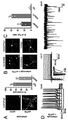

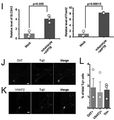


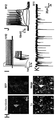


Classifications
-
- C—CHEMISTRY; METALLURGY
- C12—BIOCHEMISTRY; BEER; SPIRITS; WINE; VINEGAR; MICROBIOLOGY; ENZYMOLOGY; MUTATION OR GENETIC ENGINEERING
- C12N—MICROORGANISMS OR ENZYMES; COMPOSITIONS THEREOF; PROPAGATING, PRESERVING, OR MAINTAINING MICROORGANISMS; MUTATION OR GENETIC ENGINEERING; CULTURE MEDIA
- C12N5/00—Undifferentiated human, animal or plant cells, e.g. cell lines; Tissues; Cultivation or maintenance thereof; Culture media therefor
- C12N5/06—Animal cells or tissues; Human cells or tissues
- C12N5/0602—Vertebrate cells
- C12N5/0618—Cells of the nervous system
- C12N5/0622—Glial cells, e.g. astrocytes, oligodendrocytes; Schwann cells
-
- A—HUMAN NECESSITIES
- A61—MEDICAL OR VETERINARY SCIENCE; HYGIENE
- A61P—SPECIFIC THERAPEUTIC ACTIVITY OF CHEMICAL COMPOUNDS OR MEDICINAL PREPARATIONS
- A61P25/00—Drugs for disorders of the nervous system
- A61P25/28—Drugs for disorders of the nervous system for treating neurodegenerative disorders of the central nervous system, e.g. nootropic agents, cognition enhancers, drugs for treating Alzheimer's disease or other forms of dementia
-
- A—HUMAN NECESSITIES
- A61—MEDICAL OR VETERINARY SCIENCE; HYGIENE
- A61K—PREPARATIONS FOR MEDICAL, DENTAL OR TOILETRY PURPOSES
- A61K48/00—Medicinal preparations containing genetic material which is inserted into cells of the living body to treat genetic diseases; Gene therapy
-
- A—HUMAN NECESSITIES
- A61—MEDICAL OR VETERINARY SCIENCE; HYGIENE
- A61K—PREPARATIONS FOR MEDICAL, DENTAL OR TOILETRY PURPOSES
- A61K48/00—Medicinal preparations containing genetic material which is inserted into cells of the living body to treat genetic diseases; Gene therapy
- A61K48/005—Medicinal preparations containing genetic material which is inserted into cells of the living body to treat genetic diseases; Gene therapy characterised by an aspect of the 'active' part of the composition delivered, i.e. the nucleic acid delivered
-
- A—HUMAN NECESSITIES
- A61—MEDICAL OR VETERINARY SCIENCE; HYGIENE
- A61P—SPECIFIC THERAPEUTIC ACTIVITY OF CHEMICAL COMPOUNDS OR MEDICINAL PREPARATIONS
- A61P25/00—Drugs for disorders of the nervous system
- A61P25/14—Drugs for disorders of the nervous system for treating abnormal movements, e.g. chorea, dyskinesia
- A61P25/16—Anti-Parkinson drugs
-
- C—CHEMISTRY; METALLURGY
- C12—BIOCHEMISTRY; BEER; SPIRITS; WINE; VINEGAR; MICROBIOLOGY; ENZYMOLOGY; MUTATION OR GENETIC ENGINEERING
- C12N—MICROORGANISMS OR ENZYMES; COMPOSITIONS THEREOF; PROPAGATING, PRESERVING, OR MAINTAINING MICROORGANISMS; MUTATION OR GENETIC ENGINEERING; CULTURE MEDIA
- C12N15/00—Mutation or genetic engineering; DNA or RNA concerning genetic engineering, vectors, e.g. plasmids, or their isolation, preparation or purification; Use of hosts therefor
- C12N15/09—Recombinant DNA-technology
- C12N15/11—DNA or RNA fragments; Modified forms thereof; Non-coding nucleic acids having a biological activity
- C12N15/113—Non-coding nucleic acids modulating the expression of genes, e.g. antisense oligonucleotides; Antisense DNA or RNA; Triplex- forming oligonucleotides; Catalytic nucleic acids, e.g. ribozymes; Nucleic acids used in co-suppression or gene silencing
-
- C—CHEMISTRY; METALLURGY
- C12—BIOCHEMISTRY; BEER; SPIRITS; WINE; VINEGAR; MICROBIOLOGY; ENZYMOLOGY; MUTATION OR GENETIC ENGINEERING
- C12N—MICROORGANISMS OR ENZYMES; COMPOSITIONS THEREOF; PROPAGATING, PRESERVING, OR MAINTAINING MICROORGANISMS; MUTATION OR GENETIC ENGINEERING; CULTURE MEDIA
- C12N5/00—Undifferentiated human, animal or plant cells, e.g. cell lines; Tissues; Cultivation or maintenance thereof; Culture media therefor
- C12N5/06—Animal cells or tissues; Human cells or tissues
- C12N5/0602—Vertebrate cells
- C12N5/0618—Cells of the nervous system
- C12N5/0619—Neurons
-
- C—CHEMISTRY; METALLURGY
- C12—BIOCHEMISTRY; BEER; SPIRITS; WINE; VINEGAR; MICROBIOLOGY; ENZYMOLOGY; MUTATION OR GENETIC ENGINEERING
- C12N—MICROORGANISMS OR ENZYMES; COMPOSITIONS THEREOF; PROPAGATING, PRESERVING, OR MAINTAINING MICROORGANISMS; MUTATION OR GENETIC ENGINEERING; CULTURE MEDIA
- C12N2310/00—Structure or type of the nucleic acid
- C12N2310/10—Type of nucleic acid
- C12N2310/11—Antisense
-
- C—CHEMISTRY; METALLURGY
- C12—BIOCHEMISTRY; BEER; SPIRITS; WINE; VINEGAR; MICROBIOLOGY; ENZYMOLOGY; MUTATION OR GENETIC ENGINEERING
- C12N—MICROORGANISMS OR ENZYMES; COMPOSITIONS THEREOF; PROPAGATING, PRESERVING, OR MAINTAINING MICROORGANISMS; MUTATION OR GENETIC ENGINEERING; CULTURE MEDIA
- C12N2310/00—Structure or type of the nucleic acid
- C12N2310/10—Type of nucleic acid
- C12N2310/14—Type of nucleic acid interfering N.A.
-
- C—CHEMISTRY; METALLURGY
- C12—BIOCHEMISTRY; BEER; SPIRITS; WINE; VINEGAR; MICROBIOLOGY; ENZYMOLOGY; MUTATION OR GENETIC ENGINEERING
- C12N—MICROORGANISMS OR ENZYMES; COMPOSITIONS THEREOF; PROPAGATING, PRESERVING, OR MAINTAINING MICROORGANISMS; MUTATION OR GENETIC ENGINEERING; CULTURE MEDIA
- C12N2310/00—Structure or type of the nucleic acid
- C12N2310/30—Chemical structure
- C12N2310/31—Chemical structure of the backbone
- C12N2310/315—Phosphorothioates
-
- C—CHEMISTRY; METALLURGY
- C12—BIOCHEMISTRY; BEER; SPIRITS; WINE; VINEGAR; MICROBIOLOGY; ENZYMOLOGY; MUTATION OR GENETIC ENGINEERING
- C12N—MICROORGANISMS OR ENZYMES; COMPOSITIONS THEREOF; PROPAGATING, PRESERVING, OR MAINTAINING MICROORGANISMS; MUTATION OR GENETIC ENGINEERING; CULTURE MEDIA
- C12N2310/00—Structure or type of the nucleic acid
- C12N2310/50—Physical structure
- C12N2310/53—Physical structure partially self-complementary or closed
- C12N2310/531—Stem-loop; Hairpin
-
- C—CHEMISTRY; METALLURGY
- C12—BIOCHEMISTRY; BEER; SPIRITS; WINE; VINEGAR; MICROBIOLOGY; ENZYMOLOGY; MUTATION OR GENETIC ENGINEERING
- C12N—MICROORGANISMS OR ENZYMES; COMPOSITIONS THEREOF; PROPAGATING, PRESERVING, OR MAINTAINING MICROORGANISMS; MUTATION OR GENETIC ENGINEERING; CULTURE MEDIA
- C12N2501/00—Active agents used in cell culture processes, e.g. differentation
- C12N2501/999—Small molecules not provided for elsewhere
-
- C—CHEMISTRY; METALLURGY
- C12—BIOCHEMISTRY; BEER; SPIRITS; WINE; VINEGAR; MICROBIOLOGY; ENZYMOLOGY; MUTATION OR GENETIC ENGINEERING
- C12N—MICROORGANISMS OR ENZYMES; COMPOSITIONS THEREOF; PROPAGATING, PRESERVING, OR MAINTAINING MICROORGANISMS; MUTATION OR GENETIC ENGINEERING; CULTURE MEDIA
- C12N2740/00—Reverse transcribing RNA viruses
- C12N2740/00011—Details
- C12N2740/10011—Retroviridae
- C12N2740/16011—Human Immunodeficiency Virus, HIV
- C12N2740/16041—Use of virus, viral particle or viral elements as a vector
- C12N2740/16043—Use of virus, viral particle or viral elements as a vector viral genome or elements thereof as genetic vector
Abstract
本明細書で提供されるのは、非神経細胞をニューロンに再プログラミングする方法である。本開示の態様は、細胞再プログラミング剤を使用して、非神経細胞をニューロンに変換するためにPTBの発現または活性を抑制することに関する。また、in vivoで非神経細胞を機能性ニューロンに再プログラミングすることによって神経変性疾患を治療する方法も本明細書で提供される。Provided herein is a method of reprogramming non-neuronal cells into neurons. Aspects of the present disclosure relate to the use of cell reprogramming agents to suppress the expression or activity of PTB in order to convert non-neuronal cells into neurons. Also provided herein are methods of treating neurodegenerative diseases by reprogramming non-neuronal cells into functional neurons in vivo.
Description
本願は、米国特許法第35条第119項に基づいて、2018年4月11日に出願された仮出願シリアル番号62/656,322及び2018年8月14日に出願された仮出願シリアル番号62/718,774の優先権を主張し、それらの開示は参照により本明細書に組み込まれる。 This application is based on Article 35, paragraph 119 of the US Patent Act, provisional application serial number 62 / 656,322 filed on April 11, 2018 and provisional application serial number 62 / filed on August 14, 2018. 718,774 priority is claimed and their disclosure is incorporated herein by reference.
本発明は、国立衛生研究所により授与された認可番号5R01GM052872及び5R01HG004659の政府援助を受けて行われた。政府は本発明に一定の権利を有する。 The present invention has been made with the government assistance of authorization numbers 5R01GM052872 and 5R01HG004659 granted by the National Institutes of Health. The government has certain rights to the present invention.
本開示は、非神経細胞を神経細胞に分化するための方法及び組成物、ならびに神経変性疾患または障害を治療するための方法に関する。 The present disclosure relates to methods and compositions for differentiating non-neuronal cells into neurons, as well as methods for treating neurodegenerative diseases or disorders.
「Sequence_ST25.txt」と題する配列表が本願に付随している。これは、2019年4月11日に作成され、1,595バイトのデータを有し、IBM-PCのMS-Windows OSでフォーマットされたものである。該配列表は、あらゆる目的のためにその全体が参照により本明細書に組み込まれる。 A sequence listing entitled "Sequence_ST25.txt" is attached to this application. It was created on April 11, 2019, has 1,595 bytes of data, and is formatted on the IBM-PC MS-Windows OS. The sequence listing is incorporated herein by reference in its entirety for all purposes.
再生医療は、細胞喪失の障害に対処するための大きな期待を持っている。1つのアプローチは細胞置換を採用し、別のアプローチは細胞の分化転換を利用する。細胞置換は、造血障害の治療において目覚ましい成功を収めている。しかし、他の疾患では、このアプローチは限られた有効性を示しているか、免疫反応及び/または腫瘍形成を引き起こすリスクに関連している。対照的に、分化転換は、内因性細胞の既存の細胞可塑性を利用して新しい細胞型を生成することができる。課題は、培養だけでなく、より重要なことに、in vivoのネイティブコンテキストで細胞をある細胞型から別の細胞型に変換するための効率的な戦略を特定することである。 Regenerative medicine holds great promise for dealing with impaired cell loss. One approach employs cell replacement and the other utilizes transdifferentiation of cells. Cell replacement has been remarkably successful in the treatment of hematopoietic disorders. However, in other diseases, this approach has shown limited efficacy or is associated with an immune response and / or risk of causing tumorigenesis. In contrast, transdifferentiation can take advantage of the existing cell plasticity of endogenous cells to generate new cell types. The challenge is not only to culture, but more importantly, to identify efficient strategies for transforming cells from one cell type to another in an in vivo native context.
本開示は、星状細胞を、ポリピリミジントラクト結合(PTB)タンパク質の発現を阻害する阻害性核酸分子と接触させることにより星状細胞を神経細胞に変換することによって生成される神経細胞を提供する。一実施形態またはさらなる実施形態では、前記神経細胞はin vitroで生成される。一実施形態またはさらなる実施形態では、前記神経細胞はin vivoで生成される。一実施形態またはさらなる実施形態では、前記阻害性核酸分子は、RNAi分子である。一実施形態またはさらなる実施形態では、前記RNAi分子はshRNAである。一実施形態またはさらなる実施形態では、前記阻害性核酸分子はアンチセンス分子である。一実施形態またはさらなる実施形態では、前記阻害性核酸分子は、ウイルスまたはウイルスベクター内にある。一実施形態またはさらなる実施形態では、前記ウイルスまたはウイルスベクターは、アデノウイルス、AAV、レトロウイルス、またはレンチウイルスである。一実施形態またはさらなる実施形態では、前記星状細胞はヒト星状細胞である。 The present disclosure provides neurons produced by converting astrocytes into neurons by contacting the astrocytes with an inhibitory nucleic acid molecule that inhibits the expression of polypyrimidine tract binding (PTB) proteins. .. In one embodiment or a further embodiment, the nerve cells are produced in vitro. In one embodiment or a further embodiment, the nerve cells are generated in vivo. In one embodiment or a further embodiment, the inhibitory nucleic acid molecule is an RNAi molecule. In one embodiment or a further embodiment, the RNAi molecule is shRNA. In one embodiment or a further embodiment, the inhibitory nucleic acid molecule is an antisense molecule. In one embodiment or a further embodiment, the inhibitory nucleic acid molecule is in a virus or viral vector. In one embodiment or a further embodiment, the virus or viral vector is an adenovirus, AAV, retrovirus, or lentivirus. In one embodiment or a further embodiment, the astrocyte is a human astrocyte.
本開示は、神経変性疾患または障害を治療する方法を提供する。これは、神経変性疾患または障害を有する被験者の星状細胞をPTBの発現を阻害する阻害性核酸と接触させ、それによって星状細胞をニューロンに分化させ、神経変性疾患または障害を治療することを含む。一実施形態またはさらなる実施形態では、前記阻害性核酸は、神経変性疾患または障害に関連する被験者の脳の中枢神経系の部位に送達される。一実施形態またはさらなる実施形態では、前記被験者は哺乳動物である。一実施形態またはさらなる実施形態では、前記哺乳動物はヒトである。一実施形態またはさらなる実施形態では、前記神経変性疾患または障害は、虚血性及び出血性脳卒中、脊髄損傷、脳損傷、ハンチントン病、アルツハイマー病、パーキンソン病、統合失調症、自閉症、運動失調、筋萎縮性側索硬化症、ルーゲーリック病、ライム病、髄膜炎、片頭痛、運動ニューロン疾患、神経障害、痛み、脳損傷、脳機能障害、脊髄障害、末梢神経系障害、頭蓋神経障害、自律神経系障害、てんかん等の発作障害、パーキンソン病等の運動障害、睡眠障害、頭痛、腰痛、首の痛み、神経障害性疼痛、アルツハイマー病、めまいと回転性めまい、昏迷と昏睡、頭の怪我、脳卒中、神経系の腫瘍、多発性硬化症及びその他の脱髄性疾患、脳または脊髄の感染症、プリオン病等のせん妄や認知症からなる群より選択される。 The present disclosure provides a method of treating a neurodegenerative disease or disorder. It contacts the astrocytes of a subject with a neurodegenerative disease or disorder with an inhibitory nucleic acid that inhibits PTB expression, thereby differentiating the astrocytes into neurons and treating the neurodegenerative disease or disorder. include. In one embodiment or a further embodiment, the inhibitory nucleic acid is delivered to a site of the central nervous system of the subject's brain associated with a neurodegenerative disease or disorder. In one embodiment or a further embodiment, the subject is a mammal. In one embodiment or a further embodiment, the mammal is a human. In one embodiment or a further embodiment, the neurodegenerative disease or disorder is ischemic and hemorrhagic stroke, spinal cord injury, brain injury, Huntington's disease, Alzheimer's disease, Parkinson's disease, schizophrenia, autism, ataxia, Muscle atrophic lateral sclerosis, Lugeric's disease, Lime's disease, meningitis, migraine, motor neuron disease, neuropathy, pain, brain damage, brain dysfunction, spinal cord disorder, peripheral nervous system disorder, cranial neuropathy, autonomy Nervous system disorder, seizure disorder such as epilepsy, motor disorder such as Parkinson's disease, sleep disorder, headache, lower back pain, neck pain, neuropathy pain, Alzheimer's disease, dizziness and rotational dizziness, coma and coma, head injury, It is selected from the group consisting of stroke, neurological tumors, multiple sclerosis and other demyelinating diseases, brain or spinal cord infections, dementia and dementia such as prion disease.
また本開示は、神経変性疾患または障害を有する被験者を治療する方法を提供する。これは、星状細胞をニューロンに分化させ、ニューロンを神経変性の部位で被験者に移植することを含む。ここで、前記星状細胞は、PTBの発現を阻害することによってニューロンに変換される。一実施形態またはさらなる実施形態では、前記星状細胞は、神経変性疾患または障害の部位で被験者から得られ、ex vivoで分化される。 The disclosure also provides a method of treating a subject with a neurodegenerative disease or disorder. This involves differentiating astrocytes into neurons and transplanting the neurons into subjects at the site of neurodegeneration. Here, the astrocyte is converted into a neuron by inhibiting the expression of PTB. In one embodiment or a further embodiment, the astrocytes are obtained from a subject at the site of a neurodegenerative disease or disorder and differentiate ex vivo.
また本開示は、神経変性疾患の治療または阻害における活性を有する、薬物または化合物をスクリーニングする方法を提供する。これは、神経変性疾患を有する被験者から星状細胞を取得し、星状細胞を、PTB発現を阻害する阻害性核酸と接触させることによって星状細胞を神経細胞に分化させ、神経細胞を薬物または化合物と接触させ、薬物または化合物が神経変性疾患または障害の疾患マーカーの発現を阻害するかどうかを測定することを含む。 The present disclosure also provides a method of screening a drug or compound having activity in the treatment or inhibition of a neurodegenerative disease. It obtains astrocytes from a subject with a neurodegenerative disease and differentiates the astrocytes into neurons by contacting the astrocytes with an inhibitory nucleic acid that inhibits PTB expression, drugging the neurons or It involves contacting the compound and measuring whether the drug or compound inhibits the expression of disease markers for neurodegenerative diseases or disorders.
一実施形態またはさらなる実施形態では、前記阻害性核酸は、配列番号1または2と少なくとも80〜100%同一であり、及び/またはTがUである配列を含む。 In one embodiment or a further embodiment, the inhibitory nucleic acid comprises a sequence that is at least 80-100% identical to SEQ ID NO: 1 or 2 and / or T is U.
本開示はまた、アンチセンス分子を提供する。該アンチセンス分子は、配列番号2と少なくとも98%同一であり、PTB発現を阻害し、及び/またはTがUである配列を含む。 The present disclosure also provides antisense molecules. The antisense molecule is at least 98% identical to SEQ ID NO: 2, inhibits PTB expression, and / or comprises a sequence where T is U.
本開示はまた、ベクターを提供する。該ベクターは、配列番号1または2に記載の配列、または前記配列と少なくとも80〜99%同一であり、及び/またはTが前記配列(例えば、DNAまたはRNA)においてUである配列を含み、PTB発現を阻害する。 The present disclosure also provides a vector. The vector comprises the sequence set forth in SEQ ID NO: 1 or 2, or at least 80-99% identical to said sequence, and / or a sequence in which T is U in said sequence (eg, DNA or RNA), PTB. Inhibits expression.
特定の実施形態では、本明細書に開示されるのは、ヒト非神経細胞を成熟ニューロンに再プログラミングする方法である。いくつかの実施形態では、該方法は、ヒト成人線維芽細胞で発現されるレベルよりも高いレベルでmiR-9またはBrn2を発現するヒト非神経細胞を提供し、該細胞を、ヒト非神経細胞におけるPTBの発現または活性を抑制する細胞プログラミング剤を含む組成物と接触させ、それにより、ヒト非神経細胞を成熟ニューロンに再プログラミングすることを含む。 In certain embodiments, what is disclosed herein is a method of reprogramming human non-neuronal cells into mature neurons. In some embodiments, the method provides human nonneurons expressing miR-9 or Brn2 at levels higher than those expressed in human adult fibroblasts, and the cells are referred to as human nonneurons. It involves contacting with a composition comprising a cell programming agent that suppresses the expression or activity of PTB in, thereby reprogramming human non-neuronal cells into mature neurons.
いくつかの実施形態では、前記ヒト非神経細胞は、miR-9またはBrn2を、ヒトの成人線維芽細胞で発現するレベルの少なくとも2倍のレベルで発現する。いくつかの実施形態では、前記ヒト非神経細胞は、ヒト成人線維芽細胞において発現されるレベルよりも少なくとも10倍高いレベルでmiR-9またはBrn2を発現する。いくつかの実施形態では、前記ヒト非神経細胞は、ヒト成人線維芽細胞において発現されるレベルよりも高いレベルでmiR-9及びBrn2を発現する。 In some embodiments, the human non-neuronal cells express miR-9 or Brn2 at a level that is at least twice the level expressed in human adult fibroblasts. In some embodiments, the human non-neuronal cells express miR-9 or Brn2 at levels that are at least 10-fold higher than those expressed in human adult fibroblasts. In some embodiments, the human non-neuronal cells express miR-9 and Brn2 at levels higher than those expressed in human adult fibroblasts.
特定の実施形態では、本明細書に開示されるのは、ヒトグリア細胞を成熟ニューロンに再プログラミングする方法である。該方法は、再プログラムされるヒトグリア細胞を提供し、ヒトグリア細胞を、ヒトグリア細胞におけるPTBの発現または活性を少なくとも1日間抑制する細胞プログラミング剤を含む組成物と接触させ、それにより、ヒトグリア細胞を成熟ニューロンに再プログラミングすることを含む。 In certain embodiments, what is disclosed herein is a method of reprogramming human glial cells into mature neurons. The method provides reprogrammed human glial cells and contacts the human glial cells with a composition comprising a cell programming agent that suppresses PTB expression or activity in the human glial cells for at least 1 day, thereby maturing the human glial cells. Includes reprogramming into neurons.
いくつかの実施形態では、前記ヒトグリア細胞は、星状細胞、希突起膠細胞、上衣細胞、シュワン細胞、ミクログリア、及び衛星細胞からなる群から選択される。いくつかの実施形態では、前記ヒトグリア細胞は、GFAP(グリア線維性酸性タンパク質)またはALDH1L1(アルデヒドデヒドロゲナーゼ1ファミリーメンバーL1)に対して陽性である。
In some embodiments, the human glial cells are selected from the group consisting of astrocytes, oligodendrocytes, ependymal cells, Schwann cells, microglia, and satellite cells. In some embodiments, the human glial cells are positive for GFAP (glial fibrous acidic protein) or ALDH1L1 (
特定の実施形態では、本明細書に開示されるのは、星状細胞を成熟ニューロンに再プログラミングする方法である。該方法は、再プログラムされる星状細胞を提供し、星状細胞を、星状細胞におけるPTBの発現または活性を少なくとも1日間抑制する細胞プログラミング剤を含む組成物と接触させ、それにより、星状細胞を成熟ニューロンに再プログラミングすることを含む。 In certain embodiments, what is disclosed herein is a method of reprogramming astrocytes into mature neurons. The method provides reprogrammed astrocytes, which are contacted with a composition comprising a cell programming agent that suppresses the expression or activity of PTB in the astrocytes for at least 1 day, thereby causing the astrocytes. Includes reprogramming astrocytes into mature neurons.
いくつかの実施形態では、前記星状細胞は、マウス星状細胞である。いくつかの実施形態では、該方法は、複数のマウス星状細胞を再プログラムし、該マウス星状細胞の少なくとも60%が、Tuj1陽性の成熟ニューロンに変換される。いくつかの実施形態では、該方法は、複数のマウス星状細胞を再プログラムし、該マウス星状細胞の少なくとも40%が、Map2陽性の成熟ニューロンに変換される。 In some embodiments, the astrocyte is a mouse astrocyte. In some embodiments, the method reprograms multiple mouse astrocytes, converting at least 60% of the mouse astrocytes into Tuj1-positive mature neurons. In some embodiments, the method reprograms multiple mouse astrocytes and at least 40% of the mouse astrocytes are converted to Map2-positive mature neurons.
いくつかの実施形態では、前記星状細胞は、ヒト星状細胞である。いくつかの実施形態では、該方法は、複数のヒト星状細胞を再プログラムし、該ヒト星状細胞の少なくとも40%、少なくとも60%、または少なくとも80%が、Tuj1陽性の成熟ニューロンに変換される。いくつかの実施形態では、該方法は、複数のヒト星状細胞を再プログラムし、該ヒト星状細胞の少なくとも20%、少なくとも40%、または少なくとも60%が、Map2陽性の成熟ニューロンに変換される。 In some embodiments, the astrocyte is a human astrocyte. In some embodiments, the method reprograms multiple human astrocytes and at least 40%, at least 60%, or at least 80% of the human astrocytes are converted to Tuj1-positive mature neurons. NS. In some embodiments, the method reprograms multiple human astrocytes and at least 20%, at least 40%, or at least 60% of the human astrocytes are converted to Map2-positive mature neurons. NS.
いくつかの実施形態では、前記組成物は、PTBの発現または活性を特異的に抑制する単一の細胞プログラミング剤を含む。 In some embodiments, the composition comprises a single cell programming agent that specifically suppresses PTB expression or activity.
特定の実施形態では、本明細書に開示されるのは、ヒト非神経細胞を成熟ニューロンに再プログラミングする方法である。該方法は、再プログラムされるヒト非神経細胞を提供し、ヒト非神経細胞を、ヒト非神経細胞におけるPTBの発現または活性を少なくとも3日間抑制する単一の細胞プログラミング剤を含む組成物と接触させ、それにより、ヒト非神経細胞を成熟ニューロンに再プログラミングすることを含む。 In certain embodiments, what is disclosed herein is a method of reprogramming human non-neuronal cells into mature neurons. The method provides reprogrammed human nonneurons and contacts human nonneurons with a composition comprising a single cell programming agent that suppresses PTB expression or activity in human nonneurons for at least 3 days. It involves reprogramming human non-neuronal cells into mature neurons.
特定の実施形態では、本明細書に開示されるのは、ヒト非神経細胞を再プログラミングする方法である。該方法は、再プログラムされるヒト非神経細胞を提供し、ヒト非神経細胞をヒト非神経細胞におけるPTBの発現または活性を低下し、PTBの発現または活性が低下した後のnPTBの発現または活性を低下する単一細胞プログラミング剤を含む組成物と接触させることを含む。 In certain embodiments, what is disclosed herein is a method of reprogramming human non-neuronal cells. The method provides reprogrammed human non-neurons, reducing PTB expression or activity in human non-neurons, and nPTB expression or activity after reduced PTB expression or activity. Includes contact with a composition comprising a single cell programming agent that reduces.
いくつかの実施形態では、初期のnPTB発現レベルは、PTBの発現または活性が抑制されるにつれて、高いnPTB発現レベルに増加する。いくつかの実施形態では、nPTB発現は、PTBの発現または活性が抑制された後、高いnPTB発現レベルから、初期のnPTB発現レベルよりも高い、低いnPTB発現レベルに減少する。いくつかの実施形態では、前記ヒト非神経細胞は、ヒト成人線維芽細胞において発現されるレベルよりも高いレベルでmiR-9またはBrn2を発現する。いくつかの実施形態では、前記ヒト非神経細胞は、ヒト成人線維芽細胞において発現されるレベルよりも高いレベルでmiR-9及びBrn2を発現する。 In some embodiments, early nPTB expression levels increase to higher nPTB expression levels as PTB expression or activity is suppressed. In some embodiments, nPTB expression is reduced from a high nPTB expression level to a low nPTB expression level, which is higher than the initial nPTB expression level, after suppression of PTB expression or activity. In some embodiments, the human non-neuronal cells express miR-9 or Brn2 at levels higher than those expressed in human adult fibroblasts. In some embodiments, the human non-neuronal cells express miR-9 and Brn2 at levels higher than those expressed in human adult fibroblasts.
いくつかの実施形態では、該方法は、複数のヒト非神経細胞、ヒトグリア細胞、または星状細胞を再プログラムする。ここで、ヒト非神経細胞、ヒトグリア細胞、または星状細胞の少なくとも40%が、NeuN(神経核抗原)、Map2(微小管関連タンパク質2)、NSE(ニューロン特異的エノラーゼ)、160 kDaニューロフィラメント培地、200kDaニューロフィラメントヘビー、PDS-95(シナプス後肥厚タンパク質95)、シナプシンI、シナプトフィジン、GAD67(グルタミン酸デカルボキシラーゼ67)、GAD65(グルタミン酸デカルボキシラーゼ67)、パルブアルブミン、DARPP32(ドーパミン及びcAMP調節ニューロンリンタンパク質32)、vGLUT1(小胞グルタミン酸トランスポーター1)、vGLUT2(小胞グルタメートトランスポーター1)、アセチルコリン、及びTH(チロシンヒドロキシラーゼ)からなる群から選択された1つまたは複数のニューロンマーカーの発現を特徴とする成熟ニューロンに再プログラムされる。 In some embodiments, the method reprograms multiple human non-neuronal cells, human glial cells, or astrocytes. Here, at least 40% of human non-neuronal cells, human glial cells, or stellate cells are NeuN (neuronuclear antigen), Map2 (microtube-related protein 2), NSE (neuron-specific enolase), 160 kDa neurofilament medium. , 200 kDa Neurofilament Heavy, PDS-95 (Post-synaptic thickening protein 95), Synapsin I, Synaptophysin, GAD67 (Glutamate decarboxylase 67), GAD65 (Glutamate decarboxylase 67), Parbualbumin, DARPP32 (Dopamine and cAMP-regulated neuronal phosphorus protein) 32) Characterized by the expression of one or more neuronal markers selected from the group consisting of vGLUT1 (follicle glutamate transporter 1), vGLUT2 (vesicle glutamate transporter 1), acetylcholine, and TH (tyrosinylhydroxylase). Is reprogrammed into mature neurons.
いくつかの実施形態では、該方法は、複数のヒト非神経細胞、ヒトグリア細胞、または星状細胞を再プログラムする。ここで、ヒト非神経細胞、ヒトグリア細胞、または星状細胞の少なくとも20%は、活動電位、シナプス結合、シナプス前神経伝達物質放出、及び/またはシナプス後応答を確立する能力を特徴とする機能性ニューロンに再プログラムされる。 In some embodiments, the method reprograms multiple human non-neuronal cells, human glial cells, or astrocytes. Here, at least 20% of human nonneurons, human glial cells, or astrocytes are functional characterized by action potentials, synaptic connections, presynaptic neurotransmitter release, and / or the ability to establish postsynaptic responses. Reprogrammed into neurons.
いくつかの実施形態では、前記細胞プログラミング剤は、抗PTB阻害剤である。いくつかの実施形態では、前記抗PTB阻害剤は、抗PTBアンチセンスオリゴヌクレオチドである。いくつかの実施形態では、前記抗PTB阻害剤は、抗PTB shRNA、抗PTB miRNA、抗PTBアンチセンスオリゴヌクレオチド、抗PTB抗体、PTBの小分子阻害剤、ドミナントネガティブPTB変異体、及びポリピリミジントラクトを含むスポンジポリリボヌクレオチドからなる群から選択される。 In some embodiments, the cell programming agent is an anti-PTB inhibitor. In some embodiments, the antiPTB inhibitor is an antiPTB antisense oligonucleotide. In some embodiments, the anti-PTB inhibitor is an anti-PTB shRNA, an anti-PTB miRNA, an anti-PTB antisense oligonucleotide, an anti-PTB antibody, a small molecule inhibitor of PTB, a dominant negative PTB variant, and a polypyrimidine tract. Selected from the group consisting of sponge polyribonucleotides containing.
いくつかの実施形態では、前記細胞プログラミング剤は、PTBの発現または活性を少なくとも5日間抑制する。いくつかの実施形態では、前記細胞プログラミング剤は、PTBの発現または活性を少なくとも10日間抑制する。いくつかの実施形態では、前記細胞プログラミング剤は、PTBの発現または活性を少なくとも15日間抑制する。 In some embodiments, the cell programming agent suppresses PTB expression or activity for at least 5 days. In some embodiments, the cell programming agent suppresses PTB expression or activity for at least 10 days. In some embodiments, the cell programming agent suppresses PTB expression or activity for at least 15 days.
いくつかの実施形態では、前記ヒト非神経細胞、ヒトグリア細胞、または星状細胞は、培地中で培養される。いくつかの実施形態では、該培地は、ALK5の阻害剤、GSK3bの阻害剤、PKAの活性化因子、及びそれらの任意の組み合わせからなる群から選択される薬剤を含む。いくつかの実施形態では、前記活性化因子ALK5は、SB431542を含む。いくつかの実施形態では、前記阻害剤GSK3bは、CHIR99021を含む。いくつかの実施形態では、前記PKAの活性化因子は、ジブチリルアデノシン3',5'-サイクリックモノホスフェート(Db-cAMP)を含む。いくつかの実施形態では、前記細胞プログラミング剤は、レンチウイルスベクターで送達される。 In some embodiments, the human non-neuronal cells, human glial cells, or astrocytes are cultured in medium. In some embodiments, the medium comprises an agent selected from the group consisting of an inhibitor of ALK5, an inhibitor of GSK3b, an activator of PKA, and any combination thereof. In some embodiments, the activator ALK5 comprises SB431542. In some embodiments, the inhibitor GSK3b comprises CHIR99021. In some embodiments, the PKA activator comprises dibutyryl adenosine 3', 5'-cyclic monophosphate (Db-cAMP). In some embodiments, the cell programming agent is delivered with a lentiviral vector.
特定の実施形態では、本明細書に開示されるのは、in vivoで機能性ニューロンを生成する方法である。該方法は、脳内の星状細胞におけるPTBの発現または活性を抑制し、星状細胞が機能性ニューロンに再プログラミングできるようにする細胞プログラミング剤を含む組成物を被験者の脳に投与することを含む。 In certain embodiments, what is disclosed herein is a method of generating functional neurons in vivo. The method involves administering to the subject's brain a composition containing a cell programming agent that suppresses the expression or activity of PTB in astrocytes in the brain and allows the astrocytes to be reprogrammed into functional neurons. include.
特定の実施形態では、本明細書に開示されるのは、in vivoで機能性ニューロンを生成する方法である。該方法は、中脳の非神経細胞におけるPTBの発現または活性を抑制し、非神経細胞が機能性ニューロンに再プログラミングできるようにする細胞プログラミング剤を含む組成物を被験者の中脳に投与することを含む。 In certain embodiments, what is disclosed herein is a method of generating functional neurons in vivo. The method comprises administering to the midbrain of a subject a composition comprising a cell programming agent that suppresses the expression or activity of PTB in non-neuronal cells of the midbrain and allows the non-neuronal cells to be reprogrammed into functional neurons. including.
特定の実施形態では、本明細書に開示されるのは、in vivoでドーパミン作動性ニューロンを生成する方法である。該方法は、脳内の非神経細胞におけるPTBの発現または活性を抑制し、非神経細胞がドーパミン作動性ニューロンに再プログラミングできるようにする細胞プログラミング剤を含む組成物を被験者の脳に投与することを含む。 In certain embodiments, what is disclosed herein is a method of producing dopaminergic neurons in vivo. The method comprises administering to the subject's brain a composition comprising a cell programming agent that suppresses the expression or activity of PTB in non-neuronal cells in the brain and allows the non-neuronal cells to be reprogrammed into dopaminergic neurons. including.
いくつかの実施形態では、前記ドーパミン作動性ニューロンは、チロシンヒドロキシラーゼ(TH)、ドーパミントランスポーター(DAT/SLC6A3)、小胞モノアミントランスポーター2(VMAT2)、エングレイルド(engrailed)ホメオボックス1(En1)、FoxA2、及び/またはLIMホメオボックス転写因子1アルファ(Lmx1a)を発現する。
In some embodiments, the dopaminergic neurons are tyrosine hydroxylase (TH), dopamine transporter (DAT / SLC6A3), vesicular monoamine transporter 2 (VMAT2), engrailed homeobox 1 (En1). , FoxA2, and / or LIM
いくつかの実施形態では、該方法は、脳内の複数の非神経細胞を再プログラムする。ここで、前記非神経細胞の少なくとも10%がドーパミン作動性ニューロンに変換される。いくつかの実施形態では、該方法は、脳内の複数の非神経細胞を再プログラムする。ここで、前記非神経細胞の少なくとも30%がドーパミン作動性ニューロンに変換される。 In some embodiments, the method reprograms multiple non-neuronal cells in the brain. Here, at least 10% of the non-neuronal cells are converted to dopaminergic neurons. In some embodiments, the method reprograms multiple non-neuronal cells in the brain. Here, at least 30% of the non-neuronal cells are converted to dopaminergic neurons.
いくつかの実施形態では、機能性ニューロンまたはドーパミン作動性ニューロンの軸索終末は、被験者の線条体に到達する。 In some embodiments, the axon terminals of functional or dopaminergic neurons reach the striatum of the subject.
いくつかの実施形態では、前記細胞プログラミング剤は、被験者の黒質に投与される。 In some embodiments, the cell programming agent is administered to the substantia nigra of the subject.
いくつかの実施形態では、前記被験者はヒトである。 In some embodiments, the subject is a human.
いくつかの実施形態では、前記被験者は非ヒト動物である。 In some embodiments, the subject is a non-human animal.
特定の実施形態では、本明細書に開示されるのは、in vivoで機能性ニューロンを生成する方法である。該方法は、脳内の非神経細胞におけるPTBの発現または活性を抑制し、非神経細胞が機能性ニューロンに再プログラミングできるようにする細胞プログラミング剤をヒト被験者の脳に投与することを含む。 In certain embodiments, what is disclosed herein is a method of generating functional neurons in vivo. The method comprises administering to the brain of a human subject a cell programming agent that suppresses the expression or activity of PTB in non-neuronal cells in the brain and allows the non-neuronal cells to be reprogrammed into functional neurons.
いくつかの実施形態では、前記非神経細胞は、グリア細胞である。いくつかの実施形態では、該グリア細胞は、星状細胞である。 In some embodiments, the non-neuronal cell is a glial cell. In some embodiments, the glial cell is an astrocyte.
いくつかの実施形態では、前記細胞プログラミング剤は、ヒト被験者の中脳、線条体、または皮質に投与される。いくつかの実施形態では、前記細胞プログラミング剤は、ヒト被験者の黒質に投与される。 In some embodiments, the cell programming agent is administered to the midbrain, striatum, or cortex of a human subject. In some embodiments, the cell programming agent is administered to the substantia nigra of a human subject.
いくつかの実施形態では、前記機能性ニューロンは、ドーパミン作動性ニューロンである。いくつかの実施形態では、前記機能性ニューロンは、チロシンヒドロキシラーゼ(TH)、ドーパミントランスポーター(DAT)、小胞モノアミントランスポーター2(VMAT2)、エングレイルド(engrailed)ホメオボックス1(En1)、フォークヘッドボックスA2(FoxA2)、及び/またはLIMホメオボックス転写因子1アルファ(Lmx1a)を発現する。
In some embodiments, the functional neuron is a dopaminergic neuron. In some embodiments, the functional neurons are tyrosine hydroxylase (TH), dopamine transporter (DAT), vesicular monoamine transporter 2 (VMAT2), engrailed homeobox 1 (En1), forkhead. Expresses box A2 (FoxA2) and / or LIM
いくつかの実施形態では、前記機能性ニューロンまたはドーパミン作動性ニューロンは、シナプス前神経伝達物質を示す。いくつかの実施形態では、前記機能性ニューロンまたはドーパミン作動性ニューロンは、脳内の既存のニューロン回路に統合される。 In some embodiments, the functional or dopaminergic neurons exhibit presynaptic neurotransmitters. In some embodiments, the functional or dopaminergic neurons are integrated into existing neuronal circuits in the brain.
いくつかの実施形態では、該方法は、脳内の複数の非神経細胞または星状細胞を再プログラムする。ここで、前記非神経細胞または星状細胞の少なくとも30%が、ニューロン核抗原(NeuN)、微小管関連タンパク質2(Map2)、ニューロン特異的エノラーゼ(NSE)、160 kDaニューロフィラメント培地、200kDaニューロフィラメントヘビー、シナプス後肥厚タンパク質95(PDS-95)、シナプシンI、シナプトフィジン、グルタミン酸デカルボキシラーゼ67(GAD67)、グルタミン酸デカルボキシラーゼ67(GAD65)、パルブアルブミン、ドーパミン及びcAMP調節ニューロンリン酸化タンパク質32(DARPP32)、小胞グルタミン酸トランスポーター1(vGLUT1)、小胞グルタミン酸トランスポーター2(vGLUT2)、アセチルコリン、及びチロシンヒドロキシラーゼ(TH)からなる群から選択された1つまたは複数のニューロンマーカーの発現を特徴とする成熟ニューロンに再プログラムされる。 In some embodiments, the method reprograms multiple non-neuronal or astrocyte cells in the brain. Here, at least 30% of the non-neuronal cells or stellate cells are neuronal nuclear antigen (NeuN), microtube-related protein 2 (Map2), neuron-specific enolase (NSE), 160 kDa neurofilament medium, 200 kDa neurofilament. Heavy, post-synaptic thickening protein 95 (PDS-95), synapsin I, synaptophysin, glutamate decarboxylase 67 (GAD67), glutamate decarboxylase 67 (GAD65), parbualbumin, dopamine and cAMP regulatory neuronal phosphorylated protein 32 (DARPP32), Maturation characterized by the expression of one or more neuronal markers selected from the group consisting of follicular glutamate transporter 1 (vGLUT1), follicular glutamate transporter 2 (vGLUT2), acetylcholine, and tyrosine hydroxylase (TH). Reprogrammed into neurons.
いくつかの実施形態では、該方法は、脳内の複数の非神経細胞または星状細胞を再プログラムする。ここで、非神経細胞または星状細胞の少なくとも20%が、活動電位、シナプス接続、シナプス前神経伝達物質、及び/またはシナプス後応答を確立する能力を特徴とする機能性ニューロンに再プログラムされる。 In some embodiments, the method reprograms multiple non-neuronal or astrocyte cells in the brain. Here, at least 20% of non-neuronal or astrocyte cells are reprogrammed into functional neurons characterized by the ability to establish action potentials, synaptic connections, presynaptic neurotransmitters, and / or postsynaptic responses. ..
いくつかの実施形態では、前記細胞プログラミング剤は、抗PTBアンチセンスオリゴヌクレオチドを含む。いくつかの実施形態では、前記細胞プログラミング剤は、抗PTB shRNA、抗PTB miRNA、抗PTBアンチセンスオリゴヌクレオチド、抗PTB抗体、PTBの小分子阻害剤、ドミナントネガティブPTB変異体、ポリピリミジントラクトを含むスポンジポリリボヌクレオチド、及びそれらの任意の組み合わせからなる群から選択される。 In some embodiments, the cell programming agent comprises an anti-PTB antisense oligonucleotide. In some embodiments, the cell programming agent comprises an anti-PTB shRNA, an anti-PTB miRNA, an anti-PTB antisense oligonucleotide, an anti-PTB antibody, a small molecule inhibitor of PTB, a dominant negative PTB variant, a polypyrimidine tract. It is selected from the group consisting of sponge polyribonucleotides and any combination thereof.
いくつかの実施形態では、前記細胞プログラミング剤は、PTBの発現または活性を少なくとも5日間抑制する。いくつかの実施形態では、前記細胞プログラミング剤は、PTBの発現または活性を少なくとも10日間抑制する。いくつかの実施形態では、前記細胞プログラミング剤は、PTBの発現または活性を少なくとも15日間抑制する。いくつかの実施形態では、前記細胞プログラミング剤は、AAVベクターで送達される。 In some embodiments, the cell programming agent suppresses PTB expression or activity for at least 5 days. In some embodiments, the cell programming agent suppresses PTB expression or activity for at least 10 days. In some embodiments, the cell programming agent suppresses PTB expression or activity for at least 15 days. In some embodiments, the cell programming agent is delivered with an AAV vector.
特定の実施形態では、本明細書に開示されるのは、脳領域の機能性ニューロンの変性に関連する神経学的状態を治療する方法。該方法は、脳領域の非神経細胞におけるPTBの発現または活性を抑制する細胞プログラミング剤を含む組成物を、それを必要とする被験者の脳領域に投与すること、及び非神経細胞が機能性ニューロンに再プログラムすることを可能にし、それによって脳領域の変性した機能性ニューロンを補充することを含む。 In certain embodiments, disclosed herein are methods of treating neurological conditions associated with degeneration of functional neurons in a brain region. The method comprises administering to the brain region of a subject in need of a composition comprising a cell programming agent that suppresses the expression or activity of PTB in non-neuronal cells of the brain region, and the non-neuronal cells are functional neurons. Includes replenishing degenerated functional neurons in the brain region, thereby allowing it to be reprogrammed into.
いくつかの実施形態では、前記神経学的状態は、パーキンソン病、アルツハイマー病、ハンチントン病、統合失調症、鬱病、及び薬物中毒からなる群から選択される。 In some embodiments, the neurological condition is selected from the group consisting of Parkinson's disease, Alzheimer's disease, Huntington's disease, schizophrenia, depression, and drug addiction.
特定の実施形態では、本明細書に開示されるのは、脳領域におけるドーパミン作動性ニューロンの変性に関連する神経学的状態を治療する方法である。該方法は、必要とする被験者の脳領域に、脳領域の非神経細胞におけるPTBの発現または活性を抑制する細胞プログラミング剤を含む組成物を投与すること、及び非神経細胞がドーパミン作動性ニューロンに再プログラムすることを可能にし、それによって脳領域の変性したドーパミン作動性ニューロンを補充することを含む。 In certain embodiments, disclosed herein are methods of treating neurological conditions associated with degeneration of dopaminergic neurons in a brain region. The method comprises administering to the brain region of the subject in need a composition comprising a cell programming agent that suppresses the expression or activity of PTB in non-neuronal cells of the brain region, and the non-neuronal cells become dopaminergic neurons. It allows for reprogramming, which involves replenishing degenerated dopaminergic neurons in the brain region.
特定の実施形態では、本明細書に開示されるのは、正常レベルと比較してドーパミンの量が減少した被験者のドーパミン生合成を回復する方法である。該方法は、被験者の脳領域に、脳領域の非神経細胞におけるPTBの発現または活性を抑制する細胞プログラミング剤を含む組成物を投与すること、及び非神経細胞がドーパミン作動性ニューロンに再プログラムすることを可能にし、それによって、減少した量のドーパミンの少なくとも50%を回復させることを含む。 In certain embodiments, disclosed herein is a method of restoring dopamine biosynthesis in a subject with reduced dopamine levels compared to normal levels. The method comprises administering to the subject's brain region a composition comprising a cell programming agent that suppresses the expression or activity of PTB in nonneurons in the brain region, and the nonneurons reprogram into dopaminergic neurons. This involves restoring at least 50% of the reduced amount of dopamine.
いくつかの実施形態では、前記非神経細胞は、グリア細胞である。いくつかの実施形態では、前記非神経細胞は、星状細胞である。 In some embodiments, the non-neuronal cell is a glial cell. In some embodiments, the non-neuronal cell is an astrocyte.
いくつかの実施形態では、前記細胞プログラミング剤は、抗PTBアンチセンスオリゴヌクレオチドを含む。いくつかの実施形態では、前記単一の細胞プログラミング剤は、抗PTB shRNA、抗PTB miRNA、抗PTBアンチセンスオリゴヌクレオチド、抗PTB抗体、PTBの小分子阻害剤、ドミナントネガティブPTB変異体、ポリピリミジントラクトを含むスポンジポリリボヌクレオチド、及びそれらの任意の組み合わせからなる群から選択される。 In some embodiments, the cell programming agent comprises an anti-PTB antisense oligonucleotide. In some embodiments, the single cell programming agent is an anti-PTB shRNA, an anti-PTB miRNA, an anti-PTB antisense oligonucleotide, an anti-PTB antibody, a small molecule inhibitor of PTB, a dominant negative PTB variant, a polypyrimidine. It is selected from the group consisting of sponge polyribonucleotides containing tracts, and any combination thereof.
いくつかの実施形態では、前記機能性ニューロンまたはドーパミン作動性ニューロンは、脳領域の既存のニューロン回路に統合されている。 In some embodiments, the functional or dopaminergic neurons are integrated into existing neuronal circuits in the brain region.
いくつかの実施形態では、前記機能性ニューロンまたはドーパミン作動性ニューロンは、活動電位、シナプス前神経伝達物質、及び/またはシナプス後応答を示す。 In some embodiments, the functional or dopaminergic neurons exhibit action potentials, presynaptic neurotransmitters, and / or postsynaptic responses.
いくつかの実施形態では、前記細胞プログラミング剤は、被験者の中脳、線条体、または皮質に投与される。いくつかの実施形態では、前記細胞プログラミング剤は、被験者の黒質に投与される。 In some embodiments, the cell programming agent is administered to the subject's midbrain, striatum, or cortex. In some embodiments, the cell programming agent is administered to the substantia nigra of the subject.
いくつかの実施形態では、前記機能性ニューロンまたはドーパミン作動性ニューロンの軸索終末は、被験者の線条体に到達する。 In some embodiments, the axon terminals of said functional or dopaminergic neurons reach the striatum of the subject.
いくつかの実施形態では、前記神経学的疾患は、パーキンソン病である。いくつかの実施形態では、前記細胞プログラミング剤の投与は、パーキンソン病の1つまたは複数の症状を改善する。いくつかの実施形態では、パーキンソン病の1つまたは複数の症状は、振戦、こわばり、遅さ、平衡障害、歩行のシャッフル、姿勢の不安定性、嗅覚機能障害、認知障害、鬱病、睡眠障害、自律神経機能障害、痛み、及び倦怠感からなる群から選択される。 In some embodiments, the neurological disorder is Parkinson's disease. In some embodiments, administration of the cell programming agent improves one or more symptoms of Parkinson's disease. In some embodiments, one or more symptoms of Parkinson's disease are tremor, stiffness, slowness, imbalance, gait shuffle, postural instability, olfactory dysfunction, cognitive impairment, depression, sleep disorders, It is selected from the group consisting of autonomic dysfunction, pain, and malaise.
本明細書で提供される方法のいくつかの実施形態では、細胞プログラミング剤は、標的細胞におけるPTBの発現または活性を抑制するように構成される細胞標的化部分と結合したアンチセンスオリゴヌクレオチドを含む。いくつかの実施形態では、前記細胞標的化部分は、アンチセンスオリゴヌクレオチドを標的細胞に送達するように構成される。いくつかの実施形態では、前記標的細胞は、非神経細胞、グリア細胞、または星状細胞を含む。いくつかの実施形態では、前記細胞標的部分は、ポリペプチドを含む。 In some embodiments of the methods provided herein, the cell programming agent comprises an antisense oligonucleotide bound to a cell targeting moiety that is configured to suppress the expression or activity of PTB in the target cell. .. In some embodiments, the cell targeting moiety is configured to deliver an antisense oligonucleotide to the target cell. In some embodiments, the target cell comprises a non-neuronal cell, a glial cell, or an astrocyte. In some embodiments, the cell target moiety comprises a polypeptide.
本明細書で提供される方法のいくつかの実施形態では、前記細胞プログラミング剤は、配列番号1または2と少なくとも80%、少なくとも90%、または100%同一である核酸配列を含む。 In some embodiments of the methods provided herein, the cell programming agent comprises a nucleic acid sequence that is at least 80%, at least 90%, or 100% identical to SEQ ID NO: 1 or 2.
特定の実施形態では、本明細書に開示されるのは、医薬組成物である。これは、非神経細胞におけるPTBの発現または活性を抑制することによって、哺乳動物の非神経細胞を成熟ニューロンに再プログラムするのに有効な量の細胞プログラミング剤を含む。 In certain embodiments, it is the pharmaceutical composition disclosed herein. It contains an effective amount of cell programming agent to reprogram mammalian non-neuronal cells into mature neurons by suppressing the expression or activity of PTB in non-neuronal cells.
特定の実施形態では、本明細書に開示されるのは、医薬組成物である。これは、注射、吸入、非経口投与、静脈内投与、皮下投与、筋肉内投与、皮内投与、局所投与、または経口投与のために処方される。 In certain embodiments, it is the pharmaceutical composition disclosed herein. It is prescribed for injection, inhalation, parenteral administration, intravenous administration, subcutaneous administration, intramuscular administration, intradermal administration, topical administration, or oral administration.
特定の実施形態では、本明細書に開示されるのは、注射可能な組成物である。これは、非神経細胞におけるPTBの発現または活性を抑制するように構成されたアンチセンスオリゴヌクレオチドを含む。いくつかの実施形態では、前記非神経細胞は、グリア細胞である。いくつかの実施形態では、前記グリア細胞は、星状細胞である。いくつかの実施形態では、前記アンチセンスオリゴヌクレオチドは、配列番号1または2と少なくとも80%、少なくとも90%、または100%同一である核酸配列を含む。 In certain embodiments, what is disclosed herein is an injectable composition. It contains antisense oligonucleotides configured to suppress the expression or activity of PTB in non-neuronal cells. In some embodiments, the non-neuronal cell is a glial cell. In some embodiments, the glial cells are astrocytes. In some embodiments, the antisense oligonucleotide comprises a nucleic acid sequence that is at least 80%, at least 90%, or 100% identical to SEQ ID NO: 1 or 2.
特定の実施形態では、本明細書に開示されるのは、組成物である。これは、非神経細胞におけるPTBの発現または活性を抑制するように構成されたレンチウイルス−shRNA構築物を含む。いくつかの実施形態では、前記非神経細胞は、グリア細胞である。いくつかの実施形態では、前記グリア細胞は、星状細胞である。いくつかの実施形態では、前記構築物は、配列番号1または2と少なくとも80%、少なくとも90%、または100%同一である核酸配列を含む。 In certain embodiments, it is the composition disclosed herein. It contains lentivirus-shRNA constructs configured to suppress PTB expression or activity in non-neuronal cells. In some embodiments, the non-neuronal cell is a glial cell. In some embodiments, the glial cells are astrocytes. In some embodiments, the construct comprises a nucleic acid sequence that is at least 80%, at least 90%, or 100% identical to SEQ ID NO: 1 or 2.
特定の実施形態では、本明細書に開示されるのは、注射可能な組成物である。これは、非神経細胞におけるPTBの発現または活性を抑制するように構成されたAAV-shRNA構築物を含む。いくつかの実施形態では、前記非神経細胞は、グリア細胞である。いくつかの実施形態では、前記グリア細胞は、星状細胞である。いくつかの実施形態では、前記構築物は、配列番号1または2と少なくとも80%、少なくとも90%、または100%同一である核酸配列を含む。 In certain embodiments, what is disclosed herein is an injectable composition. It contains AAV-shRNA constructs configured to suppress PTB expression or activity in non-neuronal cells. In some embodiments, the non-neuronal cell is a glial cell. In some embodiments, the glial cells are astrocytes. In some embodiments, the construct comprises a nucleic acid sequence that is at least 80%, at least 90%, or 100% identical to SEQ ID NO: 1 or 2.
特定の実施形態では、本明細書に開示されるのは、非神経細胞をニューロンに変換するための組成物である。これは、非神経細胞におけるPTBの発現または活性を抑制するように構成された細胞標的化部分と結合したアンチセンスオリゴヌクレオチドを含む。ここで、前記細胞標的化部分は、アンチセンスオリゴヌクレオチドを非神経細胞に送達するように構成される。 In certain embodiments, what is disclosed herein is a composition for converting non-neuronal cells into neurons. It contains antisense oligonucleotides bound to cell targeting moieties configured to suppress PTB expression or activity in non-neuronal cells. Here, the cell targeting moiety is configured to deliver antisense oligonucleotides to non-neuronal cells.
いくつかの実施形態では、前記細胞標的化部分は、ポリペプチドを含む。いくつかの実施形態では、前記細胞標的化部分は、非神経細胞を特異的に標的化するように特異的に構成される。いくつかの実施形態では、前記非神経細胞は、グリア細胞、成体初代線維芽細胞、胚性線維芽細胞、上皮細胞、メラノサイト、ケラチノサイト、脂肪細胞、血球、骨髄間質細胞、ランゲルハンス細胞、筋細胞、直腸細胞、及び軟骨細胞からなる群から選択される。いくつかの実施形態では、前記非神経細胞は、神経膠芽細胞腫細胞、ヘラ細胞株、NT2細胞株、ARPE19細胞株、及びN2A細胞株からなる群から選択される細胞株に由来する。いくつかの実施形態では、前記非神経細胞は、グリア細胞である。いくつかの実施形態では、前記グリア細胞は、星状細胞、希突起膠細胞、上衣細胞、シュワン細胞、NG2細胞、及び衛星細胞からなる群から選択される。いくつかの実施形態では、前記グリア細胞は、星状細胞である。いくつかの実施形態では、前記組成物は、脳領域における機能性ニューロンの変性に関連する神経学的状態を治療するためのものである。いくつかの実施形態では、前記神経学的状態は、パーキンソン病、アルツハイマー病、ハンチントン病、統合失調症、鬱病、及び薬物中毒からなる群から選択される。いくつかの実施形態では、前記神経学的状態は、パーキンソン病である。いくつかの実施形態では、前記アンチセンスオリゴヌクレオチドは、配列番号1または2と少なくとも80%、少なくとも90%、または100%同一である核酸配列を含む。 In some embodiments, the cell targeting moiety comprises a polypeptide. In some embodiments, the cell targeting moiety is specifically configured to specifically target non-neuronal cells. In some embodiments, the non-neuronal cells are glial cells, adult primary fibroblasts, embryonic fibroblasts, epithelial cells, melanocytes, keratinocytes, adipocytes, blood cells, bone marrow stromal cells, Langerhans cells, muscle cells. , Rectal cells, and chondrocytes. In some embodiments, the non-neuronal cells are derived from a cell line selected from the group consisting of glioblastoma cells, spatula cell lines, NT2 cell lines, ARPE19 cell lines, and N2A cell lines. In some embodiments, the non-neuronal cell is a glial cell. In some embodiments, the glial cells are selected from the group consisting of stellate cells, oligodendrocytes, ependymal cells, Schwann cells, NG2 cells, and satellite cells. In some embodiments, the glial cells are astrocytes. In some embodiments, the composition is for treating a neurological condition associated with degeneration of functional neurons in a brain region. In some embodiments, the neurological condition is selected from the group consisting of Parkinson's disease, Alzheimer's disease, Huntington's disease, schizophrenia, depression, and drug addiction. In some embodiments, the neurological condition is Parkinson's disease. In some embodiments, the antisense oligonucleotide comprises a nucleic acid sequence that is at least 80%, at least 90%, or 100% identical to SEQ ID NO: 1 or 2.
特定の実施形態では、本明細書に開示されるのは、脳領域に再プログラムされたニューロンを含む動物であり、該再プログラムされたニューロンは、本明細書に開示される任意の方法によって作製される。いくつかの実施形態では、前記動物は、哺乳動物である。いくつかの実施形態では、前記動物は、ヒトである。いくつかの実施形態では、前記動物は、げっ歯類である。いくつかの実施形態では、前記動物は、ブタである。 In certain embodiments, what is disclosed herein is an animal that includes reprogrammed neurons in a brain region, the reprogrammed neurons being made by any method disclosed herein. Will be done. In some embodiments, the animal is a mammal. In some embodiments, the animal is a human. In some embodiments, the animal is a rodent. In some embodiments, the animal is a pig.
特定の実施形態では、本明細書に開示されるのは、本明細書に開示される任意の動物の脳組織である。これは、前記再プログラムされたニューロンを含む。 In certain embodiments, it is the brain tissue of any animal disclosed herein that is disclosed herein. This includes the reprogrammed neurons.
特定の実施形態では、本明細書に開示されるのは、再プログラムされたニューロンである。これは、本明細書に開示される方法の1つによって作製された。 In certain embodiments, it is the reprogrammed neurons that are disclosed herein. It was made by one of the methods disclosed herein.
本明細書及び添付の特許請求の範囲で使用されるように、単数形「1つ」及び「該」は、文脈が明確に別段の指示をしない限り、複数の指示物を含む。従って、例えば、「1つ阻害剤」への言及は、複数の阻害剤を含み、「該薬剤」への言及は、当業者に知られている1つまたは複数の薬剤及びその同等物への言及等を含む。 As used herein and in the appended claims, the singular forms "one" and "the" include a plurality of referents unless the context explicitly dictates otherwise. Thus, for example, a reference to "one inhibitor" includes a plurality of inhibitors, and a reference to "the agent" refers to one or more agents known to those of skill in the art and their equivalents. Including references, etc.
別段の定義がない限り、本明細書で使用されるすべての技術用語及び科学用語は、本開示が属する技術分野の当業者に一般に理解されているのと同じ意味を有する。本明細書に記載されるものと類似または同等の任意の方法及び試薬を、開示される方法及び組成物の実施に使用することができるが、例示的な方法及び材料をここで記載する。 Unless otherwise defined, all technical and scientific terms used herein have the same meaning as commonly understood by one of ordinary skill in the art to which this disclosure belongs. Any methods and reagents similar or equivalent to those described herein can be used in the practice of the disclosed methods and compositions, but exemplary methods and materials are described herein.
本明細書に記載のすべての刊行物、特許、及び特許出願は、方法論を説明及び開示する目的で、参照により完全に本明細書に組み込まれる。これらは、本明細書の説明に関連して使用される可能性のある刊行物に記載されており、あたかも個々の刊行物、特許、または特許出願が参照により組み込まれることが具体的かつ個別に示されているかのようである。上記及び本文全体で説明されている刊行物は、本願の出願日より前の開示のためにのみ提供されている。本明細書のいかなるものも、発明者が事前の開示のためにそのような開示に先行する権利がないことを認めるものと解釈されるべきではない。また、本開示で明示的に定義された用語に類似または同一である1つまたは複数の刊行物に提示された用語に関して、本開示で明示的に提供された用語の定義は、すべての点で支配する。 All publications, patents, and patent applications described herein are incorporated herein by reference in their entirety for the purpose of explaining and disclosing the methodology. These are described in publications that may be used in connection with the description herein, specifically and individually as if individual publications, patents, or patent applications were incorporated by reference. As if shown. The publications described above and throughout the text are provided solely for disclosure prior to the filing date of the present application. Nothing in this specification should be construed as acknowledging that the inventor does not have the right to precede such disclosure for prior disclosure. Also, with respect to terms presented in one or more publications that are similar or identical to the terms explicitly defined in this disclosure, the definitions of terms explicitly provided in this disclosure are in all respects. dominate.
また、「及び」の使用は、特に明記されていない限り、「及び/または」を意味する。同様に、「含む」、「含んでいる」、「包含する」、及び「包含している」は交換可能であり、限定することを意図するものではない。 In addition, the use of "and" means "and / or" unless otherwise specified. Similarly, "include", "include", "include", and "include" are interchangeable and are not intended to be limiting.
さらに理解されたいこととして、様々な実施形態の説明が「含む」という用語を使用する場合、当業者は、いくつかの特定の例では、実施形態が「本質的にからなる」または「からなる」という言葉を用いて代替的に記載できることは理解できる。 It should be further understood that when the description of various embodiments uses the term "contains", one of ordinary skill in the art will appreciate that, in some particular examples, the embodiments "consist of essentially" or "consist of". It is understandable that the word "" can be used as an alternative.
「星状細胞」は、脳及び脊髄の特徴的な星型グリア細胞を指すことがある。当業者には明らかであろうように、星状細胞は、その星の形、グリア線維性酸性タンパク質(GFAP)及びアルデヒドデヒドロゲナーゼ1ファミリーメンバーL1(ALDH1L1)、興奮性アミノ酸トランスポーター1/グルタミン酸アスパラギン酸トランスポーター(EAAT1/GLAST)、グルタミンシンテターゼ、S100ベータ、または興奮性アミノ酸トランスポーター1/グルタミン酸トランスポーター1(EAAT2/GLT-1)、内皮細胞と一緒に血液脳関門の参加、送信機の取り込みと放出、細胞外空間におけるイオン濃度の調節、ニューロン損傷への反応と神経系修復への参加等のマーカーの発現、及び周囲のニューロンの代謝サポートで特徴付けることができる。本開示の特定の実施形態では、星状細胞は、グリア線維性酸性タンパク質(GFAP)、アルデヒドデヒドロゲナーゼ1ファミリーメンバーL1(ALDH1L1)、またはその両方を発現する神経系における非神経細胞を指す場合がある。特定の実施形態では、星状細胞は、グリア線維性酸性タンパク質(GFAP)プロモーター駆動導入遺伝子(例えば、赤色蛍光タンパク質(RFP)、Creリコンビナーゼ)を発現する神経系における非神経細胞を指すことができる。
"Astrocyte" may refer to the characteristic star-shaped glial cells of the brain and spinal cord. As will be apparent to those skilled in the art, astrocytes are characterized by their astrocyte shape, glial fibrous acidic protein (GFAP) and
「BRN2転写因子」または「Brain-2転写因子」は、「POUドメイン、クラス3、転写因子2」(「POU3F2」)または「Oct-7」とも呼ばれる。それらは、クラスIIIのPOU-ドメイン転写因子を指すことができ、約75アミノ酸のN末端POU特異的ドメインと約60アミノ酸のC末端POU-ホメオドメインからなるDNA結合POUドメインを有する。これらは、短いa-ヘリカルフォールドを含むリンカーを介して連結されており、主に中枢神経系で発現することができる。BRN2は中枢神経系で発現することができ、前神経の塩基性ヘリックスループヘリックス転写因子Mashlと相互作用して、神経分化等の神経新生の側面を調節することができる。
The "BRN2 transcription factor" or "Brain-2 transcription factor" is also referred to as the "POU domain,
本明細書で使用される場合、本開示の組成物と細胞を「接触」するという用語は、「接触」細胞を生成するために細胞に接触することを可能にする位置に組成物(例:化合物、核酸、ウイルスベクター等)を配置することを指す。接触は、任意の適切な方法を使用して達成することができる。例えば、一実施形態では、接触は、細胞の培養物に化合物を加えることによるものである。接触はまた、それを注入するか、または組成物が標的とされる細胞型に「接触」するように、組成物を体内のある場所に送達することによって達成され得る。 As used herein, the term "contacting" a cell with the composition of the present disclosure is a composition in a position that allows it to come into contact with the cell in order to generate a "contact" cell (eg, the composition). Refers to the placement of compounds, nucleic acids, viral vectors, etc.). Contact can be achieved using any suitable method. For example, in one embodiment, the contact is by adding a compound to the cell culture. Contact can also be achieved by injecting it or delivering the composition to a location within the body such that the composition "contacts" the targeted cell type.
本明細書で、「分化する」の「分化」、「分化の誘導」の「変換」という用語は、デフォルトの細胞型(遺伝子型及び/または表現型)をデフォルト以外の細胞型(遺伝子型及び/または表現型)に変化させることを指すために交換可能に使用される。従って、「星状細胞における分化の誘導」は、細胞がその形態を星状細胞から神経細胞型(即ち、マイクロアレイ等の遺伝子分析によって決定された遺伝子発現の変化)及び/または表現型(即ち、タンパク質の発現の変化)に変化させるように誘導することを指す。 In the present specification, the terms "differentiation" of "differentiate" and "conversion" of "induction of differentiation" refer to a default cell type (genotype and / or phenotype) as a non-default cell type (genotype and / or phenotype). Used interchangeably to refer to changing to / or phenotype). Thus, "induction of differentiation in stellate cells" means that the cell changes its morphology from stellate cells to neural cell types (ie, changes in gene expression determined by genetic analysis such as microarrays) and / or phenotypes (ie, changes in gene expression). It refers to inducing a change in protein expression).
本明細書で使用される場合、「グリア細胞」という用語は、一般に、中枢神経系(例えば、脳及び脊髄)及び末梢神経系における一種の支持細胞を指すことができる。いくつかの実施形態では、ニューロンとは異なり、グリア細胞は電気インパルスを伝導せず、または活動電位を示さない。いくつかの実施形態では、グリア細胞は、シナプス接続または電気信号を介して、互いに、またはニューロンと情報を伝達しない。神経系またはin vitro培養システムでは、グリア細胞がニューロンを取り囲み、ニューロンをサポートし、ニューロン間の絶縁を提供する。グリア細胞の非限定的な例には、オリゴデンドロサイト、星状細胞、上衣細胞、シュワン細胞、ミクログリア、及び衛星細胞が含まれる。 As used herein, the term "glial cell" can generally refer to a type of supporting cell in the central nervous system (eg, brain and spinal cord) and peripheral nervous system. In some embodiments, unlike neurons, glial cells do not conduct electrical impulses or show action potentials. In some embodiments, glial cells do not communicate with each other or with neurons via synaptic connections or electrical signals. In the nervous system or in vitro culture systems, glial cells surround neurons, support them, and provide insulation between neurons. Non-limiting examples of glial cells include oligodendrocytes, astrocytes, ependymal cells, Schwann cells, microglia, and satellite cells.
本明細書で交換可能に使用される用語「iRNA」、「RNAi剤」、「iRNA剤」、「RNA干渉剤」は、RNAを含み、RNA誘導サイレンシング複合体(RISC)経路を介してRNA転写物の標的切断を媒介する剤を指す。iRNAは、RNA干渉(RNAi)として知られるプロセスを通じて、mRNAの配列特異的な分解を指示する。iRNAは、細胞、例えば、哺乳動物被験者等の被験者の細胞におけるPTBの発現を調節し、例えば、阻害する。RNAi剤には、「低分子干渉RNA(siRNA)」、「エンドリボヌクレアーゼで調製されたsiRNA(e-siRNA)」、「ショートヘアピンRNA(shRNA)」、及び「低分子時間調節RNA(stRNA)」、「さいの目に切ったsiRNA(d-siRNA)」、ならびに少なくとも1つのウラシル塩基を含むアプタマー、オリゴヌクレオチド及び他の合成核酸が含まれるが、これらに限定されない。いくつかの実施形態では、そのようなRNAi剤は、複製欠陥または複製能力のあるウイルスベクター(例えば、アデノウイルスベクター、レンチウイルスベクター、ガンマレトロウイルスベクター等)等のベクターによって送達されるが、これらに限定されない。 The terms "iRNA", "RNAi agent", "iRNA agent", and "RNA interfering agent" used interchangeably herein include RNA and RNA via the RNA-induced silencing complex (RISC) pathway. Refers to agents that mediate targeted cleavage of transcripts. iRNA directs sequence-specific degradation of mRNA through a process known as RNA interference (RNAi). iRNA regulates and inhibits PTB expression in cells, eg, cells of subjects such as mammalian subjects. RNAi agents include "small interfering RNA (siRNA)", "siRNA prepared with endoribonuclease (e-siRNA)", "short hairpin RNA (shRNA)", and "small interfering RNA (stRNA)". , "Diped siRNA (d-siRNA)", as well as, but not limited to, aptamers, oligonucleotides and other synthetic nucleic acids containing at least one uracil base. In some embodiments, such RNAi agents are delivered by vectors such as replication defective or replication capable viral vectors (eg, adenovirus vector, lentivirus vector, gammaretrovirus vector, etc.), but these Not limited to.
「マイクロRNA」または「miRNA」は、少なくとも部分的に相補的な核酸配列(mRNA)に結合し、転写後レベルで標的mRNAの発現を負に調節する非コード核酸(RNA)配列を指す。マイクロRNAは通常、二本鎖のヘアピンループ構造を持つ「前駆体」miRNAから「成熟した」形態に処理される。通常、成熟したマイクロRNA配列の長さは約19〜25ヌクレオチドである。 "MicroRNA" or "miRNA" refers to a non-coding nucleic acid (RNA) sequence that binds to at least a partially complementary nucleic acid sequence (mRNA) and negatively regulates the expression of the target mRNA at the post-transcriptional level. MicroRNAs are usually processed from "precursor" miRNAs with double-stranded hairpin loop structures into "mature" forms. Usually, mature microRNA sequences are about 19-25 nucleotides in length.
「miR-9」は、遺伝子調節に関与し、ショウジョウバエやマウスからヒトまで高度に保存されている短い非コードRNA遺伝子である。成熟した約21ntのmiRNAは、ヘアピン前駆体配列からDicer酵素によって処理される。miR-9は、発達中及び成体の脊椎動物の脳で最も高度に発現するマイクロRNAの1つである。FoxG1、Hes1、Tlx等の主要な転写調節因子は、miR-9の直接の標的となり、これをニューロンの前駆細胞の状態を制御する遺伝子ネットワークのコアに配置する。 "MiR-9" is a short non-coding RNA gene that is involved in gene regulation and is highly conserved from Drosophila and mice to humans. About 21 nt of mature miRNAs are processed by the Dicer enzyme from the hairpin precursor sequence. miR-9 is one of the most highly expressed microRNAs in the developing and adult vertebrate brains. Major transcriptional regulators such as FoxG1, Hes1, and Tlx are direct targets for miR-9 and place them in the core of the genetic network that controls the state of neuronal progenitor cells.
本明細書で使用される場合、「ニューロン」または「神経細胞」という用語は、当業者が理解するような通常の意味を有することができる。いくつかの実施形態では、ニューロンは、電気信号(例えば、膜電位放電)及び化学信号(例えば、神経伝達物質のシナプス伝達)を介して情報を受信、処理、及び送信することができる電気的に興奮可能な細胞を指すことができる。当業者が理解するように、ニューロン間で伝達される化学信号(例えば、神経伝達物質の放出及び認識に基づく)は、シナプスと呼ばれる特殊な接続を介して発生する可能性がある。本明細書で使用される場合、「成熟ニューロン」という用語は、分化したニューロンを指すことができる。いくつかの実施形態では、ニューロンは、成熟ニューロンの1つまたは複数のマーカー、例えば、微小管関連タンパク質2(MAP2)及び神経核(NeuN)、ニューロン特異的エノラーゼ(NSE)、160 kDaニューロフィラメント培地、200 kDaニューロフィラメント重、シナプス後肥厚タンパク質95(PDS-95)、シナプシンI、シナプトフィジン、グルタミン酸デカルボキシラーゼ67(GAD67)、グルタミン酸デカルボキシラーゼ67(GAD65)、パルブアルブミン、ドーパミン及びcAMPによって調節されるニューロンリン酸化タンパク質32(DARPP32)、小胞性グルタミン酸トランスポーター1(vGLUT1)、小胞性グルタミン酸トランスポーター2(vGLUT2)、アセチルコリン、及びチロシンヒドロキシラーゼ(TH)を発現する場合、成熟ニューロンである。本明細書で使用される場合、「機能性ニューロン」という用語は、化学的または電気的信号を介して情報を送受信することができるニューロンを指すことができる。いくつかの実施形態では、機能性ニューロンは、正常な神経系に存在する成熟ニューロンの1つ以上の機能的特性を示す。該機能的特性には、興奮性(例えば、活動電位を示す能力、例えば、細胞膜を横切る電圧または膜電位の急速な上昇及びその後の低下)、他のニューロンとのシナプス接続の形成、シナプス前神経伝達物質の放出、及びシナプス後反応(例:興奮性シナプス後電流または抑制性シナプス後電流)が含まれるが、これらに限定されない。いくつかの実施形態では、機能性ニューロンは、シナプシン、シナプトフィジン、グルタミン酸デカルボキシラーゼ67(GAD67)、グルタミン酸デカルボキシラーゼ67(GAD65)、パルブアルブミン、ドーパミン及びcAMPによって調節される神経細胞リンタンパク質32(DARPP32)、小胞性グルタミン酸トランスポーター1(vGLUT1)、小胞性グルタミン酸トランスポーター2(vGLUT2)、アセチルコリン、チロシンヒドロキシラーゼ(TH)、ドーパミン、小胞性GABAトランスポーター(VGAT)、及びガンマアミノ酪酸(GABA)を含む、機能性ニューロンの1つまたは複数のマーカーの発現を特徴とするが、これらに限定されない。 As used herein, the term "neuron" or "nerve cell" can have the usual meaning as understood by those skilled in the art. In some embodiments, neurons are electrically capable of receiving, processing, and transmitting information via electrical signals (eg, membrane potential discharge) and chemical signals (eg, neurotransmitter synaptic transmission). Can refer to excitable cells. As those skilled in the art will understand, chemical signals transmitted between neurons (eg, based on the release and recognition of neurotransmitters) can occur through special connections called synapses. As used herein, the term "mature neuron" can refer to a differentiated neuron. In some embodiments, the neuron is a marker of one or more mature neurons, such as microtubule-related protein 2 (MAP2) and nerve nucleus (NeuN), neuron-specific enolase (NSE), 160 kDa neurofilament medium. , 200 kDa neurofilament weight, post-synaptic thickening protein 95 (PDS-95), synapsin I, synaptophysin, glutamate decarboxylase 67 (GAD67), glutamate decarboxylase 67 (GAD65), parbualbumin, dopamine and neurons regulated by cAMP It is a mature neuron when it expresses phosphorylated protein 32 (DARPP32), follicular glutamate transporter 1 (vGLUT1), follicular glutamate transporter 2 (vGLUT2), acetylcholine, and tyrosine hydroxylase (TH). As used herein, the term "functional neuron" can refer to a neuron capable of transmitting and receiving information via chemical or electrical signals. In some embodiments, functional neurons exhibit one or more functional properties of mature neurons that are present in the normal nervous system. Its functional properties include excitatory (eg, ability to exhibit action potentials, eg, rapid rise and subsequent decline in voltage or membrane potential across cell membranes), formation of synaptic connections with other neurons, presynaptic nerves. It includes, but is not limited to, transmitter release and postsynaptic reactions (eg, excitatory postsynaptic or inhibitory postsynaptic currents). In some embodiments, the functional neuron is a neuronal phosphoprotein 32 (DARPP32) regulated by synapsin, synaptophysin, glutamate decarboxylase 67 (GAD67), glutamate decarboxylase 67 (GAD65), palbualbumin, dopamine and cAMP. , Includes vesicle glutamate transporter 1 (vGLUT1), vesicle glutamate transporter 2 (vGLUT2), acetylcholine, tyrosine hydroxylase (TH), dopamine, vesicle GABA transporter (VGAT), and gamma-aminobutyric acid (GABA) , Characterized by, but not limited to, the expression of one or more markers of functional neurons.
本明細書で使用される場合、「非神経細胞」という用語は、ニューロンではない任意のタイプの細胞を指すことができる。例示的な非神経細胞は、神経系統以外の細胞系統(例えば、造血系統)のものである細胞である。いくつかの実施形態では、非神経細胞は、ニューロン系統の細胞であるが、ニューロン、例えば、グリア細胞ではない。いくつかの実施形態では、非神経細胞は、ニューロンではない体細胞であり、例えば、グリア細胞、成体初代線維芽細胞、胚性線維芽細胞、上皮細胞、メラノサイト、ケラチノサイト、脂肪細胞、血液細胞、骨髄間質細胞、ランゲルハンス細胞、筋肉細胞、直腸細胞、または軟骨細胞等であるが、これらに限定されない。いくつかの実施形態では、非神経細胞は、神経膠芽細胞腫細胞株、ヘラ細胞株、NT2細胞株、ARPE19細胞株、またはN2A細胞株等の非神経細胞株に由来するが、これらに限定されない。「細胞系統」または「系統」は、受精した胚からの組織または器官の発達履歴を示すことができる。「神経系統」は、神経幹細胞から成熟ニューロンまでの発達履歴を参照できる。これには、神経幹細胞(神経上皮細胞、放射状グリア細胞)、神経前駆細胞(例:中間ニューロン前駆体)、ニューロン、星状細胞、オリゴデンドロサイト、及びミクログリア等、このプロセスに沿った様々な段階(神経新生として知られている)が含まれるが、これらに限定されない。 As used herein, the term "non-neuronal cell" can refer to any type of cell that is not a neuron. An exemplary non-neuronal cell is a cell that is of a cell lineage other than the neural lineage (eg, hematopoietic lineage). In some embodiments, the non-neuronal cell is a cell of the neuronal lineage, but not a neuron, eg, a glial cell. In some embodiments, non-neuronal cells are somatic cells that are not neurons, such as glial cells, adult primary fibroblasts, embryonic fibroblasts, epithelial cells, melanocytes, keratinocytes, fat cells, blood cells, Bone marrow stromal cells, Langerhans cells, muscle cells, rectal cells, cartilage cells, etc., but not limited to these. In some embodiments, non-neuronal cells are derived from, but limited to, non-neuronal cell lines such as glioblastoma cell lines, spatula cell lines, NT2 cell lines, ARPE19 cell lines, or N2A cell lines. Not done. A "cell lineage" or "lineage" can indicate the developmental history of a tissue or organ from a fertilized embryo. The "nervous system" can refer to the developmental history from neural stem cells to mature neurons. This includes various stages along this process, including neural stem cells (neural epithelial cells, radial glial cells), neural progenitor cells (eg, intermediate neuronal progenitors), neurons, astrocytes, oligodendrocytes, and microglia. Includes, but is not limited to, (known as neurogenesis).
本明細書で互換的に使用される「核酸」及び「ポリヌクレオチド」という用語は、一本鎖または二本鎖のいずれかの形態のデオキシリボヌクレオチドまたはリボヌクレオチド及びそれらのポリマーを指すことができる。この用語は、合成、天然に存在する、及び天然に存在しない、参照核酸と同様の結合特性を有し、参照ヌクレオチドと同様の方法で代謝される、既知のヌクレオチド類似体または修飾されたバックボーン残基または結合を含む核酸を包含することができる。そのような類似体の例には、ホスホロチオエート、ホスホルアミデート、メチルホスホネート、キラルメチルホスホネート、2-O-メチルリボヌクレオチド、ロックド核酸(LNA)、及びペプチド核酸(PNA)が含まれるが、これらに限定されない。 As used interchangeably herein, the terms "nucleic acid" and "polynucleotide" can refer to either single-stranded or double-stranded forms of deoxyribonucleotides or ribonucleotides and their polymers. The term is a known nucleotide analog or modified backbone residue that is synthetic, naturally occurring, and non-naturally occurring, has binding properties similar to reference nucleic acids and is metabolized in a manner similar to reference nucleotides. Nucleic acids containing groups or bonds can be included. Examples of such analogs include phosphorothioates, phosphoramidates, methylphosphonates, chiral methylphosphonates, 2-O-methylribonucleotides, locked nucleic acids (LNAs), and peptide nucleic acids (PNAs). Not limited to.
「オリゴデンドロサイト」は、中枢神経系の軸索に支持と絶縁を提供するために神経軸索を取り囲むミエリン鞘を作り出すことができるグリア細胞の一種を指すことができる。オリゴデンドロサイトはまた、PDGF受容体アルファ(PDGFR-α)、SOX10、神経/グリア抗原2(NG2)、オリゴ1、2、及び3、オリゴデンドロサイト特異的タンパク質(OSP)、ミエリン塩基性タンパク質(MBP)、またはミエリンオリゴデンドロサイト糖タンパク質(MOG)の発現において特徴付けることができる。
An "oligodendrocyte" can refer to a type of glial cell that can create a myelin sheath that surrounds nerve axons to provide support and insulation to the axons of the central nervous system. Oligodendrocytes are also PDGF receptor alpha (PDGFR-α), SOX10, nerve / glial antigen 2 (NG2),
「ポリピリミジントラクト結合タンパク質」または「PTB」とそのホモログ神経PTB(nPTB)は、どちらも遍在するRNA結合タンパク質である。PTBはポリピリミジントラクト結合タンパク質1とも呼ばれ、ヒトではPTBP1遺伝子によってコードされている。PTBP1遺伝子は、遍在的に発現する異種核リボ核タンパク質(hnRNP)のサブファミリーに属している。hnRNPはRNA結合タンパク質であり、異種核RNA(hnRNA)と複合体を形成する。これらのタンパク質は、核内のプレmRNAと関連しており、プレmRNAプロセッシング及びmRNA代謝と輸送の他の側面に影響を与えるようである。PTBは、RNAに結合する準RNA認識モチーフ(RRM)ドメインの4つの繰り返しを持つことができる。その広範な表現と一致して、PTBは多数の選択的スプライシングイベントの抑制に寄与することができる。PTBは、UCUUやUCUCU等の短いRNAモチーフを認識できる。これらは、ピリミジンが豊富なコンテキスト内にあり、構成エクソンと代替エクソンの両方の3'スプライス部位の上流のポリピリミジントラクトに関連付けられていることがよくある。場合によっては、PTBの結合部位には、エキソン配列や、調節されたエキソンの下流のイントロンの配列も含まれる。PTBによって調節されるほとんどの選択的スプライシングシステムでは、PTBと代替エクソンを取り巻く複数のPTB結合部位との相互作用によって抑制を達成することができる。場合によっては、抑制には単一のPTB結合部位が関与する可能性がある。PTBによるスプライシング抑制は、PTBとU2AF65の間の直接の競合によって発生する可能性があり、その結果、分岐点でのU2snRNPの組み立てが妨げられる可能性がある。場合によっては、PTBによるスプライシング抑制には、代替エクソンの両側にあるPTB結合部位が関与し、RNAをループアウトするPTB分子間の協調的相互作用から生じる可能性があり、それによってスプライス部位がスプライシング機構にアクセスできなくなる。PTBによるスプライシング抑制には、高親和性結合部位からのPTBの多量体化も含まれ、代替エクソンを覆い、その認識を妨げる抑制波を作り出す可能性がある。
The "polypyrimidine tract binding protein" or "PTB" and its homologous nerve PTB (nPTB) are both ubiquitous RNA binding proteins. PTB, also called polypyrimidine
PTBは非神経細胞で広く発現する可能性があるが、nPTBはニューロンに限定することができる。PTBとnPTBは、ニューロンの分化中にプログラムされたスイッチを受けることができる。例えば、図1に示すように、ニューロンの分化中、PTBはニューロンの誘導段階で徐々にダウンレギュレートされ、偶然または結果として、nPTBレベルはピークレベルまで徐々にアップレギュレートされる。その後、ニューロンの分化がニューロンの成熟段階に入ると、nPTBレベルは最初の上昇後に減少し、細胞が成熟ニューロンに発達するときのニューロンの分化中のピークレベルと比較して比較的低いレベルに戻る。 PTB can be widely expressed in non-neuronal cells, but nPTB can be limited to neurons. PTB and nPTB can undergo programmed switches during neuronal differentiation. For example, as shown in FIG. 1, during neuronal differentiation, PTB is gradually down-regulated during the induction phase of the neuron, and by chance or as a result, nPTB levels are gradually up-regulated to peak levels. Then, as neuron differentiation enters the neuronal maturation stage, nPTB levels decrease after the initial rise and return to relatively low levels compared to the peak levels during neuron differentiation as cells develop into mature neurons. ..
PTBの配列は既知である(例えば、Romanelli et al. (2005) Gene、August 15:356:11-8;Robinson et al.、PLoS One. 2008年3月12日;3(3):e1801.doi:10.1371/journal.pone.0001801;Makeyev et al.、Mol. Cell、2007年8月3日;27(3):435-48)を参照)。従って、当業者は、アンチセンス、miRNA、siRNA分子等を設計及び構築して、例えば、PTBの発現を調節するか、または阻害して本開示の方法を実施することができる。 The sequence of PTB is known (eg, Romanelli et al. (2005) Gene, August 15: 356: 11-8; Robinson et al., PLoS One. March 12, 2008; 3 (3): e1801. doi: 10.1371 / journal.pone.0001801; Makeyev et al., Mol. Cell, August 3, 2007; 27 (3): 435-48)). Thus, one of ordinary skill in the art can design and construct antisense, miRNA, siRNA molecules, etc. to regulate or inhibit the expression of PTB, for example, to carry out the methods of the present disclosure.
交換可能に使用される「タンパク質」、「ペプチド」、及び「ポリペプチド」という用語は、アミノ酸ポリマーまたは2つ以上の相互作用または結合したアミノ酸ポリマーのセットを指すことができる。 The terms "protein", "peptide", and "polypeptide" used interchangeably can refer to an amino acid polymer or a set of two or more interacting or bound amino acid polymers.
本明細書で使用される「プロモーター」という用語は、核酸の転写を指示する一連の核酸制御配列を指すことができる。本明細書で使用される場合、プロモーターは、ポリメラーゼII型プロモーターの場合、TATAエレメント等の、転写の開始部位の近くに必要な核酸配列を含む。プロモーターはまた、任意選択で、転写の開始部位から数千塩基対まで位置することができる遠位エンハンサーまたはリプレッサーエレメントを含む。プロモーターには、構成的及び誘導性プロモーターが含まれる。「構成的」プロモーターは、ほとんどの環境及び発達条件下で活性化することができるプロモーターである。「誘導性」プロモーターは、環境または発生の調節下で活性化することができるプロモーターである。「作動可能に連結された」という用語は、核酸発現制御配列(プロモーター、または転写因子結合部位の配列等)と第2の核酸配列との間の機能的連結を指し得、ここで、発現制御配列は、第2の配列に対応する核酸の転写を指示する。 As used herein, the term "promoter" can refer to a set of nucleic acid control sequences that direct nucleic acid transcription. As used herein, the promoter, in the case of the polymerase type II promoter, comprises the required nucleic acid sequence near the initiation site of transcription, such as the TATA element. Promoters also optionally include distal enhancer or repressor elements that can be located up to thousands of base pairs from the site of transcription initiation. Promoters include constitutive and inducible promoters. A "constitutive" promoter is a promoter that can be activated under most environmental and developmental conditions. An "inducible" promoter is a promoter that can be activated under the control of the environment or development. The term "operably linked" can refer to a functional link between a nucleic acid expression control sequence (such as a promoter or sequence of a transcription factor binding site) and a second nucleic acid sequence, where expression control The sequence directs transcription of the nucleic acid corresponding to the second sequence.
本明細書で使用される場合、「再プログラミング」または「分化転換」という用語は、細胞を多能性幹細胞の特徴を示す細胞に脱分化する中間プロセスなしに、異なるタイプの細胞(例えば、線維芽細胞)からの特定の系統の細胞(例えば、神経細胞)の生成を指すことができる。「多能性」とは、細胞が体または体細胞のすべての系統(即ち、適切な胚)を形成する能力を指すことがある。例示的な「多能性幹細胞」には、胚性幹細胞及び人工多能性幹細胞が含まれ得る。 As used herein, the terms "reprogramming" or "transdifferentiation" refer to different types of cells (eg, fibers) without the intermediate process of dedifferentiating the cells into cells characteristic of pluripotent stem cells. It can refer to the generation of cells of a particular lineage (eg, nerve cells) from (blast cells). "Pluripotency" may refer to the ability of a cell to form the body or all lineages of somatic cells (ie, the appropriate embryo). Exemplary "pluripotent stem cells" can include embryonic stem cells and induced pluripotent stem cells.
互換的に使用される「被験者」及び「患者」という用語は、示された場合を除き、ヒト及び非ヒト霊長類等の哺乳動物、ならびにウサギ、ラット、マウス、ヤギ、ブタ、及び他の哺乳動物種を指すことができる。この用語は、必ずしも被験者が特定の疾患と診断されたことを示すわけではなく、代わりに医学的監督下にある個人を指す場合がある。例えば、開示された方法及び組成物から利益を得る哺乳動物種には、類人猿、チンパンジー、オランウータン、ヒト、サル等の霊長類、犬、猫、モルモット、ハムスター、ベトナムのポットベリーピッグ、ウサギ、フェレット等の飼いならされた動物(ペット等、牛、水牛のバイソン、馬、ロバ、豚、羊、山羊等の家畜、プレーリードッグ、コアラクマ、カンガルーオポッサム、アライグマ、パンダ、ハイエナ、アザラシ、アシカ、ゾウアザラシ、オッター、ネズミイルカ、イルカ、クジラ、クマ、ライオン、トラ、ヒョウ、ゾウ、カバ、サイ、キリンのカモシカ、ナナカマド、ガゼル、シマウマ、ヌー等の動物園で一般的に見られるエキゾチックな動物等が含まれるが、これらに限定されない。 The terms "subject" and "patient" used interchangeably include mammals such as humans and non-human primates, as well as rabbits, rats, mice, goats, pigs, and other mammals, unless indicated. Can refer to animal species. The term does not necessarily indicate that the subject has been diagnosed with a particular disease, but may instead refer to an individual under medical supervision. For example, mammalian species that benefit from the disclosed methods and compositions include primates such as apes, chimpanzees, orangoutans, humans, monkeys, dogs, cats, guinea pigs, hamsters, Vietnamese pot-bellied pigs, rabbits, ferrets. Domestic animals such as domestic animals (pets, cows, buffalo bison, horses, donkeys, pigs, sheep, goats, etc., prairie dogs, koala bears, kangaroo possums, raigs, pandas, hyenas, monkeys, sea lions, elephants, Includes exotic animals commonly found in zoos such as otters, rat dolphins, dolphins, whales, bears, lions, tigers, leopards, elephants, hippopotamus, sai, giraffe turtles, nanakamads, gazelle, zebras, and nu. However, it is not limited to these.
「ベクター」は、別の核酸を細胞に輸送することができる核酸である。ベクターは、適切な環境に存在する場合、ベクターによって運ばれる、1つまたは複数の遺伝子によってコードされる1つまたは複数のタンパク質、またはポリヌクレオチドによってコードされるマイクロRNAの発現を指示することができる。 A "vector" is a nucleic acid capable of transporting another nucleic acid to a cell. A vector can direct the expression of one or more proteins encoded by one or more genes, or microRNAs encoded by a polynucleotide, which, when present in a suitable environment, are carried by the vector. ..
「ウイルスベクター」は、別の核酸を細胞に輸送することができるウイルス由来の核酸である。ウイルスベクターは、適切な環境に存在する場合、ベクターによって運ばれる、1つまたは複数の遺伝子によってコードされる1つまたは複数のタンパク質、またはポリヌクレオチドによってコードされるマイクロRNAの発現を指示することができる。ウイルスベクターの例には、レトロウイルス、アデノウイルス、レンチウイルス、及びアデノ随伴ウイルスベクターが含まれるが、これらに限定されない。 A "viral vector" is a nucleic acid derived from a virus that can transport another nucleic acid to a cell. Viral vectors, when present in the proper environment, can direct the expression of one or more proteins encoded by one or more genes, or microRNAs encoded by polynucleotides, carried by the vector. can. Examples of viral vectors include, but are not limited to, retroviruses, adenoviruses, lentiviruses, and adeno-associated virus vectors.
本開示は、ポリピリミジントラクト結合タンパク質(PTB)のノックダウンによって非ニューロン哺乳動物細胞または星状細胞を機能性ニューロンに変換または分化させるための組成物及び方法を提供する。本開示のいくつかの態様は、非神経細胞を成熟ニューロンに再プログラミングする方法を提供する。例示的な方法は、非神経細胞を提供し、非神経細胞を、非神経細胞におけるPTBの発現または活性を抑制する細胞プログラミング剤を含む組成物と接触させ、それによって非神経細胞を成熟ニューロンに再プログラミングすることを含む。該方法及び組成物は、in vitroで細胞を変換するだけでなく、脳内で直接in vivoでも変換する。 The present disclosure provides compositions and methods for transforming or differentiating nonneuronal mammalian or astrocyte cells into functional neurons by knockdown of a polypyrimidine tract binding protein (PTB). Some aspects of the disclosure provide a method of reprogramming non-neuronal cells into mature neurons. An exemplary method provides non-neuronal cells and contacts the non-neuronal cells with a composition comprising a cell programming agent that suppresses the expression or activity of PTB in the non-neuronal cells, thereby turning the non-neuronal cells into mature neurons. Including reprogramming. The methods and compositions not only transform cells in vitro, but also directly in vivo in the brain.
本開示のいくつかの実施形態によれば、ヒト非神経細胞が成人の線維芽細胞で発現されるレベルよりも高いレベルでmiR-9またはBrn2を発現する場合、ヒトの非神経細胞でPTBの発現または活性を抑制する単一の細胞プログラミング剤は非神経細胞を成熟ニューロンに直接変換することができる。本開示のいくつかの実施形態では、単一の細胞プログラミング剤による非神経細胞のニューロンへの直接変換は、非神経細胞のニューロンへの変換が、単一の細胞プログラミング剤と接触すること以外の介入を必要としないことを意味し得る。 According to some embodiments of the disclosure, when human nonneurons express miR-9 or Brn2 at levels higher than those expressed in adult fibroblasts, then PTB in human nonneurons A single cell programming agent that suppresses expression or activity can directly convert non-neuronal cells into mature neurons. In some embodiments of the disclosure, the direct conversion of non-neuronal cells to neurons by a single cell programming agent is other than the conversion of non-neuronal cells to neurons, except that the conversion of non-neuronal cells into contact with a single cell programming agent. It can mean that no intervention is required.
例示的な方法は、ヒト成人線維芽細胞で発現されるレベルよりも高いレベルでmiR-9またはBrn2を発現するヒト非神経細胞を提供すること及び、ヒト非神経細胞を、ヒト非神経細胞におけるPTBの発現または活性を抑制する細胞プログラミング剤を含む組成物と接触させ、それにより、ヒト非神経細胞を成熟ニューロンに再プログラミングすることを含む。 An exemplary method is to provide human nonneurons expressing miR-9 or Brn2 at levels higher than those expressed in human adult fibroblasts, and to bring human nonneurons into human nonneurons. It involves contacting with a composition comprising a cell programming agent that suppresses the expression or activity of PTB, thereby reprogramming human non-neuronal cells into mature neurons.
本開示のいくつかの実施形態によれば、ヒトグリア細胞は、ヒト成人線維芽細胞で発現されるレベルよりも高いレベルでmiR-9またはBrn2を発現することができる。別の実施形態では、本開示は、ヒトグリア細胞を成熟ニューロンに再プログラミングする方法を提供する。例示的な方法は、再プログラムするヒトグリア細胞を提供すること、及びヒトグリア細胞をヒトグリア細胞におけるPTBの発現または活性を少なくとも1日間抑制する細胞プログラミング剤を含む組成物と接触させ、それにより、ヒトグリア細胞を成熟ニューロンに再プログラミングすることを含む。 According to some embodiments of the present disclosure, human glial cells can express miR-9 or Brn2 at levels higher than those expressed in human adult fibroblasts. In another embodiment, the disclosure provides a method of reprogramming human glial cells into mature neurons. An exemplary method is to provide human glial cells to be reprogrammed and to contact human glial cells with a composition comprising a cell programming agent that suppresses PTB expression or activity in human glial cells for at least 1 day, thereby human glial cells. Includes reprogramming into mature neurons.
別の実施形態では、本開示は、星状細胞を成熟ニューロンに再プログラミングする方法を提供する。例示的な方法は、再プログラムされる星状細胞を提供すること、及び星状細胞を星状細胞におけるPTBの発現または活性を少なくとも1日間抑制する細胞プログラミング剤を含む組成物と接触させ、それにより、星状細胞を成熟ニューロンに再プログラミングすることを含む。いくつかの実施形態では、星状細胞におけるPTBの発現または活性を抑制する単一の細胞プログラミング剤は、星状細胞をニューロンに直接変換することができる。 In another embodiment, the disclosure provides a method of reprogramming astrocytes into mature neurons. An exemplary method is to provide astrocytes to be reprogrammed and to contact the astrocytes with a composition comprising a cell programming agent that suppresses the expression or activity of PTB in the astrocytes for at least 1 day. Includes reprogramming astrocytes into mature neurons. In some embodiments, a single cell programming agent that suppresses the expression or activity of PTB in astrocytes can directly convert astrocytes into neurons.
本開示によれば、場合によっては、PTBの減少は、いくつかの重要なニューロン分化因子を誘発する可能性がある。例えば、特定の理論に拘束されることを望まずに、PTBとnPTBは、ニューロンの分化に重要な2つの別々であるが絡み合ったループに別々に関与することができる。図1に示すように、PTBは、マイクロRNA miR-124が転写抑制因子RE1-Silencing Transcription factor(REST)を阻害できるニューロン誘導ループを抑制できる。これにより、miR-124及び多くのニューロン特異的遺伝子の誘導がブロックされる(ループI)。通常のニューロンの分化過程では、PTBは徐々にダウンレギュレートされる可能性があり、PTBのダウンレギュレーションは、転写活性化因子Brn2及びmiR-9を含むニューロン成熟の2番目のループの一部であるnPTBの発現を誘導する可能性がある(図1、ループII)。図1に示すように、ループIIでは、nPTBはBrn2を抑制でき、その結果、miR-9を抑制でき、miR-9はnPTBを抑制できる。 According to the present disclosure, in some cases, a decrease in PTB may induce some important neuronal differentiation factors. For example, without wanting to be bound by a particular theory, PTB and nPTB can be involved separately in two separate but intertwined loops that are important for neuronal differentiation. As shown in FIG. 1, PTB can suppress the neuron induction loop in which the microRNA miR-124 can inhibit the transcriptional repressor RE1-Silencing Transcription factor (REST). This blocks the induction of miR-124 and many neuron-specific genes (loop I). During normal neuronal differentiation, PTB can be gradually down-regulated, and PTB down-regulation is part of the second loop of neuronal maturation, which includes the transcriptional activators Brn2 and miR-9. It may induce the expression of certain nPTBs (Fig. 1, Loop II). As shown in FIG. 1, in Loop II, nPTB can suppress Brn2, and as a result, miR-9 can suppress miR-9, and miR-9 can suppress nPTB.
本開示のいくつかの実施形態によれば、非神経細胞におけるmiR-9またはBrn2の発現レベルは、非神経細胞におけるPTBの発現または活性を抑制する細胞プログラミング剤による非神経細胞の成熟ニューロンへの変換に影響を与える可能性がある。例えば、ヒト成人線維芽細胞は、miR-9及びBrn2の発現レベルが低い可能性がある。いくつかの実施形態では、ヒト成人線維芽細胞におけるPTBの発現または活性を抑制する単一の薬剤は、ヒト成人線維芽細胞を誘導してニューロン様細胞、例えば、Tuj1タンパク質の発現に分化させることができるが、成熟ニューロン、例えば、NeuNタンパク質または成熟ニューロンの他のマーカーの発現には分化させない。特定の理論に拘束されることを望まずに、いくつかの実施形態における主題の方法及び組成物は、非神経細胞のニューロンへの変換を指示する分子変化における強化フィードバックループを作成するのに特に効果的である。特定の理論に拘束されることを望まずに、PTBの発現または活性が外因性抗PTB剤によって最初にダウンレギュレートされると、RESTレベルがダウンレギュレートされる可能性があり、それが次にmiR-124レベルのアップレギュレーションにつながる可能性がある。特定の理論に拘束されることを望まずに、図1に示すように、場合によって、miR-124はPTBの発現を標的にして阻害できるため、アップレギュレートされたmiR-124は細胞内のPTBの阻害を強化することができる。そのような正の強化効果は、場合によっては、抗PTB剤、例えば、PTBに対するアンチセンスオリゴヌクレオチドが細胞内に存在し、単に一時的に活性である場合でも、長続きする可能性がある。 According to some embodiments of the present disclosure, levels of miR-9 or Brn2 expression in non-neurons are directed to mature neurons of non-neurons by cell programming agents that suppress the expression or activity of PTB in non-neurons. May affect the conversion. For example, human adult fibroblasts may have low levels of miR-9 and Brn2 expression. In some embodiments, a single agent that suppresses PTB expression or activity in human adult fibroblasts induces human adult fibroblasts to differentiate into neuronal-like cells, such as the expression of the Tuj1 protein. However, it does not differentiate into the expression of mature neurons, such as the NeuN protein or other markers of mature neurons. Without wishing to be bound by a particular theory, the methods and compositions of the subject in some embodiments are particularly useful for creating enhanced feedback loops in molecular changes that direct the conversion of non-neuronal cells into neurons. It is effective. If the expression or activity of PTB is first down-regulated by an exogenous anti-PTB agent without wanting to be bound by a particular theory, REST levels can be down-regulated, followed by May lead to miR-124 level upregulation. Upregulated miR-124 is intracellular because, in some cases, miR-124 can target and inhibit PTB expression, as shown in Figure 1, without wishing to be bound by a particular theory. The inhibition of PTB can be enhanced. In some cases, such positive strengthening effects can be long-lasting, even when anti-PTB agents, such as antisense oligonucleotides against PTB, are present intracellularly and are merely transiently active.
本開示のいくつかの実施形態によれば、ヒト非神経細胞におけるPTBの発現または活性を抑制する単一の細胞プログラミング剤は、ヒトの非神経細胞がヒト成人線維芽細胞で発現されるレベルよりも高いレベルでmiR-9またはBrn2を発現する場合、非神経細胞を成熟ニューロンに直接変換することができる。本明細書で提供される方法で使用できる例示的なヒト非神経細胞は、ヒト成人線維芽細胞で発現されるレベルよりも少なくとも2倍高いレベルでmiR-9またはBrn2を発現する。いくつかの実施形態では、ヒト非神経細胞は、ヒト成人線維芽細胞で発現されるレベルよりも少なくとも約1.2倍、少なくとも約1.5倍、少なくとも約1.6倍、少なくとも約1.8倍、少なくとも約2倍、少なくとも約2.5倍、少なくとも約3倍、少なくとも約3.5倍、少なくとも約4倍、少なくとも約4.5倍、少なくとも約5倍、少なくとも約5.5倍、少なくとも約6倍、少なくとも約6.5倍、少なくとも約7倍、少なくとも約7.5倍、少なくとも約8倍、少なくとも約8.5倍、少なくとも約9倍、少なくとも約9.5倍、少なくとも約10倍、少なくとも約11倍、少なくとも約12倍、少なくとも約15倍、少なくとも約20倍、または少なくとも約50倍高いレベルでmiR-9またはBrn2を発現する。いくつかの実施形態では、ヒトの非神経細胞は、ヒトの成人線維芽細胞で発現するレベルよりも約1.2倍、約1.5倍、約1.6倍、約1.8倍、約2倍、約2.5倍、約3倍、約3.5倍、約4倍、約4.5倍、約5倍、約5.5倍、約6倍、約6.5倍、約7倍、約7.5倍、約8倍、約8.5倍、約9倍、約9.5倍、約10倍、約11倍、約12倍、約15倍、約20倍、または約50倍高いレベルでmiR-9またはBrn2を発現する。 According to some embodiments of the present disclosure, a single cell programming agent that suppresses PTB expression or activity in human nonneurons is more than the level at which human nonneurons are expressed in human adult fibroblasts. When miR-9 or Brn2 is expressed at high levels, non-neurons can be directly converted to mature neurons. Exemplary non-neuronal cells that can be used in the methods provided herein express miR-9 or Brn2 at levels that are at least 2-fold higher than those expressed in human adult fibroblasts. In some embodiments, human nonneurons are at least about 1.2 times, at least about 1.5 times, at least about 1.6 times, at least about 1.8 times, at least about 2 times higher than the levels expressed in human adult fibroblasts. At least about 2.5 times, at least about 3 times, at least about 3.5 times, at least about 4 times, at least about 4.5 times, at least about 5 times, at least about 5.5 times, at least about 6 times, at least about 6.5 times, at least about 7 times, At least about 7.5 times, at least about 8 times, at least about 8.5 times, at least about 9 times, at least about 9.5 times, at least about 10 times, at least about 11 times, at least about 12 times, at least about 15 times, at least about 20 times, Or express miR-9 or Brn2 at at least about 50-fold higher levels. In some embodiments, human non-neuronal cells are about 1.2-fold, about 1.5-fold, about 1.6-fold, about 1.8-fold, about 2-fold, about 2.5-fold, than the levels expressed in human adult fibroblasts. About 3 times, about 3.5 times, about 4 times, about 4.5 times, about 5 times, about 5.5 times, about 6 times, about 6.5 times, about 7 times, about 7.5 times, about 8 times, about 8.5 times, about 9 It expresses miR-9 or Brn2 at fold, about 9.5 fold, about 10 fold, about 11 fold, about 12 fold, about 15 fold, about 20 fold, or about 50 fold higher levels.
いくつかの実施形態では、ヒトの非神経細胞でPTBの発現または活性を抑制する単一の細胞プログラミング剤は、ヒトの非神経細胞がヒト成人線維芽細胞で発現するレベルよりも高いレベルでmiR-9とBrn2の両方を発現する場合、非神経細胞を成熟ニューロンに直接変換できる。本明細書で提供される方法で使用できる例示的なヒト非神経細胞は、ヒト成人線維芽細胞で発現されるレベルよりも少なくとも2倍高いレベルでmiR-9及びBrn2の両方を発現する。いくつかの実施形態では、ヒト非神経細胞は、ヒト成人線維芽細胞において発現されるレベルよりも少なくとも約1.2倍、少なくとも約1.5倍、少なくとも約1.6倍、少なくとも約1.8倍、少なくとも約2倍、少なくとも約2.5倍、少なくとも約3倍、少なくとも約3.5倍、少なくとも約4倍、少なくとも約4.5倍、少なくとも約5倍、少なくとも約5.5倍、少なくとも約6倍、少なくとも約6.5倍、少なくとも約7倍、少なくとも約7.5倍、少なくとも約8倍、少なくとも約8.5倍、少なくとも約9倍、少なくとも約9.5倍、少なくとも約10倍、少なくとも約11倍、少なくとも約12倍、少なくとも約15倍、少なくとも約20倍、または少なくとも約50倍高いレベルでmiR-9及びBrn2の両方を発現する。 In some embodiments, a single cell programming agent that suppresses PTB expression or activity in human nonneurons miRs at levels higher than those expressed in human nonneurons in human adult fibroblasts. When both -9 and Brn2 are expressed, non-neurons can be directly converted to mature neurons. Exemplary non-neuronal cells that can be used in the methods provided herein express both miR-9 and Brn2 at levels that are at least 2-fold higher than those expressed in human adult fibroblasts. In some embodiments, human nonneurons are at least about 1.2 times, at least about 1.5 times, at least about 1.6 times, at least about 1.8 times, at least about 2 times higher than the levels expressed in human adult fibroblasts. At least about 2.5 times, at least about 3 times, at least about 3.5 times, at least about 4 times, at least about 4.5 times, at least about 5 times, at least about 5.5 times, at least about 6 times, at least about 6.5 times, at least about 7 times, At least about 7.5 times, at least about 8 times, at least about 8.5 times, at least about 9 times, at least about 9.5 times, at least about 10 times, at least about 11 times, at least about 12 times, at least about 15 times, at least about 20 times, Or express both miR-9 and Brn2 at levels at least about 50-fold higher.
いくつかの実施形態では、ヒトの非神経細胞でPTBの発現または活性を抑制する単一の細胞プログラミング剤は、ヒトの非神経細胞がヒト成人線維芽細胞で発現するレベルよりも高いレベルで内因性miR-9または内因性Brn2を発現する場合、非神経細胞を成熟ニューロンに直接変換できる。いくつかの実施形態では、外因性miR-9は、ヒト非神経細胞に導入されない。いくつかの実施形態では、外因性Brn2は、ヒト非神経細胞に導入されない。 In some embodiments, a single cell programming agent that suppresses PTB expression or activity in human nonneurons is endogenous at levels higher than those expressed in human nonneurons in human adult fibroblasts. When expressing sex miR-9 or endogenous Brn2, non-neurons can be directly converted to mature neurons. In some embodiments, exogenous miR-9 is not introduced into human non-neuronal cells. In some embodiments, exogenous Brn2 is not introduced into human non-neuronal cells.
いくつかの実施形態では、非神経細胞におけるmiR-9またはBrn2の発現レベルは、当業者が理解するであろう任意の技術によって評価することができる。例えば、細胞内のmiR-9の発現レベルは、逆転写(RT)-ポリメラーゼ連鎖反応(PCR)、miRNAアレイ、RNA配列(RNA-seq)、及びマルチプレックスmiRNAアッセイによって測定できる。miR-9の発現レベルは、in situハイブリダイゼーション等のin situ法でもアッセイできる。タンパク質としてのBrn2の発現レベルは、ウエスタンブロット、酵素結合免疫吸着測定法(ELISA)、免疫染色等の従来の技術、またはタンパク質マイクロアレイや分光法(例:高速液体クロマトグラフィー(HPLC)及び液体クロマトグラフィー-質量分析(LC/MS))等の他の技術によってアッセイできるが、これらに限定されない。いくつかの実施形態では、細胞または特定の種類の組織/細胞におけるmiR-9の発現レベルに関する情報は、Human MiRNA Expression Database(HMED)、miRGator 3.0、miRmine、及びPhenomiR等、マイクロRNAについて公開されているデータベースを参照することで取得できる。いくつかの実施形態では、細胞または特定の種類の組織/細胞におけるmiR-9の発現レベルに関する情報は、Human Protein Atlas、GeMDBJ Proteomics、Human Proteinpedia、Kahn Dynamic Proteomics Database等、公的に利用可能なタンパク質発現データベースを参照することで取得できる。 In some embodiments, the expression level of miR-9 or Brn2 in non-neuronal cells can be assessed by any technique that will be understood by those skilled in the art. For example, intracellular miR-9 expression levels can be measured by reverse transcription (RT) -polymerase chain reaction (PCR), miRNA arrays, RNA sequences (RNA-seq), and multiplex miRNA assays. The expression level of miR-9 can also be assayed by in situ methods such as in situ hybridization. Expression levels of Brn2 as a protein can be determined by conventional techniques such as Western blots, enzyme-linked immunosorbent assay (ELISA), immunostaining, or protein microarrays and spectroscopy (eg, high performance liquid chromatography (HPLC) and liquid chromatography). -Although it can be assayed by other techniques such as mass spectrometry (LC / MS)), it is not limited to these. In some embodiments, information about the expression level of miR-9 in cells or certain types of tissues / cells is published for microRNAs such as Human MiRNA Expression Database (HMED), miRGator 3.0, miRmine, and PhenomiR. It can be obtained by referring to the existing database. In some embodiments, information on the expression level of miR-9 in cells or certain types of tissues / cells can be found in publicly available proteins such as Human Protein Atlas, GeMDBJ Proteomics, Human Proteinpedia, Kahn Dynamic Proteomics Database, etc. It can be obtained by referring to the expression database.
本開示の特定の実施形態によれば、例示的な方法は、再プログラムされるヒトの非神経細胞を提供すること及びヒト非神経細胞を、ヒト非神経細胞におけるPTBの発現または活性の低下をもたらす単一の細胞プログラミング剤を含む組成物と接触させ、発現後のnPTBの発現または活性の低下またはPTBの活性が低下することを含む。いくつかの実施形態では、細胞プログラミング剤は、特定のタイプの非神経細胞、例えば、ヒト非神経細胞、例えば、ヒトグリア細胞におけるPTB及びnPTBの発現または活性レベルに関して連続的な事象を引き起こす可能性がある。いくつかの実施形態では、細胞プログラミング剤との接触の直接的な効果は、非神経細胞におけるPTBの発現または活性の減少である。いくつかの実施形態では、非神経細胞において、非神経細胞におけるPTBの発現または活性の減少は、非神経細胞におけるnPTB発現レベルの最初の増加を伴う。いくつかの実施形態では、初期のnPTB発現レベルは、PTBの発現または活性が抑制されるにつれて、高いnPTB発現レベルに増加する。いくつかの実施形態では、最初の増加に続いて、nPTB発現は、高いnPTB発現レベルから低いnPTB発現レベルに減少する。いくつかの実施形態では、低いnPTB発現レベルは、PTBの発現または活性が抑制された後でも、最初のnPTB発現レベルよりも高い。いくつかの実施形態では、nPTB発現レベルは、PTBの発現または活性を抑制する細胞プログラミング剤以外の外部介入なしに、最初の増加後に自発的に減少する。特定の理論に拘束されることなく、PTBの発現または活性後の非神経細胞におけるnPTB発現レベルのその後の減少は、細胞プログラミング剤によって減少し、細胞プログラミング剤による非神経細胞の成熟ニューロンへの直接変換と相関させることができる。いくつかの実施形態によれば、PTBの発現または活性を抑制する単一の細胞プログラミング剤は、ヒト成人線維芽細胞において上記のような連続的なイベントを誘発しない。例えば、nPTBは発現レベルの最初の上昇を経験できるが、その後の特定の低レベルへの低下はない。いくつかの実施形態では、ヒト星状細胞では、ヒト星状細胞におけるPTBの発現または活性を抑制する単一の細胞プログラミング剤は、PTBの発現または活性の即時の減少、nPTBの発現レベルの最初の増加、及びその後のnPTBの発現レベルの減少をもたらす。いくつかの実施形態では、PTBの発現または活性を抑制する単一の細胞プログラミング剤は、ヒト星状細胞を成熟ニューロンに直接変換する。いくつかの実施形態では、非神経細胞におけるmiR-9またはBrn2の発現レベルは、PTB発現または活性が細胞プログラミング剤によって抑制された後の最初の増加後に非神経細胞におけるnPTB発現レベルが減少するかどうかと相関させることができる。例えば、miR-9またはBrn2がヒト成人線維芽細胞よりも高いレベルで発現しているヒト星状細胞では、PTBの発現または活性が細胞プログラミング剤によって抑制された後、最初の増加後に非ニューロンにおけるnPTB発現レベルが減少する。一方、成人の線維芽細胞では、上記のように、場合によっては、その後のnPTB発現レベルの低下が起こらないことがある。 According to certain embodiments of the disclosure, exemplary methods provide reprogrammed human non-neuronal cells and reduce the expression or activity of PTB in human non-neuronal cells. Contact with a composition comprising a single cell programming agent that results in reduced expression or activity of nPTB or reduced activity of PTB after expression. In some embodiments, cell programming agents can cause contiguous events with respect to PTB and nPTB expression or activity levels in certain types of nonneurons, eg, human nonneurons, eg, human glial cells. be. In some embodiments, the direct effect of contact with a cell programming agent is a decrease in PTB expression or activity in non-neuronal cells. In some embodiments, in non-neuronal cells, a decrease in PTB expression or activity in non-neuronal cells is accompanied by an initial increase in nPTB expression levels in non-neuronal cells. In some embodiments, the initial nPTB expression level increases to a higher nPTB expression level as PTB expression or activity is suppressed. In some embodiments, following the initial increase, nPTB expression decreases from a high nPTB expression level to a low nPTB expression level. In some embodiments, the low nPTB expression level is higher than the initial nPTB expression level, even after suppression of PTB expression or activity. In some embodiments, nPTB expression levels spontaneously decrease after the initial increase without external intervention other than cell programming agents that suppress PTB expression or activity. Without being bound by any particular theory, the subsequent reduction in nPTB expression levels in non-neuronal cells after PTB expression or activation is reduced by cell programming agents and directly into mature neurons of non-neuronal cells by cell programming agents. Can be correlated with transformations. According to some embodiments, a single cell programming agent that suppresses PTB expression or activity does not elicit the above-mentioned continuous events in human adult fibroblasts. For example, nPTB can experience an initial increase in expression levels, but no subsequent decrease to a particular low level. In some embodiments, in human astrocytes, a single cell programming agent that suppresses PTB expression or activity in human astrocytes is an immediate reduction in PTB expression or activity, the first of nPTB expression levels. And subsequently decrease the expression level of nPTB. In some embodiments, a single cell programming agent that suppresses PTB expression or activity directly transforms human astrocytes into mature neurons. In some embodiments, does miR-9 or Brn2 expression level in nonneurons decrease nPTB expression levels in nonneurons after the first increase after PTB expression or activity is suppressed by a cell programming agent? Can be correlated with. For example, in human astrocytes expressing miR-9 or Brn2 at higher levels than in human adult fibroblasts, PTB expression or activity is suppressed by cell programming agents and then in nonneurons after the first increase. nPTB expression levels decrease. On the other hand, in adult fibroblasts, as described above, in some cases, the subsequent decrease in nPTB expression level may not occur.
本開示のいくつかの実施形態によれば、本明細書で提供される方法で成熟ニューロンに再プログラムすることができる例示的な非神経細胞は、星状細胞、オリゴデンドロサイト、上衣細胞、シュワン細胞、NG2細胞、及び衛星細胞等であるが、これらに限定されないグリア細胞を含むことができる。いくつかの実施形態では、グリア細胞は、ヒトグリア細胞、例えば、ヒト星状細胞であり得る。いくつかの実施形態では、グリア細胞は、マウスグリア細胞であり得る。いくつかの実施形態では、グリア細胞は、非ヒト霊長類動物、ブタ、イヌ、ロバ、ウマ、ラット、ウサギ、及びラクダ等他の任意の哺乳動物由来のグリア細胞であり得る。これらに限定されない。 According to some embodiments of the present disclosure, exemplary non-neuronal cells that can be reprogrammed into mature neurons by the methods provided herein are astrocytes, oligodendrocytes, ependymal cells, Schwann cells. It can include glial cells such as, but not limited to, cells, NG2 cells, satellite cells and the like. In some embodiments, the glial cell can be a human glial cell, eg, a human astrocyte. In some embodiments, the glial cell can be a mouse glial cell. In some embodiments, the glial cells can be glial cells from any other mammal such as non-human primate animals, pigs, dogs, donkeys, horses, rats, rabbits, and camels. Not limited to these.
いくつかの実施形態では、本明細書で提供される方法で使用できるグリア細胞は、脳から単離されたグリア細胞である。いくつかの実施形態では、グリア細胞は、例えば、親グリア細胞から分割された細胞培養物中のグリア細胞である。いくつかの実施形態では、本明細書で提供されるグリア細胞は、外部誘導下で異なるタイプの細胞から分化したグリア細胞であり、例えば、分化因子を含む培地中の神経幹細胞からin vitroで分化し、または誘導された多能性幹細胞から分化した。いくつかの他の実施形態では、グリア細胞は、神経系のグリア細胞、例えば、脳領域に存在する星状細胞である。 In some embodiments, the glial cells that can be used in the methods provided herein are glial cells isolated from the brain. In some embodiments, the glial cell is, for example, a glial cell in a cell culture divided from a parent glial cell. In some embodiments, the glial cells provided herein are glial cells that have differentiated from different types of cells under external induction, eg, in vitro from neural stem cells in a medium containing a differentiation factor. Differentiated from pluripotent stem cells that have been transformed or induced. In some other embodiments, glial cells are glial cells of the nervous system, eg, astrocytes located in brain regions.
いくつかの実施形態では、本明細書で提供される方法で使用することができる星状細胞は、脳または脊髄において星形であるグリア細胞である。いくつかの実施形態では、星状細胞は、グリア線維性酸性タンパク質(GFAP)及びアルデヒドデヒドロゲナーゼ1ファミリーメンバーL1(ALDH1L1)、興奮性アミノ酸トランスポーター1/グルタミン酸アスパラギン酸トランスポーター(EAAT1/GLAST)、グルタミンシンテターゼ、S100ベータ、または興奮性アミノ酸トランスポーター1/グルタミン酸トランスポーター1(EAAT2/GLT-1)を含むがこれらに限定されない、よく認識されている星状細胞マーカーの1つまたは複数を発現する。いくつかの実施形態では、星状細胞は、グリア線維性酸性タンパク質(GFAP)、アルデヒドデヒドロゲナーゼ1ファミリーメンバーL1(ALDH1L1)、またはその両方を発現する。特定の実施形態では、星状細胞は、グリア線維性酸性タンパク質(GFAP)プロモーター駆動導入遺伝子(例えば、赤色蛍光タンパク質(RFP)、Creリコンビナーゼ)を発現する神経系の非神経細胞である。いくつかの実施形態では、本明細書に記載の星状細胞は、ニューロンマーカー、例えば、Tuj1、NSE、NeuN、GAD67、VGluT1、またはTHに対して免疫陽性ではない。いくつかの実施形態では、本明細書に記載される星状細胞は、オリゴデンドロサイトマーカー、例えば、オリゴデンドロサイト転写因子2、OLIG2に対して免疫陽性ではない。いくつかの実施形態では、本明細書に記載の星状細胞は、ミクログリアマーカー、例えば、膜貫通タンパク質119(TMEM119)、CD45、イオン化カルシウム結合アダプター分子1(Iba1)、CD68、CD40、F4/80、またはCD11抗原様ファミリーメンバーB(CD11b)に対して免疫陽性ではない。いくつかの実施形態では、本明細書に記載の星状細胞は、NG2細胞マーカー(例えば、神経/グリア抗原2、NG2)に対して免疫陽性ではない。いくつかの実施形態では、本明細書に記載される星状細胞は、神経前駆細胞マーカー、例えば、Nestin、CXCR4、Musashi、Notch-1、SRY-Box 1(SOX1)、SRY-Box 2(SOX2)、段階特異的胚性抗原1(SSEA-1、CD15とも呼ばれる)、またはビメンチンに対して免疫陽性ではない。いくつかの実施形態では、本明細書に記載の星状細胞は、多能性マーカー、例えば、NANOG、オクタマー結合転写因子4(Oct-4)、SOX2、Kruppel Like Factor 4(KLF4)、SSEA-1、またはステージ特異的胚性抗原4(SSEA-4)に対して免疫陽性ではない。いくつかの実施形態では、本明細書に記載の星状細胞は、線維芽細胞マーカー(例えば、フィブロネクチン)に対して免疫陽性ではない。
In some embodiments, the astrocytes that can be used in the methods provided herein are glial cells that are star-shaped in the brain or spinal cord. In some embodiments, the astrocytes are glial fibrous acidic protein (GFAP) and
星状細胞には、様々なタイプまたは分類が含まれる。開示の方法によると、様々な種類の星状細胞に適用できる。異なるタイプの星状細胞の非限定的な例には、Ran2+、GFAP+、線維芽細胞成長因子受容体3陽性(FGFR3+)、及びA2B5-であり得る1型星状細胞が含まれる。1型星状細胞は、三電位グリア制限前駆細胞(GRP)から発生する可能性がある。1型星状細胞は、二能性O2A/OPC(オリゴデンドロサイト、2型星状細胞前駆細胞)細胞からは生じない可能性がある。別の非限定的な例には、A2B5+、GFAP+、FGFR3-、及びRan2-であり得る2型星状細胞が含まれる。2型星状細胞は、in vitroで三電位GRPまたは二電位O2A細胞から、あるいはこれらの前駆細胞が病変部位に移植されるとin vivoで発生する可能性がある。本明細書で提供される方法で使用することができる星状細胞は、それらの解剖学的表現型に基づいてさらに分類することができる。例えば、灰白質に見られ、エンドフィートがシナプスを包む多くの分岐過程を持つ原形質星状細胞;灰白質に見られ、ランヴィエ絞輪の端足が節を包む長く細い非分岐突起を持つことができる線維性星状細胞。本明細書で提供される方法で使用できる星状細胞はまた、GluT型及びGluR型を含み得る。GluTタイプの星状細胞は、グルタメートトランスポーター(EAAT1/SLC1A3及びEAAT2/SLC1A2)を発現し、トランスポーター電流によるグルタメートのシナプス放出に応答することができ、一方、GluRタイプの星状細胞は、グルタメート受容体(主にmGluR及びAMPAタイプ)を発現し、チャンネルを介した電流及びIP3依存性のCa2+トランジェントによるグルタメートのシナプス放出に応答する。
Astrocytes include various types or classifications. According to the method of disclosure, it can be applied to various types of astrocytes. Non-limiting examples of different types of astrocytes, Ran2 +, GFAP +, ( + FGFR3) fibroblast
本明細書で提供されるように、細胞プログラミング剤は、PTBの発現または活性を内因性または天然レベルの少なくとも約5%、少なくとも約10%、少なくとも約15%、少なくとも約20%、少なくとも約25%、少なくとも約30%、少なくとも約35%、少なくとも約40%、少なくとも約45%、少なくとも約50%、少なくとも 約55%、少なくとも約60%、少なくとも約65%、少なくとも約70%、少なくとも約75%、少なくとも約80%、少なくとも約85%、少なくとも約90%、少なくとも約95%、少なくとも約96%、少なくとも約97%、少なくとも約98%、または少なくとも約99%抑制する。本明細書で提供されるように、細胞プログラミング剤は、PTBの発現または活性を内因性または天然レベルの約5%、約10%、約15%、約20%、約25%、約30%、約35%、約40%、約45%、約50%、約55%、約60%、約65%、約70%、約75%、約80%、約85%、約90%、約95%、約96%、約97%、約98%、約99%、または約100%抑制する。いくつかの実施形態では、本明細書で提供される細胞プログラミング剤は、PTBの発現レベルを直接抑制し、例えば、PTBの転写、翻訳、またはタンパク質の安定性を抑制する。いくつかの実施形態では、本明細書で提供される細胞プログラミング剤は、PTBの活性を直接抑制し、例えば、PTBのその標的分子への結合を遮断する。いくつかの実施形態では、本明細書で提供される細胞プログラミング剤は、他の細胞シグナル伝達経路に影響を与えることなく、PTBの発現または活性に直接影響を与える。いくつかの実施形態では、本明細書で提供される細胞プログラミング剤は、別のタンパク質またはマイクロRNA、例えば、miR-124、miR-9、またはBrn2の発現または活性レベルを抑制または上方制御することによって、PTBの発現または活性を抑制しない。 As provided herein, cell programming agents cause PTB expression or activity to be at least about 5%, at least about 10%, at least about 15%, at least about 20%, at least about 25 at endogenous or natural levels. %, At least about 30%, at least about 35%, at least about 40%, at least about 45%, at least about 50%, at least about 55%, at least about 60%, at least about 65%, at least about 70%, at least about 75 %, At least about 80%, at least about 85%, at least about 90%, at least about 95%, at least about 96%, at least about 97%, at least about 98%, or at least about 99% suppression. As provided herein, cell programming agents have about 5%, about 10%, about 15%, about 20%, about 25%, about 30% of PTB expression or activity at endogenous or natural levels. , About 35%, about 40%, about 45%, about 50%, about 55%, about 60%, about 65%, about 70%, about 75%, about 80%, about 85%, about 90%, about Suppress 95%, about 96%, about 97%, about 98%, about 99%, or about 100%. In some embodiments, the cell programming agents provided herein directly suppress PTB expression levels, eg, PTB transcription, translation, or protein stability. In some embodiments, the cell programming agents provided herein directly suppress the activity of PTB, eg, block the binding of PTB to its target molecule. In some embodiments, the cell programming agents provided herein directly affect the expression or activity of PTB without affecting other cellular signaling pathways. In some embodiments, the cell programming agents provided herein suppress or upregulate the expression or activity level of another protein or microRNA, such as miR-124, miR-9, or Brn2. Does not suppress the expression or activity of PTB.
本明細書で提供されるように、PTBの発現または活性を抑制する細胞プログラミング剤は、PTBのタンパク質発現またはタンパク質活性を抑制または排除する任意のタイプの試薬であり得る。いくつかの実施形態では、細胞プログラミング剤は、小さな化学分子、干渉RNA、ショートヘアピンRNA、マイクロRNA、ドミナントネガティブPTB、スポンジポリヌクレオチド、リボザイム、アンチセンスオリゴヌクレオチド、モノクローナル抗体、またはPTBの発現または活性を抑制するように構成されたポリクローナル抗体であり得る。 As provided herein, a cell programming agent that suppresses PTB expression or activity can be any type of reagent that suppresses or eliminates PTB protein expression or protein activity. In some embodiments, the cell programming agent expresses or activates a small chemical molecule, interfering RNA, short hairpin RNA, microRNA, dominant negative PTB, sponge polynucleotide, ribozyme, antisense oligonucleotide, monoclonal antibody, or PTB. It can be a polyclonal antibody configured to suppress.
PTBの小さな化学分子阻害剤は、有機または無機の化合物であり得る。本明細書で使用される場合、「小分子」は、約3,000ダルトン未満の分子量の小さな有機または無機分子を指すことがある。前記小分子は、天然物または合成製品である可能性がある。PTBの小分子阻害剤は、PTBの活性フラグメントに基づく構造を有することができる。例えば、で知られているコンピュータモデリング法を使用して、PTBの活性フラグメント(RNA結合モチーフ等)に類似した構造を持つ分子を合理的に設計できる(例えば、1、2、3、4、またはそれ以上の異なるRNA結合モチーフ)。 The small chemical molecule inhibitor of PTB can be an organic or inorganic compound. As used herein, "small molecule" may refer to a small organic or inorganic molecule with a molecular weight of less than about 3,000 Dalton. The small molecule may be a natural or synthetic product. Small molecule inhibitors of PTB can have structures based on active fragments of PTB. For example, computer modeling methods known as can be used to reasonably design molecules with structures similar to active fragments of PTB (such as RNA binding motifs) (eg, 1, 2, 3, 4, or). Further different RNA binding motifs).
RNA干渉は、標的遺伝子PTBの発現レベルを低下させるのに役立つ。本明細書で提供されるように、該方法は、非神経細胞におけるPTBの発現を抑制するためのRNA干渉の使用を含むことができる。dsRNA分子は、DICER(Bernstein et al.、Nature 409:363、2001)と呼ばれるRNase E様酵素による2つの21 nt鎖からなる小さなdsRNA分子へのプロセシングを受けた後、様々なタイプの細胞でmRNAの配列特異的分解を指示すると考えられ、それぞれが5'リン酸基と3'ヒドロキシルを持ち、他の鎖と正確に相補的な19 nt領域を含むため、2 nt-3'オーバーハングに隣接する19nt二重領域がある。従って、RNAiは短い干渉RNA(siRNA)によって媒介される可能性があり、これは通常、長さが約19ヌクレオチドで各鎖に1〜2ヌクレオチドの3'オーバーハングがある二本鎖領域を含み、全長は約21〜23ヌクレオチドになる。 RNA interference helps reduce the expression level of the target gene PTB. As provided herein, the method can include the use of RNA interference to suppress the expression of PTB in non-neuronal cells. The dsRNA molecule is processed into a small dsRNA molecule consisting of two 21 nt strands by an RNase E-like enzyme called DICER (Bernstein et al., Nature 409: 363, 2001) and then mRNA in various types of cells. Adjacent to a 2 nt-3'overhang because each has a 5'phosphate group and a 3'hydroxyl and contains a 19 nt region that is exactly complementary to the other strand. There is a 19nt double region to do. Therefore, RNAi can be mediated by short interfering RNAs (siRNAs), which typically contain a double-stranded region approximately 19 nucleotides in length with a 3'overhang of 1-2 nucleotides on each strand. The total length is about 21-23 nucleotides.
本明細書で提供される方法で使用できる短い干渉RNA(siRNA)は、約19塩基対の長さのRNA二重鎖を含み、オプションで1つまたは2つの一本鎖オーバーハングまたはループをさらに含み、全長が約21及び23ヌクレオチドの間になることがある。siRNAは、一緒にハイブリダイズする2つのRNA鎖を含むことができ、あるいは、自己ハイブリダイズする部分を含む単一のRNA鎖を含むことができる。siRNAは、リン酸基及び/またはヒドロキシル基を含むことができる1つまたは複数の遊離鎖末端を含むことができる。siRNAは通常、ストリンジェントな条件下で標的転写産物とハイブリダイズする部分を含むことができる。siRNAの1本の鎖(またはsiRNAの自己ハイブリダイズ部分)は、標的転写物(例えば、PTB mRNA転写物)の領域と正確に相補的であり得、これは、siRNAが単一のミスマッチなしに標的転写物にハイブリダイズすることを意味する。特定の実施形態では、完全な相補性は達成されない。いくつかの実施形態では、ミスマッチは、siRNA末端またはその近くに位置する。 Short interfering RNAs (siRNAs) that can be used in the methods provided herein contain RNA duplexes of approximately 19 base pairs in length, optionally with one or two additional single-stranded overhangs or loops. Including, the total length may be between about 21 and 23 nucleotides. The siRNA can contain two RNA strands that hybridize together, or it can contain a single RNA strand that contains a self-hybridizing moiety. The siRNA can contain one or more free strand ends that can contain phosphate and / or hydroxyl groups. The siRNA can usually contain moieties that hybridize to the target transcript under stringent conditions. One strand of the siRNA (or the self-hybridizing portion of the siRNA) can be exactly complementary to the region of the target transcript (eg, the PTB mRNA transcript), which means that the siRNA does not have a single mismatch. It means hybridizing to the target transcript. In certain embodiments, perfect complementarity is not achieved. In some embodiments, the mismatch is located at or near the siRNA end.
本明細書で使用される場合、siRNAはまた、そのような分子を生成するためにin vivoで処理され得る様々なRNA構造(例えば、短いヘアピンRNA(shRNA))を含む。shRNAは、互いにハイブリダイズしてステム、ループ、及び任意選択でオーバーハング、例えば3'オーバーハングを形成する2つの相補的要素を含むRNA鎖を含むことができる。ステムは約19 bpの長さ、ループは約1〜20、例えば約4〜10、及び約6〜8 ntの長さ、及び/またはオーバーハングは約1〜20、例えば約2〜15 ntの長さにすることができる。特定の実施形態では、ステムは、最小で19ヌクレオチドの長さであり得、最大で約29ヌクレオチドの長さであり得る。本明細書で提供される古典的なsiRNAは、それらが標的とされるmRNA(例えば、PTB mRNA転写物)の分解を引き起こし、それによってタンパク質合成の速度も低下させる可能性がある。いくつかの実施形態では、PTBmRNA転写産物の3'UTRに結合する特定のsiRNA(例えば、マイクロRNA)は、古典的なRNA干渉に関連するが、それとは異なるメカニズムによって、例えば安定性を低下させるのではなく、転写物の翻訳を低下させることによる、テンプレート転写産物によってコードされるタンパク質の発現を阻害することができる。マイクロRNAは、長さが約20〜26ヌクレオチド、例えば22 ntの長さである可能性がある。マイクロRNAは、標的転写物を不安定化及び/またはそれらの翻訳(例えば、PTB発現)をブロックするために使用することができる。 As used herein, siRNAs also include various RNA structures that can be processed in vivo to produce such molecules (eg, short hairpin RNAs (shRNA)). shRNAs can include RNA strands containing two complementary elements that hybridize with each other to form stems, loops, and optionally overhangs, such as 3'overhangs. Stems are about 19 bp long, loops are about 1-20, for example about 4-10, and about 6-8 nt long, and / or overhangs are about 1-20, for example about 2-15 nt. Can be length. In certain embodiments, the stem can be as long as 19 nucleotides and as long as about 29 nucleotides. The classical siRNAs provided herein can cause degradation of the mRNAs they are targeted for (eg, PTB mRNA transcripts), which can also slow down the rate of protein synthesis. In some embodiments, the particular siRNA that binds to the 3'UTR of the PTB mRNA transcript (eg, microRNA) is associated with classical RNA interference, but by a different mechanism, eg, reduces stability. Instead, it can inhibit the expression of the protein encoded by the template transcript by reducing the translation of the transcript. MicroRNAs can be about 20-26 nucleotides in length, for example 22 nt in length. MicroRNAs can be used to destabilize target transcripts and / or block their translation (eg, PTB expression).
特定の所望のsiRNA配列をコードするDNA配列を含むプラスミドは、トランスフェクションまたはウイルス媒介感染を介して標的細胞に送達される。細胞内に入ると、DNA配列はRNA分子に継続的に転写され、RNA分子はそれ自体でループバックし、分子内の塩基対形成によってヘアピン構造を形成する。これらのヘアピン構造は、細胞によって処理されると、トランスフェクトされたsiRNA分子と同等であり、目的のタンパク質のRNAiを仲介するために細胞によって使用される。shRNAの使用は、前者がタンパク質発現の安定した長期阻害につながる可能性があるため、siRNAトランスフェクションよりも優れている。トランスフェクトされたsiRNAによるタンパク質発現の阻害は、数日より長い期間は発生しない一過性の現象である。
いくつかの場合には、これは好ましく、望ましい場合がある。より長期間のタンパク質阻害が必要な場合は、shRNAを介した阻害が望ましい。ショートヘアピンRNA(shRNA)は、5'フランキング領域、siRNA領域セグメント、ループ領域、3'siRNA領域、及び3'フランキング領域を含むように設計できるステムループ構造で構成できる。shRNAは、標的配列を効果的にノックダウンすることができる。
A plasmid containing a DNA sequence encoding a particular desired siRNA sequence is delivered to the target cells via transfection or virus-mediated infection. Once inside the cell, the DNA sequence is continuously transcribed into the RNA molecule, which loops back on its own and forms a hairpin structure by intramolecular base pairing. When processed by cells, these hairpin structures are equivalent to transfected siRNA molecules and are used by cells to mediate RNAi of the protein of interest. The use of shRNA is superior to siRNA transfection because the former may lead to stable long-term inhibition of protein expression. Inhibition of protein expression by transfected siRNA is a transient phenomenon that does not occur for longer than a few days.
In some cases this is preferable and desirable. If longer-term protein inhibition is required, shRNA-mediated inhibition is desirable. Short hairpin RNA (shRNA) can be configured with a stem-loop structure that can be designed to include a 5'flanking region, a siRNA region segment, a loop region, a 3'siRNA region, and a 3'flanking region. shRNA can effectively knock down target sequences.
標的RNA転写物(例えば、PTB mRNA転写物)の一部または全部に相補的な塩基配列を有するスポンジポリヌクレオチドは、本明細書で提供される方法でも使用することができる。例えば、スポンジポリヌクレオチドは、ポリピリミジントラクトを含むことができる。スポンジポリヌクレオチドを使用してPTBmRNA転写物を「トラップ」し、それによってそれらが通常スプライシング、翻訳、または輸送されるのをブロックし、PTBタンパク質の発現レベルを低下させることができる。 Sponge polynucleotides having a base sequence complementary to some or all of the target RNA transcript (eg, PTB mRNA transcript) can also be used in the methods provided herein. For example, sponge polynucleotides can include polypyrimidine tracts. Sponge polynucleotides can be used to "trap" PTB mRNA transcripts, thereby blocking them from being normally spliced, translated, or transported, and reducing PTB protein expression levels.
いくつかの実施形態では、細胞プログラミング剤は、PTB分子の活性を阻害することができるドミナントネガティブ変異体であり得る。ドミナントネガティブ変異体は、PTB分子の活性を阻害することができるペプチドまたはペプチド模倣物、またはPTBの活性を阻害することができるドミナントネガティブポリペプチドを発現するDNAベクターまたは遺伝子治療ベクターの形態の核酸組成物であり得る。ドミナントネガティブ変異体は、ターゲットRNAまたはPTBのリガンドに結合し、そのターゲット相互作用に影響を与える可能性がある。ドミナントネガティブ分子は、例えば、タンパク質間相互作用またはタンパク質間相互作用をブロックすることによって作用することができる。 In some embodiments, the cell programming agent can be a dominant negative variant capable of inhibiting the activity of the PTB molecule. A dominant negative variant is a nucleic acid composition in the form of a DNA vector or gene therapy vector that expresses a peptide or peptide mimetic capable of inhibiting the activity of a PTB molecule, or a dominant negative polypeptide capable of inhibiting the activity of a PTB. It can be a thing. Dominant negative mutants can bind to the ligand of the target RNA or PTB and affect its target interaction. Dominant negative molecules can act, for example, by blocking protein-protein interactions or protein-protein interactions.
ポリペプチド模倣組成物は、二次構造、例えば、ベータターン、ガンマターン、ベータシート、アルファヘリックスコンフォメーション等を誘導または安定化するために、典型的にはa)天然のアミド結合(「ペプチド結合」)結合以外の残基結合基、b)天然に存在するアミノ酸残基の代わりに非天然残基、またはc)二次構造模倣を誘発する残基、等3つの構造グループに由来する非天然構造成分の任意の組み合わせを含むことができる。個々のペプチド模倣残基は、ペプチド結合、他の化学結合、またはカップリング手段、例えば、グルタルアルデヒド、N-ヒドロキシスクシンイミドエステル、二官能性マレイミド、N、N'−ジシクロヘキシルカルボジイミド(DCC)またはN、N'−ジイソプロピルカルボジイミド(DIC)によって結合することができる。従来のアミド結合(「ペプチド結合」)結合の代替となり得る結合基には、例えば、ケトメチレン(例えば、-C(=O)-NH-の場合、-C(=O)-CH2-、アミノメチレン(CH2-NH)、エチレン、オレフィン(CH=CH)、エーテル(CH2-O)、チオエーテル(CH2-S)、テトラゾール(CN4-)、チアゾール、レトロアミド、チオアミド、またはエステルが含まれる。 Polypeptide mimicry compositions typically a) naturally a amide bond (“peptide bond” to induce or stabilize secondary structure, such as beta-turn, gamma-turn, beta-sheet, alpha-helix conformation, etc. ”) Non-binding residue binding groups, b) non-natural residues instead of naturally occurring amino acid residues, or c) residues that induce secondary structure mimicry, etc. Any combination of structural components can be included. The individual peptide mimetic residues are peptide bonds, other chemical bonds, or coupling means, such as glutaraldehyde, N-hydroxysuccinimide ester, bifunctional maleimide, N, N'-dicyclohexylcarbodiimide (DCC) or N, It can be bound by N'-diisopropylcarbodiimide (DIC). Binding groups that can replace conventional amide bond (“peptide bond”) bonds include, for example, ketomethylene (eg, -C (= O) -NH-, -C (= O) -CH 2- , amino. methylene (CH 2 -NH), ethylene, olefin (CH = CH), ether (CH 2 -O), thioether (CH 2 -S), tetrazole (CN 4 -), thiazole, retroamide, thioamide, or ester, included.
細胞プログラミング剤の別の非限定的な例は、抗PTB抗体であり得る。抗PTB抗体は、PTBに特異的に結合するポリクローナル抗体またはモノクローナル抗体であり得る。本明細書で使用される抗PTB抗体は、その活性フラグメントまたは不活性フラグメントでPTBに結合することができる。いくつかの構成では、PTBの活性フラグメントに結合する抗PTB抗体は、PTBがその機能的標的(例えば、標的RNA転写物)またはパートナー(例えば、タンパク質リガンド)と相互作用するのを阻止し、それによってPTBの活性を阻害することができる。他の構成では、PTBの不活性フラグメントに結合する抗PTB抗体は、場合によってはPTB凝集を誘発し、それによって細胞内にPTBを固定化し、標的またはパートナーと相互作用するために再配置するのを防ぎる。場合によっては、抗PTB抗体は、抗体に結合しているため、PTBのタンパク質分解を誘発することもある。 Another non-limiting example of a cell programming agent can be an anti-PTB antibody. The anti-PTB antibody can be a polyclonal antibody or a monoclonal antibody that specifically binds to PTB. The anti-PTB antibody used herein can bind to PTB with its active or inactive fragment. In some configurations, an anti-PTB antibody that binds to an active fragment of PTB blocks the PTB from interacting with its functional target (eg, target RNA transcript) or partner (eg, protein ligand), which Can inhibit the activity of PTB. In other configurations, the anti-PTB antibody that binds to the Inactive Fragment of PTB, in some cases, induces PTB aggregation, thereby immobilizing the PTB intracellularly and rearranging it to interact with the target or partner. To prevent. In some cases, anti-PTB antibodies may induce proteolysis of PTB because they are bound to the antibody.
アンチセンス核酸(例えば、DNA、RNA、修飾DNA、または修飾RNA)は、一般に、標的核酸(例:PTB mRNA転写物)の一部に相補的な一本鎖核酸であり、従って、標的に結合して二本鎖を形成することができる。抗PTBアンチセンスヌクレオチドは、例えば細胞内でのPTBの発現または活性を抑制するように構成することができる。アンチセンスオリゴヌクレオチド(ASO)は、ターゲットmRNAとペアリングして、RNAを核内酵素RNaseHによる切断の基質にすることができる。いくつかの実施形態では、アンチセンスオリゴヌクレオチドは、脳脊髄液への注射後、神経系、例えば、げっ歯類及び非ヒト霊長類神経系において約3ヶ月間、標的mRNA分解を媒介することができる。本明細書で提供されるように、本明細書で提供される方法で使用できるアンチセンスオリゴヌクレオチドは、典型的には、長さが15から35ヌクレオチドの範囲であるが、長さが10から約50ヌクレオチドの範囲であることができるオリゴヌクレオチドである。結合は、標的PTB核酸の機能を低下または阻害する可能性がある。例えば、アンチセンスオリゴヌクレオチドは、ゲノムDNA例えば、PTB遺伝子)に結合すると転写をブロックし、mRNA(例えば、PTB mRNA転写物)に結合すると翻訳を阻害し、及び/または核酸の分解をもたらすことができる。PTBの発現の減少は、ポリペプチドをコードするmRNAの配列に相補的な配列を含むアンチセンス核酸またはペプチド核酸の投与によって達成することができる。アンチセンス技術及びその応用は当技術分野で周知であり、Phillips,M.I.(ed.)Antisense Technology, Methods Enzymol., 313及び314:2000、及びそこに記載されている参考文献に記載されている。また、Crooke, S.「ANTISENSEDRUG TECHNOLOGY:PRINCIPLES, STRATEGIES, AND APPLICATIONS」(第1版) Marcel Dekker及びそこに引用されている参考文献も参照されたい。 Antisense nucleic acids (eg, DNA, RNA, modified DNA, or modified RNA) are generally single-stranded nucleic acids that are complementary to a portion of the target nucleic acid (eg, PTB mRNA transcript) and therefore bind to the target. Can form a double strand. Anti-PTB antisense nucleotides can be configured to suppress, for example, intracellular expression or activity of PTB. The antisense oligonucleotide (ASO) can be paired with the target mRNA to make the RNA a substrate for cleavage by the nuclear enzyme RNase H. In some embodiments, the antisense oligonucleotide can mediate target mRNA degradation in the nervous system, such as the rodent and non-human primate nervous system, for approximately 3 months after injection into the cerebrospinal fluid. can. As provided herein, antisense oligonucleotides that can be used in the methods provided herein typically range in length from 15 to 35 nucleotides, but from 10 in length. An oligonucleotide that can range from about 50 nucleotides. Binding can reduce or inhibit the function of the target PTB nucleic acid. For example, antisense oligonucleotides can block transcription when bound to genomic DNA (eg, the PTB gene), inhibit translation when bound to mRNA (eg, PTB mRNA transcript), and / or result in nucleic acid degradation. can. Reduction of PTB expression can be achieved by administration of an antisense nucleic acid or peptide nucleic acid containing a sequence complementary to the sequence of the mRNA encoding the polypeptide. Antisense technology and its applications are well known in the art and are described in Phillips, M. et al. I. (ed.) Antisense Technology, Methods Enzymol., 313 and 314: 2000, and references described therein. See also Crooke, S. "ANTISENSE DRUG TECHNOLOGY: PRINCIPLES, STRATEGIES, AND APPLICATIONS" (1st edition) Marcel Dekker and the references cited therein.
いくつかの実施形態では、本明細書で提供されるアンチセンスオリゴヌクレオチドは、ロックされた核酸(LNA)を含むことができる。いくつかの実施形態では、LNAは修飾されたRNAヌクレオチドを指し、リボース部分は2'酸素と4'炭素を接続する追加のブリッジで修飾され、ブリッジは3'-エンド(北)コンフォメーションでリボースを「ロック」する。いくつかの実施形態では、LNAは、必要なときはいつでもオリゴヌクレオチド中のDNAまたはRNA残基と混合され、ワトソン−クリック塩基対形成規則に従ってDNAまたはRNAとハイブリダイズする。ロックされたリボースコンフォメーションは、ベーススタッキングとバックボーンの事前編成を強化できる。本明細書で提供されるようなオリゴヌクレオチドへのLANの包含は、いくつかの実施形態では、オリゴヌクレオチドのハイブリダイゼーション特性(融解温度)を増加させる。場合によっては、LNAを含めると、ターゲットmRNAの翻訳がブロックされるが、ターゲットmRNAの分解は引き起こされない。LNAを使用する例示的な技術及び用途は、PCT/US2013/047157及びCampbell MA et al., Chem. Soc. 改訂版、40(12), 5680-9に見出すことができ、これらは、その全体が参照により本明細書に組み込まれる。 In some embodiments, the antisense oligonucleotides provided herein can include a locked nucleic acid (LNA). In some embodiments, LNA refers to a modified RNA nucleotide, the ribose moiety is modified with an additional bridge connecting 2'oxygen and 4'carbon, and the bridge is ribose in a 3'-end (north) conformation. To "lock". In some embodiments, the LNA is mixed with the DNA or RNA residue in the oligonucleotide whenever necessary and hybridizes with the DNA or RNA according to the Watson-Crick base pairing rule. The locked ribose conformation can enhance bass stacking and backbone pre-organization. Inclusion of LAN in an oligonucleotide as provided herein increases the hybridization properties (melting temperature) of the oligonucleotide in some embodiments. In some cases, inclusion of LNA blocks translation of the target mRNA but does not cause degradation of the target mRNA. Illustrative techniques and applications using LNA can be found in PCT / US2013 / 047157 and Campbell MA et al., Chem. Soc. Revised Edition, 40 (12), 5680-9, which are in their entirety. Is incorporated herein by reference.
本明細書で提供される細胞プログラミング剤はまた、RNA分子の配列特異的切断を触媒することができるリボザイムまたはデオキシリボザイムを含み得る。切断部位は、RNAまたはDNA酵素のヌクレオチドと標的RNAのヌクレオチド(PTB mRNA転写物等)との相補的なペアリングによって決定される。従って、RNA及びDNA酵素は、PTB mRNA転写物を切断するように設計することができ、それによってその分解速度を増加させる。 The cell programming agents provided herein can also include ribozymes or deoxyribozymes that can catalyze sequence-specific cleavage of RNA molecules. The cleavage site is determined by complementary pairing of RNA or DNA enzyme nucleotides with target RNA nucleotides (such as PTB mRNA transcripts). Thus, RNA and DNA enzymes can be designed to cleave PTB mRNA transcripts, thereby increasing their rate of degradation.
本明細書で提供されるように、非神経細胞と細胞プログラミング剤の接触は、再プログラミングする非神経細胞のタイプ、非神経細胞が存在する環境、細胞プログラミング剤のタイプ、及び望ましい細胞再プログラミングの結果に応じて、任意の適切な方法で実行できる。いくつかの実施形態では、PTBの小分子阻害剤またはアンチセンスオリゴヌクレオチド等の細胞プログラミング剤は、細胞プログラミング剤自体が細胞膜浸透能力を示すことを前提として、非神経細胞に直接適用される。いくつかの実施形態では、shRNA、抗体、またはドミナントネガティブ変異体等の細胞プログラミング剤は、目的の細胞プログラミング剤を発現する核酸ベクターの形で導入される。これらの構成では、非ウイルストランスフェクション法またはウイルス形質導入法を利用して、細胞プログラミング剤を導入する。非ウイルストランスフェクションとは、ウイルスを介さないすべての細胞トランスフェクション法を指す。非ウイルストランスフェクションの非限定的な例には、エレクトロポレーション、マイクロインジェクション、リン酸カルシウム沈殿、DEAE-デキストラン等のカチオン性ポリマーとそれに続くポリエチレングリコールによるトランスフェクション、デンドリマーによるトランスフェクション、リポソームを介したトランスフェクション(「リポフェクション」)、微小発射体衝撃(「遺伝子銃」)、fugene、直接音波負荷、細胞圧搾、光学的トランスフェクション、プロトプラスト融合、インパレフェクション、マグネトフェクション、ヌクレオフェクション、及びそれらの任意の組み合わせが含まれる。いくつかの実施形態では、本明細書で提供される方法は、細胞プログラミング剤を非神経細胞に送達するための適切な培地としてウイルスベクターを利用する。適切なウイルスベクターの例には、アデノウイルス、レンチウイルス、アデノ随伴ウイルス(AAV)、ポリオウイルス、単純ヘルペスウイルス(HSV)、またはネズミマロニーベースのウイルスベクターが含まれ得る。いくつかの実施形態では、ベクターはAAVベクターである。本明細書で提供されるように、ウイルスベクター法は、DNAまたはRNAウイルスベクターのいずれかの使用を含み得る。いくつかの実施形態では、細胞プログラミング剤は、AAVベクターの形態で投与される。いくつかの実施形態では、細胞プログラミング剤は、レンチウイルスベクターの形態で投与される。例えば、レンチウイルスまたはアデノ随伴ウイルス(AAV)を使用して、PTBに対してshRNAを発現させることにより、細胞プログラミング剤を非神経細胞に送達することができる。 As provided herein, non-neuronal cell programming agent contact is the type of non-neuronal cell to be reprogrammed, the environment in which the non-neuronal cell is present, the type of cell programming agent, and the desired cell reprogramming. Depending on the result, it can be done in any suitable way. In some embodiments, cell programming agents such as small molecule inhibitors of PTB or antisense oligonucleotides are applied directly to non-neuronal cells, provided that the cell programming agent itself exhibits cell membrane penetrating ability. In some embodiments, cell programming agents such as shRNAs, antibodies, or dominant negative variants are introduced in the form of nucleic acid vectors expressing the cell programming agent of interest. In these configurations, a non-viral transfection method or a viral transduction method is utilized to introduce the cell programming agent. Non-virus transfection refers to all non-virus-mediated cell transfection methods. Non-limiting examples of non-viral transfection include electroporation, microinjection, calcium phosphate precipitation, DEAE-dextran and other cationic polymers followed by transfection with polyethylene glycol, transfection with dendrimers, transfection via liposomes. Ection (“Lipofection”), Microinjection Impact (“Genetic Gun”), fugene, Direct Sonic Load, Cell Squeezing, Optical Transfection, Protoplast Fusion, Imperfection, Magnetofection, Nucleofection, and Their Any combination is included. In some embodiments, the methods provided herein utilize viral vectors as a suitable medium for delivering cell programming agents to non-neuronal cells. Examples of suitable viral vectors may include adenovirus, lentivirus, adeno-associated virus (AAV), poliovirus, herpes simplex virus (HSV), or murine malony-based viral vectors. In some embodiments, the vector is an AAV vector. As provided herein, viral vector methods can include the use of either DNA or RNA viral vectors. In some embodiments, the cell programming agent is administered in the form of an AAV vector. In some embodiments, the cell programming agent is administered in the form of a lentiviral vector. For example, lentivirus or adeno-associated virus (AAV) can be used to express shRNAs against PTB to deliver cell programming agents to non-neuronal cells.
本開示のいくつかの実施形態によれば、本明細書で提供される方法は、非神経細胞を成熟ニューロンに再プログラミングするのに十分な量の細胞プログラミング剤を介して、非神経細胞におけるPTBの発現または活性を抑制することを含む。当業者が容易に理解するように、十分な量の細胞プログラミング剤を経験的に決定することができる。いくつかの実施形態では、細胞プログラミング剤の量は、非神経細胞における細胞プログラミング剤の活性を調べる任意のタイプのアッセイによって決定することができる。例えば、細胞プログラミング剤が非神経細胞におけるPTBの発現を抑制するように構成されている場合、十分な量の細胞プログラミング剤は、薬剤の投与後、例えばウエスタンブロットによって、例示的な非神経細胞におけるPTBの発現レベルを評価することによって決定することができる。いくつかの実施形態では、機能アッセイは、例示的な非神経細胞への細胞プログラミング剤の送達後のPTBの活性を評価するために利用される。いくつかの実施形態では、ニューロンマーカーの免疫染色、ニューロンの機能特性の電気的記録等、下流のニューロンの特性を調べる他の機能アッセイを使用して、十分な量の細胞プログラミング剤を決定する。いくつかの実施形態では、細胞プログラミング剤は、ウイルスベクターの形態で送達される。ウイルスベクターは、細胞プログラミング剤、例えば、shRNA、マイクロRNA、ドミナントネガティブ変異体、または抗体、例えば、1、2、3、4、5、6、7、8、9、10、20、30、50、または100コピーをコードする発現配列の1つまたは複数のコピーを含むことができる。当業者が決定できるように、ウイルスベクターは、投与のために任意の適切な量にいじくり回すことができる。例えば、PCR、RT-PCR、または他の方法によって決定される力価は、少なくとも約105ウイルス粒子/mL、少なくとも約106粒子/mL、少なくとも約107粒子/mL、少なくとも約108粒子/mL、少なくとも約109粒子/mL、少なくとも約1010粒子/mL、少なくとも約1011粒子/mL、少なくとも約1012粒子/mL、少なくとも約1013粒子/mL、少なくとも約1014粒子/mL、または少なくとも約1015粒子/mLであり得る。いくつかの実施形態では、投与されるウイルスベクターの力価は、少なくとも約1010ウイルス粒子/mLである。いくつかの実施形態では、細胞プログラミング剤はアンチセンスオリゴヌクレオチドであり、アンチセンスオリゴヌクレオチドは、当業者が理解するように、任意の有効量で送達することができる。いくつかの実施形態では、アンチセンスオリゴヌクレオチドは、少なくとも約0.05 μg、少なくとも約0.075 μg、少なくとも約0.1 μg、少なくとも約0.125 μg、少なくとも約0.15 μg、少なくとも約0.175 μg、少なくとも約0.2 μg、少なくとも約0.225 μg、少なくとも約0.25 μg、少なくとも約0.275 μg、少なくとも約0.3 μg、少なくとも約0.325 μg、少なくとも約0.35 μg、少なくとも約0.375 μg、少なくとも約0.4 μg、少なくとも約0.425 μg、少なくとも約0.045 μg、少なくとも約0.475 μg、少なくとも約0.5 μg、少なくとも約0.6 μg、少なくとも約0.7 μg、少なくとも約0.8 μg、少なくとも約0.9 μg、少なくとも約1.0 μg、少なくとも約1.2 μg、少なくとも約1.25 μg、少なくとも約1.3 μg、少なくとも約1.4 μg、少なくとも約1.5 μg、少なくとも約1.6 μg、少なくとも約1.7 μg、少なくとも約1.8 μg、少なくとも約1.9 μg、少なくとも約2.0 μg、少なくとも約2.1 μg、少なくとも約2.2 μg、少なくとも約2.3 μg、少なくとも約2.4 μg、少なくとも約2.5 μg、少なくとも約2.75 μg、少なくとも約3 μg、少なくとも約4 μg、少なくとも約5 μg、少なくとも約6 μg、少なくとも約7 μg、少なくとも約8 μg、少なくとも約9 μg、または少なくとも約10 μg投与される。いくつかの実施形態では、アンチセンスオリゴヌクレオチドは、約0.05 μg、約0.075 μg、約0.1 μg、約0.125 μg、約0.15 μg、約0.175 μg、約0.2 μg、約0.225 μg、約0.25 μg、約0.275 μg、約0.3 μg、約0.325 μg、約0.35 μg、約0.375 μg、約0.4 μg、約0.425 μg、約0.045 μg、約0.475 μg、約0.5 μg、約0.6 μg、約0.7 μg、約0.8 μg、約0.9 μg、約1.0 μg、約1.2 μg、約1.25 μg、約1.3 μg、約1.4 μg、約1.5 μg、約1.6 μg、約1.7 μg、約1.8 μg、約1.9 μg、約2.0 μg、約2.1 μg、約2.2 μg、約2.3 μg、約2.4 μg、約2.5 μg、約2.75 μg、約3 μg、約4 μg、約5 μg、約6 μg、約7 μg、約8 μg、約9 μg、または約10 μg投与される。いくつかの場合では、アンチセンスオリゴヌクレオチドは、in vitroで約0.075〜約0.325 μg、約0.1〜約0.3 μg、または約0.125〜約0.25 μg投与される。いくつかの場合では、アンチセンスオリゴヌクレオチドは、in vivoで約1〜約10 μg、約1〜約5 μg、約1〜約3 μg、約1.5〜約2.5 μg、または約1.75〜約2.25 μg投与される。 According to some embodiments of the present disclosure, the methods provided herein are PTBs in non-neuronal cells via a sufficient amount of cell programming agent to reprogram non-neuronal cells into mature neurons. Includes suppressing the expression or activity of. Sufficient amounts of cell programming agents can be empirically determined, as will be readily appreciated by those skilled in the art. In some embodiments, the amount of cell programming agent can be determined by any type of assay that examines the activity of the cell programming agent in non-neuronal cells. For example, if the cell programming agent is configured to suppress the expression of PTB in non-neuronal cells, a sufficient amount of the cell programming agent will be added to the exemplary non-neuronal cells after administration of the agent, eg, by Western blot. It can be determined by assessing the expression level of PTB. In some embodiments, functional assays are utilized to assess the activity of PTB after delivery of a cell programming agent to exemplary non-neuronal cells. In some embodiments, other functional assays that examine the characteristics of downstream neurons, such as immunostaining of neuronal markers and electrical recording of neuronal functional characteristics, are used to determine sufficient amounts of cell programming agents. In some embodiments, the cell programming agent is delivered in the form of a viral vector. Viral vectors can be cell programming agents such as shRNA, microRNAs, dominant negative variants, or antibodies such as 1, 2, 3, 4, 5, 6, 7, 8, 9, 10, 20, 30, 50. , Or can include one or more copies of the expression sequence encoding 100 copies. As can be determined by those skilled in the art, the viral vector can be tampered with in any suitable amount for administration. For example, the titer determined by PCR, RT-PCR or other methods, at least about 10 5 viral particles / mL, at least about 10 6 particles / mL, at least about 10 7 particles / mL, at least about 108 particles / mL, at least about 10 9 particles / mL, at least about 10 10 particles / mL, at least about 10 11 particles / mL, at least about 10 12 particles / mL, at least about 10 13 particles / mL, at least about 10 14 particles / mL , Or at least about 10 15 particles / mL. In some embodiments, the titer of the viral vector administered is at least about 10 10 viral particles / mL. In some embodiments, the cell programming agent is an antisense oligonucleotide, which can be delivered in any effective amount, as will be appreciated by those skilled in the art. In some embodiments, the antisense oligonucleotide is at least about 0.05 μg, at least about 0.075 μg, at least about 0.1 μg, at least about 0.125 μg, at least about 0.15 μg, at least about 0.175 μg, at least about 0.2 μg, at least about about 0.2 μg. 0.225 μg, at least about 0.25 μg, at least about 0.275 μg, at least about 0.3 μg, at least about 0.325 μg, at least about 0.35 μg, at least about 0.375 μg, at least about 0.4 μg, at least about 0.425 μg, at least about 0.045 μg, at least about 0.475 μg, at least about 0.5 μg, at least about 0.6 μg, at least about 0.7 μg, at least about 0.8 μg, at least about 0.9 μg, at least about 1.0 μg, at least about 1.2 μg, at least about 1.25 μg, at least about 1.3 μg, at least about 1.4 μg, at least about 1.5 μg, at least about 1.6 μg, at least about 1.7 μg, at least about 1.8 μg, at least about 1.9 μg, at least about 2.0 μg, at least about 2.1 μg, at least about 2.2 μg, at least about 2.3 μg, at least about 2.4 μg, at least about 2.5 μg, at least about 2.75 μg, at least about 3 μg, at least about 4 μg, at least about 5 μg, at least about 6 μg, at least about 7 μg, at least about 8 μg, at least about 9 μg, or at least Approximately 10 μg is administered. In some embodiments, the antisense oligonucleotides are about 0.05 μg, about 0.075 μg, about 0.1 μg, about 0.125 μg, about 0.15 μg, about 0.175 μg, about 0.2 μg, about 0.225 μg, about 0.25 μg, about 0.275 μg, about 0.3 μg, about 0.325 μg, about 0.35 μg, about 0.375 μg, about 0.4 μg, about 0.425 μg, about 0.045 μg, about 0.475 μg, about 0.5 μg, about 0.6 μg, about 0.7 μg, about 0.8 μg , About 0.9 μg, about 1.0 μg, about 1.2 μg, about 1.25 μg, about 1.3 μg, about 1.4 μg, about 1.5 μg, about 1.6 μg, about 1.7 μg, about 1.8 μg, about 1.9 μg, about 2.0 μg, about 2.1 μg, about 2.2 μg, about 2.3 μg, about 2.4 μg, about 2.5 μg, about 2.75 μg, about 3 μg, about 4 μg, about 5 μg, about 6 μg, about 7 μg, about 8 μg, about 9 μg , Or about 10 μg. In some cases, antisense oligonucleotides are administered in vitro from about 0.075 to about 0.325 μg, from about 0.1 to about 0.3 μg, or from about 0.125 to about 0.25 μg. In some cases, antisense oligonucleotides are about 1 to about 10 μg, about 1 to about 5 μg, about 1 to about 3 μg, about 1.5 to about 2.5 μg, or about 1.75 to about 2.25 μg in vivo. Be administered.
本明細書で提供される方法は、非神経細胞を成熟ニューロンに再プログラミングするのに十分な一定期間、非神経細胞におけるPTBの発現または活性を抑制することを含み得る。いくつかの実施形態では、例示的な方法は、非神経細胞におけるPTBの発現または活性を少なくとも1日、少なくとも2日、少なくとも3日、少なくとも4日、少なくとも5日、少なくとも6日、少なくとも7日、少なくとも8日、少なくとも9日、少なくとも10日、少なくとも11日、少なくとも12日、少なくとも13日、少なくとも14日、少なくとも15日、少なくとも3週間、少なくとも4週間、少なくとも5週間、少なくとも2か月間、少なくとも3か月間、少なくとも4か月間、または少なくとも5ヶ月間抑制する細胞プログラミング剤を含む非神経細胞と接触することを含み、それによって非神経細胞を成熟ニューロンに再プログラミングする。いくつかの実施形態では、例示的な方法は、非神経細胞を、非神経細胞におけるPTBの発現または活性を約1日、約2日、約3日、約4日、約5日、約6日、約7日、約8日、約9日、約10日、約11日、約12日、約13日、約14日、約15日、約3週間、約4週間、約5週間、約2ヶ月間、約3ヶ月間、約4ヶ月間、または約5ヶ月間抑制する細胞プログラミング剤と接触させ、それによって非神経細胞を成熟ニューロンに再プログラミングすることを含む。いくつかの実施形態では、例示的な方法は、非神経細胞を、非神経細胞におけるPTBの発現または活性を最大2日、最大3日、最大4日、最大5日、最大6日、最大7日、最大8日、最大9日、最大10日、最大11日、最大12日、最大13日、最大14日、最大15日、最大3週間、最大4週間、最大5週間、最大2か月間、最大3か月間、最大4か月間、または最大5ヶ月間抑制する細胞プログラミング剤と接触させ、それによって非神経細胞から成熟ニューロンに再プログラミングすることを含む。いくつかの構成では、ここで提供される方法は、細胞プログラミング剤を1回だけ投与することを含み、例えば、細胞プログラミング剤の非神経細胞を含む細胞培養物に加えるか、または細胞プログラミング剤の非神経細胞を含む脳領域に1回だけ送達し、細胞プログラミング剤は、抑制する発現または非神経細胞におけるPTBの活性として所望の時間、例えば、少なくとも1日間、少なくとも2日、少なくとも4日、または最後に10日、活性を維持することができる。例えば、細胞プログラミング剤が抗PTBshRNAを発現するAAVベクターを含む場合、AAVベクターの設計により、長期間転写活性を維持することができる。いくつかの実施形態では、本明細書で提供される方法は、細胞プログラミング剤を2回以上、例えば、少なくとも2回、少なくとも3回、少なくとも4回、少なくとも5回、少なくとも6回、少なくとも7回、少なくとも8回、少なくとも9回、少なくとも10回、少なくとも12回、少なくとも15回、少なくとも20回または20回以上投与することを含む。 The methods provided herein can include suppressing the expression or activity of PTB in non-neuronal cells for a period of time sufficient to reprogram non-neuronal cells into mature neurons. In some embodiments, the exemplary method is to express or activate PTB in non-neuronal cells for at least 1 day, at least 2 days, at least 3 days, at least 4 days, at least 5 days, at least 6 days, at least 7 days. , At least 8 days, at least 9 days, at least 10 days, at least 11 days, at least 12 days, at least 13 days, at least 14 days, at least 15 days, at least 3 weeks, at least 4 weeks, at least 5 weeks, at least 2 months, It involves contacting non-neuronal cells containing cell programming agents that suppress for at least 3 months, at least 4 months, or at least 5 months, thereby reprogramming non-neuronal cells into mature neurons. In some embodiments, an exemplary method involves non-neuronal cell expression or activity of PTB in non-neuronal cells for about 1 day, about 2 days, about 3 days, about 4 days, about 5 days, about 6 days. Days, about 7 days, about 8 days, about 9 days, about 10 days, about 11 days, about 12 days, about 13 days, about 14 days, about 15 days, about 3 weeks, about 4 weeks, about 5 weeks, Includes contact with cell programming agents that suppress for about 2 months, about 3 months, about 4 months, or about 5 months, thereby reprogramming non-neurons into mature neurons. In some embodiments, the exemplary method is to cause non-neuronal cells to express or activate PTB in non-neuronal cells up to 2 days, up to 3 days, up to 4 days, up to 5 days, up to 6 days, up to 7 days. Days, up to 8 days, up to 9 days, up to 10 days, up to 11 days, up to 12 days, up to 13 days, up to 14 days, up to 15 days, up to 3 weeks, up to 4 weeks, up to 5 weeks, up to 2 months Includes contact with cell programming agents that suppress for up to 3 months, up to 4 months, or up to 5 months, thereby reprogramming from non-neuronal cells to mature neurons. In some configurations, the methods provided herein include administering the cell programming agent only once, eg, adding the cell programming agent to a cell culture containing non-neuronal cells, or of the cell programming agent. Delivered only once to the brain region containing non-neurons, the cell programming agent suppresses expression or activity of PTB in non-neurons for the desired time, eg, at least 1 day, at least 2 days, at least 4 days, or The activity can be maintained for the last 10 days. For example, if the cell programming agent contains an AAV vector that expresses anti-PTB shRNA, the design of the AAV vector can maintain transcriptional activity for a long period of time. In some embodiments, the methods provided herein use the cell programming agent more than once, eg, at least 2 times, at least 3 times, at least 4 times, at least 5 times, at least 6 times, at least 7 times. , At least 8, at least 9, at least 10, at least 12, at least 15, include at least 20 or 20 or more doses.
本明細書で提供されるように、方法は、適切な培養条件下で、in vitroで非神経細胞をニューロンに再プログラミングすることを含み得る。当業者は、ニューロンの成長を促進するために適切な細胞培養条件を選択できることを理解するであろう。いくつかの実施形態では、非神経細胞、再プログラミングを受けている細胞、及び再プログラミングされたニューロンの生存を維持するために、様々な因子を培地に提供することができる。細胞増殖をサポートすることができる任意の既知の培地を使用し、望ましい結果のために最適化することができる。培地には、HEM、DMEM、RPMI、F-12等が含まれる。培地は、グルタミンまたは他のアミノ酸、ビタミン、ミネラル、またはトランスフェリン等の有用なタンパク質等の細胞代謝に重要である可能性があるサプリメントを含むことができる。培地はまた、酵母、細菌、及びペニシリン、ストレプトマイシン、ゲンタマイシン等の真菌による汚染を防ぐための抗生物質を含むことができる。いくつかの場合では、培地は、ウシ、ウマ、ニワトリ等に由来する血清を含むことができる。出発細胞としての星状細胞のための例示的な培地は、DMEM/F12、FBS、ペニシリン/ストレプトマイシン、B27、上皮成長因子(EGF)、及び線維芽細胞成長因子2(FGF2)を含み得る。いくつかの場合では、ニューロン分化培地は、再プログラミング及び/または再プログラミングされたニューロンの維持中に使用される。いくつかの場合では、ニューロン分化培地は、SB431542、A-77-01、ALK5阻害剤II、RepSox、SB525334、GW788388、SD-208、LY215729、またはLY364947等のALK5の阻害剤(TGFβI型受容体キナーゼ)を含む。いくつかの場合では、ニューロン分化培地は、CHIR99021、IM-12、TWS119、BIO、3F8、AR-A014418、AT9283、または2-チオ(3-ヨードベンジル)-5-(1-ピリジル)-[1,3,4]-オキサジアゾール等のGSK3b(グリコーゲンシンターゼキナーゼ3ベータ)の阻害剤を含む。いくつかの場合では、ニューロン分化培地は、ジブチリル-cAMP(サイクリックアデノシン一リン酸)、8-ブロモ-cAMP、8-CPT-cAMP、タキソール、ベリノスタット、またはSp-cAMP等のPKA(プロテインキナーゼA)の活性化因子を含む。例示的なニューロン分化培地には、N3/基礎培地が含まれる。これは、B27、FBS、ChIR99021、SB431542、及びDb-cAMPが添加されたDMEM/F12、ニューロベーサル、インスリン、トランスファー、レナイトナトリウム、プロゲステロン、プトレシン、及び/またはBDNF、GDNF、NT3及びCNTF等の神経栄養因子を含む。
As provided herein, the method may include reprogramming non-neuronal cells into neurons in vitro under appropriate culture conditions. Those skilled in the art will appreciate that appropriate cell culture conditions can be selected to promote neuronal growth. In some embodiments, various factors can be provided to the medium to maintain the survival of non-neuronal cells, cells undergoing reprogramming, and reprogrammed neurons. Any known medium capable of supporting cell proliferation can be used and optimized for the desired results. The medium contains HEM, DMEM, RPMI, F-12 and the like. The medium can contain supplements that may be important for cell metabolism, such as glutamine or other amino acids, vitamins, minerals, or useful proteins such as transferrin. The medium can also contain antibiotics to prevent contamination by yeast, bacteria, and fungi such as penicillin, streptomycin, gentamicin. In some cases, the medium can include sera from bovine, horse, chicken and the like. An exemplary medium for stellate cells as starting cells can include DMEM / F12, FBS, penicillin / streptomycin, B27, epidermal growth factor (EGF), and fibroblast growth factor 2 (FGF2). In some cases, neuronal differentiation medium is used during reprogramming and / or maintenance of reprogrammed neurons. In some cases, the neuronal differentiation medium is an inhibitor of ALK5 (TGFβI type receptor kinase) such as SB431542, A-77-01, ALK5 inhibitor II, RepSox, SB525334, GW788388, SD-208, LY215729, or LY364947. )including. In some cases, the neuronal differentiation medium is CHIR99021, IM-12, TWS119, BIO, 3F8, AR-A014418, AT9283, or 2-thio (3-iodobenzyl) -5- (1-pyridyl)-[1. , 3,4]-Contains inhibitors of GSK3b (
本開示のいくつかの実施形態によれば、本明細書で提供される方法は、複数の非神経細胞を高効率で成熟ニューロンに再プログラミングすることを含む。 According to some embodiments of the present disclosure, the methods provided herein include reprogramming multiple non-neuronal cells into mature neurons with high efficiency.
いくつかの実施形態では、方法は、マウス星状細胞を成熟ニューロンに再プログラミングすることを含み、マウス星状細胞の少なくとも60%が、Tuj1陽性である成熟ニューロンに変換される。いくつかの実施形態では、マウス星状細胞の少なくとも40%は、Map2陽性である成熟ニューロンに変換される。いくつかの実施形態では、マウス星状細胞の少なくとも約20%、少なくとも約25%、少なくとも約30%、少なくとも約35%、少なくとも約38%、少なくとも約40%、少なくとも約42%、少なくとも約44%、少なくとも約46%、少なくとも約48%、少なくとも約50%、少なくとも約52%、少なくとも約54%、少なくとも約56%、少なくとも約58%、少なくとも約60%、少なくとも約62%、少なくとも約64%、少なくとも約66%、少なくとも約68%、少なくとも約70%、少なくとも約72%、少なくとも約74%、少なくとも約76%、少なくとも約78%、少なくとも約80%、少なくとも約82%、少なくとも約84%、少なくとも約86%、少なくとも約88%、少なくとも約90%、少なくとも約92%、少なくとも約94%、少なくとも約96%、少なくとも約98%、少なくとも約99%、または100%が、Tuj1またはMap2に陽性の成熟ニューロンに変換される。 In some embodiments, the method comprises reprogramming mouse astrocytes into mature neurons, where at least 60% of the mouse astrocytes are converted to Tuj1-positive mature neurons. In some embodiments, at least 40% of mouse astrocytes are converted to Map2-positive mature neurons. In some embodiments, at least about 20%, at least about 25%, at least about 30%, at least about 35%, at least about 38%, at least about 40%, at least about 42%, at least about 44 of mouse astrocytes. %, At least about 46%, at least about 48%, at least about 50%, at least about 52%, at least about 54%, at least about 56%, at least about 58%, at least about 60%, at least about 62%, at least about 64 %, At least about 66%, at least about 68%, at least about 70%, at least about 72%, at least about 74%, at least about 76%, at least about 78%, at least about 80%, at least about 82%, at least about 84 %, At least about 86%, at least about 88%, at least about 90%, at least about 92%, at least about 94%, at least about 96%, at least about 98%, at least about 99%, or 100%, Tuj1 or Map2 Converted to positive mature neurons.
いくつかの実施形態では、方法は、ヒト星状細胞を成熟ニューロンに再プログラミングすることを含み、ヒト星状細胞の少なくとも40%、少なくとも60%、または少なくとも80%が、Tuj1陽性である成熟ニューロンに変換される。いくつかの実施形態では、ヒト星状細胞の少なくとも20%、少なくとも40%、または少なくとも60%が、Map2陽性である成熟ニューロンに変換される。いくつかの実施形態では、ヒト星状細胞の少なくとも約20%、少なくとも約25%、少なくとも約30%、少なくとも約35%、少なくとも約38%、少なくとも約40%、少なくとも約42%、少なくとも約44%、少なくとも約46%、少なくとも約48%、少なくとも約50%、少なくとも約52%、少なくとも約54%、少なくとも約56%、少なくとも約58%、少なくとも約60%、少なくとも約62%、少なくとも約64%、少なくとも約66%、少なくとも約68%、少なくとも約70%、少なくとも約72%、少なくとも約74%、少なくとも約76%、少なくとも約78%、少なくとも約80%、少なくとも約82%、少なくとも約84%、少なくとも約86%、少なくとも約88%、少なくとも約90%、少なくとも約92%、少なくとも約94%、少なくとも約96%、少なくとも約98%、少なくとも約99%、または約100%が、Tuj1またはMap2に陽性の成熟ニューロンに変換される。 In some embodiments, the method comprises reprogramming human astrocytes into mature neurons, where at least 40%, at least 60%, or at least 80% of human astrocytes are Tuj1-positive mature neurons. Is converted to. In some embodiments, at least 20%, at least 40%, or at least 60% of human astrocytes are converted to Map2-positive mature neurons. In some embodiments, at least about 20%, at least about 25%, at least about 30%, at least about 35%, at least about 38%, at least about 40%, at least about 42%, at least about 44 of human astrocytes. %, At least about 46%, at least about 48%, at least about 50%, at least about 52%, at least about 54%, at least about 56%, at least about 58%, at least about 60%, at least about 62%, at least about 64 %, At least about 66%, at least about 68%, at least about 70%, at least about 72%, at least about 74%, at least about 76%, at least about 78%, at least about 80%, at least about 82%, at least about 84 %, At least about 86%, at least about 88%, at least about 90%, at least about 92%, at least about 94%, at least about 96%, at least about 98%, at least about 99%, or about 100%, Tuj1 or Converted to Map2 positive mature neurons.
いくつかの実施形態では、ここに提供される方法は、複数の非神経細胞、例えば、ヒト非神経細胞、例えば、ヒトグリア細胞、または星状細胞を再プログラミングすることを含み、非神経細胞、例えば、ヒト非神経細胞、例えば、ヒトグリア細胞、または星状細胞の少なくとも約20%、少なくとも約25%、少なくとも約30%、少なくとも約35%、少なくとも約38%、少なくとも約40%、少なくとも約42%、少なくとも約44%、少なくとも約46%、少なくとも約48%、少なくとも約50%、少なくとも約52%、少なくとも約54%、少なくとも約56%、少なくとも約58%、少なくとも約60%、少なくとも約62%、少なくとも約64%、少なくとも約66%、少なくとも約68%、少なくとも約70%、少なくとも約72%、少なくとも約74%、少なくとも約76%、少なくとも約78%、少なくとも約80%、少なくとも約82%、少なくとも約84%、少なくとも約86%、少なくとも約88%、少なくとも約90%、少なくとも約92%、少なくとも約94%、少なくとも約96%、少なくとも約98%、または少なくとも約99%が成熟ニューロンに再プログラミングされる。いくつかの実施形態では、本明細書で提供される方法は、非神経細胞、例えば、ヒト非神経細胞、例えば、ヒトグリア細胞、または星状細胞の約20%、約25%、約30%、約35%、約38%、約40%、約42%、約44%、約46%、約48%、約50%、約52%、約54%、約56%、約58%、約60%、約62%、約64%、約66%、約68%、約70%、約72%、約74%、約76%、約78%、約80%、約82%、約84%、約86%、約88%、約90%、約92%、約94%、約96%、約98%、約99%、または約100%を成熟ニューロンに再プログラムする。 In some embodiments, the methods provided herein include reprogramming multiple nonneurons, eg, human nonneurons, eg, human glial cells, or astrocytes, and nonneurons, eg, stellate cells. , At least about 20%, at least about 25%, at least about 30%, at least about 35%, at least about 38%, at least about 40%, at least about 42% of human non-neuronal cells, such as human glial cells, or astrocytes. , At least about 44%, at least about 46%, at least about 48%, at least about 50%, at least about 52%, at least about 54%, at least about 56%, at least about 58%, at least about 60%, at least about 62% , At least about 64%, at least about 66%, at least about 68%, at least about 70%, at least about 72%, at least about 74%, at least about 76%, at least about 78%, at least about 80%, at least about 82% , At least about 84%, at least about 86%, at least about 88%, at least about 90%, at least about 92%, at least about 94%, at least about 96%, at least about 98%, or at least about 99% in mature neurons Reprogrammed. In some embodiments, the methods provided herein are about 20%, about 25%, about 30% of non-neuronal cells, eg, human non-neuronal cells, eg, human glial cells, or astrocytes. About 35%, about 38%, about 40%, about 42%, about 44%, about 46%, about 48%, about 50%, about 52%, about 54%, about 56%, about 58%, about 60 %, About 62%, About 64%, About 66%, About 68%, About 70%, About 72%, About 74%, About 76%, About 78%, About 80%, About 82%, About 84%, Reprogram about 86%, about 88%, about 90%, about 92%, about 94%, about 96%, about 98%, about 99%, or about 100% into mature neurons.
いくつかの実施形態では、成熟ニューロンは、NeuN(神経核抗原)、Map2(微小管関連タンパク質2)、NSE(ニューロン特異的エノラーゼ)、160 kDaニューロフィラメント培地、200kDaニューロフィラメントヘビー、PDS-95(シナプス後肥厚タンパク質95)、シナプシンI、シナプトフィジン、GAD67(グルタミン酸デカルボキシラーゼ67)、GAD65(グルタミン酸デカルボキシラーゼ67)、パルブアルブミン、DARPP32(ドーパミン及びcAMPによって調節される神経細胞リンタンパク質32)、vGLUT1(小胞性グルタミン酸トランスポーター1)、vGLUT2(小胞性グルタミン酸トランスポーター2)、アセチルコリン、小胞性GABAトランスポーター(VGAT)、γ-アミノ酪酸(GABA)、及びTH(チロシンヒドロキシラーゼ)からなる群から選択された1つまたは複数のニューロンマーカーの発現によって特徴付けられる。いくつかの実施形態では、非神経細胞、例えば、ヒト非神経細胞、例えば、ヒトグリア細胞、または星状細胞の少なくとも40%が、成熟ニューロンに再プログラムされる。 In some embodiments, the mature neuron is NeuN (neuronuclear antigen), Map2 (microtube-related protein 2), NSE (neuron-specific enolase), 160 kDa neurofilament medium, 200 kDa neurofilament heavy, PDS-95 ( Post-synaptic thickening protein 95), synapsin I, synaptophysin, GAD67 (glutamate decarboxylase 67), GAD65 (glutamate decarboxylase 67), parbualbumin, DARPP32 (neuronal phosphoprotein 32 regulated by dopamine and cAMP), vGLUT1 (small) Selected from the group consisting of vesicular glutamate transporter 1), vGLUT2 (vesicular glutamate transporter 2), acetylcholine, vesicular GABA transporter (VGAT), gamma-aminobutyric acid (GABA), and TH (tyrosine hydroxylase). Characterized by the expression of one or more neuronal markers. In some embodiments, at least 40% of non-neuronal cells, such as human non-neuronal cells, such as human glial cells, or astrocytes, are reprogrammed into mature neurons.
当業者が容易に理解するであろうように、上記のすべてのこれらのマーカーの発現は、任意の一般的な技術によって評価することができる。例えば、本明細書に記載の特定の細胞型マーカーに対する抗体を使用する免疫染色は、目的の細胞が対応する細胞型マーカーを発現するかどうかを明らかにすることができる。特定の条件下での免疫染色は、細胞型マーカーの細胞内分布を明らかにすることもできる。これは、目的の細胞の発生段階を決定するためにも重要である。例えば、Map2の発現は、有糸分裂後の成熟ニューロンの様々な神経突起(樹状突起等)に見られるが、ニューロンの軸索には見られない。電位依存性ナトリウムチャンネル(例えば、αサブユニットNav1.1-1.9及びβサブユニット)の発現は別の例であり、活動電位を開始できる軸索の初期セグメントの成熟ニューロンとランヴィエ絞輪にクラスター化することができる。いくつかの実施形態では、フローサイトメトリー、質量分析、in situハイブリダイゼーション、RT-PCR、及びマイクロアレイ等であるがこれらに限定されない他の技術もまた、本明細書に記載される特定の細胞型マーカーの発現を評価するために使用され得る。 As will be readily appreciated by those skilled in the art, the expression of all these markers described above can be assessed by any common technique. For example, immunostaining using an antibody against a particular cell type marker described herein can reveal whether the cell of interest expresses the corresponding cell type marker. Immunostaining under certain conditions can also reveal the intracellular distribution of cell type markers. This is also important for determining the developmental stage of the cells of interest. For example, Map2 expression is found in various neurites (dendrites, etc.) of mature neurons after mitosis, but not in neuronal axons. Expression of voltage-gated sodium channels (eg, α subunit Nav1.1-1.9 and β subunit) is another example, clustered into mature neurons and node of Ranvier in the early segments of axons capable of initiating action potentials. can do. In some embodiments, other techniques such as, but not limited to, flow cytometry, mass spectrometry, in situ hybridization, RT-PCR, and microarrays are also specific cell types described herein. It can be used to assess the expression of markers.
本開示の特定の側面は、複数の非神経細胞を再プログラミングすることを含む方法を提供し、非神経細胞の少なくとも約20%、少なくとも約25%、少なくとも約30%、少なくとも約35%、少なくとも約38%、少なくとも約40%、少なくとも約42%、少なくとも約44%、少なくとも約46%、少なくとも約48%、少なくとも約50%、少なくとも約52%、少なくとも約54%、少なくとも約56%、少なくとも約58%、少なくとも約60%、少なくとも約62%、少なくとも約64%、少なくとも約66%、少なくとも約68%、少なくとも約70%、少なくとも約72%、少なくとも約74%、少なくとも約76%、少なくとも約78%、少なくとも約80%、少なくとも約82%、少なくとも約84%、少なくとも約86%、少なくとも約88%、少なくとも約90%、少なくとも約92%、少なくとも約94%、少なくとも約96%、少なくとも約98%、または少なくとも約99%例えば、ヒト非神経細胞、例えば、ヒトグリア細胞、または星状細胞は、機能性ニューロンに再プログラミングされる。いくつかの実施形態では、本明細書で提供される方法は、非神経細胞、例えば、ヒト非神経細胞、例えば、ヒトグリア細胞、または星状細胞の少なくとも20%を再プログラムして、機能性ニューロンに再プログラムする。いくつかの実施形態では、本明細書で提供される方法は、非神経細胞、例えば、ヒト非神経細胞、例えば、ヒトグリア細胞、または星状細胞の約20%、約25%、約30%、約35%、約38%、約40%、約42%、約44%、約46%、約48%、約50%、約52%、約54%、約56%、約58%、約60%、約62%、約64%、約66%、約68%、約70%、約72%、約74%、約76%、約78%、約80%、約82%、約84%、約86%、約88%、約90%、約92%、約94%、約96%、約98%、約99%、または約100%を再プログラムして、機能性ニューロンに再プログラムする。 Certain aspects of the disclosure provide methods that include reprogramming multiple nonneurons, at least about 20%, at least about 25%, at least about 30%, at least about 35%, at least about nonneurons. About 38%, at least about 40%, at least about 42%, at least about 44%, at least about 46%, at least about 48%, at least about 50%, at least about 52%, at least about 54%, at least about 56%, at least About 58%, at least about 60%, at least about 62%, at least about 64%, at least about 66%, at least about 68%, at least about 70%, at least about 72%, at least about 74%, at least about 76%, at least About 78%, at least about 80%, at least about 82%, at least about 84%, at least about 86%, at least about 88%, at least about 90%, at least about 92%, at least about 94%, at least about 96%, at least About 98%, or at least about 99%, for example, human nonneurons, such as human glial cells, or astrocytes, are reprogrammed into functional neurons. In some embodiments, the methods provided herein reprogram at least 20% of non-neuronal cells, such as human non-neuronal cells, such as human glial cells, or astrocytes, to functional neurons. Reprogram to. In some embodiments, the methods provided herein are about 20%, about 25%, about 30% of non-neuronal cells, eg, human non-neuronal cells, eg, human glial cells, or astrocytes. About 35%, about 38%, about 40%, about 42%, about 44%, about 46%, about 48%, about 50%, about 52%, about 54%, about 56%, about 58%, about 60 %, About 62%, About 64%, About 66%, About 68%, About 70%, About 72%, About 74%, About 76%, About 78%, About 80%, About 82%, About 84%, Reprogram about 86%, about 88%, about 90%, about 92%, about 94%, about 96%, about 98%, about 99%, or about 100% to reprogram into functional neurons.
いくつかの実施形態では、機能性ニューロンは、ニューロンネットワークを形成する能力、ニューロン信号を送受信する能力、またはその両方の能力を特徴とする。いくつかの実施形態では、機能性ニューロンは活動電位を発火させる。いくつかの実施形態では、機能性ニューロンは、他のニューロンとのシナプス接続を確立する。例えば、機能性ニューロンは、シナプス内のシナプス後ニューロンであり得、例えば、その樹状突起末端、例えば、樹状突起棘を有し、別のニューロンとのシナプスにおいてシナプス後区画を形成する。例えば、機能性ニューロンは、シナプス内のシナプス前ニューロンであり得、例えば、別のニューロンとのシナプスにおいてシナプス前終末を形成する軸索終末を有する。機能性ニューロンが別のニューロンと形成できるシナプスには、軸索、軸索樹状突起、及び軸索細胞体が含まれるが、これらに限定されない。機能性ニューロンが別のニューロンと形成することができるシナプスは、興奮性(例えば、グルタミン酸作動性)、抑制性(例えば、GABA作動性)、調節性、またはそれらの任意の組み合わせであり得る。いくつかの実施形態では、機能性ニューロンが別のニューロンと形成するシナプスは、グルタミン酸作動性、GABA作動性、コリン作動性、アドレナリン作動性、ドーパミン作動性、または任意の他の適切なタイプである。シナプス前ニューロンとして、機能性ニューロンは、グルタメート、GABA、アセチルコリン、アスパラギン酸、D-セリン、グリシン、一酸化窒素(NO)、一酸化炭素(CO)、硫化水素(H2S)、ドーパミン、ノルエピネフリン(ノルアドレナリンとしても知られている)、エピネフリン(アドレナリン)、ヒスタミン、セロトニン、フェネチルアミン、N-メチルフェネチルアミン、チラミン、3-ヨードチロナミン、オクトパミン、トリプタミン、ソマトスタチン、サブスタンスP、オピオイドペプチド、アデノシン三リン酸(ATP)、アデノシン、及びアナンダミド等の神経伝達物質を放出することができるが、これらに限定されない。シナプス後ニューロンとして、機能性ニューロンは、シナプス前ニューロンによってシナプス間隙に放出される神経伝達物質に対するシナプス後応答を誘発することができる。本明細書で提供される方法で生成されるシナプス後応答は、機能性ニューロンが発現する神経伝達物質受容体のタイプに応じて、興奮性、抑制性、またはそれらの任意の組み合わせのいずれかであり得る。いくつかの実施形態では、機能性ニューロンは、イオン性神経伝達物質受容体、例えば、イオン性グルタミン酸受容体及びイオン性GABA受容体を発現する。イオン性グルタミン酸受容体には、α-アミノ-3-ヒドロキシ-5-メチル-4-イソキサゾールプロピオン酸(AMPA)型グルタミン酸受容体(例えば、GluA1/GRIA1;GluA2/GRIA2;GluA3/GRIA3;GluA4/GRIA4)、デルタ受容体(例えば、GluD1/GRID1; GluD2/GRID2)、カイニン酸受容体(例えば、GluK1/GRIK1; GluK2/GRIK2; GluK3/GRIK3; GluK4/GRIK4; GluK5/GRIK5)、及びN-メチル-D-アスパラギン酸(NMDA)受容体(例えば、GluN1/GRIN1; GluN2A/GRIN2A; GluN2B/GRIN2B; GluN2C/GRIN2C; GluN2D/GRIN2D; GluN3A/GRIN3A; GluN3B/GRIN3B))が含まれるが、これらに限定されまない。イオン性GABA受容体には、GABAA受容体が含まれ得るが、これに限定されない。いくつかの実施形態では、機能性ニューロンは、代謝型神経伝達物質受容体、例えば、代謝型グルタミン酸受容体(例えば、mGluR1、mGluR5、mGluR2、mGluR3、mGluR4、mGluR6、mGluR7、mGluR8)、及び代謝性GABA受容体(例えば、GABA受容体)を発現する。いくつかの実施形態では、機能性ニューロンは、D1様ファミリードーパミン受容体(例えば、D1及びD5受容体(D1R及びD5R))またはD2様ファミリードーパミン受容体(例えば、D2、D3、及びD4受容体(D2R、D3R、及びD4R))のいずれかのタイプのドーパミン受容体を発現する。いくつかの実施形態では、本明細書で提供される機能性ニューロンは、別のニューロン(例えば、ギャップ結合)と電気シナプスを形成する。いくつかの実施形態では、本明細書で提供される機能性ニューロンは、オータプスとして知られるように、それ自体と化学的または電気的シナプスのいずれかを形成する。 In some embodiments, functional neurons are characterized by the ability to form neuronal networks, send and receive neuron signals, or both. In some embodiments, functional neurons fire action potentials. In some embodiments, functional neurons establish synaptic connections with other neurons. For example, a functional neuron can be a postsynaptic neuron within a synapse, eg, has its dendrite ends, eg, dendritic spines, and forms a postsynaptic compartment at a synapse with another neuron. For example, a functional neuron can be a presynaptic neuron within a synapse, eg, has an axon terminal that forms a presynaptic terminal at a synapse with another neuron. Synapses in which a functional neuron can form with another neuron include, but are not limited to, axons, axon dendrites, and axon perikaryons. Synapses that a functional neuron can form with another neuron can be excitatory (eg, glutamatergic), inhibitory (eg, GABAergic), regulatory, or any combination thereof. In some embodiments, the synapse formed by a functional neuron with another neuron is glutamatergic, GABAergic, cholinergic, adrenergic, dopaminergic, or any other suitable type. .. As presynaptic neurons, functional neurons include glutamate, GABA, acetylcholine, aspartic acid, D-serine, glycine, nitrogen monoxide (NO), carbon monoxide (CO), hydrogen sulfide (H 2 S), dopamine, norepinephrine. (Also known as noradrenaline), epinephrine (adrenaline), histamine, serotonin, phenethylamine, N-methylphenethylamine, tyramine, 3-iodotyronamine, octopamine, tryptamine, somatostatin, substance P, opioid peptide, adenosine triphosphate ( It can release neurotransmitters such as ATP), adenosine, and anandamide, but is not limited to these. As postsynaptic neurons, functional neurons can elicit a postsynaptic response to neurotransmitters released into the synaptic cleft by presynaptic neurons. The postsynaptic response produced by the methods provided herein can be either excitatory, inhibitory, or any combination thereof, depending on the type of neurotransmitter receptor expressed by the functional neuron. could be. In some embodiments, functional neurons express ionic neurotransmitter receptors, such as ionic glutamate receptors and ionic GABA receptors. Ionic glutamate receptors include α-amino-3-hydroxy-5-methyl-4-isoxazolepropionic acid (AMPA) -type glutamate receptors (eg, GluA1 / GRIA1; GluA2 / GRIA2; GluA3 / GRIA3; GluA4). / GRIA4), delta receptor (eg GluD1 / GRID1; GluD2 / GRID2), kainate receptor (eg GluK1 / GRIK1; GluK2 / GRIK2; GluK3 / GRIK3; GluK4 / GRIK4; GluK5 / GRIK5), and N- Methyl-D-aspartate (NMDA) receptors (eg, GluN1 / GRIN1; GluN2A / GRIN2A; GluN2B / GRIN2B; GluN2C / GRIN2C; GluN2D / GRIN2D; GluN3A / GRIN3A; GluN3B / GRIN3B) Not limited. Ionic GABA receptors can include, but are not limited to , GABA A receptors. In some embodiments, functional neurons are metabotropic neurotransmitter receptors, such as metabotropic glutamate receptors (eg, mGluR 1 , mGluR 5 , mGluR 2 , mGluR 3 , mGluR 4 , mGluR 6 , mGluR 7). , MGluR 8 ), and metabotropic GABA receptors (eg, GABA receptors). In some embodiments, the functional neuron is a D1-like family dopamine receptor (eg, D1 and D5 receptors (D1R and D5R)) or a D2-like family dopamine receptor (eg, D2, D3, and D4 receptors). (D2R, D3R, and D4R)) expresses any type of dopamine receptor. In some embodiments, the functional neurons provided herein form electrical synapses with another neuron (eg, a gap junction). In some embodiments, the functional neurons provided herein form either chemical or electrical synapses with themselves, as known as autotaps.
機能性ニューロンの特徴は、当業者が利用できる一般的な技術によって評価することができる。例えば、活動電位の発火や神経伝達物質放出に対するシナプス後反応等の機能性ニューロンの電気的特性は、パッチクランプ記録(例えば、電流クランプと電圧クランプの記録)、細胞内記録、細胞外記録(例えば、四極管記録、単線記録、及びファイルされた潜在的な記録)等の手法で調べることができる。機能性ニューロン(例えば、イオンチャンネルの発現と静止膜電位)の具体的な特性は、パッチクランプ記録によって調べることもできる。パッチクランプ記録の様々なバリエーションを、セルアタッチパッチ、インサイドアウトパッチ、アウトサイドアウトパッチ、全セル記録、穴あきパッチ、ルーズパッチ等の様々な目的に適用できる。電気的方法によるシナプス後応答の評価は、シナプス前ニューロンの電気刺激、神経伝達物質または受容体アゴニストまたはアンタゴニストの適用のいずれかと組み合わせることができる。いくつかの場合では、AMPA型グルタミン酸受容体を介したシナプス後電流は、AMPA受容体アゴニスト(AMPA等)またはアンタゴニスト(2,3-ジヒドロキシ-6-ニトロ-7-スルファモイル-ベンゾキノキサリン(NBQX)または6-シアノ-7-ニトロキノキサリン-2,3-ジオン(CNQX))等によって評価できる。いくつかの場合では、NMDA型グルタミン酸受容体を介したシナプス後電流は、NMDA受容体アゴニスト、例えば、NMDA及びグリシン、またはアンタゴニスト、例えば、AP5及びケタミンによって評価できる。いくつかの実施形態では、機能性ニューロンは、電気的アプローチ以外の技術によって検査される。例えば、様々な蛍光色素または遺伝的にコード化された蛍光タンパク質の最近の開発及びイメージング技術は、機能性ニューロンによって伝達または伝達される電気信号を監視するために利用することができる。この文脈では、カルシウム依存性蛍光色素(例えば、カルシウム指示薬)、例えば、これらに限定されないが、フラ-2、インド-1、フルオロ-3、フルオ-4、及びカルシウムグリーン-1及びカメレオン、FIP-CBSM、ペリカム、GCaMP、TN-L15、TN-humTnC、TN-XL、TN-XXL、Twitch等のカルシウム依存性蛍光タンパク質は、神経細胞膜電位の指標としてカルシウムの流入と流出を追跡するために使用できる。代替的または追加的に、電圧変化に応答してそれらのスペクトル特性を変化させることができる電位感受性色素もまた、ニューロン活動を監視するために使用され得る。 The characteristics of functional neurons can be assessed by common techniques available to those of skill in the art. For example, the electrical properties of functional neurons, such as action potential firing and postsynaptic responses to neurotransmitter release, include patch clamp recordings (eg, current and voltage clamp recordings), intracellular recordings, and extracellular recordings (eg,). , Quadrupole recording, single-line recording, and potential filed recording). Specific properties of functional neurons (eg, ion channel expression and resting membrane potential) can also be examined by patch clamp recording. Various variations of patch clamp recording can be applied for various purposes such as cell attach patch, inside out patch, outside out patch, all cell recording, perforated patch, loose patch and the like. Assessment of the postsynaptic response by electrical methods can be combined with either electrical stimulation of presynaptic neurons, application of neurotransmitters or receptor agonists or antagonists. In some cases, AMPA-type glutamate receptor-mediated postsynaptic currents are AMPA receptor agonists (such as AMPA) or antagonists (2,3-dihydroxy-6-nitro-7-sulfamoyl-benzoquinoxaline (NBQX) or It can be evaluated by 6-cyano-7-nitroquinoxaline-2,3-dione (CNQX)). In some cases, postsynaptic currents via NMDA-type glutamate receptors can be assessed by NMDA receptor agonists such as NMDA and glycine, or antagonists such as AP5 and ketamine. In some embodiments, functional neurons are examined by techniques other than electrical approaches. For example, recent developments and imaging techniques for various fluorescent dyes or genetically encoded fluorescent proteins can be utilized to monitor electrical signals transmitted or transmitted by functional neurons. In this context, calcium-dependent fluorescent dyes (eg, calcium indicators), such as, but not limited to, Hula-2, India-1, Fluoro-3, Fluo-4, and Calcium Green-1 and Chameleon, FIP- Calcium-dependent fluorescent proteins such as CBSM, Pelicum, GCaMP, TN-L15, TN-humTnC, TN-XL, TN-XXL, and Twitch can be used to track calcium influx and outflow as indicators of neural cell membrane potential. .. Alternatively or additionally, voltage-sensitive dyes that can change their spectral characteristics in response to voltage changes can also be used to monitor neuronal activity.
神経伝達物質の放出は、機能性ニューロンの重要な側面になる可能性がある。本明細書で提供される方法は、非神経細胞を、特定のタイプの神経伝達物質を放出する機能性ニューロンに再プログラムすることを含むことができる。いくつかの実施形態では、機能性ニューロンは、グルタメート、GABA、アセチルコリン、アスパラギン酸、D-セリン、グリシン、一酸化窒素(NO)、一酸化炭素(CO)、硫化水素(H2S)、ドーパミン、ノルエピネフリン(ノルアドレナリンとしても知られている)、エピネフリン(アドレナリン)、ヒスタミン、セロトニン、フェネチルアミン、N-メチルフェネチルアミン、チラミン、3-ヨードチロナミン、オクトパミン、トリプタミン、ソマトスタチン、サブスタンスP、オピオイドペプチド、アデノシン三リン酸(ATP)、アデノシン、及びアナンダミド等の神経伝達物質を放出するが、これらに限定されない。いくつかの実施形態では、機能性ニューロンは、主要な神経伝達物質としてドーパミンを放出する。いくつかの実施形態では、機能性ニューロンは、2つ以上のタイプの神経伝達物質を放出する。いくつかの実施形態では、機能性ニューロンは、活動電位に応答して神経伝達物質を放出する。いくつかの実施形態では、機能性ニューロンは、段階的電位(例えば、活動電位を誘発するための閾値を超えない膜電位変化)に応答して神経伝達物質を放出する。いくつかの実施形態では、機能性ニューロンは、基礎レベルで神経伝達物質放出を示す(例えば、自発的な神経伝達物質放出)。機能性ニューロンからの本明細書に記載の神経伝達物質放出は、当業者に利用可能な様々な技術によって評価することができる。いくつかの実施形態では、イメージングアプローチは、機能性ニューロンの神経伝達物質の放出を特徴づけるために使用できる。例えば、小胞タンパク質を含む遺伝的にコード化された蛍光融合分子を画像化することにより、シナプス小胞がシナプス前膜に融合されるプロセスを監視することができる。代替的または追加的に、特定の神経伝達物質のレベルを直接監視するために他の方法を適用することができる。例えば、HPLCプローブを使用して、機能性ニューロンが軸索を投射する培養皿または脳領域内のドーパミンの量を測定できる。HPLCで検出されたドーパミンのレベルは、機能性ニューロンのシナプス前活動を示している可能性がある。いくつかの実施形態では、そのような評価は、その膜電位を変化させるために、例えば、活動電位を誘発させるために、機能性ニューロンの刺激と組み合わせることができる。 Neurotransmitter release can be an important aspect of functional neurons. The methods provided herein can include reprogramming non-neuronal cells into functional neurons that release certain types of neurotransmitters. In some embodiments, the functional neurons are glutamate, GABA, acetylcholine, aspartic acid, D-serine, glycine, nitrogen monoxide (NO), carbon monoxide (CO), hydrogen sulfide (H 2 S), dopamine. , Norepinephrine (also known as noradrenaline), Epinephrine (adrenaline), histamine, serotonin, phenethylamine, N-methylphenethylamine, tyramine, 3-iodotyronamine, octopamine, tryptamine, somatostatin, substance P, opioid peptide, adenosine trilin Releases, but is not limited to, neurotransmitters such as acid (ATP), adenosine, and anandamide. In some embodiments, functional neurons release dopamine as the major neurotransmitter. In some embodiments, functional neurons release two or more types of neurotransmitters. In some embodiments, functional neurons release neurotransmitters in response to action potentials. In some embodiments, the functional neuron releases a neurotransmitter in response to a stepwise potential (eg, a change in membrane potential that does not exceed a threshold for inducing action potentials). In some embodiments, functional neurons exhibit neurotransmitter release at the basal level (eg, spontaneous neurotransmitter release). The neurotransmitter release described herein from functional neurons can be assessed by a variety of techniques available to those of skill in the art. In some embodiments, imaging approaches can be used to characterize the release of neurotransmitters in functional neurons. For example, by imaging genetically encoded fluorescent fusion molecules containing vesicle proteins, the process by which synaptic vesicles are fused to the presynaptic membrane can be monitored. Alternatively or additionally, other methods can be applied to directly monitor the level of a particular neurotransmitter. For example, an HPLC probe can be used to measure the amount of dopamine in a culture dish or brain region where functional neurons project axons. Dopamine levels detected by HPLC may indicate presynaptic activity in functional neurons. In some embodiments, such an assessment can be combined with stimulation of functional neurons to alter its membrane potential, eg, to elicit action potentials.
一態様では、本開示は、in vivoで機能性ニューロンを生成する方法を提供する。例示的な方法は、被験者の神経系の領域、例えば、脳または脊髄に、神経系の領域、例えば、脳または脊髄中の非神経細胞中の細胞プログラミング剤を含む組成物を投与し、非神経細胞が機能性ニューロンに再プログラムすることを可能にすることを含む。いくつかの実施形態では、細胞プログラミング剤は、PTBの発現または活性を抑制する。いくつかの実施形態では、細胞プログラミング剤は、NeuroD1タンパク質、またはNeuroD1をコードする発現構築物を含まない。 In one aspect, the present disclosure provides a method of generating functional neurons in vivo. An exemplary method is to administer a composition comprising a cell programming agent in a region of the nervous system, eg, a non-neuronal cell in the region of the nervous system, eg, the brain or spinal cord, to a region of the subject's nervous system, eg, the brain or spinal cord. Includes allowing cells to reprogram into functional neurons. In some embodiments, the cell programming agent suppresses the expression or activity of PTB. In some embodiments, the cell programming agent is free of the NeuroD1 protein, or expression construct encoding NeuroD1.
本開示のいくつかの実施形態によれば、本明細書で提供される方法は、被験者の神経系、例えば、脳または脊髄の領域への細胞プログラミング剤の直接投与を含む。いくつかの実施形態では、細胞プログラミング剤は、神経系の領域に局所的に送達される。一実施形態では、ウイルスベクター、アンチセンスオリゴヌクレオチド、小分子阻害剤、または発現カセット等であるがこれらに限定されない細胞プログラミング剤を含む組成物は、脳領域への定位的または対流増強送達によって被験者または生物に投与される。定位位置決めシステムを使用して、当業者は、細胞プログラミング剤を含む組成物と共に投与される特定の脳領域を特定することができるであろう。そのような方法及び装置は、本明細書で提供される組成物を被験者または生物に送達するために容易に使用することができる。別の実施形態では、本明細書で提供される組成物は、被験者または神経系の領域、例えば、脳脊髄液または脳室に送達される、被験者の脳または脊髄に全身的に送達され、該組成物は、神経系の特定の地域または被験者の神経系の細胞の特定のタイプに細胞プログラミング剤を再配置するように構成される1つ以上の薬剤を含む。 According to some embodiments of the present disclosure, the methods provided herein include direct administration of a cell programming agent to a subject's nervous system, eg, a region of the brain or spinal cord. In some embodiments, the cell programming agent is delivered topically to areas of the nervous system. In one embodiment, a composition comprising a cellular programming agent, such as, but not limited to, a viral vector, antisense oligonucleotide, small molecule inhibitor, or expression cassette, is subject to stereotactic or convection-enhanced delivery to a brain region. Or it is administered to an organism. Using a stereotactic positioning system, one of ordinary skill in the art will be able to identify a particular brain region to be administered with a composition containing a cell programming agent. Such methods and devices can be readily used to deliver the compositions provided herein to a subject or organism. In another embodiment, the compositions provided herein are systemically delivered to a subject's brain or spinal cord, delivered to a subject or region of the nervous system, such as cerebrospinal fluid or ventricles. The composition comprises one or more agents configured to relocate the cell programming agent to a particular region of the nervous system or to a particular type of cell in the subject's nervous system.
いくつかの実施形態では、本明細書で提供されるメソッドで使用される細胞プログラミング剤は、抗PTB shRNA、抗PTBマイクロRNA、ドミナントネガティブPTB変異体、またはポリピリミジントラクトを含むスポンジポリリボヌクレオチドを発現するウイルスを含み、該方法は、所望の脳領域へのウイルスの定位注射を含む。いくつかの実施形態では、該ウイルスには、アデノウイルス、レンチウイルス、アデノ随伴ウイルス(AAV)、ポリオウイルス、単純ヘルペスウイルス(HSV)、またはマウスマロニーベースのウイルスが含まれる。本明細書で提供される方法で使用できるAAVは、AAV2、AAV5、AAV6、AAV7、及びAAV8等であるが、これらに限定されない、任意の適切な血清型のAAVであり得る。いくつかの実施形態では、方法は、神経系、例えば、脳または脊髄の領域における非神経細胞におけるPTBの発現または活性を抑制する薬剤を発現するAAV2ベースのウイルスベクターを送達することを含む。いくつかの実施形態では、細胞プログラミング剤は、PTBの小分子阻害剤を含む。 In some embodiments, the cell programming agent used in the methods provided herein is a sponge polyribonucleotide containing anti-PTB shRNA, anti-PTB microRNA, dominant negative PTB mutant, or polypyrimidine tract. Including the virus to be expressed, the method comprises stereotactic injection of the virus into the desired brain region. In some embodiments, the virus includes an adenovirus, lentivirus, adeno-associated virus (AAV), poliovirus, herpes simplex virus (HSV), or mouse malony-based virus. AAVs that can be used in the methods provided herein include, but are not limited to, AAV2, AAV5, AAV6, AAV7, and AAV8, and can be any suitable serotype of AAV. In some embodiments, the method comprises delivering an AAV2-based viral vector that expresses an agent that suppresses the expression or activity of PTB in the nervous system, eg, non-neuronal cells in the area of the brain or spinal cord. In some embodiments, the cell programming agent comprises a small molecule inhibitor of PTB.
いくつかの実施形態では、上記のように、本明細書で提供される方法は、様々な非神経細胞を成熟ニューロンに再プログラミングすることを含む。いくつかの実施形態では、ここで提供される方法は、被験者の神経系の領域、例えば、脳または脊髄に、グリア細胞、例えば、星状細胞、希突起膠細胞、NG2細胞、衛星細胞、または神経系の上衣細胞等であるがこれらに限定されない様々な非神経細胞のPTBの発現または抑制する活性を含む細胞プログラミング剤を含む組成物を投与し、非神経細胞が機能性ニューロンに再プログラムすることを可能にすることを含む。いくつかの実施形態では、本明細書で提供される方法は、被験者の神経系、例えば脳または脊髄の領域内の星状細胞を機能性ニューロンに再プログラミングすることを含む。 In some embodiments, as described above, the methods provided herein include reprogramming various non-neuronal cells into mature neurons. In some embodiments, the methods provided herein include glial cells, such as stellate cells, oligodendrocytes, NG2 cells, satellite cells, or, in areas of the subject's nervous system, such as the brain or spinal cord. Administration of a composition comprising a cell programming agent comprising the activity of expressing or suppressing PTB in various non-neuronal cells, such as, but not limited to, ependymal cells of the nervous system, causes the non-neuronal cells to reprogram into functional neurons. Including making it possible. In some embodiments, the methods provided herein involve reprogramming astrocytes within the subject's nervous system, such as the area of the brain or spinal cord, into functional neurons.
上記のように、本明細書で提供される方法は、特定の脳領域の非神経細胞を機能性ニューロンに再プログラミングすることを含むことができる。本明細書で提供される方法で使用することができる例示的な脳領域は、後脳、中脳、または前脳のいずれかにあり得る。いくつかの実施形態では、本明細書で提供される方法は、被験者の中脳、線条体、または皮質に、中脳の非神経細胞におけるPTBの発現または活性を抑制する細胞プログラミング剤を含む組成物を投与し、非神経細胞が機能性ニューロンに再プログラムすることを可能にすることを含む。いくつかの実施形態では、ここで提供される方法は、被験者の中脳に、中脳の非神経細胞におけるPTBの発現または活性を抑制する細胞プログラミング剤を含む組成物を投与し、非神経細胞が機能性ニューロンに再プログラムすることを可能にすることを含む。 As mentioned above, the methods provided herein can include reprogramming non-neuronal cells in a particular brain region into functional neurons. An exemplary brain region that can be used in the methods provided herein can be either the hindbrain, the midbrain, or the forebrain. In some embodiments, the methods provided herein include a cell programming agent in the midbrain, striatum, or cortex of a subject that suppresses the expression or activity of PTB in nonneurons of the midbrain. Includes administering the composition to allow non-neuronal cells to reprogram into functional neurons. In some embodiments, the method provided herein comprises administering to the midbrain of a subject a composition comprising a cell programming agent that suppresses the expression or activity of PTB in nonneuronal cells of the midbrain. Includes allowing the reprogramming into functional neurons.
いくつかの実施形態では、本明細書で提供される方法は、これらに限定されないが、延髄、延髄錐体、オリーブ体、下オリーブ核、吻側腹外側髄質、尾側腹外側髄質、孤立核、呼吸中枢-呼吸群、背側呼吸群、腹側呼吸群または呼吸中枢、プレベチンガー複合体、ボチンガー複合体、後台形核、疑核、疑核、傍正中網様核、巨大細胞網様核、傍顔面帯、クネエート核、グラシル核、視床下部周囲核、挿入核、前置核、舌下核、最後野、延髄脳神経核、下唾液核、疑核、迷走神経の背側核、低光沢核、中脳、橋、橋核、橋頭蓋神経核、三叉神経感覚核の主核または橋核、三叉神経の運動核(v)、外転神経核(vi)、顔面神経核(vii)、前庭神経核(前庭神経核及び蝸牛神経核)(viii)、上唾液核、橋核、橋排尿中枢(バリントン核)、青斑核、脚橋被蓋核、後背側被蓋核、腹側被蓋網様核、傍小脳脚領域、内側傍小脳脚核、外側傍小脳脚核、傍小脳脚核(kolliker-fuse核)、橋呼吸器群、上オリーブ核、内側上オリーブ、外側上オリーブ、台形体の内側核、傍正中橋網様体、小細胞網様体、尾側橋網様核、小脳脚、上小脳脚、中小脳脚、下小脳脚、第四脳室、小脳、小脳虫部、小脳半球、前葉、後葉、片葉小節葉、小脳核、固定核、介在核、球状核、栓状核、歯状核、中脳(中脳)、蓋、四丘体、下丘、上丘、視蓋前域、被蓋、中脳水道周囲灰白質、内側縦筋束の吻側間質核、中脳網状形成、背側裂孔核、赤核、腹側被蓋野、傍小脳脚色素核、傍黒質核、吻側被蓋核、尾側線形核、縫線の吻側線形核、束間核、黒質緻密部、網様部、脚間核、大脳脚、大脳脚、中脳脳神経核、動眼神経核(iii)、エディンガー・ウェストファル核、滑車神経核(iv)、中脳水道(中脳水道、シルビウス水道)、前脳(前脳)、間脳、視床上部、松果体、手綱核、髄線条、視床ヒモ、第三脳室、交連下器官、視床、前核群、前腹核(別名腹前前核)、前背核、前内側核、内側核群、内側背内側核、正中核グループ、傍テニアル核、結合核、菱形核、層内核グループ、セントロメディアン核、傍束核、傍中心核、中央外側核、外側核群、外側背側核、外側後核、肺、腹側核群、腹側前核、腹側外側核、腹側後核、腹側後外側核、腹側後内側核、視床後部、内側膝状体、外側膝状体、視床網様核、視床下部(大脳辺縁系)(hpa軸)、前部、内側領域、視索前野の一部、内側視索前野、視交叉上核、室傍核、視索上核(主に)、前視索前野、外側領域、視索前野の一部、外側視索前野、外側視索前野の前部、視索前野の一部、視索前野の他の核、正中視索前核、脳室周囲視索前野、結節、内側領域、背内側視床下部核、腹内側核、弓状核、外側領域、外側核の結節部分、外側結節核、後部、内側領域、乳頭核、後核、外側領域、外側核の後部、視交叉、脳弓下器官、脳室周囲核、下垂体茎、灰白隆起、結核核、隆起乳頭核、結核領域、乳頭体、乳頭核、腹側視床、視床下核、不確帯、下垂体、神経下垂体後葉、中葉(中葉)、下垂体前葉、前頭葉、頭頂葉、後頭葉、側頭葉、小脳、脳幹、半卵円中心、放射冠、内包、外包、最外包、皮質下、海馬、歯状回、アンモニス角膜(CAフィールド)、アンモニス角膜領域1(CA1)、アンモニス角膜領域2(CA2)、アンモニス角膜領域3(CA3)、アンモニス角膜領域4(CA4)、扁桃体、扁桃体の中心核、扁桃体の内側核、扁桃体の皮質及び基底外側核、扁桃体の外側及び基底外側核、拡張扁桃体、分界条、分界条の床核、クローストラム、大脳基底核、線条体、背側線条体、被殻、尾状核、腹側線条体、臀部核、嗅結結節、パリダス球、腹側淡蒼球、視床下核、前脳基底部、前有孔質、無名質、基底核、ブローカ対角帯、中隔核、内側中隔核、終板、終末層の血管器官、嗅脳(古淡蒼球)、嗅球、嗅索、前嗅核、梨状皮質、前交連、海馬鉤、ミグダロイド周囲皮質、大脳皮質、前頭葉、皮質、一次運動野(中心前回、M1)、補助運動野、運動前野、前頭前野、眼窩前頭皮質、背外側前頭皮質、脳回、上前頭回、中前頭回、下前頭回、ブロドマン領域4、6、8、9、10、11、12、24、25、32、33、44、45、46、及び47、頭頂葉、皮質、一次体性感覚野(S1)、二次体性感覚野(S2)、後頭頂葉、脳回、中心後回(一次体性感覚野)、楔前部、ブロードマンの脳地図1、2、3、5、7、23、26、29、31、39、及び40、後頭葉、皮質、一次視覚野(V1)、v2、v3、v4、v5/mt、脳回、外側後頭回、楔部、ブロードマンの脳地図17(V1、一次視覚野)18、及び19、側頭葉、皮質、一次聴覚野(A1)、二次聴覚野(A2)、下側頭回、後部下側頭回、脳回、上側頭回、中側頭回、下側頭回、嗅内皮質、鼻周囲皮質、海馬傍回、紡錘状回、ブロードマンの脳地図20、21、22、27、34、35、36、37、38、41、42、内側上部側頭領域(MST)、島皮質、帯状皮質、前帯状回、後帯状回、脳梁膨大後皮質、灰白層、膝下領域25、ブロードマンの脳地図23、24; 26、29、30(脳梁膨大後部); 31、及び32等の脳領域において非神経細胞を機能性ニューロンに再プログラミングすることを含む。 In some embodiments, the methods provided herein are, but are not limited to, reticular formation, reticular formation, reticular formation, lower olive nucleus, rostral ventrolateral medulla, caudal ventrolateral medulla, isolated nucleus. , Respiratory center-respiratory group, dorsal respiratory group, ventral respiratory group or respiratory center, Prevetinger complex, Botinger complex, posterior trapezoidal nucleus, suspicious nucleus, suspicious nucleus, paramedian reticular formation, giant cell reticular formation, Parafacial formation, Kneate nucleus, Gracil nucleus, perithroid nucleus, insertion nucleus, anterior nucleus, sublingual nucleus, posterior area, medicular formation nucleus, lower saliva nucleus, suspicious nucleus, dorsal nucleus of stray nerve, low gloss nucleus , Midbrain, pons, pons nucleus, reticular formation, main or pontine of trigeminal sensory nucleus, motor nucleus of trigeminal (v), abduction nucleus (vi), facial nucleus (vii), vestibule Nerve nucleus (vestibular and cochlear nucleus) (viii), superior saliva nucleus, pontine nucleus, pontine urinary center (Barrington nucleus), blue spot nucleus, pontine reticular formation, posterior dorsal reticular formation, ventral reticular formation Reticular formation, para-cerebral leg region, medial para-cerebral leg nucleus, lateral para-cerebral leg nucleus, para-cerebral leg nucleus (kolliker-fuse nucleus), pons respiratory group, superior olive nucleus, medial superior olive, lateral superior olive, pedestal Medial nucleus of the form, paramedian pontine reticular formation, small cell reticular formation, caudal pontine reticular formation, cerebral peduncle, upper pontine peduncle, middle cerebral peduncle, lower cerebral peduncle, fourth ventricle, cerebrum, cerebellar worm , Hemisphere, anterior lobe, posterior lobe, unilobular lobe, cerebral nucleus, fixed nucleus, intervening nucleus, spherical nucleus, plug-like nucleus, denticular formation, mesencephalon (mesencephalon), pons, pons, lower hill, upper hill Hill, anterior optic region, cover, peri-mesenteric gray, rostral stromal nucleus of medial longitudinal muscle bundle, reticular formation of the mesencephalon, dorsal fissure nucleus, red nucleus, ventral covering area, paracerebral leg Pigment nuclei, parablack nuclei, rostral capsular nuclei, caudal linear nuclei, rostral linear nuclei of sewing lines, interstitial nuclei, black densities, reticular formation, interlegal nuclei, cerebral peduncles, cerebral peduncles, Midbrain brain nerve nucleus, arterial nerve nucleus (iii), Edinger Westfal nucleus, pulley nerve nucleus (iv), mesencephalic aqueduct (middle cerebral aqueduct, Sylvian aqueduct), forearm (foreground), mesencephalon, upper thorax , Ponicular formation, reticular formation, medullary streak, thalroid string, third ventricle, commissural organ, thorax, anterior nucleus group, anterior abdominal nucleus (also known as abdominal anterior nucleus), anterior dorsal nucleus, anterior medial nucleus, medial Nucleus group, medial dorsomedial nucleus, median nucleus group, paratenial nucleus, connecting nucleus, rhombic formation, intralayer nucleus group, centromedian nucleus, parabundle nucleus, paracentral nucleus, central lateral nucleus, lateral nucleus group, lateral dorsal nucleus , Lateral posterior nucleus, lung, ventral nucleus group, ventral anterior nucleus, ventral lateral nucleus, ventral posterior nucleus, ventral posterolateral nucleus, ventral posteromedial nucleus, posterior thorax, medial reticular formation, lateral paticular formation Body, reticular formation, hypothalamus (cerebral marginal system) (hpa axis), anterior, medial region, part of anterior cord, medial anterior pontine, supraclavicular nucleus, paraventricular nucleus, visual Precerebral cortex (mainly), anterior cortex, lateral region, part of cortex, lateral cortex, anterior of lateral cortex, part of cortex, other nuclei of cortex , Mid-ventrebral cortex, perventrebral cortex anterior cortex, nodule, medial region, dorsomederebral cortex nucleus, ventromerebral cortex, arched nucleus, lateral region, lateral nucleus nodule, lateral nodular cortex, posterior, medial region , Papillary nucleus, posterior nucleus, lateral area, posterior part of lateral nucleus, cortex, subcerebral cortex, perventricular nucleus, pituitary stalk, gray-white ridge, tuberculous nucleus, raised papillary nucleus, tuberculous area, papillary body, papillary nucleus , Vental cortex, hypothrebral cortex, uncertain zone, pituitary cortex, nerve pituitary cortex, middle lobe (middle lobe), anterior pituitary cortex, frontal lobe, parietal lobe, occipital lobe, temporal lobe, cerebral cortex, brain stem, semi-oval center , Radial cortex, inner capsule, outer capsule, outermost capsule, subcortical, cortex, dentate cortex, ammonis cortex (CA field), ammonis cortex region 1 (CA1), ammonis cortex region 2 (CA2), ammonis cortex region 3 (CA3) , Ammonis Corneal Region 4 (CA4), Cerebral cortex, Central core of Cerebral cortex, Cerebral cortex and Cerebral cortex of Cerebral cortex, Cerebral cortex and Cerebral cortex of Cerebral cortex, Cerebral cortex, Cerebral cortex, Clause Tram, cerebral basal nucleus, cortex, dorsal cortex, cortex, caudal nucleus, ventral cortex, gluteal nucleus, olfactory nose, paridas sphere, ventral pale blue sphere, subthalortex nucleus, cerebral basal Departmental cortex, anterior cortex, anonymity, basal nucleus, broker diagonal cortex, septic nucleus, medial septic nucleus, end plate, terminal layer vascular organ, olfactory brain (paleocortical cortex), olfactory sphere, olfactory cord, anterior cortex Nucleus, pear cortex, anterior cortex, cortex, peri-Migdaloid cortex, cerebral cortex, frontal cortex, cortex, primary motor area (central previous time, M1), auxiliary motor area, premotor area, frontal cortex, orbital frontal cortex, dorsolateral frontal cortex Cortex, Cerebral Cortex, Upper Frontal Cortex, Middle Frontal Cortex, Lower Frontal Cortex, Brodman Area 4, 6, 8, 9, 10, 11, 12, 24, 25, 32, 33, 44, 45, 46, and 47, Cerebral cortex, cortex, primary somatic sensory area (S1), secondary somatic sensory area (S2), posterior parietal cortex, cerebral cortex, central posterior (primary somatic sensory area), anterior wedge, Broadman's brain Maps 1, 2, 3, 5, 7, 23, 26, 29, 31, 39, and 40, occipital cortex, cortex, primary visual cortex (V1), v2, v3, v4, v5 / mt, cerebral cortex, lateral Cerebral cortex, wedge, Broadman's brain map 17 (V1, primary visual cortex) 18, and 19, temporal cortex, cortex, primary auditory cortex (A1), secondary auditory cortex (A2), inferior temporal cortex, Posterior inferior temporal cortex, brain cortex, superior cortex, medial temporal cortex, inferior temporal cortex, entoral cortex, peri-nasal cortex, paracapital cortex, spindle-shaped cortex, Broadman's brain map 20, 21, 22, 27, 34, 35, 36, 37 , 38, 41, 42, medial upper temporal region (MST), insular cortex, cingulate gyrus, anterior cingulate gyrus, posterior cingulate gyrus, retrosplenial cortex, indusium grise, below-knee region 25, Brodmann area 23, 24; 26, 29, 30 (resplenial cortex); including reprogramming non-neurons into functional neurons in areas of the brain such as 31, and 32.
一態様では、本開示は、in vivoでドーパミン作動性ニューロンを生成する方法を提供する。例示的な方法は、被験者の脳に、脳内の非神経細胞におけるPTBの発現または活性を抑制する細胞プログラミング剤を含む組成物を投与し、非神経細胞がドーパミン作動性ニューロンに再プログラミングすることを可能にすることを含む。いくつかの実施形態では、方法は、ドーパミン作動性ニューロンを生成するために、組成物を被験者の中脳に投与することを含む。いくつかの実施形態では、組成物は実質的な黒質(SN)に投与される。いくつかの実施形態では、組成物は腹側被蓋野(VTA)に投与される。 In one aspect, the present disclosure provides a method of producing dopaminergic neurons in vivo. An exemplary method is to administer a composition to the subject's brain containing a cell programming agent that suppresses the expression or activity of PTB in non-neuronal cells in the brain, causing the non-neuronal cells to reprogram into dopaminergic neurons. Including enabling. In some embodiments, the method comprises administering the composition to the midbrain of a subject to generate dopaminergic neurons. In some embodiments, the composition is administered to the substantia nigra (SN). In some embodiments, the composition is administered to the ventral tegmental area (VTA).
いくつかの実施形態では、ここで提供される方法は、被験者の神経系、例えば脳または脊髄の領域に、その領域の非神経細胞におけるPTBの発現または活性を抑制する細胞プログラミング剤を含む組成物を投与し、非神経細胞がその領域で優勢であるサブタイプの機能性ニューロンに再プログラムすることを可能にすることを含む。特定の理論に拘束されることなく、本明細書で提供される方法は、非神経細胞をin vivoで機能性ニューロンに再プログラミングするときに、領域、例えば特定の脳領域における局所誘導信号を利用することができる。例えば、ドーパミンニューロンは中脳領域、例えば黒質(SN)、腹側被蓋野(VTA)、または後陰部(RRF)に集まっている。局所ニューロン、非神経細胞、例えば、星状細胞、ミクログリア、またはその両方、または中脳の他の局所成分は、細胞プログラミング剤の誘導下で非神経細胞から生成されるニューロンのサブタイプ仕様に寄与することができる。 In some embodiments, the method provided herein comprises a composition in the subject's nervous system, eg, a region of the brain or spinal cord, comprising a cell programming agent that suppresses the expression or activity of PTB in non-neuronal cells in that region. Includes allowing non-neuronal cells to reprogram into subtypes of functional neurons that predominate in the area. Without being bound by a particular theory, the methods provided herein utilize local induction signals in regions, such as specific brain regions, when reprogramming non-neuronal cells into functional neurons in vivo. can do. For example, dopamine neurons are clustered in the midbrain region, such as the substantia nigra (SN), ventral tegmental area (VTA), or posterior vulva (RRF). Local neurons, non-neurons, such as astrocytes, microglia, or both, or other local components of the midbrain, contribute to the subtype specification of neurons produced from non-neurons under the guidance of cell programming agents. can do.
いくつかの実施形態では、本明細書で提供される方法は、複数の脳領域の非神経細胞におけるPTBの発現または活性を抑制する細胞プログラミング剤を含む組成物を被験者の脳領域に投与することを含み、該方法は、非神経細胞の少なくとも約5%、少なくとも約10%、少なくとも約20%、少なくとも約25%、少なくとも約30%、少なくとも約35%、少なくとも約38%、少なくとも約40%、少なくとも約42%、少なくとも約44%、少なくとも約46%、少なくとも約48%、少なくとも約50%、少なくとも約52%、少なくとも約54%、少なくとも約56%、少なくとも約58%、少なくとも約60%、少なくとも約62%、少なくとも約64%、少なくとも約66%、少なくとも約68%、少なくとも約70%、少なくとも約72%、少なくとも約74%、少なくとも約76%、少なくとも約78%、少なくとも約80%、少なくとも約82%、少なくとも約84%、少なくとも約86%、少なくとも約88%、少なくとも約90%、少なくとも約92%、少なくとも約94%、少なくとも約96%、少なくとも約98%、または少なくとも約99%をドーパミン作動性ニューロンに再プログラミングすることをさらに含む。いくつかの実施形態では、本明細書で提供される方法は、被験者の脳領域に、脳領域の複数の非神経細胞におけるPTBの発現または活性を抑制する細胞プログラミング剤を含む組成物を投与することを含み、該方法によって生成される機能性ニューロンの少なくとも約5%、少なくとも約10%、少なくとも約20%、少なくとも約25%、少なくとも約30%、少なくとも約35%、少なくとも約38%、少なくとも約40%、少なくとも約42%、少なくとも約44%、少なくとも約46%、少なくとも約48%、少なくとも約50%、少なくとも約52%、少なくとも約54%、少なくとも約56%、少なくとも約58%、少なくとも約60%、少なくとも約62%、少なくとも約64%、少なくとも約66%、少なくとも約68%、少なくとも約70%、少なくとも約72%、少なくとも約74%、少なくとも約76%、少なくとも約78%、少なくとも約80%、少なくとも約82%、少なくとも約84%、少なくとも約86%、少なくとも約88%、少なくとも約90%、少なくとも約92%、少なくとも約94%、少なくとも約96%、少なくとも約98%、または少なくとも約99%はドーパミン作動性である。いくつかの実施形態では、本明細書で提供される方法で生成されたドーパミン作動性ニューロンは、ドーパミン、チロシンヒドロキシラーゼ(TH)、ドーパミン輸送体(DAT)、小胞モノアミン輸送体2(VMAT2)、エングレイルド(engrailed)ホメオボックス1(En1)、核内受容体関連-1(Nurr1)、Gタンパク質調節内向き整流カリウムチャンネル2(Girk2)、フォークヘッドボックスA2(FoxA2)、オルソデンティクルホメオボックス2(OTX2)及び/またはLIMホメオボックス転写因子1アルファ(Lmx1a)を含むがこれらに限定されない、ドーパミン作動性ニューロンの1つまたは複数のマーカーを発現する。いくつかの実施形態では、本明細書で提供される方法で生成されたドーパミンニューロンは、過分極活性化サイクリックヌクレオチド依存性(HCN)チャンネルによって媒介され得るIh電流を示す。Ih電流は、ゆっくりと活性化する内向き電流として特徴付けることができ、過分極ステップによって活性化することができる。例えば、電圧クランプ下で、保持電位Vhが-40 mVの場合、ドーパミンニューロンで内向きにゆっくりと活性化する電流がトリガーされ、反転電位は-30 mVに近くなる。本明細書で提供される方法で生成されたドーパミンニューロンに特徴的なIh電流の活性化曲線は、-50〜-120 mVの範囲であり得、中間活性化点は-84-1 mVである。いくつかの実施形態では、本明細書で提供される方法で生成されたドーパミン作動性ニューロンは、天然のドーパミン作動性ニューロンと同様の遺伝子発現プロファイルを有する。いくつかの実施形態では、本明細書で提供される方法で生成されたドーパミン作動性ニューロンは、神経伝達物質としてドーパミンを放出する。本明細書で提供される方法で生成されたドーパミン作動性ニューロンCは、A9(例:Girk2に対して免疫陽性)、A10(例えば、カルビンジン-D28kに対して免疫陽性)、A11、A12、A13、A16、Aaq、及び終脳ドーパミンニューロンを含むがこれらに限定されない、ドーパミン作動性ニューロンの任意のサブタイプであり得る。
In some embodiments, the method provided herein is to administer to a subject's brain region a composition comprising a cell programming agent that suppresses the expression or activity of PTB in non-neuronal cells of multiple brain regions. The method comprises at least about 5%, at least about 10%, at least about 20%, at least about 25%, at least about 30%, at least about 35%, at least about 38%, at least about 40% of non-neuronal cells. , At least about 42%, at least about 44%, at least about 46%, at least about 48%, at least about 50%, at least about 52%, at least about 54%, at least about 56%, at least about 58%, at least about 60% , At least about 62%, at least about 64%, at least about 66%, at least about 68%, at least about 70%, at least about 72%, at least about 74%, at least about 76%, at least about 78%, at least about 80% , At least about 82%, at least about 84%, at least about 86%, at least about 88%, at least about 90%, at least about 92%, at least about 94%, at least about 96%, at least about 98%, or at least about 99 Includes further reprogramming% into dopaminergic neurons. In some embodiments, the methods provided herein administer to a subject's brain region a composition comprising a cell programming agent that suppresses the expression or activity of PTB in multiple non-neuronal cells of the brain region. At least about 5%, at least about 10%, at least about 20%, at least about 25%, at least about 30%, at least about 35%, at least about 38%, at least about 38% of the functional neurons produced by the method. About 40%, at least about 42%, at least about 44%, at least about 46%, at least about 48%, at least about 50%, at least about 52%, at least about 54%, at least about 56%, at least about 58%, at least About 60%, at least about 62%, at least about 64%, at least about 66%, at least about 68%, at least about 70%, at least about 72%, at least about 74%, at least about 76%, at least about 78%, at least About 80%, at least about 82%, at least about 84%, at least about 86%, at least about 88%, at least about 90%, at least about 92%, at least about 94%, at least about 96%, at least about 98%, or At least about 99% are dopaminergic. In some embodiments, the dopaminergic neurons produced by the methods provided herein are dopamine, tyrosine hydroxylase (TH), dopamine transporter (DAT), vesicular monoamine transporter 2 (VMAT2). , Engrailed homeobox 1 (En1), nuclear receptor-related-1 (Nurr1), G protein-regulated inwardly rectifying potassium channel 2 (Girk2), forkhead box A2 (FoxA2),
本開示のいくつかの実施形態によれば、本明細書で提供される方法は、機能性ニューロンの被験者の神経系、例えば、脳または脊髄の領域における非神経細胞を再プログラミングすることを含む。いくつかの実施形態では、ここで論じられるような機能性ニューロンは、神経系のニューラルネットワークに統合される。本明細書に記載されるように、再プログラムされた機能性ニューロンは、局所ニューロン、例えば、再プログラムされた機能性ニューロンに隣接するニューロンとシナプス接続を形成することができる。例えば、再プログラムされたニューロンと隣接する一次ニューロン(例えば、グルタミン酸作動性ニューロン)、GABA作動性介在ニューロン、または他の隣接するニューロン(例えば、ドーパミン作動性ニューロン、アドレナリン作動性ニューロン、またはコリン作動性ニューロン)の間のシナプス接続は、再プログラムされたニューロンがin vivoで成熟するにつれて形成され得る。局所ニューロンとのこれらのシナプス接続の中で、再プログラムされた機能性ニューロンは、シナプス前ニューロン、シナプス後ニューロン、またはその両方である可能性がある。いくつかの実施形態では、再プログラムされた機能性ニューロンは、軸索投射を遠隔の脳領域に送る。例えば、本明細書のいくつかの実施形態に従って生成される中脳領域のドーパミン作動性ニューロンは、中脳領域からの天然ドーパミン作動性ニューロンの通常の標的である線条体に投射することができる。本明細書のいくつかの実施形態に従って生成される中脳領域のドーパミン作動性ニューロンは、被殻、側坐核、中隔核、嗅結節、またはそれらの任意の組み合わせを尾状核に投射することができる。本明細書のいくつかの実施形態に従って生成される中脳領域のドーパミン作動性ニューロンは、中脳領域の天然のドーパミン作動性ニューロンが投射できる脳領域に投射することができる。いくつかの実施形態では、再プログラムされた機能性ニューロンは、脳または脊髄の1つまたは複数の既存の神経経路に統合できる。例えば、上縦束、弓状束、鉤状束、穿孔経路、視床皮質放射線、脳梁、前交連、アミグダロフガル経路、視床間接着、後交連、手綱交連、脳弓、乳頭被蓋束、視床下部経路、大脳脚、内側前脳束、内側縦束、ミオクロニックトライアングル、中皮質経路、中脳辺縁系経路、黒質線条体経路、結核漏斗経路、錐体外路系、錐体路、皮質脊髄路または脳脊髄線維、外側皮質脊髄路、前部皮質脊髄路、皮質橋線維、前頭橋線維、側頭橋線維、皮質延髄路、皮質中脳路、視蓋脊髄路、間質脊髄路、赤核脊髄路、赤核オリーブ路、オリーブ小脳路、オリーブ脊髄路、前庭脊髄路、外側前庭脊髄路、内側前庭脊髄路、網様体脊髄路、外側縫線核脊髄路、後柱-内側毛帯経路、繊細な毛帯、くねくねした毛帯、内側毛帯、脊髄視床路、外側脊髄視床路、前脊髄視床路、脊髄中脳路、脊髄小脳路、脊髄オリーブ路、及び脊髄小脳路等であるが、これらに限定されない。特定の理論に拘束されることなく、局所的な細胞環境は、本開示のいくつかの実施形態に従って生成された機能性ニューロンの投射と相関させることができる。例えば、本明細書で提供される方法のいくつかの実施形態に従って中脳で生成された機能性ニューロンは、中脳の局所環境にある他の細胞、例えば、他のネイティブドーパミン作動性ニューロンの軸索成長のガイダンス信号を放出する細胞、または共通の標的脳領域を投射するネイティブドーパミン作動性ニューロンの影響を受ける可能性がある。 According to some embodiments of the present disclosure, the methods provided herein include reprogramming non-neuronal cells in the subject's nervous system of functional neurons, eg, areas of the brain or spinal cord. In some embodiments, functional neurons as discussed herein are integrated into the neural network of the nervous system. As described herein, reprogrammed functional neurons can form synaptic connections with local neurons, such as neurons adjacent to the reprogrammed functional neurons. For example, a primary neuron adjacent to a reprogrammed neuron (eg, a glutamate-operated neuron), a GABAergic intervening neuron, or another adjacent neuron (eg, a dopaminergic neuron, an adrenalinergic neuron, or a cholinergic neuron). Synaptic connections between neurons) can form as reprogrammed neurons mature in vivo. Within these synaptic connections with local neurons, the reprogrammed functional neurons can be presynaptic neurons, postsynaptic neurons, or both. In some embodiments, the reprogrammed functional neurons send axonal projections to distant brain regions. For example, dopaminergic neurons in the midbrain region produced according to some embodiments herein can project from the midbrain region to the striatum, which is the normal target of native dopaminergic neurons. .. Dopaminergic neurons in the midbrain region produced according to some embodiments herein project the putamen, nucleus accumbens, septal nucleus, olfactory tubercle, or any combination thereof onto the caudate nucleus. be able to. Dopaminergic neurons in the midbrain region produced according to some embodiments herein can be projected into brain regions that can be projected by native dopaminergic neurons in the midbrain region. In some embodiments, the reprogrammed functional neurons can integrate into one or more existing neural pathways in the brain or spinal cord. For example, upper longitudinal bundle, arched bundle, hook-shaped bundle, perforation pathway, pyramidal corticosradiation, cerebral tract, anteridal tract, Amigdalovgal pathway, interthal tract adhesion, posterior tract, pyramidal tract, cerebral arch, papillary tract, and subthamidal tract. Pathways, cerebral tracts, medial anteridal bundles, medial longitudinal bundles, myochronic triangles, mid-cortico pathways, meridal marginal system pathways, black tract tracts, tuberculous tract tracts, extrapyramidal tracts, pyramidal tracts, cortex Pyramidal or cerebral spinal tract, lateral corticospinal tract, anterior corticospinal tract, corticeramidal tract fiber, frontal tract fiber, temporal tract fiber, corticosamidal tract, corticosamidal tract, optic spinal tract, stromal spinal tract, Pyramidal tract, Pyramidal tract, Olive cerebral tract, Olive tract, Anteridal tract, Lateral antrum spinal tract, Medial antrum spinal tract, Pyramidal tract, Lateral sewn nucleus tract, Posterior column-medial hair In the zonal tract, delicate tract, undulating tract, medial tract, spinal tract, lateral spinal tract, anteridal tract, mesophyramidal tract, spinal cerebral tract, spinal olive tract, and spinal cerebral tract, etc. Yes, but not limited to these. Without being bound by any particular theory, the local cellular environment can correlate with the projection of functional neurons generated according to some embodiments of the present disclosure. For example, functional neurons generated in the midbrain according to some embodiments of the methods provided herein are axes of other cells in the local environment of the midbrain, such as other native dopaminergic neurons. It can be affected by cells that emit guidance signals for axon growth, or native dopaminergic neurons that project common target brain regions.
一態様では、本開示は、神経系の領域における機能性ニューロンの変性に関連する神経学的治療の方法を提供する。例示的なものは、それを必要とする被験者の神経系の領域、例えば脳または脊髄に、その領域の非神経細胞におけるPTBの発現または活性を抑制する細胞プログラミング剤を含む組成物を投与し、非神経細胞が機能性ニューロンに再プログラムすることを可能にし、それによってその領域に縮退した機能性ニューロンを補充することを含む。 In one aspect, the present disclosure provides a method of neurological treatment associated with degeneration of functional neurons in the area of the nervous system. Illustratively, a composition comprising a cell programming agent that suppresses the expression or activity of PTB in non-neuronal cells in a region of the subject's nervous system in need thereof, such as the brain or spinal cord, is administered. It involves allowing non-neurons to reprogram into functional neurons, thereby replenishing the area with degenerate functional neurons.
本開示のいくつかの実施形態によれば、本明細書で提供される方法は、これらに限定されないが、パーキンソン病、アルツハイマー病、ハンチントン病、統合失調症、鬱病、及び薬物中毒を含む神経学的状態の治療を含む。該当する神経学的状態には、これらに限定されないが、筋萎縮性側索硬化症(ALS)や運動ニューロン疾患等の脊髄のニューロン喪失に関連する障害も含まれる。本明細書で提供される方法はまた、これらに限定されないが、常染色体優性小脳性運動失調、Charlevoix-Saguenayの常染色体劣性痙性運動失調、大脳皮質基底核変性症、大脳皮質基底核症候群、クロイツフェルト・ヤコブ病、脆弱X随伴振戦/運動失調症候群、前頭側頭型認知症、及び染色体17に関連するパーキンソン症候群、Kufor-Rakeb症候群、ライム病、Machado-Joseph病、Niemann-Pick病、橋小脳形成不全、Refsum病、ピルビン酸デヒドロゲナーゼ複合体欠損症、サンドホフ病、Shy-Drager症候群、Tay-Sachs病、及びWobblyヘッジホッグ症候群を含む神経変性疾患の1つまたは複数の症状の治療または改善にも使用できる。本明細書で提供されるように、「神経変性」またはその文法上の同等物は、ニューロンの死を含む、ニューロンの構造、機能、または両方の進行性の喪失を指すことができる。神経変性は、あらゆる種類のメカニズムが原因である可能性がある。本明細書で提供される方法が適用可能である神経学的状態は、任意の病因のものであり得る。神経学的状態は、遺伝性または散発性である可能性があり、遺伝子変異、タンパク質のミスフォルディング、酸化ストレス、または環境曝露(例えば、毒素または乱用薬物)が原因である可能性がある。 According to some embodiments of the disclosure, the methods provided herein are neurology including, but not limited to, Parkinson's disease, Alzheimer's disease, Huntington's disease, schizophrenia, depression, and drug addiction. Includes treatment of schizophrenia. Applicable neurological conditions include, but are not limited to, disorders associated with spinal neuron loss, such as amyotrophic lateral sclerosis (ALS) and motor neuron disease. The methods provided herein also include, but are not limited to, autosomal dominant cerebral dyskinesia, Charlevoix-Saguenay autosomal recessive spastic dyskinesia, corticobasal degeneration, corticobasal degeneration, Kreuz. Felt-Jakob disease, fragile X-associated tremor / ataxia syndrome, frontotemporal dementia, and chromosome 17-related Parkinson syndrome, Kufor-Rakeb syndrome, Lime disease, Machado-Joseph disease, Niemann-Pick disease, bridge For the treatment or amelioration of one or more symptoms of neurodegenerative diseases, including cerebral dysplasia, Refsum disease, pyruvate dehydrogenase complex deficiency, Sandhoff disease, Shy-Drager syndrome, Tay-Sachs disease, and Wobbly Hedgehog syndrome. Can also be used. As provided herein, "neurodegeneration" or its grammatical equivalent can refer to a progressive loss of neuronal structure, function, or both, including neuronal death. Neurodegeneration can be caused by any kind of mechanism. The neurological conditions to which the methods provided herein are applicable can be of any pathogenesis. The neurological condition can be hereditary or sporadic and can be due to genetic mutations, protein misfolding, oxidative stress, or environmental exposure (eg, toxins or drugs of abuse).
いくつかの実施形態では、本明細書で提供される方法は、脳領域におけるドーパミン作動性ニューロンの変性に関連する神経学的状態を治療する。他の実施形態では、本明細書で提供される方法は、これらに限定されないが、グルタミン酸作動性ニューロン、GABA作動性ニューロン、コリン作動性ニューロン、アドレナリン作動性ニューロン、ドーパミン作動性ニューロン、または神経伝達物質アスパラギン酸を放出する他の適切なタイプのニューロン、D-セリン、グリシン、一酸化窒素(NO)、一酸化炭素(CO)、硫化水素(H2S)、ノルエピネフリン(ノルアドレナリンとしても知られている)、ヒスタミン、セロトニン、フェネチルアミン、N-メチルフェネチルアミン、チラミン、3-ヨードチロナミン、オクトパミン、トリプタミン、ソマトスタチン、サブスタンスP、オピオイドペプチド、アデノシン三リン酸(ATP)、アデノシン、またはアナンダミド等あらゆるタイプのニューロンの変性に関連する神経学的状態を治療する。本明細書で提供される方法は、中脳領域(例えば、実質的な黒質または腹側被蓋野)、前脳領域、後脳領域、または脊髄等の任意の領域におけるニューロン変性に関連する神経学的状態の治療に使用できる。本明細書で提供される方法は、神経変性に関連する神経学的状態を治療するために、非神経細胞を神経系の任意の適切な領域の機能性ニューロンに再プログラミングすることを含むことができる。 In some embodiments, the methods provided herein treat neurological conditions associated with degeneration of dopaminergic neurons in a brain region. In other embodiments, the methods provided herein are, but are not limited to, glutamate-operated neurons, GABAergic neurons, cholinergic neurons, adrenergic neurons, dopaminergic neurons, or neurotransmission. Other suitable types of neurons that release the substance asparagic acid, D-serine, glycine, nitrogen monoxide (NO), carbon monoxide (CO), hydrogen sulfide (H2S), norepinephrine (also known as noradrenaline) , Histamine, serotonin, phenethylamine, N-methylphenethylamine, thyramine, 3-iodothyronamine, octopamine, tryptamine, somatostatin, substance P, opioid peptide, adenosine triphosphate (ATP), adenosine, or degeneration of all types of neurons Treat neurological conditions associated with. The methods provided herein relate to neuronal degeneration in any region such as the midbrain region (eg, the substantial substantia nigra or ventral tegmental area), the forebrain region, the hindbrain region, or the spinal cord. It can be used to treat neurological conditions. The methods provided herein may include reprogramming non-neuronal cells into functional neurons in any suitable region of the nervous system to treat neurological conditions associated with neurodegeneration. can.
本明細書で提供される方法は、パーキンソン病に関連する1つまたは複数の症状を治療または改善するのに使用を見出すことができる。パーキンソン病は、黒質緻密部(SNpc)のドーパミン作動性ニューロンの初期の顕著な機能障害または死を伴う神経変性疾患である。結果として生じる大脳基底核内のドーパミン欠乏は、古典的なパーキンソン症候群の運動症状を特徴とする運動障害につながる可能性がある。パーキンソン病はまた、多くの非運動症状と関連している可能性がある。パーキンソン病の診断の1つの基準は、死後の病理検査でのSNpc変性とレビー小体型認知症の存在である可能性がある。レビー小体型認知症には、レビー小体及びレビー神経突起と呼ばれるα-シヌクレインタンパク質の異常な凝集体が含まれる場合がある。パーキンソン病の患者は、運動症状や非運動症状等、様々な症状を示す可能性がある。本明細書で提供される方法は、パーキンソン病に関連するこれらの運動症状または非運動症状の1つまたは複数を治療または改善することができる。パーキンソン病の運動症状(パーキンソン症候群の症状)には、動作緩慢(遅さ)、こわばり、平衡障害、歩行のシャッフル、姿勢の不安定等がある。パーキンソン病患者の運動機能は不均一である可能性があり、これにより疾患のサブタイプを分類する試みが促された。例えば、振戦優性パーキンソン病(他の運動症状が比較的ない)、非振戦優性パーキンソン病(無動無言症候群及び姿勢不安定性歩行障害として説明される表現型を含むことができる)、及び同等の重症度のいくつかの運動症状を伴う混合または不確定な表現型を伴う追加のサブグループ。パーキンソン病の非運動症状には、嗅覚機能障害、認知障害、精神症状(鬱病等)、睡眠障害、自律神経機能障害、痛み、倦怠感等がある。これらの症状は、初期のパーキンソン病によく見られる。非運動機能は、古典的な運動症状の発症前にパーキンソン病に頻繁に存在する可能性がある。この疾患の運動前または前駆期は、嗅覚障害、便秘、鬱病、日中の過度の眠気、及びレム睡眠行動障害を特徴とする可能性がある。 The methods provided herein can be found to be used to treat or ameliorate one or more symptoms associated with Parkinson's disease. Parkinson's disease is a neurodegenerative disease associated with early prominent dysfunction or death of dopaminergic neurons in the substantia nigra pars compacta (SNpc). The resulting dopamine deficiency in the basal ganglia can lead to movement disorders characterized by the motor symptoms of classic Parkinson's syndrome. Parkinson's disease can also be associated with many non-motor symptoms. One criterion for the diagnosis of Parkinson's disease may be the presence of SNpc degeneration and Lewy body dementias on postmortem pathological examination. Lewy body dementias may include abnormal aggregates of the α-synuclein protein called Lewy bodies and Lewy neurites. Patients with Parkinson's disease may exhibit a variety of symptoms, including motor and non-motor symptoms. The methods provided herein can treat or ameliorate one or more of these motor or non-motor symptoms associated with Parkinson's disease. Motor symptoms of Parkinson's disease (symptoms of Parkinson's syndrome) include bradykinesia (slowness), stiffness, imbalance, gait shuffle, and postural instability. Motor function in patients with Parkinson's disease may be heterogeneous, prompting attempts to classify disease subtypes. For example, tremor-dominant Parkinson's disease (relatively free of other motor symptoms), non-tremor-dominant Parkinson's disease (which can include phenotypes described as amobility-silence syndrome and postural instability gait disturbance), and equivalent. Additional subgroups with mixed or uncertain phenotypes with some motor symptoms of severity. Non-motor symptoms of Parkinson's disease include olfactory dysfunction, cognitive deficits, psychiatric symptoms (depression, etc.), sleep disorders, autonomic dysfunction, pain, and malaise. These symptoms are common in early Parkinson's disease. Non-motor function may be frequently present in Parkinson's disease before the onset of classical motor symptoms. The pre-exercise or prodromal stage of the disease may be characterized by olfaction disorders, constipation, depression, excessive daytime sleepiness, and REM sleep behavior disorder.
いくつかの実施形態では、本明細書で提供される方法は、パーキンソン病の進行を軽減または遅らせる。パーキンソン病の進行は、運動機能の悪化を特徴とする可能性がある。病気が進行するにつれて、運動及び非運動の変動、ジスキネジア、精神病等、長期の対症療法に関連する合併症が発生する可能性がある。 In some embodiments, the methods provided herein reduce or slow the progression of Parkinson's disease. Progression of Parkinson's disease may be characterized by deterioration of motor function. As the disease progresses, complications associated with long-term symptomatic treatment, such as motor and non-motor fluctuations, dyskinesias, and psychosis, can occur.
パーキンソン病の病理学的特徴の1つは、実質的な黒質緻密部(SNpc)等の実質的な黒質内のドーパミン作動性ニューロンの喪失である可能性がある。いくつかの実施形態によれば、本明細書で提供される方法は、患者の実質的な黒質(例えば、SNpc)において機能的等パミンニューロンを補充する。パーキンソン病のニューロン喪失は、青斑核、マイネルト基底核、脚橋被蓋核、縫線核、迷走神経の背側運動核、扁桃体、視床下部等、他の多くの脳領域でも発生する可能性がある。いくつかの実施形態では、本明細書で提供される被験者におけるパーキンソン病の1つまたは複数の症状を治療または改善する方法は、パーキンソン病の患者でニューロン喪失を経験している脳領域の機能性ニューロンに非神経細胞を再プログラミングすることを含む。 One of the pathological features of Parkinson's disease may be the loss of dopaminergic neurons in the substantia nigra, such as the substantia nigra pars compacta (SNpc). According to some embodiments, the methods provided herein replenish functional equipamine neurons in the patient's substantia nigra (eg, SNpc). Neuronal loss in Parkinson's disease can also occur in many other brain regions, including the locus coeruleus, Nucleus basalis, pedunculopontine nucleus, raphe nucleus, dorsal motor nucleus of the vagus nerve, amygdala, and hypothalamus. There is. In some embodiments, the methods provided herein to treat or ameliorate one or more symptoms of Parkinson's disease in a subject are functional areas of the brain experiencing neuronal loss in a patient with Parkinson's disease. Includes reprogramming non-neuronal cells into neurons.
本明細書で提供される方法は、様々な病因のパーキンソン病の治療に使用できる。例えば、遺伝子SNCA、LRRK2、VPS35、EIF4G1、DNAJC13、CHCHD2、パーキン、PINK1、DJ-1、ATP13A2、C9ORF72、FBXO7、PLA2G6、POLG1、SCA2、SCA3、SYNJ1、RAB39Bの変異、及び22q11.2微小欠失症候群に影響を受ける1つ以上の遺伝子等、1つ以上の遺伝子変異の結果として、パーキンソン病が発生する可能性がある。または、既知の遺伝的特徴のないパーキンソン病が存在する可能性がある。 The methods provided herein can be used to treat Parkinson's disease of a variety of etiologies. For example, mutations in the genes SNCA, LRRK2, VPS35, EIF4G1, DNAJC13, CHCHD2, Perkin, PINK1, DJ-1, ATP13A2, C9ORF72, FBXO7, PLA2G6, POLG1, SCA2, SCA3, SYNJ1, RAB39B, and 22q11.2 microdeletion. Parkinson's disease can occur as a result of mutations in one or more genes, such as one or more genes that are affected by the syndrome. Alternatively, there may be Parkinson's disease without known genetic features.
本明細書で提供されるように、本明細書で提供される方法が改善できるパーキンソン病の1つまたは複数の症状は、上記の運動症状及び非症状だけでなく、他のレベルでの病理学的特徴も含み得る。例えば、パーキンソン病患者の脳におけるドーパミンシグナル伝達の低下は、機能的等パミンニューロンを補充することによって本明細書で提供される方法によって逆転または軽減することができ、これは神経回路に統合され、ドーパミンニューロンの投射を適切な脳領域、例えば線条体に再構築することができる。 As provided herein, one or more symptoms of Parkinson's disease that can be improved by the methods provided herein are pathology at other levels, as well as the motor and non-symptoms described above. It can also include symptom features. For example, decreased dopamine signaling in the brain of patients with Parkinson's disease can be reversed or alleviated by the methods provided herein by supplementing functional equipamine neurons, which are integrated into neural circuits. Projections of dopamine neurons can be reconstructed into appropriate brain regions, such as the striatum.
一態様では、本開示はまた、正常レベルと比較してドーパミン生合成の量が減少した被験者においてドーパミン放出を回復する方法を提供する。例示的な方法は、被験者の脳領域内の非神経細胞を再プログラミングし、非神経細胞がドーパミン作動性ニューロンに再プログラミングすることを可能にし、それによって減少した量のドーパミンの少なくとも50%を回復することを含む。いくつかの実施形態では、再プログラミングは、脳領域の非神経細胞におけるPTBの発現または活性を抑制する細胞プログラミング剤を含む組成物を被験者の脳領域に投与することによって実行される。いくつかの実施形態では、本明細書で提供される方法は、減少した量のドーパミンの少なくとも約20%、少なくとも約25%、少なくとも約30%、少なくとも約35%、少なくとも約40%、少なくとも約45%、少なくとも約50%、少なくとも約55%、少なくとも約60%、少なくとも約65%、少なくとも約70%、少なくとも約75%、少なくとも約80%、少なくとも約85%、少なくとも約90%、少なくとも約95%、または少なくとも約98%を回復させる。いくつかの実施形態では、本明細書で提供される方法は、減少した量のドーパミンの約20%、約25%、約30%、約35%、約40%、約45%、約50%、約55%、約60%、約65%、約70%、約75%、約80%、約85%、約90%、約95%、約98%、または約100%を回復させる。いくつかの実施形態では、本明細書で提供される方法は、減少した量のドーパミンの少なくとも約20%、少なくとも約25%、少なくとも約30%、少なくとも約35%、少なくとも約40%、少なくとも約45%、少なくとも約50%、少なくとも約55%、少なくとも約60%、少なくとも約65%、少なくとも約70%、少なくとも約75%、少なくとも約80%、少なくとも約85%、少なくとも約90%、少なくとも約95%、または少なくとも約98%を回復させる。いくつかの実施形態では、本明細書で提供される方法は、減少した量のドーパミンの少なくとも約50%を回復させる。 In one aspect, the disclosure also provides a method of restoring dopamine release in subjects with reduced amounts of dopamine biosynthesis compared to normal levels. An exemplary method reprograms non-neurons in a subject's brain region, allowing non-neurons to reprogram into dopaminergic neurons, thereby recovering at least 50% of the reduced amount of dopamine. Including doing. In some embodiments, reprogramming is performed by administering to the subject's brain region a composition comprising a cell programming agent that suppresses the expression or activity of PTB in non-neuronal cells of the brain region. In some embodiments, the methods provided herein are at least about 20%, at least about 25%, at least about 30%, at least about 35%, at least about 40%, at least about about a reduced amount of dopamine. 45%, at least about 50%, at least about 55%, at least about 60%, at least about 65%, at least about 70%, at least about 75%, at least about 80%, at least about 85%, at least about 90%, at least about Restore 95%, or at least about 98%. In some embodiments, the methods provided herein are about 20%, about 25%, about 30%, about 35%, about 40%, about 45%, about 50% of reduced amounts of dopamine. , About 55%, about 60%, about 65%, about 70%, about 75%, about 80%, about 85%, about 90%, about 95%, about 98%, or about 100%. In some embodiments, the methods provided herein are at least about 20%, at least about 25%, at least about 30%, at least about 35%, at least about 40%, at least about about a reduced amount of dopamine. 45%, at least about 50%, at least about 55%, at least about 60%, at least about 65%, at least about 70%, at least about 75%, at least about 80%, at least about 85%, at least about 90%, at least about Restore 95%, or at least about 98%. In some embodiments, the methods provided herein restore at least about 50% of the reduced amount of dopamine.
一態様では、本開示は、非神経細胞におけるPTBの発現または活性を抑制することによって、哺乳動物の非神経細胞を成熟ニューロンに再プログラムするのに有効な量の細胞プログラミング剤を含む医薬組成物を提供する。例示的な医薬組成物は、薬学的に許容される担体または賦形剤をさらに含むことができる。上記のように、本明細書で提供されるような細胞プログラミング剤は、小さな化学分子、干渉RNA、ショートヘアピンRNA、マイクロRNA、ドミナントネガティブ変異体、リボザイム、アンチセンスオリゴヌクレオチド、タンパク質阻害剤、モノクローナル抗体、ポリクローナル抗体、ペプチド、または任意の形態の修飾核酸である可能性がある。 In one aspect, the present disclosure comprises a pharmaceutical composition comprising an amount of a cell programming agent effective for reprogramming mammalian non-neuronal cells into mature neurons by suppressing the expression or activity of PTB in non-neuronal cells. I will provide a. The exemplary pharmaceutical composition may further comprise a pharmaceutically acceptable carrier or excipient. As mentioned above, cell programming agents such as those provided herein include small chemical molecules, interfering RNAs, short hairpin RNAs, microRNAs, dominant negative variants, ribozymes, antisense oligonucleotides, protein inhibitors, monoclonals. It can be an antibody, a polyclonal antibody, a peptide, or any form of modified nucleic acid.
本明細書で提供される医薬組成物は、1つ以上の担体及び賦形剤(緩衝液、炭水化物、マンニトール、タンパク質、ペプチド、またはグリシン、抗酸化剤、静菌剤、キレート剤、懸濁剤、増粘剤及び/または防腐剤等のアミノ酸を含むが、これらに限定されない。)、水、ピーナッツ油等の石油、動物、植物または合成起源のものを含む油、大豆油、鉱油、ごま油等、食塩水、デキストロース及びグリセロール水溶液、香料、着色剤、粘着防止剤及びその他の許容される添加物、または結合剤、pH緩衝剤、張性調整剤、乳化剤、湿潤剤等の生理学的状態を近似するために必要とされる他の薬学的に許容される補助物質を含み得る。賦形剤の例には、デンプン、グルコース、ラクトース、スクロース、ゼラチン、麦芽、米、小麦粉、チョーク、シリカゲル、ステアリン酸ナトリウム、モノステアリン酸グリセロール、タルク、塩化ナトリウム、乾燥脱脂乳、グリセロール、プロピレン、グリコール、水、エタノール等が含まれる。別の例では、組成物は実質的に防腐剤を含まない。他の実施形態では、該組成物は少なくとも1つの防腐剤を含む。医薬品剤形に関する一般的な方法論は、Ansel et ah、Pharmaceutical Dosage Forms and Drug Delivery Systems(Lippencott Williams & Wilkins、Baltimore Md.(1999))に記載されている。本明細書に記載の医薬組成物を投与するために当業者に知られている任意の適切な担体を使用することができるが、担体のタイプは投与様式に応じて変化し得ることが認識されるであろう。適切な製剤及び追加の担体は、Remington「薬学の科学及び実践」(第20版、Lippincott Williams & Wilkins, Baltimore Md.)に記載されており、その教示は、参照によりその全体が本明細書に組み込まれる。例示的な医薬組成物は、注射、吸入、非経口投与、静脈内投与、皮下投与、筋肉内投与、皮内投与、局所投与、または経口投与のために処方することができる。当業者が理解するように、医薬組成物は、細胞プログラミング剤のタイプ及び組成物が設計されている投与経路に応じて、任意の適切な担体または賦形剤を含むことができる。例えば、本明細書で提供される細胞プログラミング剤を含む組成物は、非経口投与用に処方することができ、アンプル、プレフィルドシリンジ、少量注入、または防腐剤を添加した複数回投与容器で単位剤形で提示することができる。該組成物は、油性または水性媒体中の懸濁液、溶液、または乳濁液、例えば、ポリエチレングリコール水溶液中の溶液等の形態をとることができる。例えば、注射可能な製剤の場合、媒体は、水溶液または油懸濁液、またはごま油、コーン油、綿実油またはピーナッツ油を含むエマルジョン、ならびにエリキシル剤、マンニトール、デキストロース、または滅菌水溶液、及び同様の医薬用媒体を含む、適切であることが当技術分野で知られているものから選択することができる。該配合物はまた、ポリ(乳酸−コ−グリコール)酸等の生体適合性、生分解性であるポリマー組成物を含み得る。これらの材料は、マイクロスフェアまたはナノスフェアに作成し、薬物をロードし、さらにコーティングまたは誘導体化して、優れた徐放性能を提供することができる。眼周囲または眼内注射に適した媒体には、例えば、注射グレードの水中の活性剤の懸濁液、リポソーム、及び親油性物質及び当技術分野で知られているものに適した媒体が含まれる。本明細書で提供される組成物は、細胞プログラミング剤及び薬学的に許容される担体または賦形剤に加えて、追加の薬剤をさらに含むことができる。例えば、ニューロンの生存目的を促進するために追加の薬剤を提供することができる。代替的または追加的に、薬力学の目的を監視するために追加の薬剤を提供することができる。いくつかの実施形態では、組成物は、浸透促進剤として、または活性成分の持続放出または制御放出のための追加の薬剤、例えば、細胞プログラミング剤を含む。 The pharmaceutical compositions provided herein are one or more carriers and excipients (buffers, carbohydrates, mannitols, proteins, peptides, or glycins, antioxidants, bacteriostatic agents, chelating agents, suspending agents). , But not limited to amino acids such as thickeners and / or preservatives), water, oils including petroleum such as peanut oil, oils including those of animal, plant or synthetic origin, soybean oil, mineral oil, sesame oil, etc. , Salt solution, dextrose and glycerol aqueous solutions, fragrances, colorants, anti-adhesives and other acceptable additives, or approximate physiological conditions such as binders, pH buffers, tension modifiers, emulsifiers, wetting agents, etc. May include other pharmaceutically acceptable auxiliary substances required to do so. Examples of excipients include starch, glucose, lactose, sucrose, gelatin, malt, rice, wheat flour, choke, silica gel, sodium stearate, glycerol monostearate, talc, sodium chloride, dry defatted milk, glycerol, propylene, Includes glycol, water, ethanol and the like. In another example, the composition is substantially free of preservatives. In other embodiments, the composition comprises at least one preservative. General methodologies for drug dosage forms are described in Ansel et ah, Pharmaceutical Dosage Forms and Drug Delivery Systems (Lippencott Williams & Wilkins, Baltimore Md. (1999)). Any suitable carrier known to those of skill in the art can be used to administer the pharmaceutical compositions described herein, but it has been recognized that the type of carrier can vary depending on the mode of administration. Will be. Suitable formulations and additional carriers are described in Remington's Science and Practice of Pharmacy (20th Edition, Lippincott Williams & Wilkins, Baltimore Md.), The teachings of which are incorporated herein by reference in their entirety. Be incorporated. Exemplary pharmaceutical compositions can be formulated for injection, inhalation, parenteral administration, intravenous administration, subcutaneous administration, intramuscular administration, intradermal administration, topical administration, or oral administration. As will be appreciated by those skilled in the art, pharmaceutical compositions can include any suitable carrier or excipient, depending on the type of cell programming agent and the route of administration for which the composition is designed. For example, the compositions containing the cell programming agents provided herein can be formulated for parenteral administration and are united in ampoules, prefilled syringes, small doses, or multi-dose containers supplemented with preservatives. Can be presented in form. The composition can take the form of a suspension, solution, or emulsion in an oily or aqueous medium, such as a solution in an aqueous polyethylene glycol solution. For example, in the case of injectable formulations, the medium is an aqueous solution or oil suspension, or an emulsion containing sesame oil, corn oil, cottonseed oil or peanut oil, as well as elixirs, mannitol, dextrose, or sterile aqueous solutions, and similar pharmaceuticals. You can choose from those known in the art to be suitable, including media. The formulation may also include a biocompatible, biodegradable polymeric composition such as poly (lactic acid-co-glycol) acid. These materials can be made into microspheres or nanospheres, loaded with drugs, and further coated or derivatized to provide excellent sustained release performance. Medium suitable for periocular or intraocular injection includes, for example, suspensions of active agents in injection grade water, liposomes, and lipophilic substances and vehicles suitable for those known in the art. .. The compositions provided herein can further comprise additional agents in addition to cell programming agents and pharmaceutically acceptable carriers or excipients. For example, additional agents can be provided to promote the survival objectives of neurons. Alternatively or additionally, additional agents can be provided to monitor the purpose of pharmacodynamics. In some embodiments, the composition comprises an additional agent, eg, a cell programming agent, as a permeation enhancer or for sustained or controlled release of the active ingredient.
本明細書で提供される組成物は、約0.0005、0.001、0.002、0.005、0.01、0.02、0.05、0.1、0.15、0.2、0.25、0.3、0.35、0.4、0.45、0.5、0.55、0.6、0.7、0.8、0.9、1.0 mL、またはそれ以上の投与量で被験者に投与することができる。該組成物は、1、2、3、4、5、6、7、8、9、10、またはそれ以上の用量経過レジメンとして投与することができる。場合によっては、組成物は、2、3、または4回の用量コースレジメンとして投与することができる。時々、該組成物は、1用量コースレジメンとして投与することができる。 The compositions provided herein are about 0.0005, 0.001, 0.002, 0.005, 0.01, 0.02, 0.05, 0.1, 0.15, 0.2, 0.25, 0.3, 0.35, 0.4, 0.45, 0.5, 0.55, 0.6, 0.7, Subjects can be administered in doses of 0.8, 0.9, 1.0 mL, or higher. The composition can be administered as a dose course regimen of 1, 2, 3, 4, 5, 6, 7, 8, 9, 10, or more. In some cases, the composition can be administered as a 2, 3, or 4 dose course regimen. From time to time, the composition can be administered as a one-dose course regimen.
2用量コースレジメンの1回目及び2回目の投与は、約0日、1日、2日、5日、7日、14日、21日、30日、2か月、4か月、6か月、9か月、1年、1.5年、2年、3年、4年、5年、10年、20年、またはそれ以上で区切ることができる。本明細書に記載の組成物は、1日1回、1週間に1回、2週間に1回、1ヶ月に1回、1年に1回、1年に2回、1年に3回、1、2、3、4、5、6、7、8、9、10年ごと、またはそれ以上の年ごとに被験者に投与することができる。時々、該組成物は、2、3、4、5、6、7、またはそれ以上の年ごとに被験者に投与され得る。時々、該組成物は被験者に一度投与することができる。 The first and second doses of the two-dose course regimen are approximately 0 days, 1 day, 2 days, 5 days, 7 days, 14 days, 21 days, 30 days, 2 months, 4 months, 6 months. , 9 months, 1 year, 1.5 years, 2 years, 3 years, 4 years, 5 years, 10 years, 20 years, or more. The compositions described herein are once daily, once a week, once every two weeks, once a month, once a year, twice a year, three times a year. , 1, 2, 3, 4, 5, 6, 7, 8, 9, 10 years or more. From time to time, the composition may be administered to a subject every two, three, four, five, six, seven, or more years. From time to time, the composition can be administered once to the subject.
本開示のいくつかの実施形態は、細胞または組織移植のための方法及び組成物を提供する。例示的な方法は、非神経細胞をin vitroでニューロンに再プログラミングすること、及び再プログラムされたニューロンを被験者の脳領域に移植することを含むことができる。いくつかの実施形態では、in vitroでの再プログラミングは、本明細書で提供される方法に従って実施することができる。例示的な組成物は、本明細書で提供される方法の任意の実施形態に従って再プログラムされたニューロンを含むことができる。 Some embodiments of the present disclosure provide methods and compositions for cell or tissue transplantation. An exemplary method can include reprogramming non-neuronal cells into neurons in vitro and transplanting reprogrammed neurons into a subject's brain region. In some embodiments, in vitro reprogramming can be performed according to the methods provided herein. An exemplary composition can include neurons reprogrammed according to any embodiment of the methods provided herein.
他の実施形態では、本明細書で提供される方法は、非神経細胞をin vivoでニューロンに再プログラミングすること、及び再プログラミングされたニューロンを外植することを含む。いくつかの実施形態では、外植片は、再プログラムされたニューロンを含む脳組織を含む。いくつかの実施形態では、外植片は、被験者の脳領域に移植される。本明細書で提供されるように、本明細書で提供される方法に従って再プログラムされたニューロンの移植は、ニューロン喪失に関連する状態に苦しむ被験者において変性ニューロンを補充するために使用され得る。 In other embodiments, the methods provided herein include reprogramming non-neuronal cells into neurons in vivo, and explanting reprogrammed neurons. In some embodiments, the explant comprises brain tissue containing reprogrammed neurons. In some embodiments, the explants are transplanted into a subject's brain region. As provided herein, neuronal transplantation reprogrammed according to the methods provided herein can be used to replenish degenerated neurons in subjects suffering from conditions associated with neuronal loss.
本開示の他のいくつかの態様は、本明細書で提供される方法の任意の実施形態に従って再プログラムされたニューロンを含む動物に関する。本明細書で提供されるように、動物は任意の哺乳動物であり得る。動物は人間になることができる。動物は、アカゲザル、カニクイザル、ベニガオザル、ベニガオザル、リスザル、フクロウサル、ヒヒ、チンパンジー、マーモセット、クモザル等の非ヒト霊長類である可能性があるが、これらに限定されない。動物は、研究動物、遺伝子組み換え動物、またはその他の適切な種類の動物である可能性がある。例えば、本開示の実施形態に従って再プログラムされた1つまたは複数のニューロンを含むマウスまたはラットを提供することができる。本開示の任意の実施形態に従って再プログラムされた1つまたは複数のニューロンを含む動物の脳組織(例えば、外植片)も本明細書で提供される。そのような脳組織は生きている可能性がある。いくつかの実施形態では、脳組織は、任意の適切な固定剤によって固定することができる。脳組織は、移植、医学研究、基礎研究、またはあらゆる種類の目的に使用できる。 Some other aspects of the disclosure relate to animals comprising neurons reprogrammed according to any embodiment of the methods provided herein. As provided herein, the animal can be any mammal. Animals can be humans. Animals may be, but are not limited to, non-human primates such as rhesus monkeys, cynomolgus monkeys, stump-tailed macaques, stump-tailed monkeys, squirrel monkeys, owl monkeys, chicks, chimpanzees, marmosets, and spider monkeys. The animal can be a research animal, a genetically modified animal, or any other suitable type of animal. For example, a mouse or rat containing one or more neurons reprogrammed according to an embodiment of the present disclosure can be provided. Animal brain tissue (eg, explants), including one or more neurons reprogrammed according to any embodiment of the present disclosure, is also provided herein. Such brain tissue may be alive. In some embodiments, the brain tissue can be fixed with any suitable fixative. Brain tissue can be used for transplantation, medical research, basic research, or any kind of purpose.
本開示は、この方法が神経変性の疾患モデルに適用可能であることを示している。例えば、本開示は、星状細胞からニューロンへの変換戦略が、化学物質によって誘発されるパーキンソン病モデルで機能する可能性があることを示している。該方法と組成は、星状細胞をドーパミン作動性、グルタミン酸作動性、GABA作動性ニューロンを含むニューロンに変換でき、これらのニューロンは脳内でシナプスを形成することができ、驚くべきことに、変換されたニューロンは損傷した黒質線条体経路を効率的に再構築して、測定可能なパーキンソン病の表現型を修正することができる。該方法の有効性は、培養中の星状細胞(ヒト及びマウス)と、抗PTBASOの単回投与を使用したマウスパーキンソン病モデルのin vivoの両方で実証された。それだけでなく、変換されたニューロンはプロセスを線条体に拡張することができる。従って、この戦略はパーキンソン病を治療する可能性があり、パーキンソン病は様々な神経変性疾患(神経機能障害に関連する他の神経疾患等)にも適用できる。いくつかの実施形態では、本開示のアプローチは、PTB抑制によって再プログラムされると成熟ニューロンを漸進的に産生する両方の哺乳動物星状細胞にすでに存在するが潜在的なニューロン成熟プログラムの遺伝的基盤を利用する。これらの発見は、抗PTB ASOの単回投与またはsiRNAを含むベクターの送達を使用して、哺乳類の脳の局所星状細胞からニューロンを生成するための臨床的に実行可能なアプローチを提供する。PTBノックダウン誘発ニューロンの表現型は、それらが生成される状況及び/またはそれらが由来する星状細胞の関数である可能性がある。 The present disclosure shows that this method is applicable to disease models of neurodegeneration. For example, the present disclosure shows that astrocyte-to-neuron conversion strategies may work in a chemical-induced Parkinson's disease model. The method and composition can transform astrocytes into neurons, including dopaminergic, glutamate-operated, and GABAergic neurons, which can form synapses in the brain and, surprisingly, transform. The damaged neurons can efficiently reconstruct the damaged substantia nigra striatal pathway to correct the measurable Parkinson's disease phenotype. The effectiveness of this method has been demonstrated both in cultured astrocytes (humans and mice) and in vivo in a mouse Parkinson's disease model using a single dose of anti-PTBASO. Not only that, the transformed neurons can extend the process into the striatum. Therefore, this strategy has the potential to treat Parkinson's disease, which can also be applied to a variety of neurodegenerative diseases, such as other neurological disorders associated with neurological dysfunction. In some embodiments, the approach of the present disclosure is genetic of a potential neuronal maturation program that is already present in both mammalian astrocytes that progressively produce mature neurons when reprogrammed by PTB suppression. Use the foundation. These findings provide a clinically viable approach for the generation of neurons from local astrocytes in the mammalian brain using a single dose of anti-PTB ASO or delivery of a vector containing siRNA. The phenotype of PTB knockdown-induced neurons may be a function of the circumstances in which they are produced and / or the astrocytes from which they are derived.
本開示は、星状細胞のニューロンへの強力な変換を示している(例えば、黒質のドーパミンニューロン、その一部は線条体に投射を送り、黒質線条体ドーパミン経路の再構築の証拠を提供する)。より具体的には、本開示は、化学的に誘発されたマウスパーキンソン病(PD)モデルにおいて、戦略がPD表現型を効率的に修正できることを示し、従って、in vivo再プログラミングの5つの要因すべてを満たす。本開示はさらに、PTBに対するアンチセンスオリゴヌクレオチド(ASO)がPD表現型を修正するのにも有効であり得ることを示し、PD及びおそらく他の神経変性疾患を治療するための一過性のヒットアンドラン戦略の実現可能性を示唆している。 The present disclosure shows a potent transformation of astrocytes into neurons (eg, substantia nigra dopamine neurons, some of which send projections to the striatum to reconstruct the substantia nigra striatal dopaminergic pathway. Provide evidence). More specifically, the present disclosure shows that in a chemically induced mouse Parkinson's disease (PD) model, strategies can effectively modify the PD phenotype, and thus all five factors of in vivo reprogramming. Meet. The present disclosure further shows that antisense oligonucleotides (ASOs) against PTB may also be effective in modifying the PD phenotype, a transient hit for the treatment of PD and possibly other neurodegenerative diseases. It suggests the feasibility of the Andran strategy.
本明細書で提供されるデータは、哺乳動物の脳におけるPTBの減少が星状細胞をニューロン(例えば、新しく変換されたニューロンの黒質内の存在によって判断されるように、黒質線条体経路を再構成することができるドーパミン作動性ニューロン)に変換し、行動障害を逆転させること(例えば、化学的に誘発されたPDモデルにおいて)ができることを示している。 The data provided herein are that the reduction of PTB in the mammalian brain determines astrocyte neurons (eg, the presence in the substantia nigra of newly transformed neurons) in the substantia nigra striatum. It has been shown to be able to transform into dopaminergic neurons that can reconfigure pathways and reverse behavioral disorders (eg, in a chemically induced PD model).
本開示の組成物の「治療有効量」は、個体の病状、年齢、性別、及び体重、ならびに個体において所望の応答を誘発する組成物の能力等の要因に応じて変化するであろう。治療上有効な量はまた、組成物の毒性または有害な効果が治療上有益な効果よりも重要である量であり得る。特定の理論に拘束されることを望まずに、ある場合には、本明細書で提供される治療有効量の細胞プログラミング剤は、ニューロン喪失を経験する脳領域の星状細胞の特定の割合を変換する細胞プログラミング剤の量であり得、脳領域におけるそのような割合の星状細胞の機能性ニューロンへの変換は、脳領域におけるニューロン喪失に関連する疾患または状態を改善または治療するのに十分であると考えられる。一方、そのような星状細胞の割合は、例えば、ニューロン変換の直接的な結果として脳領域の星状細胞の数が過度に減少するために、ニューロンの変換によってもたらされる有益な効果を上回る可能性のある嫌悪効果につながる可能性があるしきい値レベルを超えない。 The "therapeutically effective amount" of the compositions of the present disclosure will vary depending on factors such as the individual's condition, age, gender, and body weight, as well as the ability of the composition to elicit the desired response in the individual. A therapeutically effective amount can also be an amount in which the toxic or detrimental effect of the composition is more important than the therapeutically beneficial effect. Without wishing to be bound by a particular theory, in some cases, therapeutically effective amounts of cell programming agents provided herein can produce a particular proportion of astrocytes in the brain region experiencing neuronal loss. The amount of cell programming agent to convert, and the conversion of such proportions of astrocytes to functional neurons in the brain region is sufficient to ameliorate or treat diseases or conditions associated with neuronal loss in the brain region. Is considered to be. On the other hand, the proportion of such astrocytes can outweigh the beneficial effects of neuronal transformation, for example, because the number of astrocytes in the brain region is excessively reduced as a direct result of neuronal transformation. Do not exceed threshold levels that can lead to sexual astrocytes.
以下の例は、開示を説明することを意図しているが、限定するものではない。それらは使用される可能性のあるものの典型であるが、当業者に知られている他の手順を代わりに使用することができる。 The following examples are intended to illustrate, but are not limited to, disclosure. They are typical of those that may be used, but other procedures known to those of skill in the art can be used instead.
実施例1.星状細胞におけるmiR-9及びBrn2の発現 Example 1. Expression of miR-9 and Brn2 in astrocytes
PTB/nPTBによって調節される遺伝子発現プログラムは、マウスとヒトの初代星状細胞で試験された。図2C-2Fに示すように、miR-124とmiR-9はどちらもニューロンで高度に発現しているが、線維芽細胞では発現していない。ヒトとマウスの両方の星状細胞で、miR-124が低レベルで存在することがわかった(図2C及び2D)。これは、この非神経細胞型の高いRESTレベルを説明し、星状細胞のタイトなPTB制御ループを示唆している可能性がある。しかし、予期せぬことに、miR-9はマウスとヒトの両方の星状細胞で高度に発現していることが分かった(図2E及び2F)。Brn2はmiR-9と同じ発現パターンに従い、線維芽細胞は少ないが、星状細胞とニューロンの両方が高い(図2A及び2B)。これらの観察結果は、星状細胞とニューロンが共通の前駆細胞を共有している可能性があるという考えと一致している。 Gene expression programs regulated by PTB / nPTB were tested in mouse and human primary astrocytes. As shown in Figure 2C-2F, both miR-124 and miR-9 are highly expressed in neurons but not in fibroblasts. It was found that miR-124 was present at low levels in both human and mouse astrocytes (Figures 2C and 2D). This may explain the high REST levels of this non-neuronal type and suggest a tight PTB control loop in astrocytes. However, unexpectedly, miR-9 was found to be highly expressed in both mouse and human astrocytes (Figs. 2E and 2F). Brn2 follows the same expression pattern as miR-9, with fewer fibroblasts but higher astrocytes and neurons (Figures 2A and 2B). These observations are consistent with the idea that astrocytes and neurons may share common progenitor cells.
実施例2.マウス及びヒトの星状細胞におけるPTBのノックダウンは、nPTBの誘導とそれに続くnPTBの低下をもたらした。 Example 2. Knockdown of PTB in mouse and human astrocytes resulted in induction of nPTB followed by a decrease in nPTB.
本開示では、星状細胞がニューロンの成熟に重要である可能性のある因子(例えば、miR-9及びBrn2)をすでに発現していることが認識されており、PTBノックダウン誘導nPTBがmiR-9によって即座に打ち消される可能性について試験した。ヒト線維芽細胞におけるPTB/nPTB発現プロファイルとは対照的に、PTBノックダウンはマウス及びヒト星状細胞におけるnPTB誘導とそれに続くnPTB低下をもたらすことが示された(図3A及び3B)。従って、星状細胞でPTBのみを枯渇させることにより、高レベルのmiR-9は、星状細胞の成熟ニューロンへの安定した再プログラミングを強化することができる。 In the present disclosure, it is recognized that astrocytes already express factors that may be important for neuronal maturation (eg, miR-9 and Brn2), and PTB knockdown-induced nPTB is miR-. Tested for the possibility of immediate cancellation by 9. In contrast to the PTB / nPTB expression profile in human fibroblasts, PTB knockdown has been shown to result in nPTB induction and subsequent nPTB reduction in mouse and human astrocytes (FIGS. 3A and 3B). Therefore, by depleting only PTB in astrocytes, high levels of miR-9 can enhance stable reprogramming of astrocytes into mature neurons.
実施例3.PTBをノックダウンすると、in vitroで星状細胞が機能性ニューロンに効率的に変換される。 Example 3. Knockdown of PTB effectively transforms astrocytes into functional neurons in vitro.
PTBのダウンレギュレーションが星状細胞の成熟ニューロンへの効率的な変換をもたらす可能性を探るために、マウス星状細胞を生後4〜5日(P4-5)の子犬の大脳皮質から分離し、ヒト胎児星状細胞を商業的供給源(ScienCell)から入手した。両方の供給源からの細胞は、ニューロンまたは神経堤前駆細胞のマーカーがないことによって示されるように、神経細胞の検出可能な汚染なしに、予想される星状細胞マーカーGFAP及びALDH1L1を発現した(図4)。 To explore the potential for PTB downregulation to result in efficient conversion of astrocytes to mature neurons, mouse astrocytes were isolated from the cerebral cortex of 4-5 day old (P4-5) puppies. Human fetal astrocytes were obtained from a commercial source (ScienCell). Cells from both sources expressed the expected astrocyte markers GFAP and ALDH1L1 without detectable contamination of neurons, as indicated by the absence of markers of neurons or neural crest progenitor cells (" Figure 4).
マウス星状細胞は、PTB(shPTB)に対する小さなヘアピンRNAを発現するレンチウイルスベクターに感染した。形質導入の4週間後、小分子のセットを含む標準的なニューロン分化培地で維持されたshPTB処理細胞の約50%は、ニューロンの形態と汎ニューロンマーカーTuj1及びMAP2の陽性染色を示したが、コントロールshRNAで形質導入された細胞は示さなかった(図5A)。shPTB誘導ニューロンは、NeuNやニューロン特異的エノラーゼ(NSE)等の成熟ニューロンのマーカーも発現していた(図5B)。変換されたニューロンのタイプを定義するために、グルタミン酸作動性ニューロン(VGlut1)、GABA作動性ニューロン(GAD67)、ドーパミン作動性ニューロン(チロシンヒドロキシラーゼ、TH)等のマーカーを調べた(図5C)。誘導されたニューロンの大部分はグルタミン酸作動性またはGABA作動性のいずれかであり、少数(1〜2%)のTuj-1陽性細胞がドーパミン作動性マーカー(TH)を発現していた(図5C)。RT-qPCRによるSLC6A3やFoxA2、DATやVMAT2等の追加のドーパミン作動性マーカーの発現を免疫染色で調べたところ、それらの誘導は低効率で観察された(図5I-L)。誘導されたニューロンのいずれも、コリンアセチルトランスフェラーゼまたはトリプトファンヒドロキシラーゼを含む、検出可能なコリン作動性またはセロトニン作動性マーカーを発現しなかった。 Mouse astrocytes were infected with a lentiviral vector expressing a small hairpin RNA against PTB (shPTB). Four weeks after transduction, approximately 50% of shPTB-treated cells maintained in standard neuronal differentiation medium containing a set of small molecules showed neuronal morphology and positive staining for the panneuron markers Tuj1 and MAP2, although No cells transduced with control shRNA were shown (Fig. 5A). The shPTB-induced neurons also expressed markers for mature neurons such as NeuN and neuron-specific enolase (NSE) (Fig. 5B). To define the type of transformed neurons, we examined markers such as glutamatergic neurons (VGlut1), GABAergic neurons (GAD67), dopaminergic neurons (tyrosine hydroxylase, TH) (Fig. 5C). The majority of induced neurons were either glutamatergic or GABAergic, and a small number (1-2%) of Tuj-1 positive cells expressed dopaminergic markers (TH) (Fig. 5C). ). When the expression of additional dopaminergic markers such as SLC6A3, FoxA2, DAT and VMAT2 by RT-qPCR was examined by immunostaining, their induction was observed with low efficiency (Fig. 5I-L). None of the induced neurons expressed detectable cholinergic or serotonergic markers, including choline acetyltransferase or tryptophan hydroxylase.
PTBノックダウン変換細胞の機能をテストするために、shPTB発現の5〜6週間後にパッチクランプ記録を行った。ほとんどのパッチを当てた細胞は、電位依存性ナトリウム/カリウムチャンネルの電流と反復活動電位発火を示し、これらの変換された細胞の神経活動を示している(図5D)。さらに、変換されたニューロンを新たに単離されたGFPマーク付きラット星状細胞と共培養すると、様々な頻度の自発的なシナプス後イベントが検出された(図5D)。2,3-ジヒドロキシ-6-ニトロ-7-スルファモイル-ベンゾ[f]キノキサリン-2,3-ジオン(NBQX)、D(-)-2-アミノ-5-ホスホノ吉草酸(APV)(グルタミン酸作動性チャンネル受容体のアンタゴニスト)及びピクロトキシン(PiTX、GABAAチャンネル受容体のアンタゴニスト)が信号を順次ブロックする可能性があるため、これらのニューロン活動はグルタミン酸作動性ニューロンとGABA作動性ニューロンの両方からのシナプス入力への応答を反映している可能性がある(図6A)。コントロールshRNAで形質導入された細胞のパッチクランプ記録では、ニューロンの電気生理学的特性は検出されなかった(図6B)。 To test the function of PTB knockdown converted cells, patch clamp recording was performed 5-6 weeks after shPTB expression. Most patched cells show voltage-gated sodium / potassium channel currents and repetitive action potential firing, indicating neural activity in these transformed cells (Fig. 5D). In addition, co-culture of transformed neurons with newly isolated GFP-marked rat astrocytes detected varying frequencies of spontaneous postsynaptic events (Fig. 5D). 2,3-Dihydroxy-6-nitro-7-sulfamoyl-benzo [f] quinoxalin-2,3-dione (NBQX), D (-)-2-amino-5-phosphonovaleric acid (APV) (glutamateergic) These neuronal activities are synaptic inputs from both glutamate and GABAergic neurons, as channel receptor antagonists) and picrotoxins (PiTX, GABAA channel receptor antagonists) can block signals in sequence. May reflect the response to (Figure 6A). Patch clamp recordings of cells transduced with control shRNA did not detect neurophysiological properties (Fig. 6B).
ヒトの星状細胞は、マウスの星状細胞よりもさらに効率的に再プログラムされているように見えた。shPTBレンチウイルスベクターによる形質導入の4週間後、ほぼ定量的な星状細胞からニューロンへの変換が観察され、Tuj1抗体によって認識されるニューロン特異的β-チューブリンによってマークされた細胞の約90%が観察された(図5E)。変換されたニューロンはNeuN及びNSEを発現し(図5F)、マウスの星状細胞と同様に、ヒトの星状細胞は主にグルタミン酸作動性またはGABA作動性ニューロンに変換され、わずかな割合(1〜2%)が検出可能なレベルのTHを発現した(図5H)。実験条件下では、マウス星状細胞と比較して、変換効率はヒト星状細胞でより高かったが、GAD67及びVGlut1ニューロンサブタイプの相対的な百分率はより低く、おそらくヒト星状細胞由来ニューロンのより高い多様化を示していることに留意された。電気生理学的研究は、PTBの枯渇後5〜6週間でニューロンの大部分で電位依存性チャンネルによって運ばれる電流と反復活動電位を示し、ラット星状細胞と共培養すると、ほとんどのヒト星状細胞変換ニューロンも自発的なシナプス後イベントを示した(図5H)。これらのシナプス後活動は、NBQX + APV及びPiTXを順次ブロックすることができ(図6C)、コントロールshRNAで形質導入された細胞のパッチクランプ記録ではニューロンの電気生理学的特性は検出されなかった(図6D)。これらのデータは、PTBをダウンレギュレートすることにより、マウスとヒトの両方の星状細胞を1つのステップで機能性ニューロンに効率的に変換できることを示している。 Human astrocytes appeared to be reprogrammed more efficiently than mouse astrocytes. Four weeks after transduction with the shPTB lentiviral vector, near-quantitative stellate-to-neuron conversion was observed, approximately 90% of the cells marked by neuron-specific β-tubulin recognized by the Tuj1 antibody. Was observed (Fig. 5E). The transformed neurons express NeuN and NSE (Fig. 5F), and like mouse astrocytes, human astrocytes are predominantly converted to glutamatergic or GABAergic neurons, with a small percentage (1). ~ 2%) expressed detectable levels of TH (Fig. 5H). Under experimental conditions, conversion efficiency was higher in human astrocytes compared to mouse astrocytes, but the relative percentages of GAD67 and VGlut1 neuron subtypes were lower, probably in human astrocyte-derived neurons. It was noted that it shows higher diversification. Electrophysiological studies show currents and repetitive action potentials carried by potential-dependent channels in most neurons 5-6 weeks after PTB depletion, and when co-cultured with rat astrocytes, most human astrocytes Transformed neurons also showed spontaneous post-synaptic events (Fig. 5H). These postsynaptic activities were able to block NBQX + APV and PiTX sequentially (Figure 6C), and patch clamp recordings of cells transduced with control shRNA did not detect neurophysiological properties (Figure 6C). 6D). These data show that down-regulation of PTB can efficiently transform both mouse and human astrocytes into functional neurons in a single step.
実施例4.PTBをノックダウンすると、マウス中脳で星状細胞がニューロンに直接変換される。 Example 4. When PTB is knocked down, astrocytes are directly converted into neurons in the mouse midbrain.
AAVベースの戦略は、AAV血清型2ベクターを使用してin vivoデリバリー用のshPTBを発現させることにより設計された(図7A)。変換されたニューロンの系統追跡を可能にするために、赤色蛍光タンパク質(RFP)遺伝子をshPTBの5'に配置した。LoxP-Stop-LoxPユニットがRFPの5’に挿入され、RFPとshPTBの両方の条件付き表現が可能になった。このAAV-shPTBベクターを野生型(WT)マウスの中脳に注射してから10週間後、RFP陽性細胞は事実上存在しなかった(図8A)。対照的に、同じAAVベクターを星状細胞特異的GFAPプロモーター下でCreリコンビナーゼを発現するトランスジェニックマウスの脳に注入した場合、RFPとshPTBの両方が星状細胞で選択的に発現した(以下を参照)。
The AAV-based strategy was designed by using the
AAV-shPTBは、GFAP-Creマウスの黒質の片側にP30とP40の間に注入された。これは、星状細胞が中脳でニューロスフェアを生成する可能性をすでに失っていることが知られている発達段階である。陰性対照として、RFP(AAV-Empty)のみをエンコードするベクターを使用して同様の注入を実行した。AAV-Emptyを注入した群では、予想どおり、ほとんどのRFP陽性細胞はGFAP陽性であったが、NeuN陰性であり、形質導入された星状細胞がニューロンに変換されなかったことを示している(図7B、左上の2つのパネル)。対照的に、AAV-shPTBの注入後3週間までに、ほとんどの形質導入細胞はGFAP陽性のままで、典型的な星状細胞の形態を持っていたが、RFP陽性細胞の約20%が成熟ニューロンマーカーNeuNを発現し始め、これらのRFP標識NeuN陽性細胞の割合は5週間で劇的に増加した(図8B及び8C)。10週間までに、>80%のRFP陽性細胞がNeuN陽性になり、検出可能なGFAPを発現しなくなった(図7B、左下の2つのパネル、右で定量化)。これらのデータは、RFP陽性細胞が中脳で徐々にニューロンに変換されたことを示している。 AAV-shPTB was injected between P30 and P40 on one side of the substantia nigra of GFAP-Cre mice. This is a developmental stage in which astrocytes are known to have already lost the potential to produce neurospheres in the midbrain. As a negative control, a similar injection was performed using a vector encoding only RFP (AAV-Empty). In the AAV-Empty-injected group, as expected, most RFP-positive cells were GFAP-positive, but NeuN-negative, indicating that transduced astrocytes were not converted to neurons ( Figure 7B, upper left two panels). In contrast, by 3 weeks after injection of AAV-shPTB, most transduced cells remained GFAP-positive and had typical astrocyte morphology, but about 20% of RFP-positive cells matured. The neuron marker NeuN began to be expressed, and the proportion of these RFP-labeled NeuN-positive cells increased dramatically over 5 weeks (Figs. 8B and 8C). By 10 weeks,> 80% of RFP-positive cells became NeuN-positive and no longer expressed detectable GFAP (Figure 7B, lower left two panels, quantified on right). These data indicate that RFP-positive cells were gradually transformed into neurons in the midbrain.
変換されたニューロンは、Tuj1、MAP2、NSE、PSD-95等の一連のニューロンマーカーを使用した免疫染色によって調べられた。変換されたニューロンのほとんどは、AAV-shPTB送達の10週間後にこれらのマーカーの4つすべてを発現した(図7C)。シナプス後膜に存在する膜結合型グアニル酸キナーゼであるPSD-95の染色は、NeuN/RFP二重陽性細胞で典型的な点状パターンを示した。特に、in vitroでの星状細胞からニューロンへの変換の結果(図5Cを参照)とは対照的に、RFP陽性ニューロンのかなりの部分(30〜35%)は、ドーパミン作動性ニューロンの典型的なマーカーであるチロシンヒドロキシラーゼ(TH)に対して陽性に染色されたが(図7D)、グルタミン酸作動性及びGABA作動性ニューロンは比較的低いレベルで検出された(図8E)。変換された細胞のほとんどは、A9ドーパミン作動性ニューロンのマーカーであるGirk2も発現したが(図8F)、A10ドーパミン作動性ニューロンのマーカーであるカルビンジン-D28kでは少数の集団が陽性に染色された(図8G)。これらの発見は、星状細胞の異なるニューロンサブタイプへの分化転換における地域特異性を示唆している。
The transformed neurons were examined by immunostaining using a series of neuronal markers such as Tuj1, MAP2, NSE, PSD-95. Most of the transformed neurons expressed all four of these
ニューロンのサブタイプ誘導における領域特異性をさらに調査するために、皮質からの星状細胞の変換を、in vitroで中脳と比較した。免疫染色とイムノブロッティングの両方で測定したところ、中脳由来の星状細胞は、皮質由来の星状細胞(わずか1〜2%の変換)と比較して、はるかに高い効率(8〜10%)でTH陽性ニューロンに変換されることが分かった(図6E-G)。これらのデータは、特定の例では、異なる脳領域からの星状細胞が異なる遺伝子発現プログラムを示す可能性があるという本開示の認識と一致しており、脳内に領域的に異なるタイプの星状細胞が存在することを示唆している。 To further investigate region specificity in neuronal subtype induction, astrocyte transformation from the cortex was compared in vitro to the midbrain. Measured by both immunostaining and immunoblotting, midbrain-derived astrocytes are much more efficient (8-10%) than cortical-derived astrocytes (only 1-2% conversion). ) Was converted to TH-positive neurons (Fig. 6E-G). These data are consistent with the perception of the present disclosure that astrocytes from different brain regions may exhibit different gene expression programs in certain cases, and are consistent with the perception of different types of stars in the brain regionally. It suggests the presence of astrocytes.
次に、マウス脳の様々な部分の星状細胞から変換されたニューロンのサブタイプを調べた。中脳星状細胞は、皮質または線条体からのものではなく、in vivoで効率的にTH陽性ニューロンにトランス分化されたが、すべてが同様に高い(〜80%)効率でNeuN陽性ニューロンに変換されたことが見出された(図7E-G)。黒質ドーパミン作動性ニューロンの軸索によって神経支配される領域であるため、線条体に星状細胞由来のTH陽性ニューロンがほとんど存在しないことは印象的である。in vitro(〜10%)と比較してin vivo(〜35%)で変換された星状細胞由来のTH陽性ニューロンの割合が高いことは、局所的な環境手がかりが変換されたニューロンをさらに増強して、異なる脳領域で特定のサブタイプに発達する可能性があることを示唆している。これらの発見は、他の脳領域ではなく中脳からの星状細胞が神経幹細胞からドーパミン作動性ニューロンへの分化を促進するという本開示の認識と一致している。 Next, we investigated the subtypes of neurons transformed from astrocytes in various parts of the mouse brain. Midbrain astrocytes were efficiently trans-differentiated into TH-positive neurons in vivo, not from the cortex or striatum, but all became NeuN-positive neurons with similarly high (~ 80%) efficiency. It was found to have been converted (Fig. 7E-G). It is striking that there are few astrocyte-derived TH-positive neurons in the striatum because it is a region innervated by the axons of substantia nigra dopaminergic neurons. The high proportion of TH-positive neurons derived from astrocytes transformed in vivo (~ 35%) compared to in vitro (~ 10%) further enhances the transformed neurons with local environmental cues. It suggests that it may develop into specific subtypes in different brain regions. These findings are consistent with the perception of the present disclosure that astrocytes from the midbrain rather than other brain regions promote the differentiation of neural stem cells into dopaminergic neurons.
実施例5.星状細胞―変換ニューロンは、脳スライスで特徴づけられるように機能的であった。 Example 5. Astrocyte-transformed neurons were functional as characterized by brain slices.
星状細胞に変換されたニューロンをin situで機能的に特徴づけるために、AAV-shPTBによる形質導入の5〜6週間後に脳スライスで電気生理学的研究を実施した。RFP陽性ニューロンに蛍光色素ニューロビオチン488を注入して、パッチクランプ記録が取得された細胞をマークし、パッチされた細胞のドーパミン作動性サブタイプを記録後のTH染色で確認した(図7H)。Na+及びK+チャンネルの典型的な電位依存性電流が検出された(図7I)。これらのニューロンはまた、反復活動電位を発火する能力を示し(図7J)、自発的なシナプス後電流を示した(図7K)。これらのデータは、星状細胞に変換されたTH陽性ニューロンが機能していることを示唆しており、それらが神経回路に組み込まれていることを示唆している。 To functionally characterize the astrocyte-converted neurons in situ, electrophysiological studies were performed on brain slices 5-6 weeks after transduction with AAV-shPTB. RFP-positive neurons were injected with the fluorescent dye neurobiotin 488 to mark cells for which patch clamp recordings were obtained and the dopaminergic subtypes of the patched cells were confirmed by TH staining after recording (Fig. 7H). Typical voltage-gated currents of Na + and K + channels were detected (Fig. 7I). These neurons also showed the ability to fire repetitive action potentials (Fig. 7J) and spontaneous postsynaptic currents (Fig. 7K). These data suggest that TH-positive neurons converted to astrocytes are functioning and that they are integrated into neural circuits.
実施例6.中脳星状細胞由来ニューロンの黒質線条体経路への統合 Example 6. Integration of midbrain astrocyte-derived neurons into the substantia nigra striatal pathway
定量分析により、合計約4000個のRFP陽性細胞が、黒質内に約1300個のTH陽性ニューロンを生じさせることが明らかになった(図8H-J)。これらの再プログラムされたニューロンのサブタイプ特異性は、ドーパミントランスポーター(DAT)、小胞モノアミントランスポーター2(VMAT2)、及び脳スライス上のエングレイルド(engrailed)ホメオボックス1(En1)及びLIMホメオボックス転写因子1アルファ(Lmx1a)等の中脳ドーパミン作動性ニューロンの特定のマーカーの免疫染色によってさらに確認された(図9B)。RFP陽性の細胞体は黒質にのみ存在していたが、マウス中脳に神経幹細胞を移植した初期の研究で観察されたように、RFP陽性線維は、尾状核及び側坐核、中隔、嗅結節(図9A及び9G)を含む他の標的領域で検出された。これらの繊維の一部もTH陽性であった(図9H)。繊維密度の定量化は、RFP/TH-ダブルポジティブプロセスが主に線条体の尾状核-被殻(CPu)及び側坐核(NAc)領域に分布していることを明らかにした(図9I)が、セプタム(Sept)にはより多くの(〜3倍)RFPポジティブファイバーが存在する(図9Gも参照)。これらのデータは、黒質線条体経路の環境手がかりが星状細胞変換ニューロンによる神経支配のパターンに影響を与える可能性を裏付けている。 Quantitative analysis revealed that a total of about 4000 RFP-positive cells give rise to about 1300 TH-positive neurons in the substantia nigra (Fig. 8H-J). The subtype specificities of these reprogrammed neurons include dopamine transporter (DAT), vesicular monoamine transporter 2 (VMAT2), and engrailed homeobox 1 (En1) and LIM homeobox on brain slices. It was further confirmed by immunostaining of specific markers of midbrain dopaminergic neurons such as transcription factor 1alpha (Lmx1a) (Fig. 9B). RFP-positive cell bodies were present only in the substantia nigra, but RFP-positive fibers were found in the caudate nucleus, nucleus accumbens, and septum, as observed in early studies of transplantation of neural stem cells into the mouse midbrain. Was detected in other target areas, including the olfactory tubercle (FIGS. 9A and 9G). Some of these fibers were also TH positive (Fig. 9H). Quantification of fiber density revealed that the RFP / TH-double positive process is mainly distributed in the caudate nucleus-putamen (CPu) and nucleus accumbens (NAc) regions of the striatum (Fig.). 9I), but there are more (~ 3x) RFP positive fibers in Sept (see also Figure 9G). These data support that environmental cues in the substantia nigra striatal pathway may influence patterns of innervation by astrocyte-transforming neurons.
新たに変換されたニューロンの軸索が線条体に伸びたことをさらに実証するために、蛍光逆行性軸索追跡ビーズを、AAV-shPTB送達の10週間後にマウスの尾状被殻に注射した(図9E)。レトロビーズを注入してから1日後、黒質内の内因性TH陽性細胞と変換されたTH/RFP二重陽性細胞の両方が検出され、緑色のビーズで逆行的に標識された(図9Fの矢印)。まとめると、これらのデータは、中脳星状細胞へのAAV-shPTBの注射が、再プログラミング及び機能的等パミン作動性ニューロンへの変換をもたらす可能性があることを示している。
To further demonstrate that newly transformed neuronal axons extended to the striatum, fluorescent retrograde axon tracking beads were injected into the caudate putamen of
実施例7.黒質線条体経路で失われたドーパミン作動性ニューロンを補充する。 Example 7. Replenish dopaminergic neurons lost in the substantia nigra striatal pathway.
ドーパミン作動性ニューロンに変換された星状細胞の数及び線条体へのそれらの軸索の比較的強い成長は、PTB媒介星状細胞変換ニューロンが損傷した黒質線条体経路を再構成できる可能性があることを示唆した。この可能性を探求するために、ドーパミン作動性ニューロンに毒性のあるドーパミン類似体である6-ヒドロキシドーパミン(6-OHDA)を内側前脳束に片側注射することにより、ドーパミン作動性ニューロンの変性を誘発した(図11A)。予想通り、6-OHDA注射の1か月後、TH陽性細胞体の片側喪失が中脳及び線条体除神経で観察された(図11B)。損傷した黒質におけるドーパミン作動性ニューロンの喪失に伴い、GFAP陽性星状細胞の集団が劇的に増加し(図11C)、予想される反応性星状細胞応答を示した。 The number of astrocytes converted to dopaminergic neurons and the relatively strong growth of their axons into the striatum can reconstitute the damaged substantia nigra striatal pathway in PTB-mediated astrocyte-converted neurons. Suggested that there is a possibility. To explore this possibility, unilateral injection of 6-hydroxydopamine (6-OHDA), a dopamine analog that is toxic to dopaminergic neurons, into the medial forebrain bundle degenerates dopaminergic neurons. Triggered (Fig. 11A). As expected, one month after 6-OHDA injection, unilateral loss of TH-positive cell bodies was observed in the midbrain and striatal denervation (Fig. 11B). With the loss of dopaminergic neurons in the injured substantia nigra, the population of GFAP-positive astrocytes increased dramatically (Fig. 11C), demonstrating the expected reactive astrocyte response.
6-OHDAによる片側性病変の1ヶ月後、AAV-shPTBまたはAAV-Emptyが中脳に注入された。AAV-shPTB注射の10週間後の損傷した黒質の検査は、AAV-Emptyではなく、TH陽性ニューロンの数の増加を示し、その一部はRFP陽性でもあった(図11D-I)。ニューロンのカウントにより、病変のない黒質に見られる最初の約4500個のTH陽性神経細胞体が、病変側で90%以上減少(約400個に)したことが明らかになった。重要なことに、AAV-PTBの投与により、約1000個の新しいRFP/TH-double陽性ニューロンが誘導され(図11J)、それによってTH陽性ニューロンが初期数の約1/3に回復した。
One month after unilateral lesions due to 6-OHDA, AAV-shPTB or AAV-Empty was injected into the midbrain. Examination of the injured
かなりの量のRFP陽性線維も線条体及び黒質線条体経路に沿って検出され、その一部はTHにも陽性であった(図11E、図17A-F)。繊維密度の定量分析は、6-OHDAがTH陽性繊維を初期レベルの約15%に減少させ、AAV-PTBがTH陽性繊維を非病変側で検出された野生型レベルの約40%に回復させることを示した(図11L)。異なる線条体領域のRFP陽性及びRFP/TH-二重陽性線維を定量化することにより、尾状核-被殻(CPu)領域がRFP/TH-二重陽性線維の割合が最も高いことが測定された(図11K)。同様の部分的に再構成された黒質線条体経路がマウス脳への幹細胞由来のドーパミン作動性ニューロンの移植で達成されたが、これらのデータは、ニューロンのサブタイプを指定する追加の治療がないと、AAV-shPTBが内因性中脳星状細胞から変換された新しいニューロンを誘導して失われたドーパミン作動性ニューロンを補充できることを示している。 A significant amount of RFP-positive fibers were also detected along the striatal and substantia nigra striatal pathways, some of which were also TH-positive (FIGS. 11E, 17A-F). Quantitative analysis of fiber density shows that 6-OHDA reduces TH-positive fibers to about 15% of initial levels and AAV-PTB restores TH-positive fibers to about 40% of wild-type levels detected on the non-lesion side. It was shown that (Fig. 11L). By quantifying RFP-positive and RFP / TH-double-positive fibers in different striatal regions, the caudate nucleus-putamen (CPu) region has the highest proportion of RFP / TH-double-positive fibers. It was measured (Fig. 11K). Similar partially reconstituted substantia nigra striatal pathways were achieved with transplantation of stem cell-derived dopaminergic neurons into the mouse brain, but these data provide additional treatment to specify neuronal subtypes. In the absence, AAV-shPTB has been shown to be able to induce new neurons transformed from endogenous midbrain stellate cells to replace lost dopaminergic neurons.
実施例8.中脳での直接的な再プログラミングによるパーキンソン病表現型の逆転 Example 8. Reversal of Parkinson's disease phenotype by direct reprogramming in the midbrain
再構成された黒質線条体経路が回路機能を回復する能力を決定するために、AAV-shPTB形質導入マウスが片側6-OHDA病変後に運動機能の改善を示すかどうかを調べた。3つの標準的な行動テストが実行された。2つは薬物誘発性の回転に基づいており、3つ目は自発的な運動活動に基づいている。アポモルヒネによって誘発された対側回転とアンフェタミンによって誘発された同側回転の両方が、6-OHDA誘発病変に続いて著しく増加した。注目すべきことに、これらの表現型は両方とも、AAV-shPTB治療の3か月後にほぼ野生型レベルに回復したが、AAV-Empty形質導入マウスでは有意な修正は記録されなかった(図12A)。同じセットのマウスでのアポモルヒネ誘発回転の時間経過は、2〜3ヶ月で進行性の表現型の回復を示した(図12B)。
To determine the ability of the reconstructed substantia nigra striatal pathway to restore circuit function, we investigated whether AAV-shPTB transduced mice show improved motor function after unilateral 6-OHDA lesions. Three standard behavioral tests were performed. The second is based on drug-induced rotation and the third is based on spontaneous motor activity. Both apomorphine-induced contralateral rotation and amphetamine-induced ipsilateral rotation were significantly increased following 6-OHDA-induced lesions. Notably, both of these phenotypes recovered to near wild-
自発的な運動活動を調べるために、四肢の使用バイアスをスコアリングするためにシリンダーテストを実施した。病変のないマウスは比較的等しい頻度で両肢を使用したが、片側に病変のあるマウスは優先的な同側の接触を示し、対側の前肢機能が無効になっていることを示した。AAV-shPTB形質導入マウスでは、前肢使用の劇的な時間依存性の改善が観察され、治療後3か月までに野生型レベルのパフォーマンスに達したが、AAV-Empty形質導入マウスでは改善が見られなかった(図12C)。 To examine spontaneous motor activity, a cylinder test was performed to score limb use bias. Mice without lesions used both limbs relatively equally frequently, while mice with lesions on one side showed preferential ipsilateral contact, indicating that contralateral forelimb function was disabled. A dramatic time-dependent improvement in forearm use was observed in AAV-sh PTB transduced mice, reaching wild-type performance by 3 months post-treatment, but improved in AAV-Empty transduced mice. It was not (Fig. 12C).
再プログラムされたニューロンが正常な運動機能の回復に直接関与しているかどうかをテストするために、変換されたニューロンで抑制性hM4Di受容体(AAV-shPTBベクターのRFPの代わりに)を発現させることによって化学発生アプローチを採用した(図16A)。hM4Di受容体を発現するニューロンの活動電位は、注射後1〜2日以内に代謝される薬剤であるクロザピン-N-オキシド(CNO)によって強力に阻害されることが十分に確立されている。シリンダーアッセイで測定した6-OHDA処理マウスの運動能力の回復は、CNOの腹腔内注射後に消失し、注射から40分以内に表現型が再び現れた。損傷を受けていないマウスへのCNOの注射は効果がなかった。注目すべきことに、そして薬物の代謝と相関して、運動表現型は3日以内に再び消えた(図16B)。これらの結果は、星状細胞に変換されたニューロンが運動回復に直接関与している可能性があることを示している。 Expressing inhibitory hM4Di receptors (instead of RFP in the AAV-shPTB vector) in transformed neurons to test whether reprogrammed neurons are directly involved in the restoration of normal motor function. Adopted a chemical generation approach (Fig. 16A). It is well established that the action potentials of neurons expressing the hM4Di receptor are strongly inhibited by the drug clozapine-N-oxide (CNO), which is metabolized within 1-2 days after injection. The recovery of motor performance of 6-OHDA-treated mice measured by cylinder assay disappeared after intraperitoneal injection of CNO and the phenotype reappeared within 40 minutes of injection. Injection of CNO into undamaged mice was ineffective. Notably, and in correlation with drug metabolism, the motor phenotype disappeared again within 3 days (Fig. 16B). These results indicate that neurons converted to astrocytes may be directly involved in motor recovery.
実施例9.再プログラムされた脳における線条体ドーパミンの回復 Example 9. Restoration of striatal dopamine in the reprogrammed brain
線条体からの抽出物は、AAV-shPTBを介した星状細胞変換の有無にかかわらず、病変側と比較して、両方の非病変側のドーパミンのHPLC分析のために調製された。ドーパミンシグナルを特定するために、正常等パミンレベルの範囲の既知の量を線条体溶解物にスパイクした。シグナルは添加量と直線的に相関していることが示された(図15A及び15B)。次に、ドーパミンのレベルを異なる条件下で線条体で測定し、6-OHDA損傷マウスではドーパミンの効果的な除去を示したが、AVV-shPTB再プログラムマウスではドーパミンの有意な回復を示した(図15C-15F)。3つの独立した実験に基づく結果の定量化は、損傷していない線条体と比較して、ドーパミンのレベルが損傷した線条体の約25%からAAV-shPTB治療時に65%に上昇したことを示した(図6F)。ドーパミン生合成におけるこの約40%の正味の増加は、ニグラのRFP/TH-二重陽性細胞体及び線条体のプロセスの30〜35%の回復の範囲内であり、AAV-shPTB再プログラムニューロンが観察された表現型回復の原因である可能性があることを示唆している 。 Extracts from the striatum were prepared for HPLC analysis of dopamine on both non-lesioned sides as compared to the lesioned side, with or without AAV-shPTB-mediated astrocyte conversion. To identify the dopamine signal, a known amount in the range of normal equipamine levels was spiked into the striatal lysate. The signal was shown to correlate linearly with the amount added (FIGS. 15A and 15B). Next, dopamine levels were measured striatum under different conditions, showing effective removal of dopamine in 6-OHDA-damaged mice, but a significant recovery of dopamine in AVV-shPTB reprogrammed mice. (Fig. 15C-15F). Quantification of results based on three independent experiments showed that dopamine levels increased from approximately 25% of damaged striatum to 65% during AAV-sh PTB treatment compared to undamaged striatum. Was shown (Fig. 6F). This approximately 40% net increase in dopamine biosynthesis is within the 30-35% recovery range of Nigra's RFP / TH-double positive cell body and striatal processes, and AAV-shPTB reprogrammed neurons. Suggests that may be the cause of the observed phenotypic recovery.
実施例10.PTB mRNAに対するアンチセンスオリゴヌクレオチドは、ニューロンの変換を誘発し、化学的に誘発されたパーキンソン病の表現型を救済した。 Example 10. Antisense oligonucleotides against PTB mRNA induced neuronal conversion and rescued the chemically induced Parkinson's disease phenotype.
ホスホロチオエート骨格(全体的な安定性を高め、送達を助けるため)と3'フルオレセイン(注入されたASOの追跡を可能にするため)を含むPTB(PTB-ASO)を標的とする5つの21ヌクレオチド塩基アンチセンスオリゴヌクレオチド(ASO)を合成した。GFPを標的とするASO(GFP-ASO)も陰性対照として合成された。GFP-ASOではなく3つのPTB-ASOが、単離されたマウス星状細胞へのトランスフェクション時にPTB発現を低下させるのに効果的であった(図13A)。ターゲティング効率が最も高いPTB-ASOのPTB-ASO導入から5週間後(#4)、標準的なニューロン分化培地で培養されたマウス星状細胞は、Tuj1、MAP2、NSE及びNeuNを含む一連のニューロンマーカーの合成を示したが、コントロールASOでトランスフェクトされた星状細胞は合成を示さなかったため(図13B)、変換されたニューロンは少なくとも3か月間(試験した最長期間)健康なニューロン形態を維持した(データは表示されていない)。AAV-shPTBウイルスベクターと同様に、TH染色陽性で示されるように、変換されたニューロンのごく一部がドーパミン作動性であった(図13B)。 Five 21 nucleotide bases targeting PTB (PTB-ASO), including a phosphorothioate backbone (to increase overall stability and aid delivery) and 3'fluorescein (to allow tracking of infused ASO) Antisense oligonucleotide (ASO) was synthesized. ASO targeting GFP (GFP-ASO) was also synthesized as a negative control. Three PTB-ASOs, rather than GFP-ASO, were effective in reducing PTB expression during transfection into isolated mouse astrocytes (Fig. 13A). Five weeks after PTB-ASO introduction of PTB-ASO with the highest targeting efficiency (# 4), mouse astrocytes cultured in standard neuronal differentiation medium showed a series of neurons containing Tuj1, MAP2, NSE and NeuN. Although marker synthesis was shown, control ASO-transfected astrocytes showed no synthesis (Figure 13B), so transformed neurons maintained healthy neuronal morphology for at least 3 months (longest period tested). (No data is displayed). Similar to the AAV-shPTB viral vector, a small portion of the transformed neurons were dopaminergic, as shown by TH staining positive (Fig. 13B).
PTB-ASOは、GFAP-CreERTM導入遺伝子(タモキシフェンでの処理により誘導)とtdTomatoをコードする導入遺伝子(Rosa26遺伝子座に組み込まれる)の両方を運ぶマウスの中脳でin vivoでニューロン変換を誘導することが示され、その発現はCreリコンビナーゼの作用によって永続的に活性化される。得られた二重トランスジェニックマウスをタモキシフェンで処理すると、Creが誘導され、これらのマウスの星状細胞でTdTomatoが活性化された。Creは生後35日目(P35)に系統的に誘導され(図14A)、3週間後、ASOはこれらのマウスの黒質に一方的に定位注射された。ASOを注入しない場合、すべてのtdTomato標識細胞はNeuN陰性であったが、それらの細胞のほとんどはGFAP陽性であった(図14B)。しかし、PTB-ASO注射後2か月までに、tdTomato標識細胞の一部がNeuN陽性になり(図14C)、そのうちのいくつかはTH陽性でもあった(図14D)。重要なことに、PTB-ASOを注射した6-OHDA治療マウスは、治療後3か月までに劇的に減少したアポモルヒネ誘発回転を示したが、対照GFP-ASOはレスキュー効果を示さなかった(図13C)。これらの発見は、オリゴヌクレオチドベース(ASOまたはRNAi)の治療法で、PDを含む神経変性疾患を治療する可能性を高める。 PTB-ASO induces neuronal conversion in vivo in the midbrain of mice carrying both the GFAP-CreERTM transgene (induced by treatment with tamoxyphene) and the transgene encoding tdTomato (integrated at the Rosa26 locus). It has been shown that its expression is permanently activated by the action of Cre recombinase. Treatment of the resulting double transgenic mice with tamoxifen induced Cre and activated TdTomato in the astrocytes of these mice. Cre was systematically induced on day 35 (P35) after birth (Fig. 14A), and 3 weeks later, ASO was unilaterally stereotactically injected into the substantia nigra of these mice. Without ASO injection, all tdTomato-labeled cells were NeuN-negative, but most of them were GFAP-positive (Fig. 14B). However, by 2 months after PTB-ASO injection, some tdTomato-labeled cells became NeuN-positive (Fig. 14C), and some of them were also TH-positive (Fig. 14D). Importantly, 6-OHDA-treated mice injected with PTB-ASO showed a dramatically reduced apomorphine-induced turnover by 3 months post-treatment, whereas control GFP-ASO showed no rescue effect ( Figure 13C). These findings increase the likelihood of treating neurodegenerative diseases, including PD, with oligonucleotide-based (ASO or RNAi) therapies.
実施例11.材料と方法 Example 11. Materials and methods
本実施例では、実施例1〜10で使用されるいくつかの方法について記載する。 This example describes some of the methods used in Examples 1-10.
ベクターとウイルス産生 Vector and virus production
マウス星状細胞でshPTBを発現するレンチウイルスベクターを構築するために、標的配列(5'-GGGTGAAGATCCTGTTCAATA-3';配列番号1)をpLKO.1-ハイグロマイシンベクター(Addgene、#24150)にシャトルした。ヒト星状細胞については、標的配列(5'-GCGTGAAGATCCTGTTCAATA-3';配列番号2)を含む同様のベクターを使用した。ウイルス粒子は、2つのパッケージプラスミドpCMV-VSV-G(Addgene、#8454)及びpCMV-dR8.2 dvpr(Addgene、#8455)とともにLenti-X 293T細胞(Clontech)にパッケージされた。ウイルス粒子は、SW-28ローターを備えたBeckmanXL-90遠心分離機での超遠心分離によって濃縮された。 The target sequence (5'-GGGTGAAGATCCTGTTCAATA-3'; SEQ ID NO: 1) was shuttled to the pLKO.1-hygromycin vector (Addgene, # 24150) to construct a lentiviral vector expressing shPTB in mouse astrocytes. .. For human astrocytes, a similar vector containing the target sequence (5'-GCGTGAAGATCCTGTTCAATA-3'; SEQ ID NO: 2) was used. Viral particles were packaged in Lenti-X 293T cells (Clontech) with two packaged plasmids, pCMV-VSV-G (Addgene, # 8454) and pCMV-dR8.2 dvpr (Addgene, # 8455). Virus particles were concentrated by ultracentrifugation on a Beckman XL-90 centrifuge equipped with a SW-28 rotor.
AAVベクターを構築するために、マウスPTBに対する同じ標的配列を最初にEcoRIサイトとXhoIサイトの間のpTRIPZ-RFPベクターに挿入した。次に、RFPとshRNAを含むセグメントをサブクローニングして、Asc Iを使用してAAV-CMV-LOX-STOP-LOX-mG-CaMP3.0ベクター(Addgene、#50022)のCaMP3.0を置き換えた。コントロールベクターを構築するために、RFPのみを含む同様のセグメントをAAV-CMV-LOX-STOP-LOX-mG-CaMP3.0ベクターにクローニングした。得られたベクターは、AAV-shPTBまたはAAV-Emptyと呼ばれていた。AAV-hM4Di-shPTBベクターは、AAV-shPTBのRFPをpAAV-CBA-DIO-hM4Di-mCherryベクター(Addgene、#81008)からサブクローニングされたhM4DiのcDNAで置き換えることによって構築された。 To construct the AAV vector, the same target sequence for mouse PTB was first inserted into the pTRIPZ-RFP vector between the EcoRI and XhoI sites. The segment containing RFP and shRNA was then subcloned and Asc I was used to replace CaMP3.0 in the AAV-CMV-LOX-STOP-LOX-mG-CaMP3.0 vector (Addgene, # 50022). To construct a control vector, a similar segment containing only RFP was cloned into an AAV-CMV-LOX-STOP-LOX-mG-CaMP3.0 vector. The resulting vector was called AAV-shPTB or AAV-Empty. The AAV-hM4Di-shPTB vector was constructed by replacing the RFP of the AAV-shPTB with the cDNA of hM4Di subcloned from the pAAV-CBA-DIO-hM4Di-mCherry vector (Addgene, # 81008).
AAV2のウイルス粒子は、他の2つのプラスミド(pAAV-RC及びpAAV-Helper(Agilent Genomics))とともにトランスフェクトされた293T細胞にパッケージされた。採取後、ウイルス粒子をヘパリンカラム(GE HEALTHCARE BIOSCIENCES)で精製し、Ultra-4遠心フィルターユニット(Amicon、分子量カットオフ100,000)で濃縮した。ウイルス粒子の力価は、qPCRによって>1x10¹²粒子/mlであると測定された。 Viral particles of AAV2 were packaged in 293T cells transfected with two other plasmids (pAAV-RC and pAAV-Helper (Agilent Genomics)). After collection, virus particles were purified on a heparin column (GE HEALTHCARE BIOSCIENCES) and concentrated on an Ultra-4 centrifugal filter unit (Amicon, molecular weight cutoff 100,000). The titer of virus particles was measured by qPCR to be> 1x10 12 particles / ml.
アンチセンスオリゴヌクレオチドの合成 Synthesis of antisense oligonucleotide
アンチセンスオリゴヌクレオチドは、Integrated DNATechnologiesから合成された。マウスPTBを標的とするASO(ASO-mPTB)の配列は、5'-GGGTGAAGATCCTGTTCAATA-3'(配列番号1)であった。ターボGFP(5'-GTTGGTGCTCTTCATCTTGTT-3')(配列番号3)を標的とするASOを対照として合成した。すべてのASOのバックボーンには、ホスホロチオエート修飾が含まれている。フルオレセイン(FAM)は、蛍光検出のためにこれらのASOの3'末端に取り付けられた。 Antisense oligonucleotides were synthesized from Integrated DNA Technologies. The sequence of ASO (ASO-mPTB) targeting mouse PTB was 5'-GGGTGAAGATCCTGTTCAATA-3' (SEQ ID NO: 1). ASOs targeting turbo GFP (5'-GTTGGTGCTCTTCATCTTGTT-3') (SEQ ID NO: 3) were synthesized as controls. The backbone of all ASOs contains a phosphorothioate modification. Fluorescein (FAM) was attached to the 3'end of these ASOs for fluorescence detection.
ウエスタンブロット及びRT-PCR Western blot and RT-PCR
ウエスタンブロッティングによる分析のために、細胞を1xSDSローディング緩衝液で溶解し、定量化後、ブロモフェノールブルーを最終濃度0.1%になるように添加した。25〜30ugの総タンパク質を10%Nupage Bis-Trisゲルで分離し、以下の抗体でプローブした:ウサギ抗PTBP1(ダグラスブラックから提供された、1:3000)、マウス抗PTBP2(Santa Cruz、sc-376316、1:1000)、マウス抗ベータアクチン(Sigma、A2228、1:10000)、ウサギ抗Tuj1(Covance、MRB-435P、1:10000)、ウサギ抗Brn2(Cell Signaling、12137、1:1000)、ニワトリ抗TH(Aves lab、TYH、1:1000)及びウサギ抗VMAT2(Proteintech、20873-1-AP、1:500)。 For analysis by Western blotting, cells were lysed in 1xSDS loading buffer, quantified and then added bromophenol blue to a final concentration of 0.1%. 25-30 ug of total protein was isolated in 10% Nuage Bis-Tris gel and probed with the following antibodies: Rabbit anti-PTBP1 (provided by Douglas Black, 1: 3000), mouse anti-PTBP2 (Santa Cruz, sc-). 376316, 1: 1000), mouse anti-beta actin (Sigma, A2228, 1: 10000), rabbit anti-Tuj1 (Covance, MRB-435P, 1: 10000), rabbit anti-Brn2 (Cell Signaling, 12137, 1: 1000), Chicken anti-TH (Aves lab, TYH, 1: 1000) and rabbit anti-VMAT2 (Proteintech, 20873-1-AP, 1: 500).
RT-qPCR分析では、RNAをTrizol(Life Technology)で抽出し、10ug/mlのグリコーゲンを使用して低分子RNAの沈殿を促進した。全RNAは最初にDNaseI(Promega)で処理され、続いてmiScript II RT キット(QIAGEN、218160)で逆転写された。RT-qPCRは、miScript SYBR Green PCRキット(QIAGEN、218073)を使用して、step-one plus PCRマシン(Applied Biosystems)で実行した。使用したプライマーは、U6-F:5'-ACGCAAATTCGTGAAGCGTT-3'(配列番号4)、miR-124-F:5'-TAAGGCACGCGGTGAA TGCC-3'(配列番号5)、及びmiR-9-F:5'-GCGCTCTTTGGTTATCTAGCTGTATG-3'(配列番号6)であった。 In RT-qPCR analysis, RNA was extracted with Trizol (Life Technology) and 10 ug / ml glycogen was used to promote precipitation of small RNA. Total RNA was first processed with DNase I (Promega) and then reverse transcribed with the miScript II RT kit (QIAGEN, 218160). RT-qPCR was performed on a step-one plus PCR machine (Applied Biosystems) using the miScript SYBR Green PCR kit (QIAGEN, 218073). The primers used were U6-F: 5'-ACGCAAATTCGTGAAGCGTT-3'(SEQ ID NO: 4), miR-124-F: 5'-TAAGGCACGCGGTGAA TGCC-3' (SEQ ID NO: 5), and miR-9-F: 5 It was'-GCGCTCTTTGGTTATCTAGCTGTATG-3'(SEQ ID NO: 6).
In vitroでの細胞培養と分化転換 In vitro cell culture and transdifferentiation
マウス星状細胞は出生後(P4〜P5)の子犬から分離された。皮質組織を脳全体から解剖し、トリプシンとインキュベートした後、ポリ-D-リジン(Sigma)でコーティングした皿にプレーティングした。単離された星状細胞は、DMEM(GIBCO)と10%ウシ胎児血清(FBS)及びペニシリン/ストレプトマイシン(GIBCO)で培養された。非星状細胞を排除するために、皿を毎日注意深く振とうした。約90%のコンフルエンスに達した後、星状細胞をAccutase(Innovative Cell Technologies)で分離した後、800 rpmで3分間遠心分離し、DMEM/F12(GIBCO)、10%FBS(GIBCO)、ペニシリン/ストレプトマイシン(GIBCO)、B27(GIBCO)、10 ng/ml表皮成長因子(EGF、PeproTech)、及び10 ng/ml線維芽細胞成長因子2(FGF2、PeproTech)を含む培地で培養した。 Mouse astrocytes were isolated from postnatal (P4-P5) puppies. Cortical tissue was dissected from the entire brain, incubated with trypsin, and then plated on a dish coated with poly-D-lysine (Sigma). The isolated astrocytes were cultured in DMEM (GIBCO) and 10% fetal bovine serum (FBS) and penicillin / streptomycin (GIBCO). The dish was carefully shaken daily to eliminate astrocytes. After reaching approximately 90% confluence, stellate cells were isolated by Accutase (Innovative Cell Technologies) and then centrifuged at 800 rpm for 3 minutes, DMEM / F12 (GIBCO), 10% FBS (GIBCO), Penicillin / The cells were cultured in a medium containing streptomycin (GIBCO), B27 (GIBCO), 10 ng / ml epidermal growth factor (EGF, PeproTech), and 10 ng / ml fibroblast growth factor 2 (FGF2, PeproTech).
In vitroで分化転換を誘導するために、マウスの星状細胞を、マウスPTBを標的とするレンチウイルスを含む星状細胞培養培地に再懸濁し、Matrigel Matrix(Corning)でコーティングしたカバースリップ(12 mm)に播種した。24時間後、新鮮な星状細胞培養培地中のハイグロマイシンB(100ug/ml、Invitrogen)で72時間細胞を選択した。次に、該培地を、0.4%B27、2%FBS、3つの小分子のカクテル(1uM ChIR99021、10 uMSB431542及び1mMDb-cAMP)、及び神経栄養因子(BDNF、GDNF、NT3、CNTF、すべて10ng/ml)を添加したN3/基本培地(DMEM/F12とNeurobasalの1:1混合、25 μg/mlインスリン、50 μg/mlトランスファー、30 nM亜セレン酸ナトリウム、20 nMプロゲステロン、100 nMプトレシン、)に切り替えた。該培地は1日おきに半分交換された。シナプス電流を測定するために、6週間後の変換細胞に新鮮なGFP標識ラット星状細胞を添加し、さらに3〜4週間の共培養後、パッチクランプ記録を行った。 To induce transdifferentiation in vitro, mouse astrocytes were resuspended in astrocyte culture medium containing a lentivirus targeting mouse PTB and coated with Matrigel Matrix (Corning) (12). It was sown in mm). After 24 hours, cells were selected for 72 hours with hygromycin B (100 ug / ml, Invitrogen) in fresh stellate cell culture medium. The medium was then placed in 0.4% B27, 2% FBS, 3 small molecule cocktails (1uM ChIR99021, 10 uMSB431542 and 1mMDb-cAMP), and neurotrophic factors (BDNF, GDNF, NT3, CNTF, all 10 ng / ml). ) Added N3 / basal medium (1: 1 mixture of DMEM / F12 and Neurobasal, 25 μg / ml insulin, 50 μg / ml transfer, 30 nM sodium selenate, 20 nM progesterone, 100 nM putresin,) rice field. The medium was replaced in half every other day. To measure synaptic current, fresh GFP-labeled rat astrocytes were added to the converted cells after 6 weeks, and after an additional 3-4 weeks of co-culture, patch clamp recording was performed.
ヒト星状細胞は商業的供給源(ScienCell)から購入した。細胞を星状細胞培地(ScienCell)で増殖させ、約80%のコンフルエンスに達するまで継代培養した。In vitroでの分化転換の場合、培養されたヒト星状細胞は、最初にトリプシンとの結合を解除し、ヒトPTBを標的とするレンチウイルスを含む星状細胞培地に再懸濁し、マトリゲルマトリックスでコーティングされたカバースリップにプレーティングした。24時間後、細胞をハイグロマイシンB(100ug/ml、Invitrogen)で72時間選択した。該培地を、0.4%B27、2%FBS、及び神経栄養因子(BDNF、GDNF、NT3、CNTF、すべて10ng/ml)を添加したN3/基本培地に切り替えた。シナプス電流を測定するために、3週間後の変換細胞に新鮮なGFP標識ラット星状細胞を添加し、さらに2〜3週間の共培養後、パッチクランプ記録を行った。 Human astrocytes were purchased from a commercial source (ScienCell). Cells were grown in astrocyte medium (ScienCell) and subcultured until reaching approximately 80% confluence. For transdifferentiation in vitro, cultured human astrocytes first disengage from trypsin, resuspend in astrocyte medium containing a lentivirus that targets human PTB, and in the Matrigel matrix. Plated on coated coverslips. After 24 hours, cells were selected with hygromycin B (100 ug / ml, Invitrogen) for 72 hours. The medium was switched to N3 / basal medium supplemented with 0.4% B27, 2% FBS, and neurotrophic factors (BDNF, GDNF, NT3, CNTF, all 10 ng / ml). To measure synaptic currents, fresh GFP-labeled rat astrocytes were added to the converted cells after 3 weeks, and after an additional 2-3 weeks of co-culture, patch clamp recording was performed.
免疫細胞化学 Immunocytochemistry
スライドガラス上で増殖した培養細胞を、4%パラホルムアルデヒド(Affymetrix)及び室温で15分間固定した後、氷上で15分間PBS中の0.1%TritonX-100で透過処理した。PBSで2回洗浄した後、3%BSAを含むPBS及び室温で細胞を1時間ブロックした。固定した細胞を、3%BSAを含むPBS中、4℃で一晩一次抗体とともにインキュベートした。PBSで2回洗浄した後、該細胞をAlexa Fluor 488、Alexa 546、Alexa 594、またはAlexa 647(1:500、Molecular Probes)に結合した二次抗体と1時間インキュベートした。PBS中の300nM DAPIを細胞に室温で20分間適用して、核を標識した。PBSでさらに3回洗浄した後、Fluoromount-G封入剤をスライドガラスに塗布し、Olympus FluoView FV1000で画像を検査及び記録した。 Cultured cells grown on glass slides were fixed in 4% paraformaldehyde (Affymetrix) at room temperature for 15 minutes and then permeabilized on ice for 15 minutes with 0.1% Triton X-100 in PBS. After washing twice with PBS, cells were blocked for 1 hour with PBS containing 3% BSA and at room temperature. Fixed cells were incubated with primary antibody overnight at 4 ° C. in PBS containing 3% BSA. After washing twice with PBS, the cells were incubated with a secondary antibody bound to Alexa Fluor 488, Alexa 546, Alexa 594, or Alexa 647 (1: 500, Molecular Probes) for 1 hour. Cells were applied with 300 nM DAPI in PBS for 20 minutes at room temperature to label the nuclei. After washing with PBS three more times, Fluoromount-G encapsulant was applied to the slide glass and images were inspected and recorded with Olympus FluoView FV1000.
脳切片を染色するために、マウスをCO2で犠牲にし、すぐに灌流した。最初に15〜20 mLの生理食塩水(0.9%NaCl)で、次に15 mLの4%パラホルムアルデヒド(PFA)を含むPBSで組織を固定した。脳全体を抽出し、4%PFA及び4℃で一晩固定した後、クリオスタット(Leica)で14〜18umの切片に切断した。染色する前に、脳切片をクエン酸ナトリウム緩衝液(10 mMクエン酸ナトリウム、0.05%Tween 20、pH6.0)とともに95℃で15分間インキュベートし、抗原を回収した。次に、PBS中の5%正常ロバ血清及び0.3%TritonX-100及び室温でスライドを1時間処理した。残りのステップは、培養細胞と同様に実行された。
Mice were sacrificed with CO 2 and immediately perfused to stain brain sections. Tissues were fixed first with 15-20 mL of saline (0.9% NaCl) and then with PBS containing 15 mL of 4% paraformaldehyde (PFA). The entire brain was extracted, fixed overnight at 4% PFA and 4 ° C., and then cut into 14-18 um sections with a cryostat (Leica). Prior to staining, brain sections were incubated with sodium citrate buffer (10 mM sodium citrate, 0.05
以下の一次抗体を使用した:ウサギ抗Tuj1(Covance、MRB-435P、1:1,000)、マウス抗Tuj1(Covance、MMS-435P、1:1,000)、マウス抗MAP2(Milipore、MAB3418、1:1000)、マウス抗NeuN(Milipore、MAB377、1:200)、ニワトリ抗NSE(Aves lab、NSE、1:1000)、ウサギ抗VGlut1(Synaptic Systems、135-303、1:200)、ウサギ抗GAD67(Cell Signaling、63080、1:200)、ニワトリ抗TH(Aves lab、TYH、1:1000)、ウサギ抗PSD95(Cell Signaling、3450、1:200)、ウサギ抗DAT(Bioss、bs-1714R、1:100)、ヤギ抗VMAT2(Everest biotech、EB06558、1:100)、ウサギ抗En1(Abgent、AP7278a、1:100)、ウサギ抗Lmx1a(ProSci、7087、1:100)、ウサギ抗GFAP(Cell Signaling、12389、1:200)、ニワトリ抗GFAP(Aves lab、GFAP、1:100)、ウサギ抗ALDH1L1(EnCor Biotechnology、RPCA-ALDH1L1、1:2000)、マウス抗OLIG2(Santa Cruz、sc-293163、1:100)、ニワトリ抗CD11b(Aves lab、MAC、1:1000)、マウス抗NG2(Santa Cruz、sc-53389、1:100)、マウス抗ネスチン(Cell Signaling、4760、1:200)、マウス抗NANOG(Santa Cruz、sc-293121、1:100)、マウス抗フィブロネクチン(DSHB、1H9、1:500)、ウサギ抗GAD65(Cell Signaling、5843、1:50)、ウサギ抗VGlut2(Bioss、bs-9686R、1:100)、ウサギ抗Girk2(Proteintech、21647-1-AP、1:100)、ウサギ抗Calbindin D28K(Proteintech、14479-1-AP、1:100)及びマウス抗RFP(ThermoFisher、MA5-15257、1:200)。 The following primary antibodies were used: rabbit anti-Tuj1 (Covance, MRB-435P, 1: 1,000), mouse anti-Tuj1 (Covance, MMS-435P, 1: 1,000), mouse anti-MAP2 (Milipore, MAB3418, 1: 1000). , Mouse anti-NeuN (Milipore, MAB377, 1: 200), Chicken anti-NSE (Aves lab, NSE, 1: 1000), Rabbit anti-VGlut1 (Synaptic Systems, 135-303, 1: 200), Rabbit anti-GAD67 (Cell Signaling) , 63080, 1: 200), Chicken Anti-TH (Aves lab, TYH, 1: 1000), Rabbit Anti-PSD95 (Cell Signaling, 3450, 1: 200), Rabbit Anti-DAT (Bioss, bs-1714R, 1: 100) , Goat Anti-VMAT2 (Everest biotech, EB06558, 1: 100), Rabbit Anti-En1 (Abgent, AP7278a, 1: 100), Rabbit Anti-Lmx1a (ProSci, 7087, 1: 100), Rabbit Anti-GFAP (Cell Signaling, 12389,) 1: 200), chicken anti-GFAP (Aves lab, GFAP, 1: 100), rabbit anti-ALDH1L1 (EnCor Biotechnology, RPCA-ALDH1L1, 1: 2000), mouse anti-OLIG2 (Santa Cruz, sc-293163, 1: 100) , Chicken Anti-CD11b (Aves lab, MAC, 1: 1000), Mouse Anti-NG2 (Santa Cruz, sc-53389, 1: 100), Mouse Anti-Nestin (Cell Signaling, 4760, 1: 200), Mouse Anti-NANOG (Santa) Cruz, sc-293121, 1: 100), mouse anti-fibronectin (DSHB, 1H9, 1: 500), rabbit anti-GAD65 (Cell Signaling, 5843, 1:50), rabbit anti-VGlut2 (Bioss, bs-9686R, 1: 500) 100), Rabbit Anti-Girk2 (Proteintech, 21647-1-AP, 1: 100), Rabbit Anti-Calbindin D28K (Proteintech, 14479-1-AP, 1: 100) and Mouse Anti-RFP (ThermoFisher, MA5-15257, 1: 100) 200).
神経細胞体と繊維密度の定量化 Quantification of nerve cell body and fiber density
TH及びRFPの免疫染色のために、中脳全体の冠状切片を120〜140 umの間隔でサンプリングした。被験者の細胞型の総数(Nt)は、Nt = Ns*(St/Ss)の式を使用して計算された。ここで、Nsはカウントされたニューロンの数、Stは脳領域のセクションの総数、Ssは前述のようにサンプリングされたセクションの数である。RFP陽性及びRFP/TH二重陽性線維は、以前に公開された方法を使用して定量化された。分析のために、各脳から3つの冠状切片(A/P +1.3、+1.0、及び+0.70)を選択した。選択したセクションごとに、ランダムに選択した3つの領域を、60倍の油浸対物レンズを使用して1 μmの間隔でzスタック画像の1つのセクションからキャプチャした。次に、球(直径:14 μm)をプローブとして生成し、zスタック全体のファイバー密度を測定した。球の表面を横切る各繊維には1つのスコアが与えられた。線条体THファイバーの光学密度は同じセクションから決定された。サンプリングされたセクションのデジタル化された画像は、10倍の対物レンズでキャプチャされ、Image-J 1.47v(Wayne Rasband、Bethesda、MD)によって分析された。 Coronary sections of the entire midbrain were sampled at intervals of 120-140 um for immunostaining of TH and RFP. The total number of subject cell types (Nt) was calculated using the formula Nt = Ns * (St / Ss). Where Ns is the number of neurons counted, St is the total number of sections in the brain region, and Ss is the number of sections sampled as described above. RFP-positive and RFP / TH double-positive fibers were quantified using previously published methods. Three coronary sections (A / P +1.3, +1.0, and +0.70) were selected from each brain for analysis. For each selected section, three randomly selected areas were captured from one section of the z-stack image at 1 μm intervals using a 60x oil-immersed objective. Next, a sphere (diameter: 14 μm) was generated as a probe, and the fiber density of the entire z-stack was measured. One score was given to each fiber that crossed the surface of the sphere. The optical density of the striatal TH fiber was determined from the same section. Digitized images of the sampled sections were captured with a 10x objective and analyzed by Image-J 1.47v (Wayne Rasband, Bethesda, MD).
電気生理学 Electrophysiology
パッチクランプ記録は、Axopatch-1D増幅器またはAxopatch 200B増幅器(Axon Instruments)をDigidata1440Aインターフェース(Axon Instruments)に接続して実行した。データはpClamp10.0またはIgor4.04ソフトウェアで取得し、MatLabv2009bで分析した。In vitroでのマウス星状細胞からの変換されたニューロンについては、パッチクランプ記録の1週間前に小分子を培地から除去した。培養されたマウスとヒトの両方の細胞は、最初に酸素化(95%O2/5%CO2)人工脳脊髄液(150 mM NaCl、5 mM KCl、1 mM CaCl2、2 mM MgCl2、10 mMグルコース、10 mM HEPES、pH7.4)と37℃で30分間インキュベートされ、全細胞パッチクランプが選択された細胞で実行された。
Patch clamp recording was performed by connecting an Axopatch-1D amplifier or an Axopatch 200B amplifier (Axon Instruments) to the Digidata 1440A interface (Axon Instruments). Data were obtained with pClamp 10.0 or Igor 4.04 software and analyzed with MatLab v2009b. For transformed neurons from mouse astrocytes in vitro, small molecules were removed from the medium one week before patch clamp recording. Both cultured mouse and human cells were first oxygenated (95% O 2 / 5% CO 2 ) artificial cerebrospinal fluid (150 mM NaCl, 5 mM KCl, 1 mM CaCl 2 , 2 mM MgCl 2 , 2
In vivoで変換されたニューロンの活動を記録するために、皮質スライス(300 μm)をAAVベクターの注射の6〜8週間後に調製した。該スライスを酸素化(95%O2/5%CO2)解剖緩衝液(110.0 mM塩化コリン、25.0 mM NaHCO3、1.25 mM NaH2PO4、2.5 mM KCl、0.5 mM CaCl2、7.0 mM MgCl2、25.0 mMグルコース、11.6 mMアスコルビン酸、3.1 mMピルビン酸)中のビブラトームで4℃で切断し、続いて酸素化ACSF(124 mM NaCl、3 mM KCl、1.2 mM NaH2PO4、26 mM NaHCO3、2.4 mM CaCl2、1.3 mM MgSO4、10 mMデキストロース、5 mM HEPES; pH7.4)中及び室温で1時間インキュベートしてから実験を行なった。 Cortical slices (300 μm) were prepared 6-8 weeks after injection of AAV vector to record the activity of transformed neurons in vivo. Oxygenate the slice (95% O 2 / 5% CO 2 ) Anatomy buffer (110.0 mM choline chloride, 25.0 mM LVDS 3 , 1.25 mM NaH 2 PO 4 , 2.5 mM KCl, 0.5 mM CaCl 2 , 7.0 mM MgCl 2 , 25.0 mM glucose, 11.6 mM ascorbic acid, 3.1 mM pyruvate) at 4 ° C. followed by oxygenated ACSF (124 mM NaCl, 3 mM KCl, 1.2 mM NaH 2 PO 4 , 26 mM LVDS 3). , 2.4 mM CaCl 2 , 1.3 mM DDL 4 , 10 mM dextrose, 5 mM HEPES; pH 7.4) and after incubation for 1 hour at room temperature, the experiment was performed.
パッチピペット(5〜8MΩ)溶液には、150 mM KCl、5 mM NaCl、1 mM MgCl2、2 mMエチレングリコール四酢酸(EGTA)-Na、及び10 mM Hepes pH7.2が含まれていた。電圧クランプ実験では、膜電位は通常-75 mVに保持された。以下の濃度のチャンネル遮断薬が使用された:PiTX:50uM; NBQX:20uM; APV:50uM。これらのブロッカーはすべて、濃縮原液から外部溶液に希釈した後、浴に適用した。すべての実験は室温で実施された。 The patch pipette (5-8 MΩ) solution contained 150 mM KCl, 5 mM NaCl, 1 mM MgCl 2 , 2 mM ethylenediaminetetraacetic acid (EGTA) -Na, and 10 mM Hepes pH 7.2. In voltage clamp experiments, the membrane potential was typically held at -75 mV. The following concentrations of channel blockers were used: PiTX: 50uM; NBQX: 20uM; APV: 50uM. All of these blockers were applied to the bath after diluting the concentrated stock solution into an external solution. All experiments were performed at room temperature.
マウスモデルの構築 Building a mouse model
GFAP-Creトランスジェニックマウス(B6.Cg-Tg(Gfap-cre)77.6Mvs/2J)は、AAV-shPTBによって誘発されるin vivo再プログラミング実験で使用された。In vivoでのASOの効果をテストするために、GFAP-CreERTMマウス(B6.Cg-Tg(GFAP-cre/ERT2)505Fmv/J)をRosa-tdTomatoマウス(B6.Cg-Gt(ROSA)26Sortm14(CAG-tdTomato)Hze/J)と交配させた。生後35日齢のこれらのダブルGFAP-CreERTM; Rosa-tdTomatoトランスジェニックマウスの子孫に、24時間ごとに合計5日連続、腹腔内注射によりタモキシフェン(コーン油に20 mg/mlの濃度で溶解)を注射した。各注射の用量は75 mg/kgであった。タモキシフェン適用の2週間後、PTB-ASOまたはコントロールASOをこれらのマウスの黒質に注射して、ASOによって誘発されるin vivoでの再プログラミングと行動の利点を調べた。すべてのトランスジェニックマウスはジャクソン研究所から購入した。 GFAP-Cre transgenic mice (B6.Cg-Tg (Gfap-cre) 77.6Mvs / 2J) were used in AAV-shPTB-induced in vivo reprogramming experiments. To test the effect of ASO in vivo, GFAP-CreER TM mice (B6.Cg-Tg (GFAP-cre / ERT2) 505Fmv / J) were used in Rosa-td Tomato mice (B6.Cg-Gt (ROSA) 26Sortm14). (CAG-tdTomato) Hze / J) was bred. The offspring of these 35-day-old double GFAP-CreERTM; Rosa-tdTomato transgenic mice were injected with tamoxifen (dissolved in corn oil at a concentration of 20 mg / ml) by intraperitoneal injection every 24 hours for a total of 5 consecutive days. I injected it. The dose for each injection was 75 mg / kg. Two weeks after tamoxifen application, PTB-ASO or control ASO was injected into the substantia nigra of these mice to investigate the benefits of ASO-induced in vivo reprogramming and behavior. All transgenic mice were purchased from Jackson Laboratory.
6-OHDA及び定位注射による同側病変 Ipsilateral lesions due to 6-OHDA and stereotactic injection
生後40日齢の成体WT及びGFAP-Creマウスを使用して、病変を誘発する手術を行った。動物を最初にケタミン(80-100mg/kg)とキシラジン(8-10mg/kg)の混合物で麻酔し、次に定位固定マウスフレームに入れた。6-ヒドロキシドーパミン(6-OHDA、Sigma)を注射する前に、マウスを最初にデシプラミン(25 mg/kg)とパルギリン(5 mg/kg)の混合物で治療した。6-OHDAを0.02%氷冷アスコルビン酸塩/生理食塩水に15mg/mlの濃度で溶解し、3時間以内に使用した。有毒な溶液は、次の座標(ブレグマに対して)で内側前脳束(MFB)に注入された:前後(A/P)= -1.2mm;中外側(M/L)= -1.3mm及び背腹(D/V)= -4.75mm(硬膜から)。注射は、33G針を備えた5 ulハミルトンシリンジに0.1ul/minの速度で3分間適用した後、針をゆっくりと取り外した。病変後、創傷の洗浄及び縫合を行った。 Lesion-inducing surgery was performed using 40-day-old adult WT and GFAP-Cre mice. Animals were first anesthetized with a mixture of ketamine (80-100 mg / kg) and xylazine (8-10 mg / kg) and then placed in stereotactic fixed mouse frames. Before injecting 6-hydroxydopamine (6-OHDA, Sigma), mice were first treated with a mixture of desipramine (25 mg / kg) and pargyline (5 mg / kg). 6-OHDA was dissolved in 0.02% ice-cold ascorbic acid salt / saline at a concentration of 15 mg / ml and used within 3 hours. The toxic solution was injected into the medial forebrain bundle (MFB) at the following coordinates (relative to bregma): anterior-posterior (A / P) = -1.2 mm; medial-lateral (M / L) = -1.3 mm and Dorsal (D / V) = -4.75 mm (from dura). The injection was applied to a 5 ul Hamilton syringe equipped with a 33 G needle at a rate of 0.1 ul / min for 3 minutes, after which the needle was slowly removed. After the lesion, the wound was washed and sutured.
AAVベクターまたはASOは、6-OHDA誘発病変の約30日後に黒質に注射された。4ulのAAVベクターまたは2ulのASO(1ug/ul)を、以下の座標で損傷した黒質に注入した:A/P = -3.0mm; M/L = -1.2mm及びD/V = -4.5mm。同じ注射器と針を使用して、0.5ul/minの速度で3分間注射を行った後、針をゆっくりと取り外した。 AAV vectors or ASOs were injected into the substantia nigra approximately 30 days after 6-OHDA-induced lesions. 4 ul AAV vector or 2 ul ASO (1ug / ul) was injected into the damaged substantia nigra at the following coordinates: A / P = -3.0 mm; M / L = -1.2 mm and D / V = -4.5 mm .. Using the same syringe and needle, injection was performed at a rate of 0.5 ul / min for 3 minutes, after which the needle was slowly removed.
逆行トレース Retrograde trace
黒質線条体経路の逆行性追跡については、6-OHDA誘発病変を伴うまたは伴わないGFAP-Creマウスに、最初にAAV-shPTBベクターを注射した。AAV配信から3か月後、緑のRetrobeadsIX(Lumafluor, Naple, FL)は、AAV注射の同じ側の線条体に2つの部位で一方的に注射され、次の2つの座標が使用され:A/P = + 0.5mm、M/L = + 2.0mm、D/V = + 3.0mm及びA/P = + 1.2mm、M/L = + 2.0mm、D/V = + 3.0mm、約2ulのビーズが注入された。24時間後、動物を犠牲にし、すぐに灌流した。彼らの脳は、切片化と免疫染色のために4%PFAで固定された。 For retrograde follow-up of the substantia nigra striatal pathway, GFAP-Cre mice with or without 6-OHDA-induced lesions were first injected with the AAV-shPTB vector. Three months after AAV delivery, green Retrobeads IX (Lumafluor, Naple, FL) was unilaterally injected into the striatum on the same side of the AAV injection at two sites, using the following two coordinates: A / P = + 0.5mm, M / L = + 2.0mm, D / V = + 3.0mm and A / P = + 1.2mm, M / L = + 2.0mm, D / V = + 3.0mm, about 2ul The beads were injected. Twenty-four hours later, the animals were sacrificed and immediately perfused. Their brains were fixed with 4% PFA for sectioning and immunostaining.
線条体ドーパミンの測定 Measurement of striatal dopamine
マウス線条体のドーパミンレベルは、逆相高速液体クロマトグラフィー(HPLC)によって測定された。HPLC分析は、Agilent Zorbax SB-C18セミプレップカラム(ID 9.4 x 250 mm、5 μm、80Å)を備えたAgilent 1260 Infinity HPLCシステムを使用し、0.1%ギ酸を含む水/メタノールのグラジエントを使用して実施した。各物質は、前述のように、可変波長検出器(VWD)での保持時間と260 nmの吸光度によって特徴付けられる。線条体サンプルは、脳組織から直接調製された。手短に言えば、線条体解剖は、マウスの安楽死の直後に実施された。スクイーザーを使用して200 μLの0.1M塩酸でホモジナイズした後、サンプルを遠心分離した(12,000 x g、10分、4℃)。得られた上清を0.2um Nanosep MF遠心分離装置でろ過し、HPLC分析に適用した。 Dopamine levels in the mouse striatum were measured by reverse phase high performance liquid chromatography (HPLC). HPLC analysis uses an Agilent 1260 Infinity HPLC system with an Agilent Zorbax SB-C18 semi-prep column (ID 9.4 x 250 mm, 5 μm, 80 Å) using a water / methanol gradient containing 0.1% formic acid. carried out. Each substance is characterized by its retention time in a tunable wavelength detector (VWD) and its absorbance at 260 nm, as described above. Striatal samples were prepared directly from brain tissue. In short, striatal dissection was performed shortly after euthanasia of the mice. After homogenizing with 200 μL of 0.1 M hydrochloric acid using a squeezer, the sample was centrifuged (12,000 x g, 10 minutes, 4 ° C.). The resulting supernatant was filtered through a 0.2um Nanosep MF centrifuge and applied for HPLC analysis.
行動テスト Behavior test
すべての行動試験は、6-OHDA誘発病変の21〜28日後、またはAAVベクターまたはASOの送達後2、3、及び5か月に実施された。回転試験では、ライブビデオシステム下でアポモルヒネ(Sigma、0.5mg/kg)を腹腔内注射した後、マウスのアポモルヒネ誘発回転を記録した。最終結果を不明瞭にする可能性のある「ワインドアップ」効果を防ぐことを目的としたローテーションテスト(例えば、テストが金曜日に実行される場合、マウスは最初に月曜日と水曜日に注射される)を実行する前に、2日間に分けてマウスにアポモルヒネ(0.5mg/kg)を注射した。前述のように、注射の5分後に10分間回転を測定し、全身の回転のみをカウントした。データは、1分あたりの正味の対側ターンとして表される。シリンダーテストでは、マウスを個別にガラスシリンダー(直径19 cm、高さ25 cm)に入れ、説明したように、すべてのタッチを完全に表示するためにミラーを後ろに配置した。マウスをライブビデオシステムで5分間記録した。記録前に、シリンダーへのマウスの馴化は行われなかった。スコアリングには、フレームごとのビデオプレーヤー(KMPlayerバージョン4.0.7.1)が使用された。同側または反対側の前肢と独立して壁に触れるだけを数えた。同時壁タッチ(両方の足で同時に実行されたタッチ)は分析に含まれていなかった。データは、総タッチ数に対する同側タッチの百分率として表される。
All behavioral studies were performed 21-28 days after 6-OHDA-induced lesions or 2, 3, and 5 months after delivery of the AAV vector or ASO. In the rotation test, apomorphine-induced rotation in mice was recorded after intraperitoneal injection of apomorphine (Sigma, 0.5 mg / kg) under a live video system. Rotation tests aimed at preventing "windup" effects that can obscure the final result (eg, if the test is run on Friday, mice are first injected on Monday and Wednesday) Before performing, mice were injected with apomorphine (0.5 mg / kg) in two separate days. As mentioned above, rotation was measured for 10
化学発生実験では、6-OHDA誘発病変の21〜28日後及びAAV-hM4Di-shPTBの送達の2か月後にシリンダー試験を実施した。後のテストでは、回復のベースラインを記録するために、最初に動物に生理食塩水を注射した。その後の記録は、CNO(Biomol International、4 mg/kg)の腹腔内注射の40分後、または薬物代謝の72時間後に実行された。 In chemical development experiments, cylinder tests were performed 21-28 days after 6-OHDA-induced lesions and 2 months after delivery of AAV-hM4Di-shPTB. In later tests, animals were first injected with saline to record the baseline of recovery. Subsequent recordings were performed 40 minutes after intraperitoneal injection of CNO (Biomol International, 4 mg / kg) or 72 hours after drug metabolism.
実施例12.タンパク質または核酸配列

本開示の好ましい実施形態が本明細書に示され、説明されてきたが、そのような実施形態が例としてのみ提供されることは当業者には明らかであろう。多数の変形、変更、及び置換が、本開示から逸脱することなく、当業者に発生するであろう。本開示の実施形態の様々な代替案が、本開示を実施する際に使用され得ることが理解されるべきである。以下の特許請求の範囲は、本開示の範囲を定義し、これらの特許請求の範囲内の方法及び構造、ならびにそれらの同等物は、それによってカバーされることが意図されている。
Although preferred embodiments of the present disclosure have been presented and described herein, it will be apparent to those skilled in the art that such embodiments are provided by way of example only. Numerous modifications, modifications, and substitutions will occur to those skilled in the art without departing from this disclosure. It should be understood that various alternatives to the embodiments of the present disclosure may be used in implementing the present disclosure. The following claims define the scope of the present disclosure, and the methods and structures within these claims, as well as their equivalents, are intended to be covered therein.
Claims (121)
パルブアルブミン、ドーパミン及びcAMPによって調節される神経細胞リンタンパク質32(DARPP32)、小胞性グルタミン酸トランスポーター1(vGLUT1)、小胞グルタミン酸トランスポーター2(vGLUT2)、アセチルコリン、及びチロシンヒドロキシラーゼ(TH)からなる群から選択される1つまたは複数のニューロンマーカーの発現を特徴とする成熟ニューロンに再プログラムされる、請求項36〜55のいずれか1項に記載の方法。 The method reprograms a plurality of the non-neuronal cells or stellate cells in the brain, and at least 30% of the non-neuronal cells or stellate cells are neurofilament antigens (NeuN), microtube-related proteins. 2 (Map2), Neuron-specific enolase (NSE), 160 kDa neurofilament medium, 200 kDa neurofilament heavy, post-synaptic thickening protein 95 (PDS-95), synapsin I, synaptophysin, glutamate decarboxylase 67 (GAD67), glutamate decarboxylase Carboxylase 67 (GAD65),
Consists of neuronal phosphoprotein 32 (DARPP32) regulated by parvalbumin, dopamine and cAMP, follicular glutamate transporter 1 (vGLUT1), follicular glutamate transporter 2 (vGLUT2), acetylcholine, and tyrosine hydroxylase (TH) The method of any one of claims 36-55, which is reprogrammed into a mature neuron characterized by the expression of one or more neuronal markers selected from the group.
注射、吸入、非経口投与、静脈内投与、皮下投与、筋肉内投与、皮内投与、局所投与、または経口投与のために製剤化された、請求項84に記載の医薬組成物。 1
The pharmaceutical composition according to claim 84, which is formulated for injection, inhalation, parenteral administration, intravenous administration, subcutaneous administration, intramuscular administration, intradermal administration, topical administration, or oral administration.
Applications Claiming Priority (5)
| Application Number | Priority Date | Filing Date | Title |
|---|---|---|---|
| US201862656322P | 2018-04-11 | 2018-04-11 | |
| US62/656,322 | 2018-04-11 | ||
| US201862718774P | 2018-08-14 | 2018-08-14 | |
| US62/718,774 | 2018-08-14 | ||
| PCT/US2019/027027 WO2019200129A1 (en) | 2018-04-11 | 2019-04-11 | Reprogramming of non-neuronal cells into neurons and methods and compositions to treat neurodegenerative diseases and disorders |
Publications (2)
| Publication Number | Publication Date |
|---|---|
| JP2021520200A true JP2021520200A (en) | 2021-08-19 |
| JPWO2019200129A5 JPWO2019200129A5 (en) | 2022-03-22 |
Family
ID=68164570
Family Applications (1)
| Application Number | Title | Priority Date | Filing Date |
|---|---|---|---|
| JP2020554154A Pending JP2021520200A (en) | 2018-04-11 | 2019-04-11 | Methods and compositions for reprogramming non-neuronal neurons into neurons and treating neurodegenerative diseases or disorders |
Country Status (9)
| Country | Link |
|---|---|
| US (1) | US11702656B2 (en) |
| EP (1) | EP3773749A4 (en) |
| JP (1) | JP2021520200A (en) |
| KR (1) | KR20200142537A (en) |
| CN (1) | CN112312928A (en) |
| AU (1) | AU2019252534A1 (en) |
| CA (1) | CA3096691A1 (en) |
| GB (2) | GB2588307B (en) |
| WO (1) | WO2019200129A1 (en) |
Families Citing this family (10)
| Publication number | Priority date | Publication date | Assignee | Title |
|---|---|---|---|---|
| US20220273726A1 (en) * | 2019-08-16 | 2022-09-01 | Center For Excellence In Brain Science And Intelligence Technology, Chinese Academy Of Sciences | Treatment of neuronal diseases |
| CN112386699A (en) * | 2019-08-16 | 2021-02-23 | 中国科学院脑科学与智能技术卓越创新中心 | Use of Ptbp1 inhibitors for the prevention and/or treatment of neurological disorders associated with functional neuronal death |
| US20230242873A1 (en) * | 2020-05-13 | 2023-08-03 | Figene, Llc | Fibroblast based therapy for treatment of parkinson's disease |
| WO2022134107A1 (en) * | 2020-12-25 | 2022-06-30 | Center For Excellence In Brain Science And Intelligence Technology, Chinese Academy Of Sciences | Treatment of neurological diseases |
| CN112661828B (en) * | 2021-01-15 | 2022-04-01 | 武汉大学 | Antigen peptide of synapsin, antibody and application thereof |
| WO2022171167A1 (en) * | 2021-02-10 | 2022-08-18 | 中国科学院脑科学与智能技术卓越创新中心 | Use of transdifferentiation of glial cells into neurons in prevention or treatment of diseases associated with neuron loss-of-function or death |
| CN112941019B (en) * | 2021-03-18 | 2023-01-06 | 浙江大学 | Method for promoting reprogramming of somatic cells |
| CN113171369A (en) * | 2021-04-16 | 2021-07-27 | 南通大学 | Application of polypyrimidine sequence binding protein in preparation of spinal cord injury repair drug |
| WO2022222974A1 (en) * | 2021-04-21 | 2022-10-27 | 上海跃赛生物科技有限公司 | Method for quality control and enrichment of human dopaminergic neural precursor cells |
| KR20230056851A (en) * | 2021-10-20 | 2023-04-28 | 서울시립대학교 산학협력단 | Composition for inducing direct conversion comprising self-assembled RNA construct |
Citations (1)
| Publication number | Priority date | Publication date | Assignee | Title |
|---|---|---|---|---|
| US20150299698A1 (en) * | 2012-11-01 | 2015-10-22 | The Regents Of The University Of California | Methods for engineering non-neuronal cells into neurons and using newly engineered neurons to treat neurodegenerative diseases |
Family Cites Families (8)
| Publication number | Priority date | Publication date | Assignee | Title |
|---|---|---|---|---|
| US9057053B2 (en) | 2010-01-19 | 2015-06-16 | The Board Of Trustees Of The Leland Stanford Junior University | Direct conversion of cells to cells of other lineages |
| US9206426B2 (en) * | 2010-03-05 | 2015-12-08 | The Trustees Of Columbia University In The City Of New York | Inhibitory RNAs to RNA binding proteins hnRNPA1, hnRNPA2 and PTB and uses thereof |
| EP2681309A4 (en) | 2011-03-01 | 2014-10-29 | Scripps Research Inst | Direct reprogramming of human fibroblasts to functional neurons under defined conditions |
| CN113559280A (en) * | 2012-07-19 | 2021-10-29 | 宾夕法尼亚州研究基金会 | Regenerating functional neurons for treatment of disease and injury in the nervous system |
| CN109251894B (en) * | 2014-11-25 | 2022-10-04 | 宾州研究基金会 | Chemical reprogramming of human glial cells into neural cells for brain and spinal cord repair |
| US9481864B1 (en) * | 2015-06-30 | 2016-11-01 | University Of South Florida | Conversion of non-neuronal cells into neurons |
| WO2017079547A2 (en) * | 2015-11-04 | 2017-05-11 | The Scripps Research Institute | Compositions and methods for treating cystic fibrosis |
| CN105950557B (en) * | 2016-04-22 | 2019-10-25 | 中国科学院生物物理研究所 | It is a kind of to control the signal path and its application that human nerve cell reprograms |
-
2019
- 2019-04-11 GB GB2017804.2A patent/GB2588307B/en active Active
- 2019-04-11 EP EP19785091.0A patent/EP3773749A4/en active Pending
- 2019-04-11 JP JP2020554154A patent/JP2021520200A/en active Pending
- 2019-04-11 CA CA3096691A patent/CA3096691A1/en active Pending
- 2019-04-11 CN CN201980039567.0A patent/CN112312928A/en active Pending
- 2019-04-11 AU AU2019252534A patent/AU2019252534A1/en active Pending
- 2019-04-11 WO PCT/US2019/027027 patent/WO2019200129A1/en unknown
- 2019-04-11 GB GBGB2017810.9A patent/GB202017810D0/en not_active Ceased
- 2019-04-11 KR KR1020207032064A patent/KR20200142537A/en unknown
-
2020
- 2020-08-06 US US16/986,798 patent/US11702656B2/en active Active
Patent Citations (1)
| Publication number | Priority date | Publication date | Assignee | Title |
|---|---|---|---|---|
| US20150299698A1 (en) * | 2012-11-01 | 2015-10-22 | The Regents Of The University Of California | Methods for engineering non-neuronal cells into neurons and using newly engineered neurons to treat neurodegenerative diseases |
Non-Patent Citations (3)
| Title |
|---|
| NATURE CELL BIOLOGY, vol. 15, no. 10, JPN6023013222, 2013, pages 1164 - 1175, ISSN: 0005028070 * |
| PLOS ONE, vol. 10, no. 6, JPN6023013221, 2015, pages 0127878, ISSN: 0005028069 * |
| PNAS, vol. 110, no. 17, JPN6023013223, 2013, pages 7038 - 7043, ISSN: 0005028071 * |
Also Published As
| Publication number | Publication date |
|---|---|
| GB2588307B (en) | 2023-08-16 |
| CN112312928A (en) | 2021-02-02 |
| US20200377891A1 (en) | 2020-12-03 |
| WO2019200129A1 (en) | 2019-10-17 |
| GB2588307A (en) | 2021-04-21 |
| KR20200142537A (en) | 2020-12-22 |
| AU2019252534A1 (en) | 2020-10-29 |
| GB202017804D0 (en) | 2020-12-23 |
| US11702656B2 (en) | 2023-07-18 |
| GB202017810D0 (en) | 2020-12-23 |
| CA3096691A1 (en) | 2019-10-17 |
| EP3773749A4 (en) | 2021-12-22 |
| EP3773749A1 (en) | 2021-02-17 |
Similar Documents
| Publication | Publication Date | Title |
|---|---|---|
| JP2021520200A (en) | Methods and compositions for reprogramming non-neuronal neurons into neurons and treating neurodegenerative diseases or disorders | |
| US9359607B2 (en) | Methods of diagnosing and treating motor neuron diseases | |
| WO2021031565A1 (en) | Treatment of Neuronal Diseases | |
| JP2021528445A (en) | How to treat or prevent the onset of Huntington's disease | |
| Wang et al. | Developmental cytoplasmic-to-nuclear translocation of RNA-binding protein HuR is required for adult neurogenesis | |
| US20240011027A1 (en) | Methods and compositions for restoring stmn2 levels | |
| US20150197810A1 (en) | DOWNREGULATION OF miR-218 MAY BE USED AS A BIOMARKER FOR HUMAN ALS | |
| US20220273726A1 (en) | Treatment of neuronal diseases | |
| US20210260002A1 (en) | Methods of treating schizophrenia and other neuropsychiatric disorders | |
| EP2733205B1 (en) | Corticospinal upper motor neurons, methods and compositions for differentiating neural stem cells by modulating CB1 cannabinoid receptor signaling and uses thereof | |
| US20220025379A1 (en) | Methods of treating schizophrenia and other neuropsychiatric disorders | |
| US20240050589A1 (en) | Treatment of neurological diseases | |
| WO2023245670A1 (en) | Compositions and methods for treatment of neurological diseases | |
| Jalouli et al. | Transitin is required for the differentiation of avian QM7 myoblasts into myotubes | |
| CN114729355A (en) | PPM1A inhibitors and methods of using the same | |
| Sharma | Role of MECP2 in Transcriptional Regulation of Central Nervous System Myelination | |
| TW201946633A (en) | MIR-17~92 as therapeutic or diagnostic target of motor neuron (MN) degeneration diseases |
Legal Events
| Date | Code | Title | Description |
|---|---|---|---|
| A521 | Request for written amendment filed |
Free format text: JAPANESE INTERMEDIATE CODE: A523 Effective date: 20220311 |
|
| A621 | Written request for application examination |
Free format text: JAPANESE INTERMEDIATE CODE: A621 Effective date: 20220311 |
|
| A131 | Notification of reasons for refusal |
Free format text: JAPANESE INTERMEDIATE CODE: A131 Effective date: 20230404 |
|
| A02 | Decision of refusal |
Free format text: JAPANESE INTERMEDIATE CODE: A02 Effective date: 20231026 |