CN115697356A - Methods of treating cancer by inhibiting CARM1 - Google Patents
Methods of treating cancer by inhibiting CARM1 Download PDFInfo
- Publication number
- CN115697356A CN115697356A CN202180035729.0A CN202180035729A CN115697356A CN 115697356 A CN115697356 A CN 115697356A CN 202180035729 A CN202180035729 A CN 202180035729A CN 115697356 A CN115697356 A CN 115697356A
- Authority
- CN
- China
- Prior art keywords
- carm1
- cell
- gene
- cancer
- protein
- Prior art date
- Legal status (The legal status is an assumption and is not a legal conclusion. Google has not performed a legal analysis and makes no representation as to the accuracy of the status listed.)
- Pending
Links
- 206010028980 Neoplasm Diseases 0.000 title claims abstract description 416
- 201000011510 cancer Diseases 0.000 title claims abstract description 181
- 238000000034 method Methods 0.000 title claims abstract description 142
- 102100025210 Histone-arginine methyltransferase CARM1 Human genes 0.000 title claims description 140
- 108010030886 coactivator-associated arginine methyltransferase 1 Proteins 0.000 title claims description 140
- 230000002401 inhibitory effect Effects 0.000 title claims description 31
- 101150077465 Carm1 gene Proteins 0.000 claims abstract description 508
- 108090000623 proteins and genes Proteins 0.000 claims abstract description 394
- 239000012636 effector Substances 0.000 claims abstract description 218
- 102000004169 proteins and genes Human genes 0.000 claims abstract description 185
- 230000014509 gene expression Effects 0.000 claims abstract description 126
- 230000000694 effects Effects 0.000 claims abstract description 87
- 230000005746 immune checkpoint blockade Effects 0.000 claims abstract description 44
- 238000002560 therapeutic procedure Methods 0.000 claims abstract description 21
- 238000009169 immunotherapy Methods 0.000 claims abstract description 13
- 210000004027 cell Anatomy 0.000 claims description 487
- 239000003112 inhibitor Substances 0.000 claims description 200
- 239000012642 immune effector Substances 0.000 claims description 137
- 229940121354 immunomodulator Drugs 0.000 claims description 137
- 210000001744 T-lymphocyte Anatomy 0.000 claims description 102
- 239000003795 chemical substances by application Substances 0.000 claims description 100
- 239000003814 drug Substances 0.000 claims description 99
- 230000002779 inactivation Effects 0.000 claims description 83
- 210000001151 cytotoxic T lymphocyte Anatomy 0.000 claims description 73
- 101150033720 Tdrd3 gene Proteins 0.000 claims description 67
- 101100456569 Drosophila melanogaster kto gene Proteins 0.000 claims description 63
- 229940124597 therapeutic agent Drugs 0.000 claims description 63
- 108090000765 processed proteins & peptides Proteins 0.000 claims description 54
- 239000000427 antigen Substances 0.000 claims description 51
- 108091007433 antigens Proteins 0.000 claims description 50
- 102000036639 antigens Human genes 0.000 claims description 50
- 229940045513 CTLA4 antagonist Drugs 0.000 claims description 49
- 230000000593 degrading effect Effects 0.000 claims description 49
- 150000003384 small molecules Chemical class 0.000 claims description 48
- OWCOTUVKROVONT-HXUWFJFHSA-N methyl 2-[2-[2-chloro-5-[(2r)-2-hydroxy-3-(methylamino)propoxy]phenyl]-6-(3,5-dimethyl-1,2-oxazol-4-yl)-5-methylpyrimidin-4-yl]-2,7-diazaspiro[3.5]nonane-7-carboxylate Chemical compound CNC[C@@H](O)COC1=CC=C(Cl)C(C=2N=C(C(C)=C(N3CC4(C3)CCN(CC4)C(=O)OC)N=2)C2=C(ON=C2C)C)=C1 OWCOTUVKROVONT-HXUWFJFHSA-N 0.000 claims description 41
- 230000017854 proteolysis Effects 0.000 claims description 41
- 210000003171 tumor-infiltrating lymphocyte Anatomy 0.000 claims description 41
- 108010019670 Chimeric Antigen Receptors Proteins 0.000 claims description 39
- 230000037361 pathway Effects 0.000 claims description 38
- 210000000581 natural killer T-cell Anatomy 0.000 claims description 37
- 210000002865 immune cell Anatomy 0.000 claims description 35
- 239000000203 mixture Substances 0.000 claims description 35
- 102000004196 processed proteins & peptides Human genes 0.000 claims description 34
- 230000000259 anti-tumor effect Effects 0.000 claims description 33
- 230000005764 inhibitory process Effects 0.000 claims description 33
- 108020004999 messenger RNA Proteins 0.000 claims description 32
- 229920001184 polypeptide Polymers 0.000 claims description 29
- 210000004443 dendritic cell Anatomy 0.000 claims description 27
- 108010021064 CTLA-4 Antigen Proteins 0.000 claims description 26
- 230000001404 mediated effect Effects 0.000 claims description 25
- 201000001441 melanoma Diseases 0.000 claims description 25
- 239000002246 antineoplastic agent Substances 0.000 claims description 22
- 230000001965 increasing effect Effects 0.000 claims description 22
- 206010025323 Lymphomas Diseases 0.000 claims description 21
- 102100025064 Cellular tumor antigen p53 Human genes 0.000 claims description 18
- 230000027455 binding Effects 0.000 claims description 18
- 238000009739 binding Methods 0.000 claims description 18
- 102000040430 polynucleotide Human genes 0.000 claims description 18
- 108091033319 polynucleotide Proteins 0.000 claims description 18
- 239000002157 polynucleotide Substances 0.000 claims description 18
- 206010006187 Breast cancer Diseases 0.000 claims description 16
- 208000026310 Breast neoplasm Diseases 0.000 claims description 16
- 101000721661 Homo sapiens Cellular tumor antigen p53 Proteins 0.000 claims description 16
- 108020004459 Small interfering RNA Proteins 0.000 claims description 16
- 201000009030 Carcinoma Diseases 0.000 claims description 15
- 102000004127 Cytokines Human genes 0.000 claims description 15
- 108090000695 Cytokines Proteins 0.000 claims description 15
- 229940126547 T-cell immunoglobulin mucin-3 Drugs 0.000 claims description 15
- 229940022399 cancer vaccine Drugs 0.000 claims description 15
- 238000009566 cancer vaccine Methods 0.000 claims description 15
- 208000032839 leukemia Diseases 0.000 claims description 15
- 230000002829 reductive effect Effects 0.000 claims description 15
- 206010039491 Sarcoma Diseases 0.000 claims description 14
- 108091027967 Small hairpin RNA Proteins 0.000 claims description 14
- 229940127089 cytotoxic agent Drugs 0.000 claims description 14
- 230000007423 decrease Effects 0.000 claims description 14
- 208000005718 Stomach Neoplasms Diseases 0.000 claims description 13
- 239000002955 immunomodulating agent Substances 0.000 claims description 13
- 239000003053 toxin Substances 0.000 claims description 13
- 231100000765 toxin Toxicity 0.000 claims description 13
- 108700012359 toxins Proteins 0.000 claims description 13
- 206010017758 gastric cancer Diseases 0.000 claims description 12
- 230000030279 gene silencing Effects 0.000 claims description 12
- 244000052769 pathogen Species 0.000 claims description 12
- 230000001717 pathogenic effect Effects 0.000 claims description 12
- 201000011549 stomach cancer Diseases 0.000 claims description 12
- 208000000236 Prostatic Neoplasms Diseases 0.000 claims description 11
- 208000009956 adenocarcinoma Diseases 0.000 claims description 11
- -1 checkpoint blockers Substances 0.000 claims description 11
- 239000003937 drug carrier Substances 0.000 claims description 11
- 230000010468 interferon response Effects 0.000 claims description 11
- 208000014018 liver neoplasm Diseases 0.000 claims description 11
- 238000010453 CRISPR/Cas method Methods 0.000 claims description 10
- 206010009944 Colon cancer Diseases 0.000 claims description 10
- 206010018338 Glioma Diseases 0.000 claims description 10
- 206010061535 Ovarian neoplasm Diseases 0.000 claims description 10
- 206010061902 Pancreatic neoplasm Diseases 0.000 claims description 10
- 208000007097 Urinary Bladder Neoplasms Diseases 0.000 claims description 10
- 208000029742 colonic neoplasm Diseases 0.000 claims description 10
- 239000012634 fragment Substances 0.000 claims description 10
- 201000010536 head and neck cancer Diseases 0.000 claims description 10
- 208000014829 head and neck neoplasm Diseases 0.000 claims description 10
- 206010005003 Bladder cancer Diseases 0.000 claims description 9
- 208000003174 Brain Neoplasms Diseases 0.000 claims description 9
- 208000000461 Esophageal Neoplasms Diseases 0.000 claims description 9
- 208000032612 Glial tumor Diseases 0.000 claims description 9
- 208000008839 Kidney Neoplasms Diseases 0.000 claims description 9
- 206010033128 Ovarian cancer Diseases 0.000 claims description 9
- 206010060862 Prostate cancer Diseases 0.000 claims description 9
- 208000000453 Skin Neoplasms Diseases 0.000 claims description 9
- 208000024313 Testicular Neoplasms Diseases 0.000 claims description 9
- 208000002495 Uterine Neoplasms Diseases 0.000 claims description 9
- 238000003197 gene knockdown Methods 0.000 claims description 9
- 201000007270 liver cancer Diseases 0.000 claims description 9
- 208000020816 lung neoplasm Diseases 0.000 claims description 9
- 208000015486 malignant pancreatic neoplasm Diseases 0.000 claims description 9
- 201000002528 pancreatic cancer Diseases 0.000 claims description 9
- 208000008443 pancreatic carcinoma Diseases 0.000 claims description 9
- 201000005112 urinary bladder cancer Diseases 0.000 claims description 9
- 206010046766 uterine cancer Diseases 0.000 claims description 9
- 206010058467 Lung neoplasm malignant Diseases 0.000 claims description 8
- 206010030155 Oesophageal carcinoma Diseases 0.000 claims description 8
- 206010038389 Renal cancer Diseases 0.000 claims description 8
- 206010057644 Testis cancer Diseases 0.000 claims description 8
- 201000004101 esophageal cancer Diseases 0.000 claims description 8
- 229960005386 ipilimumab Drugs 0.000 claims description 8
- 201000010982 kidney cancer Diseases 0.000 claims description 8
- 201000005202 lung cancer Diseases 0.000 claims description 8
- 201000000849 skin cancer Diseases 0.000 claims description 8
- 201000003120 testicular cancer Diseases 0.000 claims description 8
- 108091008874 T cell receptors Proteins 0.000 claims description 6
- 102000016266 T-Cell Antigen Receptors Human genes 0.000 claims description 6
- 229960003301 nivolumab Drugs 0.000 claims description 6
- 102000001398 Granzyme Human genes 0.000 claims description 5
- 108060005986 Granzyme Proteins 0.000 claims description 5
- 231100000433 cytotoxic Toxicity 0.000 claims description 5
- 230000001472 cytotoxic effect Effects 0.000 claims description 5
- 230000028617 response to DNA damage stimulus Effects 0.000 claims description 5
- 230000001235 sensitizing effect Effects 0.000 claims description 5
- 239000004055 small Interfering RNA Substances 0.000 claims description 5
- 102100025137 Early activation antigen CD69 Human genes 0.000 claims description 4
- 102100034458 Hepatitis A virus cellular receptor 2 Human genes 0.000 claims description 4
- 101000934374 Homo sapiens Early activation antigen CD69 Proteins 0.000 claims description 4
- 230000002238 attenuated effect Effects 0.000 claims description 4
- 230000002147 killing effect Effects 0.000 claims description 4
- 230000001394 metastastic effect Effects 0.000 claims description 4
- 230000004936 stimulating effect Effects 0.000 claims description 4
- 101710083479 Hepatitis A virus cellular receptor 2 homolog Proteins 0.000 claims description 3
- 101001137987 Homo sapiens Lymphocyte activation gene 3 protein Proteins 0.000 claims description 3
- 101000831007 Homo sapiens T-cell immunoreceptor with Ig and ITIM domains Proteins 0.000 claims description 3
- 102000017578 LAG3 Human genes 0.000 claims description 3
- 102100024834 T-cell immunoreceptor with Ig and ITIM domains Human genes 0.000 claims description 3
- 108091007741 Chimeric antigen receptor T cells Proteins 0.000 claims description 2
- 108010074708 B7-H1 Antigen Proteins 0.000 claims 1
- 102000008203 CTLA-4 Antigen Human genes 0.000 claims 1
- 102100024216 Programmed cell death 1 ligand 1 Human genes 0.000 claims 1
- 102100040678 Programmed cell death protein 1 Human genes 0.000 claims 1
- 101710089372 Programmed cell death protein 1 Proteins 0.000 claims 1
- 210000004881 tumor cell Anatomy 0.000 description 157
- 235000018102 proteins Nutrition 0.000 description 148
- 238000011282 treatment Methods 0.000 description 87
- 241000699670 Mus sp. Species 0.000 description 57
- 241000282414 Homo sapiens Species 0.000 description 54
- 238000004458 analytical method Methods 0.000 description 48
- 101000959820 Homo sapiens Interferon alpha-1/13 Proteins 0.000 description 38
- 150000001413 amino acids Chemical class 0.000 description 38
- 230000037396 body weight Effects 0.000 description 38
- 229940079593 drug Drugs 0.000 description 34
- 150000007523 nucleic acids Chemical class 0.000 description 30
- 235000001014 amino acid Nutrition 0.000 description 29
- 229940024606 amino acid Drugs 0.000 description 29
- 108020005004 Guide RNA Proteins 0.000 description 28
- 208000037265 diseases, disorders, signs and symptoms Diseases 0.000 description 28
- 238000011002 quantification Methods 0.000 description 28
- 102100039498 Cytotoxic T-lymphocyte protein 4 Human genes 0.000 description 27
- 230000008685 targeting Effects 0.000 description 25
- 238000012360 testing method Methods 0.000 description 25
- 101100519207 Mus musculus Pdcd1 gene Proteins 0.000 description 23
- 238000002474 experimental method Methods 0.000 description 23
- 102000039446 nucleic acids Human genes 0.000 description 22
- 108020004707 nucleic acids Proteins 0.000 description 22
- 230000004083 survival effect Effects 0.000 description 22
- 238000011529 RT qPCR Methods 0.000 description 21
- 201000010099 disease Diseases 0.000 description 21
- 230000036039 immunity Effects 0.000 description 21
- 230000004614 tumor growth Effects 0.000 description 20
- 102100031256 Cyclic GMP-AMP synthase Human genes 0.000 description 19
- 101710118064 Cyclic GMP-AMP synthase Proteins 0.000 description 19
- 102000053602 DNA Human genes 0.000 description 19
- 108020004414 DNA Proteins 0.000 description 19
- 230000004044 response Effects 0.000 description 19
- 230000001225 therapeutic effect Effects 0.000 description 19
- 210000001266 CD8-positive T-lymphocyte Anatomy 0.000 description 17
- 230000003247 decreasing effect Effects 0.000 description 17
- 230000035945 sensitivity Effects 0.000 description 17
- 230000010472 type I IFN response Effects 0.000 description 17
- 101000835787 Homo sapiens Tudor domain-containing protein 3 Proteins 0.000 description 16
- 102100026362 Tudor domain-containing protein 3 Human genes 0.000 description 16
- 241000699666 Mus <mouse, genus> Species 0.000 description 14
- 230000004913 activation Effects 0.000 description 14
- 230000002950 deficient Effects 0.000 description 14
- 229960003722 doxycycline Drugs 0.000 description 14
- XQTWDDCIUJNLTR-CVHRZJFOSA-N doxycycline monohydrate Chemical compound O.O=C1C2=C(O)C=CC=C2[C@H](C)[C@@H]2C1=C(O)[C@]1(O)C(=O)C(C(N)=O)=C(O)[C@@H](N(C)C)[C@@H]1[C@H]2O XQTWDDCIUJNLTR-CVHRZJFOSA-N 0.000 description 14
- 230000006870 function Effects 0.000 description 14
- 230000001939 inductive effect Effects 0.000 description 14
- 230000002601 intratumoral effect Effects 0.000 description 14
- 101150107124 MED12 gene Proteins 0.000 description 13
- 230000001973 epigenetic effect Effects 0.000 description 13
- 238000004422 calculation algorithm Methods 0.000 description 12
- 238000001727 in vivo Methods 0.000 description 12
- 230000035772 mutation Effects 0.000 description 12
- 101150032207 srb8 gene Proteins 0.000 description 12
- 230000008859 change Effects 0.000 description 11
- 230000012010 growth Effects 0.000 description 11
- 238000001262 western blot Methods 0.000 description 11
- 108020004705 Codon Proteins 0.000 description 10
- 230000005934 immune activation Effects 0.000 description 10
- 238000009097 single-agent therapy Methods 0.000 description 10
- 241000124008 Mammalia Species 0.000 description 9
- 238000003559 RNA-seq method Methods 0.000 description 9
- 230000008901 benefit Effects 0.000 description 9
- 238000002512 chemotherapy Methods 0.000 description 9
- 238000002648 combination therapy Methods 0.000 description 9
- 230000008595 infiltration Effects 0.000 description 9
- 238000001764 infiltration Methods 0.000 description 9
- 208000024891 symptom Diseases 0.000 description 9
- 239000003981 vehicle Substances 0.000 description 9
- FWBHETKCLVMNFS-UHFFFAOYSA-N 4',6-Diamino-2-phenylindol Chemical compound C1=CC(C(=N)N)=CC=C1C1=CC2=CC=C(C(N)=N)C=C2N1 FWBHETKCLVMNFS-UHFFFAOYSA-N 0.000 description 8
- 108091033409 CRISPR Proteins 0.000 description 8
- 108010033040 Histones Proteins 0.000 description 8
- 208000034578 Multiple myelomas Diseases 0.000 description 8
- 241001529936 Murinae Species 0.000 description 8
- 206010035226 Plasma cell myeloma Diseases 0.000 description 8
- 150000001875 compounds Chemical class 0.000 description 8
- 238000002347 injection Methods 0.000 description 8
- 239000007924 injection Substances 0.000 description 8
- 210000004940 nucleus Anatomy 0.000 description 8
- 238000001959 radiotherapy Methods 0.000 description 8
- 239000000126 substance Substances 0.000 description 8
- 230000002103 transcriptional effect Effects 0.000 description 8
- 239000013598 vector Substances 0.000 description 8
- 102100033636 Histone H3.2 Human genes 0.000 description 7
- 238000012228 RNA interference-mediated gene silencing Methods 0.000 description 7
- 239000013543 active substance Substances 0.000 description 7
- 230000015572 biosynthetic process Effects 0.000 description 7
- 230000015556 catabolic process Effects 0.000 description 7
- 208000035475 disorder Diseases 0.000 description 7
- 230000009368 gene silencing by RNA Effects 0.000 description 7
- 238000002513 implantation Methods 0.000 description 7
- 230000003389 potentiating effect Effects 0.000 description 7
- 238000006467 substitution reaction Methods 0.000 description 7
- 238000012546 transfer Methods 0.000 description 7
- 208000031261 Acute myeloid leukaemia Diseases 0.000 description 6
- 230000005778 DNA damage Effects 0.000 description 6
- 231100000277 DNA damage Toxicity 0.000 description 6
- 102000014150 Interferons Human genes 0.000 description 6
- 108010050904 Interferons Proteins 0.000 description 6
- 208000033776 Myeloid Acute Leukemia Diseases 0.000 description 6
- 208000006664 Precursor Cell Lymphoblastic Leukemia-Lymphoma Diseases 0.000 description 6
- DNIAPMSPPWPWGF-UHFFFAOYSA-N Propylene glycol Chemical compound CC(O)CO DNIAPMSPPWPWGF-UHFFFAOYSA-N 0.000 description 6
- 239000004480 active ingredient Substances 0.000 description 6
- 238000011319 anticancer therapy Methods 0.000 description 6
- 238000002784 cytotoxicity assay Methods 0.000 description 6
- 231100000263 cytotoxicity test Toxicity 0.000 description 6
- 230000006378 damage Effects 0.000 description 6
- 238000006731 degradation reaction Methods 0.000 description 6
- 238000010586 diagram Methods 0.000 description 6
- 238000000338 in vitro Methods 0.000 description 6
- 230000000670 limiting effect Effects 0.000 description 6
- 239000003550 marker Substances 0.000 description 6
- 210000000822 natural killer cell Anatomy 0.000 description 6
- 239000002773 nucleotide Substances 0.000 description 6
- 125000003729 nucleotide group Chemical group 0.000 description 6
- 208000016691 refractory malignant neoplasm Diseases 0.000 description 6
- 229920002477 rna polymer Polymers 0.000 description 6
- 239000000243 solution Substances 0.000 description 6
- 230000033616 DNA repair Effects 0.000 description 5
- 206010059866 Drug resistance Diseases 0.000 description 5
- PEDCQBHIVMGVHV-UHFFFAOYSA-N Glycerine Chemical compound OCC(O)CO PEDCQBHIVMGVHV-UHFFFAOYSA-N 0.000 description 5
- 101000738771 Homo sapiens Receptor-type tyrosine-protein phosphatase C Proteins 0.000 description 5
- 206010064912 Malignant transformation Diseases 0.000 description 5
- 108091028043 Nucleic acid sequence Proteins 0.000 description 5
- 102100037422 Receptor-type tyrosine-protein phosphatase C Human genes 0.000 description 5
- 102000040945 Transcription factor Human genes 0.000 description 5
- 108091023040 Transcription factor Proteins 0.000 description 5
- 208000003721 Triple Negative Breast Neoplasms Diseases 0.000 description 5
- 230000035508 accumulation Effects 0.000 description 5
- 238000009825 accumulation Methods 0.000 description 5
- 239000012190 activator Substances 0.000 description 5
- 125000000539 amino acid group Chemical group 0.000 description 5
- 238000002659 cell therapy Methods 0.000 description 5
- 239000013068 control sample Substances 0.000 description 5
- 230000001186 cumulative effect Effects 0.000 description 5
- 238000012217 deletion Methods 0.000 description 5
- 230000037430 deletion Effects 0.000 description 5
- 230000002708 enhancing effect Effects 0.000 description 5
- 230000002349 favourable effect Effects 0.000 description 5
- 238000011068 loading method Methods 0.000 description 5
- 210000004072 lung Anatomy 0.000 description 5
- 230000036212 malign transformation Effects 0.000 description 5
- 230000003211 malignant effect Effects 0.000 description 5
- 230000011987 methylation Effects 0.000 description 5
- 238000007069 methylation reaction Methods 0.000 description 5
- 210000000056 organ Anatomy 0.000 description 5
- 229920000642 polymer Polymers 0.000 description 5
- 230000001105 regulatory effect Effects 0.000 description 5
- 239000002904 solvent Substances 0.000 description 5
- 230000000638 stimulation Effects 0.000 description 5
- 238000007920 subcutaneous administration Methods 0.000 description 5
- 210000001519 tissue Anatomy 0.000 description 5
- 208000022679 triple-negative breast carcinoma Diseases 0.000 description 5
- XLYOFNOQVPJJNP-UHFFFAOYSA-N water Substances O XLYOFNOQVPJJNP-UHFFFAOYSA-N 0.000 description 5
- 102100022900 Actin, cytoplasmic 1 Human genes 0.000 description 4
- 108010085238 Actins Proteins 0.000 description 4
- 208000024893 Acute lymphoblastic leukemia Diseases 0.000 description 4
- 208000014697 Acute lymphocytic leukaemia Diseases 0.000 description 4
- 238000010354 CRISPR gene editing Methods 0.000 description 4
- FFEARJCKVFRZRR-BYPYZUCNSA-N L-methionine Chemical group CSCC[C@H](N)C(O)=O FFEARJCKVFRZRR-BYPYZUCNSA-N 0.000 description 4
- 206010027476 Metastases Diseases 0.000 description 4
- 208000015914 Non-Hodgkin lymphomas Diseases 0.000 description 4
- 102000009572 RNA Polymerase II Human genes 0.000 description 4
- 108010009460 RNA Polymerase II Proteins 0.000 description 4
- 241000700159 Rattus Species 0.000 description 4
- 230000005809 anti-tumor immunity Effects 0.000 description 4
- 125000000637 arginyl group Chemical group N[C@@H](CCCNC(N)=N)C(=O)* 0.000 description 4
- 230000033228 biological regulation Effects 0.000 description 4
- 230000000903 blocking effect Effects 0.000 description 4
- 230000004663 cell proliferation Effects 0.000 description 4
- 229940044683 chemotherapy drug Drugs 0.000 description 4
- 239000002299 complementary DNA Substances 0.000 description 4
- 230000001086 cytosolic effect Effects 0.000 description 4
- 235000005911 diet Nutrition 0.000 description 4
- 230000037213 diet Effects 0.000 description 4
- 230000036541 health Effects 0.000 description 4
- 206010073071 hepatocellular carcinoma Diseases 0.000 description 4
- 230000036737 immune function Effects 0.000 description 4
- 230000008088 immune pathway Effects 0.000 description 4
- 238000010166 immunofluorescence Methods 0.000 description 4
- 229940079322 interferon Drugs 0.000 description 4
- 238000004519 manufacturing process Methods 0.000 description 4
- 238000005259 measurement Methods 0.000 description 4
- 230000009401 metastasis Effects 0.000 description 4
- 229930182817 methionine Chemical group 0.000 description 4
- 230000032361 posttranscriptional gene silencing Effects 0.000 description 4
- 230000008569 process Effects 0.000 description 4
- 229940121649 protein inhibitor Drugs 0.000 description 4
- 239000012268 protein inhibitor Substances 0.000 description 4
- 230000009467 reduction Effects 0.000 description 4
- 239000000523 sample Substances 0.000 description 4
- 210000000952 spleen Anatomy 0.000 description 4
- 238000013517 stratification Methods 0.000 description 4
- 238000007492 two-way ANOVA Methods 0.000 description 4
- 238000010200 validation analysis Methods 0.000 description 4
- WVDDGKGOMKODPV-UHFFFAOYSA-N Benzyl alcohol Chemical compound OCC1=CC=CC=C1 WVDDGKGOMKODPV-UHFFFAOYSA-N 0.000 description 3
- 239000012275 CTLA-4 inhibitor Substances 0.000 description 3
- 102000004190 Enzymes Human genes 0.000 description 3
- 108090000790 Enzymes Proteins 0.000 description 3
- LFQSCWFLJHTTHZ-UHFFFAOYSA-N Ethanol Chemical compound CCO LFQSCWFLJHTTHZ-UHFFFAOYSA-N 0.000 description 3
- WQZGKKKJIJFFOK-GASJEMHNSA-N Glucose Natural products OC[C@H]1OC(O)[C@H](O)[C@@H](O)[C@@H]1O WQZGKKKJIJFFOK-GASJEMHNSA-N 0.000 description 3
- 101001057504 Homo sapiens Interferon-stimulated gene 20 kDa protein Proteins 0.000 description 3
- 101001055144 Homo sapiens Interleukin-2 receptor subunit alpha Proteins 0.000 description 3
- 102100027268 Interferon-stimulated gene 20 kDa protein Human genes 0.000 description 3
- QNAYBMKLOCPYGJ-REOHCLBHSA-N L-alanine Chemical compound C[C@H](N)C(O)=O QNAYBMKLOCPYGJ-REOHCLBHSA-N 0.000 description 3
- 101150037717 Mavs gene Proteins 0.000 description 3
- 108010080991 Mediator Complex Proteins 0.000 description 3
- 102000000490 Mediator Complex Human genes 0.000 description 3
- 206010027458 Metastases to lung Diseases 0.000 description 3
- 239000012270 PD-1 inhibitor Substances 0.000 description 3
- 239000012668 PD-1-inhibitor Substances 0.000 description 3
- KHGNFPUMBJSZSM-UHFFFAOYSA-N Perforine Natural products COC1=C2CCC(O)C(CCC(C)(C)O)(OC)C2=NC2=C1C=CO2 KHGNFPUMBJSZSM-UHFFFAOYSA-N 0.000 description 3
- 102000002490 Rad51 Recombinase Human genes 0.000 description 3
- 108010068097 Rad51 Recombinase Proteins 0.000 description 3
- 102100024793 SWI/SNF complex subunit SMARCC1 Human genes 0.000 description 3
- 101710169053 SWI/SNF complex subunit SMARCC1 Proteins 0.000 description 3
- FAPWRFPIFSIZLT-UHFFFAOYSA-M Sodium chloride Chemical compound [Na+].[Cl-] FAPWRFPIFSIZLT-UHFFFAOYSA-M 0.000 description 3
- HEMHJVSKTPXQMS-UHFFFAOYSA-M Sodium hydroxide Chemical compound [OH-].[Na+] HEMHJVSKTPXQMS-UHFFFAOYSA-M 0.000 description 3
- 101710196623 Stimulator of interferon genes protein Proteins 0.000 description 3
- 108700009124 Transcription Initiation Site Proteins 0.000 description 3
- 238000007792 addition Methods 0.000 description 3
- 235000004279 alanine Nutrition 0.000 description 3
- 230000030741 antigen processing and presentation Effects 0.000 description 3
- 230000005975 antitumor immune response Effects 0.000 description 3
- 230000006907 apoptotic process Effects 0.000 description 3
- 230000009286 beneficial effect Effects 0.000 description 3
- WQZGKKKJIJFFOK-VFUOTHLCSA-N beta-D-glucose Chemical compound OC[C@H]1O[C@@H](O)[C@H](O)[C@@H](O)[C@@H]1O WQZGKKKJIJFFOK-VFUOTHLCSA-N 0.000 description 3
- 239000003560 cancer drug Substances 0.000 description 3
- 230000024245 cell differentiation Effects 0.000 description 3
- 230000002596 correlated effect Effects 0.000 description 3
- 230000001419 dependent effect Effects 0.000 description 3
- 238000001514 detection method Methods 0.000 description 3
- 239000006185 dispersion Substances 0.000 description 3
- 238000002651 drug therapy Methods 0.000 description 3
- 238000004520 electroporation Methods 0.000 description 3
- 229940088598 enzyme Drugs 0.000 description 3
- 238000011156 evaluation Methods 0.000 description 3
- 239000000284 extract Substances 0.000 description 3
- 238000000684 flow cytometry Methods 0.000 description 3
- 230000002068 genetic effect Effects 0.000 description 3
- 231100000844 hepatocellular carcinoma Toxicity 0.000 description 3
- 230000008004 immune attack Effects 0.000 description 3
- 230000001024 immunotherapeutic effect Effects 0.000 description 3
- 230000015788 innate immune response Effects 0.000 description 3
- 230000003993 interaction Effects 0.000 description 3
- 230000002452 interceptive effect Effects 0.000 description 3
- 210000003734 kidney Anatomy 0.000 description 3
- 238000002372 labelling Methods 0.000 description 3
- 210000004185 liver Anatomy 0.000 description 3
- 239000000463 material Substances 0.000 description 3
- 210000003071 memory t lymphocyte Anatomy 0.000 description 3
- 108091070501 miRNA Proteins 0.000 description 3
- 208000002154 non-small cell lung carcinoma Diseases 0.000 description 3
- 238000003305 oral gavage Methods 0.000 description 3
- 230000002018 overexpression Effects 0.000 description 3
- 229940121655 pd-1 inhibitor Drugs 0.000 description 3
- 229930192851 perforin Natural products 0.000 description 3
- 230000002688 persistence Effects 0.000 description 3
- BZQFBWGGLXLEPQ-REOHCLBHSA-N phosphoserine Chemical compound OC(=O)[C@@H](N)COP(O)(O)=O BZQFBWGGLXLEPQ-REOHCLBHSA-N 0.000 description 3
- 231100000683 possible toxicity Toxicity 0.000 description 3
- 210000002307 prostate Anatomy 0.000 description 3
- 230000001681 protective effect Effects 0.000 description 3
- 238000012216 screening Methods 0.000 description 3
- 239000007787 solid Substances 0.000 description 3
- 230000002269 spontaneous effect Effects 0.000 description 3
- 206010041823 squamous cell carcinoma Diseases 0.000 description 3
- 230000002459 sustained effect Effects 0.000 description 3
- 238000013518 transcription Methods 0.000 description 3
- 230000035897 transcription Effects 0.000 description 3
- 108010058566 130-nm albumin-bound paclitaxel Proteins 0.000 description 2
- 108010012934 Albumin-Bound Paclitaxel Proteins 0.000 description 2
- 108700028369 Alleles Proteins 0.000 description 2
- CIWBSHSKHKDKBQ-JLAZNSOCSA-N Ascorbic acid Chemical compound OC[C@H](O)[C@H]1OC(=O)C(O)=C1O CIWBSHSKHKDKBQ-JLAZNSOCSA-N 0.000 description 2
- 208000032791 BCR-ABL1 positive chronic myelogenous leukemia Diseases 0.000 description 2
- 108700020463 BRCA1 Proteins 0.000 description 2
- 102000036365 BRCA1 Human genes 0.000 description 2
- 101150072950 BRCA1 gene Proteins 0.000 description 2
- 206010004146 Basal cell carcinoma Diseases 0.000 description 2
- 241000283690 Bos taurus Species 0.000 description 2
- 241001598984 Bromius obscurus Species 0.000 description 2
- 208000011691 Burkitt lymphomas Diseases 0.000 description 2
- 238000011740 C57BL/6 mouse Methods 0.000 description 2
- 208000010833 Chronic myeloid leukaemia Diseases 0.000 description 2
- 102100021147 DNA mismatch repair protein Msh6 Human genes 0.000 description 2
- 101710099946 DNA mismatch repair protein Msh6 Proteins 0.000 description 2
- 241000196324 Embryophyta Species 0.000 description 2
- DHMQDGOQFOQNFH-UHFFFAOYSA-N Glycine Chemical compound NCC(O)=O DHMQDGOQFOQNFH-UHFFFAOYSA-N 0.000 description 2
- 208000002250 Hematologic Neoplasms Diseases 0.000 description 2
- 108010036115 Histone Methyltransferases Proteins 0.000 description 2
- 102000011787 Histone Methyltransferases Human genes 0.000 description 2
- 208000017604 Hodgkin disease Diseases 0.000 description 2
- 208000021519 Hodgkin lymphoma Diseases 0.000 description 2
- 208000010747 Hodgkins lymphoma Diseases 0.000 description 2
- 101000614988 Homo sapiens Mediator of RNA polymerase II transcription subunit 12 Proteins 0.000 description 2
- 101000757232 Homo sapiens Protein arginine N-methyltransferase 2 Proteins 0.000 description 2
- VEXZGXHMUGYJMC-UHFFFAOYSA-N Hydrochloric acid Chemical compound Cl VEXZGXHMUGYJMC-UHFFFAOYSA-N 0.000 description 2
- 206010061218 Inflammation Diseases 0.000 description 2
- 108010002350 Interleukin-2 Proteins 0.000 description 2
- 108010002586 Interleukin-7 Proteins 0.000 description 2
- LRQKBLKVPFOOQJ-YFKPBYRVSA-N L-norleucine Chemical group CCCC[C@H]([NH3+])C([O-])=O LRQKBLKVPFOOQJ-YFKPBYRVSA-N 0.000 description 2
- QIVBCDIJIAJPQS-VIFPVBQESA-N L-tryptophane Chemical compound C1=CC=C2C(C[C@H](N)C(O)=O)=CNC2=C1 QIVBCDIJIAJPQS-VIFPVBQESA-N 0.000 description 2
- 208000031422 Lymphocytic Chronic B-Cell Leukemia Diseases 0.000 description 2
- 238000000585 Mann–Whitney U test Methods 0.000 description 2
- 102100021070 Mediator of RNA polymerase II transcription subunit 12 Human genes 0.000 description 2
- 206010027406 Mesothelioma Diseases 0.000 description 2
- 241000283973 Oryctolagus cuniculus Species 0.000 description 2
- 108010016790 RNA-Induced Silencing Complex Proteins 0.000 description 2
- 102000000574 RNA-Induced Silencing Complex Human genes 0.000 description 2
- 108050006676 Retinoblastoma-related proteins Proteins 0.000 description 2
- 108010044012 STAT1 Transcription Factor Proteins 0.000 description 2
- 102000006381 STAT1 Transcription Factor Human genes 0.000 description 2
- 229940124639 Selective inhibitor Drugs 0.000 description 2
- 230000005867 T cell response Effects 0.000 description 2
- 101150080074 TP53 gene Proteins 0.000 description 2
- QIVBCDIJIAJPQS-UHFFFAOYSA-N Tryptophan Natural products C1=CC=C2C(CC(N)C(O)=O)=CNC2=C1 QIVBCDIJIAJPQS-UHFFFAOYSA-N 0.000 description 2
- 102000009322 Tudor domains Human genes 0.000 description 2
- 108050000178 Tudor domains Proteins 0.000 description 2
- 102000001742 Tumor Suppressor Proteins Human genes 0.000 description 2
- 108010040002 Tumor Suppressor Proteins Proteins 0.000 description 2
- 208000036142 Viral infection Diseases 0.000 description 2
- 239000002253 acid Substances 0.000 description 2
- 150000007513 acids Chemical class 0.000 description 2
- 230000009471 action Effects 0.000 description 2
- 230000001154 acute effect Effects 0.000 description 2
- 230000003044 adaptive effect Effects 0.000 description 2
- 108010080146 androgen receptors Proteins 0.000 description 2
- 239000003242 anti bacterial agent Substances 0.000 description 2
- 230000001093 anti-cancer Effects 0.000 description 2
- 229940124650 anti-cancer therapies Drugs 0.000 description 2
- 230000001028 anti-proliverative effect Effects 0.000 description 2
- 229940121375 antifungal agent Drugs 0.000 description 2
- 239000003429 antifungal agent Substances 0.000 description 2
- 229940041181 antineoplastic drug Drugs 0.000 description 2
- 238000013459 approach Methods 0.000 description 2
- 238000003556 assay Methods 0.000 description 2
- 229960003852 atezolizumab Drugs 0.000 description 2
- 238000010256 biochemical assay Methods 0.000 description 2
- 230000004071 biological effect Effects 0.000 description 2
- 230000031018 biological processes and functions Effects 0.000 description 2
- 210000000481 breast Anatomy 0.000 description 2
- 238000002619 cancer immunotherapy Methods 0.000 description 2
- 231100000504 carcinogenesis Toxicity 0.000 description 2
- 230000022131 cell cycle Effects 0.000 description 2
- 230000006369 cell cycle progression Effects 0.000 description 2
- 230000010261 cell growth Effects 0.000 description 2
- 230000001413 cellular effect Effects 0.000 description 2
- 238000006243 chemical reaction Methods 0.000 description 2
- 230000000973 chemotherapeutic effect Effects 0.000 description 2
- 238000003501 co-culture Methods 0.000 description 2
- 238000000576 coating method Methods 0.000 description 2
- 230000000295 complement effect Effects 0.000 description 2
- 230000000875 corresponding effect Effects 0.000 description 2
- 238000012258 culturing Methods 0.000 description 2
- 208000035250 cutaneous malignant susceptibility to 1 melanoma Diseases 0.000 description 2
- 238000011161 development Methods 0.000 description 2
- 230000018109 developmental process Effects 0.000 description 2
- 239000003085 diluting agent Substances 0.000 description 2
- LOKCTEFSRHRXRJ-UHFFFAOYSA-I dipotassium trisodium dihydrogen phosphate hydrogen phosphate dichloride Chemical compound P(=O)(O)(O)[O-].[K+].P(=O)(O)([O-])[O-].[Na+].[Na+].[Cl-].[K+].[Cl-].[Na+] LOKCTEFSRHRXRJ-UHFFFAOYSA-I 0.000 description 2
- 239000002612 dispersion medium Substances 0.000 description 2
- 230000002255 enzymatic effect Effects 0.000 description 2
- 229940011871 estrogen Drugs 0.000 description 2
- 239000000262 estrogen Substances 0.000 description 2
- 230000029142 excretion Effects 0.000 description 2
- 238000010195 expression analysis Methods 0.000 description 2
- 239000012530 fluid Substances 0.000 description 2
- 125000000524 functional group Chemical group 0.000 description 2
- 238000010199 gene set enrichment analysis Methods 0.000 description 2
- 230000004547 gene signature Effects 0.000 description 2
- 238000012226 gene silencing method Methods 0.000 description 2
- 238000001415 gene therapy Methods 0.000 description 2
- 239000008103 glucose Substances 0.000 description 2
- 235000011187 glycerol Nutrition 0.000 description 2
- 230000003394 haemopoietic effect Effects 0.000 description 2
- 201000003911 head and neck carcinoma Diseases 0.000 description 2
- 230000035876 healing Effects 0.000 description 2
- 102000046485 human PRMT2 Human genes 0.000 description 2
- 230000006698 induction Effects 0.000 description 2
- 230000004054 inflammatory process Effects 0.000 description 2
- 239000004615 ingredient Substances 0.000 description 2
- 108091008042 inhibitory receptors Proteins 0.000 description 2
- 238000011081 inoculation Methods 0.000 description 2
- 229940047124 interferons Drugs 0.000 description 2
- 238000001990 intravenous administration Methods 0.000 description 2
- 230000003902 lesion Effects 0.000 description 2
- 239000007788 liquid Substances 0.000 description 2
- 230000007774 longterm Effects 0.000 description 2
- 208000030173 low grade glioma Diseases 0.000 description 2
- 201000011649 lymphoblastic lymphoma Diseases 0.000 description 2
- 210000002540 macrophage Anatomy 0.000 description 2
- HQKMJHAJHXVSDF-UHFFFAOYSA-L magnesium stearate Chemical compound [Mg+2].CCCCCCCCCCCCCCCCCC([O-])=O.CCCCCCCCCCCCCCCCCC([O-])=O HQKMJHAJHXVSDF-UHFFFAOYSA-L 0.000 description 2
- 238000012423 maintenance Methods 0.000 description 2
- 230000036210 malignancy Effects 0.000 description 2
- 239000011159 matrix material Substances 0.000 description 2
- 230000007246 mechanism Effects 0.000 description 2
- 239000002609 medium Substances 0.000 description 2
- 206010061289 metastatic neoplasm Diseases 0.000 description 2
- QPJVMBTYPHYUOC-UHFFFAOYSA-N methyl benzoate Chemical compound COC(=O)C1=CC=CC=C1 QPJVMBTYPHYUOC-UHFFFAOYSA-N 0.000 description 2
- 125000002496 methyl group Chemical group [H]C([H])([H])* 0.000 description 2
- LXCFILQKKLGQFO-UHFFFAOYSA-N methylparaben Chemical compound COC(=O)C1=CC=C(O)C=C1 LXCFILQKKLGQFO-UHFFFAOYSA-N 0.000 description 2
- 244000005700 microbiome Species 0.000 description 2
- 108700025694 p53 Genes Proteins 0.000 description 2
- 229960001592 paclitaxel Drugs 0.000 description 2
- 229960000402 palivizumab Drugs 0.000 description 2
- 239000002245 particle Substances 0.000 description 2
- 239000008194 pharmaceutical composition Substances 0.000 description 2
- 239000000546 pharmaceutical excipient Substances 0.000 description 2
- 230000003285 pharmacodynamic effect Effects 0.000 description 2
- 239000002953 phosphate buffered saline Substances 0.000 description 2
- 210000004180 plasmocyte Anatomy 0.000 description 2
- 229920001223 polyethylene glycol Polymers 0.000 description 2
- 239000013641 positive control Substances 0.000 description 2
- 239000002243 precursor Substances 0.000 description 2
- 238000002360 preparation method Methods 0.000 description 2
- 230000002265 prevention Effects 0.000 description 2
- 230000035755 proliferation Effects 0.000 description 2
- 230000004952 protein activity Effects 0.000 description 2
- 230000005855 radiation Effects 0.000 description 2
- 230000003439 radiotherapeutic effect Effects 0.000 description 2
- 230000009257 reactivity Effects 0.000 description 2
- 230000007115 recruitment Effects 0.000 description 2
- 210000003289 regulatory T cell Anatomy 0.000 description 2
- 201000006845 reticulosarcoma Diseases 0.000 description 2
- 208000029922 reticulum cell sarcoma Diseases 0.000 description 2
- 238000002864 sequence alignment Methods 0.000 description 2
- 230000011664 signaling Effects 0.000 description 2
- 230000006641 stabilisation Effects 0.000 description 2
- 238000011105 stabilization Methods 0.000 description 2
- 238000010186 staining Methods 0.000 description 2
- 238000007619 statistical method Methods 0.000 description 2
- 239000002512 suppressor factor Substances 0.000 description 2
- 230000002195 synergetic effect Effects 0.000 description 2
- 238000003786 synthesis reaction Methods 0.000 description 2
- RCINICONZNJXQF-MZXODVADSA-N taxol Chemical compound O([C@@H]1[C@@]2(C[C@@H](C(C)=C(C2(C)C)[C@H](C([C@]2(C)[C@@H](O)C[C@H]3OC[C@]3([C@H]21)OC(C)=O)=O)OC(=O)C)OC(=O)[C@H](O)[C@@H](NC(=O)C=1C=CC=CC=1)C=1C=CC=CC=1)O)C(=O)C1=CC=CC=C1 RCINICONZNJXQF-MZXODVADSA-N 0.000 description 2
- 238000002054 transplantation Methods 0.000 description 2
- 230000003827 upregulation Effects 0.000 description 2
- 210000003462 vein Anatomy 0.000 description 2
- 238000012795 verification Methods 0.000 description 2
- 230000009385 viral infection Effects 0.000 description 2
- 238000011179 visual inspection Methods 0.000 description 2
- UKAUYVFTDYCKQA-UHFFFAOYSA-N -2-Amino-4-hydroxybutanoic acid Natural products OC(=O)C(N)CCO UKAUYVFTDYCKQA-UHFFFAOYSA-N 0.000 description 1
- 101150084750 1 gene Proteins 0.000 description 1
- IIZPXYDJLKNOIY-JXPKJXOSSA-N 1-palmitoyl-2-arachidonoyl-sn-glycero-3-phosphocholine Chemical compound CCCCCCCCCCCCCCCC(=O)OC[C@H](COP([O-])(=O)OCC[N+](C)(C)C)OC(=O)CCC\C=C/C\C=C/C\C=C/C\C=C/CCCCC IIZPXYDJLKNOIY-JXPKJXOSSA-N 0.000 description 1
- VGONTNSXDCQUGY-RRKCRQDMSA-N 2'-deoxyinosine Chemical group C1[C@H](O)[C@@H](CO)O[C@H]1N1C(N=CNC2=O)=C2N=C1 VGONTNSXDCQUGY-RRKCRQDMSA-N 0.000 description 1
- 125000003974 3-carbamimidamidopropyl group Chemical group C(N)(=N)NCCC* 0.000 description 1
- 244000215068 Acacia senegal Species 0.000 description 1
- 208000010507 Adenocarcinoma of Lung Diseases 0.000 description 1
- GUBGYTABKSRVRQ-XLOQQCSPSA-N Alpha-Lactose Chemical compound O[C@@H]1[C@@H](O)[C@@H](O)[C@@H](CO)O[C@H]1O[C@@H]1[C@@H](CO)O[C@H](O)[C@H](O)[C@H]1O GUBGYTABKSRVRQ-XLOQQCSPSA-N 0.000 description 1
- 102100032187 Androgen receptor Human genes 0.000 description 1
- 108020000948 Antisense Oligonucleotides Proteins 0.000 description 1
- 102100021569 Apoptosis regulator Bcl-2 Human genes 0.000 description 1
- 239000004475 Arginine Substances 0.000 description 1
- DCXYFEDJOCDNAF-UHFFFAOYSA-N Asparagine Natural products OC(=O)C(N)CC(N)=O DCXYFEDJOCDNAF-UHFFFAOYSA-N 0.000 description 1
- 108700020462 BRCA2 Proteins 0.000 description 1
- 102000052609 BRCA2 Human genes 0.000 description 1
- 101100153581 Bacillus anthracis topX gene Proteins 0.000 description 1
- 241000894006 Bacteria Species 0.000 description 1
- 101150017888 Bcl2 gene Proteins 0.000 description 1
- 101150008921 Brca2 gene Proteins 0.000 description 1
- 238000011357 CAR T-cell therapy Methods 0.000 description 1
- 210000001239 CD8-positive, alpha-beta cytotoxic T lymphocyte Anatomy 0.000 description 1
- 238000010356 CRISPR-Cas9 genome editing Methods 0.000 description 1
- 241000282836 Camelus dromedarius Species 0.000 description 1
- 241000282465 Canis Species 0.000 description 1
- 241000282472 Canis lupus familiaris Species 0.000 description 1
- 241000283707 Capra Species 0.000 description 1
- 208000005623 Carcinogenesis Diseases 0.000 description 1
- 102000047934 Caspase-3/7 Human genes 0.000 description 1
- 108700037887 Caspase-3/7 Proteins 0.000 description 1
- 102000053642 Catalytic RNA Human genes 0.000 description 1
- 108090000994 Catalytic RNA Proteins 0.000 description 1
- 101150116845 Cblb gene Proteins 0.000 description 1
- PTHCMJGKKRQCBF-UHFFFAOYSA-N Cellulose, microcrystalline Chemical compound OC1C(O)C(OC)OC(CO)C1OC1C(O)C(O)C(OC)C(CO)O1 PTHCMJGKKRQCBF-UHFFFAOYSA-N 0.000 description 1
- 241000282693 Cercopithecidae Species 0.000 description 1
- 206010008342 Cervix carcinoma Diseases 0.000 description 1
- 102000019034 Chemokines Human genes 0.000 description 1
- 108010012236 Chemokines Proteins 0.000 description 1
- 108010077544 Chromatin Proteins 0.000 description 1
- 208000030808 Clear cell renal carcinoma Diseases 0.000 description 1
- 208000001333 Colorectal Neoplasms Diseases 0.000 description 1
- 241000699800 Cricetinae Species 0.000 description 1
- 102100033270 Cyclin-dependent kinase inhibitor 1 Human genes 0.000 description 1
- FBPFZTCFMRRESA-FSIIMWSLSA-N D-Glucitol Natural products OC[C@H](O)[C@H](O)[C@@H](O)[C@H](O)CO FBPFZTCFMRRESA-FSIIMWSLSA-N 0.000 description 1
- FBPFZTCFMRRESA-KVTDHHQDSA-N D-Mannitol Chemical compound OC[C@@H](O)[C@@H](O)[C@H](O)[C@H](O)CO FBPFZTCFMRRESA-KVTDHHQDSA-N 0.000 description 1
- FBPFZTCFMRRESA-JGWLITMVSA-N D-glucitol Chemical compound OC[C@H](O)[C@@H](O)[C@H](O)[C@H](O)CO FBPFZTCFMRRESA-JGWLITMVSA-N 0.000 description 1
- 238000000116 DAPI staining Methods 0.000 description 1
- 230000004544 DNA amplification Effects 0.000 description 1
- 230000022963 DNA damage response, signal transduction by p53 class mediator Effects 0.000 description 1
- 230000006820 DNA synthesis Effects 0.000 description 1
- 230000004568 DNA-binding Effects 0.000 description 1
- 101100447432 Danio rerio gapdh-2 gene Proteins 0.000 description 1
- 206010061818 Disease progression Diseases 0.000 description 1
- 101100344902 Drosophila melanogaster skd gene Proteins 0.000 description 1
- KCXVZYZYPLLWCC-UHFFFAOYSA-N EDTA Chemical compound OC(=O)CN(CC(O)=O)CCN(CC(O)=O)CC(O)=O KCXVZYZYPLLWCC-UHFFFAOYSA-N 0.000 description 1
- 206010014733 Endometrial cancer Diseases 0.000 description 1
- 206010014759 Endometrial neoplasm Diseases 0.000 description 1
- 241000283086 Equidae Species 0.000 description 1
- 241000283073 Equus caballus Species 0.000 description 1
- 239000004386 Erythritol Substances 0.000 description 1
- UNXHWFMMPAWVPI-UHFFFAOYSA-N Erythritol Natural products OCC(O)C(O)CO UNXHWFMMPAWVPI-UHFFFAOYSA-N 0.000 description 1
- 108700024394 Exon Proteins 0.000 description 1
- 208000001382 Experimental Melanoma Diseases 0.000 description 1
- 241000282324 Felis Species 0.000 description 1
- 241000282326 Felis catus Species 0.000 description 1
- 241000233866 Fungi Species 0.000 description 1
- 101150112014 Gapdh gene Proteins 0.000 description 1
- 206010017993 Gastrointestinal neoplasms Diseases 0.000 description 1
- 108010010803 Gelatin Proteins 0.000 description 1
- WHUUTDBJXJRKMK-UHFFFAOYSA-N Glutamic acid Natural products OC(=O)C(N)CCC(O)=O WHUUTDBJXJRKMK-UHFFFAOYSA-N 0.000 description 1
- 239000004471 Glycine Substances 0.000 description 1
- 102100030385 Granzyme B Human genes 0.000 description 1
- 229920000084 Gum arabic Polymers 0.000 description 1
- 102100036242 HLA class II histocompatibility antigen, DQ alpha 2 chain Human genes 0.000 description 1
- 101150003775 HNF1A gene Proteins 0.000 description 1
- 206010066476 Haematological malignancy Diseases 0.000 description 1
- 206010019695 Hepatic neoplasm Diseases 0.000 description 1
- 108010020382 Hepatocyte Nuclear Factor 1-alpha Proteins 0.000 description 1
- 102100022057 Hepatocyte nuclear factor 1-alpha Human genes 0.000 description 1
- 102000003893 Histone acetyltransferases Human genes 0.000 description 1
- 108090000246 Histone acetyltransferases Proteins 0.000 description 1
- 241000282412 Homo Species 0.000 description 1
- 101001009603 Homo sapiens Granzyme B Proteins 0.000 description 1
- 101000930801 Homo sapiens HLA class II histocompatibility antigen, DQ alpha 2 chain Proteins 0.000 description 1
- 101001068133 Homo sapiens Hepatitis A virus cellular receptor 2 Proteins 0.000 description 1
- 101001078133 Homo sapiens Integrin alpha-2 Proteins 0.000 description 1
- 101001011382 Homo sapiens Interferon regulatory factor 3 Proteins 0.000 description 1
- 101001003584 Homo sapiens Prelamin-A/C Proteins 0.000 description 1
- 101000891649 Homo sapiens Transcription elongation factor A protein-like 1 Proteins 0.000 description 1
- 101000653540 Homo sapiens Transcription factor 7 Proteins 0.000 description 1
- PMMYEEVYMWASQN-DMTCNVIQSA-N Hydroxyproline Chemical compound O[C@H]1CN[C@H](C(O)=O)C1 PMMYEEVYMWASQN-DMTCNVIQSA-N 0.000 description 1
- 229940076838 Immune checkpoint inhibitor Drugs 0.000 description 1
- 108091008028 Immune checkpoint receptors Proteins 0.000 description 1
- 102000037978 Immune checkpoint receptors Human genes 0.000 description 1
- 102100025305 Integrin alpha-2 Human genes 0.000 description 1
- 102100022341 Integrin alpha-E Human genes 0.000 description 1
- 102100022297 Integrin alpha-X Human genes 0.000 description 1
- 108010014726 Interferon Type I Proteins 0.000 description 1
- 102000002227 Interferon Type I Human genes 0.000 description 1
- 101150051019 Klrg1 gene Proteins 0.000 description 1
- DCXYFEDJOCDNAF-REOHCLBHSA-N L-asparagine Chemical compound OC(=O)[C@@H](N)CC(N)=O DCXYFEDJOCDNAF-REOHCLBHSA-N 0.000 description 1
- CKLJMWTZIZZHCS-REOHCLBHSA-N L-aspartic acid Chemical compound OC(=O)[C@@H](N)CC(O)=O CKLJMWTZIZZHCS-REOHCLBHSA-N 0.000 description 1
- ZDXPYRJPNDTMRX-VKHMYHEASA-N L-glutamine Chemical compound OC(=O)[C@@H](N)CCC(N)=O ZDXPYRJPNDTMRX-VKHMYHEASA-N 0.000 description 1
- UKAUYVFTDYCKQA-VKHMYHEASA-N L-homoserine Chemical group OC(=O)[C@@H](N)CCO UKAUYVFTDYCKQA-VKHMYHEASA-N 0.000 description 1
- AGPKZVBTJJNPAG-WHFBIAKZSA-N L-isoleucine Chemical compound CC[C@H](C)[C@H](N)C(O)=O AGPKZVBTJJNPAG-WHFBIAKZSA-N 0.000 description 1
- ROHFNLRQFUQHCH-YFKPBYRVSA-N L-leucine Chemical compound CC(C)C[C@H](N)C(O)=O ROHFNLRQFUQHCH-YFKPBYRVSA-N 0.000 description 1
- QEFRNWWLZKMPFJ-ZXPFJRLXSA-N L-methionine (R)-S-oxide Chemical group C[S@@](=O)CC[C@H]([NH3+])C([O-])=O QEFRNWWLZKMPFJ-ZXPFJRLXSA-N 0.000 description 1
- QEFRNWWLZKMPFJ-UHFFFAOYSA-N L-methionine sulphoxide Chemical group CS(=O)CCC(N)C(O)=O QEFRNWWLZKMPFJ-UHFFFAOYSA-N 0.000 description 1
- COLNVLDHVKWLRT-QMMMGPOBSA-N L-phenylalanine Chemical compound OC(=O)[C@@H](N)CC1=CC=CC=C1 COLNVLDHVKWLRT-QMMMGPOBSA-N 0.000 description 1
- OUYCCCASQSFEME-QMMMGPOBSA-N L-tyrosine Chemical compound OC(=O)[C@@H](N)CC1=CC=C(O)C=C1 OUYCCCASQSFEME-QMMMGPOBSA-N 0.000 description 1
- KZSNJWFQEVHDMF-BYPYZUCNSA-N L-valine Chemical compound CC(C)[C@H](N)C(O)=O KZSNJWFQEVHDMF-BYPYZUCNSA-N 0.000 description 1
- GUBGYTABKSRVRQ-QKKXKWKRSA-N Lactose Natural products OC[C@H]1O[C@@H](O[C@H]2[C@H](O)[C@@H](O)C(O)O[C@@H]2CO)[C@H](O)[C@@H](O)[C@H]1O GUBGYTABKSRVRQ-QKKXKWKRSA-N 0.000 description 1
- ROHFNLRQFUQHCH-UHFFFAOYSA-N Leucine Natural products CC(C)CC(N)C(O)=O ROHFNLRQFUQHCH-UHFFFAOYSA-N 0.000 description 1
- KDXKERNSBIXSRK-UHFFFAOYSA-N Lysine Natural products NCCCCC(N)C(O)=O KDXKERNSBIXSRK-UHFFFAOYSA-N 0.000 description 1
- 239000004472 Lysine Substances 0.000 description 1
- 108091054437 MHC class I family Proteins 0.000 description 1
- 102000043129 MHC class I family Human genes 0.000 description 1
- 206010061269 Malignant peritoneal neoplasm Diseases 0.000 description 1
- 229930195725 Mannitol Natural products 0.000 description 1
- 206010027480 Metastatic malignant melanoma Diseases 0.000 description 1
- 102000016397 Methyltransferase Human genes 0.000 description 1
- 108060004795 Methyltransferase Proteins 0.000 description 1
- 229940123379 Methyltransferase inhibitor Drugs 0.000 description 1
- 229920000168 Microcrystalline cellulose Polymers 0.000 description 1
- 101100381525 Mus musculus Bcl6 gene Proteins 0.000 description 1
- 101100341507 Mus musculus Itgae gene Proteins 0.000 description 1
- YDGMGEXADBMOMJ-LURJTMIESA-N N(g)-dimethylarginine Chemical compound CN(C)C(\N)=N\CCC[C@H](N)C(O)=O YDGMGEXADBMOMJ-LURJTMIESA-N 0.000 description 1
- 206010061309 Neoplasm progression Diseases 0.000 description 1
- 102000007999 Nuclear Proteins Human genes 0.000 description 1
- 108010089610 Nuclear Proteins Proteins 0.000 description 1
- 102000007399 Nuclear hormone receptor Human genes 0.000 description 1
- 108020005497 Nuclear hormone receptor Proteins 0.000 description 1
- 108700005081 Overlapping Genes Proteins 0.000 description 1
- 229910019142 PO4 Inorganic materials 0.000 description 1
- 241001494479 Pecora Species 0.000 description 1
- 108010012887 Poly(A)-Binding Protein I Proteins 0.000 description 1
- 102100026090 Polyadenylate-binding protein 1 Human genes 0.000 description 1
- 102100026531 Prelamin-A/C Human genes 0.000 description 1
- 108010090931 Proto-Oncogene Proteins c-bcl-2 Proteins 0.000 description 1
- 102000013535 Proto-Oncogene Proteins c-bcl-2 Human genes 0.000 description 1
- 108010087776 Proto-Oncogene Proteins c-myb Proteins 0.000 description 1
- 102000009096 Proto-Oncogene Proteins c-myb Human genes 0.000 description 1
- 108091027981 Response element Proteins 0.000 description 1
- 108091006627 SLC12A9 Proteins 0.000 description 1
- 102000004265 STAT2 Transcription Factor Human genes 0.000 description 1
- 108010081691 STAT2 Transcription Factor Proteins 0.000 description 1
- 229940044665 STING agonist Drugs 0.000 description 1
- 206010061934 Salivary gland cancer Diseases 0.000 description 1
- 241000993925 Salvator Species 0.000 description 1
- MTCFGRXMJLQNBG-UHFFFAOYSA-N Serine Natural products OCC(N)C(O)=O MTCFGRXMJLQNBG-UHFFFAOYSA-N 0.000 description 1
- 206010041067 Small cell lung cancer Diseases 0.000 description 1
- DWAQJAXMDSEUJJ-UHFFFAOYSA-M Sodium bisulfite Chemical compound [Na+].OS([O-])=O DWAQJAXMDSEUJJ-UHFFFAOYSA-M 0.000 description 1
- 229920002472 Starch Polymers 0.000 description 1
- CZMRCDWAGMRECN-UGDNZRGBSA-N Sucrose Chemical compound O[C@H]1[C@H](O)[C@@H](CO)O[C@@]1(CO)O[C@@H]1[C@H](O)[C@@H](O)[C@H](O)[C@@H](CO)O1 CZMRCDWAGMRECN-UGDNZRGBSA-N 0.000 description 1
- 229930006000 Sucrose Natural products 0.000 description 1
- 241000282887 Suidae Species 0.000 description 1
- 230000010782 T cell mediated cytotoxicity Effects 0.000 description 1
- 230000024932 T cell mediated immunity Effects 0.000 description 1
- 101150041570 TOP1 gene Proteins 0.000 description 1
- AYFVYJQAPQTCCC-UHFFFAOYSA-N Threonine Natural products CC(O)C(N)C(O)=O AYFVYJQAPQTCCC-UHFFFAOYSA-N 0.000 description 1
- 239000004473 Threonine Substances 0.000 description 1
- 102100030627 Transcription factor 7 Human genes 0.000 description 1
- 206010066901 Treatment failure Diseases 0.000 description 1
- 108700025716 Tumor Suppressor Genes Proteins 0.000 description 1
- 102000044209 Tumor Suppressor Genes Human genes 0.000 description 1
- 108010078814 Tumor Suppressor Protein p53 Proteins 0.000 description 1
- 102100040247 Tumor necrosis factor Human genes 0.000 description 1
- 208000006105 Uterine Cervical Neoplasms Diseases 0.000 description 1
- KZSNJWFQEVHDMF-UHFFFAOYSA-N Valine Natural products CC(C)C(N)C(O)=O KZSNJWFQEVHDMF-UHFFFAOYSA-N 0.000 description 1
- TVXBFESIOXBWNM-UHFFFAOYSA-N Xylitol Natural products OCCC(O)C(O)C(O)CCO TVXBFESIOXBWNM-UHFFFAOYSA-N 0.000 description 1
- 230000002159 abnormal effect Effects 0.000 description 1
- 239000003070 absorption delaying agent Substances 0.000 description 1
- 235000010489 acacia gum Nutrition 0.000 description 1
- 239000000205 acacia gum Substances 0.000 description 1
- 150000001242 acetic acid derivatives Chemical class 0.000 description 1
- 230000003213 activating effect Effects 0.000 description 1
- 210000000577 adipose tissue Anatomy 0.000 description 1
- 239000000443 aerosol Substances 0.000 description 1
- 235000010443 alginic acid Nutrition 0.000 description 1
- 229920000615 alginic acid Polymers 0.000 description 1
- 229930013930 alkaloid Natural products 0.000 description 1
- 229940100198 alkylating agent Drugs 0.000 description 1
- 239000002168 alkylating agent Substances 0.000 description 1
- 230000004075 alteration Effects 0.000 description 1
- 125000003277 amino group Chemical group 0.000 description 1
- 102000001307 androgen receptors Human genes 0.000 description 1
- 230000033115 angiogenesis Effects 0.000 description 1
- 239000004037 angiogenesis inhibitor Substances 0.000 description 1
- 229940121369 angiogenesis inhibitor Drugs 0.000 description 1
- 229940045799 anthracyclines and related substance Drugs 0.000 description 1
- 230000000844 anti-bacterial effect Effects 0.000 description 1
- 230000000340 anti-metabolite Effects 0.000 description 1
- 238000009175 antibody therapy Methods 0.000 description 1
- 230000005904 anticancer immunity Effects 0.000 description 1
- 238000011394 anticancer treatment Methods 0.000 description 1
- 210000000612 antigen-presenting cell Anatomy 0.000 description 1
- 229940100197 antimetabolite Drugs 0.000 description 1
- 239000002256 antimetabolite Substances 0.000 description 1
- 239000003963 antioxidant agent Substances 0.000 description 1
- 235000006708 antioxidants Nutrition 0.000 description 1
- 239000000074 antisense oligonucleotide Substances 0.000 description 1
- 238000012230 antisense oligonucleotides Methods 0.000 description 1
- 239000007864 aqueous solution Substances 0.000 description 1
- ODKSFYDXXFIFQN-UHFFFAOYSA-N arginine Natural products OC(=O)C(N)CCCNC(N)=N ODKSFYDXXFIFQN-UHFFFAOYSA-N 0.000 description 1
- 230000006217 arginine-methylation Effects 0.000 description 1
- 235000010323 ascorbic acid Nutrition 0.000 description 1
- 229960005070 ascorbic acid Drugs 0.000 description 1
- 239000011668 ascorbic acid Substances 0.000 description 1
- 235000009582 asparagine Nutrition 0.000 description 1
- 229960001230 asparagine Drugs 0.000 description 1
- 235000003704 aspartic acid Nutrition 0.000 description 1
- YDGMGEXADBMOMJ-UHFFFAOYSA-N asymmetrical dimethylarginine Natural products CN(C)C(N)=NCCCC(N)C(O)=O YDGMGEXADBMOMJ-UHFFFAOYSA-N 0.000 description 1
- 125000004429 atom Chemical group 0.000 description 1
- 229950002916 avelumab Drugs 0.000 description 1
- 230000003385 bacteriostatic effect Effects 0.000 description 1
- 230000004888 barrier function Effects 0.000 description 1
- UDEWPOVQBGFNGE-UHFFFAOYSA-N benzoic acid n-propyl ester Natural products CCCOC(=O)C1=CC=CC=C1 UDEWPOVQBGFNGE-UHFFFAOYSA-N 0.000 description 1
- 235000019445 benzyl alcohol Nutrition 0.000 description 1
- OQFSQFPPLPISGP-UHFFFAOYSA-N beta-carboxyaspartic acid Natural products OC(=O)C(N)C(C(O)=O)C(O)=O OQFSQFPPLPISGP-UHFFFAOYSA-N 0.000 description 1
- 238000001574 biopsy Methods 0.000 description 1
- 238000001815 biotherapy Methods 0.000 description 1
- 201000000053 blastoma Diseases 0.000 description 1
- 238000009534 blood test Methods 0.000 description 1
- 210000000988 bone and bone Anatomy 0.000 description 1
- 210000001185 bone marrow Anatomy 0.000 description 1
- 210000004556 brain Anatomy 0.000 description 1
- 239000000872 buffer Substances 0.000 description 1
- RFCBNSCSPXMEBK-INFSMZHSSA-N c-GMP-AMP Chemical compound C([C@H]1O2)OP(O)(=O)O[C@H]3[C@@H](O)[C@H](N4C5=NC=NC(N)=C5N=C4)O[C@@H]3COP(O)(=O)O[C@H]1[C@@H](O)[C@@H]2N1C(N=C(NC2=O)N)=C2N=C1 RFCBNSCSPXMEBK-INFSMZHSSA-N 0.000 description 1
- 210000004899 c-terminal region Anatomy 0.000 description 1
- 239000001506 calcium phosphate Substances 0.000 description 1
- 229910000389 calcium phosphate Inorganic materials 0.000 description 1
- 235000011010 calcium phosphates Nutrition 0.000 description 1
- 239000000378 calcium silicate Substances 0.000 description 1
- 229910052918 calcium silicate Inorganic materials 0.000 description 1
- 235000012241 calcium silicate Nutrition 0.000 description 1
- OYACROKNLOSFPA-UHFFFAOYSA-N calcium;dioxido(oxo)silane Chemical compound [Ca+2].[O-][Si]([O-])=O OYACROKNLOSFPA-UHFFFAOYSA-N 0.000 description 1
- 230000036952 cancer formation Effects 0.000 description 1
- 230000005907 cancer growth Effects 0.000 description 1
- 230000000711 cancerogenic effect Effects 0.000 description 1
- 229910052799 carbon Inorganic materials 0.000 description 1
- 239000011203 carbon fibre reinforced carbon Substances 0.000 description 1
- 125000003178 carboxy group Chemical group [H]OC(*)=O 0.000 description 1
- 231100000315 carcinogenic Toxicity 0.000 description 1
- 239000000969 carrier Substances 0.000 description 1
- 229940076006 cell cycle modulator Drugs 0.000 description 1
- 230000011712 cell development Effects 0.000 description 1
- 230000032823 cell division Effects 0.000 description 1
- 230000003915 cell function Effects 0.000 description 1
- 230000004709 cell invasion Effects 0.000 description 1
- 230000022534 cell killing Effects 0.000 description 1
- 210000002421 cell wall Anatomy 0.000 description 1
- 235000010980 cellulose Nutrition 0.000 description 1
- 239000001913 cellulose Substances 0.000 description 1
- 229920002678 cellulose Polymers 0.000 description 1
- 201000010881 cervical cancer Diseases 0.000 description 1
- 238000012512 characterization method Methods 0.000 description 1
- 239000002738 chelating agent Substances 0.000 description 1
- 239000003153 chemical reaction reagent Substances 0.000 description 1
- 210000003483 chromatin Anatomy 0.000 description 1
- 108091006090 chromatin-associated proteins Proteins 0.000 description 1
- 210000000349 chromosome Anatomy 0.000 description 1
- 150000001860 citric acid derivatives Chemical class 0.000 description 1
- 208000029664 classic familial adenomatous polyposis Diseases 0.000 description 1
- 206010073251 clear cell renal cell carcinoma Diseases 0.000 description 1
- 238000003776 cleavage reaction Methods 0.000 description 1
- 239000011248 coating agent Substances 0.000 description 1
- 230000001427 coherent effect Effects 0.000 description 1
- 210000001072 colon Anatomy 0.000 description 1
- 238000010293 colony formation assay Methods 0.000 description 1
- 230000002301 combined effect Effects 0.000 description 1
- 208000030381 cutaneous melanoma Diseases 0.000 description 1
- 125000004122 cyclic group Chemical group 0.000 description 1
- 235000018417 cysteine Nutrition 0.000 description 1
- XUJNEKJLAYXESH-UHFFFAOYSA-N cysteine Natural products SCC(N)C(O)=O XUJNEKJLAYXESH-UHFFFAOYSA-N 0.000 description 1
- 230000001461 cytolytic effect Effects 0.000 description 1
- 238000007405 data analysis Methods 0.000 description 1
- 239000001064 degrader Substances 0.000 description 1
- 230000004041 dendritic cell maturation Effects 0.000 description 1
- 230000000779 depleting effect Effects 0.000 description 1
- 230000001066 destructive effect Effects 0.000 description 1
- 230000001627 detrimental effect Effects 0.000 description 1
- 239000008121 dextrose Substances 0.000 description 1
- 238000002405 diagnostic procedure Methods 0.000 description 1
- 230000009274 differential gene expression Effects 0.000 description 1
- 238000010790 dilution Methods 0.000 description 1
- 239000012895 dilution Substances 0.000 description 1
- 230000005750 disease progression Effects 0.000 description 1
- PMMYEEVYMWASQN-UHFFFAOYSA-N dl-hydroxyproline Natural products OC1C[NH2+]C(C([O-])=O)C1 PMMYEEVYMWASQN-UHFFFAOYSA-N 0.000 description 1
- 239000003534 dna topoisomerase inhibitor Substances 0.000 description 1
- 231100000673 dose–response relationship Toxicity 0.000 description 1
- 239000003596 drug target Substances 0.000 description 1
- 229950009791 durvalumab Drugs 0.000 description 1
- 210000003162 effector t lymphocyte Anatomy 0.000 description 1
- 201000008184 embryoma Diseases 0.000 description 1
- 201000003914 endometrial carcinoma Diseases 0.000 description 1
- 230000002357 endometrial effect Effects 0.000 description 1
- 230000003511 endothelial effect Effects 0.000 description 1
- 230000007608 epigenetic mechanism Effects 0.000 description 1
- 208000037828 epithelial carcinoma Diseases 0.000 description 1
- 235000019414 erythritol Nutrition 0.000 description 1
- UNXHWFMMPAWVPI-ZXZARUISSA-N erythritol Chemical compound OC[C@H](O)[C@H](O)CO UNXHWFMMPAWVPI-ZXZARUISSA-N 0.000 description 1
- 229940009714 erythritol Drugs 0.000 description 1
- 210000003238 esophagus Anatomy 0.000 description 1
- 102000015694 estrogen receptors Human genes 0.000 description 1
- 108010038795 estrogen receptors Proteins 0.000 description 1
- 238000013401 experimental design Methods 0.000 description 1
- 239000000945 filler Substances 0.000 description 1
- 230000004927 fusion Effects 0.000 description 1
- UHBYWPGGCSDKFX-VKHMYHEASA-N gamma-carboxy-L-glutamic acid Chemical compound OC(=O)[C@@H](N)CC(C(O)=O)C(O)=O UHBYWPGGCSDKFX-VKHMYHEASA-N 0.000 description 1
- 239000008273 gelatin Substances 0.000 description 1
- 229920000159 gelatin Polymers 0.000 description 1
- 235000019322 gelatine Nutrition 0.000 description 1
- 235000011852 gelatine desserts Nutrition 0.000 description 1
- 238000003209 gene knockout Methods 0.000 description 1
- 231100000118 genetic alteration Toxicity 0.000 description 1
- 230000004077 genetic alteration Effects 0.000 description 1
- 231100000024 genotoxic Toxicity 0.000 description 1
- 230000001738 genotoxic effect Effects 0.000 description 1
- 239000011521 glass Substances 0.000 description 1
- 208000005017 glioblastoma Diseases 0.000 description 1
- 235000013922 glutamic acid Nutrition 0.000 description 1
- 239000004220 glutamic acid Substances 0.000 description 1
- ZDXPYRJPNDTMRX-UHFFFAOYSA-N glutamine Natural products OC(=O)C(N)CCC(N)=O ZDXPYRJPNDTMRX-UHFFFAOYSA-N 0.000 description 1
- 210000003128 head Anatomy 0.000 description 1
- 210000002216 heart Anatomy 0.000 description 1
- 201000005787 hematologic cancer Diseases 0.000 description 1
- 230000002489 hematologic effect Effects 0.000 description 1
- 208000024200 hematopoietic and lymphoid system neoplasm Diseases 0.000 description 1
- 210000003958 hematopoietic stem cell Anatomy 0.000 description 1
- 210000000777 hematopoietic system Anatomy 0.000 description 1
- 230000002440 hepatic effect Effects 0.000 description 1
- 238000010562 histological examination Methods 0.000 description 1
- 238000001794 hormone therapy Methods 0.000 description 1
- 239000001257 hydrogen Substances 0.000 description 1
- 229910052739 hydrogen Inorganic materials 0.000 description 1
- 125000004435 hydrogen atom Chemical group [H]* 0.000 description 1
- 229960002591 hydroxyproline Drugs 0.000 description 1
- 229940126533 immune checkpoint blocker Drugs 0.000 description 1
- 230000028993 immune response Effects 0.000 description 1
- 210000000987 immune system Anatomy 0.000 description 1
- 239000012274 immune-checkpoint protein inhibitor Substances 0.000 description 1
- 230000003053 immunization Effects 0.000 description 1
- 238000002649 immunization Methods 0.000 description 1
- 238000010185 immunofluorescence analysis Methods 0.000 description 1
- 230000002163 immunogen Effects 0.000 description 1
- 230000001976 improved effect Effects 0.000 description 1
- 230000006872 improvement Effects 0.000 description 1
- 208000015181 infectious disease Diseases 0.000 description 1
- 238000001802 infusion Methods 0.000 description 1
- 238000011221 initial treatment Methods 0.000 description 1
- 238000003780 insertion Methods 0.000 description 1
- 230000037431 insertion Effects 0.000 description 1
- 230000006662 intracellular pathway Effects 0.000 description 1
- 238000010253 intravenous injection Methods 0.000 description 1
- 230000009545 invasion Effects 0.000 description 1
- 229910052740 iodine Inorganic materials 0.000 description 1
- 230000005865 ionizing radiation Effects 0.000 description 1
- 229960000310 isoleucine Drugs 0.000 description 1
- AGPKZVBTJJNPAG-UHFFFAOYSA-N isoleucine Natural products CCC(C)C(N)C(O)=O AGPKZVBTJJNPAG-UHFFFAOYSA-N 0.000 description 1
- 239000007951 isotonicity adjuster Substances 0.000 description 1
- 239000008101 lactose Substances 0.000 description 1
- 150000002605 large molecules Chemical class 0.000 description 1
- 238000002647 laser therapy Methods 0.000 description 1
- 235000010445 lecithin Nutrition 0.000 description 1
- 239000000787 lecithin Substances 0.000 description 1
- 229940067606 lecithin Drugs 0.000 description 1
- 239000003446 ligand Substances 0.000 description 1
- 244000239634 longleaf box Species 0.000 description 1
- 201000005249 lung adenocarcinoma Diseases 0.000 description 1
- 201000005243 lung squamous cell carcinoma Diseases 0.000 description 1
- 210000001165 lymph node Anatomy 0.000 description 1
- 210000004698 lymphocyte Anatomy 0.000 description 1
- 239000006166 lysate Substances 0.000 description 1
- 229920002521 macromolecule Polymers 0.000 description 1
- 235000019359 magnesium stearate Nutrition 0.000 description 1
- 230000014759 maintenance of location Effects 0.000 description 1
- VQHSOMBJVWLPSR-WUJBLJFYSA-N maltitol Chemical compound OC[C@H](O)[C@@H](O)[C@@H]([C@H](O)CO)O[C@H]1O[C@H](CO)[C@@H](O)[C@H](O)[C@H]1O VQHSOMBJVWLPSR-WUJBLJFYSA-N 0.000 description 1
- 239000000845 maltitol Substances 0.000 description 1
- 235000010449 maltitol Nutrition 0.000 description 1
- 229940035436 maltitol Drugs 0.000 description 1
- 210000004962 mammalian cell Anatomy 0.000 description 1
- 239000000594 mannitol Substances 0.000 description 1
- 235000010355 mannitol Nutrition 0.000 description 1
- 238000013507 mapping Methods 0.000 description 1
- HEBKCHPVOIAQTA-UHFFFAOYSA-N meso ribitol Natural products OCC(O)C(O)C(O)CO HEBKCHPVOIAQTA-UHFFFAOYSA-N 0.000 description 1
- 208000037819 metastatic cancer Diseases 0.000 description 1
- 208000011575 metastatic malignant neoplasm Diseases 0.000 description 1
- 208000021039 metastatic melanoma Diseases 0.000 description 1
- 229940095102 methyl benzoate Drugs 0.000 description 1
- 229920000609 methyl cellulose Polymers 0.000 description 1
- 239000004292 methyl p-hydroxybenzoate Substances 0.000 description 1
- 235000010270 methyl p-hydroxybenzoate Nutrition 0.000 description 1
- OLXYLDUSSBULGU-UHFFFAOYSA-N methyl pyridine-4-carboxylate Chemical compound COC(=O)C1=CC=NC=C1 OLXYLDUSSBULGU-UHFFFAOYSA-N 0.000 description 1
- 230000001035 methylating effect Effects 0.000 description 1
- 239000001923 methylcellulose Substances 0.000 description 1
- 235000010981 methylcellulose Nutrition 0.000 description 1
- 229960002216 methylparaben Drugs 0.000 description 1
- LSDPWZHWYPCBBB-UHFFFAOYSA-O methylsulfide anion Chemical compound [SH2+]C LSDPWZHWYPCBBB-UHFFFAOYSA-O 0.000 description 1
- 239000003697 methyltransferase inhibitor Substances 0.000 description 1
- 235000019813 microcrystalline cellulose Nutrition 0.000 description 1
- 239000008108 microcrystalline cellulose Substances 0.000 description 1
- 229940016286 microcrystalline cellulose Drugs 0.000 description 1
- 230000005012 migration Effects 0.000 description 1
- 238000013508 migration Methods 0.000 description 1
- 230000001617 migratory effect Effects 0.000 description 1
- 230000003278 mimic effect Effects 0.000 description 1
- 239000002480 mineral oil Substances 0.000 description 1
- 235000010446 mineral oil Nutrition 0.000 description 1
- 230000011278 mitosis Effects 0.000 description 1
- 230000008600 mitotic progression Effects 0.000 description 1
- 230000004048 modification Effects 0.000 description 1
- 238000012986 modification Methods 0.000 description 1
- 108091005601 modified peptides Proteins 0.000 description 1
- 230000036457 multidrug resistance Effects 0.000 description 1
- 210000000066 myeloid cell Anatomy 0.000 description 1
- 201000000050 myeloid neoplasm Diseases 0.000 description 1
- 210000003739 neck Anatomy 0.000 description 1
- 125000004433 nitrogen atom Chemical group N* 0.000 description 1
- 239000000346 nonvolatile oil Substances 0.000 description 1
- 231100000590 oncogenic Toxicity 0.000 description 1
- 230000002246 oncogenic effect Effects 0.000 description 1
- 238000011275 oncology therapy Methods 0.000 description 1
- 150000002894 organic compounds Chemical class 0.000 description 1
- 230000003204 osmotic effect Effects 0.000 description 1
- 210000001672 ovary Anatomy 0.000 description 1
- 238000012261 overproduction Methods 0.000 description 1
- 230000036542 oxidative stress Effects 0.000 description 1
- 210000000496 pancreas Anatomy 0.000 description 1
- 238000007911 parenteral administration Methods 0.000 description 1
- 230000036961 partial effect Effects 0.000 description 1
- 230000001575 pathological effect Effects 0.000 description 1
- 230000007170 pathology Effects 0.000 description 1
- 238000003068 pathway analysis Methods 0.000 description 1
- 229960002621 pembrolizumab Drugs 0.000 description 1
- 201000002524 peritoneal carcinoma Diseases 0.000 description 1
- COLNVLDHVKWLRT-UHFFFAOYSA-N phenylalanine Natural products OC(=O)C(N)CC1=CC=CC=C1 COLNVLDHVKWLRT-UHFFFAOYSA-N 0.000 description 1
- 235000021317 phosphate Nutrition 0.000 description 1
- 150000003013 phosphoric acid derivatives Chemical class 0.000 description 1
- 230000004962 physiological condition Effects 0.000 description 1
- 239000002504 physiological saline solution Substances 0.000 description 1
- 239000013612 plasmid Substances 0.000 description 1
- 229920003023 plastic Polymers 0.000 description 1
- 239000004033 plastic Substances 0.000 description 1
- 239000008389 polyethoxylated castor oil Substances 0.000 description 1
- 229920005862 polyol Polymers 0.000 description 1
- 150000003077 polyols Chemical class 0.000 description 1
- 235000013855 polyvinylpyrrolidone Nutrition 0.000 description 1
- 239000001267 polyvinylpyrrolidone Substances 0.000 description 1
- 229920000036 polyvinylpyrrolidone Polymers 0.000 description 1
- 239000000843 powder Substances 0.000 description 1
- 238000004321 preservation Methods 0.000 description 1
- 230000037452 priming Effects 0.000 description 1
- 230000000861 pro-apoptotic effect Effects 0.000 description 1
- 230000002062 proliferating effect Effects 0.000 description 1
- 230000001737 promoting effect Effects 0.000 description 1
- 230000000069 prophylactic effect Effects 0.000 description 1
- 230000019639 protein methylation Effects 0.000 description 1
- 230000002285 radioactive effect Effects 0.000 description 1
- 102000005962 receptors Human genes 0.000 description 1
- 108020003175 receptors Proteins 0.000 description 1
- 230000037425 regulation of transcription Effects 0.000 description 1
- 230000022532 regulation of transcription, DNA-dependent Effects 0.000 description 1
- 238000009877 rendering Methods 0.000 description 1
- 230000008439 repair process Effects 0.000 description 1
- 238000011160 research Methods 0.000 description 1
- 238000002473 ribonucleic acid immunoprecipitation Methods 0.000 description 1
- 108091092562 ribozyme Proteins 0.000 description 1
- 201000003804 salivary gland carcinoma Diseases 0.000 description 1
- 230000007017 scission Effects 0.000 description 1
- 230000009758 senescence Effects 0.000 description 1
- 238000000926 separation method Methods 0.000 description 1
- 230000019491 signal transduction Effects 0.000 description 1
- 230000003007 single stranded DNA break Effects 0.000 description 1
- 238000012174 single-cell RNA sequencing Methods 0.000 description 1
- 210000003491 skin Anatomy 0.000 description 1
- 201000003708 skin melanoma Diseases 0.000 description 1
- 208000000587 small cell lung carcinoma Diseases 0.000 description 1
- 210000000813 small intestine Anatomy 0.000 description 1
- 239000011780 sodium chloride Substances 0.000 description 1
- 235000010267 sodium hydrogen sulphite Nutrition 0.000 description 1
- 239000000600 sorbitol Substances 0.000 description 1
- 235000010356 sorbitol Nutrition 0.000 description 1
- 208000017572 squamous cell neoplasm Diseases 0.000 description 1
- 238000011301 standard therapy Methods 0.000 description 1
- 239000008107 starch Substances 0.000 description 1
- 235000019698 starch Nutrition 0.000 description 1
- 210000000130 stem cell Anatomy 0.000 description 1
- 210000002784 stomach Anatomy 0.000 description 1
- 238000003860 storage Methods 0.000 description 1
- 230000035882 stress Effects 0.000 description 1
- 239000000758 substrate Substances 0.000 description 1
- 239000005720 sucrose Substances 0.000 description 1
- CCEKAJIANROZEO-UHFFFAOYSA-N sulfluramid Chemical group CCNS(=O)(=O)C(F)(F)C(F)(F)C(F)(F)C(F)(F)C(F)(F)C(F)(F)C(F)(F)C(F)(F)F CCEKAJIANROZEO-UHFFFAOYSA-N 0.000 description 1
- 230000001629 suppression Effects 0.000 description 1
- 239000004094 surface-active agent Substances 0.000 description 1
- 238000011477 surgical intervention Methods 0.000 description 1
- 239000000725 suspension Substances 0.000 description 1
- 230000009747 swallowing Effects 0.000 description 1
- 230000009885 systemic effect Effects 0.000 description 1
- 239000000454 talc Substances 0.000 description 1
- 229910052623 talc Inorganic materials 0.000 description 1
- 235000012222 talc Nutrition 0.000 description 1
- 238000002626 targeted therapy Methods 0.000 description 1
- 230000002123 temporal effect Effects 0.000 description 1
- 210000001550 testis Anatomy 0.000 description 1
- 210000001685 thyroid gland Anatomy 0.000 description 1
- 230000000699 topical effect Effects 0.000 description 1
- 229940044693 topoisomerase inhibitor Drugs 0.000 description 1
- 230000001988 toxicity Effects 0.000 description 1
- 231100000419 toxicity Toxicity 0.000 description 1
- FGMPLJWBKKVCDB-UHFFFAOYSA-N trans-L-hydroxy-proline Natural products ON1CCCC1C(O)=O FGMPLJWBKKVCDB-UHFFFAOYSA-N 0.000 description 1
- 230000002463 transducing effect Effects 0.000 description 1
- 238000010361 transduction Methods 0.000 description 1
- 230000026683 transduction Effects 0.000 description 1
- 230000032258 transport Effects 0.000 description 1
- 238000011277 treatment modality Methods 0.000 description 1
- QORWJWZARLRLPR-UHFFFAOYSA-H tricalcium bis(phosphate) Chemical compound [Ca+2].[Ca+2].[Ca+2].[O-]P([O-])([O-])=O.[O-]P([O-])([O-])=O QORWJWZARLRLPR-UHFFFAOYSA-H 0.000 description 1
- 230000005740 tumor formation Effects 0.000 description 1
- 230000005751 tumor progression Effects 0.000 description 1
- 208000029729 tumor suppressor gene on chromosome 11 Diseases 0.000 description 1
- OUYCCCASQSFEME-UHFFFAOYSA-N tyrosine Natural products OC(=O)C(N)CC1=CC=C(O)C=C1 OUYCCCASQSFEME-UHFFFAOYSA-N 0.000 description 1
- 210000003932 urinary bladder Anatomy 0.000 description 1
- 230000002485 urinary effect Effects 0.000 description 1
- 208000012991 uterine carcinoma Diseases 0.000 description 1
- 210000004291 uterus Anatomy 0.000 description 1
- 239000004474 valine Substances 0.000 description 1
- 239000008215 water for injection Substances 0.000 description 1
- 235000010447 xylitol Nutrition 0.000 description 1
- 239000000811 xylitol Substances 0.000 description 1
- HEBKCHPVOIAQTA-SCDXWVJYSA-N xylitol Chemical compound OC[C@H](O)[C@@H](O)[C@H](O)CO HEBKCHPVOIAQTA-SCDXWVJYSA-N 0.000 description 1
- 229960002675 xylitol Drugs 0.000 description 1
Images


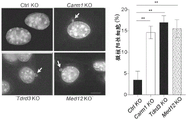


Classifications
-
- A—HUMAN NECESSITIES
- A61—MEDICAL OR VETERINARY SCIENCE; HYGIENE
- A61K—PREPARATIONS FOR MEDICAL, DENTAL OR TOILETRY PURPOSES
- A61K31/00—Medicinal preparations containing organic active ingredients
- A61K31/33—Heterocyclic compounds
- A61K31/395—Heterocyclic compounds having nitrogen as a ring hetero atom, e.g. guanethidine or rifamycins
- A61K31/41—Heterocyclic compounds having nitrogen as a ring hetero atom, e.g. guanethidine or rifamycins having five-membered rings with two or more ring hetero atoms, at least one of which being nitrogen, e.g. tetrazole
- A61K31/42—Oxazoles
- A61K31/422—Oxazoles not condensed and containing further heterocyclic rings
-
- C—CHEMISTRY; METALLURGY
- C12—BIOCHEMISTRY; BEER; SPIRITS; WINE; VINEGAR; MICROBIOLOGY; ENZYMOLOGY; MUTATION OR GENETIC ENGINEERING
- C12Y—ENZYMES
- C12Y201/00—Transferases transferring one-carbon groups (2.1)
- C12Y201/01—Methyltransferases (2.1.1)
-
- A—HUMAN NECESSITIES
- A61—MEDICAL OR VETERINARY SCIENCE; HYGIENE
- A61K—PREPARATIONS FOR MEDICAL, DENTAL OR TOILETRY PURPOSES
- A61K31/00—Medicinal preparations containing organic active ingredients
- A61K31/33—Heterocyclic compounds
- A61K31/395—Heterocyclic compounds having nitrogen as a ring hetero atom, e.g. guanethidine or rifamycins
- A61K31/435—Heterocyclic compounds having nitrogen as a ring hetero atom, e.g. guanethidine or rifamycins having six-membered rings with one nitrogen as the only ring hetero atom
- A61K31/44—Non condensed pyridines; Hydrogenated derivatives thereof
- A61K31/4409—Non condensed pyridines; Hydrogenated derivatives thereof only substituted in position 4, e.g. isoniazid, iproniazid
-
- A—HUMAN NECESSITIES
- A61—MEDICAL OR VETERINARY SCIENCE; HYGIENE
- A61K—PREPARATIONS FOR MEDICAL, DENTAL OR TOILETRY PURPOSES
- A61K31/00—Medicinal preparations containing organic active ingredients
- A61K31/33—Heterocyclic compounds
- A61K31/395—Heterocyclic compounds having nitrogen as a ring hetero atom, e.g. guanethidine or rifamycins
- A61K31/495—Heterocyclic compounds having nitrogen as a ring hetero atom, e.g. guanethidine or rifamycins having six-membered rings with two or more nitrogen atoms as the only ring heteroatoms, e.g. piperazine or tetrazines
- A61K31/505—Pyrimidines; Hydrogenated pyrimidines, e.g. trimethoprim
- A61K31/506—Pyrimidines; Hydrogenated pyrimidines, e.g. trimethoprim not condensed and containing further heterocyclic rings
-
- A—HUMAN NECESSITIES
- A61—MEDICAL OR VETERINARY SCIENCE; HYGIENE
- A61K—PREPARATIONS FOR MEDICAL, DENTAL OR TOILETRY PURPOSES
- A61K39/00—Medicinal preparations containing antigens or antibodies
- A61K39/46—Cellular immunotherapy
- A61K39/461—Cellular immunotherapy characterised by the cell type used
- A61K39/4611—T-cells, e.g. tumor infiltrating lymphocytes [TIL], lymphokine-activated killer cells [LAK] or regulatory T cells [Treg]
-
- A—HUMAN NECESSITIES
- A61—MEDICAL OR VETERINARY SCIENCE; HYGIENE
- A61K—PREPARATIONS FOR MEDICAL, DENTAL OR TOILETRY PURPOSES
- A61K39/00—Medicinal preparations containing antigens or antibodies
- A61K39/46—Cellular immunotherapy
- A61K39/463—Cellular immunotherapy characterised by recombinant expression
- A61K39/4631—Chimeric Antigen Receptors [CAR]
-
- A—HUMAN NECESSITIES
- A61—MEDICAL OR VETERINARY SCIENCE; HYGIENE
- A61K—PREPARATIONS FOR MEDICAL, DENTAL OR TOILETRY PURPOSES
- A61K39/00—Medicinal preparations containing antigens or antibodies
- A61K39/46—Cellular immunotherapy
- A61K39/463—Cellular immunotherapy characterised by recombinant expression
- A61K39/4632—T-cell receptors [TCR]; antibody T-cell receptor constructs
-
- A—HUMAN NECESSITIES
- A61—MEDICAL OR VETERINARY SCIENCE; HYGIENE
- A61K—PREPARATIONS FOR MEDICAL, DENTAL OR TOILETRY PURPOSES
- A61K39/00—Medicinal preparations containing antigens or antibodies
- A61K39/46—Cellular immunotherapy
- A61K39/464—Cellular immunotherapy characterised by the antigen targeted or presented
- A61K39/4643—Vertebrate antigens
- A61K39/4644—Cancer antigens
- A61K39/464484—Cancer testis antigens, e.g. SSX, BAGE, GAGE or SAGE
- A61K39/464488—NY-ESO
-
- A—HUMAN NECESSITIES
- A61—MEDICAL OR VETERINARY SCIENCE; HYGIENE
- A61K—PREPARATIONS FOR MEDICAL, DENTAL OR TOILETRY PURPOSES
- A61K45/00—Medicinal preparations containing active ingredients not provided for in groups A61K31/00 - A61K41/00
- A61K45/06—Mixtures of active ingredients without chemical characterisation, e.g. antiphlogistics and cardiaca
-
- A—HUMAN NECESSITIES
- A61—MEDICAL OR VETERINARY SCIENCE; HYGIENE
- A61P—SPECIFIC THERAPEUTIC ACTIVITY OF CHEMICAL COMPOUNDS OR MEDICINAL PREPARATIONS
- A61P35/00—Antineoplastic agents
-
- A—HUMAN NECESSITIES
- A61—MEDICAL OR VETERINARY SCIENCE; HYGIENE
- A61P—SPECIFIC THERAPEUTIC ACTIVITY OF CHEMICAL COMPOUNDS OR MEDICINAL PREPARATIONS
- A61P37/00—Drugs for immunological or allergic disorders
- A61P37/02—Immunomodulators
- A61P37/04—Immunostimulants
-
- C—CHEMISTRY; METALLURGY
- C07—ORGANIC CHEMISTRY
- C07K—PEPTIDES
- C07K14/00—Peptides having more than 20 amino acids; Gastrins; Somatostatins; Melanotropins; Derivatives thereof
- C07K14/435—Peptides having more than 20 amino acids; Gastrins; Somatostatins; Melanotropins; Derivatives thereof from animals; from humans
- C07K14/46—Peptides having more than 20 amino acids; Gastrins; Somatostatins; Melanotropins; Derivatives thereof from animals; from humans from vertebrates
- C07K14/47—Peptides having more than 20 amino acids; Gastrins; Somatostatins; Melanotropins; Derivatives thereof from animals; from humans from vertebrates from mammals
-
- C—CHEMISTRY; METALLURGY
- C12—BIOCHEMISTRY; BEER; SPIRITS; WINE; VINEGAR; MICROBIOLOGY; ENZYMOLOGY; MUTATION OR GENETIC ENGINEERING
- C12N—MICROORGANISMS OR ENZYMES; COMPOSITIONS THEREOF; PROPAGATING, PRESERVING, OR MAINTAINING MICROORGANISMS; MUTATION OR GENETIC ENGINEERING; CULTURE MEDIA
- C12N15/00—Mutation or genetic engineering; DNA or RNA concerning genetic engineering, vectors, e.g. plasmids, or their isolation, preparation or purification; Use of hosts therefor
- C12N15/09—Recombinant DNA-technology
- C12N15/11—DNA or RNA fragments; Modified forms thereof; Non-coding nucleic acids having a biological activity
- C12N15/113—Non-coding nucleic acids modulating the expression of genes, e.g. antisense oligonucleotides; Antisense DNA or RNA; Triplex- forming oligonucleotides; Catalytic nucleic acids, e.g. ribozymes; Nucleic acids used in co-suppression or gene silencing
-
- C—CHEMISTRY; METALLURGY
- C12—BIOCHEMISTRY; BEER; SPIRITS; WINE; VINEGAR; MICROBIOLOGY; ENZYMOLOGY; MUTATION OR GENETIC ENGINEERING
- C12N—MICROORGANISMS OR ENZYMES; COMPOSITIONS THEREOF; PROPAGATING, PRESERVING, OR MAINTAINING MICROORGANISMS; MUTATION OR GENETIC ENGINEERING; CULTURE MEDIA
- C12N9/00—Enzymes; Proenzymes; Compositions thereof; Processes for preparing, activating, inhibiting, separating or purifying enzymes
- C12N9/10—Transferases (2.)
- C12N9/1003—Transferases (2.) transferring one-carbon groups (2.1)
- C12N9/1007—Methyltransferases (general) (2.1.1.)
-
- A—HUMAN NECESSITIES
- A61—MEDICAL OR VETERINARY SCIENCE; HYGIENE
- A61K—PREPARATIONS FOR MEDICAL, DENTAL OR TOILETRY PURPOSES
- A61K2239/00—Indexing codes associated with cellular immunotherapy of group A61K39/46
- A61K2239/31—Indexing codes associated with cellular immunotherapy of group A61K39/46 characterized by the route of administration
-
- A—HUMAN NECESSITIES
- A61—MEDICAL OR VETERINARY SCIENCE; HYGIENE
- A61K—PREPARATIONS FOR MEDICAL, DENTAL OR TOILETRY PURPOSES
- A61K2239/00—Indexing codes associated with cellular immunotherapy of group A61K39/46
- A61K2239/38—Indexing codes associated with cellular immunotherapy of group A61K39/46 characterised by the dose, timing or administration schedule
-
- A—HUMAN NECESSITIES
- A61—MEDICAL OR VETERINARY SCIENCE; HYGIENE
- A61K—PREPARATIONS FOR MEDICAL, DENTAL OR TOILETRY PURPOSES
- A61K2239/00—Indexing codes associated with cellular immunotherapy of group A61K39/46
- A61K2239/46—Indexing codes associated with cellular immunotherapy of group A61K39/46 characterised by the cancer treated
- A61K2239/49—Breast
Landscapes
- Health & Medical Sciences (AREA)
- Life Sciences & Earth Sciences (AREA)
- Chemical & Material Sciences (AREA)
- General Health & Medical Sciences (AREA)
- Medicinal Chemistry (AREA)
- Animal Behavior & Ethology (AREA)
- Pharmacology & Pharmacy (AREA)
- Public Health (AREA)
- Veterinary Medicine (AREA)
- Organic Chemistry (AREA)
- Epidemiology (AREA)
- Engineering & Computer Science (AREA)
- Genetics & Genomics (AREA)
- Immunology (AREA)
- Microbiology (AREA)
- Bioinformatics & Cheminformatics (AREA)
- Zoology (AREA)
- Wood Science & Technology (AREA)
- Mycology (AREA)
- Cell Biology (AREA)
- Biomedical Technology (AREA)
- General Engineering & Computer Science (AREA)
- Molecular Biology (AREA)
- Biochemistry (AREA)
- Biotechnology (AREA)
- Chemical Kinetics & Catalysis (AREA)
- General Chemical & Material Sciences (AREA)
- Nuclear Medicine, Radiotherapy & Molecular Imaging (AREA)
- Biophysics (AREA)
- Oncology (AREA)
- Physics & Mathematics (AREA)
- Plant Pathology (AREA)
- Gastroenterology & Hepatology (AREA)
- Proteomics, Peptides & Aminoacids (AREA)
- Toxicology (AREA)
- Medicines That Contain Protein Lipid Enzymes And Other Medicines (AREA)
- Medicines Containing Antibodies Or Antigens For Use As Internal Diagnostic Agents (AREA)
- Peptides Or Proteins (AREA)
- Micro-Organisms Or Cultivation Processes Thereof (AREA)
- Pharmaceuticals Containing Other Organic And Inorganic Compounds (AREA)
Abstract
The present disclosure provides methods of treating an individual having cancer. The method comprises reducing the expression of Carm1 gene and/or Carm1 effector gene in cells of the individual, and/or reducing the activity of Carm1 protein and/or Carm1 effector protein in cells of the individual. The cancer is resistant to immunotherapy and/or checkpoint blockade therapy.
Description
Government support
The disclosure was made with government support under fund numbers R01 CA238039 and P01 CA163222 awarded by the national institutes of health. The government has certain rights in this disclosure.
RELATED APPLICATIONS
Priority of U.S. provisional application serial No. 62/991479, filed 3/18/2020, this application is hereby incorporated by reference in its entirety.
Reference appendix [ CD ROM/sequence Listing ]
The application is submitted electronically via the EFS Web, including a sequence listing in the format of a text file submitted electronically. The text file contains a sequence listing entitled "14293 _1000uk seq listing _ST25", created at 18/3/2021, and having a size of 122324 bytes. The sequence listing contained in this text document is part of the specification, which is incorporated herein by reference in its entirety.
Technical Field
The present disclosure relates to methods of treating cancer, in particular, methods of treating cancer that is resistant to drug therapy, such as immunotherapy, chemotherapy, radiation therapy, and/or checkpoint blockade therapy. In particular, the present disclosure relates to methods for treating cancer, particularly cancers that are resistant to drug therapy, such as immunotherapy, chemotherapy, radiation therapy, and/or checkpoint blockade therapy, by inhibiting the expression of the Carm1 gene, tdrd3 gene, med12 gene, and/or other Carm1 effector genes and/or the activity of the Carm1 protein, tdrd3 protein, med12 protein, and/or other Carm1 effector proteins in immune cells, cancer cells, or both.
Background
A number of Cancer drugs have been developed which can induce tumor cell apoptosis, for example, by inducing DNA damage or inhibiting key signaling pathways required for cell proliferation (Bouwman P, jonkers j. The effects of differentiated DNA mapping on Cancer therapy response and resistance. Nat Rev Cancer 2012 (9): 587-98doi10.1038/nrc 3342. While such drugs can induce substantial tumor shrinkage, recurrence is a major challenge due to the growth of resistant tumor cells. The immune system may potentially target residual disease, but many of these tumor cell targeting drugs also impair immune cell survival/function or immune cell production by the hematopoietic system. For example, chemotherapeutic drugs not only kill dividing tumor cells, but also fast dividing hematopoietic precursor cells and immune cells (Naito Y, saito K, shiiba K, ohuchi A, saigenji K, nagura H et al CD8+ T cells encapsulated with cancer cells as a protective cancer in human cancer Res 1998 58 (16): 3491-4 Sato E, olson SH, ahn J, bundy B, nishikawa H, qian F et al endothelial CD8+ tumor-encapsulating polypeptides and a high CD8+/regulatory T cell rate area with viral infection associated with cancer cells Acerand No. 2005-18551.
Chemotherapy-induced DNA damage induces activation of innate immune pathways in tumor cells, including the cGAS-STING pathway (Chen Q, sun L, chen ZJ. Regulation and function of the cGAS-STING pathway of cytolytic DNA sensing. Nat Immunol 2016 (10): 1142-9). cGAS enzyme is activated by cytosolic double-stranded DNA, resulting in the synthesis of the cyclic dinucleotide cGAMP, which activates STING receptor, inducing a strong type 1 interferon response by IRF3 transcription factors. Importantly, type 1 interferons also induce dendritic cell maturation, a key step in T cell mediated immunity (Hervas-Stubbs S, perez-Gracia JL, rouzaut A, sanmamed MF, le Bon A, melero I.direct effects of type I interferons on cells of the immunene system. Clin Cancer Res 2011 17 (9): 2619-27. Some chemotherapeutic agents are being used in combination with immunotherapeutic agents. For example, the FDA recently approved the combination of albumin-bound Paclitaxel (Nab-Paclitaxel) and PD-L1 blocking mAb for the treatment of metastatic Triple Negative Breast Cancer (TNBC), but only a small fraction of treated patients benefit from this combination compared to albumin-bound Paclitaxel monotherapy (Schmid P, chui SY, emers la. Atezolizumab and Nab-Paclitaxel in Advanced Triple-Negative Breast cancer. Reply. N Engl J Med 2019 (10): 987-8. It is important to develop tumor cell targeting drugs that enhance rather than impair immune function.
Summary of The Invention
In some embodiments, the present disclosure provides methods of treating an individual having cancer. In some embodiments, the method comprises reducing the expression of the Carm1 gene and/or Carm1 effector gene in cells of the individual; and/or reducing the activity of Carm1 protein and/or Carm1 effector protein in cells of the individual. In some embodiments, the cancer is resistant to immunotherapy and/or checkpoint blockade therapy.
In some embodiments, the Carm1 effector gene is the Tdrd3 gene and the Carm1 effector protein is the Tdrd3 protein. In some embodiments, the Carm1 effector gene is the Med12 gene and the Carm1 effector protein is the Med12 protein.
In some embodiments, the reducing step comprises administering an inhibitor to the individual. The inhibitor inhibits the expression of Carm1 gene or Carm1 effector gene and/or the activity of Carm1 protein or Carm1 effector protein in an individual.
In some embodiments, the inhibitor is selected from the group consisting of a polynucleotide, a polypeptide, an antibody, a small molecule, a protein degrading agent, and combinations thereof. In some embodiments, the inhibitor comprises EZM2302 or TP-064. In some embodiments, the protein degrading agent comprises a Carm1 protein degrading agent, a Tdrd3 protein degrading agent, and/or a Med12 protein degrading agent. In some embodiments, the protein degrading agent comprises a Carm1 protein degrading agent.
In some embodiments, the reducing step comprises silencing the Carm1 gene or Carm1 effector gene in the individual by shRNA-mediated mRNA knockdown or gene inactivation.
In some embodiments, the reducing step comprises modifying the Carm1 gene or Carm1 effector gene to reduce expression of the Carm1 gene or Carm1 effector gene. In some embodiments, the modifying step comprises modifying the Carm1 gene or the Carm1 effector gene by a CRISPR/Cas system.
In some embodiments, the cell is an immune cell.
In some embodiments, the immune cell is an immune effector cell, wherein decreased expression of the Carm1 gene or Carm1 effector gene and/or decreased activity of the Carml protein or CarmI effector protein enhances the cytotoxic function of the immune effector cell and/or reduces failure of the immune effector cell. In some embodiments, the immune effector cell is selected from the group consisting of a cytotoxic T cell, a Tumor Infiltrating Lymphocyte (TIL), a natural killer T cell (NKT), a Cytotoxic T Lymphocyte (CTL), a dendritic cell, a CD 8T cell, and a CD 4T cell.
In some embodiments, the cell is a cancer cell.
In some embodiments, the cancer cells exhibit reduced growth, reduced metastatic activity, increased susceptibility to killing by CD 8T cells, increased expression of interferon response genes (e.g., IFN α/γ pathway genes, p53 pathway genes, etc.), have a DNA damage response, or a combination thereof. In some embodiments, the interferon response gene is an IFN α/γ pathway gene and/or a p53 pathway gene.
In some embodiments, the expression or activity is decreased in both immune cells and cancer cells of the individual.
In some embodiments, the method further comprises administering to the individual an immune cell that is tumor specific for cancer and has reduced expression of a Carm1 gene or Carm1 effector gene and/or reduced activity of a Carml protein or Carm1 effector protein.
In some embodiments, the immune cell does not substantially express a Carm1 gene or a Carm1 effector gene.
In some embodiments, the immune cell is a CAR T cell.
In some embodiments, the cancer cells of the individual overexpress Carm1.
In some embodiments, the cancer is melanoma, carcinoma (carcinoma), sarcoma, adenocarcinoma, lymphoma, leukemia, kidney cancer, breast cancer, lung cancer, bladder cancer, colon cancer, ovarian cancer, prostate cancer, pancreatic cancer, stomach cancer, brain cancer, head and neck cancer, skin cancer, uterine cancer, testicular cancer, glioma, esophageal cancer, or liver cancer.
In some embodiments, the cancer is resistant to checkpoint blockade therapy using CTLA-4, PD-L1, TIM-3, LAG3, TIGIT, or PD-1 antibody blockade therapy. In some embodiments, the checkpoint blockade is selected from Nivolumab (Nivolumab), parbolizumab (Pembrolizumab), ipilimumab (Ipilimumab), attelizumab (Atezolizumab), avilumab (Avelumab), delavolumab (Durvalumab), cimiciprizumab (cemipimab), and combinations thereof.
In some embodiments, the method further comprises administering to the individual a pharmaceutically effective amount of a second therapeutic agent for treating cancer in the individual.
In some embodiments, the second therapeutic agent is selected from the group consisting of a chemotherapeutic agent, an immunotherapeutic agent, a checkpoint blocker, a toxin, a radiolabel, an siRNA, a cancer vaccine, a small molecule, a peptide, an antibody, a genetically engineered cell, a CAR T cell, a cytokine, and a combination thereof. In some embodiments, the second therapeutic agent is an anti-PD 1 antibody, an anti-CTLA-4 antibody, an anti-PD-L1 antibody, an anti-TIGIT antibody, an anti-TIM-3 antibody, or an anti-LAG 3 antibody.
In some other embodiments, the present disclosure provides methods of treating cancer in an individual. The method comprises reducing the expression of the Carm1 gene or Carm1 effector gene and/or the activity of the Carm1 protein or Carm1 effector protein in the cells of the individual.
In some embodiments, the method further comprises administering to the individual a pharmaceutically effective amount of a second therapeutic agent for treating cancer in the individual.
In some embodiments, the second therapeutic agent is selected from the group consisting of a chemotherapeutic agent, an immunotherapeutic agent, a checkpoint blocker, a toxin, a radiolabel, an siRNA, a cancer vaccine, a small molecule, a peptide, an antibody, a genetically engineered cell, a CAR T cell, a cytokine, and a combination thereof. In some embodiments, the second therapeutic agent is an anti-PD 1 antibody, an anti-CTLA-4 antibody, an anti-PD-L1 antibody, an anti-TIGIT antibody, an anti-TIM-3 antibody, or an anti-LAG 3 antibody.
In some embodiments, the reducing step comprises administering an inhibitor to the individual, wherein the inhibitor inhibits the expression of the Carm1 gene or Carm1 effector gene and/or the activity of Carm1 protein or Carm1 effector protein in the individual.
In some embodiments, the reducing step comprises silencing the Carm1 gene or Carm1 effector gene in the individual by shRNA-mediated mRNA knockdown or gene inactivation.
In some embodiments, the reducing step comprises modifying the Carm1 gene or Carm1 effector gene to reduce the expression of the Carm1 gene or Carm1 effector gene. In some embodiments, the modifying step comprises modifying the Carm1 gene or Carm1 effector gene by a CRISPR/Cas system.
In some embodiments, the reducing step comprises degrading the Carm1 protein or Carm1 effector protein.
In some embodiments, the present disclosure provides methods of increasing the sensitivity of cancer cells to immune effector cells. The method comprises inhibiting the expression and/or activity of a Carm1 gene or protein or a Carm1 effector gene or protein in a cancer cell by one or more inhibitors, wherein said inhibition increases the sensitivity of the cancer cell to immune cells.
In some embodiments, the inhibitor comprises a polynucleotide, a polypeptide, a peptide, an antibody, a small molecule, a protein degrading agent, or a combination thereof. In some embodiments, the inhibitor comprises EZM2302 or TP-064.
In some embodiments, the inhibitor comprises a Carm1 protein degrading agent, a Tdrd3 protein degrading agent, and/or a Med12 protein degrading agent. In some embodiments, the inhibitor comprises a Carm1 protein degradation agent.
In some embodiments, the Carm1 effector gene or protein is a Tdrd3 gene or protein. In some embodiments, the Carm1 effector gene or protein is a Med12 gene or protein.
In some embodiments, the immune effector cell is selected from the group consisting of a cytotoxic T cell, a Tumor Infiltrating Lymphocyte (TIL), a natural killer T cell (NKT), a Cytotoxic T Lymphocyte (CTL), a dendritic cell, a CD 8T cell, and a CD 4T cell.
In some embodiments, the present disclosure provides methods of increasing the anti-tumor function of an immune effector cell. The method comprises reducing the expression and/or activity of Carm1 gene or protein or Carm1 effector gene or protein in the immune effector cell, thereby increasing the anti-tumor function of the immune effector cell.
In some embodiments, the Carm1 effector gene or protein is a Tdrd3 gene or protein. In some embodiments, the Carm1 effector gene or protein is a Med12 gene or protein.
In some embodiments, the reducing step comprises inhibiting the expression and/or activity of a Carm1 gene or protein or a Carm1 effector gene or protein in the immune cell by one or more inhibitors.
In some embodiments, the inhibitor comprises a polynucleotide, polypeptide, peptide, antibody, small molecule, protein degrading agent, genetically engineered cell, or a combination thereof. In some embodiments, the inhibitor comprises EZM2302 or TP-064.
In some embodiments, the inhibitor comprises a Carm1 protein degradation agent, a Tdrd3 protein degradation agent, and/or a Med12 protein degradation agent. In some embodiments, the inhibitor comprises a Carm1 protein degradation agent.
In some embodiments, the expression and/or activity of the Carm1 gene or protein or Carm1 effector gene or protein is decreased by shRNA-mediated mRNA knockdown or gene inactivation.
In some embodiments, the reducing step comprises modifying the immune effector cell to reduce or remove the CARM1 gene or CARM1 effector gene in the immune effector cell. In some embodiments, the modifying step comprises modifying the Carm1 gene or Carm1 effector gene by a CRISPR/Cas system.
In some embodiments, the reducing step comprises silencing the CARM1 gene or the CARM1 effector gene in the immune effector cell.
In some embodiments, the reducing step comprises degrading the Carm1 protein or Carm1 effector protein in the immune effector cell.
In some embodiments, the immune effector cell is selected from the group consisting of a cytotoxic T cell, a Tumor Infiltrating Lymphocyte (TIL), a natural killer T cell (NKT), a Cytotoxic T Lymphocyte (CTL), a dendritic cell, a CD 8T cell, and a CD 4T cell. In some embodiments, the CD 8T cell expresses higher levels of CD69, CD45.1, granzyme B, IFN γ, ki67, or a combination thereof.
In some embodiments, the present disclosure provides immune effector cells. The immune cells have an inhibitor of the Carm1 gene/protein or of the Carm1 effector gene/protein. The inhibitor inhibits the expression of Carm1 gene or Carm1 effector gene and/or the activity of Carm1 protein or Carm1 effector protein.
In some embodiments, the Carm1 effector gene is the Tdrd3 gene and the Carm1 effector protein is the Tdrd3 protein. In some embodiments, the Carm1 effector gene is the Med12 gene and the Carm1 effector protein is the Med12 protein.
In some embodiments, the immune effector cell does not substantially express a Carm1 gene or a Carm1 effector gene.
In some embodiments, the inhibitor is selected from the group consisting of a polynucleotide, a polypeptide, an antibody, a small molecule, a protein degrading agent, and combinations thereof. In some embodiments, the inhibitor comprises EZM2302 or TP-064.
In some embodiments, the inhibitor comprises a Carm1 protein degradation agent, a Tdrd3 protein degradation agent, and/or a Med12 protein degradation agent. In some embodiments, the inhibitor comprises a Carm1 protein degradation agent.
In some embodiments, the immune effector cell is selected from the group consisting of a cytotoxic T cell, a Tumor Infiltrating Lymphocyte (TIL), a natural killer T cell (NKT), a Cytotoxic T Lymphocyte (CTL), a dendritic cell, a CD 8T cell, and a CD 4T cell.
In some embodiments, the immune effector cell is tumor-specific.
In some embodiments, the immune effector cell expresses a tumor specific T cell receptor or a Chimeric Antigen Receptor (CAR).
In some embodiments, the immune effector cell further comprises a Chimeric Antigen Receptor (CAR), wherein the CAR comprises an antigen binding domain, a transmembrane domain, and a stimulatory domain.
In some embodiments, the antigen binding domain binds to a tumor antigen or a pathogen antigen.
In some embodiments, the tumor antigen is selected from the group consisting of an antigen present in a cancer cell, a cancer cell fragment, a tumor antigen, alpha-galcer, anti-CD 3, anti-CD 28, anti-IgM, anti-CD 40, a pathogen, an attenuated pathogen, and portions thereof.
In some embodiments, the tumor antigen is associated with melanoma, carcinoma, sarcoma, adenocarcinoma, lymphoma, leukemia, kidney cancer, breast cancer, lung cancer, bladder cancer, colon cancer, ovarian cancer, prostate cancer, pancreatic cancer, stomach cancer, brain cancer, head and neck cancer, skin cancer, uterine cancer, testicular cancer, glioma, esophageal cancer, or liver cancer.
In some embodiments, the tumor antigen is associated with a solid tumor or lymphoma.
In some embodiments, the antigen binding domain is an antigen binding fragment of an antibody.
In some embodiments, the present disclosure provides a composition comprising an immune effector cell described herein and a pharmaceutically acceptable carrier. In some embodiments, the immune effector cell has a Carm1 gene/protein or an inhibitor of a Carm1 effector gene/protein. The inhibitor inhibits the expression of Carm1 gene or Carm1 effector gene and/or the activity of Carm1 protein or Carm1 effector protein.
In some embodiments, the composition further comprises a second therapeutic agent.
In some embodiments, the second therapeutic agent is selected from the group consisting of a chemotherapeutic agent, an immunotherapeutic agent, a checkpoint blocker, a toxin, a radiolabel, an siRNA, a cancer vaccine, a small molecule, a peptide, an antibody, a genetically engineered cell, a CAR T cell, a cytokine, and a combination thereof.
In some embodiments, the second therapeutic agent is an anti-PD 1 antibody, an anti-CTLA-4 antibody, an anti-PD-L1 antibody, an anti-TIGIT antibody, an anti-TIM-3 antibody, or an anti-LAG 3 antibody.
In some embodiments, the immune effector cell is selected from the group consisting of a cytotoxic T cell, a Tumor Infiltrating Lymphocyte (TIL), a natural killer T cell (NKT), a Cytotoxic T Lymphocyte (CTL), a dendritic cell, a CD 8T cell, and a CD 4T cell.
In some embodiments, the present disclosure provides methods of treating cancer in an individual. The method comprises administering to an individual having cancer an immune effector cell as described herein or a composition as described herein.
In some embodiments, the immune effector cell has a Carm1 gene/protein or an inhibitor of a Carm1 effector gene/protein. The inhibitor inhibits the expression of Carm1 gene or Carm1 effector gene and/or the activity of Carm1 protein or Carm1 effector protein.
In some embodiments, the composition comprises an immune effector cell described herein and a pharmaceutically acceptable carrier. In some embodiments, the immune effector cell has a Carm1 gene/protein or an inhibitor of a Carm1 effector gene/protein. The inhibitor inhibits the expression of Carm1 gene or Carm1 effector gene and/or the activity of Carm1 protein or Carm1 effector protein.
In some embodiments, the immune effector cell is autologous.
In some embodiments, the immune effector cell is specific for a cancer cell of the individual.
In some embodiments, the method further comprises administering to the individual having cancer a second therapeutic agent or a composition comprising a second therapeutic agent and a pharmaceutically acceptable carrier.
In some embodiments, the second therapeutic agent is selected from the group consisting of a chemotherapeutic agent, an immunotherapeutic agent, a checkpoint blocker, a toxin, a radiolabel, an siRNA, a cancer vaccine, a small molecule, a peptide, an antibody, a genetically engineered cell, a CAR T cell, a cytokine, and a combination thereof.
In some embodiments, the second therapeutic agent is an anti-PD 1 antibody, an anti-CTLA-4 antibody, an anti-TIGIT antibody, an anti-TIM-3 antibody, or an anti-LAG 3 antibody.
Unless defined otherwise, all technical and scientific terms used herein have the same meaning as commonly understood by one of ordinary skill in the art to which this invention belongs. Methods and materials for use in the present disclosure are described herein; other suitable methods and materials known in the art may also be used. These materials, methods, and examples are illustrative only and not intended to be limiting. All publications, patent applications, patents, sequences, database entries, and other references mentioned herein are incorporated by reference in their entirety. The disclosures of these publications, patent applications, patents, sequences, database entries, and other references are hereby incorporated by reference in their entirety to more fully describe the state of the art as known to those skilled in the art as of the date of this disclosure as described and claimed herein. In case of conflict, the present specification, including definitions, will control.
Other features and advantages of the disclosure will be apparent from the following detailed description and drawings, and from the claims.
Brief Description of Drawings
This patent or application document contains at least one drawing executed in color. Copies of the color images disclosed in this patent or patent application will be provided by the office upon request and payment of the necessary fee.
FIG. 1A illustrates that Carm1 is an epigenetic inhibitor in tumor-specific T cells, showing the experimental design for the in vivo discovery of epigenetic regulators that inhibit CD8T cell accumulation in tumors.
Fig. 1B illustrates that Carm1 is an epigenetic inhibitor in tumor specific T cells, showing in vivo CRISPR screening of epigenetic gRNA libraries in tumor specific CD8T cells. gRNA quantification (log 2 fold change) in CD8T cells in tumors (experimental sites) and spleen (control organs) was compared. The main experimental genes and the positive control genes are highlighted in red and blue, respectively.
Figure 1C illustrates that Carm1 is an epigenetic inhibitor in tumor specific T cells, showing MAGeCK analysis of CRISPR screening data in vivo (model-based whole genome CRISPR-Cas9 knockout assay); the MAGeCK score provides a comprehensive readout of the strength of the gene effect.
FIG. 1D illustrates that Carm1 is an epigenetic inhibitor in tumor specific T cells, showing a T cell cytotoxicity assay with Carm1-KO and control-KO OT-I CD 8T cells. CD 8T cells were edited by electroporation with Cas9 protein and bound gRNA and cultured in T cell medium containing IL-15+ IL-7 for 5 days. Co-culturing T cells with B16F10-OVA-ZsGreen tumor cells at the indicated effective target ratio (E: T) (n =8-10 per replicate under each condition); after 24 hours, viable GFP-positive tumor cells were counted using a Celigo image cytometer. Data are representative of three experiments and are shown as mean ± SEM. * P <0.0001, unpaired double-sided Mann-Whitney test.
FIG. 1E illustrates that Carm1 is an epigenetic inhibitor in tumor specific T cells, showing the anti-tumor activity of adoptively transferred Carm1-KO or control-KO OT-I CD45.1 CD 8T cells. Subcutaneous implantation of B16-OVA-ZsGreen tumor cells (0.1x10) 6 ). Day 7 post tumor cell inoculation, edited CD 8T cells (1 x 10) were transferred by tail vein injection 6 ). Recording the size of the tumor; n =8-10 mice per group.
FIG. 1F illustrates that Carm1 is an epigenetic inhibitor in tumor specific T cells, showing the tumor weight 7 days after adoptive T cell transplantation for the experiment shown in FIG. 1E.
Fig. 1G illustrates that Carm1 is an epigenetic inhibitor in tumor specific T cells, showing flow cytometric analysis of tumor infiltrating Carm1-KO or control-KO CD 8T cells after adoptive transfer of edited OT-I CD45.1 CD 8T cells (n =10 mice/group), gated with CD45.1 and CD 8T cell markers. Infiltration of CD 8T cells and expression of effectors (granzyme B, IFN γ) and proliferation (Ki-67) markers were quantified.
FIG. 2A illustrates that inhibition of Carm1 in CD 8T cells enhances its anti-tumor function, showing RNA-Seq analysis (four biological replicates per condition) of differentially expressed genes in Carm1-KO or control-KO OT-I CD 8T cells co-cultured with B16F10-Ova tumor cells for 24 hours. The color code represents the Z-score of differential gene expression.
FIG. 2B illustrates that inhibition of Carm1 in CD 8T cells enhances its anti-tumor function, showing the volcano pattern of all differentially expressed genes between Carm1-KO and control-KO OT-I CD 8T cells. Statistical significance (log 10 adjusted P-value) was plotted against log2 fold change in gene expression level (Carm 1-KO/control-KO cells).
FIG. 2C illustrates that inhibition of Carm1 in CD 8T cells enhances its anti-tumor function, showing RT-qPCR analysis of Tcf7, myb, bcl6, btg2, itgae, havcr2, and Klrg1 mRNA levels in Carm1-KO and control-KO CD 8T cells (targeting Carm1 with two different gRNAs, measured in triplicate).
Fig. 2D illustrates that inhibition of Carm1 in CD8T cells enhances its anti-tumor function, showing Gene Ontology (GO) analysis of pathways that are significantly up/down regulated in Carm1-KO relative to control-KO T cells.
Fig. 2E illustrates that Carm1 inhibition in CD8T cells enhances its anti-tumor function, showing tumor infiltration of Carm1-KO or control-KO CD8T cells following adoptive transfer of edited OT-I CD45.1 CD8T cells (n =10 mice/group), gated with CD45.1 and CD8T markers. Tcf7+ T cells and Tcf7+ CD8T cells with high Bcl2 protein levels were quantified.
FIG. 2F illustrates that inhibition of Carm1 in CD8T cells enhances its anti-tumor function, showing quantification of high Bcl2 tumor infiltration Carm1-KO or control-KO CD8T cells.
Figures 2G, 2H and 2I illustrate that Carm1 inhibition in CD8T cells enhances their anti-tumor function, showing analysis of tumor infiltrating Carm1-KO or control- KO CD8T cells 16 or 24 days after adoptive transfer of edited OT-I CD45.1 CD8T cells (n =8 mice/group), with gating on CD45.1 and CD8T cell markers. CD8T activation marker (CD 69) (fig. 2G) and T cell depletion marker (fig. 2H-2I) were quantified. In FIGS. 2A-2I, the data presented are representative of two experiments. Statistical significance was determined using two-way analysis of variance for time points at which all mice survived for tumor measurement. The graphs presented data summarized as mean ± s.d. and analyzed by unpaired double-sided Mann-Whitney test, × P <0.0001; * P <0.001; * P <0.01; * P <0.05.
FIG. 3A illustrates inactivation of the Carm1 gene in tumor cells elicited tumor immunity, showing Carm1 mRNA levels in different groups of 1208 different cancer cell lines from the Cancer Cell Line Encyclopedia (CCLE). Tumor cell lines were grouped according to cancer type.
FIG. 3B illustrates inactivation of the Carm1 gene in tumor cells to elicit tumor immunity, showing a strategy to target Carm1 in tumor cells to study the effect on T cell-mediated tumor immunity.
FIG. 3C illustrates inactivation of the Carm1 gene in tumor cells to elicit tumor immunity, showing Western blot analysis of Carm1 protein in B16F10 melanoma cells following electroporation with RNPs consisting of Cas9 protein and bound gRNAs (control, carm 1); two different controls and Carm1 gRNA were evaluated.
FIG. 3D illustrates inactivation of the Carm1 gene in tumor cells to elicit tumor immunity, showing growth of Carm1-KO and control-KO B16F10 tumors (left) and survival of tumor-bearing mice (right). Mice (n = 8-10/group) were treated with CD 8-depleted antibodies or isotype control antibodies. This in vivo phenotype was confirmed with a second Carm1 gRNA (FIG. 10A).
FIG. 3E illustrates inactivation of the Carm1 gene in tumor cells to elicit tumor immunity, showing growth of Carm1-KO tumors in T cell deficient mice. Carm1-KO and control-KO B16F10 tumor cells (0.2x10) 6 ) Implanting immunocompetent or immunodeficient (Tcra KO) mice (n = 8-10/group); tumor growth (left) and survival (right) were recorded.
FIG. 3F illustrates inactivation of the Carm1 gene in tumor cells to elicit tumor immunity, showing growth of Carm1-KO or control-KO 4T1 tumor cells after implantation into a mammary fat pad (n =8-10 mice/group); tumor growth (left) and survival (right) were recorded.
FIG. 3G illustrates inactivation of the Carm1 gene in tumor cells to elicit tumor immunity, showing quantification of spontaneous lung metastases formed in immunocompetent mice from Carm1-KO or control-KO 4T1 tumors. Representative image of lung metastasis (V, ventral; D, dorsal) (right).
Fig. 3H illustrates inactivation of the Carm1 gene in tumor cells to elicit tumor immunity, showing tumor growth (left) and survival (right) after implantation of Carm1-KO and control-KO MC38 tumor cells (n =8-10 mice/group).
FIG. 3I illustrates inactivation of the Carm1 gene in tumor cells to elicit tumor immunity, showing T cell cytotoxicity assays with Carm1-KO or control-KO B16F10-OVA-ZsGreen tumor cells. Tumor cells were co-cultured with OT-I CD 8T cells at the indicated potency target ratio (E: T) for 24 hours (n =8-10 replicates/condition).
FIG. 3J illustrates inactivation of the Carm1 gene in tumor cells to elicit tumor immunity, showing (as shown in FIG. 3I) CD 8T cell-induced apoptosis of tumor cells (Carm 1-KO or control-KO B16F10-OVA-ZsGreen cells) measured with Caspase-3/7 dye at different E: T ratios (n =8-10 repeats/group).
FIG. 3K illustrates inactivation of the Carm1 gene in tumor cells to elicit tumor immunity, showing that CARM1 inhibitors increase the sensitivity of tumor cells to T cells. B16F10-OVA-ZsGreen tumor cells were pretreated with CARM1 inhibitor (EZM 2302, 0.1. Mu.M) for 24 hours. Vehicle or inhibitor treated tumor cells were co-cultured with OT-I CD 8T cells at the indicated E: T ratios (n =7-8 replicates/condition).
FIG. 3L illustrates inactivation of the Carm1 gene in tumor cells to elicit tumor immunity, showing T cell cytotoxicity assays with human CD 8T cells expressing NY-ESO-1TCR and human BT549 TNBC. Tumor cells were pretreated with CARM1 inhibitor (EZM 2302,0.1 μ M) for 24 hours (n =7-10 replicates/group); after 24 hours of co-culture, the number of surviving tumor cells was quantified. In fig. 3A-3L, two-way analysis of variance was used to determine the statistical significance of tumor measurements at time points when all mice were alive. Statistical significance of mouse survival in each treatment group was calculated by the Log-rank (Mantel-Cox) test. Bar graphs represent data summarized as mean ± s.e.m and analyzed by unpaired two-sided Mann-Whitney test. Data shown are representative of three experiments. * p <0.05, p <0.01, p <0.001, p <0.0001, ns (not significant).
FIG. 4A illustrates innate immune activation in Carm 1-deficient tumor cells, showing RNA-seq analysis of differentially expressed genes in Carm1-KO and control-KO B16F10 tumor cells (n = 3/group). Data are representative of two independent experiments.
Fig. 4B illustrates innate immune activation in Carm 1-deficient tumor cells, showing Gene Ontology (GO) analysis of genes significantly up/down regulated in Carm1-KO compared to control-KO B16F10 tumor cells.
FIG. 4C illustrates innate immune activation in Carm 1-deficient tumor cells, showing a Venn diagram representing the number of overlapping differentially expressed genes in Carm1-KO tumors and CD 8T cells.
FIG. 4D illustrates innate immune activation in Carm 1-deficient tumor cells, showing verification of interferon inducible genes (ISGs) in Carm1-KO by RT-qPCR (n = 3/group) compared to control-KO B16F10 cells.
FIG. 4E illustrates innate immune activation in Carm 1-deficient tumor cells, showing RT-qPCR analysis of ISG mRNA levels 7 days after treatment of B16F10 cells with Carm1 inhibitor EZM2302 (0.1-1 μ M) or solvent control (n = 3/group).
FIG. 4F illustrates innate immune activation in Carm 1-deficient tumor cells, showing expression of selected ISGs in control-KO, carm1-KO, cGAS-KO, and Carm1/cGAS double KO (dKO) B16F10 cells analyzed by RT-qPCR (n = 3/group).
FIG. 4G illustrates innate immune activation in Carm 1-deficient tumor cells, showing T cell cytotoxicity assays with control-KO, carm1-KO, cGAS-KO, and Carm1/cGAS dKO B16F10 cells. Tumor cells were co-cultured with OT-ICD 8T cells for 24 hours at the indicated E: T ratio (n =7-10 replicates/condition); viable GFP positive tumor cells were counted using a Celigo image cytometer. Data are shown as mean ± SEM. * P <0.001, unpaired double-sided Mann-Whitney test.
FIGS. 4H-4I illustrate innate immune activation in Carm 1-deficient tumor cells, showing dsDNA damage in Carm1-KO versus control-KO B16F10 tumor cells based on γ H2AX (FIG. 4H) and RAD51 (FIG. 4I) Ab markers. Representative immunofluorescence images (left) of γ H2AX or RAD51 antibody markers (red); nuclei were labeled with DAPI. Quantification of γ H2AX or RAD51 foci/nuclei number (right). Data are shown as mean ± SEM. * P <0.001, unpaired double-sided Mann-Whitney test. Scale bar-10. Mu.M.
FIG. 4J illustrates innate immune activation in Carm 1-deficient tumor cells, showing detection of micronuclei in Carm1-KO and control-KO B16F10 tumor cells. The DNA is labeled with DAPI; representative images (left) and quantification (right) of micronucleus positive cells. Data are shown as mean ± SEM. * P <0.01, unpaired two-sided Mann-Whitney test. Scale bar-10. Mu.M.
FIG. 4K illustrates innate immune activation in Carm 1-deficient tumor cells, showing analysis of dsDNA damage in Carm1-KO versus control-KO CD8+ T cells. OT-I CD 8T cells were plated 7 days after editing using the aforementioned CRIPSR-Cas9 and CD8 α, γ H2AX and DAPI staining was performed. Representative immunofluorescence images of CD8 α antibody labeling (red), γ H2AX antibody labeling (green); nuclei were stained with DAPI (blue). Scale bar-20. Mu.M. The data shown in FIGS. 4D-4J represent three independent experiments. The bar graphs represent data summarized as mean ± s.e.m. and analyzed by unpaired, two-sided Mann-Whitney test. * p <0.05, p <0.01, p <0.001, p <0.0001, ns (not significant). Error bars for all qPCR data represent three replicates of SD per group.
Figure 5A illustrates that Carm1 inhibition overcomes tolerance to checkpoint blockade, showing treatment of Carm1-KO or control-KO B16F10 tumors with CTLA-4 or isotype control antibody (n =8 mice/group). Tumor growth (left) and survival (right) of tumor-bearing mice are shown. Will have comparable tumor volumes (50 mm) 3 ) The mice were randomly assigned to different treatment groups.
Figure 5B illustrates that Carm1 inhibition overcomes tolerance to checkpoint blockade, showing treatment of B16F10 tumors (n =8 mice/group) with Carm1 inhibitor (EZM 2302) or vehicle control in combination with CTLA-4 or isotype control antibody. EZM2302 (150 mg/kg b.i.d.) is administered orally for 2 weeks (days 7-21).
Figure 5C illustrates that Carm1 inhibition overcomes tolerance to checkpoint blockade, showing treatment of Carm1-KO or control-KO 4T1 tumors with anti-CTLA-4 or isotype control antibodies (n =8 mice/group).
Fig. 5D illustrates that Carm1 inhibition overcomes tolerance to checkpoint blockade, showing the number change of spontaneous lung metastases formed from 4T1 tumors treated as described in C (left) and a representative image of lung metastases (right) (n =8 mice/group).
Fig. 5E illustrates that Carm1 inhibition overcomes tolerance to checkpoint blockade, showing quantification of tumor-infiltrating CD 8T cells in Carm1-KO and control-KO B16F10 tumors (n =8 mice/group) after treatment with CTLA-4 or isotype control antibody (day 18 post tumor cell implantation). Representative flow cytograms (left) and CD 8T cell quantification in CD3+ cell percentage and per gram of tumor (middle and right, respectively).
FIGS. 5F-5G illustrate that Carm1 inhibition overcomes tolerance to checkpoint blockade, showing quantification of PD-1 positive and PD-1/Tim-3 double positive tumor infiltrating CD8+ T cells in the experiment described in FIG. 5E.
FIGS. 5H-5I illustrate that Carm1 inhibition overcomes tolerance to checkpoint blockade, showing H-I. For CD 8T cells expressing effector markers (GZMB and IFN γ) per gram of tumor, migratory cross-presenting DCs (CD 45+/CD3-/F4/80-/CD11c +/MHC-II) in the experiment described in FIG. 5E high Quantification of/CD 103+/CD11 b-) and NK cells (CD 45+/CD3-/CD49b +). In FIGS. 5A-5I, the data shown are representative of two experiments. For time points where all mice survived for tumor measurement, two-way analysis of variance was used to determine statistical significance. The Log-rank (Mantel-Cox) test was used to determine statistical significance of mouse survival. Bar graphs represent data summarized as mean ± s.e.m. and analyzed by unpaired two-sided Mann-Whitney test<0.0001;***P<0.001;**P<0.01;*P<0.05; n.s., not significant.
Fig. 6A illustrates that Tdrd3 and Med12 are downstream effectors of Carm1, showing comparison of ISG expression in control-KO, tdrd3-KO, cGAS-KO, and Tdrd3/cGAS dKO B16F10 cells by RT-qPCR (n = 3/group).
Fig. 6B illustrates that Tdrd3 and Med12 are downstream effectors of Carm1, showing comparison of ISG expression in control-KO, med12-KO, cGAS-KO, and Med12/cGAS dKO B16F10 cells by RT-qPCR (n = 3/group).
FIG. 6C illustrates that Tdrd3 and Med12 are downstream effectors of Carm1, showing immunofluorescence analysis of dsDNA damage in control-KO, carm1-KO, tdrd3-KO, and Med12-KO B16F10 tumor cells by γ H2AX antibody staining (red foci; DAPI-labeled nuclei) (left). The images of control-KO and Carm1-KO in FIG. 4 are redisplayed for comparison with other KO tumor cell lines. The number of γ H2AX foci/nuclei per cell line was quantified (right). Scale bar-10. Mu.M.
FIG. 6D illustrates that Tdrd3 and Med12 are downstream effectors of Carm1, showing analysis of micronuclei in control-KO, carm1-KO, tdrd3-KO, and Med12-KO B16F10 tumor cells performed with DAPI as the DNA dye. Representative images of cells with micronuclei (left) and percent quantification of the cells (right). Scale bar-10. Mu.M.
Fig. 6E illustrates that Tdrd3 and Med12 are downstream effectors of Carm1, showing tumor growth and survival in mice bearing control-KO and Tdrd3-KO B16F10 tumors. Mice were treated with CD 8T cell depleting antibodies or isotype control antibodies (n = 8-10/group).
Fig. 6F illustrates that Tdrd3 and Med12 are downstream effectors of Carm1, showing tumor growth and survival of control-KO and Tdrd3-KO B16F10 tumors in immunocompetent and T cell deficient (Tcra KO) mice (n = 8-10/group).
Figure 6G illustrates Tdrd3 and Med12 are downstream effectors of Carm1 showing tumor growth and survival of anti-CTLA-4 or isotype control antibody treated mice bearing Tdrd3-KO or control-KO B16F10 tumors (n =8 mice/group).
Fig. 6H illustrates that Tdrd3 and Med12 are downstream effectors of Carm1, showing arginine methylation of Med12 by Carm 1. Med12 protein was immunoprecipitated from nuclear extracts of control-KO, carm1-KO or Tdrd3-KO B16F10 tumor cells, followed by Western blot detection using an antibody specific for asymmetric dimethylation of arginine residues (ADMA, left). Nuclear extracts from the same cell line were subjected to Western blot analysis with antibodies against the indicated proteins (right).
Fig. 6I illustrates that Tdrd3 and Med12 are downstream effectors of Carm1, showing the effect of Carm1 on the interaction of Med12 with histone H3. Med12 protein was immunoprecipitated from control-KO or Carm1-KO B16F10 tumor cells, followed by Western blot detection using histone H3 antibody. Shows the input level of histone H3 in the immunoprecipitated sample (middle); histone H3 bound to Med12 normalized to total histone H3 was quantified (bottom). The graph shows proposed biochemical interactions (right). In fig. 6A-6I, statistical significance was determined using two-way analysis of variance for time points at which all mice survived for tumor measurement. Statistical significance of mouse survival in each treatment group was calculated by the Log-rank (Mantel-Cox) test. The bar graphs represent data summarized as mean ± s.e.m. and analyzed by unpaired, two-sided Mann-Whitney test. Data are presented for three (a-H) and two (I) experiments, p <0.05, p <0.01, p <0.001, p <0.0001, ns (not significant).
FIG. 7A illustrates the relevance of CARM1 in human cancers, showing analysis of the indicated pathways performed on different groups of 1208 human cancer cell lines (cancer cell line encyclopedia, CCLE). The symmetric violin diagram illustrates the stratification of the CARM1 high and low cell lines using median expression levels.
FIG. 7B illustrates the relevance of CARM1 in human cancers, showing analysis of TCGA RNA-seq data for human cancer types. Correlation of CARM1 mRNA levels with the indicated pathways. The figure shows Spearman correlation and estimated statistical significance in different cancer types for the indicated pathway after adjustment for tumor purity. Each dot represents one cancer type in TCGA.
Fig. 7C illustrates the relevance of CARM1 in human cancers, showing an ontology of Genes (GO) analysis of genes significantly up/down regulated in CARM1 high tumor cells in cutaneous melanoma (SKCM, N =442 patients) and lung squamous cell carcinoma (lucc, N =363 patients) datasets (TCGA PanCancer Atlas).
Figure 7D illustrates the relevance of CARM1 in human cancers, showing analysis of scra-seq data for malignant cells from three human cancer cohorts (GSE 123813: basal cell carcinoma; GSE103322: head and neck carcinoma; GSE116256: AML). DNA repair and p53 pathway scores are shown. Data stratification was performed as CARM1 high and low expression groups using median expression levels. Statistical comparisons were performed using the two-sided unpaired Mann-Whitney test.
FIG. 7E illustrates the correlation of CARM1 in human cancers, showing the correlation between CARM1 mRNA levels and ICB (immune checkpoint blockade) responses in cancer patients treated with PD-1 or PD-L1 mAb. This analysis is shown for tumors with low (< median) MED12 mRNA levels. In tumors with high (> median) MED12 mRNA levels, the CARM1 mRNA levels were not correlated with ICB response. The p-value was inferred by the Mann-Whitney U test.
Fig. 8A illustrates identification of CARM1 as a therapeutic target in tumor infiltrating T cells and tumor cells, showing a validation screen with an epigenetic gRNA library of interest. The library contained grnas from the first 31 genes and two positive control genes (Pdcd 1 and Cblb) from the primary screen; 186 grnas were added as controls. OT-I T cells were transduced with a lentiviral gRNA library and injected into mice bearing subcutaneous B16F10-OVA tumors. On day 10 after T cell transfer, gRNA expression of T cells isolated from tumors (experimental organs) and spleen (control organs) was quantified. The graph shows the log2 fold difference in gRNA expression (X axis) in tumors and spleen and the statistical significance of the indicated genes (Y axis).
Fig. 8B illustrates identification of CARM1 as a therapeutic target in tumor infiltrating T cells and tumor cells, showing validation of CARM1-KO in CD 8T cells by CRISPR. TIDE analysis of sequenced genomic DNA from KO cells (by disaggregation tracking insertions/deletions) showed an editing efficiency of 97.8%.
Fig. 8C illustrates the identification of CARM1 as a therapeutic target in tumor infiltrating T cells and tumor cells, showing western blot analysis of CARM1 protein in OT-I CD 8T cells edited with control or CARM1 grnas (2 different CARM1 grnas, 4 technical replicates per group). Electroporating CD 8T cells with RNPs consisting of Cas9 protein and bound grnas; carm1 protein levels were analyzed on day 7 after electroporation.
Figure 8D illustrates identification of CARM1 as a therapeutic target in tumor infiltrating T cells and tumor cells, showing T cell cytotoxicity assays with CARM1-KO (CARM 1 gRNA # 1) and control-KO-I CD 8T cells. Co-culturing T cells with B16F10-OVA-ZsGreen tumor cells at the indicated effective target ratio (E: T) (n = 7-10/replicate per condition); after 48 hours, viable GFP-positive tumor cells were counted using a Celigo image cytometer. Data are representative of three experiments and are shown as mean ± SEM. * P <0.0001, unpaired double-sided Mann-Whitney test.
Fig. 8E-8F illustrate the identification of CARM1 as a therapeutic target in tumor infiltrating T cells and tumor cells, showing T cell cytotoxicity assays with CARM1-KO (CARM 1 gRNA # 2) and control-KO-I CD 8T cells. After 24 hours (fig. 8E) and 48 hours (fig. 8F), viable GFP-positive tumor cells were counted using a Celigo image cytometer.
Fig. 8G-8H illustrate the identification of CARM1 as a therapeutic target in tumor infiltrating T cells and tumor cells, showing flow cytometric analysis of CARM1-KO CD 8T cells generated with gRNA #1 (fig. 8G), gRNA #2 (fig. 8H), and control-KO-I CD 8T cells after co-culture with tumor cells. The edited T cells were co-cultured with B16-OVA-Zsgreen tumor cells at a ratio of 1. Data are representative of two experiments and are shown as mean ± SEM, <0.01, <0.001, <0.0001, unpaired double-sided Mann-Whitney test.
Fig. 8I illustrates the identification of CARM1 as a therapeutic target in tumor infiltrating T cells and tumor cells, showing an assay to examine antigen-dependent proliferation of control-KO or CARM1-KO CD 8T cells. OT-I CD 8T cells were edited with control or Carm1 gRNA (gRNA # 1 or # 2) and cultured for 5 days in the presence of IL-15 and IL-7. T cells were then co-cultured with B16-OVA-ZsGreen cells at a ratio of 5 (E: T) for 4 days and CTV dilutions were assessed by flow cytometry. Data are summarized as mean ± s.e.m., and analyzed by unpaired, two-sided Mann-Whitney test. * P <0.01, p <0.0001.
FIG. 9A illustrates that inactivation of Carm1 in CD 8T cells enhances its anti-tumor function, showing survival of mice with B16F10-Ova melanoma following adoptive transfer of Carm1-KO (generated using gRNA # 1) or control-KO OT-I CD45.1 CD 8T cells.
FIG. 9B illustrates that inactivation of Carm1 in CD 8T cells enhances its anti-tumor function, showing a representative flow diagram of tumor infiltration of CD45.1 CD 8T cells following adoptive transfer of Carm1-KO or control-KO OT-I CD45.1 CD 8T cells. Gating was set on live/single/CD 45+ cells.
FIG. 9C illustrates that inactivation of Carm1 in CD 8T cells enhances its anti-tumor function, showing the anti-tumor activity of adoptively transferred Carm1-KO (generated using gRNA # 2) or control-KO OT-I CD45.1 CD 8T cells. Subcutaneous implantation of B16-OVA-ZsGreen tumor cells (0.1x10) 6 ). Day 7 post tumor cell inoculation, edited CD 8T cells (1 x 10) were transferred by tail vein injection 6 ). Recording the size of the tumor; n =8-10 mice per group.
FIG. 9D illustrates that inactivation of Carm1 in CD 8T cells enhances its anti-tumor function, showing survival of mice with B16F10-Ova melanoma following adoptive transfer of Carm1-KO (generated using gRNA # 2) or control-KO OT-I CD45.1 CD 8T cells.
FIG. 9E illustrates that inactivation of Carm1 in CD 8T cells enhances its anti-tumor function, showing growth of Carm1-KO and control-KO B16F10 melanoma cells (left) and 4T1 breast cancer cells (right) in a colony formation assay (500 input cells/well, 6 well plates for 5 days). Quantification of the number of colonies per group and representative images of the colony plates are shown for each condition. Data are representative of two independent experiments.
FIG. 10A illustrates that targeting Carm1 induces a type I interferon response in human and mouse tumor cells, showing growth of Carm1-KO (Carm 1 gRNA # 2) and control-KO B16F10 tumors (left) and survival of tumor-bearing mice (right). Mice (n = 8-10/group) were treated with CD 8-depleting antibodies or isotype control antibodies.
Fig. 10B illustrates a heatmap (n = 3/group) of ISGs differentially expressed in Carm1-KO and control-KO B16F10 tumor cells, showing that targeting Carm1 induces a type I interferon response in human and mouse tumor cells, which were previously found to be associated with an immunotherapeutic response for human melanoma (Benci et al, 2019). Data are representative of two independent experiments.
Fig. 10C illustrates a heatmap (n = 3/group) targeting Carm1 to induce a type I interferon response in human and mouse tumor cells, showing p53 pathway genes differentially expressed in Carm1-KO and control-KO B16F10 tumor cells. Data are representative of two independent experiments.
Fig. 10D illustrates that targeting Carm1 induces a type I interferon response in human and mouse tumor cells, showing RT-qPCR analysis of indicated ISGs in B16F10 cells treated with the Carm1 inhibitor EZM2302 (0-1 μ M) for 7 days (n = 3/group).
Fig. 10E illustrates that targeting Carm1 induces a type I interferon response in human and mouse tumor cells, showing western blot analysis that validates the activity of the Carm1 inhibitor (EZM 2302) in human tumor cells. BAF155 is a well validated target of CARM 1. SKBR3, MDA-MB-157 and MDA-MB-436 cells were treated with EZM2302 (0.1. Mu.M) or solvent control for 24 hours. Western blots were probed with Ab specific for dimethylated BAF155 protein (me 2BAF 155), total BAF155 protein, and β -actin (loading controls).
Fig. 10F illustrates that targeting Carm1 induces a type I interferon response in human and mouse tumor cells, showing an analysis of ISG in human tumor cells treated with Carm1 inhibitor (EZM 2302). RT-qPCR analysis selected ISG and IFN (n = 3/group) in human SKBR3, MDA-MB-157 and MDA-MB-436 cells treated with vehicle or CARM1 inhibitor (2. Mu.M) for 7 days.
FIG. 10G illustrates that targeting Carm1 induces a type I interferon response in human and mouse tumor cells, showing the reactivity of Carm1-KO B16F10 cells to IFN γ treatment compared to control-KO B16F10 cells. Cells were treated with IFN γ (0, 2 or 5 ng/ml) overnight and mRNA levels of ISG were analyzed by RT-qPCR (n = 3/group).
FIG. 10H illustrates that targeting Carm1 induces a type I interferon response in human and mouse tumor cells, showing the reactivity of Carm1-KO 4T1 cells to IFN γ treatment compared to control-KO 4T1 cells. Cells were treated with IFN γ (0, 2 or 5 ng/ml) overnight and mRNA levels of ISG were analyzed by RT-qPCR (n = 3/group).
FIG. 11A illustrates that targeting Carm1 enhances the sensitivity of tumor cells to IFN γ, showing analysis of IFN γ signaling in Carm1-KO and control-KO B16F10 cells. B16F10 cells were stimulated with IFN γ for 0-5 min and analyzed by Western blot for levels of phosphorylated STAT1 (pSTAT 1) and total STAT1, STAT2 and β -actin. For pSTAT1, a long exposure (5 min) and a short exposure (1 min) of the same blot are shown (left). The right panel shows quantification of pSTAT1 blots from three separate experiments using ImageJ.
FIG. 11B illustrates that targeting Carm1 enhances the sensitivity of tumor cells to IFN γ, showing an analysis of cell proliferation in the presence of IFN γ for Carm1-KO and control-KO B16F10 cells. An equal number of GFP + B16F10 cells were cultured in complete medium supplemented with IFN γ for 4 days, and GFP + live cells were counted daily with Celigo image cytometer (n = 4/group).
FIG. 11C illustrates that targeting Carm1 enhances the sensitivity of tumor cells to IFN γ, showing +/-IFN γ treatment of H2-K from Carm1-KO and control-KO B16F10 cells b And (4) expression analysis.
FIG. 11D illustrates that targeting Carm1 enhances the sensitivity of tumor cells to IFN γ, showing PD-L1 expression analysis of +/-IFN γ treated Carm1-KO and control-KO B16F10 cells.
FIG. 11E illustrates that targeting Carm1 enhances the sensitivity of tumor cells to IFN γ, showing RT-qPCR analysis of indicated ISG (n = 3/group) after stimulation with IFN γ at indicated concentrations in control-KO, carm1-KO, ifnar1-KO, and Ifnar/Carm1 dKO B16F10 cells.
FIG. 12A illustrates that inactivation of Carm1 induces a cGAS-mediated type I interferon response in human and murine tumor cells, showing the Mavs protein levels in Carm1-KO B16F10 cells edited with Mavs or control gRNAs. Duplicates of different cell lines edited with the same gRNA are shown. Beta-actin is shown as a loading control.
FIG. 12B illustrates that Carm1 inactivation induces cGAS-mediated type I interferon responses in human and murine tumor cells, showing RT-qPCR analysis of selected ISGs and IFNs in control, carm1-KO, mavs-KO, and Carm1/Mavs dKO B16F10 cells (n = 3/group).
FIG. 12C illustrates that inactivation of Carm1 induces a cGAS-mediated type I interferon response in human and murine tumor cells, indicating cGAS protein levels in Carm1-KO B16F10 cells edited with cGAS or control gRNA. Duplicates of different cell lines edited with the same Cgas gRNA are shown. Beta-actin is shown as a loading control.
FIG. 12D illustrates that Carm1 inactivation induces a cGAS-mediated type I interferon response in human and murine tumor cells, showing RT-qPCR analysis of selected ISGs in control, carm1-KO, cGAS-KO, and Carm1/cGAS dKO B16F10 cells (n = 3/group).
FIG. 12E illustrates that inactivation of Carm1 induces a cGAS-mediated type I interferon response in human and murine tumor cells, showing analysis of micronuclei in Carm1-KO and control-KO B16F10 cells. Transducing tumor cells with HA epitope-tagged Cgas cDNA; zsGreen was expressed downstream of IRES by the same lentiviral vector. Representative immunofluorescence of HA epitope-tagged cGAS (purple) and DAPI (blue); DAPI labeling was used to identify nuclei and micronuclei. Scale bar-10. Mu.M.
FIG. 12F illustrates that Carm1 inactivation induces a cGAS-mediated type I interferon response in human and murine tumor cells, demonstrating gamma H2AX antibody-labeled dsDNA damage in B16F10 tumor cells treated with CARM1 inhibitor (EZM 2302) and vehicle (5% glucose). Representative immunofluorescence images (left) of γ H2AX antibody markers (purple); DAPI marks the nucleus. Quantification of γ H2AX lesion/nucleus number (right). Data are shown as mean ± SEM,. × p <0.001, unpaired two-sided Mann-Whitney test. Scale bar-10. Mu.M.
Fig. 12G illustrates that Carm1 inactivation induces cGAS-mediated type I interferon response in human and murine tumor cells, showing quantification of the percentage of B16F10 tumor cells with γ H2AX foci after editing with two different control grnas (LacZ control grnas, intergenic grnas).
Fig. 12H illustrates that inactivation of Carm1 induces cGAS-mediated type I interferon responses in human and murine tumor cells, showing RT-qPCR analysis of ISG in B16F10 cells without editing (WT) and after editing with two different control grnas (LacZ, intergenic). ISG levels were analyzed >7 days after editing. Data summarized as mean ± s.d. are shown in fig. 12B, 12D, 12F and 12G and analyzed by unpaired two-sided Mann-Whitney test. Data are representative of three experiments, p <0.05, p <0.01, p <0.001, ns (not significant).
Figure 13A illustrates that inactivation of Carm1 sensitizes drug-resistant tumors to CTLA-4 or PD-1mAb treatment, showing that Carm1-KO or control-KO B16F10 tumors were treated with PD-1 or isotype control antibody (n =8 mice/group). Tumor growth (left) and survival of tumor bearing mice (right) are shown.
Figure 13B illustrates that inactivation of Carm1 sensitizes resistant tumors to CTLA-4 or PD-1mAb treatment, showing the quantification of CD 4T cells (number per gram of tumor) in Carm1-KO and control-KO B16F10 tumors after treatment with anti-CTLA 4 or control mAb, n = 8/group.
Figure 13C illustrates that inactivation of Carm1 sensitizes resistant tumors to CTLA-4 or PD-1mAb treatment, showing the number of intratumoral perforin + CD8+ (left) and IL2+ CD8+ (right) T cells (per gram of tumor) in Carm1-KO and control-KO B16F10 tumors after anti-CTLA 4 or control mAb treatment, n = 8/group.
FIG. 13D illustrates that inactivation of Carm1 sensitizes drug-resistant tumors to CTLA-4 or PD-1mAb treatment, showing calculated intratumoral macrophages (viable, single, CD45+ CD3-F4/80 +) (left) and Dendritic Cells (DC) (viable, single, CD45+ CD 3-)F4/80-CD11c+MHCII hi ) The number of (in). Quantification of myeloid derived suppressor factor (MDSC) (CD 45+/CD3-/F4/80-/Gr1+, expressed as percentage of CD 3-cells) (right). Myeloid cells in the Carm1-KO and control-KO B16 tumors were analyzed after anti-CTLA 4 or control mAb treatment, n = 8/group.
FIG. 13E illustrates that inactivation of Carm1 sensitizes drug-resistant tumors to CTLA-4 or PD-1mAb treatment, showing analysis of Carm1-KO and control-KO tumors after anti-CTLA 4 or control mAb treatment. Contour plots show the percentage of T cells within CD4 and CD8 positive tumors (left). Quantification of the percentage (middle) and number (right) of CD8+ T cells for the treatment groups indicated.
FIG. 13F illustrates that inactivation of Carm1 sensitizes drug-resistant tumors to CTLA-4 or PD-1mAb treatment, showing the quantification of CD4+ T cells (number per gram of tumor) for the indicated treatment group in the 4T1 tumor model.
FIG. 13G illustrates that inactivation of Carm1 sensitizes resistant tumors to CTLA-4 or PD-1mAb treatment, showing quantification of CD25+ intratumoral CD8+ T cells in the treatment group indicated in the 4T1 tumor model.
FIG. 13H illustrates that inactivation of Carm1 sensitizes drug-resistant tumors to CTLA-4 or PD-1mAb treatment, showing quantification of CD69+ intratumoral CD8+ T cells in the treatment group indicated in the 4T1 tumor model.
FIG. 13I illustrates that inactivation of Carm1 sensitizes drug-resistant tumors to CTLA-4 or PD-1mAb treatment, showing quantitation of CD8+ T cells within the tumors positive for IFN γ (left), perforin (center) and TNF α (right) for the treatment groups shown in the 4T1 tumor model. In fig. 13A-13I, the data shown represent two independent experiments with 8 mice per group. Outliers were not removed. Bar graphs represent data summarized as mean ± s.e.m and analyzed by unpaired two-sided Mann-Whitney test, { P } <0.0001; * P <0.001; * P <0.01; * P <0.05; n.s., not significant.
FIG. 14A illustrates the evaluation of the potential toxicity of Carm1 inhibitor (EZM 2302) in a C57Bl/6 mouse model, showing the major organ histopathological evaluation for assessing the potential toxicity of Carm1 inhibitors. Sex and age matched C57Bl/6 mice were randomly divided into two groups and treated twice daily for 14 days with either CARM1 inhibitor (150 mg/kg) or vehicle by oral gavage. Major organs including heart, spleen, kidney, liver, lung and small intestine were collected for pathology assessment. Representative images (n = 8/group) showing histopathological images (H & E staining) from vehicle or Carm1 inhibitor treated mice. Scale bar =50 μ M.
Fig. 14B illustrates the potential toxicity evaluation of the Carm1 inhibitor (EZM 2302) in a C57Bl/6 mouse model, showing body weight analysis of gender and age matched C57B1/6 mice treated twice daily with Carm1 inhibitor (150 mg/kg) or vehicle by oral gavage (n = 8/group).
Figure 15A illustrates the change in tumor microenvironment induced by treatment with a CARM1 inhibitor alone or in combination, showing treatment of mice with B16F10 melanoma with a CARM1 inhibitor or vehicle control. Mice also received isotype control, CTLA-4 or PD-1mAb as indicated (n =5 mice/group). Representative flow charts of live/single/CD 45+/CD3+ cell-gated CD 8T cells of the indicated groups are shown on the left. Quantification of CD 8T cells for the indicated treatment groups is shown as percentage of CD3+ T cells (middle) or number per gram of tumor (right).
Figure 15B illustrates the change in tumor microenvironment induced by CARM1 inhibitor monotherapy or combination therapy showing the number of intratumoral granzyme B + CD8+ T cells per gram of tumor for the indicated treatment groups (n =5 mice/group). A representative flow diagram is shown on the left.
Figure 15C illustrates the tumor microenvironment changes induced by CARM1 inhibitor monotherapy or combination therapy, showing the number of intratumoral IL2+ CD8+ T cells per gram of tumor in the indicated treatment groups (n =5 mice/group).
Figure 15D illustrates the tumor microenvironment changes induced by CARM1 inhibitor monotherapy or combination therapy, showing the number of intratumoral IFN γ + CD8+ T cells per gram of tumor in the indicated treatment groups (n =5 mice/group). A representative flow diagram is shown on the left.
Figure 15E illustrates the change in tumor microenvironment induced by CARM1 inhibitor monotherapy or combination therapy showing the number of intratumoral perforin + CD8+ T cells per gram of tumor for the indicated treatment groups (n =5 mice/group).
Figure 15F illustrates the change in tumor microenvironment induced by CARM1 inhibitor monotherapy or combination therapy showing analysis of intratumoral PD1+ CD8+ T cells (quantified as a percentage of CD8+ T cells) in the indicated treatment groups (n =5 mice/group). Representative flow charts (left) and PD-1 expression quantified by the percentage of CD 8T cells (middle) or MFI (right) are shown.
Figure 16A illustrates the change in tumor microenvironment induced by treatment with a CARM1 inhibitor alone or in combination, showing treatment of mice with B16F10 melanoma with a CARM1 inhibitor or vehicle control. Mice also received isotype control, CTLA-4 or PD-1mAb as indicated (n =5 mice/group). Quantification of CD4+ T cells (number per gram of tumor) for the indicated treatment groups.
Figure 16B illustrates the tumor microenvironment changes induced by CARM1 inhibitor monotherapy or combination therapy, showing quantification of IFN γ + cells (as a percentage of CD4+ T cells) for the indicated treatment groups.
Fig. 16C illustrates the tumor microenvironment changes induced by CARM1 inhibitor monotherapy or combination therapy, showing quantification of FoxP3+ Treg cells (as a percentage of CD4+ T cells) for the indicated treatment groups.
Figure 16D illustrates the tumor microenvironment changes induced by CARM1 inhibitor monotherapy or combination therapy, showing quantification of the CD8/FoxP3 Treg ratio for the indicated treatment groups.
FIG. 16E illustrates the tumor microenvironment changes induced by CARM1 inhibitor monotherapy or combination therapy, showing the calculated intratumoral NK cells (left), dendritic cells (viable, single, CD45+ CD3-F4/80-CD11c + MHCII) per gram of tumor hi ) (center) and macrophage (live, single, CD45+ CD3-F4/80 +) (right).
FIG. 17A illustrates that reconstitution of Carm1 gene expression with a doxycycline inducible promoter reverses the Carm1 knockout phenotype, showing transduction of Carm1-KO B16F10 cells with a lentiviral vector that drives Carm1 cDNA expression under the control of a Doxycycline (DOX) inducible promoter. Representative flow charts are shown after sorting GFP + cells against DOX-Carm1 or empty vector transduced tumor cells.
FIG. 17B illustrates the reversal of the Carm1 knockout phenotype by the reconstitution of Carm1 gene expression with a doxycycline inducible promoter, showing western blot validation of Carm1 protein expression 7 days after DOX induction in the indicated cell populations. Gapdh is shown as a loading control.
FIG. 17C illustrates the reversal of Carm1 knockout phenotype by the reconstitution of Carm1 gene expression with a doxycycline inducible promoter, showing RT-qPCR analysis of selected ISGs. Carm1-KO cells expressing DOX-inducible Carm1 cDNA were treated with doxycycline (0, 100 or 500 ng/ml) for 7 days, followed by IFN γ (0, 1 or 5 ng/ml) overnight (n = 3/group). control-KO cells not treated with IFN γ were included for comparison.
Figure 17D illustrates that reconstitution of the Carm1 gene expression with a doxycycline inducible promoter reverses the Carm1 knockout phenotype, showing a comparison of B16F10 melanoma growth under the following conditions: control-KO tumor cells transduced with the empty vector, carm1-KO tumor cells transduced with the empty vector, and Carm1-KO tumor cells transduced with the DOX-Carm1 vector. For each of these groups, mice were fed either a regular diet or a doxycycline-containing diet (625ppm, envigo Teclad) after tumor cell injection until the end of the experiment (18 days).
FIG. 18A illustrates that reconstitution of Carm1 gene expression with a doxycycline inducible promoter reverses the favorable changes in the tumor microenvironment, showing implantation of control-KO B16F10 tumor cells transduced with empty lentiviral vector or Carm1-KO tumor cells expressing DOX-Carm1 cDNA into C57Bl/6 mice. Mice with either tumor cell were fed either a regular diet or a doxycycline-containing diet (625ppm, envigo Teclad) after tumor cell injection until the end of the experiment (18 days). CD8 and CD 4T cell infiltration was analyzed for the indicated treatment groups (n =5 mice/group). Contour plots show the percentage of T cells within CD4+ and CD8+ tumors (left). The summary graph shows quantification of the number of CD8 and CD 4T cells per gram of tumor (right).
Figure 18B illustrates that reconstitution of Carm1 gene expression with a doxycycline inducible promoter reverses favorable changes in the tumor microenvironment, showing quantification of PD-1+ intratumoral CD8+ T cells for the indicated treatment groups (right). Contour plot (left) shows the percentage of T cells within CD8+ PD-1+ positive tumors, n = 5/group.
Figure 18C illustrates that reconstitution of the Carm1 gene expression with a doxycycline inducible promoter reverses favorable changes in the tumor microenvironment, showing quantification of CD25+ intratumoral CD8+ T cells for the indicated treatment groups (right). The contour plot (left) shows the percentage of CD25 positive CD8+ T cells, n = 5/group.
Figure 18D illustrates that reconstitution of Carm1 gene expression with a doxycycline inducible promoter reverses favorable changes in the tumor microenvironment, showing characterization of intratumoral cDC1 cells. Contour plots show the percentage of CD103+ CD11b-cDC1 (versus viable, single, CD45+ CD3-F4/80-CD11c + MHCII) hi Set gate control) (left). Summary the figure shows the percentage of cDC1 (CD 103+ CD11 b-) cells as a percentage of total DCs (right), n = 5/group.
Fig. 19A illustrates that inactivation of Tdrd3 and Med12 genes results in a phenotype similar to inactivation of the Carm1 gene, showing Tdrd3 protein levels in B16F10 cells edited with Tdrd3 or control (Ctrl) gRNA. Duplicates of different cell lines edited with the same gRNA are shown. Experiments were performed with cell lines highlighted in red.
Fig. 19B illustrates that inactivation of Tdrd3 and Med12 genes resulted in a phenotype similar to inactivation of the Carm1 gene, showing Med12 protein levels in B16F10 cells edited with Med12 or control grnas. Duplicates of different cell lines edited with the same gRNA are shown.
Fig. 19C illustrates that inactivation of Tdrd3 and Med12 genes resulted in a phenotype similar to that of Carm1 gene inactivation, showing RT-qPCR analysis of selected ISGs and IFNs in Tdrd3-KO and control-KO B16F10 cells (n = 4/group).
Fig. 19D illustrates that inactivation of Tdrd3 and Med12 genes resulted in a phenotype similar to that of inactivation of the Carm1 gene, showing RT-qPCR analysis of transcripts of selected ISGs and IFNs in Med12-KO and control-KO B16F10 cells (n = 4/group).
FIG. 19E illustrates that inactivation of Tdrd3 and Med12 genes results in a phenotype similar to that of inactivation of Carm1 gene, showing the response of Tdrd3-KO and control-KO B16F10 cells to IFN γ stimulation. RT-qPCR analysis of selected ISG and IFN transcripts after overnight stimulation with IFN γ at the indicated concentrations (n = 4/group).
FIG. 19F illustrates that inactivation of the Tdrd3 and Med12 genes results in a phenotype similar to inactivation of the Carm1 gene, showing the response of Med12-KO and control-KO B16F10 cells to IFN γ stimulation. RT-qPCR analysis of selected ISG and IFN transcripts after overnight stimulation with IFN γ at the indicated concentrations (n = 4/group).
FIGS. 19G-19I illustrate that inactivation of Tdrd3 and Med12 genes results in a phenotype similar to that of inactivation of Carm1 gene, showing western blot validation compiled for Top3B (FIG. 19G), top1 (FIG. 19H) and Med13 (FIG. 19I) in B16F10 cells. RT-qPCR analysis (bottom) was performed on selected ISGs and IFNs in the indicated cell lines (n = 4/group). In fig. 19A-19I, the data presented represent two independent experiments of 4 replicates/group. Significance was determined using the Mann-Whitney test, P <0.001; * P <0.01; * P <0.05; n.s., not significant.
FIG. 20A illustrates transcriptional changes in Carm1-KO tumor cells, showing western blot analysis of nuclear lysates from control-KO, carm1-KO or Tdrd3-KO B16F10 cells. Equal amounts of nuclear protein extracts from the indicated cell lines were probed with antibodies specific for the phosphorylated (p-Ser 2 CTD and p-Ser 5) or non-phosphorylated (total) C-terminal domain (CTD) of RNA Pol II. Lamin A/C was used as loading control (left). The fraction of phosphorylated RNA Pol II was estimated by normalizing p-Ser2 CTD to the total CTD level (right). Data shown are representative of three experiments.
FIG. 20B illustrates transcriptional changes in Carm1-KO tumor cells, showing that the ratio of P-Ser2 Pol II to total Pol II mNet-Seq signals was normalized within 500bp of the Transcription Start Site (TSS) of the gene encoding the expressed protein in Carm1-KO (red) and control-KO (blue) cells. The shaded area indicates the 95% confidence interval.
FIG. 20C illustrates transcriptional changes in Carm1-KO tumor cells, showing a box plot representing the ratio of P-Ser2 Pol II to total Pol II mNET-Seq reads within 500bp of TSS in Carm1-KO (red) and control-KO (blue) cells.
FIG. 20D illustrates transcriptional changes in Carm1-KO tumor cells, showing a Venn diagram demonstrating the overlap between the genes of increased reading for Carm1-KO relative to the normalized P-Ser2 RNA Pol II in control-KO B16F10 cells (shown as mNETSeq, blue) and the more highly expressed genes (shown as RNA-Seq, red).
FIG. 20E illustrates transcriptional changes in Carm1-KO tumor cells, showing an early enrichment pathway (left) for GSEA analysis of genes with increased normalized P-Ser2 RNA Pol II reads (total 2837 mNETSeq genes, blue) in Carm1-KO B16F10 cells compared to control-KO B16F10 cells. GSEA analysis of 275 overlapping genes between mNETSeq and RNASeq (right).
FIG. 20F illustrates transcriptional changes in Carm1-KO tumor cells, showing pathway analysis of differentially spliced genes in Carm1-KO tumor cells. Differential splicing analysis was performed with DESeq2 using log2 (FC) greater than 2 fold and adjusting p value <0.05 as statistical threshold. Enriched gene ontologies were identified using String Database. The number of exon acquisitions and losses is shown in the inset.
FIG. 20G illustrates transcriptional changes in Carm1-KO tumor cells, showing metaplot of DRIPseq (DNA-RNA immunoprecipitation analysis), representing log2 fold changes in mean normalized peak counts in Carm1-KO and control-KO cells.
FIG. 21A illustrates the expression of CARM1 in human cancers, showing analysis of TCGA RNA-seq data for human cancer types. Correlation of CARM1 mRNA levels with the indicated pathways. The figure shows Spearman correlation and estimated statistical significance of the indicated pathway in different cancer types, adjusted for tumor purity. Each dot represents one cancer type in TCGA.
Figure 21B illustrates the expression of CARM1 in human cancers, showing analysis of scra-seq data for malignant cells from three human cancer cohorts (GSE 123813: basal cell carcinoma; GSE103322: head and neck carcinoma; GSE116256: AML). IFN- γ response, IFN- α response, and APC (antigen presenting cell infiltration) pathway scores are shown. Using median expression levels, data were stratified by CARM1 high and low expression groups. Statistical comparisons were performed using the two-sided unpaired Mann-Whitney test.
Fig. 21C illustrates the expression of CARM1 in human cancers, showing a correlation of CARM1 mRNA levels with survival in metastatic melanoma (SKCM), bladder urinary epithelial carcinoma (BLCA), low Grade Glioma (LGG), sarcoma (SARC), renal clear cell carcinoma (KIRC), mesothelioma (MESO). A total of 12 TCGA datasets were analyzed. Statistical analysis was performed using TIMER 2.0; all cancer types in which CARM1 mRNA levels are correlated with survival are shown in the figure.
FIGS. 22A-22B illustrate single cell analysis of human tumor cells for correlation between CARM1 mRNA expression and DNA Repair and antigen presentation pathways, showing that single cell RNA-seq data for malignant cells were studied for correlation between CARM1 mRNA expression and DNA Repair pathway marker genes (msigdb/hallmark: DNA replai) (FIG. 22A) and antigen processing and presentation pathways (KEGG: https:// www.genome.jp/KEGG-bin/show _ pathwayhsa 04612) (FIG. 22B). Data for the following human scRNA-seq dataset are shown: ALL (acute lymphoblastic leukemia), AML (acute myelogenous leukemia), MM (multiple myeloma), and NSCLC (non-small cell lung carcinoma).
FIG. 23A illustrates CARM1-KO gene expression signature analysis of ICB response and immune-related pathways in clinical trials, showing the levels of CARM1-KO gene signature for response patients and non-response patients enrolled to evaluate the ICB (immune checkpoint blockade) cohort shown in PD-1 or PD-L1 blocking mAb clinical trials. In each subgroup, the predictive power of ICB response was evaluated by comparing the CARM1-KO gene signature high and low groups. The p-value was inferred by the Mann-Whitney U test. * p value <0.1; NS (not significant). Ipi (ipilimumab).
FIG. 23B illustrates CARM1-KO gene expression signature analysis of ICB response and immune-related pathways in a clinical trial showing analysis of the CARM1-KO signature in TCGA RNA-seq dataset of human cancer types. Correlation of the CARM1-KO tag with the indicated immune-related pathways. The figure shows Spearman correlation and estimated statistical significance of the indicated pathway in different cancer types, adjusted for tumor purity. Each dot represents one cancer type in TCGA.
Detailed Description
A detailed description of one or more preferred embodiments is provided herein. However, it is to be understood that the present disclosure may be embodied in various forms. Therefore, specific details disclosed herein are not to be interpreted as limiting, but rather as a basis for the claims and as a representative basis for teaching one skilled in the art to variously employ the present disclosure in virtually any appropriate manner.
Definition of
The singular forms "a," "an," and "the" include plural referents unless the context clearly dictates otherwise. The use of the words "a" or "an" when used in conjunction with the term "comprising" in the claims and/or the specification may mean "one," but is also consistent with the meaning of "one or more," at least one, "and" one or more.
When phrases such as "for example," "for example," and "including" are used herein, it is to be understood that "but not limited to" follows immediately, unless expressly stated otherwise. Similarly, "examples," "examples," and the like are to be construed as non-limiting.
The term "substantially" allows a deviation from the description provided that the deviation does not negatively affect the intended purpose. Even if the word "substantially" is not explicitly mentioned, the descriptive term should be understood as being modified by the term "substantially".
The term "about" as used herein is used herein to mean about, approximately, about, or within a region. When the term "about" is used in conjunction with a numerical range, it modifies that range by extending the boundaries above and below the numerical values set forth.
The term "nucleic acid" or "polynucleotide" refers to deoxyribonucleic acid (DNA) or ribonucleic acid (RNA) and polymers thereof in either single-or double-stranded form. Unless specifically limited, the term encompasses nucleic acids containing known analogs of natural nucleotides, wherein the nucleic acid has similar binding properties as the reference nucleic acid and is metabolized in a manner similar to natural nucleotides. Unless otherwise indicated, a particular nucleic acid sequence also implicitly encompasses conservatively modified variants thereof (e.g., degenerate codon substitutions), alleles, orthologs, SNPs, and complementary sequences as well as the sequence explicitly indicated. Specifically, degenerate codon substitutions may be achieved by generating sequences in which the third position of one or more selected (or all) codons is substituted with mixed-base and/or deoxyinosine residues (Batzer et al, nucleic Acid Res.19:5081 (1991); ohtsuka et al, J.biol.chem.260:2605-2608 (1985); and Rossolini et al, mol.cell.Probes 8 (1994)).
The terms "polypeptide", "peptide" and "protein" are used interchangeably herein to refer to a polymer of amino acid residues. These terms apply to amino acid polymers in which one or more amino acid residues is an artificial chemical mimetic of a corresponding naturally occurring amino acid, as well as to naturally occurring amino acid polymers and non-naturally occurring amino acid polymers.
The term "amino acid" refers to both naturally occurring and synthetic amino acids, as well as amino acid analogs and amino acid mimetics that function similarly to the naturally occurring amino acids. Naturally occurring amino acids are those encoded by the genetic code, as well as those amino acids that are subsequently modified, such as hydroxyproline, gamma-carboxyglutamic acid, and O-phosphoserine. Amino acid analogs refers to compounds having the same basic chemical structure as a naturally occurring amino acid, i.e., an α -carbon bound to a hydrogen, a carboxyl group, an amino group, and an R group, e.g., homoserine, norleucine, methionine sulfoxide, methionine methyl sulfonium. Such analogs have modified R groups (e.g., norleucine) or modified peptide backbones, but retain the same basic chemical structure as a naturally occurring amino acid. Amino acid mimetics refer to compounds that have a structure that is different from the general chemical structure of an amino acid, but that functions similarly to a naturally occurring amino acid.
"conservatively modified variants" applies to both amino acid and nucleic acid sequences. With respect to particular nucleic acid sequences, conservatively modified variants refers to nucleic acids that encode identical or substantially identical amino acid sequences, or substantially identical sequences if the nucleic acid does not encode an amino acid sequence. Due to the degeneracy of the genetic code, any given protein may be encoded by multiple functionally identical nucleic acids. For example, the codons GCA, GCC, GCG and GCU all encode the amino acid alanine. Thus, at each codon position specifying alanine, the codon can be changed to any of the corresponding codons described without changing the encoded polypeptide. Such nucleic acid variations are "silent variations," which are one of the conservatively modified variations. Every nucleic acid sequence herein that encodes a polypeptide also describes every possible silent variation of that nucleic acid. The skilled artisan will recognize that each codon in a nucleic acid (except AUG, which is typically the only codon for methionine, and TGG, which is typically the only codon for tryptophan) can be modified to produce a functionally identical molecule. Thus, each silent variation of a nucleic acid which encodes a polypeptide is implicit in each described sequence.
With respect to amino acid sequences, the skilled artisan will recognize that various substitutions, deletions or additions in a nucleic acid, peptide, polypeptide, or protein sequence, when a single amino acid or a small portion of amino acids is changed, added, or deleted in the encoded sequence, is a "conservatively modified variant" if the change results in the substitution of an amino acid with a chemically similar amino acid. Conservative substitution tables providing functionally similar amino acids are well known in the art. Such conservatively modified variants are in addition to, but not exclusive of, polymorphic variants, interspecies homologs, and alleles. The following eight groups each contain amino acids that are conservative substitutions for each other: 1) Alanine (a), glycine (G); 2) Aspartic acid (D), glutamic acid (E); 3) Asparagine (N), glutamine (Q); 4) Arginine (R), lysine (K); 5) Isoleucine (I), leucine (L), methionine (M), valine (V); 6) Phenylalanine (F), tyrosine (Y), tryptophan (W); 7) Serine (S), threonine (T); and 8) cysteine (C), methionine (M) (see, e.g., creighton, proteins (1984)).
"percent sequence identity" is determined by comparing two optimally aligned sequences over a comparison window, wherein the portion of the polynucleotide sequence in the comparison window may comprise additions or deletions (i.e., gaps) as compared to the reference sequence (e.g., polypeptide) which does not comprise additions or deletions in order to achieve optimal alignment of the two sequences. The percentage of sequence identity can be calculated as follows: the number of positions at which the identical nucleic acid base or amino acid residue is present in both sequences is determined to give the number of matched positions, the number of matched positions is divided by the total number of positions in the window of comparison, and the result is multiplied by 100 to give the percentage of sequence identity.
The term "identical" or percent "identity," in the context of two or more nucleic acid or polypeptide sequences, means that the two or more sequences or subsequences are the same sequence. Two sequences are "substantially identical" if they have a specified percentage of amino acid residues or nucleotides that are the same (i.e., at least 85%, 90%, 91%, 92%, 93%, 94%, 95%, 96%, 97%, 98%, 99%, or 100% sequence identity over the designated region, or, where not designated, over the entire sequence of the reference sequence), as measured using one of the following sequence comparison algorithms or by manual alignment and visual inspection when compared and aligned for maximum correspondence over a comparison window or designated region. The present disclosure provides polypeptides or polynucleotides that are substantially identical to the polypeptides or polynucleotides exemplified herein, respectively. The identity may be over a region of at least about 15, 25 or 50 nucleotides in length, or more preferably 100 to 500 or 1000 or more nucleotides in length, or the full length of the reference sequence. For amino acid sequences, identity or substantial identity may exist over a region of at least 5, 10, 15, or 20 amino acids, alternatively at least about 25, 30, 35, 40, 50, 75, or 100 amino acids, alternatively at least about 150, 200, or 250 amino acids in length, or over the full length of the reference sequence. For shorter amino acid sequences, e.g., amino acid sequences of 20 or fewer amino acids, substantial identity exists when one or both amino acid residues are conservatively substituted, as defined herein.
For sequence comparison, typically one sequence serves as a reference sequence to which test sequences are compared. In using a sequence comparison algorithm, test and reference sequences are input into a computer, subsequence coordinates are designated, if necessary, and sequence algorithm program parameters are designated. Default program parameters may be used, or alternative parameters may be specified. The sequence comparison algorithm then calculates the percent sequence identity of the test sequence relative to the reference sequence based on the program parameters.
As used herein, a "comparison window" includes a segment having any contiguous number of positions selected from 20 to 600, typically about 50 to 200, more typically about 100 to about 150, in which a sequence can be compared to a reference sequence having the same contiguous number of positions after optimal alignment of the two sequences. Methods of sequence alignment for comparison are well known in the art. Optimal sequence alignments for comparison can be performed, for example, by the local homology algorithm of Smith and Waterman (1970) adv.Appl.Math.2:482c, by the homology alignment algorithm of Needleman and Wunsch (1970) J.mol.biol.48:443, by the similarity search method of Pearson and Lipman (1988) Proc.Nat' l.Acad.Sci.USA 85 2444, by Computer execution of these algorithms (GAP, BESTFIT, FASTA and TFASTA in Wisconsin Genetics Software Package, genetics Computer Group,575Science Dr., madison, wis) or by manual alignment and visual inspection (Ausubel et al, current Protocols in Molecular Biology 1995).
Two examples of algorithms suitable for determining percent sequence identity and sequence similarity are the BLAST and BLAST 2.0 algorithms described in Altschul et al (1977) Nuc. Acids Res.25:3389-3402 and Altschul et al (1990) J.mol.biol.215:403-410, respectively. Software for performing BLAST analysis is publicly available through the National Center for Biotechnology Information. This algorithm first involves: high scoring sequence pairs (HSPs) are identified in a query sequence by identifying short strings of length W that match or satisfy some positive-valued threshold score T when aligned with a string of the same length in a database sequence. T is called the neighborhood word score threshold (Altschul et al, supra). These initial neighborhood word hits act as seeds to initiate searches for longer HSPs containing them. String hits extend in both directions along each sequence, as long as the cumulative alignment score can be increased. For nucleotide sequences, cumulative scores were calculated using the parameters M (reward score for a pair of matching residues; always > 0) and N (penalty score for non-matching residues; always < 0). For amino acid sequences, a scoring matrix is used to calculate the cumulative score. The extension of string hits in each direction stops when: the cumulative alignment score decreases by an amount X from its maximum reached value; a cumulative score of zero or less due to accumulation of one or more negative score residual alignments; or to the end of either sequence. The BLAST algorithm parameters W, T, and X determine the sensitivity and speed of the alignment. The BLASTN program (for nucleotide sequences) uses a string length (W) 11, an expectation (E) 10, M =5, N = -4, and double-strand comparisons by default. For amino acid sequences, the BLASTP program uses by default a string length of 3, an expectation (E) of 10, and a BLOSUM62 scoring matrix (see H Henikoff and Henikoff (1989) proc. Natl. Acad. Sci. Usa 89) alignment (B) 50, an expectation (E) of 10, M =5, N = -4, and a double-stranded comparison.
The BLAST algorithm also performs statistical analysis of the similarity between two sequences (see, e.g., karlin and Altschul (1993) proc.natl.acad.sci.usa 90. One measure of similarity provided by the BLAST algorithm is the smallest sum probability (P (N)), which indicates the probability by which a match between two nucleotide or amino acid sequences occurs by chance. For example, a test nucleic acid is considered similar to a reference sequence if the smallest sum probability in comparison of the nucleic acid to the reference nucleic acid is less than about 0.2, more preferably less than about 0.01, and most preferably less than about 0.001.
One indication that two nucleic acid sequences or polypeptides are substantially identical is that the polypeptide encoded by the first nucleic acid is immunologically cross-reactive with antibodies raised against the polypeptide encoded by the second nucleic acid, as described below. Thus, for example, where one polypeptide and a second polypeptide differ only by conservative substitutions, the two peptides are typically substantially identical. Another indication that two nucleic acid sequences are substantially identical is that the two molecules, or their complementary strands, hybridize to each other under stringent conditions, as described below. Another indication that two nucleic acid sequences are substantially identical is that the same primers can be used to amplify the sequences.
The terms "individual," "patient," and "individual" refer interchangeably to a mammal, e.g., a human or non-human primate mammal. The mammal may also be a laboratory mammal, such as a mouse, rat, rabbit, hamster. In some embodiments, the mammal may be an agricultural mammal (e.g., equine, ovine, bovine, porcine, camel) or a domestic mammal (e.g., canine, feline).
As used herein, the term "treating" in relation to any disease or condition, in one embodiment, refers to ameliorating the disease or condition (i.e., slowing or arresting or reducing the development of the disease or at least one clinical symptom thereof). In another embodiment, "treating" refers to reducing or improving at least one physical parameter, including physical parameters that the patient may fail to recognize. In yet another embodiment, "treating" or "treatment" refers to modulating the disease or disorder, whether physically (e.g., stabilization of identifiable symptoms) or physiologically (e.g., stabilization of physical parameters), or both. In yet another embodiment, "treating" or "treatment" refers to preventing or delaying the onset, development or progression of a disease or disorder.
The terms "cancer" and "cancerous" may refer to or describe a physiological condition in mammals that is generally characterized by dysregulated cell growth.
Herein, "cancer" refers to human cancers and carcinomas, sarcomas, adenocarcinomas, lymphomas, leukemias, and the like, including solid and lymphoid cancers, renal cancers, breast cancers, lung cancers, bladder cancers, colon cancers, ovarian cancers, prostate cancers, pancreatic cancers, gastric cancers, brain cancers, head and neck cancers, skin cancers, uterine cancers, testicular cancers, gliomas, esophageal cancers, and liver cancers (including hepatocellular carcinomas), lymphomas including B-type acute lymphoblastic lymphomas, non-hodgkin lymphomas (e.g., burkitt lymphoma, small cell lymphoma, and large cell lymphoma), and hodgkin lymphomas, leukemias (including AML, ALL, and CML), and multiple myeloma.
The term "cancer cell" or "cancer cell" used in the singular or plural may refer to a cell that has undergone malignant transformation such that it is pathological to the host organism. Malignant transformation is a single-step or multi-step process, which involves, in part, alteration of the genetic make-up and/or expression profile of a cell. Malignant transformation can occur spontaneously, or can occur through events or combinations of events such as drug or chemical treatment, radiation, fusion with other cells, viral infection, or activation or inactivation of a particular gene. Malignant transformation can occur in vivo or in vitro, and can be induced experimentally if desired.
The term "Carm1 effector" as used herein refers to a gene or protein that is part of the Carm1 pathway. For example, the Carm1 effector may be a protein methylated by Carm1 (e.g., med 12) or a gene expressing such a protein, or a protein recognizing methylation of Carm1 (e.g., tdrd 3) or a gene expressing such a protein. In some embodiments, the Carm1 effector may be a component of a Mediator complex (Mediator complex).
As applied to cells, the terms "contacting" and "exposing" are used herein to describe the process of delivering a therapeutic construct and a chemotherapeutic or radiotherapeutic agent to or placed in direct proximity to a target cell. To achieve cell killing or stasis, the two agents are delivered to the cells in a combined amount effective to kill the cells or prevent their division.
The term "engineered" cell or host cell, such as "genetically engineered cell," as used herein, may refer to a cell into which an exogenous nucleic acid sequence (e.g., a vector) has been introduced. Thus, a recombinant cell or engineered cell is distinguishable from a native cell that does not contain a recombinantly introduced nucleic acid. In embodiments of the disclosure, the host cell is a T cell, including a cytotoxic T cell (also referred to as TC, cytotoxic T lymphocyte, CTL, T killer cell, cytolytic T cell, CD8+ T cell, or killer T cell); the disclosure further includes NK cells and NKT cells. In some embodiments, the genetically engineered cell may comprise a CAR T cell or an armed CAR T cell. See, e.g., PCT/US2015/067225.
The term "inhibit" may refer to any degree of inhibition, reduction, attenuation, or suppression, and is not limited to only complete inhibition for these purposes. Thus, any degree of partial inhibition or relative reduction is intended to be included within the scope of the term "inhibit". For example, the term inhibiting can refer to inhibiting a biological activity or process, or inhibiting a disorder, symptom, or disease, by about 10%, about 20%, about 30%, about 40%, about 50%, about 60%, about 70%, about 80%, about 90%, about 100%, or 100% as compared to a control sample. The term "inhibit" may refer to inhibiting, reducing, attenuating or suppressing a given biological activity or process, such as gene expression or protein activity. The term "inhibiting" may also refer to inhibiting, reducing, attenuating, or suppressing a given disorder, symptom, or disease. For example, embodiments of the present disclosure may inhibit tumor growth (i.e., reduce tumor growth). The term "inhibit" may be used interchangeably with the terms "compress", "reduce", and the like.
"inhibitor" refers to any agent that can inhibit or reduce the expression or activity of a protein. For example, the inhibitor can inhibit or reduce the activity of the CARM1 protein or degrade the CARM1 protein by about 10%, about 20%, about 30%, about 40%, about 50%, about 60%, about 70%, about 80%, about 90%, about 100%, or 100% as compared to a control sample. In another example, the inhibitor may inhibit or reduce the activity of the Carm1 effector protein or degrade the Carm1 effector protein (e.g., med12 or Tdrd3 protein) by about 10%, about 20%, about 30%, about 40%, about 50%, about 60%, about 70%, about 80%, about 90%, about 100%, or 100% as compared to a control sample. The terms "active agent" or "compound" may be used interchangeably. In some embodiments, the inhibitor may inhibit or reduce the expression of the Carm1 gene by about 10%, about 20%, about 30%, about 40%, about 50%, about 60%, about 70%, about 80%, about 90%, about 100%, or 100% as compared to a control sample. In some embodiments, the inhibitor can inhibit or reduce the expression of a Carm1 effector gene (e.g., med12 or Tdrd3, etc.) by about 10%, about 20%, about 30%, about 40%, about 50%, about 60%, about 70%, about 80%, about 90%, about 100%, or 100% as compared to a control sample.
Herein, "gene silencing" induced by RNA interference refers to reducing the mRNA level of a target gene in a cell by at least about 5%, about 10%, about 20%, about 30%, about 40%, about 50%, about 60%, about 70%, about 80%, about 90%, about 95%, about 99%, about 100% relative to the mRNA level in the cell without introduction of RNA interference. In a preferred embodiment, mRNA levels are reduced by at least about 70%, about 80%, about 90%, about 95%, about 99%, about 100%.
"RNA interference (RNAi)" is a process in which expression of a target gene is inhibited by expression or introduction of RNA having the same or a highly similar sequence as the target gene, resulting in sequence-specific degradation of messenger RNA (mRNA) transcribed from the target gene or PTGS. This process has been described in plant, invertebrate and mammalian cells. RNAi can also be initiated by the introduction of nucleic acid molecules (e.g., synthetic sirnas or RNA interfering agents) to inhibit or silence the expression of a target gene. Herein, "inhibiting expression of a target gene" or "inhibiting expression of a marker gene" includes any reduction in the expression or protein activity or level of the target gene (e.g., a marker gene of the present disclosure) or a protein encoded by the target gene (e.g., a marker protein of the present disclosure). The reduction may be at least 30%, 40%, 50%, 60%, 70%, 80%, 90%, 95%, or 99% or more as compared to the expression of the target gene or the activity or level of the protein encoded by the target gene that is not targeted by the RNA interfering agent.
"short interfering RNA" (siRNA) is also referred to herein as "small interfering RNA", defined as an agent that inhibits the expression of a target gene. It is an effector molecule for inducing RNAi, leading to post-transcriptional gene silencing via the RNA-induced silencing complex (RISC). In addition to chemically synthesized siRNA, a variety of other systems in the form of potential effector molecules can be utilized for post-transcriptional gene silencing, including short hairpin RNAs (shRNA), long dsrnas, short temporal RNAs, and micrornas (mirnas). These effector molecules are either processed into sirnas, as in the case of shrnas, or directly assist gene silencing, as in the case of mirnas. Thus, the present disclosure contemplates the use of shRNA, as well as any other suitable form of RNA, to effect post-transcriptional gene silencing via RNAi. The use of shRNA has the advantage over the use of chemically synthesized siRNA, that is, inhibition of the target gene is generally long-term and stable. sirnas can be chemically synthesized, can be produced by in vitro transcription, or can be produced in a host cell from expressed shrnas.
The term "small molecule" may refer to a non-peptidic, non-oligomeric organic compound that is synthetic or found in nature. Small molecules as used herein may refer to "natural product-like" compounds, however, the term "small molecule" is not limited to "natural product-like" compounds. In contrast, a small molecule is generally characterized in that it has one or more of the following features, including having several carbon-carbon bonds, having multiple stereocenters, having multiple functional groups, and having at least two different types of functional groups, but for purposes of the present invention, this feature is not intended to be limiting.
The term "individual having cancer" may refer to an individual who has been tested to find cancer cells. The words "suffering" and "diagnosed with" may be used interchangeably.
The term "therapeutically effective amount" may refer to an amount that, upon administration to an individual, will have a desired therapeutic effect, e.g., an amount that will cure, prevent, inhibit, or at least partially arrest or partially prevent a disease or condition of interest, depending on the nature and severity of the disease or condition in the individual. In some embodiments, the term "therapeutically effective amount" or "effective amount" may refer to an amount of a therapeutic agent that is effective, when administered to a cell, tissue, or individual, alone or in combination with other therapeutic agents, to prevent or ameliorate a disease or disorder (e.g., checkpoint blockade resistant cancer) or progression of a disease or disorder. A therapeutically effective dose also refers to an amount of a therapeutic agent sufficient to result in an improvement in a symptom, e.g., treatment, healing, prevention, or amelioration of a medical condition of interest, or an increase in the rate of treatment, healing, prevention, or amelioration of such a condition. When applied to a single active ingredient administered alone, a therapeutically effective dose refers to the amount of that ingredient alone. When applied to a combination, a therapeutically effective dose refers to the combined amounts of the active ingredients that produce the therapeutic effect, whether administered in combination, sequentially or simultaneously.
As used herein, the term "solid tumor" may refer to an abnormal growth or mass of tissue, generally excluding a bubble or region containing fluid. Solid tumors can be benign (non-cancerous) or malignant. Different types of solid tumors can be named by the type of cells that form them. Examples of solid tumors are sarcomas, carcinomas and lymphomas. Leukemia (hematological cancer) does not usually form solid tumors (national cancer institute, cancer terminology dictionary).
CARM1
Several cancer drugs activate the innate immune pathways of tumor cells, but unfortunately compromise the anti-tumor immune function. It was surprisingly found that inhibition of the epigenetic enzyme Carm1 elicits beneficial effects in both cytotoxic T-cells and tumor cells. Inactivation of Carm1 may enhance transcription in T cells and tumor cells, but with different cellular consequences. Carm1 in cytotoxic T cells is inactivated, enhancing the activation state and anti-tumor function of T cells. In contrast, carm1 inhibition in tumor cells leads to accumulation of the R loop, genome damage and activation of the cGAS-STING pathway. Inactivation of Med12, a regulatory component of the mediator complex, can trigger the same tumor cell phenotype, thereby linking Carm1 to transcriptional regulation. Carm1 inhibition not only induces potent T cell mediated tumor immunity, but also enhances the sensitivity of drug resistant, highly invasive tumor cells to checkpoint blockade. Thus, the Carm1-Med12 pathway offers the opportunity to enhance anti-tumor immunity while sensitizing drug-resistant tumor cells to immune challenge.
In some embodiments, tumor infiltrating T cells are genetically screened for the finding that inactivation of the CARM1 gene enhances the anti-tumor function of the T cells. Interestingly, CARM1 ranked first in this screen, scored higher than the gene encoding the PD-1 inhibitory receptor. Functional verification proves that the gene inactivation improves T cell mediated cytotoxicity aiming at tumor cells. Thus, CARM1 is a meaningful target for enhancing the anti-tumor function of cytotoxic T cells.
In addition, the CARM1 gene in tumor cells was inactivated and it was found that CARM1 deficient tumor cells elicited substantial anti-tumor immune responses mediated by T cells. Even in PD-I or PD-1 plus CTLA-4 antibody checkpoint blockade refractory tumor models, inactivation of CARMI elicits such anti-tumor immune responses. It has been shown that useful small molecule inhibitors of CARM1 also have significant therapeutic activity in immunotherapy refractory models. Mechanistic studies have shown that CARMI inactivation can induce an innate immune response (type 1 interferon signaling) in tumor cells. This innate immune response is lost upon inactivation of the gene encoding the cytosolic DNA sensor cGAS. Thus, CARM1 inactivation can lead to DNA damage responses in tumor cells, affecting cytoplasmic DNA that inactivates the innate immune response in tumor cells. Targeting CARM1 is a new therapeutic strategy for cancers that do not respond to PD-I or CTLA-4 antibody checkpoint blockade.
In some embodiments, the present invention relates to the arginine methyltransferase Carm1, which introduces asymmetric methylation of arginine residues in histone H3 and other chromatin-associated proteins. Asymmetric methylation refers to a highly specific modification in which two methyl groups are attached to one of the two nitrogen atoms of the arginine side chain (Wysocka J, allis CD, coherent S.Histone acquisition methyl and its dynamic regulation. Frondiers in bioscience: a J ournal and virtual library2006;11 344-55. CARM1 acts as a transcriptional co-activator of nuclear hormone receptors and other transcription factors. It is recruited to chromatin by members of the p160 protein family, which also recruit the p300/CBP histone acetyltransferase enzymes. After recruitment, CARM1 enhances the activity of the co-activated complex by methylating arginine residues in p160, p300/CBP and histone H3 (residues H3R17 and H3R 26) (Daujat S, bauer UM, shah V, turner B, berger S, kouzarides T.Crosstalk beta. CARM1 methylation and CBP interaction on bone H3. Current Biol 2002 (24) 0-2097 of Chen chemical D, ma H, hong H, koh SS, huang SM, schurter BT et al Regulation of transcription by a protein methylation 1999 284 (5423); 2174-7 of Chen D, MR g, standard SM. Synergy, 160 promoter 275. J.275. Copy. 12. Journal of biological reaction, 2000 and 2000. CARM1 mRNA is reported to be overexpressed in many human Cancer types, and in breast and prostate cancers it is a co-activator of estrogen α and androgen receptor transcription (Kim YR, lee BK, park RY, nguyen NT, bae JA, kwon DD et al Differential CARM1expression in prostates and clinical cancers. BMC Cancer 2010, 197, al-Dhaher M, wu J, skilis GP, li J, higashimato K, wang Y et al CARM1 is an antigen determining means of alpha-dependent cleavage cell differentiation and promotion in Cancer cells. Cancer Res 2011 (6): 2118-28.
Using in vivo CRISPR/Cas9 screening, it was found that inactivation of the Carm1 gene in T cells enhances their anti-tumor function and increases the pool of tumor-infiltrating memory-like T cells known to be essential for sustained immunity. Recent studies have demonstrated that effector T cell populations are maintained in tumors only when a sufficient number of Tumor-specific memory-like Cells are present in the microenvironment (Jansen CS, prokhnevska N, master VA, sanda MG, carlisle JW, bilen MA et al An intra-Tumor peptides and differential diseases Stem-like CD 8T Cells. Nature 2019 (7787): 465-70 Siddiqui I, schaeuble K, chennup V, fuerts Marraco SA, calderon-Copete S, pais Ferrara D et al Intratal Tcf1 (+) PD-1 (+) CD 8T Cells with m-like Properties reagent solution in cancer tissue culture solution in tissue culture solution and reaction in tissue culture solution 195. 1). Inactivation of Carm1 in tumor cells elicits potent T cell-dependent immune attack associated with a large increase in tumor infiltration of CD 8T cells and dendritic cells. These data suggest that targeting Carm1 induces effective tumor immunity by increasing the susceptibility of drug resistant tumors to immune challenge and enhancing anti-tumor T cell function.
In some embodiments, the present disclosure provides a novel method for immunotherapy of checkpoint blockade resistant tumors. Many human cancers are non-responsive to PD-1 and/or CTLA-4 antibodies, and these resistant tumors often lack significant infiltration of CD 8T cells ("cold" tumors) (Gajewski TF, schreiber H, fu yx. Innote and adaptive immune cells in the tumor microenvironmental. Nat Immunol 2013 (10): 1014-22). Without being bound by theory, such tumors are not sufficiently immunogenic to elicit spontaneous T cell responses that can be enhanced by checkpoint blockade. Priming a tumor-specific cytotoxic T cell response requires recruitment of cross-presenting dendritic cells (cDC 1) into the tumor, followed by activation and migration of dendritic cells into the tumor draining lymph nodes, where naive CD 8T cells are primed (Wculek SK, cueto FJ, mujal AM, melero I, krummel MF, sancho d.pending cells in cancer immunology and immunothergy. Nat Rev Immunol 2020. These cellular events require activation of innate immune signals that induce the production of key chemokines and cytokines, including type 1 interferons that activate dendritic cells. The present disclosure demonstrates that inhibition of the epigenetic regulator Carm1 can overcome this barrier that prevents effective cancer immunotherapy. Significantly, inhibition of Carm1 in tumor cells or T cells has important beneficial effects on T cell-mediated tumor immunity. Inactivation of Carm1 in tumor cells induces a type 1 interferon response, resulting in a significant increase in the number of tumor infiltrating CD 8T cells, NK cells, and dendritic cells. Furthermore, these tumor infiltrating CD 8T cells show higher functional and lower expression of failure markers. Inactivation of Carm1 in T cells resulted in the retention of a number of tumor infiltrating memory-like CD 8T cells with enhanced anti-tumor function. These findings are of surprising significance, as T cell failure and loss of tumor-infiltrating memory populations are considered major obstacles to protective tumor immunity. The present disclosure provides a great deal of evidence that this pathway is associated with human cancer. TCGA data analysis demonstrated high levels of CARM1 mRNA in a wide range of human cancer types, including human cancers that are largely resistant to checkpoint blockade to date. The CARM1 mRNA levels are negatively correlated with the gene expression of key immune pathways, including MHC class I antigen presentation, type 1 interferon and IFN γ pathways. These human data are consistent with previous publications showing that type 1 gene expression signatures are associated with T cell inflammation ('hot') tumors but not with non-T cell inflammation ('cold') tumors (Gajewski TF, schreiber H, fu yx. Innote and adaptive immune cells in the tumor microvirucism. Nat Immunol 2013 (10): 1014-22.
It was surprisingly found that inhibition of Carm1 elicits such different responses in T cells and tumor cells. Without being bound by theory, inactivation of Carm1 in T cells greatly increases the accumulation of CD 8T cells in tumors. Comparison of RNA-seq analysis of Carm1-KO with control-KO T cells indicates that inactivation of Carm1 reduces end effector cell differentiation (reduces expression of Klrg 1) which is known to impair T cell-mediated tumor immunity. Carm1-KO T cells, in contrast, express higher levels of transcription factors that are critical to memory T cell differentiation, self-renewal and persistence, including Tcf7 and Myb (Raghu D, xue HH, mielke LA.control of Lymphocyte Fate, infection, and Tumor Immunity by TCF-1.Trends in immunology 2019 (12): 1149-62). Recent single-Cell RNA-seq analysis of CD 8T cells in human melanoma has demonstrated that higher expression of TCF7 in CD 8T cells is predictive of a positive outcome in patients receiving Checkpoint blockade treatment (Sade-Feldman M, yizhak K, bjorgaard SL, ray JP, de Boer CG, jenkins RW et al Defining T Cell States Associated with Response to Checkpoint immunology in Melanoma. Cell2018;175 (4): 998-1013). Myb is expressed at higher levels in Carm1-KO T cells and encodes a transcription factor that promotes memory T cell formation by transcriptionally activating Tcf7 and inhibiting Zeb 2. Previous studies have shown that Myb overexpression can enhance CD 8T cell memory formation, versatility, and promote protective anti-tumor immunity (Gautam S, fiaoravanti J, zhu W, le Gall JB, brown P, lace NE, etc. The transcription factor c-Myb regulations CD8 (+) T cell step and anti cancer immunity. Nat Immunol 20120 (3): 337-49). These data are consistent with the hypothesis that Carm1 is a co-transcriptional activator that promotes terminal differentiation of tumor-infiltrating T cells.
In contrast, inactivation of Carm1, tdrd3 and Med12 in tumor cells results in the induction of a type 1 interferon response, enhancing the sensitivity of tumors to T cell mediated attack. This type 1 interferon response is accompanied by DNA damage, micronuclei formation and cGAS-STING activation. These findings, together with previous findings, that CARM1 in concert with BRCA1 and p53 induces the cell cycle inhibitory factor p21 CIP1 (CDKN 1A) -consensus (21). Despite double-stranded DNA breaks, cell cycle progression can lead to chromosome missegregation during mitosis and the formation of micronuclei that activate the cGAS-STING pathway (Harding SM, benci JL, irianto J, discher DE, minn AJ, greenberg RA. Mitotic progression following DNA damageenability genes: plasmid registration with in micronuclei i.Nature 2017 (7668): 466-70). Interestingly, although T cells can proliferate rapidly after triggering T cell receptors, no DNA damage was observed in T cells. The possibility that the DNA damage phenotype in tumor cells is caused by accumulation of the gene R-loop is considered, but DRIP-seq does not demonstrate that Carm1-KO has increased R-loop formation compared to control-KO tumor cells. Many of the genes involved in the DNA damage response are tumor suppressor factors, which are inactivated in tumor cells due to mutations or epigenetic mechanisms. Without being bound by theory, it is speculated that inactivation of Carm1 in tumor cells amplifies pre-existing DNA damage by interfering with p 53-induced inhibition of cell cycle progression. This hypothesis may explain why Carm1 inhibits cGAS-STING activation in tumor cells but not in T cells.
Some chemotherapeutic drugs can induce cGAS STING pathway activation in tumor cells, but targeting the cell cycle is detrimental to hematopoietic precursor cells and proliferating tumor-specific T cells. In addition, a number of small molecule STING agonists have been developed for delivery by intratumoral injection (Corrales L, glickman LH, mcWhitter SM, kanne DB, sivick KE, katibah GE et al Direct Activation of STING in the Tumor Microenvironmental Leads to force and Systemic Tumor Regression and immunity. Cell reports 2015 11 (7): 1018-30). The methods provided in the present disclosure may be particularly relevant in the case of metastatic disease where resistance is blocked to checkpoints by sensitizing tumor cells to T cells and increasing the persistence of cytotoxic T cells. Inactivation of the Carm1 gene in tumor cells in combination with CTLA-4 blockade induced substantial survival benefits in the B16F10 melanoma and the 4T1 TNBC model. Importantly, the small molecule inhibitor of Carm1 showed synergistic effect with CTLA-4 blocking mAbs in the B16F10 melanoma model even resistant to the combination of PD-1 and CTLA-4 mAbs. This inhibitor greatly increased tumor infiltration by CD 8T cells, NK cells, and cross-presenting dendritic cells. These data provide evidence for targeting CARM1 in human cancers that are resistant to current immunotherapy. This approach can be used not only for checkpoint blockade therapy as described herein, but also for other immunotherapeutic approaches where T cells are key effector cells, including neo-antigen based cancer vaccines and CAR T cell therapy for solid tumor indications. In adoptive T cell therapy, CARM1 inhibition can not only sensitize drug-resistant solid tumors to cytotoxic T cells, but can also enhance T cell memory and persistence, which is critical to the sustained clinical benefit of such cell therapy (Busch DH, frassle SP, sommermeyer D, buchholz VR, ridsell sr. Roll of memory T cell subsets for adoptive immunization. Semin Immunol 2016 (1): 28-34).
Many cancer drugs activate innate immune pathways in tumor cells, but unfortunately also impair anti-tumor immune function. In some embodiments, the present disclosure provides for eliciting beneficial anti-tumor activity in cytotoxic T cells and tumor cells by inhibiting the epigenetic enzyme and the co-transcriptional activator, carm 1. In T cells, inactivation of Carm1 substantially enhances its anti-tumor function and allows the preservation of the memory-like population required for sustained anti-tumor immunity. In tumor cells, inactivation of Carm1 induces a potent type 1 interferon response that sensitizes drug resistant tumors to cytotoxic T cells. There are a substantial increase in the number of dendritic cells, CD 8T cells and NK cells in the Carm 1-deficient tumors, and infiltrating CD 8T cells express low levels of exhaustion markers. Targeting Carm1 with small molecules elicits potent anti-tumor immunity and sensitizes drug-resistant tumors to checkpoint blockade. Thus, targeting this co-transcriptional regulator provides the opportunity to enhance immune function while sensitizing drug-resistant tumor cells to immune attack.
Resistance to cancer immunotherapy remains a significant challenge. Targeting CARM1 allows immunotherapy of drug resistant tumors by enhancing T cell functionality and preserving memory-like T cell populations in tumors. CARM1 inhibition can also sensitize drug-resistant tumor cells to immune attack by inducing a type I interferon response that is indigenous to the tumor cells.
Methods of treating cancer by inhibiting Carm1
In some embodiments, the present disclosure provides methods of treating an individual having cancer. In some embodiments, the method comprises reducing the expression of the Carm1 gene and/or Carm1 effector gene in cells of the individual; and/or reducing the activity of Carm1 protein and/or Carm1 effector protein in cells of the individual. In some embodiments, the cancer is resistant to immunotherapy and/or checkpoint blockade therapy. In some embodiments, the cancer is resistant to drug therapy, such as immunotherapy, chemotherapy, radiation therapy, and/or checkpoint blockade therapy.
In some embodiments, the Carm1 effector gene is the Tdrd3 gene and the Carm1 effector protein is the Tdrd3 protein. In some embodiments, the Carm1 effector gene is the Med12 gene and the Carm1 effector protein is the Med12 protein.
In some embodiments, the method comprises reducing the expression of both the Carm1 gene and the Carm1 effector gene in the individual. In some embodiments, the method comprises decreasing the activity of both the Carm1 protein and the Carm1 effector protein in the individual.
In some embodiments, the reducing step comprises administering an inhibitor to the individual, wherein the inhibitor inhibits expression of the Carm1 gene or Carm1 effector gene and/or activity of Carm1 protein or Carm1 effector protein in the individual.
In some embodiments, the inhibitor is selected from the group consisting of a polynucleotide, a polypeptide, an antibody, a small molecule, a protein degrading agent, and combinations thereof. In some other embodiments, the inhibitor comprises any CARM1 inhibitor or CARM1 effector inhibitor, such as EZM2302, TP-064, CARM1 degrading agent, or a combination thereof. In some embodiments, the inhibitor comprises EZM2302 or TP-064.
In some embodiments, the inhibitor comprises a Carm1 protein degrading agent, a Tdrd3 protein degrading agent, a Med12 protein degrading agent, a Carm1 effector protein degrading agent, or a combination thereof. In some embodiments, the inhibitor comprises a Carm1 protein degradation agent.
In some embodiments, the reducing step comprises silencing the Carm1 gene or Carm1 effector gene in the individual by shRNA-mediated mRNA knockdown or gene inactivation.
In some embodiments, the reducing step described herein comprises modifying the Carm1 gene or the Carm1 effector gene to reduce expression of the Carm1 gene or the Carm1 effector gene. In some embodiments, the modifying step comprises modifying the Carm1 gene or the Carm1 effector gene by a CRISPR/Cas system.
In some embodiments described herein, the expression or activity is decreased in an immune cell of the individual. In some embodiments, the immune cell is an immune effector cell. In some embodiments, a decrease in the expression of the Carm1 gene or Carm1 effector gene and/or a decrease in the activity of the Carml protein or CarmI effector protein enhances the cytotoxic function of the immune effector cell and/or decreases the depletion of the immune effector cell. In some embodiments, the immune effector cell is selected from the group consisting of a cytotoxic T cell, a Tumor Infiltrating Lymphocyte (TIL), a natural killer T cell (NKT), a Cytotoxic T Lymphocyte (CTL), a dendritic cell, a CD 8T cell, and a CD 4T cell.
In some embodiments, the expression or activity is decreased in cancer cells of the individual. In some embodiments, the expression or activity is decreased in both immune cells and cancer cells of the individual.
In some embodiments, the cancer cell slows growth, reduces metastatic activity, increases sensitivity to killing by CD 8T cells, increases expression of interferon response genes, has a DNA damage response, or a combination thereof. In some embodiments, the interferon response gene is an IFN α/γ pathway gene and/or a p53 pathway gene.
In some embodiments, the reducing step comprises degrading the CARM1 protein and/or the CARM1 effector protein.
In some embodiments, the method of treating cancer further comprises administering to the individual a decrease in the expression of the Carm1 gene or Carm1 effector gene and/or a decrease in the activity of the Carm1 protein or Carm1 effector protein to enhance the cytotoxic function of the immune effector cell and reduce its failure.
In some embodiments, the immune cell does not substantially express a Carm1 gene or a Carm1 effector gene. In some embodiments, the immune cell is a CAR T cell.
In some embodiments, the cancer cells of the individual overexpress Carm1. In some embodiments, the cancer is melanoma, carcinoma, sarcoma, adenocarcinoma, lymphoma, leukemia, kidney cancer, breast cancer, lung cancer, bladder cancer, colon cancer, ovarian cancer, prostate cancer, pancreatic cancer, stomach cancer, brain cancer, head and neck cancer, skin cancer, uterine cancer, testicular cancer, glioma, esophageal cancer, or liver cancer.
In some embodiments, the cancer is resistant to checkpoint blockade therapy using CTLA-4, PD-L1, TIM-3, LAG3, TIGIT, or PD-1 antibody blockade therapy. In some embodiments, the checkpoint blockade is selected from nivolumab, palivizumab, ipilimumab, atilizumab, avilimumab, devolizumab, cimiralizumab, and combinations thereof.
In some embodiments, the method further comprises administering to the individual a pharmaceutically effective amount of a second therapeutic agent for treating cancer in the individual.
In some embodiments, the second therapeutic agent is selected from the group consisting of a chemotherapeutic agent, an immunotherapeutic agent, a checkpoint blocker, a toxin, a radiolabel, an siRNA, a cancer vaccine, a small molecule, a peptide, an antibody, a genetically engineered cell, a CAR T cell, a cytokine, and a combination thereof. In some embodiments, the second therapeutic agent is an anti-PD 1 antibody, an anti-CTLA-4 antibody, an anti-PD-L1 antibody, an anti-TIGIT antibody, an anti-TIM-3 antibody, or an anti-LAG 3 antibody.
According to another aspect of the present disclosure, a method of treating cancer in an individual comprises reducing the expression of a Carm1 gene or Carm1 effector gene and/or the activity of a Carm1 protein or Carm1 effector protein in cells of the individual.
In some embodiments, the method further comprises administering to the individual a pharmaceutically effective amount of a second therapeutic agent for treating cancer in the individual.
In some embodiments, the second therapeutic agent is selected from the group consisting of a chemotherapeutic agent, an immunotherapeutic agent, a checkpoint blocker, a toxin, a radiolabel, an siRNA, a cancer vaccine, a small molecule, a peptide, an antibody, a genetically engineered cell, a CAR T cell, a cytokine, and a combination thereof. In some embodiments, the second therapeutic agent is a checkpoint blockade agent. In some embodiments, the checkpoint blockade is selected from nivolumab, palivizumab, ipilimumab, atilizumab, avilimumab, dewalimumab, cimiciprizumab, and combinations thereof. In some embodiments, the second therapeutic agent is a CAR T cell.
In some embodiments, the second therapeutic agent is an anti-PD 1 antibody, an anti-CTLA-4 antibody, an anti-PD-L1 antibody, an anti-TIGIT antibody, an anti-TIM-3 antibody, or an anti-LAG 3 antibody. In some embodiments, the second therapeutic agent is an anti-PD 1 antibody. In some embodiments, the second therapeutic agent is an anti-CTLA-4 antibody. In some embodiments, the second therapeutic agent is a PD-1 inhibitor. In some embodiments, the second therapeutic agent is a CTLA-4 inhibitor.
In some embodiments, the Carm1 effector gene is the Tdrd3 gene and the Carm1 effector protein is the Tdrd3 protein. In some embodiments, the Carm1 effector gene is the Med12 gene and the Carm1 effector protein is the Med12 protein.
In some embodiments, the method comprises reducing the expression of both the Carm1 gene and the Carm1 effector gene in the individual. In some embodiments, the method comprises decreasing the activity of both the Carm1 protein and the Carm1 effector protein in the individual.
In some embodiments, the reducing step described herein comprises administering an inhibitor to the individual, wherein the inhibitor inhibits the expression of the Carm1 gene or the Carm1 effector gene and/or the activity of the Carm1 protein or the Carm1 effector protein in the individual.
In some embodiments, the inhibitor is selected from the group consisting of a polynucleotide, a polypeptide, an antibody, a small molecule, a protein degrading agent, and combinations thereof. In some other embodiments, wherein the inhibitor comprises any CARM1 inhibitor or CARM1 effector inhibitor, such as EZM2302, TP-064, a CARM1 degradation agent, or a combination thereof.
In some embodiments, the inhibitor comprises a Carm1 protein degradation agent, a Tdrd3 protein degradation agent, a Med12 protein degradation agent, a Carm1 effector protein inhibitor, or a combination thereof. In some embodiments, the inhibitor comprises a Carm1 protein degradation agent.
In some embodiments, the reducing step described herein comprises silencing the Carm1 gene or the Carm1 effector gene in the individual by shRNA-mediated mRNA knockdown or gene inactivation.
In some embodiments, the reducing step described herein comprises modifying the Carm1 gene or the Carm1 effector gene to reduce the expression of the Carm1 gene or the Carm1 effector gene. In some embodiments, the modifying step comprises modifying the Carm1 gene or the Carm1 effector gene by a CRISPR/Cas system.
In some embodiments, the reducing step described herein comprises degrading the Carm1 protein or the Carm1 effector protein.
In some embodiments described herein, the expression or activity is decreased in an immune cell of the individual. In some embodiments, the immune cell is an immune effector cell. In some embodiments, a decrease in the expression of the Carm1 gene or Carm1 effector gene and/or a decrease in the activity of the Carml protein or CarmI effector protein enhances the cytotoxic function of the immune effector cell and reduces its failure. In some embodiments, the immune effector cell is selected from the group consisting of a cytotoxic T cell, a Tumor Infiltrating Lymphocyte (TIL), a natural killer T cell (NKT), a Cytotoxic T Lymphocyte (CTL), a dendritic cell, a CD 8T cell, and a CD 4T cell.
In some embodiments, the expression or activity is decreased in cancer cells of the individual. In some embodiments, the expression or activity is decreased in both immune cells and cancer cells of the individual.
In some embodiments, the method of treating cancer further comprises administering to the individual an immune cell that is tumor specific for cancer and in which the expression of the Carm1 gene or Carm1 effector gene is reduced and/or the activity of Carml protein or Carm1 effector protein is reduced.
In some embodiments, the immune cell does not substantially express a Carm1 gene or a Carm1 effector gene. In some embodiments, the cancer cells of the individual overexpress Carm1. In some embodiments, the cancer is melanoma, carcinoma, sarcoma, adenocarcinoma, lymphoma, leukemia, kidney cancer, breast cancer, lung cancer, bladder cancer, colon cancer, ovarian cancer, prostate cancer, pancreatic cancer, stomach cancer, brain cancer, head and neck cancer, skin cancer, uterine cancer, testicular cancer, glioma, esophageal cancer, or liver cancer.
According to another aspect of the present disclosure, a method of sensitizing a cancer cell to an immune cell comprises inhibiting the expression and/or activity of a Carm1 gene or protein or a Carm1 effector gene or protein in a cancer cell by one or more inhibitors, wherein the inhibition sensitizes the cancer cell to an immune cell.
In some embodiments, the inhibitor comprises a polynucleotide, polypeptide, peptide, antibody, small molecule, protein degrading agent, or a combination thereof.
In some other embodiments, the inhibitor comprises any CARM1 inhibitor or CARM1 effector inhibitor, such as EZM2302, TP-064, CARM1 degrading agent, or a combination thereof. In some embodiments, the inhibitor comprises EZM2302 or TP-064.
In some embodiments, the inhibitor comprises a Carm1 protein degrading agent, a Tdrd3 protein degrading agent, a Med12 protein degrading agent, a Carm1 effector protein degrading agent, or a combination thereof. In some embodiments, the inhibitor comprises a Carm1 protein degradation agent.
In some embodiments, the Carm1 effector gene or protein is a Tdrd3 gene or protein. In some embodiments, the Carm1 effector gene or protein is the Med12 gene or protein.
In some embodiments, the immune cell is a cytotoxic T cell.
Method of treating cancer by inhibiting Carm1 in immune effector cells
According to another aspect of the present disclosure, provided herein is a method of increasing the anti-tumor function of an immune effector cell. In some embodiments, the method comprises decreasing the expression and/or activity of a Carm1 gene or protein or a Carm1 effector gene or protein in the immune effector cell, thereby increasing the anti-tumor function of the immune effector cell.
In some embodiments, the Carm1 effector gene or protein is a Tdrd3 gene or protein. In some embodiments, the Carm1 effector gene or protein is a Med12 gene or protein.
In some embodiments, the methods described herein comprise reducing the expression of both the Carm1 gene and the Carm1 effector gene in immune effector cells. In some embodiments, the methods described herein comprise reducing the activity of both a Carm1 protein and a Carm1 effector protein in an immune effector cell.
In some embodiments, the reducing step comprises inhibiting the expression and/or activity of the Carm1 gene or protein or the Carm1 effector gene or protein in the immune effector cell by one or more inhibitors. In some embodiments, the inhibitor comprises a polynucleotide, polypeptide, peptide, antibody, small molecule, protein degrading agent, genetically engineered cell, or a combination thereof. In some other embodiments, the inhibitor comprises any CARM1 inhibitor or CARM1 effector protein inhibitor, such as EZM2302, TP-064, CARM1 degrading agent, or a combination thereof. In some embodiments, the inhibitor comprises EZM2302 or TP-064.
In some embodiments, the inhibitor comprises a Carm1 protein degradation agent, a Tdrd3 protein degradation agent, a Med12 protein degradation agent, a Carm1 effector protein degradation agent, or a combination thereof. In some embodiments, the inhibitor comprises a Carm1 protein degradation agent.
In some embodiments, the expression and/or activity of the Carm1 gene or protein or the Carm1 effector gene or protein is reduced by shRNA-mediated mRNA knockdown or gene inactivation.
In some embodiments, the reducing step comprises modifying the immune effector cell to reduce or remove the CARM1 gene or CARM1 effector gene in the immune effector cell. In some embodiments, the modifying step comprises modifying the Carm1 gene or Carm1 effector gene by a CRISPR/Cas system.
In some embodiments, the reducing step comprises silencing the CARM1 gene or the CARM1 effector gene in the immune effector cell.
In some embodiments, the reducing step comprises degrading the Carm1 protein or Carm1 effector protein in the immune effector cell.
In some embodiments, the immune effector cell is a T cell. In some embodiments, the immune effector cell is selected from the group consisting of a cytotoxic T cell, a Tumor Infiltrating Lymphocyte (TIL), a natural killer T cell (NKT), a Cytotoxic T Lymphocyte (CTL), a dendritic cell, a CD 8T cell, and a CD 4T cell. In some embodiments, the immune effector cell is a cytotoxic T cell. In some embodiments, the immune effector cell is a natural killer T cell. In some embodiments, the CD 8T cell expresses higher levels of CD69, CD45.1, granzyme B, IFN γ, ki67, or a combination thereof.
Immune effector cells comprising Carm1 inhibitors
According to another aspect of the present disclosure, provided herein is an immune effector cell having an inhibitor of the Carm1 gene/protein or the Carm1 effector gene/protein, wherein the inhibitor inhibits expression of the Carm1 gene or the Carm effector gene and/or activity of the Carm1 protein or the Carm1 effector protein in the immune effector cell.
In some embodiments, the Carm1 effector gene is the Tdrd3 gene and the Carm1 effector protein is the Tdrd3 protein. In some embodiments, the Carm1 effector gene is the Med12 gene and the Carm1 effector protein is the Med12 protein.
In some embodiments, the immune effector cell does not substantially express a Carm1 gene or a Carm1 effector gene.
In some embodiments, the inhibitor is selected from the group consisting of a polynucleotide, a polypeptide, an antibody, a small molecule, a protein degrading agent, and combinations thereof. In some other embodiments, wherein the inhibitor comprises any CARM1 inhibitor or CARM1 effector protein inhibitor, such as EZM2302, TP-064, a CARM1 degradation agent, or a combination thereof. In some embodiments, the inhibitor comprises EZM2302 or TP-064.
In some embodiments, the inhibitor comprises a Carm1 protein degradation agent, a Tdrd3 protein degradation agent, a Med12 protein degradation agent, a Carm1 effector protein degradation agent, or a combination thereof. In some embodiments, the inhibitor comprises a Carm1 protein degradation agent.
In some embodiments, the expression of both the CARM1 gene and the CARM1 effector gene is decreased in the immune effector cell. In some embodiments, the activity of both the CARM1 protein and the CARM1 effector protein is decreased in the immune effector cell.
In some embodiments, the immune effector cell is a T cell. In some embodiments, the immune effector cell is selected from the group consisting of a cytotoxic T cell, a Tumor Infiltrating Lymphocyte (TIL), a natural killer T cell (NKT), a Cytotoxic T Lymphocyte (CTL), a dendritic cell, a CD 8T cell, and a CD 4T cell. In some embodiments, the immune effector cell is a cytotoxic T cell. In some embodiments, the immune effector cell is a natural killer T cell.
In some embodiments, the immune effector cell is tumor-specific. In some embodiments, the immune effector cell expresses a tumor-specific T cell receptor or a Chimeric Antigen Receptor (CAR).
In some embodiments, the immune effector cell further comprises a Chimeric Antigen Receptor (CAR), wherein the CAR comprises an antigen binding domain, a transmembrane domain, and a stimulatory domain. In some embodiments, the antigen binding domain binds to a tumor antigen or a pathogen antigen.
In some embodiments, the tumor antigen is selected from the group consisting of an antigen present in a cancer cell, a cancer cell fragment, a tumor antigen, alpha-galcer, anti-CD 3, anti-CD 28, anti-IgM, anti-CD 40, a pathogen, an attenuated pathogen, and portions thereof. In some embodiments, the tumor antigen is a cancer cell, a fragment of a cancer cell, or a tumor antigen.
In some embodiments, the tumor antigen is associated with melanoma, carcinoma, sarcoma, adenocarcinoma, lymphoma, leukemia, kidney cancer, breast cancer, lung cancer, bladder cancer, colon cancer, ovarian cancer, prostate cancer, pancreatic cancer, stomach cancer, brain cancer, head and neck cancer, skin cancer, uterine cancer, testicular cancer, glioma, esophageal cancer, or liver cancer.
In some embodiments, the tumor antigen is associated with a solid tumor or lymphoma.
In some embodiments, the antigen binding domain is an antigen binding fragment of an antibody.
According to another aspect of the present invention, provided herein is a composition comprising an immune effector cell as described herein and a pharmaceutically acceptable carrier.
In some embodiments, the immune effector cell has a Carm1 gene/protein or an inhibitor of a Carm1 effector gene/protein. The inhibitor inhibits the expression of Carm1 gene or Carm1 effector gene and/or the activity of Carm1 protein or Carm1 effector protein.
In some embodiments, the immune effector cell does not substantially express the Carm1 gene or Carm1 effector gene.
In some embodiments, the inhibitor is selected from the group consisting of a polynucleotide, a polypeptide, an antibody, a small molecule, a protein degrading agent, and combinations thereof. In some other embodiments, wherein the inhibitor comprises any CARM1 inhibitor or CARM1 effector protein inhibitor, such as EZM2302, TP-064, a CARM1 degrading agent, or a combination thereof. In some embodiments, the inhibitor comprises EZM2302 or TP-064.
In some embodiments, the inhibitor comprises a Carm1 protein degrading agent, a Tdrd3 protein degrading agent, a Med12 protein degrading agent, a Carm1 effector protein degrading agent, or a combination thereof. In some embodiments, the inhibitor comprises a Carm1 protein degradation agent.
In some embodiments, the expression of both the CARM1 gene and the CARM1 effector gene in the immune effector cell is decreased. In some embodiments, the activity of both the CARM1 protein and the CARM1 effector protein is decreased in the immune effector cell.
In some embodiments, the immune effector cell is a T cell. In some embodiments, the immune effector cell is selected from the group consisting of a cytotoxic T cell, a Tumor Infiltrating Lymphocyte (TIL), a natural killer T cell (NKT), a Cytotoxic T Lymphocyte (CTL), a dendritic cell, a CD 8T cell, and a CD 4T cell. In some embodiments, the immune effector cell is a cytotoxic T cell. In some embodiments, the immune effector cell is a natural killer T cell.
In some embodiments, the immune effector cell is tumor-specific. In some embodiments, the immune effector cell expresses a tumor-specific T cell receptor or a Chimeric Antigen Receptor (CAR).
In some embodiments, the immune effector cell further comprises a Chimeric Antigen Receptor (CAR), wherein the CAR comprises an antigen binding domain, a transmembrane domain, and a stimulatory domain. In some embodiments, the antigen binding domain binds to a tumor antigen or a pathogen antigen.
In some embodiments, the tumor antigen is selected from the group consisting of an antigen present in a cancer cell, a cancer cell fragment, a tumor antigen, alpha-galcer, anti-CD 3, anti-CD 28, anti-IgM, anti-CD 40, a pathogen, an attenuated pathogen, and portions thereof. In some embodiments, the tumor antigen is a cancer cell, a fragment of a cancer cell, or a tumor antigen.
In some embodiments, the tumor antigen is associated with melanoma, carcinoma, sarcoma, adenocarcinoma, lymphoma, leukemia, kidney cancer, breast cancer, lung cancer, bladder cancer, colon cancer, ovarian cancer, prostate cancer, pancreatic cancer, stomach cancer, brain cancer, head and neck cancer, skin cancer, uterine cancer, testicular cancer, glioma, esophageal cancer, or liver cancer.
In some embodiments, the tumor antigen is associated with a solid tumor or lymphoma.
In some embodiments, the antigen binding domain is an antigen binding fragment of an antibody.
In some embodiments, the composition further comprises a second therapeutic agent. In some embodiments, the second therapeutic agent is selected from the group consisting of a chemotherapeutic agent, an immunotherapeutic agent, a checkpoint blocker, a toxin, a radiolabel, an siRNA, a cancer vaccine, a small molecule, a peptide, an antibody, a genetically engineered cell, a CAR T cell, a cytokine, and a combination thereof. In some embodiments, the second therapeutic agent is a checkpoint blocker. In some embodiments, the second therapeutic agent is a CAR T cell.
In some embodiments, the second therapeutic agent is an anti-PD 1 antibody, an anti-CTLA-4 antibody, an anti-PD-L1 antibody, an anti-TIGIT antibody, an anti-TIM-3 antibody, or an anti-LAG 3 antibody. In some embodiments, the second therapeutic agent is an anti-PD 1 antibody. In some embodiments, the second therapeutic agent is an anti-CTLA-4 antibody. In some embodiments, the second therapeutic agent is a PD-1 inhibitor. In some embodiments, the second therapeutic agent is a CTLA-4 inhibitor.
In some embodiments, the immune effector cell is a T cell. In some embodiments, the immune effector cell is selected from the group consisting of a cytotoxic T cell, a Tumor Infiltrating Lymphocyte (TIL), a natural killer T cell (NKT), a Cytotoxic T Lymphocyte (CTL), a dendritic cell, a CD 8T cell, and a CD 4T cell. In some embodiments, the immune effector cell is a cytotoxic T cell. In some embodiments, the immune effector cell is a natural killer T cell.
Methods of treating cancer using immune effector cells
According to another aspect of the present disclosure, provided herein is a method of treating cancer in an individual. In some embodiments, the method comprises administering to an individual having cancer an immune effector cell or composition described herein.
In some embodiments, the immune effector cell has a Carm1 gene/protein or an inhibitor of a Carm1 effector gene/protein. The inhibitor inhibits the expression of Carm1 gene or Carm1 effector gene and/or the activity of Carm1 protein or Carm1 effector protein.
In some embodiments, the composition comprises an immune effector cell described herein and a pharmaceutically acceptable carrier. In some embodiments, the immune effector cell has a Carm1 gene/protein or an inhibitor of a Carm1 effector gene/protein. The inhibitor inhibits the expression of Carm1 gene or Carm1 effector gene and/or the activity of Carm1 protein or Carm1 effector protein.
In some embodiments, the immune effector cell is autologous. In some embodiments, the immune effector cell is specific for a cancer cell of the individual.
In some embodiments, the method further comprises administering to the individual having cancer a second therapeutic agent or a composition comprising the second therapeutic agent and a pharmaceutically acceptable carrier.
In some embodiments, the second therapeutic agent is selected from the group consisting of a chemotherapeutic agent, an immunotherapeutic agent, a checkpoint blocker, a toxin, a radiolabel, an siRNA, a cancer vaccine, a small molecule, a peptide, an antibody, a genetically engineered cell, a CAR T cell, a cytokine, and a combination thereof. In some embodiments, the second therapeutic agent is a checkpoint blockade agent. In some embodiments, the second therapeutic agent is a CAR T cell.
In some embodiments, the second therapeutic agent is an anti-PD 1 antibody, an anti-CTLA-4 antibody, an anti-PD-L1 antibody, an anti-TIGIT antibody, an anti-TIM-3 antibody, or an anti-LAG 3 antibody. In some embodiments, the second therapeutic agent is an anti-PD 1 antibody. In some embodiments, the second therapeutic agent is an anti-CTLA-4 antibody. In some embodiments, the second therapeutic agent is a PD-1 inhibitor. In some embodiments, the second therapeutic agent is a CTLA-4 inhibitor.
In some embodiments, the cancer is a carcinoma, sarcoma, adenocarcinoma, lymphoma, leukemia, and the like, including solid and lymphoid cancers, kidney, breast, lung, bladder, colon, ovary, prostate, pancreas, stomach, brain, head and neck, skin, uterus, testis, glioma, esophagus, and liver (including hepatocellular carcinoma), lymphoma, including B-type acute lymphoblastic lymphoma, non-hodgkin lymphoma (e.g., burkitt lymphoma, small cell lymphoma, and large cell lymphoma), and hodgkin lymphoma, leukemia (including AML, ALL, and CML), and multiple myeloma. In some embodiments, the cancer is melanoma. In some embodiments, the cancer is a plasma cell malignancy, such as Multiple Myeloma (MM) or a plasma cell precancerous lesion. In some embodiments, the individual has been diagnosed with cancer or is predisposed to cancer.
In some cases, the method of treatment may include a single administration, multiple administrations, and repeat administrations, as needed to prevent or treat the disease or disorder in the individual. In some cases, a method of treatment can include assessing the level of disease in an individual before, during, and/or after treatment. In some cases, treatment may continue until a decrease in the individual's disease level is detected.
Following administration, the individual can be evaluated to detect, assess or determine their disease level. In some cases, treatment may continue until a change (e.g., a decrease) in the level of disease is detected in the individual.
After the patient's condition improves (e.g., the individual's disease level changes (e.g., decreases)), a maintenance dose of a compound, composition, or combination of the disclosure can be administered as needed. Subsequently, the dosage or frequency of administration, or both, may be reduced to a level that maintains the improved state as a function of the symptoms. However, upon recurrence of disease symptoms, patients may require intermittent treatment for extended periods of time.
It is also within the scope of the present disclosure to combine any of the methods and any compositions disclosed herein with one or more therapeutic agents. Therapeutic agents include, but are not limited to, small molecules, peptides, antibodies, ribozymes, antisense oligonucleotides, chemotherapeutic agents, and radiation.
It is also within the scope of the present invention to combine any of the methods and any compositions disclosed herein with conventional cancer treatments and various drugs to enhance the efficacy of such treatments by reducing the dose/toxicity and/or increasing the sensitivity of conventional treatments. One conventional treatment is the use of radiation therapy. Another conventional treatment is the use of chemotherapeutic drugs, which can be divided into: alkylating agents, antimetabolites, anthracyclines, plant alkaloids, topoisomerase inhibitors, and anti-tumor agents. All of these drugs affect cell division or DNA synthesis and function in some way. Other conventional cancer treatments are agents that do not directly interfere with DNA. Examples of such active agents in combination with the present disclosure may include, for example, "small molecule" drugs that block specific enzymes involved in cancer cell growth. Monoclonal antibodies, cancer vaccines, angiogenesis inhibitors, and gene therapy, as they also interfere with the growth of cancer cells, are targeted therapies that can also be combined with the compositions and methods disclosed herein.
One common characteristic of cancer cells is their propensity to grow in a manner that is not under the control of the host. Primary cancer cells (i.e., cells obtained from the vicinity of the site of malignant transformation) can be readily distinguished from non-cancerous cells by well-established techniques, particularly histological examination. Herein, the definition of cancer cells includes not only primary cancer cells, but also any cells derived from a cancer cell progenitor. This includes metastatic cancer cells, in vitro cultures and cell lines derived from cancer cells.
In some embodiments, the inhibitor causes degradation of the protein, thereby inhibiting or reducing the activity of the protein. For example, inhibitor targeting may be achieved using small molecule degradation agents that cause degradation of the protein, thereby inhibiting or reducing the activity of the protein.
In some embodiments, the inhibitor inhibits expression of the protein, such as with shRNA or by gene inactivation (such as with a CRISPR/Cas system).
In some embodiments, the inhibitor can be a CARM1 inhibitor.
In some embodiments, the CARM1 inhibitor is a small molecule.
Small molecules typically have a molecular weight of less than 10kD, typically less than 2kD, preferably less than 1 kD. Small molecules include, but are not limited to, inorganic molecules, organic molecules containing inorganic components, molecules containing radioactive atoms, synthetic molecules, peptide mimetics, and antibody mimetics. Since small molecule therapeutics may better penetrate into cells, they may have a lower sensitivity to breakdown and a lower ability to elicit an immune response than large molecules.
For example, several CARM1 inhibitors are shown in table 1 and table 2 below. See, draw, A.E., moradei, O., jacques, S.L., et al, identification of a CARM1 Inhibitor with Point In Vitro and In Vivo Activity In recent Models of Multiple Myelomas, scientific Reports,7,17993 (2017); nakayama K, et al, TP-064, A patent And selected Small molecular Inhibitor Of PRMT4 For Multiple Myeloma, oncotarget,2018,9,18480-18493, both Of which are incorporated herein by reference.
TABLE 1

TABLE 2

The CARM1 inhibitor can be EZM2302 (GSK 3359088). EZM2302 is a potent and selective inhibitor of CARM1 enzymatic activity, shows anti-proliferative effects in vitro and in vivo, and has broad selectivity for histone methyltransferases in biochemical assays (IC) 50 =6nm)。

EZM2302
Molecular weight: 585.09
The molecular formula is as follows: c 29 H 37 ClN 6 O 5
Treatment of malignant melanoma cell lines with EZM2302 resulted in inhibition of PABP1 and SMB methylation and cell arrest with IC50 values in the nanomolar range. The oral administration of EZM2302 demonstrates dose-dependent in vivo CARM1 inhibitory and anti-tumor activity in a malignant melanoma xenograft model. EZM2302 (GSK 3359088) is a potent and selective inhibitor of CARM1 enzymatic activity, showing antiproliferative effects both in vitro and in vivo, with broad selectivity for histone methyltransferases in biochemical assays (IC 50=6 nm).
For example, submicromolar levels of EZM2302 can inhibit CARM1 activity in tumor cells. In addition, interferon-stimulated gene expression in B16F10 cells after treatment with the Carm1 inhibitor EZM2302 was investigated. Specifically, EZM2302 treatment induced upregulation of genetically excised ISG expression in B16F10 cells mimicking the Carm1 phenotype. EZM2302 also significantly inhibited B16F10 tumor growth when administered to B16F10 tumor-bearing mice by oral gavage at 150 mg/kg/day.
The CARM1 inhibitor can be an arginine methyltransferase inhibitor TP-064.TP-064 is a potent selective and cell-active chemical probe of human CARM 1. TP-064-inhibits the methyltransferase activity of CARM1, its potency (half maximal inhibitory concentration, IC) on CARM1 50 <10 nM) and more selective than other CARM family proteins.
One of skill in the art will recognize that other small molecule inhibitors of the CARM1 gene/protein and/or CARM1 effector gene/protein may be used in the methods provided herein. For example, other small molecule inhibitors of CARM1 can be used interchangeably with EZM 2302.
Accordingly, some aspects of the disclosure may include administering to an individual a composition comprising a therapeutically effective amount of a small molecule inhibitor.
In some embodiments of the present invention, the substrate is, the therapeutically effective amount of the inhibitor is at least about 0.1mg/kg body weight, at least about 0.25mg/kg body weight, at least about 0.5mg/kg body weight, at least about 0.75mg/kg body weight, at least about 1mg/kg body weight, at least about 2mg/kg body weight, at least about 3mg/kg body weight, at least about 4mg/kg body weight, at least about 5mg/kg body weight, at least about 6mg/kg body weight, at least about 7mg/kg body weight, at least about 8mg/kg body weight, at least about 9mg/kg body weight, at least about 10mg/kg body weight, at least about 15mg/kg body weight, at least about 20mg/kg body weight, at least about 25mg/kg body weight, at least about 30mg/kg body weight, a therapeutically effective amount of the inhibitor at least about 40mg/kg body weight, at least about 50mg/kg body weight, at least about 75mg/kg body weight, at least about 100mg/kg body weight, at least about 200mg/kg body weight, at least about 250mg/kg body weight, at least about 300mg/kg body weight, at least about 350mg/kg body weight, at least about 400mg/kg body weight, at least about 450mg/kg body weight, at least about 500mg/kg body weight, at least about 550mg/kg body weight, at least about 600mg/kg body weight, at least about 650mg/kg body weight, at least about 700mg/kg body weight, at least about 750mg/kg body weight, at least about 800mg/kg body weight, at least about 900mg/kg body weight, or at least about 1000mg/kg body weight.
The inhibitor or a composition comprising the inhibitor may be administered to the individual once (e.g., as a single injection, bolus, or depot). Alternatively, administration may be once daily, twice daily, or more than twice daily for a period of time, such as from about 2 days to about 28 days, or from about 7 days to about 10 days, or from about 7 days to about 15 days, to an individual in need thereof. The administration may also be once daily, twice daily, or more than twice daily for a period of time, 1, 2, 3, 4, 5, 6, 7, 8, 9, 10, 11, 12 times per year, or a combination thereof.
The dosage may vary depending on known factors, such as the pharmacodynamic characteristics of the active ingredient and its mode and route of administration; the time of administration of the active ingredient; the age, sex, health and weight of the recipient; the nature and extent of the symptoms; the type of concurrent treatment, the frequency of treatment, and the desired effect; and the rate of excretion.
The therapeutically effective dose may depend on a number of factors known to those of ordinary skill in the art. For example, the dosage may vary depending on the nature, size and condition of the individual or sample being treated, and further depending on the route of administration of the composition (as applicable) and the effect desired by the practitioner. These amounts can be readily determined by the skilled artisan.
Gene inactivation of TDRD3 can mimic the CARM1 inactivation phenotype. Thus, the inhibitors mentioned in some embodiments of the present disclosure may also include TDRD3 inhibitors. In some embodiments, the inhibitor may inhibit activation and/or activity of TDRD3, and/or may degrade TDRD3 protein. For example, the inhibitor may be a small molecule inhibitor that binds to the active site of TDRD3 and inhibits the activation and/or activity of TDRD3, or a small molecule degrader of TDRD 3.
Some aspects of the disclosure relate to methods of treating an individual having cancer. In one embodiment, the method comprises administering to the individual a composition comprising one or more inhibitors and a pharmaceutically acceptable excipient, wherein the inhibitor inhibits the expression and/or activity of CARM1, TDRD3, or a combination thereof.
Any suitable method may be used to diagnose cancer, including, but not limited to, biopsy, x-ray examination, blood test, and the diagnostic methods of the present disclosure.
In some embodiments of the present disclosure, the cancer may comprise a solid tumor.
In other embodiments, the cancer may comprise a hematologic malignancy, such as a cancer that begins with hematopoietic stem and progenitor cells in the bone marrow. For example, a hematological malignancy can include leukemia, lymphoma, or myeloma.
Non-limiting examples of cancers involving aspects of the present disclosure include, but are not limited to, carcinoma, lymphoma, blastoma, sarcoma, and leukemia. More specific examples of such cancers include squamous cell cancer, small-cell lung cancer, non-small cell lung cancer, adenocarcinoma (e.g., lung adenocarcinoma), squamous carcinoma (e.g., lung squamous carcinoma), peritoneal carcinoma, hepatocellular carcinoma, gastrointestinal cancer, gastric cancer (gastric cancer), gastric cancer (stomach cancer), melanoma, pancreatic cancer, glioblastoma, cervical cancer, ovarian cancer, liver cancer, bladder cancer, liver tumor, breast cancer, colon cancer, colorectal cancer, endometrial or uterine carcinoma, salivary gland carcinoma, kidney, liver, prostate, vulval, thyroid, hepatic carcinoma (hepatoma), and various types of head and neck cancer. Other examples include cancers in which Carm1 is overexpressed. In an exemplary embodiment, the cancer is breast cancer. In another exemplary embodiment, the cancer is prostate cancer. For example, CARM1 is a co-activator of estrogen and androgen receptors that drive the growth of these tumors, respectively.
Some embodiments of the disclosure are also applicable to cancers having mutations in one or more tumor suppressor genes, non-limiting examples of which include p53, adenomatous polyposis coli gene (APC), retinoblastoma-related protein (RB), von-Hippel-Lindau (VHL), BRCA1, and BRCA2 mutations.
p53 is a transcription factor that regulates several intracellular pathways, including pathways involved in cell survival, DNA repair, apoptosis, and senescence. p53 maintains the integrity of DNA in response to a number of stimuli such as ionizing radiation, genotoxic damage and oxidative stress. p53 is frequently mutated in cancer. See, e.g., perri, france sco, salvator Pisconti and Giuseppena Della vitalia Scarpati. "P53 events and cancer: a light linkage." Annals of translational media 4.24 (2016). Most p53 mutations in human cancers are missense mutations, resulting in the production of full-length mutant p53 proteins. In fact, only 10-15% of TP53 mutations are defined as "destructive mutations," i.e., mutations that result in inactive or truncated proteins, while the remaining 85-90% typically result in the synthesis of functional proteins. Missense mutations often cluster in exons 4-9 of the TP53 gene, which corresponds to a specific sequence of the gene that represents the p53 DNA binding domain. Thus, missense p53 mutations can alter their general transcription factor function. Interestingly, several studies demonstrated that many mutant p53 proteins not only lost their tumor suppressor function, but also acquired a new oncogenic function. This phenomenon is referred to as "gain of function of mutant p 53". More specifically, mutant p53 may interact with proteins that are normally paired with wild-type p 53. This new binding causes a loss of its anticancer activity and instead they are destroyed and act as carcinogenic promoters.
In some embodiments, the cancer is characterized by overexpression of a gene encoding CARM1, a gene encoding Tdrd3, or a combination thereof.
In some embodiments, the cancer is characterized by increased TGF- β activation or mutations in DNA repair pathways (such as the p53 signaling pathway).
Resistance of tumor cells to chemotherapeutic and radiotherapeutic agents is a major problem in clinical oncology. One goal of current cancer research is to improve its efficacy by combining chemotherapy and radiation therapy with other therapies. The present disclosure contemplates that inhibitors (such as CARM1 inhibitors or Tdrd3 inhibitors) can similarly be used in combination with chemotherapy, radiation therapy, or immunotherapeutic interventions, as well as pro-apoptotic agents or cell cycle modulators.
Alternatively, the treatment of the present disclosure may be performed before or after the treatment with the other active agent, with intervals varying from minutes to weeks. In embodiments where the additional active agent is applied to the individual separately from the present disclosure, it will generally be ensured that there is not a significant time separation between each delivery, thereby allowing the active agent and the present treatment to still be able to exert a favorable combined effect on the cancer cells. In this case, it is contemplated that the cancer cells may be exposed to both treatment modalities within about 12-24 hours of each other, more preferably within about 6-12 hours of each other. However, in some cases, this period of time for treatment can be significantly extended if the administrations are separated by a period of days (e.g., 2, 3,4, 5,6, or 7 days) to weeks (e.g., 1, 2, 3,4, 5,6, 7, or 8 weeks).
It is contemplated that the treatment cycle will be repeated as needed. It is also contemplated that a variety of standard therapies as well as surgical intervention may be used in conjunction with the cell therapy of the present invention.
Some aspects of the disclosure also relate to methods of treating drug-resistant cancers. Resistant cancers may also be referred to as treating "refractory" (i.e., resistant) cancers. In one embodiment, "resistant cancer" may refer to cancer cells that have acquired resistance to an anti-cancer therapy (e.g., immunotherapy and/or chemotherapy). Cancer cells can acquire resistance to treatment through a range of mechanisms, including mutation or overexpression of drug targets, inactivation of drugs, or clearance of drugs from cells. Tumors that recur after an initial treatment response can be resistant to multiple drugs (i.e., multidrug resistance). In a conventional drug resistance perspective, one or more cells in a tumor population can acquire genetic alterations that confer drug resistance. Thus, causes of drug resistance include, for example: a) Some cells that are not killed by the treatment mutate (change) and become resistant. Once it has expanded, there may be more resistant cells than treatment-sensitive cells; b) And (4) gene amplification. Cancer cells can produce hundreds of copies of a particular gene. This gene triggers overproduction of proteins, rendering anticancer drugs ineffective; c) Cancer cells can pump drugs out of the cells as fast as they enter the cells; d) Cancer cells can stop drug uptake by proteins that transport the drug across the cell wall; e) Cancer cells can learn how to repair DNA breaks caused by some anticancer drugs; f) Cancer cells can form a mechanism for deactivating drugs. Thus, resistance to the anticancer active agents used is a major cause of therapeutic failure in malignant conditions, triggering tumors to become resistant. Drug resistance is a major cause of cancer treatment failure.
In one embodiment, "drug resistant cancer" refers to a cancer that is drug resistant as described above. In another embodiment, "drug resistant cancer" refers to cancer cells that have acquired resistance to any treatment (e.g., chemotherapy, immunotherapy, radiation therapy, or biological therapy).
For example, due to its significant clinical outcome in advanced malignancies, immune checkpoint blockers, such as inhibitors against programmed death 1 (PD-1) and its ligand (PD-L1), have received widespread attention over the past decade. However, primary and acquired drug resistance become one of the major obstacles, greatly limiting the long-term efficacy and wide-ranging use of PD-1/PD-L1 blocking therapies. See Bai, jie et al, "Regulation of PD-1/PD-L1 pathway and resistance to PD-1/PD-L1blockade," Oncotarget 8.66 (2017): 110693.
Similarly, antibodies that block the immune checkpoint receptor CTLA-4 (e.g., ipilimumab) have resulted in increased patient survival in many studies compared to conventional anti-cancer therapies. See Seidel, judith A., atsushi Otsuka and Kenji Kabashima, "Anti-PD-1and Anti-CTLA-4 therapeutics in cancer. However, as with PD-1, patients eventually relapse and progress to tumor progression.
The selective pressure caused by checkpoint inhibitor therapy can lead to tumor cells that can escape immune-mediated recognition and deletion by new pathways. For example, tumor cells from patients refractory to anti-PD-1 therapy may be mutated by the loss of IFN- γ response elements or MHC class I to render them less susceptible to T-cell mediated killing. anti-PD-1 or anti-CTLA-4 treatment may also result in upregulation of other inhibitory receptors.
Even in tumor models refractory to checkpoint blockers (e.g., PD-1 or PD-1 plus CTLA-4 antibodies), inactivation or reduced expression of the CARM1 gene/protein and/or CARM1 effector gene/protein can elicit an anti-tumor immune response. In some embodiments, both the B16F10 melanoma model and the 4T1 breast cancer model are checkpoint blockade refractory. Targeting the CARM1 gene/protein and/or the CARM1 effector gene/protein is therefore a therapeutic strategy for refractory (or drug-resistant) cancers, including those that do not respond to checkpoint blockers such as PD1 or CTLA-4 antibodies.
In some embodiments, an inhibitor, such as a CARM1 inhibitor, is administered to the individual. Administration of the inhibitor according to the present disclosure may be carried out in any convenient manner, including aerosol inhalation, injection, swallowing, infusion, implantation or transplantation. The compositions described herein may be administered to a patient subcutaneously, intradermally, intratumorally, intranodal, intramedullary, intramuscularly, intravenously or intralymphatically, or intraperitoneally. In one embodiment, the composition of the present disclosure is preferably administered by intravenous injection.
Although individual needs vary, it is within the ability of one skilled in the art to determine the optimal effective amount range for a given composition for a particular disease or condition. An effective amount refers to an amount that provides a therapeutic or prophylactic benefit. The dose administered will depend on the age, health and weight of the recipient, the nature of concurrent treatment (if present), the frequency of treatment and the nature of the desired effect.
Embodiments may also include an anti-cancer treatment comprising a combination of one or more anti-cancer therapies. For example, the one or more therapies may be selected from: antibody therapy (e.g., anti-PD 1 antibodies such as nivolumab or parbolizumab, anti-CTLA-4 antibodies such as ipilimumab, or both), cancer vaccines, adoptive T cell therapy (e.g., CAR T cells, TCR T cells), chemotherapy, cytokine therapy, toxins, radiolabels, sirnas, small molecules, peptides, antibodies, genetically engineered cells, cytokines, dendritic cell therapy, gene therapy, hormone therapy, laser therapy, and radiation therapy. Accordingly, embodiments may include administering at least one additional therapeutic agent (i.e., a second inhibitor and/or an anti-cancer agent) to the individual. In some embodiments, inactivation of Carm1 in a tumor is synergistic with checkpoint blockade therapy (i.e., anti-PD 1).
In addition, some embodiments include cancer stratification methods. The term "stratification" refers to the division of a patient into patients more likely (or less likely) to benefit from CARM1, med12 and/or TDRD3 inhibitor-based anti-cancer therapy than other patients. Thus, cancer patients can be stratified using the methods of the present disclosure based on the patient's sensitivity to treatment with a CARM1 inhibitor, a Med12 inhibitor, and/or a TDRD3 inhibitor.
In particular, a patient who "benefits" from anticancer therapy with a CARM1 inhibitor, a Med12 inhibitor, and/or a TDRD3 inhibitor is a patient who has a higher likelihood of the inhibitor having a therapeutic effect. (a) This possibility that the cancer and/or cancer patient may or may not have a good response depends on whether CARM1, med12 or TDRD3 is overexpressed in the cancer. Furthermore, (a) this possibility that the cancer and/or cancer patient may or may not have a good response, may also depend on whether the cancer is resistant to checkpoint blockade.
Accordingly, a patient who is unlikely to "benefit" from anticancer therapy with a CARM1 inhibitor, a Med12 inhibitor, and/or a TDRD3 inhibitor is one who has a low likelihood of the inhibitor having therapeutic efficacy.
Aspects of the invention also relate to methods of increasing the sensitivity of cancer cells to immune effector cells (e.g., cytotoxic T cells).
In one embodiment, the method comprises inhibiting the expression and/or activity of one or more of CARM1, med12, tdrd3 in a cancer cell by contacting the cancer cell with one or more inhibitors, wherein inhibiting the expression and/or activity of one or more of CARM1, med12, tdrd3 in the cancer cell increases the sensitivity of the cancer cell to immune effector cells (e.g., cytotoxic T cells).
Other aspects of the invention relate to methods of increasing the anti-tumor function of immune effector cells (e.g., cytotoxic T cells).
In one embodiment, the method comprises reducing expression and/or activity of one or more of CARM1, med12, tdrd3 in an immune effector cell (e.g., a cytotoxic T cell), wherein inhibiting expression and/or activity of one or more of CARM1, med12, tdrd3 by one or more inhibitors, and wherein reducing expression and/or activity of one or more of CARM1, med12, tdrd3 in a T cell increases anti-tumor function of the immune effector cell (e.g., a cytotoxic T cell).
Other aspects of the disclosure relate to methods of reducing tumor growth in an individual.
In one embodiment, the method comprises administering to the individual a composition comprising one or more inhibitors and a pharmaceutically acceptable excipient, wherein the inhibitor inhibits the expression and/or activity of one or more of CARM1, med12, tdrd3 in tumor cells and/or immune effector cells (e.g., cytotoxic T cells); wherein inhibition of expression and/or activity of one or more of CARM1, med12, tdrd3 in tumor cells and/or immune effector cells (e.g., cytotoxic T cells) results in inhibition of tumor growth in the individual.
Tumor growth may refer to cell proliferation, invasion, angiogenesis or metastasis in a mammal. One skilled in the art will recognize that there are a variety of techniques in the art to indicate tumor growth, such as tumor volume, tumor size, cell number, and the like.
In one embodiment, tumor volume is indicative of tumor growth. In some embodiments, a CARM1 knockout in a B16 melanoma cell inhibits tumor growth in vivo, as indicated by a decrease in tumor volume. Similarly, the CARM1 gene knockout in B16-Ova cells also inhibits tumor growth, and small molecule inhibitors inhibit CARM1 from inhibiting tumor growth in B16F 10-bearing tumor mice.
Any of the therapeutic applications described herein may be applied to any individual in need of such treatment, including, for example, mammals such as mice, rats, dogs, cats, cows, horses, rabbits, monkeys, pigs, sheep, goats, or humans. In some embodiments, the individual is a mouse, rat, or human. In some embodiments, the individual is a mouse. In some embodiments, the individual is a rat. In some embodiments, the individual is a human.
Therapeutic compositions
As described herein, some aspects of the present disclosure relate to administering a composition to an individual to prevent or treat cancer, e.g., to prevent tumor growth and/or to prevent tumor metastasis.
In some embodiments, the composition may comprise an inhibitor and a pharmaceutically acceptable carrier. In accordance with the present disclosure, pharmaceutically acceptable carriers can include any and all solvents, dispersion media, coatings, antibacterial and antifungal agents, isotonic and absorption delaying agents, and the like, compatible with pharmaceutical administration. The use of such media and agents for pharmaceutically active substances is well known in the art. Any conventional media or agent that is compatible with the active compound may be employed. Supplementary active compounds may also be incorporated into the compositions.
Non-limiting examples of pharmaceutically acceptable carriers include solid or liquid fillers, diluents, and encapsulating substances, including, but not limited to, lactose, dextrose, sucrose, sorbitol, mannitol, xylitol, erythritol, maltitol, starch, gum arabic, alginates, gelatin, calcium phosphate, calcium silicate, cellulose, methyl cellulose, microcrystalline cellulose, polyvinylpyrrolidone, water, methyl benzoate, propyl benzoate, talc, magnesium stearate, and mineral oil.
The pharmaceutical compositions of the present disclosure may be formulated to be compatible with their intended route of administration. Examples of routes of administration include parenteral administration, such as intravenous, intradermal, subcutaneous, oral (e.g., inhalation), transdermal (topical), transmucosal, and rectal administration. Solutions or suspensions for parenteral, intradermal or subcutaneous application may comprise the following ingredients: sterile diluents, such as water for injection, saline solution, fixed oils, polyethylene glycols, glycerin, propylene glycol or other synthetic solvents; antibacterial agents such as benzyl alcohol or methyl paraben; antioxidants, such as ascorbic acid or sodium bisulfite; chelating agents, such as ethylenediaminetetraacetic acid; buffers such as acetates, citrates or phosphates; and an osmotic pressure regulator such as sodium chloride or glucose. The pH can be adjusted with acids or bases, such as hydrochloric acid or sodium hydroxide. The parenteral preparation can be sealed in ampoules, disposable syringes or multiple dose vials made of glass or plastic. The dosage may vary depending on known factors, such as the pharmacodynamic characteristics of the active ingredient and its mode and route of administration; the time of administration of the active ingredient; the age, sex, health and weight of the recipient; the nature and extent of the symptoms; the type of concurrent treatment, the frequency of treatment and the desired effect; and the rate of excretion.
Pharmaceutical compositions suitable for injectable use include sterile aqueous solutions (where water soluble) or dispersions and sterile powders for the extemporaneous preparation of sterile injectable solutions or dispersions. For intravenous administration, suitable carriers include physiological saline, bacteriostatic water, cremophor EM TM (BASF, parsippany, n.j) or Phosphate Buffered Saline (PBS). In all cases, the composition must be sterile and fluid to facilitate injection. It must be stable under the conditions of manufacture and storage and must be protected from the contaminating action of microorganisms such as bacteria and fungi. The carrier can be a solvent or dispersion medium containing, for example, water, ethanol, pharmaceutically acceptable polyols, such as glycerol, propylene glycol, liquid polyethylene glycols, and suitable mixtures thereof. For example, proper fluidity can be maintained, for example, by the use of a coating such as lecithin, by the maintenance of the required particle size in the case of dispersion and by the use of surfactants. Can pass through various antibodiesBacterial and antifungal agents to prevent the action of microorganisms, for example, parabens, chlorobutanol, phenol, ascorbic acid and thimerosal. In many cases, it will be useful to include isotonic agents, for example, sugars, polyalcohols such as mannitol, sorbitol, sodium chloride in the composition. Prolonged absorption of the injectable compositions can be brought about by including in the composition an agent which delays absorption, for example, aluminum monostearate and gelatin.
Sterile injectable solutions can be prepared by incorporating the compound in the required amount in the appropriate solvent with one or more of the ingredients enumerated herein, as required, followed by sterile filtration. Generally, dispersions are prepared by incorporating the active compound into a sterile vehicle which contains a basic dispersion medium and the required other ingredients from those enumerated herein. In the case of sterile powders for the preparation of sterile injectable solutions, examples of useful preparation methods include vacuum drying and freeze-drying which yields a powder of the active ingredient plus any additional ingredient from a previously sterile-filtered solution thereof.
Oral compositions typically comprise an inert diluent or an edible carrier. They may be encapsulated in gelatin capsules or compressed into tablets. For the purpose of oral therapeutic administration, the active compounds may be incorporated into excipients and used in the form of tablets, dragees or capsules. Oral compositions can also be prepared using a fluid carrier for use as a mouthwash, wherein the compound in the fluid carrier is applied orally and rinsed, drained, or swallowed.
Pharmaceutically compatible binding agents and/or adjuvant materials may be included as part of the composition. Tablets, pills, capsules, lozenges, and the like may contain any of the following ingredients or compounds of similar properties: binders, such as microcrystalline cellulose, tragacanth or gelatin; excipients, such as starch or lactose; disintegrating agents, such as alginic acid, primogel or corn starch; lubricants, such as magnesium stearate or sterol esters (sterotes); glidants, such as colloidal silicon dioxide; sweetening agents, such as sucrose or saccharin; or a flavoring agent such as peppermint, methyl salicylate, or orange flavoring.
Systemic administration may also be by transmucosal or transdermal means. For transmucosal or transdermal administration, penetrants appropriate to the barrier to be permeated are used in the formulation. Such penetrants are generally known in the art, and include, for transmucosal administration, for example, detergents, bile salts, and fusidic acid derivatives. Transmucosal administration can be achieved through the use of nasal sprays or suppositories. For transdermal administration, the active compounds are formulated as ointments, salves, gels, or creams as is well known in the art.
The therapeutically effective dose may depend on a number of factors known to those of ordinary skill in the art. For example, the dosage may vary depending on the nature, size and condition of the individual or sample being treated, and further depending on the route of administration of the composition (as applicable) and the effect desired by the practitioner. These amounts can be readily determined by the skilled artisan.
The inhibitor may be incorporated into a pharmaceutical composition suitable for administration. Such compositions may comprise an inhibitor and a pharmaceutically acceptable carrier. Thus, in some embodiments, the compounds of the present disclosure are present in a pharmaceutical composition.
Embodiments may also include combination compositions, i.e., compositions comprising two or more agents that prevent and/or treat cancer. The term "combination" may refer to a fixed combination in one dosage unit form, as well as to a kit of parts for combined administration, wherein the compound and the combination partner, e.g. the other drug, also referred to as "therapeutic agent" or "co-agent" (co-agent), may be administered separately at the same time or separately within time intervals, especially if these time intervals allow the combination partners to show a synergistic, e.g. synergistic effect. The terms "co-administration" or "co-administration" and the like as used herein are intended to encompass the administration of the selected combination partners to a single individual in need thereof (e.g., a patient), and are intended to include treatment regimens in which the active agents are not necessarily administered by the same route of administration or simultaneously. The term "pharmaceutical combination" as used herein refers to a product produced by mixing or combining more than one active ingredient, including fixed and non-fixed combinations of active ingredients. The term "fixed combination" means that the active ingredients, e.g. the compound and the combination partners, are administered to the patient simultaneously in the form of a single entity or dosage. The term "non-fixed combination" means that the active ingredients, e.g., a compound and a combination partner, are administered to a patient as separate entities either simultaneously, concurrently or sequentially with no specific time limits, wherein such administration provides therapeutically effective levels of both compounds in the patient. The latter also applies to cocktail therapy, e.g. the administration of three or more active ingredients.
For example, the combination composition may comprise an inhibitor and at least one other therapeutic agent (i.e., a second inhibitor and/or an anti-cancer agent). For example, the one other therapeutic agent can be a toxin, a chemotherapeutic agent, a radiolabel, an siRNA, a small molecule, a peptide, an antibody, a genetically engineered cell, or a cytokine.
In some embodiments, the at least one additional therapeutic agent is selected from: chemotherapeutic agents, immunotherapeutic agents, checkpoint blockers, toxins, radioactive labels, siRNA, cancer vaccines, small molecules, peptides, antibodies, genetically engineered cells, cytokines, and combinations thereof. In exemplary embodiments, the at least one other therapeutic agent may comprise a checkpoint inhibitor, such as an inhibitor that targets a key regulator of the individual's immune system.
The immune system of an individual protects the individual against disease, including cancer. One major class of immune cells that play this role is called T cells. T cells have proteins that initiate the immune response and other proteins that shut down the immune response. These proteins are called checkpoints. Some checkpoint proteins act to tell T cells to activate. However, if T cells are active for too long or respond to something that should not happen, they can begin to destroy healthy cells and tissues. Thus, other checkpoints act to tell T cells to shut down. Some cancer cells produce high levels of proteins that can shut off T cells when they should attack them. By suppressing the immune system, cancer cells cause T cells to no longer recognize and kill cancer cells. In an exemplary embodiment, the at least one other therapeutic agent can be a T cell. In another exemplary embodiment, the at least one other therapeutic agent can be a CAR T cell.
Agents that block checkpoint proteins are called checkpoint inhibitors. These agents may prevent proteins on the cancer cells from suppressing the immune system of the individual. This will cause the immune system to reopen, enabling T cells to find and attack cancer cells.
Key checkpoint proteins include CTLA-4 (found on T cells), PD-1 (found on T cells), and PD-L1 (found on cancer cells). Accordingly, some aspects of the disclosure may include administering to the individual a composition comprising a checkpoint inhibitor, such as a checkpoint inhibitory antibody. For example, the antibody can be an anti-PD 1 antibody, an anti-CTLA-4 antibody, an anti-PD-L1 antibody, or a combination thereof (see table 3):
TABLE 3 checkpoint inhibitors


One skilled in the art will recognize that any checkpoint inhibitor may be used in the embodiments described herein. In some embodiments, the combination treatment of Carm1 inactivation and checkpoint blockade therapy with an anti-PD-1 antibody produces a synergistic effect on tumor growth inhibition. In other embodiments, the combination treatment of Carm1 inactivation and checkpoint blockade therapy with anti-CTLA-4 antibodies produces a synergistic effect on tumor growth inhibition.
The inhibitor and the one or more other therapeutic agents may be referred to as an anti-cancer agent. An "anti-cancer" agent can negatively affect cancer in an individual, for example, by killing cancer cells, inducing apoptosis of cancer cells, reducing the growth rate of cancer cells, reducing the incidence or number of metastases, reducing tumor size, inhibiting tumor growth, reducing blood supply to tumors or cancer cells, promoting an immune response against cancer cells or tumors, preventing or inhibiting the progression of cancer, or prolonging the lifespan of an individual with cancer. More generally, these other compositions will be provided in a combined amount effective to kill or inhibit cell proliferation. This process may involve simultaneously contacting the cancer cell with the expression construct and the agent or agents. This can be achieved by contacting the cell with a single composition or pharmaceutical formulation comprising both active agents or by simultaneously contacting the cell with two different compositions or formulations, wherein one composition comprises the expression construct and the other composition comprises the second active agent.
In certain embodiments, the compositions and methods of the present disclosure may also include various combinations of chemical and radiation based therapies. For example, combination chemotherapy may include, for example, albumin-bound paclitaxel (abraxane), hexamethamine, docetaxel, herceptin, methotrexate, mitoxantrone (novantrone), norrex (zoladex), cisplatin (CDDP), carboplatin, procarbazine, dichloromethyldiethylamine, cyclophosphamide, camptothecin, ifosfamide, melphalan, chlorambucil, busulfan, nitrourea, dactinomycin, daunorubicin, doxorubicin, bleomycin, plicomycin, mitomycin, etoposide (VP 16), tamoxifen, raloxifene, estrogen receptor binding agents, paclitaxel, gemcitabine, navelbine, farnesyl protein transferase inhibitors, cisplatin, 5-fluorouracil, vincristine, vinblastine, and methotrexate, or any analog or derivative variant thereof, and combinations thereof.
In some embodiments, chemotherapy for an individual is used in conjunction with the methods described herein, e.g., before, during, and/or after administration of the methods described herein.
Other factors that cause DNA damage and are widely used include so-called gamma-ray, X-ray and/or radioisotope directed delivery to tumor cells. Other forms of DNA damage factors, such as microwave and ultraviolet radiation, are also contemplated. It is likely that all of these factors will cause extensive damage to DNA, DNA precursors, DNA replication and repair, and chromosomal assembly and maintenance. The dose of X-rays ranges from a long-term (3 to 4 weeks) daily dose of 50 to 200 roentgens, to a single dose of 2000 to 6000 roentgens. The dosage range of radioisotopes varies widely, depending on the half-life of the isotope, the intensity and type of radiation emitted, and the uptake by tumor cells.
Immunotherapy generally relies on targeting and destroying cells with immune effector cells and molecules. For example, the immune effector may be an antibody specific for certain markers on the surface of tumor cells. The antibody itself may act as an effector of the therapy, or it may recruit other cells to actually effect cell killing. The antibody may also be conjugated to a drug or toxin (chemotherapeutic agent, radionuclide, ricin a chain, cholera toxin, pertussis toxin, etc.) merely as a targeting agent. Alternatively, the effector may be a lymphocyte carrying a surface molecule that interacts directly or indirectly with a tumor cell target. Effector cells include cytotoxic T cells and NK cells.
Thus, immunotherapy in addition to the inventive treatment described herein may be combined with the inventive treatment as part of a combination therapy. The general approach of combination therapy is discussed below. In general, tumor cells must have some marker that is easily targeted, i.e., a marker that is not present on most other cells. There are many tumor markers, any of which may be suitable for targeting in the present disclosure. Common tumor markers include PD-1, PD-L1, CTLA4, carcinoembryonic antigen, prostate specific antigen, urinary tumor associated antigen, fetal antigen, tyrosinase (p 97), gp68, TAG-72, HMFG, sialyl Lewis antigen, mucA, mucB, PLAP, estrogen receptor, laminin receptor, erb B and p155.
In yet another embodiment, the second therapy is gene therapy, wherein the therapeutic polynucleotide is administered before, after, or simultaneously with the clinical embodiments of the present disclosure. The present disclosure encompasses a variety of expression products, including cell proliferation inducers, cell proliferation inhibitors, or programmed cell death modulators.
About 60% of cancer patients will undergo some type of surgery, including prophylactic, diagnostic or staged, therapeutic and palliative surgery. Therapeutic surgery is a cancer therapy that may be used in conjunction with other therapies, such as the disclosed therapies, chemotherapy, radiation therapy, hormonal therapy, gene therapy, immunotherapy and/or alternative therapies.
Therapeutic surgery includes resection, in which all or part of cancerous tissue is physically removed, resected, and/or destroyed. Tumor resection refers to the physical removal of at least a portion of a tumor. In addition to tumor resection, surgical treatment includes laser surgery, cryosurgery, electronic surgery, and controlled surgery (morse surgery). It is further contemplated that the present disclosure may be used in conjunction with the removal of surface cancers, precancerous lesions, or incidental amounts of normal tissue.
After excision of part or all of the cancer cells, tissue, or tumor, a cavity may form in the body. Treatment may be achieved by perfusion, direct injection or local application of other anti-cancer treatments to the area. Such treatment may be repeated, for example, every 1, 2, 3, 4, 5, 6, or 7 days, or every 1, 2, 3, 4, and 5 weeks, or every 1, 2, 3, 4, 5, 6, 7, 8, 9, 10, 11, or 12 months. These treatments may also be administered in different doses.
It is contemplated that other active agents may be used in conjunction with the present disclosure to enhance the efficacy of the treatment. These additional agents include immunomodulators, drugs that affect cell surface receptor upregulation and GAP junctions, cytostatics and differentiating agents, inhibitors of cell adhesion, or agents that increase the sensitivity of hyperproliferative cells to apoptosis-inducing agents. Immunomodulators include tumor necrosis factor; interferons α, β, and γ; IL-2 and other cytokines; F42K and other cytokine analogs; or MIP-1, MIP-1 β, MCP-1, RANTES and other chemokines. It is further contemplated that upregulation of cell surface receptors or their ligands (e.g., fas/Fas ligand, DR4 or DR 5/TRAIL) enhances the apoptosis-inducing capabilities of the present disclosure by establishing autocrine or paracrine effects on hyperproliferative cells. Increasing intercellular signaling by increasing the number of GAP junctions will increase the anti-hyperproliferative effect on the adjacent hyperproliferative cell population. In other embodiments, cytostatic or differentiation agents may be used in conjunction with the present disclosure to improve the anti-hyperproliferative efficacy of the treatments. Cell adhesion inhibitors are contemplated to improve the efficacy of the present disclosure. Examples of cell adhesion inhibitors are Focal Adhesion Kinase (FAK) inhibitors and lovastatin. It is further contemplated that other agents that increase the sensitivity of hyperproliferative cells to apoptosis, such as antibody c225, may be used in combination with the present disclosure to increase the therapeutic effect.
Reagent kit
Some aspects of the disclosure also encompass kits, such as kits for treating and/or diagnosing cancer.
In one embodiment, the kit can comprise one or more compositions described herein, such as an inhibitor.
For example, the kit can comprise one or more reagents for detecting the level of CARM1, med12, TDRD3, or other proteins in a sample. The one or more reagents may be immobilized on a solid support. Non-limiting examples of the solid support structure composition include plastic, cardboard, glass, plexiglass, tin, paper, or combinations thereof. The solid support may also comprise a measuring rod, a spoon, a scoop, a filter paper, or a swab.
The reagents may include a labeled compound or reagent (e.g., scFv or monoclonal antibody) capable of detecting cancer or tumor cells in a biological sample; means for determining the amount of gene expression in the sample; and means for comparing the amount of gene expression in the sample to a standard (e.g., a control sample or threshold). In some embodiments, the standard is a non-cancer cell or a cell extract thereof. The compound or agent may be packaged in a suitable container. The kit may further comprise instructions for using the kit to detect cancer in a sample.
In embodiments, the kit may further comprise primers for amplifying mRNA transcribed from a gene encoding a polypeptide and/or a control sample for testing the primers. For example, the control sample may comprise nucleic acids that hybridize to the primers.
The kit may also include a sample collection device, such as a device for collecting biological fluid or tumor biopsy.
In one embodiment, the kit may comprise a container containing the one or more reagents and optionally information material. The informational material may be descriptive, instructional, marketing material, or other material related to the use of the methods and/or active agents described herein for diagnostic purposes. In one embodiment, the kit further comprises one or more anti-cancer therapeutic agents.
The form of the information material of the kit is not limited. In one embodiment, the informational material can include production information regarding the kit components, such as molecular weight, concentration, expiration date, batch or production site information, and the like. In one embodiment, the informational material relates to a method of using a kit component. The information may be provided in a variety of formats, including printed text, computer readable material, video or audio recordings, or information providing a link or address to substantive material.
The kit may include other ingredients such as solvents or buffers, stabilizers or preservatives. Alternatively, the kit may comprise the therapeutic agent which may be provided in any form, for example in liquid, dried or lyophilized form, preferably in substantially pure and/or sterile form. Where the reagent is provided in a liquid solution, the liquid solution is preferably an aqueous solution. Where the reagents are provided in dry form, reconstitution is typically carried out by addition of a suitable solvent. A solvent (e.g., sterile water or buffer) may optionally be provided in the kit.
Immune effector cells
In some embodiments, the present disclosure provides immune effector cells expressing at least one antigen recognizing receptor, including T cells, cytotoxic T cells, tumor Infiltrating Lymphocytes (TILs), regulatory (CD 4) T cells, and Natural Killer (NKT) cells. In any aspect, the immune effector cell expresses at least one tumor-specific antigen recognition receptor. In some aspects, tumor cell antigen-specific T cells, NKT cells, TILs, CTL cells, or other immune effector cells are used. Non-limiting examples of immune effector cells include T cells, such as α β -TCR + T cells (e.g., CD8+ T cells or CD4+ T cells), γ δ -TCR + T cells, tumor Infiltrating Lymphocytes (TILs), natural killer T cells (NKTs), cytotoxic T Lymphocytes (CTLs), and CD 4T cells.
In some embodiments, an immune effector cell disclosed herein comprises a Chimeric Antigen Receptor (CAR).
Antibodies
In some embodiments, the inhibitors disclosed herein are antibodies, such as antagonist antibodies, or antigen binding fragments thereof. For example, the inhibitor can be a CARM1 antibody, a Med12 antibody, a TDRD3 antibody, or an antigen binding fragment thereof.
The antibodies of the present disclosure bind to at least one specific epitope specific for a CARM1, med12, or TDRD3 protein, subunit, fragment, portion, or any combination thereof of the present invention. The epitope can comprise an antibody binding region comprising at least a portion of the amino acid sequence of CARM1, med12, or TDRD3 (e.g., SEQ ID NOS: 1-9), which epitope preferably consists of at least 1-5 amino acids of the sequence. The antibody may include or be derived from any mammal, such as, but not limited to, a human, a mouse, a rabbit, a rat, a rodent, a primate, or any combination thereof, and the like.
The anti-CARM 1, anti-Med 12, or anti-TDRD 3 antibodies described herein have at least one activity, such as, but not limited to, inhibiting CARM1, med12, or TDRD3 activity. Thus, anti-CARM 1, anti-Med 12 or anti-TDRD 3 antibodies can be screened for the corresponding activity, such as, but not limited to, at least one biological activity of human CARM1, med12 or TDRD3, according to known methods.
As used herein, "antibody," "antibody portion" or "antibody fragment" and/or "antibody variant" and the like include any protein-or polypeptide-containing molecule that comprises at least a portion of an immunoglobulin molecule, such as, but not limited to, at least one Complementarity Determining Region (CDR) of a heavy or light chain or a ligand-binding portion thereof, a heavy or light chain variable region, a heavy or light chain constant region, a framework region, or any portion thereof. The antibodies optionally further affect a particular ligand, such as, but not limited to, the antibodies modulate, decrease, increase, antagonize, agonize, alleviate, mitigate, block, inhibit, abrogate, and/or interfere with at least one of CARM1, med12, or TDRD3 activity or binding in vitro, in situ, and/or in vivo. As a non-limiting example, a suitable anti-CARM 1 antibody, specified portion or variant of the invention may bind to at least one CARM1 protein or polypeptide of the invention, or a specified portion, variant or domain thereof. Suitable anti-CARM 1 antibodies, specified portions, or variants can also optionally affect at least one CARM1 activity or function, such as, but not limited to, CARM1 signaling, CARM1 activity, CARM1 production, and/or synthesis.
"antibody" refers to an immunoglobulin family molecule comprising a tetrameric building block. Each tetramer is composed of two identical pairs of polypeptide chains, each pair having one "light" (about 25 kD) and one "heavy" (about 50-70 kD) chain, linked by disulfide bonds. Recognized immunoglobulin genes include the kappa, lambda, alpha, gamma, delta, epsilon and mu constant region genes, as well as myriad immunoglobulin variable region genes. Light chains are classified as either kappa or lambda. Heavy chains are classified as gamma, mu, alpha, delta, or epsilon, which in turn define the immunoglobulin class, igG, igM, igA, igD, and IgE, respectively. The antibody can be of any isotype/type (e.g., igG, igM, igA, igD, and IgE) or of any subclass (e.g., igG1, igG2, igG3, igG4, igA1, igA 2).
The term "antibody" is also intended to include antibodies, digested fragments, specified portions and variants thereof, including antibody mimetics or antibody portions that mimic the structure and/or function of an antibody or a specified fragment or portion thereof, including single chain antibodies and fragments thereof. Functional fragments include antigen binding fragments that bind to mammalian CARM1, med12, or TDRD 3. For example, the invention encompasses antibody fragments capable of binding to CARM1, med12 or TDRD3 or portions thereof, including but not limited to Fab (e.g., by papain digestion), fab ' (e.g., by pepsin digestion and partial reduction) and F (ab ') 2 (e.g., by pepsin digestion), facb (e.g., by plasmin digestion), pFc ' (e.g., by pepsin or plasmin digestion), fd (e.g., by pepsin digestion, partial reduction and reaggregation), fv or scFv (e.g., by molecular biology techniques) fragments (see, e.g., colligan, immunology, supra).
Both light and heavy chains are divided into structural and functional homologous regions. The terms "constant" and "variable" are used structurally and functionally. The N-terminus of each chain defines a variable (V) region or domain of about 100 to 110 or more amino acids primarily responsible for antigen recognition. The terms variable light chain (VL) and variable heavy chain (VH) refer to these regions of the light and heavy chains, respectively. VH and VL pair together to form a single antigen-binding site. In addition to the V region, both the heavy and light chains contain a constant (C) region or domain. The secreted form of the immunoglobulin C region consists of three C domains (CH 1, CH2, CH3, optionally CH4 (C μ)) and a hinge region. The membrane-bound form of the immunoglobulin C region also has a membrane domain and an intracellular domain. Each light chain has a VL at the N-terminus followed by a constant domain (C) at the other end. The constant domains of the light Chain (CL) and heavy chains (CH 1, CH2 or CH 3) confer important biological properties such as secretion, transplacental movement, fc receptor binding, complement fixation, etc. By convention, constant region domains have increasing numbers as they are farther from the antigen binding site or amino terminus of the antibody. The N terminal is a variable region, and the C terminal is a constant region; the CH3 and CL domains actually comprise the carboxy-terminal domains of the heavy and light chains, respectively. VL is aligned with VH and CL is aligned with the first constant domain (CH 1) of the heavy chain. Herein, "antibody" includes conventional antibody structures and antibody variants. Thus, full length antibodies, chimeric antibodies, humanized antibodies, human antibodies and fragments thereof are within the scope of this concept.
The antibody may be an intact immunoglobulin chain, or various well-characterized antibody fragments produced by peptidase digestion. The term "antibody fragment" as used herein refers to one or more portions of an antibody that retain the ability to specifically interact with (e.g., bind, sterically hinder, stabilize/destabilize, sterically distribute) an antigenic epitope. Thus, for example, pepsin digests an antibody below the disulfide bonds in the hinge region to produce a dimer of F (ab) '2, which is Fab' which is itself a light chain linked to VH-CH1 by a disulfide bond. The F (ab) '2 may be reduced under mild conditions to break the disulfide bond in the hinge region, thereby converting the F (ab) '2 dimer into an Fab ' monomer. The Fab' monomer is essentially a Fab with a partial hinge region (Paul, fundamental Immunology 3 rd edition (1993)). Although various antibody fragments are defined in terms of digestion of intact antibodies, the skilled artisan will appreciate that such fragments can be synthesized de novo by chemical means or by using recombinant DNA methods. As used herein, "antibody fragment" refers to one or more portions of an antibody that have been produced by modifying an intact antibody or synthesized de novo using recombinant DNA methodologies, retaining binding specificity and functional activity. Examples of antibody fragments include Fv fragments, single chain antibodies (ScFv), fab', fd (Vh and CH1 domains), dAb (Vh and isolated CDR); multimeric forms of these fragments (e.g., F (ab') 2) having the same binding specificity.
Such fragments can be produced by enzymatic cleavage, synthesis, or recombinant techniques, as known in the art and/or as described herein. Antibodies can also be produced in a variety of truncated forms using antibody genes in which one or more stop codons are introduced upstream of the natural stop site. For example, a combinatorial gene encoding a F (ab') 2 heavy chain portion can be designed to include a DNA sequence encoding the CH1 domain and/or hinge region of the heavy chain. The portions of the antibody may be chemically linked together by conventional techniques or may be prepared as a continuous protein using genetic engineering techniques.
The "Fab" domain as used in this context comprises a heavy chain variable domain, a constant region CH1 domain, a light chain variable domain and a light chain constant region CL domain. The interaction of these domains is stabilized by disulfide bonds between the CH1 and CL domains. In some embodiments, the heavy chain domain of the Fab is in the order VH-CH from N-terminus to C-terminus and the light chain domain of the Fab is in the order VL-CL from N-terminus to C-terminus. In some embodiments, the heavy chain domain of the Fab is in the order CH-VH from N-terminus to C-terminus and the light chain domain of the Fab is in the order CL-VL. Although Fab fragments have been identified in the past by papain digestion of intact immunoglobulins, "Fab" can typically be recombinantly produced by any method in the present disclosure. Each Fab fragment is monovalent for antigen binding, i.e., it has a single antigen binding site.
"complementarity determining domains" or "complementarity determining regions" ("CDRs") are interchangeable and refer to the hypervariable regions of VL and VH. The CDRs are the target protein binding sites of the antibody chains, which are specific for this target protein. Each VL or VH has three CDRs (CDR 1-3, numbered sequentially from the N-terminus) which constitute about 15-20% of the variable domain. The CDRs are structurally complementary to epitopes of the target protein and are therefore directly responsible for the binding specificity. The remaining segments of VL or VH, the so-called Framework Regions (FR), show minor amino acid sequence variations (Kuby, immunology, 4 th edition, chapter 4 w.h.freeman & co., new York, 2000).
The positions of the CDRs and framework regions may be determined using definitions well known in the art, such as Kabat, chothia, and AbM (see, e.g., kabat et Al, 1991Sequences of Proteins of Immunological Interest, 5 th edition, U.S. department of Health and Human Services, NIH Publication No.91-3242, johnson et Al, nucleic Acids Res.,29 (2001); chothia and Lesk, J.mol.biol., 196. The definition of antigen binding sites is also described in the following documents: ruiz et al, nucleic Acids Res., 28; lefranc, m.p., nucleic Acids res, 29, 207-209 (2001); (ImmunoGenTiCs (IMGT) numbering) Lefranc, M. -P., the Immunologist,7,132-136 (1999); lefranc, m. -p. et al, dev.comp.immunol.,27,55-77 (2003); maccall et al, j.mol.biol.,262, 732-745 (1996); martin et al, proc.natl.acad.sci.usa, 86; martin et al, methods enzymol.,203 (1991); rees et al, in Sternberg M.J.E. (eds.), protein Structure Prediction, oxford University Press, oxford,141-172 (1996).
CDR amino acid residues in the VH are numbered 31-35 (HCDR 1), 50-65 (HCDR 2) and 95-102 (HCDR 3) according to Kabat; CDR amino acid residues in the VL are numbered 24-34 (LCDR 1), 50-56 (LCDR 2) and 89-97 (LCDR 3). CDR amino acids in VH are numbered 26-32 (HCDR 1), 52-56 (HCDR 2) and 95-102 (HCDR 3) under Chothia; the amino acid residues in VL are numbered 26-32 (LCDR 1), 50-52 (LCDR 2) and 91-96 (LCDR 3). The CDR definition in combination with Kabat and Chothia, consists of amino acid residues 26-35 (HCDR 1), 50-65 (HCDR 2) and 95-102 (HCDR 3) in the human VH and amino acid residues 24-34 (LCDR 1), 50-56 (LCDR 2) and 89-97 (LCDR 3) in the human VL.
As used herein, "antibody variable light chain" or "antibody variable heavy chain" refers to a polypeptide comprising a VL or a VH, respectively. Endogenous VL is encoded by gene segments V (variable) and J (linked), and endogenous VH is encoded by V, D (diversity) and J. Each of the VL or VH includes CDRs and Framework Regions (FRs). The term "variable region" or "V region" refers interchangeably to a heavy or light chain comprising FR1-CDR1-FR2-CDR2-FR3-CDR3-FR 4. The V region may be naturally occurring, recombinant or synthetic. In the present application, an antibody light chain and/or an antibody heavy chain may sometimes be collectively referred to as an "antibody chain".
Herein, the C-terminal portion of an immunoglobulin heavy chain (comprising, for example, the CH2 and CH3 domains) is the "Fc" domain. As used herein, "Fc region" refers to the constant region of an antibody, excluding the first constant region (CH 1) immunoglobulin domain. Fc refers to the last two constant region immunoglobulin domains of IgA, igD, and IgG, the last three constant region immunoglobulin domains of IgE and IgM, and the flexible hinge at the N-terminus of these domains. For IgA and IgM, the Fc may comprise a J chain. For IgG, fc includes the hinge between the immunoglobulin domains CH2 and CH3 and CH1 and CL. It is understood in the art that the boundaries of the Fc region may vary, however, the human IgG heavy chain Fc region is generally defined as comprising residues C226 or P230 to its carboxy terminus, with numbering being performed according to the EU index in Kabat et al (1991, NIH Publication 91-3242, national Technical Information service, springfield, va.). "Fc region" can refer to this region in isolation, or that region present in an antibody or antibody fragment. "Fc region" includes naturally occurring allelic variants of the Fc region, e.g., variants in the CH2 and CH3 regions, including, e.g., modifications that modulate effector function. The Fc region also includes variants that do not result in altered biological function. For example, one or more amino acids are deleted from the N-terminus or C-terminus of an immunoglobulin Fc region without substantial loss of biological function. For example, in certain embodiments, the C-terminal lysine is modified, replaced, or removed. In particular embodiments, one or more C-terminal residues in the Fc region are altered or removed. In certain embodiments, one or more C-terminal residues (e.g., terminal lysine) in the Fc are deleted. In certain other embodiments, one or more C-terminal residues in the Fc are substituted with alternative amino acids (e.g., replacing a terminal lysine). Such variants may be selected according to general rules known in the art so as to have minimal effect on activity (see, e.g., bowie et al, science 247, 306-1310, 1990). The Fc domain is the portion of an immunoglobulin (Ig) that is responsible for recognition by cellular receptors (e.g., fcR) and binding of complement activating protein C1 q. The lower hinge region encoded in the 5' portion of the CH2 exon provides flexibility for binding of the antibody to the FcR receptor.
A "chimeric antibody" is an antibody molecule in which (a) the constant regions or portions thereof are altered, replaced or exchanged such that the antigen binding site (variable region) is linked to a constant region of a different or altered type, effector function and/or species, or to an entirely different molecule that confers new properties to the chimeric antibody, such as enzymes, toxins, hormones, growth factors and drugs; or (b) altering, replacing or exchanging the variable region or a portion thereof with a variable region having a different or altered antigenic specificity.
A "humanized" antibody is an antibody that retains the reactivity (e.g., binding specificity, activity) of a non-human antibody while being less immunogenic in humans. This can be achieved, for example, by retaining the non-human CDR regions and replacing the remainder of the antibody with a human counterpart (see, e.g., morrison et al, proc. Natl. Acad. Sci. USA,81, 6851-6855 (1984); morrison and Oi, adv. Immunol., 44.
"human antibodies" include antibodies having variable regions in which both the framework and CDR regions are derived from human-derived sequences. Furthermore, if the antibody comprises a constant region, the constant region is also derived from human sequences, e.g., human germline sequences or mutated versions of human germline sequences, or antibodies comprising consensus framework sequences derived from analysis of human framework sequences, as described in Knappik et al, J.mol.biol.296:57-86, 2000. Human antibodies can include amino acid residues not encoded by human sequences (e.g., mutations introduced by random or site-specific mutagenesis in vitro or by somatic mutation in vivo, or conservative substitutions that promote stability or manufacture).
The term "corresponding human germline sequence" refers to a nucleic acid sequence encoding a human variable region amino acid sequence or subsequence having the highest defined amino acid sequence identity with the reference variable region amino acid sequence or subsequence as compared to all other known variable region amino acid sequences encoded by human germline immunoglobulin variable region sequences. The corresponding human germline sequence can also refer to the human variable region amino acid sequence or subsequence having the highest amino acid sequence identity to the reference variable region amino acid sequence or subsequence as compared to all other evaluated variable region amino acid sequences. The corresponding human germline sequence may be framework region only, complementarity determining region only, framework and complementarity determining regions, variable segments (as defined above), or other combinations of sequences or subsequences that include variable regions. Sequence identity can be determined using the methods described herein, e.g., aligning two sequences using BLAST, ALIGN, or other alignment algorithms known in the art. The corresponding human germline nucleic acid or amino acid sequence can have at least about 90%, 92%, 93%, 94%, 95%, 96%, 97%, 98%, 99% or 100% sequence identity with the reference variable region nucleic acid or amino acid sequence.
The term "valency" as used herein refers to the number of potential target binding sites in a polypeptide. Each target binding site specifically binds to a target molecule or a specific site on a target molecule. Where a polypeptide comprises more than one target binding site, each target binding site may specifically bind to the same or different molecules (e.g., may bind to different molecules, such as different antigens, or different epitopes on the same molecule). For example, conventional antibodies have two binding sites and are bivalent; "trivalent" and "tetravalent" refer to the presence of three binding sites and four binding sites, respectively, in an antibody molecule.
In the context of describing the interaction between a target (e.g., a protein) and an antibody, the phrase "specific binding" refers to a binding reaction that can determine the presence of the target in a heterogeneous population of proteins and other biological products (e.g., in a biological sample, such as blood, serum, plasma, or tissue sample). Thus, under certain specified conditions, an antibody having a particular binding specificity binds to a particular target at least twice background and does not substantially bind other targets present in the sample in significant amounts. In one embodiment, an antibody having a particular binding specificity binds to a particular antigen at least ten (10) times background and does not substantially bind other targets present in the sample in significant amounts under specified conditions. Under such conditions, specific binding to the antibody may require selection of the antibody for its specificity for a particular protein. A variety of formats can be used to select antibodies that specifically react with a particular antigen protein of interest. For example, solid-phase ELISA immunoassays are commonly used to select Antibodies specifically immunoreactive with a protein (a description of immunoassay formats and conditions that can be used to determine specific immunoreactivity, see, e.g., harlow & Lane, using Antibodies, a Laboratory Manual (1998)). Typically, specific or selective binding reactions will produce a signal that is at least twice the background signal, more typically at least 10 to 100 times background.
The term "equilibrium dissociation constant (KD, M)" refers to the dissociation rate constant (KD, time) -1 ) Divided by the binding rate constant (ka, time) -l ,M -1 ). The equilibrium dissociation constant can be measured using any method known in the art. The antibody will generally have a molecular weight of less than about 10 -7 Or 10 -8 M, e.g. less than about 10 -9 M or 10 -10 M, in some embodiments less than about 10 -11 M、10 -12 M or 10 -13 The equilibrium dissociation constant of M.
At least one antibody of the invention binds to at least one specific epitope (e.g., SEQ ID NOS: 1-9) specific for a CARM1, med12, or TDRD3 protein, subunit, fragment, portion, or any combination thereof described herein. As described herein, the at least one epitope can comprise at least one antibody binding region comprising at least a portion of a protein sequence corresponding to a peptide sequence from a receptor binding region of human CARM1, med12, or TDRD3 described herein, wherein the epitope preferably consists of at least one extracellular, soluble, hydrophilic, external, or cytoplasmic portion of said protein.
Typically, a human antibody or antigen-binding fragment of the invention will comprise an antigen-binding region comprising at least one human complementarity determining region (CDR 1, CDR2 and CDR 3) or a variant of at least one heavy chain variable region, and at least one human complementarity determining region (CDR 1, CDR2 and CDR 4) or a variant of at least one light chain variable region.
anti-CARM 1, anti-Med 12 or anti-TDRD 3 antibodies for use in the methods and compositions of the invention may optionally have the following characteristics: high affinity binding to the CARM1, med12 or TDRD3 protein and optionally and preferably low toxicity. In particular, such antibodies, specific fragments or variants of the invention are useful in the invention, wherein the individual components, such as the variable, constant and framework regions, individually and/or collectively, optionally and preferably, have low immunogenicity. Antibodies useful in the present invention optionally have the following characteristics: it enables long-term treatment of patients with measurable symptomatic relief and low and/or acceptable toxicity. Low or acceptable immunogenicity and/or high affinity, as well as other suitable properties, may contribute to achieving a therapeutic result. "Low immunogenicity" is defined herein as producing a significant HAHA, HACA or HAMA response in less than about 75%, or preferably less than about 50% of patients treated, and/or low titers (titers of less than about 300, preferably less than about 100, as measured by a dual-antigen enzyme immunoassay) in treated patients (Elliott et al, lancet344:1125-1127 (1994), incorporated herein by reference in its entirety).
In another aspect, the invention relates to human antibodies and antigen-binding fragments as described herein, which are modified by covalent attachment of an organic moiety. Such modifications can result in antibodies or antigen-binding fragments with improved pharmacokinetic properties (e.g., increased serum half-life in vivo). The organic moiety may be a linear or branched hydrophilic polymer group, a fatty acid group, or a fatty acid ester group. Lipid molecules such as distearoylphosphatidylethanolamine moieties, either alone or covalently bonded to hydrophilic polymers, are useful. In particular embodiments, the hydrophilic polymer group may have a molecular weight of about 800 to about 120000 daltons, and may be a polyalkylene glycol (e.g., polyethylene glycol (PEG), polypropylene glycol (PPG)), a carbohydrate polymer, an amino acid polymer, or polyvinylpyrrolidone, and the fatty acid or fatty acid ester group may comprise about 8 to about 40 carbon atoms.
The modified antibodies and antigen-binding fragments of the invention may comprise one or more organic moieties covalently bonded, directly or indirectly, to the antibody. Each organic moiety bonded to an antibody or antigen-binding fragment of the invention may independently be a hydrophilic polymer group, a fatty acid group, or a fatty acid ester group. The term "fatty acid" as used herein includes monocarboxylic acids and dicarboxylic acids. The term "hydrophilic polymer group" as used herein refers to an organic polymer that is more soluble in water than in octane. For example, polylysine is more soluble in water than in octane. Thus, the invention encompasses antibodies modified by covalent attachment of polylysine. Hydrophilic polymers suitable for modifying the antibodies of the invention may be linear or branched and include, for example, polyalkylene glycols (e.g., PEG, monomethoxypolyethylene glycol (mPEG), PPG, etc.), saccharides (e.g., dextran, cellulose, oligosaccharides, polysaccharides, etc.), polymers of hydrophilic amino acids (e.g., polylysine, polyarginine, polyaspartic acid, etc.), polyalkylene oxides (e.g., polyethylene oxide, polypropylene oxide, etc.), and polyvinylpyrrolidone. Preferably, the hydrophilic polymer modifying the antibody of the invention has a molecular weight of about 800 to about 150000 daltons as a separate molecular entity. For example, PEG may be used 5000 And PEG 20000 Where the subscript is the average molecular weight of the polymer in daltons. The hydrophilic polymer group may be substituted with 1 to 6 alkyl, fatty acid or fatty acid ester groups. Hydrophilic polymers substituted with fatty acids or fatty acid ester groups can be prepared by employing suitable methods. For example, a polymer comprising amine groups may be coupled to carboxylic acids of fatty acids or fatty acid esters, and activated carboxylic acids (e.g., activated with N, N-carbonyldiimidazole) on fatty acids or fatty acid esters may be coupled to hydroxyl groups of the polymer.
Fatty acids and fatty acid esters suitable for modifying the antibodies of the invention may be saturated or may contain one or more units of unsaturation. Fatty acids suitable for modifying the antibodies of the invention include, for example, n-dodecanoic acid (C12, lauric acid), n-tetradecanoic acid (C14, myristic acid), n-octadecanoic acid (C18, stearic acid), n-eicosanoic acid (C20, arachidic acid), n-docosanoic acid (C22, behenic acid), n-triacontanoic acid (C30), n-tetracosanoic acid (C40), cis- Δ 9-octadecanoic acid (C18, oleic acid), all cis- Δ 5,8,11, 14-eicosatetraenoic acid (C20, arachidonic acid), suberic acid, tetradecanedioic acid, octadecanedioic acid, docosanedioic acid, and the like. Suitable fatty acid esters include monoesters of dicarboxylic acids containing linear or branched lower alkyl groups. The lower alkyl group may contain 1 to about 12, preferably 1 to about 6 carbon atoms.
Modified human antibodies and antigen-binding fragments can be prepared using suitable methods, such as by reaction with one or more modifying agents. The term "modifying agent" as used herein refers to a suitable organic group (e.g., hydrophilic polymer, fatty acid ester) that contains an activating group. An "activating group" is a chemical moiety or functional group that can react with a second chemical group under appropriate conditions to form a covalent bond between the modifying agent and the second chemical group. For example, amine-reactive activating groups include electrophilic groups such as toluenesulfonic acid, methanesulfonic acid, halogens (chlorine, bromine, fluorine, iodine), N-hydroxysuccinimide ester (HS), and the like. The activating group reactive with thiol includes, for example, maleimide, iodoacetyl, acryloyl, pyridine disulfide, 5-mercapto-2-nitrobenzoic acid thiol (TNB-thiol), and the like. The aldehyde functional group can be coupled to an amine-or hydrazide-containing molecule, and the azide group can react with the trivalent phosphorus group to form either a phosphoramidate linkage or a phosphoimide linkage. Suitable methods for introducing activating groups into molecules are known in the art (see, e.g., hermanson, G.T., bioconjugate Techniques, academic Press: san Diego, calif. (1996)). The activating group can be bonded directly to an organic group (e.g., hydrophilic polymer, fatty acid ester), or through a linker moiety, such as a divalent C1-C12 group, wherein one or more carbon atoms can be replaced with a heteroatom such as oxygen, nitrogen, or sulfur. Suitable linker moieties include, for example, tetraethylene glycol, - (CH 2) 3-, -NH- (CH 2) 6-NH-, - (CH 2) 2-NH-, and-CH 2-O-CH2-CH2-O-CH2-CH2-O-CH-NH-. For example, a modifier comprising a linker moiety may be prepared by: a mono-Boc-alkyldiamine (e.g., mono-Boc-ethylenediamine, mono-Boc-hexamethylenediamine) is reacted with a fatty acid in the presence of 1-ethyl-3- (3-dimethylaminopropyl) carbodiimide (EDC) to form an amide bond between the free amine and the carboxylic acid of the fatty acid. The Boc protecting group can be removed from the product by treatment with trifluoroacetic acid (TFA) to expose a primary amine, which can be coupled with the other carboxylic acid, or can be reacted with maleic anhydride, and the resulting product cyclized to produce an activated maleimide-based derivative of the fatty acid (see, e.g., thompson et al, WO 92/16221, the entire teachings of which are incorporated herein by reference).
The modified antibodies of the invention can be produced by reacting a human antibody or antigen-binding fragment with a modifying agent. For example, the organic moiety can be bonded to the antibody in a non-site specific manner by using an amine-reactive modifier, such as a NHS ester of PEG. Modified human antibodies or antigen-binding fragments can also be prepared by reducing disulfide bonds (e.g., intrachain disulfide bonds) of an antibody or antigen-binding fragment. The reduced antibody or antigen-binding fragment can then be reacted with a thiol-reactive modifier to produce a modified antibody of the invention. Modified human antibody and antigen-binding fragments comprising an organic moiety bonded to a specific human antibody site of the antibody of the invention may be prepared using suitable methods, such as reverse proteolysis (Fisch et al, bioconjugate chem., 3.
The monoclonal antibodies of the invention can be produced by conventional immunization and hybridoma techniques. After immunization of mice with a human CARM1, med12 or TDRD3 antigen composition comprising a polypeptide of the invention, lymphocytes from spleen cells or lymph node tissue of the immunized animal are recovered and immortalized by fusion with myeloma cells or transformation with Epstein-Barr (EB) virus. Monoclonal antibodies are obtained by screening clones expressing the desired antibody. Although mice are often used as test models, it is contemplated that any mammalian subject, including human subjects or antibody-producing cells, can be manipulated according to the methods of the invention to serve as the basis for the generation of mammalian, including human and hybrid cell lines. Techniques for cloning recombinant DNA of antibody molecules directly from antibody-expressing B cells are within the scope of the invention. Such B cells can be isolated by fluorescence activated cell sorting.
Although mouse monoclonal antibodies are typically produced, the invention is not so limited. For therapeutic applications, it is desirable to obtain human or humanized antibodies. Such antibodies can be obtained by using human hybridomas or by producing humanized antibodies. Humanized antibodies can be developed by replacing specific segments of a non-human antibody with corresponding segments of the human antibody gene. This approach preserves most or all of the CDR regions of the light and heavy chain variable regions of the parent antibody and largely replaces the framework regions with human sequences (EP patent No. 184187, EP patent No. 171496, and WO patent No. 86/01533. Human monoclonal antibodies are also produced in transgenic mice comprising in their genome a gene or gene segment encoding a human antibody (U.S. Pat. No. 6162963, WO patent No. 93/12227; U.S. Pat. No. 58775397; U.S. Pat. No. 5874299; U.S. Pat. No. 5814318; U.S. Pat. No. 5789650; U.S. Pat. No. 5770429; U.S. Pat. No. 5661016; U.S. Pat. No. 5625126; U.S. Pat. No. 5569855 45806; and WO patent No. 91/10741).
Human monoclonal antibodies can also be obtained from recombinant antibody libraries produced in vitro or in vivo using phage display, ribosome display, or related screening or selection techniques. Knappik et al (U.S. Pat. No. 6291158; U.S. Pat. No. 6291159; U.S. Pat. No. 6291160 and U.S. Pat. No. 6291161) disclose examples of procedures for generating antibody libraries of primarily human origin. Examples of methods for selecting human antibodies against specific antigen targets from such libraries are disclosed by Krebs et al (U.S. Pat. No. 5955341; U.S. Pat. No. 5759817; U.S. Pat. No. 5658727; U.S. Pat. No. 6235469; U.S. Pat. No. 5969108; U.S. Pat. No. 5886793).
The at least one anti-CARM 1, anti-Med 12 or anti-TDRD 3 antibody of the invention may optionally be produced by a cell line, mixed cell line, immortalized cell or clonal population of immortalized cells as known in the art. See, e.g., ausubel et al, current Protocols in Molecular Biology, john Wiley & Sons, inc., NY, N.Y. (1987-2001); sambrook et al, molecular Cloning: A Laboratory Manual, 2 nd edition, cold Spring Harbor, N.Y. (1989); harlow and Lane, antibodies, a Laboratory Manual, cold Spring Harbor, N.Y. (1989); edited by Colligan et al, current Protocols in Immunology, john Wiley & Sons, inc., NY (1994-2001); colligan et al, current Protocols in Protein Science, john Wiley & Sons, NY, N.Y., (1997-2001), each of which is incorporated herein by reference.
Human antibodies specific for human CARM1, med12 or TDRD3 (as described herein), variants or fragments thereof, can be generated against suitable immunogenic antigens described herein, such as isolated and/or synthetic CARM1, med12 or TDRD3, variants or portions thereof (including synthetic molecules, such as synthetic polypeptides), as described herein. Other specific antibodies or mammalian antibodies in general may be similarly produced. The preparation of immunogenic antigens and the production of monoclonal antibodies can be carried out using any suitable technique.
In one method, the antibody-producing cells are prepared by combining a suitable immortalized cell line (e.g., a myeloma cell line such as, but not limited to, sp2/0-AG14, NSO, NS1, NS2, AE-1, L.5, >243, P3X63Ag8.653, sp2 SA3, sp2 MAI, sp2 SS1, sp2 SA5, U937, MLA 144, ACTIV, MOLT4, DA-1, juRKAT, WEHI, K-562, COS, RAJI, NIH 3T3, HL-60, MLA 144, NAMAIWA, NEURO 2A, and the like, a hybrid myeloma, a fusion product thereof, any cell or fused cell derived therefrom, or any other suitable cell line known in the art (which may be found, for example, in the ATCC)) with antibody-producing cells (e.g., without limitation, isolated or cloned spleen cells, peripheral blood cells, lymphocytes, tonsils cells or other immune or B cell-containing cells, or any other cell expressing heavy or light chain constant or variable or framework or CDR sequences, wherein the heavy or light chain constant or variable or framework or CDR sequences can be endogenous or heterologous nucleic acid molecules, recombinant or endogenous, viral, bacterial, algal, prokaryotic, amphibian, insect, reptile, fish, mammalian, rodent, equine, ovine, caprine, ovine, primate, eukaryotic, genomic DNA, cDNA, rDNA, mitochondrial DNA or RNA, chloroplast DNA or RNA, hnRNA, mRNA, tRNA, single, double or triple stranded, hybrid, etc., or any combination thereof) to produce a hybridoma. See, e.g., ausubel, supra and Colligan, immunology, supra, chapter 2, herein incorporated by reference in its entirety.
Antibody-producing cells can also be obtained from peripheral blood or preferably spleen or lymph nodes of a human or other suitable animal immunized with the antigen of interest. Any other suitable host cell may also be used to express heterologous or endogenous nucleic acids encoding the antibodies, specified fragments, or variants thereof of the invention. Fused cells (hybridomas) or recombinant cells can be isolated using selective culture conditions or other suitable known methods and cloned by limiting dilution or cell sorting or other known methods. Cells producing antibodies with the desired specificity can be selected by a suitable assay, such as an ELISA.
Other suitable methods for generating or isolating antibodies with the requisite specificity can be used, including, but not limited to, methods for selecting recombinant antibodies from polypeptide or protein display libraries (e.g., but not limited to, phage, ribosome, oligonucleotide, RNA, cDNA, etc. display libraries; e.g., those available from Cambridge antibody Technologies, cambridge, UK; morphosys, martinsreid/Planegg, del.; biovariation, aberdeen, scotland, UK; bioInvent, lund, sweden; dyax Corp., enzon, affymax/Biosite; xoma, berkeley, calif.; ixsys), see, e.g., EP368684, PCT/GB91/01134; PCT/GB92/01755; PCT/GB92/002240; PCT/GB92/00883; PCT/GB93/00605; U.S. Ser. No. 08/350260 (5/12 th 1994); PCT/GB94/01422; PCT/GB94/02662; PCT/GB97/01835; (CAT/MRC); WO90/14443; WO90/14424; WO90/14430; PCT/US94/1234; WO92/18619; WO96/07754; (Scripps); WO96/13583, WO97/08320 (MorphoSys); WO95/16027 (BioInvent); WO88/06630; WO90/3809 (Dyax); us patent No. 4704692 (Enzon); PCT/US91/02989 (Affymax); WO89/06283; EP 371998; EP550400; (Xoma); EP 229046; PCT/US91/07149 (Ixsys); or randomly generated peptides or proteins-U.S. Pat. Nos. 5723323, 5763192, 5814476, 5817483, 582454, 5976862, WO 86/05803, EP 590689 (Ixsys, now Applied Molecular Evolution (AME), each of which is incorporated herein by reference in its entirety); or by immunizing a transgenic animal known in the art and/or described herein capable of producing a human antibody repertoire (e.g., SCID mouse, nguyen et al, microbiol. Immunol.41:901-907 (1997); sandhu et al, crit. Rev. Biotechnol.16:95-118 (1996); eren et al, immunol.93:154-161 (1998), each of which and related patents and applications are incorporated herein by reference in their entirety). Such techniques include, but are not limited to, ribosome display (Hanes et al, proc. Natl. Acad. Sci. USA,94, 4937-4942 (5 months 1997); hanes et al, proc. Natl. Acad. Sci. USA,95, 14130-14135 (11 months 1998)); techniques for the production of single Cell antibodies (e.g., the selected lymphocyte antibody method ("SLAM") (U.S. Pat. No. 5,27052,Wen et al, J.Immunol.17:887-892 (1987); babcook et al, proc.Natl.Acad.Sci.USA 93, 7843-7848 (1996)), gel microdroplets and flow cytometry (Powell et al, biotechnology.8: 333-337 (1990); one Cell Systems, cambridge, mass.; gray et al, J.Imm.Meth.182:155-163 (1995); kenny et al, bio/technology.13: 787-790 (1995)), B Cell selection (Steenbakkers et al, mol.Biol.Reports 19-134 (1994); jonak et al, provittage Biotech, vol.5, in ro Immunization In Hybridoma Technology, borreack editor, pusevier Science B.V., amsterdam, netherlands (1988)).
Methods for engineering or humanizing non-human antibodies or human antibodies, which are well known in the art, may also be used. Typically, a humanized or engineered antibody has one or more amino acid residues from a non-human source, such as, but not limited to, a mouse, rat, rabbit, non-human primate, or other mammal. Human amino acid residues are commonly referred to as "import" residues, which are typically taken from an "import" variable, constant or other domain having a known human sequence. Known human Ig sequences are disclosed, for example:
www.ncbi.nlm.nih.gov/entrez/query.fcgi;
www.atcc.org/phage/hdb.html;
www.sciquest.com/;
www.abcam.com/;
www.antibodyresource.com/onlinecomp.html;
www.public.iastate.edu/~pedro/research_tools.html;
www.mgen.uni-heidelberg.de/SD/IT/IT.html;
www.whfreeman.com/immunology/CH05/kuby05.htm;
www.library.thinkquest.org/12429/Immune/Antibody.html;
www.hhmi.org/grants/lectures/1996/vlab/;
www.path.cam.ac.uk/~mrc7/mikeimages.html;
www.antibodyresource.com/;
mcb.harvard.edu/BioLinks/Immunology.html.www.immunologylink.com;
pathbox.wustl.edu/~hcenter/index.html;
www.biotech.ufl.edu/~hcl/;
www.pebio.com/pa/340913/340913.html;
www.nal.usda.gov/awic/pubs/antibody/;
www.m.ehime-u.ac.jp/~yasuhito/Elisa.html;
www.biodesign.com/table.asp;
www.icnet.uk/axp/facs/davies/links.html;
www.biotech.ufl.edu/~fccl/protocol.html;
www.isac-net.org/sites_geo.html;
aximtl.imt.uni-marburg.de/~rek/AEPStart.html;
baserv.uci.kun.nl/~jraats/linksl.html;
www.recab.uni-hd.de/immuno.bme.nwu.edu/;
www.mrc-cpe.cam.ac.uk/imt-doc/public/INTRO.html;
www.ibt.unam.mx/vir/V_mice.html;
imgt.cnusc.fr:8104/;
www.biochem.ucl.ac.uk/~martin/abs/index.html;
antibody.bath.ac.uk/;
abgen.cvm.tamu.edu/lab/wwwabgen.html;
www.unizh.ch/~honegger/AHOseminar/Slide01.html;
www.cryst.bbk.ac.uk/~ubcg07s/;
www.nimr.mrc.ac.uk/CC/ccaewg/ccaewg.htm;
www.path.cam.ac.uk/~mrc7/humanisation/TAHHP.html;
www.ibt.unam.mx/vir/structure/stat_aim.html;
www.biosci.missouri.edu/smithgp/index.html;
www.cryst.bioc.cam.ac.uk/~fmolina/Web-pages/Pept/spottech.html;
www.jerini.de/fr_products.htm;
www.patents.ibm.com/ibm.html.kabat et al, sequences of Proteins of Immunological Interest, u.s.dept.health (1983), each of which is incorporated herein by reference in its entirety.
Such input sequences may be used to reduce immunogenicity, or to reduce, enhance or modify binding, affinity, association rate, dissociation rate, avidity, specificity, half-life or any other suitable characteristic, as is known in the art. Typically, part or all of the non-human or human CDR sequences are maintained, and human or other amino acids are used to replace the non-human sequences of the variable and constant regions. Antibodies may also optionally be humanized, retaining high affinity for the antigen and other favorable biological properties. To achieve this goal, humanized antibodies can optionally be prepared by a method of analyzing the parental sequences and various conceptual humanized products using three-dimensional models of the parental and humanized sequences. Three-dimensional immunoglobulin models are generally available and familiar to those skilled in the art. Computer programs are available for elucidating and displaying the possible three-dimensional conformational structures of selected candidate immunoglobulin sequences. Examination of these displays allows analysis of the likely role of the residues in the function of the candidate immunoglobulin sequence, i.e., analysis of residues that affect the ability of the candidate immunoglobulin to bind its antigen. In this manner, FR residues can be selected and combined from the consensus and input sequences such that desired antibody characteristics are achieved, such as increased affinity for the antigen of interest. In general, CDR residues are directly and most substantially involved in affecting antigen binding. Humanization or engineering of the antibodies of the invention can be performed using any known method, such as, but not limited to, winter (Jones et al, nature 321 (1986); riechmann et al, nature332:323 (1988); verhoeyen et al, science 239 (1988)); sims et al, J.Immunol.151:2296 (1993); chothia and Lesk, J.mol.biol.196:901 (1987); carter et al, proc.Natl.Acad.Sci.U.S.A.89:4285 (1992); presta et al, J.Immunol.151:2623 (1993); U.S. patent nos. 5723323, 5976862, 5824514, 5817483, 5814476, 5763192, 5723323, 5766886, 5714352, 6204023, 6180370, 5693762, 5530101, 5585089, 5225539 and 4816567; PCT/: US98/16280, US96/18978, US91/09630, US91/55939, US94/01234, GB89/01334, GB91/01134, GB92/01755; WO90/14443, WO90/144024, WO90/12430 and EP 229246, each of which (including references cited therein) is incorporated herein by reference in its entirety.
anti-CARM 1, anti-Med 12, or anti-TDRD 3 antibodies can also optionally be generated by immunizing a transgenic animal (e.g., mouse, rat, hamster, non-human primate, etc.) described herein and/or known in the art to be capable of generating a repertoire of human antibodies. Cells producing human anti-CARM 1, anti-Med 13 or anti-TDRD 3 antibodies can be isolated from such animals and immortalized using suitable methods (e.g., as described herein).
Transgenic mice capable of producing a repertoire of human antibodies that bind to human antigens may be generated by known methods (for example, but not limited to WO 98/50433 to Lonberg et al, WO 98/24893 to Jakobovits et al, WO 98/24884 to Lonberg et al, WO 97/13852 to Lonberg et al, WO 94/25585 to Kucherlapate et al, EP 0463B 1 to Kucherlapate et al, EP 0710 71A1 to Kuuggen et al, U.S. Pat. Nos. No. 4 to Br38151B 1 to Kucherlapate et al, bruggern et al, EP 0463B 1 to Kurgberg B1 to Kurgren et al, EP 0812 to Bruggen et al, EP 081554 to Bruggen et al, WO 0814 to Kugenberg B1; taylor et al, int.Immunol.6 (4) 579-591 (1994); green et al, nature Genetics7:13-21 (1994); mendez et al, nature Genetics 15. Typically, these mice comprise at least one transgene comprising DNA from at least one human immunoglobulin locus that is, or can be, functionally rearranged. Endogenous immunoglobulin loci in such mice can be disrupted or deleted to eliminate the ability of the animal to produce antibodies encoded by the endogenous genes.
Antibodies that specifically bind to similar proteins or fragments can be conveniently screened using peptide display libraries. The method involves screening a large number of peptides for individual members having a desired function or structure. Antibody screening of peptide display libraries is well known in the art. The displayed peptide sequences may be 3 to 5000 or more amino acids in length, often 5-100 amino acids in length, usually about 8 to 25 amino acids in length. In addition to direct chemical synthesis methods for generating peptide libraries, several recombinant DNA methods have been described. One method involves displaying peptide sequences on the surface of a phage or cell. Each phage or cell contains a nucleotide sequence that encodes a specific peptide sequence displayed. Such methods are described in PCT patent publication Nos. 91/17271, 91/18980, 91/19818 and 93/08278. Other systems for generating peptide libraries have aspects of both in vitro chemical synthesis and recombinant methods. See PCT patent publication Nos. 92/05258, 92/14843, and 96/19256. See also us patent numbers 5658754 and 5643768. Peptide display libraries, vectors and screening kits are commercially available from suppliers such as Invitrogen (Carlsbad, calif.) and Cambridge antibody Technologies (Cambridge, UK). See, e.g., U.S. patent nos. 4704692, 4939666, 4946778, 5260203, 5455030, 5518889, 5534621, 5656730, 5763733, 5767260, 5856456, assigned to Enzon; 5223409, 5403484, 5571698, 5837500, assigned to Dyax; 5427908, 5580717 assigned to Affymax; 5885793, assigned to Cambridge anti Technologies; 5750373 assigned to Genentech; 5618920, 5595898, 5576195, 5698435, 5693493 and 5698417 assigned to Xoma; colligan, supra; ausubel, supra; or Sambrook, supra, each of the above patents and publications are incorporated herein by reference in their entirety.
The antibodies of the invention can also be prepared in milk by administering at least one anti-CARM 1, anti-Med 12, or anti-TDRD 3 antibody-encoding nucleic acid to a transgenic animal or mammal (e.g., goat, cow, horse, sheep, etc.) that produces the antibody in its milk. Such animals may be provided using known methods. See, for example, but not limited to, U.S. patent nos. 5827690;5849992;4873316;5849992;5994616;5565362;5304489, each of which is incorporated herein by reference in its entirety.
The antibodies of the invention may also be prepared by: transgenic plants and cultured plant cells (such as, but not limited to, tobacco and corn) are provided using at least one anti-CARM 1, anti-Med 12, or anti-TDRD 3 antibody-encoding nucleic acid to produce the antibody, specified portion, or variant in a plant part or in cultured cells therefrom. As a non-limiting example, transgenic tobacco leaves expressing recombinant proteins have been successfully used to provide large quantities of recombinant proteins, for example using inducible promoters. See, e.g., cramer et al, curr. Top. Microbol. Immunol.240:95-118 (1999) and references cited therein. In addition, transgenic maize has been used to express mammalian proteins at commercial production levels with biological activity equivalent to proteins produced in other recombinant systems or purified from natural sources. See, e.g., hood et al, adv. Exp. Med. Biol.464:127-147 (1999) and references cited therein. Antibodies, including antibody fragments such as single chain antibodies (scFv), have also been produced in large quantities from transgenic plant seeds, including tobacco seeds and potato tubers. See, e.g., conrad et al, plant mol.biol.38:101-109 (1998) and references cited therein. Thus, the antibodies of the invention can also be produced in transgenic plants according to known methods. See also, e.g., fischer et al, biotechnol.appl.biochem.30:99-108 (10 months 1999); ma et al, trends Biotechnol.13:522-7 (1995); ma et al, plant physiol.109:341-6 (1995); whitelam et al, biochem. Soc. Trans.22:940-944 (1994); and references cited therein. Each of the above references is incorporated herein by reference in its entirety.
The antibodies of the invention can bind human CARM1, med12 or TDRD3 with a wide range of affinities (KD). In preferred embodiments, at least one human mAb of the invention can optionally bind with high affinity to human CARM1, med12, or TDRD3. For example, a human mAb can be equal to or less than about 10 -7 M, such as, but not limited to, 0.1-9.9 (or any range or value therein) X10 -7 、10 -8 、10 -9 、10 -10 、10 -11 、10 -12 、10 -13 Or any range or value therein, binds to human CARM1, med12, or TDRD3.
The affinity or avidity of an antibody for an antigen may be determined experimentally using any suitable method. (see, e.g., berzofsky et al, "Antibody-Antibody Interactions," In Fundamental Immunology, paul, W.E. eds., raven Press: new York, N.Y. (1984); kuby, janis Immunology, W.H.Freeman and Company: new York, N.Y. (1992); and methods described herein). The measured affinity of a particular antibody-antigen interaction may vary if measured under different conditions (e.g., salt concentration, pH). Thus, measurements of affinity and other antigen binding parameters (e.g., KD, ka, KD) are preferably performed using standard solutions of antibodies and antigens, as well as standard buffers (e.g., the buffers described herein).
anti-CARM 1, anti-Med 12 or anti-TDRD 3 antibodies can be recovered and purified from recombinant cell cultures by well-known methods, including but not limited to protein a purification, ammonium sulfate or ethanol precipitation, acid extraction, anion or cation exchange chromatography, phosphocellulose chromatography, hydrophobic interaction chromatography, affinity chromatography, hydroxylapatite chromatography and lectin chromatography. High performance liquid chromatography ("HPLC") can also be used for purification. See, e.g., colligan, current Protocols in Immunology or Current Protocols in Protein Science, john Wiley & Sons, NY, N.Y. (1997-2001), e.g., chapters 1, 4, 6, 8, 9, 10, each of which is incorporated herein by reference in its entirety.
Antibodies of the invention include naturally purified products, products of chemical synthetic processes, and products produced by recombinant techniques from eukaryotic hosts, including, for example, yeast, higher plant, insect, and mammalian cells. Depending on the host used in the recombinant production process, the antibodies of the invention may be glycosylated or may be non-glycosylated, preferably glycosylated. Such procedures are described in a number of standard laboratory manuals, such as Sambrook, supra, sections 17.37-17.42; ausubel, supra, chapters 10, 12, 13, 16, 18, and 20; colligan, protein Science, supra, chapters 12-14, all of which are incorporated herein by reference in their entirety.
Typical mammalian expression vectors contain at least one promoter element (which mediates the initiation of mRNA transcription), antibody coding sequences, and signals required for transcription termination and polyadenylation of the transcript. Other elements include enhancers, kozak sequences, and intervening sequences flanking the RNA splice donor and acceptor sites. High efficiency transcription can be achieved with early and late promoters from SV40, long Terminal Repeats (LTRs) from retroviruses (e.g., GAS-6, HTLVI, HIVI), and early promoters from Cytomegalovirus (CMV). However, cellular elements (e.g., the human actin promoter) may also be used. Expression vectors suitable for use in the practice of the present invention include, for example, the vectors pIRES1neo, pRetro-Off, pRetro-On, PLXSN or pLNCX (Clonetech Labs, palo Alto, calif.), pcDNA3.1 (+/-), pcDNA/Zeo (+/-) or pcDNA3.1/Hygro (+/-) (Invitrogen), PSVL and PMSG (Pharmacia, uppsala, sweden), pGAS-6cat (ATCC 37152), pSV2dhfr (ATCC 37146) and pBC12MI (ATCC 67109). Mammalian host cells that may be used include human Hela 293, H9 and Jurkat cells, mouse NIH3T3 and C127 cells, cos 1, cos7 and CV 1, quail QC1-3 cells, mouse L cells, and Chinese Hamster Ovary (CHO) cells.
Alternatively, the gene may be expressed in a stable cell line containing the gene integrated into the chromosome. Cotransfection with a selectable marker such as dhfr, gpt, neomycin or hygromycin allows for the identification and isolation of transfected cells.
The transfected gene can also be amplified to express large amounts of the encoded antibody. The DHFR (dihydrofolate reductase) marker is used to develop cell lines carrying hundreds or even thousands of copies of the gene of interest. Another useful selectable marker is Glutamine Synthase (GS) (Murphy et al, biochem. J.227:277-279 (1991); bebbington et al, bio/Technology 10 169-175 (1992)). Using these markers, mammalian cells are grown in selective media and the most resistant cells are selected. These cell lines contain an amplifiable gene integrated into the chromosome. Chinese Hamster Ovary (CHO) and NSO cells are commonly used for the production of antibodies.
Expression vectors pC1 and pC4 contain the strong promoter of Rous sarcoma virus (LTR) (Cullen et al, mol. Cell. Biol.5:438-447 (1985)) plus a fragment of the CMV enhancer (Boshart et al, cell 41, 521-530 (1985)). Multiple cloning sites, such as restriction endonuclease cleavage sites BamHI, xbaI and Asp718, facilitate cloning of the gene of interest. The vector also contains the 3' intron of the rat preproinsulin gene, polyadenylation, and termination signals.
RNA interference
One of the most important findings recently in biomedical research is the RNA interference (RNAi) pathway, which is used by cells to regulate the activity of many genes. The principles of RNAi open many new possibilities for identifying therapeutic targets. RNA interference (RNAi) is a powerful tool for genome-wide, high-throughput analysis of gene function. The term "RNA interference" (RNAi), also known as post-transcriptional gene silencing (PTGS), refers to a biological process in which RNA molecules inhibit gene expression. An "RNA interference agent" as used herein is defined as any agent that interferes with or inhibits the expression of a target gene (e.g., a target gene of the present disclosure) by RNA interference (RNAi). Such RNA interfering agents include, but are not limited to, nucleic acid molecules, including RNA molecules homologous to a target gene (e.g., a target gene of the present disclosure) or a fragment thereof, short interfering RNAs (sirnas), short hairpin RNAs (shrnas), and small molecules that interfere with or inhibit expression of a target gene by RNA interference (RNAi).
In one embodiment, the siRNA is a small hairpin (also known as stem-loop) RNA (shRNA). These shrnas consist of a short (e.g., 19-25 nucleotides) antisense strand, followed by a 5-9 nucleotide loop and a complementary sense strand. Alternatively, the sense strand may precede the nucleotide loop structure and the antisense strand may follow. These shrnas can be included in plasmids, retroviruses, and lentiviruses.
CRISPR
By "CRISPR" is meant a group of regularly clustered, short palindromic repeats, or a system comprising such a group of repeats. As used herein, "Cas" refers to a CRISPR-associated protein. The "CRISPR/Cas" system refers to a system derived from CRISPR and Cas that can be used to silence, enhance or mutate CARM1 or CARM1 effector genes.
Naturally occurring CRISPR/Cas systems are found in about 40% of sequenced eubacterial genomes and 90% of sequenced archaeal genomes (Grissa et al 2007.Bmc Bioinformatics 8. This system is a class of prokaryotic immune systems that confers resistance to foreign genetic elements such as plasmids and phages, and provides a form of adaptive immunity (Barrangou et al 2007.Science 315, 1709-1712 marragini et al 2008science 322.
CRISPR/Cas systems have been improved for gene editing (silencing, enhancing or altering specific genes) in eukaryotes (such as mice or primates) (Wiedenheft et al 2012.nature 482. This is achieved by introducing a plasmid comprising a specifically designed CRISPR and one or more suitable Cas into a eukaryotic cell.
CRISPR sequences are sometimes referred to as CRISRP loci and comprise alternating repeats and spacers. In naturally occurring CRISPRs, the spacer sequence typically comprises a sequence other than bacterial, such as a plasmid or phage sequence; in the CRISPR/Cas system, the spacer sequence is derived from CARM1 or CARM1 effector gene sequences. Repeated sequences typically exhibit some dichotomous symmetry, may form secondary structures such as hairpins, and may or may not be palindromic.
RNA from the CRISPR locus can be constitutively expressed and processed by Cas proteins into small RNAs. These processed RNAs contain a spacer sequence flanked by repeat sequences. These RNAs guide other Cas proteins to silence foreign genetic elements at the RNA or DNA level (Horvath et al 2010.science 327. Thus, the spacer sequence can serve as a template for an RNA molecule, similar to siRNA (Pennisi 2013.Science 341.
Since these are naturally present in many different types of bacteria, the precise arrangement of CRISPR and the structure, function and number of Cas genes and their products vary from species to species (Haft et al 2005PLoS comput. Biol.1. For example, cse (Cas subtype, e.g., escherichia coli) proteins (e.g., casA) form a functional complex, cascade, and CRISPR RNA transcripts are processed into Cascade-retained spacer-repeat units (brous et al 2008. Sciences 321. In other prokaryotes, cas6 processes CRISPR transcripts. CRISPR-based phage inactivation in e.coli requires Cascade and Cas3, but not Cas1 or Cas2. Cmr (Cas-RAMP module) proteins in Pyrococcus furiosus (Pyrococcus furiosus) and other prokaryotes form functional complexes with small CRISPR RNAs capable of recognizing and cleaving complementary target RNAs. The simpler CRISPR system relies on the protein Cas9, a nuclease with two active cleavage sites, one for each strand of the duplex. Combining Cas9 and improved CRISPR locus RNA, can be used in a gene editing system (Pennisi 2013.Science 341.
Thus, the CRISPR/Cas system can be used to edit a CARM1 or CARM1 effector gene (add or delete base pairs), for example to repair an impaired CARM1 or CARM1 effector gene (e.g., if an impairment of the CARM1 or CARM1 effector gene results in high or low post-translational modification, production, expression, level, stability or activity of the CARM1 or CARM1 effector gene), or to introduce a premature termination, thereby reducing the expression of an over-expressed CARM1 or CARM1 effector gene. Alternatively, the CRISPR/Cas system can be used like RNA interference, turning off the CARM1 or CARM1 effector gene in a reversible manner. For example, in mammalian cells, RNA can direct Cas protein to the CARM1 or CARM1 effector gene promoter, spatially blocking RNA polymerase.
Artificial CRISPR systems that inhibit the CARM1 or CARM1 effector gene can be generated using techniques known in the art, such as those described in U.S. patent application No. 13/842859 (published as US 20140068797). Such a CARM1 or CARM 1-effector gene-inhibiting CRISPR system can include a guide RNA (gRNA) comprising a CARM1 or CARM 1-effector gene targeting domain (i.e., a nucleotide sequence complementary to a CARM1 or CARM 1-effector gene DNA strand) and a second domain that interacts with an RNA-guided nuclease (e.g., cpf1 or a Cas molecule, e.g., a Cas9 molecule).
In some embodiments, the ability of an RNA-guided nuclease (e.g., cpf1 or a Cas molecule, e.g., a Cas9 molecule) to interact with and cleave a target nucleic acid is dependent on the Protospacer Adjacent Motif (PAM). The PAM sequence is a sequence in the target nucleic acid. In some embodiments, cleavage of the target nucleic acid occurs upstream of the PAM sequence. RNA-guided nuclease molecules (e.g., cpf1 or Cas molecules, e.g., cas9 molecules) from different bacterial species can recognize different sequence motifs (e.g., PAM sequences). In addition to recognizing different PAM sequences, RNA-guided nucleases from different species (e.g., cpf1 or Cas molecules, e.g., cas9 molecules) can be directed to different target sequences (e.g., target sequences adjacent to, e.g., immediately upstream of, the PAM sequence) by gRNA molecules comprising a targeting domain capable of hybridizing to the target sequence and a tracr sequence that binds the RNA-guided nuclease (e.g., cpf1 or Cas molecule, e.g., cas9 molecule).
In some embodiments, the CRISPR system comprises a gRNA molecule and a Cas9 molecule from streptococcus pyogenes (s. The Cas9 molecule of streptococcus pyogenes recognizes the sequence motif NGG and directs cleavage of a target nucleic acid sequence at a position 1 to 10, e.g., 3 to 5 base pairs upstream of that sequence. gRNA molecules for use with the streptococcus pyogenes-based CRISPR system can comprise a CARM1 or CARM1 effector gene targeting sequence, as well as tracr sequences known to interact with streptococcus pyogenes (see, e.g., mali et al, SCIENCE 2013 339 (6121): 823-826.
In some embodiments, the CRISPR system comprises a gRNA molecule and a Cas9 molecule from streptococcus thermophilus (s. The Cas9 molecule of streptococcus thermophilus recognizes the sequence motifs NGGNG and NNAGAAW (W = a or T) and directs cleavage of the core target nucleic acid sequence at a position 1 to 10, e.g. 3 to 5 base pairs upstream of these sequences. gRNA molecules used with streptococcus thermophilus-based CRISPR systems can comprise CARM1 or CARM1 effector gene targeting sequences, as well as tracr sequences known to interact with streptococcus thermophilus (see, e.g., horvath et al, SCIENCE 2010 (5962): 167-170 and Deveau et al, J BACTERIOL 2008 190 (4): 1390-1400.
In some embodiments, the CRISPR system comprises a gRNA molecule and a Cas9 molecule from staphylococcus aureus (s. The Cas9 molecule of staphylococcus aureus recognizes the sequence motif NNGRR (R = a or G) and directs cleavage of a target nucleic acid sequence at a position 1 to 10, e.g., 3 to 5 base pairs upstream of that sequence. gRNA molecules used with staphylococcus aureus-based CRISPR systems can comprise a CARM1 or CARM1 effector gene targeting sequence, as well as a tracr sequence known to interact with staphylococcus aureus (see, e.g., ran f. Et al, NATURE, vol 520, 2015, 186-191).
In some embodiments, the CRISPR system comprises a gRNA molecule and an RNA-guided nuclease, e.g., a cpf1 molecule from a bacteria of the family Lachnospiraceae (Lachnospiraceae) or a cpf 1-molecule from an aminoacidococcus species (Acidaminococcus sp.). The cpf1 molecule, e.g., a cpf1 molecule from a bacterium of the family lachnospiraceae or a cpf 1-molecule from a species of the genus aminoacetococcus, recognizes the sequence motif TTN (where N = a, T, G or C) or preferably TTTN (where N = a, T, G or C) and directs cleavage of the target nucleic acid sequence at a position 1-25 base pairs upstream of the PAM sequence, e.g., at a position 18-19 base pairs upstream of the PAM sequence on the same strand as the PAM and at a position 23 base pairs upstream of the PAM sequence on the opposite strand as the PAM, resulting in a cohesive end break. gRNA molecules for use with cpf 1-based CRISPR systems (e.g., those utilizing cpf1 molecules from bacteria of the family lachnospiraceae or from aminoacid coccus species) can comprise a CARM1 or CARM1 effector gene targeting sequence as well as a tracr sequence that interacts with cpf1 (see, e.g., zetsche b. Et al, CELL, volume 163: 3,2015, 10 months, 759-771).
Nucleic acid and amino acid composition
Provided herein are nucleic acid sequences (SEQ id nos: 10-16) of human CARM1 and CARM1 effector genes (e.g., MED12 and TDRD 3). Table 4 provides a list of amino acid sequences of human CARM1 and CARM1 effector genes (e.g., MED12 and TDRD 3).
TABLE 4


Chimeric antigen receptors
In some cases, the present disclosure provides a Chimeric Antigen Receptor (CAR) comprising an antigen binding domain to a tumor cell antigen. A CAR is an artificially constructed hybrid protein or polypeptide comprising an extracellular portion that recognizes a tumor cell antigen (e.g., an antigen binding domain (scFv) of an antibody), a cytoplasmic signaling domain derived from a T cell receptor, and a costimulatory domain. (Kalos M et al, sci Transl Med.2011.8/10/3 (95)). Kalos et al describe the generation of CD 19-targeted CAR T cells and demonstrate that CAR-modified T cells mediate potent anti-tumor effects in chronic lymphocytic leukemia patients. Features of the CAR include its ability to redirect T cell specificity and reactivity to a selected target in a non-MHC-restricted manner, exploiting the antigen-binding properties of monoclonal antibodies. CAR-modified T cells have the potential to replicate and persist in vivo for long periods of time, allowing for durable tumor control without the need for repeated antibody infusions. (Kalos M et al, sci Transl Med.2011, 8 months, 10 days; 3 (95)). non-MHC restricted antigen recognition confers the ability of CAR-expressing T cells to recognize antigen independently of antigen processing, thereby bypassing the major mechanism of tumor escape. Furthermore, when expressed in T cells, the CARs advantageously do not dimerize with endogenous T Cell Receptor (TCR) alpha and beta chains. CAR modified T cells are described in detail in WO2014/201021, WO2012/079000 and WO2012/09999, and Milone et al 2009mol. Ther.17.
The CAR binds the binding site of a molecule that recognizes the targeted antigen (i.e., the "antigen binding domain") to one or more domains of conventional immunoreceptors (e.g., the "stimulatory domain" or "signaling domain") that are responsible for initiating signal transduction leading to lymphocyte activation.
In some embodiments, the binding moieties used are derived from the structure of Fab (antigen binding) fragments of monoclonal antibodies (mabs) that have high affinity for the targeted tumor antigen. Since Fab is the product of two genes, the corresponding sequences are typically combined by a short linker fragment that allows the heavy chain to fold into its native configuration on the light chain derived peptide, creating a single chain fragment variable (scFv) region.
Fv or (scFv) antibody fragments comprise the VH and VL domains of an antibody, wherein these domains are present in a single polypeptide chain. Typically, the Fv polypeptide further comprises a polypeptide linker between the VH and VL domains that enables the scFv to form the structure required for antigen binding.
In some embodiments, the binding moiety used is derived from a cytoplasmic signaling domain derived from a T cell receptor and a co-stimulatory molecule.
In some embodiments, the signaling portion of the CAR typically comprises the zeta chain of the TCR/CD3 complex, or less commonly, the gamma chain of an immunoglobulin receptor fcsri or the intracellular domain of a CD3 epsilon chain, wherein the transmembrane regions are derived from the same molecule.
In some aspects, the CAR comprises an antigen binding domain, a transmembrane domain, a stimulatory domain, and a costimulatory domain. Other embodiments of the disclosure provide related nucleic acids, recombinant expression vectors, host cells, cell populations, antibodies or antigen-binding portions thereof, and pharmaceutical compositions related to the CARs of the disclosure.
In one aspect, the antigen binding domain binds to a tumor cell antigen. The term "tumor cell antigen" or "tumor antigen" as used herein refers to any polypeptide expressed by a tumor that is capable of inducing an immune response. <xnotran> , , (PSMA), (CEA), CD19, CD20, CD22, ROR1, , CD333/IL3Ra, c-Met, F77, EGFRvIII, GD-2, NY-ESO-1TCR, ERBB2, BIRC5, CEACAM5, WDR46, BAGE, CSAG2, DCT, MAGED4, GAGE1, GAGE2, GAGE3, GAGE4, GAGE5, GAGE6, GAGE7, GAGE8, IL13RA2, MAGEA1, MAGEA2, MAGEA3, MAGEA4, MAGEA6, MAGEA9, MAGEA10, MAGEA12, MAGEB1, MAGEB2, MAGEC2, TP53, TYR, TYRP1, SAGE1, SYCP1, SSX2, SSX4, KRAS, PRAME, NRAS, ACTN4, CTNNB1, CASP8, CDC27, CDK4, EEF2, FN1, HSPA1B, LPGAT1, ME1, HHAT, TRAPPC1, MUM3, MYO1B, PAPOLG, OS9, PTPRK, TPI1, ADFP, AFP, AIM2, ANXA2, ART4, CLCA2, CPSF1, PPIB, EPHA2, EPHA3, FGF5, CA9, TERT, MGAT5, CEL, F4.2, CAN, ETV6, BIRC7, CSF1, OGT, MUC1, MUC2, MUM1, CTAG1A, CTAG2, CTAG, MRPL28, FOLH1, RAGE, SFMBT1, KAAG1, SART1, TSPYL1, SART3, SOX10, TRG, WT1, TACSTD1, SILV, SCGB2A2, MC1R, MLANA, GPR143, OCA2, KLK3, SUPT7L, ARTC1, BRAF, CASP5, CDKN2A, UBXD5, EFTUD2, GPNMB, NFYC, PRDX5, ZUBR1, SIRT2, SNRPD1, HERV-K-MEL, CXorf61, CCDC110, VENTXP1, SPA17, KLK4, ANKRD30A, RAB38, CCND1, CYP1B1, MDM2, MMP2, ZNF395, RNF43, SCRN1, STEAP1, 707-AP, TGFBR2, PXDNL, AKAP13, PRTN3, PSCA, RHAMM, ACPP, ACRBP, LCK, RCVRN, RPS2, RPL10A, SLC45A3, BCL2L1, DKK1, ENAH, CSPG4, RGS5, BCR, BCR-ABL, ABL-BCR, DEK, DEK-CAN, ETV6-AML1, LDLR-FUT, NPM1-ALK1, PML-RARA, SYT-SSX1, SYT-SSX2, FLT3, ABL1, AML1, LDLR, FUT1, NPM1, ALK, PML1, RARA, SYT, SSX1, MSLN, UBE2V1, HNRPL, WHSC2, EIF4EBP1, WNK2, OAS3, BCL-2, MCL1, CTSH, ABCC3, BST2, MFGE8, </xnotran> TPBG, FMOD, XAGE1, RPSA, COTL1, CALR3, PA2G4, EZH2, FMNL1, HPSE, APC, UBE2A, BCAP31, TOP2A, TOP2B, ITGB8, RPA1, ABI2, CCNI, CDC2, SEPT2, STAT1, LRP1, ADAM17, JUP, DDR1, ITPR2, HMOX1, 4, BAAT, DNAJC8, TAPBP, LGALS3BP, PAGE4, PAK2, CDKN1A, PTHLH, SOX2, TPM 2, and the like SOX11, TRPM8, TYMS, ATIC, PGK1, SOX4, TOR3A, TRGC2, BTBD2, SLBP, EGFR, IER3, TTK, LY6K, IGF2BP3, GPC3, SLC35A4, HSMD, H3F3A, ALDH1A1, MFI2, MMP14, SDCBP, PARP12, MET, CCNB1, PAX3-FKHR, PAX3, FOXO1, XBP1, SYND1, ETV5, HSPA1A, HMHA1, TRIM68, and any combination thereof.
A T cell expressing a CAR is generally referred to as a CAR T cell. A T cell expressing a CAR is referred to herein as a CAR T cell or a CAR-modified T cell. In some embodiments, the cells can be genetically modified to stably express antibody binding domains on their surfaces, thereby conferring neoantigen specificity to various cancer cells. In some cases, the T cells are genetically modified to stably express a CAR that binds the antigen recognition domain of a specific antibody to an intracellular stimulation domain (e.g., a signaling domain). Thus, in addition to the antigen binding domain, the CAR may comprise the zeta chain of the TCR/CD3 complex, the gamma chain of the immunoglobulin receptor fcsri 26, 27, or the intracellular domain of the CD3 epsilon chain, and the CAR may also comprise transmembrane regions from the same molecule or other type I transmembrane proteins (e.g., CD4, CD8, and CD 28).
In one embodiment, the CAR of the present disclosure comprises an extracellular domain having an antigen recognition domain, a transmembrane domain, and a cytoplasmic domain.
In one embodiment, a transmembrane domain is used that is naturally associated with one of the domains in the CAR. In another embodiment, the cytoplasmic domain can be designed to include a stimulatory domain and a costimulatory domain.
The CAR can comprise an intracytoplasmic portion of a costimulatory molecule such as CD28, CD134/OX40, CD137/4-1BB, lck, ICOS, or DAP 10.
The disclosure also relates to strategies for Adoptive Cell Therapy (ACT). ACT is a method of treating cancer by administering therapeutic lymphocytes to a patient. This approach requires the ex vivo generation of tumor-specific T cell lymphocytes and their infusion into a patient. In addition to lymphocyte infusion, the host may be manipulated in other ways to support uptake of T cells and their immune response, e.g., by pre-treating the host (with radiation or chemotherapy) and administering a lymphocyte growth factor (e.g., IL-2). One method for generating such tumor-specific lymphocytes involves the expansion of antigen-specific T cells. In some embodiments, the antigen may be an antigen present in a cancer cell, a cancer cell fragment, a tumor antigen, alpha-galcer, anti-CD 3, anti-CD 28, anti-IgM, anti-CD 40, a pathogen, an attenuated pathogen, or a portion thereof.
In one embodiment, the disclosure relates to the generation of T cells having reduced expression of the Carm1 gene and/or activity of the Carm1 protein as described herein and a desired CAR targeted to a tumor antigen. Modified T cells can be generated by introducing a vector (e.g., a plasmid, lentiviral vector, retroviral vector, adenoviral vector, adeno-associated viral vector) encoding the desired CAR into a cell. In another embodiment, the T cell comprises one or more inhibitors that inhibit the expression of the Carm1 gene and/or the activity of the Carm1 protein and the desired CAR against a tumor antigen. Modified T cells are able to replicate in vivo, resulting in long-term persistence, which may lead to tumor control.
In one aspect, the disclosure provides a method of treating cancer comprising administering a composition capable of silencing a gene that inhibits T cell function. In one embodiment, the method involves administering a T cell having reduced expression of the Carm1 gene and/or activity of the Carm1 protein and a desired CAR against a tumor antigen.
It is to be understood that while the disclosure has been described in conjunction with the detailed description thereof, the foregoing description is intended to illustrate and not limit the scope of the invention, which is defined by the scope of the appended claims. Other aspects, advantages, and modifications are within the scope of the following claims.
The invention will be further described in the following examples, which do not limit the scope of the invention described in the claims.
Examples
Example 1 Carm1 was found to be a down-regulator of tumor infiltrating T cells
In vivo CRISPR/Cas9 screening of tumor-specific T cells was performed to find negative regulators of anti-tumor immunity. A gRNA library targeted to epigenetic regulators is delivered into CD 8T cells using lentiviral vectors. These T cells were derived from Cas9 and OT-I T Cell Receptor (TCR) transgenic mice, resulting in a gene-editing T cell library with defined antigen specificity (fig. 1A). A total of 426 genes representing annotated epigenetic regulators were evaluated using three gRNA libraries (5 grnas/gene plus 100 negative control grnas). The edited T cells were transferred to immunocompetent mice bearing subcutaneous B16F10 melanoma that expresses the Ova antigen recognized by the OT-I TCR. Targeting key negative regulatory factors enhances T cell proliferation/survival within the tumor, resulting in enrichment of the corresponding gRNA. After 10 days, gRNA expression was quantified by deep sequencing of gRNA cassettes in T cells isolated from tumors or control organs (spleen). Grnas targeting positive control genes Pdcd1 and Cblb (encoding PD-1 and Cbl-B proteins, respectively), were the most advanced enriched grnas in each of the three pools, demonstrating reproducible identification of key negative regulators (fig. 1B-1C). The top hit in the preliminary screen was the Carm1 gene, which encodes an arginine methyltransferase that introduces asymmetric dimethylation in histone H3 (H3R 17 and H3R26 residues) and other nuclear proteins. This effect is unique to Carm1 and is not seen in the genes encoding other arginine methyltransferases: while grnas targeting Carm1 were enriched in tumor infiltrating T cells relative to spleen, grnas targeting Prmt1, prmt2, prmt5, and Prmt6 were depleted; furthermore, no gRNA enrichment targeting Prmt3, prmt7 and Prmt8 was observed. These results were validated with a library of targeted grnas representing the 31 top candidate genes, two positive control genes (Pdcd 1 and Cblb), and a set of control grnas. Carm1 was again identified in this validation screen as the top hit (FIG. 8A).
Carm1-KO T cells were generated for functional experiments by electroporating OT-I T cells with ribonucleoprotein complexes (RNPs) consisting of Cas9 protein and bound gRNAs. This transient editing program was efficient as confirmed by genomic DNA sequencing and Carm1 protein loss after editing using two different grnas (fig. 8B-8C). Cytotoxicity assays demonstrated that Carm1-KO T cells were more effective at killing B16F10-Ova melanoma cells than control editing T cells (FIG. 1D and FIGS. 8D-8F). Carm1-KO cells expressed higher levels of the CD69 activation marker, granzyme B cytotoxic protein, and cytokines IL-2, IFN γ, and TNF α (FIGS. 8G-8H) than control-KO T cells after co-culture with B16F10-Ova tumor cells, and showed enhanced antigen-induced proliferation (FIG. 8I). These data demonstrate that Carm1 is a negative regulator of tumor-specific T cells.
Example 2 Carm1 inhibition in CD8T cells enhances their anti-tumor function
Carm1-KO T cells were found to confer more effective anti-tumor immunity against B16F10-OVA tumors than control-KO CD 8T cells (FIGS. 1E-1F and 9A). Flow cytometry analysis showed that tumor infiltration of Carm1-KO T cells was greatly enhanced compared to control-KO CD45.1+ CD8+ T cells, including an increase in accumulation of T cells expressing effector granzymes B and IFN γ and the proliferation marker Ki67 (FIGS. 1G and 9B). Anti-tumor immune enhancement of Carm1-KO T cells was confirmed using a second gRNA (FIGS. 9C-9D).
7 days after editing, OT-I CD 8T cells were co-cultured with B16F10-Ova tumor cells for 24 hours. RNA-seq analysis demonstrated significant changes in gene expression in Carm1-KO compared to control T cells, including upregulation of 1143 genes and downregulation of 1199 genes (FIGS. 2A-2B). The up-regulated genes encode chemokine receptors (Cxcr 3) that mediate T cell recruitment into tumors and key genes required for the maintenance of a memory T cell population (transcription factors Tcf7, myb, bcl6; surface receptors Itgae and Cd 27). Genes that are down-regulated include genes associated with terminal differentiation (Klrg 1), cytokine signaling inhibition (Socs 1, socs 3), and intratumoral T cell dysfunction (Egr 2). To validate the RNA-seq results, qPCR analysis was performed using two different grnas and found that Tcf7, myb, bcl6 and Itgae (associated with T cell memory) were upregulated in Carm1-KO T cells, while Klrg1 (associated with terminal differentiation) and Havcr2 (associated with dysfunction) were downregulated in Carm1-KO T cells compared to control-KO CD 8T cells (fig. 2C).
Gene Set Enrichment Analysis (GSEA) showed that genes overexpressed in Carm1-KO compared to control T cells were detected byThe pre-pathways are the "T cell activation", "mitotic nuclear division", "Foxo signaling" pathways and "leukocyte-mediated cytotoxic modulation". Pathways of down-regulation involved RNA biology, protein translation and DNA repair (fig. 2D). Genes identified by GSEA in the Foxo signaling pathway include Bcl6 and Il7r, both of which play important roles in T-cell memory. Most of the tumor infiltrating Carm1-KO CD 8T cells were positive for Tcf7 protein, and also had high levels of Bcl-2, consistent with the increased memory-like cell pool of control-KO T cells compared to Carm1-KOT cells (FIG. 2E). Furthermore, tumor infiltration of Carm1-KO into Bcl-2 cells compared to control-KO CD 8T cells hi The cell number was significantly higher (fig. 2F). The persistence of T cells was studied by studying tumor infiltrating Carm1-KO and control-KO CD 8T cells at early (d 16) and late (d 24) stages after T cell metastasis. At both time points, a significantly larger population of Carm1-KO CD 8T cells expressed the CD69 activation marker (FIG. 2G). In contrast, only a small fraction of Carm1-KO T cells co-expressed PD-1 and Tim-3 inhibitory receptors or CD39, which are markers for depleted T cells (FIGS. 2H-2I). These data indicate that targeting Carm1 in CD 8T cells enhances its anti-tumor function and allows maintenance of a large population of tumor infiltrating T cells that express memory markers.
Example 3 inactivation of Carm1 Gene in tumor cells elicits tumor immunity
RNA-seq data analysis on 1208 human cancer cell lines (cancer cell line encyclopedia, CCLE) showed high levels of CARM1 expression in a wide variety of human cancer cell lines (fig. 3A). Therefore, we investigated the role of Carm1 in tumor cells by inactivating the Carm1 gene in checkpoint blockade of drug resistant mouse cancer cell lines, including B16F10 melanoma and 4T1 breast cancer models (FIGS. 3B-3C). The in vitro proliferation and survival of B16F10 and 4T1 cells was not affected by inactivation of the Carm1 gene, but the growth of the Carm1-KO tumor cells in vivo was greatly reduced compared to the control-KO tumor cells (fig. 9E, fig. 3D, and fig. 10A). Importantly, depletion of CD 8T cells restored in vivo growth of Carm1-KO B16F10 cells, suggesting that inactivation of Carm1 in tumor cells elicited potent T cell-mediated tumor immunity (fig. 3D and fig. 10A). This conclusion was validated by comparing B16F10 tumor growth in immunocompetent wild-type mice and T cell deficient T cell receptor alpha (Tcr alpha) KO mice (fig. 3E). Inactivation of the Carm1 gene in the Triple Negative Breast Cancer (TNBC) 4T1 model and the MC38 colon cancer model also significantly slowed tumor growth and conferred a survival benefit (FIGS. 3F-3H). Importantly, the number of spontaneous lung metastases was significantly reduced after in situ implantation of Carm1-KO compared to control-KO 4T1 tumor cells (FIG. 3G).
Next, using an imaging-based T cell cytotoxicity assay, it was examined whether inactivation of the Carm1 gene increased the sensitivity of tumor cells to CD 8T cell killing. Indeed, carm1-KO B16F10-Ova tumor cells were significantly more sensitive to T-cell mediated cytotoxicity than control-KO tumor cells, as demonstrated by quantification of viable ZsGreen + tumor cells or caspase 3/7 cell death reporter labeled dead tumor cells (FIGS. 3I-3J). A high affinity small molecule Inhibitor of CARM1 (EZM 2302) is reported (Drew AE, moradei O, jacques SL, rioux N, boriack-Sjodin AP, allain C et al Identification of a CARM1 Inhibitor with position In Vitro and In Vivo Activity In preference model of Multiple Myeloma. Sci Rep 2017 (1): 17993. Pretreatment of B16F10-Ova tumor cells with EZM2302 also sensitized CD 8T cells (fig. 3K). This CARM1 inhibitor also sensitizes human TNBC cell lines to cytotoxic T cells. BT549 TNBC cells were co-cultured with human CD 8T cells expressing a TCR specific for the NY-ESO-1 peptide presented by HLA-A2: 01. Pretreatment of these tumor cells with the CARM1 inhibitor (EZM 2302) enhanced CD 8T cell-mediated cytotoxicity (fig. 3L). These data demonstrate that inactivation of the Carm1 gene in tumor cells induces potent T cell-mediated immunity, and that Carm 1-deficient tumor cells are more sensitive to T cell-mediated cytotoxicity.
Example 4 innate immune activation in Carm1 deficient tumor cells
RNA-seq analysis showed significant changes in the transcriptome of Carm1-KO B16F10 tumor cells compared to control-KO B16F10 tumor cells. Specifically, we observed that Carm1-KO expressed many interferon-responsive genes increased compared to control-KO tumor cells, although these tumor cells were not exposed to exogenous type 1 interferon or IFN γ (FIG. 4A, FIG. 10B). Gene Set Enrichment Analysis (GSEA) also showed transcriptional activation of the IFN α/γ and p53 pathways in Carm1-KO tumor cells (FIG. 4B, FIG. 10C). In human melanoma, a higher profile of type 1 interferon gene expression was found to be associated with increased CD 8T cell infiltration (Gajewski TF, fuerts MB, wo SR. Innate immune sensing of cancer: tasks from an identified role for type I IFNs. Cancer Immunol Immunother 2012 (8): 1343-7). Importantly, there was little overlap between genes differentially expressed by inactivation of the Carm1 gene in tumor cells compared to T cells (FIG. 4C). In particular, the p53 pathway and the interferon alpha/gamma response pathway are transcriptionally activated only in Carm1-KO tumor cells, but not in Carm1-KO T cells. Validation by RT-qPCR using two Carm 1-targeted grnas demonstrated that Carm1-KO expresses multiple interferon-stimulated genes (ISGs) including Irf7, CCl5, cxcl10, ifit1, oasl1, and Tap1 at two to seven-fold higher levels compared to control-KO tumor cells (fig. 4D). Importantly, pretreatment of B16F10 cells with the CARM1 inhibitor EZM2302 also significantly increased mRNA levels of these ISGs (fig. 4E, fig. 10D). EZM2302 also induced the expression of a similar set of ISGs in cancer cell lines of human breast cancer and melanoma (FIGS. 10E-10F).
The gene sets induced by type 1 interferons and IFN γ are essentially overlapping, and IFN γ secreted by activated T cells is a cytokine essential for protective tumor immunity (Dunn GP, ikeda H, bruce AT, koebel C, upppaluri R, bui J et al Interferon-gamma and cancer immunological Reg 2005 (1-3): 231-45). Therefore, it was investigated whether inactivation of the Carm1 gene in tumor cells enhances its transcriptional response to IFN γ. Indeed, for all ISGs tested, both Carm1-KO B16F10 and 4T1 cells showed enhanced transcriptional responses to IFN γ compared to control-KO tumor cells (fig. 10G-10H). IFN γ stimulation also induced higher levels of STAT1 phosphorylation compared to control-KO B16F10 cells for Carm1-KO, despite similar levels of total STAT1 and STAT2 proteins between the two cell lines (FIG. 11A). IFN γ inhibited proliferation of Carm1-KO tumor cells more significantly than control-KO cells (fig. 11B). Carm1-KO tumor cells in the absence of IFN γLower and higher levels of MHC class I protein (H2-K) were expressed both after IFN γ stimulation b ) (ii) a Following IFN γ stimulation, carm1-KO showed slightly higher levels of PD-L1 compared to control-KO tumor cells (FIGS. 11C-11D). When the Ifnar1 gene was inactivated in Carm1-KO tumor cells, the enhanced transcriptional response of Carm1-KO tumor cells to IFN γ was reduced, suggesting that type 1 interferon signaling was enhanced in this process (FIG. 11E). These data indicate that Carm1-KO tumor cells show enhanced responsiveness to IFN γ, an important cytokine secreted by activated T cells.
Next, it was investigated what innate immunity sensor is required for the interferon gene expression signature identified in Carm1-KO tumor cells. Inactivation of the Mavs gene (encoding the essential adaptor protein for double stranded RNA sensors Rig-I and Mda-5) had no effect on expression of ISG (FIGS. 12A-12B). In sharp contrast, inactivation of the Cgas gene greatly reduced ISG mRNA levels, suggesting that the Cgas enzyme is a key element in the activated innate immune pathway in Carm1-KO tumor cells (fig. 4F and fig. 12C-12D). It was also tested whether the activation of cGAS could explain the enhanced sensitivity of Carm1-KO tumor cells to cytotoxic T cells. Indeed, carm1-KO tumor cells were highly sensitive to CD 8T cells, while inactivation of Cgas in Carm1-KO tumor cells rendered them significantly more resistant to cytotoxic T cells (FIG. 4G). Inactivation of Cgas (without inactivation of Carm 1) also renders B16F10-Ova tumor cells more tolerant to T cells than wild-type B16F10-Ova cells. These data indicate that enhanced sensitivity of Carm1-KO to cytotoxic T cells compared to control-KO tumor cells requires a functional cGAS-STING pathway, whereas lower activation levels of cGAS-STING pathway in control-KO tumor cells are associated with T cell mediated killing.
These results strongly suggest that inactivation of Carm1 induces a DNA damage response in tumor cells. DNA damage induces rapid phosphorylation of histone H2AX (γ H2 AX), thereby allowing sensitive readout of double-stranded DNA (dsDNA) breaks (Kinner A, wu W, staudt C, iliakis G.Gamma-H2AX in recognition and signaling of DNA double-strand breaks in the context of chromatography. Nucleic Acids Res 2008. Immunofluorescence analysis showed that a significant fraction of Carm1-KO B16F10 cells had a gamma H2AX antibody labelMultiple nuclear foci were recorded, and such foci were detected only in a small fraction of control-KO tumor cells (fig. 4H). This conclusion was confirmed by labeling with an antibody specific for another dsDNA fragmentation marker RAD51 (fig. 4I). dsDNA fragmentation induces chromosomal misforthesis and cytoplasmic micronuclei formation during mitosis (Fenech M, kirsch-Volders M, natarajan AT, surralles J, crott JW, parry J, et al Molecular mechanisms of microbiocleus, nucleic acid bridge and nuclear fragment formation in mammalian and human cells. Mutagenesis 2011 (1): 125-32. Such micronuclei tend to have a fragile nuclear membrane, resulting in exposure of their dsDNA to cGAS (Mackenzie KJ, carroll P, martin CA, murina O, fluteau A, simpson DJ et al cGAS subvariance of microucil lines genome to nature 2017 (7668): 461-5). Dam 1-KO in a significantly higher percentage of cells compared to control-KO tumor cells, DAPI + micronuclei were detected (fig. 4J). A subset of these cytoplasmic DAPI + micronuclei are cGAS positive when the epitope-tagged form of cGAS is expressed in tumor cells (fig. 12E). CARM1 inhibitor treatment also induced focal accumulation of γ H2AX in B16F10 melanoma cells (fig. 12F). Using grnas targeting intergenic regions in the mouse genome, we confirmed CRISPR/Cas 9-based gene editing (before analysis) >1 week) did not result in substantial dsDNA damage or ISG expression (fig. 12G-12H). These results demonstrate that inactivation of the Carm1 gene induces innate immune activation in tumor cells due to activation of the cGAS-STING pathway. Previous studies have shown that CARM1 and p300 cooperate with BRCA1 and p53 to induce the cell cycle inhibitor p21 CIP1 (CDKN 1A) expression (Lee YH, bedford MT, stable MR.regulated recovery of the gene BRCA1 to the p21 gene by activator methylation. Genes Dev.2011 (2): 176-88. Failure of cell cycle arrest following DNA damage can lead to chromosome segregation, micronucleus formation and cGAS activation during mitosis (Bakhoum SF, cantley lc. The Multifaceted Role of Chromosomal Instability in Cancer and Its microorganisms.cell 2018 (6): 1347-60.
Interestingly, unlike tumor cells, carm1 clearance did not induce dsDNA damage in CD 8T cells (FIG. 4K). Furthermore, inactivation of Carm1 resulted in different gene expression changes in T cells compared to tumor cells (FIG. 4C, FIGS. 2A-2C), suggesting that Carm1 induced cell type specific consequences (enhanced function, memory-like cell bank preservation) in T cells compared to tumor cells (induction of DNA damage and cGAS-STING signaling).
Example 5 Carm1 inhibition to overcome resistance to checkpoint blockade
Many human tumor types with CTLA-4 or PD-1mAb checkpoint blockade resistance have poor CD 8T cell infiltration ("cold tumors"). Poor CD 8T cell infiltration is associated with a lack of type 1 interferon gene expression profiles (Gao J, shi LZ, ZHao H, chen J, xiong L, he Q, et al, loss of IFN-gamma Pathway Genes in Tumor Cells as a Mechanism of Resistance to Anti-CTLA-4therapy. Cell 2016 (2): 397-404 Thorsson V, gibbs DL, brown SD, wolf D, bortone DS, ou Yang TH, et al, the Immune Landscape of cancer. Immunity 2019 (2): 411-2. It is postulated that CARM1 inhibitor treatment may be effective in checkpoint blockade of drug resistant tumors by enhancing the function of tumor-specific T cells and increasing the sensitivity of tumor cells to cytotoxic T cells. B16F10 melanoma is resistant to monotherapy with CTLA-4 or PD-1 mAbs, even combination therapy with two checkpoint antibodies (Curran MA, montalvo W, yagita H, allison JP. PD-1and CTLA-4combination block expansions encapsulating Tcells and processes regulation T and myoid cells with a protein B16 melanoma tumors Proc Natl Acad Sci U S2010 107 (9): 4275-80). Treatment of Carm1-KO tumors with CTLA-4 or PD1 blockers substantially reduced tumor growth and conferred significant survival benefits (FIGS. 5A and 13A). Importantly, small molecule mediated inhibition of Carm1 (EZM 2302) also sensitised B16F10 melanoma to CTLA-4mAb checkpoint blockade and resulted in significant survival benefits (figure 5B). The inhibitor is administered at a dose of 150mg/kg, but due to the availability limitations of the compound, optimization of the drug dose for this application is not feasible. This inhibitor was previously evaluated only in immunodeficient mouse models and showed modest slowing of growth in vivo in a human multiple myeloma cell line (RPMI-8226). The highly metastatic 4T1 model of TNBC is also resistant to CTLA-4 blockade. Inactivation of the Carm1 gene sensitized the 4T1 tumor to CTLA-4 blockade and conferred significant survival benefits compared to the other three experimental groups (FIG. 5C). The CTLA-4mAb treated Carm1-KO 4T1 tumor mice also showed a substantial reduction in the number of spontaneous lung metastases compared to all other treatment groups (fig. 5D).
Analysis of Carm1-KO B16F10 tumors showed a significant increase in the number of infiltrating CD8T cells and a significant decrease in the percentage of CD8T cells expressing inhibitory PD-1 and Tim-3 inhibitory receptors (FIGS. 5E-5G). In contrast, the tumor-infiltrating CD 4T cell numbers (calculated per gram of tumor) were similar for all treatment groups, although the percentage of CD 4T cells in the total T cell pool was lower for the Carm1-KO compared to the control-KO tumor due to a significant increase in the accumulation of CD8T cells (fig. 13B, fig. 5E). CTLA-4mAb treatment further enhanced the infiltration of the Carm1-KO B16F10 tumor by CD8T cells and reduced the percentage of CD8T cells that were double positive for PD-1 and Tim-3 inhibitory receptors, compared to the other three experimental groups (fig. 5E-5G). Furthermore, a substantially greater percentage of CD8T cells were positive for key functional markers (including granzyme B and IFN γ) with Carm1-KO compared to control-KO CTLA-4 treated tumors (fig. 5H and 13C). Carm1-KO tumors (treated with control or CTLA-4 mAbs) were also infiltrated with a greater number of dendritic cells, including cross-presenting cDC1 cells, as well as NK cells, compared to control-KO tumors treated with either mAb (FIG. 5I, FIG. 13D). Similar changes were observed in Carm1-KO4T1 tumors, particularly after treatment with CTLA-4mAb (FIGS. 13E-13I). These data demonstrate that inactivation of the Carm1 gene intrinsic to tumor cells induces a number of significant changes in the tumor microenvironment, including enhanced infiltration of CD8T cells, NK cells, and dendritic cells. Furthermore, CD8T cells express substantially lower levels of PD-1 and Tim-3 inhibitory receptors, consistent with reduced levels of T cell depletion.
The effect of CARM1 inhibitor treatment on the tumor immune microenvironment was also studied, including monotherapy as well as combination therapy with PD-1 or CTLA-4 mabs (figures 14, 15 and 16). In this toxicity study of the CARM1 inhibitor, no histological changes were observed between the CARM1 inhibitor and the solvent control treated groups in the comprehensive analysis of major organs or in weight loss (fig. 14A-14B). CD 8T cell infiltration was significantly increased following CARM1 inhibitor monotherapy, and even higher when the inhibitor was used in combination with PD-1 or CTLA-4 mabs (fig. 15A). In contrast, CD 8T cell infiltration did not increase following PD-1 or CTLA-4 monotherapy compared to the isotype control antibody group. We also observed a significant increase in the number of CD 8T cells (per gram of tumor) expressing granzyme B, IL-2, IFN γ and perforin in all three CARM1 inhibitor treated groups (fig. 15B-15E). In contrast, PD-1 expression of CD 8T cells was reduced in the CARM1 inhibitor treated group compared to the vehicle control group (fig. 15F). CARM1 inhibitor treatment did not alter the number of tumor infiltrating CD 4T cells or FoxP3+ Tregs. However, the CD8/FoxP3 Treg ratio was substantially increased in all three of the Carm1 treated groups due to increased CD 8T cell infiltration (fig. 16A-16D). Interestingly, tumor infiltrating NK cells were also increased in all three CARM1 inhibitor treated groups, with the highest concentration when the CARM1 inhibitor was used in combination with PD-1 or CTLA-4 mabs. When the CARM1 inhibitor was used in combination with PD-1 or CTLA-4mAb, the cDC1 cells increased, but no change in macrophage numbers was detected in any of the treatment groups (fig. 16E). These data demonstrate that small molecule mediated inhibition of Carm1 induces significant beneficial changes in the tumor immune microenvironment, especially for CD 8T cells, NK cells and cDC1. These beneficial changes are further enhanced when the CARM1 inhibitor is used in combination with PD-1 or CTLA-4 mAbs.
To examine whether re-expression of Carm1 in tumor cells reversed the phenotype induced by Carm1 inactivation, we introduced Carm1 cDNA under the control of a doxycycline (Dox) inducible promoter into Carm1-KO B16F10 tumor cells (FIGS. 17A-17B). Dox treatment suppressed the mRNA levels of IFN γ -inducible genes in Carm1-KO cells, consistent with our finding that control-KO tumors showed lower IFN γ reactivity than Carm1-KO tumor cells (FIGS. 17C and 10G-H). Empty vector transduced control-KO B16F10 tumor cells showed rapid growth, regardless of whether the mice were treated with vehicle or Dox (fig. 17D). As expected, carm1-KO tumor cells transduced with the Carm1 vector but treated with vehicle grew slowly with kinetics similar to those of Carm1-KO tumor cells transduced with the empty vector. In contrast, dox treatment substantially accelerated the growth of Carm1-KO tumor cells transduced with the Carm1 vector. Importantly, dox-induced re-expression of Carm1 in Carm1-KO tumor cells also reversed key aspects of the tumor microenvironment, including a significant degree of CD 8T cell infiltration, lower PD-1 expression levels of CD 8T cells, and increased cDC1 infiltration (fig. 18A-18D).
Example 6 Tdrd3 and Med12 are effector molecules of the Carm1 pathway
Carm1 placed H3R17me2a and H3R26me2a methyl arginine markers (Yang Y, lu Y, espejo A, wu J, xu W, liang S, et al TDRD3 is an effector molecule for amino-methylated stone marks molecular cell 2010 40 (6): 1016-23) at the tail of the histone recognized by protein Tdrd3 containing the Tudor domain. Carm1 also methylates a number of other nucleoproteins, including Med12, med12 being a component of the mediator complex regulatory arm (Cheng D, vemulapalli V, lu Y, shen J, aoyagi S, fry CJ et al CARM1 proteins MED12 to regulatory its RNA-binding affinity. Life Sci Alliance 2018 (5): e 201800117. We found that inactivation of the Tdrd3 or Med12 genes in B16F10 cells increased mRNA levels of various ISGs, similar to inactivation of Carm1 (fig. 6A-6B, fig. 19A-19D). Furthermore, this ISG gene expression profile was lost when the Cgas gene was also inactivated in Tdrd3-KO or Med12-KO B16F10 cells (FIGS. 6A-6B). Similar to the clearance of Carm1, inactivation of Tdrd3 or Med12 resulted in higher sensitivity to IFN γ and in enhanced IFN γ -induced expression of ISG (FIGS. 19E-19F). In addition to some upregulation of ISG in Med13-KO tumor cells, inactivation of genes encoding other Tdrd3 or Med 12-related proteins (Top 3b, top1, and Med 13) did not result in a substantial increase in ISG mRNA levels (fig. 19G-19I). Inactivation of the Tdrd3 or Med12 genes in B16F10 tumor cells also resulted in accumulation of γ H2AX positive nuclear foci and cytoplasmic micronuclei as described above for Carm1-KO B16F10 cells (fig. 6C-6D). Finally, tdrd3-KO tumors were found to have substantially reduced growth, and this phenotype was also dependent on CD 8T cells (FIGS. 6E-6F). CTLA-4mAb treatment further inhibited the growth of Tdrd3-KO tumor cells and led to the survival of most mice (fig. 6G).
Biochemical studies have shown that Med12 is indeed a direct target for Carm 1. Asymmetric methylation of Med12 arginine residues can be readily detected by Western blot analysis upon immunoprecipitation of Med12 from control-KO B16F10 cells (fig. 6H), consistent with previous reports identifying Med12 as a target for Carm1 (Cheng D, vemulapalli V, lu Y, shen J, aoyagi S, fry CJ, et al. Carm1 methylation Med12 to regulation its RNA-binding affinity. Life Sci Alliance 2018 (5): e 201800117. Asymmetric methylation of the Med12 arginine residue was absent in the Carm1-KO B16F10 tumor cells (but not Tdrd3-KO tumor cells, as expected). In addition, immunoprecipitation of Med12 from nuclear lysates showed that less histone H3 bound to Med12 by Carm1-KO compared to control-KO cells, indicating that Carm1 promoted recruitment of Med12 to histone H3 (fig. 6I). These results demonstrate that inactivation of the Tdrd3 and Med12 genes results in an immune-mediated phenotype similar to that of the Carm1 gene.
It was further investigated whether RNA Pol II mediated transcription was altered by Carm1-KO compared to control-KO tumor cells. Western blot analysis of the C-terminal domain (CTD) of RNA Pol II showed increased phosphorylation of Ser2 (p-Ser 2, marker of transcriptional elongation) and Ser5 in nuclear lysates by Carm1-KO compared to control-KO cells (Burato ski S. Progress through the RNA polymerase II CTD cycle. Mol Cell 2009 (4): 541-6). Pol II phosphorylation (Nojima T, gomes T, carmo-Fonseca M, proudfoot NJ. Mammalian NET-Seq analysis defines proteins RNA profiles and associated RNA processing genome-wild. Nat. Protocol 2016; 3): 28) was studied systematically using mammalian native extended transcriptional sequencing (mNET-Seq). Inactivation of the Carm1 gene increased the normalized read density of the p-Ser2 CTD Pol II tags relative to the total Pol II tags around the direct promoter region of the gene (FIGS. 20B-20C). Furthermore, approximately 25% of upregulated genes were associated with higher normalized RNA Pol II pSer2 status in Carm1-KO cells compared to control-KO cells (FIG. 20D). GSEA analysis of these overlapping genes showed enrichment of the p53 pathway, consistent with the data provided in figure 4 (figure 20E). These data provide evidence for altered transcriptional regulation of Carm 1-deficient tumor cells, consistent with Med12 being identified as a target for Carm 1. In addition, increased abundance of alternatively spliced genes was found in Carm1-KO tumor cells (exon gain in 638 genes, exon loss in 708 genes). Pathway analysis using these alternatively spliced genes identified DNA repair, transcriptional regulation, and chromatin organization as the leading enrichment pathway in Carm1-KO tumor cells (fig. 20F). The cotranscripted R-loop structure is associated with genomic instability and is thought to be resolved by TDRD3 and TOP3B (Yang Y, mcBride KM, hensley S, lu Y, chedin F, bedford MT. Alignment strategies the reliability of TOP3B to chromosome to expression R loop amplification. Molecular cell 2014 53 (3): 484-97). RNA immunoprecipitation (DRIP-seq) DNA based on S9.6 (Sanz LA, chedin F. High-resolution, strand-specific R-loop mapping via S9.6-based DNA-RNA immunization and high-throughput sequencing. Nat protocol 2019 (6): 1734-55), was used to determine whether R-loop is increased by Carm 1-KO. No significant trend in the increase of R-loop was observed (FIG. 20G), suggesting that co-transcribed R-loops on the gene region are not a source of the genomic instability observed in Carm1-KO cells.
Example 7 relevance of CARM1 in human cancers
The relevance of CARM1 in human cancers was next investigated, including the potential role of CARM1 in tumor cells. It was found that gene signatures of many important pathways, including p53, MHC class I antigen presentation, and interferon-related pathways, were down-regulated in human cancer cell lines with high levels of CARM1 mRNA compared to human cancer cell lines with low levels of CARM1 mRNA (fig. 7A). These results indicate that CARM1 also plays an important role in human tumor cells by inhibiting important immune pathways.
Analysis of the cancer genomic map (TCGA) RNA-seq dataset showed that CARM1 can modulate immune responses in human cancers. In many human cancer types, CARM1 mRNA levels are inversely correlated with T cell and APC tumor-infiltrating genetic signatures and responses to IFN γ and IFN α (fig. 7B, fig. 21A). In addition, the level of CARM1 mRNA is positively correlated with the genetic profile of immunosuppressive myeloid-derived suppressor cells (MDSCs) infiltrating tumors. To better understand the role of CARM1 expression in human tumors, a differential regulatory pathway analysis was performed on the two cancer types studied above [ TCGA skin melanoma (SKCM) and lung squamous cell carcinoma (lucc) ]. Tumors with high CARM1 mRNA levels showed down-regulation of interferon gamma and alpha response pathways, while pathways associated with Myc targets V1/V2 and G2M checkpoints were up-regulated relative to tumors with low CARM1 mRNA levels (fig. 7C).
RNA-seq data from TCGA allowed analysis of a large number of tumors, but lacked single cell resolution. Thus, the population of malignant cells in the independent human tumor scRNA-seq dataset from multiple cancer types was also investigated. We have again found that high CARM1 mRNA levels are negatively associated with p53 and DNA repair pathways as well as key immune pathways (APC infiltration and response to IFN α/IFN γ); high CARM1 mRNA levels were also associated with decreased survival of many human cancer types (fig. 7D, 21B-21C, and 22A-22B).
The clinical Immune Checkpoint Blockade (ICB) dataset was also examined for CARM1 gene expression, and 16 clinical trial datasets were analyzed. Low expression of CARM1 was not associated with ICB response per se. Given that MED12 is a downstream target of CARM1, the role of MED12 and CARM1 mRNA levels in response to inhibition of the PD-1 pathway was investigated: the cohorts were first separated according to MED12mRNA levels and then examined for correlation of the CARM1 mRNA levels in MED12 low (< median) and MED12 high (> median) cohorts. In four clinical groups, both MED12 and CARM1 expression levels were found to be significantly lower in ICB responders compared to non-responders (two PD-1 blocking assays in melanoma, one PD-L1 blocking assay in renal cancer, one PD-1 blocking assay in glioblastoma) (fig. 7E). Furthermore, higher levels of the Carm1-KO gene expression signature were associated with ICB responsiveness in the four clinical cohorts; this tag was obtained from RNA-seq analysis of Carm1-KO versus control-KO B16F10 tumor cells (fig. 23A). In the TCGA RNA-seq dataset, this Carm1-KO tag was positively correlated with the gene tags for CTL infiltration, antigen processing and presentation, and interferon gamma response (FIG. 23B). These data provide evidence that CARM1 expression is associated with a major immune pathway in human cancer.
Method
Cell lines
The B16F10, 4T1, MC38 parental cell lines were purchased from the American Type Culture Collection (ATCC). B16-OVA-ZsGreen cells were generated by using N-terminally truncated variants of the chicken ovalbumin (subcloned from pcDNA3 deltaOVA, addgene plasmid # 6459525) was generated by lentiviral transduction of parental cell lines with the pHAGE expression vector expressed. zsGreen + cells were sorted to establish the purity of the cell line. Expression and OVA peptides SIINFEKL and H2-K against zsGreent b The presentation of the complexes on the cell surface was confirmed for B16-OVA-zsGreen cells. In addition, these cells were also tested for their ability to activate OT-I CD 8T cells. Mouse Carm1 cDNA was synthesized and used with gBlocks from IDT TM This was cloned into pINDUCER21-ORF-EG (Addgene plasmid # 46948) to generate a Dox-Carm1 construct. Empty vector or Dox-Carm1 transduced B16F10 control-KO or Carm1-KO cells were sorted and purified according to GFP expression. Dox-inducible Carm1 protein expression was confirmed by Western blotting. B16F10 and 4T1 cells were cultured in DMEM and RPMI media supplemented with 10% FBS and 1% penicillin/streptomycin, respectively. MC38 cells in DMEM medium supplemented with 10% Fetal Bovine Serum (FBS), 100IU/ml penicillin/streptomycin, 5mM non-essential amino acids, 5mM sodium pyruvate and 5mM HEPES at 37 deg.C, 5% CO 2 And (5) culturing. The cell lines were verified for mycoplasma contamination using the ATCC universal mycoplasma detection kit.
Mouse
Male mice of 6-8 weeks of age were used for all experiments. WT C57BL/6 mice (JAX stock # 000664), balb/C (JAX stock # 000651) and Tcr α -KO mice (JAX stock # 002116) were purchased from Jackson Laboratory. OT-I (JAX stock # 003831) was crossed with a CD45.1 isogenic line (JAX # 002014). OT-I Cas9 double transgenic mice were generated by crossing OT-I mice (JAX stock # 003831) with mice (with constitutive Cas9 expression) carrying Rosa 26-targeted knock-in streptococcus pyogenes Cas9 (JAX stock # 024858). The presence of the Cas9 transgene was verified according to the genotyping procedure published by Jackson Labs. Prior to all experiments, all purchased mice were acclimatized to housing conditions at the Dana-Farber Cancer Institute Animal Resource Facility for one week. Clusters of each mouse strain were maintained in the same animal facility. Mice were housed in pathogen-free conditions and met the animal Care guidelines of the Dana-Farber Cancer Institute compliance Committee on Animals and the National Institutes of Health (National Institutes of Health). The animal protocol was approved for DFCI IACUC.
In vivo CRISPR/Cas9 screening in tumor-infiltrating CD 8T cells
A. Cloning and viral production of epigenetic Grna libraries
For preliminary screening, we constructed three gRNA sequence lentiviral plasmid libraries targeting a total of 426 genes encoding epigenetic regulators. Each library contained five unique gRNA sequences targeting 142 candidate genes. In addition, targeted grnas containing six genes previously demonstrated to inhibit CD 8T cell accumulation in tumors served as positive controls (Pdcd 1, ctla4, cblb, egr2, smad2, and Ppp2r2 d). We also included 100 gRNA sequences as negative controls. These gRNA libraries were cloned into the lentiviral plasmid vector pLKO-gRNA-thy1.1 driving expression of thy1.1 surface markers. To this end, the plko.3g vector (addgne plasmid # 14748) was modified by replacing eGFP with the thy1.1 coding sequence. The gRNA library was then cloned into the resulting lentiviral expression vector.
For validation screening, a new gRNA library was constructed by targeting 31 genes selected from the top hits of the primary screen, as well as Pdcd1 and Cblb (6 grnas/gene) as positive control genes (Genetic validation Platform, broad Institute). As negative controls, 186 grnas were added (93 non-targeted grnas plus 93 intergenic grnas).
To generate lentiviruses for transduction of pooled gRNA libraries, one for each 162cm 2 Tissue culture flasks of HEK293T cells of (1), resulting in the following transfection mixtures: mu.g of pLKO-gRNA-Thy1.1 plasmid preparation containing a library of lentiviral gRNA sequences, 7. Mu.g of pCMV-DR.9.1 and 0.7. Mu.g of pCMCV-VSV-G, in 700. Mu.l of OPTI-MEM serum-free medium (Gibco) plus 42. Mu.l of TransiT-293 transfection reagent (Mirus). The transfection mixture was added to 162cm 2 Low generation HEK293T cells (80-90% confluency) in tissue culture flasks and then incubated overnight. The following day, the medium was removed and replaced with 20ml RPMI supplemented with 20% FCS. Viral supernatants (162 cm each) were collected at 48 and 72 hours post transduction 2 Total 2x 20ml supernatant in culture flask), pass 0A 45 μ M filter (ThermoFisher) and concentrated by ultracentrifugation at 112000x g. Viral titers were determined by transducing HEK293T cells with serial dilutions of a small aliquot of the concentrated preparation and measuring the percentage of thy1.1 positive HEK cells by flow cytometry.
Transduction of OT-I Cas9 CD 8T cells
Spleen and peripheral lymph nodes (inguinal, axillary and cervical lymph nodes) from OT-I Cas9 mice were screened with 70 μm cell strainer in complete RPMI medium [ containing 10% FBS +1X GlutaMax TM (Gibco), 100U/ml penicillin-streptomycin, 1mM sodium pyruvate, 20mM HEPES and 50. Mu.M 2-mercaptoethanol]And (4) medium mechanical dissociation. EasySep according to manufacturer's instructions TM Mouse CD8+ T cell isolation kit (Stemcell Technologies) OT-I Cas9CD 8T cells were isolated from single cell suspensions (. Gtoreq.97% purity). T cells were cultured in complete RPMI medium supplemented with 100ng/ml IL-15 (Biolegend) and 5ng/ml IL-7 (Bioregend) for 48 hours. Then, a 24-well non-tissue culture treatment plate (2x 10) coated with retronectin (Takara Bio) was used 6 Individual cells/well), lentiviral preparations were concentrated by rotary infection (MOI = 15), and T cells were transduced with a lentiviral gRNA library. Rotational infection was performed at 32 ℃ for 1.5-2 hours at 2000rpm in a total volume of 1ml of virus preparation plus complete RPMI medium supplemented with 5. Mu.g/ml protamine sulfate (Sigma-Aldrich). Cells were cultured for 72 hours after transduction in complete RPMI medium supplemented with 50ng/ml IL-15 and 2.5ng/ml IL-7 (Biolegend), and then EasySep was used TM Mouse CD90.1 Positive selection kit (StemCell Technologies) for Thy1.1 + Magnetic enrichment of cells; the method ensures that the purity of Thy1.1+ cells reaches more than or equal to 93 percent.
C. In vivo screening of tumor infiltrating Cd 8T cells
Thy1.1 transduction of gRNA + OT-I Cas9 CD 8T cells (5 x 10) 6 ) Intravenous injection into tumors with B16-OVA-ZsGreen (. Gtoreq.25 mm) 2 Tumor area) in 10-12C 57BL/6 mice. Day 10 post T cell metastasis, tumors and spleens were isolated for recovery of the metastasized Thy1.1 + CD 8T cells. GentlemACS with the program "37C _. M _. TDK _. 1" GentleM was used on a dissociation instrument (Miltenyi Biotec)ACS-C tubes (Milteniy-Biotec) and dissociated tumors using an enzyme cocktail containing 1mg/ml collagenase D (Sigma-Aldrich), 20U/ml DNase I (Sigma Aldrich) and 100. Mu.g/ml hyaluronidase type V (Sigma Aldrich) in RPMI medium (without other supplements). The tumor cell suspension was then centrifuged at 50 × g for 5 minutes and the supernatant was collected. Spleen was mechanically dissociated using a 70 μm cell strainer and then EasySep was used according to the manufacturer's instructions TM Mouse CD8+ T cell isolation kit (Stemcell Technologies) Total CD 8T cells were isolated. Live Thy1.1 from tumor and spleen suspensions using a FACSAria IIIu cell sorter (BD) equipped with a 70 μm nozzle and a "Yield" purity hood + TCRβ + CD8 + CD4 - Single cells to ensure complete collection of events. The cell pellet was washed once with cold PBS and genomic DNA was extracted using the Zymogen Quick-gDNA Microprep kit according to the manufacturer's suspension cell protocol.
Sequencing of gRNA library and quantification of gRNA expression
Genomic DNA isolated from tumor-infiltrating OT-I thy1.1+ CD 8T cells was PCR amplified for the gRNA cassette, and Illumina sequencing of gRNA expression was performed using the genetic perturbation platform of the institute of labor, MA and harvard university, brodif institute (Cambridge, MA). The PCR amplification and Illumina sequencing procedures are detailed in
https://portals.broadinstitute.org/gpp/public/resources/protocols。
Data analysis was performed using MageCK (model-based whole genome CRISPR-Cas9 knockout analysis) (MAGeCK enabled robust identification of approach genes from genes-scale CRISPR/Cas9 knock out screens. Ome Biol 2014 15 (12): 554) such as Li W, xu H, xiao T, cong L, love MI, zhang F, etc.. To find candidate genes, normalized gRNA count tables were loaded into MaGeCK by comparing the tumor (experimental) and spleen (control) conditions described above. The top genes were identified based on the mean log2 fold change (LFC) and False Discovery Rate (FDR) of all grnas.
Assaying edited T cells and tumor cells
Gene editing in tumor cells
By using a Cas9 protein and bindingThe recombinant gRNA of (a) is electroporated to edit the cell. 20 μ M gRNA (using the broards institute genome perturbation platform design) was mixed with Cas9 protein (obtained from university of california at beckeley school) in equimolar ratio. 10 of each nuclear transfection reaction using SF cell line 96-well nuclear transfection kit 5 Individual tumor cells were subjected to nuclear transfection for gene editing in tumor cells. Gene editing efficiency was determined by DNA sequencing followed by TIDE analysis and Western blot analysis.
Adoptive transfer of edited T cells into tumor-bearing mice
Electroporation was performed using P3 primary cells 96-well nuclear transfection kit (Lonza) with RNP consisting of Cas9 protein (20 μ M) and bound gRNA (20 μ M) to perform editing of OT-I CD45.1 CD 8T cells (2 x10 per electroporation condition) 6 One cell). Freshly isolated naive OT-I CD45.1+ CD 8T cells were edited and then supplemented with CD3+ CD28+ Dynabeads (Invitrogen) in complete RPMI medium [ RPMI medium (Life Technologies 15140122) containing 1% penicillin/streptomycin (Life Technologies 21985023), 50 μ M β -mercaptoethanol (Life Technologies 21985023), and 1% L-glutamine (Life Technologies 25030081) (Life Technologies 11875119) supplemented with 10% FBS (Life Technologies 10437028) and 2ng/ml IL-2+2.5ng/ml IL-7+50ng/ml IL-15 + IL-15 + MRB + 1%]And culturing for 24 hours. Dynabeads were then removed and cells were cultured for an additional five days in fresh medium with 2.5ng/mL IL-7+50ng/mL IL-15. The editing efficiency of Carm1 gRNA was confirmed by Western blot analysis. Carm1 or control edited T cells (1X 10 in 100. Mu.L PBS) were then used 6 Individual cells) were adoptively transferred to C57BL/6 mice bearing B16-OVA-ZsGreen tumors. Another group of mice was injected with 100. Mu.L PBS (T cell free control). Tumor size was measured with digital calipers every three days.
Generation of Primary human CD 8T cells expressing NY-ESO-1 specific TCR
Peripheral Blood Mononuclear Cells (PBMCs) were isolated from leukapheresis samples of healthy donors (blaine women hospital blood bank) by Ficoll density gradient centrifugation. CD 8T cells were purified from PBMCs using CD8 Dynabeads (StemCell # 19053) according to the manufacturer's instructions. Alpha CD 3/alpha CD28 magnetic beads (Life Technologies #1132D,ratio of magnetic beads to T cells 1) activated the isolated CD8 cells for 48 hours and cultured in the presence of 30U/mL human IL-2 for one week. The amplified CD 8T cells were transduced with lentivirus by rotary infection to introduce the NY-ESO-1TCR. Non-tissue culture treated 24-well plates were coated with 0.8ml 15. Mu.g/ml retrocontrol (Takara; kyoto, japan) overnight at 4 ℃. Wells were blocked with sterile 2% BSA for 15 min at room temperature and then washed gently once with PBS. Next, lentiviruses were added at 15-fold multiplicity of infection (MOI) to wells of retronectin coated plates and the plates were rotated at 2000 Xg at 32 ℃ for 2.5 hours. The supernatant in the wells was then carefully decanted and the wells were gently washed with 0.5ml PBS. 0.5x 10 6 Each T cell was transferred to a well containing 10. Mu.g/ml protamine sulfate (Sigma-Aldrich) in RPMI-1640 medium containing 30U/ml IL-2 and cultured for three days. Separation of NY-ESO-1TCR + T cells to purity by FACS>90% and amplified with Dynabeads and IL-2 (30U/ml).
T cell cytotoxicity assay
The killing of fluorescent tumor cells by CD 8T cells was studied using Celigo image cytometer (Nexcelom). Carm1-KO or control-KO B16-OVA-ZsGreen cells were washed with PBS and 5000 tumor cells (8-10 replicates/group) were added per well in flat-bottom 96-well plates. OT-I CD 8T cells were added at different effect-to-target ratios. After 24 or 48 hours of co-culture, the medium was removed and the wells were washed with PBS to remove dead tumor cells and CD 8T cells. Viable adherent tumor cells were then counted using a Celigo image cytometer. Alternatively, apoptotic tumor cells were counted based on Caspase 3/7 activation. After co-culturing the tumor cells with T cells for 12, 24 or 48 hours, caspase-3/7 reagent (Essen Bioscience) was added directly to the medium (0.5 μ M final concentration) and positive tumor cells were counted using an image cytometer.
T cell cytotoxicity assays using human tumor cells were performed using NY-ESO-1 transduced human CD 8T cells and BT549 human triple negative breast cancer cells positive for HLA-A02 x 01 and endogenously expressing the NY-ESO-1 antigen. BT549 cells were co-cultured with human CD 8T cells expressing NY-ESO-1TCR at increasing effective-to-target ratio (E: T) for 12-72 hours. Cytotoxicity was quantified by flow cytometry. All in vitro cytotoxicity assays were performed in human or mouse T cell culture medium (without IL-2 addition).
RNA extraction and RT-qPCR
Total RNA was extracted using the RNeasy Mini Kit (Qiagen) according to the manufacturer's protocol. The extracted RNA (1. Mu.g) was transcribed into cDNA according to the manufacturer's protocol (ThermoFisher) using the SuperScript IV VILO master mix containing ezDNase enzyme. The cDNA samples were diluted and used for real-time quantitative PCR (RT-qPCR). Taqman master mix (ThermoFisher) and gene specific primers were used for PCR amplification and detected using the Quantstrudio 6Flex real-time PCR system (ThermoFisher). RT-qPCR data were normalized to GAPDH and HPRT (housekeeping gene) and expressed as fold change in gene expression for the test samples compared to the control samples.
Protein extraction and immunoblotting
Whole cell extracts were prepared by lysis and sonication of cells in RIPA buffer [20mM Tris-HCl (pH 7.5), 150mM NaCl, 1mM EDTA, 1mM EGTA, 1% NP-40, 1% sodium deoxycholate, 2.5mM sodium pyrophosphate, 1mM β -glycerophosphate, 1mM Na3VO4, 1 μ g/ml leupeptin ] supplemented with a mixture of Halt protease and phosphatase inhibitors (ThermoFisher). Protein concentrations were determined using the Bradford protein assay (Bio-Rad). Protein samples (20. Mu.g) were separated by SDS-PAGE using 4-12% NuPAGE Novex Bis-Tris mini-gel (ThermoFisher) and transferred onto PVDF membrane (Bio-Rad). Blots were blocked in PBS with 5% blocking powder (Bio-Rad) and 0.2% Tween, and incubated overnight with primary antibody. After incubation with the second detection reagent and subsequent washing, the blot was incubated in Western lighting Plus ECL substrate (PerkinElmer). Luminescence was captured using a ChemiDoc MP system (Bio-Rad Laboratories).
Tumor cell colony formation assay
B16F10, 4T1 and MC38 cells were trypsinized and transferred to fresh medium, counted and diluted appropriately, seeded into 6-well plates at a density of 500 cells/well. Cells were allowed to grow for 5-6 days, with fresh medium added on day 3. Washing the cells with PBS and washing with crystal violet solution (0.5% w/v crystal violet powder, 80% 2 O and 20% v/v methanol). Colony numbers and colony areas were quantified using ImageJ software according to the user manual.
Immunofluorescence imaging
Cells were cultured on glass coverslips, washed with PBS and fixed in 4% paraformaldehyde in PBS for 20 minutes at room temperature. Cells were permeabilized in 0.2% Triton X-100 for 15 min, then blocked with 10% serum in blocking agent (ThermoFisher) for 30 min. The coverslip was then incubated with primary antibody overnight at 4 ℃ in a humidified chamber and then with secondary antibody for 1 hour. Primary and secondary antibodies were diluted in blocking buffer and all incubations were performed at room temperature. The cover slips were mounted using a Prolong Gold staining Medium (ThermoFisher) containing DAPI. Imaging was performed using a Hamamatsu ORCA-Flash4.0V 3Digital CMOS camera and a Nikon Ti-E Motorized Microscope 2000U Microscope equipped with a Plan Apo Lamda 60x/1.40oil Ph3 DM objective. Images were captured with a Nikon Elements Acquisition Software. All scoring was performed under blind conditions. Three independent experiments were performed, three biological replicates per group.
Quantification of micronuclei in tumor cells
Tumor cells were stained with DAPI and the percentage of cytoplasmic micronucleus positive cells was determined using a Plan Apo λ 100x/1.45Oil DIC objective using a Nikon Ti inverted microscope. Micronuclei is defined as discrete aggregates of DNA separated from the primary nucleus in cells with normal morphology of the primary nucleus. Cells that exhibited apoptosis were excluded from the analysis. All scoring was performed under blind conditions. Three independent experiments were performed, three biological replicates per group (each replicate, counting >100 cells).
Mouse tumor model
Female BALB/C (Jackson Laboratory # 000651) or C57BL/6J (Jackson Laboratory # 000664) mice at 4-6 weeks of age were purchased from The Jackson Laboratory. B16F10 or MC38 tumor cells (2X 10) 5 ) Isogenic C57BL/6J mice were injected subcutaneously in 50. Mu.L PBS. 4T1 TNBC cell (1 x 10) 5 ) Mammary fat pads of syngeneic BALB/c mice were injected in situ in 100. Mu.l of Matrigel-supplemented PBS. Mice with similar tumor burden were randomized into treatment groups. Depletion of CD8T cells in BALB/C and C57BL/6J mice was achieved by IP injection of 100 μ l of CD8 β mAb (Bio X Cell, clone No. 53-5.8# BE0223) in PBS on day-1, day 0 and then every 3 days post tumor inoculation. Mice receiving the same dose of isotype control mAb (Bio X Cell, clone HRPN # BE 0088) in PBS were used as control. CD8T cells from the spleen were labeled with CD8 mAb (Biolegend # 100741) and then subjected to flow cytometry analysis (BD Fortessa, BD Biosciences) to confirm CD8T cell depletion. CD8T cells were significantly depleted within 24 hours after administration of the CD8 β antibody and at the end of the experiment.
For checkpoint blockade experiments, tumor-bearing mice were administered either an anti-CTLA 4 mAb (clone 9H10, # BP0131, 100. Mu.g/injection) or a corresponding isotype-type control antibody (polyclonal Syrian hamster IgG, 100. Mu.g/mouse). Alternatively, mice received anti-PD 1 (clone 29F.1A12, # BE0273, 200. Mu.g/injection) or rat IgG2a isotype control Ab anti-trinitrophenol (clone: 2A3, 200. Mu.g/injection) starting on day 7 after tumor inoculation and then every 3 days. The specific endpoints for each experiment are shown in the figure legend.
For the CARM1 inhibitor experiment, mice received the CARM1 inhibitor EZM2302 (150 mg/kg dose in 100. Mu.L) or vehicle (5% glucose) twice daily by oral gavage. Inhibitor treatment was carried out for 14 days due to the limited amount of compound available.
4T1 transfer assay
Carm1-KO or control-KO 4T1 cells (10) 5 Individual cells) were injected into mammary fat pads of 6-week-old BALB/c mice. After three weeks, lung tissue was washed three times with PBS and fixed in Bouin solution (10 mL per lung) for 4-5 days. Visible metastatic nodules were counted under a stereoscopic microscope (Leica).
Doxycycline-induced in vivo expression of Carm1
C57Bl/6 mice bearing Carm1-KO B16F10 tumor cells (transduced with Dox-Carm1 or empty vector constructs) were fed a doxycycline-containing diet (625ppm, envigo Techlad) until the end of the experiment (18 days). Mice receiving regular feed served as controls. The expression of Carm1 in the tumor was confirmed by Western blotting at the end of the experiment.
Flow cytometry analysis of tumor infiltrating immune cells
Tumors were excised 18 days after tumor cell inoculation and cut into small pieces using a sterile scalpel in serum-free RPMI 1640 medium (ThermoFisher # 11875093). In GentlemACS TM The tissues were dissociated on a dissociator (Miltenyi Biotec # 130-093-235) using GentleMCS-C or M tubes in 1mg/ml collagenase IV (Sigma-Aldrich # C5138), 20 units/ml DNAse IV (Sigma-Aldrich # D5205), 0.1mg/ml hyaluronidase V (Sigma-Aldrich # H6254), followed by incubation at 37 ℃ for 20 minutes. The resulting cell suspension was passed through a 70 μm filter and pelleted by centrifugation at 300 × g for 5 minutes. To remove the erythrocytes, ACK lysis buffer (3 x volume) was added for 45-60 seconds, followed by 2 volumes of RPMI to terminate the erythrocyte lysis. (ii) precipitating cells from the pooled supernatants: (>300 Xg or 1500rpm,5 min) were resuspended in an appropriate buffer for flow cytometric analysis of the tumor.
The single cell suspensions were stained with 5. Mu.g/mL Fc receptor-blocking anti-mouse CD16/CD32 antibody (clone 2.4G2, BD PharMingen) for 5 minutes at 4 ℃ and then surface protein was stained with the antibody cocktail for 30 minutes at 4 ℃ in a volume of 100. Mu.L. The cells were then washed twice with PBS, stained with LIVE/DEAD fixation DEAD cell staining kit (Molecular Probes) for 15 minutes at 4 ℃ and washed twice with staining buffer (PBS supplemented with 1% BSA and 2mM EDTA). Finally, cells were fixed by incubation in BD Cytofix fixation buffer (BD Biosciences) for 30 min at 4 ℃. Samples were analyzed using a BD LSR Fortessa X-20 cell analyzer and BD FACSDiva software version 8.0. For intracellular staining, cells were stained with surface markers, fixed in Fix/Perm buffer (eBioscience) for 15 min, washed twice in permeation buffer (eBioscience), and stained with primary antibody targeting intracellular proteins in permeation buffer for 30 min at 4 ℃. Data analysis was performed on FlowJo 10.
Whole body RNA-seq analysis
The B16F10 tumor cells or OT-I CD 8T cells were edited with Carm1 or control gRNAs and the absence of expression of Carm1 protein was confirmed by Western blot analysis. Total RNA was extracted using three biological replicates using RNeasy Plus mini kit (Qiagen # 74134) according to the manufacturer's protocol. RNA quality was checked using an agilent BioAnalyzer 2000 instrument. Subsequent analyses were performed with RNA with integrity values greater than 9.5. RNA-seq analysis was performed by GeneWiz. Library preparation was performed using the standard mRNA library preparation TruSeq RNA sample preparation kit v2 (Illumina). The DNA concentration of the library was quantified by Qubit (Invitrogen) and equal amounts of DNA were mixed for sequencing. Single-ended 75bp sequencing of the edited tumor cells and paired-end 150bp sequencing of the edited CD 8T cells were performed on an Illumina NextSeq 500 instrument. Gene enumeration quantification was performed with RSEM (Li B, dewey CN. RSEM: acid transfer quantification from RNA-Seq data with or without a reference gene. BMC Bioinformatics 2011 323). In Carm1-KO tumors, we have derived a series of genes from the tumor Carm1-KO RNA-seq study in which a Differential Expression Study (DES) was performed between the Carm1-KO group and the control group. Using the cutoff values of "abs (og 2 FC) >0.05& FDR < -0.05 & TMP >1", we obtained 146 up-regulated genes and 18 down-regulated genes. In summary, these 164 genes represent the gene signature for "Carm1 knock-out" or "Carml suppression", and the resulting gene signature is therefore enriched in Carm 1-deficient cells. Statistics for differentially expressed genes were calculated by DESeq2 (Love MI, huber W, anders S.modeled evaluation of fold change and dispersion for RNA-seq data with DESeq2.Genome Biol 2014 (12): 550). The criteria of log2FC >1, FDR <0.05 and mean TPM >1 were used to hook out the significantly differentially expressed genes for the CD 8T cell KO analysis and the criterion of FDR <0.01 was used for the tumor cell KO experiments.
Mammalian native extension transcriptional sequencing (mNET-Seq)
Samples were processed as previously suggested (Nojima T, gomes T, carmo-Fonseca M, proudfoot NJ. Mammarian NET-seq analysis definitions RNA profiles and associated RNA processing genome-wide. Nat protocol 2016 (3): 413-28). Briefly, three sets of 8x10 under each experimental condition 6 The individual cells were treated separately and pooled after MNase treatment. Protein G Dynabeads (ThermoFisher, 10004D) were conjugated to the following antibodies: RNAPII Ser2-P (Abcam, rabbit polyclonal # 5095, 5. Mu.g/IP), RNAPII CTD (g)Long8WG16, biolegend # 664912, 5. Mu.g/IP). Library synthesis was performed using NEBNext small RNA-seq library preparation kit (# E7300S). Before library preparation, the Agilent RNA6000Pico kit (# 5067-1513) was used to assess sample quality. The samples were processed according to the manufacturer's Next Generation Sequencing (NGS) protocol, except for the size selection step, which was performed as previously described (29). We selected the 150-250bp barcode fragment, which corresponds to the 23bp-123bp RNA fragment. Samples were sequenced using a PE75 flow cell on the Illumina Nextseq500 platform. Analysis was performed by the molecular biology core agency of the Dana Farber cancer institute, as described previously (Nojima T, gomes T, carmo-Fonseca M, proudfoot NJ. Mammarian NET-seq analysis definitions RNA profiles and associated RNA processing genome-wide. Nat Protoc 2016 (3): 413-28).
Computational analysis
mNET-Seq data analysis
The mNET-Seq reads were processed as described in (Mammarian NET-Seq vectors Genome-wide sodium Transcription Coupled to RNA processing. Cell 2015, nojima T, gomes T, grosso ARF, kimura H, dye MJ, dhir S, etc.: 526-40. Briefly, the adapter sequence was removed using the software tool cutatapt (version 1.18) and the trimmed reads were aligned to the mouse genome (mm 10) using TopHat (version 2.1.1). The position and the strandness (strandness) of the 5' end of the second read in each coincident read pair was identified and used for further analysis. The R coding environment (R-project. Org) is used to calculate the read frequency and perform statistical analysis.
Cancer Cell Line Encyclopedia (CCLE) analysis
To evaluate CARM1 expression and its association with different molecular phenotypes in human cancer cell lines, we collected and collated RNA-seq and mutation profiles of 1208 cell lines from Depmap (Ghandi M, huang FW, jane-Valbuena J, kryukov GV, lo CC, mcDonald ER, 3) rd And (iv) Next-generation characteristics of the Cancer Cell Line encyclopedia.Nature 2019;569 (7757):503-8). To investigate the correlation between CARM1 expression and biomarkers and pathways, we used biomarker and pathway scores as output variables, CARM1 mRNA levels To input variables, and to use cancer type for adjustment, a linear regression model was fitted. Adjusted p-values for each biomarker and pathway are returned and reported.
TCGA data analysis
A Cancer data set with patient survival and tumor gene expression profiles was collected from The TCGA database (Weinstein JN, collisson EA, mills GB, shaw KR, ozenberger BA, ellrott K et al The Cancer Genome Atlas Pan-Cancer analysis project Nat Genet 2013 (10): 1113-20). When clinical information is available, we divided the breast cancer data set into intracavity A, intracavity B, HER-2 positive and basal PAM50 (microarray 50 predictive analysis) subtypes (Parker JS, mullins M, cheng MC, leung S, voduc D, vickery T et al Supervised risk predictor of Breast cancer base on internal sub types.J Clin Oncol 2009 (8): 1160-7). Each of the PAM50 subtypes is known to have distinct genomics characteristics (Multiplatform analysis of 12cancer types of expression with and with respect to origin properties of 929-44, such as, for example, hoadley KA, yau C, wolf DM, chernick AD, tamborer D, ng S, etc.) and degree of cytotoxic T cell infiltration (Miyan M, schmidt-Mende J, kiessg R, pocke I, de Boniface J. Differential cancer filtration by T-cells characteristics in research media in sample J2016 (1): 227. Melanoma was annotated by primary and metastatic sites in all TCGA cancers; head and neck cancer (HNSC) was annotated in correspondence to its HPV status. When both data sets were available, the level of CARM1 expression was compared between cancerous tissue and matched normal tissue. For each sample, transcriptome spectra were log2 (1 + TPM) transformed. We normalized the patient's log scale transcriptome data by quantile normalization and further normalized the expression of each gene by subtracting the mean of all samples, where a zero value represents mean expression. The correlation between CARM1 expression and pathway scores was assessed by Spearman correlation. To further assess the clinical relevance of CARM1 expression in cancer, we examined whether CARM1 expression and MED12 are associated with survival benefit in multiple cancer types using a Cox regression model.
Human single cell RNA-seq data analysis
CARM1 expression in human malignant cells was evaluated at single cell resolution. Retrieval and processing of derived basal Cell carcinoma (GSE 123813) (3551 cells) (Tiross I, IZar B, prakadan SM, wadsworth MH,2nd, treacy D, trombeta JJ et al, segmentation of the multicell Analysis of malignant melanema by Single-Cell RNA-Seq. Science 2016: 189-96), squamous Cell carcinoma of the Head and Neck (GSE 103322) (2488 cells) (Zhang Z, liu R, jin R, fan Y, li T, shuai Y et al Integrating Clinical and Genetic Analysis of sexual Invasion in Head and New Squamous Cell Carcinoma. Front Oncol 2019), acute myeloid leukemia (GSE 116256) (12489 cells) (van Galen P, hovestdt V, wadsworth II MH, hughes TK, griffin GK, battaglia S et al Single-Cell RNA-Seq Reveals Releventhes Relevave Disease progress and Immunity.2019 (176: 6) 5-81.24), acute lymphoblastic leukemia (132E) 14170 (GSE) and non-malignant Cell myeloma # 9237 (lung cancer) (16840, NSE # 12637). For each collected data set, quality control, clustering, and cell type annotation were performed uniformly. Annotated malignant cells were confirmed by their copy number variation by infrafacnv (Patel AP, tirosh I, trombe JJ, shalek AK, gillespie SM, wakimoto H et al Single-cell RNA-seq high heights in primary cytology 2014 344 (6190): 1396-401). The AddModuleCore module in the Seurat software package was used to derive the pathway scores for single cell data (Butler A, hoffman P, smibert P, papalexi E, satija R. Integration single-cell transfer data access conditions, technologies, and fields. Nature biotechnology 2018 (5): 411-20). Statistical comparisons were performed using the two-sided unpaired Mann-Whitney test and Spearman correlation.
Gene ontology and pathway enrichment analysis
Gene ontology and pathway enrichment was performed using Metascape (Zhou Y, zhou B, pache L, chang M, khodabakhshi AH, tanaseichuk O et al, metascapes a biologics-oriented resource for the analysis of systems-level data. Nat Commun 2019 (1): 1523) or GSEA/mSigDB (Liberzon A, birger C, thorvaldsdotti H, ghandi M, mesirov JP, tamayo P. Molecular Signatures Database (MSigDB) hallmark gene set collection. Cell 2015 1 (6): 417-25). For the whole tumor Carm1 knockout we studied, differentially expressed genes were selected for pathway enrichment studies. Pathway signature genes were obtained from the GSEA/mSigDB marker gene set compilation. Pathway tag scores were calculated in each dataset using average normalized mRNA expression in the overall RNA data. Spearman correlations were calculated for the expression patterns of the path tags in the CCLE, TCGA and scra-seq data. The estimated purity score was used to adjust the correlation when both data were available in the TCGA data.
Statistical analysis
Statistical analysis was performed using R3.6 and GraphPad Prism6 software. Each experiment was performed 2-3 times as indicated. Comparisons between the two groups were performed as indicated using unpaired Student t-test, two-way anova or unpaired double-sided Mann-Whitney test, # p <0.05, # p <0.01, # p <0.001, # p <0.0001, ns (not significant). For in vivo studies, the sample size was determined as a function of the amount of effect ((mean difference)/(SD) = 2.0) for two-sample t-test comparisons, where significance level 5%, efficacy 90%, and two-sided t-tests were taken. Within-and between-groups variance was assessed using normal probability maps to confirm normal distributions (GraphPad prism6.0, graphPad Software, san Diego, CA). The mean values of the groups were compared using Student's t-test. Growth of primary tumors over time was analyzed using a two-way anova with multiple comparisons. To compare the mouse survival curves, the Log-rank (Mantel-Cox) test was used. All p values were two-sided and statistical significance was assessed at the 0.05 level.
Data and material availability
All transcriptomics data (RNA-seq, mNET-seq and DRIP-seq) generated during this study were stored at NCBI GEO (Gene Expression Omnibus) with the accession numbers GSE144917, GSE149139 and GSE148905.
Equivalent scheme
Those skilled in the art will recognize, or be able to ascertain using no more than routine experimentation, many equivalents to the specific substances and procedures described herein. Such equivalents are considered to be within the scope of this invention and are covered by the claims.
Sequence listing
<110> Dana-Faber Cancer research corporation (Dana Farber Cancer Institute)
<120> method for treating cancer by inhibiting CARM1
<130> 14293-1000
<150> US 62/991,479
<151> 2019-03-18
<160> 16
<170> PatentIn version 3.5
<210> 1
<211> 585
<212> PRT
<213> Intelligent (Homo sapiens)
<400> 1
Met Ala Ala Ala Ala Ala Ala Val Gly Pro Gly Ala Gly Gly Ala Gly
1 5 10 15
Ser Ala Val Pro Gly Gly Ala Gly Pro Cys Ala Thr Val Ser Val Phe
20 25 30
Pro Gly Ala Arg Leu Leu Thr Ile Gly Asp Ala Asn Gly Glu Ile Gln
35 40 45
Arg His Ala Glu Gln Gln Ala Leu Arg Leu Glu Val Arg Ala Gly Pro
50 55 60
Asp Ser Ala Gly Ile Ala Leu Tyr Ser His Glu Asp Val Cys Val Phe
65 70 75 80
Lys Cys Ser Val Ser Arg Glu Thr Glu Cys Ser Arg Val Gly Lys Gln
85 90 95
Ser Phe Ile Ile Thr Leu Gly Cys Asn Ser Val Leu Ile Gln Phe Ala
100 105 110
Thr Pro Asn Asp Phe Cys Ser Phe Tyr Asn Ile Leu Lys Thr Cys Arg
115 120 125
Gly His Thr Leu Glu Arg Ser Val Phe Ser Glu Arg Thr Glu Glu Ser
130 135 140
Ser Ala Val Gln Tyr Phe Gln Phe Tyr Gly Tyr Leu Ser Gln Gln Gln
145 150 155 160
Asn Met Met Gln Asp Tyr Val Arg Thr Gly Thr Tyr Gln Arg Ala Ile
165 170 175
Leu Gln Asn His Thr Asp Phe Lys Asp Lys Ile Val Leu Asp Val Gly
180 185 190
Cys Gly Ser Gly Ile Leu Ser Phe Phe Ala Ala Gln Ala Gly Ala Arg
195 200 205
Lys Ile Tyr Ala Val Glu Ala Ser Thr Met Ala Gln His Ala Glu Val
210 215 220
Leu Val Lys Ser Asn Asn Leu Thr Asp Arg Ile Val Val Ile Pro Gly
225 230 235 240
Lys Val Glu Glu Val Ser Leu Pro Glu Gln Val Asp Ile Ile Ile Ser
245 250 255
Glu Pro Met Gly Tyr Met Leu Phe Asn Glu Arg Met Leu Glu Ser Tyr
260 265 270
Leu His Ala Lys Lys Tyr Leu Lys Pro Ser Gly Asn Met Phe Pro Thr
275 280 285
Ile Gly Asp Val His Leu Ala Pro Phe Thr Asp Glu Gln Leu Tyr Met
290 295 300
Glu Gln Phe Thr Lys Ala Asn Phe Trp Tyr Gln Pro Ser Phe His Gly
305 310 315 320
Val Asp Leu Ser Ala Leu Arg Gly Ala Ala Val Asp Glu Tyr Phe Arg
325 330 335
Gln Pro Val Val Asp Thr Phe Asp Ile Arg Ile Leu Met Ala Lys Ser
340 345 350
Val Lys Tyr Thr Val Asn Phe Leu Glu Ala Lys Glu Gly Asp Leu His
355 360 365
Arg Ile Glu Ile Pro Phe Lys Phe His Met Leu His Ser Gly Leu Val
370 375 380
His Gly Leu Ala Phe Trp Phe Asp Val Ala Phe Ile Gly Ser Ile Met
385 390 395 400
Thr Val Trp Leu Ser Thr Ala Pro Thr Glu Pro Leu Thr His Trp Tyr
405 410 415
Gln Val Arg Cys Leu Phe Gln Ser Pro Leu Phe Ala Lys Ala Gly Asp
420 425 430
Thr Leu Ser Gly Thr Cys Leu Leu Ile Ala Asn Lys Arg Gln Ser Tyr
435 440 445
Asp Ile Ser Ile Val Ala Gln Val Asp Gln Thr Gly Ser Lys Ser Ser
450 455 460
Asn Leu Leu Asp Leu Lys Asn Pro Phe Phe Arg Tyr Thr Gly Thr Thr
465 470 475 480
Pro Ser Pro Pro Pro Gly Ser His Tyr Thr Ser Pro Ser Glu Asn Met
485 490 495
Trp Asn Thr Gly Ser Thr Tyr Asn Leu Ser Ser Gly Met Ala Val Ala
500 505 510
Gly Met Pro Thr Ala Tyr Asp Leu Ser Ser Val Ile Ala Ser Gly Ser
515 520 525
Ser Val Gly His Asn Asn Leu Ile Pro Leu Gly Ser Ser Gly Ala Gln
530 535 540
Gly Ser Gly Gly Gly Ser Thr Ser Ala His Tyr Ala Val Asn Ser Gln
545 550 555 560
Phe Thr Met Gly Gly Pro Ala Ile Ser Met Ala Ser Pro Met Ser Ile
565 570 575
Pro Thr Asn Thr Met His Tyr Gly Ser
580 585
<210> 2
<211> 384
<212> PRT
<213> Intelligent (Homo sapiens)
<400> 2
Met Ala Ala Ala Ala Ala Ala Val Gly Pro Gly Ala Gly Gly Ala Gly
1 5 10 15
Ser Ala Val Pro Gly Gly Ala Gly Pro Cys Ala Thr Val Ser Val Phe
20 25 30
Pro Gly Ala Arg Leu Leu Thr Ile Gly Asp Ala Asn Gly Glu Ile Gln
35 40 45
Arg His Ala Glu Gln Gln Ala Leu Arg Leu Glu Val Arg Ala Gly Pro
50 55 60
Asp Ser Ala Gly Ile Ala Leu Tyr Ser His Glu Asp Val Cys Val Phe
65 70 75 80
Lys Cys Ser Val Ser Arg Glu Thr Glu Cys Ser Arg Val Gly Lys Gln
85 90 95
Ser Phe Ile Ile Thr Leu Gly Cys Asn Ser Val Leu Ile Gln Phe Ala
100 105 110
Thr Pro Asn Asp Phe Cys Ser Phe Tyr Asn Ile Leu Lys Thr Cys Arg
115 120 125
Gly His Thr Leu Glu Arg Ser Val Phe Ser Glu Arg Thr Glu Glu Ser
130 135 140
Ser Ala Val Gln Tyr Phe Gln Phe Tyr Gly Tyr Leu Ser Gln Gln Gln
145 150 155 160
Asn Met Met Gln Asp Tyr Val Arg Thr Gly Thr Tyr Gln Arg Ala Ile
165 170 175
Leu Gln Asn His Thr Asp Phe Lys Asp Lys Ile Val Leu Asp Val Gly
180 185 190
Cys Gly Ser Gly Ile Leu Ser Phe Phe Ala Ala Gln Ala Gly Ala Arg
195 200 205
Lys Ile Tyr Ala Val Glu Ala Ser Thr Met Ala Gln His Ala Glu Val
210 215 220
Leu Val Lys Ser Asn Asn Leu Thr Asp Arg Ile Val Val Ile Pro Gly
225 230 235 240
Lys Val Glu Glu Val Ser Leu Pro Glu Gln Val Asp Ile Ile Ile Ser
245 250 255
Glu Pro Met Gly Tyr Met Leu Phe Asn Glu Arg Met Leu Glu Ser Tyr
260 265 270
Leu His Ala Lys Lys Tyr Leu Lys Pro Ser Gly Asn Met Phe Pro Thr
275 280 285
Ile Gly Asp Val His Leu Ala Pro Phe Thr Asp Glu Gln Leu Tyr Met
290 295 300
Glu Gln Phe Thr Lys Ala Asn Phe Trp Tyr Gln Pro Ser Phe His Gly
305 310 315 320
Val Asp Leu Ser Ala Leu Arg Gly Ala Ala Val Asp Glu Tyr Phe Arg
325 330 335
Gln Pro Val Val Asp Thr Phe Asp Ile Arg Ile Leu Met Ala Lys Ser
340 345 350
Val Lys Tyr Thr Val Asn Phe Leu Glu Ala Lys Glu Gly Asp Leu His
355 360 365
Ser Ala Cys Leu Ala Ser Pro Ala Ala Thr Ala Leu Cys Leu Pro Gly
370 375 380
<210> 3
<211> 608
<212> PRT
<213> Intelligent (Homo sapiens)
<400> 3
Met Ala Ala Ala Ala Ala Ala Val Gly Pro Gly Ala Gly Gly Ala Gly
1 5 10 15
Ser Ala Val Pro Gly Gly Ala Gly Pro Cys Ala Thr Val Ser Val Phe
20 25 30
Pro Gly Ala Arg Leu Leu Thr Ile Gly Asp Ala Asn Gly Glu Ile Gln
35 40 45
Arg His Ala Glu Gln Gln Ala Leu Arg Leu Glu Val Arg Ala Gly Pro
50 55 60
Asp Ser Ala Gly Ile Ala Leu Tyr Ser His Glu Asp Val Cys Val Phe
65 70 75 80
Lys Cys Ser Val Ser Arg Glu Thr Glu Cys Ser Arg Val Gly Lys Gln
85 90 95
Ser Phe Ile Ile Thr Leu Gly Cys Asn Ser Val Leu Ile Gln Phe Ala
100 105 110
Thr Pro Asn Asp Phe Cys Ser Phe Tyr Asn Ile Leu Lys Thr Cys Arg
115 120 125
Gly His Thr Leu Glu Arg Ser Val Phe Ser Glu Arg Thr Glu Glu Ser
130 135 140
Ser Ala Val Gln Tyr Phe Gln Phe Tyr Gly Tyr Leu Ser Gln Gln Gln
145 150 155 160
Asn Met Met Gln Asp Tyr Val Arg Thr Gly Thr Tyr Gln Arg Ala Ile
165 170 175
Leu Gln Asn His Thr Asp Phe Lys Asp Lys Ile Val Leu Asp Val Gly
180 185 190
Cys Gly Ser Gly Ile Leu Ser Phe Phe Ala Ala Gln Ala Gly Ala Arg
195 200 205
Lys Ile Tyr Ala Val Glu Ala Ser Thr Met Ala Gln His Ala Glu Val
210 215 220
Leu Val Lys Ser Asn Asn Leu Thr Asp Arg Ile Val Val Ile Pro Gly
225 230 235 240
Lys Val Glu Glu Val Ser Leu Pro Glu Gln Val Asp Ile Ile Ile Ser
245 250 255
Glu Pro Met Gly Tyr Met Leu Phe Asn Glu Arg Met Leu Glu Ser Tyr
260 265 270
Leu His Ala Lys Lys Tyr Leu Lys Pro Ser Gly Asn Met Phe Pro Thr
275 280 285
Ile Gly Asp Val His Leu Ala Pro Phe Thr Asp Glu Gln Leu Tyr Met
290 295 300
Glu Gln Phe Thr Lys Ala Asn Phe Trp Tyr Gln Pro Ser Phe His Gly
305 310 315 320
Val Asp Leu Ser Ala Leu Arg Gly Ala Ala Val Asp Glu Tyr Phe Arg
325 330 335
Gln Pro Val Val Asp Thr Phe Asp Ile Arg Ile Leu Met Ala Lys Ser
340 345 350
Val Lys Tyr Thr Val Asn Phe Leu Glu Ala Lys Glu Gly Asp Leu His
355 360 365
Arg Ile Glu Ile Pro Phe Lys Phe His Met Leu His Ser Gly Leu Val
370 375 380
His Gly Leu Ala Phe Trp Phe Asp Val Ala Phe Ile Gly Ser Ile Met
385 390 395 400
Thr Val Trp Leu Ser Thr Ala Pro Thr Glu Pro Leu Thr His Trp Tyr
405 410 415
Gln Val Arg Cys Leu Phe Gln Ser Pro Leu Phe Ala Lys Ala Gly Asp
420 425 430
Thr Leu Ser Gly Thr Cys Leu Leu Ile Ala Asn Lys Arg Gln Ser Tyr
435 440 445
Asp Ile Ser Ile Val Ala Gln Val Asp Gln Thr Gly Ser Lys Ser Ser
450 455 460
Asn Leu Leu Asp Leu Lys Asn Pro Phe Phe Arg Tyr Thr Gly Thr Thr
465 470 475 480
Pro Ser Pro Pro Pro Gly Ser His Tyr Thr Ser Pro Ser Glu Asn Met
485 490 495
Trp Asn Thr Gly Ser Thr Tyr Asn Leu Ser Ser Gly Met Ala Val Ala
500 505 510
Gly Met Pro Thr Ala Tyr Asp Leu Ser Ser Val Ile Ala Ser Gly Ser
515 520 525
Ser Val Gly His Asn Asn Leu Ile Pro Leu Ala Asn Thr Gly Ile Val
530 535 540
Asn His Thr His Ser Arg Met Gly Ser Ile Met Ser Thr Gly Ile Val
545 550 555 560
Gln Gly Ser Ser Gly Ala Gln Gly Ser Gly Gly Gly Ser Thr Ser Ala
565 570 575
His Tyr Ala Val Asn Ser Gln Phe Thr Met Gly Gly Pro Ala Ile Ser
580 585 590
Met Ala Ser Pro Met Ser Ile Pro Thr Asn Thr Met His Tyr Gly Ser
595 600 605
<210> 4
<211> 651
<212> PRT
<213> Intelligent (Homo sapiens)
<400> 4
Met Leu Arg Leu Gln Met Thr Asp Gly His Ile Ser Cys Thr Ala Val
1 5 10 15
Glu Phe Ser Tyr Met Ser Lys Ile Ser Leu Asn Thr Pro Pro Gly Thr
20 25 30
Lys Val Lys Leu Ser Gly Ile Val Asp Ile Lys Asn Gly Phe Leu Leu
35 40 45
Leu Asn Asp Ser Asn Thr Thr Val Leu Gly Gly Glu Val Glu His Leu
50 55 60
Ile Glu Lys Trp Glu Leu Gln Arg Ser Leu Ser Lys His Asn Arg Ser
65 70 75 80
Asn Ile Gly Thr Glu Gly Gly Pro Pro Pro Phe Val Pro Phe Gly Gln
85 90 95
Lys Cys Val Ser His Val Gln Val Asp Ser Arg Glu Leu Asp Arg Arg
100 105 110
Lys Thr Leu Gln Val Thr Met Pro Val Lys Pro Thr Asn Asp Asn Asp
115 120 125
Glu Phe Glu Lys Gln Arg Thr Ala Ala Ile Ala Glu Val Ala Lys Ser
130 135 140
Lys Glu Thr Lys Thr Phe Gly Gly Gly Gly Gly Gly Ala Arg Ser Asn
145 150 155 160
Leu Asn Met Asn Ala Ala Gly Asn Arg Asn Arg Glu Val Leu Gln Lys
165 170 175
Glu Lys Ser Thr Lys Ser Glu Gly Lys His Glu Gly Val Tyr Arg Glu
180 185 190
Leu Val Asp Glu Lys Ala Leu Lys His Ile Thr Glu Met Gly Phe Ser
195 200 205
Lys Glu Ala Ser Arg Gln Ala Leu Met Asp Asn Gly Asn Asn Leu Glu
210 215 220
Ala Ala Leu Asn Val Leu Leu Thr Ser Asn Lys Gln Lys Pro Val Met
225 230 235 240
Gly Pro Pro Leu Arg Gly Arg Gly Lys Gly Arg Gly Arg Ile Arg Ser
245 250 255
Glu Asp Glu Glu Asp Leu Gly Asn Ala Arg Pro Ser Ala Pro Ser Thr
260 265 270
Leu Phe Asp Phe Leu Glu Ser Lys Met Gly Thr Leu Asn Val Glu Glu
275 280 285
Pro Lys Ser Gln Pro Gln Gln Leu His Gln Gly Gln Tyr Arg Ser Ser
290 295 300
Asn Thr Glu Gln Asn Gly Val Lys Asp Asn Asn His Leu Arg His Pro
305 310 315 320
Pro Arg Asn Asp Thr Arg Gln Pro Arg Asn Glu Lys Pro Pro Arg Phe
325 330 335
Gln Arg Asp Ser Gln Asn Ser Lys Ser Val Leu Glu Gly Ser Gly Leu
340 345 350
Pro Arg Asn Arg Gly Ser Glu Arg Pro Ser Thr Ser Ser Val Ser Glu
355 360 365
Val Trp Ala Glu Asp Arg Ile Lys Cys Asp Arg Pro Tyr Ser Arg Tyr
370 375 380
Asp Arg Thr Lys Asp Thr Ser Tyr Pro Leu Gly Ser Gln His Ser Asp
385 390 395 400
Gly Ala Phe Lys Lys Arg Asp Asn Ser Met Gln Ser Arg Ser Gly Lys
405 410 415
Gly Pro Ser Phe Ala Glu Ala Lys Glu Asn Pro Leu Pro Gln Gly Ser
420 425 430
Val Asp Tyr Asn Asn Gln Lys Arg Gly Lys Arg Glu Ser Gln Thr Ser
435 440 445
Ile Pro Asp Tyr Phe Tyr Asp Arg Lys Ser Gln Thr Ile Asn Asn Glu
450 455 460
Ala Phe Ser Gly Ile Lys Ile Glu Lys His Phe Asn Val Asn Thr Asp
465 470 475 480
Tyr Gln Asn Pro Val Arg Ser Asn Ser Phe Ile Gly Val Pro Asn Gly
485 490 495
Glu Val Glu Met Pro Leu Lys Gly Arg Arg Ile Gly Pro Ile Lys Pro
500 505 510
Ala Gly Pro Val Thr Ala Val Pro Cys Asp Asp Lys Ile Phe Tyr Asn
515 520 525
Ser Gly Pro Lys Arg Arg Ser Gly Pro Ile Lys Pro Glu Lys Ile Leu
530 535 540
Glu Ser Ser Ile Pro Met Glu Tyr Ala Lys Met Trp Lys Pro Gly Asp
545 550 555 560
Glu Cys Phe Ala Leu Tyr Trp Glu Asp Asn Lys Phe Tyr Arg Ala Glu
565 570 575
Val Glu Ala Leu His Ser Ser Gly Met Thr Ala Val Val Lys Phe Ile
580 585 590
Asp Tyr Gly Asn Tyr Glu Glu Val Leu Leu Ser Asn Ile Lys Pro Ile
595 600 605
Gln Thr Glu Ala Trp Glu Glu Glu Gly Thr Tyr Asp Gln Thr Leu Glu
610 615 620
Phe Arg Arg Gly Gly Asp Gly Gln Pro Arg Arg Ser Thr Arg Pro Thr
625 630 635 640
Gln Gln Phe Tyr Gln Pro Pro Arg Ala Arg Asn
645 650
<210> 5
<211> 650
<212> PRT
<213> Intelligent (Homo sapiens)
<400> 5
Met Leu Arg Leu Gln Met Thr Asp Gly His Ile Ser Cys Thr Ala Val
1 5 10 15
Glu Phe Ser Tyr Met Ser Lys Ile Ser Leu Asn Thr Pro Pro Gly Thr
20 25 30
Lys Val Lys Leu Ser Gly Ile Val Asp Ile Lys Asn Gly Phe Leu Leu
35 40 45
Leu Asn Asp Ser Asn Thr Thr Val Leu Gly Gly Glu Val Glu His Leu
50 55 60
Ile Glu Lys Trp Glu Leu Gln Arg Ser Leu Ser Lys His Asn Arg Ser
65 70 75 80
Asn Ile Gly Thr Glu Gly Gly Pro Pro Pro Phe Val Pro Phe Gly Gln
85 90 95
Cys Val Ser His Val Gln Val Asp Ser Arg Glu Leu Asp Arg Arg Lys
100 105 110
Thr Leu Gln Val Thr Met Pro Val Lys Pro Thr Asn Asp Asn Asp Glu
115 120 125
Phe Glu Lys Gln Arg Thr Ala Ala Ile Ala Glu Val Ala Lys Ser Lys
130 135 140
Glu Thr Lys Thr Phe Gly Gly Gly Gly Gly Gly Ala Arg Ser Asn Leu
145 150 155 160
Asn Met Asn Ala Ala Gly Asn Arg Asn Arg Glu Val Leu Gln Lys Glu
165 170 175
Lys Ser Thr Lys Ser Glu Gly Lys His Glu Gly Val Tyr Arg Glu Leu
180 185 190
Val Asp Glu Lys Ala Leu Lys His Ile Thr Glu Met Gly Phe Ser Lys
195 200 205
Glu Ala Ser Arg Gln Ala Leu Met Asp Asn Gly Asn Asn Leu Glu Ala
210 215 220
Ala Leu Asn Val Leu Leu Thr Ser Asn Lys Gln Lys Pro Val Met Gly
225 230 235 240
Pro Pro Leu Arg Gly Arg Gly Lys Gly Arg Gly Arg Ile Arg Ser Glu
245 250 255
Asp Glu Glu Asp Leu Gly Asn Ala Arg Pro Ser Ala Pro Ser Thr Leu
260 265 270
Phe Asp Phe Leu Glu Ser Lys Met Gly Thr Leu Asn Val Glu Glu Pro
275 280 285
Lys Ser Gln Pro Gln Gln Leu His Gln Gly Gln Tyr Arg Ser Ser Asn
290 295 300
Thr Glu Gln Asn Gly Val Lys Asp Asn Asn His Leu Arg His Pro Pro
305 310 315 320
Arg Asn Asp Thr Arg Gln Pro Arg Asn Glu Lys Pro Pro Arg Phe Gln
325 330 335
Arg Asp Ser Gln Asn Ser Lys Ser Val Leu Glu Gly Ser Gly Leu Pro
340 345 350
Arg Asn Arg Gly Ser Glu Arg Pro Ser Thr Ser Ser Val Ser Glu Val
355 360 365
Trp Ala Glu Asp Arg Ile Lys Cys Asp Arg Pro Tyr Ser Arg Tyr Asp
370 375 380
Arg Thr Lys Asp Thr Ser Tyr Pro Leu Gly Ser Gln His Ser Asp Gly
385 390 395 400
Ala Phe Lys Lys Arg Asp Asn Ser Met Gln Ser Arg Ser Gly Lys Gly
405 410 415
Pro Ser Phe Ala Glu Ala Lys Glu Asn Pro Leu Pro Gln Gly Ser Val
420 425 430
Asp Tyr Asn Asn Gln Lys Arg Gly Lys Arg Glu Ser Gln Thr Ser Ile
435 440 445
Pro Asp Tyr Phe Tyr Asp Arg Lys Ser Gln Thr Ile Asn Asn Glu Ala
450 455 460
Phe Ser Gly Ile Lys Ile Glu Lys His Phe Asn Val Asn Thr Asp Tyr
465 470 475 480
Gln Asn Pro Val Arg Ser Asn Ser Phe Ile Gly Val Pro Asn Gly Glu
485 490 495
Val Glu Met Pro Leu Lys Gly Arg Arg Ile Gly Pro Ile Lys Pro Ala
500 505 510
Gly Pro Val Thr Ala Val Pro Cys Asp Asp Lys Ile Phe Tyr Asn Ser
515 520 525
Gly Pro Lys Arg Arg Ser Gly Pro Ile Lys Pro Glu Lys Ile Leu Glu
530 535 540
Ser Ser Ile Pro Met Glu Tyr Ala Lys Met Trp Lys Pro Gly Asp Glu
545 550 555 560
Cys Phe Ala Leu Tyr Trp Glu Asp Asn Lys Phe Tyr Arg Ala Glu Val
565 570 575
Glu Ala Leu His Ser Ser Gly Met Thr Ala Val Val Lys Phe Ile Asp
580 585 590
Tyr Gly Asn Tyr Glu Glu Val Leu Leu Ser Asn Ile Lys Pro Ile Gln
595 600 605
Thr Glu Ala Trp Glu Glu Glu Gly Thr Tyr Asp Gln Thr Leu Glu Phe
610 615 620
Arg Arg Gly Gly Asp Gly Gln Pro Arg Arg Ser Thr Arg Pro Thr Gln
625 630 635 640
Gln Phe Tyr Gln Pro Pro Arg Ala Arg Asn
645 650
<210> 6
<211> 744
<212> PRT
<213> Intelligent (Homo sapiens)
<400> 6
Met Ala Gln Val Ala Gly Ala Ala Leu Ser Gln Ala Gly Trp Tyr Leu
1 5 10 15
Ser Asp Glu Gly Ile Glu Ala Cys Thr Ser Ser Pro Asp Lys Val Asn
20 25 30
Val Asn Asp Ile Ile Leu Ile Ala Leu Asn Thr Asp Leu Arg Thr Ile
35 40 45
Gly Lys Lys Phe Leu Pro Ser Asp Ile Asn Ser Gly Lys Val Glu Lys
50 55 60
Leu Glu Gly Pro Cys Val Leu Gln Ile Gln Lys Ile Arg Asn Val Ala
65 70 75 80
Ala Pro Lys Asp Asn Glu Glu Ser Gln Ala Ala Pro Arg Met Leu Arg
85 90 95
Leu Gln Met Thr Asp Gly His Ile Ser Cys Thr Ala Val Glu Phe Ser
100 105 110
Tyr Met Ser Lys Ile Ser Leu Asn Thr Pro Pro Gly Thr Lys Val Lys
115 120 125
Leu Ser Gly Ile Val Asp Ile Lys Asn Gly Phe Leu Leu Leu Asn Asp
130 135 140
Ser Asn Thr Thr Val Leu Gly Gly Glu Val Glu His Leu Ile Glu Lys
145 150 155 160
Trp Glu Leu Gln Arg Ser Leu Ser Lys His Asn Arg Ser Asn Ile Gly
165 170 175
Thr Glu Gly Gly Pro Pro Pro Phe Val Pro Phe Gly Gln Lys Cys Val
180 185 190
Ser His Val Gln Val Asp Ser Arg Glu Leu Asp Arg Arg Lys Thr Leu
195 200 205
Gln Val Thr Met Pro Val Lys Pro Thr Asn Asp Asn Asp Glu Phe Glu
210 215 220
Lys Gln Arg Thr Ala Ala Ile Ala Glu Val Ala Lys Ser Lys Glu Thr
225 230 235 240
Lys Thr Phe Gly Gly Gly Gly Gly Gly Ala Arg Ser Asn Leu Asn Met
245 250 255
Asn Ala Ala Gly Asn Arg Asn Arg Glu Val Leu Gln Lys Glu Lys Ser
260 265 270
Thr Lys Ser Glu Gly Lys His Glu Gly Val Tyr Arg Glu Leu Val Asp
275 280 285
Glu Lys Ala Leu Lys His Ile Thr Glu Met Gly Phe Ser Lys Glu Ala
290 295 300
Ser Arg Gln Ala Leu Met Asp Asn Gly Asn Asn Leu Glu Ala Ala Leu
305 310 315 320
Asn Val Leu Leu Thr Ser Asn Lys Gln Lys Pro Val Met Gly Pro Pro
325 330 335
Leu Arg Gly Arg Gly Lys Gly Arg Gly Arg Ile Arg Ser Glu Asp Glu
340 345 350
Glu Asp Leu Gly Asn Ala Arg Pro Ser Ala Pro Ser Thr Leu Phe Asp
355 360 365
Phe Leu Glu Ser Lys Met Gly Thr Leu Asn Val Glu Glu Pro Lys Ser
370 375 380
Gln Pro Gln Gln Leu His Gln Gly Gln Tyr Arg Ser Ser Asn Thr Glu
385 390 395 400
Gln Asn Gly Val Lys Asp Asn Asn His Leu Arg His Pro Pro Arg Asn
405 410 415
Asp Thr Arg Gln Pro Arg Asn Glu Lys Pro Pro Arg Phe Gln Arg Asp
420 425 430
Ser Gln Asn Ser Lys Ser Val Leu Glu Gly Ser Gly Leu Pro Arg Asn
435 440 445
Arg Gly Ser Glu Arg Pro Ser Thr Ser Ser Val Ser Glu Val Trp Ala
450 455 460
Glu Asp Arg Ile Lys Cys Asp Arg Pro Tyr Ser Arg Tyr Asp Arg Thr
465 470 475 480
Lys Asp Thr Ser Tyr Pro Leu Gly Ser Gln His Ser Asp Gly Ala Phe
485 490 495
Lys Lys Arg Asp Asn Ser Met Gln Ser Arg Ser Gly Lys Gly Pro Ser
500 505 510
Phe Ala Glu Ala Lys Glu Asn Pro Leu Pro Gln Gly Ser Val Asp Tyr
515 520 525
Asn Asn Gln Lys Arg Gly Lys Arg Glu Ser Gln Thr Ser Ile Pro Asp
530 535 540
Tyr Phe Tyr Asp Arg Lys Ser Gln Thr Ile Asn Asn Glu Ala Phe Ser
545 550 555 560
Gly Ile Lys Ile Glu Lys His Phe Asn Val Asn Thr Asp Tyr Gln Asn
565 570 575
Pro Val Arg Ser Asn Ser Phe Ile Gly Val Pro Asn Gly Glu Val Glu
580 585 590
Met Pro Leu Lys Gly Arg Arg Ile Gly Pro Ile Lys Pro Ala Gly Pro
595 600 605
Val Thr Ala Val Pro Cys Asp Asp Lys Ile Phe Tyr Asn Ser Gly Pro
610 615 620
Lys Arg Arg Ser Gly Pro Ile Lys Pro Glu Lys Ile Leu Glu Ser Ser
625 630 635 640
Ile Pro Met Glu Tyr Ala Lys Met Trp Lys Pro Gly Asp Glu Cys Phe
645 650 655
Ala Leu Tyr Trp Glu Asp Asn Lys Phe Tyr Arg Ala Glu Val Glu Ala
660 665 670
Leu His Ser Ser Gly Met Thr Ala Val Val Lys Phe Ile Asp Tyr Gly
675 680 685
Asn Tyr Glu Glu Val Leu Leu Ser Asn Ile Lys Pro Ile Gln Thr Glu
690 695 700
Ala Trp Glu Glu Glu Gly Thr Tyr Asp Gln Thr Leu Glu Phe Arg Arg
705 710 715 720
Gly Gly Asp Gly Gln Pro Arg Arg Ser Thr Arg Pro Thr Gln Gln Phe
725 730 735
Tyr Gln Pro Pro Arg Ala Arg Asn
740
<210> 7
<211> 2177
<212> PRT
<213> Intelligent (Homo sapiens)
<400> 7
Met Ala Ala Phe Gly Ile Leu Ser Tyr Glu His Arg Pro Leu Lys Arg
1 5 10 15
Pro Arg Leu Gly Pro Pro Asp Val Tyr Pro Gln Asp Pro Lys Gln Lys
20 25 30
Glu Asp Glu Leu Thr Ala Leu Asn Val Lys Gln Gly Phe Asn Asn Gln
35 40 45
Pro Ala Val Ser Gly Asp Glu His Gly Ser Ala Lys Asn Val Ser Phe
50 55 60
Asn Pro Ala Lys Ile Ser Ser Asn Phe Ser Ser Ile Ile Ala Glu Lys
65 70 75 80
Leu Arg Cys Asn Thr Leu Pro Asp Thr Gly Arg Arg Lys Pro Gln Val
85 90 95
Asn Gln Lys Asp Asn Phe Trp Leu Val Thr Ala Arg Ser Gln Ser Ala
100 105 110
Ile Asn Thr Trp Phe Thr Asp Leu Ala Gly Thr Lys Pro Leu Thr Gln
115 120 125
Leu Ala Lys Lys Val Pro Ile Phe Ser Lys Lys Glu Glu Val Phe Gly
130 135 140
Tyr Leu Ala Lys Tyr Thr Val Pro Val Met Arg Ala Ala Trp Leu Ile
145 150 155 160
Lys Met Thr Cys Ala Tyr Tyr Ala Ala Ile Ser Glu Thr Lys Val Lys
165 170 175
Lys Arg His Val Asp Pro Phe Met Glu Trp Thr Gln Ile Ile Thr Lys
180 185 190
Tyr Leu Trp Glu Gln Leu Gln Lys Met Ala Glu Tyr Tyr Arg Pro Gly
195 200 205
Pro Ala Gly Ser Gly Gly Cys Gly Ser Thr Ile Gly Pro Leu Pro His
210 215 220
Asp Val Glu Val Ala Ile Arg Gln Trp Asp Tyr Thr Glu Lys Leu Ala
225 230 235 240
Met Phe Met Phe Gln Asp Gly Met Leu Asp Arg His Glu Phe Leu Thr
245 250 255
Trp Val Leu Glu Cys Phe Glu Lys Ile Arg Pro Gly Glu Asp Glu Leu
260 265 270
Leu Lys Leu Leu Leu Pro Leu Leu Leu Arg Tyr Ser Gly Glu Phe Val
275 280 285
Gln Ser Ala Tyr Leu Ser Arg Arg Leu Ala Tyr Phe Cys Thr Arg Arg
290 295 300
Leu Ala Leu Gln Leu Asp Gly Val Ser Ser His Ser Ser His Val Ile
305 310 315 320
Ser Ala Gln Ser Thr Ser Thr Leu Pro Thr Thr Pro Ala Pro Gln Pro
325 330 335
Pro Thr Ser Ser Thr Pro Ser Thr Pro Phe Ser Asp Leu Leu Met Cys
340 345 350
Pro Gln His Arg Pro Leu Val Phe Gly Leu Ser Cys Ile Leu Gln Thr
355 360 365
Ile Leu Leu Cys Cys Pro Ser Ala Leu Val Trp His Tyr Ser Leu Thr
370 375 380
Asp Ser Arg Ile Lys Thr Gly Ser Pro Leu Asp His Leu Pro Ile Ala
385 390 395 400
Pro Ser Asn Leu Pro Met Pro Glu Gly Asn Ser Ala Phe Thr Gln Gln
405 410 415
Val Arg Ala Lys Leu Arg Glu Ile Glu Gln Gln Ile Lys Glu Arg Gly
420 425 430
Gln Ala Val Glu Val Arg Trp Ser Phe Asp Lys Cys Gln Glu Ala Thr
435 440 445
Ala Gly Phe Thr Ile Gly Arg Val Leu His Thr Leu Glu Val Leu Asp
450 455 460
Ser His Ser Phe Glu Arg Ser Asp Phe Ser Asn Ser Leu Asp Ser Leu
465 470 475 480
Cys Asn Arg Ile Phe Gly Leu Gly Pro Ser Lys Asp Gly His Glu Ile
485 490 495
Ser Ser Asp Asp Asp Ala Val Val Ser Leu Leu Cys Glu Trp Ala Val
500 505 510
Ser Cys Lys Arg Ser Gly Arg His Arg Ala Met Val Val Ala Lys Leu
515 520 525
Leu Glu Lys Arg Gln Ala Glu Ile Glu Ala Glu Arg Cys Gly Glu Ser
530 535 540
Glu Ala Ala Asp Glu Lys Gly Ser Ile Ala Ser Gly Ser Leu Ser Ala
545 550 555 560
Pro Ser Ala Pro Ile Phe Gln Asp Val Leu Leu Gln Phe Leu Asp Thr
565 570 575
Gln Ala Pro Met Leu Thr Asp Pro Arg Ser Glu Ser Glu Arg Val Glu
580 585 590
Phe Phe Asn Leu Val Leu Leu Phe Cys Glu Leu Ile Arg His Asp Val
595 600 605
Phe Ser His Asn Met Tyr Thr Cys Thr Leu Ile Ser Arg Gly Asp Leu
610 615 620
Ala Phe Gly Ala Pro Gly Pro Arg Pro Pro Ser Pro Phe Asp Asp Pro
625 630 635 640
Ala Asp Asp Pro Glu His Lys Glu Ala Glu Gly Ser Ser Ser Ser Lys
645 650 655
Leu Glu Asp Pro Gly Leu Ser Glu Ser Met Asp Ile Asp Pro Ser Ser
660 665 670
Ser Val Leu Phe Glu Asp Met Glu Lys Pro Asp Phe Ser Leu Phe Ser
675 680 685
Pro Thr Met Pro Cys Glu Gly Lys Gly Ser Pro Ser Pro Glu Lys Pro
690 695 700
Asp Val Glu Lys Glu Val Lys Pro Pro Pro Lys Glu Lys Ile Glu Gly
705 710 715 720
Thr Leu Gly Val Leu Tyr Asp Gln Pro Arg His Val Gln Tyr Ala Thr
725 730 735
His Phe Pro Ile Pro Gln Glu Glu Ser Cys Ser His Glu Cys Asn Gln
740 745 750
Arg Leu Val Val Leu Phe Gly Val Gly Lys Gln Arg Asp Asp Ala Arg
755 760 765
His Ala Ile Lys Lys Ile Thr Lys Asp Ile Leu Lys Val Leu Asn Arg
770 775 780
Lys Gly Thr Ala Glu Thr Asp Gln Leu Ala Pro Ile Val Pro Leu Asn
785 790 795 800
Pro Gly Asp Leu Thr Phe Leu Gly Gly Glu Asp Gly Gln Lys Arg Arg
805 810 815
Arg Asn Arg Pro Glu Ala Phe Pro Thr Ala Glu Asp Ile Phe Ala Lys
820 825 830
Phe Gln His Leu Ser His Tyr Asp Gln His Gln Val Thr Ala Gln Val
835 840 845
Ser Arg Asn Val Leu Glu Gln Ile Thr Ser Phe Ala Leu Gly Met Ser
850 855 860
Tyr His Leu Pro Leu Val Gln His Val Gln Phe Ile Phe Asp Leu Met
865 870 875 880
Glu Tyr Ser Leu Ser Ile Ser Gly Leu Ile Asp Phe Ala Ile Gln Leu
885 890 895
Leu Asn Glu Leu Ser Val Val Glu Ala Glu Leu Leu Leu Lys Ser Ser
900 905 910
Asp Leu Val Gly Ser Tyr Thr Thr Ser Leu Cys Leu Cys Ile Val Ala
915 920 925
Val Leu Arg His Tyr His Ala Cys Leu Ile Leu Asn Gln Asp Gln Met
930 935 940
Ala Gln Val Phe Glu Gly Leu Cys Gly Val Val Lys His Gly Met Asn
945 950 955 960
Arg Ser Asp Gly Ser Ser Ala Glu Arg Cys Ile Leu Ala Tyr Leu Tyr
965 970 975
Asp Leu Tyr Thr Ser Cys Ser His Leu Lys Asn Lys Phe Gly Glu Leu
980 985 990
Phe Ser Asp Phe Cys Ser Lys Val Lys Asn Thr Ile Tyr Cys Asn Val
995 1000 1005
Glu Pro Ser Glu Ser Asn Met Arg Trp Ala Pro Glu Phe Met Ile
1010 1015 1020
Asp Thr Leu Glu Asn Pro Ala Ala His Thr Phe Thr Tyr Thr Gly
1025 1030 1035
Leu Gly Lys Ser Leu Ser Glu Asn Pro Ala Asn Arg Tyr Ser Phe
1040 1045 1050
Val Cys Asn Ala Leu Met His Val Cys Val Gly His His Asp Pro
1055 1060 1065
Asp Arg Val Asn Asp Ile Ala Ile Leu Cys Ala Glu Leu Thr Gly
1070 1075 1080
Tyr Cys Lys Ser Leu Ser Ala Glu Trp Leu Gly Val Leu Lys Ala
1085 1090 1095
Leu Cys Cys Ser Ser Asn Asn Gly Thr Cys Gly Phe Asn Asp Leu
1100 1105 1110
Leu Cys Asn Val Asp Val Ser Asp Leu Ser Phe His Asp Ser Leu
1115 1120 1125
Ala Thr Phe Val Ala Ile Leu Ile Ala Arg Gln Cys Leu Leu Leu
1130 1135 1140
Glu Asp Leu Ile Arg Cys Ala Ala Ile Pro Ser Leu Leu Asn Ala
1145 1150 1155
Ala Cys Ser Glu Gln Asp Ser Glu Pro Gly Ala Arg Leu Thr Cys
1160 1165 1170
Arg Ile Leu Leu His Leu Phe Lys Thr Pro Gln Leu Asn Pro Cys
1175 1180 1185
Gln Ser Asp Gly Asn Lys Pro Thr Val Gly Ile Arg Ser Ser Cys
1190 1195 1200
Asp Arg His Leu Leu Ala Ala Ser Gln Asn Arg Ile Val Asp Gly
1205 1210 1215
Ala Val Phe Ala Val Leu Lys Ala Val Phe Val Leu Gly Asp Ala
1220 1225 1230
Glu Leu Lys Gly Ser Gly Phe Thr Val Thr Gly Gly Thr Glu Glu
1235 1240 1245
Leu Pro Glu Glu Glu Gly Gly Gly Gly Ser Gly Gly Arg Arg Gln
1250 1255 1260
Gly Gly Arg Asn Ile Ser Val Glu Thr Ala Ser Leu Asp Val Tyr
1265 1270 1275
Ala Lys Tyr Val Leu Arg Ser Ile Cys Gln Gln Glu Trp Val Gly
1280 1285 1290
Glu Arg Cys Leu Lys Ser Leu Cys Glu Asp Ser Asn Asp Leu Gln
1295 1300 1305
Asp Pro Val Leu Ser Ser Ala Gln Ala Gln Arg Leu Met Gln Leu
1310 1315 1320
Ile Cys Tyr Pro His Arg Leu Leu Asp Asn Glu Asp Gly Glu Asn
1325 1330 1335
Pro Gln Arg Gln Arg Ile Lys Arg Ile Leu Gln Asn Leu Asp Gln
1340 1345 1350
Trp Thr Met Arg Gln Ser Ser Leu Glu Leu Gln Leu Met Ile Lys
1355 1360 1365
Gln Thr Pro Asn Asn Glu Met Asn Ser Leu Leu Glu Asn Ile Ala
1370 1375 1380
Lys Ala Thr Ile Glu Val Phe Gln Gln Ser Ala Glu Thr Gly Ser
1385 1390 1395
Ser Ser Gly Ser Thr Ala Ser Asn Met Pro Ser Ser Ser Lys Thr
1400 1405 1410
Lys Pro Val Leu Ser Ser Leu Glu Arg Ser Gly Val Trp Leu Val
1415 1420 1425
Ala Pro Leu Ile Ala Lys Leu Pro Thr Ser Val Gln Gly His Val
1430 1435 1440
Leu Lys Ala Ala Gly Glu Glu Leu Glu Lys Gly Gln His Leu Gly
1445 1450 1455
Ser Ser Ser Arg Lys Glu Arg Asp Arg Gln Lys Gln Lys Ser Met
1460 1465 1470
Ser Leu Leu Ser Gln Gln Pro Phe Leu Ser Leu Val Leu Thr Cys
1475 1480 1485
Leu Lys Gly Gln Asp Glu Gln Arg Glu Gly Leu Leu Thr Ser Leu
1490 1495 1500
Tyr Ser Gln Val His Gln Ile Val Asn Asn Trp Arg Asp Asp Gln
1505 1510 1515
Tyr Leu Asp Asp Cys Lys Pro Lys Gln Leu Met His Glu Ala Leu
1520 1525 1530
Lys Leu Arg Leu Asn Leu Val Gly Gly Met Phe Asp Thr Val Gln
1535 1540 1545
Arg Ser Thr Gln Gln Thr Thr Glu Trp Ala Met Leu Leu Leu Glu
1550 1555 1560
Ile Ile Ile Ser Gly Thr Val Asp Met Gln Ser Asn Asn Glu Leu
1565 1570 1575
Phe Thr Thr Val Leu Asp Met Leu Ser Val Leu Ile Asn Gly Thr
1580 1585 1590
Leu Ala Ala Asp Met Ser Ser Ile Ser Gln Gly Ser Met Glu Glu
1595 1600 1605
Asn Lys Arg Ala Tyr Met Asn Leu Ala Lys Lys Leu Gln Lys Glu
1610 1615 1620
Leu Gly Glu Arg Gln Ser Asp Ser Leu Glu Lys Val Arg Gln Leu
1625 1630 1635
Leu Pro Leu Pro Lys Gln Thr Arg Asp Val Ile Thr Cys Glu Pro
1640 1645 1650
Gln Gly Ser Leu Ile Asp Thr Lys Gly Asn Lys Ile Ala Gly Phe
1655 1660 1665
Asp Ser Ile Phe Lys Lys Glu Gly Leu Gln Val Ser Thr Lys Gln
1670 1675 1680
Lys Ile Ser Pro Trp Asp Leu Phe Glu Gly Leu Lys Pro Ser Ala
1685 1690 1695
Pro Leu Ser Trp Gly Trp Phe Gly Thr Val Arg Val Asp Arg Arg
1700 1705 1710
Val Ala Arg Gly Glu Glu Gln Gln Arg Leu Leu Leu Tyr His Thr
1715 1720 1725
His Leu Arg Pro Arg Pro Arg Ala Tyr Tyr Leu Glu Pro Leu Pro
1730 1735 1740
Leu Pro Pro Glu Asp Glu Glu Pro Pro Ala Pro Thr Leu Leu Glu
1745 1750 1755
Pro Glu Lys Lys Ala Pro Glu Pro Pro Lys Thr Asp Lys Pro Gly
1760 1765 1770
Ala Ala Pro Pro Ser Thr Glu Glu Arg Lys Lys Lys Ser Thr Lys
1775 1780 1785
Gly Lys Lys Arg Ser Gln Pro Ala Thr Lys Thr Glu Asp Tyr Gly
1790 1795 1800
Met Gly Pro Gly Arg Ser Gly Pro Tyr Gly Val Thr Val Pro Pro
1805 1810 1815
Asp Leu Leu His His Pro Asn Pro Gly Ser Ile Thr His Leu Asn
1820 1825 1830
Tyr Arg Gln Gly Ser Ile Gly Leu Tyr Thr Gln Asn Gln Pro Leu
1835 1840 1845
Pro Ala Gly Gly Pro Arg Val Asp Pro Tyr Arg Pro Val Arg Leu
1850 1855 1860
Pro Met Gln Lys Leu Pro Thr Arg Pro Thr Tyr Pro Gly Val Leu
1865 1870 1875
Pro Thr Thr Met Thr Gly Val Met Gly Leu Glu Pro Ser Ser Tyr
1880 1885 1890
Lys Thr Ser Val Tyr Arg Gln Gln Gln Pro Ala Val Pro Gln Gly
1895 1900 1905
Gln Arg Leu Arg Gln Gln Leu Gln Gln Ser Gln Gly Met Leu Gly
1910 1915 1920
Gln Ser Ser Val His Gln Met Thr Pro Ser Ser Ser Tyr Gly Leu
1925 1930 1935
Gln Thr Ser Gln Gly Tyr Thr Pro Tyr Val Ser His Val Gly Leu
1940 1945 1950
Gln Gln His Thr Gly Pro Ala Gly Thr Met Val Pro Pro Ser Tyr
1955 1960 1965
Ser Ser Gln Pro Tyr Gln Ser Thr His Pro Ser Thr Asn Pro Thr
1970 1975 1980
Leu Val Asp Pro Thr Arg His Leu Gln Gln Arg Pro Ser Gly Tyr
1985 1990 1995
Val His Gln Gln Ala Pro Thr Tyr Gly His Gly Leu Thr Ser Thr
2000 2005 2010
Gln Arg Phe Ser His Gln Thr Leu Gln Gln Thr Pro Met Ile Ser
2015 2020 2025
Thr Met Thr Pro Met Ser Ala Gln Gly Val Gln Ala Gly Val Arg
2030 2035 2040
Ser Thr Ala Ile Leu Pro Glu Gln Gln Gln Gln Gln Gln Gln Gln
2045 2050 2055
Gln Gln Gln Gln Gln Gln Gln Gln Gln Gln Gln Gln Gln Gln Gln
2060 2065 2070
Gln Gln Gln Tyr His Ile Arg Gln Gln Gln Gln Gln Gln Ile Leu
2075 2080 2085
Arg Gln Gln Gln Gln Gln Gln Gln Gln Gln Gln Gln Gln Gln Gln
2090 2095 2100
Gln Gln Gln Gln Gln Gln Gln Gln Gln Gln Gln Gln His Gln Gln
2105 2110 2115
Gln Gln Gln Gln Gln Ala Ala Pro Pro Gln Pro Gln Pro Gln Ser
2120 2125 2130
Gln Pro Gln Phe Gln Arg Gln Gly Leu Gln Gln Thr Gln Gln Gln
2135 2140 2145
Gln Gln Thr Ala Ala Leu Val Arg Gln Leu Gln Gln Gln Leu Ser
2150 2155 2160
Asn Thr Gln Pro Gln Pro Ser Thr Asn Ile Phe Gly Arg Tyr
2165 2170 2175
<210> 8
<211> 2180
<212> PRT
<213> Intelligent (Homo sapiens)
<400> 8
Met Ala Ala Phe Gly Ile Leu Ser Tyr Glu His Arg Pro Leu Lys Arg
1 5 10 15
Pro Arg Leu Gly Pro Pro Asp Val Tyr Pro Gln Asp Pro Lys Gln Lys
20 25 30
Glu Asp Glu Leu Thr Ala Leu Asn Val Lys Gln Gly Phe Asn Asn Gln
35 40 45
Pro Ala Val Ser Gly Asp Glu His Gly Ser Ala Lys Asn Val Ser Phe
50 55 60
Asn Pro Ala Lys Ile Ser Ser Asn Phe Ser Ser Ile Ile Ala Glu Lys
65 70 75 80
Leu Arg Cys Asn Thr Leu Pro Asp Thr Gly Arg Arg Lys Pro Gln Val
85 90 95
Asn Gln Lys Asp Asn Phe Trp Leu Val Thr Ala Arg Ser Gln Ser Ala
100 105 110
Ile Asn Thr Trp Phe Thr Asp Leu Ala Gly Thr Lys Pro Leu Thr Gln
115 120 125
Leu Ala Lys Lys Val Pro Ile Phe Ser Lys Lys Glu Glu Val Phe Gly
130 135 140
Tyr Leu Ala Lys Tyr Thr Val Pro Val Met Arg Ala Ala Trp Leu Ile
145 150 155 160
Lys Met Thr Cys Ala Tyr Tyr Ala Ala Ile Ser Glu Thr Lys Val Lys
165 170 175
Lys Arg His Val Asp Pro Phe Met Glu Trp Thr Gln Ile Ile Thr Lys
180 185 190
Tyr Leu Trp Glu Gln Leu Gln Lys Met Ala Glu Tyr Tyr Arg Pro Gly
195 200 205
Pro Ala Gly Ser Gly Gly Cys Gly Ser Thr Ile Gly Pro Leu Pro His
210 215 220
Asp Val Glu Val Ala Ile Arg Gln Trp Asp Tyr Thr Glu Lys Leu Ala
225 230 235 240
Met Phe Met Phe Gln Asp Gly Met Leu Asp Arg His Glu Phe Leu Thr
245 250 255
Trp Val Leu Glu Cys Phe Glu Lys Ile Arg Pro Gly Glu Asp Glu Leu
260 265 270
Leu Lys Leu Leu Leu Pro Leu Leu Leu Arg Tyr Ser Gly Glu Phe Val
275 280 285
Gln Ser Ala Tyr Leu Ser Arg Arg Leu Ala Tyr Phe Cys Thr Arg Arg
290 295 300
Leu Ala Leu Gln Leu Asp Gly Val Ser Ser His Ser Ser His Val Ile
305 310 315 320
Ser Ala Gln Ser Thr Ser Thr Leu Pro Thr Thr Pro Ala Pro Gln Pro
325 330 335
Pro Thr Ser Ser Thr Pro Ser Thr Pro Phe Ser Asp Leu Leu Met Cys
340 345 350
Pro Gln His Arg Pro Leu Val Phe Gly Leu Ser Cys Ile Leu Gln Thr
355 360 365
Ile Leu Leu Cys Cys Pro Ser Ala Leu Val Trp His Tyr Ser Leu Thr
370 375 380
Asp Ser Arg Ile Lys Thr Gly Ser Pro Leu Asp His Leu Pro Ile Ala
385 390 395 400
Pro Ser Asn Leu Pro Met Pro Glu Gly Asn Ser Ala Phe Thr Gln Gln
405 410 415
Val Arg Ala Lys Leu Arg Glu Ile Glu Gln Gln Ile Lys Glu Arg Gly
420 425 430
Gln Ala Val Glu Val Arg Trp Ser Phe Asp Lys Cys Gln Glu Ala Thr
435 440 445
Ala Gly Phe Thr Ile Gly Arg Val Leu His Thr Leu Glu Val Leu Asp
450 455 460
Ser His Ser Phe Glu Arg Ser Asp Phe Ser Asn Ser Leu Asp Ser Leu
465 470 475 480
Cys Asn Arg Ile Phe Gly Leu Gly Pro Ser Lys Asp Gly His Glu Ile
485 490 495
Ser Ser Asp Asp Asp Ala Val Val Ser Leu Leu Cys Glu Trp Ala Val
500 505 510
Ser Cys Lys Arg Ser Gly Arg His Arg Ala Met Val Val Ala Lys Leu
515 520 525
Leu Glu Lys Arg Gln Ala Glu Ile Glu Ala Glu Arg Cys Gly Glu Ser
530 535 540
Glu Ala Ala Asp Glu Lys Gly Ser Ile Ala Ser Gly Ser Leu Ser Ala
545 550 555 560
Pro Ser Ala Pro Ile Phe Gln Asp Val Leu Leu Gln Phe Leu Asp Thr
565 570 575
Gln Ala Pro Met Leu Thr Asp Pro Arg Ser Glu Ser Glu Arg Val Glu
580 585 590
Phe Phe Asn Leu Val Leu Leu Phe Cys Glu Leu Ile Arg His Asp Val
595 600 605
Phe Ser His Asn Met Tyr Thr Cys Thr Leu Ile Ser Arg Gly Asp Leu
610 615 620
Ala Phe Gly Ala Pro Gly Pro Arg Pro Pro Ser Pro Phe Asp Asp Pro
625 630 635 640
Ala Asp Asp Pro Glu His Lys Glu Ala Glu Gly Ser Ser Ser Ser Lys
645 650 655
Leu Glu Asp Pro Gly Leu Ser Glu Ser Met Asp Ile Asp Pro Ser Ser
660 665 670
Ser Val Leu Phe Glu Asp Met Glu Lys Pro Asp Phe Ser Leu Phe Ser
675 680 685
Pro Thr Met Pro Cys Glu Gly Lys Gly Ser Pro Ser Pro Glu Lys Pro
690 695 700
Asp Val Glu Lys Glu Val Lys Pro Pro Pro Lys Glu Lys Ile Glu Gly
705 710 715 720
Thr Leu Gly Val Leu Tyr Asp Gln Pro Arg His Val Gln Tyr Ala Thr
725 730 735
His Phe Pro Ile Pro Gln Glu Glu Ser Cys Ser His Glu Cys Asn Gln
740 745 750
Arg Leu Val Val Leu Phe Gly Val Gly Lys Gln Arg Asp Asp Ala Arg
755 760 765
His Ala Ile Lys Lys Ile Thr Lys Asp Ile Leu Lys Val Leu Asn Arg
770 775 780
Lys Gly Thr Ala Glu Thr Asp Gln Leu Ala Pro Ile Val Pro Leu Asn
785 790 795 800
Pro Gly Asp Leu Thr Phe Leu Gly Gly Glu Asp Gly Gln Lys Arg Arg
805 810 815
Arg Asn Arg Pro Glu Ala Phe Pro Thr Ala Glu Asp Ile Phe Ala Lys
820 825 830
Phe Gln His Leu Ser His Tyr Asp Gln His Gln Val Thr Ala Gln Val
835 840 845
Ser Arg Asn Val Leu Glu Gln Ile Thr Ser Phe Ala Leu Gly Met Ser
850 855 860
Tyr His Leu Pro Leu Val Gln His Val Gln Phe Ile Phe Asp Leu Met
865 870 875 880
Glu Tyr Ser Leu Ser Ile Ser Gly Leu Ile Asp Phe Ala Ile Gln Leu
885 890 895
Leu Asn Glu Leu Ser Val Val Glu Ala Glu Leu Leu Leu Lys Ser Ser
900 905 910
Asp Leu Val Gly Ser Tyr Thr Thr Ser Leu Cys Leu Cys Ile Val Ala
915 920 925
Val Leu Arg His Tyr His Ala Cys Leu Ile Leu Asn Gln Asp Gln Met
930 935 940
Ala Gln Val Phe Glu Gly Leu Cys Gly Val Val Lys His Gly Met Asn
945 950 955 960
Arg Ser Asp Gly Ser Ser Ala Glu Arg Cys Ile Leu Ala Tyr Leu Tyr
965 970 975
Asp Leu Tyr Thr Ser Cys Ser His Leu Lys Asn Lys Phe Gly Glu Leu
980 985 990
Phe Ser Asp Phe Cys Ser Lys Val Lys Asn Thr Ile Tyr Cys Asn Val
995 1000 1005
Glu Pro Ser Glu Ser Asn Met Arg Trp Ala Pro Glu Phe Met Ile
1010 1015 1020
Asp Thr Leu Glu Asn Pro Ala Ala His Thr Phe Thr Tyr Thr Gly
1025 1030 1035
Leu Gly Lys Ser Leu Ser Glu Asn Pro Ala Asn Arg Tyr Ser Phe
1040 1045 1050
Val Cys Asn Ala Leu Met His Val Cys Val Gly His His Asp Pro
1055 1060 1065
Asp Arg Val Asn Asp Ile Ala Ile Leu Cys Ala Glu Leu Thr Gly
1070 1075 1080
Tyr Cys Lys Ser Leu Ser Ala Glu Trp Leu Gly Val Leu Lys Ala
1085 1090 1095
Leu Cys Cys Ser Ser Asn Asn Gly Thr Cys Gly Phe Asn Asp Leu
1100 1105 1110
Leu Cys Asn Val Asp Val Ser Asp Leu Ser Phe His Asp Ser Leu
1115 1120 1125
Ala Thr Phe Val Ala Ile Leu Ile Ala Arg Gln Cys Leu Leu Leu
1130 1135 1140
Glu Asp Leu Ile Arg Cys Ala Ala Ile Pro Ser Leu Leu Asn Ala
1145 1150 1155
Ala Cys Ser Glu Gln Asp Ser Glu Pro Gly Ala Arg Leu Thr Cys
1160 1165 1170
Arg Ile Leu Leu His Leu Phe Lys Thr Pro Gln Leu Asn Pro Cys
1175 1180 1185
Gln Ser Asp Gly Asn Lys Pro Thr Val Gly Ile Arg Ser Ser Cys
1190 1195 1200
Asp Arg His Leu Leu Ala Ala Ser Gln Asn Arg Ile Val Asp Gly
1205 1210 1215
Ala Val Phe Ala Val Leu Lys Ala Val Phe Val Leu Gly Asp Ala
1220 1225 1230
Glu Leu Lys Gly Ser Gly Phe Thr Val Thr Gly Gly Thr Glu Glu
1235 1240 1245
Leu Pro Glu Glu Glu Gly Gly Gly Gly Ser Gly Gly Arg Arg Gln
1250 1255 1260
Gly Gly Arg Asn Ile Ser Val Glu Thr Ala Ser Leu Asp Val Tyr
1265 1270 1275
Ala Lys Tyr Val Leu Arg Ser Ile Cys Gln Gln Glu Trp Val Gly
1280 1285 1290
Glu Arg Cys Leu Lys Ser Leu Cys Glu Asp Ser Asn Asp Leu Gln
1295 1300 1305
Asp Pro Val Leu Ser Ser Ala Gln Ala Gln Arg Leu Met Gln Leu
1310 1315 1320
Ile Cys Tyr Pro His Arg Leu Leu Asp Asn Glu Asp Gly Glu Asn
1325 1330 1335
Pro Gln Arg Gln Arg Ile Lys Arg Ile Leu Gln Asn Leu Asp Gln
1340 1345 1350
Trp Thr Met Arg Gln Ser Ser Leu Glu Leu Gln Leu Met Ile Lys
1355 1360 1365
Gln Thr Pro Asn Asn Glu Met Asn Ser Leu Leu Glu Asn Ile Ala
1370 1375 1380
Lys Ala Thr Ile Glu Val Phe Gln Gln Ser Ala Glu Thr Gly Ser
1385 1390 1395
Ser Ser Gly Ser Thr Ala Ser Asn Met Pro Ser Ser Ser Lys Thr
1400 1405 1410
Lys Pro Val Leu Ser Ser Leu Glu Arg Ser Gly Val Trp Leu Val
1415 1420 1425
Ala Pro Leu Ile Ala Lys Leu Pro Thr Ser Val Gln Gly His Val
1430 1435 1440
Leu Lys Ala Ala Gly Glu Glu Leu Glu Lys Gly Gln His Leu Gly
1445 1450 1455
Ser Ser Ser Arg Lys Glu Arg Asp Arg Gln Lys Gln Lys Ser Met
1460 1465 1470
Ser Leu Leu Ser Gln Gln Pro Phe Leu Ser Leu Val Leu Thr Cys
1475 1480 1485
Leu Lys Gly Gln Asp Glu Gln Arg Glu Gly Leu Leu Thr Ser Leu
1490 1495 1500
Tyr Ser Gln Val His Gln Ile Val Asn Asn Trp Arg Asp Asp Gln
1505 1510 1515
Tyr Leu Asp Asp Cys Lys Pro Lys Gln Leu Met His Glu Ala Leu
1520 1525 1530
Lys Leu Arg Leu Asn Leu Val Gly Gly Met Phe Asp Thr Val Gln
1535 1540 1545
Arg Ser Thr Gln Gln Thr Thr Glu Trp Ala Met Leu Leu Leu Glu
1550 1555 1560
Ile Ile Ile Ser Gly Thr Val Asp Met Gln Ser Asn Asn Glu Leu
1565 1570 1575
Phe Thr Thr Val Leu Asp Met Leu Ser Val Leu Ile Asn Gly Thr
1580 1585 1590
Leu Ala Ala Asp Met Ser Ser Ile Ser Gln Gly Ser Met Glu Glu
1595 1600 1605
Asn Lys Arg Ala Tyr Met Asn Leu Ala Lys Lys Leu Gln Lys Glu
1610 1615 1620
Leu Gly Glu Arg Gln Ser Asp Ser Leu Glu Lys Val Arg Gln Leu
1625 1630 1635
Leu Pro Leu Pro Lys Gln Thr Arg Asp Val Ile Thr Cys Glu Pro
1640 1645 1650
Gln Gly Ser Leu Ile Asp Thr Lys Gly Asn Lys Ile Ala Gly Phe
1655 1660 1665
Asp Ser Ile Phe Lys Lys Glu Gly Leu Gln Val Ser Thr Lys Gln
1670 1675 1680
Lys Ile Ser Pro Trp Asp Leu Phe Glu Gly Leu Lys Pro Ser Ala
1685 1690 1695
Pro Leu Ser Trp Gly Trp Phe Gly Thr Val Arg Val Asp Arg Arg
1700 1705 1710
Val Ala Arg Gly Glu Glu Gln Gln Arg Leu Leu Leu Tyr His Thr
1715 1720 1725
His Leu Arg Pro Arg Pro Arg Ala Tyr Tyr Leu Glu Pro Leu Pro
1730 1735 1740
Leu Pro Pro Glu Asp Glu Glu Pro Pro Ala Pro Thr Leu Leu Glu
1745 1750 1755
Pro Glu Lys Lys Ala Pro Glu Pro Pro Lys Thr Asp Lys Pro Gly
1760 1765 1770
Ala Ala Pro Pro Ser Thr Glu Glu Arg Lys Lys Lys Ser Thr Lys
1775 1780 1785
Gly Lys Lys Arg Ser Gln Pro Ala Thr Lys Thr Glu Asp Tyr Gly
1790 1795 1800
Met Gly Pro Gly Arg Ser Gly Pro Tyr Gly Val Thr Val Pro Pro
1805 1810 1815
Asp Leu Leu His His Pro Asn Pro Gly Ser Ile Thr His Leu Asn
1820 1825 1830
Tyr Arg Gln Gly Ser Ile Gly Leu Tyr Thr Gln Asn Gln Pro Leu
1835 1840 1845
Pro Ala Gly Gly Pro Arg Val Asp Pro Tyr Arg Pro Val Arg Leu
1850 1855 1860
Pro Met Gln Lys Leu Pro Thr Arg Pro Thr Tyr Pro Gly Val Leu
1865 1870 1875
Pro Thr Thr Met Thr Gly Val Met Gly Leu Glu Pro Ser Ser Tyr
1880 1885 1890
Lys Thr Ser Val Tyr Arg Gln Gln Gln Pro Ala Val Pro Gln Gly
1895 1900 1905
Gln Arg Leu Arg Gln Gln Leu Gln Ala Lys Ile Gln Ser Gln Gly
1910 1915 1920
Met Leu Gly Gln Ser Ser Val His Gln Met Thr Pro Ser Ser Ser
1925 1930 1935
Tyr Gly Leu Gln Thr Ser Gln Gly Tyr Thr Pro Tyr Val Ser His
1940 1945 1950
Val Gly Leu Gln Gln His Thr Gly Pro Ala Gly Thr Met Val Pro
1955 1960 1965
Pro Ser Tyr Ser Ser Gln Pro Tyr Gln Ser Thr His Pro Ser Thr
1970 1975 1980
Asn Pro Thr Leu Val Asp Pro Thr Arg His Leu Gln Gln Arg Pro
1985 1990 1995
Ser Gly Tyr Val His Gln Gln Ala Pro Thr Tyr Gly His Gly Leu
2000 2005 2010
Thr Ser Thr Gln Arg Phe Ser His Gln Thr Leu Gln Gln Thr Pro
2015 2020 2025
Met Ile Ser Thr Met Thr Pro Met Ser Ala Gln Gly Val Gln Ala
2030 2035 2040
Gly Val Arg Ser Thr Ala Ile Leu Pro Glu Gln Gln Gln Gln Gln
2045 2050 2055
Gln Gln Gln Gln Gln Gln Gln Gln Gln Gln Gln Gln Gln Gln Gln
2060 2065 2070
Gln Gln Gln Gln Gln Gln Tyr His Ile Arg Gln Gln Gln Gln Gln
2075 2080 2085
Gln Ile Leu Arg Gln Gln Gln Gln Gln Gln Gln Gln Gln Gln Gln
2090 2095 2100
Gln Gln Gln Gln Gln Gln Gln Gln Gln Gln Gln Gln Gln Gln Gln
2105 2110 2115
His Gln Gln Gln Gln Gln Gln Gln Ala Ala Pro Pro Gln Pro Gln
2120 2125 2130
Pro Gln Ser Gln Pro Gln Phe Gln Arg Gln Gly Leu Gln Gln Thr
2135 2140 2145
Gln Gln Gln Gln Gln Thr Ala Ala Leu Val Arg Gln Leu Gln Gln
2150 2155 2160
Gln Leu Ser Asn Thr Gln Pro Gln Pro Ser Thr Asn Ile Phe Gly
2165 2170 2175
Arg Tyr
2180
<210> 9
<211> 2176
<212> PRT
<213> Intelligent (Homo sapiens)
<400> 9
Met Ala Ala Phe Gly Ile Leu Ser Tyr Glu His Arg Pro Leu Lys Arg
1 5 10 15
Pro Arg Leu Gly Pro Pro Asp Val Tyr Pro Gln Asp Pro Lys Gln Lys
20 25 30
Glu Asp Glu Leu Thr Ala Leu Asn Val Lys Gln Gly Phe Asn Asn Gln
35 40 45
Pro Ala Val Ser Gly Asp Glu His Gly Ser Ala Lys Asn Val Ser Phe
50 55 60
Asn Pro Ala Lys Ile Ser Ser Asn Phe Ser Ser Ile Ile Ala Glu Lys
65 70 75 80
Leu Arg Cys Asn Thr Leu Pro Asp Thr Gly Arg Arg Lys Pro Gln Val
85 90 95
Asn Gln Lys Asp Asn Phe Trp Leu Val Thr Ala Arg Ser Gln Ser Ala
100 105 110
Ile Asn Thr Trp Phe Thr Asp Leu Ala Gly Thr Lys Pro Leu Thr Gln
115 120 125
Leu Ala Lys Lys Val Pro Ile Phe Ser Lys Lys Glu Glu Val Phe Gly
130 135 140
Tyr Leu Ala Lys Tyr Thr Val Pro Val Met Arg Ala Ala Trp Leu Ile
145 150 155 160
Lys Met Thr Cys Ala Tyr Tyr Ala Ala Ile Ser Glu Thr Lys Val Lys
165 170 175
Lys Arg His Val Asp Pro Phe Met Glu Trp Thr Gln Ile Ile Thr Lys
180 185 190
Tyr Leu Trp Glu Gln Leu Gln Lys Met Ala Glu Tyr Tyr Arg Pro Gly
195 200 205
Pro Ala Gly Ser Gly Gly Cys Gly Ser Thr Ile Gly Pro Leu Pro His
210 215 220
Asp Val Glu Val Ala Ile Arg Gln Trp Asp Tyr Thr Glu Lys Leu Ala
225 230 235 240
Met Phe Met Phe Gln Asp Gly Met Leu Asp Arg His Glu Phe Leu Thr
245 250 255
Trp Val Leu Glu Cys Phe Glu Lys Ile Arg Pro Gly Glu Asp Glu Leu
260 265 270
Leu Lys Leu Leu Leu Pro Leu Leu Leu Arg Tyr Ser Gly Glu Phe Val
275 280 285
Gln Ser Ala Tyr Leu Ser Arg Arg Leu Ala Tyr Phe Cys Thr Arg Arg
290 295 300
Leu Ala Leu Gln Leu Asp Gly Val Ser Ser His Ser Ser His Val Ile
305 310 315 320
Ser Ala Gln Ser Thr Ser Thr Leu Pro Thr Thr Pro Ala Pro Gln Pro
325 330 335
Pro Thr Ser Ser Thr Pro Ser Thr Pro Phe Ser Asp Leu Leu Met Cys
340 345 350
Pro Gln His Arg Pro Leu Val Phe Gly Leu Ser Cys Ile Leu Gln Thr
355 360 365
Ile Leu Leu Cys Cys Pro Ser Ala Leu Val Trp His Tyr Ser Leu Thr
370 375 380
Asp Ser Arg Ile Lys Thr Gly Ser Pro Leu Asp His Leu Pro Ile Ala
385 390 395 400
Pro Ser Asn Leu Pro Met Pro Glu Gly Asn Ser Ala Phe Thr Gln Gln
405 410 415
Val Arg Ala Lys Leu Arg Glu Ile Glu Gln Gln Ile Lys Glu Arg Gly
420 425 430
Gln Ala Val Glu Val Arg Trp Ser Phe Asp Lys Cys Gln Glu Ala Thr
435 440 445
Ala Gly Phe Thr Ile Gly Arg Val Leu His Thr Leu Glu Val Leu Asp
450 455 460
Ser His Ser Phe Glu Arg Ser Asp Phe Ser Asn Ser Leu Asp Ser Leu
465 470 475 480
Cys Asn Arg Ile Phe Gly Leu Gly Pro Ser Lys Asp Gly His Glu Ile
485 490 495
Ser Ser Asp Asp Asp Ala Val Val Ser Leu Leu Cys Glu Trp Ala Val
500 505 510
Ser Cys Lys Arg Ser Gly Arg His Arg Ala Met Val Val Ala Lys Leu
515 520 525
Leu Glu Lys Arg Gln Ala Glu Ile Glu Ala Glu Arg Cys Gly Glu Ser
530 535 540
Glu Ala Ala Asp Glu Lys Gly Ser Ile Ala Ser Gly Ser Leu Ser Ala
545 550 555 560
Pro Ser Ala Pro Ile Phe Gln Asp Val Leu Leu Gln Phe Leu Asp Thr
565 570 575
Gln Ala Pro Met Leu Thr Asp Pro Arg Ser Glu Ser Glu Arg Val Glu
580 585 590
Phe Phe Asn Leu Val Leu Leu Phe Cys Glu Leu Ile Arg His Asp Val
595 600 605
Phe Ser His Asn Met Tyr Thr Cys Thr Leu Ile Ser Arg Gly Asp Leu
610 615 620
Ala Phe Gly Ala Pro Gly Pro Arg Pro Pro Ser Pro Phe Asp Asp Pro
625 630 635 640
Ala Asp Asp Pro Glu His Lys Glu Ala Glu Gly Ser Ser Ser Ser Lys
645 650 655
Leu Glu Asp Pro Gly Leu Ser Glu Ser Met Asp Ile Asp Pro Ser Ser
660 665 670
Ser Val Leu Phe Glu Asp Met Glu Lys Pro Asp Phe Ser Leu Phe Ser
675 680 685
Pro Thr Met Pro Cys Glu Gly Lys Gly Ser Pro Ser Pro Glu Lys Pro
690 695 700
Asp Val Glu Lys Glu Val Lys Pro Pro Pro Lys Glu Lys Ile Glu Gly
705 710 715 720
Thr Leu Gly Val Leu Tyr Asp Gln Pro Arg His Val Gln Tyr Ala Thr
725 730 735
His Phe Pro Ile Pro Gln Glu Glu Ser Cys Ser His Glu Cys Asn Gln
740 745 750
Arg Leu Val Val Leu Phe Gly Val Gly Lys Gln Arg Asp Asp Ala Arg
755 760 765
His Ala Ile Lys Lys Ile Thr Lys Asp Ile Leu Lys Val Leu Asn Arg
770 775 780
Lys Gly Thr Ala Glu Thr Asp Gln Leu Ala Pro Ile Val Pro Leu Asn
785 790 795 800
Pro Gly Asp Leu Thr Phe Leu Gly Gly Glu Asp Gly Gln Lys Arg Arg
805 810 815
Arg Asn Arg Pro Glu Ala Phe Pro Thr Ala Glu Asp Ile Phe Ala Lys
820 825 830
Phe Gln His Leu Ser His Tyr Asp Gln His Gln Val Thr Ala Gln Val
835 840 845
Ser Arg Asn Val Leu Glu Gln Ile Thr Ser Phe Ala Leu Gly Met Ser
850 855 860
Tyr His Leu Pro Leu Val Gln His Val Gln Phe Ile Phe Asp Leu Met
865 870 875 880
Glu Tyr Ser Leu Ser Ile Ser Gly Leu Ile Asp Phe Ala Ile Gln Leu
885 890 895
Leu Asn Glu Leu Ser Val Val Glu Ala Glu Leu Leu Leu Lys Ser Ser
900 905 910
Asp Leu Val Gly Ser Tyr Thr Thr Ser Leu Cys Leu Cys Ile Val Ala
915 920 925
Val Leu Arg His Tyr His Ala Cys Leu Ile Leu Asn Gln Asp Gln Met
930 935 940
Ala Gln Val Phe Glu Gly Leu Cys Gly Val Val Lys His Gly Met Asn
945 950 955 960
Arg Ser Asp Gly Ser Ser Ala Glu Arg Cys Ile Leu Ala Tyr Leu Tyr
965 970 975
Asp Leu Tyr Thr Ser Cys Ser His Leu Lys Asn Lys Phe Gly Glu Leu
980 985 990
Phe Ser Asp Phe Cys Ser Lys Val Lys Asn Thr Ile Tyr Cys Asn Val
995 1000 1005
Glu Pro Ser Glu Ser Asn Met Arg Trp Ala Pro Glu Phe Met Ile
1010 1015 1020
Asp Thr Leu Glu Asn Pro Ala Ala His Thr Phe Thr Tyr Thr Gly
1025 1030 1035
Leu Gly Lys Ser Leu Ser Glu Asn Pro Ala Asn Arg Tyr Ser Phe
1040 1045 1050
Val Cys Asn Ala Leu Met His Val Cys Val Gly His His Asp Pro
1055 1060 1065
Asp Arg Val Asn Asp Ile Ala Ile Leu Cys Ala Glu Leu Thr Gly
1070 1075 1080
Tyr Cys Lys Ser Leu Ser Ala Glu Trp Leu Gly Val Leu Lys Ala
1085 1090 1095
Leu Cys Cys Ser Ser Asn Asn Gly Thr Cys Gly Phe Asn Asp Leu
1100 1105 1110
Leu Cys Asn Val Asp Val Ser Asp Leu Ser Phe His Asp Ser Leu
1115 1120 1125
Ala Thr Phe Val Ala Ile Leu Ile Ala Arg Gln Cys Leu Leu Leu
1130 1135 1140
Glu Asp Leu Ile Arg Cys Ala Ala Ile Pro Ser Leu Leu Asn Ala
1145 1150 1155
Ala Cys Ser Glu Gln Asp Ser Glu Pro Gly Ala Arg Leu Thr Cys
1160 1165 1170
Arg Ile Leu Leu His Leu Phe Lys Thr Pro Gln Leu Asn Pro Cys
1175 1180 1185
Gln Ser Asp Gly Asn Lys Pro Thr Val Gly Ile Arg Ser Ser Cys
1190 1195 1200
Asp Arg His Leu Leu Ala Ala Ser Gln Asn Arg Ile Val Asp Gly
1205 1210 1215
Ala Val Phe Ala Val Leu Lys Ala Val Phe Val Leu Gly Asp Ala
1220 1225 1230
Glu Leu Lys Gly Ser Gly Phe Thr Val Thr Gly Gly Thr Glu Glu
1235 1240 1245
Leu Pro Glu Glu Glu Gly Gly Gly Gly Ser Gly Gly Arg Arg Gln
1250 1255 1260
Gly Gly Arg Asn Ile Ser Val Glu Thr Ala Ser Leu Asp Val Tyr
1265 1270 1275
Ala Lys Tyr Val Leu Arg Ser Ile Cys Gln Gln Glu Trp Val Gly
1280 1285 1290
Glu Arg Cys Leu Lys Ser Leu Cys Glu Asp Ser Asn Asp Leu Gln
1295 1300 1305
Asp Pro Val Leu Ser Ser Ala Gln Ala Gln Arg Leu Met Gln Leu
1310 1315 1320
Ile Cys Tyr Pro His Arg Leu Leu Asp Asn Glu Asp Gly Glu Asn
1325 1330 1335
Pro Gln Arg Gln Arg Ile Lys Arg Ile Leu Gln Asn Leu Asp Gln
1340 1345 1350
Trp Thr Met Arg Gln Ser Ser Leu Glu Leu Gln Leu Met Ile Lys
1355 1360 1365
Gln Thr Pro Asn Asn Glu Met Asn Ser Leu Leu Glu Asn Ile Ala
1370 1375 1380
Lys Ala Thr Ile Glu Val Phe Gln Gln Ser Ala Glu Thr Gly Ser
1385 1390 1395
Ser Ser Gly Ser Thr Ala Ser Asn Met Pro Ser Ser Ser Lys Thr
1400 1405 1410
Lys Pro Val Leu Ser Ser Leu Glu Arg Ser Gly Val Trp Leu Val
1415 1420 1425
Ala Pro Leu Ile Ala Lys Leu Pro Thr Ser Val Gln Gly His Val
1430 1435 1440
Leu Lys Ala Ala Gly Glu Glu Leu Glu Lys Gly Gln His Leu Gly
1445 1450 1455
Ser Ser Ser Arg Lys Glu Arg Asp Arg Gln Lys Gln Lys Ser Met
1460 1465 1470
Ser Leu Leu Ser Gln Gln Pro Phe Leu Ser Leu Val Leu Thr Cys
1475 1480 1485
Leu Lys Gly Gln Asp Glu Gln Arg Glu Gly Leu Leu Thr Ser Leu
1490 1495 1500
Tyr Ser Gln Val His Gln Ile Val Asn Asn Trp Arg Asp Asp Gln
1505 1510 1515
Tyr Leu Asp Asp Cys Lys Pro Lys Gln Leu Met His Glu Ala Leu
1520 1525 1530
Lys Leu Arg Leu Asn Leu Val Gly Gly Met Phe Asp Thr Val Gln
1535 1540 1545
Arg Ser Thr Gln Gln Thr Thr Glu Trp Ala Met Leu Leu Leu Glu
1550 1555 1560
Ile Ile Ile Ser Gly Thr Val Asp Met Gln Ser Asn Asn Glu Leu
1565 1570 1575
Phe Thr Thr Val Leu Asp Met Leu Ser Val Leu Ile Asn Gly Thr
1580 1585 1590
Leu Ala Ala Asp Met Ser Ser Ile Ser Gln Gly Ser Met Glu Glu
1595 1600 1605
Asn Lys Arg Ala Tyr Met Asn Leu Ala Lys Lys Leu Gln Lys Glu
1610 1615 1620
Leu Gly Glu Arg Gln Ser Asp Ser Leu Glu Lys Val Arg Gln Leu
1625 1630 1635
Leu Pro Leu Pro Lys Gln Thr Arg Asp Val Ile Thr Cys Glu Pro
1640 1645 1650
Gln Gly Ser Leu Ile Asp Thr Lys Gly Asn Lys Ile Ala Gly Phe
1655 1660 1665
Asp Ser Ile Phe Lys Lys Glu Gly Leu Gln Val Ser Thr Lys Gln
1670 1675 1680
Lys Ile Ser Pro Trp Asp Leu Phe Glu Gly Leu Lys Pro Ser Ala
1685 1690 1695
Pro Leu Ser Trp Gly Trp Phe Gly Thr Val Arg Val Asp Arg Arg
1700 1705 1710
Val Ala Arg Gly Glu Glu Gln Gln Arg Leu Leu Leu Tyr His Thr
1715 1720 1725
His Leu Arg Pro Arg Pro Arg Ala Tyr Tyr Leu Glu Pro Leu Pro
1730 1735 1740
Leu Pro Pro Glu Asp Glu Glu Pro Pro Ala Pro Thr Leu Leu Glu
1745 1750 1755
Pro Glu Lys Lys Ala Pro Glu Pro Pro Lys Thr Asp Lys Pro Gly
1760 1765 1770
Ala Ala Pro Pro Ser Thr Glu Glu Arg Lys Lys Lys Ser Thr Lys
1775 1780 1785
Gly Lys Lys Arg Ser Gln Pro Ala Thr Lys Thr Glu Asp Tyr Gly
1790 1795 1800
Met Gly Pro Gly Arg Ser Gly Pro Tyr Gly Val Thr Val Pro Pro
1805 1810 1815
Asp Leu Leu His His Pro Asn Pro Gly Ser Ile Thr His Leu Asn
1820 1825 1830
Tyr Arg Gln Gly Ser Ile Gly Leu Tyr Thr Gln Asn Gln Pro Leu
1835 1840 1845
Pro Ala Gly Gly Pro Arg Val Asp Pro Tyr Arg Pro Val Arg Leu
1850 1855 1860
Pro Met Gln Lys Leu Pro Thr Arg Pro Thr Tyr Pro Gly Val Leu
1865 1870 1875
Pro Thr Thr Met Thr Gly Val Met Gly Leu Glu Pro Ser Ser Tyr
1880 1885 1890
Lys Thr Ser Val Tyr Arg Gln Gln Gln Pro Ala Val Pro Gln Gly
1895 1900 1905
Gln Arg Leu Arg Gln Gln Leu Gln Ser Gln Gly Met Leu Gly Gln
1910 1915 1920
Ser Ser Val His Gln Met Thr Pro Ser Ser Ser Tyr Gly Leu Gln
1925 1930 1935
Thr Ser Gln Gly Tyr Thr Pro Tyr Val Ser His Val Gly Leu Gln
1940 1945 1950
Gln His Thr Gly Pro Ala Gly Thr Met Val Pro Pro Ser Tyr Ser
1955 1960 1965
Ser Gln Pro Tyr Gln Ser Thr His Pro Ser Thr Asn Pro Thr Leu
1970 1975 1980
Val Asp Pro Thr Arg His Leu Gln Gln Arg Pro Ser Gly Tyr Val
1985 1990 1995
His Gln Gln Ala Pro Thr Tyr Gly His Gly Leu Thr Ser Thr Gln
2000 2005 2010
Arg Phe Ser His Gln Thr Leu Gln Gln Thr Pro Met Ile Ser Thr
2015 2020 2025
Met Thr Pro Met Ser Ala Gln Gly Val Gln Ala Gly Val Arg Ser
2030 2035 2040
Thr Ala Ile Leu Pro Glu Gln Gln Gln Gln Gln Gln Gln Gln Gln
2045 2050 2055
Gln Gln Gln Gln Gln Gln Gln Gln Gln Gln Gln Gln Gln Gln Gln
2060 2065 2070
Gln Gln Tyr His Ile Arg Gln Gln Gln Gln Gln Gln Ile Leu Arg
2075 2080 2085
Gln Gln Gln Gln Gln Gln Gln Gln Gln Gln Gln Gln Gln Gln Gln
2090 2095 2100
Gln Gln Gln Gln Gln Gln Gln Gln Gln Gln Gln His Gln Gln Gln
2105 2110 2115
Gln Gln Gln Gln Ala Ala Pro Pro Gln Pro Gln Pro Gln Ser Gln
2120 2125 2130
Pro Gln Phe Gln Arg Gln Gly Leu Gln Gln Thr Gln Gln Gln Gln
2135 2140 2145
Gln Thr Ala Ala Leu Val Arg Gln Leu Gln Gln Gln Leu Ser Asn
2150 2155 2160
Thr Gln Pro Gln Pro Ser Thr Asn Ile Phe Gly Arg Tyr
2165 2170 2175
<210> 10
<211> 3295
<212> DNA
<213> Intelligent (Homo sapiens)
<400> 10
ctcggcctgc acggcggctg cggcggcggt agcggcagcg gcggcggcgg cggcggcggc 60
ggcggcggcg gcggcggcgg cggcggcagc ggcggcggcc tgggcccggg cgcagcggcg 120
gcggcggcgg ggcctggagc cggatctaag atggcagcgg cggcggcggc ggtggggccg 180
ggcgcgggcg gcgcggggtc ggcggtcccg ggcggcgcgg ggccctgcgc taccgtgtcg 240
gtgttccccg gcgcccgcct cctcaccatc ggcgacgcga acggcgagat ccagcggcac 300
gcggagcagc aggcgctgcg cctcgaggtg cgcgccggcc cggactcggc gggcatcgcc 360
ctctacagcc atgaagatgt gtgtgtcttt aagtgctcag tgtcccgaga gacagagtgc 420
agccgtgtgg gcaagcagtc cttcatcatc accctgggct gcaacagcgt cctcatccag 480
ttcgccacac ccaacgattt ctgttccttc tacaacatcc tgaaaacctg ccggggccac 540
accctggagc ggtctgtgtt cagcgagcgg acggaggagt cttctgccgt gcagtacttc 600
cagttttatg gctacctgtc ccagcagcag aacatgatgc aggactacgt gcggacaggc 660
acctaccagc gcgccatcct gcaaaaccac accgacttca aggacaagat cgttcttgat 720
gttggctgtg gctctgggat cctgtcgttt tttgccgccc aagctggagc acggaaaatc 780
tacgcggtgg aggccagcac catggcccag cacgctgagg tcttggtgaa gagtaacaac 840
ctgacggacc gcatcgtggt catcccgggc aaggtggagg aggtgtcact ccccgagcag 900
gtggacatca tcatctcgga gcccatgggc tacatgctct tcaacgagcg catgctggag 960
agctacctcc acgccaagaa gtacctgaag cccagcggaa acatgtttcc taccattggt 1020
gacgtccacc ttgcaccctt cacggatgaa cagctctaca tggagcagtt caccaaggcc 1080
aacttctggt accagccatc tttccatgga gtggacctgt cggccctccg aggtgccgcg 1140
gtggatgagt atttccggca gcctgtggtg gacacatttg acatccggat cctgatggcc 1200
aagtctgtca agtacacggt gaacttctta gaagccaaag aaggagattt gcacaggata 1260
gaaatcccat tcaaattcca catgctgcat tcagggctgg tccacggcct ggctttctgg 1320
tttgacgttg ctttcatcgg ctccataatg accgtgtggc tgtccacagc cccgacagag 1380
cccctgaccc actggtacca ggtgcggtgc ctgttccagt caccactgtt cgccaaggca 1440
ggggacacgc tctcagggac atgtctgctt attgccaaca aaagacagag ctacgacatc 1500
agtattgtgg cccaggtgga ccagaccggc tccaagtcca gtaacctcct ggatctgaaa 1560
aaccccttct ttagatacac gggcacaacg ccctcacccc cacccggctc ccactacaca 1620
tctccctcgg aaaacatgtg gaacacgggc agcacctaca acctcagcag cgggatggcc 1680
gtggcaggga tgccgaccgc ctatgacttg agcagtgtta ttgccagtgg ctccagcgtg 1740
ggccacaaca acctgattcc tttagccaac acggggattg tcaatcacac ccactcccgg 1800
atgggctcca taatgagcac ggggattgtc caagggtcct ccggcgccca gggcagtggt 1860
ggtggcagca cgagtgccca ctatgcagtc aacagccagt tcaccatggg cggccccgcc 1920
atctccatgg cgtcgcccat gtccatcccg accaacacca tgcactacgg gagctagggg 1980
cccgccccgc ggactgacag caccaggaaa ccaaatgatg tccctgcccg ccgcccccgc 2040
cgggcggctt tcccccttgt actggagaag ctcgaacacc cggtcacagc tctctttgct 2100
atgggaactg ggacactttt ttacacgatg ttgccgccgt ccccacccta acccccacct 2160
cccggccctg agcgtgtgtc gctgccatat tttacacaaa atcatgttgt gggagccctc 2220
gtcccccctc ctgcccgctc taccctgacc tgggcttgtc atctgctgga acaggcgcca 2280
tggggcctgc cagccctgcc tgccaggtcc cttagcacct gtccccctgc ctgtctccag 2340
tgggaaggta gcctggccag gcggggcctc cccttcgacg accaggcctc ggtcacaacg 2400
gacgtgacat gctgcttttt ttaattttat ttttttatga aaagaaccag tgtcaatccg 2460
cagaccctct gtgaagccag gccggccggg ccgagccagc agcccctctc cctagactca 2520
gaggcgccgc ggggaggggt ggccccgccg aggcttcagg ggccccctcc ccaccaaagg 2580
gttcacctca cacttgaatg tacaacccac cccactgtcg ggaaggcctc cgtcctcggc 2640
ccctgcctct tgctgctgtc ctgtccccga gcccctgcag gtcccccccc gcccccccac 2700
tcaagagtta gagcaggtgg ctgcaggcct tgggcccgga gggaaggcca ctgccggcca 2760
cttggggcag acacagacac ctcaaggatc tgtcacggaa ggcgtccttt ttccttgtag 2820
ctaacgttag gcctgagtag ctcccctcca tccttgtaga cgctccagtc cctactactg 2880
tgacggcatt tccatccctc ccctgcccgg gaagggacct tgcagggacc tctccctcca 2940
aaaaaagaaa aaaagaaaaa gaaagaaaaa ataaatgagg aaacgtgttg cagcacaggc 3000
agttttcttc tccttctgct cccctgtttc tcataccccc aaactcagat gctggagctc 3060
aggcccgccg tgtgtgcacc caggcaggag cgggcgctgt ccaggctggg ccgccccctt 3120
ggctctccct cctgttccag gggagccata ggagggaaag caggtggccc gggggggata 3180
tgggggcccc agccctgtcc caaagctccc tgctcggctg cccctcgccc gcctttatat 3240
aaattctctg aatcaccttt gcatagaaaa taaaagtgtt tgctttgtaa gaaaa 3295
<210> 11
<211> 3226
<212> DNA
<213> Intelligent (Homo sapiens)
<400> 11
ctcggcctgc acggcggctg cggcggcggt agcggcagcg gcggcggcgg cggcggcggc 60
ggcggcggcg gcggcggcgg cggcggcagc ggcggcggcc tgggcccggg cgcagcggcg 120
gcggcggcgg ggcctggagc cggatctaag atggcagcgg cggcggcggc ggtggggccg 180
ggcgcgggcg gcgcggggtc ggcggtcccg ggcggcgcgg ggccctgcgc taccgtgtcg 240
gtgttccccg gcgcccgcct cctcaccatc ggcgacgcga acggcgagat ccagcggcac 300
gcggagcagc aggcgctgcg cctcgaggtg cgcgccggcc cggactcggc gggcatcgcc 360
ctctacagcc atgaagatgt gtgtgtcttt aagtgctcag tgtcccgaga gacagagtgc 420
agccgtgtgg gcaagcagtc cttcatcatc accctgggct gcaacagcgt cctcatccag 480
ttcgccacac ccaacgattt ctgttccttc tacaacatcc tgaaaacctg ccggggccac 540
accctggagc ggtctgtgtt cagcgagcgg acggaggagt cttctgccgt gcagtacttc 600
cagttttatg gctacctgtc ccagcagcag aacatgatgc aggactacgt gcggacaggc 660
acctaccagc gcgccatcct gcaaaaccac accgacttca aggacaagat cgttcttgat 720
gttggctgtg gctctgggat cctgtcgttt tttgccgccc aagctggagc acggaaaatc 780
tacgcggtgg aggccagcac catggcccag cacgctgagg tcttggtgaa gagtaacaac 840
ctgacggacc gcatcgtggt catcccgggc aaggtggagg aggtgtcact ccccgagcag 900
gtggacatca tcatctcgga gcccatgggc tacatgctct tcaacgagcg catgctggag 960
agctacctcc acgccaagaa gtacctgaag cccagcggaa acatgtttcc taccattggt 1020
gacgtccacc ttgcaccctt cacggatgaa cagctctaca tggagcagtt caccaaggcc 1080
aacttctggt accagccatc tttccatgga gtggacctgt cggccctccg aggtgccgcg 1140
gtggatgagt atttccggca gcctgtggtg gacacatttg acatccggat cctgatggcc 1200
aagtctgtca agtacacggt gaacttctta gaagccaaag aaggagattt gcacaggata 1260
gaaatcccat tcaaattcca catgctgcat tcagggctgg tccacggcct ggctttctgg 1320
tttgacgttg ctttcatcgg ctccataatg accgtgtggc tgtccacagc cccgacagag 1380
cccctgaccc actggtacca ggtgcggtgc ctgttccagt caccactgtt cgccaaggca 1440
ggggacacgc tctcagggac atgtctgctt attgccaaca aaagacagag ctacgacatc 1500
agtattgtgg cccaggtgga ccagaccggc tccaagtcca gtaacctcct ggatctgaaa 1560
aaccccttct ttagatacac gggcacaacg ccctcacccc cacccggctc ccactacaca 1620
tctccctcgg aaaacatgtg gaacacgggc agcacctaca acctcagcag cgggatggcc 1680
gtggcaggga tgccgaccgc ctatgacttg agcagtgtta ttgccagtgg ctccagcgtg 1740
ggccacaaca acctgattcc tttagggtcc tccggcgccc agggcagtgg tggtggcagc 1800
acgagtgccc actatgcagt caacagccag ttcaccatgg gcggccccgc catctccatg 1860
gcgtcgccca tgtccatccc gaccaacacc atgcactacg ggagctaggg gcccgccccg 1920
cggactgaca gcaccaggaa accaaatgat gtccctgccc gccgcccccg ccgggcggct 1980
ttcccccttg tactggagaa gctcgaacac ccggtcacag ctctctttgc tatgggaact 2040
gggacacttt tttacacgat gttgccgccg tccccaccct aacccccacc tcccggccct 2100
gagcgtgtgt cgctgccata ttttacacaa aatcatgttg tgggagccct cgtcccccct 2160
cctgcccgct ctaccctgac ctgggcttgt catctgctgg aacaggcgcc atggggcctg 2220
ccagccctgc ctgccaggtc ccttagcacc tgtccccctg cctgtctcca gtgggaaggt 2280
agcctggcca ggcggggcct ccccttcgac gaccaggcct cggtcacaac ggacgtgaca 2340
tgctgctttt tttaatttta tttttttatg aaaagaacca gtgtcaatcc gcagaccctc 2400
tgtgaagcca ggccggccgg gccgagccag cagcccctct ccctagactc agaggcgccg 2460
cggggagggg tggccccgcc gaggcttcag gggccccctc cccaccaaag ggttcacctc 2520
acacttgaat gtacaaccca ccccactgtc gggaaggcct ccgtcctcgg cccctgcctc 2580
ttgctgctgt cctgtccccg agcccctgca ggtccccccc cgccccccca ctcaagagtt 2640
agagcaggtg gctgcaggcc ttgggcccgg agggaaggcc actgccggcc acttggggca 2700
gacacagaca cctcaaggat ctgtcacgga aggcgtcctt tttccttgta gctaacgtta 2760
ggcctgagta gctcccctcc atccttgtag acgctccagt ccctactact gtgacggcat 2820
ttccatccct cccctgcccg ggaagggacc ttgcagggac ctctccctcc aaaaaaagaa 2880
aaaaagaaaa agaaagaaaa aataaatgag gaaacgtgtt gcagcacagg cagttttctt 2940
ctccttctgc tcccctgttt ctcatacccc caaactcaga tgctggagct caggcccgcc 3000
gtgtgtgcac ccaggcagga gcgggcgctg tccaggctgg gccgccccct tggctctccc 3060
tcctgttcca ggggagccat aggagggaaa gcaggtggcc cgggggggat atgggggccc 3120
cagccctgtc ccaaagctcc ctgctcggct gcccctcgcc cgcctttata taaattctct 3180
gaatcacctt tgcatagaaa ataaaagtgt ttgctttgta agaaaa 3226
<210> 12
<211> 3020
<212> DNA
<213> Intelligent (Homo sapiens)
<400> 12
gagtaccgag ttccaccgct gtgcgaggtg tggcgttcag gacccccgaa tgggaaactt 60
caggatctgg ggagagggcc catgcgggga cggtgggagc cgcagggtga gctcctcgtg 120
gaaacgtgga cgggacgggc cccttcctga tgccgggctt cgaaatgaag atgtgtgtgt 180
ctttaagtgc tcagtgtccc gagagacaga gtgcagccgt gtgggcaagc agtccttcat 240
catcaccctg ggctgcaaca gcgtcctcat ccagttcgcc acacccaacg atttctgttc 300
cttctacaac atcctgaaaa cctgccgggg ccacaccctg gagcggtctg tgttcagcga 360
gcggacggag gagtcttctg ccgtgcagta cttccagttt tatggctacc tgtcccagca 420
gcagaacatg atgcaggact acgtgcggac aggcacctac cagcgcgcca tcctgcaaaa 480
ccacaccgac ttcaaggaca agatcgttct tgatgttggc tgtggctctg ggatcctgtc 540
gttttttgcc gcccaagctg gagcacggaa aatctacgcg gtggaggcca gcaccatggc 600
ccagcacgct gaggtcttgg tgaagagtaa caacctgacg gaccgcatcg tggtcatccc 660
gggcaaggtg gaggaggtgt cactccccga gcaggtggac atcatcatct cggagcccat 720
gggctacatg ctcttcaacg agcgcatgct ggagagctac ctccacgcca agaagtacct 780
gaagcccagc ggaaacatgt ttcctaccat tggtgacgtc caccttgcac ccttcacgga 840
tgaacagctc tacatggagc agttcaccaa ggccaacttc tggtaccagc catctttcca 900
tggagtggac ctgtcggccc tccgaggtgc cgcggtggat gagtatttcc ggcagcctgt 960
ggtggacaca tttgacatcc ggatcctgat ggccaagtct gtcaagtaca cggtgaactt 1020
cttagaagcc aaagaaggag atttgcacag gatagaaatc ccattcaaat tccacatgct 1080
gcattcaggg ctggtccacg gcctggcttt ctggtttgac gttgctttca tcggctccat 1140
aatgaccgtg tggctgtcca cagccccgac agagcccctg acccactggt accaggtgcg 1200
gtgcctgttc cagtcaccac tgttcgccaa ggcaggggac acgctctcag ggacatgtct 1260
gcttattgcc aacaaaagac agagctacga catcagtatt gtggcccagg tggaccagac 1320
cggctccaag tccagtaacc tcctggatct gaaaaacccc ttctttagat acacgggcac 1380
aacgccctca cccccacccg gctcccacta cacatctccc tcggaaaaca tgtggaacac 1440
gggcagcacc tacaacctca gcagcgggat ggccgtggca gggatgccga ccgcctatga 1500
cttgagcagt gttattgcca gtggctccag cgtgggccac aacaacctga ttcctttagg 1560
gtcctccggc gcccagggca gtggtggtgg cagcacgagt gcccactatg cagtcaacag 1620
ccagttcacc atgggcggcc ccgccatctc catggcgtcg cccatgtcca tcccgaccaa 1680
caccatgcac tacgggagct aggggcccgc cccgcggact gacagcacca ggaaaccaaa 1740
tgatgtccct gcccgccgcc cccgccgggc ggctttcccc cttgtactgg agaagctcga 1800
acacccggtc acagctctct ttgctatggg aactgggaca cttttttaca cgatgttgcc 1860
gccgtcccca ccctaacccc cacctcccgg ccctgagcgt gtgtcgctgc catattttac 1920
acaaaatcat gttgtgggag ccctcgtccc ccctcctgcc cgctctaccc tgacctgggc 1980
ttgtcatctg ctggaacagg cgccatgggg cctgccagcc ctgcctgcca ggtcccttag 2040
cacctgtccc cctgcctgtc tccagtggga aggtagcctg gccaggcggg gcctcccctt 2100
cgacgaccag gcctcggtca caacggacgt gacatgctgc tttttttaat tttatttttt 2160
tatgaaaaga accagtgtca atccgcagac cctctgtgaa gccaggccgg ccgggccgag 2220
ccagcagccc ctctccctag actcagaggc gccgcgggga ggggtggccc cgccgaggct 2280
tcaggggccc cctccccacc aaagggttca cctcacactt gaatgtacaa cccaccccac 2340
tgtcgggaag gcctccgtcc tcggcccctg cctcttgctg ctgtcctgtc cccgagcccc 2400
tgcaggtccc cccccgcccc cccactcaag agttagagca ggtggctgca ggccttgggc 2460
ccggagggaa ggccactgcc ggccacttgg ggcagacaca gacacctcaa ggatctgtca 2520
cggaaggcgt cctttttcct tgtagctaac gttaggcctg agtagctccc ctccatcctt 2580
gtagacgctc cagtccctac tactgtgacg gcatttccat ccctcccctg cccgggaagg 2640
gaccttgcag ggacctctcc ctccaaaaaa agaaaaaaag aaaaagaaag aaaaaataaa 2700
tgaggaaacg tgttgcagca caggcagttt tcttctcctt ctgctcccct gtttctcata 2760
cccccaaact cagatgctgg agctcaggcc cgccgtgtgt gcacccaggc aggagcgggc 2820
gctgtccagg ctgggccgcc cccttggctc tccctcctgt tccaggggag ccataggagg 2880
gaaagcaggt ggcccggggg ggatatgggg gccccagccc tgtcccaaag ctccctgctc 2940
ggctgcccct cgcccgcctt tatataaatt ctctgaatca cctttgcata gaaaataaaa 3000
gtgtttgctt tgtaagaaaa 3020
<210> 13
<211> 2645
<212> DNA
<213> Intelligent (Homo sapiens)
<400> 13
actgagcatg cccagttgca gagccgacca gaggagtttt ttcttttctt ttcttttttt 60
ttttttaagg gggggggtct caagtaggag gcctccccat cacccccacc ccagcccccc 120
accacccccg gcctaagcag ctaccatggc ccaggtggcc ggcgcggcgt tgtcccaggc 180
gggttggtat ctttcagatg aaggcattga agcttgcaca agctctccag acaaagtcaa 240
tgtaaatgac atcatcctga ttgctctcaa tacagatctg agaacaattg gcaagaaatt 300
cctccccagt gacatcaata gtggaaaggt agaaaagctc gaaggtccat gtgttttgca 360
aattcaaaaa attcgcaatg ttgctgcacc aaaggataat gaagaatctc aggctgcacc 420
aaggatgctg cgattacaga tgactgatgg tcatataagt tgcacagcag tagaatttag 480
ttatatgtca aaaataagcc tgaacacacc acctggaact aaagttaagc tctcaggcat 540
tgttgacata aaaaatggat tcctgctctt gaatgactct aacaccacag ttcttggtgg 600
tgaagtggaa caccttattg agaaatggga gttacagaga agcttatcaa aacacaatag 660
aagcaatatt ggaactgaag gtggaccacc gccttttgtg ccttttggac agaagtgtgt 720
atctcatgtc caagtggata gcagagaact tgatcgaaga aaaacattgc aagttacaat 780
gcctgtcaaa cctacaaatg ataatgatga atttgaaaag caaaggacgg ctgctattgc 840
tgaagttgca aagagcaagg aaaccaagac atttggagga ggtggtggtg gtgctagaag 900
taatctcaat atgaatgctg ctggtaaccg aaatagggaa gttttacaga aagaaaagtc 960
aaccaaatca gagggaaaac atgaaggtgt ctatagagaa ctggttgatg agaaagctct 1020
gaagcacata acggaaatgg gcttcagtaa ggaagcatcg aggcaagctc ttatggataa 1080
tggcaacaac ttagaagcag cactgaacgt acttcttaca agcaataaac agaaacctgt 1140
tatgggtcct cctctgagag gtagaggaaa aggcaggggg cgaataagat ctgaagatga 1200
agaggacctg ggaaatgcaa ggccatcagc accaagcaca ttatttgatt tcttggaatc 1260
taaaatggga actttgaatg tggaagaacc taaatcacag ccacagcagc ttcatcaggg 1320
acaatacaga tcatcaaata ctgagcaaaa tggagtaaaa gataataatc atctgagaca 1380
tcctcctcga aatgatacca ggcagccaag aaatgaaaaa ccgcctcgtt ttcaaagaga 1440
ctcccaaaat tcaaagtcag ttttagaagg cagtggatta cctagaaata gaggttctga 1500
aagaccaagt acttcttcag tatctgaagt atgggctgaa gacagaatca aatgtgatag 1560
accgtattct agatatgaca gaactaaaga tacttcatat cctttaggtt ctcagcatag 1620
tgatggtgct tttaaaaaaa gagataactc tatgcaaagc agatcaggaa aaggtccctc 1680
ctttgcagag gcaaaagaaa atccacttcc tcaaggatct gtagattata ataatcaaaa 1740
acgtggaaaa agagaaagcc aaacatctat tcctgattat ttttatgaca ggaaatcaca 1800
aacaataaat aatgaagctt tcagtggtat aaaaattgaa aaacatttta atgtaaatac 1860
tgattatcag aatccagttc gaagtaatag tttcattggt gttccaaatg gagaagtaga 1920
aatgccactg aaaggaagac gaataggacc tattaagcca gcaggacctg tcacagctgt 1980
accctgtgat gataaaatat tttacaatag tgggcccaaa cgaagatctg ggccaattaa 2040
gccagaaaaa atactagaat catctattcc tatggagtat gcaaaaatgt ggaaacctgg 2100
agatgaatgt tttgcacttt attgggaaga caacaagttt taccgggcag aagttgaagc 2160
cctccattct tcgggtatga cagcagttgt taaattcatt gactacggaa actatgaaga 2220
ggtgctactg agcaatatca agcccattca aacagaggca tgggaggaag aaggcaccta 2280
cgatcaaact ctggagttcc gtaggggagg tgatggccag ccaagacgat ccactcggcc 2340
aacccaacag ttttaccaac caccccgggc tcggaactaa taggaaaaga ctctttgtga 2400
agaaacgagc cagtgactga aacaccctgg tggaaacctg ttgacagacc ttccactttc 2460
tcttcagaat aagtagctgt ggtggatatt attatttgaa gaaagaaaaa acagatttta 2520
gggtggaaaa aacagtcaac tcacacaaag aatggaaaaa aatactgagt taaattaagc 2580
aaataccttt tacaagtgaa aggaagaatt tttcttctgc cgtcaataaa accattgtgc 2640
tatta 2645
<210> 14
<211> 3032
<212> DNA
<213> Intelligent (Homo sapiens)
<400> 14
acccaccctg cgccccagcc ctctacagcg cgggcgaagg gcatcggcgg ggaagcggct 60
gtggttgcca gatctggcag tacagagcca aggtggcagg gccgcgctag gacacggatg 120
gtcccagggg gagggcaaca ggaaaaacaa gcactgacac aactgcccca tttccccatg 180
tcccctaaaa gcaggttttc ttgggctgac cagggcattt tcactgtggt gtccgactgg 240
ggcaactcga tctgcccgcg gggactcaaa aaagcttgag ctgcattttt tctttgcgtc 300
cgggcggacc aaaaggcgag gtgaagggaa cgagtgttta taagaaagta aacttttgtg 360
cagaatgggc tggggacacc tctgagtttt gctgcccgca acgtgacctt tttagcgcgg 420
tcctggacgc gttccctcgc agccaacccg tcagtctcct caggccttat cctgccctct 480
ctgaggtggc cgcctgtggg cgctgcaaac gccacacttt tgctctacga agaacctcct 540
cgcgactttc caaggtatct ttcagatgaa ggcattgaag cttgcacaag ctctccagac 600
aaagtcaatg taaatgacat catcctgatt gctctcaata tctgagaaca attggcaaga 660
aattcctccc cagtgacatc aatagtggaa aggtagaaaa gctcgaaggt ccatgtgttt 720
tgcaaattca aaaaattcgc aatgttgctg caccaaagga taatgaagaa tctcaggctg 780
caccaaggat gctgcgatta cagatgactg atggtcatat aagttgcaca gcagtagaat 840
ttagttatat gtcaaaaata agcctgaaca caccacctgg aactaaagtt aagctctcag 900
gcattgttga cataaaaaat ggattcctgc tcttgaatga ctctaacacc acagttcttg 960
gtggtgaagt ggaacacctt attgagaaat gggagttaca gagaagctta tcaaaacaca 1020
atagaagcaa tattggaact gaaggtggac caccgccttt tgtgcctttt ggacagaagt 1080
gtgtatctca tgtccaagtg gatagcagag aacttgatcg aagaaaaaca ttgcaagtta 1140
caatgcctgt caaacctaca aatgataatg atgaatttga aaagcaaagg acggctgcta 1200
ttgctgaagt tgcaaagagc aaggaaacca agacatttgg aggaggtggt ggtggtgcta 1260
gaagtaatct caatatgaat gctgctggta accgaaatag ggaagtttta cagaaagaaa 1320
agtcaaccaa atcagaggga aaacatgaag gtgtctatag agaactggtt gatgagaaag 1380
ctctgaagca cataacggaa atgggcttca gtaaggaagc atcgaggcaa gctcttatgg 1440
ataatggcaa caacttagaa gcagcactga acgtacttct tacaagcaat aaacagaaac 1500
ctgttatggg tcctcctctg agaggtagag gaaaaggcag ggggcgaata agatctgaag 1560
atgaagagga cctgggaaat gcaaggccat cagcaccaag cacattattt gatttcttgg 1620
aatctaaaat gggaactttg aatgtggaag aacctaaatc acagccacag cagcttcatc 1680
agggacaata cagatcatca aatactgagc aaaatggagt aaaagataat aatcatctga 1740
gacatcctcc tcgaaatgat accaggcagc caagaaatga aaaaccgcct cgttttcaaa 1800
gagactccca aaattcaaag tcagttttag aaggcagtgg attacctaga aatagaggtt 1860
ctgaaagacc aagtacttct tcagtatctg aagtatgggc tgaagacaga atcaaatgtg 1920
atagaccgta ttctagatat gacagaacta aagatacttc atatccttta ggttctcagc 1980
atagtgatgg tgcttttaaa aaaagagata actctatgca aagcagatca ggaaaaggtc 2040
cctcctttgc agaggcaaaa gaaaatccac ttcctcaagg atctgtagat tataataatc 2100
aaaaacgtgg aaaaagagaa agccaaacat ctattcctga ttatttttat gacaggaaat 2160
cacaaacaat aaataatgaa gctttcagtg gtataaaaat tgaaaaacat tttaatgtaa 2220
atactgatta tcagaatcca gttcgaagta atagtttcat tggtgttcca aatggagaag 2280
tagaaatgcc actgaaagga agacgaatag gacctattaa gccagcagga cctgtcacag 2340
ctgtaccctg tgatgataaa atattttaca atagtgggcc caaacgaaga tctgggccaa 2400
ttaagccaga aaaaatacta gaatcatcta ttcctatgga gtatgcaaaa atgtggaaac 2460
ctggagatga atgttttgca ctttattggg aagacaacaa gttttaccgg gcagaagttg 2520
aagccctcca ttcttcgggt atgacagcag ttgttaaatt cattgactac ggaaactatg 2580
aagaggtgct actgagcaat atcaagccca ttcaaacaga ggcatgggag gaagaaggca 2640
cctacgatca aactctggag ttccgtaggg gaggtgatgg ccagccaaga cgatccactc 2700
ggccaaccca acagttttac caaccacccc gggctcggaa ctaataggaa aagtagactc 2760
tttgtgaaga aacgagccag tgactgaaac accctggtgg aaacctgttg acagaccttc 2820
cactttctct tcagaataag tagctgtggt ggatattatt atttgaagaa agaaaaaaca 2880
gattttaggg tggaaaaaac agtcaactca cacaaagaat ggaaaaaaat actgagttaa 2940
attaagcaaa taccttttac aagtgaaagg aagaattttt cttctgccgt caataaaacc 3000
attgtgctat tattgtttta aaaaaaaaaa aa 3032
<210> 15
<211> 3032
<212> DNA
<213> Intelligent (Homo sapiens)
<400> 15
acccaccctg cgccccagcc ctctacagcg cgggcgaagg gcatcggcgg ggaagcggct 60
gtggttgcca gatctggcag tacagagcca aggtggcagg gccgcgctag gacacggatg 120
gtcccagggg gagggcaaca ggaaaaacaa gcactgacac aactgcccca tttccccatg 180
tcccctaaaa gcaggttttc ttgggctgac cagggcattt tcactgtggt gtccgactgg 240
ggcaactcga tctgcccgcg gggactcaaa aaagcttgag ctgcattttt tctttgcgtc 300
cgggcggacc aaaaggcgag gtgaagggaa cgagtgttta taagaaagta aacttttgtg 360
cagaatgggc tggggacacc tctgagtttt gctgcccgca acgtgacctt tttagcgcgg 420
tcctggacgc gttccctcgc agccaacccg tcagtctcct caggccttat cctgccctct 480
ctgaggtggc cgcctgtggg cgctgcaaac gccacacttt tgctctacga agaacctcct 540
cgcgactttc caaggtatct ttcagatgaa ggcattgaag cttgcacaag ctctccagac 600
aaagtcaatg taaatgacat catcctgatt gctctcaata tctgagaaca attggcaaga 660
aattcctccc cagtgacatc aatagtggaa aggtagaaaa gctcgaaggt ccatgtgttt 720
tgcaaattca aaaaattcgc aatgttgctg caccaaagga taatgaagaa tctcaggctg 780
caccaaggat gctgcgatta cagatgactg atggtcatat aagttgcaca gcagtagaat 840
ttagttatat gtcaaaaata agcctgaaca caccacctgg aactaaagtt aagctctcag 900
gcattgttga cataaaaaat ggattcctgc tcttgaatga ctctaacacc acagttcttg 960
gtggtgaagt ggaacacctt attgagaaat gggagttaca gagaagctta tcaaaacaca 1020
atagaagcaa tattggaact gaaggtggac caccgccttt tgtgcctttt ggacagaagt 1080
gtgtatctca tgtccaagtg gatagcagag aacttgatcg aagaaaaaca ttgcaagtta 1140
caatgcctgt caaacctaca aatgataatg atgaatttga aaagcaaagg acggctgcta 1200
ttgctgaagt tgcaaagagc aaggaaacca agacatttgg aggaggtggt ggtggtgcta 1260
gaagtaatct caatatgaat gctgctggta accgaaatag ggaagtttta cagaaagaaa 1320
agtcaaccaa atcagaggga aaacatgaag gtgtctatag agaactggtt gatgagaaag 1380
ctctgaagca cataacggaa atgggcttca gtaaggaagc atcgaggcaa gctcttatgg 1440
ataatggcaa caacttagaa gcagcactga acgtacttct tacaagcaat aaacagaaac 1500
ctgttatggg tcctcctctg agaggtagag gaaaaggcag ggggcgaata agatctgaag 1560
atgaagagga cctgggaaat gcaaggccat cagcaccaag cacattattt gatttcttgg 1620
aatctaaaat gggaactttg aatgtggaag aacctaaatc acagccacag cagcttcatc 1680
agggacaata cagatcatca aatactgagc aaaatggagt aaaagataat aatcatctga 1740
gacatcctcc tcgaaatgat accaggcagc caagaaatga aaaaccgcct cgttttcaaa 1800
gagactccca aaattcaaag tcagttttag aaggcagtgg attacctaga aatagaggtt 1860
ctgaaagacc aagtacttct tcagtatctg aagtatgggc tgaagacaga atcaaatgtg 1920
atagaccgta ttctagatat gacagaacta aagatacttc atatccttta ggttctcagc 1980
atagtgatgg tgcttttaaa aaaagagata actctatgca aagcagatca ggaaaaggtc 2040
cctcctttgc agaggcaaaa gaaaatccac ttcctcaagg atctgtagat tataataatc 2100
aaaaacgtgg aaaaagagaa agccaaacat ctattcctga ttatttttat gacaggaaat 2160
cacaaacaat aaataatgaa gctttcagtg gtataaaaat tgaaaaacat tttaatgtaa 2220
atactgatta tcagaatcca gttcgaagta atagtttcat tggtgttcca aatggagaag 2280
tagaaatgcc actgaaagga agacgaatag gacctattaa gccagcagga cctgtcacag 2340
ctgtaccctg tgatgataaa atattttaca atagtgggcc caaacgaaga tctgggccaa 2400
ttaagccaga aaaaatacta gaatcatcta ttcctatgga gtatgcaaaa atgtggaaac 2460
ctggagatga atgttttgca ctttattggg aagacaacaa gttttaccgg gcagaagttg 2520
aagccctcca ttcttcgggt atgacagcag ttgttaaatt cattgactac ggaaactatg 2580
aagaggtgct actgagcaat atcaagccca ttcaaacaga ggcatgggag gaagaaggca 2640
cctacgatca aactctggag ttccgtaggg gaggtgatgg ccagccaaga cgatccactc 2700
ggccaaccca acagttttac caaccacccc gggctcggaa ctaataggaa aagtagactc 2760
tttgtgaaga aacgagccag tgactgaaac accctggtgg aaacctgttg acagaccttc 2820
cactttctct tcagaataag tagctgtggt ggatattatt atttgaagaa agaaaaaaca 2880
gattttaggg tggaaaaaac agtcaactca cacaaagaat ggaaaaaaat actgagttaa 2940
attaagcaaa taccttttac aagtgaaagg aagaattttt cttctgccgt caataaaacc 3000
attgtgctat tattgtttta aaaaaaaaaa aa 3032
<210> 16
<211> 6925
<212> DNA
<213> Intelligent (Homo sapiens)
<400> 16
agtttccggc aatggtcgag agtttctaac gtgccccctt gttgtctctc ggccgccgtc 60
ctctcaacca ccgcccccct tttcggctcc ctctccccct tcccgttccc ccagtcagcc 120
tggccctgct ggtgcctccg gcgctacggg ctgggcaaga tggcggcctt cgggatcttg 180
agctacgaac accggcccct gaagcggccg cggctggggc ctcccgatgt ttaccctcag 240
gaccccaaac agaaggagga tgaactgacg gccttgaatg taaaacaagg tttcaataac 300
cagcctgctg tctctgggga tgagcatggc agtgccaaga acgtcagctt caatcctgcc 360
aagatcagtt ccaacttcag cagcattatt gcagagaaat tacgttgtaa tacccttcct 420
gacactggtc gcaggaagcc ccaagtgaac cagaaggata acttctggct ggtgactgca 480
cgatcccaga gtgccattaa cacttggttc actgacttgg ctggcaccaa gccactcacg 540
caactagcca aaaaggtccc cattttcagt aagaaggaag aggtgtttgg gtacttagcc 600
aaatacacag tgcctgtgat gcgggctgcc tggctcatta agatgacctg tgcctactat 660
gcagcaatct ctgagaccaa ggttaagaag agacatgttg accctttcat ggaatggact 720
cagatcatca ccaagtactt atgggagcag ttacagaaga tggctgaata ctaccggcca 780
gggcctgcag gaagtggggg ctgtggttcc acgatagggc ccttgcccca tgatgtagag 840
gtggcaatcc ggcagtggga ttacaccgag aagctggcca tgttcatgtt tcaggatgga 900
atgctggaca gacatgagtt cctgacctgg gtgcttgagt gttttgagaa gatccgccct 960
ggagaggatg aattgcttaa actgctgctg cctctgcttc tccgatactc tggggaattt 1020
gttcagtctg catacctgtc ccgccggctt gcctacttct gtacacggag actggccctg 1080
cagctggatg gtgtgagcag tcactcatct catgttatat ctgctcagtc aacaagcacg 1140
ctacccacca cccctgctcc tcagccccca actagcagca caccctcgac tccctttagt 1200
gacctgctta tgtgccctca gcaccggccc ctggtttttg gcctcagctg tatcctacag 1260
accatcctcc tgtgctgtcc tagtgccttg gtttggcact actcactgac tgatagcaga 1320
attaagaccg gctcaccact tgaccacttg cctattgccc cgtccaacct gcccatgcca 1380
gagggtaaca gtgccttcac tcagcaggtc cgtgcaaagt tgcgggagat cgagcagcag 1440
atcaaggagc ggggacaggc agttgaagtt cgctggtctt tcgataaatg ccaggaagct 1500
actgcaggct tcaccattgg acgggtactt catactttgg aagtgctgga cagccatagt 1560
tttgaacgct ctgacttcag caactctctt gactcccttt gtaaccgaat ctttggattg 1620
ggacctagca aggatgggca tgagatctcc tcagatgatg atgctgtggt gtcattgcta 1680
tgtgaatggg ctgtcagctg caagcgttct ggtcggcatc gtgctatggt ggtagccaag 1740
ctcctggaga agagacaggc ggagattgag gctgagcgtt gtggagaatc agaagccgca 1800
gatgagaagg gttccatcgc ctctggctcc ctttctgctc ccagtgctcc cattttccag 1860
gatgtcctcc tgcagtttct ggatacacag gctcccatgc tgacggaccc tcgaagtgag 1920
agtgagcggg tggaattctt taacttagta ctgctgttct gtgaactgat tcgacatgat 1980
gttttctccc acaacatgta tacttgcact ctcatctccc gaggggacct tgcctttgga 2040
gcccctggtc cccggcctcc ctctcccttt gatgatcctg ccgatgaccc agagcacaag 2100
gaggctgaag gcagcagcag cagcaagctg gaagatccag ggctctcaga atctatggac 2160
attgacccta gttccagtgt tctctttgag gacatggaga agcctgattt ctcattgttc 2220
tcccctacta tgccctgtga ggggaagggc agtccatccc ctgagaagcc agatgtcgag 2280
aaggaggtga agcccccacc caaggagaag attgaaggga cccttggggt tctttacgac 2340
cagccacgac acgtgcagta cgccacccat tttcccatcc cccaggagga gtcatgcagc 2400
catgagtgca accagcggtt ggtcgtactg tttggggtgg gaaagcagcg agatgatgcc 2460
cgccatgcca tcaagaaaat caccaaggat atcttgaagg ttctgaaccg caaagggaca 2520
gcagaaactg accagcttgc tcctattgtg cctctgaatc ctggagacct gacattctta 2580
ggtggggagg atgggcagaa gcggcgacgc aaccggcctg aagccttccc cactgctgaa 2640
gatatctttg ctaagttcca gcacctttca cattatgacc aacaccaggt cacggctcag 2700
gtctcccgga atgttctgga gcagatcacg agctttgccc ttggcatgtc ataccacttg 2760
cctctggtgc agcatgtgca gttcatcttc gacctcatgg aatattcact cagcatcagt 2820
ggcctcatcg actttgccat tcagctgctg aatgaactga gtgtagttga ggctgagctg 2880
cttctcaaat cctcggatct ggtgggcagc tacactacta gcctgtgcct gtgcatcgtg 2940
gctgtcctgc ggcactatca tgcctgcctc atcctcaacc aggaccagat ggcacaggtc 3000
tttgaggggc tgtgtggcgt cgtgaagcat gggatgaacc ggtccgatgg ctcctctgca 3060
gagcgctgta tccttgctta tctctatgat ctgtacacct cctgtagcca tttaaagaac 3120
aaatttgggg agctcttcag cgacttttgc tcaaaggtga agaacaccat ctactgcaac 3180
gtggagccat cggaatcaaa tatgcgctgg gcacctgagt tcatgatcga cactctagag 3240
aaccctgcag ctcacacctt cacctacacg gggctaggca agagtcttag tgagaaccct 3300
gctaaccgct acagctttgt ctgcaatgcc cttatgcacg tctgtgtggg gcaccatgat 3360
cccgataggg tgaatgacat cgcaatcctg tgtgcagagc tgaccggcta ttgcaagtca 3420
ctgagtgcag aatggctagg agtgcttaag gccttgtgct gctcctctaa caatggcact 3480
tgtggtttca acgatctcct ctgcaatgtt gatgtcagtg acctatcttt tcatgactcg 3540
ctggctactt ttgttgccat cctcatcgct cggcagtgtt tgctcctgga agatctgatt 3600
cgctgtgctg ccatcccttc actccttaat gctgcttgta gtgaacagga ctctgagcca 3660
ggggcccggc ttacctgccg catcctcctt caccttttca agacaccgca gctcaatcct 3720
tgccagtctg atggaaacaa gcctacagta ggaatccgct cctcctgcga ccgccacctg 3780
ctggctgcct cccagaaccg catcgtggat ggagccgtgt ttgctgttct caaggctgtg 3840
tttgtacttg gggatgcgga actgaaaggt tcaggcttca ctgtgacagg aggaacagaa 3900
gaacttccag aggaggaggg aggaggtggc agtggtggtc ggaggcaggg tggccgcaac 3960
atctctgtgg agacagccag tctggatgtc tatgccaagt acgtgctgcg cagcatctgc 4020
caacaggaat gggtaggaga acgttgcctt aagtctctgt gtgaggacag caatgacctg 4080
caagacccag tgttgagtag tgcccaggcg cagcgcctca tgcagctcat ttgctatcca 4140
catcgactgc tggacaatga ggatggggaa aacccccagc ggcagcgcat aaagcgcatt 4200
ctccagaact tggaccagtg gaccatgcgc cagtcttcct tggagctgca gctcatgatc 4260
aagcagaccc ctaacaatga gatgaactcc ctcttggaga acatcgccaa ggccacaatc 4320
gaggttttcc aacagtcagc agagacaggg tcatcttctg gaagtactgc aagcaacatg 4380
cccagcagca gcaagaccaa gcctgtgctc agctctctag agcgctctgg tgtatggctg 4440
gtggcccccc tcattgctaa actgcccacc tcagtccagg gacatgtgtt aaaggctgct 4500
ggggaagaat tggagaaggg tcagcacctg ggttcctctt cacgcaaaga acgtgatcga 4560
caaaagcaga agagcatgtc cctattgagc cagcagccct tcttatcgct ggtgctaaca 4620
tgtctgaaag ggcaggatga acaacgcgag ggactcctta cctccctcta cagccaggtg 4680
caccagattg tgaataattg gcgagatgac cagtacttag atgattgcaa accaaagcag 4740
cttatgcatg aggcactcaa actgcggctc aacctggtgg ggggcatgtt tgacacggtg 4800
cagcgcagca cccagcagac cacggagtgg gccatgctcc tcctggagat catcatcagc 4860
ggcactgtcg acatgcagtc caacaatgag ctcttcacta ctgtgttgga catgctgagc 4920
gtgctcatca atgggacatt ggctgcagac atgtctagca tctcgcaagg tagcatggag 4980
gaaaacaagc gtgcatacat gaacctggcg aagaagttgc agaaggagtt gggggagcgc 5040
cagtcagaca gtctggaaaa ggttcgccag ctgctgccac tgcccaagca gacccgagat 5100
gtcatcacgt gtgagccaca gggctccctt atcgatacca agggcaacaa gattgctggc 5160
ttcgattcca tcttcaagaa ggagggtcta caggtttcca ccaaacagaa gatctcgccc 5220
tgggatcttt ttgaggggtt gaagccgtca gcaccactct cttggggctg gtttggaaca 5280
gtccgagtgg accggcgagt ggctcgagga gaggagcagc agcggttgct gctctaccac 5340
acacacctga ggccccggcc ccgcgcctat tacctggagc cactgccact gcccccagaa 5400
gatgaggagc cgcctgctcc taccctgcta gagcctgaga aaaaggctcc agagcccccc 5460
aaaactgaca aaccgggggc tgctccaccc agtactgagg aacgcaagaa gaagtccacc 5520
aagggcaaga aacgcagcca gccagctacc aagacagagg actatggaat gggcccgggt 5580
cggagcggcc cttatggtgt gacagtgcct ccggacctcc tgcaccaccc aaaccctggt 5640
tctataacac accttaacta caggcaaggc tccataggcc tgtacaccca gaaccagcca 5700
ctacctgcag gtggccctcg tgtggaccca taccgtcctg tgcgcttacc aatgcagaag 5760
ctgcccaccc gaccaactta ccctggagtg ctgcccacaa ccatgactgg cgtcatgggt 5820
ttagaaccct cctcttataa gacctctgtg taccggcagc agcaacctgc ggtgccccaa 5880
ggacagcgcc ttcgccaaca gctccagcag agtcagggca tgttgggaca gtcatctgtc 5940
catcagatga ctcccagctc ttcctacggt ttgcagactt cccagggcta tactccttat 6000
gtttctcatg tgggattgca gcaacacaca ggccctgcag gtaccatggt gccccccagc 6060
tactccagcc agccttacca gagcacccac ccttctacca atcctactct tgtagatcct 6120
acccgccacc tgcaacagcg gcccagtggc tatgtgcacc agcaggcccc cacctatgga 6180
catggactga cctccactca aaggttttca caccagacac tgcagcagac acccatgata 6240
agtaccatga ctccaatgag tgcccagggc gtccaggcag gcgtccgttc aacagccatc 6300
ctacctgagc agcagcagca gcagcaacag cagcaacagc aacagcagca gcagcagcaa 6360
cagcaacagc agcagcagca gcagcagtac cacatccggc agcagcagca gcagcagatc 6420
ctgcggcagc agcagcaaca gcaacagcag cagcagcagc agcagcaaca gcaacagcag 6480
cagcagcaac agcaacaaca gcaacaccag cagcaacagc agcaacaggc ggctcctccc 6540
caaccccagc cccagtccca gccccagttc cagcgccagg ggcttcagca gacccagcag 6600
cagcaacaga cagcagcttt ggtccggcaa cttcaacaac agctctctaa tacccagcca 6660
cagcccagta ccaacatatt tggacgctac tgagccacct ggaggaactg cttgtgcact 6720
ggatgtggcc ccaccctttc ctcttaattc ccaatcccat tcctgggcta gcaccagtag 6780
tggttggggc cctcccctca ggctccattt ttaataagtt tttagtattt ttgttaatgt 6840
gaggcattga gctgttgggt tttgtatatt atttatatag agaccccaga gctgttgcac 6900
ccaatacaca gagcttcttt gcaaa 6925
Claims (88)
1. A method of treating a subject having cancer, comprising:
a) Reducing the expression of the Carm1 gene and/or Carm1 effector gene in cells of the individual; and/or
b) Reducing the activity of Carm1 protein and/or Carm1 effector protein in cells of the individual;
wherein the cancer is resistant to immunotherapy and/or checkpoint blockade therapy.
2. The method of claim 1, wherein the Carm1 effector gene is the Tdrd3 gene and the Carm1 effector protein is the Tdrd3 protein.
3. The method of claim 1, wherein the Carm1 effector gene is the Med12 gene and the Carm1 effector protein is the Med12 protein.
4. The method of any one of claims 1-3, wherein the reducing step comprises administering an inhibitor to the individual, wherein the inhibitor inhibits the expression of the Carm1 gene or Carm1 effector gene and/or the activity of Carm1 protein or Carm1 effector protein in the individual.
5. The method of claim 4, wherein the inhibitor is selected from the group consisting of a polynucleotide, a polypeptide, an antibody, a small molecule, a protein degradation agent, and combinations thereof.
6. The method of claim 4 or 5 wherein the inhibitor comprises EZM2302 or TP-064.
7. The method of claim 5, wherein the protein degradation agent comprises a Carm1 protein degradation agent, tdrd3 protein degradation agent, and/or Med12 protein degradation agent.
8. The method of claim 7, wherein the protein degradation agent comprises a Carm1 protein degradation agent.
9. The method of any one of claims 1-5, wherein the reducing step comprises silencing the Carm1 gene or Carm1 effector gene in the individual by shRNA-mediated mRNA knockdown or gene inactivation.
10. The method of any one of claims 1-5, wherein the reducing step comprises modifying the Carm1 gene or Carm1 effector gene to reduce the expression of the Carm1 gene or Carm1 effector gene.
11. The method of claim 10, wherein the modifying step comprises modifying the Carm1 gene or Carm1 effector gene by a CRISPR/Cas system.
12. The method of any one of claims 1-11, wherein the cell is an immune cell.
13. The method of claim 12, wherein the immune cell is an immune effector cell, wherein a decrease in the expression of the Carm1 gene or Carm1 effector gene and/or a decrease in the activity of Carm1 protein or Carm1 effector protein enhances the cytotoxic function of the immune effector cell and/or reduces the depletion of the immune effector cell.
14. The method of claim 12 or 13, wherein the immune effector cell is selected from the group consisting of a cytotoxic T cell, a Tumor Infiltrating Lymphocyte (TIL), a natural killer T cell (NKT), a Cytotoxic T Lymphocyte (CTL), a dendritic cell, a CD 8T cell, and a CD 4T cell.
15. The method of any one of claims 1-11, wherein the cell is a cancer cell.
16. The method of claim 15, wherein the cancer cell grows slower, has reduced metastatic activity, has increased susceptibility to killing by CD 8T cells, has increased expression of an interferon response gene, has a DNA damage response, or a combination thereof.
17. The method of claim 16 wherein the interferon response gene is an IFN α/γ pathway gene and/or a p53 pathway gene.
18. The method of any one of claims 1-17, wherein the expression or activity is reduced in both immune cells and cancer cells of the individual.
19. The method of any one of claims 1-18, further comprising:
administering to the individual an immune cell which is tumor specific for cancer and in which the expression of the Carm1 gene or Carm1 effector gene is reduced and/or the activity of the Carml protein or Carm1 effector protein is reduced.
20. The method of claim 19, wherein the immune cell does not substantially express a Carm1 gene or a Carm1 effector gene.
21. The method of claim 19 or 20, wherein the immune cell is a CAR T cell.
22. The method of any one of claims 1-21, wherein the cancer cells of the individual overexpress Carm1.
23. The method of any one of claims 1-22, wherein the cancer is melanoma, carcinoma, sarcoma, adenocarcinoma, lymphoma, leukemia, renal cancer, breast cancer, lung cancer, bladder cancer, colon cancer, ovarian cancer, prostate cancer, pancreatic cancer, stomach cancer, brain cancer, head and neck cancer, skin cancer, uterine cancer, testicular cancer, glioma, esophageal cancer, or liver cancer.
24. The method of any one of claims 1-23, wherein the cancer is resistant to checkpoint blockade therapy using CTLA-4, PD-L1, TIM-3, LAG3, TIGIT, or PD-1 antibody blockade therapy.
25. The method of claim 24, wherein the checkpoint blockade agent is selected from the group consisting of nivolumab, palbociclumab, ipilimumab, atilizumab, avilumab, devolizumab, cimiralizumab, and combinations thereof.
26. The method of any one of claims 1-25, further comprising:
administering to the individual a pharmaceutically effective amount of a second therapeutic agent for treating cancer in the individual.
27. The method of claim 26, wherein the second therapeutic agent is selected from the group consisting of a chemotherapeutic agent, an immunotherapeutic agent, a checkpoint blocker, a toxin, a radiolabel, an siRNA, a cancer vaccine, a small molecule, a peptide, an antibody, a genetically engineered cell, a CAR T cell, a cytokine, and a combination thereof.
28. The method of claim 26 or 27, wherein the second therapeutic agent is an anti-PD 1 antibody, an anti-CTLA-4 antibody, an anti-PD-L1 antibody, an anti-TIGIT antibody, an anti-TIM-3 antibody, or an anti-LAG 3 antibody.
29. A method of treating cancer in an individual comprising:
reducing the expression of the Carm1 gene or Carm1 effector gene and/or the activity of the Carm1 protein or Carm1 effector protein in the cells of the individual.
30. The method of claim 29, further comprising:
administering to the individual a pharmaceutically effective amount of a second therapeutic agent for treating cancer in the individual.
31. The method of claim 30, wherein the second therapeutic agent is selected from the group consisting of chemotherapeutic agents, immunotherapeutic agents, checkpoint blockers, toxins, radiolabels, sirnas, cancer vaccines, small molecules, peptides, antibodies, genetically engineered cells, CAR T cells, cytokines, and combinations thereof.
32. The method of claim 30 or 31, wherein the second therapeutic agent is an anti-PD 1 antibody, an anti-CTLA-4 antibody, an anti-PD-L1 antibody, an anti-TIGIT antibody, an anti-TIM-3 antibody, or an anti-LAG 3 antibody.
33. The method of any one of claims 30-32, wherein the reducing step comprises administering an inhibitor to the individual, wherein the inhibitor inhibits the expression of the Carm1 gene or Carm1 effector gene and/or the activity of the Carm1 protein or Carm1 effector protein in the individual.
34. The method of any one of claims 30-32, wherein the reducing step comprises silencing the Carm1 gene or Carm1 effector gene in the individual by shRNA-mediated mRNA knockdown or gene inactivation.
35. The method of any one of claims 30-32, wherein the reducing step comprises modifying the Carm1 gene or Carm1 effector gene to reduce the expression of the Carm1 gene or Carm1 effector gene.
36. The method of claim 35, wherein the modifying step comprises modifying the Carm1 gene or Carm1 effector gene by a CRISPR/Cas system.
37. The method according to any one of claims 30-32, wherein the reducing step comprises degrading the Carm1 protein or the Carm1 effector protein.
38. A method of sensitizing cancer cells to immune effector cells comprising:
inhibiting the expression and/or activity of a Carm1 gene or protein or a Carm1 effector gene or protein in a cancer cell by one or more inhibitors, wherein the inhibition sensitizes the cancer cell to an immune effector cell.
39. The method of claim 38, wherein the inhibitor comprises a polynucleotide, polypeptide, peptide, antibody, small molecule, protein degrading agent, or a combination thereof.
40. The method of claim 38 or 39 wherein the inhibitor comprises EZM2302 or TP-064.
41. The method of claim 38 or 39, wherein the inhibitor comprises a Carm1 protein degradation agent, tdrd3 protein degradation agent, and/or Med12 protein degradation agent.
42. The method of claim 41, wherein the inhibitor comprises a Carm1 protein degradation agent.
43. The method of any one of claims 38-42, wherein the Carm1 effector gene or protein is a Tdrd3 gene or protein.
44. The method of any one of claims 38-42, wherein the Carm1 effector gene or protein is a Med12 gene or protein.
45. The method of any one of claims 38-44, wherein the immune effector cell is selected from the group consisting of a cytotoxic T cell, a Tumor Infiltrating Lymphocyte (TIL), a natural killer T cell (NKT), a Cytotoxic T Lymphocyte (CTL), a dendritic cell, a CD 8T cell, and a CD 4T cell.
46. A method of increasing the anti-tumor function of an immune effector cell, comprising:
reducing the expression and/or activity of Carm1 gene or protein or Carm1 effector gene or protein in the immune effector cell, thereby improving the anti-tumor function of the immune effector cell.
47. The method of claim 46, wherein the Carm1 effector gene or protein is the Tdrd3 gene or protein.
48. The method of claim 46, wherein the Carm1 effector gene or protein is the Med12 gene or protein.
49. The method of any one of claims 46-48, wherein the reducing step comprises inhibiting the expression and/or activity of Carm1 gene or protein or Carm1 effector gene or protein in the immune effector cell by one or more inhibitors.
50. The method of claim 49, wherein the inhibitor comprises a polynucleotide, a polypeptide, a peptide, an antibody, a small molecule, a protein degrading agent, a genetically engineered cell, or a combination thereof.
51. The method of claim 49 wherein the inhibitor comprises EZM2302 or TP-064.
52. The method of claim 49 or 50, wherein the inhibitor comprises a Carm1 protein degradation agent, tdrd3 protein degradation agent, and/or Med12 protein degradation agent.
53. The method of claim 49 or 50, wherein the inhibitor comprises a Carm1 protein degradation agent.
54. The method of any one of claims 46 to 48, wherein the expression and/or activity of Carm1 gene or protein or Carm1 effector gene or protein is reduced by shRNA-mediated mRNA knock-down or gene inactivation.
55. The method of any one of claims 46-48, wherein the reducing step comprises modifying the immune effector cell to reduce or remove the CARM1 gene or the CARM1 effector gene in the immune effector cell.
56. The method of claim 55, wherein the modifying step comprises modifying the Carm1 gene or Carm1 effector gene by a CRISPR/Cas system.
57. The method of any one of claims 46-48, wherein said reducing step comprises silencing the Carm1 gene or Carm1 effector gene in said immune effector cell.
58. The method of any one of claims 46-48, wherein said reducing step comprises degrading Carm1 protein or Carm1 effector protein in said immune effector cell.
59. The method of any one of claims 46-58, wherein the immune effector cell is selected from the group consisting of a cytotoxic T cell, a Tumor Infiltrating Lymphocyte (TIL), a natural killer T cell (NKT), a Cytotoxic T Lymphocyte (CTL), a dendritic cell, a CD 8T cell, and a CD 4T cell.
60. The method of claim 59, wherein the CD 8T cells express elevated levels of CD69, CD45.1, granzyme B, IFN γ, ki67, or a combination thereof.
61. Immune effector cell, wherein said immune cell has an inhibitor of the Carm1 gene/protein or of the Carm1 effector gene/protein, wherein said inhibitor inhibits the expression of said Carm1 or Carml effector gene and/or the activity of said Carm1 protein or Carm1 effector protein.
62. The immune effector cell of claim 61, wherein the Carm1 effector gene is the Tdrd3 gene and the Carm1 effector protein is the Tdrd3 protein.
63. The immune effector cell of claim 61, wherein the Carm1 effector gene is the Med12 gene and the Carm1 effector protein is the Med12 protein.
64. The immune effector cell of any one of claims 61-63, which does not substantially express a Carm1 gene or a Carm1 effector gene.
65. The immune effector cell of any one of claims 61-64, wherein the inhibitor is selected from the group consisting of a polynucleotide, a polypeptide, an antibody, a small molecule, a protein degrading agent, and combinations thereof.
66. The immune effector cell of any one of claims 61-65, wherein the inhibitor comprises EZM2302 or TP-064.
67. The immune effector cell of any one of claims 61-65, wherein the inhibitor comprises a Carm1 protein degrading agent, a Tdrd3 protein degrading agent, and/or a Med12 protein degrading agent.
68. The immune effector cell of claim 67, wherein the inhibitor comprises a Carm1 protein degrading agent.
69. The immune effector cell of any one of claims 61-68, wherein the immune effector cell is selected from the group consisting of a cytotoxic T cell, a Tumor Infiltrating Lymphocyte (TIL), a natural killer T cell (NKT), a Cytotoxic T Lymphocyte (CTL), a dendritic cell, a CD 8T cell, and a CD 4T cell.
70. The immune effector cell of any one of claims 61-69, wherein the immune effector cell is tumor-specific.
71. The immune effector cell of any one of claims 61-70, wherein the immune effector cell expresses a tumor-specific T cell receptor or a Chimeric Antigen Receptor (CAR).
72. The immune effector cell of any one of claims 61-70, wherein the immune effector cell further comprises a Chimeric Antigen Receptor (CAR), wherein the CAR comprises an antigen binding domain, a transmembrane domain, and a stimulatory domain.
73. The immune effector cell of claim 72, wherein the antigen binding domain binds to a tumor antigen or a pathogen antigen.
74. The immune effector cell of claim 73, wherein the tumor antigen is selected from the group consisting of an antigen present in a cancer cell, a fragment of a cancer cell, a tumor antigen, alpha-galcer, anti-CD 3, anti-CD 28, anti-IgM, anti-CD 40, a pathogen, an attenuated pathogen, and portions thereof.
75. The immune effector cell of claim 73, wherein the tumor antigen is associated with melanoma, carcinoma, sarcoma, adenocarcinoma, lymphoma, leukemia, renal cancer, breast cancer, lung cancer, bladder cancer, colon cancer, ovarian cancer, prostate cancer, pancreatic cancer, gastric cancer, brain cancer, head and neck cancer, skin cancer, uterine cancer, testicular cancer, glioma, esophageal cancer, or liver cancer.
76. The immune effector cell of claim 73 or 74, wherein the tumor antigen is associated with a solid tumor or a lymphoma.
77. The immune effector cell of any one of claims 72-75, wherein the antigen binding domain is an antigen binding fragment of an antibody.
78. A composition comprising the immune effector cell of any one of claims 61-77 and a pharmaceutically acceptable carrier.
79. The composition of claim 78, further comprising a second therapeutic agent.
80. The composition of claim 79, wherein the second therapeutic agent is selected from the group consisting of a chemotherapeutic agent, an immunotherapeutic agent, a checkpoint blocker, a toxin, a radiolabel, an siRNA, a cancer vaccine, a small molecule, a peptide, an antibody, a genetically engineered cell, a CAR T cell, a cytokine, and a combination thereof.
81. The composition of claim 79, wherein the second therapeutic agent is an anti-PD 1 antibody, an anti-CTLA-4 antibody, an anti-PD-L1 antibody, an anti-TIGIT antibody, an anti-TIM-3 antibody, or an anti-LAG 3 antibody.
82. The composition of any one of claims 78-81, wherein the immune effector cell is selected from the group consisting of a cytotoxic T cell, a Tumor Infiltrating Lymphocyte (TIL), a natural killer T cell (NKT), a Cytotoxic T Lymphocyte (CTL), a dendritic cell, a CD 8T cell, and a CD 4T cell.
83. A method of treating cancer in an individual comprising:
administering the immune effector cell of any one of claims 61-77 or the composition of claim 78 to an individual having cancer.
84. The method of claim 83, wherein the immune effector cell is autologous.
85. The method of claim 83 or 84, wherein the immune effector cell is specific for a cancer cell of the individual.
86. The method of any one of claims 83-85, further comprising administering to the individual having cancer a second therapeutic agent or a composition comprising the second therapeutic agent and a pharmaceutically acceptable carrier.
87. The method of claim 86, wherein the second therapeutic agent is selected from the group consisting of a chemotherapeutic agent, an immunotherapeutic agent, a checkpoint blocker, a toxin, a radiolabel, an siRNA, a cancer vaccine, a small molecule, a peptide, an antibody, a genetically engineered cell, a CAR T cell, a cytokine, and a combination thereof.
88. The method of claim 86, wherein the second therapeutic agent is an anti-PD 1 antibody, an anti-CTLA-4 antibody, an anti-TIGIT antibody, an anti-TIM-3 antibody, or an anti-LAG 3 antibody.
Applications Claiming Priority (3)
| Application Number | Priority Date | Filing Date | Title |
|---|---|---|---|
| US202062991479P | 2020-03-18 | 2020-03-18 | |
| US62/991,479 | 2020-03-18 | ||
| PCT/US2021/023023 WO2021188828A1 (en) | 2020-03-18 | 2021-03-18 | Methods for treating cancer by inhibiting carm1 |
Publications (1)
| Publication Number | Publication Date |
|---|---|
| CN115697356A true CN115697356A (en) | 2023-02-03 |
Family
ID=77771409
Family Applications (1)
| Application Number | Title | Priority Date | Filing Date |
|---|---|---|---|
| CN202180035729.0A Pending CN115697356A (en) | 2020-03-18 | 2021-03-18 | Methods of treating cancer by inhibiting CARM1 |
Country Status (5)
| Country | Link |
|---|---|
| US (1) | US20230201166A1 (en) |
| EP (1) | EP4121070A4 (en) |
| JP (1) | JP2023518396A (en) |
| CN (1) | CN115697356A (en) |
| WO (1) | WO2021188828A1 (en) |
Families Citing this family (3)
| Publication number | Priority date | Publication date | Assignee | Title |
|---|---|---|---|---|
| CN113373170A (en) * | 2021-04-29 | 2021-09-10 | 江西农业大学 | pFNCpfAb/pCrAb double-plasmid system and application thereof |
| WO2023086026A2 (en) * | 2021-11-09 | 2023-05-19 | Agency For Science, Technology And Research | Method and composition for inhibiting telomerase activity |
| CN114259559B (en) * | 2021-12-28 | 2024-03-19 | 天津科技大学 | Synthetic tumor vaccine containing alpha-GalCer endogenous adjuvant |
Family Cites Families (2)
| Publication number | Priority date | Publication date | Assignee | Title |
|---|---|---|---|---|
| EP3193608A4 (en) * | 2014-09-17 | 2018-05-02 | Epizyme, Inc. | Carm1 inhibitors and uses thereof |
| KR20190090822A (en) * | 2016-12-01 | 2019-08-02 | 글락소스미스클라인 인털렉츄얼 프로퍼티 디벨로프먼트 리미티드 | Combination therapy |
-
2021
- 2021-03-18 US US17/910,864 patent/US20230201166A1/en active Pending
- 2021-03-18 EP EP21771184.5A patent/EP4121070A4/en active Pending
- 2021-03-18 WO PCT/US2021/023023 patent/WO2021188828A1/en unknown
- 2021-03-18 JP JP2022555911A patent/JP2023518396A/en active Pending
- 2021-03-18 CN CN202180035729.0A patent/CN115697356A/en active Pending
Also Published As
| Publication number | Publication date |
|---|---|
| EP4121070A1 (en) | 2023-01-25 |
| JP2023518396A (en) | 2023-05-01 |
| US20230201166A1 (en) | 2023-06-29 |
| WO2021188828A1 (en) | 2021-09-23 |
| EP4121070A4 (en) | 2024-05-22 |
Similar Documents
| Publication | Publication Date | Title |
|---|---|---|
| JP7219254B2 (en) | Methods and compositions for reducing immunosuppression by tumor cells | |
| CN112088008B (en) | Expansion of modified cells and uses thereof | |
| KR102081567B1 (en) | Biomarkers and combination therapies using oncolytic virus and immunomodulation | |
| JP2021529753A (en) | Compositions and methods for regulating the inflammatory phenotype of monocytes and macrophages, and immunotherapy using them | |
| CN115697356A (en) | Methods of treating cancer by inhibiting CARM1 | |
| US20190048094A1 (en) | Redirecting immune responses | |
| US10611839B2 (en) | Anti CD84 antibodies, compositions comprising same and uses thereof | |
| TW202110890A (en) | Anti-siglec-9 compositions and methods for modulating myeloid cell inflammatory phenotypes and uses thereof | |
| JP2021520208A (en) | KIR3DL3 as HHLA2 receptor, anti-HHLA2 antibody, and its use | |
| TW202210523A (en) | Anti-vsig4 compositions and methods for modulating myeloid cell inflammatory phenotypes and uses thereof | |
| JP5566374B2 (en) | Drugs for diseases that cause neoplastic growth of plasma cells | |
| TW202110891A (en) | Anti-psgl-1 compositions and methods for modulating myeloid cell inflammatory phenotypes and uses thereof | |
| EP4041403A1 (en) | Anti-kir3dl3 antibodies and uses thereof | |
| KR20220042131A (en) | Anti-LRRC25 compositions and methods for modulating myeloid cell inflammatory phenotype, and uses thereof | |
| KR20220042128A (en) | Anti-CD53 compositions and methods for modulating myeloid cell inflammatory phenotype, and uses thereof | |
| WO2022133089A1 (en) | Cea6 binding molecules and uses thereof | |
| EP4339210A1 (en) | Combined therapy using anti-cd300c antibody | |
| CN117866906B (en) | Use of FOXR A inhibitor in preparation of medicament for treating tumor | |
| EP4339211A1 (en) | Anti-cd300c monoclonal antibody, and biomarker thereof for preventing or treating cancer | |
| US20230159645A1 (en) | Multispecific binding moieties comprising pd-1 and tgf-brii binding domains | |
| US20220144926A1 (en) | Identification and targeting of pathogenic extracellular matrix for diagnosis and treatment of cancer and other diseases | |
| John | The Immune Checkpoint B7x Promotes the Induction of Regulatory T Cells in the Tumor Microenvironment and Mediates Resistance to Immunotherapy | |
| CN117642421A (en) | Engineered immune cells specifically targeting mesothelin and uses thereof | |
| JP2022513082A (en) | Use of IRE1α-XBP1 signaling pathway biomarkers to regulate immune response |
Legal Events
| Date | Code | Title | Description |
|---|---|---|---|
| PB01 | Publication | ||
| PB01 | Publication | ||
| SE01 | Entry into force of request for substantive examination | ||
| SE01 | Entry into force of request for substantive examination |