CN115536752A - Antigen receptor and uses thereof - Google Patents
Antigen receptor and uses thereof Download PDFInfo
- Publication number
- CN115536752A CN115536752A CN202210717765.1A CN202210717765A CN115536752A CN 115536752 A CN115536752 A CN 115536752A CN 202210717765 A CN202210717765 A CN 202210717765A CN 115536752 A CN115536752 A CN 115536752A
- Authority
- CN
- China
- Prior art keywords
- antigen
- domain
- cell
- receptor
- peptide chain
- Prior art date
- Legal status (The legal status is an assumption and is not a legal conclusion. Google has not performed a legal analysis and makes no representation as to the accuracy of the status listed.)
- Pending
Links
- 102000006306 Antigen Receptors Human genes 0.000 title claims abstract description 258
- 108010083359 Antigen Receptors Proteins 0.000 title claims abstract description 258
- 239000000427 antigen Substances 0.000 claims abstract description 514
- 108091007433 antigens Proteins 0.000 claims abstract description 511
- 102000036639 antigens Human genes 0.000 claims abstract description 511
- 210000004027 cell Anatomy 0.000 claims abstract description 410
- 210000001744 T-lymphocyte Anatomy 0.000 claims abstract description 187
- 208000037265 diseases, disorders, signs and symptoms Diseases 0.000 claims abstract description 114
- 201000010099 disease Diseases 0.000 claims abstract description 92
- 230000014509 gene expression Effects 0.000 claims abstract description 88
- 108090000765 processed proteins & peptides Proteins 0.000 claims description 286
- 230000027455 binding Effects 0.000 claims description 227
- 206010028980 Neoplasm Diseases 0.000 claims description 164
- 108091008874 T cell receptors Proteins 0.000 claims description 103
- 102000016266 T-Cell Antigen Receptors Human genes 0.000 claims description 102
- 108060003951 Immunoglobulin Proteins 0.000 claims description 91
- 102000018358 immunoglobulin Human genes 0.000 claims description 91
- 238000000034 method Methods 0.000 claims description 83
- 108020004707 nucleic acids Proteins 0.000 claims description 81
- 102000039446 nucleic acids Human genes 0.000 claims description 81
- 150000007523 nucleic acids Chemical class 0.000 claims description 80
- 230000011664 signaling Effects 0.000 claims description 80
- 201000011510 cancer Diseases 0.000 claims description 79
- 125000003275 alpha amino acid group Chemical group 0.000 claims description 57
- 102000005962 receptors Human genes 0.000 claims description 52
- 108020003175 receptors Proteins 0.000 claims description 52
- 241000282414 Homo sapiens Species 0.000 claims description 44
- 108010087408 alpha-beta T-Cell Antigen Receptors Proteins 0.000 claims description 28
- 230000002068 genetic effect Effects 0.000 claims description 24
- 239000008194 pharmaceutical composition Substances 0.000 claims description 23
- 238000011282 treatment Methods 0.000 claims description 22
- 210000004899 c-terminal region Anatomy 0.000 claims description 21
- 102000006707 alpha-beta T-Cell Antigen Receptors Human genes 0.000 claims description 15
- 210000002865 immune cell Anatomy 0.000 claims description 15
- 102100026964 M1-specific T cell receptor beta chain Human genes 0.000 claims description 13
- 108010087819 Fc receptors Proteins 0.000 claims description 8
- 102000009109 Fc receptors Human genes 0.000 claims description 8
- 239000003814 drug Substances 0.000 claims description 5
- 210000005260 human cell Anatomy 0.000 claims description 3
- 239000003937 drug carrier Substances 0.000 claims description 2
- 101000998953 Homo sapiens Immunoglobulin heavy variable 1-2 Proteins 0.000 claims 1
- 101001008255 Homo sapiens Immunoglobulin kappa variable 1D-8 Proteins 0.000 claims 1
- 101001047628 Homo sapiens Immunoglobulin kappa variable 2-29 Proteins 0.000 claims 1
- 101001008321 Homo sapiens Immunoglobulin kappa variable 2D-26 Proteins 0.000 claims 1
- 101001047619 Homo sapiens Immunoglobulin kappa variable 3-20 Proteins 0.000 claims 1
- 101001008263 Homo sapiens Immunoglobulin kappa variable 3D-15 Proteins 0.000 claims 1
- 102100036887 Immunoglobulin heavy variable 1-2 Human genes 0.000 claims 1
- 102100022949 Immunoglobulin kappa variable 2-29 Human genes 0.000 claims 1
- 230000008685 targeting Effects 0.000 abstract description 9
- 108091032973 (ribonucleotides)n+m Proteins 0.000 description 93
- 108090000623 proteins and genes Proteins 0.000 description 69
- 102000004169 proteins and genes Human genes 0.000 description 59
- 210000001519 tissue Anatomy 0.000 description 58
- 235000001014 amino acid Nutrition 0.000 description 57
- 235000018102 proteins Nutrition 0.000 description 57
- 229940024606 amino acid Drugs 0.000 description 53
- 150000001413 amino acids Chemical class 0.000 description 50
- 239000003795 chemical substances by application Substances 0.000 description 49
- 230000028993 immune response Effects 0.000 description 46
- 239000012634 fragment Substances 0.000 description 43
- 102000017420 CD3 protein, epsilon/gamma/delta subunit Human genes 0.000 description 38
- 108050005493 CD3 protein, epsilon/gamma/delta subunit Proteins 0.000 description 38
- 108010019670 Chimeric Antigen Receptors Proteins 0.000 description 37
- 230000006870 function Effects 0.000 description 33
- 239000012642 immune effector Substances 0.000 description 32
- 229940121354 immunomodulator Drugs 0.000 description 32
- 210000000612 antigen-presenting cell Anatomy 0.000 description 28
- 150000002632 lipids Chemical class 0.000 description 25
- 241000894007 species Species 0.000 description 25
- 210000001266 CD8-positive T-lymphocyte Anatomy 0.000 description 24
- 210000004698 lymphocyte Anatomy 0.000 description 24
- 210000004443 dendritic cell Anatomy 0.000 description 23
- 210000000056 organ Anatomy 0.000 description 23
- 230000035755 proliferation Effects 0.000 description 23
- -1 DNA or RNA Chemical class 0.000 description 22
- 230000004913 activation Effects 0.000 description 22
- 238000004519 manufacturing process Methods 0.000 description 22
- 238000000338 in vitro Methods 0.000 description 21
- 101000716102 Homo sapiens T-cell surface glycoprotein CD4 Proteins 0.000 description 20
- 102100036011 T-cell surface glycoprotein CD4 Human genes 0.000 description 20
- 239000000203 mixture Substances 0.000 description 19
- 239000013598 vector Substances 0.000 description 19
- 108010047041 Complementarity Determining Regions Proteins 0.000 description 18
- 102000004127 Cytokines Human genes 0.000 description 16
- 108090000695 Cytokines Proteins 0.000 description 16
- 206010027476 Metastases Diseases 0.000 description 16
- 241000700605 Viruses Species 0.000 description 16
- 210000003819 peripheral blood mononuclear cell Anatomy 0.000 description 16
- 102000004196 processed proteins & peptides Human genes 0.000 description 16
- 238000013518 transcription Methods 0.000 description 16
- 230000035897 transcription Effects 0.000 description 16
- 108020004414 DNA Proteins 0.000 description 15
- 210000001151 cytotoxic T lymphocyte Anatomy 0.000 description 15
- 230000000694 effects Effects 0.000 description 15
- 230000001965 increasing effect Effects 0.000 description 15
- 239000002502 liposome Substances 0.000 description 15
- 210000004881 tumor cell Anatomy 0.000 description 15
- 239000002105 nanoparticle Substances 0.000 description 14
- 239000000126 substance Substances 0.000 description 14
- 102100038449 Claudin-6 Human genes 0.000 description 13
- 241001529936 Murinae Species 0.000 description 13
- 241000699666 Mus <mouse, genus> Species 0.000 description 13
- 102000007327 Protamines Human genes 0.000 description 13
- 108010007568 Protamines Proteins 0.000 description 13
- 230000009401 metastasis Effects 0.000 description 13
- 229940048914 protamine Drugs 0.000 description 13
- 238000012546 transfer Methods 0.000 description 13
- 102000002029 Claudin Human genes 0.000 description 12
- 108050009302 Claudin Proteins 0.000 description 12
- 108700018351 Major Histocompatibility Complex Proteins 0.000 description 12
- 108020004999 messenger RNA Proteins 0.000 description 12
- 238000006467 substitution reaction Methods 0.000 description 12
- 230000001225 therapeutic effect Effects 0.000 description 12
- 101000946843 Homo sapiens T-cell surface glycoprotein CD8 alpha chain Proteins 0.000 description 11
- 206010061535 Ovarian neoplasm Diseases 0.000 description 11
- 102100034922 T-cell surface glycoprotein CD8 alpha chain Human genes 0.000 description 11
- 210000004408 hybridoma Anatomy 0.000 description 11
- 210000000987 immune system Anatomy 0.000 description 11
- 238000001727 in vivo Methods 0.000 description 11
- 230000020382 suppression by virus of host antigen processing and presentation of peptide antigen via MHC class I Effects 0.000 description 11
- 108010076504 Protein Sorting Signals Proteins 0.000 description 10
- 238000013461 design Methods 0.000 description 10
- 239000012636 effector Substances 0.000 description 10
- 230000003053 immunization Effects 0.000 description 10
- 238000002649 immunization Methods 0.000 description 10
- 230000003993 interaction Effects 0.000 description 10
- 230000008569 process Effects 0.000 description 10
- 108010017213 Granulocyte-Macrophage Colony-Stimulating Factor Proteins 0.000 description 9
- 102100039620 Granulocyte-macrophage colony-stimulating factor Human genes 0.000 description 9
- 241000282412 Homo Species 0.000 description 9
- 101000914514 Homo sapiens T-cell-specific surface glycoprotein CD28 Proteins 0.000 description 9
- 241000699670 Mus sp. Species 0.000 description 9
- 206010033128 Ovarian cancer Diseases 0.000 description 9
- 230000006044 T cell activation Effects 0.000 description 9
- 102100027213 T-cell-specific surface glycoprotein CD28 Human genes 0.000 description 9
- 230000002159 abnormal effect Effects 0.000 description 9
- 208000009956 adenocarcinoma Diseases 0.000 description 9
- 239000002671 adjuvant Substances 0.000 description 9
- 231100000433 cytotoxic Toxicity 0.000 description 9
- 230000001472 cytotoxic effect Effects 0.000 description 9
- 210000002443 helper t lymphocyte Anatomy 0.000 description 9
- 229940072221 immunoglobulins Drugs 0.000 description 9
- 238000002826 magnetic-activated cell sorting Methods 0.000 description 9
- 239000012528 membrane Substances 0.000 description 9
- 210000004379 membrane Anatomy 0.000 description 9
- 244000052769 pathogen Species 0.000 description 9
- 238000002360 preparation method Methods 0.000 description 9
- 230000004044 response Effects 0.000 description 9
- 230000028327 secretion Effects 0.000 description 9
- 210000003719 b-lymphocyte Anatomy 0.000 description 8
- 230000015572 biosynthetic process Effects 0.000 description 8
- 125000002091 cationic group Chemical group 0.000 description 8
- 230000005754 cellular signaling Effects 0.000 description 8
- 150000001875 compounds Chemical class 0.000 description 8
- 230000004069 differentiation Effects 0.000 description 8
- 208000015181 infectious disease Diseases 0.000 description 8
- 239000002773 nucleotide Substances 0.000 description 8
- 125000003729 nucleotide group Chemical group 0.000 description 8
- 230000000069 prophylactic effect Effects 0.000 description 8
- 239000000243 solution Substances 0.000 description 8
- 241000193830 Bacillus <bacterium> Species 0.000 description 7
- 241000894006 Bacteria Species 0.000 description 7
- 238000002965 ELISA Methods 0.000 description 7
- 101000882898 Homo sapiens Claudin-6 Proteins 0.000 description 7
- 101000914484 Homo sapiens T-lymphocyte activation antigen CD80 Proteins 0.000 description 7
- 241000124008 Mammalia Species 0.000 description 7
- 102000004503 Perforin Human genes 0.000 description 7
- 108010056995 Perforin Proteins 0.000 description 7
- KHGNFPUMBJSZSM-UHFFFAOYSA-N Perforine Natural products COC1=C2CCC(O)C(CCC(C)(C)O)(OC)C2=NC2=C1C=CO2 KHGNFPUMBJSZSM-UHFFFAOYSA-N 0.000 description 7
- 102100027222 T-lymphocyte activation antigen CD80 Human genes 0.000 description 7
- 230000000735 allogeneic effect Effects 0.000 description 7
- 230000006907 apoptotic process Effects 0.000 description 7
- 210000004369 blood Anatomy 0.000 description 7
- 239000008280 blood Substances 0.000 description 7
- 230000000139 costimulatory effect Effects 0.000 description 7
- 238000012217 deletion Methods 0.000 description 7
- 230000037430 deletion Effects 0.000 description 7
- 238000004520 electroporation Methods 0.000 description 7
- 238000002474 experimental method Methods 0.000 description 7
- 230000004927 fusion Effects 0.000 description 7
- 108020001507 fusion proteins Proteins 0.000 description 7
- 102000037865 fusion proteins Human genes 0.000 description 7
- 238000009169 immunotherapy Methods 0.000 description 7
- 238000003780 insertion Methods 0.000 description 7
- 230000037431 insertion Effects 0.000 description 7
- 230000001404 mediated effect Effects 0.000 description 7
- 201000001441 melanoma Diseases 0.000 description 7
- 229930192851 perforin Natural products 0.000 description 7
- 150000003904 phospholipids Chemical class 0.000 description 7
- 230000003248 secreting effect Effects 0.000 description 7
- 238000001890 transfection Methods 0.000 description 7
- 241001430294 unidentified retrovirus Species 0.000 description 7
- 230000003612 virological effect Effects 0.000 description 7
- 108090000229 Claudin-6 Proteins 0.000 description 6
- 102000001398 Granzyme Human genes 0.000 description 6
- 108060005986 Granzyme Proteins 0.000 description 6
- 101000946889 Homo sapiens Monocyte differentiation antigen CD14 Proteins 0.000 description 6
- 241001465754 Metazoa Species 0.000 description 6
- 102100035877 Monocyte differentiation antigen CD14 Human genes 0.000 description 6
- 108091036066 Three prime untranslated region Proteins 0.000 description 6
- 108060008682 Tumor Necrosis Factor Proteins 0.000 description 6
- 102000000852 Tumor Necrosis Factor-alpha Human genes 0.000 description 6
- 125000000539 amino acid group Chemical group 0.000 description 6
- 210000001185 bone marrow Anatomy 0.000 description 6
- 230000004663 cell proliferation Effects 0.000 description 6
- 230000001900 immune effect Effects 0.000 description 6
- 230000001939 inductive effect Effects 0.000 description 6
- 230000003211 malignant effect Effects 0.000 description 6
- 230000007935 neutral effect Effects 0.000 description 6
- 238000007911 parenteral administration Methods 0.000 description 6
- WTJKGGKOPKCXLL-RRHRGVEJSA-N phosphatidylcholine Chemical compound CCCCCCCCCCCCCCCC(=O)OC[C@H](COP([O-])(=O)OCC[N+](C)(C)C)OC(=O)CCCCCCCC=CCCCCCCCC WTJKGGKOPKCXLL-RRHRGVEJSA-N 0.000 description 6
- 229920001184 polypeptide Polymers 0.000 description 6
- 150000003839 salts Chemical class 0.000 description 6
- 230000000638 stimulation Effects 0.000 description 6
- 238000013519 translation Methods 0.000 description 6
- 210000003171 tumor-infiltrating lymphocyte Anatomy 0.000 description 6
- 238000002255 vaccination Methods 0.000 description 6
- 102000040650 (ribonucleotides)n+m Human genes 0.000 description 5
- 206010006187 Breast cancer Diseases 0.000 description 5
- 208000026310 Breast neoplasm Diseases 0.000 description 5
- 108091026890 Coding region Proteins 0.000 description 5
- 241000701022 Cytomegalovirus Species 0.000 description 5
- 101710168479 Granulysin Proteins 0.000 description 5
- 102100021186 Granulysin Human genes 0.000 description 5
- 108090000978 Interleukin-4 Proteins 0.000 description 5
- 102000043131 MHC class II family Human genes 0.000 description 5
- 102000018697 Membrane Proteins Human genes 0.000 description 5
- 108010052285 Membrane Proteins Proteins 0.000 description 5
- 208000005718 Stomach Neoplasms Diseases 0.000 description 5
- 241000194017 Streptococcus Species 0.000 description 5
- 230000024932 T cell mediated immunity Effects 0.000 description 5
- 102100036856 Tumor necrosis factor receptor superfamily member 9 Human genes 0.000 description 5
- 238000003556 assay Methods 0.000 description 5
- 230000008901 benefit Effects 0.000 description 5
- 210000000805 cytoplasm Anatomy 0.000 description 5
- 230000006378 damage Effects 0.000 description 5
- 230000001419 dependent effect Effects 0.000 description 5
- 239000000539 dimer Substances 0.000 description 5
- 208000035475 disorder Diseases 0.000 description 5
- 230000002708 enhancing effect Effects 0.000 description 5
- 206010017758 gastric cancer Diseases 0.000 description 5
- 210000004907 gland Anatomy 0.000 description 5
- 208000014829 head and neck neoplasm Diseases 0.000 description 5
- 210000003958 hematopoietic stem cell Anatomy 0.000 description 5
- 230000005931 immune cell recruitment Effects 0.000 description 5
- 230000001976 improved effect Effects 0.000 description 5
- 239000000463 material Substances 0.000 description 5
- 230000004048 modification Effects 0.000 description 5
- 238000012986 modification Methods 0.000 description 5
- 210000005259 peripheral blood Anatomy 0.000 description 5
- 239000011886 peripheral blood Substances 0.000 description 5
- 230000004043 responsiveness Effects 0.000 description 5
- 238000010186 staining Methods 0.000 description 5
- 201000011549 stomach cancer Diseases 0.000 description 5
- 210000001550 testis Anatomy 0.000 description 5
- MTCFGRXMJLQNBG-REOHCLBHSA-N (2S)-2-Amino-3-hydroxypropansäure Chemical compound OC[C@H](N)C(O)=O MTCFGRXMJLQNBG-REOHCLBHSA-N 0.000 description 4
- 102000019260 B-Cell Antigen Receptors Human genes 0.000 description 4
- 108010012919 B-Cell Antigen Receptors Proteins 0.000 description 4
- 241000589969 Borreliella burgdorferi Species 0.000 description 4
- 241000193403 Clostridium Species 0.000 description 4
- 208000035473 Communicable disease Diseases 0.000 description 4
- DHMQDGOQFOQNFH-UHFFFAOYSA-N Glycine Chemical compound NCC(O)=O DHMQDGOQFOQNFH-UHFFFAOYSA-N 0.000 description 4
- 108010021625 Immunoglobulin Fragments Proteins 0.000 description 4
- 102000008394 Immunoglobulin Fragments Human genes 0.000 description 4
- 102000006496 Immunoglobulin Heavy Chains Human genes 0.000 description 4
- 108010019476 Immunoglobulin Heavy Chains Proteins 0.000 description 4
- 108010002350 Interleukin-2 Proteins 0.000 description 4
- 102000000588 Interleukin-2 Human genes 0.000 description 4
- OUYCCCASQSFEME-QMMMGPOBSA-N L-tyrosine Chemical compound OC(=O)[C@@H](N)CC1=CC=C(O)C=C1 OUYCCCASQSFEME-QMMMGPOBSA-N 0.000 description 4
- 206010058467 Lung neoplasm malignant Diseases 0.000 description 4
- 108091054438 MHC class II family Proteins 0.000 description 4
- 206010061902 Pancreatic neoplasm Diseases 0.000 description 4
- FAPWRFPIFSIZLT-UHFFFAOYSA-M Sodium chloride Chemical compound [Na+].[Cl-] FAPWRFPIFSIZLT-UHFFFAOYSA-M 0.000 description 4
- 241000191940 Staphylococcus Species 0.000 description 4
- 102100022153 Tumor necrosis factor receptor superfamily member 4 Human genes 0.000 description 4
- 108020000999 Viral RNA Proteins 0.000 description 4
- 230000001580 bacterial effect Effects 0.000 description 4
- 210000000170 cell membrane Anatomy 0.000 description 4
- 230000001413 cellular effect Effects 0.000 description 4
- HVYWMOMLDIMFJA-DPAQBDIFSA-N cholesterol Chemical compound C1C=C2C[C@@H](O)CC[C@]2(C)[C@@H]2[C@@H]1[C@@H]1CC[C@H]([C@H](C)CCCC(C)C)[C@@]1(C)CC2 HVYWMOMLDIMFJA-DPAQBDIFSA-N 0.000 description 4
- 230000001461 cytolytic effect Effects 0.000 description 4
- 238000006471 dimerization reaction Methods 0.000 description 4
- 239000002158 endotoxin Substances 0.000 description 4
- 210000002919 epithelial cell Anatomy 0.000 description 4
- 108010062214 gamma-delta T-Cell Antigen Receptors Proteins 0.000 description 4
- 102000011778 gamma-delta T-Cell Antigen Receptors Human genes 0.000 description 4
- 230000002496 gastric effect Effects 0.000 description 4
- 210000001156 gastric mucosa Anatomy 0.000 description 4
- 238000001415 gene therapy Methods 0.000 description 4
- 210000004602 germ cell Anatomy 0.000 description 4
- 230000006698 induction Effects 0.000 description 4
- 230000002401 inhibitory effect Effects 0.000 description 4
- 230000010354 integration Effects 0.000 description 4
- 230000003834 intracellular effect Effects 0.000 description 4
- 230000002147 killing effect Effects 0.000 description 4
- 230000000670 limiting effect Effects 0.000 description 4
- 210000004072 lung Anatomy 0.000 description 4
- 201000005202 lung cancer Diseases 0.000 description 4
- 208000020816 lung neoplasm Diseases 0.000 description 4
- 208000015486 malignant pancreatic neoplasm Diseases 0.000 description 4
- 239000013642 negative control Substances 0.000 description 4
- 208000002154 non-small cell lung carcinoma Diseases 0.000 description 4
- 201000002528 pancreatic cancer Diseases 0.000 description 4
- 208000008443 pancreatic carcinoma Diseases 0.000 description 4
- 230000001717 pathogenic effect Effects 0.000 description 4
- 239000000047 product Substances 0.000 description 4
- 230000002062 proliferating effect Effects 0.000 description 4
- 238000011321 prophylaxis Methods 0.000 description 4
- 238000000275 quality assurance Methods 0.000 description 4
- 230000002829 reductive effect Effects 0.000 description 4
- 238000010561 standard procedure Methods 0.000 description 4
- 208000029729 tumor suppressor gene on chromosome 11 Diseases 0.000 description 4
- OUYCCCASQSFEME-UHFFFAOYSA-N tyrosine Natural products OC(=O)C(N)CC1=CC=C(O)C=C1 OUYCCCASQSFEME-UHFFFAOYSA-N 0.000 description 4
- PORPENFLTBBHSG-MGBGTMOVSA-N 1,2-dihexadecanoyl-sn-glycerol-3-phosphate Chemical compound CCCCCCCCCCCCCCCC(=O)OC[C@H](COP(O)(O)=O)OC(=O)CCCCCCCCCCCCCCC PORPENFLTBBHSG-MGBGTMOVSA-N 0.000 description 3
- 239000004475 Arginine Substances 0.000 description 3
- 108091008875 B cell receptors Proteins 0.000 description 3
- 208000035143 Bacterial infection Diseases 0.000 description 3
- 101150013553 CD40 gene Proteins 0.000 description 3
- 206010009944 Colon cancer Diseases 0.000 description 3
- 241000711573 Coronaviridae Species 0.000 description 3
- 241000588914 Enterobacter Species 0.000 description 3
- 241000186811 Erysipelothrix Species 0.000 description 3
- LFQSCWFLJHTTHZ-UHFFFAOYSA-N Ethanol Chemical compound CCO LFQSCWFLJHTTHZ-UHFFFAOYSA-N 0.000 description 3
- 241000233866 Fungi Species 0.000 description 3
- NYHBQMYGNKIUIF-UUOKFMHZSA-N Guanosine Chemical class C1=NC=2C(=O)NC(N)=NC=2N1[C@@H]1O[C@H](CO)[C@@H](O)[C@H]1O NYHBQMYGNKIUIF-UUOKFMHZSA-N 0.000 description 3
- 241000709721 Hepatovirus A Species 0.000 description 3
- 241000725303 Human immunodeficiency virus Species 0.000 description 3
- 108010074328 Interferon-gamma Proteins 0.000 description 3
- 208000016604 Lyme disease Diseases 0.000 description 3
- 102000043129 MHC class I family Human genes 0.000 description 3
- 108091054437 MHC class I family Proteins 0.000 description 3
- 241000186367 Mycobacterium avium Species 0.000 description 3
- 241000187479 Mycobacterium tuberculosis Species 0.000 description 3
- 241000588650 Neisseria meningitidis Species 0.000 description 3
- 208000034176 Neoplasms, Germ Cell and Embryonal Diseases 0.000 description 3
- 108091028043 Nucleic acid sequence Proteins 0.000 description 3
- 241000283973 Oryctolagus cuniculus Species 0.000 description 3
- 241000235648 Pichia Species 0.000 description 3
- 241000589516 Pseudomonas Species 0.000 description 3
- 241000700159 Rattus Species 0.000 description 3
- 208000006265 Renal cell carcinoma Diseases 0.000 description 3
- 206010039491 Sarcoma Diseases 0.000 description 3
- 208000000453 Skin Neoplasms Diseases 0.000 description 3
- 229930182558 Sterol Natural products 0.000 description 3
- 241000194049 Streptococcus equinus Species 0.000 description 3
- 241000193998 Streptococcus pneumoniae Species 0.000 description 3
- 108700019146 Transgenes Proteins 0.000 description 3
- 241000589884 Treponema pallidum Species 0.000 description 3
- 102100040245 Tumor necrosis factor receptor superfamily member 5 Human genes 0.000 description 3
- 108091023045 Untranslated Region Proteins 0.000 description 3
- 208000007097 Urinary Bladder Neoplasms Diseases 0.000 description 3
- 208000002495 Uterine Neoplasms Diseases 0.000 description 3
- 238000007792 addition Methods 0.000 description 3
- AWUCVROLDVIAJX-UHFFFAOYSA-N alpha-glycerophosphate Natural products OCC(O)COP(O)(O)=O AWUCVROLDVIAJX-UHFFFAOYSA-N 0.000 description 3
- 230000004075 alteration Effects 0.000 description 3
- 230000030741 antigen processing and presentation Effects 0.000 description 3
- 230000000890 antigenic effect Effects 0.000 description 3
- ODKSFYDXXFIFQN-UHFFFAOYSA-N arginine Natural products OC(=O)C(N)CCCNC(N)=N ODKSFYDXXFIFQN-UHFFFAOYSA-N 0.000 description 3
- 239000000872 buffer Substances 0.000 description 3
- 239000000969 carrier Substances 0.000 description 3
- 238000010367 cloning Methods 0.000 description 3
- 208000029742 colonic neoplasm Diseases 0.000 description 3
- 238000007796 conventional method Methods 0.000 description 3
- 230000009089 cytolysis Effects 0.000 description 3
- 230000001086 cytosolic effect Effects 0.000 description 3
- 230000003013 cytotoxicity Effects 0.000 description 3
- 231100000135 cytotoxicity Toxicity 0.000 description 3
- 238000001514 detection method Methods 0.000 description 3
- 238000010790 dilution Methods 0.000 description 3
- 239000012895 dilution Substances 0.000 description 3
- REZZEXDLIUJMMS-UHFFFAOYSA-M dimethyldioctadecylammonium chloride Chemical compound [Cl-].CCCCCCCCCCCCCCCCCC[N+](C)(C)CCCCCCCCCCCCCCCCCC REZZEXDLIUJMMS-UHFFFAOYSA-M 0.000 description 3
- 230000008030 elimination Effects 0.000 description 3
- 238000003379 elimination reaction Methods 0.000 description 3
- 210000000981 epithelium Anatomy 0.000 description 3
- 230000008029 eradication Effects 0.000 description 3
- 239000013604 expression vector Substances 0.000 description 3
- 238000000684 flow cytometry Methods 0.000 description 3
- 238000009472 formulation Methods 0.000 description 3
- 230000002538 fungal effect Effects 0.000 description 3
- 210000004475 gamma-delta t lymphocyte Anatomy 0.000 description 3
- 230000012010 growth Effects 0.000 description 3
- 230000006872 improvement Effects 0.000 description 3
- 238000010348 incorporation Methods 0.000 description 3
- 239000012678 infectious agent Substances 0.000 description 3
- 230000005764 inhibitory process Effects 0.000 description 3
- 238000002347 injection Methods 0.000 description 3
- 239000007924 injection Substances 0.000 description 3
- 208000032839 leukemia Diseases 0.000 description 3
- 239000003446 ligand Substances 0.000 description 3
- 229920006008 lipopolysaccharide Polymers 0.000 description 3
- 201000007270 liver cancer Diseases 0.000 description 3
- 208000014018 liver neoplasm Diseases 0.000 description 3
- 210000001165 lymph node Anatomy 0.000 description 3
- 210000002540 macrophage Anatomy 0.000 description 3
- 210000004962 mammalian cell Anatomy 0.000 description 3
- 238000005259 measurement Methods 0.000 description 3
- 239000002609 medium Substances 0.000 description 3
- 230000001394 metastastic effect Effects 0.000 description 3
- 206010061289 metastatic neoplasm Diseases 0.000 description 3
- 210000000822 natural killer cell Anatomy 0.000 description 3
- 230000001613 neoplastic effect Effects 0.000 description 3
- 244000045947 parasite Species 0.000 description 3
- 239000002245 particle Substances 0.000 description 3
- 239000000546 pharmaceutical excipient Substances 0.000 description 3
- 150000003905 phosphatidylinositols Chemical class 0.000 description 3
- 230000004962 physiological condition Effects 0.000 description 3
- 210000002826 placenta Anatomy 0.000 description 3
- 239000003755 preservative agent Substances 0.000 description 3
- 230000002265 prevention Effects 0.000 description 3
- 230000037452 priming Effects 0.000 description 3
- 230000009257 reactivity Effects 0.000 description 3
- 125000002652 ribonucleotide group Chemical group 0.000 description 3
- 201000000849 skin cancer Diseases 0.000 description 3
- 125000006850 spacer group Chemical group 0.000 description 3
- 230000009870 specific binding Effects 0.000 description 3
- 210000000952 spleen Anatomy 0.000 description 3
- 238000012289 standard assay Methods 0.000 description 3
- 210000000130 stem cell Anatomy 0.000 description 3
- 150000003432 sterols Chemical class 0.000 description 3
- 235000003702 sterols Nutrition 0.000 description 3
- 210000002784 stomach Anatomy 0.000 description 3
- 229940031000 streptococcus pneumoniae Drugs 0.000 description 3
- 210000001541 thymus gland Anatomy 0.000 description 3
- 239000001226 triphosphate Substances 0.000 description 3
- 241000701161 unidentified adenovirus Species 0.000 description 3
- 206010046766 uterine cancer Diseases 0.000 description 3
- 230000002792 vascular Effects 0.000 description 3
- CITHEXJVPOWHKC-UUWRZZSWSA-N 1,2-di-O-myristoyl-sn-glycero-3-phosphocholine Chemical compound CCCCCCCCCCCCCC(=O)OC[C@H](COP([O-])(=O)OCC[N+](C)(C)C)OC(=O)CCCCCCCCCCCCC CITHEXJVPOWHKC-UUWRZZSWSA-N 0.000 description 2
- TZCPCKNHXULUIY-RGULYWFUSA-N 1,2-distearoyl-sn-glycero-3-phosphoserine Chemical compound CCCCCCCCCCCCCCCCCC(=O)OC[C@H](COP(O)(=O)OC[C@H](N)C(O)=O)OC(=O)CCCCCCCCCCCCCCCCC TZCPCKNHXULUIY-RGULYWFUSA-N 0.000 description 2
- LKKMLIBUAXYLOY-UHFFFAOYSA-N 3-Amino-1-methyl-5H-pyrido[4,3-b]indole Chemical compound N1C2=CC=CC=C2C2=C1C=C(N)N=C2C LKKMLIBUAXYLOY-UHFFFAOYSA-N 0.000 description 2
- WEVYNIUIFUYDGI-UHFFFAOYSA-N 3-[6-[4-(trifluoromethoxy)anilino]-4-pyrimidinyl]benzamide Chemical compound NC(=O)C1=CC=CC(C=2N=CN=C(NC=3C=CC(OC(F)(F)F)=CC=3)C=2)=C1 WEVYNIUIFUYDGI-UHFFFAOYSA-N 0.000 description 2
- GOZMBJCYMQQACI-UHFFFAOYSA-N 6,7-dimethyl-3-[[methyl-[2-[methyl-[[1-[3-(trifluoromethyl)phenyl]indol-3-yl]methyl]amino]ethyl]amino]methyl]chromen-4-one;dihydrochloride Chemical compound Cl.Cl.C=1OC2=CC(C)=C(C)C=C2C(=O)C=1CN(C)CCN(C)CC(C1=CC=CC=C11)=CN1C1=CC=CC(C(F)(F)F)=C1 GOZMBJCYMQQACI-UHFFFAOYSA-N 0.000 description 2
- 241000186046 Actinomyces Species 0.000 description 2
- 208000036832 Adenocarcinoma of ovary Diseases 0.000 description 2
- 241000712892 Arenaviridae Species 0.000 description 2
- 201000002909 Aspergillosis Diseases 0.000 description 2
- 241000228212 Aspergillus Species 0.000 description 2
- 208000036641 Aspergillus infections Diseases 0.000 description 2
- 208000023275 Autoimmune disease Diseases 0.000 description 2
- 241000193738 Bacillus anthracis Species 0.000 description 2
- 241000606660 Bartonella Species 0.000 description 2
- 206010005003 Bladder cancer Diseases 0.000 description 2
- 241000589968 Borrelia Species 0.000 description 2
- 108091003079 Bovine Serum Albumin Proteins 0.000 description 2
- 241001227615 Bovine foamy virus Species 0.000 description 2
- 241000714266 Bovine leukemia virus Species 0.000 description 2
- 241000589562 Brucella Species 0.000 description 2
- 108010029697 CD40 Ligand Proteins 0.000 description 2
- 102100032937 CD40 ligand Human genes 0.000 description 2
- 101100228196 Caenorhabditis elegans gly-4 gene Proteins 0.000 description 2
- 241000589876 Campylobacter Species 0.000 description 2
- 102100025570 Cancer/testis antigen 1 Human genes 0.000 description 2
- 102000011727 Caspases Human genes 0.000 description 2
- 108010076667 Caspases Proteins 0.000 description 2
- 108010012236 Chemokines Proteins 0.000 description 2
- 102000019034 Chemokines Human genes 0.000 description 2
- 241000193449 Clostridium tetani Species 0.000 description 2
- XULFJDKZVHTRLG-JDVCJPALSA-N DOSPA trifluoroacetate Chemical compound [O-]C(=O)C(F)(F)F.CCCCCCCC\C=C/CCCCCCCCOCC(C[N+](C)(C)CCNC(=O)C(CCCNCCCN)NCCCN)OCCCCCCCC\C=C/CCCCCCCC XULFJDKZVHTRLG-JDVCJPALSA-N 0.000 description 2
- 206010061818 Disease progression Diseases 0.000 description 2
- 241000196324 Embryophyta Species 0.000 description 2
- 241000194033 Enterococcus Species 0.000 description 2
- 241000709661 Enterovirus Species 0.000 description 2
- 102000004190 Enzymes Human genes 0.000 description 2
- 108090000790 Enzymes Proteins 0.000 description 2
- 241000713730 Equine infectious anemia virus Species 0.000 description 2
- 241000588722 Escherichia Species 0.000 description 2
- 208000000461 Esophageal Neoplasms Diseases 0.000 description 2
- 241000713800 Feline immunodeficiency virus Species 0.000 description 2
- 241000711950 Filoviridae Species 0.000 description 2
- 241000589601 Francisella Species 0.000 description 2
- 241001663880 Gammaretrovirus Species 0.000 description 2
- 102100041003 Glutamate carboxypeptidase 2 Human genes 0.000 description 2
- ZWZWYGMENQVNFU-UHFFFAOYSA-N Glycerophosphorylserin Natural products OC(=O)C(N)COP(O)(=O)OCC(O)CO ZWZWYGMENQVNFU-UHFFFAOYSA-N 0.000 description 2
- 239000004471 Glycine Substances 0.000 description 2
- 102000003886 Glycoproteins Human genes 0.000 description 2
- 108090000288 Glycoproteins Proteins 0.000 description 2
- 241000606768 Haemophilus influenzae Species 0.000 description 2
- 241000590002 Helicobacter pylori Species 0.000 description 2
- 102100031573 Hematopoietic progenitor cell antigen CD34 Human genes 0.000 description 2
- 102100021519 Hemoglobin subunit beta Human genes 0.000 description 2
- 108091005904 Hemoglobin subunit beta Proteins 0.000 description 2
- 241000700739 Hepadnaviridae Species 0.000 description 2
- HTTJABKRGRZYRN-UHFFFAOYSA-N Heparin Chemical compound OC1C(NC(=O)C)C(O)OC(COS(O)(=O)=O)C1OC1C(OS(O)(=O)=O)C(O)C(OC2C(C(OS(O)(=O)=O)C(OC3C(C(O)C(O)C(O3)C(O)=O)OS(O)(=O)=O)C(CO)O2)NS(O)(=O)=O)C(C(O)=O)O1 HTTJABKRGRZYRN-UHFFFAOYSA-N 0.000 description 2
- 241000700721 Hepatitis B virus Species 0.000 description 2
- 241000700586 Herpesviridae Species 0.000 description 2
- 241000238631 Hexapoda Species 0.000 description 2
- 108010033040 Histones Proteins 0.000 description 2
- 101000856237 Homo sapiens Cancer/testis antigen 1 Proteins 0.000 description 2
- 101000777663 Homo sapiens Hematopoietic progenitor cell antigen CD34 Proteins 0.000 description 2
- 101000899111 Homo sapiens Hemoglobin subunit beta Proteins 0.000 description 2
- 101000959820 Homo sapiens Interferon alpha-1/13 Proteins 0.000 description 2
- 108091006905 Human Serum Albumin Proteins 0.000 description 2
- 102000008100 Human Serum Albumin Human genes 0.000 description 2
- 241000598436 Human T-cell lymphotropic virus Species 0.000 description 2
- 241000713673 Human foamy virus Species 0.000 description 2
- 241000713772 Human immunodeficiency virus 1 Species 0.000 description 2
- 241000714192 Human spumaretrovirus Species 0.000 description 2
- 108010054477 Immunoglobulin Fab Fragments Proteins 0.000 description 2
- 102000001706 Immunoglobulin Fab Fragments Human genes 0.000 description 2
- 108700005091 Immunoglobulin Genes Proteins 0.000 description 2
- 108010067060 Immunoglobulin Variable Region Proteins 0.000 description 2
- 102000017727 Immunoglobulin Variable Region Human genes 0.000 description 2
- 102100037850 Interferon gamma Human genes 0.000 description 2
- 108010002352 Interleukin-1 Proteins 0.000 description 2
- 108010002386 Interleukin-3 Proteins 0.000 description 2
- 102000000646 Interleukin-3 Human genes 0.000 description 2
- 241000701377 Iridoviridae Species 0.000 description 2
- 208000008839 Kidney Neoplasms Diseases 0.000 description 2
- 241000588748 Klebsiella Species 0.000 description 2
- 241000588747 Klebsiella pneumoniae Species 0.000 description 2
- 102100031413 L-dopachrome tautomerase Human genes 0.000 description 2
- 101710093778 L-dopachrome tautomerase Proteins 0.000 description 2
- COLNVLDHVKWLRT-QMMMGPOBSA-N L-phenylalanine Chemical compound OC(=O)[C@@H](N)CC1=CC=CC=C1 COLNVLDHVKWLRT-QMMMGPOBSA-N 0.000 description 2
- AYFVYJQAPQTCCC-GBXIJSLDSA-N L-threonine Chemical compound C[C@@H](O)[C@H](N)C(O)=O AYFVYJQAPQTCCC-GBXIJSLDSA-N 0.000 description 2
- QIVBCDIJIAJPQS-VIFPVBQESA-N L-tryptophane Chemical compound C1=CC=C2C(C[C@H](N)C(O)=O)=CNC2=C1 QIVBCDIJIAJPQS-VIFPVBQESA-N 0.000 description 2
- 108091026898 Leader sequence (mRNA) Proteins 0.000 description 2
- 241000589248 Legionella Species 0.000 description 2
- 208000007764 Legionnaires' Disease Diseases 0.000 description 2
- 241000713666 Lentivirus Species 0.000 description 2
- 241000207925 Leonurus Species 0.000 description 2
- 241000186781 Listeria Species 0.000 description 2
- 241000186779 Listeria monocytogenes Species 0.000 description 2
- 108060001084 Luciferase Proteins 0.000 description 2
- 239000005089 Luciferase Substances 0.000 description 2
- 208000007433 Lymphatic Metastasis Diseases 0.000 description 2
- 108010074338 Lymphokines Proteins 0.000 description 2
- 102000008072 Lymphokines Human genes 0.000 description 2
- 206010025323 Lymphomas Diseases 0.000 description 2
- 102100028389 Melanoma antigen recognized by T-cells 1 Human genes 0.000 description 2
- 241000588771 Morganella <proteobacterium> Species 0.000 description 2
- 241001467553 Mycobacterium africanum Species 0.000 description 2
- 241000186366 Mycobacterium bovis Species 0.000 description 2
- 241000186363 Mycobacterium kansasii Species 0.000 description 2
- 241000588653 Neisseria Species 0.000 description 2
- 241000588652 Neisseria gonorrhoeae Species 0.000 description 2
- 241000221960 Neurospora Species 0.000 description 2
- 241000712464 Orthomyxoviridae Species 0.000 description 2
- 206010061328 Ovarian epithelial cancer Diseases 0.000 description 2
- 229910019142 PO4 Inorganic materials 0.000 description 2
- 241001631646 Papillomaviridae Species 0.000 description 2
- 241000711504 Paramyxoviridae Species 0.000 description 2
- 102000035195 Peptidases Human genes 0.000 description 2
- 108091005804 Peptidases Proteins 0.000 description 2
- 241000713137 Phlebovirus Species 0.000 description 2
- 241000709664 Picornaviridae Species 0.000 description 2
- 206010035226 Plasma cell myeloma Diseases 0.000 description 2
- 241000288906 Primates Species 0.000 description 2
- 101710093543 Probable non-specific lipid-transfer protein Proteins 0.000 description 2
- ATUOYWHBWRKTHZ-UHFFFAOYSA-N Propane Chemical compound CCC ATUOYWHBWRKTHZ-UHFFFAOYSA-N 0.000 description 2
- 206010060862 Prostate cancer Diseases 0.000 description 2
- 208000000236 Prostatic Neoplasms Diseases 0.000 description 2
- 108010029485 Protein Isoforms Proteins 0.000 description 2
- 102000001708 Protein Isoforms Human genes 0.000 description 2
- 241000588769 Proteus <enterobacteria> Species 0.000 description 2
- 241000588768 Providencia Species 0.000 description 2
- 241000702247 Reoviridae Species 0.000 description 2
- 241000712907 Retroviridae Species 0.000 description 2
- 108091028664 Ribonucleotide Proteins 0.000 description 2
- 241000606701 Rickettsia Species 0.000 description 2
- 240000004808 Saccharomyces cerevisiae Species 0.000 description 2
- 235000014680 Saccharomyces cerevisiae Nutrition 0.000 description 2
- 241000607142 Salmonella Species 0.000 description 2
- 241000710961 Semliki Forest virus Species 0.000 description 2
- MTCFGRXMJLQNBG-UHFFFAOYSA-N Serine Natural products OCC(N)C(O)=O MTCFGRXMJLQNBG-UHFFFAOYSA-N 0.000 description 2
- 241000607720 Serratia Species 0.000 description 2
- 241000607768 Shigella Species 0.000 description 2
- 101710173694 Short transient receptor potential channel 2 Proteins 0.000 description 2
- 241001529934 Simian T-lymphotropic virus 3 Species 0.000 description 2
- 241000713656 Simian foamy virus Species 0.000 description 2
- 206010041067 Small cell lung cancer Diseases 0.000 description 2
- 241000191967 Staphylococcus aureus Species 0.000 description 2
- 241000193985 Streptococcus agalactiae Species 0.000 description 2
- 241000193996 Streptococcus pyogenes Species 0.000 description 2
- 241000187747 Streptomyces Species 0.000 description 2
- 241000282887 Suidae Species 0.000 description 2
- 108010092262 T-Cell Antigen Receptors Proteins 0.000 description 2
- 208000000389 T-cell leukemia Diseases 0.000 description 2
- 208000028530 T-cell lymphoblastic leukemia/lymphoma Diseases 0.000 description 2
- 210000000662 T-lymphocyte subset Anatomy 0.000 description 2
- 208000024313 Testicular Neoplasms Diseases 0.000 description 2
- 206010057644 Testis cancer Diseases 0.000 description 2
- AYFVYJQAPQTCCC-UHFFFAOYSA-N Threonine Natural products CC(O)C(N)C(O)=O AYFVYJQAPQTCCC-UHFFFAOYSA-N 0.000 description 2
- 239000004473 Threonine Substances 0.000 description 2
- 241000710924 Togaviridae Species 0.000 description 2
- 241000223259 Trichoderma Species 0.000 description 2
- QIVBCDIJIAJPQS-UHFFFAOYSA-N Tryptophan Natural products C1=CC=C2C(CC(N)C(O)=O)=CNC2=C1 QIVBCDIJIAJPQS-UHFFFAOYSA-N 0.000 description 2
- 108060008683 Tumor Necrosis Factor Receptor Proteins 0.000 description 2
- 101710165473 Tumor necrosis factor receptor superfamily member 4 Proteins 0.000 description 2
- DRTQHJPVMGBUCF-XVFCMESISA-N Uridine Chemical compound O[C@@H]1[C@H](O)[C@@H](CO)O[C@H]1N1C(=O)NC(=O)C=C1 DRTQHJPVMGBUCF-XVFCMESISA-N 0.000 description 2
- 241000700618 Vaccinia virus Species 0.000 description 2
- 241000607598 Vibrio Species 0.000 description 2
- 241000713325 Visna/maedi virus Species 0.000 description 2
- 241000607734 Yersinia <bacteria> Species 0.000 description 2
- ATBOMIWRCZXYSZ-XZBBILGWSA-N [1-[2,3-dihydroxypropoxy(hydroxy)phosphoryl]oxy-3-hexadecanoyloxypropan-2-yl] (9e,12e)-octadeca-9,12-dienoate Chemical compound CCCCCCCCCCCCCCCC(=O)OCC(COP(O)(=O)OCC(O)CO)OC(=O)CCCCCCC\C=C\C\C=C\CCCCC ATBOMIWRCZXYSZ-XZBBILGWSA-N 0.000 description 2
- 238000002835 absorbance Methods 0.000 description 2
- 230000009471 action Effects 0.000 description 2
- 239000012190 activator Substances 0.000 description 2
- 239000004480 active ingredient Substances 0.000 description 2
- 125000002252 acyl group Chemical group 0.000 description 2
- 108091005764 adaptor proteins Proteins 0.000 description 2
- 102000035181 adaptor proteins Human genes 0.000 description 2
- OIRDTQYFTABQOQ-KQYNXXCUSA-N adenosine group Chemical group [C@@H]1([C@H](O)[C@H](O)[C@@H](CO)O1)N1C=NC=2C(N)=NC=NC12 OIRDTQYFTABQOQ-KQYNXXCUSA-N 0.000 description 2
- 230000002411 adverse Effects 0.000 description 2
- 108010053584 alpha-Globins Proteins 0.000 description 2
- 230000001668 ameliorated effect Effects 0.000 description 2
- 230000006229 amino acid addition Effects 0.000 description 2
- 230000003321 amplification Effects 0.000 description 2
- 238000003782 apoptosis assay Methods 0.000 description 2
- 238000013459 approach Methods 0.000 description 2
- 210000004436 artificial bacterial chromosome Anatomy 0.000 description 2
- 210000004507 artificial chromosome Anatomy 0.000 description 2
- 210000001106 artificial yeast chromosome Anatomy 0.000 description 2
- 229940065181 bacillus anthracis Drugs 0.000 description 2
- 208000022362 bacterial infectious disease Diseases 0.000 description 2
- 230000004888 barrier function Effects 0.000 description 2
- 239000011230 binding agent Substances 0.000 description 2
- 230000005540 biological transmission Effects 0.000 description 2
- 201000000053 blastoma Diseases 0.000 description 2
- 229940098773 bovine serum albumin Drugs 0.000 description 2
- 125000003178 carboxy group Chemical group [H]OC(*)=O 0.000 description 2
- 230000015556 catabolic process Effects 0.000 description 2
- 230000032823 cell division Effects 0.000 description 2
- 230000003915 cell function Effects 0.000 description 2
- 230000010261 cell growth Effects 0.000 description 2
- 230000006037 cell lysis Effects 0.000 description 2
- 230000011748 cell maturation Effects 0.000 description 2
- 238000001516 cell proliferation assay Methods 0.000 description 2
- 230000036755 cellular response Effects 0.000 description 2
- 210000003169 central nervous system Anatomy 0.000 description 2
- 230000008859 change Effects 0.000 description 2
- 238000006243 chemical reaction Methods 0.000 description 2
- 239000003153 chemical reaction reagent Substances 0.000 description 2
- OSASVXMJTNOKOY-UHFFFAOYSA-N chlorobutanol Chemical compound CC(C)(O)C(Cl)(Cl)Cl OSASVXMJTNOKOY-UHFFFAOYSA-N 0.000 description 2
- 208000006990 cholangiocarcinoma Diseases 0.000 description 2
- 235000012000 cholesterol Nutrition 0.000 description 2
- 230000004186 co-expression Effects 0.000 description 2
- 230000000536 complexating effect Effects 0.000 description 2
- 239000012228 culture supernatant Substances 0.000 description 2
- 208000035250 cutaneous malignant susceptibility to 1 melanoma Diseases 0.000 description 2
- 231100000599 cytotoxic agent Toxicity 0.000 description 2
- 238000002784 cytotoxicity assay Methods 0.000 description 2
- 239000002619 cytotoxin Substances 0.000 description 2
- 230000034994 death Effects 0.000 description 2
- 230000003247 decreasing effect Effects 0.000 description 2
- 238000006731 degradation reaction Methods 0.000 description 2
- 230000004041 dendritic cell maturation Effects 0.000 description 2
- 238000011161 development Methods 0.000 description 2
- 230000018109 developmental process Effects 0.000 description 2
- 239000003085 diluting agent Substances 0.000 description 2
- 229960003724 dimyristoylphosphatidylcholine Drugs 0.000 description 2
- 229960005160 dimyristoylphosphatidylglycerol Drugs 0.000 description 2
- 230000005750 disease progression Effects 0.000 description 2
- 239000006185 dispersion Substances 0.000 description 2
- 238000010494 dissociation reaction Methods 0.000 description 2
- 230000005593 dissociations Effects 0.000 description 2
- BPHQZTVXXXJVHI-AJQTZOPKSA-N ditetradecanoyl phosphatidylglycerol Chemical compound CCCCCCCCCCCCCC(=O)OC[C@H](COP(O)(=O)OC[C@@H](O)CO)OC(=O)CCCCCCCCCCCCC BPHQZTVXXXJVHI-AJQTZOPKSA-N 0.000 description 2
- 231100000673 dose–response relationship Toxicity 0.000 description 2
- 230000004064 dysfunction Effects 0.000 description 2
- 210000003162 effector t lymphocyte Anatomy 0.000 description 2
- 201000008184 embryoma Diseases 0.000 description 2
- 239000000839 emulsion Substances 0.000 description 2
- 210000001723 extracellular space Anatomy 0.000 description 2
- 210000004700 fetal blood Anatomy 0.000 description 2
- 238000001943 fluorescence-activated cell sorting Methods 0.000 description 2
- 238000010353 genetic engineering Methods 0.000 description 2
- 230000000762 glandular Effects 0.000 description 2
- 150000002327 glycerophospholipids Chemical class 0.000 description 2
- 229940047650 haemophilus influenzae Drugs 0.000 description 2
- 201000010536 head and neck cancer Diseases 0.000 description 2
- 229940037467 helicobacter pylori Drugs 0.000 description 2
- 244000000013 helminth Species 0.000 description 2
- 229960002897 heparin Drugs 0.000 description 2
- 229920000669 heparin Polymers 0.000 description 2
- 239000000833 heterodimer Substances 0.000 description 2
- 238000005734 heterodimerization reaction Methods 0.000 description 2
- 238000009396 hybridization Methods 0.000 description 2
- 150000002430 hydrocarbons Chemical group 0.000 description 2
- 230000002209 hydrophobic effect Effects 0.000 description 2
- 102000027596 immune receptors Human genes 0.000 description 2
- 108091008915 immune receptors Proteins 0.000 description 2
- 230000002163 immunogen Effects 0.000 description 2
- 230000002458 infectious effect Effects 0.000 description 2
- 238000001802 infusion Methods 0.000 description 2
- 238000001361 intraarterial administration Methods 0.000 description 2
- 230000008863 intramolecular interaction Effects 0.000 description 2
- 238000007918 intramuscular administration Methods 0.000 description 2
- 238000001990 intravenous administration Methods 0.000 description 2
- JJTUDXZGHPGLLC-UHFFFAOYSA-N lactide Chemical compound CC1OC(=O)C(C)OC1=O JJTUDXZGHPGLLC-UHFFFAOYSA-N 0.000 description 2
- 210000000265 leukocyte Anatomy 0.000 description 2
- 238000001638 lipofection Methods 0.000 description 2
- 230000036210 malignancy Effects 0.000 description 2
- 230000035800 maturation Effects 0.000 description 2
- 239000000693 micelle Substances 0.000 description 2
- 244000005700 microbiome Species 0.000 description 2
- 238000010369 molecular cloning Methods 0.000 description 2
- 201000000050 myeloid neoplasm Diseases 0.000 description 2
- 210000003739 neck Anatomy 0.000 description 2
- 238000003199 nucleic acid amplification method Methods 0.000 description 2
- 230000008520 organization Effects 0.000 description 2
- 208000013371 ovarian adenocarcinoma Diseases 0.000 description 2
- 201000006588 ovary adenocarcinoma Diseases 0.000 description 2
- 230000036961 partial effect Effects 0.000 description 2
- 230000001575 pathological effect Effects 0.000 description 2
- 230000037361 pathway Effects 0.000 description 2
- 230000002093 peripheral effect Effects 0.000 description 2
- COLNVLDHVKWLRT-UHFFFAOYSA-N phenylalanine Natural products OC(=O)C(N)CC1=CC=CC=C1 COLNVLDHVKWLRT-UHFFFAOYSA-N 0.000 description 2
- 235000021317 phosphate Nutrition 0.000 description 2
- 230000026731 phosphorylation Effects 0.000 description 2
- 238000006366 phosphorylation reaction Methods 0.000 description 2
- 239000013612 plasmid Substances 0.000 description 2
- 210000004180 plasmocyte Anatomy 0.000 description 2
- 229920000642 polymer Polymers 0.000 description 2
- 239000011148 porous material Substances 0.000 description 2
- 235000020004 porter Nutrition 0.000 description 2
- 239000002243 precursor Substances 0.000 description 2
- 238000012545 processing Methods 0.000 description 2
- 230000005522 programmed cell death Effects 0.000 description 2
- 230000001681 protective effect Effects 0.000 description 2
- 238000010188 recombinant method Methods 0.000 description 2
- 230000007115 recruitment Effects 0.000 description 2
- 230000009467 reduction Effects 0.000 description 2
- 230000001850 reproductive effect Effects 0.000 description 2
- 230000002441 reversible effect Effects 0.000 description 2
- 239000002336 ribonucleotide Substances 0.000 description 2
- 238000003118 sandwich ELISA Methods 0.000 description 2
- 210000003491 skin Anatomy 0.000 description 2
- 208000000587 small cell lung carcinoma Diseases 0.000 description 2
- 239000002904 solvent Substances 0.000 description 2
- 230000000392 somatic effect Effects 0.000 description 2
- 238000007920 subcutaneous administration Methods 0.000 description 2
- 230000004083 survival effect Effects 0.000 description 2
- 239000000725 suspension Substances 0.000 description 2
- 208000024891 symptom Diseases 0.000 description 2
- 238000003786 synthesis reaction Methods 0.000 description 2
- 238000007910 systemic administration Methods 0.000 description 2
- 230000009885 systemic effect Effects 0.000 description 2
- 201000003120 testicular cancer Diseases 0.000 description 2
- 230000001052 transient effect Effects 0.000 description 2
- 210000002993 trophoblast Anatomy 0.000 description 2
- 102000003298 tumor necrosis factor receptor Human genes 0.000 description 2
- 241001529453 unidentified herpesvirus Species 0.000 description 2
- 239000002691 unilamellar liposome Substances 0.000 description 2
- 201000005112 urinary bladder cancer Diseases 0.000 description 2
- 239000013603 viral vector Substances 0.000 description 2
- XLYOFNOQVPJJNP-UHFFFAOYSA-N water Substances O XLYOFNOQVPJJNP-UHFFFAOYSA-N 0.000 description 2
- 210000005253 yeast cell Anatomy 0.000 description 2
- SSOORFWOBGFTHL-OTEJMHTDSA-N (4S)-5-[[(2S)-1-[[(2S)-1-[[(2S)-1-[[(2S)-1-[[(2S)-1-[[(2S)-1-[[(2S)-1-[[(2S)-6-amino-1-[[(2S)-1-[[(2S)-1-[[(2S)-1-[[(2S)-1-[[2-[(2S)-2-[[(2S)-1-[[(2S)-1-[[(2S)-1-[[(2S)-1-[[(2S)-1-[[(2S)-1-[[(2S)-6-amino-1-[[(2S)-1-[[(2S)-1-[[(2S,3S)-1-[[(2S)-1-[[(2S)-1-[[(2S)-6-amino-1-[[(2S)-1-[[(2S)-1-[[(2S)-1-[[(2S)-1-[[(2S)-1-[[(2S)-5-amino-1-[[(2S)-1-[[(2S)-1-[[(2S)-6-amino-1-[[(2S)-6-amino-1-[[(2S)-1-[[(2S)-1-[[(2S)-5-amino-1-[[(2S)-5-carbamimidamido-1-[[(2S)-5-carbamimidamido-1-[[(1S)-4-carbamimidamido-1-carboxybutyl]amino]-1-oxopentan-2-yl]amino]-1-oxopentan-2-yl]amino]-1,5-dioxopentan-2-yl]amino]-5-carbamimidamido-1-oxopentan-2-yl]amino]-5-carbamimidamido-1-oxopentan-2-yl]amino]-1-oxohexan-2-yl]amino]-1-oxohexan-2-yl]amino]-5-carbamimidamido-1-oxopentan-2-yl]amino]-4-methyl-1-oxopentan-2-yl]amino]-1,5-dioxopentan-2-yl]amino]-4-methyl-1-oxopentan-2-yl]amino]-3-hydroxy-1-oxopropan-2-yl]amino]-3-hydroxy-1-oxopropan-2-yl]amino]-3-hydroxy-1-oxopropan-2-yl]amino]-1-oxopropan-2-yl]amino]-1-oxohexan-2-yl]amino]-3-hydroxy-1-oxopropan-2-yl]amino]-1-oxo-3-phenylpropan-2-yl]amino]-3-methyl-1-oxopentan-2-yl]amino]-3-methyl-1-oxobutan-2-yl]amino]-5-carbamimidamido-1-oxopentan-2-yl]amino]-1-oxohexan-2-yl]amino]-3-methyl-1-oxobutan-2-yl]amino]-5-carbamimidamido-1-oxopentan-2-yl]amino]-3-methyl-1-oxobutan-2-yl]amino]-4-methyl-1-oxopentan-2-yl]amino]-1-oxopropan-2-yl]amino]-5-carbamimidamido-1-oxopentan-2-yl]carbamoyl]pyrrolidin-1-yl]-2-oxoethyl]amino]-3-(1H-indol-3-yl)-1-oxopropan-2-yl]amino]-4-methyl-1-oxopentan-2-yl]amino]-1-oxo-3-phenylpropan-2-yl]amino]-5-carbamimidamido-1-oxopentan-2-yl]amino]-1-oxohexan-2-yl]amino]-3-methyl-1-oxobutan-2-yl]amino]-5-carbamimidamido-1-oxopentan-2-yl]amino]-4-methyl-1-oxopentan-2-yl]amino]-1-oxo-3-phenylpropan-2-yl]amino]-3-(1H-imidazol-4-yl)-1-oxopropan-2-yl]amino]-3-methyl-1-oxobutan-2-yl]amino]-4-methyl-1-oxopentan-2-yl]amino]-4-[[(2S)-2-[[(2S)-2-[[(2S)-2,6-diaminohexanoyl]amino]-3-methylbutanoyl]amino]propanoyl]amino]-5-oxopentanoic acid Chemical compound CC[C@H](C)[C@H](NC(=O)[C@@H](NC(=O)[C@H](CCCNC(N)=N)NC(=O)[C@H](CCCCN)NC(=O)[C@@H](NC(=O)[C@H](CCCNC(N)=N)NC(=O)[C@@H](NC(=O)[C@H](CC(C)C)NC(=O)[C@H](C)NC(=O)[C@H](CCCNC(N)=N)NC(=O)[C@@H]1CCCN1C(=O)CNC(=O)[C@H](Cc1c[nH]c2ccccc12)NC(=O)[C@H](CC(C)C)NC(=O)[C@H](Cc1ccccc1)NC(=O)[C@H](CCCNC(N)=N)NC(=O)[C@H](CCCCN)NC(=O)[C@@H](NC(=O)[C@H](CCCNC(N)=N)NC(=O)[C@H](CC(C)C)NC(=O)[C@H](Cc1ccccc1)NC(=O)[C@H](Cc1c[nH]cn1)NC(=O)[C@@H](NC(=O)[C@H](CC(C)C)NC(=O)[C@H](CCC(O)=O)NC(=O)[C@H](C)NC(=O)[C@@H](NC(=O)[C@@H](N)CCCCN)C(C)C)C(C)C)C(C)C)C(C)C)C(C)C)C(C)C)C(=O)N[C@@H](Cc1ccccc1)C(=O)N[C@@H](CO)C(=O)N[C@@H](CCCCN)C(=O)N[C@@H](C)C(=O)N[C@@H](CO)C(=O)N[C@@H](CO)C(=O)N[C@@H](CO)C(=O)N[C@@H](CC(C)C)C(=O)N[C@@H](CCC(N)=O)C(=O)N[C@@H](CC(C)C)C(=O)N[C@@H](CCCNC(N)=N)C(=O)N[C@@H](CCCCN)C(=O)N[C@@H](CCCCN)C(=O)N[C@@H](CCCNC(N)=N)C(=O)N[C@@H](CCCNC(N)=N)C(=O)N[C@@H](CCC(N)=O)C(=O)N[C@@H](CCCNC(N)=N)C(=O)N[C@@H](CCCNC(N)=N)C(=O)N[C@@H](CCCNC(N)=N)C(O)=O SSOORFWOBGFTHL-OTEJMHTDSA-N 0.000 description 1
- SNKAWJBJQDLSFF-NVKMUCNASA-N 1,2-dioleoyl-sn-glycero-3-phosphocholine Chemical compound CCCCCCCC\C=C/CCCCCCCC(=O)OC[C@H](COP([O-])(=O)OCC[N+](C)(C)C)OC(=O)CCCCCCC\C=C/CCCCCCCC SNKAWJBJQDLSFF-NVKMUCNASA-N 0.000 description 1
- MWRBNPKJOOWZPW-NYVOMTAGSA-N 1,2-dioleoyl-sn-glycero-3-phosphoethanolamine zwitterion Chemical compound CCCCCCCC\C=C/CCCCCCCC(=O)OC[C@H](COP(O)(=O)OCCN)OC(=O)CCCCCCC\C=C/CCCCCCCC MWRBNPKJOOWZPW-NYVOMTAGSA-N 0.000 description 1
- WEYNBWVKOYCCQT-UHFFFAOYSA-N 1-(3-chloro-4-methylphenyl)-3-{2-[({5-[(dimethylamino)methyl]-2-furyl}methyl)thio]ethyl}urea Chemical compound O1C(CN(C)C)=CC=C1CSCCNC(=O)NC1=CC=C(C)C(Cl)=C1 WEYNBWVKOYCCQT-UHFFFAOYSA-N 0.000 description 1
- UHDGCWIWMRVCDJ-UHFFFAOYSA-N 1-beta-D-Xylofuranosyl-NH-Cytosine Natural products O=C1N=C(N)C=CN1C1C(O)C(O)C(CO)O1 UHDGCWIWMRVCDJ-UHFFFAOYSA-N 0.000 description 1
- KWVJHCQQUFDPLU-YEUCEMRASA-N 2,3-bis[[(z)-octadec-9-enoyl]oxy]propyl-trimethylazanium Chemical compound CCCCCCCC\C=C/CCCCCCCC(=O)OCC(C[N+](C)(C)C)OC(=O)CCCCCCC\C=C/CCCCCCCC KWVJHCQQUFDPLU-YEUCEMRASA-N 0.000 description 1
- CFWRDBDJAOHXSH-SECBINFHSA-N 2-azaniumylethyl [(2r)-2,3-diacetyloxypropyl] phosphate Chemical compound CC(=O)OC[C@@H](OC(C)=O)COP(O)(=O)OCCN CFWRDBDJAOHXSH-SECBINFHSA-N 0.000 description 1
- 102100030310 5,6-dihydroxyindole-2-carboxylic acid oxidase Human genes 0.000 description 1
- 101710163881 5,6-dihydroxyindole-2-carboxylic acid oxidase Proteins 0.000 description 1
- LMFAFFIRCNUJJO-UHFFFAOYSA-N 5-(4-azidophenyl)-8-iodo-3-methyl-1,2,4,5-tetrahydro-3-benzazepin-7-ol Chemical compound C1N(C)CCC2=CC(I)=C(O)C=C2C1C1=CC=C(N=[N+]=[N-])C=C1 LMFAFFIRCNUJJO-UHFFFAOYSA-N 0.000 description 1
- ZAYHVCMSTBRABG-UHFFFAOYSA-N 5-Methylcytidine Natural products O=C1N=C(N)C(C)=CN1C1C(O)C(O)C(CO)O1 ZAYHVCMSTBRABG-UHFFFAOYSA-N 0.000 description 1
- ZAYHVCMSTBRABG-JXOAFFINSA-N 5-methylcytidine Chemical compound O=C1N=C(N)C(C)=CN1[C@H]1[C@H](O)[C@H](O)[C@@H](CO)O1 ZAYHVCMSTBRABG-JXOAFFINSA-N 0.000 description 1
- FVFVNNKYKYZTJU-UHFFFAOYSA-N 6-chloro-1,3,5-triazine-2,4-diamine Chemical group NC1=NC(N)=NC(Cl)=N1 FVFVNNKYKYZTJU-UHFFFAOYSA-N 0.000 description 1
- 102100023990 60S ribosomal protein L17 Human genes 0.000 description 1
- OGHAROSJZRTIOK-KQYNXXCUSA-O 7-methylguanosine Chemical compound C1=2N=C(N)NC(=O)C=2[N+](C)=CN1[C@@H]1O[C@H](CO)[C@@H](O)[C@H]1O OGHAROSJZRTIOK-KQYNXXCUSA-O 0.000 description 1
- 206010000117 Abnormal behaviour Diseases 0.000 description 1
- 206010069754 Acquired gene mutation Diseases 0.000 description 1
- 241000251468 Actinopterygii Species 0.000 description 1
- 208000010507 Adenocarcinoma of Lung Diseases 0.000 description 1
- 208000003200 Adenoma Diseases 0.000 description 1
- 241000701242 Adenoviridae Species 0.000 description 1
- 101710137115 Adenylyl cyclase-associated protein 1 Proteins 0.000 description 1
- 206010067484 Adverse reaction Diseases 0.000 description 1
- 241000701386 African swine fever virus Species 0.000 description 1
- 241000710929 Alphavirus Species 0.000 description 1
- 208000004881 Amebiasis Diseases 0.000 description 1
- 206010001980 Amoebiasis Diseases 0.000 description 1
- 102000052587 Anaphase-Promoting Complex-Cyclosome Apc3 Subunit Human genes 0.000 description 1
- 108700004606 Anaphase-Promoting Complex-Cyclosome Apc3 Subunit Proteins 0.000 description 1
- 201000002045 Ancylostomiasis Diseases 0.000 description 1
- 208000033211 Ankylostomiasis Diseases 0.000 description 1
- 102100023003 Ankyrin repeat domain-containing protein 30A Human genes 0.000 description 1
- 108010032595 Antibody Binding Sites Proteins 0.000 description 1
- 230000010785 Antibody-Antibody Interactions Effects 0.000 description 1
- 101000719121 Arabidopsis thaliana Protein MEI2-like 1 Proteins 0.000 description 1
- 108010031480 Artificial Receptors Proteins 0.000 description 1
- DCXYFEDJOCDNAF-UHFFFAOYSA-N Asparagine Natural products OC(=O)C(N)CC(N)=O DCXYFEDJOCDNAF-UHFFFAOYSA-N 0.000 description 1
- 102100035526 B melanoma antigen 1 Human genes 0.000 description 1
- 102100038080 B-cell receptor CD22 Human genes 0.000 description 1
- 230000003844 B-cell-activation Effects 0.000 description 1
- 102100024222 B-lymphocyte antigen CD19 Human genes 0.000 description 1
- 102100022005 B-lymphocyte antigen CD20 Human genes 0.000 description 1
- 108020000946 Bacterial DNA Proteins 0.000 description 1
- 206010004146 Basal cell carcinoma Diseases 0.000 description 1
- 108060000903 Beta-catenin Proteins 0.000 description 1
- 102000015735 Beta-catenin Human genes 0.000 description 1
- 206010004593 Bile duct cancer Diseases 0.000 description 1
- 206010005098 Blastomycosis Diseases 0.000 description 1
- 206010005949 Bone cancer Diseases 0.000 description 1
- 208000018084 Bone neoplasm Diseases 0.000 description 1
- 241000283690 Bos taurus Species 0.000 description 1
- YDNKGFDKKRUKPY-JHOUSYSJSA-N C16 ceramide Natural products CCCCCCCCCCCCCCCC(=O)N[C@@H](CO)[C@H](O)C=CCCCCCCCCCCCCC YDNKGFDKKRUKPY-JHOUSYSJSA-N 0.000 description 1
- 102000002110 C2 domains Human genes 0.000 description 1
- 108050009459 C2 domains Proteins 0.000 description 1
- 102100032912 CD44 antigen Human genes 0.000 description 1
- 101150108242 CDC27 gene Proteins 0.000 description 1
- 102100027674 CTD small phosphatase-like protein Human genes 0.000 description 1
- 101710156847 CTD small phosphatase-like protein Proteins 0.000 description 1
- 241000714198 Caliciviridae Species 0.000 description 1
- 241000282836 Camelus dromedarius Species 0.000 description 1
- 102100039510 Cancer/testis antigen 2 Human genes 0.000 description 1
- 241000222122 Candida albicans Species 0.000 description 1
- 101100495352 Candida albicans CDR4 gene Proteins 0.000 description 1
- 206010007134 Candida infections Diseases 0.000 description 1
- 241000282472 Canis lupus familiaris Species 0.000 description 1
- 241000283707 Capra Species 0.000 description 1
- 102100027667 Carboxy-terminal domain RNA polymerase II polypeptide A small phosphatase 2 Human genes 0.000 description 1
- 101710134389 Carboxy-terminal domain RNA polymerase II polypeptide A small phosphatase 2 Proteins 0.000 description 1
- 201000009030 Carcinoma Diseases 0.000 description 1
- 208000017897 Carcinoma of esophagus Diseases 0.000 description 1
- 201000000274 Carcinosarcoma Diseases 0.000 description 1
- 102100026548 Caspase-8 Human genes 0.000 description 1
- 108090000538 Caspase-8 Proteins 0.000 description 1
- 241000700199 Cavia porcellus Species 0.000 description 1
- 206010057248 Cell death Diseases 0.000 description 1
- 102100025064 Cellular tumor antigen p53 Human genes 0.000 description 1
- 108091007741 Chimeric antigen receptor T cells Proteins 0.000 description 1
- 241000606161 Chlamydia Species 0.000 description 1
- VYZAMTAEIAYCRO-UHFFFAOYSA-N Chromium Chemical compound [Cr] VYZAMTAEIAYCRO-UHFFFAOYSA-N 0.000 description 1
- 241001533384 Circovirus Species 0.000 description 1
- 102100039518 Claudin-12 Human genes 0.000 description 1
- 101710197000 Claudin-12 Proteins 0.000 description 1
- 208000019487 Clear cell papillary renal cell carcinoma Diseases 0.000 description 1
- 208000030808 Clear cell renal carcinoma Diseases 0.000 description 1
- 206010009344 Clonorchiasis Diseases 0.000 description 1
- 241000193468 Clostridium perfringens Species 0.000 description 1
- 241000224483 Coccidia Species 0.000 description 1
- 241000223205 Coccidioides immitis Species 0.000 description 1
- 102100040501 Contactin-associated protein 1 Human genes 0.000 description 1
- 241000557626 Corvus corax Species 0.000 description 1
- 241000186216 Corynebacterium Species 0.000 description 1
- 241000186227 Corynebacterium diphtheriae Species 0.000 description 1
- 241000709687 Coxsackievirus Species 0.000 description 1
- 229940046168 CpG oligodeoxynucleotide Drugs 0.000 description 1
- 241000699800 Cricetinae Species 0.000 description 1
- MIKUYHXYGGJMLM-GIMIYPNGSA-N Crotonoside Natural products C1=NC2=C(N)NC(=O)N=C2N1[C@H]1O[C@@H](CO)[C@H](O)[C@@H]1O MIKUYHXYGGJMLM-GIMIYPNGSA-N 0.000 description 1
- 201000007336 Cryptococcosis Diseases 0.000 description 1
- 241000221204 Cryptococcus neoformans Species 0.000 description 1
- 208000008953 Cryptosporidiosis Diseases 0.000 description 1
- 206010011502 Cryptosporidiosis infection Diseases 0.000 description 1
- 102000013701 Cyclin-Dependent Kinase 4 Human genes 0.000 description 1
- 108010025464 Cyclin-Dependent Kinase 4 Proteins 0.000 description 1
- UHDGCWIWMRVCDJ-PSQAKQOGSA-N Cytidine Natural products O=C1N=C(N)C=CN1[C@@H]1[C@@H](O)[C@@H](O)[C@H](CO)O1 UHDGCWIWMRVCDJ-PSQAKQOGSA-N 0.000 description 1
- IGXWBGJHJZYPQS-SSDOTTSWSA-N D-Luciferin Chemical group OC(=O)[C@H]1CSC(C=2SC3=CC=C(O)C=C3N=2)=N1 IGXWBGJHJZYPQS-SSDOTTSWSA-N 0.000 description 1
- NYHBQMYGNKIUIF-UHFFFAOYSA-N D-guanosine Natural products C1=2NC(N)=NC(=O)C=2N=CN1C1OC(CO)C(O)C1O NYHBQMYGNKIUIF-UHFFFAOYSA-N 0.000 description 1
- WQZGKKKJIJFFOK-QTVWNMPRSA-N D-mannopyranose Chemical compound OC[C@H]1OC(O)[C@@H](O)[C@@H](O)[C@@H]1O WQZGKKKJIJFFOK-QTVWNMPRSA-N 0.000 description 1
- 241000450599 DNA viruses Species 0.000 description 1
- CYCGRDQQIOGCKX-UHFFFAOYSA-N Dehydro-luciferin Natural products OC(=O)C1=CSC(C=2SC3=CC(O)=CC=C3N=2)=N1 CYCGRDQQIOGCKX-UHFFFAOYSA-N 0.000 description 1
- 241000725619 Dengue virus Species 0.000 description 1
- 101100216227 Dictyostelium discoideum anapc3 gene Proteins 0.000 description 1
- 102100037070 Doublecortin domain-containing protein 2 Human genes 0.000 description 1
- 102100025137 Early activation antigen CD69 Human genes 0.000 description 1
- 241001115402 Ebolavirus Species 0.000 description 1
- 241001466953 Echovirus Species 0.000 description 1
- 102000002322 Egg Proteins Human genes 0.000 description 1
- 108010000912 Egg Proteins Proteins 0.000 description 1
- 201000009051 Embryonal Carcinoma Diseases 0.000 description 1
- 208000005431 Endometrioid Carcinoma Diseases 0.000 description 1
- 102100039328 Endoplasmin Human genes 0.000 description 1
- 241000588921 Enterobacteriaceae Species 0.000 description 1
- 241000991587 Enterovirus C Species 0.000 description 1
- 241000283086 Equidae Species 0.000 description 1
- 208000000832 Equine Encephalomyelitis Diseases 0.000 description 1
- 241000186810 Erysipelothrix rhusiopathiae Species 0.000 description 1
- 241000588724 Escherichia coli Species 0.000 description 1
- 241000701959 Escherichia virus Lambda Species 0.000 description 1
- 102000010834 Extracellular Matrix Proteins Human genes 0.000 description 1
- 108010037362 Extracellular Matrix Proteins Proteins 0.000 description 1
- 241000282326 Felis catus Species 0.000 description 1
- 201000006353 Filariasis Diseases 0.000 description 1
- BJGNCJDXODQBOB-UHFFFAOYSA-N Fivefly Luciferin Natural products OC(=O)C1CSC(C=2SC3=CC(O)=CC=C3N=2)=N1 BJGNCJDXODQBOB-UHFFFAOYSA-N 0.000 description 1
- 241000710781 Flaviviridae Species 0.000 description 1
- 238000012413 Fluorescence activated cell sorting analysis Methods 0.000 description 1
- 241000605909 Fusobacterium Species 0.000 description 1
- 208000022072 Gallbladder Neoplasms Diseases 0.000 description 1
- 208000005577 Gastroenteritis Diseases 0.000 description 1
- 201000003741 Gastrointestinal carcinoma Diseases 0.000 description 1
- 108700028146 Genetic Enhancer Elements Proteins 0.000 description 1
- 108700039691 Genetic Promoter Regions Proteins 0.000 description 1
- 208000021309 Germ cell tumor Diseases 0.000 description 1
- 208000032612 Glial tumor Diseases 0.000 description 1
- 206010018338 Glioma Diseases 0.000 description 1
- WHUUTDBJXJRKMK-UHFFFAOYSA-N Glutamic acid Natural products OC(=O)C(N)CCC(O)=O WHUUTDBJXJRKMK-UHFFFAOYSA-N 0.000 description 1
- JZNWSCPGTDBMEW-UHFFFAOYSA-N Glycerophosphorylethanolamin Natural products NCCOP(O)(=O)OCC(O)CO JZNWSCPGTDBMEW-UHFFFAOYSA-N 0.000 description 1
- 229930186217 Glycolipid Natural products 0.000 description 1
- 208000031886 HIV Infections Diseases 0.000 description 1
- 102100029966 HLA class II histocompatibility antigen, DP alpha 1 chain Human genes 0.000 description 1
- 241000606790 Haemophilus Species 0.000 description 1
- 206010061192 Haemorrhagic fever Diseases 0.000 description 1
- 102100034458 Hepatitis A virus cellular receptor 2 Human genes 0.000 description 1
- 208000005331 Hepatitis D Diseases 0.000 description 1
- 208000037262 Hepatitis delta Diseases 0.000 description 1
- 206010019786 Hepatitis non-A non-B Diseases 0.000 description 1
- 108010088652 Histocompatibility Antigens Class I Proteins 0.000 description 1
- 102000008949 Histocompatibility Antigens Class I Human genes 0.000 description 1
- 102000006947 Histones Human genes 0.000 description 1
- 201000002563 Histoplasmosis Diseases 0.000 description 1
- 208000017604 Hodgkin disease Diseases 0.000 description 1
- 208000010747 Hodgkins lymphoma Diseases 0.000 description 1
- 101000757191 Homo sapiens Ankyrin repeat domain-containing protein 30A Proteins 0.000 description 1
- 101000874316 Homo sapiens B melanoma antigen 1 Proteins 0.000 description 1
- 101000884305 Homo sapiens B-cell receptor CD22 Proteins 0.000 description 1
- 101000980825 Homo sapiens B-lymphocyte antigen CD19 Proteins 0.000 description 1
- 101000897405 Homo sapiens B-lymphocyte antigen CD20 Proteins 0.000 description 1
- 101000868273 Homo sapiens CD44 antigen Proteins 0.000 description 1
- 101000889345 Homo sapiens Cancer/testis antigen 2 Proteins 0.000 description 1
- 101000914324 Homo sapiens Carcinoembryonic antigen-related cell adhesion molecule 5 Proteins 0.000 description 1
- 101000914321 Homo sapiens Carcinoembryonic antigen-related cell adhesion molecule 7 Proteins 0.000 description 1
- 101000721661 Homo sapiens Cellular tumor antigen p53 Proteins 0.000 description 1
- 101000954709 Homo sapiens Doublecortin domain-containing protein 2 Proteins 0.000 description 1
- 101000934374 Homo sapiens Early activation antigen CD69 Proteins 0.000 description 1
- 101000812663 Homo sapiens Endoplasmin Proteins 0.000 description 1
- 101000892862 Homo sapiens Glutamate carboxypeptidase 2 Proteins 0.000 description 1
- 101000864089 Homo sapiens HLA class II histocompatibility antigen, DP alpha 1 chain Proteins 0.000 description 1
- 101000930802 Homo sapiens HLA class II histocompatibility antigen, DQ alpha 1 chain Proteins 0.000 description 1
- 101000930801 Homo sapiens HLA class II histocompatibility antigen, DQ alpha 2 chain Proteins 0.000 description 1
- 101000968032 Homo sapiens HLA class II histocompatibility antigen, DR beta 3 chain Proteins 0.000 description 1
- 101001068133 Homo sapiens Hepatitis A virus cellular receptor 2 Proteins 0.000 description 1
- 101000985516 Homo sapiens Hermansky-Pudlak syndrome 5 protein Proteins 0.000 description 1
- 101000599852 Homo sapiens Intercellular adhesion molecule 1 Proteins 0.000 description 1
- 101000998120 Homo sapiens Interleukin-3 receptor subunit alpha Proteins 0.000 description 1
- 101000614481 Homo sapiens Kidney-associated antigen 1 Proteins 0.000 description 1
- 101001134060 Homo sapiens Melanocyte-stimulating hormone receptor Proteins 0.000 description 1
- 101000578784 Homo sapiens Melanoma antigen recognized by T-cells 1 Proteins 0.000 description 1
- 101001133056 Homo sapiens Mucin-1 Proteins 0.000 description 1
- 101000934338 Homo sapiens Myeloid cell surface antigen CD33 Proteins 0.000 description 1
- 101000617725 Homo sapiens Pregnancy-specific beta-1-glycoprotein 2 Proteins 0.000 description 1
- 101000874141 Homo sapiens Probable ATP-dependent RNA helicase DDX43 Proteins 0.000 description 1
- 101000880770 Homo sapiens Protein SSX2 Proteins 0.000 description 1
- 101001109419 Homo sapiens RNA-binding protein NOB1 Proteins 0.000 description 1
- 101001012157 Homo sapiens Receptor tyrosine-protein kinase erbB-2 Proteins 0.000 description 1
- 101000857677 Homo sapiens Runt-related transcription factor 1 Proteins 0.000 description 1
- 101000821981 Homo sapiens Sarcoma antigen 1 Proteins 0.000 description 1
- 101000665137 Homo sapiens Scm-like with four MBT domains protein 1 Proteins 0.000 description 1
- 101000739178 Homo sapiens Secretoglobin family 3A member 2 Proteins 0.000 description 1
- 101000648075 Homo sapiens Trafficking protein particle complex subunit 1 Proteins 0.000 description 1
- 206010020376 Hookworm infection Diseases 0.000 description 1
- 241000714260 Human T-lymphotropic virus 1 Species 0.000 description 1
- 241000714259 Human T-lymphotropic virus 2 Species 0.000 description 1
- 241000701085 Human alphaherpesvirus 3 Species 0.000 description 1
- 241000713340 Human immunodeficiency virus 2 Species 0.000 description 1
- VEXZGXHMUGYJMC-UHFFFAOYSA-N Hydrochloric acid Chemical compound Cl VEXZGXHMUGYJMC-UHFFFAOYSA-N 0.000 description 1
- 102000009490 IgG Receptors Human genes 0.000 description 1
- 108010073807 IgG Receptors Proteins 0.000 description 1
- DGAQECJNVWCQMB-PUAWFVPOSA-M Ilexoside XXIX Chemical compound C[C@@H]1CC[C@@]2(CC[C@@]3(C(=CC[C@H]4[C@]3(CC[C@@H]5[C@@]4(CC[C@@H](C5(C)C)OS(=O)(=O)[O-])C)C)[C@@H]2[C@]1(C)O)C)C(=O)O[C@H]6[C@@H]([C@H]([C@@H]([C@H](O6)CO)O)O)O.[Na+] DGAQECJNVWCQMB-PUAWFVPOSA-M 0.000 description 1
- 102000013463 Immunoglobulin Light Chains Human genes 0.000 description 1
- 108010065825 Immunoglobulin Light Chains Proteins 0.000 description 1
- 102000012745 Immunoglobulin Subunits Human genes 0.000 description 1
- 108010079585 Immunoglobulin Subunits Proteins 0.000 description 1
- 102100037877 Intercellular adhesion molecule 1 Human genes 0.000 description 1
- 102000008070 Interferon-gamma Human genes 0.000 description 1
- 108090000174 Interleukin-10 Proteins 0.000 description 1
- 108010065805 Interleukin-12 Proteins 0.000 description 1
- 108090000176 Interleukin-13 Proteins 0.000 description 1
- 102100033493 Interleukin-3 receptor subunit alpha Human genes 0.000 description 1
- 108010002616 Interleukin-5 Proteins 0.000 description 1
- 108090001005 Interleukin-6 Proteins 0.000 description 1
- 108010002586 Interleukin-7 Proteins 0.000 description 1
- 108090001007 Interleukin-8 Proteins 0.000 description 1
- 108010002335 Interleukin-9 Proteins 0.000 description 1
- 102000015696 Interleukins Human genes 0.000 description 1
- 108010063738 Interleukins Proteins 0.000 description 1
- 206010061252 Intraocular melanoma Diseases 0.000 description 1
- 241000235058 Komagataella pastoris Species 0.000 description 1
- 208000000675 Krukenberg Tumor Diseases 0.000 description 1
- XUJNEKJLAYXESH-REOHCLBHSA-N L-Cysteine Chemical compound SC[C@H](N)C(O)=O XUJNEKJLAYXESH-REOHCLBHSA-N 0.000 description 1
- ONIBWKKTOPOVIA-BYPYZUCNSA-N L-Proline Chemical compound OC(=O)[C@@H]1CCCN1 ONIBWKKTOPOVIA-BYPYZUCNSA-N 0.000 description 1
- QNAYBMKLOCPYGJ-REOHCLBHSA-N L-alanine Chemical compound C[C@H](N)C(O)=O QNAYBMKLOCPYGJ-REOHCLBHSA-N 0.000 description 1
- ODKSFYDXXFIFQN-BYPYZUCNSA-P L-argininium(2+) Chemical compound NC(=[NH2+])NCCC[C@H]([NH3+])C(O)=O ODKSFYDXXFIFQN-BYPYZUCNSA-P 0.000 description 1
- DCXYFEDJOCDNAF-REOHCLBHSA-N L-asparagine Chemical compound OC(=O)[C@@H](N)CC(N)=O DCXYFEDJOCDNAF-REOHCLBHSA-N 0.000 description 1
- CKLJMWTZIZZHCS-REOHCLBHSA-N L-aspartic acid Chemical compound OC(=O)[C@@H](N)CC(O)=O CKLJMWTZIZZHCS-REOHCLBHSA-N 0.000 description 1
- WHUUTDBJXJRKMK-VKHMYHEASA-N L-glutamic acid Chemical compound OC(=O)[C@@H](N)CCC(O)=O WHUUTDBJXJRKMK-VKHMYHEASA-N 0.000 description 1
- ZDXPYRJPNDTMRX-VKHMYHEASA-N L-glutamine Chemical compound OC(=O)[C@@H](N)CCC(N)=O ZDXPYRJPNDTMRX-VKHMYHEASA-N 0.000 description 1
- HNDVDQJCIGZPNO-YFKPBYRVSA-N L-histidine Chemical compound OC(=O)[C@@H](N)CC1=CN=CN1 HNDVDQJCIGZPNO-YFKPBYRVSA-N 0.000 description 1
- AGPKZVBTJJNPAG-WHFBIAKZSA-N L-isoleucine Chemical compound CC[C@H](C)[C@H](N)C(O)=O AGPKZVBTJJNPAG-WHFBIAKZSA-N 0.000 description 1
- ROHFNLRQFUQHCH-YFKPBYRVSA-N L-leucine Chemical compound CC(C)C[C@H](N)C(O)=O ROHFNLRQFUQHCH-YFKPBYRVSA-N 0.000 description 1
- KDXKERNSBIXSRK-YFKPBYRVSA-N L-lysine Chemical compound NCCCC[C@H](N)C(O)=O KDXKERNSBIXSRK-YFKPBYRVSA-N 0.000 description 1
- FFEARJCKVFRZRR-BYPYZUCNSA-N L-methionine Chemical compound CSCC[C@H](N)C(O)=O FFEARJCKVFRZRR-BYPYZUCNSA-N 0.000 description 1
- KZSNJWFQEVHDMF-BYPYZUCNSA-N L-valine Chemical compound CC(C)[C@H](N)C(O)=O KZSNJWFQEVHDMF-BYPYZUCNSA-N 0.000 description 1
- 102000017578 LAG3 Human genes 0.000 description 1
- 241000194036 Lactococcus Species 0.000 description 1
- 101150030213 Lag3 gene Proteins 0.000 description 1
- 241000589242 Legionella pneumophila Species 0.000 description 1
- 241000222722 Leishmania <genus> Species 0.000 description 1
- ROHFNLRQFUQHCH-UHFFFAOYSA-N Leucine Natural products CC(C)CC(N)C(O)=O ROHFNLRQFUQHCH-UHFFFAOYSA-N 0.000 description 1
- 108010028921 Lipopeptides Proteins 0.000 description 1
- 102000016261 Long-Acting Insulin Human genes 0.000 description 1
- 108010092217 Long-Acting Insulin Proteins 0.000 description 1
- 229940100066 Long-acting insulin Drugs 0.000 description 1
- DDWFXDSYGUXRAY-UHFFFAOYSA-N Luciferin Natural products CCc1c(C)c(CC2NC(=O)C(=C2C=C)C)[nH]c1Cc3[nH]c4C(=C5/NC(CC(=O)O)C(C)C5CC(=O)O)CC(=O)c4c3C DDWFXDSYGUXRAY-UHFFFAOYSA-N 0.000 description 1
- KDXKERNSBIXSRK-UHFFFAOYSA-N Lysine Natural products NCCCCC(N)C(O)=O KDXKERNSBIXSRK-UHFFFAOYSA-N 0.000 description 1
- 239000004472 Lysine Substances 0.000 description 1
- 108010010995 MART-1 Antigen Proteins 0.000 description 1
- 206010025598 Malignant hydatidiform mole Diseases 0.000 description 1
- 108010031099 Mannose Receptor Proteins 0.000 description 1
- 241000712079 Measles morbillivirus Species 0.000 description 1
- 102100034216 Melanocyte-stimulating hormone receptor Human genes 0.000 description 1
- 108010057081 Merozoite Surface Protein 1 Proteins 0.000 description 1
- 102000003735 Mesothelin Human genes 0.000 description 1
- 108090000015 Mesothelin Proteins 0.000 description 1
- 206010027462 Metastases to ovary Diseases 0.000 description 1
- 206010027480 Metastatic malignant melanoma Diseases 0.000 description 1
- 241000700601 Moniliformis Species 0.000 description 1
- 102000013967 Monokines Human genes 0.000 description 1
- 108010050619 Monokines Proteins 0.000 description 1
- 102100034256 Mucin-1 Human genes 0.000 description 1
- 241000711386 Mumps virus Species 0.000 description 1
- 101100096028 Mus musculus Smok1 gene Proteins 0.000 description 1
- 241000186359 Mycobacterium Species 0.000 description 1
- 241000187484 Mycobacterium gordonae Species 0.000 description 1
- 241000186364 Mycobacterium intracellulare Species 0.000 description 1
- 241000186362 Mycobacterium leprae Species 0.000 description 1
- 241000204031 Mycoplasma Species 0.000 description 1
- 208000031888 Mycoses Diseases 0.000 description 1
- 102100025243 Myeloid cell surface antigen CD33 Human genes 0.000 description 1
- 102000003505 Myosin Human genes 0.000 description 1
- 108060008487 Myosin Proteins 0.000 description 1
- CRJGESKKUOMBCT-VQTJNVASSA-N N-acetylsphinganine Chemical compound CCCCCCCCCCCCCCC[C@@H](O)[C@H](CO)NC(C)=O CRJGESKKUOMBCT-VQTJNVASSA-N 0.000 description 1
- 108010084333 N-palmitoyl-S-(2,3-bis(palmitoyloxy)propyl)cysteinyl-seryl-lysyl-lysyl-lysyl-lysine Proteins 0.000 description 1
- 108091007491 NSP3 Papain-like protease domains Proteins 0.000 description 1
- 102000007530 Neurofibromin 1 Human genes 0.000 description 1
- 108010085793 Neurofibromin 1 Proteins 0.000 description 1
- 241001263478 Norovirus Species 0.000 description 1
- 206010030155 Oesophageal carcinoma Diseases 0.000 description 1
- 241001452677 Ogataea methanolica Species 0.000 description 1
- 108010038807 Oligopeptides Proteins 0.000 description 1
- 102000015636 Oligopeptides Human genes 0.000 description 1
- 241000243985 Onchocerca volvulus Species 0.000 description 1
- 108700026244 Open Reading Frames Proteins 0.000 description 1
- 241000713112 Orthobunyavirus Species 0.000 description 1
- 241000150452 Orthohantavirus Species 0.000 description 1
- 241000150218 Orthonairovirus Species 0.000 description 1
- 241000702244 Orthoreovirus Species 0.000 description 1
- 102000038030 PI3Ks Human genes 0.000 description 1
- 108091007960 PI3Ks Proteins 0.000 description 1
- 108060006580 PRAME Proteins 0.000 description 1
- 102000036673 PRAME Human genes 0.000 description 1
- 206010033767 Paracoccidioides infections Diseases 0.000 description 1
- 201000000301 Paracoccidioidomycosis Diseases 0.000 description 1
- 208000002606 Paramyxoviridae Infections Diseases 0.000 description 1
- 208000030852 Parasitic disease Diseases 0.000 description 1
- 208000000821 Parathyroid Neoplasms Diseases 0.000 description 1
- 241000701945 Parvoviridae Species 0.000 description 1
- 241000606856 Pasteurella multocida Species 0.000 description 1
- 241001494479 Pecora Species 0.000 description 1
- 108010033276 Peptide Fragments Proteins 0.000 description 1
- 102000007079 Peptide Fragments Human genes 0.000 description 1
- 241000150350 Peribunyaviridae Species 0.000 description 1
- 206010057249 Phagocytosis Diseases 0.000 description 1
- 102000004160 Phosphoric Monoester Hydrolases Human genes 0.000 description 1
- 108090000608 Phosphoric Monoester Hydrolases Proteins 0.000 description 1
- 108091000080 Phosphotransferase Proteins 0.000 description 1
- 208000007913 Pituitary Neoplasms Diseases 0.000 description 1
- 201000005746 Pituitary adenoma Diseases 0.000 description 1
- 206010061538 Pituitary tumour benign Diseases 0.000 description 1
- 241000224016 Plasmodium Species 0.000 description 1
- 241000276498 Pollachius virens Species 0.000 description 1
- 229920003171 Poly (ethylene oxide) Polymers 0.000 description 1
- 108091036407 Polyadenylation Proteins 0.000 description 1
- 239000004698 Polyethylene Substances 0.000 description 1
- 229920002873 Polyethylenimine Polymers 0.000 description 1
- 241001505332 Polyomavirus sp. Species 0.000 description 1
- ZLMJMSJWJFRBEC-UHFFFAOYSA-N Potassium Chemical compound [K] ZLMJMSJWJFRBEC-UHFFFAOYSA-N 0.000 description 1
- 241000700625 Poxviridae Species 0.000 description 1
- 102100022019 Pregnancy-specific beta-1-glycoprotein 2 Human genes 0.000 description 1
- 208000024777 Prion disease Diseases 0.000 description 1
- 102100035724 Probable ATP-dependent RNA helicase DDX43 Human genes 0.000 description 1
- 101710089372 Programmed cell death protein 1 Proteins 0.000 description 1
- ONIBWKKTOPOVIA-UHFFFAOYSA-N Proline Natural products OC(=O)C1CCCN1 ONIBWKKTOPOVIA-UHFFFAOYSA-N 0.000 description 1
- 239000004365 Protease Substances 0.000 description 1
- 102100037686 Protein SSX2 Human genes 0.000 description 1
- 241000125945 Protoparvovirus Species 0.000 description 1
- 229930185560 Pseudouridine Natural products 0.000 description 1
- PTJWIQPHWPFNBW-UHFFFAOYSA-N Pseudouridine C Natural products OC1C(O)C(CO)OC1C1=CNC(=O)NC1=O PTJWIQPHWPFNBW-UHFFFAOYSA-N 0.000 description 1
- 108020005161 RNA Caps Proteins 0.000 description 1
- 102100022491 RNA-binding protein NOB1 Human genes 0.000 description 1
- 241000711798 Rabies lyssavirus Species 0.000 description 1
- 101150066717 Rara gene Proteins 0.000 description 1
- 102100030086 Receptor tyrosine-protein kinase erbB-2 Human genes 0.000 description 1
- 108020004511 Recombinant DNA Proteins 0.000 description 1
- 208000015634 Rectal Neoplasms Diseases 0.000 description 1
- 241000725643 Respiratory syncytial virus Species 0.000 description 1
- 241000711931 Rhabdoviridae Species 0.000 description 1
- 239000008156 Ringer's lactate solution Substances 0.000 description 1
- 241000702670 Rotavirus Species 0.000 description 1
- 241000710799 Rubella virus Species 0.000 description 1
- 102100025373 Runt-related transcription factor 1 Human genes 0.000 description 1
- 241000235070 Saccharomyces Species 0.000 description 1
- 102100021466 Sarcoma antigen 1 Human genes 0.000 description 1
- 241000235346 Schizosaccharomyces Species 0.000 description 1
- 241000235347 Schizosaccharomyces pombe Species 0.000 description 1
- 101100225046 Schizosaccharomyces pombe (strain 972 / ATCC 24843) ecl2 gene Proteins 0.000 description 1
- 102100038689 Scm-like with four MBT domains protein 1 Human genes 0.000 description 1
- 102100037269 Secretoglobin family 3A member 2 Human genes 0.000 description 1
- 101710173693 Short transient receptor potential channel 1 Proteins 0.000 description 1
- 208000032023 Signs and Symptoms Diseases 0.000 description 1
- 241000700584 Simplexvirus Species 0.000 description 1
- 208000021712 Soft tissue sarcoma Diseases 0.000 description 1
- PFNFFQXMRSDOHW-UHFFFAOYSA-N Spermine Natural products NCCCNCCCCNCCCN PFNFFQXMRSDOHW-UHFFFAOYSA-N 0.000 description 1
- 241000589970 Spirochaetales Species 0.000 description 1
- 206010041736 Sporotrichosis Diseases 0.000 description 1
- 102100035748 Squamous cell carcinoma antigen recognized by T-cells 3 Human genes 0.000 description 1
- 101710185775 Squamous cell carcinoma antigen recognized by T-cells 3 Proteins 0.000 description 1
- 241001505901 Streptococcus sp. 'group A' Species 0.000 description 1
- 241001312524 Streptococcus viridans Species 0.000 description 1
- 101710172711 Structural protein Proteins 0.000 description 1
- 230000037453 T cell priming Effects 0.000 description 1
- 230000006052 T cell proliferation Effects 0.000 description 1
- 230000005867 T cell response Effects 0.000 description 1
- 108010083312 T-Cell Antigen Receptor-CD3 Complex Proteins 0.000 description 1
- 210000000173 T-lymphoid precursor cell Anatomy 0.000 description 1
- 108700026226 TATA Box Proteins 0.000 description 1
- 108700019889 TEL-AML1 fusion Proteins 0.000 description 1
- 102100033082 TNF receptor-associated factor 3 Human genes 0.000 description 1
- 208000033133 Testicular seminomatous germ cell tumor Diseases 0.000 description 1
- 208000024770 Thyroid neoplasm Diseases 0.000 description 1
- 206010044269 Toxocariasis Diseases 0.000 description 1
- 201000005485 Toxoplasmosis Diseases 0.000 description 1
- 102100025256 Trafficking protein particle complex subunit 1 Human genes 0.000 description 1
- 206010052779 Transplant rejections Diseases 0.000 description 1
- 241000242541 Trematoda Species 0.000 description 1
- 241000589886 Treponema Species 0.000 description 1
- 206010044608 Trichiniasis Diseases 0.000 description 1
- DTQVDTLACAAQTR-UHFFFAOYSA-M Trifluoroacetate Chemical compound [O-]C(=O)C(F)(F)F DTQVDTLACAAQTR-UHFFFAOYSA-M 0.000 description 1
- LVTKHGUGBGNBPL-UHFFFAOYSA-N Trp-P-1 Chemical compound N1C2=CC=CC=C2C2=C1C(C)=C(N)N=C2C LVTKHGUGBGNBPL-UHFFFAOYSA-N 0.000 description 1
- 101710155955 U4/U6.U5 tri-snRNP-associated protein 1 Proteins 0.000 description 1
- 102100027244 U4/U6.U5 tri-snRNP-associated protein 1 Human genes 0.000 description 1
- 201000005969 Uveal melanoma Diseases 0.000 description 1
- KZSNJWFQEVHDMF-UHFFFAOYSA-N Valine Natural products CC(C)C(N)C(O)=O KZSNJWFQEVHDMF-UHFFFAOYSA-N 0.000 description 1
- 241000700647 Variola virus Species 0.000 description 1
- 241000251539 Vertebrata <Metazoa> Species 0.000 description 1
- 241000711975 Vesicular stomatitis virus Species 0.000 description 1
- 108010067390 Viral Proteins Proteins 0.000 description 1
- 208000036142 Viral infection Diseases 0.000 description 1
- 208000027418 Wounds and injury Diseases 0.000 description 1
- 241000710772 Yellow fever virus Species 0.000 description 1
- 102000007624 ZAP-70 Protein-Tyrosine Kinase Human genes 0.000 description 1
- 108010046882 ZAP-70 Protein-Tyrosine Kinase Proteins 0.000 description 1
- 206010061418 Zygomycosis Diseases 0.000 description 1
- 230000001594 aberrant effect Effects 0.000 description 1
- 238000009825 accumulation Methods 0.000 description 1
- 150000001242 acetic acid derivatives Chemical class 0.000 description 1
- 230000021736 acetylation Effects 0.000 description 1
- 238000006640 acetylation reaction Methods 0.000 description 1
- 239000002253 acid Substances 0.000 description 1
- 230000002378 acidificating effect Effects 0.000 description 1
- 150000007513 acids Chemical class 0.000 description 1
- 108700010877 adenoviridae proteins Proteins 0.000 description 1
- 208000024447 adrenal gland neoplasm Diseases 0.000 description 1
- 230000006838 adverse reaction Effects 0.000 description 1
- 235000004279 alanine Nutrition 0.000 description 1
- 229910052783 alkali metal Inorganic materials 0.000 description 1
- 150000001340 alkali metals Chemical class 0.000 description 1
- 229910052784 alkaline earth metal Inorganic materials 0.000 description 1
- 230000029936 alkylation Effects 0.000 description 1
- 238000005804 alkylation reaction Methods 0.000 description 1
- 239000013566 allergen Substances 0.000 description 1
- 210000002203 alpha-beta t lymphocyte Anatomy 0.000 description 1
- WNROFYMDJYEPJX-UHFFFAOYSA-K aluminium hydroxide Chemical compound [OH-].[OH-].[OH-].[Al+3] WNROFYMDJYEPJX-UHFFFAOYSA-K 0.000 description 1
- 229910021502 aluminium hydroxide Inorganic materials 0.000 description 1
- 230000009435 amidation Effects 0.000 description 1
- 238000007112 amidation reaction Methods 0.000 description 1
- 238000004458 analytical method Methods 0.000 description 1
- 230000033115 angiogenesis Effects 0.000 description 1
- 208000010123 anthracosis Diseases 0.000 description 1
- 230000005809 anti-tumor immunity Effects 0.000 description 1
- 230000009830 antibody antigen interaction Effects 0.000 description 1
- 230000010100 anticoagulation Effects 0.000 description 1
- 239000012736 aqueous medium Substances 0.000 description 1
- 201000009361 ascariasis Diseases 0.000 description 1
- 235000009582 asparagine Nutrition 0.000 description 1
- 229960001230 asparagine Drugs 0.000 description 1
- 235000003704 aspartic acid Nutrition 0.000 description 1
- 244000309743 astrovirus Species 0.000 description 1
- 244000052616 bacterial pathogen Species 0.000 description 1
- 230000006420 basal activation Effects 0.000 description 1
- 210000002469 basement membrane Anatomy 0.000 description 1
- 208000003373 basosquamous carcinoma Diseases 0.000 description 1
- 229960000686 benzalkonium chloride Drugs 0.000 description 1
- CADWTSSKOVRVJC-UHFFFAOYSA-N benzyl(dimethyl)azanium;chloride Chemical compound [Cl-].C[NH+](C)CC1=CC=CC=C1 CADWTSSKOVRVJC-UHFFFAOYSA-N 0.000 description 1
- HMFHBZSHGGEWLO-TXICZTDVSA-N beta-D-ribose Chemical group OC[C@H]1O[C@@H](O)[C@H](O)[C@@H]1O HMFHBZSHGGEWLO-TXICZTDVSA-N 0.000 description 1
- DRTQHJPVMGBUCF-PSQAKQOGSA-N beta-L-uridine Natural products O[C@H]1[C@@H](O)[C@H](CO)O[C@@H]1N1C(=O)NC(=O)C=C1 DRTQHJPVMGBUCF-PSQAKQOGSA-N 0.000 description 1
- WGDUUQDYDIIBKT-UHFFFAOYSA-N beta-Pseudouridine Natural products OC1OC(CN2C=CC(=O)NC2=O)C(O)C1O WGDUUQDYDIIBKT-UHFFFAOYSA-N 0.000 description 1
- OQFSQFPPLPISGP-UHFFFAOYSA-N beta-carboxyaspartic acid Natural products OC(=O)C(N)C(C(O)=O)C(O)=O OQFSQFPPLPISGP-UHFFFAOYSA-N 0.000 description 1
- 208000026900 bile duct neoplasm Diseases 0.000 description 1
- 238000001574 biopsy Methods 0.000 description 1
- 230000000903 blocking effect Effects 0.000 description 1
- 210000004204 blood vessel Anatomy 0.000 description 1
- 210000002798 bone marrow cell Anatomy 0.000 description 1
- 238000010322 bone marrow transplantation Methods 0.000 description 1
- 150000001642 boronic acid derivatives Chemical class 0.000 description 1
- 210000000481 breast Anatomy 0.000 description 1
- 239000006172 buffering agent Substances 0.000 description 1
- 239000011575 calcium Substances 0.000 description 1
- 159000000007 calcium salts Chemical class 0.000 description 1
- 230000003185 calcium uptake Effects 0.000 description 1
- BPKIGYQJPYCAOW-FFJTTWKXSA-I calcium;potassium;disodium;(2s)-2-hydroxypropanoate;dichloride;dihydroxide;hydrate Chemical compound O.[OH-].[OH-].[Na+].[Na+].[Cl-].[Cl-].[K+].[Ca+2].C[C@H](O)C([O-])=O BPKIGYQJPYCAOW-FFJTTWKXSA-I 0.000 description 1
- ZEWYCNBZMPELPF-UHFFFAOYSA-J calcium;potassium;sodium;2-hydroxypropanoic acid;sodium;tetrachloride Chemical compound [Na].[Na+].[Cl-].[Cl-].[Cl-].[Cl-].[K+].[Ca+2].CC(O)C(O)=O ZEWYCNBZMPELPF-UHFFFAOYSA-J 0.000 description 1
- 238000002619 cancer immunotherapy Methods 0.000 description 1
- 201000003984 candidiasis Diseases 0.000 description 1
- 150000001720 carbohydrates Chemical class 0.000 description 1
- 235000014633 carbohydrates Nutrition 0.000 description 1
- 125000004432 carbon atom Chemical group C* 0.000 description 1
- 239000005018 casein Substances 0.000 description 1
- BECPQYXYKAMYBN-UHFFFAOYSA-N casein, tech. Chemical compound NCCCCC(C(O)=O)N=C(O)C(CC(O)=O)N=C(O)C(CCC(O)=N)N=C(O)C(CC(C)C)N=C(O)C(CCC(O)=O)N=C(O)C(CC(O)=O)N=C(O)C(CCC(O)=O)N=C(O)C(C(C)O)N=C(O)C(CCC(O)=N)N=C(O)C(CCC(O)=N)N=C(O)C(CCC(O)=N)N=C(O)C(CCC(O)=O)N=C(O)C(CCC(O)=O)N=C(O)C(COP(O)(O)=O)N=C(O)C(CCC(O)=N)N=C(O)C(N)CC1=CC=CC=C1 BECPQYXYKAMYBN-UHFFFAOYSA-N 0.000 description 1
- 235000021240 caseins Nutrition 0.000 description 1
- 229920006317 cationic polymer Polymers 0.000 description 1
- 230000020411 cell activation Effects 0.000 description 1
- 230000030833 cell death Effects 0.000 description 1
- 230000024245 cell differentiation Effects 0.000 description 1
- 210000004671 cell-free system Anatomy 0.000 description 1
- 230000007969 cellular immunity Effects 0.000 description 1
- 229940106189 ceramide Drugs 0.000 description 1
- ZVEQCJWYRWKARO-UHFFFAOYSA-N ceramide Natural products CCCCCCCCCCCCCCC(O)C(=O)NC(CO)C(O)C=CCCC=C(C)CCCCCCCCC ZVEQCJWYRWKARO-UHFFFAOYSA-N 0.000 description 1
- 229930183167 cerebroside Natural products 0.000 description 1
- 150000001784 cerebrosides Chemical class 0.000 description 1
- 208000019065 cervical carcinoma Diseases 0.000 description 1
- 238000012512 characterization method Methods 0.000 description 1
- 238000007385 chemical modification Methods 0.000 description 1
- 238000002512 chemotherapy Methods 0.000 description 1
- 210000004978 chinese hamster ovary cell Anatomy 0.000 description 1
- 229960004926 chlorobutanol Drugs 0.000 description 1
- 229910052804 chromium Inorganic materials 0.000 description 1
- 239000011651 chromium Substances 0.000 description 1
- 150000001860 citric acid derivatives Chemical class 0.000 description 1
- 208000009060 clear cell adenocarcinoma Diseases 0.000 description 1
- 206010073251 clear cell renal cell carcinoma Diseases 0.000 description 1
- 238000003776 cleavage reaction Methods 0.000 description 1
- 239000013599 cloning vector Substances 0.000 description 1
- 201000003486 coccidioidomycosis Diseases 0.000 description 1
- 210000001072 colon Anatomy 0.000 description 1
- 239000003086 colorant Substances 0.000 description 1
- 230000004154 complement system Effects 0.000 description 1
- 239000002299 complementary DNA Substances 0.000 description 1
- 239000008139 complexing agent Substances 0.000 description 1
- 238000004590 computer program Methods 0.000 description 1
- 238000011970 concomitant therapy Methods 0.000 description 1
- 229920001577 copolymer Polymers 0.000 description 1
- 239000013601 cosmid vector Substances 0.000 description 1
- 210000004748 cultured cell Anatomy 0.000 description 1
- 238000012258 culturing Methods 0.000 description 1
- 208000030381 cutaneous melanoma Diseases 0.000 description 1
- XUJNEKJLAYXESH-UHFFFAOYSA-N cysteine Natural products SCC(N)C(O)=O XUJNEKJLAYXESH-UHFFFAOYSA-N 0.000 description 1
- 235000018417 cysteine Nutrition 0.000 description 1
- UHDGCWIWMRVCDJ-ZAKLUEHWSA-N cytidine Chemical compound O=C1N=C(N)C=CN1[C@H]1[C@H](O)[C@@H](O)[C@H](CO)O1 UHDGCWIWMRVCDJ-ZAKLUEHWSA-N 0.000 description 1
- 239000003145 cytotoxic factor Substances 0.000 description 1
- 231100000263 cytotoxicity test Toxicity 0.000 description 1
- 230000002950 deficient Effects 0.000 description 1
- 238000001212 derivatisation Methods 0.000 description 1
- 201000002893 dermoid cyst of ovary Diseases 0.000 description 1
- 238000000502 dialysis Methods 0.000 description 1
- UMGXUWVIJIQANV-UHFFFAOYSA-M didecyl(dimethyl)azanium;bromide Chemical compound [Br-].CCCCCCCCCC[N+](C)(C)CCCCCCCCCC UMGXUWVIJIQANV-UHFFFAOYSA-M 0.000 description 1
- 229940079920 digestives acid preparations Drugs 0.000 description 1
- OGQYPPBGSLZBEG-UHFFFAOYSA-N dimethyl(dioctadecyl)azanium Chemical compound CCCCCCCCCCCCCCCCCC[N+](C)(C)CCCCCCCCCCCCCCCCCC OGQYPPBGSLZBEG-UHFFFAOYSA-N 0.000 description 1
- 206010013023 diphtheria Diseases 0.000 description 1
- 230000009429 distress Effects 0.000 description 1
- 239000002552 dosage form Substances 0.000 description 1
- 231100000276 dose-dependent cytotoxicity Toxicity 0.000 description 1
- 229940079593 drug Drugs 0.000 description 1
- 241001493065 dsRNA viruses Species 0.000 description 1
- 101150058725 ecl1 gene Proteins 0.000 description 1
- 210000003981 ectoderm Anatomy 0.000 description 1
- 210000002969 egg yolk Anatomy 0.000 description 1
- 235000013345 egg yolk Nutrition 0.000 description 1
- 230000005684 electric field Effects 0.000 description 1
- 238000005370 electroosmosis Methods 0.000 description 1
- 201000011025 embryonal testis carcinoma Diseases 0.000 description 1
- 239000003995 emulsifying agent Substances 0.000 description 1
- 206010014599 encephalitis Diseases 0.000 description 1
- 210000000750 endocrine system Anatomy 0.000 description 1
- 210000001900 endoderm Anatomy 0.000 description 1
- 210000002472 endoplasmic reticulum Anatomy 0.000 description 1
- 230000003511 endothelial effect Effects 0.000 description 1
- 239000003623 enhancer Substances 0.000 description 1
- 201000004101 esophageal cancer Diseases 0.000 description 1
- 125000001495 ethyl group Chemical group [H]C([H])([H])C([H])([H])* 0.000 description 1
- 238000001704 evaporation Methods 0.000 description 1
- 230000008020 evaporation Effects 0.000 description 1
- 238000011124 ex vivo culture Methods 0.000 description 1
- 230000007717 exclusion Effects 0.000 description 1
- 230000001747 exhibiting effect Effects 0.000 description 1
- 210000002744 extracellular matrix Anatomy 0.000 description 1
- 230000002349 favourable effect Effects 0.000 description 1
- 239000000945 filler Substances 0.000 description 1
- 239000000796 flavoring agent Substances 0.000 description 1
- 108700014844 flt3 ligand Proteins 0.000 description 1
- 239000012530 fluid Substances 0.000 description 1
- GNBHRKFJIUUOQI-UHFFFAOYSA-N fluorescein Chemical compound O1C(=O)C2=CC=CC=C2C21C1=CC=C(O)C=C1OC1=CC(O)=CC=C21 GNBHRKFJIUUOQI-UHFFFAOYSA-N 0.000 description 1
- 230000037433 frameshift Effects 0.000 description 1
- 244000053095 fungal pathogen Species 0.000 description 1
- 201000006592 giardiasis Diseases 0.000 description 1
- 108060003196 globin Proteins 0.000 description 1
- 235000013922 glutamic acid Nutrition 0.000 description 1
- 239000004220 glutamic acid Substances 0.000 description 1
- ZDXPYRJPNDTMRX-UHFFFAOYSA-N glutamine Natural products OC(=O)C(N)CCC(N)=O ZDXPYRJPNDTMRX-UHFFFAOYSA-N 0.000 description 1
- 150000002337 glycosamines Chemical group 0.000 description 1
- 230000013595 glycosylation Effects 0.000 description 1
- 238000006206 glycosylation reaction Methods 0.000 description 1
- 125000003630 glycyl group Chemical group [H]N([H])C([H])([H])C(*)=O 0.000 description 1
- 239000003102 growth factor Substances 0.000 description 1
- 239000001963 growth medium Substances 0.000 description 1
- 229940029575 guanosine Drugs 0.000 description 1
- 230000003394 haemopoietic effect Effects 0.000 description 1
- 210000003128 head Anatomy 0.000 description 1
- 230000003862 health status Effects 0.000 description 1
- 201000005787 hematologic cancer Diseases 0.000 description 1
- 208000024200 hematopoietic and lymphoid system neoplasm Diseases 0.000 description 1
- 238000011134 hematopoietic stem cell transplantation Methods 0.000 description 1
- 208000006454 hepatitis Diseases 0.000 description 1
- 231100000283 hepatitis Toxicity 0.000 description 1
- 208000029570 hepatitis D virus infection Diseases 0.000 description 1
- HNDVDQJCIGZPNO-UHFFFAOYSA-N histidine Natural products OC(=O)C(N)CC1=CN=CN1 HNDVDQJCIGZPNO-UHFFFAOYSA-N 0.000 description 1
- 239000000710 homodimer Substances 0.000 description 1
- 238000011577 humanized mouse model Methods 0.000 description 1
- 230000028996 humoral immune response Effects 0.000 description 1
- 229910052739 hydrogen Inorganic materials 0.000 description 1
- 239000001257 hydrogen Substances 0.000 description 1
- 230000007062 hydrolysis Effects 0.000 description 1
- 238000006460 hydrolysis reaction Methods 0.000 description 1
- 125000002887 hydroxy group Chemical group [H]O* 0.000 description 1
- 201000002312 ileal neoplasm Diseases 0.000 description 1
- 210000003297 immature b lymphocyte Anatomy 0.000 description 1
- 210000002861 immature t-cell Anatomy 0.000 description 1
- 230000005934 immune activation Effects 0.000 description 1
- 230000036039 immunity Effects 0.000 description 1
- 238000010166 immunofluorescence Methods 0.000 description 1
- 238000010185 immunofluorescence analysis Methods 0.000 description 1
- 230000005847 immunogenicity Effects 0.000 description 1
- 230000005918 in vitro anti-tumor Effects 0.000 description 1
- 230000005917 in vivo anti-tumor Effects 0.000 description 1
- 238000011534 incubation Methods 0.000 description 1
- 239000000411 inducer Substances 0.000 description 1
- 230000000977 initiatory effect Effects 0.000 description 1
- 239000007972 injectable composition Substances 0.000 description 1
- 208000014674 injury Diseases 0.000 description 1
- 238000002743 insertional mutagenesis Methods 0.000 description 1
- 229960003130 interferon gamma Drugs 0.000 description 1
- 229940047122 interleukins Drugs 0.000 description 1
- 230000009878 intermolecular interaction Effects 0.000 description 1
- 201000002313 intestinal cancer Diseases 0.000 description 1
- 210000000936 intestine Anatomy 0.000 description 1
- 230000001788 irregular Effects 0.000 description 1
- 238000002955 isolation Methods 0.000 description 1
- AGPKZVBTJJNPAG-UHFFFAOYSA-N isoleucine Natural products CCC(C)C(N)C(O)=O AGPKZVBTJJNPAG-UHFFFAOYSA-N 0.000 description 1
- 229960000310 isoleucine Drugs 0.000 description 1
- 210000000244 kidney pelvis Anatomy 0.000 description 1
- 229940115932 legionella pneumophila Drugs 0.000 description 1
- 230000003902 lesion Effects 0.000 description 1
- 230000029226 lipidation Effects 0.000 description 1
- 239000002479 lipoplex Substances 0.000 description 1
- 239000007788 liquid Substances 0.000 description 1
- 210000004185 liver Anatomy 0.000 description 1
- 230000004807 localization Effects 0.000 description 1
- 230000007774 longterm Effects 0.000 description 1
- 239000000314 lubricant Substances 0.000 description 1
- 201000005249 lung adenocarcinoma Diseases 0.000 description 1
- 201000005243 lung squamous cell carcinoma Diseases 0.000 description 1
- 238000011469 lymphodepleting chemotherapy Methods 0.000 description 1
- 201000004792 malaria Diseases 0.000 description 1
- 208000026037 malignant tumor of neck Diseases 0.000 description 1
- 239000003550 marker Substances 0.000 description 1
- 239000011159 matrix material Substances 0.000 description 1
- 210000003519 mature b lymphocyte Anatomy 0.000 description 1
- 230000007246 mechanism Effects 0.000 description 1
- 230000010534 mechanism of action Effects 0.000 description 1
- 210000002752 melanocyte Anatomy 0.000 description 1
- 206010027191 meningioma Diseases 0.000 description 1
- 210000002901 mesenchymal stem cell Anatomy 0.000 description 1
- 210000003716 mesoderm Anatomy 0.000 description 1
- 230000007102 metabolic function Effects 0.000 description 1
- 208000021039 metastatic melanoma Diseases 0.000 description 1
- 229930182817 methionine Natural products 0.000 description 1
- 230000000813 microbial effect Effects 0.000 description 1
- 244000000010 microbial pathogen Species 0.000 description 1
- 238000000520 microinjection Methods 0.000 description 1
- 230000003278 mimic effect Effects 0.000 description 1
- 230000011278 mitosis Effects 0.000 description 1
- 210000001616 monocyte Anatomy 0.000 description 1
- 201000010879 mucinous adenocarcinoma Diseases 0.000 description 1
- 201000007524 mucormycosis Diseases 0.000 description 1
- 230000035772 mutation Effects 0.000 description 1
- 230000001400 myeloablative effect Effects 0.000 description 1
- 230000007498 myristoylation Effects 0.000 description 1
- 150000002790 naphthalenes Chemical class 0.000 description 1
- 210000005170 neoplastic cell Anatomy 0.000 description 1
- 230000009826 neoplastic cell growth Effects 0.000 description 1
- 238000006386 neutralization reaction Methods 0.000 description 1
- 210000000440 neutrophil Anatomy 0.000 description 1
- VVGIYYKRAMHVLU-UHFFFAOYSA-N newbouldiamide Natural products CCCCCCCCCCCCCCCCCCCC(O)C(O)C(O)C(CO)NC(=O)CCCCCCCCCCCCCCCCC VVGIYYKRAMHVLU-UHFFFAOYSA-N 0.000 description 1
- 201000011330 nonpapillary renal cell carcinoma Diseases 0.000 description 1
- 231100000956 nontoxicity Toxicity 0.000 description 1
- 239000000346 nonvolatile oil Substances 0.000 description 1
- 210000004940 nucleus Anatomy 0.000 description 1
- 201000002575 ocular melanoma Diseases 0.000 description 1
- 239000003921 oil Substances 0.000 description 1
- 208000002042 onchocerciasis Diseases 0.000 description 1
- 230000006548 oncogenic transformation Effects 0.000 description 1
- 210000003463 organelle Anatomy 0.000 description 1
- 230000002611 ovarian Effects 0.000 description 1
- 208000004971 ovarian teratoma Diseases 0.000 description 1
- 210000001672 ovary Anatomy 0.000 description 1
- 230000017448 oviposition Effects 0.000 description 1
- 230000036407 pain Effects 0.000 description 1
- 230000026792 palmitoylation Effects 0.000 description 1
- 201000010198 papillary carcinoma Diseases 0.000 description 1
- 206010033794 paragonimiasis Diseases 0.000 description 1
- 210000002990 parathyroid gland Anatomy 0.000 description 1
- 229940051027 pasteurella multocida Drugs 0.000 description 1
- 230000000149 penetrating effect Effects 0.000 description 1
- 230000035699 permeability Effects 0.000 description 1
- 238000002823 phage display Methods 0.000 description 1
- 230000008782 phagocytosis Effects 0.000 description 1
- NBIIXXVUZAFLBC-UHFFFAOYSA-K phosphate Chemical compound [O-]P([O-])([O-])=O NBIIXXVUZAFLBC-UHFFFAOYSA-K 0.000 description 1
- 239000010452 phosphate Substances 0.000 description 1
- 150000008104 phosphatidylethanolamines Chemical class 0.000 description 1
- 150000003013 phosphoric acid derivatives Chemical class 0.000 description 1
- 102000020233 phosphotransferase Human genes 0.000 description 1
- 230000008288 physiological mechanism Effects 0.000 description 1
- 208000021310 pituitary gland adenoma Diseases 0.000 description 1
- 201000008824 placental choriocarcinoma Diseases 0.000 description 1
- 239000013600 plasmid vector Substances 0.000 description 1
- 206010035653 pneumoconiosis Diseases 0.000 description 1
- 229920000724 poly(L-arginine) polymer Polymers 0.000 description 1
- 229920000729 poly(L-lysine) polymer Polymers 0.000 description 1
- 229920001481 poly(stearyl methacrylate) Polymers 0.000 description 1
- 229920001515 polyalkylene glycol Polymers 0.000 description 1
- 229920000573 polyethylene Polymers 0.000 description 1
- 229920001451 polypropylene glycol Polymers 0.000 description 1
- 239000013641 positive control Substances 0.000 description 1
- 229910052700 potassium Inorganic materials 0.000 description 1
- 239000011591 potassium Substances 0.000 description 1
- 230000003389 potentiating effect Effects 0.000 description 1
- 230000002028 premature Effects 0.000 description 1
- 230000013823 prenylation Effects 0.000 description 1
- 210000004986 primary T-cell Anatomy 0.000 description 1
- 230000000770 proinflammatory effect Effects 0.000 description 1
- 230000001737 promoting effect Effects 0.000 description 1
- 239000001294 propane Substances 0.000 description 1
- 210000002307 prostate Anatomy 0.000 description 1
- 235000019833 protease Nutrition 0.000 description 1
- 235000019419 proteases Nutrition 0.000 description 1
- 238000000159 protein binding assay Methods 0.000 description 1
- 230000006337 proteolytic cleavage Effects 0.000 description 1
- PTJWIQPHWPFNBW-GBNDHIKLSA-N pseudouridine Chemical compound O[C@@H]1[C@H](O)[C@@H](CO)O[C@H]1C1=CNC(=O)NC1=O PTJWIQPHWPFNBW-GBNDHIKLSA-N 0.000 description 1
- 230000002685 pulmonary effect Effects 0.000 description 1
- 238000003127 radioimmunoassay Methods 0.000 description 1
- 238000002708 random mutagenesis Methods 0.000 description 1
- 230000010837 receptor-mediated endocytosis Effects 0.000 description 1
- 206010038038 rectal cancer Diseases 0.000 description 1
- 201000001275 rectum cancer Diseases 0.000 description 1
- 230000022532 regulation of transcription, DNA-dependent Effects 0.000 description 1
- 230000001105 regulatory effect Effects 0.000 description 1
- 230000022120 response to tumor cell Effects 0.000 description 1
- 238000010242 retro-orbital bleeding Methods 0.000 description 1
- 238000012552 review Methods 0.000 description 1
- 102220080600 rs797046116 Human genes 0.000 description 1
- 235000002020 sage Nutrition 0.000 description 1
- 201000004409 schistosomiasis Diseases 0.000 description 1
- 230000007017 scission Effects 0.000 description 1
- 238000012216 screening Methods 0.000 description 1
- 208000011581 secondary neoplasm Diseases 0.000 description 1
- 238000002864 sequence alignment Methods 0.000 description 1
- 238000012163 sequencing technique Methods 0.000 description 1
- 125000003607 serino group Chemical group [H]N([H])[C@]([H])(C(=O)[*])C(O[H])([H])[H] 0.000 description 1
- 208000019694 serous adenocarcinoma Diseases 0.000 description 1
- 208000004548 serous cystadenocarcinoma Diseases 0.000 description 1
- 210000002966 serum Anatomy 0.000 description 1
- 230000001568 sexual effect Effects 0.000 description 1
- 230000019491 signal transduction Effects 0.000 description 1
- 238000002741 site-directed mutagenesis Methods 0.000 description 1
- 201000003708 skin melanoma Diseases 0.000 description 1
- 210000000813 small intestine Anatomy 0.000 description 1
- 206010073373 small intestine adenocarcinoma Diseases 0.000 description 1
- 229910052708 sodium Inorganic materials 0.000 description 1
- 239000011734 sodium Substances 0.000 description 1
- 239000011780 sodium chloride Substances 0.000 description 1
- 239000007787 solid Substances 0.000 description 1
- 230000037439 somatic mutation Effects 0.000 description 1
- 238000000527 sonication Methods 0.000 description 1
- 238000001179 sorption measurement Methods 0.000 description 1
- 229940063675 spermine Drugs 0.000 description 1
- 210000004988 splenocyte Anatomy 0.000 description 1
- 102000009076 src-Family Kinases Human genes 0.000 description 1
- 108010087686 src-Family Kinases Proteins 0.000 description 1
- 230000010473 stable expression Effects 0.000 description 1
- 238000003153 stable transfection Methods 0.000 description 1
- 239000008223 sterile water Substances 0.000 description 1
- 230000035882 stress Effects 0.000 description 1
- 235000011044 succinic acid Nutrition 0.000 description 1
- 150000003444 succinic acids Chemical class 0.000 description 1
- 231100000617 superantigen Toxicity 0.000 description 1
- 230000000153 supplemental effect Effects 0.000 description 1
- 239000004094 surface-active agent Substances 0.000 description 1
- 238000001356 surgical procedure Methods 0.000 description 1
- 230000002459 sustained effect Effects 0.000 description 1
- 230000008961 swelling Effects 0.000 description 1
- 208000011580 syndromic disease Diseases 0.000 description 1
- 230000002195 synergetic effect Effects 0.000 description 1
- 206010042863 synovial sarcoma Diseases 0.000 description 1
- 208000001608 teratocarcinoma Diseases 0.000 description 1
- 206010062123 testicular embryonal carcinoma Diseases 0.000 description 1
- 208000024662 testicular seminoma Diseases 0.000 description 1
- 208000004238 testicular teratoma Diseases 0.000 description 1
- 238000012360 testing method Methods 0.000 description 1
- 239000002562 thickening agent Substances 0.000 description 1
- RTKIYNMVFMVABJ-UHFFFAOYSA-L thimerosal Chemical compound [Na+].CC[Hg]SC1=CC=CC=C1C([O-])=O RTKIYNMVFMVABJ-UHFFFAOYSA-L 0.000 description 1
- 229940033663 thimerosal Drugs 0.000 description 1
- 210000001685 thyroid gland Anatomy 0.000 description 1
- 210000001578 tight junction Anatomy 0.000 description 1
- 239000003104 tissue culture media Substances 0.000 description 1
- 230000001256 tonic effect Effects 0.000 description 1
- 230000001988 toxicity Effects 0.000 description 1
- 231100000419 toxicity Toxicity 0.000 description 1
- 230000002463 transducing effect Effects 0.000 description 1
- 230000014723 transformation of host cell by virus Effects 0.000 description 1
- 230000009261 transgenic effect Effects 0.000 description 1
- 206010044412 transitional cell carcinoma Diseases 0.000 description 1
- 102000035160 transmembrane proteins Human genes 0.000 description 1
- 108091005703 transmembrane proteins Proteins 0.000 description 1
- 238000002054 transplantation Methods 0.000 description 1
- 230000032258 transport Effects 0.000 description 1
- 238000011269 treatment regimen Methods 0.000 description 1
- 208000003982 trichinellosis Diseases 0.000 description 1
- 201000007588 trichinosis Diseases 0.000 description 1
- 235000011178 triphosphate Nutrition 0.000 description 1
- UNXRWKVEANCORM-UHFFFAOYSA-N triphosphoric acid Chemical compound OP(O)(=O)OP(O)(=O)OP(O)(O)=O UNXRWKVEANCORM-UHFFFAOYSA-N 0.000 description 1
- 201000002311 trypanosomiasis Diseases 0.000 description 1
- 230000005748 tumor development Effects 0.000 description 1
- 230000004614 tumor growth Effects 0.000 description 1
- 241000724775 unclassified viruses Species 0.000 description 1
- 241001148471 unidentified anaerobic bacterium Species 0.000 description 1
- 241000701447 unidentified baculovirus Species 0.000 description 1
- 241000712461 unidentified influenza virus Species 0.000 description 1
- 230000003827 upregulation Effects 0.000 description 1
- 238000011144 upstream manufacturing Methods 0.000 description 1
- DRTQHJPVMGBUCF-UHFFFAOYSA-N uracil arabinoside Natural products OC1C(O)C(CO)OC1N1C(=O)NC(=O)C=C1 DRTQHJPVMGBUCF-UHFFFAOYSA-N 0.000 description 1
- 229940045145 uridine Drugs 0.000 description 1
- 239000004474 valine Substances 0.000 description 1
- 210000003462 vein Anatomy 0.000 description 1
- 230000006648 viral gene expression Effects 0.000 description 1
- 230000009385 viral infection Effects 0.000 description 1
- 244000052613 viral pathogen Species 0.000 description 1
- 238000001262 western blot Methods 0.000 description 1
- 229940051021 yellow-fever virus Drugs 0.000 description 1
Images


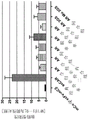

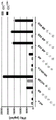


Classifications
-
- C—CHEMISTRY; METALLURGY
- C07—ORGANIC CHEMISTRY
- C07K—PEPTIDES
- C07K14/00—Peptides having more than 20 amino acids; Gastrins; Somatostatins; Melanotropins; Derivatives thereof
- C07K14/435—Peptides having more than 20 amino acids; Gastrins; Somatostatins; Melanotropins; Derivatives thereof from animals; from humans
- C07K14/705—Receptors; Cell surface antigens; Cell surface determinants
- C07K14/70503—Immunoglobulin superfamily
- C07K14/7051—T-cell receptor (TcR)-CD3 complex
-
- A—HUMAN NECESSITIES
- A61—MEDICAL OR VETERINARY SCIENCE; HYGIENE
- A61K—PREPARATIONS FOR MEDICAL, DENTAL OR TOILETRY PURPOSES
- A61K39/00—Medicinal preparations containing antigens or antibodies
- A61K39/0005—Vertebrate antigens
- A61K39/0011—Cancer antigens
- A61K39/001102—Receptors, cell surface antigens or cell surface determinants
-
- A—HUMAN NECESSITIES
- A61—MEDICAL OR VETERINARY SCIENCE; HYGIENE
- A61K—PREPARATIONS FOR MEDICAL, DENTAL OR TOILETRY PURPOSES
- A61K39/00—Medicinal preparations containing antigens or antibodies
- A61K39/46—Cellular immunotherapy
- A61K39/461—Cellular immunotherapy characterised by the cell type used
- A61K39/4611—T-cells, e.g. tumor infiltrating lymphocytes [TIL], lymphokine-activated killer cells [LAK] or regulatory T cells [Treg]
-
- A—HUMAN NECESSITIES
- A61—MEDICAL OR VETERINARY SCIENCE; HYGIENE
- A61K—PREPARATIONS FOR MEDICAL, DENTAL OR TOILETRY PURPOSES
- A61K39/00—Medicinal preparations containing antigens or antibodies
- A61K39/46—Cellular immunotherapy
- A61K39/463—Cellular immunotherapy characterised by recombinant expression
- A61K39/4631—Chimeric Antigen Receptors [CAR]
-
- A—HUMAN NECESSITIES
- A61—MEDICAL OR VETERINARY SCIENCE; HYGIENE
- A61K—PREPARATIONS FOR MEDICAL, DENTAL OR TOILETRY PURPOSES
- A61K39/00—Medicinal preparations containing antigens or antibodies
- A61K39/46—Cellular immunotherapy
- A61K39/464—Cellular immunotherapy characterised by the antigen targeted or presented
- A61K39/4643—Vertebrate antigens
- A61K39/4644—Cancer antigens
-
- A—HUMAN NECESSITIES
- A61—MEDICAL OR VETERINARY SCIENCE; HYGIENE
- A61P—SPECIFIC THERAPEUTIC ACTIVITY OF CHEMICAL COMPOUNDS OR MEDICINAL PREPARATIONS
- A61P35/00—Antineoplastic agents
-
- A—HUMAN NECESSITIES
- A61—MEDICAL OR VETERINARY SCIENCE; HYGIENE
- A61P—SPECIFIC THERAPEUTIC ACTIVITY OF CHEMICAL COMPOUNDS OR MEDICINAL PREPARATIONS
- A61P43/00—Drugs for specific purposes, not provided for in groups A61P1/00-A61P41/00
-
- C—CHEMISTRY; METALLURGY
- C07—ORGANIC CHEMISTRY
- C07K—PEPTIDES
- C07K16/00—Immunoglobulins [IGs], e.g. monoclonal or polyclonal antibodies
- C07K16/18—Immunoglobulins [IGs], e.g. monoclonal or polyclonal antibodies against material from animals or humans
- C07K16/28—Immunoglobulins [IGs], e.g. monoclonal or polyclonal antibodies against material from animals or humans against receptors, cell surface antigens or cell surface determinants
- C07K16/30—Immunoglobulins [IGs], e.g. monoclonal or polyclonal antibodies against material from animals or humans against receptors, cell surface antigens or cell surface determinants from tumour cells
-
- C—CHEMISTRY; METALLURGY
- C12—BIOCHEMISTRY; BEER; SPIRITS; WINE; VINEGAR; MICROBIOLOGY; ENZYMOLOGY; MUTATION OR GENETIC ENGINEERING
- C12N—MICROORGANISMS OR ENZYMES; COMPOSITIONS THEREOF; PROPAGATING, PRESERVING, OR MAINTAINING MICROORGANISMS; MUTATION OR GENETIC ENGINEERING; CULTURE MEDIA
- C12N5/00—Undifferentiated human, animal or plant cells, e.g. cell lines; Tissues; Cultivation or maintenance thereof; Culture media therefor
- C12N5/06—Animal cells or tissues; Human cells or tissues
- C12N5/0602—Vertebrate cells
- C12N5/0634—Cells from the blood or the immune system
- C12N5/0636—T lymphocytes
-
- A—HUMAN NECESSITIES
- A61—MEDICAL OR VETERINARY SCIENCE; HYGIENE
- A61K—PREPARATIONS FOR MEDICAL, DENTAL OR TOILETRY PURPOSES
- A61K39/00—Medicinal preparations containing antigens or antibodies
- A61K2039/51—Medicinal preparations containing antigens or antibodies comprising whole cells, viruses or DNA/RNA
- A61K2039/515—Animal cells
- A61K2039/5156—Animal cells expressing foreign proteins
-
- A—HUMAN NECESSITIES
- A61—MEDICAL OR VETERINARY SCIENCE; HYGIENE
- A61K—PREPARATIONS FOR MEDICAL, DENTAL OR TOILETRY PURPOSES
- A61K39/00—Medicinal preparations containing antigens or antibodies
- A61K2039/58—Medicinal preparations containing antigens or antibodies raising an immune response against a target which is not the antigen used for immunisation
- A61K2039/585—Medicinal preparations containing antigens or antibodies raising an immune response against a target which is not the antigen used for immunisation wherein the target is cancer
-
- A—HUMAN NECESSITIES
- A61—MEDICAL OR VETERINARY SCIENCE; HYGIENE
- A61K—PREPARATIONS FOR MEDICAL, DENTAL OR TOILETRY PURPOSES
- A61K2239/00—Indexing codes associated with cellular immunotherapy of group A61K39/46
- A61K2239/46—Indexing codes associated with cellular immunotherapy of group A61K39/46 characterised by the cancer treated
- A61K2239/59—Reproductive system, e.g. uterus, ovaries, cervix or testes
-
- C—CHEMISTRY; METALLURGY
- C07—ORGANIC CHEMISTRY
- C07K—PEPTIDES
- C07K2317/00—Immunoglobulins specific features
- C07K2317/30—Immunoglobulins specific features characterized by aspects of specificity or valency
- C07K2317/35—Valency
-
- C—CHEMISTRY; METALLURGY
- C07—ORGANIC CHEMISTRY
- C07K—PEPTIDES
- C07K2317/00—Immunoglobulins specific features
- C07K2317/50—Immunoglobulins specific features characterized by immunoglobulin fragments
- C07K2317/52—Constant or Fc region; Isotype
-
- C—CHEMISTRY; METALLURGY
- C07—ORGANIC CHEMISTRY
- C07K—PEPTIDES
- C07K2317/00—Immunoglobulins specific features
- C07K2317/50—Immunoglobulins specific features characterized by immunoglobulin fragments
- C07K2317/52—Constant or Fc region; Isotype
- C07K2317/524—CH2 domain
-
- C—CHEMISTRY; METALLURGY
- C07—ORGANIC CHEMISTRY
- C07K—PEPTIDES
- C07K2317/00—Immunoglobulins specific features
- C07K2317/50—Immunoglobulins specific features characterized by immunoglobulin fragments
- C07K2317/52—Constant or Fc region; Isotype
- C07K2317/526—CH3 domain
-
- C—CHEMISTRY; METALLURGY
- C07—ORGANIC CHEMISTRY
- C07K—PEPTIDES
- C07K2317/00—Immunoglobulins specific features
- C07K2317/50—Immunoglobulins specific features characterized by immunoglobulin fragments
- C07K2317/56—Immunoglobulins specific features characterized by immunoglobulin fragments variable (Fv) region, i.e. VH and/or VL
-
- C—CHEMISTRY; METALLURGY
- C07—ORGANIC CHEMISTRY
- C07K—PEPTIDES
- C07K2317/00—Immunoglobulins specific features
- C07K2317/60—Immunoglobulins specific features characterized by non-natural combinations of immunoglobulin fragments
- C07K2317/62—Immunoglobulins specific features characterized by non-natural combinations of immunoglobulin fragments comprising only variable region components
- C07K2317/622—Single chain antibody (scFv)
-
- C—CHEMISTRY; METALLURGY
- C07—ORGANIC CHEMISTRY
- C07K—PEPTIDES
- C07K2317/00—Immunoglobulins specific features
- C07K2317/70—Immunoglobulins specific features characterized by effect upon binding to a cell or to an antigen
- C07K2317/73—Inducing cell death, e.g. apoptosis, necrosis or inhibition of cell proliferation
-
- C—CHEMISTRY; METALLURGY
- C07—ORGANIC CHEMISTRY
- C07K—PEPTIDES
- C07K2317/00—Immunoglobulins specific features
- C07K2317/70—Immunoglobulins specific features characterized by effect upon binding to a cell or to an antigen
- C07K2317/74—Inducing cell proliferation
-
- C—CHEMISTRY; METALLURGY
- C07—ORGANIC CHEMISTRY
- C07K—PEPTIDES
- C07K2319/00—Fusion polypeptide
- C07K2319/33—Fusion polypeptide fusions for targeting to specific cell types, e.g. tissue specific targeting, targeting of a bacterial subspecies
-
- C—CHEMISTRY; METALLURGY
- C12—BIOCHEMISTRY; BEER; SPIRITS; WINE; VINEGAR; MICROBIOLOGY; ENZYMOLOGY; MUTATION OR GENETIC ENGINEERING
- C12N—MICROORGANISMS OR ENZYMES; COMPOSITIONS THEREOF; PROPAGATING, PRESERVING, OR MAINTAINING MICROORGANISMS; MUTATION OR GENETIC ENGINEERING; CULTURE MEDIA
- C12N2510/00—Genetically modified cells
Abstract
The present application relates to antigen receptors and uses thereof. The invention generally includes treating diseases by targeting cells that express antigens on the cell surface. In particular, the invention relates to recombinant antigen receptors and uses thereof. T cells engineered to express such antigen receptors are useful for treating diseases characterized by expression of one or more antigens bound by the antigen receptor.
Description
The application is a divisional application of a Chinese patent application with the invention name of 201680058729.1 and the application number of "antigen receptor and application thereof", and the original application is a PCT international application PCT/EP2016/073792 filed 2016, 10, 05 and entering the Chinese national stage 2018, 04, 08.
Technical Field
The present invention relates to recombinant antigen receptors and uses thereof. T cells engineered to express such antigen receptors are useful for treating diseases characterized by expression of one or more antigens bound by the antigen receptor.
Background
T cells play a key role in cell-mediated immunity in humans and animals. Recognition and binding of specific antigens is mediated by T Cell Receptors (TCRs) expressed on the surface of T cells. The TCR of a T cell is capable of interacting with an immunogenic peptide (epitope) that binds to a Major Histocompatibility Complex (MHC) molecule and is presented on the surface of a target cell. Specific binding of the TCR triggers a signaling cascade within the T cell, leading to proliferation and differentiation into mature effector T cells.
The TCR is part of a complex signaling machinery, comprising a heterodimeric complex of TCR α and β chains, which is the co-receptor CD4 or CD8 and CD3 signaling modules (fig. 1). The TCR α/β heterodimer is responsible for antigen recognition and transmits activation signals in cooperation with CD3 through the cell membrane, while the CD3 chain itself transfers the input signal to an adaptor protein (adaptor protein) within the cell. Thus, transfer of the TCR α/β chains provides the opportunity to redirect T cells to any antigen of interest.
Adoptive Cell Transfer (ACT) based immunotherapy can be broadly defined as a form of passive immunization with previously sensitized T cells that are transferred to non-immune recipients or autologous hosts after ex vivo expansion from low precursor frequency to clinically relevant cell numbers. Cell types that have been used in ACT experiments include lymphokine-activated killer (LAK) cells (Mule, J.J. et al (1984) Science 225,1487-1489, rosenberg, S.A. et al (1985) N.Engl. J.Med.313, 1485-1492), tumor-infiltrating lymphocytes (tumor-infiltrating lymphocyte, TIL) (Rosenberg, S.A. et al (1994) J.Natl.cancer Instrument.86, 1159-1166), donor lymphocytes after Hematopoietic Stem Cell Transplantation (HSCT), and tumor-specific T cell lines or clones (Dudley, M.E. et al (2001) J.20, yes, Y.363, C. Et al (Acc. 1618.16168, 16173 A.16173. Adoptive T cell transfer has been shown to be therapeutically active against human viral infections such as CMV. For adoptive immunotherapy of melanoma, rosenberg and colleagues established an ACT method that relies on the infusion of ex vivo expanded autologous Tumor Infiltrating Lymphocytes (TILs) isolated from resected tumors combined with non-myeloablative lymphodepleting chemotherapy and high doses of IL2. Clinical studies have resulted in objective response rates of about 50% in treated patients with metastatic melanoma (Dudley, M.E. et al (2005) J.Clin.Oncol.23: 2346-2357).
An alternative approach is adoptive transfer of autologous T cells reprogrammed to express tumor reactive immune receptors of defined specificity during a short ex vivo culture period, and then reinfused into the patient (Kershaw M.H. et al (2013) Nature Reviews Cancer 13 (8): 525-41). This strategy makes ACT applicable to a variety of common malignancies, even in patients lacking tumor-reactive T cells. Since the antigen specificity of T cells is completely dependent on the heterodimeric complex of TCR α and β chains, transferring the cloned TCR gene into T cells offers the potential to redirect it to any antigen of interest. Thus, TCR gene therapy offers an attractive strategy to develop antigen-specific immunotherapy with autologous lymphocytes as a therapeutic option. The main advantages of TCR gene transfer are the generation of therapeutic amounts of antigen-specific T cells within a few days, and the possibility of introducing specificities that are not present in the patient's endogenous TCR repertoire. Several groups have demonstrated that TCR gene transfer is an attractive strategy for redirecting antigen specificity of primary T cells (Morgan, R.A. et al (2003) J.Immunol.171,3287-3295 Cooper, L.J.et al (2000) J.Virol.74,8207-8212, K.et al (2000) J.Immunol.165,528-532 Kessels, H.W.et al (2001) Nat.Immunol.2,957-961 Dembic, Z.et al (1986) Nature 320, 232-238. The feasibility of TCR gene therapy in humans was initially demonstrated by Rosenberg and its team in clinical trials for the treatment of malignant melanoma. Adoptive transfer of autologous lymphocytes transduced with a retrovirus with a melanoma/melanocyte antigen specific TCR resulted in cancer regression in up to 30% of treated melanoma patients (Morgan, r.a. et al (2006) Science 314,126-129 johnson, l.a. et al (2009) Blood 114, 535-546. At the same time, clinical testing for TCR gene therapy has also extended to cancers other than melanoma that target many different tumor antigens (Park, t.s. Et al (2011) Trends biotechnol.29, 550-557).
The potential of ACT has been greatly extended using genetic engineering methods to insert antigen-targeting receptors with defined specificity into T cells. Chimeric Antigen Receptors (CARs) are a class of antigen-targeting receptors consisting of an intracellular T cell signaling domain fused to an extracellular antigen-binding domain, most commonly a single-chain variable fragment (scFv) from a monoclonal antibody. CARs recognize cell surface antigens directly, independent of MHC-mediated presentation, allowing the use of a single receptor construct specific for any given antigen in all patients. The original CAR fused the antigen recognition domain to the CD3 zeta active chain of the T Cell Receptor (TCR) complex (figure 2). Subsequent CAR iterations included secondary costimulatory signals in tandem with CD3 ζ, including intracellular domains from CD28 or various TNF receptor family molecules, such as 4-1BB (CD 137) and OX40 (CD 134). Furthermore, the third generation receptor contains two costimulatory signals in addition to CD3 ζ, most commonly from CD28 and 4-1BB. Second and third generation CARs significantly improved the in vitro and in vivo antitumor efficacy (Zhao et al (2009) j.immunol., (183) 5563-5574), in some cases inducing complete remission in advanced cancer patients (Porter et al (2011) n.engl.j.med., (365) 725-733).
Classical CARs consist of an antigen-specific single chain antibody (scFv) fragment fused to a transmembrane signaling domain (e.g., CD3 ζ). After introduction into T cells, it is expressed as a membrane-bound protein and induces an immune response upon binding to its cognate antigen (Eshhar et al (1993) PNAS, (90) 720-724). The induced antigen-specific immune response results in the activation of cytotoxic CD8+ T cells, which in turn results in the eradication of cells expressing the specific antigen, such as tumor cells or virus-infected cells expressing the specific antigen. However, these classical CAR constructs are unable to activate/stimulate T cells through their endogenous CD3 complex, which is often necessary for T cell activation. Since the antigen binding domain is fused to CD3 ζ, T cell activation is induced by biochemical "short circuit" (Aggen et al (2012) Gene Therapy, (19) 365-374). This non-physiological activation of T cells by this biochemical short circuit poses a risk to patients treated in this way, as over-activation of T cells may lead to undesirable side effects. For example, long-term basal activation of recombinant T cells caused by CAR expression has been observed in vitro ("tonic signaling"), which results in increased accumulation of inhibitory molecules (e.g., LAG-3, tim-3 and PD-1) on the surface of recombinant CAR-expressing T cells, which in turn results in premature depletion of T cells, followed by a strong negative impact on the response to tumor cells in vivo (Long et al (2015) nat. Med., (21) 581-590). This adverse reaction is associated with irregular clustering of scFv fragments by the framework residues of the antibody. In addition, although this type of classical CAR construct has been successfully tested for different neoplasias (e.g. leukemia) (Porter et al (2011) n.engl.j.med., (365) 725-733), it also leads to fatal autoimmune diseases (off-target/off-tumor-reaction) due to basal expression of target antigens (target tumor antigens) in normal tissues; morgan et al (2010) Mol ther., (18) 843-51).
An alternative approach, in which T Cell activation occurs by a more physiological mechanism, is to provide a similar single chain-TCR (scTv) fragment fused to the C β constant domain from the T Cell Receptor (TCR) and its co-expression with the C α constant domain from the TCR (Voss et al (2010) Blood, (115) 5154-5163), which recruits the essential endogenous CD3 ζ homodimer (Call et al (2002) Cell, (111) 967-79). However, in order for these constructs to act as immune system activators, it is necessary that the constant domains from the TCR be derived from a mouse TCR or murine (Cohen et al (2006) Cancer res., (66) 8878-86) to achieve chain pairing between the scTCR and ca. The fact that these constructs must have heterologous sequences for their function increases the risk of the immune system reacting with them upon administration and reduces or destroys their therapeutic effect.
Thus, there is a need to provide alternative recombinant antigen receptors, wherein e.g. the receptor is sufficient upon antigen binding to be able to activate T cells expressing it in a normal physiological manner by the endogenous CD3 complex, and optionally, at least in the signaling domain of the antigen receptor, does not require the presence of any amino acid sequences of non-human origin, which may induce an unwanted immune response against the recombinant antigen receptor itself.
Description of the invention
Disclosure of Invention
The present invention relates to recombinant antigen receptors having at least two antigen binding sites. The antigen receptor comprises two peptide chains. In one aspect, the peptide chains each comprise at least two domains in addition to the immunoreceptor signaling domain, wherein each of the two domains on one peptide chain forms an antigen binding site with one domain on the other peptide chain. In another aspect, a peptide chain has at least four domains in addition to the immunoreceptor signaling domain, wherein two of the four domains form two antigen binding sites with two remaining domains on the same peptide chain.
In one aspect, the present invention relates to an antigen receptor comprising a first peptide chain and a second peptide chain, wherein the first peptide chain comprises at least a first domain and a second domain and an immunoreceptor signaling domain; the second peptide chain comprises at least a first domain and a second domain and an immunoreceptor signaling domain; wherein the first domain from the first peptide chain forms a first antigen binding site together with one domain from the second peptide chain, and wherein the second domain from the first peptide chain forms a second antigen binding site together with another domain from the second peptide chain. In the antigen receptor of this aspect, the domains forming the respective antigen binding sites are preferably located on different peptide chains. Thus, the antigen binding site is formed by intermolecular interaction of the domains.
In one embodiment, the first domain and/or the second domain each comprises a variable region of an immunoglobulin chain or a variable region of a T cell receptor chain or a portion of said variable region.
In one embodiment, one of the domains forming the first antigen binding site comprises a heavy chain variable region of an immunoglobulin, or a portion thereof, specific for an antigen, and the other domain forming the first antigen binding site comprises a light chain variable region of an immunoglobulin, or a portion thereof, specific for an antigen. In one embodiment, one of the domains forming the second antigen-binding site comprises a heavy chain variable region of an immunoglobulin, or a portion thereof, specific for the antigen, and the other domain forming the second antigen-binding site comprises a light chain variable region of an immunoglobulin, or a portion thereof, specific for the antigen.
In one embodiment, the first domain from the first peptide chain comprises a heavy chain variable region of an immunoglobulin, or a portion thereof, specific for the antigen, and the domain from the second peptide chain that forms an antigen binding site with the first domain from the first peptide chain comprises a light chain variable region of an immunoglobulin, or a portion thereof, specific for the antigen. In one embodiment, the second domain from the first peptide chain comprises a heavy chain variable region of an immunoglobulin, or a portion thereof, specific for the antigen, and the domain from the second peptide chain that forms an antigen binding site with the second domain from the first peptide chain comprises a light chain variable region of an immunoglobulin, or a portion thereof, specific for the antigen.
In one embodiment, the first domain and the second domain from the first peptide chain each comprise a heavy chain variable region of an immunoglobulin, or a portion thereof; and the first domain and the second domain from the second peptide chain each comprise a light chain variable region of an immunoglobulin or a portion thereof.
In one embodiment, the N-terminal domain from the first peptide chain forms an antigen binding site together with the N-terminal domain from the second peptide chain; and the C-terminal domain from the first peptide chain forms together with the C-terminal domain from the second peptide chain an antigen binding site.
In one embodiment, the N-terminal domain from the first peptide chain forms an antigen binding site together with the C-terminal domain from the second peptide chain; and the C-terminal domain from the first peptide chain forms an antigen binding site together with the N-terminal domain from the second peptide chain.
In one aspect, the invention relates to an antigen receptor comprising a first peptide chain and a second peptide chain, wherein the first peptide chain comprises at least four domains and an immunoreceptor signaling domain; the second peptide chain comprises an immunoreceptor signaling domain; wherein two domains from the first peptide chain form a first antigen binding site and wherein two further domains from the first peptide chain form a second antigen binding site. In the antigen receptor of this aspect, the domains forming the respective antigen binding sites are preferably located on the same peptide chain. Thus, the antigen binding site is formed by intramolecular interaction of the domains.
In one embodiment, each of the four domains comprises a variable region of an immunoglobulin chain or a variable region of a T cell receptor chain or a portion of said variable region.
In one embodiment, one of the domains forming the first antigen binding site comprises a heavy chain variable region of an immunoglobulin, or a portion thereof, specific for an antigen, and the other domain forming the first antigen binding site comprises a light chain variable region of an immunoglobulin, or a portion thereof, specific for an antigen. In one embodiment, one of the domains forming the second antigen-binding site comprises a heavy chain variable region of an immunoglobulin, or a portion thereof, specific for the antigen, and the other domain forming the second antigen-binding site comprises a light chain variable region of an immunoglobulin, or a portion thereof, specific for the antigen.
In one embodiment, the two N-terminal domains of the four domains together form an antigen binding site; and the two C-terminal domains of the four domains together form the antigen binding site.
In one embodiment, one of the two N-terminal domains of the four domains comprises a heavy chain variable region of an immunoglobulin or a portion thereof, and the other of the two N-terminal domains of the four domains comprises a light chain variable region of an immunoglobulin or a portion thereof; and one of the two C-terminal domains of the four domains comprises a heavy chain variable region of an immunoglobulin or a portion thereof and the other of the two C-terminal domains of the four domains comprises a light chain variable region of an immunoglobulin or a portion thereof.
In one embodiment of the antigen receptor of the invention, the immunoreceptor signaling domain comprises a constant region or invariant region of a T cell receptor chain or a constant region or invariant region of an immune cell Fc receptor chain or a portion of said constant region or invariant region. In one embodiment of the antigen receptor of the invention, (i) the first peptide chain comprises a constant region of a T cell receptor alpha chain or a portion thereof and the second peptide chain comprises a constant region of a T cell receptor beta chain or a portion thereof, or (ii) the first peptide chain comprises a constant region of a T cell receptor beta chain or a portion thereof and the second peptide chain comprises a constant region of a T cell receptor alpha chain or a portion thereof.
In one embodiment of the antigen receptor of the present invention, the immunoreceptor signaling domain is of human origin.
In one embodiment, the antigen receptor of the present invention comprises a linker that connects the domains of the antigen receptor. In one embodiment, the antigen receptor of the present invention comprises one or more linkers between the domains forming the antigen binding site and/or between the domains forming the antigen binding site and the immunoreceptor signaling domain. The linker may be any arbitrary amino acid sequence of any length so long as it does not interfere with the function of the antigen receptor, e.g., the ability of the antigen receptor to bind to an antigen or to associate with an endogenous CD3 complex, or to induce an immune response upon antigen binding.
In one embodiment of the antigen receptor of the present invention, the first antigen binding site and the second antigen binding site bind to the same antigen or different antigens. In one embodiment of the antigen receptor of the present invention, the first antigen binding site and the second antigen binding site bind to different epitopes on the same antigen. Thus, while the domains forming the first antigen-binding site are preferably from the same immunoglobulin and the domains forming the second antigen-binding site are preferably from the same immunoglobulin, the domains forming the first antigen-binding site and the domains forming the second antigen-binding site are from the same or different immunoglobulins that bind to the same or different antigens.
In one embodiment, the antigen is a disease-specific antigen, preferably a tumor antigen. In one embodiment, the antigen is expressed on the surface of a cell.
In one aspect, the invention relates to a peptide chain of any of the antigen receptors of the invention. In one embodiment, the present invention relates to a peptide chain comprising a first domain and a second domain, each comprising a heavy chain variable region of an immunoglobulin or a portion thereof or each comprising a light chain variable region of an immunoglobulin or a portion thereof, and wherein the peptide chain further comprises an immunoreceptor signaling domain. In one embodiment, the invention relates to a peptide chain comprising at least 4 domains and an immunoreceptor signaling domain, wherein two domains from the peptide chain form a first antigen-binding site and the other two domains from the peptide chain form a second antigen-binding site. Further embodiments of the peptide chains of the invention are as described herein for the antigen receptors of the invention.
In one aspect, the invention relates to cells, particularly immune effector cells, such as T cells, that are genetically modified to express the antigen receptors of the invention. In one aspect, the invention relates to recombinant cells, in particular immune effector cells, such as T cells, expressing the first peptide chain, the second peptide chain or both the first and second peptide chains of the antigen receptor of the invention. Further embodiments of the cell or recombinant cell of the invention are as described herein for the antigen receptor of the invention or the peptide chain of the invention.
In one aspect, the invention relates to a method for producing a cell expressing an antigen receptor of the invention, the method comprising: (ii) (a) providing a cell; (b) Providing a first genetic construct encoding a first peptide chain of an antigen receptor of the invention; (c) Providing a second genetic construct encoding a second peptide chain of the antigen receptor of the invention; (d) Introducing a first genetic construct and a second genetic construct into a cell; and (e) allowing the construct to be expressed in the cell. In one embodiment, the invention relates to a method for producing a cell expressing an antigen receptor, said receptor comprising a first peptide chain and a second peptide chain, said method comprising: (a) providing a cell; (b) Providing a first genetic construct encoding a first peptide chain comprising at least a first domain and a second domain and an immunoreceptor signaling domain; (c) Providing a second genetic construct encoding a second peptide chain comprising at least a first domain and a second domain and an immunoreceptor signaling domain; (d) Introducing a first genetic construct and a second genetic construct into a cell; and (e) allowing the construct to be expressed in the cell; wherein a first domain from a first peptide chain is capable of forming a first antigen binding site with one domain from a second peptide chain, and wherein a second domain from a first peptide chain is capable of forming a second antigen binding site with another domain from a second peptide chain. In one embodiment, the invention relates to a method for producing a cell expressing an antigen receptor, said receptor comprising a first peptide chain and a second peptide chain, said method comprising: (ii) (a) providing a cell; (b) Providing a first genetic construct encoding a first peptide chain comprising at least four domains and an immunoreceptor signaling domain; (c) Providing a second genetic construct encoding a second peptide chain comprising an immunoreceptor signaling domain; (d) Introducing a first genetic construct and a second genetic construct into a cell; and (e) allowing the construct to be expressed in the cell; wherein two domains from the first peptide chain are capable of forming a first antigen binding site, and wherein two further domains from the first peptide chain are capable of forming a second antigen binding site. In one embodiment of the method of the invention, the expression of the antigen receptor is at the cell surface. In one embodiment of the method of the invention, the first peptide chain and the second peptide chain are provided on a single genetic construct. In one embodiment of the method of the invention, the cell is a human cell. In one embodiment of the method of the invention, the cell is an immune effector cell, such as a T cell. In one embodiment of the method of the invention, the genetic construct comprises DNA and/or RNA. Further embodiments of the methods of the invention are as described herein for the antigen receptor of the invention.
In one aspect, the invention relates to recombinant cells, in particular immune effector cells, such as T cells, produced by the methods of the invention for producing cells expressing an antigen receptor. Further embodiments of the recombinant cells of the invention are as described herein for the antigen receptor of the invention or the method of the invention for producing a cell expressing an antigen receptor.
In one aspect, the invention relates to a first peptide chain, a second peptide chain, or both a first peptide chain and a second peptide chain encoding an antigen receptor of the invention, or a nucleic acid, such as DNA or RNA, encoding a peptide chain of the invention. Further embodiments of the nucleic acids of the invention are as described herein for the antigen receptor of the invention or the peptide chain of the invention.
The invention generally includes treating a disease by targeting cells expressing one or more antigens on the cell surface, such as diseased cells expressing one or more disease-specific antigens on the cell surface, particularly cancer cells expressing one or more tumor antigens on the cell surface, using the antigen receptors of the invention. The method provides for the selective eradication of cells expressing one or more antigens on their surface, thereby minimizing adverse effects on normal cells not expressing the antigens. In one embodiment, T cells genetically modified to express the antigen receptors of the invention that target the cells by binding to an antigen are administered. T cells are able to recognize diseased cells that express antigens on the cell surface, resulting in eradication of the diseased cells. In one embodiment, the target cell population or target tissue is a tumor cell or tumor tissue.
In one aspect, the invention relates to a pharmaceutical composition comprising an antigen receptor of the invention, a recombinant cell of the invention, or a nucleic acid of the invention; and a pharmaceutically acceptable carrier. The pharmaceutical compositions of the invention are useful as medicaments, particularly for the treatment of diseases, such as cancer, characterized by expression of one or more antigens (e.g., one or more tumor antigens) bound by the antigen receptor of the invention.
In one aspect, the invention relates to a method of treating a disease (e.g., cancer) comprising administering to a subject a therapeutically effective amount of a pharmaceutical composition of the invention, wherein the disease is characterized by expression of at least one antigen (e.g., a tumor antigen) bound by the antigen receptor.
In one aspect, the invention relates to a method of treating a subject having a disease, disorder or condition associated with expression or elevated expression of at least one antigen, the method comprising administering to the subject a T cell genetically modified to express an antigen receptor of the invention that targets the at least one antigen. In one embodiment, the disease, disorder or condition is cancer. In one embodiment, the T cell pair may be autologous, allogeneic or syngeneic.
In one embodiment of the invention, the antigen receptor binds to only one antigen (e.g. is monospecific and recognizes the same epitope or is bispecific or multispecific and recognizes different epitopes on the same antigen) or to different antigens, in particular two different antigens.
In one embodiment of all aspects of the invention, the method of treatment further comprises obtaining a sample of cells from the subject, said sample comprising T cells or T cell progenitor cells, and transfecting said cells with a nucleic acid encoding an antigen receptor of the invention to provide T cells genetically modified to express the antigen receptor. In one embodiment of all aspects of the invention, T cells genetically modified to express an antigen receptor are stably or transiently transfected with a nucleic acid encoding the antigen receptor. Thus, the nucleic acid encoding the antigen receptor may or may not be integrated into the genome of the T cell. In one embodiment of all aspects of the invention, the T cells and/or cell sample are from a subject to whom T cells genetically modified to express an antigen receptor have been administered. In one embodiment of all aspects of the invention, the T cells and/or cell sample are from a mammal different from the mammal to which the T cells genetically modified to express the antigen receptor are administered.
In one embodiment of all aspects of the invention, T cells genetically modified to express an antigen receptor are inactivated to express an endogenous T cell receptor and/or an endogenous HLA.
In one embodiment of all aspects of the invention, the antigen is expressed in a diseased cell (e.g., a cancer cell). In one embodiment, the antigen is expressed on the surface of a diseased cell (e.g., a cancer cell). In one embodiment, the antigen receptor binds to an extracellular domain or an epitope in the extracellular domain of the antigen. In one embodiment, the antigen receptor binds to a native epitope of an antigen present on the surface of a living cell. In one embodiment of all aspects of the invention, the antigen is a tumor antigen. In one embodiment of all aspects of the invention, the antigen is selected from claudin (e.g.claudin 6 and claudin 18.2), CD19, CD20, CD22, CD33, CD123, mesothelin, CEA, c-Met, PSMA, GD-2 and NY-ESO-1. In one embodiment of all aspects of the invention, the antigen is a pathogen antigen. The pathogen may be a fungal, viral or bacterial pathogen. In one embodiment of all aspects of the invention, the expression of the antigen is at the cell surface. In one embodiment, the antigen is claudin, in particular claudin 6 or claudin 18.2, and said antigen receptor binds to the first extracellular loop of said claudin. In one embodiment, when expressed by and/or present on a T cell, binding of the antigen receptor to an antigen present on the cell results in an immune effector function of the T cell, e.g., release of a cytokine. In one embodiment, when expressed by and/or present on a T cell, binding of the antigen receptor to an antigen present on the cell (e.g., an antigen presenting cell) results in stimulation, priming, and/or expansion of the T cell. In one embodiment, binding of the antigen receptor to an antigen present on a diseased cell (e.g., a cancer cell), when expressed by and/or present on a T cell, wherein the T cell preferably releases cytotoxic factors such as perforin and granzyme, results in cytolysis and/or apoptosis of the diseased cell.
In one embodiment of all aspects of the invention, the domain of the antigen receptor forming the antigen binding site is comprised in the exodomain (exodomain) of the antigen receptor. In one embodiment of all aspects of the invention, the antigen receptor of the invention comprises a transmembrane domain. In one embodiment, the transmembrane domain is a transmembrane hydrophobic alpha helix.
In one embodiment of all aspects of the invention, the antigen receptor of the invention comprises a signal peptide that directs the nascent protein into the endoplasmic reticulum. In one embodiment, the signal peptide precedes the domain that forms the antigen binding site.
In one embodiment of all aspects of the invention, the antigen receptor of the invention is preferably specific for the antigen it is targeting, especially when it is present on the surface of a cell (e.g. a diseased cell or an antigen presenting cell).
In one embodiment of all aspects of the invention, the antigen receptor of the invention may be expressed by or present on the surface of a T cell, preferably a cytotoxic T cell. In one embodiment, the T cells are reactive with an antigen targeted by an antigen receptor of the present invention.
In another aspect, the invention provides agents and compositions described herein for use in the methods described herein.
Other features and advantages of the invention will be apparent from the following detailed description, and from the claims.
Detailed Description
Although the invention is described in detail below, it is to be understood that this invention is not limited to the particular methodology, protocols, and reagents described herein as these may vary. It is also to be understood that the terminology used herein is for the purpose of describing particular embodiments only, and is not intended to limit the scope of the present invention which will be limited only by the appended claims. Unless defined otherwise, all technical and scientific terms used herein have the same meaning as commonly understood by one of ordinary skill in the art.
Hereinafter, elements of the present invention will be described. These elements are listed with particular embodiments, however, it should be understood that they may be combined in any manner and in any number to produce additional embodiments. The various described examples and preferred embodiments should not be construed as limiting the invention to only the explicitly described embodiments. This description should be understood to support and encompass embodiments combining the explicitly described embodiments with any number of the disclosed and/or preferred elements. Further, unless the context indicates otherwise, the description of the present application is deemed to disclose any permutation and combination of all described elements in the present application.
Preferably, the terms used herein are defined as described in the following: "A multilinual gloss of biological meters (IUPAC Recommendations)", H.G.W.Leuenberger, B.Nagel, and H. Eds, (1995) Helvetica Chimica Acta, CH-4010 Basel, switzerland.
The practice of the present invention will employ, unless otherwise indicated, conventional methods of biochemistry, cell biology, immunology and recombinant DNA techniques explained in the literature of the art (see, e.g., molecular Cloning: A Laboratory Manual, 2 nd edition, J.Sambrook et al, cold Spring Harbor Laboratory Press, cold Spring Harbor 1989).
Throughout this specification and the claims which follow, unless the context requires otherwise, the word "comprise", and variations such as "comprises" and "comprising", will be understood to imply the inclusion of a stated member, integer or step or group of members, integers or steps but not the exclusion of any other member, integer or step or group of members, integers or steps although in some embodiments such other members, integers or steps or groups of members, integers or steps may be excluded, i.e. the subject matter is intended to include the stated member, integer or step or group of members, integers or steps. In the context of describing the invention (especially in the context of the claims), terms that are not defined by numerical terms should be construed to cover both the singular and the plural, unless otherwise indicated herein or clearly contradicted by context. Recitation of ranges of values herein are merely intended to serve as a shorthand method of referring individually to each separate value falling within the range. Unless otherwise indicated herein, each separate value is incorporated into the specification as if it were individually recited herein.
All methods described herein can be performed in any suitable order unless otherwise indicated herein or otherwise clearly contradicted by context. The use of any and all examples, or exemplary language (e.g., "such as") provided herein, is intended merely to better illuminate the invention and does not pose a limitation on the scope of the invention otherwise claimed. No language in the specification should be construed as indicating any non-claimed element as essential to the practice of the invention.
Throughout this specification, various documents are cited. Each of the documents cited herein (including all patents, patent applications, scientific publications, manufacturer's specifications, instructions, etc.), whether supra or infra, is hereby incorporated by reference in its entirety. Nothing herein is to be construed as an admission that the invention is not entitled to antedate such disclosure by virtue of prior invention.
The term "immune response" refers to the overall bodily response to an antigen, and preferably refers to a cellular or cellular as well as a humoral immune response. The immune response may be protective/prophylactic/preventative and/or therapeutic.
By "providing an immune response" may be meant that there is no immune response to a particular target antigen, target cell and/or target tissue before providing an immune response, but may also mean that there is a certain level of immune response to a particular target antigen, target cell and/or target tissue before providing an immune response and that the immune response is enhanced after providing an immune response. Thus, "providing an immune response" includes "inducing an immune response" and "enhancing an immune response". Preferably, after providing an immune response in a subject, the subject is protected from developing a disease, such as a cancer disease, or the disease condition is ameliorated by providing an immune response. For example, an immune response against a tumor antigen can be provided in a patient having a cancer disease or a subject at risk of developing a cancer disease. Providing an immune response in this case may mean that the disease status of the subject is improved, that the subject does not develop metastasis, or that the subject at risk of developing a cancer disease does not develop a cancer disease.
"cell-mediated immunity" or "cellular immunity" or similar terms are intended to include cellular responses against cells characterized by expression of an antigen, particularly by presentation of the antigen with class I or class II MHC. Cellular responses are associated with cells called T cells or T lymphocytes, which act as "helper cells" or "killer cells". Helper T cell (also known as CD 4) + T cells) exert a central role by modulating the immune response, killing cells (also known as cytotoxic T cells, cytolytic T cells, CD 8) + T cells or CTLs) kill diseased cells (e.g., cancer cells) thereby preventing the production of more diseased cells.
The term "antigen" relates to a substance comprising an epitope against which an immune response is generated and/or directed. Preferably, an antigen in the context of the present invention is a molecule which, optionally after processing, induces an immune response, which is preferably specific for the antigen or the cell expressing the antigen, preferably on the cell surface. The term "antigen" includes in particular proteins and peptides. The antigen is preferably a product corresponding to or derived from a naturally occurring antigen. Such naturally occurring antigens may include or may be derived from allergens, viruses, bacteria, fungi, parasites and other infectious agents and pathogens, or the antigen may also be a tumor antigen. According to the invention, the antigen may correspond to a naturally occurring product, such as a viral protein or a portion thereof.
The term "pathogen" relates to pathogenic microorganisms and includes viruses, bacteria, fungi, unicellular organisms and parasites. Examples of pathogenic viruses are Human Immunodeficiency Virus (HIV), cytomegalovirus (CMV), herpes virus (HSV), hepatitis A Virus (HAV), HBV, HCV, papilloma virus and Human T Lymphotropic Virus (HTLV). Unicellular organisms include plasmodium, trypanosomes, amoebae, and the like.
In a preferred embodiment, the antigen is a disease-specific antigen or a disease-associated antigen. The term "disease-specific antigen" or "disease-associated antigen" refers to all antigens that have a pathological significance. In a particularly preferred embodiment, the antigen is present in diseased cells, tissues and/or organs, whereas it is absent or present in reduced amounts in healthy cells, tissues and/or organs, and thus can be used to target diseased cells, tissues and/or organs, e.g., by T cells carrying antigen receptors targeting the antigen. In one embodiment, the disease-specific antigen or disease-associated antigen is present on the surface of the diseased cell.
In a preferred embodiment, the antigen is a tumor antigen or tumor-associated antigen, i.e. a component of a cancer cell, which may be derived from the cytoplasm, the cell surface and the nucleus, in particular those antigens which are produced, preferably in large amounts, as surface antigens on cancer cells.
In the context of the present invention, the term "tumor antigen" or "tumor-associated antigen" relates to a protein which is specifically expressed in a limited number of tissues and/or organs or in a specific developmental stage under normal conditions, e.g. a tumor antigen may be specifically expressed in gastric tissue (preferably in the gastric mucosa), in reproductive organs (e.g. in the testis), in trophoblast tissue (e.g. in the placenta) or in germ-line cells and expressed or aberrantly expressed in one or more tumor or cancer tissues under normal conditions. In this context, "limited number" preferably means not more than 3, more preferably not more than 2. Tumor antigens in the context of the present invention include, for example, differentiation antigens, preferably cell type-specific differentiation antigens, i.e., proteins that are specifically expressed in a particular cell type at a certain differentiation stage under normal conditions; cancer/testis antigens, i.e., proteins that are normally specifically expressed in the testis and sometimes in the placenta; and germline specific antigens. In the context of the present invention, the tumor antigen is preferably associated with the cell surface of cancer cells and is preferably not or only rarely expressed in normal tissues. Preferably, the cancer cell is identified by a tumor antigen or an aberrant expression of a tumor antigen. In the context of the present invention, the tumor antigen expressed by the cancer cells in the subject (e.g. a patient suffering from a cancer disease) is preferably a self-protein in said subject. In some preferred embodiments, the tumor antigen in the context of the present invention is specifically expressed under normal conditions in non-essential tissues or organs (i.e. tissues or organs which do not lead to the death of the subject when damaged by the immune system) or in body organs or tissues which are not or only hardly accessible to the immune system. Preferably, the amino acid sequence of the tumor antigen is identical between the tumor antigen expressed in normal tissue and the tumor antigen expressed in cancer tissue.
Examples of tumor antigens useful in the present invention are p53, ART-4, BAGE, β -catenin/M, bcr-abL CAMEL, CAP-1, CASP-8, CDC27/M, CDK4/M, CEA, cell surface proteins of the CLAUDIN family (e.g. CLAUDIN-6, CLAUDIN-18.2 and CLAUDIN-12), C-MYC, CT, cyp-B, DAM, ELF2M, ETV6-AML1, G250, GAGE, gnT-V, gap100, HAGE, HER-2/neu, HPV-E7, HPV-E6, HAST-2, hT (or hTRT), LAGE, LR/FUT, MAGE-A (preferably MAGE-A1, MAGE-A2, MAGE-A3, MAGE-A4 MAGE-A5, MAGE-A6, MAGE-A7, MAGE-A8, MAGE-A9, MAGE-A10, MAGE-A11 or MAGE-A12), MAGE-B, MAGE-C, MART-1/Melan-A, MC1R, myosin/M, MUC1, MUM-2, MUM-3, NA88-A, NF1, NY-ESO-1, NY-BR-1, p190 small BCR-abL, pm1/RARa, PRAME, proteinase 3, PSA, PSM, RAGE, RU1 or RU2, SAGE, SART-1 or SART-3, SCGB3A2, SCGB 1, SCP2, SCP3, SSX, SUIVIN, TEL/AML1, RVI/M, TRP-1, TRP-2, TRP-A-2, TRP-2/WT 2, and TPWT-A2. Particularly preferred tumor antigens include CLAUDIN-18.2 (CLDN 18.2) and CLAUDIN-6 (CLDN 6).
The term "CLDN" or simply "C1" as used herein refers to claudins, including CLDN6 and CLDN18.2. Preferably, the claudin is human claudin. Claudins are a family of proteins that are the most important components of tight junctions, where they establish a paracellular barrier that controls the flow of molecules in the intercellular space between epithelial cells. Claudins are transmembrane proteins with 4 times of transmembrane, with both the N-and C-termini located in the cytoplasm. The first extracellular loop, designated EC1 or ECL1, consists of an average of 53 amino acids, and the second extracellular loop, designated EC2 or ECL2, consists of about 24 amino acids. Cell surface proteins of the claudin family are expressed in tumors of various origins and are particularly suitable as target structures associated with targeted cancer immunotherapy due to their selective expression (not expressed in normal tissues associated with toxicity) and localization on the plasma membrane.
CLDN6 and CLDN18.2 have been identified as differentially expressed in tumor tissues, with the only normal tissue expressing CLDN18.2 being the stomach (differentiated epithelial cells of the gastric mucosa) and the only normal tissue expressing CLDN6 being the placenta.
CLDN18.2 is expressed in cancers of various origins, such as pancreatic cancer, esophageal cancer, gastric cancer, bronchial cancer, breast cancer and ENT tumors. CLDN18.2 is a valuable target for the prevention and/or treatment of primary tumors, such as gastric, esophageal, pancreatic, lung (e.g., non-small cell lung cancer (NSCLC)), ovarian, colon, liver, head and neck, and gallbladder cancers, and their metastases, in particular gastric cancer metastasis (e.g., krukenberg tumor), peritoneal metastasis, and lymph node metastasis. Antigen receptors targeting at least CLDN18.2 may be useful for treating such cancer diseases.
CLDN6 has been found to be expressed in, for example, ovarian cancer, lung cancer, gastric cancer, breast cancer, liver cancer, pancreatic cancer, skin cancer, melanoma, head and neck cancer, sarcoma, cholangiocarcinoma, renal cell carcinoma, and bladder cancer. CLDN6 is a particularly preferred target for the prevention and/or treatment of: ovarian cancer, particularly ovarian adenocarcinoma and ovarian teratoma; lung cancer, including Small Cell Lung Cancer (SCLC) and non-small cell lung cancer (NSCLC), particularly squamous cell lung cancer and adenocarcinoma; stomach cancer, breast cancer, liver cancer, pancreatic cancer; skin cancer, particularly basal cell carcinoma and squamous cell carcinoma; malignant melanoma; head and neck cancer, particularly malignant polymorphic adenomas; sarcomas, particularly synovial sarcoma and carcinosarcoma; bile duct cancer; bladder cancer, particularly transitional cell carcinoma and papillary carcinoma; kidney cancers, particularly renal cell carcinoma, including clear cell renal cell carcinoma and papillary renal cell carcinoma; colon cancer, small bowel cancer, including ileal cancer, particularly small bowel adenocarcinoma and ileal adenocarcinoma; testicular embryonal carcinoma, placental choriocarcinoma, cervical carcinoma, testicular cancer, in particular testicular seminoma, testicular teratoma, and embryonal testicular cancer; uterine cancer, germ cell tumors, such as teratocarcinoma or embryonal carcinoma, in particular germ cell tumors of the testis, and metastatic forms thereof. Antigen receptors targeting at least CLDN6 are useful for treating these cancer diseases.
In the context of embodiments of the present invention, the antigen is preferably present on the surface of a cell, preferably an antigen presenting cell or a diseased cell. According to the invention, an antigen, if it binds to an antigen receptor, is preferably capable of inducing stimulation, priming and/or expansion of T cells carrying the antigen receptor bound to the antigen, optionally in the presence of an appropriate co-stimulatory signal. Recognition of an antigen on the surface of a diseased cell can result in an immune response against the antigen (or cells expressing the antigen).
According to various aspects of the present invention, it is preferred that the object is to provide an immune response against antigen expressing diseased cells (e.g. cancer cells expressing antigens such as tumor antigens, in particular CLDN6 or CLDN 18.2) and to treat diseases (e.g. cancer diseases) involving antigen expressing (e.g. tumor antigen) cells. Preferably, the invention relates to the administration of antigen receptor engineered immune effector cells, such as T cells targeted to diseased cells expressing the antigen. Cells expressing an antigen on their surface can be targeted by immune effector cells carrying an antigen receptor that targets the antigen.
"cell surface" is used according to its usual meaning in the art and thus includes the exterior of a cell accessible for binding by proteins and other molecules. An antigen is expressed on the surface of a cell if it is located at the surface of the cell and accessible for binding by an antigen binding molecule (such as an antigen receptor or an antigen-specific antibody) added to the cell. In one embodiment, the antigen expressed on the cell surface is an intact membrane protein with an extracellular portion that is recognized by an antigen receptor. An antigen receptor is expressed on the surface of a cell if it is located at the surface of the cell and accessible for binding by an antigen, such as an antigen receptor specifically added to the cell. In one embodiment, the antigen receptor expressed on the cell surface is an intact membrane protein with an extracellular portion that recognizes the antigen.
In the context of the present invention, the term "extracellular portion" or "ectodomain" refers to the portion of a molecule (e.g., a protein) that is directed towards the extracellular space of a cell and is preferably accessible from the outside of the cell by a binding molecule (e.g., an antibody) located outside the cell. Preferably, the term refers to one or more extracellular loops or domains or fragments thereof.
The terms "a portion" or "part" are used interchangeably herein and refer to a continuous or discontinuous element of a structure (e.g., an amino acid sequence). The term "fragment" refers to a contiguous element of a structure, such as an amino acid sequence. A portion, part or fragment of a structure preferably comprises one or more functional properties of the structure, e.g. antigenicity, immunity and/or binding. A part or portion of a protein sequence preferably comprises at least 6, in particular at least 8, at least 12, at least 15, at least 20, at least 30, at least 50 or at least 100 consecutive and/or non-consecutive amino acids of the protein sequence. Fragments of a protein sequence preferably comprise at least 6, in particular at least 8, at least 12, at least 15, at least 20, at least 30, at least 50 or at least 100 consecutive amino acids of the protein sequence.
According to the invention, the antigen is not (substantially) expressed in the cell if the expression level is below the detection limit and/or if the expression level is too low to allow binding by antigen-specific antibodies added to the cell. According to the invention, an antigen is expressed in a cell if the expression level is above the detection limit and/or if the expression level is high enough to allow binding of an antigen-specific antibody added to the cell. Preferably, the antigen expressed in the cell is expressed or exposed to (i.e., present on) the surface of the cell and thus can be bound by an antigen-specific molecule (e.g., an antibody or antigen receptor) added to the cell.
By "target cell" is meant a cell that is the target of an immune response (e.g., a cellular immune response). Target cells include any unwanted cells, such as cancer cells. In some preferred embodiments, the target cell is a cell expressing a target antigen, particularly a disease-specific antigen, preferably present on the cell surface.
The term "epitope" refers to an antigenic determinant in a molecule, such as an antigen, i.e., a portion or fragment of a molecule that is recognized (i.e., bound) by the immune system, e.g., by an antibody or antigen receptor. For example, an epitope is a discrete three-dimensional site on an antigen that is recognized by the immune system. Epitopes usually consist of chemically active surface groups of molecules (e.g. amino acids or sugar side chains) and usually have specific three-dimensional structural characteristics as well as specific charge characteristics. Conformational and non-conformational epitopes are distinguished in that in the presence of denaturing solvents, binding to the former is lost, but not to the latter. Preferably, the epitope is capable of eliciting an immune response against the antigen or the cell expressing the antigen. Preferably, the term relates to an immunogenic portion of an antigen. An epitope of a protein (e.g. a tumor antigen) preferably comprises a continuous or discontinuous portion of the protein and is preferably from 5 to 100, preferably from 5 to 50, more preferably from 8 to 30, most preferably from 10 to 25 amino acids in length, e.g. an epitope may preferably be 9, 10, 11, 12, 13,14, 15, 16, 17, 18, 19, 20, 21, 22, 23, 24 or 25 amino acids in length.
By "antigen processing" is meant the degradation of an antigen into processed products of fragments of the antigen (e.g., the degradation of proteins into peptides) and the association of one or more of these fragments with MHC molecules (e.g., by binding) for presentation by a cell, preferably an antigen-presenting cell to a particular T cell.
Antigen Presenting Cells (APCs) are cells that display antigen in the context of a Major Histocompatibility Complex (MHC) on their surface. T cells can recognize this complex using their T Cell Receptor (TCR). Antigen presenting cells process and present antigens to T cells. According to the present invention, the term "antigen presenting cell" includes professional antigen presenting cells and non-professional antigen presenting cells.
Professional antigen presenting cells internalize antigens very efficiently by phagocytosis or by receptor-mediated endocytosis, and then display on their membranes fragments of the antigens bound to MHC class II molecules. T cells recognize and interact with antigen-MHC class II molecule complexes on the membrane of antigen presenting cells. Additional costimulatory signals are then generated by the antigen presenting cells, resulting in activation of the T cells. Expression of co-stimulatory molecules is a defined feature of professional antigen presenting cells. The main types of professional antigen presenting cells are dendritic cells (which have the broadest range of antigen presentation and are probably the most important antigen presenting cells), macrophages, B cells and certain activated epithelial cells.
Non-professional antigen presenting cells do not constitutively express MHC class II proteins required for interaction with naive T cells (naive T cells); these are only expressed when non-professional antigen presenting cells are stimulated with certain cytokines (e.g., IFN γ).
Dendritic Cells (DCs) are populations of leukocytes that present antigens captured in peripheral tissues to T cells through both MHC class II and class I antigen presentation pathways. Dendritic cells are known to be potent immune response inducers, and activation of these cells is a key step in inducing anti-tumor immunity. Dendritic cells and progenitor cells can be obtained from peripheral blood, bone marrow, tumor infiltrating cells, peritumoral tissue infiltrating cells, lymph nodes, spleen, skin, cord blood, or any other suitable tissue or fluid. For example, dendritic cells can be differentiated ex vivo by adding a combination of cytokines such as GM-CSF, IL-4, IL-13, and/or TNF α to a culture of monocytes harvested from peripheral blood. Alternatively, CD34 positive cells harvested from peripheral blood, umbilical cord blood or bone marrow can differentiate into dendritic cells by adding to the culture medium a combination of GM-CSF, IL-3, TNF α, CD40 ligand, LPS, flt3 ligand and/or other compounds that induce dendritic cell differentiation, maturation and proliferation. Dendritic cells can be conveniently classified as "immature" cells and "mature" cells, which can be used as a simple method to distinguish between two well-characterized phenotypes. However, this nomenclature should not be construed to exclude all possible intermediate stages of differentiation. Immature dendritic cells are characterized as antigen presenting cells with high antigen uptake and processing capacity, which are associated with high expression of Fc γ receptors and mannose receptors. The mature phenotype is generally characterized by lower expression of these markers, but high expression of cell surface molecules responsible for T cell activation, such as MHC class I and II, adhesion molecules (e.g., CD54 and CD 11), and costimulatory molecules (e.g., CD40, CD80, CD86, and 4-1 BB). Dendritic cell maturation is referred to as the state of dendritic cell activation, in which case such antigen presenting dendritic cells lead to T cell priming, while presentation of immature dendritic cells leads to tolerance. Dendritic cell maturation is mainly caused by biomolecules with microbial characteristics detected by natural receptors (bacterial DNA, viral RNA, endotoxins, etc.), pro-inflammatory cytokines (TNF, IL-1, IFN), the attachment of CD40 on the dendritic cell surface by CD40L, and substances released from cells undergoing stress cell death. Dendritic cells can be obtained by culturing bone marrow cells in vitro with cytokines such as granulocyte-macrophage colony stimulating factor (GM-CSF) and tumor necrosis factor alpha.
The term "immunogenicity" relates to the relative efficiency with which an antigen induces an immune response.
The term "immune effector function" in the context of the present invention includes any function mediated by a component of the immune system which results in, for example, killing of diseased cells such as tumor cells, or inhibition of tumor growth and/or inhibition of tumor development, including inhibition of tumor dissemination and metastasis. Preferably, an immune effector function in the context of the present invention is a T cell mediated effector function. In helper T cells (CD 4) + T cells), these functions include the release of cytokines such as interleukin-2 and/or the activation of CD8 + Lymphocytes (CTL) and/or B cells, and in the case of CTL elimination of the cells (i.e.specific for the expression of the antigen) e.g.by apoptosis or perforin-mediated cell lysisCells) that produce cytokines such as IFN- γ and TNF- α, as well as specific cytolytic killing of antigen-expressing target cells.
The term "immunoreactive cell" or "immune effector cell" in the context of the present invention relates to a cell which exerts an effector function during an immune response. An "immunoreactive cell" is preferably capable of binding to an antigen (e.g., an antigen expressed on the surface of a cell) and mediating an immune response. For example, these cells secrete cytokines and/or chemokines, kill microorganisms, secrete antibodies, recognize infected or cancer cells, and optionally eliminate these cells. For example, immunoreactive cells include T cells (cytotoxic T cells, helper T cells, tumor infiltrating T cells), B cells, natural killer cells, neutrophils, macrophages, and dendritic cells. Preferably, in the context of the present invention, an "immunoreactive cell" is a T cell, preferably CD4 + And/or CD8 + T cells. According to the present invention, the term "immunoreactive cells" also includes cells which can mature into immune cells (e.g. T cells, in particular T helper cells or cytolytic T cells) under appropriate stimulation. Immunoreactive cells include CD34 + Hematopoietic stem cells, immature and mature T cells and immature and mature B cells. Differentiation of T cell precursors into cytolytic T cells upon exposure to antigen is analogous to clonal selection of the immune system.
Preferably, an "immunoreactive cell" or "immune effector cell" recognizes an antigen with a certain degree of specificity, particularly if present on the surface of an antigen presenting cell or diseased cell (e.g., a cancer cell). Preferably, the recognition is such that the cells recognizing the antigen are responsive or reactive. If the cells are helper T cells (CD 4) + T cells), then this responsiveness or reactivity may involve the release of cytokines and/or CD8 + Lymphocyte (CTL) and/or B cell activation. If the cell is a CTL, such responsiveness or reactivity may involve the elimination of the cell (i.e., the cell characterized by the expression of the antigen) by apoptosis or perforin-mediated cell lysis. According to the invention, CTL responsiveness may include sustained calcium flux, cell division, and finenessProduction of cytokines such as IFN- γ and TNF- α, upregulation of activation markers such as CD44 and CD69, and specific cytolytic killing of antigen expressing target cells. CTL responsiveness may also be determined using artificial reporters that accurately indicate CTL responsiveness. Such CTLs that recognize an antigen and are responsive or reactive are also referred to herein as "antigen-responsive CTLs".
"lymphoid cells" are cells capable of generating an immune response (e.g. a cellular immune response), optionally after appropriate modification, e.g. after transfer of a T cell receptor or antigen receptor, or precursor cells of such cells, and include lymphocytes (preferably T lymphocytes), lymphoblasts and plasma cells. The lymphoid cells may be immunoreactive cells or immune effector cells as described herein. Preferred lymphoid cells are T cells that can be modified to express a T cell receptor or antigen receptor on the cell surface. In one embodiment, the lymphoid cells lack endogenous expression of T cell receptors.
The terms "T cell" and "T lymphocyte" are used interchangeably herein and include T helper cells (CD 4+ T cells) and cytotoxic T cells (CTLs, CD8+ T cells), the latter including cytolytic T cells.
T cells belong to a group of leukocytes called lymphocytes and play a central role in cell-mediated immunity. They can be distinguished from other lymphocyte types (e.g., B cells and natural killer cells) by the presence of a specific receptor on their cell surface called the T Cell Receptor (TCR). The thymus is the major organ responsible for T cell maturation. A number of different T cell subsets have been discovered, each with a unique function.
T helper cells assist in the immune process in other functions, including maturation of B cells into plasma cells and activation of cytotoxic T cells and macrophages. These cells are also called CD4+ T cells because they express the CD4 protein on their surface. Helper T cells are activated when they present a peptide antigen via MHC class II molecules expressed on the surface of Antigen Presenting Cells (APCs). Once activated, they rapidly divide and secrete small proteins called cytokines that regulate or assist the active immune response.
Cytotoxic T cells destroy virus-infected cells and tumor cells, and are also involved in transplant rejection. These cells are also called CD8+ T cells because they express the CD8 glycoprotein at their surface. These cells recognize their targets by binding to antigens associated with MHC class I present on the surface of almost every cell of the body.
Most T cells have a T Cell Receptor (TCR) that exists as a complex of several proteins. The actual T cell receptor is composed of two independent peptide chains, which are produced by independent T cell receptor alpha and beta (TCR alpha and TCR beta) genes and are referred to as alpha-TCR chains and beta-TCR chains. γ δ T cells (gamma delta T cells) represent a small subset of T cells with a unique T Cell Receptor (TCR) on their surface. However, in γ δ T cells, the TCR is formed by one γ chain and one δ chain. This group of T cells was much fewer than α β T cells (2% of total T cells).
All T cells are derived from hematopoietic stem cells in bone marrow. Hematopoietic progenitor cells derived from hematopoietic stem cells are present in the thymus and expand through cell division to produce large quantities of immature thymocytes. The earliest thymocytes express neither CD4 nor CD8 and are therefore classified as double negative (CD 4-CD 8-) cells. As they progress during development, they become double positive thymocytes (CD 4+ CD8 +) and eventually mature into single positive (CD 4+ CD 8-or CD4-CD8 +) thymocytes, which are then released from the thymus into peripheral tissues.
T cells can generally be prepared in vitro or ex vivo using standard procedures. For example, T cells can be isolated from bone marrow, peripheral blood, or a fraction of bone marrow or peripheral blood of a mammal such as a patient using commercially available cell isolation systems. Alternatively, the T cells may be derived from related or unrelated humans, non-human animals, cell lines or cultures. The sample comprising T cells may for example be Peripheral Blood Mononuclear Cells (PBMCs).
T cells used according to the invention may express or may lack expression of endogenous T cell receptors.
Nucleic acids encoding antigen receptors, such as RNA, can be introduced into T cells or other cells with cytolytic capacity, particularly lymphoid cells.
The term "antigen-targeted antigen receptor" or similar terms relate to antigen receptors that when present on immune effector cells such as T cells recognize an antigen on the surface of, for example, an antigen presenting cell or diseased cell (e.g., cancer cell) such that the immune effector cell is stimulated, primed and/or expanded or otherwise functions as an effector of the immune effector cell as described above.
The term "antigen-specific T cell" or similar terms relate to a T cell which, especially when provided with an antigen receptor, recognizes an antigen targeted by the antigen receptor on the surface of, for example, an antigen presenting cell or diseased cell (such as a cancer cell), and preferably performs the effector function of the T cell as described above. T cells and other lymphoid cells are considered specific for an antigen if the cells kill target cells expressing the antigen. T cell specificity can be assessed using any of a variety of standard techniques (e.g., in a chromium release assay or proliferation assay). Alternatively, synthesis of lymphokines (e.g., interferon- γ) can be measured.
The term "major histocompatibility complex" and the abbreviation "MHC" include both MHC class I and MHC class II molecules and refer to the complex of genes present in all vertebrates. MHC proteins or molecules are important in immune responses for signaling between lymphocytes and antigen presenting cells or diseased cells, where they bind peptides and present them for recognition by T cell receptors. Proteins encoded by MHC are expressed on the cell surface and display to T cells both self-antigens (peptide fragments from the cell itself) and non-self antigens (e.g. fragments of invading microorganisms).
According to the present invention, the term "antigen receptor" includes engineered receptors which confer on immune effector cells (e.g. T cells) a random specificity, for example the specificity of monoclonal antibodies. In this way, a large number of antigen-specific T cells can be generated for adoptive cell transfer. Thus, the antigen receptor according to the invention may be present on a T cell, e.g. instead of or in addition to the T cell's own T cell receptor. Such T cells do not necessarily need to process and present antigens for recognition of target cells, but may preferably specifically recognize any antigen present on target cells. Preferably, the antigen receptor is expressed on the surface of a cell. For the purposes of the present invention, T cells comprising an antigen receptor are included within the term "T cell" as used herein. In particular, according to the present invention, the term "antigen receptor" includes artificial receptors comprising a single molecule or molecular complex that recognizes (i.e. binds) a target structure (e.g. an antigen) on a target cell (e.g. a cancer cell) (e.g. by binding an antigen binding site or antigen binding domain to an antigen expressed on the surface of the target cell) and can confer specificity to an immune effector cell, such as a T cell, expressing said antigen receptor on the cell surface. Preferably, recognition of the target structure by the antigen receptor results in activation of immune effector cells expressing the antigen receptor. The antigen receptor may comprise one or more protein units comprising one or more domains as described herein. The term "antigen receptor" preferably does not include T cell receptors. According to the present invention, the term "antigen receptor" is preferably synonymous with the terms "Chimeric Antigen Receptor (CAR)", "chimeric T cell receptor" and "artificial T cell receptor".
According to the present invention, an antigen can be recognized by an antigen receptor through any antigen recognition domain (also simply referred to herein as "domain") capable of forming an antigen binding site, for example, through an antibody and an antigen-binding portion of a T cell receptor, which may be located on the same or different peptide chains. In one embodiment, the two domains forming the antigen binding site are derived from an immunoglobulin. In another embodiment, the two domains forming the antigen binding site are derived from a T cell receptor. Particularly preferred are antibody variable domains, such as single chain variable fragments (scFv) derived from monoclonal antibodies, and T cell receptor variable domains, particularly TCR alpha and beta single chains. In fact, almost all substances that bind a given target with high affinity can be used as antigen recognition domains.
In one embodiment, the antigen receptor of the present invention comprises at least four immunoglobulin variable domains forming at least two binding sites, wherein the two binding sites may bind the same or different epitopes, which epitopes may be located on the same or different antigens. In one embodiment, the antigen receptor comprises a heavy chain variable domain (or region) of an immunoglobulin specific for a first epitope (VH (1)), a light chain variable domain (or region) of an immunoglobulin specific for a first epitope (VL (1)), a heavy chain variable domain (or region) of an immunoglobulin specific for a second epitope (VH (2)), and a light chain variable domain (or region) of an immunoglobulin specific for a second epitope (VL) (VL (2)), which first and second epitopes may be the same or different and may be on the same or different antigens. In one embodiment, VH (1) is capable of interacting with VL (1) and forming an antigen binding site, and VH (2) is capable of interacting with VL (2) and forming an antigen binding site, whereas VH (1) is not capable of interacting with VL (2) and forming an antigen binding site, and VH (2) is not capable of interacting with VL (1) and forming an antigen binding site. However, in another embodiment, VH (1) is capable of interacting with VL (1) and VL (2) and forming an antigen binding site, and VH (2) is capable of interacting with VL (2) and VL (1) and forming an antigen binding site. In the latter embodiment, VH (1) and VH (2) may be the same or at least derived from the same immunoglobulin, and VL (1) and VL (2) may be the same or at least derived from the same immunoglobulin.
In one aspect, the present invention relates to an antigen receptor, also referred to herein as a combinatorial antigen receptor, comprising a first peptide chain and a second peptide chain, wherein the first peptide chain comprises at least a first domain and a second domain, and an immunoreceptor signaling domain; the second peptide chain comprises at least a first domain and a second domain and an immunoreceptor signaling domain; wherein the first domain from the first peptide chain forms a first antigen binding site together with one domain from the second peptide chain, and wherein the second domain from the first peptide chain forms a second antigen binding site together with another domain from the second peptide chain.
In one embodiment, the combined antigen receptor of the invention comprises a heavy chain variable domain linked to a light chain variable domain on each of two peptide chains, wherein the formation of the two antigen binding sites occurs by interaction between the heavy chain variable domain and the light chain variable domain on different peptide chains. In one embodiment, the combined antigen receptor of the invention comprises two peptide chains, wherein one peptide chain comprises VL (1) and VH (2) and the other polypeptide chain comprises VH (1) and VL (2). In another embodiment, the combined antigen receptor of the invention comprises a heavy chain variable domain linked to a heavy chain variable domain on one peptide chain and a light chain variable domain linked to a light chain variable domain on the other peptide chain, wherein the formation of the two antigen binding sites occurs by interaction between the heavy chain variable domain and the light chain variable domain on different peptide chains. In one embodiment, the combined antigen receptor of the invention comprises two peptide chains, wherein one peptide chain comprises VH (1) and VH (2) and the other peptide chain comprises VL (1) and VL (2).
In one embodiment, the combined antigen receptor of the present invention comprises a first peptide chain in which the heavy chain variable region (VH) and the light chain variable region (VL) are preferably arranged in the order VH (1) -VL (2) from N-terminus to C-terminus, and a second peptide chain in which the heavy chain variable region (VH) and the light chain variable region (VL) are preferably arranged in the order VL (1) -VH (2) from N-terminus to C-terminus. The immunoreceptor signaling domain is preferably located C-terminal to the variable region arrangement and preferably comprises a constant region of the T cell receptor alpha chain, or a portion thereof, located on one peptide chain and a constant region of the T cell receptor beta chain located on the other peptide chain.
In one embodiment, the combined antigen receptor of the present invention comprises a first peptide chain in which the heavy chain variable region (VH) and the light chain variable region (VL) are preferably arranged in the order VH (1) -VH (2) from N-terminus to C-terminus, and a second peptide chain in which the heavy chain variable region (VH) and the light chain variable region (VL) are preferably arranged in the order VL (1) -VL (2) from N-terminus to C-terminus. The immunoreceptor signaling domain is preferably located C-terminal to the variable region arrangement and preferably comprises a constant region of the T cell receptor alpha chain, or a portion thereof, located on one peptide chain, and a constant region of the T cell receptor beta chain, or a portion thereof, located on the other peptide chain.
In one aspect, the present invention relates to an antigen receptor, also referred to herein as a tandem antigen receptor (tandem antigen receptor), comprising a first peptide chain and a second peptide chain, wherein the first peptide chain comprises at least four domains and an immunoreceptor signaling domain; the second peptide chain comprises an immunoreceptor signaling domain; wherein two domains from the first peptide chain form a first antigen binding site, and wherein two further domains from the first peptide chain form a second antigen binding site.
In one embodiment, the tandem antigen receptor of the present invention comprises a heavy chain variable domain linked to a light chain variable domain linked to another heavy chain variable domain linked to a light chain variable domain on a first peptide chain, wherein the formation of two antigen binding sites occurs by interaction between the heavy chain variable domain and the light chain variable domain on the same peptide chain. Thus, the first peptide chain comprises VH (1) and VL (1) and VH (2) and VL (2) on the same peptide chain. In one embodiment, the tandem antigen receptor of the present invention may be considered to comprise two scFv molecules connected by a linker peptide on a first peptide chain.
In one embodiment, the tandem antigen receptor of the invention comprises a first peptide chain in which the heavy chain variable region (VH) and the light chain variable region (VL) are preferably arranged in the order VH (1) -VL (1) -VH (2) -VL (2), VL (1) -VH (2) -VL (2), VH (1) -VL (2) -VH (2), or VL (1) -VH (1) -VL (2) -VH (2), from N-terminus to C-terminus. The immunoreceptor signaling domain is preferably located C-terminal to the variable region arrangement and preferably comprises a constant region of a T cell receptor alpha chain or a portion thereof or a constant region of a T cell receptor beta chain or a portion thereof. If the first peptide chain comprises a constant region of a T cell receptor beta chain or a portion thereof, the immunoreceptor signaling domain located on the second peptide chain preferably comprises a constant region of a T cell receptor alpha chain or a portion thereof, or if the first peptide chain comprises a constant region of a T cell receptor alpha chain or a portion thereof, the immunoreceptor signaling domain located on the second peptide chain preferably comprises a constant region of a T cell receptor beta chain or a portion thereof.
The antigen receptors of the present invention have at least two antigen binding sites and are therefore at least bivalent. As described above, the binding site of the antigen receptor of the present invention may bind to the same or different epitopes, which may be located on the same or different antigens. If the binding sites bind to the same epitope, in particular on the same antigen, the two binding sites may be identical or substantially identical and/or may be formed by identical or substantially identical domains, wherein such identical or substantially identical domains may be derived from, for example, the same immunoglobulin. If the binding sites bind to different epitopes on the same or different antigens, the two binding sites are different and are formed by different domains, wherein such different domains may be derived from different immunoglobulins. In the case of such different domains, it is preferred that the domains with different epitope specificities do not or substantially do not interact with each other, i.e. VH (1) is unable to interact with VL (2) and form an antigen binding site, and VH (2) is unable to interact with VL (1) and form an antigen binding site. Thus, VH (1) interacts with VL (1) and forms an antigen binding site, and VH (2) interacts with VL (2) and forms an antigen binding site. If the combined antigen receptor of the invention comprises two peptide chains, wherein one peptide chain comprises VH (1) and VL (2) and the other peptide chain comprises VH (2) and VL (1), this results in the peptide chains not being able to form an antigen binding site by intramolecular interaction of the domains.
The two domains of the antigen receptor of the invention forming the antigen binding site may also be derived from a T cell receptor and may be fragments or portions thereof that maintain antigen specific binding, in particular binding to peptide-MHC complexes, such as the variable region of a T cell receptor.
According to the invention, the term "variable region of a T cell receptor" relates to the variable domain of a TCR chain. The variable regions of both the TCR a and β chains have three hypervariable regions or Complementarity Determining Regions (CDRs), while the variable region of the β chain has an additional hypervariable region (HV 4) which is not normally in contact with antigen and is therefore not considered a CDR. CDR3 is the main CDR responsible for recognition of the processed antigen, although CDR1 of the α chain also appears to interact with the N-terminal portion of the antigen peptide, while CDR1 of the β chain interacts with the C-terminal portion of the peptide. CDR2 is thought to recognize MHC. The CDR4 of the β chain is not thought to be involved in antigen recognition, but has been shown to interact with superantigens.
The above disclosure relating to immunoglobulin variable domains applies in a corresponding manner to T cell receptor variable domains. Instead of the heavy chain variable domain of an immunoglobulin (VH) (VH (1)) and the light chain variable domain of an immunoglobulin (VL) (VL (1)) being specific for a first epitope, the antigen receptor of the invention may comprise the variable domain of a TCR alpha chain of a TCR specific for the first epitope and the variable domain of a TCR beta chain of a TCR specific for the first epitope. Alternatively or additionally, instead of the heavy chain variable domain (VH) of the immunoglobulin (VH) (VH (2)) and the light chain variable domain (VL) of the immunoglobulin (VL (2)) being specific for the second epitope, the antigen receptor of the invention may comprise the variable domain of the TCR alpha chain of the TCR specific for the second epitope and the variable domain of the TCR beta chain of the TCR specific for the second epitope.
Since each antigen binding site is formed by two domains, each domain may comprise a portion or fragment of an immunoglobulin or T cell receptor, respectively. Individual single parts or fragments may not be able to bind to an antigen, but when two single parts or fragments associate, they together form or reconstitute the antigen binding structure of the original immunoglobulin or T cell receptor and are therefore able to bind to the same antigen, preferably with the same affinity.
Following antigen recognition, the receptor preferentially clusters (clusters) and signals to the cell. In this regard, an "immunoreceptor signaling domain" or "T cell signaling domain" is a domain involved in transmitting an activation signal to T cells upon antigen binding. Such signaling may be achieved by the antigen receptor of the invention comprising on one peptide chain a constant or invariant region of a T cell receptor chain or a constant or invariant region of an immune cell Fc receptor chain or a portion of said constant or invariant region, e.g., a constant region of a T cell receptor alpha chain or a portion thereof, and on the otherA corresponding constant or invariant region of a T cell receptor chain on one peptide chain or a corresponding constant or invariant region of an Fc receptor chain of an immune cell or a portion of said constant or invariant region, e.g., a constant region of a β chain of a T cell receptor or a portion thereof. In this regard, the CD3 complex represents an antigen expressed on a subset of mature human T cells, thymocytes, and natural killer cells as part of a multi-molecular T Cell Receptor (TCR) complex. A T cell co-receptor is a protein complex that is composed of four distinct chains. In mammals, the complex comprises a CD3 γ chain, a CD3 δ chain, and two CD3 epsilon chains. These chains associate with the T Cell Receptor (TCR) and zeta chains to generate an activation signal in T lymphocytes. The TCR, zeta chain and CD3 molecules together comprise a TCR complex. CD3 is responsible for TCR signaling. As described in Lin and Weiss, journal of Cell Science 114,243-244 (2001), activation of the TCR complex by binding to MHC-presented specific epitopes results in phosphorylation of immunoreceptor tyrosine-based activation motifs (ITAMs) by Src family kinases, thereby causing recruitment of additional kinases that lead to T Cell activation, including Ca 2+ And (4) releasing. For example, by an immobilized anti-CD 3 antibody, clustering of CD3 on T cells results in T cell activation, which is similar to T cell receptor engagement, but independent of its clonal typical specificity.
The antigen receptor signaling domain is preferably used at least for interacting with a native cell signaling complex (e.g., the CD3 complex) that is responsible for signaling antigen binding to the antigen receptor into the cell, resulting in immune cell activation. The properties of the signaling domain are limited to its ability to interact with the native signaling complex to induce immune cell activation when an antigen binds to an antigen receptor.
Preferably, the signalling domain on one peptide chain will form a dimer with the signalling domain on the second chain, for example by a disulphide bond. Preferred signalling domains may comprise a constant or invariant region of a T cell receptor chain or a constant or invariant region of an immune cell Fc receptor chain or a portion of said constant or invariant region. Preferred signaling domains may comprise the constant region of the alpha, beta, gamma or delta chain of a T cell receptor, or a portion thereof, and the D2 or D3 invariant region of the constant domain of an Fc receptor of an immune cell, or a portion thereof. In some preferred embodiments, the first peptide chain comprises a constant region of a T cell receptor alpha chain or a portion thereof and the second peptide chain comprises a constant region of a T cell receptor beta chain or a portion thereof, or the first peptide chain comprises a constant region of a T cell receptor beta chain or a portion thereof and the second peptide chain comprises a constant region of a T cell receptor alpha chain or a portion thereof. In another embodiment, the first peptide chain comprises a constant region of a T cell receptor gamma chain or a portion thereof, the second peptide chain comprises a constant region of a T cell receptor delta chain or a portion thereof, or the first peptide chain comprises a constant region of a T cell receptor delta chain or a portion thereof, and the second peptide chain comprises a constant region of a T cell receptor gamma chain or a portion thereof. Optionally, the signaling domain may be modified such that additional disulfide bonds are created between the chains, resulting in more efficient dimer formation and greater dimer stability.
While not wishing to be limited to a particular mechanism of action, it is believed that the two peptide chains of the antigen receptor of the invention when expressed on the surface of immune cells form dimers due to interactions (e.g., disulfide bonds) between the individual immunoreceptor signaling domains on at least two chains, and form complexes with endogenous CD3 complexes involved in physiological T cell receptor signaling. However, the invention may also include direct fusions to CD3 epsilon or any other immune cell signaling domain (CD 3, CD3 subunit fcepsilonr) instead of TCR ca and cp domains. Upon antigen binding, it is thought that a signal is transmitted into the cell, resulting in activation of the immune cell and generation of an antigen-specific immune response. Furthermore, it is believed that inter-chain antigen binding provides a more stable antigen-antigen receptor-endogenous CD3 signaling module than monovalent receptors and divalent receptors that are only capable of intra-chain antigen binding, which in turn allows for more efficient stimulation of antigen-specific immune responses. It is also believed that this higher stability allows the selection of only human-derived immunoreceptor signaling domains (e.g., minimizing or eliminating the substitution of amino acid sequences from another species (e.g., mouse) for amino acid sequences from humans). Thus, any potential unwanted immune response to the antigen receptor itself can be avoided.
In addition to the domain forming the antigen binding site and the immunoreceptor signaling domain comprising CD3 ζ or any other immune cell signaling domain, the antigen receptor according to the present invention or the peptide chain thereof further comprises one or more co-stimulatory domains. The co-stimulatory domain is used to enhance proliferation and survival of T cells, such as cytotoxic T cells, upon binding of the antigen receptor to the targeting moiety. The nature of the co-stimulatory domain is limited to its ability to enhance cell proliferation and survival after the antigen receptor is bound to the targeting moiety. Suitable co-stimulatory domains include CD28, CD137 (4-1 BB), which is a member of the Tumor Necrosis Factor (TNF) receptor family, CD134 (OX 40), which is a member of the TNFR superfamily of receptors, and CD278 (ICOS), which is a co-stimulatory molecule of the CD28 superfamily expressed on activated T cells. Those skilled in the art will appreciate that sequence variants of these mentioned co-stimulatory domains may be used without adversely affecting the present invention, wherein these variants have the same or similar activity as the domain they model above. Such variants have at least about 80% sequence identity with the amino acid sequence from which their domain is derived. In some embodiments of the invention, the antigen receptor construct or peptide chain thereof comprises two co-stimulatory domains. Although the specific combination includes four mentioned domain all possible variations, but specific examples include CD28+ CD137 (4-1 BB) and CD28+ CD134 (OX 40).
The antigen receptor or peptide chain thereof of the present invention may comprise one or more co-stimulatory domains and an immunoreceptor signaling domain linked in an N-terminal to C-terminal direction. However, the antigen receptor or peptide chain thereof of the present invention is not limited to this arrangement, and other arrangements are acceptable and include an immunoreceptor signaling domain and one or more co-stimulatory domains.
It will be appreciated that since the domains forming the antigen binding site must be free to bind antigen, the arrangement of these domains in the fusion protein is generally such as to enable the display of the region outside the cell. Likewise, since the costimulatory and immunoreceptor signaling domains are used to induce T cell activity and proliferation, fusion proteins typically display these domains inside the cell. The antigen receptor may contain additional elements such as a signal peptide to ensure proper export of the fusion protein to the cell surface, a transmembrane domain to ensure that the fusion protein remains as a whole membrane protein, and a hinge domain (or spacer) to confer flexibility to the domain forming the antigen binding site and to allow strong binding to the antigen.
Optionally, the antigen receptors of the present invention may also comprise a linker, which may be a random amino acid sequence or other chemical compound that may serve as a spacer between amino acid sequences. The linker is typically designed to provide flexibility and protease resistance. For example, a linker may be located between the first domain and the second domain on the first peptide chain and/or between the first domain and the second domain on the second peptide chain of the combined antigen receptor of the invention. In another embodiment, a linker may be located between any two of the at least four domains on the first peptide chain of the tandem antigen receptor of the invention, i.e., between the first domain and the second domain and/or between the second domain and the third domain and/or between the third domain and the fourth domain. Optionally, a linker may be present between the domain forming the antigen binding site and the immunoreceptor signaling domain. Any type of linker known in the art that allows the domain to form an antigen binding site or that does not interfere with antigen binding is included in the invention. In some embodiments, the linker may be an arbitrary amino acid sequence and may be at least 5, 10, 15, 20, 25, 35, 40, 45, 50, 55, 60, 65, 70, 75, 80, 85, 90, 95, or at least 100 amino acid residues in length. Amino acid linkers are typically rich in glycine for flexibility, and serine and threonine for solubility. In one embodiment, the linker is one or more (1, 2,3, 4,5, 6, 7, 8, or 9) repeats of four glycine residues followed by a serine residue (Gly 4 Ser). In certain embodiments, the linker may be a hinge region of the antibody or fragment thereof.
The antigen receptor of the present invention may also comprise another domain, such as the classical transmembrane domain, that anchors the antigen receptor to the membrane. Preferably, the transmembrane domain is incorporated into or part of a signalling domain.
In other embodiments, the antigen receptor or peptide chain of the antigen receptor of the present invention may further comprise other domains, such as additional domains that are involved in or enhance antigen binding when not part of the immunoreceptor signaling domain, signal sequences for membrane-bound expression or for secretion, domains that provide improved dimerization, and transmembrane domains. In certain embodiments, the transmembrane domain may be a hydrophobic alpha helix spanning the membrane.
Preferably, the signal sequence or signal peptide is one that allows sufficient passage through the secretory pathway and expression on the cell surface so that the antigen receptor can, for example, bind to an antigen present in the extracellular environment. Preferably, the signal sequence or signal peptide is cleavable and removable from the mature peptide chain. The signal sequence or signal peptide is preferably selected with respect to the cell or organism in which the peptide chain is produced.
In a particular embodiment, the peptide chain of the combined antigen receptor of the invention may comprise the following structure: NH 2-signal peptide-first domain involved in antigen binding-optional linker-second domain involved in antigen binding-optional linker-immunoreceptor signaling domain-COOH.
In a particular embodiment, the first peptide chain of the tandem antigen receptor of the present invention may comprise the following structure: NH 2-signal peptide-first domain involved in antigen binding-optional linker-second domain involved in antigen binding-optional linker-third domain involved in antigen binding-optional linker-fourth domain involved in antigen binding-optional linker-immunoreceptor signaling domain-COOH.
With respect to the tandem antigen receptor of the present invention, the first and second (from N-terminus to C-terminus) domains involved in forming the antigen binding site of the first peptide chain are also referred to as "N-terminal domains", and the third and fourth (from N-terminus to C-terminus) domains involved in forming the antigen binding site of the first peptide chain are also referred to herein as "C-terminal domains".
Exemplary antigen receptors of the invention include, but are not limited to, those formed from first and second peptide chains having the structures listed in table I below (VH is the heavy chain variable region of an immunoglobulin or a portion thereof; VL is the light chain variable region of an immunoglobulin or a portion thereof; C1 and C2 are immune receptor signaling domains that will form dimers with each other, such as the constant or invariant region of an immune cell Fc receptor chain, or the constant or invariant region of a T cell receptor chain, or a portion thereof):
TABLE I
| First peptide chain | Second peptide chain |
| VH(1)-VL(2)-C1 | VL(1)-VH(2)-C2 |
| VH(1)-VH(2)-C1 | VL(1)-VL(2)-C2 |
| VH(1)-VH(2)-C1 | VL(2)-VL(1)-C2 |
| VH(1)-VL(2)-C1 | VH(2)-VL(1)-C2 |
| VH(1)-VL(1)-VH(2)-VL(2)-C1 | C2 |
| VL(1)-VH(1)-VH(2)-VL(2)-C1 | C2 |
| VH(1)-VL(1)-VL(2)-VH(2)-C1 | C2 |
| VL(1)-VH(1)-VL(2)-VH(2)-C1 | C2 |
As defined above, the antigen receptor comprises a heavy chain variable domain of an immunoglobulin (VH) specific for a first epitope (VH (1)), a light chain variable domain of an immunoglobulin (VL) specific for a first epitope (VL (1)), a heavy chain variable domain of an immunoglobulin (VH) specific for a second epitope (VH (2)), and a light chain variable domain of an immunoglobulin (VL) specific for a second epitope (VL (2)), which first and second epitopes may be the same or different and may be located on the same or different antigens. In one embodiment, VH (1) is capable of interacting with VL (1) and forming an antigen binding site, and VH (2) is capable of interacting with VL (2) and forming an antigen binding site, whereas VH (1) is not capable of interacting with VL (2) and forming an antigen binding site, and VH (2) is not capable of interacting with VL (1) and forming an antigen binding site. However, in another embodiment, VH (1) is capable of interacting with VL (1) and VL (2) and forming an antigen binding site, and VH (2) is capable of interacting with VL (2) and VL (1) and forming an antigen binding site. In the latter embodiment, VH (1) and VH (2) may be the same or at least from the same immunoglobulin, and VL (1) and VL (2) may be the same or at least from the same immunoglobulin.
In some particular embodiments, the C1 and C2 domains of the first and second peptide chains listed in table 1 are constant regions of, or portions of, the T cell receptor alpha and beta chains, respectively.
In a preferred embodiment, when both domains on one peptide chain are immunoglobulin heavy chain variable regions or portions thereof and both domains on the other chain are immunoglobulin light chain variable regions or portions thereof, a linker exists between the first and second domains on the two peptide chains. The linker may be any arbitrary amino acid sequence of 10-25 amino acids in length, more preferably 15 amino acids in length. In some particular embodiments, the linker is 3 repeats of a 5-mer amino acid sequence (Gly 4 Ser).
In certain embodiments of the invention, the amino acid sequences of the first and second peptide chains (e.g., those comprising one or more domains forming an antigen binding site or an immunoreceptor signaling domain) are of mammalian origin, preferably of mouse origin, and more preferably of human origin. In one embodiment, the amino acid sequence is of human origin, but has been murinized by replacing one or more amino acids in the human sequence with amino acids found in corresponding positions in the mouse sequence. Such substitutions may provide greater dimerization or stability or the ability to transmit a signal into the cell upon antigen binding. In yet another embodiment, the amino acid sequence is of mouse origin and has been humanized.
According to the present invention, antigen receptors may replace the function of T cell receptors as described above, and in particular may confer cellular (e.g. T cell as described above) reactivity, e.g. cytolytic activity. However, in contrast to the binding of T cell receptors to antigenic peptide-MHC complexes as described above, antigen receptors may in certain embodiments bind to antigens, particularly when expressed on the surface of cells.
The amino acid sequence of the peptide chain (including any domain or linker) may be modified. For example, and as understood by those skilled in the art, the sequences of the variable regions of antibodies and T cell receptors can be modified without losing the ability to bind to a target, and thus the amino acid sequence of the antigen binding site can be similarly modified without losing the ability to bind to a target. For example, the amino acid sequence of the domain forming the antigen binding site may be identical or highly homologous to the variable region of the antibody from which it is derived. By "highly homologous", it is contemplated that 1 to 5, preferably 1 to 4, e.g. 1 to 3 or 1 or 2 substitutions may be made. In one embodiment, the peptide chain may comprise natural amino acids and unnatural amino acids. In another embodiment, the peptide chain comprises only natural amino acids. The term "unnatural amino acid" refers to an amino acid that has a structure that is different from the 20 natural amino acid species. Since unnatural amino acids have a structure similar to that of natural amino acids, unnatural amino acids can be classified as derivatives or analogs of a given natural amino acid.
The invention also includes derivatives of the antigen receptors and peptide chains described herein. According to the invention, "derivatives" are modified forms of proteins and peptides. Such modifications include any chemical modification and include single or multiple substitutions, deletions and/or additions of any molecule (e.g., carbohydrate, lipid, and/or protein or peptide) associated with the antigen receptor or peptide chain. In one embodiment, "derivatives" of a protein or peptide include those modified analogs resulting from glycosylation, acetylation, phosphorylation, amidation, palmitoylation, myristoylation, prenylation, lipidation, alkylation, derivatization, introduction of protecting/blocking groups, proteolytic cleavage, or binding to an antigen. The term "derivative" also extends to all functional chemical equivalents of the antigen receptor and peptide chain. Preferably, the modified antigen receptor or peptide chain thereof has an increased binding or dimerization capacity and/or an increased immune activation capacity.
The cells used in conjunction with the antigen receptor system of the present invention are preferably T cells, in particular cytotoxic lymphocytes, preferably selected from the group consisting of cytotoxic T cells, natural Killer (NK) cells and lymphokine-activated killer (LAK) cells. Once activated, each of these cytotoxic lymphocytes triggers the destruction of the target cell. For example, cytotoxic T cells trigger destruction of target cells by either or both of the following means. First, after activation, T cells release cytotoxins such as perforin, granzyme (granzyme), and granulysin (granulysin). Perforin and granulysin create pores in the target cell, while granzyme enters the cell and triggers a caspase cascade in the cytoplasm, which induces apoptosis (programmed cell death) of the cell. Second, apoptosis can be induced by Fas-Fas ligand interaction between T cells and target cells. The cytotoxic lymphocytes are preferably autologous cells, although allogeneic or allogeneic cells may be used.
The term "immunoglobulin" relates to a protein of the immunoglobulin superfamily, preferably to an antigen receptor such as an antibody or a B-cell receptor (BCR). Immunoglobulins are characterized by domains with characteristic immunoglobulin (Ig) folds, i.e., immunoglobulin domains. The term encompasses membrane bound immunoglobulins as well as soluble immunoglobulins. Membrane bound immunoglobulins, also known as surface immunoglobulins or membrane immunoglobulins, are typically part of the BCR. Soluble immunoglobulins are commonly referred to as antibodies. Immunoglobulins typically comprise multiple chains, usually two identical heavy chains and two identical light chains linked by disulfide bonds. These chains are composed primarily of immunoglobulin domains, e.g., V L (variable light chain) Domain, C L (constant light chain) Domain and C H (constant heavy chain) Domain C H 1、C H 2、C H 3 and C H 4. There are five types of mammalian immunoglobulin heavy chains, i.e., α, δ, ε, γ, and μ, which are responsible for different classes of antibodies, i.e., igA, igD, igE, igG, and IgM. The heavy chain of a membrane or surface immunoglobulin comprises a transmembrane domain and a short cytoplasmic domain at its carboxy-terminus compared to the heavy chain of a soluble immunoglobulin. There are two types of light chains in mammals, i.e., λ and κ. Immunoglobulin chains comprise variable and constant regions. The constant regions are essentially conserved among different isotypes of immunoglobulins, where the variable portions are highly diverse and responsible for antigen recognition.
The term "antibody" refers to a glycoprotein comprising at least two heavy (H) chains and two light (L) chains interconnected by disulfide bonds. The term "antibody" includes monoclonal antibodies, recombinant antibodies, human antibodies, humanized antibodies, and chimeric antibodies. Each heavy chain is composed of a heavy chain variable region (abbreviated herein as VH) and a heavy chain constant region. Each light chain is composed of a light chain variable region (abbreviated herein as VL) and a light chain constant region. The VH and VL regions can be further subdivided into regions of hypervariability, termed Complementarity Determining Regions (CDRs), interspersed with regions that are more conserved, termed Framework Regions (FRs). Each VH and VL is composed of three CDRs and four FRs, arranged from amino-terminus to carboxy-terminus in the following order: FR1, CDR1, FR2, CDR2, FR3, CDR3, FR4. The variable regions of the heavy and light chains contain binding domains that interact with antigens. The constant region of the antibody may mediate the binding of the immunoglobulin to host tissues or factors, including various cells of the immune system (e.g., effector cells) and the first component of the classical complement system (Clq).
The term "monoclonal antibody" as used herein refers to a preparation of antibody molecules of a single molecular composition (preparation). Monoclonal antibodies exhibit a single binding specificity and affinity. In one embodiment, the monoclonal antibody is produced by a hybridoma that includes a B cell obtained from a non-human animal (e.g., a mouse) fused to an immortalized cell.
The term "recombinant antibody" as used herein includes all antibodies prepared, expressed, produced or isolated by recombinant methods, such as (a) antibodies isolated from an animal (e.g., a mouse) that is transgenic or transchromosomal with respect to immunoglobulin genes or hybridomas prepared therefrom; (b) Antibodies isolated from host cells transformed to express the antibodies (e.g., from transfectomas); (c) Antibodies isolated from recombinant, combinatorial antibody libraries; and (d) antibodies prepared, expressed, produced or isolated by any other method involving splicing of immunoglobulin gene sequences to other DNA sequences.
The term "human antibody" as used herein is intended to include antibodies having variable and constant regions derived from human germline immunoglobulin sequences. Human antibodies may comprise amino acid residues not encoded by human germline immunoglobulin sequences (e.g., mutations introduced by random or site-specific mutagenesis in vitro or by somatic mutation in vivo).
The term "humanized antibody" refers to a molecule having an antigen binding site substantially derived from an immunoglobulin of a non-human species, wherein the remaining immunoglobulin structure of the molecule is based on the structure and/or sequence of a human immunoglobulin. The antigen binding site may comprise a complete variable domain fused to a constant domain, or Complementarity Determining Regions (CDRs) grafted only onto the appropriate framework regions in the variable domain. The antigen binding site may be wild-type or modified by one or more amino acid substitutions, for example, to make it more similar to a human immunoglobulin. Some forms of humanized antibodies retain all CDR sequences (e.g., humanized mouse antibodies that comprise all six CDRs from a mouse antibody). Other forms have one or more CDRs altered relative to the original antibody.
The term "chimeric antibody" refers to an antibody in which a portion of each amino acid sequence of the heavy and light chains is homologous to corresponding sequences in an antibody derived from a particular species or belonging to a particular class, while the remaining segments of the chains are homologous to corresponding sequences in another antibody. Generally, the variable regions of both the light and heavy chains mimic those of an antibody from one mammalian species, while the constant portions are homologous to sequences of an antibody from another species. One distinct advantage of such chimeric forms is that the variable regions can be conveniently obtained from currently known sources using readily available B cells or hybridomas from non-human host organisms, while the constant regions in combination therewith are derived, for example, from human cell preparations. While variable regions have the advantage of being easy to prepare and specificity is not affected by the source, constant regions that are human will have a lower probability of eliciting an immune response in a human subject than constant regions from non-human sources upon antibody injection. However, the definition is not limited to the specific example.
Antibodies can be derived from different species, including but not limited to mouse, rat, rabbit, guinea pig, and human.
Antibodies described herein include IgA, e.g., igA1 or IgA2, igG1, igG2, igG3, igG4, igE, igM, and IgD antibodies. In various embodiments, the antibody is an IgG1 antibody, more particularly an IgG1, κ, or IgG1, λ isotype (i.e., igG1, κ, λ); igG2a antibodies (e.g., igG2a, κ, λ); igG2b antibodies (e.g., igG2b, κ, λ); an IgG3 antibody (e.g., igG3, κ, λ) or an IgG4 antibody (e.g., igG4, κ, λ).
The antigen receptor described herein may comprise one or more antigen junctions of antibodiesAnd (4) combining the parts. The term "antigen-binding portion" (or simply "binding portion") of an antibody or "antigen-binding fragment" (or simply "binding fragment") of an antibody or similar terms refer to one or more fragments of an antibody that retain the ability to specifically bind to an antigen. It has been shown that the antigen binding function of an antibody can be performed by fragments of a full-length antibody. Examples of binding fragments encompassed within the term "antibody-binding portion" of an antibody include (i) Fab fragments, monovalent fragments consisting of VL, VH, CL and CH domains; (ii) F (ab') 2 A fragment, a bivalent fragment comprising two Fab fragments linked by a disulfide bridge at the hinge region; (iii) an Fd fragment consisting of the VH and CH domains; (iv) (ii) an Fv fragment consisting of the VL and VH domains of a single arm of an antibody; (v) dAb fragments consisting of VH domains (Ward et al (1989) Nature 341; (vi) (vii) an isolated Complementarity Determining Region (CDR), and (vii) a combination of two or more isolated CDRs, which may optionally be joined by a synthetic linker. Furthermore, although the two domains of the Fv fragment (VL and VH) are encoded by separate genes, they can be joined using recombinant methods by a synthetic linker that enables them to be formed into a single protein chain, in which the VL and VH regions pair to form monovalent molecules (known as single chain Fv (scFv); see, e.g., bird et al (1988) Science 242 423-426; and Huston et al (1988) Proc. Natl. Acad. Sci.USA 85. Such single chain antibodies are also intended to be encompassed within the term "antigen-binding fragment" of an antibody. Another example is a binding domain immunoglobulin fusion protein comprising (i) a binding domain polypeptide fused to an immunoglobulin hinge region polypeptide; (ii) An immunoglobulin heavy chain CH2 constant region fused to the hinge region; and (iii) an immunoglobulin heavy chain CH3 constant region fused to a CH2 constant region. The binding domain polypeptide may be a heavy chain variable region or a light chain variable region. Binding domain immunoglobulin fusion proteins are further disclosed in US 2003/0118592 and US 2003/0133939. These antibody fragments are obtained using conventional methods known to those skilled in the art and the fragments are screened for use in the same manner as intact antibodies.
Single chain variable fragments (scFv) are fusion proteins of the variable regions of the heavy (VH) and light (VL) chains of immunoglobulins, which are linked to a linker peptide. The linker may connect the N-terminus of VH to the C-terminus of VL, or vice versa. Bivalent (or bivalent) single-chain variable fragments (di-scFv, bi-scFv) can be engineered by linking two scFv. This can be done by generating a single peptide chain with two VH and two VL regions, resulting in a tandem scFv.
In connection with the present invention, the term "binding domain" or simply "domain" characterizes a structure, such as an antibody structure, that binds/interacts with a given target structure/antigen/epitope, optionally when interacting with another domain. Thus, these domains according to the invention represent "antigen binding sites".
Antibodies and antibody derivatives may be used to provide binding domains, such as antibody fragments, in particular to provide VL and VH regions.
The binding domain of an antigen that may be present in an antigen receptor has the ability to bind (target) an antigen, i.e. to an epitope present in the antigen, preferably an epitope located in the extracellular domain of the antigen. Preferably, the binding domain of the antigen is specific for the antigen. Preferably, the binding domain of the antigen binds to an antigen expressed on the surface of a cell. In some particularly preferred embodiments, the binding domain of the antigen binds to a native epitope of the antigen present on the surface of a living cell.
For the purposes of the present invention, all antibodies and derivatives of antibodies, e.g., antibody fragments, described herein are encompassed by the term "antibody".
Antibodies can be produced by a variety of techniques, including conventional monoclonal antibody methods, such as standard somatic hybridization techniques of Kohler and Milstein, nature 256. Although the tube cell hybridization method is preferred, in principle other techniques for producing monoclonal antibodies can be used, such as viral or oncogenic transformation of B lymphocytes or phage display techniques using antibody gene libraries.
A preferred animal system for preparing hybridomas that secrete monoclonal antibodies is the murine system. The generation of hybridomas in mice is a well established method. Immunization protocols and techniques for isolating immunized splenocytes for fusion are known in the art. Fusion partners (partner) (e.g., murine myeloma cells) and fusion procedures are also known.
Other preferred animal systems for preparing hybridomas secreting monoclonal antibodies are rat and rabbit systems (e.g., as described in Spieker-Polet et al, proc. Natl. Acad. Sci. U.S.A.92:9348 (1995), also see Rossi et al, am.J.Clin. Pathol.124:295 (2005)).
To produce antibodies, mice can be immunized with vector-conjugated peptides derived from the antigen sequence (i.e., the sequence to which the antibody is directed), an enriched preparation of recombinantly expressed antigen or fragment thereof, and/or cells expressing the antigen. Alternatively, mice can be immunized with DNA encoding an antigen or fragment thereof. In cases where immunization with purified or enriched preparations of an antigen does not produce antibodies, mice can also be immunized with cells (e.g., cell lines) expressing the antigen to promote an immune response.
The immune response may be monitored during the course of an immunization protocol, wherein plasma and serum samples are obtained via tail vein or retro-orbital bleeding. Mice with sufficient immunoglobulin titers can be used for fusion. Mice can be boosted intraperitoneally or intravenously with antigen expressing cells 3 days prior to sacrifice and removal of the spleen to increase the rate of specific antibody secreting hybridomas.
To generate monoclonal antibody-producing hybridomas, spleen and lymph node cells from immunized mice can be isolated and fused to a suitable immortalized cell line, such as a mouse myeloma cell line. The resulting hybridomas are then screened to produce antigen-specific antibodies. Each well can then be screened by ELISA for antibody-secreting hybridomas. Antibodies specific for the antigen can be identified by immunofluorescence using antigen-expressing cells and FACS analysis. Antibody secreting hybridomas can be replated, screened again, and if still positive for monoclonal antibodies, subcloned by limiting dilution. The stable subclones can then be cultured in vitro to produce antibodies in tissue culture medium for characterization.
Standard binding assays (e.g., ELISA, western blot, immunofluorescence, and flow cytometry analysis) can be used to determine the ability of antibodies and other binding agents to bind antigen.
The term "binding" according to the present invention preferably relates to specific binding.
According to the present invention, an agent (e.g. an antigen receptor) is capable of binding (targeting) a predetermined target if the agent has significant affinity for and binds to the predetermined target in a standard assay. "affinity" or "binding affinity" is often determined by balancing the dissociation constant (K) D ) And (6) measuring. Preferably, the term "significant affinity" means at 10 -5 M or less, 10 -6 M or less, 10 -7 M or less, 10 -8 M or less, 10 -9 M or less, 10 -10 M or less, 10 -11 M or less or 10 -12 Dissociation constant (K) of M or less D ) Binding to a predetermined target.
In a standard assay, an agent is (substantially) unable to bind (target) a target if the agent has no significant affinity for the target and does not bind significantly (in particular, detectably) to the target. Preferably, an agent does not detectably bind to the target if it is present at a concentration of up to 2 μ g/ml, preferably 10 μ g/ml, more preferably 20 μ g/ml, especially 50 μ g/ml or 100 μ g/ml or more. Preferably, the K if the agent binds to the target with a binding to the predetermined target to which the agent is capable of binding D Compared with at least 10 times, 100 times and 10 times 3 10 times of 4 10 times of the Chinese traditional medicine 5 Multiple or 10 times 6 Multiple high K D Binding, then the agent has no significant affinity for the target. For example, K if an agent binds to a target to which the agent is capable of binding D Is 10 -7 M, K bound to a target to which the agent does not have significant affinity D Will be at least 10 -6 M、10 -5 M、10 -4 M、10 -3 M、10 -2 M or 10 -1 M。
In the case of a standard assay, the assay,an agent is specific for a predetermined target if it is capable of binding to the target while it is (substantially) incapable of binding to other targets, i.e. has no significant affinity for and does not significantly bind to other targets. Preferably, the agent is specific for the predetermined target if the affinity for and binding to such other target does not significantly exceed the affinity for or bind to a protein not associated with the predetermined target, such as Bovine Serum Albumin (BSA), casein or Human Serum Albumin (HSA). Preferably, K is a value that if an agent binds to a predetermined target in order to bind to a target for which the agent is not specific D Compared with at least 10 times, 100 times and 10 times 3 10 times of 4 10 times of the Chinese traditional medicine 5 Multiple or 10 times 6 Multiple low K D Binding, then the agent is specific for the target. For example, if an agent binds to a K that the agent is specific for a target for which the agent is specific D Is 10 -7 M, K, bound to a target for which the agent is not specific D Will be at least 10 -6 M、10 -5 M、10 -4 M、10 -3 M、10 -2 M or 10 -1 M。
Binding of the agent to the target can be determined experimentally using any suitable method, see, e.g., berzofsky et al, "Antibody-Antibody Interactions" In Fundamental Immunology, paul, W.E. eds., raven Press New York, N Y (1984), kuby, janis Immunology, W.H. Freeman and Company New York, N Y (1992), and the methods described herein. Affinity can be readily determined using conventional techniques, e.g., by equilibrium dialysis; using the BIAcore 2000 instrument, the general procedure outlined by the manufacturer was used; by radioimmunoassay using a radiolabeled target antigen; or by other methods known to those skilled in the art. Affinity data can be analyzed, for example, by the methods of Scatchard et al, ann n.y.acad.scl,51 (1949). The measured affinity of a particular antibody-antigen interaction may be different if measured under different conditions (e.g., salt concentration, pH). Thus, affinity and other antigen binding parameters (e.g., K) D 、IC 50 ) To measureThe amounts are preferably performed with standardized solutions of antibodies and antigens and standardized buffers.
The invention can involve introducing (i.e., transfecting) a nucleic acid encoding an antigen receptor into a cell, such as a T cell, in vitro or in vivo.
For the purposes of the present invention, the term "transfection" includes the introduction of a nucleic acid into a cell or the uptake of a nucleic acid by a cell, wherein the cell may be present in a subject (e.g., a patient). Thus, according to the present invention, the cells described herein for transfecting a nucleic acid may be present in vitro or in vivo, e.g., the cells may form part of an organ, tissue and/or organism of a patient. According to the invention, transfection may be transient or stable. For some applications of transfection, it is sufficient if the transfected genetic material is only transiently expressed. Since the nucleic acid introduced during transfection is not normally integrated into the nuclear genome, the exogenous nucleic acid will be diluted or degraded through mitosis. Cells that allow episomal expansion of nucleic acids greatly reduce the dilution rate. If the nucleic acid desired to be transfected actually remains in the genome of the cell and its progeny, stable transfection must occur. The RNA can be transfected into cells to transiently express the protein it encodes.
According to the present invention, any technique that can be used to introduce (i.e., transfer or transfect) nucleic acids into cells can be used. Preferably, nucleic acids, such as RNA, are transfected into cells by standard techniques. Such techniques include electroporation, lipofection (lipofection), and microinjection. In a particularly preferred embodiment of the invention, the RNA is introduced into the cells by electroporation. Electroporation or electroosmosis involves a significant increase in the electrical conductivity and permeability of the plasma membrane of a cell caused by an externally applied electric field. It is commonly used in molecular biology as a means of introducing a substance into a cell. According to the present invention, it is preferred that the introduction of a nucleic acid encoding a protein or peptide into a cell results in the expression of said protein or peptide.
A variety of methods can be used to introduce antigen receptor constructs into T cells, including non-viral based DNA transfection, transposon based systems, and viral based systems. Non-viral based DNA transfection has a low risk of insertional mutagenesis. Transposon-based systems can integrate transgenes more efficiently than plasmids that do not contain an integration element. Viral-based systems include the use of gamma-retroviruses and lentiviral vectors. Gamma-retroviruses are relatively easy to produce, transduce T cells efficiently and permanently, and have been preliminarily proven safe from the standpoint of integration of primary human T cells. Lentiviral vectors also transduce T cells efficiently and permanently, but are more expensive to manufacture. They are also potentially safer than retrovirus-based systems.
To transfect cells in vivo, a pharmaceutical composition comprising a nucleic acid encoding an antigen receptor may be used. A delivery vector that targets a nucleic acid to a particular cell, such as a T cell, can be administered to a patient, resulting in transfection that occurs in vivo.
According to the invention, it is preferred to administer the nucleic acid encoding the antigen receptor in naked form or in a vector. Vectors (e.g., lipid vectors) contemplated for use in the present invention include any substance or carrier that can associate with a nucleic acid (e.g., RNA), e.g., by forming a complex with the nucleic acid or forming a vesicle in which the nucleic acid is enclosed or encapsulated. This can result in increased stability of the nucleic acid compared to a naked nucleic acid. In particular, the stability of nucleic acids in blood can be increased. For example, nanoparticle RNA preparations with defined particle size may be used, such as lipid complexes from RNA and liposomes (lipoplex), e.g. comprising DOTMA and DOPE or DOTMA and cholesterol.
The term "nanoparticle" as used herein refers to any particle having a diameter such that the particle is suitable for systemic, in particular parenteral, administration of, in particular, nucleic acids, typically a diameter of less than 1000 nanometers (nm). In some embodiments, the nanoparticles are less than 600nm in diameter. In some embodiments, the nanoparticles are less than 400nm in diameter.
The term "nanoparticle formulation" or similar term as used herein refers to any substance comprising at least one nanoparticle. In some embodiments, the nanoparticle composition is a uniform assembly of nanoparticles. In some embodiments, the nanoparticle composition is a dispersion or emulsion. Typically, when at least two immiscible materials are combined, a dispersion or emulsion is formed.
The term "lipid complex" or "nucleic acid lipid complex", in particular "RNA lipid complex" refers to a complex of a lipid and a nucleic acid (in particular RNA). When cationic liposomes, which often also comprise neutral "helper" lipids, are mixed with nucleic acids, lipid complexes are formed spontaneously.
Cationic lipids, cationic polymers, and other positively charged species may form complexes with negatively charged nucleic acids. These cationic molecules can be used to complex with nucleic acids to form, for example, so-called lipid complexes or polymeric complexes (polyplex), respectively, and these complexes have been shown to deliver nucleic acids into cells.
Nanoparticle nucleic acid preparations for use in the present invention can be obtained by a variety of protocols and from a variety of nucleic acid complexing compounds. Lipids, polymers, oligomers or amphiphiles are typical complexing agents. In one embodiment, the complexing compound comprises at least one agent selected from protamine, polyethyleneimine, poly-L-lysine, poly-L-arginine, or histone.
According to the invention, protamine can be used as a cationic carrier agent. The term "protamine" refers to any of a variety of strongly basic proteins that are rich in arginine and found in particular to be associated with DNA in sperm cells (instead of somatic histones) of a variety of animals, such as fish. In particular, the term "protamine" refers to a strongly basic protein found in protamine that is soluble in water, does not thermally coagulate, and produces primarily arginine upon hydrolysis. In purified form, they are useful in long acting insulin formulations and to neutralize heparin anticoagulation.
According to the present invention, the term "protamine" as used herein is intended to include any protamine amino acid sequence obtained or derived from a natural or biological source, including fragments thereof and multimeric forms of said amino acid sequence or fragments thereof. Furthermore, the term encompasses (synthetic) polypeptides that are artificial and specifically designed for a specific purpose and that cannot be isolated from natural or biological sources.
The protamine used according to the invention may be sulfated protamine or protamine hydrochloride. In a preferred embodiment, the protamine source used to produce the nanoparticles described herein is protamine 5000, which comprises more than 10mg/ml (5000 heparin neutralization units/ml) of protamine in isotonic saline solution.
Liposomes are microscopic lipid vesicles, often having one or more vesicle-forming lipid (e.g., phospholipid) bilayers, and are capable of encapsulating drugs. Different types of liposomes may be used in the context of the present invention, including but not limited to multilamellar vesicles (MLVs), small Unilamellar Vesicles (SUVs), large Unilamellar Vesicles (LUVs), sterically Stabilized Liposomes (SSL), multivesicular Vesicles (MV), and Large Multivesicular Vesicles (LMVs), as well as other bilayer forms known in the art. The size and lamellar nature (lamellarity) of the liposomes will depend on the mode of preparation and the choice of the type of vesicle to be used will depend on the preferred mode of administration. There are several other forms of supramolecular organization in which lipids can be present in aqueous media, including lamellar phases composed of monolayers, hexagonal and inverted hexagonal phases, cubic phases, micelles, reverse micelles. These phases can also be obtained in combination with DNA or RNA, and interactions with RNA and DNA can significantly affect the phase state. The described phases may be present in the nanoparticle nucleic acid formulations of the present invention.
For the formation of nucleic acid-lipid complexes from nucleic acids and liposomes, any suitable method of forming liposomes may be used as long as it provides the envisaged nucleic acid-lipid complexes. Liposomes can be formed using standard methods, such as Reverse Evaporation (REV), ethanol injection, dehydration-rehydration (DRV), sonication, or other suitable methods.
After liposome formation, the liposomes can be sized to obtain a population of liposomes having a substantially uniform size range.
The bilayer-forming lipids typically have two hydrocarbon chains (particularly acyl chains) and a polar or non-polar head group. The lipids forming the bilayer are composed of naturally occurring lipids or synthetic sources, including phospholipids such as phosphatidylcholine, phosphatidylethanolamine, phosphatidic acid, phosphatidylinositol, and sphingomyelin, in which the two hydrocarbon chains are typically about 14-22 carbon atoms in length and have varying degrees of unsaturation. Other suitable lipids for use in the compositions of the invention include glycolipids and sterols such as cholesterol and various analogues thereof which may also be used in liposomes.
Cationic lipids generally have a lipophilic moiety, such as a sterol, acyl, or diacyl chain, and have an overall net positive charge. The head group of the lipid typically carries a positive charge. The cationic lipid preferably has a positive charge of 1 to 10 valences, more preferably a positive charge of 1 to 3 valences, and more preferably a positive charge of 1 valences. Examples of cationic lipids include, but are not limited to, 1, 2-di-O-octadecenyl-3-trimethylammonium propane (DOTMA), dimethyldioctadecyl ammonium (DDAB), 1, 2-dioleoyl-3-trimethylammonium-propane (DOTAP), 1, 2-dioleoyl-3-dimethylammonium-propane (DODAP), 1, 2-diacyloxy-3-dimethylammonium propane, 1, 2-dialkoxy-3-dimethylammonium propane, dioctadecyl dimethylammonium chloride (DODAC), 1, 2-dimyristoyloxypropyl-1, 3-Dimethylhydroxyethylammonium (DMRIE), and 2, 3-dioleoyloxy-N- [2- (spermine carboxamide) ethyl ] -N, N-dimethyl-1-propamonium trifluoroacetate (DOSPA). Preferred are DOTMA, DOTAP, DODAC and DOSPA. Most preferred is DOTMA.
In addition, the nanoparticle described herein preferably further comprises a neutral lipid in view of structural stability and the like. The neutral lipid may be appropriately selected in view of the delivery efficiency of the nucleic acid-lipid complex. Examples of neutral lipids include, but are not limited to, 1, 2-di- (9Z-octadecenoyl) -sn-glycero-3-phosphoethanolamine (DOPE), 1, 2-dioleoyl-sn-glycero-3-phosphocholine (DOPC), diacylphosphatidylcholine, diacylphosphatidylethanolamine, ceramide, sphingomyelin, cephalin, sterols, and cerebrosides. Preference is given to DOPE and/or DOPC. Most preferred is DOPE. In the case where the cationic liposome contains both a cationic lipid and a neutral lipid, the molar ratio of the cationic lipid to the neutral lipid can be appropriately determined in view of the stability of the liposome and the like.
According to one embodiment, the nanoparticle described herein may comprise a phospholipid. The phospholipid may be a glycerophospholipid. Examples of glycerophospholipids include, but are not limited to, three types of lipids: (i) Zwitterionic phospholipids, including for example natural, partially hydrogenated or fully hydrogenated forms of Phosphatidylcholine (PC), egg yolk phosphatidylcholine, PC of soy origin, dimyristoyl phosphatidylcholine (DMPC) Sphingomyelin (SM); (ii) negatively charged phospholipids: it includes, for example, phosphatidylserine (PS), phosphatidylinositol (PI), phosphatidic Acid (PA), phosphatidylglycerol (PG) dipalmitoyl PG, dimyristoylphosphatidylglycerol (DMPG); synthetic derivatives, wherein the conjugate negatively charges the zwitterionic phospholipid, such as is the case with methoxy-polyethylene glycol-distearoylphosphatidylethanolamine (mPEG-DSPE); and (iii) cationic phospholipids, including, for example, phosphatidylcholine or sphingomyelin, which phosphate monoesters are O-methylated to form cationic lipids.
The association of the nucleic acid with the lipid carrier may occur, for example, by: the nucleic acids of the carrier fill the interstitial spaces so that the carrier physically captures the nucleic acids, either by covalent, ionic or hydrogen bonding, or by adsorption via non-specific bonds. Regardless of the manner of association, the nucleic acid must retain its therapeutic (i.e., coding) properties.
According to the invention, in one embodiment the nucleic acid encoding the antigen receptor is RNA, preferably mRNA. The RNA is preferably obtained by in vitro transcription.
The term "nucleic acid" as used herein is intended to include DNA and RNA, e.g., genomic DNA, cDNA, mRNA, recombinantly produced, and chemically synthesized molecules. The nucleic acid molecule may be single-stranded or double-stranded. RNA includes in vitro transcribed RNA (IVT RNA) or synthetic RNA. According to the invention, the nucleic acid is preferably an isolated nucleic acid.
The nucleic acid may be comprised in a vector. The term "vector" as used herein includes any vector known to the skilled person, including plasmid vectors, cosmid vectors, phage vectors such as lambda phage, viral vectors such as adenovirus or baculovirus vectors, or artificial chromosome vectors such as Bacterial Artificial Chromosomes (BAC), yeast Artificial Chromosomes (YAC), or P1 Artificial Chromosomes (PAC). The vector includes an expression vector and a cloning vector. Expression vectors include plasmids as well as viral vectors, and typically contain the desired coding sequences and appropriate DNA sequences necessary for expression of an operably linked coding sequence in a particular host organism (e.g., bacteria, yeast, plant, insect, or mammalian) or in an in vitro expression system. Cloning vectors are commonly used to engineer and amplify a certain desired DNA fragment and may lack the functional sequences necessary for expression of the desired DNA fragment.
In the context of the present invention, the term "RNA" relates to a molecule comprising and preferably consisting entirely or essentially of ribonucleotide residues. "ribonucleotides" relate to nucleotides having a hydroxyl group in the 2' -position of the beta-D-ribofuranosyl group. The term includes double-stranded RNA, single-stranded RNA, isolated RNA such as partially purified RNA, substantially pure RNA, synthetic RNA, recombinantly produced RNA, and modified RNA that differs from naturally occurring RNA by the addition, deletion, substitution, and/or alteration of one or more nucleotides. Such alterations may include the addition of non-nucleotide species to, for example, the end or interior of the RNA, e.g., at one or more nucleotides of the RNA. The nucleotides in the RNA molecule can also include non-standard nucleotides, such as non-naturally occurring nucleotides or chemically synthesized nucleotides or deoxynucleotides. These altered RNAs may be referred to as analogs or analogs of naturally occurring RNAs.
According to the invention, the term "RNA" includes and preferably relates to "mRNA", which means "messenger RNA" and relates to a "transcript" that can be produced using DNA as a template and encodes a peptide or protein. An mRNA typically comprises a5 'untranslated region (5' -UTR), a protein or peptide coding region, and a3 'untranslated region (3' -UTR). mRNA has a limited half-life in cells and in vitro. Preferably, the mRNA is produced by in vitro transcription using a DNA template. In one embodiment of the invention, the RNA is obtained by in vitro transcription or chemical synthesis. In vitro transcription methods are known to the skilled worker. For example, there are a number of commercially available in vitro transcription kits.
In one embodiment of the invention, the RNA is a self-replicating RNA, e.g., a single-stranded self-replicating RNA. In one embodiment, the self-replicating RNA is a positive-sense single-stranded RNA. In one embodiment, the self-replicating RNA is viral RNA or RNA derived from viral RNA. In one embodiment, the self-replicating RNA is or is derived from an alphavirus genomic RNA. In one embodiment, the self-replicating RNA is a viral gene expression vector. In one embodiment, the virus is Semliki forest virus (Semliki forest virus). In one embodiment, the self-replicating RNA comprises one or more transgenes, at least one of which encodes an agent described herein. In one embodiment, if the RNA is or is derived from a viral RNA, the transgene may partially or completely replace a viral sequence, such as a viral sequence encoding a structural protein. In one embodiment, the self-replicating RNA is an in vitro transcribed RNA.
In order to increase the expression and/or stability of the RNA used according to the invention, it may be modified, preferably without altering the sequence of the expressed peptide or protein.
The term "modified" in the context of RNA as used according to the present invention includes any modification of RNA that does not naturally occur in said RNA.
In one embodiment of the invention, the RNA used according to the invention has no uncapped 5' -triphosphate. Such removal of uncapped 5' -triphosphates can be achieved by treating the RNA with a phosphatase.
The RNA according to the invention may have modified naturally occurring or synthetic ribonucleotides to increase its stability and/or reduce cytotoxicity. For example, in one embodiment, 5-methylcytidine is partially or completely, preferably completely, substituted for cytidine in the RNA used according to the invention. Alternatively or additionally, in one embodiment, in the RNA for use according to the invention, the pseudouridine partially or completely, preferably completely, replaces uridine.
In one embodiment, the term "modifying" relates to providing an RNA having a5 '-cap or 5' -cap analog. The term "5 '-cap" refers to a cap structure present on the 5' end of an mRNA molecule and typically consists of guanosine nucleotides linked to the mRNA by unusual 5 'to 5' triphosphate linkages. In one embodiment, the guanosine is methylated at the 7-position. The term "conventional 5 '-cap" refers to the naturally occurring RNA 5' -cap, preferably to the 7-methylguanosine cap (m 7G). In the context of the present invention, the term "5 '-cap" includes 5' -cap analogs that resemble RNA cap structures and are modified to have the ability to stabilize RNA (if attached thereto), preferably in vivo and/or in a cell.
Providing RNA with a5 '-cap or 5' -cap analog can be achieved by transcribing the DNA template out of the body in the presence of the 5 '-cap or 5' -cap analog, where the 5 '-cap is co-transcribed into the resulting RNA strand, or the RNA can be produced, for example, by in vitro transcription, and a capping enzyme (e.g., that of vaccinia virus) can be used to attach the 5' -cap to the transcribed RNA.
The RNA may comprise additional modifications. For example, an additional modification of the RNA used in the present invention may be an extension or truncation of the naturally occurring poly (a) tail or a change in the 5' -or 3' -untranslated region (UTR), such as the introduction of a UTR unrelated to the coding region of the RNA, such as the insertion of one or more, preferably two copies of a 3' -UTR derived from a globin gene, such as α 2 globin, α 1 globin, β globin, preferably β globin, more preferably human β globin.
Thus, in order to increase the stability and/or expression of the RNA used according to the invention, it may be modified to co-exist with a polya sequence, preferably having a length of 10 to 500, more preferably 30 to 300, even more preferably 65 to 200 and in particular 100 to 150 adenosine residues. In a particularly preferred embodiment, the poly a sequence has a length of about 120 adenosine residues. Furthermore, the incorporation of two or more 3 '-untranslated regions (UTRs) into the 3' -untranslated regions of RNA molecules can result in increased translation efficiency. In a specific embodiment, the 3' -UTR is derived from the human beta-globin gene.
The term "stability" of an RNA relates to the "half-life" of the RNA. "half-life" refers to the time required to eliminate half the activity, amount, or number of molecules. In the context of the present invention, the half-life of an RNA is indicative of the stability of said RNA. The half-life of an RNA can affect the "expression duration" of the RNA. It is expected that RNAs with long half-lives will be expressed for extended periods of time.
In the context of the present invention, the term "transcription" relates to a process in which the genetic code in a DNA sequence is transcribed into RNA. Subsequently, the RNA can be translated into protein. According to the present invention, the term "transcription" includes "in vitro transcription", wherein the term "in vitro transcription" relates to a process wherein RNA (in particular mRNA) is synthesized in vitro in a cell-free system, preferably using a suitable cell extract. Preferably, cloning vectors are used to produce transcripts. These cloning vectors are often referred to as transcription vectors and are encompassed by the term "vector" according to the present invention.
The term "translation" according to the present invention relates to a process in the cell ribosome by which the strand of messenger RNA directs the assembly of an amino acid sequence to produce a peptide or protein.
According to the invention, the nucleic acids may be present alone or in combination with other nucleic acids (which may be homologous or heterologous). In some preferred embodiments, the nucleic acid is functionally linked to an expression control sequence, which may be homologous or heterologous with respect to the nucleic acid. The term "homologous" means that the nucleic acids are also functionally linked in nature, and the term "heterologous" means that the nucleic acids are not functionally linked in nature.
A nucleic acid and an expression control sequence are "functionally" linked to each other if they are covalently linked to each other in such a way that expression or transcription of said nucleic acid is under the control of or affected by said expression control sequence. If the nucleic acid is translated into a functional protein, the expression control sequence, when functionally linked to the coding sequence, is induced to result in transcription of the nucleic acid without causing a frame shift of the coding sequence or the coding sequence cannot be translated into the desired protein or peptide.
According to the present invention, the term "expression control sequence" or "expression control element" comprises promoters, ribosome binding sites, enhancers and other control elements which regulate gene transcription or mRNA translation. In some embodiments of the invention, the expression control sequence may be regulated. The precise structure of the expression control sequence may vary depending on the species or cell type, but typically comprises 5' -nontranscribed sequences and 5' -and 3' -nontranslated sequences, e.g., TATA box, capping sequence, CAAT sequence, and the like, which are involved in initiation of transcription and translation, respectively. More specifically, the 5' -non-transcribed expression control sequence comprises a promoter region comprising a promoter sequence for transcriptional control of a functionally linked nucleic acid. The expression control sequence may also comprise an enhancer sequence or an upstream activator sequence.
According to the present invention, the term "expression" is used in its most general sense and includes the production of RNA and/or peptides or proteins, for example by transcription and/or translation. With respect to RNA, the term "expression" or "translation" relates in particular to the production of peptides or proteins. It also includes partial expression of nucleic acids. Furthermore, expression may be transient or stable. According to the present invention, the term expression also includes "abnormal expression" or "abnormal expression".
According to the present invention, "abnormal expression" or "abnormal expression" means that the expression is altered, preferably increased, compared to a reference, e.g. a state in which the subject does not suffer from a disease associated with abnormal or abnormal expression of a certain protein (e.g. a tumor antigen). By increased expression is meant an increase of at least 10%, in particular at least 20%, at least 50% or at least 100% or more. In one embodiment, expression is only present in diseased tissue, while expression is inhibited in healthy tissue.
The term "specifically expressed" means that the protein is expressed substantially only in a specific tissue or organ. For example, a tumor antigen specifically expressed in the gastric mucosa means that the protein is predominantly expressed in the gastric mucosa and is not expressed in other tissues or is not expressed to a large extent in other tissues or organ types. Thus, proteins expressed only in gastric mucosal cells and to a significantly lesser extent in any other tissue such as the testis are specifically expressed in gastric mucosal cells. In some embodiments, the tumor antigen may also be specifically expressed under normal conditions in more than one tissue type or organ, for example in 2 or 3 tissue types or organs, but preferably in no more than 3 different tissue or organ types. In this case, the tumor antigen is then specifically expressed in these organs. For example, a tumor antigen is specifically expressed in the lung and stomach if it is expressed in the lung and stomach, preferably to about the same extent, under normal conditions.
According to the present invention, the term "nucleic acid encoding" \8230 ";" nucleic acid(s) "means that the nucleic acid(s) can be expressed to produce the protein or peptide encoded thereby if present in a suitable environment, preferably within a cell.
The term "peptide" according to the present invention includes oligopeptides and polypeptides and refers to a substance comprising 2 or more, preferably 3 or more, preferably 4 or more, preferably 6 or more, preferably 8 or more, preferably 9 or more, preferably 10 or more, preferably 13 or more, preferably 16 or more, preferably 21 or more, and up to preferably 8, 10, 20, 30, 40 or 50, in particular 100 amino acids linked by covalent peptide bonds. The term "protein" refers to large peptides, preferably peptides having more than 100 amino acid residues, but in general the terms "peptide", "peptide chain" and "protein" are synonymous and are used interchangeably herein.
As described above, the amino acid sequences of the peptide chains and antigen receptors described herein may be modified to obtain variants of the amino acid sequences. Thus, the invention includes variants of the peptide and protein sequences described herein, and includes variants of the naturally occurring amino acid sequences, thereby producing sequences functionally equivalent to the sequences, e.g., amino acid sequences exhibiting properties identical or similar to those of the sequences. Important properties are to maintain the binding of antigen receptors to their targets or to transduce antigen binding signals to cells such as T cells. In one embodiment, the variant molecule or sequence is immunologically identical to its parent molecule or sequence.
The term "variant" according to the invention refers in particular to mutants, splice variants, conformational variants, isoforms, allelic variants, species variants (species variant) and species homologues (species homolog), in particular those occurring in nature. Allelic variants involve alterations in the normal sequence of a gene, often with insignificant significance. Whole-gene sequencing often identifies multiple allelic variants of a given gene. A species homologue is a nucleic acid or amino acid sequence of a different species origin from a given nucleic acid or amino acid sequence. The term "variant" shall encompass any post-translationally modified and conformational variant.
The term "immunologically equivalent" means an immunologically equivalent molecule, e.g., an immunologically equivalent amino acid sequence, that exhibits the same or substantially the same immunological properties and/or exerts the same or substantially the same immunological effect, e.g., with respect to the type of immune effect. In the context of the present invention, the term "immunologically equivalent" is preferably used in relation to the immunological effect or properties of the antigen receptor used for the treatment.
Those skilled in the art will appreciate that, in particular, the sequences of the CDR sequences, hypervariable regions and variable regions can be modified without losing the ability to bind to a target. For example, the CDR regions may be identical or highly homologous to the parent antibody regions. "highly homologous", it is contemplated that 1 to 5, preferably 1 to 4, e.g. 1 to 3 or 1 or 2 substitutions may be made in the CDR.
For the purposes of the present invention, "variants" of an amino acid sequence include amino acid insertion variants, amino acid addition variants, amino acid deletion variants and/or amino acid substitution variants. Amino acid deletion variants that comprise a deletion at the N-terminus and/or C-terminus of the protein are also referred to as N-terminal and/or C-terminal truncated variants.
Amino acid insertion variants include the insertion of a single or two or more amino acids in a particular amino acid sequence. In the case of amino acid sequence variants with insertions, one or more amino acid residues are inserted into a particular site of the amino acid sequence, although random insertions and appropriate screening of the resulting product are also possible.
Amino acid addition variants comprise amino-terminal and/or carboxy-terminal fusions of one or more amino acids (e.g., 1,2, 3, 5, 10, 20, 30, 50, or more amino acids).
Amino acid deletion variants are characterized by the removal of one or more amino acids from the sequence, e.g., the removal of 1,2, 3, 5, 10, 20, 30, 50, or more amino acids. The deletion may be in any position of the protein.
Amino acid substitution variants are characterized by the removal of at least one residue in the sequence and the insertion of another residue at that position. It is preferred to modify the amino acid sequence at positions that are not conserved between homologous proteins or peptides and/or to replace the amino acid with another amino acid having similar properties. Preferably, the amino acid changes in the protein variants are conservative amino acid changes, i.e., substitutions of amino acids with the same charge or without charge. Conservative amino acid changes include substitutions in one of a family of related amino acids in its side chain. Naturally occurring amino acids are generally divided into four families: acidic amino acids (aspartic acid, glutamic acid); basic amino acids (lysine, arginine, histidine); nonpolar amino acids (alanine, valine, leucine, isoleucine, proline, phenylalanine, methionine, tryptophan); and uncharged polar amino acids (glycine, asparagine, glutamine, cysteine, serine, threonine, tyrosine). Phenylalanine, tryptophan, and tyrosine are sometimes grouped together as aromatic amino acids.
Preferably, the degree of similarity (preferably identity) between a given amino acid sequence and the amino acid sequence of a variant of said given amino acid sequence will be at least about 60%, 65%, 70%, 80%, 81%, 82%, 83%, 84%, 85%, 86%, 87%, 88%, 89%, 90%, 91%, 92%, 93%, 94%, 95%, 96%, 97%, 98% or 99%. Preferably, the degree of similarity or identity is given to a region of amino acids that is at least about 10%, at least about 20%, at least about 30%, at least about 40%, at least about 50%, at least about 60%, at least about 70%, at least about 80%, at least about 90%, or about 100% of the entire length of the reference amino acid sequence. For example, if a reference amino acid sequence consists of 200 amino acids, it is preferred to give a degree of similarity or identity to at least about 20, at least about 40, at least about 60, at least about 80, at least about 100, at least about 120, at least about 140, at least about 160, at least about 180, or about 200 amino acids (preferably consecutive amino acids). In some preferred embodiments, the degree of similarity or identity is given over the entire length of the reference amino acid sequence. Alignments that can be used to determine sequence similarity (preferably sequence identity) using tools known in the art, preferably using optimal sequence alignments, e.g., using Align, using standard settings, preferably EMBOSS:: needle, matrix: blosum62, gap Open (Gap Open) 10.0, gap extended (Gap extended) 0.5.
"sequence similarity" refers to the percentage of amino acids that are identical or represent conservative amino acid substitutions. "sequence identity" between two amino acid sequences refers to the percentage of amino acids that are identical between the sequences.
The term "percent identity" is intended to mean the percentage of amino acid residues that are identical between the two sequences to be compared, obtained after optimal alignment, which percentage is purely statistical and the differences between the two sequences are randomly distributed and cover their full length. Sequence comparison between two amino acid sequences is routinely performed by comparing these sequences after optimal alignment thereof, either by segment or by "comparison window" to identify and compare local regions of sequence similarity. In addition to manual generation, optimal alignment of sequences for comparison can be generated by: the local homology algorithm of Smith and Waterman,1981, ads App.Math.2,482, the similarity search method of Neddleman and Wunsch,1970, J.mol.biol.48,443, pearson and Lipman,1988, proc.Natl Acad.Sci.USA 85,2444, or by Computer programs using these algorithms (GAP, BESTFIT, FASTA, BLAST P, BLAST N and TFASTA in the Wisconsin Genetics software package, genetics Computer Group,575 Science drive, madison, wis.).
Percent identity is calculated by: the number of positions that are the same between two sequences being compared is determined, the number is divided by the number of positions compared, and the result is multiplied by 100 to obtain the percent identity between the two sequences.
According to the invention, homologous amino acid sequences exhibit at least 40%, in particular at least 50%, at least 60%, at least 70%, at least 80%, at least 90% and preferably at least 95%, at least 98% or at least 99% amino acid residue identity.
According to the invention, a variant, fragment, portion, part or derivative of an amino acid sequence, peptide or protein preferably has the functional properties of the amino acid sequence, peptide or protein from which it is derived, respectively, i.e. it is functionally equivalent. In one embodiment, a variant, fragment, portion, part or derivative of an amino acid sequence, peptide or protein is functionally equivalent (e.g., immunologically equivalent) to the amino acid sequence, peptide or protein from which it is derived, respectively. In one embodiment, the functional property is a property of binding to an antigen or transducing a binding signal within a cell.
According to the present invention, the term "derived" means that a specific entity (in particular a specific sequence) is present in the object (in particular an organism or a molecule) from which it was derived. In the case of amino acid sequences, in particular specific sequence regions, "derived" means in particular that the relevant amino acid sequence is derived from the amino acid sequence in which it is present.
The term "cell" or "host cell" preferably refers to an intact cell, i.e. a cell with an intact membrane that does not release its normal intracellular components (e.g. enzymes, organelles or genetic material). The intact cells are preferably living cells, i.e. cells capable of performing their normal metabolic function. Preferably, according to the present invention, the term relates to any cell that can be transfected with an exogenous nucleic acid. Preferably, the nucleic acid is expressed in a recipient when the cell is transfected with the exogenous nucleic acid and transferred to the recipient. The term "cell" includes bacterial cells; other cells that may be used are yeast cells, fungal cells or mammalian cells. Suitable bacterial cells include cells from gram-negative bacterial strains, such as e.g. Escherichia coli, proteus and Pseudomonas strains, as well as gram-positive bacterial strains, such as Bacillus (Bacillus), streptomyces (Streptomyces), staphylococcus (Staphylococcus) and Lactococcus strains. Suitable fungal cells include cells from Trichoderma (Trichoderma), neurospora (Neurospora) and Aspergillus (Aspergillus) species. Suitable yeast cells include cells from Saccharomyces (e.g., saccharomyces cerevisiae), schizosaccharomyces (e.g., schizosaccharomyces pombe), pichia (e.g., pichia pastoris and Pichia methanolica), and Hansenula (Hansenula) species. Suitable mammalian cells include, for example, CHO cells, BHK cells, heLa cells, COS cells, 293HEK, and the like. However, amphibian cells, insect cells, plant cells, and any other cells used in the art for expression of heterologous proteins may also be used. Mammalian cells are particularly preferred for adoptive transfer, e.g., cells from humans, mice, hamsters, pigs, goats, and primates. Cells can be derived from a wide variety of tissue types and include primary cells and cell lines, such as cells of the immune system, particularly antigen presenting cells (e.g., dendritic cells and T cells), stem cells (e.g., hematopoietic stem cells and mesenchymal stem cells), and other cell types. Particularly preferred cells for use according to the invention are immunoreactive or immune effector cells, in particular T cells.
Cells comprising a nucleic acid molecule preferably express a peptide or protein encoded by the nucleic acid.
The term "clonal amplification" or "amplification" refers to a process in which a particular entity is amplified. In the context of the present invention, the term is preferably used in cases where lymphocytes are stimulated by an antigen, proliferate and amplify the immune response of specific lymphocytes recognizing said antigen. Preferably, clonal expansion results in differentiation of lymphocytes. The term "priming" refers to a process in which a T cell first comes into contact with its specific antigen and results in differentiation into effector T cells.
The molecules (e.g., nucleic acids, peptide chains, or antigen receptors) or cells described herein can be recombinant and/or isolated.
The term "isolated" as used herein is intended to mean an entity that is substantially free of other molecules, such as other cellular material. The term "isolated" preferably means that the isolated entity has been separated from its natural environment. The isolated entity may be in a substantially purified state. The term "substantially purified" preferably means that the entity is substantially free of other materials with which it is associated in nature or in vivo.
In the context of the present invention, the term "recombinant" means "prepared by genetic engineering". Preferably, in the context of the present invention, a "recombinant subject", e.g. a recombinant cell, is not naturally occurring.
The term "naturally occurring" as used herein refers to the fact that an object may exist in nature. For example, a peptide or nucleic acid that is present in an organism (including viruses) and that can be isolated from a source in nature and that has not been intentionally modified by man in the laboratory is naturally occurring.
The term "autologous" is used to describe anything that originates from the same subject. For example, "autograft" refers to the transplantation of tissues or organs derived from the same subject. Such procedures are advantageous because they overcome immunological barriers that would otherwise lead to rejection.
The term "allogenic" is used to describe anything derived from different individuals of the same species. When the genes at one or more loci are not identical, two or more individuals are considered to be allogeneic with respect to each other.
The term "isogenic" is used to describe anything derived from individuals or tissues having the same genotype (i.e., animals or tissues thereof of the same oviposition twin or same inbred strain).
The term "heterologous" is used to describe something consisting of a plurality of different elements. As an example, the transfer of bone marrow from one individual to a different individual constitutes a xenotransplant. A heterologous gene is a gene derived from a source other than the subject.
As used herein, "reduce" or "inhibit" means the ability to cause an overall reduction in levels of preferably 5% or greater, 10% or greater, 20% or greater, more preferably 50% or greater, and most preferably 75% or greater. The term "inhibit" or similar phrases include completely or substantially completely inhibiting, i.e., reducing to zero or substantially reducing to zero.
Terms such as "increase" or "increasing" preferably relate to an increase or increase of about at least 10%, preferably at least 20%, preferably at least 30%, more preferably at least 40%, more preferably at least 50%, even more preferably at least 80%, and most preferably at least 100%.
The antigen receptors of the present invention have a wide range of therapeutic uses, as they can be engineered to target virtually any antigen, including disease-specific antigens. Thus, the present invention relates to the use of the antigen receptors of the invention, their peptide chains, nucleic acids encoding them and other related molecules in therapeutic and prophylactic methods. One such use is to generate antigen-specific immune cells that can be administered to a patient to prevent or treat a disease characterized by the expression of one or more antigens that can be bound by the antigen receptors of the invention expressed in the immune cells. Preferably, the disease is cancer. Furthermore, the antigen receptors and related molecules of the present invention may also be used to selectively eradicate cells expressing a predetermined antigen, which may be bound by at least one antigen binding site of the antigen receptor of the present invention, as well as for immunization or vaccination against a disease in which the predetermined antigen is expressed.
In one embodiment, a method of treating or preventing a disease comprises administering to a patient an effective amount of a nucleic acid encoding an antigen receptor of the present invention, wherein at least one antigen binding site of the antigen receptor is capable of binding to an antigen (e.g., a viral or tumor antigen) associated with the disease to be treated or prevented. In another embodiment, a method of treating or preventing a disease comprises administering to a patient an effective amount of a recombinant immune effector cell or an expanded population of said immune effector cell. The immune effector cell or cell population recombinantly expresses an antigen receptor of the invention, wherein at least one antigen binding site of the antigen receptor is capable of binding an antigen associated with a disease to be treated or prevented. In some preferred embodiments, the disease is cancer and the antigen is a tumor-associated antigen.
In another embodiment, the invention provides a method of immunization or vaccination against a disease associated with a particular antigen or against a pathogenic organism expressing a particular antigen, the method comprising administering to a patient an effective amount of a nucleic acid encoding an antigen receptor of the invention, wherein at least one antigen binding site of the antigen receptor is capable of binding a specific antigen. In another embodiment, the invention provides a method of immunization or vaccination against a disease associated with a particular antigen or against a pathogenic organism expressing a particular antigen, the method comprising administering to a patient an effective amount of a recombinant immune effector cell or an expanded population of said immune effector cell, said immune effector cell or population of cells recombinantly expressing an antigen receptor of the invention, wherein at least one antigen binding site of the antigen receptor is capable of binding to a specific antigen.
In certain embodiments, the immune effector cell population may be a clonally expanded population. The recombinant immune effector cell or population thereof provides a therapeutic or prophylactic immune effector function in an antigen-specific manner. Preferably, the antigen receptor of the invention is expressed on the cell surface of an immune effector cell.
The cells used in connection with the therapeutic and prophylactic methods of the invention are preferably immune effector cells, and the immune effector cells are preferably T cells. In particular, the cells used herein are cytotoxic lymphocytes, preferably selected from the group consisting of cytotoxic T cells, natural Killer (NK) cells and lymphokine-activated killer (LAK) cells. Upon activation/stimulation, each of these cytotoxic lymphocytes triggers the destruction of the target cell. For example, cytotoxic T cells trigger the destruction of target cells by either or both of the following means. First, after activation, T cells release cytotoxins such as perforin, granzyme and granulysin. Perforin and granulysin create pores in the target cell and granzyme enters the cell and triggers a caspase cascade in the cytoplasm, which induces apoptosis (programmed cell death) of the cell. Second, apoptosis can be induced by Fas-Fas ligand interaction between T cells and target tumor cells. T cells and other cytotoxic lymphocytes will preferably be autologous cells, although allogeneic or allogeneic cells may be used.
Thus, the agents, compositions, and methods described herein can be used to treat a subject having a disease (e.g., a disease characterized by the presence of diseased cells that express an antigen). Particularly preferred diseases are cancer diseases.
The agents, compositions, and methods described herein can also be used for immunization or vaccination to prevent the diseases described herein.
The term "disease" refers to an abnormal condition affecting the body of an individual. Disease is often interpreted as a medical condition associated with specific symptoms and signs. The disease may be caused by factors originally from an external source (e.g., an infectious disease), or it may also be caused by internal dysfunction (e.g., an autoimmune disease). In humans, "disease" is often used more broadly to refer to any condition that causes pain, dysfunction, distress, social problems or death in an affected individual, or similar problems to those with which the individual comes into contact. In this broader sense it sometimes includes injuries, disabilities, disorders, syndromes, infections, individual symptoms, abnormal behavior, and atypical changes in structure and function, while in other cases and for other purposes these may be considered distinguishable categories. Diseases often affect individuals not only physically but also emotionally, as infections and suffering from many diseases can change one's opinion of life and one's personality. According to the present invention, the term "disease" includes infectious diseases and cancer diseases, in particular those forms of cancer described herein. Any reference herein to cancer or a particular form of cancer also includes cancer metastasis thereof.
The diseases to be treated according to the invention are preferably diseases involving antigens. According to the present invention, "a disease involving an antigen", "a disease associated with expression or increased expression of an antigen" or the like means that an antigen is expressed in a cell of a diseased tissue or organ. Expression in cells of a diseased tissue or organ may be increased compared to conditions in a healthy tissue or organ. In one embodiment, expression is found only in diseased tissue and not in healthy tissue, e.g., expression is inhibited. According to the present invention, the diseases related to antigens include infectious diseases and cancer diseases, wherein the disease-associated antigens are preferably infectious agent antigens and tumor antigens, respectively. Preferably, the disease involving an antigen is preferably a disease involving a cell expressing the antigen, preferably on the cell surface.
The term "healthy" or "normal" refers to a non-pathological condition, and preferably means not infected or not cancerous.
The term "cancer disease" or "cancer" refers to or describes the physiological condition of an individual that is typically characterized by unregulated cell growth. Examples of cancer include, but are not limited to, carcinoma, lymphoma, blastoma, sarcoma, and leukemia. More specifically, examples of such cancers include bone cancer, blood cancer, lung cancer, liver cancer, pancreatic cancer, skin cancer, cancer of the head or neck, cutaneous or intraocular melanoma, uterine cancer, ovarian cancer, rectal cancer, cancer of the anal region, stomach cancer, colon cancer, breast cancer, prostate cancer, uterine cancer, cancer of the sexual and reproductive organs, hodgkin's Disease, cancer of the esophagus, cancer of the small intestine, cancer of the endocrine system, cancer of the thyroid gland, cancer of the parathyroid gland, cancer of the adrenal gland, sarcoma of soft tissue, cancer of the bladder, cancer of the kidney, cancer of the renal cell, cancer of the renal pelvis, neoplasms of the Central Nervous System (CNS), cancer of the neuroectodermal layer, tumor of the spinal axis, glioma, meningioma and pituitary adenoma. The term "cancer" according to the present invention also includes cancer metastasis. Preferably, a "cancer disease" is characterized by cells expressing a tumor antigen and cancer cells expressing a tumor antigen.
In one embodiment, the cancer disease is a malignant disease characterized by anaplastic, invasive and metastatic properties. Malignant tumors can be contrasted with non-cancerous benign tumors, which are not self-limiting in their growth, can invade adjacent tissue, and may be able to spread to distant tissue (metastases), whereas benign tumors do not have these properties.
According to the present invention, the term "tumor" or "tumor disease" refers to a swelling or lesion formed by abnormal growth of cells (referred to as neoplastic cells or tumor cells). By "tumor cell" is meant an abnormal cell that grows by rapid, uncontrolled cellular proliferation and continues to grow after the stimulus that initiated the new growth ceases. Tumors exhibit a partial or complete lack of structural organization and function in coordination with normal tissue, and often form distinct tissue masses, which may be benign, premalignant, or malignant.
According to the invention, a "cancer" is a malignant tumor derived from epithelial cells. This group represents the most common cancers, including common forms of breast, prostate, lung and colon cancer.
"adenocarcinoma" is a cancer that originates in glandular tissue. This tissue is also part of a larger tissue class known as epithelial tissue. Epithelial tissues include the skin, glands and various other tissues found in the body's cavities and organs. The epithelium is embryologically derived from ectoderm, endoderm and mesoderm. To be classified as adenocarcinoma, the cells need not necessarily be part of a gland, as long as they have secretory properties. This form of cancer can occur in some higher mammals including humans. Well differentiated adenocarcinomas tend to resemble the glandular tissue from which they are derived, whereas poorly differentiated adenocarcinomas may not. By staining the cells from the biopsy, the pathologist will determine whether the tumor is an adenocarcinoma or some other type of cancer. Adenocarcinoma can occur in many tissues of the body due to the ubiquitous nature of glands in the body. Although each gland may not secrete the same substance, as long as the cell has exocrine function, it is considered a gland and thus its malignant form is named adenocarcinoma. Malignant adenocarcinomas invade other tissues and often give sufficient time for this to metastasize. Ovarian adenocarcinoma is the most common type of ovarian cancer. It includes serous and mucinous adenocarcinomas, clear cell adenocarcinomas and endometrioid adenocarcinomas.
Lymphomas and leukemias are malignancies derived from hematopoietic (blood-forming) cells.
A germ cell tumor or blastoma is a tumor (usually malignant) that resembles immature or embryonic tissue. Many of these tumors are most common in children.
By "metastasis" is meant the spread of cancer cells from their original site to another part of the body. The formation of metastases is a very complex process and depends on malignant cells detaching from the primary tumor, invading the extracellular matrix, penetrating the endothelial basement membrane into body cavities and blood vessels, and then infiltrating the target organs after transport through the blood. Finally, the growth of new tumors at the target site is dependent on angiogenesis. Tumor metastasis often occurs even after removal of the primary tumor, as tumor cells or components can retain and develop metastatic potential. In one embodiment, the term "metastasis" according to the invention relates to "distant metastasis", which relates to metastasis away from the primary tumor and regional lymph node system. In one embodiment, the term "metastasis" according to the invention relates to lymph node metastasis.
When a person is re-infected with a condition that it had infected in the past, relapse or recurrence occurs. For example, if a patient has a neoplastic disease, has received successful treatment of the disease and reoccurs the disease, the newly-occurring disease can be considered a relapse or recurrence. However, according to the present invention, recurrence or recurrence of the neoplastic disease may, but need not, occur at the site of the original neoplastic disease. Thus, for example, if a patient has an ovarian tumor and has received successful treatment, the recurrence or recurrence may be the appearance of an ovarian tumor or the appearance of a tumor at a site different from the ovary. Recurrence or recurrence of a tumor also includes situations in which the tumor occurs at a location different from the original tumor site, as well as at the original tumor site. Preferably, the primary tumor that the patient has received treatment is a primary tumor, and the tumor at a site different from the site of the primary tumor is a secondary or metastatic tumor.
Infectious diseases that can be treated or prevented by the present invention are caused by infectious agents, including but not limited to viruses, bacteria, fungi, protozoa, helminths (helminths) and parasites.
Infectious viruses of both human and non-human vertebrates include retroviruses, RNA viruses and DNA viruses. Examples of viruses that have been found in humans include, but are not limited to: retroviridae (Retroviridae) (e.g., human immunodeficiency viruses such as HIV-1 (also known as HTLV-III, LAV or HTLV-III/LAV or HIV-III)); and other isolates, such as HIV-LP; picornaviridae (Picornaviridae) (e.g., poliovirus, hepatitis A virus; enterovirus, human coxsackievirus, rhinovirus, echovirus); caliciviridae (Calciviridae) (e.g., strains that cause gastroenteritis); togaviridae (Togaviridae) (e.g., equine encephalitis virus, rubella virus); flaviviridae (Flaviridae) (e.g., dengue virus, encephalitis virus, yellow fever virus); coronaviridae (Coronaviridae) (e.g., coronaviruses); rhabdoviridae (Rhabboviridae) (e.g., vesicular stomatitis virus, rabies virus); filoviridae (Filoviridae) (e.g., ebola virus); paramyxoviridae (Paramyxoviridae) (e.g., parainfluenza virus, mumps virus, measles virus, respiratory syncytial virus); orthomyxoviridae (Orthomyxoviridae) (e.g., influenza virus); bunyaviridae (bunaviridae) (e.g., hantavirus, bunyavirus (bunga virus), phlebovirus (phlebo virus), and norovirus (Nairo virus)); arenaviridae (Arenaviridae) (hemorrhagic fever virus); reoviridae (Reoviridae) (e.g., reoviruses, circoviruses, and rotaviruses); binuclear glyconucleoviridae (birnaveridae); hepadnaviridae (Hepadnaviridae) (hepatitis b virus); parvoviridae (parvovirida) (parvovirus); papovaviridae (Papovaviridae) (papillomavirus, polyomavirus); adenoviridae (adenoviruses) (most adenoviruses); herpesviridae (Herpesviridae) (herpes simplex virus (HSV) 1 and 2, varicella zoster virus, cytomegalovirus (CMV), herpes virus); poxviridae (Poxyiridae) (smallpox virus, vaccinia virus, poxvirus); and Iridoviridae (Iridoviridae) (e.g., african swine fever virus); and unclassified viruses (e.g., spongiform encephalopathy pathogens, hepatitis delta pathogens (believed to be defective satellite of hepatitis b virus), non-a non-b hepatitis pathogens (class 1 = internal transmission; class 2 = parenteral transmission (i.e. hepatitis c); norwalk and related viruses, and astrovirus).
Contemplated retroviruses include both simple retroviruses and complex retroviruses. Complex retroviruses include lentiviruses, T-cell leukemia viruses and the foamy virus subpopulation. Lentiviruses include HIV-1, but also HIV-2, SIV, visna virus (Visna virus), feline Immunodeficiency Virus (FIV), and Equine Infectious Anemia Virus (EIAV). T cell leukemia viruses include HTLV-1, HTLV-II, simian T cell leukemia virus (STLV), and Bovine Leukemia Virus (BLV). Foamy viruses include Human Foamy Virus (HFV), simian Foamy Virus (SFV) and Bovine Foamy Virus (BFV).
Bacterial infections or diseases that can be treated or prevented by the present invention are caused by bacteria including, but not limited to, bacteria having an intracellular stage in their life cycle, such as Mycobacteria (e.g., mycobacterium tuberculosis (mycobacterium tuberculosis), mycobacterium bovis (m.bovis), mycobacterium avium (m.avium), mycobacterium leprae (M leprae) or mycobacterium africanum (m.africanum)), rickettsia (Rickettsia), mycoplasma, chlamydia and legionella. Other examples of contemplated bacterial infections include, but are not limited to, infections caused by: gram-positive Bacillus (e.g., listeria (Listeria), bacillus such as Bacillus anthracis (Bacillus anthracosis), erysipelothrix (Erysipelothrix) species); gram-negative bacillus species (e.g., bartonella (Bartonella), brucella (Brucella), campylobacter (Campylobacter), enterobacter (Enterobacter), escherichia (Escherichia), francisella (Francisella), haemophilus (Hemophilus), klebsiella (Klebsiella), morganella (Morganella), proteus, providencia (Providencia), pseudomonas (Pseudomonas), salmonella (Salmonella), serratia (Serratia), shigella (Shigella), vibrio (Vibrio), and Yersinia (Yersinia); spirochete bacteria (e.g., borrelia (Borrelia) species, including Borrelia burgdorferi (Borrelia burgdorferi) that cause Lyme disease); anaerobic bacteria (e.g., actinomyces (Actinomyces) and Clostridium (Clostridium) species); gram-positive and gram-negative cocci, enterococcus (Enterococcus) species, streptococcus (Streptococcus) species, pneumococcus (Pneumococcus) species, staphylococcus (Staphylococcus) species, neisseria (Neisseria) species. Specific examples of infectious bacteria include, but are not limited to: helicobacter pylori (Helicobacter pylori), borrelia burgdorferi (Borrelia burgdorferi), legionella pneumophila (Legionella pneumoniae), mycobacterium tuberculosis, mycobacterium avium, M.intracellulare (M.intracellularis), mycobacterium kansasii (M.kansaii), mycobacterium gordonii (M.gordonae), staphylococcus aureus (Staphylococcus aureus), neisseria gonorrhoeae (Neisseria gonorrhoeae), neisseria meningitidis (Neisseria meningitidis), listeria monocytogenes (Listeria monocytogenes), streptococcus pyogenes (Streptococcus pyogenes) (group A Streptococcus), streptococcus agalactiae (Streptococcus agalactiae) (group B), streptococcus viridis (Streptococcus viridans), streptococcus pneumoniae (Streptococcus bovis, streptococcus bovis (Streptococcus bovis), streptococcus pneumoniae (Streptococcus pneumoniae), haemophilus influenzae (Haemophilus influenzae), bacillus anthracis (Bacillus antacis), corynebacterium diphtheriae (Corynebacterium diphtheria), erysipelothrix rhusiopathiae (Erysipelothrix), clostridium perfringens (Clostridium perfringers), clostridium tetani (Clostridium tetani), enterobacter (Enterobacteriaceae), klebsiella pneumoniae (Klebsiella pneumoniae), pasteurella multocida (Pasturella multocida), clostridium nucleatum (Fusobacterium nuciferum), streptococcus candidus (Streptococcus moniliformis), treponema pallidum (Treponema pallidum), treponema pallidum (Treponema), and Leonurus (Leonurus).
Fungal diseases that may be treated or prevented by the present invention include, but are not limited to, aspergillosis (aspergillosis), cryptococcosis (crytocococcosis), sporotrichosis, coccidioidomycosis, paracoccidioidomycosis, histoplasmosis, blastomycosis, zygomycosis, and candidiasis.
Parasitic diseases that may be treated or prevented by the present invention include, but are not limited to, amebiasis, malaria, leishmania, coccidia, giardiasis, cryptosporidiosis, toxoplasmosis and trypanosomiasis. Also included are infections caused by various worms, such as, but not limited to, ascariasis, ancylostomiasis, trichurism, strongylostomiasis, toxocariasis (toxoccariasis), trichinosis, onchocerciasis, filariasis, and dirofilariosis. Also included are infections caused by a variety of flukes, such as, but not limited to, schistosomiasis, paragonimiasis, and clonorchiasis sinensis.
The term "treatment" or "therapeutic treatment" relates to any treatment that improves the health status and/or extends (increases) the longevity of an individual. The treatment can eliminate the disease in the individual, arrest or slow the progression of the disease in the individual, inhibit or slow the progression of the disease in the individual, reduce the frequency or severity of symptoms in the individual, and/or reduce relapse in an individual who is currently suffering from or previously suffering from the disease.
The term "prophylactic treatment" or "prophylactic treatment" relates to any treatment which is intended to prevent the occurrence of a disease in an individual. The terms "prophylactic treatment" or "prophylactic treatment" are used interchangeably herein.
The terms "individual" and "subject" are used interchangeably herein. They refer to humans, non-human primates, or other mammals (e.g., mice, rats, rabbits, dogs, cats, cows, pigs, sheep, horses, or primates) that can be suffering from or susceptible to a disease or disorder (e.g., cancer), but may or may not be suffering from the disease or disorder. In many embodiments, the subject is a human. Unless otherwise indicated, the terms "individual" and "subject" do not indicate a particular age and thus include adults, the elderly, children, and neonates. In some preferred embodiments of the invention, an "individual" or "subject" is a "patient". According to the present invention, the term "patient" means a subject for treatment, in particular a diseased subject.
By "at risk" is meant a subject (i.e., patient) identified as having a higher probability of being normally diseased, particularly cancer, as compared to the general population. Furthermore, subjects who have had or are currently suffering from a disease (particularly cancer) are subjects at increased risk of developing the disease, so that the subject may continue to be ill. The risk of cancer metastasis also increases in subjects that currently have or have had cancer.
The term "immunotherapy" relates to a treatment involving a specific immune response or response.
In the context of the present invention, terms such as "protect", "prevent", "prophylactic", "prevent" or "protective" relate to the prevention or treatment or both of the occurrence and/or spread of a disease in a subject, and in particular to minimising the chance of or delaying the progression of a disease in a subject. For example, as described above, a human at risk for a tumor would be a candidate for treatment to prevent the tumor.
Prophylactic administration of immunotherapy, e.g., prophylactic administration of an agent or composition of the invention, preferably protects the recipient from the disease. Therapeutic administration of immunotherapy, e.g., of an agent or composition of the invention, may result in inhibiting the progression/development of disease. This includes slowing of disease progression/progression, in particular interruption of disease progression, which preferably leads to elimination of the disease.
Immunotherapy can be performed using any of a variety of techniques, wherein the agents provided herein preferably function to remove antigen-expressing cells from a patient. Such removal may occur as a result of enhancing or inducing an immune response in a patient specific for the antigen or the cells expressing the antigen.
The term "immunization" or "vaccination" describes the process of treating a subject for the purpose of inducing an immune response for therapeutic or prophylactic reasons.
The term "in vivo" relates to a condition in a subject.
The antigen receptors, peptide chains, nucleic acids, recombinant cells, immune effector cells (preferably T cells), and other compounds and agents described herein of the invention may be administered in the form of any suitable pharmaceutical composition.
The pharmaceutical compositions of the present invention are preferably sterile and comprise an effective amount of the agents described herein and optionally additional agents discussed herein to produce a desired response or desired effect.
The pharmaceutical compositions are generally provided in homogeneous dosage forms and can be prepared in a manner known per se. The pharmaceutical composition may, for example, be in the form of a solution or suspension.
The pharmaceutical composition may comprise salts, buffer substances, preservatives, carriers, diluents and/or excipients, all of which are preferably pharmaceutically acceptable. The term "pharmaceutically acceptable" refers to the non-toxicity of materials that do not interact with the active ingredients of a pharmaceutical composition.
Pharmaceutically unacceptable salts are useful in the preparation of pharmaceutically acceptable salts and are included in the present invention. Such pharmaceutically acceptable salts include, in a non-limiting manner, those prepared from the following acids: hydrochloric, hydrobromic, sulfuric, nitric, phosphoric, maleic, acetic, salicylic, citric, formic, malonic, succinic acids, and the like. Pharmaceutically acceptable salts may also be prepared as alkali metal or alkaline earth metal salts, for example sodium, potassium or calcium salts.
Suitable buffering substances for pharmaceutical compositions include acetates, citrates, borates and phosphates.
Suitable preservatives for use in pharmaceutical compositions include benzalkonium chloride, chlorobutanol, parabens, and thimerosal.
Injectable formulations may contain pharmaceutically acceptable excipients, such as Ringer's Lactate.
The term "carrier" refers to an organic or inorganic component, natural or synthetic in nature, in which the active components are combined to facilitate, enhance or effect the application. According to the present invention, the term "carrier" also includes one or more compatible solid or liquid fillers, diluents or encapsulating substances suitable for administration to a patient.
Carrier substances which can be used for parenteral administration are, for example, sterile water, ringer's solution (Ringer), lactated Ringer's solution, sterile sodium chloride solution, polyalkylene glycols, hydrogenated naphthalenes and, in particular, biocompatible lactide polymers, lactide/glycolide copolymers or polyoxyethylene/polyoxypropylene copolymers.
The term "excipient" when used herein is intended to indicate all materials which may be present in a pharmaceutical composition and which are not active ingredients, such as carriers, binders, lubricants, thickeners, surfactants, preservatives, emulsifiers, buffers, flavoring or coloring agents.
The agents and compositions described herein may be administered by any conventional route, for example, by parenteral administration, including by injection or infusion. Administration is preferably parenteral, e.g., intravenous, intraarterial, subcutaneous, intradermal, or intramuscular.
Compositions suitable for parenteral administration typically comprise a sterile aqueous or non-aqueous preparation of the active compound, which is preferably isotonic with the blood of the recipient. Examples of compatible carriers and solvents are ringer's solution and isotonic sodium chloride solution. In addition, sterile, fixed oils are conventionally employed as a solution or suspension medium.
The agents and compositions described herein are administered in an effective amount. An "effective amount" refers to an amount that, alone or in combination with additional dosages, achieves a desired response or desired effect. In the case of treatment of a particular disease or a particular condition, the desired response preferably involves inhibiting the progression of the disease. This includes slowing the progression of the disease and, in particular, interrupting or reversing the progression of the disease. The desired response in treating a disease or condition may also be to delay or prevent the onset of the disease or condition.
An effective amount of an agent or composition described herein will depend on the condition to be treated, the severity of the disease, the individual parameters of the patient (including age, physiological condition, size and weight), the duration of treatment, the type of concomitant therapy (if any), the particular route of administration, and the like. Thus, the administered dose of the agents described herein may depend on a number of such parameters. In the event that the patient does not respond adequately to the initial dose, a higher dose may be used (or an effectively higher dose may be achieved by a different, more local route of administration).
The agents and compositions described herein can be administered (e.g., in vivo) to a patient to treat or prevent a variety of diseases, such as those described herein. Preferred patients include human patients with diseases that can be eliminated or ameliorated by administration of the agents and compositions described herein. This includes diseases involving cells characterized by antigen expression.
For example, in one embodiment, the agents described herein can be used to treat a patient having a cancer disease (e.g., a cancer disease described herein characterized by the presence of antigen-expressing cancer cells).
The pharmaceutical compositions and methods of treatment described according to the present invention may also be used for immunization or vaccination to prevent the diseases described herein.
The pharmaceutical composition of the invention may be administered with a supplemental immune enhancing substance, such as one or more adjuvants, and may comprise one or more immune enhancing substances to further increase its efficacy, preferably to achieve a synergistic effect of immune stimulation. The term "adjuvant" relates to compounds that prolong or enhance or accelerate the immune response. Depending on the type of adjuvant, various mechanisms are possible in this regard. For example, compounds that allow DC maturation, e.g., lipopolysaccharides or CD40 ligands, form a first class of suitable adjuvants. Generally, any agent that affects the immune system of the type of "danger signal" (LPS, GP96, dsRNA, etc.) or cytokine (e.g. GM-CSF) may be used as an adjuvant that is capable of enhancing and/or affecting the immune response in a controlled manner. CpG oligodeoxynucleotides can optionally also be used in this case, although, as mentioned above, their side effects which occur in certain cases should be taken into account. Particularly preferred adjuvants are cytokines, such as monokines, lymphokines, interleukins or chemokines, for example IL-1, IL-2, IL-3, IL-4, IL-5, IL-6, IL-7, IL-8, IL-9, IL-10, IL-12, IFN α, IFN γ, GM-CSF, LT- α, or growth factors such as hGH. Further known adjuvants are aluminium hydroxide, freund's adjuvant or oils such as Most preferably
 ISA51. Lipopeptides such as Pam3Cys are also suitable for use as adjuvants in the pharmaceutical compositions of the invention.
The pharmaceutical composition may be administered topically or systemically, preferably systemically.
The term "systemic administration" refers to the administration of an agent such that the agent is widely distributed in a significant amount throughout the body of an individual and produces a desired effect. For example, the agent may produce its desired effect in the blood and/or reach its desired site of action via the vascular system. Typical systemic routes of administration include administration by direct introduction of the agent into the vascular system or oral, pulmonary or intramuscular administration, where the agent is adsorbed, enters the vascular system, and is transported via the blood to one or more desired sites of action.
According to the invention, the preferred systemic administration is by parenteral administration. The term "parenteral administration" refers to administration of an agent such that the agent does not pass through the intestine. The term "parenteral administration" includes intravenous administration, subcutaneous administration, intradermal administration, or intraarterial administration, but is not limited thereto.
Administration can also be effected, for example, orally, intraperitoneally, or intramuscularly.
The agents and compositions provided herein can be used alone or in combination with conventional treatment regimens such as surgery, irradiation, chemotherapy, and/or bone marrow transplantation (autologous, syngeneic, allogeneic or unrelated).
The invention is described in detail below by means of figures and examples, which are for illustrative purposes only and are not meant to be limiting. Other embodiments also included in the invention will be apparent to the skilled person in view of the description and examples.
Drawings
FIG. 1 contains a schematic representation of the TCR-CD3 complex. The TCR and CD3 subunits are composed of an extracellular domain, a stem region (talk region), a transmembrane domain, and a cytoplasmic domain with ITAMs. Expression of the CD4 or CD8 co-receptor determines the commitment of CD4+ or CD8+ T cell subsets. The intracytoplasmic CD3 immunoreceptor tyrosine-based activation motif (ITAM) is represented as a cylinder (adapted from "The T cell receptor computers book", MP Lefranc, G Lefranc, 2001).
FIG. 2 shows the design of successive generations of CAR. Schematic representation of different generations of CARs (1G, first generation, 2G, second generation, 3G, third generation). The first generation comprises extracellular scFv and cytoplasmic CD3 zeta chain/ZAP 70 mediating immediate effector functions such as IFN γ secretion or cytotoxicity, the second generation additionally comprises CD28/PI3K promoting proliferation, the third generation further comprises 4-1BB or OX40/TRAF (Casucci, m. Et al (2011) 2.
Figure 3 contains a schematic of different receptor formats for T cell redirection against antigen. C α/β is the constant domain of the TCR, which also serves as a signaling domain that transmits antigen-bound signals through the cell membrane to the signaling domain of the recruited CD3 complex in the cytoplasm; VH and VL are the variable regions of the heavy and light chains, respectively, of an immunoglobulin, and they are representative of the domains on the two peptide chains that form the antigen binding site.
Left side: a second generation CAR consisting of an antigen-specific scFv fragment, an IgG 1-derived spacer domain, a CD28 costimulatory and CD3 zeta signaling domain (classical single chain CAR); the middle part: a novel CAR pattern based on the linkage of scFv to the constant domain of murine TCR β chain and the co-expression of the constant domain of murine TCR α chain (monovalent non-combinatorial antigen receptor); right side: murine TCR consisting of TCR alpha/beta chains (mu, murine). Heterodimeric CD3 δ epsilon and homodimeric CD3 ζ zeta are recruited by the ca domain, while CD3 γ epsilon is recruited by the cp domain.
Fig. 4A and B show the structures of certain antigen receptors described herein. The nomenclature of the domains is as described in the legend to FIG. 3. Tandem antigen receptors (tandem ARs) comprise chains containing 4 variable antibody fragments that form 2 antigen binding sites. The intra/inter-combinatory AR is capable of intra-and inter-chain binding to the antigen. Inter-combinatory AR allows binding of antigen only by interchain binding. Monovalent non-combinatorial AR represents a monovalent prototype of a new AR-design based on endogenous CD3 recruitment, divalent non-combinatorial AR is CAR, which has been proposed by Gross et al, 1992 FASEB J. (6) 3370-3378 and restricts binding to each antigen to single-chain. The monovalent combination AR served as a reference AR or negative control to demonstrate the benefit of function by providing higher valency by scFv fragments in CAR design.
Fig. 5 is a histogram showing the relative expression levels of antigen receptors in T cells. Expression on CAR RNA electroporated T cells has been verified in flow cytometry analysis by using idiotypic antibodies directed against the paratope of the claudin 6 specific antibody IMAB 206. Flow cytometry antibodies were directly labeled with Dylight-640, giving expression levels as Mean Fluorescence Intensity (MFI).
Fig. 6A to 6C are histograms showing relative induction levels of IFN- γ production, which is an indicator of immune cell activation. FIG. 6A: IFN- γ production was detected in ELISA of CD8+ T cells, which were electroporated with different antigen receptor expression constructs and controls and co-cultured with Cl6 negative and positive Immature Dendritic Cells (iDC). Bivalent antigen receptors capable of interchain antigen binding showed good IFN- γ production (interchain AR 2GS, 3GS, 4 GS). Changes in linker length between variable domains have no significant effect on receptor function, but the 3GS linker appears to be slightly better than the 2GS linker and the 4GS linker. Truncation of the N-terminal variable domain of the bivalent antigen receptor to obtain a monovalent antigen receptor (monovalent combination AR) significantly reduces the receptor function, demonstrating the importance of bivalent antigen binding for excellent receptor function. The murine constant domains in the antigen receptor structure resulted in slightly improved IFN- γ production (inter-combination AR Mu 3 GS). FIG. 6B: repeated experiments of those disclosed in fig. 6A, show similar results, but with T cells obtained from different donors to demonstrate donor independence of the results. FIG. 6C: essentially a repetition of the experiment disclosed in figures 6A and 6B. Tandem antigen receptors and intrachain/interchain antigen receptors (enabling antigen recognition by both intrachain and interchain VH/VL domain combinations) each result in higher induction of IFN- γ expression compared to monovalent antigen receptors (monovalent unconjugated ARs) and compared to antigen receptors with only intrachain antigen binding (bivalent unconjugated ARs).
FIG. 7 is a histogram showing the cytotoxic potency of CAR Cl 6-modified T cells on ovarian cancer cells Sk-ov-3. Tumor cells were electroporated with increasing amounts of Cl6RNA and CD8+ T cells were electroporated with different antigen receptor expression constructs. T-cell effector function measured by cell lysis decreased with decreasing amount of Cl6 expressed in Sk-ov-3 cells. The cytotoxicity of T cells recombinantly expressing bivalent antigen receptors (tandem AR and inter-combination AR) is less dependent on antigen density on target cells than monovalent non-combination antigen receptors and classical sccars.
Fig. 8A-8D are histograms showing immune cell activation results as determined by cell proliferation against idcs as APCs. Fig. 8A and 8B: CFSE labeled CD4+ (fig. 8A) and CD8+ T cells (fig. 8B) were directed against proliferation of Cl6 negative Immature Dendritic Cells (iDC), respectively. Following cis co-stimulation (i.e. electroporation into T cells) with the molecules CD80 and 41BBL, the classical single-chain chimeric antigen receptor (classical scCAR) showed background proliferation in the absence of its cognate antigen. This was also observed for T cells not incubated with any target cells, indicating that non-specific proliferation is an intrinsic feature of T cells expressing classical scCAR (data not shown). This result was also observed with the Cl6 negative cell line Sk-Ov-3. Fig. 8C and 8D: CFSE labeled CD4+ (fig. 8C) and CD8+ T cells (fig. 8D) were directed against Cl6+ iDC proliferation, respectively. CD4+ T cells proliferated against Cl 6-loaded idcs when electroporated with a construct expressing inter-combinatorial AR. The classical scCAR design results in less proliferation. For both CD8+ T cells, classical scCAR and inter-combination AR showed almost the same good proliferation.
Fig. 9A-9B are histograms showing immune cell activation results as determined by cell proliferation against tumor cells as APCs. Proliferation of CFSE-labeled CD4+ (FIG. 9A) and CD8+ (FIG. 9B) T cells against the Cl6+ ovarian cancer cell line Ov-90 electroporated with the co-stimulatory molecules CD80 and 41BBL (trans co-stimulation). When used like iDC Cl6+ The CD4+ T cells are able to target Ov-90 upon inter-combination AR electroporation of (a) Cl6+CD80+41BBL+ And (5) proliferation. Both monovalent and bivalent antigen receptors result in the proliferation of CD8+ T cells. The proliferation was comparable to classical scCAR. Cis co-stimulation of T cells further enhances T cell proliferation.
Figure 10 is a histogram showing the dose-dependent induction of antigen by IFN- γ production. IFN- γ production was detected in an ELISA on CD8+ T cells, electroporated with different antigen receptor expression constructs and controls, and co-cultured with Immature Dendritic Cells (iDCs) electroporated with 0.01 μ g to 1 μ g of relevant full length Cl6RNA and 1 μ g of irrelevant gp 100. Cytokine secretion of bivalent antigen receptors was evaluated, depending on the assumed amount of combinatorial chains paired with sequential inter-combinatorial > inter-combinatorial/intra-combinatorial > bivalent non-combinatorial AR. At high antigen density (1 μ g Cl 6), classical CARs elicit the highest amount of IFN γ secretion, followed by inter-and inter/intra-combination AR. Bivalent non-combinatory AR functions slightly less than monovalent combinatory AR and monovalent non-combinatory AR. Bivalent non-combinatorial AR relies only on chain pairing between invariant human C-domains, whereas monovalent combinatorial AR achieves better chain pairing between human C-domains and mouse V-domains. Monovalent non-combinatorial ARs are more functional than bivalent non-combinatorial ARs due to the fact that interchain pairing is facilitated by the mouse C domain rather than the weaker human C domain. This is also true for any antigen dose used in the assay. Notably, when antigen density was reduced (0.1 μ g, 0.01 μ g of Cl6 RNA-electroporated cells), inter-combination AR became increasingly reactive to iDC RNA applying pulses of relevant antigens when compared to classical CARs. This is consistent with a similar trend shown in the antigen dose-dependent cytotoxicity assay (figure 7) against the C16 electroporated tumor cell line Skov-3.
Figure 11 is a histogram showing the dose-dependent induction of antigen by IFN- γ production. In fig. 11A, a comparison of the patterns of a classical CAR and a new combined classical CAR is shown. Both CARs rely on signaling through the fused CD3 zeta signaling moiety, and not on the endogenous CD3 complex in T cells. Figure 11B shows detection of IFN- γ production in ELISA of CD8+ T cells electroporated with claudin 6-specific classical CAR and a novel combination classical CAR, both carrying a CH2-CH 3-antibody domain and CD28 and CD3 zeta as signaling domains. They were co-cultured with Immature Dendritic Cells (iDCs) electroporated with 0.002. Mu.g to 2. Mu.g of relevant full length Cl6RNA and 2. Mu.g of irrelevant gp 100. At higher antigen densities (2 μ g-0.2 μ g Cl 6), both CAR patterns cause an equally high amount of IFN γ secretion. For lower antigen densities, classical CARs appear to be slightly more efficient at secreting IFN γ. Importantly, the combination of classical CARs is functional within the same range of titrating antigens as classical CARs.
Detailed Description
Examples
The techniques and methods used herein are described herein or practiced in a manner known per se and as described, for example, in Sambrook et al, molecular Cloning: A Laboratory Manual, 2 nd edition (1989) Cold Spring Harbor Laboratory Press, cold Spring Harbor, N.Y. All methods, including the use of kits and reagents, are performed according to the manufacturer's information unless otherwise specified.
Example 1: expression of antigen receptors in T cells
Expression of various antigen receptor constructs was assessed one day after electroporation into CD8+ T cells using C16scFv idiotype specific antibodies. The constructs or combination of constructs tested for expression were (i) the constant domain of the murine T cell receptor alpha chain only (mC α); (ii) only VH-VL-mC β (scFv-mC β); (iii) VH-VL-CH2-CH3-CD28-CD3 ζ (classical scCAR); (iv) mC α and VH-VL-mC β (monovalent non-combined AR); (v) VH-VL-mC α and VH-VL-mC β (bivalent non-combinatorial AR); (vi) VL-VH-mC α and VH-VL-mC β (intra/inter-combination AR); (vii) mC α and VH-VL-mC β (tandem AR); (viii) VH-hC α and VL-hC β (monovalent combination AR); (ix) VH- (GGGGS) 3-VH-hC α and VL- (GGGGS) 3-VL-hC β (inter-combinatory AR 3 GS); and (x) VH- (GGGGS) 3-VH-mC α and VL- (GGGGS) 3-VL-mC β (inter-combinatory AR Mu 3 GS) ("m" or "Mu" for murine origin and "h" for human constant domain origin). These different antigen receptor constructs are schematically shown in FIGS. 4A-4B. The data in fig. 5 represent results from: two separate measurements of bivalent non-combined, intra/inter-combined and monovalent combined antigen receptors; five measurements of antigen receptors between tandem and binding; and up to 10 measurements of classical scCAR and monovalent non-combinatorial antigen receptors. The MFI value for each sample was normalized to the corresponding negative control (mC α or scFv-mC β set to 1).
As shown in fig. 5, the classical scCAR design showed optimal expression. Monovalent non-combinatorial, bivalent non-combinatorial, intra/inter-combinatorial and inter-combinatorial antigen receptors with human (hC) or murine (mC) constant domains show similar expression. Compared to other constructs than the classical scCAR, the monovalent combination AR showed reduced surface staining, whereas the tandem antigen receptor showed increased surface staining.
Example 2: IFN-gamma secretion assay
On day 1 of the experiment, fresh peripheral blood mononuclear cells ("PBMCs") were isolated from the buffy coat of one healthy donor. CD14+ cells were isolated from 3/4 of PBMC using MACS sorting. MACS flow through, and the remaining PBMCs were then MACS sorted for CD8+ T cells. CD14+ cells were differentiated into immature dendritic cells ("iDCs") by administration of IL-4&GM-CSF (1000U/ml) on days 1,3, 6. CD8+ T cells were transferred to OKT 3-coated 6-well plates. On day 3, T cells were transferred to a new 6-well plate. On day 7, idcs were electroporated with irrelevant and Cl6 IVT RNA. OKT3 activated T cells were then electroporated with control or antigen receptor constructs as shown in the respective figures and described in example 1. For quality assurance, antigen receptor surface expression on T cells was analyzed on day 8 using C16 antigen receptor idiotype specific antibodies labeled with Dylight 650. The electroporated T cells and antigen electroporated idcs were then co-cultured in 96-well plates for 20 hours in duplicate at an E: T ratio of 10. On day 9, culture supernatants were removed and used IFN-. Gamma.Ready Set Go from eBioscience! The kit (# 88-7316-88) analyzed the amount of secreted IFN-. Gamma.in a sandwich ELISA. Absorbance was measured using a Tecan Sunrise ELISA reader.
As shown in fig. 6A, mock (mC α) electroporated T cells cultured with Cl6 positive or negative iDC showed no non-specific IFN- γ production. In antigen receptor positive T cells, only classical scCAR positive cells produce a detectable cytokine background when cultured with Cl6 negative target cells. When cultured with Cl6+ iDC, IFN-. Gamma.production from monovalent antigen receptor positive cells can be observed. Here, classical sccars produce large amounts of IFN- γ. In this case, no IFN- γ production was observed for VH-VH-C α when electroporation was performed with only one negative control combined with an antigen receptor chain. This control demonstrates that both chains are required for antigen recognition and subsequent T cell activation. In contrast, inter-combination AR (3 GS) showed a significant increase in IFN- γ production when compared to that produced with monovalent ARs (combined and non-combined). Different linker lengths (2 or 4 repeats of the Gly4Ser linker between variable domains) do not have a greater effect on function than 3 repeats.
Importantly, truncation of the N-terminal variable domain in the combined antigen receptor structure shows a strong reduction in effector function. This observation clearly demonstrates that bivalent antigen binding supports T cell activation. The significant increase in function between the monovalent and bivalent combinations of AR also strongly indicates that, in addition to the improvement in chain pairing mediated by the inter-chain variable domain, the incidence of antigen binding itself across both chains further stabilizes receptor chain pairing and thus improves incorporation into the endogenous CD3 complex and subsequent T cell activation/function. It is well known from double-stranded T cell receptors that heterodimerization of the chains is a fundamental prerequisite for efficient incorporation into the CD3 complex.
Integration of murine residues into the combinatorial AR structure can further increase activation, presumably due to the known stronger interaction of the murine TCR constant ca/β domain compared to the human TCR constant ca/β domain. Thus, improved heterodimerization of antigen receptor peptide chains by interchain antigen binding to variable domains or by dimerization of T cell receptor constant domains on a single chain improves the integration of the antigen receptor into the endogenous CD3 complex and thus improves T cell function. These results are highly reproducible with different T cell donors (see fig. 6B).
Notably, the classical scCAR demonstrated a non-specific background of IFN- γ production against Cl 6-negative iDC in this experiment. This result is also reproducible and consistent with the data published by Long et al ((2015) nat. Med., (21) 581-590) discussing trophoblast signaling through the classical scCAR-CD28-CD3 ζ fusion model. Long et al observed antigen-independent activation of several classical scCAR of different antigen specificity. It is assumed that nonspecific background activation of classical scCAR positive T cells is not a T cell donor-specific effect.
In addition, as shown in figure 6C, the bivalent non-combined antigen receptor and the intra/inter-combined antigen receptor were tested together with the tandem bivalent antigen receptor for IFN- γ production. For comparison, a negative control, a monovalent non-combinatorial AR and a classical scCAR as a positive control are also shown in the figure. Tandem bivalent antigen receptors show improvements compared to monovalent antigen receptors. The bivalent non-combinatorial antigen receptors VH-VL-Calpha + VH-VL-Cbeta cause lower IFN-gamma production compared to the relevant intra/inter-combinatorial antigen receptors VL-VH-Calpha + VH-VL-Cbeta. These observations confirm the hypothesis that the interchain interactions conferred by both V domain pairing and antigen binding to variable domains on different chains stabilize the heterodimer topology and provide a more robust receptor-dependent T cell signaling response.
Example 3: cytotoxicity assays
On day 1 of the experiment, fresh PBMCs were isolated from two buffy coats of two healthy donors. PBMCs were MACS sorted for CD8+ T cells. CD8+ T cells were transferred to OKT3 coated 6-well plates. They were cultured in a medium containing 50U/ml IL-2. On day 3, T cells were transferred to new 6-well plates and the medium was changed. On day 7, the ovarian cancer cell line Sk-Ov-3 was RNA electroporated with varying amounts of Cl6RNA and 10 μ g luciferase RNA. As shown in figure 7 and described in example 1 above, OKT 3-activated T cells were electroporated with an unrelated classical scCAR, a related Cl 6-specific classical scCAR, monovalent non-combinatorial antigen receptors, and tandem antigen receptors, as well as the combinatorial interna-antigen receptors of the invention. For quality assurance, antigen receptor surface expression on T cells was analyzed on day 8 using C16 antigen receptor idiotype specific antibodies labeled with Dylight 650. Antigen receptor electroporated T cells and antigen electroporated Sk-Ov-3 were then co-cultured in 96 well plates in triplicate at an E: T ratio of 30. After 3 hours of incubation, fluorescein was added to each culture. Specific cleavage was detected by a decrease in luciferin signal in the TECAN reader (due to its conversion by the released luciferase).
Sk-Ov-3 data profoundly demonstrate that the bivalent antigen receptors of the invention (inter-combinatory AR and tandem AR) show a significant improvement compared to classical scCAR and monovalent non-combinatory AR. The classical scCAR design showed about 77% optimal lysis for C16 electroporated Sk-Ov-3 cells compared to all other antigen receptor constructs. However, it should be mentioned that 10 μ g of antigenic RNA is a non-physiological condition and does not accurately reflect the in vivo situation. At low antigen doses, monovalent non-combination AR failed to lyse tumor cells in a satisfactory manner (9.2%). In contrast, inter-combination AR (3 GS) still showed good specific lysis (41.3%) compared to the classical scCAR design (48.1%) and, finally, was less dependent on antigen density for a large number of cytotoxic effector functions.
Example 4: proliferation assay
On experimental day 1, fresh PBMCs were isolated from buffy coats of healthy donors. From 3/4 of the PBMCs, CD14+ cells were isolated using MACS sorting and the remaining PBMCs were frozen. By administering IL-4 on days 1,3, 6&GM-CSF (1000U/ml) differentiated CD14+ cells into iDCs. On day 7, iDCs were electroporated with irrelevant and C16 IVT-RNA. Frozen PBMCs were thawed on the same day and MACS sorted to give CD4+ and CD8+ cells. In the absence of any prior activation (OKT 3), naive T cells (6X 10, respectively) were cultured as shown in FIGS. 8A-8D 6 And 7X 10 6 Individual cells) were sequentially electroporated with control, classical, monovalent, and bivalent antigen receptor constructs, respectively, as described in example 1. In an independent panel of T cell responders, the same antigen receptor construct was also electroporated with costimulatory molecules 41BBL and CD80 to achieve either autologous or cis-costimulation, which was demonstrated to improve effector function.
For quality assurance, antigen receptor and 41BBL + CD80 expression on T cells were analyzed by FACS staining on day 8. The T cells were then labeled with the proliferation marker CFSE. The electroporated T cells were then co-cultured with iDC and ovarian cancer cell line OV-90 in 96-well plates for 5 days in duplicate at an E: T ratio of 10. On day 5, the cultured cells were stained with CD4 or CD8 antibody labeled with APC-Cy7 in a 96-well plate. Proliferation of T cells was detected by FACS via a decrease in CFSE signal due to dilution in proliferating daughter cells. With minor adjustments, the sub-population size was assessed using a proliferation tool implemented by Flowjo. The sum of the proliferating cells is described as progeny.
T cell background proliferation was assessed for cells cultured with the Cl6 negative cell line Sk-Ov-3 and Cl6 negative iDC. Independent of the electroporated antigen receptor construct, neither CD4+ nor CD8+ T cells proliferated against Cl 6-negative cells. After cis-co-stimulation, only classical scCAR-engineered T cells proliferated non-specifically against Cl 6-negative iDC and Sk-OV-3 (Sk-Ov-3 data not shown). CD4+ T cells were overall less efficient at proliferating against Cl6+ cells compared to CD8+ T cells (see fig. 8C to 8D). This is especially true when Cl6+ Ov-90 tumor cells were used as target cells (fig. 9A to 9B).
The results of T cells co-cultured with iDC demonstrate overall good function of antigen receptors between combinations. In CD4+ T cells without cis-co-stimulation, the inter-combination antigen receptors showed excellent proliferation and even outperformed the classical scCAR design. This effect was not due to reduced surface expression of classical scCAR by CAR idiotype staining analysis. For CD8+ T cells, the inter-combinatorial antigen receptor design showed significant proliferation (70%) against Cl 6-loaded iDC. Cis-costimulation of T cells can further enhance T cell responses.
Interestingly, no cell proliferation was observed against the Cl6+ ovarian cancer cell line OV-90 (data not shown). This is presumably due to the lack of co-stimulatory molecules on the surface of tumor cells. To compensate for this lack of co-stimulation, ov-90 cells were electroporated with CD80 and 41BBL RNA. In this case, proliferation of CD4+ and CD8+ T cells could be detected (see fig. 9A and 9B). Notably, for CD4+ cells, this is only the case for inter-combinatorial AR design (15%). For CD8+ cells, the proliferation pattern appears to be different. The monovalent non-combinatorial AR, classical scCAR and inter-combinatorial AR responses were comparable to 60% proliferating cells. Following cis-costimulation, proliferation of CD4+ T cells in particular is increased.
The data clearly show that the bivalent antigen receptor construct (inter-combination AR) was able to proliferate against Cl6+ tumor cell lines when co-stimulated. In general, inter-combination AR proliferates much better against idcs loaded with homologous target antigens than monovalent non-combination AR. When co-stimulated in cis against Cl6 negative idcs, classical scCAR tended to respond non-specifically, indicating a higher susceptibility to antigen-independent T cell signaling.
Example 5: antigen titrated IFN gamma secretion assay
On day 1 of the experiment, fresh peripheral blood mononuclear cells ("PBMCs") were isolated from the buffy coat of one healthy donor. CD14+ cells were isolated from 3/4 of PBMC using MACS sorting. MACS flow through, and the remaining PBMCs were MACS sorted to give CD8+ T cells. CD14+ cells were differentiated into immature dendritic cells ("iDC") by administration of IL-4&GM-CSF (1000U/ml) on days 1,3, and 6. CD8+ T cells were transferred to OKT3 coated 6-well plates. On day 3, T cells were transferred to a new 6-well plate. On day 7, idcs were electroporated with irrelevant and Cl6 IVT RNA dose-dependently. OKT3 activated T cells were then electroporated with controls or antigen receptor constructs as shown in the respective figures and described in example 1. For quality assurance, antigen receptor surface expression on T cells was analyzed on day 8 using C16 antigen receptor idiotype specific antibodies labeled with Dylight 650. The electroporated T cells and antigen electroporated idcs were then co-cultured in 96-well plates for 20 hours in duplicate at an E: T ratio of 10. On day 9, culture supernatants were removed and used IFN-. Gamma.Ready Set Go from eBioscience! The kit (# 88-7316-88) analyzed the amount of secreted IFN-. Gamma.in a sandwich ELISA. Absorbance was measured using a Tecan Sunrise ELISA reader. The results are shown in FIG. 10.
The amount of cytokine secretion by the bivalent antigen receptor is assessed, depending on the presumed propensity to pair in a combined manner favoring stable expression and subsequent T cell signaling. We speculate that the exclusive combinatorial V-domain inter-chain pairing of inter-combinatorial ARs is observed, while inter-combinatorial/intra-combinatorial ARs can co-exist in less favorable intra-chain pairing ratios. At high antigen density (1 μ g Cl 6), classical CARs elicited the highest amount of IFN γ secretion by inter-and inter/intra-combinatory ARs as expected. Bivalent non-combinatorial ARs are slightly less functional than monovalent combinatorial ARs and monovalent non-combinatorial AR references. Bivalent non-combinatorial AR relies only on chain pairing between invariant human C-domains, whereas monovalent combinatorial AR achieves better chain pairing between human C-domains and mouse V-domains. Consistent with the expected results, monovalent non-combinatorial ARs are also more functional than bivalent non-combinatorial ARs, since inter-chain pairing (although it is also limited here to the fact of the C domain) is facilitated by the mouse C domain, rather than the weaker human C domain. The same is true for any antigen dose used in the assay.
Notably, when antigen density was reduced (0.1 μ g, 0.01 μ g Cl6RNA electroporated cells), inter-combination AR became increasingly reactive towards iDC pulsed with relevant RNA compared to classical CARs. This is consistent with a similar trend shown in the antigen dose-dependent cytotoxicity assay (figure 7) against the C16 electroporated tumor cell line Skov-3.
Classical CARs were also compared to the new combination classical AR in an antigen titrated IFN γ secretion assay (figure 11). Both chimeric antigen receptors comprise a CH2-CH 3-antibody domain and CD28 and CD3 zeta as signaling domains. They were co-cultured with Immature Dendritic Cells (iDCs) electroporated with 0.002. Mu.g to 2. Mu.g of relevant full length Cl6RNA and 2. Mu.g of irrelevant gp 100. At higher antigen densities (2-0.2 μ g Cl 6), both CAR models elicit equally high amounts of IFN γ secretion. For lower antigen densities, classical CARs appear to be slightly more efficient at secreting IFN γ. Importantly, the combination classical CAR was functional within the same range of titration antigens as the classical CAR reduced to very low amounts of endogenously expressed claudin 6. However, this combined AR pattern clearly results in the same but not better T cell signaling than the classical CAR, presumably due to exogenous CD3 ζ -dependent rather than endogenous CD 3-independent signaling, compared to TCR ca/C β -based combined AR designs. The former mechanism is hypothesized to be independent of endogenous CD3 recruitment by the TCR or CAR, respectively, which in turn will regulate the amount of T cell activation.
Claims (52)
1. An antigen receptor comprising a first peptide chain and a second peptide chain, wherein,
the first peptide chain comprises at least a first domain and a second domain and an immunoreceptor signaling domain;
the second peptide chain comprises at least a first domain and a second domain and an immunoreceptor signaling domain;
wherein a first domain from said first peptide chain together with a domain from said second peptide chain forms a first antigen binding site, and
wherein a second domain from the first peptide chain forms a second antigen binding site with another domain from the second peptide chain.
2. The receptor of claim 1, wherein the immunoreceptor signaling domain comprises a constant region or invariant region of a T-cell receptor chain or a constant region or invariant region of an immune cell Fc receptor chain or a portion of the constant region or invariant region.
3. The receptor according to claim 1 or 2, wherein the first peptide chain and/or the second peptide chain further comprises a linker between the first domain and the second domain and/or between the first domain and the second domain and the immunoreceptor signaling domain.
4. The receptor of claim 3, wherein the linker is an arbitrary amino acid sequence.
5. The receptor of any one of claims 1 to 4, wherein the first domain and/or the second domain each comprise a variable region of an immunoglobulin chain or a variable region of a T cell receptor chain or a portion of the variable region.
6. The receptor according to any one of claims 1 to 5,
(i) The first peptide chain comprises a constant region of a T cell receptor alpha chain or a portion thereof and the second peptide chain comprises a constant region of a T cell receptor beta chain or a portion thereof, or
(ii) The first peptide chain comprises a constant region of a T cell receptor beta chain or a portion thereof and the second peptide chain comprises a constant region of a T cell receptor alpha chain or a portion thereof.
7. The receptor according to any one of claims 1 to 6, wherein the immunoreceptor signaling domain is of human origin.
8. The receptor of any one of claims 1 to 7, wherein the first domain from the first peptide chain comprises a heavy chain variable region of an immunoglobulin, or a portion thereof, specific for an antigen, and the domain from the second peptide chain that forms an antigen binding site with the first domain from the first peptide chain comprises a light chain variable region of an immunoglobulin, or a portion thereof, specific for the antigen.
9. The receptor of any one of claims 1 to 8, wherein the second domain from the first peptide chain comprises a heavy chain variable region of an immunoglobulin, or a portion thereof, specific for an antigen, and the domain from the second peptide chain that forms an antigen binding site with the second domain from the first peptide chain comprises a light chain variable region of an immunoglobulin, or a portion thereof, specific for the antigen.
10. The receptor of any one of claims 1 to 9, wherein the first and second antigen-binding sites bind to the same antigen or different antigens.
11. The receptor of any one of claims 1 to 10, wherein the first antigen binding site and the second antigen binding site bind to different epitopes on the same antigen.
12. The receptor according to any one of claims 1 to 11, wherein the antigen is a disease-specific antigen, preferably a tumor antigen.
13. The receptor of claim 12, wherein the antigen is expressed on the surface of a cell.
14. The receptor according to any one of claims 1 to 13,
the first domain and the second domain from the first peptide chain each comprise a heavy chain variable region of an immunoglobulin or a portion thereof; and
the first domain and the second domain from the second peptide chain each comprise a light chain variable region of an immunoglobulin or a portion thereof.
15. The receptor according to any one of claims 1 to 14,
an N-terminal domain from the first peptide chain forms an antigen binding site together with an N-terminal domain from the second peptide chain; and
the C-terminal domain from the first peptide chain forms an antigen binding site together with the C-terminal domain from the second peptide chain.
16. A peptide chain comprising a first domain and a second domain, each of the first domain and the second domain comprising a heavy chain variable region of an immunoglobulin or a portion thereof or each of the first domain and the second domain comprising a light chain variable region of an immunoglobulin or a portion thereof, and wherein the peptide chain further comprises an immunoreceptor signaling domain.
17. An antigen receptor comprising a first peptide chain and a second peptide chain, wherein:
the first peptide chain comprises at least four domains and an immunoreceptor signaling domain;
the second peptide chain comprises an immunoreceptor signaling domain;
wherein two domains from the first peptide chain form a first antigen binding site, and
wherein the other two domains from the first peptide chain form a second antigen binding site.
18. The receptor of claim 17, wherein the immunoreceptor signaling domain comprises a constant region or invariant region of a T-cell receptor chain or a constant region or invariant region of an immune cell Fc receptor chain or a portion of the constant region or invariant region.
19. The receptor according to claim 17 or 18, wherein the first peptide chain further comprises a linker between at least two of the four domains and/or a linker between the four domains and the immunoreceptor signaling domain.
20. The receptor of claim 19, wherein the linker is an arbitrary amino acid sequence.
21. The receptor of any one of claims 17 to 20, wherein the four domains each comprise a variable region of an immunoglobulin chain or a variable region of a T cell receptor chain or a portion of said variable region.
22. The receptor according to any one of claims 17 to 21,
(i) The first peptide chain comprises a constant region of a T cell receptor alpha chain or a portion thereof and the second peptide chain comprises a constant region of a T cell receptor beta chain or a portion thereof, or
(ii) The first peptide chain comprises a constant region of a T cell receptor beta chain or a portion thereof, and the second peptide chain comprises a constant region of a T cell receptor alpha chain or a portion thereof.
23. The receptor according to any one of claims 17 to 22, wherein the immunoreceptor signaling domain is of human origin.
24. The receptor of any one of claims 17 to 23, wherein one of the domains forming the first antigen-binding site comprises a heavy chain variable region of an immunoglobulin, or a portion thereof, specific for an antigen, and the other domain forming the first antigen-binding site comprises a light chain variable region of an immunoglobulin, or a portion thereof, specific for the antigen.
25. The receptor of any one of claims 17-24, wherein one of the domains forming the second antigen-binding site comprises a heavy chain variable region of an immunoglobulin, or a portion thereof, specific for an antigen, and the other domain forming the second antigen-binding site comprises a light chain variable region of an immunoglobulin, or a portion thereof, specific for the antigen.
26. The receptor of any one of claims 17-25, wherein the first and second antigen-binding sites bind to the same antigen or different antigens.
27. The receptor of any one of claims 17 to 26, wherein the first antigen binding site and the second antigen binding site bind to different epitopes on the same antigen.
28. The receptor according to any one of claims 17 to 27, wherein the antigen is a disease-specific antigen, preferably a tumor antigen.
29. The receptor of claim 28, wherein the antigen is expressed on the surface of a cell.
30. The receptor according to any one of claims 17 to 29,
two N-terminal domains of the four domains together form an antigen binding site; and
the two C-terminal domains of the four domains together form an antigen binding site.
31. The receptor according to any one of claims 17 to 30,
one of the two N-terminal domains of the four domains comprises a heavy chain variable region of an immunoglobulin or a portion thereof and the other of the two N-terminal domains of the four domains comprises a light chain variable region of an immunoglobulin or a portion thereof; and
one of the two C-terminal domains of the four domains comprises an immunoglobulin heavy chain variable region or a portion thereof, and the other of the two C-terminal domains of the four domains comprises an immunoglobulin light chain variable region or a portion thereof.
32. A peptide chain comprising at least 4 domains and an immunoreceptor signaling domain,
wherein two domains from the peptide chain form a first antigen binding site and two other domains from the peptide chain form a second antigen binding site.
33. A recombinant cell expressing the first peptide chain, the second peptide chain or both the first and second peptide chains as defined in any one of claims 1 to 15 or expressing the peptide chain of claim 16.
34. A recombinant cell expressing the first peptide chain or both the first and second peptide chains as defined in any one of claims 17 to 31, or expressing the peptide chain of claim 32.
35. A method of generating a cell expressing an antigen receptor, said receptor comprising a first peptide chain and a second peptide chain, said method comprising:
(a) Providing a cell;
(b) Providing a first genetic construct encoding a first peptide chain comprising at least a first domain and a second domain and an immunoreceptor signaling domain;
(c) Providing a second genetic construct encoding a second peptide chain comprising at least a first domain and a second domain and an immunoreceptor signaling domain;
(d) Introducing the first genetic construct and the second genetic construct into the cell; and
(e) Allowing the construct to be expressed in the cell;
wherein a first domain from the first peptide chain is capable of forming a first antigen binding site together with a domain from the second peptide chain, and
wherein a second domain from the first peptide chain is capable of forming a second antigen binding site with another domain from the second peptide chain.
36. A method of generating a cell expressing an antigen receptor, said receptor comprising a first peptide chain and a second peptide chain, said method comprising:
(a) Providing a cell;
(b) Providing a first genetic construct encoding a first peptide chain comprising at least four domains and an immunoreceptor signaling domain;
(c) Providing a second genetic construct encoding a second peptide chain comprising an immunoreceptor signaling domain;
(d) Introducing the first genetic construct and the second genetic construct into the cell; and
(e) Allowing the construct to be expressed in the cell;
wherein two domains from the first peptide chain are capable of forming a first antigen binding site, and
wherein the other two domains from the first peptide chain are capable of forming a second antigen binding site.
37. The method of claim 35 or 36, wherein the expression of the antigen receptor is at the cell surface.
38. The method of any one of claims 35-37, wherein the first peptide chain and the second peptide chain are provided on a single genetic construct.
39. The method of any one of claims 35 to 38, wherein the cell is a human cell.
40. The method of any one of claims 35 to 39, wherein the cell is a T cell.
41. A recombinant cell produced by the method of any one of claims 35 to 40.
42. A nucleic acid encoding the first peptide chain, the second peptide chain or both the first and second peptide chains as defined in any one of claims 1 to 15, or encoding the peptide chain of claim 16.
43. A nucleic acid encoding the first peptide chain or both the first and second peptide chains as defined in any one of claims 17 to 31, or encoding the peptide chain of claim 32.
44. The nucleic acid of claim 42 or 43, wherein the nucleic acid is DNA or RNA.
45. A pharmaceutical composition comprising the antigen receptor of any one of claims 1 to 15 or 17 to 31, the recombinant cell of claim 33, 34 or 41, or the nucleic acid of any one of claims 42 to 44; and a pharmaceutically acceptable carrier.
46. A pharmaceutical composition according to claim 45 for use as a medicament.
47. The pharmaceutical composition according to claim 45, for use in the treatment of a disease characterized by expression of at least one antigen bound by the antigen receptor.
48. The pharmaceutical composition of claim 47, wherein the antigen is a tumor antigen.
49. The pharmaceutical composition of claim 47 or 48, wherein the disease is cancer.
50. A method for treating a disease comprising administering to a subject a therapeutically effective amount of the pharmaceutical composition of claim 45, wherein the disease is characterized by expression of at least one antigen bound by the antigen receptor.
51. The method of claim 50, wherein the antigen is a tumor antigen.
52. The method of claim 50 or 51, wherein the disease is cancer.
Applications Claiming Priority (4)
| Application Number | Priority Date | Filing Date | Title |
|---|---|---|---|
| PCT/EP2015/073156 WO2017059900A1 (en) | 2015-10-07 | 2015-10-07 | Antigen receptors and uses thereof |
| EPPCT/EP2015/073156 | 2015-10-07 | ||
| CN201680058729.1A CN108137673B (en) | 2015-10-07 | 2016-10-05 | Antigen receptor and uses thereof |
| PCT/EP2016/073792 WO2017060300A1 (en) | 2015-10-07 | 2016-10-05 | Antigen receptors and uses thereof |
Related Parent Applications (1)
| Application Number | Title | Priority Date | Filing Date |
|---|---|---|---|
| CN201680058729.1A Division CN108137673B (en) | 2015-10-07 | 2016-10-05 | Antigen receptor and uses thereof |
Publications (1)
| Publication Number | Publication Date |
|---|---|
| CN115536752A true CN115536752A (en) | 2022-12-30 |
Family
ID=54288785
Family Applications (2)
| Application Number | Title | Priority Date | Filing Date |
|---|---|---|---|
| CN201680058729.1A Active CN108137673B (en) | 2015-10-07 | 2016-10-05 | Antigen receptor and uses thereof |
| CN202210717765.1A Pending CN115536752A (en) | 2015-10-07 | 2016-10-05 | Antigen receptor and uses thereof |
Family Applications Before (1)
| Application Number | Title | Priority Date | Filing Date |
|---|---|---|---|
| CN201680058729.1A Active CN108137673B (en) | 2015-10-07 | 2016-10-05 | Antigen receptor and uses thereof |
Country Status (20)
| Country | Link |
|---|---|
| US (3) | US20180282390A1 (en) |
| EP (2) | EP3617226A1 (en) |
| JP (1) | JP6657393B2 (en) |
| KR (1) | KR102084173B1 (en) |
| CN (2) | CN108137673B (en) |
| AU (1) | AU2016335217B2 (en) |
| BR (1) | BR112018006006A2 (en) |
| CA (1) | CA2998450C (en) |
| DK (1) | DK3359563T3 (en) |
| ES (1) | ES2748224T3 (en) |
| HK (2) | HK1255312A1 (en) |
| HU (1) | HUE046749T2 (en) |
| IL (1) | IL258079B (en) |
| MX (1) | MX2018004121A (en) |
| NZ (1) | NZ740474A (en) |
| PL (1) | PL3359563T3 (en) |
| PT (1) | PT3359563T (en) |
| SI (1) | SI3359563T1 (en) |
| WO (2) | WO2017059900A1 (en) |
| ZA (1) | ZA201801607B (en) |
Families Citing this family (9)
| Publication number | Priority date | Publication date | Assignee | Title |
|---|---|---|---|---|
| WO2018166589A1 (en) * | 2017-03-15 | 2018-09-20 | Biontech Cell & Gene Therapies Gmbh | Antigen receptors and uses thereof |
| CR20200014A (en) | 2017-07-14 | 2020-06-11 | Immatics Biotechnologies Gmbh | Improved dual specificity polypeptide molecule |
| MA50185A (en) | 2017-09-22 | 2020-07-29 | Wuxi Biologics Ireland Ltd | NEW BISPECIFIC CD3 / CD19 POLYPEPTID COMPLEXES |
| AU2018334886A1 (en) * | 2017-09-22 | 2020-04-09 | WuXi Biologics Ireland Limited | Novel bispecific polypeptide complexes |
| CN107827959B (en) * | 2017-11-09 | 2018-10-30 | 杭州续缓生物科技有限公司 | Identify the TCR and application thereof of hepatitis B (HBV) surface antigen S 183-91 epitopes |
| WO2021190580A1 (en) * | 2020-03-26 | 2021-09-30 | Wuxi Biologics (Shanghai) Co., Ltd. | Bispecific polypeptide complexes, compositions, and methods of preparation and use |
| WO2022233956A1 (en) | 2021-05-05 | 2022-11-10 | Immatics Biotechnologies Gmbh | Antigen binding proteins specifically binding prame |
| WO2023215725A1 (en) * | 2022-05-02 | 2023-11-09 | Fred Hutchinson Cancer Center | Compositions and methods for cellular immunotherapy |
| WO2024062138A1 (en) | 2022-09-23 | 2024-03-28 | Mnemo Therapeutics | Immune cells comprising a modified suv39h1 gene |
Family Cites Families (7)
| Publication number | Priority date | Publication date | Assignee | Title |
|---|---|---|---|---|
| US7754208B2 (en) | 2001-01-17 | 2010-07-13 | Trubion Pharmaceuticals, Inc. | Binding domain-immunoglobulin fusion proteins |
| US20030133939A1 (en) | 2001-01-17 | 2003-07-17 | Genecraft, Inc. | Binding domain-immunoglobulin fusion proteins |
| DE102006041455B4 (en) * | 2006-09-04 | 2011-07-28 | Johannes-Gutenberg-Universität Mainz, 55122 | A method of producing a cell line stabilized functionalized single-chain human antigen-recognizing TCR (scTCR) expressing cell line, stabilized TAA-specific scTCR, uses thereof, and pharmaceutical compositions containing them |
| DK2160401T3 (en) * | 2007-05-11 | 2014-10-20 | Altor Bioscience Corp | Fusion Molecules and IL-15 Variants |
| KR101612999B1 (en) * | 2010-08-24 | 2016-04-15 | 로슈 글리카트 아게 | Activatable bispecific antibodies |
| CA2861491C (en) * | 2012-02-13 | 2020-08-25 | Seattle Children's Hospital D/B/A Seattle Children's Research Institute | Bispecific chimeric antigen receptors and therapeutic uses thereof |
| ES2714523T3 (en) * | 2012-09-04 | 2019-05-28 | Cellectis | Chimeric receptor of multi-chain antigens and uses thereof |
-
2015
- 2015-10-07 WO PCT/EP2015/073156 patent/WO2017059900A1/en active Application Filing
-
2016
- 2016-10-05 KR KR1020187010044A patent/KR102084173B1/en active IP Right Grant
- 2016-10-05 AU AU2016335217A patent/AU2016335217B2/en active Active
- 2016-10-05 SI SI201630392T patent/SI3359563T1/en unknown
- 2016-10-05 CN CN201680058729.1A patent/CN108137673B/en active Active
- 2016-10-05 PL PL16777681T patent/PL3359563T3/en unknown
- 2016-10-05 EP EP19187303.3A patent/EP3617226A1/en not_active Withdrawn
- 2016-10-05 MX MX2018004121A patent/MX2018004121A/en unknown
- 2016-10-05 EP EP16777681.4A patent/EP3359563B8/en active Active
- 2016-10-05 NZ NZ740474A patent/NZ740474A/en unknown
- 2016-10-05 CA CA2998450A patent/CA2998450C/en active Active
- 2016-10-05 ES ES16777681T patent/ES2748224T3/en active Active
- 2016-10-05 WO PCT/EP2016/073792 patent/WO2017060300A1/en active Application Filing
- 2016-10-05 HU HUE16777681A patent/HUE046749T2/en unknown
- 2016-10-05 JP JP2018517521A patent/JP6657393B2/en active Active
- 2016-10-05 BR BR112018006006A patent/BR112018006006A2/en active Search and Examination
- 2016-10-05 CN CN202210717765.1A patent/CN115536752A/en active Pending
- 2016-10-05 US US15/765,926 patent/US20180282390A1/en not_active Abandoned
- 2016-10-05 DK DK16777681.4T patent/DK3359563T3/en active
- 2016-10-05 PT PT167776814T patent/PT3359563T/en unknown
-
2018
- 2018-03-08 ZA ZA2018/01607A patent/ZA201801607B/en unknown
- 2018-03-13 IL IL258079A patent/IL258079B/en unknown
- 2018-11-13 HK HK18114447.0A patent/HK1255312A1/en unknown
-
2019
- 2019-02-01 HK HK19101861.3A patent/HK1259455A1/en unknown
-
2021
- 2021-09-18 US US17/478,906 patent/US20220033461A1/en not_active Abandoned
-
2022
- 2022-08-21 US US17/892,089 patent/US20230119334A1/en active Pending
Also Published As
Similar Documents
| Publication | Publication Date | Title |
|---|---|---|
| AU2020203129B2 (en) | Enhancing the effect of CAR-engineered T cells by means of nucleic acid vaccination | |
| CN108137673B (en) | Antigen receptor and uses thereof | |
| CN110621693B (en) | Antigen receptor and uses thereof | |
| RU2792042C2 (en) | Antigen receptors and their applications | |
| RU2794945C2 (en) | Antigen receptors and their applications |
Legal Events
| Date | Code | Title | Description |
|---|---|---|---|
| PB01 | Publication | ||
| PB01 | Publication | ||
| SE01 | Entry into force of request for substantive examination | ||
| SE01 | Entry into force of request for substantive examination |