CN115397841A - Method of killing or inhibiting growth of cancer cells - Google Patents
Method of killing or inhibiting growth of cancer cells Download PDFInfo
- Publication number
- CN115397841A CN115397841A CN202180028053.2A CN202180028053A CN115397841A CN 115397841 A CN115397841 A CN 115397841A CN 202180028053 A CN202180028053 A CN 202180028053A CN 115397841 A CN115397841 A CN 115397841A
- Authority
- CN
- China
- Prior art keywords
- cancer
- ptx3
- cells
- tumor
- cell
- Prior art date
- Legal status (The legal status is an assumption and is not a legal conclusion. Google has not performed a legal analysis and makes no representation as to the accuracy of the status listed.)
- Pending
Links
- 238000000034 method Methods 0.000 title claims abstract description 206
- 230000002147 killing effect Effects 0.000 title claims abstract description 17
- 230000002401 inhibitory effect Effects 0.000 title claims abstract description 15
- 230000005907 cancer growth Effects 0.000 title description 3
- 101001082142 Homo sapiens Pentraxin-related protein PTX3 Proteins 0.000 claims abstract description 716
- 102100027351 Pentraxin-related protein PTX3 Human genes 0.000 claims abstract description 716
- BFHAYPLBUQVNNJ-UHFFFAOYSA-N Pectenotoxin 3 Natural products OC1C(C)CCOC1(O)C1OC2C=CC(C)=CC(C)CC(C)(O3)CCC3C(O3)(O4)CCC3(C=O)CC4C(O3)C(=O)CC3(C)C(O)C(O3)CCC3(O3)CCCC3C(C)C(=O)OC2C1 BFHAYPLBUQVNNJ-UHFFFAOYSA-N 0.000 claims abstract description 710
- 206010028980 Neoplasm Diseases 0.000 claims abstract description 283
- 229920002674 hyaluronan Polymers 0.000 claims abstract description 234
- 229960003160 hyaluronic acid Drugs 0.000 claims abstract description 233
- 201000011510 cancer Diseases 0.000 claims abstract description 160
- 238000001356 surgical procedure Methods 0.000 claims abstract description 24
- 210000004027 cell Anatomy 0.000 claims description 236
- KIUKXJAPPMFGSW-DNGZLQJQSA-N (2S,3S,4S,5R,6R)-6-[(2S,3R,4R,5S,6R)-3-Acetamido-2-[(2S,3S,4R,5R,6R)-6-[(2R,3R,4R,5S,6R)-3-acetamido-2,5-dihydroxy-6-(hydroxymethyl)oxan-4-yl]oxy-2-carboxy-4,5-dihydroxyoxan-3-yl]oxy-5-hydroxy-6-(hydroxymethyl)oxan-4-yl]oxy-3,4,5-trihydroxyoxane-2-carboxylic acid Chemical compound CC(=O)N[C@H]1[C@H](O)O[C@H](CO)[C@@H](O)[C@@H]1O[C@H]1[C@H](O)[C@@H](O)[C@H](O[C@H]2[C@@H]([C@@H](O[C@H]3[C@@H]([C@@H](O)[C@H](O)[C@H](O3)C(O)=O)O)[C@H](O)[C@@H](CO)O2)NC(C)=O)[C@@H](C(O)=O)O1 KIUKXJAPPMFGSW-DNGZLQJQSA-N 0.000 claims description 230
- 239000003814 drug Substances 0.000 claims description 76
- 229940124597 therapeutic agent Drugs 0.000 claims description 66
- 210000001519 tissue Anatomy 0.000 claims description 52
- 108090000623 proteins and genes Proteins 0.000 claims description 47
- 230000002503 metabolic effect Effects 0.000 claims description 40
- 206010009944 Colon cancer Diseases 0.000 claims description 34
- 238000002271 resection Methods 0.000 claims description 30
- 206010060862 Prostate cancer Diseases 0.000 claims description 26
- 208000000236 Prostatic Neoplasms Diseases 0.000 claims description 26
- 208000029742 colonic neoplasm Diseases 0.000 claims description 24
- 208000000453 Skin Neoplasms Diseases 0.000 claims description 23
- 201000000849 skin cancer Diseases 0.000 claims description 23
- 210000003169 central nervous system Anatomy 0.000 claims description 22
- 238000007674 radiofrequency ablation Methods 0.000 claims description 21
- 206010006187 Breast cancer Diseases 0.000 claims description 20
- 208000026310 Breast neoplasm Diseases 0.000 claims description 20
- 206010058467 Lung neoplasm malignant Diseases 0.000 claims description 19
- 201000005202 lung cancer Diseases 0.000 claims description 19
- 208000020816 lung neoplasm Diseases 0.000 claims description 19
- 206010061902 Pancreatic neoplasm Diseases 0.000 claims description 17
- 208000015486 malignant pancreatic neoplasm Diseases 0.000 claims description 17
- 201000001441 melanoma Diseases 0.000 claims description 17
- 201000002528 pancreatic cancer Diseases 0.000 claims description 17
- 208000008443 pancreatic carcinoma Diseases 0.000 claims description 17
- 239000003242 anti bacterial agent Substances 0.000 claims description 16
- 238000009169 immunotherapy Methods 0.000 claims description 14
- 229940127089 cytotoxic agent Drugs 0.000 claims description 13
- 208000035346 Margins of Excision Diseases 0.000 claims description 12
- 239000002246 antineoplastic agent Substances 0.000 claims description 12
- 230000003115 biocidal effect Effects 0.000 claims description 12
- 239000003112 inhibitor Substances 0.000 claims description 12
- 206010005003 Bladder cancer Diseases 0.000 claims description 11
- 201000010915 Glioblastoma multiforme Diseases 0.000 claims description 11
- 206010033128 Ovarian cancer Diseases 0.000 claims description 11
- 206010061535 Ovarian neoplasm Diseases 0.000 claims description 11
- 208000005718 Stomach Neoplasms Diseases 0.000 claims description 11
- 208000024770 Thyroid neoplasm Diseases 0.000 claims description 11
- 208000007097 Urinary Bladder Neoplasms Diseases 0.000 claims description 11
- 208000002495 Uterine Neoplasms Diseases 0.000 claims description 11
- 206010017758 gastric cancer Diseases 0.000 claims description 11
- 208000005017 glioblastoma Diseases 0.000 claims description 11
- 201000007270 liver cancer Diseases 0.000 claims description 11
- 208000014018 liver neoplasm Diseases 0.000 claims description 11
- 238000001959 radiotherapy Methods 0.000 claims description 11
- 201000011549 stomach cancer Diseases 0.000 claims description 11
- 201000002510 thyroid cancer Diseases 0.000 claims description 11
- 201000005112 urinary bladder cancer Diseases 0.000 claims description 11
- 206010046766 uterine cancer Diseases 0.000 claims description 11
- 206010005949 Bone cancer Diseases 0.000 claims description 10
- 208000018084 Bone neoplasm Diseases 0.000 claims description 10
- 206010008342 Cervix carcinoma Diseases 0.000 claims description 10
- 208000001333 Colorectal Neoplasms Diseases 0.000 claims description 10
- 208000000461 Esophageal Neoplasms Diseases 0.000 claims description 10
- 206010017993 Gastrointestinal neoplasms Diseases 0.000 claims description 10
- 208000008839 Kidney Neoplasms Diseases 0.000 claims description 10
- 206010030155 Oesophageal carcinoma Diseases 0.000 claims description 10
- 206010038389 Renal cancer Diseases 0.000 claims description 10
- 208000004337 Salivary Gland Neoplasms Diseases 0.000 claims description 10
- 206010061934 Salivary gland cancer Diseases 0.000 claims description 10
- 208000024313 Testicular Neoplasms Diseases 0.000 claims description 10
- 206010057644 Testis cancer Diseases 0.000 claims description 10
- 208000006105 Uterine Cervical Neoplasms Diseases 0.000 claims description 10
- 201000005188 adrenal gland cancer Diseases 0.000 claims description 10
- 208000024447 adrenal gland neoplasm Diseases 0.000 claims description 10
- 229940121363 anti-inflammatory agent Drugs 0.000 claims description 10
- 239000002260 anti-inflammatory agent Substances 0.000 claims description 10
- 230000009702 cancer cell proliferation Effects 0.000 claims description 10
- 201000010881 cervical cancer Diseases 0.000 claims description 10
- 201000004101 esophageal cancer Diseases 0.000 claims description 10
- 230000001605 fetal effect Effects 0.000 claims description 10
- 201000010536 head and neck cancer Diseases 0.000 claims description 10
- 208000014829 head and neck neoplasm Diseases 0.000 claims description 10
- 108010093564 inter-alpha-inhibitor Proteins 0.000 claims description 10
- 201000010982 kidney cancer Diseases 0.000 claims description 10
- 230000001926 lymphatic effect Effects 0.000 claims description 10
- 208000002154 non-small cell lung carcinoma Diseases 0.000 claims description 10
- 238000002626 targeted therapy Methods 0.000 claims description 10
- 201000003120 testicular cancer Diseases 0.000 claims description 10
- 208000029729 tumor suppressor gene on chromosome 11 Diseases 0.000 claims description 10
- 150000003431 steroids Chemical class 0.000 claims description 9
- 239000000730 antalgic agent Substances 0.000 claims description 8
- 238000002659 cell therapy Methods 0.000 claims description 8
- 238000002651 drug therapy Methods 0.000 claims description 8
- 206010028851 Necrosis Diseases 0.000 claims description 7
- 108060008682 Tumor Necrosis Factor Proteins 0.000 claims description 7
- 230000006907 apoptotic process Effects 0.000 claims description 7
- 230000017074 necrotic cell death Effects 0.000 claims description 7
- 102000000852 Tumor Necrosis Factor-alpha Human genes 0.000 claims description 6
- 210000004881 tumor cell Anatomy 0.000 claims description 6
- 238000002512 chemotherapy Methods 0.000 claims description 5
- 230000005880 cancer cell killing Effects 0.000 claims description 2
- 102000003390 tumor necrosis factor Human genes 0.000 claims 1
- 239000000203 mixture Substances 0.000 abstract description 68
- 239000000284 extract Substances 0.000 description 50
- 210000003954 umbilical cord Anatomy 0.000 description 43
- 102000004169 proteins and genes Human genes 0.000 description 37
- LFQSCWFLJHTTHZ-UHFFFAOYSA-N Ethanol Chemical compound CCO LFQSCWFLJHTTHZ-UHFFFAOYSA-N 0.000 description 36
- 235000018102 proteins Nutrition 0.000 description 35
- 238000011282 treatment Methods 0.000 description 35
- 102000001732 Small Leucine-Rich Proteoglycans Human genes 0.000 description 34
- 108010040068 Small Leucine-Rich Proteoglycans Proteins 0.000 description 34
- 208000037265 diseases, disorders, signs and symptoms Diseases 0.000 description 33
- 210000001691 amnion Anatomy 0.000 description 31
- 239000003795 chemical substances by application Substances 0.000 description 31
- 102000004127 Cytokines Human genes 0.000 description 29
- 108090000695 Cytokines Proteins 0.000 description 29
- 201000010099 disease Diseases 0.000 description 28
- 230000000770 proinflammatory effect Effects 0.000 description 28
- 239000007787 solid Substances 0.000 description 27
- 239000012472 biological sample Substances 0.000 description 24
- -1 e.g. Chemical compound 0.000 description 22
- 238000009472 formulation Methods 0.000 description 20
- 239000000523 sample Substances 0.000 description 20
- 210000002966 serum Anatomy 0.000 description 19
- 230000014509 gene expression Effects 0.000 description 18
- 239000008194 pharmaceutical composition Substances 0.000 description 18
- 230000002829 reductive effect Effects 0.000 description 18
- 102000000589 Interleukin-1 Human genes 0.000 description 16
- 108010002352 Interleukin-1 Proteins 0.000 description 16
- 210000001136 chorion Anatomy 0.000 description 16
- 229960000789 guanidine hydrochloride Drugs 0.000 description 16
- PJJJBBJSCAKJQF-UHFFFAOYSA-N guanidinium chloride Chemical compound [Cl-].NC(N)=[NH2+] PJJJBBJSCAKJQF-UHFFFAOYSA-N 0.000 description 16
- 230000035755 proliferation Effects 0.000 description 16
- 210000004369 blood Anatomy 0.000 description 15
- 239000008280 blood Substances 0.000 description 15
- 238000011534 incubation Methods 0.000 description 15
- 239000010410 layer Substances 0.000 description 15
- 125000005647 linker group Chemical group 0.000 description 15
- 208000024891 symptom Diseases 0.000 description 15
- GOZMBJCYMQQACI-UHFFFAOYSA-N 6,7-dimethyl-3-[[methyl-[2-[methyl-[[1-[3-(trifluoromethyl)phenyl]indol-3-yl]methyl]amino]ethyl]amino]methyl]chromen-4-one;dihydrochloride Chemical compound Cl.Cl.C=1OC2=CC(C)=C(C)C=C2C(=O)C=1CN(C)CCN(C)CC(C1=CC=CC=C11)=CN1C1=CC=CC(C(F)(F)F)=C1 GOZMBJCYMQQACI-UHFFFAOYSA-N 0.000 description 14
- 238000003556 assay Methods 0.000 description 14
- 210000002381 plasma Anatomy 0.000 description 14
- JUJBNYBVVQSIOU-UHFFFAOYSA-M sodium;4-[2-(4-iodophenyl)-3-(4-nitrophenyl)tetrazol-2-ium-5-yl]benzene-1,3-disulfonate Chemical compound [Na+].C1=CC([N+](=O)[O-])=CC=C1N1[N+](C=2C=CC(I)=CC=2)=NC(C=2C(=CC(=CC=2)S([O-])(=O)=O)S([O-])(=O)=O)=N1 JUJBNYBVVQSIOU-UHFFFAOYSA-M 0.000 description 14
- 239000007900 aqueous suspension Substances 0.000 description 13
- 230000000694 effects Effects 0.000 description 13
- 108090000765 processed proteins & peptides Proteins 0.000 description 13
- 239000000243 solution Substances 0.000 description 13
- 210000002536 stromal cell Anatomy 0.000 description 13
- 241000124008 Mammalia Species 0.000 description 11
- 238000000339 bright-field microscopy Methods 0.000 description 11
- 239000002552 dosage form Substances 0.000 description 11
- 238000002347 injection Methods 0.000 description 11
- 239000007924 injection Substances 0.000 description 11
- 239000011159 matrix material Substances 0.000 description 11
- 238000005199 ultracentrifugation Methods 0.000 description 11
- 208000032612 Glial tumor Diseases 0.000 description 10
- 206010018338 Glioma Diseases 0.000 description 10
- 102000007056 Recombinant Fusion Proteins Human genes 0.000 description 10
- 108010008281 Recombinant Fusion Proteins Proteins 0.000 description 10
- 210000003425 amniotic epithelial cell Anatomy 0.000 description 10
- AIYUHDOJVYHVIT-UHFFFAOYSA-M caesium chloride Chemical compound [Cl-].[Cs+] AIYUHDOJVYHVIT-UHFFFAOYSA-M 0.000 description 10
- 238000004587 chromatography analysis Methods 0.000 description 10
- 239000011248 coating agent Substances 0.000 description 10
- 238000000576 coating method Methods 0.000 description 10
- 238000009295 crossflow filtration Methods 0.000 description 10
- 239000000126 substance Substances 0.000 description 10
- 108700025763 PTX3 Proteins 0.000 description 9
- FAPWRFPIFSIZLT-UHFFFAOYSA-M Sodium chloride Chemical compound [Na+].[Cl-] FAPWRFPIFSIZLT-UHFFFAOYSA-M 0.000 description 9
- 210000002919 epithelial cell Anatomy 0.000 description 9
- 229960000485 methotrexate Drugs 0.000 description 9
- 102000004196 processed proteins & peptides Human genes 0.000 description 9
- 239000002464 receptor antagonist Substances 0.000 description 9
- 229940044551 receptor antagonist Drugs 0.000 description 9
- 239000000725 suspension Substances 0.000 description 9
- XLYOFNOQVPJJNP-UHFFFAOYSA-N water Substances O XLYOFNOQVPJJNP-UHFFFAOYSA-N 0.000 description 9
- IJGRMHOSHXDMSA-UHFFFAOYSA-N Atomic nitrogen Chemical compound N#N IJGRMHOSHXDMSA-UHFFFAOYSA-N 0.000 description 8
- AOJJSUZBOXZQNB-TZSSRYMLSA-N Doxorubicin Chemical compound O([C@H]1C[C@@](O)(CC=2C(O)=C3C(=O)C=4C=CC=C(C=4C(=O)C3=C(O)C=21)OC)C(=O)CO)[C@H]1C[C@H](N)[C@H](O)[C@H](C)O1 AOJJSUZBOXZQNB-TZSSRYMLSA-N 0.000 description 8
- FBOZXECLQNJBKD-ZDUSSCGKSA-N L-methotrexate Chemical compound C=1N=C2N=C(N)N=C(N)C2=NC=1CN(C)C1=CC=C(C(=O)N[C@@H](CCC(O)=O)C(O)=O)C=C1 FBOZXECLQNJBKD-ZDUSSCGKSA-N 0.000 description 8
- XSQUKJJJFZCRTK-UHFFFAOYSA-N Urea Chemical compound NC(N)=O XSQUKJJJFZCRTK-UHFFFAOYSA-N 0.000 description 8
- 238000002648 combination therapy Methods 0.000 description 8
- 229940079593 drug Drugs 0.000 description 8
- 239000000499 gel Substances 0.000 description 8
- 238000004519 manufacturing process Methods 0.000 description 8
- 239000000546 pharmaceutical excipient Substances 0.000 description 8
- 210000002826 placenta Anatomy 0.000 description 8
- 150000003839 salts Chemical class 0.000 description 8
- 230000001225 therapeutic effect Effects 0.000 description 8
- WOVKYSAHUYNSMH-RRKCRQDMSA-N 5-bromodeoxyuridine Chemical compound C1[C@H](O)[C@@H](CO)O[C@H]1N1C(=O)NC(=O)C(Br)=C1 WOVKYSAHUYNSMH-RRKCRQDMSA-N 0.000 description 7
- WOVKYSAHUYNSMH-UHFFFAOYSA-N BROMODEOXYURIDINE Natural products C1C(O)C(CO)OC1N1C(=O)NC(=O)C(Br)=C1 WOVKYSAHUYNSMH-UHFFFAOYSA-N 0.000 description 7
- 229960002170 azathioprine Drugs 0.000 description 7
- LMEKQMALGUDUQG-UHFFFAOYSA-N azathioprine Chemical compound CN1C=NC([N+]([O-])=O)=C1SC1=NC=NC2=C1NC=N2 LMEKQMALGUDUQG-UHFFFAOYSA-N 0.000 description 7
- 238000001516 cell proliferation assay Methods 0.000 description 7
- 239000007788 liquid Substances 0.000 description 7
- 239000002609 medium Substances 0.000 description 7
- 239000003094 microcapsule Substances 0.000 description 7
- 210000000056 organ Anatomy 0.000 description 7
- 102000004954 Biglycan Human genes 0.000 description 6
- 108090001138 Biglycan Proteins 0.000 description 6
- PTOAARAWEBMLNO-KVQBGUIXSA-N Cladribine Chemical compound C1=NC=2C(N)=NC(Cl)=NC=2N1[C@H]1C[C@H](O)[C@@H](CO)O1 PTOAARAWEBMLNO-KVQBGUIXSA-N 0.000 description 6
- 102000004237 Decorin Human genes 0.000 description 6
- 108090000738 Decorin Proteins 0.000 description 6
- 229920002683 Glycosaminoglycan Polymers 0.000 description 6
- UETNIIAIRMUTSM-UHFFFAOYSA-N Jacareubin Natural products CC1(C)OC2=CC3Oc4c(O)c(O)ccc4C(=O)C3C(=C2C=C1)O UETNIIAIRMUTSM-UHFFFAOYSA-N 0.000 description 6
- 241001465754 Metazoa Species 0.000 description 6
- ZDZOTLJHXYCWBA-VCVYQWHSSA-N N-debenzoyl-N-(tert-butoxycarbonyl)-10-deacetyltaxol Chemical compound O([C@H]1[C@H]2[C@@](C([C@H](O)C3=C(C)[C@@H](OC(=O)[C@H](O)[C@@H](NC(=O)OC(C)(C)C)C=4C=CC=CC=4)C[C@]1(O)C3(C)C)=O)(C)[C@@H](O)C[C@H]1OC[C@]12OC(=O)C)C(=O)C1=CC=CC=C1 ZDZOTLJHXYCWBA-VCVYQWHSSA-N 0.000 description 6
- 229960002537 betamethasone Drugs 0.000 description 6
- UREBDLICKHMUKA-DVTGEIKXSA-N betamethasone Chemical compound C1CC2=CC(=O)C=C[C@]2(C)[C@]2(F)[C@@H]1[C@@H]1C[C@H](C)[C@@](C(=O)CO)(O)[C@@]1(C)C[C@@H]2O UREBDLICKHMUKA-DVTGEIKXSA-N 0.000 description 6
- 230000001413 cellular effect Effects 0.000 description 6
- 238000005119 centrifugation Methods 0.000 description 6
- 229960002436 cladribine Drugs 0.000 description 6
- 229960005420 etoposide Drugs 0.000 description 6
- VJJPUSNTGOMMGY-MRVIYFEKSA-N etoposide Chemical compound COC1=C(O)C(OC)=CC([C@@H]2C3=CC=4OCOC=4C=C3[C@@H](O[C@H]3[C@@H]([C@@H](O)[C@@H]4O[C@H](C)OC[C@H]4O3)O)[C@@H]3[C@@H]2C(OC3)=O)=C1 VJJPUSNTGOMMGY-MRVIYFEKSA-N 0.000 description 6
- 210000005260 human cell Anatomy 0.000 description 6
- 210000002865 immune cell Anatomy 0.000 description 6
- 230000001965 increasing effect Effects 0.000 description 6
- 229960004768 irinotecan Drugs 0.000 description 6
- UWKQSNNFCGGAFS-XIFFEERXSA-N irinotecan Chemical compound C1=C2C(CC)=C3CN(C(C4=C([C@@](C(=O)OC4)(O)CC)C=4)=O)C=4C3=NC2=CC=C1OC(=O)N(CC1)CCC1N1CCCCC1 UWKQSNNFCGGAFS-XIFFEERXSA-N 0.000 description 6
- 239000002502 liposome Substances 0.000 description 6
- 210000002540 macrophage Anatomy 0.000 description 6
- KBOPZPXVLCULAV-UHFFFAOYSA-N mesalamine Chemical compound NC1=CC=C(O)C(C(O)=O)=C1 KBOPZPXVLCULAV-UHFFFAOYSA-N 0.000 description 6
- 208000037819 metastatic cancer Diseases 0.000 description 6
- 208000011575 metastatic malignant neoplasm Diseases 0.000 description 6
- 229920001184 polypeptide Polymers 0.000 description 6
- 239000003755 preservative agent Substances 0.000 description 6
- 239000000047 product Substances 0.000 description 6
- 238000010188 recombinant method Methods 0.000 description 6
- 238000010850 salt effect Methods 0.000 description 6
- 239000011780 sodium chloride Substances 0.000 description 6
- NCEXYHBECQHGNR-QZQOTICOSA-N sulfasalazine Chemical compound C1=C(O)C(C(=O)O)=CC(\N=N\C=2C=CC(=CC=2)S(=O)(=O)NC=2N=CC=CC=2)=C1 NCEXYHBECQHGNR-QZQOTICOSA-N 0.000 description 6
- 229960001940 sulfasalazine Drugs 0.000 description 6
- NCEXYHBECQHGNR-UHFFFAOYSA-N sulfasalazine Natural products C1=C(O)C(C(=O)O)=CC(N=NC=2C=CC(=CC=2)S(=O)(=O)NC=2N=CC=CC=2)=C1 NCEXYHBECQHGNR-UHFFFAOYSA-N 0.000 description 6
- NRUKOCRGYNPUPR-QBPJDGROSA-N teniposide Chemical compound COC1=C(O)C(OC)=CC([C@@H]2C3=CC=4OCOC=4C=C3[C@@H](O[C@H]3[C@@H]([C@@H](O)[C@@H]4O[C@@H](OC[C@H]4O3)C=3SC=CC=3)O)[C@@H]3[C@@H]2C(OC3)=O)=C1 NRUKOCRGYNPUPR-QBPJDGROSA-N 0.000 description 6
- 229960001278 teniposide Drugs 0.000 description 6
- CMSMOCZEIVJLDB-UHFFFAOYSA-N Cyclophosphamide Chemical compound ClCCN(CCCl)P1(=O)NCCCO1 CMSMOCZEIVJLDB-UHFFFAOYSA-N 0.000 description 5
- 101800001691 Inter-alpha-trypsin inhibitor light chain Proteins 0.000 description 5
- 102000013462 Interleukin-12 Human genes 0.000 description 5
- 108010065805 Interleukin-12 Proteins 0.000 description 5
- 101710201625 Leucine-rich protein Proteins 0.000 description 5
- 102100032859 Protein AMBP Human genes 0.000 description 5
- QJJXYPPXXYFBGM-LFZNUXCKSA-N Tacrolimus Chemical compound C1C[C@@H](O)[C@H](OC)C[C@@H]1\C=C(/C)[C@@H]1[C@H](C)[C@@H](O)CC(=O)[C@H](CC=C)/C=C(C)/C[C@H](C)C[C@H](OC)[C@H]([C@H](C[C@H]2C)OC)O[C@@]2(O)C(=O)C(=O)N2CCCC[C@H]2C(=O)O1 QJJXYPPXXYFBGM-LFZNUXCKSA-N 0.000 description 5
- 239000013543 active substance Substances 0.000 description 5
- 208000009956 adenocarcinoma Diseases 0.000 description 5
- 210000004381 amniotic fluid Anatomy 0.000 description 5
- 230000004611 cancer cell death Effects 0.000 description 5
- 201000007455 central nervous system cancer Diseases 0.000 description 5
- 229960004397 cyclophosphamide Drugs 0.000 description 5
- 235000014113 dietary fatty acids Nutrition 0.000 description 5
- 239000003085 diluting agent Substances 0.000 description 5
- 208000035475 disorder Diseases 0.000 description 5
- 239000000194 fatty acid Substances 0.000 description 5
- 229930195729 fatty acid Natural products 0.000 description 5
- ODKNJVUHOIMIIZ-RRKCRQDMSA-N floxuridine Chemical compound C1[C@H](O)[C@@H](CO)O[C@H]1N1C(=O)NC(=O)C(F)=C1 ODKNJVUHOIMIIZ-RRKCRQDMSA-N 0.000 description 5
- 229940117681 interleukin-12 Drugs 0.000 description 5
- GLVAUDGFNGKCSF-UHFFFAOYSA-N mercaptopurine Chemical compound S=C1NC=NC2=C1NC=N2 GLVAUDGFNGKCSF-UHFFFAOYSA-N 0.000 description 5
- 108020004707 nucleic acids Proteins 0.000 description 5
- 102000039446 nucleic acids Human genes 0.000 description 5
- 150000007523 nucleic acids Chemical class 0.000 description 5
- 230000002335 preservative effect Effects 0.000 description 5
- 210000000130 stem cell Anatomy 0.000 description 5
- 239000000758 substrate Substances 0.000 description 5
- 229960001967 tacrolimus Drugs 0.000 description 5
- QJJXYPPXXYFBGM-SHYZHZOCSA-N tacrolimus Natural products CO[C@H]1C[C@H](CC[C@@H]1O)C=C(C)[C@H]2OC(=O)[C@H]3CCCCN3C(=O)C(=O)[C@@]4(O)O[C@@H]([C@H](C[C@H]4C)OC)[C@@H](C[C@H](C)CC(=C[C@@H](CC=C)C(=O)C[C@H](O)[C@H]2C)C)OC QJJXYPPXXYFBGM-SHYZHZOCSA-N 0.000 description 5
- 210000002993 trophoblast Anatomy 0.000 description 5
- FPVKHBSQESCIEP-UHFFFAOYSA-N (8S)-3-(2-deoxy-beta-D-erythro-pentofuranosyl)-3,6,7,8-tetrahydroimidazo[4,5-d][1,3]diazepin-8-ol Natural products C1C(O)C(CO)OC1N1C(NC=NCC2O)=C2N=C1 FPVKHBSQESCIEP-UHFFFAOYSA-N 0.000 description 4
- ZESRJSPZRDMNHY-YFWFAHHUSA-N 11-deoxycorticosterone Chemical compound O=C1CC[C@]2(C)[C@H]3CC[C@](C)([C@H](CC4)C(=O)CO)[C@@H]4[C@@H]3CCC2=C1 ZESRJSPZRDMNHY-YFWFAHHUSA-N 0.000 description 4
- NDMPLJNOPCLANR-UHFFFAOYSA-N 3,4-dihydroxy-15-(4-hydroxy-18-methoxycarbonyl-5,18-seco-ibogamin-18-yl)-16-methoxy-1-methyl-6,7-didehydro-aspidospermidine-3-carboxylic acid methyl ester Natural products C1C(CC)(O)CC(CC2(C(=O)OC)C=3C(=CC4=C(C56C(C(C(O)C7(CC)C=CCN(C67)CC5)(O)C(=O)OC)N4C)C=3)OC)CN1CCC1=C2NC2=CC=CC=C12 NDMPLJNOPCLANR-UHFFFAOYSA-N 0.000 description 4
- AOJJSUZBOXZQNB-VTZDEGQISA-N 4'-epidoxorubicin Chemical compound O([C@H]1C[C@@](O)(CC=2C(O)=C3C(=O)C=4C=CC=C(C=4C(=O)C3=C(O)C=21)OC)C(=O)CO)[C@H]1C[C@H](N)[C@@H](O)[C@H](C)O1 AOJJSUZBOXZQNB-VTZDEGQISA-N 0.000 description 4
- STQGQHZAVUOBTE-UHFFFAOYSA-N 7-Cyan-hept-2t-en-4,6-diinsaeure Natural products C1=2C(O)=C3C(=O)C=4C(OC)=CC=CC=4C(=O)C3=C(O)C=2CC(O)(C(C)=O)CC1OC1CC(N)C(O)C(C)O1 STQGQHZAVUOBTE-UHFFFAOYSA-N 0.000 description 4
- 102100036601 Aggrecan core protein Human genes 0.000 description 4
- 108010067219 Aggrecans Proteins 0.000 description 4
- 206010003571 Astrocytoma Diseases 0.000 description 4
- 108010006654 Bleomycin Proteins 0.000 description 4
- 102100032912 CD44 antigen Human genes 0.000 description 4
- 108010035532 Collagen Proteins 0.000 description 4
- 102000008186 Collagen Human genes 0.000 description 4
- PMATZTZNYRCHOR-CGLBZJNRSA-N Cyclosporin A Chemical compound CC[C@@H]1NC(=O)[C@H]([C@H](O)[C@H](C)C\C=C\C)N(C)C(=O)[C@H](C(C)C)N(C)C(=O)[C@H](CC(C)C)N(C)C(=O)[C@H](CC(C)C)N(C)C(=O)[C@@H](C)NC(=O)[C@H](C)NC(=O)[C@H](CC(C)C)N(C)C(=O)[C@H](C(C)C)NC(=O)[C@H](CC(C)C)N(C)C(=O)CN(C)C1=O PMATZTZNYRCHOR-CGLBZJNRSA-N 0.000 description 4
- 108010036949 Cyclosporine Proteins 0.000 description 4
- UHDGCWIWMRVCDJ-CCXZUQQUSA-N Cytarabine Chemical compound O=C1N=C(N)C=CN1[C@H]1[C@@H](O)[C@H](O)[C@@H](CO)O1 UHDGCWIWMRVCDJ-CCXZUQQUSA-N 0.000 description 4
- 241000196324 Embryophyta Species 0.000 description 4
- HTIJFSOGRVMCQR-UHFFFAOYSA-N Epirubicin Natural products COc1cccc2C(=O)c3c(O)c4CC(O)(CC(OC5CC(N)C(=O)C(C)O5)c4c(O)c3C(=O)c12)C(=O)CO HTIJFSOGRVMCQR-UHFFFAOYSA-N 0.000 description 4
- ULGZDMOVFRHVEP-RWJQBGPGSA-N Erythromycin Chemical compound O([C@@H]1[C@@H](C)C(=O)O[C@@H]([C@@]([C@H](O)[C@@H](C)C(=O)[C@H](C)C[C@@](C)(O)[C@H](O[C@H]2[C@@H]([C@H](C[C@@H](C)O2)N(C)C)O)[C@H]1C)(C)O)CC)[C@H]1C[C@@](C)(OC)[C@@H](O)[C@H](C)O1 ULGZDMOVFRHVEP-RWJQBGPGSA-N 0.000 description 4
- 108010073385 Fibrin Proteins 0.000 description 4
- 102000009123 Fibrin Human genes 0.000 description 4
- BWGVNKXGVNDBDI-UHFFFAOYSA-N Fibrin monomer Chemical compound CNC(=O)CNC(=O)CN BWGVNKXGVNDBDI-UHFFFAOYSA-N 0.000 description 4
- ZRALSGWEFCBTJO-UHFFFAOYSA-N Guanidine Chemical compound NC(N)=N ZRALSGWEFCBTJO-UHFFFAOYSA-N 0.000 description 4
- 241000282412 Homo Species 0.000 description 4
- 101000868273 Homo sapiens CD44 antigen Proteins 0.000 description 4
- XDXDZDZNSLXDNA-TZNDIEGXSA-N Idarubicin Chemical compound C1[C@H](N)[C@H](O)[C@H](C)O[C@H]1O[C@@H]1C2=C(O)C(C(=O)C3=CC=CC=C3C3=O)=C3C(O)=C2C[C@@](O)(C(C)=O)C1 XDXDZDZNSLXDNA-TZNDIEGXSA-N 0.000 description 4
- XDXDZDZNSLXDNA-UHFFFAOYSA-N Idarubicin Natural products C1C(N)C(O)C(C)OC1OC1C2=C(O)C(C(=O)C3=CC=CC=C3C3=O)=C3C(O)=C2CC(O)(C(C)=O)C1 XDXDZDZNSLXDNA-UHFFFAOYSA-N 0.000 description 4
- 102000013691 Interleukin-17 Human genes 0.000 description 4
- 108050003558 Interleukin-17 Proteins 0.000 description 4
- 102000000588 Interleukin-2 Human genes 0.000 description 4
- 108010002350 Interleukin-2 Proteins 0.000 description 4
- 102000013264 Interleukin-23 Human genes 0.000 description 4
- 108010065637 Interleukin-23 Proteins 0.000 description 4
- 102000004889 Interleukin-6 Human genes 0.000 description 4
- 108090001005 Interleukin-6 Proteins 0.000 description 4
- 229930192392 Mitomycin Natural products 0.000 description 4
- 229930012538 Paclitaxel Natural products 0.000 description 4
- 108091005804 Peptidases Proteins 0.000 description 4
- 102000035195 Peptidases Human genes 0.000 description 4
- 239000004365 Protease Substances 0.000 description 4
- 102000016611 Proteoglycans Human genes 0.000 description 4
- 108010067787 Proteoglycans Proteins 0.000 description 4
- NKANXQFJJICGDU-QPLCGJKRSA-N Tamoxifen Chemical compound C=1C=CC=CC=1C(/CC)=C(C=1C=CC(OCCN(C)C)=CC=1)/C1=CC=CC=C1 NKANXQFJJICGDU-QPLCGJKRSA-N 0.000 description 4
- MUMGGOZAMZWBJJ-DYKIIFRCSA-N Testostosterone Chemical compound O=C1CC[C@]2(C)[C@H]3CC[C@](C)([C@H](CC4)O)[C@@H]4[C@@H]3CCC2=C1 MUMGGOZAMZWBJJ-DYKIIFRCSA-N 0.000 description 4
- 102100040247 Tumor necrosis factor Human genes 0.000 description 4
- RJURFGZVJUQBHK-UHFFFAOYSA-N actinomycin D Natural products CC1OC(=O)C(C(C)C)N(C)C(=O)CN(C)C(=O)C2CCCN2C(=O)C(C(C)C)NC(=O)C1NC(=O)C1=C(N)C(=O)C(C)=C2OC(C(C)=CC=C3C(=O)NC4C(=O)NC(C(N5CCCC5C(=O)N(C)CC(=O)N(C)C(C(C)C)C(=O)OC4C)=O)C(C)C)=C3N=C21 RJURFGZVJUQBHK-UHFFFAOYSA-N 0.000 description 4
- 238000001042 affinity chromatography Methods 0.000 description 4
- 239000000556 agonist Substances 0.000 description 4
- 229960003437 aminoglutethimide Drugs 0.000 description 4
- ROBVIMPUHSLWNV-UHFFFAOYSA-N aminoglutethimide Chemical compound C=1C=C(N)C=CC=1C1(CC)CCC(=O)NC1=O ROBVIMPUHSLWNV-UHFFFAOYSA-N 0.000 description 4
- 206010002224 anaplastic astrocytoma Diseases 0.000 description 4
- 239000004599 antimicrobial Substances 0.000 description 4
- 230000015572 biosynthetic process Effects 0.000 description 4
- 229960001561 bleomycin Drugs 0.000 description 4
- OYVAGSVQBOHSSS-UAPAGMARSA-O bleomycin A2 Chemical compound N([C@H](C(=O)N[C@H](C)[C@@H](O)[C@H](C)C(=O)N[C@@H]([C@H](O)C)C(=O)NCCC=1SC=C(N=1)C=1SC=C(N=1)C(=O)NCCC[S+](C)C)[C@@H](O[C@H]1[C@H]([C@@H](O)[C@H](O)[C@H](CO)O1)O[C@@H]1[C@H]([C@@H](OC(N)=O)[C@H](O)[C@@H](CO)O1)O)C=1N=CNC=1)C(=O)C1=NC([C@H](CC(N)=O)NC[C@H](N)C(N)=O)=NC(N)=C1C OYVAGSVQBOHSSS-UAPAGMARSA-O 0.000 description 4
- GXJABQQUPOEUTA-RDJZCZTQSA-N bortezomib Chemical compound C([C@@H](C(=O)N[C@@H](CC(C)C)B(O)O)NC(=O)C=1N=CC=NC=1)C1=CC=CC=C1 GXJABQQUPOEUTA-RDJZCZTQSA-N 0.000 description 4
- 239000004202 carbamide Substances 0.000 description 4
- MYSWGUAQZAJSOK-UHFFFAOYSA-N ciprofloxacin Chemical compound C12=CC(N3CCNCC3)=C(F)C=C2C(=O)C(C(=O)O)=CN1C1CC1 MYSWGUAQZAJSOK-UHFFFAOYSA-N 0.000 description 4
- 229960002842 clobetasol Drugs 0.000 description 4
- CBGUOGMQLZIXBE-XGQKBEPLSA-N clobetasol propionate Chemical compound C1CC2=CC(=O)C=C[C@]2(C)[C@]2(F)[C@@H]1[C@@H]1C[C@H](C)[C@@](C(=O)CCl)(OC(=O)CC)[C@@]1(C)C[C@@H]2O CBGUOGMQLZIXBE-XGQKBEPLSA-N 0.000 description 4
- 229920001436 collagen Polymers 0.000 description 4
- 239000007859 condensation product Substances 0.000 description 4
- 229920006037 cross link polymer Polymers 0.000 description 4
- 229960000684 cytarabine Drugs 0.000 description 4
- 229960002806 daclizumab Drugs 0.000 description 4
- 229960000975 daunorubicin Drugs 0.000 description 4
- STQGQHZAVUOBTE-VGBVRHCVSA-N daunorubicin Chemical compound O([C@H]1C[C@@](O)(CC=2C(O)=C3C(=O)C=4C=CC=C(C=4C(=O)C3=C(O)C=21)OC)C(C)=O)[C@H]1C[C@H](N)[C@H](O)[C@H](C)O1 STQGQHZAVUOBTE-VGBVRHCVSA-N 0.000 description 4
- ZESRJSPZRDMNHY-UHFFFAOYSA-N de-oxy corticosterone Natural products O=C1CCC2(C)C3CCC(C)(C(CC4)C(=O)CO)C4C3CCC2=C1 ZESRJSPZRDMNHY-UHFFFAOYSA-N 0.000 description 4
- 229960004091 diflucortolone Drugs 0.000 description 4
- OGPWIDANBSLJPC-RFPWEZLHSA-N diflucortolone Chemical compound C1([C@@H](F)C2)=CC(=O)C=C[C@]1(C)[C@]1(F)[C@@H]2[C@@H]2C[C@@H](C)[C@H](C(=O)CO)[C@@]2(C)C[C@@H]1O OGPWIDANBSLJPC-RFPWEZLHSA-N 0.000 description 4
- 229960003668 docetaxel Drugs 0.000 description 4
- 229960004679 doxorubicin Drugs 0.000 description 4
- YJGVMLPVUAXIQN-UHFFFAOYSA-N epipodophyllotoxin Natural products COC1=C(OC)C(OC)=CC(C2C3=CC=4OCOC=4C=C3C(O)C3C2C(OC3)=O)=C1 YJGVMLPVUAXIQN-UHFFFAOYSA-N 0.000 description 4
- 229960001904 epirubicin Drugs 0.000 description 4
- 229960001842 estramustine Drugs 0.000 description 4
- FRPJXPJMRWBBIH-RBRWEJTLSA-N estramustine Chemical compound ClCCN(CCCl)C(=O)OC1=CC=C2[C@H]3CC[C@](C)([C@H](CC4)O)[C@@H]4[C@@H]3CCC2=C1 FRPJXPJMRWBBIH-RBRWEJTLSA-N 0.000 description 4
- AEUTYOVWOVBAKS-UWVGGRQHSA-N ethambutol Chemical compound CC[C@@H](CO)NCCN[C@@H](CC)CO AEUTYOVWOVBAKS-UWVGGRQHSA-N 0.000 description 4
- 238000000605 extraction Methods 0.000 description 4
- 210000003754 fetus Anatomy 0.000 description 4
- 229950003499 fibrin Drugs 0.000 description 4
- 229960004039 finasteride Drugs 0.000 description 4
- DBEPLOCGEIEOCV-WSBQPABSSA-N finasteride Chemical compound N([C@@H]1CC2)C(=O)C=C[C@]1(C)[C@@H]1[C@@H]2[C@@H]2CC[C@H](C(=O)NC(C)(C)C)[C@@]2(C)CC1 DBEPLOCGEIEOCV-WSBQPABSSA-N 0.000 description 4
- 230000036541 health Effects 0.000 description 4
- JYGXADMDTFJGBT-VWUMJDOOSA-N hydrocortisone Chemical compound O=C1CC[C@]2(C)[C@H]3[C@@H](O)C[C@](C)([C@@](CC4)(O)C(=O)CO)[C@@H]4[C@@H]3CCC2=C1 JYGXADMDTFJGBT-VWUMJDOOSA-N 0.000 description 4
- 229960000908 idarubicin Drugs 0.000 description 4
- 230000003100 immobilizing effect Effects 0.000 description 4
- 238000000338 in vitro Methods 0.000 description 4
- 238000001727 in vivo Methods 0.000 description 4
- CGIGDMFJXJATDK-UHFFFAOYSA-N indomethacin Chemical compound CC1=C(CC(O)=O)C2=CC(OC)=CC=C2N1C(=O)C1=CC=C(Cl)C=C1 CGIGDMFJXJATDK-UHFFFAOYSA-N 0.000 description 4
- 229940100601 interleukin-6 Drugs 0.000 description 4
- 238000001990 intravenous administration Methods 0.000 description 4
- 235000015110 jellies Nutrition 0.000 description 4
- 239000008274 jelly Substances 0.000 description 4
- 150000002632 lipids Chemical class 0.000 description 4
- 210000004185 liver Anatomy 0.000 description 4
- 239000000463 material Substances 0.000 description 4
- 229960004963 mesalazine Drugs 0.000 description 4
- 239000002105 nanoparticle Substances 0.000 description 4
- 229920005615 natural polymer Polymers 0.000 description 4
- 229910052757 nitrogen Inorganic materials 0.000 description 4
- 239000002674 ointment Substances 0.000 description 4
- 229960001592 paclitaxel Drugs 0.000 description 4
- 238000007911 parenteral administration Methods 0.000 description 4
- 230000036961 partial effect Effects 0.000 description 4
- 229960002340 pentostatin Drugs 0.000 description 4
- FPVKHBSQESCIEP-JQCXWYLXSA-N pentostatin Chemical compound C1[C@H](O)[C@@H](CO)O[C@H]1N1C(N=CNC[C@H]2O)=C2N=C1 FPVKHBSQESCIEP-JQCXWYLXSA-N 0.000 description 4
- 235000013855 polyvinylpyrrolidone Nutrition 0.000 description 4
- 229920000036 polyvinylpyrrolidone Polymers 0.000 description 4
- 239000000843 powder Substances 0.000 description 4
- 230000000069 prophylactic effect Effects 0.000 description 4
- 210000005267 prostate cell Anatomy 0.000 description 4
- 210000000064 prostate epithelial cell Anatomy 0.000 description 4
- 235000019419 proteases Nutrition 0.000 description 4
- 238000001742 protein purification Methods 0.000 description 4
- 229960004641 rituximab Drugs 0.000 description 4
- UCSJYZPVAKXKNQ-HZYVHMACSA-N streptomycin Chemical compound CN[C@H]1[C@H](O)[C@@H](O)[C@H](CO)O[C@H]1O[C@@H]1[C@](C=O)(O)[C@H](C)O[C@H]1O[C@@H]1[C@@H](NC(N)=N)[C@H](O)[C@@H](NC(N)=N)[C@H](O)[C@H]1O UCSJYZPVAKXKNQ-HZYVHMACSA-N 0.000 description 4
- RCINICONZNJXQF-MZXODVADSA-N taxol Chemical compound O([C@@H]1[C@@]2(C[C@@H](C(C)=C(C2(C)C)[C@H](C([C@]2(C)[C@@H](O)C[C@H]3OC[C@]3([C@H]21)OC(C)=O)=O)OC(=O)C)OC(=O)[C@H](O)[C@@H](NC(=O)C=1C=CC=CC=1)C=1C=CC=CC=1)O)C(=O)C1=CC=CC=C1 RCINICONZNJXQF-MZXODVADSA-N 0.000 description 4
- 229960005353 testolactone Drugs 0.000 description 4
- BPEWUONYVDABNZ-DZBHQSCQSA-N testolactone Chemical compound O=C1C=C[C@]2(C)[C@H]3CC[C@](C)(OC(=O)CC4)[C@@H]4[C@@H]3CCC2=C1 BPEWUONYVDABNZ-DZBHQSCQSA-N 0.000 description 4
- ZFXYFBGIUFBOJW-UHFFFAOYSA-N theophylline Chemical compound O=C1N(C)C(=O)N(C)C2=C1NC=N2 ZFXYFBGIUFBOJW-UHFFFAOYSA-N 0.000 description 4
- 239000012049 topical pharmaceutical composition Substances 0.000 description 4
- 229960000303 topotecan Drugs 0.000 description 4
- UCFGDBYHRUNTLO-QHCPKHFHSA-N topotecan Chemical compound C1=C(O)C(CN(C)C)=C2C=C(CN3C4=CC5=C(C3=O)COC(=O)[C@]5(O)CC)C4=NC2=C1 UCFGDBYHRUNTLO-QHCPKHFHSA-N 0.000 description 4
- IEDVJHCEMCRBQM-UHFFFAOYSA-N trimethoprim Chemical compound COC1=C(OC)C(OC)=CC(CC=2C(=NC(N)=NC=2)N)=C1 IEDVJHCEMCRBQM-UHFFFAOYSA-N 0.000 description 4
- 229960001082 trimethoprim Drugs 0.000 description 4
- 239000003981 vehicle Substances 0.000 description 4
- 229960004355 vindesine Drugs 0.000 description 4
- UGGWPQSBPIFKDZ-KOTLKJBCSA-N vindesine Chemical compound C([C@@H](C[C@]1(C(=O)OC)C=2C(=CC3=C([C@]45[C@H]([C@@]([C@H](O)[C@]6(CC)C=CCN([C@H]56)CC4)(O)C(N)=O)N3C)C=2)OC)C[C@@](C2)(O)CC)N2CCC2=C1N=C1[C]2C=CC=C1 UGGWPQSBPIFKDZ-KOTLKJBCSA-N 0.000 description 4
- 229960002066 vinorelbine Drugs 0.000 description 4
- GBABOYUKABKIAF-GHYRFKGUSA-N vinorelbine Chemical compound C1N(CC=2C3=CC=CC=C3NC=22)CC(CC)=C[C@H]1C[C@]2(C(=O)OC)C1=CC([C@]23[C@H]([C@]([C@H](OC(C)=O)[C@]4(CC)C=CCN([C@H]34)CC2)(O)C(=O)OC)N2C)=C2C=C1OC GBABOYUKABKIAF-GHYRFKGUSA-N 0.000 description 4
- 230000003442 weekly effect Effects 0.000 description 4
- FDKXTQMXEQVLRF-ZHACJKMWSA-N (E)-dacarbazine Chemical compound CN(C)\N=N\c1[nH]cnc1C(N)=O FDKXTQMXEQVLRF-ZHACJKMWSA-N 0.000 description 3
- NMUSYJAQQFHJEW-KVTDHHQDSA-N 5-azacytidine Chemical compound O=C1N=C(N)N=CN1[C@H]1[C@H](O)[C@H](O)[C@@H](CO)O1 NMUSYJAQQFHJEW-KVTDHHQDSA-N 0.000 description 3
- SQDAZGGFXASXDW-UHFFFAOYSA-N 5-bromo-2-(trifluoromethoxy)pyridine Chemical group FC(F)(F)OC1=CC=C(Br)C=N1 SQDAZGGFXASXDW-UHFFFAOYSA-N 0.000 description 3
- 239000004475 Arginine Substances 0.000 description 3
- 241000894006 Bacteria Species 0.000 description 3
- 241000195940 Bryophyta Species 0.000 description 3
- 241000218631 Coniferophyta Species 0.000 description 3
- 241000195493 Cryptophyta Species 0.000 description 3
- 108010092160 Dactinomycin Proteins 0.000 description 3
- IAYPIBMASNFSPL-UHFFFAOYSA-N Ethylene oxide Chemical compound C1CO1 IAYPIBMASNFSPL-UHFFFAOYSA-N 0.000 description 3
- GHASVSINZRGABV-UHFFFAOYSA-N Fluorouracil Chemical compound FC1=CNC(=O)NC1=O GHASVSINZRGABV-UHFFFAOYSA-N 0.000 description 3
- 102000005720 Glutathione transferase Human genes 0.000 description 3
- 108010070675 Glutathione transferase Proteins 0.000 description 3
- 241000238631 Hexapoda Species 0.000 description 3
- 101000611663 Homo sapiens Prolargin Proteins 0.000 description 3
- 229920000288 Keratan sulfate Polymers 0.000 description 3
- 108010076876 Keratins Proteins 0.000 description 3
- 102000011782 Keratins Human genes 0.000 description 3
- 102000007547 Laminin Human genes 0.000 description 3
- 108010085895 Laminin Proteins 0.000 description 3
- 108010000817 Leuprolide Proteins 0.000 description 3
- OJMMVQQUTAEWLP-UHFFFAOYSA-N Lincomycin Natural products CN1CC(CCC)CC1C(=O)NC(C(C)O)C1C(O)C(O)C(O)C(SC)O1 OJMMVQQUTAEWLP-UHFFFAOYSA-N 0.000 description 3
- 241000218922 Magnoliophyta Species 0.000 description 3
- NWIBSHFKIJFRCO-WUDYKRTCSA-N Mytomycin Chemical compound C1N2C(C(C(C)=C(N)C3=O)=O)=C3[C@@H](COC(N)=O)[C@@]2(OC)[C@@H]2[C@H]1N2 NWIBSHFKIJFRCO-WUDYKRTCSA-N 0.000 description 3
- 108010040201 Polymyxins Proteins 0.000 description 3
- 241000985694 Polypodiopsida Species 0.000 description 3
- 241000288906 Primates Species 0.000 description 3
- 102100040659 Prolargin Human genes 0.000 description 3
- DNIAPMSPPWPWGF-UHFFFAOYSA-N Propylene glycol Chemical compound CC(O)CO DNIAPMSPPWPWGF-UHFFFAOYSA-N 0.000 description 3
- 229930189077 Rifamycin Natural products 0.000 description 3
- 241000283984 Rodentia Species 0.000 description 3
- 240000004808 Saccharomyces cerevisiae Species 0.000 description 3
- 239000004098 Tetracycline Substances 0.000 description 3
- JXLYSJRDGCGARV-WWYNWVTFSA-N Vinblastine Natural products O=C(O[C@H]1[C@](O)(C(=O)OC)[C@@H]2N(C)c3c(cc(c(OC)c3)[C@]3(C(=O)OC)c4[nH]c5c(c4CCN4C[C@](O)(CC)C[C@H](C3)C4)cccc5)[C@@]32[C@H]2[C@@]1(CC)C=CCN2CC3)C JXLYSJRDGCGARV-WWYNWVTFSA-N 0.000 description 3
- 239000004480 active ingredient Substances 0.000 description 3
- 230000002776 aggregation Effects 0.000 description 3
- 238000004220 aggregation Methods 0.000 description 3
- OFCNXPDARWKPPY-UHFFFAOYSA-N allopurinol Chemical compound OC1=NC=NC2=C1C=NN2 OFCNXPDARWKPPY-UHFFFAOYSA-N 0.000 description 3
- 229960003459 allopurinol Drugs 0.000 description 3
- 230000033115 angiogenesis Effects 0.000 description 3
- 239000004037 angiogenesis inhibitor Substances 0.000 description 3
- 210000004102 animal cell Anatomy 0.000 description 3
- 210000001367 artery Anatomy 0.000 description 3
- 229960002756 azacitidine Drugs 0.000 description 3
- VSRXQHXAPYXROS-UHFFFAOYSA-N azanide;cyclobutane-1,1-dicarboxylic acid;platinum(2+) Chemical compound [NH2-].[NH2-].[Pt+2].OC(=O)C1(C(O)=O)CCC1 VSRXQHXAPYXROS-UHFFFAOYSA-N 0.000 description 3
- 210000002469 basement membrane Anatomy 0.000 description 3
- 229960004669 basiliximab Drugs 0.000 description 3
- 210000004204 blood vessel Anatomy 0.000 description 3
- 210000000988 bone and bone Anatomy 0.000 description 3
- 229960001467 bortezomib Drugs 0.000 description 3
- 239000002775 capsule Substances 0.000 description 3
- 229960004562 carboplatin Drugs 0.000 description 3
- 238000004113 cell culture Methods 0.000 description 3
- 230000030833 cell death Effects 0.000 description 3
- 229960002227 clindamycin Drugs 0.000 description 3
- KDLRVYVGXIQJDK-AWPVFWJPSA-N clindamycin Chemical compound CN1C[C@H](CCC)C[C@H]1C(=O)N[C@H]([C@H](C)Cl)[C@@H]1[C@H](O)[C@H](O)[C@@H](O)[C@@H](SC)O1 KDLRVYVGXIQJDK-AWPVFWJPSA-N 0.000 description 3
- 238000010835 comparative analysis Methods 0.000 description 3
- 239000006071 cream Substances 0.000 description 3
- 238000004132 cross linking Methods 0.000 description 3
- 229960003654 desoxycortone Drugs 0.000 description 3
- 238000011161 development Methods 0.000 description 3
- 230000018109 developmental process Effects 0.000 description 3
- 229960003957 dexamethasone Drugs 0.000 description 3
- UREBDLICKHMUKA-CXSFZGCWSA-N dexamethasone Chemical compound C1CC2=CC(=O)C=C[C@]2(C)[C@]2(F)[C@@H]1[C@@H]1C[C@@H](C)[C@@](C(=O)CO)(O)[C@@]1(C)C[C@@H]2O UREBDLICKHMUKA-CXSFZGCWSA-N 0.000 description 3
- 238000011026 diafiltration Methods 0.000 description 3
- 238000000502 dialysis Methods 0.000 description 3
- RGLYKWWBQGJZGM-ISLYRVAYSA-N diethylstilbestrol Chemical compound C=1C=C(O)C=CC=1C(/CC)=C(\CC)C1=CC=C(O)C=C1 RGLYKWWBQGJZGM-ISLYRVAYSA-N 0.000 description 3
- 229960000452 diethylstilbestrol Drugs 0.000 description 3
- 238000010494 dissociation reaction Methods 0.000 description 3
- 230000005593 dissociations Effects 0.000 description 3
- 150000002148 esters Chemical class 0.000 description 3
- 239000013604 expression vector Substances 0.000 description 3
- 150000004665 fatty acids Chemical class 0.000 description 3
- 239000010408 film Substances 0.000 description 3
- 229960000390 fludarabine Drugs 0.000 description 3
- GIUYCYHIANZCFB-FJFJXFQQSA-N fludarabine phosphate Chemical compound C1=NC=2C(N)=NC(F)=NC=2N1[C@@H]1O[C@H](COP(O)(O)=O)[C@@H](O)[C@@H]1O GIUYCYHIANZCFB-FJFJXFQQSA-N 0.000 description 3
- 235000003599 food sweetener Nutrition 0.000 description 3
- 238000001641 gel filtration chromatography Methods 0.000 description 3
- 210000003494 hepatocyte Anatomy 0.000 description 3
- 229940088597 hormone Drugs 0.000 description 3
- 239000005556 hormone Substances 0.000 description 3
- 238000004255 ion exchange chromatography Methods 0.000 description 3
- KXCLCNHUUKTANI-RBIYJLQWSA-N keratan Chemical compound CC(=O)N[C@@H]1[C@@H](O)C[C@@H](COS(O)(=O)=O)O[C@H]1O[C@@H]1[C@@H](O)[C@H](O[C@@H]2[C@H](O[C@@H](O[C@H]3[C@H]([C@@H](COS(O)(=O)=O)O[C@@H](O)[C@@H]3O)O)[C@H](NC(C)=O)[C@H]2O)COS(O)(=O)=O)O[C@H](COS(O)(=O)=O)[C@@H]1O KXCLCNHUUKTANI-RBIYJLQWSA-N 0.000 description 3
- 229960004752 ketorolac Drugs 0.000 description 3
- OZWKMVRBQXNZKK-UHFFFAOYSA-N ketorolac Chemical compound OC(=O)C1CCN2C1=CC=C2C(=O)C1=CC=CC=C1 OZWKMVRBQXNZKK-UHFFFAOYSA-N 0.000 description 3
- GFIJNRVAKGFPGQ-LIJARHBVSA-N leuprolide Chemical compound CCNC(=O)[C@@H]1CCCN1C(=O)[C@H](CCCNC(N)=N)NC(=O)[C@H](CC(C)C)NC(=O)[C@@H](CC(C)C)NC(=O)[C@@H](NC(=O)[C@H](CO)NC(=O)[C@H](CC=1C2=CC=CC=C2NC=1)NC(=O)[C@H](CC=1N=CNC=1)NC(=O)[C@H]1NC(=O)CC1)CC1=CC=C(O)C=C1 GFIJNRVAKGFPGQ-LIJARHBVSA-N 0.000 description 3
- 229960004338 leuprorelin Drugs 0.000 description 3
- 229960005287 lincomycin Drugs 0.000 description 3
- OJMMVQQUTAEWLP-KIDUDLJLSA-N lincomycin Chemical compound CN1C[C@H](CCC)C[C@H]1C(=O)N[C@H]([C@@H](C)O)[C@@H]1[C@H](O)[C@H](O)[C@@H](O)[C@@H](SC)O1 OJMMVQQUTAEWLP-KIDUDLJLSA-N 0.000 description 3
- 229960003907 linezolid Drugs 0.000 description 3
- TYZROVQLWOKYKF-ZDUSSCGKSA-N linezolid Chemical compound O=C1O[C@@H](CNC(=O)C)CN1C(C=C1F)=CC=C1N1CCOCC1 TYZROVQLWOKYKF-ZDUSSCGKSA-N 0.000 description 3
- 210000004962 mammalian cell Anatomy 0.000 description 3
- 210000004379 membrane Anatomy 0.000 description 3
- 239000012528 membrane Substances 0.000 description 3
- 229960001428 mercaptopurine Drugs 0.000 description 3
- 229960004857 mitomycin Drugs 0.000 description 3
- 238000002156 mixing Methods 0.000 description 3
- 229950006780 n-acetylglucosamine Drugs 0.000 description 3
- 235000015097 nutrients Nutrition 0.000 description 3
- 239000003921 oil Substances 0.000 description 3
- 235000019198 oils Nutrition 0.000 description 3
- 229960001756 oxaliplatin Drugs 0.000 description 3
- DWAFYCQODLXJNR-BNTLRKBRSA-L oxaliplatin Chemical compound O1C(=O)C(=O)O[Pt]11N[C@@H]2CCCC[C@H]2N1 DWAFYCQODLXJNR-BNTLRKBRSA-L 0.000 description 3
- 229960005079 pemetrexed Drugs 0.000 description 3
- QOFFJEBXNKRSPX-ZDUSSCGKSA-N pemetrexed Chemical compound C1=N[C]2NC(N)=NC(=O)C2=C1CCC1=CC=C(C(=O)N[C@@H](CCC(O)=O)C(O)=O)C=C1 QOFFJEBXNKRSPX-ZDUSSCGKSA-N 0.000 description 3
- 229920001223 polyethylene glycol Polymers 0.000 description 3
- 229920000642 polymer Polymers 0.000 description 3
- XOFYZVNMUHMLCC-ZPOLXVRWSA-N prednisone Chemical compound O=C1C=C[C@]2(C)[C@H]3C(=O)C[C@](C)([C@@](CC4)(O)C(=O)CO)[C@@H]4[C@@H]3CCC2=C1 XOFYZVNMUHMLCC-ZPOLXVRWSA-N 0.000 description 3
- 238000002360 preparation method Methods 0.000 description 3
- 108020003175 receptors Proteins 0.000 description 3
- 102000005962 receptors Human genes 0.000 description 3
- 238000003259 recombinant expression Methods 0.000 description 3
- 230000004044 response Effects 0.000 description 3
- 229960003292 rifamycin Drugs 0.000 description 3
- YVOFSHPIJOYKSH-NLYBMVFSSA-M sodium rifomycin sv Chemical compound [Na+].OC1=C(C(O)=C2C)C3=C([O-])C=C1NC(=O)\C(C)=C/C=C/[C@H](C)[C@H](O)[C@@H](C)[C@@H](O)[C@@H](C)[C@H](OC(C)=O)[C@H](C)[C@@H](OC)\C=C\O[C@@]1(C)OC2=C3C1=O YVOFSHPIJOYKSH-NLYBMVFSSA-M 0.000 description 3
- 239000000375 suspending agent Substances 0.000 description 3
- 239000003765 sweetening agent Substances 0.000 description 3
- 239000003826 tablet Substances 0.000 description 3
- 235000019364 tetracycline Nutrition 0.000 description 3
- 150000003522 tetracyclines Chemical class 0.000 description 3
- 238000002560 therapeutic procedure Methods 0.000 description 3
- 239000002562 thickening agent Substances 0.000 description 3
- 230000009261 transgenic effect Effects 0.000 description 3
- 238000011269 treatment regimen Methods 0.000 description 3
- 239000013638 trimer Substances 0.000 description 3
- 238000000108 ultra-filtration Methods 0.000 description 3
- 230000002792 vascular Effects 0.000 description 3
- 230000035899 viability Effects 0.000 description 3
- 229960003048 vinblastine Drugs 0.000 description 3
- JXLYSJRDGCGARV-XQKSVPLYSA-N vincaleukoblastine Chemical compound C([C@@H](C[C@]1(C(=O)OC)C=2C(=CC3=C([C@]45[C@H]([C@@]([C@H](OC(C)=O)[C@]6(CC)C=CCN([C@H]56)CC4)(O)C(=O)OC)N3C)C=2)OC)C[C@@](C2)(O)CC)N2CCC2=C1NC1=CC=CC=C21 JXLYSJRDGCGARV-XQKSVPLYSA-N 0.000 description 3
- 229960004528 vincristine Drugs 0.000 description 3
- OGWKCGZFUXNPDA-XQKSVPLYSA-N vincristine Chemical compound C([N@]1C[C@@H](C[C@]2(C(=O)OC)C=3C(=CC4=C([C@]56[C@H]([C@@]([C@H](OC(C)=O)[C@]7(CC)C=CCN([C@H]67)CC5)(O)C(=O)OC)N4C=O)C=3)OC)C[C@@](C1)(O)CC)CC1=C2NC2=CC=CC=C12 OGWKCGZFUXNPDA-XQKSVPLYSA-N 0.000 description 3
- OGWKCGZFUXNPDA-UHFFFAOYSA-N vincristine Natural products C1C(CC)(O)CC(CC2(C(=O)OC)C=3C(=CC4=C(C56C(C(C(OC(C)=O)C7(CC)C=CCN(C67)CC5)(O)C(=O)OC)N4C=O)C=3)OC)CN1CCC1=C2NC2=CC=CC=C12 OGWKCGZFUXNPDA-UHFFFAOYSA-N 0.000 description 3
- 238000005406 washing Methods 0.000 description 3
- QWPXBEHQFHACTK-KZVYIGENSA-N (10e,12e)-86-chloro-12,14,4-trihydroxy-85,14-dimethoxy-33,2,7,10-tetramethyl-15,16-dihydro-14h-7-aza-1(6,4)-oxazina-3(2,3)-oxirana-8(1,3)-benzenacyclotetradecaphane-10,12-dien-6-one Chemical compound CN1C(=O)CC(O)C2(C)OC2C(C)C(OC(=O)N2)CC2(O)C(OC)\C=C\C=C(C)\CC2=CC(OC)=C(Cl)C1=C2 QWPXBEHQFHACTK-KZVYIGENSA-N 0.000 description 2
- HZSBSRAVNBUZRA-RQDPQJJXSA-J (1r,2r)-cyclohexane-1,2-diamine;tetrachloroplatinum(2+) Chemical compound Cl[Pt+2](Cl)(Cl)Cl.N[C@@H]1CCCC[C@H]1N HZSBSRAVNBUZRA-RQDPQJJXSA-J 0.000 description 2
- RDJGLLICXDHJDY-NSHDSACASA-N (2s)-2-(3-phenoxyphenyl)propanoic acid Chemical compound OC(=O)[C@@H](C)C1=CC=CC(OC=2C=CC=CC=2)=C1 RDJGLLICXDHJDY-NSHDSACASA-N 0.000 description 2
- SGKRLCUYIXIAHR-AKNGSSGZSA-N (4s,4ar,5s,5ar,6r,12ar)-4-(dimethylamino)-1,5,10,11,12a-pentahydroxy-6-methyl-3,12-dioxo-4a,5,5a,6-tetrahydro-4h-tetracene-2-carboxamide Chemical compound C1=CC=C2[C@H](C)[C@@H]([C@H](O)[C@@H]3[C@](C(O)=C(C(N)=O)C(=O)[C@H]3N(C)C)(O)C3=O)C3=C(O)C2=C1O SGKRLCUYIXIAHR-AKNGSSGZSA-N 0.000 description 2
- FFTVPQUHLQBXQZ-KVUCHLLUSA-N (4s,4as,5ar,12ar)-4,7-bis(dimethylamino)-1,10,11,12a-tetrahydroxy-3,12-dioxo-4a,5,5a,6-tetrahydro-4h-tetracene-2-carboxamide Chemical compound C1C2=C(N(C)C)C=CC(O)=C2C(O)=C2[C@@H]1C[C@H]1[C@H](N(C)C)C(=O)C(C(N)=O)=C(O)[C@@]1(O)C2=O FFTVPQUHLQBXQZ-KVUCHLLUSA-N 0.000 description 2
- YJGVMLPVUAXIQN-LGWHJFRWSA-N (5s,5ar,8ar,9r)-5-hydroxy-9-(3,4,5-trimethoxyphenyl)-5a,6,8a,9-tetrahydro-5h-[2]benzofuro[5,6-f][1,3]benzodioxol-8-one Chemical compound COC1=C(OC)C(OC)=CC([C@@H]2C3=CC=4OCOC=4C=C3[C@@H](O)[C@@H]3[C@@H]2C(OC3)=O)=C1 YJGVMLPVUAXIQN-LGWHJFRWSA-N 0.000 description 2
- HMLGSIZOMSVISS-ONJSNURVSA-N (7r)-7-[[(2z)-2-(2-amino-1,3-thiazol-4-yl)-2-(2,2-dimethylpropanoyloxymethoxyimino)acetyl]amino]-3-ethenyl-8-oxo-5-thia-1-azabicyclo[4.2.0]oct-2-ene-2-carboxylic acid Chemical compound N([C@@H]1C(N2C(=C(C=C)CSC21)C(O)=O)=O)C(=O)\C(=N/OCOC(=O)C(C)(C)C)C1=CSC(N)=N1 HMLGSIZOMSVISS-ONJSNURVSA-N 0.000 description 2
- MINDHVHHQZYEEK-UHFFFAOYSA-N (E)-(2S,3R,4R,5S)-5-[(2S,3S,4S,5S)-2,3-epoxy-5-hydroxy-4-methylhexyl]tetrahydro-3,4-dihydroxy-(beta)-methyl-2H-pyran-2-crotonic acid ester with 9-hydroxynonanoic acid Natural products CC(O)C(C)C1OC1CC1C(O)C(O)C(CC(C)=CC(=O)OCCCCCCCCC(O)=O)OC1 MINDHVHHQZYEEK-UHFFFAOYSA-N 0.000 description 2
- RXZBMPWDPOLZGW-XMRMVWPWSA-N (E)-roxithromycin Chemical compound O([C@@H]1[C@@H](C)C(=O)O[C@@H]([C@@]([C@H](O)[C@@H](C)C(=N/OCOCCOC)/[C@H](C)C[C@@](C)(O)[C@H](O[C@H]2[C@@H]([C@H](C[C@@H](C)O2)N(C)C)O)[C@H]1C)(C)O)CC)[C@H]1C[C@@](C)(OC)[C@@H](O)[C@H](C)O1 RXZBMPWDPOLZGW-XMRMVWPWSA-N 0.000 description 2
- LKJPYSCBVHEWIU-KRWDZBQOSA-N (R)-bicalutamide Chemical compound C([C@@](O)(C)C(=O)NC=1C=C(C(C#N)=CC=1)C(F)(F)F)S(=O)(=O)C1=CC=C(F)C=C1 LKJPYSCBVHEWIU-KRWDZBQOSA-N 0.000 description 2
- XUBOMFCQGDBHNK-JTQLQIEISA-N (S)-gatifloxacin Chemical compound FC1=CC(C(C(C(O)=O)=CN2C3CC3)=O)=C2C(OC)=C1N1CCN[C@@H](C)C1 XUBOMFCQGDBHNK-JTQLQIEISA-N 0.000 description 2
- IIZPXYDJLKNOIY-JXPKJXOSSA-N 1-palmitoyl-2-arachidonoyl-sn-glycero-3-phosphocholine Chemical compound CCCCCCCCCCCCCCCC(=O)OC[C@H](COP([O-])(=O)OCC[N+](C)(C)C)OC(=O)CCC\C=C/C\C=C/C\C=C/C\C=C/CCCCC IIZPXYDJLKNOIY-JXPKJXOSSA-N 0.000 description 2
- FUFLCEKSBBHCMO-UHFFFAOYSA-N 11-dehydrocorticosterone Natural products O=C1CCC2(C)C3C(=O)CC(C)(C(CC4)C(=O)CO)C4C3CCC2=C1 FUFLCEKSBBHCMO-UHFFFAOYSA-N 0.000 description 2
- DBPWSSGDRRHUNT-CEGNMAFCSA-N 17α-hydroxyprogesterone Chemical compound C1CC2=CC(=O)CC[C@]2(C)[C@@H]2[C@@H]1[C@@H]1CC[C@@](C(=O)C)(O)[C@@]1(C)CC2 DBPWSSGDRRHUNT-CEGNMAFCSA-N 0.000 description 2
- VOXZDWNPVJITMN-ZBRFXRBCSA-N 17β-estradiol Chemical compound OC1=CC=C2[C@H]3CC[C@](C)([C@H](CC4)O)[C@@H]4[C@@H]3CCC2=C1 VOXZDWNPVJITMN-ZBRFXRBCSA-N 0.000 description 2
- CKTSBUTUHBMZGZ-SHYZEUOFSA-N 2'‐deoxycytidine Chemical compound O=C1N=C(N)C=CN1[C@@H]1O[C@H](CO)[C@@H](O)C1 CKTSBUTUHBMZGZ-SHYZEUOFSA-N 0.000 description 2
- WXTMDXOMEHJXQO-UHFFFAOYSA-N 2,5-dihydroxybenzoic acid Chemical compound OC(=O)C1=CC(O)=CC=C1O WXTMDXOMEHJXQO-UHFFFAOYSA-N 0.000 description 2
- UEJJHQNACJXSKW-UHFFFAOYSA-N 2-(2,6-dioxopiperidin-3-yl)-1H-isoindole-1,3(2H)-dione Chemical compound O=C1C2=CC=CC=C2C(=O)N1C1CCC(=O)NC1=O UEJJHQNACJXSKW-UHFFFAOYSA-N 0.000 description 2
- YTVCXBVFGQEBAL-ARJAWSKDSA-N 2-methoxy-5-[(z)-2-(7-methoxy-1,3-benzodioxol-5-yl)ethenyl]phenol Chemical compound C=1C=2OCOC=2C(OC)=CC=1\C=C/C1=CC=C(OC)C(O)=C1 YTVCXBVFGQEBAL-ARJAWSKDSA-N 0.000 description 2
- PYSICVOJSJMFKP-UHFFFAOYSA-N 3,5-dibromo-2-chloropyridine Chemical compound ClC1=NC=C(Br)C=C1Br PYSICVOJSJMFKP-UHFFFAOYSA-N 0.000 description 2
- CLPFFLWZZBQMAO-UHFFFAOYSA-N 4-(5,6,7,8-tetrahydroimidazo[1,5-a]pyridin-5-yl)benzonitrile Chemical compound C1=CC(C#N)=CC=C1C1N2C=NC=C2CCC1 CLPFFLWZZBQMAO-UHFFFAOYSA-N 0.000 description 2
- TVZGACDUOSZQKY-LBPRGKRZSA-N 4-aminofolic acid Chemical compound C1=NC2=NC(N)=NC(N)=C2N=C1CNC1=CC=C(C(=O)N[C@@H](CCC(O)=O)C(O)=O)C=C1 TVZGACDUOSZQKY-LBPRGKRZSA-N 0.000 description 2
- PJMNEPMSGCRSRC-IEVKOWOJSA-N 4-androstene-3,6,17-trione Chemical compound O=C1CC[C@]2(C)[C@H]3CC[C@](C)(C(CC4)=O)[C@@H]4[C@@H]3CC(=O)C2=C1 PJMNEPMSGCRSRC-IEVKOWOJSA-N 0.000 description 2
- LGZKGOGODCLQHG-CYBMUJFWSA-N 5-[(2r)-2-hydroxy-2-(3,4,5-trimethoxyphenyl)ethyl]-2-methoxyphenol Chemical compound C1=C(O)C(OC)=CC=C1C[C@@H](O)C1=CC(OC)=C(OC)C(OC)=C1 LGZKGOGODCLQHG-CYBMUJFWSA-N 0.000 description 2
- WYWHKKSPHMUBEB-UHFFFAOYSA-N 6-Mercaptoguanine Natural products N1C(N)=NC(=S)C2=C1N=CN2 WYWHKKSPHMUBEB-UHFFFAOYSA-N 0.000 description 2
- GSDSWSVVBLHKDQ-UHFFFAOYSA-N 9-fluoro-3-methyl-10-(4-methylpiperazin-1-yl)-7-oxo-2,3-dihydro-7H-[1,4]oxazino[2,3,4-ij]quinoline-6-carboxylic acid Chemical compound FC1=CC(C(C(C(O)=O)=C2)=O)=C3N2C(C)COC3=C1N1CCN(C)CC1 GSDSWSVVBLHKDQ-UHFFFAOYSA-N 0.000 description 2
- PQSUYGKTWSAVDQ-ZVIOFETBSA-N Aldosterone Chemical compound C([C@@]1([C@@H](C(=O)CO)CC[C@H]1[C@@H]1CC2)C=O)[C@H](O)[C@@H]1[C@]1(C)C2=CC(=O)CC1 PQSUYGKTWSAVDQ-ZVIOFETBSA-N 0.000 description 2
- PQSUYGKTWSAVDQ-UHFFFAOYSA-N Aldosterone Natural products C1CC2C3CCC(C(=O)CO)C3(C=O)CC(O)C2C2(C)C1=CC(=O)CC2 PQSUYGKTWSAVDQ-UHFFFAOYSA-N 0.000 description 2
- WZPBZJONDBGPKJ-UHFFFAOYSA-N Antibiotic SQ 26917 Natural products O=C1N(S(O)(=O)=O)C(C)C1NC(=O)C(=NOC(C)(C)C(O)=O)C1=CSC(N)=N1 WZPBZJONDBGPKJ-UHFFFAOYSA-N 0.000 description 2
- BFYIZQONLCFLEV-DAELLWKTSA-N Aromasine Chemical compound O=C1C=C[C@]2(C)[C@H]3CC[C@](C)(C(CC4)=O)[C@@H]4[C@@H]3CC(=C)C2=C1 BFYIZQONLCFLEV-DAELLWKTSA-N 0.000 description 2
- 108010024976 Asparaginase Proteins 0.000 description 2
- 102000015790 Asparaginase Human genes 0.000 description 2
- BSYNRYMUTXBXSQ-UHFFFAOYSA-N Aspirin Chemical compound CC(=O)OC1=CC=CC=C1C(O)=O BSYNRYMUTXBXSQ-UHFFFAOYSA-N 0.000 description 2
- MLDQJTXFUGDVEO-UHFFFAOYSA-N BAY-43-9006 Chemical compound C1=NC(C(=O)NC)=CC(OC=2C=CC(NC(=O)NC=3C=C(C(Cl)=CC=3)C(F)(F)F)=CC=2)=C1 MLDQJTXFUGDVEO-UHFFFAOYSA-N 0.000 description 2
- 108010001478 Bacitracin Proteins 0.000 description 2
- KPYSYYIEGFHWSV-UHFFFAOYSA-N Baclofen Chemical compound OC(=O)CC(CN)C1=CC=C(Cl)C=C1 KPYSYYIEGFHWSV-UHFFFAOYSA-N 0.000 description 2
- KUVIULQEHSCUHY-XYWKZLDCSA-N Beclometasone Chemical compound C1CC2=CC(=O)C=C[C@]2(C)[C@]2(Cl)[C@@H]1[C@@H]1C[C@H](C)[C@@](C(=O)COC(=O)CC)(OC(=O)CC)[C@@]1(C)C[C@@H]2O KUVIULQEHSCUHY-XYWKZLDCSA-N 0.000 description 2
- 241000283690 Bos taurus Species 0.000 description 2
- 108010085074 Brevican Proteins 0.000 description 2
- 102100032312 Brevican core protein Human genes 0.000 description 2
- VOVIALXJUBGFJZ-KWVAZRHASA-N Budesonide Chemical compound C1CC2=CC(=O)C=C[C@]2(C)[C@@H]2[C@@H]1[C@@H]1C[C@H]3OC(CCC)O[C@@]3(C(=O)CO)[C@@]1(C)C[C@@H]2O VOVIALXJUBGFJZ-KWVAZRHASA-N 0.000 description 2
- 102000014914 Carrier Proteins Human genes 0.000 description 2
- 102000011727 Caspases Human genes 0.000 description 2
- 108010076667 Caspases Proteins 0.000 description 2
- GNWUOVJNSFPWDD-XMZRARIVSA-M Cefoxitin sodium Chemical compound [Na+].N([C@]1(OC)C(N2C(=C(COC(N)=O)CS[C@@H]21)C([O-])=O)=O)C(=O)CC1=CC=CS1 GNWUOVJNSFPWDD-XMZRARIVSA-M 0.000 description 2
- 229920001287 Chondroitin sulfate Polymers 0.000 description 2
- LUKZNWIVRBCLON-GXOBDPJESA-N Ciclesonide Chemical compound C1([C@H]2O[C@@]3([C@H](O2)C[C@@H]2[C@@]3(C[C@H](O)[C@@H]3[C@@]4(C)C=CC(=O)C=C4CC[C@H]32)C)C(=O)COC(=O)C(C)C)CCCCC1 LUKZNWIVRBCLON-GXOBDPJESA-N 0.000 description 2
- 108010078777 Colistin Proteins 0.000 description 2
- HVXBOLULGPECHP-WAYWQWQTSA-N Combretastatin A4 Chemical compound C1=C(O)C(OC)=CC=C1\C=C/C1=CC(OC)=C(OC)C(OC)=C1 HVXBOLULGPECHP-WAYWQWQTSA-N 0.000 description 2
- MFYSYFVPBJMHGN-ZPOLXVRWSA-N Cortisone Chemical compound O=C1CC[C@]2(C)[C@H]3C(=O)C[C@](C)([C@@](CC4)(O)C(=O)CO)[C@@H]4[C@@H]3CCC2=C1 MFYSYFVPBJMHGN-ZPOLXVRWSA-N 0.000 description 2
- MFYSYFVPBJMHGN-UHFFFAOYSA-N Cortisone Natural products O=C1CCC2(C)C3C(=O)CC(C)(C(CC4)(O)C(=O)CO)C4C3CCC2=C1 MFYSYFVPBJMHGN-UHFFFAOYSA-N 0.000 description 2
- 229930105110 Cyclosporin A Natural products 0.000 description 2
- VVNCNSJFMMFHPL-VKHMYHEASA-N D-penicillamine Chemical compound CC(C)(S)[C@@H](N)C(O)=O VVNCNSJFMMFHPL-VKHMYHEASA-N 0.000 description 2
- CKTSBUTUHBMZGZ-UHFFFAOYSA-N Deoxycytidine Natural products O=C1N=C(N)C=CN1C1OC(CO)C(O)C1 CKTSBUTUHBMZGZ-UHFFFAOYSA-N 0.000 description 2
- ZQZFYGIXNQKOAV-OCEACIFDSA-N Droloxifene Chemical compound C=1C=CC=CC=1C(/CC)=C(C=1C=C(O)C=CC=1)\C1=CC=C(OCCN(C)C)C=C1 ZQZFYGIXNQKOAV-OCEACIFDSA-N 0.000 description 2
- 102000004190 Enzymes Human genes 0.000 description 2
- 108090000790 Enzymes Proteins 0.000 description 2
- 108010008165 Etanercept Proteins 0.000 description 2
- 108010049003 Fibrinogen Proteins 0.000 description 2
- 102000008946 Fibrinogen Human genes 0.000 description 2
- 108010067306 Fibronectins Proteins 0.000 description 2
- 102000016359 Fibronectins Human genes 0.000 description 2
- UIOFUWFRIANQPC-JKIFEVAISA-N Floxacillin Chemical compound N([C@@H]1C(N2[C@H](C(C)(C)S[C@@H]21)C(O)=O)=O)C(=O)C1=C(C)ON=C1C1=C(F)C=CC=C1Cl UIOFUWFRIANQPC-JKIFEVAISA-N 0.000 description 2
- IECPWNUMDGFDKC-UHFFFAOYSA-N Fusicsaeure Natural products C12C(O)CC3C(=C(CCC=C(C)C)C(O)=O)C(OC(C)=O)CC3(C)C1(C)CCC1C2(C)CCC(O)C1C IECPWNUMDGFDKC-UHFFFAOYSA-N 0.000 description 2
- IAJILQKETJEXLJ-UHFFFAOYSA-N Galacturonsaeure Natural products O=CC(O)C(O)C(O)C(O)C(O)=O IAJILQKETJEXLJ-UHFFFAOYSA-N 0.000 description 2
- 229930182566 Gentamicin Natural products 0.000 description 2
- CEAZRRDELHUEMR-URQXQFDESA-N Gentamicin Chemical compound O1[C@H](C(C)NC)CC[C@@H](N)[C@H]1O[C@H]1[C@H](O)[C@@H](O[C@@H]2[C@@H]([C@@H](NC)[C@@](C)(O)CO2)O)[C@H](N)C[C@@H]1N CEAZRRDELHUEMR-URQXQFDESA-N 0.000 description 2
- 102000006395 Globulins Human genes 0.000 description 2
- 108010044091 Globulins Proteins 0.000 description 2
- PEDCQBHIVMGVHV-UHFFFAOYSA-N Glycerine Chemical compound OCC(O)CO PEDCQBHIVMGVHV-UHFFFAOYSA-N 0.000 description 2
- 102000003886 Glycoproteins Human genes 0.000 description 2
- 108090000288 Glycoproteins Proteins 0.000 description 2
- 108010069236 Goserelin Proteins 0.000 description 2
- BLCLNMBMMGCOAS-URPVMXJPSA-N Goserelin Chemical compound C([C@@H](C(=O)N[C@H](COC(C)(C)C)C(=O)N[C@@H](CC(C)C)C(=O)N[C@@H](CCCN=C(N)N)C(=O)N1[C@@H](CCC1)C(=O)NNC(N)=O)NC(=O)[C@H](CO)NC(=O)[C@H](CC=1C2=CC=CC=C2NC=1)NC(=O)[C@H](CC=1NC=NC=1)NC(=O)[C@H]1NC(=O)CC1)C1=CC=C(O)C=C1 BLCLNMBMMGCOAS-URPVMXJPSA-N 0.000 description 2
- HVLSXIKZNLPZJJ-TXZCQADKSA-N HA peptide Chemical compound C([C@@H](C(=O)N[C@@H](CC(O)=O)C(=O)N[C@@H](C(C)C)C(=O)N1[C@@H](CCC1)C(=O)N[C@@H](CC(O)=O)C(=O)N[C@@H](CC=1C=CC(O)=CC=1)C(=O)N[C@@H](C)C(O)=O)NC(=O)[C@H]1N(CCC1)C(=O)[C@@H](N)CC=1C=CC(O)=CC=1)C1=CC=C(O)C=C1 HVLSXIKZNLPZJJ-TXZCQADKSA-N 0.000 description 2
- 229920002971 Heparan sulfate Polymers 0.000 description 2
- 101001079904 Homo sapiens Hyaluronan and proteoglycan link protein 1 Proteins 0.000 description 2
- 101001078445 Homo sapiens Hyaluronan and proteoglycan link protein 2 Proteins 0.000 description 2
- 101001078431 Homo sapiens Hyaluronan and proteoglycan link protein 3 Proteins 0.000 description 2
- 101001078435 Homo sapiens Hyaluronan and proteoglycan link protein 4 Proteins 0.000 description 2
- 102100028084 Hyaluronan and proteoglycan link protein 1 Human genes 0.000 description 2
- 102100025264 Hyaluronan and proteoglycan link protein 2 Human genes 0.000 description 2
- 102100025260 Hyaluronan and proteoglycan link protein 3 Human genes 0.000 description 2
- 102100025266 Hyaluronan and proteoglycan link protein 4 Human genes 0.000 description 2
- 102100027735 Hyaluronan mediated motility receptor Human genes 0.000 description 2
- 101710128038 Hyaluronan synthase Proteins 0.000 description 2
- VSNHCAURESNICA-UHFFFAOYSA-N Hydroxyurea Chemical compound NC(=O)NO VSNHCAURESNICA-UHFFFAOYSA-N 0.000 description 2
- 108010050904 Interferons Proteins 0.000 description 2
- 102000014150 Interferons Human genes 0.000 description 2
- 102100020881 Interleukin-1 alpha Human genes 0.000 description 2
- 102000051628 Interleukin-1 receptor antagonist Human genes 0.000 description 2
- 108700021006 Interleukin-1 receptor antagonist Proteins 0.000 description 2
- JUZNIMUFDBIJCM-ANEDZVCMSA-N Invanz Chemical compound O=C([C@H]1NC[C@H](C1)SC=1[C@H](C)[C@@H]2[C@H](C(N2C=1C(O)=O)=O)[C@H](O)C)NC1=CC=CC(C(O)=O)=C1 JUZNIMUFDBIJCM-ANEDZVCMSA-N 0.000 description 2
- SHGAZHPCJJPHSC-NUEINMDLSA-N Isotretinoin Chemical compound OC(=O)C=C(C)/C=C/C=C(C)C=CC1=C(C)CCCC1(C)C SHGAZHPCJJPHSC-NUEINMDLSA-N 0.000 description 2
- ONIBWKKTOPOVIA-BYPYZUCNSA-N L-Proline Chemical compound OC(=O)[C@@H]1CCCN1 ONIBWKKTOPOVIA-BYPYZUCNSA-N 0.000 description 2
- 239000005517 L01XE01 - Imatinib Substances 0.000 description 2
- 239000005551 L01XE03 - Erlotinib Substances 0.000 description 2
- 239000005511 L01XE05 - Sorafenib Substances 0.000 description 2
- GSDSWSVVBLHKDQ-JTQLQIEISA-N Levofloxacin Chemical compound C([C@@H](N1C2=C(C(C(C(O)=O)=C1)=O)C=C1F)C)OC2=C1N1CCN(C)CC1 GSDSWSVVBLHKDQ-JTQLQIEISA-N 0.000 description 2
- TYMRLRRVMHJFTF-UHFFFAOYSA-N Mafenide Chemical compound NCC1=CC=C(S(N)(=O)=O)C=C1 TYMRLRRVMHJFTF-UHFFFAOYSA-N 0.000 description 2
- QWPXBEHQFHACTK-UHFFFAOYSA-N Maytansinol Natural products CN1C(=O)CC(O)C2(C)OC2C(C)C(OC(=O)N2)CC2(O)C(OC)C=CC=C(C)CC2=CC(OC)=C(Cl)C1=C2 QWPXBEHQFHACTK-UHFFFAOYSA-N 0.000 description 2
- SBDNJUWAMKYJOX-UHFFFAOYSA-N Meclofenamic Acid Chemical compound CC1=CC=C(Cl)C(NC=2C(=CC=CC=2)C(O)=O)=C1Cl SBDNJUWAMKYJOX-UHFFFAOYSA-N 0.000 description 2
- ZRVUJXDFFKFLMG-UHFFFAOYSA-N Meloxicam Chemical compound OC=1C2=CC=CC=C2S(=O)(=O)N(C)C=1C(=O)NC1=NC=C(C)S1 ZRVUJXDFFKFLMG-UHFFFAOYSA-N 0.000 description 2
- RJQXTJLFIWVMTO-TYNCELHUSA-N Methicillin Chemical compound COC1=CC=CC(OC)=C1C(=O)N[C@@H]1C(=O)N2[C@@H](C(O)=O)C(C)(C)S[C@@H]21 RJQXTJLFIWVMTO-TYNCELHUSA-N 0.000 description 2
- 241000699670 Mus sp. Species 0.000 description 2
- WHNWPMSKXPGLAX-UHFFFAOYSA-N N-Vinyl-2-pyrrolidone Chemical compound C=CN1CCCC1=O WHNWPMSKXPGLAX-UHFFFAOYSA-N 0.000 description 2
- OVRNDRQMDRJTHS-UHFFFAOYSA-N N-acelyl-D-glucosamine Natural products CC(=O)NC1C(O)OC(CO)C(O)C1O OVRNDRQMDRJTHS-UHFFFAOYSA-N 0.000 description 2
- OVRNDRQMDRJTHS-FMDGEEDCSA-N N-acetyl-beta-D-glucosamine Chemical compound CC(=O)N[C@H]1[C@H](O)O[C@H](CO)[C@@H](O)[C@@H]1O OVRNDRQMDRJTHS-FMDGEEDCSA-N 0.000 description 2
- MBLBDJOUHNCFQT-LXGUWJNJSA-N N-acetylglucosamine Natural products CC(=O)N[C@@H](C=O)[C@@H](O)[C@H](O)[C@H](O)CO MBLBDJOUHNCFQT-LXGUWJNJSA-N 0.000 description 2
- CHJJGSNFBQVOTG-UHFFFAOYSA-N N-methyl-guanidine Natural products CNC(N)=N CHJJGSNFBQVOTG-UHFFFAOYSA-N 0.000 description 2
- CMWTZPSULFXXJA-UHFFFAOYSA-N Naproxen Natural products C1=C(C(C)C(O)=O)C=CC2=CC(OC)=CC=C21 CMWTZPSULFXXJA-UHFFFAOYSA-N 0.000 description 2
- 229930193140 Neomycin Natural products 0.000 description 2
- 108010043296 Neurocan Proteins 0.000 description 2
- 102100030466 Neurocan core protein Human genes 0.000 description 2
- PHVGLTMQBUFIQQ-UHFFFAOYSA-N Nortryptiline Chemical compound C1CC2=CC=CC=C2C(=CCCNC)C2=CC=CC=C21 PHVGLTMQBUFIQQ-UHFFFAOYSA-N 0.000 description 2
- 108010077077 Osteonectin Proteins 0.000 description 2
- 102000009890 Osteonectin Human genes 0.000 description 2
- 239000004100 Oxytetracycline Substances 0.000 description 2
- KDLHZDBZIXYQEI-UHFFFAOYSA-N Palladium Chemical compound [Pd] KDLHZDBZIXYQEI-UHFFFAOYSA-N 0.000 description 2
- UOZODPSAJZTQNH-UHFFFAOYSA-N Paromomycin II Natural products NC1C(O)C(O)C(CN)OC1OC1C(O)C(OC2C(C(N)CC(N)C2O)OC2C(C(O)C(O)C(CO)O2)N)OC1CO UOZODPSAJZTQNH-UHFFFAOYSA-N 0.000 description 2
- 229930182555 Penicillin Natural products 0.000 description 2
- JGSARLDLIJGVTE-MBNYWOFBSA-N Penicillin G Chemical compound N([C@H]1[C@H]2SC([C@@H](N2C1=O)C(O)=O)(C)C)C(=O)CC1=CC=CC=C1 JGSARLDLIJGVTE-MBNYWOFBSA-N 0.000 description 2
- 102000004160 Phosphoric Monoester Hydrolases Human genes 0.000 description 2
- 108090000608 Phosphoric Monoester Hydrolases Proteins 0.000 description 2
- ONIBWKKTOPOVIA-UHFFFAOYSA-N Proline Natural products OC(=O)C1CCCN1 ONIBWKKTOPOVIA-UHFFFAOYSA-N 0.000 description 2
- 239000012979 RPMI medium Substances 0.000 description 2
- 239000012980 RPMI-1640 medium Substances 0.000 description 2
- FTALBRSUTCGOEG-UHFFFAOYSA-N Riluzole Chemical compound C1=C(OC(F)(F)F)C=C2SC(N)=NC2=C1 FTALBRSUTCGOEG-UHFFFAOYSA-N 0.000 description 2
- VYPSYNLAJGMNEJ-UHFFFAOYSA-N Silicium dioxide Chemical compound O=[Si]=O VYPSYNLAJGMNEJ-UHFFFAOYSA-N 0.000 description 2
- 102100024471 Stabilin-1 Human genes 0.000 description 2
- 101710164042 Stabilin-1 Proteins 0.000 description 2
- 102100024470 Stabilin-2 Human genes 0.000 description 2
- 101710164033 Stabilin-2 Proteins 0.000 description 2
- 108010053950 Teicoplanin Proteins 0.000 description 2
- 108091033399 Telomestatin Proteins 0.000 description 2
- BPEGJWRSRHCHSN-UHFFFAOYSA-N Temozolomide Chemical compound O=C1N(C)N=NC2=C(C(N)=O)N=CN21 BPEGJWRSRHCHSN-UHFFFAOYSA-N 0.000 description 2
- WKDDRNSBRWANNC-UHFFFAOYSA-N Thienamycin Natural products C1C(SCCN)=C(C(O)=O)N2C(=O)C(C(O)C)C21 WKDDRNSBRWANNC-UHFFFAOYSA-N 0.000 description 2
- HJLSLZFTEKNLFI-UHFFFAOYSA-N Tinidazole Chemical compound CCS(=O)(=O)CCN1C(C)=NC=C1[N+]([O-])=O HJLSLZFTEKNLFI-UHFFFAOYSA-N 0.000 description 2
- 108010059993 Vancomycin Proteins 0.000 description 2
- OGQICQVSFDPSEI-UHFFFAOYSA-N Zorac Chemical compound N1=CC(C(=O)OCC)=CC=C1C#CC1=CC=C(SCCC2(C)C)C2=C1 OGQICQVSFDPSEI-UHFFFAOYSA-N 0.000 description 2
- IHGLINDYFMDHJG-UHFFFAOYSA-N [2-(4-methoxyphenyl)-3,4-dihydronaphthalen-1-yl]-[4-(2-pyrrolidin-1-ylethoxy)phenyl]methanone Chemical compound C1=CC(OC)=CC=C1C(CCC1=CC=CC=C11)=C1C(=O)C(C=C1)=CC=C1OCCN1CCCC1 IHGLINDYFMDHJG-UHFFFAOYSA-N 0.000 description 2
- 238000010521 absorption reaction Methods 0.000 description 2
- 229960001138 acetylsalicylic acid Drugs 0.000 description 2
- 229960005339 acitretin Drugs 0.000 description 2
- 229930183665 actinomycin Natural products 0.000 description 2
- RJURFGZVJUQBHK-IIXSONLDSA-N actinomycin D Chemical compound C[C@H]1OC(=O)[C@H](C(C)C)N(C)C(=O)CN(C)C(=O)[C@@H]2CCCN2C(=O)[C@@H](C(C)C)NC(=O)[C@H]1NC(=O)C1=C(N)C(=O)C(C)=C2OC(C(C)=CC=C3C(=O)N[C@@H]4C(=O)N[C@@H](C(N5CCC[C@H]5C(=O)N(C)CC(=O)N(C)[C@@H](C(C)C)C(=O)O[C@@H]4C)=O)C(C)C)=C3N=C21 RJURFGZVJUQBHK-IIXSONLDSA-N 0.000 description 2
- 229960002964 adalimumab Drugs 0.000 description 2
- OIRDTQYFTABQOQ-KQYNXXCUSA-N adenosine Chemical compound C1=NC=2C(N)=NC=NC=2N1[C@@H]1O[C@H](CO)[C@@H](O)[C@H]1O OIRDTQYFTABQOQ-KQYNXXCUSA-N 0.000 description 2
- 239000000853 adhesive Substances 0.000 description 2
- 229960002478 aldosterone Drugs 0.000 description 2
- IHUNBGSDBOWDMA-AQFIFDHZSA-N all-trans-acitretin Chemical compound COC1=CC(C)=C(\C=C\C(\C)=C\C=C\C(\C)=C\C(O)=O)C(C)=C1C IHUNBGSDBOWDMA-AQFIFDHZSA-N 0.000 description 2
- 229960003099 amcinonide Drugs 0.000 description 2
- ILKJAFIWWBXGDU-MOGDOJJUSA-N amcinonide Chemical compound O([C@@]1([C@H](O2)C[C@@H]3[C@@]1(C[C@H](O)[C@]1(F)[C@@]4(C)C=CC(=O)C=C4CC[C@H]13)C)C(=O)COC(=O)C)C12CCCC1 ILKJAFIWWBXGDU-MOGDOJJUSA-N 0.000 description 2
- 229960004821 amikacin Drugs 0.000 description 2
- LKCWBDHBTVXHDL-RMDFUYIESA-N amikacin Chemical compound O([C@@H]1[C@@H](N)C[C@H]([C@@H]([C@H]1O)O[C@@H]1[C@@H]([C@@H](N)[C@H](O)[C@@H](CO)O1)O)NC(=O)[C@@H](O)CCN)[C@H]1O[C@H](CN)[C@@H](O)[C@H](O)[C@H]1O LKCWBDHBTVXHDL-RMDFUYIESA-N 0.000 description 2
- 150000001413 amino acids Chemical class 0.000 description 2
- 229960003896 aminopterin Drugs 0.000 description 2
- 229960000836 amitriptyline Drugs 0.000 description 2
- KRMDCWKBEZIMAB-UHFFFAOYSA-N amitriptyline Chemical compound C1CC2=CC=CC=C2C(=CCCN(C)C)C2=CC=CC=C21 KRMDCWKBEZIMAB-UHFFFAOYSA-N 0.000 description 2
- 229960003022 amoxicillin Drugs 0.000 description 2
- LSQZJLSUYDQPKJ-NJBDSQKTSA-N amoxicillin Chemical compound C1([C@@H](N)C(=O)N[C@H]2[C@H]3SC([C@@H](N3C2=O)C(O)=O)(C)C)=CC=C(O)C=C1 LSQZJLSUYDQPKJ-NJBDSQKTSA-N 0.000 description 2
- 229960000723 ampicillin Drugs 0.000 description 2
- AVKUERGKIZMTKX-NJBDSQKTSA-N ampicillin Chemical compound C1([C@@H](N)C(=O)N[C@H]2[C@H]3SC([C@@H](N3C2=O)C(O)=O)(C)C)=CC=CC=C1 AVKUERGKIZMTKX-NJBDSQKTSA-N 0.000 description 2
- 230000003321 amplification Effects 0.000 description 2
- 229960001220 amsacrine Drugs 0.000 description 2
- XCPGHVQEEXUHNC-UHFFFAOYSA-N amsacrine Chemical compound COC1=CC(NS(C)(=O)=O)=CC=C1NC1=C(C=CC=C2)C2=NC2=CC=CC=C12 XCPGHVQEEXUHNC-UHFFFAOYSA-N 0.000 description 2
- 229960004238 anakinra Drugs 0.000 description 2
- 230000000202 analgesic effect Effects 0.000 description 2
- 229960002932 anastrozole Drugs 0.000 description 2
- YBBLVLTVTVSKRW-UHFFFAOYSA-N anastrozole Chemical compound N#CC(C)(C)C1=CC(C(C)(C#N)C)=CC(CN2N=CN=C2)=C1 YBBLVLTVTVSKRW-UHFFFAOYSA-N 0.000 description 2
- DKVSUQWCZQBWCP-QAGGRKNESA-N androsta-1,4,6-triene-3,17-dione Chemical compound O=C1C=C[C@]2(C)[C@H]3CC[C@](C)(C(CC4)=O)[C@@H]4[C@@H]3C=CC2=C1 DKVSUQWCZQBWCP-QAGGRKNESA-N 0.000 description 2
- 230000000844 anti-bacterial effect Effects 0.000 description 2
- 230000000781 anti-lymphocytic effect Effects 0.000 description 2
- 230000000340 anti-metabolite Effects 0.000 description 2
- 230000000845 anti-microbial effect Effects 0.000 description 2
- 230000001494 anti-thymocyte effect Effects 0.000 description 2
- 230000000259 anti-tumor effect Effects 0.000 description 2
- 229940100197 antimetabolite Drugs 0.000 description 2
- 239000002256 antimetabolite Substances 0.000 description 2
- 239000003963 antioxidant agent Substances 0.000 description 2
- 230000003078 antioxidant effect Effects 0.000 description 2
- 235000006708 antioxidants Nutrition 0.000 description 2
- ODKSFYDXXFIFQN-UHFFFAOYSA-N arginine Natural products OC(=O)C(N)CCCNC(N)=N ODKSFYDXXFIFQN-UHFFFAOYSA-N 0.000 description 2
- 229960003272 asparaginase Drugs 0.000 description 2
- DCXYFEDJOCDNAF-UHFFFAOYSA-M asparaginate Chemical compound [O-]C(=O)C(N)CC(N)=O DCXYFEDJOCDNAF-UHFFFAOYSA-M 0.000 description 2
- 229960004099 azithromycin Drugs 0.000 description 2
- MQTOSJVFKKJCRP-BICOPXKESA-N azithromycin Chemical compound O([C@@H]1[C@@H](C)C(=O)O[C@@H]([C@@]([C@H](O)[C@@H](C)N(C)C[C@H](C)C[C@@](C)(O)[C@H](O[C@H]2[C@@H]([C@H](C[C@@H](C)O2)N(C)C)O)[C@H]1C)(C)O)CC)[C@H]1C[C@@](C)(OC)[C@@H](O)[C@H](C)O1 MQTOSJVFKKJCRP-BICOPXKESA-N 0.000 description 2
- 229960003623 azlocillin Drugs 0.000 description 2
- JTWOMNBEOCYFNV-NFFDBFGFSA-N azlocillin Chemical compound N([C@@H](C(=O)N[C@H]1[C@H]2SC([C@@H](N2C1=O)C(O)=O)(C)C)C=1C=CC=CC=1)C(=O)N1CCNC1=O JTWOMNBEOCYFNV-NFFDBFGFSA-N 0.000 description 2
- WZPBZJONDBGPKJ-VEHQQRBSSA-N aztreonam Chemical compound O=C1N(S([O-])(=O)=O)[C@@H](C)[C@@H]1NC(=O)C(=N/OC(C)(C)C(O)=O)\C1=CSC([NH3+])=N1 WZPBZJONDBGPKJ-VEHQQRBSSA-N 0.000 description 2
- 229960003644 aztreonam Drugs 0.000 description 2
- 229960003071 bacitracin Drugs 0.000 description 2
- 229930184125 bacitracin Natural products 0.000 description 2
- CLKOFPXJLQSYAH-ABRJDSQDSA-N bacitracin A Chemical compound C1SC([C@@H](N)[C@@H](C)CC)=N[C@@H]1C(=O)N[C@@H](CC(C)C)C(=O)N[C@H](CCC(O)=O)C(=O)N[C@@H]([C@@H](C)CC)C(=O)N[C@@H]1C(=O)N[C@H](CCCN)C(=O)N[C@@H]([C@@H](C)CC)C(=O)N[C@H](CC=2C=CC=CC=2)C(=O)N[C@@H](CC=2N=CNC=2)C(=O)N[C@H](CC(O)=O)C(=O)N[C@@H](CC(N)=O)C(=O)NCCCC1 CLKOFPXJLQSYAH-ABRJDSQDSA-N 0.000 description 2
- 229960000794 baclofen Drugs 0.000 description 2
- 229960004168 balsalazide Drugs 0.000 description 2
- IPOKCKJONYRRHP-FMQUCBEESA-N balsalazide Chemical compound C1=CC(C(=O)NCCC(=O)O)=CC=C1\N=N\C1=CC=C(O)C(C(O)=O)=C1 IPOKCKJONYRRHP-FMQUCBEESA-N 0.000 description 2
- 229960004495 beclometasone Drugs 0.000 description 2
- 230000008901 benefit Effects 0.000 description 2
- 229960000397 bevacizumab Drugs 0.000 description 2
- 229960000997 bicalutamide Drugs 0.000 description 2
- 108091008324 binding proteins Proteins 0.000 description 2
- 230000004071 biological effect Effects 0.000 description 2
- 239000013060 biological fluid Substances 0.000 description 2
- 230000036770 blood supply Effects 0.000 description 2
- 210000000481 breast Anatomy 0.000 description 2
- 229960004436 budesonide Drugs 0.000 description 2
- 239000000872 buffer Substances 0.000 description 2
- 229930195731 calicheamicin Natural products 0.000 description 2
- 229960003669 carbenicillin Drugs 0.000 description 2
- FPPNZSSZRUTDAP-UWFZAAFLSA-N carbenicillin Chemical compound N([C@H]1[C@H]2SC([C@@H](N2C1=O)C(O)=O)(C)C)C(=O)C(C(O)=O)C1=CC=CC=C1 FPPNZSSZRUTDAP-UWFZAAFLSA-N 0.000 description 2
- IVUMCTKHWDRRMH-UHFFFAOYSA-N carprofen Chemical compound C1=CC(Cl)=C[C]2C3=CC=C(C(C(O)=O)C)C=C3N=C21 IVUMCTKHWDRRMH-UHFFFAOYSA-N 0.000 description 2
- 229960003184 carprofen Drugs 0.000 description 2
- 239000000969 carrier Substances 0.000 description 2
- QYIYFLOTGYLRGG-GPCCPHFNSA-N cefaclor Chemical compound C1([C@H](C(=O)N[C@@H]2C(N3C(=C(Cl)CS[C@@H]32)C(O)=O)=O)N)=CC=CC=C1 QYIYFLOTGYLRGG-GPCCPHFNSA-N 0.000 description 2
- 229960005361 cefaclor Drugs 0.000 description 2
- 229960004841 cefadroxil Drugs 0.000 description 2
- NBFNMSULHIODTC-CYJZLJNKSA-N cefadroxil monohydrate Chemical compound O.C1([C@@H](N)C(=O)N[C@H]2[C@@H]3N(C2=O)C(=C(CS3)C)C(O)=O)=CC=C(O)C=C1 NBFNMSULHIODTC-CYJZLJNKSA-N 0.000 description 2
- 229960000603 cefalotin Drugs 0.000 description 2
- 229960003012 cefamandole Drugs 0.000 description 2
- OLVCFLKTBJRLHI-AXAPSJFSSA-N cefamandole Chemical compound CN1N=NN=C1SCC1=C(C(O)=O)N2C(=O)[C@@H](NC(=O)[C@H](O)C=3C=CC=CC=3)[C@H]2SC1 OLVCFLKTBJRLHI-AXAPSJFSSA-N 0.000 description 2
- 229960001139 cefazolin Drugs 0.000 description 2
- MLYYVTUWGNIJIB-BXKDBHETSA-N cefazolin Chemical compound S1C(C)=NN=C1SCC1=C(C(O)=O)N2C(=O)[C@@H](NC(=O)CN3N=NN=C3)[C@H]2SC1 MLYYVTUWGNIJIB-BXKDBHETSA-N 0.000 description 2
- 229960003719 cefdinir Drugs 0.000 description 2
- RTXOFQZKPXMALH-GHXIOONMSA-N cefdinir Chemical compound S1C(N)=NC(C(=N\O)\C(=O)N[C@@H]2C(N3C(=C(C=C)CS[C@@H]32)C(O)=O)=O)=C1 RTXOFQZKPXMALH-GHXIOONMSA-N 0.000 description 2
- 229960004069 cefditoren Drugs 0.000 description 2
- KMIPKYQIOVAHOP-YLGJWRNMSA-N cefditoren Chemical compound S([C@@H]1[C@@H](C(N1C=1C(O)=O)=O)NC(=O)\C(=N/OC)C=2N=C(N)SC=2)CC=1\C=C/C=1SC=NC=1C KMIPKYQIOVAHOP-YLGJWRNMSA-N 0.000 description 2
- HVFLCNVBZFFHBT-ZKDACBOMSA-N cefepime Chemical compound S([C@@H]1[C@@H](C(N1C=1C([O-])=O)=O)NC(=O)\C(=N/OC)C=2N=C(N)SC=2)CC=1C[N+]1(C)CCCC1 HVFLCNVBZFFHBT-ZKDACBOMSA-N 0.000 description 2
- 229960002100 cefepime Drugs 0.000 description 2
- 229960002129 cefixime Drugs 0.000 description 2
- OKBVVJOGVLARMR-QSWIMTSFSA-N cefixime Chemical compound S1C(N)=NC(C(=N\OCC(O)=O)\C(=O)N[C@@H]2C(N3C(=C(C=C)CS[C@@H]32)C(O)=O)=O)=C1 OKBVVJOGVLARMR-QSWIMTSFSA-N 0.000 description 2
- 229960004261 cefotaxime Drugs 0.000 description 2
- AZZMGZXNTDTSME-JUZDKLSSSA-M cefotaxime sodium Chemical compound [Na+].N([C@@H]1C(N2C(=C(COC(C)=O)CS[C@@H]21)C([O-])=O)=O)C(=O)\C(=N/OC)C1=CSC(N)=N1 AZZMGZXNTDTSME-JUZDKLSSSA-M 0.000 description 2
- 229960002682 cefoxitin Drugs 0.000 description 2
- 229960005090 cefpodoxime Drugs 0.000 description 2
- WYUSVOMTXWRGEK-HBWVYFAYSA-N cefpodoxime Chemical compound N([C@H]1[C@@H]2N(C1=O)C(=C(CS2)COC)C(O)=O)C(=O)C(=N/OC)\C1=CSC(N)=N1 WYUSVOMTXWRGEK-HBWVYFAYSA-N 0.000 description 2
- 229960001668 cefuroxime Drugs 0.000 description 2
- JFPVXVDWJQMJEE-IZRZKJBUSA-N cefuroxime Chemical compound N([C@@H]1C(N2C(=C(COC(N)=O)CS[C@@H]21)C(O)=O)=O)C(=O)\C(=N/OC)C1=CC=CO1 JFPVXVDWJQMJEE-IZRZKJBUSA-N 0.000 description 2
- 229960000590 celecoxib Drugs 0.000 description 2
- RZEKVGVHFLEQIL-UHFFFAOYSA-N celecoxib Chemical compound C1=CC(C)=CC=C1C1=CC(C(F)(F)F)=NN1C1=CC=C(S(N)(=O)=O)C=C1 RZEKVGVHFLEQIL-UHFFFAOYSA-N 0.000 description 2
- 229940106164 cephalexin Drugs 0.000 description 2
- ZAIPMKNFIOOWCQ-UEKVPHQBSA-N cephalexin Chemical compound C1([C@@H](N)C(=O)N[C@H]2[C@@H]3N(C2=O)C(=C(CS3)C)C(O)=O)=CC=CC=C1 ZAIPMKNFIOOWCQ-UEKVPHQBSA-N 0.000 description 2
- VUFGUVLLDPOSBC-XRZFDKQNSA-M cephalothin sodium Chemical compound [Na+].N([C@H]1[C@@H]2N(C1=O)C(=C(CS2)COC(=O)C)C([O-])=O)C(=O)CC1=CC=CS1 VUFGUVLLDPOSBC-XRZFDKQNSA-M 0.000 description 2
- 229960003115 certolizumab pegol Drugs 0.000 description 2
- 229960005395 cetuximab Drugs 0.000 description 2
- DDTDNCYHLGRFBM-YZEKDTGTSA-N chembl2367892 Chemical compound CC(=O)N[C@H]1[C@@H](O)[C@H](O)[C@H](CO)O[C@H]1O[C@@H]([C@H]1C(N[C@@H](C2=CC(O)=CC(O[C@@H]3[C@H]([C@H](O)[C@H](O)[C@@H](CO)O3)O)=C2C=2C(O)=CC=C(C=2)[C@@H](NC(=O)[C@@H]2NC(=O)[C@@H]3C=4C=C(O)C=C(C=4)OC=4C(O)=CC=C(C=4)[C@@H](N)C(=O)N[C@H](CC=4C=C(Cl)C(O5)=CC=4)C(=O)N3)C(=O)N1)C(O)=O)=O)C(C=C1Cl)=CC=C1OC1=C(O[C@H]3[C@H]([C@@H](O)[C@H](O)[C@H](CO)O3)NC(C)=O)C5=CC2=C1 DDTDNCYHLGRFBM-YZEKDTGTSA-N 0.000 description 2
- 229960004630 chlorambucil Drugs 0.000 description 2
- JCKYGMPEJWAADB-UHFFFAOYSA-N chlorambucil Chemical compound OC(=O)CCCC1=CC=C(N(CCCl)CCCl)C=C1 JCKYGMPEJWAADB-UHFFFAOYSA-N 0.000 description 2
- 229940059329 chondroitin sulfate Drugs 0.000 description 2
- 210000003711 chorioallantoic membrane Anatomy 0.000 description 2
- 229960003728 ciclesonide Drugs 0.000 description 2
- 229960001265 ciclosporin Drugs 0.000 description 2
- DHSUYTOATWAVLW-WFVMDLQDSA-N cilastatin Chemical compound CC1(C)C[C@@H]1C(=O)N\C(=C/CCCCSC[C@H](N)C(O)=O)C(O)=O DHSUYTOATWAVLW-WFVMDLQDSA-N 0.000 description 2
- 229960004912 cilastatin Drugs 0.000 description 2
- 229960003405 ciprofloxacin Drugs 0.000 description 2
- 229960004316 cisplatin Drugs 0.000 description 2
- DQLATGHUWYMOKM-UHFFFAOYSA-L cisplatin Chemical compound N[Pt](N)(Cl)Cl DQLATGHUWYMOKM-UHFFFAOYSA-L 0.000 description 2
- 229960002626 clarithromycin Drugs 0.000 description 2
- AGOYDEPGAOXOCK-KCBOHYOISA-N clarithromycin Chemical compound O([C@@H]1[C@@H](C)C(=O)O[C@@H]([C@@]([C@H](O)[C@@H](C)C(=O)[C@H](C)C[C@](C)([C@H](O[C@H]2[C@@H]([C@H](C[C@@H](C)O2)N(C)C)O)[C@H]1C)OC)(C)O)CC)[C@H]1C[C@@](C)(OC)[C@@H](O)[C@H](C)O1 AGOYDEPGAOXOCK-KCBOHYOISA-N 0.000 description 2
- 238000003776 cleavage reaction Methods 0.000 description 2
- 229960004299 clocortolone Drugs 0.000 description 2
- YMTMADLUXIRMGX-RFPWEZLHSA-N clocortolone Chemical compound C1([C@@H](F)C2)=CC(=O)C=C[C@]1(C)[C@]1(Cl)[C@@H]2[C@@H]2C[C@@H](C)[C@H](C(=O)CO)[C@@]2(C)C[C@@H]1O YMTMADLUXIRMGX-RFPWEZLHSA-N 0.000 description 2
- 229960002219 cloprednol Drugs 0.000 description 2
- YTJIBEDMAQUYSZ-FDNPDPBUSA-N cloprednol Chemical compound O=C1C=C[C@]2(C)[C@H]3[C@@H](O)C[C@](C)([C@@](CC4)(O)C(=O)CO)[C@@H]4[C@@H]3C=C(Cl)C2=C1 YTJIBEDMAQUYSZ-FDNPDPBUSA-N 0.000 description 2
- 229960003326 cloxacillin Drugs 0.000 description 2
- LQOLIRLGBULYKD-JKIFEVAISA-N cloxacillin Chemical compound N([C@@H]1C(N2[C@H](C(C)(C)S[C@@H]21)C(O)=O)=O)C(=O)C1=C(C)ON=C1C1=CC=CC=C1Cl LQOLIRLGBULYKD-JKIFEVAISA-N 0.000 description 2
- 238000011260 co-administration Methods 0.000 description 2
- 229960003346 colistin Drugs 0.000 description 2
- LGZKGOGODCLQHG-UHFFFAOYSA-N combretastatin Natural products C1=C(O)C(OC)=CC=C1CC(O)C1=CC(OC)=C(OC)C(OC)=C1 LGZKGOGODCLQHG-UHFFFAOYSA-N 0.000 description 2
- 229960005537 combretastatin A-4 Drugs 0.000 description 2
- HRRAOGKGGZFKSW-UHFFFAOYSA-N combretastatin A2 Natural products COc1ccc(C=C/c2cc(O)c3OCOc3c2)cc1OC HRRAOGKGGZFKSW-UHFFFAOYSA-N 0.000 description 2
- HVXBOLULGPECHP-UHFFFAOYSA-N combretastatin A4 Natural products C1=C(O)C(OC)=CC=C1C=CC1=CC(OC)=C(OC)C(OC)=C1 HVXBOLULGPECHP-UHFFFAOYSA-N 0.000 description 2
- 239000002131 composite material Substances 0.000 description 2
- 150000001875 compounds Chemical class 0.000 description 2
- 238000013270 controlled release Methods 0.000 description 2
- 210000003239 corneal fibroblast Anatomy 0.000 description 2
- 239000003246 corticosteroid Substances 0.000 description 2
- 229960001334 corticosteroids Drugs 0.000 description 2
- 229960004544 cortisone Drugs 0.000 description 2
- 229930182912 cyclosporin Natural products 0.000 description 2
- 239000002254 cytotoxic agent Substances 0.000 description 2
- 231100000599 cytotoxic agent Toxicity 0.000 description 2
- 229960003901 dacarbazine Drugs 0.000 description 2
- 229960000640 dactinomycin Drugs 0.000 description 2
- 229960002615 dalfopristin Drugs 0.000 description 2
- 108700028430 dalfopristin Proteins 0.000 description 2
- SUYRLXYYZQTJHF-VMBLUXKRSA-N dalfopristin Chemical compound O=C([C@@H]1N(C2=O)CC[C@H]1S(=O)(=O)CCN(CC)CC)O[C@H](C(C)C)[C@H](C)\C=C\C(=O)NC\C=C\C(\C)=C\[C@@H](O)CC(=O)CC1=NC2=CO1 SUYRLXYYZQTJHF-VMBLUXKRSA-N 0.000 description 2
- 229960001145 deflazacort Drugs 0.000 description 2
- FBHSPRKOSMHSIF-GRMWVWQJSA-N deflazacort Chemical compound C1CC2=CC(=O)C=C[C@]2(C)[C@@H]2[C@@H]1[C@@H]1C[C@H]3OC(C)=N[C@@]3(C(=O)COC(=O)C)[C@@]1(C)C[C@@H]2O FBHSPRKOSMHSIF-GRMWVWQJSA-N 0.000 description 2
- FMGSKLZLMKYGDP-USOAJAOKSA-N dehydroepiandrosterone Chemical compound C1[C@@H](O)CC[C@]2(C)[C@H]3CC[C@](C)(C(CC4)=O)[C@@H]4[C@@H]3CC=C21 FMGSKLZLMKYGDP-USOAJAOKSA-N 0.000 description 2
- 229960002593 desoximetasone Drugs 0.000 description 2
- VWVSBHGCDBMOOT-IIEHVVJPSA-N desoximetasone Chemical compound C1CC2=CC(=O)C=C[C@]2(C)[C@]2(F)[C@@H]1[C@@H]1C[C@@H](C)[C@H](C(=O)CO)[C@@]1(C)C[C@@H]2O VWVSBHGCDBMOOT-IIEHVVJPSA-N 0.000 description 2
- 229960001259 diclofenac Drugs 0.000 description 2
- DCOPUUMXTXDBNB-UHFFFAOYSA-N diclofenac Chemical compound OC(=O)CC1=CC=CC=C1NC1=C(Cl)C=CC=C1Cl DCOPUUMXTXDBNB-UHFFFAOYSA-N 0.000 description 2
- 229960001585 dicloxacillin Drugs 0.000 description 2
- YFAGHNZHGGCZAX-JKIFEVAISA-N dicloxacillin Chemical compound N([C@@H]1C(N2[C@H](C(C)(C)S[C@@H]21)C(O)=O)=O)C(=O)C1=C(C)ON=C1C1=C(Cl)C=CC=C1Cl YFAGHNZHGGCZAX-JKIFEVAISA-N 0.000 description 2
- HUPFGZXOMWLGNK-UHFFFAOYSA-N diflunisal Chemical compound C1=C(O)C(C(=O)O)=CC(C=2C(=CC(F)=CC=2)F)=C1 HUPFGZXOMWLGNK-UHFFFAOYSA-N 0.000 description 2
- 229960000616 diflunisal Drugs 0.000 description 2
- 239000000539 dimer Substances 0.000 description 2
- SWSQBOPZIKWTGO-UHFFFAOYSA-N dimethylaminoamidine Natural products CN(C)C(N)=N SWSQBOPZIKWTGO-UHFFFAOYSA-N 0.000 description 2
- 229960004100 dirithromycin Drugs 0.000 description 2
- WLOHNSSYAXHWNR-NXPDYKKBSA-N dirithromycin Chemical compound O([C@@H]1[C@@H](C)C(=O)O[C@@H]([C@@]([C@H]2O[C@H](COCCOC)N[C@H]([C@@H]2C)[C@H](C)C[C@@](C)(O)[C@H](O[C@H]2[C@@H]([C@H](C[C@@H](C)O2)N(C)C)O)[C@H]1C)(C)O)CC)[C@H]1C[C@@](C)(OC)[C@@H](O)[C@H](C)O1 WLOHNSSYAXHWNR-NXPDYKKBSA-N 0.000 description 2
- 239000012153 distilled water Substances 0.000 description 2
- 239000003534 dna topoisomerase inhibitor Substances 0.000 description 2
- AVAACINZEOAHHE-VFZPANTDSA-N doripenem Chemical compound C=1([C@H](C)[C@@H]2[C@H](C(N2C=1C(O)=O)=O)[C@H](O)C)S[C@@H]1CN[C@H](CNS(N)(=O)=O)C1 AVAACINZEOAHHE-VFZPANTDSA-N 0.000 description 2
- 229960000895 doripenem Drugs 0.000 description 2
- 229960003722 doxycycline Drugs 0.000 description 2
- 229950004203 droloxifene Drugs 0.000 description 2
- 239000000975 dye Substances 0.000 description 2
- 239000000839 emulsion Substances 0.000 description 2
- 229960002549 enoxacin Drugs 0.000 description 2
- IDYZIJYBMGIQMJ-UHFFFAOYSA-N enoxacin Chemical compound N1=C2N(CC)C=C(C(O)=O)C(=O)C2=CC(F)=C1N1CCNCC1 IDYZIJYBMGIQMJ-UHFFFAOYSA-N 0.000 description 2
- 229940088598 enzyme Drugs 0.000 description 2
- 229930013356 epothilone Natural products 0.000 description 2
- HESCAJZNRMSMJG-KKQRBIROSA-N epothilone A Chemical class C/C([C@@H]1C[C@@H]2O[C@@H]2CCC[C@@H]([C@@H]([C@@H](C)C(=O)C(C)(C)[C@@H](O)CC(=O)O1)O)C)=C\C1=CSC(C)=N1 HESCAJZNRMSMJG-KKQRBIROSA-N 0.000 description 2
- AAKJLRGGTJKAMG-UHFFFAOYSA-N erlotinib Chemical compound C=12C=C(OCCOC)C(OCCOC)=CC2=NC=NC=1NC1=CC=CC(C#C)=C1 AAKJLRGGTJKAMG-UHFFFAOYSA-N 0.000 description 2
- 229960002770 ertapenem Drugs 0.000 description 2
- 229960003276 erythromycin Drugs 0.000 description 2
- 229930182833 estradiol Natural products 0.000 description 2
- 229960005309 estradiol Drugs 0.000 description 2
- 229960000403 etanercept Drugs 0.000 description 2
- 229960000285 ethambutol Drugs 0.000 description 2
- 238000012869 ethanol precipitation Methods 0.000 description 2
- 229960005293 etodolac Drugs 0.000 description 2
- XFBVBWWRPKNWHW-UHFFFAOYSA-N etodolac Chemical compound C1COC(CC)(CC(O)=O)C2=N[C]3C(CC)=CC=CC3=C21 XFBVBWWRPKNWHW-UHFFFAOYSA-N 0.000 description 2
- 229960000752 etoposide phosphate Drugs 0.000 description 2
- LIQODXNTTZAGID-OCBXBXKTSA-N etoposide phosphate Chemical compound COC1=C(OP(O)(O)=O)C(OC)=CC([C@@H]2C3=CC=4OCOC=4C=C3[C@@H](O[C@H]3[C@@H]([C@@H](O)[C@@H]4O[C@H](C)OC[C@H]4O3)O)[C@@H]3[C@@H]2C(OC3)=O)=C1 LIQODXNTTZAGID-OCBXBXKTSA-N 0.000 description 2
- 229960004945 etoricoxib Drugs 0.000 description 2
- MNJVRJDLRVPLFE-UHFFFAOYSA-N etoricoxib Chemical compound C1=NC(C)=CC=C1C1=NC=C(Cl)C=C1C1=CC=C(S(C)(=O)=O)C=C1 MNJVRJDLRVPLFE-UHFFFAOYSA-N 0.000 description 2
- 230000007717 exclusion Effects 0.000 description 2
- 229960000255 exemestane Drugs 0.000 description 2
- 229950011548 fadrozole Drugs 0.000 description 2
- 229960001419 fenoprofen Drugs 0.000 description 2
- 229960005341 fenoprofen calcium Drugs 0.000 description 2
- VHUXSAWXWSTUOD-UHFFFAOYSA-L fenoprofen calcium (anhydrous) Chemical compound [Ca+2].[O-]C(=O)C(C)C1=CC=CC(OC=2C=CC=CC=2)=C1.[O-]C(=O)C(C)C1=CC=CC(OC=2C=CC=CC=2)=C1 VHUXSAWXWSTUOD-UHFFFAOYSA-L 0.000 description 2
- 229940012952 fibrinogen Drugs 0.000 description 2
- 229960004273 floxacillin Drugs 0.000 description 2
- 229960000961 floxuridine Drugs 0.000 description 2
- 229960002949 fluorouracil Drugs 0.000 description 2
- 229960002390 flurbiprofen Drugs 0.000 description 2
- SYTBZMRGLBWNTM-UHFFFAOYSA-N flurbiprofen Chemical compound FC1=CC(C(C(O)=O)C)=CC=C1C1=CC=CC=C1 SYTBZMRGLBWNTM-UHFFFAOYSA-N 0.000 description 2
- 229960002074 flutamide Drugs 0.000 description 2
- MKXKFYHWDHIYRV-UHFFFAOYSA-N flutamide Chemical compound CC(C)C(=O)NC1=CC=C([N+]([O-])=O)C(C(F)(F)F)=C1 MKXKFYHWDHIYRV-UHFFFAOYSA-N 0.000 description 2
- 229960004421 formestane Drugs 0.000 description 2
- OSVMTWJCGUFAOD-KZQROQTASA-N formestane Chemical compound O=C1CC[C@]2(C)[C@H]3CC[C@](C)(C(CC4)=O)[C@@H]4[C@@H]3CCC2=C1O OSVMTWJCGUFAOD-KZQROQTASA-N 0.000 description 2
- 229960000308 fosfomycin Drugs 0.000 description 2
- YMDXZJFXQJVXBF-STHAYSLISA-N fosfomycin Chemical compound C[C@@H]1O[C@@H]1P(O)(O)=O YMDXZJFXQJVXBF-STHAYSLISA-N 0.000 description 2
- 239000012634 fragment Substances 0.000 description 2
- 229960001625 furazolidone Drugs 0.000 description 2
- PLHJDBGFXBMTGZ-WEVVVXLNSA-N furazolidone Chemical compound O1C([N+](=O)[O-])=CC=C1\C=N\N1C(=O)OCC1 PLHJDBGFXBMTGZ-WEVVVXLNSA-N 0.000 description 2
- 229960004675 fusidic acid Drugs 0.000 description 2
- IECPWNUMDGFDKC-MZJAQBGESA-N fusidic acid Chemical compound O[C@@H]([C@@H]12)C[C@H]3\C(=C(/CCC=C(C)C)C(O)=O)[C@@H](OC(C)=O)C[C@]3(C)[C@@]2(C)CC[C@@H]2[C@]1(C)CC[C@@H](O)[C@H]2C IECPWNUMDGFDKC-MZJAQBGESA-N 0.000 description 2
- 102000037865 fusion proteins Human genes 0.000 description 2
- 108020001507 fusion proteins Proteins 0.000 description 2
- 229960003923 gatifloxacin Drugs 0.000 description 2
- 238000002523 gelfiltration Methods 0.000 description 2
- 229960002518 gentamicin Drugs 0.000 description 2
- RWSXRVCMGQZWBV-WDSKDSINSA-N glutathione Chemical compound OC(=O)[C@@H](N)CCC(=O)N[C@@H](CS)C(=O)NCC(O)=O RWSXRVCMGQZWBV-WDSKDSINSA-N 0.000 description 2
- 229960001743 golimumab Drugs 0.000 description 2
- 229960002913 goserelin Drugs 0.000 description 2
- 229960004198 guanidine Drugs 0.000 description 2
- MCAHMSDENAOJFZ-BVXDHVRPSA-N herbimycin Chemical compound N1C(=O)\C(C)=C\C=C/[C@H](OC)[C@@H](OC(N)=O)\C(C)=C\[C@H](C)[C@@H](OC)[C@@H](OC)C[C@H](C)[C@@H](OC)C2=CC(=O)C=C1C2=O MCAHMSDENAOJFZ-BVXDHVRPSA-N 0.000 description 2
- 229930193320 herbimycin Natural products 0.000 description 2
- BXWNKGSJHAJOGX-UHFFFAOYSA-N hexadecan-1-ol Chemical compound CCCCCCCCCCCCCCCCO BXWNKGSJHAJOGX-UHFFFAOYSA-N 0.000 description 2
- 108010003425 hyaluronan-mediated motility receptor Proteins 0.000 description 2
- 229960000890 hydrocortisone Drugs 0.000 description 2
- 239000000017 hydrogel Substances 0.000 description 2
- 229960001330 hydroxycarbamide Drugs 0.000 description 2
- XXSMGPRMXLTPCZ-UHFFFAOYSA-N hydroxychloroquine Chemical compound ClC1=CC=C2C(NC(C)CCCN(CCO)CC)=CC=NC2=C1 XXSMGPRMXLTPCZ-UHFFFAOYSA-N 0.000 description 2
- 229960004171 hydroxychloroquine Drugs 0.000 description 2
- 229910052588 hydroxylapatite Inorganic materials 0.000 description 2
- 229960002899 hydroxyprogesterone Drugs 0.000 description 2
- 235000010979 hydroxypropyl methyl cellulose Nutrition 0.000 description 2
- 229920003088 hydroxypropyl methyl cellulose Polymers 0.000 description 2
- KTUFNOKKBVMGRW-UHFFFAOYSA-N imatinib Chemical compound C1CN(C)CCN1CC1=CC=C(C(=O)NC=2C=C(NC=3N=C(C=CN=3)C=3C=NC=CC=3)C(C)=CC=2)C=C1 KTUFNOKKBVMGRW-UHFFFAOYSA-N 0.000 description 2
- 229960002411 imatinib Drugs 0.000 description 2
- 229960002182 imipenem Drugs 0.000 description 2
- ZSKVGTPCRGIANV-ZXFLCMHBSA-N imipenem Chemical compound C1C(SCC\N=C\N)=C(C(O)=O)N2C(=O)[C@H]([C@H](O)C)[C@H]21 ZSKVGTPCRGIANV-ZXFLCMHBSA-N 0.000 description 2
- 239000003018 immunosuppressive agent Substances 0.000 description 2
- 229940125721 immunosuppressive agent Drugs 0.000 description 2
- 239000007943 implant Substances 0.000 description 2
- 229960000905 indomethacin Drugs 0.000 description 2
- 230000001939 inductive effect Effects 0.000 description 2
- 238000001802 infusion Methods 0.000 description 2
- 230000005764 inhibitory process Effects 0.000 description 2
- 230000003993 interaction Effects 0.000 description 2
- 229960004461 interferon beta-1a Drugs 0.000 description 2
- 229960003161 interferon beta-1b Drugs 0.000 description 2
- 229940047124 interferons Drugs 0.000 description 2
- 229940124829 interleukin-23 Drugs 0.000 description 2
- 238000005342 ion exchange Methods 0.000 description 2
- 229960003350 isoniazid Drugs 0.000 description 2
- QRXWMOHMRWLFEY-UHFFFAOYSA-N isoniazide Chemical compound NNC(=O)C1=CC=NC=C1 QRXWMOHMRWLFEY-UHFFFAOYSA-N 0.000 description 2
- 239000000644 isotonic solution Substances 0.000 description 2
- 229960005280 isotretinoin Drugs 0.000 description 2
- 229960000318 kanamycin Drugs 0.000 description 2
- 229930027917 kanamycin Natural products 0.000 description 2
- SBUJHOSQTJFQJX-NOAMYHISSA-N kanamycin Chemical compound O[C@@H]1[C@@H](O)[C@H](O)[C@@H](CN)O[C@@H]1O[C@H]1[C@H](O)[C@@H](O[C@@H]2[C@@H]([C@@H](N)[C@H](O)[C@@H](CO)O2)O)[C@H](N)C[C@@H]1N SBUJHOSQTJFQJX-NOAMYHISSA-N 0.000 description 2
- 229930182823 kanamycin A Natural products 0.000 description 2
- DKYWVDODHFEZIM-UHFFFAOYSA-N ketoprofen Chemical compound OC(=O)C(C)C1=CC=CC(C(=O)C=2C=CC=CC=2)=C1 DKYWVDODHFEZIM-UHFFFAOYSA-N 0.000 description 2
- 229960000991 ketoprofen Drugs 0.000 description 2
- 239000000787 lecithin Substances 0.000 description 2
- 235000010445 lecithin Nutrition 0.000 description 2
- 229940067606 lecithin Drugs 0.000 description 2
- 229960000681 leflunomide Drugs 0.000 description 2
- VHOGYURTWQBHIL-UHFFFAOYSA-N leflunomide Chemical compound O1N=CC(C(=O)NC=2C=CC(=CC=2)C(F)(F)F)=C1C VHOGYURTWQBHIL-UHFFFAOYSA-N 0.000 description 2
- 229960004942 lenalidomide Drugs 0.000 description 2
- GOTYRUGSSMKFNF-UHFFFAOYSA-N lenalidomide Chemical compound C1C=2C(N)=CC=CC=2C(=O)N1C1CCC(=O)NC1=O GOTYRUGSSMKFNF-UHFFFAOYSA-N 0.000 description 2
- 229960003881 letrozole Drugs 0.000 description 2
- HPJKCIUCZWXJDR-UHFFFAOYSA-N letrozole Chemical compound C1=CC(C#N)=CC=C1C(N1N=CN=C1)C1=CC=C(C#N)C=C1 HPJKCIUCZWXJDR-UHFFFAOYSA-N 0.000 description 2
- 229960003376 levofloxacin Drugs 0.000 description 2
- 239000003446 ligand Substances 0.000 description 2
- 230000000670 limiting effect Effects 0.000 description 2
- 229960002422 lomefloxacin Drugs 0.000 description 2
- ZEKZLJVOYLTDKK-UHFFFAOYSA-N lomefloxacin Chemical compound FC1=C2N(CC)C=C(C(O)=O)C(=O)C2=CC(F)=C1N1CCNC(C)C1 ZEKZLJVOYLTDKK-UHFFFAOYSA-N 0.000 description 2
- 239000006210 lotion Substances 0.000 description 2
- 229960000994 lumiracoxib Drugs 0.000 description 2
- KHPKQFYUPIUARC-UHFFFAOYSA-N lumiracoxib Chemical compound OC(=O)CC1=CC(C)=CC=C1NC1=C(F)C=CC=C1Cl KHPKQFYUPIUARC-UHFFFAOYSA-N 0.000 description 2
- 229940124302 mTOR inhibitor Drugs 0.000 description 2
- 229960003640 mafenide Drugs 0.000 description 2
- 239000003628 mammalian target of rapamycin inhibitor Substances 0.000 description 2
- 230000008774 maternal effect Effects 0.000 description 2
- 229960004961 mechlorethamine Drugs 0.000 description 2
- HAWPXGHAZFHHAD-UHFFFAOYSA-N mechlorethamine Chemical class ClCCN(C)CCCl HAWPXGHAZFHHAD-UHFFFAOYSA-N 0.000 description 2
- 229940013798 meclofenamate Drugs 0.000 description 2
- 229960004616 medroxyprogesterone Drugs 0.000 description 2
- FRQMUZJSZHZSGN-HBNHAYAOSA-N medroxyprogesterone Chemical compound C([C@@]12C)CC(=O)C=C1[C@@H](C)C[C@@H]1[C@@H]2CC[C@]2(C)[C@@](O)(C(C)=O)CC[C@H]21 FRQMUZJSZHZSGN-HBNHAYAOSA-N 0.000 description 2
- HYYBABOKPJLUIN-UHFFFAOYSA-N mefenamic acid Chemical compound CC1=CC=CC(NC=2C(=CC=CC=2)C(O)=O)=C1C HYYBABOKPJLUIN-UHFFFAOYSA-N 0.000 description 2
- 229960003464 mefenamic acid Drugs 0.000 description 2
- 229960004296 megestrol acetate Drugs 0.000 description 2
- RQZAXGRLVPAYTJ-GQFGMJRRSA-N megestrol acetate Chemical compound C1=C(C)C2=CC(=O)CC[C@]2(C)[C@@H]2[C@@H]1[C@@H]1CC[C@@](C(C)=O)(OC(=O)C)[C@@]1(C)CC2 RQZAXGRLVPAYTJ-GQFGMJRRSA-N 0.000 description 2
- 229960001929 meloxicam Drugs 0.000 description 2
- 229960001924 melphalan Drugs 0.000 description 2
- SGDBTWWWUNNDEQ-LBPRGKRZSA-N melphalan Chemical compound OC(=O)[C@@H](N)CC1=CC=C(N(CCCl)CCCl)C=C1 SGDBTWWWUNNDEQ-LBPRGKRZSA-N 0.000 description 2
- 229960005108 mepolizumab Drugs 0.000 description 2
- 229960002260 meropenem Drugs 0.000 description 2
- DMJNNHOOLUXYBV-PQTSNVLCSA-N meropenem Chemical compound C=1([C@H](C)[C@@H]2[C@H](C(N2C=1C(O)=O)=O)[C@H](O)C)S[C@@H]1CN[C@H](C(=O)N(C)C)C1 DMJNNHOOLUXYBV-PQTSNVLCSA-N 0.000 description 2
- 229910052751 metal Inorganic materials 0.000 description 2
- 239000002184 metal Substances 0.000 description 2
- 229960003085 meticillin Drugs 0.000 description 2
- 229960000282 metronidazole Drugs 0.000 description 2
- VAOCPAMSLUNLGC-UHFFFAOYSA-N metronidazole Chemical compound CC1=NC=C([N+]([O-])=O)N1CCO VAOCPAMSLUNLGC-UHFFFAOYSA-N 0.000 description 2
- 229960000198 mezlocillin Drugs 0.000 description 2
- YPBATNHYBCGSSN-VWPFQQQWSA-N mezlocillin Chemical compound N([C@@H](C(=O)N[C@H]1[C@H]2SC([C@@H](N2C1=O)C(O)=O)(C)C)C=1C=CC=CC=1)C(=O)N1CCN(S(C)(=O)=O)C1=O YPBATNHYBCGSSN-VWPFQQQWSA-N 0.000 description 2
- HPNSFSBZBAHARI-UHFFFAOYSA-N micophenolic acid Natural products OC1=C(CC=C(C)CCC(O)=O)C(OC)=C(C)C2=C1C(=O)OC2 HPNSFSBZBAHARI-UHFFFAOYSA-N 0.000 description 2
- 239000004530 micro-emulsion Substances 0.000 description 2
- 239000011859 microparticle Substances 0.000 description 2
- 229960003775 miltefosine Drugs 0.000 description 2
- PQLXHQMOHUQAKB-UHFFFAOYSA-N miltefosine Chemical compound CCCCCCCCCCCCCCCCOP([O-])(=O)OCC[N+](C)(C)C PQLXHQMOHUQAKB-UHFFFAOYSA-N 0.000 description 2
- 229960001156 mitoxantrone Drugs 0.000 description 2
- KKZJGLLVHKMTCM-UHFFFAOYSA-N mitoxantrone Chemical compound O=C1C2=C(O)C=CC(O)=C2C(=O)C2=C1C(NCCNCCO)=CC=C2NCCNCCO KKZJGLLVHKMTCM-UHFFFAOYSA-N 0.000 description 2
- 230000004048 modification Effects 0.000 description 2
- 238000012986 modification Methods 0.000 description 2
- 230000004660 morphological change Effects 0.000 description 2
- 229960003702 moxifloxacin Drugs 0.000 description 2
- FABPRXSRWADJSP-MEDUHNTESA-N moxifloxacin Chemical compound COC1=C(N2C[C@H]3NCCC[C@H]3C2)C(F)=CC(C(C(C(O)=O)=C2)=O)=C1N2C1CC1 FABPRXSRWADJSP-MEDUHNTESA-N 0.000 description 2
- 229960003128 mupirocin Drugs 0.000 description 2
- 229930187697 mupirocin Natural products 0.000 description 2
- DDHVILIIHBIMQU-YJGQQKNPSA-L mupirocin calcium hydrate Chemical compound O.O.[Ca+2].C[C@H](O)[C@H](C)[C@@H]1O[C@H]1C[C@@H]1[C@@H](O)[C@@H](O)[C@H](C\C(C)=C\C(=O)OCCCCCCCCC([O-])=O)OC1.C[C@H](O)[C@H](C)[C@@H]1O[C@H]1C[C@@H]1[C@@H](O)[C@@H](O)[C@H](C\C(C)=C\C(=O)OCCCCCCCCC([O-])=O)OC1 DDHVILIIHBIMQU-YJGQQKNPSA-L 0.000 description 2
- HPNSFSBZBAHARI-RUDMXATFSA-N mycophenolic acid Chemical compound OC1=C(C\C=C(/C)CCC(O)=O)C(OC)=C(C)C2=C1C(=O)OC2 HPNSFSBZBAHARI-RUDMXATFSA-N 0.000 description 2
- JORAUNFTUVJTNG-BSTBCYLQSA-N n-[(2s)-4-amino-1-[[(2s,3r)-1-[[(2s)-4-amino-1-oxo-1-[[(3s,6s,9s,12s,15r,18s,21s)-6,9,18-tris(2-aminoethyl)-3-[(1r)-1-hydroxyethyl]-12,15-bis(2-methylpropyl)-2,5,8,11,14,17,20-heptaoxo-1,4,7,10,13,16,19-heptazacyclotricos-21-yl]amino]butan-2-yl]amino]-3-h Chemical compound CC(C)CCCCC(=O)N[C@@H](CCN)C(=O)N[C@H]([C@@H](C)O)CN[C@@H](CCN)C(=O)N[C@H]1CCNC(=O)[C@H]([C@@H](C)O)NC(=O)[C@H](CCN)NC(=O)[C@H](CCN)NC(=O)[C@H](CC(C)C)NC(=O)[C@@H](CC(C)C)NC(=O)[C@H](CCN)NC1=O.CCC(C)CCCCC(=O)N[C@@H](CCN)C(=O)N[C@H]([C@@H](C)O)CN[C@@H](CCN)C(=O)N[C@H]1CCNC(=O)[C@H]([C@@H](C)O)NC(=O)[C@H](CCN)NC(=O)[C@H](CCN)NC(=O)[C@H](CC(C)C)NC(=O)[C@@H](CC(C)C)NC(=O)[C@H](CCN)NC1=O JORAUNFTUVJTNG-BSTBCYLQSA-N 0.000 description 2
- 229960000515 nafcillin Drugs 0.000 description 2
- GPXLMGHLHQJAGZ-JTDSTZFVSA-N nafcillin Chemical compound C1=CC=CC2=C(C(=O)N[C@@H]3C(N4[C@H](C(C)(C)S[C@@H]43)C(O)=O)=O)C(OCC)=CC=C21 GPXLMGHLHQJAGZ-JTDSTZFVSA-N 0.000 description 2
- 229960002009 naproxen Drugs 0.000 description 2
- CMWTZPSULFXXJA-VIFPVBQESA-N naproxen Chemical compound C1=C([C@H](C)C(O)=O)C=CC2=CC(OC)=CC=C21 CMWTZPSULFXXJA-VIFPVBQESA-N 0.000 description 2
- 239000013642 negative control Substances 0.000 description 2
- 229960004927 neomycin Drugs 0.000 description 2
- 229960000808 netilmicin Drugs 0.000 description 2
- ZBGPYVZLYBDXKO-HILBYHGXSA-N netilmycin Chemical compound O([C@@H]1[C@@H](N)C[C@H]([C@@H]([C@H]1O)O[C@@H]1[C@]([C@H](NC)[C@@H](O)CO1)(C)O)NCC)[C@H]1OC(CN)=CC[C@H]1N ZBGPYVZLYBDXKO-HILBYHGXSA-N 0.000 description 2
- 229960001180 norfloxacin Drugs 0.000 description 2
- OGJPXUAPXNRGGI-UHFFFAOYSA-N norfloxacin Chemical compound C1=C2N(CC)C=C(C(O)=O)C(=O)C2=CC(F)=C1N1CCNCC1 OGJPXUAPXNRGGI-UHFFFAOYSA-N 0.000 description 2
- 229960001158 nortriptyline Drugs 0.000 description 2
- 238000003199 nucleic acid amplification method Methods 0.000 description 2
- 229960001699 ofloxacin Drugs 0.000 description 2
- QQBDLJCYGRGAKP-FOCLMDBBSA-N olsalazine Chemical compound C1=C(O)C(C(=O)O)=CC(\N=N\C=2C=C(C(O)=CC=2)C(O)=O)=C1 QQBDLJCYGRGAKP-FOCLMDBBSA-N 0.000 description 2
- 229960004110 olsalazine Drugs 0.000 description 2
- 229950008017 ormaplatin Drugs 0.000 description 2
- 229960001019 oxacillin Drugs 0.000 description 2
- UWYHMGVUTGAWSP-JKIFEVAISA-N oxacillin Chemical compound N([C@@H]1C(N2[C@H](C(C)(C)S[C@@H]21)C(O)=O)=O)C(=O)C1=C(C)ON=C1C1=CC=CC=C1 UWYHMGVUTGAWSP-JKIFEVAISA-N 0.000 description 2
- 229960000625 oxytetracycline Drugs 0.000 description 2
- IWVCMVBTMGNXQD-PXOLEDIWSA-N oxytetracycline Chemical compound C1=CC=C2[C@](O)(C)[C@H]3[C@H](O)[C@H]4[C@H](N(C)C)C(O)=C(C(N)=O)C(=O)[C@@]4(O)C(O)=C3C(=O)C2=C1O IWVCMVBTMGNXQD-PXOLEDIWSA-N 0.000 description 2
- 235000019366 oxytetracycline Nutrition 0.000 description 2
- LSQZJLSUYDQPKJ-UHFFFAOYSA-N p-Hydroxyampicillin Natural products O=C1N2C(C(O)=O)C(C)(C)SC2C1NC(=O)C(N)C1=CC=C(O)C=C1 LSQZJLSUYDQPKJ-UHFFFAOYSA-N 0.000 description 2
- 229960004662 parecoxib Drugs 0.000 description 2
- TZRHLKRLEZJVIJ-UHFFFAOYSA-N parecoxib Chemical compound C1=CC(S(=O)(=O)NC(=O)CC)=CC=C1C1=C(C)ON=C1C1=CC=CC=C1 TZRHLKRLEZJVIJ-UHFFFAOYSA-N 0.000 description 2
- 229960001914 paromomycin Drugs 0.000 description 2
- UOZODPSAJZTQNH-LSWIJEOBSA-N paromomycin Chemical compound N[C@@H]1[C@@H](O)[C@H](O)[C@H](CN)O[C@@H]1O[C@H]1[C@@H](O)[C@H](O[C@H]2[C@@H]([C@@H](N)C[C@@H](N)[C@@H]2O)O[C@@H]2[C@@H]([C@@H](O)[C@H](O)[C@@H](CO)O2)N)O[C@@H]1CO UOZODPSAJZTQNH-LSWIJEOBSA-N 0.000 description 2
- 239000006072 paste Substances 0.000 description 2
- 229960001639 penicillamine Drugs 0.000 description 2
- 229940049954 penicillin Drugs 0.000 description 2
- XYJRXVWERLGGKC-UHFFFAOYSA-D pentacalcium;hydroxide;triphosphate Chemical compound [OH-].[Ca+2].[Ca+2].[Ca+2].[Ca+2].[Ca+2].[O-]P([O-])([O-])=O.[O-]P([O-])([O-])=O.[O-]P([O-])([O-])=O XYJRXVWERLGGKC-UHFFFAOYSA-D 0.000 description 2
- SZFPYBIJACMNJV-UHFFFAOYSA-N perifosine Chemical compound CCCCCCCCCCCCCCCCCCOP([O-])(=O)OC1CC[N+](C)(C)CC1 SZFPYBIJACMNJV-UHFFFAOYSA-N 0.000 description 2
- 229950010632 perifosine Drugs 0.000 description 2
- 239000006187 pill Substances 0.000 description 2
- 229960002292 piperacillin Drugs 0.000 description 2
- WCMIIGXFCMNQDS-IDYPWDAWSA-M piperacillin sodium Chemical compound [Na+].O=C1C(=O)N(CC)CCN1C(=O)N[C@H](C=1C=CC=CC=1)C(=O)N[C@@H]1C(=O)N2[C@@H](C([O-])=O)C(C)(C)S[C@@H]21 WCMIIGXFCMNQDS-IDYPWDAWSA-M 0.000 description 2
- 229960002702 piroxicam Drugs 0.000 description 2
- QYSPLQLAKJAUJT-UHFFFAOYSA-N piroxicam Chemical compound OC=1C2=CC=CC=C2S(=O)(=O)N(C)C=1C(=O)NC1=CC=CC=N1 QYSPLQLAKJAUJT-UHFFFAOYSA-N 0.000 description 2
- 230000003169 placental effect Effects 0.000 description 2
- 229960001237 podophyllotoxin Drugs 0.000 description 2
- YJGVMLPVUAXIQN-XVVDYKMHSA-N podophyllotoxin Chemical compound COC1=C(OC)C(OC)=CC([C@@H]2C3=CC=4OCOC=4C=C3[C@H](O)[C@@H]3[C@@H]2C(OC3)=O)=C1 YJGVMLPVUAXIQN-XVVDYKMHSA-N 0.000 description 2
- YVCVYCSAAZQOJI-UHFFFAOYSA-N podophyllotoxin Natural products COC1=C(O)C(OC)=CC(C2C3=CC=4OCOC=4C=C3C(O)C3C2C(OC3)=O)=C1 YVCVYCSAAZQOJI-UHFFFAOYSA-N 0.000 description 2
- 229920002704 polyhistidine Polymers 0.000 description 2
- XDJYMJULXQKGMM-UHFFFAOYSA-N polymyxin E1 Natural products CCC(C)CCCCC(=O)NC(CCN)C(=O)NC(C(C)O)C(=O)NC(CCN)C(=O)NC1CCNC(=O)C(C(C)O)NC(=O)C(CCN)NC(=O)C(CCN)NC(=O)C(CC(C)C)NC(=O)C(CC(C)C)NC(=O)C(CCN)NC1=O XDJYMJULXQKGMM-UHFFFAOYSA-N 0.000 description 2
- KNIWPHSUTGNZST-UHFFFAOYSA-N polymyxin E2 Natural products CC(C)CCCCC(=O)NC(CCN)C(=O)NC(C(C)O)C(=O)NC(CCN)C(=O)NC1CCNC(=O)C(C(C)O)NC(=O)C(CCN)NC(=O)C(CCN)NC(=O)C(CC(C)C)NC(=O)C(CC(C)C)NC(=O)C(CCN)NC1=O KNIWPHSUTGNZST-UHFFFAOYSA-N 0.000 description 2
- 239000001267 polyvinylpyrrolidone Substances 0.000 description 2
- 238000011176 pooling Methods 0.000 description 2
- SCVFZCLFOSHCOH-UHFFFAOYSA-M potassium acetate Chemical compound [K+].CC([O-])=O SCVFZCLFOSHCOH-UHFFFAOYSA-M 0.000 description 2
- 229960002847 prasterone Drugs 0.000 description 2
- 229960005205 prednisolone Drugs 0.000 description 2
- OIGNJSKKLXVSLS-VWUMJDOOSA-N prednisolone Chemical compound O=C1C=C[C@]2(C)[C@H]3[C@@H](O)C[C@](C)([C@@](CC4)(O)C(=O)CO)[C@@H]4[C@@H]3CCC2=C1 OIGNJSKKLXVSLS-VWUMJDOOSA-N 0.000 description 2
- 229960004618 prednisone Drugs 0.000 description 2
- 230000008569 process Effects 0.000 description 2
- 238000012545 processing Methods 0.000 description 2
- 229960005442 quinupristin Drugs 0.000 description 2
- WTHRRGMBUAHGNI-LCYNINFDSA-N quinupristin Chemical compound N([C@@H]1C(=O)N[C@@H](C(N2CCC[C@H]2C(=O)N(C)[C@@H](CC=2C=CC(=CC=2)N(C)C)C(=O)N2C[C@@H](CS[C@H]3C4CCN(CC4)C3)C(=O)C[C@H]2C(=O)N[C@H](C(=O)O[C@@H]1C)C=1C=CC=CC=1)=O)CC)C(=O)C1=NC=CC=C1O WTHRRGMBUAHGNI-LCYNINFDSA-N 0.000 description 2
- 108700028429 quinupristin Proteins 0.000 description 2
- 229960004181 riluzole Drugs 0.000 description 2
- 229960000371 rofecoxib Drugs 0.000 description 2
- RZJQGNCSTQAWON-UHFFFAOYSA-N rofecoxib Chemical compound C1=CC(S(=O)(=O)C)=CC=C1C1=C(C=2C=CC=CC=2)C(=O)OC1 RZJQGNCSTQAWON-UHFFFAOYSA-N 0.000 description 2
- 229960005224 roxithromycin Drugs 0.000 description 2
- YGSDEFSMJLZEOE-UHFFFAOYSA-N salicylic acid Chemical compound OC(=O)C1=CC=CC=C1O YGSDEFSMJLZEOE-UHFFFAOYSA-N 0.000 description 2
- 230000007017 scission Effects 0.000 description 2
- 208000011581 secondary neoplasm Diseases 0.000 description 2
- 239000008159 sesame oil Substances 0.000 description 2
- 235000011803 sesame oil Nutrition 0.000 description 2
- QFJCIRLUMZQUOT-HPLJOQBZSA-N sirolimus Chemical class C1C[C@@H](O)[C@H](OC)C[C@@H]1C[C@@H](C)[C@H]1OC(=O)[C@@H]2CCCCN2C(=O)C(=O)[C@](O)(O2)[C@H](C)CC[C@H]2C[C@H](OC)/C(C)=C/C=C/C=C/[C@@H](C)C[C@@H](C)C(=O)[C@H](OC)[C@H](O)/C(C)=C/[C@@H](C)C(=O)C1 QFJCIRLUMZQUOT-HPLJOQBZSA-N 0.000 description 2
- 150000003384 small molecules Chemical class 0.000 description 2
- 229960003787 sorafenib Drugs 0.000 description 2
- 229960000268 spectinomycin Drugs 0.000 description 2
- UNFWWIHTNXNPBV-WXKVUWSESA-N spectinomycin Chemical compound O([C@@H]1[C@@H](NC)[C@@H](O)[C@H]([C@@H]([C@H]1O1)O)NC)[C@]2(O)[C@H]1O[C@H](C)CC2=O UNFWWIHTNXNPBV-WXKVUWSESA-N 0.000 description 2
- 230000004936 stimulating effect Effects 0.000 description 2
- 229960005322 streptomycin Drugs 0.000 description 2
- 238000007920 subcutaneous administration Methods 0.000 description 2
- 238000006467 substitution reaction Methods 0.000 description 2
- 229960002673 sulfacetamide Drugs 0.000 description 2
- SKIVFJLNDNKQPD-UHFFFAOYSA-N sulfacetamide Chemical compound CC(=O)NS(=O)(=O)C1=CC=C(N)C=C1 SKIVFJLNDNKQPD-UHFFFAOYSA-N 0.000 description 2
- 229960005158 sulfamethizole Drugs 0.000 description 2
- VACCAVUAMIDAGB-UHFFFAOYSA-N sulfamethizole Chemical compound S1C(C)=NN=C1NS(=O)(=O)C1=CC=C(N)C=C1 VACCAVUAMIDAGB-UHFFFAOYSA-N 0.000 description 2
- 229960000894 sulindac Drugs 0.000 description 2
- MLKXDPUZXIRXEP-MFOYZWKCSA-N sulindac Chemical compound CC1=C(CC(O)=O)C2=CC(F)=CC=C2\C1=C/C1=CC=C(S(C)=O)C=C1 MLKXDPUZXIRXEP-MFOYZWKCSA-N 0.000 description 2
- 238000003786 synthesis reaction Methods 0.000 description 2
- 229960001603 tamoxifen Drugs 0.000 description 2
- 229940120982 tarceva Drugs 0.000 description 2
- 230000008685 targeting Effects 0.000 description 2
- 229940063683 taxotere Drugs 0.000 description 2
- 229960000565 tazarotene Drugs 0.000 description 2
- 229960001608 teicoplanin Drugs 0.000 description 2
- 229960003250 telithromycin Drugs 0.000 description 2
- LJVAJPDWBABPEJ-PNUFFHFMSA-N telithromycin Chemical compound O([C@@H]1[C@@H](C)C(=O)[C@@H](C)C(=O)O[C@@H]([C@]2(OC(=O)N(CCCCN3C=C(N=C3)C=3C=NC=CC=3)[C@@H]2[C@@H](C)C(=O)[C@H](C)C[C@@]1(C)OC)C)CC)[C@@H]1O[C@H](C)C[C@H](N(C)C)[C@H]1O LJVAJPDWBABPEJ-PNUFFHFMSA-N 0.000 description 2
- YVSQVYZBDXIXCC-INIZCTEOSA-N telomestatin Chemical compound N=1C2=COC=1C(N=1)=COC=1C(N=1)=COC=1C(N=1)=COC=1C(N=1)=COC=1C(=C(O1)C)N=C1C(=C(O1)C)N=C1[C@@]1([H])N=C2SC1 YVSQVYZBDXIXCC-INIZCTEOSA-N 0.000 description 2
- 229960004964 temozolomide Drugs 0.000 description 2
- IWVCMVBTMGNXQD-UHFFFAOYSA-N terramycin dehydrate Natural products C1=CC=C2C(O)(C)C3C(O)C4C(N(C)C)C(O)=C(C(N)=O)C(=O)C4(O)C(O)=C3C(=O)C2=C1O IWVCMVBTMGNXQD-UHFFFAOYSA-N 0.000 description 2
- 229960003604 testosterone Drugs 0.000 description 2
- 229960002180 tetracycline Drugs 0.000 description 2
- 229930101283 tetracycline Natural products 0.000 description 2
- 229960003433 thalidomide Drugs 0.000 description 2
- 229960000278 theophylline Drugs 0.000 description 2
- 231100001274 therapeutic index Toxicity 0.000 description 2
- 125000003396 thiol group Chemical group [H]S* 0.000 description 2
- 229960004659 ticarcillin Drugs 0.000 description 2
- OHKOGUYZJXTSFX-KZFFXBSXSA-N ticarcillin Chemical compound C=1([C@@H](C(O)=O)C(=O)N[C@H]2[C@H]3SC([C@@H](N3C2=O)C(O)=O)(C)C)C=CSC=1 OHKOGUYZJXTSFX-KZFFXBSXSA-N 0.000 description 2
- 229960005053 tinidazole Drugs 0.000 description 2
- 229960003087 tioguanine Drugs 0.000 description 2
- MNRILEROXIRVNJ-UHFFFAOYSA-N tioguanine Chemical compound N1C(N)=NC(=S)C2=NC=N[C]21 MNRILEROXIRVNJ-UHFFFAOYSA-N 0.000 description 2
- 229960000488 tizanidine Drugs 0.000 description 2
- XFYDIVBRZNQMJC-UHFFFAOYSA-N tizanidine Chemical compound ClC=1C=CC2=NSN=C2C=1NC1=NCCN1 XFYDIVBRZNQMJC-UHFFFAOYSA-N 0.000 description 2
- 229960000707 tobramycin Drugs 0.000 description 2
- NLVFBUXFDBBNBW-PBSUHMDJSA-N tobramycin Chemical compound N[C@@H]1C[C@H](O)[C@@H](CN)O[C@@H]1O[C@H]1[C@H](O)[C@@H](O[C@@H]2[C@@H]([C@@H](N)[C@H](O)[C@@H](CO)O2)O)[C@H](N)C[C@@H]1N NLVFBUXFDBBNBW-PBSUHMDJSA-N 0.000 description 2
- 229960001017 tolmetin Drugs 0.000 description 2
- UPSPUYADGBWSHF-UHFFFAOYSA-N tolmetin Chemical compound C1=CC(C)=CC=C1C(=O)C1=CC=C(CC(O)=O)N1C UPSPUYADGBWSHF-UHFFFAOYSA-N 0.000 description 2
- 229940044693 topoisomerase inhibitor Drugs 0.000 description 2
- 229960005026 toremifene Drugs 0.000 description 2
- XFCLJVABOIYOMF-QPLCGJKRSA-N toremifene Chemical compound C1=CC(OCCN(C)C)=CC=C1C(\C=1C=CC=CC=1)=C(\CCCl)C1=CC=CC=C1 XFCLJVABOIYOMF-QPLCGJKRSA-N 0.000 description 2
- 231100000331 toxic Toxicity 0.000 description 2
- 230000002588 toxic effect Effects 0.000 description 2
- 210000001585 trabecular meshwork Anatomy 0.000 description 2
- 229960000575 trastuzumab Drugs 0.000 description 2
- 229950000212 trioxifene Drugs 0.000 description 2
- 229960002004 valdecoxib Drugs 0.000 description 2
- LNPDTQAFDNKSHK-UHFFFAOYSA-N valdecoxib Chemical compound CC=1ON=C(C=2C=CC=CC=2)C=1C1=CC=C(S(N)(=O)=O)C=C1 LNPDTQAFDNKSHK-UHFFFAOYSA-N 0.000 description 2
- 229960000653 valrubicin Drugs 0.000 description 2
- ZOCKGBMQLCSHFP-KQRAQHLDSA-N valrubicin Chemical compound O([C@H]1C[C@](CC2=C(O)C=3C(=O)C4=CC=CC(OC)=C4C(=O)C=3C(O)=C21)(O)C(=O)COC(=O)CCCC)[C@H]1C[C@H](NC(=O)C(F)(F)F)[C@H](O)[C@H](C)O1 ZOCKGBMQLCSHFP-KQRAQHLDSA-N 0.000 description 2
- 229960003165 vancomycin Drugs 0.000 description 2
- MYPYJXKWCTUITO-LYRMYLQWSA-N vancomycin Chemical compound O([C@@H]1[C@@H](O)[C@H](O)[C@@H](CO)O[C@H]1OC1=C2C=C3C=C1OC1=CC=C(C=C1Cl)[C@@H](O)[C@H](C(N[C@@H](CC(N)=O)C(=O)N[C@H]3C(=O)N[C@H]1C(=O)N[C@H](C(N[C@@H](C3=CC(O)=CC(O)=C3C=3C(O)=CC=C1C=3)C(O)=O)=O)[C@H](O)C1=CC=C(C(=C1)Cl)O2)=O)NC(=O)[C@@H](CC(C)C)NC)[C@H]1C[C@](C)(N)[C@H](O)[C@H](C)O1 MYPYJXKWCTUITO-LYRMYLQWSA-N 0.000 description 2
- MYPYJXKWCTUITO-UHFFFAOYSA-N vancomycin Natural products O1C(C(=C2)Cl)=CC=C2C(O)C(C(NC(C2=CC(O)=CC(O)=C2C=2C(O)=CC=C3C=2)C(O)=O)=O)NC(=O)C3NC(=O)C2NC(=O)C(CC(N)=O)NC(=O)C(NC(=O)C(CC(C)C)NC)C(O)C(C=C3Cl)=CC=C3OC3=CC2=CC1=C3OC1OC(CO)C(O)C(O)C1OC1CC(C)(N)C(O)C(C)O1 MYPYJXKWCTUITO-UHFFFAOYSA-N 0.000 description 2
- 229960001771 vorozole Drugs 0.000 description 2
- XLMPPFTZALNBFS-INIZCTEOSA-N vorozole Chemical compound C1([C@@H](C2=CC=C3N=NN(C3=C2)C)N2N=CN=C2)=CC=C(Cl)C=C1 XLMPPFTZALNBFS-INIZCTEOSA-N 0.000 description 2
- GVJHHUAWPYXKBD-IEOSBIPESA-N α-tocopherol Chemical compound OC1=C(C)C(C)=C2O[C@@](CCC[C@H](C)CCC[C@H](C)CCCC(C)C)(C)CCC2=C1C GVJHHUAWPYXKBD-IEOSBIPESA-N 0.000 description 2
- LVFBVLGFEQGOQI-SXBJJNBASA-N (2R,3R,4S,5R)-2-(6-amino-2-chloropurin-9-yl)-5-(hydroxymethyl)oxolane-3,4-diol Chemical compound ClC=1N=C(C=2N=CN([C@H]3[C@H](O)[C@H](O)[C@@H](CO)O3)C2N1)N.ClC=1N=C(C=2N=CN([C@H]3[C@H](O)[C@H](O)[C@@H](CO)O3)C2N1)N LVFBVLGFEQGOQI-SXBJJNBASA-N 0.000 description 1
- LNAZSHAWQACDHT-XIYTZBAFSA-N (2r,3r,4s,5r,6s)-4,5-dimethoxy-2-(methoxymethyl)-3-[(2s,3r,4s,5r,6r)-3,4,5-trimethoxy-6-(methoxymethyl)oxan-2-yl]oxy-6-[(2r,3r,4s,5r,6r)-4,5,6-trimethoxy-2-(methoxymethyl)oxan-3-yl]oxyoxane Chemical compound CO[C@@H]1[C@@H](OC)[C@H](OC)[C@@H](COC)O[C@H]1O[C@H]1[C@H](OC)[C@@H](OC)[C@H](O[C@H]2[C@@H]([C@@H](OC)[C@H](OC)O[C@@H]2COC)OC)O[C@@H]1COC LNAZSHAWQACDHT-XIYTZBAFSA-N 0.000 description 1
- WCDDVEOXEIYWFB-VXORFPGASA-N (2s,3s,4r,5r,6r)-3-[(2s,3r,5s,6r)-3-acetamido-5-hydroxy-6-(hydroxymethyl)oxan-2-yl]oxy-4,5,6-trihydroxyoxane-2-carboxylic acid Chemical compound CC(=O)N[C@@H]1C[C@H](O)[C@@H](CO)O[C@H]1O[C@@H]1[C@@H](C(O)=O)O[C@@H](O)[C@H](O)[C@H]1O WCDDVEOXEIYWFB-VXORFPGASA-N 0.000 description 1
- GUXHBMASAHGULD-SEYHBJAFSA-N (4s,4as,5as,6s,12ar)-7-chloro-4-(dimethylamino)-1,6,10,11,12a-pentahydroxy-3,12-dioxo-4a,5,5a,6-tetrahydro-4h-tetracene-2-carboxamide Chemical compound C1([C@H]2O)=C(Cl)C=CC(O)=C1C(O)=C1[C@@H]2C[C@H]2[C@H](N(C)C)C(=O)C(C(N)=O)=C(O)[C@@]2(O)C1=O GUXHBMASAHGULD-SEYHBJAFSA-N 0.000 description 1
- ZFHMQZAREIBMCO-BAFYGKSASA-N (6r)-4-chloro-5-thia-1-azabicyclo[4.2.0]oct-2-en-8-one Chemical compound C1=CC(Cl)S[C@@H]2CC(=O)N21 ZFHMQZAREIBMCO-BAFYGKSASA-N 0.000 description 1
- DNIAPMSPPWPWGF-GSVOUGTGSA-N (R)-(-)-Propylene glycol Chemical compound C[C@@H](O)CO DNIAPMSPPWPWGF-GSVOUGTGSA-N 0.000 description 1
- ZORQXIQZAOLNGE-UHFFFAOYSA-N 1,1-difluorocyclohexane Chemical compound FC1(F)CCCCC1 ZORQXIQZAOLNGE-UHFFFAOYSA-N 0.000 description 1
- KOKTUHPFPWFELE-HCWSKCQFSA-N 1-[(2s,3r,4s,5r)-2-fluoro-3,4-dihydroxy-5-methyloxolan-2-yl]pyrimidine-2,4-dione Chemical compound O[C@@H]1[C@H](O)[C@@H](C)O[C@@]1(F)N1C(=O)NC(=O)C=C1 KOKTUHPFPWFELE-HCWSKCQFSA-N 0.000 description 1
- IXPNQXFRVYWDDI-UHFFFAOYSA-N 1-methyl-2,4-dioxo-1,3-diazinane-5-carboximidamide Chemical compound CN1CC(C(N)=N)C(=O)NC1=O IXPNQXFRVYWDDI-UHFFFAOYSA-N 0.000 description 1
- IZXIZTKNFFYFOF-UHFFFAOYSA-N 2-Oxazolidone Chemical class O=C1NCCO1 IZXIZTKNFFYFOF-UHFFFAOYSA-N 0.000 description 1
- QXLQZLBNPTZMRK-UHFFFAOYSA-N 2-[(dimethylamino)methyl]-1-(2,4-dimethylphenyl)prop-2-en-1-one Chemical compound CN(C)CC(=C)C(=O)C1=CC=C(C)C=C1C QXLQZLBNPTZMRK-UHFFFAOYSA-N 0.000 description 1
- ALHXTYGCVYTWIY-UHFFFAOYSA-N 2-amino-3,7-dihydropurine-6-thione Chemical compound N1C(N)=NC(=S)C2=C1N=CN2.N1C(N)=NC(=S)C2=C1N=CN2 ALHXTYGCVYTWIY-UHFFFAOYSA-N 0.000 description 1
- PJKVJJYMWOCLIJ-UHFFFAOYSA-N 2-amino-6-methyl-5-pyridin-4-ylsulfanyl-1h-quinazolin-4-one;hydron;dichloride Chemical compound Cl.Cl.CC1=CC=C2NC(N)=NC(=O)C2=C1SC1=CC=NC=C1 PJKVJJYMWOCLIJ-UHFFFAOYSA-N 0.000 description 1
- BCHZICNRHXRCHY-UHFFFAOYSA-N 2h-oxazine Chemical compound N1OC=CC=C1 BCHZICNRHXRCHY-UHFFFAOYSA-N 0.000 description 1
- WUBBRNOQWQTFEX-UHFFFAOYSA-N 4-aminosalicylic acid Chemical compound NC1=CC=C(C(O)=O)C(O)=C1 WUBBRNOQWQTFEX-UHFFFAOYSA-N 0.000 description 1
- KEWSCDNULKOKTG-UHFFFAOYSA-N 4-cyano-4-ethylsulfanylcarbothioylsulfanylpentanoic acid Chemical compound CCSC(=S)SC(C)(C#N)CCC(O)=O KEWSCDNULKOKTG-UHFFFAOYSA-N 0.000 description 1
- IFGWYHGYNVGVRB-UHFFFAOYSA-N 5-(2,4-difluorophenoxy)-n-[2-(dimethylamino)ethyl]-1-(2-methylpropyl)indazole-6-carboxamide Chemical compound CN(C)CCNC(=O)C=1C=C2N(CC(C)C)N=CC2=CC=1OC1=CC=C(F)C=C1F IFGWYHGYNVGVRB-UHFFFAOYSA-N 0.000 description 1
- SRHSMXLXWORYJK-SSDOTTSWSA-N 5-[(2,3-difluorophenyl)methylsulfanyl]-7-[[(2r)-1-hydroxypropan-2-yl]amino]-3h-[1,3]thiazolo[4,5-d]pyrimidin-2-one Chemical compound N=1C=2NC(=O)SC=2C(N[C@@H](CO)C)=NC=1SCC1=CC=CC(F)=C1F SRHSMXLXWORYJK-SSDOTTSWSA-N 0.000 description 1
- MFEFTTYGMZOIKO-UHFFFAOYSA-N 5-azacytosine Chemical compound NC1=NC=NC(=O)N1 MFEFTTYGMZOIKO-UHFFFAOYSA-N 0.000 description 1
- MJZJYWCQPMNPRM-UHFFFAOYSA-N 6,6-dimethyl-1-[3-(2,4,5-trichlorophenoxy)propoxy]-1,6-dihydro-1,3,5-triazine-2,4-diamine Chemical compound CC1(C)N=C(N)N=C(N)N1OCCCOC1=CC(Cl)=C(Cl)C=C1Cl MJZJYWCQPMNPRM-UHFFFAOYSA-N 0.000 description 1
- QNQZWEGMKJBHEM-UHFFFAOYSA-N 6-methyl-5-(2-methylpyrazol-3-yl)-n-[(5-methylsulfonylpyridin-2-yl)methyl]-2-oxo-1-[3-(trifluoromethyl)phenyl]pyridine-3-carboxamide Chemical compound O=C1N(C=2C=C(C=CC=2)C(F)(F)F)C(C)=C(C=2N(N=CC=2)C)C=C1C(=O)NCC1=CC=C(S(C)(=O)=O)C=N1 QNQZWEGMKJBHEM-UHFFFAOYSA-N 0.000 description 1
- FUXVKZWTXQUGMW-FQEVSTJZSA-N 9-Aminocamptothecin Chemical compound C1=CC(N)=C2C=C(CN3C4=CC5=C(C3=O)COC(=O)[C@]5(O)CC)C4=NC2=C1 FUXVKZWTXQUGMW-FQEVSTJZSA-N 0.000 description 1
- 244000215068 Acacia senegal Species 0.000 description 1
- 235000006491 Acacia senegal Nutrition 0.000 description 1
- 244000136475 Aleurites moluccana Species 0.000 description 1
- 235000006667 Aleurites moluccana Nutrition 0.000 description 1
- 229940123413 Angiotensin II antagonist Drugs 0.000 description 1
- 102000005666 Apolipoprotein A-I Human genes 0.000 description 1
- 108010059886 Apolipoprotein A-I Proteins 0.000 description 1
- MAVDNGWEBZTACC-HNNXBMFYSA-N Apratastat Chemical compound ONC(=O)[C@H]1C(C)(C)SCCN1S(=O)(=O)C1=CC=C(OCC#CCO)C=C1 MAVDNGWEBZTACC-HNNXBMFYSA-N 0.000 description 1
- 235000003911 Arachis Nutrition 0.000 description 1
- 244000105624 Arachis hypogaea Species 0.000 description 1
- 108010011485 Aspartame Proteins 0.000 description 1
- 241000416162 Astragalus gummifer Species 0.000 description 1
- 108010062877 Bacteriocins Proteins 0.000 description 1
- 206010004146 Basal cell carcinoma Diseases 0.000 description 1
- 229940122361 Bisphosphonate Drugs 0.000 description 1
- 102400000967 Bradykinin Human genes 0.000 description 1
- 101800004538 Bradykinin Proteins 0.000 description 1
- 208000003174 Brain Neoplasms Diseases 0.000 description 1
- 239000004255 Butylated hydroxyanisole Substances 0.000 description 1
- 239000002126 C01EB10 - Adenosine Substances 0.000 description 1
- 239000004072 C09CA03 - Valsartan Substances 0.000 description 1
- 238000011357 CAR T-cell therapy Methods 0.000 description 1
- 101150013553 CD40 gene Proteins 0.000 description 1
- GAGWJHPBXLXJQN-UORFTKCHSA-N Capecitabine Chemical compound C1=C(F)C(NC(=O)OCCCCC)=NC(=O)N1[C@H]1[C@H](O)[C@H](O)[C@@H](C)O1 GAGWJHPBXLXJQN-UORFTKCHSA-N 0.000 description 1
- GAGWJHPBXLXJQN-UHFFFAOYSA-N Capecitabine Natural products C1=C(F)C(NC(=O)OCCCCC)=NC(=O)N1C1C(O)C(O)C(C)O1 GAGWJHPBXLXJQN-UHFFFAOYSA-N 0.000 description 1
- 229920002134 Carboxymethyl cellulose Polymers 0.000 description 1
- 108010019670 Chimeric Antigen Receptors Proteins 0.000 description 1
- 241000370738 Chlorion Species 0.000 description 1
- 102100037637 Cholesteryl ester transfer protein Human genes 0.000 description 1
- UDKCHVLMFQVBAA-UHFFFAOYSA-M Choline salicylate Chemical compound C[N+](C)(C)CCO.OC1=CC=CC=C1C([O-])=O UDKCHVLMFQVBAA-UHFFFAOYSA-M 0.000 description 1
- QCZAWDGAVJMPTA-RNFRBKRXSA-N ClC1=CC=CC(=N1)C1=NC(=NC(=N1)N[C@@H](C(F)(F)F)C)N[C@@H](C(F)(F)F)C Chemical compound ClC1=CC=CC(=N1)C1=NC(=NC(=N1)N[C@@H](C(F)(F)F)C)N[C@@H](C(F)(F)F)C QCZAWDGAVJMPTA-RNFRBKRXSA-N 0.000 description 1
- 102000004266 Collagen Type IV Human genes 0.000 description 1
- 108010042086 Collagen Type IV Proteins 0.000 description 1
- 102000010970 Connexin Human genes 0.000 description 1
- 108050001175 Connexin Proteins 0.000 description 1
- 239000004971 Cross linker Substances 0.000 description 1
- 102000000541 Defensins Human genes 0.000 description 1
- 108010002069 Defensins Proteins 0.000 description 1
- FMTDIUIBLCQGJB-UHFFFAOYSA-N Demethylchlortetracyclin Natural products C1C2C(O)C3=C(Cl)C=CC(O)=C3C(=O)C2=C(O)C2(O)C1C(N(C)C)C(O)=C(C(N)=O)C2=O FMTDIUIBLCQGJB-UHFFFAOYSA-N 0.000 description 1
- BWGNESOTFCXPMA-UHFFFAOYSA-N Dihydrogen disulfide Chemical compound SS BWGNESOTFCXPMA-UHFFFAOYSA-N 0.000 description 1
- IAZDPXIOMUYVGZ-UHFFFAOYSA-N Dimethylsulphoxide Chemical compound CS(C)=O IAZDPXIOMUYVGZ-UHFFFAOYSA-N 0.000 description 1
- LVGKNOAMLMIIKO-UHFFFAOYSA-N Elaidinsaeure-aethylester Natural products CCCCCCCCC=CCCCCCCCC(=O)OCC LVGKNOAMLMIIKO-UHFFFAOYSA-N 0.000 description 1
- HKVAMNSJSFKALM-GKUWKFKPSA-N Everolimus Chemical compound C1C[C@@H](OCCO)[C@H](OC)C[C@@H]1C[C@@H](C)[C@H]1OC(=O)[C@@H]2CCCCN2C(=O)C(=O)[C@](O)(O2)[C@H](C)CC[C@H]2C[C@H](OC)/C(C)=C/C=C/C=C/[C@@H](C)C[C@@H](C)C(=O)[C@H](OC)[C@H](O)/C(C)=C/[C@@H](C)C(=O)C1 HKVAMNSJSFKALM-GKUWKFKPSA-N 0.000 description 1
- 102100028043 Fibroblast growth factor 3 Human genes 0.000 description 1
- 239000005780 Fluazinam Substances 0.000 description 1
- WJOHZNCJWYWUJD-IUGZLZTKSA-N Fluocinonide Chemical compound C1([C@@H](F)C2)=CC(=O)C=C[C@]1(C)[C@]1(F)[C@@H]2[C@@H]2C[C@H]3OC(C)(C)O[C@@]3(C(=O)COC(=O)C)[C@@]2(C)C[C@@H]1O WJOHZNCJWYWUJD-IUGZLZTKSA-N 0.000 description 1
- POPFMWWJOGLOIF-XWCQMRHXSA-N Flurandrenolide Chemical compound C1([C@@H](F)C2)=CC(=O)CC[C@]1(C)[C@@H]1[C@@H]2[C@@H]2C[C@H]3OC(C)(C)O[C@@]3(C(=O)CO)[C@@]2(C)C[C@@H]1O POPFMWWJOGLOIF-XWCQMRHXSA-N 0.000 description 1
- 102000004961 Furin Human genes 0.000 description 1
- 108090001126 Furin Proteins 0.000 description 1
- 108010010803 Gelatin Proteins 0.000 description 1
- JRZJKWGQFNTSRN-UHFFFAOYSA-N Geldanamycin Natural products C1C(C)CC(OC)C(O)C(C)C=C(C)C(OC(N)=O)C(OC)CCC=C(C)C(=O)NC2=CC(=O)C(OC)=C1C2=O JRZJKWGQFNTSRN-UHFFFAOYSA-N 0.000 description 1
- 108010024636 Glutathione Proteins 0.000 description 1
- 108010015899 Glycopeptides Proteins 0.000 description 1
- 102000002068 Glycopeptides Human genes 0.000 description 1
- 102000006771 Gonadotropins Human genes 0.000 description 1
- 108010086677 Gonadotropins Proteins 0.000 description 1
- 108010043121 Green Fluorescent Proteins Proteins 0.000 description 1
- 102000004144 Green Fluorescent Proteins Human genes 0.000 description 1
- 229920000084 Gum arabic Polymers 0.000 description 1
- 241000989913 Gunnera petaloidea Species 0.000 description 1
- QXZGBUJJYSLZLT-UHFFFAOYSA-N H-Arg-Pro-Pro-Gly-Phe-Ser-Pro-Phe-Arg-OH Natural products NC(N)=NCCCC(N)C(=O)N1CCCC1C(=O)N1C(C(=O)NCC(=O)NC(CC=2C=CC=CC=2)C(=O)NC(CO)C(=O)N2C(CCC2)C(=O)NC(CC=2C=CC=CC=2)C(=O)NC(CCCN=C(N)N)C(O)=O)CCC1 QXZGBUJJYSLZLT-UHFFFAOYSA-N 0.000 description 1
- 229940121710 HMGCoA reductase inhibitor Drugs 0.000 description 1
- MUQNGPZZQDCDFT-JNQJZLCISA-N Halcinonide Chemical compound C1CC2=CC(=O)CC[C@]2(C)[C@]2(F)[C@@H]1[C@@H]1C[C@H]3OC(C)(C)O[C@@]3(C(=O)CCl)[C@@]1(C)C[C@@H]2O MUQNGPZZQDCDFT-JNQJZLCISA-N 0.000 description 1
- HTTJABKRGRZYRN-UHFFFAOYSA-N Heparin Chemical compound OC1C(NC(=O)C)C(O)OC(COS(O)(=O)=O)C1OC1C(OS(O)(=O)=O)C(O)C(OC2C(C(OS(O)(=O)=O)C(OC3C(C(O)C(O)C(O3)C(O)=O)OS(O)(=O)=O)C(CO)O2)NS(O)(=O)=O)C(C(O)=O)O1 HTTJABKRGRZYRN-UHFFFAOYSA-N 0.000 description 1
- 101000880514 Homo sapiens Cholesteryl ester transfer protein Proteins 0.000 description 1
- 101001060280 Homo sapiens Fibroblast growth factor 3 Proteins 0.000 description 1
- 101001035951 Homo sapiens Hyaluronan-binding protein 2 Proteins 0.000 description 1
- 101000663635 Homo sapiens Sphingosine kinase 1 Proteins 0.000 description 1
- 108090000144 Human Proteins Proteins 0.000 description 1
- 102000003839 Human Proteins Human genes 0.000 description 1
- 108010013214 Hyaluronan Receptors Proteins 0.000 description 1
- 102000018866 Hyaluronan Receptors Human genes 0.000 description 1
- 102000003918 Hyaluronan Synthases Human genes 0.000 description 1
- 108090000320 Hyaluronan Synthases Proteins 0.000 description 1
- 102100039238 Hyaluronan-binding protein 2 Human genes 0.000 description 1
- FOGXJPFPZOHSQS-AYVLZSQQSA-N Hydrocortisone butyrate propionate Chemical compound C1CC2=CC(=O)CC[C@]2(C)[C@@H]2[C@@H]1[C@@H]1CC[C@@](C(=O)COC(=O)CC)(OC(=O)CCC)[C@@]1(C)C[C@@H]2O FOGXJPFPZOHSQS-AYVLZSQQSA-N 0.000 description 1
- 229920001612 Hydroxyethyl starch Polymers 0.000 description 1
- 239000003458 I kappa b kinase inhibitor Substances 0.000 description 1
- HEFNNWSXXWATRW-UHFFFAOYSA-N Ibuprofen Chemical compound CC(C)CC1=CC=C(C(C)C(O)=O)C=C1 HEFNNWSXXWATRW-UHFFFAOYSA-N 0.000 description 1
- DGAQECJNVWCQMB-PUAWFVPOSA-M Ilexoside XXIX Chemical compound C[C@@H]1CC[C@@]2(CC[C@@]3(C(=CC[C@H]4[C@]3(CC[C@@H]5[C@@]4(CC[C@@H](C5(C)C)OS(=O)(=O)[O-])C)C)[C@@H]2[C@]1(C)O)C)C(=O)O[C@H]6[C@@H]([C@H]([C@@H]([C@H](O6)CO)O)O)O.[Na+] DGAQECJNVWCQMB-PUAWFVPOSA-M 0.000 description 1
- 108090000723 Insulin-Like Growth Factor I Proteins 0.000 description 1
- 102000004218 Insulin-Like Growth Factor I Human genes 0.000 description 1
- 108010005716 Interferon beta-1a Proteins 0.000 description 1
- 108010005714 Interferon beta-1b Proteins 0.000 description 1
- 102000003814 Interleukin-10 Human genes 0.000 description 1
- 108090000174 Interleukin-10 Proteins 0.000 description 1
- 102000003816 Interleukin-13 Human genes 0.000 description 1
- 108090000176 Interleukin-13 Proteins 0.000 description 1
- 102000015696 Interleukins Human genes 0.000 description 1
- 108010063738 Interleukins Proteins 0.000 description 1
- 229940118465 Isomerase inhibitor Drugs 0.000 description 1
- ROHFNLRQFUQHCH-YFKPBYRVSA-N L-leucine Chemical compound CC(C)C[C@H](N)C(O)=O ROHFNLRQFUQHCH-YFKPBYRVSA-N 0.000 description 1
- 239000005411 L01XE02 - Gefitinib Substances 0.000 description 1
- 108010070919 LJP 1082 Proteins 0.000 description 1
- 108010063045 Lactoferrin Proteins 0.000 description 1
- 102000010445 Lactoferrin Human genes 0.000 description 1
- 208000018142 Leiomyosarcoma Diseases 0.000 description 1
- ROHFNLRQFUQHCH-UHFFFAOYSA-N Leucine Natural products CC(C)CC(N)C(O)=O ROHFNLRQFUQHCH-UHFFFAOYSA-N 0.000 description 1
- 108010028921 Lipopeptides Proteins 0.000 description 1
- 108060001084 Luciferase Proteins 0.000 description 1
- 239000005089 Luciferase Substances 0.000 description 1
- 206010025323 Lymphomas Diseases 0.000 description 1
- FYYHWMGAXLPEAU-UHFFFAOYSA-N Magnesium Chemical compound [Mg] FYYHWMGAXLPEAU-UHFFFAOYSA-N 0.000 description 1
- MQHWFIOJQSCFNM-UHFFFAOYSA-L Magnesium salicylate Chemical compound [Mg+2].OC1=CC=CC=C1C([O-])=O.OC1=CC=CC=C1C([O-])=O MQHWFIOJQSCFNM-UHFFFAOYSA-L 0.000 description 1
- 102000002274 Matrix Metalloproteinases Human genes 0.000 description 1
- 108010000684 Matrix Metalloproteinases Proteins 0.000 description 1
- GZENKSODFLBBHQ-ILSZZQPISA-N Medrysone Chemical compound C([C@@]12C)CC(=O)C=C1[C@@H](C)C[C@@H]1[C@@H]2[C@@H](O)C[C@]2(C)[C@@H](C(C)=O)CC[C@H]21 GZENKSODFLBBHQ-ILSZZQPISA-N 0.000 description 1
- 206010027476 Metastases Diseases 0.000 description 1
- LGDSHSYDSCRFAB-UHFFFAOYSA-N Methyl isothiocyanate Chemical compound CN=C=S LGDSHSYDSCRFAB-UHFFFAOYSA-N 0.000 description 1
- FQISKWAFAHGMGT-SGJOWKDISA-M Methylprednisolone sodium succinate Chemical compound [Na+].C([C@@]12C)=CC(=O)C=C1[C@@H](C)C[C@@H]1[C@@H]2[C@@H](O)C[C@]2(C)[C@@](O)(C(=O)COC(=O)CCC([O-])=O)CC[C@H]21 FQISKWAFAHGMGT-SGJOWKDISA-M 0.000 description 1
- 102000029749 Microtubule Human genes 0.000 description 1
- 108091022875 Microtubule Proteins 0.000 description 1
- HRHKSTOGXBBQCB-UHFFFAOYSA-N Mitomycin E Natural products O=C1C(N)=C(C)C(=O)C2=C1C(COC(N)=O)C1(OC)C3N(C)C3CN12 HRHKSTOGXBBQCB-UHFFFAOYSA-N 0.000 description 1
- BHUZLJOUHMBZQY-YXQOSMAKSA-N N-[4-[(2R,4R,6S)-4-[[(4,5-diphenyl-2-oxazolyl)thio]methyl]-6-[4-(hydroxymethyl)phenyl]-1,3-dioxan-2-yl]phenyl]-N'-hydroxyoctanediamide Chemical compound C1=CC(CO)=CC=C1[C@H]1O[C@@H](C=2C=CC(NC(=O)CCCCCCC(=O)NO)=CC=2)O[C@@H](CSC=2OC(=C(N=2)C=2C=CC=CC=2)C=2C=CC=CC=2)C1 BHUZLJOUHMBZQY-YXQOSMAKSA-N 0.000 description 1
- FSKKAFWNOHCLHC-UHFFFAOYSA-N NC=1N=CNC(=O)N=1.NC=1N=CNC(=O)N=1 Chemical compound NC=1N=CNC(=O)N=1.NC=1N=CNC(=O)N=1 FSKKAFWNOHCLHC-UHFFFAOYSA-N 0.000 description 1
- BLXXJMDCKKHMKV-UHFFFAOYSA-N Nabumetone Chemical compound C1=C(CCC(C)=O)C=CC2=CC(OC)=CC=C21 BLXXJMDCKKHMKV-UHFFFAOYSA-N 0.000 description 1
- 101100284369 Neurospora crassa (strain ATCC 24698 / 74-OR23-1A / CBS 708.71 / DSM 1257 / FGSC 987) has-1 gene Proteins 0.000 description 1
- PVNIIMVLHYAWGP-UHFFFAOYSA-N Niacin Chemical compound OC(=O)C1=CC=CN=C1 PVNIIMVLHYAWGP-UHFFFAOYSA-N 0.000 description 1
- 244000061176 Nicotiana tabacum Species 0.000 description 1
- 235000002637 Nicotiana tabacum Nutrition 0.000 description 1
- 238000000636 Northern blotting Methods 0.000 description 1
- 239000004104 Oleandomycin Substances 0.000 description 1
- RZPAKFUAFGMUPI-UHFFFAOYSA-N Oleandomycin Natural products O1C(C)C(O)C(OC)CC1OC1C(C)C(=O)OC(C)C(C)C(O)C(C)C(=O)C2(OC2)CC(C)C(OC2C(C(CC(C)O2)N(C)C)O)C1C RZPAKFUAFGMUPI-UHFFFAOYSA-N 0.000 description 1
- MKPDWECBUAZOHP-AFYJWTTESA-N Paramethasone Chemical compound C1([C@@H](F)C2)=CC(=O)C=C[C@]1(C)[C@@H]1[C@@H]2[C@@H]2C[C@@H](C)[C@@](C(=O)CO)(O)[C@@]2(C)C[C@@H]1O MKPDWECBUAZOHP-AFYJWTTESA-N 0.000 description 1
- 206010057249 Phagocytosis Diseases 0.000 description 1
- 108010064785 Phospholipases Proteins 0.000 description 1
- 102000015439 Phospholipases Human genes 0.000 description 1
- 229920003171 Poly (ethylene oxide) Polymers 0.000 description 1
- 239000004698 Polyethylene Substances 0.000 description 1
- ZLMJMSJWJFRBEC-UHFFFAOYSA-N Potassium Chemical compound [K] ZLMJMSJWJFRBEC-UHFFFAOYSA-N 0.000 description 1
- 102000001253 Protein Kinase Human genes 0.000 description 1
- MUPFEKGTMRGPLJ-RMMQSMQOSA-N Raffinose Natural products O(C[C@H]1[C@@H](O)[C@H](O)[C@@H](O)[C@@H](O[C@@]2(CO)[C@H](O)[C@@H](O)[C@@H](CO)O2)O1)[C@@H]1[C@H](O)[C@@H](O)[C@@H](O)[C@@H](CO)O1 MUPFEKGTMRGPLJ-RMMQSMQOSA-N 0.000 description 1
- 208000015634 Rectal Neoplasms Diseases 0.000 description 1
- 102000012479 Serine Proteases Human genes 0.000 description 1
- 108010022999 Serine Proteases Proteins 0.000 description 1
- 108091027967 Small hairpin RNA Proteins 0.000 description 1
- ABBQHOQBGMUPJH-UHFFFAOYSA-M Sodium salicylate Chemical compound [Na+].OC1=CC=CC=C1C([O-])=O ABBQHOQBGMUPJH-UHFFFAOYSA-M 0.000 description 1
- 102100039024 Sphingosine kinase 1 Human genes 0.000 description 1
- 108010034396 Streptogramins Proteins 0.000 description 1
- 238000000692 Student's t-test Methods 0.000 description 1
- 229930006000 Sucrose Natural products 0.000 description 1
- CZMRCDWAGMRECN-UGDNZRGBSA-N Sucrose Chemical compound O[C@H]1[C@H](O)[C@@H](CO)O[C@@]1(CO)O[C@@H]1[C@H](O)[C@@H](O)[C@H](O)[C@@H](CO)O1 CZMRCDWAGMRECN-UGDNZRGBSA-N 0.000 description 1
- NHUHCSRWZMLRLA-UHFFFAOYSA-N Sulfisoxazole Chemical compound CC1=NOC(NS(=O)(=O)C=2C=CC(N)=CC=2)=C1C NHUHCSRWZMLRLA-UHFFFAOYSA-N 0.000 description 1
- 210000001744 T-lymphocyte Anatomy 0.000 description 1
- 108010076818 TEV protease Proteins 0.000 description 1
- 229920001615 Tragacanth Polymers 0.000 description 1
- 102000004338 Transferrin Human genes 0.000 description 1
- 108090000901 Transferrin Proteins 0.000 description 1
- 102100040245 Tumor necrosis factor receptor superfamily member 5 Human genes 0.000 description 1
- 206010054094 Tumour necrosis Diseases 0.000 description 1
- MUPFEKGTMRGPLJ-UHFFFAOYSA-N UNPD196149 Natural products OC1C(O)C(CO)OC1(CO)OC1C(O)C(O)C(O)C(COC2C(C(O)C(O)C(CO)O2)O)O1 MUPFEKGTMRGPLJ-UHFFFAOYSA-N 0.000 description 1
- 108700042768 University of Wisconsin-lactobionate solution Proteins 0.000 description 1
- WERKSKAQRVDLDW-ANOHMWSOSA-N [(2s,3r,4r,5r)-2,3,4,5,6-pentahydroxyhexyl] (z)-octadec-9-enoate Chemical compound CCCCCCCC\C=C/CCCCCCCC(=O)OC[C@H](O)[C@@H](O)[C@H](O)[C@H](O)CO WERKSKAQRVDLDW-ANOHMWSOSA-N 0.000 description 1
- 235000010489 acacia gum Nutrition 0.000 description 1
- DPXJVFZANSGRMM-UHFFFAOYSA-N acetic acid;2,3,4,5,6-pentahydroxyhexanal;sodium Chemical compound [Na].CC(O)=O.OCC(O)C(O)C(O)C(O)C=O DPXJVFZANSGRMM-UHFFFAOYSA-N 0.000 description 1
- 229960005305 adenosine Drugs 0.000 description 1
- 230000001070 adhesive effect Effects 0.000 description 1
- 239000002998 adhesive polymer Substances 0.000 description 1
- 239000002671 adjuvant Substances 0.000 description 1
- 239000003463 adsorbent Substances 0.000 description 1
- 239000000443 aerosol Substances 0.000 description 1
- 238000001261 affinity purification Methods 0.000 description 1
- 229960000552 alclometasone Drugs 0.000 description 1
- FJXOGVLKCZQRDN-PHCHRAKRSA-N alclometasone Chemical compound C([C@H]1Cl)C2=CC(=O)C=C[C@]2(C)[C@@H]2[C@@H]1[C@@H]1C[C@@H](C)[C@@](C(=O)CO)(O)[C@@]1(C)C[C@@H]2O FJXOGVLKCZQRDN-PHCHRAKRSA-N 0.000 description 1
- IAJILQKETJEXLJ-QTBDOELSSA-N aldehydo-D-glucuronic acid Chemical compound O=C[C@H](O)[C@@H](O)[C@H](O)[C@H](O)C(O)=O IAJILQKETJEXLJ-QTBDOELSSA-N 0.000 description 1
- 229960002459 alefacept Drugs 0.000 description 1
- 229930013930 alkaloid Natural products 0.000 description 1
- 150000003797 alkaloid derivatives Chemical class 0.000 description 1
- 229940100198 alkylating agent Drugs 0.000 description 1
- 239000002168 alkylating agent Substances 0.000 description 1
- 125000002947 alkylene group Chemical group 0.000 description 1
- 210000001643 allantois Anatomy 0.000 description 1
- 230000000735 allogeneic effect Effects 0.000 description 1
- 229940087168 alpha tocopherol Drugs 0.000 description 1
- AEMOLEFTQBMNLQ-WAXACMCWSA-N alpha-D-glucuronic acid Chemical compound O[C@H]1O[C@H](C(O)=O)[C@@H](O)[C@H](O)[C@H]1O AEMOLEFTQBMNLQ-WAXACMCWSA-N 0.000 description 1
- 125000003277 amino group Chemical group 0.000 description 1
- 229940126575 aminoglycoside Drugs 0.000 description 1
- 229960004909 aminosalicylic acid Drugs 0.000 description 1
- 229940121369 angiogenesis inhibitor Drugs 0.000 description 1
- 239000002333 angiotensin II receptor antagonist Substances 0.000 description 1
- 238000010171 animal model Methods 0.000 description 1
- 230000002280 anti-androgenic effect Effects 0.000 description 1
- 230000001772 anti-angiogenic effect Effects 0.000 description 1
- 230000009949 anti-apoptotic pathway Effects 0.000 description 1
- 230000001093 anti-cancer Effects 0.000 description 1
- 229940046836 anti-estrogen Drugs 0.000 description 1
- 230000001833 anti-estrogenic effect Effects 0.000 description 1
- 230000003388 anti-hormonal effect Effects 0.000 description 1
- 230000003110 anti-inflammatory effect Effects 0.000 description 1
- 230000000118 anti-neoplastic effect Effects 0.000 description 1
- 230000001028 anti-proliverative effect Effects 0.000 description 1
- 230000000840 anti-viral effect Effects 0.000 description 1
- 239000000051 antiandrogen Substances 0.000 description 1
- 229940030495 antiandrogen sex hormone and modulator of the genital system Drugs 0.000 description 1
- 229940088710 antibiotic agent Drugs 0.000 description 1
- 239000003429 antifungal agent Substances 0.000 description 1
- 229940121375 antifungal agent Drugs 0.000 description 1
- 229940034982 antineoplastic agent Drugs 0.000 description 1
- 229940045719 antineoplastic alkylating agent nitrosoureas Drugs 0.000 description 1
- 239000003443 antiviral agent Substances 0.000 description 1
- 230000001640 apoptogenic effect Effects 0.000 description 1
- 238000013459 approach Methods 0.000 description 1
- 239000003886 aromatase inhibitor Substances 0.000 description 1
- 229940046844 aromatase inhibitors Drugs 0.000 description 1
- 125000003118 aryl group Chemical group 0.000 description 1
- 229940072224 asacol Drugs 0.000 description 1
- 239000000605 aspartame Substances 0.000 description 1
- 235000010357 aspartame Nutrition 0.000 description 1
- IAOZJIPTCAWIRG-QWRGUYRKSA-N aspartame Chemical compound OC(=O)C[C@H](N)C(=O)N[C@H](C(=O)OC)CC1=CC=CC=C1 IAOZJIPTCAWIRG-QWRGUYRKSA-N 0.000 description 1
- 229960003438 aspartame Drugs 0.000 description 1
- 210000003719 b-lymphocyte Anatomy 0.000 description 1
- 102000052586 bactericidal permeability increasing protein Human genes 0.000 description 1
- 108010032816 bactericidal permeability increasing protein Proteins 0.000 description 1
- 239000000022 bacteriostatic agent Substances 0.000 description 1
- 208000003373 basosquamous carcinoma Diseases 0.000 description 1
- 239000011324 bead Substances 0.000 description 1
- 235000013871 bee wax Nutrition 0.000 description 1
- 239000012166 beeswax Substances 0.000 description 1
- 239000011230 binding agent Substances 0.000 description 1
- 229920002988 biodegradable polymer Polymers 0.000 description 1
- 239000004621 biodegradable polymer Substances 0.000 description 1
- 230000033228 biological regulation Effects 0.000 description 1
- 150000004663 bisphosphonates Chemical class 0.000 description 1
- 238000002725 brachytherapy Methods 0.000 description 1
- QXZGBUJJYSLZLT-FDISYFBBSA-N bradykinin Chemical compound NC(=N)NCCC[C@H](N)C(=O)N1CCC[C@H]1C(=O)N1[C@H](C(=O)NCC(=O)N[C@@H](CC=2C=CC=CC=2)C(=O)N[C@@H](CO)C(=O)N2[C@@H](CCC2)C(=O)N[C@@H](CC=2C=CC=CC=2)C(=O)N[C@@H](CCCNC(N)=N)C(O)=O)CCC1 QXZGBUJJYSLZLT-FDISYFBBSA-N 0.000 description 1
- 210000004556 brain Anatomy 0.000 description 1
- GEHJBWKLJVFKPS-UHFFFAOYSA-N bromochloroacetic acid Chemical compound OC(=O)C(Cl)Br GEHJBWKLJVFKPS-UHFFFAOYSA-N 0.000 description 1
- 230000001680 brushing effect Effects 0.000 description 1
- 229940043253 butylated hydroxyanisole Drugs 0.000 description 1
- 235000019282 butylated hydroxyanisole Nutrition 0.000 description 1
- CZBZUDVBLSSABA-UHFFFAOYSA-N butylated hydroxyanisole Chemical compound COC1=CC=C(O)C(C(C)(C)C)=C1.COC1=CC=C(O)C=C1C(C)(C)C CZBZUDVBLSSABA-UHFFFAOYSA-N 0.000 description 1
- 229940046731 calcineurin inhibitors Drugs 0.000 description 1
- 159000000007 calcium salts Chemical class 0.000 description 1
- 229960001838 canakinumab Drugs 0.000 description 1
- 229960004117 capecitabine Drugs 0.000 description 1
- 150000001718 carbodiimides Chemical class 0.000 description 1
- 229910002091 carbon monoxide Inorganic materials 0.000 description 1
- 239000001768 carboxy methyl cellulose Substances 0.000 description 1
- 235000010948 carboxy methyl cellulose Nutrition 0.000 description 1
- 208000002458 carcinoid tumor Diseases 0.000 description 1
- 229960004682 cefoperazone Drugs 0.000 description 1
- GCFBRXLSHGKWDP-XCGNWRKASA-N cefoperazone Chemical compound O=C1C(=O)N(CC)CCN1C(=O)N[C@H](C=1C=CC(O)=CC=1)C(=O)N[C@@H]1C(=O)N2C(C(O)=O)=C(CSC=3N(N=NN=3)C)CS[C@@H]21 GCFBRXLSHGKWDP-XCGNWRKASA-N 0.000 description 1
- DKOQGJHPHLTOJR-WHRDSVKCSA-N cefpirome Chemical compound N([C@@H]1C(N2C(=C(C[N+]=3C=4CCCC=4C=CC=3)CS[C@@H]21)C([O-])=O)=O)C(=O)\C(=N/OC)C1=CSC(N)=N1 DKOQGJHPHLTOJR-WHRDSVKCSA-N 0.000 description 1
- 229960000466 cefpirome Drugs 0.000 description 1
- 229960000484 ceftazidime Drugs 0.000 description 1
- ORFOPKXBNMVMKC-DWVKKRMSSA-N ceftazidime Chemical compound S([C@@H]1[C@@H](C(N1C=1C([O-])=O)=O)NC(=O)\C(=N/OC(C)(C)C(O)=O)C=2N=C(N)SC=2)CC=1C[N+]1=CC=CC=C1 ORFOPKXBNMVMKC-DWVKKRMSSA-N 0.000 description 1
- 229960004086 ceftibuten Drugs 0.000 description 1
- UNJFKXSSGBWRBZ-BJCIPQKHSA-N ceftibuten Chemical compound S1C(N)=NC(C(=C\CC(O)=O)\C(=O)N[C@@H]2C(N3C(=CCS[C@@H]32)C(O)=O)=O)=C1 UNJFKXSSGBWRBZ-BJCIPQKHSA-N 0.000 description 1
- 229960001991 ceftizoxime Drugs 0.000 description 1
- NNULBSISHYWZJU-LLKWHZGFSA-N ceftizoxime Chemical compound N([C@@H]1C(N2C(=CCS[C@@H]21)C(O)=O)=O)C(=O)\C(=N/OC)C1=CSC(N)=N1 NNULBSISHYWZJU-LLKWHZGFSA-N 0.000 description 1
- 229960004755 ceftriaxone Drugs 0.000 description 1
- VAAUVRVFOQPIGI-SPQHTLEESA-N ceftriaxone Chemical compound S([C@@H]1[C@@H](C(N1C=1C(O)=O)=O)NC(=O)\C(=N/OC)C=2N=C(N)SC=2)CC=1CSC1=NC(=O)C(=O)NN1C VAAUVRVFOQPIGI-SPQHTLEESA-N 0.000 description 1
- 239000006143 cell culture medium Substances 0.000 description 1
- 230000024245 cell differentiation Effects 0.000 description 1
- 210000000170 cell membrane Anatomy 0.000 description 1
- 230000004663 cell proliferation Effects 0.000 description 1
- 239000000919 ceramic Substances 0.000 description 1
- 229960000541 cetyl alcohol Drugs 0.000 description 1
- 230000008859 change Effects 0.000 description 1
- ROWSTIYZUWEOMM-UHFFFAOYSA-N chembl488755 Chemical compound C12=CC=CC=C2C(=O)C2=C1C1=CC=C(O)C=C1N=C2NCCN(C)C ROWSTIYZUWEOMM-UHFFFAOYSA-N 0.000 description 1
- PIQCTGMSNWUMAF-UHFFFAOYSA-N chembl522892 Chemical compound C1CN(C)CCN1C1=CC=C(NC(=N2)C=3C(NC4=CC=CC(F)=C4C=3N)=O)C2=C1 PIQCTGMSNWUMAF-UHFFFAOYSA-N 0.000 description 1
- 229960005091 chloramphenicol Drugs 0.000 description 1
- WIIZWVCIJKGZOK-RKDXNWHRSA-N chloramphenicol Chemical compound ClC(Cl)C(=O)N[C@H](CO)[C@H](O)C1=CC=C([N+]([O-])=O)C=C1 WIIZWVCIJKGZOK-RKDXNWHRSA-N 0.000 description 1
- 229960002688 choline salicylate Drugs 0.000 description 1
- 230000001684 chronic effect Effects 0.000 description 1
- 229960001146 clobetasone Drugs 0.000 description 1
- XXIFVOHLGBURIG-OZCCCYNHSA-N clobetasone Chemical compound C1CC2=CC(=O)C=C[C@]2(C)[C@]2(F)[C@@H]1[C@@H]1C[C@H](C)[C@@](C(=O)CCl)(O)[C@@]1(C)CC2=O XXIFVOHLGBURIG-OZCCCYNHSA-N 0.000 description 1
- 239000008199 coating composition Substances 0.000 description 1
- 229940110456 cocoa butter Drugs 0.000 description 1
- 235000019868 cocoa butter Nutrition 0.000 description 1
- 239000003240 coconut oil Substances 0.000 description 1
- 235000019864 coconut oil Nutrition 0.000 description 1
- 210000001072 colon Anatomy 0.000 description 1
- 208000022136 colorectal lymphoma Diseases 0.000 description 1
- 238000004040 coloring Methods 0.000 description 1
- 238000011284 combination treatment Methods 0.000 description 1
- 230000000875 corresponding effect Effects 0.000 description 1
- BMCQMVFGOVHVNG-TUFAYURCSA-N cortisol 17-butyrate Chemical compound C1CC2=CC(=O)CC[C@]2(C)[C@@H]2[C@@H]1[C@@H]1CC[C@@](C(=O)CO)(OC(=O)CCC)[C@@]1(C)C[C@@H]2O BMCQMVFGOVHVNG-TUFAYURCSA-N 0.000 description 1
- 239000007822 coupling agent Substances 0.000 description 1
- 238000007766 curtain coating Methods 0.000 description 1
- 230000003247 decreasing effect Effects 0.000 description 1
- 230000003111 delayed effect Effects 0.000 description 1
- 238000012217 deletion Methods 0.000 description 1
- 230000037430 deletion Effects 0.000 description 1
- 229940027008 deltasone Drugs 0.000 description 1
- 229960002398 demeclocycline Drugs 0.000 description 1
- 229940119740 deoxycorticosterone Drugs 0.000 description 1
- 230000001419 dependent effect Effects 0.000 description 1
- 230000000779 depleting effect Effects 0.000 description 1
- 229960003662 desonide Drugs 0.000 description 1
- WBGKWQHBNHJJPZ-LECWWXJVSA-N desonide Chemical compound C1CC2=CC(=O)C=C[C@]2(C)[C@@H]2[C@@H]1[C@@H]1C[C@H]3OC(C)(C)O[C@@]3(C(=O)CO)[C@@]1(C)C[C@@H]2O WBGKWQHBNHJJPZ-LECWWXJVSA-N 0.000 description 1
- 239000003599 detergent Substances 0.000 description 1
- 206010012601 diabetes mellitus Diseases 0.000 description 1
- 235000005911 diet Nutrition 0.000 description 1
- 230000037213 diet Effects 0.000 description 1
- 238000003618 dip coating Methods 0.000 description 1
- 125000000600 disaccharide group Chemical group 0.000 description 1
- 239000002270 dispersing agent Substances 0.000 description 1
- 231100000673 dose–response relationship Toxicity 0.000 description 1
- 239000003937 drug carrier Substances 0.000 description 1
- 239000000890 drug combination Substances 0.000 description 1
- 238000012377 drug delivery Methods 0.000 description 1
- 229960000284 efalizumab Drugs 0.000 description 1
- 230000008030 elimination Effects 0.000 description 1
- 238000003379 elimination reaction Methods 0.000 description 1
- 239000003480 eluent Substances 0.000 description 1
- 239000003974 emollient agent Substances 0.000 description 1
- 239000003995 emulsifying agent Substances 0.000 description 1
- 230000001804 emulsifying effect Effects 0.000 description 1
- 210000004696 endometrium Anatomy 0.000 description 1
- 238000005516 engineering process Methods 0.000 description 1
- 230000002255 enzymatic effect Effects 0.000 description 1
- 210000005081 epithelial layer Anatomy 0.000 description 1
- ITSGNOIFAJAQHJ-BMFNZSJVSA-N esorubicin Chemical compound O([C@H]1C[C@@](O)(CC=2C(O)=C3C(=O)C=4C=CC=C(C=4C(=O)C3=C(O)C=21)OC)C(=O)CO)[C@H]1C[C@H](N)C[C@H](C)O1 ITSGNOIFAJAQHJ-BMFNZSJVSA-N 0.000 description 1
- 229950002017 esorubicin Drugs 0.000 description 1
- 239000000328 estrogen antagonist Substances 0.000 description 1
- 125000001495 ethyl group Chemical group [H]C([H])([H])C([H])([H])* 0.000 description 1
- LVGKNOAMLMIIKO-QXMHVHEDSA-N ethyl oleate Chemical compound CCCCCCCC\C=C/CCCCCCCC(=O)OCC LVGKNOAMLMIIKO-QXMHVHEDSA-N 0.000 description 1
- 229940093471 ethyl oleate Drugs 0.000 description 1
- 229960005167 everolimus Drugs 0.000 description 1
- 230000029142 excretion Effects 0.000 description 1
- 238000002474 experimental method Methods 0.000 description 1
- 238000002710 external beam radiation therapy Methods 0.000 description 1
- 210000001723 extracellular space Anatomy 0.000 description 1
- 239000010685 fatty oil Substances 0.000 description 1
- NMUSYJAQQFHJEW-ARQDHWQXSA-N fazarabine Chemical compound O=C1N=C(N)N=CN1[C@H]1[C@@H](O)[C@H](O)[C@@H](CO)O1 NMUSYJAQQFHJEW-ARQDHWQXSA-N 0.000 description 1
- 229940125753 fibrate Drugs 0.000 description 1
- 210000002950 fibroblast Anatomy 0.000 description 1
- 239000000945 filler Substances 0.000 description 1
- 239000000796 flavoring agent Substances 0.000 description 1
- UZCGKGPEKUCDTF-UHFFFAOYSA-N fluazinam Chemical compound [O-][N+](=O)C1=CC(C(F)(F)F)=C(Cl)C([N+]([O-])=O)=C1NC1=NC=C(C(F)(F)F)C=C1Cl UZCGKGPEKUCDTF-UHFFFAOYSA-N 0.000 description 1
- 229960001440 fluclorolone Drugs 0.000 description 1
- VTWKPILBIUBMDS-OTJLYDAYSA-N fluclorolone Chemical compound O=C1C=C[C@]2(C)[C@@]3(Cl)[C@@H](Cl)C[C@](C)([C@@]([C@H](O)C4)(O)C(=O)CO)[C@@H]4[C@@H]3C[C@H](F)C2=C1 VTWKPILBIUBMDS-OTJLYDAYSA-N 0.000 description 1
- 229960002011 fludrocortisone Drugs 0.000 description 1
- AAXVEMMRQDVLJB-BULBTXNYSA-N fludrocortisone Chemical compound O=C1CC[C@]2(C)[C@@]3(F)[C@@H](O)C[C@](C)([C@@](CC4)(O)C(=O)CO)[C@@H]4[C@@H]3CCC2=C1 AAXVEMMRQDVLJB-BULBTXNYSA-N 0.000 description 1
- 229960004511 fludroxycortide Drugs 0.000 description 1
- 239000012530 fluid Substances 0.000 description 1
- 229960003469 flumetasone Drugs 0.000 description 1
- WXURHACBFYSXBI-GQKYHHCASA-N flumethasone Chemical compound C1([C@@H](F)C2)=CC(=O)C=C[C@]1(C)[C@]1(F)[C@@H]2[C@@H]2C[C@@H](C)[C@@](C(=O)CO)(O)[C@@]2(C)C[C@@H]1O WXURHACBFYSXBI-GQKYHHCASA-N 0.000 description 1
- 229960000676 flunisolide Drugs 0.000 description 1
- 229960001347 fluocinolone acetonide Drugs 0.000 description 1
- FEBLZLNTKCEFIT-VSXGLTOVSA-N fluocinolone acetonide Chemical compound C1([C@@H](F)C2)=CC(=O)C=C[C@]1(C)[C@]1(F)[C@@H]2[C@@H]2C[C@H]3OC(C)(C)O[C@@]3(C(=O)CO)[C@@]2(C)C[C@@H]1O FEBLZLNTKCEFIT-VSXGLTOVSA-N 0.000 description 1
- 229960000785 fluocinonide Drugs 0.000 description 1
- 229960005355 fluocortin Drugs 0.000 description 1
- XWTIDFOGTCVGQB-FHIVUSPVSA-N fluocortin butyl Chemical compound C1([C@@H](F)C2)=CC(=O)C=C[C@]1(C)[C@@H]1[C@@H]2[C@@H]2C[C@@H](C)[C@H](C(=O)C(=O)OCCCC)[C@@]2(C)C[C@@H]1O XWTIDFOGTCVGQB-FHIVUSPVSA-N 0.000 description 1
- 229960003973 fluocortolone Drugs 0.000 description 1
- GAKMQHDJQHZUTJ-ULHLPKEOSA-N fluocortolone Chemical compound C1([C@@H](F)C2)=CC(=O)C=C[C@]1(C)[C@@H]1[C@@H]2[C@@H]2C[C@@H](C)[C@H](C(=O)CO)[C@@]2(C)C[C@@H]1O GAKMQHDJQHZUTJ-ULHLPKEOSA-N 0.000 description 1
- 229960001048 fluorometholone Drugs 0.000 description 1
- FAOZLTXFLGPHNG-KNAQIMQKSA-N fluorometholone Chemical compound C([C@@]12C)=CC(=O)C=C1[C@@H](C)C[C@@H]1[C@]2(F)[C@@H](O)C[C@]2(C)[C@@](O)(C(C)=O)CC[C@H]21 FAOZLTXFLGPHNG-KNAQIMQKSA-N 0.000 description 1
- 229940124307 fluoroquinolone Drugs 0.000 description 1
- 229960001751 fluoxymesterone Drugs 0.000 description 1
- YLRFCQOZQXIBAB-RBZZARIASA-N fluoxymesterone Chemical compound C1CC2=CC(=O)CC[C@]2(C)[C@]2(F)[C@@H]1[C@@H]1CC[C@](C)(O)[C@@]1(C)C[C@@H]2O YLRFCQOZQXIBAB-RBZZARIASA-N 0.000 description 1
- 229960003590 fluperolone Drugs 0.000 description 1
- HHPZZKDXAFJLOH-QZIXMDIESA-N fluperolone Chemical compound C1CC2=CC(=O)C=C[C@]2(C)[C@]2(F)[C@@H]1[C@@H]1CC[C@@](C(=O)[C@@H](OC(C)=O)C)(O)[C@@]1(C)C[C@@H]2O HHPZZKDXAFJLOH-QZIXMDIESA-N 0.000 description 1
- 229960003238 fluprednidene Drugs 0.000 description 1
- YVHXHNGGPURVOS-SBTDHBFYSA-N fluprednidene Chemical compound O=C1C=C[C@]2(C)[C@@]3(F)[C@@H](O)C[C@](C)([C@@](C(=C)C4)(O)C(=O)CO)[C@@H]4[C@@H]3CCC2=C1 YVHXHNGGPURVOS-SBTDHBFYSA-N 0.000 description 1
- 229960002714 fluticasone Drugs 0.000 description 1
- MGNNYOODZCAHBA-GQKYHHCASA-N fluticasone Chemical compound C1([C@@H](F)C2)=CC(=O)C=C[C@]1(C)[C@]1(F)[C@@H]2[C@@H]2C[C@@H](C)[C@@](C(=O)SCF)(O)[C@@]2(C)C[C@@H]1O MGNNYOODZCAHBA-GQKYHHCASA-N 0.000 description 1
- 239000006260 foam Substances 0.000 description 1
- 235000013355 food flavoring agent Nutrition 0.000 description 1
- 229960000671 formocortal Drugs 0.000 description 1
- QNXUUBBKHBYRFW-QWAPGEGQSA-N formocortal Chemical compound C1C(C=O)=C2C=C(OCCCl)CC[C@]2(C)[C@]2(F)[C@@H]1[C@@H]1C[C@H]3OC(C)(C)O[C@@]3(C(=O)COC(=O)C)[C@@]1(C)C[C@@H]2O QNXUUBBKHBYRFW-QWAPGEGQSA-N 0.000 description 1
- BPZSYCZIITTYBL-UHFFFAOYSA-N formoterol Chemical compound C1=CC(OC)=CC=C1CC(C)NCC(O)C1=CC=C(O)C(NC=O)=C1 BPZSYCZIITTYBL-UHFFFAOYSA-N 0.000 description 1
- 229960002848 formoterol Drugs 0.000 description 1
- 125000000524 functional group Chemical group 0.000 description 1
- 239000007789 gas Substances 0.000 description 1
- XGALLCVXEZPNRQ-UHFFFAOYSA-N gefitinib Chemical compound C=12C=C(OCCCN3CCOCC3)C(OC)=CC2=NC=NC=1NC1=CC=C(F)C(Cl)=C1 XGALLCVXEZPNRQ-UHFFFAOYSA-N 0.000 description 1
- 239000008273 gelatin Substances 0.000 description 1
- 229920000159 gelatin Polymers 0.000 description 1
- 235000019322 gelatine Nutrition 0.000 description 1
- 235000011852 gelatine desserts Nutrition 0.000 description 1
- QTQAWLPCGQOSGP-GBTDJJJQSA-N geldanamycin Chemical compound N1C(=O)\C(C)=C/C=C\[C@@H](OC)[C@H](OC(N)=O)\C(C)=C/[C@@H](C)[C@@H](O)[C@H](OC)C[C@@H](C)CC2=C(OC)C(=O)C=C1C2=O QTQAWLPCGQOSGP-GBTDJJJQSA-N 0.000 description 1
- 239000003349 gelling agent Substances 0.000 description 1
- 229960005277 gemcitabine Drugs 0.000 description 1
- SDUQYLNIPVEERB-QPPQHZFASA-N gemcitabine Chemical compound O=C1N=C(N)C=CN1[C@H]1C(F)(F)[C@H](O)[C@@H](CO)O1 SDUQYLNIPVEERB-QPPQHZFASA-N 0.000 description 1
- 229960005219 gentisic acid Drugs 0.000 description 1
- 229940097043 glucuronic acid Drugs 0.000 description 1
- 229960003180 glutathione Drugs 0.000 description 1
- 150000004676 glycans Chemical class 0.000 description 1
- 235000011187 glycerol Nutrition 0.000 description 1
- 239000002622 gonadotropin Substances 0.000 description 1
- 239000008187 granular material Substances 0.000 description 1
- 239000005090 green fluorescent protein Substances 0.000 description 1
- 230000012010 growth Effects 0.000 description 1
- 239000003102 growth factor Substances 0.000 description 1
- 239000003966 growth inhibitor Substances 0.000 description 1
- 230000003394 haemopoietic effect Effects 0.000 description 1
- 229960002383 halcinonide Drugs 0.000 description 1
- 229960002475 halometasone Drugs 0.000 description 1
- GGXMRPUKBWXVHE-MIHLVHIWSA-N halometasone Chemical compound C1([C@@H](F)C2)=CC(=O)C(Cl)=C[C@]1(C)[C@]1(F)[C@@H]2[C@@H]2C[C@@H](C)[C@@](C(=O)CO)(O)[C@@]2(C)C[C@@H]1O GGXMRPUKBWXVHE-MIHLVHIWSA-N 0.000 description 1
- 239000003481 heat shock protein 90 inhibitor Substances 0.000 description 1
- 229920000669 heparin Polymers 0.000 description 1
- 229960002897 heparin Drugs 0.000 description 1
- 229940022353 herceptin Drugs 0.000 description 1
- FBPFZTCFMRRESA-UHFFFAOYSA-N hexane-1,2,3,4,5,6-hexol Chemical compound OCC(O)C(O)C(O)C(O)CO FBPFZTCFMRRESA-UHFFFAOYSA-N 0.000 description 1
- 239000003276 histone deacetylase inhibitor Substances 0.000 description 1
- 229940099552 hyaluronan Drugs 0.000 description 1
- KIUKXJAPPMFGSW-MNSSHETKSA-N hyaluronan Chemical compound CC(=O)N[C@H]1[C@H](O)O[C@H](CO)[C@@H](O)C1O[C@H]1[C@H](O)[C@@H](O)[C@H](O[C@H]2[C@@H](C(O[C@H]3[C@@H]([C@@H](O)[C@H](O)[C@H](O3)C(O)=O)O)[C@H](O)[C@@H](CO)O2)NC(C)=O)[C@@H](C(O)=O)O1 KIUKXJAPPMFGSW-MNSSHETKSA-N 0.000 description 1
- 229940014041 hyaluronate Drugs 0.000 description 1
- 229960002453 hydrocortisone aceponate Drugs 0.000 description 1
- MFBMYAOAMQLLPK-FZNHGJLXSA-N hydrocortisone aceponate Chemical compound C1CC2=CC(=O)CC[C@]2(C)[C@@H]2[C@@H]1[C@@H]1CC[C@@](C(=O)COC(C)=O)(OC(=O)CC)[C@@]1(C)C[C@@H]2O MFBMYAOAMQLLPK-FZNHGJLXSA-N 0.000 description 1
- 229960001524 hydrocortisone butyrate Drugs 0.000 description 1
- 229960002846 hydrocortisone probutate Drugs 0.000 description 1
- 239000008172 hydrogenated vegetable oil Substances 0.000 description 1
- 229940050526 hydroxyethylstarch Drugs 0.000 description 1
- 239000001866 hydroxypropyl methyl cellulose Substances 0.000 description 1
- UFVKGYZPFZQRLF-UHFFFAOYSA-N hydroxypropyl methyl cellulose Chemical compound OC1C(O)C(OC)OC(CO)C1OC1C(O)C(O)C(OC2C(C(O)C(OC3C(C(O)C(O)C(CO)O3)O)C(CO)O2)O)C(CO)O1 UFVKGYZPFZQRLF-UHFFFAOYSA-N 0.000 description 1
- 229960001680 ibuprofen Drugs 0.000 description 1
- 210000000987 immune system Anatomy 0.000 description 1
- 229940124622 immune-modulator drug Drugs 0.000 description 1
- 238000003364 immunohistochemistry Methods 0.000 description 1
- 239000000367 immunologic factor Substances 0.000 description 1
- 239000002955 immunomodulating agent Substances 0.000 description 1
- 229940121354 immunomodulator Drugs 0.000 description 1
- 230000002584 immunomodulator Effects 0.000 description 1
- 238000002513 implantation Methods 0.000 description 1
- 230000006872 improvement Effects 0.000 description 1
- 238000003780 insertion Methods 0.000 description 1
- 230000037431 insertion Effects 0.000 description 1
- 229940047122 interleukins Drugs 0.000 description 1
- 210000000936 intestine Anatomy 0.000 description 1
- 238000001361 intraarterial administration Methods 0.000 description 1
- 238000007918 intramuscular administration Methods 0.000 description 1
- 238000007912 intraperitoneal administration Methods 0.000 description 1
- 238000007913 intrathecal administration Methods 0.000 description 1
- 238000010253 intravenous injection Methods 0.000 description 1
- 229940084651 iressa Drugs 0.000 description 1
- 239000003394 isomerase inhibitor Substances 0.000 description 1
- 229960004384 ketorolac tromethamine Drugs 0.000 description 1
- BWHLPLXXIDYSNW-UHFFFAOYSA-N ketorolac tromethamine Chemical compound OCC(N)(CO)CO.OC(=O)C1CCN2C1=CC=C2C(=O)C1=CC=CC=C1 BWHLPLXXIDYSNW-UHFFFAOYSA-N 0.000 description 1
- 210000003734 kidney Anatomy 0.000 description 1
- 229940043355 kinase inhibitor Drugs 0.000 description 1
- CSSYQJWUGATIHM-IKGCZBKSSA-N l-phenylalanyl-l-lysyl-l-cysteinyl-l-arginyl-l-arginyl-l-tryptophyl-l-glutaminyl-l-tryptophyl-l-arginyl-l-methionyl-l-lysyl-l-lysyl-l-leucylglycyl-l-alanyl-l-prolyl-l-seryl-l-isoleucyl-l-threonyl-l-cysteinyl-l-valyl-l-arginyl-l-arginyl-l-alanyl-l-phenylal Chemical compound C([C@H](N)C(=O)N[C@@H](CCCCN)C(=O)N[C@@H](CS)C(=O)N[C@@H](CCCNC(N)=N)C(=O)N[C@@H](CCCNC(N)=N)C(=O)N[C@@H](CC=1C2=CC=CC=C2NC=1)C(=O)N[C@@H](CCC(N)=O)C(=O)N[C@@H](CC=1C2=CC=CC=C2NC=1)C(=O)N[C@@H](CCCNC(N)=N)C(=O)N[C@@H](CCSC)C(=O)N[C@@H](CCCCN)C(=O)N[C@@H](CCCCN)C(=O)N[C@@H](CC(C)C)C(=O)NCC(=O)N[C@@H](C)C(=O)N1CCC[C@H]1C(=O)N[C@@H](CO)C(=O)N[C@@H]([C@@H](C)CC)C(=O)N[C@@H]([C@@H](C)O)C(=O)N[C@@H](CS)C(=O)N[C@@H](C(C)C)C(=O)N[C@@H](CCCNC(N)=N)C(=O)N[C@@H](CCCNC(N)=N)C(=O)N[C@@H](C)C(=O)N[C@@H](CC=1C=CC=CC=1)C(O)=O)C1=CC=CC=C1 CSSYQJWUGATIHM-IKGCZBKSSA-N 0.000 description 1
- 229940078795 lactoferrin Drugs 0.000 description 1
- 235000021242 lactoferrin Nutrition 0.000 description 1
- 231100000518 lethal Toxicity 0.000 description 1
- 230000001665 lethal effect Effects 0.000 description 1
- 208000032839 leukemia Diseases 0.000 description 1
- 229940057995 liquid paraffin Drugs 0.000 description 1
- JAPHQRWPEGVNBT-UTUOFQBUSA-N loracarbef Chemical compound C1([C@H](C(=O)N[C@@H]2C(N3C(=C(Cl)CC[C@@H]32)C([O-])=O)=O)[NH3+])=CC=CC=C1 JAPHQRWPEGVNBT-UTUOFQBUSA-N 0.000 description 1
- 229960001977 loracarbef Drugs 0.000 description 1
- 229960001798 loteprednol Drugs 0.000 description 1
- YPZVAYHNBBHPTO-MXRBDKCISA-N loteprednol Chemical compound O=C1C=C[C@]2(C)[C@H]3[C@@H](O)C[C@](C)([C@@](CC4)(O)C(=O)OCCl)[C@@H]4[C@@H]3CCC2=C1 YPZVAYHNBBHPTO-MXRBDKCISA-N 0.000 description 1
- 210000004072 lung Anatomy 0.000 description 1
- 239000008176 lyophilized powder Substances 0.000 description 1
- 235000010335 lysozyme Nutrition 0.000 description 1
- 239000003120 macrolide antibiotic agent Substances 0.000 description 1
- 229940041033 macrolides Drugs 0.000 description 1
- 239000011777 magnesium Substances 0.000 description 1
- 229910052749 magnesium Inorganic materials 0.000 description 1
- 229940072082 magnesium salicylate Drugs 0.000 description 1
- 238000012423 maintenance Methods 0.000 description 1
- 210000001161 mammalian embryo Anatomy 0.000 description 1
- 238000007726 management method Methods 0.000 description 1
- 229950002736 marizomib Drugs 0.000 description 1
- 239000003550 marker Substances 0.000 description 1
- RPFYDENHBPRCTN-NRFANRHFSA-N mdo-cpt Chemical compound C1=C2C=C(CN3C4=CC5=C(C3=O)COC(=O)[C@]5(O)CC)C4=NC2=CC2=C1OCO2 RPFYDENHBPRCTN-NRFANRHFSA-N 0.000 description 1
- 229960001011 medrysone Drugs 0.000 description 1
- 229960001810 meprednisone Drugs 0.000 description 1
- PIDANAQULIKBQS-RNUIGHNZSA-N meprednisone Chemical compound C1CC2=CC(=O)C=C[C@]2(C)[C@@H]2[C@@H]1[C@@H]1C[C@H](C)[C@@](C(=O)CO)(O)[C@@]1(C)CC2=O PIDANAQULIKBQS-RNUIGHNZSA-N 0.000 description 1
- 210000003716 mesoderm Anatomy 0.000 description 1
- 150000002739 metals Chemical class 0.000 description 1
- 229920000609 methyl cellulose Polymers 0.000 description 1
- 239000001923 methylcellulose Substances 0.000 description 1
- 235000010981 methylcellulose Nutrition 0.000 description 1
- HRHKSTOGXBBQCB-VFWICMBZSA-N methylmitomycin Chemical compound O=C1C(N)=C(C)C(=O)C2=C1[C@@H](COC(N)=O)[C@@]1(OC)[C@H]3N(C)[C@H]3CN12 HRHKSTOGXBBQCB-VFWICMBZSA-N 0.000 description 1
- 229960004584 methylprednisolone Drugs 0.000 description 1
- 229960002037 methylprednisolone aceponate Drugs 0.000 description 1
- DALKLAYLIPSCQL-YPYQNWSCSA-N methylprednisolone aceponate Chemical compound C1([C@@H](C)C2)=CC(=O)C=C[C@]1(C)[C@@H]1[C@@H]2[C@@H]2CC[C@@](C(=O)COC(C)=O)(OC(=O)CC)[C@@]2(C)C[C@@H]1O DALKLAYLIPSCQL-YPYQNWSCSA-N 0.000 description 1
- 239000004005 microsphere Substances 0.000 description 1
- 210000004688 microtubule Anatomy 0.000 description 1
- 239000002480 mineral oil Substances 0.000 description 1
- 235000010446 mineral oil Nutrition 0.000 description 1
- 231100000324 minimal toxicity Toxicity 0.000 description 1
- 229960004023 minocycline Drugs 0.000 description 1
- CFCUWKMKBJTWLW-BKHRDMLASA-N mithramycin Chemical compound O([C@@H]1C[C@@H](O[C@H](C)[C@H]1O)OC=1C=C2C=C3C[C@H]([C@@H](C(=O)C3=C(O)C2=C(O)C=1C)O[C@@H]1O[C@H](C)[C@@H](O)[C@H](O[C@@H]2O[C@H](C)[C@H](O)[C@H](O[C@@H]3O[C@H](C)[C@@H](O)[C@@](C)(O)C3)C2)C1)[C@H](OC)C(=O)[C@@H](O)[C@@H](C)O)[C@H]1C[C@@H](O)[C@H](O)[C@@H](C)O1 CFCUWKMKBJTWLW-BKHRDMLASA-N 0.000 description 1
- 229960002744 mometasone furoate Drugs 0.000 description 1
- WOFMFGQZHJDGCX-ZULDAHANSA-N mometasone furoate Chemical compound O([C@]1([C@@]2(C)C[C@H](O)[C@]3(Cl)[C@@]4(C)C=CC(=O)C=C4CC[C@H]3[C@@H]2C[C@H]1C)C(=O)CCl)C(=O)C1=CC=CO1 WOFMFGQZHJDGCX-ZULDAHANSA-N 0.000 description 1
- 229940124303 multikinase inhibitor Drugs 0.000 description 1
- 229960003816 muromonab-cd3 Drugs 0.000 description 1
- 229940014456 mycophenolate Drugs 0.000 description 1
- RTGDFNSFWBGLEC-SYZQJQIISA-N mycophenolate mofetil Chemical compound COC1=C(C)C=2COC(=O)C=2C(O)=C1C\C=C(/C)CCC(=O)OCCN1CCOCC1 RTGDFNSFWBGLEC-SYZQJQIISA-N 0.000 description 1
- 229960004866 mycophenolate mofetil Drugs 0.000 description 1
- 229960000951 mycophenolic acid Drugs 0.000 description 1
- QZECRCLSIGFCIO-RISCZKNCSA-N n-[2-[(2,3-difluorophenyl)methylsulfanyl]-6-[(2r,3s)-3,4-dihydroxybutan-2-yl]oxypyrimidin-4-yl]azetidine-1-sulfonamide Chemical compound N=1C(SCC=2C(=C(F)C=CC=2)F)=NC(O[C@H](C)[C@@H](O)CO)=CC=1NS(=O)(=O)N1CCC1 QZECRCLSIGFCIO-RISCZKNCSA-N 0.000 description 1
- 229960004270 nabumetone Drugs 0.000 description 1
- 239000002088 nanocapsule Substances 0.000 description 1
- 210000002184 nasal cartilage Anatomy 0.000 description 1
- 229960005027 natalizumab Drugs 0.000 description 1
- 210000000440 neutrophil Anatomy 0.000 description 1
- 230000031972 neutrophil apoptotic process Effects 0.000 description 1
- 229960003512 nicotinic acid Drugs 0.000 description 1
- 235000001968 nicotinic acid Nutrition 0.000 description 1
- 239000011664 nicotinic acid Substances 0.000 description 1
- 231100000344 non-irritating Toxicity 0.000 description 1
- 229960002351 oleandomycin Drugs 0.000 description 1
- RZPAKFUAFGMUPI-KGIGTXTPSA-N oleandomycin Chemical compound O1[C@@H](C)[C@H](O)[C@@H](OC)C[C@@H]1O[C@@H]1[C@@H](C)C(=O)O[C@H](C)[C@H](C)[C@H](O)[C@@H](C)C(=O)[C@]2(OC2)C[C@H](C)[C@H](O[C@H]2[C@@H]([C@H](C[C@@H](C)O2)N(C)C)O)[C@H]1C RZPAKFUAFGMUPI-KGIGTXTPSA-N 0.000 description 1
- 235000019367 oleandomycin Nutrition 0.000 description 1
- 239000004006 olive oil Substances 0.000 description 1
- 235000008390 olive oil Nutrition 0.000 description 1
- 229950010444 onercept Drugs 0.000 description 1
- 230000002611 ovarian Effects 0.000 description 1
- 229960002739 oxaprozin Drugs 0.000 description 1
- OFPXSFXSNFPTHF-UHFFFAOYSA-N oxaprozin Chemical compound O1C(CCC(=O)O)=NC(C=2C=CC=CC=2)=C1C1=CC=CC=C1 OFPXSFXSNFPTHF-UHFFFAOYSA-N 0.000 description 1
- 239000005022 packaging material Substances 0.000 description 1
- 229910052763 palladium Inorganic materials 0.000 description 1
- FJKROLUGYXJWQN-UHFFFAOYSA-N papa-hydroxy-benzoic acid Natural products OC(=O)C1=CC=C(O)C=C1 FJKROLUGYXJWQN-UHFFFAOYSA-N 0.000 description 1
- 239000012188 paraffin wax Substances 0.000 description 1
- 229960002858 paramethasone Drugs 0.000 description 1
- 239000002245 particle Substances 0.000 description 1
- 229960001744 pegaspargase Drugs 0.000 description 1
- 108010001564 pegaspargase Proteins 0.000 description 1
- 239000008188 pellet Substances 0.000 description 1
- 229940072223 pentasa Drugs 0.000 description 1
- 230000000737 periodic effect Effects 0.000 description 1
- 230000008782 phagocytosis Effects 0.000 description 1
- 239000003358 phospholipase A2 inhibitor Substances 0.000 description 1
- 150000003904 phospholipids Chemical class 0.000 description 1
- 239000003757 phosphotransferase inhibitor Substances 0.000 description 1
- 230000006461 physiological response Effects 0.000 description 1
- 239000004033 plastic Substances 0.000 description 1
- 229920003023 plastic Polymers 0.000 description 1
- 229960003171 plicamycin Drugs 0.000 description 1
- 230000010287 polarization Effects 0.000 description 1
- 229920000573 polyethylene Polymers 0.000 description 1
- 229920001282 polysaccharide Polymers 0.000 description 1
- 239000005017 polysaccharide Substances 0.000 description 1
- 229950004406 porfiromycin Drugs 0.000 description 1
- 239000011591 potassium Substances 0.000 description 1
- 229910052700 potassium Inorganic materials 0.000 description 1
- 235000011056 potassium acetate Nutrition 0.000 description 1
- 159000000001 potassium salts Chemical class 0.000 description 1
- 229940069328 povidone Drugs 0.000 description 1
- 229960002794 prednicarbate Drugs 0.000 description 1
- FNPXMHRZILFCKX-KAJVQRHHSA-N prednicarbate Chemical compound C1CC2=CC(=O)C=C[C@]2(C)[C@@H]2[C@@H]1[C@@H]1CC[C@@](C(=O)COC(=O)CC)(OC(=O)OCC)[C@@]1(C)C[C@@H]2O FNPXMHRZILFCKX-KAJVQRHHSA-N 0.000 description 1
- 229960000624 procarbazine Drugs 0.000 description 1
- CPTBDICYNRMXFX-UHFFFAOYSA-N procarbazine Chemical compound CNNCC1=CC=C(C(=O)NC(C)C)C=C1 CPTBDICYNRMXFX-UHFFFAOYSA-N 0.000 description 1
- QELSKZZBTMNZEB-UHFFFAOYSA-N propylparaben Chemical compound CCCOC(=O)C1=CC=C(O)C=C1 QELSKZZBTMNZEB-UHFFFAOYSA-N 0.000 description 1
- 239000002599 prostaglandin synthase inhibitor Substances 0.000 description 1
- 210000002307 prostate Anatomy 0.000 description 1
- 238000011471 prostatectomy Methods 0.000 description 1
- 108060006633 protein kinase Proteins 0.000 description 1
- 239000003197 protein kinase B inhibitor Substances 0.000 description 1
- 230000006337 proteolytic cleavage Effects 0.000 description 1
- 238000000746 purification Methods 0.000 description 1
- 229960005206 pyrazinamide Drugs 0.000 description 1
- IPEHBUMCGVEMRF-UHFFFAOYSA-N pyrazinecarboxamide Chemical compound NC(=O)C1=CN=CC=N1 IPEHBUMCGVEMRF-UHFFFAOYSA-N 0.000 description 1
- WKSAUQYGYAYLPV-UHFFFAOYSA-N pyrimethamine Chemical compound CCC1=NC(N)=NC(N)=C1C1=CC=C(Cl)C=C1 WKSAUQYGYAYLPV-UHFFFAOYSA-N 0.000 description 1
- 229960000611 pyrimethamine Drugs 0.000 description 1
- MIXMJCQRHVAJIO-TZHJZOAOSA-N qk4dys664x Chemical compound O.C1([C@@H](F)C2)=CC(=O)C=C[C@]1(C)[C@@H]1[C@@H]2[C@@H]2C[C@H]3OC(C)(C)O[C@@]3(C(=O)CO)[C@@]2(C)C[C@@H]1O.C1([C@@H](F)C2)=CC(=O)C=C[C@]1(C)[C@@H]1[C@@H]2[C@@H]2C[C@H]3OC(C)(C)O[C@@]3(C(=O)CO)[C@@]2(C)C[C@@H]1O MIXMJCQRHVAJIO-TZHJZOAOSA-N 0.000 description 1
- 150000007660 quinolones Chemical class 0.000 description 1
- MUPFEKGTMRGPLJ-ZQSKZDJDSA-N raffinose Chemical compound O[C@H]1[C@H](O)[C@@H](CO)O[C@@]1(CO)O[C@@H]1[C@H](O)[C@@H](O)[C@H](O)[C@@H](CO[C@@H]2[C@@H]([C@@H](O)[C@@H](O)[C@@H](CO)O2)O)O1 MUPFEKGTMRGPLJ-ZQSKZDJDSA-N 0.000 description 1
- ZAHRKKWIAAJSAO-UHFFFAOYSA-N rapamycin Natural products COCC(O)C(=C/C(C)C(=O)CC(OC(=O)C1CCCCN1C(=O)C(=O)C2(O)OC(CC(OC)C(=CC=CC=CC(C)CC(C)C(=O)C)C)CCC2C)C(C)CC3CCC(O)C(C3)OC)C ZAHRKKWIAAJSAO-UHFFFAOYSA-N 0.000 description 1
- 108010003189 recombinant human tumor necrosis factor-binding protein-1 Proteins 0.000 description 1
- 206010038038 rectal cancer Diseases 0.000 description 1
- 210000000664 rectum Anatomy 0.000 description 1
- 201000001275 rectum cancer Diseases 0.000 description 1
- 230000009467 reduction Effects 0.000 description 1
- 230000001172 regenerating effect Effects 0.000 description 1
- 238000003757 reverse transcription PCR Methods 0.000 description 1
- JQXXHWHPUNPDRT-WLSIYKJHSA-N rifampicin Chemical compound O([C@](C1=O)(C)O/C=C/[C@@H]([C@H]([C@@H](OC(C)=O)[C@H](C)[C@H](O)[C@H](C)[C@@H](O)[C@@H](C)\C=C\C=C(C)/C(=O)NC=2C(O)=C3C([O-])=C4C)C)OC)C4=C1C3=C(O)C=2\C=N\N1CC[NH+](C)CC1 JQXXHWHPUNPDRT-WLSIYKJHSA-N 0.000 description 1
- 229960001225 rifampicin Drugs 0.000 description 1
- 229960001487 rimexolone Drugs 0.000 description 1
- QTTRZHGPGKRAFB-OOKHYKNYSA-N rimexolone Chemical compound C1CC2=CC(=O)C=C[C@]2(C)[C@@H]2[C@@H]1[C@@H]1C[C@@H](C)[C@@](C(=O)CC)(C)[C@@]1(C)C[C@@H]2O QTTRZHGPGKRAFB-OOKHYKNYSA-N 0.000 description 1
- VHXNKPBCCMUMSW-FQEVSTJZSA-N rubitecan Chemical compound C1=CC([N+]([O-])=O)=C2C=C(CN3C4=CC5=C(C3=O)COC(=O)[C@]5(O)CC)C4=NC2=C1 VHXNKPBCCMUMSW-FQEVSTJZSA-N 0.000 description 1
- 229950009213 rubitecan Drugs 0.000 description 1
- 235000019204 saccharin Nutrition 0.000 description 1
- CVHZOJJKTDOEJC-UHFFFAOYSA-N saccharin Chemical compound C1=CC=C2C(=O)NS(=O)(=O)C2=C1 CVHZOJJKTDOEJC-UHFFFAOYSA-N 0.000 description 1
- 229940081974 saccharin Drugs 0.000 description 1
- 239000000901 saccharin and its Na,K and Ca salt Substances 0.000 description 1
- 229960004889 salicylic acid Drugs 0.000 description 1
- NGWSFRIPKNWYAO-SHTIJGAHSA-N salinosporamide A Chemical compound C([C@@H]1[C@H](O)[C@]23C(=O)O[C@]2([C@H](C(=O)N3)CCCl)C)CCC=C1 NGWSFRIPKNWYAO-SHTIJGAHSA-N 0.000 description 1
- 229960004540 secukinumab Drugs 0.000 description 1
- 239000000377 silicon dioxide Substances 0.000 description 1
- 235000012239 silicon dioxide Nutrition 0.000 description 1
- 229960003323 siltuximab Drugs 0.000 description 1
- 239000002356 single layer Substances 0.000 description 1
- 229960002930 sirolimus Drugs 0.000 description 1
- 229940075439 smac mimetic Drugs 0.000 description 1
- 239000011734 sodium Substances 0.000 description 1
- 229910052708 sodium Inorganic materials 0.000 description 1
- 235000010413 sodium alginate Nutrition 0.000 description 1
- 239000000661 sodium alginate Substances 0.000 description 1
- 229940005550 sodium alginate Drugs 0.000 description 1
- 235000019812 sodium carboxymethyl cellulose Nutrition 0.000 description 1
- 229920001027 sodium carboxymethylcellulose Polymers 0.000 description 1
- 229960004025 sodium salicylate Drugs 0.000 description 1
- 159000000000 sodium salts Chemical class 0.000 description 1
- 239000002904 solvent Substances 0.000 description 1
- 210000001082 somatic cell Anatomy 0.000 description 1
- 239000001593 sorbitan monooleate Substances 0.000 description 1
- 235000011069 sorbitan monooleate Nutrition 0.000 description 1
- 229940035049 sorbitan monooleate Drugs 0.000 description 1
- 239000003549 soybean oil Substances 0.000 description 1
- 235000012424 soybean oil Nutrition 0.000 description 1
- 125000006850 spacer group Chemical group 0.000 description 1
- 238000004528 spin coating Methods 0.000 description 1
- 210000000278 spinal cord Anatomy 0.000 description 1
- 238000005507 spraying Methods 0.000 description 1
- 239000003381 stabilizer Substances 0.000 description 1
- 239000008174 sterile solution Substances 0.000 description 1
- 230000000638 stimulation Effects 0.000 description 1
- 210000002784 stomach Anatomy 0.000 description 1
- 238000003860 storage Methods 0.000 description 1
- 229940041030 streptogramins Drugs 0.000 description 1
- 239000005720 sucrose Substances 0.000 description 1
- 229960004793 sucrose Drugs 0.000 description 1
- SEEPANYCNGTZFQ-UHFFFAOYSA-N sulfadiazine Chemical compound C1=CC(N)=CC=C1S(=O)(=O)NC1=NC=CC=N1 SEEPANYCNGTZFQ-UHFFFAOYSA-N 0.000 description 1
- 229960004306 sulfadiazine Drugs 0.000 description 1
- 229960000654 sulfafurazole Drugs 0.000 description 1
- 229960005404 sulfamethoxazole Drugs 0.000 description 1
- 229940124530 sulfonamide Drugs 0.000 description 1
- 150000003456 sulfonamides Chemical class 0.000 description 1
- JLKIGFTWXXRPMT-UHFFFAOYSA-N sulphamethoxazole Chemical compound O1C(C)=CC(NS(=O)(=O)C=2C=CC(N)=CC=2)=N1 JLKIGFTWXXRPMT-UHFFFAOYSA-N 0.000 description 1
- 239000000829 suppository Substances 0.000 description 1
- 230000004083 survival effect Effects 0.000 description 1
- 239000012730 sustained-release form Substances 0.000 description 1
- 239000006188 syrup Substances 0.000 description 1
- 235000020357 syrup Nutrition 0.000 description 1
- 238000012353 t test Methods 0.000 description 1
- 229950007866 tanespimycin Drugs 0.000 description 1
- AYUNIORJHRXIBJ-TXHRRWQRSA-N tanespimycin Chemical compound N1C(=O)\C(C)=C\C=C/[C@H](OC)[C@@H](OC(N)=O)\C(C)=C\[C@H](C)[C@@H](O)[C@@H](OC)C[C@H](C)CC2=C(NCC=C)C(=O)C=C1C2=O AYUNIORJHRXIBJ-TXHRRWQRSA-N 0.000 description 1
- 229940040944 tetracyclines Drugs 0.000 description 1
- 238000011285 therapeutic regimen Methods 0.000 description 1
- 229940107955 thymoglobulin Drugs 0.000 description 1
- 210000001685 thyroid gland Anatomy 0.000 description 1
- MIMJSJSRRDZIPW-UHFFFAOYSA-N tilmacoxib Chemical compound C=1C=C(S(N)(=O)=O)C(F)=CC=1C=1OC(C)=NC=1C1CCCCC1 MIMJSJSRRDZIPW-UHFFFAOYSA-N 0.000 description 1
- 229960004631 tixocortol Drugs 0.000 description 1
- YWDBSCORAARPPF-VWUMJDOOSA-N tixocortol Chemical compound O=C1CC[C@]2(C)[C@H]3[C@@H](O)C[C@](C)([C@@](CC4)(O)C(=O)CS)[C@@H]4[C@@H]3CCC2=C1 YWDBSCORAARPPF-VWUMJDOOSA-N 0.000 description 1
- 229960003989 tocilizumab Drugs 0.000 description 1
- 229960000984 tocofersolan Drugs 0.000 description 1
- 238000011200 topical administration Methods 0.000 description 1
- 230000000699 topical effect Effects 0.000 description 1
- 230000001988 toxicity Effects 0.000 description 1
- 231100000419 toxicity Toxicity 0.000 description 1
- 230000002103 transcriptional effect Effects 0.000 description 1
- 238000012546 transfer Methods 0.000 description 1
- 239000012581 transferrin Substances 0.000 description 1
- 230000001052 transient effect Effects 0.000 description 1
- 150000003626 triacylglycerols Chemical class 0.000 description 1
- 229960005294 triamcinolone Drugs 0.000 description 1
- GFNANZIMVAIWHM-OBYCQNJPSA-N triamcinolone Chemical compound O=C1C=C[C@]2(C)[C@@]3(F)[C@@H](O)C[C@](C)([C@@]([C@H](O)C4)(O)C(=O)CO)[C@@H]4[C@@H]3CCC2=C1 GFNANZIMVAIWHM-OBYCQNJPSA-N 0.000 description 1
- FQCQGOZEWWPOKI-UHFFFAOYSA-K trisalicylate-choline Chemical compound [Mg+2].C[N+](C)(C)CCO.OC1=CC=CC=C1C([O-])=O.OC1=CC=CC=C1C([O-])=O.OC1=CC=CC=C1C([O-])=O FQCQGOZEWWPOKI-UHFFFAOYSA-K 0.000 description 1
- 229960005041 troleandomycin Drugs 0.000 description 1
- LQCLVBQBTUVCEQ-QTFUVMRISA-N troleandomycin Chemical compound O1[C@@H](C)[C@H](OC(C)=O)[C@@H](OC)C[C@@H]1O[C@@H]1[C@@H](C)C(=O)O[C@H](C)[C@H](C)[C@H](OC(C)=O)[C@@H](C)C(=O)[C@@]2(OC2)C[C@H](C)[C@H](O[C@H]2[C@@H]([C@H](C[C@@H](C)O2)N(C)C)OC(C)=O)[C@H]1C LQCLVBQBTUVCEQ-QTFUVMRISA-N 0.000 description 1
- 229960000497 trovafloxacin Drugs 0.000 description 1
- WVPSKSLAZQPAKQ-CDMJZVDBSA-N trovafloxacin Chemical compound C([C@H]1[C@@H]([C@H]1C1)N)N1C(C(=CC=1C(=O)C(C(O)=O)=C2)F)=NC=1N2C1=CC=C(F)C=C1F WVPSKSLAZQPAKQ-CDMJZVDBSA-N 0.000 description 1
- 230000004614 tumor growth Effects 0.000 description 1
- 229960002249 ulobetasol Drugs 0.000 description 1
- LEHFPXVYPMWYQD-XHIJKXOTSA-N ulobetasol Chemical compound C1([C@@H](F)C2)=CC(=O)C=C[C@]1(C)[C@]1(F)[C@@H]2[C@@H]2C[C@H](C)[C@@](C(=O)CCl)(O)[C@@]2(C)C[C@@H]1O LEHFPXVYPMWYQD-XHIJKXOTSA-N 0.000 description 1
- 210000001113 umbilicus Anatomy 0.000 description 1
- 210000003932 urinary bladder Anatomy 0.000 description 1
- 229960003824 ustekinumab Drugs 0.000 description 1
- 229960004699 valsartan Drugs 0.000 description 1
- SJSNUMAYCRRIOM-QFIPXVFZSA-N valsartan Chemical compound C1=CC(CN(C(=O)CCCC)[C@@H](C(C)C)C(O)=O)=CC=C1C1=CC=CC=C1C1=NN=N[N]1 SJSNUMAYCRRIOM-QFIPXVFZSA-N 0.000 description 1
- 238000007740 vapor deposition Methods 0.000 description 1
- 235000015112 vegetable and seed oil Nutrition 0.000 description 1
- 239000008158 vegetable oil Substances 0.000 description 1
- 210000003462 vein Anatomy 0.000 description 1
- 238000012795 verification Methods 0.000 description 1
- 235000012431 wafers Nutrition 0.000 description 1
- 239000002699 waste material Substances 0.000 description 1
- 238000001262 western blot Methods 0.000 description 1
- 239000000080 wetting agent Substances 0.000 description 1
- 239000002076 α-tocopherol Substances 0.000 description 1
- 235000004835 α-tocopherol Nutrition 0.000 description 1
- 150000003952 β-lactams Chemical class 0.000 description 1
Images


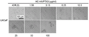

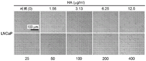


Classifications
-
- A—HUMAN NECESSITIES
- A61—MEDICAL OR VETERINARY SCIENCE; HYGIENE
- A61K—PREPARATIONS FOR MEDICAL, DENTAL OR TOILETRY PURPOSES
- A61K38/00—Medicinal preparations containing peptides
- A61K38/16—Peptides having more than 20 amino acids; Gastrins; Somatostatins; Melanotropins; Derivatives thereof
- A61K38/17—Peptides having more than 20 amino acids; Gastrins; Somatostatins; Melanotropins; Derivatives thereof from animals; from humans
- A61K38/1703—Peptides having more than 20 amino acids; Gastrins; Somatostatins; Melanotropins; Derivatives thereof from animals; from humans from vertebrates
- A61K38/1709—Peptides having more than 20 amino acids; Gastrins; Somatostatins; Melanotropins; Derivatives thereof from animals; from humans from vertebrates from mammals
-
- C—CHEMISTRY; METALLURGY
- C07—ORGANIC CHEMISTRY
- C07K—PEPTIDES
- C07K14/00—Peptides having more than 20 amino acids; Gastrins; Somatostatins; Melanotropins; Derivatives thereof
- C07K14/435—Peptides having more than 20 amino acids; Gastrins; Somatostatins; Melanotropins; Derivatives thereof from animals; from humans
- C07K14/705—Receptors; Cell surface antigens; Cell surface determinants
- C07K14/71—Receptors; Cell surface antigens; Cell surface determinants for growth factors; for growth regulators
-
- A—HUMAN NECESSITIES
- A61—MEDICAL OR VETERINARY SCIENCE; HYGIENE
- A61K—PREPARATIONS FOR MEDICAL, DENTAL OR TOILETRY PURPOSES
- A61K47/00—Medicinal preparations characterised by the non-active ingredients used, e.g. carriers or inert additives; Targeting or modifying agents chemically bound to the active ingredient
- A61K47/50—Medicinal preparations characterised by the non-active ingredients used, e.g. carriers or inert additives; Targeting or modifying agents chemically bound to the active ingredient the non-active ingredient being chemically bound to the active ingredient, e.g. polymer-drug conjugates
- A61K47/51—Medicinal preparations characterised by the non-active ingredients used, e.g. carriers or inert additives; Targeting or modifying agents chemically bound to the active ingredient the non-active ingredient being chemically bound to the active ingredient, e.g. polymer-drug conjugates the non-active ingredient being a modifying agent
- A61K47/62—Medicinal preparations characterised by the non-active ingredients used, e.g. carriers or inert additives; Targeting or modifying agents chemically bound to the active ingredient the non-active ingredient being chemically bound to the active ingredient, e.g. polymer-drug conjugates the non-active ingredient being a modifying agent the modifying agent being a protein, peptide or polyamino acid
-
- A—HUMAN NECESSITIES
- A61—MEDICAL OR VETERINARY SCIENCE; HYGIENE
- A61K—PREPARATIONS FOR MEDICAL, DENTAL OR TOILETRY PURPOSES
- A61K35/00—Medicinal preparations containing materials or reaction products thereof with undetermined constitution
- A61K35/12—Materials from mammals; Compositions comprising non-specified tissues or cells; Compositions comprising non-embryonic stem cells; Genetically modified cells
- A61K35/48—Reproductive organs
- A61K35/50—Placenta; Placental stem cells; Amniotic fluid; Amnion; Amniotic stem cells
-
- A—HUMAN NECESSITIES
- A61—MEDICAL OR VETERINARY SCIENCE; HYGIENE
- A61K—PREPARATIONS FOR MEDICAL, DENTAL OR TOILETRY PURPOSES
- A61K45/00—Medicinal preparations containing active ingredients not provided for in groups A61K31/00 - A61K41/00
- A61K45/06—Mixtures of active ingredients without chemical characterisation, e.g. antiphlogistics and cardiaca
-
- A—HUMAN NECESSITIES
- A61—MEDICAL OR VETERINARY SCIENCE; HYGIENE
- A61K—PREPARATIONS FOR MEDICAL, DENTAL OR TOILETRY PURPOSES
- A61K47/00—Medicinal preparations characterised by the non-active ingredients used, e.g. carriers or inert additives; Targeting or modifying agents chemically bound to the active ingredient
- A61K47/50—Medicinal preparations characterised by the non-active ingredients used, e.g. carriers or inert additives; Targeting or modifying agents chemically bound to the active ingredient the non-active ingredient being chemically bound to the active ingredient, e.g. polymer-drug conjugates
- A61K47/51—Medicinal preparations characterised by the non-active ingredients used, e.g. carriers or inert additives; Targeting or modifying agents chemically bound to the active ingredient the non-active ingredient being chemically bound to the active ingredient, e.g. polymer-drug conjugates the non-active ingredient being a modifying agent
- A61K47/56—Medicinal preparations characterised by the non-active ingredients used, e.g. carriers or inert additives; Targeting or modifying agents chemically bound to the active ingredient the non-active ingredient being chemically bound to the active ingredient, e.g. polymer-drug conjugates the non-active ingredient being a modifying agent the modifying agent being an organic macromolecular compound, e.g. an oligomeric, polymeric or dendrimeric molecule
- A61K47/61—Medicinal preparations characterised by the non-active ingredients used, e.g. carriers or inert additives; Targeting or modifying agents chemically bound to the active ingredient the non-active ingredient being chemically bound to the active ingredient, e.g. polymer-drug conjugates the non-active ingredient being a modifying agent the modifying agent being an organic macromolecular compound, e.g. an oligomeric, polymeric or dendrimeric molecule the organic macromolecular compound being a polysaccharide or a derivative thereof
-
- A—HUMAN NECESSITIES
- A61—MEDICAL OR VETERINARY SCIENCE; HYGIENE
- A61P—SPECIFIC THERAPEUTIC ACTIVITY OF CHEMICAL COMPOUNDS OR MEDICINAL PREPARATIONS
- A61P13/00—Drugs for disorders of the urinary system
- A61P13/08—Drugs for disorders of the urinary system of the prostate
-
- A—HUMAN NECESSITIES
- A61—MEDICAL OR VETERINARY SCIENCE; HYGIENE
- A61P—SPECIFIC THERAPEUTIC ACTIVITY OF CHEMICAL COMPOUNDS OR MEDICINAL PREPARATIONS
- A61P35/00—Antineoplastic agents
-
- C—CHEMISTRY; METALLURGY
- C07—ORGANIC CHEMISTRY
- C07K—PEPTIDES
- C07K14/00—Peptides having more than 20 amino acids; Gastrins; Somatostatins; Melanotropins; Derivatives thereof
- C07K14/435—Peptides having more than 20 amino acids; Gastrins; Somatostatins; Melanotropins; Derivatives thereof from animals; from humans
- C07K14/46—Peptides having more than 20 amino acids; Gastrins; Somatostatins; Melanotropins; Derivatives thereof from animals; from humans from vertebrates
- C07K14/47—Peptides having more than 20 amino acids; Gastrins; Somatostatins; Melanotropins; Derivatives thereof from animals; from humans from vertebrates from mammals
- C07K14/4701—Peptides having more than 20 amino acids; Gastrins; Somatostatins; Melanotropins; Derivatives thereof from animals; from humans from vertebrates from mammals not used
- C07K14/4702—Regulators; Modulating activity
- C07K14/4705—Regulators; Modulating activity stimulating, promoting or activating activity
-
- C—CHEMISTRY; METALLURGY
- C07—ORGANIC CHEMISTRY
- C07K—PEPTIDES
- C07K14/00—Peptides having more than 20 amino acids; Gastrins; Somatostatins; Melanotropins; Derivatives thereof
- C07K14/435—Peptides having more than 20 amino acids; Gastrins; Somatostatins; Melanotropins; Derivatives thereof from animals; from humans
- C07K14/705—Receptors; Cell surface antigens; Cell surface determinants
-
- C—CHEMISTRY; METALLURGY
- C07—ORGANIC CHEMISTRY
- C07K—PEPTIDES
- C07K14/00—Peptides having more than 20 amino acids; Gastrins; Somatostatins; Melanotropins; Derivatives thereof
- C07K14/81—Protease inhibitors
- C07K14/8107—Endopeptidase (E.C. 3.4.21-99) inhibitors
- C07K14/811—Serine protease (E.C. 3.4.21) inhibitors
-
- C—CHEMISTRY; METALLURGY
- C12—BIOCHEMISTRY; BEER; SPIRITS; WINE; VINEGAR; MICROBIOLOGY; ENZYMOLOGY; MUTATION OR GENETIC ENGINEERING
- C12N—MICROORGANISMS OR ENZYMES; COMPOSITIONS THEREOF; PROPAGATING, PRESERVING, OR MAINTAINING MICROORGANISMS; MUTATION OR GENETIC ENGINEERING; CULTURE MEDIA
- C12N5/00—Undifferentiated human, animal or plant cells, e.g. cell lines; Tissues; Cultivation or maintenance thereof; Culture media therefor
- C12N5/06—Animal cells or tissues; Human cells or tissues
- C12N5/0602—Vertebrate cells
- C12N5/0693—Tumour cells; Cancer cells
-
- C—CHEMISTRY; METALLURGY
- C07—ORGANIC CHEMISTRY
- C07K—PEPTIDES
- C07K14/00—Peptides having more than 20 amino acids; Gastrins; Somatostatins; Melanotropins; Derivatives thereof
- C07K14/435—Peptides having more than 20 amino acids; Gastrins; Somatostatins; Melanotropins; Derivatives thereof from animals; from humans
- C07K14/52—Cytokines; Lymphokines; Interferons
- C07K14/525—Tumour necrosis factor [TNF]
-
- C—CHEMISTRY; METALLURGY
- C12—BIOCHEMISTRY; BEER; SPIRITS; WINE; VINEGAR; MICROBIOLOGY; ENZYMOLOGY; MUTATION OR GENETIC ENGINEERING
- C12N—MICROORGANISMS OR ENZYMES; COMPOSITIONS THEREOF; PROPAGATING, PRESERVING, OR MAINTAINING MICROORGANISMS; MUTATION OR GENETIC ENGINEERING; CULTURE MEDIA
- C12N2502/00—Coculture with; Conditioned medium produced by
- C12N2502/02—Coculture with; Conditioned medium produced by embryonic cells
- C12N2502/025—Coculture with; Conditioned medium produced by embryonic cells extra-embryonic cells, e.g. amniotic epithelium, placental cells, Wharton's jelly
Landscapes
- Health & Medical Sciences (AREA)
- Life Sciences & Earth Sciences (AREA)
- Chemical & Material Sciences (AREA)
- General Health & Medical Sciences (AREA)
- Medicinal Chemistry (AREA)
- Organic Chemistry (AREA)
- Veterinary Medicine (AREA)
- Public Health (AREA)
- Animal Behavior & Ethology (AREA)
- Pharmacology & Pharmacy (AREA)
- Engineering & Computer Science (AREA)
- Zoology (AREA)
- Proteomics, Peptides & Aminoacids (AREA)
- Genetics & Genomics (AREA)
- Epidemiology (AREA)
- Bioinformatics & Cheminformatics (AREA)
- Gastroenterology & Hepatology (AREA)
- Biochemistry (AREA)
- Cell Biology (AREA)
- Molecular Biology (AREA)
- Biophysics (AREA)
- Nuclear Medicine, Radiotherapy & Molecular Imaging (AREA)
- General Chemical & Material Sciences (AREA)
- Chemical Kinetics & Catalysis (AREA)
- Immunology (AREA)
- Biomedical Technology (AREA)
- Toxicology (AREA)
- Biotechnology (AREA)
- Developmental Biology & Embryology (AREA)
- Wood Science & Technology (AREA)
- Urology & Nephrology (AREA)
- Marine Sciences & Fisheries (AREA)
- Pregnancy & Childbirth (AREA)
- General Engineering & Computer Science (AREA)
- Microbiology (AREA)
- Oncology (AREA)
- Reproductive Health (AREA)
- Virology (AREA)
- Medicines Containing Material From Animals Or Micro-Organisms (AREA)
- Pharmaceuticals Containing Other Organic And Inorganic Compounds (AREA)
Abstract
In certain embodiments, provided herein are compositions comprising an isolated heavy chain-hyaluronic acid/n-pentraxin 3 ("HC-HA/PTX 3") complex for use in a method of killing cancer cells. Also provided herein are methods of inhibiting cancer cell regrowth of a tumor in an individual in need thereof, the method comprising contacting the area surrounding the tumor with an isolated HC-HA/PTX3 complex following a surgical procedure. Also provided herein are combinations and kits for practicing the methods.
Description
Cross-referencing
This application claims priority and benefit from U.S. provisional application No. 62/975,599, filed on 12/2/2020, the entire contents of which are incorporated herein by reference.
Disclosure of Invention
All publications, patents and patent applications mentioned in this specification are herein incorporated by reference to the same extent as if each individual publication, patent or patent application was specifically and individually indicated to be incorporated by reference.
Disclosed herein are methods of directly killing cancer cells in a subject in need thereof, the methods comprising contacting cancer cells with an isolated HC-HA/PTX3 complex. In some embodiments, the cancer cell is from or within a solid tumor. In some embodiments, the cancer cell is from a cancer selected from the group consisting of: liver cancer, pancreatic cancer, bladder cancer, prostate cancer, lung cancer, non-small cell lung cancer, ovarian cancer, breast cancer, melanoma, gastric cancer, colon cancer, colorectal cancer, central Nervous System (CNS) cancer, bone cancer, lymphatic cancer, skin cancer, head and neck cancer, kidney cancer, testicular cancer, uterine cancer, cervical cancer, esophageal cancer, thyroid cancer, salivary gland cancer, adrenal gland cancer, and gastrointestinal cancer. In some embodiments, the cancer is non-surgical. In some embodiments, the CNS cancer is a glioma or a metastatic cancer. In some embodiments, the glioma is glioblastoma multiforme or anaplastic astrocytoma. In some embodiments, the cancer is lung cancer, breast cancer, colon cancer, or skin cancer. In some embodiments, the colon cancer is adenocarcinoma. In some embodiments, the skin cancer is melanoma. In some embodiments, the cancer is prostate cancer. In some embodiments, contacting comprises injecting the HC-HA/PTX3 complex into a tumor, surrounding tissue, or a combination thereof. In some embodiments, the contacting is performed before, during, or after surgical resection, cryoablation, or radiofrequency ablation of the cancer cells. In some embodiments, contacting comprises applying HC-HA/PTX3 to a surgical margin of a surgically excised cancer cell, or to any remaining portion of the tumor. In some embodiments, the isolated HC-HA/PTX3 complex is a native HC-HA/PTX3 complex, a reconstituted HC-HA/PTX3 complex, or a combination thereof. In some embodiments, the native HC-HA/PTX3 complex is isolated from fetal support tissue. In some embodiments, the reconstituted HC-HA/PTX3 complex comprises heavy chain 1 (HC 1) and heavy chain 2 (HC 2) of an inter-alpha-inhibitor (lal), hyaluronic Acid (HA), and PTX3. In some embodiments, the reconstituted HC-HA/PTX3 complex comprises HC1, HC2, HA, PTX3, and TSG-6. In some embodiments, HC-HA/PTX3 is contained in a composition comprising a pharmaceutically acceptable diluent, excipient, vehicle or carrier. In some embodiments, the method further comprises administering an additional therapeutic agent. In some embodiments, the therapeutic agent is selected from the group consisting of a chemotherapeutic agent, an analgesic agent, an anti-inflammatory agent, a steroid, an immunotherapy, a cell therapy, a radiation therapy, a targeted drug therapy, and an antibiotic. In some embodiments, administering the therapeutic agent occurs prior to contacting the cancer cells with the HC-HA/PTX3 complex. In some embodiments, administering the therapeutic agent occurs after contacting the cancer cells with the HC-HA/PTX3 complex. In some embodiments, administering the therapeutic agent occurs simultaneously with contacting the cancer cells with the HC-HA/PTX3 complex. In some embodiments, angiogenesis is reduced or inhibited. In some embodiments, killing the cancer cells is by apoptosis or necrosis.
Disclosed herein are methods of directly inhibiting cancer cell proliferation in a subject in need thereof, the methods comprising contacting a cancer cell with an isolated HC-HA/PTX3 complex. In some embodiments, the cancer cell is from or within a solid tumor. In some embodiments, the cancer cell is from a cancer selected from the group consisting of: liver cancer, pancreatic cancer, bladder cancer, prostate cancer, lung cancer, non-small cell lung cancer, ovarian cancer, breast cancer, melanoma, gastric cancer, colon cancer, colorectal cancer, central Nervous System (CNS) cancer, bone cancer, lymphatic cancer, skin cancer, head and neck cancer, kidney cancer, testicular cancer, uterine cancer, cervical cancer, esophageal cancer, thyroid cancer, salivary gland cancer, adrenal gland cancer, and gastrointestinal cancer. In some embodiments, the cancer is non-surgical. In some embodiments, the CNS cancer is a glioma or a metastatic cancer. In some embodiments, the glioma is a glioblastoma multiforme or anaplastic astrocytoma. In some embodiments, the cancer is lung cancer, breast cancer, colon cancer, or skin cancer. In some embodiments, the colon cancer is adenocarcinoma. In some embodiments, the skin cancer is melanoma. In some embodiments, the cancer is prostate cancer. In some embodiments, contacting comprises injecting the HC-HA/PTX3 complex into a tumor, surrounding tissue, or a combination thereof. In some embodiments, the contacting is performed before, during, or after surgical resection, cryoablation, or radiofrequency ablation of the cancer cells. In some embodiments, contacting comprises applying HC-HA/PTX3 to a surgical margin of a surgically excised cancer cell. In some embodiments, the isolated HC-HA/PTX3 complex is a native HC-HA/PTX3 complex, a reconstituted HC-HA/PTX3 complex, or a combination thereof. In some embodiments, the native HC-HA/PTX3 complex is isolated from fetal support tissue. In some embodiments, the reconstituted HC-HA/PTX3 complex comprises heavy chain 1 (HC 1) and heavy chain 2 (HC 2) of an inter-alpha-inhibitor (lal), hyaluronic Acid (HA), and PTX3. In some embodiments, the reconstituted HC-HA/PTX3 complex comprises HC1, HC2, HA, PTX3, and TSG-6. In some embodiments, a pharmaceutically acceptable diluent, excipient, vehicle or carrier is included. In a further embodiment, HC-HA/PTX3 is included in a composition comprising an administered therapeutic agent. In some embodiments, the therapeutic agent is selected from the group consisting of a chemotherapeutic agent, an analgesic agent, an anti-inflammatory agent, a steroid, an immunotherapy, a cell therapy, a radiotherapy, a targeted drug therapy, and an antibiotic. In some embodiments, administering the therapeutic agent occurs prior to contacting the cancer cells with the HC-HA/PTX3 complex. In some embodiments, administering the therapeutic agent occurs after contacting the cancer cells with the HC-HA/PTX3 complex. In some embodiments, administering the therapeutic agent occurs simultaneously with contacting the cancer cells with the HC-HA/PTX3 complex. In some embodiments, angiogenesis is reduced or inhibited. In some embodiments, proliferation is inhibited in cells expressing CD44 or RHAMM.
Described herein are methods of directly reducing metabolic activity of a cancer cell in a subject in need thereof, the method comprising contacting the cancer cell with an isolated HC-HA/PTX3 complex. In some embodiments, the cancer cell is from or within a solid tumor. In some embodiments, the cancer cell is from a cancer selected from the group consisting of: liver cancer, pancreatic cancer, bladder cancer, prostate cancer, lung cancer, non-small cell lung cancer, ovarian cancer, breast cancer, melanoma, gastric cancer, colon cancer, colorectal cancer, central Nervous System (CNS) cancer, bone cancer, lymphatic cancer, skin cancer, head and neck cancer, kidney cancer, testicular cancer, uterine cancer, cervical cancer, esophageal cancer, thyroid cancer, salivary gland cancer, adrenal cancer, and gastrointestinal cancer. In some embodiments, the cancer is non-surgical. In some embodiments, the CNS cancer is a glioma or a metastatic cancer. In some embodiments, the glioma is glioblastoma multiforme or anaplastic astrocytoma. In some embodiments, the cancer is lung cancer, breast cancer, colon cancer, or skin cancer. In some embodiments, the colon cancer is adenocarcinoma. In some embodiments, the skin cancer is melanoma. In some embodiments, the cancer is prostate cancer. In some embodiments, contacting comprises injecting the HC-HA/PTX3 complex into a tumor, surrounding tissue, or a combination thereof. In some embodiments, the contacting is before, during, or after surgical resection, cryoablation, or radiofrequency ablation of the cancer cells, or into any remaining portion of the tumor. In some embodiments, contacting comprises applying HC-HA/PTX3 to a surgical margin of a surgically excised cancer cell. In some embodiments, the isolated HC-HA/PTX3 complex is a native HC-HA/PTX3 complex, a reconstituted HC-HA/PTX3 complex, or a combination thereof. In some embodiments, the native HC-HA/PTX3 complex is isolated from fetal support tissue. In some embodiments, the reconstituted HC-HA/PTX3 complex comprises heavy chain 1 (HC 1) and heavy chain 2 (HC 2) of an inter-alpha-inhibitor (lal), hyaluronic Acid (HA), and PTX3. In some embodiments, the reconstituted HC-HA/PTX3 complex comprises HC1, HC2, HA, PTX3, and TSG-6. In some embodiments, HC-HA/PTX3 is contained in a composition comprising a pharmaceutically acceptable diluent, excipient, vehicle or carrier. In a further embodiment, HC-HA/PTX3 is included in a composition comprising administration of a therapeutic agent. In some embodiments, the therapeutic agent is selected from the group consisting of a chemotherapeutic agent, an analgesic agent, an anti-inflammatory agent, a steroid, an immunotherapy, a cell therapy, a radiotherapy, a targeted drug therapy, and an antibiotic. In some embodiments, administering the therapeutic agent occurs prior to contacting the cancer cells with the HC-HA/PTX3 complex. In some embodiments, administering the therapeutic agent occurs after contacting the cancer cells with the HC-HA/PTX3 complex. In some embodiments, administering the therapeutic agent occurs simultaneously with contacting the cancer cells with the HC-HA/PTX3 complex. In some embodiments, angiogenesis is reduced or inhibited. In some embodiments, the metabolic activity is decreased in cells expressing CD44 or RHAMM.
Described herein are methods of killing cancer cells comprising contacting a surgical margin or any portion of a tumor with an isolated HC-HA/PTX3 complex before, during, or after surgical resection, cryoablation, or radiofrequency ablation of the tumor, thereby killing cancer cells of the surgical margin. In some embodiments, the tumor is a cancer selected from the group consisting of: liver cancer, pancreatic cancer, bladder cancer, prostate cancer, lung cancer, non-small cell lung cancer, ovarian cancer, breast cancer, melanoma, gastric cancer, colon cancer, colorectal cancer, central Nervous System (CNS) cancer, bone cancer, lymphatic cancer, skin cancer, head and neck cancer, kidney cancer, testicular cancer, uterine cancer, cervical cancer, esophageal cancer, thyroid cancer, salivary gland cancer, adrenal gland cancer, and gastrointestinal cancer. In some embodiments, the cancer is non-surgical. In some embodiments, the CNS cancer is a glioma or a metastatic cancer. In some embodiments, the glioma is a glioblastoma multiforme. In some embodiments, the cancer is lung cancer, breast cancer, colon cancer, or skin cancer. In some embodiments, the colon cancer is adenocarcinoma. In some embodiments, the skin cancer is melanoma. In some embodiments, the tumor is prostate cancer. In some embodiments, the HC-HA/PTX3 complex is a native HC-HA/PTX3 complex, a reconstituted HC-HA/PTX3 complex, or a combination thereof. In some embodiments, the native HC-HA/PTX3 complex is isolated from fetal support tissue. In some embodiments, the reconstituted HC-HA/PTX3 complex comprises heavy chain 1 (HC 1) and heavy chain 2 (HC 2) of an inter-alpha-inhibitor (lal), hyaluronic Acid (HA), and PTX3. In some embodiments, the reconstituted HC-HA/PTX3 complex comprises HC1, HC2, HA, PTX3, and TSG-6. In some embodiments, HC-HA/PTX3 is contained in a composition comprising a pharmaceutically acceptable diluent, excipient, vehicle or carrier. In a further embodiment, the reconstituted HC-HA/PTX3 complex comprises administration of a therapeutic agent. In some embodiments, the therapeutic agent is selected from the group consisting of a chemotherapeutic agent, an analgesic agent, an anti-inflammatory agent, a steroid, an immunotherapy, a cell therapy, a radiotherapy, a targeted drug therapy, and an antibiotic. In some embodiments, administering the therapeutic agent occurs prior to contacting the cancer cells with the HC-HA/PTX3 complex. In some embodiments, administering the therapeutic agent occurs after contacting the cancer cells with the HC-HA/PTX3 complex. In some embodiments, administering the therapeutic agent occurs simultaneously with contacting the cancer cells with the HC-HA/PTX3 complex. In some embodiments, killing the cancer cell is by apoptosis or necrosis.
Disclosed herein, in some embodiments, is a method of inhibiting cancer cell regrowth of a tumor in an individual in need thereof, comprising contacting a region surrounding the tumor with an isolated heavy chain-hyaluronic acid/n-pentraxin 3 (HC-HA/PTX 3) complex after a surgical procedure, thereby inhibiting cancer cell regrowth of the region surrounding the tumor. In some embodiments, the surgical procedure comprises surgical resection, cryoablation, or radio frequency ablation of a tumor. In some embodiments, the surgical procedure comprises chemotherapy, immunotherapy, or targeted therapy. In some embodiments, the area surrounding the tumor comprises a surgical margin. In some embodiments, the area surrounding the tumor is a peritumoral area. In some embodiments, the tumor is a solid tumor. In some embodiments, the tumor is a cancer selected from the group consisting of: liver cancer, pancreatic cancer, bladder cancer, prostate cancer, lung cancer, non-small cell lung cancer, ovarian cancer, breast cancer, melanoma, gastric cancer, colon cancer, colorectal cancer, central Nervous System (CNS) cancer, bone cancer, lymphatic cancer, skin cancer, head and neck cancer, kidney cancer, testicular cancer, uterine cancer, cervical cancer, esophageal cancer, thyroid cancer, salivary gland cancer, adrenal gland cancer, and gastrointestinal cancer. In some embodiments, the cancer is a non-surgical cancer. In some embodiments, the cancer is pancreatic cancer. In some embodiments, the cancer is prostate cancer. In some embodiments, the cancer is glioblastoma multiforme. In some embodiments, the cancer is a skin cancer. In some embodiments, the cancer is colon cancer. In some embodiments, the cancer is lung cancer. In some embodiments, the cancer is breast cancer. In some embodiments, the area surrounding the tumor is contacted with about 10 micrograms to 100 milligrams. In some embodiments, the HC-HA/PTX3 complex is a native HC-HA/PTX3 complex, a reconstituted HC-HA/PTX3 complex, or a combination thereof. In some embodiments, the native HC-HA/PTX3 complex is isolated from fetal support tissue. In some embodiments, the reconstituted HC-HA/PTX3 complex comprises heavy chain 1 (HC 1) and heavy chain 2 (HC 2) of the inter-alpha-inhibitor (lal), hyaluronic Acid (HA), and PTX3. In some embodiments, the reconstituted HC-HA/PTX3 complex comprises HC1, HC2, HA, PTX3, and tumor necrosis factor alpha-stimulated gene 6 (TSG-6). In some embodiments, the Hyaluronic Acid (HA) is a high molecular weight hyaluronic acid (HMW HA). In some embodiments, the Hyaluronic Acid (HA) is a low molecular weight hyaluronic acid (LMW HA). In some embodiments, the HC-HA/PTX3 complex is cryopreserved. In some embodiments, the HC-HA/PTX3 complex comprises living cells. In some embodiments, the method further comprises administering a therapeutic agent. In some embodiments, the therapeutic agent is selected from the group consisting of a chemotherapeutic agent, an analgesic agent, an anti-inflammatory agent, a steroid, an immunotherapy, a cell therapy, a radiotherapy, a targeted drug therapy, and an antibiotic. In some embodiments, administering the therapeutic agent occurs prior to contacting the area surrounding the tumor with the HC-HA/PTX3 complex. In some embodiments, administering the therapeutic agent occurs after contacting the area surrounding the tumor with the HC-HA/PTX3 complex. In some embodiments, administration of the therapeutic agent occurs simultaneously with contacting the area surrounding the tumor with the HC-HA/PTX3 complex. In some embodiments, the method inhibits tumor cell regrowth by killing cancer cells. In some embodiments, killing of cancer cells is achieved by apoptosis or necrosis. In some embodiments, the method inhibits tumor cell regrowth by inhibiting cancer cell proliferation. In some embodiments, the method inhibits tumor cell regrowth by inhibiting the metabolic activity of the cancer cell.
In some embodiments, disclosed herein are methods of killing tumor cancer cells in an individual in need thereof, comprising contacting the tumor or an area surrounding the tumor with an isolated heavy chain hyaluronic acid/n-pentraxin 3 (HC-HA/PTX 3) complex before, during, or after a surgical procedure, thereby killing the cancer cells. In some embodiments, the surgical procedure comprises surgical resection, cryoablation, or radio frequency ablation of a tumor. In some embodiments, the surgical procedure comprises chemotherapy, immunotherapy, or targeted therapy. In some embodiments, the area surrounding the tumor comprises a surgical margin. In some embodiments, the area surrounding the tumor is a peritumoral area. In some embodiments, the tumor is a solid tumor. In some embodiments, the tumor is a cancer selected from the group consisting of: liver cancer, pancreatic cancer, bladder cancer, prostate cancer, lung cancer, non-small cell lung cancer, ovarian cancer, breast cancer, melanoma, gastric cancer, colon cancer, colorectal cancer, central Nervous System (CNS) cancer, bone cancer, lymphatic cancer, skin cancer, head and neck cancer, kidney cancer, testicular cancer, uterine cancer, cervical cancer, esophageal cancer, thyroid cancer, salivary gland cancer, adrenal cancer, and gastrointestinal cancer. In some embodiments, the cancer is a non-surgical cancer. In some embodiments, the cancer is pancreatic cancer. In some embodiments, the cancer is prostate cancer. In some embodiments, the cancer is glioblastoma multiforme. In some embodiments, the cancer is a skin cancer. In some embodiments, the cancer is colon cancer. In some embodiments, the cancer is lung cancer. In some embodiments, the cancer is breast cancer. In some embodiments, the area surrounding the tumor is contacted with about 10 micrograms to 100 milligrams. In some embodiments, the HC-HA/PTX3 complex is a native HC-HA/PTX3 complex, a reconstituted HC-HA/PTX3 complex, or a combination thereof. In some embodiments, the native HC-HA/PTX3 complex is isolated from fetal support tissue. In some embodiments, the reconstituted HC-HA/PTX3 complex comprises heavy chain 1 (HC 1) and heavy chain 2 (HC 2) of an inter-alpha-inhibitor (lal), hyaluronic Acid (HA), and PTX3. In some embodiments, the reconstituted HC-HA/PTX3 complex comprises HC1, HC2, HA, PTX3, and tumor necrosis factor alpha-stimulated gene 6 (TSG-6). In some embodiments, the HC-HA/PTX3 complex is cryopreserved. In some embodiments, the HC-HA/PTX3 complex comprises living cells. In some embodiments, the Hyaluronic Acid (HA) is a high molecular weight hyaluronic acid (HMW HA). In some embodiments, the Hyaluronic Acid (HA) is a low molecular weight hyaluronic acid (LMW HA). In some embodiments, the contacting comprises injecting HC-HA/PTX3 directly into the tumor. In some embodiments, the method further comprises administering a therapeutic agent. In some embodiments, the therapeutic agent is selected from the group consisting of a chemotherapeutic agent, an analgesic agent, an anti-inflammatory agent, a steroid, an immunotherapy, a cell therapy, a radiotherapy, a targeted drug therapy, and an antibiotic. In some embodiments, administering the therapeutic agent occurs prior to contacting the cancer cells with the HC-HA/PTX3 complex. In some embodiments, administering the therapeutic agent occurs after contacting the cancer cells with the HC-HA/PTX3 complex. In some embodiments, administering the therapeutic agent occurs simultaneously with contacting the cancer cells with the HC-HA/PTX3 complex. In some embodiments, the killing of the cancer cells is by apoptosis or necrosis.
Drawings
Fig. 1A shows the aggregation of LNCaP cells after culture in RPMI medium.
FIG. 1B shows that PC-3 cells are evenly distributed after culture in RPMI medium.
FIGS. 2A-2D show the morphology and cellular metabolic activity of LNCaP under a range of doses of either refined BTgel or HC-HA/PTX3 treatment.
FIGS. 3A-3D show the morphology and metabolic activity of PC-3 under a range of doses of either purified BTgel or HC-HA/PTX3 treatment.
FIG. 4A shows WST-1 assay data in LNCaP cells after treatment with UC extract (using UC extract in water), HC-HA/PTX3, and HA.
FIG. 4B shows data from WST-1 assays in PC-3 cells after treatment with UC extract (using UC extract in water), HC-HA/PTX3, and HA.
FIGS. 5A-5C show the morphology of LNCaP cells after treatment with HA (FIG. 5A), HC-HA/PTX3 (FIG. 5B), and Umbilical Cord Extract (UCE) (FIG. 5C).
FIGS. 6A-6C show the morphology of PC-3 cells after treatment with HA (FIG. 6A), HC-HA/PTX3 (FIG. 6B), and Umbilical Cord Extract (UCE) (FIG. 6C).
Fig. 7 shows that LNCaP cells grown on laminin and type IV collagen showed more cell aggregation than cells grown on other surfaces.
Fig. 8A shows bright field images of the morphology of the PrEC prostate cell line taken at 10x and 20x magnification.
Figure 8B shows bright field images of the morphology of the PNT2 prostate cell line taken at 10x and 20x magnification.
FIG. 9 shows representative bright field microscopy images (scale bar 50 μm) of human primary normal prostate cells after 48 hours incubation with varying concentrations of HC-HA/PTX3 and HMW-HA.
FIGS. 10A and 10B show the metabolic activity (%) assessed in normal human primary prostate epithelial cells PrEC (FIG. 10A) and normal human prostate cell line PNT2 (FIG. 10B) after 48 hours of incubation with HC-HA/PTX3 and HA at different concentrations (0.78, 3.125, 6.25, 12.5, 25, 50, 100 μ g/ml) determined by WST-1. P values were calculated by a two-tailed t-test against untreated samples.
FIGS. 11A and 11B show that after 48 hours incubation with various concentrations (0.78, 3.125, 6.25, 12.5, 25, 50, 100 μ g/ml) of HC-HA/PTX3, the cell lines were: comparative analysis of metabolic activity (%) assessed in PC3& LNCaP (fig. 11A) and log scale metabolic activity (fig. 11B).
Fig. 12A and 12B show that after 48 hours incubation with different concentrations of HA, the cell lines in normal primary prostate epithelial cells (PrEC) & cell line (PNT 2) and prostate cancer cell lines: comparative analysis of metabolic activity (%) assessed in PC3& LNCaP (fig. 12A) and log scale metabolic activity (fig. 12B).
FIG. 13 shows representative bright field microscopy images (scale bar 50 μm) of A375 (melanoma) cells after 48 hours incubation with different concentrations of HC-HA/PTX3 and HMW-HA.
FIG. 14 shows representative bright field microscopy images (scale bar 50 μm) of HT-29 (colon cancer) cells after 48 hours incubation with different concentrations of HC-HA/PTX3 and HMW-HA.
FIG. 15 shows representative bright field microscopy images (scale bar 50 μm) of A549 (lung cancer) cells after 48 hours incubation with different concentrations of HC-HA/PTX3 and HMW-HA.
FIG. 16 shows representative bright field microscopy images (scale bar 50 μm) of MCF-7 (breast cancer) cells after 48 hours incubation with different concentrations of HC-HA/PTX3 and HMW-HA.
FIGS. 17A-17D show the results of the WST-1 assay in4 human cancer cell lines after 48 hours incubation with various concentrations of HC-HA/PTX3 and HA: a375 (FIG. 17A), HT-29 (FIG. 17B), MCF-7 (FIG. 17C) and A-549 (FIG. 17D).
Figure 18A shows representative bright field microscopy images (scale bar 50 μm) of LNC (limbal niche cells) for different time points (15-30 minutes, 1 hour, 5 hours, 24 hours, and 48 hours, respectively) after treatment with different concentrations of HC-HA/PTX3.
Figure 18B shows representative bright field microscopy images (scale bar 50 μm) of LNCs (limbal microenvironment cells) after treatment with different concentrations of HMW-HA for different time points (15-30 min, 1 hr, 5 hr, 24 hr, and 48 hr, respectively).
FIG. 18C shows representative bright field microscopy images (scale bar 50 μm) of LNCs (limbal microenvironment cells) after 48 hours incubation with 100 μ g/ml HC-HA/PTX3 and HMW-HA.
FIG. 19 shows the metabolic activity (%) in limbal microenvironment cells assessed by WST-1 assay after 48 hours incubation with various concentrations of HC-HA/PTX3 and HA.
FIGS. 20A and 20B show representative bright field microscopy images (scale bar 50 μm) of HTM (human trabecular meshwork) cells for different time points after treatment with different concentrations of HC-HA/PTX3 (FIG. 20A) and HMW-HA (FIG. 20B).
FIG. 21 shows the metabolic activity (%) in human trabecular meshwork cells assessed by the WST-1 assay after 48 hours of incubation with various concentrations of HC-HA/PTX3 and HA.
FIGS. 22A and 22B show representative bright field microscopy images (scale bar 50 μm) of Human Corneal Fibroblast (HCF) cells for different time points after treatment with different concentrations of HC-HA/PTX3 (FIG. 22A) and HMW-HA (FIG. 22B).
FIG. 23 shows the metabolic activity (%) in human corneal fibroblasts assessed by the WST-1 assay after 48 hours incubation with various concentrations of HC-HA/PTX3 and HA.
Fig. 24A and 24B show three types of human normal primary mesenchymal cells assessed by WST-1 assay after 48 hours incubation with different concentrations of HC-HA/PTX3 (fig. 24A) and HA (fig. 24B): comparative analysis of metabolic activity (%) in HCF, HTM & LNC.
FIG. 25 provides representative bright field microscopy images (scale bar 50 μm) showing the transient effect of HC-HA/PTX3 (100 μ g/ml) on LNC and HCF cell morphology, with no corresponding effect in HTM cells.
FIG. 26A shows bright field images of A375 cell morphology after treatment with HC-HA/PTX3 and HA compared to untreated cells.
Fig. 26B shows BrdU cell proliferation assay curves using a375 cells.
Fig. 26C shows a semi-log scale BrdU cell proliferation assay curve using a375 cells.
Fig. 27A shows bright field images of the pcec cell morphology after treatment with HA or HC-HA/PTX3 at two magnifications (10x &20x).
FIG. 27B shows BrdU cell proliferation assay curves after HC-HA/PTX3 treatment.
Fig. 27C shows BrdU cell proliferation assay curves after HA treatment.
FIG. 28A shows bright field images of PNT2 cell morphology after HC-HA/PTX3, HMW-HA treatment, or no treatment.
FIG. 28B shows BrdU cell proliferation assay curves in PNT2 cells after treatment with HC-HA/PTX3 or HA.
FIG. 29A shows bright field images of PC3 cell morphology after HC-HA/PTX3, HMW-HA treatment, or no treatment.
FIG. 29B shows BrdU cell proliferation assay curves in PC3 cells after treatment with HC-HA/PTX3 or HA.
FIG. 30A shows bright field images of LNCaP cell morphology after treatment with HC-HA/PTX3, HMW-HA or no treatment.
FIG. 30B shows BrdU cell proliferation assay curves in LNCaP cells after treatment with HC-HA/PTX3 or HA.
Detailed Description
Certain terms
Unless defined otherwise, all technical and scientific terms used herein have the same meaning as commonly understood by one of ordinary skill in the art to which the claimed subject matter belongs. All patents, patent applications, published applications and publications, websites and other published materials referred to throughout the disclosure herein are incorporated by reference in their entirety, unless otherwise indicated. If there are multiple definitions of terms herein, those in this section prevail. When referring to a URL or other such identifier or address, it is understood that such identifiers may vary, and that particular information on the internet may vary, but equivalent information is known and can be readily accessed, such as by searching the internet and/or an appropriate database. The citation of this demonstrates the availability and public dissemination of such information.
As used herein, ranges and amounts can be expressed as "about" a particular value or range. About also includes the exact amount or value. Thus, "about 5. Mu.g" means "about 5. Mu.g" and "5. Mu.g" as well. In some embodiments, the term "about" includes amounts that are expected to be within experimental error. In some embodiments, the term "about" refers to +/-20%, 10%, or 5% of the value.
As used herein, HC-HA/PTX3 complex or isolated HC-HA/PTX3 refers to native HC-HA/PTX3, reconstituted HC-HA/PTX3, or a combination thereof. As used herein, a reconstituted HC-HA/PTX3 (rcHC-HA/PTX 3) complex is a HC-HA/PTX3 complex formed by the assembly of component molecules of the complex in vitro. In some embodiments, the rcHC-HA/PTX3 complex comprises HA, HC1 and HC2 of I α I, and PTX3. In some embodiments, the rcHC-HA/PTX3 complex comprises HA, HC1 and HC2 of I α I, PTX3, and TSG-6. The process of assembling rcHC-HA/PTX3 includes reconstitution with purified native protein or molecules isolated from biological sources, production of recombinant proteins by recombinant methods, or synthesis of molecules by in vitro synthesis. In some cases, the purified native protein used to assemble rcHC-HA/PTX3 is a protein complexed with other proteins (i.e., multimers, multi-chain proteins, or other complexes). In some cases, PTX3 is purified from cells as multimers (e.g., homomultimers) and used to assemble the rcHC-HA/PTX3 complex.
As used herein, a purified native HC-HA/PTX3 (nHC-HA/PTX 3) complex refers to a HC-HA/PTX3 complex purified from a biological source, such as a cell, tissue, or biological fluid. In some embodiments, HC-HA/PTX3 is isolated from fetal support tissue, such as the placenta, amnion, chorion, umbilical cord, or umbilical cord amnion. In some embodiments, HC-HA/PTX3 is isolated from amniotic membrane. In some embodiments, the native HC-HA/PTX3 complex comprises HA, HC1 of I α I, and PTX3. Such complexes are typically assembled in vivo in a subject, or ex vivo in cells, tissues or biological fluids from a subject, including humans or other animals.
As used herein, "hyaluronic acid (hyaluronan)" or "hyaluronate" (HA) are used interchangeably to refer to a substantially non-sulfated linear glycosaminoglycan (GAG) having a repeating disaccharide unit of D-glucuronic acid and N-acetylglucosamine (D-glucuronyl-N-acetylglucosamine).
As used herein, the term "high molecular weight" or "HMW" as in high molecular weight hyaluronic acid (HMW HA) means a weight average molecular weight of greater than about 500 kilodaltons (kDa), e.g., between about 500kDa and about 10,000kda, between about 800kDa and about 8,500kda, between about 1100kDa and about 5,000kda, or between about 1400kDa and about 3,500kdaAnd (4) HA. In some embodiments, the HMW HA HAs a weight average molecular weight of 3000kDa or greater. In some embodiments, the HMW HA HAs a weight average molecular weight of 3000kDa. In some embodiments, the HMW HA is HMW HA Has a weight average molecular weight of about 3000kDa. In some embodiments, the HMW HA HAs a molecular weight of about 500kDa to about 10,000kda. In some embodiments, the HMW HA HAs a molecular weight of about 800kDa to about 8,500kda. In some embodiments, the HMW HA HAs a molecular weight of about 3,000kda.
As used herein, the term "low molecular weight" or "LMW" as in Low Molecular Weight Hyaluronic Acid (LMWHA) means HA having a weight average molecular weight of less than 500kDa, e.g., less than about 400kDa, less than about 300kDa, less than about 200kDa, about 200-300kDa, or about 1-300 kDa.
As used herein, pentraxin 3 or PTX3 protein or polypeptide refers to any PTX3 protein, including but not limited to recombinantly produced proteins, synthetically produced proteins, natural PTX3 proteins, and PTX3 proteins extracted from cells or tissues. PTX3 includes multimeric forms (e.g., homomultimers) of PTX3, including but not limited to dimers, trimers, tetramers, pentamers, hexamers, heptamers, octamers, and other naturally or artificially generated multimeric forms.
As used herein, "hyaluronic acid binding protein," "HA binding protein," or "HABP" refers to any protein that specifically binds HA.
As used herein, "linker moiety" refers to a hyaluronic acid binding domain.
"biological activity" as used herein refers to the in vivo activity of the nHC-HA/PTX3 or rcHC-HA/PTX3 complex or the physiological response generated following in vivo administration of the nHC-HA/PTX3 or rcHC-HA/PTX3 complex or a composition or mixture containing the nHC-HA/PTX3 or rcHC-HA/PTX3 complex. Thus, biological activities include therapeutic effects and pharmaceutical activities of nHC-HA/PTX3 or rHC-HA/PTX 3 complexes and compositions and mixtures thereof.
As used herein, the terms "subject," "individual," and "patient" are used interchangeably. Any of these terms should not be construed as requiring supervision by a medical professional (e.g., doctor, nurse, physician's assistant, caregiver, end-of-care worker). As used herein, a subject is any animal, including mammals (e.g., humans or non-human animals) and non-mammals. In one embodiment of the methods and compositions provided herein, the mammal is a human.
As used herein, the terms "treat," "treating," or "treatment" and other grammatical equivalents include alleviating, eliminating, or ameliorating one or more symptoms of a disease or condition, ameliorating, preventing, or reducing the appearance, severity, or frequency of one or more other symptoms of a disease or condition, ameliorating or preventing an underlying metabolic cause of one or more symptoms of a disease or condition, inhibiting a disease or condition, e.g., arresting the development of a disease or condition, alleviating a disease or condition, causing regression of a disease or condition, alleviating a condition caused by a disease or condition, or prophylactically and/or therapeutically inhibiting the symptoms of a disease or condition. In one non-limiting example, for prophylactic benefit, the rcHC-HA/PTX3 complex, natural HC-HA/PTX3 complex, or composition disclosed herein is administered to an individual at risk of developing a particular disorder, prone to developing a particular disorder, or to an individual informed of one or more physiological symptoms of a disorder.
As used herein, "placenta" refers to an organ that connects a developing fetus to the maternal uterine wall to allow for absorption of nutrients, elimination of waste products, and exchange of gases by the maternal blood supply. The placenta consists of three layers. The innermost disc layer surrounding the fetus is called the amniotic membrane. The allantois is the middle layer of the placenta (from the postembryonic intestine); blood vessels originating from the umbilicus pass through the membrane. The outermost layer of the placenta, the chorion, is in contact with the endometrium. The chorioallantoic membrane and the allantoic sac fuse to form a chorioallantoic membrane.
As used herein, "chorion" refers to a membrane formed by an extraembryonic mesoderm and two trophoblasts. The chorion consists of two layers: an outer layer formed from the trophoblast and an inner layer formed from the embryo in the somatic cell; the amniotic membrane is contacted with the latter. The trophoblast consists of an inner layer of cubic or prismatic cells, a cytotrophoblast or Lanhans layer and an outer, nucleoplasmic-rich layer without cell boundaries, a syncytiotrophoblast. The avascular amniotic membrane is adhered to the inner layer of the chorion.
As used herein, "amnion-chorion" refers to a product comprising an amnion and a chorion. In some embodiments, the amniotic membrane and the chorion are not separated (i.e., the amniotic membrane naturally adheres to the inner layer of the chorion). In some embodiments, the amniotic membrane is initially separated from the chorion, and subsequently combined with the chorion during processing.
As used herein, "umbilical cord" refers to the organ that connects the developing fetus to the placenta. The umbilical cord consists of Wharton's jelly, a gelatinous mass made primarily of mucopolysaccharides. It contains one vein (which carries oxygenated, nutrient-rich blood to the fetus) and two arteries (which carry deoxygenated, nutrient-depleted blood away).
As used herein, "placental amniotic membrane" (PAM) refers to an amniotic membrane derived from the placenta. In some embodiments, the PAM is substantially isolated.
As used herein, "umbilical cord amniotic membrane" (UCAM) refers to an amniotic membrane derived from the umbilical cord. UCAMs are translucent films. The UCAM has multiple layers: an epithelial layer; a base film; a dense layer; a layer of fibroblasts; and a sponge layer. It lacks a vascular or direct blood supply. In some embodiments, UCAM includes wharton's jelly. In some embodiments, the UCAM includes blood vessels and/or arteries. In some embodiments, UCAM includes wharton's jelly and blood vessels and/or arteries.
As used herein, the terms "purified," "isolated," and "purified" refer to a material (e.g., nHC-HA/PTX3 complex) that is substantially or essentially free of components that normally accompany it in its native state. In some embodiments, "purified" or "isolated" refers to a material (e.g., nHC-HA/PTX3 complex) that is about 50% or more free of components that normally accompany it in its native state, e.g., about 50%, about 55%, about 60%, about 65%, about 70%, about 75%, about 80%, about 85%, about 90%, about 91%, about 92%, about 93%, about 94%, about 95%, about 96%, about 97%, about 98%, or about 99% free of components that normally accompany it in its native state.
Method of treatment
In certain embodiments, disclosed herein are methods of treating an individual in need thereof, comprising administering to the individual an nHC-HA/PTX3 or rcHC-HA/PTX3 complex described herein, including methods of directly killing cancer cells, directly inhibiting cancer cell proliferation, reducing cancer cell metabolic activity, or a combination thereof. In some embodiments, the nHC-HA/PTX3 or rcHC-HA/PTX3 complex is used to directly kill cancer cells. In some embodiments, the nHC-HA/PTX3 or rcHC-HA/PTX3 complex is used to directly inhibit the proliferation of cancer cells. In some embodiments, the nHC-HA/PTX3 or rcHC-HA/PTX3 complex is used to reduce the metabolic activity of cancer cells. In some embodiments, the cell is from or within a solid tumor.
In some embodiments, a method of treating a subject in need thereof comprises administering to the subject an nHC-HA/PTX3 or rcHC-HA/PTX3 complex described herein by any suitable method. In some embodiments, the individual in need thereof has cancer. In some embodiments, the individual in need thereof has a non-surgical cancer. In some embodiments, the individual in need thereof has a non-surgical cancer selected from pancreatic cancer, prostate cancer, and glioblastoma multiforme. In some embodiments, the individual has a solid tumor. In some embodiments, the individual has a cancer selected from the group consisting of: liver cancer, pancreatic cancer, bladder cancer, prostate cancer, lung cancer, non-small cell lung cancer, ovarian cancer, breast cancer, melanoma, gastric cancer, colon cancer, colorectal cancer, central Nervous System (CNS) cancer, bone cancer, lymphatic cancer, skin cancer, head and neck cancer, kidney cancer, testicular cancer, uterine cancer, cervical cancer, esophageal cancer, thyroid cancer, salivary gland cancer, adrenal gland cancer, and gastrointestinal cancer.
In some embodiments, the nHC-HA/PTX3 or rcHC-HA/PTX3 complex is administered to the subject before, during, or after the surgical procedure. In some embodiments, the surgical procedure comprises resection of a tumor. In some embodiments, the surgical procedure comprises surgical resection, cryoablation, or radio frequency ablation of a tumor. In some embodiments, the surgical procedure comprises chemotherapy, immunotherapy, or targeted therapy.
In some embodiments, the nHC-HA/PTX3 or rcHC-HA/PTX3 complex is administered up to 1 day, up to 2 days, up to 3 days, up to 5 days, or more than 5 days after tumor resection. In some embodiments, the nHC-HA/PTX3 or rcHC-HA/PTX3 complex is administered for one week, two weeks, one month, two months, three months, four months, five months, one year, two years, three years, four years, five years, or more than five years after tumor resection. In some embodiments, the nHC-HA/PTX3 or rcHC-HA/PTX3 complex is administered in a single administration after tumor resection, or over a period of time (e.g., daily, multiple times per week, weekly, bi-weekly, monthly, or less frequently). In some embodiments, the nHC-HA/PTX3 or rcHC-HA/PTX3 complex is administered in a single administration after tumor resection, or over a period of time (e.g., daily, multiple times per week, weekly, biweekly, monthly, or more frequently).
In some embodiments, the nHC-HA/PTX3 or rcHC-HA/PTX3 complex is administered prior to tumor resection. In some embodiments, the nHC-HA/PTX3 or rcHC-HA/PTX3 complex is administered up to 1 day, up to 2 days, up to 3 days, up to 5 days, or more than 5 days prior to tumor resection. In some embodiments, the nHC-HA/PTX3 or rcHC-HA/PTX3 complex is administered in a single administration prior to tumor resection, or over a period of time (e.g., daily, multiple times per week, weekly, bi-weekly, monthly, or less frequently). In some embodiments, the nHC-HA/PTX3 or rcHC-HA/PTX3 complex is administered in a single administration prior to tumor resection, or over a period of time (e.g., daily, multiple times per week, weekly, bi-weekly, monthly, or more frequently).
In some embodiments, the nHC-HA/PTX3 or rcHC-HA/PTX3 complex is administered to the subject during or after surgical resection, cryoablation, or radiofrequency ablation of the tumor. In some embodiments, the nHC-HA/PTX3 or rcHC-HA/PTX3 complex is applied to the surgical margin (e.g., a distinct tissue region surrounding a tumor that HAs been surgically resected) following surgical resection (e.g., total or partial), cryoablation, or radiofrequency ablation of the tumor.
In some embodiments, a method of treating an individual in need thereof comprises administering to the individual an nHC-HA/PTX3 or rcHC-HA/PTX3 complex by any suitable route of administration. Suitable methods of administration will depend on the disease or condition to be treated. In some embodiments, the nHC-HA/PTX3 or rcHC-HA/PTX3 complex is administered locally to the treatment site. In some embodiments, the nHC-HA/PTX3 or rcHC-HA/PTX3 complex is injected into the tumor, tissue surrounding the tumor, or both. In some embodiments, the nHC-HA/PTX3 or rcHC-HA/PTX3 complex is injected into a tumor. In some embodiments, the nHC-HA/PTX3 or rcHC-HA/PTX3 complex is applied to the area surrounding the tumor after the tumor HAs been surgically removed or treated with cryoablation or radiofrequency ablation. In some embodiments, the nHC-HA/PTX3 or rcHC-HA/PTX3 complex is administered systemically. Exemplary methods for administering the nHC-HA/PTX3 or rcHC-HA/PTX3 complexes provided herein include, but are not limited to, parenteral, enteral, subcutaneous, transdermal, intradermal, intravenous, topical, inhalation, or implantation.
It is demonstrated herein that isolated HC-HA/PTX3 complexes kill cancer cells directly. In certain embodiments, provided herein is the use of an isolated HC-HA/PTX3 complex (including formulations or compositions comprising HC-HA/PTX 3) to kill cancer cells. In some embodiments, provided herein is the use of an isolated HC-HA/PTX3 complex to kill cancer cells in a solid tumor. In some embodiments, provided herein is the use of isolated HC-HA/PTX3 complexes to kill cancer cells in the area surrounding a tumor following surgical removal or treatment of the tumor with cryoablation or radiofrequency ablation. In some embodiments, provided herein is the use of an isolated HC-HA/PTX3 complex to locally kill cancer cells in a subject in need thereof. In some embodiments, provided herein is the use of an isolated HC-HA/PTX3 complex to kill cancer cells systemically in a subject in need thereof.
It is demonstrated herein that the isolated HC-HA/PTX3 complex inhibits the proliferation of cancer cells. In certain embodiments, provided herein is the use of an isolated HC-HA/PTX3 complex (including formulations or compositions comprising HC-HA/PTX 3) to inhibit cancer cell proliferation. In some embodiments, provided herein is the use of an isolated HC-HA/PTX3 complex to inhibit cancer cell proliferation in a solid tumor. In some embodiments, provided herein is the use of an isolated HC-HA/PTX3 complex to inhibit cancer cell proliferation in a region surrounding a tumor following surgical removal or treatment of the tumor with cryoablation or radiofrequency ablation. In some embodiments, provided herein is the use of an isolated HC-HA/PTX3 complex to locally inhibit cancer cell proliferation in a subject in need thereof. In some embodiments, provided herein is the use of an isolated HC-HA/PTX3 complex to systemically inhibit cancer cell proliferation in a subject in need thereof. In some embodiments, proliferation is inhibited or reduced by 5-95%, 10-90%, 20-80%, 30-70%, 40-60%, 50-95%, 65-85%, or 75-95%. In some embodiments, proliferation is inhibited or reduced by at least 5%, 10%, 15%, 20%, 25%, 30%, 35%, 40%, 45%, 50%, 55%, 60%, 65%, 70%, 75%, 80%, 85%, 90%, 95%, or greater than 95%. In some embodiments, proliferation is inhibited or reduced by at least 5%. In some embodiments, proliferation is inhibited or reduced by at least 10%. In some embodiments, proliferation is inhibited or reduced by at least 50%.
Isolated HC-HA/PTX3 complexes are demonstrated herein to reduce the metabolic activity of cancer cells. In certain embodiments, provided herein is the use of an isolated HC-HA/PTX3 complex (including formulations or compositions comprising HC-HA/PTX 3) to reduce metabolic activity in cancer cells. In some embodiments, provided herein is the use of an isolated HC-HA/PTX3 complex to reduce metabolic activity of cancer cells in a solid tumor. In some embodiments, provided herein is the use of an isolated HC-HA/PTX3 complex to reduce metabolic activity of cancer cells in a region surrounding a tumor following surgical removal or treatment of the tumor with cryoablation or radiofrequency ablation. In some embodiments, provided herein is the use of an isolated HC-HA/PTX3 complex to locally reduce the metabolic activity of a cancer cell in a subject in need thereof. In some embodiments, provided herein is the use of an isolated HC-HA/PTX3 complex to reduce the metabolic activity of cancer cells systemically in a subject in need thereof. In some embodiments, the metabolic activity is reduced by 5-95%, 10-90%, 20-80%, 30-70%, 40-60%, 50-95%, 65-85%, or 75-95%. In some embodiments, the metabolic activity is reduced by at least 5%, 10%, 15%, 20%, 25%, 30%, 35%, 40%, 45%, 50%, 55%, 60%, 65%, 70%, 75%, 80%, 85%, 90%, 95%, or greater than 95%. In some embodiments, the metabolic activity is reduced by at least 5%. In some embodiments, the metabolic activity is reduced by at least 10%. In some embodiments, the metabolic activity is reduced by at least 50%.
In certain embodiments, provided herein is the use of an isolated HC-HA/PTX3 complex (including formulations or compositions comprising HC-HA/PTX 3) to increase cancer cell death. In some embodiments, cancer cell death is increased by about 10% to about 25%, about 10% to about 50%, about 20% to about 90%. In some embodiments, cancer cell death is increased by at least 5%, at least 10%, at least 20%, at least 30%, at least 40%, at least 50%, at least 60%, at least 70%, at least 80%, at least 90%, at least 95%, or greater than 95%. In some embodiments, cancer cell death is caused by apoptosis. In some embodiments, cancer cell death is caused by necrosis.
In some embodiments, the cancer cell is from or within a solid tumor. In some embodiments, the solid tumor is liver cancer, pancreatic cancer, bladder cancer, prostate cancer, lung cancer, non-small cell lung cancer, ovarian cancer, breast cancer, melanoma, gastric cancer, colon cancer, colorectal cancer, central Nervous System (CNS) cancer, bone cancer, lymphatic cancer, skin cancer, head and neck cancer, kidney cancer, testicular cancer, uterine cancer, cervical cancer, esophageal cancer, thyroid cancer, salivary gland cancer, adrenal cancer, or gastrointestinal cancer. In some embodiments, the cancer cell is from a liquid tumor. In some embodiments, the liquid cancer is a lymphoma or leukemia. In some embodiments, the CNS cancer is a glioma or a metastatic cancer. In some embodiments, the glioma is glioblastoma multiforme or anaplastic astrocytoma. In some embodiments, the colon cancer is adenocarcinoma, carcinoid tumors, primary colorectal lymphoma, stromal tumors, or leiomyosarcoma. In some embodiments, the skin cancer is melanoma, basal cell carcinoma, or squamous cell carcinoma.
Method for producing isolated nHC-HA/PTX3 complexes
In some embodiments, an isolated native HC-HA/PTX3 (nHC-HA/PTX 3) complex is used in the methods provided herein.
In some embodiments, the isolated nHC-HA/PTX3 complex is isolated from amniotic membrane tissue. In some embodiments, the isolated nHC-HA/PTX3 complex is isolated from amniotic membrane or umbilical cord. In some embodiments, the isolated nHC-HA/PTX3 complex is isolated from fresh, frozen, or previously frozen Placental Amniotic Membrane (PAM), fresh, frozen, or previously frozen Umbilical Cord Amniotic Membrane (UCAM), fresh, frozen, or previously frozen placenta, fresh, frozen, or previously frozen umbilical cord, fresh, frozen, or previously frozen chorion, fresh, frozen, or previously frozen amnion-chorion, or any combination thereof. Such tissues may be obtained from any mammal, such as, but not limited to, a human, a non-human primate, a cow, or a pig.
In some embodiments, the nHC-HA/PTX3 is purified by any suitable method. In some embodiments, the nHC-HA/PTX3 complex is purified by centrifugation (e.g., ultracentrifugation, gradient centrifugation), chromatography (e.g., ion exchange, affinity, size exclusion, and hydroxyapatite chromatography), tangential Flow Filtration (TFF), gel filtration or differential solubility, ethanol precipitation, or any other available Protein Purification technique (see, e.g., scopes, protein Purification Principles and Practice 2 nd edition, springer-Verlag, new York,1987, higgins, S.J., and Hames, B.D. (eds.), protein Expression: A Practical Approach, oxford Univ Press,1999; and Deutscher, M.P., simon, M.I., abelson, J.N. (eds.), guide to Protein Purification: method 182 (enzyme), incorporated by reference herein, pages).
In some embodiments, the nHC-HA/PTX3 is isolated from the extract. In some embodiments, the extract is prepared from an amniotic membrane extract. In some embodiments, the extract is prepared from an umbilical cord extract. In some embodiments, the umbilical cord extract comprises umbilical cord matrix and/or wharton's jelly. In some embodiments, the nHC-HA/PTX3 complex is contained in an extract prepared by ultracentrifugation. In some embodiments, the nHC-HA/PTX3 complex is contained in an extract prepared by ultracentrifugation using a CsCl/4-6M guanidine hydrochloride gradient. In some embodiments, the extract is prepared by at least 2 rounds of ultracentrifugation. In some embodiments, the extract is prepared by more than 2 rounds of ultracentrifugation (i.e., nHC-HA/PTX3 No. 2). In some embodiments, the extract is prepared by at least 4 rounds of ultracentrifugation (i.e., nHC-HA/PTX3 No. 4). In some embodiments, the nHC-HA/PTX3 complex comprises small leucine-rich proteoglycans. In some embodiments, the nHC-HA/PTX3 complex comprises HC1, HA, PTX3, and/or small leucine-rich proteoglycans.
In some embodiments, the extract prepared by extraction in an isotonic solution is ultracentrifuged. In some embodiments, the isotonic solution is PBS. For example, in some embodiments, the tissue is homogenized in PBS to produce a homogenized sample. The homogenized sample was then separated into a soluble fraction and an insoluble fraction by centrifugation. In some embodiments, the soluble fraction of PBS-extracted tissue is ultracentrifuged. In such embodiments, nHC-HA/PTX3 purified by ultracentrifugation of PBS extracted tissue is referred to as an nHC-HA/PTX3 soluble complex. In some embodiments, the nHC-HA soluble complex comprises small leucine-rich proteoglycans. In some embodiments, the nHC-HA/PTX3 soluble complex comprises HC1, HA, PTX3 and/or small leucine-rich proteoglycans.
In some embodiments, the extract prepared by direct guanidine hydrochloride extraction (e.g., 4-6M GnHCl) of the amniotic membrane and/or umbilical cord tissue is ultracentrifuged. In some embodiments, the GnHCl extract tissue is then centrifuged to produce a GnHCl soluble portion and a GnHCl insoluble portion. In some embodiments, the GnHCl soluble fraction is ultracentrifuged. In such embodiments, the nHC-HA/PTX3 purified by ultracentrifugation of guanidine hydrochloride-extracted tissue is referred to as an nHC-HA/PTX3 insoluble complex. In some embodiments, the nHC-HA insoluble complex comprises small leucine-rich proteoglycans. In some embodiments, the nHC-HA/PTX3 insoluble complex comprises HC1, HA, PTX3 and/or small leucine-rich proteoglycans.
In some embodiments, the extract prepared by further extraction of the insoluble portion of the tissue with guanidine hydrochloride by PBS extraction is ultracentrifuged. For example, in some embodiments, the tissue is homogenized in PBS to produce a homogenized sample. The homogenized sample was then separated into a soluble fraction and an insoluble fraction by centrifugation. The insoluble fraction is then further extracted in guanidine hydrochloride (e.g., 4-6M GnHCl) and centrifuged to produce a guanidine hydrochloride soluble fraction and an insoluble fraction. In some embodiments, the guanidine hydrochloride soluble fraction is ultracentrifuged. In such embodiments, the nHC-HA/PTX3 purified by ultracentrifugation of guanidine hydrochloride-extracted tissue is referred to as an nHC-HA/PTX3 insoluble complex. In some embodiments, the nHC-HA insoluble complex comprises small leucine-rich proteoglycans. In some embodiments, the nHC-HA/PTX3 insoluble complex comprises HC1, HA, PTX3, and/or small leucine-rich proteoglycans.
In some embodiments, the method of purifying an isolated nHC-HA/PTX3 extract comprises: (ii) (a) dissolving the isolated extract (e.g., prepared by the soluble or insoluble methods described herein) in CsCl/4-6M guanidine hydrochloride at an initial density of 1.35g/ml to produce a CsCl mixture, (b) centrifuging the CsCl mixture at 125,000x g for 48 hours at 15 ℃ to produce a first purified extract, and pooling/adjusting the HA-containing fractions to an initial density of 1.40g/ml, performing a second ultracentrifugation at 125,000x g for 48 hours at 15 ℃, (c) pooling the purified fractions and dialyzing against distilled water to remove CsCl and guanidine hydrochloride, producing a dialysate. In some embodiments, the method of purifying an isolated extract further comprises (d) mixing the dialysate with 3 volumes of 95% (v/v) ethanol containing 1.3% (w/v) potassium acetate at 0 ℃ for 1 hour to produce a first dialysate/ethanol mixture, (e) centrifuging the first dialysate/ethanol mixture at 15,000x g to produce a second purified extract, and (f) extracting the second purified extract. In some embodiments, the method of purifying an isolated extract further comprises: (g) Washing the second purified extract with ethanol (e.g., 70% ethanol) to produce a second purified extract/ethanol mixture; (h) Centrifuging the second purified extract/ethanol mixture to produce a third purified extract; (i) extracting the third purified extract. In some embodiments, the method of purifying an isolated extract further comprises: (j) Washing the third purified extract with ethanol (e.g., 70% ethanol) to produce a third purified extract/ethanol mixture; (k) Centrifuging the third purified extract/ethanol mixture to produce a fourth purified extract; (l) extracting the fourth purified extract. In some embodiments, the purified extract comprises an nHC-HA/PTX3 complex.
In some embodiments, the nHC-HA/PTX3 complex is purified by immunoaffinity chromatography. In some embodiments, an anti-HC 1 antibody, an anti-HC 2 antibody, or both are produced and immobilized to a stationary support. In some embodiments, the unpurified HC-HA complex (i.e., mobile phase) is passed through a support. In certain instances, the HC-HA complex binds to an antibody (e.g., by interaction of (a) an anti-HC 1 antibody and HC1, (b) an anti-HC 2 antibody and HC2, (c) an anti-PTX antibody and PTX3, (d) an anti-SLRP antibody and SLRP, or (e) any combination thereof). In some embodiments, the support is washed (e.g., with PBS) to remove any unbound or loosely bound molecules. In some embodiments, the support is then washed with a solution (e.g., 1% SDS, 6M guanidine hydrochloride, or 8M urea) capable of eluting the nHC-HA/PTX3 complex from the support.
In some embodiments, the nHC-HA/PTX3 complex is purified by affinity chromatography. In some embodiments, the HABP is generated and immobilized onto a stationary support. In some embodiments, the unpurified nHC-HA/PTX3 complex (i.e., mobile phase) is passed through a support. In certain instances, the nHC-HA/PTX3 complex binds to HABP. In some embodiments, the support is washed (e.g., with PBS) to remove any unbound or loosely bound molecules. In some embodiments, the support is then washed with a solution capable of eluting the HC-HA complex from the support.
In some embodiments, the nHC-HA/PTX3 complex is purified by HABP affinity chromatography, and immunoaffinity chromatography using an anti-HC 1 antibody, an anti-HC 2 antibody, an anti-PTX 3 antibody, an anti-SLRP or a combination of SLRPs or any antibody combination thereof.
In some embodiments, the nHC-HA/PTX3 complex is purified from an insoluble fraction as described herein using one or more antibodies. In some embodiments, the nHC-HA/PTX3 complex is purified from the insoluble fraction as described herein using an anti-SLRP antibody.
In some embodiments, the nHC-HA/PTX3 complex is purified from a soluble fraction as described herein. In some embodiments, the nHC-HA/PTX3 complex is purified from a soluble fraction as described herein using an anti-PTX 3 antibody.
In some embodiments, the nHC-HA/PTX3 complex comprises small leucine-rich proteoglycans (SLRPs). In some embodiments, the nHC-HA/PTX3 complex comprises a class I, class II, or class III SLRP. In some embodiments, the small leucine-rich proteoglycans are selected from class I SLRPs, such as decorin and biglycan. In some embodiments, the small leucine-rich proteoglycans are selected from class II SLRPs, such as FM, basement-membrane glycans, PRELP (leucine-rich protein with proline arginine at the terminus), keratinoglycans, and osteo-adhesive proteoglycans. In some embodiments, the small leucine-rich proteoglycans are selected from the group consisting of class III SLRPs, such as exoglycans (epipycan) and osteochann. In some embodiments, the small leucine-rich proteoglycans are selected from bikunin, decorin, biglycan and bone adhesive proteoglycan. In some embodiments, the small leucine-rich protein comprises a glycosaminoglycan. In some embodiments, the small leucine-rich proteoglycans comprise keratan sulfate.
Method for producing rcHC-HA/PTX3 complex
In some embodiments, the rcHC-HA/PTX3 complex is used in the methods provided herein. This reconstituted HC-HA/PTX3 complex may or may not contain SLRP.
In some embodiments, a method for producing a reconstituted HC-HA/PTX3 complex comprises (a) contacting Hyaluronic Acid (HA) with lal and TSG-6 to form a HC-HA complex that pre-binds TSG-6 and (b) contacting the HC-HA complex with n-pentraxin 3 (PTX 3) under suitable conditions to form an rcHC-HA/PTX3 complex. Provided herein are rcHC-HA/PTX3 complexes produced by such methods. In some embodiments, HC1 of lal forms a covalent bond with HA. In some embodiments, steps (a) and (b) of the method are performed sequentially in order. In some embodiments, the methods comprise contacting HC-HA complexes pre-bound to TSG-6 with PTX3. In some embodiments, the Hyaluronic Acid (HA) is high molecular weight hyaluronic acid (HMW HW). In some embodiments, the Hyaluronic Acid (HA) is a low molecular weight hyaluronic acid (LMW HW).
In some embodiments, a method for producing a reconstituted HC-HA/PTX3 complex comprises (a) contacting high molecular weight hyaluronic acid (HMW HA) with lal and TSG-6 to form a HC-HA complex that pre-binds TSG-6 and (b) contacting the HC-HA complex with pentraxin 3 (PTX 3) under suitable conditions to form an rcHC-HA/PTX3 complex. Provided herein are rcHC-HA/PTX3 complexes produced by such methods. In some embodiments, HC1 of I α I forms a covalent bond with HA. In some embodiments, steps (a) and (b) of the method are performed sequentially in order. In some embodiments, the methods comprise contacting HC-HA complexes pre-bound to TSG-6 with PTX3.
In some embodiments, the I α I protein and the TSG-6 protein are contacted with the HMW HA in a molar ratio of about 1, 2. In some embodiments, the ratio of I α I to TSG-6 ranges from about 1 to about 20, e.g., from about 1 to about 10, e.g., from about 1 to 5 about: 1, e.g., from about 1 to about 3. In some embodiments, the ratio of lal to TSG-6 is 3. In some embodiments, the ratio of lal to TSG-6 is 3.
In some cases, TSG-6 interacts with I α I and forms a covalent complex with HC1 and HC2 of I α I (e.g., HC1. TSG-6 and HC2. TSG-6). In certain instances, HC is transferred to HA in the presence of HA to form rcHC-HA.
In some embodiments, the step of contacting the Hyaluronic Acid (HA) with I α I and TSG-6 is performed for at least 10 minutes, at least 30 minutes, at least 1 hour, at least 2 hours, at least 3 hours, at least 4 hours, at least 5 hours, at least 6 hours, at least 12 hours, or at least 24 hours or more. In some embodiments, the step of contacting HA with I α I and TSG-6 is performed for at least 2 hours or more. In some embodiments, the step of contacting HA with I α I and TSG-6 is performed for at least 2 hours. In some embodiments, the step of contacting HA with I α I and TSG-6 occurs at 37 ℃. In some embodiments, the step of contacting the immobilized HA with I α I and TSG-6 occurs with 5mM MgCl in PBS 2 In (1). In some embodiments, the Hyaluronic Acid (HA) is high molecular weight hyaluronic acid (HMW HW). In some embodiments, the Hyaluronic Acid (HA) is a low molecular weight hyaluronic acid (LMW HW).
In some embodiments, the step of contacting high molecular weight hyaluronic acid (HMW HA) with lal and TSG-6 is performed for at least 10 minutes, at least 30 minutes, at least 1 hour, at least 2 hours, at least 3 hours, at least 4 hours, at least 5 hours, at least 6 hours, at least 12 hours, or at least 24 hours or longer. In some embodiments, the step of contacting the HMW HA with I α I and TSG-6 is performed for at least 2 hours or more. In some embodiments, the step of contacting the HMW HA with I α I and TSG-6 is performed for at least 2 hours. In some embodiments, the step of contacting the HMW HA with I α I and TSG-6 occurs at 37 ℃. In some embodiments, the step of contacting the immobilized HMW HA with lal and TSG-6 occurs with 5mM MgCl in PBS 2 In (1).
In some embodiments, the step of contacting PTX3 with the HC-HA complex is performed for at least 10 minutes, at least 30 minutes, at least 1 hour, at least 2 hours, at least 3 hours, at least 4 hours, at least 5 hours, at least 6 hours, at least 12 hours, or at least 24 hours or more. In some embodiments, the step of contacting PTX3 with the HC-HA complex is performed for at least 2 hours or more. In some embodiments, PTX3 is complexed with HC-HAThe step of contacting is performed for at least 2 hours. In some embodiments, the step of contacting PTX3 with the HC-HA complex occurs at 37 ℃. In some embodiments, the step of contacting PTX3 with the HC-HA complex occurs in 5mM MgCl in PBS 2 In (1).
In some embodiments, the methods comprise simultaneously contacting Hyaluronic Acid (HA) with pentraxin 3 (PTX 3) protein, an inter-a-inhibitor (lal) protein comprising heavy chain 1 (HC 1) and heavy chain 2 (HC 2), and tumor necrosis factor a-stimulating gene 6 (TSG-6) under suitable conditions to form an HC-HA/PTX3 complex. In some embodiments, the contacting of HA with PTX3, I α I, and TSG-6 is for at least 10 minutes, at least 30 minutes, at least 1 hour, at least 2 hours, at least 3 hours, at least 4 hours, at least 5 hours, at least 6 hours, at least 12 hours, or at least 24 hours or more. In some embodiments, the step of contacting HA, PTX3, I α I, and TSG-6 occurs at 37 ℃. In some embodiments, the step of contacting HA, PTX3, lal and TSG-6 occurs in 5mM MgCl in PBS 2 In (1). In some embodiments, the Hyaluronic Acid (HA) is high molecular weight hyaluronic acid (HMW HW). In some embodiments, the Hyaluronic Acid (HA) is a low molecular weight hyaluronic acid (LMW HW).
In some embodiments, the methods comprise simultaneously contacting high molecular weight hyaluronic acid (HMW HA) with a pentraxin 3 (PTX 3) protein, an inter-alpha-inhibitor (lal) protein comprising heavy chain 1 (HC 1) and heavy chain 2 (HC 2), and tumor necrosis factor alpha stimulating gene 6 (TSG-6) under suitable conditions to form an HC-HA/PTX3 complex. In some embodiments, contacting the HMW HA with PTX3, lal, and TSG-6 is for at least 10 minutes, at least 30 minutes, at least 1 hour, at least 2 hours, at least 3 hours, at least 4 hours, at least 5 hours, at least 6 hours, at least 12 hours, or at least 24 hours or more. In some embodiments, the step of contacting the HMW HA, PTX3, I α I, and TSG-6 occurs at 37 ℃. In some embodiments, the step of contacting the HMW HA, PTX3, lal, and TSG-6 occurs at 5mM MgCl in PBS 2 In (1).
In some embodiments, the method comprises contacting Hyaluronic Acid (HA) with an n-pentraxin 3 (PTX 3) protein, comprisingHeavy chain 1 (HC 1) and heavy chain 2 (HC 2) inter- α -inhibitor (I α I) proteins and TNF α -stimulating gene 6 (TSG-6) are sequentially contacted in any order under suitable conditions to form a HC-HA/PTX3 complex. In some embodiments, contacting the HMW HA with PTX3, lal, and TSG-6 is for at least 10 minutes, at least 30 minutes, at least 1 hour, at least 2 hours, at least 3 hours, at least 4 hours, at least 5 hours, at least 6 hours, at least 12 hours, or at least 24 hours or more. In some embodiments, the step of contacting HA, PTX3, I α I, and TSG-6 occurs at 37 ℃. In some embodiments, the step of contacting HA, PTX3, lal and TSG-6 occurs at 5mM MgCl in PBS 2 In (1). In some embodiments, the Hyaluronic Acid (HA) is high molecular weight hyaluronic acid (HMW HW). In some embodiments, hyaluronic Acid (HA) is low molecular weight hyaluronic acid (LMW HW).
In some embodiments, the method comprises sequentially contacting high molecular weight hyaluronic acid (HMW HA) with a pentraxin 3 (PTX 3) protein, an inter-alpha-inhibitor (lal) protein comprising heavy chain 1 (HC 1) and heavy chain 2 (HC 2), and tumor necrosis factor alpha stimulating gene 6 (TSG-6) under suitable conditions, in any order, to form a HC-HA/PTX3 complex. In some embodiments, contacting the HMW HA with PTX3, I α I, and TSG-6 is for at least 10 minutes, at least 30 minutes, at least 1 hour, at least 2 hours, at least 3 hours, at least 4 hours, at least 5 hours, at least 6 hours, at least 12 hours, or at least 24 hours or longer. In some embodiments, the step of contacting the HMW HA, PTX3, I α I, and TSG-6 occurs at 37 ℃. In some embodiments, the step of contacting the HMW HA, PTX3, lal, and TSG-6 occurs at 5mM MgCl in PBS 2 In (1).
In some embodiments, the method of producing an rcHC-HA/PTX3 complex further comprises adding one or more small leucine-rich proteoglycans (SLRPs). In some embodiments, a method for producing a reconstituted HC-HA/PTX3 complex comprises (a) contacting Hyaluronic Acid (HA) with lal and TSG-6 to form a HC-HA complex that pre-binds TSG-6, (b) contacting the HC-HA complex with pentraxin 3 (PTX 3) and (c) contacting the HC-HA complex with one or more SLRPS under suitable conditions to form an rcHC-HA/PTX3 complex. In some embodiments, a method for producing a reconstituted HC-HA/PTX3 complex comprises (a) contacting high molecular weight hyaluronic acid (HMW HA) with lal and TSG-6 to form a HC-HA complex that pre-binds TSG-6, (b) contacting the HC-HA complex with n-pentraxin 3 (PTX 3) and (c) contacting the HC-HA complex with one or more SLRPS under suitable conditions to form an rcHC-HA/PTX3 complex. Provided herein are rcHC-HA/PTX3 complexes produced by such methods. In some embodiments, HC1 of I α I forms a covalent bond with HA. In some embodiments, the method comprises contacting an HC-HA complex that is pre-bound to TSG-6 with PTX3. In some embodiments, steps (a), (b), and (c) of the method are performed sequentially in order. In some embodiments, steps (a), (b), and (c) of the method are performed simultaneously. In some embodiments, step (a) of the method is performed, followed by steps (b) and (c) of the method in sequence. In some embodiments, step (a) of the method is performed, followed by performing steps (b) and (c) of the method simultaneously.
In some embodiments, the SLRP is selected from a class I, class II, or class III SLRP. In some embodiments, the SLRP is selected from the group consisting of class I SLRPs, such as decorin and biglycan. In some embodiments, the small leucine rich proteoglycans are selected from the group consisting of class II SLRPs, such as FM, basement membrane glycans, PRELP (leucine rich protein with proline arginine rich at the terminus), keratins, and osteonectin glycans. In some embodiments, the small leucine-rich proteoglycans are selected from class III SLRPs, such as exoglycans and osteochanlan. In some embodiments, the small leucine-rich proteoglycans are selected from bikunin, decorin, biglycan, and bone adhesion proteoglycan. In some embodiments, the small leucine-rich protein comprises a glycosaminoglycan. In some embodiments, the small leucine-rich proteoglycans comprise keratan sulfate.
PTX3
In some embodiments, PTX3 used in the methods is isolated from a cell or cells (e.g., a tissue extract). Exemplary cells suitable for expressing PTX3 include, but are not limited to, animal cells including, but not limited to, mammalian cells, primate cells, human cells, rodent cells, insect cells, bacteria, and yeast, and plant cells including, but not limited to, algae, angiosperms, gymnosperms, ferns, and bryophytes. In some embodiments, PTX3 used in the methods is isolated from human cells. In some embodiments, PTX3 used in the methods is isolated from a cell that is stimulated with one or more pro-inflammatory cytokines to upregulate PTX3 expression. In some embodiments, the proinflammatory cytokine is IL-1 or TNF- α.
In some embodiments, PTX3 used in the methods is isolated from amniotic cells. In some embodiments, PTX3 used in the methods is isolated from amniotic cells from umbilical cord. In some embodiments, the amniotic cells are stimulated with one or more pro-inflammatory cytokines to upregulate PTX3 expression. In some embodiments, the proinflammatory cytokine is IL-1 or TNF- α.
In some embodiments, PTX3 used in the methods is isolated from umbilical cord cells. In some embodiments, umbilical cord cells are stimulated with one or more pro-inflammatory cytokines to upregulate PTX3 expression. In some embodiments, the proinflammatory cytokine is IL-1 or TNF- α.
In some embodiments, PTX3 used in the methods is isolated from amniotic epithelial cells. In some embodiments, PTX3 used in the methods is isolated from umbilical cord epithelial cells. In some embodiments, the amniotic epithelial cells or umbilical cord epithelial cells are stimulated with one or more pro-inflammatory cytokines to upregulate PTX3 expression. In some embodiments, the proinflammatory cytokine is IL-1 or TNF- α.
In some embodiments, PTX3 used in the methods is isolated from amniotic stromal cells. In some embodiments, PTX3 used in the methods is isolated from umbilical cord matrix cells. In some embodiments, the amniotic stromal cells or umbilical cord stromal cells are stimulated with one or more pro-inflammatory cytokines to upregulate PTX3 expression. In some embodiments, the proinflammatory cytokine is IL-1 or TNF- α.
In some embodiments, the PTX3 used in the methods is a native PTX3 protein isolated from a cell. In some embodiments, the cells are stimulated with one or more pro-inflammatory cytokines to up-regulate PTX3 expression. In some embodiments, the proinflammatory cytokine is IL-1 or TNF- α.
In some embodiments, PTX3 is produced by recombinant techniques. In some embodiments, PTX3 is expressed from a recombinant expression vector. In some embodiments, the nucleic acid encoding PTX3 is operably linked to a constitutive promoter. In some embodiments, the nucleic acid encoding PTX3 is operably linked to an inducible promoter. In some embodiments, PTX3 is expressed in a transgenic animal. In some embodiments, PTX3 is a recombinant protein. In some embodiments, PTX3 is a recombinant protein isolated from a cell. In some embodiments, PTX3 is a recombinant protein produced in a cell-free extract.
In some embodiments, PTX3 is purified from amniotic membrane, umbilical cord amniotic membrane, chorion, amniotic fluid, or a combination thereof. In some embodiments, PTX3 is purified from amniotic cells. In some embodiments, the amniotic cells are amniotic epithelial cells. In some embodiments, the amniotic membrane cells are umbilical cord epithelial cells. In some embodiments, the amniotic cells are amniotic stromal cells. In some embodiments, the amniotic cells are umbilical cord matrix cells. In some embodiments, the amniotic cells are stimulated with one or more pro-inflammatory cytokines to upregulate PTX3 expression. In some embodiments, the proinflammatory cytokine is IL-1 or TNF- α.
In some embodiments, PTX3 is not isolated from a cell or cells (e.g., a tissue extract).
In some embodiments, PTX3 comprises a PTX3 fragment sufficient to facilitate formation of an rcHC-HA/PTX3 complex. PTX3 variants for use in the provided methods include variants having amino acid modifications that are amino acid substitutions (substitutions), deletions, or insertions. In some embodiments, such modifications improve one or more properties of the PTX3 polypeptide, e.g., improve one or more therapeutic properties of the rcHC-HA/PTX3 complex (e.g., anti-inflammatory, anti-immune, anti-angiogenic, anti-scarring, anti-adhesion, regenerative, or other therapeutic activities described herein).
In some embodiments, the PTX3 protein is obtained from a commercial source. An exemplary commercial source of PTX3 is, but is not limited to, PTX3 (catalog number 1826-TS; R & D Systems, minneapolis, MN).
In some embodiments, the PTX3 protein used in the methods is a multimeric protein. In some embodiments, the PTX3 protein used in the method is a homomultimer. In some embodiments, the homomultimer is a dimer, trimer, tetramer, hexamer, pentamer, or octamer. In some embodiments, the PTX3 homomultimer is a trimer, tetramer or octamer. In a particular embodiment, the PTX3 homomultimer is an octamer. In some embodiments, the multimerization domain is modified to improve multimerization of the PTX3 protein. In some embodiments, the multimerization domain is replaced with a hetero-multimerization domain (e.g., an Fc multimerization domain or a leucine zipper), which when fused to PTX3, improves multimerization of PTX3.
TSG-6
In some embodiments, TSG-6 used in the methods is isolated from a cell or a plurality of cells (e.g., a tissue extract). Exemplary cells suitable for expressing TSG-6 include, but are not limited to, animal cells including, but not limited to, mammalian cells, primate cells, human cells, rodent cells, insect cells, bacteria, and yeast, and plant cells including, but not limited to, algae, angiosperms, gymnosperms, ferns, and bryophytes. In some embodiments, TSG-6 used in the methods is isolated from human cells. In some embodiments, TSG-6 for use in the methods is isolated from a cell that is stimulated with one or more pro-inflammatory cytokines to upregulate TSG-6 expression. In some embodiments, the proinflammatory cytokine is IL-1 or TNF- α.
In some embodiments, TSG-6 used in the methods is isolated from amniotic cells. In some embodiments, TSG-6 used in the methods is isolated from amniotic cells from umbilical cord. In some embodiments, TSG-6 for use in the methods is isolated from amniotic cells stimulated with one or more pro-inflammatory cytokines to upregulate TSG-6 expression. In some embodiments, the proinflammatory cytokine is IL-1 or TNF- α.
In some embodiments, the TSG-6 used in the methods is isolated from umbilical cord cells. In some embodiments, TSG-6 for use in the methods is isolated from umbilical cord cells that are stimulated with one or more pro-inflammatory cytokines to upregulate TSG-6 expression. In some embodiments, the proinflammatory cytokine is IL-1 or TNF- α.
In some embodiments, TSG-6 used in the methods is isolated from amniotic epithelial cells. In some embodiments, TSG-6 used in the methods is isolated from umbilical cord epithelial cells. In some embodiments, TSG-6 for use in the methods is isolated from amniotic epithelial cells or umbilical cord epithelial cells stimulated with one or more pro-inflammatory cytokines to upregulate TSG-6 expression. In some embodiments, the proinflammatory cytokine is IL-1 or TNF- α.
In some embodiments, TSG-6 used in the methods is isolated from amniotic stromal cells. In some embodiments, TSG-6 used in the methods is isolated from umbilical cord matrix cells. In some embodiments, TSG-6 used in the methods is isolated from amniotic membrane stromal cells or umbilical cord stromal cells that are stimulated with one or more pro-inflammatory cytokines to upregulate TSG-6 expression. In some embodiments, the proinflammatory cytokine is IL-1 or TNF- α.
In some embodiments, TSG-6 used in the methods is a native TSG-6 protein isolated from a cell. In some embodiments, the cells are stimulated with one or more pro-inflammatory cytokines to upregulate TSG-6 expression. In some embodiments, the proinflammatory cytokine is IL-1 or TNF- α.
In some embodiments, TSG-6 is prepared by recombinant techniques. In some embodiments, TSG-6 is expressed from a recombinant expression vector. In some embodiments, the nucleic acid encoding TSG-6 is operably linked to a constitutive promoter. In some embodiments, the nucleic acid encoding TSG-6 is operably linked to an inducible promoter. In some embodiments, TSG-6 is expressed in a transgenic animal. In some embodiments, TSG-6 is a recombinant protein. In some embodiments, TSG-6 is a recombinant protein isolated from a cell. In some embodiments, TSG-6 is a recombinant protein produced in a cell-free extract.
In some embodiments, TSG-6 is purified from amniotic membrane, chorionic membrane, amniotic fluid, or a combination thereof. In some embodiments, TSG-6 is purified from amniotic cells. In some embodiments, the amniotic cells are amniotic epithelial cells. In some embodiments, the amniotic epithelial cells are umbilical cord epithelial cells. In some embodiments, the amniotic cells are amniotic stromal cells. In some embodiments, the amniotic cells are umbilical cord matrix cells. In some embodiments, stimulation of the amniotic cells with one or more pro-inflammatory cytokines upregulates TSG-6 expression. In some embodiments, the proinflammatory cytokine is IL-1 or TNF- α.
In some embodiments, TSG-6 is not isolated from a cell or cells (e.g., a tissue extract).
In some embodiments, TSG-6 comprises a TSG-6 fragment sufficient to promote or catalyze the transfer of HC1 of I α I to HA. In some embodiments, TSG-6 comprises a link module for TSG-6.
In some embodiments, TSG-6 comprises an affinity tag. Exemplary affinity tags include, but are not limited to, a hemagglutinin tag, a polyhistidine tag, a myc tag, a FLAG tag, a glutathione-S-transferase (GST) tag. Such affinity tags are well known in the art for purification. In some embodiments, such affinity tags are incorporated into the TSG-6 polypeptide as a fusion protein or through a chemical linker. In some embodiments, TSG-6 comprises an affinity tag, and unbound TSG-6 is removed from the rcHC-HA/PTX3 complex by affinity purification.
In some embodiments, the TSG-6 protein is obtained from a commercial source. An exemplary commercial source of TSG-6 is, but is not limited to, TSG-6 (catalog number 2104-TS R & D Systems, minneapolis, MN).
IαI
In some embodiments, lal comprises an HC1 chain. In some embodiments, lal comprises HC1 and HC2 chains. In some embodiments, lal comprises HC1, HC2 chain, and bikunin. In some embodiments, lal comprises HC1 and HC2 chains and bikunin connected by a chondroitin sulfate chain.
In some embodiments, the I α I is isolated from a biological sample. In some embodiments, the biological sample is a biological sample from a mammal. In some embodiments, the mammal is a human. In some embodiments, the biological sample is a blood, serum, plasma, liver, amniotic membrane, chorionic membrane, or amniotic fluid sample. In some embodiments, the biological sample is a blood, serum, or plasma sample. In some embodiments, the biological sample is a blood sample. In some embodiments, the biological sample is a serum sample. In some embodiments, the biological sample is a plasma sample. In some embodiments, the lal is purified from human blood, plasma, or serum. In some embodiments, the lal is isolated from human serum. In some embodiments, lal is not isolated from serum. In some embodiments, the lal used in the methods is produced in amniotic cells. In some embodiments, the I α I used in the methods is produced in umbilical cord cells. In some embodiments, the lal used in the methods is produced in amniotic cells from umbilical cord. In some embodiments, the lal used in the methods is produced in amniotic epithelial cells. In some embodiments, the lal used in the methods is produced in umbilical cord epithelial cells. In some embodiments, the lal used in the methods is produced in amniotic stromal cells. In some embodiments, the lal used in the method is produced in umbilical cord matrix cells. In some embodiments, the I α I used in the methods is produced in hepatocytes. In some embodiments, the lal is prepared by recombinant techniques.
In some embodiments, HC1 of lal is isolated from a biological sample. In some embodiments, the biological sample is a biological sample from a mammal. In some embodiments, the mammal is a human. In some embodiments, the biological sample is a blood, serum, plasma, liver, amniotic membrane, chorionic membrane, or amniotic fluid sample. In some embodiments, the biological sample is a blood, serum, or plasma sample. In some embodiments, the biological sample is a blood sample. In some embodiments, the biological sample is a serum sample. In some embodiments, the biological sample is a plasma sample. In some embodiments, the lal HC1 is purified from human blood, plasma, or serum. In some embodiments, the lal is isolated from human serum. In some embodiments, HC1 of lal is not purified from serum. In some embodiments, HC1 of lal is produced by recombinant techniques. In some embodiments, the HC1 of lal is purified from hepatocytes. In some embodiments, lal HC1 is purified from amniotic cells. In some embodiments, the lal HC1 is purified from amniotic epithelial cells or umbilical cord epithelial cells. In some embodiments, the lal HC1 is purified from amniotic stromal cells or umbilical cord stromal cells.
In some embodiments, HC2 of the lal is isolated from the biological sample. In some embodiments, the biological sample is a biological sample from a mammal. In some embodiments, the mammal is a human. In some embodiments, the biological sample is a blood, serum, plasma, liver, amniotic membrane, chorionic membrane, or amniotic fluid sample. In some embodiments, the biological sample is a blood, serum, or plasma sample. In some embodiments, the biological sample is a blood sample. In some embodiments, the biological sample is a serum sample. In some embodiments, the biological sample is a plasma sample. In some embodiments, the HC2 of lal is purified from human blood, plasma, or serum. In some embodiments, HC2 of I α I is isolated from human serum. In some embodiments, HC2 of lal is isolated from human serum. In some embodiments, HC2 of lal is not isolated from serum. In some embodiments, HC2 of lal is produced by recombinant techniques. In some embodiments, the lal HC2 is purified from hepatocytes. In some embodiments, the lal HC2 is purified from amniotic cells. In some embodiments, the lal HC2 is purified from amniotic epithelial cells or umbilical cord epithelial cells. In some embodiments, the lal HC2 is purified from amniotic stromal cells or umbilical cord stromal cells.
HA
In some embodiments, HA is purified from a cell, tissue, or fluid sample. In some embodiments, the HA is obtained from a commercial supplier (e.g., sigma Aldrich or Advanced Medical Optics, irvine, CA (e.g., health)). In some embodiments, the HA is obtained as a powder from a commercial supplier. In some embodiments, HA is expressed in a cell. Exemplary cells suitable for expressing HA include, but are not limited to, animal cells including, but not limited to, mammalian cells, primate cells, human cells, rodent cells, insect cells, bacteria, and yeast, and plant cells including, but not limited to, algae, angiosperms, gymnosperms, ferns, and bryophytes. In some embodiments, HA is expressed in human cells. In some embodiments, HA is expressed in a transgenic animal. In some embodiments, the HA is obtained from a cell that expresses hyaluronan synthase (e.g., HAs1, HAs2, and HAs 3). In some embodiments, the cell contains a recombinant expression vector that expresses HA synthase. In certain instances, HA synthase prolongs hyaluronic acid by repeatedly adding glucuronic acid and N-acetylglucosamine to nascent polysaccharides as they are squeezed through the cell membrane into the extracellular space.
In some embodiments, the HA for use in the methods is a High Molecular Weight (HMW) HA. In some embodiments, the HMW HA HAs a weight average molecular weight greater than about 500 kilodaltons (kDa), e.g., between about 500kDa and about 10,000kda, between about 800kDa and about 8,500kda, between about 1100kDa and about 5,000kda, or between about 1400kDa and about 3,500kda. In some embodiments, the HMW HA HAs a weight average molecular weight of about 3000kDa.
Additional Components
In some embodiments, one or more additional components are added to produce an rcHC-HA/PTX3 complex. In some embodiments, small leucine-rich proteoglycans (SLRPs) are added to produce the rcHC-HA/PTX3 complex. In some embodiments, the SLRP is a class I, class II, or class II SLRP. In some embodiments, the SLRP is selected from the group consisting of class I SLRPs, such as decorin and biglycan. In some embodiments, the SLRP is selected from the group consisting of class II SLRPs, such as FM, basement-membrane glycans, PRELP (leucine-rich proteins with terminal proline-arginine-rich), keratinans, and osteonectins. In some embodiments, the SLRP is selected from the group consisting of class III SLRPs, such as epiglycan and osteochanlan. In some embodiments, the SLRP is selected from bikunin, decorin, biglycan and osteonectin. In some embodiments, the SLRP comprises a glycosaminoglycan. In some embodiments, the SLRP comprises keratan sulfate.
In some embodiments, the HMW HA is immobilized by any suitable method. In some embodiments, the HMW HA is immobilized on a solid support, e.g., a culture dish, bead, column, or other suitable surface, e.g., the surface of an implantable medical device or a portion thereof, or on a surface that is subsequently attached to or combined with an implantable medical device as described herein. In some embodiments, the HMW HA is immobilized directly to the solid support, e.g., by chemical bonding. In some embodiments, the HMW HA is indirectly attached to the solid support through a linker or an intermediate protein. A variety of heterobifunctional crosslinkers for forming covalent bonds between amino groups and thiol groups and introducing thiol groups into proteins are known to those skilled in the art. In some embodiments, the HMW HA is immobilized directly on the solid support by crosslinking to the solid support. In some embodiments, the HMW HA is directly immobilized on the solid support without crosslinking to the solid support. In some embodiments, the HMW HA is immobilized directly on the solid support as a coating. In some embodiments, the HMW HA is immobilized on Covalink TM -NH on the surface. In some embodiments, the HMW HA is immobilized directly on the solid support as a coating. In some embodiments, HMW HA is immobilized on Covalink at 4 ℃ TM -NH on the surface for about 16 hours.
In some embodiments, the method comprises immobilizing HMW HA to a solid surface by direct attachment to a solid support (i.e., without an intermediate protein). In some embodiments, prior to contacting the immobilized HA with I α I, TSG-6 and PTX3, the solid support is washed to remove unbound HMW HA. In some embodiments, prior to contacting the immobilized HA with I α I, TSG-6, and PTX3, the solid support is washed with a wash solution of 8M GnHCl and PBS to remove unbound HMW HA.
In some embodiments, the method comprises immobilizing HA to a solid surface via an intermediate protein or linker. In some embodiments, the linker is a peptide linker. In some embodiments, the intermediate protein is an HA binding protein (HABP). In some embodiments, the HABP is first attached to a solid support (e.g., by cross-linking, chemical bonding, or by a chemical linker). In some embodiments, the solid support comprising the HABP is then contacted with HA (e.g., HMW HA) to immobilize the HA to the solid support by binding of the HABP to the HA. In some embodiments, prior to contacting the immobilized HMW HA with I α I, TSG-6 and PTX3, the solid support is washed to remove unbound HMW HA. In some embodiments, prior to contacting the immobilized HA with I α I, TSG-6, and PTX3, the solid support is washed with a wash solution of 8M GnHCl and PBS to remove unbound HMW HA.
In some embodiments, the method comprises immobilizing HA to the solid surface via attachment of a peptide linker to the solid support and attachment of HA to the peptide linker. In some embodiments, the peptide linker comprises a protease cleavage site.
In some embodiments, the method comprises immobilizing HA to a solid surface by attaching a cleavable chemical linker, such as, but not limited to, a disulfide chemical linker.
In some embodiments, the HABP selected for use in the methods is one that dissociates from HA after formation of an rcHC-HA/PTX3 complex. In some embodiments, the HABP is non-covalently bound to HA. In some embodiments, the method further comprises dissociating the rcHC-HA/PTX3 complex from HABP using one or more dissociation agents. Dissociating agents (e.g., guanidine hydrochloride, urea, and various detergents, e.g., SDS) for disrupting non-covalent interactions are known in the art. In some embodiments, the dissociating agent is urea. In some embodiments, the dissociating agent is guanidine hydrochloride. In some embodiments, the dissociating agent is about 4M to about 8M guanidine hydrochloride. In some embodiments, the dissociating agent is about 4M, about 5M, about 6M, about 7M, about 8M guanidine hydrochloride. In some embodiments, the dissociating agent is about 4M to about 8M guanidine hydrochloride in PBS at pH 7.5.
In some embodiments, such dissociation agents are used to dissociate the rcHC-HA/PTX3 complex from the intermediate HABP. The HABP used in the methods is typically selected such that the binding affinity to HA is strong enough to allow assembly of the rcHC-HA/PTX3 complex, but dissociates from the rcHC-HA/PTX3 complex with a suitable dissociating agent. In some embodiments, the dissociating agent is guanidine hydrochloride.
Exemplary HABPs for use with the methods provided herein include, but are not limited to, HAPLN1, HAPLN2, HAPLN3, HAPLN4, aggrecan, versican, neurocan, brevican, phosphatcan, TSG-6, CD44, stabilin-1, stabilin-2, or a portion thereof sufficient to bind HA (e.g., a linking moiety thereof). In some embodiments, the HABP is a versican. In some embodiments, the HABP is a recombinant protein. In some embodiments, the HABP is a recombinant mammalian protein. In some embodiments, the HABP is a recombinant human protein. In some embodiments, the HABP is a recombinant versican protein or a sufficient portion thereof to bind HA. In some embodiments, the HABP is a recombinant aggrecan protein or a sufficient portion thereof to bind HA. In some embodiments, the HABP is a native HABP or a sufficient portion thereof to bind HA. In some embodiments, the native HABP is isolated from a mammalian tissue or cell. In some embodiments, the HABP is isolated from bovine nasal cartilage (e.g., HABP from Seikagaku, which contains the HA binding domain of aggrecan and connexin).
In some embodiments, the HABP comprises a linking moiety of HAPLN1, HAPLN2, HAPLN3, HAPLN4, aggrecan, versican, neurocan, brevican, phosphatase proteoglycan, TSG-6, CD44, stabilin-1, or stabilin-2. In some embodiments, the HABP comprises a link module of versican. In some embodiments, the HABP comprising a linking moiety is a recombinant protein. In some embodiments, the HABP comprising a link module of versican is a recombinant protein.
In some embodiments, the intermediate protein (e.g., HABP) contains a proteolytic cleavage sequence that is recognized and hydrolyzed by a site-specific protease (e.g., furin, 3C protease, caspase, matrix metalloproteinase, or TEV protease). In such embodiments, the assembled rcHC-HA/PTX3 complex is released from the solid support by contacting the immobilized complex with a protease that cleaves a specific cleavage sequence.
In some embodiments, the rcHC-HA/PTX3 complex is purified. In some embodiments, the rcHC-HA/PTX3 complex is purified by any suitable method or combination of methods. The embodiments described below are not exclusive and are merely exemplary.
In some embodiments, the rcHC-HA/PTX3 complex is purified by chromatography (e.g., ion exchange, affinity, size exclusion, and hydroxyapatite chromatography), tangential Flow Filtration (TFF), gel filtration, centrifugation (e.g., gradient centrifugation), or differential solubility, ethanol precipitation, or any other available protein purification technique.
In some embodiments, the rcHC-HA/PTX3 complex is purified by immunoaffinity chromatography. In some embodiments, antibodies to a component of the rcHC-HA/PTX3 complex (e.g., anti-HC 1, anti-PTX 3, antibodies to one or more SLRPs of the rcHC-HA/PTX3 complex (e.g., anti-bikunin, anti-decorin, anti-biglycan, or anti-osteonectin)) are generated and immobilized on a solid support. In some embodiments, the unpurified rcHC-HA/PTX3 complex (i.e., mobile phase) is passed through a support. In certain instances, the rcHC-HA/PTX3 complex binds to an antibody. In some embodiments, the support is washed (e.g., with PBS) to remove any unbound or loosely bound molecules. In some embodiments, the support is then washed with a solution capable of eluting the rcHC-HA/PTX3 complex from the support (e.g., 1% sds, 6M guanidine hydrochloride, or 8M urea). In some embodiments, the dissociating agent is removed from the dissociated rcHC-HA/PTX3 complex. In some embodiments, the dissociating agent is removed from the dissociated rcHC-HA/PTX3 complex by a method including, but not limited to, ion exchange chromatography, dialysis, tangential Flow Filtration (TFF), gel filtration chromatography, ultrafiltration, or diafiltration.
In some embodiments, the rcHC-HA/PTX3 complex is purified by affinity chromatography. In some embodiments, HABP is used to bind rcHC-HA/PTX3 complex to purify the complex and immobilize to a stationary support. In some embodiments, the unpurified rcHC-HA/PTX3 complex (i.e., mobile phase) is passed through a support. In certain instances, the rcHC-HA/PTX3 complex binds to HABP. In some embodiments, the support is washed (e.g., with PBS) to remove any unbound or loosely bound molecules. In some embodiments, the support is then washed with a solution (e.g., a dissociation agent) capable of eluting the rcHC-HA/PTX3 complex from the support. In some embodiments, the dissociating agent is removed from the dissociated rcHC-HA/PTX3 complex by a method including, but not limited to, ion exchange chromatography, dialysis, tangential Flow Filtration (TFF), gel filtration chromatography, ultrafiltration, or diafiltration.
In some embodiments, the rcHC-HA/PTX3 complex is purified by a combination of HABP affinity chromatography and immunoaffinity chromatography, wherein immunoaffinity chromatography uses antibodies directed against one or more components of the rcHC-HA/PTX3 complex.
In some embodiments, one or more components of the rcHC-HA/PTX3 complexes disclosed herein comprise an affinity tag (e.g., a fusion protein of PTX3 or HC1 with an affinity tag). In some embodiments, exemplary affinity tags incorporated into one or more components of the rcHC-HA/PTX3 complex include, but are not limited to, a hemagglutinin tag, a polyhistidine, a myc tag, a FLAG tag, or a glutathione-S-transferase sequence. In some embodiments, the ligand of the affinity tag is immobilized to a solid support. In some embodiments, the unpurified rcHC-HA/PTX3 complex is passed through a support. In certain instances, the rcHC-HA/PTX3 complex binds to a ligand. In some embodiments, the support is washed (e.g., with PBS) to remove any unbound or loosely bound molecules. In some embodiments, the support is then washed with a solution capable of eluting the rcHC-HA/PTX3 complex disclosed herein from the support. In some embodiments, the eluent is removed from the dissociated rcHC-HA/PTX3 complex by a method including, but not limited to, ion exchange chromatography, dialysis, tangential Flow Filtration (TFF), gel filtration chromatography, ultrafiltration, or diafiltration.
In some embodiments, PTX3, TSG-6 and/or HC1 is conjugated to a label. "label" refers to a detectable compound or composition conjugated, directly or indirectly, to a polypeptide to produce a labeled polypeptide. In some embodiments, the label is detectable by itself (e.g., radioisotope labels or fluorescent labels) or, in the case of an enzymatic label, catalyzes a chemical change in the composition of a detectable substrate compound. Non-limiting examples of labels include fluorescent moieties, dyes, fluorescent tags, green fluorescent protein, or luciferase.
Method for evaluating the activity of nHC-HA/PTX3 and rcHC-HA/PTX3 complexes
The properties of the nHC-HA/PTX3 and rcHC-HA/PTX3 complexes provided herein are assessed by any suitable method, including in vitro and in vivo methods. Provided herein are exemplary in vitro methods, including but not limited to cell culture methods, which assess the ability of nHC-HA/PTX3 or rcHC-HA/PTX3 complexes to promote macrophage attachment to immobilized nHC-HA/PTX3 or rcHC-HA/PTX3 complexes, the ability to inhibit or reduce macrophage aggregation, the ability to promote neutrophil apoptosis, phagocytosis of apoptotic neutrophils, and M2 polarization of stimulated macrophages. In some embodiments, the macrophages used in the assay are stimulated, for example by exposure to LPS or IFN- γ. In some embodiments, gene or protein expression in stimulated macrophages is assessed following contact with an nHC-HA/PTX3 or rcHC-HA/PTX3 complex. In such methods of assessing the activity of an nHC-HA/PTX3 or an rcHC-HA/PTX3 complex, a comparison is made using a suitable control. In some embodiments, the control is not treated with nHC-HA/PTX3 or rcHC-HA/PTX3 complex (i.e., a negative control).
In some embodiments, the activity of the rcHC-HA/PTX3 complex is compared to the activity of the native HC-HA/PTX3 complex. In some embodiments, the native HC-HA/PTX3 is isolated from amniotic membrane.
In some embodiments, gene expression in the treated macrophages is assessed by PCR, RT-PCR, northern blot, western blot, dot blot, immunohistochemistry, chromatography, or other suitable methods of detecting protein or nucleic acid. In some embodiments, the expression levels of IL-10, IL-12, IL23, LIGHT and SPHK1 are assessed.
Pharmaceutical composition
In certain embodiments, disclosed herein are pharmaceutical compositions comprising an nHC-HA/PTX3 or rcHC-HA/PTX3 complex described herein. In certain embodiments, disclosed herein are pharmaceutical compositions comprising an nHC-HA/PTX3 or rcHC-HA/PTX3 complex produced by the methods provided herein. In some embodiments, the pharmaceutical compositions are formulated in a conventional manner using one or more physiologically acceptable carriers comprising excipients and auxiliaries that facilitate processing of the nHC-HA/PTX3 or rcHC-HA/PTX3 complexes into formulations suitable for pharmaceutical use. The appropriate formulation depends on the chosen route of administration. Any well-known techniques, carriers, and excipients may be used as appropriate and as understood in the art.
In certain embodiments, disclosed herein is a pharmaceutical composition comprising an nHC-HA/PTX3 or rcHC-HA/PTX3 complex disclosed herein. In some embodiments, the pharmaceutical composition further comprises at least one pharmaceutically acceptable carrier. In some embodiments, the pharmaceutical composition further comprises adjuvants, excipients, preservatives, delayed absorption agents, fillers, binders, adsorbents, buffers, and/or solubilizers. Exemplary pharmaceutical compositions formulated to contain the nHC-HA/PTX3 or rcHC-HA/PTX3 complexes provided herein include, but are not limited to, solutions, suspensions, emulsions, syrups, granules, powders, salves, tablets, capsules, pills, tinctures, transdermal patches, ointments, lotions, creams, pastes, foams, gels, or aerosols.
Dosage forms
In some embodiments, the nHC-HA/PTX3 or rcHC-HA/PTX3 complexes disclosed herein are administered as an aqueous suspension. In some embodiments, the aqueous suspension comprises water, ringer's solution, and/or isotonic sodium chloride solution. In some embodiments, the aqueous suspension comprises a sweetening or flavoring agent, a coloring substance, or a dye, and, if desired, an emulsifying or suspending agent, along with the diluents water, ethanol, propylene glycol, glycerin, or a combination thereof. In some embodiments, the aqueous suspension comprises a suspending agent. In some embodiments, the aqueous suspension comprises sodium carboxymethylcellulose, methylcellulose, hydroxypropylmethylcellulose, sodium alginate, polyvinylpyrrolidone, gum tragacanth and/or gum acacia. In some embodiments, the aqueous suspension comprises a dispersing or wetting agent. In some embodiments, the aqueous suspension comprises a naturally occurring phospholipid (e.g., lecithin), or a condensation product of an alkylene oxide with a fatty acid (e.g., polyoxyethylene stearate), or a condensation product of ethylene oxide with a long chain aliphatic alcohol (e.g., heptadecaethyleneoxycetanol), or a condensation product of ethylene oxide with a partial ester derived from a fatty acid and a hexitol (e.g., polyoxyethylene sorbitol monooleate), or a condensation product of ethylene oxide with a partial ester derived from a fatty acid and a hexitol anhydride (e.g., polyethylene sorbitan monooleate). In some embodiments, the aqueous suspension comprises a preservative. In some embodiments, the aqueous suspension comprises ethyl or n-propyl paraben. In some embodiments, the aqueous suspension comprises a sweetener. In some embodiments, the aqueous suspension comprises sucrose, saccharin, or aspartame.
In some embodiments, the nHC-HA/PTX3 or rcHC-HA/PTX3 complexes disclosed herein are administered as an oily suspension. In some embodiments, oily suspensions are formulated by suspending the active ingredient in a vegetable oil (e.g., arachis oil, olive oil, sesame oil or coconut oil) or a mineral oil (e.g., liquid paraffin). In some embodiments, the oily suspension comprises a thickening agent (e.g., beeswax, hard paraffin or cetyl alcohol). In some embodiments, the oily suspension comprises a sweetener (e.g., those described above). In some embodiments, the oily suspension comprises an antioxidant (e.g., butylated hydroxyanisole or alpha-tocopherol).
In some embodiments, the nHC-HA/PTX3 or rcHC-HA/PTX3 complexes disclosed herein are formulated for parenteral injection (e.g., by injection or infusion including intra-arterial, intracardiac, intradermal, intraduodenal, intramedullary, intramuscular, intraosseous, intraperitoneal, intrathecal, intravascular, intravenous, intravitreal, epidural, and/or subcutaneous). In some embodiments, the nHC-HA/PTX3 or rcHC-HA/PTX3 complexes disclosed herein are administered as a sterile solution, suspension, or emulsion.
In some embodiments, formulations for parenteral administration include aqueous and/or non-aqueous (oily) sterile injection solutions of an nHC-HA/PTX3 or rcHC-HA/PTX3 complex disclosed herein, which in some embodiments contain an antioxidant, a buffer, a bacteriostatic agent, and/or a solute that renders the formulation isotonic with the blood of the intended recipient; and/or aqueous and/or non-aqueous sterile suspensions, which in some embodiments include suspending agents and/or thickening agents. In some embodiments, formulations for parenteral administration include suitable stabilizing agents or agents that increase the solubility of the nHC-HA/PTX3 or rcHC-HA/PTX3 complexes disclosed herein to allow for the preparation of highly concentrated solutions.
In some embodiments, the nHC-HA/PTX3 or rcHC-HA/PTX3 complexes disclosed herein are administered as oil-in-water microemulsions, wherein the active ingredient is dissolved in the oil phase. In some embodiments, the nHC-HA/PTX3 or rcHC-HA/PTX3 complexes disclosed herein are dissolved in a fatty oil (e.g., sesame oil, or synthetic fatty acid esters, (e.g., ethyl oleate or triglycerides, or liposomes.) in some embodiments, the nHC-HA/PTX3 or rcHC-HA/PTX3 complexes disclosed herein are dissolved in a mixture of soybean oil and/or lecithin.
In some embodiments, the composition formulated for parenteral administration is administered as a single bolus injectionAnd (4) application. In some embodiments, compositions formulated for parenteral administration are delivered via a continuous intravenous delivery device (e.g., deltec CADD-PLUS) TM Model 5400 intravenous pump).
In some embodiments, the injectable preparation is in unit dosage form, for example, in ampoules or in multi-dose containers, with an added preservative. In some embodiments, formulations for injection are stored in powder form or under freeze-dried (lyophilized) conditions, requiring only the addition of a sterile liquid carrier, e.g., saline or sterile pyrogen-free water, immediately prior to use.
In some embodiments, the nHC-HA/PTX3 or rcHC-HA/PTX3 complexes disclosed herein are formulated for topical administration. Topical formulations include, but are not limited to, ointments, creams, lotions, solutions, pastes, gels, films, sticks, liposomes, nanoparticles. In some embodiments, the topical formulation is applied by use of a patch, bandage, or wound dressing.
In some embodiments, the nHC-HA/PTX3 or rcHC-HA/PTX3 complexes disclosed herein are formulated into compositions in the form of solids, cross-linked gels, or liposomes. In some embodiments, the nHC-HA/PTX3 or rcHC-HA/PTX3 complexes disclosed herein are formulated as insoluble crosslinked hydrogels.
In some embodiments, the topical formulation comprises a gelling agent (or thickening agent). <xnotran> , , (, , , , , , , ), , , , (, ), , , , , , , , ( ), , , , , , , , , , , , (MCC), , , , , , , , , , , , , , , , , , , , , ( PEG 200-4500), , , , , , , , , ( ), , , , , , / (PVM/MA), ( ), ( ), , (HPMC), (CMC), </xnotran> Silicon dioxide, polyvinylpyrrolidone (PVP: povidone), or a combination thereof.
In some embodiments, the topical formulations disclosed herein comprise an emollient. <xnotran> , , , , , , , , , , , , (kukui oil ester), , , , , , , , , , , , , , , , , , , , , , , , , , , , , , , , , , , , , , , , , , , , , , ,2- , , , , , , , , . </xnotran>
In some embodiments, the nHC-HA/PTX3 or rcHC-HA/PTX3 complexes disclosed herein are formulated with one or more natural polymers. In some embodiments, the nHC-HA/PTX3 or rcHC-HA/PTX3 complexes disclosed herein are formulated with a natural polymer that is fibronectin, collagen, laminin, keratin, fibrin, fibrinogen, hyaluronic acid, heparan sulfate, chondroitin sulfate. In some embodiments, the nHC-HA/PTX3 or rcHC-HA/PTX3 complexes disclosed herein are formulated with a polymer gel formulated from a natural polymer. In some embodiments, the nHC-HA/PTX3 or rcHC-HA/PTX3 complexes disclosed herein are formulated with a polymer gel formulated from natural polymers such as, but not limited to, fibronectin, collagen, laminin, keratin, fibrin, fibrinogen, hyaluronic acid, heparan sulfate, chondroitin sulfate, and combinations thereof. In some embodiments, the nHC-HA/PTX3 or rcHC-HA/PTX3 complexes disclosed herein are formulated with a cross-linked polymer. In some embodiments, the nHC-HA/PTX3 or rcHC-HA/PTX3 complexes disclosed herein are formulated with a non-crosslinked polymer. In some embodiments, the nHC-HA/PTX3 or rcHC-HA/PTX3 complexes disclosed herein are formulated with non-crosslinked polymers and crosslinked polymers. In some embodiments, the nHC-HA/PTX3 or rcHC-HA/PTX3 complexes disclosed herein are formulated with a crosslinked hyaluronic acid gel. In some embodiments, the nHC-HA/PTX3 or rcHC-HA/PTX3 complexes disclosed herein are formulated with insoluble crosslinked HA hydrogels. In some embodiments, the nHC-HA/PTX3 or rcHC-HA/PTX3 complexes disclosed herein are formulated with a non-crosslinked hyaluronic acid gel. In some embodiments, the nHC-HA/PTX3 or rcHC-HA/PTX3 complexes disclosed herein are formulated with a collagen matrix. In some embodiments, the nHC-HA/PTX3 or rcHC-HA/PTX3 complexes disclosed herein are formulated with a fibrin matrix. In some embodiments, the nHC-HA/PTX3 or rcHC-HA/PTX3 complexes disclosed herein are formulated with a fibrin/collagen matrix.
In some embodiments, the nHC-HA/PTX3 or rcHC-HA/PTX3 complexes disclosed herein are formulated for administration to a tumor or tissue associated therewith. Formulations suitable for administration to tumors include, but are not limited to, solutions, suspensions (e.g., aqueous suspensions), ointments, gels, creams, liposomes, vesicles, pharmasomes, nanoparticles, or combinations thereof. In some embodiments, the nHC-HA/PTX3 or rcHC-HA/PTX3 complexes disclosed herein for injection into a solid tumor are administered by injection into the tumor, surrounding tissue, or a combination thereof. In some embodiments, the nHC-HA/PTX3 or rcHC-HA/PTX3 complexes disclosed herein are administered concurrently with resection of the cancer cells or tumor. In some embodiments, the nHC-HA/PTX3 or rcHC-HA/PTX3 complexes disclosed herein are administered to a surgical margin for resection of a cancer cell or tumor. In some embodiments, nHC-HA/PTX3 or rHC-HA/PTX 3 complexes disclosed herein are administered
As used herein, a "long acting formulation" is a controlled release formulation implanted (e.g., subcutaneously, intramuscularly, intravitreally, or subconjunctivally) into a tumor or tissue associated therewith (e.g., a surgical margin). In some embodiments, the depot formulation is formulated by forming a microencapsulated matrix (also referred to as a microencapsulated matrix) of an nHC-HA/PTX3 or rcHC-HA/PTX3 complex disclosed herein in a biodegradable polymer. In some embodiments, the depot is formulated by embedding the nHC-HA/PTX3 or rcHC-HA/PTX3 complexes disclosed herein in liposomes or microemulsions.
In some embodiments, the nHC-HA/PTX3 or rcHC-HA/PTX3 complexes disclosed herein are formulated for rectal or vaginal administration. In some embodiments, the nHC-HA/PTX3 or rcHC-HA/PTX3 complexes disclosed herein are administered as a suppository. In some embodiments, compositions suitable for rectal administration are prepared by mixing an nHC-HA/PTX3 or rcHC-HA/PTX3 complex disclosed herein with a suitable non-irritating excipient which is solid at ordinary temperatures but liquid at the rectal temperature and will therefore melt in the rectum to release the drug. In some embodiments, compositions suitable for rectal administration are prepared by mixing an nHC-HA/PTX3 or rcHC-HA/PTX3 complex disclosed herein with cocoa butter, glycerinated gelatin, hydrogenated vegetable oils, polyethylene glycols of various molecular weights, or mixtures of fatty acid esters of polyethylene glycols.
In certain embodiments, the nHC-HA/PTX3 or rcHC-HA/PTX3 complexes described herein optionally incorporate controlled release particles, lipid complexes, liposomes, nanoparticles, microspheres, microparticles, nanocapsules, or other agents that enhance or facilitate local delivery to the skin. One example of a conventional microencapsulation process for pharmaceutical formulations is described in U.S. Pat. No. 3,737,337, the disclosure of which is incorporated herein by reference.
Dosage form
The amount of the pharmaceutical composition administered depends in part on the individual being treated. In the case of administration of a pharmaceutical composition to a human subject, the daily dosage is usually determined by the prescribing physician and the dosage will generally vary depending upon the following factors: age, sex, diet, weight, general health and response of the individual, severity of the individual's symptoms, exact disease or condition being treated, severity of the disease or condition being treated, time of administration, route of administration, formulation of the composition, rate of excretion, drug combination, and the judgment of the prescribing physician.
In some cases, the nHC-HA/PTX3 or rcHC-HA/PTX3 complexes disclosed herein are administered directly into the tumor and/or surrounding tissue by local injection. In some cases, the tumor cannot be surgically excised, or is "inoperable". In some embodiments, the inoperable tumor is inaccessible or the patient has a medical condition that limits the ability to undergo surgery. In some cases, where the tumor is located in a sensitive location, such as the spinal cord, brain, or other tissue, surgical resection may severely damage surrounding tissue. In some cases, the tumor infiltrates or invades surrounding tissue, such as certain brain cancers, and cannot be surgically extracted without harming the surrounding tissue. Non-surgical tumors may be from, but are not limited to, central Nervous System (CNS) cancers, such as glioblastoma multiforme, breast, pancreatic or bladder cancer. In some cases, the cancer has multiple secondary tumors or metastases at other sites in the body. The number of secondary tumors may be too large to be safely resected. Types of metastatic cancer include, but are not limited to, bladder, breast, colon, kidney, lung, melanoma, ovarian, pancreatic, prostate, rectal, stomach, thyroid, liver or uterine cancer.
In some embodiments, the nHC-HA/PTX3 or rcHC-HA/PTX3 complexes disclosed herein are administered at a dose of between about 0.001 to about 1000 mg. In some embodiments, the amount of nHC-HA/PTX3 or rcHC-HA/PTX3 complex administered is in the range of about 0.5 to about 50 mg. In some embodiments, the amount of nHC-HA/PTX3 or rcHC-HA/PTX3 complex administered is from about 0.001 to about 7g. In some embodiments, the amount of nHC-HA/PTX3 or rcHC-HA/PTX3 complex administered is from about 0.01 to about 7g. In some embodiments, the amount of nHC-HA/PTX3 or rcHC-HA/PTX3 complex administered is from about 0.02 to about 5g. In some embodiments, the amount of nHC-HA/PTX3 or rcHC-HA/PTX3 complex disclosed herein is from about 0.05 to about 2.5g. In some embodiments, the amount of nHC-HA/PTX3 or rcHC-HA/PTX3 administered is from about 0.1 to about 1g.
In some embodiments, the nHC-HA/PTX3 or rcHC-HA/PTX3 complexes disclosed herein are administered before, during, or after the onset of a disease or condition. In some embodiments, the combination therapy is administered before, during, or after the onset of the disease or condition. In some embodiments, the nHC-HA/PTX3 or rcHC-HA/PTX3 complexes disclosed herein are administered with a combination therapy before, during, or after the onset of a disease or condition. In some embodiments, the time of administration of a composition comprising nHC-HA/PTX3 or rcHC-HA/PTX3 disclosed herein is different. Thus, in some embodiments, the nHC-HA/PTX3 or rcHC-HA/PTX3 complexes disclosed herein are used as a prophylactic agent and are continuously administered to a subject predisposed to developing a condition or disease to prevent the occurrence of a disease or condition. In some embodiments, the nHC-HA/PTX3 or rcHC-HA/PTX3 complexes disclosed herein are administered to a subject as soon as possible during or after the onset of symptoms. In some embodiments, administration of an nHC-HA/PTX3 or rcHC-HA/PTX3 complex disclosed herein is initiated within the first 48 hours of symptom onset, preferably within the first 48 hours of symptom onset, more preferably within the first 6 hours of symptom onset, and most preferably within 3 hours of symptom onset. In some embodiments, the initial administration is by any feasible route, e.g., intravenous injection, bolus injection, 5 minute to about 5 hour infusion, pill, capsule, transdermal patch, oral delivery, or a combination thereof. Preferably, the nHC-HA/PTX3 or rcHC-HA/PTX3 complexes disclosed herein are administered as soon as possible after the onset of the disease or condition is detected or suspected and for the length of time required to treat the disease, for example, from about 1 month to about 3 months. In some embodiments, the length of treatment time varies for each subject, and the length is determined using known criteria. In some embodiments, an nHC-HA/PTX3 or rcHC-HA/PTX3 complex or a formulation containing the complex disclosed herein is administered for at least 2 weeks, preferably from about 1 month to about 5 years, more preferably from about 1 month to about 3 years.
In some embodiments, the nHC-HA/PTX3 or rcHC-HA/PTX3 complexes disclosed herein are administered in a single dose, once daily. In some embodiments, the nHC-HA/PTX3 or rcHC-HA/PTX3 complexes disclosed herein are administered in multiple doses more than once daily. In some embodiments, the nHC-HA/PTX3 or rcHC-HA/PTX3 complexes disclosed herein are administered twice daily. In some embodiments, the nHC-HA/PTX3 or rcHC-HA/PTX3 complexes disclosed herein are administered three times per day. In some embodiments, the nHC-HA/PTX3 or rcHC-HA/PTX3 complex is administered four times per day. In some embodiments, the nHC-HA/PTX3 or rcHC-HA/PTX3 complexes disclosed herein are administered more than four times per day.
In some embodiments, the nHC-HA/PTX3 or rcHC-HA/PTX3 complexes disclosed herein are administered in a single dose. In some embodiments, the nHC-HA/PTX3 or rcHC-HA/PTX3 complexes disclosed herein are administered in a single dose in conjunction with tumor resection, cryoablation, or radiofrequency ablation.
In some embodiments, the nHC-HA/PTX3 or rcHC-HA/PTX3 complexes disclosed herein are administered for prophylactic and/or therapeutic treatment. In therapeutic applications, in some embodiments, an nHC-HA/PTX3 or rcHC-HA/PTX3 complex disclosed herein is administered to an individual already having a disease or condition in an amount sufficient to cure or at least partially inhibit symptoms of the disease or condition. Effective amounts for this use will depend on the severity and course of the disease or condition, previous therapy, the health, weight, and response to the drug of the individual, and the judgment of the treating physician.
In prophylactic applications, in some embodiments, the nHC-HA/PTX3 or rcHC-HA/PTX3 complexes disclosed herein are administered to an individual at risk of a particular disorder. Such an amount is defined as a "prophylactically effective amount or dose". In such applications, the exact amount also depends on the health, weight and other physical parameters of the individual.
In the case where the condition of the individual is not improved, the nHC-HA/PTX3 or rcHC-HA/PTX3 complexes disclosed herein are administered chronically, i.e., for an extended period of time, including throughout the life of the individual, according to the judgment of the physician, to improve or otherwise control or limit the symptoms of the individual's disease or condition.
In some embodiments, where the individual's condition does improve, the nHC-HA/PTX3 or rcHC-HA/PTX3 complexes disclosed herein are administered continuously or temporarily to reduce the dose of the drug being administered or temporarily to suspend for a period of time (i.e., a "drug holiday") according to the judgment of the physician. In some embodiments, the length of the drug holiday varies between 2 days and 1 year, including, by way of example only, 2 days, 3 days, 4 days, 5 days, 6 days, 7 days, 10 days, 12 days, 15 days, 20 days, 28 days, 35 days, 50 days, 70 days, 100 days, 120 days, 150 days, 180 days, 200 days, 250 days, 280 days, 300 days, 320 days, 350 days, or 365 days. In some embodiments, the dose reduction during a drug holiday is 10% -100%, including, by way of example only, 10%, 15%, 20%, 25%, 30%, 35%, 40%, 45%, 50%, 55%, 60%, 65%, 70%, 75%, 80%, 85%, 90%, 95%, or 100%.
Once the subject condition has improved, a maintenance dose will be administered if necessary. In some embodiments, the dosage or frequency of administration, or both, is then reduced according to the symptoms to a level that maintains improvement in the disease, disorder, or condition. In some embodiments, the subject is in need of chronic intermittent treatment following any symptom recurrence.
In some embodiments, the pharmaceutical compositions described herein are in unit dosage form, suitable for single administration of a precise dose. In unit dosage forms, the formulation is divided into unit doses containing appropriate quantities of the nHC-HA/PTX3 or rcHC-HA/PTX3 complexes disclosed herein. In some embodiments, the unit dose is in a packaged form comprising discrete amounts of the formulation. Non-limiting examples are packaged tablets or capsules, and powders in vials or ampoules. In some embodiments, the aqueous suspension composition is packaged in a single-dose non-reclosable container. In some embodiments, multi-dose reclosable containers are used, in which case a preservative is typically included in the composition. In some embodiments, formulations for parenteral injection are presented in unit dosage forms, including, but not limited to, ampoules, or in multi-dose containers with an added preservative.
In some embodiments, the area surrounding the tumor is contacted with at least or about 1 microgram (ug), 10ug, 20ug, 30ug, 40ug, 50ug, 60ug, 70ug, 80ug, 90ug, 100ug, 200ug, 300ug, 400ug, 500ug, 600ug, 700ug, 800ug, 900ug, 1000ug, or greater than 1000ug of HC-HA/PTX3 complex. In some embodiments, the area surrounding the tumor is contacted with at least or about 1 milligram (mg), 10mg, 20mg, 30mg, 40mg, 50mg, 60mg, 70mg, 80mg, 90mg, 100mg, 200mg, 300mg, 400mg, 500mg, or greater than 500mg of HC-HA/PTX3 complex. In some embodiments, the area surrounding the tumor is contacted with HC-HA/PTX3 complexes in the range of about 1 to 10,1 to 20, 1 to 40, 1 to 60, 1 to 80, 1 to 100, 1 to 150, 1 to 200, 10 to 20, 10 to 40, 10 to 80, 10 to 100, 10 to 150, 10 to 200, 20 to 40, 20 to 60, 20 to 80, 20 to 100, 20 to 150, 20 to 200, 40 to 60, 40 to 80, 40 to 100, 40 to 150, 40 to 200, 60 to 80, 60 to 100, 60 to 150, 60 to 200, 80 to 100, 80 to 150, 80 to 200, 100 to 150, 100 to 200, or 150 to 200 micrograms (ug). In some embodiments, the area surrounding the tumor is contacted with HC-HA/PTX3 complex in the range of about 1 to 10,1 to 20, 1 to 40, 1 to 60, 1 to 80, 1 to 100, 1 to 150, 1 to 200, 10 to 20, 10 to 40, 10 to 60, 10 to 100, 10 to 150, 10 to 200, 20 to 40, 20 to 60, 20 to 80, 20 to 100, 20 to 150, 20 to 200, 40 to 60, 40 to 80, 40 to 100, 40 to 150, 40 to 200, 60 to 80, 60 to 100, 60 to 150, 60 to 200, 80 to 100, 80 to 150, 80 to 200, 100 to 150, 100 to 200, or 150 to 200 milligrams (mg).
Suitable daily dosages for the nHC-HA/PTX3 or rcHC-HA/PTX3 complexes disclosed herein are, for example, about 0.01 to 100mg. The indicated daily dosage for large mammals, including but not limited to humans, ranges from about 10ug to about 100mg, from about 0.5mg to about 100mg, and is conveniently administered in divided doses, including but not limited to up to four times daily or in sustained release form. Unit dosage forms suitable for oral administration include about 1 to 50mg of the active ingredient. Suitable doses for injection into the tumor and/or surrounding tissue range from about 0.1 to about 100mg per injection. The above ranges are only implicit, since the number of variables relating to the individual treatment regimen is large and it is not uncommon for there to be considerable deviations from these recommended values. In some embodiments, the dosage varies according to a number of variables, not limited to the activity of the nHC-HA/PTX3 or rcHC-HA/PTX3 complex used, the disease or condition to be treated, the mode of administration, the requirements of the individual subject, the severity of the disease or condition being treated, and the judgment of the physician.
In some embodiments, toxicity and therapeutic efficacy of such therapeutic regimens are determined by standard pharmaceutical procedures in cell cultures or experimental animals, including but not limited to determining LD 50 (dose lethal to 50% of the population) and ED 50 (a dose therapeutically effective in 50% of the population). In some embodiments, the dose ratio between toxic and therapeutic effects is the therapeutic index, expressed as LD 50 And ED 50 The ratio therebetween. Preference is given to nHC-HA/PTX3 or rcHC-HA/PTX3 complexes which exhibit a high therapeutic index. In some embodiments, data obtained from cell culture assays and animal studies is used to formulate a range of dosages for use in humans. The dosages of nHC-HA/PTX3 or rcHC-HA/PTX3 complexes disclosed herein are preferably positioned to include ED 50 (with minimal toxicity) in the circulating concentration range. In some embodiments, the dosage is within the range depending on the dosage form used and the route of administration usedThe inside of the enclosure changes.
In some embodiments, the pharmaceutical composition of nHC-HA/PTX3 or rcHC-HA/PTX3 complex is packaged as an article of manufacture containing a packaging material, a pharmaceutical composition effective to prevent and/or treat a disease or condition, and a label indicating that the pharmaceutical composition is to be used to treat the disease or condition. In some embodiments, the pharmaceutical composition is packaged in a unit dosage form containing an amount of the pharmaceutical composition in a single dose or multiple doses. In some embodiments, the packaged composition comprises a lyophilized powder of the pharmaceutical composition that is reconstituted (e.g., with water or saline) prior to administration.
Medical device
In some embodiments, the nHC-HA/PTX3 or rcHC-HA/PTX3 composites disclosed herein are assembled directly on the surface of an implantable medical device or formulated as a coating for an implantable medical device. Methods for covalently attaching hyaluronic acid to surfaces such as, but not limited to, metals, polymers, ceramics, silicas, and composites are well known in the art, and in some embodiments, are used in conjunction with the methods provided herein for assembling nHC-HA/PTX3 or rcHC-HA/PTX3 complexes on such surfaces (see, e.g., U.S. Pat. nos. 5,356,433, 5,336,518, 4,613,665, 4,810,784, 5,037,677, 8,093,365. In some embodiments, the nHC-HA/PTX3 or rcHC-HA/PTX3 complex is assembled directly on the surface of the implantable medical device or a portion thereof. In some embodiments, the nHC-HA/PTX3 or rcHC-HA/PTX3 complexes that have been produced according to the methods provided herein are purified and then attached directly to the surface of an implantable medical device or a portion thereof. In some embodiments, an nHC-HA/PTX3 or rcHC-HA/PTX3 complex that HAs been produced according to the methods provided herein is purified and then formulated into a coating for attachment to a medical device or a portion thereof. In some embodiments, the coating is applied directly to the surface or to a pretreated or coated surface, wherein the pretreatment or coating is designed to facilitate adhesion of the coating to the substrate. In some embodiments, the nHC-HA/PTX3 or rcHC-HA/PTX3 complex that HAs been produced according to the methods provided herein is purified and then attached to a medical device or portion thereof that HAs been coated with a substance that promotes the attachment of the nHC-HA/PTX3 or rcHC-HA/PTX3 complex. For example, in some embodiments, the medical device or a portion thereof is coated with an adhesive polymer that provides functional groups on its surface for covalent attachment of hyaluronic acid of the nHC-HA/PTX3 or rcHC-HA/PTX3 complex. In some embodiments, a coupling agent, such as, but not limited to, a carbodiimide, is used to attach the nHC-HA/PTX3 or rcHC-HA/PTX3 complex to the polymeric coating. In some embodiments, photoalignment is employed to covalently attach an nHC-HA/PTX3 or rcHC-HA/PTX3 complex that HAs been produced according to the methods provided herein to a medical device or a portion thereof. In some embodiments, the nHC-HA/PTX3 or rcHC-HA/PTX3 complexes that have been produced according to the methods provided herein are attached to a medical device or a portion thereof using a spacer molecule comprising a photochemically or thermochemically reactive group.
In some embodiments, a coating formulation comprising an nHC-HA/PTX3 or rcHC-HA/PTX3 complex is applied to a substrate by, for example, dip coating. Other methods of application include, but are not limited to, spraying, washing, vapor deposition, brushing, roll coating, curtain coating, spin coating, and other methods known in the art.
Exemplary implantable medical devices include, but are not limited to, bone implants, wound drains, shunts, urethral inserts, metal or plastic implants, stents, stent grafts, vascular grafts, pellets, wafers, implantable drug pumps, drug delivery systems, microparticles, nanoparticles, and microcapsules.
In some embodiments, the nHC-HA/PTX3 or rcHC-HA/PTX3 complexes disclosed herein are attached to or assembled directly onto microcapsules. In some embodiments, the nHC-HA/PTX3 or rcHC-HA/PTX3 complex is combined with materials used to form the microcapsules and a microcapsule comprising the nHC-HA/PTX3 or rcHC-HA/PTX3 complex is produced. In some embodiments, an nHC-HA/PTX3 or rcHC-HA/PTX3 complex is used to coat the interior surface of the microcapsules. In some embodiments, an nHC-HA/PTX3 or rHC-HA/PTX 3 complex is used to coat the outer surface of the microcapsules. In some embodiments, an nHC-HA/PTX3 or rcHC-HA/PTX3 complex is used to coat the inner and outer surfaces of the microcapsules.
Combination of
In some embodiments, the compositions and methods described herein are used in combination with a second or additional therapeutic agent other than a native or reconstituted HC-HA/PTX3 complex. In some embodiments, the compositions and methods described herein are used in combination with two or more therapeutic agents. In some embodiments, the compositions and methods described herein are used in combination with one or more therapeutic agents. In some embodiments, the compositions and methods described herein are used in combination with 2, 3, 4, 5, 6, 7, 8, 9, 10, or more therapeutic agents.
In some embodiments, the nHC-HA/PTX3 or rcHC-HA/PTX3 complex disclosed herein and the second therapeutic agent are administered in the same dosage form. In some embodiments, the nHC-HA/PTX3 or rcHC-HA/PTX3 complexes disclosed herein and the second therapeutic agent are administered in separate dosage forms.
In some embodiments, the nHC-HA/PTX3 or rcHC-HA/PTX3 complex disclosed herein and the second therapeutic agent are administered simultaneously (e.g., simultaneously, substantially simultaneously, or within the same treatment regimen).
In some embodiments, the nHC-HA/PTX3 or rcHC-HA/PTX3 complex disclosed herein and the second therapeutic agent are administered sequentially. In some embodiments, the nHC-HA/PTX3 or rcHC-HA/PTX3 complexes disclosed herein are administered before or after the second therapeutic agent. In some embodiments, the time period between administration of an nHC-HA/PTX3 or rcHC-HA/PTX3 complex disclosed herein and a second active agent ranges from several minutes to several hours, depending on the properties of each agent, such as the potency, solubility, bioavailability, plasma half-life, and kinetic profile of the agent. In some embodiments, diurnal variation in target molecule concentration determines the optimal dosage interval. In some embodiments, the time between administration of an nHC-HA/PTX3 or rcHC-HA/PTX3 complex disclosed herein and a second active agent is about 1 hour, about 2 hours, about 3 hours, about 4 hours, about 5 hours, about 6 hours, about 7 hours, about 8 hours, about 9 hours, about 10 hours, about 11 hours, about 1 day, about 2 days, about 3 days, about 4 days, about 5 days, about 6 days, about 1 week, about 2 weeks, about 3 weeks, about 1 month, or more.
In some embodiments, co-administration of an nHC-HA/PTX3 or rcHC-HA/PTX3 complex disclosed herein results in a lower dose of nHC-HA/PTX3 or rcHC-HA/PTX3 complex than would be required if the nHC-HA/PTX3 or rcHC-HA/PTX3 complex were administered alone. In some embodiments, co-administration of the second therapeutic agent results in a desired dose of the second agent that is lower than the dose required when the second agent is administered alone. Methods for experimentally determining therapeutically effective doses of drugs and other agents for combination treatment regimens are known and described in the art. For example, the use of metronomic dosing, i.e., providing more frequent, lower doses to minimize toxic side effects, has been widely described in the art. Combination therapy further includes periodic treatments that are started and stopped at different times to assist in the clinical management of the individual.
In some embodiments, the combination therapy of nHC-HA/PTX3 or rcHC-HA/PTX3 complexes and one or more additional therapeutic agents is modified. In some embodiments, the combination therapy is modified whereby the amount of nHC-HA/PTX3 or rcHC-HA/PTX3 complex is increased relative to the amount of the second therapeutic agent. In some embodiments, the combination therapy is modified whereby the amount of nHC-HA/PTX3 or rcHC-HA/PTX3 complex is reduced relative to the amount of the second therapeutic agent. In some embodiments, the combination therapy is modified whereby the amount of the second therapeutic agent is increased relative to the amount of nHC-HA/PTX3 or rcHC-HA/PTX3 complex. In some embodiments, the combination therapy is modified whereby the amount of the second therapeutic agent is reduced relative to the amount of nHC-HA/PTX3 or rcHC-HA/PTX3 complex.
In some embodiments, the second therapeutic agent is selected from a cytotoxic agent, an analgesic, an anti-inflammatory agent, an antibiotic, an antimicrobial, an anti-angiogenic agent, a chemotherapeutic agent, an antineoplastic agent, immunotherapy, or radiotherapy. In some embodiments, the second therapeutic agent is a chemotherapeutic agent. In some embodiments, the second therapeutic agent is selected from the group consisting of alkylating agents, antimetabolites, epiphyllotoxins (epidophyllotoxins), antineoplastic enzymes, topoisomerase inhibitors, procarbazine, mitoxantrone, palladium coordination complexes, biological response modifiers and growth inhibitors, hormone/anti-hormone therapeutics, hematopoietic growth factors, aromatase inhibitors, antiestrogens, antiandrogens, corticosteroids, gonadotropin agonists, microtubule active agents, nitrosoureas, lipid or protein kinase targeting agents, immunomodulatory drugs (IMiD), protein or lipid phosphatase targeting agents, antiangiogenic agents, akt inhibitors, IGF-I inhibitors, FGF3 modulators, mTOR inhibitors, smac mimetics, HDAC inhibitors, agents that induce cellular differentiation heparin, bradykinin 1 receptor antagonists, angiotensin II antagonists, cyclooxygenase inhibitors, acetylkinase inhibitors, lymphokine inhibitors, cytokine inhibitors, IKK inhibitors, P38MAPK inhibitors, HSP90 inhibitors, multi-kinase inhibitors, bisphosphonates, rapamycin derivatives, anti-apoptotic pathway inhibitors, hudrogen agonists, shrna agonists, ras agonists, caspase inhibitors, ipase inhibitors, ipn-20-map inhibitors, CD-kinase inhibitors, and combinations thereof. In some embodiments, the antimicrobial agent is an antiviral, antibacterial, or antifungal agent. One or more non-limiting exemplary antibacterial agents include the following categories: aminoglycosides, β -lactams, quinolones or fluoroquinolones, macrolides, sulfonamides, sulfamethoxazole, tetracyclines, streptogramins, oxazolidinones (e.g. linezolid), clindamycin, lincomycin, rifamycin, glycopeptides, polymyxin, lipopeptide antibiotics as well as pharmacologically acceptable sodium salts, pharmacologically acceptable calcium salts, pharmacologically acceptable potassium salts, lipid formulations, derivatives and/or analogues of the above. Some exemplary classes of innate peptides or proteins are transferrin, lactoferrin, defensins, phospholipases, lysozymes, antibacterial peptides, serine proteinase bacteriocins, bactericidal permeability-increasing proteins, amphipathic alpha helical peptides, and other synthetic antimicrobial proteins. In some embodiments, the antimicrobial agent is a preservative.
In some embodiments of the present invention, the substrate is, the second therapeutic agent is selected from the group consisting of ARRY-797, dacarbazine (dacarbazine) (DTIC), actinomycin (actinomycins) C2, C3, D, and F1, cyclophosphamide (cyclophosphamide), melphalan (melphalan), estramustine (estramustine), maytansinol (maytansinol), rifamycin (rifamycin), varicocetin (streptovariacin), doxorubicin (doxorubicin), daunorubicin (daunorubicin), epirubicin (epirubicin), idarubicin (idarubicin), ditorelbicin (ditorelbicin), ditorelbicin (ditorubicin), esorubicin (mitoxantrone), bleomycin (bleomycin) a, A2 and B, irinotecan (Irinotecan), and Irinotecan (Irinotecan) Topotecan (Topotecan), 9-aminocamptothecin, 10, 11-methylenedioxycamptothecin, 9-nitrocamptothecin, bortezomib (bortezomib), temozolomide (temozolomide), TAS103, NPI0052, combretastatin (combretastatin), combretastatin A-2 (combretastatin A-2), combretastatin A-4 (combretastatin A-4), calicheamicins (calicheamicins), neocarcinostatins (neocarzinostatins), epothilones (epothilones) A, B, C and semisynthetic variants, herceptins (Herceptin), rituximab (Rituxan), CD40 antibodies, asparaginase (asparaginase), interleukins (intestins) -2, interferons (interferons), leuprolide (uplonolide), and pemetrexed (pegaspargase), 5-fluorouracil, fluorodeoxyuridine (fluorodeoxyuridine), tolazar (ptorafur), 5' -deoxyfluorouridine, UFT, MITC, S-1capecitabine (S-1 capecitabine), diethylstilbestrol (diethyltilbestrol), tamoxifen (tamoxifen), toremifene (toremifene), tolmendex, nordroxacine (thymitaq) flutamide (flutamide), fluoxymesterone (fluoromesterone), bicalutamide (bicalutamide), finasteride (finasteride), estradiol (estradiol), trioxifene (trioxifene), dexamethasone (dexamethasone), leuprolide acetate (leuprolide acetate), estramustine (estramustine), droloxifene (droloxifene) medroxyprogesterone (medroxyprogesterone), megestrol acetate (megestrol acetate), aminoglutethimide (aminoglutethimide), testolactone (testolactone), testosterone (testosterone), diethylstilbestrol (diethylstilbestrol), hydroxyprogesterone (hydroxyprogesterone), mitomycin (mitomycins) A, B and C, poisitomycin (porfiromycin), cisplatin (cissplatin) carboplatin (carboplatin), oxaliplatin (oxaliplatin), tetraplatin (tetraplatin), platinum-DACH (platinum-DACH), ormaplatin (ormaplatin), thalidomide (thalidomide), lenalidomide (lenalidomide), CI-973, telomestatin (telomestatin), CHIR258, rad 001, SAHA, nicotianide (Tubacin), 17-AAG, sorafenib (sorafenib), JM-216, podophyllotoxin (podophyllotoxin), epipodophyllotoxin (epipodophyllotoxin), etoposide (etoposide), teniposide (teniposide), tarceva (Tarceva), iressa, imatinib (Imatinib), miltefosine (Miltefosine), perifosine (Perifosine), aminopterin (aminopterin), methotrexate (methotrexate), methotrexate (methotrexin), methothrexate (methothrerin), methotrexate (dichoro-methotrexate), 6-mercaptopurine (6-mercaptoprine), thioguanine (thioguanine), azathioprine (azathioprine), allopurinol (allopurinol), cladribine (cladribine), fludarabine (udfiabine) pentostatin (pentostatin), 2-chloroadenosine (2-chloroadenosine), deoxycytidine (deoxycytidine), cytarabine (cytarabine), azacytidine (cytarabine), azacitidine (azacitidine), 5-azacytosine (5-azacytosine), gemcitabine (genic), 5-azacytosine-arabinoside (5-azacytosine), vincristine (vincristine), vinblastine (vinblastine), vinorelbine (vinorelbine), vinoxirane (leucrosine), isovinblastine (leucrosidine) and vindesine (vindesine), paclitaxel (paclitaxel), taxotere (taxotere) and/or docetaxel (docetaxel).
In some embodiments, the second therapeutic agent is niacin, a fibrate, a statin, an Apo-A1 mimetic polypeptide (e.g., DF-4, novartis), an apoA-I transcriptional up-regulator, an ACAT inhibitor, a CETP modulator, a Glycoprotein (GP) IIb/IIIa receptor antagonist, a P2Y12 receptor antagonist, an Lp-PLA 2-inhibitor, an anti-Tumor Necrosis Factor (TNF) agent, an interleukin-1 (IL-1) receptor antagonist, an interleukin-2 (IL-2) receptor antagonist, an interleukin-6 (IL-6) receptor antagonist, an interleukin-12 (IL-12) receptor antagonist, an interleukin-17 (IL-17) receptor antagonist, an interleukin-23 (IL-23) receptor antagonist, a cytotoxic agent, an antimicrobial agent, an immunomodulator, an antibiotic, a T cell co-stimulatory blocker, a B cell depleting agent, an immunosuppressive agent, an anti-lymphocyte antibody, an anti-metabolite, a plant alkaloid, a terpenoid compound, an isomerase inhibitor, an anti-topoisomerase inhibitor, an anti-tumor hormone (e.g., an aromatic hormone combination thereof.
<xnotran> , TGF- β , TGF- β , TNF , TNF , IL1 β , IL1 β , IL-2 , IL-2 , IL-6 , IL-6 , IL-12 , IL-12 , IL-17 , IL-17 , IL-23 IL-23 . </xnotran>
In some embodiments, the second active agent is alexancept (alefacept), efavirenzumab (efalizumab), methotrexate (methotrexate), acitretin (acitretin), isotretinoin (isotretinoin), hydroxyurea (hydroxyurea), mycophenolate (mycophenolate mofetil), sulfasalazine (sulfasalazine), 6-Thioguanine (6-Thioguanine), dyuss (Donex), tyloxanidone (taclex), betamethasone (betamethasone), tazarotene (tazarotene), hydroxychloroquine (hydroxychloroquine), sulfasalazine (sulfasalazine), etanercept (etanercept), adalimumab (adalimumab), thalidomiumb (arib), bayprexib (AHiflt), monoclonal antibody (AHI-12), anti-monoclonal antibody (Biometribub-14), anti-monoclonal antibody (NCI-CD-14, monoclonal antibody (Biometribub-14), anti-monoclonal antibody (NCI-14, NCAb-12, biometribub (NCI-12, NCE-Neometribub (Biometribub-14, biometribub, monoclonal antibody (NCE, biometribub-14, genzyme), antibody 3F8 (NCI), 177Lu-J591 (Weill Medical College of Cornell University), TB-403 (BioInvent International AB), anakinra (anakinra), azathioprine (azathioprine), cyclophosphamide (cyclophosphamide), cyclosporine A (cyclosporine A), leflunomide (leflunomide), d-penicillamine (d-penicillamine), amitriptyline (amitriptyline) or nortriptyline (nortriptyline), chlorambucil (chlorembuucrium), nitrogen mustard (nitrogen muld), prasterone (prasterone), LJP 394 (sodium abelimus (abetemsuum)), LJP 1082 (Joule Pharmaceutica), egylcotid (bermulukinum), berolimus (berolimus), marolimus (monoclonal), monoclonal antibody (CD 40), tacrolimus (monoclonal antibody), tacrolimus (tacrolimus), monoclonal antibody (CD-L (monoclonal antibody (gamma-L), pharmacia Upjohn), anti-thymocyte globulin-rabbit (thymoglobulin, genzyme), moluomab-CD 3 (Muromonab-CD 3, FDA Office of orange Products Development), basiliximab (basiliximab), daclizumab (daclizumab), riluzole (riluzole), cladribine (cladribine), natalizumab (natlizumab), interferon beta-1b (interferon beta-1 b), interferon beta-1a (interferon beta-1 a), or, tizanidine (tizanidine), baclofen (baclofen), mesalamine (mesalazine), mesalazine tablets (asacol), valsartan (pentasa), aminosalicylic acid (mesalamine), balsalazide (balsalazide), olsalazine (olsalazine), 6-mercaptopurine (6-mercaptoprene), AIN457 (anti-IL-17 monoclonal antibody, novartis), theophylline (theophylline), D2E7 (human anti-TNF mAb from Knoll Pharmaceuticals), mepolizumab (Mepolizumab) (anti-IL-5 antibody, SB 240563), cananoub (Canarinumab) (anti-IL-1 beta monoclonal antibody, NIAMS), anti-IL-2 receptor antibody (Daclizumab), NHLBI), CNTO 328 (anti-IL-6 monoclonal antibody, centocor), ACZ885 (human anti-interleukin-1 beta monoclonal antibody, novartis), CNTO 1275 (human anti-IL-12 monoclonal antibody, centocor), (3S) -N-hydroxy-4- ({ 4- [ (4-hydroxy-2-butynyl) oxy ] phenyl } sulfonyl) -2, 2-dimethyl-3-thiomorpholinecarboxamide (Alrasat), golimumab (CNTO 148), onacept (Onercept), BG9924 (Biogen ec), certolizumab (Certolumab Pegol) (CDP 870, UCB Idrma), AZD 56 (AstraZeneca), AZD5069 (AstraZeneca), AZD9668 (AstraZeneca), AZD7928 (AstraZeneca), AZD2914 (AstraZeneca), AZD6067 (AstraZeneca), AZD3342 (AstraZeneca), AZD8309 (AstraZeneca), [ (1R) -3-methyl-1- ({ (2S) -3-phenyl-2- [ (pyrazin-2-ylcarbonyl) amino ] propionyl } amino) butyl ] boronic acid (Bortezomib), AMG-714, CNTO 15 (anti-IL-15 monoclonal Antibody, amgen), ABT-874 (anti-IL-12 monoclonal Antibody, abbott Labs), MRA (Torulzumab (Tocilizumab), anti-IL-6 receptor monoclonal Antibody, chuugical), CAT-354 (anti-human interleukin-13, catinologue Technology, medImmune), aspirin (aspirin), salicylic acid, gentisic acid, choline magnesium salicylate, choline salicylate, magnesium salicylate, sodium salicylate, diflunisal (diflunisal), carprofen (carprofen), fenoprofen (fenoprofen), fenoprofen calcium (fenoprofen calcium), flurbiprofen (flurbiprofen), ibuprofen (ibupropen), ketoprofen (ketoprofen), nabumetone (nabutone), ketorolac (ketorolac), ketorolac tromethamine (ketorolac), naproxen (naproxen), oxaprozin (oxazin), diclofenac (diclofenac), etodolac (etodolac), indomethacin (indomethacin), sulindac (sulindac), tolmetin (tolmetin), meclofenamate (meclofenamate), meclofenamate sodium (meclofenamate sodium), mefenamic acid (mefenamic acid), piroxicam (piroxicam), meloxicam (meloxicam), celecoxib (celecoxib), rofecoxib (rofecoxib), valdecoxib (valdecoxib), parecoxib (parecoxib), etoricoxib (etoricoxib), lumiracoxib (lumiracoxib), CS-502 (Sankyo), JTE-522 (Japan Tobacco 398.), L-745,337 (almiral NS), sigma (Sigma), betamethasone (betamethasone, estnisone), prednisone (prednid), deltasone), alclometasone, aldosterone (aldosterone), amcinonide (amcinonide), beclometasone (beclometasone), betamethasone (betamethasone), budesonide (budesonide), ciclesonide (ciclesonide), clobetasol (clobetasol), clobetasol (clobetasone), clocortolone (clocortolone), cloprednol (cloprednol), cortisone (cortisone), clobetasol (cortexol), deflazacort (deflazacort), deoxycorticosterone (desoxycorticosterone), desoxyneb (desonide), desoxymethasone (desoxymetdione), desoxycorticosterone (desoxycorticosterone), dexamethasone (desoxymethasone), difloromethione (diflucortolone), diflucortolone (difluoride), diflucortolone (diflucortolone), and the preparation method for treating diabetes, <xnotran> (fluclorolone), (fludrocortisone), (fludroxycortide), (flumetasone), (flunisolide), (fluocinolone acetonide), (fluocinonide), (fluocortin), (fluocortolone), (fluorometholone), (fluperolone), (fluprednidene), (fluticasone), (formocortal), (formoterol), (halcinonide), (halometasone), (hydrocortisone), (hydrocortisone aceponate), (hydrocortisone buteprate), (hydrocortisone butyrate), (loteprednol), (medrysone), (meprednisone), (methylprednisolone), (methylprednisolone aceponate), (mometasone furoate), (paramethasone), (prednicarbate), (prednisone), (rimexolone), (tixocortol), (triamcinolone), (ulobetasol), (cisplatin), (carboplatin), (oxaliplatin), (mechlorethamine), (cyclophosphamide), (chlorambucil), (vincristine), (vinblastine), </xnotran> Vinorelbine (vinorelbine), vindesine (vindesine), azathioprine (azathioprine), mercaptopurine (mercaptoprine), fludarabine (fludarabine), pentostatin (pentostatin), cladribine (cladribine), 5-fluorouracil (5 FU), floxuridine (floxuridine, FUDR), cytarabine (cythemin), methotrexate (methotrexate), trimethoprim (trimethoprim), pyrimethamine (pyrimethanmine), pemetrexed (pemetrexed), paclitaxel (paclitaxel), docetaxel (docetaxel), etoposide (etoposide), teniposide (teniposide), irinotecan (irinotecan), topotecan (topotecan), amsacrine (amsacrine), etoposide (etoposide), etoposide phosphate (etoposide phosphate), teniposide (teniposide), dactinomycin (dactinomycin) doxorubicin (doxorubicin), daunorubicin (daunorubicin), valrubicin (valrubicin), idarubicin (idarubicin), epirubicin (epirubicin), bleomycin (bleomycin), priomycin (plicamycin), mitomycin (mitomycin), trastuzumab (trastuzumab), cetuximab (cetuximab), rituximab (rituximab), bevacizumab (bevacizumab), finasteride (finasteride), goserelin (goserelin), aminoglutethimide (aminoglutethimide), anastrozole (anastrozole), letrozole (letrozole), vorozole (vorozole), exemestane (exemestane), 4-androstene-3, 6, 17-trione ("6-OXO"; 1,4, 6-androstatriene-3, 17-dione (ATD), formestane (formestane), testolactone (testolactone), fadrozole (fadrozole), or a combination thereof.
In some embodiments, the second therapeutic agent is an antibiotic. In some embodiments, the second therapeutic agent is an antibacterial agent. In some embodiments of the present invention, the substrate is, the second therapeutic agent is amikacin (amikacin), gentamicin (gentamicin), kanamycin (kanamycin), neomycin (neomycin), netilmicin (netilmicin), streptomycin (streptomycin), tobramycin (tobramycin), paromomycin (paromomycin), geldanamycin (geldanmycin), herbimycin (herbimycin), chlorocephem (loracarbef), ertapenem (ertapenem), doripenem (doripenem), imipenem (imipenem), cilastatin (cilastatin), meropenem (meropenem), cefadroxil (cefadroxil), cefazolin (cefazolin), cephalothin (cefalotin), cefalexin (cefalexin), cefaclor (cefaclor) cefamandole (cefamandole), cefoxitin (cefoxitin), fluazinam (defpro |), cefuroxime (cefuroxime), cefixime (cefixime), cefdinir (cefdinir), cefditoren (cefditoren), cefoperazone (cefperazone), cefotaxime (cefotaxime), cefpodoxime (cefpodoxime), ceftazidime (cefradime), ceftibuten (cefbuperaten), ceftizoxime (cefdinioxime), ceftriaxone (cefatrixone), cefepime (cefepime), cefpirome (cefobiprole), teicoplanin (teicoplanin), vancomycin (vancomycin), azithromycin (azithromycin), clarithromycin (clarithromycin), and dirithromycin (dirithromycin), erythromycin (erythromycin), roxithromycin (roxithromycin), oleandomycin (troleandomycin), telithromycin (telithromycin), spectinomycin (spectinomycin), aztreonam (aztreonam), amoxicillin (amoxicillin), ampicillin (ampicillin), azlocillin (azlocillin), carbenicillin (carbenicillin), cloxacillin (cloxacillin) dicloxacillin (dicloxacillin), flucloxacillin (flucloxacillin), mezlocillin (mezlocillin), methicillin (methicillin), nafcillin (nafcillin), oxacillin (oxacillin), penicillin (penicillin), piperacillin (piperacillin), ticarcillin (ticarcillin), bacitracin (bacitracin), colistin (colistin) polymyxin (polymyxin) B, ciprofloxacin (ciprofloxacin), enoxacin (enoxacin), gatifloxacin (gatifloxacin), levofloxacin (levofloxacin), lomefloxacin (lomefloxacin), moxifloxacin (moxifloxacin), norfloxacin (norfloxacin), ofloxacin (ofloxacin), trovafloxacin (trovffloxacin), mafenide (mafenide) ondrifol (proltosil), sulfacetamide (sulfacetamide), sulfamethizole (sulfamethizole), sulfadiazine (sulfanimimide), sulfasalazine (sulfasalazine), sulfisoxazole (sulfazioxazole), trimethoprim (trimethoprim), demeclocycline (democlocycline), doxycycline (doxycycline), minocycline (minocycline), oxytetracycline (oxytetracycline), tetracycline (tetracycline), arsamphetamine (arsprophenamine), chloramphenicol (chloremphenicol), clindamycin (clindamycin), lincomycin (lincomycin), ethambutol (ethambutol), fosfomycin (fosfomycin), fusidic acid (fusidic acid), furazolidone (furazolidone), isoniazid (isoniazid), linezolid (linezolid), metronidazole (metronidazole), mupirocin (mupirocin), nitrofuradain (nitrofuratin), platemycin (tensimycin), pyrazinamide (pyrazimide), quinupristin (quinupristin)/dalfopristin (dalfopristin), rifampin (rimafatin), tinidazole (tinidazole) and combinations thereof.
In some embodiments, the second therapeutic agent is radiation therapy. In some embodiments, the second therapeutic agent is selected from x-ray therapy or proton beam therapy. In some embodiments, the radiation therapy can be external beam radiation or brachytherapy.
In some embodiments, the second therapeutic agent is a targeted therapy. Targeted therapies target specific genes, proteins or tissue environments that contribute to cancer growth and survival. In some embodiments, the targeted therapy comprises one or more monoclonal antibodies. In some embodiments, targeted therapies include small molecules, such as, but not limited to, angiogenesis inhibitors, as described herein. In some embodiments, targeted therapy includes one or more monoclonal antibodies and one or more small molecules, as described herein.
Combination with cells
In some embodiments, the nHC-HA/PTX3 or rcHC-HA/PTX3 complexes disclosed herein are co-administered with a cell, cells, or tissue.
In some embodiments, the nHC-HA/PTX3 or rcHC-HA/PTX3 complexes disclosed herein are co-administered with a therapeutic cell. In some embodiments, the nHC-HA/PTX3 or rcHC-HA/PTX3 complexes disclosed herein are co-administered with a tissue graft. In some embodiments, the nHC-HA/PTX3 or rcHC-HA/PTX3 complexes disclosed herein are co-administered with a stem cell graft. In some embodiments, the nHC-HA/PTX3 or rcHC-HA/PTX3 complexes disclosed herein are co-administered with an organ transplant. In some embodiments, the nHC-HA/PTX3 or rcHC-HA/PTX3 complexes disclosed herein are co-administered with immune cells.
In some embodiments, the nHC-HA/PTX3 or rcHC-HA/PTX3 complex disclosed herein is administered concurrently (e.g., simultaneously, substantially simultaneously, or within the same treatment regimen) with tumor resection, cryoablation, or radiofrequency ablation. In some embodiments, the nHC-HA/PTX3 or rcHC-HA/PTX3 complexes disclosed herein are administered before or after tumor resection, cryoablation, or radiofrequency ablation. In some embodiments, the time period between administration of an nHC-HA/PTX3 or rcHC-HA/PTX3 complex disclosed herein and tumor resection, cryoablation, or radiofrequency ablation ranges from a few minutes to a few hours, depending on the properties of each agent, such as the potency, solubility, bioavailability, plasma half-life, and kinetic profile of the agent. In some embodiments, the diurnal variation in target molecule concentration determines the optimal dosage interval. In some embodiments, the time between administration of an nHC-HA/PTX3 or rcHC-HA/PTX3 complex disclosed herein and a second active agent is about less than one hour, less than one day, less than one week, or less than one month.
In some embodiments, the nHC-HA/PTX3 or rcHC-HA/PTX3 complexes disclosed herein are co-administered with tumor resection, cryoablation, or radiofrequency ablation and an immunosuppressive agent. In some embodiments, the nHC-HA/PTX3 or rcHC-HA/PTX3 complexes disclosed herein are co-administered with: tumor resection, cryoablation, or radiofrequency ablation and calcineurin inhibitors (e.g., cyclosporine (cyclosporine) or tacrolimus (tacrolimus)); mTOR inhibitors (sirolimus; everolimus); antiproliferative agents (azathioprine) or mycophenolic acid); corticosteroids (e.g., prednisolone (prednisolone) or hydrocortisone); monoclonal anti-IL-2 ra receptor antibodies (e.g., basiliximab or daclizumab); polyclonal anti-T cell antibodies (e.g., anti-thymocyte globulin (ATG) or anti-lymphocyte globulin (ALG)); a chemotherapeutic agent; an analgesic; an anti-inflammatory agent; a steroid; and an antibiotic or a combination thereof.
In some embodiments, the tissue is coated with an nHC-HA/PTX3 or rcHC-HA/PTX3 complex disclosed herein. In some embodiments, a plurality of stem cells are coated with an nHC-HA/PTX3 or rcHC-HA/PTX3 complex disclosed herein. In some embodiments, an organ is coated with an nHC-HA/PTX3 or rcHC-HA/PTX3 complex disclosed herein. In some embodiments, coating a tissue with an nHC-HA/PTX3 or rcHC-HA/PTX3 complex disclosed herein protects the tissue from the host immune system.
In some embodiments, an organ, tissue, or plurality of stem cells is contacted with an nHC-HA/PTX3 or rcHC-HA/PTX3 complex disclosed herein. In some embodiments, an organ, tissue, or plurality of stem cells is contacted with a composition comprising an nHC-HA/PTX3 or rcHC-HA/PTX3 complex disclosed herein. In some embodiments, the composition has a pH of about 7.0 to about 7.5. In some embodiments, the composition has a pH of 7.4. In some embodiments, the composition further comprises potassium, magnesium, and raffinose. In some embodiments, the composition further comprises at least one of adenosine, glutathione, allopurinol, and hydroxyethyl starch. In some embodiments, the composition is a UW solution supplemented with nHC-HA/PTX3 or rcHC-HA/PTX3 complexes disclosed herein.
In some embodiments, the organ, tissue, or plurality of stem cells is contacted with an nHC-HA/PTX3 or rcHC-HA/PTX3 complex disclosed herein for about 30 minutes, about 1 hour, about 2 hours, about 3 hours, about 4 hours, about 5 hours, about 6 hours, about 12 hours, about 24 hours, about 36 hours, or about 48 hours. In some embodiments, the contacting occurs at a temperature that protects tissue and vascular regulation (e.g., below ambient temperature). In some embodiments, the contacting occurs at 4 ℃.
In some embodiments, the nHC-HA/PTX3 or rcHC-HA/PTX3 complexes disclosed herein are co-administered with immune cells to a subject in need thereof. In some embodiments, the immune cells are allogeneic to a subject in need thereof. In some embodiments, the immune cells are autologous to the subject in need thereof. In some embodiments, the immune cells are genetically modified prior to administration to a subject in need thereof. In some embodiments, the immune cell is modified to comprise a chimeric antigen receptor (CAR T cell therapy).
Examples
The following examples are for illustrative purposes only and are not intended to limit the scope of the claimed subject matter.
Example 1: determination of prostate cancer cell lines exposed to cryopreserved AM and UC extracts or purified HC-
Post HA/PTX3 (AM) viability and Metabolic Activity
Published data (Alvim et al, "The potential real disk of tumor promotion after use of dehydrated human amnion/chlorion membrane allograft in a positive marker response model", the r Adv Uro 2019, vol 11: 1-10) show that mice treated with dehydrated human amniotic and chorionic allograft products grow faster in tumor growth after partial resection than untreated mice. Experiments were performed to determine the viability and metabolic activity of prostate cancer cell lines after exposure to cryopreserved AM and UC extracts or purified HC-HA/PTX3 (AM).
After two or more ultracentrifugations, the cryopreserved extracts of AM and UC or purified HC-HA/PTX3 were examined to determine whether they inhibited proliferation and reduced the overall cellular metabolic activity of human prostate cancer cell lines (i.e., PC-3 and LNCaP).
For preliminary feasibility, the effect of purified HC-HA/PTX3 and refined BTGEL (i.e., granulated AM and UC) were tested using the WST-1 assay. The WST-1 assay was used to quantify total cellular metabolic activity.
Amplification of LNCaP and PC-3
Receive the purchased frozen PC-3: ( CRL-1435 TM Batch number 70004013) and LNCaP (LNCaP clone FGC)
 CRL-1740, lot No. 64207637) and stored in liquid nitrogen. Each vial of LNCaP and PC-3 (1 ml, amount of cells not provided) was centrifuged at 200 Xg for 5 minutes. The storage medium was removed and the cells were resuspended in 1ml RPMI1640 complete medium. A total of 0.5ml was transferred to 100mm dishes (two dishes per cell line). Cells at 37 ℃ and 5% CO 2 And (4) incubating. After three days the medium was changed and the cells were incubated for an additional four days, at which point the cells reached 70% confluence. Note that LNCaP tended to form aggregates (fig. 1A) while PC-3 grew as a uniformly distributed monolayer (fig. 1B). Cells were harvested by 0.25% trypsin-EDTA (cat # 25200-056, fisher Scientific) and counted by a hemocytometer. The total cell number of LNCaP and PC-3 was 5.92x10 6 And 2.88x10 6 。
Resuspend cells in RPMI1640 complete medium containing 5% dmso as follows:
LNCaP:10 tubes, 5.4x10 5 Per tube, 0.5 ml/tube (about 1.08x10) 6 Per ml), liquid nitrogen tank: S1R4B5, and
PC-3:10 tubes, 2.6x10 5 Per tube, 0.5 ml/tube (about 5.2x10) 5 Per ml), liquid nitrogen tank: S1R4B5.
Effect of rBTGel and HC-HA/PTX3 on the cellular Metabolic Activity of LNCaP and PC-3
2.9x10 from amplification (supra) 5 Individual LNCaP cells or 2.9x10 5 Individual PC-3 cells were passaged in a 100mm dish and cultured for 4 days. Cells harvested (LNCaP: total cells 1.24x10 6 The activity is 91%; PC-3: the total cell content is 4.08x10 6 The activity is 97%; PC-3 cells grew faster than LNCaP cells, consistent with the report that cell doubling times for LNCaP and PC-3 were approximately 60 hours and 33 hours, respectively) and at 1X10 4 /cm 2 Or 3.2x10 4 The cells were seeded in 96-well plates (one plate for LNCaP and one plate for PC-3). After overnight incubation, LNCaP and PC-3 cells (n = 3) were treated with 0, 0.78, 1.56, 3.125, 6.25, 12.5, 25, 50, or 100 μ g/ml rBTGel (donor number BTR161857; HA:175 μ g/ml; protein: 165 μ g/ml) or HC-HA/PTX3 (2 nd ultracentrifuged pooled fraction 3-9 of AM/PBS, donor 2 for stability verification, DI: TGLP17E 002) (e.g., 11.1 μ l of 10x dose to 100 μ l of media was added to obtain the correct dose). After 48 hours of treatment, the cell morphology was recorded by microscopic images (FIGS. 2A and 2B, and FIGS. 3A and 3B) and then used for WST-1 assays of cell metabolic activity (Cat No. 10008883, cayman Chemical company, ann arbor, MI) according to the manufacturer's instructions (OD 450 or OD450-OD 670) (FIGS. 2C and 2D, and FIGS. 3C and 3D).
Morphologically, LNCaP grows into small and large cell clusters. After 48 hours of treatment of the cells, the high dose (50 and 100. Mu.g/ml) of refined BTgel resulted in most spindle cells becoming round cells, but most cells remained attached and clustered. Cell death may also occur based on lower cell density. Similar morphological changes occurred at much lower HC-HA/PTX3 doses (6.25 μ g/ml or higher). These data indicate that both refined BTGel and HC-HA/PTX3 inhibit the cellular metabolic activity of LNCaP. Refined BTGel was prepared in saline (about 154mM NaCl), whereas HC-HA/PTX3 had been extensively dialyzed against distilled water and contained no detectable salts. Thus, when lyophilized BTGel is added to a cell culture medium, it may increase the salt concentration in the medium, and if the salt concentration is increased by about 30mM or more, it may decrease cell proliferation. When 0.78, 1.56, 3.125, 6.25, 12.5, 25, 50, 100. Mu.g/ml of purified BTGel was added, the salt concentration in the medium was increased by 6.2, 12.4, 24.7, 49.5, 98.9, 197.9, 395.8, 791.5mM, respectively (the salt concentration was obtained by the equation dose/175X 154/0.111; the HA concentration of BTGel was 175. Mu.g/ml, the saline contained 154mM NaCl, and the total volume of the medium in each well was 0.111 ml). Therefore, the inhibition of purified BTgel at 6.25. Mu.g/ml or more is likely to be caused by salt without worrying about the salt effect of HC-HA/PTX3. The WST-1 assay data showed that the metabolic activity of LNCaP was inhibited (p < 0.05) when the dose of refined BTGel was 25 μ g/ml or higher (fig. 2C). It is not known whether purified BTGel can inhibit cellular metabolic activity of LNCaP at these doses due to concerns about salt effects. In contrast, HC-HA/PTX3 at concentrations of 6.25 μ g/ml or higher inhibited the metabolic activity of LNCaP (40% -85%) (FIGS. 2C and 2D).
Morphologically, PC-3 grew into cells that were more evenly distributed than LNCaP. However, higher doses (50 and 100. Mu.g/ml) of refined BTgel resulted in more rounded cells after 48 hours of treatment of the cells. Cell death may also occur based on lower cell densities. Similar morphological changes occur at much lower HC-HA/PTX3 doses (12.5 μ g/ml or higher). These data suggest that both refined BTGel and HC-HA/PTX3 inhibit the proliferation (viability) of PC-3. However, as described above, the inhibition of purified BTGel at 6.25. Mu.g/ml or more is likely to be caused by salt without worrying about the salt effect of HC-HA/PTX3. The WST-1 assay data showed that PC-3 proliferation was inhibited (p < 0.05) when the dose of purified BTgel was 6.25. Mu.g/ml or higher (FIG. 3C). It is not known whether purified BTgel can inhibit PC-3 proliferation at these doses because of concerns about salt effects. In contrast, HC-HA/PTX3 at concentrations of 3.125 μ g/ml or higher inhibited PC-3 proliferation (8% -100%) (FIGS. 3C and 3D).
Both treatment groups showed dose-dependent effects that significantly reduced the metabolic effects of PC-3 and LNCaP prostate cancer cells. Depending on morphology, there appears to be cell death associated with higher concentrations of WST-1 readout.
These data show that HC-HA/PTX3 does not promote the development of PC-3 and LNCaP cancers, allowing for post-prostatectomy use. In addition, rBTGEL has been shown to have anti-cancer effects. The following example will show the effect of UC extract on prostate cancer cell proliferation without potential salt effects.
Conclusion
Purified BTGel at 6.25. Mu.g/ml or more and HC-HA/PTX3 at 3.125. Mu.g/ml or more inhibited PC-3 proliferation. Purified BTGel at 25 μ g/ml or more and HC-HA/PTX3 at 6.25 μ g/ml or more inhibit proliferation of LNCaP. Because of concerns about salt effects, it is not known whether the salt in Amniotic Membrane and Umbilical Cord (AMUC) or refined BTGel inhibits proliferation at these doses.
Lower concentrations of rBTGEL and HC-HA/PTX3 may be required to inhibit PC-3 compared to LNCaP, since PC-3 cells are known to have faster doubling times. Therefore, the difference between the negative control group and the treatment group was larger in PC-3.
Thus, although published data indicate that certain fetal support products are associated with faster tumor recurrence and growth, it is demonstrated here that AM and UC products do not contribute to cancer recurrence after prostatectomy.
Example 2: determination of prostate cancer cell line Exposure to cryopreserved UC extract, HA and purified HC-HA
Metabolic activity after PTX3 (AM).
The results of example 1 show that HC-HA/PTX3 reduces metabolic activity in both prostate cell lines at concentrations as low as 6.25 μ g/ml in both cell types. In addition, rBTGEL was shown to inhibit activity at 25. Mu.g/ml and above. The following study was conducted to exclude the possibility that the salt concentration in higher doses of rBTGel confounded the interpretation of the data.
The UC extract in the test water was assayed using the same WST-1 assay. In addition, HC-HA/PTX3 was also tested for comparison with UC results. HA was used as a control group. A series of doses were tested based on HA μ g/ml.
PC3 and LNCaP cells at 1X10 4 /cm 2 Seeded in 96-well plates (100 ul/well) (n =3 per group). After overnight incubation, cells were treated with a series of doses (defined by HA concentration, 0, 1.56, 3.125, 6.25, 12.5, 25, 50, 100, 200 and 400 μ g/ml, n = 3) of UC (extracted with water), purified HC-HA/PTX3 (pooled fractions 3-9 of AM/PBS ultracentrifugation 2, donor 2 for stability validation, DI: TGLP17E002, prepared on 19 days 9/2017) and HA. After 48 hours of treatment, the cell morphology was recorded and the cell metabolic activity was measured with WST-1 (cat # 10008883, cayman Chemical company, ann arbor, MI) according to the manufacturer's instructions (OD 450 or OD450-OD 670).
The WST-1 assay data showed that the metabolic activity of LNCaP cells was significantly inhibited (p < 0.05) when treated with UC extract (. Gtoreq.100. Mu.g/ml) and HC-HA/PTX3 (. Gtoreq.6.25. Mu.g/ml), but not HA (see FIG. 4A). Also, UC extract (. Gtoreq.200. Mu.g/ml) and HC-HA/PTX3 (. Gtoreq.1.56. Mu.g/ml) significantly inhibited metabolic activity in PC-3 cells, but HA did not (see FIG. 4B). Cell death was clearly noted microscopically in both LNCaP (FIG. 5A-FIG. 5C) and PC3 (FIG. 6A-FIG. 6C) cells using HC-HA/PTX3 at concentrations ≧ 25 μ g/ml.
WST-1 and morphological evaluation confirmed that HC-HA/PTX3 significantly inhibited metabolic activity. According to morphology, both PC3 and LNCaP cells round, have reduced adhesion to the matrix, and are less numerous when exposed to higher concentrations of HC-HA/PTX3. Except that LNCaP cells aggregated into clusters of small diameter, round cells at higher HC-HA/PTX3 concentrations, but PC3 cells did not.
The WST-1 assay data show that the metabolic activity of LNCaP cells is significantly inhibited (p < 0.05) when treated with UC extract (. Gtoreq.100. Mu.g/ml) and HC-HA/PTX3 (. Gtoreq.6.25. Mu.g/ml), but not HA. Also, UC extract (. Gtoreq.200. Mu.g/ml) and HC-HA/PTX3 (. Gtoreq.1.56. Mu.g/ml) significantly inhibited metabolic activity in PC-3 cells, but HA did not. Cell death was clearly noted microscopically in both LNCaP and PC3 cells using HC-HA/PTX3 at a concentration of 25. Mu.g/ml or more.
Discussion of the related Art
WST-1 and morphological evaluation confirmed that HC-HA/PTX3 significantly inhibited metabolic activity. According to morphology, both PC3 and LNCaP cells round, have reduced adhesion to the matrix, and are less numerous when exposed to higher concentrations of HC-HA/PTX3. Except that LNCaP cells aggregated into clusters of small diameter, round cells at higher HC-HA/PTX3 concentrations, but PC3 cells did not. The reason for LNCaP aggregation without PC3 aggregation after HC-HA/PTX3 exposure is still unknown, but LNCaP cells have been shown to have a faster aggregation rate than PC 3. This may be due to their attachment dependence, LNCaP and PC3 being anchor dependent and independent, respectively. For LNCaP, viability and proliferation require attachment to the surface, whereas detachment induces cell death through the process of anoikis. The literature compares the effects of LNCaP growth on different coating agents (poly-l-lysine, poly-l-ornithine, human placental type IV collagen, fibronectin and laminin) and shows that laminin and type IV collagen promote cell aggregation (figure 7, collected at 24 hours). This aggregation is similar to the morphology seen in the experiments provided herein, and may indicate that HC-HA/PTX3 reduces LNCaP cell surface attachment. It is well known that different substrate properties, including surface charge, topography, hydrophobicity/hydrophilicity, surface chemistry and surface energy may influence cell behaviour, while modified cell-substrate interactions may influence the generation of intracellular signals. In fact, liquid coating techniques are commonly used in this field, by culturing cells on a surface with non-adhesive properties to induce aggregation/spheroids, so that the interactions between cells are more prominent than the interactions established between cells and the surface. Thus, for the majority of cell lines, cell aggregation results in spheroids forming within 1-3 days.
Conclusion
The WST-1 assay data show that the metabolic activity of LNCaP cells is significantly inhibited (p < 0.05) when treated with UC extract (. Gtoreq.100. Mu.g/ml) and HC-HA/PTX3 (. Gtoreq.6.25. Mu.g/ml), but not HA. Also, UC extract (. Gtoreq.200. Mu.g/ml) and HC-HA/PTX3 (. Gtoreq.1.56. Mu.g/ml) significantly inhibited metabolic activity in PC-3 cells, but HA did not. Cell death was clearly noted microscopically in both LNCaP and PC3 cells using HC-HA/PTX3 at a concentration of 25. Mu.g/ml or more.
These data indicate that inhibition of prostate cell metabolic activity by UC extract is not associated with salt effects. Furthermore, it is demonstrated here that AM and UC products do not contribute to cancer recurrence after prostatectomy.
Example 3: determination of the effects of HC-HA/PTX3 and HA on the morphology and metabolic activity of Normal prostate cells/cell lines
And compared to prostate cancer cell lines.
The prostate epithelium is composed of two histologically distinct layers: a secretory cavity layer and a basal cell layer. Human normal prostate epithelial basal cells and luminal cells are used. Human normal prostate epithelial basal cells (PrEC) were obtained from Clonetics-BioWhittaker, inc. (Walkersville, md., USA) and cultured in prostate epithelial basal cell culture medium (all from Clonetics) using PrEgM BulletKit, which contains supplements and growth factors (BPE, hydrocortisone, hEGF, epinephrine, insulin, triiodothyronine, transferrin, gentamicin/amphotericin B, and retinoic acid).
Human normal prostate cavity PNT2 cell lines were purchased from Sigma (cat No. 95012613). The cell line was established by immortalizing normal adult human prostate epithelial cells by transfection with a plasmid containing the SV40 genome with a defective origin of replication. PNT2 cells were cultured in RPMI1640 medium (RPMI 1640 complete medium) supplemented with 10% Fetal Bovine Serum (FBS), L-glutamine (2 mM), penicillin (100U/ml) and streptomycin (100. Mu.g/ml) in an atmosphere of 5% CO2 in air at 37 ℃ in a humidified incubator. At 70% -80% confluence, cells were harvested by 0.25% (w/v) trypsin-0.53 mM EDTA solution and stored in aliquots in liquid nitrogen gas phase in 95% complete medium and 5% DMSO.
Table 1: cellular metabolic analysis of the WST-1 assay:


the cells were thawed in their respective growth media. The cells were then incubated at 3.2x10 4 The WST-1 assay was performed overnight with/ml seeding in 96-well plates (i.e. 3200 cells +100 ul/well) (n =3 per assay). Purified HC-HA/PTX3[ AM2P (F3-9) [ TGAM17H008 ]](ii) a Reserve =67 μ g]And HMW-HA (Healon; cat # CE0344; batch # UP30583; stock concentration =10 mg/ml) were serially diluted with the respective media to obtain the above concentrations. Cells were treated with successive doses of purified HC-HA/PTX3 or HA and maintained at 37 ℃ for 48 hours. Cell morphology was recorded under a microscope (bright field at 10x and 20x magnification) before treatment and after 24 hours and 48 hours of treatment. The metabolic activity of each cell line was measured by WST-1 (cat # 10008883, cayman Chemical company, annArbor, MI) at 48 hours according to the manufacturer's instructions (OD 450 or OD450 OD 670).
A. Expansion of Normal human prostate cells/cell lines
Human normal prostate epithelial basal cells (PrEC) grow faster than human normal prostate cavity cells (PNT 2). PrEC has a doubling time of 18-24 hours, whereas PNT2 has a longer doubling time of about 36-48 hours.
Bright field images taken at 10x and 20x magnification of the prac (fig. 8A) and PNT2 (fig. 8B) prostate cell lines show the morphology of normal prostate cells.
Effect of HC-HA/PTX3 and HMW-HA on human Normal prostate cell morphology
PrEC cells usually proliferate rapidly and attach well to surfaces. Cells generally adhere well to each other.
HC-HA/PTX3 treatment 24 hours later: at 6.25. Mu.g/ml, the cells lose intercellular adhesion; at 12.5 μ g/ml, the loss of intercellular adhesion increases; at 25. Mu.g/ml, the cells gradually begin to change their morphology; at 50 μ g/ml, cells become rounded and lose cell-cell adhesion; at 100. Mu.g/ml, the cells become round and smaller in size, but they do not die. HC-HA/PTX3 treatment 48 hours later: at 3.13. Mu.g/ml, cells began to dissociate; at 25. Mu.g/ml, the cells were completely detached from each other and did not grow aggregated; at 50 μ g/ml, the cells are stressed and become smaller in size, smaller and more round; at 100. Mu.g/ml, the cells die and the cell count drops sharply (FIG. 9).
No significant effect of HA treatment was observed.
Effects of HC-HA/PTX3 and HA on human Normal prostate cell metabolism
The metabolic activity of PrEC and PNT2 was significantly reduced in a dose-dependent manner after treatment with HC-HA/PTX3. In contrast, HA had no significant effect on cellular metabolism (fig. 10A and 10B). Between these two types of normal prostate cells, prEC (FIG. 10A) was more sensitive to very low concentrations of HC-HA/PTX3 (1.56 and 3.125. Mu.g/ml). The same low concentrations of HC-HA/PTX3 (1.56 and 3.13. Mu.g/ml) did not affect PNT2 (FIG. 10B) cell metabolic activity.
Metabolic activity (%) was assessed in normal human primary prostate epithelial cells PrEC and normal human prostate cell line PNT2 by WST-1 assay after 48 hours incubation with HC-HA/PTX3 or HA at different concentrations (0.78, 3.125, 6.25, 12.5, 25, 50, 100 μ g/ml). P values were calculated by a two-tailed t-test against untreated samples.
Normal HC-HA/PTX3 and HA treatments&Ratio of cellular Metabolic Activity between cancer prostate cells/cell lines
Comparative analysis
The effect of different concentrations of HC-HA/PTX3 (FIGS. 11A and 11B) and HA (FIGS. 12A and 12B) on the metabolic activity of normal prostate cells (PrEC & PNT 2) and prostate cancer cell line (LNCaP & PC 3) were compared. Both normal and cancer cell types respond in a similar manner to HC-HA/PTX3 and HMW-HA. At 25 μ g/ml HC-HA/PTX3, prEC cells and PNT2 cell lines showed lower sensitivity compared to the cancer cell line (LNCaP & PC 3). There was no significant difference in metabolic activity of the PrEC, PNT2, PC3 and LNCaP cells after treatment with HMW-HA.
FIGS. 11A-11B and 12A-12B show that after 48 hours of incubation with HC-HA/PTX3 and HA at different concentrations (0.78, 3.125, 6.25, 12.5, 25, 50, 100 μ g/ml), the concentration of the antigen in normal primary prostate epithelial cells (PrEC) and cell lines (PNT 2) and prostate cancer cell lines were determined by WST-1: comparative analysis of metabolic activity (%) assessed in PC3 and LNCaP. Semilog regression analysis showed that HC-HA/PTX3 linearly inhibited the metabolic activity of all types of prostate cells in a dose-dependent manner without any significant effect of HA (p < 0.05); * (p < 0.01); * (p < 0.001).
Overall, HC-HA/PTX3 inhibited cellular metabolic activity of all types of human prostate cells/cell lines (including normal and cancer) in a dose-dependent manner, while HMW-HA had no significant effect. At concentrations up to 25. Mu.g/ml, the PNT2 cell line was less sensitive to HC-HA/PTX3 (cell metabolic activity: 60%; p < 0.001), while the metabolic activity of the cancer cell lines (PC 3 and LNCaP) decreased to less than 25% (p < 0.001).
Example 4: the effect of HC-HA/PTX3 and HA on the morphology and metabolic activity of selected cancer epithelial cell lines was determined.
The above examples show that HC-HA/PTX3 modulates the metabolic activity of two prostate cancer cell lines (PC-3 and LNCaP). In this study, HC-HA/PTX3 and HMW-HA (as controls) were tested against 4 different human cancer cell lines by the WST-1 assay: effects of morphology and metabolic activity of A-375 (melanoma), A549 (lung cancer), MCF-7 (breast cancer) and HT-29 (colon adenocarcinoma). The concentrations of HC-HA/PTX3 and HA were selected based on the results shown in example 1.
It is noted in example 1 that the HC-HA/PTX 3-response curve tends to plateau at concentrations of 50. Mu.g/ml and above. The metabolic activity of HC-HA/PTX3 concentrations between 50. Mu.g/ml and 100. Mu.g/ml did not change significantly. Thus, the maximum concentration of HC-HA/PTX3 was maintained at 100. Mu.g/ml.
Cells were thawed and resuspended in 5ml of pre-warmed fresh complete medium and then centrifuged at 250g for 5 minutes. The supernatant was carefully removed and the cell pellet was resuspended in 10ml of pre-warmed fresh complete medium and plated in 100mm plates maintained at 37 ℃. After 5 days of culture, A-549 and HT-29&A357 cells were nearly 70% -80% confluentWhereas MCF-7 cells grow slowly. The cell plate was removed from the incubator, the medium was carefully removed, and then washed twice with 3-5ml of D-PBS. Cells from each 100mM plate were then harvested using 5ml of 0.25% trypsin-0.53 mM EDTA. After 5-10 minutes, 5ml of fresh medium was added to neutralize the effect of trypsin-EDTA and the cells were centrifuged at 250g for 5 minutes. After centrifugation, the supernatant was carefully removed and the cell pellet was resuspended in 2ml of fresh medium. For each cell line, 10 μ l of the cell suspension was mixed with 10 μ l of trypan blue to stain dead cells, and the number of live cells was counted by a hemocytometer under a microscope. In this study, four 96-well plates were used, one for each cell line. The number of cells to be seeded was determined according to the preceding examples (seeding cell count: 1X10 in examples 1 and 2) 4 /cm 2 ). Assuming that the area of each well of a 96-well plate is 0.32cm 2 Then 3.2x10 is required 4 Cells/ml. Each well was seeded with 3200 cells in 100 μ l of medium. Cells were treated with HC-HA/PTX3 and HA (as controls) at the following concentrations: 0.78, 1.56, 3.125, 6.25, 12.5, 25, 50, 100 (μ g/ml) in triplicate. Two untreated samples were used: samples not treated with WST-1 reagent and samples not treated with WST-1 reagent (as controls for the WST-1 assay). To avoid pipetting errors, cells were diluted in culture medium to a final volume of 8ml, as shown in table 2.
Table 2: dilution protocol to generate 3200 cells in 100. Mu.l of medium

After overnight incubation at 37 ℃, the cell morphology was captured under a bright field microscope at magnifications of 10x and 20x, respectively. Purified HC-HA/PTX3[ AM2P (F3-9); purifying at 2018, 2 months and 2 days; stock =25.78 μ g ] and HMW-HA (Healon; cat No. CE0344; lot No. UP30583; expiration date: 12 months in 2018; stock concentration =10 mg/ml) were serially diluted with the respective media to obtain the desired concentration (i.e. 0.78, 1.56, 3.125, 6.25, 12.5, 25, 50, 100 μ g/ml). Cells were treated with successive doses of purified HC-HA/PTX3 or HA and maintained in an incubator at 37 ℃ for 48 hours. Cell morphology was recorded under a microscope (bright field at 10x and 20x magnification) before treatment and after 24 hours and 48 hours of treatment. The metabolic activity of each cell line was measured by WST-1 (cat # 10008883, cayman Chemical company, annArbor, MI) at 48 hours according to the manufacturer's instructions (OD 450 or OD450-OD 670).
Effect of HC-HA/PTX3 or HMW-HA on human cancer epithelial cell morphology: a-375 (melanoma): normally, cells proliferate rapidly and adhere well to surfaces.
HC-HA/PTX3 treatment: after 24 hours: at 25 (μ g/ml): the cells tend to be spindle-shaped. At 50 (μ g/ml): the cells become spindle-shaped. At 100 (μ g/ml): the cells were round and dead. After 48 hours: at 6.25 (μ g/ml): the cells showed a slight loss of intercellular adhesion. At 12.5 (μ g/ml): the cells lose more intercellular adhesion. Cells tend to grow individually rather than in clumps. At 25 (μ g/ml): >80% of the cells are contracting, in the shape of a spindle, the remaining cells are round. At 50 (μ g/ml): >60% of the cells die and are round, the remaining cells are spindle-shaped. At 100 (μ g/ml): all cells were round and dead. See fig. 13.
HA treatment: no significant effect of HA was observed.
The results show that as the HC-HA/PTX3 concentration increases, in general a-375 cells change morphology in a dose-dependent manner according to the following steps:
normal epithelial cell adhesion > cells lose intercellular adhesion and become spindle-shaped. Cell contraction > cells round and die.
HT-29 (colon cancer): cells are generally round and grow in aggregates. Each aggregate looks like a sphere.
HC-HA/PTX3 treatment: after 24 hours: at 50 (μ g/ml): the cells lose adhesion and are detached. At 100 (μ g/ml): the cells are in the shape of beads and are less viable. After 48 hours: at 1.56 (μ g/ml): intercellular adhesion is gradually lost. At 6.25 (μ g/ml): <10 cells in each pellet adhered to each other. The cells lose more intercellular adhesion. At 25 (μ g/ml): cells tend to grow singly rather than in aggregates. At 50 (μ g/ml): the cells grew individually. At 100 (μ g/ml): the single cells are round and less viable. See fig. 14.
HA treatment: no significant effect of HA was observed.
As HC-HA/PTX3 concentration increases in a dose-dependent manner, generally HT-29 cells change morphology according to the following steps:
>20 cells adhered to each other. Looking like a grape. Gradual loosening of intercellular adhesion > <10 cells adhered to each other in each pellet > the cells gradually come out of the pellet. The intercellular and cellular matrix adhesion is reduced rather than aggregated. Single round cells survived less.
a549 (lung cancer): typically the cells are epithelial and proliferate rapidly.
HC-HA/PTX3 treatment: after 24 hours: at 25 (μ g/ml): the cell morphology changed slightly to spindle shape. At 50 (μ g/ml), the cells tended to be more spindle-shaped. At 100 (μ g/ml): all cells did not die. Dead cells are round. After 48 hours: at 12.5 (μ g/ml): the cell morphology changed slightly to spindle-shaped. At 50 (μ g/ml): cells tend to be more spindle-shaped. At 100 (μ g/ml): all cells did not die. Dead cells are round. See fig. 15.
HA treatment: no significant effect of HA was observed.
Under HC-HA/PTX3 treatment, in general A-549 cells change their morphology according to the following steps:
and (5) cell proliferation. Intercellular adhesion. Round > cells & punctate. Death was caused by death
MCF-7 (breast cancer): typically the cells are epithelial cells. And (4) slowly growing. Adhere to each other. And (4) aggregation and growth.
HC-HA/PTX3 treatment: after 24 hours: no effect was observed until 50. Mu.g/ml. At 100 (μ g/ml): the cells in the form of beads die. After 48 hours, the effect was the same. See fig. 16.
HA treatment: no significant effect of HA was observed.
Under HC-HA/PTX3 treatment, MCF-7 cells in general change their morphology according to the following steps:
cell aggregation growth > cell dissociation. And (4) death.
Effect of HC-HA/PTX3 or HMW-HA on the metabolic activity of cancer cell lines:
a375: a-375 cell metabolism was significantly reduced in a dose-dependent manner. After 48 hours of treatment with HC-HA/PTX3, there was a significant change in cellular metabolism from 1.56. Mu.g/ml. No significant effect of HA was observed. (FIG. 17A)
HT-29: HT-29 cell metabolism is significantly reduced in a dose-dependent manner. After 48 hours of treatment with HC-HA/PTX3, the metabolic activity of the cells changed significantly from 6.25. Mu.g/ml. No significant effect of HA was observed. (FIG. 17B)
A549: a549 cell metabolism is significantly reduced in a dose-dependent manner. After 48 hours of treatment with HC-HA/PTX3, the metabolic activity of the cells was changed from 0.78. Mu.g/ml. No significant effect of HA was observed. (FIG. 17D)
MCF-7: MCF-7 showed the lowest sensitivity to HC-HA/PTX3. A significant effect of HC-HA/PTX3 was observed at high concentrations (50. Mu.g/ml), but the effect was not as strong as that observed in other cell lines, A375 and HT-29. (FIG. 17C)
HC-HA/PTX3 inhibits cell aggregation, intercellular junctions, cell shape and cell adhesion, thereby inhibiting the cellular metabolic activity of all 4 cancer cell types tested in a dose-dependent manner. In contrast, hyaluronic acid has no significant effect on cell morphology and metabolic activity of all four cell types. These effects occur rapidly, e.g., within 24 hours, strongly suggesting that HC-HA/PTX3, upon binding to CD44, leads to a change in cytoskeletal/membrane interactions, which is specific and distinct from HMW HA.
A375 and HT-29 were more sensitive to HC-HA/PTX3 than to A549 and MCF-7 in4 cell lines. Since MCF-7 does not significantly express CD44, the differences between these four tumor cells may be related to the extent of CD44 expression.
The changes in intercellular adhesion and substrate adhesion upon contact with HC-HA/PTX3 are different for A375 and HT-29. For a375, morphological changes occurred after detachment: the cells first become spindle-shaped and then round. In contrast, HT-29 did not show any morphological changes. The cells remained round from the beginning. At high concentrations of HC-HA/PTX3, they gradually lost intercellular adhesion. These results indicate that HC-HA/PTX3 may regulate cell adhesion signaling through different pathways in different types of cancer.
The major events observed in these cancer cell lines are: inhibition of the metabolic activity of the cell results in a change in the shape of the cell, leading to cell death.
Example 5: determining the effect of HC-HA/PTX3 and HA on mesenchymal cells.
In examples 2 and 4, HC-HA/PTX3 HAs been identified to affect the morphology and metabolic activity of normal prostate epithelial cells, similarly to the morphology and metabolic activity of tumor cells. To address whether this effect is also applicable to normal mesenchymal cells, it was examined whether HC-HA/PTX3 also exerts similar effects on the morphology and metabolic activity of a range of mesenchymal cells.
Three normal mesenchymal primary cells were used respectively: (a) limbal microenvironment cells (LNC), (b) Human Trabecular Meshwork (HTM), and (c) Human Corneal Fibroblasts (HCF). The cells are cultured in their respective growth media. LNCs were cultured in HSCM medium on 5% base gel (serum-free). HTM cells were cultured on a 5% basal gel in ESCM medium with 5% FBS. HCF cells were only cultured on plastic in DMEM with 5% FBS. Cells were cultured in each well of a 6-well plate. For LNC and HTM cells, wells were first coated with 1000 μ l of 5% base gel and incubated at 37 ℃ for 1 hour. Subsequently, cells were seeded in each well in 1000 μ l of medium. After the cells reached sufficient confluence, they were trypsinized and then centrifuged briefly at 200g for 5 minutes. The medium was removed and the cell pellet was replenished with fresh medium. Selected wells of a 96-well plate were coated with 50 μ Ι of 5% primer and incubated at 37 ℃ for 1 hour. Afterwards, LNC and HTM cells were seeded in primer coated 96-well plates and incubated overnight at 37 ℃. In each well, 3200 cells were seeded in 100 μ l of medium. The following day, cells were treated with a series of doses (defined by HA concentrations, e.g., 0, 1.56, 3.125, 6.25, 12.5, 25, 50, 100 μ g/ml, n = 3) of purified HC-HA/PTX3 and HA. The concentrations of HC-HA/PTX3 and HA were calculated by serial dilution as done in example 3. Cell morphology was recorded after 15-30 minutes, 1 hour, 5 hours, 24 hours and 48 hours of treatment, respectively. After 48 hours of incubation, the metabolic activity of the cells was measured by WST-1 (cat # 10008883, cayman Chemical company, ann arbor, MI) according to the manufacturer's instructions (OD 450 or OD450-OD 670).
Changes in the morphology of limbal microenvironment cells (LNCs) were observed within 15 minutes using 25 to 100 μ g/ml HC-HA/PTX3. However, after incubation with HC-HA/PTX3 for a longer period of time (1 hour), the cells again returned to their original shape. Cell morphology was not further altered (see fig. 18A).
Cells treated with high doses of HMW-HA (100. Mu.g/ml) contracted more and became linear (FIG. 18B). When treated with 100. Mu.g/ml HC-HA/PTX3 for 48 hours, the LNC became flat but relatively short. FIG. 18C shows representative bright field microscopy images (scale bar 50 μm) of LNCs (limbal microenvironment cells) after 48 hours incubation with 100 μ g/ml HC-HA/PTX3 or HMW-HA.
LNCs are metabolically very resistant to HC-HA/PTX3. After 48 hours of incubation, the overall cellular metabolic activity remained around 75%, indicating that HC-HA/PTX3 had little effect on LNC, although the cellular metabolic activity decreased rapidly with increasing HC-HA/PTX3 concentration. The significant effect of HC-HA/PTX3 started at a concentration of 12.5. Mu.g/ml. No significant change in LNC metabolic activity was detected upon treatment with HMW-HA. FIG. 19 shows the metabolic activity (%) in limbal microenvironment cells assessed by WST-1 assay after 48 hours incubation with various concentrations (1.56, 3.125, 6.25, 12.5, 25, 50, 100 μ g/ml) of HC-HA/PTX3. P-values were calculated by two-tailed student's t-test against untreated samples.
Human Trabecular Meshwork (HTM) cells are more resistant to HC-HA/PTX3 than LNC. No morphological changes were observed in HTM cells for a short period of time. The cell shape was not altered during 48 hours of incubation with different concentrations of HC-HA/PTX3. 5% of FBS in the medium makes the cells more resistant to HC-HA/PTX3. Cell morphology was not altered when treated with HMW-HA. FIGS. 20A and 20B show representative bright field microscopy images (scale bar 50 μm) of HTM (human trabecular meshwork) cells for different time points (15-30 minutes, 1 hour, 5 hours, 24 hours, and 48 hours, respectively) after treatment with different concentrations of HC-HA/PTX3 (FIG. 20A) and HMW-HA (FIG. 20B).
HTM cells were strongly resistant to HC-HA/PTX3 as observed in cell morphology (fig. 20A). After 48 hours of incubation, the total cellular metabolic activity remained above 75%, indicating that HC-HA/PTX3 had little effect on HTM, although the highest concentration of HC-HA/PTX3 (100 μ g/ml) significantly reduced the cellular metabolic activity (p = 0.04). No significant change in HTM metabolic activity was detected upon treatment with HMW-HA (fig. 20B).
FIG. 21 shows the metabolic activity (%) in human trabecular meshwork cells assessed by the WST-1 assay after 48 hours incubation with various concentrations (1.56, 3.125, 6.25, 12.5, 25, 50, 100. Mu.g/ml) of HC-HA/PTX3. P-values were calculated by two-tailed student's t-test against untreated samples. As shown, HC-HA/PTX3 and HMW-HA have little effect on HTM.
The effect of HC-HA/PTX3 on Human Corneal Fibroblast (HCF) morphology was nearly similar to that on LNC. Within 15-30 minutes after treatment with 100. Mu.g/ml HC-HA/PTX3, the cells become rounded, but they return to their original morphology after one hour. Thereafter, even if the incubation with HC-HA/PTX3 was carried out for a long period of time (48 hours) at a high concentration, the cell shape was not changed. HMW-HA had no significant effect on HCF cell morphology. Fig. 22A and 22B show representative bright field microscopy images (scale bar 50 μm) of Human Corneal Fibroblasts (HCFs) for different time points (15-30 min, 1 hr, 5 hr, 24 hr, and 48 hr, respectively) after treatment with different concentrations of HC-HA/PTX3 (fig. 22A) and HMW-HA (fig. 22B).
HC-HA/PTX3 inhibited the cellular metabolic activity of HCF cells in a dose-dependent manner, while HMW-HA had no significant effect. Significant inhibition was observed starting at a concentration of 12.5. Mu.g/ml. FIG. 23 shows the metabolic activity (%) in human corneal fibroblasts assessed by the WST-1 assay after 48 hours of incubation with various concentrations (1.56, 3.125, 6.25, 12.5, 25, 50, 100. Mu.g/ml) of HC-HA/PTX3 and HA. P-values were calculated by a two-tailed student's t-test against untreated samples.
Overall, human normal mesenchymal primary cells showed higher resistance to HC-HA/PTX3 compared to epithelial cells (normal and cancer) (table 3), although HC-HA/PTX3 significantly inhibited cell metabolic activity in a dose-dependent manner. HMW-HA had no significant effect on the cellular morphology and metabolic activity of primary mesenchymal cells. In the three types of mesenchymal cells, HTM is more stringent (metabolic activity: 84.8%; p = 0.04) for HC-HA/PTX3 at high concentration (100 μ g/ml), while HC-HA/PTX3 at the same concentration can inhibit the metabolic activity of HCF (50%; p = 0.01) and LNC (61.4%; p = 0.04) more. Fig. 24A and 24B show three types of human normal primary mesenchymal cells assessed by WST-1 assay after 48 hours incubation with different concentrations (1.56, 3.125, 6.25, 12.5, 25, 50, 100 μ g/ml) of HC-HA/PTX3 (fig. 24A) and HA (fig. 24B): comparative analysis of metabolic activity (%) in HCF, HTM & LNC. P-values were calculated by a two-tailed student's t-test against untreated samples. * Representing p <0.05.
LNCs and HCFs are more sensitive to HC-HA/PTX3, and cell shape changes very rapidly (within 15-30 minutes) with high doses of HC-HA/PTX3, although it does not persist for longer. Within 1 hour, the cells returned to their normal morphology, after which the cell morphology was no longer affected even after prolonged incubation with high concentrations of HC-HA/PTX3 (48 hours). In contrast, HC-HA/PTX3 was unable to modulate HTM cell shape at high concentrations, whether for shorter or longer incubation periods. Representative bright field microscopy images (scale bar 50 μm) (fig. 25) show that mesenchymal cells (LNC & HCF) are morphologically abnormal and rapidly recover when challenged with HC-HA/PTX3 (100 μ g/ml) in a short time. HTM cells do not respond to HC-HA/PTX3 as do other normal mesenchymal cells.
TABLE 3 difference between mesenchymal and epithelial cells when treated with high HC-HA/PTX3 concentrations (e.g., 100 μ g/ml) for 48 hours.

Examples6: the role of HC-HA/PTX3 in inhibiting cell proliferation in both normal and cancer human cells was determined.
We observed in example 4 that the metabolic activity (measured by WST-1) and morphology of A375 (melanoma) cells are sensitive to HC-HA/PTX3. With increasing HC-HA/PTX3 concentration, metabolic activity is inhibited, cell morphology changes from epithelial cell to fusiform and then rounded, losing cell-cell and cell-matrix adhesion. Since cellular metabolic activity is directly proportional to the cell proliferation rate, the role of HC-HA/PTX3 in inhibiting cell proliferation will be determined by quantifying the DNA content of proliferating cells. 5-bromo-2' -deoxyuridine (BrdU) incorporated into cell DNA during cell proliferation was detected by a BrdU cell proliferation assay kit (cat #6813, cell Signaling technology, USA) using an anti-BrdU antibody.
Considering the results in example 4, in which a375 cells showed a significant morphological change after 24 hours of treatment starting from HC-HA/PTX3 at 25 μ g/ml, the cells were treated with HC-HA/PTX3 at the following concentrations: 0.25, 50 and 100. Mu.g/ml for 24 hours. HA at 100 μ g/ml was used as a control because HA lacks a significant effect on cell morphology and metabolic activity, as shown in example 5. Based on this preliminary study, the proliferation assay protocol was optimized and applied to evaluate the effect of HC-HA/PTX3 on human prostate cells (normal & cancer). The test groups are shown in table 4.
A375 cells were seeded in 96-well plates (3200 cells in 100 μ l medium/well) and incubated overnight. Cells were treated with HC-HA/PTX3 and HA at the above concentrations for 48 hours. Mu.l of 10xBrdU solution was added to each well and the cells were placed in an incubator for 4 hours. The medium was removed and the fixing/denaturing solution was added at 100. Mu.l/well for 30 minutes. The solution was removed and 1 Xthe detection antibody solution was added at 100. Mu.l/well for 1 hour. The solution was removed and washed three times with wash buffer, then 1x HRP conjugated secondary antibody solution was added at 100 μ Ι/well at room temperature for 30 minutes. The solution was removed and washed three times with wash buffer as appropriate, and TMB substrate was added at 100 μ l/well for 30 minutes at room temperature. STOP solution was added at 100. Mu.L/well and absorbance was read at 450 nm.
Table 4: experimental design-A375 cells

HC-HA/PTX3 and HA antiproliferative activity was studied in A375 (melanoma) cells. With different concentrations of HC-HA/PTX3 (25, 50)&100 μ g/ml) and 100 μ g/ml HA incubation for 48 hours showed the same effect on a375 cell morphology as observed in example 4 (fig. 26A). BrdU incorporation assay showed that HC-HA/PTX3 significantly inhibited A375 cell growth (p) in a dose-dependent manner<0.005 And HA does not contribute significantly (fig. 26B). Statistical significance (p-value) was calculated from student's t-test. Semi-log linear regression analysis confirmed that HC-HA/PTX3 HAs linear inhibition effect on A375 cell growth (R) 2 = 0.9681). (FIG. 26C)
The cells were analyzed using a BrdU cell proliferation assay kit (cat #6813
Prostate cell lines for cell proliferation assays
Table 5: experimental design-PrEC, PNT2, PC-3&LNCaP cells

PrEC, PNT2, PC-3 and LNCaP cells were plated at 3.2x10 3 Individual cells/well were seeded in 96-well plates and incubated overnight. Then using five concentrations of HC-HA/PTX3 (1.56, 3.13, 6.25, 12.5)&25 μ g/ml) and 100 μ g/ml HMW-HA for 48 hours in triplicate, as shown in table 5. Finally, 10 μ M BrdU was added to the wells and the cells were incubated for 4 hours. The medium was removed and the fixing/denaturing solution was added at 100. Mu.l/well for 30 minutes. The solution was removed, 1x detection antibody solution was added, and 1x HRP-conjugated secondary antibody solution was added at 100 μ Ι/well for 30 minutes at room temperature. The solution was removed and washed three times with wash buffer as appropriate, and TMB substrate was added at 100 μ l/well for 30 minutes at room temperature. STOP solution was added at 100. Mu.L/well and absorbance was read at 450 nm.
Morphological analysis of cells of PrEC cells by light field phase contrast microscopy (FIG. 27A) and BrdU incorporation assay demonstrated that HC-HA/PTX3 had antiproliferative effects on PrEC cells. HA does not inhibit cell proliferation. In this experiment, HC-HA/PTX3 was used at a maximum concentration of 25. Mu.g/ml. At 12.5 and 25 μ g/ml HC-HA/PTX3, the O.D. values became negative, indicating that there were no cells in the wells. Due to the bias in the analysis process, these data may not indicate the effect of HC-HA/PTX3 exposure.
Figures 27A and 27B show that HC-HA/PTX3 inhibited PrEC Cell proliferation in a dose-dependent manner, while HMW-HA (figure 27C) had no significant effect on Cell proliferation, as detected by BrdU Cell proliferation assay kit #6813 (Cell Signaling, USA). Fig. 27A shows bright field images of the pcec cell morphology at two magnifications (10x &20x); fig. 27B and 27C show BrdU cell proliferation assay curves. Statistical significance (p-value) was calculated from student t-test.
Cytomorphological analysis by bright field phase contrast microscopy and a BrdU incorporation assay of PNT2 cells demonstrated the antiproliferative effect of HC-HA/PTX3 on PNT2 cells. HA does not inhibit cell proliferation. Typically the cells grow in aggregates. With increasing HC-HA/PTX3 concentration, it inhibited cell-cell adhesion (FIG. 28A). In the BrdU assay, no significant effect of HC-HA/PTX3 was observed due to high standard error (fig. 28B). The P value is greater than 0.05.BrdU data showed similarity to earlier WST-1 data.
HC-HA/PTX3 inhibited PNT2 Cell proliferation in a dose-dependent manner, whereas HMW-HA had no significant effect on Cell proliferation, as detected by BrdU Cell proliferation assay kit #6813 (Cell Signaling, USA). PNT2 cells were cultured at 3.2x10 3 Individual cells/well were seeded in 96-well plates and incubated overnight. Then using five concentrations of HC-HA/PTX3 (1.56, 3.13, 6.25, 12.5)&25 μ g/ml) and two concentrations of HMW-HA (25&100 μ g/ml) for 48 hours in triplicate. Finally, 10 μ M BrdU was added to the wells and the cells were incubated for 4 hours.
HC-HA/PTX3, but not HA, had an antiproliferative effect on the PC-3 prostate cancer cell line, as observed in example 7. BrdU assay showed that HC-HA/PTX3 significantly inhibited PC-3 proliferation at a concentration of 25 μ g/ml (p = 0.04). The BrdU data appears similar to the WST-1 data.
HC-HA/PTX3 inhibited PC3 Cell proliferation in a dose-dependent manner, whereas HMW-HA had no significant effect on Cell proliferation as detected by BrdU Cell proliferation assay kit #6813 (Cell Signaling, USA). PC3 cells were cultured at 3.2x10 3 Individual cells/well were seeded in 96-well plates and incubated overnight. Then using five concentrations of HC-HA/PTX3 (1.56, 3.13, 6.25, 12.5)&25 μ g/ml) and two concentrations of HMW-HA (25&100 μ g/ml) for 48 hours in triplicate. Finally, 10 μ M BrdU was added to the wells and the cells were incubated for 4 hours. Figure 29A shows bright field images of PC3 cell morphology. Fig. 29B shows BrdU cell proliferation assay curves.
Under HC-HA/PTX3 treatment, the cell morphology of LNCaP changed in a dose-dependent manner. Surprisingly, although LNCaP is a cancer cell, it proliferates less rapidly than PC-3 or PNT2 or PrEC cells. This observation is well documented in BrdU data for all cell types. In the untreated case, the o.d. value of LNCaP cells was 0.5, while the values of the remaining other cells were >0.75. Interestingly, for LNCaP, cells remained in aggregate growth even at high concentrations of HC-HA/PTX3, but their morphology changed, thus suggesting that HC-HA/PTX3 had a greater effect on cell-matrix attachment than cell-cell adhesion. The high standard error at HC-HA/PTX3 concentrations of 3.13 and 12.5. Mu.g/ml and HA of 100. Mu.g/ml represent erroneous plots. The o.d. value at 25 μ g/ml HC-HA/PTX3 was negative due to pipetting errors in the blank samples (see fig. 30A). Enough cells could be observed in the subject wells (see fig. 30B).
HC-HA/PTX3 inhibited LNCaP Cell proliferation in a dose-dependent manner, whereas HMW-HA had no significant effect on Cell proliferation, as detected by BrdU Cell proliferation assay kit #6813 (Cell Signaling, USA). LNCaP cells at 3.2x10 3 Individual cells/well were seeded in 96-well plates and incubated overnight. Then using HC-HA/PTX3 (1.56, 3.13, 6.25, 12.5) of five concentrations&25 μ g/ml) and two concentrations of HMW-HA (25:)&100 μ g/ml) were treated for 48 hours in triplicate. Finally, 10 μ M BrdU was added to the wells and the cells were incubated for 4 hours. FIG. 30A shows LNCaP thinBright field image of the cell morphology. Fig. 30B shows BrdU cell proliferation assay curves.
BrdU assays on four types of prostate epithelial cell/cell lines (normal and cancer) showed that HC-HA/PTX3, but not HMW-HA, inhibited cell proliferation in a dose-dependent manner. Due to technical errors, the results of PrEC and LNCaP reflect an impression that is different from that observed in the WST-1 assay data. However, the corresponding cell morphology images support that the effect of HC-HA/PTX3 and HA on prostate cells is similar to that observed in previous experiments.
While preferred embodiments have been shown and described herein, it will be obvious to those skilled in the art that such embodiments are provided by way of example only. Many changes, modifications, and substitutions may now occur. It should be understood that various alternatives to the embodiments described herein may be employed in practicing the described methods. It is intended that the following claims define the scope of the embodiments and that methods and structures within the scope of these claims and their equivalents be covered thereby.
Claims (64)
1. A method of inhibiting cancer cell regrowth of a tumor in an individual in need thereof, the method comprising contacting a region surrounding the tumor with an isolated heavy chain-hyaluronic acid/n-pentraxin 3 (HC-HA/PTX 3) complex after a surgical procedure, thereby inhibiting cancer cell regrowth of the region surrounding the tumor.
2. The method of claim 1, wherein the surgical procedure comprises surgical resection, cryoablation, or radiofrequency ablation of the tumor.
3. The method of claim 1, wherein the surgical procedure comprises chemotherapy, immunotherapy, or targeted therapy.
4. The method of claim 1, wherein the region around the tumor comprises a surgical margin.
5. The method of claim 1, wherein the area surrounding the tumor is a peritumoral area.
6. The method of claim 1, wherein the tumor is a solid tumor.
7. The method of claim 1, wherein the tumor is a cancer selected from the group consisting of: liver cancer, pancreatic cancer, bladder cancer, prostate cancer, lung cancer, non-small cell lung cancer, ovarian cancer, breast cancer, melanoma, gastric cancer, colon cancer, colorectal cancer, central Nervous System (CNS) cancer, bone cancer, lymphatic cancer, skin cancer, head and neck cancer, kidney cancer, testicular cancer, uterine cancer, cervical cancer, esophageal cancer, thyroid cancer, salivary gland cancer, adrenal gland cancer, and gastrointestinal cancer.
8. The method of claim 7, wherein the cancer is a non-surgical cancer.
9. The method of claim 7, wherein the cancer is pancreatic cancer.
10. The method of claim 7, wherein the cancer is prostate cancer.
11. The method of claim 7, wherein the cancer is glioblastoma multiforme.
12. The method of claim 7, wherein the cancer is a skin cancer.
13. The method of claim 7, wherein the cancer is colon cancer.
14. The method of claim 7, wherein the cancer is lung cancer.
15. The method of claim 7, wherein the cancer is breast cancer.
16. The method of claim 1, wherein the area surrounding the tumor is contacted with about 10 micrograms to 100 milligrams.
17. The method of any one of claims 1-16, wherein the HC-HA/PTX3 complex is a native HC-HA/PTX3 complex, a reconstituted HC-HA/PTX3 complex, or a combination thereof.
18. The method of claim 17, wherein the native HC-HA/PTX3 complex is isolated from fetal support tissue.
19. The method of claim 17, wherein the reconstituted HC-HA/PTX3 complex comprises heavy chain 1 (HC 1) and heavy chain 2 (HC 2) of inter-alpha-inhibitor (lal), hyaluronic Acid (HA) and PTX3.
20. The method of claim 17, wherein the reconstituted HC-HA/PTX3 complex comprises HC1, HC2, HA, PTX3, and tumor necrosis factor alpha-stimulating gene 6 (TSG-6).
21. The method of claim 1, wherein the Hyaluronic Acid (HA) is high molecular weight hyaluronic acid (HMW HA).
22. The method of claim 1, wherein the Hyaluronic Acid (HA) is low molecular weight hyaluronic acid (LMW HA).
23. The method of claim 1, wherein the HC-HA/PTX3 complex is cryopreserved.
24. The method of claim 1, wherein the HC-HA/PTX3 complex comprises living cells.
25. The method of claim 1, further comprising administering a therapeutic agent.
26. The method of claim 25, wherein the therapeutic agent is selected from the group consisting of a chemotherapeutic agent, an analgesic agent, an anti-inflammatory agent, a steroid, an immunotherapy, a cell therapy, a radiotherapy, a targeted drug therapy, and an antibiotic.
27. The method of claim 25, wherein administering the therapeutic agent occurs prior to contacting the area surrounding the tumor with the HC-HA/PTX3 complex.
28. The method of claim 25, wherein administering the therapeutic agent occurs after contacting the area surrounding the tumor with the HC-HA/PTX3 complex.
29. The method of claim 25, wherein administering the therapeutic agent occurs simultaneously with contacting the area surrounding the tumor with the HC-HA/PTX3 complex.
30. The method of any one of claims 1-29, wherein the method inhibits tumor cell regrowth by killing cancer cells.
31. The method of claim 30, wherein the killing of cancer cells is by apoptosis or necrosis.
32. The method of any one of claims 1-29, wherein the method inhibits tumor cell regrowth by inhibiting cancer cell proliferation.
33. The method of any one of claims 1-29, wherein the method inhibits tumor cell regrowth by inhibiting the metabolic activity of cancer cells.
34. A method of killing cancer cells of a tumor in an individual in need thereof, the method comprising contacting the tumor or an area surrounding the tumor with an isolated heavy chain hyaluronic acid/n-pentraxin 3 (HC-HA/PTX 3) complex before, during or after a surgical procedure, thereby killing the cancer cells.
35. The method of claim 34, wherein the surgical procedure comprises surgical resection, cryoablation, or radiofrequency ablation of the tumor.
36. The method of claim 34, wherein the surgical procedure comprises chemotherapy, immunotherapy, or targeted therapy.
37. The method of claim 34, wherein the area around the tumor comprises a surgical margin.
38. The method of claim 34, wherein the area surrounding the tumor is a peritumoral area.
39. The method of claim 34, wherein the tumor is a solid tumor.
40. The method of claim 34, wherein the tumor is a cancer selected from the group consisting of: liver cancer, pancreatic cancer, bladder cancer, prostate cancer, lung cancer, non-small cell lung cancer, ovarian cancer, breast cancer, melanoma, gastric cancer, colon cancer, colorectal cancer, central Nervous System (CNS) cancer, bone cancer, lymphatic cancer, skin cancer, head and neck cancer, kidney cancer, testicular cancer, uterine cancer, cervical cancer, esophageal cancer, thyroid cancer, salivary gland cancer, adrenal gland cancer, and gastrointestinal cancer.
41. The method of claim 34, wherein the cancer is a non-surgical cancer.
42. The method of claim 34, wherein the cancer is pancreatic cancer.
43. The method of claim 34, wherein the cancer is prostate cancer.
44. The method of claim 34, wherein the cancer is glioblastoma multiforme.
45. The method of claim 34, wherein the cancer is a skin cancer.
46. The method of claim 34, wherein the cancer is colon cancer.
47. The method of claim 34, wherein the cancer is lung cancer.
48. The method of claim 34, wherein the cancer is breast cancer.
49. The method of claim 34, wherein the area surrounding the tumor is contacted with about 10 micrograms to 100 milligrams.
50. The method according to any one of claims 34-49, wherein the HC-HA/PTX3 complex is a native HC-HA/PTX3 complex, a reconstituted HC-HA/PTX3 complex, or a combination thereof.
51. The method of claim 50, wherein the native HC-HA/PTX3 complex is isolated from fetal support tissue.
52. The method of claim 50, wherein the reconstituted HC-HA/PTX3 complex comprises heavy chain 1 (HC 1) and heavy chain 2 (HC 2) of inter-a-inhibitor (lal), hyaluronic Acid (HA) and PTX3.
53. The method of claim 50, wherein the reconstituted HC-HA/PTX3 complex comprises HC1, HC2, HA, PTX3, and tumor necrosis factor α -stimulating gene 6 (TSG-6).
54. The method of claim 34, wherein the HC-HA/PTX3 complex is cryopreserved.
55. The method of claim 34, wherein the HC-HA/PTX3 complex comprises living cells.
56. The method of claim 34, wherein the Hyaluronic Acid (HA) is high molecular weight hyaluronic acid (HMW HA).
57. The method of claim 34, wherein the Hyaluronic Acid (HA) is low molecular weight hyaluronic acid (LMW HA).
58. The method of claim 34, wherein the contacting comprises injecting the HC-HA/PTX3 directly into a tumor.
59. The method of claim 34, further comprising administering a therapeutic agent.
60. The method of claim 59, wherein the therapeutic agent is selected from the group consisting of a chemotherapeutic agent, an analgesic agent, an anti-inflammatory agent, a steroid, an immunotherapy, a cell therapy, a radiation therapy, a targeted drug therapy, and an antibiotic.
61. The method of claim 59, wherein administering the therapeutic agent occurs prior to contacting the cancer cell with the HC-HA/PTX3 complex.
62. The method of claim 59, wherein administering the therapeutic agent occurs after contacting the cancer cell with the HC-HA/PTX3 complex.
63. The method of claim 59, wherein administering the therapeutic agent occurs simultaneously with contacting the cancer cell with the HC-HA/PTX3 complex.
64. The method of claim 34, wherein the killing of the cancer cells is by apoptosis or necrosis.
Applications Claiming Priority (3)
| Application Number | Priority Date | Filing Date | Title |
|---|---|---|---|
| US202062975599P | 2020-02-12 | 2020-02-12 | |
| US62/975,599 | 2020-02-12 | ||
| PCT/US2021/017773 WO2021163419A1 (en) | 2020-02-12 | 2021-02-12 | Methods of killing or inhibiting the growth of cancer cells |
Publications (1)
| Publication Number | Publication Date |
|---|---|
| CN115397841A true CN115397841A (en) | 2022-11-25 |
Family
ID=77292678
Family Applications (1)
| Application Number | Title | Priority Date | Filing Date |
|---|---|---|---|
| CN202180028053.2A Pending CN115397841A (en) | 2020-02-12 | 2021-02-12 | Method of killing or inhibiting growth of cancer cells |
Country Status (8)
| Country | Link |
|---|---|
| US (1) | US20230083005A1 (en) |
| EP (1) | EP4103592A4 (en) |
| JP (1) | JP2023513735A (en) |
| KR (1) | KR20220143689A (en) |
| CN (1) | CN115397841A (en) |
| AU (1) | AU2021219772A1 (en) |
| CA (1) | CA3170323A1 (en) |
| WO (1) | WO2021163419A1 (en) |
Family Cites Families (3)
| Publication number | Priority date | Publication date | Assignee | Title |
|---|---|---|---|---|
| JP5505760B2 (en) * | 2005-09-27 | 2014-05-28 | ティッシュテク・インコーポレーテッド | Amnion preparation and purified composition and method of use thereof |
| EP2872181B1 (en) * | 2012-07-11 | 2020-11-18 | Tissuetech, Inc. | Compositions containing hc-ha/ptx3 complexes and methods of use thereof |
| CA3117723A1 (en) * | 2018-11-07 | 2020-05-14 | Tissuetech, Inc. | Methods of cellular reprogramming |
-
2021
- 2021-02-12 KR KR1020227031259A patent/KR20220143689A/en unknown
- 2021-02-12 AU AU2021219772A patent/AU2021219772A1/en active Pending
- 2021-02-12 JP JP2022548856A patent/JP2023513735A/en active Pending
- 2021-02-12 US US17/798,421 patent/US20230083005A1/en active Pending
- 2021-02-12 EP EP21753538.4A patent/EP4103592A4/en active Pending
- 2021-02-12 CA CA3170323A patent/CA3170323A1/en active Pending
- 2021-02-12 CN CN202180028053.2A patent/CN115397841A/en active Pending
- 2021-02-12 WO PCT/US2021/017773 patent/WO2021163419A1/en unknown
Also Published As
| Publication number | Publication date |
|---|---|
| KR20220143689A (en) | 2022-10-25 |
| AU2021219772A1 (en) | 2022-09-01 |
| JP2023513735A (en) | 2023-04-03 |
| CA3170323A1 (en) | 2021-08-19 |
| US20230083005A1 (en) | 2023-03-16 |
| WO2021163419A1 (en) | 2021-08-19 |
| EP4103592A4 (en) | 2024-01-10 |
| EP4103592A1 (en) | 2022-12-21 |
Similar Documents
| Publication | Publication Date | Title |
|---|---|---|
| US20230279048A1 (en) | Compositions containing hc-ha/ptx3 complexes and methods of use thereof | |
| JP6965311B2 (en) | Bispecific fusion protein | |
| JP2017200915A (en) | Compositions and methods for growth factor modulation | |
| ES2559029T3 (en) | Compositions containing the HC.HA complex and methods of use thereof | |
| SA04240497A (en) | Low dose methods for treating disorders in which tnf c activity is detrimental | |
| JP2018528218A (en) | Recombinant glycosylated eculizumab and eculizumab variants | |
| Tao et al. | A dual-targeted metal–organic framework based nanoplatform for the treatment of rheumatoid arthritis by restoring the macrophage niche | |
| Mikelis et al. | Pleiotrophin as a possible new target for angiogenesis-related diseases and cancer | |
| WO2016011878A1 (en) | Polypeptide and polypeptide complex for suppressing tumor metastasis and treating leukemia as well as preparation method therefor and application thereof | |
| CN115397841A (en) | Method of killing or inhibiting growth of cancer cells | |
| CZ288986B6 (en) | Pharmaceutical preparation intended for prevention of narrowing of traumatized tubular walls of animal blood vessels | |
| US20210205468A1 (en) | Synthetic multidomain peptide biomaterials that inhibit inducible nitric oxide synthase | |
| Heinegard | Proteoglycans and Matrix Proteins of Connective Tissues With Special Reference to Cartilage | |
| Schepetkin et al. | Macrophagal polykaryocytes in inflammation, tumor growth, and tissue remodeling | |
| Athanassiou et al. | THU0108 Antibodies to parvovirus b19 in rheumatoid arthritis and osteoarthritis | |
| UA119531C2 (en) | Liquid formulation comprising gm-csf neutralizing compound | |
| JPH02215730A (en) | Cell proliferation inhibitor |
Legal Events
| Date | Code | Title | Description |
|---|---|---|---|
| PB01 | Publication | ||
| PB01 | Publication | ||
| SE01 | Entry into force of request for substantive examination | ||
| SE01 | Entry into force of request for substantive examination | ||
| CB02 | Change of applicant information |
Country or region after: U.S.A. Address after: Florida, USA Applicant after: Biotissue Holding Co. Address before: Florida, USA Applicant before: TISSUETECH, Inc. Country or region before: U.S.A. |
|
| CB02 | Change of applicant information |