CN114729007A - Combined antiviral treatment of measles - Google Patents
Combined antiviral treatment of measles Download PDFInfo
- Publication number
- CN114729007A CN114729007A CN202080077668.XA CN202080077668A CN114729007A CN 114729007 A CN114729007 A CN 114729007A CN 202080077668 A CN202080077668 A CN 202080077668A CN 114729007 A CN114729007 A CN 114729007A
- Authority
- CN
- China
- Prior art keywords
- peptide
- hrc
- fip
- antiviral
- fusion
- Prior art date
- Legal status (The legal status is an assumption and is not a legal conclusion. Google has not performed a legal analysis and makes no representation as to the accuracy of the status listed.)
- Pending
Links
- 230000000840 anti-viral effect Effects 0.000 title claims abstract description 203
- 201000005505 Measles Diseases 0.000 title claims abstract description 103
- 238000011282 treatment Methods 0.000 title description 14
- 108090000765 processed proteins & peptides Proteins 0.000 claims abstract description 358
- 239000000863 peptide conjugate Substances 0.000 claims abstract description 196
- 230000004927 fusion Effects 0.000 claims abstract description 189
- 208000015181 infectious disease Diseases 0.000 claims abstract description 125
- 108010068327 4-hydroxyphenylpyruvate dioxygenase Proteins 0.000 claims abstract description 122
- 238000000034 method Methods 0.000 claims abstract description 85
- 102000004196 processed proteins & peptides Human genes 0.000 claims abstract description 75
- 239000000203 mixture Substances 0.000 claims abstract description 66
- 210000004899 c-terminal region Anatomy 0.000 claims abstract description 56
- 230000002401 inhibitory effect Effects 0.000 claims abstract description 48
- 241000712079 Measles morbillivirus Species 0.000 claims description 263
- 229920001223 polyethylene glycol Polymers 0.000 claims description 168
- 239000002202 Polyethylene glycol Substances 0.000 claims description 161
- 210000004027 cell Anatomy 0.000 claims description 131
- 230000035772 mutation Effects 0.000 claims description 108
- 125000003275 alpha amino acid group Chemical group 0.000 claims description 69
- LYCAIKOWRPUZTN-UHFFFAOYSA-N Ethylene glycol Chemical group OCCO LYCAIKOWRPUZTN-UHFFFAOYSA-N 0.000 claims description 59
- HVYWMOMLDIMFJA-DPAQBDIFSA-N cholesterol Chemical group C1C=C2C[C@@H](O)CC[C@]2(C)[C@@H]2[C@@H]1[C@@H]1CC[C@H]([C@H](C)CCCC(C)C)[C@@]1(C)CC2 HVYWMOMLDIMFJA-DPAQBDIFSA-N 0.000 claims description 50
- 239000004471 Glycine Substances 0.000 claims description 45
- 239000012528 membrane Substances 0.000 claims description 43
- 230000004807 localization Effects 0.000 claims description 34
- 239000002105 nanoparticle Substances 0.000 claims description 31
- 235000012000 cholesterol Nutrition 0.000 claims description 25
- 102000007056 Recombinant Fusion Proteins Human genes 0.000 claims description 21
- 108010008281 Recombinant Fusion Proteins Proteins 0.000 claims description 21
- 102000003886 Glycoproteins Human genes 0.000 claims description 19
- 108090000288 Glycoproteins Proteins 0.000 claims description 19
- 230000002163 immunogen Effects 0.000 claims description 18
- 230000000069 prophylactic effect Effects 0.000 claims description 16
- 230000028993 immune response Effects 0.000 claims description 15
- 239000000539 dimer Substances 0.000 claims description 12
- GVJHHUAWPYXKBD-UHFFFAOYSA-N d-alpha-tocopherol Chemical group OC1=C(C)C(C)=C2OC(CCCC(C)CCCC(C)CCCC(C)C)(C)CCC2=C1C GVJHHUAWPYXKBD-UHFFFAOYSA-N 0.000 claims description 11
- 230000001939 inductive effect Effects 0.000 claims description 11
- 235000010384 tocopherol Nutrition 0.000 claims description 11
- 229960001295 tocopherol Drugs 0.000 claims description 11
- 229930003799 tocopherol Chemical group 0.000 claims description 11
- 239000011732 tocopherol Chemical group 0.000 claims description 11
- GVJHHUAWPYXKBD-IEOSBIPESA-N α-tocopherol Chemical group OC1=C(C)C(C)=C2O[C@@](CCC[C@H](C)CCC[C@H](C)CCCC(C)C)(C)CCC2=C1C GVJHHUAWPYXKBD-IEOSBIPESA-N 0.000 claims description 11
- 125000000913 palmityl group Chemical group [H]C([*])([H])C([H])([H])C([H])([H])C([H])([H])C([H])([H])C([H])([H])C([H])([H])C([H])([H])C([H])([H])C([H])([H])C([H])([H])C([H])([H])C([H])([H])C([H])([H])C([H])([H])C([H])([H])[H] 0.000 claims description 9
- 230000002265 prevention Effects 0.000 claims description 9
- 230000034217 membrane fusion Effects 0.000 claims description 8
- COLNVLDHVKWLRT-QMMMGPOBSA-N phenylalanine group Chemical group N[C@@H](CC1=CC=CC=C1)C(=O)O COLNVLDHVKWLRT-QMMMGPOBSA-N 0.000 claims description 8
- 150000008574 D-amino acids Chemical class 0.000 claims description 7
- 125000001584 benzyloxycarbonyl group Chemical group C(=O)(OCC1=CC=CC=C1)* 0.000 claims description 7
- 239000006199 nebulizer Substances 0.000 claims description 4
- 238000007920 subcutaneous administration Methods 0.000 claims description 3
- 208000031886 HIV Infections Diseases 0.000 abstract description 6
- 208000037357 HIV infectious disease Diseases 0.000 abstract description 5
- 208000033519 human immunodeficiency virus infectious disease Diseases 0.000 abstract description 5
- 241000700605 Viruses Species 0.000 description 198
- 125000005647 linker group Chemical group 0.000 description 158
- 210000004556 brain Anatomy 0.000 description 92
- 210000002220 organoid Anatomy 0.000 description 78
- 108090000623 proteins and genes Proteins 0.000 description 70
- 230000003612 virological effect Effects 0.000 description 56
- 210000003169 central nervous system Anatomy 0.000 description 50
- 210000004379 membrane Anatomy 0.000 description 42
- 235000018102 proteins Nutrition 0.000 description 38
- 102000004169 proteins and genes Human genes 0.000 description 38
- 102000005962 receptors Human genes 0.000 description 37
- 108020003175 receptors Proteins 0.000 description 37
- 239000002552 dosage form Substances 0.000 description 29
- 239000003112 inhibitor Substances 0.000 description 29
- 239000008194 pharmaceutical composition Substances 0.000 description 28
- 238000002474 experimental method Methods 0.000 description 27
- 241000699670 Mus sp. Species 0.000 description 26
- 206010042297 Subacute sclerosing panencephalitis Diseases 0.000 description 26
- 230000014509 gene expression Effects 0.000 description 26
- 150000002632 lipids Chemical class 0.000 description 26
- 229940125777 fusion inhibitor Drugs 0.000 description 24
- 230000000694 effects Effects 0.000 description 23
- 235000001014 amino acid Nutrition 0.000 description 22
- 108010059722 Viral Fusion Proteins Proteins 0.000 description 21
- 150000001875 compounds Chemical class 0.000 description 21
- 101000633780 Homo sapiens Signaling lymphocytic activation molecule Proteins 0.000 description 20
- 241000699666 Mus <mouse, genus> Species 0.000 description 20
- 239000000562 conjugate Substances 0.000 description 20
- 238000001727 in vivo Methods 0.000 description 20
- 239000002609 medium Substances 0.000 description 20
- 102100029215 Signaling lymphocytic activation molecule Human genes 0.000 description 19
- 238000004458 analytical method Methods 0.000 description 19
- 150000001413 amino acids Chemical class 0.000 description 18
- 108020001507 fusion proteins Proteins 0.000 description 18
- 210000001519 tissue Anatomy 0.000 description 18
- 238000003556 assay Methods 0.000 description 17
- 102000037865 fusion proteins Human genes 0.000 description 17
- 108091032973 (ribonucleotides)n+m Proteins 0.000 description 16
- 230000008685 targeting Effects 0.000 description 16
- 241000725303 Human immunodeficiency virus Species 0.000 description 15
- 241000713772 Human immunodeficiency virus 1 Species 0.000 description 15
- 239000003795 chemical substances by application Substances 0.000 description 15
- 238000009472 formulation Methods 0.000 description 15
- 150000003573 thiols Chemical class 0.000 description 15
- 229960005486 vaccine Drugs 0.000 description 15
- 230000005764 inhibitory process Effects 0.000 description 14
- -1 linker compound Chemical class 0.000 description 14
- 239000000243 solution Substances 0.000 description 14
- 108700028369 Alleles Proteins 0.000 description 13
- 102100026189 Beta-galactosidase Human genes 0.000 description 13
- 108010074687 Signaling Lymphocytic Activation Molecule Family Member 1 Proteins 0.000 description 13
- 102000008115 Signaling Lymphocytic Activation Molecule Family Member 1 Human genes 0.000 description 13
- 108010005774 beta-Galactosidase Proteins 0.000 description 13
- 238000003559 RNA-seq method Methods 0.000 description 12
- 208000037265 diseases, disorders, signs and symptoms Diseases 0.000 description 12
- 206010014599 encephalitis Diseases 0.000 description 12
- 230000002829 reductive effect Effects 0.000 description 12
- 239000007921 spray Substances 0.000 description 12
- 125000000101 thioether group Chemical group 0.000 description 12
- 230000014599 transmission of virus Effects 0.000 description 12
- 230000009385 viral infection Effects 0.000 description 12
- PEDCQBHIVMGVHV-UHFFFAOYSA-N Glycerine Chemical compound OCC(O)CO PEDCQBHIVMGVHV-UHFFFAOYSA-N 0.000 description 11
- 230000005540 biological transmission Effects 0.000 description 11
- 238000004113 cell culture Methods 0.000 description 11
- 230000008859 change Effects 0.000 description 11
- 239000003814 drug Substances 0.000 description 11
- 125000000524 functional group Chemical group 0.000 description 11
- 230000003993 interaction Effects 0.000 description 11
- 238000001890 transfection Methods 0.000 description 11
- 230000004913 activation Effects 0.000 description 10
- 239000004480 active ingredient Substances 0.000 description 10
- 125000000151 cysteine group Chemical group N[C@@H](CS)C(=O)* 0.000 description 10
- 239000003937 drug carrier Substances 0.000 description 10
- 125000005439 maleimidyl group Chemical group C1(C=CC(N1*)=O)=O 0.000 description 10
- 230000007246 mechanism Effects 0.000 description 10
- 239000013642 negative control Substances 0.000 description 10
- 210000001778 pluripotent stem cell Anatomy 0.000 description 10
- 239000013641 positive control Substances 0.000 description 10
- 239000003981 vehicle Substances 0.000 description 10
- 230000007502 viral entry Effects 0.000 description 10
- XLYOFNOQVPJJNP-UHFFFAOYSA-N water Substances O XLYOFNOQVPJJNP-UHFFFAOYSA-N 0.000 description 10
- LFQSCWFLJHTTHZ-UHFFFAOYSA-N Ethanol Chemical compound CCO LFQSCWFLJHTTHZ-UHFFFAOYSA-N 0.000 description 9
- 239000000443 aerosol Substances 0.000 description 9
- 238000013459 approach Methods 0.000 description 9
- 230000000975 bioactive effect Effects 0.000 description 9
- 230000008878 coupling Effects 0.000 description 9
- 238000010168 coupling process Methods 0.000 description 9
- 238000005859 coupling reaction Methods 0.000 description 9
- 201000010099 disease Diseases 0.000 description 9
- 238000005516 engineering process Methods 0.000 description 9
- 108010048367 enhanced green fluorescent protein Proteins 0.000 description 9
- 230000012010 growth Effects 0.000 description 9
- 238000004519 manufacturing process Methods 0.000 description 9
- 239000000463 material Substances 0.000 description 9
- 150000007523 nucleic acids Chemical class 0.000 description 9
- 210000002345 respiratory system Anatomy 0.000 description 9
- 241001465754 Metazoa Species 0.000 description 8
- 241001529936 Murinae Species 0.000 description 8
- 210000003743 erythrocyte Anatomy 0.000 description 8
- 239000007788 liquid Substances 0.000 description 8
- 239000002773 nucleotide Substances 0.000 description 8
- 125000003729 nucleotide group Chemical group 0.000 description 8
- 238000012163 sequencing technique Methods 0.000 description 8
- 210000002966 serum Anatomy 0.000 description 8
- 108050001175 Connexin Proteins 0.000 description 7
- 102000010970 Connexin Human genes 0.000 description 7
- 108020004414 DNA Proteins 0.000 description 7
- WQZGKKKJIJFFOK-GASJEMHNSA-N Glucose Chemical compound OC[C@H]1OC(O)[C@H](O)[C@@H](O)[C@@H]1O WQZGKKKJIJFFOK-GASJEMHNSA-N 0.000 description 7
- 101000852870 Homo sapiens Interferon alpha/beta receptor 1 Proteins 0.000 description 7
- 102100036714 Interferon alpha/beta receptor 1 Human genes 0.000 description 7
- 241000282560 Macaca mulatta Species 0.000 description 7
- 238000011529 RT qPCR Methods 0.000 description 7
- 241000144282 Sigmodon Species 0.000 description 7
- 230000027455 binding Effects 0.000 description 7
- 210000005013 brain tissue Anatomy 0.000 description 7
- 229940079593 drug Drugs 0.000 description 7
- 239000010408 film Substances 0.000 description 7
- 230000001965 increasing effect Effects 0.000 description 7
- 108020004707 nucleic acids Proteins 0.000 description 7
- 102000039446 nucleic acids Human genes 0.000 description 7
- 239000013612 plasmid Substances 0.000 description 7
- 230000002064 post-exposure prophylaxis Effects 0.000 description 7
- 239000000047 product Substances 0.000 description 7
- 238000003762 quantitative reverse transcription PCR Methods 0.000 description 7
- 239000006228 supernatant Substances 0.000 description 7
- 239000004094 surface-active agent Substances 0.000 description 7
- 108090000626 DNA-directed RNA polymerases Proteins 0.000 description 6
- 102000004163 DNA-directed RNA polymerases Human genes 0.000 description 6
- 239000006144 Dulbecco’s modified Eagle's medium Substances 0.000 description 6
- DHMQDGOQFOQNFH-UHFFFAOYSA-N Glycine Chemical compound NCC(O)=O DHMQDGOQFOQNFH-UHFFFAOYSA-N 0.000 description 6
- 241000712003 Human respirovirus 3 Species 0.000 description 6
- 230000001154 acute effect Effects 0.000 description 6
- 239000002671 adjuvant Substances 0.000 description 6
- WQZGKKKJIJFFOK-VFUOTHLCSA-N beta-D-glucose Chemical compound OC[C@H]1O[C@@H](O)[C@H](O)[C@@H](O)[C@@H]1O WQZGKKKJIJFFOK-VFUOTHLCSA-N 0.000 description 6
- 230000015572 biosynthetic process Effects 0.000 description 6
- FNXLCIKXHOPCKH-UHFFFAOYSA-N bromamine Chemical class BrN FNXLCIKXHOPCKH-UHFFFAOYSA-N 0.000 description 6
- 239000000969 carrier Substances 0.000 description 6
- 238000013461 design Methods 0.000 description 6
- 150000002148 esters Chemical group 0.000 description 6
- 230000002209 hydrophobic effect Effects 0.000 description 6
- 238000000338 in vitro Methods 0.000 description 6
- 238000002955 isolation Methods 0.000 description 6
- 231100000518 lethal Toxicity 0.000 description 6
- 230000001665 lethal effect Effects 0.000 description 6
- 239000002245 particle Substances 0.000 description 6
- 238000000746 purification Methods 0.000 description 6
- QFYXSLAAXZTRLG-UHFFFAOYSA-N pyrrolidine-2,3-dione Chemical group O=C1CCNC1=O QFYXSLAAXZTRLG-UHFFFAOYSA-N 0.000 description 6
- 239000003381 stabilizer Substances 0.000 description 6
- 230000000087 stabilizing effect Effects 0.000 description 6
- 230000004083 survival effect Effects 0.000 description 6
- 238000002560 therapeutic procedure Methods 0.000 description 6
- 102100034343 Integrase Human genes 0.000 description 5
- 238000000585 Mann–Whitney U test Methods 0.000 description 5
- 108010076504 Protein Sorting Signals Proteins 0.000 description 5
- FAPWRFPIFSIZLT-UHFFFAOYSA-M Sodium chloride Chemical compound [Na+].[Cl-] FAPWRFPIFSIZLT-UHFFFAOYSA-M 0.000 description 5
- 208000037065 Subacute sclerosing leukoencephalitis Diseases 0.000 description 5
- 230000006978 adaptation Effects 0.000 description 5
- 210000000170 cell membrane Anatomy 0.000 description 5
- 239000002299 complementary DNA Substances 0.000 description 5
- 239000004615 ingredient Substances 0.000 description 5
- 210000004072 lung Anatomy 0.000 description 5
- 230000001404 mediated effect Effects 0.000 description 5
- 210000002161 motor neuron Anatomy 0.000 description 5
- 230000001537 neural effect Effects 0.000 description 5
- 210000002569 neuron Anatomy 0.000 description 5
- 239000000546 pharmaceutical excipient Substances 0.000 description 5
- 229920000642 polymer Polymers 0.000 description 5
- 229920001184 polypeptide Polymers 0.000 description 5
- 238000002360 preparation method Methods 0.000 description 5
- 239000003755 preservative agent Substances 0.000 description 5
- 230000008569 process Effects 0.000 description 5
- 238000011321 prophylaxis Methods 0.000 description 5
- 108010054624 red fluorescent protein Proteins 0.000 description 5
- 230000002441 reversible effect Effects 0.000 description 5
- 230000011664 signaling Effects 0.000 description 5
- 239000007787 solid Substances 0.000 description 5
- 230000001225 therapeutic effect Effects 0.000 description 5
- 238000002255 vaccination Methods 0.000 description 5
- 239000013598 vector Substances 0.000 description 5
- 238000001262 western blot Methods 0.000 description 5
- GUBGYTABKSRVRQ-XLOQQCSPSA-N Alpha-Lactose Chemical compound O[C@@H]1[C@@H](O)[C@@H](O)[C@@H](CO)O[C@H]1O[C@@H]1[C@@H](CO)O[C@H](O)[C@H](O)[C@H]1O GUBGYTABKSRVRQ-XLOQQCSPSA-N 0.000 description 4
- CURLTUGMZLYLDI-UHFFFAOYSA-N Carbon dioxide Chemical compound O=C=O CURLTUGMZLYLDI-UHFFFAOYSA-N 0.000 description 4
- 208000014912 Central Nervous System Infections Diseases 0.000 description 4
- 101150034814 F gene Proteins 0.000 description 4
- 241000282412 Homo Species 0.000 description 4
- GUBGYTABKSRVRQ-QKKXKWKRSA-N Lactose Natural products OC[C@H]1O[C@@H](O[C@H]2[C@H](O)[C@@H](O)C(O)O[C@@H]2CO)[C@H](O)[C@@H](O)[C@H]1O GUBGYTABKSRVRQ-QKKXKWKRSA-N 0.000 description 4
- 101710169105 Minor spike protein Proteins 0.000 description 4
- 101710081079 Minor spike protein H Proteins 0.000 description 4
- 241000714177 Murine leukemia virus Species 0.000 description 4
- 241001112090 Pseudovirus Species 0.000 description 4
- 229930006000 Sucrose Natural products 0.000 description 4
- CZMRCDWAGMRECN-UGDNZRGBSA-N Sucrose Chemical compound O[C@H]1[C@H](O)[C@@H](CO)O[C@@]1(CO)O[C@@H]1[C@H](O)[C@@H](O)[C@H](O)[C@@H](CO)O1 CZMRCDWAGMRECN-UGDNZRGBSA-N 0.000 description 4
- 101800001690 Transmembrane protein gp41 Proteins 0.000 description 4
- 241000711975 Vesicular stomatitis virus Species 0.000 description 4
- 206010058874 Viraemia Diseases 0.000 description 4
- 208000036142 Viral infection Diseases 0.000 description 4
- 150000001408 amides Chemical group 0.000 description 4
- 239000000872 buffer Substances 0.000 description 4
- 230000001413 cellular effect Effects 0.000 description 4
- 230000002490 cerebral effect Effects 0.000 description 4
- 238000012512 characterization method Methods 0.000 description 4
- 238000006243 chemical reaction Methods 0.000 description 4
- 235000018417 cysteine Nutrition 0.000 description 4
- XUJNEKJLAYXESH-UHFFFAOYSA-N cysteine Natural products SCC(N)C(O)=O XUJNEKJLAYXESH-UHFFFAOYSA-N 0.000 description 4
- 230000001419 dependent effect Effects 0.000 description 4
- 239000012636 effector Substances 0.000 description 4
- 239000000839 emulsion Substances 0.000 description 4
- 238000010195 expression analysis Methods 0.000 description 4
- 239000013604 expression vector Substances 0.000 description 4
- 239000012091 fetal bovine serum Substances 0.000 description 4
- 230000002068 genetic effect Effects 0.000 description 4
- 239000008103 glucose Substances 0.000 description 4
- 235000011187 glycerol Nutrition 0.000 description 4
- 238000003306 harvesting Methods 0.000 description 4
- 238000004128 high performance liquid chromatography Methods 0.000 description 4
- 230000002458 infectious effect Effects 0.000 description 4
- 230000015788 innate immune response Effects 0.000 description 4
- NOESYZHRGYRDHS-UHFFFAOYSA-N insulin Chemical compound N1C(=O)C(NC(=O)C(CCC(N)=O)NC(=O)C(CCC(O)=O)NC(=O)C(C(C)C)NC(=O)C(NC(=O)CN)C(C)CC)CSSCC(C(NC(CO)C(=O)NC(CC(C)C)C(=O)NC(CC=2C=CC(O)=CC=2)C(=O)NC(CCC(N)=O)C(=O)NC(CC(C)C)C(=O)NC(CCC(O)=O)C(=O)NC(CC(N)=O)C(=O)NC(CC=2C=CC(O)=CC=2)C(=O)NC(CSSCC(NC(=O)C(C(C)C)NC(=O)C(CC(C)C)NC(=O)C(CC=2C=CC(O)=CC=2)NC(=O)C(CC(C)C)NC(=O)C(C)NC(=O)C(CCC(O)=O)NC(=O)C(C(C)C)NC(=O)C(CC(C)C)NC(=O)C(CC=2NC=NC=2)NC(=O)C(CO)NC(=O)CNC2=O)C(=O)NCC(=O)NC(CCC(O)=O)C(=O)NC(CCCNC(N)=N)C(=O)NCC(=O)NC(CC=3C=CC=CC=3)C(=O)NC(CC=3C=CC=CC=3)C(=O)NC(CC=3C=CC(O)=CC=3)C(=O)NC(C(C)O)C(=O)N3C(CCC3)C(=O)NC(CCCCN)C(=O)NC(C)C(O)=O)C(=O)NC(CC(N)=O)C(O)=O)=O)NC(=O)C(C(C)CC)NC(=O)C(CO)NC(=O)C(C(C)O)NC(=O)C1CSSCC2NC(=O)C(CC(C)C)NC(=O)C(NC(=O)C(CCC(N)=O)NC(=O)C(CC(N)=O)NC(=O)C(NC(=O)C(N)CC=1C=CC=CC=1)C(C)C)CC1=CN=CN1 NOESYZHRGYRDHS-UHFFFAOYSA-N 0.000 description 4
- 230000010468 interferon response Effects 0.000 description 4
- 210000003734 kidney Anatomy 0.000 description 4
- 239000008101 lactose Substances 0.000 description 4
- 239000002502 liposome Substances 0.000 description 4
- 238000004020 luminiscence type Methods 0.000 description 4
- 210000004962 mammalian cell Anatomy 0.000 description 4
- 239000003921 oil Substances 0.000 description 4
- 230000003389 potentiating effect Effects 0.000 description 4
- 239000000843 powder Substances 0.000 description 4
- 238000012545 processing Methods 0.000 description 4
- 238000011002 quantification Methods 0.000 description 4
- 238000003259 recombinant expression Methods 0.000 description 4
- 230000007480 spreading Effects 0.000 description 4
- 238000003892 spreading Methods 0.000 description 4
- 239000005720 sucrose Substances 0.000 description 4
- FBPFZTCFMRRESA-FSIIMWSLSA-N D-Glucitol Natural products OC[C@H](O)[C@H](O)[C@@H](O)[C@H](O)CO FBPFZTCFMRRESA-FSIIMWSLSA-N 0.000 description 3
- 108010010803 Gelatin Proteins 0.000 description 3
- 108010043121 Green Fluorescent Proteins Proteins 0.000 description 3
- 102000004144 Green Fluorescent Proteins Human genes 0.000 description 3
- 101710154606 Hemagglutinin Proteins 0.000 description 3
- 206010061598 Immunodeficiency Diseases 0.000 description 3
- 108060003951 Immunoglobulin Proteins 0.000 description 3
- 102000002227 Interferon Type I Human genes 0.000 description 3
- 108010014726 Interferon Type I Proteins 0.000 description 3
- 102000014150 Interferons Human genes 0.000 description 3
- 108010050904 Interferons Proteins 0.000 description 3
- 101710093908 Outer capsid protein VP4 Proteins 0.000 description 3
- 101710135467 Outer capsid protein sigma-1 Proteins 0.000 description 3
- 229920003171 Poly (ethylene oxide) Polymers 0.000 description 3
- DNIAPMSPPWPWGF-UHFFFAOYSA-N Propylene glycol Chemical compound CC(O)CO DNIAPMSPPWPWGF-UHFFFAOYSA-N 0.000 description 3
- 101710176177 Protein A56 Proteins 0.000 description 3
- 241000725643 Respiratory syncytial virus Species 0.000 description 3
- 241000282695 Saimiri Species 0.000 description 3
- 241000144290 Sigmodon hispidus Species 0.000 description 3
- VYPSYNLAJGMNEJ-UHFFFAOYSA-N Silicium dioxide Chemical compound O=[Si]=O VYPSYNLAJGMNEJ-UHFFFAOYSA-N 0.000 description 3
- 101710172711 Structural protein Proteins 0.000 description 3
- 108020000999 Viral RNA Proteins 0.000 description 3
- 230000002776 aggregation Effects 0.000 description 3
- 238000004220 aggregation Methods 0.000 description 3
- 125000003277 amino group Chemical group 0.000 description 3
- 239000003443 antiviral agent Substances 0.000 description 3
- 230000002238 attenuated effect Effects 0.000 description 3
- 230000000903 blocking effect Effects 0.000 description 3
- 150000001720 carbohydrates Chemical class 0.000 description 3
- 235000014633 carbohydrates Nutrition 0.000 description 3
- 150000001768 cations Chemical class 0.000 description 3
- 210000001638 cerebellum Anatomy 0.000 description 3
- 239000003153 chemical reaction reagent Substances 0.000 description 3
- 230000000295 complement effect Effects 0.000 description 3
- 230000001268 conjugating effect Effects 0.000 description 3
- 230000001086 cytosolic effect Effects 0.000 description 3
- 231100000135 cytotoxicity Toxicity 0.000 description 3
- 230000003013 cytotoxicity Effects 0.000 description 3
- 230000007423 decrease Effects 0.000 description 3
- 230000003247 decreasing effect Effects 0.000 description 3
- 230000002950 deficient Effects 0.000 description 3
- 238000001514 detection method Methods 0.000 description 3
- 238000010586 diagram Methods 0.000 description 3
- 230000009274 differential gene expression Effects 0.000 description 3
- 238000002050 diffraction method Methods 0.000 description 3
- 239000003623 enhancer Substances 0.000 description 3
- 230000026502 entry into host cell Effects 0.000 description 3
- 238000001914 filtration Methods 0.000 description 3
- 239000000499 gel Substances 0.000 description 3
- 239000008273 gelatin Substances 0.000 description 3
- 229920000159 gelatin Polymers 0.000 description 3
- 235000019322 gelatine Nutrition 0.000 description 3
- 235000011852 gelatine desserts Nutrition 0.000 description 3
- 239000005090 green fluorescent protein Substances 0.000 description 3
- 230000036541 health Effects 0.000 description 3
- 210000002865 immune cell Anatomy 0.000 description 3
- 230000003053 immunization Effects 0.000 description 3
- 238000002649 immunization Methods 0.000 description 3
- 238000003119 immunoblot Methods 0.000 description 3
- 102000018358 immunoglobulin Human genes 0.000 description 3
- 238000011534 incubation Methods 0.000 description 3
- 239000007924 injection Substances 0.000 description 3
- 238000002347 injection Methods 0.000 description 3
- 238000003780 insertion Methods 0.000 description 3
- 229940079322 interferon Drugs 0.000 description 3
- 230000000670 limiting effect Effects 0.000 description 3
- 238000010234 longitudinal analysis Methods 0.000 description 3
- 210000004698 lymphocyte Anatomy 0.000 description 3
- 230000004048 modification Effects 0.000 description 3
- 238000012986 modification Methods 0.000 description 3
- 210000000056 organ Anatomy 0.000 description 3
- 238000004806 packaging method and process Methods 0.000 description 3
- 230000008506 pathogenesis Effects 0.000 description 3
- 230000006320 pegylation Effects 0.000 description 3
- 230000010076 replication Effects 0.000 description 3
- 230000004044 response Effects 0.000 description 3
- 150000003839 salts Chemical class 0.000 description 3
- 238000012216 screening Methods 0.000 description 3
- 239000011780 sodium chloride Substances 0.000 description 3
- RYYKJJJTJZKILX-UHFFFAOYSA-M sodium octadecanoate Chemical compound [Na+].CCCCCCCCCCCCCCCCCC([O-])=O RYYKJJJTJZKILX-UHFFFAOYSA-M 0.000 description 3
- 239000002904 solvent Substances 0.000 description 3
- 239000000600 sorbitol Substances 0.000 description 3
- 235000010356 sorbitol Nutrition 0.000 description 3
- 238000006467 substitution reaction Methods 0.000 description 3
- 239000000725 suspension Substances 0.000 description 3
- 230000002195 synergetic effect Effects 0.000 description 3
- 238000003786 synthesis reaction Methods 0.000 description 3
- 238000012360 testing method Methods 0.000 description 3
- ARAIBEBZBOPLMB-UFGQHTETSA-N zanamivir Chemical compound CC(=O)N[C@@H]1[C@@H](N=C(N)N)C=C(C(O)=O)O[C@H]1[C@H](O)[C@H](O)CO ARAIBEBZBOPLMB-UFGQHTETSA-N 0.000 description 3
- 229960001028 zanamivir Drugs 0.000 description 3
- YBJHBAHKTGYVGT-ZKWXMUAHSA-N (+)-Biotin Chemical compound N1C(=O)N[C@@H]2[C@H](CCCCC(=O)O)SC[C@@H]21 YBJHBAHKTGYVGT-ZKWXMUAHSA-N 0.000 description 2
- VZQHRKZCAZCACO-PYJNHQTQSA-N (2s)-2-[[(2s)-2-[2-[[(2s)-2-[[(2s)-2-amino-5-(diaminomethylideneamino)pentanoyl]amino]propanoyl]amino]prop-2-enoylamino]-3-methylbutanoyl]amino]propanoic acid Chemical compound OC(=O)[C@H](C)NC(=O)[C@H](C(C)C)NC(=O)C(=C)NC(=O)[C@H](C)NC(=O)[C@@H](N)CCCNC(N)=N VZQHRKZCAZCACO-PYJNHQTQSA-N 0.000 description 2
- FPIPGXGPPPQFEQ-UHFFFAOYSA-N 13-cis retinol Natural products OCC=C(C)C=CC=C(C)C=CC1=C(C)CCCC1(C)C FPIPGXGPPPQFEQ-UHFFFAOYSA-N 0.000 description 2
- DQSIXGDDUJJEQH-QRPNPIFTSA-N 2-aminoacetic acid;(2s)-2-amino-3-phenylpropanoic acid Chemical compound NCC(O)=O.OC(=O)[C@@H](N)CC1=CC=CC=C1 DQSIXGDDUJJEQH-QRPNPIFTSA-N 0.000 description 2
- FWBHETKCLVMNFS-UHFFFAOYSA-N 4',6-Diamino-2-phenylindol Chemical compound C1=CC(C(=N)N)=CC=C1C1=CC2=CC=C(C(N)=N)C=C2N1 FWBHETKCLVMNFS-UHFFFAOYSA-N 0.000 description 2
- 108010085238 Actins Proteins 0.000 description 2
- 102000007469 Actins Human genes 0.000 description 2
- HJCMDXDYPOUFDY-WHFBIAKZSA-N Ala-Gln Chemical compound C[C@H](N)C(=O)N[C@H](C(O)=O)CCC(N)=O HJCMDXDYPOUFDY-WHFBIAKZSA-N 0.000 description 2
- 239000012103 Alexa Fluor 488 Substances 0.000 description 2
- 239000012110 Alexa Fluor 594 Substances 0.000 description 2
- 101000975750 Allium sativum Mannose-specific lectin Proteins 0.000 description 2
- CIWBSHSKHKDKBQ-JLAZNSOCSA-N Ascorbic acid Chemical compound OC[C@H](O)[C@H]1OC(=O)C(O)=C1O CIWBSHSKHKDKBQ-JLAZNSOCSA-N 0.000 description 2
- 239000012583 B-27 Supplement Substances 0.000 description 2
- 102000000905 Cadherin Human genes 0.000 description 2
- 108050007957 Cadherin Proteins 0.000 description 2
- 241000282465 Canis Species 0.000 description 2
- 102000000844 Cell Surface Receptors Human genes 0.000 description 2
- 108010001857 Cell Surface Receptors Proteins 0.000 description 2
- 108020004705 Codon Proteins 0.000 description 2
- 108091035707 Consensus sequence Proteins 0.000 description 2
- FBPFZTCFMRRESA-KVTDHHQDSA-N D-Mannitol Chemical compound OC[C@@H](O)[C@@H](O)[C@H](O)[C@H](O)CO FBPFZTCFMRRESA-KVTDHHQDSA-N 0.000 description 2
- FBPFZTCFMRRESA-JGWLITMVSA-N D-glucitol Chemical compound OC[C@H](O)[C@@H](O)[C@H](O)[C@H](O)CO FBPFZTCFMRRESA-JGWLITMVSA-N 0.000 description 2
- 102000016911 Deoxyribonucleases Human genes 0.000 description 2
- 108010053770 Deoxyribonucleases Proteins 0.000 description 2
- RTZKZFJDLAIYFH-UHFFFAOYSA-N Diethyl ether Chemical compound CCOCC RTZKZFJDLAIYFH-UHFFFAOYSA-N 0.000 description 2
- 206010014612 Encephalitis viral Diseases 0.000 description 2
- 102100038132 Endogenous retrovirus group K member 6 Pro protein Human genes 0.000 description 2
- 102000004190 Enzymes Human genes 0.000 description 2
- 108090000790 Enzymes Proteins 0.000 description 2
- 101001066129 Homo sapiens Glyceraldehyde-3-phosphate dehydrogenase Proteins 0.000 description 2
- 101000978730 Homo sapiens Nephrin Proteins 0.000 description 2
- 101000687905 Homo sapiens Transcription factor SOX-2 Proteins 0.000 description 2
- 206010061218 Inflammation Diseases 0.000 description 2
- 102000004877 Insulin Human genes 0.000 description 2
- 108090001061 Insulin Proteins 0.000 description 2
- 102100034349 Integrase Human genes 0.000 description 2
- 101710203526 Integrase Proteins 0.000 description 2
- ONIBWKKTOPOVIA-BYPYZUCNSA-N L-Proline Chemical compound OC(=O)[C@@H]1CCCN1 ONIBWKKTOPOVIA-BYPYZUCNSA-N 0.000 description 2
- ROHFNLRQFUQHCH-YFKPBYRVSA-N L-leucine Chemical compound CC(C)C[C@H](N)C(O)=O ROHFNLRQFUQHCH-YFKPBYRVSA-N 0.000 description 2
- ROHFNLRQFUQHCH-UHFFFAOYSA-N Leucine Natural products CC(C)CC(N)C(O)=O ROHFNLRQFUQHCH-UHFFFAOYSA-N 0.000 description 2
- 102000003960 Ligases Human genes 0.000 description 2
- 108090000364 Ligases Proteins 0.000 description 2
- 108060001084 Luciferase Proteins 0.000 description 2
- 239000005089 Luciferase Substances 0.000 description 2
- 231100000002 MTT assay Toxicity 0.000 description 2
- 238000000134 MTT assay Methods 0.000 description 2
- 241000282567 Macaca fascicularis Species 0.000 description 2
- 241000124008 Mammalia Species 0.000 description 2
- 229930195725 Mannitol Natural products 0.000 description 2
- 102000018697 Membrane Proteins Human genes 0.000 description 2
- 108010052285 Membrane Proteins Proteins 0.000 description 2
- 208000005647 Mumps Diseases 0.000 description 2
- 239000012580 N-2 Supplement Substances 0.000 description 2
- 102100035486 Nectin-4 Human genes 0.000 description 2
- 240000002853 Nelumbo nucifera Species 0.000 description 2
- 235000006508 Nelumbo nucifera Nutrition 0.000 description 2
- 102100023195 Nephrin Human genes 0.000 description 2
- 108091028043 Nucleic acid sequence Proteins 0.000 description 2
- 108090001074 Nucleocapsid Proteins Proteins 0.000 description 2
- 229930040373 Paraformaldehyde Natural products 0.000 description 2
- 235000019483 Peanut oil Nutrition 0.000 description 2
- 108091005804 Peptidases Proteins 0.000 description 2
- 241000144300 Peromyscus gossypinus Species 0.000 description 2
- 102000007982 Phosphoproteins Human genes 0.000 description 2
- 108010089430 Phosphoproteins Proteins 0.000 description 2
- ONIBWKKTOPOVIA-UHFFFAOYSA-N Proline Natural products OC(=O)C1CCCN1 ONIBWKKTOPOVIA-UHFFFAOYSA-N 0.000 description 2
- 239000004365 Protease Substances 0.000 description 2
- 101710149136 Protein Vpr Proteins 0.000 description 2
- 206010037660 Pyrexia Diseases 0.000 description 2
- 108010092799 RNA-directed DNA polymerase Proteins 0.000 description 2
- 101710146873 Receptor-binding protein Proteins 0.000 description 2
- 108700008625 Reporter Genes Proteins 0.000 description 2
- 108010044012 STAT1 Transcription Factor Proteins 0.000 description 2
- 229920002684 Sepharose Polymers 0.000 description 2
- 238000012300 Sequence Analysis Methods 0.000 description 2
- 102100029904 Signal transducer and activator of transcription 1-alpha/beta Human genes 0.000 description 2
- PXIPVTKHYLBLMZ-UHFFFAOYSA-N Sodium azide Chemical compound [Na+].[N-]=[N+]=[N-] PXIPVTKHYLBLMZ-UHFFFAOYSA-N 0.000 description 2
- 229920002472 Starch Polymers 0.000 description 2
- 108010076818 TEV protease Proteins 0.000 description 2
- 102100024270 Transcription factor SOX-2 Human genes 0.000 description 2
- QIVBCDIJIAJPQS-UHFFFAOYSA-N Tryptophan Natural products C1=CC=C2C(CC(N)C(O)=O)=CNC2=C1 QIVBCDIJIAJPQS-UHFFFAOYSA-N 0.000 description 2
- FPIPGXGPPPQFEQ-BOOMUCAASA-N Vitamin A Natural products OC/C=C(/C)\C=C\C=C(\C)/C=C/C1=C(C)CCCC1(C)C FPIPGXGPPPQFEQ-BOOMUCAASA-N 0.000 description 2
- ATBOMIWRCZXYSZ-XZBBILGWSA-N [1-[2,3-dihydroxypropoxy(hydroxy)phosphoryl]oxy-3-hexadecanoyloxypropan-2-yl] (9e,12e)-octadeca-9,12-dienoate Chemical compound CCCCCCCCCCCCCCCC(=O)OCC(COP(O)(=O)OCC(O)CO)OC(=O)CCCCCCC\C=C\C\C=C\CCCCC ATBOMIWRCZXYSZ-XZBBILGWSA-N 0.000 description 2
- 238000002679 ablation Methods 0.000 description 2
- 230000009471 action Effects 0.000 description 2
- 230000003044 adaptive effect Effects 0.000 description 2
- 210000001552 airway epithelial cell Anatomy 0.000 description 2
- FPIPGXGPPPQFEQ-OVSJKPMPSA-N all-trans-retinol Chemical compound OC\C=C(/C)\C=C\C=C(/C)\C=C\C1=C(C)CCCC1(C)C FPIPGXGPPPQFEQ-OVSJKPMPSA-N 0.000 description 2
- AWUCVROLDVIAJX-UHFFFAOYSA-N alpha-glycerophosphate Natural products OCC(O)COP(O)(O)=O AWUCVROLDVIAJX-UHFFFAOYSA-N 0.000 description 2
- 239000003242 anti bacterial agent Substances 0.000 description 2
- 229940088710 antibiotic agent Drugs 0.000 description 2
- 239000000427 antigen Substances 0.000 description 2
- 108091007433 antigens Proteins 0.000 description 2
- 102000036639 antigens Human genes 0.000 description 2
- 239000003963 antioxidant agent Substances 0.000 description 2
- 235000006708 antioxidants Nutrition 0.000 description 2
- 239000007864 aqueous solution Substances 0.000 description 2
- 125000004429 atom Chemical group 0.000 description 2
- 230000004888 barrier function Effects 0.000 description 2
- 210000000270 basal cell Anatomy 0.000 description 2
- 230000008901 benefit Effects 0.000 description 2
- 230000001588 bifunctional effect Effects 0.000 description 2
- 229960000074 biopharmaceutical Drugs 0.000 description 2
- 235000019282 butylated hydroxyanisole Nutrition 0.000 description 2
- 229910052799 carbon Inorganic materials 0.000 description 2
- 239000001569 carbon dioxide Substances 0.000 description 2
- 229910002092 carbon dioxide Inorganic materials 0.000 description 2
- 239000003729 cation exchange resin Substances 0.000 description 2
- 239000013553 cell monolayer Substances 0.000 description 2
- 238000004587 chromatography analysis Methods 0.000 description 2
- 238000002648 combination therapy Methods 0.000 description 2
- 238000004624 confocal microscopy Methods 0.000 description 2
- 230000021615 conjugation Effects 0.000 description 2
- 239000013078 crystal Substances 0.000 description 2
- 210000004292 cytoskeleton Anatomy 0.000 description 2
- 238000002784 cytotoxicity assay Methods 0.000 description 2
- 231100000263 cytotoxicity test Toxicity 0.000 description 2
- 210000004443 dendritic cell Anatomy 0.000 description 2
- 230000002074 deregulated effect Effects 0.000 description 2
- 230000001627 detrimental effect Effects 0.000 description 2
- 235000014113 dietary fatty acids Nutrition 0.000 description 2
- 230000004069 differentiation Effects 0.000 description 2
- 238000010790 dilution Methods 0.000 description 2
- 239000012895 dilution Substances 0.000 description 2
- 208000035475 disorder Diseases 0.000 description 2
- 239000006185 dispersion Substances 0.000 description 2
- 210000002889 endothelial cell Anatomy 0.000 description 2
- 238000001317 epifluorescence microscopy Methods 0.000 description 2
- 210000002919 epithelial cell Anatomy 0.000 description 2
- MMXKVMNBHPAILY-UHFFFAOYSA-N ethyl laurate Chemical compound CCCCCCCCCCCC(=O)OCC MMXKVMNBHPAILY-UHFFFAOYSA-N 0.000 description 2
- 239000000194 fatty acid Substances 0.000 description 2
- 229930195729 fatty acid Natural products 0.000 description 2
- 230000001605 fetal effect Effects 0.000 description 2
- 108091006047 fluorescent proteins Proteins 0.000 description 2
- 102000034287 fluorescent proteins Human genes 0.000 description 2
- 239000011888 foil Substances 0.000 description 2
- 230000000799 fusogenic effect Effects 0.000 description 2
- 102000034356 gene-regulatory proteins Human genes 0.000 description 2
- 108091006104 gene-regulatory proteins Proteins 0.000 description 2
- BRZYSWJRSDMWLG-CAXSIQPQSA-N geneticin Chemical compound O1C[C@@](O)(C)[C@H](NC)[C@@H](O)[C@H]1O[C@@H]1[C@@H](O)[C@H](O[C@@H]2[C@@H]([C@@H](O)[C@H](O)[C@@H](C(C)O)O2)N)[C@@H](N)C[C@H]1N BRZYSWJRSDMWLG-CAXSIQPQSA-N 0.000 description 2
- 238000012268 genome sequencing Methods 0.000 description 2
- 230000005182 global health Effects 0.000 description 2
- 230000001434 glomerular Effects 0.000 description 2
- 230000013595 glycosylation Effects 0.000 description 2
- 238000006206 glycosylation reaction Methods 0.000 description 2
- 239000008187 granular material Substances 0.000 description 2
- 239000000185 hemagglutinin Substances 0.000 description 2
- 229920001519 homopolymer Polymers 0.000 description 2
- 102000047486 human GAPDH Human genes 0.000 description 2
- 238000010166 immunofluorescence Methods 0.000 description 2
- 229940072221 immunoglobulins Drugs 0.000 description 2
- 239000002955 immunomodulating agent Substances 0.000 description 2
- 230000002584 immunomodulator Effects 0.000 description 2
- 229940121354 immunomodulator Drugs 0.000 description 2
- 238000002513 implantation Methods 0.000 description 2
- 230000001976 improved effect Effects 0.000 description 2
- 230000006872 improvement Effects 0.000 description 2
- 238000010348 incorporation Methods 0.000 description 2
- 230000006698 induction Effects 0.000 description 2
- 239000012678 infectious agent Substances 0.000 description 2
- 230000004054 inflammatory process Effects 0.000 description 2
- 229940125396 insulin Drugs 0.000 description 2
- 230000010354 integration Effects 0.000 description 2
- 210000003963 intermediate filament Anatomy 0.000 description 2
- 230000003834 intracellular effect Effects 0.000 description 2
- 230000009545 invasion Effects 0.000 description 2
- 239000008297 liquid dosage form Substances 0.000 description 2
- 239000006193 liquid solution Substances 0.000 description 2
- 210000001165 lymph node Anatomy 0.000 description 2
- 239000008176 lyophilized powder Substances 0.000 description 2
- 238000007726 management method Methods 0.000 description 2
- 239000000594 mannitol Substances 0.000 description 2
- 235000010355 mannitol Nutrition 0.000 description 2
- 239000003550 marker Substances 0.000 description 2
- 108010082117 matrigel Proteins 0.000 description 2
- 229910044991 metal oxide Inorganic materials 0.000 description 2
- 150000004706 metal oxides Chemical class 0.000 description 2
- 230000009456 molecular mechanism Effects 0.000 description 2
- 238000010172 mouse model Methods 0.000 description 2
- 208000010805 mumps infectious disease Diseases 0.000 description 2
- 210000000885 nephron Anatomy 0.000 description 2
- 230000002276 neurotropic effect Effects 0.000 description 2
- 231100000252 nontoxic Toxicity 0.000 description 2
- 230000003000 nontoxic effect Effects 0.000 description 2
- 210000004940 nucleus Anatomy 0.000 description 2
- 235000019198 oils Nutrition 0.000 description 2
- 239000002674 ointment Substances 0.000 description 2
- 229920002866 paraformaldehyde Polymers 0.000 description 2
- 244000052769 pathogen Species 0.000 description 2
- 230000001717 pathogenic effect Effects 0.000 description 2
- 230000037361 pathway Effects 0.000 description 2
- 238000003068 pathway analysis Methods 0.000 description 2
- 239000000312 peanut oil Substances 0.000 description 2
- 230000002688 persistence Effects 0.000 description 2
- 230000000144 pharmacologic effect Effects 0.000 description 2
- YBYRMVIVWMBXKQ-UHFFFAOYSA-N phenylmethanesulfonyl fluoride Chemical compound FS(=O)(=O)CC1=CC=CC=C1 YBYRMVIVWMBXKQ-UHFFFAOYSA-N 0.000 description 2
- 239000004033 plastic Substances 0.000 description 2
- 229920003023 plastic Polymers 0.000 description 2
- 210000000557 podocyte Anatomy 0.000 description 2
- 238000011533 pre-incubation Methods 0.000 description 2
- 238000001556 precipitation Methods 0.000 description 2
- 238000004321 preservation Methods 0.000 description 2
- 230000035755 proliferation Effects 0.000 description 2
- QELSKZZBTMNZEB-UHFFFAOYSA-N propylparaben Chemical compound CCCOC(=O)C1=CC=C(O)C=C1 QELSKZZBTMNZEB-UHFFFAOYSA-N 0.000 description 2
- 235000019419 proteases Nutrition 0.000 description 2
- 230000001681 protective effect Effects 0.000 description 2
- 210000000512 proximal kidney tubule Anatomy 0.000 description 2
- 238000011552 rat model Methods 0.000 description 2
- 230000008707 rearrangement Effects 0.000 description 2
- 230000009467 reduction Effects 0.000 description 2
- 230000001105 regulatory effect Effects 0.000 description 2
- 230000000241 respiratory effect Effects 0.000 description 2
- 238000012552 review Methods 0.000 description 2
- 238000001338 self-assembly Methods 0.000 description 2
- 239000008159 sesame oil Substances 0.000 description 2
- 235000011803 sesame oil Nutrition 0.000 description 2
- 108010061514 sialic acid receptor Proteins 0.000 description 2
- 238000002415 sodium dodecyl sulfate polyacrylamide gel electrophoresis Methods 0.000 description 2
- 239000003549 soybean oil Substances 0.000 description 2
- 235000012424 soybean oil Nutrition 0.000 description 2
- 235000019698 starch Nutrition 0.000 description 2
- 238000007619 statistical method Methods 0.000 description 2
- 239000000126 substance Substances 0.000 description 2
- 239000013589 supplement Substances 0.000 description 2
- 239000000829 suppository Substances 0.000 description 2
- 239000006188 syrup Substances 0.000 description 2
- 235000020357 syrup Nutrition 0.000 description 2
- 239000000454 talc Substances 0.000 description 2
- 229910052623 talc Inorganic materials 0.000 description 2
- 238000004448 titration Methods 0.000 description 2
- 231100000331 toxic Toxicity 0.000 description 2
- 230000002588 toxic effect Effects 0.000 description 2
- 238000003151 transfection method Methods 0.000 description 2
- 238000012546 transfer Methods 0.000 description 2
- 230000009466 transformation Effects 0.000 description 2
- 102000035160 transmembrane proteins Human genes 0.000 description 2
- 108091005703 transmembrane proteins Proteins 0.000 description 2
- 125000000430 tryptophan group Chemical group [H]N([H])C(C(=O)O*)C([H])([H])C1=C([H])N([H])C2=C([H])C([H])=C([H])C([H])=C12 0.000 description 2
- 210000005239 tubule Anatomy 0.000 description 2
- 210000003501 vero cell Anatomy 0.000 description 2
- 201000002498 viral encephalitis Diseases 0.000 description 2
- 230000029812 viral genome replication Effects 0.000 description 2
- 238000012800 visualization Methods 0.000 description 2
- 235000019155 vitamin A Nutrition 0.000 description 2
- 239000011719 vitamin A Substances 0.000 description 2
- 229940045997 vitamin a Drugs 0.000 description 2
- DGVVWUTYPXICAM-UHFFFAOYSA-N β‐Mercaptoethanol Chemical compound OCCS DGVVWUTYPXICAM-UHFFFAOYSA-N 0.000 description 2
- HDTRYLNUVZCQOY-UHFFFAOYSA-N α-D-glucopyranosyl-α-D-glucopyranoside Natural products OC1C(O)C(O)C(CO)OC1OC1C(O)C(O)C(O)C(CO)O1 HDTRYLNUVZCQOY-UHFFFAOYSA-N 0.000 description 1
- YYGNTYWPHWGJRM-UHFFFAOYSA-N (6E,10E,14E,18E)-2,6,10,15,19,23-hexamethyltetracosa-2,6,10,14,18,22-hexaene Chemical compound CC(C)=CCCC(C)=CCCC(C)=CCCC=C(C)CCC=C(C)CCC=C(C)C YYGNTYWPHWGJRM-UHFFFAOYSA-N 0.000 description 1
- WRIDQFICGBMAFQ-UHFFFAOYSA-N (E)-8-Octadecenoic acid Natural products CCCCCCCCCC=CCCCCCCC(O)=O WRIDQFICGBMAFQ-UHFFFAOYSA-N 0.000 description 1
- NWUYHJFMYQTDRP-UHFFFAOYSA-N 1,2-bis(ethenyl)benzene;1-ethenyl-2-ethylbenzene;styrene Chemical compound C=CC1=CC=CC=C1.CCC1=CC=CC=C1C=C.C=CC1=CC=CC=C1C=C NWUYHJFMYQTDRP-UHFFFAOYSA-N 0.000 description 1
- PZNPLUBHRSSFHT-RRHRGVEJSA-N 1-hexadecanoyl-2-octadecanoyl-sn-glycero-3-phosphocholine Chemical compound CCCCCCCCCCCCCCCCCC(=O)O[C@@H](COP([O-])(=O)OCC[N+](C)(C)C)COC(=O)CCCCCCCCCCCCCCC PZNPLUBHRSSFHT-RRHRGVEJSA-N 0.000 description 1
- JKMHFZQWWAIEOD-UHFFFAOYSA-N 2-[4-(2-hydroxyethyl)piperazin-1-yl]ethanesulfonic acid Chemical compound OCC[NH+]1CCN(CCS([O-])(=O)=O)CC1 JKMHFZQWWAIEOD-UHFFFAOYSA-N 0.000 description 1
- BFSVOASYOCHEOV-UHFFFAOYSA-N 2-diethylaminoethanol Chemical compound CCN(CC)CCO BFSVOASYOCHEOV-UHFFFAOYSA-N 0.000 description 1
- LQJBNNIYVWPHFW-UHFFFAOYSA-N 20:1omega9c fatty acid Natural products CCCCCCCCCCC=CCCCCCCCC(O)=O LQJBNNIYVWPHFW-UHFFFAOYSA-N 0.000 description 1
- XMTQQYYKAHVGBJ-UHFFFAOYSA-N 3-(3,4-DICHLOROPHENYL)-1,1-DIMETHYLUREA Chemical compound CN(C)C(=O)NC1=CC=C(Cl)C(Cl)=C1 XMTQQYYKAHVGBJ-UHFFFAOYSA-N 0.000 description 1
- YCAFMCMKNHYQFE-UHFFFAOYSA-N 4-nitro-2-[(2-phenylacetyl)amino]benzamide Chemical compound NC(=O)C1=CC=C([N+]([O-])=O)C=C1NC(=O)CC1=CC=CC=C1 YCAFMCMKNHYQFE-UHFFFAOYSA-N 0.000 description 1
- QSBYPNXLFMSGKH-UHFFFAOYSA-N 9-Heptadecensaeure Natural products CCCCCCCC=CCCCCCCCC(O)=O QSBYPNXLFMSGKH-UHFFFAOYSA-N 0.000 description 1
- 208000024893 Acute lymphoblastic leukemia Diseases 0.000 description 1
- 208000014697 Acute lymphocytic leukaemia Diseases 0.000 description 1
- 229920001817 Agar Polymers 0.000 description 1
- 229920000936 Agarose Polymers 0.000 description 1
- 102100027211 Albumin Human genes 0.000 description 1
- 108010088751 Albumins Proteins 0.000 description 1
- 102000002260 Alkaline Phosphatase Human genes 0.000 description 1
- 108020004774 Alkaline Phosphatase Proteins 0.000 description 1
- 241000416162 Astragalus gummifer Species 0.000 description 1
- 108091003079 Bovine Serum Albumin Proteins 0.000 description 1
- 239000004255 Butylated hydroxyanisole Substances 0.000 description 1
- 238000011740 C57BL/6 mouse Methods 0.000 description 1
- 241001678559 COVID-19 virus Species 0.000 description 1
- 108090000565 Capsid Proteins Proteins 0.000 description 1
- OKTJSMMVPCPJKN-UHFFFAOYSA-N Carbon Chemical compound [C] OKTJSMMVPCPJKN-UHFFFAOYSA-N 0.000 description 1
- 102000014914 Carrier Proteins Human genes 0.000 description 1
- 241000282693 Cercopithecidae Species 0.000 description 1
- 102100023321 Ceruloplasmin Human genes 0.000 description 1
- 241000282552 Chlorocebus aethiops Species 0.000 description 1
- 235000005979 Citrus limon Nutrition 0.000 description 1
- 244000131522 Citrus pyriformis Species 0.000 description 1
- 108091026890 Coding region Proteins 0.000 description 1
- 208000003322 Coinfection Diseases 0.000 description 1
- 208000035473 Communicable disease Diseases 0.000 description 1
- 229920002261 Corn starch Polymers 0.000 description 1
- 229920000742 Cotton Polymers 0.000 description 1
- 241000699800 Cricetinae Species 0.000 description 1
- 230000004543 DNA replication Effects 0.000 description 1
- 102000007260 Deoxyribonuclease I Human genes 0.000 description 1
- 108010008532 Deoxyribonuclease I Proteins 0.000 description 1
- LVGKNOAMLMIIKO-UHFFFAOYSA-N Elaidinsaeure-aethylester Natural products CCCCCCCCC=CCCCCCCCC(=O)OCC LVGKNOAMLMIIKO-UHFFFAOYSA-N 0.000 description 1
- 241000196324 Embryophyta Species 0.000 description 1
- 108010067770 Endopeptidase K Proteins 0.000 description 1
- 101710121417 Envelope glycoprotein Proteins 0.000 description 1
- 101710091045 Envelope protein Proteins 0.000 description 1
- 239000001856 Ethyl cellulose Substances 0.000 description 1
- ZZSNKZQZMQGXPY-UHFFFAOYSA-N Ethyl cellulose Chemical compound CCOCC1OC(OC)C(OCC)C(OCC)C1OC1C(O)C(O)C(OC)C(CO)O1 ZZSNKZQZMQGXPY-UHFFFAOYSA-N 0.000 description 1
- 244000187656 Eucalyptus cornuta Species 0.000 description 1
- 241000206602 Eukaryota Species 0.000 description 1
- 241000190598 Flexal mammarenavirus Species 0.000 description 1
- 101710160621 Fusion glycoprotein F0 Proteins 0.000 description 1
- 108700039691 Genetic Promoter Regions Proteins 0.000 description 1
- 108060003393 Granulin Proteins 0.000 description 1
- 239000007995 HEPES buffer Substances 0.000 description 1
- HTTJABKRGRZYRN-UHFFFAOYSA-N Heparin Chemical compound OC1C(NC(=O)C)C(O)OC(COS(O)(=O)=O)C1OC1C(OS(O)(=O)=O)C(O)C(OC2C(C(OS(O)(=O)=O)C(OC3C(C(O)C(O)C(O3)C(O)=O)OS(O)(=O)=O)C(CO)O2)NS(O)(=O)=O)C(C(O)=O)O1 HTTJABKRGRZYRN-UHFFFAOYSA-N 0.000 description 1
- 241000238631 Hexapoda Species 0.000 description 1
- 101000831616 Homo sapiens Protachykinin-1 Proteins 0.000 description 1
- 241000713340 Human immunodeficiency virus 2 Species 0.000 description 1
- 108010061833 Integrases Proteins 0.000 description 1
- 241000007938 Juquitiba virus Species 0.000 description 1
- XUJNEKJLAYXESH-REOHCLBHSA-N L-Cysteine Chemical compound SC[C@H](N)C(O)=O XUJNEKJLAYXESH-REOHCLBHSA-N 0.000 description 1
- 150000008575 L-amino acids Chemical class 0.000 description 1
- 241000713666 Lentivirus Species 0.000 description 1
- 240000007472 Leucaena leucocephala Species 0.000 description 1
- 235000010643 Leucaena leucocephala Nutrition 0.000 description 1
- 206010058467 Lung neoplasm malignant Diseases 0.000 description 1
- 235000007688 Lycopersicon esculentum Nutrition 0.000 description 1
- PEEHTFAAVSWFBL-UHFFFAOYSA-N Maleimide Chemical compound O=C1NC(=O)C=C1 PEEHTFAAVSWFBL-UHFFFAOYSA-N 0.000 description 1
- 241000712045 Morbillivirus Species 0.000 description 1
- GXCLVBGFBYZDAG-UHFFFAOYSA-N N-[2-(1H-indol-3-yl)ethyl]-N-methylprop-2-en-1-amine Chemical compound CN(CCC1=CNC2=C1C=CC=C2)CC=C GXCLVBGFBYZDAG-UHFFFAOYSA-N 0.000 description 1
- 101710043865 Nectin-4 Proteins 0.000 description 1
- 206010029350 Neurotoxicity Diseases 0.000 description 1
- 241000526636 Nipah henipavirus Species 0.000 description 1
- 108091092724 Noncoding DNA Proteins 0.000 description 1
- 102000011931 Nucleoproteins Human genes 0.000 description 1
- 108010061100 Nucleoproteins Proteins 0.000 description 1
- 239000005642 Oleic acid Substances 0.000 description 1
- ZQPPMHVWECSIRJ-UHFFFAOYSA-N Oleic acid Natural products CCCCCCCCC=CCCCCCCCC(O)=O ZQPPMHVWECSIRJ-UHFFFAOYSA-N 0.000 description 1
- 240000007594 Oryza sativa Species 0.000 description 1
- 235000007164 Oryza sativa Nutrition 0.000 description 1
- 101150084044 P gene Proteins 0.000 description 1
- 239000002033 PVDF binder Substances 0.000 description 1
- 108091059809 PVRL4 Proteins 0.000 description 1
- 241001111421 Pannus Species 0.000 description 1
- 241001579678 Panthea coenobita Species 0.000 description 1
- 208000037581 Persistent Infection Diseases 0.000 description 1
- 229940123066 Polymerase inhibitor Drugs 0.000 description 1
- 229920001213 Polysorbate 20 Polymers 0.000 description 1
- 208000006664 Precursor Cell Lymphoblastic Leukemia-Lymphoma Diseases 0.000 description 1
- 102100024304 Protachykinin-1 Human genes 0.000 description 1
- 108010001267 Protein Subunits Proteins 0.000 description 1
- 102000002067 Protein Subunits Human genes 0.000 description 1
- 101710188315 Protein X Proteins 0.000 description 1
- 229940122277 RNA polymerase inhibitor Drugs 0.000 description 1
- 238000013381 RNA quantification Methods 0.000 description 1
- 230000010799 Receptor Interactions Effects 0.000 description 1
- 206010057190 Respiratory tract infections Diseases 0.000 description 1
- 241000712907 Retroviridae Species 0.000 description 1
- 241000220010 Rhode Species 0.000 description 1
- 240000004808 Saccharomyces cerevisiae Species 0.000 description 1
- 235000019485 Safflower oil Nutrition 0.000 description 1
- 229920005654 Sephadex Polymers 0.000 description 1
- 239000012507 Sephadex™ Substances 0.000 description 1
- 240000003768 Solanum lycopersicum Species 0.000 description 1
- 108010090804 Streptavidin Proteins 0.000 description 1
- UCKMPCXJQFINFW-UHFFFAOYSA-N Sulphide Chemical compound [S-2] UCKMPCXJQFINFW-UHFFFAOYSA-N 0.000 description 1
- 210000001744 T-lymphocyte Anatomy 0.000 description 1
- BHEOSNUKNHRBNM-UHFFFAOYSA-N Tetramethylsqualene Natural products CC(=C)C(C)CCC(=C)C(C)CCC(C)=CCCC=C(C)CCC(C)C(=C)CCC(C)C(C)=C BHEOSNUKNHRBNM-UHFFFAOYSA-N 0.000 description 1
- 241000723792 Tobacco etch virus Species 0.000 description 1
- 206010044221 Toxic encephalopathy Diseases 0.000 description 1
- 229920001615 Tragacanth Polymers 0.000 description 1
- 108091023040 Transcription factor Proteins 0.000 description 1
- 102000040945 Transcription factor Human genes 0.000 description 1
- 108700019146 Transgenes Proteins 0.000 description 1
- HDTRYLNUVZCQOY-WSWWMNSNSA-N Trehalose Natural products O[C@@H]1[C@@H](O)[C@@H](O)[C@@H](CO)O[C@@H]1O[C@@H]1[C@H](O)[C@@H](O)[C@@H](O)[C@@H](CO)O1 HDTRYLNUVZCQOY-WSWWMNSNSA-N 0.000 description 1
- LEHOTFFKMJEONL-UHFFFAOYSA-N Uric Acid Chemical compound N1C(=O)NC(=O)C2=C1NC(=O)N2 LEHOTFFKMJEONL-UHFFFAOYSA-N 0.000 description 1
- TVWHNULVHGKJHS-UHFFFAOYSA-N Uric acid Natural products N1C(=O)NC(=O)C2NC(=O)NC21 TVWHNULVHGKJHS-UHFFFAOYSA-N 0.000 description 1
- 241000221561 Ustilaginales Species 0.000 description 1
- 108700005077 Viral Genes Proteins 0.000 description 1
- 108010067390 Viral Proteins Proteins 0.000 description 1
- 108070000030 Viral receptors Proteins 0.000 description 1
- HCHKCACWOHOZIP-UHFFFAOYSA-N Zinc Chemical compound [Zn] HCHKCACWOHOZIP-UHFFFAOYSA-N 0.000 description 1
- WERKSKAQRVDLDW-ANOHMWSOSA-N [(2s,3r,4r,5r)-2,3,4,5,6-pentahydroxyhexyl] (z)-octadec-9-enoate Chemical compound CCCCCCCC\C=C/CCCCCCCC(=O)OC[C@H](O)[C@@H](O)[C@H](O)[C@H](O)CO WERKSKAQRVDLDW-ANOHMWSOSA-N 0.000 description 1
- DPXJVFZANSGRMM-UHFFFAOYSA-N acetic acid;2,3,4,5,6-pentahydroxyhexanal;sodium Chemical compound [Na].CC(O)=O.OCC(O)C(O)C(O)C(O)C=O DPXJVFZANSGRMM-UHFFFAOYSA-N 0.000 description 1
- 239000002253 acid Substances 0.000 description 1
- 230000002378 acidificating effect Effects 0.000 description 1
- 230000003213 activating effect Effects 0.000 description 1
- 230000033289 adaptive immune response Effects 0.000 description 1
- 230000004721 adaptive immunity Effects 0.000 description 1
- 239000000654 additive Substances 0.000 description 1
- 239000004964 aerogel Substances 0.000 description 1
- 238000001042 affinity chromatography Methods 0.000 description 1
- 239000008272 agar Substances 0.000 description 1
- 150000001298 alcohols Chemical class 0.000 description 1
- 235000010443 alginic acid Nutrition 0.000 description 1
- 239000000783 alginic acid Substances 0.000 description 1
- 229920000615 alginic acid Polymers 0.000 description 1
- 229960001126 alginic acid Drugs 0.000 description 1
- 150000004781 alginic acids Chemical class 0.000 description 1
- 229910045601 alloy Inorganic materials 0.000 description 1
- 239000000956 alloy Substances 0.000 description 1
- HDTRYLNUVZCQOY-LIZSDCNHSA-N alpha,alpha-trehalose Chemical compound O[C@@H]1[C@@H](O)[C@H](O)[C@@H](CO)O[C@@H]1O[C@@H]1[C@H](O)[C@@H](O)[C@H](O)[C@@H](CO)O1 HDTRYLNUVZCQOY-LIZSDCNHSA-N 0.000 description 1
- 229940037003 alum Drugs 0.000 description 1
- WNROFYMDJYEPJX-UHFFFAOYSA-K aluminium hydroxide Chemical compound [OH-].[OH-].[OH-].[Al+3] WNROFYMDJYEPJX-UHFFFAOYSA-K 0.000 description 1
- 210000001132 alveolar macrophage Anatomy 0.000 description 1
- 150000001412 amines Chemical class 0.000 description 1
- 125000000539 amino acid group Chemical group 0.000 description 1
- 238000012870 ammonium sulfate precipitation Methods 0.000 description 1
- 230000003321 amplification Effects 0.000 description 1
- 239000003708 ampul Substances 0.000 description 1
- 210000004727 amygdala Anatomy 0.000 description 1
- 238000000540 analysis of variance Methods 0.000 description 1
- 239000003957 anion exchange resin Substances 0.000 description 1
- 230000008485 antagonism Effects 0.000 description 1
- 230000001093 anti-cancer Effects 0.000 description 1
- 239000007900 aqueous suspension Substances 0.000 description 1
- 235000010323 ascorbic acid Nutrition 0.000 description 1
- 229960005070 ascorbic acid Drugs 0.000 description 1
- 239000011668 ascorbic acid Substances 0.000 description 1
- 101150036080 at gene Proteins 0.000 description 1
- 239000012298 atmosphere Substances 0.000 description 1
- 230000001363 autoimmune Effects 0.000 description 1
- 210000003719 b-lymphocyte Anatomy 0.000 description 1
- 230000001580 bacterial effect Effects 0.000 description 1
- 108010058966 bacteriophage T7 induced DNA polymerase Proteins 0.000 description 1
- 210000000227 basophil cell of anterior lobe of hypophysis Anatomy 0.000 description 1
- 229960000686 benzalkonium chloride Drugs 0.000 description 1
- CADWTSSKOVRVJC-UHFFFAOYSA-N benzyl(dimethyl)azanium;chloride Chemical compound [Cl-].C[NH+](C)CC1=CC=CC=C1 CADWTSSKOVRVJC-UHFFFAOYSA-N 0.000 description 1
- 230000002457 bidirectional effect Effects 0.000 description 1
- 108091008324 binding proteins Proteins 0.000 description 1
- 229960002685 biotin Drugs 0.000 description 1
- 235000020958 biotin Nutrition 0.000 description 1
- 239000011616 biotin Substances 0.000 description 1
- 210000004369 blood Anatomy 0.000 description 1
- 239000008280 blood Substances 0.000 description 1
- 125000001246 bromo group Chemical group Br* 0.000 description 1
- 239000007853 buffer solution Substances 0.000 description 1
- 239000006172 buffering agent Substances 0.000 description 1
- CDQSJQSWAWPGKG-UHFFFAOYSA-N butane-1,1-diol Chemical compound CCCC(O)O CDQSJQSWAWPGKG-UHFFFAOYSA-N 0.000 description 1
- CZBZUDVBLSSABA-UHFFFAOYSA-N butylated hydroxyanisole Chemical compound COC1=CC=C(O)C(C(C)(C)C)=C1.COC1=CC=C(O)C=C1C(C)(C)C CZBZUDVBLSSABA-UHFFFAOYSA-N 0.000 description 1
- 229940043253 butylated hydroxyanisole Drugs 0.000 description 1
- 239000002775 capsule Substances 0.000 description 1
- 239000001768 carboxy methyl cellulose Substances 0.000 description 1
- 239000006143 cell culture medium Substances 0.000 description 1
- 230000030833 cell death Effects 0.000 description 1
- 230000007910 cell fusion Effects 0.000 description 1
- 239000013592 cell lysate Substances 0.000 description 1
- 239000006285 cell suspension Substances 0.000 description 1
- 229920002678 cellulose Polymers 0.000 description 1
- 239000001913 cellulose Substances 0.000 description 1
- 229920002301 cellulose acetate Polymers 0.000 description 1
- 239000003638 chemical reducing agent Substances 0.000 description 1
- 230000002759 chromosomal effect Effects 0.000 description 1
- 238000003776 cleavage reaction Methods 0.000 description 1
- 230000008045 co-localization Effects 0.000 description 1
- 229940110456 cocoa butter Drugs 0.000 description 1
- 235000019868 cocoa butter Nutrition 0.000 description 1
- 229940000425 combination drug Drugs 0.000 description 1
- 238000004891 communication Methods 0.000 description 1
- 230000000052 comparative effect Effects 0.000 description 1
- 230000001447 compensatory effect Effects 0.000 description 1
- 238000007906 compression Methods 0.000 description 1
- 230000006835 compression Effects 0.000 description 1
- 238000005094 computer simulation Methods 0.000 description 1
- 230000002153 concerted effect Effects 0.000 description 1
- 239000000356 contaminant Substances 0.000 description 1
- 230000001276 controlling effect Effects 0.000 description 1
- 235000005687 corn oil Nutrition 0.000 description 1
- 239000002285 corn oil Substances 0.000 description 1
- 239000008120 corn starch Substances 0.000 description 1
- 235000012343 cottonseed oil Nutrition 0.000 description 1
- 239000002385 cottonseed oil Substances 0.000 description 1
- 238000006880 cross-coupling reaction Methods 0.000 description 1
- 210000004748 cultured cell Anatomy 0.000 description 1
- 238000012258 culturing Methods 0.000 description 1
- DTPCFIHYWYONMD-UHFFFAOYSA-N decaethylene glycol Chemical compound OCCOCCOCCOCCOCCOCCOCCOCCOCCOCCO DTPCFIHYWYONMD-UHFFFAOYSA-N 0.000 description 1
- 238000012350 deep sequencing Methods 0.000 description 1
- 230000005860 defense response to virus Effects 0.000 description 1
- 230000003111 delayed effect Effects 0.000 description 1
- 239000008121 dextrose Substances 0.000 description 1
- 239000003085 diluting agent Substances 0.000 description 1
- 229940042399 direct acting antivirals protease inhibitors Drugs 0.000 description 1
- PRAKJMSDJKAYCZ-UHFFFAOYSA-N dodecahydrosqualene Natural products CC(C)CCCC(C)CCCC(C)CCCCC(C)CCCC(C)CCCC(C)C PRAKJMSDJKAYCZ-UHFFFAOYSA-N 0.000 description 1
- 238000003255 drug test Methods 0.000 description 1
- 239000005293 duran Substances 0.000 description 1
- 230000008030 elimination Effects 0.000 description 1
- 238000003379 elimination reaction Methods 0.000 description 1
- 239000003995 emulsifying agent Substances 0.000 description 1
- 210000000981 epithelium Anatomy 0.000 description 1
- 238000012869 ethanol precipitation Methods 0.000 description 1
- 235000019325 ethyl cellulose Nutrition 0.000 description 1
- 229920001249 ethyl cellulose Polymers 0.000 description 1
- LVGKNOAMLMIIKO-QXMHVHEDSA-N ethyl oleate Chemical compound CCCCCCCC\C=C/CCCCCCCC(=O)OCC LVGKNOAMLMIIKO-QXMHVHEDSA-N 0.000 description 1
- 229940093471 ethyl oleate Drugs 0.000 description 1
- 210000003527 eukaryotic cell Anatomy 0.000 description 1
- 238000011156 evaluation Methods 0.000 description 1
- 239000012526 feed medium Substances 0.000 description 1
- 239000000945 filler Substances 0.000 description 1
- 238000004108 freeze drying Methods 0.000 description 1
- 239000012737 fresh medium Substances 0.000 description 1
- 108700004026 gag Genes Proteins 0.000 description 1
- 101150098622 gag gene Proteins 0.000 description 1
- 238000005227 gel permeation chromatography Methods 0.000 description 1
- 238000002523 gelfiltration Methods 0.000 description 1
- 150000002334 glycols Chemical class 0.000 description 1
- 125000003630 glycyl group Chemical group [H]N([H])C([H])([H])C(*)=O 0.000 description 1
- 210000003714 granulocyte Anatomy 0.000 description 1
- 239000001963 growth medium Substances 0.000 description 1
- 229960002897 heparin Drugs 0.000 description 1
- 229920000669 heparin Polymers 0.000 description 1
- 108010037896 heparin-binding hemagglutinin Proteins 0.000 description 1
- 208000006454 hepatitis Diseases 0.000 description 1
- 231100000283 hepatitis Toxicity 0.000 description 1
- 210000001320 hippocampus Anatomy 0.000 description 1
- 239000005556 hormone Substances 0.000 description 1
- 229940088597 hormone Drugs 0.000 description 1
- 102000046537 human SLAMF1 Human genes 0.000 description 1
- 210000005260 human cell Anatomy 0.000 description 1
- 150000004677 hydrates Chemical class 0.000 description 1
- 239000000017 hydrogel Substances 0.000 description 1
- 229920001600 hydrophobic polymer Polymers 0.000 description 1
- WGCNASOHLSPBMP-UHFFFAOYSA-N hydroxyacetaldehyde Natural products OCC=O WGCNASOHLSPBMP-UHFFFAOYSA-N 0.000 description 1
- 238000010191 image analysis Methods 0.000 description 1
- 230000001900 immune effect Effects 0.000 description 1
- 210000000987 immune system Anatomy 0.000 description 1
- 230000006058 immune tolerance Effects 0.000 description 1
- 230000005847 immunogenicity Effects 0.000 description 1
- 238000001114 immunoprecipitation Methods 0.000 description 1
- 230000001771 impaired effect Effects 0.000 description 1
- 239000007943 implant Substances 0.000 description 1
- 238000000126 in silico method Methods 0.000 description 1
- 210000003000 inclusion body Anatomy 0.000 description 1
- 210000004263 induced pluripotent stem cell Anatomy 0.000 description 1
- 238000001802 infusion Methods 0.000 description 1
- 108091006086 inhibitor proteins Proteins 0.000 description 1
- 230000000977 initiatory effect Effects 0.000 description 1
- 230000000266 injurious effect Effects 0.000 description 1
- 239000002054 inoculum Substances 0.000 description 1
- 230000037431 insertion Effects 0.000 description 1
- 238000007918 intramuscular administration Methods 0.000 description 1
- 238000007912 intraperitoneal administration Methods 0.000 description 1
- 238000007915 intraurethral administration Methods 0.000 description 1
- 238000001990 intravenous administration Methods 0.000 description 1
- 238000005342 ion exchange Methods 0.000 description 1
- 238000004255 ion exchange chromatography Methods 0.000 description 1
- NPFOYSMITVOQOS-UHFFFAOYSA-K iron(III) citrate Chemical compound [Fe+3].[O-]C(=O)CC(O)(CC([O-])=O)C([O-])=O NPFOYSMITVOQOS-UHFFFAOYSA-K 0.000 description 1
- 230000002427 irreversible effect Effects 0.000 description 1
- QXJSBBXBKPUZAA-UHFFFAOYSA-N isooleic acid Natural products CCCCCCCC=CCCCCCCCCC(O)=O QXJSBBXBKPUZAA-UHFFFAOYSA-N 0.000 description 1
- 239000007951 isotonicity adjuster Substances 0.000 description 1
- 210000003292 kidney cell Anatomy 0.000 description 1
- 210000000265 leukocyte Anatomy 0.000 description 1
- 239000006194 liquid suspension Substances 0.000 description 1
- 238000011866 long-term treatment Methods 0.000 description 1
- 239000007937 lozenge Substances 0.000 description 1
- 201000005202 lung cancer Diseases 0.000 description 1
- 208000020816 lung neoplasm Diseases 0.000 description 1
- 210000003563 lymphoid tissue Anatomy 0.000 description 1
- 239000006166 lysate Substances 0.000 description 1
- 239000012139 lysis buffer Substances 0.000 description 1
- VTHJTEIRLNZDEV-UHFFFAOYSA-L magnesium dihydroxide Chemical compound [OH-].[OH-].[Mg+2] VTHJTEIRLNZDEV-UHFFFAOYSA-L 0.000 description 1
- 239000000347 magnesium hydroxide Substances 0.000 description 1
- 229910001862 magnesium hydroxide Inorganic materials 0.000 description 1
- 238000013507 mapping Methods 0.000 description 1
- 239000011159 matrix material Substances 0.000 description 1
- 229940041323 measles vaccine Drugs 0.000 description 1
- 238000005259 measurement Methods 0.000 description 1
- 230000010534 mechanism of action Effects 0.000 description 1
- 229910052751 metal Inorganic materials 0.000 description 1
- 239000002184 metal Substances 0.000 description 1
- 125000002496 methyl group Chemical group [H]C([H])([H])* 0.000 description 1
- 235000010270 methyl p-hydroxybenzoate Nutrition 0.000 description 1
- 239000004292 methyl p-hydroxybenzoate Substances 0.000 description 1
- 229960002216 methylparaben Drugs 0.000 description 1
- 239000011859 microparticle Substances 0.000 description 1
- 239000002480 mineral oil Substances 0.000 description 1
- 235000010446 mineral oil Nutrition 0.000 description 1
- 238000012544 monitoring process Methods 0.000 description 1
- 239000000178 monomer Substances 0.000 description 1
- 229940126619 mouse monoclonal antibody Drugs 0.000 description 1
- 239000002324 mouth wash Substances 0.000 description 1
- 229940051866 mouthwash Drugs 0.000 description 1
- 210000004400 mucous membrane Anatomy 0.000 description 1
- AFBIVMWJUFLUNW-UHFFFAOYSA-N n-(3-cyanophenyl)-2-phenylacetamide Chemical compound C=1C=CC(C#N)=CC=1NC(=O)CC1=CC=CC=C1 AFBIVMWJUFLUNW-UHFFFAOYSA-N 0.000 description 1
- 238000002663 nebulization Methods 0.000 description 1
- 210000005036 nerve Anatomy 0.000 description 1
- 210000001178 neural stem cell Anatomy 0.000 description 1
- 230000007135 neurotoxicity Effects 0.000 description 1
- 231100000228 neurotoxicity Toxicity 0.000 description 1
- 230000007935 neutral effect Effects 0.000 description 1
- 238000006386 neutralization reaction Methods 0.000 description 1
- 230000003472 neutralizing effect Effects 0.000 description 1
- 238000007481 next generation sequencing Methods 0.000 description 1
- 210000000633 nuclear envelope Anatomy 0.000 description 1
- 238000003199 nucleic acid amplification method Methods 0.000 description 1
- ZQPPMHVWECSIRJ-KTKRTIGZSA-N oleic acid Chemical compound CCCCCCCC\C=C/CCCCCCCC(O)=O ZQPPMHVWECSIRJ-KTKRTIGZSA-N 0.000 description 1
- 239000004006 olive oil Substances 0.000 description 1
- 235000008390 olive oil Nutrition 0.000 description 1
- 238000000424 optical density measurement Methods 0.000 description 1
- LXCFILQKKLGQFO-UHFFFAOYSA-N p-hydroxybenzoic acid methyl ester Natural products COC(=O)C1=CC=C(O)C=C1 LXCFILQKKLGQFO-UHFFFAOYSA-N 0.000 description 1
- 239000006174 pH buffer Substances 0.000 description 1
- 230000007170 pathology Effects 0.000 description 1
- 239000008188 pellet Substances 0.000 description 1
- 230000000149 penetrating effect Effects 0.000 description 1
- 230000035515 penetration Effects 0.000 description 1
- 239000000137 peptide hydrolase inhibitor Substances 0.000 description 1
- 239000003208 petroleum Substances 0.000 description 1
- 230000004526 pharmaceutical effect Effects 0.000 description 1
- UYWQUFXKFGHYNT-UHFFFAOYSA-N phenylmethyl ester of formic acid Natural products O=COCC1=CC=CC=C1 UYWQUFXKFGHYNT-UHFFFAOYSA-N 0.000 description 1
- 239000008363 phosphate buffer Substances 0.000 description 1
- 239000008055 phosphate buffer solution Substances 0.000 description 1
- WTJKGGKOPKCXLL-RRHRGVEJSA-N phosphatidylcholine Chemical compound CCCCCCCCCCCCCCCC(=O)OC[C@H](COP([O-])(=O)OCC[N+](C)(C)C)OC(=O)CCCCCCCC=CCCCCCCCC WTJKGGKOPKCXLL-RRHRGVEJSA-N 0.000 description 1
- 230000026731 phosphorylation Effects 0.000 description 1
- 238000006366 phosphorylation reaction Methods 0.000 description 1
- 239000000049 pigment Substances 0.000 description 1
- 108700004029 pol Genes Proteins 0.000 description 1
- 101150088264 pol gene Proteins 0.000 description 1
- 229920005862 polyol Polymers 0.000 description 1
- 150000003077 polyols Chemical class 0.000 description 1
- 239000000256 polyoxyethylene sorbitan monolaurate Substances 0.000 description 1
- 235000010486 polyoxyethylene sorbitan monolaurate Nutrition 0.000 description 1
- 239000000244 polyoxyethylene sorbitan monooleate Substances 0.000 description 1
- 235000010482 polyoxyethylene sorbitan monooleate Nutrition 0.000 description 1
- 229940068977 polysorbate 20 Drugs 0.000 description 1
- 229920000053 polysorbate 80 Polymers 0.000 description 1
- 229940068968 polysorbate 80 Drugs 0.000 description 1
- 229920001343 polytetrafluoroethylene Polymers 0.000 description 1
- 239000004810 polytetrafluoroethylene Substances 0.000 description 1
- 229920002981 polyvinylidene fluoride Polymers 0.000 description 1
- 239000011148 porous material Substances 0.000 description 1
- 230000004481 post-translational protein modification Effects 0.000 description 1
- 230000001323 posttranslational effect Effects 0.000 description 1
- 229920001592 potato starch Polymers 0.000 description 1
- 239000002243 precursor Substances 0.000 description 1
- 230000002335 preservative effect Effects 0.000 description 1
- 125000002924 primary amino group Chemical group [H]N([H])* 0.000 description 1
- 230000000750 progressive effect Effects 0.000 description 1
- 230000000644 propagated effect Effects 0.000 description 1
- 235000010232 propyl p-hydroxybenzoate Nutrition 0.000 description 1
- 239000004405 propyl p-hydroxybenzoate Substances 0.000 description 1
- 229960003415 propylparaben Drugs 0.000 description 1
- 230000004952 protein activity Effects 0.000 description 1
- 230000012846 protein folding Effects 0.000 description 1
- 230000004853 protein function Effects 0.000 description 1
- 230000009145 protein modification Effects 0.000 description 1
- 230000017854 proteolysis Effects 0.000 description 1
- 238000011084 recovery Methods 0.000 description 1
- 238000009877 rendering Methods 0.000 description 1
- 230000033458 reproduction Effects 0.000 description 1
- 238000011160 research Methods 0.000 description 1
- 239000011347 resin Substances 0.000 description 1
- 229920005989 resin Polymers 0.000 description 1
- 238000010839 reverse transcription Methods 0.000 description 1
- 238000004007 reversed phase HPLC Methods 0.000 description 1
- 235000009566 rice Nutrition 0.000 description 1
- 230000000630 rising effect Effects 0.000 description 1
- 102200070479 rs28933693 Human genes 0.000 description 1
- 235000005713 safflower oil Nutrition 0.000 description 1
- 239000003813 safflower oil Substances 0.000 description 1
- 230000007017 scission Effects 0.000 description 1
- 150000003335 secondary amines Chemical class 0.000 description 1
- 230000003248 secreting effect Effects 0.000 description 1
- 238000013207 serial dilution Methods 0.000 description 1
- 239000000741 silica gel Substances 0.000 description 1
- 229910002027 silica gel Inorganic materials 0.000 description 1
- 239000000377 silicon dioxide Substances 0.000 description 1
- 239000002356 single layer Substances 0.000 description 1
- 238000001542 size-exclusion chromatography Methods 0.000 description 1
- 235000020183 skimmed milk Nutrition 0.000 description 1
- 150000003384 small molecules Chemical class 0.000 description 1
- 235000019812 sodium carboxymethyl cellulose Nutrition 0.000 description 1
- 229920001027 sodium carboxymethylcellulose Polymers 0.000 description 1
- 239000012453 solvate Substances 0.000 description 1
- 239000008347 soybean phospholipid Substances 0.000 description 1
- 241000894007 species Species 0.000 description 1
- 238000001694 spray drying Methods 0.000 description 1
- 229940031439 squalene Drugs 0.000 description 1
- TUHBEKDERLKLEC-UHFFFAOYSA-N squalene Natural products CC(=CCCC(=CCCC(=CCCC=C(/C)CCC=C(/C)CC=C(C)C)C)C)C TUHBEKDERLKLEC-UHFFFAOYSA-N 0.000 description 1
- 230000010473 stable expression Effects 0.000 description 1
- 238000003153 stable transfection Methods 0.000 description 1
- 238000010561 standard procedure Methods 0.000 description 1
- 239000008107 starch Substances 0.000 description 1
- 210000000130 stem cell Anatomy 0.000 description 1
- 239000011550 stock solution Substances 0.000 description 1
- 238000003860 storage Methods 0.000 description 1
- 229960005322 streptomycin Drugs 0.000 description 1
- ADNPLDHMAVUMIW-CUZNLEPHSA-N substance P Chemical compound C([C@@H](C(=O)NCC(=O)N[C@@H](CC(C)C)C(=O)N[C@@H](CCSC)C(N)=O)NC(=O)[C@H](CC=1C=CC=CC=1)NC(=O)[C@H](CCC(N)=O)NC(=O)[C@H](CCC(N)=O)NC(=O)[C@H]1N(CCC1)C(=O)[C@H](CCCCN)NC(=O)[C@H]1N(CCC1)C(=O)[C@@H](N)CCCN=C(N)N)C1=CC=CC=C1 ADNPLDHMAVUMIW-CUZNLEPHSA-N 0.000 description 1
- 239000000758 substrate Substances 0.000 description 1
- 235000000346 sugar Nutrition 0.000 description 1
- 150000008163 sugars Chemical class 0.000 description 1
- 238000013268 sustained release Methods 0.000 description 1
- 239000012730 sustained-release form Substances 0.000 description 1
- 208000024891 symptom Diseases 0.000 description 1
- 239000003826 tablet Substances 0.000 description 1
- 229940124597 therapeutic agent Drugs 0.000 description 1
- 238000011287 therapeutic dose Methods 0.000 description 1
- 125000003396 thiol group Chemical group [H]S* 0.000 description 1
- 230000009772 tissue formation Effects 0.000 description 1
- 231100000419 toxicity Toxicity 0.000 description 1
- 230000001988 toxicity Effects 0.000 description 1
- 235000010487 tragacanth Nutrition 0.000 description 1
- 239000000196 tragacanth Substances 0.000 description 1
- 229940116362 tragacanth Drugs 0.000 description 1
- 238000013518 transcription Methods 0.000 description 1
- 230000035897 transcription Effects 0.000 description 1
- 238000006276 transfer reaction Methods 0.000 description 1
- 230000009261 transgenic effect Effects 0.000 description 1
- 230000003363 transsynaptic effect Effects 0.000 description 1
- 239000013638 trimer Substances 0.000 description 1
- 239000000717 tumor promoter Substances 0.000 description 1
- 238000007492 two-way ANOVA Methods 0.000 description 1
- 241001430294 unidentified retrovirus Species 0.000 description 1
- 238000011144 upstream manufacturing Methods 0.000 description 1
- 229940116269 uric acid Drugs 0.000 description 1
- 229940125575 vaccine candidate Drugs 0.000 description 1
- 235000013311 vegetables Nutrition 0.000 description 1
- 230000007486 viral budding Effects 0.000 description 1
- 230000009447 viral pathogenesis Effects 0.000 description 1
- 210000002845 virion Anatomy 0.000 description 1
- 230000000007 visual effect Effects 0.000 description 1
- 239000001993 wax Substances 0.000 description 1
- 229910052725 zinc Inorganic materials 0.000 description 1
- 239000011701 zinc Substances 0.000 description 1
Images


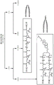


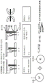


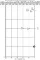


Classifications
-
- A—HUMAN NECESSITIES
- A61—MEDICAL OR VETERINARY SCIENCE; HYGIENE
- A61K—PREPARATIONS FOR MEDICAL, DENTAL OR TOILETRY PURPOSES
- A61K47/00—Medicinal preparations characterised by the non-active ingredients used, e.g. carriers or inert additives; Targeting or modifying agents chemically bound to the active ingredient
- A61K47/50—Medicinal preparations characterised by the non-active ingredients used, e.g. carriers or inert additives; Targeting or modifying agents chemically bound to the active ingredient the non-active ingredient being chemically bound to the active ingredient, e.g. polymer-drug conjugates
- A61K47/51—Medicinal preparations characterised by the non-active ingredients used, e.g. carriers or inert additives; Targeting or modifying agents chemically bound to the active ingredient the non-active ingredient being chemically bound to the active ingredient, e.g. polymer-drug conjugates the non-active ingredient being a modifying agent
- A61K47/54—Medicinal preparations characterised by the non-active ingredients used, e.g. carriers or inert additives; Targeting or modifying agents chemically bound to the active ingredient the non-active ingredient being chemically bound to the active ingredient, e.g. polymer-drug conjugates the non-active ingredient being a modifying agent the modifying agent being an organic compound
- A61K47/543—Lipids, e.g. triglycerides; Polyamines, e.g. spermine or spermidine
-
- A—HUMAN NECESSITIES
- A61—MEDICAL OR VETERINARY SCIENCE; HYGIENE
- A61K—PREPARATIONS FOR MEDICAL, DENTAL OR TOILETRY PURPOSES
- A61K39/00—Medicinal preparations containing antigens or antibodies
- A61K39/12—Viral antigens
- A61K39/21—Retroviridae, e.g. equine infectious anemia virus
-
- A—HUMAN NECESSITIES
- A61—MEDICAL OR VETERINARY SCIENCE; HYGIENE
- A61K—PREPARATIONS FOR MEDICAL, DENTAL OR TOILETRY PURPOSES
- A61K39/00—Medicinal preparations containing antigens or antibodies
- A61K39/12—Viral antigens
-
- A—HUMAN NECESSITIES
- A61—MEDICAL OR VETERINARY SCIENCE; HYGIENE
- A61K—PREPARATIONS FOR MEDICAL, DENTAL OR TOILETRY PURPOSES
- A61K39/00—Medicinal preparations containing antigens or antibodies
- A61K39/12—Viral antigens
- A61K39/155—Paramyxoviridae, e.g. parainfluenza virus
- A61K39/165—Mumps or measles virus
-
- A—HUMAN NECESSITIES
- A61—MEDICAL OR VETERINARY SCIENCE; HYGIENE
- A61K—PREPARATIONS FOR MEDICAL, DENTAL OR TOILETRY PURPOSES
- A61K47/00—Medicinal preparations characterised by the non-active ingredients used, e.g. carriers or inert additives; Targeting or modifying agents chemically bound to the active ingredient
- A61K47/50—Medicinal preparations characterised by the non-active ingredients used, e.g. carriers or inert additives; Targeting or modifying agents chemically bound to the active ingredient the non-active ingredient being chemically bound to the active ingredient, e.g. polymer-drug conjugates
- A61K47/51—Medicinal preparations characterised by the non-active ingredients used, e.g. carriers or inert additives; Targeting or modifying agents chemically bound to the active ingredient the non-active ingredient being chemically bound to the active ingredient, e.g. polymer-drug conjugates the non-active ingredient being a modifying agent
- A61K47/54—Medicinal preparations characterised by the non-active ingredients used, e.g. carriers or inert additives; Targeting or modifying agents chemically bound to the active ingredient the non-active ingredient being chemically bound to the active ingredient, e.g. polymer-drug conjugates the non-active ingredient being a modifying agent the modifying agent being an organic compound
- A61K47/55—Medicinal preparations characterised by the non-active ingredients used, e.g. carriers or inert additives; Targeting or modifying agents chemically bound to the active ingredient the non-active ingredient being chemically bound to the active ingredient, e.g. polymer-drug conjugates the non-active ingredient being a modifying agent the modifying agent being an organic compound the modifying agent being also a pharmacologically or therapeutically active agent, i.e. the entire conjugate being a codrug, i.e. a dimer, oligomer or polymer of pharmacologically or therapeutically active compounds
-
- A—HUMAN NECESSITIES
- A61—MEDICAL OR VETERINARY SCIENCE; HYGIENE
- A61P—SPECIFIC THERAPEUTIC ACTIVITY OF CHEMICAL COMPOUNDS OR MEDICINAL PREPARATIONS
- A61P31/00—Antiinfectives, i.e. antibiotics, antiseptics, chemotherapeutics
- A61P31/12—Antivirals
- A61P31/14—Antivirals for RNA viruses
-
- A—HUMAN NECESSITIES
- A61—MEDICAL OR VETERINARY SCIENCE; HYGIENE
- A61P—SPECIFIC THERAPEUTIC ACTIVITY OF CHEMICAL COMPOUNDS OR MEDICINAL PREPARATIONS
- A61P31/00—Antiinfectives, i.e. antibiotics, antiseptics, chemotherapeutics
- A61P31/12—Antivirals
- A61P31/14—Antivirals for RNA viruses
- A61P31/18—Antivirals for RNA viruses for HIV
-
- C—CHEMISTRY; METALLURGY
- C07—ORGANIC CHEMISTRY
- C07K—PEPTIDES
- C07K14/00—Peptides having more than 20 amino acids; Gastrins; Somatostatins; Melanotropins; Derivatives thereof
- C07K14/005—Peptides having more than 20 amino acids; Gastrins; Somatostatins; Melanotropins; Derivatives thereof from viruses
-
- A—HUMAN NECESSITIES
- A61—MEDICAL OR VETERINARY SCIENCE; HYGIENE
- A61K—PREPARATIONS FOR MEDICAL, DENTAL OR TOILETRY PURPOSES
- A61K39/00—Medicinal preparations containing antigens or antibodies
- A61K2039/60—Medicinal preparations containing antigens or antibodies characteristics by the carrier linked to the antigen
- A61K2039/6031—Proteins
- A61K2039/6075—Viral proteins
-
- A—HUMAN NECESSITIES
- A61—MEDICAL OR VETERINARY SCIENCE; HYGIENE
- A61K—PREPARATIONS FOR MEDICAL, DENTAL OR TOILETRY PURPOSES
- A61K39/00—Medicinal preparations containing antigens or antibodies
- A61K2039/60—Medicinal preparations containing antigens or antibodies characteristics by the carrier linked to the antigen
- A61K2039/6093—Synthetic polymers, e.g. polyethyleneglycol [PEG], Polymers or copolymers of (D) glutamate and (D) lysine
-
- A—HUMAN NECESSITIES
- A61—MEDICAL OR VETERINARY SCIENCE; HYGIENE
- A61K—PREPARATIONS FOR MEDICAL, DENTAL OR TOILETRY PURPOSES
- A61K38/00—Medicinal preparations containing peptides
-
- C—CHEMISTRY; METALLURGY
- C12—BIOCHEMISTRY; BEER; SPIRITS; WINE; VINEGAR; MICROBIOLOGY; ENZYMOLOGY; MUTATION OR GENETIC ENGINEERING
- C12N—MICROORGANISMS OR ENZYMES; COMPOSITIONS THEREOF; PROPAGATING, PRESERVING, OR MAINTAINING MICROORGANISMS; MUTATION OR GENETIC ENGINEERING; CULTURE MEDIA
- C12N2760/00—MICROORGANISMS OR ENZYMES; COMPOSITIONS THEREOF; PROPAGATING, PRESERVING, OR MAINTAINING MICROORGANISMS; MUTATION OR GENETIC ENGINEERING; CULTURE MEDIA ssRNA viruses negative-sense
- C12N2760/00011—Details
- C12N2760/18011—Paramyxoviridae
- C12N2760/18411—Morbillivirus, e.g. Measles virus, canine distemper
- C12N2760/18422—New viral proteins or individual genes, new structural or functional aspects of known viral proteins or genes
-
- C—CHEMISTRY; METALLURGY
- C12—BIOCHEMISTRY; BEER; SPIRITS; WINE; VINEGAR; MICROBIOLOGY; ENZYMOLOGY; MUTATION OR GENETIC ENGINEERING
- C12N—MICROORGANISMS OR ENZYMES; COMPOSITIONS THEREOF; PROPAGATING, PRESERVING, OR MAINTAINING MICROORGANISMS; MUTATION OR GENETIC ENGINEERING; CULTURE MEDIA
- C12N2760/00—MICROORGANISMS OR ENZYMES; COMPOSITIONS THEREOF; PROPAGATING, PRESERVING, OR MAINTAINING MICROORGANISMS; MUTATION OR GENETIC ENGINEERING; CULTURE MEDIA ssRNA viruses negative-sense
- C12N2760/00011—Details
- C12N2760/18011—Paramyxoviridae
- C12N2760/18411—Morbillivirus, e.g. Measles virus, canine distemper
- C12N2760/18433—Use of viral protein as therapeutic agent other than vaccine, e.g. apoptosis inducing or anti-inflammatory
-
- C—CHEMISTRY; METALLURGY
- C12—BIOCHEMISTRY; BEER; SPIRITS; WINE; VINEGAR; MICROBIOLOGY; ENZYMOLOGY; MUTATION OR GENETIC ENGINEERING
- C12N—MICROORGANISMS OR ENZYMES; COMPOSITIONS THEREOF; PROPAGATING, PRESERVING, OR MAINTAINING MICROORGANISMS; MUTATION OR GENETIC ENGINEERING; CULTURE MEDIA
- C12N2760/00—MICROORGANISMS OR ENZYMES; COMPOSITIONS THEREOF; PROPAGATING, PRESERVING, OR MAINTAINING MICROORGANISMS; MUTATION OR GENETIC ENGINEERING; CULTURE MEDIA ssRNA viruses negative-sense
- C12N2760/00011—Details
- C12N2760/18011—Paramyxoviridae
- C12N2760/18411—Morbillivirus, e.g. Measles virus, canine distemper
- C12N2760/18434—Use of virus or viral component as vaccine, e.g. live-attenuated or inactivated virus, VLP, viral protein
Abstract
Described herein are peptides, compositions, and methods for treating measles or HIV infection with an antiviral peptide conjugate comprising a Fusion Inhibitory Peptide (FIP) conjugated to a C-terminal Heptad Repeat (HRC) peptide. Also described herein are stable soluble measles F proteins, compositions, and methods of preventing measles infection with stable F proteins that can be administered alone or in combination with the antiviral peptide conjugates described herein.
Description
Cross Reference to Related Applications
This application claims us application number 62/895,752 filed on 4.9.2019; U.S. application No. 62/988,286 filed on 11/3/2020; and U.S. application No. 63/009,883, filed on 14/4/2020, all of which are incorporated herein by reference in their entirety.
All patents, patent applications, and publications cited herein are incorporated by reference in their entirety. The disclosures of these publications in their entireties are hereby incorporated by reference into this application.
The disclosure of this patent contains material which is subject to copyright protection. The copyright owner has no objection to the facsimile reproduction by anyone of the patent document or the patent disclosure, as it appears in the patent and trademark office patent file or records, but otherwise reserves any and all copyright rights whatsoever.
Government support
The invention was made with government support awarded by the National Institutes of Health, which subsidized NS091263, NS105699, AI121349 and AI 119762. The government has certain rights in this invention.
Background
Measles remains a challenge to global health. Although the measles vaccine was introduced in 1963, there is no FDA-approved antiviral treatment for already infected individuals. Current methods have limited ability to prevent measles virus ("MV" or "MeV") fusion and entry into host cells. Current proposals are to inoculate vaccines or Immunoglobulins (IG) within 72 hours after exposure. The antiviral compound may be directed against different components of viral activity, for example, to prevent viral replication or to prevent viral entry into a host cell.
Disclosure of Invention
In certain aspects, the invention described herein relates to an antiviral peptide comprising a combination of a C-terminal Heptad Repeat (HRC) peptide and a Fusion Inhibitory Peptide (FIP). Unexpectedly, the combination of HRC and FIP showed a synergistic effect, more effective than either method alone. This combination prevents MeV activation in its entirety and also prevents refolding and fusion of already activated viruses.
In one aspect, the present invention provides an antiviral peptide conjugate comprising a Fusion Inhibitory Peptide (FIP) and a C-terminal Heptad Repeat (HRC) peptide (FIP-HRC). In some embodiments, the antiviral peptide conjugate further comprises a membrane-localizing moiety. In some embodiments, the membrane-localization portion comprises a membrane-localization portion selected from the group consisting of cholesterol, tocopherol, and palm-based. In some embodiments, the membrane localization moiety is conjugated to the C-terminus of the HRC peptide. In some embodiments, the antiviral peptide conjugate further comprises a linker region. In some embodiments, the linker region comprises polyethylene glycol (PEG). In some embodiments, the PEG is 4 ethylene glycol units in length (PEG)4). In some embodiments, the PEG is 11 ethylene glycol units in length (PEG)11)。
In some embodiments, the linker region is conjugated to the C-terminus of the HRC peptide. In some embodiments, the antiviral peptide conjugate further comprises a membrane localization moiety and a linker region. In some embodiments, the linker region is conjugated to the C-terminus of the HRC peptide, and the membrane-localization moiety is conjugated to the linker region. In some embodiments, the linker region comprises polyethylene glycol (PEG). In some embodiments, the PEG is 4 ethylene glycol units in length (PEG)4). In some embodiments, the PEG is 11 ethylene glycol units in length (PEG)11). In some embodiments, the PEG is 12 ethylene glycol units in length (PEG)12). In some embodiments, the PEG is 14 ethylene glycol units in length (PEG)14). In some embodiments, the antiviral peptide comprisesA dimer of the FIP region and the HRC peptide region. In some embodiments, the antiviral peptide comprises a first FIP-HRC peptide conjugated to the linker region and a second FIP-HRC peptide conjugated to the linker region. In some embodiments, the peptide further comprises a serine-glycine linker. In some embodiments, the serine-glycine linker is located between the FIP and HRC peptides. In some embodiments, the serine-glycine linker is located C-terminal to the HRC peptide. In some embodiments, the serine-glycine linker is located between the FIP region and the HRC peptide region, and further comprises a second serine-glycine linker located C-terminal to the HRC peptide region. In some embodiments, the serine-glycine linker comprises the amino acid sequence gsgsgsg. In some embodiments, the first phenylalanine residue of the FIP of the antiviral peptide conjugate is a D-amino acid. In some embodiments, the N-terminus of the antiviral peptide conjugate further comprises a benzyloxycarbonyl group. In some embodiments, the FIP peptide comprises the amino acid sequence FFG. In some embodiments, the HRC peptide comprises amino acid sequence PPISLERLDVGTNLGN AIAKLEDAKELLESSDQILR. In some embodiments, the HRC peptide conjugate comprises amino acid sequence PPISLERLDVGTN. In some embodiments, the antiviral peptide comprises amino acid sequence FFGPPISLERLDVGTNLGNAIAKLEDAKELLESSDQILR. In some embodiments, the antiviral peptide comprises amino acid sequence FFGPPISLERLDVGTN.
In another aspect, the invention provides a nanoparticle comprising any of the antiviral peptide conjugates described herein. In some embodiments, the nanoparticles have a diameter of about 50nm to about 150 nm.
In another aspect, the invention provides a composition comprising any of the antiviral peptide conjugates described herein.
In another aspect, the present invention provides a prophylactic composition comprising an antiviral peptide conjugate comprising a Fusion Inhibitory Peptide (FIP) and a C-terminal Heptad Repeat (HRC) peptide (FIP-HRC), wherein the measles antiviral peptide conjugate prevents membrane fusion of measles virus.
In another aspect, the present invention provides a nanoparticle comprising a Fusion Inhibitory (FIP) peptide and a C-terminal Heptad Repeat (HRC) peptide (FIP-HRC).
In another aspect, the present invention provides a method of post-infection measles prevention (post-infection measles prophyylaxis) comprising administering to a subject in need thereof an antiviral peptide conjugate comprising a Fusion Inhibitory Peptide (FIP) and a C-terminal Heptapeptide (HRC) peptide (FIP-HRC). In some embodiments, the antiviral peptide conjugate further comprises a linker region. In some embodiments, the antiviral peptide conjugate further comprises a membrane-localizing moiety. In some embodiments, the antiviral peptide conjugate further comprises a linker region and a membrane-locating moiety region. In some embodiments, administering comprises intranasal inhalation or oral inhalation. In some embodiments, the antiviral peptide conjugate is administered by a device selected from the group consisting of a nebulizer, an aerosolizer, and an inhaler. In some embodiments, administering comprises administering subcutaneously. In some embodiments, the subject has been exposed to a measles virus comprising a wild-type fusion glycoprotein. In some embodiments, the subject has been exposed to measles virus comprising one or more mutations of a fusion glycoprotein selected from the group consisting of N462K, L454W, T461I, E455G, E170G, G506E, M337L, D538G, G168R, S262G, a440P, R520C, and L550P.
In another aspect, the invention provides a recombinant protein comprising a stable soluble measles F protein comprising the E445G mutation.
In another aspect, the invention provides a recombinant protein comprising a stable soluble measles F protein comprising the double mutations E170G and E455G.
In another aspect, the present invention provides a recombinant protein comprising the amino acid sequence SEQ ID NO. 3 or SEQ ID NO. 4.
In another aspect, the present invention provides a recombinant protein comprising the amino acid sequence SEQ ID NO. 5 or SEQ ID NO. 6.
In another aspect, the invention provides an immunogenic composition comprising any one of the recombinant proteins described herein. In some embodiments, the present invention provides a method of preventing measles infection prior to measles exposure by administering an immunogenic composition to a subject. In some embodiments, the present invention provides a method of inducing an immune response against measles virus by administering to a subject an immunogenic composition.
In another aspect, the invention provides an immunogenic composition comprising the amino acid sequence SEQ ID No. 1 or SEQ ID No. 2, and further comprising any of the antiviral peptide conjugates described herein.
In another aspect, the invention provides a method of inducing an immune response against measles virus by administering to a subject any of the immunogenic compositions described herein and further administering any of the antiviral peptide conjugates described herein.
In another aspect, the present invention provides a method of producing a recombinant protein comprising the amino acid sequence SEQ ID NO. 3 or SEQ ID NO. 4.
In another aspect, the present invention provides a method of producing a recombinant protein comprising the amino acid sequence SEQ ID NO. 5 or SEQ ID NO. 6.
In another aspect, the invention provides a method for a cell line for expression of a recombinant protein comprising the amino acid sequence SEQ ID NO. 3 or SEQ ID NO. 4.
In another aspect, the invention provides a method for a cell line for expression of a recombinant protein comprising the amino acid sequence SEQ ID NO. 5 or SEQ ID NO. 6.
In another aspect, the present invention provides an antiviral peptide conjugate comprising a Fusion Inhibitory Peptide (FIP) and a C-terminal Heptad Repeat (HRC) peptide (FIP-HRC), wherein the HRC peptide is derived from HIV-GP41 (C34). In some embodiments, the HRC peptide comprises amino acid sequence WMEWDREINNYTSLIHSLIEESQNQQEKNEQELL.
In another aspect, the invention provides a method of HIV prevention after infection, the method comprising administering to a subject in need thereof an antiviral peptide conjugate comprising a Fusion Inhibitory Peptide (FIP) and a C-terminal Heptapeptide (HRC) peptide (FIP-HRC). In some embodiments, the HRC peptide is derived from HIV-GP41 (C34).
Drawings
This patent or application document contains at least one drawing executed in color. To meet the requirements of the PCT patent application, many of the figures provided herein are black and white representations of original color images.
Figure 1 shows that MV infection begins in the respiratory tract.
Figure 2 shows the exit of viremia and MV pathogenesis (egress).
Figure 3 shows that measles can also cause serious complications.
Figure 4 shows data for HIV infected patients with MV encephalitis.
Figure 5 shows a schematic representation of measles virus.
Figure 6 shows that the MV F genes of two patients contain the same nucleotide mutations.
Figure 7 shows that wild-type measles virus is tightly controlled for entry into the Central Nervous System (CNS).
FIG. 8 shows an analysis method of the fusion complex.
Fig. 9A-9C show the level of fusion. FIGS. 9A-B show that all F proteins exhibit similar levels of fusion in the presence of known receptors. Figure 9C shows F L454W induced fusion in the absence of a known receptor.
Fig. 10 shows the two thermal states of the F protein.
FIG. 11 shows cell-to-cell fusion (cell-to-cell fusion) induced by recombinant viruses.
Figure 12 shows 90 day old brain organoids infected with measles wild type F virus and virus with L454W F.
Figure 13 shows ex vivo tissue from mice (no receptor).
Fig. 14 shows MV data for cotton rats.
Fig. 15A-15B show survival data for mice.
Figure 16 shows viral entry as a therapeutic target.
Figure 17 shows F glycoprotein-derived peptides.
Figure 18 shows that F glycoprotein derived peptides inhibit viral entry.
Figure 19 shows targeting HRC peptides to the lipid membrane.
Figure 20 shows the improvement in affinity (avidity) of HRC peptide for F protein.
Figure 21 shows another embodiment of targeting HRC peptides to the lipid membrane.
Figures 22A-22B show that conjugated peptides are effective in vivo. The model used in FIG. 22A was a cotton mouse, and the model used in FIG. 22B was a SLAM: IFNARKO mouse.
Figures 23A-23C show that MV HRC4 peptide blocks ex vivo transmission of virus.
Figure 24 shows that intranasal administration of MV HRC4 protected suckling mice from lethal infection with L454W F-bearing virus.
Figure 25 shows that the peptide particle size is in the nanomolar range.
Figure 26 shows amphiphilic structure driven self-assembly and nanoparticle formation.
Fig. 27 shows that the nanoparticles assemble (disassemble) and retain the peptide at the lipid membrane interface.
Fig. 28 shows that nanoparticles are able to cross HAE barriers and are bioavailable in vivo. By reaching the relevant infection site, these nanoparticles prevent the proliferation of measles virus.
Figure 29 shows that the biodistribution in the cotton rat model is improved when conjugated peptides are administered intranasally at 6 mg/kg.
Figure 30 shows a combinatorial strategy in which Fusion Inhibitory Peptide (FIP) binds to the fusion protein and stabilizes the pre-fusion state of measles F protein.
Fig. 31 shows an isobologram curve (isobologram curve) showing synergy between FIP and HRC peptides.
Figure 32 shows that FIP added to the HRC region enhances antiviral activity.
Figure 33 shows that when FIP and HRC are structurally identical, the potency exceeds the synergistic effect of the two inhibitors added together.
Figure 34 shows additional data for efficacy over synergy of the two inhibitors added together when FIP and HRC are structurally identical.
Figure 35 shows that FIP added to the HRC region enhances antiviral activity.
Figure 36 shows additional data that FIP added to the HRC region enhances antiviral activity.
Fig. 37 shows survival rates.
Figure 38 shows a number of Measles Viruses (MVs).
Figure 39 shows that MV HRC4 peptide and RNA polymerase inhibitors prevented MV wild-type infection in human motor neurons.
Figure 40 shows HRC4 peptide and RNA polymerase inhibitors: wild type viruses and CNS-adapted viruses (CNS-adapted viruses).
Figure 41 shows MV HRC4 peptide and RNA polymerase inhibitor versus (vs.) CNS-adapted MVs in human motor neurons.
42A-42B illustrate the steps that are entered.
FIGS. 43A-43C also show the steps that are entered.
FIGS. 44A-44C show that the H-F interaction is altered in L454W F.
Figure 45 shows that intranasal administration of MV HRC4 protected IFNAR KO mice from lethal MV encephalitis.
Figure 46 shows a schematic of the various organoids that can be grown from pluripotent stem cells and the developmental signals used.
FIGS. 47A-47C show "mini-brain" (mini-brain) produced from pluripotent stem cells. Fig. 47A shows that a complex morphology with heterogeneous regions containing neural progenitors (SOX2, red) and neurons (TUJ1, green) is evident. Figure 47B shows immunofluorescence images of whole kidney organoids grown from pluripotent stem cells with patterned kidney cells (patterned nephron). Glomerular forming podocytes (NPHS1, yellow), early proximal tubules (lotus root agglutinin, pink), and distal tubule/collecting ducts (E-cadherin, green). Figure 47C shows 3D reconstruction of the middle of human aSC-derived lung organoids, stained for intermediate filaments of basal cells (green), actin cytoskeleton (red), and nuclei (blue), and imaged by confocal microscopy.
Figure 48 shows the reported measles cases over time.
Figure 49 shows a schematic representation of measles virus.
Figure 50 shows measles virus entry into cells.
Figure 51 shows the steps of measles virus entry into cells.
Figure 52 shows measles virus entry as a potential therapeutic target.
Figure 53 shows the structure of measles Fusion Inhibitory Peptide (FIP).
Figure 54 shows a combinatorial strategy for preventing measles virus entry into cells.
Figure 55 shows the design of lipid-peptide conjugates.
Figure 56 shows the advantages of lipid-peptide conjugates.
Fig. 57-58 show examples of lipid-peptide structures.
FIGS. 59-60 show reaction schemes for the synthesis of monomeric lipid-peptide conjugates.
Fig. 61 shows a list of lipid-peptide conjugates.
Figure 62 shows the purification and characterization data for lipid-peptide conjugates.
FIG. 63 shows the process of a fusion assay based on β -galactosidase complementation.
Figure 64 shows data on the inhibitory activity of measles lipid-peptide conjugates in the fusion assay.
Figure 65 shows the optimal lipid-peptide conjugate candidates based on inhibitory activity from fusion assay data.
Figure 66 shows the process of MTT cytotoxicity assay.
Figure 67 shows MTT assay data.
Figure 68 shows the course of the thermal stability study of measles fusion proteins.
Figure 69 shows data from a thermal stability study of measles fusion protein.
Figure 70 shows the F stability properties of measles lipid-peptide conjugates.
Figure 71 shows optimal lipid-peptide conjugate candidates based on the F stability properties of measles lipid-peptide conjugates.
Fig. 72 shows quantifying each stage of fusion activation.
Figures 73-75 show the binding activity of measles virus to Red Blood Cells (RBC) in the presence of various concentrations of different lipid-peptide conjugates (figure 73: HRC; figure 74: FIP-HRC; figure 75: FIP).
Figure 76 shows a schematic of the in vivo efficacy of measles lipid-peptide conjugates.
Figure 77 shows data on the in vivo efficacy of measles lipid-peptide conjugates.
Figure 78 shows particle size measurement data for measles lipid-peptide conjugates.
Fig. 79 shows a schematic of the interaction between lipid-peptide conjugates.
Fig. 80 shows data from isobologram (isobologram) analysis.
Fig. 81 shows the quantification of the isobologram analysis.
Figure 82 figure 1 shows a schematic of the mechanism of action of measles fusion inhibitor conjugates.
Fig. 83 shows schematic diagrams for various designs of different lipid-peptide conjugates.
Figure 84 shows data for in vivo efficacy.
FIGS. 85A-85C show FIP-HRC targeting of MV F-expressing cells.
FIG. 86 shows FIP-HRC stabilizes measles F in its pre-fusion state.
Figure 87 shows the F-stabilizing properties of MeV peptides.
Figures 88-90 show the stability characteristics of MeV peptides to soluble F.
Fig. 91 shows that FIP-HRC prevents F activation (completely different from the mechanism of HRC that prevents F refolding).
Figure 92 shows FIP-HRC targeting MeV F expressing cells.
Figure 93 shows data from three different experiments, indicating that FIP-HRC targets MeV F expressing cells.
Figure 94 shows the stability characteristics of MeV peptides.
Figure 95 shows the cytotoxicity of MeV peptides.
Figure 96 shows synergy data from the HRC + FIP conjugate isobologram analysis.
FIG. 97 shows the potency of FIP-HRC with 12 amino acids derived from measles HRC.
Figure 98 shows inhibition data of BG505 (human immunodeficiency virus type 1 (HIV-1) strain) using various lipid-peptide conjugates and positive and negative controls.
Fig. 99 shows inhibition data for B41(HIV-1 strain) using various lipid-peptide conjugates and positive and negative controls.
Figure 100 shows inhibition data for 16055(HIV-1 strain) using various lipid-peptide conjugates and positive and negative controls.
Figure 101 shows inhibition data for MN (HIV-1 strain) using various lipid-peptide conjugates and positive and negative controls.
Figure 102 shows inhibition data for Vesicular Stomatitis Virus (VSV) using various lipid-peptide conjugates and positive and negative controls.
Fig. 103 shows inhibition data for Murine Leukemia Virus (MLV) using various lipid-peptide conjugates.
FIGS. 104A-104B show alternative positions within the F protein from CNS-adapted viruses.
Figures 105A-105I show ex vivo infection of wild-type (wt) virus and ex vivo infection of virus with L454W F: CNS-adapted viruses outperform wt viruses in Organotypic Brain Cultures (OBCs).
Figures 106A-D show efficient spread of CNS-adapted MeV variants in human pluripotent stem cell (hiPSC) -derived brain organoids.
FIGS. 107A-107H show the fusion activity and thermostability of MeV fusion (F) proteins with the indicated mutations, with additional mutations in the L454W F background stabilizing the prefusion state of the F protein.
FIG. 108 shows CD150xIFNAR being highly susceptibleKOIn the CNS, transmission of L454W F-bearing viruses is inhibited.
FIGS. 109A-109B show that MeV F L454W induces interferon-stimulated genes in mouse brain slice cultures compared to wild-type MeV.
FIGS. 110A-110B show infection of human brain organoids in the presence of a fusion inhibitor.
FIGS. 111A-111B show induction of host antiviral genes in brain organoids infected with the fusion protein mutant MeV.
Fig. 112 shows a heat map of the correlation of gene level RPKM values between brain organoids and BrainSpan atlas.
Figure 113 shows KEGG pathway analysis of differentially expressed genes following L454W infection.
FIG. 114 shows a Longitudinal Analysis of Viral Alleles (LAVA) profile of the mutant virus populations of L454W and L454W/G506E fusion proteins.
FIG. 115 shows a LAVA map of the mutant virus populations of L454W and L454W/E455G fusion proteins.
FIG. 116 shows a LAVA profile of wild-type fusion protein virus population.
FIGS. 117A-117E show that (A) soluble MeV-FE170G-E455G has been incubated at 4 ℃ or 55 ℃ for the indicated time with or without FIP-HRC 1mM (dimer without cholesterol). Following incubation, F was immunoprecipitated using a mouse pre-fusion specific antibody. Immunoprecipitated proteins were run on SDS-PAGE reducing gels, transferred to PVDF membrane and incubated with α -MV-F-HRC biotin (1: 1000). Streptavidin alkaline phosphatase conjugate has been used as secondary antibody (in Phosphate Buffer (pbs) in a ratio of 1: 1000). (B) Western immunoblot (Western Blot, WB) bands from the experiments described in (a) have been quantified using ImageJ software. Results represent mean ± SEM of three independent experiments. (C) Soluble MeV-FE170G-E455G has been incubated at 4 ℃,25 ℃ or 37 ℃ for 1 week. Immunoprecipitation was performed as described in (A). (D) Soluble MeV-FWT or MeV-FE170G-E455G had been incubated with or without FIP-HRC, commercially available FIP, FIP dimer or 3g (1mM) at 4 ℃ or 55 ℃ for 60 minutes or 120 minutes. (E) Western immunoblot (Western Blot, WB) bands from the experiments described in (D) have been quantified using ImageJ software. Results represent mean ± SEM of three independent experiments.
Detailed Description
Despite more than 50 years ago vaccines were introduced, measles remains a challenge to global health. For already infected individuals, there is no FDA-approved antiviral therapy, and current methods have limited ability to prevent measles virus (MeV) fusion and entry into host cells. In one embodiment, the subject matter disclosed herein relates to a combination of two existing MeV antiviral approaches directed to different modes of action. The first approach involves targeting the terminal Heptad Repeat (HRC) region of MeV fusion protein (F) using HRC-derived peptides, which interfere with structural rearrangements required for viral fusion during infection (e.g., prevent refolding). The second approach involves targeting the heptad repeat b (hrb) region of MeV F using Fusion Inhibitory Peptide (FIP), which stabilizes MeV F in its non-fusion-competent prefusion state (e.g., see fig. 52-32). This combination approach has proven to be more effective than either approach alone in several in vitro, ex vivo and in vivo models. This combination together prevents MeV activation and also prevents refolding and fusion of already activated viruses.
Furthermore, in one embodiment, the presently disclosed subject matter relates to a combination of two antiviral approaches to prevent measles virus from fusing with cell membranes and entering cells by: targeting the C-terminal Heptad Repeat (HRC) region of MeV F using HRC-derived peptides, which interferes with the structural rearrangement required for viral fusion during infection; and targeting the heptad repeat b (hrb) region of MeV F using Fusion Inhibitory Peptide (FIP), which stabilizes MeV F in the pre-activation state. This technique demonstrates that the combination of the two methods is more effective than either method alone, in vitro, ex vivo, and in vivo.
The present invention relates to measles antiviral peptides for administration by the respiratory or subcutaneous routes. The invention also relates to a combination of two different peptide domains that act by different mechanisms to prevent entry: (1) a peptide domain ("HRC") complementary to the heptad repeat on the measles F protein, which is critical to the F-refolding step entered by the virus, which occurs once the F protein is activated and inserted into the target cell membrane. This refolding is responsible for virus-cell membrane fusion and entry, and thus blocking it by binding of HRC peptide to F protein can prevent virus entry and infection; (2) a peptide domain ("FIP") that binds to a different region of the F protein (e.g., the HRB region) and stabilizes the F protein in a pre-activated conformation, preventing F activation to a state that enables fusion. If the F protein is not activated to a state enabling fusion, no subsequent entering step will occur. (1) The combination of (1) and (2) results in a peptide that first stabilizes the F protein in its pre-fusion conformation, rendering it non-fusion-competent-then, for F proteins that have been activated for fusion, prevents refolding and fusion. The efficacy of this combination has been studied both in vitro and in vivo and is significantly enhanced over either method alone.
Reference documents:
is the prevention and treatment of measles with Plemper RK, Snyder JP.? (Measles control-can Measles virus inhibitors make a difference; 10(8) pages 811-20.
Post-exposure management of disease preventable by the Woo TM. vaccine (Postexposure management of vaccine-preventive diseases.) J pediatric Health Care (J pediatric Health Care) 2016 month 3; 30(2) pages 173-82.
Welsch JC, Talekar a, Mathieu C, petsis a, Moscona a, Horvat B, Porotto m. brain penetrating fusion inhibitors can prevent Fatal measles virus infection (Fatal measles virus infection by vaccine-dependent fusion inhibitors) viriology journal (JVirol) for 12 months 2013; 87(24) pages 13785-94.
Hashiguchi T, Fukuda Y, Matsuoka R, Kuroda D, Kubota M, Shirogane Y, Watanabe S, Tsumoto K, Kohda D, Plemper RK, Yanagi Y. Structure of the pre-fusion form of measles virus fusion protein complexed with inhibitor (Structures of the compression form of measles virus fusion protein complex with inhibitors) Proc. Proc Natl Acad Sci USA (ProcNatl Acad Sci USA) 2018 month 3; 115(10) pages 2496 and 501.
Antiviral peptide conjugates
In certain aspects, the present invention provides antiviral peptide conjugates comprising a Fusion Inhibitory Peptide (FIP) and a C-terminal Heptad Repeat (HRC) peptide.
Measles virus belongs to the genus measles virus (Morbillivirus) paramyxovirus. It is a polymorphic virus with a diameter ranging from 100nm to 300 nm. The measles genome consists of six genes, each encoding a structural protein: referred to as N (nucleocapsid Protein), P (phosphoprotein), M (matrix Protein), F (fusion Protein), H (hemagglutinin Protein) and L (Large Protein). The phosphoprotein (P) gene of one of these genes also encodes two non-structural proteins (V and C). There are different wild-type measles virus genetic lineages based on the nucleotide sequence of the nucleoprotein.
Fusion Inhibiting Peptide (FIP)
In some embodiments, FIP is a fusion inhibitor peptide comprising the amino acid sequence phenylalanine-glycine (FFG). In some embodiments, FIP is a fusion inhibitor peptide consisting of the amino acid sequence phenylalanine-glycine (FFG). In some embodiments, the first phenylalanine residue of measles FIP is a D-amino acid and the second phenylalanine residue and glycine residue are L-amino acids (i.e., D-FFG). D-amino acids are amino acids in which the stereogenic carbon alpha of the amino group has the D-configuration. In some embodiments, the N-terminus of FIP further comprises a benzyloxycarbonyl group (i.e., Z-D-FFG) as shown in figure 53. In some embodiments, the FIP further comprises one or more serine-glycine linkers. In some embodiments, the serine-glycine linker comprises the amino acid sequence gsgsgsg. In some embodiments, the serine-glycine linker consists of the amino acid sequence GSGSG. In some embodiments, FIP comprises a serine-glycine linker at the C-terminus of FIP (e.g., FFG-GSGSGSG, D-FFG-GSGSG, Z-D-GSGSG). In some embodiments, FIP binds to the HRB region of MeV F to prevent viral entry into the host cell.
C-terminal Heptapeptide Repeat (HRC) peptides
In some embodiments, the C-terminal Heptad Repeat (HRC) peptide is derived from measles virus F protein. HRC peptides are conserved between measles strains (conserved) and can be derived from any measles strain F protein. In some embodiments, the measles virus F protein-derived C-terminal heptad repeat peptide is derived from measles strain B3. In some embodiments, the measles virus-derived C-terminal heptad repeat peptide is derived from measles strain G954. The nucleotide and amino acid sequences of the measles virus genome and the proteins encoded therein are publicly available in databases known to those skilled in the art, such as, but not limited to, GenBank and ViPR (www.viprbrc.org).
In some embodiments, the HRC is a measles virus F protein-derived C-terminal heptad repeat peptide. In some embodiments, the HRC comprises residues 450 to 485 of the C-terminal heptad repeat peptide of the measles virus-derived F protein. In some embodiments, the HRC comprises amino acid sequence PPISLERLDVGTNLGNAIAKLEDAKELLESSDQILR. In some embodiments, the HRC consists of amino acid sequence PPISLERLDVGTNLGNAIAKLEDAKELLESSDQILR. In some embodiments, the HRC comprises an amino acid sequence comprising about 99%, 98%, 97%, 96%, 95%, 94%, 93%, 92%, 91%, 90%, 89%, 88%, 87%, 86%, 85%, 84%, 83%, 82%, 81%, 80%, 79%, 78%, 77%, 76%, or 75% sequence identity to PPISLERLDVGTNLGNAIAKLEDAKELLESSDQILR. In some embodiments, the HRC comprises residues 450 to 462 of the C-terminal heptad repeat peptide of the measles virus-derived F protein. In some embodiments, the HRC comprises amino acid sequence PPISLERLDVGTN. In some embodiments, the HRC consists of amino acid sequence PPISLERLDVGTN. In some embodiments, the HRC comprises an amino acid sequence comprising about 99%, 98%, 97%, 96%, 95%, 94%, 93%, 92%, 91%, 90%, 89%, 88%, 87%, 86%, 85%, 84%, 83%, 82%, 81%, 80%, 79%, 78%, 77%, 76%, or 75% sequence identity to PPISLERLDVGTN. In some embodiments, the HRC comprises residues 450 to 463, 464, 465, 466, 467, 468, 469, 470, 471, 472, 473, 474, 475, 476, 477, 478, 479, 480, 481, 482, 483, or 484 of the measles virus-derived C-terminal heptad repeat peptide. In some embodiments, the HRC comprises any amino acid sequence between PPISLERLDVGTNLGNAIAKLEDAKELLESSDQILR and PPISLERLDVGTN in length. In some embodiments, the HRC peptide further comprises one or more serine-glycine linkers. In some embodiments, the serine-glycine linker comprises the amino acid sequence gsgsgsg. In some embodiments, the serine-glycine linker consists of the amino acid sequence gsgsgsg. In some embodiments, the HRC peptide comprises a serine-glycine linker (e.g., PPISLERLDVGTNLGNAIAKLEDAKELLESSDQILR-GSGSG, PPISLERLDVGTN-GSGSG) at the C-terminus of the HRC peptide.
Human Immunodeficiency Virus (HIV) is a lentivirus belonging to the family of retroviridae, the sub-family of positive retroviruses. HIV is classified into types 1 and 2 (HIV-1, HIV-2) according to the difference in genetic characteristics and viral antigens. The HIV genome includes the gag gene, which encodes the outer nuclear membrane protein (MA, p17), the capsid protein (CA, p24), the nucleocapsid (NC, p7), and smaller nucleic acid stabilizing proteins. The gag reading frame is followed by a pol reading frame encoding the following enzymes: protease (PR, p12), reverse transcriptase (RT, p51) and ribonuclease H (p15) or RT ribonuclease H (co-p 66) and integrase (IN, p 32). The pol gene is followed by an env reading frame from which two envelope glycoproteins are derived: gp120 (surface protein, SU) and gp41 (transmembrane protein, TM). In addition to structural proteins, the HIV genome encodes several regulatory proteins: tat (transactivator) and Rev (RNA splicing regulator) are essential for the initiation of HIV replication, while the other regulatory proteins Nef (negative regulator), Vif (viral infectious agent), Vpr (viral protein r) and Vpu (viral protein unique) have an influence on viral replication, viral budding and pathogenesis. In some embodiments, the C-terminal Heptad Repeat (HRC) peptide is derived from an HIV-1 virus gp41 protein. The gp41 HRC peptide is conserved among HIV strains and can be derived from any HIV-1 strain gp 41. In some embodiments, the gp41 HRC peptide is a "C34" peptide as described in: pessi et al, General Strategy to confer Potent Antiviral Activity on Peptides Derived from Natural Fusion proteins (A General Stratagy to endo Natural Fusion-protein-Derived Peptides with Point antibody Activity), public science library-integration (PLoS One),2012,7(5): e36833, which is incorporated herein by reference in its entirety. The nucleotide and amino acid sequences of the genome of the HIV-1 virus, and the proteins encoded therein, are publicly available in databases known to those skilled in the art, such as, but not limited to, the GenBank and Los Alamos National Laboratory (Los Alamos National Laboratory) HIV sequence database (www.hiv.lanl.gov).
In some embodiments, the HRC peptide is derived from HIV-gp41 (also referred to as "C34" peptide).
In some embodiments, the HRC comprises residues 117 to 150 of HIV-gp 41. In some embodiments, the HRC comprises amino acid sequence WMEWDREINNYTSLIHSLIEESQNQQEKNEQELL. In some embodiments, the HRC consists of amino acid sequence WMEWDREINNYTSLIHSLIE ESQNQQEKNEQELL. In some embodiments, the HRC comprises an amino acid sequence comprising about 99%, 98%, 97%, 96%, 95%, 94%, 93%, 92%, 91%, 90%, 89%, 88%, 87%, 86%, 85%, 84%, 83%, 82%, 81%, 80%, 79%, 78%, 77%, 76%, or 75% sequence identity to WMEWDREINNYTSLIHSLIEESQNQQEKNEQELL. In some embodiments, the HRC peptide further comprises one or more serine-glycine linkers. In some embodiments, the serine-glycine linker comprises the amino acid sequence gsgsgsgsg. In some embodiments, the serine-glycine linker consists of the amino acid sequence gsgsgsg. In some embodiments, the HRC peptide comprises a serine-glycine linker (e.g., WMEWDREINNYTSLIHSLIEESQNQQEKNEQELL-GSGSG) at the C-terminus of the HRC peptide.
In some embodiments, the antiviral peptide conjugate comprises a Fusion Inhibitory Peptide (FIP) and a C-terminal Heptad Repeat (HRC) peptide comprising the amino acid sequence FFG-PPISLERLDVGTNLGNAIAKLEDAKELLESSDQILR. In some embodiments, the antiviral peptide conjugate comprises a Fusion Inhibitory Peptide (FIP) and a C-terminal Heptad Repeat (HRC) peptide consisting of the amino acid sequence FFG-PPISLERLDVGTNLGNAIAKLEDAKELLESSDQILR. In some embodiments, the antiviral peptide conjugate further comprises one or more serine-glycine linkers. In some embodiments, the serine-glycine linker comprises the amino acid sequence gsgsgsg. In some embodiments, the serine-glycine linker consists of the amino acid sequence GSGSG. In some embodiments, the antiviral peptide conjugate comprises a serine-glycine linker (e.g., FFG-GSGSG-PPISLERLDVGTNLGNAIAKLEDAKELLESSDQILR) between the FIP and HRC peptides. In some embodiments, the antiviral peptide conjugate comprises a serine-glycine linker (e.g., FFG-PPISLERLDVGTNLGNAIAKLEDAKELLESSDQILR-GSGSG) at the C-terminus of the HRC peptide. In some embodiments, the antiviral peptide conjugate comprises a serine-glycine linker (e.g., FFG-GSGSG-PPISLERLDVGTNLGNAIAKLEDAKELLESSDQILR-gsgsgsg) between the FIP and HRC peptide and at the C-terminus of the HRC peptide. In some embodiments, the antiviral peptide conjugate comprises an amino acid sequence comprising about 99%, 98%, 97%, 96%, 95%, 94%, 93%, 92%, 91%, 90%, 89%, 88%, 87%, 86%, 85%, 84%, 83%, 82%, 81%, 80%, 79%, 78%, 77%, 76%, or 75% sequence identity to FFG-PPISLERLDVGTNLGNAIAKLEDAKELLESSDQILR, FFG-gsg-PPISLERLDVGTNLGNAIAKLEDAKELLESSDQIL, FFG-PPISLERLDVGTNLGNAIAKLEDAKELLESSDQILR-GSGSG or FFG-gsg-PPISLERLDVGTNLGNAIAKLEDAKELLESSDQILR-GSGSG. In some embodiments, the first phenylalanine residue of FIP is the D-amino acid (i.e., D-FFG- … …). In some embodiments, the N-terminus of the antiviral peptide conjugate further comprises a benzyloxycarbonyl group (i.e., Z-D-FFG- … …). In some embodiments, the antiviral peptide conjugate comprises a C-terminal cysteine residue (e.g., FFG-PPISLERLDVGTNLGNAIAKLEDAKELLESSDQILR-C, FFG-gsgsgsg-PPISLERLDVGTNLGNAIAKLEDAKELLESSDQIL-C, FFG-PPISLERLDVGTNLGNAIAKLEDAKELLESSDQILR-GSGSG-C or FFG-GSGSG-PPISLERLDVGTNLGNAIAKLEDAKELLESSDQILR-GSGSG-C) for conjugating the FIP-HRC peptide to a linker and membrane localization moiety described herein.
In some embodiments, the antiviral peptide conjugate comprises a Fusion Inhibitory Peptide (FIP) and a C-terminal Heptad Repeat (HRC) peptide comprising the amino acid sequence FFG-PPISLERDVGTN. In some embodiments, the antiviral peptide conjugate comprises a Fusion Inhibitory Peptide (FIP) and a C-terminal Heptad Repeat (HRC) peptide consisting of the amino acid sequence FFG-PPISLERLDVGTN. In some embodiments, the antiviral peptide conjugate further comprises one or more serine-glycine linkers. In some embodiments, the serine-glycine linker comprises the amino acid sequence gsgsgsg. In some embodiments, the serine-glycine linker consists of the amino acid sequence GSGSG. In some embodiments, the antiviral peptide conjugate comprises a serine-glycine linker (e.g., FFG-GSGSG-PPISLERLDVGTN) between the FIP and HRC peptides. In some embodiments, the antiviral peptide conjugate comprises a serine-glycine linker (e.g., FFG-PPISLERLDVGTN-GSGSG) at the C-terminus of the HRC peptide. In some embodiments, the antiviral peptide conjugate comprises a serine-glycine linker (e.g., FFG-GSGSG-PPISLERLDVGTN-gsgsgsg) between the FIP and HRC peptide and at the C-terminus of the HRC peptide. In some embodiments, the antiviral peptide conjugate comprises amino acids that comprise about 99%, 98%, 97%, 96%, 95%, 94%, 93%, 92%, 91%, 90%, 89%, 88%, 87%, 86%, 85%, 84%, 83%, 82%, 81%, 80%, 79%, 78%, 77%, 76%, or 75% sequence identity to FFG-PPISLERLDVGTN, FFG-gsg-PPISLERLDVGTN, FFG-PPISLERLDVGTN-GSGSG or FFG-gsg-PPISLERLDVGTN-gsg. In some embodiments, the first phenylalanine residue of FIP is the D-amino acid (i.e., D-FFG- … …). In some embodiments, the N-terminus of the antiviral peptide conjugate further comprises a benzyloxycarbonyl group (i.e., Z-D-FFG- … …). In some embodiments, the antiviral peptide conjugate comprises a C-terminal cysteine residue (e.g., FFG-PPISLERLDVGTN-C, FFG-gsgsgsg-PPISLERLDVGTN-C, FFG-PPISLERLDVGTN-GSGSG-C or FFG-GSGSG-PPISLERLDVGTN-GSGSG-C) for conjugating the FIP-HRC peptide to a linker and membrane localization moiety described herein.
In some embodiments, the antiviral peptide conjugate comprises a Fusion Inhibitory Peptide (FIP) and a C-terminal Heptad Repeat (HRC) peptide comprising the amino acid sequence FFG-WMEWDREINNYTSLIHSLIEESQNQQEKNEQELL. In some embodiments, the antiviral peptide conjugate comprises a Fusion Inhibitory Peptide (FIP) and a C-terminal Heptad Repeat (HRC) peptide consisting of the amino acid sequence FFG-WMEWDREINNYTSLIHSLIEESQNQQEKNEQELL. In some embodiments, the antiviral peptide conjugate further comprises one or more serine-glycine linkers. In some embodiments, the serine-glycine linker comprises the amino acid sequence gsgsgsg. In some embodiments, the serine-glycine linker consists of the amino acid sequence gsgsgsg. In some embodiments, the antiviral peptide conjugate comprises a serine-glycine linker (e.g., FFG-GSGSG-WMEWDREINNYTSLIHSLIEESQNQQEKNEQELL) between the FIP and HRC peptides. In some embodiments, the antiviral peptide conjugate comprises a serine-glycine linker (e.g., FFG-WMEWDREINNYTSLIHSLIEESQNQQEKNEQELL-GSGSG) at the C-terminus of the HRC peptide. In some embodiments, the antiviral peptide conjugate comprises a serine-glycine linker (e.g., FFG-GSGSG-WMEWDREINNYTSLIHSLIEESQNQQEKNEQELL-gsgsgsg) between the FIP and HRC peptide and at the C-terminus of the HRC peptide. In some embodiments, the antiviral peptide conjugate comprises amino acids that comprise about 99%, 98%, 97%, 96%, 95%, 94%, 93%, 92%, 91%, 90%, 89%, 88%, 87%, 86%, 85%, 84%, 83%, 82%, 81%, 80%, 79%, 78%, 77%, 76%, or 75% sequence identity to FFG-WMEWDREINNYTSLIHSLIEESQNQQEKNEQELL, FFG-gsg-WMEWDREINNYTSLIHSLIEESQNQQEKNEQELL, FFG-WMEWDREINNYTSLIHSLIEESQNQQEKNEQELL-GSGSG or FFG-gsg-WMEWDREINNYTSLIHSLIEESQNQQEKNEQELL-gsg. In some embodiments, the first phenylalanine residue of FIP is the D-amino acid (i.e., D-FFG- … …). In some embodiments, the N-terminus of the antiviral peptide conjugate further comprises a benzyloxycarbonyl group (i.e., Z-D-FFG- … …). In some embodiments, the antiviral peptide conjugate comprises a C-terminal cysteine residue (e.g., FFG-WMEWDREINNYTSLIHSLIEESQNQQEKNEQELL-C, FFG-gsgsgsg-WMEWDREINNYTSLIHSLIEESQNQQEKNEQELL-C, FFG-WMEWDREINNYTSLIHSLIEESQNQQEKNEQELL-gsgsgsg-C or FFG-GSGSG-WMEWDREINNYTSLIHSLIEESQNQQEKNEQELL-gsgsgsg-C) for conjugating the FIP-HRC peptide to a linker and membrane localization moiety described herein.
Without wishing to be bound by any theory, the ability of a viral fusion protein to refold and reach a post-fusion state (i.e., enter a cell) depends on the interaction between two complementary Heptad Repeat (HR) regions (HRN and HRC, respectively) located at the N-and C-termini of the protein. In some embodiments, the antiviral peptide conjugate comprising FIP and HRC regions stabilizes the pre-fusion state of the fusion protein (e.g., reversibly binds, indicating that the fusion protein is still in its pre-fusion state, see fig. 74). In some embodiments, an antiviral peptide conjugate comprising FIP and HRC regions stabilizes the measles fusion protein in a prefusion state. In some embodiments, an antiviral peptide conjugate comprising FIP and HRC regions stabilizes the HIV envelope protein in a prefusion state.
As described herein, the glycine serine linker comprises the amino acid sequence GSGSG. However, shorter or longer glycine serine linkers can be used. In one embodiment, the glycine serine linker has the formula (GS)nOr G (SG)nOr S (GS)nWherein n is 1,2, 3, 4,5, 6, 7, 8,9 and 10.
Connector (Linker)
In some embodiments, the antiviral peptide conjugate further comprises a linker. In some embodiments, the linker is a component of biocompatibility, solubility, and low immunogenicity or antigenicity based on its record. In some embodiments, the linker is a polyethylene glycol (PEG) linker. PEG refers to a compound composed of repeating ethylene glycol units. Covalent conjugation of peptides to PEG, known as pegylation, is well known in the art. See, e.g., turechk et al, biopharmaceutical pegylation: an overview of Approved Drugs of chemical and non-clinical Safety Information (PEGylation of Biopharmaceuticals: A Review of Chemistry and Nonclinal Safety Information of Approved Drugs), Journal of Pharmaceutical Sciences (Journal of Pharmaceutical Sciences),105(2016,460-475, which is incorporated herein by reference in its entirety). The PEG linker compound may be attached to the peptide by linkage to a PEG moiety and/or a functional group attached to the peptide (linkage). Covalent attachment of PEG (including linear and branched PEG polymers) to a bioactive molecule can be achieved using the amino group of the bioactive molecule as the attachment site. Covalent attachment of PEG (including linear and branched PEG polymers) to a bioactive molecule can be achieved using the thiol group of the bioactive molecule as an attachment site. Alternatively, the bioactive molecule itself may be modified to include functional groups (e.g., amines, thiols) to provide attachment sites to linear and branched PEG polymers.
In some embodiments, the PEG linker is bifunctional. The bifunctional PEG linker compound has two functional groups and thus can be attached to two bioactive molecules (e.g., antiviral peptides described herein) through each functional group to produce a conjugate comprising two antiviral peptides. In some embodiments, the two antiviral peptides of the antiviral peptide conjugate are the same (i.e., homobivalent (homobivalent) conjugates). In some embodiments, the two antiviral peptides of the antiviral peptide conjugate are different (i.e., heterotypic (heterobivalent) bivalent conjugates). In some embodiments, the PEG linker may be branched and/or have multiple functional groups, such as, but not limited to, three, four, five, or more functional groups. Such a multivalent PEG linker can be attached to a plurality of bioactive molecules (e.g., antiviral peptides described herein) through each functional group to produce a conjugate comprising a plurality of antiviral peptides, such as, but not limited to, three, four, five, or more antiviral peptides. In some embodiments, the antiviral peptides of the multivalent antiviral peptide conjugates are the same (i.e., homomultivalent conjugates). In some embodiments, the antiviral peptides of the antiviral peptide conjugates are different (i.e., hetero-multivalent conjugates). Multivalent antiviral peptide conjugates of the invention can comprise various combinations of FIP and HRC peptides (e.g., without limitation, fig. 83). For example, in one embodiment, the multivalent antiviral peptide conjugate comprises a FIP-HRC conjugate, further conjugated to a branched PEG linker, optionally further comprising a membrane localization moiety as described herein. The FIP-HRC conjugates may be the same or different. In another embodiment, the multivalent antiviral peptide conjugate comprises a plurality of FIPs conjugated to a branched PEG linker, further conjugated to an HRC peptide, optionally further comprising a membrane localization moiety. In some embodiments, the multivalent antiviral peptide conjugate comprises a plurality of FIPs conjugated to a branched PEG linker, optionally further comprising a membrane localization moiety.
In some embodiments, the PEG linker has 0 to 50 ethylene glycol units (glycol units). In some embodiments, the PEG linker has 1 ethylene glycol unit (i.e., the linker is PEG)1). In some embodiments, the PEG linker has 2 ethylene glycol units (i.e., one or more PEG units)The linker is PEG2). In some embodiments, the PEG linker has 3 ethylene glycol units (i.e., the linker is PEG)3). In some embodiments, the PEG linker has 4 ethylene glycol units (i.e., the linker is PEG)4). In some embodiments, the PEG linker has 5 ethylene glycol units (i.e., the linker is PEG)5). In some embodiments, the PEG linker has 6 ethylene glycol units (i.e., the linker is PEG)6). In some embodiments, the PEG linker has 7 ethylene glycol units (i.e., the linker is PEG)7). In some embodiments, the PEG linker has 8 ethylene glycol units (i.e., the linker is PEG)8). In some embodiments, the PEG linker has 9 ethylene glycol units (i.e., the linker is PEG)9). In some embodiments, the PEG linker has 10 ethylene glycol units (i.e., the linker is PEG)10). In some embodiments, the PEG linker has 11 ethylene glycol units (i.e., the linker is PEG)11). In some embodiments, the PEG linker has 12 ethylene glycol units (i.e., the linker is PEG)12). In some embodiments, the PEG linker has 13 ethylene glycol units (i.e., the linker is PEG)13). In some embodiments, the PEG linker has 14 ethylene glycol units (i.e., the linker is PEG)14). In some embodiments, the PEG linker has 15 ethylene glycol units (i.e., the linker is PEG)15). In some embodiments, the PEG linker has 16 ethylene glycol units (i.e., the linker is PEG)16). In some embodiments, the PEG linker has 17 ethylene glycol units (i.e., the linker is PEG)17). In some embodiments, the PEG linker has 18 ethylene glycol units (i.e., the linker is PEG)18). In some embodiments, the PEG linker has 19 ethylene glycol units (i.e., the linker is PEG)19). In some embodiments, the PEG linker has 20 ethylene glycol units (i.e., the linker is PEG)20). In some embodiments, the PEG linker has 21 ethylene glycol units (i.e., the linker is PEG)21). In some embodiments, the PEG linker has 22BDiol units (i.e., linker is PEG)22). In some embodiments, the PEG linker has 23 ethylene glycol units (i.e., the linker is PEG)23). In some embodiments, the PEG linker has 24 ethylene glycol units (i.e., the linker is PEG)24). In some embodiments, the PEG linker has 25 ethylene glycol units (i.e., the linker is PEG)25). In some embodiments, the PEG linker has 26 ethylene glycol units (i.e., the linker is PEG)26). In some embodiments, the PEG linker has 27 ethylene glycol units (i.e., the linker is PEG)27). In some embodiments, the PEG linker has 28 ethylene glycol units (i.e., the linker is PEG)28). In some embodiments, the PEG linker has 29 ethylene glycol units (i.e., the linker is PEG)29). In some embodiments, the PEG linker has 30 ethylene glycol units (i.e., the linker is PEG)30). In some embodiments, the PEG linker has 31 ethylene glycol units (i.e., the linker is PEG)31). In some embodiments, the PEG linker has 32 ethylene glycol units (i.e., the linker is PEG)32). In some embodiments, the PEG linker has 33 ethylene glycol units (i.e., the linker is PEG)33). In some embodiments, the PEG linker has 34 ethylene glycol units (i.e., the linker is PEG)34). In some embodiments, the PEG linker has 35 ethylene glycol units (i.e., the linker is PEG)35). In some embodiments, the PEG linker has 36 ethylene glycol units (i.e., the linker is PEG)36). In some embodiments, the PEG linker has 50 ethylene glycol units (i.e., the linker is PEG)50). In some embodiments, the PEG linker has 4 to 12 ethylene glycol units. In some embodiments, the linker has from 4 to 24 ethylene glycol units. In some embodiments, the PEG linker is PEG 5000, which is a polyethylene glycol polymer having an average molecular weight of about 5000 Da. PEG 5000 comprises about 114 ethylene glycol units, and thus, in some embodiments, the PEG linker has about 114 ethylene glycol units (i.e., the linker is PEG114). In some embodimentsThe PEG linker is PEG 40,000, which is a polyethylene glycol polymer having an average molecular weight of about 40,000 Da. PEG 40000 comprises about 910 ethylene glycol units, and thus, in some embodiments, the PEG linker has about 910 ethylene glycol units (i.e., the linker is a PEG910)。
In some embodiments, the antiviral peptide conjugate comprises two FIP-HRC peptides optionally conjugated to a PEG linker, the conjugate having the formula:
[FIP]-Gx-[HRC]-Gx-L-Pn-L-Gx-[HRC]-Gx-[FIP]
wherein [ FIP ] comprises a fusion inhibitor peptide as described herein;
g comprises a gsgsgsg linker as described herein, wherein x of any one G group is 0 or 1;
[ HRC ] comprises a C-terminal heptad repeat peptide as described herein;
l is one or more functional group linkages; and
p is a PEG moiety, where n is the number of ethylene glycol units, and n is 0-50. In some embodiments, n is 4. In some embodiments, n is 11. In some embodiments, n is 12. In some embodiments, n is 24. In some embodiments, shorter or longer glycine serine linkers can be used. In some embodiments, the glycine serine linker has the formula (GS)nOr G (SG)nOr S (GS)nWherein n is 1,2, 3, 4,5, 6, 7, 8,9 and 10.
In some embodiments, L comprises one or more sulfide moieties. In some embodiments, the one or more sulfide moieties are derived from one or more thiol moieties. In some embodiments, one or more sulfide moieties are derived from cysteine. In some embodiments, the one or more sulfide moieties are conjugated to the C-terminus of the HRC peptide. In some embodiments, the one or more thiol moieties are conjugated to the C-terminus of the HRC peptide. In some embodiments, the one or more cysteine moieties comprise the C-terminus of the HRC peptide. In some embodiments, L comprises one or more pyrrolidinedione moieties. In some embodiments, the one or more pyrrolidinedione moieties are derived from one or more maleimide moieties. In some embodiments, one or more pyrrolidinedione moieties are conjugated to PEG. In some embodiments, one or more maleimide moieties are conjugated to PEG. In some embodiments, L is formed by coupling one or more thiol moieties to one or more maleimide moieties. In some embodiments, L is formed by coupling one or more thiol moieties conjugated to the C-terminus of the HRC peptide with one or more maleimide moieties conjugated to PEG. In some embodiments, L is formed by coupling one or more cysteine moieties at the C-terminus of the HRC peptide to one or more maleimide moieties conjugated to PEG.
In some embodiments, the C-terminal cysteine residue of the unconjugated FIP-HRC peptide terminates in a thiol (general structure "R-SH," where R is the FIP-HRC peptide). The cross-coupling reaction between thiol and maleimide links the two components to form a sulfide (general structure "R1-S-R2" where R1 is the FIP-HRC peptide and R2 is the PEG linker).

In some embodiments, L comprises

In some embodiments, L comprises

In some embodiments, the antiviral peptide conjugate comprises two FIP-HRC peptides optionally conjugated to a PEG linker, the conjugate having the formula:

wherein [ FIP ] comprises a fusion inhibitor peptide as described herein;
g comprises a gsgsgsg linker as described herein, wherein x of any one G group is 0 or 1; [ HRC ] comprises a C-terminal heptad repeat peptide as described herein; and is
Wherein n is 0-50. In some embodiments, n is 4. In some embodiments, n is 11. In some embodiments, n is 12. In some embodiments, n is 24. In some embodiments, shorter or longer glycine serine linkers (linkers) may be used. In some embodiments, the glycine serine linker has the formula (GS)nOr G (SG)nOr S (GS)nWherein n is 1,2, 3, 4,5, 6, 7, 8,9 and 10.
Membrane localization sites (Membrane localization organisms)/anchors (Anchor)
In some embodiments, the antiviral peptide conjugate comprises a Fusion Inhibitory Peptide (FIP) and a C-terminal Heptad Repeat (HRC) peptide as described herein, and further comprises a membrane localization moiety (also referred to herein as an anchor). The membrane localization moiety is any component that enhances the ability of the peptide to localize at the targeted treatment site. In some embodiments, the membrane localization moiety is hydrophobic (or otherwise increases the hydrophobicity of the antiviral peptide conjugate), which increases the tendency of the peptide to localize at and/or insert into the lipid membrane (e.g., see fig. 56). In some embodiments, the membrane localization moiety is a lipid. In some embodiments, the membrane localization moiety is cholesterol, tocopherol, or palmityl. In some embodiments, the membrane localization moiety is conjugated to the C-terminus of the antiviral peptide (see fig. 61 and 64). In some embodiments, the membrane localization moiety is conjugated to a linker (e.g., PEG)4Or PEG11) And the linker is conjugated to the C-terminus of the peptide (see fig. 61 and 64). The membrane localization moiety of the present invention may be attached to the peptide by bonding with a functional group attached to the membrane localization moiety and/or the peptide. Covalent attachment of the membrane-locating moiety to the bioactive molecule can be achieved using the amino group of the bioactive molecule as a linking site. Alternatively, the biologically active molecule itself may be modified toFunctional groups are included to provide attachment sites to the membrane localization moiety.
In some embodiments, the antiviral peptide conjugate comprises a FIP-HRC peptide optionally conjugated to a PEG linker and to a membrane localization moiety, the conjugate having the formula:
[FIP]-Gx-[HRC]-Gx-L-Pn-L-MLM
wherein [ FIP ] comprises a fusion inhibitor peptide as described herein;
g comprises a gsgsgsg linker as described herein, wherein x of any one G group is 0 or 1; [ HRC ] comprises a C-terminal heptad repeat peptide as described herein;
l is one or more functional group linkages;
p is a PEG moiety, where n is the number of ethylene glycol units, and n is 0-50; and the MLM is a film positioning portion. In some embodiments, n is 4. In some embodiments, n is 11. In some embodiments, n is 12. In some embodiments, n is 24. In some embodiments, the MLM is cholesterol. In some embodiments, the MLM is a tocopherol. In some embodiments, the MLM is palmityl. In some embodiments, shorter or longer glycine serine linkers (linkers) may be used. In some embodiments, the glycine serine linker has the formula (GS) n, or g (sg) n, or s (GS) n, wherein n is 1,2, 3, 4,5, 6, 7, 8,9, and 10.
In some embodiments, L comprises

In some embodiments, L comprises

In some embodiments, L comprises

In some embodiments, the MLM comprises

In some embodiments, the MLM comprises

In some embodiments, the MLM comprises

In some embodiments, an antiviral peptide conjugate comprises a FIP-HRC peptide conjugated to a membrane localization moiety linker, the conjugate having the formula:
[FIP]-Gx-[HRC]-Gx-L-MLM
wherein [ FIP ] comprises a fusion inhibitor peptide as described herein;
g comprises a gsgsgsg linker as described herein, wherein x of any one G group is 0 or 1; [ HRC ] comprises a C-terminal heptad repeat peptide as described herein;
l is one or more functional group linkages; and
MLM is the film positioning portion. In some embodiments, the MLM is cholesterol. In some embodiments, the MLM is a tocopherol. In some embodiments, the MLM is palmityl. In some embodiments, shorter or longer glycine serine linkers (linkers) may be used. In some embodiments, the glycine serine linker has the formula (GS)nOr G (SG)nOr S (GS)nWherein n is 1,2, 3, 4,5, 6, 7, 8,9 and 10.
In some embodiments, L comprises

In some embodiments, L comprises

In some embodiments, L comprises

In some embodiments, the MLM comprises

In some embodiments, the MLM comprises

In some embodiments, the MLM comprises

In some embodiments, L comprises one or more sulfide moieties. In some embodiments, the one or more sulfide moieties are derived from one or more thiol moieties. In some embodiments, one or more sulfide moieties are derived from cysteine. In some embodiments, the one or more sulfide moieties are conjugated to the C-terminus of the HRC peptide. In some embodiments, the one or more thiol moieties are conjugated to the C-terminus of the HRC peptide. In some embodiments, the one or more cysteine moieties comprise the C-terminus of the HRC peptide. In some embodiments, L comprises one or more amide moieties. In some embodiments, one or more amide moieties are derived from one or more bromoamides. In some embodiments, L comprises one or more ester moieties. In some embodiments, one or more ester moieties are derived from one or more bromo esters. In some embodiments, one or more amide, bromoamide, ester, or bromoester moieties are conjugated to the MLM. In some embodiments, the MLM is conjugated to a PEG linker, which in turn comprises one or more amide, bromoamide, ester, or bromoester moieties. In some embodiments, L is formed by coupling one or more thiol moieties with one or more bromoamide or bromoester moieties. In some embodiments, L is formed by coupling one or more thiol moieties conjugated to the C-terminus of the HRC peptide with one or more bromoamide or bromoester moieties conjugated to MLM or PEG linker. In some embodiments, L is formed by coupling one or more cysteine moieties at the C-terminus of the HRC peptide with one or more bromoamide or bromoester moieties conjugated to an MLM or PEG linker.
In some embodiments, the antiviral peptide conjugate comprises a FIP-HRC peptide optionally conjugated to a PEG linker and to a membrane localization moiety, the conjugate having the formula:

wherein [ FIP ] comprises a fusion inhibitor peptide as described herein;
g comprises a gsgsgsg linker as described herein, wherein x of any one G group is 0 or 1; [ HRC ] comprises a C-terminal heptad repeat peptide as described herein;
wherein n is 0-50; and the MLM is a film positioning portion. In some embodiments, n is 4. In some embodiments, n is 11. In some embodiments, n is 12. In some embodiments, n is 24. In some embodiments, the MLM is cholesterol. In some embodiments, the MLM is a tocopherol. In some embodiments, the MLM is palmityl. In some embodiments, shorter or longer glycine serine linkers (linkers) may be used. In some embodiments, the glycine serine linker has the formula (GS)nOr G (SG)nOr S (GS)nWherein n is 1,2. 3, 4,5, 6, 7, 8,9 and 10.
In some embodiments, the MLM comprises

In some embodiments, the MLM comprises

In some embodiments, the MLM comprises

In some embodiments, an antiviral peptide conjugate comprises a FIP-HRC peptide conjugated to a membrane localization moiety linker, the conjugate having the formula:

wherein [ FIP ] comprises a fusion inhibitor peptide as described herein;
g comprises a gsgsgsg linker as described herein, wherein x of any one G group is 0 or 1; [ HRC ] comprises a C-terminal heptad repeat peptide as described herein; and is
The MLM is a film positioning portion. In some embodiments, the MLM is cholesterol. In some embodiments, the MLM is a tocopherol. In some embodiments, the MLM is palmityl. In some embodiments, shorter or longer glycine serine linkers (linkers) may be used. In some embodiments, the glycine serine linker has the formula (GS)nOr G (SG)nOr S (GS)nWherein n is 1,2, 3, 4,5, 6, 7, 8,9 and 10.
In some embodiments, the MLM comprises

In some embodiments, the MLM comprises

In some embodiments, the MLM comprises

In some embodiments, the antiviral peptide conjugate comprises two FIP-HRC peptides, each FIP-HRC peptide optionally conjugated to a PEG linker and conjugated to a membrane localization moiety, the conjugate having the formula:

wherein [ FIP ] comprises a fusion inhibitor peptide as described herein;
g comprises a gsgsgsg linker as described herein, wherein x of any one G group is 0 or 1; [ HRC ] comprises a C-terminal heptad repeat peptide as described herein;
l is one or more functional group bonding groups;
p is a PEG moiety, where n is the number of ethylene glycol units, n-0-50; and
the MLM is a film positioning portion. In some embodiments, the MLM is cholesterol. In some embodiments, the MLM is a tocopherol. In some embodiments, the MLM is palmityl. In some embodiments, n is 4. In some embodiments, n is 11. In some embodiments, n is 12. In some embodiments, n is 24. In some embodiments, shorter or longer glycine serine linkers (linkers) may be used. In some embodiments, the glycine serine linker has the formula (GS)nOr G (SG)nOr S (GS)nWherein n is 1,2, 3, 4,5, 6, 7, 8,9 and 10.
In some embodiments, functional group bonding includes

In some embodiments, functional group bonding comprises

In some embodiments, functional group bonding comprises

In some embodiments, functional group bonding comprises

In some embodiments, the MLM comprises

In some embodiments, the MLM comprises

In some embodiments, the MLM comprises

In some embodiments, L comprises one or more sulfide moieties. In some embodiments, one or more sulfide moieties are derived from one or more thiol moieties. In some embodiments, one or more sulfide moieties are derived from cysteine. In some embodiments, the one or more sulfide moieties are conjugated to the C-terminus of the HRC peptide. In some embodiments, the one or more thiol moieties are conjugated to the C-terminus of the HRC peptide. In some embodiments, the one or more cysteine moieties comprise the C-terminus of the HRC peptide. In some embodiments, L comprises one or more pyrrolidinedione moieties. In some embodiments, the one or more pyrrolidinedione moieties are derived from one or more maleimide moieties. In some embodiments, one or more pyrrolidinedione moieties are conjugated to one or more PEGs, which in turn can each be conjugated to a branched linker through, for example, an ester or amide bond. In some embodiments, the branched PEG linker moiety is conjugated to the MLM through, for example, an ether linkage. In some embodiments, one or more maleimide moieties are conjugated to one or more PEG or branched PEG linker moieties. In some embodiments, L is formed by coupling one or more thiol moieties to one or more maleimide moieties. In some embodiments, L is formed by coupling one or more thiol moieties conjugated to the C-terminus of the HRC peptide with one or more maleimide moieties conjugated to one or more PEG or branched PEG linker moieties. In some embodiments, L is formed by coupling one or more cysteine moieties at the C-terminus of the HRC peptide to one or more maleimide moieties conjugated to one or more PEG or branched PEG linker moieties.
In some embodiments, the antiviral peptide conjugate comprises two FIP-HRC peptides, each FIP-HRC peptide optionally conjugated to a PEG linker and conjugated to a membrane localization moiety, the conjugate having the formula:

wherein [ FIP ] comprises a fusion inhibitor peptide as described herein;
g comprises a gsgsgsg linker as described herein, wherein x of any one G group is 0 or 1; [ HRC ] comprises a C-terminal heptad repeat peptide as described herein;
wherein n is 0-50; and
the MLM is a film positioning portion. In some embodiments, the MLM is cholesterol. In some embodiments, the MLM is a tocopherol. In some embodiments, the MLM is palmityl. In some embodiments, n is 4. In some embodiments, n is 11. In some embodiments, n is 12. In some embodiments, n is 24. In some embodiments, a shorter or longer glycine serine linker (linker) may be used. In some embodiments, the glycine serine linker has the formula (GS)nOr G (SG)nOr S (GS)nWherein n is 1,2, 3, 4,5, 6, 7, 8,9 and 10.
In some embodiments, the MLM comprises

In some embodiments, the MLM comprises

In some embodiments, the MLM comprises

In some embodiments, an antiviral peptide conjugate comprises a monomeric form of a Fusion Inhibitory Peptide (FIP) and a C-terminal Heptad Repeat (HRC) peptide ("FIP-HRC) as described herein450-485", see fig. 64).
In some embodiments, an antiviral peptide conjugate comprises a Fusion Inhibitory Peptide (FIP) and a C-terminal Heptad Repeat (HRC) peptide as described herein, in monomeric form, and further comprising a lipid (e.g., cholesterol) conjugated to the C-terminus of the peptide ("FIP-HRC)450-485Chol ", see FIG. 64).
In some embodiments, antiviral peptide conjugationThe article comprises a Fusion Inhibitory Peptide (FIP) and a C-terminal Heptad Repeat (HRC) peptide as described herein, in monomeric form and further comprising a linker (e.g., PEG) conjugated to the C-terminus of the peptide4) And lipids (e.g. cholesterol) ("FIP-HRC)450-485Peg4-chol ", see FIG. 64).
In some embodiments, an antiviral peptide conjugate comprises a Fusion Inhibitory Peptide (FIP) and a C-terminal Heptad Repeat (HRC) peptide as described herein, in dimeric form and further comprising a linker (e.g., PEG) conjugated to the C-terminus of the peptide4) And lipids (e.g., cholesterol) ("[ FIP-HRC)450-485-PEG4]2Chol ", see FIG. 64).
In some embodiments, an antiviral peptide conjugate comprises a Fusion Inhibitory Peptide (FIP) and a C-terminal Heptad Repeat (HRC) peptide as described herein, in dimeric form and further comprising a linker (e.g., PEG) conjugated to the C-terminus of the peptide11)(“[FIP-HRC450-485-peg11]2", see fig. 64).
Antiviral peptide conjugate nanoparticles
In certain aspects, the present invention provides a nanoparticle comprising an antiviral peptide conjugate described herein. In some embodiments, the nanoparticles of the present invention have a diameter of about 50nm to about 150 nm. In some embodiments, the present invention provides a composition comprising a plurality of nanoparticles comprising a plurality of any of the antiviral peptide conjugates described herein. In some embodiments, the nanoparticle is composed of an antiviral peptide conjugate and further comprises other fusogenic lipids (fusogenic lipids) or natural lipids (e.g., 1-palmitoyl-2-oleoyl-glycero-3-phosphocholine (POPC) and phosphatidylglycerol (POPG)). In some embodiments, the nanoparticle has a particle size suitable for delivery in a pharmaceutical composition. In some embodiments, the nanoparticles are encapsulated in a hydrogel that is used for controlled, localized, and slow delivery. In some embodiments, the diameter of the nanoparticle is suitable for inhalation, intranasal administration, or direct instillation into the lung (e.g., using delivery via an inhaler, an aerosolizer, or a nebulizer).
In some embodiments, when the antiviral peptide conjugates are placed in an aqueous solution, they self-assemble into nanoparticles such that the hydrophobic regions of the peptide (e.g., the membrane localization portion) bind to form a hydrophobic core, while the hydrophilic regions of the peptide (e.g., the FIP-HRC peptide) extend outward (see, e.g., fig. 26). In some embodiments, when the nanoparticles are in proximity to a bilayer lipid (e.g., host cell membrane), they are disassembled because the hydrophobic region (e.g., membrane localization portion) will interact with the lipid membrane, while the hydrophilic region (e.g., FIP-HRC peptide) will face into aqueous solution (see, e.g., fig. 27).
Stable soluble measles F protein
Previously, soluble F was always switched to its post-fusion state unless disulfide-modified to maintain the pre-fusion state, as was the respiratory syncytial virus F vaccine candidate. Described herein are mutations in measles F that stabilize the soluble F protein in its pre-fusion state. When produced in soluble form, with ablation (ablation) of the intracytoplasmic and transmembrane domains, both the human parainfluenza virus type 3 wild type (wt) F protein and the measles wt F protein fold in the post-fusion state, even if a foldomer (foldomer) is added at the C-terminus to keep the trimer oligomerized. If wt measles F protein is supplemented with an antiviral peptide conjugate as described herein (such as, but not limited to, FIP-HRC-PEG)11Dimer), the wt F protein remains in its pre-fusion state. In some embodiments, the soluble F protein comprises SEQ ID NO 1 or SEQ ID NO 2. In some embodiments, the soluble F protein consists of SEQ ID NO 1 or SEQ ID NO 2. In some embodiments, the soluble F protein comprises an amino acid sequence comprising about 99%, 98%, 97%, 96%, 95%, 94%, 93%, 92%, 91%, 90%, 89%, 88%, 87%, 86%, 85%, 84%, 83%, 82%, 81%, 80%, 79%, 78%, 77%, 76%, or 75% sequence identity to SEQ ID No. 1 or SEQ ID No. 2. MGLKVNVSAIFMAVLLTLQTPTGQIHWGNLSKIGVVGIGSASYKVMTRSSHQSLVIKLMPNITLLNNCTRVEIAEYRRLLRTVLEPIRDALNAMTQNIRPVQSVASSRRHKRFAGVVLAGAALGVATAAQITAGIALHQSMLNSQAIDNLRASLETTNQAIEAIRQAGQEMILAVQGVQDYINNELIPSMNQLSCDLIGQKLGLKLLRYYTEILSLFGPSLRDPISAEISIQALSYALGGDINKVLEKLGYSGGDLLGILESRGIKARITHVDTESYFIVLSIAYPTLSEIKGVIVHRLEGVSYNIGSQEWYTTVPKYVATQGYLISNFDESSCTFMPEGTVCSQNALYPMSPLLQECLRGSTKSCARTLVSGSFGNRFILSQGNLIANCASILCKCYTTGTIINQDPDKILTYIAADHCPVVEVNGVTIQVGSRRYPDAVYLHRIDLGPPISLERLDVGTNLGNAIAKLEDAKELLESSDQILRSMKGLSSTS(SEQ ID NO:1)。
In some embodiments, the soluble F protein optionally comprises a Tobacco Etch Virus (tobaco Etch Virus) protease site (in italics), a linker (in bold), a foldamer domain (in double underline) and a 6xHis tag (in bold italics).

In certain aspects, the invention provides a recombinant polypeptide comprising a soluble measles F protein comprising one or more mutations that result in a stable protein without the need for engineered disulfide bonds. In some embodiments, the mutation is E455G. In some embodiments, the one or more mutations are E170G and E455G. In certain aspects, the technology provides nucleic acids encoding these recombinant polypeptides.
In some embodiments, the stable soluble F protein comprising mutation E455G comprises SEQ ID No. 3 or SEQ ID No. 4. In some embodiments, the stable soluble F protein comprising mutation E455G consists of SEQ ID No. 3 or SEQ ID No. 4. In some embodiments, the stable soluble F protein comprises an amino acid sequence comprising about 99%, 98%, 97%, 96%, 95%, 94%, 93%, 92%, 91%, 90%, 89%, 88%, 87%, 86%, 85%, 84%, 83%, 82%, 81%, 80%, 79%, 78%, 77%, 76%, or 75% sequence identity to SEQ ID No. 3 or SEQ ID No. 4, wherein the amino acid at position 455 is G.

In some embodiments, the stable soluble F protein comprising the mutation E455G optionally comprises a tobacco etch virus protease site (in italics), a linker (in bold), a foldamer domain (in double underline) and a 6xHis tag (in bold italics).

In some embodiments, the stable soluble F protein comprising the double mutants E170G and E455G comprises SEQ ID NO 5 or SEQ ID NO 6. In some embodiments, the stable soluble F protein comprising the double mutants E170G and E455G consists of SEQ ID NO 5 or SEQ ID NO 6. In some embodiments, a stable soluble F protein comprising the double mutations E170G and E455G comprises an amino acid sequence comprising about 99%, 98%, 97%, 96%, 95%, 94%, 93%, 92%, 91%, 90%, 89%, 88%, 87%, 86%, 85%, 84%, 83%, 82%, 81%, 80%, 79%, 78%, 77%, 76%, or 75% sequence identity to SEQ No. 5 or SEQ ID No. 6, wherein the amino acid at position 170 is G and the amino acid at position 455 is G.

In some embodiments, a stable soluble F protein comprising the double mutations E170G and E455G optionally comprises a tobacco etch virus protease site (in italics), a linker (in bold), a foldamer domain (in double underlining), and a 6xHis tag (in bold italics).

In certain aspects, the invention provides a cell comprising a nucleic acid encoding any one of the soluble F proteins of the invention suitable for recombinant expression (e.g., SEQ ID NOs: 1,2, 3, 4,5, or 6). In certain aspects, the invention provides a clonally derived cell population comprising a nucleic acid encoding any one of the soluble F proteins of the invention suitable for recombinant expression (e.g., SEQ ID NOs: 1,2, 3, 4,5, or 6). In certain aspects, the invention provides a stable cell pool (pool of cells) comprising a nucleic acid encoding any of the soluble F proteins of the invention suitable for recombinant expression (e.g., SEQ ID NOs: 1,2, 3, 4,5, or 6). In some embodiments, the nucleic acid sequence is codon optimized for optimal expression in a host cell (e.g., a mammalian cell) or any other suitable expression system.
It is understood by those skilled in the art that during recombinant expression of a soluble F protein of the invention (e.g., SEQ ID Nos: 1,2, 3, 4,5 or 6), the signal peptide (sometimes referred to as signal sequence, targeting signal, localization sequence, transit peptide, leader sequence or leader peptide) present at the N-terminus of most proteins newly synthesized, designated toward the secretory pathway, can be cleaved from the final recombinant protein. In some embodiments, the recombinant soluble F protein thus comprises amino acids beginning at or about beginning at amino acid 25 (i.e., signal peptide MGLKVNVSAIFMAVLLTLQTPTGQ is cleaved from the recombinant protein).
In certain aspects, the invention provides a method for producing measles F protein in a pre-fusion state. In certain aspects, the invention provides for culturing a cell line expressing any of the soluble F proteins of the invention (e.g., SEQ ID Nos: 1,2, 3, 4,5, or 6) in a culture medium comprising any of the antiviral peptide conjugates described herein, thereby producing a stable soluble F protein in a pre-fusion state.
A variety of expression systems for producing recombinant proteins are known in the art and include prokaryotic (e.g., bacterial), plant, insect, yeast and mammalian expression systems. Suitable cell lines can be transformed, transduced or transfected with a nucleic acid containing the coding sequence for the soluble F protein of the invention to produce the molecule of interest. An expression vector (vector) containing such a nucleic acid sequence, which can be linked to at least one regulatory sequence in a manner allowing expression of the nucleotide sequence in a host cell, can be introduced by methods known in the art. It is understood by practitioners in the art that the design of an expression vector may depend on factors such as the choice of host cell to be transfected and/or the type and/or amount of desired protein to be expressed. Enhancer regions are those sequences found upstream or downstream of the promoter region in the non-coding DNA region, the importance of which in optimizing expression is also known in the art. If desired, an origin of replication from a viral source may be used, for example if a prokaryotic host is used for introducing the plasmid DNA. However, in eukaryotes, chromosomal integration is a common mechanism of DNA replication. For stable transfection of mammalian cells, a small fraction of the cells can integrate the introduced DNA into their genome. The expression vector and transfection method used may be factors contributing to a successful integration event. For stable amplification and expression of a desired protein, a vector containing a DNA encoding the protein of interest is stably integrated into the genome of a eukaryotic cell (e.g., a mammalian cell), resulting in stable expression of the transfected gene. Genes encoding selectable markers (e.g., resistance to antibiotics or drugs) can be introduced into host cells along with the gene of interest to identify and select clones that stably express the gene encoding the protein of interest. Cells containing the gene of interest can be identified by drug selection, where cells incorporating the selectable marker gene will survive in the presence of the drug. Cell death without incorporation of the selectable marker gene. The surviving cells can then be screened to produce the desired protein molecule.
Host cell strains may also be selected that regulate the expression of the inserted sequences or modify and process the nucleic acid in a particular manner as desired. Such modifications (e.g., glycosylation and other post-translational modifications) and processing (e.g., cleavage) of the protein product may be important for the function of the protein. Different host cell strains have unique and specific mechanisms for post-translational processing and modification of proteins and gene products. Thus, a suitable host system or cell line may be selected to ensure proper modification and processing of the foreign protein expressed. Thus, eukaryotic host cells with cellular machinery for proper processing of the primary transcript, glycosylation and phosphorylation of the gene product can be used.
For the host cell being cultured, a variety of culture parameters can be used. Suitable Culture conditions for mammalian cells are well known in the art (Cleveland WL et al, J. Immunol Methods, 1983,56(2): 221-. Cell culture conditions may vary depending on the type of host cell selected. Commercially available media can be used.
Soluble F proteins of the invention (e.g., SEQ ID NOs: 1,2, 3, 4,5, or 6) can be purified from any human or non-human cell expressing the polypeptide, including those that have been transfected with an expression construct expressing a soluble F protein of the invention. For protein recovery, isolation and/or purification, the cell culture medium or cell lysate (lysate) is centrifuged to remove granulocytes and cell debris. The desired polypeptide molecules are isolated or purified from contaminating soluble proteins and polypeptides by suitable purification techniques. Non-limiting methods of purification of the protein include: size exclusion chromatography; affinity chromatography; ion exchange chromatography; ethanol precipitation; reverse phase High Performance Liquid Chromatography (HPLC); resin (e.g., silica or cation exchange resin (e.g., DEAE)) chromatography; carrying out chromatographic focusing; SDS-PAGE; ammonium sulfate precipitation; gel filtration using, for example, Sephadex G-75, agarose (Sepharose); protein a sepharose chromatography for removal of immunoglobulin contaminants, and the like. Other additives, such as protease inhibitors (e.g., PMSF or proteinase K) may be used to inhibit proteolytic degradation during purification. Purification procedures that can select for carbohydrates, such as ion exchange soft gel chromatography, or HPLC using cation or anion exchange resins, where the more acidic fractions are collected, can also be used.
Composition comprising a metal oxide and a metal oxide
In some embodiments, the antiviral peptide conjugate is administered in the form of a pharmaceutical composition comprising the antiviral peptide conjugate and a pharmaceutically acceptable carrier (carrier). In some embodiments, the stabilized F protein is administered in the form of a pharmaceutical composition comprising an antiviral peptide conjugate and a pharmaceutically acceptable carrier. In some embodiments, the pharmaceutical composition is in the form of a spray, aerosol, gel, solution, emulsion, or suspension.
The composition is preferably administered to the mammal with a pharmaceutically acceptable carrier. Generally, in some embodiments, an appropriate amount of a pharmaceutically acceptable salt is used in the formulation, which in some embodiments may render the formulation isotonic.
In certain embodiments, the antiviral peptide conjugates are provided as an immunogenic composition comprising any one of the antiviral peptide conjugates described herein and a pharmaceutically acceptable carrier. In certain embodiments, the stabilized F protein is provided as an immunogenic composition comprising any one of the stabilized F proteins described herein and a pharmaceutically acceptable carrier. In certain embodiments, the immunogenic composition further comprises an adjuvant.
In some embodiments, the pharmaceutically acceptable carrier is selected from the group consisting of saline, ringer's solution, dextrose solution, and combinations of these. Other suitable pharmaceutically acceptable carriers known in the art are contemplated. Suitable carriers and formulations thereof are described in Remington's Pharmaceutical Sciences (Mack Publishing Co., Ltd., 2005). The pH of the solution is preferably from about 5 to about 8, more preferably from about 7 to about 7.5. The formulation may also comprise a lyophilized powder. Other carriers include sustained release formulations, such as semipermeable matrices of solid hydrophobic polymers, which matrices are in the form of shaped articles, e.g. films, liposomes or microparticles. It will be apparent to those skilled in the art that certain carriers may be more preferred depending, for example, on the route of administration and the concentration of antiviral peptide conjugate administered.
The phrase "pharmaceutically acceptable carrier" as used herein refers to a pharmaceutically acceptable material, composition or vehicle (vehicle), such as a liquid or solid filler, diluent, excipient, solvent or encapsulating material, involved in carrying or transporting the subject agent from one organ or body part to another organ or body part. Every carrier that is compatible with the other ingredients of the formulation and not injurious to the patient is acceptable. Some examples of materials that can be used as pharmaceutically acceptable carriers include: sugars such as lactose, glucose and sucrose; starches, such as corn starch and potato starch; cellulose and its derivatives, such as sodium carboxymethyl cellulose, ethyl cellulose and cellulose acetate; powdered tragacanth; malt; gelatin; talc; excipients, such as cocoa butter and suppository waxes; oils such as peanut oil, cottonseed oil, safflower oil, sesame oil, olive oil, corn oil and soybean oil; glycols, such as butanediol; polyols such as glycerol, sorbitol, mannitol and polyethylene glycol; esters, such as ethyl oleate and ethyl laurate; agar; buffering agents such as magnesium hydroxide and aluminum hydroxide; alginic acid; pyrogen-free water; isotonic saline; a ringer's solution; ethanol; a phosphate buffer solution; and other non-toxic compatible materials for use in pharmaceutical formulations. The term "carrier" denotes a natural or synthetic organic or inorganic ingredient with which the active ingredient is associated to facilitate the application. The components of the pharmaceutical composition can also be mixed with the compounds of the present invention in a manner that does not present an interaction that would significantly impair the desired pharmaceutical effect, and can be mixed with each other. The composition may also include additional agents such as isotonic agents, preservatives, surfactants and divalent cations (preferably zinc).
The composition may also include excipients or agents for stabilizing the antiviral peptide conjugate composition, such as buffers, reducing agents, bulk proteins (bulk proteins), amino acids (e.g., glycine or praline), or carbohydrates. Large volume proteins useful in formulating antiviral peptide conjugate compositions include albumin. Typical carbohydrates used to formulate antiviral peptide conjugates include, but are not limited to, sucrose, mannitol, lactose, trehalose, or glucose.
Surfactants may also be used to prevent soluble and insoluble aggregation and/or precipitation of peptides or proteins contained in the composition. Suitable surfactants include, but are not limited to, sorbitol trioleate, soy lecithin and oleic acid. In some cases, a solution type aerosol using a solvent such as ethanol is preferable. Thus, formulations including the antiviral peptide conjugate or stabilized F protein may also include a surfactant that can reduce or prevent surface-induced aggregation of the antiviral peptide conjugate or stabilized F protein caused by solution nebulization in forming an aerosol. A variety of conventional surfactants can be used, such as polyoxyethylene fatty acid esters and alcohols, and polyoxyethylene sorbitol fatty acid esters. The amount is typically between 0.001% and 4% by weight of the formulation. Particularly preferred surfactants for the purposes of the present invention are polyoxyethylene sorbitol monooleate, polysorbate 80, polysorbate 20. Other agents known in the art may also be included in the composition.
In some embodiments, the pharmaceutical compositions and dosage forms further comprise one or more compounds that reduce the rate of decay of the active ingredient or change in properties of the composition. So-called stabilizers or preservatives may include, but are not limited to, amino acids, antioxidants, pH buffers, or salt buffers. Non-limiting examples of antioxidants include Butylated Hydroxyanisole (BHA), ascorbic acid and its derivatives, tocopherol and its derivatives, butylated hydroxyanisole, and cysteine. Non-limiting examples of preservatives include parabens, such as methyl or propyl paraben and benzalkonium chloride. Other non-limiting examples of amino acids include glycine or proline.
The present invention also teaches stabilizing (preventing or minimizing thermally or mechanically induced soluble or insoluble aggregation and/or precipitation of inhibitor proteins) a liquid solution containing an antiviral peptide conjugate at or below neutral pH by using amino acids (with or without divalent cations) including proline or glycine, resulting in a clear or nearly clear solution that is stable at room temperature or preferably for pharmaceutical administration.
In one embodiment, the composition is a pharmaceutical composition in single unit or multiple unit dosage form. Pharmaceutical compositions of single unit or multiple unit dosage forms of the invention comprise a prophylactically or therapeutically effective amount of one or more compositions (e.g., a compound of the invention or other prophylactic or therapeutic agent), typically one or more vehicles, carriers or excipients, stabilizers and/or preservatives. Preferably, the vehicle, carrier, excipient, stabilizer and preservative are pharmaceutically acceptable.
In some embodiments, the pharmaceutical compositions and dosage forms comprise anhydrous pharmaceutical compositions and dosage forms. Anhydrous or low moisture content ingredients may be used to prepare anhydrous pharmaceutical compositions and dosage forms of the invention under low moisture or low humidity conditions. Pharmaceutical compositions and dosage forms comprising lactose and at least one active ingredient comprising a primary or secondary amine are preferably anhydrous if substantial contact with moisture and/or humidity during manufacture, packaging and/or storage is expected. Anhydrous pharmaceutical compositions are prepared and stored such that their anhydrous nature is maintained. Thus, preferably, anhydrous compositions are packaged using materials known to prevent exposure to water, such that they can be included in a suitable prescription kit. Examples of suitable packaging include, but are not limited to, sealed foils, plastics, unit dose containers (e.g., vials), blister packs, and strip packs.
Suitable vehicles are well known to those skilled in the art of pharmacy, and non-limiting examples of suitable vehicles include glucose, sucrose, starch, lactose, gelatin, rice, silica gel, glycerol, talc, sodium chloride, dried skim milk, propylene glycol, water, sodium stearate, ethanol, and the like as are well known in the art. Saline solutions as well as aqueous dextrose and glycerol solutions may also be employed as liquid carriers. Whether a particular vehicle is suitable for incorporation into a pharmaceutical composition or dosage form depends on a variety of factors well known in the art including, but not limited to, the manner in which the dosage form is administered to a patient and the particular active ingredient in the dosage form. The pharmaceutical vehicles can be sterile liquids, such as water and oils, including those of petroleum, animal, vegetable or synthetic origin, such as peanut oil, soybean oil, mineral oil, sesame oil and the like.
The pharmaceutical compositions of the present invention are formulated to be compatible with their intended route of administration. Examples of routes of administration in the lower respiratory tract include, but are not limited to, oral or nasal inhalation (e.g., inhalation of particles small enough to be specifically deposited in the lower respiratory tract). In various embodiments, the pharmaceutical composition or single unit dosage form is sterile and in a form suitable for administration to a subject, preferably an animal subject, more preferably a mammalian subject, and most preferably a human subject.
The composition, shape and type of dosage forms of the present invention will generally vary depending on their use. Non-limiting examples of dosage forms include powders; a solution agent; aerosol (e.g. sprays, metered or non-metered aerosols, oral or nasal inhalers (including Metered Dose Inhalers (MDIs)), liquid dosage forms suitable for mucosal administration to a patient, including suspensions (e.g. aqueous or non-aqueous liquid suspensions, oil-in-water emulsions or water-in-oil liquid emulsions), solutions and sterile solids (e.g. crystalline or amorphous solids) which may also be reconstituted to provide liquid dosage forms suitable for administration to the lower respiratory tract.
The invention also provides that the pharmaceutical composition may be packaged in a sealed container, such as a designated amount of an ampoule or sachet (sachette). In one embodiment, the pharmaceutical composition may be provided as a dry sterile lyophilized powder suitable for administration into a delivery device for the lower respiratory tract of a patient. If desired, the pharmaceutical composition may be presented in a pack or dispenser device which may contain one or more unit dosage forms containing the active ingredient. The package may for example comprise a metal or plastic foil, such as a blister pack. The packaging or dispensing device may be accompanied by instructions for administration.
Methods of preparing these formulations or compositions include the step of bringing into association a compound of the invention with a carrier and, optionally, one or more accessory ingredients. Typically, the formulation is prepared by the following steps: the compound of the present invention is uniformly and thoroughly mixed with a liquid carrier or finely divided solid carrier or both, and then the product is shaped if necessary.
Formulations of the invention suitable for administration may be in the form of a powder, granules, or as a solution or suspension in an aqueous or non-aqueous liquid, or as an oil-in-water or water-in-oil liquid emulsion, or as an elixir or syrup, or as a lozenge (using inert bases such as gelatin and glycerin, or sucrose and acacia) and/or as a mouthwash or the like, each containing a predetermined amount of a compound of the invention (e.g., an antiviral peptide conjugate) as an active ingredient.
The liquid compositions herein can be used as is with a delivery device, or they can be used to prepare pharmaceutically acceptable formulations comprising the antiviral peptide conjugates, for example by spray drying. The following references disclose methods for spray freeze-drying proteins for pharmaceutical administration and are incorporated herein by reference: maa et al, journal of pharmaceutical biotechnology today (Curr. pharm. Biotechnol),2001,1, 283-302. In another embodiment, the liquid solutions herein are freeze spray dried and the spray dried product is collected as a powder containing dispersible antiviral peptide conjugate that is therapeutically effective when administered to the lower respiratory tract of an individual.
The compounds and pharmaceutical compositions of the present invention may be used in combination therapy, i.e., the compounds and pharmaceutical compositions may be administered simultaneously, prior to, or after one or more other desired therapeutic or medical procedures. The particular combination therapy (therapy or procedure) to be used in the combination regimen will take into account the compatibility of the therapy and/or procedure as required and the desired therapeutic effect. It will also be appreciated that the therapy employed may achieve the desired effect on the same disorder (e.g., the compound of the invention may be administered simultaneously with another antiviral agent).
The invention also provides a pharmaceutical package or kit comprising one or more containers filled with one or more of the ingredients of the pharmaceutical composition of the invention. Optionally, associated with such containers may be a notice in a form prescribed by a governmental agency regulating the manufacture, use and sale of pharmaceuticals or biological products, which notice reflects approval by the agency for manufacture, use or sale for human administration.
The present invention provides dosage forms comprising an antiviral peptide conjugate (e.g., FIP-HRC peptide) suitable for use in treating measles or HIV infection. The dosage form may be formulated, for example, as a spray, aerosol, nanoparticle, liposome, or other form known to those skilled in the art. See, e.g., Remington's Pharmaceutical Sciences; previous "Remington: pharmaceutical Science and Practice (Remington: The Science and Practice of Pharmacy); howard C., Ansel et al, "Pharmaceutical Dosage Forms and administration Systems (Pharmaceutical Delivery Systems), Lipingote Williams Wilkins publishing company (Lippincott Williams & Wilkins); 7 th edition (10 months and 1 day 1999).
The invention also provides dosage forms comprising a stable F protein suitable for use in the treatment or prevention of measles infection. The dosage form may be formulated, for example, as a spray, aerosol, nanoparticle, liposome, or other form known to those skilled in the art. See, e.g., Remington's Pharmaceutical Sciences; previous "Remington: the Science and Practice of Pharmacy (Remington: The Science and Practice of Pharmacy); howard C., Ansel et al, "Pharmaceutical Dosage Forms and administration Systems (Pharmaceutical Delivery Systems), Lipingote Williams Wilkins publishing company (Lippincott Williams & Wilkins); 7 th edition (10 month 1 year 1999).
In general, a dosage form for emergency treatment of an infection/disorder may contain a greater amount of one or more of its contained active ingredients than a dosage form for long-term treatment of the same disease. In addition, the prophylactically and therapeutically effective dosage form may vary from condition to condition. For example, when intended to treat existing measles or HIV infection, a therapeutically effective dosage form may comprise an antiviral peptide conjugate having an appropriate antiviral effect. On the other hand, when the peptide of the present invention is intended to be used as a prophylactic agent (e.g., vaccine) against measles or HIV infection, a different effective dose may contain an antiviral peptide conjugate having an appropriate immunogenic effect. When aiming at treating existing measles infections, a therapeutically effective dosage form may comprise a stable F protein with a suitable antiviral effect. On the other hand, when the stabilized F protein of the present invention is intended to be used as a prophylactic agent (e.g., vaccine) for measles infection, different effective doses may contain the stabilized F protein having an appropriate immunogenic effect. These and other aspects of the particular dosage forms encompassed by the present invention are different from one another and will be apparent to those skilled in the art. See, e.g., Remington's Pharmaceutical Sciences, in 2005, Mark Publishing Co., Mack Publishing Co.); gennaro's Remington: in The Science and Practice of Pharmacy (Remington: The Science and Practice of Pharmacy), Lepidote Williams Wilkins publishing company; version 20 (2003); howard C., Ansel et al, "Pharmaceutical Dosage Forms and administration Systems (Pharmaceutical Delivery Systems), Leipigked Williams Wilkins publishing company; 7 th edition (10 month 1 year 1999); and Encyclopedia of Pharmaceutical Technology (Encyclopedia of Pharmaceutical Technology), edited by Swarbrick, J. & JC Boylan, Marcel Dekker, Inc., new york, 1988, which are incorporated herein by reference in their entirety.
The pH of the pharmaceutical composition or dosage form may also be adjusted to improve the delivery and/or stability of one or more active ingredients. Similarly, the polarity, ionic strength, or tonicity of the solvent vehicle can be adjusted to improve delivery. Compounds such as stearates can also be added to pharmaceutical compositions or dosage forms to advantageously alter the hydrophilicity or lipophilicity of one or more active ingredients to improve delivery. In this regard, stearates can also be used as lipid vehicles for formulations, as emulsifiers or surfactants, and as delivery or penetration enhancers. Different salts, hydrates or solvates of the active ingredient may be used to further adjust the properties of the resulting composition.
The compositions can be formulated using techniques with appropriate carriers and adjuvants to produce compositions suitable for immunization (immunization). The composition may include an adjuvant such as, but not limited to, alum, polyinosinic acid (poly IC), MF-59, squalene-based adjuvant, or liposome-based adjuvant suitable for immunization.
In some embodiments, the compositions and methods comprise any suitable agent or immunomodulator that can modulate host immune tolerance mechanisms and induce antibody release. In certain embodiments, the immunomodulator is administered for a time and in an amount sufficient to transiently modulate an immune response in a subject, to induce an immune response comprising antibodies against measles F protein or HIV-1 envelope.
Method of treatment
In one embodiment, the subject matter disclosed herein relates to prophylactic medical treatment initiated after exposure to MV to prevent infection from occurring or worsening. In one embodiment, the subject matter disclosed herein relates to prophylaxis against a subject who has been exposed to MV or is suspected of being exposed to MV. In one embodiment, the subject can be administered post-exposure prophylaxis comprising an antiviral peptide conjugate described herein, or a pharmaceutical composition thereof. In some embodiments, the antiviral peptide conjugate comprises FIP conjugated to an HRC peptide derived from measles virus as a prophylactic agent for measles infection. The present invention contemplates the use of any of the antiviral peptide conjugates described herein. In some embodiments, the HRC peptide conjugated to FIP is in monomeric form (e.g., "FIP-HRC)450-485", see fig. 64). In some embodiments, the FIP-conjugated HRC peptide is in monomeric form and further comprises a lipid (e.g., cholesterol) conjugated to the C-terminus of the peptide (e.g., "FIP-HRC)450-485Chol ", see FIG. 64). In some embodiments, the FIP-conjugated HRC peptide is in monomeric form and further comprises a linker conjugated to the C-terminus of the peptide (e.g., PEG)4) And lipids (e.g., cholesterol) (e.g., "FIP-HRC450-485-peg4Chol ", see FIG. 64). In some embodiments, the FIP-conjugated HRC peptide is in dimeric form and further comprises a linker conjugated to the C-terminus of the peptide (e.g., PEG)4) And lipids (e.g., cholesterol) (e.g., "[ FIP-HRC ]450-485-peg4]2Chol ", see FIG. 64). In some embodiments, the FIP-conjugated HRC peptide is in dimeric form and further comprises a linker conjugated to the C-terminus of the peptide (e.g., PEG)4) (e.g., "[ FIP-HRC ]450-485-peg11]2", see fig. 64). At one isIn embodiments, the antiviral peptide conjugates described herein may be administered in the form of nanoparticles. In one embodiment, the antiviral peptide conjugates described herein can be administered intranasally by an intranasal spray or any other suitable method known in the art. In one embodiment, the antiviral peptide conjugates described herein can be administered subcutaneously by syringe or any other suitable method known in the art. In one embodiment, the subject matter disclosed herein is applicable and applied to post-exposure prophylaxis of paramyxoviruses other than MV (e.g., mumps). In one embodiment, the subject matter disclosed herein relates to a method of post-exposure prophylaxis against any virus by inhibiting viral fusion.
In one embodiment, the presently disclosed subject matter relates to prophylactic medical treatment initiated after exposure to HIV to prevent the occurrence or worsening of infection. In one embodiment, the subject matter disclosed herein relates to prophylaxis against a subject who has been exposed to HIV or is suspected of being exposed to HIV. In one embodiment, the subject can be administered post-exposure prophylaxis comprising an antiviral peptide conjugate described herein, or a pharmaceutical composition thereof. In some embodiments, the antiviral peptide conjugate comprises FIP conjugated to an HRC peptide derived from HIV-gp41 ("C34") as a prophylactic agent for HIV infection. In some embodiments, the antiviral peptide conjugates comprise FIP conjugated to HRC peptide derived from HIV-gp41 ("C34"), which can also be used as a prophylactic agent for MV infection. In some embodiments, the FIP-conjugated HRC peptide is in monomeric form and further comprises a lipid (e.g., cholesterol) conjugated to the C-terminus of the peptide. In some embodiments, the FIP-conjugated HRC peptide is in monomeric form and further comprises a linker conjugated to the C-terminus of the peptide (e.g., PEG)4) And lipids (e.g., cholesterol). In some embodiments, the FIP-conjugated HRC peptide is in dimeric form and further comprises a linker conjugated to the C-terminus of the peptide (e.g., PEG)4) And lipids (e.g., cholesterol). In some embodiments, the FIP-conjugated HRC peptide is in dimeric form and further comprises a linker conjugated to the C-terminus of the peptide (e.g., PEG)4). In one embodiment, the antiviral peptide conjugates described hereinThe compound may be administered in the form of nanoparticles. In one embodiment, the antiviral peptide conjugate may be administered intranasally by an intranasal spray or any other suitable method known in the art. In one embodiment, the antiviral peptide conjugate may be administered subcutaneously by syringe or any other suitable method known in the art. In one embodiment, the subject matter disclosed herein relates to a method of post-exposure prophylaxis against any virus by inhibiting viral fusion.
In some embodiments, the subject has been exposed to a measles virus comprising a wild-type fusion glycoprotein. In some embodiments, the subject has been exposed to measles virus comprising one or more mutations of a fusion glycoprotein selected from N462K, L454W, T461I, E455G, E170G, G506E, M337L, D538G, G168R, S262G, a440P, R520C, and L550P (e.g., see fig. 104).
One or more compounds, or combinations of compounds, or pharmaceutical compositions disclosed herein can be administered to a cell, mammal, or human by any suitable means. Non-limiting examples of methods of administration include (among others) (a) administration by oral route, which includes administration in capsules, tablets, granules, sprays, syrups, or other such forms; (b) by parenteral routes such as intraocular, intranasal, intraaural, rectal, vaginal, intraurethral, transmucosal, buccal, or transdermal, including administration as aqueous suspensions, oily formulations, and the like, or as instillations, sprays, suppositories, salves, ointments, and the like; (c) administration by injection, including subcutaneous, intraperitoneal, intravenous, intramuscular, intradermal, intraorbital, intracapsular, intraspinal, intrasternal, and the like, including infusion pump delivery; (d) local administration (administration), for example by injection directly in the kidney or heart region, for example by implantation with an implant (depot); (e) application by external application (administration by topically); as deemed appropriate by one skilled in the art, for contacting a compound or combination of compounds disclosed herein with living tissue; (f) administration by inhalation, including by aerosol, nebulizer and powder formulations; and (g) administration by implantation.
It will be apparent to those skilled in the art that the effective in vivo dose to be administered and the particular mode of administration will vary depending upon the age, weight and species of the subject being treated and the particular use for which the compound or combination of compounds disclosed herein is intended. Determination of an effective dosage level, i.e., a dosage level necessary to achieve a desired result, can be accomplished by one of skill in the art using routine pharmacological procedures. Generally, human clinical use of the product begins with a lower dosage level and then increases the dosage level until the desired effect is achieved. Alternatively, acceptable in vitro studies can be used to establish useful dosages and routes of administration for the compositions identified in the present methods using established pharmacological methods. Effective animal doses from in vivo studies (e.g., 6mg/kg in cotton rats; see fig. 22 and 84) can be converted to appropriate human doses using conversion methods known in the art (e.g., see Nair AB, Jacob s. brief practical guidelines for dose conversion between animals and humans. (a simple practice guide for dose conversion between animals and humans). Journal of basic and clinical pharmacy 2016 (3 months; 7(2): 27)).
Prophylactic method
Without being bound by theory, the antiviral peptide conjugates described herein stabilize soluble measles F protein in the pre-fusion state. Thus, in some embodiments, the antiviral peptide conjugates of the present invention can be used as vaccines with soluble measles F protein (e.g., SEQ ID NO:1, 2, 3, 4,5, or 6) to facilitate an immune response against the measles F protein prior to fusion. In some embodiments, the antiviral peptide conjugates of the present invention may be administered with soluble measles F protein (e.g., SEQ ID NO:1, 2, 3, 4,5, or 6) to elicit a protective immune response against measles. In some embodiments, the invention provides a method of inducing an immune response in a subject, the method comprising administering an immunogenic composition comprising any one of the antiviral peptide conjugates of the invention and any one of the soluble measles F protein (e.g., SEQ ID NOs: 1,2, 3, 4,5, or 6). In some embodiments, the antiviral peptide conjugates of the present invention may also be used with soluble measles F protein (e.g., SEQ ID NO:1, 2, 3, 4,5, or 6) as a prophylactic treatment for subjects infected with measles virus.
In some embodiments, a stable soluble F protein (e.g., SEQ ID NO:3, 4,5, or 6) can be used as a vaccine to facilitate an immune response against measles F protein prior to fusion (i.e., without the addition of an antiviral peptide conjugate as described herein). In some embodiments, a stable soluble F protein (e.g., SEQ ID NO:3, 4,5, or 6) can be used to elicit a protective immune response against measles. In some embodiments, the invention provides a method of inducing an immune response in a subject, the method comprising administering an immunogenic composition comprising any one of the stable soluble F proteins described herein (e.g., SEQ ID NOs: 3, 4,5, or 6).
In some embodiments, the stabilized F protein is administered alone or in combination with any of the antiviral peptide conjugates described herein. In some embodiments, the antiviral peptide conjugates of the present invention can be used with soluble measles F protein (e.g., SEQ ID NO:1, 2, 3, 4,5, or 6) as a prophylactic treatment for subjects infected with measles virus.
In some embodiments, the present invention provides compositions and methods for inducing an immune response, such as inducing antibodies against measles virus or against HIV. In some embodiments, the antibody is a broadly neutralizing antibody. In some embodiments, the method induces antibodies against measles F protein or HIV-1 envelope. In some embodiments, the methods employ compositions comprising a stabilized F protein and/or any of the antiviral peptide conjugates described herein. In some embodiments, the method further comprises administering an adjuvant. In some embodiments, the present invention provides compositions and methods for inducing an immune response against measles virus comprising a wild-type fusion glycoprotein. In some embodiments, the present invention provides compositions and methods for inducing an immune response against measles virus comprising one or more mutations of a fusion glycoprotein selected from N462K, L454W, T461I, E455G, E170G, G506E, M337L, D538G, G168R, S262G, a440P, R520C, and L550P (e.g., see fig. 104).
Examples
The following examples are provided to facilitate a more complete understanding of the invention. The following examples illustrate exemplary modes of making and practicing the invention. However, the scope of the invention is not limited to the specific embodiments disclosed in these examples, which are for illustrative purposes only, as alternative methods may be used to achieve similar results.
Example 1
MeV infection may begin in the respiratory tract, as shown in figure 1. Alveolar macrophages and Dendritic Cells (DCs) are the primary targets for expressing MeV receptor signaling lymphocyte activation molecules (SLAMs, also known as CD 150). Binding of the MeV receptor binding protein hemagglutinin (H) to CD150 results in infection of these cells, and then viral spread to the bronchial associated lymphoid tissues and/or draining lymph nodes. The virus proliferates in CD150 expressing B and T lymphocytes, followed by viremia. Adhesion connexin (PVRL4 or connexin 4(nectin 4)) may also act as MeV receptors, but is present on the basolateral surface of airway epithelial cells; it is associated with viral transmission in later stages of the disease. The exit of viremia and MeV pathogenesis is shown in figure 2. As shown in FIG. 3, measles can also cause serious complications. Some of these complications affect the Central Nervous System (CNS). Both Measles Inclusion Body Encephalitis (MIBE) and Subacute Sclerosing Panencephalitis (SSPE) are fatal complications of measles infection. MIBE may occur 1-9 months after viral infection. SSPE may appear years after infection. In one embodiment, the subject matter disclosed herein focuses on clinical isolates from MIBE and SSPE central nervous system complications. In one embodiment, Acute Measles Encephalitis (AME) is another complication of MeV. In the past, the incidence of SSPE was considered to be 1:100000 cases, but recent data showed that infection in the first year of birth in children could lead to 1 in 600 cases of infected persons (Wendorf, k.a. et al, clinical infectious disease medicine (clin. infection. dis.)), 2017,65(2), 226-. In severely immunocompromised patients, MIBE is a fatal CNS manifestation of measles. Data for HIV-infected MeV encephalitis patients are shown in figure 4. Patient 8 was on HAART one year prior. At the time of the report, patient 1 and patient 8 were still alive. All patients eventually died (unpublished data, personal communications, Diana hadie).
MV pathology: complications of the central nervous system
A long standing problem is how measles is infected and transmitted in the CNS, because the CNS lacks known MV receptors. Fusion complexes of paramyxoviruses have been described herein. Also described are CNS clinical isolates. Thus, another problem discussed herein is how the measles fusion complex adapts in the CNS. A schematic representation of measles virus including F and H proteins is shown in figure 5. The subject matter disclosed herein also relates to data on the sequence of clinical isolates from patient 1 and patient 6, as shown in figure 4.
In one embodiment, the subject disclosed herein relates to the characterization of fusion complexes of neuropathogenic (neuropathogenic) MeV isolates recovered from the CNS of patients suffering from MIBE during the onset of south africa measles in 2009/2010. The MeV sequence was isolated from postmortem brain tissue of two HIV-infected patients diagnosed with MIBE by positive MeV PCR during the epidemic of measles in south africa. Viral genome sequencing showed that in both cases the MeV F gene contained the same nucleotide mutation as shown in figure 6, which resulted in a leucine to tryptophan substitution at position 454 (L454W). The first patient was a 27 year old female who developed MIBE 3 months after acute measles. Notably, the L454W mutation present in the virus isolated from her brain was not present in the virus in her early blood samples taken during her acute MV infection. The second patient was a 34 year old female with typical MIBE symptoms occurring 3 weeks after acute MeV infection. The entry of wild-type measles virus into the CNS is tightly controlled as shown in figure 7. The access step to wild-type MeV requires either the connexin 4 or CD150 receptor. Thus, a problem arises as to how do MeV strains of SSPE and MIBE enter and spread within the CNS?
In one embodiment of the presently disclosed subject matter, the fusion machinery of the neuropathogenic MeV isolate recovered from the CNS of patients with MIBE during the onset of south africa measles in 2009/2010 was characterized, and another question raised was how would this single amino acid mutation alter the measles fusion complex? Viral genome sequencing showed that in both cases the MeV F gene contained the same nucleotide mutation, which resulted in a leucine to tryptophan substitution at position 454 (L454W). The assay of the fusion complex is shown in FIG. 8.We raised the question whether the L454W nucleotide mutation would alter the activity of the MV fusion machinery.
CNS-adapted fusion machinery: changes the requirement that the receptor participates in the fusion
As shown in fig. 9A-B, all F proteins exhibited similar levels of fusion in the presence of known receptors. As shown in fig. 9C, F L454W induced fusion in the absence of a known receptor.
Is the L454W mutation destabilize F?
Disclosed herein is the thermostability of wild-type MeV F and CNS-adapted MeV F. Cells expressing the indicated MV F (x-axis) were incubated overnight at 32 ℃ and then placed at the indicated temperature (T). The cells were then incubated with monoclonal antibodies (mAbs) and MeV F in either the pre-or post-fusion state at 4 ℃. The values on the y-axis represent Relative Luminescence Units (RLU). F L454W has a thermal stability of less than wt F. A schematic of these two states is shown in fig. 10. Viruses with CNS-derived F can spread in cells lacking MeV receptors. Recombinant viruses induced cell-cell fusion as shown in figure 11. MIBE-derived F L454W were able to mediate fusion in the absence of the known MeV receptor. The increase in fusion activity of MV F454W over F wild type was associated with a decrease in stability. MV F L454W can facilitate fusion independent of the H protein.
With L454W
Is the virus of F more pathogenic than the wild-type virus?
Models for answering this question include, but are not limited to, human brain organoids, ex vivo tissues from mice and cotton rats. MeV infects cotton rats and viral titers can be assessed. There is also a transgenic suckling mouse model for the CD150 receptor, which causes lethal infection with wild-type virus.
The 90-day old brain organoids were infected with measles wild-type F virus and with L454W F virus, as shown in figure 12. Figure 13 shows ex vivo tissue from mice (no receptor present). Ex vivo infection of wild-type virus and ex vivo infection of virus with L454W F: CNS-adapted viruses outperform wild-type viruses in organotypic brain cultures. Cerebellum sections from IFNAR KO murine brains were infected with 5000 PFU/plate using wild-type virus with the EGFP gene for 4 days. Red fluorescence (scale bar 500 μm) associated with infection at 24 hours or 96 hours was observed by epi-fluorescence microscopy. Transmission of MeV with L454W F in Organotypic Brain Cultures (OBC). Cerebellum sections from IFNAR KO murine brains were infected with the MeV-IC323-L454W F EGFP gene (green fluorescence) and the wild type virus with tdtomato (red fluorescence) at 5000 PFU/plate for 4 days. Green and red fluorescence (scale bar 500 μm) associated with infection at 24 hours (fig. 13A) or 72 hours (fig. 13B) were observed by epi-fluorescence microscopy.
Figure 14 shows MeV virus data for cotton rats. Fig. 15A-B show survival data for mice. Figure 16 shows viral entry as a therapeutic target. The F glycoprotein-derived peptides as shown in figure 17 inhibit viral entry as shown in figure 18. Figure 19 shows targeting HRC peptides to the lipid membrane. Figure 20 shows increasing the affinity of HRC peptide for F protein. Figure 21 shows another embodiment of targeting HRC peptides to the lipid membrane. Fig. 22A-B show that conjugated peptides are effective in vivo. The model used was cotton mouse in FIG. 22A and SLAM IFNARKO mouse in FIG. 22B. The route of administration was intranasal administration at a dose of 6 mg/kg. Figures 23A-C show that MeV HRC4 peptide blocks ex vivo transmission of virus. The model used was ex vivo tissue from mice infected with virus bearing L454W F (no CD 150). Figure 24 shows that intranasal administration of MeV HRC4 protected suckling mice from lethal infection with L454W F-bearing virus.
Tables 1-3 provide non-limiting examples of peptides.
Table 1 provides a list of peptides and modifications thereof.a(amino acid residues are indicated in single letter code; Ac-acetylated N-terminus; GSGSG-a five amino acid linker; D-FFG-D-amino acid for the first phenylalanine residue; Z-D-FFG-Z is benzyloxycarbonyl attached to the N-terminus of the D-FFG sequence;b(FIP-fusion inhibitor peptide; HRC 450-485-measles HRC-derived peptide sequence starting at amino acid 450 and ending at amino acid 485);c(Peg-polyethylene glycol; Chol-cholesterol);d(peptide-HRC 450-485/FIP-HRC 450-485/FIP).


Table 2 shows the inhibitory activity (significant peptide) of FIP, HRC and FIP-HRC peptides in the fusion assay. Fusion assays based on beta-galactosidase complementation were performed. Briefly, 293T cells transiently transfected with connexin-4 and omega reporter subunits (target cells) were incubated with cells co-expressing viral glycoproteins (H and F) and alpha reporter subunits (effector cells) in the presence or absence of fusion inhibitor peptides for the indicated time. In the absence of peptide, fusion between target and effector cells leads to the reconstitution of beta-galactosidase activity using the luminescence kit Galacto-StarTMThis activity was quantified by the β -galactosidase reporter gene (ThermoFisher, USA). In the presence of the peptide, fusion is reduced and the activity of beta-galactosidase is therefore also reduced. Data were from at least three independent experiments.

Table 3 shows inhibitory activity of FIP, HRC and FIP-HRC peptides in the fusion assay (expanded version). Fusion assays based on β -galactosidase complementation were performed as described previously. In short, in the presence or absence293T cells (target cells) transiently transfected with either connexin-4 or CD150 and the omega reporter subunit were incubated with cells co-expressing viral glycoproteins (H and F) and the alpha reporter subunit (effector cells) for the indicated time in the presence of fusion inhibitor peptides. Fusion between target and effector cells in the absence of peptide leads to the reconstitution of beta-galactosidase activity using the luminescence kit Galacto-StarTMThis activity was quantified by the β -galactosidase reporter gene (ThermoFisher, USA). In the presence of the peptide, fusion is reduced and the activity of beta-galactosidase is therefore also reduced. Data were from three independent experiments (except 9 replicates, § 6 replicates and/or 5 replicates).


Peptide conjugation induces self-association in solution
Figure 25 shows that the peptide particle size is in the nanomolar range. Fig. 26 shows amphiphilic structure driven self-assembly and nanoparticle formation. Fig. 27 shows that the nanoparticles assemble (disassemble) and retain the peptide at the lipid membrane interface. Fig. 28 shows that nanoparticles are able to cross HAE barriers and are bioavailable in vivo. These prevent the proliferation of measles virus by reaching the relevant infection site. Figure 29 shows that conjugated peptide, when administered intranasally at 6mg/kg, improved biodistribution in a cotton rat model. Figure 30 shows a combinatorial strategy in which Fusion Inhibitory Peptide (FIP) binds to the fusion protein and stabilizes the pre-fusion state of measles F protein. Figure 31 depicts an isobologram curve (isobologram curve) showing synergy between FIP and HRC peptides (combination drug test: simultaneous treatment of 2 different compounds). Figure 32 shows that FIP added to the HRC region enhances antiviral activity. As shown in fig. 33 and 34, when FIP and HRC are structurally identical, the potency exceeds the synergistic effect of the two inhibitors together. Fig. 35 and 36 show that FIP added to the HRC region enhances antiviral activity.
Summary of peptide antiviral approaches to MV IC323 eGFP:
virus stock: IC323-EGFP, P3(ALa) titer: 2X 10⁶pfu/ml
Peptide stock: peptide MeV HRC4 concentration: 50mg/ml
A therapeutic dose of about 1mg/kg or about 0.1mg/kg, 24 hours before infection or 6 hours before infection
Mouse: SLAM IFNAR KO (3.5 to 4 weeks old)
Intranasal infection (5 uL/nostril >10 uL/mouse)
20.000 pfu/mouse
Fig. 37 shows survival rates. SLAM IFNAR the survival time of KO mice was 3.5 weeks. Figure 38 shows a variety of mevs. Figure 39 shows that MV HRC4 peptide and RNA polymerase inhibitors block MeV wild-type infection in human motor neurons. The infection days: and 8 days. Green fluorescence indicates MeV infection, which is proportional to viral infection.
MeV HRC4 peptide and RNA polymerase inhibitors block viral transmission in motor neurons. Information on polymerase inhibitors is described in scientific transformation Medicine (Science relative Medicine), 2014, 16/4: volume 6, No. 232, page 232. Figure 40 shows HRC4 peptide and RNA polymerase inhibitors: wild type virus and CNS-adapted virus. Figure 41 shows MV HRC4 peptide and RNA polymerase inhibitors in human motor neurons versus CNS-adapted MV. Over time, the activity of the polymerase inhibitor decreases.
The MeV HRC4 peptide only blocked viral transmission at the highest concentration. FIGS. 42A-B and 43A-C show the steps that are entered. FIGS. 44A-C show that the H-F interaction is changed in L454W F. Figure 45 shows that intranasal administration of MeV HRC4 protected IFNAR KO mice from fatal MV encephalitis. FIG. 46 shows a schematic of the various organoids that can be grown from pluripotent stem cells and the developmental signals used. FIGS. 47A-C show "Mini-brains" generated from pluripotent stem cells. Fig. 47A shows that complex morphology with heterogeneous regions containing neural progenitors (SOX2, red) and neurons (TUJ1, green) is evident (Lancaster et al, 2013). Figure 47B shows immunofluorescence images of whole kidney organoids grown from pluripotent stem cells with patterned renal cells (nephron). Glomerular forming podocytes (NPHS1, yellow), early proximal tubules (lotus root agglutinin, pink), and distal tubule/collecting ducts (E-cadherin, green). Figure 47C shows 3D reconstruction of the middle section of human aSC-derived lung organoids, stained for intermediate filaments of basal cells (green), actin cytoskeleton (red), and nuclei (blue), and imaged by confocal microscopy.
Prevention after Exposure
In one embodiment, the subject matter disclosed herein relates to prophylactic medical treatment initiated after exposure to MeV to prevent infection from occurring. In one embodiment, the subject matter disclosed herein relates to prophylaxis against a subject who has been exposed to MeV or is suspected of being exposed to MeV. In one embodiment, the subject may be administered a post-exposure prophylactic agent consisting of HRC peptide nanoparticles (e.g., HRC4 peptide nanoparticles). In one embodiment, a post-exposure prophylactic agent consisting of a FIP-conjugated peptide (e.g., FIP-conjugated HRC4 peptide nanoparticles) can be administered to a subject. In one embodiment, the post-exposure prophylactic agent can be administered intranasally by an intranasal spray or any other suitable method known in the art. In one embodiment, the post-exposure prophylactic agent can be administered subcutaneously by syringe or any other suitable method known in the art. In one embodiment, the subject matter disclosed herein may be applicable and applied to post-exposure prophylaxis of paramyxoviruses other than MeV (e.g., mumps). In one embodiment, the presently disclosed subject matter relates to a method of post-exposure prophylaxis against any virus by inhibiting viral fusion.
Example 2
Fig. 48-83 depict further embodiments of the invention, including certain lipid-peptide conjugates composed of at least one fusion inhibiting peptide, a linker (e.g., polyethylene glycol (PEG)), and a membrane localization moiety. In some embodiments, the fusion inhibitor peptide is a measles fusion inhibitor peptide or a measles HRC-derived peptide. In some embodiments, the PEG linker is repeated or attached 4-24 times. In other embodiments, the membrane localization moiety is cholesterol, tocopherol, or palmityl. Other embodiments of these lipid-peptide conjugates may comprise monomers or dimers, lipids, or no lipids. In vitro and in vivo biological test data from a fusion assay based on β -galactosidase complementation, MTT cytotoxicity assay, thermostability assay and F stability assay indicate that the measles fusion inhibiting conjugate inhibits infection.
Example 3
The invention is further described in FIGS. 84-93. Figure 84 shows in vivo efficacy data. Intranasal administration of MeV-derived peptides protected cotton rats from MV infection. Cotton rats (n-4) were infected intranasally with the MeV "WTFb" strain. Intranasal treatments were performed at 100. mu.l (5m/kg dose of peptide) 24 and 12 hours prior to infection. Animals were euthanized 4 days after infection. MeV titration of lung homogenates showed, [ FIP-HRC450-485-peg4]2Cholesterol is the most effective inhibitor. In comparison to untreated animals, [ HRC450-485-peg4]2Cholesterol reduces virus titer.
Figure 85 shows FIP-HRC targeting MeV F expressing cells. Flow cytometry-FIP-HRC 450-485-peg 4-2-chol peptide localization in HEK293T cells. HEK293T cells expressing MeV F were incubated with peptide (1 μ M) for 60 min at 37 ℃. Protein F and HRC-FIP peptide were stained with Alexa Fluor 488 (green, x-axis) and Alexa Fluor 594 (red, y-axis), respectively. Represents three independent experiments. For FIP-HRC containing cholesterol, HRC signal was proportional to F signal (a), indicating that FIP-HEC was directed to F expressing cells. FIP alone (without cholesterol) localizes HRC to F expressing cells, but HRC signaling is low (B). When lipid was present but FIP was absent (C), HRC did not specifically localize to F-expressing cells.
FIG. 86 shows FIP-HRC stabilizes measles F in its pre-fusion state. Thermostability of MeV F wild type in the presence of FIP, HRC and FIP-HRC peptides. 293T cells expressing MeV F ("WT") were incubated overnight at 37 ℃. Then, the cells were left at 55 ℃ for 10 minutes with increasing concentrations of the indicated peptides. The cells were then incubated with pre-fusion conformation-specific mouse mAb (77.4) at 4 ℃. Anti-mouse secondary antibodies conjugated to Alexa 488 were used for detection. Stained cells were identified using a cellular analyzer high content image system. Values on the y-axis indicate the percentage% of positive cells compared to untreated cells and represent the percentage of conformational antibody binding (reflecting the percentage of F in the pre-fusion state.)
These values are the mean (± SE) of the results from three experiments. [ FIP-HRC450-485-peg4]2The cholesterol peptide is most effective in stabilizing F in the prefusion state.
Figure 87 shows the thermostability of MeV F (wt) in the presence of the indicated peptides. The concentration of the epitope before its fusion was 50% ("stable concentration 50" or SC50) and 90% ("stable concentration 90" or SC90) of the sample that was not incubated at 55 ℃ for 10 minutes. These values are the mean (± SE) of the results from at least three experiments. [ FIP-HRC450-485-peg4]2-chol is the most effective F stabilizer.
Figures 88-90 show the stability properties (for soluble F) of MeV peptides. Referring to figure 88, cells were transfected with two soluble forms of MeV F. Wt F and mutant E455G (intrinsically stable in the pre-fusion state) were used. The cells are then incubated with or without the indicated peptide. 24 hours post transfection, aliquots of the supernatants were immunoprecipitated using pre-fusion conformation specific mouse mAb (77.4) or anti-Histidine (HIS) antibody. Detection was performed using anti-HRC polyclonal antibodies. The [ FIP-HRC450-485- ]2-peg11 peptide stabilized wt soluble F (see lanes (lane)1 and 2, and compared to lane 5, which is stabilized E455G F). Referring to FIG. 89, cells were transfected with three soluble forms of MeV F. Wt F, mutant E455G, double mutant E170G E455G (both mutants are inherently stable in the pre-fusion state when expressed as transmembrane proteins on cells) were used. The cells were then incubated at 37 ℃ with or without the indicated peptide. 24 hours after transfection, 3 aliquots of the supernatant of each combination were transferred to 4 ℃,45 ℃ or 55 ℃ for 30 minutes. The samples were then transferred to 4 ℃ and immunoprecipitated using pre-fusion conformation specific mouse mAb (77.4). Detection was performed using anti-HRC polyclonal antibodies. The [ FIP-HRC450-485- ]2-peg11 peptide stabilized all soluble F proteins at 45 ℃ and 55 ℃. The mutant soluble F with the E455G and the mutant soluble F with the E170G E455G mutation were stable at 37 ℃ in the pre-fusion state even in the absence of [ FIP-HRC450-485- ]2-peg 11.
FIG. 90 shows a protein immunoblot (Western blot). 293T cells expressing the soluble form of the measles virus (MeV) fusion protein (F) wt, E455G or EG170-E455G were cultured for 24 hours with (+) or without (-)1uM of the specified peptide. The supernatant of the cultured cells was collected and incubated at 4 ℃,45 ℃ or 55 ℃ for 30 minutes (30'). The samples were then transferred to 4 ℃ and immunoprecipitated using a pre-fusion conformation-specific mouse mAb (77.4) that recognizes the pre-fusion epitope. (A) The pellet was subjected to western blot analysis using anti-MeV FHRC. (B) Optical density measurement of immunoprecipitated MV F protein detected by western blot analysis. The protein content was normalized to MeV F wt in the presence of [ FIP-HRC450-485- ]2-peg 11. (n-3, mean ± sem). The [ FIP-HRC450-485- ]2-peg11 peptide stabilized all soluble F proteins at 45 ℃ and 55 ℃. The mutant soluble F with the E455G and the mutant soluble F with the E170G-E455G mutation were stable at 37 ℃ in the pre-fusion state even in the absence of [ FIP-HRC450-485- ]2-peg 11.
Fig. 91 shows that FIP-HRC prevents F activation (unlike the mechanism of HRC that prevents F refolding). Monolayer cells co-expressing H-HN T193A (a chimeric binding protein with a MeV stem and HPIV3 head that binds to sialic acid receptor but triggers MeV F) and MeV F (S262R, a readily activatable F) were allowed to bind to sialic acid receptor Red Blood Cells (RBC) at 4 ℃. After transfer to 37 ℃, medium containing the indicated compound or peptide (1uM) was added for 60 minutes. Then, 10mM zanamivir (zanamivir) was added to release reversibly bound (i.e., bound only by H-HN and not by F-insertion) RBCs. RBCs that bind reversibly through HN receptor interaction (orange), irreversibly through F-insertion (blue), or fuse (white) were quantified. Ordinate values are the mean (± SE) of results from experiments performed in triplicate. [ HRC450-485-peg4]2-chol blocked fusion (irreversible binding, blue) after F insertion into target cells. [ FIP-HRC450-485-peg4]2-chol pre-fusion status was blocked (reversible binding, orange). 3G is a small molecule that stabilizes F in its prefusion state (reversibly bound RBC, orange). Zanamivir was added at the beginning of incubation at 37 ℃ to release all RBCs.
Figure 85 shows FIP-HRC targeting MeV F expressing cells. FIG. 92 shows the localization of [ FIP-HRC450-485-peg4]2-chol peptide in HEK293T cells. HEK293T cell cultures were incubated with peptide (1. mu.M) for 60 min at 37 ℃. The F protein and HRC-FIP peptide were stained with Alexa Fluor 488 (green) and Alexa Fluor 594 (red), respectively. The merged image shows the co-localization. Figure 93 shows FIP-HRC targeting MeV F expressing cells from three independent experiments.
Figure 94 shows the thermostability of MeV F (WT) in the presence of the indicated peptides. The concentration of the epitope before its fusion was 50% ("stable concentration 50" or SC50) and 90% ("stable concentration 900" or SC90) of the sample that was not incubated at 55 ℃ for 10 minutes. These values are the mean (± SE) of the results from at least three experiments. [ FIP-HRC450-485-peg4]2-chol is the most effective F stabilizer.
Figure 95 shows the cytotoxicity of MeV peptides. The cytotoxicity of MeV peptides was assessed in 293T HEK cell cultures using a commercial MTT assay. The peptide is not toxic.
Fig. 96 shows the synergy. Isobologram analysis of HRC + FIP. The diagonal is the sum line. Experimental data points (represented by dots) located below, above or above the line represent synergy, additivity or antagonism, respectively. The red dashed line is the curve generated by the contributions of FIP and HRC at different ratios of the same two components. The blue dots indicate the contribution of HRC-4 and FIP-PEG 4-Chol-dimer in FIP-HRC-PEG 4-Chol-dimer at IC50 concentration. Data were from three experiments. The table shows the results of the isobologram analysis.
FIG. 97 shows the potency of FIP-HRC with 12 amino acids derived from measles HRC. Instead of HRC peptide derived from measles F, HRC peptide derived from human parainfluenza virus 3(HPIV3) F was used. This peptide is called VIKI. The VIKI peptide is very effective against HPIV3 and Nipah virus, but it is a weak inhibitor of measles. Fig. 97 shows IC50 and IC90 for the peptides. FIP-VIKI HRC-PEG 4-CHOL-dimer was significantly less potent than FIP-MV HRC-PEG 4-CHOL-dimer. This indicates that potency is related to amino acid sequence. The modified FIP-MeV HRC (FIP-MeV HRC-Mod-PEG 4-CHOL-dimer) is a FIP-HRC with 12 amino acids derived from measles HRC. FIP-MeV HRC with 12aa from the measles HRC region was more potent than FIP-VIKI.
Example 4 FIP-HIV HRC
Serum neutralization assay
Pseudoviral env and NL-Luc-AM vectors were co-transfected in 293T cells using Effectine (Qiagen) reagents. After 16 hours the medium was changed, the supernatant was aliquoted and then frozen at-80 ℃ for 32 hours. Viral titrations (titractions) were performed on TZM-BL cells to find dilutions that produced 100,000 luciferase counts per second. Serum was thawed at 56 ° and heat inactivated for 1 hour. Unused samples were stored at-20 °. TZM-BL cells were seeded (seed) at a concentration of 1x10⁴ cells/well in opaque leukocyte culture plates 16 hours prior to infection. The serum was thawed and spun at maximum speed for 10 minutes. Serum was first diluted 1:5 and spun through Spin-x (costar) filter tubes for 10 minutes at maximum speed. (this does not apply to purified IgG).
Serum was serially diluted five more steps, each step diluted 1: 4. Control inhibitors were serially diluted 6 steps at 1: 5. The diluted serum and control inhibitors were transferred to pre-incubated plates. Serum from each animal was transferred to its respective plate (110ul per well). Each plate contains at least two virus control wells and one background well, when they are media only. Pseudoviruses were then thawed, diluted to a concentration previously determined to produce 2,000,000CPS, and (110ul) added to all wells receiving medium except the background wells. Plates were incubated at 37 ℃ for 1 hour. The pre-incubation mixture is then mixed with the cells. Each well contained enough volume for two replicates (100ul of preincubation mix with 100ul of cells). 16 hours after infection, the medium can be aspirated from the cells and replaced if necessary. Media was aspirated from the cells on day 3 post-infection and 50ul Glo-Lysis buffer (Promega) was added to each well. The test panels were frozen at-80 ℃ for at least 2 hours. The assay plate was thawed and each well was mixed with a multichannel pipettor. Equal amounts (50ul) of Bright-Glo substrate (Promega) were added to each well and luciferase counts were detected. Counts from background wells within each assay plate were subtracted from the sample data and the counts were plotted as percent inhibition, with virus control wells set to 100% growth.
Pseudoviral transfection protocol
This protocol was suitable for 30ml transfection in T175 flasks. For the T75 flask, all amounts were halved. Day before transfection: 293T cells (ATCC, CRL-11268) were seeded into T175 flasks to approximately 70% confluence. Note: splitting a fully confluent T175 flask at 1:3 can give the desired cell concentration. Day of transfection: note: transfection is best performed later in the day to bring the overnight incubation time closer to 16 hours. 1.5ml of EC buffer (Effectene kit-Qiagen, 301427) was added to a 15ml tissue culture tube. 12ug of DNA was added to EC buffer. For pseudoviruses, 12ug env plasmid and 12ug backbone vector (backbone vector) (NL-Luc) were added. Note: most env tested were most infectious with a 1:1 env: backbone transfection, however, low titers of env could be enhanced by empirically determining their ideal ratio on a case-by-case basis (the ratio of either plasmid does not exceed 5-fold). Note: the NL-Luc-AM backbone has been shown to produce a more infectious pseudovirus compared to the standard pNL4-3.Luc.R-E- (aidsreagent. org, # 3418). 100ul of enhancer (Effectene kit) was added and gently mixed by rotation. Incubate at room temperature for 5 minutes. 120ul of Effectene reagent (Effectene kit) was added and gently mixed by rotation. Incubate at room temperature for 10 minutes. Cell line media was gently aspirated from 293T flasks. 10ml of cell line medium (DMEM, 10% FBS, Pen/Strep, Gln) was added rapidly to the DNA complexes in 15ml tubes. Note: it is stated that it is not necessary to use Opti-Mem with Effect. The cell line medium containing the DNA complex was quickly removed from the tube and gently added to 293T flasks. Note: 293T cells are not tightly attached to the flask-media is not aspirated directly onto the cells. The flasks were incubated at 37 degrees overnight. Day 1 post transfection: note: the first thing in the morning is to perform a rinse. The medium was gently aspirated from the 293T cell culture flask. To the flask was added gently 30ml of fresh cell line medium. Note: the Effectene transfection method is said to be non-toxic to cells-as a precaution, the medium is changed when transfecting pseudoviruses. Day 2 post transfection: note: harvesting later in the day is claimed to achieve higher titers. Harvesting may also be delayed until day 3. The virus supernatant was removed from the flask and added to a 50ml tube. The sample was spun at 1600RPM for 10 minutes. Note: at this time, the cryovial (VWR, # 66021-. Viral supernatants were sterile filtered by Steriflip (Fisher, SCGP00525) or equivalent device. Note: this step was performed to remove any remaining cellular debris, but it was stated that this could capture some virus on the filter. For low titer viruses, this step can be omitted. Virus supernatants were quickly aliquoted into cryovials, 1ml per tube. The samples were immediately placed in a freezer at-80 ℃. Note: the longer the virus is left at room temperature without cells, the lower the titer. Such harvesting is not recommended for more than one or two viruses at a time. The aliquots can also be made on ice.
Table 4: 072920EF summary


FIGS. 98-103 show inhibition data for various viruses. Figure 98 shows inhibition data for BG505(HIV-1) strain using various lipid-peptide conjugates and positive and negative controls. Fig. 99 shows inhibition data for B41(HIV-1 strain) using various lipid-peptide conjugates and positive and negative controls. Figure 101 shows inhibition data for 16055(HIV-1 strain) using various lipid-peptide conjugates and positive and negative controls. Figure 101 shows inhibition data for MN (HIV-1 strain) using various lipid-peptide conjugates and positive and negative controls. Figure 102 shows inhibition data for Vesicular Stomatitis Virus (VSV) using various lipid-peptide conjugates and positive and negative controls. Fig. 103 shows inhibition data for Murine Leukemia Virus (MLV) using various lipid-peptide conjugates.
Example 5 molecular characterization of viral fusion complexes of measles Virus conducive to intracerebral infection and spread
Abstract
An isolate of measles virus (MeV) with a single amino acid change in the fusion protein (F) L454W was found in patients who died from measles virus (MeV) Central Nervous System (CNS) infections. We analyzed whether this mutation has an adaptive advantage in the CNS over the wild-type virus, resulting in disease in these patients. Using mouse Organotypic Brain Cultures (OBCs) and human brain organoids, we show that specific CNS adaptive mutations in F lead to increased ex vivo spread of the virus, which is associated with an enhanced innate immune response. The idea that the spread of the virus in brain tissue is blocked by inhibitory peptides targeting F supports the involvement of F in CNS transmission. A single mutation in MeV F alters the fusion complex, making the virus more neuropathogenic, even in the face of innate immune responses, spreading in brain tissue.
Introduction to the design reside in
Despite the availability of a safe and effective measles virus (MeV) vaccine, MeV is not eradicated and has caused 100,000-. Part of the resistance factor to eradicating MeV by vaccination is low vaccination coverage, which is often associated with parental unreasonable concerns about vaccine safety (Jansen et al, 2003). In addition, the vaccine is a live attenuated virus and cannot be used in severely immunocompromised people. Measles is terrified globally as the SARS-CoV-2 pandemic reduces coverage of routine vaccination in children and may expose more susceptible individuals to infection.
At the time of initial infection, MeV infects immune cells in the respiratory tract that express activated CD150(SLAM), thereby gaining access to the immune system (Tatsuo, Ono, Tanaka and Yanagi, 2000). After reaching the draining lymph nodes, the virus proliferates in CD 150-expressing lymphocytes and from there continues to cause viremia. In the late stages of infection, MeV infects respiratory epithelial cells through connexin 4 expressed on the basolateral membranes of these cells; from this location, the MeV leaves the respiratory tract of the host and can be transmitted (Muhlebach et al, 2011; Noyce et al, 2011).
When MeV infects the Central Nervous System (CNS), MeV can cause fatal complications after several days to several years of acute phase infection (Allen, McQuaid, McMahon, Kirk and McConnell, 1996; Buchanan and Bonthius, 2012; Hosoya, 2006). In a small proportion of cases, Subacute Sclerosing Panencephalitis (SSPE) develops years after the initial infection. SSPE is characterized by persistent infection of the brain associated with assembly of hypermutated MeV genomic RNA and viral transcripts as well as defective viral particles (Cattaneo, Schmid, Biller, Sheppard and Udem, 1988; Rima and Duprex, 2005; Schmid et al, 1992). Measles inclusion encephalitis (MIBE) occurs weeks to months after infection of immunocompromised patients with wild-type (wt) virus and, in rare cases, with injection of the previous type of attenuated MeV vaccine (no longer used) (Baldolli et al, 2016; Buchanan and Bonthius, 2012; Hughes, Jenney, Newton, Morris and Klaper, 1993). MIBE may be associated with a viral fusion complex that mediates fusion without the need to engage with cell surface receptors, called the super fusion MeV fusion complex (Hardie, Albertyn, Heckmann and Smuts, 2013; c.mathieu et al, 2015). To date, little is known about the mechanisms controlling MeV infection and spread in the CNS, although CNS invasion appears to require viral fusion (F) proteins so that they can be targeted by fusion inhibitors (Hashiguchi et al, 2018; Makhortova et al, 2007; m.watanabe et al, 2016; Young and Rall, 2009).
Infection of cells by MeV begins with attachment to cell surface receptors and is then mediated by the concerted action of MeV receptor binding (H) and F proteins on the surface of the virus. Thus, the H/F complex of MeV constitutes a viral fusion mechanism that facilitates entry into host cells (Chang and Dutch, 2012; Harrison, 2008). Infected cells synthesize F as a precursor (F0), which is cleaved intracellularly (F0) to produce a prefusion F complex consisting of three C-terminal F1 subunits, which F1 subunits are linked to three N-terminal F2 subunits through disulfide bonds. The new virion showed that this trimeric F structure was kinetically trapped in a metastable conformation at the outer surface of the viral membrane (Hashiguchi et al, 2018). Following engagement of the H glycoprotein by a target cell surface entry receptor (i.e., CD150 or connexin 4 from a wild-type strain), F is ready for fusion activation (Muhlebach et al, 2011; Noyce et al, 2011; Tatsuo et al, 2000). Upon receptor engagement, H triggers a structural transformation of the pre-fusion F protein, extending to insert its hydrophobic fusion peptide into the host cell membrane. F then refolds into a stable fused 6-helix bundle structure, which binds the virus and target cell membrane together to initiate the formation of a fusion pore. The ability of the F protein to refold and reach this post-fusion state depends on the interaction between two complementary Heptad Repeat (HR) regions (HRN and HRC, respectively) located at the N-and C-termini of the protein. This fusion step can be inhibited by peptides corresponding to these HR regions (Lambert et al, 1996).
CNS infection of several patients has been observed with MeV isolates bearing F proteins containing mutations in the HRC domain (Ayata et al, 2010; Hardie et al, 2013; Jurgens et al, 2015; Watanabe et al, 2013). Some mutant viruses have impaired growth in other organs in vivo (s.watanabe et al, 2015). Viral sequences were recovered from two patients who died of MIBE containing F with the L454W mutation, which confers metastable F thermal instability (Hardie et al, 2013). We have previously demonstrated that this F mutation affects entry into target cells; recombinant MeV (IC323 strain) expressing green fluorescent protein and carrying L454W F (MeV-IC323-EGFP-F L454W) spread in cells lacking known MeV receptors. In the intercellular fusion assay, F alone with the L454W mutation mediated fusion independently of the H protein (Jurgens et al, 2015). In contrast, other superfusion viruses rely on the H protein for membrane fusion (Sato et al, 2018).
The F L454W mutation found in MIBE patients may be de novo synthesized in the CNS (Hardie et al, 2013) or may be present in the wt virus population and undergo positive selection in the CNS. The source of the virus is uncertain and it is unclear whether CNS isolates bearing this F can infect other tissues. One report shows that viruses with L454W F can emerge under selective pressure for certain fusion inhibitors (Ha et al, 2017), suggesting that viruses with this neuropathogenic F protein can be found outside the CNS. In a recent study, we found that virus with L454W F grew better than wt in cotton rat lungs, was 10-fold more lethal in the lactating hCD150 tg mouse model of MeV CNS infection, and reached the mouse CNS faster than wt MeV (Mathieu et al, 2019). However, the reasons for this observed effect of mutations and altered fusion complexes in the brain have not been investigated.
In this series of experiments we raised the question whether MeV carrying CNS adapted fusion complexes differs from wt MeV in growth and transmission in the following two ex vivo models of CNS infection: mouse organotypic brain cultures and human brain organoids. The observed superfusion variants in cases of encephalitis spread more effectively in these models than wt. The infection does not require any known measles receptor and the extent of infection is inversely proportional to the stability of MeV F (wt or mutant) in its pre-fusion state. Viral transmission is blocked by fusion inhibitors that inhibit F refolding. An innate immune response is induced, but this does not block viral transmission. The ex vivo evolution model of the virus described above emphasizes the functional requirements of MeV fusion complexes for CNS adaptation.
Results
Measles virus carrying the F glycoprotein L454W is unstable in cell culture
We, as well as others, have previously described mutations in the MeV F glycoprotein associated with a neuropathogenic measles strain isolated from a patient or generated in an experimental setting (L454W, T461I and N462K) (Hardie et al, 2013; Jurgens et al, 2015; Mathieu et al, 2019; Watanabe et al; 2013). These mutations reduce the thermostability of the metastable state prior to fusion of the F protein. To explore the molecular determinants that alter stability, these mutations were mapped onto the X-ray structure of the pre-fusion conformation (MeV F; PDB5 YXW; Hashiguchi et al, 2018) and the post-fusion conformation (HPIV 3F; PDB 1 ZTM; Yin et al, 2005) of the F protein (FIG. 104). The three mutations (L454W, T461I and N462K) were all located within the HRC domain. Based on the pre-fusion structure, the L454W mutation may result in steric hindrance with T314 within the same atom and/or L457 within an adjacent atom. The T461I and N462K mutations occurred in the well-ordered alpha-helical region of the HRC domain. In computer simulations (In silico), mutations of these residues can lead to spatial conflicts with neighboring protomers. Most notably, these three mutations occur in the HRC domain at the intersection of the head and stem regions of the prefusion conformation. This interface interaction may be important for stabilizing the prefusion state, and therefore, as our previous data suggest, mutations in this region may result in reduced stability of the MeV-F prefusion structure (Jurgens et al, 2015).
MeV F L454W was found in two different clinical cases. This mutation reduces the stability of the fusion protein, resulting in a superfusion phenotype that allows MeV to propagate in Vero cells even in the absence of a known receptor. To evaluate the effect of this mutation on virus adaptability (fitness), we generated recombinant infectious clones of MeV IC323-EGFP with F L454W and cultured the recombinant virus in Vero-CD150 cells at 37 ℃ or 32 ℃ (lower temperatures stabilized the F protein in its pre-fusion state (Jurgens et al 2015)). In the process of generating a recombinant virus with L454WF by reverse genetics, which is considered a routine process, an unexpected set of mutations occurred. And usually to 5.106Two virus preparation drops compared to wt Virus stock solutions with more than one plaque Forming Unit (pfu)/mlThe degree is kept low and only reaches 2X10 at 32 DEG C5pfu/ml and only up to about 5X10 at 37 ℃5pfu/ml. We noted previously that the mutant virus titers were significantly reduced at 37 ℃ compared to the wt virus (Mathieu et al, 2019), indicating that the F protein with the L454W mutation is detrimental to virus growth in cell culture. Next generation sequencing of these two virus stocks taken from the same initial plasmid but grown at different temperatures after 3 passages showed additional mutations in both F proteins: in addition to the L454W present in the 100% sequence, the G506E mutation occurred at 32 ℃ with an allele frequency of about 36%, and the E455G mutation occurred at 37 ℃ with an allele frequency of about 22%. The E455G mutation can be observed in figure 104, but G506 was not resolved in the available crystal structure. We hypothesized that the instability of the L454W F variant favors the appearance of these two new mutations in cell culture. These unexpected mutations provide significant investigative tools as described below.
MeV-IC323-EGFP-F in brain tissue without known receptors
Growth of L454W with wt Virus
Since MeV virus with L454W F has been isolated from the CNS, we hypothesize that it adapts well in two brain infection models (murine and human). We hypothesized that this mutation would be subject to positive selection pressure in the brain. We have previously used mouse cerebellar Organotypic Brain Cultures (OBCs) from IFNAR1 Knockout (KO) mice to assess viral infection and spread in the four cell types present in the CNS (Welsch et al, 2017), but lacking the antiviral effects associated with type 1 IFN responses. Hippocampus and cerebellar OBCs from mice expressing the human CD 150F 1 transgene maintained wt virus infection and transmission (j.c. Welsch et al, 2013), but wt virus did not transmit when the known MeV receptor was absent (Ferren et al, 2019; Welsch et al, 2017). In the experiments shown in FIGS. 105A-D, OBCs were derived from IFNAR1KO mice that did not express any known measles receptor. wt virus (expressing enhanced green fluorescent protein, EGFP) failed to spread within 96 hours (fig. 106A and 106B). In FIG. 105C-D, OBC from IFNAR1KO mice were co-infected with 5000pfu of a wt virus expressing different fluorescent proteins (red fluorescent protein tdTomato) and MeV-IC323-EGFP-F L454W (with an additional G506E mutation appearing in culture). Infection was monitored at 24 hours (fig. 105C) and 96 hours (fig. 105D). While wt virus did not (as expected) spread efficiently in tissues, virus with L454W (EGFP) infected and spread and the G506E mutant allele frequency increased from about 36% to about 70%, indicating a strong positive selection pressure for this mutation in F.
Blocking fusion inhibits the spread of all variants
Studies on infection with MeV vaccine strains have shown that interference with F protein function can prevent transmission within the CNS (Makhortova et al, 2007). We previously shown that MeV F-derived dimeric cholesterol-conjugated fusion-inhibiting peptides (called HRC4, which blocks F-mediated fusion) block wt MeV infection in vitro, ex vivo and in vivo in cotton and mice (Mathieu et al, 2015, 2019; Welsch et al, 2013). The efficacy of HRC4 peptide in blocking viral transmission in OBC tissues following viral exposure was evaluated in fig. 105E-G. OBC as used herein was obtained from IFNAR1KO (Welsch et al, 2017) which does not express any known MeV receptor.
The number of MeV N RNA copies, reflecting the viral load present in the OBC at the end of the experiment, was quantified using RT-qPCR. Figure 105F shows that viral load in post-infection cultures treated with 100nM HRC4 peptide significantly decreased by 2 log reductions (log reduction) compared to viral load in untreated samples (Mann-Whitney U-test), and the number of MeV N RNA copies in the group treated with 10nM significantly decreased by 4-fold (p 0.03, Mann-Whitney U-test). No significant change in viral load was observed in the group treated with the lower concentration of fusion inhibitor. Four days after the introduction of the MeV IC323-EGFP-F L454W virus into the tissue, a broad fusion region was formed throughout the culture (FIG. 105G). HRC4 peptide (100nM or 10nM) blocked the spread of MeV-IC323-EGFP-F L454W over the same time period (FIG. 105G). At the highest concentration used (100nM), only isolated single infected cells were observed. The lower concentration (10nM) was only partially inhibitory, several focal spreading zones were observed (fig. 105G), and no significant difference was found between the lower concentration (below 10nM) and untreated tissue (fig. 105F). Since the fusion inhibitor was added 24 hours post-infection, these results indicate that HRC4 peptide blocked intercellular spreading in three-dimensional CNS tissues, indicating that the spread of MeV in brain tissue is dependent on the function of the fusion protein. Even though OBCs from IFNAR1KO mice expressing the human CD150(SLAM) receptor were used-to enhance viral transmission in the CNS, the highest concentration of peptide blocked transmission of MeV IC323-EGFP-F L454W, at least as well as WT virus (figure 108).
Evolution of L454W-bearing viruses in immunocompetent OBCs
To evaluate the viral evolution of two viruses with a double population of L454W plus L454W/E455G or L454W/G506E in the presence of the CNS of fully qualified (full component) mice, we obtained OBCs from wt mice. OBC from C57BL/6 suckling mice were infected with MeV wt and L454W F viruses (using additional L454W/E455G or L454W/G506E). We previously observed (Ferren et al, 2019) that wt virus did not spread in this model, which we confirmed herein. In contrast, both viruses with L454W F were efficiently transmitted in C57BL/6OBC and had similar infection levels after 7 days (fig. 105H). After 7 days, the OBCs were lysed for sequencing to assess transcriptome, viral evolution, and viral RNA quantification. In C57BL/6OBC, the virus with L454W F induced a gene expression pattern associated with strong interferon signaling compared to the wt virus infected OBC (minimal transmission), where the gene pattern was indistinguishable from uninfected OBCs (see fig. 109 differential gene expression analysis). Despite this strong innate immune response, however, the spread of the virus with L454W F was reduced but not halted (see fig. 105I). We then analyzed the viral sequences in these OBCs to determine if the L454W/E455G and L454W/G506E mutations were conserved. Sequence analysis is shown in the LAVA diagram of the supplement material (Lin et al, 2019 b); LAVA Panel # 1 shows the allele frequency of L454W/G506E in four samples (CM005-8) and L454W/E455G in one sample (CM 017). The allele frequency of the double mutant of the virus introduced into L454W/G506E was about 36% (observed in the virus preparation), rising to 97%, 89%, 96% and 78% in ex vivo tissues after 7 days. For L454W/E455G, the double mutant allele frequency of the input virus was about 22%, and in the ex vivo sequence, the double mutant decreased to about 4%.
Infection of human induced pluripotent stem cell (hiPSC) -derived brain organoids: CNS adapted variants and wt viruses
To extend CNS modeling to human neural tissue, we differentiated hipscs (from two independent donors, one male and one female, FA10 and FA11) into brain organoids (Lancaster and Knoblich, 2014). Large organoids 90 days old were infected with wt virus or virus with L454W F. To analyze whether changes in viral transmission in this model are related to the identity of F rather than simply due to individual mutations, organoids were also infected with MeV bearing F with the N462K mutation (a previously discovered laboratory-adapted mutation that grows efficiently in hamster brain) and the T461I mutation (previously discovered in SSPE cases) (Watanabe et al, 2013). These mutations are depicted in figure 104. Infection was monitored over a10 day period and propagation was assessed by monitoring fluorescence (fig. 106A and 110 show photographs taken on day 10). To ensure infection with equal amounts of virus, inocula for infection of human brain organoids were evaluated in parallel in CD150 expressing Vero cells (Vero-CD150) as shown in figure 106B and figure 110. All virus titers were similar (pfu/ml), with the L454W mutant only slightly lower. In Vero-CD150, all viruses spread efficiently and destroyed the cell monolayer within 3 days (data not shown). After 10 days, infected and uninfected human brain organoids were lysed for RNA sequencing to assess transcriptomes, monitor viral evolution during organoid infection, and quantify viral RNA (see fig. 107C-D, fig. 112, and supplementary materials).
The transmission efficiency of the L454W F-bearing virus in human brain organoids was higher compared to the wt F-bearing virus (fig. 106A and 106C), and was effectively blocked by HRC4 fusion inhibitor added 24 hours post infection (fig. 110). Viruses with the T461I mutation F (from SSPE patients) also spread in brain organoids. In contrast, the virus with N462K F showed only modest increases in transmission compared to the wt virus (fig. 106A and 106C).
In fig. 106D, we compared differential gene expression between uninfected or infected (wt virus or virus with L454W F protein) cells in human brain organoids. MeV with L454W F has a Reading Per Million (RPM) value several times higher than MeV wt (FIG. 106D). The virus with L454W F induced a gene expression pattern associated with interferon signaling compared to organoids and control samples infected with wt virus (p ═ 2.6x10⁻¹⁷, differential gene expression analysis see figure 110). This response appears likely to be the result of differences in viral growth, as the interferon response appears to be associated with viral counts independent of the particular F mutation (figure 111 shows differential expression of all MeV variant infections shown in figure 106A). For both groups of human brain organoids, the highest gene level expression coefficients were associated with the youngest fetal developmental stage (8-13 weeks post-conception) and with brain tissue derived from amygdala (FIG. 112), as previously described (Luo et al, 2016; Qian et al, 2016). Pathway analysis of differentially expressed genes common to both human brain organoid infection series showed that measles infection in human brain organoids elicited a strong interferon response (fig. 113).
We then raised questions as to whether these two virus populations (L454W-L454W/E455G and L454W-L454W/G506E) evolved in this model as they did in mouse organotypic brain sections. Sequence analysis is shown in the LAVA diagram. In the case of the L454W-L454W/G506E population, it is noteworthy that the allele frequency of the G506E variant in the F protein of the viral sequence, which occurs from brain organoids, increased from about 36% in the virus stock to about 96% in FA10 brain organoids and about 78% in FA11 brain organoids (see data for FA10 in fig. 114). A second viral stock (containing L454W and L454W/E455G, where the double mutant is present at 22% allele frequency) was used to infect a second group of brain organoids (derived from FA11 iPSC). The L454W mutation was maintained and the allele frequency of E455G F was reduced to about 2% in organoids (see figure 115). The wild-type virus did not show any significant changes (see figure 116).
L454W
Additional mutations in the F background stabilize the prefusion state of the F protein
In murine and human tissues we observed a positive selection of G506E and a negative selection of E455G in F. To understand the reasons for the different selection of these two additional mutations, we assessed the functional properties of F with the additional mutations (alone and in combination with L454W) using cells transfected with cDNA of the indicated F protein. In our functional experiments, F with the L454W/G506E mutation was still significantly less stable than wt F, while F with L454W/E455G was more stable than wt F (FIGS. 108A-B). The hallmark of the neuropathogenic variant is the ability to fuse in the absence of known viral receptors, and this phenotype remains: L454W/G506E F can mediate fusion in the absence of any known receptor (CD150/SLAM or connexin 4). However, L454W/E455G F, with the E455G mutation significantly reduced in frequency during organoid growth, did not mediate fusion in the absence of known receptors.
Then, based on the results of FIGS. 107A-B, we raised questions about whether the homogenous virus population with L454W/E455G F behaves like the wt virus in organotypic brain slices and brain organoid models, and whether E455G would be negatively stressed for selection in these neural tissues. Therefore, we tried to generate two new viruses carrying L454W/E455G F or E455G F proteins and to analyze their growth and spread in ex vivo brain tissue. In Vero-CD150, recombinant viruses with the double mutant L454W/E455G F grew similarly to the wt virus. However, the recombinant virus with E455G F having a single mutation could not be recovered, indicating that it was disadvantageous in culture. To determine whether the double mutant was dominant in the brain, we co-infected wt (red) and virus with L454W/E455G (green) F in OBC, as shown in fig. 105D. Figure 107E shows the extent of viral transmission 96 hours post infection. In sharp contrast to the results shown in FIG. 105D, where the L454W virus invaded the entire tissue and the wt infection was limited, co-infection resulted in similar (limited) spread of the wt and L454W/E455G viruses.
To confirm the phenotype of the L454W/E455G virus in human brain organoids, we infected 90-day-old organoids (FIG. 1107F-H). Infection was monitored over a10 day period and figure 107F shows fluorescent viral transmission on day 10. To ensure infection with the expected amount of virus (5000 pfu/well), the inoculum for infecting human brain organoids was evaluated in parallel in Vero-CD150, as shown in figure 107G. Viruses harboring L454W/E455G F had limited spread in FA10 and FA11 hipSC-derived brain organoids after 10 days (FIG. 107F), but spread in Vero-CD150 and destroyed the cell monolayer within 2 days (FIG. 107G). After 10 days, infected human brain organoids were lysed for RNA sequencing to assess transcriptome, monitor viral evolution during organoid infection, quantify viral RNA, and assess viral evolution (see fig. 107H, 111, and 113). The number of viral genomes infected with L454W/E455G was similar to that observed for the wt virus (FIG. 106), and after 10 days the double mutation remained stable (see LAVA Panel # 5). To confirm these findings, we differentiated another group of brain organoids from hiPSC FA 11. A total of 9 wells were infected with 1000 pfu/well wt, L454W F (mixed population of L454W and L454W/E455G) and L4545/E455G F-bearing virus. 20 days after infection, brain organoids were lysed and RNA was extracted for virus sequencing. Data are shown in LAVA graph # 6. In all triplicate samples, wt virus changed only in the P gene (R77C), with an allele frequency of about 30%. The virus with L454W F (with the mixed population of L454W and L454W/E455G) in one sample completely eliminated the E455G mutation. In the second sample, E455G remained at about 24%, and the third sample lost E455G and acquired an additional mutation (D538G), with a frequency of about 23%. The double mutant L454W/E455G remained stable, did not change significantly, and did not spread well in brain organoids.
Given that the discovery of F stable mutations such as G506E do not necessarily interfere with CNS adaptation, we looked for evidence of this phenotype in vivo. When originally published about the L454W mutation in F (Hardie et al, 2013), the F protein of the isolated patient virus carries an additional mutation (M337L). We evaluated the effect of M337L (alone and in combination with L454W) on fusion and stability of F (fig. 107A and 107B). Expressed M337L F did not mediate fusion in the absence of the receptor, and its pre-fusion state was more stable than wt F. M337L/L454W F promoted fusion in the absence of acceptor and was significantly less heat resistant than wt F. In vivo evolution (by different mutated residues) resulted in similar functional changes as those caused by G506E. This pattern reflects a CNS-specific adaptation pattern.
Discussion of the related Art
In an outbreak of MeV in south africa, 8 HIV-infected patients died from MeV CNS clinical manifestations (Albertyn et al, 2011). We characterized the properties of a CNS-adapted fusion complex (consisting of H and F) (Jurgens et al, 2015). The MeV fusion complex sequence from the CNS of MIBE patients is altered such that F is activated without known entry into the receptor. The F proteins of isolates from two different patients contained a specific amino acid change at position 454 (L454W), which increased the ability of F to mediate fusion with any heterotypic adhesin and significantly reduced the thermostability of F (Jurgens et al, 2015).
In the technical approach of recombinant viruses with L454W F by reverse genetics, a set of (unexpectedlyinformation) mutations that unexpectedly provided useful information was unexpectedly found. Standard methods performed at 37 ℃ produced mixed virus populations with L454W F or L454W/E455G F. The temperature of virus production was reduced to 32 ℃ to stabilize F to give a population containing L454W and L454W/G506E. Although E455 is located in the HRC domain (see fig. 104), residue G506 is located in the transmembrane domain and is not resolved in the available crystal structure of MeV F. Placing charged residues in the transmembrane domain may have a large impact on protein folding and activity, but this mutation was positively selected in our ex vivo model. Our data show that HRC mutation E455G results in increased F stability, and that L454W/E455G F (unlike L454W) requires H (MeV receptor binding protein) to bind to the receptor to mediate fusion. Thus, this second HRC domain mutation has requirements for H-receptor interaction. The G506E transmembrane domain mutation slightly restored the stability of F, but the double mutant L454W/G506F was still not as stable as wt and mediated fusion in the absence of a known receptor. Although these compensatory mutations arise as a result of technical approaches, they provide direct evidence that viruses that can be fused without a known receptor can be positively selected in the CNS.
Viruses with the T461I and N462K F proteins showed genetic stability both in cell culture and ex vivo (data not shown), however, surprisingly, the N462K F-carrying virus did not grow satisfactorily in human brain organoids as we expected. The double mutant L454W/G506E appeared to have reached a "balance" suitable for both culture conditions. As we have observed for human parainfluenza virus type 3 (Iketani et al, 2018), isolation of viruses from clinical specimens in cell culture provides selective pressure for virus evolution, which can mask the true characteristics of clinical strains. Direct sequencing of clinical samples avoids the generation of these cell line artifacts (Iketani et al, 2018). We previously described viruses from SSPE cases, in which the consensus f (SSPE f) has 5 amino acid changes (G168R, E170G, S262G, a440P, R520C, L550P) (angious et al, 2019). SSPE F is less thermostable than wt F and can mediate fusion in the absence of known receptors. When evaluating the effect of mutations alone, we observed that a single mutation could not confer fusion properties. Some mutations increase the stability of F prior to fusion (e.g., E170G), while others decrease it (e.g., S262G). We speculate that in vivo evolution of SSPE F leading to full adaptation to the CNS may occur in clinical cases.
Deep sequencing of one of the clinical samples for which the presence of the L454W mutation in F was first determined revealed the presence of several other mutations in the F protein. One additional mutation in the F protein (M337L) had a similar allele frequency as L454W, and the fusion phenotype of the F protein with M337L/L454W with F with the L454W/G506 mutation, demonstrated that the virus evolved in vivo towards similar F function as observed in brain organoids. These results indicate that different mutations may be positively selected in the CNS, which give similar unstable and receptor-independent F. We mapped the M337L mutation to the pre-fusion structure of MeV F. This mutation may form a hydrophobic interaction with L256 and L257 to stabilize the prefusion conformation of MeV F. Viruses with L454W/E455G F were successfully recovered in our experiments and performed similarly to the wt virus. Infection in brain organoids is limited (for wt virus) and does not lead to negative selection of mutations in the 10-20 day span of two independent experiments presented herein. Longer infections may result in the elimination of the E455G mutation, or in the introduction of additional mutations. Despite multiple attempts, viruses with only E455G F could not be recovered and we believe that the improvement in E455G F stability might be detrimental to adaptation.
Drawing notes
Figure 104 shows alternate positions within the F protein from CNS-adapted viruses. (A) Schematic representation of MeV F with Fusion Peptide (FP), N-terminal Heptad Repeat (HRN), C-terminal Heptad Repeat (HRC), Transmembrane (TM), and Cytoplasmic (CT) domains. (B) Band plots of the pre-fusion (left, MeV F; PDB5YXW) and post-fusion (right, HPIV 3F; PDB 1ZTM) conformations. Five substitutions in the F protein structure are shown (M337L, L454W, E455G, T461I and N462K).
Figure 105 shows ex vivo infection of wild type (wt) virus and L454W F bearing virus: CNS-adapted viruses outperform wt viruses in Organotypic Brain Cultures (OBCs). (A-B) OBC from IFNARKO mouse brain was infected with 5000 plaque forming units (pfu)/section of wt virus with EGFP (Green fluorescence). (C, D) OBC from IFNARKO mouse brain were co-infected with tdTomato (Red fluorescence) wt virus in 5000 plaque forming units (pfu)/section and MeV-IC323-L454W FEGFP (Green fluorescence) (5000 pfu/section) and monitored over 96 hours. Photographs were taken at 24 hours (C) and 96 hours (D). Scale bar 500 μm. (E-G) MeV F-derived fusion inhibitor peptide (HRC4) inhibits the spread of MeV with L454WF in OBC. OBCs from IFNARKO murine brains were infected with 5000 pfu/section of MeV-IC323-L454W FEGFP for 4 days. OBCs were treated by addition of HRC4 fusion inhibitory peptide at the indicated concentrations at 24 hours, 48 hours, and 72 hours post initial infection, or left untreated (NT control). (E) The steps are shown schematically. (F) Total RNA was harvested from the organotypic slices 4 days post infection and the expression level of MeV N gene was quantified by RT-qPCR. Results are expressed as mean ± standard deviation of cultures from 5 different mice (.;, P < 0.01;. P <0.001[ mann-whitney U test ]). (G) In OBC treated at the indicated concentrations, green fluorescence associated with infection was observed by an epifluorescence microscope at 4dpi (scale bar 500 μm). (H) Isolated virus with L454W F infection in fully immunologically qualified OBC. OBCs from C57/BL6 murine brain were infected with 1000 pfu/section of L454W F bearing virus (using two virus preparations, one with an additional E455G in F and the other with G506E in F) for 7 days. As shown, the photographs were taken 4 days post infection. (I) Growth of L454W-bearing viruses in wt and IFNARKO OBC. OBCs from wt or IFNARKO murine brain were infected with 1000 PFU/slice of MeV-IC323-L454WFEGFP for 7 days. Total RNA was harvested from OBCs 4 days post infection and MeV N gene expression levels were quantified by RT-qPCR. Results are expressed as mean ± standard deviation in cultures from at least 5 different mice (, P < 0.05;. P <0.001[ mann-whitney U test ]).
Figure 106 shows efficient spread of CNS-adapted MeV variants in human pluripotent stem cell (hiPSC) -derived brain organoids. (A) Two separate groups of 90-day-old human brain organoids (derived from two hipscs, FA10 and FA11) were infected with recombinant MeV viruses (with EGFP or tdTomato fluorescent protein) with the indicated MeV fusion (F) protein. For each virus, 3 separate wells each containing 2-4 organoids were infected (5,000 pfu/well). The brain organoids were monitored over time and the fluorescence shown here reflects the infection after 10 days. Scale bar 1000 μm. (B) Viral titers of inocula for infection were assessed on Vero CD150 in units of pfu/ml (log). (C) Total RNA was harvested from human brain organoids 10 days after infection and MeV N gene expression levels were quantified by RT-qPCR. (D) RNA-Seq analysis of infection of wt virus and virus with L454WF in brain organoids (data from three independent experiments). Transcription analysis was performed on seven replicates of uninfected and MeV-infected brain organoids (n-6 uninfected, n-5 WT, n-2 virus with L454W F). The MeV RPM values for each sample are depicted below each heat map. Raw counts were normalized in all samples and differential expression analysis was performed. The 50 genes with the lowest corrected p-values between L454W and no infection are depicted in the heatmap, stained by log2 fold change of the mean normalized count for each sample versus each gene.
Figure 107 shows the fusion activity and thermostability of MeV fusion (F) proteins with the indicated mutations. (A) Intercellular fusion between HEK293T cells co-expressing the indicated MeV F protein and MeV wt hemagglutinin (H) and HEK293T cells (without any known measles receptor) was assessed by B-gal complementation assay. The values on the Y-axis represent the mean (standard error, SE) of the Relative Luminescence Units (RLU) as a result of three independent experiments. P <0.05, p <0.01, p <0.001, p <0.0001 (two-way ANOVA). (B) Percentage fusion activity of MeV F protein with the indicated mutation (IC323) compared to wt F in the presence of connexin 4, CD150 or in the absence of receptor. (C) HEK293T cells were transfected with MeV F protein with the indicated mutations and incubated at 37 ℃ for 24 hours and then raised to 55 ℃ for the indicated time. The values on the Y-axis represent the percentage of pre-fusion conformation-specific antibody bound to the indicated F protein (compared to wt F protein at time zero). These values are the average of three independent experiments. P <0.0001 (bidirectional analysis of variance results are summarized in fig. 4D). (D) Time (min) to reduce the fraction of pre-fusion epitope to 50% (TS50) and to 10% (TS10) at 55 ℃ compared to wt F at zero (100%). Data are mean +/-SE of three experiments. (E) OBCs from IFNARKO murine brain were co-infected with tdTomato (red fluorescence) bearing wt virus in 5000 pfu/section and MeV-IC323-L454W/E455G FEGFP (green fluorescence) and monitored over 96 hours. The photographs were taken at 96 hours. Scale bar 500 μm. (F) Two independent groups of 90-day-old human brain organoids (derived from two hipscs, FA10 and FA11) were infected with recombinant MeV virus carrying L4545W/E455G F protein. Three separate wells containing 2-4 organoids were infected (5000 pfu/well). The brain organoids were monitored over time and the fluorescence shown here reflects the infection after 10 days. Scale bar 1000 μm. (G) Viral titers of viral inocula used for infection were evaluated on Vero CD150 (pfu/ml; log). The photographs show the extent of infection after 2 days (pfu/well is indicated). (H) 10 days after infection, total RNA was harvested from human brain organoids and the gene expression level of MeV N was quantified by RT-qPCR.
Reference to the literature
1.Ader,N.,Brindley,M.,Avila,M., C., Horvat, B., Hilternperger, G., Schneider-Schaulies, J., Vandervelde, M., Zurbriggen, A., PLemper, R.K. et al (2013) Mechanism of measles adhesion protein triggering active membrane fusion (Mechanism for active membrane fusion by viral attachment protein J.Virol.),87, 314-.
Albertyn, C., van der plates, H., Hardie, D., Candy, S., Tomoka, T., Leepan, E.B., and Heckmann, J.M, (2011). The Silent casualties of outbreak of measles in South Africa (Sient castales from the measles outbox in South Africa medical journal (S.A.Med.J.), 101, 313-charge 314, 316-charge 317.
Allen, I.V., McQuaid, S., McMahon, J., Kirk, J, and McConnell, R. (1996) The significance of The distribution of measles virus antigens and genomes in The CNS in The SSPE on The mechanisms of virus transmission and demyelination, The Journal of Neuropathology and Experimental Neurology (Journal of Neuropathology and Experimental Neurology),55, 471-480.
Analysis of Subacute Sclerosing panencephalitic Genotype B3 Virus prevalent in South Africa 2009, Moscona, A. et al (2019), Analysis of the 2009, South Africa Measles genome B3 Virus, from the 2009, 2010, South Africa, Phy, Smits, H., Rybkina, K., Stelitano, D., Eley, B., Wilmshurst, J., Ferren, M., Lalande, A., Mathieu, C., Moscona, A. Shows That the hyperfusion F protein contributes to Measles Virus Infection in the Brain (Analysis of a Subacute screening panopectitis genome B3 Virus from the 2009, 2010South Africa textual viral genes F Proteins from Epidemic diseases skin, Virus in J.93).
Avila, M., Alves, L., Khosravi, M., Ader-Ebert, N., Origi, F., Schneider-Schaulies, J., Zurbriggen, A., Plier, R.K., and Plattet, P. (2014.) Molecular determinants defining the triggering range of F complexes before Canine fever virus fusion (Molecular determinants defining the triggering range of F complexes of prefusion F complexes of Canine fever virus). J.Virol (J.Virol.),88, 2951-.
Ayata, m., Takeuchi, k., Takeda, m., Ohgimoto, s., Kato, s, Sharma, l.b., Tanaka, m., Kuwamura, m., Ishida, H, and Ogura, h. (2010). F gene of Osaka-2strain derived from measles virus of subacute sclerosing panencephalitis is The major determinant of neurotoxicity (The F gene of The Osaka-2strain of subacute sclerosing panencephalitis a major or minor degree of neurovirulence).
Baldolli, A., dargore, S., Carddieau, E., Vabret, A., Dina, J., de La Blancori, A., and Verdon, R. (2016.) Measles inclusion-encephalitis (MIBE) in a immunological completed patient Journal of Clinical Virology (Journal of Clinical Virology),81, 43-46.
Battles, M.B., Langdijk, J.P., Furmanova-Holllenstein, P., ChaiwatPongsakorn, S., Costello, H.M., Kwanten, L., Vrandx, L., Vink, P., Jaench, S., Jonckers, T.H.M. et al (2016.) Molecular mechanisms of respiratory syncytial virus inhibitors (Molecular mechanisms of respiratory syncytial virus inhibitors) (Nature. chem.biol.),12, 87-93.
Buchanan, R. and Bonthius, D.J. (2012.) Measles Virus and Associated Central Nervous System Sequelae paediatric Neurology workshop (sensiars in Pediatric Neurology),19,107-.
Multiple viral mutations rather than host factors lead to defective measles virus gene expression in subacute sclerosing panencephalitis cell lines (Multiple viral mutations in both types of cell lines) (J.Virus., 62, 1388-1397).
Fusion and entry of paramyxoviruses 11.Chang, a. and Dutch, R.E. (2012): multiple paths to a common target (Paramyxovirus fusion and entry). Virus (Viruses),4, 613-.
Cosby, S.L. and Brankin, B. (1995). Measles virus infects brain endothelial cells and effects on their adhesion properties. veterinary microbiology (vet. Microbiol),44, 135. plus 139.
Delhaye, s., Paul, s., Blakqori, g., min et, m., Weber, f., Staeheli, p, and Michiels, t. (2006). Neurons produce type I interferons during viral encephalitis (nerves type I interferon during viral encephalitis) (proc.natl.acad.sci.u.s.a.) (103, 7835-.
Delpeut, s., Sawatsky, b., Wong, x, Frenzke, m., cottaneo, r, and von Messling, v. (2017). connexin-4 interaction controls Measles Virus toxicity in a novel Model of Pathogenesis-Squirrel Monkey (Saimiri Virus) (connexin-4 Interactions Govern Measles Virus in a New Model of Pathogenesis, the Squirrel Monkey (Saimiri Virus)). Virology (Journal of Virology),91, e02490-16.
Attenuation of Devaux, p., Hodge, g., mccansey, m.b. and Cattaneo, r. (2008). V-or C-deficient measles virus: infection is controlled by inflammation and interferon response in rhesus monkeys (infection of V-or C-defective diseases viruses: infection control by the infection and interference responses of rhesus monkeys). J.Virol (Journal of Virology),82, 5359-.
Devaux, p., Hudacek, a.w., Hodge, g., Reyes-del valley, j., mccansney, m.b., and cataneo, r. (2011) a Recombinant Measles Virus that fails To Antagonize STAT1 Function fails To Control Rhesus Inflammation and attenuate (a Recombinant Measles Virus Unable To anticancer STAT1 Function Canot Control infection and Is attentuated in Rhesus monkey) a Journal of Virology (Journal of Virology),85, 348-.
Esolen, l.m., Takahashi, k., Johnson, r.t., Vaisberg, a., Moench, t.r., Wesselingh, s.l., and Griffin, D.E (1995), acute lethal measles Brain endothelial cell infection in children with access surfaces (j.clin.invest.),96,2478 @.
18, Ferren, m., Horvat, b., and Mathieu, c. (2019). Advancing to New therapies (Measles Encephalitis: Towards New Therapeutics) virus (virues), 11.
Frenzke, m., Sawatsky, b., Wong, x.x., Delpeut, s., matero, m., cottaneo, r. and von Messling, V. (2013) the spreading of the connexin-4-dependent measles virus to cynomolgus monkey tracheal epithelium: the role of infected immune cells in infiltrating the lamina propria (linker-4-dependent cells virus spread to the cynomolgus monkey molecular hepatitis: roll of infected immune cells in the lung cancer promoter), the Journal of Virology (Journal of Virology),87, 2526-2534.
Geneous, a.r., Harrison, o.j., Troyanovsky, r.b., matero, m., navarajah, c.k., Donohue, r.c., Pfaller, c.k., Alekhina, o., Sergeeva, a.p., intra, i.e., et al (2019) Trans-endocytosis induced by a linker transfers cytoplasmic cargo between cells, including infectious agents (Trans-endocytic ligase cycloplasmic cargo, encapsulating inductive material, beta cells), journal of cell science (j.cell.sci.),132.
Griffin, d.e., Lin, w. -H, and Pan, c. -h. (2012.) Measles virus, immune control, and persistence (Measles virus, immune control, and persistence.) FEMS microbiology review (FEMS microbiol.rev.),36, 649-.
Ha, M.N., Delpeut, S., Noyce, R.S., Sisson, G.G., Black, K.M., Lin, L.T., Bilimoria, D.D., Plemper, R.K., Priv, G.G., and Richardson, C.D. (2017) Mutations in Measles Virus Fusion proteins conferring Resistance to the Membrane Fusion Inhibitors benzyloxycarbonyl-d-Phe-l-Phe-Gly and 4-Nitro-2-phenylacetamido-Benzamide (Mutations in the Fusion proteins of Measles Virus proton transfer reaction to the Membrane Fusion Inhibitors introduction Carbobenezoxy-d-Phe-Gly and 4-Nitro-2-Phenylacetyl Amino-Berlodes J.91. J.J.).
Hardie, D.R., Albertyn, C., Heckmann, J.M. and Smuts, H.E.M. (2013.) Measles Inclusion Body Encephalitis (MIBE) patient's brain virus Molecular characteristics (Molecular characterization of a virus in the brains of patients with diseases with cells inclusion body encyclopedicis (MIBE)). J.Virol.J., 10,283.
Harrison, S.C. (2008). Virus Membrane fusion (Viral membrane fusion). Nature Structure and molecular biology (nat. struct. mol. biol.),15,690-.
25.Hashiguchi,T.,Fukuda,Y.,Matsuoka,R.,Kuroda,D.,Kubota,M.,Shirogane,Y.,Watanabe,S.,
Tsumoto, K., Kohda, D., Plemper, R.K. et al (2018) Structure of the pre-fusion form of measles virus fusion protein complexed with inhibitors (Structures of the prefusion form of measles virus fusion protein with inhibitors) Proc. Natl.Acad.Sci.U.S.A.; 115, 2496-2501.
Hosoya, M. (2006.) measles encephalitis: is direct viral invasion or autoimmune-mediated inflammation? (Measles encephalitis: direct visual invasion or autoimmune-mediated information.
Hughes, i., Jenney, m.e., Newton, r.w., Morris, d.j., and klaper, P.E (1993). Measles encephalitis (Measles encephalititis treating for acute lymphoblastic leukemia) childhood disease archive (arch.dis.child),68, 775-.
Human health-Required Viral Entry Properties were Lost due to Rapid Genomic changes during virus Isolation (Viral Entry Properties for filtration in Humans arm lot, high throughput Rapid Genomic Change Viral Isolation) MBio 9.
Jansen, v.a.a., Stollenwerk, n., Jensen, h.j., Ramsay, m.e., Edmunds, w.j., and Rhodes, c.j. (2003). Measles outbreaks in people with reduced vaccination rates (Measles outbreaks in a vaccination with closed vaccine uptake) Science (Science),301,804.
Jurgens, e.m., Mathieu, c., paleremo, l.m., Hardie, d., Horvat, b., Moscona, a. and poroto, m. (2015). Measles fusion mechanism is deregulated in neuropathogenic variants (Measles fusion is dysregulated in neuropathogenic variants). MBio 6.
Peptides from conserved regions of paramyxovirus fusion (F) protein conserved regions are potent inhibitors of viral fusion (Peptides from bound regions of paramyxovirus fusion (F) U.S. department of sciences U.S. S. A. (Proc. Natl. Acad. Sci. U.S. A.),93, 2186. sup. 2191).
Lancaster, m.a., and Knoblich, J.A, (2014) production of brain organoids from human pluripotent stem cells natural laboratory manual (Nat protocol), 9, 2329-.
Lemon, k., de Vries, r.d., Mesman, a.w., McQuaid, s., van Amerongen, g., yuksel, s., Ludlow, m., Rennick, l.j., Kuiken, t., Rima, b.k., et al (2011) Early target cells of measles virus after aerosol infection of non-human primates (Early target cells of measles virus aerogels infection of non-human primate Pathogens), public library Pathogens (PLoS Pathologens), 7, e1001263.
Leonard, V.H.J., Sinn, P.L., Hodge, G.Miest, T.D., Deviaux, P.D., Oezguen, N.D., Braun, W.D., McCray, P.B., McChesney, M.B., Cattaneo, R.et al (2008). Measure The epithelial receptors of Measles viruses, which are still toxic in rhesus monkeys but which cannot pass through airway epithelial cells and do not shed (Measles virus pigment in rhesus monkey skin antibodies and hormone skin short).
Leonard, V.H.J., Hodge, G.G., Reyes-Del Valle, J.McChesney, M.B., and Cattaneo, R. (2010) Measles virus selectively inactivates signaling lymphocyte activating molecules (SLAM; CD150), attenuates and induces a strong adaptive immune response in rhesus monkeys (SLAM; CD150) is an attenuated and induced strain adaptive immunity response in rhesus monkeys. J.Virol (Journal of Virology),84, 3413. 3420.
Lin, j. -y., Kuo, r. -l., and Huang, h. -i. (2019a) Activation of type I interferon antiviral responses in human neural Stem cells Stem Cell research and therapy (Stem Cell Res Ther),10,387.
Lin, m.j., Shean, r.c., Makhsous, n, and Greninger, a.l. (2019B). A simplified visualization tool for longitudinal analysis of viral alleles (LAVA: a streamlined visualization tool for longitudinal analysis of viral alloys), BioRxiv2019.12.17.879320.
Luo, C., Lancaster, M.A., Castanon, R., Nery, J.R., Knoblich, J.A., and Ecker, J.R. (2016.) A Brain organoid summarizes the Epigenomic characteristics of the Human Fetal Brain (cellular Rep),17, 3369-3384.
Makhortova, N.R., Askovich, P., Patterson, C.E., Gechman, L.A., Gerard, N.P., and Rall, G.F. (2007). Neurokinin-1enables transsynaptic transmission of measles virus in neurons (Neurokinin-1 enzymes viruses trans-synthetic strains in neurons). Virology (Virology),362, 235-.
Mathieu, C., Huey, D., Jurgens, E., Welsch, J.C., DeVito, I., Talekar, A., Horvat, B., Niewiesk, S., Moscona, A. and Porott, M. (2015) Prevention of measles virus infection by intranasal delivery of fusion inhibiting peptides (preservation of media virus infection by intracellular delivery of fusion inhibitors) J.Virol (J.Virol), 89, 1143. 1155.
Mathieu, C., Ferren, M., Jurgens, E., Dumont, C., Rybkina, K., Harder, O., Stelitano, D., Madeddu, S., san, G., Schwartz, D. et al (2019) Measles Virus harbors Measles Inclusion Encephalitis-Derived Fusion proteins that are Pathogenic following Respiratory tract Infection (Measles Virus Bearing Measles viruses Infection disease Infection) J.Virol. J.93: e01862-18.
43.Moss, W.J. and Griffin, D.E. (2012). Measles (Measles). Lancet, 379, 153-.
Muhlebach, M.D., Mateo, M., Sinn, P.L., Pr ü fer, S., Uhlig, K.M., Leonard, V.H.J., Navartanarajah, C.K., Frenzke, M., Wong, X.X., Sawatsky, B.et al (2011). Adhesin connexin-4 is the epithelial receptor of measles virus (Adherens junctin linker-4 is the epithelial receptor for measles virus). Nature (Nature),480,530 one 533.
45.Noyce, R.S., Bondre, D.G., Ha, M.N., Lin, L.T., Sisson, G.G., Tsao, M.S., and Richardson, C.D. (2011). Tumor cell marker PVRL4 (connexin 4) is the epithelial cell receptor for measles virus (Tumor cell marker PVRL4 (connexin 4) is an epithelial cell receptor for a measles virus). public science library pathogen (PLoS Patholog.), 7, e1002240.
Perry, R.T., Murray, J.S., Gacic-Dobo, M., Dabbagh, A., Mulders, M.N., Strebel, P.M., Okwo-Bele, J. -M., Rota, P.A., and Goodson, J.L. (20152014) 2000 + Rep. worldwide regional measles elimination-worker (Progress beyond 2014). morbidity and mortality weekly report (MMWR Morb. mortal. Wkly.) 64,1246 + 1251.
Design of small molecule entry inhibitors with activity against measles-primary pathogenic strains (Design of a small-molecule entry inhibitor with activity acquisition primary enzymes strain), antibacterial and chemotherapy (antibiotic. Agents Chemicals.), 49,3755 one 3761.
48.Prussia, a.j., plemeper, r.k. and Snyder, J.P. (2008). Structural suggestions on the mechanism of action and development of resistance (a structural promoter for mechanisms and the degree of resistance.) Biochemistry 47, 13573-13583.
A Mini bioreactor was used to simulate ZIKV exposed Brain Region Specific Organoids (Brain-Region-Specific oligonucleotides Using Mini-biologies for Modeling ZIKV Exposition) cells (Cell),165, 1238-1254.
Rima, B.K. and Duprex, W.P. (2005.) Molecular mechanisms of measles Virus persistence Virus Research (Virus Research),111, 132-.
Sato, Y., Watanabe, S., Fukuda, Y., Hashiguchi, T., Yanagi, Y., and Ohno, S. (2018). Intercellular Measles Virus Spread among Human Neurons depends on Hemagglutinin and hyperfusion proteins (Cell-to-Cell strains viral Spread between Human Neurons Is Dependent on 10 on Hemagglutinin and Hypergenetic Fusion Protein). J.Virol.92: e02166-17.
A typical feature of Subacute sclerosing panencephalitis is the alteration of the fusion protein cytoplasmic domain of persistent measles virus (viral infection of the competent measles virus), 188, 910-.
53.Simons, E., Ferrari, M., Fricks, J., Wannemuehler, K., Anand, A., Burton, A. and Strebel, P. (2012). evaluation of the global measles mortality reduction target in 2010: results from monitoring the data model (Association of the 2010global media simulation reduction good: results from a model of superficial data.) Lancet (Lancet),379, 2173-.
Sun, A., Prussia, A., Zhan, W., Murray, E.E., Doyle, J., Cheng, L. -T., Yoon, J. -J., Radchenko, E.V., Palyulin, V.A., company, R.W., et al (2006) non-peptide inhibitors of measles virus entry. journal of medicinal chemistry (J.Med.chem.),49, 5080-5092.
Tatsuo, H.Ono, N.A., Tanaka, K.and Yanagi, Y. (2000). SLAM (CDw150) is the cell receptor for measles virus (SLAM (CDw150) is a cellular receptor for cells), Nature (Nature),406, 893-897.
56.Watanabe, M., Hashimoto, K., Abe, Y., Kodama, E.N., Nabika, R., Oishi, S., Ohara, S., Sato, M., Kawasaki, Y., Fujii, N. et al (2016.) A Novel Peptide Derived from a Heptad Repeat sequence of a Fusion Protein Inhibits Replication of Subacute Sclerosing Panencephalitis Virus In Vitro and In Vivo (A Novel Peptide Derived from the Fusion Protein Heptad Repeat In Vitro and In Vivo).
Mutant fusion proteins with enhanced fusion activity promote the spread of measles virus in human neuronal cells and mammalian hamster brain (Mutant fusion proteins with enhanced activity activities in human neuronal cells and mammalian hamsters) J.Virol.87, 2648-J.Virol.2659.
58.Watanabe, S., Ohno, S., Shirogane, Y., Suzuki, S.O., Koga, R, and Yanagi, Y. (2015.) Measles virus mutants of fusion proteins with enhanced fusion activity spread efficiently in neuronal cells, but do not otherwise cause strong cytopathies in other cells (Measles virus variants stress with enhanced fusion activity in neural cells, but not in other cells. virology (J.Virol. 89, 2710. 2717).
Welsch, J.C., Talekar, A., Mathieu, C., Pessi, A., Moscona, A., Horvat, B., and Porotto, M. (2013). brain-penetrating fusion inhibitors prevent Fatal measles virus infection (Fatal viruses infected by virus infection by viruses-dependent fusion inhibitors J.Virol., 87, 13785-.
Welsch, j.c., Lionnet, c., Terzian, c., Horvat, b., Gerlier, d., and Mathieu, c. (2017). Framework for Studying Neurotropic viral infections of the CNS and for Screening Antiviral Drugs (organic Brain Cultures: A Framework for Studying CNS Infection by Neurotropic Viruses and Screening Antiviral Drugs) biological Protocol (Bio-Protocol)7, e2605.
61.Welsch, J.C., Charvet, B., Dussurgey, S., Allatif, O., Aurine, N., Horvat, B., Gerlier, D., and Mathieu, C. (2019). Interferon Receptor Type I signals drive Selective permissivity of Astrocytes and Microglia to Measles Virus during Brain Infection (Type I Interferon Receptor Signaling driver selected permissivity of Astrocytes and Microglia to viruses in infectious Brain). J.virology (J.virol.),93: e00618-19.
Young, v.a. and Rall, g.f. (2009.) synapse entry: the transmission of measles virus within and between neurons (Making it to the synapse: measles virus spread in and among _ neurones), microbiology and immunology frontier monograph (curr. top. Microbiol. Immunol.),330, 3-30.
Method
Peptides and chemicals. MV F-derived fusion inhibitor peptides have been described previously (Mathieu et al, 2015). Briefly, a 36-aa peptide derived from the C-terminal heptad repeat region of MV F protein was synthesized. In this study a dimeric cholesterol conjugated (HRC4) form of the peptide was used. N- (3-cyanophenyl) -2-phenylacetamide (also known as 3G) was purchased from Zerenex Molecular Limited (UK) in the UK. The purity of 3G was tested by High Pressure Liquid Chromatography (HPLC) and showed > 95% purity.
Plasmids and reagents. The genes for MeV IC323H and F protein were codon optimized, synthesized and subcloned into the mammalian expression vector pCAGGS. Plasmids encoding ligase 4, CD150, are commercially available.
A cell. HEK293T (human renal epithelial cells), 293-3-46(Radecke and Biller, 1995; Radecke et al, 1995) and Vero-SLAM/CD150 (African green monkey kidney) cells in supplemented with 10% fetal bovine serum (FBS, Life Technologies); saimer Feishel (Thermo Fisher)) and Du's modified Eagle's medium for antibiotics (Dulbecco's modified Eagle's medium (DMEM); life technologies corporation; saimer Feishell science) at 37 ℃ in 5% carbon dioxide. 293-3-46 and Vero-SLAM/CD150 medium supplemented with 1mg/ml Geneticin (Saimer Feishel scientific Co.).
Production and analysis of recombinant viruses. MeV IC323-EGFP (Hashimoto et al, 2002) is a recombinant virus that expresses a gene encoding EGFP. All variants with mutations T461I, N462K, and L454W were generated in a MeV IC323-EGFP background using reverse genetics (using a plasmid encoding MeV IC323-EGFP, provided by Yanagi friend at kyoto university, fuggang, japan). MeV IC323-Td Tomato was generated by replacing the EGFP expression cassette with a sequence encoding tdTomato red fluorescent protein. The MeV IC323 recombinant virus was removed from 293-3-46 cells as described previously (Radecke et al, 1995). Virus production of the virus with L454W was carried out at E37 ℃ or 32 ℃. All viruses were propagated and titrated in Vero-SLAM/CD150 cells.
And (5) structural modeling. 20 models were made for wild-type (wt) measles virus fusion glycoprotein (MeV F) using the protein homology server Phyre2 (Kelley et al, 2015). The poor local geometry of each model of the MeV F pre-and post-fusion states was repaired manually by the program XtalView (McRee, 1999). Subsequently, the generated model was refined by CNS-1.3(Br ü nger et al, 1998) for the electron density of the pre-and post-fusion states of 5EVM and 1ZTM, respectively. The same procedure was used to prepare models for all MV-F muteins. All structural figures were made using PyMol (www.pymol.org /).
Fusion assays based on Beta-galactosidase (Beta-Gal) complementation. Fusion experiments based on Beta-Gal complementation were performed as described previously (Jurgens et al, 2015). Briefly, 293T cells transiently transfected with the above constructs and omega reporter subunits were incubated with cells expressing viral glycoproteins and alpha reporter subunits for the indicated time periods in the presence or absence of MV F HRC derived fusion inhibitor peptides (Mathieu et al, 2015).
The cell surface was stained with F-conformation-specific mAb. 293T cells transiently transfected with the viral glycoprotein construct were incubated overnight at 37 ℃ in complete medium (DMEM, 10% FBS). 20 hours after transfection, cells were transferred to 55 ℃ for the time shown in the figure. Thereafter, the cells were incubated with mouse monoclonal antibody (mAb) (1:1,000) that specifically detects MeV F in the pre-fusion conformation for 1 hour on ice. Cells were washed with PBS and then incubated with Alexa-488 anti-mouse secondary antibody (1: 500; Life technologies) on ice for 1 hour. Cells were washed with PBS and then fixed on ice with 4% Paraformaldehyde (PFA) and DAPI (4', 6-diamidino-2-phenylindole); Saimer Feishel (Thermo Fisher)) at 1:1000 dilution for 60 minutes. Plates were washed, 0.01% sodium azide was added, and plates were imaged by using a live Cell image analysis system (IN Cell Analyzer). The percentage of positively recognized cells was determined using Cell Profiler.
Preparation of organotypic cerebellar cultures and post-infection treatment. Cerebellar sections were prepared from IFNAR1KO (and SLAM/CD150tg x IFNAR1KO) or C57/BL6 mice and maintained in culture, as detailed elsewhere (Welsch et al, 2017). Briefly, cerebellum was isolated from the brain of 7-day-old mice and cut with a McIlwain tissue microtome (WPI corporation-europe) to obtain progressive sections 350 μm thick. Then, the brain is sliced in cold -A/5g/L D-glucose/1 XCanine uric acid buffer, dissociated and placed on Millipore (Millipore) cell culture insert membrane (Millicell cell culture insert, 30mm, hydrophilic Polytetrafluoroethylene, Millipore). Subsequently, sections were prepared in a medium supplemented with 25% horse serum, 5g/L glucose, 1% HEPES (all from Seimer Feishell scientific Co.) and 0.1mg/L human recombinant insulin (R)&D Systems Inc.) was cultured in a 5% carbon dioxide humidified atmosphere at 37 ℃. After the slicing step, the medium was changed daily. Sections from 5 mice were infected with MeVIC323-EGFP-F L454W virus (5.103 pfu/section from IFNAR1KO and 100 pfu/section from SLAM/CD150tg x IFNAR1KO mice) on the day of sectioning. The cultures were then treated daily from
 day 1 to day 4 with serial dilutions of HRC4 fusion inhibitor in Neurobasal medium or with vehicle (untreated conditions; "NT"). Mu.l of 10000nM, 1000nM or 100nM HRC4 was added to each well above 5 sections. After a few minutes, the droplets containing the peptide were completely absorbed and reached the lower compartment of the system containing the feed medium. The final concentration in the medium (1ml) was 100nM, 10nM or 1 nM. At each time point, sections were collected, RNA extracted and RT-qPCR (Wel) performed as described previouslysch et al, 2013)
Human brain organoids are differentiated. Brain organoids were generated from two iPSC lines (FA0000010 and FA0000011, abbreviated FA10 and FA11) as described previously (Lancaster and Knoblich, 2014). Briefly, cells were dissociated into single Cell suspensions using EB medium (DMEM/F12 (semer fly Technologies, cat # 11330-. Fresh medium was changed every other day until day 6. On day 6, EB media was replaced with Neural Induction (NI) media (DMEM/F12, 1X N2 supplement, 1X Glutamax, 1X NEAA and 1. mu.g/ml heparin (Sigma Aldrich, cat # H3149) and organoids were transferred to low adhesion plates of 60mm or 100mm, neuroepithelial tissue formation was allowed until days 11-14, medium was changed every other day between days 11-24 organoids were coated with Matrigel drops and allowed to gel by holding for 30 minutes at 37 ℃ Matrigel Matrigel coated organoids were transferred to differentiation media (1:1DMEM/F12: Neurobasal media, 0.5% N2 supplement, 2% vitamin A free B27 supplement (Life technologies, cat #12587010), 0.25% insulin solution (Life technologies), cat #12585014), 50uM 2-mercaptoethanol, 1% Glutamax, 0.5% NEAA, 1% penicillin-streptomycin) for 4 days. After 4 days, organoids were transferred to differentiation medium containing vitamin A in B27 supplement (Seimer Feishell science, cat # 17504-. Brain organoids were cultured for an additional 60 days, with medium changed every 7 days, and then used for further experiments on either 90 or 270 days.
Brain organoid RNA-Seq and analysis. RNA was extracted from uninfected and infected brain organoids using Direct-zoltm RNA MicroPrep (Zymo) and submitted to the JP Sulzberger Columbia Genome Center (JP Sulzberger Columbia Genome Center) for library preparation and sequencing. A strand-specific RNA-Seq library was prepared using poly-A enrichment (poly-A enrichment) and sequenced on an Illumina Novaseq with paired-end 2X100 reads. Transcript abundance quantification was performed using Kallisto version 0.44.0 with GRCh38 as the reference genome after mass and adaptor (adapter) tailoring (Bray et al, 2016).
To understand the developmental stage of brain organoids, we used the brain span dataset (22031440). Because branspan provides normalized RPKM expression values based on Gencode v10 annotations), we mapped R1 sequencing reads to GRCh37 annotations of the human genome using bowtie2(22388286) and quantified RPKM levels at gene level (gene-level) using featureCounts (Liao et al, 2014). Genes were filtered based on any gene level (gene-level) RPKM sum greater than 1000 in all 539 RNA-Seq experiments (524 BrainSpan, 15 MeV infections). All correlation matrices for log2 log transformed (log2-transformed) RPKM values with a pseudo count of 1 were generated in R v3.6.2. The top 100 BrainSpan samples with the highest correlation coefficient to uninfected FA10 brain organoid replication 1 were extracted and a heat map of the correlation coefficients was generated using a phemap (https:// https:// gitubb.
Gene expression differential analysis was performed using Kallisto transcript abundance and the R Bioconductor package (R Bioconductor package) DESeq2 (Love et al, 2014). Analytical codes are available at http:// www.github.com/greninger-lab/MeV-bridge-organisms. Low count filtration was performed for genes with an average of less than one count per sample. Batch effects and biological differences between organoids were included as confounders in the design and were normalized and differentially expressed using the default parameters of DESeq 2. The expression of 50 genes with the lowest benegeming-henberg (Benjamini-Hochberg) corrected p-value between F454W and uninfected organoids was plotted on a heat map generated using R-package (R package) pheatmap and a bar graph was generated in the R-package ggplot. Enrichment of differentially expressed genes (padj <0.0001, log2 fold change in absolute value >1) between L454W and uninfected KEGG pathways was assessed using R-package ReactomePA (Yu and He, 2016).
To calculate the read-per-million (RPM) values for MeV reads, each sample was aligned to NC _001498MeV reference sequence using Bowtie2 using default parameters (22388286). The MeV RPM value is calculated using the number of mapped readings in the generated BAM file.
RNAseq of mouse brain sections. RNA extracted from brain sections was prepared and sequenced as described above for organoids. The reads were pseudo-aligned to the mouse reference transcriptome mmGRCm38 by Kallisto v0.44 (reference). Differential expression analysis was performed in DESeq2, incorporating batch effects into the design. As described above, expression heatmaps were generated for brain organoids. MeV RPM was calculated as described above.
Brain organoid genome specific RT-qPCR. SuperScript was used according to the manufacturer's instructionsTMIII first strand synthesis system (thermfosher), specific reverse transcription targeting MeV genomic strand was performed on 500ng total RNA using human GAPDH reverse primer and FW 5' labeled MeV primer 5'-gcagggcaatctcacaatcagg AAAACTGGTGTTCTACAACAA-3' (MeV sequence of the antigenomic strand containing TAG sequence from Nipah (Nipah) virus). The cDNA obtained was then diluted 1: 10. QPCR was then performed as described previously using MeV Rev5 '-TGAAGGCCACTGCATT-3' and Tag FW 5'-gcagggcaatctcacaatcagg-3' primers (Iketani et al, 2018). All results were normalized to human GAPDH bias.
Nggs and variant expression (harvesting). The mNGS was carried out as described previously (Iketani et al, 2018). Briefly, RNA was extracted from 50. mu.L of virus culture using the Quick-RNA Viral Kit (Zymo) and treated with TURBO DNase I (Thermo Fisher). cDNA was generated from DNase (DNase) treated RNA using Superscript IV reverse transcriptase (seimerley) and random hexamers (IDT), followed by second strand synthesis by Sequenase 2.0DNA polymerase. Then, the resulting double-stranded cDNA was purified using a DNA Clean & Concentrator Kit (Zymo). A library was constructed from 2 μ L of cDNA using Nextera XT kit (Illumina) and sequenced in a 1x192 bp Illumina MiSeq run.
Sequencing reads were adapted and mass trimmed using trimmatic v0.38 (Bolger et al, 2014). Variants with a frequency of greater than 10% and coverage of greater than 10x were identified with LAVA (githu. com/greninger-lab/LAVA) using the MeV reference genome (NC _ 001498). All variants were confirmed manually by mapping sequencing reads to the same MeV reference strain in Geneious v11.1.4 (Kearse et al, 2012). Intergenic regions between the matrix and the fusion protein and those variants present in the homopolymer (homo polymeric track) were excluded from the analysis. Sequencing reads can be obtained under national center for BioProject number PRJNA594952, National Center for Biotechnology Information (NCBI).
Statistical analysis: the Mantel-Cox test was used for comparative survival analysis. All other statistical comparisons were performed using the mann-whitney U test. All analyses were performed in GraphPad Prism5 software. Statistical analysis of RNAseq data was performed in R.
References to methods section
1, Bolger, AM, Lohse, m, and Usadel, B. (2014). A Flexible trimmer (Trimmmatic: a flexible trimmer for Illumina sequence data) for Illumina sequence data. Bioinformatics (oxford, england) (bioinforma. oxf. engl.),30, 2114-.
Bray, N.L., Pimentel, H., Melsted, P, and Pachter, L. (2016) Near-optimal probabilistic RNA-seq quantification (Natural-optimal probabilistic RNA-seq quantification), Nature Biotechnology (Nat. Biotechnol.),34, 525-.
Br ü nger, a.t., Adams, p.d., Clore, g.m., DeLano, w.l., gross, p., gross-Kunstleve, r.w., Jiang, j.s., Kuszewski, j., Nilges, m., Pannu, n.s., et al (1998) crystallography and nmr systems: new software suite for macromolecular Structure determination (Crystailography & NMR system: A new software suite for the structural determination.) Crystallography, edition D: biological crystallography (Acta Crystallogr.D biol. Crystallogr.),54,905 and 921.
Hashimoto, K., Ono, N., Tatsuo, H., Minagawa, H., Takeda, M., Takeuchi, K, and Yanagi, Y. (2002). recombinant viruses expressing green fluorescent protein revealed measles virus entry (SLAM (CD150) -independent media virus entry as recovered by virus expressing green fluorescent protein) virology journal (J.Virol.),76, 6743-.
Human health-Required Viral Entry Properties were Lost due to Rapid Genomic changes during virus Isolation (Viral Entry Properties for filtration in Humans arm lot, high throughput Rapid Genomic Change Viral Isolation) MBio 9.
Jurgens, e.m., Mathieu, c., paleremo, l.m., Hardie, d., Horvat, b., Moscona, a. and Porotto, m. (2015). Measles fusion mechanism is deregulated in neuropathogenic variants (Measles fusion is dysregulated in neuropathogenic variants). MBio 6.
Kearse, m., Moir, r., Wilson, a., storees-Havas, s., Cheung, m., Sturrock, s., Buxton, s., Cooper, a., Markowitz, s., Duran, c. et al (2012). An integrated and extensible desktop software platform for organizing and analyzing sequence data bioinformatics (Oxf. Engl.). 28,1647 and 1649.
Kelley, l.a., melzulis, s., Yates, c.m., Wass, m.n., and Sternberg, m.j.e. (2015) Phyre2 portal for protein modeling, prediction, and analysis (The Phyre2 web portal for protein modeling, prediction and analysis) natural laboratory manual (nat. protocol.), 10,845, 858.
Lancaster, m.a. and Knoblich, J.A. (2014.) production of brain organoids from human pluripotent stem cells natural experimental manual (nat. protocol.), 9, 2329-.
Langmead, b. and Salzberg, s.l. (2012) Fast gapped-read alignment with Bowtie 2. natural methodology (nat. methods),9,357- & 359.
Liao, y., Smyth, g.k. and Shi, W. (2014). featurepools: an efficient general procedure for assigning sequence reads to genomic features (FeatureCounts: an effective genetic pure program for assigning sequence reads to genomic features bioinformatics (Oxf. Engl.) Oxford, England, 30, 923-.
Love, M.I., Huber, W., and Anders, S. (2014.) moderate estimates of fold change and dispersion of RNA-seq data were made using DESeq2 (modeled evaluation of fold change and dispersion for RNA-seq data with DESeq 2.) Genome biology (Genome Biol., 15,550).
Mathieu, C., Huey, D., Jurgens, E., Welsch, J.C., DeVito, I., Talekar, A., Horvat, B., Niewiesk, S., Moscona, A. and Porott, M. (2015) Prevention of measles virus infection by intranasal delivery of fusion inhibiting peptides (preservation of media virus infection by intracellular delivery of fusion inhibitors) J.Virol (J.Virol) 89, 1143. supplement 1155.
McReee, D.E. (1999). XtalView/Xfit- -a multifunctional program for manipulating atomic coordinates and electron density (XtalView/Xfit- -A versable program for manipulating atomic coordinates and electron density.) journal of structural biology (J.Structure.biol., 125, 156. 165.
Radecke, f., and biller, M.A. (1995). appendix: measles virus antigenome and protein consensus sequences (Appendix: measles virus antigens and protein consensus sequences), microbiology and immunology frontier monograph (curr. Top. Microbiol. Immunol.),191, 181-.
16.Radecke,F.,Spielhofer,P.,Schneider,H.,Kaelin,K.,Huber,M., C.C., Christiansen, G.and Biller, M.A (1995). measles virus was removed from cloned DNA (culture of measles viruses from cloned DNA.) journal of the European society for molecular biology (EMBO J.),14, 5773-.
Welsch, J.C., Talekar, A., Mathieu, C., Pessi, A., Moscona, A., Horvat, B., and Porotto, M. (2013). A brain-penetrating fusion inhibitor prevents Fatal measles virus infection (Fatal viruses infected by virus infection) J.Virol. 87, 13785-.
Welsch, j.c., Lionnet, c., Terzian, c., Horvat, b., Gerlier, d., and Mathieu, c. (2017). A Framework for Studying Neurotropic viral infections of the CNS and for Screening Antiviral Drugs (A Framework for Studying CNS Infection by Neurotropic Viruses and Screening Antiviral Drugs), Bio-Protoc.7, e2605.
Example 6 soluble F protein Stable in Pre-fusion State
Figures 117A-E show data for measles fusion proteins stabilized by specific mutations E107G and E455G and by FIP-HRC (cholesterol-free dimer). Soluble F in the prefusion state is useful as a vaccine. FIGS. 117A-E show 1) stable F incubated at 55 ℃ in its pre-fusion state for up to two hours in the presence of FIP-HRC; 2) stable F remains in its pre-fusion state for up to one week at 37 ℃; 3) wt F is partially stabilized by FIP-HRC peptide; and 4) FIP-HRC has better F stability than commercial stabilizers (e.g., commercial FIP and 3 g).
Claims (59)
1. An antiviral peptide conjugate comprising a Fusion Inhibitory Peptide (FIP) and a C-terminal Heptad Repeat (HRC) peptide (FIP-HRC).
2. The antiviral peptide conjugate of claim 1, further comprising a membrane-localizing moiety.
3. The antiviral peptide conjugate of claim 2, wherein the membrane-localization moiety comprises a membrane-localization moiety selected from the group consisting of cholesterol, tocopherol, and palmityl.
4. The antiviral peptide conjugate of claim 2, wherein the membrane-localizing moiety is conjugated to the C-terminus of the HRC peptide.
5. The antiviral peptide conjugate of claim 1, further comprising a linker region.
6. The antiviral peptide conjugate of claim 5, wherein the linker region comprises polyethylene glycol (PEG).
7. The antiviral peptide conjugate of claim 6, wherein the PEG is 4 ethylene glycol units (PEG) in length4)。
8. The peptide conjugate of claim 6, wherein the PEG is 11 ethylene glycol units (PEG) in length11)。
9. The antiviral peptide conjugate of claim 5, wherein the linker region is conjugated to the C-terminus of the HRC peptide.
10. The antiviral peptide conjugate of claim 1, further comprising a membrane localization moiety and a linker region.
11. The antiviral peptide conjugate of claim 10, wherein the linker region is conjugated to the C-terminus of the HRC peptide and the membrane-localizing moiety is conjugated to the linker region.
12. The antiviral peptide conjugate of claim 11, wherein the linker region comprises polyethylene glycol (PEG).
13. The antiviral peptide conjugate of claim 12, wherein the PEG is 4 ethylene glycol units (PEG) in length4)。
14. The antiviral peptide conjugate of claim 12, wherein the PEG is 11 ethylene glycol units (PEG) in length11)。
15. The antiviral peptide conjugate of any one of claims 5-14, wherein the antiviral peptide comprises a dimer of the FIP region and the HRC peptide region.
16. The antiviral peptide conjugate of claim 15, wherein the antiviral peptide comprises a first FIP-HRC peptide conjugated to the linker region and a second FIP-HRC peptide conjugated to the linker region.
17. The antiviral peptide conjugate of any one of claims 1-16, wherein the peptide further comprises a serine-glycine linker.
18. The antiviral peptide conjugate of claim 17, wherein the serine-glycine linker is between the FIP and HRC peptides.
19. The antiviral peptide conjugate of claim 18, wherein the serine-glycine linker is C-terminal to the HRC peptide.
20. The antiviral peptide conjugate of claim 19, wherein the serine-glycine linker is located between the FIP and HRC peptide regions, and the antiviral peptide further comprises a second serine-glycine linker located C-terminal to the HRC peptide region.
21. The antiviral peptide conjugate of any of claims 17-20, wherein the serine-glycine linker comprises the amino acid sequence GSGSG.
22. The antiviral peptide conjugate of any of claims 1-21, wherein the first phenylalanine residue of FIP is a D-amino acid.
23. The antiviral peptide conjugate of any one of claims 1-21, wherein the N-terminus of the peptide further comprises a benzyloxycarbonyl group.
24. The antiviral peptide conjugate of any of claims 1-23, wherein the FIP peptide comprises the amino acid sequence FFG.
25. The antiviral peptide conjugate of any of claims 1-23, wherein the HRC peptide comprises amino acid sequence PPISLERLDVGTNLGNAIAKLEDAKELLESSDQILR.
26. The antiviral peptide conjugate of any one of claims 1-23, wherein the HRC peptide conjugate comprises amino acid sequence PPISLERLDVGTN.
27. The antiviral peptide conjugate of any one of claims 1-23, wherein the antiviral peptide comprises amino acid sequence FFGPPISLERLDVGTNLGNAIAKLEDAKELLESSDQILR.
28. The antiviral peptide conjugate of any one of claims 1-23, wherein the antiviral peptide comprises amino acid sequence FFGPPISLERLDVGTN.
29. A nanoparticle comprising the antiviral peptide conjugate of any one of claims 1-28.
30. The nanoparticle of claim 29, wherein the diameter of the nanoparticle is between about 50nm and about 150 nm.
31. A composition comprising the antiviral peptide conjugate of any one of claims 1-28.
32. A prophylactic composition comprising an antiviral peptide conjugate comprising a Fusion Inhibitory Peptide (FIP) and a C-terminal Heptad Repeat (HRC) peptide (FIP-HRC), wherein the measles antiviral peptide conjugate prevents membrane fusion of measles virus.
33. A nanoparticle comprising a Fusion Inhibitory Peptide (FIP) and a C-terminal Heptad Repeat (HRC) peptide (FIP-HRC).
34. A method of measles prevention following infection, the method comprising administering to a subject in need thereof an antiviral peptide conjugate comprising a Fusion Inhibitory Peptide (FIP) and a C-terminal Heptapeptide (HRC) peptide (FIP-HRC).
35. The method of claim 34, wherein the antiviral peptide conjugate further comprises a linker region.
36. The method of claim 34, wherein the antiviral peptide conjugate further comprises a membrane-localizing moiety.
37. The method of claim 34, wherein the antiviral peptide conjugate further comprises a linker region and a membrane-locating moiety.
38. The method of claim 34, wherein said administering comprises intranasal inhalation or oral inhalation.
39. The method of claim 38, wherein the antiviral peptide conjugate is administered via a device selected from the group consisting of a nebulizer, an aerosolizer, and an inhaler.
40. The method of claim 34, wherein said administering comprises subcutaneous administration.
41. The method of claim 34, wherein the subject has been exposed to measles virus comprising a wild-type fusion glycoprotein.
42. The method of claim 34, wherein the subject has been exposed to measles virus comprising one or more mutations of a fusion glycoprotein selected from the group consisting of N462K, L454W, T461I, E455G, E170G, G506E, M337L, D538G, G168R, S262G, a440P, R520C, and L550P.
43. A recombinant protein comprising a stable soluble measles F protein comprising the E445G mutation.
44. A recombinant protein comprising a stable soluble measles F protein comprising the double mutations E170G and E455G.
45. A recombinant protein comprising the amino acid sequence SEQ ID No. 3 or SEQ ID No. 4.
46. A recombinant protein comprising the amino acid sequence SEQ ID NO 5 or SEQ ID NO 6.
47. An immunogenic composition comprising any one of the recombinant proteins according to claims 43-46.
48. A method of preventing measles infection prior to measles exposure by administering to a subject the immunogenic composition of claim 47.
49. A method of inducing an immune response against measles virus by administering to a subject the immunogenic composition of claim 47.
50. An immunogenic composition comprising the amino acid sequence SEQ ID NO:1 or SEQ ID NO:2 and further comprising the antiviral peptide conjugate of any one of claims 1-28.
51. A method of inducing an immune response against measles virus by administering to a subject the immunogenic composition of claim 47, and further administering an immunogenic composition comprising the antiviral peptide conjugate of any one of claims 1-28.
52. A method of producing a recombinant protein comprising the amino acid sequence SEQ ID NO 3 or SEQ ID NO 4.
53. A method of producing a recombinant protein comprising the amino acid sequence SEQ ID NO 5 or SEQ ID NO 6.
54. A cell line expressing a recombinant protein comprising the amino acid sequence SEQ ID No. 3 or SEQ ID No. 4.
55. A cell line expressing a recombinant protein comprising the amino acid sequence SEQ ID NO 5 or SEQ ID NO 6.
56. An antiviral peptide conjugate comprising a Fusion Inhibitory Peptide (FIP) and a C-terminal Heptad Repeat (HRC) peptide (FIP-HRC), wherein the HRC peptide is derived from HIV-gp41 (C34).
57. The antiviral peptide conjugate of claim 56, wherein the HRC peptide comprises amino acid sequence WMEWDREINNYTSLIHSLIEESQNQQEKNEQELL.
58. A method of HIV prevention following infection, the method comprising administering to a subject in need thereof an antiviral peptide conjugate comprising a Fusion Inhibitory Peptide (FIP) and a C-terminal Heptad Repeat (HRC) peptide (FIP-HRC).
59. The method of claim 58, wherein the HRC peptide is derived from HIV-gp41 (C34).
Applications Claiming Priority (7)
| Application Number | Priority Date | Filing Date | Title |
|---|---|---|---|
| US201962895752P | 2019-09-04 | 2019-09-04 | |
| US62/895,752 | 2019-09-04 | ||
| US202062988286P | 2020-03-11 | 2020-03-11 | |
| US62/988,286 | 2020-03-11 | ||
| US202063009883P | 2020-04-14 | 2020-04-14 | |
| US63/009,883 | 2020-04-14 | ||
| PCT/US2020/049473 WO2021046398A2 (en) | 2019-09-04 | 2020-09-04 | Combination antiviral therapy for measles |
Publications (1)
| Publication Number | Publication Date |
|---|---|
| CN114729007A true CN114729007A (en) | 2022-07-08 |
Family
ID=74853434
Family Applications (1)
| Application Number | Title | Priority Date | Filing Date |
|---|---|---|---|
| CN202080077668.XA Pending CN114729007A (en) | 2019-09-04 | 2020-09-04 | Combined antiviral treatment of measles |
Country Status (6)
| Country | Link |
|---|---|
| US (1) | US20230014151A1 (en) |
| EP (1) | EP4025586A4 (en) |
| JP (1) | JP2022551813A (en) |
| CN (1) | CN114729007A (en) |
| IL (1) | IL291091A (en) |
| WO (1) | WO2021046398A2 (en) |
Families Citing this family (2)
| Publication number | Priority date | Publication date | Assignee | Title |
|---|---|---|---|---|
| WO2022159372A1 (en) * | 2021-01-22 | 2022-07-28 | Decoy Therapeutics, Inc. | Methods and compositions for treating infections |
| WO2023150375A2 (en) * | 2022-02-07 | 2023-08-10 | Decoy Therapeutics Inc. | Methods and compositions for treating covid infections |
Citations (2)
| Publication number | Priority date | Publication date | Assignee | Title |
|---|---|---|---|---|
| US20060280754A1 (en) * | 2003-11-04 | 2006-12-14 | Garry Robert F | Method of preventing virus: cell fusion by inhibiting the function of the fusion initiation region in rna viruses having class i membrane fusogenic envelope proteins |
| US20170216448A1 (en) * | 2014-05-07 | 2017-08-03 | Cornell University | Inhibitors of fusion between viral and cell membranes as well as compositions and methods of using them |
Family Cites Families (1)
| Publication number | Priority date | Publication date | Assignee | Title |
|---|---|---|---|---|
| WO2012020108A2 (en) * | 2010-08-11 | 2012-02-16 | Cormus Srl | Multimeric inhibitors of viral fusion and uses thereof |
-
2020
- 2020-09-04 EP EP20861697.9A patent/EP4025586A4/en active Pending
- 2020-09-04 US US17/640,327 patent/US20230014151A1/en active Pending
- 2020-09-04 WO PCT/US2020/049473 patent/WO2021046398A2/en unknown
- 2020-09-04 JP JP2022515099A patent/JP2022551813A/en active Pending
- 2020-09-04 CN CN202080077668.XA patent/CN114729007A/en active Pending
-
2022
- 2022-03-03 IL IL291091A patent/IL291091A/en unknown
Patent Citations (2)
| Publication number | Priority date | Publication date | Assignee | Title |
|---|---|---|---|---|
| US20060280754A1 (en) * | 2003-11-04 | 2006-12-14 | Garry Robert F | Method of preventing virus: cell fusion by inhibiting the function of the fusion initiation region in rna viruses having class i membrane fusogenic envelope proteins |
| US20170216448A1 (en) * | 2014-05-07 | 2017-08-03 | Cornell University | Inhibitors of fusion between viral and cell membranes as well as compositions and methods of using them |
Non-Patent Citations (2)
| Title |
|---|
| DENNIS HOFFMANN;WIBKE BAYER;OLIVER WILDNER;: "In situ tumor vaccination with adenovirus vectors encoding measles virus fusogenic membrane proteins and cytokines", WORLD JOURNAL OF GASTROENTEROLOGY, no. 22, 14 June 2007 (2007-06-14) * |
| TIAGO N. FIGUEIRA, ET AL: "Structure-Stability-Function Mechanistic Links in the Anti-Measles Virus Action of Tocopherol-Derivatized Peptide Nanoparticles", ACS NANO, vol. 12, no. 10, 19 September 2018 (2018-09-19), pages 1, XP093100901, DOI: 10.1021/acsnano.8b01422 * |
Also Published As
| Publication number | Publication date |
|---|---|
| EP4025586A2 (en) | 2022-07-13 |
| EP4025586A4 (en) | 2024-01-03 |
| WO2021046398A3 (en) | 2021-04-08 |
| US20230014151A1 (en) | 2023-01-19 |
| IL291091A (en) | 2022-05-01 |
| JP2022551813A (en) | 2022-12-14 |
| WO2021046398A2 (en) | 2021-03-11 |
Similar Documents
| Publication | Publication Date | Title |
|---|---|---|
| US20220072697A1 (en) | Multivalent vaccines for rabies virus and coronaviruses | |
| AU2004230485A1 (en) | The severe acute respiratory syndrome coronavirus | |
| EP0724602A1 (en) | MONOCLONAL IgA ANTIBODY AGAINST RESPIRATORY SYNCYTIAL VIRUS | |
| EP4130047A1 (en) | Soluble ace2 and fusion protein, and applications thereof | |
| CA3202566A1 (en) | Neutralizing monoclonal antibodies against covid-19 | |
| CN114729007A (en) | Combined antiviral treatment of measles | |
| US20230235007A1 (en) | Humanized ace2-fc fusion protein for treatment and prevention of sars-cov-2 infection | |
| EA017957B1 (en) | Influenza inhibiting compositions and methods | |
| US20240082386A1 (en) | Methods for immunizing pre-immune subjects against respiratory syncytial virus (rsv) | |
| WO2023020623A1 (en) | Fusion protein and spike protein nanoparticle for preventing or treating coronavirus infections, and use thereof | |
| WO2022051859A1 (en) | Pseudotyped retroviral particles for inducing immunity against coronavirus infections | |
| US20110305670A1 (en) | Nucleic acid encoding fusion polypeptides that prevent or inhibit hiv infection | |
| US20240101629A1 (en) | Use of interleukin-7 for the treatment of coronavirus | |
| TW201545755A (en) | Recombinant respiratory syncytial virus (RSV) and vaccines | |
| JP2024502783A (en) | coronavirus vaccine | |
| CN115772227A (en) | Novel Delta variant vaccine of coronavirus SARS-CoV-2 and application thereof | |
| JP2023540705A (en) | Modified antiviral binding agents | |
| US11186626B2 (en) | Broadly neutralizing antibody targeting the ebolavirus glycoprotein internal fusion loop | |
| US20060110740A1 (en) | Use of sendai virus as a human parainfluenza vaccine | |
| Ávila-Nieto et al. | Novel Spike-stabilized trimers with improved production protect K18-hACE2 mice and golden Syrian hamsters from the highly pathogenic SARS-CoV-2 Beta variant | |
| WO2023201304A2 (en) | Compositions for preventing or treating coronavirus infections | |
| WO2005063798A1 (en) | Identification of two linear epitopes on ebola or marburg virus glycoproteins critical for infection | |
| WO2022226060A1 (en) | Antibody cocktail for treatment of ebolavirus infections | |
| WO2022261343A1 (en) | Peptide vlp-based vaccines | |
| WO2016133560A1 (en) | Recombinant human cc10 protein for treatment of influenza and ebola |
Legal Events
| Date | Code | Title | Description |
|---|---|---|---|
| PB01 | Publication | ||
| PB01 | Publication | ||
| SE01 | Entry into force of request for substantive examination | ||
| SE01 | Entry into force of request for substantive examination |