CN114277154B - Detection kit for lung cancer diagnosis and early lung cancer noninvasive screening - Google Patents
Detection kit for lung cancer diagnosis and early lung cancer noninvasive screening Download PDFInfo
- Publication number
- CN114277154B CN114277154B CN202210102556.6A CN202210102556A CN114277154B CN 114277154 B CN114277154 B CN 114277154B CN 202210102556 A CN202210102556 A CN 202210102556A CN 114277154 B CN114277154 B CN 114277154B
- Authority
- CN
- China
- Prior art keywords
- lung cancer
- combination
- sequence
- gene
- fluorescent probe
- Prior art date
- Legal status (The legal status is an assumption and is not a legal conclusion. Google has not performed a legal analysis and makes no representation as to the accuracy of the status listed.)
- Active
Links
- 238000001514 detection method Methods 0.000 title claims abstract description 94
- 208000020816 lung neoplasm Diseases 0.000 title claims abstract description 89
- 206010058467 Lung neoplasm malignant Diseases 0.000 title claims abstract description 88
- 201000005202 lung cancer Diseases 0.000 title claims abstract description 88
- 238000012216 screening Methods 0.000 title abstract description 19
- 238000003745 diagnosis Methods 0.000 title abstract description 14
- 108090000623 proteins and genes Proteins 0.000 claims abstract description 67
- 230000011987 methylation Effects 0.000 claims abstract description 57
- 238000007069 methylation reaction Methods 0.000 claims abstract description 57
- 239000007850 fluorescent dye Substances 0.000 claims abstract description 35
- 101000703741 Homo sapiens Short stature homeobox protein 2 Proteins 0.000 claims abstract description 34
- 101001117509 Homo sapiens Prostaglandin E2 receptor EP4 subtype Proteins 0.000 claims abstract description 33
- 102100024450 Prostaglandin E2 receptor EP4 subtype Human genes 0.000 claims abstract description 31
- 101000831616 Homo sapiens Protachykinin-1 Proteins 0.000 claims abstract description 30
- 102100024304 Protachykinin-1 Human genes 0.000 claims abstract description 30
- 102100031976 Short stature homeobox protein 2 Human genes 0.000 claims abstract description 27
- 239000012472 biological sample Substances 0.000 claims abstract description 14
- -1 RASSF1A Proteins 0.000 claims abstract description 9
- 238000003753 real-time PCR Methods 0.000 claims abstract description 8
- LSNNMFCWUKXFEE-UHFFFAOYSA-N Sulfurous acid Chemical compound OS(O)=O LSNNMFCWUKXFEE-UHFFFAOYSA-N 0.000 claims description 19
- 238000003908 quality control method Methods 0.000 claims description 12
- 239000003795 chemical substances by application Substances 0.000 claims description 9
- 239000000203 mixture Substances 0.000 claims description 9
- 125000006853 reporter group Chemical group 0.000 claims description 9
- 238000012360 testing method Methods 0.000 claims description 9
- 101000756632 Homo sapiens Actin, cytoplasmic 1 Proteins 0.000 claims description 8
- 238000000746 purification Methods 0.000 claims description 8
- 239000003153 chemical reaction reagent Substances 0.000 claims description 7
- OPTASPLRGRRNAP-UHFFFAOYSA-N cytosine Chemical class NC=1C=CNC(=O)N=1 OPTASPLRGRRNAP-UHFFFAOYSA-N 0.000 claims description 7
- 239000013612 plasmid Substances 0.000 claims description 7
- 238000011282 treatment Methods 0.000 claims description 7
- 101150109738 Ptger4 gene Proteins 0.000 claims description 6
- 101150108167 TAC1 gene Proteins 0.000 claims description 6
- 238000000605 extraction Methods 0.000 claims description 6
- 210000002966 serum Anatomy 0.000 claims description 6
- ISAKRJDGNUQOIC-UHFFFAOYSA-N Uracil Chemical compound O=C1C=CNC(=O)N1 ISAKRJDGNUQOIC-UHFFFAOYSA-N 0.000 claims description 5
- 239000000463 material Substances 0.000 claims description 5
- UDGUGZTYGWUUSG-UHFFFAOYSA-N 4-[4-[[2,5-dimethoxy-4-[(4-nitrophenyl)diazenyl]phenyl]diazenyl]-n-methylanilino]butanoic acid Chemical compound COC=1C=C(N=NC=2C=CC(=CC=2)N(C)CCCC(O)=O)C(OC)=CC=1N=NC1=CC=C([N+]([O-])=O)C=C1 UDGUGZTYGWUUSG-UHFFFAOYSA-N 0.000 claims description 4
- 101150087690 ACTB gene Proteins 0.000 claims description 4
- 230000003321 amplification Effects 0.000 claims description 4
- 238000003199 nucleic acid amplification method Methods 0.000 claims description 4
- 239000008213 purified water Substances 0.000 claims description 4
- XLYOFNOQVPJJNP-UHFFFAOYSA-N water Chemical compound O XLYOFNOQVPJJNP-UHFFFAOYSA-N 0.000 claims description 4
- AOSFMYBATFLTAQ-UHFFFAOYSA-N 1-amino-3-(benzimidazol-1-yl)propan-2-ol Chemical compound C1=CC=C2N(CC(O)CN)C=NC2=C1 AOSFMYBATFLTAQ-UHFFFAOYSA-N 0.000 claims description 3
- 238000007400 DNA extraction Methods 0.000 claims description 3
- DWAQJAXMDSEUJJ-UHFFFAOYSA-M Sodium bisulfite Chemical compound [Na+].OS([O-])=O DWAQJAXMDSEUJJ-UHFFFAOYSA-M 0.000 claims description 3
- 108091036078 conserved sequence Proteins 0.000 claims description 3
- 238000010791 quenching Methods 0.000 claims description 3
- 230000000171 quenching effect Effects 0.000 claims description 3
- HRZFUMHJMZEROT-UHFFFAOYSA-L sodium disulfite Chemical compound [Na+].[Na+].[O-]S(=O)S([O-])(=O)=O HRZFUMHJMZEROT-UHFFFAOYSA-L 0.000 claims description 3
- 235000010267 sodium hydrogen sulphite Nutrition 0.000 claims description 3
- 229940001584 sodium metabisulfite Drugs 0.000 claims description 3
- 235000010262 sodium metabisulphite Nutrition 0.000 claims description 3
- 230000001131 transforming effect Effects 0.000 claims description 3
- WCKQPPQRFNHPRJ-UHFFFAOYSA-N 4-[[4-(dimethylamino)phenyl]diazenyl]benzoic acid Chemical compound C1=CC(N(C)C)=CC=C1N=NC1=CC=C(C(O)=O)C=C1 WCKQPPQRFNHPRJ-UHFFFAOYSA-N 0.000 claims description 2
- 235000005658 Spondias pinnata Nutrition 0.000 claims description 2
- 244000025012 Spondias pinnata Species 0.000 claims description 2
- HJUFTIJOISQSKQ-UHFFFAOYSA-N fenoxycarb Chemical compound C1=CC(OCCNC(=O)OCC)=CC=C1OC1=CC=CC=C1 HJUFTIJOISQSKQ-UHFFFAOYSA-N 0.000 claims description 2
- ABZLKHKQJHEPAX-UHFFFAOYSA-N tetramethylrhodamine Chemical compound C=12C=CC(N(C)C)=CC2=[O+]C2=CC(N(C)C)=CC=C2C=1C1=CC=CC=C1C([O-])=O ABZLKHKQJHEPAX-UHFFFAOYSA-N 0.000 claims description 2
- MPLHNVLQVRSVEE-UHFFFAOYSA-N texas red Chemical compound [O-]S(=O)(=O)C1=CC(S(Cl)(=O)=O)=CC=C1C(C1=CC=2CCCN3CCCC(C=23)=C1O1)=C2C1=C(CCC1)C3=[N+]1CCCC3=C2 MPLHNVLQVRSVEE-UHFFFAOYSA-N 0.000 claims description 2
- 229940035893 uracil Drugs 0.000 claims description 2
- 238000000034 method Methods 0.000 abstract description 18
- 230000035945 sensitivity Effects 0.000 abstract description 17
- 101000652324 Homo sapiens Transcription factor SOX-17 Proteins 0.000 abstract description 11
- 102100030243 Transcription factor SOX-17 Human genes 0.000 abstract description 11
- 102100035342 Cysteine dioxygenase type 1 Human genes 0.000 abstract description 8
- 101000737778 Homo sapiens Cysteine dioxygenase type 1 Proteins 0.000 abstract description 8
- 230000004083 survival effect Effects 0.000 abstract description 7
- 101100178928 Mus musculus Hoxa9 gene Proteins 0.000 abstract description 5
- 108020004414 DNA Proteins 0.000 description 60
- 239000000243 solution Substances 0.000 description 44
- 239000003550 marker Substances 0.000 description 20
- 239000011324 bead Substances 0.000 description 16
- 210000002381 plasma Anatomy 0.000 description 15
- 238000006243 chemical reaction Methods 0.000 description 13
- 206010028980 Neoplasm Diseases 0.000 description 12
- 238000002156 mixing Methods 0.000 description 11
- 239000000523 sample Substances 0.000 description 11
- 238000013399 early diagnosis Methods 0.000 description 9
- 201000011510 cancer Diseases 0.000 description 8
- 238000007477 logistic regression Methods 0.000 description 8
- 210000004027 cell Anatomy 0.000 description 7
- 238000002360 preparation method Methods 0.000 description 7
- LFQSCWFLJHTTHZ-UHFFFAOYSA-N Ethanol Chemical compound CCO LFQSCWFLJHTTHZ-UHFFFAOYSA-N 0.000 description 6
- HEMHJVSKTPXQMS-UHFFFAOYSA-M Sodium hydroxide Chemical group [OH-].[Na+] HEMHJVSKTPXQMS-UHFFFAOYSA-M 0.000 description 6
- 239000007788 liquid Substances 0.000 description 6
- 239000006148 magnetic separator Substances 0.000 description 6
- 208000002154 non-small cell lung carcinoma Diseases 0.000 description 6
- 239000006174 pH buffer Substances 0.000 description 6
- 239000000047 product Substances 0.000 description 6
- 239000006228 supernatant Substances 0.000 description 6
- 208000029729 tumor suppressor gene on chromosome 11 Diseases 0.000 description 6
- 102100021090 Homeobox protein Hox-A9 Human genes 0.000 description 5
- 229940122426 Nuclease inhibitor Drugs 0.000 description 5
- 206010036790 Productive cough Diseases 0.000 description 5
- 239000007984 Tris EDTA buffer Substances 0.000 description 5
- 238000013211 curve analysis Methods 0.000 description 5
- 230000000694 effects Effects 0.000 description 5
- 108010027263 homeobox protein HOXA9 Proteins 0.000 description 5
- 238000007885 magnetic separation Methods 0.000 description 5
- 239000012487 rinsing solution Substances 0.000 description 5
- 210000003802 sputum Anatomy 0.000 description 5
- 208000024794 sputum Diseases 0.000 description 5
- KFZMGEQAYNKOFK-UHFFFAOYSA-N Isopropanol Chemical compound CC(C)O KFZMGEQAYNKOFK-UHFFFAOYSA-N 0.000 description 4
- FAPWRFPIFSIZLT-UHFFFAOYSA-M Sodium chloride Chemical compound [Na+].[Cl-] FAPWRFPIFSIZLT-UHFFFAOYSA-M 0.000 description 4
- 238000004458 analytical method Methods 0.000 description 4
- 230000003196 chaotropic effect Effects 0.000 description 4
- 239000006166 lysate Substances 0.000 description 4
- 239000011259 mixed solution Substances 0.000 description 4
- 102000004169 proteins and genes Human genes 0.000 description 4
- 150000003839 salts Chemical class 0.000 description 4
- 230000009466 transformation Effects 0.000 description 4
- 101150024390 CDO1 gene Proteins 0.000 description 3
- 108010067770 Endopeptidase K Proteins 0.000 description 3
- 101150008172 HOXA9 gene Proteins 0.000 description 3
- 101100310647 Homo sapiens SOX17 gene Proteins 0.000 description 3
- 101150007417 SOX17 gene Proteins 0.000 description 3
- 230000009286 beneficial effect Effects 0.000 description 3
- 210000004369 blood Anatomy 0.000 description 3
- 239000008280 blood Substances 0.000 description 3
- 238000013276 bronchoscopy Methods 0.000 description 3
- 230000034994 death Effects 0.000 description 3
- 231100000517 death Toxicity 0.000 description 3
- 239000003398 denaturant Substances 0.000 description 3
- 239000003480 eluent Substances 0.000 description 3
- 239000013610 patient sample Substances 0.000 description 3
- 239000003223 protective agent Substances 0.000 description 3
- 238000011160 research Methods 0.000 description 3
- 239000004094 surface-active agent Substances 0.000 description 3
- 102000053602 DNA Human genes 0.000 description 2
- 230000007067 DNA methylation Effects 0.000 description 2
- 102000016928 DNA-directed DNA polymerase Human genes 0.000 description 2
- 108010014303 DNA-directed DNA polymerase Proteins 0.000 description 2
- KCXVZYZYPLLWCC-UHFFFAOYSA-N EDTA Chemical compound OC(=O)CN(CC(O)=O)CCN(CC(O)=O)CC(O)=O KCXVZYZYPLLWCC-UHFFFAOYSA-N 0.000 description 2
- 206010054107 Nodule Diseases 0.000 description 2
- 238000011529 RT qPCR Methods 0.000 description 2
- 206010041067 Small cell lung cancer Diseases 0.000 description 2
- 208000009956 adenocarcinoma Diseases 0.000 description 2
- 238000004140 cleaning Methods 0.000 description 2
- 239000005549 deoxyribonucleoside Substances 0.000 description 2
- 238000005516 engineering process Methods 0.000 description 2
- 239000012530 fluid Substances 0.000 description 2
- 239000012634 fragment Substances 0.000 description 2
- 238000011534 incubation Methods 0.000 description 2
- 230000004048 modification Effects 0.000 description 2
- 238000012986 modification Methods 0.000 description 2
- 150000007523 nucleic acids Chemical class 0.000 description 2
- 238000004393 prognosis Methods 0.000 description 2
- 208000000587 small cell lung carcinoma Diseases 0.000 description 2
- 239000011780 sodium chloride Substances 0.000 description 2
- 239000010414 supernatant solution Substances 0.000 description 2
- 235000011178 triphosphate Nutrition 0.000 description 2
- 239000001226 triphosphate Substances 0.000 description 2
- UNXRWKVEANCORM-UHFFFAOYSA-N triphosphoric acid Chemical compound OP(O)(=O)OP(O)(=O)OP(O)(O)=O UNXRWKVEANCORM-UHFFFAOYSA-N 0.000 description 2
- QIGBRXMKCJKVMJ-UHFFFAOYSA-N 1,4-Benzenediol Natural products OC1=CC=C(O)C=C1 QIGBRXMKCJKVMJ-UHFFFAOYSA-N 0.000 description 1
- QKNYBSVHEMOAJP-UHFFFAOYSA-N 2-amino-2-(hydroxymethyl)propane-1,3-diol;hydron;chloride Chemical compound Cl.OCC(N)(CO)CO QKNYBSVHEMOAJP-UHFFFAOYSA-N 0.000 description 1
- OCKGFTQIICXDQW-ZEQRLZLVSA-N 5-[(1r)-1-hydroxy-2-[4-[(2r)-2-hydroxy-2-(4-methyl-1-oxo-3h-2-benzofuran-5-yl)ethyl]piperazin-1-yl]ethyl]-4-methyl-3h-2-benzofuran-1-one Chemical compound C1=C2C(=O)OCC2=C(C)C([C@@H](O)CN2CCN(CC2)C[C@H](O)C2=CC=C3C(=O)OCC3=C2C)=C1 OCKGFTQIICXDQW-ZEQRLZLVSA-N 0.000 description 1
- 208000019901 Anxiety disease Diseases 0.000 description 1
- 102000012406 Carcinoembryonic Antigen Human genes 0.000 description 1
- 108010022366 Carcinoembryonic Antigen Proteins 0.000 description 1
- 208000005623 Carcinogenesis Diseases 0.000 description 1
- 230000004544 DNA amplification Effects 0.000 description 1
- 101100477520 Homo sapiens SHOX gene Proteins 0.000 description 1
- 102100033420 Keratin, type I cytoskeletal 19 Human genes 0.000 description 1
- 108010066302 Keratin-19 Proteins 0.000 description 1
- 208000019693 Lung disease Diseases 0.000 description 1
- 101710163270 Nuclease Proteins 0.000 description 1
- 206010073310 Occupational exposures Diseases 0.000 description 1
- 238000012408 PCR amplification Methods 0.000 description 1
- 102000012288 Phosphopyruvate Hydratase Human genes 0.000 description 1
- 108010022181 Phosphopyruvate Hydratase Proteins 0.000 description 1
- 108700025071 Short Stature Homeobox Proteins 0.000 description 1
- 102100029992 Short stature homeobox protein Human genes 0.000 description 1
- 108020004682 Single-Stranded DNA Proteins 0.000 description 1
- GLEVLJDDWXEYCO-UHFFFAOYSA-N Trolox Chemical compound O1C(C)(C(O)=O)CCC2=C1C(C)=C(C)C(O)=C2C GLEVLJDDWXEYCO-UHFFFAOYSA-N 0.000 description 1
- 230000002159 abnormal effect Effects 0.000 description 1
- 230000036506 anxiety Effects 0.000 description 1
- 230000001640 apoptogenic effect Effects 0.000 description 1
- 238000013459 approach Methods 0.000 description 1
- 238000003556 assay Methods 0.000 description 1
- 210000001124 body fluid Anatomy 0.000 description 1
- 239000010839 body fluid Substances 0.000 description 1
- 239000000872 buffer Substances 0.000 description 1
- 239000007853 buffer solution Substances 0.000 description 1
- 230000036952 cancer formation Effects 0.000 description 1
- 231100000504 carcinogenesis Toxicity 0.000 description 1
- 238000005119 centrifugation Methods 0.000 description 1
- 230000008859 change Effects 0.000 description 1
- 238000003759 clinical diagnosis Methods 0.000 description 1
- 230000000295 complement effect Effects 0.000 description 1
- 238000005336 cracking Methods 0.000 description 1
- 230000002380 cytological effect Effects 0.000 description 1
- 230000009089 cytolysis Effects 0.000 description 1
- 238000012217 deletion Methods 0.000 description 1
- 230000037430 deletion Effects 0.000 description 1
- 238000006477 desulfuration reaction Methods 0.000 description 1
- 230000023556 desulfurization Effects 0.000 description 1
- 230000003009 desulfurizing effect Effects 0.000 description 1
- 238000010586 diagram Methods 0.000 description 1
- 238000009792 diffusion process Methods 0.000 description 1
- 201000010099 disease Diseases 0.000 description 1
- 208000037265 diseases, disorders, signs and symptoms Diseases 0.000 description 1
- 238000010828 elution Methods 0.000 description 1
- 239000012149 elution buffer Substances 0.000 description 1
- 238000003912 environmental pollution Methods 0.000 description 1
- 230000001973 epigenetic effect Effects 0.000 description 1
- 235000019441 ethanol Nutrition 0.000 description 1
- 230000006650 fundamental cellular process Effects 0.000 description 1
- 108010083422 gastrin-releasing peptide precursor Proteins 0.000 description 1
- 230000002068 genetic effect Effects 0.000 description 1
- 229960000789 guanidine hydrochloride Drugs 0.000 description 1
- PJJJBBJSCAKJQF-UHFFFAOYSA-N guanidinium chloride Chemical compound [Cl-].NC(N)=[NH2+] PJJJBBJSCAKJQF-UHFFFAOYSA-N 0.000 description 1
- ZJYYHGLJYGJLLN-UHFFFAOYSA-N guanidinium thiocyanate Chemical compound SC#N.NC(N)=N ZJYYHGLJYGJLLN-UHFFFAOYSA-N 0.000 description 1
- UYTPUPDQBNUYGX-UHFFFAOYSA-N guanine Chemical group O=C1NC(N)=NC2=C1N=CN2 UYTPUPDQBNUYGX-UHFFFAOYSA-N 0.000 description 1
- 125000000687 hydroquinonyl group Chemical group C1(O)=C(C=C(O)C=C1)* 0.000 description 1
- 230000006872 improvement Effects 0.000 description 1
- 230000002757 inflammatory effect Effects 0.000 description 1
- 239000003112 inhibitor Substances 0.000 description 1
- 208000003849 large cell carcinoma Diseases 0.000 description 1
- 230000003902 lesion Effects 0.000 description 1
- 230000007774 longterm Effects 0.000 description 1
- 238000004519 manufacturing process Methods 0.000 description 1
- 230000000877 morphologic effect Effects 0.000 description 1
- 230000001338 necrotic effect Effects 0.000 description 1
- 108020004707 nucleic acids Proteins 0.000 description 1
- 102000039446 nucleic acids Human genes 0.000 description 1
- 231100000675 occupational exposure Toxicity 0.000 description 1
- 230000010355 oscillation Effects 0.000 description 1
- 210000005259 peripheral blood Anatomy 0.000 description 1
- 239000011886 peripheral blood Substances 0.000 description 1
- 210000004910 pleural fluid Anatomy 0.000 description 1
- 230000002265 prevention Effects 0.000 description 1
- 230000008569 process Effects 0.000 description 1
- 238000012545 processing Methods 0.000 description 1
- 230000002685 pulmonary effect Effects 0.000 description 1
- 208000023504 respiratory system disease Diseases 0.000 description 1
- 230000000391 smoking effect Effects 0.000 description 1
- 206010041823 squamous cell carcinoma Diseases 0.000 description 1
- 238000007619 statistical method Methods 0.000 description 1
- 238000003860 storage Methods 0.000 description 1
- 230000001225 therapeutic effect Effects 0.000 description 1
- 210000001519 tissue Anatomy 0.000 description 1
- 230000000472 traumatic effect Effects 0.000 description 1
- 238000011269 treatment regimen Methods 0.000 description 1
- 239000000107 tumor biomarker Substances 0.000 description 1
- 210000004881 tumor cell Anatomy 0.000 description 1
- 239000000439 tumor marker Substances 0.000 description 1
- 210000002700 urine Anatomy 0.000 description 1
Images


Abstract
The invention belongs to the technical field of molecular diagnosis, and discloses a detection kit for lung cancer diagnosis and early lung cancer noninvasive screening, which comprises a primer pair and a fluorescent probe combination for detecting the methylation level of a target region of a lung cancer related gene in a human biological sample, wherein the lung cancer related gene is one or more of SHOX2, RASSF1A, PTGER4, TAC1, SOX17, CDO1 and HOXA 9; moreover, the sensitivity, specificity and accuracy of detection can be effectively improved by adopting a plurality of lung cancer related genes and different target sequences for combined detection. The kit adopts a methylation specific fluorescence quantitative PCR method, is simple to operate and easy to popularize, has an important significance for improving the survival rate of lung cancer patients, and has good clinical application prospects.
Description
Technical Field
The invention belongs to the field of molecular diagnosis, and particularly relates to a detection kit for lung cancer diagnosis and early lung cancer noninvasive screening, which can detect the methylation level of a target region of one or more genes in SHOX2, RASSF1A, PTGER4, TAC1, SOX17, CDO1 and HOXA 9.
Background
The global cancer statistical data in 2018 show that lung cancer is still the main factor of cancer morbidity and mortality in the global scope, and 210 ten thousand new lung cancer cases and 180 ten thousand death cases are estimated to be newly added in the year and account for 18.4% of cancer deaths. Despite the current rise in diagnostic and therapeutic approaches, most lung cancer cases are diagnosed in advanced stages, the five-year survival rate is only 16%, and smoking, environmental pollution, occupational exposure, respiratory system diseases, genetic factors and the like are the causes of lung cancer.
According to the morphological characteristics of lung cancer cells under a microscope, the lung cancer cells can be preliminarily divided into two types: small Cell Lung Cancer (SCLC) and non-small cell lung cancer (NSCLC). The vast majority of lung cancers are non-small cell lung cancers, accounting for about 85%. It can be further divided into three categories, respectively: adenocarcinoma, squamous carcinoma and large cell carcinoma. Among them, adenocarcinoma is the most predominant type, accounting for about 50% of non-small cell lung cancers. According to the diffusion degree of cancer cells, the lung cancer is divided into a 0 stage, a I stage, a II stage, a III stage or a IV stage, wherein the 0 stage and the I stage have good prognosis, and the IV stage is a late stage of cancer. Early diagnosis of cancer is a major factor in improving the survival of patients, and the 5-year overall survival rate of non-small cell lung cancer patients diagnosed in stage I is close to 50%, while the five-year survival rate of lung cancer patients diagnosed in stage IV is only 1%. In order to solve a plurality of problems of lung cancer prevention and treatment, further popularization and improvement of lung cancer screening and early diagnosis and early treatment strategies are required as keys.
The lung cancer screening means recommended in the consensus of Chinese experts for lung cancer screening and management in 2019 are as follows: LDCT examination, tumor marker detection, bronchoscopy, and sputum cytology. Sputum cytology examination is a means with high specificity and weak sensitivity, and cannot be used as a conventional lung cancer screening means. Bronchoscopy is an invasive and traumatic examination means, has high requirements on technical operation techniques of clinicians, is not easy to be accepted by patients, and cannot be used as a conventional lung cancer screening means. Common tumor markers, such as gastrin-releasing peptide precursor, neuron-specific enolase, carcinoembryonic antigen, cytokeratin 19 fragment and the like, have reference value for lung cancer diagnosis, but the protein markers are not directly related to tumor gene abnormal change, and the specificity is ensured by quantitative cutoff values, so that the sensitivity is limited. The LDCT examination has high sensitivity to the lung cancer, is a reliable means for screening the lung cancer of the high-risk group at present, but has high misdiagnosis rate, and the higher false positive rate can cause unnecessary anxiety, unnecessary radiation exposure and further invasive examination, such as bronchoscopy, so as to definitely eliminate the lung cancer. Early detection, early diagnosis and early treatment of lung cancer are important measures for reducing the death number of lung cancer, however, the current early diagnosis technology of lung cancer has many disadvantages. In order to effectively improve the early diagnosis proportion of the high-risk group of the lung cancer, a noninvasive method with high sensitivity and characteristics is urgently needed to improve the compliance of early screening, realize early diagnosis and early treatment of the lung cancer and improve the survival rate of patients.
Methylation of cytosine residues within CpG dinucleotides plays an important role in fundamental cellular processes, human diseases and even cancer. DNA methylation occurs early in carcinogenesis, and is a very stable marker, so DNA methylation detection can be an effective cancer screening means, and epigenetic cancer biomarkers are independent of classical morphology, thus having broad potential to overcome cytological limitations. A large number of researches show that the gene methylation detection is an ideal early diagnosis method for the lung cancer. With the continuous progress of detection technology, the methylation detection of various genes can be realized. Free circulating tumor DNA (ctDNA) is single-stranded or double-stranded DNA released into body fluid after tumor cells are necrotic or apoptotic, and the ctDNA is detected, so that the tumor can be monitored in real time, diagnosed at early stage, judged for prognosis and the like. In the current related research reports, ctDNA is not high in detection sensitivity through single methylation, so that the ctDNA is difficult to serve as an effective method for early screening of lung cancer. Research shows that the multi-gene methylation combined detection has higher detection rate in samples such as lesion tissues, bronchoalveolar lavage fluid, sputum, peripheral blood and the like, and has higher detection sensitivity and specificity compared with single-gene methylation detection, so that the method is an ideal early diagnosis method for lung cancer.
Disclosure of Invention
In view of this, the present invention provides a detection kit for lung cancer auxiliary diagnosis and early lung cancer noninvasive screening, which uses different detection regions of different lung cancer-related genes as target regions for methylation detection, thereby significantly improving the sensitivity and specificity of detection and realizing rapid and accurate detection of early lung cancer.
In order to achieve the above purpose, the technical scheme of the invention is as follows:
a detection kit for lung cancer diagnosis and early lung cancer noninvasive screening comprises a primer pair and a fluorescent probe combination for detecting the methylation level of a target region of a lung cancer related gene in a human biological sample; the primer pair and the fluorescent probe are combined to be used for carrying out fluorescent quantitative PCR amplification on the target region of the lung cancer related gene after sulfite treatment; the lung cancer related gene is one or more of SHOX2, RASSF1A, PTGER4, TAC1, SOX17, CDO1 and HOXA 9.
As a preferred embodiment of the present invention, the lung cancer-related genes are at least two of SHOX2, RASSF1A, PTGER4, TAC1, SOX17, CDO1, HOXA 9.
More preferably, the lung cancer-related genes are at least three of SHOX2, RASSF1A, PTGER4, TAC1, SOX17, CDO1, HOXA 9.
Most preferably, the lung cancer-associated genes include at least PTGER4 and TAC1.
Specifically, in the above technical solution, the target region positions of the lung cancer related genes are as follows:
the detection target region of the SHOX2 gene is selected from the full-length region or partial region of GRCh38: 3;
the RASSF1A gene detection target region is selected from a full-length region or a partial region of GRCh38: 3;
the PTGER4 gene detection target region is selected from a full-length region or a partial region of GRCh38:5, 40679345-40681915;
the TAC1 gene detection target region is selected from a full-length region or a partial region of GRCh38: 7;
the SOX17 gene detection target region is selected from a full-length region or a partial region of GRCh38:8 54457647-54460002;
the CDO1 gene detection target region is selected from a full-length region or a partial region of GRCh38: 5;
the HOXA9 gene detection target region is selected from the full-length region or partial region of GRCh38:7 27163082-27164508.
Preferably, the present invention provides two target regions for each of the above lung cancer-related genes, as follows:
the target region of the SHOX2 gene is a full-length region or a partial region of GRCh38:3 from 158096232 to 158097040, and a full-length region or a partial region of GRCh38:3 from 158098541 to 158099300;
the target region of the RASSF1A gene is a full-length region or a partial region from GRCh38:3 50329626 to 50330340 and a full-length region or a partial region from GRCh38: 3;
the target region of the PTGER4 gene is a full-length region or a partial region of GRCh38: 5;
the target region of the TAC1 gene is a full-length region or a partial region of GRCh38:7 97731978-97732326 and a full-length region or a partial region of GRCh38:7 97732777-97733338;
the target region of the SOX17 gene is a full-length region or a partial region of GRCh38:8 54457679-54458470 and a full-length region or a partial region of GRCh38: 8;
the target region of the CDO1 gene is a full-length region or a partial region of GRCh38:5, 115804358-115804732 and a full-length region or a partial region of GRCh38:5, 115804748-115805462;
the target region of the HOXA9 gene is a full-length region or a partial region of GRCh38:7 from 27163398 to 27163804, and a full-length region or a partial region of GRCh38:7 from 27163836 to 27164255.
More preferably, the positions of the two target regions of the lung cancer related gene are specifically as follows:
the sequences of the two target regions of the SHOX2 gene are GRCh38:3, 158096298-158096648 and GRCh38:3, 158098932-158099255, respectively;
the sequences of the two target regions of the RASSF1A gene are GRCh38: 3;
the sequences of the two target regions of the PTGER4 gene are GRCh38: 5;
the sequences of the two target regions of the TAC1 gene are GRCh38: 7;
the sequences of the two target regions of the SOX17 gene are GRCh38: 8;
the sequences of the two target regions of the CDO1 gene are GRCh38:5, 115804423-115804162 and GRCh38: 5;
the sequences of the two target regions of the HOXA9 gene were GRCh38: 7.
Aiming at different target regions of different lung cancer related genes, different primer pairs and fluorescent probe combinations are adopted for detection. It is understood that the primer pair and fluorescent probe combination is complementary to the sequence of the sulfite-treated detection target region of the corresponding lung cancer-associated methylated gene.
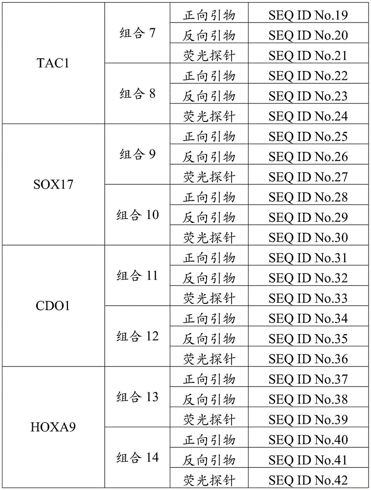
Preferably, for the target regions of the 7 lung cancer-associated genes, the invention provides 14 primer pairs and fluorescent probe combinations, as shown in table 1. It is understood that the test kit comprises at least one combination of table 1, and more preferably, the test kit comprises at least two combinations of table 1.
TABLE 1

Further, in the above technical solution, a fluorescent reporter group is labeled at the 5 'end of the fluorescent probe, and a fluorescent quencher group is labeled at the 3' end of the fluorescent probe.
Preferably, the fluorescence reporter group is any one of FAM, ROX, VIC, JOE, CY3, CY5, NED, TAMRA and TEXAS RED, and the fluorescence quencher group is any one of AMRA, DABCYL, ECLIPSE, BHQ-1, BHQ-2, BHQ-3 and a non-fluorescence quencher (MGB) binding to a molecular groove.
More preferably, the fluorescence reporter group is at least two of FAM, ROX, VIC and CY5, and the fluorescence quencher group is at least one of BHQ-1, BHQ-2, BHQ-3 and MGB.
Further, in the above technical scheme, the detection kit further comprises an ACTB primer pair and a fluorescent probe combination, wherein the ACTB is an internal reference gene, and the combination is used for amplifying an ACTB gene conserved sequence after sulfite treatment in the human biological sample.
Preferably, the conserved sequence of the ACTB gene is specifically selected from the region GRCh38: 7.
Preferably, the particulars of the ACTB primer pair and fluorescent probe combination are shown in table 2:
TABLE 2

Further, in the above technical solution, the PCR reaction system of the detection kit may be: 20. Mu.L total, 1.2 XTTaqman qPCR buffer, 6mM MgCl 2 The kit comprises a solution, a 250nM deoxyribonucleoside triphosphate mixture, 1.5 active units of DNA polymerase, a primer probe combination and purified water. Specifically, the primer probe combination includes at least one of combinations 1 to 14 and a combination 15.
Further, in the above technical solution, the human-derived biological sample includes, but is not limited to, tissue, blood, plasma, serum, sputum, urine, pleural fluid, alveolar lavage fluid, and the like.
Preferably, the biological sample of human origin is blood, plasma, serum or sputum.
More preferably, the biological sample of human origin is plasma or serum.
Further, in the above technical solution, the detection kit further comprises a reagent for extracting and purifying DNA from a human biological sample.
Wherein the extraction and purification reagent comprises a sulfite conversion agent that can convert unmethylated cytosine in the human biological sample DNA to uracil while methylated cytosine residues remain unchanged. The template sequence converted by the sulfite converting agent is complementarily combined with a corresponding primer pair and a fluorescent probe combination in the kit in the subsequent PCR amplification reaction, the methylated cytosine residue site on the template is used as a cytosine residue to be matched with the guanine residue of the primer, and the unmethylated cytosine residue site is used as a uracil residue and cannot be matched with the primer in the kit.
Preferably, the sulfite converting agent is one or two of sodium bisulfite, ammonium bisulfite and sodium metabisulfite.
Further, in the above technical scheme, the detection kit comprises a negative quality control material and a positive quality control material.
Preferably, the negative quality control material is any one of purified water, bisDNA obtained by transforming cell DNA with methylation negative in the detection target region with sulfite, or artificially synthesized plasmid with methylation negative in the detection target region.
Preferably, the positive quality control product is bisDNA obtained by transforming cell DNA with methylation positive detection target region sequence through sulfite or artificially synthesized plasmid for detecting methylation positive target.
Further, in the above technical solution, the detection kit may further include an instruction for explaining a method of using the detection kit and a method for determining a detection result.
Preferably, the content of the description includes: constructing a diagnosis model aiming at different methylated gene target sequences or combinations by adopting a statistical method of logistic regression, and setting different cutoff values; and predicting and evaluating the risk of the lung cancer of the subject according to the detection result.
Preferably, the method for using the detection kit can refer to the following steps:
processing a sample to be detected to obtain a bisDNA solution, taking 5-10 mu L of the bisDNA solution, carrying out methylation detection by using a primer pair and a fluorescent probe combination in the kit, and simultaneously detecting a positive quality control product to a negative quality control product; the combination of the primer pair and the fluorescent probe comprises at least one of combinations 1 to 14 and a combination 15;
and analyzing the methylation condition of the target sequence of the lung cancer related gene of the detection sample by using the instruction in the detection kit.
More preferably, the process of the sample to be tested can refer to the following steps:
(1) Collecting samples: obtaining human plasma or serum;
(2) Cracking: taking 2.0mL of plasma or serum, adding 2.0mL of lysate and 200 mu L of proteinase K, and incubating for 15min at room temperature; the lysate comprises a protein denaturant, a surfactant, a pH buffer and a nuclease inhibitor;
(3) And (3) transformation: adding 1.0mL of the transformation solution into the mixed solution in the step (2), and respectively incubating at 98 ℃ for 10min and 64 ℃ for 60min; the conversion mixed liquor comprises a sulfite conversion agent, a protective agent and a pH regulator;
(4) Centrifuging: adding 2.0mL of isopropanol into the step (3), uniformly mixing by vortex, centrifuging for 1min at 16000g, and carefully sucking the lower-layer clear liquid into a new centrifugal tube by using a pipette;
(5) Combining: adding 2.0mL of binding solution and 100 mu L of magnetic beads into the mixed solution obtained in the step (4), incubating for 2min at room temperature, and performing magnetic separation; the binding solution comprises chaotropic salt and NaCl;
(6) Rinsing: rinsing the magnetic beads obtained in the step (5) by using 800 mu L of rinsing liquid, and then carrying out magnetic separation; repeating the steps once;
(7) And (3) desulfurization: adding 800 mu L of desulfurizer into the magnetic beads rinsed in the step (6), incubating for 15min at room temperature, and then carrying out magnetic separation;
(8) Rinsing: rinsing the magnetic beads obtained in the step (7) twice by using 800 mu L of rinsing liquid, and then carrying out magnetic separation;
(9) And (3) elution: and (5) adding 40-100 mu L of eluent into the magnetic beads obtained in the step (8), incubating for 5min at 56 ℃, and then carrying out magnetic separation to obtain a bisDNA solution.
The invention has the beneficial effects that: the invention adopts a method for detecting the methylation level of one or more genes in lung cancer related genes SHOX2, RASSF1A, PTGER4, TAC1, SOX17, CDO1 and HOXA9, thereby realizing the auxiliary diagnosis of lung cancer and the noninvasive screening of early lung cancer; by positioning the target region of the lung cancer related methylated genes and adopting the combination of different lung cancer related methylated genes, the uncertainty of the lung cancer related methylated markers and the limitation caused by ctDNA fragment deletion are effectively avoided, and the accuracy and specificity of detection are improved. Moreover, the kit can detect biological samples obtained by a non-invasive means, and improves the compliance of a subject. Therefore, the kit provided by the invention can realize early diagnosis and early treatment of lung cancer, improve the survival rate of patients and has good clinical application prospect.
Drawings
FIG. 1 is a graph showing the results of the bisACTB assay in example 1 after the extraction and purification of a plasma sample;
FIG. 2 shows the 1X10 detection by the detection kit in example 2 4 Schematic diagram of concentration gradient gene methylation amplification result in copies/mu L DNA solution;
FIG. 3 is a ROC graph comparing the detection results of the detection kit of example 3 with clinical results;
FIG. 4 is a ROC graph comparing the results of 7 methylated genes detected in example 4 with clinical results;
FIG. 5 is a ROC curve comparing the detection results of the preferred combination markers of example 4 with clinical results;
FIG. 6 is a graph of ROC results from comparison of the combined marker test results with clinical results in example 4.
Detailed Description
To further illustrate the technical solutions and effects of the present invention adopted to achieve the intended purposes, the following detailed descriptions of the embodiments, features and effects of the present invention are provided by specific examples. It should be understood that the following examples are only for illustrating the technical solutions of the present invention more clearly and are not intended to limit the present invention.
The invention selects 7 lung cancer related methylation gene markers, specifically SHOX2, RASSF1A, PTGER4, TAC1, SOX17, CDO1 and HOXA9, and provides two target regions for each marker. Further, 14 primer pairs and fluorescent probe combinations are designed respectively aiming at different target sequences of different markers. The detection kit provided by the invention comprises at least one combination of 14 combinations; preferably, the test kit comprises at least two of the 14 combinations, and preferably the two combinations are from different markers.
In a specific embodiment, the present invention employs a logistic regression analysis to construct a multi-marker combined diagnostic model for detecting lung cancer. The samples are divided into case groups and control groups, SPSS or Medcalc software is used for carrying out logic analysis on detection data and clinical results to obtain a regression coefficient and a deviation constant of each marker, and then a corresponding logistic regression equation is obtained. And substituting the detection Ct value of each marker into a logistic regression equation, calculating a corresponding logistic score according to a logistic score formula, carrying out ROC curve analysis on the logistic score and a clinical result by using Medcalc software, and selecting the optimal cutoff value in a 95% confidence interval.
In the following examples, the instruments, consumables and reagents are not indicated, and they are all conventional products commercially available.
Example 1DNA extraction and purification
The DNA extraction and purification reagent consists of lysis solution, proteinase K, conversion solution, binding solution, rinsing solution A, desulfurizing agent, rinsing solution B and eluent.
The lysate consists of protein denaturants, surfactants, pH buffers and nuclease inhibitors.
The conversion solution consists of a sulfite conversion agent, a protective agent and a pH regulator, wherein the sulfite conversion agent is one or two of sodium bisulfite, ammonium bisulfite and sodium metabisulfite, the protective agent is hydroquinone or Trolox, and the pH regulator is NaOH.
The binding solution consists of high-concentration chaotropic salt and NaCl.
The rinsing solution A consists of high-concentration chaotropic salt, nuclease inhibitor, pH buffer and absolute ethyl alcohol.
The desulfurizer consists of NaOH and absolute ethyl alcohol.
The rinsing liquid B consists of nuclease inhibitor, pH buffer and absolute ethyl alcohol.
The eluent consists of nuclease inhibitor and pH buffer.
In this example, the protein denaturant was guanidine thiocyanate, the surfactant was NP-40, the pH buffer was Tris-HCl, the nuclease inhibitor was EDTA, and the chaotropic salt was guanidine hydrochloride.
In this example, a plasma sample of a lung cancer patient is taken as an example, and plasma bisDNA is obtained by extraction and purification. The extraction and purification method comprises the following steps:
(1) Centrifuging freshly collected EDTA anticoagulated blood sample at 4 deg.C for 10min at 1600g, carefully aspirating the plasma supernatant, and transferring to a new centrifuge tube;
(2) Centrifuging at 16000g for 10min at 4 deg.C, further removing cell source nucleic acid attached to cell debris, carefully sucking out supernatant and transferring to new centrifuge tube;
(3) Adding 2.0mL of lysate and 200 mu L of proteinase K into 2.0mL of plasma, uniformly mixing by vortex oscillation, incubating at room temperature for 15min, and uniformly mixing by turning upside down every 3-5 min for 10sec;
(4) Continuously adding 1.0mL of the conversion mixed solution, shaking, uniformly mixing, centrifuging briefly to remove liquid drops on the inner wall of the tube cover, and respectively incubating at 98 ℃ for 10min and 64 ℃ for 60min;
(5) Adding 2.0mL of isopropanol after incubation is finished, whirling and uniformly mixing, centrifuging for 1min at 16000g, and carefully sucking lower-layer clear liquid and a new centrifugal tube by using a pipettor;
(6) Adding 2.0mL of binding solution and 100 μ L of magnetic beads into the above mixed solution, incubating at room temperature for 10min, turning upside down every 3min, mixing for 10sec, adsorbing magnetic beads with magnetic separator for 2min after incubation, and discarding supernatant;
(7) Adding 800 μ L of rinsing solution A for resuspending magnetic beads, shaking and cleaning for 1min, adsorbing magnetic beads with magnetic separator for 1min, and removing supernatant; repeating the steps once;
(8) Adding 800 mu L of desulfurizer for resuspending magnetic beads, incubating for 15min at room temperature, and turning upside down every 3-5 min for uniformly mixing for 10sec; the magnetic beads were adsorbed by a magnetic separator for 1min, and the supernatant solution was discarded.
(9) Adding 800 μ L of rinsing solution B for resuspending magnetic beads, shaking and cleaning for 1min, adsorbing magnetic beads with magnetic separator for 1min, and removing supernatant;
(10) Repeating the step (9) once, carrying out 6000g rapid centrifugation for 30sec, adsorbing magnetic beads by using a magnetic separator, removing residual supernatant solution, and standing at room temperature for 5min to remove residual ethanol;
(11) Adding 100 μ L of elution buffer solution to resuspend the magnetic beads, shaking for 2min, adsorbing the magnetic beads by a magnetic separator, sucking the supernatant into a new centrifugal tube, and marking;
(12) The labeled DNA is stored in a refrigerator at 4 ℃ for standby, or stored in a refrigerator at-20 ℃ for long-term storage.
By using the reagent for extracting and purifying DNA and the preparation method provided by the embodiment, the DNA extraction and transformation can be completed in one step.
Plasma samples of 6 patients with lung cancer were extracted by the above method, and Ct values of the internal reference gene bisACTB were detected by fluorescence quantitative PCR as shown in fig. 1, wherein the primer probe set for amplifying the internal reference gene bisACTB was combined into combination 15 (the specific process for preparing and detecting bisACTB can be referred to in example 2).
Example 2A detection kit
The detection kit provided by the embodiment selects one target region of each of the three gene markers TAC1, PTGER4 and SHOX2 as a detection region, and specifically comprises the following steps:
the target region of the TAC1 gene is GRCh38:7, 97732108-97732308;
the target region of the PTGER4 gene is GRCh38:5, 40681355-40681679;
the SHOX2 target region is GRCh38: 3.
Aiming at the nucleic acid sequences of the detection regions, artificially synthesized plasmids which are transformed by sulfite under the methylation state and the non-methylation state are respectively constructed as a positive quality control product and a negative quality control product and are respectively named as TAC1-M1, TAC1-UM1, PTGER4-M1, PTGER4-UM1, SHOX2-M1 and SHOX2-UM1.
In order to monitor the quality of template DNA and whether the PCR detection result is effective, ACTB is used as an internal reference, and the detection region of the ACTB gene is GRCh38: 7. And constructing a corresponding artificially synthesized plasmid bisActB according to a sequence of the ACTB detection region after sulfite transformation.
The above plasmids are used to prepare methylated gene solutions with different concentration gradients respectively, including single gene methylation, two gene methylation and three gene methylation, and each methylation combination is used to prepare 1x10 4 The copies/μ LDNA template contains template DNA solutions of 100% gene methylation, 10% gene methylation, 1% gene methylation, 0.1% gene methylation and 0% gene methylation. Using SHOX2 as an example, the preparation method of each concentration of methylated DNA template is shown as follows:
(1) Preparation of SHOX2 100% methylation solution: 10% by volume of 1X10 5 copies/. Mu.L of SHOX2-M1 and 10% by volume of 1X10 5 Thoroughly mixing the solutions of copies/μ L of bisACCTB and 80% by volume of TE buffer;
(2) Preparation of SHOX2 10% methylation solution: 10% by volume of 1X10 4 copies/. Mu.L of SHOX2-M1, 9% by volume of 1X10 5 copies/. Mu.L of SHOX2-UM1 and 10% by volume of 1X10 5 Thoroughly mixing the resulting solution with copies/. Mu.L of bisACTB and 71% volume of TE buffer;
(3) Preparation of SHOX 2% methylation solution: 10% by volume of 1X10 3 copies/. Mu.L of SHOX2-M1, 9.9% by volume 1X10 5 copies/. Mu.L of SHOX2-UM1 and 10% by volume of 1X10 5 Thoroughly mixing the solution obtained by copies/mu LbIsACTB and 70.1% of TE buffer solution by volume;
(4) SHOX2 0.1% methylation solution preparation: 10% by volume of 1X10 2 copies/. Mu.L of SHOX2-M1, 9.99% by volume of 1X10 5 copies/. Mu.L of SHOX2-UM1 and 10% by volume of 1X10 5 Thoroughly mixing the solution obtained by copies/mu LbIsACTB and 70.01% of TE buffer solution by volume;
(5) Preparation of SHOX2 0% methylation solution: 10% by volume of 1X10 5 copies/. Mu.L of SHOX2-UM1 and 10% by volume of 1X10 5 The resulting solution was mixed well with copies/. Mu.L of bisACTB and 80% volume of TE buffer.
The primer pair and fluorescent probe combinations for detecting the above methylation gene combinations in this example include combination 2 for detecting methylation of the SHOX2 gene, combination 7 for detecting methylation of TAC1, combination 6 for detecting methylation of PTGER4, and combination 15 for detecting the reference gene ACTB.
Wherein, the fluorescence reporter group at the 5 'end of the fluorescent probe in the combination 2 is FAM, and the fluorescence quencher group at the 3' end is MGB; the fluorescence reporter group at the 5 'end of the fluorescent probe in the combination 7 is ROX, and the fluorescence quenching group at the 3' end is BHQ-2; the fluorescent reporter group at the 5 'end of the fluorescent probe in the combination 6 is Cy5, and the fluorescent quenching group at the 3' end is BHQ-3; the fluorescence reporter group at the 5 'end of the fluorescent probe in the combination 15 is VIC, and the fluorescence quencher group at the 3' end is MGB.
In this example, the PCR reaction system includes 1.2 XTaqman qPCR buffer solution and 6mM MgCl in addition to the above 4 combinations 2 20. Mu.L of the solution, 250nM of a deoxyribonucleoside triphosphate mixture, 1.5 active units of DNA polymerase and purified water.
5. Mu.L of methylated gene solutions with different concentration gradients were added to the above 20. Mu.L of mixed reaction solution, and each solution was repeatedly tested 3 times to perform a fluorescent quantitative PCR reaction.
The fluorescent quantitative PCR reaction program comprises three stages: 5min at 95 ℃; 15s at 95 ℃, 30s at 62 ℃,10 cycles; 15s at 95 ℃, 30s at 60 ℃,35 cycles; the FAM/ROX/Cy5/VIC fluorescence signals were collected at the third stage. And taking the cycle number Ct value required by the FAM/ROX/Cy5/VIC fluorescence signal to reach the set threshold as a standard, wherein the gene with the amplification curve is positive in methylation, and is negative otherwise.
The detection result shows that no matter single gene methylation, two gene methylation or three gene methylation, different concentration gradient methylation solutions can stably detect each methylated gene, and no methylation negative template is amplified. Wherein 1 is multiplied by 10 4 The results of methylation detection in the three gene concentration gradients in copies/. Mu.L DNA solution are shown in FIG. 2.
As can be seen from the present example, the kit for detecting multiple gene methylation for lung cancer aided diagnosis and/or noninvasive screening of early lung cancer provided by the present invention can detect 1 × 10 4 0.1% of the genes were methylated in copies/. Mu.L DNA solution.
Example 3 logistic regression analysis and cutoff value setting
This example illustrates a method for performing logistic regression analysis and cutoff value setting on a sample using the kit and detection method provided in example 2.
In this embodiment, the methylation conditions of SHOX2, TAC1 and PTGER4 in 20 normal human plasma samples, 25 benign lung disease plasma samples and 51 lung cancer plasma samples are detected, and SPSS or Medcalc software is used to perform logical analysis on the detection data and clinical results, so as to obtain the corresponding logistic regression equation: y = k 1 ×x 1 +k 2 ×x 2 +k 3 ×x 3 + b. Wherein x 1 /x 2 /x 3 Respectively the Ct values of the gene amplification of SHOX2, TAC1 and PTGER4, k 1 /k 2 /k 3 The clinical coefficients of the SHOX2, TAC1 and PTGER4 genes are respectively, b is a constant, and the specific numerical values are shown in a table 3:
TABLE 3
| Gene | SHOX2 | TAC1 | PTGER4 | Constant number |
| Numerical value | -0.2749(k 1 ) | -0.42624(k 2 ) | -0.70429(k 3 ) | 44.96082(b) |
The corresponding logical score is calculated according to the formula S = EXP (y)/[ EXP (y) +1] × 100. And carrying out ROC curve analysis on the logic score and the clinical result by using Medcalc software, and selecting the optimal cutoff value.
In this example, the ROC curve is shown in fig. 3, and the cutoff values obtained are shown in table 4:
TABLE 4

According to the table, when the logic score is more than or equal to 46.26 when the clinical sample is detected, the patient is indicated to be at high risk of suffering from lung cancer, and the patient is recommended to be subjected to further clinical examination; when the logical score is < 46.26, the subject is advised to have a low risk of lung cancer, once a year methylation test.
Example 4 ROC Curve analysis of markers
This example provides the results of ROC curve analysis of 7 methylation gene markers and multi-marker combined detection models provided in the present invention.
237 clinical plasma samples were collected, including 83 normal patient samples, 74 pulmonary nodule patient samples (45 of benign nodules and 29 of inflammatory nodules), and 80 non-small cell lung cancer patient samples ( stage 0, 2, 9, 18, 26, and 25 of stages I, II).
237 examples of corresponding bis-cfDNA templates were obtained according to the method in example 1. Then, the primer pairs of the markers and the fluorescent probe combination provided by the invention are used for carrying out fluorescent quantitative PCR detection, and methylation level detection data of each marker are obtained. Using MedCalc software, a 95% confidence interval was chosen, yielding the ROC curve and its area under the curve (AUC) values. Table 5 shows the area under the curve (AUC) of the ROC curve for two target sequences of 7 methylated genes of the invention, and the ROC curve for each gene is shown in FIG. 4.
TABLE 5

According to ROC curve analysis, the following results are obtained: the area under the curve of the two target areas of the 7 methylated genes is relatively consistent, but a certain difference exists, and the two target areas or any one of the two target areas of one gene can be simultaneously selected for detection; of the areas under the curves for 7 genes, TAC1 and PTGER4 were the largest and the area of difference was the largest, with the combination being selected preferentially.
Simultaneous detection of two target regions of a marker or more markers simultaneously may result in better sensitivity and specificity, but may increase use costs. The method of fluorescent quantitative PCR is adopted to detect 3 markers simultaneously, which is undoubtedly an optimal scheme for taking sensitivity and specificity into consideration.
This example uses a combination of 3 markers including at least TAC1 and PTGER4, and the logistic regression method in example 2, the area under the ROC curve (AUC), the cutoff value and the corresponding sensitivity and specificity of different combinations, and the specific results are shown in table 6, and the ROC curve is shown in fig. 5.
TABLE 6
| Marker combination | Sensitivity% | Specificity% | Cutoff | AUC | 95%CI |
| PTGER4/TAC1/SHOX2 | 86.25 | 89.81 | >27.25 | 0.926 | 0.885to 0.956 |
| PTGER4/TAC1/RASSF1A | 86.25 | 88.54 | >30.02 | 0.937 | 0.898to 0.964 |
| PTGER4/TAC1/SOX17 | 81.25 | 89.81 | >33.33 | 0.946 | 0.909to 0.971 |
| PTGER4/TAC1/ |
80 | 89.81 | >37.88 | 0.941 | 0.903to 0.967 |
| PTGER4/TAC1/HOXA9 | 83.75 | 89.17 | >31.85 | 0.936 | 0.897to 0.964 |
To better illustrate the beneficial effects of the combination markers in Table 6, the area under ROC curve (AUC), cutoff value and corresponding sensitivity and specificity of some marker combinations not including TAC1 and PTGER4 were analyzed simultaneously in this example, and the specific results are shown in Table 7 and the ROC curve is shown in FIG. 6.
TABLE 7
| Marker combination | Sensitivity% | Specificity% | Cutoff | AUC | 95%CI |
| SHOX2/RASSF1A/PTGER4 | 75 | 91.12 | >32.6 | 0.842 | 0.789to 0.886 |
| SHOX2/RASSF1A/ |
80 | 89.17 | >32.78 | 0.873 | 0.823to 0.912 |
| SHOX2/RASSF1A/SOX17 | 68.75 | 89.17 | >38.22 | 0.895 | 0.849to 0.931 |
| SHOX2/PTGER4/SOX17 | 72.5 | 89.81 | >38.53 | 0.910 | 0.867to 0.944 |
| SHOX2/PTGER4/HOXA9 | 70 | 89.17 | >35.73 | 0.874 | 0.825to 0.913 |
| SHOX2/SOX17/HOXA9 | 70 | 89.17 | >42.61 | 0.886 | 0.838to 0.923 |
Comparing the results of tables 6 and 7, it can be seen that the area under the ROC curve (AUC) is significantly larger for the tri-marker co-detection model including at least TAC1 and PTGER4, and that it has better sensitivity while maintaining specificity around 90% compared to the tri-marker co-detection model not including TAC1 and PTGER4 at the same time. The three-marker combined detection model at least comprising TAC1 and PTGER4 can be used as a better combined marker for clinical detection of lung cancer.
It should be noted that, in view of the use cost, the present embodiment only performs the expansion analysis on the joint detection model of the single detection areas of the three markers including TAC1 and PTGER4, and only expands part of the joint detection model, which does not represent all embodiments of the present invention. The user can select three or more markers in a single detection area or two detection areas for combined detection according to actual needs, such as better beneficial effects.
Meanwhile, the settings of the cutoff values of the different combination models in the embodiment are the cutoff values set at specificity which is kept around 90%, which does not mean that the cutoff values can only be used in the invention, and the appropriate cutoff values can be selected from the ROC curves provided by the invention according to different purposes.
Example 5 Effect of the Multi-marker Joint detection model in clinical applications of Lung cancer detection
This example provides the practical effect of the multi-marker combined detection model in example 4 in clinical application of lung cancer detection.
The results of clinical tests with the combination PTGER4/TAC1/SHOX2 were selected for analysis, and the results of comparison of the actual test results with the results of clinical diagnosis are shown in Table 8.
TABLE 8

The detection results in the table show that the PTGER4/TAC1/SHOX2 marker combination provided by the invention has the overall sensitivity of 86.25 percent, the specificity of 90.45 percent and the accuracy of 89.03 percent for detecting the lung cancer of the 237 clinical samples, wherein the sensitivity of the lung cancer in the stages 0 to II is 82.76 percent. Therefore, the kit provided by the application can be used for auxiliary diagnosis of lung cancer or early screening of lung cancer of general risk groups.
In the embodiment, only the PTGER4/TAC1/SHOX2 combination is used for showing the clinical use effect of the multi-marker combination for detecting the lung cancer, and a user can select a proper combination model for clinical application according to the actual use and the expected use.
The embodiments described above are some, but not all embodiments of the present invention. Modifications to the above embodiments or equivalent replacements of parts of technical features may be made by those skilled in the art, and these modifications and replacements are all within the protection scope of the present invention.
Sequence listing
<110> Wuhan Kangzhi Biotechnology GmbH
<120> detection kit for lung cancer diagnosis and early lung cancer noninvasive screening
<160> 45
<170> SIPOSequenceListing 1.0
<210> 1
<211> 21
<212> DNA
<213> Artificial Sequence (Artificial Sequence)
<400> 1
tatcgggagg tgttggagag c 21
<210> 2
<211> 23
<212> DNA
<213> Artificial Sequence (Artificial Sequence)
<400> 2
gcccgaacta ccgaactact acg 23
<210> 3
<211> 17
<212> DNA
<213> Artificial Sequence (Artificial Sequence)
<400> 3
ccgcctcgat acaaccg 17
<210> 4
<211> 21
<212> DNA
<213> Artificial Sequence (Artificial Sequence)
<400> 4
tttcgtttcg tttgttcgat c 21
<210> 5
<211> 24
<212> DNA
<213> Artificial Sequence (Artificial Sequence)
<400> 5
ctaccttcta acccgactta aacg 24
<210> 6
<211> 19
<212> DNA
<213> Artificial Sequence (Artificial Sequence)
<400> 6
tcgtacgagt ataggcgtt 19
<210> 7
<211> 18
<212> DNA
<213> Artificial Sequence (Artificial Sequence)
<400> 7
<210> 8
<211> 20
<212> DNA
<213> Artificial Sequence (Artificial Sequence)
<400> 8
<210> 9
<211> 23
<212> DNA
<213> Artificial Sequence (Artificial Sequence)
<400> 9
cgggagttgg tattcgttgg gcg 23
<210> 10
<211> 18
<212> DNA
<213> Artificial Sequence (Artificial Sequence)
<400> 10
cgaagcgtgc gtgttttc 18
<210> 11
<211> 22
<212> DNA
<213> Artificial Sequence (Artificial Sequence)
<400> 11
<210> 12
<211> 18
<212> DNA
<213> Artificial Sequence (Artificial Sequence)
<400> 12
<210> 13
<211> 23
<212> DNA
<213> Artificial Sequence (Artificial Sequence)
<400> 13
ttttatttcg cgcgtttagt ttc 23
<210> 14
<211> 22
<212> DNA
<213> Artificial Sequence (Artificial Sequence)
<400> 14
gaacgcccat taaccgaatt aa 22
<210> 15
<211> 22
<212> DNA
<213> Artificial Sequence (Artificial Sequence)
<400> 15
<210> 16
<211> 19
<212> DNA
<213> Artificial Sequence (Artificial Sequence)
<400> 16
agcgattggc gggttttac 19
<210> 17
<211> 21
<212> DNA
<213> Artificial Sequence (Artificial Sequence)
<400> 17
tactacaacc gcgaactacc g 21
<210> 18
<211> 22
<212> DNA
<213> Artificial Sequence (Artificial Sequence)
<400> 18
ttgtagttta tgcgtttaac gt 22
<210> 19
<211> 19
<212> DNA
<213> Artificial Sequence (Artificial Sequence)
<400> 19
gagcgattag cgtgcgttc 19
<210> 20
<211> 20
<212> DNA
<213> Artificial Sequence (Artificial Sequence)
<400> 20
<210> 21
<211> 17
<212> DNA
<213> Artificial Sequence (Artificial Sequence)
<400> 21
cgcgaacact tactacg 17
<210> 22
<211> 24
<212> DNA
<213> Artificial Sequence (Artificial Sequence)
<400> 22
acgtggtacg tatcgttatt acgg 24
<210> 23
<211> 21
<212> DNA
<213> Artificial Sequence (Artificial Sequence)
<400> 23
caaaatcccg taaaaaaccc g 21
<210> 24
<211> 23
<212> DNA
<213> Artificial Sequence (Artificial Sequence)
<400> 24
acccctttcc atcctctcgc acg 23
<210> 25
<211> 23
<212> DNA
<213> Artificial Sequence (Artificial Sequence)
<400> 25
gtttggattt tgttgcgtta gtc 23
<210> 26
<211> 18
<212> DNA
<213> Artificial Sequence (Artificial Sequence)
<400> 26
<210> 27
<211> 19
<212> DNA
<213> Artificial Sequence (Artificial Sequence)
<400> 27
aaagcgttta tcggtcgtc 19
<210> 28
<211> 22
<212> DNA
<213> Artificial Sequence (Artificial Sequence)
<400> 28
tttgtacggt ttggttgagt cg 22
<210> 29
<211> 21
<212> DNA
<213> Artificial Sequence (Artificial Sequence)
<400> 29
ccgacgaaaa aaccctactc g 21
<210> 30
<211> 21
<212> DNA
<213> Artificial Sequence (Artificial Sequence)
<400> 30
ataaccacgc gaccgccctc g 21
<210> 31
<211> 20
<212> DNA
<213> Artificial Sequence (Artificial Sequence)
<400> 31
tacgcgattt ttgggacgtc 20
<210> 32
<211> 20
<212> DNA
<213> Artificial Sequence (Artificial Sequence)
<400> 32
<210> 33
<211> 21
<212> DNA
<213> Artificial Sequence (Artificial Sequence)
<400> 33
ccccgacttc cccgaactcc g 21
<210> 34
<211> 25
<212> DNA
<213> Artificial Sequence (Artificial Sequence)
<400> 34
cgagtgggta atgtacgtta agttc 25
<210> 35
<211> 21
<212> DNA
<213> Artificial Sequence (Artificial Sequence)
<400> 35
cattcctcct caaacgaaac g 21
<210> 36
<211> 28
<212> DNA
<213> Artificial Sequence (Artificial Sequence)
<400> 36
cgccgcctaa cattaaaact acaacgcg 28
<210> 37
<211> 22
<212> DNA
<213> Artificial Sequence (Artificial Sequence)
<400> 37
ggtttatttg tcgttcgtcg tc 22
<210> 38
<211> 18
<212> DNA
<213> Artificial Sequence (Artificial Sequence)
<400> 38
<210> 39
<211> 27
<212> DNA
<213> Artificial Sequence (Artificial Sequence)
<400> 39
ctacctcatt acgcttaccg cccaacg 27
<210> 40
<211> 22
<212> DNA
<213> Artificial Sequence (Artificial Sequence)
<400> 40
cgtagtaatt cggggttggt tc 22
<210> 41
<211> 18
<212> DNA
<213> Artificial Sequence (Artificial Sequence)
<400> 41
<210> 42
<211> 23
<212> DNA
<213> Artificial Sequence (Artificial Sequence)
<400> 42
cgcggtttcg atttttcgtt cgc 23
<210> 43
<211> 25
<212> DNA
<213> Artificial Sequence (Artificial Sequence)
<400> 43
ggtgtttgtt tttttgatta ggtgt 25
<210> 44
<211> 25
<212> DNA
<213> Artificial Sequence (Artificial Sequence)
<400> 44
acctcataac cttatcacac aaacc 25
<210> 45
<211> 18
<212> DNA
<213> Artificial Sequence (Artificial Sequence)
<400> 45
Claims (5)
1. The application of a primer pair and a fluorescent probe composition for detecting the methylation level of a target region of a lung cancer related gene in a human biological sample in preparing a detection kit for assisting in diagnosing lung cancer is characterized in that,
the primer pair and the fluorescent probe composition are used for carrying out fluorescent quantitative PCR amplification on the target region of the lung cancer related gene after sulfite treatment;
the lung cancer-related genes are SHOX2, PTGER4 and TAC1;
the target region of the SHOX2 gene is GRCh38:3 158096298-158096648 and GRCh38:3 158098932-158099255;
the target region of the PTGER4 gene is GRCh38:5, 40679693-40679952 and GRCh38: 5;
the target region of the TAC1 gene is GRCh38: 7;
the primer pair and fluorescent probe composition for amplifying the target region of the SHOX2 gene comprises a combination 1 and a combination 2, wherein the sequence of the primer pair of the combination 1 is shown as SEQ ID No. 1-2, the sequence of the fluorescent probe of the combination 1 is shown as SEQ ID No.3, the sequence of the primer pair of the combination 2 is shown as SEQ ID No. 4-5, and the sequence of the fluorescent probe of the combination 2 is shown as SEQ ID No. 6;
the primer pair and fluorescent probe composition for amplifying the target region of the PTGER4 gene is a combination 5 and a combination 6, the sequence of the primer pair of the combination 5 is shown as SEQ ID No. 13-14, the sequence of the fluorescent probe of the combination 5 is shown as SEQ ID No.15, the sequence of the primer pair of the combination 6 is shown as SEQ ID No. 16-17, and the sequence of the fluorescent probe of the combination 6 is shown as SEQ ID No. 18;
the primer pair and fluorescent probe composition for amplifying the target region of the TAC1 gene comprises a combination 7 and a combination 8, wherein the sequence of the primer pair of the combination 7 is shown as SEQ ID No. 19-20, the sequence of the fluorescent probe of the combination 7 is shown as SEQ ID No.21, the sequence of the primer pair of the combination 8 is shown as SEQ ID No. 22-23, and the sequence of the fluorescent probe of the combination 8 is shown as SEQ ID No. 24;
the detection kit also comprises an ACTB primer pair and a fluorescent probe composition which are used for amplifying the ACTB gene conserved sequence after the human biological sample is treated by sulfite;
the sequences of the ACTB primer pair and the primer pair in the fluorescent probe composition are shown as SEQ ID No. 43-44, and the sequence of the fluorescent probe is shown as SEQ ID No. 45;
the human biological sample is serum or plasma.
2. The use of claim 1, wherein the fluorescent probe is labeled with a fluorescent reporter group at the 5 'end and a fluorescent quencher group at the 3' end;
the fluorescent reporter group is any one of FAM, ROX, VIC, JOE, CY3, CY5, NED, TAMRA and TEXAS RED;
the fluorescence quenching group is any one of AMRA, DABCYL, ECLIPSE, BHQ-1, BHQ-2, BHQ-3 and MGB.
3. The use according to claim 1, wherein the test kit comprises reagents for DNA extraction and purification of a human biological sample; the reagent for extraction and purification comprises a sulfite converting agent for converting unmethylated cytosine in the DNA of the human biological sample into uracil.
4. The use according to claim 3, wherein the sulfite converting agent is one or more of sodium bisulfite, ammonium bisulfite and sodium metabisulfite.
5. The use of claim 1, wherein the test kit comprises a negative quality control material and a positive quality control material,
the negative quality control product is any one of purified water, bisDNA obtained by transforming cell DNA with methylation negative in the detection target region through sulfite or artificially synthesized plasmid with methylation negative in the detection target region;
the positive quality control product is any one of bisDNA obtained by converting cell DNA with positive methylation of a detection target region sequence through sulfite or artificially synthesized plasmid with positive methylation of the detection target.
Priority Applications (1)
| Application Number | Priority Date | Filing Date | Title |
|---|---|---|---|
| CN202210102556.6A CN114277154B (en) | 2022-01-27 | 2022-01-27 | Detection kit for lung cancer diagnosis and early lung cancer noninvasive screening |
Applications Claiming Priority (1)
| Application Number | Priority Date | Filing Date | Title |
|---|---|---|---|
| CN202210102556.6A CN114277154B (en) | 2022-01-27 | 2022-01-27 | Detection kit for lung cancer diagnosis and early lung cancer noninvasive screening |
Publications (2)
| Publication Number | Publication Date |
|---|---|
| CN114277154A CN114277154A (en) | 2022-04-05 |
| CN114277154B true CN114277154B (en) | 2022-11-29 |
Family
ID=80881796
Family Applications (1)
| Application Number | Title | Priority Date | Filing Date |
|---|---|---|---|
| CN202210102556.6A Active CN114277154B (en) | 2022-01-27 | 2022-01-27 | Detection kit for lung cancer diagnosis and early lung cancer noninvasive screening |
Country Status (1)
| Country | Link |
|---|---|
| CN (1) | CN114277154B (en) |
Families Citing this family (3)
| Publication number | Priority date | Publication date | Assignee | Title |
|---|---|---|---|---|
| WO2023236201A1 (en) * | 2022-06-10 | 2023-12-14 | Suzhou Huhu Health Technology Co., Ltd | Methods, compositions, and kits for detecting malignant lung nodules and lung cancer |
| CN115976205A (en) * | 2022-09-28 | 2023-04-18 | 北京鑫诺美迪基因检测技术有限公司 | Primer probe combination, kit and detection method suitable for digital PCR quantitative detection of lung cancer related gene methylation |
| CN116987788B (en) * | 2023-06-19 | 2024-03-01 | 嘉兴允英医学检验有限公司 | Method and kit for detecting early lung cancer by using flushing liquid |
Citations (3)
| Publication number | Priority date | Publication date | Assignee | Title |
|---|---|---|---|---|
| CN110964809A (en) * | 2018-09-29 | 2020-04-07 | 广州市康立明生物科技有限责任公司 | HOXA7 methylation detection reagent |
| CN111254199A (en) * | 2020-03-23 | 2020-06-09 | 郑州大学第一附属医院 | Lung cancer related KEAP1 gene methylation detection kit |
| CN112094912A (en) * | 2020-10-16 | 2020-12-18 | 中国药科大学 | Plasma free DNA methylation gene combination for identifying benign and malignant pulmonary nodules and application thereof |
Family Cites Families (7)
| Publication number | Priority date | Publication date | Assignee | Title |
|---|---|---|---|---|
| US20130022974A1 (en) * | 2011-06-17 | 2013-01-24 | The Regents Of The University Of Michigan | Dna methylation profiles in cancer |
| CN107034296B (en) * | 2017-06-05 | 2019-06-25 | 北京鑫诺美迪基因检测技术有限公司 | A kind of composition and its application for early stage of lung cancer non-invasive screening |
| CN109825586B (en) * | 2019-03-11 | 2022-10-21 | 广州市新合生物医疗科技有限公司 | DNA methylation qPCR kit for lung cancer detection and use method |
| CN110229908B (en) * | 2019-07-03 | 2023-01-31 | 四川沃文特生物技术有限公司 | Primer, probe and kit for early detection of lung cancer gene methylation |
| CN110387421A (en) * | 2019-08-28 | 2019-10-29 | 深圳市新合生物医疗科技有限公司 | DNA methylation qPCR kit and application method for lung cancer detection |
| EP3789505A1 (en) * | 2019-09-05 | 2021-03-10 | Forschungszentrum Borstel | Methods and means for diagnosing lung cancer |
| CN112195245A (en) * | 2020-10-16 | 2021-01-08 | 中国药科大学 | Lung cancer related methylation gene combination in plasma and application thereof |
-
2022
- 2022-01-27 CN CN202210102556.6A patent/CN114277154B/en active Active
Patent Citations (3)
| Publication number | Priority date | Publication date | Assignee | Title |
|---|---|---|---|---|
| CN110964809A (en) * | 2018-09-29 | 2020-04-07 | 广州市康立明生物科技有限责任公司 | HOXA7 methylation detection reagent |
| CN111254199A (en) * | 2020-03-23 | 2020-06-09 | 郑州大学第一附属医院 | Lung cancer related KEAP1 gene methylation detection kit |
| CN112094912A (en) * | 2020-10-16 | 2020-12-18 | 中国药科大学 | Plasma free DNA methylation gene combination for identifying benign and malignant pulmonary nodules and application thereof |
Also Published As
| Publication number | Publication date |
|---|---|
| CN114277154A (en) | 2022-04-05 |
Similar Documents
| Publication | Publication Date | Title |
|---|---|---|
| CN114277154B (en) | Detection kit for lung cancer diagnosis and early lung cancer noninvasive screening | |
| WO2021128519A1 (en) | Combination of dna methylation biomarkers, and detection method therefor and kit thereof | |
| WO2023071889A1 (en) | Methylation biomarker related to detection of gastric cancer lymph node metastasis, or combination thereof and use thereof | |
| CN112195245A (en) | Lung cancer related methylation gene combination in plasma and application thereof | |
| EP3828273A1 (en) | Methylation modification-based tumor marker stamp-ep2 | |
| EP3904515A1 (en) | Tumor marker stamp-ep3 based on methylation modification | |
| CN114672568B (en) | Kit for detecting cervical cell gene methylation | |
| CN111549135A (en) | DNA methylation qPCR kit for cervical cancer detection, and use method and application thereof | |
| CN112899359A (en) | Methylation marker for detecting benign and malignant lung nodules or combination and application thereof | |
| CN113462763A (en) | Kit for designing gene panel for targeted detection of soft tissue tumor small round cell tumor fusion | |
| CN113930516B (en) | Primer, kit, model and construction method for methylation of cervical cancer related gene | |
| CN109837344B (en) | Methylated EphA7 nucleotide fragment, detection method and application thereof | |
| TWI385252B (en) | Cancer screening method | |
| CN111826446A (en) | Primer, probe and kit for early screening and auxiliary diagnosis of bladder cancer | |
| WO2020063898A1 (en) | Use of hoxa7 methylation detection reagent in preparation of diagnostic reagent for lung cancer | |
| Liu et al. | Multiple biomarker-combined screening for colorectal cancer based on bisulfate conversion-free detection of fecal DNA methylation | |
| CN115820850A (en) | Biomarker of endometrial cancer, probe primer combination and kit | |
| CN114807350A (en) | Biomarker for judging benign and malignant thyroid nodules, polygene joint inspection kit and application | |
| CN115772567B (en) | Methylation site for auxiliary detection of lung cancer somatic cell TP53 gene mutation and application thereof | |
| CN115772566B (en) | Methylation biomarker for auxiliary detection of lung cancer somatic ERBB2 gene mutation and application thereof | |
| CN115772564B (en) | Methylation biomarker for auxiliary detection of lung cancer somatic cell ATM gene fusion mutation and application thereof | |
| CN116356029B (en) | Application of TXNRD1 gene methylation level detection in endometrial cancer diagnosis and/or screening | |
| CN111549140B (en) | Kit for detecting methylation of lung cancer related genes C2orf40, FIBUN and GRP and application thereof | |
| CN114959030B (en) | Application of reagent for detecting HCG9 gene methylation in preparation of product for diagnosing bladder cancer | |
| CN116987791B (en) | Application of plasma markers in identification of benign and malignant thyroid nodule |
Legal Events
| Date | Code | Title | Description |
|---|---|---|---|
| PB01 | Publication | ||
| PB01 | Publication | ||
| SE01 | Entry into force of request for substantive examination | ||
| SE01 | Entry into force of request for substantive examination | ||
| CB02 | Change of applicant information | ||
| CB02 | Change of applicant information |
Address after: 430075 Workshop No. 1-4 (1), No. 8, Wuhan Optics Valley Precision Medical Industry Base Phase I (all for own use), No. 9, Gaokeyuan 3rd Road, Wuhan East Lake New Technology Development Zone, Wuhan, Hubei Province Applicant after: WUHAN HEALTHCARE BIOTECHNOLOGY CO.,LTD. Address before: 430075 No. 666 High-tech Avenue, Donghu Development Zone, Wuhan City, Hubei Province, 4th Floor, B6 Building, Wuhan National Biological Industrial Base Applicant before: WUHAN HEALTHCARE BIOTECHNOLOGY CO.,LTD. |
|
| GR01 | Patent grant | ||
| GR01 | Patent grant |