CN114181281A - Peptide therapy for inflammation and fibrosis - Google Patents
Peptide therapy for inflammation and fibrosis Download PDFInfo
- Publication number
- CN114181281A CN114181281A CN202111595612.6A CN202111595612A CN114181281A CN 114181281 A CN114181281 A CN 114181281A CN 202111595612 A CN202111595612 A CN 202111595612A CN 114181281 A CN114181281 A CN 114181281A
- Authority
- CN
- China
- Prior art keywords
- peptide
- ebp
- lys
- fibrosis
- liver
- Prior art date
- Legal status (The legal status is an assumption and is not a legal conclusion. Google has not performed a legal analysis and makes no representation as to the accuracy of the status listed.)
- Pending
Links
- 108090000765 processed proteins & peptides Proteins 0.000 title claims abstract description 433
- 206010016654 Fibrosis Diseases 0.000 title claims abstract description 78
- 230000004761 fibrosis Effects 0.000 title claims abstract description 74
- 230000004054 inflammatory process Effects 0.000 title claims abstract description 42
- 206010061218 Inflammation Diseases 0.000 title claims abstract description 33
- 238000002560 therapeutic procedure Methods 0.000 title abstract description 9
- 102100034798 CCAAT/enhancer-binding protein beta Human genes 0.000 claims abstract description 321
- 101710134031 CCAAT/enhancer-binding protein beta Proteins 0.000 claims abstract description 321
- 102000004196 processed proteins & peptides Human genes 0.000 claims abstract description 124
- 238000006366 phosphorylation reaction Methods 0.000 claims abstract description 96
- 230000026731 phosphorylation Effects 0.000 claims abstract description 94
- 150000001413 amino acids Chemical class 0.000 claims abstract description 38
- 206010067125 Liver injury Diseases 0.000 claims description 102
- 210000002540 macrophage Anatomy 0.000 claims description 100
- 229920001223 polyethylene glycol Polymers 0.000 claims description 87
- 210000004185 liver Anatomy 0.000 claims description 78
- 230000002757 inflammatory effect Effects 0.000 claims description 74
- 230000004913 activation Effects 0.000 claims description 72
- 239000002202 Polyethylene glycol Substances 0.000 claims description 68
- 208000019425 cirrhosis of liver Diseases 0.000 claims description 59
- 210000000651 myofibroblast Anatomy 0.000 claims description 59
- 210000004072 lung Anatomy 0.000 claims description 53
- 208000037265 diseases, disorders, signs and symptoms Diseases 0.000 claims description 50
- 231100000753 hepatic injury Toxicity 0.000 claims description 49
- 210000001744 T-lymphocyte Anatomy 0.000 claims description 46
- 210000001519 tissue Anatomy 0.000 claims description 46
- 201000010099 disease Diseases 0.000 claims description 40
- 201000009794 Idiopathic Pulmonary Fibrosis Diseases 0.000 claims description 38
- 208000036971 interstitial lung disease 2 Diseases 0.000 claims description 38
- 239000003814 drug Substances 0.000 claims description 37
- 125000003178 carboxy group Chemical group [H]OC(*)=O 0.000 claims description 36
- 230000002401 inhibitory effect Effects 0.000 claims description 31
- 208000008338 non-alcoholic fatty liver disease Diseases 0.000 claims description 30
- 208000006454 hepatitis Diseases 0.000 claims description 26
- 206010053219 non-alcoholic steatohepatitis Diseases 0.000 claims description 23
- 239000008194 pharmaceutical composition Substances 0.000 claims description 23
- 206010035664 Pneumonia Diseases 0.000 claims description 22
- 108010034143 Inflammasomes Proteins 0.000 claims description 20
- 208000005069 pulmonary fibrosis Diseases 0.000 claims description 20
- 208000008439 Biliary Liver Cirrhosis Diseases 0.000 claims description 19
- 101000945963 Homo sapiens CCAAT/enhancer-binding protein beta Proteins 0.000 claims description 17
- 150000008574 D-amino acids Chemical class 0.000 claims description 15
- 208000033222 Biliary cirrhosis primary Diseases 0.000 claims description 11
- 206010018364 Glomerulonephritis Diseases 0.000 claims description 11
- 208000004852 Lung Injury Diseases 0.000 claims description 11
- 208000012654 Primary biliary cholangitis Diseases 0.000 claims description 11
- 206010069363 Traumatic lung injury Diseases 0.000 claims description 11
- 208000027418 Wounds and injury Diseases 0.000 claims description 11
- 239000002253 acid Substances 0.000 claims description 11
- 230000003176 fibrotic effect Effects 0.000 claims description 11
- 231100000283 hepatitis Toxicity 0.000 claims description 11
- 210000003734 kidney Anatomy 0.000 claims description 11
- 201000008383 nephritis Diseases 0.000 claims description 11
- 208000010157 sclerosing cholangitis Diseases 0.000 claims description 11
- 206010008609 Cholangitis sclerosing Diseases 0.000 claims description 10
- 210000004556 brain Anatomy 0.000 claims description 10
- 230000007882 cirrhosis Effects 0.000 claims description 10
- 230000002496 gastric effect Effects 0.000 claims description 10
- 208000027866 inflammatory disease Diseases 0.000 claims description 10
- 208000014674 injury Diseases 0.000 claims description 10
- 231100000515 lung injury Toxicity 0.000 claims description 10
- 230000005855 radiation Effects 0.000 claims description 10
- 206010003827 Autoimmune hepatitis Diseases 0.000 claims description 9
- 208000006682 alpha 1-Antitrypsin Deficiency Diseases 0.000 claims description 9
- 208000000419 Chronic Hepatitis B Diseases 0.000 claims description 8
- 208000006545 Chronic Obstructive Pulmonary Disease Diseases 0.000 claims description 8
- 208000006154 Chronic hepatitis C Diseases 0.000 claims description 8
- 206010010317 Congenital absence of bile ducts Diseases 0.000 claims description 8
- 206010014561 Emphysema Diseases 0.000 claims description 8
- 208000005176 Hepatitis C Diseases 0.000 claims description 8
- 208000002972 Hepatolenticular Degeneration Diseases 0.000 claims description 8
- 206010061216 Infarction Diseases 0.000 claims description 8
- 206010061481 Renal injury Diseases 0.000 claims description 8
- 206010039710 Scleroderma Diseases 0.000 claims description 8
- AYFVYJQAPQTCCC-UHFFFAOYSA-N Threonine Natural products CC(O)C(N)C(O)=O AYFVYJQAPQTCCC-UHFFFAOYSA-N 0.000 claims description 8
- 239000004473 Threonine Substances 0.000 claims description 8
- 208000018839 Wilson disease Diseases 0.000 claims description 8
- 201000005271 biliary atresia Diseases 0.000 claims description 8
- 206010014599 encephalitis Diseases 0.000 claims description 8
- 208000002672 hepatitis B Diseases 0.000 claims description 8
- 208000010710 hepatitis C virus infection Diseases 0.000 claims description 8
- 230000007574 infarction Effects 0.000 claims description 8
- 208000037806 kidney injury Diseases 0.000 claims description 8
- 208000037581 Persistent Infection Diseases 0.000 claims description 7
- 208000026594 alcoholic fatty liver disease Diseases 0.000 claims description 7
- DKIDEFUBRARXTE-UHFFFAOYSA-N 3-mercaptopropanoic acid Chemical compound OC(=O)CCS DKIDEFUBRARXTE-UHFFFAOYSA-N 0.000 claims description 6
- 239000003937 drug carrier Substances 0.000 claims description 6
- 238000004519 manufacturing process Methods 0.000 claims description 6
- 238000002360 preparation method Methods 0.000 claims description 6
- 201000002793 renal fibrosis Diseases 0.000 claims description 6
- 208000022559 Inflammatory bowel disease Diseases 0.000 claims description 5
- 206010058522 Oesophageal injury Diseases 0.000 claims description 5
- 208000037976 chronic inflammation Diseases 0.000 claims description 5
- 210000000056 organ Anatomy 0.000 claims description 5
- 208000037999 tubulointerstitial fibrosis Diseases 0.000 claims description 5
- 208000007082 Alcoholic Fatty Liver Diseases 0.000 claims description 4
- 208000022309 Alcoholic Liver disease Diseases 0.000 claims description 4
- 208000024827 Alzheimer disease Diseases 0.000 claims description 4
- 208000031229 Cardiomyopathies Diseases 0.000 claims description 4
- 201000004624 Dermatitis Diseases 0.000 claims description 4
- 201000009906 Meningitis Diseases 0.000 claims description 4
- 206010048654 Muscle fibrosis Diseases 0.000 claims description 4
- 201000002481 Myositis Diseases 0.000 claims description 4
- 201000004681 Psoriasis Diseases 0.000 claims description 4
- 206010052428 Wound Diseases 0.000 claims description 4
- 206010003246 arthritis Diseases 0.000 claims description 4
- 208000025698 brain inflammatory disease Diseases 0.000 claims description 4
- 239000003518 caustics Substances 0.000 claims description 4
- 230000001969 hypertrophic effect Effects 0.000 claims description 4
- 208000028867 ischemia Diseases 0.000 claims description 4
- 208000023589 ischemic disease Diseases 0.000 claims description 4
- 230000002980 postoperative effect Effects 0.000 claims description 4
- 206010019708 Hepatic steatosis Diseases 0.000 claims description 3
- 208000002260 Keloid Diseases 0.000 claims description 3
- 206010028594 Myocardial fibrosis Diseases 0.000 claims description 3
- 206010034665 Peritoneal fibrosis Diseases 0.000 claims description 3
- 206010050207 Skin fibrosis Diseases 0.000 claims description 3
- 210000001035 gastrointestinal tract Anatomy 0.000 claims description 3
- 210000001117 keloid Anatomy 0.000 claims description 3
- 230000008733 trauma Effects 0.000 claims description 3
- 238000011282 treatment Methods 0.000 abstract description 89
- 229910019142 PO4 Inorganic materials 0.000 abstract description 8
- 239000010452 phosphate Substances 0.000 abstract description 8
- 241000699670 Mus sp. Species 0.000 description 148
- VZGDMQKNWNREIO-UHFFFAOYSA-N tetrachloromethane Chemical compound ClC(Cl)(Cl)Cl VZGDMQKNWNREIO-UHFFFAOYSA-N 0.000 description 96
- 210000001865 kupffer cell Anatomy 0.000 description 75
- 238000000034 method Methods 0.000 description 75
- 230000014509 gene expression Effects 0.000 description 67
- 108090000623 proteins and genes Proteins 0.000 description 60
- 239000000203 mixture Substances 0.000 description 58
- -1 NF κ B Proteins 0.000 description 49
- 230000001225 therapeutic effect Effects 0.000 description 42
- 241001465754 Metazoa Species 0.000 description 40
- 102000004169 proteins and genes Human genes 0.000 description 38
- 229940024606 amino acid Drugs 0.000 description 36
- 235000001014 amino acid Nutrition 0.000 description 36
- 235000018102 proteins Nutrition 0.000 description 36
- 101710186200 CCAAT/enhancer-binding protein Proteins 0.000 description 35
- 125000006850 spacer group Chemical group 0.000 description 35
- 108010006654 Bleomycin Proteins 0.000 description 34
- 229960001561 bleomycin Drugs 0.000 description 34
- OYVAGSVQBOHSSS-UAPAGMARSA-O bleomycin A2 Chemical compound N([C@H](C(=O)N[C@H](C)[C@@H](O)[C@H](C)C(=O)N[C@@H]([C@H](O)C)C(=O)NCCC=1SC=C(N=1)C=1SC=C(N=1)C(=O)NCCC[S+](C)C)[C@@H](O[C@H]1[C@H]([C@@H](O)[C@H](O)[C@H](CO)O1)O[C@@H]1[C@H]([C@@H](OC(N)=O)[C@H](O)[C@@H](CO)O1)O)C=1N=CNC=1)C(=O)C1=NC([C@H](CC(N)=O)NC[C@H](N)C(N)=O)=NC(N)=C1C OYVAGSVQBOHSSS-UAPAGMARSA-O 0.000 description 34
- 231100000784 hepatotoxin Toxicity 0.000 description 34
- 210000004027 cell Anatomy 0.000 description 33
- 231100000234 hepatic damage Toxicity 0.000 description 33
- 210000003494 hepatocyte Anatomy 0.000 description 33
- 230000008818 liver damage Effects 0.000 description 33
- 230000002829 reductive effect Effects 0.000 description 32
- 125000005647 linker group Chemical group 0.000 description 30
- 125000003275 alpha amino acid group Chemical group 0.000 description 28
- 241000699666 Mus <mouse, genus> Species 0.000 description 27
- 230000008595 infiltration Effects 0.000 description 27
- 238000001764 infiltration Methods 0.000 description 27
- 241000699660 Mus musculus Species 0.000 description 25
- 230000006907 apoptotic process Effects 0.000 description 25
- 238000011830 transgenic mouse model Methods 0.000 description 25
- 210000000447 Th1 cell Anatomy 0.000 description 23
- 210000000068 Th17 cell Anatomy 0.000 description 23
- 230000002440 hepatic effect Effects 0.000 description 23
- 125000003277 amino group Chemical group 0.000 description 22
- 229940079593 drug Drugs 0.000 description 22
- 238000002474 experimental method Methods 0.000 description 22
- 238000009472 formulation Methods 0.000 description 21
- 125000000524 functional group Chemical group 0.000 description 21
- 239000002502 liposome Substances 0.000 description 21
- 206010051222 Toxic oil syndrome Diseases 0.000 description 20
- 230000027455 binding Effects 0.000 description 20
- 150000001875 compounds Chemical class 0.000 description 20
- 239000000126 substance Substances 0.000 description 20
- 230000006378 damage Effects 0.000 description 19
- 230000001965 increasing effect Effects 0.000 description 19
- 208000019423 liver disease Diseases 0.000 description 19
- 125000006239 protecting group Chemical group 0.000 description 19
- 230000004044 response Effects 0.000 description 19
- 230000000694 effects Effects 0.000 description 18
- 239000000411 inducer Substances 0.000 description 18
- 229920000642 polymer Polymers 0.000 description 18
- 102100036475 Alanine aminotransferase 1 Human genes 0.000 description 17
- 108010082126 Alanine transaminase Proteins 0.000 description 17
- 238000011161 development Methods 0.000 description 17
- 230000018109 developmental process Effects 0.000 description 17
- 230000006698 induction Effects 0.000 description 17
- 230000003278 mimic effect Effects 0.000 description 17
- 239000000178 monomer Substances 0.000 description 17
- 102000005962 receptors Human genes 0.000 description 17
- 108020003175 receptors Proteins 0.000 description 17
- 102100036672 Interleukin-23 receptor Human genes 0.000 description 16
- 108040001844 interleukin-23 receptor activity proteins Proteins 0.000 description 16
- 229920005989 resin Polymers 0.000 description 16
- 239000011347 resin Substances 0.000 description 16
- 102100029647 Apoptosis-associated speck-like protein containing a CARD Human genes 0.000 description 15
- 101000669447 Homo sapiens Toll-like receptor 4 Proteins 0.000 description 15
- DNIAPMSPPWPWGF-UHFFFAOYSA-N Propylene glycol Chemical compound CC(O)CO DNIAPMSPPWPWGF-UHFFFAOYSA-N 0.000 description 15
- 102100039360 Toll-like receptor 4 Human genes 0.000 description 15
- 102000009618 Transforming Growth Factors Human genes 0.000 description 15
- 108010009583 Transforming Growth Factors Proteins 0.000 description 15
- 231100000439 acute liver injury Toxicity 0.000 description 15
- 238000006243 chemical reaction Methods 0.000 description 15
- 208000018191 liver inflammation Diseases 0.000 description 15
- 102100026189 Beta-galactosidase Human genes 0.000 description 14
- 101001109465 Homo sapiens NACHT, LRR and PYD domains-containing protein 3 Proteins 0.000 description 14
- 102100022691 NACHT, LRR and PYD domains-containing protein 3 Human genes 0.000 description 14
- 108700019146 Transgenes Proteins 0.000 description 14
- 238000002679 ablation Methods 0.000 description 14
- 238000003786 synthesis reaction Methods 0.000 description 14
- 230000001404 mediated effect Effects 0.000 description 13
- 238000010186 staining Methods 0.000 description 13
- 102000010168 Myeloid Differentiation Factor 88 Human genes 0.000 description 12
- 108010077432 Myeloid Differentiation Factor 88 Proteins 0.000 description 12
- 230000015572 biosynthetic process Effects 0.000 description 12
- 108040003610 interleukin-12 receptor activity proteins Proteins 0.000 description 12
- 210000002966 serum Anatomy 0.000 description 12
- 239000003981 vehicle Substances 0.000 description 12
- 101001032345 Homo sapiens Interferon regulatory factor 8 Proteins 0.000 description 11
- 101000669460 Homo sapiens Toll-like receptor 5 Proteins 0.000 description 11
- 102100038069 Interferon regulatory factor 8 Human genes 0.000 description 11
- 102000003810 Interleukin-18 Human genes 0.000 description 11
- 108090000171 Interleukin-18 Proteins 0.000 description 11
- 102100039357 Toll-like receptor 5 Human genes 0.000 description 11
- 230000001154 acute effect Effects 0.000 description 11
- 238000004458 analytical method Methods 0.000 description 11
- 238000010171 animal model Methods 0.000 description 11
- UCMIRNVEIXFBKS-UHFFFAOYSA-N beta-alanine Chemical compound NCCC(O)=O UCMIRNVEIXFBKS-UHFFFAOYSA-N 0.000 description 11
- 230000005764 inhibitory process Effects 0.000 description 11
- 230000007246 mechanism Effects 0.000 description 11
- 229920003169 water-soluble polymer Polymers 0.000 description 11
- OKKJLVBELUTLKV-UHFFFAOYSA-N Methanol Chemical class OC OKKJLVBELUTLKV-UHFFFAOYSA-N 0.000 description 10
- 125000000217 alkyl group Chemical group 0.000 description 10
- 150000001408 amides Chemical class 0.000 description 10
- 230000000903 blocking effect Effects 0.000 description 10
- 239000000539 dimer Substances 0.000 description 10
- 238000000338 in vitro Methods 0.000 description 10
- 108040006852 interleukin-4 receptor activity proteins Proteins 0.000 description 10
- 239000000463 material Substances 0.000 description 10
- 230000011664 signaling Effects 0.000 description 10
- 239000000243 solution Substances 0.000 description 10
- OFNXOACBUMGOPC-HZYVHMACSA-N 5'-hydroxystreptomycin Chemical compound CN[C@H]1[C@H](O)[C@@H](O)[C@H](CO)O[C@H]1O[C@@H]1[C@](C=O)(O)[C@H](CO)O[C@H]1O[C@@H]1[C@@H](NC(N)=N)[C@H](O)[C@@H](NC(N)=N)[C@H](O)[C@H]1O OFNXOACBUMGOPC-HZYVHMACSA-N 0.000 description 9
- 102000004091 Caspase-8 Human genes 0.000 description 9
- 108090000538 Caspase-8 Proteins 0.000 description 9
- 108090000695 Cytokines Proteins 0.000 description 9
- QNAYBMKLOCPYGJ-UHFFFAOYSA-N D-alpha-Ala Natural products CC([NH3+])C([O-])=O QNAYBMKLOCPYGJ-UHFFFAOYSA-N 0.000 description 9
- 102000003814 Interleukin-10 Human genes 0.000 description 9
- 108010081750 Reticulin Proteins 0.000 description 9
- FAPWRFPIFSIZLT-UHFFFAOYSA-M Sodium chloride Chemical compound [Na+].[Cl-] FAPWRFPIFSIZLT-UHFFFAOYSA-M 0.000 description 9
- 239000006227 byproduct Substances 0.000 description 9
- ACSIXWWBWUQEHA-UHFFFAOYSA-N clodronic acid Chemical compound OP(O)(=O)C(Cl)(Cl)P(O)(O)=O ACSIXWWBWUQEHA-UHFFFAOYSA-N 0.000 description 9
- 229960002286 clodronic acid Drugs 0.000 description 9
- 238000000576 coating method Methods 0.000 description 9
- 208000035475 disorder Diseases 0.000 description 9
- OFNXOACBUMGOPC-UHFFFAOYSA-N hydroxystreptomycin Natural products CNC1C(O)C(O)C(CO)OC1OC1C(C=O)(O)C(CO)OC1OC1C(N=C(N)N)C(O)C(N=C(N)N)C(O)C1O OFNXOACBUMGOPC-UHFFFAOYSA-N 0.000 description 9
- OKPOKMCPHKVCPP-UHFFFAOYSA-N isoorientaline Natural products C1=C(O)C(OC)=CC(CC2C3=CC(OC)=C(O)C=C3CCN2C)=C1 OKPOKMCPHKVCPP-UHFFFAOYSA-N 0.000 description 9
- 230000006320 pegylation Effects 0.000 description 9
- 238000010647 peptide synthesis reaction Methods 0.000 description 9
- JTQHYPFKHZLTSH-UHFFFAOYSA-N reticulin Natural products COC1CC(OC2C(CO)OC(OC3C(O)CC(OC4C(C)OC(CC4OC)OC5CCC6(C)C7CCC8(C)C(CCC8(O)C7CC=C6C5)C(C)O)OC3C)C(O)C2OC)OC(C)C1O JTQHYPFKHZLTSH-UHFFFAOYSA-N 0.000 description 9
- 231100000331 toxic Toxicity 0.000 description 9
- 230000002588 toxic effect Effects 0.000 description 9
- 102000004127 Cytokines Human genes 0.000 description 8
- 102100029880 Glycodelin Human genes 0.000 description 8
- 101000585553 Homo sapiens Glycodelin Proteins 0.000 description 8
- 102000003777 Interleukin-1 beta Human genes 0.000 description 8
- 108090000193 Interleukin-1 beta Proteins 0.000 description 8
- 102000013264 Interleukin-23 Human genes 0.000 description 8
- 108010076504 Protein Sorting Signals Proteins 0.000 description 8
- 125000003295 alanine group Chemical group N[C@@H](C)C(=O)* 0.000 description 8
- 229940088598 enzyme Drugs 0.000 description 8
- 150000002148 esters Chemical class 0.000 description 8
- 230000004048 modification Effects 0.000 description 8
- 238000012986 modification Methods 0.000 description 8
- 230000037361 pathway Effects 0.000 description 8
- 230000035755 proliferation Effects 0.000 description 8
- 239000011780 sodium chloride Substances 0.000 description 8
- 238000010532 solid phase synthesis reaction Methods 0.000 description 8
- 238000012360 testing method Methods 0.000 description 8
- 108010035532 Collagen Proteins 0.000 description 7
- 102000008186 Collagen Human genes 0.000 description 7
- 102000004190 Enzymes Human genes 0.000 description 7
- 108090000790 Enzymes Proteins 0.000 description 7
- LYCAIKOWRPUZTN-UHFFFAOYSA-N Ethylene glycol Chemical compound OCCO LYCAIKOWRPUZTN-UHFFFAOYSA-N 0.000 description 7
- 102000004388 Interleukin-4 Human genes 0.000 description 7
- 108090000978 Interleukin-4 Proteins 0.000 description 7
- 102000004889 Interleukin-6 Human genes 0.000 description 7
- 108090001005 Interleukin-6 Proteins 0.000 description 7
- 108091054455 MAP kinase family Proteins 0.000 description 7
- 102000043136 MAP kinase family Human genes 0.000 description 7
- 210000004241 Th2 cell Anatomy 0.000 description 7
- 230000001640 apoptogenic effect Effects 0.000 description 7
- 238000003556 assay Methods 0.000 description 7
- 239000002775 capsule Substances 0.000 description 7
- 210000003690 classically activated macrophage Anatomy 0.000 description 7
- 229920001436 collagen Polymers 0.000 description 7
- 230000021615 conjugation Effects 0.000 description 7
- 230000008878 coupling Effects 0.000 description 7
- 238000010168 coupling process Methods 0.000 description 7
- 238000005859 coupling reaction Methods 0.000 description 7
- 239000003112 inhibitor Substances 0.000 description 7
- 239000007788 liquid Substances 0.000 description 7
- 239000003550 marker Substances 0.000 description 7
- 239000002480 mineral oil Substances 0.000 description 7
- 235000010446 mineral oil Nutrition 0.000 description 7
- 230000036542 oxidative stress Effects 0.000 description 7
- 239000000843 powder Substances 0.000 description 7
- 231100000241 scar Toxicity 0.000 description 7
- 239000007921 spray Substances 0.000 description 7
- 208000024891 symptom Diseases 0.000 description 7
- 239000003826 tablet Substances 0.000 description 7
- XLYOFNOQVPJJNP-UHFFFAOYSA-N water Substances O XLYOFNOQVPJJNP-UHFFFAOYSA-N 0.000 description 7
- 102100030126 Interferon regulatory factor 4 Human genes 0.000 description 6
- 102000003812 Interleukin-15 Human genes 0.000 description 6
- 108090000172 Interleukin-15 Proteins 0.000 description 6
- KDXKERNSBIXSRK-UHFFFAOYSA-N Lysine Natural products NCCCCC(N)C(O)=O KDXKERNSBIXSRK-UHFFFAOYSA-N 0.000 description 6
- 239000004472 Lysine Substances 0.000 description 6
- 102000009571 Macrophage Inflammatory Proteins Human genes 0.000 description 6
- 108010009474 Macrophage Inflammatory Proteins Proteins 0.000 description 6
- ZMXDDKWLCZADIW-UHFFFAOYSA-N N,N-Dimethylformamide Chemical compound CN(C)C=O ZMXDDKWLCZADIW-UHFFFAOYSA-N 0.000 description 6
- 229920002472 Starch Polymers 0.000 description 6
- 102100040247 Tumor necrosis factor Human genes 0.000 description 6
- 230000001476 alcoholic effect Effects 0.000 description 6
- 230000003321 amplification Effects 0.000 description 6
- 125000001584 benzyloxycarbonyl group Chemical group C(=O)(OCC1=CC=CC=C1)* 0.000 description 6
- 210000004899 c-terminal region Anatomy 0.000 description 6
- 239000001768 carboxy methyl cellulose Substances 0.000 description 6
- 230000001684 chronic effect Effects 0.000 description 6
- 238000003776 cleavage reaction Methods 0.000 description 6
- 230000002596 correlated effect Effects 0.000 description 6
- 230000004069 differentiation Effects 0.000 description 6
- 239000007884 disintegrant Substances 0.000 description 6
- 230000001976 improved effect Effects 0.000 description 6
- 108010051920 interferon regulatory factor-4 Proteins 0.000 description 6
- 238000003199 nucleic acid amplification method Methods 0.000 description 6
- 239000002243 precursor Substances 0.000 description 6
- 125000002924 primary amino group Chemical group [H]N([H])* 0.000 description 6
- 150000003839 salts Chemical class 0.000 description 6
- 230000007017 scission Effects 0.000 description 6
- 230000019491 signal transduction Effects 0.000 description 6
- 235000019698 starch Nutrition 0.000 description 6
- 229940124597 therapeutic agent Drugs 0.000 description 6
- 108010003415 Aspartate Aminotransferases Proteins 0.000 description 5
- 102000004625 Aspartate Aminotransferases Human genes 0.000 description 5
- 229920002134 Carboxymethyl cellulose Polymers 0.000 description 5
- 102000011727 Caspases Human genes 0.000 description 5
- 108010076667 Caspases Proteins 0.000 description 5
- 108020004414 DNA Proteins 0.000 description 5
- 101150064015 FAS gene Proteins 0.000 description 5
- KRHYYFGTRYWZRS-UHFFFAOYSA-N Fluorane Chemical compound F KRHYYFGTRYWZRS-UHFFFAOYSA-N 0.000 description 5
- 108010010803 Gelatin Proteins 0.000 description 5
- PEDCQBHIVMGVHV-UHFFFAOYSA-N Glycerol Natural products OCC(O)CO PEDCQBHIVMGVHV-UHFFFAOYSA-N 0.000 description 5
- 241000282412 Homo Species 0.000 description 5
- 101000611183 Homo sapiens Tumor necrosis factor Proteins 0.000 description 5
- PWKSKIMOESPYIA-BYPYZUCNSA-N L-N-acetyl-Cysteine Chemical compound CC(=O)N[C@@H](CS)C(O)=O PWKSKIMOESPYIA-BYPYZUCNSA-N 0.000 description 5
- DAQAKHDKYAWHCG-UHFFFAOYSA-N Lactacystin Natural products CC(=O)NC(C(O)=O)CSC(=O)C1(C(O)C(C)C)NC(=O)C(C)C1O DAQAKHDKYAWHCG-UHFFFAOYSA-N 0.000 description 5
- 101000945962 Mus musculus CCAAT/enhancer-binding protein beta Proteins 0.000 description 5
- 125000002947 alkylene group Chemical group 0.000 description 5
- 239000003153 chemical reaction reagent Substances 0.000 description 5
- 239000011248 coating agent Substances 0.000 description 5
- 238000013270 controlled release Methods 0.000 description 5
- 239000003085 diluting agent Substances 0.000 description 5
- 239000012634 fragment Substances 0.000 description 5
- 239000008273 gelatin Substances 0.000 description 5
- 229920000159 gelatin Polymers 0.000 description 5
- 229940014259 gelatin Drugs 0.000 description 5
- 235000019322 gelatine Nutrition 0.000 description 5
- 235000011852 gelatine desserts Nutrition 0.000 description 5
- 210000003547 hepatic macrophage Anatomy 0.000 description 5
- 230000028974 hepatocyte apoptotic process Effects 0.000 description 5
- 238000001727 in vivo Methods 0.000 description 5
- 239000007928 intraperitoneal injection Substances 0.000 description 5
- DAQAKHDKYAWHCG-RWTHQLGUSA-N lactacystin Chemical compound CC(=O)N[C@H](C(O)=O)CSC(=O)[C@]1([C@@H](O)C(C)C)NC(=O)[C@H](C)[C@@H]1O DAQAKHDKYAWHCG-RWTHQLGUSA-N 0.000 description 5
- 239000012528 membrane Substances 0.000 description 5
- 210000004379 membrane Anatomy 0.000 description 5
- 229920000036 polyvinylpyrrolidone Polymers 0.000 description 5
- 235000013855 polyvinylpyrrolidone Nutrition 0.000 description 5
- 239000013641 positive control Substances 0.000 description 5
- 229960004063 propylene glycol Drugs 0.000 description 5
- 231100000748 severe hepatic injury Toxicity 0.000 description 5
- 229940032147 starch Drugs 0.000 description 5
- 239000008107 starch Substances 0.000 description 5
- 230000000638 stimulation Effects 0.000 description 5
- 239000004094 surface-active agent Substances 0.000 description 5
- 230000004083 survival effect Effects 0.000 description 5
- 102100022900 Actin, cytoplasmic 1 Human genes 0.000 description 4
- 108010085238 Actins Proteins 0.000 description 4
- IJGRMHOSHXDMSA-UHFFFAOYSA-N Atomic nitrogen Chemical compound N#N IJGRMHOSHXDMSA-UHFFFAOYSA-N 0.000 description 4
- 102100038495 Bile acid receptor Human genes 0.000 description 4
- 108090000426 Caspase-1 Proteins 0.000 description 4
- 101001066288 Gallus gallus GATA-binding factor 3 Proteins 0.000 description 4
- HKXLAGBDJVHRQG-YFKPBYRVSA-N L-lysinamide Chemical compound NCCCC[C@H](N)C(N)=O HKXLAGBDJVHRQG-YFKPBYRVSA-N 0.000 description 4
- 108010076039 Polyproteins Proteins 0.000 description 4
- 102000006747 Transforming Growth Factor alpha Human genes 0.000 description 4
- 101800004564 Transforming growth factor alpha Proteins 0.000 description 4
- 238000010521 absorption reaction Methods 0.000 description 4
- 229960004308 acetylcysteine Drugs 0.000 description 4
- 102000035181 adaptor proteins Human genes 0.000 description 4
- 108091005764 adaptor proteins Proteins 0.000 description 4
- 239000000443 aerosol Substances 0.000 description 4
- 125000003118 aryl group Chemical group 0.000 description 4
- 125000001797 benzyl group Chemical group [H]C1=C([H])C([H])=C(C([H])=C1[H])C([H])([H])* 0.000 description 4
- 230000004071 biological effect Effects 0.000 description 4
- 235000010948 carboxy methyl cellulose Nutrition 0.000 description 4
- 239000008112 carboxymethyl-cellulose Substances 0.000 description 4
- 229940105329 carboxymethylcellulose Drugs 0.000 description 4
- 239000000969 carrier Substances 0.000 description 4
- 239000003795 chemical substances by application Substances 0.000 description 4
- 125000000151 cysteine group Chemical group N[C@@H](CS)C(=O)* 0.000 description 4
- MGHPNCMVUAKAIE-UHFFFAOYSA-N diphenylmethanamine Chemical compound C=1C=CC=CC=1C(N)C1=CC=CC=C1 MGHPNCMVUAKAIE-UHFFFAOYSA-N 0.000 description 4
- 230000006870 function Effects 0.000 description 4
- 239000008187 granular material Substances 0.000 description 4
- 230000036541 health Effects 0.000 description 4
- 229910000040 hydrogen fluoride Inorganic materials 0.000 description 4
- 229920003132 hydroxypropyl methylcellulose phthalate Polymers 0.000 description 4
- 229940031704 hydroxypropyl methylcellulose phthalate Drugs 0.000 description 4
- 238000003364 immunohistochemistry Methods 0.000 description 4
- 230000001771 impaired effect Effects 0.000 description 4
- 230000001939 inductive effect Effects 0.000 description 4
- 239000007924 injection Substances 0.000 description 4
- 238000002347 injection Methods 0.000 description 4
- 210000000936 intestine Anatomy 0.000 description 4
- 231100000518 lethal Toxicity 0.000 description 4
- 230000001665 lethal effect Effects 0.000 description 4
- 238000012317 liver biopsy Methods 0.000 description 4
- 239000011159 matrix material Substances 0.000 description 4
- 229920000609 methyl cellulose Polymers 0.000 description 4
- 235000010981 methylcellulose Nutrition 0.000 description 4
- 239000001923 methylcellulose Substances 0.000 description 4
- 238000010172 mouse model Methods 0.000 description 4
- 229910052757 nitrogen Inorganic materials 0.000 description 4
- 239000006187 pill Substances 0.000 description 4
- ISWRGOKTTBVCFA-UHFFFAOYSA-N pirfenidone Chemical compound C1=C(C)C=CC(=O)N1C1=CC=CC=C1 ISWRGOKTTBVCFA-UHFFFAOYSA-N 0.000 description 4
- 229920001184 polypeptide Polymers 0.000 description 4
- 239000001267 polyvinylpyrrolidone Substances 0.000 description 4
- 239000000047 product Substances 0.000 description 4
- 238000000746 purification Methods 0.000 description 4
- 239000007787 solid Substances 0.000 description 4
- 239000007790 solid phase Substances 0.000 description 4
- 241000894007 species Species 0.000 description 4
- 210000002784 stomach Anatomy 0.000 description 4
- UCSJYZPVAKXKNQ-HZYVHMACSA-N streptomycin Chemical compound CN[C@H]1[C@H](O)[C@@H](O)[C@H](CO)O[C@H]1O[C@@H]1[C@](C=O)(O)[C@H](C)O[C@H]1O[C@@H]1[C@@H](NC(N)=N)[C@H](O)[C@@H](NC(N)=N)[C@H](O)[C@H]1O UCSJYZPVAKXKNQ-HZYVHMACSA-N 0.000 description 4
- 238000006467 substitution reaction Methods 0.000 description 4
- 230000001988 toxicity Effects 0.000 description 4
- 231100000419 toxicity Toxicity 0.000 description 4
- 125000002221 trityl group Chemical group [H]C1=C([H])C([H])=C([H])C([H])=C1C([*])(C1=C(C(=C(C(=C1[H])[H])[H])[H])[H])C1=C([H])C([H])=C([H])C([H])=C1[H] 0.000 description 4
- JKMHFZQWWAIEOD-UHFFFAOYSA-N 2-[4-(2-hydroxyethyl)piperazin-1-yl]ethanesulfonic acid Chemical compound OCC[NH+]1CCN(CCS([O-])(=O)=O)CC1 JKMHFZQWWAIEOD-UHFFFAOYSA-N 0.000 description 3
- QTBSBXVTEAMEQO-UHFFFAOYSA-N Acetic acid Chemical compound CC(O)=O QTBSBXVTEAMEQO-UHFFFAOYSA-N 0.000 description 3
- CSCPPACGZOOCGX-UHFFFAOYSA-N Acetone Chemical compound CC(C)=O CSCPPACGZOOCGX-UHFFFAOYSA-N 0.000 description 3
- 208000030090 Acute Disease Diseases 0.000 description 3
- VCSABYLVNWQYQE-UHFFFAOYSA-N Ala-Lys-Lys Natural products NCCCCC(NC(=O)C(N)C)C(=O)NC(CCCCN)C(O)=O VCSABYLVNWQYQE-UHFFFAOYSA-N 0.000 description 3
- 108010088751 Albumins Proteins 0.000 description 3
- 102000009027 Albumins Human genes 0.000 description 3
- 102000002260 Alkaline Phosphatase Human genes 0.000 description 3
- 108020004774 Alkaline Phosphatase Proteins 0.000 description 3
- 108090000672 Annexin A5 Proteins 0.000 description 3
- 102000004121 Annexin A5 Human genes 0.000 description 3
- 241000193738 Bacillus anthracis Species 0.000 description 3
- OBMZMSLWNNWEJA-XNCRXQDQSA-N C1=CC=2C(C[C@@H]3NC(=O)[C@@H](NC(=O)[C@H](NC(=O)N(CC#CCN(CCCC[C@H](NC(=O)[C@@H](CC4=CC=CC=C4)NC3=O)C(=O)N)CC=C)NC(=O)[C@@H](N)C)CC3=CNC4=C3C=CC=C4)C)=CNC=2C=C1 Chemical compound C1=CC=2C(C[C@@H]3NC(=O)[C@@H](NC(=O)[C@H](NC(=O)N(CC#CCN(CCCC[C@H](NC(=O)[C@@H](CC4=CC=CC=C4)NC3=O)C(=O)N)CC=C)NC(=O)[C@@H](N)C)CC3=CNC4=C3C=CC=C4)C)=CNC=2C=C1 OBMZMSLWNNWEJA-XNCRXQDQSA-N 0.000 description 3
- 102100035904 Caspase-1 Human genes 0.000 description 3
- VEXZGXHMUGYJMC-UHFFFAOYSA-M Chloride anion Chemical compound [Cl-] VEXZGXHMUGYJMC-UHFFFAOYSA-M 0.000 description 3
- 108010069514 Cyclic Peptides Proteins 0.000 description 3
- 102000001189 Cyclic Peptides Human genes 0.000 description 3
- 108050006400 Cyclin Proteins 0.000 description 3
- CMSMOCZEIVJLDB-UHFFFAOYSA-N Cyclophosphamide Chemical compound ClCCN(CCCl)P1(=O)NCCCO1 CMSMOCZEIVJLDB-UHFFFAOYSA-N 0.000 description 3
- 230000004568 DNA-binding Effects 0.000 description 3
- 229920002307 Dextran Polymers 0.000 description 3
- YMWUJEATGCHHMB-UHFFFAOYSA-N Dichloromethane Chemical compound ClCCl YMWUJEATGCHHMB-UHFFFAOYSA-N 0.000 description 3
- 239000001856 Ethyl cellulose Substances 0.000 description 3
- ZZSNKZQZMQGXPY-UHFFFAOYSA-N Ethyl cellulose Chemical compound CCOCC1OC(OC)C(OCC)C(OCC)C1OC1C(O)C(O)C(OC)C(CO)O1 ZZSNKZQZMQGXPY-UHFFFAOYSA-N 0.000 description 3
- 239000007995 HEPES buffer Substances 0.000 description 3
- 101000603876 Homo sapiens Bile acid receptor Proteins 0.000 description 3
- 101001043352 Homo sapiens Lysyl oxidase homolog 2 Proteins 0.000 description 3
- 101000713602 Homo sapiens T-box transcription factor TBX21 Proteins 0.000 description 3
- 108090000890 Interferon regulatory factor 1 Proteins 0.000 description 3
- 102000004289 Interferon regulatory factor 1 Human genes 0.000 description 3
- 102100030131 Interferon regulatory factor 5 Human genes 0.000 description 3
- 101710157897 Interferon regulatory factor 5 Proteins 0.000 description 3
- 102000000589 Interleukin-1 Human genes 0.000 description 3
- 108010002352 Interleukin-1 Proteins 0.000 description 3
- 206010023330 Keloid scar Diseases 0.000 description 3
- 102100021948 Lysyl oxidase homolog 2 Human genes 0.000 description 3
- 101000962335 Mus musculus NACHT, LRR and PYD domains-containing protein 4C Proteins 0.000 description 3
- HSHXDCVZWHOWCS-UHFFFAOYSA-N N'-hexadecylthiophene-2-carbohydrazide Chemical compound CCCCCCCCCCCCCCCCNNC(=O)c1cccs1 HSHXDCVZWHOWCS-UHFFFAOYSA-N 0.000 description 3
- 102000035195 Peptidases Human genes 0.000 description 3
- 108091005804 Peptidases Proteins 0.000 description 3
- 101710176384 Peptide 1 Proteins 0.000 description 3
- 108091093037 Peptide nucleic acid Proteins 0.000 description 3
- 108091000080 Phosphotransferase Proteins 0.000 description 3
- 102100036691 Proliferating cell nuclear antigen Human genes 0.000 description 3
- 239000004365 Protease Substances 0.000 description 3
- 108010087999 Steryl-Sulfatase Proteins 0.000 description 3
- 102100036840 T-box transcription factor TBX21 Human genes 0.000 description 3
- 102000003714 TNF receptor-associated factor 6 Human genes 0.000 description 3
- 108090000009 TNF receptor-associated factor 6 Proteins 0.000 description 3
- 238000008050 Total Bilirubin Reagent Methods 0.000 description 3
- 239000002671 adjuvant Substances 0.000 description 3
- 125000003545 alkoxy group Chemical group 0.000 description 3
- 150000001412 amines Chemical class 0.000 description 3
- 239000007864 aqueous solution Substances 0.000 description 3
- 239000011230 binding agent Substances 0.000 description 3
- 238000012742 biochemical analysis Methods 0.000 description 3
- 210000004369 blood Anatomy 0.000 description 3
- 239000008280 blood Substances 0.000 description 3
- 230000008499 blood brain barrier function Effects 0.000 description 3
- 150000001720 carbohydrates Chemical class 0.000 description 3
- 235000014633 carbohydrates Nutrition 0.000 description 3
- 230000001413 cellular effect Effects 0.000 description 3
- 238000003501 co-culture Methods 0.000 description 3
- 230000006835 compression Effects 0.000 description 3
- 238000007906 compression Methods 0.000 description 3
- 238000001218 confocal laser scanning microscopy Methods 0.000 description 3
- 229920001577 copolymer Polymers 0.000 description 3
- 239000003246 corticosteroid Substances 0.000 description 3
- 230000034994 death Effects 0.000 description 3
- 238000012217 deletion Methods 0.000 description 3
- 230000037430 deletion Effects 0.000 description 3
- 238000002716 delivery method Methods 0.000 description 3
- 238000001514 detection method Methods 0.000 description 3
- 239000003599 detergent Substances 0.000 description 3
- 229940000406 drug candidate Drugs 0.000 description 3
- 238000012377 drug delivery Methods 0.000 description 3
- 238000005538 encapsulation Methods 0.000 description 3
- 230000012202 endocytosis Effects 0.000 description 3
- 235000019325 ethyl cellulose Nutrition 0.000 description 3
- 229920001249 ethyl cellulose Polymers 0.000 description 3
- 239000000835 fiber Substances 0.000 description 3
- 239000000796 flavoring agent Substances 0.000 description 3
- 239000011521 glass Substances 0.000 description 3
- 231100000844 hepatocellular carcinoma Toxicity 0.000 description 3
- 206010073071 hepatocellular carcinoma Diseases 0.000 description 3
- 125000000623 heterocyclic group Chemical group 0.000 description 3
- 235000010979 hydroxypropyl methyl cellulose Nutrition 0.000 description 3
- 229920003088 hydroxypropyl methyl cellulose Polymers 0.000 description 3
- 239000001866 hydroxypropyl methyl cellulose Substances 0.000 description 3
- 238000003119 immunoblot Methods 0.000 description 3
- 239000012133 immunoprecipitate Substances 0.000 description 3
- 239000000314 lubricant Substances 0.000 description 3
- 125000003588 lysine group Chemical group [H]N([H])C([H])([H])C([H])([H])C([H])([H])C([H])([H])C([H])(N([H])[H])C(*)=O 0.000 description 3
- 108020004999 messenger RNA Proteins 0.000 description 3
- 238000000386 microscopy Methods 0.000 description 3
- 239000004005 microsphere Substances 0.000 description 3
- 230000035772 mutation Effects 0.000 description 3
- ZXERDUOLZKYMJM-ZWECCWDJSA-N obeticholic acid Chemical compound C([C@@]12C)C[C@@H](O)C[C@H]1[C@@H](CC)[C@@H](O)[C@@H]1[C@@H]2CC[C@]2(C)[C@@H]([C@H](C)CCC(O)=O)CC[C@H]21 ZXERDUOLZKYMJM-ZWECCWDJSA-N 0.000 description 3
- 229960001601 obeticholic acid Drugs 0.000 description 3
- 230000003647 oxidation Effects 0.000 description 3
- 238000007254 oxidation reaction Methods 0.000 description 3
- 229910052760 oxygen Inorganic materials 0.000 description 3
- 230000000242 pagocytic effect Effects 0.000 description 3
- 239000000816 peptidomimetic Substances 0.000 description 3
- 102000020233 phosphotransferase Human genes 0.000 description 3
- 229960003073 pirfenidone Drugs 0.000 description 3
- 102000040430 polynucleotide Human genes 0.000 description 3
- 108091033319 polynucleotide Proteins 0.000 description 3
- 239000002157 polynucleotide Substances 0.000 description 3
- 230000002685 pulmonary effect Effects 0.000 description 3
- 238000011002 quantification Methods 0.000 description 3
- 239000002464 receptor antagonist Substances 0.000 description 3
- 238000011084 recovery Methods 0.000 description 3
- 230000009467 reduction Effects 0.000 description 3
- 238000006722 reduction reaction Methods 0.000 description 3
- 238000003757 reverse transcription PCR Methods 0.000 description 3
- 210000003705 ribosome Anatomy 0.000 description 3
- 239000000523 sample Substances 0.000 description 3
- 230000035945 sensitivity Effects 0.000 description 3
- 239000011257 shell material Substances 0.000 description 3
- 239000007909 solid dosage form Substances 0.000 description 3
- 239000002904 solvent Substances 0.000 description 3
- 239000008223 sterile water Substances 0.000 description 3
- 231100000765 toxin Toxicity 0.000 description 3
- 239000003053 toxin Substances 0.000 description 3
- 239000013598 vector Substances 0.000 description 3
- 239000000080 wetting agent Substances 0.000 description 3
- YBJHBAHKTGYVGT-ZKWXMUAHSA-N (+)-Biotin Chemical compound N1C(=O)N[C@@H]2[C@H](CCCCC(=O)O)SC[C@@H]21 YBJHBAHKTGYVGT-ZKWXMUAHSA-N 0.000 description 2
- 108091032973 (ribonucleotides)n+m Proteins 0.000 description 2
- WNXJIVFYUVYPPR-UHFFFAOYSA-N 1,3-dioxolane Chemical compound C1COCO1 WNXJIVFYUVYPPR-UHFFFAOYSA-N 0.000 description 2
- VBICKXHEKHSIBG-UHFFFAOYSA-N 1-monostearoylglycerol Chemical compound CCCCCCCCCCCCCCCCCC(=O)OCC(O)CO VBICKXHEKHSIBG-UHFFFAOYSA-N 0.000 description 2
- 230000002407 ATP formation Effects 0.000 description 2
- IKHGUXGNUITLKF-UHFFFAOYSA-N Acetaldehyde Chemical compound CC=O IKHGUXGNUITLKF-UHFFFAOYSA-N 0.000 description 2
- VCSABYLVNWQYQE-SRVKXCTJSA-N Ala-Lys-Lys Chemical compound NCCCC[C@H](NC(=O)[C@@H](N)C)C(=O)N[C@@H](CCCCN)C(O)=O VCSABYLVNWQYQE-SRVKXCTJSA-N 0.000 description 2
- QGZKDVFQNNGYKY-UHFFFAOYSA-N Ammonia Chemical compound N QGZKDVFQNNGYKY-UHFFFAOYSA-N 0.000 description 2
- 241000416162 Astragalus gummifer Species 0.000 description 2
- 241000534000 Berula erecta Species 0.000 description 2
- BPYKTIZUTYGOLE-IFADSCNNSA-N Bilirubin Chemical compound N1C(=O)C(C)=C(C=C)\C1=C\C1=C(C)C(CCC(O)=O)=C(CC2=C(C(C)=C(\C=C/3C(=C(C=C)C(=O)N\3)C)N2)CCC(O)=O)N1 BPYKTIZUTYGOLE-IFADSCNNSA-N 0.000 description 2
- 108091003079 Bovine Serum Albumin Proteins 0.000 description 2
- 239000002126 C01EB10 - Adenosine Substances 0.000 description 2
- 241000282472 Canis lupus familiaris Species 0.000 description 2
- KXDHJXZQYSOELW-UHFFFAOYSA-M Carbamate Chemical compound NC([O-])=O KXDHJXZQYSOELW-UHFFFAOYSA-M 0.000 description 2
- 108090000397 Caspase 3 Proteins 0.000 description 2
- 102100029855 Caspase-3 Human genes 0.000 description 2
- 229920000623 Cellulose acetate phthalate Polymers 0.000 description 2
- HEDRZPFGACZZDS-UHFFFAOYSA-N Chloroform Chemical compound ClC(Cl)Cl HEDRZPFGACZZDS-UHFFFAOYSA-N 0.000 description 2
- 208000017667 Chronic Disease Diseases 0.000 description 2
- 108010045171 Cyclic AMP Response Element-Binding Protein Proteins 0.000 description 2
- 102000005636 Cyclic AMP Response Element-Binding Protein Human genes 0.000 description 2
- SRBFZHDQGSBBOR-IOVATXLUSA-N D-xylopyranose Chemical compound O[C@@H]1COC(O)[C@H](O)[C@H]1O SRBFZHDQGSBBOR-IOVATXLUSA-N 0.000 description 2
- 108010049207 Death Domain Receptors Proteins 0.000 description 2
- 102000009058 Death Domain Receptors Human genes 0.000 description 2
- QOSSAOTZNIDXMA-UHFFFAOYSA-N Dicylcohexylcarbodiimide Chemical compound C1CCCCC1N=C=NC1CCCCC1 QOSSAOTZNIDXMA-UHFFFAOYSA-N 0.000 description 2
- 108010039471 Fas Ligand Protein Proteins 0.000 description 2
- 208000018522 Gastrointestinal disease Diseases 0.000 description 2
- WQZGKKKJIJFFOK-GASJEMHNSA-N Glucose Natural products OC[C@H]1OC(O)[C@H](O)[C@@H](O)[C@@H]1O WQZGKKKJIJFFOK-GASJEMHNSA-N 0.000 description 2
- DHMQDGOQFOQNFH-UHFFFAOYSA-N Glycine Chemical compound NCC(O)=O DHMQDGOQFOQNFH-UHFFFAOYSA-N 0.000 description 2
- 108010017080 Granulocyte Colony-Stimulating Factor Proteins 0.000 description 2
- 102100039619 Granulocyte colony-stimulating factor Human genes 0.000 description 2
- 101100005713 Homo sapiens CD4 gene Proteins 0.000 description 2
- 101000611023 Homo sapiens Tumor necrosis factor receptor superfamily member 6 Proteins 0.000 description 2
- DGAQECJNVWCQMB-PUAWFVPOSA-M Ilexoside XXIX Chemical compound C[C@@H]1CC[C@@]2(CC[C@@]3(C(=CC[C@H]4[C@]3(CC[C@@H]5[C@@]4(CC[C@@H](C5(C)C)OS(=O)(=O)[O-])C)C)[C@@H]2[C@]1(C)O)C)C(=O)O[C@H]6[C@@H]([C@H]([C@@H]([C@H](O6)CO)O)O)O.[Na+] DGAQECJNVWCQMB-PUAWFVPOSA-M 0.000 description 2
- 238000012404 In vitro experiment Methods 0.000 description 2
- 102000019223 Interleukin-1 receptor Human genes 0.000 description 2
- 108050006617 Interleukin-1 receptor Proteins 0.000 description 2
- 206010023421 Kidney fibrosis Diseases 0.000 description 2
- AHLPHDHHMVZTML-BYPYZUCNSA-N L-Ornithine Chemical compound NCCC[C@H](N)C(O)=O AHLPHDHHMVZTML-BYPYZUCNSA-N 0.000 description 2
- ODKSFYDXXFIFQN-BYPYZUCNSA-N L-arginine Chemical compound OC(=O)[C@@H](N)CCCN=C(N)N ODKSFYDXXFIFQN-BYPYZUCNSA-N 0.000 description 2
- RHGKLRLOHDJJDR-BYPYZUCNSA-N L-citrulline Chemical compound NC(=O)NCCC[C@H]([NH3+])C([O-])=O RHGKLRLOHDJJDR-BYPYZUCNSA-N 0.000 description 2
- ZDXPYRJPNDTMRX-VKHMYHEASA-N L-glutamine Chemical compound OC(=O)[C@@H](N)CCC(N)=O ZDXPYRJPNDTMRX-VKHMYHEASA-N 0.000 description 2
- 229930182816 L-glutamine Natural products 0.000 description 2
- UKAUYVFTDYCKQA-VKHMYHEASA-N L-homoserine Chemical compound OC(=O)[C@@H](N)CCO UKAUYVFTDYCKQA-VKHMYHEASA-N 0.000 description 2
- 125000000729 N-terminal amino-acid group Chemical group 0.000 description 2
- 102100031892 Nanos homolog 2 Human genes 0.000 description 2
- 101710196785 Nanos homolog 2 Proteins 0.000 description 2
- 229930182555 Penicillin Natural products 0.000 description 2
- JGSARLDLIJGVTE-MBNYWOFBSA-N Penicillin G Chemical compound N([C@H]1[C@H]2SC([C@@H](N2C1=O)C(O)=O)(C)C)C(=O)CC1=CC=CC=C1 JGSARLDLIJGVTE-MBNYWOFBSA-N 0.000 description 2
- 206010057249 Phagocytosis Diseases 0.000 description 2
- 229920003171 Poly (ethylene oxide) Polymers 0.000 description 2
- 239000004372 Polyvinyl alcohol Substances 0.000 description 2
- 239000012980 RPMI-1640 medium Substances 0.000 description 2
- 238000011529 RT qPCR Methods 0.000 description 2
- 108020004511 Recombinant DNA Proteins 0.000 description 2
- DBMJMQXJHONAFJ-UHFFFAOYSA-M Sodium laurylsulphate Chemical compound [Na+].CCCCCCCCCCCCOS([O-])(=O)=O DBMJMQXJHONAFJ-UHFFFAOYSA-M 0.000 description 2
- 235000021355 Stearic acid Nutrition 0.000 description 2
- 240000001058 Sterculia urens Species 0.000 description 2
- 235000015125 Sterculia urens Nutrition 0.000 description 2
- 108010090804 Streptavidin Proteins 0.000 description 2
- 229930006000 Sucrose Natural products 0.000 description 2
- QHNORJFCVHUPNH-UHFFFAOYSA-L To-Pro-3 Chemical compound [I-].[I-].S1C2=CC=CC=C2[N+](C)=C1C=CC=C1C2=CC=CC=C2N(CCC[N+](C)(C)C)C=C1 QHNORJFCVHUPNH-UHFFFAOYSA-L 0.000 description 2
- 229920001615 Tragacanth Polymers 0.000 description 2
- 102000046299 Transforming Growth Factor beta1 Human genes 0.000 description 2
- 101800002279 Transforming growth factor beta-1 Proteins 0.000 description 2
- 102100031988 Tumor necrosis factor ligand superfamily member 6 Human genes 0.000 description 2
- 102100040403 Tumor necrosis factor receptor superfamily member 6 Human genes 0.000 description 2
- BMGOFDMKDVVGJG-NHCYSSNCSA-N Val-Asp-Lys Chemical compound CC(C)[C@@H](C(=O)N[C@@H](CC(=O)O)C(=O)N[C@@H](CCCCN)C(=O)O)N BMGOFDMKDVVGJG-NHCYSSNCSA-N 0.000 description 2
- 229930003316 Vitamin D Natural products 0.000 description 2
- QYSXJUFSXHHAJI-XFEUOLMDSA-N Vitamin D3 Natural products C1(/[C@@H]2CC[C@@H]([C@]2(CCC1)C)[C@H](C)CCCC(C)C)=C/C=C1\C[C@@H](O)CCC1=C QYSXJUFSXHHAJI-XFEUOLMDSA-N 0.000 description 2
- DPXJVFZANSGRMM-UHFFFAOYSA-N acetic acid;2,3,4,5,6-pentahydroxyhexanal;sodium Chemical compound [Na].CC(O)=O.OCC(O)C(O)C(O)C(O)C=O DPXJVFZANSGRMM-UHFFFAOYSA-N 0.000 description 2
- 125000002777 acetyl group Chemical group [H]C([H])([H])C(*)=O 0.000 description 2
- 239000000654 additive Substances 0.000 description 2
- OIRDTQYFTABQOQ-KQYNXXCUSA-N adenosine Chemical compound C1=NC=2C(N)=NC=NC=2N1[C@@H]1O[C@H](CO)[C@@H](O)[C@H]1O OIRDTQYFTABQOQ-KQYNXXCUSA-N 0.000 description 2
- 150000001299 aldehydes Chemical class 0.000 description 2
- SHGAZHPCJJPHSC-YCNIQYBTSA-N all-trans-retinoic acid Chemical compound OC(=O)\C=C(/C)\C=C\C=C(/C)\C=C\C1=C(C)CCCC1(C)C SHGAZHPCJJPHSC-YCNIQYBTSA-N 0.000 description 2
- 125000000539 amino acid group Chemical group 0.000 description 2
- 230000001745 anti-biotin effect Effects 0.000 description 2
- 238000013459 approach Methods 0.000 description 2
- QVGXLLKOCUKJST-UHFFFAOYSA-N atomic oxygen Chemical compound [O] QVGXLLKOCUKJST-UHFFFAOYSA-N 0.000 description 2
- 239000011324 bead Substances 0.000 description 2
- WQZGKKKJIJFFOK-VFUOTHLCSA-N beta-D-glucose Chemical compound OC[C@H]1O[C@@H](O)[C@H](O)[C@@H](O)[C@@H]1O WQZGKKKJIJFFOK-VFUOTHLCSA-N 0.000 description 2
- 238000004166 bioassay Methods 0.000 description 2
- 239000012620 biological material Substances 0.000 description 2
- 230000033228 biological regulation Effects 0.000 description 2
- 238000001574 biopsy Methods 0.000 description 2
- 229910052799 carbon Inorganic materials 0.000 description 2
- 125000004432 carbon atom Chemical group C* 0.000 description 2
- 150000001735 carboxylic acids Chemical class 0.000 description 2
- 238000004113 cell culture Methods 0.000 description 2
- 230000004663 cell proliferation Effects 0.000 description 2
- 229940081734 cellulose acetate phthalate Drugs 0.000 description 2
- 238000007385 chemical modification Methods 0.000 description 2
- 239000003086 colorant Substances 0.000 description 2
- 238000004040 coloring Methods 0.000 description 2
- 238000004891 communication Methods 0.000 description 2
- 230000006854 communication Effects 0.000 description 2
- 239000000470 constituent Substances 0.000 description 2
- 239000000356 contaminant Substances 0.000 description 2
- 238000007796 conventional method Methods 0.000 description 2
- 229960001334 corticosteroids Drugs 0.000 description 2
- 229960004397 cyclophosphamide Drugs 0.000 description 2
- 230000008021 deposition Effects 0.000 description 2
- 229960002086 dextran Drugs 0.000 description 2
- 235000014113 dietary fatty acids Nutrition 0.000 description 2
- LOKCTEFSRHRXRJ-UHFFFAOYSA-I dipotassium trisodium dihydrogen phosphate hydrogen phosphate dichloride Chemical compound P(=O)(O)(O)[O-].[K+].P(=O)(O)([O-])[O-].[Na+].[Na+].[Cl-].[K+].[Cl-].[Na+] LOKCTEFSRHRXRJ-UHFFFAOYSA-I 0.000 description 2
- 239000002270 dispersing agent Substances 0.000 description 2
- 239000002552 dosage form Substances 0.000 description 2
- 210000001198 duodenum Anatomy 0.000 description 2
- 239000003995 emulsifying agent Substances 0.000 description 2
- 239000000839 emulsion Substances 0.000 description 2
- 239000002158 endotoxin Substances 0.000 description 2
- 238000005516 engineering process Methods 0.000 description 2
- 239000003623 enhancer Substances 0.000 description 2
- 239000002702 enteric coating Substances 0.000 description 2
- 238000009505 enteric coating Methods 0.000 description 2
- 230000002255 enzymatic effect Effects 0.000 description 2
- ZNMRDZZRAFJOKY-UHFFFAOYSA-N ethanesulfonic acid methyl 2-hydroxy-3-[N-[4-[methyl-[2-(4-methylpiperazin-1-yl)acetyl]amino]phenyl]-C-phenylcarbonimidoyl]-1H-indole-6-carboxylate Chemical compound CCS(=O)(=O)O.CN1CCN(CC1)CC(=O)N(C)C2=CC=C(C=C2)N=C(C3=CC=CC=C3)C4=C(NC5=C4C=CC(=C5)C(=O)OC)O ZNMRDZZRAFJOKY-UHFFFAOYSA-N 0.000 description 2
- 239000000194 fatty acid Substances 0.000 description 2
- 229930195729 fatty acid Natural products 0.000 description 2
- 239000012091 fetal bovine serum Substances 0.000 description 2
- 210000002950 fibroblast Anatomy 0.000 description 2
- 230000003352 fibrogenic effect Effects 0.000 description 2
- 238000009501 film coating Methods 0.000 description 2
- 239000007888 film coating Substances 0.000 description 2
- 235000013355 food flavoring agent Nutrition 0.000 description 2
- 125000002485 formyl group Chemical group [H]C(*)=O 0.000 description 2
- BTCSSZJGUNDROE-UHFFFAOYSA-N gamma-aminobutyric acid Chemical compound NCCCC(O)=O BTCSSZJGUNDROE-UHFFFAOYSA-N 0.000 description 2
- 238000003209 gene knockout Methods 0.000 description 2
- 229920001519 homopolymer Polymers 0.000 description 2
- 150000007857 hydrazones Chemical class 0.000 description 2
- 125000004356 hydroxy functional group Chemical group O* 0.000 description 2
- 208000026278 immune system disease Diseases 0.000 description 2
- 230000005847 immunogenicity Effects 0.000 description 2
- 238000001114 immunoprecipitation Methods 0.000 description 2
- 238000011534 incubation Methods 0.000 description 2
- 229940060367 inert ingredients Drugs 0.000 description 2
- 210000004969 inflammatory cell Anatomy 0.000 description 2
- 230000028709 inflammatory response Effects 0.000 description 2
- 239000000543 intermediate Substances 0.000 description 2
- 238000002955 isolation Methods 0.000 description 2
- 238000011862 kidney biopsy Methods 0.000 description 2
- JVTAAEKCZFNVCJ-UHFFFAOYSA-N lactic acid Chemical compound CC(O)C(O)=O JVTAAEKCZFNVCJ-UHFFFAOYSA-N 0.000 description 2
- 238000002386 leaching Methods 0.000 description 2
- 230000003902 lesion Effects 0.000 description 2
- 201000007270 liver cancer Diseases 0.000 description 2
- 230000005976 liver dysfunction Effects 0.000 description 2
- 210000005228 liver tissue Anatomy 0.000 description 2
- 238000011068 loading method Methods 0.000 description 2
- 239000007937 lozenge Substances 0.000 description 2
- 210000005265 lung cell Anatomy 0.000 description 2
- 238000010297 mechanical methods and process Methods 0.000 description 2
- 230000005226 mechanical processes and functions Effects 0.000 description 2
- 208000030159 metabolic disease Diseases 0.000 description 2
- 230000011987 methylation Effects 0.000 description 2
- 238000007069 methylation reaction Methods 0.000 description 2
- 239000000693 micelle Substances 0.000 description 2
- 230000009526 moderate injury Effects 0.000 description 2
- 238000010369 molecular cloning Methods 0.000 description 2
- 125000004433 nitrogen atom Chemical group N* 0.000 description 2
- QJGQUHMNIGDVPM-UHFFFAOYSA-N nitrogen group Chemical group [N] QJGQUHMNIGDVPM-UHFFFAOYSA-N 0.000 description 2
- 210000001331 nose Anatomy 0.000 description 2
- QIQXTHQIDYTFRH-UHFFFAOYSA-N octadecanoic acid Chemical compound CCCCCCCCCCCCCCCCCC(O)=O QIQXTHQIDYTFRH-UHFFFAOYSA-N 0.000 description 2
- OQCDKBAXFALNLD-UHFFFAOYSA-N octadecanoic acid Natural products CCCCCCCC(C)CCCCCCCCC(O)=O OQCDKBAXFALNLD-UHFFFAOYSA-N 0.000 description 2
- 229940015847 ofev Drugs 0.000 description 2
- 239000004006 olive oil Substances 0.000 description 2
- 235000008390 olive oil Nutrition 0.000 description 2
- 230000003204 osmotic effect Effects 0.000 description 2
- 239000001301 oxygen Substances 0.000 description 2
- 230000036961 partial effect Effects 0.000 description 2
- 239000002245 particle Substances 0.000 description 2
- 229940049954 penicillin Drugs 0.000 description 2
- 230000008782 phagocytosis Effects 0.000 description 2
- 239000000546 pharmaceutical excipient Substances 0.000 description 2
- 239000002953 phosphate buffered saline Substances 0.000 description 2
- XNGIFLGASWRNHJ-UHFFFAOYSA-N phthalic acid Chemical compound OC(=O)C1=CC=CC=C1C(O)=O XNGIFLGASWRNHJ-UHFFFAOYSA-N 0.000 description 2
- 230000037081 physical activity Effects 0.000 description 2
- 230000035479 physiological effects, processes and functions Effects 0.000 description 2
- 125000003386 piperidinyl group Chemical group 0.000 description 2
- 230000010287 polarization Effects 0.000 description 2
- 235000010482 polyoxyethylene sorbitan monooleate Nutrition 0.000 description 2
- 239000000244 polyoxyethylene sorbitan monooleate Substances 0.000 description 2
- 229940068968 polysorbate 80 Drugs 0.000 description 2
- 229920000053 polysorbate 80 Polymers 0.000 description 2
- 229920001343 polytetrafluoroethylene Polymers 0.000 description 2
- 239000004810 polytetrafluoroethylene Substances 0.000 description 2
- 229940100467 polyvinyl acetate phthalate Drugs 0.000 description 2
- 229920002451 polyvinyl alcohol Polymers 0.000 description 2
- 229940068984 polyvinyl alcohol Drugs 0.000 description 2
- XOFYZVNMUHMLCC-ZPOLXVRWSA-N prednisone Chemical compound O=C1C=C[C@]2(C)[C@H]3C(=O)C[C@](C)([C@@](CC4)(O)C(=O)CO)[C@@H]4[C@@H]3CCC2=C1 XOFYZVNMUHMLCC-ZPOLXVRWSA-N 0.000 description 2
- 229960004618 prednisone Drugs 0.000 description 2
- 229940002612 prodrug Drugs 0.000 description 2
- 239000000651 prodrug Substances 0.000 description 2
- 230000002035 prolonged effect Effects 0.000 description 2
- 239000003380 propellant Substances 0.000 description 2
- 230000006916 protein interaction Effects 0.000 description 2
- 230000017854 proteolysis Effects 0.000 description 2
- 125000000168 pyrrolyl group Chemical group 0.000 description 2
- 229940044551 receptor antagonist Drugs 0.000 description 2
- 238000005932 reductive alkylation reaction Methods 0.000 description 2
- 230000001105 regulatory effect Effects 0.000 description 2
- 229930002330 retinoic acid Natural products 0.000 description 2
- 230000002441 reversible effect Effects 0.000 description 2
- 238000012552 review Methods 0.000 description 2
- FSYKKLYZXJSNPZ-UHFFFAOYSA-N sarcosine Chemical compound C[NH2+]CC([O-])=O FSYKKLYZXJSNPZ-UHFFFAOYSA-N 0.000 description 2
- 230000028327 secretion Effects 0.000 description 2
- 239000011734 sodium Substances 0.000 description 2
- 229910052708 sodium Inorganic materials 0.000 description 2
- 235000019812 sodium carboxymethyl cellulose Nutrition 0.000 description 2
- 235000019333 sodium laurylsulphate Nutrition 0.000 description 2
- JQWHASGSAFIOCM-UHFFFAOYSA-M sodium periodate Chemical compound [Na+].[O-]I(=O)(=O)=O JQWHASGSAFIOCM-UHFFFAOYSA-M 0.000 description 2
- 238000010561 standard procedure Methods 0.000 description 2
- 238000007619 statistical method Methods 0.000 description 2
- 239000008117 stearic acid Substances 0.000 description 2
- 150000003431 steroids Chemical class 0.000 description 2
- 229960005322 streptomycin Drugs 0.000 description 2
- 239000005720 sucrose Substances 0.000 description 2
- 229960004793 sucrose Drugs 0.000 description 2
- 230000008093 supporting effect Effects 0.000 description 2
- 239000000375 suspending agent Substances 0.000 description 2
- 239000000725 suspension Substances 0.000 description 2
- 230000008685 targeting Effects 0.000 description 2
- XOAAWQZATWQOTB-UHFFFAOYSA-N taurine Chemical compound NCCS(O)(=O)=O XOAAWQZATWQOTB-UHFFFAOYSA-N 0.000 description 2
- 238000011191 terminal modification Methods 0.000 description 2
- 125000001412 tetrahydropyranyl group Chemical group 0.000 description 2
- WROMPOXWARCANT-UHFFFAOYSA-N tfa trifluoroacetic acid Chemical compound OC(=O)C(F)(F)F.OC(=O)C(F)(F)F WROMPOXWARCANT-UHFFFAOYSA-N 0.000 description 2
- ZRKFYGHZFMAOKI-QMGMOQQFSA-N tgfbeta Chemical compound C([C@H](NC(=O)[C@H](C(C)C)NC(=O)CNC(=O)[C@H](CCC(O)=O)NC(=O)[C@H](CCCNC(N)=N)NC(=O)[C@H](CC(N)=O)NC(=O)[C@H](CC(C)C)NC(=O)[C@H]([C@@H](C)O)NC(=O)[C@H](CCC(O)=O)NC(=O)[C@H]([C@@H](C)O)NC(=O)[C@H](CC(C)C)NC(=O)CNC(=O)[C@H](C)NC(=O)[C@H](CO)NC(=O)[C@H](CCC(N)=O)NC(=O)[C@@H](NC(=O)[C@H](C)NC(=O)[C@H](C)NC(=O)[C@@H](NC(=O)[C@H](CC(C)C)NC(=O)[C@@H](N)CCSC)C(C)C)[C@@H](C)CC)C(=O)N[C@@H]([C@@H](C)O)C(=O)N[C@@H](C(C)C)C(=O)N[C@@H](CC=1C=CC=CC=1)C(=O)N[C@@H](C)C(=O)N1[C@@H](CCC1)C(=O)N[C@@H]([C@@H](C)O)C(=O)N[C@@H](CC(N)=O)C(=O)N[C@@H](CCC(O)=O)C(=O)N[C@@H](C)C(=O)N[C@@H](CC=1C=CC=CC=1)C(=O)N[C@@H](CCCNC(N)=N)C(=O)N[C@@H](C)C(=O)N[C@@H](CC(C)C)C(=O)N1[C@@H](CCC1)C(=O)N1[C@@H](CCC1)C(=O)N[C@@H](CCCNC(N)=N)C(=O)N[C@@H](CCC(O)=O)C(=O)N[C@@H](CCCNC(N)=N)C(=O)N[C@@H](CO)C(=O)N[C@@H](CCCNC(N)=N)C(=O)N[C@@H](CC(C)C)C(=O)N[C@@H](CC(C)C)C(O)=O)C1=CC=C(O)C=C1 ZRKFYGHZFMAOKI-QMGMOQQFSA-N 0.000 description 2
- 125000003396 thiol group Chemical group [H]S* 0.000 description 2
- 125000000341 threoninyl group Chemical group [H]OC([H])(C([H])([H])[H])C([H])(N([H])[H])C(*)=O 0.000 description 2
- 235000010487 tragacanth Nutrition 0.000 description 2
- 239000000196 tragacanth Substances 0.000 description 2
- 229940116362 tragacanth Drugs 0.000 description 2
- 229960001727 tretinoin Drugs 0.000 description 2
- GETQZCLCWQTVFV-UHFFFAOYSA-N trimethylamine Chemical compound CN(C)C GETQZCLCWQTVFV-UHFFFAOYSA-N 0.000 description 2
- 235000015112 vegetable and seed oil Nutrition 0.000 description 2
- 239000008158 vegetable oil Substances 0.000 description 2
- 230000003612 virological effect Effects 0.000 description 2
- 235000019166 vitamin D Nutrition 0.000 description 2
- 239000011710 vitamin D Substances 0.000 description 2
- 150000003710 vitamin D derivatives Chemical class 0.000 description 2
- 229940046008 vitamin d Drugs 0.000 description 2
- WTKYBFQVZPCGAO-LURJTMIESA-N (2s)-2-(pyridin-3-ylamino)propanoic acid Chemical compound OC(=O)[C@H](C)NC1=CC=CN=C1 WTKYBFQVZPCGAO-LURJTMIESA-N 0.000 description 1
- HSINOMROUCMIEA-FGVHQWLLSA-N (2s,4r)-4-[(3r,5s,6r,7r,8s,9s,10s,13r,14s,17r)-6-ethyl-3,7-dihydroxy-10,13-dimethyl-2,3,4,5,6,7,8,9,11,12,14,15,16,17-tetradecahydro-1h-cyclopenta[a]phenanthren-17-yl]-2-methylpentanoic acid Chemical class C([C@@]12C)C[C@@H](O)C[C@H]1[C@@H](CC)[C@@H](O)[C@@H]1[C@@H]2CC[C@]2(C)[C@@H]([C@H](C)C[C@H](C)C(O)=O)CC[C@H]21 HSINOMROUCMIEA-FGVHQWLLSA-N 0.000 description 1
- WRIDQFICGBMAFQ-UHFFFAOYSA-N (E)-8-Octadecenoic acid Natural products CCCCCCCCCC=CCCCCCCC(O)=O WRIDQFICGBMAFQ-UHFFFAOYSA-N 0.000 description 1
- 125000003088 (fluoren-9-ylmethoxy)carbonyl group Chemical group 0.000 description 1
- DTCCVIYSGXONHU-CJHDCQNGSA-N (z)-2-(2-phenylethenyl)but-2-enedioic acid Chemical compound OC(=O)\C=C(C(O)=O)\C=CC1=CC=CC=C1 DTCCVIYSGXONHU-CJHDCQNGSA-N 0.000 description 1
- UKAUYVFTDYCKQA-UHFFFAOYSA-N -2-Amino-4-hydroxybutanoic acid Natural products OC(=O)C(N)CCO UKAUYVFTDYCKQA-UHFFFAOYSA-N 0.000 description 1
- NWUYHJFMYQTDRP-UHFFFAOYSA-N 1,2-bis(ethenyl)benzene;1-ethenyl-2-ethylbenzene;styrene Chemical compound C=CC1=CC=CC=C1.CCC1=CC=CC=C1C=C.C=CC1=CC=CC=C1C=C NWUYHJFMYQTDRP-UHFFFAOYSA-N 0.000 description 1
- AUAGGMPIKOZAJZ-UHFFFAOYSA-N 1,3,6-trioxocane Chemical compound C1COCOCCO1 AUAGGMPIKOZAJZ-UHFFFAOYSA-N 0.000 description 1
- IXPNQXFRVYWDDI-UHFFFAOYSA-N 1-methyl-2,4-dioxo-1,3-diazinane-5-carboximidamide Chemical compound CN1CC(C(N)=N)C(=O)NC1=O IXPNQXFRVYWDDI-UHFFFAOYSA-N 0.000 description 1
- HZONRRHNQILCNO-UHFFFAOYSA-N 1-methyl-2h-pyridine Chemical compound CN1CC=CC=C1 HZONRRHNQILCNO-UHFFFAOYSA-N 0.000 description 1
- 125000004214 1-pyrrolidinyl group Chemical group [H]C1([H])N(*)C([H])([H])C([H])([H])C1([H])[H] 0.000 description 1
- MIJDSYMOBYNHOT-UHFFFAOYSA-N 2-(ethylamino)ethanol Chemical compound CCNCCO MIJDSYMOBYNHOT-UHFFFAOYSA-N 0.000 description 1
- LVVHLRGIYZFSEL-UHFFFAOYSA-N 2-methoxyethyl n-[2-[2-[2-[2-(4-oxobutoxy)ethoxy]ethoxy]ethoxy]ethyl]carbamate Chemical compound COCCOC(=O)NCCOCCOCCOCCOCCCC=O LVVHLRGIYZFSEL-UHFFFAOYSA-N 0.000 description 1
- LQJBNNIYVWPHFW-UHFFFAOYSA-N 20:1omega9c fatty acid Natural products CCCCCCCCCCC=CCCCCCCCC(O)=O LQJBNNIYVWPHFW-UHFFFAOYSA-N 0.000 description 1
- NYPYHUZRZVSYKL-ZETCQYMHSA-N 3,5-diiodo-L-tyrosine Chemical compound OC(=O)[C@@H](N)CC1=CC(I)=C(O)C(I)=C1 NYPYHUZRZVSYKL-ZETCQYMHSA-N 0.000 description 1
- BRMWTNUJHUMWMS-UHFFFAOYSA-N 3-Methylhistidine Natural products CN1C=NC(CC(N)C(O)=O)=C1 BRMWTNUJHUMWMS-UHFFFAOYSA-N 0.000 description 1
- UQTZMGFTRHFAAM-ZETCQYMHSA-N 3-iodo-L-tyrosine Chemical compound OC(=O)[C@@H](N)CC1=CC=C(O)C(I)=C1 UQTZMGFTRHFAAM-ZETCQYMHSA-N 0.000 description 1
- QCQCHGYLTSGIGX-GHXANHINSA-N 4-[[(3ar,5ar,5br,7ar,9s,11ar,11br,13as)-5a,5b,8,8,11a-pentamethyl-3a-[(5-methylpyridine-3-carbonyl)amino]-2-oxo-1-propan-2-yl-4,5,6,7,7a,9,10,11,11b,12,13,13a-dodecahydro-3h-cyclopenta[a]chrysen-9-yl]oxy]-2,2-dimethyl-4-oxobutanoic acid Chemical group N([C@@]12CC[C@@]3(C)[C@]4(C)CC[C@H]5C(C)(C)[C@@H](OC(=O)CC(C)(C)C(O)=O)CC[C@]5(C)[C@H]4CC[C@@H]3C1=C(C(C2)=O)C(C)C)C(=O)C1=CN=CC(C)=C1 QCQCHGYLTSGIGX-GHXANHINSA-N 0.000 description 1
- FWMNVWWHGCHHJJ-SKKKGAJSSA-N 4-amino-1-[(2r)-6-amino-2-[[(2r)-2-[[(2r)-2-[[(2r)-2-amino-3-phenylpropanoyl]amino]-3-phenylpropanoyl]amino]-4-methylpentanoyl]amino]hexanoyl]piperidine-4-carboxylic acid Chemical compound C([C@H](C(=O)N[C@H](CC(C)C)C(=O)N[C@H](CCCCN)C(=O)N1CCC(N)(CC1)C(O)=O)NC(=O)[C@H](N)CC=1C=CC=CC=1)C1=CC=CC=C1 FWMNVWWHGCHHJJ-SKKKGAJSSA-N 0.000 description 1
- 125000004042 4-aminobutyl group Chemical group [H]C([*])([H])C([H])([H])C([H])([H])C([H])([H])N([H])[H] 0.000 description 1
- 229940117976 5-hydroxylysine Drugs 0.000 description 1
- SLXKOJJOQWFEFD-UHFFFAOYSA-N 6-aminohexanoic acid Chemical compound NCCCCCC(O)=O SLXKOJJOQWFEFD-UHFFFAOYSA-N 0.000 description 1
- QSBYPNXLFMSGKH-UHFFFAOYSA-N 9-Heptadecensaeure Natural products CCCCCCCC=CCCCCCCCC(O)=O QSBYPNXLFMSGKH-UHFFFAOYSA-N 0.000 description 1
- LQVMZVKOVPITOO-UHFFFAOYSA-N 9h-fluoren-1-ylmethyl carbonochloridate Chemical compound C1C2=CC=CC=C2C2=C1C(COC(=O)Cl)=CC=C2 LQVMZVKOVPITOO-UHFFFAOYSA-N 0.000 description 1
- NIXOWILDQLNWCW-UHFFFAOYSA-M Acrylate Chemical compound [O-]C(=O)C=C NIXOWILDQLNWCW-UHFFFAOYSA-M 0.000 description 1
- 101150051188 Adora2a gene Proteins 0.000 description 1
- 229920001817 Agar Polymers 0.000 description 1
- 229920000936 Agarose Polymers 0.000 description 1
- QGZKDVFQNNGYKY-UHFFFAOYSA-O Ammonium Chemical compound [NH4+] QGZKDVFQNNGYKY-UHFFFAOYSA-O 0.000 description 1
- AKKUDRZKFZWPBH-SRVKXCTJSA-N Asp-Lys-His Chemical compound C1=C(NC=N1)C[C@@H](C(=O)O)NC(=O)[C@H](CCCCN)NC(=O)[C@H](CC(=O)O)N AKKUDRZKFZWPBH-SRVKXCTJSA-N 0.000 description 1
- 241000894006 Bacteria Species 0.000 description 1
- GUBGYTABKSRVRQ-DCSYEGIMSA-N Beta-Lactose Chemical compound OC[C@H]1O[C@@H](O[C@H]2[C@H](O)[C@@H](O)[C@H](O)O[C@@H]2CO)[C@H](O)[C@@H](O)[C@H]1O GUBGYTABKSRVRQ-DCSYEGIMSA-N 0.000 description 1
- 208000019838 Blood disease Diseases 0.000 description 1
- 206010065553 Bone marrow failure Diseases 0.000 description 1
- 241000283690 Bos taurus Species 0.000 description 1
- 108700031361 Brachyury Proteins 0.000 description 1
- 208000014644 Brain disease Diseases 0.000 description 1
- 125000001433 C-terminal amino-acid group Chemical group 0.000 description 1
- OYPRJOBELJOOCE-UHFFFAOYSA-N Calcium Chemical compound [Ca] OYPRJOBELJOOCE-UHFFFAOYSA-N 0.000 description 1
- 241000283707 Capra Species 0.000 description 1
- 239000004215 Carbon black (E152) Substances 0.000 description 1
- 208000024172 Cardiovascular disease Diseases 0.000 description 1
- 108010078791 Carrier Proteins Proteins 0.000 description 1
- 208000008964 Chemical and Drug Induced Liver Injury Diseases 0.000 description 1
- 102000019034 Chemokines Human genes 0.000 description 1
- 108010012236 Chemokines Proteins 0.000 description 1
- 206010008635 Cholestasis Diseases 0.000 description 1
- 108091026890 Coding region Proteins 0.000 description 1
- 208000032170 Congenital Abnormalities Diseases 0.000 description 1
- 206010010356 Congenital anomaly Diseases 0.000 description 1
- FBPFZTCFMRRESA-KVTDHHQDSA-N D-Mannitol Chemical compound OC[C@@H](O)[C@@H](O)[C@H](O)[C@H](O)CO FBPFZTCFMRRESA-KVTDHHQDSA-N 0.000 description 1
- 125000000030 D-alanine group Chemical group [H]N([H])[C@](C([H])([H])[H])(C(=O)[*])[H] 0.000 description 1
- 125000003625 D-valyl group Chemical group N[C@@H](C(=O)*)C(C)C 0.000 description 1
- 102000007528 DNA Polymerase III Human genes 0.000 description 1
- 108010071146 DNA Polymerase III Proteins 0.000 description 1
- 101710088194 Dehydrogenase Proteins 0.000 description 1
- 206010072268 Drug-induced liver injury Diseases 0.000 description 1
- LVGKNOAMLMIIKO-UHFFFAOYSA-N Elaidinsaeure-aethylester Natural products CCCCCCCCC=CCCCCCCCC(=O)OCC LVGKNOAMLMIIKO-UHFFFAOYSA-N 0.000 description 1
- 241000196324 Embryophyta Species 0.000 description 1
- 241000283086 Equidae Species 0.000 description 1
- 241000588724 Escherichia coli Species 0.000 description 1
- 108700039887 Essential Genes Proteins 0.000 description 1
- LFQSCWFLJHTTHZ-UHFFFAOYSA-N Ethanol Chemical compound CCO LFQSCWFLJHTTHZ-UHFFFAOYSA-N 0.000 description 1
- JOYRKODLDBILNP-UHFFFAOYSA-N Ethyl urethane Chemical compound CCOC(N)=O JOYRKODLDBILNP-UHFFFAOYSA-N 0.000 description 1
- 229920003136 Eudragit® L polymer Polymers 0.000 description 1
- 229920003137 Eudragit® S polymer Polymers 0.000 description 1
- 208000010201 Exanthema Diseases 0.000 description 1
- 102000010834 Extracellular Matrix Proteins Human genes 0.000 description 1
- 108010037362 Extracellular Matrix Proteins Proteins 0.000 description 1
- 238000011771 FVB mouse Methods 0.000 description 1
- 229940089514 Fas agonist Drugs 0.000 description 1
- 241000282326 Felis catus Species 0.000 description 1
- 102000007563 Galectins Human genes 0.000 description 1
- 108010046569 Galectins Proteins 0.000 description 1
- 239000004471 Glycine Substances 0.000 description 1
- 241000711549 Hepacivirus C Species 0.000 description 1
- 206010019668 Hepatic fibrosis Diseases 0.000 description 1
- 206010019695 Hepatic neoplasm Diseases 0.000 description 1
- 206010019851 Hepatotoxicity Diseases 0.000 description 1
- 208000033981 Hereditary haemochromatosis Diseases 0.000 description 1
- STGQSBKUYSPPIG-CIUDSAMLSA-N His-Ser-Asp Chemical compound OC(=O)C[C@@H](C(O)=O)NC(=O)[C@H](CO)NC(=O)[C@@H](N)CC1=CN=CN1 STGQSBKUYSPPIG-CIUDSAMLSA-N 0.000 description 1
- 102100032742 Histone-lysine N-methyltransferase SETD2 Human genes 0.000 description 1
- 101000654725 Homo sapiens Histone-lysine N-methyltransferase SETD2 Proteins 0.000 description 1
- 101000612671 Homo sapiens Pulmonary surfactant-associated protein C Proteins 0.000 description 1
- 239000004354 Hydroxyethyl cellulose Substances 0.000 description 1
- 229920000663 Hydroxyethyl cellulose Polymers 0.000 description 1
- 229920001479 Hydroxyethyl methyl cellulose Polymers 0.000 description 1
- PMMYEEVYMWASQN-DMTCNVIQSA-N Hydroxyproline Chemical compound O[C@H]1CN[C@H](C(O)=O)C1 PMMYEEVYMWASQN-DMTCNVIQSA-N 0.000 description 1
- 229920002153 Hydroxypropyl cellulose Polymers 0.000 description 1
- 208000026350 Inborn Genetic disease Diseases 0.000 description 1
- WTDRDQBEARUVNC-LURJTMIESA-N L-DOPA Chemical compound OC(=O)[C@@H](N)CC1=CC=C(O)C(O)=C1 WTDRDQBEARUVNC-LURJTMIESA-N 0.000 description 1
- WTDRDQBEARUVNC-UHFFFAOYSA-N L-Dopa Natural products OC(=O)C(N)CC1=CC=C(O)C(O)=C1 WTDRDQBEARUVNC-UHFFFAOYSA-N 0.000 description 1
- FFFHZYDWPBMWHY-UHFFFAOYSA-N L-Homocysteine Natural products OC(=O)C(N)CCS FFFHZYDWPBMWHY-UHFFFAOYSA-N 0.000 description 1
- 235000014852 L-arginine Nutrition 0.000 description 1
- 229930064664 L-arginine Natural products 0.000 description 1
- CKLJMWTZIZZHCS-REOHCLBHSA-N L-aspartic acid Chemical compound OC(=O)[C@@H](N)CC(O)=O CKLJMWTZIZZHCS-REOHCLBHSA-N 0.000 description 1
- HNDVDQJCIGZPNO-YFKPBYRVSA-N L-histidine Chemical compound OC(=O)[C@@H](N)CC1=CN=CN1 HNDVDQJCIGZPNO-YFKPBYRVSA-N 0.000 description 1
- FFFHZYDWPBMWHY-VKHMYHEASA-N L-homocysteine Chemical compound OC(=O)[C@@H](N)CCS FFFHZYDWPBMWHY-VKHMYHEASA-N 0.000 description 1
- LRQKBLKVPFOOQJ-YFKPBYRVSA-N L-norleucine Chemical compound CCCC[C@H]([NH3+])C([O-])=O LRQKBLKVPFOOQJ-YFKPBYRVSA-N 0.000 description 1
- ZKZBPNGNEQAJSX-REOHCLBHSA-N L-selenocysteine Chemical compound [SeH]C[C@H](N)C(O)=O ZKZBPNGNEQAJSX-REOHCLBHSA-N 0.000 description 1
- 229930182853 L-selenocysteine Natural products 0.000 description 1
- XUIIKFGFIJCVMT-LBPRGKRZSA-N L-thyroxine Chemical compound IC1=CC(C[C@H]([NH3+])C([O-])=O)=CC(I)=C1OC1=CC(I)=C(O)C(I)=C1 XUIIKFGFIJCVMT-LBPRGKRZSA-N 0.000 description 1
- 235000010643 Leucaena leucocephala Nutrition 0.000 description 1
- 240000007472 Leucaena leucocephala Species 0.000 description 1
- OYHQOLUKZRVURQ-HZJYTTRNSA-N Linoleic acid Chemical compound CCCCC\C=C/C\C=C/CCCCCCCC(O)=O OYHQOLUKZRVURQ-HZJYTTRNSA-N 0.000 description 1
- 208000019693 Lung disease Diseases 0.000 description 1
- WSXTWLJHTLRFLW-SRVKXCTJSA-N Lys-Ala-Lys Chemical compound NCCCC[C@H](N)C(=O)N[C@@H](C)C(=O)N[C@@H](CCCCN)C(O)=O WSXTWLJHTLRFLW-SRVKXCTJSA-N 0.000 description 1
- IRNSXVOWSXSULE-DCAQKATOSA-N Lys-Ala-Val Chemical compound CC(C)[C@@H](C(O)=O)NC(=O)[C@H](C)NC(=O)[C@@H](N)CCCCN IRNSXVOWSXSULE-DCAQKATOSA-N 0.000 description 1
- ZUGVARDEGWMMLK-SRVKXCTJSA-N Lys-Ser-Lys Chemical compound NCCCC[C@H](N)C(=O)N[C@@H](CO)C(=O)N[C@H](C(O)=O)CCCCN ZUGVARDEGWMMLK-SRVKXCTJSA-N 0.000 description 1
- 108010046938 Macrophage Colony-Stimulating Factor Proteins 0.000 description 1
- 102100028123 Macrophage colony-stimulating factor 1 Human genes 0.000 description 1
- FYYHWMGAXLPEAU-UHFFFAOYSA-N Magnesium Chemical compound [Mg] FYYHWMGAXLPEAU-UHFFFAOYSA-N 0.000 description 1
- 229930195725 Mannitol Natural products 0.000 description 1
- 238000000585 Mann–Whitney U test Methods 0.000 description 1
- 241001529936 Murinae Species 0.000 description 1
- 101100481584 Mus musculus Tlr1 gene Proteins 0.000 description 1
- 208000023178 Musculoskeletal disease Diseases 0.000 description 1
- JDHILDINMRGULE-LURJTMIESA-N N(pros)-methyl-L-histidine Chemical compound CN1C=NC=C1C[C@H](N)C(O)=O JDHILDINMRGULE-LURJTMIESA-N 0.000 description 1
- SECXISVLQFMRJM-UHFFFAOYSA-N N-Methylpyrrolidone Chemical compound CN1CCCC1=O SECXISVLQFMRJM-UHFFFAOYSA-N 0.000 description 1
- WHNWPMSKXPGLAX-UHFFFAOYSA-N N-Vinyl-2-pyrrolidone Chemical compound C=CN1CCCC1=O WHNWPMSKXPGLAX-UHFFFAOYSA-N 0.000 description 1
- JJIHLJJYMXLCOY-BYPYZUCNSA-N N-acetyl-L-serine Chemical compound CC(=O)N[C@@H](CO)C(O)=O JJIHLJJYMXLCOY-BYPYZUCNSA-N 0.000 description 1
- 230000006181 N-acylation Effects 0.000 description 1
- PYUSHNKNPOHWEZ-YFKPBYRVSA-N N-formyl-L-methionine Chemical compound CSCC[C@@H](C(O)=O)NC=O PYUSHNKNPOHWEZ-YFKPBYRVSA-N 0.000 description 1
- 125000001429 N-terminal alpha-amino-acid group Chemical group 0.000 description 1
- 206010028980 Neoplasm Diseases 0.000 description 1
- 239000005642 Oleic acid Substances 0.000 description 1
- ZQPPMHVWECSIRJ-UHFFFAOYSA-N Oleic acid Natural products CCCCCCCCC=CCCCCCCCC(O)=O ZQPPMHVWECSIRJ-UHFFFAOYSA-N 0.000 description 1
- AHLPHDHHMVZTML-UHFFFAOYSA-N Orn-delta-NH2 Natural products NCCCC(N)C(O)=O AHLPHDHHMVZTML-UHFFFAOYSA-N 0.000 description 1
- INHHFZUVCCBNTO-UHFFFAOYSA-N PAP Chemical compound OCC(O)CNC1=CC=CC=C1 INHHFZUVCCBNTO-UHFFFAOYSA-N 0.000 description 1
- 229920001734 PEG propionaldehyde Polymers 0.000 description 1
- 235000019483 Peanut oil Nutrition 0.000 description 1
- 241001494479 Pecora Species 0.000 description 1
- 108010043958 Peptoids Proteins 0.000 description 1
- 102000004160 Phosphoric Monoester Hydrolases Human genes 0.000 description 1
- 108090000608 Phosphoric Monoester Hydrolases Proteins 0.000 description 1
- ABLZXFCXXLZCGV-UHFFFAOYSA-N Phosphorous acid Chemical class OP(O)=O ABLZXFCXXLZCGV-UHFFFAOYSA-N 0.000 description 1
- 229920001363 Polidocanol Polymers 0.000 description 1
- 101710182846 Polyhedrin Proteins 0.000 description 1
- 229920000037 Polyproline Polymers 0.000 description 1
- 229920001213 Polysorbate 20 Polymers 0.000 description 1
- 229920001219 Polysorbate 40 Polymers 0.000 description 1
- 229920001214 Polysorbate 60 Polymers 0.000 description 1
- 229920002642 Polysorbate 65 Polymers 0.000 description 1
- ZLMJMSJWJFRBEC-UHFFFAOYSA-N Potassium Chemical compound [K] ZLMJMSJWJFRBEC-UHFFFAOYSA-N 0.000 description 1
- 229940124158 Protease/peptidase inhibitor Drugs 0.000 description 1
- 229940079156 Proteasome inhibitor Drugs 0.000 description 1
- 101800004937 Protein C Proteins 0.000 description 1
- 102100040971 Pulmonary surfactant-associated protein C Human genes 0.000 description 1
- 238000002123 RNA extraction Methods 0.000 description 1
- 241000700159 Rattus Species 0.000 description 1
- 241000220010 Rhode Species 0.000 description 1
- 241000283984 Rodentia Species 0.000 description 1
- 239000006146 Roswell Park Memorial Institute medium Substances 0.000 description 1
- 230000018199 S phase Effects 0.000 description 1
- 101800001700 Saposin-D Proteins 0.000 description 1
- 102400000827 Saposin-D Human genes 0.000 description 1
- 108010077895 Sarcosine Proteins 0.000 description 1
- MTCFGRXMJLQNBG-UHFFFAOYSA-N Serine Natural products OCC(N)C(O)=O MTCFGRXMJLQNBG-UHFFFAOYSA-N 0.000 description 1
- 229920001800 Shellac Polymers 0.000 description 1
- VYPSYNLAJGMNEJ-UHFFFAOYSA-N Silicium dioxide Chemical compound O=[Si]=O VYPSYNLAJGMNEJ-UHFFFAOYSA-N 0.000 description 1
- 101710172711 Structural protein Proteins 0.000 description 1
- KDYFGRWQOYBRFD-UHFFFAOYSA-N Succinic acid Natural products OC(=O)CCC(O)=O KDYFGRWQOYBRFD-UHFFFAOYSA-N 0.000 description 1
- CZMRCDWAGMRECN-UGDNZRGBSA-N Sucrose Chemical compound O[C@H]1[C@H](O)[C@@H](CO)O[C@@]1(CO)O[C@@H]1[C@H](O)[C@@H](O)[C@H](O)[C@@H](CO)O1 CZMRCDWAGMRECN-UGDNZRGBSA-N 0.000 description 1
- 241000282887 Suidae Species 0.000 description 1
- NINIDFKCEFEMDL-UHFFFAOYSA-N Sulfur Chemical group [S] NINIDFKCEFEMDL-UHFFFAOYSA-N 0.000 description 1
- 230000006044 T cell activation Effects 0.000 description 1
- AUYYCJSJGJYCDS-LBPRGKRZSA-N Thyrolar Chemical compound IC1=CC(C[C@H](N)C(O)=O)=CC(I)=C1OC1=CC=C(O)C(I)=C1 AUYYCJSJGJYCDS-LBPRGKRZSA-N 0.000 description 1
- 108090000340 Transaminases Proteins 0.000 description 1
- 102000004338 Transferrin Human genes 0.000 description 1
- 108090000901 Transferrin Proteins 0.000 description 1
- 108060008682 Tumor Necrosis Factor Proteins 0.000 description 1
- 230000021736 acetylation Effects 0.000 description 1
- 238000006640 acetylation reaction Methods 0.000 description 1
- 150000007513 acids Chemical class 0.000 description 1
- 230000009471 action Effects 0.000 description 1
- 230000003213 activating effect Effects 0.000 description 1
- 239000012190 activator Substances 0.000 description 1
- 230000009692 acute damage Effects 0.000 description 1
- 231100000569 acute exposure Toxicity 0.000 description 1
- 125000002252 acyl group Chemical group 0.000 description 1
- 230000010933 acylation Effects 0.000 description 1
- 238000005917 acylation reaction Methods 0.000 description 1
- 229960005305 adenosine Drugs 0.000 description 1
- 230000002411 adverse Effects 0.000 description 1
- 239000008272 agar Substances 0.000 description 1
- 239000000556 agonist Substances 0.000 description 1
- 229960003767 alanine Drugs 0.000 description 1
- 125000003172 aldehyde group Chemical group 0.000 description 1
- 235000010443 alginic acid Nutrition 0.000 description 1
- 229920000615 alginic acid Polymers 0.000 description 1
- 239000000783 alginic acid Substances 0.000 description 1
- 229960001126 alginic acid Drugs 0.000 description 1
- 150000004781 alginic acids Chemical class 0.000 description 1
- 150000003973 alkyl amines Chemical class 0.000 description 1
- 125000002877 alkyl aryl group Chemical group 0.000 description 1
- 230000029936 alkylation Effects 0.000 description 1
- 238000005804 alkylation reaction Methods 0.000 description 1
- 150000001370 alpha-amino acid derivatives Chemical class 0.000 description 1
- 235000008206 alpha-amino acids Nutrition 0.000 description 1
- DTOSIQBPPRVQHS-PDBXOOCHSA-N alpha-linolenic acid Chemical compound CC\C=C/C\C=C/C\C=C/CCCCCCCC(O)=O DTOSIQBPPRVQHS-PDBXOOCHSA-N 0.000 description 1
- 235000020661 alpha-linolenic acid Nutrition 0.000 description 1
- 229910000323 aluminium silicate Inorganic materials 0.000 description 1
- 210000002821 alveolar epithelial cell Anatomy 0.000 description 1
- 230000009435 amidation Effects 0.000 description 1
- 238000007112 amidation reaction Methods 0.000 description 1
- 238000010640 amide synthesis reaction Methods 0.000 description 1
- 229960002684 aminocaproic acid Drugs 0.000 description 1
- 229910021529 ammonia Inorganic materials 0.000 description 1
- 235000011114 ammonium hydroxide Nutrition 0.000 description 1
- 229960004977 anhydrous lactose Drugs 0.000 description 1
- 230000003510 anti-fibrotic effect Effects 0.000 description 1
- 230000000692 anti-sense effect Effects 0.000 description 1
- 230000000259 anti-tumor effect Effects 0.000 description 1
- 239000003963 antioxidant agent Substances 0.000 description 1
- 238000003782 apoptosis assay Methods 0.000 description 1
- 230000009925 apoptotic mechanism Effects 0.000 description 1
- 230000005775 apoptotic pathway Effects 0.000 description 1
- PYMYPHUHKUWMLA-UHFFFAOYSA-N arabinose Natural products OCC(O)C(O)C(O)C=O PYMYPHUHKUWMLA-UHFFFAOYSA-N 0.000 description 1
- 125000004429 atom Chemical group 0.000 description 1
- LMEKQMALGUDUQG-UHFFFAOYSA-N azathioprine Chemical compound CN1C=NC([N+]([O-])=O)=C1SC1=NC=NC2=C1NC=N2 LMEKQMALGUDUQG-UHFFFAOYSA-N 0.000 description 1
- 229960002170 azathioprine Drugs 0.000 description 1
- 230000008901 benefit Effects 0.000 description 1
- 239000000440 bentonite Substances 0.000 description 1
- 229910000278 bentonite Inorganic materials 0.000 description 1
- SVPXDRXYRYOSEX-UHFFFAOYSA-N bentoquatam Chemical compound O.O=[Si]=O.O=[Al]O[Al]=O SVPXDRXYRYOSEX-UHFFFAOYSA-N 0.000 description 1
- 229960000686 benzalkonium chloride Drugs 0.000 description 1
- 229960001950 benzethonium chloride Drugs 0.000 description 1
- UREZNYTWGJKWBI-UHFFFAOYSA-M benzethonium chloride Chemical compound [Cl-].C1=CC(C(C)(C)CC(C)(C)C)=CC=C1OCCOCC[N+](C)(C)CC1=CC=CC=C1 UREZNYTWGJKWBI-UHFFFAOYSA-M 0.000 description 1
- 125000003236 benzoyl group Chemical group [H]C1=C([H])C([H])=C(C([H])=C1[H])C(*)=O 0.000 description 1
- CADWTSSKOVRVJC-UHFFFAOYSA-N benzyl(dimethyl)azanium;chloride Chemical compound [Cl-].C[NH+](C)CC1=CC=CC=C1 CADWTSSKOVRVJC-UHFFFAOYSA-N 0.000 description 1
- SRBFZHDQGSBBOR-UHFFFAOYSA-N beta-D-Pyranose-Lyxose Natural products OC1COC(O)C(O)C1O SRBFZHDQGSBBOR-UHFFFAOYSA-N 0.000 description 1
- 229940000635 beta-alanine Drugs 0.000 description 1
- 150000001576 beta-amino acids Chemical class 0.000 description 1
- 235000013361 beverage Nutrition 0.000 description 1
- 210000003445 biliary tract Anatomy 0.000 description 1
- 230000000975 bioactive effect Effects 0.000 description 1
- 229960002685 biotin Drugs 0.000 description 1
- 235000020958 biotin Nutrition 0.000 description 1
- 239000011616 biotin Substances 0.000 description 1
- 230000007698 birth defect Effects 0.000 description 1
- 230000000740 bleeding effect Effects 0.000 description 1
- 230000017531 blood circulation Effects 0.000 description 1
- 210000001218 blood-brain barrier Anatomy 0.000 description 1
- 230000037396 body weight Effects 0.000 description 1
- 210000001185 bone marrow Anatomy 0.000 description 1
- 210000005013 brain tissue Anatomy 0.000 description 1
- KDPAWGWELVVRCH-UHFFFAOYSA-N bromoacetic acid Chemical compound OC(=O)CBr KDPAWGWELVVRCH-UHFFFAOYSA-N 0.000 description 1
- 239000000872 buffer Substances 0.000 description 1
- 244000309464 bull Species 0.000 description 1
- 125000000484 butyl group Chemical group [H]C([*])([H])C([H])([H])C([H])([H])C([H])([H])[H] 0.000 description 1
- 238000010804 cDNA synthesis Methods 0.000 description 1
- ZMCUDHNSHCRDBT-UHFFFAOYSA-M caesium bicarbonate Chemical compound [Cs+].OC([O-])=O ZMCUDHNSHCRDBT-UHFFFAOYSA-M 0.000 description 1
- 239000011575 calcium Substances 0.000 description 1
- 229910052791 calcium Inorganic materials 0.000 description 1
- 235000011116 calcium hydroxide Nutrition 0.000 description 1
- 159000000007 calcium salts Chemical class 0.000 description 1
- 229910000394 calcium triphosphate Inorganic materials 0.000 description 1
- 125000001314 canonical amino-acid group Chemical group 0.000 description 1
- 150000004657 carbamic acid derivatives Chemical class 0.000 description 1
- 150000001718 carbodiimides Chemical class 0.000 description 1
- 125000002915 carbonyl group Chemical group [*:2]C([*:1])=O 0.000 description 1
- 125000005392 carboxamide group Chemical group NC(=O)* 0.000 description 1
- 150000007942 carboxylates Chemical group 0.000 description 1
- 150000001732 carboxylic acid derivatives Chemical class 0.000 description 1
- 239000004359 castor oil Substances 0.000 description 1
- 235000019438 castor oil Nutrition 0.000 description 1
- 239000003054 catalyst Substances 0.000 description 1
- 239000003729 cation exchange resin Substances 0.000 description 1
- 125000002091 cationic group Chemical group 0.000 description 1
- 230000020411 cell activation Effects 0.000 description 1
- 230000005779 cell damage Effects 0.000 description 1
- 238000010822 cell death assay Methods 0.000 description 1
- 208000037887 cell injury Diseases 0.000 description 1
- 239000013592 cell lysate Substances 0.000 description 1
- 239000008004 cell lysis buffer Substances 0.000 description 1
- 210000004671 cell-free system Anatomy 0.000 description 1
- 229920002678 cellulose Polymers 0.000 description 1
- 239000001913 cellulose Substances 0.000 description 1
- 229920002301 cellulose acetate Polymers 0.000 description 1
- WZNRVWBKYDHTKI-UHFFFAOYSA-N cellulose, acetate 1,2,4-benzenetricarboxylate Chemical compound OC1C(O)C(O)C(CO)OC1OC1C(CO)OC(O)C(O)C1O.OC1C(O)C(O)C(CO)OC1OC1C(CO)OC(O)C(O)C1O.CC(=O)OCC1OC(OC(C)=O)C(OC(C)=O)C(OC(C)=O)C1OC1C(OC(C)=O)C(OC(C)=O)C(OC(C)=O)C(COC(C)=O)O1.CC(=O)OCC1OC(OC(C)=O)C(OC(C)=O)C(OC(C)=O)C1OC1C(OC(C)=O)C(OC(C)=O)C(OC(C)=O)C(COC(C)=O)O1.OC(=O)C1=CC(C(=O)O)=CC=C1C(=O)OCC1C(OC2C(C(OC(=O)C=3C(=CC(=CC=3)C(O)=O)C(O)=O)C(OC(=O)C=3C(=CC(=CC=3)C(O)=O)C(O)=O)C(COC(=O)C=3C(=CC(=CC=3)C(O)=O)C(O)=O)O2)OC(=O)C=2C(=CC(=CC=2)C(O)=O)C(O)=O)C(OC(=O)C=2C(=CC(=CC=2)C(O)=O)C(O)=O)C(OC(=O)C=2C(=CC(=CC=2)C(O)=O)C(O)=O)C(OC(=O)C=2C(=CC(=CC=2)C(O)=O)C(O)=O)O1 WZNRVWBKYDHTKI-UHFFFAOYSA-N 0.000 description 1
- 230000003727 cerebral blood flow Effects 0.000 description 1
- 230000008859 change Effects 0.000 description 1
- 238000012512 characterization method Methods 0.000 description 1
- 229920001429 chelating resin Polymers 0.000 description 1
- 238000002144 chemical decomposition reaction Methods 0.000 description 1
- 150000008280 chlorinated hydrocarbons Chemical class 0.000 description 1
- ITVPBBDAZKBMRP-UHFFFAOYSA-N chloro-dioxido-oxo-$l^{5}-phosphane;hydron Chemical compound OP(O)(Cl)=O ITVPBBDAZKBMRP-UHFFFAOYSA-N 0.000 description 1
- FOCAUTSVDIKZOP-UHFFFAOYSA-N chloroacetic acid Chemical compound OC(=O)CCl FOCAUTSVDIKZOP-UHFFFAOYSA-N 0.000 description 1
- 231100000359 cholestasis Toxicity 0.000 description 1
- 230000007870 cholestasis Effects 0.000 description 1
- 230000009693 chronic damage Effects 0.000 description 1
- 229960002173 citrulline Drugs 0.000 description 1
- 229940121657 clinical drug Drugs 0.000 description 1
- 238000010367 cloning Methods 0.000 description 1
- 238000002648 combination therapy Methods 0.000 description 1
- 230000000295 complement effect Effects 0.000 description 1
- 238000009833 condensation Methods 0.000 description 1
- 230000005494 condensation Effects 0.000 description 1
- 238000012790 confirmation Methods 0.000 description 1
- 238000011109 contamination Methods 0.000 description 1
- 239000013068 control sample Substances 0.000 description 1
- 239000002285 corn oil Substances 0.000 description 1
- 235000005687 corn oil Nutrition 0.000 description 1
- 230000000875 corresponding effect Effects 0.000 description 1
- 210000004748 cultured cell Anatomy 0.000 description 1
- WZHCOOQXZCIUNC-UHFFFAOYSA-N cyclandelate Chemical compound C1C(C)(C)CC(C)CC1OC(=O)C(O)C1=CC=CC=C1 WZHCOOQXZCIUNC-UHFFFAOYSA-N 0.000 description 1
- 125000004122 cyclic group Chemical group 0.000 description 1
- 125000000113 cyclohexyl group Chemical group [H]C1([H])C([H])([H])C([H])([H])C([H])(*)C([H])([H])C1([H])[H] 0.000 description 1
- 201000003146 cystitis Diseases 0.000 description 1
- 210000000172 cytosol Anatomy 0.000 description 1
- 230000001086 cytosolic effect Effects 0.000 description 1
- 238000006114 decarboxylation reaction Methods 0.000 description 1
- 230000007423 decrease Effects 0.000 description 1
- 230000003247 decreasing effect Effects 0.000 description 1
- 230000000593 degrading effect Effects 0.000 description 1
- 230000003111 delayed effect Effects 0.000 description 1
- 230000002939 deleterious effect Effects 0.000 description 1
- YSMODUONRAFBET-UHFFFAOYSA-N delta-DL-hydroxylysine Natural products NCC(O)CCC(N)C(O)=O YSMODUONRAFBET-UHFFFAOYSA-N 0.000 description 1
- 238000010511 deprotection reaction Methods 0.000 description 1
- 238000001212 derivatisation Methods 0.000 description 1
- 239000008121 dextrose Substances 0.000 description 1
- 238000003745 diagnosis Methods 0.000 description 1
- VILAVOFMIJHSJA-UHFFFAOYSA-N dicarbon monoxide Chemical compound [C]=C=O VILAVOFMIJHSJA-UHFFFAOYSA-N 0.000 description 1
- 235000005911 diet Nutrition 0.000 description 1
- 230000037213 diet Effects 0.000 description 1
- 238000009792 diffusion process Methods 0.000 description 1
- 230000029087 digestion Effects 0.000 description 1
- 238000006471 dimerization reaction Methods 0.000 description 1
- 235000019329 dioctyl sodium sulphosuccinate Nutrition 0.000 description 1
- 238000002845 discoloration Methods 0.000 description 1
- YHAIUSTWZPMYGG-UHFFFAOYSA-L disodium;2,2-dioctyl-3-sulfobutanedioate Chemical compound [Na+].[Na+].CCCCCCCCC(C([O-])=O)(C(C([O-])=O)S(O)(=O)=O)CCCCCCCC YHAIUSTWZPMYGG-UHFFFAOYSA-L 0.000 description 1
- 238000004090 dissolution Methods 0.000 description 1
- AFOSIXZFDONLBT-UHFFFAOYSA-N divinyl sulfone Chemical class C=CS(=O)(=O)C=C AFOSIXZFDONLBT-UHFFFAOYSA-N 0.000 description 1
- PMMYEEVYMWASQN-UHFFFAOYSA-N dl-hydroxyproline Natural products OC1C[NH2+]C(C([O-])=O)C1 PMMYEEVYMWASQN-UHFFFAOYSA-N 0.000 description 1
- 231100000673 dose–response relationship Toxicity 0.000 description 1
- 239000008298 dragée Substances 0.000 description 1
- 238000009510 drug design Methods 0.000 description 1
- 101150031548 ecm gene Proteins 0.000 description 1
- 229910001651 emery Inorganic materials 0.000 description 1
- 230000001804 emulsifying effect Effects 0.000 description 1
- 210000003038 endothelium Anatomy 0.000 description 1
- 230000002708 enhancing effect Effects 0.000 description 1
- 230000008971 epithelial apoptosis Effects 0.000 description 1
- YSMODUONRAFBET-UHNVWZDZSA-N erythro-5-hydroxy-L-lysine Chemical compound NC[C@H](O)CC[C@H](N)C(O)=O YSMODUONRAFBET-UHNVWZDZSA-N 0.000 description 1
- 229940017733 esbriet Drugs 0.000 description 1
- 230000032050 esterification Effects 0.000 description 1
- 238000005886 esterification reaction Methods 0.000 description 1
- YYXLGGIKSIZHSF-UHFFFAOYSA-N ethene;furan-2,5-dione Chemical compound C=C.O=C1OC(=O)C=C1 YYXLGGIKSIZHSF-UHFFFAOYSA-N 0.000 description 1
- 150000002170 ethers Chemical class 0.000 description 1
- CEIPQQODRKXDSB-UHFFFAOYSA-N ethyl 3-(6-hydroxynaphthalen-2-yl)-1H-indazole-5-carboximidate dihydrochloride Chemical compound Cl.Cl.C1=C(O)C=CC2=CC(C3=NNC4=CC=C(C=C43)C(=N)OCC)=CC=C21 CEIPQQODRKXDSB-UHFFFAOYSA-N 0.000 description 1
- 125000001495 ethyl group Chemical group [H]C([H])([H])C([H])([H])* 0.000 description 1
- LVGKNOAMLMIIKO-QXMHVHEDSA-N ethyl oleate Chemical compound CCCCCCCC\C=C/CCCCCCCC(=O)OCC LVGKNOAMLMIIKO-QXMHVHEDSA-N 0.000 description 1
- 229940093471 ethyl oleate Drugs 0.000 description 1
- GDCRSXZBSIRSFR-UHFFFAOYSA-N ethyl prop-2-enoate;2-methylprop-2-enoic acid Chemical compound CC(=C)C(O)=O.CCOC(=O)C=C GDCRSXZBSIRSFR-UHFFFAOYSA-N 0.000 description 1
- 229940093476 ethylene glycol Drugs 0.000 description 1
- SFNALCNOMXIBKG-UHFFFAOYSA-N ethylene glycol monododecyl ether Chemical compound CCCCCCCCCCCCOCCO SFNALCNOMXIBKG-UHFFFAOYSA-N 0.000 description 1
- 238000011156 evaluation Methods 0.000 description 1
- 201000005884 exanthem Diseases 0.000 description 1
- 239000013604 expression vector Substances 0.000 description 1
- 210000002744 extracellular matrix Anatomy 0.000 description 1
- 108010052621 fas Receptor Proteins 0.000 description 1
- 102000018823 fas Receptor Human genes 0.000 description 1
- 150000004665 fatty acids Chemical class 0.000 description 1
- 208000010706 fatty liver disease Diseases 0.000 description 1
- 230000002349 favourable effect Effects 0.000 description 1
- 239000000945 filler Substances 0.000 description 1
- 238000011049 filling Methods 0.000 description 1
- 239000007941 film coated tablet Substances 0.000 description 1
- 238000001914 filtration Methods 0.000 description 1
- 239000000834 fixative Substances 0.000 description 1
- 239000007850 fluorescent dye Substances 0.000 description 1
- CJOFXWAVKWHTFT-XSFVSMFZSA-N fluvoxamine Chemical compound COCCCC\C(=N/OCCN)C1=CC=C(C(F)(F)F)C=C1 CJOFXWAVKWHTFT-XSFVSMFZSA-N 0.000 description 1
- 229960004038 fluvoxamine Drugs 0.000 description 1
- 235000013305 food Nutrition 0.000 description 1
- 239000012737 fresh medium Substances 0.000 description 1
- 229910021485 fumed silica Inorganic materials 0.000 description 1
- 125000003838 furazanyl group Chemical group 0.000 description 1
- 125000002541 furyl group Chemical group 0.000 description 1
- 229960003692 gamma aminobutyric acid Drugs 0.000 description 1
- 239000007789 gas Substances 0.000 description 1
- 239000007903 gelatin capsule Substances 0.000 description 1
- 238000007429 general method Methods 0.000 description 1
- 208000016361 genetic disease Diseases 0.000 description 1
- 230000001434 glomerular Effects 0.000 description 1
- 239000008103 glucose Substances 0.000 description 1
- 239000003292 glue Substances 0.000 description 1
- ZEMPKEQAKRGZGQ-XOQCFJPHSA-N glycerol triricinoleate Natural products CCCCCC[C@@H](O)CC=CCCCCCCCC(=O)OC[C@@H](COC(=O)CCCCCCCC=CC[C@@H](O)CCCCCC)OC(=O)CCCCCCCC=CC[C@H](O)CCCCCC ZEMPKEQAKRGZGQ-XOQCFJPHSA-N 0.000 description 1
- 150000002314 glycerols Chemical class 0.000 description 1
- 229940075507 glyceryl monostearate Drugs 0.000 description 1
- 230000013595 glycosylation Effects 0.000 description 1
- 238000006206 glycosylation reaction Methods 0.000 description 1
- 229920000578 graft copolymer Polymers 0.000 description 1
- 229910052736 halogen Inorganic materials 0.000 description 1
- 230000026030 halogenation Effects 0.000 description 1
- 238000005658 halogenation reaction Methods 0.000 description 1
- 150000002367 halogens Chemical class 0.000 description 1
- 239000007902 hard capsule Substances 0.000 description 1
- IXCSERBJSXMMFS-UHFFFAOYSA-N hcl hcl Chemical compound Cl.Cl IXCSERBJSXMMFS-UHFFFAOYSA-N 0.000 description 1
- 238000010438 heat treatment Methods 0.000 description 1
- 208000014951 hematologic disease Diseases 0.000 description 1
- 208000018706 hematopoietic system disease Diseases 0.000 description 1
- 208000031169 hemorrhagic disease Diseases 0.000 description 1
- 231100000304 hepatotoxicity Toxicity 0.000 description 1
- 230000007686 hepatotoxicity Effects 0.000 description 1
- 125000001072 heteroaryl group Chemical group 0.000 description 1
- 150000002402 hexoses Chemical class 0.000 description 1
- 238000004128 high performance liquid chromatography Methods 0.000 description 1
- HNDVDQJCIGZPNO-UHFFFAOYSA-N histidine Natural products OC(=O)C(N)CC1=CN=CN1 HNDVDQJCIGZPNO-UHFFFAOYSA-N 0.000 description 1
- 108010018006 histidylserine Proteins 0.000 description 1
- 229930195733 hydrocarbon Natural products 0.000 description 1
- 150000002430 hydrocarbons Chemical class 0.000 description 1
- 235000011167 hydrochloric acid Nutrition 0.000 description 1
- 230000007062 hydrolysis Effects 0.000 description 1
- 238000006460 hydrolysis reaction Methods 0.000 description 1
- 229920001477 hydrophilic polymer Polymers 0.000 description 1
- 230000002209 hydrophobic effect Effects 0.000 description 1
- 235000019447 hydroxyethyl cellulose Nutrition 0.000 description 1
- 229960002591 hydroxyproline Drugs 0.000 description 1
- 235000010977 hydroxypropyl cellulose Nutrition 0.000 description 1
- 239000001863 hydroxypropyl cellulose Substances 0.000 description 1
- 210000003405 ileum Anatomy 0.000 description 1
- 125000002632 imidazolidinyl group Chemical group 0.000 description 1
- 125000002636 imidazolinyl group Chemical group 0.000 description 1
- 125000002883 imidazolyl group Chemical group 0.000 description 1
- 125000001841 imino group Chemical group [H]N=* 0.000 description 1
- 238000007654 immersion Methods 0.000 description 1
- 230000001900 immune effect Effects 0.000 description 1
- 230000009851 immunogenic response Effects 0.000 description 1
- 230000001506 immunosuppresive effect Effects 0.000 description 1
- 239000007943 implant Substances 0.000 description 1
- 238000000099 in vitro assay Methods 0.000 description 1
- 238000005462 in vivo assay Methods 0.000 description 1
- 238000011065 in-situ storage Methods 0.000 description 1
- 238000010348 incorporation Methods 0.000 description 1
- 108700016226 indium-bleomycin Proteins 0.000 description 1
- 230000006882 induction of apoptosis Effects 0.000 description 1
- 239000003701 inert diluent Substances 0.000 description 1
- 239000000954 inflammatory inducer Substances 0.000 description 1
- 239000004615 ingredient Substances 0.000 description 1
- 230000000977 initiatory effect Effects 0.000 description 1
- 150000007529 inorganic bases Chemical class 0.000 description 1
- 238000003780 insertion Methods 0.000 description 1
- 230000037431 insertion Effects 0.000 description 1
- 230000003993 interaction Effects 0.000 description 1
- 230000031261 interleukin-10 production Effects 0.000 description 1
- 238000001990 intravenous administration Methods 0.000 description 1
- NTHXOOBQLCIOLC-UHFFFAOYSA-N iohexol Chemical compound OCC(O)CN(C(=O)C)C1=C(I)C(C(=O)NCC(O)CO)=C(I)C(C(=O)NCC(O)CO)=C1I NTHXOOBQLCIOLC-UHFFFAOYSA-N 0.000 description 1
- 235000014413 iron hydroxide Nutrition 0.000 description 1
- NCNCGGDMXMBVIA-UHFFFAOYSA-L iron(ii) hydroxide Chemical class [OH-].[OH-].[Fe+2] NCNCGGDMXMBVIA-UHFFFAOYSA-L 0.000 description 1
- 230000001678 irradiating effect Effects 0.000 description 1
- QXJSBBXBKPUZAA-UHFFFAOYSA-N isooleic acid Natural products CCCCCCCC=CCCCCCCCCC(O)=O QXJSBBXBKPUZAA-UHFFFAOYSA-N 0.000 description 1
- JJWLVOIRVHMVIS-UHFFFAOYSA-N isopropylamine Chemical compound CC(C)N JJWLVOIRVHMVIS-UHFFFAOYSA-N 0.000 description 1
- 125000002183 isoquinolinyl group Chemical group C1(=NC=CC2=CC=CC=C12)* 0.000 description 1
- 125000001786 isothiazolyl group Chemical group 0.000 description 1
- 125000000842 isoxazolyl group Chemical group 0.000 description 1
- 210000001630 jejunum Anatomy 0.000 description 1
- 208000017169 kidney disease Diseases 0.000 description 1
- 230000002147 killing effect Effects 0.000 description 1
- 239000004310 lactic acid Substances 0.000 description 1
- 235000014655 lactic acid Nutrition 0.000 description 1
- 229960000448 lactic acid Drugs 0.000 description 1
- 210000002429 large intestine Anatomy 0.000 description 1
- 229950006462 lauromacrogol 400 Drugs 0.000 description 1
- 150000002611 lead compounds Chemical class 0.000 description 1
- 229950008325 levothyroxine Drugs 0.000 description 1
- 239000003446 ligand Substances 0.000 description 1
- 235000020778 linoleic acid Nutrition 0.000 description 1
- OYHQOLUKZRVURQ-IXWMQOLASA-N linoleic acid Natural products CCCCC\C=C/C\C=C\CCCCCCCC(O)=O OYHQOLUKZRVURQ-IXWMQOLASA-N 0.000 description 1
- KQQKGWQCNNTQJW-UHFFFAOYSA-N linolenic acid Natural products CC=CCCC=CCC=CCCCCCCCC(O)=O KQQKGWQCNNTQJW-UHFFFAOYSA-N 0.000 description 1
- 229960004488 linolenic acid Drugs 0.000 description 1
- 230000029226 lipidation Effects 0.000 description 1
- 239000008297 liquid dosage form Substances 0.000 description 1
- 229940057995 liquid paraffin Drugs 0.000 description 1
- 239000006193 liquid solution Substances 0.000 description 1
- 208000014018 liver neoplasm Diseases 0.000 description 1
- 229920001427 mPEG Polymers 0.000 description 1
- 229960003511 macrogol Drugs 0.000 description 1
- 239000011777 magnesium Substances 0.000 description 1
- 229910052749 magnesium Inorganic materials 0.000 description 1
- ZLNQQNXFFQJAID-UHFFFAOYSA-L magnesium carbonate Chemical compound [Mg+2].[O-]C([O-])=O ZLNQQNXFFQJAID-UHFFFAOYSA-L 0.000 description 1
- 239000001095 magnesium carbonate Substances 0.000 description 1
- 229910000021 magnesium carbonate Inorganic materials 0.000 description 1
- 229940037627 magnesium lauryl sulfate Drugs 0.000 description 1
- HBNDBUATLJAUQM-UHFFFAOYSA-L magnesium;dodecyl sulfate Chemical compound [Mg+2].CCCCCCCCCCCCOS([O-])(=O)=O.CCCCCCCCCCCCOS([O-])(=O)=O HBNDBUATLJAUQM-UHFFFAOYSA-L 0.000 description 1
- 238000009115 maintenance therapy Methods 0.000 description 1
- 125000005439 maleimidyl group Chemical class C1(C=CC(N1*)=O)=O 0.000 description 1
- 238000007726 management method Methods 0.000 description 1
- 239000000594 mannitol Substances 0.000 description 1
- 235000010355 mannitol Nutrition 0.000 description 1
- 229960001855 mannitol Drugs 0.000 description 1
- 230000035800 maturation Effects 0.000 description 1
- 238000005259 measurement Methods 0.000 description 1
- 239000002609 medium Substances 0.000 description 1
- 230000004060 metabolic process Effects 0.000 description 1
- 125000002496 methyl group Chemical group [H]C([H])([H])* 0.000 description 1
- 125000000250 methylamino group Chemical group [H]N(*)C([H])([H])[H] 0.000 description 1
- 230000000813 microbial effect Effects 0.000 description 1
- 239000003094 microcapsule Substances 0.000 description 1
- 150000007522 mineralic acids Chemical class 0.000 description 1
- 239000007932 molded tablet Substances 0.000 description 1
- 239000003068 molecular probe Substances 0.000 description 1
- 239000002062 molecular scaffold Substances 0.000 description 1
- 239000001788 mono and diglycerides of fatty acids Substances 0.000 description 1
- 150000002772 monosaccharides Chemical class 0.000 description 1
- 125000004573 morpholin-4-yl group Chemical group N1(CCOCC1)* 0.000 description 1
- 125000002757 morpholinyl group Chemical group 0.000 description 1
- 210000003097 mucus Anatomy 0.000 description 1
- 208000010125 myocardial infarction Diseases 0.000 description 1
- 239000002088 nanocapsule Substances 0.000 description 1
- 239000002105 nanoparticle Substances 0.000 description 1
- 210000002850 nasal mucosa Anatomy 0.000 description 1
- 239000007922 nasal spray Substances 0.000 description 1
- 229930014626 natural product Natural products 0.000 description 1
- 239000013642 negative control Substances 0.000 description 1
- 230000027405 negative regulation of phosphorylation Effects 0.000 description 1
- 230000007935 neutral effect Effects 0.000 description 1
- 229930027945 nicotinamide-adenine dinucleotide Natural products 0.000 description 1
- BOPGDPNILDQYTO-NNYOXOHSSA-N nicotinamide-adenine dinucleotide Chemical compound C1=CCC(C(=O)N)=CN1[C@H]1[C@H](O)[C@H](O)[C@@H](COP(O)(=O)OP(O)(=O)OC[C@@H]2[C@H]([C@@H](O)[C@@H](O2)N2C3=NC=NC(N)=C3N=C2)O)O1 BOPGDPNILDQYTO-NNYOXOHSSA-N 0.000 description 1
- XZXHXSATPCNXJR-ZIADKAODSA-N nintedanib Chemical compound O=C1NC2=CC(C(=O)OC)=CC=C2\C1=C(C=1C=CC=CC=1)\NC(C=C1)=CC=C1N(C)C(=O)CN1CCN(C)CC1 XZXHXSATPCNXJR-ZIADKAODSA-N 0.000 description 1
- 229960004378 nintedanib Drugs 0.000 description 1
- 239000012457 nonaqueous media Substances 0.000 description 1
- 231100000956 nontoxicity Toxicity 0.000 description 1
- 102000039446 nucleic acids Human genes 0.000 description 1
- 108020004707 nucleic acids Proteins 0.000 description 1
- 150000007523 nucleic acids Chemical class 0.000 description 1
- 239000002773 nucleotide Substances 0.000 description 1
- 125000003729 nucleotide group Chemical group 0.000 description 1
- 235000015097 nutrients Nutrition 0.000 description 1
- 239000003921 oil Substances 0.000 description 1
- 235000019198 oils Nutrition 0.000 description 1
- ZQPPMHVWECSIRJ-KTKRTIGZSA-N oleic acid Chemical compound CCCCCCCC\C=C/CCCCCCCC(O)=O ZQPPMHVWECSIRJ-KTKRTIGZSA-N 0.000 description 1
- 235000021313 oleic acid Nutrition 0.000 description 1
- 239000008184 oral solid dosage form Substances 0.000 description 1
- 150000007524 organic acids Chemical class 0.000 description 1
- 235000005985 organic acids Nutrition 0.000 description 1
- 150000007530 organic bases Chemical class 0.000 description 1
- 150000002895 organic esters Chemical class 0.000 description 1
- 239000003960 organic solvent Substances 0.000 description 1
- 229960003104 ornithine Drugs 0.000 description 1
- 125000002971 oxazolyl group Chemical group 0.000 description 1
- 150000002923 oximes Chemical class 0.000 description 1
- 125000005740 oxycarbonyl group Chemical group [*:1]OC([*:2])=O 0.000 description 1
- 238000004806 packaging method and process Methods 0.000 description 1
- 238000002638 palliative care Methods 0.000 description 1
- 238000007911 parenteral administration Methods 0.000 description 1
- 230000008506 pathogenesis Effects 0.000 description 1
- 230000001717 pathogenic effect Effects 0.000 description 1
- 230000001575 pathological effect Effects 0.000 description 1
- 239000000312 peanut oil Substances 0.000 description 1
- 239000008188 pellet Substances 0.000 description 1
- RFWLACFDYFIVMC-UHFFFAOYSA-D pentacalcium;[oxido(phosphonatooxy)phosphoryl] phosphate Chemical compound [Ca+2].[Ca+2].[Ca+2].[Ca+2].[Ca+2].[O-]P([O-])(=O)OP([O-])(=O)OP([O-])([O-])=O.[O-]P([O-])(=O)OP([O-])(=O)OP([O-])([O-])=O RFWLACFDYFIVMC-UHFFFAOYSA-D 0.000 description 1
- 239000000137 peptide hydrolase inhibitor Substances 0.000 description 1
- 239000002304 perfume Substances 0.000 description 1
- 230000010412 perfusion Effects 0.000 description 1
- 210000004303 peritoneum Anatomy 0.000 description 1
- 230000035699 permeability Effects 0.000 description 1
- 235000020030 perry Nutrition 0.000 description 1
- 230000002688 persistence Effects 0.000 description 1
- 239000003208 petroleum Substances 0.000 description 1
- 125000001997 phenyl group Chemical group [H]C1=C([H])C([H])=C(*)C([H])=C1[H] 0.000 description 1
- NBIIXXVUZAFLBC-UHFFFAOYSA-K phosphate Chemical compound [O-]P([O-])([O-])=O NBIIXXVUZAFLBC-UHFFFAOYSA-K 0.000 description 1
- 150000003904 phospholipids Chemical class 0.000 description 1
- UEZVMMHDMIWARA-UHFFFAOYSA-M phosphonate Chemical compound [O-]P(=O)=O UEZVMMHDMIWARA-UHFFFAOYSA-M 0.000 description 1
- 235000011007 phosphoric acid Nutrition 0.000 description 1
- 150000003016 phosphoric acids Chemical class 0.000 description 1
- BZQFBWGGLXLEPQ-REOHCLBHSA-N phosphoserine Chemical compound OC(=O)[C@@H](N)COP(O)(O)=O BZQFBWGGLXLEPQ-REOHCLBHSA-N 0.000 description 1
- 208000017983 photosensitivity disease Diseases 0.000 description 1
- 231100000434 photosensitization Toxicity 0.000 description 1
- 125000004193 piperazinyl group Chemical group 0.000 description 1
- 125000000587 piperidin-1-yl group Chemical group [H]C1([H])N(*)C([H])([H])C([H])([H])C([H])([H])C1([H])[H] 0.000 description 1
- 229920001983 poloxamer Polymers 0.000 description 1
- 229920000191 poly(N-vinyl pyrrolidone) Polymers 0.000 description 1
- 229940068917 polyethylene glycols Drugs 0.000 description 1
- 229920005862 polyol Polymers 0.000 description 1
- 150000003077 polyols Chemical class 0.000 description 1
- 235000010486 polyoxyethylene sorbitan monolaurate Nutrition 0.000 description 1
- 239000000256 polyoxyethylene sorbitan monolaurate Substances 0.000 description 1
- 235000010483 polyoxyethylene sorbitan monopalmitate Nutrition 0.000 description 1
- 239000000249 polyoxyethylene sorbitan monopalmitate Substances 0.000 description 1
- 235000010989 polyoxyethylene sorbitan monostearate Nutrition 0.000 description 1
- 239000001818 polyoxyethylene sorbitan monostearate Substances 0.000 description 1
- 235000010988 polyoxyethylene sorbitan tristearate Nutrition 0.000 description 1
- 239000001816 polyoxyethylene sorbitan tristearate Substances 0.000 description 1
- 108010026466 polyproline Proteins 0.000 description 1
- 229920001451 polypropylene glycol Polymers 0.000 description 1
- 229920000136 polysorbate Polymers 0.000 description 1
- 229950008882 polysorbate Drugs 0.000 description 1
- 229940068977 polysorbate 20 Drugs 0.000 description 1
- 229940101027 polysorbate 40 Drugs 0.000 description 1
- 229940113124 polysorbate 60 Drugs 0.000 description 1
- 229940099511 polysorbate 65 Drugs 0.000 description 1
- 229920005990 polystyrene resin Polymers 0.000 description 1
- 229920002744 polyvinyl acetate phthalate Polymers 0.000 description 1
- 230000006659 positive regulation of apoptotic process Effects 0.000 description 1
- 239000011591 potassium Substances 0.000 description 1
- 229910052700 potassium Inorganic materials 0.000 description 1
- 235000011118 potassium hydroxide Nutrition 0.000 description 1
- 229940069328 povidone Drugs 0.000 description 1
- 238000001556 precipitation Methods 0.000 description 1
- 239000003755 preservative agent Substances 0.000 description 1
- 230000002265 prevention Effects 0.000 description 1
- 201000000742 primary sclerosing cholangitis Diseases 0.000 description 1
- 230000037452 priming Effects 0.000 description 1
- MFDFERRIHVXMIY-UHFFFAOYSA-N procaine Chemical compound CCN(CC)CCOC(=O)C1=CC=C(N)C=C1 MFDFERRIHVXMIY-UHFFFAOYSA-N 0.000 description 1
- 229960004919 procaine Drugs 0.000 description 1
- 230000000770 proinflammatory effect Effects 0.000 description 1
- 150000003147 proline derivatives Chemical class 0.000 description 1
- 125000001500 prolyl group Chemical group [H]N1C([H])(C(=O)[*])C([H])([H])C([H])([H])C1([H])[H] 0.000 description 1
- 239000003207 proteasome inhibitor Substances 0.000 description 1
- 229960000856 protein c Drugs 0.000 description 1
- 235000004252 protein component Nutrition 0.000 description 1
- 230000000506 psychotropic effect Effects 0.000 description 1
- 210000002258 pulmonary myofibroblast Anatomy 0.000 description 1
- 125000004309 pyranyl group Chemical group O1C(C=CC=C1)* 0.000 description 1
- 125000003373 pyrazinyl group Chemical group 0.000 description 1
- 125000003072 pyrazolidinyl group Chemical group 0.000 description 1
- 125000002755 pyrazolinyl group Chemical group 0.000 description 1
- 125000002098 pyridazinyl group Chemical group 0.000 description 1
- 125000004076 pyridyl group Chemical group 0.000 description 1
- 125000000714 pyrimidinyl group Chemical group 0.000 description 1
- 125000000719 pyrrolidinyl group Chemical group 0.000 description 1
- 125000001422 pyrrolinyl group Chemical group 0.000 description 1
- 238000004445 quantitative analysis Methods 0.000 description 1
- 150000003242 quaternary ammonium salts Chemical class 0.000 description 1
- 229920005604 random copolymer Polymers 0.000 description 1
- 206010037844 rash Diseases 0.000 description 1
- 239000011541 reaction mixture Substances 0.000 description 1
- 230000009257 reactivity Effects 0.000 description 1
- 238000003753 real-time PCR Methods 0.000 description 1
- 230000008707 rearrangement Effects 0.000 description 1
- 238000010188 recombinant method Methods 0.000 description 1
- 238000011160 research Methods 0.000 description 1
- 238000007363 ring formation reaction Methods 0.000 description 1
- 229920006395 saturated elastomer Polymers 0.000 description 1
- 150000003335 secondary amines Chemical class 0.000 description 1
- 230000009919 sequestration Effects 0.000 description 1
- 125000003607 serino group Chemical group [H]N([H])[C@]([H])(C(=O)[*])C(O[H])([H])[H] 0.000 description 1
- 239000012679 serum free medium Substances 0.000 description 1
- 239000008159 sesame oil Substances 0.000 description 1
- 235000011803 sesame oil Nutrition 0.000 description 1
- 239000004208 shellac Substances 0.000 description 1
- 229940113147 shellac Drugs 0.000 description 1
- ZLGIYFNHBLSMPS-ATJNOEHPSA-N shellac Chemical compound OCCCCCC(O)C(O)CCCCCCCC(O)=O.C1C23[C@H](C(O)=O)CCC2[C@](C)(CO)[C@@H]1C(C(O)=O)=C[C@@H]3O ZLGIYFNHBLSMPS-ATJNOEHPSA-N 0.000 description 1
- 235000013874 shellac Nutrition 0.000 description 1
- 108091006024 signal transducing proteins Proteins 0.000 description 1
- 102000034285 signal transducing proteins Human genes 0.000 description 1
- 230000007727 signaling mechanism Effects 0.000 description 1
- 229950009513 simtuzumab Drugs 0.000 description 1
- 238000002741 site-directed mutagenesis Methods 0.000 description 1
- 208000017520 skin disease Diseases 0.000 description 1
- 210000000813 small intestine Anatomy 0.000 description 1
- 150000003384 small molecules Chemical class 0.000 description 1
- 235000015424 sodium Nutrition 0.000 description 1
- 235000010413 sodium alginate Nutrition 0.000 description 1
- 239000000661 sodium alginate Substances 0.000 description 1
- 229940005550 sodium alginate Drugs 0.000 description 1
- 229920001027 sodium carboxymethylcellulose Polymers 0.000 description 1
- APSBXTVYXVQYAB-UHFFFAOYSA-M sodium docusate Chemical group [Na+].CCCCC(CC)COC(=O)CC(S([O-])(=O)=O)C(=O)OCC(CC)CCCC APSBXTVYXVQYAB-UHFFFAOYSA-M 0.000 description 1
- 238000002415 sodium dodecyl sulfate polyacrylamide gel electrophoresis Methods 0.000 description 1
- 235000011121 sodium hydroxide Nutrition 0.000 description 1
- 159000000000 sodium salts Chemical class 0.000 description 1
- 239000008109 sodium starch glycolate Substances 0.000 description 1
- 229940079832 sodium starch glycolate Drugs 0.000 description 1
- 229920003109 sodium starch glycolate Polymers 0.000 description 1
- 239000007901 soft capsule Substances 0.000 description 1
- 230000000392 somatic effect Effects 0.000 description 1
- 238000001179 sorption measurement Methods 0.000 description 1
- 239000003549 soybean oil Substances 0.000 description 1
- 235000012424 soybean oil Nutrition 0.000 description 1
- 210000001845 splenic macrophage Anatomy 0.000 description 1
- 230000002269 spontaneous effect Effects 0.000 description 1
- 239000003381 stabilizer Substances 0.000 description 1
- 239000007858 starting material Substances 0.000 description 1
- 239000003206 sterilizing agent Substances 0.000 description 1
- 230000004936 stimulating effect Effects 0.000 description 1
- 238000003860 storage Methods 0.000 description 1
- 125000001424 substituent group Chemical group 0.000 description 1
- 235000000346 sugar Nutrition 0.000 description 1
- 238000009495 sugar coating Methods 0.000 description 1
- 150000008163 sugars Chemical class 0.000 description 1
- 229940124530 sulfonamide Drugs 0.000 description 1
- 150000003456 sulfonamides Chemical class 0.000 description 1
- BDHFUVZGWQCTTF-UHFFFAOYSA-M sulfonate Chemical compound [O-]S(=O)=O BDHFUVZGWQCTTF-UHFFFAOYSA-M 0.000 description 1
- 125000000472 sulfonyl group Chemical group *S(*)(=O)=O 0.000 description 1
- 229910052717 sulfur Inorganic materials 0.000 description 1
- 239000013589 supplement Substances 0.000 description 1
- 230000003319 supportive effect Effects 0.000 description 1
- 239000003765 sweetening agent Substances 0.000 description 1
- 230000002195 synergetic effect Effects 0.000 description 1
- 239000006188 syrup Substances 0.000 description 1
- 235000020357 syrup Nutrition 0.000 description 1
- 230000009897 systematic effect Effects 0.000 description 1
- 238000007910 systemic administration Methods 0.000 description 1
- 230000009885 systemic effect Effects 0.000 description 1
- 239000000454 talc Substances 0.000 description 1
- 229910052623 talc Inorganic materials 0.000 description 1
- 229960003080 taurine Drugs 0.000 description 1
- 125000000999 tert-butyl group Chemical group [H]C([H])([H])C(*)(C([H])([H])[H])C([H])([H])[H] 0.000 description 1
- 238000010998 test method Methods 0.000 description 1
- 238000011287 therapeutic dose Methods 0.000 description 1
- 231100001274 therapeutic index Toxicity 0.000 description 1
- 125000001113 thiadiazolyl group Chemical group 0.000 description 1
- 125000000335 thiazolyl group Chemical group 0.000 description 1
- 125000001544 thienyl group Chemical group 0.000 description 1
- 125000005505 thiomorpholino group Chemical group 0.000 description 1
- 125000004568 thiomorpholinyl group Chemical group 0.000 description 1
- 230000000699 topical effect Effects 0.000 description 1
- 238000002723 toxicity assay Methods 0.000 description 1
- 102000014898 transaminase activity proteins Human genes 0.000 description 1
- 238000012546 transfer Methods 0.000 description 1
- 239000012581 transferrin Substances 0.000 description 1
- 230000009261 transgenic effect Effects 0.000 description 1
- 230000001052 transient effect Effects 0.000 description 1
- 230000032258 transport Effects 0.000 description 1
- 102000040811 transporter activity Human genes 0.000 description 1
- 108091092194 transporter activity Proteins 0.000 description 1
- 125000001425 triazolyl group Chemical group 0.000 description 1
- 125000004044 trifluoroacetyl group Chemical group FC(C(=O)*)(F)F 0.000 description 1
- 229940035722 triiodothyronine Drugs 0.000 description 1
- 125000005591 trimellitate group Chemical group 0.000 description 1
- 230000003827 upregulation Effects 0.000 description 1
- 239000012178 vegetable wax Substances 0.000 description 1
- 235000013311 vegetables Nutrition 0.000 description 1
- 239000013603 viral vector Substances 0.000 description 1
- 238000012800 visualization Methods 0.000 description 1
- 238000001262 western blot Methods 0.000 description 1
- 238000009736 wetting Methods 0.000 description 1
- 229940124629 β-receptor antagonist Drugs 0.000 description 1
Images


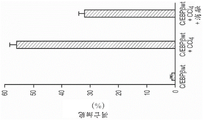


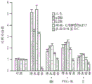


Classifications
-
- A—HUMAN NECESSITIES
- A61—MEDICAL OR VETERINARY SCIENCE; HYGIENE
- A61K—PREPARATIONS FOR MEDICAL, DENTAL OR TOILETRY PURPOSES
- A61K38/00—Medicinal preparations containing peptides
- A61K38/04—Peptides having up to 20 amino acids in a fully defined sequence; Derivatives thereof
- A61K38/07—Tetrapeptides
-
- C—CHEMISTRY; METALLURGY
- C07—ORGANIC CHEMISTRY
- C07K—PEPTIDES
- C07K5/00—Peptides containing up to four amino acids in a fully defined sequence; Derivatives thereof
- C07K5/04—Peptides containing up to four amino acids in a fully defined sequence; Derivatives thereof containing only normal peptide links
- C07K5/10—Tetrapeptides
- C07K5/1019—Tetrapeptides with the first amino acid being basic
-
- A—HUMAN NECESSITIES
- A61—MEDICAL OR VETERINARY SCIENCE; HYGIENE
- A61K—PREPARATIONS FOR MEDICAL, DENTAL OR TOILETRY PURPOSES
- A61K47/00—Medicinal preparations characterised by the non-active ingredients used, e.g. carriers or inert additives; Targeting or modifying agents chemically bound to the active ingredient
- A61K47/06—Organic compounds, e.g. natural or synthetic hydrocarbons, polyolefins, mineral oil, petrolatum or ozokerite
- A61K47/08—Organic compounds, e.g. natural or synthetic hydrocarbons, polyolefins, mineral oil, petrolatum or ozokerite containing oxygen, e.g. ethers, acetals, ketones, quinones, aldehydes, peroxides
- A61K47/10—Alcohols; Phenols; Salts thereof, e.g. glycerol; Polyethylene glycols [PEG]; Poloxamers; PEG/POE alkyl ethers
-
- A—HUMAN NECESSITIES
- A61—MEDICAL OR VETERINARY SCIENCE; HYGIENE
- A61K—PREPARATIONS FOR MEDICAL, DENTAL OR TOILETRY PURPOSES
- A61K47/00—Medicinal preparations characterised by the non-active ingredients used, e.g. carriers or inert additives; Targeting or modifying agents chemically bound to the active ingredient
- A61K47/50—Medicinal preparations characterised by the non-active ingredients used, e.g. carriers or inert additives; Targeting or modifying agents chemically bound to the active ingredient the non-active ingredient being chemically bound to the active ingredient, e.g. polymer-drug conjugates
- A61K47/51—Medicinal preparations characterised by the non-active ingredients used, e.g. carriers or inert additives; Targeting or modifying agents chemically bound to the active ingredient the non-active ingredient being chemically bound to the active ingredient, e.g. polymer-drug conjugates the non-active ingredient being a modifying agent
- A61K47/56—Medicinal preparations characterised by the non-active ingredients used, e.g. carriers or inert additives; Targeting or modifying agents chemically bound to the active ingredient the non-active ingredient being chemically bound to the active ingredient, e.g. polymer-drug conjugates the non-active ingredient being a modifying agent the modifying agent being an organic macromolecular compound, e.g. an oligomeric, polymeric or dendrimeric molecule
- A61K47/59—Medicinal preparations characterised by the non-active ingredients used, e.g. carriers or inert additives; Targeting or modifying agents chemically bound to the active ingredient the non-active ingredient being chemically bound to the active ingredient, e.g. polymer-drug conjugates the non-active ingredient being a modifying agent the modifying agent being an organic macromolecular compound, e.g. an oligomeric, polymeric or dendrimeric molecule obtained otherwise than by reactions only involving carbon-to-carbon unsaturated bonds, e.g. polyureas or polyurethanes
- A61K47/60—Medicinal preparations characterised by the non-active ingredients used, e.g. carriers or inert additives; Targeting or modifying agents chemically bound to the active ingredient the non-active ingredient being chemically bound to the active ingredient, e.g. polymer-drug conjugates the non-active ingredient being a modifying agent the modifying agent being an organic macromolecular compound, e.g. an oligomeric, polymeric or dendrimeric molecule obtained otherwise than by reactions only involving carbon-to-carbon unsaturated bonds, e.g. polyureas or polyurethanes the organic macromolecular compound being a polyoxyalkylene oligomer, polymer or dendrimer, e.g. PEG, PPG, PEO or polyglycerol
-
- A—HUMAN NECESSITIES
- A61—MEDICAL OR VETERINARY SCIENCE; HYGIENE
- A61K—PREPARATIONS FOR MEDICAL, DENTAL OR TOILETRY PURPOSES
- A61K9/00—Medicinal preparations characterised by special physical form
- A61K9/0012—Galenical forms characterised by the site of application
- A61K9/007—Pulmonary tract; Aromatherapy
- A61K9/0073—Sprays or powders for inhalation; Aerolised or nebulised preparations generated by other means than thermal energy
- A61K9/0078—Sprays or powders for inhalation; Aerolised or nebulised preparations generated by other means than thermal energy for inhalation via a nebulizer such as a jet nebulizer, ultrasonic nebulizer, e.g. in the form of aqueous drug solutions or dispersions
-
- A—HUMAN NECESSITIES
- A61—MEDICAL OR VETERINARY SCIENCE; HYGIENE
- A61P—SPECIFIC THERAPEUTIC ACTIVITY OF CHEMICAL COMPOUNDS OR MEDICINAL PREPARATIONS
- A61P29/00—Non-central analgesic, antipyretic or antiinflammatory agents, e.g. antirheumatic agents; Non-steroidal antiinflammatory drugs [NSAID]
-
- C—CHEMISTRY; METALLURGY
- C07—ORGANIC CHEMISTRY
- C07K—PEPTIDES
- C07K7/00—Peptides having 5 to 20 amino acids in a fully defined sequence; Derivatives thereof
- C07K7/04—Linear peptides containing only normal peptide links
- C07K7/06—Linear peptides containing only normal peptide links having 5 to 11 amino acids
-
- C—CHEMISTRY; METALLURGY
- C07—ORGANIC CHEMISTRY
- C07K—PEPTIDES
- C07K7/00—Peptides having 5 to 20 amino acids in a fully defined sequence; Derivatives thereof
- C07K7/04—Linear peptides containing only normal peptide links
- C07K7/08—Linear peptides containing only normal peptide links having 12 to 20 amino acids
-
- A—HUMAN NECESSITIES
- A61—MEDICAL OR VETERINARY SCIENCE; HYGIENE
- A61K—PREPARATIONS FOR MEDICAL, DENTAL OR TOILETRY PURPOSES
- A61K38/00—Medicinal preparations containing peptides
Abstract
The present application relates to peptide therapy for inflammation and fibrosis. In particular, the present application relates to peptides that inhibit the phosphorylation of amino acids in the phosphate receptor domain of C/EBP β, and their use for the treatment of inflammation and fibrosis.
Description
The present application is a divisional application filed on 2016, 4, 11, under the name of 201680024300.0, entitled "peptide therapy for inflammation and fibrosis".
Cross Reference to Related Applications
This application claims priority to U.S. provisional patent application No. 62/160,173 filed on 12.5.2015, the entire contents and basic contents of which are incorporated herein by reference as if fully set forth below.
Sequence listing
This application contains a sequence listing that has been electronically submitted in ASCII format and is incorporated herein by reference in its entirety. The ASCII copy created on day 4, month 11 of 2016 is named 247106.000034_ sl.
Technical Field
The present invention relates to therapeutic peptides that inhibit the phosphorylation of C/EBP β, and their use for the treatment of inflammation and fibrosis.
Background
Activation of hepatic myofibroblasts (of different origin) is the cause of liver fibrosis development in chronic liver disease, and clearly, clearance of myofibroblasts by apoptosis allows recovery from liver injury and reversal of liver fibrosis. The liver specialist believes that inhibiting or reversing the activation of myofibroblasts of different origins is crucial for treating liver fibrosis. Finally, blocking the progression of liver fibrosis will reduce the development of primary liver cancer, as most hepatocellular carcinoma occurs in the liver of liver cirrhosis.
According to NIH and WHO (32; 33), the effects of liver disease can be summarized each year as follows: i) liver cirrhosis: its mortality rate is around 800,000 worldwide (32) and 27,000 in the us; ii) chronic liver disease: 421,000 people are hospitalized in the United states for chronic liver disease. In addition, drugs that prevent the progression of liver fibers and reduce liver inflammation can affect the management of patients with: non-alcoholic steatohepatitis (affecting about 1000 million people in the united states, and this is a worldwide "epidemic"); hepatitis c (chronic infection in about 300 million people in the united states and 1.7 million people worldwide), hepatitis b (chronic infection in about 100 million people in the united states and 35 million people worldwide), and those less common chronic liver diseases that afflict adults (primary biliary cirrhosis; sclerosing cholangitis; autoimmune hepatitis; hereditary hemochromatosis) and those less common chronic liver diseases that afflict children (including biliary atresia; alpha-1 antitrypsin deficiency and other rare familial genetic disorders) for which there is currently no treatment available.
There is currently no approved drug that directly inhibits or reverses liver fibrosis. Current treatment focuses on managing complications caused by liver inflammation and fibrosis. Drug candidates for clinical development in this area include: a) GR-MD-02 (Galectin treatment Inc.) -indication-NASH (fatty liver disease) patients with advanced fibrosis-stage 2). This drug is delivered in liposomes and targets macrophages for apoptosis, but not hepatic myofibroblasts responsible for the fibrogenesis pathway. Since killing macrophages may alter immune balance, significant non-target adverse effects are expected; b) stozomab (Simtuzumab), an anti-fibrotic monoclonal antibody against lysyl oxidase-like 2(LOXL2) enzyme (gillidd science) -indications: liver fibrosis; primary sclerosing cholangitis; non-alcoholic steatohepatitis-stage 2). The agent can prevent the development of active fibrogenesis, but does not reverse the existing cross-linked collagen fibers. In addition, it can induce immunogenic responses. The efficacy of the large protein (antibody) is also a problem, as it must interact with LOXL2 in the potentially inaccessible extracellular matrix of the liver in liver cirrhosis; c) obeticholic acid (OCA) is a bile acid analog and an agonist of Farnesoid X Receptor (FXR) (Intercept Pharmaceuticals) OCA is being developed for various chronic liver diseases including Primary Biliary Cirrhosis (PBC), nonalcoholic steatohepatitis (NASH), and Primary Sclerosing Cholangitis (PSC) -stage 3. One major problem is that blockade of FXR in the absence of the bile acid receptor farnesoid X receptor (26) is associated with spontaneous development of liver tumors; and d) emlicasan (emery) (middle and large pharmaceutical companies), a subgroup of nonalcoholic fatty liver diseases (NAFLD) 2 in patients with inflammation and/or fibrotic nonalcoholic steatohepatitis (NASH). The drug is an active caspase protease inhibitor. One major problem is that prolonged inhibition of hepatocyte caspase may promote the development of hepatocellular carcinoma and other organ tumors by eliminating a critical anti-tumor checkpoint (22).
Drugs that reduce or prevent the progression of pulmonary fibrosis can affect the health care of patients with Idiopathic Pulmonary Fibrosis (IPF). IPF affects five (5) million people worldwide and 200,000 patients in the united states (11). No therapy is known to improve the health-related quality of life or quality of life of patients with IPF, and these patients survive only 3 to 5 years after diagnosis.
Drug candidates for clinical development in this area include: a) pirfenidone (Esbriet, pirfenidone), is newly approved by the FDA for the treatment of IPF. However, the product specifications indicate that "pirfenidone should be used cautiously in patients with mild to moderate liver injury (Child Pugh grades A and B), and also in those patients with mild, moderate or severe kidney injury. The drug can also cause elevated liver enzymes; photosensitization reactions or rashes; gastrointestinal disorders and drug reactions in the case of concurrent administration with a strong inhibitor of CYP1a2 (e.g., fluvoxamine); b) OFEV (nintedanib) is also FDA approved for the treatment of IPF. However, the safety information sheet on OFEV describes that this therapeutic agent can cause birth defects or death, liver problems, bleeding and gastrointestinal disorders in unborn babies, and in more severe cases, can cause stroke and heart attack; c) oral prednisone (or some other form of corticosteroid) can reduce lung inflammation, and these symptoms can be significantly improved. These steroids may be used in combination with other drugs. However, the course of benefit to the patient (according to the results seen) can be slow (1-3 months) and corticosteroids risk significant side effects; d) the affluent administration (Fluimucil) (N-acetylcysteine) is mainly used for symptomatic relief of IPF; however, supportive palliative treatment can be expensive; e) cyclophosphamide (cytoxan, cyclophosphamide) can be used in those patients in which steroid therapy is not effective or possible, and the drug can also be used as a combination therapeutic agent with corticosteroids. The drug is immunosuppressive and response to therapy may be slow (6 months or longer) and there may be significant side effects (including myelosuppression, blood disorders, and cystitis); to name a few; and f) prednisone, azathioprine, and N-acetylcysteine (NAC) for treating IPF patients. However, NAC is thought to be associated with increased risk of mortality and hospitalization in IPF patients.
Inflammation contributes to the pathogenesis of most acute and chronic liver diseases1. Excessive liver damage and inflammation associated with liver disease induced by viral, toxic, immune, and metabolic diseases2Leading to liver dysfunction and chronic disease in the underlying deposition of scar tissue and the development of cirrhosis, which in turn is the major cause of morbidity and mortality in patients affected by chronic liver disease2,3. TLR-4ko mice and TLR-4 mice that were reported to be resistant to hepatotoxin and to be myeloradioactively+/+Reconstitution of macrophages confers on these animals a susceptibility to hepatotoxins4Thus, the amplification of toxic liver injury is mediated by macrophages. The role of macrophages in hepatitis with toxic liver injury has been demonstrated using macrophage ablation5And further characterized using IL-1 receptor antagonists in experimental alcoholic liver injury models6And the use of adenosine-2A(A2A) Receptor-ko mouse characterization7. Fas-mediated secretion of IL-18 from macrophages causes acute liver injury in mice8And macrophage phagocytosis removes hepatocyte debris during hepatocyte injury9. However, the signal transduction mechanisms essential for amplifying liver damage in kupffer cells were only partially characterized1。
Inflammasome is a protein complex that is necessary to trigger activation of an inflammatory response in macrophages, and subsequent macrophage activation1,10,11. CCAAT/enhancer binding protein-beta (C/EBP beta) has been demonstrated12,13,14Are key signal molecules of macrophages as dominant inhibitors of the expression of C/EBP beta DNA-binding site15Or targeted deletion of C/EBP beta results in impaired macrophage differentiation16。
Furthermore, C/EBP β expression is dramatically increased during the differentiation of these cells and is induced by macrophage regulators (LPS, IL-1, G-CSF, TGF β, vitamin D, retinoic acid)13,17. In this context, researchers have shown that phosphorylation of C/EBP β by ribosomal S-kinase-2 (RSK-2), which ribosomal S-kinase-2 is activated directly by extracellular regulated phosphorylation of kinase (ERK) -1/2, plays an important role in the ERK/mitogen-activated protein kinase (MAPK) signaling pathway that regulates cell survival18,19,20,21. In connection with macrophage activation and survival, a dominant positive phosphorylation mutant, C/EBP β -Glu217 (which mimics phosphorylated C/EBP β -Thr217 in a biological assay) has been reported22) Is sufficient to rescue impaired macrophage function and activity induced by anthrax lethal toxin.
Disclosure of Invention
As specified in the background section above, there is a need for effective treatments for inflammation and fibrosis, particularly in the human liver, lung, kidney and any other tissues and/or organs. The present invention addresses these and other needs by providing novel therapeutic peptides and related compositions and methods.
In one aspect, the invention provides an isolated peptide comprising the amino acid sequence Lys-Ala-Val-Asp, wherein at least one amino acid is a D-amino acid, and wherein said peptide is capable of inhibiting phosphorylation of human CCAAT/enhancer binding protein- β (C/EBP β) at threonine 266(Thr 266) or is capable of inhibiting phosphorylation of mouse C/EBP β at Thr 217.
In one embodiment, Ala and/or Val within the amino acid sequence Lys-Ala-Val-Asp is a D-amino acid (DAla, DVal). At one endIn one embodiment, the peptide is capable of selectively inhibiting phosphorylation of human CCAAT/enhancer binding protein-beta (C/EBP beta) at threonine 266(Thr 266). In one embodiment, the peptide is capable of inhibiting activation of myofibroblasts and/or macrophage inflammasome. In one embodiment, the peptide is between four amino acids and eight amino acids in length. In one embodiment, the peptide comprises an amino acid sequence selected from the group consisting of seq id no: Lys-DAla-DVal-Asp, Lys-DAla-Val-Asp and Lys-Ala-DVal-Asp. In one embodiment, the peptide consists of an amino acid sequence selected from the group consisting of seq id no: Lys-DAla-DVal-Asp, Lys-DAla-Val-Asp and Lys-Ala-DVal-Asp. In a specific embodiment, the peptide comprises the amino acid sequence Lys-DAla-DVal-Asp. In a specific embodiment, the peptide consists of the amino acid sequence Lys-DAla-DVal-Asp. In one embodiment, the peptide consists of an amino acid sequence selected from the group consisting of seq id no: Lys-Ser-Lys-Ala-Lys-Lys-Ala-Val-Asp-Lys-His-Ser-Asp (SEQ ID NO:3), Lys-Ala-Val-Asp-Lys-His-Ser (SEQ ID NO:4), and Ala-Lys-Lys-Ala-Val-Asp-Lys-His (SEQ ID NO:5) (e.g., peptides in which Ala and/or Val within the amino acid motif Lys-Ala-Val-Asp is a D-amino acid). In one embodiment, the peptide of the invention further comprises polyethylene glycol (PEG). In one embodiment, the peptides of the invention further comprise a linker that is lactonized with acid (Ac) or mercaptopropionic acid (Mpr) or trimethyl Lock (TML) (see, e.g., Greenwald, Journal of Controlled Release]74,2001, 159-171). In one embodiment, the carboxyl end group of the peptides of the invention is OH, OCH3Or NH2A group. In one embodiment, the peptide of the invention is a cyclic peptide.
In one embodiment, the peptide is selected from the group consisting of:
Lys-DAla-DVal-Asp、
Ac-Lys-DAla-DVal-Asp、
Mpr-Lys-DAla-DVal-Asp、
PEG-Lys-DAla-DVal-Asp、
PEG-Ac-Lys-DAla-DVal-Asp、
PEG-Mpr-Lys-DAla-DVal-Asp、
Lys-DAla-Val-Asp、
Ac-Lys-DAla-Val-Asp、
Mpr-Lys-DAla-Val-Asp、
PEG-Lys-DAla-Val-Asp、
PEG-Ac-Lys-DAla-Val-Asp、
PEG-Mpr-Lys-DAla-Val-Asp、
Lys-Ala-DVal-Asp、
Ac-Lys-Ala-DVal-Asp、
Mpr-Lys-Ala-DVal-Asp、
PEG-Lys-Ala-DVal-Asp、
PEG-Mpr-Lys-Ala-DVal-Asp, and
PEG-Mpr-Lys-Ala-DVal-Asp,
wherein the carboxyl end group of the peptide is OH or OCH3Or NH2A group.
In one embodiment, the peptide is selected from the group consisting of: Lys-DAla-DVal-Asp, Ac-Lys-DAla-DVal-Asp, Mpr-Lys-DAla-DVal-Asp, PEG-Ac-Lys-DAla-DVal-Asp and PEG-Mpr-Lys-DAla-DVal-Asp, wherein the carboxyl end group of the peptide is OH, OCH3Or NH2A group.
In one embodiment, the peptide is selected from the group consisting of: Lys-DAla-DVal-Asp-NH2、Ac-Lys-DAla-DVal-Asp-NH2、Mpr-Lys-DAla-DVal-Asp-NH2、PEG-Lys-DAla-DVal-Asp-NH2、PEG-Ac-Lys-DAla-DVal-Asp-NH2And PEG-Mpr-Lys-DAla-DVal-Asp-NH2。
In one embodiment, the peptide has a structure according to formula (I):

in combination with the peptides of the invention, the invention provides pharmaceutical compositions comprising one or more of the peptides of the invention. In one embodiment, the one or more peptides are present in the composition in an amount effective to inhibit activation of myofibroblasts and/or macrophage inflammasome.
In another aspect, the invention provides a method for inhibiting tissue fibrosis in a subject in need thereof, said method comprising administering to the subject an effective amount of one or more peptides of the invention, or a pharmaceutical composition comprising such one or more peptides. In one embodiment, the tissue is in the liver, lung or kidney. In one embodiment, the tissue fibrosis is associated with liver injury or hepatitis. In one embodiment, the tissue fibrosis is associated with lung injury or pneumonia. In one embodiment, the tissue fibrosis is associated with kidney injury or nephritis.
In another aspect, the present invention provides a method for inhibiting macrophage and/or T cell inflammation in a subject in need thereof, comprising administering to the subject an effective amount of one or more peptides of the invention, or a pharmaceutical composition comprising such one or more peptides.
In yet another aspect, the invention provides a method for treating a fibrotic disease in a tissue in a subject in need thereof, the method comprising administering to the subject an effective amount of one or more peptides of the invention, or a pharmaceutical composition comprising such one or more peptides. In one embodiment, the disease is associated with liver injury, hepatitis, and/or liver fibrosis. In one embodiment, the disease is cirrhosis or liver fibrosis of any etiology. In one embodiment, the disease is selected from the group consisting of: non-alcoholic fatty liver disease (NAFLD), non-alcoholic steatohepatitis (NASH), alcoholic fatty liver disease, alcoholic steatohepatitis, hepatic steatosis, autoimmune hepatitis, chronic hepatitis C, chronic hepatitis B, primary biliary cirrhosis, secondary biliary cirrhosis, sclerosing cholangitis, alpha-1-antitrypsin deficiency, Wilson's disease, and biliary atresia. In one embodiment, the disease is associated with lung injury, pneumonia, and/or pulmonary fibrosis. In one embodiment, the disease is selected from the group consisting of: idiopathic pulmonary fibrosis, radiation-induced pneumonia, chronic obstructive pulmonary disease, and emphysema. In one embodiment, the disease is associated with kidney injury, nephritis, and/or renal fibrosis. In one embodiment, the disease is glomerulonephritis or tubulointerstitial fibrosis. In one embodiment, the disease is selected from the group consisting of: skin fibrosis secondary to a burn, keloid, hypertrophic post-operative wound, scleroderma, esophageal or gastrointestinal fibrosis secondary to a corrosive substance, esophageal or gastrointestinal fibrosis secondary to an inflammatory disease, fibrosis secondary to an ischemic disease, peritoneal fibrosis, pancreatic fibrosis, fibrosis after irradiation, myocardial fibrosis secondary to an infarction, brain fibrosis secondary to an ischemia or infarction, post-traumatic brain fibrosis, post-traumatic muscle fibrosis, and synovial/joint fibrosis.
In another aspect, the invention provides a method for treating an inflammatory disease in a subject in need thereof, the method comprising administering to the subject an effective amount of one or more peptides of the invention, or a pharmaceutical composition comprising such one or more peptides. In one embodiment, the disease is selected from the group consisting of: alcoholic liver disease, non-alcoholic steatohepatitis (NASH), autoimmune hepatitis, chronic hepatitis c, chronic hepatitis b, primary biliary cirrhosis, secondary biliary cirrhosis, sclerosing cholangitis, alpha-1-antitrypsin deficiency, wilson's disease, biliary atresia, idiopathic pulmonary fibrosis, radiation-induced pneumonia, chronic obstructive pulmonary disease, emphysema, chronic infection and/or inflammation of the lung, glomerulonephritis, interstitial fibrosis of the small tubes, skin inflammation secondary to burns, scleroderma, psoriasis, inflammatory bowel disease, esophageal injury and/or inflammation, post-radiation esophageal or gastrointestinal tract inflammation, inflammatory cardiomyopathy, post-traumatic brain inflammation, alzheimer's disease, encephalitis, meningitis, myositis, and arthritis.
In any of the above methods of the invention, the peptide or pharmaceutical composition may be administered, for example, systemically, by inhalation, topically, sublingually, orally, intranasally, or by direct instillation to a tissue or organ.
In any of the above methods of the invention, the subject may be, for example, a human or veterinary animal or a laboratory animal model.
Specifically, the present application provides the following:
1. an isolated peptide comprising the amino acid sequence Lys-Ala-Val-Asp, wherein at least one amino acid is a D-amino acid, and wherein said peptide is capable of inhibiting phosphorylation of human CCAAT/enhancer binding protein-beta (C/EBP β) at threonine 266(Thr 266).
2. The peptide according to scheme 1, wherein Ala and/or Val within the amino acid sequence Lys-Ala-Val-Asp is a D-amino acid.
3. The peptide of scheme 1, wherein the peptide is capable of selectively inhibiting phosphorylation of human CCAAT/enhancer binding protein-beta (C/EBP beta) at threonine 266(Thr 266).
4. The peptide of scheme 1 or scheme 2, wherein the peptide is capable of inhibiting activation of myofibroblasts and/or macrophage inflammasome.
5. The peptide of any of schemes 1-4, wherein the peptide is between four amino acids and eight amino acids in length.
6. The peptide of any of schemes 1-5, wherein the peptide comprises an amino acid sequence selected from the group consisting of SEQ ID NO: Lys-DAla-DVal-Asp, Lys-DAla-Val-Asp and Lys-Ala-DVal-Asp.
7. The peptide of scheme 6, wherein said peptide consists of an amino acid sequence selected from the group consisting of seq id no: Lys-DAla-DVal-Asp, Lys-DAla-Val-Asp and Lys-Ala-DVal-Asp.
8. The peptide according to any of schemes 1 to 5, wherein the peptide comprises the amino acid sequence Lys-DAla-DVal-Asp.
9. The peptide of scheme 8, wherein the peptide consists of the amino acid sequence Lys-DAla-DVal-Asp.
10. The peptide of any of schemes 1-4, wherein said peptide consists of an amino acid sequence selected from the group consisting of SEQ ID NO: Lys-Ser-Lys-Ala-Lys-Lys-Ala-Val-Asp-Lys-His-Ser-Asp (SEQ ID NO:3), Lys-Ala-Val-Asp-Lys-His-Ser (SEQ ID NO:4), and Ala-Lys-Lys-Ala-Val-Asp-Lys-His (SEQ ID NO: 5).
11. The peptide of any one of schemes 1-10, wherein the peptide further comprises polyethylene glycol (PEG).
12. The peptide of any of schemes 1-11, wherein the peptide further comprises a linker of acid (Ac) or mercaptopropionic acid (Mpr).
13. The peptide of any of schemes 1-12, wherein the carboxy terminal group of the peptide is OH, OCH3Or NH2A group.
14. The peptide of any one of schemes 1-10, wherein the peptide is a cyclic peptide.
15. The peptide of scheme 1, wherein said peptide is selected from the group consisting of:
Lys-DAla-DVal-Asp、
Ac-Lys-DAla-DVal-Asp、
Mpr-Lys-DAla-DVal-Asp、
PEG-Lys-DAla-DVal-Asp、
PEG-Ac-Lys-DAla-DVal-Asp、
PEG-Mpr-Lys-DAla-DVal-Asp、
Lys-DAla-Val-Asp、
Ac-Lys-DAla-Val-Asp、
Mpr-Lys-DAla-Val-Asp、
PEG-Lys-DAla-Val-Asp、
PEG-Ac-Lys-DAla-Val-Asp、
PEG-Mpr-Lys-DAla-Val-Asp、
Lys-Ala-DVal-Asp、
Ac-Lys-Ala-DVal-Asp、
Mpr-Lys-Ala-DVal-Asp、
PEG-Lys-Ala-DVal-Asp、
PEG-Mpr-Lys-Ala-DVal-Asp, and
PEG-Mpr-Lys-Ala-DVal-Asp,
wherein the carboxyl end group of the peptide is OH or OCH3Or NH2A group.
16. The peptide of claim 15, wherein the peptideSelected from the group consisting of: Lys-DAla-DVal-Asp, Ac-Lys-DAla-DVal-Asp, Mpr-Lys-DAla-DVal-Asp, PEG-Ac-Lys-DAla-DVal-Asp and PEG-Mpr-Lys-DAla-DVal-Asp, wherein the carboxyl end group of the peptide is OH, OCH3Or NH2A group.
17. The peptide of scheme 16, wherein said peptide is selected from the group consisting of: Lys-DAla-DVal-Asp-NH2、Ac-Lys-DAla-DVal-Asp-NH2、Mpr-Lys-DAla-DVal-Asp-NH2、PEG-Lys-DAla-DVal-Asp-NH2、PEG-Ac-Lys-DAla-DVal-Asp-NH2And PEG-Mpr-Lys-DAla-DVal-Asp-NH2。
18. The peptide of scheme 1, wherein the peptide has the structure of formula (I):

19. a pharmaceutical composition comprising one or more peptides of any one of schemes 1-18 and a pharmaceutically acceptable carrier.
20. The pharmaceutical composition of scheme 19, wherein the one or more peptides are present in an amount effective to inhibit activation of myofibroblasts and/or macrophage inflammasome.
21. A method for inhibiting tissue fibrosis in a subject in need thereof, the method comprising administering to the subject an effective amount of one or more peptides of any one of schemes 1-18, or a pharmaceutical composition of schemes 19 or 20.
22. The method of claim 21, wherein the tissue fibrosis is associated with liver injury or hepatitis.
23. The method of scheme 21, wherein the tissue fibrosis is associated with lung injury or pneumonia.
24. The method of scheme 21, wherein the tissue fibrosis is associated with kidney injury or nephritis.
25. The method of scheme 21, wherein the tissue is in the liver, lung or kidney.
26. A method for inhibiting macrophage and/or T cell inflammation in a subject in need thereof, comprising administering to the subject an effective amount of one or more peptides of any one of schemes 1-18, or a pharmaceutical composition of schemes 19 or 20.
27. A method for treating a tissue fibrotic disease in a subject in need thereof, the method comprising administering to the subject an effective amount of one or more peptides of any one of schemes 1-18, or a pharmaceutical composition of schemes 19 or 20.
28. The method of claim 27, wherein the disease is associated with liver injury, hepatitis, and/or liver fibrosis.
29. The method of claim 27, wherein the disease is cirrhosis or liver fibrosis of any etiology.
30. The method of scheme 28, wherein the disease is selected from the group consisting of: non-alcoholic fatty liver disease (NAFLD), non-alcoholic steatohepatitis (NASH), alcoholic fatty liver disease, alcoholic steatohepatitis, hepatic steatosis, autoimmune hepatitis, chronic hepatitis C, chronic hepatitis B, primary biliary cirrhosis, secondary biliary cirrhosis, sclerosing cholangitis, alpha-1-antitrypsin deficiency, Wilson's disease, and biliary atresia.
31. The method of claim 27, wherein the disease is associated with lung injury, pneumonia, and/or pulmonary fibrosis.
32. The method of scheme 31, wherein the disease is selected from the group consisting of: idiopathic pulmonary fibrosis, radiation-induced pneumonia, chronic obstructive pulmonary disease, and emphysema.
33. The method of scheme 27, wherein the disease is associated with kidney injury, nephritis, and/or renal fibrosis.
34. The method of scheme 33, wherein the disease is glomerulonephritis or tubulointerstitial fibrosis.
35. The method of scheme 27, wherein the disease is selected from the group consisting of: skin fibrosis secondary to a burn, keloid, hypertrophic post-operative wound, scleroderma, esophageal or gastrointestinal fibrosis secondary to a corrosive substance, esophageal or gastrointestinal fibrosis secondary to an inflammatory disease, fibrosis secondary to an ischemic disease, peritoneal fibrosis, pancreatic fibrosis, fibrosis after irradiation, myocardial fibrosis secondary to an infarction, brain fibrosis secondary to an ischemia or infarction, post-traumatic brain fibrosis, post-traumatic muscle fibrosis, and synovial/joint fibrosis.
36. A method for treating an inflammatory disease in a subject in need thereof, the method comprising administering to the subject an effective amount of one or more peptides of any one of schemes 1-18, or a pharmaceutical composition of schemes 19 or 20.
37. The method of scheme 36, wherein said disease is selected from the group consisting of: alcoholic liver disease, non-alcoholic steatohepatitis (NASH), autoimmune hepatitis, chronic hepatitis c, chronic hepatitis b, primary biliary cirrhosis, secondary biliary cirrhosis, sclerosing cholangitis, alpha-1-antitrypsin deficiency, wilson's disease, biliary atresia, idiopathic pulmonary fibrosis, radiation-induced pneumonia, chronic obstructive pulmonary disease, emphysema, chronic infection and/or inflammation of the lung, glomerulonephritis, interstitial fibrosis of the small tubes, skin inflammation secondary to burns, scleroderma, psoriasis, inflammatory bowel disease, esophageal injury and/or inflammation, post-radiation esophageal or gastrointestinal tract inflammation, inflammatory cardiomyopathy, post-traumatic brain inflammation, alzheimer's disease, encephalitis, meningitis, myositis, and arthritis.
38. The method of any of schemes 21-38, wherein the peptide or pharmaceutical composition is administered systemically.
39. The method of any one of schemes 21-38, wherein said peptide or pharmaceutical composition is administered by a route selected from the group consisting of: by inhalation, topically, sublingually, orally, or intranasally.
40. The composition of any one of schemes 21-38, wherein the peptide or pharmaceutical composition is administered by direct instillation to a tissue or organ.
41. The method of any one of claims 21-41, wherein the subject is a human.
These and other aspects of the invention will be apparent to those of ordinary skill in the art in view of the following specification, claims and drawings.
Drawings
Figure 1. the therapeutic leader peptide inhibits the activation of hepatic myofibroblasts and liver injury, which induces fibrosis after exposure to human hepatotoxin CCl 4. C/EBP β -wt mice (4) were given a single dose of CCl 4. Eight hours later, the animals received an IP injection of a therapeutic leader peptide (5 μ g of peptide attached to PEG). Animals were sacrificed 30hr after CCl4 administration. Animals receiving CCl4 only had strong activation of myofibroblasts (as determined by α -SMA) (upper row) and severe liver injury (as assessed by standard clinical examination prior to sacrifice); leader peptides block the discolouration and grainy appearance of severe liver damage. The livers of treated mice were similar to controls (second row). Histopathology induced by CCl4 was activation of myofibroblasts (α -SMA), severe liver injury (H & E staining), with structural disruption (reticulin staining). The therapeutic PEG-30 kDa-peptide reduced myofibroblast activation and liver damage.
Fig. 2A-2b high efficacy of therapeutic PEG-30kDa peptides in a chronic mouse model of liver fibrosis. All groups of mice (except the control group) received CCl4 administration for 16 weeks (n-6/group). The CCl4 treated group received only hepatotoxin, while the other groups also received the indicated therapeutic PEG-30kDa peptide (7 μ g of peptide linked to 420 μ g PEG-30kDa, once a week) starting at week 8. FIG. 2A liver fibrosis was determined by quantifying Sirius staining (percent fibrosis/area) as described (7; 25): control (0.02 +/-0.01); leader peptide (0.8 +/-0.7); alternative peptide 1(2.1 +/-1.0); alternative peptide 2(3.5+/-1.2) and CC14(6.7 +/-1.6%). 1P <0.0001 for leader peptide, and <0.001 for alternative peptides 1 and 2P. Figure 2b. detection of liver fibrosis by trichrome-stained histology of liver fibrosis, as it is the clinical standard. Control animals (receiving vehicle control only) had negligible fibrosis (stages 0-1/VI); animals receiving CCl4 for 16 weeks developed severe fibrosis (stages 5/VI or 6/VI; this was clinically significant in patients); whereas animals receiving CCl4 for 16 weeks and also receiving the leader PEG-30kDa peptide 1 starting at week 8 had slight fibrosis (stages 1-2/VI; this was not clinically significant in patients).
FIG. 3 Induction of C/EBP β -Thr217And C/EBP beta-Thr, and217phosphorylation of (D) is required for the Th1/Th17 response of bleomycin. C/EBP beta at 7 days of bleomycin treatmentwtLung inflammation was induced in mice. Purified lung T cells (CD-4+), phospho-C/EBP β Thr217, IL-12R β, IL-23R, and IL-4R α were identified as described in Table 2 and FIG. 5. C/EBP β -Ala217 mice blocked phosphorylated C/EBP β -Thr217 in T cells and were non-responsive to bleomycin induction of the Th1/Th2 phenotype. Bleomycin induces T cell expression of IL-4R α in C/EBP β -Ala217 mice (Th2 phenotype).
FIG. 4 Induction of C/EBP β -Thr217And C/EBP beta-Thr, and217phosphorylation of (D) is required for the Th1/Th17 response of bleomycin. C/EBP beta at 7 days of bleomycin treatmentwtLung inflammation was induced in mice. Purified lung T cells (CD-4+), phospho-C/EBP β Thr217, IL-12R β, IL-23R, and IL-4R α were identified as described in Table 2 and FIG. 5. The C/EBP β peptide blocks the phosphorylated C/EBP β -Thr217, Th1/Th17 phenotype in T cells and induces T cell expression of IL-4R α (Th2 phenotype).
FIG. 5 Th1/Th17 cells were induced and the Th1/Th17 cells were associated with activation of lung myofibroblasts in response to bleomycin treatment. Evidence from M1 macrophage ablation experiments. On day 7 after bleomycin treatment, purified CD4+ mouse lung T cells expressed IL-12R or IL-23R (Th1/Th17 phenotype) and aSMA (activated myofibroblasts). When mice received bleomycin and clodronate (blocker of ATP production) liposomes (intratracheal) and IP), there were preferential ablation of phagocytic M1 macrophages (undetectable TNF α R2) and preferential ablation of Th1 (IL-12R-red)/Th 17 (IL-23R-green) cells in the case of induction of IL-4R α (aqua) (Th2 phenotype) and markedly reduced α SMA (activated myofibroblasts) (magenta) on day 7. Blockade of Th1/Th17 cells by M1 macrophage ablation for 21 days significantly inhibited bleomycin-induced pulmonary fibrosis (trichrome), suggesting that the Th1/Th17 phenotype, but not the Th2 phenotype, is responsible for this effect.
FIG. 6 Th2 stimulator and C/EBP β peptide induced apoptosis of isolated primary mouse lung Th1 and Th17 cells from bleomycin-treated animals. T cells were isolated by CD-4+ affinity and treated with either IL-10 or IL-4 at 10. mu.g/ml, or with 100pM C/EBP β peptide for 6 hr. Apoptosis was detected as described (12). Th2 inducer and C/EBP beta peptide (P <0.05) stimulate apoptosis of Th-1 and Th-17 lung cells in vitro.
FIGS. 7A-7B human Th1 and Th17 cells induced proliferation of human lung myofibroblasts in co-culture. Figure 7a. LMF was co-cultured with induced Th1 or Th17 or Th2, uninduced controls, and peptide-treated Th1 or Th 17. At least 100 cells/sample (N:3) were counted for PCNA and P <0.01 for Th1 and Th17 cells (NS of other co-culture systems). FIG. 7B.Th1/Th 17-LMF; th 2-LMF; and schematic representation of Th1/Th17 treated with peptide-LMF coculture.
FIG. 8 phosphorylation of C/EBP β -Thr266 was induced and correlated with Th1 responses in lungs from IPF patients. Representative patients with IPF and representative control individuals were studied. DNA, phospho-C/EBP β Thr266, T-Bet, and GATA-3 were identified by confocal laser scanning microscopy.
FIG. 9 phosphorylation of C/EBP β -Thr266 was induced and correlated with Th1/Th17 responses in lungs from IPF patients. Representative patients with IPF and representative control individuals were studied. phospho-C/EBP β Thr266, IL-12R, IL-23R, IL-4R α, and α SMA were identified by confocal laser scanning microscopy.
FIG. 10 phosphorylation of C/EBP β on Thr266 was induced and required for the Th1/Th17 response of human CD4+ T cells to inflammatory inducers. In vitro experiments with human blood precursor T cells. Normal human CD-4+ T cells expressed IL-12R β and T-Bet (Th1 phenotype) 16 hours after treatment with human recombinant IL-12(Th1 inducer). When normal human blood CD-4+ T cells were treated in vitro with IL-23 (a Th17 inducer), they expressed IL-23R and GATA-3 (data not shown) (Th17 phenotype).
FIG. 11 phosphorylation of C/EBP β -Thr266 was induced and correlated with Th1/Th17 responses in lungs from IPF lung tissue. Freshly isolated T cells were purified and characterized as described below. phospho-C/EBP β Thr266, IL-12R, IL-23R, and IL-4R α were identified by confocal laser scanning microscopy.
Figure 12 Th2 stimulator and C/EBP β peptide induced apoptosis of isolated primary human lung Th1 and Th17 cells from IPF patients. T cells were isolated by CD-4+ affinity and treated with either IL-10 or IL-4 at 10. mu.g/ml, or with 100pM C/EBP β peptide for 16 hr. Apoptosis was detected as described (12). Th2 inducer and C/EBP beta peptide (P <0.05) stimulate apoptosis of Th-1 and Th-17 lung cells in vitro.
fas-L modulation induces liver injury and inflammation by phosphorylated C/EBP β -Thr217 in mice. FIG. 13A serum ALT (IU/ml) levels were determined 12 hours after a single IP dose of Jo-2Ab (FasL). Mice expressing the phosphorylation mimic C/EBP β -Glu217 transgene were more susceptible to liver damage induced by Jo-2Ab (P <0.0001) than control C/EBP β -wt mice as judged by serum ALT levels. Mice expressing a non-phosphorylatable C/EBP β -Ala217 transgene are highly resistant to Fas-L induced liver damage (P < 0.01); n-20 mice/group. Figure 13b. judging by the apoptotic annexin-V assay, Jo-2Ab induced minimal damage to cultured primary hepatocytes isolated from phosphorylation mimic C/EBP β -Glu217 transgenic mice (closed-loop) (P <0.001) when compared to hepatocytes from C/EBP β -wt mice (open-loop). Primary hepatocytes from control cultures of C/EBP β -wt without Jo-2 treatment had less than 5% baseline apoptosis. Jo-2Ab stimulated more infiltration of F4/80+ macrophage inflammatory cells in the liver of C/EBP β -Glu217 mice than in the liver of C/EBP β -wt mice (P < 0.01). Jo-2Ab induced a greater area of apoptotic damage to hepatocytes in C/EBP β -Glu217 mouse liver than in C/EBP β -wt mouse liver (P < 0.005). Values are mean (SD) of at least 6 animals per group and are representative of three experiments.
FIGS. 14A-14B Primary Kupffer cells cultured by TGF- α activation were associated with phosphorylation of C/EBP β -Thr 217. The Kupffer cells cultured in RPMI 1640, 10% fetal bovine serum with L-glutamine, 25. mu.M HEPES and penicillin/streptomycin were treated with TGF α (10. mu.M) for 8 hr. FIG. 14A. cultured Kupffer cells freshly isolated from C/EBP β -wt mice express activated RSK-phospho-Ser 380 and endogenous C/EBP β is phosphorylated on Thr217 (P <0.001) following treatment with TGF- α. Representative examples of triplicate samples from three experiments. FIG. 14B TGF- α also induced NOS-2 expression in cultured Kupffer cells (P < 0.01). TO-PRO3 was used TO stain DNA from cells. Representative examples of triplicate samples from three experiments. Fluorescence and bright field images were quantified using the Keyence microscope BZ9000 analysis software program.
FIGS. 15A-15E. induce phosphorylation of C/EBP β on Thr217, and this phosphorylation of C/EBP β on Thr217 is essential for Kupffer cell activation following mouse hepatotoxin treatment. FIG. 15A. comparison of C/EBP β -wt mice (P) as identified by expression of F4/80 by microscopy<0.0001) comparison, CCl4Acute administration of (b) stimulated a higher degree of macrophage infiltration in the liver of the phosphorylation mimic C/EBP β -Glu217 mouse. When compared with C/EBP beta-wt mice (P)<0.0001) C/EBP beta-Ala 217 transgene inhibited CCl4-induced macrophage hepatic infiltration of about 90%. CCl4Induced macrophage liver infiltration was similar in TGF α transgenic and C/EBP β -wt mice (NS). FIG. 15B. from CCl4The degree of hepatocyte apoptosis induced was in C/EBP β -Glu217 mice (P)<0.005) and in TGF-alpha mice (P)<0.05), but when compared to C/EBP β -wt mice, it was in C/EBP β -Ala217 mice (P/EBP β -Ala217)<0.01) was improved. FIG. 15℃ and C/EBP β -wt mice (P)<0.01) comparison, CCl4Serum ALT was stimulated higher in C/EBP β -Glu217 mice. When compared with C/EBP beta-wt mice (P)<0.001) C/EBP beta-Ala 217 transgene inhibited CCl4Induced serum ALT of about 50%. CCl4Induced serum ALT in TGF-alpha transgenic mice and C/EBP-wt miceIs similar (NS). FIG. 15D. dominant negative peptide blocking phosphorylation of C/EBP β -Thr217 also inhibits CCl4Inducing infiltration of Kupffer cells by about 60% (P)<0.01). FIG. 15E. the peptide inhibits CCl4Induce about 45% of hepatocyte apoptosis (P)<0.001). Values are mean (SD) of at least 6 animals per group and are representative of two experiments.
Fig. 16A-16f. macrophages are induced and are essential for liver damage in response to hepatotoxin treatment in mice. FIG. 16A. in administration of CCl4C/EBP β -wt mice that received clodronate liposomes to deplete macrophages 24hr before CCl430hr after treatment, significantly reduced Kupffer cells (about 90%; P)<0.005). FIG. 16B consumption of macrophages with clodronate liposomes in C/EBP β -wt mice resulted in CCl as assessed by counting apoptotic hepatocytes in liver biopsies430 hours post-treatment liver damage was reduced (P)<0.01). FIG. 16C.C/EBP β -wt mice pre-treated with clodronate liposomes in CCl4Serum ALT levels were also reduced by about 75% (P) 30 hours after treatment<0.005). FIGS. 16D, 16E and 16F. CCl never received clodronate liposomes4Chlorophosphonate liposomes in CCl compared to hepatomegaphagous cells isolated from treated C/EBP β -wt mice430hr post-treatment induced inhibition of TLR5, MyD88, and TLR4 expression in Kupffer cells isolated from C/EBP β -wt mice (P)<0.001). Values are mean (SD) of at least 6 animals per group and are representative of two experiments.
FIGS. 17A-17B phosphorylated C/EBP β -Thr217 stimulates the inflammatory body signal 1 complex in Kupffer cells in mice. FIG. 17A. CCl4Thirty hours after treatment, CD-11/CD-68 primary kupffer cells isolated from C/EBP β -wt mice expressed phosphorylated C/EBP β -Thr217 and inflammatory body signal 1 complex gene products, TLR4, NF κ B, IRF8, and MyD 88. Expression of phosphorylated C/EBP β -Thr217, TLR4, NF κ B, IRF8 and MyD88 was blocked in C/EBP β -Ala217 transgenic mice. Kupffer cells isolated from C/EBP β -Glu217 transgenic mice in the absence of CCl4 Inflammatory Signaling 1 Complex is expressed in the treated cases, whereas Kupffer cells isolated from TGF alpha mice were in the absence of CCl4The following table shows the conditions of treatmentReach phosphorylated C/EBP beta-Thr 217, TLR4, NF kappa B, IRF8 and MyD88 (for CCl)4Treated C/EBP β -wt mice, C/EBP β -Glu217 mice, and TGF α mice, P<0.05). Fluorescence and bright field images were quantified using the Keyence microscope BZ9000 analysis software program. Representative examples of three independent experiments as depicted in fig. 15A-15E. FIG. 17B immunoprecipitates of C/EBP β and analysis with vehicle or CCl430hr after treatment of mice its associated proteins from freshly isolated primary kupffer cells. Phosphorylated C/EBP β -Thr217 (or C/EBP β -Glu217) is associated with TLR4, NF κ B, IRF8 and MyD88, whereas non-phosphorylated C/EBP β -Thr217 (or C/EBP β -Ala217) is associated with TLR4, NF κ B, IRF8 and MyD 88. By CCl4Treatment (and macrophage activation) increased the association between phosphorylated C/EBP β -Thr217 and inflammatory body signal 1 protein. P-actin was used as an internal control for sample loading. Representative examples of three independent experiments as depicted in fig. 15A-15E.
FIGS. 18A-18B. phosphorylated C/EBP β -Thr217 stimulates the expression of inflammatory body complex signal 2 in Kupffer cells in mice. FIG. 18A. CCl4Thirty hours after treatment, CD-11/CD-68 primary Kupffer cells purified from C/EBP β -wt mice expressed phosphorylated C/EBP β -Thr217 and inflammatory body signal 2 complex gene products, NALP3, TLR5, IL-1R1, and adaptor protein ASC. Expression of phosphorylated C/EBP β -Thr217, NALP3, TLR5, IL-1R1 and adaptor ASC was blocked in C/EBP β -Ala217 transgenic mice. Kupffer cells isolated from C/EBP β -Glu217 transgenic mice in the absence of CCl4 Inflammatory body signal 2 complex is expressed in the treated condition, whereas kupffer cells isolated from TGF α mice were in the absence of CCl4Phosphorylated C/EBP β -Thr217, NALP3, TLR5, IL-1R1 and adaptor protein ASC were expressed in the case of treatment (for CCl)4Treated C/EBP β -wt mice, C/EBP β -Glu217 mice, and TGF α mice, P<0.01). Fluorescence and bright field images were quantified using the Keyence microscope BZ9000 analysis software program. Representative examples of three independent experiments as depicted in fig. 15A-15E. FIG. 18B immunoprecipitates of C/EBP β and analysis with vehicle or CCl430hr after treatment of mice from freshly isolated primary kupffer cellsIts related protein. Phosphorylated C/EBP β -Thr217 (or C/EBP β -Glu217) is associated with NALP3, TLR5, IL-1R1 and adaptor ASC, whereas non-phosphorylated C/EBP β -Thr217 (or C/EBP β -Ala217) is associated with NALP3, TLR5, IL-1R1 and adaptor ASC. By CCl4Treatment (and macrophage activation) increased the association between phosphorylated C/EBP β -Thr217 and inflammatory body signal 2 protein. Beta-actin was used as an internal control for sample loading. Representative examples of three independent experiments as depicted in fig. 15A-15E.
FIGS. 19A-19℃ phosphorylated C/EBP β -Thr217 stimulates the expression of inflammatory body structure and byproduct genes in Kupffer cells in mice. Figure 19a. hepatophagy cells freshly isolated from phosphorylated mimic C/EBP β -Glu217 mice expressed inflammasome-associated activated transcripts when compared to C/EBP β -wt mice. This includes increased expression of inflammatory body genes (ASC, IRF-1, IRF-4, IRF-5, TCAM-2, TLR-6, TRAF-6, MyD-88, Nod-1, and Rel), as well as increased expression of direct and indirect cytokine by-products (IL-I β, IL-6, IL-15, IL-18, and TNF α). FIG. 19B, when and from CCl4Freshly isolated Kupffer cells from treated C/EBP beta-wt mice when compared to CCl4Freshly isolated C/EBP β -Ala217 Kupffer cells in treated mice expressed suppressed inflammatory transcripts. This includes reduced expression of inflammatory body genes (IRF-4, NALP-alpha, NALP-3, TCAM-2, TLR-1, TLR-3, TLR-5, TLR-6, TLR-7, TLR-8, TLR-9, Nod-1, and Rel), as well as reduced expression of genes of direct and indirect cytokine inflammatory body by-products (IL-1 beta, IL-6, IL-10, IL-15, IL-18, IL-23 alpha, and CXCL-3). FIG. 19℃ with CCl4Treatment was associated with induction of IL-18, active caspase-1 and IL-1 β inflammatory protein expression in the liver of C/EBP β -wt, C/EBP β -Glu217, and TGF α mice. Values are mean (SD) of triplicates and are representative of three experiments.
FIGS. 20A-20J.C/EBP β -Thr266 was associated with inflammatory body complexes in Kupffer cells from patients with toxic oil syndrome. Liver biopsies from all 16 patients were analyzed with TOS, which is still available at the compton university medical center, madrid, spain. These patients suffer from moderately severe acute liver injury. Figures 20A and 20C there was a significant increase in macrophage infiltration of the liver (about 20-fold; 1,004,683+/-140,485 and 41,160+/-3,353; P <0.001) (figure 20A) and a significant increase in the degree of hepatocyte apoptosis (about 30-fold; 32.0 +/-4.7% and 1.0 +/-0.2%; P <0.001) (figure 20C) in TOS patients compared to normal subjects (figures 20B and 20D). Figures 20F, 20H and 20j kupffer cells in the liver from patients with TOS expressed MyD-88, phosphorylated C/EBP β -Thr266, and TLR-5 (figures 20E, 20G and 20I) when compared to macrophages in normal liver (for all, P < 0.001).
FIG. 21 phosphorylation mimic C/EBP β -Glu217 transgenic mice are more susceptible to liver damage induced by FAS-R activation than control C/EBP β -wt mice. Representative histological samples of C/EBP β -wt and C/EBP β -Glu217(E) mice after 30hr treatment with vehicle or CCl 4. Formalin-fixed liver samples were stained with reticulohistochemistry or F4/80 immunohistochemistry. Liver injury (reticulin staining) and liver macrophage infiltration (F4/80) were induced by Jo-Ab (FAS), but more prominently in liver tissue from C/EBP β -Glu217 mice. Representative examples of experiments are described in figures 13A-13D above.
FIG. 22 lactacystin induced minimal damage to primary hepatocytes isolated from C/EBP β -Glu217 transgenic mice. Lactacystin induced minimal damage to cultured primary hepatocytes isolated from phosphorylation mimic C/EBP β -Glu217 transgenic mice (closed-loop) (P <0.0001) when compared to hepatocytes from C/EBP β -wt mice (open-loop) as judged by the apoptotic annexin-V assay. Primary hepatocytes from control cultures of C/EBP β -wt without treatment with lactacystin had less than 5% baseline apoptosis. Values are the mean (SD) of triplicate samples and are representative of two experiments.
23A-23B. phosphorylation mimic C/EBP β -Glu217 transgenic mice were more susceptible to liver damage induced by CCl4 activation than control mice. FIG. 23A. representative histological samples of C/EBP β -wt, TGF α, C/EBP β -Glu217(E), and C/EBP β -Ala217(A) after 30hr treatment with vehicle or CCl 4. Formalin-fixed liver samples were stained with reticulohistochemistry or F4/80 immunohistochemistry. Liver injury (reticulin staining) and liver macrophage infiltration (F4/80) were induced by CCl4, but more prominently in liver tissue from C/EBP β -Glu217 mice. C/EBP β -Ala217 mice were non-responsive to both liver injury and CCL4 stimulation by liver macrophage infiltration. Figure 23b. representative histological samples of C/EBP β -wt mice after 30hr treatment with vehicle or CCL 4. Formalin-fixed liver samples were immunohistochemically stained with either reticulin or F4/80. The dominant negative peptide (100 μ g IP at 8 hr) that blocks phosphorylation of C/EBP β -Thr217 inhibited CCl 4-induced liver injury (reticulin staining) by about 90% (P <0.001) and inhibited kupffer cell infiltration (F4/80) by about 60% (P < 0.01). Representative examples of experiments are described above in fig. 15A-15E.
FIG. 24 macrophage ablation prevents CCl 4-induced liver damage and macrophage infiltration. Histological samples of C/EBP β -wt mouse mice receiving macrophage depletion by clodronate liposomes 24hr prior to vehicle or CCl4 administration. After 30hr treatment with Cll4, macrophage-depleted mice had significantly reduced liver injury (reticulin staining) and liver macrophage infiltration (F4/80). Representative examples of experiments are described above in fig. 16A-16F.
25A-25B treatment with the PEG-30kDa peptide IP, intratracheal instillation of the peptide (positive control), or by inhaled peptide significantly reduced the extent of pulmonary fibrosis; expression of α -SMA (marker of activated myofibroblasts), co-localized with C/EBP β -Thr217 phosphorylation. Furthermore, all treatments reduced lung inflammation as determined by expression of IL-23R (a marker for Th-17 cells). Bleomycin increased pulmonary fibrosis by >5 fold compared to control animals. In contrast, pulmonary fibrosis was reduced by about 60% (P <0.001) in animals receiving the PEG-30kDa peptide or the peptide by inhalation in only 14 days of treatment with 3 doses on days 10, 17 and 24. As expected, intratracheal instillation (therapeutic positive control) had outstanding efficacy with only minor differences from control animals (P < 0.001). The peptide also reduced Th-17 inflammation (probably an important component of IPF inflammation) as judged by reduced expression of IL-23R. C/EBP β -wt mice receiving the PEG-30kDa peptide or receiving the peptide by inhalation had less lung injury than control C/EBP β -wt mice (P <0.001, fig. 25A) judged by quantification of C/EBP β -wt from essentially normal surface active protein C (sfpc) expression after treatment with bleomycin. Consistent with quantification of IMH, collagen α 1 (major ECM genes) and TGF β 1 (fibrogenic cytokines) were reduced by three peptide formulations as determined by RT-PCR.
FIG. 26 peptides treated with PEG-30kDa peptide IP, intratracheal instillation of the peptide (positive control), or by inhalation as depicted in FIGS. 25A-25B. All treatments improved tidal volume > 35% (P <0.01), although 14 days of treatment were brief.
FIG. 27 shows that C/EBP β -wt mice receiving the PEG-30 kDa-peptide or receiving the peptide by inhalation had less lung injury (about 60%) than control C/EBP β -wt mice (P <0.005) as judged by significant inhibition of alveolar epithelial apoptosis following bleomycin treatment.
FIG. 28. representative immunohistochemistry from renal biopsies of patients with glomerulonephritis. Glomerulonephritis has a significant increase in activated myofibroblasts (α -SMA) and has an enhanced macrophage inflammatory response as recorded against liver and lung inflammation and fibrosis (F4/80). These cells were positive for C/EBP β -266, indicating the high feasibility of targeting kidney inflammation and fibrosis with the PEG-30kDa peptide.
Detailed Description
Definition of
Unless defined otherwise, all technical and scientific terms used herein have the same meaning as commonly understood by one of ordinary skill in the art to which this invention belongs. Other specifically defined terms are to be construed in a manner consistent with the definitions provided herein. Although any methods and materials similar or equivalent to those described herein can be used in the practice of the present invention, the preferred materials and methods are described herein. In describing and claiming the present invention, the following terminology will be used.
As used in this specification and the appended claims, the singular forms "a", "an" and "the" include plural referents unless the context clearly dictates otherwise. Thus, for example, reference to "a construct" includes a combination of two or more nucleic acid constructs, and the like.
As used herein, the term "subject" refers to human, mammalian and/or veterinary animals (e.g., cats, dogs, cows, horses, sheep, pigs, etc.) and experimental animal models. In certain embodiments, the subject is a human patient, including both sexes in the adult and pediatric populations.
In the context of the present invention, the term "treating" or "treatment" in the context of its reference to any disease symptom referred to herein means alleviating or alleviating at least one symptom associated with such a disorder, or slowing or reversing the progression of such a disorder. Within the meaning of the present invention, the term "treatment" also means to arrest, delay the onset (i.e. the period before the clinical manifestation of the disease) and/or reduce the risk of development or worsening of the disease. The term "treating" with respect to a state, disorder or condition can also include (1) preventing or delaying the onset of at least one clinical or subclinical symptom of a state, disorder or condition developing in a subject who may be afflicted with or predisposed to the state, disorder or condition, but who has not yet experienced or exhibited clinical or subclinical symptoms of the state, disorder or condition; or (2) inhibiting the state, disorder or condition, i.e., arresting, reducing or delaying the progression of the disease or its recurrence (in the case of maintenance therapy) or at least one clinical or subclinical symptom thereof; or (3) alleviating the disease, i.e., causing regression of at least one of the state, disorder or condition, or clinical or subclinical symptoms thereof.
The practice of the present invention employs, unless otherwise indicated, conventional techniques of statistical analysis, molecular biology (including recombinant techniques), microbiology, cell biology, and biochemistry that are within the skill of the art. Such tools and techniques are described in detail below, for example: sambrook et al, (2001), Molecular Cloning: a Laboratory Manual [ molecular cloning: laboratory manual 3 cold spring harbor laboratory press: cold spring harbor, new york; authored by Ausubel et al, (2005), Current Protocols in Molecular Biology [ modern Molecular Biology Protocols ], John Wiley father publishing company (John Wiley and Sons, Inc.): hopken, new jersey; bonifacino et al, (2005), Current Protocols in Cell Biology [ modern Cell Biology Protocols ], John Wiley father publishing company (John Wiley and Sons, Inc.): hopken, new jersey; coligan et al, (2005), Current Protocols in Immunology [ modern immunological Protocols ], John Wiley parent publishing company (John Wiley and Sons, Inc.): hopken, new jersey; compiled by Coico et al, (2005), Current Protocols in Microbiology [ experimental Protocols in modern Microbiology ], John Wiley father publishing company (John Wiley and Sons, Inc.): hopken, new jersey; coligan et al, (2005), Current Protocols in Protein Science [ modern Protein Science Protocols ], John Wiley father publishing company (John Wiley and Sons, Inc.): hopken, new jersey; and ena et al, (2005), Current Protocols in Pharmacology [ modern drug Protocols ], John Wiley father-son publishing company (John Wiley and Sons, Inc.): hopken, new jersey. Additional technical explanations are in, for example, U.S. patent No. 7,912,698 and U.S. patent application publication nos. 2011/0202322 and 2011/0307437.
Peptides, compositions and administration of the invention
The present invention provides therapeutic peptides designed to block a phosphorylation on a molecular protein target in liver, lung, kidney and/or other scar tissue to prevent and/or inhibit myofibroblast and/or macrophage activation and production, thereby inhibiting inflammation and/or fibrosis of liver, lung, and/or other scar tissue. Since the peptides of the invention are not necessary for other mechanical processes, they are highly specific and effective while minimizing any potential off-target toxicity.
In certain embodiments, the invention provides therapeutic peptides with high efficacy against molecular targets that prevent myofibroblast activation and inhibit liver, lung, and kidney fibrosis. In other embodiments, the invention further provides that phosphorylated C/EBP β -phospho-Thr 266 (human syngeneic mouse C/EBP β -Thr217) plays a major role in liver, lung and kidney inflammation and injury by modulating inflammatory body activation in inflammatory macrophages and activating myofibroblasts. The evolutionarily conserved C/EBP β -phosphate-Thr 217 signaling (the same in human C/EBP β -phosphate-Thr 266) regulates macrophage inflammatory body activity and liver damage induced by various acting hepatotoxins. In certain embodiments, these therapeutic peptides also block activation of myofibroblasts by apoptotic mechanisms, prevent progression of fibrosis of scar tissue, and allow regression of fibrosis of scar tissue.
The present invention provides therapeutic peptides designed to block phosphorylation on one protein, a single event that is critical for the generation of liver, lung, kidney and/or other scar tissue and is not essential for other mechanical processes. Thus, the drug candidate has a high degree of specificity and efficacy, while minimizing any potential off-target toxicity. In certain embodiments, the present invention provides that phosphorylated C/EBP β -Thr217 plays a major role in liver, lung and/or kidney inflammation and injury by modulating inflammatory body activation in liver lung and/or kidney macrophages. The evolutionarily conserved C/EBP β -phospho-Thr 217 signaling (the same in human C/EBP β -phospho-Thr 266) regulates macrophage inflammatory body activity and liver, lung and/or kidney injury induced by different acting hepato-and/or pneumotoxins.
In certain embodiments, the peptides of the invention have been shown to stimulate apoptosis and block fibrogenesis, prevent progression of inflammation and/or fibrosis in liver, lung, kidney, and/or other tissues, and induce regression of inflammation and/or fibrosis in liver, lung, kidney, and/or other tissues. In certain embodiments, the peptides of the invention also exhibit high efficacy against molecular targets, such as phosphorylation of C/EBP β -Thr217 in activated liver, lung, and/or kidney myofibroblasts and/or macrophages, preventing myofibroblast and/or macrophage activation and inhibiting liver, lung, and/or kidney inflammation and fibrogenesis.
In one aspect, the invention provides an isolated peptide comprising the amino acid sequence Lys-Ala-Val-Asp, wherein at least one amino acid is a D-amino acid, and wherein said peptide is capable of inhibiting phosphorylation of human CCAAT/enhancer binding protein- β (C/EBP β) at threonine 266(Thr 266) or is capable of inhibiting phosphorylation of mouse C/EBP β at Thr 217. For examples of human and mouse C/EBP β sequences, see, e.g., human: GenBank gene ID 1051; mice: GenBank Gene ID 12608.
In one embodiment, Ala and/or Val within the amino acid sequence Lys-Ala-Val-Asp is a D-amino acid. In one embodiment, the peptide is capable of selectively inhibiting phosphorylation of human CCAAT/enhancer binding protein-beta (C/EBP beta) at threonine 266(Thr 266). In one embodiment, the peptide is capable of inhibiting activation of myofibroblasts and/or macrophage inflammasome. In one embodiment, the peptide is between four amino acids and eight amino acids in length. In one embodiment, the peptide comprises an amino acid sequence selected from the group consisting of seq id no: Lys-DAla-DVal-Asp, Lys-DAla-Val-Asp and Lys-Ala-DVal-Asp. In one embodiment, the peptide consists of an amino acid sequence selected from the group consisting of seq id no: Lys-DAla-DVal-Asp, Lys-DAla-Val-Asp and Lys-Ala-DVal-Asp. In one embodiment, the peptide comprises the amino acid sequence Lys-DAla-DVal-Asp. In one embodiment, the peptide consists of the amino acid sequence Lys-DAla-DVal-Asp. In one embodiment, the peptide consists of an amino acid sequence selected from the group consisting of seq id no: Lys-Ser-Lys-Ala-Lys-Lys-Ala-Val-Asp-Lys-His-Ser-Asp (SEQ ID NO:3), Lys-Ala-Val-Asp-Lys-His-Ser (SEQ ID NO:4), and Ala-Lys-Lys-Ala-Val-Asp-Lys-His (SEQ ID NO:5) (e.g., peptides in which Ala and/or Val within the amino acid motif Lys-Ala-Val-Asp is a D-amino acid). In one embodiment, the peptide further comprises polyethylene glycol (PEG). In one embodiment, the peptide further comprises a linker that is lactonized with acid (Ac) or mercaptopropionic acid (Mpr) or trimethyl lock (TML) (see, e.g., Greenwald, Journal of Controlled Release]74,2001,159-171). In one embodiment, the carboxyl end group of the peptide is OH, OCH3Or NH2A group. In one embodiment, the peptide is a cyclic peptide.
In one embodiment, the peptide is selected from the group consisting of:
Lys-DAla-DVal-Asp、
Ac-Lys-DAla-DVal-Asp、
Mpr-Lys-DAla-DVal-Asp、
PEG-Lys-DAla-DVal-Asp、
PEG-Ac-Lys-DAla-DVal-Asp、
PEG-Mpr-Lys-DAla-DVal-Asp、
Lys-DAla-Val-Asp、
Ac-Lys-DAla-Val-Asp、
Mpr-Lys-DAla-Val-Asp、
PEG-Lys-DAla-Val-Asp、
PEG-Ac-Lys-DAla-Val-Asp、
PEG-Mpr-Lys-DAla-Val-Asp、
Lys-Ala-DVal-Asp、
Ac-Lys-Ala-DVal-Asp、
Mpr-Lys-Ala-DVal-Asp、
PEG-Lys-Ala-DVal-Asp、
PEG-Mpr-Lys-Ala-DVal-Asp, and
PEG-Mpr-Lys-Ala-DVal-Asp,
wherein the carboxyl end group of the peptide is OH or OCH3Or NH2A group.
In one embodiment, the peptide is selected from the group consisting of: Lys-DAla-DVal-Asp, Ac-Lys-DAla-DVal-Asp, Mpr-Lys-DAla-DVal-Asp, PEG-Ac-Lys-DAla-DVal-Asp and PEG-Mpr-Lys-DAla-DVal-Asp, wherein the carboxyl end group of the peptide is OH, OCH3Or NH2A group.
In one embodiment, the peptide is selected from the group consisting of: Lys-DAla-DVal-Asp-NH2、Ac-Lys-DAla-DVal-Asp-NH2、Mpr-Lys-DAla-DVal-Asp-NH2、PEG-Lys-DAla-DVal-Asp-NH2、PEG-Ac-Lys-DAla-DVal-Asp-NH2And PEG-Mpr-Lys-DAla-DVal-Asp-NH2。
In one embodiment, the peptide has a structure according to formula (I):

in addition to the peptides disclosed above, the invention also encompasses C/EBP β -Ala-217 amino acids 1 to 296 (mouse), C/EBP β -Ala-217 fragment amino acids 216 to 253 (mouse) and C/EBP β -Ala-217 amino acids 1 to 285 (mouse) (e.g., obtained based on GenBank gene ID 12608 with Ala at position 217). Corresponding human sequences and fragments are also contemplated and can be obtained based on the human C/EBP β sequence with Ala at position 266 (e.g., based on GenBank gene ID 1051 with Ala at position 266).
The peptides of the invention may be modified, for example, by using heterobifunctional linkers. Non-limiting examples of terminal groups for heterobifunctional PEGs are maleimides, vinylsulfones, pyridyl disulfides, amines, carboxylic acids and NHS esters (see, e.g., Veronese, France sco M. "peptide and protein PEGylation: a review of proteins and solutions. [ PEGylation of peptides and proteins: review of problems and solutions ]" Biomaterials [ Biomaterials ]22.5 (2001): 405-). 417). The peptides of the invention may employ, for example, branched, Y-shaped or comb-shaped third-generation pegylating agents (see, e.g., Ryan, Sinead M; Mantovani, Giuseppe; Wang, Xuexuuan; Haddleton, DavidM; Brayden, David J, (2008), "Advances in PEGylation of important biotechnological molecules: Delivery aspects", Expert Opinion on Drug Delivery 5(4): 371-83). The peptides of the invention may be multimerized and/or cyclized (e.g., a polypeptide group).
Useful delivery techniques for the peptides of the invention include, for example, LAR-reservoir microsphere polymer matrices (Midatech Pharma) and Protected Graft Copolymers (PGC)TM) Technique (PharmaIN).
The peptides of the invention can be modified in various ways to improve their pharmacokinetic and other properties. The peptides may be modified at the amino (N-) terminus and/or the carboxy (C-) terminus and/or the side chain of one or more amino acid residues, peptide phosphorylation, etc., by substituting one or more naturally occurring genetically encoded amino acids with a non-canonical amino acid.
Amino-terminal modifications include methylation (e.g., -NHCH)3Or- -N (CH)3)2) Acetylation (for example, with acetic acid or its halogenated derivatives (such as. alpha. -chloroacetic acid,. alpha. -bromoacetic acid or. alpha. -iodoacetic acid)), with the addition of a benzyloxycarbonyl (Cbz) group, or with a compound containing a carboxylate function (defined by RCOO- -) or a sulfonyl function (defined by R- -SO- -)2- - (Y- -O) (wherein R is selected from the group consisting of alkyl, aryl, heteroaryl, alkylaryl, etc.) and any blocking group of the like blocks the amino terminus. One may also incorporate de-amino acids at the N-terminus (such that no N-terminal amino group is present) to reduce sensitivity to proteases or to limit the conformation of peptidal compounds.
Carboxyl terminal modifications include the substitution of the free acid with a carboxamide group or the formation of a cyclic lactam at the carboxyl terminal to introduce structural constraints. One may also cyclize the peptides of the invention, or may incorporate a deamino or decarboxylation residue at the terminus of the peptide, such that no terminal amino or carboxyl group is present, to reduce sensitivity to proteases or to limit the conformation of the peptide. The C-terminal functional group of the compounds of the present invention includes amide, amide lower alkyl, amide di (lower alkyl), lower alkoxy, hydroxy, and carboxy, and lower ester derivatives thereof, and pharmaceutically acceptable salts thereof.
One can substitute the naturally occurring side chain having 20 genetically encoded amino acids (or stereoisomeric D-amino acids) with other side chains, for example with groups such as alkyl, lower alkyl, cyclo 4-, 5-, 6-to 7-membered alkyl, amide lower alkyl, amide di (lower alkyl), lower alkoxy, hydroxy, carboxy and lower ester derivatives thereof, and with 4-, 5-, 6-to 7-membered heterocycles. For example, proline analogs in which the ring size of the proline residue varies from 5-to 4-, 6-or 7-membered can be employed. The cyclic group may be saturated or unsaturated, and if unsaturated, may be aromatic or non-aromatic. The heterocyclic group preferably contains one or more nitrogen, oxygen and/or sulfur heteroatoms. Examples of such groups include furazanyl, furyl, imidazolidinyl, imidazolyl, imidazolinyl, isothiazolyl, isoxazolyl, morpholinyl (e.g., morpholino), oxazolyl, piperazinyl (e.g., 1-piperazinyl), piperidinyl (e.g., 1-piperidinyl, piperidinyl), pyranyl, pyrazinyl, pyrazolidinyl, pyrazolinyl, pyrrolyl, pyridazinyl, pyridinyl, pyrimidinyl, pyrrolidinyl (e.g., 1-pyrrolidinyl), pyrrolinyl, pyrrolyl, thiadiazolyl, thiazolyl, thienyl, thiomorpholinyl (e.g., thiomorpholino), and triazolyl. These heterocyclic groups may be substituted or unsubstituted. When a group is substituted, the substituent may be alkyl, alkoxy, halogen, oxygen, substituted or unsubstituted phenyl.
Common examples of conventional amino acid substitutions include stereoisomeric (e.g., D-amino acids) and unnatural amino acids such as, for example, L-ornithine, L-homocysteine, L-homoserine, L-citrulline, 3-sulfinyl-L-alanine, N- (L-arginine) succinic acid, 3, 4-dihydroxy-L-phenylalanine, 3-iodo-L-tyrosine, 3, 5-diiodo-L-tyrosine, triiodothyronine, L-thyroxine, L-selenocysteine, N- (L-arginine) taurine, 4-aminobutyric acid, (R, S) -3-amino-2-methylpropionic acid, a-disubstituted amino acids, a, N-alkyl amino acids, lactic acid, beta-alanine, 3-pyridyl alanine, 4-hydroxyproline, O-phosphoserine, N-methylglycine, N-acetylserine, N-formylmethionine, 3-methylhistidine, 5-hydroxylysine, norleucine and other similar amino acids and imino acids. General methods for the site-specific incorporation of unnatural amino acids into proteins and peptides are described in Noren et al, Science [ Science ], 244: 182-.
One can also readily modify peptides by phosphorylation and other methods (e.g., as described in Hruby et al, (1990), Biochem J. [ J. Biochem ]268: 249-262).
The peptide compounds of the invention are also useful as structural models for non-peptide compounds with similar biological activity. One skilled in the art recognizes that various techniques can be used to construct compounds that have the same or similar desired biological activity as the leader peptide compound, but have more favorable activity than the leader with respect to solubility, stability, and susceptibility to hydrolysis and proteolysis (see, e.g., Morgan and Gainor, (1989), Ann. Rep. Med. chem. [ journal of medicinal chemistry: 24: 243-. These techniques include replacing the peptide backbone with a backbone consisting of phosphonates, amidates, carbamates, sulfonamides, secondary amines and N-methyl amino acids.
The invention also provides conjugates of the disclosed peptide monomers. Thus, according to a preferred embodiment, the monomeric peptides of the invention are dimeric or oligomeric, thereby enhancing their biological activity.
In one embodiment, the peptide monomers of the invention can be oligomerized using the biotin/streptavidin system. Biotinylated analogs of the peptide monomers can be synthesized by standard techniques. For example, these peptide monomers may be C-terminally biotinylated. These biotinylated monomers are then oligomerized by incubation with streptavidin [ e.g., at a 4:1 molar ratio, at room temperature, in Phosphate Buffered Saline (PBS) or HEPES-buffered RPMI media (Invitrogen), for 1 hour ]. In a variation of this embodiment, biotinylated peptide monomers can be oligomerized by incubation with any of a number of commercially available anti-biotin antibodies [ e.g., goat anti-biotin IgG from Kirkegaard & Perry Laboratories, Inc. (washington DC) ].
Linker in other embodiments, the peptide monomers of the invention can be dimerized by covalent binding to at least one linker moiety. The joint (L)K) Part may be C1-12A linking moiety, optionally terminated with one or two-NH-linkages and optionally substituted on one or more available carbon atoms with a lower alkyl substituent (e.g., -NH-R-NH-, wherein R is lower (C) substituted with a functional group such as a carboxyl group or an amino group (such as, for example, a lysine residue or a lysine amide)1-6) Alkylene).
In addition toIn other embodiments, polyethylene glycol (PEG) may be used as the linker LKThe joint LKDimerize two peptide monomers: for example, a single PEG moiety may be attached to the N-terminus of both peptide chains of a peptide dimer simultaneously.
In yet another further embodiment, the joint (L)k) The moiety is preferably, but not necessarily, a molecule containing two carboxylic acids and optionally substituted on one or more available atoms with additional functional groups, such as amines capable of binding to one or more PEG molecules. Such molecules can be depicted as:
-CO-(CH2)n-X-(CH2)m-CO-
wherein n is an integer from 0 to 10, m is an integer from 1 to 10, and X is selected from O, S, N (CH)2)pNR1、NCO(CH2)pNR1And CHNR1,R1Selected from H, Boc, Cbz, etc., and p is an integer from 1 to 10.
Linkers may be incorporated into the peptide during peptide synthesis. For example, at the joint LKWhen the moiety contains two functional groups capable of serving as a starting site for peptide synthesis and a third functional group capable of binding to another molecular moiety (e.g., a carboxyl group or an amino group), the linker may be conjugated to a solid support. Thereafter, the two peptide monomers can be synthesized directly to linker L in a variation of the solid phase synthesis techniqueKOn part of the two reactive nitrogen groups.
In which the peptide dimer is bound by linker LKIn an alternative embodiment of partial dimerization, the linker may be conjugated to both peptide monomers of the peptide dimer after peptide synthesis. Such conjugation can be achieved by methods established in the art. In one embodiment, the linker contains at least two functional groups suitable for attachment to the target functional group of the synthetic peptide monomer. For example, a linker having two free amine groups may be reacted with the C-terminal carboxyl group of each of the two peptide monomers. In another example, a linker containing two carboxyl groups (either pre-activated or in the presence of a suitable coupling reagent) may be attached to each of the two peptide monomersN-terminal or side chain amine groups, or C-terminal lysine amide.
A spacer region. The peptide monomer or dimer may further comprise one or more spacer moieties. Such spacer moieties may be attached to a peptide monomer or peptide dimer (e.g., such spacer moieties may be attached to a linker L linking monomers of a peptide dimerKPortion). For example, such spacer moieties may be attached to the peptide via the carbonyl carbon of a lysine linker, or via the nitrogen atom of an iminodiacetic acid linker. Such spacers may link the peptide to an attached water-soluble polymer moiety or protecting group.
In one embodiment, the spacer moiety is C1-12A connecting portion of C1-12The linking moiety is optionally terminated with an-NH-linkage or a carboxyl (-COOH) group, and is optionally substituted on one or more available carbon atoms with a lower alkyl substituent. In one embodiment, the spacer is R-COOH, wherein R is lower (C)1-6) Alkylene group of the lower group (C)1-6) The alkylene group is optionally substituted with a functional group capable of binding to another molecular moiety, such as a carboxyl group or an amino group. For example, the spacer may be a glycine (G) residue, or aminocaproic acid.
In other embodiments, the spacer is-NH-R-NH-, wherein R is lower (C)1-6) Alkylene group of the lower group (C)1-6) The alkylene group is substituted with a functional group capable of binding to another molecular moiety, such as a carboxyl group or an amino group. For example, the spacer may be a lysine (K) residue or a lysine amide (K-NH)2Lysine residue in which the carboxyl group has been converted to an amide moiety-CONH2)。
A spacer may be incorporated into the peptide during peptide synthesis. For example, where the spacer contains a free amino group and a second functional group (e.g., a carboxyl group or an amino group) capable of binding to another molecular moiety, the spacer can be conjugated to a solid support. Thereafter, the peptide can be synthesized directly onto the free amino group of the spacer by standard solid phase techniques.
For example, a spacer containing two functional groups is first passed throughCoupled to the solid support via the first functional group. Next, a linker L having two functional groups capable of serving as an initiation site for peptide synthesis and a third functional group (e.g., a carboxyl group or an amino group) capable of binding another molecular moietyKThe moiety is conjugated to the spacer via the second functional group of the spacer and the third functional group of the linker. Thereafter, the two peptide monomers can be synthesized directly to linker L in a variation of the solid phase synthesis techniqueKOn part of the two reactive nitrogen groups. For example, a solid support-coupled spacer having a free amine group can be reacted with a lysine linker via the free carboxyl group of the linker.
In alternative embodiments in which the peptide compound contains a spacer moiety, the spacer may be conjugated to the peptide following peptide synthesis. Such conjugation can be achieved by methods established in the art. In one embodiment, the linker contains at least one functional group suitable for attachment to a target functional group of a synthetic peptide. For example, a spacer having a free amine group can be reacted with the C-terminal carboxyl group of the peptide. In another example, a linker having a free carboxyl group may be reacted with a free amine group at the N-terminus of the peptide or a free amine group of a lysine residue. In yet another example, a spacer containing a free thiol group can be conjugated to a cysteine residue of a peptide by oxidation to form a disulfide bond.
A water soluble compound moiety. These peptide monomers, dimers, or multimers of the invention may further comprise one or more water-soluble polymer moieties. Preferably, these polymers are covalently attached to the peptide compounds of the present invention. Including the description below, U.S. patent application serial No. 10/844,933 and international patent application No. PCT/US 04/14887, filed 5/12/2004, are hereby incorporated by reference in their entireties.
In recent years, water-soluble polymers, such as polyethylene glycol (PEG), have been used for covalent modification of peptides of therapeutic and diagnostic importance. Attachment of such polymers is believed to enhance biological activity, prolong blood circulation time, reduce immunogenicity, increase water solubility, and enhance resistance to protease digestion (see, e.g., J.M. Harris, eds. "Biomedical and Biotechnical Applications of Polyethylene Glycol Chemistry" Biomedical and biotechnological Applications of Polyethylene Glycol Chemistry, "Plenum, New York, 1992; Knauf et al, (1988), J.biol.chem. [ J.Biochemical J. ] 263; 15064; Tsutsumi et al, (1995), J.Controled Release [ J.controlled Release ]33: 447; Kita et al, (1990), Drug Des.Delivery [ Drug design and delivery ]6: 157; Abschoski et al, (1977), J.biol.chem. [ biochemical ]252: 582; Beaump et al, (1983), Biochemical analysis [ chemical ]660 ], Biochemical analysis [ Biochemical ] 293.1981 ]293, Biochemical et al, (19825: Biochemical analysis [ Biochemical ]293, et al.
The water-soluble polymer of the peptide compound used in the present invention may be, for example, polyethylene glycol (PEG), a copolymer of ethylene glycol/propylene glycol, carboxymethyl cellulose, dextran, polyvinyl alcohol, polyvinylpyrrolidone, poly-1, 3-dioxolane, poly-1, 3, 6-trioxane, ethylene/maleic anhydride, polyamino acid (one of homopolymer or random copolymer), poly (n-vinylpyrrolidone) polyethylene glycol, propylene glycol homopolymer, polypropylene oxide/ethylene oxide copolymer, and polyoxyethylene polyol.
The water-soluble polymer may be of any molecular mass, or may be branched or unbranched. Preferred PEGs for use in the present invention comprise linear, unbranched PEGs having a low molecular weight. It will be appreciated that in a given PEG formulation, the molecular weight will generally vary from molecule to molecule. Some molecules will be more, and less, than the molecular weight. This change is generally reflected by the use of the word "about" to describe the molecular weight of the PEG molecule.
The peptides, peptide dimers, and other peptide-based molecules of the invention can be attached to water-soluble polymers (e.g., PEG) using any of a variety of chemical reactions that attach one or more water-soluble polymers to the receptor-binding portion of the molecule (e.g., peptide + spacer). Typical embodiments employ a single attachment linkage to covalently attach one or more water-soluble polymers to the receptor-binding moiety, however, in alternative embodiments, multiple attachment linkages may be used, including further variations (where different species of water-soluble polymers are attached to the receptor-binding moiety at different attachment linkages), which may include one or more covalent attachment linkages to a spacer and/or one or both peptide chains. In some embodiments, dimers or higher order multimers will comprise different kinds of peptide chains (i.e., heterodimers or other heteromultimers). By way of example and not limitation, a dimer may comprise a first peptide chain having a PEG attachment linkage, and a second peptide chain may lack a PEG attachment linkage or utilize a different linkage chemistry than the first peptide chain, and in some variants the spacer may comprise or lack a PEG attachment linkage, and if the spacer is pegylated, the spacer may utilize a different linkage chemistry than the first and/or second peptide chains. An alternative embodiment employs PEG attached to the spacer portion of the receptor binding moiety and a different water soluble polymer (e.g., a carbohydrate) conjugated to the side chain of one of the amino acids of the peptide portion of the molecule.
A variety of polyethylene glycol (PEG) species can be used for pegylation of the receptor binding moiety (peptide + spacer). Essentially any suitable reactive PEG reagent can be used. In preferred embodiments, the reactive PEG reagent, when conjugated to a receptor binding moiety, will result in the formation of a carbamate or amide bond. Suitable classes of reactive PEGs include, but are not limited to, those available for sale in NOF catalog of drug delivery systems (2003) (Yebesu Garden plant Tower, Hubbeshu 4-Ditumo 20-3, acerbate, Tokyo 150-. For example and without limitation, the following PEG reagents are generally preferred in various embodiments: mPEG2-NHS, mPEG2-ALD, multi-arm PEG, MPEG (MAL)2, mPEG2(MAL), mPEG-NH2mPEG-SPA, mPEG-SBA, mPEG-thioester, mPEG-diester, mPEG-BTC, mPEG-butyrALD, mPEG-ACET, heterofunctional PEG (NH)2-PEG-COOH, Boc-PEG-NHS, Fmoc-PEG-NHS, NHS-PEG-VS, NHS-PEG-MAL), PEG acrylate (ACRL-PEG-NHS), PEG-phospholipid (e.g., mPEG-DSPE), SMulti-arm PEGs of the UNBRITE series (including the GL series of glycerol-based PEGs activated by chemical reactions selected by those skilled in the art), any SUNBRITE activated PEG (including, but not limited to, carboxy-PEG, p-NP-PEG, Tresyl) -PEG, acetaldehyde PEG, acetal-PEG, amino-PEG, thiol-PEG, maleimido-PEG, hydroxy-PEG-amine, amino-PEG-COOH, hydroxy-PEG-acetaldehyde, anhydride-PEG, functionalized PEG-phospholipids, and other similar and/or suitable reactive PEGs as selected by those skilled in the art for their particular applications and uses).
The number of polymer molecules attached can vary: for example, one, two, three, or more water-soluble polymers may be attached to the peptides of the invention. The multiple attached polymers can be the same or different chemical moieties (e.g., PEGs of different molecular weights). In some cases, the degree of polymer attachment (number of polymer moieties attached to the peptide and/or total number of polymer-attached peptides) may be influenced by the ratio of polymer molecules to peptide molecules in the attachment reaction, as well as by the total concentration of each in the reaction mixture. Generally, the optimal polymer to peptide ratio (in terms of efficiency of reaction without providing excess unreacted peptide and/or polymer moieties) will be determined by factors such as: the desired degree of polymer attachment (e.g., mono-, di-, tri-, etc.), the molecular weight of the polymer selected, whether the polymer is branched or unbranched, and the reaction conditions of the particular attachment method.
A number of PEG attachment methods are available to those skilled in the art (see, e.g., Goodson et al, (1990), Bio/Technology [ Bio/Technology ]8: 343; EP 0401384; Malik et al, (1992), exp. Hematol. [ Experimental hematology ]20: 1028-. For example, activated PEG can be covalently bound to an amino acid residue through a reactive group, such as the free amino group in the N-terminal amino acid residue and lysine (K) residue or the free carboxyl group in the C-terminal amino acid residue. Thiol groups (e.g., as found on cysteine residues) can also be used as reactive groups for attaching PEG. Furthermore, enzyme-assisted Methods for introducing activated groups (e.g., hydrazide, aldehyde, and aromatic-amino groups), particularly at the C-terminus of polypeptides, have been described (Schwarz et al, (1990), Methods Enzymol [ Methods of enzymes ]184: 160; Rose et al, (1991), Bioconjugate Chem [ Bioconjugate chemistry ]2: 154; Gaertner et al, (1994), J.biol.chem. [ J.Biochemi ]269: 7224).
For example, a PEG molecule can be attached to a peptide amino group using methoxylated PEG ("mPEG") with different active moieties. Such polymers include mPEG-succinimide succinate, mPEG-succinimide carbonate, mPEG-imide ester (imidate), mPEG-4-nitrophenyl carbonate, and mPEG-cyanuric chloride. Similarly, methylated PEG with free amine groups (mPEG-NH) can be used2) PEG molecules were attached to the peptide carboxyl groups.
Where attachment of PEG is non-specific and peptides containing specific PEG attachments are desired, the desired pegylated compounds can be purified from a mixture of pegylated compounds. For example, if an N-terminally pegylated peptide is desired, the N-terminally pegylated form can be purified from a population of randomly pegylated peptides (i.e., separating that moiety from other mono-pegylated moieties).
Site-specific pegylation at the N-terminus, side chain, and C-terminus can be performed by: (i) solid phase synthesis (see, e.g., Felix et al, (1995), int.j.peptide Protein Res. [ journal of peptide and Protein research ]46:253) or (ii) attachment of peptides to the end points of liposome surface grafted PEG chains via a reactive aldehyde group at the N-terminus generated by sodium periodate oxidation of an N-terminal threonine in a site-specific manner (see, e.g., Zalipsky et al, (1995), bioconj.chem. [ bioconjugate chemistry ]6: 705; this method is limited to polypeptides having an N-terminal serine or threonine residue), or (iii) via hydrazone, reduced hydrazone, oxime or reduced oxime bonds as described in U.S. patent No. 6,077,939.
In one approach, selective N-terminal pegylation can be achieved by reductive alkylation that utilizes different classes available for derivatization of specific proteinsDifferential reactivity of primary amino groups of type (lysine versus N-terminus). Under appropriate reaction conditions, a PEG-containing carbonyl group is selectively attached to the N-terminus of the peptide. For example, one can use the pK between the s-amino group of a lysine residue and the alpha-amino group of the N-terminal residue of the peptideaThe reaction is performed at a different pH to selectively N-terminally PEGylate the protein. With such selective attachment, pegylation occurs predominantly at the N-terminus of the protein without significant modification of other reactive groups (e.g., lysine side chain amino groups). Using reductive alkylation, PEG should have a single reactive aldehyde for coupling to the protein (e.g., PEG propionaldehyde can be used).
Site-specific mutagenesis is another method that can be used to prepare peptides for site-specific polymer attachment. By this method, the amino acid sequence of the peptide is designed to incorporate the appropriate reactive group at the desired position within the peptide. For example, WO90/12874 describes site-directed pegylation of proteins modified by insertion of cysteine residues or substitution of cysteine residues with other residues.
Similar attachment methods can be used when PEG is attached to the spacer or linker moiety. In this case, the linker or spacer contains a reactive group and covalent attachment is achieved using an activated PEG molecule containing an appropriate complementary reactive group. In a preferred embodiment, the linker or spacer reactive group contains a terminal amino group (i.e., located at the end of the linker or spacer) that will react with an appropriately activated PEG molecule to make a stable covalent bond (e.g., an amide or carbamate). Suitable activated PEG species include, but are not limited to: mPEG-p-nitrophenyl carbonate (mPEG-NPC), mPEG-succinimidyl carbonate (mPEG-SC), and mPEG-succinimidyl propionate (mPEG-SPA). In other preferred embodiments, the linker or spacer reactive group contains a carboxyl group that can be activated under suitable reaction conditions to form a covalent bond with an amine-containing PEG molecule. Suitable PEG molecules include mPEG-NH2And suitable reaction conditions include carbodiimide-mediated amide formation and the like.
The peptides of the present invention can be prepared by conventional methods known in the art. These standard methods include specific solid phase synthesis, automated solid phase synthesis, partial solid phase synthesis methods, fragment condensation, traditional solution synthesis, and recombinant DNA techniques (see, e.g., Merrifield, J.Am.chem.Soc. [ Proc. Natl. Acad. Chem. acta ] 196385: 2149 and Merrifield et al, 1982, Biochemistry [ Biochemistry ]21: 502).
The preferred method for peptide synthesis is solid phase synthesis. Solid Phase Peptide Synthesis procedures are well known in the art (see, e.g., Stewart, Solid Phase Peptide Syntheses [ Solid Phase Peptide Synthesis ], Fremann corporation (Freeman and Co.): san Francisco, 1969; total 2002/2003 catalogue from Novabiochem corporation, san Diego, USA; Goodman, Synthesis of Peptides and Peptides [ Synthesis of Peptide and polypeptide mimetics ], Houben-Weyl, Stuttgart (Stuttgart) 2002). In solid phase synthesis, the synthesis is usually started from the C-terminus of the peptide using an alpha-amino protected resin. Suitable starting materials can be prepared, for example, by attaching the desired alpha-amino acid to a chloromethylated resin, a methylol resin, a polystyrene resin, a benzhydrylamine resin, or the like. One such chloromethylated resin is sold under the trade name BIO-BEADS SX-1 by Berle Laboratories (Bio Rad Laboratories, Ristona, Calif.). The preparation of methylol resins has been described (Bodonszky et al, (1966), chemical industries Inc. (chem. Ind.), London 38: 1597). Benzhydrylamine (BHA) resins have been described (Pietta and Marshall, 1970, chem. commu. [ chemical communication ], 650), and the hydrochloride salt form is commercially available from Beckman Instruments, Inc. For example, according to the method described by Gisin, an α -amino protected amino acid can be coupled to a chloromethylated resin with the aid of a cesium bicarbonate catalyst (1973, Helv. Chim. acta [ Switzerland Chemicals ]56: 1467).
After the initial coupling, the α -amino protecting group is removed, for example, at room temperature using trifluoroacetic acid (TFA) or hydrochloric acid (HCl) in an organic solvent. Thereafter, the α -amino protected amino acids are in turn coupled to the growing support-bound peptide chain. The α -amino protecting groups are those known to be useful in the art of stepwise synthesis of peptides, including: protective groups of the acyl type (e.g., formyl, trifluoroacetyl, acetyl), protective groups of the aromatic urethane type [ e.g., phenoxycarbonyl (Cbz) and substituted Cbz ], aliphatic urethane protective groups [ e.g., t-butoxycarbonyl (Boc), isopropylcarbonyl, cyclohexyl-like oxycarbonyl ], and protective groups of the alkyl type (e.g., benzyl, triphenylmethyl), fluorenylmethylchloroformate (Fmoc), allyloxycarbonyl (Alloc), and 1- (4, 4-dimethyl-2, 6-dioxocyclohex-1-ylidene) ethyl (Dde).
Side chain protecting groups (typically ethers, esters, trityl, PMC (2,2,5,7, 8-pentamethyl-chroman-6-sulfonyl), etc.) remain intact during coupling and do not cleave out during deprotection of the amino terminal protecting group or during coupling. The side chain protecting groups must be removable at the completion of the synthesis of the final peptide and under reaction conditions that do not alter the target peptide. Side chain protecting groups for Tyr include tetrahydropyranyl, t-butyl, trityl, benzyl, Cbz, Z-Br-Cbz, and 2, 5-dichlorobenzyl. Side chain protecting groups for Asp include benzyl, 2, 6-dichlorophenyl, methyl, ethyl and cyclohexyl. Side chain protecting groups for Thr and Ser include acetyl, benzoyl, trityl, tetrahydropyranyl, benzyl, 2, 6-dichlorophenyl, and Cbz. Side chain protecting groups for Arg include nitro, tosyl (Tos), Cbz, adamantyloxycarbonyltrimesoyl (Mts), 2,4,6, 7-pentamethyldihydrobenzofuran-5-sulfonyl (Pbf), 4-methoxy-2, 3, 6-trimethyl-benzenesulfonyl (Mtr), or Boc. Side chain protecting groups of Lys include Cbz, 2-chlorobenzyloxycarbonyl (2-Cl-Cbz), 2-bromobenzyloxycarbonyl (2-Br-Cbz), Tos or Boc.
After removal of the alpha-amino protecting group, the remaining protected amino acids are coupled stepwise in the desired order. Each protected amino acid is typically activated with an appropriate carboxyl group (e.g., in solution (e.g., in methylene Chloride (CH))2Cl2) N-methylpyrrolidone, Dimethylformamide (DMF) or mixtures thereof) is reacted in an approximately 3-fold excess of 2- (1H-benzotriazol-1-yl) -1,1,3,3 tetramethyluronium Hexafluorophosphate (HBTU) or Dicyclohexylcarbodiimide (DCC))。
After the desired amino acid sequence has been completed, the desired peptide is uncoupled from the resin support by treatment with a reagent, such as trifluoroacetic acid (TFA) or Hydrogen Fluoride (HF), which cleaves not only the peptide from the resin but also all remaining side chain protecting groups. When chloromethylated resins are used, hydrogen fluoride treatment results in the formation of free peptide acids. When benzhydrylamine resins are used, hydrogen fluoride treatment directly results in free peptide amides. Alternatively, when a chloromethylated resin is employed, the side chain protected peptide may be uncoupled by treating the peptide resin with ammonia to give the desired side chain protected amide, or by treating the peptide resin with an alkylamine to give the side chain protected alkylamide or dialkylamide. The side chain protection is then removed in a conventional manner by treatment with hydrogen fluoride to give the free amide, alkylamide or dialkylamide. In preparing the esters of the invention, the resin used to prepare the peptide acid is employed and the side chain protected peptide is cleaved with a base and an appropriate alcohol (e.g., methanol). The side chain protecting groups are then removed in a conventional manner by treatment with hydrogen fluoride to obtain the desired ester. The resulting peptide can be further purified using HPLC.
These procedures can also be used to synthesize peptides in which amino acids other than the 20 naturally occurring genetically encoded amino acids are substituted at one, two or more positions of any of the compounds of the invention. Synthetic amino acids that may be substituted into the peptides of the invention include, but are not limited to, N-methyl, L-hydroxypropyl, L-3, 4-dihydroxyphenylalanyl, delta amino acids such as (L-delta-hydroxylysinyl and D-delta-methylalaninyl, L-delta-methylalaninyl, beta amino acids, and isoquinolinyl). D-amino acids and non-naturally occurring synthetic amino acids may also be incorporated into the peptides of the invention.
In addition to chemical synthesis, the peptides of the invention may be synthesized by expressing one or more polynucleotides comprising the peptide coding region using recombinant DNA techniques. Thus, provided herein are isolated polynucleotides encoding the peptides of the invention, as well as recombinant vectors and host cells (including eukaryotic and prokaryotic) that have been genetically modified to express or overexpress the peptides of the invention.
In one embodiment, the invention provides an isolated polynucleotide comprising a nucleotide sequence encoding a peptide of the invention.
Expression can be achieved by isolating and cloning the DNA fragment encoding the peptide of interest into an expression vector in any conventional expression system known in the art.
The compounds useful in the present invention are not limited to peptides that are incorporated into natural and/or unnatural amino acids. The invention also encompasses various peptidomimetics, such as, for example, peptoids (a class of peptidomimetics having side chains attached to the nitrogen atoms of the peptide backbone rather than to the alpha-carbon). Many non-peptide molecules with similar functional properties to the peptides of the invention can be developed to introduce different chemical functional groups within a single molecule. These molecules are often referred to as scaffold molecules or scaffolds because they can accommodate a wide range of chemical functions and can be designed to exhibit a wide range of relative geometric orientations of chemical functional groups in space. Molecular scaffold systems include, but are not limited to, carbohydrates (see, e.g., Tamaruya et al, Angew chem. int. Ed. Engl. [ applied chemistry International English edition ], 2004,43(21):2834-7), Peptide Nucleic Acids (PNA's) (see, e.g., Peptide Nucleic Acids: Protocols and Applications [ Peptide Nucleic Acids: Protocols and Applications ], 2 nd edition, Peter E. Nielsen eds., Horizon Bioscience [ geospin Bioscience ], 2004), and molecules that are not derived from a biological precursor (see, e.g., Savinov and Austin, org. Lett. [ organic chemistry Communications ]2002,4(9): 1419-22). The combination of such different sets of chemical reactions may require chemical protection of the reactive functional groups during the synthesis. These techniques are well known in the art and can be found in references such as T.W.Green, P.G.M.Wuts, Protective Groups in Organic Synthesis [ Protective Groups in Organic Synthesis ] Weili International scientific Press (Wiley-Interscience), New York, 1999.
The peptides and derivatives thereof disclosed herein may be formulated into compositions with pharmaceutically acceptable carriers (such as adjuvants or vehicles) and/or excipients and/or diluents. The compositions of the present invention may include pharmaceutically acceptable salts of the components thereof. Pharmaceutically acceptable salts include the acid addition salts (formed with the free amino groups of the peptide) formed with inorganic acids such as, for example, hydrochloric or phosphoric acids, or organic acids such as acetic, tartaric, mandelic, and the like. Salts formed with free carboxyl groups can be derived from inorganic bases (such as, for example, sodium, potassium, ammonium, calcium, or iron hydroxides) and organic bases (such as isopropylamine, trimethylamine, 2-ethylamino ethanol, histidine, procaine, and the like).
Pharmaceutically acceptable carriers are familiar to those skilled in the art and may include sterile liquids, such as water and oils, including those of petroleum, animal, vegetable, or synthetic origin (e.g., peanut oil, soybean oil, mineral oil, sesame oil, and the like). Aqueous or aqueous saline solutions and aqueous dextrose and glycerol solutions are preferably used as carriers, particularly for injectable solutions. Suitable Pharmaceutical carriers are described in e.w. martin, "Remington's Pharmaceutical Sciences [ Remington Pharmaceutical science ]". For compositions formulated as liquid solutions, acceptable carriers and diluents include saline and sterile water, and may optionally include antioxidants, buffers, bacteriostats and other conventional additives. These compositions may also be formulated as pills, capsules, granules or tablets which may contain, in addition to the peptide of the invention, diluents, dispersing agents and surfactants, binders and lubricants. The appropriate formulation depends on the chosen route of administration.
The optimal therapeutically effective amount of a compound or composition of the present invention can be determined experimentally, taking into account the exact mode of administration, the form of administration of the drug, the instructions involved in the administration, the subject involved (e.g., weight, health, age, sex, etc.), and the preferences and experience of the attending physician or veterinarian.
The efficacy of the peptides and compositions of the invention can be determined using in vitro and in vivo assays described in the examples section below.
Following well established procedures in the art, effective amounts and toxicity of the peptides and compositions of the invention that appear to be good in vivo testing can be determined in studies using small animal models (e.g., mice, rats or dogs) in which they are found to be therapeutically effective and in which the drugs can be administered by the same routes as set forth for human trials.
For any pharmaceutical composition used in the methods of the invention, a dose-response curve derived from an animal system can be used to determine the test dose administered to a human. In safety determinations for each composition, the dose and frequency of administration should meet or exceed those expected for use in any clinical trial.
As disclosed herein, the dosage of the compound in the compositions of the present invention is determined to ensure that the dosage administered continuously or intermittently does not exceed the amount determined after consideration of the results of the test animal and the individual condition of the patient. The specific dose will naturally vary depending on the dosage program, the condition of the patient or subject animal (e.g., age, weight, sex, sensitivity, feeding, dosage period, drug used in combination, severity of the disease, etc.) (and will ultimately be at the discretion of the practitioner and each patient's circumstances).
Toxicity and therapeutic efficacy of the compositions of the invention can be determined by standard pharmaceutical procedures in laboratory animals, e.g., by determining LD50(dose lethal to 50% of the population) and ED50(therapeutically effective dose in 50% of the population). The dose ratio between therapeutic and toxic side effects is the therapeutic index and can be expressed as ED50/LD50The ratio of (a) to (b).
All known peptide delivery methods can be used to deliver the peptides of the invention to the target tissue. The particular type of delivery for a given peptide is determined by its specific size, flexibility, conformation, biochemical properties of the constituent amino acids, and amino acid arrangement. Peptide compositions also determine, in part, the extent of protein binding, enzyme stability, cellular sequestration, uptake into non-target tissues, clearance, and affinity for protein carriers. Other aspects independent of the peptide composition, such as cerebral blood flow, diet, age, sex, species (used for experimental studies), route of administration and the effects of existing pathological conditions must also be considered.
Peptides and/or pharmaceutical compositions comprising a peptide of the invention can be administered to a subject in need thereof (e.g., a human or animal) via various routes of administration including, but not limited to, systemic administration, inhalation, topical, sublingual, oral, intranasal, and/or direct instillation (e.g., intratracheal instillation for pulmonary treatment). In addition, the peptides of the invention may be formulated for any suitable administration, with or without any pharmaceutically acceptable carriers, excipients, solvents, and/or solutions, and in a certain appropriate dosage.
Examples of delivery methods for obtaining effective tissue delivery of the peptides of the invention (and effective passage of the blood brain barrier [ BBB ] in the case of brain tissue) include, but are not limited to, the following (e.g., reviewed in Witt and Davis, AAPS Journal [ AAPS Journal ], 2006; 8(1): E76-E88):
(i) immersion procedures (e.g., direct injection [ e.g., using an external pump or intravenous line ], transient osmotic patency, shunt, and biodegradable implants);
(ii) pharmacologically-based approaches increase tissue delivery by chemical modification of the peptide molecule itself, or by attaching or encapsulating the peptide in a substance that increases permeability, stability, bioavailability, and/or receptor affinity; in addition, modifications of the peptide structure and/or addition of components (e.g., lipophilic enhancers, polymers, antibodies) that can enhance the local peptide concentration in the target tissue;
(iii) physiological-based strategies that utilize various vector mechanisms; these strategies can be combined, depending on the nature of a given peptide, to produce "hybrid" peptides, resulting in synergistic delivery and end effects.
Specific examples of peptide modifications and methods for improving delivery of the peptides of the invention include, but are not limited to, lipidation (e.g., methylation, dimethylation, halogenation of constituent amino acids, or acylation or alkylation of N-terminal amino acids), structural modifications to enhance stability (e.g., using D-amino acids, N-acylation, or cyclization, e.g., via disulfide bonds or via hydrazide bonds), glycosylation (e.g., addition of monosaccharides like e.g., glucose or xylose), increasing the affinity of nutrient transporters (e.g., addition of hexoses or a large neutral amino acid carrier that facilitates delivery of substances to the brain), formation of prodrugs by conjugation of peptides to molecules with known transporter activity or to lipophilicity enhancers, which prodrugs are cleaved at or near the site of action (e.g., using esterification [ e.g., with aromatic benzoyl esters or branched tertiary butyl esters ] or amino groups, Amidation of hydroxy or carboxylic acid containing peptides; also, conjugation to a methyl dihydropyridine carrier and subsequent oxidation by NADH-linked dehydrogenase in the brain can be used to facilitate redox system mediated delivery to the brain, which results in quaternary ammonium salts that do not cross the BBB endothelium), carrier-based delivery (e.g., by coupling a peptide to a substance that increases affinity by receptor-mediated or absorption-mediated endocytosis and is transported through the biological membrane, and then releasing the peptide by enzymatic cleavage (e.g., by coupling the peptide to a substance that increases affinity for the biological membrane and increases transport to the biological membrane via receptor-mediated or absorption-mediated endocytosis, followed by enzymatic analysis of the product [ conjugation of the peptide to murine monoclonal antibody (OX26) to transferrin or to cationized albumin to increase brain absorption ]), cationization to increase membrane entry via absorption-mediated endocytosis), and polymer conjugation/encapsulation (e.g., conjugation to poly (ethylene glycol) [ PEG ] or poly (styrene maleic acid) or encapsulation via microcapsules or nanocapsules [ e.g., polymeric nanoparticles having a polysorbate outer coating such as, for example, polysorbate-80, of a size between 10nm and 1000nm ], liposomes [ e.g., surface-modified long-circulating liposomes grafted with flexible hydrophilic polymers such as, for example, PEG and/or liposomes composed of phospholipid bilayers (e.g., pluronic copolymer P85), which act as carriers for hydrophilic and hydrophobic peptides ], micelles [ e.g., stable polymeric micelles prepared from amphiphilic PEG-phospholipid conjugates ], or cell ghosts). Reviewed in Torchilin and Lukyanov, DDT, 2003,8(6): 259-266; egleton and Davis, neuroRx, 2005,2: 44-53; witt and Davis, AAPS Journal [ AAPS Journal ], 2006; 8(1) E76-E88.
Regardless of the delivery method used, an important aspect of the invention is to keep the size of the resulting delivered peptide small enough (e.g., by using a cleavable conjugate).
Oral delivery. Contemplated for use herein are oral solid dosage forms, which are generally described in Remington's Pharmaceutical Sciences, ramington's Pharmaceutical Sciences, 18 th edition, 1990 (Mack Publishing Co) easton, pennsylvania 18042) in chapter 89, which is incorporated herein by reference. Solid dosage forms include tablets, capsules, pills, dragees or lozenges, cachets, pills, powders or granules. In addition, liposomes or proteoid encapsulation can be used to formulate the compositions of the invention (like, for example, the proteoid microspheres reported in U.S. Pat. No. 4,925,673). Liposomes can be encapsulated using liposomes, and these liposomes can be derivatized with various polymers (e.g., U.S. Pat. No. 5,013,556). A description of possible solid dosage forms of the therapeutic agent is given by: marshall, k., in: modern pharmaceuticals, edited by g.s.banker and c.t.rhodes, chapter 10, 1979, incorporated herein by reference. Typically, the formulation will include the peptide of the invention (or a chemically modified form thereof) and inert ingredients that allow protection from the gastric environment and bioactive substances released in the intestine.
Also contemplated for use herein are orally administered liquid dosage forms, including pharmaceutically acceptable emulsions, solutions, suspensions, and syrups, which may contain other ingredients including inert diluents; adjuvants such as wetting agents, emulsifying agents, and suspending agents; and sweetening, flavoring and perfuming agents.
As discussed above, these peptides may be chemically modified such that oral delivery of the derivatives is effective. Generally, the chemical modification contemplated is the attachment of at least one moiety to the component molecule itself, wherein the moiety allows (a) increased peptide stability (e.g., by inhibiting proteolysis) and (b) efficient uptake into the bloodstream from the stomach or intestine. As discussed above, common delivery-modifying peptides include PEGylation or addition of moieties such as propylene glycol, copolymers of ethylene glycol and propylene glycol, carboxymethylcellulose, dextran, polyvinyl alcohol, polyvinylpyrrolidone, polyproline, poly-1, 3-dioxolane, and poly-1, 3, 6-trioxocane (tioxocane) (see, e.g., Abuchowski and Davis, (1981), "Soluble Polymer-Enzyme Adducts", Enzymes as Drugs, Hounberg and Roberts, (Wiley-Interscience publishing Co., New York, N.Y.), page 383 No. 367-, and Newmark et al, (1982), J.Appl. Biomm. [ journal of applied biochemistry ]4: 185: 189).
For oral formulations, the site of release may be the stomach, small intestine (duodenum, jejunum or ileum) or large intestine. Formulations are available to those skilled in the art that do not dissolve in the stomach, but rather release the substance in the duodenum or elsewhere in the intestine. Preferably, the release will avoid the deleterious effects of the gastric environment by protecting the peptide (or derivative) or by releasing the peptide (or derivative) beyond the gastric environment (e.g., in the intestine).
To ensure sufficient gastric resistance, a coating that is impermeable at least at pH 5.0 is critical. Examples of more common inert ingredients used as enteric coatings are Cellulose Acetate Trimellitate (CAT), hydroxypropylmethylcellulose phthalate (HPMCP), HPMCP 50, HPMCP 55, polyvinyl acetate phthalate (PVAP), Eudragit L30D, Aquateric, Cellulose Acetate Phthalate (CAP), Eudragit L, Eudragit S, and shellac. These coatings can be used as hybrid films.
Coatings or coating mixtures may also be used on tablets that are not intended for protection of the stomach. This may include sugar coatings or coatings that make the tablet easier to swallow. Capsules may consist of a hard shell (e.g., gelatin) for delivery of a dry therapeutic agent (i.e., powder), and for liquid forms a soft gelatin shell may be used. The outer shell material of the cachets can be thick starch or other edible paper. For pills, lozenges, molded tablets or tablet grinds, a moist massing technique may be used.
The peptide (or derivative) may be included in the formulation as fine multiparticulates in the form of granules or pellets having a particle size of about 1 mm. The formulation of the material for capsule administration may also be a powder, a lightly compressed plug, or even a tablet. These therapeutic agents may be prepared by compression.
Coloring and/or flavoring agents may also be included. For example, the peptide (or derivative) may be formulated (e.g., encapsulated by liposomes or microspheres) and then further included in an edible product (e.g., a frozen beverage containing coloring and flavoring agents).
One can dilute or increase the volume of the peptide (or derivative) with an inert substance. These diluents may include carbohydrates, in particular mannitol, lactose, anhydrous lactose, cellulose, sucrose, modified dextrans and starch. Certain inorganic salts, including calcium triphosphate, magnesium carbonate and sodium chloride, may also be used as fillers. Some commercially available diluents are Fast-Flo, Emdex, STA-Rx 1500, Emcompress, and Avicel.
Disintegrants may be included in the therapeutic formulation of the solid dosage form. Materials used as disintegrants include, but are not limited to, starches, including the commercial disintegrant based on starch, Explotab. Sodium starch glycolate, Amberlite, sodium carboxymethylcellulose, ultramylopectin (ultramylopectin), sodium alginate, gelatin, orange peel, acid carboxymethylcellulose, natural sponge, and bentonite may be used. The disintegrant may also be an insoluble cation exchange resin. Powdered gums may be used as disintegrants and binders and may include powdered gums such as agar, Karaya (Karaya) or tragacanth. Alginic acid and its sodium salt may also be used as a disintegrant.
The peptide (or derivative) agent may be held together to form a hard tablet using a binder and includes materials from natural products (e.g., acacia, tragacanth, starch and gelatin). Others include Methyl Cellulose (MC), Ethyl Cellulose (EC) and carboxymethyl cellulose (CMC). Both polyvinylpyrrolidone (PVP) and Hydroxypropylmethylcellulose (HPMC) may be used in the alcoholic solution to granulate the peptide (or derivative).
Anti-wear agents may be included in the formulation of the peptide (or derivative) to prevent adhesions during formulation. Lubricants may be used as a layer between the peptide (or derivative) and the mold wall, and these may include, but are not limited to: stearic acid includes its magnesium and calcium salts, Polytetrafluoroethylene (PTFE), liquid paraffin, vegetable oils and waxes. Soluble lubricants such as sodium lauryl sulfate, magnesium lauryl sulfate, polyethylene glycols of various molecular weights, Carbowax 4000 and 6000 may also be used.
Glidants may be added which can improve drug flow properties during formulation and aid rearrangement during compression. Glidants may include starch, talc, fumed silica and hydrated aluminosilicates.
To aid dissolution of the peptide (or derivative) into the aqueous environment, a surfactant may be added as a wetting agent. The surfactant may include anionic detergents such as sodium lauryl sulfate, sodium dioctyl sulfosuccinate and sodium dioctyl sulfonate. Cationic detergents may be used and may include benzalkonium chloride or benzethonium chloride. A list of potential nonionic detergents that may be included in the formulation as surfactants are lauromacrogol 400, stearic acid macrogol 40, polyoxyethylene hydrogenated castor oil 10, 50 and 60, glyceryl monostearate, polysorbate 20, 40, 60, 65 and 80, sucrose fatty acid esters, methylcellulose and carboxymethylcellulose. These surfactants may be present in the formulation of the protein or derivative, either alone or as a mixture in different proportions.
Additives which potentially enhance the uptake of the peptide (or derivative) are for example the fatty acids oleic acid, linoleic acid and linolenic acid.
Controlled release oral formulations may be desirable. The peptide (or derivative) may be incorporated into an inert matrix which allows release by diffusion or leaching mechanisms, such as a glue. Slowly degrading matrices may also be incorporated into the formulation. Some enteric coatings also have a delayed release effect. Another form of controlled release is by a method based on the Oros therapeutic system (Alza Corp.), i.e. the drug is enclosed in a semi-permeable membrane which, due to osmotic action, allows the water to enter and push out the drug through a single small opening.
Other coatings may be used in the formulation. These include various sugars that can be used in coating trays. The peptides (or derivatives) can also be given in film-coated tablets and the materials used in this case are divided into 2 groups. The first group is non-enteric materials and includes methylcellulose, ethylcellulose, hydroxyethylcellulose, methylhydroxyethylcellulose, hydroxypropylcellulose, hydroxypropyl-methylcellulose, sodium carboxy-methylcellulose, povidone, and polyethylene glycol. The second group consists of enteric materials, which are the common esters of phthalic acid.
Mixtures of materials may be used to provide optimal film coatings. Film coating can be carried out in a pan coater or in a fluidized bed or by compression coating.
And (4) parenteral delivery. Preparations for parenteral administration according to the invention include sterile aqueous or non-aqueous solutions, suspensions or emulsions. Examples of nonaqueous solvents or vehicles are propylene glycol, polyethylene glycol, vegetable oils (such as olive oil and corn oil), gelatin, and injectable organic esters (such as ethyl oleate). Such dosage forms may also contain adjuvants such as preserving, wetting, emulsifying, and dispersing agents. They may be sterilized, for example, by filtration (by bacteria holding the filter), by incorporating sterilizing agents into the composition, by irradiating the composition, or by heating the composition. They may also be manufactured immediately before use, using sterile water or some other sterile injectable medium.
Administration by inhalation and intranasal administration. The present invention encompasses any delivery device suitable for administration by inhalation and intranasal administration of the compositions of the present invention. Preferably, such means administer a metered amount of the composition. The compositions of the present invention may be packaged in any suitable form or container, as long as means are provided for delivering the composition to the oral or pulmonary or nasal mucosa. Non-limiting examples of useful delivery devices include, for example, drip tubes, droppers, unit dose containers, squeeze bottle pump sprayers, airless and preservative-free sprayers, compressed air atomizers, metered dose inhalers, insufflators, and pressurized metered dose inhalers. For administering a liquid in the form of droplets, the composition of the invention may be placed in a container provided with a conventional dropper/closure device, e.g. comprising a pipette or the like, preferably delivering a substantially fixed volume of the composition/droplet. For administration of the aqueous solution in the form of a spray, the aqueous solution may be dispensed into a spray form by various methods known to those skilled in the art. For example, such compositions will be placed in a suitable atomizing device, e.g., in a pump-atomizer or the like. The atomising means will be provided in a suitable manner, such as a spray adaptor for delivering an aqueous spray to the nares. Preferably, it will be provided in a means to ensure delivery of a substantially fixed volume of composition/actuation (i.e. per spray unit). Examples of nasal sprays include the nasal brakes produced by ing. erich Pfeiffer GmbH, Radolfzell, germany (see U.S. patent No. 4,511,069, U.S. patent No. 4,778,810, U.S. patent No. 5,203,840, U.S. patent No. 5,860,567, U.S. patent No. 5,893,484, U.S. patent No. 6,227,415, and U.S. patent No. 6,364,166). Additional aerosol delivery forms may include, for example, compressed air nebulizers, jet nebulizers, ultrasonic nebulizers, and piezoelectric nebulizers. Alternatively, the spray may be bottled under pressure in an aerosol device. The propellant may be a gas or a liquid (e.g., a fluorinated hydrocarbon and/or a chlorinated hydrocarbon). The spray composition may be suspended or dissolved in a liquid propellant. Stabilizers and/or suspending agents and/or co-solvents may be present. Dry powders can be readily dispersed in inhalation devices such as those described in U.S. patent nos. 6,514,496 and Garcia-Arieta et al, biol. pharm. bull. [ bio and drug gazette ] 2001; 24:1411 and 1416. If desired, the powder or liquid can be filled into soft or hard capsules or in single dose devices suitable for nasal administration. The powder may be sieved prior to filling into capsules (e.g., gelatin capsules). The delivery device may have means to open the capsule. The powdered nasal composition may be used directly as a powder in unit dosage form. The contents of the capsule or single dose device may be administered using, for example, an insufflator. Preferably, it will be provided with means to ensure administration of a substantially fixed amount of the composition.
In another embodiment, the composition of the invention may be provided as a nasal insert with one or more peptides of the invention. The insert may remain in the nose but is washed by nasal mucus and may be designed to release the peptide, fragment or derivative of the invention at the same location in the nose. Suitable types of nasal inserts include nasal plugs, tampons, and the like. Further examples of nasal inserts, their characteristics and preparation are described in EP 490806.
The delivery device is not only important for delivering the peptides of the invention, but also for providing a suitable environment for storage. This would include protection against microbial contamination and chemical degradation. The device and formulation should be compatible to avoid potential leaching or adsorption. The delivery device (or its packaging) may optionally be provided with a label and/or instructions for use.
The peptides of the invention can be administered using any standard route of administration and techniques known in the art. The peptides of the invention may also be delivered using vectors (e.g., viral vectors) that have the ability to express the peptides.
Therapeutic applications of the peptides of the invention
Fibrotic diseases that can be targeted by the peptides of the invention include, but are not limited to, liver diseases including, but not limited to, cirrhosis and fibrosis of any etiology (alcoholic; non-alcoholic steatohepatitis; autoimmune hepatitis; chronic hepatitis C; chronic hepatitis B; primary biliary cirrhosis; secondary biliary cirrhosis; sclerosing cholangitis; alpha-1-antitrypsin deficiency; Wilson's disease; biliary atresia); lung diseases including, but not limited to, idiopathic pulmonary fibrosis; radiation-induced pneumonia; chronic obstructive pulmonary disease; emphysema; secondary to chronic infection and inflammation; renal diseases, including but not limited to glomerulonephritis and tubulointerstitial fibrosis; skin diseases, including but not limited to secondary to burns; keloid scars; hypertrophic post-operative wounds; scleroderma; esophageal-gastrointestinal tract, including but not limited to secondary to corrosive substances; secondary to inflammatory diseases (inflammatory bowel disease; esophageal injury and inflammation); secondary to ischemic disease; fibrosis of peritoneum; pancreatic fibrosis; after radiation; cardiovascular disease secondary to infarction; brain disease secondary to ischemia/infarction; after trauma; and musculoskeletal diseases including, but not limited to, post-traumatic muscle fibrosis and synovial/joint fibrosis.
Inflammatory diseases that may be targeted by the peptides of the invention include, but are not limited to, liver diseases including, but not limited to, liver inflammatory diseases, such hepatitis diseases including, but not limited to, alcoholic liver disease; non-alcoholic steatohepatitis; autoimmune hepatitis; chronic hepatitis c; chronic hepatitis B; primary biliary cirrhosis; secondary biliary cirrhosis; sclerosing cholangitis; alpha-1-antitrypsin deficiency; wilson's disease; closing the biliary tract; lung inflammation associated with idiopathic pulmonary fibrosis; radiation-induced pneumonia; chronic obstructive pulmonary disease; emphysema; secondary to chronic infection and inflammation; nephritis associated with glomerulonephritis; tubulointerstitial fibrosis; skin inflammation secondary to burns; scleroderma; psoriasis; oesophago-gastrointestinal tract secondary to inflammatory disease (inflammatory bowel disease; oesophageal injury and inflammation); after radiation; inflammatory cardiomyopathy; brain inflammation (post-traumatic; Alzheimer's disease; encephalitis; meningitis); musculoskeletal inflammation due to myositis and arthritis.
The peptides of the invention may be used as part of a combination therapy with various other therapies known for the particular disease being targeted.
Therapeutic targets for liver fibrosis and potential clinical relevance thereof
Activation of hepatic myofibroblasts (of different origin) is responsible for the development of liver fibrosis in chronic liver diseases (13; 15; 19; 20) and clearly, clearance of myofibroblasts by apoptosis allows recovery from liver injury and reversal of liver fibrosis (7; 20; 24). The liver specialist believes that inhibiting or reversing the activation of myofibroblasts of different origins is crucial for the treatment of liver fibrosis (7; 15; 19; 20; 24). Finally, blocking the progression of liver fibrosis will reduce the development of primary liver cancer, as most hepatocellular carcinoma occurs in the liver of liver cirrhosis (34).
The rationale for developing the therapeutic peptides of the invention is provided below: a) activation of hepatic myofibroblasts is the cause of the development of liver fibrosis in chronic liver diseases of all origins (13; 15; 19; 20) (ii) a b) Inhibition of myofibroblast activity by apoptosis would allow recovery from liver injury and potential reversal of liver fibrosis (7; 20; 24) (ii) a c) Phosphorylated C/EBP β -Thr217 is essential for the survival of activated hepatic myofibroblasts by binding inactive procaspase 8 complex and preventing its own cleavage and activation (4); d) phosphorylated C/EBP β -Thr217 in activated hepatic myofibroblasts is important for the progression of hepatic fibers. This was determined using a classical human hepatotoxin-induced liver injury and fibrosis model in mice (15; 24; 38) and in primary mouse and human hepatic myofibroblasts (4; 6; 7; 23; 27); e) phosphorylation of C/EBP β -Thr217 in activated hepatic myofibroblasts is also important in other animal models that mimic human liver injury-other causes of fibrosis (acute Fas-induced and chronic dimethyldinitrammonium-induced liver injury and fibrosis); f) phosphorylation of human C/EBP β -Thr266 (homologous human phosphate receptor) in activated hepatic myofibroblasts also occurs in human liver fibrosis (7) and in cultured activated primary human myofibroblasts (7); g) mice expressing a non-phosphorylatable C/EBP β -Ala217 transgene were refractory to induction of myofibroblast activation and proliferation by different hepatotoxins (4; 7) (ii) a h) Non-phosphorylatable C/EBP β -Ala217 is present within death receptor complex II with active caspase 8 and is involved in apoptosis of activated hepatic myofibroblasts in transgenic mice (4; 7; 9) (ii) a i) Blocking phosphorylation of C/EBP β -Thr217 with C/EBP β -Ala217 transgene or by C/EBP β gene knock-out reduced the fibrotic response of the liver to acute and chronic injury (4; 7) (ii) a j) The decreased fibrotic response of the liver to hepatotoxins in C/EBP β -ko mice indicates that the important target for RSK phosphorylation in activated hepatic myofibroblasts is C/EBP β -Thr217 instead of other phosphate receptors in C-Fos, CREB, CBP or other proteins (4; 44; 45, a first step of; 46; 52) (ii) a k) The peptide can prevent phosphorylation of C/EBP beta-Thr 217 (molecular target) in myofibroblasts activated by liver injury in culture with collagen type 1 matrix or in mice (4); and l) the therapeutic leader peptide and two alternative peptides stimulate apoptosis of hepatic myofibroblasts in culture following activation by collagen type 1 matrix or by liver injury in mice and block active fibrogenesis, preventing progression of liver fibrosis and inducing regression; m) reversal of apoptosis and fibrosis of the peptide-stimulated activated hepatic myofibroblasts in a chronic animal model of cirrhosis (7); and n) the therapeutic lead compound has very high efficacy against molecular targets, preventing activation of myofibroblasts and inhibiting liver fibrosis.
Therapeutic targets for liver inflammation and potential clinical relevance thereof
Activation of kupffer cells (of different origin) is responsible for the development of hepatitis in acute and chronic liver diseases. Excessive liver damage and inflammation associated with liver disease induced by viral, toxic, immune, and metabolic diseases (13) leads to liver dysfunction in the underlying deposition of scar tissue and the development of cirrhosis and chronic disease (15). Amplification of liver damage can be mediated by macrophages (14, 40).
Expression of genes required for inflammatory body activation is indispensable for macrophage activation and inflammatory body function. C/EBP β appears to be an important signaling molecule for macrophages because its expression is significantly increased during differentiation of these cells and is induced by macrophage regulators (LPS, IL-1, G-CSF, TGF β, vitamin D, retinoic acid) (Akira et al, 1990) { Friedman, 20072075/id }. C/EBP β can regulate the expression of inflammatory cytokines and chemokines following macrophage activation and differentiation, which is associated with inflammatory body activation { Friedman, 20072075/id }. Furthermore, expression of dominant inhibitors of the C/EBP DNA-binding site { Iwama, 20022077/id } or targeted deletion of C/EBP β resulted in impaired macrophage differentiation { Sebastian, 20062079/id }.
Phosphorylation of CCAAT/enhancer binding protein (C/EBP) -beta by Ribosomal S Kinase (RSK), which is activated directly by Extracellular Regulated Kinase (ERK) -1/2 phosphorylation, plays an important role in the ERK/Mitogen Activated Protein Kinase (MAPK) signaling pathway that regulates Cell survival (Buck et al, Mol Cell [ molecular Cell ]4:1087, 1999; Buck et al, Mol Cell [ molecular Cell ]8: 807-. Expression of the phosphorylated mutant C/EBP β -Glu217 (Trautwain et al, Nature [ Nature ]364: 544. sup. 547,1993) which mimics phosphorylated C/EBP β -Thr217 in a biological assay is sufficient to rescue macrophage damage induced by anthrax lethal toxin (Buck and Chojkier, Am J Physiol Cell Physiol [ journal of physiological Cell in USA ]293: C1788-96,2007).
Therapeutic targets for pulmonary inflammation and fibrosis and their potential clinical relevance
Activation of LMF is responsible for the development of pulmonary fibrosis in IPF (11; 14; 15). Phosphorylated C/EBP β -Thr217 promotes survival of activated LMF by binding to inactive pro-caspase 8 complex and preventing its own cleavage and activation (6). Phosphorylation of C/EBP β -Thr217 in activated LMF is critical for the progression of pulmonary fibrosis. This was determined in mouse, primary mouse and human LMF in tissue culture and cell-free systems using the classical bleomycin-induced pulmonary fibrosis model (5; 6); phosphorylation of human C/EBP β -Thr266 in activated LMF occurs in human pulmonary fibrosis of IPF (5). Mice expressing a non-phosphorylatable C/EBP β -Ala217 transgene were refractory to bleomycin-induced activation and proliferation of LMF (5); non-phosphorylatable C/EBP β -Ala217 promotes death of activated LMF by binding to inactive pro-caspase and inducing its own cleavage and activation (5). A non-phosphorylatable C/EBP beta-Ala 217 dominant negative transgene is present in death receptor complex II with active caspase 8 and is associated with activated apoptosis of LMF in transgenic mice (3; 5; 6). Blocking phosphorylation of C/EBP β -Thr217 with C/EBP β -Ala217 transgene or by C/EBP β gene knock-out reduced the fibrotic response of the lung (5; 25); the reduced pulmonary fibrotic response to bleomycin in C/EBP β -ko mice (25) indicates that the important target of RSK in activated LMF is C/EBP β -Thr217 rather than other RSK phosphate receptors on C-Fos, CREB, CBP or other proteins. The inhibitory parent peptide prevents phosphorylation of C/EBP β -Thr217 in LMF activated in culture by collagen type 1 matrix or in vivo by lung injury (5).
The synthesis and analysis of the therapeutic peptides of the invention containing non-naturally occurring amino acids with or without the N-terminal PEG-30kDa is provided below
The amino acid sequences of these peptides are provided below:


an acid linker (Ac); mercaptopropionic acid linker (Mpr); d-amino acids Table 1
Therapeutic peptides for the treatment of liver fibrosis
(efficacy and stability)


The parent peptide was developed based on the discovery of important downstream specific MAPK pathway phosphorylation of C/EBP β -Thr 217. Thus, targets that are the last step in a broad signaling network can be prevented by fewer side effects. Indeed, mice expressing dominant negative full length C/EBP β -Ala217 have a normal phenotype and are fertile.
The present invention provides the efficacy and safety of therapeutic peptides. In certain embodiments, the present invention provides three selected PEG-peptides that are very effective in preventing the activation of primary human and mouse hepatic myofibroblasts in culture (see table 1). Their efficacy is comparable to that of the parent peptide. Neither the parent peptide nor any of the three selected peptides induced damage (at 100-fold therapeutic dose) to primary hepatocytes cultured in humans or mice in vivo or to hepatocytes in mice (table 1).
Although no toxicity problems were observed in mice with the parent compound, similar syntheses were used to develop related peptides to improve the potential predictable drawbacks of immunogenicity, short half-lives and limited bioavailability in humans (29; 39; 42). After making a library based on the parent peptide (all 56 of the 76 peptides that can be synthesized were tested), multiple substitutions of amino acids were generated via similar syntheses. The selected PEG-peptides of the present invention are the results of in vitro testing of all compounds within this library.
The assay was used in a stepwise manner to select the safest and most efficient peptides (including apoptosis assays in activated primary human hepatic myofibroblasts; cell-free caspase 8 activation assays; acute liver injury/fibrogenesis models; toxicity assays in highly differentiated primary human hepatocyte cultures and mice).
The invention is further described and illustrated by the following examples. However, the use of these and other examples anywhere in the specification is illustrative only and does not limit the scope and meaning of the invention or any exemplary terms in any way. Likewise, the present invention is not limited to any particular embodiment described herein. Indeed, many modifications and variations of the present invention will be apparent to those skilled in the art upon reading this specification, and such changes may be made without departing from the spirit and scope of the invention. It is therefore intended that the invention be limited only by the terms of the appended claims, which cover all such equivalent variations as fall within the true spirit and scope of the invention.
Throughout the specification, various citations are cited, and the entire contents of each citation are incorporated herein by reference. The following examples are provided to more particularly describe the invention. This is merely to illustrate and not to limit the invention.
Example 1: in vivo efficacy and safety pilot study of lead therapeutic PEG-30 kDa-peptide in acute liver injury and hepatic fibroblast activation
To induce acute liver injury, normal mice were given a single dose of the human hepatotoxin carbon tetrachloride (CCl4) (4). After 8 hours, animals received an IP injection of the leading therapeutic PEG-30 kDa-peptide (5. mu.g peptide attached to PEG-30kDa [ peptide to PEG at 1:60 ]). After 30hr, animals were sacrificed at the peak of hepatocyte death as measured by ALT (clinical endpoint routinely used in clinical practice and used by FDA in hepatotoxicity assessment evaluation in human drug studies) (16). Mice receiving CCl4 alone strongly expressed α -SMA in the liver, a primary indicator of activated hepatic myofibroblasts, compared to secondary expression in mice receiving CCl4 and lead-directed therapeutic PEG-peptide (4; 7). Severe acute liver injury was present in animals receiving CCl4, while mild to moderate injury was present in animals receiving both CCl4 and lead therapeutic compounds (figure 1).
The therapeutic leader PEG peptide blocked the typical liver discoloration and granular appearance of severe liver injury induced by hepatotoxin CCl4, and the livers of these treated mice were similar to the control livers (figure 1). Histopathology induced by CCl4 was one of severe liver damage and structural disruption as reflected by standard clinical staining (H & E and reticulin) (fig. 1).
The leader PEG-30 kDa-peptide reduced acute liver injury (FIG. 1). Serum ALT was clearly induced by hepatotoxin CCl4(10,554+/-867IU/dL), but 8hr after lesion establishment (6,754+/-905IU/dL), although serum ALT improved with only a single treatment of the therapeutic leader peptide. Albumin mRNA (the primary indicator of normal liver-specific gene expression) (14) was reduced by 16-fold, and IL-6 and TNF-a mRNA (the primary indicator of liver inflammation) were increased by 3-fold and 85-fold, respectively, compared to control values by CCl 4. The therapeutic leader PEG peptide significantly improved these values (albumin mRNA: only 2-fold decrease, IL-6mRNA is normal, and TNF-. alpha.is only 4-fold increase over control values).
The other two alternative PEG peptides 1 and 2 performed similarly to the leader PEG peptide in minimizing acute liver injury and activation of myofibroblasts. Therefore, the early safety of therapeutic peptides is very high in acute mouse models of liver injury and hepatic fibroblast activation.
Example 2: high efficacy of lead therapeutic peptides in chronic mouse models of liver fibrosis
Therapeutic leading PEG-peptides were subjected to a systematic analysis of efficacy and safety in a classical chronic liver fibrosis model (7; 15; 24; 38). CCl4 (human hepatotoxin) was administered to mice for 16 weeks according to the classical model of liver fibrosis (7). Once severe liver fibrosis has been established, three selected therapeutic PEG-peptides are IP administered once a week starting at week 8 (7 μ g of peptide grafted PEG). The efficacy of the three selected PEG peptides included quantitative analysis of liver fibrosis (7). Administration of CCl4 to mice induced cirrhosis of the liver for 16 weeks (fig. 2A and 2B). The leading PEG-30kDa peptide (once liver fibrosis has been severe, given once a week from week 8) was found to reduce liver fibrosis by > 8-fold. The other two PEG-30kDa peptide 1 and peptide 2 were also effective in reducing liver fibrosis (about 3-fold and about 2-fold, respectively) (fig. 2A). This mimics the administration of years of treatment to patients with established severe liver fibrosis.
Thus, in administration of CCl4After cirrhosis has been established after 8 weeks, this example provides a hepatitis C virus having Ac-Lys-D-Ala-D-Val-Asp-NH2The lead therapeutic peptide of (a) is highly effective in preventing the progression of liver fibrosis and inducing regression of liver fibrosis. Ac-Lys-D-Ala-D-Val-Asp-NH2IP administration (7 μ g peptide attached to PEG-30 kDa) was continued for another 4 or 8 weeks once a week. CCl4Administration was continued for 4 or 8 weeks. The Ac-Lys-D-Ala-D-Val-Asp-NH2The peptide inhibits hepatic fibrosis by 8 times.
In contrast, after establishing cirrhosis at week 8, treatment with the parent peptide having the amino acid sequence of Ac-Lys-Ala-Val-Lys-CHO (SEQ ID NO:7) without any D-amino acid was continued for another 4 or 8 weeks (5. mu.g IP, 3 times/week and for 9 weeks, followed by 1. mu.g IP, 3 times/week for 10-12 weeks or 10-16 weeks), while CCl was used4Continue to induce liver damage and fibrosis. The parent peptide inhibits liver fibrosis by only 2-fold to 3-fold.
Example 3: induction of C/EBP β phosphorylation on Thr217 in mouse T cells and correlation of this C/EBP β phosphorylation with inflammatory Th1/Th17 response to bleomycin treatment-inhibition of C/EBP β peptide
Results supporting phosphorylation of mouse C/EBP β on Thr217 conferring a Th1/Th17 phenotype were obtained using freshly isolated CD-4+ mouse lung T cells. On day 7 after bleomycin treatment, purified lung T cells from C/EBP β -wt mice expressed IL-12R β (Th1 phenotype) or IL-23R (Th17 phenotype) and endogenous C/EBP β was phosphorylated on Thr217 (detected using specific antibodies developed against this epitope) (fig. 3 and 4). Treatment of C/EBP β -Ala217 mice with bleomycin (FIG. 3) or C/EBP β -wt mice with bleomycin and C/EBP β peptide blocked the phosphorylated C/EBP β -Thr217 and Th1/Th17 phenotypes and induced T-cell expression of IL-4R α (Th2 phenotype) (FIG. 4).
Example 4: induction of Th1/Th17 cells, and the association of Th1/Th17 cells with activation of pulmonary myofibroblasts in response to bleomycin treatment-evidence from M1 macrophage ablation experiments
On day 7 after bleomycin treatment, purified CD4+ mouse lung T cells expressed IL-12R or IL-23R (Th1/Th17 phenotype) and α SMA (activated myofibroblasts). When mice received bleomycin and clodronate (blocker of ATP production) liposomes (intratracheal and IP), there were preferential ablation of phagocytic M1 macrophages (undetectable TNF α R2) and preferential ablation of Th1(IL-12R)/Th17(IL-23R) cells with induction of IL-4R α (Th2 phenotype) and markedly reduced α SMA (activated myofibroblasts) on day 7 (figure 5). Blockade of Th1/Th17 cells by M1 macrophage ablation (confounding factors) for 21 days significantly inhibited bleomycin-induced pulmonary fibrosis (fig. 5, trichrome), suggesting that the Th1/Th17(M1) phenotype, but not the Th2 phenotype, is responsible for this effect.
Example 5: isolation and purification of T cells from bleomycin-treated mice and their in vitro inhibition by C/EBP beta peptide and Th2 inducer
Approximately 500 million CD4+ T cells were isolated and purified from 165mg of C/EBP β -wt mouse lungs on day 7 following bleomycin treatment with specific antibodies to surface receptors. Greater than 95% of T cells are Th1 or Th 17. These Th1/Th17 cells were treated in vitro with 10. mu.g/ml IL-4, IL-10(Th2 inducer) or 100pM C/EBP β peptide (inhibitor of C/EBP β -Thr217 phosphorylation) for 4 hr. Treatment with either a Th2 inducer or C/EBP β peptide stimulated > 50% apoptosis, suggesting that this is a near rational mechanism by which inhibition of C/EBP β -Thr217 phosphorylation blocked Th1/Th17 induction. This effect is consistent with the protein/protein interaction between phosphorylated C/EBP β -Thr 217/procaspase-8, and inhibition of procaspase-8 cleavage and auto-activation by the XEVD caspase inhibitory cassette generated by phosphorylation of Thr-217 in C/EBP β (12). Stimulation of apoptosis of Th1/Th17 cells by Th2 inducers was unexpected (indicating up-regulation of IL-4 and IL-10 signaling pathways) and provided a psychotropic tool for analysis of Th1/Th17 regulation (FIG. 6).
Example 6: human Th1 and Th17, but not Th2 cells induced proliferation of human lung myofibroblasts in a coculture System-evidence of blocking pneumococcal cells by inhibiting phosphorylation of C/EBP-beta-Thr 266
Primary human precursor CD-4+ T cells cultured on glass were induced with IL-12(Th1), IL-23(Th17) or IL-4(Th2) for 6 hr. In addition, precursor T cells stimulated with Th1 and Th17 inducers were treated with C/EBP β peptide (100pM) for 6 hours in an attempt to prevent the Th1 and Th17 phenotypes. After removal of the T cell inducer with fresh medium, primary lung myofibroblasts cultured on collagen type 1 slides in defined serum-free medium were inserted into the T cell culture [ into the same slide space ] (fig. 7B) analyzed for LMF proliferation by the presence of proliferating cell nuclear antigen (PCNA; DNA polymerase delta helper protein), S phase marker (12). Proliferation of LMF cells was stimulated by Th1 and Th17 cells (fig. 12) approximately 4-fold higher than control LMF (cultured with uninduced precursor T cells) (fig. 7) (P < 0.01). LMF cell proliferation was not stimulated by Th2 cells (FIG. 12) or T cells treated with IL-12 or IL-23 plus C/EBP β peptide (FIG. 7). Therefore, human LMF cell proliferation (an important step in its activation) is stimulated by human Th1 and Th17 cells. This novel system would allow analysis of T cell/LMF interactions.
Example 7: human Th1/Th17 cells are a universal phenotype in IPF
The clinical relevance of the experimental findings was confirmed by the presence of phosphorylated C/EBP β -Thr266 in lung Th1 and Th17 cells in IPF (FIGS. 8 and 9). As depicted in fig. 11 and 12, > 95% of lung CD-4+ T cells in IPF explants expressed IL-12R and T-beta (Th1 phenotype (55, 84)); IL-23R (Th17 phenotype (49, 80)); and phosphoric acid-C/EBP β Thr 266. In these samples, 5% of the T cells expressed IL-4R and GATA-3(Th2 phenotype (95)). With a moderate prevalence of Th2 cells (about 60%), normal lungs have about 10-fold fewer T cells. Some experiments will systematically characterize the molecular and functional characteristics of purified lung Th1/Th2/Th17 from IPF patients.
Example 8: induction of phosphorylation of C/EBP β at Thr266 and the phosphorylation of C/EBP β at Thr266 is essential for the Th1/Th17 response of human CD4+ T cells to inflammatory inducers-evidence that inhibition of C/EBP- β -Thr266 phosphorylation blocks pneumococcal cells
Results supporting phosphorylation of human C/EBP β on Thr266 conferring a Th1/Th17 phenotype were obtained using freshly isolated human CD-4+ human blood T cells. Normal human blood CD-4+ T cells express IL-12R β and T-Bet (Th1 phenotype) following treatment with human recombinant IL-12(Th1 inducer). When normal human CD-4+ T cells were treated in vitro with IL-23 (a Th17 inducer), they expressed IL-23R (FIG. 10) and GATA-3 (data not shown) (Th17 phenotype). The induction of Th1 and Th17 cells was associated with endogenous C/EBP β phosphorylation on Thr 217. Blocking endogenous C/EBP β phosphorylation with dominant negative C/EBP β peptide prevented CD-4+ T cell activation to Th1 (and conversion to a population of Th2 cells) or Th17 (induction of apoptosis and absence of Th2 cells) phenotype (fig. 10). IL-4 induced the Th2 phenotype, while untreated CD-4+ T cells remained unmodified.
Example 9: isolation and purification of T cells from fresh IPF lung tissue and their in vitro response to C/EBP beta peptide and Th2 inducer
Approximately 1000 million CD4+ T cells were isolated and purified from 0.5mg fresh IPF lung biopsies using specific antibodies against surface receptors. Greater than 95% of T cells were either Th1 or Th17 as judged by their IL-12R and IL-23R markers (FIG. 11). Greater than 90% of the T cells expressed the Th17 marker. These Th1/Th17 cells were treated in vitro with 10. mu.g/ml IL-4, IL-10(Th2 inducer) or 100pM C/EBP β peptide (inhibitor of phosphorylation of human C/EBP β -Thr 266) for 16 hr. Treatment with either the Th2 inducer or the C/EBP β peptide stimulated > 35% apoptosis compared to baseline (fig. 12), suggesting that this is a near rational mechanism by which inhibition of C/EBP β -Thr217 phosphorylation helped block Th1/Th17 induction. This effect is consistent with the protein/protein interaction between phosphorylated C/EBP β -Thr 217/procaspase-8, and inhibition of procaspase-8 cleavage and auto-activation by the XEVD caspase inhibitory cassette generated by phosphorylation of Thr-217 in C/EBP β (12).
In summary, examples 3-9 above demonstrate that a) C/EBP β -Thr217 phosphorylation is essential for the activation of human and mouse myofibroblasts. The novel peptides of the invention inhibit myofibroblast activation; b) C/EBP β -Thr217 phosphorylation is essential for bleomycin-induced activation of lung Th1 and Th17 cells; c) selective depletion of Th1 and Th17 cells, but not Th2 cells, prevents bleomycin-induced activation of LMF and pulmonary fibrosis by ablation of phagocytic M1 macrophages; d) human Th1 and Th17 cells, but not Th2 cells, stimulate proliferation/activation of human LMF in co-culture; e) the clinical relevance of these findings was confirmed by the presence of phosphorylated C/EBP β -Thr266 in lung Th1 and Th17 cells in IPF lung tissue, and in T cells freshly isolated from IPF lung tissue; f) lung T cells in IPF were > 95% Th1/Th 17; and g) C/EBP β peptide and Th2 inducer stimulate apoptosis in lung Th1/Th17 cells freshly isolated in vitro from bleomycin-treated mice or from IPF lung tissue.
Example 10: C/EBP beta-Thr 217 phosphorylation stimulates macrophage inflammasome activation and liver injury
This example provides a study to investigate whether signaling through phosphorylation of C/EBP β -Thr217 (a potential novel therapeutic target) is the major mechanism of liver inflammation and injury through activation of inflammasome in kupffer cells. The effect of evolutionarily conserved C/EBP β -phosphate-Thr 217 signaling (the same in human C/EBP β -phosphate-Thr 266) on macrophage inflammatory somatic activity and liver injury induced by hepatotoxin in mice and humans was investigated.
Method
Structure of C/EBP beta-Ala 217 and C/EBP beta-Glu 217 mice
The animal protocol was approved by the veterinary health care unit of the VA san diego healthcare system. Transgenic mice expressing C/EBP β -Ala217 (dominant negative, non-phosphorylated mutation) or C/EBP β -Glu217 (dominant positive, phosphorylated mimic mutation) of C/EBP β -Thr217 phosphate receptor were as previously described27Described reproduction and persistence of backcrossing with parental wild-type inbred FVB mice>Generation 10. The presence of the rsv gene was determined by PCR was used to identify these transgenic mice. Primer sequences for RSV PCR were custom designed (RSV.2271 sense TAGGGTGTGTTTAGGCGAAA (SEQ ID NO:1), and RSV.2510 antisense TCTGTTGCCTTCCTAATAAG (SEQ ID NO: 2)).
Animal procedures
C/EBP beta-wt, C/EBP beta-Ala 217, and C/EBP beta-Glu 217 mice in acute exposure to hepatotoxin27(23-27g) each received only one intraperitoneal injection of CCl4(70μl CCl4And 30 μ L mineral oil) or mineral oil (70 μ L saline and 30 μ L mineral oil), or Jo-2Ab (Fas-L; 0.2 μ g/g body weight) or saline vehicle (50 μ l). In other experiments, C/EBP β -wt mice (25g) each received one intraperitoneal injection of CCl4(70μl CCl4And 30. mu.l mineral oil) or mineral oil (70. mu.l saline and 30. mu.l mineral oil), but after 8hr the animals received 50. mu.l saline (vehicle) or cell-permeabilized Ac-KAla217VD-CHO21(American Peptide) (100. mu.g IP). In these experiments, CCl was last4Animals were sacrificed 30hr post injection or 8hr post Fas-L injection.
Macrophage purification
The following reported criteria for in vitro experiments with macrophages were followed42. Adult C/EBP beta-wt using FVB background (yield 1.3X 10)5Macrophage/liver), C/EBP beta-Ala 217 (2.2X 10 production)5Macrophage/liver), C/EBP β -Glu217 (producing 1.4x 10)5Macrophage/liver) and TGF α (1.6 x 10 production)5Macrophage/liver) mice were used to isolate primary kupffer cells. As previously described, by in situ perfusion and single step density Nycodenz gradient (Accurate Chemical)&Scientific [ accurate chemistry and science ]]Westbury (Westbury, N.Y.) preparation of cells43. The Kupffer cells were isolated on a 13% density gradient and then affinity purified by magnetic beads attached to CD-11/CD-68 antibody (Miltenyl Biotechnology). No CSF-1 or supplement was used. Aliquots were coated on glass coverslips and allowed to stand at 37 ℃ for 1hr, and then diluted with acetone: methanol fixation. Kupffer cells by their canonical morphology, adhesion to glass, and use of antibodies against F-4/80 and CD-68And (4) identifying the body. These preparations have a purity of greater than 95%. Aliquots of macrophages were cultured in RPMI 1640, 10% fetal bovine serum with L-glutamine, 25 μ M HEPES and penicillin/streptomycin. In some embodiments, kupffer cells are treated with TGF α (10 μ M) for 8 hr.
Microscopy
Fluorescent markers were observed using antibodies directed against C/EBP β/RSK phosphate Ser380, F4/80, NOS-2TLR4, NF κ B, IRF8, MyD88, NALP3, TLR-5, IL-1R1, and ASC (Santa Cruz Biotechnology, Santa Cruz, Gilfonia), or C/EBP β -phosphate Thr217 was Alexa 488, 750, 350, 647, and 594 in the Keyence fluorescent microscope fluorescent dye used. Analysis of at least 100 cells per assay site18,21,23,27. Nuclear morphology was analyzed using TO-PRO-3 (Molecular Probes, Ewing, Eurene). Fluorescence and bright field images were quantified using the Keyence microscope BZ9000 analysis software program. Inter-observer agreement>90%。
Inflammatory genes
By using RT as described by the manufacturer (SAB biosciences, Inc.; Balen-Siya, Calif.)2Quantitative real-time PCR array determined kupffer cell expression of 86 inflammatory genes. Control and test freshly isolated kupffer cell samples were analyzed for RNA purification and amplification steps, and for housekeeping genes (β -actin) with an internal control sample using a berleiq 5 real-time PCR detection system (berle laboratories (Bio-Rad), herklex, california)21. Following the manufacturer's recommendations, total RNA isolation, treatment with dnase, precipitation with chloroform, and cDNA synthesis were performed using 1 μ g of total RNA as described for RT-PCR.
Immunoprecipitation and immunoblotting
Pre-cleared freshly isolated Kupffer cell lysates were incubated with purified C/EBP β antibody for 2hr, then A/G + agarose (Santa Cruz Biotechnology) was added for 12 hr. The immunoprecipitation reactions each contained 500. mu.g total protein and 2. mu.g antibody(or purified IgG preimmune serum as negative control). The immunoprecipitates were washed 3 times in 500ml of cell lysis buffer and resolved by SDS-PAGE and the detection of C/EBP β -phosphate Thr217, TLR4, NF κ B, IRF8, MyD88, NALP3, TLR-5, IL-1R1, ASC β -actin, active caspase 3, IL-1 β, and IL-18 by Western blotting following the chemiluminescence protocol (Perkin-Elmer, Belton, Connecticut) using specific antibodies18,19,21,27. Negative samples were run without the primary antibody.
Human liver
From 16 patients with acute liver injury secondary to toxic oil syndrome and moderately severe liver injury35And anonymous, de-identified liver samples were obtained from 10 control subjects (NDRI) without liver disease. The protocol was approved by the san diego university, san diego human protection program. Because all of these samples were in excess, the standard of care and documentation of samples was an exempt, unlicensed IRB approved protocol.
Statistical analysis
Results are expressed as mean values (. + -. SD or. + -. SE). The difference in mean values between the parametric and nonparametric populations was assessed using Student-f or Wilcoxon Mann-Whitney tests, respectively, where P values <0.05 were significant.
Results
The regulation of Fas-L induces liver injury and inflammation by phosphorylated C/EBP β -Thr217 in mice.
Hepatotoxins (Fas and CCl) in mice exposed4) Thereafter, in clinical drug studies, by quantitative histology and immunohistochemistry24Cell death assay23And by measuring serum alanine Aminotransferase (ALT) levels21(liver injury indicators are routinely used in patient care), and by the food and drug administration25To determine the extent of liver damage.
By macrophages8Fas-mediated IL-18 secretion, and injection of Fas agonist antibody (Jo-2 Ab)26Severe liver damage was induced in mice. First, by serumALT level (P)<0.0001) (FIG. 13A) and histological findings (FIG. 21), the data show that mice expressing the dominant positive, phosphorylation mimic C/EBP β -Glu217 transgene are more susceptible to liver damage induced by Fas-R activation with Jo-2Ab than control C/EBP β -wt mice. Mice expressing the dominant negative, non-phosphorylatable C/EBP beta-Ala 2177 transgene are highly resistant to Fas-L induced liver damage (P<0.01) (fig. 13A). In contrast, Fas-L (Jo-2 Ab) induced minimal damage to cultured primary hepatocytes isolated from phosphorylation mimic C/EBP β -Glu217 transgenic mice (P) when compared to hepatocytes from C/EBP β -wt mice as judged by the apoptotic annexin-V assay<0.001) (fig. 13B). Primary hepatocytes from control cultures of C/EBP β -wt without Jo-2 treatment had less than 5% baseline apoptosis. Primary hepatocytes cultured with C/EBP β -Glu217 apoptotic cell death induced by the proteasome inhibitor lactacystin consistent with anti-Fas-induced cell damage when compared to primary hepatocytes cultured with C/EBP β -wt27And also unreactive (figure 22). Taken together, these experiments indicate that susceptibility to severe liver injury induced by Fas-L signaling requires phosphorylation of C/EBP β -Thr217 in hepatocytes other than those deleted from these tissue culture studies. Although of interest, the resistance of C/EBP β -Glu217 hepatocytes to Fas and lactacystin-induced injury is not the focus of these studies.
Both hepatocytes and nonparenchymal hepatocytes (including macrophages) express the Fas receptor (CD95)28. In this context, Fas-L was also found to stimulate more infiltration of F4/80+ macrophage inflammatory cells in the liver of C/EBP β -Glu217 mice than in the liver of C/EBP β -wt mice (FIGS. 13C and 21), which corresponds to a larger area of apoptotic damage to hepatocytes (FIGS. 13D and 21).
Primary Kupffer cells cultured by TGF-alpha activation were associated with phosphorylation of C/EBP beta-Thr 217.
The above experiments show that Kupffer cells contribute to the amplification of Fas-L induced liver injury in C/EBP β -Glu217 mice and are a general mechanism of injury in C/EBP β -wt mice, as reported for other animal models of Fas-L and liver injury4,5,6,7,8. Since C/EBP β expression in macrophages is closely linked to the maturation and function of these cells13,14,15,16,17Thus, it was evaluated whether phosphorylated C/EBP β -Thr217 modulates the polarization of inflammatory primary hepatophagous cells isolated as previously reported23。
Activators in signaling with TGF-alpha, MAPK18And classical inflammatory macrophage inducers17After treatment, freshly isolated cultured Kupffer cells from C/EBP β -wt mice expressed activated RSK-phospho-Ser 380 and expressed endogenous C/EBP β phosphorylated on Thr21718(FIG. 14A), and expression of NOS-2 mediated by C/EBP β in activated macrophages29(FIG. 14B). Taken together, these results indicate a potential link between C/EBP β -Thr217 phosphorylation in hepatocytes, macrophage activation and liver injury in mice as well as in cultured cells.
Phosphorylation of C/EBP β on Thr217 is induced and is necessary for activation of kupffer cells following treatment with mouse hepatotoxin.
To analyze whether phosphorylation of C/EBP β on Thr217 was induced and whether activation of Kupffer cells was necessary by chemical liver injury, C/EBP β -wt, TGF- α, C/EBP β -Glu217, and C/EBP β -Ala217 transgenic mice were administered a single dose of CCl4The CCl4Is a classical and predictable hepatotoxin inducing oxidative stress in rodent and human liver21,30,31. Eight hours later, the C/EBP β -wt mice received intraperitoneal injections of either cell-penetrating, dominant-negative C/EBP β peptide (100 μ g) or vehicle (50 μ g saline). In early studies, this peptide dose was found to provide sufficient systemic and hepatic bioavailability in mice and to block phosphorylation of C/EBP β -Thr21721,27. Animals were sacrificed 30hr from the peak of liver injury.
CCl4Treatment induced severe acute liver injury (with structural rupture) in C/EBP β -wt mice, but mild to moderate injury in C/EBP β -Ala217 mice (fig. 23A, reticulin staining). As found for Fas (fig. 13A and 21), by CCl4Induced liverThe lesions were also more severe in C/EBP β -Glu217 mice (FIGS. 15A-15E and 23A). The degree of liver damage by histological analysis in these animal models correlated with both macrophage infiltration of the liver (fig. 15A and fig. 23A, F4/80 staining), the degree of hepatocyte apoptosis (fig. 15B) and serum ALT levels (fig. 15C).
Acute administration of CCl4Approximately 20-fold stimulation of liver macrophage infiltration (P) after 30hr in C/EBP β -wt mice<0.005) as identified by expression of F4/80 by quantitative microscopy23(FIG. 15A). CCl4Administration induced an even higher degree of macrophage infiltration (approximately 40-fold) (P) in the liver of phosphorylation mimic C/EBP β -Glu217 mice<0.0001) (fig. 15A). In addition, when compared to C/EBP beta-wt mice (P)<0.001) blocking phosphorylation of C/EBP β -Thr217 by C/EBP β -Ala217 transgene inhibits CCl4Induced macrophage liver infiltration of approximately 90% (fig. 15A).
Dominant negative peptide blocking phosphorylation of C/EBP beta-Thr 21721Also inhibit CCl4Inducing infiltration of Kupffer cells by about 60% (P)<0.01) (FIG. 15D and FIG. 23B, F4/80 staining), and inhibits liver damage by about 45% (P)<0.001) (fig. 15E and 23B, reticulin staining).
Macrophages are induced and are essential for liver damage in response to hepatotoxin therapy in mice.
To determine the role of macrophages in toxic liver injury using an alternative method, C/EBP β -wt mice received clodronate liposomes to deplete macrophages 24hr prior to hepatotoxin administration5. These animals are in CCl4Significant reduction in Kupffer cell infiltration (about 90%; P) was observed 30 hours after treatment<0.005) (FIGS. 16A and 24) and significant reduction in liver damage, e.g., by counting apoptotic hepatocytes (P) in liver biopsies<0.01) (fig. 16B and fig. 24) and by measurement of serum ALT (about 75%; p<0.005) (fig. 16C).
CCl4Thirty hours after treatment, CD-11/CD-68 mouse macrophages purified from the liver of C/EBP β -wt mice expressed high levels of TLR5, MyD88 and TLR4 (FIGS. 16D, 16E and 16F), which are key components of the inflammasome1. And never received chlorineCCl of phosphonate liposomes4Kupffer cells (P) isolated from treated animals<0.001) (FIGS. 16D, 16E and 16F) in comparison, clodronate liposomes were induced from CCl4Inhibition of TLR5, MyD88, and TLR4 expression in isolated kupffer cells in treated animals indicates that activation of inflammasome in kupffer cells is associated with liver damage induced by hepatotoxins. In conclusion, the results obtained from transgenic mice and hepatocytes with phosphorylated dominant-positive and dominant-negative C/EBP β -Thr217 along with experiments with macrophage ablation indicate that phosphorylation of C/EBP β -Thr217 (or C/EBP β -Glu217) in macrophages is a critical step in hepatotoxin-induced liver damage.
Phosphorylated C/EBP β -Thr217 stimulates the inflammatory body signal 1 complex in Kupffer cells in mice.
The priming stimulus (signal 1) acting through the NF-. kappa.B pathway usually precedes the assembly of the inflammasome complex, so as to upregulate the expression of pre-IL-1. beta. and NALP 3. Upon ligand sensing or enzyme activation in the cytosol (signal 2), cytosolic sensors oligomerize to form an activation platform for caspase 132。
CCl4Thirty hours after treatment, CD-11/CD-68 primary Kupffer cells purified from C/EBP β -wt mice expressed phosphorylated C/EBP β -Thr217, which phosphorylated C/EBP β -Thr217 was co-expressed with key components of the inflammatory body Signal 1 Complex gene product, including TLR4, NF κ B, IRF8, and MyD88 (FIG. 17A)1。
Phosphorylation of C/EBP β -Thr217 is required for the expression of the inflammatory body signal 1 complex in kupffer cells induced by hepatotoxin treatment, as it is blocked in non-phosphorylatable C/EBP β -Ala217 transgenic mice (fig. 17A). In contrast, kupffer cells isolated from dominant-positive C/EBP β -Glu217 transgenic mice expressed the inflammasome signal 1 complex, even in the absence of hepatotoxin treatment (fig. 17A). Similarly, phosphorylated C/EBP β -Thr217 was associated with expression of key protein components of the inflammatory body signal 1 complex, including TLR4, nfk B, IRF8, and MyD88, in kupffer cells isolated from TGF α transgenic mice with stimulation of MAPK signaling (fig. 17A).
To further delineate the physical binding of phosphorylated C/EBP β -Thr217 to members of the inflammatory body signal 1 complex in purified kupffer cells after hepatotoxin treatment, we immunoprecipitated C/EBP β (which was normalized by β -actin for immunoblotting) and analyzed its associated proteins. Phosphorylated C/EBP β -Thr217 or C/EBP β -Glu217, but not non-phosphorylated C/EBP β -Thr217 or C/EBP β -Ala217, was found to be physically associated with TLR4, nfk B, IRF8 and MyD88 in freshly isolated primary kupffer cells (fig. 17B). By CCl4Treatment (and subsequent macrophage activation) increased the association between phosphorylated C/EBP β -Thr217 or C/EBP β -Glu217 and inflammatory body signal 1 protein (fig. 17B).
Phosphorylated C/EBP β -Thr217 stimulates the expression of inflammatory complex signal 2 in Kupffer cells in mice.
Whereas activation of the inflammatory body Signal-2 pathway is essential for expression of several inflammatory cytokines1,11,33The effect of phosphorylated C/EBP β -Thr217 on the inflammatory body signal 2 pathway was analyzed in Kupffer cells. CCl discovery in C/EBP beta-wt mice4Treatment stimulated the expression of inflammatory body signal 2 protein in kupffer cells (fig. 18A). CCl4Thirty hours after treatment, freshly purified CD-11/CD-68 hepatomegaphage cells from C/EBP β -wt mice expressed phosphorylated C/EBP β -Thr217, which phosphorylated C/EBP β -Thr217 was co-expressed with key components of inflammatory complex Signal 2, including NALP3, TLR5, IL-1R1, and adaptor protein ASC (FIG. 18A)1. The expression of phosphorylated C/EBP β -Thr217 is also required for the induction of inflammatory polyhedrin complex signal 2 in kupffer cells stimulated by hepatotoxin treatment, as both were blocked in non-phosphorylated C/EBP β -Ala217 mice (fig. 18B). In contrast, kupffer cells isolated from the phosphorylation mimic C/EBP β -Glu217 mouse expressed partially activated (primed) inflammatory body signal 2 complex even in the absence of hepatotoxin treatment (fig. 18A). In addition, phosphorylated C/EBP β -Thr217 was correlated with expression of key components of the inflammatory body signal 2 complex, including NALP3, TLR5, IL-1R1, and ASC, in Kupffer cells isolated from TGF α transgenic mice (FIGS.)18A)。
To further delineate the physical binding of phosphorylated C/EBP β -Thr217 to members of the inflammatory body signal 2 complex in primary kupffer cells purified after hepatotoxin treatment, C/EBP β was immunoprecipitated and its associated proteins were analyzed. Liver damage was found to increase the physical binding of the inflammatory body signal 2 complex protein (NALP3, TLR-5, IL-1R1 and ASC) to phosphorylated C/EBP β -Thr217 or C/EBP β -Glu217, but not to non-phosphorylated C/EBP β -Thr217 or C/EBP β -Ala217 in Kupffer cells (FIG. 18B).
Phosphorylated C/EBP β -Thr217 stimulates the expression of inflammatory body structure and byproduct genes in Kupffer cells in mice.
It was found that hepatophagemid cells freshly isolated from the phosphorylation mimic C/EBP β -Glu217 mouse expressed inflammasome-related activated transcripts when compared to C/EBP β -wt mice. This includes increased expression of inflammatory body genes (ASC, IRF-1, IRF-4, IRF-5, TCAM-2, TRL-6, TRAF-6, Myo-D88, Nod-1, and Rel), and increased expression of direct and indirect cytokine gene byproducts (IL-1 β, IL-6, IL-15, IL-18, and TNF α)1,11,33(FIG. 19A). These data indicate that phosphorylated C/EBP β -Thr217 (or C/EBP β -Glu217) is required for the expression of inflammatory body structural proteins and by-products. In addition, when and from CCl4Comparison of freshly isolated Kupffer cells from C/EBP β -wt treated mice with CCl4Freshly isolated C/EBP β Ala217 kupffer cells in treated mice expressed suppressed inflammatory transcripts. This includes reduced expression of inflammatory body genes (IRF-4, NALP-alpha, NALP-3, TCAM-2, TRL-1, TRL-3, TLR-5, TRL-6, TRL-7, TRL-8, TRL-9, Nod-1, and Rel), and reduced expression of direct and indirect cytokine inflammatory body gene by-products (IL-1 beta, IL-6, IL-10, IL-15, IL-18, IL-23 alpha, and CXCL-3)1,11,33(FIG. 19B). In addition, CCU treatment and induction of IL-18, active caspase-1 and IL-1 beta inflammatory body protein expression in the liver of C/EBP beta-wt, C/EBP beta-Glu 217, and TGF alpha mice1Correlation (fig. 19C).
Human liver damage induced by toxic oil syndrome is also characterized by phosphorylated C/EBP β -Thr217 associated with the inflammatory body complex in kupffer cells.
Toxic Oil Syndrome (TOS) occurring in the midwest region of spain in summer in 1981 affects about 20,000 million people who are afflicted with acute liver injury. Induction of oxidative stress liver injury by olive oil contaminant 1, 2-dioleoyl ester of 3- (N-phenylamino) -1, 2-propanediol in a dose-responsive manner34,35. Liver biopsies from all 16 patients were analyzed with TOS, which is still available at the compton university medical center, madrid, spain. These patients had moderately severe acute liver injury (as characterized by elevated ALT and aspartate Aminotransferase (AST)) when compared to normal individuals, with the fraction of cholestasis judged by increased alkaline phosphatase and total bilirubin (table 3). The degree of liver damage in these TOS patients correlated with macrophage infiltration of the liver (fig. 20B), the degree of hepatocyte apoptosis (fig. 20D) by histological analysis compared to controls (fig. 20A and 20C) and serum ALT levels (table 3).
Table 3 baseline demographics and clinical liver testing in subjects with Toxic Oil Syndrome (TOS). In a group of patients afflicted with toxic oil syndrome (N ═ 16), the values of: serum aminotransferase (ALT; generally up to 40 IU/ml); aspartate aminotransferase (AST; generally up to 35 IU/ml); alkaline phosphatase (alk. Phosphatase; commonly up to 126 IU/ml); and total bilirubin (T.Bilirubin; generally up to 1.2 mg/dL). The values shown are the mean (SE) or% and (95% Confidence Interval (CI)).
| Parameter(s) | Mean value (SE) or number (%) | 95%CI |
| Age (year of old) | 37.7(3.7) | 30.0 to 45.5 |
| Sex (Male) | 8(50%) | N/A |
| ALT(IU/mL) | 277(50) | 171 to 382 |
| AST(IU/mL) | 138(20) | 94 to 181 |
| Alkaline phosphatase (IU/mL) | 454(89) | 264 to 644 |
| Total bilirubin (mg/dL) | 2.5(0.8) | 0.9 to 4.1 |
Because TOS is characterized by oxidative stress leading to acute inflammatory liver injury, patients with TOS are analyzed for whether the liver is associated with CCl with acute inflammatory liver injury4Animal models have similar characteristics. Kupffer cells characterized by expression of specific markers as described above expressed phosphorylated C/EBP β -Thr266 (an exact homolog of mouse Thr217) in the livers from patients with TOS when compared to macrophages in normal livers (fig. 20G and 20H). TOS liver was found to have increased marker signature of activated inflammatory bodies MyD-88 and TLR-5 when compared to macrophages in normal liver (fig. 20F and 20J) (fig. 20E and 20I).
Discussion of the related Art
In these studies, the inventors found that phosphorylated C/EBP β -Thr217 activates a novel role in inflammasome in Kupffer cells, resulting from CCl4Or Fas-L induced amplification of liver damage. C/EBP beta-Thr 217 phosphorylation is via oxidative stress of hepatotoxin CCl in mice4Or macrophage infiltration of the liver following induction of liver injury by Fas-L, and macrophage activation in primary kupffer cell cultures (stimulation by TGF alpha (an inducer of C/EBP beta-Thr 217 phosphorylation 18)).
Notably, blockade of C/EBP β -Thr217 phosphorylation by expression of dominant negative non-phosphorylated C/EBP β -Ala217 transgene in mice or by administration of C/EBP β phosphorylation inhibitory peptide to C/EBP β -wt mice prevented4Or liver damage induced by Fas-L. Inhibition of phosphorylation of C/EBP β -Thr217 also improves macrophage liver infiltration, expression and activation of inflammatory body polyprotein complexes, and polarization of inflammatory-promoting kupffer cells. It was previously shown that splenic macrophages in C/EBP β -Ala217 transgenic mice have increased caspase 3 expression (suggesting an activated apoptotic pathway) compared to control mice28. However, the inventors did not find a reduction in the number of kupffer cells in C/EBP β -Ala217 transgenic mice compared to control mice (fig. 15A), strongly suggesting that there is no increase in apoptosis of C/EBP β -Ala217 macrophages, unlike in the liver.
Specifically, phosphorylation of C/EBP β -Thr217 in Kupffer cells is required for stimulation of expression of the polyprotein complex inflammatory body Signal 1(NF κ B, IRF8, adaptor proteins MyD88 and TLR4) and inflammatory body Signal 2 pathways (NALP3, TLR5, IL-1R1 and adaptor protein ASC)1,11,33. Phosphorylated C/EBP β -Thr217, but not non-phosphorylated C/EBP β -Thr217, was also found to be physically associated with the inflammatory polyprotein complex signal 1 and signal 2.
The central component of the inflammasome is a member of the NALP family, and this protein is associated with the adaptor apoptosis-related plaque-like protein (ASC), which in turn recruits pro-inflammatory caspasesProtease precursor (e.g. procaspase-1)36. The NALP3 found by the inventors in the model of inflammatory body activation in kupffer cells was able to form inflammasome, while mutations in the gene encoding NALP3(CIAS1) resulted in several autoinflammatory disorders, suggesting its physiological relevance36。
Use of CCl in phosphorylated mimic C/EBP β -Glu217 mice4Acute oxidative stress liver injury of (A) induces expression of inflammatory body genes of Kupffer cells (ASC, IRF-1, IRF-4, IRF-5, TCAM-2, TRL-6, TRAF-6, Myo-D88, Nod-1 and Rel), and increased gene expression of direct and indirect cytokine inflammatory body byproducts of Kupffer cells (IL-1 beta, IL-6, IL-10, IL-15, IL-18, IL-23 alpha and CXCL-3), markers of inflammatory body activation1,11,33,37. In contrast, when compared to the slave CCl4Comparison of freshly isolated Kupffer cells from C/EBP β -wt treated mice with CCl4Freshly isolated non-phosphorylatable C/EBP beta-Ala 217 Kupffer cells in treated mice express inhibited inflammatory transcripts. This includes reduced expression of inflammatory body genes (IRF-4, NALP-alpha, NALP-3, TCAM-2, TRL-1, TRL-3, TRL-5, TRL-6, TRL-7, TRL-8, TRL-9, Nod-1, and Rel), and reduced expression of genes of direct and indirect cytokine inflammatory body by-products (IL-l beta, IL-6, IL-10, IL-15, IL-18, IL-23 alpha, and CXCL-3).
The C/EBP beta-Ala 217 mutant acts as transdominant negative for C/EBP beta-Thr 217 phosphorylation21. In contrast, the C/EBP β -Glu217 mutant acts as a positive reverse for phosphorylation of C/EBP β -Thr21718. In liver injury, phosphorylation of C/EBP β -Thr217 in macrophages can stimulate macrophage proliferation/survival (as reported for anthrax lethal toxin) and/or promote transfer to damaged hepatocytes, and direct destruction of damaged hepatocytes or phagocytosis of damaged hepatocytes23。CCl4Increased hepatophagocytic infiltration by about 20-fold, whereas the macrophage stimulating factor TGF α did not increase macrophage hepatic infiltration in transgenic mice lacking hepatocyte injury. Macrophage infiltration was also observed in the livers of animals acutely treated with Fas-L, suggesting that whatever the mechanism of liver injury, the mechanism by which this was doneStimulation of C/EBP β -Thr217 phosphorylation in cellular macrophages regulates liver infiltration.
The Fas-L assay is physiologically relevant because a significant increase in soluble Fas-L occurs in patients with drug-induced liver injury or alcoholic liver disease38,39. Acute FasL administration (acting on TNF superfamily receptors) induced greater macrophage infiltration and liver injury in phosphorylation mimic C/EBP β -Glu217 transgenic mice. Mice expressing the C/EBP β -Ala217 transgene were refractory to the development of liver damage by Fas-L.
Modulation of macrophage activity by ablation also suggests an important role for phosphorylated C/EBP β -Thr217 in macrophages to induce liver damage following hepatotoxin exposure. The inventors have reported that TLR-4ko mice and TLR-4 mice are bone marrow irradiated due to the resistance of TLR-4ko mice to hepatotoxin+/+Reconstitution of macrophages confers on these animals a susceptibility to hepatotoxins4Thus, the amplification of toxic liver injury is mediated by macrophages. Recently, the role of macrophages in toxic liver injury has been demonstrated using macrophage ablation5Demonstration of the use of IL-1 receptor antagonists in a model of experimental alcoholic liver injury6And the use of adenosine-2A(A2A) Receptor-ko mouse confirmation7. Adenosine is required to maintain inflammatory body activation via the A2A receptor and the HIF-1. alpha. pathway7. Furthermore, A2AAdenosine receptors and C/EBP β are required for IL-10 production by macrophages exposed to E.coli40Indicating phosphorylated C/EBP β -Thr217 and A2APotential collection of signaling pathways in activated kupffer cells.
Thus, the studies presented herein characterized phosphorylated C/EBP β -Thr217 in macrophages as a novel and major signaling pathway in hepatotoxin-induced liver injury. Phosphorylated C/EBP beta-Thr 217 (human Thr266) may also play a major role in macrophage inflammasome in liver injury induced by experimental as well as human alcoholic and non-alcoholic steatohepatitis (NASH)1,41. These findings presented herein are in conjunction with C/EBP betaThe role as an important signaling protein for macrophages is consistent, since the expression of dominant inhibitors of the C/EBP β DNA-binding site or the targeted deletion of C/EBP β leads to impaired macrophage differentiation16。
The well characterized characteristics of acute human oxidative stress liver injury, Toxic Oil Syndrome (TOS), induced by toxic contaminants, at least partially mimic and validate the findings in the case of an animal model of acute oxidative stress inflammatory liver injury. The discovery of human acute liver injury due to TOS suggests that findings in cellular and animal models may be applicable to certain types of acute liver injury in humans. Further understanding of these pathways in acute liver injury in humans can define whether phosphorylated C/EBP β -Thr266 in macrophages is pathogenic in these injuries.
In summary, the discovery presented in this example that activation of the inflammatory body polyprotein complex in kupffer cells by C/EBP β -Thr217 (human Thr266) provides a novel signaling mechanism as a key step in the development of liver inflammation and injury1. Liver inflammation and injury are major factors in the morbidity and mortality of acute and chronic liver diseases in humans1,2,3,41. Thus, IL-1 beta receptor antagonists6、A2AReceptor antagonists7And small molecule peptidomimetics are potential candidates for the prevention and treatment of inflammatory liver injury as target inhibitors of human C/EBP β -Thr266 phosphorylation in kupffer cells.
Example 11: high efficacy of therapeutic PEG-30kDa peptides in bleomycin mouse model of pulmonary fibrosis via various routes of administration
C/EBP β -wt mice received bleomycin administration as described above. On day 10, once the fibrosis response had been established, mice received i) PEG-30kDa-Lys-DAla-DVal-Asp-NH Intraperitoneally (IP)2(10 μ g of peptide attached to PEG on days 10, 17 and 24); or ii) receiving the peptide PEG-30kDa-Lys-DAla-DVal-Asp-NH by inhalation2(average particle size 1.3 μm-it reaches the alveoli efficiently, 800 μ g of peptide on days 10, 17 and 24; specific inhalation parameters are shown in Table 2). At a dose of 1.0mg/L [ 800. mu.g/dose]Nominal concentration ofPeptides were administered daily for less than 1 hour via nasal inhalation of test substance aerosols only [ on days 10, 17 and 21]. Or iii) receiving the peptide PEG-30kDa-Lys-DAla-DVal-Asp-NH by intratracheal instillation2(40. mu.g of peptide on days 10, 17 and 24).
TABLE 2 Aerosol parameters given by inhaled peptides

The control group received sterile water instead of bleomycin; an IP acceptor peptide; receiving the peptide by inhalation; or receiving the peptide by intratracheal instillation. These animals were sacrificed on day 27. The left lung was expanded with fixative, analyzed by trichrome staining and quantified in its entirety by the Odyssey Visualization microscopy software protocol to minimize analytical error as previously described (Ramamoorthy et al, 2009, American Journal of Physiology, Endocrinology and Metabolism [ Journal of Physiology, Endocrinology ]297: e 392-401). The right lung was snap frozen in liquid nitrogen and used for IMH, immunoblotting and qRT-PCR. Treatment with the PEG-30kDa peptide IP, intratracheal instillation of the peptide (positive control), or by inhaled peptide significantly reduced the degree of pulmonary fibrosis; expression of α -SMA (marker of activated myofibroblasts), co-localized with C/EBP β -Thr217 phosphorylation.
Furthermore, all treatments reduced lung inflammation as determined by expression of IL-23R (a marker for Th-17 cells). Bleomycin increased pulmonary fibrosis by >5 fold compared to control animals. In contrast, pulmonary fibrosis was reduced by about 60% (P <0.001) in animals receiving the PEG-30kDa peptide or the peptide by inhalation in only 14 days of treatment with 3 doses on days 10, 17 and 24 (fig. 25A). Intratracheal instillation (therapeutic positive control) had outstanding efficacy with only minor differences (P <0.001) from control animals (fig. 25A-25B). The peptide also reduced Th-17 inflammation (probably an important component of IPF inflammation (69)) as judged by reduced expression of IL-23R (68) (fig. 25A). Consistent with quantitative IHC, collagen α 1 (major ECM gene (5)) and TGF β 1 (fibrogenic cytokine (5)) were reduced by three peptide formulations as determined by RT-PCR (fig. 25B).
Thus, higher and prolonged doses (corrected by FDA tables, as well as physiologically based PK analysis for patients with IPF) may be more effective in patients. Although 14 days of treatment were brief, all treatments improved tidal volume > 35% (P <0.01) (fig. 26). C/EBP β -wt mice receiving the PEG-30kDa peptide or receiving the peptide by inhalation after treatment with bleomycin had less lung injury than control C/EBP β -wt mice, judged from essentially normal surfactant protein C (sfpc) expression by quantification of IHC (P <0.001) (fig. 25A) and by significant inhibition of alveolar epithelial cell apoptosis (about 60%) (P <0.005) (fig. 27).
Example 12: significant increase in the number of myofibroblasts activated in renal fibrosis and expression of phospho-C/EBP β Thr266 (a human homolog of mouse phospho-C/EBP β Thr217), and having activated macrophage inflammation
Renal biopsies from three patients with renal fibrosis secondary to glomerulonephritis were analyzed. These biopsies showed extensive glomerular fibrosis with a significant increase in the number of activated myofibroblasts (indicated by α -SMA expression) and significant expression of phospho-C/EBP β Thr266 (a human homolog of mouse phospho-C/EBP β Thr217), as well as inflammation with activated macrophages (represented by F4/80) (fig. 28).
These findings suggest that the mechanisms leading to tissue fibrosis are identical in the liver, lung and kidney and point to the common mechanisms involving myofibroblast inflammatory macrophages and phospho-C/EBP β Thr266, which are potentially activated in all tissue fibrotic diseases. Thus, by targeting phospho-C/EBP β -Thr266 with the PEG-30kDa peptide of the invention, treatment of renal fibrosis is also feasible.
Example 13: the in vivo release of an intermediate molecule from the PEG30 kD-Mpr peptide is pharmacologically active
The structure of the test peptide is shown below, where one of the lead molecules is the Mpr-linked PEG peptide (also known as PEG-30 kDa-Mpr-peptide).

The chemical structure of pegylated peptide 361332 is shown below:

studies using PEGylated peptide 361332 are provided

aThe dose reflected a peptide equivalent of 7.5 mg/kg; for this pilot study, n ═ 1 mouse per time point. Levels of peptides and MPR-intermediates in plasma samples
Time Ac-Lys-Dala-Dval-Asp-NH2 Mpr-Lys-DAla-DVal-Asp-NH2

Since the Mpr peptide releases active peptides, the Mpr peptide may also serve as a therapeutic agent for additional formulations. Thus, it can be administered as an alternative formulation for the treatment of various tissue fibrotic diseases.
References to liver inflammation and fibrosis
1.Bravo R,Frank R,Blundell PA and MacDonald-Bravo H.Cyclin/PCNAis the auxiliary protein of DNApolymerase-delta.Nature 326:515-7,1987.
2.Buck M,Turler H and Chojkier M.LAP(NF-IL6),a tissue-specific transcriptional activator,is an inhibitor of hepatoma cell proliferation.EMBO J 13:851-860,1994.
3.Buck M,Poli V,Chojkier M and Hunter T.Phosphorylation of rat serine105or mouse threonine 217in C/EBPβis required for hepatocyte proliferation induced by TGFα.Mol Cell 4:1087,1999.
4.Buck M,Poli V,Hunter T and Chojkier M.C/EBPβphosphorylation by RSK creates a functional XEXD caspase inhibitory box critical for cell survival.Mol Cell 8:807-16,2001.
5.Buck M,Zhang L,Hunter T and Chojkier M.Nuclear export of phosphorylated C/EBPβmediates the inhibition of albumin expression by TNF.EMBO J 20:6712-23,2001.
6.Buck M,Chojkier M.Signal Transduction in the Liver.Hepatology 37:731-8,2003.
7.Buck M,Chojkier M.A ribosomal S6K-mediated signal to C/EBPβis critical for the development of liver fibrosis.PLOS One 2:e1372,2007.
8.Buck M,Chojkier M.C/EBPβphosphorylation rescues macrophage dysfunction and apoptosis induced by anthrax lethal toxin.Am J Physiol Cell Physiol 293:C1788-96,2007.
9.Buck M and Chojkier M.C/EBPβAssociates With Caspase 8Complex Proteins and Modulates Apoptosis in Hepatic Stellate Cells.J Clin Gastroenterol 41:S295-99,2007.
10.Buck M.Direct infection and replication of naturally occurring hepatitis C virus genotypes 1,2,3,and 4 in normal human hepatocyte cultures.PLOS One 3:2660,2008.
11.Buck M and Chojkier M.C/EBPβ-Thr217 phosphorylation Signaling Contributes to the Development of Lung Injury and Fibrosis in Mice.PLOS One2011.
12.Castilla R,R Gonzalez,D Fouad,E.Fraga and J Muntane.Dual Effect of Ethanol on Cell Death in Primary Culture of Human and Rat Hepatocytes.Alcohol&Alcoholism 39,290-6,2004.
13.Chung R and Podolsky D.Cirrhosis and its Complications.In:Harrison's Principles of Internal Medicine,New York:McGraw-Hill,2005,p.1754-1767
14.Chojkier M.Regulation of liver-specific gene expression.In:Progress in Liver Diseases,edited by Boyer J and OcknerR.Orlando:W.B.Saunders,1995,p.37-61.
15.Chojkier,M.Regulation of collagen gene expression.In Liver growth and repair.430-50;1998.
16.Chojkier M.Troglitazone and liver injury:in search of answers.Hepatology 41:237-246,2005.PMID:15657914
17.Descombes P,Chojkier M,Lichtsteiner S,Falvey E and Schibler U.LAP,a novel member of the C/EBP gene family,encodes a liver-enriched transcriptional activator protein.Genes Dev 4:1541-51,1990.
18.Friedman JR,Larris B,Le PP,Peiris TH,Arsenlis A,Schug J,Tobias JW,Kaestner KH,Greenbaum LE.Orthogonal analysis of C/EBPβtargets in vivo during liver proliferation.Proc Natl Acad Sci USA.2004;101(35):12986-91.
19.Friedman SL.Molecular regulation of hepatic fibrosis,an integrated cellular response to tissue injury.J Biol Chem 275:2247-2250,2000.PMID:10644669
20.Friedman SL and Bansal MB.Reversal of hepatic fibrosis--fact or fantasyHepatology 43:S82-S88,2006.PMID:16447275.
21.Gewolb J.Protecting the Liver From Itself.Science Now 4,2001.
22.Gyrd-Hansen M&Pascal Meier.IAPs:from caspase inhibitors to modulators of NF-κB,inflammation and cancer Nature Reviews Cancer 10,561-574(August 2010)doi:10.1038/nrc2889
23.Houglum K,Lee KS and Chojkier M.Proliferation of hepatic stellate cells is inhibited by phosphorylation of CREB on Serine 133.J Clin Invest 99:1322,1997.
24.Iredale J.Stellate cell behavior during resolution of liver injury.Semin Liv Dis 21:427-436,2001.
25.Jimenez W,Pares A,Caballeria J,Heredia D,Bruguera M,Torres M,Rojkind M and Rodes J.Measurement of fibrosis in needle liver biopsies:evaluation of a colorimetric method.Hepatology 5:815-818,1985.PMID:4029893
26.Kim K,Keiichirou Morimura,Yatrik Shah,Qian Yang,Jerrold M.Ward and Frank J.Gonzalez Spontaneous development of liver tumors in the absence of the bile acid receptor farnesoid X receptor.Carcinogenesis(2007)28(5):940-946.doi:10.1093/carcin/bgl249.
27.Lee KS,Buck M,Houglum K and Chojkier M.Activation of hepatic stellate cells by TGFa and collagen type I is mediated by oxidative stress through c-myb expression.J Clin Invest96:2461-2468,1995.PMID#7593635.
28.McKnight SL.McBindall-a better name for CCAAT/enhancer binding proteinsCell 107:259-261,2001.
29.Magzoub,M.;Graslund,A.Cell-penetrating peptides.Q Rev Biophys 37;147-195;2004.
30.Nakajima T,Kishimoto T,Akira S.Phosphorylation at threonine-235 by a ras-dependent mitogen-activated protein kinase cascade is essential for transcription factor NF-IL6.PNAS.90:2207-11.1993
31.Patsch W,T Tamai,and G Schonfeld.Effect of fatty acids on lipid and apoprotein secretion and association in hepatocyte cultures.J Clin Invest.1983;72:371-8.
32.Reducing Risks,Promoting Healthy Life.In:The World Health Report2002,Geneva:World Health Organization,2002,p.1-230.
33.Report 04-5491.Executive Summary.In:Action Plan for Liver Disease Research,NIH,2004,p.1-6.
34.Report 04-5491.Liver Cancer.Action Plan for Liver Disease Research.137-143.2004.U.S.Department of Health and Human Services,NIH.
35.Report 04-5491.Chapter 2.In:Action Plan for Liver Disease Research,NIH,2004,p.39-43.
36.Rojas M,Lin YZ.Controlling EGF-stimulated Ras activation in intact cells by a cell-permeable peptide mimicking phosphorylated EGF receptor.J Biol Chem.271:27456-61.1996.
37.Roy SK,Hu J,Meng Q,Shapiro PS,Reddy SP,Platanias LC,Lindner DJ,Johnson PF,Pritchard C,Pages G,Pouyssegur J and Kalvakolanu DV.MEKK1plays a critical role in activating the transcription factor C/EBP-beta-dependent gene expression in response to IFN-gamma.Proc NatlAcadSci USA 99:7945-7950,2002.PMID:12048245
38.Rudolph K,Chang S,Millard M,Schreiber-Agus N and DePinho R.Inhibition of experimental liver cirrhosis in mice by telomerase gene delivery.Science 287:1253-1258,2000.PMID:10678830
39.Sato,A.K.;Viswanathan,M.;Kent,R.B.;Wood,C.R.Therapeutic peptides:technological advances driving peptides into development.Curr.Opin.Biotech.17;638-42;2006.
40.Trautwein C,Caelles C,van der Geer P,Hunter T,Karin M and Chojkier M.Transactivation by NF-IL6/LAP is enhanced by phosphorylation of its activation domain.Nature 364:544-547,1993.
41.Trautwein C,Karin M,Hunter T and Chojkier M.PKA and C site-specific phosphorylations of LAP modulate its binding affinity to DNA-recognition elements.J Clin Inv 93:2554-61,1994.
42.Veronese,F.M.PEGylation,successful approach to drug delivery.Drug Disc Today10;1451-8;2005.
43.Wegner M,Cao Z and Rosenfeld MG.Calcium-regulated phosphorylation within the leucine zipper of C/EBP.Science 256:370-373,1992.PMID:1314426
44.Yamamoto KK,Gonzalez GA,Biggs WH,III and Montminy MR.Phosphorylation-induced binding and transcriptional efficacy of nuclear factor CREB.Nature 334:494-498,1988.PMID:290047.
45.Chen RH,Abate C,Blenis J.Phosphorylation of the c-Fos transrepression domain by mitogen-activated protein kinase and 90-kDa ribosomal S6 kinase.Proc Natl Acad Sci USA.1993 Dec 1;90(23):10952-6.
46.Manning G,Whyte DB,Martinez R,Hunter T,Sudarsanam S.The protein kinase complement of the human genome.Science.2002;298:1912-34.
47.Jhappan C,C.Stahle,R.N.Harkins,N.Fausto,G.H.Smith and G.T.Merlino.1990.TGF overexpression in transgenic mice induces liver neoplasia and abnormal development of the mammary gland and pancreas.Cell 61:1137-1146.
48.Henzel WJ,Watanabe C,Stults JT.Protein identification:the origins of peptide mass fingerprinting.J Am Soc Mass Spectrom.2003;14(9):931-42.
49.Olsen JV,Ong SE,Mann M.Trypsin cleaves exclusively C-terminal to arginine and lysine residues.Mol Cell Proteomics.2004;3(6):608-14.
50.Sherman,Nicholas E.;Kinter,Michael(2000).Protein sequencing and identification using tandem mass spectrometry.New York:John Wiley.
51.Wible B.A.,Peter Hawryluk,Eckhard Ficker,Yuri A.Kuryshev,Glenn Kirsch,Arthur M.Brown.HERG-Lite:Anovel comprehensive high-throughput screen for drug-induced hERG risk.Journal of Pharmacological and Toxicological Methods 52(2005)136-145.
52.Johnson S.A.,Tony Hunter.Kinomics:methods for deciphering the kinome.Nature Methods 2,17-25(2005).
53.Sinz M,Gillian Wallace,and Jasminder Sahi.Current IndustrialPractices in Assessing CYP450 Enzyme Induction:Preclinical and Clinical.AAPS J.2008;10(2):391-400.
54.Bernardi M,Calandra S,Colantoni A,Trevisani F,Raimondo ML,Sica G,Schepis F,Mandini M,Simoni P,Contin M,Raimondo G.Q-T interval prolongation in cirrhosis:prevalence,relationship with severity,and etiology of the disease and possible pathogenetic factors.Hepatology.1998;27(1):28-34.
55.Baker MP,and Jones TD.Identification and removal of immunogenicity from therapeutic proteins.Curr Opin Drug Discov Devel.(2007)10:219.
56.Perry LCA,Jones TD,and Baker MP.New approaches to prediction of immune responses to therapeutic proteins during pre-clinical development.Drugs RD(2008)9(6):385.
References cited for pulmonary inflammation and fibrosis
1.Akira S,Isshiki H,Sugita T,Tanabe O,Kinoshita S,Nishio Y,Nakajima T,Hirano T and Kishimoto T.Anuclear factor for IL-6expression(NF-IL6)is amember of a C/EBP family.EMBO J 9:1897-1906,1990.
2.Barron L and Wynn TA.Fibrosis is regulated by Th2 and Th17 responses and by dynamic interactions between fibroblasts and macrophages.Am J of Phys-Gastroint.and Liver Physiology 300:723-728,2011.
3.Bedossa P,Houglum K,Trautwein C,Holstege Aand Chojkier M.Stimulation of collagen 1(I)gene expression is associated with lipid peroxidation in hepatocellular injury.A link to tissue fibrosisHepatology 19:1262-1271,1994.
4.Berberich-Siebelt F,Berberich I,Andrulis M,Santner-Nanan B,Jha MK,Klein-Hessling S,Schimpl Aand Serfling E.SUMOylation interferes with CCAAT/Enhancer-binding protein-mediated c-myc repression,but not IL-4activation in T-cells.J Immunol 176:4851,2006.
5.Bravo R,Frank R,Blundell PA and MacDonald-Bravo H.Cyclin/PCNA is the auxiliary protein of DNA polymerase-delta.Nature 326:515-517,1987.
6.Buck M.Targeting ribosomal S-6 kinase for the prevention and treatment of liver injury and liver fibrosis.Drug News Perspect 21:301-306,2008.
7.Buck M and Chojkier M.Aribosomal S-6 kinase-mediated signal to C/EBP is critical for the development of liver fibrosis.PLOS One 2:e1372,2007.
8.Buck M and Chojkier M.C/EBP(beta)phosphorylation rescues macrophage dysfunction and apoptosis induced by anthrax lethal toxin.Am J Physiol Cell Physiol 293:C1788-C1796,2007.
9.Buck M and Chojkier M.Signal Transduction in the Liver:C/EBP Modulates Cell Proliferation and Survival.Hepatology 37:731-738,2003.
10.Buck M and Chojkier M.Muscle wasting and dedifferentiation induced by oxidative stress in a murine model of cachexia is prevented by inhibitors of nitric oxide synthesis and antioxidants.EMBO J15:1753-65,1996.
11.Buck M and Chojkier M.C/EBP-Thr217 phosphorylation Signaling Contributes to the Development of Lung Injury and Fibrosis in Mice.PLOS One2011.
12.Buck M,Poli V,Hunter T and Chojkier M.C/EBP phosphorylation by RSK creates a functional XEXD caspase inhibitory box critical for cell survival.Mol Cell 8:807-816,2001.
13.Buck M,Poli V,van der Geer P,Chojkier M and Hunter T.Phosphorylation of rat serine 105 or mouse threonine 217 in C/EBP is required for hepatocyte proliferation induced by TGF.Mol Cell 4:1087-92,1999.
14.Buck M,Turler H and Chojkier M.LAP(NF-IL6),a tissue-specific transcriptional activator,is an inhibitor of hepatoma cell proliferation.EMBO J13:851-860,1994.
15.Buck M,Zhang L,Hunter T and Chojkier M.Nuclear export of phosphorylated C/EBP mediates the inhibition of albumin expression by TNF.EMBO J 20:6712-6723,2001.
16.Chojkier M and Fierer J.D-galactosamine hepatotoxicity is associated with endotoxin sensitivity and mediated by lymphoreticular cells in mice.Gastroenterology 88:115-121,1985.
17.Chojkier M.Regulation of collagen gene expression.In:Liver growth and repair,edited by Strain A and Diehl A.London:Chapman&Hall,1998,p.430-450.
18.Crystal RG,Bitterman PB,Mossman B,Schwarz MI,Sheppard D,Almasy L,Chapman HA,Friedman SL,King TE,Jr.,Leinwand LA,Liotta L,Martin GR,Schwartz DA,Schultz GS,Wagner CR and Musson RA.Future Research Directions in IPF:Am JRespir Crit Care Med 166:236-46,2002.
19.Cutroneo KR,White SL,Phan SH and Ehrlich HP.Therapies for bleomycin induced lung fibrosis through regulation of TGF-beta1 induced collagen gene expression.Journal of Cellular Physiology 211:585-589,2007.
20.Degryse AL,Xu XC,Tanjore H,Polosukhin VV,Jones B,Mc Mahon FB,Ortiz C,Blackwell TS and Lawson WE.TGF Signaling In Epithelium Regulates Bleomycin Induced Alveolar Injury And Fibroblast Recruitment.Am JRespir Crit Care Med 183:A6144,2011.
21.Descombes P,Chojkier M,Lichtsteiner S,Falvey E and Schibler U.LAP,a novel member of the C/EBP gene family,encodes a liver-enriched transcriptional activator protein.Genes Dev 4:1541-1551,1990.
22.Fichtner-Fiegl S,Strober W,Kawakami K,Puri RK and Kitani A.IL-13signaling through the IL-13 receptor is involved in induction of TGF-1 production and fibrosis.Nature Medicine 12:99-106,2006.
23.Franchi L,Eigenbros T,Munoz-Planillo R and Nunez G.The Inflammasome:A caspase-1 activation platform regulating immune responses and disease pathogenesis.Nat Immunol 10:241,2009.
24.Garantziotis S,Steele MP and Schwartz DA.Pulmonary fibrosis:thinking outside of the lung.J Clin Invest 114:319-321,2004.
25.Green D and Reed J.Mitochondria and apoptosis.Science 281:1309-1312,1998.
26.Hardie WD,Glasser SW and Hagood JS.Emerging concepts in the pathogenesis of lung fibrosis.Am J Pathol 175:3-16,2009.
27.He W,Zhang L,Ni A,Zhang Z,Mirotsou M,Mao L,Pratt RE and Dzau VJ.Exogenously administered secreted frizzled related protein 2(Sfrp2)reduces fibrosis and improves cardiac function in a rat model of myocardial infarction.PNAS 2010.
28.Hilgendorff A,Doerner M,Rawer D,Leick J,Trotter A,Ebsen M,Ruppert C,Gunther A,Gortner L and Reiss I.Effects of a recombinant surfactant protein-C-based surfactant on lung function and pulmonary surfactant system in a model of meconium aspiration syndrome.Crit Care Med 134:203-210,2006.
29.Homer RJ,Elias JA,Lee CG and Herzog E.Modern concepts on the role of inflammation in pulmonary fibrosis.Archives of Pathology&Laboratory Medicine 135:780-788,2011.
30.Houglum K,Lee KS and Chojkier M.Proliferation of hepatic stellate cells is inhibited by phosphorylation of CREB on Serine 133.J Clin Invest 99:1322-1389,1997.
31.Hu B,Ullenbruch MR,Jin H,Gharaee-Kermani M and Phan SH.An essential role of CCAAT/enhancer binding protein beta in bleomycin-induced pulmonary fibrosis.J Pathol 211:455-462,2007.
32.Huh JR,Leung MWL,Huang P,Ryan DA,Krout MR,Malapaka R,Chow J,Manel N,Ciofani M,Kim SV,Cuesta A,Santori FR,Lafaille JJ,Xu HE,Gin DY,Rastinejad F and Littman DR.Digoxin and its derivatives suppress Th17cell differentiation by antagonizing RORyt activity.Nature 472:486-490,2011.
33.Jakubzick C,Choi ES,Joshi BH,Keane MP,Kunkel SL,Puri RK and Hogaboam CM.Therapeutic Attenuation of Pulmonary Fibrosis Via Targeting of IL-4-and IL-13-Responsive Cells.J Immunol 171:2684-2693,2003.
34.Jenner RG,Townsend MJ,Jackson I,Sun K,Bouwman RD,Young RA,Glimcher LH and Lord GM.The transcription factors T-bet and GATA-3 control alternative pathways of T-cell differentiation through a shared set of target genes.Proc Natl Acad Sci USA 106:17876-17881,2009.
35.Kabelitz D.Expression and function of Toll-like receptors in T lymphocytes.Curr Opin Imm19:39-45,2007.
36.Kamei Y,Xu L,Heinzel T,Torchia J,Kurokawa R,Gloss B,Lin SC,Heyman RA,Rose DW,Glass CK and Rosenfeld MG.A CBP integrator complex mediates transcriptional activation and AP-1 inhibition by nuclear receptors.Cell85:403-414,1996.
37.Karpel JP,Aldrich TK,Mitsudo S and Norin AJ.Lung Lymphocytes in Bleomycin-Induced Pulmonary Disease.Lung 167:163-172,1989.
38.Kelley J.Cytokines of the lung.American Review of repiratory disease141:765-788,1990.
39.Kinoshita SM and Taguchi S.NF-IL6(C/EBPbeta)induces HIV-1replication by inhibiting cytidine deaminase APOBEC3G.Proc NatlAcadSci USA105:15022-15027,2008.
40.Konigshoff M,Kramer M,Balsara N,Wilhelm J,Amarie OV,Jahn A,Rose F,Fink L,Seeger W,Schaefer L,Gunther A and Eickelberg O.WNT1-inducible signaling protein-1 mediates pulmonary fibrosis in mice and is upregulated in humans with idiopathic pulmonary fibrosis.J Clin Invest 119:772-787,2009.
41.Kowenz-Leutz E,Twamley G,Ansieau S and Leutz A.Novel mechanism of C/EBP beta(NF-M)transcriptional control:activation through derepression.Genes Dev 8:2781-2791,1994.
42.Lee F,Hagler J,Chen Z and Maniatis T.Activation of the I kappa B alpha kinase complex by MEKK1,a kinase of the JNK pathway.Cell 88:213-222,1997.
43.Li B,Tournier C,Davis RJ and Flavell RA.Regulation of IL-4expression by the transcription factor JunB during T helper cell differentiation.EMBO J 18:420-432,1999.
44.Li M,Krishnaveni MS,Li C,Zhou B,Xing Y,Banfalvi A,Li A,Lombardi V,Akbari O,Borok Z and Minoo P.Epithelium-specific deletion of TGF-B receptor type II protects mice from bleomycin-induced pulmonary fibrosis.Journal of Clinical Investigation 121:277-287,2011.
45.Magzoub M and A.Cell-penetrating peptides:[corrected]from inception to application.Q Rev Biophys 37:147-195,2004.
46.Maitra A,Shen F,Hanel W,Mossman K,Tocker J,Swart D and Gaffen SL.Distinct functional motifs within the IL-17 receptor regulate signal transduction and target gene expression.Proc Natl Acad Sci USA 104:7506-7511,2007.
47.Martinon F,Burns K and Tschopp J.The inflammasome:a molecular platform triggering activation of inflammatory caspases and processing of proIL-beta.Molecular Cell 10:417-426,2002.
48.Matsusaka T,Fujikawa K,Nishio Y,Mukaido N,Matsushima K,Kishimoto T and Akira S.Transcription Factors NF-IL6 and NF-kappa B Synergistically activate transcription of the inflammatory cytokines,interleukin 6and interleukin 8.PNAS 90:10193-10197,1999.
49.McGeachy MJ and Cua DJ.Th17 Cell Differentiation:The Long and Winding Road.Immunity 28:445-453,2008.
50.McGuirk P and Mills KHG.Pathogen-specific regulatory T cells provoke a shift in the Th1/Th2 paradigm in immunity to infectious diseases.Trends Immunol 23:450-455,2002.
51.McKnight SL.McBindall-a better name for CCAAT/enhancer binding proteinsCell 107:259-261,2001.
52.Meneghin A and Hogaboam CM.Infectious disease,the innate immune response,and fibrosis.J Clin Invest 117:530-538,2007.
53.Mombaerts,Peter,Iacomini,John,Johnson,Randall S.,Herrup,Karl,Tonegawa,Susumu,and Papaioannou,Virginia E.RAG-1-deficient mice have no mature B and T lymphocytes.Cell 68,869-77.1992.
54.Moore BB and Hogaboam CM.Murine models of pulmonary fibrosis.Am J Physiol Lung Cell Mol Physiol 294:L152-L160,2008.
55.Mullen AC,High FA,Hutchins AS,Lee HW,Villarino AV,Livingston DM,Kung AL,Cereb N,Yao TP,Yang SY and Reiner SL.Role of T-bet in Commitment of TH1 Cells Before IL-12-Dependent Selection.Science 292:1907-1910,2001.
56.Murray PJ and Wynn TA.Protective and pathogenic functions of macrophage subsets.Nat Rev Immunol 11:723-737,2011.
57.Nakajima T,Kinoshita S,Sasagawa T,Sasaki K,Naruto M,Kishimoto T and Akira S.Phosphorylation at threonine-235 by a ras-dependent mitogen-activated protein kinase cascade is essential for transcription factor NF-IL6.Proc NatlAcadSci USA 90:2207-2211,1993.
58.Nguyen TL.Targeting RSK:an overview of small molecule inhibitors.Anti-Cancer Agents Med Chem 8:710-716,2008.
59.Olson AL,Swigris JJ,Lezotte DC,Norris JM,Wilson CG and Brown KK.Mortality from pulmonary fibrosis increased in the United States from 1992 to2003.Am J Respir Crit Care Med 176:277-284,2007.
60.Orens JB,Estenne M,Arcasoy S,Conte JV,Corris P,Egan JJ,Egan T,Keshavjee S,Knoop C,Kotloff R,Martinez FJ,Nathan S,Palmer S,Patterson A,Singer L,Snell G,Studer S,Vachiary JL,Gianville AR.J Heart Lung Transplant 25:745-755,2006.
61.Palombella VJ,Rando OJ,Goldberg AL and Maniatis T.The ubiquitin-proteasome pathway is required for processing the NF-kB1 precursor protein and the activation of NF-kB.Cell 78:773-785,1994.
62.Pantelidis P,Fanning GC,Wells AU,Welsh KI and du Bois RM.Analysis of tumor necrosis factor-alpha,lymphotoxin-alpha,tumor necrosis factor receptor II,and interleukin-6 polymorphisms in patients with idiopathic pulmonary fibrosis.Am J Re spir Crit Care Med 163:1432-1436,2001.
63.Pardo A,Gibson K,Cisneros J,Richards TJ,Yang Y,Becerril C,Yousem S,Herrera I,Ruiz V,Selman Ms and Kaminski N.Up-Regulation and Profibrotic Role of Osteopontin in Human Idiopathic Pulmonary Fibrosis.PloS Med 2:e251,2005.
64.Pedroza M,Schneider DJ,Karmouty-Quintana H,Coote J,Shaw S,Corrigan R,Molina JG,Alcorn JL,Galas D,Gelinas R and Blackburn MR.Interleukin-6 contributes to inflammation and remodeling in a model of adenisone mediated lung injury.PLOS One 6:e22667,2011.
65.Phillipps RJ,Burdick MJ,Hong K,Lutz MA,Murray LA,Xue YY,Belperio JA,Keane MP and Strieter RM.Circulating fibrocytes traffic to the lungs in response to CXCL 12 and mediate fibrosis.J Clin Invest 114:438-446,2004.
66.Pulendran B,Tang H and Manicassamy S.Programming dendritic cells to induce TH2 and tolerogenic responses.Nature Immunology 11:647-655,2010.
67.Raghu G,Weycker D,Edelsberg J,Bradford WZ and Oster G.Incidence and prevalence of idiopathic pulmonary fibrosis.Am J Res Critical Care Med 174:810-816,2006.
68.Ramamoorthy S,Donohue M and Buck M.Decreased Jun-D and myogenin expression in muscle wasting of human cachexia.American J of Physiology,Endocrinology and Metabolism 297:e392-401,2009.
69.Rosati M,Valentin A,Patenaude DJ and Pavlakis GN.CCAT-Enhancer-binding protein beta(C/EBP beta)activates CCR5 promotor:increased C/EBP beta and CCR5 in T lymphocytes from HIV-1-infected individuals.J Immunol 167:1654-1662,2001.
70.Ruddy MJ,Wong GC,Liu XK,Yamamoto H,Kasayama S,Kirkwood KL and Gaffen SL.Functional Cooperation between Interleukin-17 and Tumor Necrosis Factor-is Mediated by CCAAT/Enhancer-binding Protein Family Members.The Journal of Biological Chemistry 279:2559-2567,2004.
71.Ruffell D,Mourkioti F,Gambardella A,Kirstetter.P,Lopez RG,Rosenthal N and Nerlov C.A CREB-C/EBPbeta cascade induces M2macrophage-specific gene expression and promotes muscle injury repair.PNAS 106:17475-17480,2009.
72.Sato AK,Viswanathan M,Kent RB and Wood CR.Therapeutic peptides:technological advances driving peptides into development.Curr Opin Biotech 17:638-642,2006.
73.Schreck R,Rieber P and Baeuerle PA.Reactive oxygen intermediates as apparently widely used messengers in the activation of the NF-kB transcription factor and HIV-1.EMBO J 10:2247-2258,1991.
74.Schrier DJ,Phan SH and McGarry BM.The effects of the nude(nu/nu)mutation on bleomycin-induced pulmonary fibrosis.A biochemical evaluation.The American Rev of Respiratory Disease 127:614-617,1983.
75.Schwabe RF and Sakurai H.IKK phosphorylates p65 at S468 in transactivaton domain 2.FASEB J19:1758-1760,2005.
76.Screpanti I,Musiani P,Bellavia D,Cappelletti M,Aiello FB,Maroder M,Frati L,Modesti A,Gulino A and Poli V.Inactivation of the IL-6 Gene Prevents Development of Multicentric Castleman's Disease in C/EBP[3-deficient Mice.Journal of Experimental Medicine 184:1561-1566,1996.
77.Selman M,King TE and Pardo A.Idiopathic pulmonary fibrosis:prevailing and evolving hypotheses about its pathogenesis and implications for therapy.Ann Intern Med 134:136-151,2001.
78.Shen F,Li N,Gade P,Kalvakolanu DV,Weibley T,Doble B,Woodgett JR,Wood TD and Gaffen SL.IL-17 Receptor signaling inhibits C/EBPbeta by sequential phosphorylation of the regulatory 2 domain.Sci Signal 2:ra8,2009.
79.Smith JA,Poteet-Smith C,Xu Y,Errington TM,Hecht SM and Lannigan DA.Identification of the first specific inhibitor of p90 ribosomal S6kinase(RSK)reveals an unexpected role for RSK in cancer cell proliferation.Cancer Res 65:1027-1034,2005.
80.Solt LA,Kumar N,Nuhant P,Wang Y,Lauer JL,Liu J,Istrate MA,Kamenecka TM,Roush WR,Vidovic D,Schurer SC,Xu J,Wagoner G,Drew PD,Griffin PR and Burris TP.Suppression of TH17 differentiation and autoimmunity by a synthetic ROR ligand.Nature 472:491-494,2011.
81.Spierings D,McStay G,Saleh M,Bender C,Chipuk J,Maurer U and Green D.Connected to death:the(unexpurgated)mitochondrial pathway of apoptosis.Science 310:66-67,2005.
82.Stein B and Baldwin A.Distinct mechanisms for regulation of the interleukin-8 gene involve synergism and cooperativity between C/EBP and NF-kB.Mol Cell Biol 13:7191-7198,1993.
83.Stein B,Cogswell P and Baldwin A,Jr.Functional and physical associations between NF-kB and C/EBP family members:Rel domain-bZIP interaction.Mol Cell Biol 13:3964-3974,1993.
84.Szabo SJ,Kim ST,Costa GL,Zhang X,Fathman CG and Glimcher LH.A Novel Transcription Factor,T-bet,Directs Th1 Lineage Commitment.Cell 100:655-669,2000.
85.Takeshita F,Suzuki K,Sasaki S,Ishii N,Klinman DM and Ishii KJ.Transcriptional Regulation of the Human TLR9 Gene.J Immunol 173:2552-2561,2004.
86.Trautwein C,Caelles C,van der Geer P,Hunter T,Karin M and Chojkier M.Transactivation by NF-IL6/LAP is enhanced by phosphorylation of its activation domain.Nature 364:544-547,1993.
87.Trautwein C,van der Geer P,Karin M,Hunter T and Chojkier M.Protein kinase Aand C site-specific phosphorylations of LAP(NF-IL6)modulate its binding affinity to DNA-recognition elements.J Clin Invest 93:2554-2561,1994.
88.Trujillo G,Meneghin A,Flaherty KR,Sholl LM,Myers JL,Kazerooni EA,Gross BH,Oak SR,Coelho AL,Evanoff H,Day E,Toews GB,Joshi AD,Schaller MA,Waters B,Jarai G,Westwick J,Kunkel SL,Martinez FJ and Hogaboam CM.TLR9 Differentiates Rapidly from Slowly Progressing Forms of Idiopathic Pulmonary Fibrosis.Science Translational Medicine 2:57ra82,2010.
89.Veronese FM and Pasut G.PEGylation,successful approach to drug delivery.DrugDiscov Today 10:1451-1458,2005.
90.Wegner M,Cao Z and Rosenfeld MG.Calcium-regulated phosphorylation within the leucine zipper of C/EBP.Science 256:370-373,1992.
91.Wilson MS,Elnekave E,Mentink-Kane MM,Hodges MG,Pesce JT,Ramalingam TR,Thompson RW,Kamanaka M,Flavell RA,Keane-Myers A,Cheever AW and Wynn TA.IL-13Ra2 and IL-10 coordinately suppress airway inflammation,airway-hyperreactivity,and fibrosis in mice.Journal of Clinical Investigation 117:2941-2951,2007.
92.Wilson MS,Madala SK,Ramalingam TR,Gochuico BR,Rosas IO,Cheever AW and Wynn TA.Bleomycin and IL-1-mediated pulmonary fibrosis isIL-17Adependent.Journal of Experimental Medicine 207:535-552,2010.
93.Yang DD,Conze D,Whitmarsh AJ,Barrett T,Davis RJ,Rinct|n M and Flavell RA.Differentiation of CD4+T Cells to Th1 Cells Requires MAP Kinase JNK2.Immunity 9:575-585,1998.
94.Zhang K,Gharaee-Kermani M,McGarry B,Remick D and Phan SH.TNF-alpha-mediated lung cytokine networking and eosinophil recruitment in pulmonary fibrosis.Journal of Immunology 158:954-959,1997.
95.Zheng W-P and Flavell RA.The Transcription Factor GATA-3Is Necessary and Sufficient for Th2 Cytokine Gene Expression in CD4 T Cells.Cell.
References to macrophage inflammasome activation and liver injury
1.Szabo,G.&Petrasek,J.Inflammasome activation and function in liver disease.Nature Reviews Gastroenterology&Hepatology 12,387-400(2015).
2.Chung,R.&Podolsky,D.Cirrhosis and its Complications.Harrison's Principles of Internal Medicine.McGraw-Hill:New York,1754-1767(2005).
3.Chojkier,M.Regulation of collagen gene expression.Lier Growth and Repair.Chapman&Hall:London,430-450(1998).
4.Chojkier,M.&Fierer,J.D-Galactosamine hepatotoxicity is associated with endotoxin sensitivity and mediated by lymphoreticular cells in mice.Gastroenterology 88,115-121(1985).
5.Duffield,J.S.et al.Selective depletion of macrophages reveals distinct,opposing roles during liver injury and repair.J Clin.Invest.115,56-65(2005).
6.Petrasek,J.et al.IL-1receptor antagonist ameliorates inflammasome-dependent alcoholic steatohepatitis in mice.The Journal of Clinical Investigation 122,3476-3489(2012).
7.Ouyang,X.et al.Adenosine is required for sustained inflammasome activation via the A2Areceptor and the HIF-la pathway.Nat Commun.4,1-18(2013).
8.Tsutsui,H.et al Caspase-1-Independent,Fas/Fas Ligand-Mediated IL-18Secretion from Macrophages Causes Acute Liver Injury in Mice.Immunity 11,359-367(1999).
9.Diehl,A.M.Neighborhood watch orchestrates liver regeneration.Nature Medicine 18.,497-499(2012).
10.Martinon,F.,Burns,K.&Tschopp,J.The inflammasome:a molecular platform triggering activation of inflammatory caspases and processing of proIL-beta.Molecular Cell 10,417-426(2002).
11.Franchi,L.,Eigenbrod,T.,Munoz-Planillo,R.&Nunez,G.The inflammasome:a caspase-1-activation platform that regulates immune responses and disease pathogenesis.Nat.Immunol.10,241-247(2009).
12.Descombes,P.,Chojkier,M.,Lichtsteiner,S.,Falvey,E.&Schibler,U.LAP,a novel member of the C/EBP gene family,encodes a liver-enriched transcriptional activator protein.Genes Dev 4,1541-1551(1990).
13.Akira,S.et al.Anuclear factor for IL-6 expression(NF-IL6)is a member of a C/EBP family.The EMBO Journal 9,1897-1906(1990).
14.Poli,V.,Mancini,F.P.&Cortese,R.IL6-DBP,a nuclear protein involved in interleukin-6 signal transduction,defines a new family of leucine zipper proteins related to C/EBP.Cell 63,643-653(1990).
15.Iwama,A.et al.Reciprocal roles for CCAAT/enhancer binding protein(C/EBP)and PU.1 transcription factors in Langerhans cell commitment.J.Exp.Med.195,547-558(2002).
16.Sebastian,T.&Johnson,P.F.Stop and go:anti-proliferative and mitogenic functions of the transcription factor C/EBPb.Cell Cycle 5,953-957(2006).
17.Friedman,A.D.Transcriptional control of granulocyte and monocyte development.Oncogene 26,6816-6828(2007).
18.Buck,M.,Poli,V.,van der Geer,P.,Chojkier,M.&Hunter,T.Phosphorylation of rat serine 105 or mouse threonine 217 in C/EBP beta is required for hepatocyte proliferation induced by TGF alpha.Mol Cell 4,1087-1092(1999).
19.Buck M,C.M.C/EBPβAssociates With Caspase 8 Complex Proteins and Modulates Apoptosis in Hepatic Stellate Cells.J Clin Gastroenterol 41,S295-S299(2001).
20.Buck,M.&Chojkier,M.Signal Transduction in the Liver:C/EBPb Modulates Cell Proliferation and Survival.Hepatology 37,731-738(2003).
21.Buck,M.&Chojkier,M.A ribosomal S-6 kinase-mediated signal to C/EBP-beta is critical for the development of liver fibrosis.PLoS One 2,e1372(2007).
22.Trautwein,C.et al.Transactivation by NF-IL6/LAP is enhanced by phosphorylation of its activation domain.Nature 364,544-547(1993).
23.Buck,M.&Chojkier,M.C/EBP(beta)phosphorylation rescues macrophage disfunction and apoptosis induced by anthrax lethal toxin.Am J Physiol Cell Physiol 293,C1788-C1796(2007).
24.Ramamoorthy,S.,Donohue,M.&Buck,M.Decreased Jun-D and myogenin expression in muscle wasting of human cachexia.Am J Physiol Endocrinol Metab 297,E392-401(2009).
25.Chojkier,M.Troglitazone and liver injury:in search of answers.Hepatology 41,237-246(2005).
26.Ogasawara,J.et al.Lethal effect of the anti-Fas antibody in mice.Nature 364,806-809(1993).
27.Buck,M.,Poli,V.,Hunter,T.&Chojkier,M.C/EBPbeta phosphorylation by RSK creates a functional XEXD caspase inhibitory box critical for cell survival.Mol Cell 8,807-816(2001).
28.Malhi,H.&Gores,G.J.Cellular and Molecular Mechanisms of Liver Injury.Gastroenterology 134,1641-1654(2008).
29.Cieslik,K.,Zhu,Y.&Wu,K.K.Salicylate Suppresses Macrophage Nitric-oxide Synthase-2 and Cyclo-oxygenase-2 Expression by Inhibiting CCAAT/Enhancer-binding Protein-beta Binding via a Common Signaling Pathway.Journal of Biological Chemistry 277,49304-49310(2002).
30.Rudolph,K.L.,Chang,S.,Millard,M.,Schreiber-Agus,N.&Depinho,R.A.Inhibition of experimental liver cirrhosis in mice by telomerase gene delivery.Science 287,1253-1258(2000).
31.Julien,B.et al.Antifibrogenic role of the cannabinoid receptor CB2 in the liver.Gastroenterology 128,742-755(2005).
32.Walsh,J.G.,Muruve,D.A.&Power,C.Inflammasomes in the CNS.Nature Reviews Neuroscience 15,84-97(2014).
33.Martinon,F.,Mayor,A.&Tschopp,J.The Inflammasomes:Guardians of the Body.Annual Review of Immunology 27,229-265(2009).
34.Tabuenca,J.M.Toxic-allergic syndrome caused by ingestion of rapeseed oil denatured with aniline.The Lancet 318,567-568(1981).
35.Solis-Herruzo,J.A.et al.Hepatic injury in the toxic epidemic syndrome caused by ingestion of adulterated cooking oil.Hepatology 4,131-139(1984).
36.Dadley-Moore,D.Switching on the inflammasome.Nature Reviews Immunology 6,88-89(2006).
37.Lamkanfi,M.Emerging inflammasome effector mechanisms.Nat Rev Immunol 11,213-220(2011).
38.Ryo,K.et al.Significance of Fas antigen-mediated apoptosis in human fulminant hepatic failure.Am J Gastroenterol 95,2047-2055(2000).
39. J.,Mathurin,P.,Poynard,T.,Gougerot-Pocidalo,M.A.&Chollet-Martin,S.Raised plasma soluble Fas and Fas-ligand in alcoholic liver disease.Lancet 351,1930-1931(1998).
40.Csóka,B.et al.A2A adenosine receptors and C/EBPbeta are crucially required for IL-10production by macrophages exposed to Escherichia coli.Blood110,2685-2695(2007).
41.Mehal,W.Z.The Inflammasome in Liver Injury and Non-Alcoholic Fatty Liver Disease.Digestive Diseases 32,507-515(2014).
42.Murray,Peter J.et al Macrophage Activation and Polarization:Nomenclature and Experimental Guidelines.Immunology 41,14-20(2014).
43.Brenner DA,Alcorn JM,Feitelberg SP,Leffert HL,Chojkier M.Expression of collagen genes in the liver.Mol Biol Med.7,105-15(1990).
***
The invention is not to be limited in scope by the specific embodiments described herein. Indeed, various modifications of the invention in addition to those described herein will become apparent to those skilled in the art from the foregoing description. Such modifications are intended to fall within the scope of the appended claims.
All patents, descriptions, publications, test methods, documents, and other materials cited herein are hereby incorporated by reference in their entirety as if set forth in this specification.
Sequence listing
<110> board of president of california university
<120> peptide treatment for inflammation and fibrosis
<130> 247106.000034
<140>
<141>
<150> 62/160,173
<151> 2015-05-12
<160> 7
<170> PatentIn version 3.5
<210> 1
<211> 20
<212> DNA
<213> Artificial sequence
<220>
<223> description of Artificial sequences-synthetic primers
<400> 1
<210> 2
<211> 20
<212> DNA
<213> Artificial sequence
<220>
<223> description of artificial sequences: synthetic primers
<400> 2
<210> 3
<211> 13
<212> PRT
<213> Artificial sequence
<220>
<223> description of artificial sequences: synthetic peptides
<400> 3
Lys Ser Lys Ala Lys Lys Ala Val Asp Lys His Ser Asp
1 5 10
<210> 4
<211> 10
<212> PRT
<213> Artificial sequence
<220>
<223> description of artificial sequences: synthetic peptides
<400> 4
Lys Ala Lys Lys Ala Val Asp Lys His Ser
1 5 10
<210> 5
<211> 8
<212> PRT
<213> Artificial sequence
<220>
<223> description of artificial sequences: synthetic peptides
<400> 5
Ala Lys Lys Ala Val Asp Lys His
1 5
<210> 6
<211> 4
<212> PRT
<213> Artificial sequence
<220>
<223> description of artificial sequences: synthetic peptides
<220>
<223> N-terminal Ac
<220>
<223> C-terminal NH2
<400> 6
Lys Ala Val Asp
1
<210> 7
<211> 4
<212> PRT
<213> Artificial sequence
<220>
<223> description of artificial sequences: synthetic peptides
<220>
<223> N-terminal Ac
<220>
<223> C-terminal CHO
<400> 7
Lys Ala Val Lys
1
Claims (34)
1. An isolated peptide selected from the group consisting of: Lys-Ser-Lys-Ala-Lys-Lys-Ala-Val-Asp-Lys-His-Ser-Asp (SEQ ID NO:3), Lys-Ala-Val-Asp-Lys-His-Ser (SEQ ID NO:4) and Ala-Lys-Lys-Ala-Val-Asp-Lys-His (SEQ ID NO:5), each of which comprises the amino acid sequence Lys-Ala-Val-Asp, wherein Ala and/or Val within the amino acid sequence Lys-Ala-Val-Asp is a D-amino acid, and wherein the peptide is capable of inhibiting phosphorylation of human CCAAT/enhancer binding protein-beta (C/EBP beta) at threonine 266(Thr 266).
2. The peptide of claim 1, wherein the peptide is Lys-Ser-Lys-Ala-Lys-Lys-Ala-Val-Asp-Lys-His-Ser-Asp (SEQ ID NO: 3).
3. The peptide of claim 1, wherein the peptide is Lys-Ala-Lys-Lys-Ala-Val-Asp-Lys-His-Ser (SEQ ID NO: 4).
4. The peptide of claim 1, wherein the peptide is Ala-Lys-Lys-Ala-Val-Asp-Lys-His (SEQ ID NO: 5).
5. The peptide of claim 4, wherein the peptide is capable of inhibiting activation of myofibroblasts and/or macrophage inflammasome.
6. The peptide of any one of claims 1-5, wherein the peptide further comprises polyethylene glycol (PEG).
7. The peptide of any one of claims 1-6, wherein the peptide further comprises a linker of acid (Ac) or mercaptopropionic acid (Mpr).
8. The peptide of any one of claims 1-7, wherein the carboxy terminal group of the peptide is OH, OCH3Or NH2A group.
9. A pharmaceutical composition comprising one or more peptides of any one of claims 1-8 and a pharmaceutically acceptable carrier.
10. The pharmaceutical composition of claim 9, wherein the one or more peptides are present in an amount effective to inhibit activation of myofibroblasts and/or macrophage inflammasome.
11. Use of one or more peptides of any one of claims 1-8 or the pharmaceutical composition of claim 9 or 10 in the manufacture of a medicament for inhibiting tissue fibrosis in a subject in need thereof.
12. The use of claim 11, wherein the tissue fibrosis is associated with liver injury or hepatitis.
13. The use of claim 11, wherein the tissue fibrosis is associated with lung injury or pneumonia.
14. The use of claim 11, wherein the tissue fibrosis is associated with kidney injury or nephritis.
15. The use of claim 11, wherein the tissue is in the liver, lung, or kidney.
16. Use of one or more peptides of any one of claims 1-8 or a pharmaceutical composition of claim 9 or 10 in the preparation of a medicament for inhibiting macrophage and/or T cell inflammation in a subject in need thereof.
17. Use of one or more peptides of any one of claims 1-8 or the pharmaceutical composition of claim 9 or 10 in the manufacture of a medicament for treating a tissue fibrotic disease in a subject in need thereof.
18. The use of claim 17, wherein the disease is associated with liver injury, hepatitis, and/or liver fibrosis.
19. The use of claim 17, wherein the disease is cirrhosis or liver fibrosis of any aetiology.
20. The use of claim 18, wherein said disease is selected from the group consisting of: non-alcoholic fatty liver disease (NAFLD), non-alcoholic steatohepatitis (NASH), alcoholic fatty liver disease, alcoholic steatohepatitis, hepatic steatosis, autoimmune hepatitis, chronic hepatitis C, chronic hepatitis B, primary biliary cirrhosis, secondary biliary cirrhosis, sclerosing cholangitis, alpha-1-antitrypsin deficiency, Wilson's disease and biliary atresia.
21. The use of claim 17, wherein the disease is associated with lung injury, pneumonia and/or pulmonary fibrosis.
22. The use of claim 21, wherein the disease is selected from the group consisting of: idiopathic pulmonary fibrosis, radiation-induced pneumonia, chronic obstructive pulmonary disease, and emphysema.
23. The use of claim 17, wherein the disease is associated with kidney injury, nephritis and/or renal fibrosis.
24. The use according to claim 23, wherein the disease is glomerulonephritis or tubulointerstitial fibrosis.
25. The use of claim 17, wherein the disease is selected from the group consisting of: skin fibrosis secondary to burns, keloids, hypertrophic post-operative wounds, scleroderma, esophageal or gastrointestinal fibrosis secondary to corrosive substances, esophageal or gastrointestinal fibrosis secondary to inflammatory diseases, fibrosis secondary to ischemic diseases, peritoneal fibrosis, pancreatic fibrosis, fibrosis after irradiation, myocardial fibrosis secondary to infarction, brain fibrosis secondary to ischemia or infarction, brain fibrosis after trauma, muscle fibrosis after trauma, and synovial/joint fibrosis.
26. Use of one or more peptides of any one of claims 1-8 or the pharmaceutical composition of claim 9 or 10 in the manufacture of a medicament for treating an inflammatory disease in a subject in need thereof.
27. The use of claim 26, wherein said disease is selected from the group consisting of: alcoholic liver disease, non-alcoholic steatohepatitis (NASH), autoimmune hepatitis, chronic hepatitis c, chronic hepatitis b, primary biliary cirrhosis, secondary biliary cirrhosis, sclerosing cholangitis, alpha-1-antitrypsin deficiency, wilson's disease, biliary atresia, idiopathic pulmonary fibrosis, radiation-induced pneumonia, chronic obstructive pulmonary disease, emphysema, chronic infection and/or inflammation of the lung, glomerulonephritis, interstitial fibrosis of the small tubes, skin inflammation secondary to burns, scleroderma, psoriasis, inflammatory bowel disease, esophageal injury and/or inflammation, post-radiation esophageal or gastrointestinal tract inflammation, inflammatory cardiomyopathy, post-traumatic brain inflammation, alzheimer's disease, encephalitis, meningitis, myositis, and arthritis.
28. The use of any one of claims 11-27, wherein the medicament is administered systemically.
29. The use of any one of claims 11-27, wherein the medicament is administered by a route selected from the group consisting of: by inhalation, topically, sublingually, orally, or intranasally.
30. The use of any one of claims 11-27, wherein the medicament is administered by direct instillation to a tissue or organ.
31. The use of any one of claims 11-27, wherein the subject is a human.
32. The use of claim 28, wherein the subject is a human.
33. The use of claim 29, wherein the subject is a human.
34. The use of claim 30, wherein the subject is a human.
Applications Claiming Priority (4)
| Application Number | Priority Date | Filing Date | Title |
|---|---|---|---|
| US201562160173P | 2015-05-12 | 2015-05-12 | |
| US62/160,173 | 2015-05-12 | ||
| PCT/US2016/026966 WO2016182660A1 (en) | 2015-05-12 | 2016-04-11 | Peptide treatment for inflammation and fibrosis |
| CN201680024300.0A CN107531752B (en) | 2015-05-12 | 2016-04-11 | Peptide therapy for inflammation and fibrosis |
Related Parent Applications (1)
| Application Number | Title | Priority Date | Filing Date |
|---|---|---|---|
| CN201680024300.0A Division CN107531752B (en) | 2015-05-12 | 2016-04-11 | Peptide therapy for inflammation and fibrosis |
Publications (1)
| Publication Number | Publication Date |
|---|---|
| CN114181281A true CN114181281A (en) | 2022-03-15 |
Family
ID=57248348
Family Applications (3)
| Application Number | Title | Priority Date | Filing Date |
|---|---|---|---|
| CN202210439797.XA Pending CN114621318A (en) | 2015-05-12 | 2016-04-11 | Peptide therapy for inflammation and fibrosis |
| CN202111595612.6A Pending CN114181281A (en) | 2015-05-12 | 2016-04-11 | Peptide therapy for inflammation and fibrosis |
| CN201680024300.0A Active CN107531752B (en) | 2015-05-12 | 2016-04-11 | Peptide therapy for inflammation and fibrosis |
Family Applications Before (1)
| Application Number | Title | Priority Date | Filing Date |
|---|---|---|---|
| CN202210439797.XA Pending CN114621318A (en) | 2015-05-12 | 2016-04-11 | Peptide therapy for inflammation and fibrosis |
Family Applications After (1)
| Application Number | Title | Priority Date | Filing Date |
|---|---|---|---|
| CN201680024300.0A Active CN107531752B (en) | 2015-05-12 | 2016-04-11 | Peptide therapy for inflammation and fibrosis |
Country Status (7)
| Country | Link |
|---|---|
| US (1) | US10434134B2 (en) |
| EP (1) | EP3294752B1 (en) |
| JP (1) | JP7372026B2 (en) |
| CN (3) | CN114621318A (en) |
| AU (1) | AU2016260190A1 (en) |
| CA (1) | CA2984769C (en) |
| WO (1) | WO2016182660A1 (en) |
Cited By (1)
| Publication number | Priority date | Publication date | Assignee | Title |
|---|---|---|---|---|
| CN114621318A (en) * | 2015-05-12 | 2022-06-14 | 加利福尼亚大学董事会 | Peptide therapy for inflammation and fibrosis |
Families Citing this family (8)
| Publication number | Priority date | Publication date | Assignee | Title |
|---|---|---|---|---|
| US11840565B2 (en) | 2016-12-29 | 2023-12-12 | University Of Miami | Methods and compositions for treating virus-associated inflammation |
| MX2019007916A (en) * | 2016-12-29 | 2019-12-05 | Univ Miami | Method for modulating inflammasome activity and inflammation in the lung. |
| CN111770539B (en) * | 2019-04-02 | 2021-07-09 | 大唐移动通信设备有限公司 | LMF (local mean function) selection method, terminal positioning service method and device |
| US20220411500A1 (en) * | 2019-11-18 | 2022-12-29 | The Board Of Trustees Of The Leland Stanford Junior University | Treatment of fibrosis with combined blockade of il-6 and immune checkpoint |
| CN114134191B (en) * | 2020-09-04 | 2023-12-12 | 琛蓝(美国)营养制品股份有限公司 | Preparation method and application of anti-inflammatory kidney-protecting clam peptide |
| CN112960780B (en) * | 2021-03-03 | 2023-02-03 | 龙江环保集团股份有限公司 | Pretreatment method of biomembrane carrier and biological sewage treatment process |
| DE102022203153A1 (en) | 2022-03-31 | 2023-10-05 | Psa Automobiles Sa | Storage space arrangement for an interior of a vehicle, vehicle with the storage space arrangement and method for using a storage space arrangement |
| CN115850440B (en) * | 2022-12-09 | 2023-07-11 | 中国人民解放军陆军军医大学 | Long-chain MyD88 decoy peptide and application thereof as medicine |
Citations (3)
| Publication number | Priority date | Publication date | Assignee | Title |
|---|---|---|---|---|
| US20040141976A1 (en) * | 2000-10-27 | 2004-07-22 | Mario Chojkier | Treatment of disease by inducing cell apoptosis |
| US20070041992A1 (en) * | 2005-08-19 | 2007-02-22 | Ute Frevert | Compositions and methods for inactivating or suppressing inflammatory cells |
| CN104220098A (en) * | 2012-02-14 | 2014-12-17 | 加利福尼亚大学董事会 | Systemic delivery and regulated expression of paracrine genes for cardiovascular diseases and other conditions |
Family Cites Families (9)
| Publication number | Priority date | Publication date | Assignee | Title |
|---|---|---|---|---|
| US8129349B2 (en) * | 2000-10-27 | 2012-03-06 | The Regents Of The University Of California | Treatment of disease by inducing cell apoptosis |
| WO2002046218A2 (en) * | 2000-10-27 | 2002-06-13 | The Regents Of The University Of California | Treatment of disease by inducing cell apoptosis |
| AU2003217609A1 (en) * | 2002-02-21 | 2003-09-09 | The Regents Of The University Of California | Treatment of disease by inducing cell apoptosis |
| US7119071B2 (en) * | 2002-05-21 | 2006-10-10 | University Of Medicine And Dentistry Of New Jersey | Amino terminal substance P compositions and methods for using the same |
| NZ563187A (en) * | 2005-04-29 | 2010-05-28 | Univ California | Peptides and peptide mimetics to treat pathologies characterized by an inflammatory response |
| CN101227918A (en) * | 2005-04-29 | 2008-07-23 | 加州大学评议会 | Peptides and peptide simulacrums to treat pathologies characterized by an inflammatory response |
| WO2010014830A2 (en) * | 2008-07-30 | 2010-02-04 | Cosmix Therapeutics Llc | Peptide therapeutics that bind vegf and methods of use thereof |
| CA2842330A1 (en) * | 2011-07-19 | 2013-01-24 | Thrasos Innovation, Inc. | Anti-fibrotic peptides and their use in methods for treating diseases and disorders characterized by fibrosis |
| EP3294752B1 (en) * | 2015-05-12 | 2020-11-04 | The Regents of the University of California | Peptide treatment for inflammation and fibrosis |
-
2016
- 2016-04-11 EP EP16793126.0A patent/EP3294752B1/en active Active
- 2016-04-11 AU AU2016260190A patent/AU2016260190A1/en not_active Abandoned
- 2016-04-11 JP JP2017559043A patent/JP7372026B2/en active Active
- 2016-04-11 US US15/570,910 patent/US10434134B2/en active Active
- 2016-04-11 CN CN202210439797.XA patent/CN114621318A/en active Pending
- 2016-04-11 CN CN202111595612.6A patent/CN114181281A/en active Pending
- 2016-04-11 WO PCT/US2016/026966 patent/WO2016182660A1/en active Application Filing
- 2016-04-11 CN CN201680024300.0A patent/CN107531752B/en active Active
- 2016-04-11 CA CA2984769A patent/CA2984769C/en active Active
Patent Citations (3)
| Publication number | Priority date | Publication date | Assignee | Title |
|---|---|---|---|---|
| US20040141976A1 (en) * | 2000-10-27 | 2004-07-22 | Mario Chojkier | Treatment of disease by inducing cell apoptosis |
| US20070041992A1 (en) * | 2005-08-19 | 2007-02-22 | Ute Frevert | Compositions and methods for inactivating or suppressing inflammatory cells |
| CN104220098A (en) * | 2012-02-14 | 2014-12-17 | 加利福尼亚大学董事会 | Systemic delivery and regulated expression of paracrine genes for cardiovascular diseases and other conditions |
Non-Patent Citations (2)
| Title |
|---|
| EMEL CANBAY ET AL: "Intraperitoneal chemotherapy and its evolving role in management of gastric cancer with peritoneal metastases", CHIN J CANCER RES, vol. 26, no. 1, pages 1 - 3 * |
| 高玲: "庆大霉素投药方案合理性的讨论", 中国医院药学杂志, vol. 1, pages 1 - 3 * |
Cited By (1)
| Publication number | Priority date | Publication date | Assignee | Title |
|---|---|---|---|---|
| CN114621318A (en) * | 2015-05-12 | 2022-06-14 | 加利福尼亚大学董事会 | Peptide therapy for inflammation and fibrosis |
Also Published As
| Publication number | Publication date |
|---|---|
| AU2016260190A1 (en) | 2017-11-23 |
| JP2018520999A (en) | 2018-08-02 |
| US10434134B2 (en) | 2019-10-08 |
| CN107531752B (en) | 2022-05-10 |
| WO2016182660A1 (en) | 2016-11-17 |
| CN114621318A (en) | 2022-06-14 |
| CN107531752A (en) | 2018-01-02 |
| EP3294752A4 (en) | 2018-12-19 |
| CA2984769C (en) | 2023-03-21 |
| EP3294752B1 (en) | 2020-11-04 |
| CA2984769A1 (en) | 2016-11-17 |
| JP7372026B2 (en) | 2023-10-31 |
| EP3294752A1 (en) | 2018-03-21 |
| US20180125919A1 (en) | 2018-05-10 |
Similar Documents
| Publication | Publication Date | Title |
|---|---|---|
| CN107531752B (en) | Peptide therapy for inflammation and fibrosis | |
| JP6676589B2 (en) | Compstatin analogs with improved pharmacokinetic properties | |
| CN104661672B (en) | For preventing or treating the compositionss of septicemia | |
| US20190023738A1 (en) | Therapeutic compositions including phenazine-3-one and phenothiazine-3-one derivatives and uses thereof | |
| US20160101158A1 (en) | Peptide therapeutics and methods for using same | |
| US20120028880A1 (en) | Vegfr-1/nrp-1 targeting peptides | |
| US20160151446A1 (en) | Peptide therapeutics and methods for using same | |
| ES2871544T3 (en) | Peptides and methods of using them | |
| JP4896728B2 (en) | Bradykinin B1 receptor antagonist | |
| ES2256070T3 (en) | ANTIMICROBIAL ACTIVITY OF THE FIRST CATIONIC GROUP OF HUMAN LACTOFERRINA. | |
| CN107406513B (en) | Double-stranded molecules (BIPARTITEs) and their use for treating abnormal protein aggregation | |
| KR102419584B1 (en) | Composition comprising blood-brain barrier penetrating peptide as effective component and uses thereof | |
| JP2015514134A (en) | Aromatic cationic peptides and uses thereof | |
| KR102510514B1 (en) | cell killing agent | |
| US20160279255A1 (en) | THERAPEUTIC COMPOSITIONS INCLUDING MODULATORS OF deltaPKC AND/OR epsilonPKC, AND USES THEREOF | |
| US11547738B2 (en) | Compositions and methods for treating fibrosis | |
| WO2016190852A1 (en) | Therapeutic compositions including chromanyl compounds, variants and analogues thereof, and uses thereof | |
| WO2016144352A2 (en) | Therapeutic compositions including acrylamido compounds or phenyl-substituted maleimide compounds, variants thereof and methods of using the same | |
| WO2017073764A1 (en) | Novel nk3-receptor agonist | |
| US20240108740A1 (en) | Therapeutic compositions including spn10 and uses thereof | |
| US20240002441A1 (en) | Peptides and uses thereof in modulation of amyloid-beta protein degrading proteases | |
| KR20240041399A (en) | Novel peptide having anti-cancer activity and uses thereof | |
| CN116249542A (en) | Peptide composition for preventing or treating Alzheimer's dementia | |
| WO2020000061A1 (en) | Renal treatment |
Legal Events
| Date | Code | Title | Description |
|---|---|---|---|
| PB01 | Publication | ||
| PB01 | Publication | ||
| SE01 | Entry into force of request for substantive examination | ||
| SE01 | Entry into force of request for substantive examination |