CN114040730A - Acellular tendon matrix method and application thereof - Google Patents
Acellular tendon matrix method and application thereof Download PDFInfo
- Publication number
- CN114040730A CN114040730A CN201980092758.3A CN201980092758A CN114040730A CN 114040730 A CN114040730 A CN 114040730A CN 201980092758 A CN201980092758 A CN 201980092758A CN 114040730 A CN114040730 A CN 114040730A
- Authority
- CN
- China
- Prior art keywords
- tendon
- matrix
- dtm
- decellularized
- composition
- Prior art date
- Legal status (The legal status is an assumption and is not a legal conclusion. Google has not performed a legal analysis and makes no representation as to the accuracy of the status listed.)
- Pending
Links
Images


Classifications
-
- A—HUMAN NECESSITIES
- A61—MEDICAL OR VETERINARY SCIENCE; HYGIENE
- A61L—METHODS OR APPARATUS FOR STERILISING MATERIALS OR OBJECTS IN GENERAL; DISINFECTION, STERILISATION OR DEODORISATION OF AIR; CHEMICAL ASPECTS OF BANDAGES, DRESSINGS, ABSORBENT PADS OR SURGICAL ARTICLES; MATERIALS FOR BANDAGES, DRESSINGS, ABSORBENT PADS OR SURGICAL ARTICLES
- A61L27/00—Materials for grafts or prostheses or for coating grafts or prostheses
- A61L27/36—Materials for grafts or prostheses or for coating grafts or prostheses containing ingredients of undetermined constitution or reaction products thereof, e.g. transplant tissue, natural bone, extracellular matrix
- A61L27/3683—Materials for grafts or prostheses or for coating grafts or prostheses containing ingredients of undetermined constitution or reaction products thereof, e.g. transplant tissue, natural bone, extracellular matrix subjected to a specific treatment prior to implantation, e.g. decellularising, demineralising, grinding, cellular disruption/non-collagenous protein removal, anti-calcification, crosslinking, supercritical fluid extraction, enzyme treatment
- A61L27/3687—Materials for grafts or prostheses or for coating grafts or prostheses containing ingredients of undetermined constitution or reaction products thereof, e.g. transplant tissue, natural bone, extracellular matrix subjected to a specific treatment prior to implantation, e.g. decellularising, demineralising, grinding, cellular disruption/non-collagenous protein removal, anti-calcification, crosslinking, supercritical fluid extraction, enzyme treatment characterised by the use of chemical agents in the treatment, e.g. specific enzymes, detergents, capping agents, crosslinkers, anticalcification agents
-
- A—HUMAN NECESSITIES
- A61—MEDICAL OR VETERINARY SCIENCE; HYGIENE
- A61F—FILTERS IMPLANTABLE INTO BLOOD VESSELS; PROSTHESES; DEVICES PROVIDING PATENCY TO, OR PREVENTING COLLAPSING OF, TUBULAR STRUCTURES OF THE BODY, e.g. STENTS; ORTHOPAEDIC, NURSING OR CONTRACEPTIVE DEVICES; FOMENTATION; TREATMENT OR PROTECTION OF EYES OR EARS; BANDAGES, DRESSINGS OR ABSORBENT PADS; FIRST-AID KITS
- A61F2/00—Filters implantable into blood vessels; Prostheses, i.e. artificial substitutes or replacements for parts of the body; Appliances for connecting them with the body; Devices providing patency to, or preventing collapsing of, tubular structures of the body, e.g. stents
- A61F2/02—Prostheses implantable into the body
- A61F2/08—Muscles; Tendons; Ligaments
-
- A—HUMAN NECESSITIES
- A61—MEDICAL OR VETERINARY SCIENCE; HYGIENE
- A61K—PREPARATIONS FOR MEDICAL, DENTAL OR TOILETRY PURPOSES
- A61K35/00—Medicinal preparations containing materials or reaction products thereof with undetermined constitution
- A61K35/12—Materials from mammals; Compositions comprising non-specified tissues or cells; Compositions comprising non-embryonic stem cells; Genetically modified cells
- A61K35/32—Bones; Osteocytes; Osteoblasts; Tendons; Tenocytes; Teeth; Odontoblasts; Cartilage; Chondrocytes; Synovial membrane
-
- A—HUMAN NECESSITIES
- A61—MEDICAL OR VETERINARY SCIENCE; HYGIENE
- A61K—PREPARATIONS FOR MEDICAL, DENTAL OR TOILETRY PURPOSES
- A61K35/00—Medicinal preparations containing materials or reaction products thereof with undetermined constitution
- A61K35/12—Materials from mammals; Compositions comprising non-specified tissues or cells; Compositions comprising non-embryonic stem cells; Genetically modified cells
- A61K35/48—Reproductive organs
- A61K35/52—Sperm; Prostate; Seminal fluid; Leydig cells of testes
-
- A—HUMAN NECESSITIES
- A61—MEDICAL OR VETERINARY SCIENCE; HYGIENE
- A61L—METHODS OR APPARATUS FOR STERILISING MATERIALS OR OBJECTS IN GENERAL; DISINFECTION, STERILISATION OR DEODORISATION OF AIR; CHEMICAL ASPECTS OF BANDAGES, DRESSINGS, ABSORBENT PADS OR SURGICAL ARTICLES; MATERIALS FOR BANDAGES, DRESSINGS, ABSORBENT PADS OR SURGICAL ARTICLES
- A61L27/00—Materials for grafts or prostheses or for coating grafts or prostheses
- A61L27/36—Materials for grafts or prostheses or for coating grafts or prostheses containing ingredients of undetermined constitution or reaction products thereof, e.g. transplant tissue, natural bone, extracellular matrix
- A61L27/3641—Materials for grafts or prostheses or for coating grafts or prostheses containing ingredients of undetermined constitution or reaction products thereof, e.g. transplant tissue, natural bone, extracellular matrix characterised by the site of application in the body
- A61L27/3645—Connective tissue
- A61L27/3662—Ligaments, tendons
-
- A—HUMAN NECESSITIES
- A61—MEDICAL OR VETERINARY SCIENCE; HYGIENE
- A61L—METHODS OR APPARATUS FOR STERILISING MATERIALS OR OBJECTS IN GENERAL; DISINFECTION, STERILISATION OR DEODORISATION OF AIR; CHEMICAL ASPECTS OF BANDAGES, DRESSINGS, ABSORBENT PADS OR SURGICAL ARTICLES; MATERIALS FOR BANDAGES, DRESSINGS, ABSORBENT PADS OR SURGICAL ARTICLES
- A61L27/00—Materials for grafts or prostheses or for coating grafts or prostheses
- A61L27/50—Materials characterised by their function or physical properties, e.g. injectable or lubricating compositions, shape-memory materials, surface modified materials
- A61L27/52—Hydrogels or hydrocolloids
-
- A—HUMAN NECESSITIES
- A61—MEDICAL OR VETERINARY SCIENCE; HYGIENE
- A61L—METHODS OR APPARATUS FOR STERILISING MATERIALS OR OBJECTS IN GENERAL; DISINFECTION, STERILISATION OR DEODORISATION OF AIR; CHEMICAL ASPECTS OF BANDAGES, DRESSINGS, ABSORBENT PADS OR SURGICAL ARTICLES; MATERIALS FOR BANDAGES, DRESSINGS, ABSORBENT PADS OR SURGICAL ARTICLES
- A61L27/00—Materials for grafts or prostheses or for coating grafts or prostheses
- A61L27/50—Materials characterised by their function or physical properties, e.g. injectable or lubricating compositions, shape-memory materials, surface modified materials
- A61L27/54—Biologically active materials, e.g. therapeutic substances
-
- A—HUMAN NECESSITIES
- A61—MEDICAL OR VETERINARY SCIENCE; HYGIENE
- A61L—METHODS OR APPARATUS FOR STERILISING MATERIALS OR OBJECTS IN GENERAL; DISINFECTION, STERILISATION OR DEODORISATION OF AIR; CHEMICAL ASPECTS OF BANDAGES, DRESSINGS, ABSORBENT PADS OR SURGICAL ARTICLES; MATERIALS FOR BANDAGES, DRESSINGS, ABSORBENT PADS OR SURGICAL ARTICLES
- A61L2300/00—Biologically active materials used in bandages, wound dressings, absorbent pads or medical devices
- A61L2300/40—Biologically active materials used in bandages, wound dressings, absorbent pads or medical devices characterised by a specific therapeutic activity or mode of action
- A61L2300/404—Biocides, antimicrobial agents, antiseptic agents
-
- A—HUMAN NECESSITIES
- A61—MEDICAL OR VETERINARY SCIENCE; HYGIENE
- A61L—METHODS OR APPARATUS FOR STERILISING MATERIALS OR OBJECTS IN GENERAL; DISINFECTION, STERILISATION OR DEODORISATION OF AIR; CHEMICAL ASPECTS OF BANDAGES, DRESSINGS, ABSORBENT PADS OR SURGICAL ARTICLES; MATERIALS FOR BANDAGES, DRESSINGS, ABSORBENT PADS OR SURGICAL ARTICLES
- A61L2300/00—Biologically active materials used in bandages, wound dressings, absorbent pads or medical devices
- A61L2300/40—Biologically active materials used in bandages, wound dressings, absorbent pads or medical devices characterised by a specific therapeutic activity or mode of action
- A61L2300/412—Tissue-regenerating or healing or proliferative agents
- A61L2300/414—Growth factors
-
- A—HUMAN NECESSITIES
- A61—MEDICAL OR VETERINARY SCIENCE; HYGIENE
- A61L—METHODS OR APPARATUS FOR STERILISING MATERIALS OR OBJECTS IN GENERAL; DISINFECTION, STERILISATION OR DEODORISATION OF AIR; CHEMICAL ASPECTS OF BANDAGES, DRESSINGS, ABSORBENT PADS OR SURGICAL ARTICLES; MATERIALS FOR BANDAGES, DRESSINGS, ABSORBENT PADS OR SURGICAL ARTICLES
- A61L2430/00—Materials or treatment for tissue regeneration
- A61L2430/10—Materials or treatment for tissue regeneration for reconstruction of tendons or ligaments
-
- A—HUMAN NECESSITIES
- A61—MEDICAL OR VETERINARY SCIENCE; HYGIENE
- A61L—METHODS OR APPARATUS FOR STERILISING MATERIALS OR OBJECTS IN GENERAL; DISINFECTION, STERILISATION OR DEODORISATION OF AIR; CHEMICAL ASPECTS OF BANDAGES, DRESSINGS, ABSORBENT PADS OR SURGICAL ARTICLES; MATERIALS FOR BANDAGES, DRESSINGS, ABSORBENT PADS OR SURGICAL ARTICLES
- A61L2430/00—Materials or treatment for tissue regeneration
- A61L2430/40—Preparation and treatment of biological tissue for implantation, e.g. decellularisation, cross-linking
Landscapes
- Health & Medical Sciences (AREA)
- Life Sciences & Earth Sciences (AREA)
- Chemical & Material Sciences (AREA)
- Medicinal Chemistry (AREA)
- General Health & Medical Sciences (AREA)
- Biomedical Technology (AREA)
- Animal Behavior & Ethology (AREA)
- Public Health (AREA)
- Veterinary Medicine (AREA)
- Engineering & Computer Science (AREA)
- Epidemiology (AREA)
- Oral & Maxillofacial Surgery (AREA)
- Transplantation (AREA)
- Dermatology (AREA)
- Chemical Kinetics & Catalysis (AREA)
- Molecular Biology (AREA)
- Botany (AREA)
- Orthopedic Medicine & Surgery (AREA)
- Rheumatology (AREA)
- Rehabilitation Therapy (AREA)
- Vascular Medicine (AREA)
- Cell Biology (AREA)
- Dispersion Chemistry (AREA)
- General Chemical & Material Sciences (AREA)
- Reproductive Health (AREA)
- Biotechnology (AREA)
- Developmental Biology & Embryology (AREA)
- Immunology (AREA)
- Virology (AREA)
- Zoology (AREA)
- Pharmacology & Pharmacy (AREA)
- Heart & Thoracic Surgery (AREA)
- Cardiology (AREA)
- Medicines That Contain Protein Lipid Enzymes And Other Medicines (AREA)
- Medicines Containing Material From Animals Or Micro-Organisms (AREA)
- Materials For Medical Uses (AREA)
- Surgical Instruments (AREA)
Abstract
Methods of making acellular tendon matrices (DTMs) and DTM hydrogels are provided. These compositions and hydrogels can be used to repair tendon injuries and in some cases can be used by injection, arthroscopic procedures, or as an adjunct to traditional surgical repairs.
Description
Cross reference to related applications
The present application is an international application claiming benefit of U.S. provisional application No. 62/782,903 filed on 20.12.2018 and U.S. provisional application No. 62/890,865 filed on 23.8.2019, each of which is incorporated herein by reference in its entirety.
Technical Field
The invention described herein relates generally to acellular tendon matrices, and methods of making and using acellular tendon matrices.
Background
Regenerative medicine is an emerging discipline that has identified many uses for extracellular matrix materials. Tendons are fibrous connective tissue that connects muscles and bones. The connection between muscle and tendon is called the tendon connection or tendon-muscle insertion point; the connection between a tendon and a bone is called a bone tendon connection. This is also known as tendon insertion or tendon termination, and the disease herein is known as tendinopathy. The latter connection, i.e. the connection between the tendon and the bone (where the tendon collagen fibers are inserted into the bone matrix), is a common site of tendon injury. Often these injuries are due to overuse, endogenous tendinosis (tendinopathy), or traumatic injury.
Tendon injury results in well-characterized cellular and tissue changes that collectively result in alterations in the biomechanical properties of the tendon. For example, Arya and Kulig,J. Appl. Physiol. 108:670-675 (2010). Injury from overuse, endogenous degeneration or trauma may manifest as a torn tendon. Tears are classified by severity, from a first level of slight tearing, to a second level of moderate to severe tearing, and finally a third level of complete tearingAnd (4) cracking. They are also classified in other ways, e.g. partially or completely, in different anatomical regions of the body, such as the rotator cuff, the achilles tendon, the quadriceps tendon, the biceps tendon, etc.
Lacerations often require surgical intervention. In some aspects, the present invention provides methods of producing compositions for repairing tendon injuries, including lacerations.
Furthermore, the composition of the present invention induces tissue regeneration, which accelerates tendon regrowth, tendon healing, or reconstruction of native tendon inserted bone. The methods of the invention retain endogenous growth factors present in the extracellular matrix and provide compositions for tendon regeneration, healing and/or repair.
Summary of The Invention
In one aspect, the present invention provides a method of producing a composition comprising Matrix Metalloproteinase (MMP) and/or collagenase digested tendon tissue, an antimicrobial agent, and a sterile aqueous carrier solution.
In another aspect, the present invention provides an acellular tendon matrix (DTM) composition, wherein the DTM composition is prepared by a method comprising the steps of: (i) mincing tendon tissue samples; (ii) decellularizing the minced tendon tissue sample; (iii) milling; (iv) digestion; (v) stopping and neutralizing; (vi) washing; and (vii) lyophilizing.
In one aspect, the present invention provides a method for preparing an acellular tendon matrix for the preservation of growth factors.
In some embodiments, the present disclosure provides an acellular tendon matrix (DTM) composition comprising tendon tissue digested with Matrix Metalloproteinases (MMPs). In some embodiments, the present disclosure provides an acellular tendon matrix (DTM) composition comprising collagenase digested tendon tissue. In some embodiments, the composition comprises collagen digest. In some embodiments, the composition further comprises an antimicrobial agent. In some embodiments, the composition further comprises a sterile aqueous carrier solution. In some embodiments, the acellular tendon matrix (DTM) is a protein that retains at least 50% of the growth factors present in minced tendon tissue. In some embodiments, the composition is moldable. In some embodiments, the composition is capable of substantially adhering to an anatomical feature.
In some embodiments, the present disclosure also provides methods of making an acellular tendon matrix (DTM) composition, the method comprising one or more steps selected from the group consisting of: mincing tendon tissue samples; decellularizing the minced tendon tissue sample; grinding; digesting; stopping and neutralizing; washing; and freeze-drying. In some embodiments, the method comprises digestion with a Matrix Metalloproteinase (MMP) selected from MMP-2, MMP-9, MMP-14, or a combination thereof. In some embodiments, the method comprises digestion with a collagenase as described herein. In some embodiments, the method comprises decellularizing with a dnase described herein.
The present disclosure also provides an acellular tendon matrix (DTM) composition, wherein the DTM composition is prepared by a method comprising one or more steps selected from the group consisting of: mincing tendon tissue samples; decellularizing the minced tendon tissue sample; digesting; and freeze-drying. In some embodiments, the present disclosure provides an acellular tendon matrix (DTM) composition, wherein the DTM composition is prepared by a method comprising one or more steps selected from the group consisting of: mincing tendon tissue samples; decellularizing the minced tendon tissue sample; grinding; digesting; stopping; neutralizing; washing; and freeze-drying. In some embodiments, the decellularizing step comprises exposing the minced tendon tissue sample to a solution comprising one or more components selected from a chaotropic salt, a non-ionic detergent, a zwitterionic detergent, a cationic detergent, an anionic detergent, or a combination thereof. In some embodiments, the decellularizing step comprises exposing the minced tendon tissue sample to a dnase, rnase, or a combination thereof. In some embodiments, the decellularizing step comprises exposing the minced tendon tissue sample to a dnase. In some embodiments, the digesting step comprises digesting with a solution comprising a Matrix Metalloproteinase (MMP). In some embodiments, the Matrix Metalloprotease (MMP) is selected from MMP-2, MMP-9, MMP-14, or a combination thereof. In some embodiments, the stopping and/or neutralizing step comprises stopping and/or neutralizing with a solution comprising one or more protease inhibitors selected from the group consisting of TAPI-0, TAPI-1, TAPI-2, marimastat (marimastat), phosphoramidon (phosphonamidon), luteolin, PMSF, pepstatin A, leupeptin, E-64, sodium orthovanadate, or a combination thereof.
The present disclosure also provides a method of stimulating tendon regeneration, the method comprising one or more steps selected from the group consisting of: resuspending the DTM composition described herein in a pharmaceutically acceptable carrier; and applying the resuspended DTM composition to a tendon site in need of stimulation of tendon regeneration. In some embodiments, the resuspended DTM composition is moldable. In some embodiments, the resuspended DTM composition has a putty consistency. In some embodiments, the resuspended DTM composition is a gel. In some embodiments, the resuspended DTM composition is a paste. In some embodiments, the resuspended DTM composition is thixotropic. In some embodiments, the resuspended DTM composition is viscoelastic. In some embodiments, the resuspended DTM composition is injectable. In some embodiments, the resuspended DTM composition is spreadable.
The present disclosure also provides an acellular tendon matrix (DTM) hydrogel comprising a resuspended DTM composition described herein, and one or more of 1-ethyl-3- [ 3-dimethylaminopropyl ] carbodiimide (EDC) and PEG-N-hydroxysuccinimide (NHS) ester. In some embodiments, the hydrogel is moldable. In some embodiments, the hydrogel has a putty consistency. In some embodiments, the hydrogel is a paste. In some embodiments, the hydrogel is thixotropic. In some embodiments, the hydrogel is viscoelastic. In some embodiments, the hydrogel is injectable. In some embodiments, the hydrogel is spreadable.
The present disclosure also provides a soft cast acellular tendon matrix (DTM) object, wherein the soft cast object is prepared by a method comprising one or more of the following steps: resuspending the acellular tendon matrix (DTM) composition described herein in physiological buffer; mixing the DTM composition with PEG-N-hydroxysuccinimide (NHS) ester to prepare a soft hydrogel; transferring the soft hydrogel into a three-dimensional mold; curing and polymerizing; and inactivating the polymerization reaction.
The present disclosure also provides an acellular tendon matrix (DTM) hydrogel comprising the resuspended DTM composition described herein, further comprising 1-ethyl-3- [ 3-dimethylaminopropyl ] carbodiimide (EDC) and a water-soluble coupling agent selected from N-hydroxysuccinimide (NHS) or N-hydroxysulfosuccinimide (sulfoNHS) linked to the (EDC) coupling agent.
The present disclosure also provides a method of treating a tendon tear and/or stimulating tendon regeneration in a subject, the method comprising one or more of: obtaining an acellular tendon matrix (DTM) composition comprising tendon tissue digested with Matrix Metalloproteinases (MMPs) or collagenases; resuspending the DTM composition in a pharmaceutically acceptable carrier; and applying the resuspended DTM composition to a tendon site in need of stimulation of tendon regeneration.
The present disclosure also provides an acellular tendon matrix produced from a natural tendon comprising more than 90% by weight of TGF- β in the natural tendon. In some embodiments, the acellular tendon matrix comprises greater than 95% by weight of TGF- β in native tendon. In some embodiments, the acellular tendon matrix comprises greater than 99% by weight of TGF- β in native tendon. In some embodiments, the acellular tendon matrix of any one of claims 18-20 comprising less than 5% by weight of cellular material in a native tendon. In some embodiments, the acellular tendon matrix described herein comprises less than 2% by weight of cellular material in the native tendon. In some embodiments, the acellular tendon matrix described herein comprises less than 1% by weight of cellular material in a native tendon. In some embodiments, the acellular tendon matrix described herein comprises less than 0.1% by weight of cellular material in the native tendon. In some embodiments, the acellular tendon matrices described herein comprise cells that are substantially free of TGF- β producing cells. In some embodiments, the acellular tendon matrix described herein comprises less than 5% by weight of DNA in native tendon. In some embodiments, the acellular tendon matrix described herein comprises less than 2% by weight of DNA in native tendon. In some embodiments, the acellular tendon matrix described herein comprises less than 1% by weight of DNA in native tendon. In some embodiments, the acellular tendon matrix described herein comprises less than 0.1% by weight of DNA in native tendon. In some embodiments, the acellular tendon matrix described herein is substantially free of DNA.
The present disclosure also provides a method of preparing an acellular tendon matrix (DTM) composition from a tendon comprising one or more of: decellularizing the tendon to produce a decellularized tendon; contacting the decellularized tendon with an enzyme solution comprising a Matrix Metalloproteinase (MMP) to produce a digested, decellularized tendon; lyophilizing the digested, decellularized tendon to produce a lyophilized tendon; and reconstituting the lyophilized tendon to produce an acellular tendon matrix. In some embodiments, the method comprises contacting the tendon with a dnase solution. In some embodiments, the dnase solution comprises from about 10 to about 100 units of dnase per ml of solvent, from about 25 to about 75 units of dnase per ml of solvent, from about 40 to about 60 units of dnase per ml of solvent, or from about 50 units of dnase per ml of solvent. In some embodiments, the decellularizing comprises contacting the tendon with about 4 ml to about 50 ml of a dnase solution per 1g of tendon. In some embodiments, the decellularizing comprises contacting the tendon with about 5 ml to about 10 ml of a dnase solution per 1g of tendon. In some embodiments, the decellularizing comprises contacting the tendon with about 10 ml to about 50 ml of a dnase solution per 1g of tendon. In some embodiments, the contacting occurs for a period of about 1 hour, and optionally occurs on a shaker. In some embodiments, the decellularizing further comprises washing the tendon with phosphate buffered saline. In some embodiments, the decellularizing further comprises filtering the tendon. In some embodiments, the lyophilizing comprises freezing the digested, decellularized tendon at-80 ℃ for at least about 30 minutes. In some embodiments, the method further comprises filtering through a 70 micron filter using centrifugation between about 1500G to about 2500G for about 1 minute to about 15 minutes. In some embodiments, the MMP comprises collagenase. In some embodiments, the collagenase is selected from collagenase type I, collagenase type III, and combinations thereof. In some embodiments, the concentration of collagenase type I in the enzyme solution is about 2 mg/ml. In some embodiments, the concentration of collagenase type III in the enzyme solution is about 1 mg/ml. In some embodiments, the decellularized tendon is contacted with about 10 ml to about 50 ml of enzyme solution per 1g of tendon. In some embodiments, the decellularized tendon is contacted with about 5 ml to about 10 ml of enzyme solution per 1g of tendon. In some embodiments, the decellularized tendon is contacted with the enzyme solution for about 24 hours. In some embodiments, the decellularized tendon is contacted with the enzyme solution for about 12 hours. In some embodiments, the decellularized tendon is contacted with the enzyme solution for about 1 hour, about 2 hours, about 3 hours, about 4 hours, about 5 hours, about 6 hours, about 7 hours, about 8 hours, about 9 hours, about 10 hours, about 11 hours, about 12 hours, about 13 hours, about 14 hours, about 15 hours, about 16 hours, about 17, about 18 hours, about 19 hours, about 20 hours, about 21 hours, about 22 hours, about 23 hours, or about 24 hours. In some embodiments, the decellularized tendon is contacted with an enzyme solution at about 37 ℃. In some embodiments, the reconstitution comprises mixing about 2 microliters to about 5 microliters of solvent with about 1 milligram of lyophilized tendon.
Brief description of the drawings
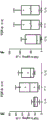
Fig. 1A-B show the characterization of the native tendon for DNA content (fig. 1A) and protein content (fig. 1B) in the tendon prior to treatment of the native patellar and achilles tendons. The measurements depict tendons from a total of 6 donors.
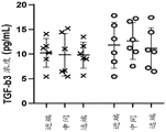
Fig. 2A-B show the native TGF- β concentration based on tendon type and location. The TGF- β 3 concentration (fig. 2A) and TGF- β 1 concentration (fig. 2B) found in the native tendon samples (prior to treatment) are shown.
FIG. 3 shows a comparison of decellularization using DNase and detergent. The DNA content in both the patellar and achilles tendons was measured in native tendons, tendons treated with dnase 50U for 1 hour, tendons treated with dnase 50U for 2 hours, and tendons treated with traditional decellularization methods using SDS or EDTA.
FIG. 4 shows the total tendon protein of tendon samples digested with various enzyme reagents including C-1 collagenase I, C-3 collagenase III, C-1 collagenase I and C-3 collagenase III, and pepsin.
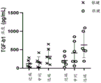
Figure 5 shows TGF- β concentrations before and after tendon processing into acellular tendon matrix. The native tendon was measured by averaging all proximal, medial and distal portions of both the patellar tendon and the achilles tendon.
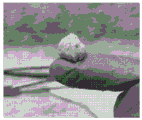
Fig. 6A-B show the elastic properties of the acellular tendon matrix treatment that promotes the ability to stretch (fig. 6A) from an unstretched conformation (fig. 6B) without being torn apart. DTM is storage stable in the form of a sterile lyophilized powder and can be reconstituted as a putty or as an injectable solution. This image shows a DTM putty which can be formed by resuspending lyophilized DTM at 3-5 ul/mg. The putty is pliable/stretchable for surgical application to a desired repair area.
FIGS. 7A-C show that DNase treatment was effective in decellularizing tendon tissue. The tendon was decellularized using dnase at various concentrations (10U, 50U and 100U) over 1 hour. 1 XPBS was used as a control without decellularization. The DNA concentration was determined using DNeasy kit (Qiagen). The data indicate that as little as 50U of DNase can effectively decellularize the tissue.
Figure 8 shows that dnase treatment was as effective as standard detergent methods in decellularizing tendons. 50U of DNase was compared to conventional detergents, 1% SDS and 0.1% EDTA. DNase 50U was tested at 0.5 hours, 1 hour and 2 hours, whereas standard SDS and EDTA protocols required 24 hours of decellularization. DNA concentration was determined using DNEasy kit (Qiagen, n = 3). All values were normalized to be acellular. Tukey's (Tukey's) HSD multiple comparison post-hoc tests showed no significant difference between the different times of DNase treatment or decellularization by DNase compared to between SDS and EDTA.
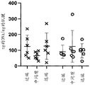
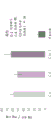
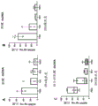
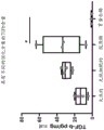
Figures 9A-H show that the achilles tendon matrix has more protein content than the patellar tendon. The achilles tendon and patellar tendon were divided into 1/3 sections consisting of the proximal, central/medial and distal ends of the tendon. (A-D) Total protein of native tendon was measured using BCA protein quantification kit (Thermo Scientific). (E-H) TGF-. beta.was measured using the TGF-. beta.magnetic bead panel Milliplex kit (Millipore Sigma, # TGFBMAG-64K-03). Analysis of variance (ANOVA) showed no statistically significant differences between tendon regions, so the entire tendon could be used by treatment. Total protein was not different when comparing two different tendons (D) (P =0.93), but TGF- β in the achilles tendon (H) was statistically higher than the patellar tendon (P = 0.0045).
Figure 10 shows that filtration effectively eliminated collagenase activity. The decellularized tendon was treated with collagenase to improve the shape factor of DTM. The 100 kDa filter was very effective in eliminating collagenase activity in the final product. Analysis of variance showed significant differences for each group (F (4, 22) =18.06, p < 0.0001). Importantly, there was no significant difference in collagenase activity between the native sample and the 100 kDa filtered sample.
Figure 11 shows that DTM retains more biological activity than the standard method of decellularising tendons with pepsin. After decellularization, the tendon was digested using a solution containing collagenase of the 1 (92.5 g tendon/g collagenase 1) and 3 (185 g tendon/1 g Col 3) type or using pepsin (according to the previously disclosed method (Farnebo et al 2014, PMID: 24341855)). Analysis of variance indicated significant differences between groups, F (3,11) =5.056, p = 0.0193. Tukey's HSD showed a significantly lower TGF- β with pepsin after the fact (P = 0.0249).
FIGS. 12A-C show the difference in cell proliferation when plated on different surfaces. Tissue culture plates were untreated (control, "TC treatment"), coated with collagen or with DTM. Primary tenocytes (ZenBio # TEN-F) were plated at 20,000 cells/well and cell viability was quantified using Presto Blue (Thermo Fisher) either (a) 48 hours or (B) 7 days after plating, resulting in significantly different growth rates (C). (ANOVA = F (3,26) = 10.6, p < 0.0001).
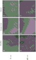
FIGS. 13A-F show the difference in morphology and/or proliferation of cells plated on different surfaces. Tissue culture plates were untreated (control, "TC treatment"), coated with collagen or with DTM. Primary tenocytes (ZenBio # TEN-F) were plated at 20,000 cells/well. Live cell images were taken by time lag photography (time laps video) over 3 days, showing significant differences in cell morphology and proliferation rates between different surface treatments (fig. 6). Still images of live cell images were taken at 48 hours and showed that tenocytes adhered, proliferated more rapidly on DTM with increased adhesion spots and more natural cell morphology (F) compared to standard tissue cultures (D) or collagen coated plates (E).
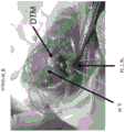
Figures 14A-C show images of a surgical application of the DTM. The DTM may form a putty or an injectable solution. In this case, putty was placed on the larger tubercle and supraspinatus muscles were surgically attached to fix the DTM.
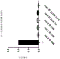
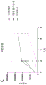
Figure 15 shows the normalized TGFb content of four samples from four different donors over two treatment steps. For each donor, the first column represents the amount of TGFb in native tendon, the second column represents the amount of TGFb in decellularized tendon, and the third column represents the amount of TGFb in digested tendon.
Detailed Description
Unless defined otherwise, all technical and scientific terms used herein have the same meaning as commonly understood by one of ordinary skill in the art to which this invention belongs. All patents and publications mentioned herein are incorporated by reference in their entirety.
Definition of
As used herein, the terms "co-administration", "administration in combination with … …", "administration in combination with … …", "simultaneous" and "simultaneous" encompass the administration of two or more active pharmaceutical ingredients. Co-administration includes simultaneous administration in separate compositions, administration at different times in separate compositions, or administration in a composition in which two or more active pharmaceutical ingredients are present. Preferably, administration is simultaneous in separate compositions and in a composition in which both agents are present.
The term "in vivo" refers to an event that occurs in the body of a subject.
The term "in vitro" refers to an event that occurs in vitro in a subject. In vitro assays include cell-based assays that use live or dead cells, and may also include cell-free assays that do not use whole cells.
As used herein, "treatment", "treating", "alleviating" and "ameliorating" are used interchangeably. These terms refer to methods of achieving a beneficial or desired result, including but not limited to a therapeutic benefit and/or a prophylactic benefit. Therapeutic benefit means eradication or amelioration of the underlying disorder being treated. Moreover, therapeutic benefit is achieved by eradicating or ameliorating one or more physiological symptoms associated with the underlying disorder, such that an improvement is observed in the patient, although the patient may still be afflicted with the underlying disorder.
The term "effective amount" or "therapeutically effective amount" refers to an amount of a compound or combination of compounds as described herein sufficient to effect the intended use, including but not limited to treatment of disease. The therapeutically effective amount may vary depending on the intended use (in vitro or in vivo), or the subject and disease condition being treated (e.g., the weight, age, and sex of the subject), the severity of the disease condition, the mode of administration, and the like, which can be readily determined by one of ordinary skill in the art. The term also applies to doses that induce a specific response (e.g., reduction in platelet adhesion and/or cell migration) in the target cells. The specific dosage will vary depending upon the particular compound selected, the dosage regimen to be followed, whether the compound is administered in combination with other compounds, the time of administration, the tissue to which the compound is administered, and the physical delivery system in which the compound is carried.
The term "therapeutic effect" as used herein includes a therapeutic benefit and/or a prophylactic benefit. Prophylactic effects include delaying or eliminating the appearance of a disease or disorder, delaying or eliminating the onset of symptoms of a disease or disorder, slowing, stopping, or reversing the progression of a disease or disorder, or any combination thereof.
As used herein, "donor" refers to a mammalian source of tendon connective tissue. The donor may be of human or other animal origin, including cadaveric tendon tissue. An "allogeneic" donor tissue is a donor tissue from a non-genetically identical member of the same species, e.g., a donor tissue harvested from one human subject, and the resulting composition then administered to a different human subject. Tendon connective tissue may be harvested from a donor of another species for use in the methods herein to produce an acellular tendon matrix composition; such compositions are "xenogenic" acellular tendon matrix compositions. Preferred sources of xenotransplantation are pigs, horses, cattle, sheep, dogs and rodents. Regardless of the source, the xenograft tendon tissue may be fresh or fresh frozen tissue from a cadaveric donor. Preferred sources of allograft are the achilles tendon and the patellar tendon. These tendons are readily available and relatively large in size. They are also widely used in autograft and allograft applications for the reconstruction of torn or damaged ligaments and tendons.
As used herein, "decellularized" refers to the general (at least 80%), almost complete (at least 95%), or substantially complete (at least 99%) removal of the cellular components of tendon connective tissue.
As used herein, "matrix metalloprotease" refers to a protein of the Matrix Metalloprotease (MMP) family. Matrix Metalloproteinases (MMPs) comprise a large family of zinc-dependent endoproteases that together are capable of degrading all extracellular matrix (ECM) components. The term includes apop-and activated forms of each member of the MMP family. The term includes MMP-2, MMP-9, MMP-14, homologs, derivatives, and fragments thereof. Fanjul-FernandezWait forIn the review of articlesBiochim. Biophy. Acta The mammalian MMP family is summarized in 1803:3-19 (2010).
A variety of growth factors are known in the art, including IGF-1 (insulin-like growth factor 1, or growth regulator C), TGF- β (transforming growth factor β), PDGF (platelet-derived growth factor), VEGF (vascular endothelial growth factor (VEGF), also known as Vascular Permeability Factor (VPF)), bFGF (basic fibroblast growth factor or fibroblast growth factor 2 (FGF2)), GDF-5 (growth differentiation factor 5), GDF-6 (growth differentiation factor 6), GDF-7 (growth differentiation factor 7), HGF (hepatocyte growth factor or scatter factor). Without being bound by theory, a non-limiting list of the above growth factors found in the extracellular matrix of tendons is known in the art.
The phrase "pharmaceutically acceptable" refers to those compounds, materials, compositions, and/or dosage forms which are, within the scope of sound medical judgment, suitable for use in contact with the tissues of human beings and animals without excessive toxicity, irritation, allergic response, or other problem or complication commensurate with a reasonable benefit/risk ratio.
"pharmaceutically acceptable carrier" or "pharmaceutically acceptable excipient" is intended to include any and all solvents, dispersion media, coatings, antibacterial and antifungal agents, isotonic and absorption delaying agents, and inert ingredients. The use of such pharmaceutically acceptable carriers or pharmaceutically acceptable excipients for active pharmaceutical ingredients is well known in the art. Except insofar as any conventional pharmaceutically acceptable carrier or pharmaceutically acceptable excipient is incompatible with the DTM component, its use in the therapeutic compositions of the invention is contemplated. Additional active pharmaceutical ingredients, such as other drugs, may also be added to the compositions and methods described.
When ranges are used herein to describe, for example, physical or chemical properties such as molecular weight or chemical formula, it is intended to include all combinations and subcombinations of ranges and specific embodiments therein. The use of the term "about" when referring to a number or a numerical range means that the number or numerical range referred to is an approximation within experimental variability (or within statistical experimental error), and thus the number or numerical range may vary. This variation is generally from 0% to 15%, preferably from 0% to 10%, more preferably from 0% to 5% of the number or range of values. The term "comprising" (and related terms such as "comprises" or "comprising" or "having" or "including") includes those embodiments, such as, for example, embodiments of any composition of matter, method, or process that "consists of" or "consists essentially of" the described features.
The terms "sequence identity", "percent identity" and "percent sequence identity" in the context of two or more nucleic acids or polypeptides refer to two or more sequences or subsequences that are the same or have a specified percentage of nucleotides or amino acid residues that are the same, regardless of any conservative amino acid substitutions as part of the sequence identity, when compared and aligned (gaps introduced, if necessary) to obtain the maximum correspondence. The percent identity can be measured using sequence comparison software or algorithms or by visual inspection. Various algorithms and software that can be used to obtain an alignment of amino acid or nucleotide sequences are known in the art. Suitable programs for determining percent sequence identity include, for example, the BLAST suite of programs available from the National Center for Biotechnology Information BLAST website of the U.S. Government (U.S. Goverment's National Center for Biotechnology BLAST website). Comparisons between two sequences can be made using the BLASTN or BLASTP algorithms. BLASTN is used to compare nucleic acid sequences, while BLASTP is used to compare amino acid sequences. ALIGN, ALIGN-2 (Genentech, South San Francisco, California), or MegAlign, available from DNASTAR, are additional publicly available software programs that can be used to ALIGN sequences. One skilled in the art can determine the appropriate parameters for maximum alignment by specific alignment software. In certain embodiments, default parameters of the alignment software are used.
For the avoidance of doubt, a particular feature (e.g. integer, feature, value, use, disease, formula, compound or group) described in connection with a particular aspect, embodiment or example of the invention is intended to be understood as applying to any other aspect, embodiment or example described herein unless incompatible therewith. Such features may therefore be used, where appropriate, in combination with any definitions, claims or embodiments defined herein. All of the features disclosed in this specification (including any accompanying claims, abstract and drawings), and/or all of the steps of any method or process so disclosed, may be combined in any combination, except combinations where at least some of such features and/or steps are mutually exclusive. The invention is not limited to any details of any disclosed embodiment. The invention extends to any novel one, or any novel combination, of the features disclosed in this specification (including any accompanying claims, abstract and drawings), or to any novel one, or any novel combination, of the steps of any method or process so disclosed.
Method for preparing acellular tendon matrix
It is an object of embodiments of the present disclosure to produce DTMs that retain growth factors, particularly TGF- β, in the matrix by developing a gentle and specific decellularization and digestion protocol. Detergents are traditionally irritating and can remove or denature proteins as well as cellular material.
Typical digestion techniques for acellular matrices use a general purpose protease, most commonly pepsin, which indiscriminately cleaves all proteins into small polypeptides. In this application, enzymes dedicated to the cleavage of collagen are used to break down the tendon into smaller parts, which can then form self-assembling peptides. Collagen, mainly type I collagen, forms the structural skeleton of the tendon. By specifically cleaving collagen, we digested the tendon, but retained the biological activity of the attached growth factor.
Collagenase is an endopeptidase that digests the triple helix of native collagen fibers common in tendons. Collagenase cleaves the bond between the neutral amino acid (X) and glycine in the sequence Pro-X-Gly-Pro (found at high frequency in collagen). Bacterial collagenases, such as those produced by clostridium histolyticum, can attack almost all types of collagen and degrade both native collagen, which is insoluble in water, and denatured collagen, which is soluble in water. The ability of clostridial collagenase to digest native triple helix I, II and type III collagen through multiple cuts in the triple helix is a major distinguishing factor. Clostridial collagenases represent an unusually large metalloprotease, a family of which shares a zinc-containing motif in the center of the active site (Gonzales and Robert-Baudouy 1996).
Matrix Metalloproteinases (MMPs) also have the ability to cleave collagen fibers in a very specific sequence. Due to their triple helical structure, I, II and type III interstitial collagen are highly resistant to proteolytic attack, but can be cleaved by MMP collagenases at specific sites. MMP-2 and-9 are closely related at the structural level and have been shown to have collagenase activity against type I and type III collagen, producing classical 3/4 and 1/4 fragments. MMP-1, MMP-8, MMP-13, MT-MMPs also have some limited collagenase activity.
In one aspect, the invention provides a method of producing a composition comprising Matrix Metalloproteinase (MMP) digested tendon tissue, an antimicrobial agent, and a sterile aqueous carrier solution. In some embodiments, the Matrix Metalloprotease (MMP) is selected from MMP-2, MMP-9, MMP-14, or a combination thereof. In one aspect, MMPs are designed to be constitutively active. Those skilled in the art will appreciate that other MMPs may be used. Collagenase, gelatinase, stromelysin and membrane-type MMPs (MT-MMPs) can be used. In certain embodiments, collagenase may be used to decellularize and/or digest a tendon that is decellularized. As described herein, collagenase is capable of degrading triple helical fibril collagen into distinct 3/4 and 1/4 fragments. These collagens are the main components of bone, cartilage and dentin. The collagenases include collagenase type 1, collagenase type 2, collagenase type 3, collagenase type 8, collagenase type 13, collagenase type 14, and collagenase type 18. Non-limiting examples of one or more MMPs that may be used include MMP1 (interstitial collagenase, CLG, CLGN), MMP2 (gelatinase-A, 72 kDa gelatinase), MMP3 (stromelysin 1, CHDS6, MMP-3, SL-1, STMY1, STR1), MMP7 (stromelysin, PUMP 1, MMP-7, MPSL1, PUMP-1), MMP8 (neutrophil collagenase, CLG1, HNC, MMP-8, PMNL-CL), MMP9 (gelatinase-B, 92 kDa gelatinase, CLG4B, GELB, MANDP2, MMP-9), MMP10 (stromelysin 2, SL-2, STMY2), MMP11 (stromelysin 3, SL-3, ST3, STMY3), MMP12 (macrophage elastase, HME, ME, MME, MMP12, MMP-583, MMP 24-MMP 573 24, MMP 573-573 13, MMP-24 DP-24, MMP-DP 573), MMP14 (MT 14-MMP, MMP-14, MMP-X14, MT-MMP 1, MT 14-MMP, MT1-MMP, MT 14, WNCHRS, MMP14 (MT 14-MMP, MTMMP 14, SMCP-2, MMP-15, MT2MMP), MMP14 (MT 14-MMP, C8orf 14, MMP-X14, MT-MMP 14, MT 14-MMP), MMP14 (MT 14-MMP, MMP-17, MT4MMP, MTMMP 14), MMP14 (collagenase 4, xcol 14, toad collagenase), MMP14 (RASI-1, sometimes called stromelysin-4, MMP14, RASI-1, CODA), MMP 72 (enamel lysin, AI2A, MMP-20, MMP-14, MMP-MMP (HTFR 14, MMP-14, MMP 23-MMP 23, MMP-MMP (MIFR-14, MMP-14-MMP 23), MMP 14-MMP (MIFR-14, MMP-14, MMP-MMP 23, MMP-14-MMP (MIFR-14, MMP-14, MMP-MMP) MMP-24, MMP25, MT-MMP5, MT-MMP5, MT5-MMP, MT5MMP, MTMMP5), MMP25 (MT6-MMP, MMP-25, MMP20, MMP20A, MMPL1, MT-MMP6, MT-MMP6, MT6-MMP, MT6MMP, MTMMP6), MMP26 (matrix dissolution factor-2, endometese), MMP27 (MMP-22, C-MMP, MMP-27), and MMP28 (Epilysin, EPILYSIN, MM28, MMP-25, MMP-28).
The concentration of collagenase used to enzymatically digest the decellularized tendon may vary depending on the particular collagenase used. In certain embodiments, collagenase type 1 can be used to enzymatically digest decellularized tendons. In certain embodiments, collagenase type 3 can be used to enzymatically digest decellularized tendons. The collagenase concentration used to enzymatically digest the decellularized tendon may be about 0.1 mg (mg)/mL, about 0.2 mg/mL, about 0.3 mg/mL, about 0.4 mg/mL, about 0.5 mg/mL, about 0.6 mg/mL, about 0.7 mg/mL, about 0.8 mg/mL, about 0.9 mg/mL, about 1.0 mg/mL, about 1.1 mg/mL, about 1.2 mg/mL, about 1.3 mg/mL, about 1.4 mg/mL, about 1.5 mg/mL, about 1.6 mg/mL, about 1.7 mg/mL, about 1.8 mg/mL, about 1.9 mg/mL, about 2.0 mg/mL, about 2.1 mg/mL, about 2.2 mg/mL, about 2.3 mg/mL, about 2.4 mg/mL, about 2.5 mg/mL, about 2.6 mg/mL, About 2.7 mg/mL, about 2.8 mg/mL, about 2.9 mg/mL, about 3.0 mg/mL, about 3.1 mg/mL, about 3.2 mg/mL, about 3.3 mg/mL, about 3.4 mg/mL, about 3.5 mg/mL, about 3.6 mg/mL, about 3.7 mg/mL, about 3.8 mg/mL, about 3.9 mg/mL, about 4.0 mg/mL, about 5.0 mg/mL, about 6.0 mg/mL, about 7.0 mg/mL, about 8.0 mg/mL, about 9.0 mg/mL, or about 10.0 mg/mL. In certain embodiments, the collagenase concentration used to enzymatically digest the decellularized tendon is about 1.0 mg/mL. In other embodiments, the collagenase concentration used to enzymatically digest the decellularized tendon is about 2.0 mg/mL.
Antimicrobial agents are suitable agents for parenteral formulations, for example alkyl or aryl alcohols, such as benzyl alcohol, chlorobutanol or 2-ethoxyethanol. Aminoarylates are also suitable, such as methyl, ethyl, propyl or butyl parabens and combinations thereof. Alkyl and aryl acids may also be suitable, such as benzoic or sorbic acid; biguanides, for example chlorhexidine or phenols, for example phenol or 3-cresol. In some embodiments, a combination of chemically compatible antimicrobial agents is used.
In one aspect, the present invention provides an acellular tendon matrix (DTM) composition, wherein the DTM composition is prepared by a method comprising the steps of: (i) mincing tendon tissue samples; (ii) decellularizing the minced tendon tissue sample; (iii) digestion; and (iv) lyophilization.
In another aspect, the present invention provides an acellular tendon matrix (DTM) composition, wherein the DTM composition is prepared by a method comprising the steps of: (i) mincing tendon tissue samples; (ii) decellularizing the minced tendon tissue sample; (iii) milling; (iv) digestion; (v) stopping and neutralizing; (vi) washing; and (vii) lyophilizing.
In some cases, the tendon matrix may be present as a fraction of about 40%, about 45%, about 50%, about 55%, about 60%, about 65%, about 70%, about 75%, about 80%, about 85%, about 90%, about 95%, or about 99% by weight of the isolated tendon tissue prior to decellularization, milling, digestion, lyophilization, and/or washing.
In some cases, prior to decellularization, milling, digestion, lyophilization, and/or washing, the tendon matrix may be present in a fraction of isolated tendon tissue of about 50% to about 90%, about 50% to about 80%, about 50% to about 70%, about 50% to about 60%, about 50% to about 55%, 60% to about 90%, about 60% to about 80%, about 60% to about 70%, about 60% to about 65%, 70% to about 90%, about 70% to about 80%, about 70% to about 75%, 80% to about 90%, about 80% to about 85%, or about 85% to about 90% by weight.
In some cases, the tendon matrix may be present in less than about 90%, less than about 85%, less than about 80%, less than about 75%, less than about 70%, less than about 65%, less than about 60%, less than about 55%, less than about 50%, less than about 45%, less than about 40%, less than about 35%, less than about 30%, less than about 25%, less than about 20%, less than about 15%, or less than about 10% by weight of the isolated tendon tissue prior to decellularization, milling, digestion, lyophilization, and/or washing.
In some cases, the tendon matrix may be present as about 40%, about 45%, about 50%, about 55%, about 60%, about 65%, about 70%, about 75%, about 80%, about 85%, about 90%, about 95%, or about 99% by volume of the isolated tendon tissue prior to decellularization, milling, digestion, lyophilization, and/or washing.
In some cases, prior to decellularization, milling, digestion, lyophilization, and/or washing, the tendon matrix may be present in a fraction of isolated tendon tissue of about 50% to about 90%, about 50% to about 80%, about 50% to about 70%, about 50% to about 60%, about 50% to about 55%, 60% to about 90%, about 60% to about 80%, about 60% to about 70%, about 60% to about 65%, 70% to about 90%, about 70% to about 80%, about 70% to about 75%, 80% to about 90%, about 80% to about 85%, or about 85% to about 90% by volume.
In some cases, the tendon matrix may be present in less than about 90%, less than about 85%, less than about 80%, less than about 75%, less than about 70%, less than about 65%, less than about 60%, less than about 55%, less than about 50%, less than about 45%, less than about 40%, less than about 35%, less than about 30%, less than about 25%, less than about 20%, less than about 15%, or less than about 10% by volume of the isolated tendon tissue prior to decellularization, milling, digestion, lyophilization, and/or washing.
In some cases, after decellularization, milling, digestion, lyophilization, and/or washing, the tendon matrix may be present in about 40%, about 45%, about 50%, about 55%, about 60%, about 65%, about 70%, about 75%, about 80%, about 85%, about 90%, about 95%, or about 99% by weight of the decellularized, milled, digested, lyophilized, and/or washed portion of the tendon tissue.
In some cases, after decellularization, milling, digestion, lyophilization, and/or washing, the tendon matrix may be present in a fraction of decellularized, milled, digested, lyophilized, and/or washed tendon tissue of about 50% to about 90%, about 50% to about 80%, about 50% to about 70%, about 50% to about 60%, about 50% to about 55%, 60% to about 90%, about 60% to about 80%, about 60% to about 65%, 70% to about 90%, about 70% to about 80%, about 70% to about 75%, 80% to about 90%, about 80% to about 85%, or about 85% to about 90% by weight.
In some cases, after decellularization, milling, digestion, lyophilization, and/or washing, the tendon matrix may be present in a portion of the decellularized, milled, digested, lyophilized, and/or washed tendon tissue greater than about 99%, greater than about 95%, greater than about 90%, greater than about 85%, greater than about 80%, greater than about 75%, greater than about 70%, greater than about 65%, greater than about 60%, greater than about 55%, greater than about 50%, greater than about 45%, greater than about 40%, greater than about 35%, greater than about 30%, greater than about 25%, greater than about 20%, greater than about 15%, or greater than about 10% by weight.
In some cases, after decellularization, milling, digestion, lyophilization, and/or washing, the tendon matrix may be present in a portion of about 40%, about 45%, about 50%, about 55%, about 60%, about 65%, about 70%, about 75%, about 80%, about 85%, about 90%, about 95%, or about 99% by volume of the decellularized, milled, digested, lyophilized, and/or washed tendon tissue.
In some cases, after decellularization, milling, digestion, lyophilization, and/or washing, the tendon matrix may be present in a fraction of about 50% to about 90%, about 50% to about 80%, about 50% to about 70%, about 50% to about 60%, about 50% to about 55%, 60% to about 90%, about 60% to about 80%, about 60% to about 70%, about 60% to about 65%, 70% to about 90%, about 70% to about 80%, about 70% to about 75%, 80% to about 90%, about 80% to about 85%, or about 85% to about 90% by volume of the decellularized, milled, digested, lyophilized, and/or washed tendon tissue.
In some cases, after decellularization, milling, digestion, lyophilization, and/or washing, the tendon matrix may be present in a portion of the decellularized, milled, digested, lyophilized, and/or washed tendon tissue greater than about 99%, greater than about 95%, greater than about 90%, greater than about 85%, greater than about 80%, greater than about 75%, greater than about 70%, greater than about 65%, greater than about 60%, greater than about 55%, greater than about 50%, greater than about 45%, greater than about 40%, greater than about 35%, greater than about 30%, greater than about 25%, greater than about 20%, greater than about 15%, or greater than about 10% by volume.
In one aspect, the decellularizing step comprises exposing the minced tendon tissue sample to a solution comprising one or more components selected from a chaotropic salt, a non-ionic detergent, a zwitterionic detergent, a cationic detergent, an anionic detergent, or a combination thereof. In some aspects, the decellularization step comprises one or more freeze/thaw cycles. In some aspects, the decellularizing step further comprises treatment with dnase and/or rnase. In some aspects, the decellularizing step further comprises one or more washes in a balanced salt solution, such as hank's balanced salt solution in phosphate buffered saline.
In some embodiments, minced tendon tissue samples are rinsed in ultrapure water and then decellularized using a solution containing 1% w/v Sodium Dodecyl Sulfate (SDS) under moderate agitation. In some embodiments, moderate agitation is intermittent.
In another aspect, the minced tendon tissue sample is decellularized using a solution comprising one or more of an ionic detergent, a non-ionic detergent, an anionic detergent, or a cationic detergent. In some aspects, the decellularization solution further comprises a chaotropic salt. In some embodiments, the chaotropic salt is urea. In some embodiments, the decellularization solution comprises from 0.5M urea to 8M urea. In some embodiments, the decellularization solution comprises 2M to 5M urea. In some embodiments, the decellularization solution comprises about 3M urea.
In some aspects, the decellularization solution comprises a surfactant and a chaotropic salt. In some aspects, the decellularization solution further comprises an antifoaming agent, such as Antifoam 204.
In another aspect, the method further comprises the step of precipitating cellular proteins, the method further comprising treating the minced tendon tissue sample with a concentrated cosmotropic solution. In some embodiments, the concentrated cosmotropic solution is ammonium sulfate. Cosmotropic salting out is for example performed according to Wingfield,Curr. Protoc. Protein Sci.appendix 3: completion of the procedure summarized in appendix-3F (2001).
The chopping may be performed by those skilled in the artKnown methods are used to accomplish this, for example, by first removing sheath, adipose and synovial tissue from a tendon tissue sample. Then, tendon tissue specimens were cut into about 1 to 4mm3Size blocks, then washed with Phosphate Buffered Saline (PBS).
In one aspect, the stopping and neutralizing step comprises stopping and neutralizing with a solution comprising one or more protease inhibitors selected from the group consisting of TAPI-0, TAPI-1, TAPI-2, marimastat, phosphoramidon, luteolin, PMSF, pepstatin A, leupeptin, E-64, sodium orthovanadate, or combinations thereof.
Decellularization can be monitored by methods known in the art, including sectioning the decellularized and control samples (i.e., untreated samples of the starting donor tendon tissue), and then detecting the cellular components and collagen fiber structure with hematoxylin-eosin staining and masson-goddner trichrome staining, respectively. DNA may be extracted from the decellularized sample and the untreated starting sample; the decellularized sample should have at most one-fourth of the equivalent starting weight of DNA recovered, see, e.g., Seif-Naraghi et al,Acta Biomater.8:3695-3703 (2012)。
decellularized tissue has extracellular matrix (ECM) components of all or most of the tissue regions, including ECM components of the vascular tree. The ECM component may include any one or any combination of the following: fibronectin, fibrillin, laminin, elastin, members of the collagen family (e.g., collagen I, III and IV), ECM-associated growth proteins (including growth factors and cytokines), glycosaminoglycans, matrices, reticulocytes, and thrombospondin, which can maintain a defined tissue structure, such as the basal lamina. Successful decellularization can be defined as no detectable myofilaments, endothelial cells, smooth muscle cells, and nuclei or removal of more than 97% of detectable DNA in tissue sections (e.g., as measured by fluorimetry) using standard histological staining procedures. Residual cellular debris can be removed from the decellularized tissue.
The morphology and structure of the ECM can be maintained during and after the decellularization process. As used herein, "morphology" refers to the overall shape of the ECM, while "structure" as used herein refers to the outer surface, the inner surface, and the ECM therebetween. The morphology and structure of the ECM can be examined visually and/or histologically.
One or more compounds may be applied to or on the decellularized tissue, for example, to preserve the decellularized tissue, or to prepare the decellularized tissue for recellularization or integration or implantation into a host. Such compounds include, but are not limited to, one or more growth factors (e.g., VEGF, DKK-1, FGF, BMP-1, BMP-4, SDF-1, IGF, and HGF), immunomodulators (e.g., cytokines, glucocorticoids, IL2R antagonists, leukotriene antagonists), and/or factors that alter the coagulation cascade (e.g., aspirin, heparin binding protein, and heparin). In addition, the decellularized tissue can be further treated with, for example, radiation (e.g., UV, gamma) to reduce or eliminate the presence of any type of microorganism remaining on or in the decellularized tissue.
In some aspects, the present invention provides a method of preparing an acellular tendon matrix (DTM) composition, the composition prepared using the method further comprising retaining at least 100%, at least 99%, at least 98%, at least 97%, at least 96%, at least 95%, at least 94%, at least 93%, at least 92%, at least 91%, at least 90% of the growth factors present in the minced tendon tissue. In some aspects, the composition is prepared using a method of preparing a Decellularized Tendon Matrix (DTM) composition, the composition prepared using the method further comprising retaining at least 85%, at least 80%, at least 75%, at least 70%, at least 65%, at least 60%, at least 55%, at least 50%, at least 45%, or at least 40% of the growth factors present in the minced tendon tissue. In some aspects, the present invention provides a method of preparing a Decellularized Tendon Matrix (DTM) composition, the composition prepared using the method further comprising retaining at least 99%, at least 98%, at least 97%, at least 96%, at least 95%, at least 94%, at least 93%, at least 92%, at least 91%, at least 90%, at least 89%, at least 88%, at least 87%, at least 86%, at least 85%, at least 84%, at least 83%, at least 82%, at least 81%, at least 80%, at least 79%, at least 78%, at least 77%, at least 76%, at least 75%, at least 74%, at least 73%, at least 72%, at least 71%, at least 70%, at least 69%, at least 68%, at least 67%, at least 66%, at least 65%, at least 64%, at least 63%, at least 62%, at least 61%, at least 60%, (ii) present in the minced tendon tissue, At least 59%, at least 58%, at least 57%, at least 56%, at least 55%, at least 54%, at least 53%, at least 52%, at least 51%, at least 50%, at least 49%, at least 48%, at least 47%, at least 46%, at least 45%, at least 44%, at least 43%, at least 42%, at least 41%, at least 40%, at least 39%, at least 38%, at least 37%, at least 36%, at least 35%, at least 34%, at least 33%, at least 32%, at least 31%, at least 31%, at least 30%, at least 29%, at least 28%, at least 27%, at least 26%, at least 25%, at least 24%, at least 23%, at least 22%, at least 21%, at least 20%, at least 19%, at least 18%, at least 17%, at least 16%, at least 15%, at least 14%, at least 13%, at least 12%, at least 11%, or at least 10% of a growth factor. In some aspects, the present invention provides a method of preparing an acellular tendon matrix (DTM) composition, wherein the composition prepared using the method retains from about 70% to about 100% of the growth factors present in the pre-acellular minced tendon tissue. In some aspects, the present invention provides a method of preparing an acellular tendon matrix (DTM) composition, wherein the composition prepared using the method retains from about 70% to about 75% of the growth factors present in the pre-acellular minced tendon tissue. In some aspects, the present invention provides a method of preparing an acellular tendon matrix (DTM) composition, wherein the composition prepared using the method retains from about 75% to about 80% of the growth factors present in the pre-acellular minced tendon tissue. In some aspects, the present invention provides a method of preparing an acellular tendon matrix (DTM) composition, wherein the composition prepared using the method retains from about 80% to about 85% of the growth factors present in the pre-acellular minced tendon tissue. In some aspects, the present invention provides a method of preparing an acellular tendon matrix (DTM) composition, wherein the composition prepared using the method retains from about 85% to about 90% of the growth factors present in the pre-acellular minced tendon tissue. In some aspects, the present invention provides a method of preparing an acellular tendon matrix (DTM) composition, wherein the composition prepared using the method retains from about 90% to about 95% of the growth factors present in the pre-acellular minced tendon tissue. In some aspects, the present invention provides a method of preparing an acellular tendon matrix (DTM) composition, wherein the composition prepared using the method retains from about 95% to about 100% of the growth factors present in the pre-acellular minced tendon tissue. In some aspects, the present invention provides a method of preparing an acellular tendon matrix (DTM) composition, wherein the composition prepared using the method retains from about 75% to about 95% of the growth factors present in the pre-acellular minced tendon tissue. In some aspects, the present invention provides a method of preparing an acellular tendon matrix (DTM) composition, wherein the composition prepared using the method retains from about 70% to about 80% of the growth factors present in the pre-acellular minced tendon tissue. In some aspects, the present invention provides a method of preparing an acellular tendon matrix (DTM) composition, wherein the composition prepared using the method retains from about 80% to about 90% of the growth factors present in the pre-acellular minced tendon tissue. In some embodiments, the growth factor is selected from the group consisting of IGF-1, TGF- β, PDGF, VEGF, bFGF, GDF-5, GDF-6, GDF-7, HGF, and combinations thereof. In some embodiments, the growth factor comprises at least TGF- β.
In some aspects, the present invention provides a method of preparing an acellular tendon matrix (DTM) composition, the method further comprising retaining at least 90% of the cytokines present in the minced tendon tissue, wherein the growth factors are selected from the group consisting of: IGF-1, TGF-beta, PDGF, VEGF, bFGF, GDF-5, GDF-6, GDF-7, HGF, and combinations thereof. In some aspects, a method of making a Decellularized Tendon Matrix (DTM) composition further comprises retaining at least 99%, at least 98%, at least 97%, at least 96%, at least 95%, at least 94%, at least 93%, at least 92%, at least 91%, at least 90%, at least 89%, at least 88%, at least 87%, at least 86%, at least 85%, at least 84%, at least 83%, at least 82%, at least 81%, at least 80%, at least 79%, at least 78%, at least 77%, at least 76%, at least 75%, at least 74%, at least 73%, at least 72%, at least 71%, at least 70%, at least 69%, at least 68%, at least 67%, at least 66%, at least 65%, at least 64%, at least 63%, at least 62%, at least 61%, at least 60%, at least 59%, at least 58%, at least 57%, or the amount present in minced tendon tissue, At least 56%, at least 55%, at least 54%, at least 53%, at least 52%, at least 51%, at least 50%, at least 49%, at least 48%, at least 47%, at least 46%, at least 45%, at least 44%, at least 43%, at least 42%, at least 41%, at least 40%, at least 39%, at least 38%, at least 37%, at least 36%, at least 35%, at least 34%, at least 33%, at least 32%, at least 31%, at least 30%, at least 29%, at least 28%, at least 27%, at least 26%, at least 25%, at least 24%, at least 23%, at least 22%, at least 21%, at least 20%, at least 19%, at least 18%, at least 17%, at least 16%, at least 15%, at least 14%, at least 13%, at least 12%, at least 11%, or at least 10% of a growth factor selected from IGF-1, at least 54%, at least, TGF-beta, PDGF, VEGF, bFGF, GDF-5, GDF-6, GDF-7, HGF, and combinations thereof.
In some aspects, the invention provides a method of making an acellular tendon matrix (DTM) composition, the method further comprising retaining at least 90% of TGF- β present in the minced tendon tissue. In some aspects, the invention provides a method of making an acellular tendon matrix (DTM) composition, the method further comprising retaining at least 95% of TGF- β present in the minced tendon tissue. In some aspects, the invention provides a method of making an acellular tendon matrix (DTM) composition, the method further comprising retaining at least 99% of TGF- β present in the minced tendon tissue. In some aspects, a method of making a Decellularized Tendon Matrix (DTM) composition further comprises retaining at least 99%, at least 98%, at least 97%, at least 96%, at least 95%, at least 94%, at least 93%, at least 92%, at least 91%, at least 90%, at least 89%, at least 88%, at least 87%, at least 86%, at least 85%, at least 84%, at least 83%, at least 82%, at least 81%, at least 80%, at least 79%, at least 78%, at least 77%, at least 76%, at least 75%, at least 74%, at least 73%, at least 72%, at least 71%, at least 70%, at least 69%, at least 68%, at least 67%, at least 66%, at least 65%, at least 64%, at least 63%, at least 62%, at least 61%, at least 60%, at least 59%, at least 58%, by weight, of a native tendon (native tendon) At least 57%, at least 56%, at least 55%, at least 54%, at least 53%, at least 52%, at least 51%, at least 50%, at least 49%, at least 48%, at least 47%, at least 46%, at least 45%, at least 44%, at least 43%, at least 42%, at least 41%, at least 40%, at least 39%, at least 38%, at least 37%, at least 36%, at least 35%, at least 34%, at least 33%, at least 32%, at least 31%, at least 30%, at least 29%, at least 28%, at least 27%, at least 26%, at least 25%, at least 24%, at least 23%, at least 22%, at least 21%, at least 20%, at least 19%, at least 18%, at least 17%, at least 16%, at least 15%, at least 14%, at least 13%, at least 12%, at least 11%, or at least 10% TGF- β.
In some aspects, a method of making an acellular tendon matrix (DTM) composition further comprises retaining at least 85%, at least 80%, at least 75%, at least 70%, at least 65%, at least 60%, at least 55%, at least 50%, at least 45%, or at least 40% of a growth factor present in the minced tendon tissue, wherein the growth factor is selected from the group consisting of IGF-1, TGF- β, PDGF, VEGF, bFGF, GDF-5, GDF-6, GDF-7, HGF, and combinations thereof.
In some aspects, a method of making a Decellularized Tendon Matrix (DTM) composition, the method further comprising increasing a concentration of a growth factor present in the decellularized tissue or the DTM by at least 500%, at least 250%, at least 200%, at least 150%, at least 100%, at least 95%, at least 90%, at least 85%, at least 80%, at least 75%, at least 70%, at least 65%, at least 60%, at least 55%, at least 50%, at least 45%, at least 40%, at least 35%, at least 30%, at least 25%, at least 20%, at least 15%, at least 10%, at least 5%, wherein the growth factor is selected from the group consisting of IGF-1, TGF- β, PDGF, VEGF, bFGF, GDF-5, GDF-6, GDF-7, HGF, and combinations thereof.
In one aspect, the composition retains 2 or more of the above growth factors, 3 or more of the above growth factors, 4 or more of the above growth factors, 5 or more of the above growth factors, 6 or more of the above growth factors, 7 or more of the above growth factors. In one aspect, the composition retains HGF and one or more growth factors selected from IGF-1, TGF- β, PDGF, VEGF, bFGF, GDF-5, GDF-6 and GDF-7. In one aspect, the composition retains IGF-1 and HGF.
In one aspect, the DTM composition further comprises retaining at least 85%, at least 84%, at least 83%, at least 82%, at least 81%, at least 80%, at least 79%, at least 78%, at least 77%, at least 76%, at least 75%, at least 74%, at least 73%, at least 72%, at least 71%, at least 70%, at least 69%, at least 68%, at least 67%, at least 66%, at least 65%, at least 64%, at least 63%, at least 62%, at least 61%, at least 60%, at least 59%, at least 58%, at least 57%, at least 56%, at least 55%, at least 54%, at least 53%, at least 52%, at least 51%, at least 50%, at least 49%, at least 48%, at least 47%, at least 46%, at least 45%, at least 44%, at least 43%, at least 42%, at least 41%, at least 40%, (ii) present in the minced tendon tissue, At least 39%, at least 38%, at least 37%, at least 36%, at least 35%, at least 34%, at least 33%, at least 32%, at least 31%, at least 30%, at least 29%, at least 28%, at least 27%, at least 26%, at least 25%, at least 24%, at least 23%, at least 22%, at least 21%, at least 20%, at least 19%, at least 18%, at least 17%, at least 16%, at least 15%, at least 14%, at least 13%, at least 12%, at least 11%, or at least 10% IGF-1 and HGF.
In some aspects, the present invention provides a method of preparing an acellular tendon matrix (DTM) composition, further comprising removing at least 90% of the cellular material present in the minced tendon tissue. In some aspects, the present invention provides a method of preparing an acellular tendon matrix (DTM) composition, further comprising removing at least 95% of the cellular material present in the minced tendon tissue. In some aspects, the present invention provides a method of preparing an acellular tendon matrix (DTM) composition, further comprising removing at least 99% of the cellular material present in the minced tendon tissue. In some aspects, a method of making a Decellularized Tendon Matrix (DTM) composition further comprises removing at least 99%, at least 98%, at least 97%, at least 96%, at least 95%, at least 94%, at least 93%, at least 92%, at least 91%, at least 90%, at least 89%, at least 88%, at least 87%, at least 86%, at least 85%, at least 84%, at least 83%, at least 82%, at least 81%, at least 80%, at least 79%, at least 78%, at least 77%, at least 76%, at least 75%, at least 74%, at least 73%, at least 72%, at least 71%, at least 70%, at least 69%, at least 68%, at least 67%, at least 66%, at least 65%, at least 64%, at least 63%, at least 62%, at least 61%, at least 60%, at least 59%, at least 58%, at least 57%, or, At least 56%, at least 55%, at least 54%, at least 53%, at least 52%, at least 51%, at least 50%, at least 49%, at least 48%, at least 47%, at least 46%, at least 45%, at least 44%, at least 43%, at least 42%, at least 41%, at least 40%, at least 39%, at least 38%, at least 37%, at least 36%, at least 35%, at least 34%, at least 33%, at least 32%, at least 31%, at least 30%, at least 29%, at least 28%, at least 27%, at least 26%, at least 25%, at least 24%, at least 23%, at least 22%, at least 21%, at least 20%, at least 19%, at least 18%, at least 17%, at least 16%, at least 15%, at least 14%, at least 13%, at least 12%, at least 11%, or at least 10% of cellular material. In certain embodiments, the present invention provides a method of making an acellular tendon matrix (DTM) composition, and the DTM is substantially free of cellular material. In certain embodiments, the present invention provides a method of making an acellular tendon matrix (DTM) composition, and the DTM is substantially free of TGF- β producing cells.
In some aspects, the invention provides a method of preparing an acellular tendon matrix (DTM) composition, further comprising removing at least 90% of the nucleic acids (e.g., DNA or RNA) present in the minced tendon tissue. In some aspects, the invention provides a method of preparing an acellular tendon matrix (DTM) composition, further comprising removing at least 95% of the nucleic acids (e.g., DNA or RNA) present in the minced tendon tissue. In some aspects, the invention provides a method of preparing an acellular tendon matrix (DTM) composition, further comprising removing at least 99% of the nucleic acids (e.g., DNA or RNA) present in the minced tendon tissue. In some aspects, a method of making a Decellularized Tendon Matrix (DTM) composition further comprises removing at least 99%, at least 98%, at least 97%, at least 96%, at least 95%, at least 94%, at least 93%, at least 92%, at least 91%, at least 90%, at least 89%, at least 88%, at least 87%, at least 86%, at least 85%, at least 84%, at least 83%, at least 82%, at least 81%, at least 80%, at least 79%, at least 78%, at least 77%, at least 76%, at least 75%, at least 74%, at least 73%, at least 72%, at least 71%, at least 70%, at least 69%, at least 68%, at least 67%, at least 66%, at least 65%, at least 64%, at least 63%, at least 62%, at least 61%, at least 60%, at least 59%, at least 58%, at least 57%, or, At least 56%, at least 55%, at least 54%, at least 53%, at least 52%, at least 51%, at least 50%, at least 49%, at least 48%, at least 47%, at least 46%, at least 45%, at least 44%, at least 43%, at least 42%, at least 41%, at least 40%, at least 39%, at least 38%, at least 37%, at least 36%, at least 35%, at least 34%, at least 33%, at least 32%, at least 31%, at least 30%, at least 29%, at least 28%, at least 27%, at least 26%, at least 25%, at least 24%, at least 23%, at least 22%, at least 21%, at least 20%, at least 19%, at least 18%, at least 17%, at least 16%, at least 15%, at least 14%, at least 13%, at least 12%, at least 11%, or at least 10% of a nucleic acid (e.g., DNA or RNA). In certain embodiments, the present invention provides a method of making an acellular tendon matrix (DTM) composition, and the DTM is substantially free of nucleic acids (e.g., DNA or RNA).
Various methods are known in the art, such as those described by Gilpin and Yang,Biomed. Res. Int.2017: 9831534 (2017). Many methods include intensive detergent extraction and prolonged treatment with promiscuous proteases (e.g. pepsin) at extreme non-physiological pH. The methods and processes of the present invention differ from those known in the art by using fewer promiscuous proteases that are active at physiological pH. Without being bound by theory, the methods and processes of the present invention are less protein denatured and retain more functional growth factors in the acellular tendon matrix. In some aspects, MMP2, MMP9, MMP14, or a combination thereof are used in the preparation of the acellular tendon matrix compositions of the present invention. Eckhard et al, who are,Data Brief299-310 (2017) mapped target cleavage sites for the MMP family (including MMP2, MMP9, and MMP 14) using the whole proteomic approach.
DTM hydrogel
In another aspect, the present disclosure provides an acellular tendon matrix hydrogel. Hydrogels can be prepared by exploiting the intrinsic polymerization ability of pepsin treated monomeric collagen by controlling temperature or pH. These methods are well known, but some are unpredictable, e.g. Drake et al,Biochemistry 5:301-312 (1966) details the preparation of polymerizable proteolytic fragments of collagen. Other methods, such as Bahney et al,FASEB J1486-1496 (2011) and Ungerleider et al,Methods84:53-59 (2015), are also well known. These well-known methods are particularly unpredictable when applied to protein-rich extracellular matrix tissues.
More reliable and better controlled crosslinking can be achieved by using carbodiimide crosslinker chemistry. In some embodiments, the hydrogel is prepared by mixing the DTM composition and reacting with a carboxyl-reactive crosslinking agent, such as 1-ethyl-3- (3-dimethylaminopropyl) carbodiimide "EDC". EDC crosslinking is most effective under acidic (e.g. about pH 4.5) conditions and performs best in buffers that are free of extraneous carboxyl and amine groups. MES buffer (4-morpholinoethanesulfonic acid) is a suitable carbodiimide reaction buffer. Phosphate buffer and neutral pH (up to 7.2) conditions are compatible with the reaction chemistry, but less efficient; increasing the amount of EDC in the reaction solution can easily compensate for any reduced efficiency. EDC was mixed with N-hydroxysuccinimide (NHS) or its water-soluble analogue (Sulfo-NHS)1:1 to further improve crosslinking. EDC couples NHS with the carboxyl group, forming an NHS ester that is much more stable than the O-acylisourea intermediate, while allowing efficient binding to primary amines at physiological pH.
In another aspect, the DTM hydrogel is formed by reconstituting the DTM in a sterile, pharmaceutically acceptable injection solution.
Pharmaceutical composition for injection
In one aspect, the present invention provides a pharmaceutical composition for repairing or treating a tendon tear. In a preferred embodiment, the present invention provides a pharmaceutical composition comprising a DTM hydrogel applied directly to the site of a tendon injury. In one aspect, the site of tendon injury is a primary tear. In another aspect, the site of tendon injury is a secondary tear. In one aspect, the site of tendon injury is a tertiary tear.
Pharmaceutical compositions are typically formulated to provide a therapeutically effective amount of the DTM hydrogel, and also contain one or more pharmaceutically acceptable excipients, carriers, including inert solid diluents and fillers, diluents, including sterile aqueous solutions and various organic solvents, penetration enhancers, solubilizers, and adjuvants.
In preparing the compositions of the present disclosure, the compositions comprising the acellular tendon matrix may further comprise an excipient. Some examples of suitable excipients include lactose, dextrose, sucrose, sorbitol, mannitol, starches, gum acacia, calcium phosphate, alginates, tragacanth, gelatin, calcium silicate, microcrystalline cellulose, PEG, polyvinylpyrrolidone, cellulose, water, sterile saline, syrup, and methyl cellulose. The formulation may additionally comprise: lubricants such as talc, magnesium stearate and mineral oil; a wetting agent; emulsifying and suspending agents; preservatives, such as methyl and propyl hydroxybenzoate; a sweetener; and a flavoring agent. The compositions of the present disclosure may be formulated to provide rapid, sustained, or delayed release of the active ingredient (e.g., a growth factor) after implantation in a patient by employing procedures known in the art.
In some cases, the pharmaceutical compositions described herein may include excipients that can provide long-term storage, increase the formulation of the active ingredient, promote drug absorption, reduce viscosity, increase flavor, or increase the solubility of the pharmaceutical composition. Non-limiting examples of excipients can include anti-adherents, binders (e.g., sucrose, lactose, starch, cellulose, gelatin, or polyethylene glycol), coatings (e.g., hydroxypropyl methylcellulose or gelatin), disintegrants, glidants, lubricants, or preservatives (e.g., acids, esters, phenols, mercury compounds, or ammonium compounds). A pharmaceutical composition of the present disclosure may comprise about 1%, 2%, 3%, 4%, 5%, 6%, 7%, 8%, 9%, 10%, 15%, 20%, 25%, 30%, 35%, 40%, 45%, 50% or greater than about 50% by weight or volume of excipient. For example, the pharmaceutical composition may comprise 5% by volume of excipient. In another example, the pharmaceutical composition may comprise 8% by weight of excipients. It is contemplated that the vehicle or vehicles may be selected based on the active ingredients in the pharmaceutical composition.
In certain embodiments, the pharmaceutical compositions of the present disclosure may comprise one or more solubilizing agents. As used herein, "solubilizing agent" includes compounds such as: triacetin, triethyl citrate, ethyl oleate, ethyl octanoate, sodium lauryl sulfate, docusate sodium, vitamin E TPGS, dimethylacetamide, N-methylpyrrolidone, N-hydroxyethyl pyrrolidone, polyvinylpyrrolidone, hydroxypropyl methylcellulose, hydroxypropyl cyclodextrin, ethanol, N-butanol isopropanol, cholesterol, bile salts, polyethylene glycol 200-. The pharmaceutical compositions of the present disclosure may comprise about 1%, 2%, 3%, 4%, 5%, 6%, 7%, 8%, 9%, 10%, 15%, 20%, 25%, 30%, 35%, 40%, 45%, 50% or greater than about 50% by weight or by volume of a solubilizing agent. For example, the pharmaceutical composition may comprise 10% by volume of a solubilizer. In another example, the pharmaceutical composition may comprise 5% by weight of a solubilizing agent.
In some embodiments, the composition comprises a stabilizer. In some embodiments, the stabilizing agent is selected from, for example, fatty acids, fatty alcohols, long chain fatty acid esters, long chain ethers, hydrophilic derivatives of fatty acids, polyvinylpyrrolidone, polyvinyl ethers, polyvinyl alcohols, hydrocarbons, hydrophobic polymers, hygroscopic polymers, and combinations thereof. In some embodiments, amide analogs of stabilizers are also used. Other useful compositions include one or more antioxidants to enhance chemical stability when desired. Suitable antioxidants include, by way of example only, ascorbic acid and sodium metabisulfite. In one embodiment, the antioxidant is selected from the group consisting of metal chelators, thiol-containing compounds, and other general stabilizers.
Other useful compositions include one or more surfactants to enhance physical stability or for other purposes. Suitable nonionic surfactants include polyoxyethylene fatty acid glycerides and vegetable oils, polyoxyethylene, hydrogenated castor oil, polyoxyethylene alkyl ethers, alkylphenyl ethers, octoxynol 10 and octoxynol 40.
In some embodiments, the compositions disclosed herein comprise a preservative. Preservatives suitable for use in the compositions described herein include, but are not limited to, benzoic acid, boric acid, parabens, phenols, chlorinated phenolic compounds, alcohols, quaternary compounds (quaternary compounds), quaternary ammonium compounds (such as benzalkonium chloride, cetyltrimethylammonium bromide, or cetylpyridinium chloride), stabilized chlorine dioxide, mercuric preparations (such as mercuric borate benzene (merfen) or thimerosal), or mixtures thereof. In some embodiments, the preservative is methylparaben. In some embodiments, the concentration of methylparaben is from about 0.05% to about 1.0%, from about 0.1% to about 0.2%, by weight or volume.
In some embodiments, the compositions of the present disclosure may comprise a base, and the base may comprise sodium stearyl fumarate, diethanolamine cetyl sulfate (diethanol amine cetyl sulfate), isostearate, polyethoxylated castor oil, benzalkoniur alpha chloride, noxyl 10, octoxynol 9, sodium lauryl sulfate, sorbitan esters (sorbitan monolaurate, sorbitan monooleate, sorbitan monopalmitate, sorbitan monostearate, sorbitan sesquioleate, sorbitan trioleate, sorbitan tristearate, sorbitan laurate, sorbitan oleate, sorbitan palmitate, sorbitan stearate, sorbitan dioleate, sorbitan sesquiisostearate, sorbitan sesquistearate, sodium stearyl alcohol, sodium lauryl sulfate, sorbitan monolaurate, sorbitan monooleate, sorbitan monopalmitate, sorbitan stearate, sorbitan dioleate, sorbitan sesquiisostearate, sorbitan sesquistearate, sodium stearate, sodium stearyl alcohol, Sorbitan triisostearate), lecithin, a pharmaceutically acceptable salt thereof, a combination thereof, or a derivative thereof.
In one embodiment, the concentration of acellular tendon matrix (DTM) in the DTM hydrogel pharmaceutical composition is selected from about 0.2 mg/mL to 20 mg/mL; 0.2 mg/mL to 19 mg/mL; 0.2 mg/mL to 18 mg/mL; 0.2 mg/mL to 17 mg/mL; 0.2 mg/mL to 16 mg/mL; 0.2 mg/mL to 15 mg/mL; 0.2 mg/mL to 14 mg/mL; 0.2 mg/mL to 13 mg/mL; 0.2 mg/mL to 12 mg/mL; 0.2 mg/mL to 11 mg/mL; 0.2 mg/mL to 10 mg/mL; 0.2 mg/mL to 9 mg/mL; 0.2 mg/mL to 8 mg/mL; 0.2 mg/mL to 7 mg/mL; 0.2 mg/mL to 6 mg/mL; 0.3 mg/mL to 6 mg/mL; 0.4 mg/mL to 6 mg/mL; 0.5 mg/mL to 6 mg/mL; 0.6 mg/mL to 6 mg/mL; 0.7 mg/mL to 6 mg/mL; 0.8 mg/mL to 6 mg/mL; 0.9 mg/mL to 6 mg/mL; 1 mg/mL to 6 mg/mL; 2 mg/mL to 6 mg/mL; 3 mg/mL to 6 mg/mL; about 3 mg/mL; about 4 mg/mL; about 5 mg/mL; and about 6 mg/mL.
In one embodiment, the concentration of acellular tendon matrix (DTM) in the DTM hydrogel pharmaceutical composition is selected from about 1.0 mg/mL to 6 mg/mL; 1.1 mg/mL to 6 mg/mL; 1.2 mg/mL to 6 mg/mL; 1.3 mg/mL to 6 mg/mL; 1.4 mg/mL to 6 mg/mL; 1.5 mg/mL to 6 mg/mL; 1.6 mg/mL to 6 mg/mL; 1.7 mg/mL to 6 mg/mL; 1.8 mg/mL to 6 mg/mL; 1.9 mg/mL to 6 mg/mL; 2.0 mg/mL to 6 mg/mL; 2.1 mg/mL to 6 mg/mL; 2.2 mg/mL to 6 mg/mL; 2.3 mg/mL to 6 mg/mL; 2.4 mg/mL to 6 mg/mL; 2.5 mg/mL to 6 mg/mL; 2.6 mg/mL to 6 mg/mL; 2.7 mg/mL to 6 mg/mL; 2.8 mg/mL to 6 mg/mL; 2.9 mg/mL to 6 mg/mL; 3.0 mg/mL to 6 mg/mL; 3.1 mg/mL to 6 mg/mL; 3.2 mg/mL to 6 mg/mL; 3.3 mg/mL to 6 mg/mL; 3.4 mg/mL to 6 mg/mL; 3.5 mg/mL to 6 mg/mL; 3.6 mg/mL to 6 mg/mL; 3.7 mg/mL to 6 mg/mL; 3.8 mg/mL to 6 mg/mL; 3.9 mg/mL to 6 mg/mL; 4.0 mg/mL to 6 mg/mL; 4.1 mg/mL to 6 mg/mL; 4.2 mg/mL to 6 mg/mL; 4.3 mg/mL to 6 mg/mL; 4.4 mg/mL to 6 mg/mL; 4.5 mg/mL to 6 mg/mL; 4.6 mg/mL to 6 mg/mL; 4.7 mg/mL to 6 mg/mL; 4.8 mg/mL to 6 mg/mL; 4.9 mg/mL to 6 mg/mL; 5.0 mg/mL to 6 mg/mL; 5.1 mg/mL to 6 mg/mL; 5.2 mg/mL to 6 mg/mL; 5.3 mg/mL to 6 mg/mL; 5.4 mg/mL to 6 mg/mL; 5.5 mg/mL to 6 mg/mL; 5.6 mg/mL to 6 mg/mL; 5.7 mg/mL to 6 mg/mL; 5.8 mg/mL to 6 mg/mL; and 6 mg/mL.
In one embodiment, the percentage (%) of DTM hydrogel in the pharmaceutical composition is selected from the group consisting of 100%, 90%, 80%, 70%, 60%, 50%, 40%, 30%, 20%, 19%, 18%, 17%, 16%, 15%, 14%, 13%, 12%, 11%, 10%, 9%, 8%, 7%, 6%, 5%, 4%, 3%, 2%, 1%, 0.5%, 0.4%, 0.3%, 0.2%, 0.1%, 0.09%, 0.08%, 0.07%, 0.06%, 0.05%, 0.04%, 0.03%, 0.02%, 0.01%, 0.009%, 0.008%, 0.007%, 0.006%, 0.005%, 0.004%, 0.003%, 0.002%, 0.001%, 0.0009%, 0.0008%, 0.0007%, 0.0006%, 0.0005%, 0.0004%, 0.0003%, 0.0002%, or 0002% w/v% of the pharmaceutical composition of the invention.
The composition may further comprise one or more pharmaceutically acceptable additives and excipients. Such additives and excipients include, but are not limited to, detackifiers, anti-foaming agents, buffers, polymers, antioxidants, preservatives, chelating agents, viscosity modifiers, tonicity agents, suspending agents, binders, fillers, plasticizers, lubricants, and mixtures thereof.
Ethanol, glycerol, propylene glycol and liquid polyethylene glycols (and suitable mixtures thereof), cyclodextrin derivatives and vegetable oils may also be used. Proper fluidity can be maintained, for example, by the use of a coating such as lecithin, by the maintenance of the required particle size in the case of dispersion and by the use of surfactants. Various antibacterial and antifungal agents can result in the prevention of the action of microorganisms, such as parabens, chlorobutanol, phenol, sorbic acid, and thimerosal.
The composition may further comprise a peptide. The composition may further comprise a protein. The composition may further comprise an amino acid. The composition may also comprise water.
The composition may further comprise at least one growth factor. In some cases, the at least one growth factor may include insulin-like growth factor-1, insulin-like growth factor binding protein-3, Vascular Endothelial Growth Factor (VEGF), Hepatocyte Growth Factor (HGF), placental growth factor (PLGF), or any combination thereof. The at least one growth factor may enhance viability, enhance product stability, cell differentiation, maintain sternness (sternness), reduce anti-inflammatory, or any combination thereof. At least one growth factor may be added to the composition. At least one growth factor may be added to a subcomponent of the composition. At least one growth factor may be added to the viscosity-modulating component, the plurality of isolated stem cells, the isolated inducing component, the isolated scaffold component, or any combination thereof. For example, at least one growth factor may be added to the compositions of the present disclosure comprising an acellular tendon matrix to enhance integration of host tissue with the composition upon transplantation into a host. At least one growth factor may be added prior to forming the composition. The at least one growth factor may be added after the composition is formed.
The composition may further comprise at least one of: chemokine ligand 2, macrophage inflammatory protein-1 (MIP-1) alpha, MIP-1 beta, MIP-2, beta-chemokine ligand-5, beta-chemokine ligand-20, alpha-chemokine ligand-14, lipopolysaccharide-induced alpha-chemokines, granulocyte-macrophage colony stimulating factor, interleukin IL-1 beta, phorbol myristate acetate (phorbol myrristate acetate), epidermal growth factor, fibroblast growth factor, vascular endothelial growth factor, connective tissue growth factor, platelet-derived growth factor, insulin-like growth factor, nerve growth factor, hepatocyte growth factor, colony stimulating factor, stem cell factor, keratinocyte growth factor, granulocyte colony stimulating factor, granulocyte macrophage colony stimulating factor, beta-chemokine ligand-5, beta-chemokine ligand-20, alpha-chemokine ligand-14, lipopolysaccharide-induced alpha-chemokine, granulocyte colony stimulating factor, granulocyte-macrophage colony stimulating factor, interleukin IL-1 beta, phorbol-macrophage colony stimulating factor, fibroblast growth factor, hepatocyte growth factor, fibroblast colony stimulating factor, fibroblast growth factor, and/or a peptide, Glial-derived neurotrophic factor, ciliary neurotrophic factor, endothelial monocyte activation polypeptide, epithelial neutrophil activation peptide, erythropoietin, bone morphogenic protein, brain-derived neurotrophic factor, transforming growth factor beta, tumor necrosis factor, or any combination thereof. The composition may further comprise at least one hormone. In some cases, at least one hormone may be prolactin or leptin.
In some cases, there may be six major growth factor families (EGF, FGF, IGF, PDGF, TGF and VEGF) associated with healing. Examples of such growth factors may include, but are not limited to, platelet derived growth factors (PDGF-A, PDGF-B, PDGF-C and PDGF-D), insulin-like growth factors I and II (IGF-I and IGF-II), acidic and basic fibroblast growth factors (aFGF and bFGF), alpha and beta transforming growth factors (TGF-a and TGF-beta (e.g., TGF-beta 1, TGF-beta 2, TGF-beta 3)), Epidermal Growth Factor (EGF), and the like. These growth factors can stimulate mitosis in one or more cells involved in healing, and can be combined.
Other positive angiogenic agents for co-administration with the compositions disclosed herein may include, but are not limited to, e.g., HGF, TNF-a, angiogenin, IL-8, and the like. Other examples of additional agents may include Platelet Derived Growth Factor (PDGF) (e.g., Bekapram (rhPDGF-BB) such as REGRANEX, adenosine-A2A receptor agonists), keratinocyte growth factor (KGF-2, lepivmin; Lactoferrin (LF); thymosin beta-4 (T beta 4); thrombin derived activated receptor peptide (TP 508; CHRYSALIN;) adenovirus vectors encoding platelet derived growth factor (PDGF-B); autologous Bone Marrow Stem Cells (BMSC); and engineered living tissue grafts (e.g., Apligraff et al); antibiotics and antimicrobial ulcers can also be combined.
One skilled in the art will appreciate that additional agents can be co-administered or administered separately from the compositions disclosed herein.
The composition of the present invention may be contained in a suitable solvent in a desired amount together with various other ingredients as listed above, and then filter-sterilized, as necessary. Generally, dispersions are prepared by incorporating the various sterilized active ingredients into a sterile vehicle which contains the basic dispersion medium and the required other ingredients from those enumerated above.
Other pharmaceutical compositions
Pharmaceutical compositions may also be prepared from the compositions described herein and one or more pharmaceutically acceptable excipients suitable for sublingual, buccal, rectal, intraosseous, intraocular, intranasal, epidural, or intraspinal administration. The formulation of such pharmaceutical compositions is well known in the art. See, e.g., editions by Anderson et al, Handbook of Clinical Drug Data, 10 th edition, McGraw-Hill, 2002; and Pratt and Taylor editions, Principles of Drug Action, 3 rd edition, churchll Livingston, n.y., 1990, each of which is incorporated herein by reference in its entirety.
The compositions of the present invention may also be delivered via impregnated or coated devices such as sutures, e.g. suture anchors. Such methods of administration may, for example, help prevent or ameliorate tendon injury or injury. The compositions of the present invention may be administered, for example, by local delivery of sutures or suture anchors. In some embodiments, the compounds of the present invention are mixed with a matrix. Such a matrix may be a polymer matrix and may be used to bind the compound to the scaffold. Polymeric matrices suitable for such use include, for example, lactone-based polyesters or copolyesters, such as polylactides, polycaprolactone glycolides, polyorthoesters, polyanhydrides, polyaminoacids, polysaccharides, polyphosphazenes, poly (ether-ester) copolymers (e.g., PEO-PLLA); polydimethylsiloxanes, poly (ethylene-vinyl acetate), acrylate-based polymers or copolymers (e.g., polyhydroxyethylmethacrylate, polyvinylpyrrolidone), fluorinated polymers such as polytetrafluoroethylene and cellulose esters; and Polyetheretherketone (PEEK). Metallic or biocomposites, such as polylactic acid (PLA) and beta-calcium triphosphate (beta-TCP) are also suitable. PLA/hydroxyapatite may also be used, see for example Dorozhkin,Biomatter, 1:3-56 (2011). Suitable matrices may be non-degradable or may degrade over time, thereby releasing one or more compounds. The composition of the present invention can be applied directly to the site of tendon injury and/or directly to the site of tendon injury. In some aspects, the compositions of the present invention are administered adjacent to a site of tendon injury and/or adjacent to a site of tendon injury. In another aspect, the composition of the invention is administered to a tendon in need of regeneration.
The DTM hydrogel can be applied to the surface of the suture, suture anchor, or medical device by various methods, such as dip/spin coating, spray coating, dip coating, and/or brush coating. The compound may be applied in a solvent and the solvent may be allowed to evaporate, thereby forming a hydrogel layer on the suture, suture anchor, or medical device. Alternatively, the compound may be located in a suture, suture anchor, or body of a medical device, such as in a microchannel or microwell. When implanted, the compound diffuses out of the suture, suture anchor, or body of the medical device to contact the tendon. Such sutures, suture anchors or medical devices may be prepared by immersing a suture, suture anchor or medical device manufactured to comprise such micropores or microchannels in a solution of a composition of the invention in a suitable solvent, followed by evaporation of the solvent. Excess hydrogel on the surface of the suture, suture anchor, or medical device may be removed via an additional brief solvent wash. In still other embodiments, the compounds of the invention may be covalently attached to a suture, suture anchor, or medical device. Covalent linkers that degrade in vivo, resulting in the release of the compounds of the invention, may be used. Any biologically labile bond (linkage) can be used for this purpose, such as an ester, amide or anhydride bond.
In some aspects, the DTM hydrogel of the invention is administered directly to the tendon. In some aspects, the DTM hydrogel of the invention is administered directly to the tendon using surgical or medical needles ranging from 10 gauge needles to 25 gauge needles. The needle may be 10 gauge, 11 gauge, 12 gauge, 13 gauge, 14 gauge, 15 gauge, 16 gauge, 18 gauge, 20 gauge, 22 gauge, 23 gauge, 24 gauge or 25 gauge. In some aspects, the needle is 16 gauge to 20 gauge. The viscosity of the DTM hydrogel can be adjusted to optimize the composition delivered through a particular gauge needle (e.g., 16 gauge or 20 gauge).
The rheological properties of the DTM hydrogels of the present invention may be matched to a particular medical or surgical needle gauge to achieve optimal injection. For example, the dynamic viscosity of the DTM hydrogel of the invention is about 0.05 Pa · s to about 1.0 Pa ·.
The invention also provides a kit. The kit comprises the lyophilized DTM composition and carbodiimide crosslinker, either alone or in combination in a suitable package, and written material that may include instructions for use, discussion of clinical studies, and a list of side effects. In some embodiments, the kit further comprises an applicator for applying the composition to a tendon in need thereof. In some embodiments, the kit further comprises a removable accessory capable of mixing. In one aspect, the kit includes a syringe with lyophilized DTM, a second syringe with aqueous resuspension buffer, and a mixing connector connecting the syringes to allow mixing between the two syringes. Such kits may also include information such as scientific literature references, package insert materials, clinical trial results, and/or summaries of these, etc., which indicate or determine the activity and/or advantages of the compositions, and/or which describe dosages, administrations, side effects, drug interactions, or other information useful to the healthcare provider. Such information may be based on the results of various studies, such as studies involving in vivo models using experimental animals and studies based on human clinical trials.
Method for treating tendon
In one aspect, the composition of the invention is for use in stimulating tendon regeneration, the method comprising: (i) resuspending a DTM composition according to the invention in a pharmaceutically acceptable carrier; (ii) applying the resuspended DTM composition to a tendon site where tendon regeneration needs to be stimulated.
In another aspect, a DTM hydrogel is prepared immediately prior to treating a subject in need thereof, the method comprising: (i) resuspending a DTM composition according to the invention in a pharmaceutically acceptable carrier; (ii) preparing a DTM hydrogel; and (iii) applying the DTM hydrogel to a tendon site where tendon regeneration needs to be stimulated. In certain aspects, the area of the tendon where tendon regeneration needs to be stimulated is a primary tear. In certain aspects, the area of the tendon where tendon regeneration needs to be stimulated is a secondary tear; on the other hand, the tendon site where tendon regeneration needs to be stimulated is a tertiary tear. In certain aspects, the site is a complete tear.
In certain aspects, a tendon site in need of stimulation of tendon regeneration is a site with acute injury. In certain aspects, the tendon site where stimulation of tendon regeneration is desired is selected from the group consisting of lateral epicondylitis, achilles tendonitis, fibular tendonitis, patella, quadriceps tendonitis, and combinations thereof.
In certain aspects, the DTM hydrogel is prepared using carbodiimide chemistry. In certain aspects, the DTM hydrogel is prepared by reconstituting DTM in a pharmaceutically acceptable sterile solution for injection.
In one aspect, the DTM composition of the invention is applied to the area of the tendon in need of repair by single needle injection. In one aspect, the application of the DTM composition of the invention is image-guided. In certain aspects, the DTM compositions of the invention are applied to the area of the tendon in need of repair using an arthroscope. In another aspect, the DTM composition of the invention is applied directly to the tendon site in need of repair during an open surgical procedure.
In certain aspects, the compositions of the invention are administered to one or more joints via image-guided injection. X-ray, Computed Tomography (CT), or ultrasound are useful imaging methods to guide joint injection.
While the present invention has been described in considerable detail with reference to various versions thereof, other versions are possible. Therefore, the spirit and scope of the appended claims should not be limited to the description of the preferred versions contained herein.
The reader's attention is directed to all papers and documents which are filed concurrently with this specification and which are open to public inspection with this specification, and the contents of all such papers and documents are incorporated herein by reference. All the features disclosed in this specification (including any accompanying claims, abstract and drawings) may be replaced by alternative features serving the same, equivalent or similar purpose, unless expressly stated otherwise. Thus, unless expressly stated otherwise, each feature disclosed is one example only of a generic series of equivalent or similar features.
Examples
Embodiments contained herein will now be described with reference to the following examples. These examples are provided for illustrative purposes only, and the disclosure contained herein should in no way be construed as limited to these examples, but rather should be construed to encompass any and all variations which become apparent as a result of the teachings provided herein.
EXAMPLE 1 preparation of acellular tendon matrix
Human cadaveric achilles tendons were washed with Phosphate Buffered Saline (PBS), ph7.4, and then sheath, fat and synovial tissue were removed from tendon tissue samples. Tendon tissue samples were then minced to a size of about 1 to 4mm3And then washed with Phosphate Buffered Saline (PBS).
The minced tendon pieces were immersed in a decellularized solution containing 1% w/v Sodium Dodecyl Sulfate (SDS) and moderately stirred. The minced material was exchanged with ultrapure water several times to remove the remaining SDS and cellular components.
The material is then snap frozen and then milled to produce a heterogeneous material with a range of particle sizes. The resulting material was then resuspended in MMP digestion buffer. The suspension is incubated.
Then adding a stop solution to stop MMP digestion; the buffer was then changed and the solution was neutralized. The washed material was then exchanged with multiple buffers of the washing buffer and then lyophilized.
Decellularization was determined by comparing SYTO Green 11 (nuclear) staining of native tendon starting material with the final DTM product. Decellularization was further confirmed using hematoxylin & eosin, 4', 6-diamidino-2-phenylindole (DAPI) staining, agarose gel electrophoresis, and residual DNA quantification. The DTM product was essentially free of nuclear staining. Residual DNA is present at a concentration of at or below about 2 ng/mL.
MALDI-TOF mass spectrometry was used to demonstrate the presence of TGF- β in the DTM product.
Example 2 characterization of DTM hydrogels
The DTM hydrogel is prepared by resuspending the DTM of the invention in a pharmaceutically acceptable sterile solution for injection. The following method (according to Zuidema et al, j. biomed. mater. res. B appl. biometer, 102:1063-73 (2014)) was then used to characterize the resulting DTM hydrogel: (1) time scanning to determine the gel time of the hydrogel. (2) Strain scanning to determine the linear-viscoelastic region of the hydrogel with respect to strain. (3) Frequency sweep to determine the linear equilibrium modulus plateau of the hydrogel. (4) Time scans were performed using values obtained from the strain and frequency scans to accurately report the equilibrium modulus and gel time.
Example 3 DTM treatment for maintaining Natural growth factor Profile
Acellular and enzymatic treatment techniques were developed to produce acellular tendon matrix putty that retains the biological activity of TGF- β to promote tissue regeneration.
The regenerative capacity of the tendon is poor and usually heals through the scar, rather than through a natural-like tissue structure, resulting in a reduction in mechanical strength. Thus, the failure rate of tendon repair (e.g., rotator cuff repair) ranges from 20% to 90%, depending on patient age, tear size, and other biological factors. There is an unmet clinical need to stimulate tendon healing to produce stronger regeneration to improve patient outcomes.
Acellular extracellular matrix (ECM) has been frequently used as a regenerative material for tissue engineering because it retains tissue-resident proteins and growth factors, and may also provide structural support. There are a number of growth factors that can drive tendon remodeling, particularly transforming growth factor beta (TGF- β), whose role in regenerative healing has been studied. TGF- β signalling has been shown to be critical for tendon formation during development. After injury, TGF- β is transiently regulated to promote healing by stimulating collagen production and angiogenesis. In addition, exogenous TGF- β 1 injection was reported to increase collagen type I and type III mRNA, and increased biomechanical function of the repaired tendon was also found in this group.
Aims (i) to develop a decellularized technique, (ii) to develop a method for enzymatic digestion of decellularized tendons, and (iii) to characterize the protein profile of the Decellularized Tendon Matrix (DTM).
Native tendon characterization-the goal is to determine which tendons are best suited for development of allograft products.
The patella and achilles tendon are characterized by DNA content and native protein concentration. Any differences between location and protein profiles within each source (i.e., proximal and distal) were also determined. As shown in fig. 1A-B and 2A-B, no significant difference was found between the patella and the achilles tendon. DNA content was measured using DNEasy kit (Qiagen). Total protein content was measured using the BCA kit (Thermo Scientific). Since TGF- β is a key growth factor in tendon healing, it is important to determine the concentration of pre-treated (native) TGF- β in each tendon (patella versus (vs) tendon) and its location (proximal, medial, distal) (see e.g. fig. 2A-B).
Detergent-free decellularization-the goal of this study was to develop a milder and more rapid decellularization method compared to traditional detergent-based methods. DNase was compared with detergents (e.g., SDS and EDTA), which generally took a long time to treat (1-2 weeks). Different times and concentrations of dnase were tested. As shown in fig. 3, it was confirmed that decellularization for 1 hour using dnase 50U was significantly different from the natural DNA content, and it was shown to be equivalent to the conventional method.
Collagenase digestion maximizes protein content-enzymatic digestion allows manipulation of the decellularized tendon into a form suitable for surgery, such as an injectable system or putty. Enzymatic digestion is modified to maximize the function of growth factors. As shown in fig. 4, collagenase I, III and a combination of the two were compared to pepsin digestion. All tendon samples were measured as total protein per mg tissue (μ g)/(μ g protein/mg tissue). To prepare the enzyme solutions, collagenase I (Life technologies) was used at 2 mg/1 mL PBS, 1 mg/1 mL PBS for collagenase III (MP biomedicals), and 1 mg/1 mL 0.1M HCl for pepsin (Sigma). All samples were incubated for 24 hours.
Acellular tendon matrix (DTM) retains TGF- β protein-to ensure that our acellular tendon matrix (DTM) retains biological activity, native tissue TGF- β levels were compared to processed products. TGF- β I, II and III play important roles in tendon healing and repair. After enzymatic digestion and a final lyophilization step, 30 μ g of total protein per sample was measured based on BCA results. TGF-. beta.Millipox kit (Millipore Sigma) was used to measure all DTM samples. As shown in fig. 5, the final prototype of DTM retained TGF β I, II and III. As shown in fig. 6A-B, the DTM process promotes elastic properties with stretch capability (fig. 6A) without being torn apart.
There is an unmet clinical need to stimulate tendon healing to produce stronger regeneration, thereby improving patient outcomes. The current standard of care for tendon repair has a high failure rate, in part because excessive scarring results in a reduction in the biomechanical function of the joint. In this study, a technique was developed to generate an acellular tendon matrix putty that retains TGF- β biological activity to promote tissue regeneration. Other tests are being performed, such as in vitro assays focusing on cellular responses to DTM and in vivo rotator cuff repair models, to further characterize DTM efficacy in promoting tendon repair.
Example 4 tendon acellular & enzymatic digestion and reconstitution of acellular tendon matrix (DTM)
The aim was to develop a milder and more rapid decellularisation process compared to traditional detergent-based processes.
DNase was compared with detergents such as SDS and EDTA, which are usually very long (1-2 weeks). Different times and concentrations of dnase were tested. It was confirmed that the decellularization for 1 hour using 50U of dnase was significantly different from the natural DNA content, and it was shown to be equivalent to the conventional method. DTM was prepared according to the following procedure.
Tendon decellularization method-first, the tendon is weighed and recorded. Next, the tendon is minced into smaller pieces of uniform size. Next, for decellularization, the minced block was placed in a DNase solution (see, e.g., the following Table; 0.5 g tendon/mL DNase solution; DNase solution: 50U DNase I per 1 mL of 1 XPBS; for 2 grams of minced tendon, placed in 4 mL of 1 XPBS and 200U DNase added). Next, the mixture was incubated at 56 ℃ for 1 hour with gentle shaking. Next, to wash the DTM, 1 XPBS (2 mL of 1 XPBS if 1 mL of DNase solution is added) is added at twice the original volume. Next, the DTM was placed on a 70 um cell filter and centrifuged at 2000G for 5-10 minutes. Finally, it was frozen at-80 ℃ for at least 30 minutes and the tube was placed in a lyophilizer.
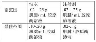
Enzymatic digestion (injectable DTM) -first, the decellularized tendon was weighed and recorded. Next, to create an injection, the.02-. 10 g tendon was weighed out and 1 mL collagenase solution (type I collagenase @ 2 mg/mL, type III collagenase @ 1 mg/mL in 1 XPBS) was added. Next, the mixture was incubated at 37 ℃ for 24 hours. Next, to wash the DTM, 1 XPBS (2 mL of 1 XPBS if 1 mL of collagenase solution was added) was added at twice the original volume. Next, the DTM was placed on a 70 um cell filter and centrifuged at 2000G for 5-10 minutes. Next, the DTM was placed into a new microcentrifuge tube containing 1 mL PBS and vortexed for 30 seconds. Next, the solution was placed in a 100 kDa filter and spun at 12,000G for 5 minutes. Finally, it was frozen at-80 ℃ for at least 30 minutes and the tube was placed in a lyophilizer.
Enzymatic digestion (putty DTM) -first, the decellularized tendon was weighed and recorded. Next, to produce putty, 10-.20 g of tendon was weighed out and 1 mL of collagenase solution (type I collagenase @ 2 mg/mL, type III collagenase @ 1 mg/mL in 1 XPBS) was added. Next, the mixture was incubated at 37 ℃ for 12 hours. Next, to wash the DTM, 1X PBS was added at twice the original volume (2 mL of 1X PBS if 1 mL of collagenase solution was added). Next, the DTM was placed on a 70 um cell filter and centrifuged at 2000G for 5-10 minutes. Next, the DTM was placed into a new microcentrifuge tube containing 1 mL PBS and vortexed for 30 seconds. Next, the solution was placed in a 100 kDa filter and spun at 12,000G for 5 minutes. Finally, frozen at-80 ℃ for at least 30 minutes and lyophilized.
Reconstitution-2-5 uL of 1 XPBS/mg tendon was added and additional PBS could be added until the desired consistency was reached.
The tendon was decellularized with dnase (10U, 50U and 100U) at various concentrations over 1 hour (see, e.g., fig. 7). 1 XPBS was used as a control without decellularization. The DNA concentration was determined using DNeasy kit (Qiagen). This data indicates that dnase as low as 50U is effective in decellularizing tissues. As shown in FIG. 8, 50U of DNase was compared with conventional detergents, 1% SDS and 0.1% EDTA. The DNase 50U was tested at 0.5 hours, 1 hour and 2 hours, whereas standard SDS and EDTA protocols require a 24 hour decellularization. DNA concentration was determined using DNEasy kit (Qiagen, n = 3). All values were normalized to no decellularization. Tukey's HSD multiple comparison post hoc tests showed no significant difference in the different times of DNase treatment or between decellularization with DNase (vers) versus SDS and EDTA. The following table also indicates the percentage of DNA remaining in the patella and achilles tendon after various decellularization methods and/or for various time periods:
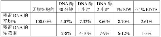
natural tendons were characterized to determine which tendons were best suited for development of allograft products.
The patella and achilles tendon are characterized by DNA content and native protein concentration. We also aimed to determine any differences between location and protein profile within each source (i.e. proximal versus (vs.) distal). No significant differences in total protein or TGF β content were found between different regions of the tendon. However, we did find that the Achilles tendon has a higher relative content of protein. The achilles tendon and the patellar tendon were divided into 1/3 sections consisting of the proximal, medial/distal ends of the tendons. (FIGS. 9A-D) Total protein of native tendon was measured using BCA protein quantification kit (Thermo Scientific). (FIG. 9E-H) TGF-. beta.was measured using a TGF-. beta.magnetic bead panel Millipolex kit (Millipore Sigma, # TGFBMAG-64K-03). Analysis of variance showed no statistically significant differences between tendon regions, so the entire tendon could be used by treatment. When comparing two different tendons (fig. 9D) there was no difference in total protein (P =0.93), but (fig. 9H) TGF- β in achilles tendon was statistically higher than patellar tendon (P = 0.0045).
| F value | p value | Significance of | |
| 9A | F (2,15) =0.01075 | 0.9893 | Is free of |
| 9B | F (2,15) =1.069 | 0.3680 | Is free of |
| 9C | F (2,33) =0.9342 | 0.4030 | Is free of |
| 9E | F (2,15) =1.849 | 0.1915 | Is free of |
| 9F | F (2,15) =0.3373 | 0.7190 | Is free of |
| 9G | F (2,33) =0.7912 | 0.4617 | Is free of |
As shown in fig. 10, filtration effectively eliminated collagenase activity. The decellularized tendon was treated with collagenase to improve the form factor (form-factor) of DTM. The 100 kDa filter was very effective in eliminating collagenase activity in the final product. Analysis of variance showed significant differences for each group (F (4, 22) =18.06, p < 0.0001). Importantly, there was no significant difference in collagenase activity between the native sample and the 100 kDa filtered sample.
| Comparison | p value | Significance of |
| Natural pair (vs.) decellularized | 0.9919 | Is free of |
| Natural filter-free for collagenase | <0.0001 | **** |
| Natural 70 um collagenase filter | 0.0116 | * |
| |
0.9635 | Is free of |
| Decellularized filter-free for collagenase | <0.0001 | **** |
| Decellularized 70 um collagenase filter | 0.0381 | * |
| Decellularized collagenase 100 kDa filter | >0.9999 | Is free of |
| Collagenase no filter to collagenase 70 um filter | 0.0021 | ** |
| Collagenase no filter to collagenase 100 kDa filter | <0.0001 | **** |
| Collagenase 70 um filter to |
0.003 | ** |
As shown in fig. 11, more biological activity was retained in DTM compared to the standard method of decellularizing tendons with pepsin. The tendon was digested after decellularization using a solution containing collagenase type 1 (92.5 g tendon/g collagenase 1) and 3 (185 g tendon/1 g collagenase 3) or using pepsin, considering the previously disclosed method (Farnebo et al 2014, PMID: 24341855). Analysis of variance indicated significant differences between groups, F (3,11) =5.056, p = 0.0193. Tukey's HSD showed post hoc pepsin with significantly less TGF-b (P = 0.0249).
As shown in fig. 15, the normalized TGFb content of four samples from four different donors was obtained in two processing steps. For each donor, the first column represents the amount of TGFb in native tendon, the second column represents the amount of TGFb in decellularized tendon, and the third column represents the amount of TGFb in digested tendon. The following table also describes the percent change (percent increase measured after collagenase processing from native tendon) through the processing steps:
| donor | Increase in TGF- |
| # | |
| 1 | 590.16 |
| #2 | 677.04 |
| #3 | 144.75 |
| #4 | 210.36 |
Differences in cell proliferation across different surfaces were studied (see, e.g., FIGS. 12A-C). Tissue culture plates were untreated (control, "TC treatment"), coated with collagen or with DTM. Primary tenocytes (ZenBio # TEN-F) were plated at 20,000 cells/well and cell viability was quantified using Presto Blue (Thermo Fisher) either (a) 48 hours or (B) 7 days after plating, resulting in significantly different growth rates (C). (analysis of variance = F (3,26) = 10.6, p < o.0001).
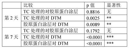
Claims (55)
Applications Claiming Priority (5)
| Application Number | Priority Date | Filing Date | Title |
|---|---|---|---|
| US201862782903P | 2018-12-20 | 2018-12-20 | |
| US62/782903 | 2018-12-20 | ||
| US201962890865P | 2019-08-23 | 2019-08-23 | |
| US62/890865 | 2019-08-23 | ||
| PCT/US2019/068112 WO2020132608A1 (en) | 2018-12-20 | 2019-12-20 | Decellularized tendon matrix methods and uses thereof |
Publications (1)
| Publication Number | Publication Date |
|---|---|
| CN114040730A true CN114040730A (en) | 2022-02-11 |
Family
ID=71101957
Family Applications (1)
| Application Number | Title | Priority Date | Filing Date |
|---|---|---|---|
| CN201980092758.3A Pending CN114040730A (en) | 2018-12-20 | 2019-12-20 | Acellular tendon matrix method and application thereof |
Country Status (9)
| Country | Link |
|---|---|
| US (1) | US20220047775A1 (en) |
| EP (1) | EP3897456A4 (en) |
| JP (1) | JP2022514775A (en) |
| KR (1) | KR20220002245A (en) |
| CN (1) | CN114040730A (en) |
| AU (1) | AU2019404558B2 (en) |
| CA (1) | CA3124665A1 (en) |
| MX (1) | MX2021007422A (en) |
| WO (1) | WO2020132608A1 (en) |
Cited By (1)
| Publication number | Priority date | Publication date | Assignee | Title |
|---|---|---|---|---|
| CN117018293A (en) * | 2023-08-07 | 2023-11-10 | 武汉理工大学 | Extracellular matrix wound repair stent and preparation method and application thereof |
Families Citing this family (1)
| Publication number | Priority date | Publication date | Assignee | Title |
|---|---|---|---|---|
| MX2023000120A (en) * | 2020-06-20 | 2023-02-09 | The Steadman Clinic And Steadman Philippon Res Institute | Decellularized tendon matrix methods and uses thereof. |
Citations (6)
| Publication number | Priority date | Publication date | Assignee | Title |
|---|---|---|---|---|
| JP2013165740A (en) * | 2010-05-14 | 2013-08-29 | Waseda Univ | Decellularization method and artificial tissue for implantation into the human body |
| US20130236439A1 (en) * | 2012-03-08 | 2013-09-12 | Lifecell Corporation | Enzyme-activated collagen and tissue matrices |
| US20140350677A1 (en) * | 2011-12-09 | 2014-11-27 | The Board Of Trustees Of The Leland Stanford Junior University | Decellularized composite tissue bioscaffolds for musculoskeletal tissue interface reconstruction and methods of production |
| US20160053250A1 (en) * | 2013-04-05 | 2016-02-25 | Claudia Zylberberg | Matrix metalloproteinasses and uses thereof |
| WO2017143344A1 (en) * | 2016-02-18 | 2017-08-24 | The Curators Of The University Of Missouri | Injectable nanomaterial-extracellular matrix constructs |
| CN107737373A (en) * | 2017-12-07 | 2018-02-27 | 山东隽秀生物科技股份有限公司 | A kind of preparation method of tendon repair material |
Family Cites Families (4)
| Publication number | Priority date | Publication date | Assignee | Title |
|---|---|---|---|---|
| US8894707B2 (en) * | 2009-03-06 | 2014-11-25 | Mayo Foundation For Medical Education And Research | Tendon or ligament tissue engineering |
| US8968785B2 (en) * | 2009-10-02 | 2015-03-03 | Covidien Lp | Surgical compositions |
| WO2014144215A1 (en) * | 2013-03-15 | 2014-09-18 | The Board Of Trustees Of The Leland Stanford Junior University | Injectable composition for in-situ repair and regeneration of an injured ligament or tendon and methods of use |
| JP6454125B2 (en) * | 2014-10-14 | 2019-01-16 | 地方独立行政法人東京都立産業技術研究センター | Method for producing collagen gel |
-
2019
- 2019-12-20 KR KR1020217022979A patent/KR20220002245A/en active Pending
- 2019-12-20 CN CN201980092758.3A patent/CN114040730A/en active Pending
- 2019-12-20 CA CA3124665A patent/CA3124665A1/en active Pending
- 2019-12-20 EP EP19898521.0A patent/EP3897456A4/en active Pending
- 2019-12-20 US US17/415,501 patent/US20220047775A1/en active Pending
- 2019-12-20 JP JP2021536113A patent/JP2022514775A/en active Pending
- 2019-12-20 AU AU2019404558A patent/AU2019404558B2/en active Active
- 2019-12-20 MX MX2021007422A patent/MX2021007422A/en unknown
- 2019-12-20 WO PCT/US2019/068112 patent/WO2020132608A1/en not_active Ceased
Patent Citations (6)
| Publication number | Priority date | Publication date | Assignee | Title |
|---|---|---|---|---|
| JP2013165740A (en) * | 2010-05-14 | 2013-08-29 | Waseda Univ | Decellularization method and artificial tissue for implantation into the human body |
| US20140350677A1 (en) * | 2011-12-09 | 2014-11-27 | The Board Of Trustees Of The Leland Stanford Junior University | Decellularized composite tissue bioscaffolds for musculoskeletal tissue interface reconstruction and methods of production |
| US20130236439A1 (en) * | 2012-03-08 | 2013-09-12 | Lifecell Corporation | Enzyme-activated collagen and tissue matrices |
| US20160053250A1 (en) * | 2013-04-05 | 2016-02-25 | Claudia Zylberberg | Matrix metalloproteinasses and uses thereof |
| WO2017143344A1 (en) * | 2016-02-18 | 2017-08-24 | The Curators Of The University Of Missouri | Injectable nanomaterial-extracellular matrix constructs |
| CN107737373A (en) * | 2017-12-07 | 2018-02-27 | 山东隽秀生物科技股份有限公司 | A kind of preparation method of tendon repair material |
Non-Patent Citations (1)
| Title |
|---|
| LING-JU NING等: ""preparation and characterization of decellularized tendon slices for tendon tissue engineering",Ling-Ju Ning等,《J Biomed Mater Res Part A》,第1448-1456页", 《J BIOMED MATER RES PART A》, 31 December 2012 (2012-12-31), pages 1448 - 1456 * |
Cited By (1)
| Publication number | Priority date | Publication date | Assignee | Title |
|---|---|---|---|---|
| CN117018293A (en) * | 2023-08-07 | 2023-11-10 | 武汉理工大学 | Extracellular matrix wound repair stent and preparation method and application thereof |
Also Published As
| Publication number | Publication date |
|---|---|
| JP2022514775A (en) | 2022-02-15 |
| EP3897456A1 (en) | 2021-10-27 |
| WO2020132608A1 (en) | 2020-06-25 |
| CA3124665A1 (en) | 2020-06-25 |
| KR20220002245A (en) | 2022-01-06 |
| AU2019404558B2 (en) | 2025-08-21 |
| US20220047775A1 (en) | 2022-02-17 |
| AU2019404558A1 (en) | 2021-08-12 |
| EP3897456A4 (en) | 2022-09-21 |
| MX2021007422A (en) | 2021-12-10 |
Similar Documents
| Publication | Publication Date | Title |
|---|---|---|
| US12090175B2 (en) | Compositions and methods for tissue repair with extracellular matrices | |
| Kawecki et al. | A review of decellurization methods caused by an urgent need for quality control of cell‐free extracellular matrix'scaffolds and their role in regenerative medicine | |
| US10729729B2 (en) | Compositions and methods for treating and preventing tissue injury and disease | |
| US20180125897A1 (en) | Decellularized Adipose Cell Growth Scaffold | |
| US11246891B2 (en) | Compositions and methods for treating and preventing tissue injury and disease | |
| JP6545102B2 (en) | How to treat a cardiac condition | |
| US20250367241A1 (en) | Compositions and methods for treating and preventing tissue injury and disease | |
| US20160271187A1 (en) | Compositions and methods for treating and preventing tissue injury and disease | |
| AU2019404558B2 (en) | Decellularized tendon matrix methods and uses thereof | |
| US20200353008A1 (en) | Compositions and methods for treating nerve injury | |
| Goncharuk et al. | Matrix Metalloproteinase-9 is involved in the fibrotic process in denervated muscles after sciatic nerve trauma and recovery | |
| HK40068683A (en) | Decellularized tendon matrix methods and uses thereof | |
| CN112839687A (en) | Decellularized muscle matrix | |
| HK40086575A (en) | Decellularized tendon matrix methods and uses thereof | |
| US20230226257A1 (en) | Decellularized tendon matrix methods and uses thereof | |
| HK1212380B (en) | Compositions and methods for treating and preventing tissue injury and disease |
Legal Events
| Date | Code | Title | Description |
|---|---|---|---|
| PB01 | Publication | ||
| PB01 | Publication | ||
| SE01 | Entry into force of request for substantive examination | ||
| SE01 | Entry into force of request for substantive examination | ||
| REG | Reference to a national code |
Ref country code: HK Ref legal event code: DE Ref document number: 40068683 Country of ref document: HK |