CN113710255A - Treatment of heart failure - Google Patents
Treatment of heart failure Download PDFInfo
- Publication number
- CN113710255A CN113710255A CN202080028568.8A CN202080028568A CN113710255A CN 113710255 A CN113710255 A CN 113710255A CN 202080028568 A CN202080028568 A CN 202080028568A CN 113710255 A CN113710255 A CN 113710255A
- Authority
- CN
- China
- Prior art keywords
- subject
- mitochondria
- composition
- mitochondrial
- pab
- Prior art date
- Legal status (The legal status is an assumption and is not a legal conclusion. Google has not performed a legal analysis and makes no representation as to the accuracy of the status listed.)
- Pending
Links
Images


Classifications
-
- A—HUMAN NECESSITIES
- A61—MEDICAL OR VETERINARY SCIENCE; HYGIENE
- A61K—PREPARATIONS FOR MEDICAL, DENTAL OR TOILETRY PURPOSES
- A61K35/00—Medicinal preparations containing materials or reaction products thereof with undetermined constitution
- A61K35/12—Materials from mammals; Compositions comprising non-specified tissues or cells; Compositions comprising non-embryonic stem cells; Genetically modified cells
- A61K35/34—Muscles; Smooth muscle cells; Heart; Cardiac stem cells; Myoblasts; Myocytes; Cardiomyocytes
-
- A—HUMAN NECESSITIES
- A61—MEDICAL OR VETERINARY SCIENCE; HYGIENE
- A61P—SPECIFIC THERAPEUTIC ACTIVITY OF CHEMICAL COMPOUNDS OR MEDICINAL PREPARATIONS
- A61P9/00—Drugs for disorders of the cardiovascular system
- A61P9/04—Inotropic agents, i.e. stimulants of cardiac contraction; Drugs for heart failure
-
- A—HUMAN NECESSITIES
- A61—MEDICAL OR VETERINARY SCIENCE; HYGIENE
- A61K—PREPARATIONS FOR MEDICAL, DENTAL OR TOILETRY PURPOSES
- A61K9/00—Medicinal preparations characterised by special physical form
- A61K9/0012—Galenical forms characterised by the site of application
- A61K9/0019—Injectable compositions; Intramuscular, intravenous, arterial, subcutaneous administration; Compositions to be administered through the skin in an invasive manner
Abstract
The present disclosure relates to compositions comprising isolated mitochondria or combined mitochondrial agents, and methods of treating disorders using such compositions. The method is based at least in part on the following findings: isolated mitochondria themselves, as well as isolated mitochondria linked to therapeutic, diagnostic and/or imaging agents, can be delivered to a patient's tissue by injecting them into the patient's blood vessel.
Description
Priority requirement
This application claims priority from U.S. provisional application No. 62/806,473 filed on 15/2/2019. The foregoing is incorporated by reference herein in its entirety.
Technical Field
The present disclosure relates to therapeutic uses of mitochondria and combined mitochondrial agents.
Background
Mitochondria are double-membrane-bound organelles present in the cytoplasm of nucleated eukaryotic cells. Except for red blood cells, they are present in almost every cell of the human body. They are the primary site of cellular energy metabolism and produce Adenosine Triphosphate (ATP) for different cellular functions. Typically, over 90% of the ATP demand of a cell is provided by the cell's own mitochondria.
Mitochondria are composed of two concentric membranes with specific functions. The inner mitochondrial membrane contains proteins for ATP synthase. The mitochondrial outer membrane, which contains a large amount of integral membrane proteins, surrounds the entire organelle.
The structure of mitochondria has striking similarities to some modern prokaryotes. Indeed, mitochondria are thought to originate from an ancient symbiosis when nucleated cells engulf aerobic prokaryotes. In symbiotic relationships, host cells begin to rely on phagocytosed prokaryotes to produce energy, and prokaryotic cells begin to rely on the protective environment provided by the host cells.
Mitochondria can be used to treat a variety of disorders because of their primary function in cellular metabolism. There is also a need to utilize mitochondria for drug delivery as well as some other therapeutic and diagnostic purposes.
Disclosure of Invention
The present disclosure provides pharmaceutical compositions comprising mitochondria and methods of treating disorders using such pharmaceutical compositions. The present specification further provides diagnostic and imaging methods using such pharmaceutical compositions. The method is based at least in part on the following findings: isolated mitochondria themselves, as well as isolated mitochondria linked to therapeutic, diagnostic and/or imaging agents, can be delivered to a patient's tissue by injecting them into the patient's blood vessel. That is, mitochondria are injected or applied directly to the target tissue, although this is contemplated by certain methods described herein, it is not always necessary. More specifically, in some cases, the methods described herein make use of the following findings: after injection or infusion of mitochondria into, for example, an artery, the mitochondria can traverse the arterial wall and be taken up by cells of the patient's tissue. The methods described herein can provide tissues or cells with local and general distribution of mitochondria or mitochondria with therapeutic, diagnostic, and/or imaging agents for a variety of therapeutic, diagnostic, and/or imaging purposes using relatively simple medical procedures.
Provided herein, inter alia, are methods of treating or preventing heart failure in a subject comprising administering to the subject a therapeutically effective amount of a composition comprising isolated mitochondria or a combined mitochondrial agent. In some embodiments, the composition is administered to the subject by intramyocardial injection. In some embodiments, the subject has or is at risk of developing heart failure as follows: right Ventricular Hypertrophy (RVH), Left Ventricular Hypertrophy (LVH), Right Ventricular Failure (RVF), or Left Ventricular Failure (LVF). In some embodiments, the subject has a lung disease. In some embodiments, the pulmonary disease affects right ventricular function. In some embodiments, the composition is administered to the subject by injecting the composition into a blood vessel of the subject. In some embodiments, the mitochondria are autologous. In some embodiments, the mitochondria are allogeneic. In some embodiments, the mitochondria are xenogeneic.
Provided herein, inter alia, are methods of maintaining Right Ventricular (RV) contractility, maintaining RV capillary density, preventing RV dilation, or delaying RVF onset in a subject, comprising administering to the subject a therapeutically effective amount of a composition comprising isolated mitochondria or a combined mitochondrial agent. In some embodiments, the composition is administered to the subject by intramyocardial injection. In some embodiments, the subject has or is at risk of developing heart failure as follows: right Ventricular Hypertrophy (RVH), Left Ventricular Hypertrophy (LVH), Right Ventricular Failure (RVF), or Left Ventricular Failure (LVF). In some embodiments, the subject has a lung disease. In some embodiments, the pulmonary disease affects right ventricular function. In some embodiments, the composition is administered to the subject by injecting the composition into a blood vessel of the subject. In some embodiments, the mitochondria are autologous. In some embodiments, the mitochondria are allogeneic. In some embodiments, the mitochondria are xenogeneic.
Provided herein, inter alia, are methods of maintaining Left Ventricular (LV) contractility, maintaining LV capillary density, preventing LV dilation, or delaying onset of Left Ventricular Failure (LVF) in a subject, comprising administering to the subject a therapeutically effective amount of a composition comprising isolated mitochondria or a combined mitochondrial agent. In some embodiments, the composition is administered to the subject by intramyocardial injection. In some embodiments, the subject has or is at risk of developing heart failure as follows: right Ventricular Hypertrophy (RVH), Left Ventricular Hypertrophy (LVH), Right Ventricular Failure (RVF), or Left Ventricular Failure (LVF). In some embodiments, the subject has a lung disease. In some embodiments, the pulmonary disease affects left ventricular function. In some embodiments, the composition is administered to the subject by injecting the composition into a blood vessel of the subject. In some embodiments, the mitochondria are autologous. In some embodiments, the mitochondria are allogeneic. In some embodiments, the mitochondria are xenogeneic.
Provided herein, inter alia, is a method of maintaining ventricular contractility in a subject, the method comprising:
identifying a subject in need thereof; and
administering to the subject a therapeutically effective amount of a composition comprising isolated mitochondria or a combined mitochondrial agent. In some embodiments, the subject is identified by measuring end-systolic pressure volume (ESPV).
Provided herein, inter alia, is a method of maintaining ventricular capillary density in a subject, the method comprising:
identifying a subject in need thereof; and
administering to the subject a therapeutically effective amount of a composition comprising isolated mitochondria or a combined mitochondrial agent.
Provided herein, inter alia, is a method of reducing the risk of ventricular dilatation in a subject, the method comprising:
identifying a subject in need thereof; and
administering to the subject a therapeutically effective amount of a composition comprising isolated mitochondria or a combined mitochondrial agent.
In some embodiments, the subject is identified as having diabetes, obesity, hypertension, alcohol abuse, cocaine use and abuse, bacterial infections, viral infections, fungal infections, parasitic infections, exposure to toxins (e.g., lead, mercury, or cobalt), arrhythmia, or late pregnancy complications.
Provided herein, inter alia, is a method of delaying the onset of heart failure in a subject, the method comprising:
identifying a subject in need thereof; and
administering to the subject a therapeutically effective amount of a composition comprising isolated mitochondria or a combined mitochondrial agent. In some embodiments, the subject is identified as having right ventricular hypertrophy or left ventricular hypertrophy.
Provided herein, inter alia, are methods of treating heart failure, delaying onset of heart failure, reducing the risk of developing heart failure in a subject comprising administering to the subject a therapeutically effective amount of a composition comprising isolated mitochondria or a combined mitochondrial agent. In some embodiments, the composition is administered to the subject by intramyocardial injection. In some embodiments, the method comprises identifying the subject as at risk of developing heart failure. In some embodiments, the subject has a lung disease. In some embodiments, the composition is administered to the subject by injecting the composition into a blood vessel leading to the heart.
Provided herein, inter alia, are methods of treating cardiac hypertrophy, delaying onset of cardiac hypertrophy, reducing the risk of developing cardiac hypertrophy in a subject comprising administering to the subject a therapeutically effective amount of a composition comprising isolated mitochondria or a combined mitochondrial agent. In some embodiments, the composition is administered to the subject by intramyocardial injection. In some embodiments, the method comprises identifying the subject as at risk of developing cardiac hypertrophy. In some embodiments, the subject has a lung disease. In some embodiments, the composition is administered to the subject by injecting the composition into a blood vessel leading to the heart.
In certain embodiments, the blood vessel is a blood vessel or a portion of a vascular system that delivers blood to a target site, target organ, or target area, e.g., a coronary artery of the subject, a portal vein of the liver of the subject, a pancreatic aorta of the subject, or a prostate artery of the subject.
In certain embodiments, the mitochondria can have different origins, e.g., the mitochondria can be autologous, allogeneic, or xenogeneic. In certain embodiments, the autologous mitochondria can have exogenous mtDNA. In some embodiments, the mitochondria are from a first degree relative of the subject.
In some embodiments, the described methods comprise the step of collecting isolated mitochondria from cells prior to administration. The isolated mitochondria or the combined mitochondrial reagents can be administered to the subject immediately after collection of the isolated mitochondria from the cell.
In one aspect, the present disclosure provides compositions comprising isolated mitochondria and/or combined mitochondrial agents; and a carrier. In some embodiments, the composition is a pharmaceutical composition. The carrier may be any suitable carrier, for example, breath buffer, mitochondrial buffer, sterile mitochondrial buffer, University of Wisconsin (UW) solution, blood, serum, or a contrast agent.
In all of the methods and/or compositions described herein, the combined mitochondrial reagent can comprise a pharmaceutical agent. The agent may be a therapeutic agent, an imaging agent, a diagnostic agent, or any combination thereof. The imaging agent may be radioactive. In some embodiments, the imaging agent is18F-rhodamine 6G or iron oxide nanoparticles. In some embodiments, the agent is linked to the mitochondria by a covalent bond. Alternatively or additionally, the agent is embedded in the mitochondria. The combined mitochondrial agents can include antibodies or antigen binding fragments. Furthermore, in all of the methods and/or compositions described herein, the mitochondria can be autologous, allogeneic or xenogeneic. In some embodiments, the mitochondria have exogenous DNA (e.g., mtDNA).
As used herein, the term "isolated mitochondria" means functionally intact mitochondria that are free of foreign eukaryotic cellular material.
A "combined mitochondrial agent" is an isolated mitochondrion that is artificially combined with a pharmaceutical, diagnostic or imaging agent or any other agent. The agent is combined with the mitochondria in any manner, e.g., linked (e.g., chemically or electrostatically linked) to the mitochondria, attached to the mitochondria, embedded in the mitochondrial membrane, substantially enclosed within the mitochondria, or completely encapsulated by the mitochondria, so long as the mitochondria and the agent are in physical contact with each other. The combined mitochondrial agents are designed such that after injection the mitochondria act as a "carrier" that can deliver the agent to the patient's tissue.
Throughout the specification, the terms "subject" and "patient" are used to describe an animal, human or non-human to which treatment according to the methods of the present disclosure is provided. Veterinary applications are clearly contemplated by the present disclosure. The term includes, but is not limited to, birds, reptiles, amphibians, and mammals, e.g., humans, other primates, pigs, rodents (e.g., mice and rats), rabbits, guinea pigs, hamsters, cows, horses, cats, dogs, sheep, and goats. Preferred subjects are humans, farm animals and domestic pets (e.g., cats and dogs).
The term "treating" is used herein to mean delaying the onset of a condition (e.g., a disease described herein), inhibiting the condition, alleviating the effects of the condition, or extending the life of a patient suffering from the condition.
An "ischemia-related disease" is a disease that involves ischemia. As used herein, ischemia refers to a reduction in blood flow to an organ and/or tissue. The reduction in blood flow may be caused by any suitable mechanism, including partial or complete occlusion (obstruction), narrowing (constriction), and/or leakage/rupture of one or more blood vessels supplying blood to the organ and/or tissue, and the like.
By "immediately after collection of mitochondria from a cell" is meant immediately after collection of mitochondria from a cell and before any substantial reduction in the viability of the mitochondria occurs.
As used herein, the term "transplantation" is used throughout the specification as a generic term describing the process of transplanting an organ, tissue, cell mass, single cell, or organelle into a recipient. The term "cell transplantation" is used throughout the specification as a general term describing the process of transferring at least one cell (e.g., islet cells or stem cells) to a recipient. Such transplantation can be performed, for example, by removing the beta cells (or intact islets) from a donor pancreas and placing them in a recipient patient whose pancreas is not producing sufficient insulin. The term includes all types of transplantation known in the art, except blood transfusions. Transplantation is classified by the site and genetic relationship between the donor and recipient. The term includes, for example, autografts (where cells or tissue are removed from one location in a patient and transferred to the same location or another location in the same subject), allografts (where the transplant is between members of the same species), and xenografts (where the transplant is between members of different species).
Unless defined otherwise, all technical and scientific terms used herein have the same meaning as commonly understood by one of ordinary skill in the art to which this invention belongs. Although methods and materials similar or equivalent to those described herein can be used in the practice or testing of the present invention, suitable methods and materials are described below. All publications, patent applications, patents, and other references mentioned herein are incorporated by reference in their entirety. In case of conflict, the present specification, including definitions, will control. In addition, the materials, methods, and examples are illustrative only and not intended to be limiting.
Other features and advantages of the invention will be apparent from the following detailed description, and from the claims.
Drawings
FIG. 1 is a schematic diagram depicting a mitochondrial isolation method.
Fig. 2 is a schematic diagram depicting disease prognosis associated with ventricular overload.
Fig. 3 is a schematic diagram depicting an overview of a method of mitochondrial transplantation in a subject.
Fig. 4 is a schematic diagram depicting an animal model study using Pulmonary Artery Banding (PAB).
Fig. 5 is a schematic diagram depicting a timeline of measurement and analysis of an experiment.
Fig. 6 is a line graph showing the change in functional area (FAC, expressed as a percentage) at baseline, 1 month after PAB, and at euthanasia for the control group (C, also referred to as the "sham" group), the PAB-V (vehicle) group, and the PAB-M (mitochondrial) group.
Fig. 7 is a line graph showing tricuspid annular plane systolic shift (tase in mm) at baseline, 1 month after PAB, and at euthanasia for the control group (C, also referred to as "sham" group), PAB-V (vehicle) group, and PAB-M (mitochondrial) group.
Fig. 8 is a line graph showing Right Ventricular (RV) wall thickness (in mm) at baseline, 1 month post PAB, and at euthanasia for the control group (C, also referred to as "sham" group), PAB-V (vehicle) group, and PAB-M (mitochondrial) group.
Fig. 9 is a box plot showing dP/dt max (in mmHg/sec) at euthanasia for the control group (C, also referred to as the "sham" group), PAB-V (vehicle) group, and PAB-M (mitochondria) group.
Fig. 10 is a line graph showing dP/dt max (in mmHg/sec) at baseline and at euthanasia for the control group (C, also referred to as "sham" group), PAB-V (vehicle) group, and PAB-M (mitochondrial) group.
Fig. 11A is an immunofluorescence image showing TUNEL staining illustrating apoptotic cells (white arrows) in the TUNEL positive control group. Cardiomyocytes were stained by desmin staining and nuclei were stained by DAPI staining.
Fig. 11B is an immunofluorescence image showing TUNEL staining illustrating apoptotic cells (white arrows) in the control/sham group. Cardiomyocytes were stained by desmin staining and nuclei were stained by DAPI staining.
Fig. 11C is an immunofluorescence image showing TUNEL staining illustrating apoptotic cells (white arrows) in PAB-V group. Cardiomyocytes were stained by desmin staining and nuclei were stained by DAPI staining.
Fig. 11D is an immunofluorescence image showing TUNEL staining illustrating apoptotic cells (white arrows) in PAB-M group. Cardiomyocytes were stained by desmin staining and nuclei were stained by DAPI staining.
Fig. 11E is a bar graph showing the ratio (%) of desmin per field/number of nuclei per field (P < 0.01).
Fig. 12A is an immunofluorescence image showing CD31 staining illustrating capillary density (white arrows) in the control/sham group. Cardiomyocytes were stained via desmin staining and nuclei were stained via DAPI staining.
Fig. 12B is an immunofluorescence image showing CD31 staining demonstrating capillary density (white arrows) in the PAB-V group. Cardiomyocytes were stained via desmin staining and nuclei were stained via DAPI staining.
Fig. 12C is an immunofluorescence image showing CD31 staining illuminating the capillary density (white arrows) in the PAB-M group. Cardiomyocytes were stained via desmin staining and nuclei were stained via DAPI staining.
Fig. 13A is an electron microscopy analysis showing the number and shape of mitochondria in the control/sham group.
FIG. 13B is an electron microscopy analysis showing the number and shape of mitochondria in the PAB-V group.
FIG. 13C is an electron microscopy analysis showing the number and shape of mitochondria in the PAB-C group.
Fig. 14 is a schematic illustration depicting disease outcome and prognosis associated with mitochondrial transplantation therapy.
Figure 15 is an immunofluorescence image showing pictures of control, RV hypertrophy (RVH) and RVH with mitochondrial transplantation.
Fig. 16 is a box plot showing ATP levels in control cardiomyocytes, untreated hypertrophic cardiomyocytes (H-mitochondria), and mitochondria-treated hypertrophic cardiomyocytes (H-cardiac mitochondria, H-gastrocnemius mitochondria, and H-soleus mitochondria), for comparison of p-0.05 for control and 0.001 for untreated hypertrophic cardiomyocytes (H-mitochondria).
FIG. 17A is an immunofluorescence image showing TUNEL staining illustrating apoptotic cells (white arrows) in the control/sham, PAB-V and PAB-M groups. Cardiomyocytes were stained via desmin staining and nuclei were stained via DAPI staining.
Fig. 17B is a box plot showing TUNEL positive nuclei per 1000 nuclei, (. rho. control, p 0.01, and # PAB-V versus PAB-M, p 0.05).
FIG. 17C is a microscopic analysis showing representative tissue sections detecting fibrosis in the control/sham, PAB-V and PAB-M groups.
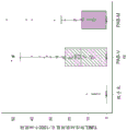
Fig. 17D is a box plot showing the rate (%) of fibrosis per field at the end of the study, (. beta. contrast, p 0.01, and # PAB-V vs. PAB-M, p 0.05).
Fig. 18A is a box plot showing RV wall thickness (in cm) at baseline for the control group (C, also referred to as the "sham" group), PAB-V group, and PAB-M group.
Fig. 18B is a box plot showing RV wall thickness (in cm) at 1 month post-PAB for the control group (C, also referred to as the "sham" group), PAB-V group, and PAB-M group.
Fig. 18C is a box plot showing RV wall thickness (in cm) at euthanasia for the control group (C, also referred to as the "sham" group), PAB-V group, and PAB-M group.
Fig. 19A is a box plot showing the change in functional area (FAC, expressed as a percentage) at baseline for the control group (C, also referred to as the "sham" group), the PAB-V group, and the PAB-M group.
FIG. 19B is a box plot showing the change in functional area (FAC, expressed as a percentage) at 1 month post-PAB for the control group (C, also referred to as the "sham" group), the PAB-V group, and the PAB-M group.
FIG. 19C is a box plot showing the functional zone change (FAC, expressed in percent) at euthanasia for the control group (C, also referred to as the "sham" group), the PAB-V group, and the PAB-M group.
Fig. 20A is a box plot showing tricuspid annular planar systolic shift (tase in mm) at baseline for the control group (C, also known as the "sham" group), the PAB-V group, and the PAB-M group.
Fig. 20B is a box plot showing tricuspid annulus plane systolic shift (tape, in mm) at 1 month post-PAB for the control group (C, also referred to as the "sham" group), the PAB-V group, and the PAB-M group.
Fig. 20C is a box plot showing tricuspid annulus plane systolic shift (tape, in mm) at euthanasia for the control group (C, also known as the "sham" group), the PAB-V group, and the PAB-M group.
FIG. 21A is a box plot showing dP/dt max (in mmHg/sec) at baseline for the control group (C, also referred to as the "sham" group), the PAB-V group, and the PAB-M group.
FIG. 21B is a box plot showing dP/dt max (in mmHg/sec) at euthanasia for the control group (C, also referred to as the "sham" group), the PAB-V group, and the PAB-M group.
Fig. 22 is a schematic diagram showing an outline of an animal model study using Pulmonary Artery Banding (PAB) and some of the clinical findings.
Detailed Description
Right Ventricular Hypertrophy (RVH) and failure (RVF) are the leading causes of heart disease morbidity and mortality that affect the long-term prognosis of patients, where the Right Ventricle (RV) is abnormally loaded due to pulmonary hypertension, obstruction of the outflow tract, or functions as a systemic ventricle. As an initial compensation step, the RV accommodates these hemodynamic changes by increasing wall thickness to provide higher contractile force to overcome the increase in afterload. Eventually, these mechanisms are inadequate, and hypertrophy continues to cause dilation and failure of contractions. Clinical observations indicate that these compensatory changes on the left side maintain contractile function more effectively and for a longer period of time than on the right side, while failure occurs more rapidly on the right side. Mitochondrial function directly affects cardiac function and contractility. The lack of adaptability of Rv to long-term increased stress load is associated with the failure of mitochondrial and calcium processing mechanisms to keep up with the need for myocardial tissue thickening. Inefficient calcium cycling with Adenosine Triphosphate (ATP) consumption and Electronic Transport Chain (ETC) dysfunction further limits ATP synthesis, leading to bioenergy depletion (McCully JD, Rousou AJ, Parker RA, Levitsky S.Age and genes in mitochondridial oxidation and free matrix calcium degradation and hydrolysis, Ann ThorSurg. 2007; 83: 1102-. These events eventually overwhelm cellular regulatory mechanisms and lead to more rapid deterioration of cardiac function in RVH.
Mitochondrial enzyme activity and mitochondrial dna (mtdna) content decrease progressively from hypertrophied to depleted, playing an important role in RV dysfunction. In response to these findings, the present disclosure has demonstrated, using a combination microarray and proteomic analysis of matched samples, that mitochondrial function with respect to mitochondrial number/quality is as important as myocardial tissue development in adapting parenchymal RV to pathological load. In addition, activation of pro-apoptotic pathways (particularly mitochondrial-related activation) and down-regulation of calcium signaling pathways are associated with progression to failure. In response, the initial upregulation of oxidative phosphorylation and associated calcium processing for mitochondrial stabilization is to meet the energy requirements for myocardial growth to adapt the thin-walled RV to pressure overload. However, with prolonged exposure to increased pressure loads, mitochondria fail to adapt, leading to rapid deterioration due to reduced contractile function. Progression to heart failure is associated with a decline in energy reserve capacity, and compensatory mechanisms can no longer support the imbalance between a decrease in energy supply and an increase in adaptive RV wall thickening requirements.
The central role of mitochondria in the progression from hypertrophy to heart failure has been recognized. Current therapies for patients with right heart failure are largely limited to therapies that target lung function rather than directly intervening in the severe myocardial energy deficit due to mitochondrial dysfunction. Previous studies have demonstrated successful and safe techniques for therapeutic transplantation of autologous respiratory competent mitochondria that use viable mitochondria isolated from healthy tissue to replace and/or supplement the pool of naturally damaged mitochondria. However, success of treatment has been demonstrated primarily in acute ischemia-reperfusion injury models, where the beneficial effects of mitochondrial transplantation have been confirmed (McCully JD, Cowan DB, Pacak CA, Toumpoulis IK, Dayanan H, Levitsky S.Objection of isolated mitochondria duringearl surgery for cardiovascular performance. am J Physiol Heart Circuit physiology. 2009; 296(1): H94105). Less is known for: whether mitochondrial transplantation can achieve long-term mitochondrial dysfunction in addition to adaptive maintenance of mitochondrial function due to progressive pressure overload hypertrophy. Furthermore, the source of mitochondria for transplantation may play a key role due to a combination of metabolic adaptation and mitochondrial dysfunction. As reported, mitochondria adapt themselves to the effects required by the tissues they supply (Fern < ndez-Vizarra E, Enr i quez JA, pirez-Martos A, Montoya J, Fern < z > -Silva P.tissue-specific differences in mitochondreal activity and biology. mitochonddrion.2011; 11(1): 207-. In contrast to the slowly contracting skeletal muscle, the mitochondria of the rapidly contracting skeletal muscle are accustomed to the high glucose metabolizing cells, which can rapidly adapt themselves to increased energy demands. It has been established that hypertrophied and failing myocardium instead uses glucose as a metabolic substrate (Doenst T, Nguyen TD, Abel ED. Cardiac metabolism in heart failure: electrolytes beyond ATP production. Circ Res.2013; 113: 709724.). Thus, the source of mitochondria for transplantation may have relevance as the goal is to restore defective energy production and mitochondrial dynamics in failing hearts. Since myocardial mitochondria in failing hearts have been damaged, mitochondria from other cellular sources must be collected for transplantation.
The present disclosure is based, in part, on the following surprising findings: mitochondria can be used to prevent, treat and/or reduce one or more of the symptoms of heart failure, even before heart failure occurs. Accordingly, in one aspect, the present disclosure provides methods of minimizing heart failure, reducing the risk of heart failure, ameliorating at least one symptom of heart failure, preventing or treating cellular damage, tissue damage, and/or organ damage associated with heart failure in a subject at risk for heart failure.
In some embodiments, the methods of treating or preventing heart failure in a subject described herein comprise administering to the subject a therapeutically effective amount of a composition comprising isolated mitochondria or a combined mitochondrial agent. In some embodiments, the composition is administered to the subject by direct injection, intramyocardial injection, or infusion. In some embodiments, the subject has or is at risk of developing: right Ventricular Hypertrophy (RVH), Left Ventricular Hypertrophy (LVH), or Right Ventricular Failure (RVF), or Left Ventricular Failure (LVF).
The present disclosure is also based, at least in part, on the following findings: isolated mitochondria and isolated mitochondria linked to therapeutic, diagnostic and/or imaging agents can be delivered to a tissue of a patient by injecting them into a blood vessel of the patient. Using relatively simple medical procedures, a skilled practitioner can distribute mitochondria locally and/or generally to a patient's tissue and/or cells for a variety of purposes. Further, mitochondria can be used as carrier agents, e.g., to deliver therapeutic, diagnostic, and/or imaging agents to a patient's tissue. In contrast to some traditional therapeutic regimens involving nanoparticles, it is further noted that mitochondria are not toxic and do not elicit any substantial adverse immune or autoimmune response.
Without intending to be bound by any theory, it is believed that the infused mitochondria exude through the capillary wall by first adhering to the endothelium. After mitochondria are injected or infused into arteries, they can cross the endothelium of blood vessels and be taken up by tissue cells through the endosomal actin-dependent internalization process.
Combined mitochondrial reagents
A combined mitochondrial agent includes mitochondria that are physically associated with an agent (e.g., a therapeutic agent, a diagnostic agent, and/or an imaging agent).
The therapeutic agent may be any agent having therapeutic or prophylactic use. Exemplary therapeutic agents include, for example, therapeutic agents for ischemia-related conditions, cytotoxic agents for the treatment of cancer, and the like. In some cases, mitochondria can deliver therapeutic agents to specific cells, e.g., tumor cells. The therapeutic agent may be, for example, an intracellular inhibitor, deactivator, toxin, arresting substance(s) and/or cytostatic/cytotoxic substance(s) that, once inside the cell, inhibits, destroys, arrests, modifies and/or alters the cell such that the cell is no longer able to function and/or survive normally. The therapeutic agent may be an agent that restores the appropriate function of the cell, for example, a DNA vector for gene therapy. The therapeutic agent may be, for example, an inorganic compound or an organic compound; small molecules (less than 500 daltons) or large molecules; protein molecules (proteinaceous molecules), such as peptides, polypeptides, proteins, post-translationally modified proteins, or antibodies; or a nucleic acid molecule, such as a double-stranded DNA, single-stranded DNA, double-stranded RNA, single-stranded RNA, or triple-helical nucleic acid molecule. In some embodiments, the therapeutic agent may be a natural product derived from any known organism (e.g., from an animal, plant, bacterium, fungus, protist, or virus) or from a library of synthetic molecules. In some embodiments, the therapeutic agent can be a monomeric or polymeric compound. Some exemplary therapeutic agents include cytotoxic agents, DNA vectors, small interfering rna (sirna), microrna (mirna), reactive peptides, nanoparticles, microspheres, and fluorescent molecules.
Diagnostic agents are agents that have diagnostic utility. In some embodiments, as mitochondria carry the diagnostic agent into the cell, the diagnostic agent can be designed to determine intracellular conditions, such as intracellular pH and oxidative stress (oxidative stress).
Imaging agents are agents used in imaging techniques. Including, but not limited to, X-ray, Computed Tomography (CT), Magnetic Resonance Imaging (MRI), scintigraphy, fluorescence, ultrasound, etc. The imaging agent may be fluorescent and/or radioactive. In some embodiments, the imaging agent may also be a diagnostic agent. Exemplary imaging agents include, but are not limited to, MitoTracker fluorophore (Thermo Fisher Scientific Inc.), RFP, BacMam 2.0(Thermo Fisher Scientific Inc.), pH-sensitive pHrodo fluorescent dye (Thermo Fisher Scientific Inc.),18F-rhodamine 6G,18F-labeled rhodamine B, magnetic iron oxide nanoparticles, and gold-and platinum-based nanoparticles.
As discussed above, the combined mitochondrial reagent comprises mitochondria and the reagent in direct and/or indirect physical contact with each other. For example, the agent may be attached to the mitochondria, embedded in the mitochondrial membrane, or fully or partially enclosed in the mitochondria. In some cases, the agent may be covalently linked to the mitochondria. In some cases, the agent is directly linked to a component of the mitochondrial membrane by a covalent bond (e.g., an amide bond and a disulfide bond) or indirectly linked to a component of the mitochondrial membrane by a linker (e.g., a peptide linker) or another covalently bonded agent. In other cases, the agent may be non-covalently attached to the mitochondria, for example, by hydrophobic interactions, Van der Waals interactions, and/or electrostatic interactions, and the like.
In some embodiments, the combined mitochondrial agent can comprise two or more different types of agents, e.g., two different classes of therapeutic agents, three different classes of imaging agents, one therapeutic agent and one imaging agent, therapeutic agent and diagnostic agent, and the like. The skilled practitioner will appreciate that any variation is possible.
One particularly useful linker for linking mitochondria and agents provides for sustained release of the agent after injection. This can be achieved, for example, using a hydrazone functional group. For example, a component that forms a hydrazone to covalently bind an agent to the mitochondrial membrane. Once the combined mitochondrial agent is taken up by the cell, the change in pH will result in hydrolysis of the hydrazone, thereby releasing the bound agent within the cell.
In some embodiments, the therapeutic, diagnostic and/or imaging agents may be attached to the outer mitochondrial membrane using functionalized surface chemistry. In some cases, heterobifunctional chemical methods (heterobifunctional chemistries) can attach therapeutic, diagnostic, and/or imaging agents to the mitochondrial surface, and once these agents are internalized, they can be released by interaction with intercellular esterases (e.g., via interaction with acetoxymethyl esters) or by ultraviolet or near-infrared light-activated strategies. UV-Light activation and Near-IR activation strategies are described, for example, in Zhou, Fang, Hanjie Wang and Jin Chang, "Progress in the Field of construction Near-isolated Light-Responsive Drug Delivery plants," Journal of Nanoscience and Nanotechnology 16.3(2016): 2111-2125; bansal, Akshaya and Yong Zhang, "Photocotroled negative delivery systems for biological applications," Accounts of chemical research 47.10(2014): 3052-3060; barhoumi, Aoune, Qian Liu and Daniel S.Kohane, "Ultraviolet light-mediated delivery: Principles, applications, and changes," Journal of Controlled Release 219(2015): 31-42. Each of which is incorporated by reference in its entirety.
Pharmaceutical and other compositions
The present disclosure provides compositions comprising isolated mitochondria, compositions comprising a combined mitochondrial reagent, compositions comprising both isolated mitochondria and a combined mitochondrial reagent, and methods of using such compositions.
The pharmaceutical compositions described herein can include mitochondria and/or a combined mitochondrial agent and a pharmaceutically acceptable carrier. As used herein, the language "pharmaceutically acceptable carrier" includes saline, solvents, dispersion media, coatings, antibacterial and antifungal agents, isotonic and absorption delaying agents, and the like, compatible with pharmaceutical administration. In some embodiments, the pharmaceutically acceptable carrier is phosphate buffered saline, Krebs buffer, Tyrode solution, contrast medium, or omnipaque, or a mixture thereof. In some embodiments, the pharmaceutically acceptable carrier is sterile mitochondrial buffer (300mM sucrose; 10mM K + -HEPES (potassium buffered (4- (2-hydroxyethyl) -1-piperazineethanesulfonic acid, pH 7.2); 1mM K + -EGTA (potassium buffered ethylene glycol tetraacetic acid, pH 8.0)). in some embodiments, the pharmaceutically acceptable carrier is a breath buffer (250mM sucrose, 2mM KH)2PO4,10mM mgCl220mM K-HEPES buffer (pH 7.2) and 0.5mM K-EGTA (pH 8.0)).
The pharmaceutical compositions are generally formulated to be compatible with their intended route of administration. Examples of routes of administration include parenteral, e.g., intravenous, intradermal, subcutaneous, oral (e.g., inhalation), sublingual, transdermal (e.g., topical), transmucosal, and rectal administration.
The pharmaceutical compositions can be formulated for a variety of clinical uses, such as imaging, treating wounds, treating injuries, preserving organs, improving mitochondrial function in organs or tissues, and skin care. In some cases, the pharmaceutically acceptable carrier is a contrast agent for imaging purposes. In some embodiments, the pharmaceutical composition can include an antibacterial agent, an antibacterial agent (e.g., an antibiotic), an antifungal agent, a disinfectant, an analgesic, an anesthetic, a steroid, a nutritional supplement, an etheric oil (ethereal oil), and the like. Anesthetics are drugs that can prevent pain during surgery or treatment. Exemplary analgesics include, but are not limited to, acetaminophen, non-steroidal anti-inflammatory drugs, salicylates, ibuprofen, and lidocaine. Exemplary antibacterial agents include, but are not limited to, dichlorobenzyl alcohol (dichlorobenzyl alcohol), amylmetacresol, and antibiotics. Exemplary antibiotics include penicillins, cephalosporins, bacitracin, brevibacillin, mupirocin, chloramphenicol, thiamphenicol, lincomycin, clindamycin, macrolides, novobiocin, polymyxin, rifamycin, spectinomycin, tetracyclines, vancomycin, teicoplanin, streptogramins, antifolates, sulfonamides, trimethoprim, pyrimethamine, nitrofuran, urotropin mandelate, urotropine hippurate, nitroimidazoles, quinolones, fluoroquinolones, isoniazid, ethambutol, pyrazinamide, para-aminosalicylic acid, cycloserine, capreomycin, ethionamide, prothiocyanide, thiosemicarbazide, and puromycin. Antimicrobial agents are antimicrobial substances that can be applied to living tissue/skin to reduce the likelihood of infection, sepsis or decay. Exemplary antibacterial agents include, but are not limited to, chlorhexidine and its salts, benzalkonium and its salts, triclosan, and cetylpyridinium chloride, marprofen (cetylpyridium chloride). Exemplary antifungal agents include, but are not limited to, tolnaftate, miconazole, fluconazole, clotrimazole, econazole, ketoconazole, itraconazole, terbinafine, amphotericin, nystatin, and natamycin. Exemplary steroids include, but are not limited to, prednisone acetate, prednisolone valerate, prednisolone (prednisone acetate), prednisolone (prednisone), alclomethasone dipropionate (alclomethasone dipropionate), fluocinolone acetonide (flurinolone acetonide), dexamethasone, methylprednisolone, desonide (desonide), pivalate (pivalate), clocortolone valerate (clocotolone pivalate), triamcinolone acetonide, prednisolone propionate, fluticasone propionate, fludroxolone, mometasone furoate, desoximetasone, betamethasone dipropionate, betamethasone valerate, betamethasone propionate, betamethasone benzoate, diflorososone diacetate, fluocinonide (fluocinonide), sihcode (haininide), amcinolone acetonide (amcinolone acetonide), betamethasone propionate (halopropionate), and betasone propionate (beclomethasone). Exemplary nutritional supplements include, but are not limited to, vitamins, minerals, herbal products, and amino acids. Vitamins include, but are not limited to, vitamin a, vitamin B, vitamin C, vitamin D, vitamin E, and vitamin K. Ether oils include, but are not limited to, those derived from peppermint, sage, fir, lavender, basil, lemon, juniper, rosemary, eucalyptus, marigold, chamomile, orange, and the like. Many of these reagents are described, for example, in WO 2008152626 (which is incorporated by reference in its entirety). The composition comprising mitochondria and/or the combined mitochondrial agents can be formulated in any form (e.g., liquid, semi-solid, or solid). Exemplary compositions include liquids, creams, ointments, salves (salves), oils, emulsions, liposomal formulations, and the like.
Methods of making compositions comprising mitochondria and/or combined mitochondrial agents
Isolating mitochondria
Mitochondria for use in the methods described herein can be isolated or provided from any source, e.g., isolated from a cultured cell or tissue. Exemplary cells include, but are not limited to, muscle tissue cells, cardiac fibroblasts, cultured cells, HeLa cells, prostate cancer cells, yeast, and the like, and any mixtures thereof. Exemplary tissues include, but are not limited to, liver tissue, skeletal muscle, heart, brain, and adipose tissue. Mitochondria can be isolated from cells of autologous, allogeneic and/or xenogeneic origin. In some cases, mitochondria are isolated from cells having a genetic modification (e.g., cells having a modified mtDNA or modified nuclear DNA).
Mitochondria can be isolated from a cell or tissue by any means known to those skilled in the art. In one example, a tissue sample or cell sample is collected and then homogenized. After homogenization, mitochondria were isolated by repeated centrifugation. Alternatively, the cell homogenate may be filtered through a nylon mesh filter. Typical methods for isolating mitochondria are described, for example, in McCully JD, Cowan DB, Pacak CA, Toumpoulis IK, Dayalan H and Levitsky S, Injection of isolated mitochondria duringeary recovery for cardioprotection, Am J Physiol 296, H94-H105.PMC2637784 (2009); frezza, C., Cipolat, S., & Scorrano, L, Organelle isolation: functional mitochondria from mouse live, muscle and cut filters. Nature protocols,2(2),287-295 (2007); and PCT application entitled "Products and Methods to Isolate Mitochondria" (PCT/US 2015/035584; WO 2015192020); each of which is incorporated by reference.
Method for making combined mitochondrial reagents
The skilled practitioner will appreciate that the agent may be attached to the mitochondria in a variety of ways, for example, by attachment to the mitochondria, partial or complete encapsulation in the mitochondrial membrane, encapsulation in the mitochondria, or encapsulation within the mitochondria.
Without intending to be bound by any theory or any particular method, it is believed that the outer membrane of mitochondria is viscous and thus particularly suitable for combination with a variety of agents. In some embodiments, the agent may be attached to the outer membrane of the mitochondria simply by incubation. For example, an effective amount of an agent can be mixed well with isolated mitochondria in a buffer (e.g., a breath buffer) at a temperature that is favorable for the isolated mitochondria (e.g., 0 ℃ to 26 ℃,0 ℃ to 4 ℃, or about 0 ℃,4 ℃, 26 ℃). The procedure can be used to attach an effective amount of an agent (e.g., a nanoparticle, a DNA carrier, an RNA carrier) to mitochondria.
In some embodiments, organic cations (e.g., rhodamine and tetramethylrhodamine) are readily sequestered by functional mitochondria due to the potential on the mitochondrial membrane. Healthy mitochondrial membranes maintain a potential difference between the inside and the outside of the organelle, which is called the membrane potential. This membrane potential is a direct result of the process of mitochondrial function, and can be lost if mitochondria do not work properly. Lipid-soluble cations are sequestered by mitochondria due to their positive charge and their solubility in both intimal lipids and the aqueous space of the matrix. Similarly, in some other embodiments, the anion, due to its negative charge, may attach to the outer membrane of the mitochondria. To link mitochondria to these agents, an effective amount of the agent should be mixed well with the isolated mitochondria in a buffer (e.g., a breath buffer) at a temperature favorable to the isolated mitochondria (e.g., about 0 ℃ or 4 ℃).
The therapeutic, diagnostic and/or imaging agents may be attached to the phospholipid, peptide or protein on the mitochondrial membrane by a chemical bond. For example, molecules including a fluorophore (phododo Red (Thermo fisher scientific, Inc.) and a metal particle (e.g., a 30nm magnetic iron oxide nanoparticle (Sigma)) can be covalently attached to exposed amine groups on exposed proteins and peptides on the outer membrane of intact mitochondria using a succinimidyl ester conjugate. These reactive reagents react with non-protonated aliphatic amine groups, including the amine terminus of the protein and the epsilon amino group of lysine residues, to produce stable amide bonds. In another example, when agents (e.g., Mito Orange CMTMRos (Invitrogen, Carlsbad, CA, now Thermo-Fisher Scientific, Cambridge, MA)) are mixed with functional mitochondria, they are oxidized and then reacted with thiol groups on proteins and polypeptides on the mitochondria to form conjugates.
There are many reactive chemical moieties available for attaching therapeutic, diagnostic and/or imaging agents to the surface of mitochondria (e.g., carboxylic acids, amine functionalized, etc.).
The agent may be attached to the outer or inner mitochondrial membrane via protein bonding, amine bonding, or other attachment methods. Alternatively or additionally, the agent may be attached to the mitochondrial membrane by hydrophobic interactions, van der waals interactions, and/or electrostatic interactions.
In many cases, the therapeutic, diagnostic and imaging agents can be simply mixed with the isolated mitochondria under favorable conditions (e.g., 0 ℃ to 26 ℃,0 ℃ to 4 ℃, or about 0 ℃,4 ℃, 26 ℃, pH 7.2-8.0) and incubated together in a buffer (e.g., a breath buffer) for a sufficient time (e.g., minutes, 5 minutes, 10 minutes, or 1 hour).
Exemplary methods for preparing a combined mitochondrial reagent are described in McCully et al, Injection of isolated mitochondria duringearly recovery for cardioprotection, Am J Physiol 296, H94-H105.pmc2637784 (2009); and Masuzawa et al, Transplantation of automated derivative mitochondria protectants the heart from immunochemia-perfusion injury, Am J Physiol 304, H966-982.PMC3625892 (2013). Each of the foregoing is incorporated by reference in its entirety.
Methods of making compositions comprising mitochondrial and/or combined mitochondrial agents
The isolated mitochondria and the combined mitochondrial agent can be mixed with a pharmaceutically acceptable carrier to manufacture a pharmaceutical composition. Pharmaceutically acceptable carriers include any compound or composition useful for facilitating storage, stability, administration, cell targeting, and/or delivery of the mitochondria and/or the combined mitochondrial agent, including without limitation suitable carriers, diluents, solvents, excipients, pH modifiers, salts, colorants, rheology modifiers, lubricants, coatings, fillers, antifoaming agents, polymers, hydrogels, surfactants, emulsifiers, adjuvants, preservatives, phospholipids, fatty acids, monoglycerides, diglycerides, and triglycerides and derivatives thereof, waxes, oils, and water. In some embodiments, the isolated mitochondria and/or the combined mitochondrial reagents are mixed and suspended in water, saline, buffer, breath buffer, or sterile mitochondrial buffer for in vivo delivery. Pharmaceutically acceptable salts, buffers, or buffer systems include, but are not limited to, saline, phosphate buffer, Phosphate Buffered Saline (PBS), or breath buffer that may be included in the compositions described herein. Delivery of the combined mitochondrial agent to a target cell can be facilitated by a carrier (e.g., a liposome) having the ability to facilitate delivery to the cell in vivo.
Methods of making compositions (e.g., liquid compositions, semi-solid compositions, and solid compositions (e.g., liquids, creams, lotions, ointments, oils, etc.)) are well known in the art. The skilled practitioner will understand that such known methods can be modified to add one or more steps to add mitochondrial and/or combined mitochondrial reagents and form the compositions described herein. The skilled practitioner will understand that in some cases, the compositions described herein may include more than one type of mitochondrial agent in combination. For example, compositions comprising mitochondria wherein substantially each mitochondria is associated with a plurality of types of agents are included. Also included are compositions comprising mitochondria, wherein each mitochondria is paired with only one type of agent, but wherein the composition comprises a mixture of mitochondria/agent pairs.
Treating cardiovascular diseases
The heart is a high energy organ that requires continuous oxygen supply to maintain proper function. Under aerobic conditions, the heart acquires its energy mainly by mitochondria, which make up 30% of the total volume of the cardiomyocytes. After an ischemic attack, high-energy phosphate levels rapidly decline, with changes in mitochondrial structure, volume, oxygen consumption, and ATP synthesis.
The present disclosure provides methods of treating or preventing cardiovascular disease (e.g., heart failure). Cardiovascular disease refers to a class of diseases involving the heart or blood vessels. Cardiovascular diseases include, for example, Coronary Artery Disease (CAD) (e.g., angina and myocardial infarction (commonly referred to as a heart attack)), stroke, heart failure, hypertensive heart disease, rheumatic heart disease, cardiomyopathy, arrhythmia, congenital heart disease, valvular heart disease, myocarditis, aortic aneurysm, peripheral artery disease, thromboembolic disease, and venous thrombosis, among others.
Heart failure, also known as chronic heart failure, refers to a condition in which the heart fails to maintain sufficient blood flow to meet the physical needs. Signs and symptoms of heart failure typically include shortness of breath, excessive fatigue, and swelling of the legs. Limited exercise capacity is also common in heart failure patients. As used in this context, by "treating" is meant ameliorating at least one symptom of a disorder associated with the disease. Typically, treatment results in an improvement in blood supply, as well as an improvement in one or more symptoms (e.g., shortness of breath, excessive fatigue, and swelling of the legs).
In general, the methods involve administering a composition as described herein (e.g., a composition comprising isolated mitochondria or a composition comprising a combined mitochondrial agent) to a subject in need of such treatment or who has been determined to be in need of such treatment.
In some aspects, the methods described herein may also be used to maintain ventricular contractility (e.g., Right Ventricular (RV) contractility), maintain capillary density (e.g., RV capillary density), prevent ventricular dilatation (e.g., RV dilatation), delay the onset of heart failure (e.g., RVF), or reduce the risk of developing cardiovascular disease (e.g., heart failure).
The present disclosure provides methods of minimizing heart failure, reducing the risk of heart failure, ameliorating at least one symptom of heart failure, preventing or treating cellular damage, tissue damage, and/or organ damage associated with heart failure in a subject at risk for heart failure.
As used herein, the term "at risk for heart failure" refers to an increased risk for heart failure as compared to the risk for heart failure in a normal person in a population (e.g., within the same age group). In some embodiments, the risk is about or at least 50%, 60%, 70%, 80%, 90%, or 100% higher than the risk of heart failure in a normal human in the human population. In some embodiments, the risk is about or at least 2, 3, 4, 5, 6, 7, 8, 9, 10 times higher than the risk of heart failure in a normal human in a human population. In some embodiments, the age group is at least 40, 50, 60, 70, or 80 years old.
This increased risk of heart failure may be due to various factors, such as genetic factors (e.g., genetic mutations), environmental factors (e.g., occupational risks, contamination), various diseases, medical procedures (e.g., surgery, organ/tissue transplantation), behaviors (e.g., smoking, inactivity), and so forth. Once the subject is identified as at risk for heart failure, a therapeutically effective amount of a composition as described herein can be administered to the subject to reduce the risk of heart failure. Risks may also be caused by potential medical procedures. As used herein, the term "medical procedure" refers to a course of action intended to achieve a healthcare delivery result. Medical procedures may include, for example, diagnostic procedures, therapeutic procedures, and surgical procedures. Some medical procedures include, for example, extracorporeal membrane pulmonary oxygenation (ECMO), chemotherapy, radiation therapy, tracheal intubation, gene therapy, anesthesia, ablation, resection, cardiopulmonary resuscitation (CPR), cryosurgery, endoscopic surgery, lateralization laminectomy, image guided surgery, knee cartilage replacement therapy, laminectomy, laparoscopic surgery, lithotomy, lithotripsy, lobectomy, neovaginoplasty (neovignetting), radiation surgery, stereotactic surgery, vaginoplasty, transplantation (e.g., tissue or organ transplantation), xenograft, and the like. The health care provider may determine whether medical procedures and actions (e.g., smoking) can increase heart failure risk. Many risk factors are known in the art, including, for example, hypertension, myocardial infarction, heart valve abnormalities, cardiomyopathy, family history of heart disease, and diabetes. In these cases, a therapeutically effective amount of a composition as described herein may be administered to the subject prior to these procedures to minimize risk.
In some embodiments, the methods described herein can be used to treat or prevent heart failure in a subject. In some embodiments, the subject has or is at risk of developing: right Ventricular Hypertrophy (RVH), Left Ventricular Hypertrophy (LVH), Right Ventricular Failure (RVF), or Left Ventricular Failure (LVF).
Right Ventricular Hypertrophy (RVH) is a condition defined by abnormal enlargement of the myocardium surrounding the right ventricle. RVH typically occurs as a result of chronic lung disease or structural defects in the heart. One of the most common causes of RVH is pulmonary arterial hypertension (PH). Pulmonary arterial hypertension is characterized by elevated blood pressure in the blood vessels that supply blood to the lungs. Pulmonary hypertension may lead to elevated pulmonary artery pressures. The right ventricle attempts to compensate for this elevated pressure by changing its shape and size. Hypertrophy of individual muscle cells results in an increase in the thickness of the right ventricular wall. Common causes of pulmonary hypertension include Chronic Obstructive Pulmonary Disease (COPD), pulmonary embolism, and other restrictive lung diseases. RVH often occurs as a result of these conditions. RVH also occurs in response to structural defects in the heart. One common cause is tricuspid insufficiency. Tricuspid insufficiency is a condition in which blood backflow occurs due to the inability of the tricuspid valve to close properly. Other structural defects that may lead to RVH include tetranection (tetranect of Fallot), ventricular septal defects, pulmonary valve stenosis, and atrial septal defects. RVH is also associated with abdominal obesity, elevated fasting glucose, high systolic blood pressure, and shortening of the midwall portion of the left ventricle. Other risk factors for RVH include smoking, sleep apnea, and vigorous activity.
Thus, in one aspect, the present disclosure provides a method of reducing the risk of developing right ventricular hypertrophy. The method involves identifying a subject as being at risk of developing right ventricular hypertrophy and administering a composition as described herein to the subject. In some embodiments, the method involves identifying the subject as having, for example, pulmonary hypertension, COPD, pulmonary embolism, restrictive lung disease, tricuspid insufficiency, farobetrain, ventricular septal defect, pulmonary stenosis, atrial septal defect, abdominal obesity, elevated fasting glucose, high systolic pressure, and/or a shortening of the midwall portion of the left ventricle, among others.
Left Ventricular Hypertrophy (LVH) is the thickening of the heart muscle in the left ventricle of the heart. Although LVH is not a disease per se, it is often a hallmark of a disease involving the heart. Disease processes that may lead to LVH include any disease that increases the afterload that causes the heart to have to contract, as well as some primary diseases of the heart muscle. Reasons that may lead to increased afterload of LVH include aortic valve stenosis, aortic valve insufficiency, and hypertension. The primary disease of the heart muscle leading to LVH is known as hypertrophic cardiomyopathy, which may lead to heart failure. Long-standing mitral insufficiency may also lead to LVH as a compensatory mechanism.
Thus, in one aspect, the present disclosure provides a method of reducing the risk of developing left ventricular hypertrophy. The method involves identifying the subject as being at risk of developing left ventricular hypertrophy and administering a composition as described herein to the subject. In some embodiments, the method involves identifying the subject as having, for example, aortic stenosis, aortic insufficiency, hypertension, hypertrophic cardiomyopathy, and/or mitral insufficiency, among others.
Heart Failure (HF), also known as congestive heart failure, refers to a condition in which the heart is unable to pump enough to maintain blood flow to meet the needs of the body. Signs and symptoms of heart failure typically include shortness of breath, excessive fatigue, and swelling of the legs. Limited exercise capacity is also a common feature. Common causes of heart failure include coronary artery disease, including previous myocardial infarction (heart attack), hypertension, atrial fibrillation, valvular heart disease, excessive alcohol consumption, infection, and cardiomyopathy. The left side of the heart receives oxygen-enriched blood from the lungs and pumps it onwards to the systemic circulation (other parts of the body than the pulmonary circulation). Left heart failure can lead to backflow (engorgement) of blood into the lungs, leading to respiratory symptoms and fatigue due to inadequate supply of oxygenated blood. Right-sided heart failure is often caused by pulmonary heart disease, which is often caused by difficulties with the circulation of the lungs, such as pulmonary hypertension or stenosis of the pulmonary arteries.
Accordingly, in one aspect, the present disclosure provides a method of reducing the risk of developing heart failure. The method involves identifying a subject as being at risk of developing heart failure, and administering to the subject a composition as described herein. In some embodiments, the subject has LVH or RVH, and thus an increased risk of developing heart failure. In another aspect, the methods described herein may also be used to treat or reduce the risk of developing: pulmonary heart disease or a pulmonary disorder (e.g., chronic obstructive pulmonary disease, chronic bronchitis, emphysema, cystic fibrosis, pleural effusion, or bronchiectasis).
Methods of diagnosing cardiovascular disorders are known in the art. One of the primary methods of diagnosing cardiovascular disorders is echocardiography, which can be used to measure the thickness of heart muscles. For example, an Electrocardiogram (ECG) is commonly used to show signs of elevated cardiac voltage in LVH individuals.
The present disclosure also provides methods of treating ischemic heart and other ischemia-related diseases. Attempts to mitigate myocardial tissue necrosis and improve post-ischemic function using pharmacological and/or exogenous substrate intervention (alone or in combination with surgical techniques) have provided only limited cardioprotective effects. Despite these interventions, mitochondrial damage and dysfunction remain major problems following myocardial ischemia and remain a significant cause of morbidity and mortality. Mitochondrial damage occurs primarily during ischemia rather than reperfusion, and maintenance of mitochondrial respiratory function enhances contractile recovery and reduces the size of myocardial infarction.
The methods described herein may be used to treat ischemic hearts. For example, an effective amount of the isolated mitochondria can be injected into a blood vessel of a subject, e.g., the coronary vasculature of a subject. For example, about 1 × 10 may be used7The individual mitochondria are administered into the coronary vasculature of a subject. The injected mitochondria are internalized by cardiomyocytes after transplantation and provide enhanced oxygen consumption, upregulate chemokines that enhance cardiac function after infarction, and upregulate expression of protein pathways important for maintaining myocardial energetic properties. In another example, an effective amount of mitochondria can be injected directly into the area at risk (ischemic area). The injection may be repeated several times at different sites of the heart.
Reperfusion injury is tissue damage caused by the blood supply when blood returns to tissue after a period of ischemia or hypoxia. When blood flow is restored, the lack of oxygen and nutrients during ischemia can lead to inflammation and oxidative damage. The inflammatory response further leads to reperfusion injury in the tissue. Thus, in some cases, treatment also involves administration of an immunosuppressive agent to the patient. The immunosuppressant may, for example, be administered separately, but as a concurrent treatment with the mitochondrial agent. Alternatively or additionally, immunosuppressive agents may be linked to mitochondria to form a combined mitochondrial agent that can be used in therapy. Particularly useful immunosuppressive agents are bisphosphonates.
Ischemia/reperfusion injury in some other organs is also commonly associated with mitochondrial damage and dysfunction. These organs include, but are not limited to, lung, kidney, liver, skeletal muscle, brain, and the like. These injuries or diseases include, but are not limited to, ischemic colitis, mesenteric ischemia, cerebral ischemia, stroke, acute limb ischemia, cyanosis (cyanosis), and gangrene. The described methods may also be used to treat ischemic injury in these organs/tissues. For these treatments, the isolated mitochondria and/or the combined mitochondrial agents can be injected directly into organ tissue or into blood vessels that carry blood to the target organ/tissue or injury site of the subject.
Heart operation
Isolated mitochondria and/or combined mitochondrial agents can be delivered to the heart to reduce myocardial stunning and allow the heart to be disconnected from surgical procedures (e.g., cardioplegia) and to recover the heart without increasing heart rate or cardiac oxygen demand. In some embodiments, the method involves injecting the isolated mitochondria and/or the combined mitochondrial reagent directly into the heart. In some methods, isolated mitochondria and/or combined mitochondrial agents are injected into a coronary artery.
Imaging
The imaging agent can be attached to the mitochondria, typically by co-incubation of the mitochondria with the imaging agent. Such imaging agents include, but are not limited to, MitoTracker and pHrodo fluorophores from Thermo Fisher Scientific Inc,18F-rhodamine 6G and iron oxide nanoparticles.
Mitochondrial agents including combinations of imaging agents can be injected into tissue (e.g., cardiac tissue), or perfused through blood vessels. The labeled mitochondrial tissue can be examined using imaging techniques such as Positron Emission Tomography (PET), micro-computed tomography (μ CT), and Magnetic Resonance Imaging (MRI), bright field microscopy, and 3-D super-resolution microscopy, among others. The skilled practitioner will understand that other imaging techniques or modalities may be used. They include, but are not limited to, X-ray, scintigraphy, fluorescence, and ultrasound.
Administration of
The isolated mitochondria and the combined mitochondrial agent can be administered to the patient by intravenous, intra-arterial, intraperitoneal, intramuscular injection, and/or by intraosseous infusion. In some embodiments, the isolated mitochondria and the combined mitochondrial agent can be delivered by direct injection or by vascular infusion.
Once the mitochondria are injected into the tissue, they are taken up by the cells surrounding the injection site. Thus, in some embodiments, the injection site is a target site. In some other embodiments, the mitochondria are injected into a blood vessel that carries blood to a target site (e.g., an organ, tissue, or site of injury). Without intending to be bound by any theory, evidence suggests that mitochondria delivered by direct injection are internalized by cells by actin-dependent endocytosis. However, mitochondrial uptake by vascular delivery appears to be more complex. When mitochondria are delivered by vascular infusion, they are rapidly and widely taken up, suggesting that mechanisms are involved that allow them to rapidly cross the vessel wall. Some studies support the concept that cells can routinely escape from the circulation. It has been shown that certain cardiac and mesenchymal stem cells appear to be actively excreted from the vascular system in a process other than blood Cell extravasation (Cheng, k., Shen, d., Xie, y., cingolni, e., malliars, k., Marb a n, e.,2012, Brief report: mecanism of invasion of infected stem cells. stem cells.30, 2835-2842.; Allen, t.a., Gracieux, d., Talib, m., tokakz, d.a., Hensley, m.t., Cores, j.vandergriff, a, taving, j., Andrade, j.b., Dinh, p.u., Yoder, j.a, Cheng, k.7, ex 20135. Cell 170). Migration of stem cells across the vessel wall requires extensive remodeling of the endothelium. Mitochondria can use a similar remodeling mechanism to cross the vessel wall. Another possible mechanism of mitochondrial uptake may be similar to hemocyte extravasation. Some cells routinely escape from the circulation. For example, leukocyte extravasation (i.e., hemocyte extravasation) between venous endothelial cells is a well-known process involving cell adhesion proteins. Further, infused mitochondria may also seep through the capillary wall through the spaces between endothelial cells. After the mitochondria pass through the vascular endothelium, they are taken up by tissue cells by an endosomal actin-dependent internalization process.
The mitochondria or combined mitochondrial agents can be administered to the subject as a single, single treatment or alternatively as multiple treatments, e.g., intermittently or continuously for about 1, 2, 5, 8, 10, 20, 30, 50, or 60 days, a year, an indefinite course of treatment, or until a physician determines that administration of the mitochondria or combined mitochondrial agents is no longer required.
In one method of administration, mitochondria or a combined mitochondrial agent is injected directly into an organ tissue (e.g., heart tissue). In some cases, the injection may be repeated several times at different sites of the organ. In such a method, injections may be made using a sterile 1ml insulin syringe with a small needle (e.g., 28 gauge) and each injection site may receive, for example, about 1.2 x10⁶And (4) mitochondria.
The skilled practitioner will appreciate that the amount of mitochondria and/or combined mitochondrial agents (e.g., compositions comprising mitochondria and/or combined mitochondrial agents) that should be administered to a patient will vary depending on, for example, the type of condition being treated in the patent, the route of administration, the duration of treatment, the size of the area to be treated, and/or the location of the treatment site, etc. The skilled practitioner will be able to determine the dosage to be administered in view of these and other variables. For example, a total of about 1 × 10 may be used7The mitochondria are administered into a blood vessel of a subject, for example, to treat myocardial ischemia. As another example, in the case of larger organs or affected regions, a greater number of mitochondria (e.g., 1X 10) can be used10To 1X10¹⁴Individual mitochondria) into a blood vessel. Conversely, in the case of small focal lesions, 1 × 10 may be used3To 1X10⁶Individual mitochondria were infused into the patient. Thus, an effective amount of a mitochondrial or combined mitochondrial agent (or composition comprising the same) is a mitochondrial or combined mitochondrial sufficient to produce a desired therapeutic effectTotal amount of mitochondrial reagent. An effective amount can be, for example, at least or about 1X10²Individual mitochondria or combined mitochondrial agents, e.g. about 1X10³To about 1X10¹⁴About 1X10⁴To about 1X10¹³About 1X10⁵To about 1X10¹²About 1X10⁶To about 1X10¹¹About 1X10⁷To about 1X10¹⁰About 1X10³To about 1X10⁷About 1X10⁴To about 1X10⁶About 1X10⁷To about 1X10¹⁴Or about 1X10⁸To about 1X10¹³About 1X10⁹To about 1X10¹²About 1X10⁵To about 1X10⁸Or at least or about 1 × 10³、1×10⁴、1×10⁵、1×10⁶、1×10⁷、1×10⁸、1×10⁹、1×10¹⁰、1×10¹¹、1×10¹²、1×10¹³Or at least or about 1 × 10¹⁴Or, for example, more than 1X10¹⁴The amount of (c). As used herein, in the context of administration to a patient, the term "total amount" may refer to the total amount of mitochondrial or combined mitochondrial agent in a single administration (e.g., an amount administered in one injection, one infusion) or in multiple administrations (e.g., multiple injections), depending on the dosing regimen being performed.
Isolated mitochondria and/or combined mitochondrial agents can be administered to a subject every 12 to 24 hours by a variety of routes (e.g., direct injection, vascular delivery). In some embodiments, isolated mitochondria and/or combined mitochondrial agents can be administered to a subject every 5 to 10 minutes (e.g., every 5 minutes, every 10 minutes) by a variety of routes (e.g., direct injection, vascular infusion).
For the treatment of cardiovascular or pulmonary diseases, isolated mitochondria and/or combined mitochondrial agents can be administered into a variety of blood vessels including, for example, the aorta, the vena cava (e.g., superior or inferior vena cava), the coronary vein, the circumflex artery, the left coronary artery, the left anterior descending branch, multiple pulmonary veins, the right coronary artery, the pulmonary vein, or the pulmonary artery.
The isolated mitochondria and/or the combined mitochondrial agent can be administered to the subject at least or about 1, 2, 3, 4, 5, 6, 7, 8, 9, 10, 20, or 30 days or at least or about 1, 2, 3, 4, 5, 6, 7, 8, 9, 10, 11, 12 months or at least or about 1, 2, 3, 4, or 5 years prior to onset of Right Ventricular Hypertrophy (RVH), Left Ventricular Hypertrophy (LVH), Right Ventricular Failure (RVF), or Left Ventricular Failure (LVF). In some embodiments, the isolated mitochondria and/or the combined mitochondrial agent can be administered to the subject within 1, 2, 3, 4, 5, 6, 7, 8, 9, 10, 20, or 30 days, or within 1, 2, 3, 4, 5, 6, 7, 8, 9, 10, 11, 12, 24 months, or within about 1, 2, 3, 4, or 5 years after the subject is identified as being at risk for developing a cardiovascular disease that may lead to heart failure (e.g., obesity, right ventricular hypertrophy, etc.).
In some embodiments, the isolated mitochondria and/or the combined mitochondrial reagent can be administered to the subject within 1, 2, 3, 4, 5, 6, 7, 8, 9, 10, 20, or 30 days, or within 1, 2, 3, 4, 5, 6, 7, 8, 9, 10, 11, 12, 24 months, or within about 1, 2, 3, 4, or 5 years after the subject is identified as having a cardiovascular disease and some other condition that can lead to heart failure (e.g., obesity, right ventricular hypertrophy, etc.).
In some embodiments, the isolated mitochondria and/or the combined mitochondrial reagent can be administered to the subject at least or about 1, 2, 3, 4, 5, 6, 7, 8, 9, 10, 20, or 30 days or at least or about 1, 2, 3, 4, 5, 6, 7, 8, 9, 10, 11, 12 months or at least or about 1, 2, 3, 4, or 5 years after onset and/or diagnosis of Right Ventricular Hypertrophy (RVH), Left Ventricular Hypertrophy (LVH), Right Ventricular Failure (RVF), or Left Ventricular Failure (LVF).
In some embodiments, the isolated mitochondria or the combined mitochondrial reagents can be injected directly into a tissue or organ through a volume gauge 24, 25, 26, 27, 28, 29, 30, 31, 32, 33, and 34 needle. In some other cases, the isolated mitochondrial or combined mitochondrial agent can be delivered to the target site through a catheter.
In some cases, mitochondria are freshly isolated and viable. The mitochondria or the combined mitochondrial reagent can be administered to the subject within about 5 minutes, about 10 minutes, about 20 minutes, about 30 minutes, about 40 minutes, about 50 minutes, about 60 minutes, about 70 minutes, about 80 minutes, about 90 minutes, about 100 minutes, about 110 minutes, about 120 minutes after the mitochondria are isolated. In some cases, the mitochondria or the combined mitochondrial reagent is administered to the subject within about 5 minutes, about 10 minutes, about 20 minutes, about 30 minutes, about 40 minutes, about 50 minutes, about 60 minutes, about 70 minutes, about 80 minutes, about 90 minutes, about 100 minutes, about 110 minutes, about 120 minutes after the onset of the mitochondrial isolation process. In some cases, the mitochondria and/or the combined mitochondrial agent can be stored for a short period of time (e.g., about or at least 10 minutes, about or at least 20 minutes, about or at least 30 minutes, about or at least 40 minutes, about or at least 50 minutes, about or at least 60 minutes, about or at least 1 hour, about or at least 2 hours, about or at least 3 hours, about or at least 4 hours, or about or at least 24 hours) prior to use.
Mitochondria for use in therapy can be isolated from cells or organs of autologous, allogeneic and xenogeneic origin. In some cases, mitochondria are collected from cultured cells or tissues of a subject and administered back to the same subject. In some other cases, mitochondria are collected from a cultured cell or tissue of a second subject and administered to the first subject. In some cases, mitochondria are collected from cultured cells or tissues from different species (e.g., mouse, pig, yeast).
Examples
The invention is further described in the following examples, which do not limit the scope of the invention described in the claims.
Example 1: exemplary methods for isolating mitochondria from tissue samples or cultured cells
Preparation of
The following solutions can be prepared to isolate intact, viable, respirable mitochondria. To successfully isolate mitochondria using the present method, the solution and tissue samples were kept on ice to preserve mitochondrial viability. Even when kept on ice, the isolated mitochondria will show a decrease in functional activity over time (Olson et al, J Biol Chem 242: 325-. These solutions are prepared beforehand, if possible.
1M K-HEPES stock solution (pH adjusted to 7.2 with KOH).
0.5M K-EGTA stock solution (pH adjusted to 8.0 with KOH).
1M KH2PO4Stock solutions.
1M mgCl2Stock solutions.
Homogenization buffer (pH 7.2): 300mM sucrose, 10mM K-HEPES and 1mM K-EGTA. The buffer can be stored at 4 ℃.
Breath buffer: 250mM sucrose, 2mM KH2PO4,10mM mgCl220mM K-HEPES buffer (pH 7.2) and 0.5mM K-EGTA (pH 8.0). The buffer can be stored at 4 ℃.
10X PBS stock solution: 80g of NaCl, 2g of KCl and 14.4g of Na2HPO4And 2.4g KH2PO4Dissolved in 1L of double distilled H2O (pH 7.4).
By pipetting 100mL of 10 XPBS to 1L of double distilled H2O to prepare 1X PBS.
A stock solution of subtilisin A was prepared by weighing 4mg of subtilisin A into a 1.5mL microcentrifuge tube. The stock solution can be stored at-20 ℃ until use.
A BSA stock was prepared by weighing 20mg BSA into a 1.5mL microcentrifuge tube. The stock solution can be stored at-20 ℃ until use.
Separation of mitochondria from tissue
A diagram summarizing the procedural steps for isolating mitochondria using tissue dissociation and differential filtration is shown in figure 1. Two 6mm biopsy sample punches were transferred to 5mL homogenization buffer in dissociation C-tube and the sample was homogenized using a 1 minute homogenization procedure of a tissue dissociator (a). Subtilisin a stock solution (250 μ L) was added to the homogenate in the dissociation C tube and incubated together on ice for 10 minutes (B). The homogenate was centrifuged at 750x G for 4 minutes (as an optional step). The homogenate was filtered through a pre-wetted 40 μm mesh filter in a 50mL conical centrifuge tube on ice, then 250 μ L BSA stock solution was added to the filtrate (C). The filtrate was refiltered through a fresh pre-wetted 40 μm mesh filter in a 50mL conical centrifuge tube on ice (D). The filtrate was refiltered through a fresh pre-wetted 10 μm mesh filter in a 50mL conical centrifuge tube on ice (E). The filtrate was refiltered through a fresh pre-wetted 6 μm mesh filter in a 50mL conical centrifuge tube on ice. The resulting filtrate may be used immediately or may be concentrated by centrifugation. With concentration, the filtrate was transferred to a 1.5mL microcentrifuge tube and centrifuged at 9000x g for 10 minutes at 4 ℃ (F). The supernatant was removed and the pellet containing mitochondria was resuspended in 1mL of breath buffer and combined in (G).
Immediately prior to isolation, subtilisin A was dissolved in 1mL of homogenization buffer. Immediately prior to isolation, BSA was dissolved in 1mL of homogenization buffer. Two fresh tissue samples were collected using a 6mm biopsy punch and stored in 1X PBS in 50mL conical centrifuge tubes on ice. Two 6mm tissue punches were transferred to dissociation C-tubes containing 5mL ice-cold homogenization buffer. Tissues were homogenized by fitting dissociated C-tubes on a tissue dissociator and selecting a preset mitochondrial separation period (60 second homogenization).
The dissociated C tube was moved to an ice bucket. Subtilisin a stock solution (250 μ L) was added to the homogenate, mixed by inversion, and the homogenate incubated on ice for ten minutes. A 40 μm mesh filter was placed on a 50mL conical centrifuge tube on ice and the filter was pre-wetted with homogenization buffer and the homogenate was filtered into a 50mL conical centrifuge tube on ice.
Freshly prepared BSA stock solution (250 μ L) was added to the filtrate and mixed by inversion. (if mitochondrial protein assay is required, this step is omitted.) a 40 μm mesh filter is placed on a 50mL conical centrifuge tube on ice and the filter is pre-wetted with homogenization buffer and the homogenate is filtered into a 50mL conical centrifuge tube on ice. A 10 μm filter was placed on a 50mL conical centrifuge tube on ice and the filter was pre-wetted with homogenization buffer and the homogenate was filtered into a 50mL conical centrifuge tube on ice. The filtrate was transferred to two precooled 1.5mL microcentrifuge tubes and centrifuged at 9000x g for 10 minutes at 4 ℃. The supernatant was removed and the pellet was resuspended in 1mL ice-cold breath buffer and combined therein (G).
Mitochondria isolated from tissues are used immediately for injection or for the preparation of combined mitochondrial reagents.
Separation of mitochondria from cultured cells
Mitochondria can be isolated from cultured cells. This procedure is essentially the same as that used to isolate mitochondria from a tissue sample, except that cultured cells (e.g., human fibroblasts) are used instead of a biopsy sample.
Number of mitochondria
The number of viable mitochondria was determined by labeling aliquots (10. mu.L) of isolated mitochondria with either MitoTracker Orange CMTMros or MitoTracker Red CMXRos (5. mu. mol/L; Invitrogen, Carlsbad, Calif., now Thermo-Fisher Scientific, Cambridge, Ma). Aliquots of labeled mitochondria were spotted on slides and counted using a spinning disk confocal microscope with a 63 XC-apochromatic objective lens (1.2W Korr/0.17NA, Zeiss). Mitochondria were counterstained with the mitochondrial specific dye MitoFluor Green or MitoTracker Deep FM (Invitrogen, Carlsbad, CA, now Thermo-Fisher Scientific, Cambridge, MA). The appropriate wavelengths are selected for measuring autofluorescence and background fluorescence using unstained cells and tissues. Briefly, 1 μ l of labeled mitochondria was placed on a microscope slide and covered. Mitochondrial number was determined at low (× 10) magnification covering the entire specimen area using MetaMorph imaging analysis software.
Example 2: exemplary methods of making a combined mitochondrial reagent
By means of an electric potential, the mitochondria are linked to18F-rhodamine 6G combination
Mitochondrial isolation solution A (homogenization buffer: 300mM sucrose, 10mM K-HEPES and 1mM K-EGTA, pH7.2) was added at 4 deg.C18F-rhodamine 6G (40-100. mu. Ci in a volume of 20. mu.l) was diluted to a volume of 1.0mL, and then separated mitochondria (0.5mL, containing 1X 10) were separated in the mitochondria separation solution A7To 1X10⁸) And (4) fully mixing. In the mixture, in response to an electrical potential across the inner mitochondrial membrane,18f-rhodamine 6G electrophoretically distributes into the mitochondrial matrix and is therefore sequestered by functional mitochondria. The mixture was incubated on ice for 10 to 30 minutes. The mixture was washed 3 times by centrifugation at 9,000rpm (10,000g) for 10 minutes, and the precipitate was resuspended in mitochondrial separation solution A each time. After the final wash, the pellet was resuspended in breath buffer.
Combining mitochondria with iron oxide nanoparticles via the outer mitochondrial membrane
Iron oxide nanoparticles containing succinimidyl ester (10mg) were suspended in breath buffer at 4 ℃ and then separated from mitochondria (1.0ml, containing 1X10⁷To 1X10⁸) And (4) fully mixing. Iron oxide binds to mitochondrial amine groups on the outer mitochondrial membrane by a succinimide ester amine reaction. The mixture was incubated on ice for 10 to 30 minutes. The mixture was washed 3 times by centrifugation at 9,000rpm (10,000g) for 10 minutes, and the precipitate was resuspended in mitochondrial separation solution A each time. After the final wash, the pellet was resuspended in breath buffer.
Combining mitochondria with two agents
Will be provided with18F-rhodamine 6G (40 to 100. mu. Ci in a volume of 20. mu.l) and iron oxide nanoparticles containing succinimidyl ester (10mg) were combined, and diluted with mitochondrial isolation solution A to a volume of 1.0mL at 4 ℃ and then separated mitochondria (0.5mL, containing 1X 10) in mitochondrial isolation solution7To 1X10⁸) And (4) fully mixing. The mixture was incubated on ice for 10 to 30 minutes. The mixture was washed 3 times by centrifugation at 9,000rpm (10,000g) for 10 minutes, and the precipitate was resuspended in mitochondrial separation solution A each time. At the mostAfter the final wash, the pellet was resuspended in breath buffer.
Mitochondrial assembly via thiol groups
Will be provided with Fluorophore (5. mu. mol/l; Invitrogen, Carlsbad, Calif., now Thermo-Fisher Scientific, Cambridge, Ma.) was mixed with isolated mitochondria (1.0mL) in respiration buffer. When the probes are mixed with functional mitochondria, they are oxidized and then react with thiol groups on proteins and peptides on the mitochondria to form conjugates. The mixture was incubated on ice at 4 ℃ for 10 minutes in the dark. The mixture was washed 3 times by centrifugation at 9,000rpm (10,000g) for 10 minutes, and the precipitate was resuspended in mitochondrial separation solution A each time. After the final wash, the pellet was resuspended in breath buffer.
Example 3: prevention of pressure overload hypertrophy heart failure by autologous mitochondrial transplantation
The target is as follows:
a key event in the progression of Right Ventricular Hypertrophy (RVH) to failure (RVF) is cardiomyocyte apoptosis due to mitochondrial dysfunction. With the transplantation of respiratory competent mitochondria available, the purpose of these experiments was to determine whether injecting autologous mitochondria could prevent heart failure.
The method comprises the following steps:
the RVH/RVF model was created by bundling the pulmonary arteries 50% (gradient 15-20mmHg) in immature piglets (n ═ 6/group). Sham operated animals were used as controls (Ctr). Animals were followed by echocardiography (RV free wall thickness measured in M-Mode, tase) for 8 weeks. Four weeks after surgery, the bundled animals were treated with mitochondria isolated from the piglet's own calf muscle (PAB-mitochondria) or vehicle (PAB-V) by injection into the RV free wall. At euthanasia, the tissues were subjected to histological analysis to determine cardiomyocyte hypertrophy, fibrosis (H & E, Masson's Trichrome, desmin), and apoptosis (detected by TUNEL). Invasive PV circuit measurements (Ved, Dp/Dt max, Pdev) were obtained at baseline and at euthanasia.
As a result:
all animals survived to the study endpoint. One month after surgery, the bundled animals showed signs of hypertrophy with significantly thicker RV free walls than Ctr (0.27 + -0.03 cm vs. 0.4 + -0.02 cm; P < 0.01; FIG. 8). In PAB-mitochondrial animals, their RV wall thickness increased further until the end of the study, however the PAB-V heart had been severely dilated (0.5 + -0.04 cm vs. 0.28 + -0.08 cm; P < 0.01; FIG. 8). Total heart weight (Ctr: 100.6. + -. 11.4g, PAB-V: 132.4. + -. 31.9g, PAB-mitochondria: 141.5. + -. 31.4 g; P <0.05) and histological hypertrophy calculations (desmin/nucleus ratio: Ctr: 0.17. + -. 0.02, PAB-V: 0.45. + -. 0.01, PAB-mitochondria: 0.42. + -. 0.; P < 0.05; FIGS. 11A to 11E) correspond to these findings. There was no apoptotic cardiomyocyte loss in Ctr and PAB-mitochondrial hearts, but 3 ± 1 per total nucleus in PAB-V hearts (fig. 12A to 12C). At baseline, Dp/Dt max for all groups was 831.9 ± 170.5, and at euthanasia, the value dropped to 501.2 ± 158.9 in PAB-V, compared to a significant increase to 1006 ± 178.2 in PAB-mitochondria and remained unchanged (P <0.05) for 843.5 ± 27.6 in Ctr hearts (fig. 9-10). At baseline, TAPSE was 10.3 + -1.7 mm, the value was significantly improved to 12.3 + -1.1 mm in PAB-mitochondrial hearts, compared to 6.5 + -0.6 mm (P <0.01) in PAB-V hearts (FIG. 7).
And (4) conclusion:
mitochondrial transplantation maintained hypertrophic adaptation of RV and maintained contractile function. By addressing myocardial dysfunction directly with respect to mitochondrial dysfunction, it may be used to treat patients with lung diseases affecting right heart function.
Example 4: transplantation of autologous mitochondria for the treatment of right heart failure
Right Ventricular Hypertrophy (RVH) and failure (RVF) are the leading causes of heart disease morbidity and mortality. A key event in progression to failure (RVF) is cardiomyocyte apoptosis due to mitochondrial dysfunction. With the transplantation of respiratory competent mitochondria available, the objective of this study was to determine whether local intramyocardial injection of autologous mitochondria could treat heart failure.
The beneficial effects of transplanted mitochondria from different sources were determined in cultured hypertrophic cardiomyocytes. The RVH/RVF model was established by pulmonary artery banding in immature piglets with sham control (n-6/group), which were treated by injection into the RV free wall using autologous mitochondria isolated from the piglet's own calf muscle (PAB-M) and a comparative vehicle (PAB-V). Animals were followed by echocardiography (free wall thickness, contractile function) for 8 weeks and Dp/Dt max was measured at the end of the study when performing histological analysis for cardiomyocyte hypertrophy, fibrosis and apoptosis. There was no significant difference in which mitochondrial source was used, whether internalized or ATP levels. At 4 weeks, the bundled animals showed RVH (C0.27 ± 0.03cm vs PAB 0.4 ± 0.02cm wall thickness; P ═ 0.01), which increased further in PAB-M, but PAB-V had expanded severely (0.5 ± 0.04cm vs 0.28 ± 0.08 cm; P ═ 0.01). At baseline, there was no difference in contractile function, but the function was significantly improved in PAB-M, compared to a significant decrease in PAB-V heart, which is also reflected by Dp/Dtmax at the end of the study. In C, apoptotic cardiomyocyte loss and fibrosis were negligible, but in PAB-V hearts the number of significant losses and fibrosis in hypertrophic hearts was highest (C: 1 ± 0.4 vs PAB-V: 13 ± 1.6; P ═ 0.001 and 8 ± 1.9; P ═ 0.01. PAB-V vs PAB-M P ═ 0.05). Myocardial dysfunction is addressed directly by mitochondrial transplantation, maintaining hypertrophic adaptation of RV and preserving contractile function.
The method and the result are as follows:
animal model
Immature male yorkshire piglets (N ═ 18), weighed 5-10kg, were subjected to Pulmonary Artery Banding (PAB) or sham surgery. Piglets were sedated with telazol (4.5mg/kgim), xylazine (2mg/kgim) and atropine (0.04 mg/kgim). After oral intubation, ventilation was started with isoflurane (1-3%) and air. EKG, blood oxygen saturation (maintained at > 97%), body temperature and end tidal carbon dioxide were monitored. Femoral and venous lines were placed. Piglets were placed on their right side, covered and left thoracotomy was performed in the fourth intercostal space. Prior to thoracotomy lidocaine (1%, iv) was administered to prevent ventricular fibrillation. The Pulmonary Artery (PA) was dissected from the ascending aorta and the tape was placed while RV and distal PA pressure were monitored by needle puncture. The PA was bundled to 50% of its original diameter. A 4F angiographic catheter (write Medical Systems, South Jordan, Ut) was inserted into the PA and connected to a PowerLab data acquisition system (DAQ, adestruments, 16/35 series) to acquire data for baseline pressure calculations. Baseline echocardiograms were obtained epicardially before and after placement of the PA-band. The thoracotomy is closed in three layers, and pleural air is expelled through the chest tube. Bupivacaine (0.25%; <0.03mg/kg) was instilled as a topical analgesic and post-operative systemic analgesia was provided by benamine/glunixin meglumine (1-2mg/kg im) and fentanyl patch (1-4ug/kg transdermal) for the first 72 hours. Piglets were allowed to recover in an incubator at 37 ℃ and were brought back to the pen shortly after. Sham surgery (C, N ═ 6) involves opening and closing the chest by local manipulation at the PA site. Following intraoperative echocardiography, progression of RV hypertrophy is determined every other week. During these procedures, animals were maintained under isoflurane (1-3%) sedation.
Four weeks after PAB, animals were treated by direct injection of vehicle (PAB-V, N ═ 6) or autologous mitochondria (PAB-M, N ═ 6) into RV free walls following the same anesthesia protocol as above. Under sterile conditions, muscle biopsies from the calf muscles of piglets were obtained and mitochondria were isolated as described above. Under direct vision, through the subxiphoid route, a 30G needle will be used to contain 10X 106Either 1ml of buffer (300mM sucrose, 10mM K-HEPES and 1mM K-EGTA, pH7.2 and temperature 4 ℃) or buffer only (PAB-V, N ═ 6) per ml of autologous mitochondria (PAB-M, N ═ 6) was injected at 10 points in the RV free wall. Echocardiography was performed directly before injection and after mitochondrial/vehicle injection. The incision was closed layer by layer and the animal recovered to health.
All animals survived for an additional 4 weeks (8 weeks after initial PAB) and were monitored by echocardiography every other week. At the end of the study, except that anesthesia was maintained through a mask rather than cannula, as described aboveThe piglets were anesthetized in the same manner as described above. A median sternotomy was performed and PA (at the proximal and distal ends of the PA band) and aortic pressure were measured invasively using a 4F angiography catheter (write Medical Systems, South Jordan, UT). In addition, PA and RV pressure-volume curves were calculated from data obtained using a 7F Scisense pressure-volume (PV) loop catheter (Transonic, Ithaca, Ny) inserted through RVOT. The PV ring conduit was connected to Powerlab and the signal was automatically calibrated using an advvantage pressure-volume system (ADV500, Transonic, Ithaca, NY). After taking all measurements, Fetal was applied To euthanasia, and the heart was excised and rinsed on ice with Phosphate Buffered Saline (PBS). RV free wall biopsies were obtained, snap frozen and stored in liquid nitrogen until further use. RV free wall tissues alone were embedded in Optimal Cutting Temperature (OCT) compounds, snap frozen and stored at-80 ℃ until sectioning. Wet/dry weight calculations were performed using fresh RV free wall.
Isolated cardiomyocyte cell culture model
A cell culture model of neonatal rat cardiomyocytes was used to determine the internalization of mitochondria in hypertrophic cardiomyocytes. Furthermore, functional benefits of different mitochondrial origins were tested in this model. We compared drug-induced hypertrophic samples with non-hypertrophic control samples. All isolated cell experiments were performed in duplicate.
Briefly, neonatal rat cardiomyocytes were isolated using a commercially available isolation kit (Worthington) and cultured as described in more detail previously (Choi YH, Stamm C, Hammer PE, et al. Cardiac reduction through engineered tissue. am J Pathol.2006; 169: 72-85). After two days of culture, cells were assigned to control or hypertrophy. To stimulate cardiac hypertrophy in vitro, cardiomyocytes were treated with angiotensin II (100 nM; Sigma-Aldrich) for 24 hours after serum starvation for 24 hours.
Separation of mitochondria from muscle tissue
To determine whether the mitochondrial source plays a role in the benefits of restoring mitochondrial function, mitochondria from two different skeletal muscle sources (fast and slow contracting muscles) were harvested by punch biopsy of gastrocnemius and soleus muscles obtained from female rats and compared to RV myocardium mitochondria. This method resulted in > 99.5% viable mitochondria from 100mg tissue samples. Muscle tissue was homogenized in homogenization buffer (300mM sucrose, 10mM K-HEPES and 1mM K-EGTA, pH7.2 and temperature 4 ℃) using a commercial tissue dissociator, followed by addition of 250. mu.l of a buffer solution containing subtilisin A in 1ml of homogenization buffer. The homogenate was mixed by inversion and incubated on ice for 10 minutes followed by differential filtration. The filtrate was transferred to two pre-cooled microcentrifuge tubes and centrifuged at 9,000Xg for 10 minutes at 4 ℃. The supernatant was removed and the combined pellet was resuspended in 1ml ice-cold breath buffer (250mmol/l sucrose, 2mmol/l KH2PO4, 10mmol/l MgCl2, 20mmol/l K + -HEPES buffer, pH7.2, 0.5mmol/l K + -EGTA, pH 8.0, 5mmol/l glutamate, 5mmol/l malate, 8mmol/l succinate, 1mmol/l ADP). The number of mitochondria was counted using a Coulter counter (Beckman Coulter Life Sciences, Indianapolis, In).
Determination of internalization of transplanted mitochondria
Mitochondria were pre-labeled with a pHrodo red particle label (ThermoFisher, Waltham, Mass.) for 10 minutes at 4 ℃ and then washed 4 times in breath buffer. The phodod fluorescence provides a positive indication of internalization, as it fluoresces only after uptake by viable mitochondria. The labeled mitochondria were resuspended in fresh breath buffer and the final wash supernatant was saved. This wash was used to determine non-specific labeling by incubating control cells with the supernatant (data not shown). Labeled mitochondria (1x 100/well) were co-incubated with cardiomyocytes (approximately 50,000/well). After 24 hours, the media was removed and the cells were washed 4 times with 1x PBS and 200 μ Ι of fresh media was added to each well. For staining, cells were permeabilized in 0.1% Triton X-100 in PBS for 3 minutes and incubated with primary antibody diluted 1:1000 in 10% Fetal Bovine Serum (FBS) in PBS for 1 hour. Desmin as a primary antibody for cardiomyocytes was used with a species appropriate Alexa Fluor 488 conjugated secondary antibody (ThermoFisher, Waltham, MA). The nuclei were stained simultaneously using 4', 6-diamidino-2-phenylindole (DAPI) (ThermoFisher). Internalization assays are based on the entry of red fluorescent mitochondria into desmin-stained, green cardiomyocytes. Internalization was assessed with Zeiss fluorescence microscope.
Determination of mitochondrial function
The ATP content was determined using an ATPLITE luminescent ATP detection assay system (Perkin Elmer, Waltham, Mass.). A separate set of cells was used for these experiments, as the fluorescent dye that labels mitochondria might interfere with mitochondrial function.
Echocardiography measurements
Echocardiographic measurements were obtained at baseline, before treatment (4 weeks post-bundling) and at study endpoint (8 weeks post-bundling). All studies were performed using a Philips iE33 device (Philips Healthcare, Amsterdam, Netherlands) equipped with an S8-3 converter, and all cycles were preserved by synchronized ECG recordings. RV function was assessed from a 4-lumen view by Fractional Area Change (FAC) and tricuspid annulus plane systolic shift (tape). At end diastole, M-mode recordings were obtained from Parasternal Long Axis (PLAX) and Parasternal Short Axis (PSA) views, on which RV free wall thickness was measured.
Invasive Pressure Volume (PV) measurement
PA and RV pressure-volume curves were calculated from data obtained using a 7F Scisense pressure-volume (PV) loop catheter (Transonic Systems Inc, Ithaca, NY) inserted through the right auricle and secured with 4-0Prolene suture (Ethicon Inc., Somerville, NJ). The PV ring conduit was connected and automatically calibrated using an advvantage pressure-volume system (ADV500, Transonic, Ithaca, NY). Measurements were obtained at baseline and at euthanasia. Data were collected using a Powerlab data collection system (DAQ, AD Instruments, series 16/35) and analyzed using the provided software LabChart 7 collection software (AD Instruments, Sidney, Australia). The RV peak progression pressure (Pdev, mmHg), the RV end-diastolic pressure (Ped, mmHg), and the maximum change in RV pressure over time (dp/dt max, mmHg/s) were measured. The maximum volume (Vmax) and end diastole volume (Ved) are calculated using the PV ring catheter.
Histological analysis of RV tissue
RV tissues were embedded in Optimal Cutting Temperature (OCT) compounds, snap frozen and stored at-80 ℃ until further use. Sections were obtained and the frozen slides were stored at-80 ℃ until used for staining. All slides were observed using a Zeiss observer, z1 fluorescence microscope with a Nikon 20x objective (NA ═ 20 x/0.45). Ten randomly selected fields of view from each slide were photographed with a Leica color digital camera and analyzed for ImageJ (version 2.0.0-rc-43, obtained from National Institute of Health, Bethesda, MD).
Determination of cardiac myocyte hypertrophy
In addition to echocardiography measurements, RV hypertrophy was also assessed using immunofluorescent staining for desmin (1:50, Santa Cruz Biotechnology inc., Dallas, Tx) and DAPI (1:1000, Dako, Carpinteria, CA) to visualize cardiomyocytes, and used to determine the number of nuclei per field. Using ImageJ, the ratio of binding protein to the number of nuclei per field was calculated.
Determination of myocardial apoptosis
Without being bound by theory, it has been shown that cardiomyocyte apoptosis is primarily a result of mitochondrial dysfunction, so we determined cardiomyocyte apoptosis by TUNEL staining using the ApopTag Plus fluorescein in situ apoptosis detection kit (millipore sigma, Burlington, MA). Cardiomyocytes were counterstained with desmin (1:50, Santa Cruz Biotechnology Inc., Dellas, Tx) and nuclei were counterstained with DAPI (1:1000, Dako, Carpinteria, CA). TUNEL positive nuclei were counted manually. ImageJ was used to determine the total number of nuclei per field. Data are expressed as the ratio of apoptotic nuclei per 1000 cardiomyocyte nuclei.
Myocardial fibrosis determination
Individual tissue sections were stained with Masson's trichrome, which resulted in blue areas of fibrosis (collagen-rich) and red myocardium. We measured blue and red areas (fibrosis versus myocardium), and this ratio was used as an assessment of fibrosis. The slides were observed with a Nikon 10x objective lens. Ten randomly selected fields of view were obtained for each tissue sample and analyzed using ImageJ. The results are expressed as a ratio of blue to red regions.
Statistical analysis
All results are reported as mean ± Standard Error of Mean (SEM). After confirming the normal distribution of the data, ANOVA and Bonferroni post hoc analyses for multiple sets of comparisons were performed to obtain statistical significance calculations (SPSS 23, IBM Corporation, Armonk, NY). A probability value of ≦ 0.05 was considered statistically significant.
Myocardial cell culture model
Cardiomyocyte size was determined as an indicator of hypertrophy (H) following exposure to angiotensin II. Following treatment, cardiomyocytes increased significantly in size compared to control cardiomyocytes, which was expressed as a ratio of the number of nuclei per field (C: 2.4 ± 0.2 versus H: 4.2 ± 0.5; p ═ 0.01). Mitochondria internalized into hypertrophic RV cardiomyocytes at the same magnitude as internalized into control cardiomyocytes (fig. 15).
ATP levels expressed per number of cardiomyocytes were reduced in hypertrophic cardiomyocytes compared to controls (C: 404 ± 28 vs H-mitochondria: 256 ± 23; P ═ 0.01), but normalized to supernormal levels following mitochondrial transplantation. No statistical differences were observed as to which mitochondrial source was used (H-cardiac mitochondria: 541. + -.36 vs H-gastrocnemius mitochondria: 527. + -.98 vs H-soleus mitochondria: 531. + -.19; n.s.). Skeletal muscle mitochondria responded in the same way as mitochondria isolated from cardiac muscle (fig. 16).
Animal(s) productionModel (model)
All 18 piglets survived to the study endpoint. There was no significant difference in the mean gradient measured across the PA bands (mmHg, mean ± SEM) at the study endpoints between PAB-V (12.1 ± 1.6) and PAB-M (9.7 ± 1.9) groups (P ═ 0.8). There were no differences in body weight (in kg) between the three groups, either before any intervention (C: 12.2 ± 1.5, PAB-V: 11.4 ± 1.5, PAB-M: 11.9 ± 0.9; P ═ 0.9) or at the end of the study (C: 17.3 ± 3.1, PAB-V: 16.6 ± 1.2, PAB-M: 17.8 ± 0.9; P ═ 0.4). However, at the end of the study, the total heart weight (in grams) of the PA-banded animals was significantly higher compared to sham controls (C: 100.6 + -4.7 vs PAB-V: 132.4 + -13 and PAB-M: 141.6 + -12.8; P < 0.05). There was no significant difference in RV wet/dry weight ratio (wet-dry/dry) between the three groups (C: 4.1 ± 0.05, PAB-V: 5.3 ± 0.09, PAB-M: 4.9 ± 0.3; P ═ 0.5).
Histological analysis
Hypertrophy was assessed histologically by calculating the ratio of myocardial area to the number of nuclei per field of view. At the end of the study, muscle mass showed significant increases in both PAB groups compared to sham-operated controls (C: 0.2 ± 0.02 vs PAB-V: 0.36 ± 0.02 and PAB-M: 0.36 ± 0.3; P ═ 0.001). This finding is related to the presence of cardiomyocyte apoptosis, with the PAB-V heart showing the highest number of apoptosis-positive cardiomyocyte nuclei (C: 1. + -. 0.4 vs. PAB-V: 13. + -. 1.7 vs. PAB-M: 8. + -. 1.9; P < 0.05; FIGS. 11A to 11E and 17A to 17B). PAB-V mitochondria were also swollen and the cristae was reduced (fig. 13A to 13C). These findings are also supported by the following results: myocardial fibrosis was significantly increased in vehicle treated hypertrophied hearts compared to mitochondria treated hypertrophied hearts (C: 4 + -0.55 vs PAB-V: 15 + -1.3 vs PAB-M: 10 + -1.1; P < 0.05; FIGS. 17C-17D).
Echocardiography measurements
In addition to the histological assessment of cardiomyocyte hypertrophy, the right ventricular wall thickness in centimeters was also measured on the M-mode recording at end-diastole. There were no significant differences in baseline wall thickness between groups before any intervention (C: 0.25 ± 0.01, PAB-V: 0.24 ± 0.01, PAB-M: 0.25 ± 0.01; P ═ 0.48), but this value increased significantly in the bundled groups within 4 weeks after bundling (C: 0.28 ± 0.01 versus PAB-V: 0.4 ± 0.02 and PAB-M: 0.38 ± 0.02; P <0.001). At the end of the study, the mitochondria-treated heart maintained its wall thickness, whereas the vehicle-treated heart returned to baseline, indicating dilation (C: 0.28 ± 0.01 vs PAB-V: 0.34 ± 0.03; P ═ 0.15; vs PAB-M: 0.47 ± 0.02; P ═ 0.05). (FIGS. 8 and 18A to 18C).
Baseline tricuspid annulus plane systolic shifts (tapes, units: mm) were not different between groups (C: 10.6 ± 0.2, PAB-V: 10 ± 0.4, PAB-M: 9.8 ± 0.2; P ═ 0.2), but at 4 weeks post-PAB, the values were significantly lower in the bundled group compared to the sham control (C: 12.3 ± 0.6 versus PAB-V: 8.2 ± 0.3 and PAB-M:8 ± 0.3, P <0.001). there was no difference between the two hypertrophic groups prior to treatment with mitochondria (PAB-V versus PAB-M; P ═ 0.9). Four weeks after mitochondrial transplantation, the treated hypertrophied heart was significantly superior in function to vehicle-treated hearts (PAB-V: 6.7 ± 0.2 versus PAB-M: 12.2 ± 0.4; P <0.001), and there was no difference between the mitochondria-treated hearts and sham-operated controls (C: 13 ± 0.5 versus PAB-M: 12.2 ± 0.4; P ═ 0.42). (FIGS. 6 and 19A to 19C)
At baseline, there was no difference in functional domain changes (FAC, expressed in%) between the three groups (C: 38.2 ± 1.4, PAB-S: 41.2 ± 3.4, PAB-M: 41.3 ± 2.1; P ═ 0.60), but significant changes occurred in the hypertrophied group 4 weeks after bundling (C: 43 ± 1.6, PAB-V: 23.7 ± 1.5, PAB-M: 25 ± 2.5, P <0.001) compared to sham controls. The two hypertrophic groups did not differ from each other prior to mitochondrial treatment, but at the end of the study, the contractile function of vehicle-treated hearts was significantly reduced compared to mitochondrial-treated and sham-operated controls (C: 46.3 + -1.9 and PAB-M: 45.7 + -0.9 vs PAB-V: 21.5 + -1.9; P <0.001). (FIGS. 7 and 20A to 20C)
Invasive Pressure Volume (PV) measurement
At the end of the study, Vmax (ml/min) and Ved (ml/min) were higher for PAB-V compared to C and PAB-M but the differences did not reach significance (C: 83.7 ± 11.8, PAB-V: 122.1 ± 30.3, PAB-M: 94.5 ± 13.8; P ═ 0.42 and C: 73.8 ± 8.3, PAB-V: 99.9 ± 25.3, PAB-M: 84.8 ± 13.6; P ═ 0.57). At the end of the study, bundled animals had higher pdev (mmhg) but also not achieved significance compared to sham controls (C: 10 ± 0.9, PAB-V: 18.6 ± 6.2, PAB-M: 20.5 ± 1.9, P ═ 0.16).
The mean dP/dt max (mmHg/sec) was 831.9 ± 56.8 for all animals prior to any intervention; at baseline, animals did not differ significantly from each other (P > 0.05). However, at the end of the study, DP/dt max (mmHg/sec) was significantly higher for mitochondrially treated hypertrophied hearts compared to vehicle-treated hearts (PAB-V: 506.6 ± 88.1 vs PAB-M: 894.9 ± 119.23; P <0.05), whereas PAB-M hearts were not significantly different from sham operated groups for control hearts (C: 777 ± 29.4; P ═ 0.9). (FIGS. 9-10 and 21A-21B)
And (4) conclusion:
mitochondrial transplantation maintained hypertrophic adaptation of RV and maintained contractile function. By addressing myocardial dysfunction directly with respect to mitochondrial dysfunction, it may be used to treat patients with lung diseases affecting right heart function.
The aim of this study was to address deficiencies affecting mitochondrial energy properties to delay the onset of heart failure. Without being bound by theory, myocardial mitochondrial dysfunction plays a critical role in heart failure, partly due to the increased energy demand for thickening RV, and our intervention aims to improve mitochondrial function in order to maximize energy production by transplantation of respiratory competent mitochondria. We have confirmed that autologous exogenous mitochondria obtained from skeletal muscle sources are internalized into hypertrophic cardiomyocytes and increase mitochondrial function as do those obtained from RV myocardium. Autologous mitochondria were transplanted in a large animal model of pulmonary artery banding, confirming protection of cardiomyocytes from apoptotic cell loss. Furthermore, maintaining hypertrophic growth results in preservation of contractile function, as compared to untreated hypertrophied hearts showing signs of expansion and contraction failure. Thus, in some embodiments, described herein are methods of preventing or reducing apoptotic cell loss of cardiomyocytes and maintaining or improving contractile function of the heart, the method comprising administering to a patient a composition comprising mitochondria.
Without being bound by theory, the right ventricle is the primary determinant of the prognosis of pulmonary disease-associated heart failure. In the course of the disease, adaptation and maladaptation of the RV is critical. Initially, RV contractility increases through changes in muscle properties and compensatory muscle hypertrophy, up to a point where the afterload is uncoupled when the contractility is exceeded, indicating maladaptation, which is a hallmark of ventricular dilatation. In right heart failure, treatment is primarily limited to severe energy loss directed to lung function, rather than directly intervening in the RV myocardium, which is the result of mitochondrial dysfunction (receptor MM, Kramer T, Pan Z, et al. mortality in pulmonary hypertension patent version. Eur. Respir. J.2017; 50(2) pii: 1700740; Galie N, Humbert M, Vachiery J-L, et al. ESC/ERS Guidelines for the diagnosis and treatment of pulmonary hypertension patent version J.2015.2015, 46: 903: 975; carbide V, Amateral. TM. surgery J.3, III. gamma. J.3. and III. Crohn 3. Crateral. J.3. peripheral).
Without being bound by theory, mitochondrial dysfunction leading to a reduced capacity to generate ATP is known to affect cardiac function because about 90% of cellular ATP is used for contraction and relaxation as well as calcium regulation (Doenst, Nguyen TD, Abel ED. Carbonic metabolism in heart failure: indications beyond ATP production. Circuit Res.2013; 113: 709-. Mitochondrial dysfunction may be due to a lack of mitochondrial quality control, resulting in defects in metabolic signaling, bioenergy profiles, calcium transport, Reactive Oxygen Species (ROS) generation, and activation of cell death pathways. This results in a vicious feed forward cycle that leads to cardiomyocyte death by apoptosis (Brown DA, Perry JB, Allen ME, et al. Ext consensus as a therapeutic target in heart failure. Nat Rev cardiac. 2016; 14: 238-) -250). Alterations in mitochondrial function are thought to be the primary cause of pressure overload hypertrophy and failure. In human studies of end-stage heart failure, it has been shown that markers of energy metabolism in the failing heart have been reduced, which leads to the following concept: the failing heart is a fuel-depleted engine (Doenst T, Nguyen TD, Abel ED. cardio metabolism in heart failure: immunity after ATP production. Circuit Res.2013; 113: 709724; Neubauer S. the failure heart engine out of fuel. N Engl J. Med.2007; 356: 1140. 1151). However, mitochondria play a more complex role in regulating metabolism and cell death, rather than starving failing myocardial energy. The right heart failure piglet model showed impaired mitochondrial oxidative phosphorylation and significant structural damage, emphasizing the importance of mitochondrial function and structural mass to right heart failure due to pressure overload (Noly P-E, Piquereau J, CobleM, et al.Right vascular cognitive function in a pig model of chronic pulmonary hypertension. J. thorovasc Surg 2020; 159(1): 129-. The data herein indicate that cardiomyocytes are better protected from apoptotic cell loss after treatment with mitochondria, while vehicle-treated hypertrophic hearts show more apoptotic cell death.
Without being bound by theory, the metabolic demand of hypertrophic RV increases dramatically, and the necessity of thin RV to increase muscle mass to compensate for increased pressure load requires enhanced mitochondrial support, which needs to occur rapidly. However, mitochondria cannot keep up with rapidly growing muscle growth (Friehs I, Cowan DB, Choi Y-H, et al, pressure-overload hyperbolography of the devitalizing heart activity of differentiation gene and protein pathway in the left and right ventural myocarpium. AM J physical Circuit Physiol.2013; 304: H697708; Phillips D, Aponte AM, Covin R, Neufeld E, Yu ZX, Balaban RS. Homogenerus protein in the mammalia left and right venture vessel from the physiology genes in the physiology gene and right venture domain in the physiology gene and 1198; genome DB 21.2011: 1198). In our animal models, stress overload is not reversed and mitochondria are required to adapt to thickened RV muscle to maintain function. Based on our results, this compensatory accommodation to accommodate increased pressure loading does not occur chronically as indicated by decreased wall thickness in vehicle-treated hypertrophied hearts. In contrast, mitochondrial transplantation maintains hypertrophic adaptive growth of RV. Furthermore, the data show that the source of mitochondria used for transplantation is not a determining factor for its benefits. There is no difference as to which mitochondrial source is used. Skeletal muscle mitochondria respond similarly to mitochondria isolated from the myocardium. Therefore, myocardial mitochondria are not necessary for the treatment of RVH/RVF mediated mitochondrial dysfunction. The exogenous autologous skeletal muscle mitochondria remain depleted of the contractile function of RV.
Mitochondrial transplantation as a therapeutic intervention is directed to various aspects of mitochondrial function and structure. Metabolic manipulation of mitochondria alone is not sufficient to treat the failing right ventricle because the structural integrity of mitochondria must also be addressed. In addition, mitochondrial transplantation is directed to impaired mitochondrial dynamics in the right heart of hypertrophy/failure. The underlying mechanism under investigation is the interruption of mitochondrial biosynthesis (production of new mitochondria), which is an early event in the pathophysiology of heart failure. Mitochondrial biosynthetic signaling is maintained during the early stages of compensatory hypertrophy. In contrast, mitochondrial biosynthetic signals drop as soon as decompensated heart failure becomes evident.
In summary, in this study, we extended our understanding of the benefits of mitochondrial transplantation. Specifically, the results indicate that exogenous autologous skeletal muscle mitochondria maintain systolic function in the failing heart.
Example 5: autologous mitochondrial transplantation by intracoronary injection for myocardial protection
Experiments were performed in a pig model to investigate autologous Mitochondrial Transplantation (MT) in pre-ischemic coronary arteries as a therapeutic strategy for prophylactic myocardial protection.
The method comprises the following steps:
the left coronary artery was cannulated in a yorkshire pig (n-26). In a single bolus (MT)S) Or continuous mode (10 injections over 60 minutes; MT (multiple terminal)SS) Delivery of mitochondria (1X 10)9) Or buffer (vehicle [ Veh)]). A single injection was delivered as an antegrade bolus into the left coronary artery trunk (1X10 in 6 mL)9). Continuous injections (10 injections) were delivered every 5 minutesInjection of 1X10 portions of 6mL breath buffer9). At 15 minutes post-injection, the heart was subjected to transient ischemia (RI) by tightening (snaring) the left anterior descending branch. After 30 minutes of RI, the fastener was released and the heart was reperfused for 120 minutes.
As a result:
during the period before the RI period, Coronary Blood Flow (CBF) and myocardial function are temporarily increased. After 30 minutes of RI, MTSAnd MTSSThe CBF of the heart increased significantly and persisted throughout reperfusion (Veh vs. MT)SAnd MTSS;P=0.04)。MTSAnd MTSSSignificantly enhanced ejection fraction (Veh vs. MT) is shownS,P<0.001; veh vs MTSSP ═ 0.04) and development pressure (Veh vs MT)S,P<0.001; veh vs MTSSAnd P ═ 0.03). By segmental shortening (Veh vs. MT)SP is 0.03; veh vs MTSS,P<0.001), partial shortening (Veh vs MT)S,P<0.001; veh vs MTSSP ═ 0.04) and strain analysis (Veh vs. MT)SP is 0.002; veh vs MTSSP ═ 0.003) the assessed regional function also improved significantly. Although there was no difference in risk zone between treatment groups, infarct size was significantly reduced in both MT groups (Veh vs MT)SAnd MTSS,P<0.001)。
And (4) conclusion:
pre-ischemic MT by single or sequential intracoronary injections provides prophylactic myocardial protection, significantly reduces infarct size and enhances overall and local cardiac function.
Example 6: myocardial protection by intracoronary delivery of mitochondria
Autologous mitochondrial transplantation involves providing ischemic tissue with viable, respirable mitochondria isolated from the body to mitigate the effects of natural mitochondrial damage. Experiments were performed in clinically relevant pig models to study the safety and efficacy of intracoronary delivery of mitochondria.
The method comprises the following steps:
for adult pigsAnaesthesia and isolation of autologous mitochondria. Animals were sedated and intubated with terazole (2.2-4.4 mg/kg)/xylazine (1-2 mg/kg). General anesthesia was maintained with 0.5-2% isoflurane-oxygen mixture. A median sternotomy was performed and the heart was suspended in a pericardial stent. Then, under fluoroscopy, a 5F JR angiography catheter (write Medical Sys, UT) is floated through the right carotid artery (5F sheath) to the left coronary artery opening, establishing an angiographic access to the Left Coronary Artery (LCA). By using18F-rhodamine-6G labeled mitochondrial angiography left coronary artery injection followed by Position Emission Tomography (PET) to assess mitochondrial uptake and biodistribution (n-3). Safety profile of mitochondrial injection within coronary arteries was evaluated under normal conditions, during coronary vasoconstriction and during tachycardia (n ═ 18). To assess the therapeutic efficacy of mitochondrial transplantation within coronary arteries, the left anterior descending branch was tightened for 30 minutes. At the start of reperfusion, animals received either mitochondria (n-8) or vehicle solution (n-8) followed by 2 hours of reperfusion.
As a result:
intracoronary delivery of mitochondria results in rapid uptake and specific biodistribution of mitochondria throughout the heart. Coronary artery patency (patency) and myocardial function were maintained under all conditions tested. Intracoronary mitochondria cause a concentration-dependent increase in Coronary Blood Flow (CBF). Mitochondrially induced hyperemia requires mitochondrion viability, ATP production, and in part, an endovascular inward rectifying potassium channel (K)IR) Activation of (2). Intracoronary mitochondrial delivery resulted in a significant enhancement of myocardial function after ischemia, improvement of CBF and reduction of infarct size compared to controls.
Intracoronary mitochondrial transplantation is a safe and effective method for improving myocardial perfusion, myocardial function and cardiac tissue survival.
Other embodiments
It is to be understood that while the invention has been described in conjunction with the detailed description thereof, the foregoing description is intended to illustrate and not limit the scope of the invention, which is defined by the scope of the appended claims. Other aspects, advantages, and modifications are within the scope of the following claims.
Claims (45)
1. A method of treating or preventing heart failure in a subject, the method comprising administering to the subject a therapeutically effective amount of a composition comprising isolated mitochondria or a combined mitochondrial agent.
2. The method of claim 1, wherein the composition is administered to the subject by intramuscular injection.
3. The method of claim 1, wherein the subject has or is at risk of developing heart failure as follows: right Ventricular Hypertrophy (RVH), Left Ventricular Hypertrophy (LVH), Right Ventricular Failure (RVF), or Left Ventricular Failure (LVF).
4. The method of claim 1, wherein the subject has a lung disease.
5. The method of claim 4, wherein the lung disease affects right ventricular function.
6. The method of claim 1, wherein the composition is administered to the subject by injecting the composition into a blood vessel of the subject.
7. The method of claim 1, wherein the mitochondria are autologous.
8. The method of claim 1, wherein the mitochondria are allogeneic.
9. The method of claim 1, wherein the mitochondria are xenogeneic.
10. A method of maintaining Right Ventricular (RV) contractility, maintaining RV capillary density, preventing RV dilation, or delaying RVF onset in a subject, comprising administering to the subject a therapeutically effective amount of a composition comprising isolated mitochondria or a combined mitochondrial agent.
11. The method of claim 10, wherein the composition is administered to the subject by intramuscular injection.
12. The method of claim 10, wherein the subject has or is at risk of developing: right Ventricular Hypertrophy (RVH) or Right Ventricular Failure (RVF).
13. The method of claim 10, wherein the subject has a lung disease.
14. The method of claim 10, wherein the lung disease affects right ventricular function.
15. The method of claim 10, wherein the composition is administered to the subject by injecting the composition into a blood vessel of the subject.
16. The method of claim 10, wherein the mitochondria are autologous.
17. The method of claim 10, wherein the mitochondria are allogeneic.
18. The method of claim 10, wherein the mitochondria are xenogeneic.
19. A method of maintaining Left Ventricular (LV) contractility, maintaining LV capillary density, preventing LV dilation, or delaying onset of Left Ventricular Failure (LVF) in a subject, comprising administering to the subject a therapeutically effective amount of a composition comprising isolated mitochondria or a combined mitochondrial agent.
20. The method of claim 19, wherein the composition is administered to the subject by intramuscular injection.
21. The method of claim 19, wherein the subject has or is at risk of developing: left Ventricular Hypertrophy (LVH) or Left Ventricular Failure (LVF).
22. The method of claim 19, wherein the subject has a lung disease.
23. The method of claim 19, wherein the pulmonary disease affects left ventricular function.
24. The method of claim 19, wherein the composition is administered to the subject by injecting the composition into a blood vessel of the subject.
25. The method of claim 19, wherein the mitochondria are autologous.
26. The method of claim 19, wherein the mitochondria are allogeneic.
27. The method of claim 19, wherein the mitochondria are xenogeneic.
28. A method of maintaining ventricular contractility in a subject, the method comprising
Identifying the subject in need thereof; and
administering to the subject a therapeutically effective amount of a composition comprising isolated mitochondria or a combined mitochondrial agent.
29. The method of claim 28, wherein the subject is identified by measuring End Systolic Pressure Volume (ESPV), LV peak development pressure, ejection fraction, systolic shortening, LV end diastolic pressure, or dP/dt (change in pressure over time).
30. A method of maintaining ventricular capillary density in a subject, the method comprising
Identifying the subject in need thereof; and
administering to the subject a therapeutically effective amount of a composition comprising isolated mitochondria or a combined mitochondrial agent.
31. The method of claim 30, wherein the ventricular capillary density is measured by Magnetic Resonance Imaging (MRI) or angiographic imaging of microvascular circulation.
32. A method of reducing the risk of ventricular dilatation in a subject, the method comprising
Identifying the subject in need thereof; and
administering to the subject a therapeutically effective amount of a composition comprising isolated mitochondria or a combined mitochondrial agent.
33. The method of claim 32, wherein the subject is identified as having diabetes, obesity, hypertension, alcohol abuse, cocaine use and abuse, bacterial infections, viral infections, fungal infections, parasitic infections, exposure to toxins (e.g., lead, mercury, or cobalt), arrhythmia, or late pregnancy complications.
34. A method of delaying the onset of heart failure in a subject, the method comprising
Identifying the subject in need thereof; and
administering to the subject a therapeutically effective amount of a composition comprising isolated mitochondria or a combined mitochondrial agent.
35. The method of claim 34, wherein the subject is identified as having right ventricular hypertrophy or left ventricular hypertrophy.
36. A method of treating, delaying onset of, reducing the risk of developing heart failure in a subject, comprising administering to the subject a therapeutically effective amount of a composition comprising isolated mitochondria or a combined mitochondrial agent.
37. The method of claim 36, wherein the composition is administered to the subject by intramuscular injection.
38. The method of claim 36, wherein the method comprises identifying the subject as at risk of developing heart failure.
39. The method of claim 36, wherein the subject has a lung disease.
40. The method of claim 36, wherein the composition is administered to the subject by injecting the composition into a blood vessel leading to the heart.
41. A method of treating cardiac hypertrophy, delaying onset of cardiac hypertrophy, reducing the risk of developing cardiac hypertrophy in a subject comprising administering a therapeutically effective amount of a composition comprising isolated mitochondria or a combined mitochondrial agent to said subject.
42. The method of claim 41, wherein the composition is administered to the subject by intramuscular injection.
43. The method of claim 41, wherein the method comprises identifying the subject as at risk of developing cardiac hypertrophy.
44. The method of claim 41, wherein the subject has a lung disease.
45. The method of claim 41, wherein the composition is administered to the subject by injecting the composition into a blood vessel leading to the heart.
Applications Claiming Priority (3)
| Application Number | Priority Date | Filing Date | Title |
|---|---|---|---|
| US201962806473P | 2019-02-15 | 2019-02-15 | |
| US62/806,473 | 2019-02-15 | ||
| PCT/US2020/018371 WO2020168247A1 (en) | 2019-02-15 | 2020-02-14 | Treating heart failure |
Publications (1)
| Publication Number | Publication Date |
|---|---|
| CN113710255A true CN113710255A (en) | 2021-11-26 |
Family
ID=72044848
Family Applications (1)
| Application Number | Title | Priority Date | Filing Date |
|---|---|---|---|
| CN202080028568.8A Pending CN113710255A (en) | 2019-02-15 | 2020-02-14 | Treatment of heart failure |
Country Status (7)
| Country | Link |
|---|---|
| US (1) | US20220160782A1 (en) |
| EP (1) | EP3923963A4 (en) |
| JP (1) | JP2022519767A (en) |
| CN (1) | CN113710255A (en) |
| AU (1) | AU2020223358A1 (en) |
| CA (1) | CA3130213A1 (en) |
| WO (1) | WO2020168247A1 (en) |
Families Citing this family (2)
| Publication number | Priority date | Publication date | Assignee | Title |
|---|---|---|---|---|
| CN106536056B (en) | 2014-06-13 | 2021-07-16 | 儿童医学中心公司 | Products and methods for isolating mitochondria |
| EP3383411A4 (en) * | 2015-11-30 | 2019-07-03 | Flagship Pioneering Innovations V, Inc. | Methods and compositions relating to chondrisomes from cultured cells |
Citations (2)
| Publication number | Priority date | Publication date | Assignee | Title |
|---|---|---|---|---|
| US20170151287A1 (en) * | 2015-11-30 | 2017-06-01 | Flagship Ventures Management, Inc. | Methods and compositions of chondrisomes |
| CN109152795A (en) * | 2016-01-15 | 2019-01-04 | 儿童医学中心公司 | The therapeutical uses of mitochondria and combined mitochondria reagent |
Family Cites Families (5)
| Publication number | Priority date | Publication date | Assignee | Title |
|---|---|---|---|---|
| CA2614959A1 (en) * | 2005-07-14 | 2007-01-25 | Myogen, Inc. | Use of inhibitors of the ubiquitin proteasome pathway for increasing contractility of the heart |
| US20090202606A1 (en) * | 2008-01-25 | 2009-08-13 | Viromed Co., Ltd. | Treatment and Prevention of Cardiac Conditions Using Two or More Isoforms of Hepatocyte Growth Factor |
| WO2013171752A1 (en) * | 2012-05-16 | 2013-11-21 | Minovia Therapeutics Ltd. | Compositions and methods for inducing angiogenesis |
| WO2015073396A1 (en) * | 2013-11-12 | 2015-05-21 | The Brigham And Women's Hospital, Inc. | Growth differentiation factor (gdf) for treatment of diastolic heart failure |
| JP7218897B2 (en) * | 2016-11-16 | 2023-02-07 | ルカ・サイエンス株式会社 | Method for producing cardiac stem cells for use in treating and/or preventing heart failure |
-
2020
- 2020-02-14 AU AU2020223358A patent/AU2020223358A1/en active Pending
- 2020-02-14 WO PCT/US2020/018371 patent/WO2020168247A1/en unknown
- 2020-02-14 EP EP20756733.0A patent/EP3923963A4/en active Pending
- 2020-02-14 US US17/430,822 patent/US20220160782A1/en active Pending
- 2020-02-14 CA CA3130213A patent/CA3130213A1/en active Pending
- 2020-02-14 CN CN202080028568.8A patent/CN113710255A/en active Pending
- 2020-02-14 JP JP2021547460A patent/JP2022519767A/en active Pending
Patent Citations (2)
| Publication number | Priority date | Publication date | Assignee | Title |
|---|---|---|---|---|
| US20170151287A1 (en) * | 2015-11-30 | 2017-06-01 | Flagship Ventures Management, Inc. | Methods and compositions of chondrisomes |
| CN109152795A (en) * | 2016-01-15 | 2019-01-04 | 儿童医学中心公司 | The therapeutical uses of mitochondria and combined mitochondria reagent |
Non-Patent Citations (2)
| Title |
|---|
| DOUGLAS B. COWAN ET AL.: "Intracoronary Delivery of Mitochondria to the Ischemic Heart for Cardioprotection", PLOS ONE * |
| JAMES D. MCCULLY ET AL.: "Mitochondrial transplantation for therapeutic use", CLIN TRANS MED * |
Also Published As
| Publication number | Publication date |
|---|---|
| EP3923963A4 (en) | 2022-11-23 |
| WO2020168247A1 (en) | 2020-08-20 |
| AU2020223358A1 (en) | 2021-09-09 |
| EP3923963A1 (en) | 2021-12-22 |
| CA3130213A1 (en) | 2020-08-20 |
| JP2022519767A (en) | 2022-03-24 |
| US20220160782A1 (en) | 2022-05-26 |
Similar Documents
| Publication | Publication Date | Title |
|---|---|---|
| US20230416413A1 (en) | Therapeutic use of mitochondria and combined mitochondrial agents | |
| Doulamis et al. | Mitochondrial transplantation for myocardial protection in diabetic hearts | |
| Liu et al. | Mitochondrial transfer/transplantation: an emerging therapeutic approach for multiple diseases | |
| Zhang et al. | Particle-based artificial three-dimensional stem cell spheroids for revascularization of ischemic diseases | |
| JP2008526749A (en) | Sustained release delivery of PDGF using self-assembling peptide nanofibers | |
| US20040191225A1 (en) | Injection system | |
| US20220160782A1 (en) | Treating Heart Failure | |
| Chen et al. | Renal subcapsular delivery of PGE2 promotes kidney repair by activating endogenous Sox9+ stem cells | |
| US20220347212A1 (en) | Prophylactic and therapeutic use of mitochondria and combined mitochondrial agents | |
| WO2019028469A1 (en) | Polymer-functionalized mitochondrial compositions and methods of use in cellular transplantation and for altering metabolic phenotype | |
| Kanemitsu et al. | Insulin-like growth factor-1 enhances the efficacy of myoblast transplantation with its multiple functions in the chronic myocardial infarction rat model | |
| Popov | One step forward: extracellular mitochondria transplantation | |
| Huang et al. | Kidney targeting peptide-modified biomimetic nanoplatforms for treatment of acute kidney injury | |
| US20160113936A1 (en) | Methods for modulating monocyte function | |
| Dib et al. | Safety and feasibility of percutaneous autologous skeletal myoblast transplantation in the coil-infarcted swine myocardium | |
| Borenstein et al. | Non-cultured cell transplantation in an ovine model of non-ischemic heart failure | |
| Dayanir et al. | Mitokondri Nakli ve Transferi, geçmişten gelecek beklentilere | |
| Dayanır et al. | MitochondrialTransplantation andTransfer, from past to future expectations | |
| Latorre et al. | Evaluation of Wedged Arterial Injection as a New Technique for Delivery of Experimental Therapeutic Sustances into the Porcine Pancreas | |
| Garbade et al. | 159: Right Ventricular Cell Transplantation Attenuates Impaired LV-Contractility, Modulates Gene Expression Profile and Angiogenesis in Non-Ischemic Cardiomyopathy |
Legal Events
| Date | Code | Title | Description |
|---|---|---|---|
| PB01 | Publication | ||
| PB01 | Publication | ||
| SE01 | Entry into force of request for substantive examination | ||
| SE01 | Entry into force of request for substantive examination |