CN113508136A - Anti-transferrin receptor antibodies and uses thereof - Google Patents
Anti-transferrin receptor antibodies and uses thereof Download PDFInfo
- Publication number
- CN113508136A CN113508136A CN201980092818.1A CN201980092818A CN113508136A CN 113508136 A CN113508136 A CN 113508136A CN 201980092818 A CN201980092818 A CN 201980092818A CN 113508136 A CN113508136 A CN 113508136A
- Authority
- CN
- China
- Prior art keywords
- transferrin receptor
- receptor antibody
- cases
- seq
- sequence
- Prior art date
- Legal status (The legal status is an assumption and is not a legal conclusion. Google has not performed a legal analysis and makes no representation as to the accuracy of the status listed.)
- Pending
Links
- 239000012581 transferrin Substances 0.000 title claims abstract description 255
- 102000005962 receptors Human genes 0.000 claims abstract description 243
- 108020003175 receptors Proteins 0.000 claims abstract description 243
- 238000000034 method Methods 0.000 claims abstract description 79
- 229940127121 immunoconjugate Drugs 0.000 claims abstract description 68
- 239000008194 pharmaceutical composition Substances 0.000 claims abstract description 47
- 102000040430 polynucleotide Human genes 0.000 claims description 316
- 108091033319 polynucleotide Proteins 0.000 claims description 316
- 230000004048 modification Effects 0.000 claims description 167
- 238000012986 modification Methods 0.000 claims description 167
- 125000003729 nucleotide group Chemical group 0.000 claims description 153
- 230000027455 binding Effects 0.000 claims description 109
- 229920000642 polymer Polymers 0.000 claims description 71
- 239000002773 nucleotide Substances 0.000 claims description 68
- 230000035772 mutation Effects 0.000 claims description 67
- 108090000623 proteins and genes Proteins 0.000 claims description 65
- 108090000765 processed proteins & peptides Proteins 0.000 claims description 64
- 210000004027 cell Anatomy 0.000 claims description 60
- 108020004999 messenger RNA Proteins 0.000 claims description 59
- 230000000021 endosomolytic effect Effects 0.000 claims description 54
- 239000012634 fragment Substances 0.000 claims description 51
- 210000003205 muscle Anatomy 0.000 claims description 49
- 108020004459 Small interfering RNA Proteins 0.000 claims description 48
- 102000004169 proteins and genes Human genes 0.000 claims description 46
- 150000007523 nucleic acids Chemical class 0.000 claims description 41
- 102000039446 nucleic acids Human genes 0.000 claims description 40
- 108020004707 nucleic acids Proteins 0.000 claims description 40
- 108020004414 DNA Proteins 0.000 claims description 36
- 206010028980 Neoplasm Diseases 0.000 claims description 33
- 229920001223 polyethylene glycol Polymers 0.000 claims description 32
- 108010033576 Transferrin Receptors Proteins 0.000 claims description 30
- 201000011510 cancer Diseases 0.000 claims description 29
- 108091032973 (ribonucleotides)n+m Proteins 0.000 claims description 23
- 206010013801 Duchenne Muscular Dystrophy Diseases 0.000 claims description 23
- 201000000585 muscular atrophy Diseases 0.000 claims description 23
- 206010028289 Muscle atrophy Diseases 0.000 claims description 22
- 239000003795 chemical substances by application Substances 0.000 claims description 22
- 230000020763 muscle atrophy Effects 0.000 claims description 22
- 108091034117 Oligonucleotide Proteins 0.000 claims description 21
- 239000000126 substance Substances 0.000 claims description 20
- 235000000346 sugar Nutrition 0.000 claims description 20
- 206010012601 diabetes mellitus Diseases 0.000 claims description 19
- 108010044540 auristatin Proteins 0.000 claims description 17
- 230000021615 conjugation Effects 0.000 claims description 17
- 229910052757 nitrogen Inorganic materials 0.000 claims description 17
- 229910052717 sulfur Inorganic materials 0.000 claims description 17
- 206010068871 Myotonic dystrophy Diseases 0.000 claims description 14
- 238000012230 antisense oligonucleotides Methods 0.000 claims description 14
- 150000003384 small molecules Chemical class 0.000 claims description 14
- 239000000074 antisense oligonucleotide Substances 0.000 claims description 13
- 239000003053 toxin Substances 0.000 claims description 13
- 231100000765 toxin Toxicity 0.000 claims description 13
- 239000013598 vector Substances 0.000 claims description 13
- 229960000549 4-dimethylaminophenol Drugs 0.000 claims description 12
- VHYFNPMBLIVWCW-UHFFFAOYSA-N 4-dimethylaminopyridine Substances CN(C)C1=CC=NC=C1 VHYFNPMBLIVWCW-UHFFFAOYSA-N 0.000 claims description 12
- 201000006938 muscular dystrophy Diseases 0.000 claims description 12
- YUOCYTRGANSSRY-UHFFFAOYSA-N pyrrolo[2,3-i][1,2]benzodiazepine Chemical compound C1=CN=NC2=C3C=CN=C3C=CC2=C1 YUOCYTRGANSSRY-UHFFFAOYSA-N 0.000 claims description 12
- 206010006895 Cachexia Diseases 0.000 claims description 11
- 201000009340 myotonic dystrophy type 1 Diseases 0.000 claims description 11
- 102000040650 (ribonucleotides)n+m Human genes 0.000 claims description 10
- 102100022437 Myotonin-protein kinase Human genes 0.000 claims description 10
- 125000002496 methyl group Chemical group [H]C([H])([H])* 0.000 claims description 10
- 230000037361 pathway Effects 0.000 claims description 10
- 238000012228 RNA interference-mediated gene silencing Methods 0.000 claims description 9
- 230000009368 gene silencing by RNA Effects 0.000 claims description 9
- 210000001995 reticulocyte Anatomy 0.000 claims description 9
- 210000002027 skeletal muscle Anatomy 0.000 claims description 9
- NOESYZHRGYRDHS-UHFFFAOYSA-N insulin Chemical compound N1C(=O)C(NC(=O)C(CCC(N)=O)NC(=O)C(CCC(O)=O)NC(=O)C(C(C)C)NC(=O)C(NC(=O)CN)C(C)CC)CSSCC(C(NC(CO)C(=O)NC(CC(C)C)C(=O)NC(CC=2C=CC(O)=CC=2)C(=O)NC(CCC(N)=O)C(=O)NC(CC(C)C)C(=O)NC(CCC(O)=O)C(=O)NC(CC(N)=O)C(=O)NC(CC=2C=CC(O)=CC=2)C(=O)NC(CSSCC(NC(=O)C(C(C)C)NC(=O)C(CC(C)C)NC(=O)C(CC=2C=CC(O)=CC=2)NC(=O)C(CC(C)C)NC(=O)C(C)NC(=O)C(CCC(O)=O)NC(=O)C(C(C)C)NC(=O)C(CC(C)C)NC(=O)C(CC=2NC=NC=2)NC(=O)C(CO)NC(=O)CNC2=O)C(=O)NCC(=O)NC(CCC(O)=O)C(=O)NC(CCCNC(N)=N)C(=O)NCC(=O)NC(CC=3C=CC=CC=3)C(=O)NC(CC=3C=CC=CC=3)C(=O)NC(CC=3C=CC(O)=CC=3)C(=O)NC(C(C)O)C(=O)N3C(CCC3)C(=O)NC(CCCCN)C(=O)NC(C)C(O)=O)C(=O)NC(CC(N)=O)C(O)=O)=O)NC(=O)C(C(C)CC)NC(=O)C(CO)NC(=O)C(C(C)O)NC(=O)C1CSSCC2NC(=O)C(CC(C)C)NC(=O)C(NC(=O)C(CCC(N)=O)NC(=O)C(CC(N)=O)NC(=O)C(NC(=O)C(N)CC=1C=CC=CC=1)C(C)C)CC1=CN=CN1 NOESYZHRGYRDHS-UHFFFAOYSA-N 0.000 claims description 8
- 102100039939 Growth/differentiation factor 8 Human genes 0.000 claims description 7
- 108050006583 Growth/differentiation factor 8 Proteins 0.000 claims description 7
- 230000003828 downregulation Effects 0.000 claims description 7
- 239000012636 effector Substances 0.000 claims description 7
- XNSAINXGIQZQOO-SRVKXCTJSA-N protirelin Chemical compound NC(=O)[C@@H]1CCCN1C(=O)[C@@H](NC(=O)[C@H]1NC(=O)CC1)CC1=CN=CN1 XNSAINXGIQZQOO-SRVKXCTJSA-N 0.000 claims description 7
- 102000029749 Microtubule Human genes 0.000 claims description 6
- 108091022875 Microtubule Proteins 0.000 claims description 6
- 108010052185 Myotonin-Protein Kinase Proteins 0.000 claims description 6
- 230000002638 denervation Effects 0.000 claims description 6
- AMRJKAQTDDKMCE-UHFFFAOYSA-N dolastatin Chemical compound CC(C)C(N(C)C)C(=O)NC(C(C)C)C(=O)N(C)C(C(C)C)C(OC)CC(=O)N1CCCC1C(OC)C(C)C(=O)NC(C=1SC=CN=1)CC1=CC=CC=C1 AMRJKAQTDDKMCE-UHFFFAOYSA-N 0.000 claims description 6
- 229930188854 dolastatin Natural products 0.000 claims description 6
- 230000002519 immonomodulatory effect Effects 0.000 claims description 6
- 108091070501 miRNA Proteins 0.000 claims description 6
- 210000004688 microtubule Anatomy 0.000 claims description 6
- 230000002829 reductive effect Effects 0.000 claims description 6
- 230000001105 regulatory effect Effects 0.000 claims description 6
- MFRNYXJJRJQHNW-DEMKXPNLSA-N (2s)-2-[[(2r,3r)-3-methoxy-3-[(2s)-1-[(3r,4s,5s)-3-methoxy-5-methyl-4-[methyl-[(2s)-3-methyl-2-[[(2s)-3-methyl-2-(methylamino)butanoyl]amino]butanoyl]amino]heptanoyl]pyrrolidin-2-yl]-2-methylpropanoyl]amino]-3-phenylpropanoic acid Chemical compound CN[C@@H](C(C)C)C(=O)N[C@@H](C(C)C)C(=O)N(C)[C@@H]([C@@H](C)CC)[C@H](OC)CC(=O)N1CCC[C@H]1[C@H](OC)[C@@H](C)C(=O)N[C@H](C(O)=O)CC1=CC=CC=C1 MFRNYXJJRJQHNW-DEMKXPNLSA-N 0.000 claims description 5
- 108020000948 Antisense Oligonucleotides Proteins 0.000 claims description 5
- 201000006935 Becker muscular dystrophy Diseases 0.000 claims description 5
- 208000037149 Facioscapulohumeral dystrophy Diseases 0.000 claims description 5
- 206010019280 Heart failures Diseases 0.000 claims description 5
- 101000766306 Homo sapiens Serotransferrin Proteins 0.000 claims description 5
- 102100022745 Laminin subunit alpha-2 Human genes 0.000 claims description 5
- 239000002202 Polyethylene glycol Substances 0.000 claims description 5
- 108091027967 Small hairpin RNA Proteins 0.000 claims description 5
- 108010083111 Ubiquitin-Protein Ligases Proteins 0.000 claims description 5
- 102000006275 Ubiquitin-Protein Ligases Human genes 0.000 claims description 5
- IEDXPSOJFSVCKU-HOKPPMCLSA-N [4-[[(2S)-5-(carbamoylamino)-2-[[(2S)-2-[6-(2,5-dioxopyrrolidin-1-yl)hexanoylamino]-3-methylbutanoyl]amino]pentanoyl]amino]phenyl]methyl N-[(2S)-1-[[(2S)-1-[[(3R,4S,5S)-1-[(2S)-2-[(1R,2R)-3-[[(1S,2R)-1-hydroxy-1-phenylpropan-2-yl]amino]-1-methoxy-2-methyl-3-oxopropyl]pyrrolidin-1-yl]-3-methoxy-5-methyl-1-oxoheptan-4-yl]-methylamino]-3-methyl-1-oxobutan-2-yl]amino]-3-methyl-1-oxobutan-2-yl]-N-methylcarbamate Chemical compound CC[C@H](C)[C@@H]([C@@H](CC(=O)N1CCC[C@H]1[C@H](OC)[C@@H](C)C(=O)N[C@H](C)[C@@H](O)c1ccccc1)OC)N(C)C(=O)[C@@H](NC(=O)[C@H](C(C)C)N(C)C(=O)OCc1ccc(NC(=O)[C@H](CCCNC(N)=O)NC(=O)[C@@H](NC(=O)CCCCCN2C(=O)CCC2=O)C(C)C)cc1)C(C)C IEDXPSOJFSVCKU-HOKPPMCLSA-N 0.000 claims description 5
- 201000006815 congenital muscular dystrophy Diseases 0.000 claims description 5
- 208000008570 facioscapulohumeral muscular dystrophy Diseases 0.000 claims description 5
- 230000002452 interceptive effect Effects 0.000 claims description 5
- 108010093470 monomethyl auristatin E Proteins 0.000 claims description 5
- 108010059074 monomethylauristatin F Proteins 0.000 claims description 5
- 239000003197 protein kinase B inhibitor Substances 0.000 claims description 5
- 208000035657 Abasia Diseases 0.000 claims description 4
- 229940126638 Akt inhibitor Drugs 0.000 claims description 4
- 208000003174 Brain Neoplasms Diseases 0.000 claims description 4
- 206010006187 Breast cancer Diseases 0.000 claims description 4
- 208000026310 Breast neoplasm Diseases 0.000 claims description 4
- 206010007559 Cardiac failure congestive Diseases 0.000 claims description 4
- 102000004127 Cytokines Human genes 0.000 claims description 4
- 108090000695 Cytokines Proteins 0.000 claims description 4
- 102100040669 F-box only protein 32 Human genes 0.000 claims description 4
- 108010009306 Forkhead Box Protein O1 Proteins 0.000 claims description 4
- 108010009307 Forkhead Box Protein O3 Proteins 0.000 claims description 4
- 102000004315 Forkhead Transcription Factors Human genes 0.000 claims description 4
- 108090000852 Forkhead Transcription Factors Proteins 0.000 claims description 4
- 101000892323 Homo sapiens F-box only protein 32 Proteins 0.000 claims description 4
- 102000004877 Insulin Human genes 0.000 claims description 4
- 108090001061 Insulin Proteins 0.000 claims description 4
- 208000037581 Persistent Infection Diseases 0.000 claims description 4
- 210000004556 brain Anatomy 0.000 claims description 4
- 208000020832 chronic kidney disease Diseases 0.000 claims description 4
- 208000022831 chronic renal failure syndrome Diseases 0.000 claims description 4
- 208000011425 congenital myotonic dystrophy Diseases 0.000 claims description 4
- 239000002955 immunomodulating agent Substances 0.000 claims description 4
- 229940121354 immunomodulator Drugs 0.000 claims description 4
- 229940125396 insulin Drugs 0.000 claims description 4
- 201000007270 liver cancer Diseases 0.000 claims description 4
- 208000001076 sarcopenia Diseases 0.000 claims description 4
- 206010005003 Bladder cancer Diseases 0.000 claims description 3
- 206010008342 Cervix carcinoma Diseases 0.000 claims description 3
- 206010009944 Colon cancer Diseases 0.000 claims description 3
- 208000001333 Colorectal Neoplasms Diseases 0.000 claims description 3
- 208000000461 Esophageal Neoplasms Diseases 0.000 claims description 3
- 208000008839 Kidney Neoplasms Diseases 0.000 claims description 3
- 206010058467 Lung neoplasm malignant Diseases 0.000 claims description 3
- 208000015914 Non-Hodgkin lymphomas Diseases 0.000 claims description 3
- 206010030155 Oesophageal carcinoma Diseases 0.000 claims description 3
- 206010033128 Ovarian cancer Diseases 0.000 claims description 3
- 206010061535 Ovarian neoplasm Diseases 0.000 claims description 3
- 206010061902 Pancreatic neoplasm Diseases 0.000 claims description 3
- 206010060862 Prostate cancer Diseases 0.000 claims description 3
- 208000000236 Prostatic Neoplasms Diseases 0.000 claims description 3
- 206010038389 Renal cancer Diseases 0.000 claims description 3
- 208000000453 Skin Neoplasms Diseases 0.000 claims description 3
- 208000024770 Thyroid neoplasm Diseases 0.000 claims description 3
- 208000007097 Urinary Bladder Neoplasms Diseases 0.000 claims description 3
- 208000006105 Uterine Cervical Neoplasms Diseases 0.000 claims description 3
- 208000002495 Uterine Neoplasms Diseases 0.000 claims description 3
- 201000010881 cervical cancer Diseases 0.000 claims description 3
- 201000004101 esophageal cancer Diseases 0.000 claims description 3
- 201000010982 kidney cancer Diseases 0.000 claims description 3
- 208000032839 leukemia Diseases 0.000 claims description 3
- 208000014018 liver neoplasm Diseases 0.000 claims description 3
- 201000005202 lung cancer Diseases 0.000 claims description 3
- 208000020816 lung neoplasm Diseases 0.000 claims description 3
- 208000015486 malignant pancreatic neoplasm Diseases 0.000 claims description 3
- 201000001441 melanoma Diseases 0.000 claims description 3
- 201000002528 pancreatic cancer Diseases 0.000 claims description 3
- 208000008443 pancreatic carcinoma Diseases 0.000 claims description 3
- 230000001717 pathogenic effect Effects 0.000 claims description 3
- 201000000849 skin cancer Diseases 0.000 claims description 3
- 239000007787 solid Substances 0.000 claims description 3
- 201000002510 thyroid cancer Diseases 0.000 claims description 3
- 201000005112 urinary bladder cancer Diseases 0.000 claims description 3
- 206010046766 uterine cancer Diseases 0.000 claims description 3
- 208000014085 Chronic respiratory disease Diseases 0.000 claims description 2
- 101710164910 E3 ubiquitin-protein ligase TRIM63 Proteins 0.000 claims description 2
- 101710191029 F-box only protein 32 Proteins 0.000 claims description 2
- 208000002250 Hematologic Neoplasms Diseases 0.000 claims description 2
- 101001123331 Homo sapiens Peroxisome proliferator-activated receptor gamma coactivator 1-alpha Proteins 0.000 claims description 2
- 108010073807 IgG Receptors Proteins 0.000 claims description 2
- 102000009490 IgG Receptors Human genes 0.000 claims description 2
- 102100028960 Peroxisome proliferator-activated receptor gamma coactivator 1-alpha Human genes 0.000 claims description 2
- 206010070308 Refractory cancer Diseases 0.000 claims description 2
- 208000037140 Steinert myotonic dystrophy Diseases 0.000 claims description 2
- 230000007812 deficiency Effects 0.000 claims description 2
- 230000002584 immunomodulator Effects 0.000 claims description 2
- 208000037819 metastatic cancer Diseases 0.000 claims description 2
- 208000011575 metastatic malignant neoplasm Diseases 0.000 claims description 2
- 210000004165 myocardium Anatomy 0.000 claims description 2
- 238000007911 parenteral administration Methods 0.000 claims description 2
- 239000000546 pharmaceutical excipient Substances 0.000 claims description 2
- PUIBPGHAXSCVRF-QHFGJBOXSA-N prostaglandin C1 Chemical compound CCCCC[C@H](O)\C=C\C1=CCC(=O)[C@@H]1CCCCCCC(O)=O PUIBPGHAXSCVRF-QHFGJBOXSA-N 0.000 claims description 2
- 208000016691 refractory malignant neoplasm Diseases 0.000 claims description 2
- 238000007910 systemic administration Methods 0.000 claims description 2
- 102000007238 Transferrin Receptors Human genes 0.000 claims 2
- 102000009561 Forkhead Box Protein O1 Human genes 0.000 claims 1
- 102000009562 Forkhead Box Protein O3 Human genes 0.000 claims 1
- 206010066476 Haematological malignancy Diseases 0.000 claims 1
- 238000000375 direct analysis in real time Methods 0.000 claims 1
- 238000012063 dual-affinity re-targeting Methods 0.000 claims 1
- 238000011282 treatment Methods 0.000 abstract description 12
- 125000005647 linker group Chemical group 0.000 description 106
- -1 TRIM63 Proteins 0.000 description 101
- 239000000562 conjugate Substances 0.000 description 69
- 102100026144 Transferrin receptor protein 1 Human genes 0.000 description 59
- 230000000692 anti-sense effect Effects 0.000 description 58
- STQGQHZAVUOBTE-UHFFFAOYSA-N 7-Cyan-hept-2t-en-4,6-diinsaeure Natural products C1=2C(O)=C3C(=O)C=4C(OC)=CC=CC=4C(=O)C3=C(O)C=2CC(O)(C(C)=O)CC1OC1CC(N)C(O)C(C)O1 STQGQHZAVUOBTE-UHFFFAOYSA-N 0.000 description 50
- 108060002716 Exonuclease Proteins 0.000 description 42
- 102000013165 exonuclease Human genes 0.000 description 42
- 235000018102 proteins Nutrition 0.000 description 42
- 239000002585 base Substances 0.000 description 40
- IAYPIBMASNFSPL-UHFFFAOYSA-N Ethylene oxide Chemical group C1CO1 IAYPIBMASNFSPL-UHFFFAOYSA-N 0.000 description 32
- 208000037265 diseases, disorders, signs and symptoms Diseases 0.000 description 32
- 108091081021 Sense strand Proteins 0.000 description 31
- 239000000203 mixture Substances 0.000 description 30
- 102000004196 processed proteins & peptides Human genes 0.000 description 29
- 235000001014 amino acid Nutrition 0.000 description 28
- 239000000427 antigen Substances 0.000 description 28
- 108091007433 antigens Proteins 0.000 description 27
- 102000036639 antigens Human genes 0.000 description 27
- 229920001184 polypeptide Polymers 0.000 description 25
- 239000002105 nanoparticle Substances 0.000 description 23
- 108010047041 Complementarity Determining Regions Proteins 0.000 description 22
- 108050003222 Transferrin receptor protein 1 Proteins 0.000 description 22
- 125000002467 phosphate group Chemical group [H]OP(=O)(O[H])O[*] 0.000 description 22
- RYYWUUFWQRZTIU-UHFFFAOYSA-K thiophosphate Chemical compound [O-]P([O-])([O-])=S RYYWUUFWQRZTIU-UHFFFAOYSA-K 0.000 description 22
- 102000004190 Enzymes Human genes 0.000 description 21
- 108090000790 Enzymes Proteins 0.000 description 21
- 229940088598 enzyme Drugs 0.000 description 21
- 241000282567 Macaca fascicularis Species 0.000 description 20
- 101710163270 Nuclease Proteins 0.000 description 20
- 108091093037 Peptide nucleic acid Proteins 0.000 description 19
- 230000000694 effects Effects 0.000 description 18
- 230000014509 gene expression Effects 0.000 description 18
- 125000003275 alpha amino acid group Chemical group 0.000 description 17
- 201000010099 disease Diseases 0.000 description 17
- YACKEPLHDIMKIO-UHFFFAOYSA-N methylphosphonic acid Chemical class CP(O)(O)=O YACKEPLHDIMKIO-UHFFFAOYSA-N 0.000 description 17
- 125000004573 morpholin-4-yl group Chemical group N1(CCOCC1)* 0.000 description 17
- SMWDFEZZVXVKRB-UHFFFAOYSA-N Quinoline Chemical compound N1=CC=CC2=CC=CC=C21 SMWDFEZZVXVKRB-UHFFFAOYSA-N 0.000 description 16
- FAPWRFPIFSIZLT-UHFFFAOYSA-M Sodium chloride Chemical compound [Na+].[Cl-] FAPWRFPIFSIZLT-UHFFFAOYSA-M 0.000 description 16
- 239000003814 drug Substances 0.000 description 16
- 208000035475 disorder Diseases 0.000 description 15
- 125000002887 hydroxy group Chemical group [H]O* 0.000 description 15
- 150000003230 pyrimidines Chemical class 0.000 description 15
- 210000001519 tissue Anatomy 0.000 description 14
- 125000000217 alkyl group Chemical group 0.000 description 13
- 238000003556 assay Methods 0.000 description 13
- 150000001875 compounds Chemical class 0.000 description 13
- 238000009472 formulation Methods 0.000 description 13
- IJGRMHOSHXDMSA-UHFFFAOYSA-N nitrogen Substances N#N IJGRMHOSHXDMSA-UHFFFAOYSA-N 0.000 description 13
- 239000002157 polynucleotide Substances 0.000 description 13
- 150000001413 amino acids Chemical class 0.000 description 12
- 125000005439 maleimidyl group Chemical group C1(C=CC(N1*)=O)=O 0.000 description 12
- 239000000178 monomer Substances 0.000 description 12
- 239000002719 pyrimidine nucleotide Substances 0.000 description 12
- 230000009467 reduction Effects 0.000 description 12
- 239000000523 sample Substances 0.000 description 12
- 235000002639 sodium chloride Nutrition 0.000 description 12
- 108700012359 toxins Proteins 0.000 description 12
- 108060003951 Immunoglobulin Proteins 0.000 description 11
- 241001465754 Metazoa Species 0.000 description 11
- 230000010056 antibody-dependent cellular cytotoxicity Effects 0.000 description 11
- 102000018358 immunoglobulin Human genes 0.000 description 11
- 229910052760 oxygen Inorganic materials 0.000 description 11
- 150000003212 purines Chemical class 0.000 description 11
- NFGXHKASABOEEW-UHFFFAOYSA-N 1-methylethyl 11-methoxy-3,7,11-trimethyl-2,4-dodecadienoate Chemical compound COC(C)(C)CCCC(C)CC=CC(C)=CC(=O)OC(C)C NFGXHKASABOEEW-UHFFFAOYSA-N 0.000 description 10
- XEEYBQQBJWHFJM-UHFFFAOYSA-N Iron Chemical compound [Fe] XEEYBQQBJWHFJM-UHFFFAOYSA-N 0.000 description 10
- 229940079593 drug Drugs 0.000 description 10
- 125000000623 heterocyclic group Chemical group 0.000 description 10
- 150000002632 lipids Chemical class 0.000 description 10
- 238000004519 manufacturing process Methods 0.000 description 10
- 239000002213 purine nucleotide Substances 0.000 description 10
- 108700024394 Exon Proteins 0.000 description 9
- PEEHTFAAVSWFBL-UHFFFAOYSA-N Maleimide Chemical compound O=C1NC(=O)C=C1 PEEHTFAAVSWFBL-UHFFFAOYSA-N 0.000 description 9
- 208000021642 Muscular disease Diseases 0.000 description 9
- 201000009623 Myopathy Diseases 0.000 description 9
- 229910019142 PO4 Inorganic materials 0.000 description 9
- JLCPHMBAVCMARE-UHFFFAOYSA-N [3-[[3-[[3-[[3-[[3-[[3-[[3-[[3-[[3-[[3-[[3-[[5-(2-amino-6-oxo-1H-purin-9-yl)-3-[[3-[[3-[[3-[[3-[[3-[[5-(2-amino-6-oxo-1H-purin-9-yl)-3-[[5-(2-amino-6-oxo-1H-purin-9-yl)-3-hydroxyoxolan-2-yl]methoxy-hydroxyphosphoryl]oxyoxolan-2-yl]methoxy-hydroxyphosphoryl]oxy-5-(5-methyl-2,4-dioxopyrimidin-1-yl)oxolan-2-yl]methoxy-hydroxyphosphoryl]oxy-5-(6-aminopurin-9-yl)oxolan-2-yl]methoxy-hydroxyphosphoryl]oxy-5-(6-aminopurin-9-yl)oxolan-2-yl]methoxy-hydroxyphosphoryl]oxy-5-(6-aminopurin-9-yl)oxolan-2-yl]methoxy-hydroxyphosphoryl]oxy-5-(6-aminopurin-9-yl)oxolan-2-yl]methoxy-hydroxyphosphoryl]oxyoxolan-2-yl]methoxy-hydroxyphosphoryl]oxy-5-(5-methyl-2,4-dioxopyrimidin-1-yl)oxolan-2-yl]methoxy-hydroxyphosphoryl]oxy-5-(4-amino-2-oxopyrimidin-1-yl)oxolan-2-yl]methoxy-hydroxyphosphoryl]oxy-5-(5-methyl-2,4-dioxopyrimidin-1-yl)oxolan-2-yl]methoxy-hydroxyphosphoryl]oxy-5-(5-methyl-2,4-dioxopyrimidin-1-yl)oxolan-2-yl]methoxy-hydroxyphosphoryl]oxy-5-(6-aminopurin-9-yl)oxolan-2-yl]methoxy-hydroxyphosphoryl]oxy-5-(6-aminopurin-9-yl)oxolan-2-yl]methoxy-hydroxyphosphoryl]oxy-5-(4-amino-2-oxopyrimidin-1-yl)oxolan-2-yl]methoxy-hydroxyphosphoryl]oxy-5-(4-amino-2-oxopyrimidin-1-yl)oxolan-2-yl]methoxy-hydroxyphosphoryl]oxy-5-(4-amino-2-oxopyrimidin-1-yl)oxolan-2-yl]methoxy-hydroxyphosphoryl]oxy-5-(6-aminopurin-9-yl)oxolan-2-yl]methoxy-hydroxyphosphoryl]oxy-5-(4-amino-2-oxopyrimidin-1-yl)oxolan-2-yl]methyl [5-(6-aminopurin-9-yl)-2-(hydroxymethyl)oxolan-3-yl] hydrogen phosphate Polymers Cc1cn(C2CC(OP(O)(=O)OCC3OC(CC3OP(O)(=O)OCC3OC(CC3O)n3cnc4c3nc(N)[nH]c4=O)n3cnc4c3nc(N)[nH]c4=O)C(COP(O)(=O)OC3CC(OC3COP(O)(=O)OC3CC(OC3COP(O)(=O)OC3CC(OC3COP(O)(=O)OC3CC(OC3COP(O)(=O)OC3CC(OC3COP(O)(=O)OC3CC(OC3COP(O)(=O)OC3CC(OC3COP(O)(=O)OC3CC(OC3COP(O)(=O)OC3CC(OC3COP(O)(=O)OC3CC(OC3COP(O)(=O)OC3CC(OC3COP(O)(=O)OC3CC(OC3COP(O)(=O)OC3CC(OC3COP(O)(=O)OC3CC(OC3COP(O)(=O)OC3CC(OC3COP(O)(=O)OC3CC(OC3COP(O)(=O)OC3CC(OC3CO)n3cnc4c(N)ncnc34)n3ccc(N)nc3=O)n3cnc4c(N)ncnc34)n3ccc(N)nc3=O)n3ccc(N)nc3=O)n3ccc(N)nc3=O)n3cnc4c(N)ncnc34)n3cnc4c(N)ncnc34)n3cc(C)c(=O)[nH]c3=O)n3cc(C)c(=O)[nH]c3=O)n3ccc(N)nc3=O)n3cc(C)c(=O)[nH]c3=O)n3cnc4c3nc(N)[nH]c4=O)n3cnc4c(N)ncnc34)n3cnc4c(N)ncnc34)n3cnc4c(N)ncnc34)n3cnc4c(N)ncnc34)O2)c(=O)[nH]c1=O JLCPHMBAVCMARE-UHFFFAOYSA-N 0.000 description 9
- 239000000872 buffer Substances 0.000 description 9
- 101150015424 dmd gene Proteins 0.000 description 9
- 229920000233 poly(alkylene oxides) Polymers 0.000 description 9
- 238000002305 strong-anion-exchange chromatography Methods 0.000 description 9
- 208000001072 type 2 diabetes mellitus Diseases 0.000 description 9
- 238000005406 washing Methods 0.000 description 9
- ALYNCZNDIQEVRV-UHFFFAOYSA-N 4-aminobenzoic acid Chemical compound NC1=CC=C(C(O)=O)C=C1 ALYNCZNDIQEVRV-UHFFFAOYSA-N 0.000 description 8
- QAOWNCQODCNURD-UHFFFAOYSA-N Sulfuric acid Chemical compound OS(O)(=O)=O QAOWNCQODCNURD-UHFFFAOYSA-N 0.000 description 8
- WPYMKLBDIGXBTP-UHFFFAOYSA-N benzoic acid group Chemical group C(C1=CC=CC=C1)(=O)O WPYMKLBDIGXBTP-UHFFFAOYSA-N 0.000 description 8
- 230000000295 complement effect Effects 0.000 description 8
- 230000004540 complement-dependent cytotoxicity Effects 0.000 description 8
- 239000013604 expression vector Substances 0.000 description 8
- 230000006870 function Effects 0.000 description 8
- 229940072221 immunoglobulins Drugs 0.000 description 8
- 238000000338 in vitro Methods 0.000 description 8
- 239000003112 inhibitor Substances 0.000 description 8
- 239000000463 material Substances 0.000 description 8
- 208000018360 neuromuscular disease Diseases 0.000 description 8
- 235000021317 phosphate Nutrition 0.000 description 8
- 239000010452 phosphate Substances 0.000 description 8
- 125000000548 ribosyl group Chemical group C1([C@H](O)[C@H](O)[C@H](O1)CO)* 0.000 description 8
- 239000011780 sodium chloride Substances 0.000 description 8
- RLNIWODKAMVILO-UHFFFAOYSA-N 4-hydroxynon-2-enoic acid Chemical compound CCCCCC(O)C=CC(O)=O RLNIWODKAMVILO-UHFFFAOYSA-N 0.000 description 7
- 108091023037 Aptamer Proteins 0.000 description 7
- 239000004971 Cross linker Substances 0.000 description 7
- 208000026350 Inborn Genetic disease Diseases 0.000 description 7
- PYMYPHUHKUWMLA-LMVFSUKVSA-N Ribose Natural products OC[C@@H](O)[C@@H](O)[C@@H](O)C=O PYMYPHUHKUWMLA-LMVFSUKVSA-N 0.000 description 7
- HMFHBZSHGGEWLO-UHFFFAOYSA-N alpha-D-Furanose-Ribose Natural products OCC1OC(O)C(O)C1O HMFHBZSHGGEWLO-UHFFFAOYSA-N 0.000 description 7
- 150000001408 amides Chemical group 0.000 description 7
- 125000003277 amino group Chemical group 0.000 description 7
- QVGXLLKOCUKJST-UHFFFAOYSA-N atomic oxygen Chemical compound [O] QVGXLLKOCUKJST-UHFFFAOYSA-N 0.000 description 7
- 239000002299 complementary DNA Substances 0.000 description 7
- 210000003414 extremity Anatomy 0.000 description 7
- 210000001503 joint Anatomy 0.000 description 7
- 230000007935 neutral effect Effects 0.000 description 7
- 239000001301 oxygen Substances 0.000 description 7
- NBIIXXVUZAFLBC-UHFFFAOYSA-K phosphate Chemical compound [O-]P([O-])([O-])=O NBIIXXVUZAFLBC-UHFFFAOYSA-K 0.000 description 7
- 230000002441 reversible effect Effects 0.000 description 7
- AGGWFDNPHKLBBV-YUMQZZPRSA-N (2s)-2-[[(2s)-2-amino-3-methylbutanoyl]amino]-5-(carbamoylamino)pentanoic acid Chemical compound CC(C)[C@H](N)C(=O)N[C@H](C(O)=O)CCCNC(N)=O AGGWFDNPHKLBBV-YUMQZZPRSA-N 0.000 description 6
- OKTJSMMVPCPJKN-UHFFFAOYSA-N Carbon Chemical compound [C] OKTJSMMVPCPJKN-UHFFFAOYSA-N 0.000 description 6
- HMFHBZSHGGEWLO-SOOFDHNKSA-N D-ribofuranose Chemical compound OC[C@H]1OC(O)[C@H](O)[C@@H]1O HMFHBZSHGGEWLO-SOOFDHNKSA-N 0.000 description 6
- AOJJSUZBOXZQNB-TZSSRYMLSA-N Doxorubicin Chemical compound O([C@H]1C[C@@](O)(CC=2C(O)=C3C(=O)C=4C=CC=C(C=4C(=O)C3=C(O)C=21)OC)C(=O)CO)[C@H]1C[C@H](N)[C@H](O)[C@H](C)O1 AOJJSUZBOXZQNB-TZSSRYMLSA-N 0.000 description 6
- 238000002965 ELISA Methods 0.000 description 6
- LFQSCWFLJHTTHZ-UHFFFAOYSA-N Ethanol Chemical compound CCO LFQSCWFLJHTTHZ-UHFFFAOYSA-N 0.000 description 6
- WQZGKKKJIJFFOK-GASJEMHNSA-N Glucose Chemical compound OC[C@H]1OC(O)[C@H](O)[C@@H](O)[C@@H]1O WQZGKKKJIJFFOK-GASJEMHNSA-N 0.000 description 6
- 108010021625 Immunoglobulin Fragments Proteins 0.000 description 6
- 102000008394 Immunoglobulin Fragments Human genes 0.000 description 6
- 108010036176 Melitten Proteins 0.000 description 6
- HEMHJVSKTPXQMS-UHFFFAOYSA-M Sodium hydroxide Chemical compound [OH-].[Na+] HEMHJVSKTPXQMS-UHFFFAOYSA-M 0.000 description 6
- 239000013504 Triton X-100 Substances 0.000 description 6
- 229920004890 Triton X-100 Polymers 0.000 description 6
- 206010067584 Type 1 diabetes mellitus Diseases 0.000 description 6
- DRTQHJPVMGBUCF-XVFCMESISA-N Uridine Chemical compound O[C@@H]1[C@H](O)[C@@H](CO)O[C@H]1N1C(=O)NC(=O)C=C1 DRTQHJPVMGBUCF-XVFCMESISA-N 0.000 description 6
- 239000002253 acid Substances 0.000 description 6
- 125000000539 amino acid group Chemical group 0.000 description 6
- 208000016361 genetic disease Diseases 0.000 description 6
- 239000003862 glucocorticoid Substances 0.000 description 6
- 238000004128 high performance liquid chromatography Methods 0.000 description 6
- 230000001976 improved effect Effects 0.000 description 6
- 239000003550 marker Substances 0.000 description 6
- VDXZNPDIRNWWCW-JFTDCZMZSA-N melittin Chemical compound NCC(=O)N[C@@H]([C@@H](C)CC)C(=O)NCC(=O)N[C@@H](C)C(=O)N[C@@H](C(C)C)C(=O)N[C@@H](CC(C)C)C(=O)N[C@@H](CCCCN)C(=O)N[C@@H](C(C)C)C(=O)N[C@@H](CC(C)C)C(=O)N[C@@H]([C@@H](C)O)C(=O)N[C@@H]([C@@H](C)O)C(=O)NCC(=O)N[C@@H](CC(C)C)C(=O)N1CCC[C@H]1C(=O)N[C@@H](C)C(=O)N[C@@H](CC(C)C)C(=O)N[C@@H]([C@@H](C)CC)C(=O)N[C@@H](CO)C(=O)N[C@H](C(=O)N[C@@H]([C@@H](C)CC)C(=O)N[C@@H](CCCCN)C(=O)N[C@@H](CCCNC(N)=N)C(=O)N[C@@H](CCCCN)C(=O)N[C@@H](CCCNC(N)=N)C(=O)N[C@@H](CCC(N)=O)C(=O)N[C@@H](CCC(N)=O)C(N)=O)CC1=CNC2=CC=CC=C12 VDXZNPDIRNWWCW-JFTDCZMZSA-N 0.000 description 6
- 239000013612 plasmid Substances 0.000 description 6
- 125000002924 primary amino group Chemical group [H]N([H])* 0.000 description 6
- 230000008569 process Effects 0.000 description 6
- 238000003753 real-time PCR Methods 0.000 description 6
- 210000002363 skeletal muscle cell Anatomy 0.000 description 6
- 210000002435 tendon Anatomy 0.000 description 6
- 230000001225 therapeutic effect Effects 0.000 description 6
- 102100029193 Low affinity immunoglobulin gamma Fc region receptor III-A Human genes 0.000 description 5
- 229920001213 Polysorbate 20 Polymers 0.000 description 5
- 238000011529 RT qPCR Methods 0.000 description 5
- 102000004338 Transferrin Human genes 0.000 description 5
- 108090000901 Transferrin Proteins 0.000 description 5
- 239000007983 Tris buffer Substances 0.000 description 5
- 108060008682 Tumor Necrosis Factor Proteins 0.000 description 5
- 150000001299 aldehydes Chemical class 0.000 description 5
- 238000004458 analytical method Methods 0.000 description 5
- 230000015572 biosynthetic process Effects 0.000 description 5
- 229910052799 carbon Inorganic materials 0.000 description 5
- 238000004113 cell culture Methods 0.000 description 5
- 231100000433 cytotoxic Toxicity 0.000 description 5
- 230000001472 cytotoxic effect Effects 0.000 description 5
- 125000005842 heteroatom Chemical group 0.000 description 5
- 229910052742 iron Inorganic materials 0.000 description 5
- 230000014759 maintenance of location Effects 0.000 description 5
- 208000005264 motor neuron disease Diseases 0.000 description 5
- 239000013642 negative control Substances 0.000 description 5
- 239000002904 solvent Substances 0.000 description 5
- 238000003786 synthesis reaction Methods 0.000 description 5
- 238000012360 testing method Methods 0.000 description 5
- 102000003390 tumor necrosis factor Human genes 0.000 description 5
- JWDFQMWEFLOOED-UHFFFAOYSA-N (2,5-dioxopyrrolidin-1-yl) 3-(pyridin-2-yldisulfanyl)propanoate Chemical compound O=C1CCC(=O)N1OC(=O)CCSSC1=CC=CC=N1 JWDFQMWEFLOOED-UHFFFAOYSA-N 0.000 description 4
- VPFUWHKTPYPNGT-UHFFFAOYSA-N 3-(3,4-dihydroxyphenyl)-1-(5-hydroxy-2,2-dimethylchromen-6-yl)propan-1-one Chemical compound OC1=C2C=CC(C)(C)OC2=CC=C1C(=O)CCC1=CC=C(O)C(O)=C1 VPFUWHKTPYPNGT-UHFFFAOYSA-N 0.000 description 4
- 208000006545 Chronic Obstructive Pulmonary Disease Diseases 0.000 description 4
- 238000012286 ELISA Assay Methods 0.000 description 4
- 101000835093 Homo sapiens Transferrin receptor protein 1 Proteins 0.000 description 4
- NWIBSHFKIJFRCO-WUDYKRTCSA-N Mytomycin Chemical compound C1N2C(C(C(C)=C(N)C3=O)=O)=C3[C@@H](COC(N)=O)[C@@]2(OC)[C@@H]2[C@H]1N2 NWIBSHFKIJFRCO-WUDYKRTCSA-N 0.000 description 4
- 108091028043 Nucleic acid sequence Proteins 0.000 description 4
- 108010061844 Poly(ADP-ribose) Polymerases Proteins 0.000 description 4
- 102000012338 Poly(ADP-ribose) Polymerases Human genes 0.000 description 4
- 229920000776 Poly(Adenosine diphosphate-ribose) polymerase Polymers 0.000 description 4
- 229920002873 Polyethylenimine Polymers 0.000 description 4
- 239000004793 Polystyrene Substances 0.000 description 4
- 210000001744 T-lymphocyte Anatomy 0.000 description 4
- 239000007984 Tris EDTA buffer Substances 0.000 description 4
- 238000002835 absorbance Methods 0.000 description 4
- WQZGKKKJIJFFOK-VFUOTHLCSA-N beta-D-glucose Chemical compound OC[C@H]1O[C@@H](O)[C@H](O)[C@@H](O)[C@@H]1O WQZGKKKJIJFFOK-VFUOTHLCSA-N 0.000 description 4
- 210000004369 blood Anatomy 0.000 description 4
- 239000008280 blood Substances 0.000 description 4
- 210000000988 bone and bone Anatomy 0.000 description 4
- WTEOIRVLGSZEPR-UHFFFAOYSA-N boron trifluoride Chemical compound FB(F)F WTEOIRVLGSZEPR-UHFFFAOYSA-N 0.000 description 4
- 230000001413 cellular effect Effects 0.000 description 4
- 230000005754 cellular signaling Effects 0.000 description 4
- WHTVZRBIWZFKQO-UHFFFAOYSA-N chloroquine Chemical group ClC1=CC=C2C(NC(C)CCCN(CC)CC)=CC=NC2=C1 WHTVZRBIWZFKQO-UHFFFAOYSA-N 0.000 description 4
- HVYWMOMLDIMFJA-DPAQBDIFSA-N cholesterol Chemical compound C1C=C2C[C@@H](O)CC[C@]2(C)[C@@H]2[C@@H]1[C@@H]1CC[C@H]([C@H](C)CCCC(C)C)[C@@]1(C)CC2 HVYWMOMLDIMFJA-DPAQBDIFSA-N 0.000 description 4
- 238000013270 controlled release Methods 0.000 description 4
- 230000001419 dependent effect Effects 0.000 description 4
- 239000000539 dimer Substances 0.000 description 4
- 239000006185 dispersion Substances 0.000 description 4
- 238000005516 engineering process Methods 0.000 description 4
- 125000000524 functional group Chemical group 0.000 description 4
- 230000000799 fusogenic effect Effects 0.000 description 4
- BTCSSZJGUNDROE-UHFFFAOYSA-N gamma-aminobutyric acid Chemical compound NCCCC(O)=O BTCSSZJGUNDROE-UHFFFAOYSA-N 0.000 description 4
- RAXXELZNTBOGNW-UHFFFAOYSA-N imidazole Natural products C1=CNC=N1 RAXXELZNTBOGNW-UHFFFAOYSA-N 0.000 description 4
- 238000001990 intravenous administration Methods 0.000 description 4
- 150000002576 ketones Chemical class 0.000 description 4
- 210000004962 mammalian cell Anatomy 0.000 description 4
- 239000002609 medium Substances 0.000 description 4
- 239000012528 membrane Substances 0.000 description 4
- 229910052751 metal Inorganic materials 0.000 description 4
- 239000002184 metal Substances 0.000 description 4
- 210000002161 motor neuron Anatomy 0.000 description 4
- 125000001624 naphthyl group Chemical group 0.000 description 4
- FDLYAMZZIXQODN-UHFFFAOYSA-N olaparib Chemical compound FC1=CC=C(CC=2C3=CC=CC=C3C(=O)NN=2)C=C1C(=O)N(CC1)CCN1C(=O)C1CC1 FDLYAMZZIXQODN-UHFFFAOYSA-N 0.000 description 4
- 150000002923 oximes Chemical class 0.000 description 4
- 125000001997 phenyl group Chemical group [H]C1=C([H])C([H])=C(*)C([H])=C1[H] 0.000 description 4
- 229920003023 plastic Polymers 0.000 description 4
- 239000004033 plastic Substances 0.000 description 4
- 229920000139 polyethylene terephthalate Polymers 0.000 description 4
- 239000005020 polyethylene terephthalate Substances 0.000 description 4
- 239000011541 reaction mixture Substances 0.000 description 4
- 150000003839 salts Chemical class 0.000 description 4
- 239000004055 small Interfering RNA Substances 0.000 description 4
- 239000001488 sodium phosphate Substances 0.000 description 4
- 229910000162 sodium phosphate Inorganic materials 0.000 description 4
- 235000011008 sodium phosphates Nutrition 0.000 description 4
- 230000009870 specific binding Effects 0.000 description 4
- 239000012089 stop solution Substances 0.000 description 4
- JJAHTWIKCUJRDK-UHFFFAOYSA-N succinimidyl 4-(N-maleimidomethyl)cyclohexane-1-carboxylate Chemical compound C1CC(CN2C(C=CC2=O)=O)CCC1C(=O)ON1C(=O)CCC1=O JJAHTWIKCUJRDK-UHFFFAOYSA-N 0.000 description 4
- 150000003457 sulfones Chemical class 0.000 description 4
- 239000011593 sulfur Chemical group 0.000 description 4
- 229940037128 systemic glucocorticoids Drugs 0.000 description 4
- 239000003656 tris buffered saline Substances 0.000 description 4
- RYFMWSXOAZQYPI-UHFFFAOYSA-K trisodium phosphate Chemical compound [Na+].[Na+].[Na+].[O-]P([O-])([O-])=O RYFMWSXOAZQYPI-UHFFFAOYSA-K 0.000 description 4
- 230000003612 virological effect Effects 0.000 description 4
- DLKUYSQUHXBYPB-NSSHGSRYSA-N (2s,4r)-4-[[2-[(1r,3r)-1-acetyloxy-4-methyl-3-[3-methylbutanoyloxymethyl-[(2s,3s)-3-methyl-2-[[(2r)-1-methylpiperidine-2-carbonyl]amino]pentanoyl]amino]pentyl]-1,3-thiazole-4-carbonyl]amino]-2-methyl-5-(4-methylphenyl)pentanoic acid Chemical compound N([C@@H]([C@@H](C)CC)C(=O)N(COC(=O)CC(C)C)[C@H](C[C@@H](OC(C)=O)C=1SC=C(N=1)C(=O)N[C@H](C[C@H](C)C(O)=O)CC=1C=CC(C)=CC=1)C(C)C)C(=O)[C@H]1CCCCN1C DLKUYSQUHXBYPB-NSSHGSRYSA-N 0.000 description 3
- RWZVMMQNDHPRQD-SFTDATJTSA-N (6as)-3-[3-[[(6as)-2-methoxy-8-methylidene-11-oxo-7,9-dihydro-6ah-pyrrolo[2,1-c][1,4]benzodiazepin-3-yl]oxy]propoxy]-2-methoxy-8-methylidene-7,9-dihydro-6ah-pyrrolo[2,1-c][1,4]benzodiazepin-11-one Chemical compound N1=C[C@@H]2CC(=C)CN2C(=O)C(C=C2OC)=C1C=C2OCCCOC1=CC(N=C[C@H]2N(CC(=C)C2)C2=O)=C2C=C1OC RWZVMMQNDHPRQD-SFTDATJTSA-N 0.000 description 3
- UQVNRKBFAXNOGA-LWTNMJDUSA-N (E)-tomaymycin Chemical compound CO[C@H]1NC2=CC(O)=C(OC)C=C2C(=O)N2C\C(=C\C)C[C@@H]12 UQVNRKBFAXNOGA-LWTNMJDUSA-N 0.000 description 3
- VILFTWLXLYIEMV-UHFFFAOYSA-N 1,5-difluoro-2,4-dinitrobenzene Chemical compound [O-][N+](=O)C1=CC([N+]([O-])=O)=C(F)C=C1F VILFTWLXLYIEMV-UHFFFAOYSA-N 0.000 description 3
- FPKVOQKZMBDBKP-UHFFFAOYSA-N 1-[4-[(2,5-dioxopyrrol-1-yl)methyl]cyclohexanecarbonyl]oxy-2,5-dioxopyrrolidine-3-sulfonic acid Chemical compound O=C1C(S(=O)(=O)O)CC(=O)N1OC(=O)C1CCC(CN2C(C=CC2=O)=O)CC1 FPKVOQKZMBDBKP-UHFFFAOYSA-N 0.000 description 3
- LOJNBPNACKZWAI-UHFFFAOYSA-N 3-nitro-1h-pyrrole Chemical compound [O-][N+](=O)C=1C=CNC=1 LOJNBPNACKZWAI-UHFFFAOYSA-N 0.000 description 3
- OZFPSOBLQZPIAV-UHFFFAOYSA-N 5-nitro-1h-indole Chemical compound [O-][N+](=O)C1=CC=C2NC=CC2=C1 OZFPSOBLQZPIAV-UHFFFAOYSA-N 0.000 description 3
- QTBSBXVTEAMEQO-UHFFFAOYSA-N Acetic acid Chemical compound CC(O)=O QTBSBXVTEAMEQO-UHFFFAOYSA-N 0.000 description 3
- 102100021569 Apoptosis regulator Bcl-2 Human genes 0.000 description 3
- 206010003694 Atrophy Diseases 0.000 description 3
- BTBUEUYNUDRHOZ-UHFFFAOYSA-N Borate Chemical compound [O-]B([O-])[O-] BTBUEUYNUDRHOZ-UHFFFAOYSA-N 0.000 description 3
- 241000283707 Capra Species 0.000 description 3
- KXDHJXZQYSOELW-UHFFFAOYSA-M Carbamate Chemical compound NC([O-])=O KXDHJXZQYSOELW-UHFFFAOYSA-M 0.000 description 3
- 208000024172 Cardiovascular disease Diseases 0.000 description 3
- 241000193468 Clostridium perfringens Species 0.000 description 3
- 108091026890 Coding region Proteins 0.000 description 3
- PMATZTZNYRCHOR-CGLBZJNRSA-N Cyclosporin A Chemical compound CC[C@@H]1NC(=O)[C@H]([C@H](O)[C@H](C)C\C=C\C)N(C)C(=O)[C@H](C(C)C)N(C)C(=O)[C@H](CC(C)C)N(C)C(=O)[C@H](CC(C)C)N(C)C(=O)[C@@H](C)NC(=O)[C@H](C)NC(=O)[C@H](CC(C)C)N(C)C(=O)[C@H](C(C)C)NC(=O)[C@H](CC(C)C)N(C)C(=O)CN(C)C1=O PMATZTZNYRCHOR-CGLBZJNRSA-N 0.000 description 3
- 108010036949 Cyclosporine Proteins 0.000 description 3
- 101150034590 DAR1 gene Proteins 0.000 description 3
- 108091008102 DNA aptamers Proteins 0.000 description 3
- 102000016911 Deoxyribonucleases Human genes 0.000 description 3
- 108010053770 Deoxyribonucleases Proteins 0.000 description 3
- IAZDPXIOMUYVGZ-UHFFFAOYSA-N Dimethylsulphoxide Chemical compound CS(C)=O IAZDPXIOMUYVGZ-UHFFFAOYSA-N 0.000 description 3
- 108010053187 Diphtheria Toxin Proteins 0.000 description 3
- 102000016607 Diphtheria Toxin Human genes 0.000 description 3
- 241000588724 Escherichia coli Species 0.000 description 3
- 108091007024 FBXOs Proteins 0.000 description 3
- 102000036355 FBXOs Human genes 0.000 description 3
- 108010087819 Fc receptors Proteins 0.000 description 3
- 102000009109 Fc receptors Human genes 0.000 description 3
- 102100035427 Forkhead box protein O1 Human genes 0.000 description 3
- 102100035421 Forkhead box protein O3 Human genes 0.000 description 3
- PEDCQBHIVMGVHV-UHFFFAOYSA-N Glycerine Chemical compound OCC(O)CO PEDCQBHIVMGVHV-UHFFFAOYSA-N 0.000 description 3
- 101000971171 Homo sapiens Apoptosis regulator Bcl-2 Proteins 0.000 description 3
- 101000917858 Homo sapiens Low affinity immunoglobulin gamma Fc region receptor III-A Proteins 0.000 description 3
- 101000611202 Homo sapiens Peptidyl-prolyl cis-trans isomerase B Proteins 0.000 description 3
- 102100029098 Hypoxanthine-guanine phosphoribosyltransferase Human genes 0.000 description 3
- 206010022489 Insulin Resistance Diseases 0.000 description 3
- 108010002386 Interleukin-3 Proteins 0.000 description 3
- 102000000646 Interleukin-3 Human genes 0.000 description 3
- 241000124008 Mammalia Species 0.000 description 3
- 241000699666 Mus <mouse, genus> Species 0.000 description 3
- UGJBHEZMOKVTIM-UHFFFAOYSA-N N-formylglycine Chemical compound OC(=O)CNC=O UGJBHEZMOKVTIM-UHFFFAOYSA-N 0.000 description 3
- 102100040283 Peptidyl-prolyl cis-trans isomerase B Human genes 0.000 description 3
- 102000003992 Peroxidases Human genes 0.000 description 3
- 239000004952 Polyamide Substances 0.000 description 3
- 241000288906 Primates Species 0.000 description 3
- 102000006382 Ribonucleases Human genes 0.000 description 3
- 108010083644 Ribonucleases Proteins 0.000 description 3
- 101100393304 Saccharomyces cerevisiae (strain ATCC 204508 / S288c) GPD1 gene Proteins 0.000 description 3
- NINIDFKCEFEMDL-UHFFFAOYSA-N Sulfur Chemical group [S] NINIDFKCEFEMDL-UHFFFAOYSA-N 0.000 description 3
- YXFVVABEGXRONW-UHFFFAOYSA-N Toluene Chemical compound CC1=CC=CC=C1 YXFVVABEGXRONW-UHFFFAOYSA-N 0.000 description 3
- LJWZOKOFCBPNAG-UHFFFAOYSA-N Trichothecin Natural products CC=CC(=O)OC1CC2OC3C=C(C)C(=O)CC3(C)C1(C)C21CO1 LJWZOKOFCBPNAG-UHFFFAOYSA-N 0.000 description 3
- 230000032683 aging Effects 0.000 description 3
- 230000029936 alkylation Effects 0.000 description 3
- 238000005804 alkylation reaction Methods 0.000 description 3
- 230000037444 atrophy Effects 0.000 description 3
- DRTQHJPVMGBUCF-PSQAKQOGSA-N beta-L-uridine Natural products O[C@H]1[C@@H](O)[C@H](CO)O[C@@H]1N1C(=O)NC(=O)C=C1 DRTQHJPVMGBUCF-PSQAKQOGSA-N 0.000 description 3
- NXVYSVARUKNFNF-UHFFFAOYSA-N bis(2,5-dioxopyrrolidin-1-yl) 2,3-dihydroxybutanedioate Chemical compound O=C1CCC(=O)N1OC(=O)C(O)C(O)C(=O)ON1C(=O)CCC1=O NXVYSVARUKNFNF-UHFFFAOYSA-N 0.000 description 3
- 229910000085 borane Inorganic materials 0.000 description 3
- HXCHCVDVKSCDHU-LULTVBGHSA-N calicheamicin Chemical compound C1[C@H](OC)[C@@H](NCC)CO[C@H]1O[C@H]1[C@H](O[C@@H]2C\3=C(NC(=O)OC)C(=O)C[C@](C/3=C/CSSSC)(O)C#C\C=C/C#C2)O[C@H](C)[C@@H](NO[C@@H]2O[C@H](C)[C@@H](SC(=O)C=3C(=C(OC)C(O[C@H]4[C@@H]([C@H](OC)[C@@H](O)[C@H](C)O4)O)=C(I)C=3C)OC)[C@@H](O)C2)[C@@H]1O HXCHCVDVKSCDHU-LULTVBGHSA-N 0.000 description 3
- 229930195731 calicheamicin Natural products 0.000 description 3
- 239000012876 carrier material Substances 0.000 description 3
- 125000002091 cationic group Chemical group 0.000 description 3
- 229960001265 ciclosporin Drugs 0.000 description 3
- KRKNYBCHXYNGOX-UHFFFAOYSA-N citric acid Chemical compound OC(=O)CC(O)(C(O)=O)CC(O)=O KRKNYBCHXYNGOX-UHFFFAOYSA-N 0.000 description 3
- 238000003776 cleavage reaction Methods 0.000 description 3
- 239000010949 copper Substances 0.000 description 3
- 238000010168 coupling process Methods 0.000 description 3
- 231100000599 cytotoxic agent Toxicity 0.000 description 3
- 239000002619 cytotoxin Substances 0.000 description 3
- 238000013461 design Methods 0.000 description 3
- ZLRAAUUPULJGTL-UHFFFAOYSA-N diaminophosphinous acid Chemical group NP(N)O ZLRAAUUPULJGTL-UHFFFAOYSA-N 0.000 description 3
- 238000010790 dilution Methods 0.000 description 3
- 239000012895 dilution Substances 0.000 description 3
- OFDNQWIFNXBECV-VFSYNPLYSA-N dolastatin 10 Chemical compound CC(C)[C@H](N(C)C)C(=O)N[C@@H](C(C)C)C(=O)N(C)[C@@H]([C@@H](C)CC)[C@H](OC)CC(=O)N1CCC[C@H]1[C@H](OC)[C@@H](C)C(=O)N[C@H](C=1SC=CN=1)CC1=CC=CC=C1 OFDNQWIFNXBECV-VFSYNPLYSA-N 0.000 description 3
- 239000002552 dosage form Substances 0.000 description 3
- 229960004679 doxorubicin Drugs 0.000 description 3
- 229960005501 duocarmycin Drugs 0.000 description 3
- 229930184221 duocarmycin Natural products 0.000 description 3
- VQNATVDKACXKTF-XELLLNAOSA-N duocarmycin Chemical compound COC1=C(OC)C(OC)=C2NC(C(=O)N3C4=CC(=O)C5=C([C@@]64C[C@@H]6C3)C=C(N5)C(=O)OC)=CC2=C1 VQNATVDKACXKTF-XELLLNAOSA-N 0.000 description 3
- 150000002148 esters Chemical class 0.000 description 3
- 239000012091 fetal bovine serum Substances 0.000 description 3
- 230000002068 genetic effect Effects 0.000 description 3
- 239000008103 glucose Substances 0.000 description 3
- 239000010931 gold Substances 0.000 description 3
- 229910052736 halogen Inorganic materials 0.000 description 3
- 210000003494 hepatocyte Anatomy 0.000 description 3
- 238000001727 in vivo Methods 0.000 description 3
- 210000000629 knee joint Anatomy 0.000 description 3
- 210000003041 ligament Anatomy 0.000 description 3
- 208000019423 liver disease Diseases 0.000 description 3
- 230000033001 locomotion Effects 0.000 description 3
- 239000002082 metal nanoparticle Substances 0.000 description 3
- 125000001570 methylene group Chemical group [H]C([H])([*:1])[*:2] 0.000 description 3
- TUGMVGKTLNQWJN-UHFFFAOYSA-N morpholin-4-ylmethylphosphonic acid Chemical class OP(O)(=O)CN1CCOCC1 TUGMVGKTLNQWJN-UHFFFAOYSA-N 0.000 description 3
- 238000001964 muscle biopsy Methods 0.000 description 3
- 239000002777 nucleoside Substances 0.000 description 3
- 210000004940 nucleus Anatomy 0.000 description 3
- 229960000572 olaparib Drugs 0.000 description 3
- 208000033808 peripheral neuropathy Diseases 0.000 description 3
- 108040007629 peroxidase activity proteins Proteins 0.000 description 3
- WTJKGGKOPKCXLL-RRHRGVEJSA-N phosphatidylcholine Chemical compound CCCCCCCCCCCCCCCC(=O)OC[C@H](COP([O-])(=O)OCC[N+](C)(C)C)OC(=O)CCCCCCCC=CCCCCCCCC WTJKGGKOPKCXLL-RRHRGVEJSA-N 0.000 description 3
- NBIIXXVUZAFLBC-UHFFFAOYSA-N phosphoric acid Substances OP(O)(O)=O NBIIXXVUZAFLBC-UHFFFAOYSA-N 0.000 description 3
- 239000011505 plaster Substances 0.000 description 3
- 229920000729 poly(L-lysine) polymer Polymers 0.000 description 3
- 229920000962 poly(amidoamine) Polymers 0.000 description 3
- 229920002647 polyamide Polymers 0.000 description 3
- 229920000447 polyanionic polymer Polymers 0.000 description 3
- 108010011110 polyarginine Proteins 0.000 description 3
- 229920002851 polycationic polymer Polymers 0.000 description 3
- 239000011148 porous material Substances 0.000 description 3
- 239000000047 product Substances 0.000 description 3
- 238000000746 purification Methods 0.000 description 3
- 239000002096 quantum dot Substances 0.000 description 3
- ZAHRKKWIAAJSAO-UHFFFAOYSA-N rapamycin Natural products COCC(O)C(=C/C(C)C(=O)CC(OC(=O)C1CCCCN1C(=O)C(=O)C2(O)OC(CC(OC)C(=CC=CC=CC(C)CC(C)C(=O)C)C)CCC2C)C(C)CC3CCC(O)C(C3)OC)C ZAHRKKWIAAJSAO-UHFFFAOYSA-N 0.000 description 3
- 238000003259 recombinant expression Methods 0.000 description 3
- 230000004044 response Effects 0.000 description 3
- 238000010839 reverse transcription Methods 0.000 description 3
- HMABYWSNWIZPAG-UHFFFAOYSA-N rucaparib Chemical compound C1=CC(CNC)=CC=C1C(N1)=C2CCNC(=O)C3=C2C1=CC(F)=C3 HMABYWSNWIZPAG-UHFFFAOYSA-N 0.000 description 3
- 229920006395 saturated elastomer Polymers 0.000 description 3
- 230000007017 scission Effects 0.000 description 3
- JRPHGDYSKGJTKZ-UHFFFAOYSA-K selenophosphate Chemical compound [O-]P([O-])([O-])=[Se] JRPHGDYSKGJTKZ-UHFFFAOYSA-K 0.000 description 3
- QFJCIRLUMZQUOT-HPLJOQBZSA-N sirolimus Chemical compound C1C[C@@H](O)[C@H](OC)C[C@@H]1C[C@@H](C)[C@H]1OC(=O)[C@@H]2CCCCN2C(=O)C(=O)[C@](O)(O2)[C@H](C)CC[C@H]2C[C@H](OC)/C(C)=C/C=C/C=C/[C@@H](C)C[C@@H](C)C(=O)[C@H](OC)[C@H](O)/C(C)=C/[C@@H](C)C(=O)C1 QFJCIRLUMZQUOT-HPLJOQBZSA-N 0.000 description 3
- 229960002930 sirolimus Drugs 0.000 description 3
- 238000003998 size exclusion chromatography high performance liquid chromatography Methods 0.000 description 3
- 239000000243 solution Substances 0.000 description 3
- 241000894007 species Species 0.000 description 3
- 238000006467 substitution reaction Methods 0.000 description 3
- 150000008163 sugars Chemical group 0.000 description 3
- 238000001356 surgical procedure Methods 0.000 description 3
- 208000011580 syndromic disease Diseases 0.000 description 3
- 238000002560 therapeutic procedure Methods 0.000 description 3
- 150000003573 thiols Chemical group 0.000 description 3
- 238000013518 transcription Methods 0.000 description 3
- 230000035897 transcription Effects 0.000 description 3
- 230000001052 transient effect Effects 0.000 description 3
- LJWZOKOFCBPNAG-HULHSAFCSA-N trichothecin Chemical compound C([C@@]12[C@@]3(C)[C@@]4(C)CC(=O)C(C)=C[C@H]4O[C@@H]1C[C@H]3OC(=O)\C=C/C)O2 LJWZOKOFCBPNAG-HULHSAFCSA-N 0.000 description 3
- UORVGPXVDQYIDP-UHFFFAOYSA-N trihydridoboron Substances B UORVGPXVDQYIDP-UHFFFAOYSA-N 0.000 description 3
- DRTQHJPVMGBUCF-UHFFFAOYSA-N uracil arabinoside Natural products OC1C(O)C(CO)OC1N1C(=O)NC(=O)C=C1 DRTQHJPVMGBUCF-UHFFFAOYSA-N 0.000 description 3
- 229940045145 uridine Drugs 0.000 description 3
- 229950011257 veliparib Drugs 0.000 description 3
- JNAHVYVRKWKWKQ-CYBMUJFWSA-N veliparib Chemical compound N=1C2=CC=CC(C(N)=O)=C2NC=1[C@@]1(C)CCCN1 JNAHVYVRKWKWKQ-CYBMUJFWSA-N 0.000 description 3
- 239000013603 viral vector Substances 0.000 description 3
- AADVCYNFEREWOS-UHFFFAOYSA-N (+)-DDM Natural products C=CC=CC(C)C(OC(N)=O)C(C)C(O)C(C)CC(C)=CC(C)C(O)C(C)C=CC(O)CC1OC(=O)C(C)C(O)C1C AADVCYNFEREWOS-UHFFFAOYSA-N 0.000 description 2
- OGNSCSPNOLGXSM-UHFFFAOYSA-N (+/-)-DABA Natural products NCCC(N)C(O)=O OGNSCSPNOLGXSM-UHFFFAOYSA-N 0.000 description 2
- LLXVXPPXELIDGQ-UHFFFAOYSA-N (2,5-dioxopyrrolidin-1-yl) 3-(2,5-dioxopyrrol-1-yl)benzoate Chemical compound C=1C=CC(N2C(C=CC2=O)=O)=CC=1C(=O)ON1C(=O)CCC1=O LLXVXPPXELIDGQ-UHFFFAOYSA-N 0.000 description 2
- BQWBEDSJTMWJAE-UHFFFAOYSA-N (2,5-dioxopyrrolidin-1-yl) 4-[(2-iodoacetyl)amino]benzoate Chemical compound C1=CC(NC(=O)CI)=CC=C1C(=O)ON1C(=O)CCC1=O BQWBEDSJTMWJAE-UHFFFAOYSA-N 0.000 description 2
- PMJWDPGOWBRILU-UHFFFAOYSA-N (2,5-dioxopyrrolidin-1-yl) 4-[4-(2,5-dioxopyrrol-1-yl)phenyl]butanoate Chemical compound O=C1CCC(=O)N1OC(=O)CCCC(C=C1)=CC=C1N1C(=O)C=CC1=O PMJWDPGOWBRILU-UHFFFAOYSA-N 0.000 description 2
- RBAFCMJBDZWZIV-UHFFFAOYSA-N (2,5-dioxopyrrolidin-1-yl) 4-azido-2-hydroxybenzoate Chemical compound OC1=CC(N=[N+]=[N-])=CC=C1C(=O)ON1C(=O)CCC1=O RBAFCMJBDZWZIV-UHFFFAOYSA-N 0.000 description 2
- LWAVGNJLLQSNNN-UHFFFAOYSA-N (2,5-dioxopyrrolidin-1-yl) 4-azidobenzoate Chemical compound C1=CC(N=[N+]=[N-])=CC=C1C(=O)ON1C(=O)CCC1=O LWAVGNJLLQSNNN-UHFFFAOYSA-N 0.000 description 2
- FUOJEDZPVVDXHI-UHFFFAOYSA-N (2,5-dioxopyrrolidin-1-yl) 5-azido-2-nitrobenzoate Chemical compound [O-][N+](=O)C1=CC=C(N=[N+]=[N-])C=C1C(=O)ON1C(=O)CCC1=O FUOJEDZPVVDXHI-UHFFFAOYSA-N 0.000 description 2
- NGXDNMNOQDVTRL-UHFFFAOYSA-N (2,5-dioxopyrrolidin-1-yl) 6-(4-azido-2-nitroanilino)hexanoate Chemical compound [O-][N+](=O)C1=CC(N=[N+]=[N-])=CC=C1NCCCCCC(=O)ON1C(=O)CCC1=O NGXDNMNOQDVTRL-UHFFFAOYSA-N 0.000 description 2
- KQRHTCDQWJLLME-XUXIUFHCSA-N (2s)-2-[[(2s)-2-[[(2s)-2-[[(2s)-2-aminopropanoyl]amino]-4-methylpentanoyl]amino]propanoyl]amino]-4-methylpentanoic acid Chemical compound CC(C)C[C@@H](C(O)=O)NC(=O)[C@H](C)NC(=O)[C@H](CC(C)C)NC(=O)[C@H](C)N KQRHTCDQWJLLME-XUXIUFHCSA-N 0.000 description 2
- CVTDRWSAVGLKEU-RQSYGGGSSA-N (2s)-2-[[(2s)-6-amino-2-[[(2s)-2-[[(2s)-2-[[2-[[(2s)-2-[[(2s,3s)-2-[[(2s)-2-[[2-[[(2s)-2-[[(2s,3s)-2-amino-3-methylpentanoyl]amino]-3-phenylpropanoyl]amino]acetyl]amino]propanoyl]amino]-3-methylpentanoyl]amino]propanoyl]amino]acetyl]amino]-4-methylpentano Chemical compound C([C@H](NC(=O)[C@@H](N)[C@@H](C)CC)C(=O)NCC(=O)N[C@@H](C)C(=O)N[C@@H]([C@@H](C)CC)C(=O)N[C@@H](C)C(=O)NCC(=O)N[C@@H](CC(C)C)C(=O)N[C@@H](CC(C)C)C(=O)N[C@@H](CCCCN)C(=O)N[C@@H](CC(N)=O)C(=O)N[C@@H]([C@@H](C)CC)C(=O)N[C@@H](CC=1C=CC=CC=1)C(N)=O)C1=CC=CC=C1 CVTDRWSAVGLKEU-RQSYGGGSSA-N 0.000 description 2
- GERXSZLDSOPHJV-UHFFFAOYSA-N (4-nitrophenyl) 2-iodoacetate Chemical compound [O-][N+](=O)C1=CC=C(OC(=O)CI)C=C1 GERXSZLDSOPHJV-UHFFFAOYSA-N 0.000 description 2
- OMRPLUKQNWNZAV-CONSDPRKSA-N (6as)-3-[3-[[(6as)-2-methoxy-8-(4-methoxyphenyl)-11-oxo-6a,7-dihydropyrrolo[2,1-c][1,4]benzodiazepin-3-yl]oxy]propoxy]-8-(4-aminophenyl)-2-methoxy-6a,7-dihydropyrrolo[2,1-c][1,4]benzodiazepin-11-one Chemical compound C1=CC(OC)=CC=C1C1=CN2C(=O)C3=CC(OC)=C(OCCCOC=4C(=CC=5C(=O)N6C=C(C[C@H]6C=NC=5C=4)C=4C=CC(N)=CC=4)OC)C=C3N=C[C@@H]2C1 OMRPLUKQNWNZAV-CONSDPRKSA-N 0.000 description 2
- WHTVZRBIWZFKQO-AWEZNQCLSA-N (S)-chloroquine Chemical group ClC1=CC=C2C(N[C@@H](C)CCCN(CC)CC)=CC=NC2=C1 WHTVZRBIWZFKQO-AWEZNQCLSA-N 0.000 description 2
- IAKHMKGGTNLKSZ-INIZCTEOSA-N (S)-colchicine Chemical compound C1([C@@H](NC(C)=O)CC2)=CC(=O)C(OC)=CC=C1C1=C2C=C(OC)C(OC)=C1OC IAKHMKGGTNLKSZ-INIZCTEOSA-N 0.000 description 2
- LKUDPHPHKOZXCD-UHFFFAOYSA-N 1,3,5-trimethoxybenzene Chemical compound COC1=CC(OC)=CC(OC)=C1 LKUDPHPHKOZXCD-UHFFFAOYSA-N 0.000 description 2
- UTQUILVPBZEHTK-ZOQUXTDFSA-N 1-[(2r,3r,4s,5r)-3,4-dihydroxy-5-(hydroxymethyl)oxolan-2-yl]-3-methylpyrimidine-2,4-dione Chemical compound O=C1N(C)C(=O)C=CN1[C@H]1[C@H](O)[C@H](O)[C@@H](CO)O1 UTQUILVPBZEHTK-ZOQUXTDFSA-N 0.000 description 2
- NEOJKYRRLHDYII-TURQNECASA-N 1-[(2r,3r,4s,5r)-3,4-dihydroxy-5-(hydroxymethyl)oxolan-2-yl]-5-(2-oxopropyl)pyrimidine-2,4-dione Chemical compound O=C1NC(=O)C(CC(=O)C)=CN1[C@H]1[C@H](O)[C@H](O)[C@@H](CO)O1 NEOJKYRRLHDYII-TURQNECASA-N 0.000 description 2
- WZIZREBAUZZJOS-TURQNECASA-N 1-[(2r,3r,4s,5r)-3,4-dihydroxy-5-(hydroxymethyl)oxolan-2-yl]-5-[2-(methylamino)ethyl]pyrimidine-2,4-dione Chemical compound O=C1NC(=O)C(CCNC)=CN1[C@H]1[C@H](O)[C@H](O)[C@@H](CO)O1 WZIZREBAUZZJOS-TURQNECASA-N 0.000 description 2
- QLOCVMVCRJOTTM-TURQNECASA-N 1-[(2r,3r,4s,5r)-3,4-dihydroxy-5-(hydroxymethyl)oxolan-2-yl]-5-prop-1-ynylpyrimidine-2,4-dione Chemical compound O=C1NC(=O)C(C#CC)=CN1[C@H]1[C@H](O)[C@H](O)[C@@H](CO)O1 QLOCVMVCRJOTTM-TURQNECASA-N 0.000 description 2
- SGKGZYGMLGVQHP-ZOQUXTDFSA-N 1-[(2r,3r,4s,5r)-3,4-dihydroxy-5-(hydroxymethyl)oxolan-2-yl]-6-methylpyrimidine-2,4-dione Chemical compound CC1=CC(=O)NC(=O)N1[C@H]1[C@H](O)[C@H](O)[C@@H](CO)O1 SGKGZYGMLGVQHP-ZOQUXTDFSA-N 0.000 description 2
- AASYSXRGODIQGY-UHFFFAOYSA-N 1-[1-(2,5-dioxopyrrol-1-yl)hexyl]pyrrole-2,5-dione Chemical compound O=C1C=CC(=O)N1C(CCCCC)N1C(=O)C=CC1=O AASYSXRGODIQGY-UHFFFAOYSA-N 0.000 description 2
- NWHMETIBESABNA-UHFFFAOYSA-N 1-[2-(7-azido-4-methyl-2-oxochromen-3-yl)acetyl]oxy-2,5-dioxopyrrolidine-3-sulfonic acid Chemical compound O=C1OC=2C=C(N=[N+]=[N-])C=CC=2C(C)=C1CC(=O)ON1C(=O)CC(S(O)(=O)=O)C1=O NWHMETIBESABNA-UHFFFAOYSA-N 0.000 description 2
- VHYRLCJMMJQUBY-UHFFFAOYSA-N 1-[4-[4-(2,5-dioxopyrrol-1-yl)phenyl]butanoyloxy]-2,5-dioxopyrrolidine-3-sulfonic acid Chemical compound O=C1C(S(=O)(=O)O)CC(=O)N1OC(=O)CCCC1=CC=C(N2C(C=CC2=O)=O)C=C1 VHYRLCJMMJQUBY-UHFFFAOYSA-N 0.000 description 2
- XQUPVDVFXZDTLT-UHFFFAOYSA-N 1-[4-[[4-(2,5-dioxopyrrol-1-yl)phenyl]methyl]phenyl]pyrrole-2,5-dione Chemical compound O=C1C=CC(=O)N1C(C=C1)=CC=C1CC1=CC=C(N2C(C=CC2=O)=O)C=C1 XQUPVDVFXZDTLT-UHFFFAOYSA-N 0.000 description 2
- UPNUQQDXHCUWSG-UHFFFAOYSA-N 1-[6-(4-azido-2-nitroanilino)hexanoyloxy]-2,5-dioxopyrrolidine-3-sulfonic acid Chemical compound O=C1C(S(=O)(=O)O)CC(=O)N1OC(=O)CCCCCNC1=CC=C(N=[N+]=[N-])C=C1[N+]([O-])=O UPNUQQDXHCUWSG-UHFFFAOYSA-N 0.000 description 2
- UHDGCWIWMRVCDJ-UHFFFAOYSA-N 1-beta-D-Xylofuranosyl-NH-Cytosine Natural products O=C1N=C(N)C=CN1C1C(O)C(O)C(CO)O1 UHDGCWIWMRVCDJ-UHFFFAOYSA-N 0.000 description 2
- GFYLSDSUCHVORB-IOSLPCCCSA-N 1-methyladenosine Chemical compound C1=NC=2C(=N)N(C)C=NC=2N1[C@@H]1O[C@H](CO)[C@@H](O)[C@H]1O GFYLSDSUCHVORB-IOSLPCCCSA-N 0.000 description 2
- WJNGQIYEQLPJMN-IOSLPCCCSA-N 1-methylinosine Chemical compound C1=NC=2C(=O)N(C)C=NC=2N1[C@@H]1O[C@H](CO)[C@@H](O)[C@H]1O WJNGQIYEQLPJMN-IOSLPCCCSA-N 0.000 description 2
- PIINGYXNCHTJTF-UHFFFAOYSA-N 2-(2-azaniumylethylamino)acetate Chemical compound NCCNCC(O)=O PIINGYXNCHTJTF-UHFFFAOYSA-N 0.000 description 2
- IQZWKGWOBPJWMX-UHFFFAOYSA-N 2-Methyladenosine Natural products C12=NC(C)=NC(N)=C2N=CN1C1OC(CO)C(O)C1O IQZWKGWOBPJWMX-UHFFFAOYSA-N 0.000 description 2
- NIXOWILDQLNWCW-UHFFFAOYSA-N 2-Propenoic acid Natural products OC(=O)C=C NIXOWILDQLNWCW-UHFFFAOYSA-N 0.000 description 2
- WEZDRVHTDXTVLT-GJZGRUSLSA-N 2-[[(2s)-2-[[(2s)-2-[(2-aminoacetyl)amino]-3-phenylpropanoyl]amino]-4-methylpentanoyl]amino]acetic acid Chemical compound OC(=O)CNC(=O)[C@H](CC(C)C)NC(=O)[C@@H](NC(=O)CN)CC1=CC=CC=C1 WEZDRVHTDXTVLT-GJZGRUSLSA-N 0.000 description 2
- HXUVTXPOZRFMOY-NSHDSACASA-N 2-[[(2s)-2-[[2-[(2-aminoacetyl)amino]acetyl]amino]-3-phenylpropanoyl]amino]acetic acid Chemical compound NCC(=O)NCC(=O)N[C@H](C(=O)NCC(O)=O)CC1=CC=CC=C1 HXUVTXPOZRFMOY-NSHDSACASA-N 0.000 description 2
- HTOVHZGIBCAAJU-UHFFFAOYSA-N 2-amino-2-propyl-1h-purin-6-one Chemical compound CCCC1(N)NC(=O)C2=NC=NC2=N1 HTOVHZGIBCAAJU-UHFFFAOYSA-N 0.000 description 2
- CDAWCLOXVUBKRW-UHFFFAOYSA-N 2-aminophenol Chemical compound NC1=CC=CC=C1O CDAWCLOXVUBKRW-UHFFFAOYSA-N 0.000 description 2
- YHJZWYNRTYKWEC-UHFFFAOYSA-N 2-diazonio-3,3,3-trifluoro-1-(4-nitrophenoxy)prop-1-en-1-olate Chemical compound [O-][N+](=O)C1=CC=C(OC(=O)C(=[N+]=[N-])C(F)(F)F)C=C1 YHJZWYNRTYKWEC-UHFFFAOYSA-N 0.000 description 2
- IQZWKGWOBPJWMX-IOSLPCCCSA-N 2-methyladenosine Chemical compound C12=NC(C)=NC(N)=C2N=CN1[C@@H]1O[C@H](CO)[C@@H](O)[C@H]1O IQZWKGWOBPJWMX-IOSLPCCCSA-N 0.000 description 2
- HEBDGRTWECSNNT-UHFFFAOYSA-N 2-methylidenepentanoic acid Chemical compound CCCC(=C)C(O)=O HEBDGRTWECSNNT-UHFFFAOYSA-N 0.000 description 2
- USCCECGPGBGFOM-UHFFFAOYSA-N 2-propyl-7h-purin-6-amine Chemical compound CCCC1=NC(N)=C2NC=NC2=N1 USCCECGPGBGFOM-UHFFFAOYSA-N 0.000 description 2
- RHFUOMFWUGWKKO-XVFCMESISA-N 2-thiocytidine Chemical compound S=C1N=C(N)C=CN1[C@H]1[C@H](O)[C@H](O)[C@@H](CO)O1 RHFUOMFWUGWKKO-XVFCMESISA-N 0.000 description 2
- GJTBSTBJLVYKAU-XVFCMESISA-N 2-thiouridine Chemical compound O[C@@H]1[C@H](O)[C@@H](CO)O[C@H]1N1C(=S)NC(=O)C=C1 GJTBSTBJLVYKAU-XVFCMESISA-N 0.000 description 2
- NITXODYAMWZEJY-UHFFFAOYSA-N 3-(pyridin-2-yldisulfanyl)propanehydrazide Chemical compound NNC(=O)CCSSC1=CC=CC=N1 NITXODYAMWZEJY-UHFFFAOYSA-N 0.000 description 2
- RDPUKVRQKWBSPK-UHFFFAOYSA-N 3-Methylcytidine Natural products O=C1N(C)C(=N)C=CN1C1C(O)C(O)C(CO)O1 RDPUKVRQKWBSPK-UHFFFAOYSA-N 0.000 description 2
- UTQUILVPBZEHTK-UHFFFAOYSA-N 3-Methyluridine Natural products O=C1N(C)C(=O)C=CN1C1C(O)C(O)C(CO)O1 UTQUILVPBZEHTK-UHFFFAOYSA-N 0.000 description 2
- PECYZEOJVXMISF-UHFFFAOYSA-N 3-aminoalanine Chemical compound [NH3+]CC(N)C([O-])=O PECYZEOJVXMISF-UHFFFAOYSA-N 0.000 description 2
- RDPUKVRQKWBSPK-ZOQUXTDFSA-N 3-methylcytidine Chemical compound O=C1N(C)C(=N)C=CN1[C@H]1[C@H](O)[C@H](O)[C@@H](CO)O1 RDPUKVRQKWBSPK-ZOQUXTDFSA-N 0.000 description 2
- AOJJSUZBOXZQNB-VTZDEGQISA-N 4'-epidoxorubicin Chemical compound O([C@H]1C[C@@](O)(CC=2C(O)=C3C(=O)C=4C=CC=C(C=4C(=O)C3=C(O)C=21)OC)C(=O)CO)[C@H]1C[C@H](N)[C@@H](O)[C@H](C)O1 AOJJSUZBOXZQNB-VTZDEGQISA-N 0.000 description 2
- ZLOIGESWDJYCTF-UHFFFAOYSA-N 4-Thiouridine Natural products OC1C(O)C(CO)OC1N1C(=O)NC(=S)C=C1 ZLOIGESWDJYCTF-UHFFFAOYSA-N 0.000 description 2
- ZMRMMAOBSFSXLN-UHFFFAOYSA-N 4-[4-(2,5-dioxopyrrol-1-yl)phenyl]butanehydrazide Chemical compound C1=CC(CCCC(=O)NN)=CC=C1N1C(=O)C=CC1=O ZMRMMAOBSFSXLN-UHFFFAOYSA-N 0.000 description 2
- BCZUPRDAAVVBSO-MJXNYTJMSA-N 4-acetylcytidine Chemical compound C1=CC(C(=O)C)(N)NC(=O)N1[C@H]1[C@H](O)[C@H](O)[C@@H](CO)O1 BCZUPRDAAVVBSO-MJXNYTJMSA-N 0.000 description 2
- GCNTZFIIOFTKIY-UHFFFAOYSA-N 4-hydroxypyridine Chemical compound OC1=CC=NC=C1 GCNTZFIIOFTKIY-UHFFFAOYSA-N 0.000 description 2
- QLHLYJHNOCILIT-UHFFFAOYSA-N 4-o-(2,5-dioxopyrrolidin-1-yl) 1-o-[2-[4-(2,5-dioxopyrrolidin-1-yl)oxy-4-oxobutanoyl]oxyethyl] butanedioate Chemical compound O=C1CCC(=O)N1OC(=O)CCC(=O)OCCOC(=O)CCC(=O)ON1C(=O)CCC1=O QLHLYJHNOCILIT-UHFFFAOYSA-N 0.000 description 2
- ZLOIGESWDJYCTF-XVFCMESISA-N 4-thiouridine Chemical compound O[C@@H]1[C@H](O)[C@@H](CO)O[C@H]1N1C(=O)NC(=S)C=C1 ZLOIGESWDJYCTF-XVFCMESISA-N 0.000 description 2
- ZAYHVCMSTBRABG-UHFFFAOYSA-N 5-Methylcytidine Natural products O=C1N=C(N)C(C)=CN1C1C(O)C(O)C(CO)O1 ZAYHVCMSTBRABG-UHFFFAOYSA-N 0.000 description 2
- ZXIATBNUWJBBGT-JXOAFFINSA-N 5-methoxyuridine Chemical compound O=C1NC(=O)C(OC)=CN1[C@H]1[C@H](O)[C@H](O)[C@@H](CO)O1 ZXIATBNUWJBBGT-JXOAFFINSA-N 0.000 description 2
- SNNBPMAXGYBMHM-JXOAFFINSA-N 5-methyl-2-thiouridine Chemical compound S=C1NC(=O)C(C)=CN1[C@H]1[C@H](O)[C@H](O)[C@@H](CO)O1 SNNBPMAXGYBMHM-JXOAFFINSA-N 0.000 description 2
- ZAYHVCMSTBRABG-JXOAFFINSA-N 5-methylcytidine Chemical compound O=C1N=C(N)C(C)=CN1[C@H]1[C@H](O)[C@H](O)[C@@H](CO)O1 ZAYHVCMSTBRABG-JXOAFFINSA-N 0.000 description 2
- CKOMXBHMKXXTNW-UHFFFAOYSA-N 6-methyladenine Chemical compound CNC1=NC=NC2=C1N=CN2 CKOMXBHMKXXTNW-UHFFFAOYSA-N 0.000 description 2
- MSSXOMSJDRHRMC-UHFFFAOYSA-N 9H-purine-2,6-diamine Chemical compound NC1=NC(N)=C2NC=NC2=N1 MSSXOMSJDRHRMC-UHFFFAOYSA-N 0.000 description 2
- HDZZVAMISRMYHH-UHFFFAOYSA-N 9beta-Ribofuranosyl-7-deazaadenin Natural products C1=CC=2C(N)=NC=NC=2N1C1OC(CO)C(O)C1O HDZZVAMISRMYHH-UHFFFAOYSA-N 0.000 description 2
- 102100024049 A-kinase anchor protein 13 Human genes 0.000 description 2
- 208000030507 AIDS Diseases 0.000 description 2
- QXRNAOYBCYVZCD-BQBZGAKWSA-N Ala-Lys Chemical compound C[C@H](N)C(=O)N[C@H](C(O)=O)CCCCN QXRNAOYBCYVZCD-BQBZGAKWSA-N 0.000 description 2
- NLXLAEXVIDQMFP-UHFFFAOYSA-N Ammonia chloride Chemical compound [NH4+].[Cl-] NLXLAEXVIDQMFP-UHFFFAOYSA-N 0.000 description 2
- 108700042778 Antimicrobial Peptides Proteins 0.000 description 2
- 102000044503 Antimicrobial Peptides Human genes 0.000 description 2
- 108010033604 Apoptosis Inducing Factor Proteins 0.000 description 2
- 102000007272 Apoptosis Inducing Factor Human genes 0.000 description 2
- CIWBSHSKHKDKBQ-JLAZNSOCSA-N Ascorbic acid Chemical compound OC[C@H](O)[C@H]1OC(=O)C(O)=C1O CIWBSHSKHKDKBQ-JLAZNSOCSA-N 0.000 description 2
- 229910015900 BF3 Inorganic materials 0.000 description 2
- DWRXFEITVBNRMK-UHFFFAOYSA-N Beta-D-1-Arabinofuranosylthymine Natural products O=C1NC(=O)C(C)=CN1C1C(O)C(O)C(CO)O1 DWRXFEITVBNRMK-UHFFFAOYSA-N 0.000 description 2
- 108060000903 Beta-catenin Proteins 0.000 description 2
- 102000015735 Beta-catenin Human genes 0.000 description 2
- 108091003079 Bovine Serum Albumin Proteins 0.000 description 2
- 101710167766 C-type lectin domain family 11 member A Proteins 0.000 description 2
- 102100032528 C-type lectin domain family 11 member A Human genes 0.000 description 2
- 108010001017 CD71 antigen Proteins 0.000 description 2
- 101001059929 Caenorhabditis elegans Forkhead box protein O Proteins 0.000 description 2
- KLWPJMFMVPTNCC-UHFFFAOYSA-N Camptothecin Natural products CCC1(O)C(=O)OCC2=C1C=C3C4Nc5ccccc5C=C4CN3C2=O KLWPJMFMVPTNCC-UHFFFAOYSA-N 0.000 description 2
- 101100112922 Candida albicans CDR3 gene Proteins 0.000 description 2
- 241000701489 Cauliflower mosaic virus Species 0.000 description 2
- 229920001661 Chitosan Polymers 0.000 description 2
- KRKNYBCHXYNGOX-UHFFFAOYSA-K Citrate Chemical compound [O-]C(=O)CC(O)(CC([O-])=O)C([O-])=O KRKNYBCHXYNGOX-UHFFFAOYSA-K 0.000 description 2
- 241000193163 Clostridioides difficile Species 0.000 description 2
- 108700032819 Croton tiglium crotin II Proteins 0.000 description 2
- 229930188224 Cryptophycin Natural products 0.000 description 2
- 229920000858 Cyclodextrin Polymers 0.000 description 2
- CMSMOCZEIVJLDB-UHFFFAOYSA-N Cyclophosphamide Chemical class ClCCN(CCCl)P1(=O)NCCCO1 CMSMOCZEIVJLDB-UHFFFAOYSA-N 0.000 description 2
- UHDGCWIWMRVCDJ-PSQAKQOGSA-N Cytidine Natural products O=C1N=C(N)C=CN1[C@@H]1[C@@H](O)[C@@H](O)[C@H](CO)O1 UHDGCWIWMRVCDJ-PSQAKQOGSA-N 0.000 description 2
- WQZGKKKJIJFFOK-QTVWNMPRSA-N D-mannopyranose Chemical compound OC[C@H]1OC(O)[C@@H](O)[C@@H](O)[C@@H]1O WQZGKKKJIJFFOK-QTVWNMPRSA-N 0.000 description 2
- 125000000824 D-ribofuranosyl group Chemical group [H]OC([H])([H])[C@@]1([H])OC([H])(*)[C@]([H])(O[H])[C@]1([H])O[H] 0.000 description 2
- 230000000970 DNA cross-linking effect Effects 0.000 description 2
- 108010092160 Dactinomycin Proteins 0.000 description 2
- 108010093668 Deubiquitinating Enzymes Proteins 0.000 description 2
- 102000001477 Deubiquitinating Enzymes Human genes 0.000 description 2
- BWGNESOTFCXPMA-UHFFFAOYSA-N Dihydrogen disulfide Chemical compound SS BWGNESOTFCXPMA-UHFFFAOYSA-N 0.000 description 2
- MYMOFIZGZYHOMD-UHFFFAOYSA-N Dioxygen Chemical compound O=O MYMOFIZGZYHOMD-UHFFFAOYSA-N 0.000 description 2
- AADVCYNFEREWOS-OBRABYBLSA-N Discodermolide Chemical compound C=C\C=C/[C@H](C)[C@H](OC(N)=O)[C@@H](C)[C@H](O)[C@@H](C)C\C(C)=C/[C@H](C)[C@@H](O)[C@@H](C)\C=C/[C@@H](O)C[C@@H]1OC(=O)[C@H](C)[C@@H](O)[C@H]1C AADVCYNFEREWOS-OBRABYBLSA-N 0.000 description 2
- 102100021158 Double homeobox protein 4 Human genes 0.000 description 2
- 101150029707 ERBB2 gene Proteins 0.000 description 2
- HTIJFSOGRVMCQR-UHFFFAOYSA-N Epirubicin Natural products COc1cccc2C(=O)c3c(O)c4CC(O)(CC(OC5CC(N)C(=O)C(C)O5)c4c(O)c3C(=O)c12)C(=O)CO HTIJFSOGRVMCQR-UHFFFAOYSA-N 0.000 description 2
- 108700039887 Essential Genes Proteins 0.000 description 2
- 239000005977 Ethylene Substances 0.000 description 2
- LYCAIKOWRPUZTN-UHFFFAOYSA-N Ethylene glycol Chemical compound OCCO LYCAIKOWRPUZTN-UHFFFAOYSA-N 0.000 description 2
- 206010016654 Fibrosis Diseases 0.000 description 2
- 102100028875 Formylglycine-generating enzyme Human genes 0.000 description 2
- 101710192607 Formylglycine-generating enzyme Proteins 0.000 description 2
- 108700004714 Gelonium multiflorum GEL Proteins 0.000 description 2
- SXRSQZLOMIGNAQ-UHFFFAOYSA-N Glutaraldehyde Chemical class O=CCCCC=O SXRSQZLOMIGNAQ-UHFFFAOYSA-N 0.000 description 2
- 108010009504 Gly-Phe-Leu-Gly Proteins 0.000 description 2
- YLEIWGJJBFBFHC-KBPBESRZSA-N Gly-Phe-Lys Chemical compound NCCCC[C@@H](C(O)=O)NC(=O)[C@@H](NC(=O)CN)CC1=CC=CC=C1 YLEIWGJJBFBFHC-KBPBESRZSA-N 0.000 description 2
- JZNWSCPGTDBMEW-UHFFFAOYSA-N Glycerophosphorylethanolamin Natural products NCCOP(O)(=O)OCC(O)CO JZNWSCPGTDBMEW-UHFFFAOYSA-N 0.000 description 2
- 102000003886 Glycoproteins Human genes 0.000 description 2
- 108090000288 Glycoproteins Proteins 0.000 description 2
- 102000004269 Granulocyte Colony-Stimulating Factor Human genes 0.000 description 2
- 108010017080 Granulocyte Colony-Stimulating Factor Proteins 0.000 description 2
- 108010017213 Granulocyte-Macrophage Colony-Stimulating Factor Proteins 0.000 description 2
- 102100039620 Granulocyte-macrophage colony-stimulating factor Human genes 0.000 description 2
- 102000001398 Granzyme Human genes 0.000 description 2
- 108060005986 Granzyme Proteins 0.000 description 2
- 229930195695 Halichondrin Natural products 0.000 description 2
- 102000048988 Hemochromatosis Human genes 0.000 description 2
- 108700022944 Hemochromatosis Proteins 0.000 description 2
- 102000003964 Histone deacetylase Human genes 0.000 description 2
- 108090000353 Histone deacetylase Proteins 0.000 description 2
- 102100022103 Histone-lysine N-methyltransferase 2A Human genes 0.000 description 2
- 101000833679 Homo sapiens A-kinase anchor protein 13 Proteins 0.000 description 2
- 101000968549 Homo sapiens Double homeobox protein 4 Proteins 0.000 description 2
- 101000896557 Homo sapiens Eukaryotic translation initiation factor 3 subunit B Proteins 0.000 description 2
- 101001045846 Homo sapiens Histone-lysine N-methyltransferase 2A Proteins 0.000 description 2
- 101000596925 Homo sapiens Homeobox protein TGIF1 Proteins 0.000 description 2
- 101000988834 Homo sapiens Hypoxanthine-guanine phosphoribosyltransferase Proteins 0.000 description 2
- 101001033233 Homo sapiens Interleukin-10 Proteins 0.000 description 2
- 101000589002 Homo sapiens Myogenin Proteins 0.000 description 2
- OAKJQQAXSVQMHS-UHFFFAOYSA-N Hydrazine Chemical compound NN OAKJQQAXSVQMHS-UHFFFAOYSA-N 0.000 description 2
- VEXZGXHMUGYJMC-UHFFFAOYSA-N Hydrochloric acid Chemical compound Cl VEXZGXHMUGYJMC-UHFFFAOYSA-N 0.000 description 2
- 229920001612 Hydroxyethyl starch Polymers 0.000 description 2
- 208000035150 Hypercholesterolemia Diseases 0.000 description 2
- XDXDZDZNSLXDNA-TZNDIEGXSA-N Idarubicin Chemical compound C1[C@H](N)[C@H](O)[C@H](C)O[C@H]1O[C@@H]1C2=C(O)C(C(=O)C3=CC=CC=C3C3=O)=C3C(O)=C2C[C@@](O)(C(C)=O)C1 XDXDZDZNSLXDNA-TZNDIEGXSA-N 0.000 description 2
- XDXDZDZNSLXDNA-UHFFFAOYSA-N Idarubicin Natural products C1C(N)C(O)C(C)OC1OC1C2=C(O)C(C(=O)C3=CC=CC=C3C3=O)=C3C(O)=C2CC(O)(C(C)=O)C1 XDXDZDZNSLXDNA-UHFFFAOYSA-N 0.000 description 2
- DGAQECJNVWCQMB-PUAWFVPOSA-M Ilexoside XXIX Chemical compound C[C@@H]1CC[C@@]2(CC[C@@]3(C(=CC[C@H]4[C@]3(CC[C@@H]5[C@@]4(CC[C@@H](C5(C)C)OS(=O)(=O)[O-])C)C)[C@@H]2[C@]1(C)O)C)C(=O)O[C@H]6[C@@H]([C@H]([C@@H]([C@H](O6)CO)O)O)O.[Na+] DGAQECJNVWCQMB-PUAWFVPOSA-M 0.000 description 2
- 108010054477 Immunoglobulin Fab Fragments Proteins 0.000 description 2
- 102000001706 Immunoglobulin Fab Fragments Human genes 0.000 description 2
- 108010067060 Immunoglobulin Variable Region Proteins 0.000 description 2
- 102000017727 Immunoglobulin Variable Region Human genes 0.000 description 2
- 102000014150 Interferons Human genes 0.000 description 2
- 108010050904 Interferons Proteins 0.000 description 2
- 102000000589 Interleukin-1 Human genes 0.000 description 2
- 108010002352 Interleukin-1 Proteins 0.000 description 2
- 102000003814 Interleukin-10 Human genes 0.000 description 2
- 108090000174 Interleukin-10 Proteins 0.000 description 2
- 102100039068 Interleukin-10 Human genes 0.000 description 2
- 102000013462 Interleukin-12 Human genes 0.000 description 2
- 108010065805 Interleukin-12 Proteins 0.000 description 2
- 102000003810 Interleukin-18 Human genes 0.000 description 2
- 108090000171 Interleukin-18 Proteins 0.000 description 2
- 108010002350 Interleukin-2 Proteins 0.000 description 2
- 102000000588 Interleukin-2 Human genes 0.000 description 2
- 102100030703 Interleukin-22 Human genes 0.000 description 2
- 108090001005 Interleukin-6 Proteins 0.000 description 2
- 102000004889 Interleukin-6 Human genes 0.000 description 2
- XUJNEKJLAYXESH-REOHCLBHSA-N L-Cysteine Chemical compound SC[C@H](N)C(O)=O XUJNEKJLAYXESH-REOHCLBHSA-N 0.000 description 2
- FADYJNXDPBKVCA-UHFFFAOYSA-N L-Phenylalanyl-L-lysin Natural products NCCCCC(C(O)=O)NC(=O)C(N)CC1=CC=CC=C1 FADYJNXDPBKVCA-UHFFFAOYSA-N 0.000 description 2
- FBOZXECLQNJBKD-ZDUSSCGKSA-N L-methotrexate Chemical compound C=1N=C2N=C(N)N=C(N)C2=NC=1CN(C)C1=CC=C(C(=O)N[C@@H](CCC(O)=O)C(O)=O)C=C1 FBOZXECLQNJBKD-ZDUSSCGKSA-N 0.000 description 2
- 108010001831 LDL receptors Proteins 0.000 description 2
- 101710099301 Low affinity immunoglobulin gamma Fc region receptor III-A Proteins 0.000 description 2
- 102100024640 Low-density lipoprotein receptor Human genes 0.000 description 2
- 208000035180 MODY Diseases 0.000 description 2
- 108700011259 MicroRNAs Proteins 0.000 description 2
- 208000026072 Motor neurone disease Diseases 0.000 description 2
- 241001529936 Murinae Species 0.000 description 2
- 102100032970 Myogenin Human genes 0.000 description 2
- 206010061533 Myotonia Diseases 0.000 description 2
- VQAYFKKCNSOZKM-IOSLPCCCSA-N N(6)-methyladenosine Chemical compound C1=NC=2C(NC)=NC=NC=2N1[C@@H]1O[C@H](CO)[C@@H](O)[C@H]1O VQAYFKKCNSOZKM-IOSLPCCCSA-N 0.000 description 2
- VQAYFKKCNSOZKM-UHFFFAOYSA-N NSC 29409 Natural products C1=NC=2C(NC)=NC=NC=2N1C1OC(CO)C(O)C1O VQAYFKKCNSOZKM-UHFFFAOYSA-N 0.000 description 2
- 108091007491 NSP3 Papain-like protease domains Proteins 0.000 description 2
- MRWXACSTFXYYMV-UHFFFAOYSA-N Nebularine Natural products OC1C(O)C(CO)OC1N1C2=NC=NC=C2N=C1 MRWXACSTFXYYMV-UHFFFAOYSA-N 0.000 description 2
- 102100023181 Neurogenic locus notch homolog protein 1 Human genes 0.000 description 2
- PXHVJJICTQNCMI-UHFFFAOYSA-N Nickel Chemical compound [Ni] PXHVJJICTQNCMI-UHFFFAOYSA-N 0.000 description 2
- 108700020796 Oncogene Proteins 0.000 description 2
- KDLHZDBZIXYQEI-UHFFFAOYSA-N Palladium Chemical compound [Pd] KDLHZDBZIXYQEI-UHFFFAOYSA-N 0.000 description 2
- 108010056995 Perforin Proteins 0.000 description 2
- 102000004503 Perforin Human genes 0.000 description 2
- MIDZLCFIAINOQN-WPRPVWTQSA-N Phe-Ala Chemical compound OC(=O)[C@H](C)NC(=O)[C@@H](N)CC1=CC=CC=C1 MIDZLCFIAINOQN-WPRPVWTQSA-N 0.000 description 2
- OZILORBBPKKGRI-RYUDHWBXSA-N Phe-Arg Chemical compound NC(N)=NCCC[C@@H](C(O)=O)NC(=O)[C@@H](N)CC1=CC=CC=C1 OZILORBBPKKGRI-RYUDHWBXSA-N 0.000 description 2
- FADYJNXDPBKVCA-STQMWFEESA-N Phe-Lys Chemical compound NCCCC[C@@H](C(O)=O)NC(=O)[C@@H](N)CC1=CC=CC=C1 FADYJNXDPBKVCA-STQMWFEESA-N 0.000 description 2
- RBRNEFJTEHPDSL-ACRUOGEOSA-N Phe-Phe-Lys Chemical compound C([C@@H](C(=O)N[C@@H](CCCCN)C(O)=O)NC(=O)[C@@H](N)CC=1C=CC=CC=1)C1=CC=CC=C1 RBRNEFJTEHPDSL-ACRUOGEOSA-N 0.000 description 2
- 240000007643 Phytolacca americana Species 0.000 description 2
- 235000009074 Phytolacca americana Nutrition 0.000 description 2
- 229920002845 Poly(methacrylic acid) Polymers 0.000 description 2
- 229920000954 Polyglycolide Polymers 0.000 description 2
- 239000004743 Polypropylene Substances 0.000 description 2
- ZLMJMSJWJFRBEC-UHFFFAOYSA-N Potassium Chemical compound [K] ZLMJMSJWJFRBEC-UHFFFAOYSA-N 0.000 description 2
- WCUXLLCKKVVCTQ-UHFFFAOYSA-M Potassium chloride Chemical compound [Cl-].[K+] WCUXLLCKKVVCTQ-UHFFFAOYSA-M 0.000 description 2
- 239000004365 Protease Substances 0.000 description 2
- 229930185560 Pseudouridine Natural products 0.000 description 2
- PTJWIQPHWPFNBW-UHFFFAOYSA-N Pseudouridine C Natural products OC1C(O)C(CO)OC1C1=CNC(=O)NC1=O PTJWIQPHWPFNBW-UHFFFAOYSA-N 0.000 description 2
- 108091008103 RNA aptamers Proteins 0.000 description 2
- 239000012980 RPMI-1640 medium Substances 0.000 description 2
- 102000007056 Recombinant Fusion Proteins Human genes 0.000 description 2
- 108010008281 Recombinant Fusion Proteins Proteins 0.000 description 2
- OWPCHSCAPHNHAV-UHFFFAOYSA-N Rhizoxin Natural products C1C(O)C2(C)OC2C=CC(C)C(OC(=O)C2)CC2CC2OC2C(=O)OC1C(C)C(OC)C(C)=CC=CC(C)=CC1=COC(C)=N1 OWPCHSCAPHNHAV-UHFFFAOYSA-N 0.000 description 2
- 108091028664 Ribonucleotide Proteins 0.000 description 2
- 108010039491 Ricin Proteins 0.000 description 2
- 102100025373 Runt-related transcription factor 1 Human genes 0.000 description 2
- 240000004808 Saccharomyces cerevisiae Species 0.000 description 2
- 241001635911 Sarepta Species 0.000 description 2
- 241000239226 Scorpiones Species 0.000 description 2
- 206010040047 Sepsis Diseases 0.000 description 2
- 101710192761 Serine-type anaerobic sulfatase-maturating enzyme Proteins 0.000 description 2
- VYPSYNLAJGMNEJ-UHFFFAOYSA-N Silicium dioxide Chemical compound O=[Si]=O VYPSYNLAJGMNEJ-UHFFFAOYSA-N 0.000 description 2
- 229920002125 Sokalan® Polymers 0.000 description 2
- 208000003954 Spinal Muscular Atrophies of Childhood Diseases 0.000 description 2
- UEDUENGHJMELGK-HYDKPPNVSA-N Stevioside Chemical compound O([C@@H]1[C@@H](O)[C@H](O)[C@@H](CO)O[C@H]1O[C@]12C(=C)C[C@@]3(C1)CC[C@@H]1[C@@](C)(CCC[C@]1([C@@H]3CC2)C)C(=O)O[C@H]1[C@@H]([C@@H](O)[C@H](O)[C@@H](CO)O1)O)[C@@H]1O[C@H](CO)[C@@H](O)[C@H](O)[C@H]1O UEDUENGHJMELGK-HYDKPPNVSA-N 0.000 description 2
- PZBFGYYEXUXCOF-UHFFFAOYSA-N TCEP Chemical compound OC(=O)CCP(CCC(O)=O)CCC(O)=O PZBFGYYEXUXCOF-UHFFFAOYSA-N 0.000 description 2
- 102000003714 TNF receptor-associated factor 6 Human genes 0.000 description 2
- 108090000009 TNF receptor-associated factor 6 Proteins 0.000 description 2
- NKANXQFJJICGDU-QPLCGJKRSA-N Tamoxifen Chemical compound C=1C=CC=CC=1C(/CC)=C(C=1C=CC(OCCN(C)C)=CC=1)/C1=CC=CC=C1 NKANXQFJJICGDU-QPLCGJKRSA-N 0.000 description 2
- 102100028644 Tenascin-R Human genes 0.000 description 2
- 241000723873 Tobacco mosaic virus Species 0.000 description 2
- UQVNRKBFAXNOGA-IUODEOHRSA-N Tomaymycin Natural products CO[C@H]1Nc2cc(O)c(OC)cc2C(=O)N3CC(=CC)C[C@H]13 UQVNRKBFAXNOGA-IUODEOHRSA-N 0.000 description 2
- 102100026143 Transferrin receptor protein 2 Human genes 0.000 description 2
- 108010023649 Tripartite Motif Proteins Proteins 0.000 description 2
- IBIDRSSEHFLGSD-YUMQZZPRSA-N Val-Arg Chemical compound CC(C)[C@H](N)C(=O)N[C@H](C(O)=O)CCCN=C(N)N IBIDRSSEHFLGSD-YUMQZZPRSA-N 0.000 description 2
- JKHXYJKMNSSFFL-IUCAKERBSA-N Val-Lys Chemical compound CC(C)[C@H](N)C(=O)N[C@H](C(O)=O)CCCCN JKHXYJKMNSSFFL-IUCAKERBSA-N 0.000 description 2
- PVNFMCBFDPTNQI-UIBOPQHZSA-N [(1S,2R,5S,6S,16E,18E,20R,21S)-11-chloro-21-hydroxy-12,20-dimethoxy-2,5,9,16-tetramethyl-8,23-dioxo-4,24-dioxa-9,22-diazatetracyclo[19.3.1.110,14.03,5]hexacosa-10,12,14(26),16,18-pentaen-6-yl] acetate [(1S,2R,5S,6S,16E,18E,20R,21S)-11-chloro-21-hydroxy-12,20-dimethoxy-2,5,9,16-tetramethyl-8,23-dioxo-4,24-dioxa-9,22-diazatetracyclo[19.3.1.110,14.03,5]hexacosa-10,12,14(26),16,18-pentaen-6-yl] 3-methylbutanoate [(1S,2R,5S,6S,16E,18E,20R,21S)-11-chloro-21-hydroxy-12,20-dimethoxy-2,5,9,16-tetramethyl-8,23-dioxo-4,24-dioxa-9,22-diazatetracyclo[19.3.1.110,14.03,5]hexacosa-10,12,14(26),16,18-pentaen-6-yl] 2-methylpropanoate [(1S,2R,5S,6S,16E,18E,20R,21S)-11-chloro-21-hydroxy-12,20-dimethoxy-2,5,9,16-tetramethyl-8,23-dioxo-4,24-dioxa-9,22-diazatetracyclo[19.3.1.110,14.03,5]hexacosa-10,12,14(26),16,18-pentaen-6-yl] propanoate Chemical compound CO[C@@H]1\C=C\C=C(C)\Cc2cc(OC)c(Cl)c(c2)N(C)C(=O)C[C@H](OC(C)=O)[C@]2(C)OC2[C@H](C)[C@@H]2C[C@@]1(O)NC(=O)O2.CCC(=O)O[C@H]1CC(=O)N(C)c2cc(C\C(C)=C\C=C\[C@@H](OC)[C@@]3(O)C[C@H](OC(=O)N3)[C@@H](C)C3O[C@@]13C)cc(OC)c2Cl.CO[C@@H]1\C=C\C=C(C)\Cc2cc(OC)c(Cl)c(c2)N(C)C(=O)C[C@H](OC(=O)C(C)C)[C@]2(C)OC2[C@H](C)[C@@H]2C[C@@]1(O)NC(=O)O2.CO[C@@H]1\C=C\C=C(C)\Cc2cc(OC)c(Cl)c(c2)N(C)C(=O)C[C@H](OC(=O)CC(C)C)[C@]2(C)OC2[C@H](C)[C@@H]2C[C@@]1(O)NC(=O)O2 PVNFMCBFDPTNQI-UIBOPQHZSA-N 0.000 description 2
- 150000007513 acids Chemical class 0.000 description 2
- RJURFGZVJUQBHK-UHFFFAOYSA-N actinomycin D Natural products CC1OC(=O)C(C(C)C)N(C)C(=O)CN(C)C(=O)C2CCCN2C(=O)C(C(C)C)NC(=O)C1NC(=O)C1=C(N)C(=O)C(C)=C2OC(C(C)=CC=C3C(=O)NC4C(=O)NC(C(N5CCCC5C(=O)N(C)CC(=O)N(C)C(C(C)C)C(=O)OC4C)=O)C(C)C)=C3N=C21 RJURFGZVJUQBHK-UHFFFAOYSA-N 0.000 description 2
- 230000009471 action Effects 0.000 description 2
- OIRDTQYFTABQOQ-KQYNXXCUSA-N adenosine Chemical compound C1=NC=2C(N)=NC=NC=2N1[C@@H]1O[C@H](CO)[C@@H](O)[C@H]1O OIRDTQYFTABQOQ-KQYNXXCUSA-N 0.000 description 2
- 238000001042 affinity chromatography Methods 0.000 description 2
- 108010054982 alanyl-leucyl-alanyl-leucine Proteins 0.000 description 2
- 125000003172 aldehyde group Chemical group 0.000 description 2
- 125000005600 alkyl phosphonate group Chemical group 0.000 description 2
- WQZGKKKJIJFFOK-PHYPRBDBSA-N alpha-D-galactose Chemical compound OC[C@H]1O[C@H](O)[C@H](O)[C@@H](O)[C@H]1O WQZGKKKJIJFFOK-PHYPRBDBSA-N 0.000 description 2
- 229910000147 aluminium phosphate Inorganic materials 0.000 description 2
- LZTKWODAORHDMR-UHFFFAOYSA-N aniline trihydroxy(sulfanylidene)-lambda5-phosphane Chemical compound P(O)(O)(O)=S.NC1=CC=CC=C1 LZTKWODAORHDMR-UHFFFAOYSA-N 0.000 description 2
- 238000005571 anion exchange chromatography Methods 0.000 description 2
- 229920006318 anionic polymer Polymers 0.000 description 2
- 229940045799 anthracyclines and related substance Drugs 0.000 description 2
- 230000003388 anti-hormonal effect Effects 0.000 description 2
- PYMYPHUHKUWMLA-WDCZJNDASA-N arabinose Chemical compound OC[C@@H](O)[C@@H](O)[C@H](O)C=O PYMYPHUHKUWMLA-WDCZJNDASA-N 0.000 description 2
- PYMYPHUHKUWMLA-UHFFFAOYSA-N arabinose Natural products OCC(O)C(O)C(O)C=O PYMYPHUHKUWMLA-UHFFFAOYSA-N 0.000 description 2
- 125000003118 aryl group Chemical group 0.000 description 2
- LMEKQMALGUDUQG-UHFFFAOYSA-N azathioprine Chemical class CN1C=NC([N+]([O-])=O)=C1SC1=NC=NC2=C1NC=N2 LMEKQMALGUDUQG-UHFFFAOYSA-N 0.000 description 2
- 229960002170 azathioprine Drugs 0.000 description 2
- 229910052788 barium Inorganic materials 0.000 description 2
- DSAJWYNOEDNPEQ-UHFFFAOYSA-N barium atom Chemical compound [Ba] DSAJWYNOEDNPEQ-UHFFFAOYSA-N 0.000 description 2
- 239000011324 bead Substances 0.000 description 2
- LNHWXBUNXOXMRL-VWLOTQADSA-N belotecan Chemical compound C1=CC=C2C(CCNC(C)C)=C(CN3C4=CC5=C(C3=O)COC(=O)[C@]5(O)CC)C4=NC2=C1 LNHWXBUNXOXMRL-VWLOTQADSA-N 0.000 description 2
- 229950011276 belotecan Drugs 0.000 description 2
- 230000008901 benefit Effects 0.000 description 2
- SRBFZHDQGSBBOR-UHFFFAOYSA-N beta-D-Pyranose-Lyxose Natural products OC1COC(O)C(O)C1O SRBFZHDQGSBBOR-UHFFFAOYSA-N 0.000 description 2
- MSWZFWKMSRAUBD-UHFFFAOYSA-N beta-D-galactosamine Natural products NC1C(O)OC(CO)C(O)C1O MSWZFWKMSRAUBD-UHFFFAOYSA-N 0.000 description 2
- WGDUUQDYDIIBKT-UHFFFAOYSA-N beta-Pseudouridine Natural products OC1OC(CN2C=CC(=O)NC2=O)C(O)C1O WGDUUQDYDIIBKT-UHFFFAOYSA-N 0.000 description 2
- 229960000074 biopharmaceutical Drugs 0.000 description 2
- LNQHREYHFRFJAU-UHFFFAOYSA-N bis(2,5-dioxopyrrolidin-1-yl) pentanedioate Chemical compound O=C1CCC(=O)N1OC(=O)CCCC(=O)ON1C(=O)CCC1=O LNQHREYHFRFJAU-UHFFFAOYSA-N 0.000 description 2
- VYLDEYYOISNGST-UHFFFAOYSA-N bissulfosuccinimidyl suberate Chemical compound O=C1C(S(=O)(=O)O)CC(=O)N1OC(=O)CCCCCCC(=O)ON1C(=O)C(S(O)(=O)=O)CC1=O VYLDEYYOISNGST-UHFFFAOYSA-N 0.000 description 2
- 229920001400 block copolymer Polymers 0.000 description 2
- 230000037396 body weight Effects 0.000 description 2
- 229910052796 boron Inorganic materials 0.000 description 2
- 210000000481 breast Anatomy 0.000 description 2
- 239000001506 calcium phosphate Substances 0.000 description 2
- 229940127093 camptothecin Drugs 0.000 description 2
- VSJKWCGYPAHWDS-FQEVSTJZSA-N camptothecin Chemical compound C1=CC=C2C=C(CN3C4=CC5=C(C3=O)COC(=O)[C@]5(O)CC)C4=NC2=C1 VSJKWCGYPAHWDS-FQEVSTJZSA-N 0.000 description 2
- 125000002837 carbocyclic group Chemical group 0.000 description 2
- 125000004432 carbon atom Chemical group C* 0.000 description 2
- 125000003178 carboxy group Chemical group [H]OC(*)=O 0.000 description 2
- 235000010418 carrageenan Nutrition 0.000 description 2
- 229920001525 carrageenan Polymers 0.000 description 2
- 239000000679 carrageenan Substances 0.000 description 2
- 229940113118 carrageenan Drugs 0.000 description 2
- 229920006317 cationic polymer Polymers 0.000 description 2
- 230000010261 cell growth Effects 0.000 description 2
- 210000000170 cell membrane Anatomy 0.000 description 2
- 230000004700 cellular uptake Effects 0.000 description 2
- 239000001913 cellulose Substances 0.000 description 2
- 229920002678 cellulose Polymers 0.000 description 2
- 235000010980 cellulose Nutrition 0.000 description 2
- 238000005119 centrifugation Methods 0.000 description 2
- 230000008859 change Effects 0.000 description 2
- 238000012512 characterization method Methods 0.000 description 2
- 238000006243 chemical reaction Methods 0.000 description 2
- 229960003677 chloroquine Drugs 0.000 description 2
- 235000012000 cholesterol Nutrition 0.000 description 2
- 238000004587 chromatography analysis Methods 0.000 description 2
- 229910052804 chromium Inorganic materials 0.000 description 2
- 239000011651 chromium Substances 0.000 description 2
- 229960002173 citrulline Drugs 0.000 description 2
- 238000000576 coating method Methods 0.000 description 2
- 210000004439 collateral ligament Anatomy 0.000 description 2
- 238000004440 column chromatography Methods 0.000 description 2
- 239000002131 composite material Substances 0.000 description 2
- 238000007796 conventional method Methods 0.000 description 2
- 229920001577 copolymer Polymers 0.000 description 2
- 230000008878 coupling Effects 0.000 description 2
- 238000005859 coupling reaction Methods 0.000 description 2
- 239000003431 cross linking reagent Substances 0.000 description 2
- 108010006226 cryptophycin Proteins 0.000 description 2
- PSNOPSMXOBPNNV-VVCTWANISA-N cryptophycin 1 Chemical compound C1=C(Cl)C(OC)=CC=C1C[C@@H]1C(=O)NC[C@@H](C)C(=O)O[C@@H](CC(C)C)C(=O)O[C@H]([C@H](C)[C@@H]2[C@H](O2)C=2C=CC=CC=2)C/C=C/C(=O)N1 PSNOPSMXOBPNNV-VVCTWANISA-N 0.000 description 2
- PSNOPSMXOBPNNV-UHFFFAOYSA-N cryptophycin-327 Natural products C1=C(Cl)C(OC)=CC=C1CC1C(=O)NCC(C)C(=O)OC(CC(C)C)C(=O)OC(C(C)C2C(O2)C=2C=CC=CC=2)CC=CC(=O)N1 PSNOPSMXOBPNNV-UHFFFAOYSA-N 0.000 description 2
- 239000012228 culture supernatant Substances 0.000 description 2
- 230000001351 cycling effect Effects 0.000 description 2
- 125000000753 cycloalkyl group Chemical group 0.000 description 2
- 229960004397 cyclophosphamide Drugs 0.000 description 2
- 229930182912 cyclosporin Natural products 0.000 description 2
- 235000018417 cysteine Nutrition 0.000 description 2
- 125000000151 cysteine group Chemical class N[C@@H](CS)C(=O)* 0.000 description 2
- OPTASPLRGRRNAP-UHFFFAOYSA-N cytosine Chemical class NC=1C=CNC(=O)N=1 OPTASPLRGRRNAP-UHFFFAOYSA-N 0.000 description 2
- 230000006378 damage Effects 0.000 description 2
- STQGQHZAVUOBTE-VGBVRHCVSA-N daunorubicin Chemical compound O([C@H]1C[C@@](O)(CC=2C(O)=C3C(=O)C=4C=CC=C(C=4C(=O)C3=C(O)C=21)OC)C(C)=O)[C@H]1C[C@H](N)[C@H](O)[C@H](C)O1 STQGQHZAVUOBTE-VGBVRHCVSA-N 0.000 description 2
- 229960000975 daunorubicin Drugs 0.000 description 2
- 229940124447 delivery agent Drugs 0.000 description 2
- 239000000412 dendrimer Substances 0.000 description 2
- 229920000736 dendritic polymer Polymers 0.000 description 2
- 229940119679 deoxyribonucleases Drugs 0.000 description 2
- 239000003599 detergent Substances 0.000 description 2
- 238000011161 development Methods 0.000 description 2
- 230000018109 developmental process Effects 0.000 description 2
- 229930191339 dianthin Natural products 0.000 description 2
- 230000004069 differentiation Effects 0.000 description 2
- JAZCSWFKVAHBLR-UHFFFAOYSA-N dihydrogen phosphate;phenylazanium Chemical compound OP(O)(O)=O.NC1=CC=CC=C1 JAZCSWFKVAHBLR-UHFFFAOYSA-N 0.000 description 2
- ZPTBLXKRQACLCR-XVFCMESISA-N dihydrouridine Chemical compound O[C@@H]1[C@H](O)[C@@H](CO)O[C@H]1N1C(=O)NC(=O)CC1 ZPTBLXKRQACLCR-XVFCMESISA-N 0.000 description 2
- 125000000118 dimethyl group Chemical group [H]C([H])([H])* 0.000 description 2
- KPUWHANPEXNPJT-UHFFFAOYSA-N disiloxane Chemical class [SiH3]O[SiH3] KPUWHANPEXNPJT-UHFFFAOYSA-N 0.000 description 2
- ZWIBGKZDAWNIFC-UHFFFAOYSA-N disuccinimidyl suberate Chemical compound O=C1CCC(=O)N1OC(=O)CCCCCCC(=O)ON1C(=O)CCC1=O ZWIBGKZDAWNIFC-UHFFFAOYSA-N 0.000 description 2
- VSJKWCGYPAHWDS-UHFFFAOYSA-N dl-camptothecin Natural products C1=CC=C2C=C(CN3C4=CC5=C(C3=O)COC(=O)C5(O)CC)C4=NC2=C1 VSJKWCGYPAHWDS-UHFFFAOYSA-N 0.000 description 2
- 231100000673 dose–response relationship Toxicity 0.000 description 2
- 238000012377 drug delivery Methods 0.000 description 2
- 210000002310 elbow joint Anatomy 0.000 description 2
- 210000002472 endoplasmic reticulum Anatomy 0.000 description 2
- 108700015053 epidermal growth factor receptor activity proteins Proteins 0.000 description 2
- 102000052116 epidermal growth factor receptor activity proteins Human genes 0.000 description 2
- DANUORFCFTYTSZ-UHFFFAOYSA-N epinigericin Natural products O1C2(C(CC(C)(O2)C2OC(C)(CC2)C2C(CC(O2)C2C(CC(C)C(O)(CO)O2)C)C)C)C(C)C(OC)CC1CC1CCC(C)C(C(C)C(O)=O)O1 DANUORFCFTYTSZ-UHFFFAOYSA-N 0.000 description 2
- 229960001904 epirubicin Drugs 0.000 description 2
- 229930013356 epothilone Natural products 0.000 description 2
- 150000003883 epothilone derivatives Chemical class 0.000 description 2
- GATNOFPXSDHULC-UHFFFAOYSA-N ethylphosphonic acid Chemical compound CCP(O)(O)=O GATNOFPXSDHULC-UHFFFAOYSA-N 0.000 description 2
- 230000001036 exonucleolytic effect Effects 0.000 description 2
- 238000002474 experimental method Methods 0.000 description 2
- 229960003082 galactose Drugs 0.000 description 2
- 229960003692 gamma aminobutyric acid Drugs 0.000 description 2
- 229910052732 germanium Inorganic materials 0.000 description 2
- UIVFUQKYVFCEKJ-OPTOVBNMSA-N gimatecan Chemical compound C1=CC=C2C(\C=N\OC(C)(C)C)=C(CN3C4=CC5=C(C3=O)COC(=O)[C@]5(O)CC)C4=NC2=C1 UIVFUQKYVFCEKJ-OPTOVBNMSA-N 0.000 description 2
- 229950009073 gimatecan Drugs 0.000 description 2
- 239000011521 glass Substances 0.000 description 2
- 229960000587 glutaral Drugs 0.000 description 2
- RWSXRVCMGQZWBV-WDSKDSINSA-N glutathione Chemical compound OC(=O)[C@@H](N)CCC(=O)N[C@@H](CS)C(=O)NCC(O)=O RWSXRVCMGQZWBV-WDSKDSINSA-N 0.000 description 2
- 229960004275 glycolic acid Drugs 0.000 description 2
- PCHJSUWPFVWCPO-UHFFFAOYSA-N gold Chemical compound [Au] PCHJSUWPFVWCPO-UHFFFAOYSA-N 0.000 description 2
- 239000003102 growth factor Substances 0.000 description 2
- UYTPUPDQBNUYGX-UHFFFAOYSA-N guanine Chemical class O=C1NC(N)=NC2=C1N=CN2 UYTPUPDQBNUYGX-UHFFFAOYSA-N 0.000 description 2
- 150000002367 halogens Chemical class 0.000 description 2
- 230000036541 health Effects 0.000 description 2
- 201000005787 hematologic cancer Diseases 0.000 description 2
- FUZZWVXGSFPDMH-UHFFFAOYSA-M hexanoate Chemical compound CCCCCC([O-])=O FUZZWVXGSFPDMH-UHFFFAOYSA-M 0.000 description 2
- FUZZWVXGSFPDMH-UHFFFAOYSA-N hexanoic acid Chemical compound CCCCCC(O)=O FUZZWVXGSFPDMH-UHFFFAOYSA-N 0.000 description 2
- 210000004394 hip joint Anatomy 0.000 description 2
- 108010045676 holotransferrin Proteins 0.000 description 2
- 150000002430 hydrocarbons Chemical group 0.000 description 2
- JYGXADMDTFJGBT-VWUMJDOOSA-N hydrocortisone Chemical compound O=C1CC[C@]2(C)[C@H]3[C@@H](O)C[C@](C)([C@@](CC4)(O)C(=O)CO)[C@@H]4[C@@H]3CCC2=C1 JYGXADMDTFJGBT-VWUMJDOOSA-N 0.000 description 2
- XXSMGPRMXLTPCZ-UHFFFAOYSA-N hydroxychloroquine Chemical group ClC1=CC=C2C(NC(C)CCCN(CCO)CC)=CC=NC2=C1 XXSMGPRMXLTPCZ-UHFFFAOYSA-N 0.000 description 2
- 229960004171 hydroxychloroquine Drugs 0.000 description 2
- 229940050526 hydroxyethylstarch Drugs 0.000 description 2
- 229960000908 idarubicin Drugs 0.000 description 2
- 239000012729 immediate-release (IR) formulation Substances 0.000 description 2
- 230000028993 immune response Effects 0.000 description 2
- 239000003018 immunosuppressive agent Substances 0.000 description 2
- 229940125721 immunosuppressive agent Drugs 0.000 description 2
- 238000009169 immunotherapy Methods 0.000 description 2
- 238000001802 infusion Methods 0.000 description 2
- 230000002401 inhibitory effect Effects 0.000 description 2
- 208000014674 injury Diseases 0.000 description 2
- 108010074108 interleukin-21 Proteins 0.000 description 2
- 230000003834 intracellular effect Effects 0.000 description 2
- 238000007918 intramuscular administration Methods 0.000 description 2
- UWKQSNNFCGGAFS-XIFFEERXSA-N irinotecan Chemical compound C1=C2C(CC)=C3CN(C(C4=C([C@@](C(=O)OC4)(O)CC)C=4)=O)C=4C3=NC2=CC=C1OC(=O)N(CC1)CCC1N1CCCCC1 UWKQSNNFCGGAFS-XIFFEERXSA-N 0.000 description 2
- 229960004768 irinotecan Drugs 0.000 description 2
- 230000010438 iron metabolism Effects 0.000 description 2
- 125000000468 ketone group Chemical group 0.000 description 2
- JVTAAEKCZFNVCJ-UHFFFAOYSA-N lactic acid Chemical compound CC(O)C(O)=O JVTAAEKCZFNVCJ-UHFFFAOYSA-N 0.000 description 2
- 230000000670 limiting effect Effects 0.000 description 2
- 239000007788 liquid Substances 0.000 description 2
- 238000011068 loading method Methods 0.000 description 2
- 230000007774 longterm Effects 0.000 description 2
- RVFGKBWWUQOIOU-NDEPHWFRSA-N lurtotecan Chemical compound O=C([C@]1(O)CC)OCC(C(N2CC3=4)=O)=C1C=C2C3=NC1=CC=2OCCOC=2C=C1C=4CN1CCN(C)CC1 RVFGKBWWUQOIOU-NDEPHWFRSA-N 0.000 description 2
- 229950002654 lurtotecan Drugs 0.000 description 2
- 238000002595 magnetic resonance imaging Methods 0.000 description 2
- 201000006950 maturity-onset diabetes of the young Diseases 0.000 description 2
- 230000001404 mediated effect Effects 0.000 description 2
- WDWDWGRYHDPSDS-UHFFFAOYSA-N methanimine Chemical compound N=C WDWDWGRYHDPSDS-UHFFFAOYSA-N 0.000 description 2
- 229960000485 methotrexate Drugs 0.000 description 2
- CAAULPUQFIIOTL-UHFFFAOYSA-N methyl dihydrogen phosphate Chemical compound COP(O)(O)=O CAAULPUQFIIOTL-UHFFFAOYSA-N 0.000 description 2
- 108010072990 meucin-13 Proteins 0.000 description 2
- 108010073004 meucin-18 Proteins 0.000 description 2
- HPNSFSBZBAHARI-UHFFFAOYSA-N micophenolic acid Natural products OC1=C(CC=C(C)CCC(O)=O)C(OC)=C(C)C2=C1C(=O)OC2 HPNSFSBZBAHARI-UHFFFAOYSA-N 0.000 description 2
- 230000000813 microbial effect Effects 0.000 description 2
- 229960004857 mitomycin Drugs 0.000 description 2
- HPNSFSBZBAHARI-RUDMXATFSA-N mycophenolic acid Chemical compound OC1=C(C\C=C(/C)CCC(O)=O)C(OC)=C(C)C2=C1C(=O)OC2 HPNSFSBZBAHARI-RUDMXATFSA-N 0.000 description 2
- YOHYSYJDKVYCJI-UHFFFAOYSA-N n-[3-[[6-[3-(trifluoromethyl)anilino]pyrimidin-4-yl]amino]phenyl]cyclopropanecarboxamide Chemical compound FC(F)(F)C1=CC=CC(NC=2N=CN=C(NC=3C=C(NC(=O)C4CC4)C=CC=3)C=2)=C1 YOHYSYJDKVYCJI-UHFFFAOYSA-N 0.000 description 2
- QNILTEGFHQSKFF-UHFFFAOYSA-N n-propan-2-ylprop-2-enamide Chemical compound CC(C)NC(=O)C=C QNILTEGFHQSKFF-UHFFFAOYSA-N 0.000 description 2
- 239000002121 nanofiber Substances 0.000 description 2
- 239000002073 nanorod Substances 0.000 description 2
- 239000002071 nanotube Substances 0.000 description 2
- MRWXACSTFXYYMV-FDDDBJFASA-N nebularine Chemical compound O[C@@H]1[C@H](O)[C@@H](CO)O[C@H]1N1C2=NC=NC=C2N=C1 MRWXACSTFXYYMV-FDDDBJFASA-N 0.000 description 2
- IDBIFFKSXLYUOT-UHFFFAOYSA-N netropsin Chemical compound C1=C(C(=O)NCCC(N)=N)N(C)C=C1NC(=O)C1=CC(NC(=O)CN=C(N)N)=CN1C IDBIFFKSXLYUOT-UHFFFAOYSA-N 0.000 description 2
- 201000001119 neuropathy Diseases 0.000 description 2
- 230000007823 neuropathy Effects 0.000 description 2
- DANUORFCFTYTSZ-BIBFWWMMSA-N nigericin Chemical group C([C@@H]1C[C@H]([C@H]([C@]2([C@@H](C[C@](C)(O2)C2O[C@@](C)(CC2)C2[C@H](CC(O2)[C@@H]2[C@H](C[C@@H](C)[C@](O)(CO)O2)C)C)C)O1)C)OC)[C@H]1CC[C@H](C)C([C@@H](C)C(O)=O)O1 DANUORFCFTYTSZ-BIBFWWMMSA-N 0.000 description 2
- QJGQUHMNIGDVPM-UHFFFAOYSA-N nitrogen group Chemical group [N] QJGQUHMNIGDVPM-UHFFFAOYSA-N 0.000 description 2
- 108091008104 nucleic acid aptamers Proteins 0.000 description 2
- 230000000269 nucleophilic effect Effects 0.000 description 2
- 150000003833 nucleoside derivatives Chemical class 0.000 description 2
- 235000016709 nutrition Nutrition 0.000 description 2
- RNVCVTLRINQCPJ-UHFFFAOYSA-N o-toluidine Chemical compound CC1=CC=CC=C1N RNVCVTLRINQCPJ-UHFFFAOYSA-N 0.000 description 2
- 238000011275 oncology therapy Methods 0.000 description 2
- 230000003287 optical effect Effects 0.000 description 2
- 125000001181 organosilyl group Chemical group [SiH3]* 0.000 description 2
- 239000005022 packaging material Substances 0.000 description 2
- 230000005298 paramagnetic effect Effects 0.000 description 2
- 230000002093 peripheral effect Effects 0.000 description 2
- 108010024607 phenylalanylalanine Proteins 0.000 description 2
- 108010018625 phenylalanylarginine Proteins 0.000 description 2
- MTZWHHIREPJPTG-UHFFFAOYSA-N phorone Chemical compound CC(C)=CC(=O)C=C(C)C MTZWHHIREPJPTG-UHFFFAOYSA-N 0.000 description 2
- 150000008104 phosphatidylethanolamines Chemical class 0.000 description 2
- ACVYVLVWPXVTIT-UHFFFAOYSA-M phosphinate Chemical compound [O-][PH2]=O ACVYVLVWPXVTIT-UHFFFAOYSA-M 0.000 description 2
- PTMHPRAIXMAOOB-UHFFFAOYSA-L phosphoramidate Chemical compound NP([O-])([O-])=O PTMHPRAIXMAOOB-UHFFFAOYSA-L 0.000 description 2
- 150000008300 phosphoramidites Chemical class 0.000 description 2
- NQQWFVUVBGSGQN-UHFFFAOYSA-N phosphoric acid;piperazine Chemical compound OP(O)(O)=O.C1CNCCN1 NQQWFVUVBGSGQN-UHFFFAOYSA-N 0.000 description 2
- 229910052698 phosphorus Inorganic materials 0.000 description 2
- 239000011574 phosphorus Substances 0.000 description 2
- 229960001954 piperazine phosphate Drugs 0.000 description 2
- BASFCYQUMIYNBI-UHFFFAOYSA-N platinum Chemical compound [Pt] BASFCYQUMIYNBI-UHFFFAOYSA-N 0.000 description 2
- 229920003213 poly(N-isopropyl acrylamide) Polymers 0.000 description 2
- 229920003192 poly(bis maleimide) Polymers 0.000 description 2
- 239000004584 polyacrylic acid Substances 0.000 description 2
- 230000008488 polyadenylation Effects 0.000 description 2
- 239000003910 polypeptide antibiotic agent Substances 0.000 description 2
- 229920001155 polypropylene Polymers 0.000 description 2
- 229920002223 polystyrene Polymers 0.000 description 2
- 239000004814 polyurethane Substances 0.000 description 2
- 229920002635 polyurethane Polymers 0.000 description 2
- 239000001267 polyvinylpyrrolidone Substances 0.000 description 2
- 229920000036 polyvinylpyrrolidone Polymers 0.000 description 2
- 235000013855 polyvinylpyrrolidone Nutrition 0.000 description 2
- 230000004481 post-translational protein modification Effects 0.000 description 2
- 239000011591 potassium Substances 0.000 description 2
- 229910052700 potassium Inorganic materials 0.000 description 2
- PTJWIQPHWPFNBW-GBNDHIKLSA-N pseudouridine Chemical compound O[C@@H]1[C@H](O)[C@@H](CO)O[C@H]1C1=CNC(=O)NC1=O PTJWIQPHWPFNBW-GBNDHIKLSA-N 0.000 description 2
- UBQKCCHYAOITMY-UHFFFAOYSA-N pyridin-2-ol Chemical compound OC1=CC=CC=N1 UBQKCCHYAOITMY-UHFFFAOYSA-N 0.000 description 2
- 125000002112 pyrrolidino group Chemical group [*]N1C([H])([H])C([H])([H])C([H])([H])C1([H])[H] 0.000 description 2
- 230000002285 radioactive effect Effects 0.000 description 2
- GZUITABIAKMVPG-UHFFFAOYSA-N raloxifene Chemical compound C1=CC(O)=CC=C1C1=C(C(=O)C=2C=CC(OCCN3CCCCC3)=CC=2)C2=CC=C(O)C=C2S1 GZUITABIAKMVPG-UHFFFAOYSA-N 0.000 description 2
- 229960004622 raloxifene Drugs 0.000 description 2
- 230000010076 replication Effects 0.000 description 2
- 238000011160 research Methods 0.000 description 2
- OWPCHSCAPHNHAV-LMONGJCWSA-N rhizoxin Chemical compound C/C([C@H](OC)[C@@H](C)[C@@H]1C[C@H](O)[C@]2(C)O[C@@H]2/C=C/[C@@H](C)[C@]2([H])OC(=O)C[C@@](C2)(C[C@@H]2O[C@H]2C(=O)O1)[H])=C\C=C\C(\C)=C\C1=COC(C)=N1 OWPCHSCAPHNHAV-LMONGJCWSA-N 0.000 description 2
- 239000002336 ribonucleotide Substances 0.000 description 2
- DWRXFEITVBNRMK-JXOAFFINSA-N ribothymidine Chemical compound O=C1NC(=O)C(C)=CN1[C@H]1[C@H](O)[C@H](O)[C@@H](CO)O1 DWRXFEITVBNRMK-JXOAFFINSA-N 0.000 description 2
- VHXNKPBCCMUMSW-FQEVSTJZSA-N rubitecan Chemical compound C1=CC([N+]([O-])=O)=C2C=C(CN3C4=CC5=C(C3=O)COC(=O)[C@]5(O)CC)C4=NC2=C1 VHXNKPBCCMUMSW-FQEVSTJZSA-N 0.000 description 2
- 229950009213 rubitecan Drugs 0.000 description 2
- 229950004707 rucaparib Drugs 0.000 description 2
- 239000012146 running buffer Substances 0.000 description 2
- RHFUOMFWUGWKKO-UHFFFAOYSA-N s2C Natural products S=C1N=C(N)C=CN1C1C(O)C(O)C(CO)O1 RHFUOMFWUGWKKO-UHFFFAOYSA-N 0.000 description 2
- HFHDHCJBZVLPGP-UHFFFAOYSA-N schardinger α-dextrin Chemical compound O1C(C(C2O)O)C(CO)OC2OC(C(C2O)O)C(CO)OC2OC(C(C2O)O)C(CO)OC2OC(C(O)C2O)C(CO)OC2OC(C(C2O)O)C(CO)OC2OC2C(O)C(O)C1OC2CO HFHDHCJBZVLPGP-UHFFFAOYSA-N 0.000 description 2
- JRPHGDYSKGJTKZ-UHFFFAOYSA-N selenophosphoric acid Chemical compound OP(O)([SeH])=O JRPHGDYSKGJTKZ-UHFFFAOYSA-N 0.000 description 2
- 238000013207 serial dilution Methods 0.000 description 2
- 210000002966 serum Anatomy 0.000 description 2
- 238000004904 shortening Methods 0.000 description 2
- 210000000323 shoulder joint Anatomy 0.000 description 2
- 229910052710 silicon Inorganic materials 0.000 description 2
- 239000010703 silicon Substances 0.000 description 2
- 239000011734 sodium Substances 0.000 description 2
- 229910052708 sodium Inorganic materials 0.000 description 2
- 238000002415 sodium dodecyl sulfate polyacrylamide gel electrophoresis Methods 0.000 description 2
- 235000011121 sodium hydroxide Nutrition 0.000 description 2
- HHSGWIABCIVPJT-UHFFFAOYSA-M sodium;1-[4-[(2-iodoacetyl)amino]benzoyl]oxy-2,5-dioxopyrrolidine-3-sulfonate Chemical compound [Na+].O=C1C(S(=O)(=O)[O-])CC(=O)N1OC(=O)C1=CC=C(NC(=O)CI)C=C1 HHSGWIABCIVPJT-UHFFFAOYSA-M 0.000 description 2
- 239000007790 solid phase Substances 0.000 description 2
- 230000003381 solubilizing effect Effects 0.000 description 2
- 238000004611 spectroscopical analysis Methods 0.000 description 2
- 238000012421 spiking Methods 0.000 description 2
- 230000010473 stable expression Effects 0.000 description 2
- OHHNJQXIOPOJSC-UHFFFAOYSA-N stevioside Natural products CC1(CCCC2(C)C3(C)CCC4(CC3(CCC12C)CC4=C)OC5OC(CO)C(O)C(O)C5OC6OC(CO)C(O)C(O)C6O)C(=O)OC7OC(CO)C(O)C(O)C7O OHHNJQXIOPOJSC-UHFFFAOYSA-N 0.000 description 2
- 229940013618 stevioside Drugs 0.000 description 2
- 235000019202 steviosides Nutrition 0.000 description 2
- 238000007920 subcutaneous administration Methods 0.000 description 2
- 229940124530 sulfonamide Drugs 0.000 description 2
- 150000003456 sulfonamides Chemical class 0.000 description 2
- 208000024891 symptom Diseases 0.000 description 2
- 230000008685 targeting Effects 0.000 description 2
- 210000001738 temporomandibular joint Anatomy 0.000 description 2
- 108010020387 tenascin R Proteins 0.000 description 2
- RWQNBRDOKXIBIV-UHFFFAOYSA-N thymine Chemical class CC1=CNC(=O)NC1=O RWQNBRDOKXIBIV-UHFFFAOYSA-N 0.000 description 2
- 239000010936 titanium Substances 0.000 description 2
- UCFGDBYHRUNTLO-QHCPKHFHSA-N topotecan Chemical compound C1=C(O)C(CN(C)C)=C2C=C(CN3C4=CC5=C(C3=O)COC(=O)[C@]5(O)CC)C4=NC2=C1 UCFGDBYHRUNTLO-QHCPKHFHSA-N 0.000 description 2
- 229960000303 topotecan Drugs 0.000 description 2
- 230000014616 translation Effects 0.000 description 2
- 238000011269 treatment regimen Methods 0.000 description 2
- QORWJWZARLRLPR-UHFFFAOYSA-H tricalcium bis(phosphate) Chemical compound [Ca+2].[Ca+2].[Ca+2].[O-]P([O-])([O-])=O.[O-]P([O-])([O-])=O QORWJWZARLRLPR-UHFFFAOYSA-H 0.000 description 2
- HDZZVAMISRMYHH-KCGFPETGSA-N tubercidin Chemical compound C1=CC=2C(N)=NC=NC=2N1[C@@H]1O[C@H](CO)[C@@H](O)[C@H]1O HDZZVAMISRMYHH-KCGFPETGSA-N 0.000 description 2
- 229930184737 tubulysin Natural products 0.000 description 2
- 210000005102 tumor initiating cell Anatomy 0.000 description 2
- 241000701161 unidentified adenovirus Species 0.000 description 2
- IBIDRSSEHFLGSD-UHFFFAOYSA-N valinyl-arginine Natural products CC(C)C(N)C(=O)NC(C(O)=O)CCCN=C(N)N IBIDRSSEHFLGSD-UHFFFAOYSA-N 0.000 description 2
- 229960000653 valrubicin Drugs 0.000 description 2
- ZOCKGBMQLCSHFP-KQRAQHLDSA-N valrubicin Chemical compound O([C@H]1C[C@](CC2=C(O)C=3C(=O)C4=CC=CC(OC)=C4C(=O)C=3C(O)=C21)(O)C(=O)COC(=O)CCCC)[C@H]1C[C@H](NC(=O)C(F)(F)F)[C@H](O)[C@H](C)O1 ZOCKGBMQLCSHFP-KQRAQHLDSA-N 0.000 description 2
- 108010073969 valyllysine Proteins 0.000 description 2
- 230000035899 viability Effects 0.000 description 2
- 208000016261 weight loss Diseases 0.000 description 2
- 230000004580 weight loss Effects 0.000 description 2
- 210000003857 wrist joint Anatomy 0.000 description 2
- UHVMMEOXYDMDKI-JKYCWFKZSA-L zinc;1-(5-cyanopyridin-2-yl)-3-[(1s,2s)-2-(6-fluoro-2-hydroxy-3-propanoylphenyl)cyclopropyl]urea;diacetate Chemical compound [Zn+2].CC([O-])=O.CC([O-])=O.CCC(=O)C1=CC=C(F)C([C@H]2[C@H](C2)NC(=O)NC=2N=CC(=CC=2)C#N)=C1O UHVMMEOXYDMDKI-JKYCWFKZSA-L 0.000 description 2
- XEEQGYMUWCZPDN-DOMZBBRYSA-N (-)-(11S,2'R)-erythro-mefloquine Chemical compound C([C@@H]1[C@@H](O)C=2C3=CC=CC(=C3N=C(C=2)C(F)(F)F)C(F)(F)F)CCCN1 XEEQGYMUWCZPDN-DOMZBBRYSA-N 0.000 description 1
- CMMLZMMKTYEOKV-LMBDXJSXSA-N (1R,4S,5'R,6'R,10S,11R,12R,14R,15R,16S,18R,19S,20R,25S,26R,27S,29S)-4-ethyl-11,15,19-trihydroxy-6'-[(2S)-2-hydroxypropyl]-5',10,12,14,16,18,20,26,29-nonamethylspiro[24,28-dioxabicyclo[23.3.1]nonacosa-5,7,21-triene-27,2'-oxane]-13,17,23-trione Polymers O([C@@H]1CC[C@@H](C=CC=CC[C@H](C)[C@@H](O)[C@@H](C)C(=O)[C@H](C)[C@@H](O)[C@H](C)C(=O)[C@H](C)[C@@H](O)[C@H](C)C=CC(=O)O[C@H]([C@H]2C)[C@H]1C)CC)[C@]12CC[C@@H](C)[C@@H](C[C@H](C)O)O1 CMMLZMMKTYEOKV-LMBDXJSXSA-N 0.000 description 1
- CMMLZMMKTYEOKV-HQCSJJBPSA-N (1r,4s,5e,5'r,6'r,7e,10s,11r,12r,14r,15r,16s,18r,19s,20r,21e,25s,26r,27s,29s)-4-ethyl-11,15,19-trihydroxy-6'-(2-hydroxypropyl)-5',10,12,14,16,18,20,26,29-nonamethylspiro[24,28-dioxabicyclo[23.3.1]nonacosa-5,7,21-triene-27,2'-oxane]-13,17,23-trione Polymers O([C@@H]1CC[C@@H](/C=C/C=C/C[C@H](C)[C@@H](O)[C@@H](C)C(=O)[C@H](C)[C@@H](O)[C@H](C)C(=O)[C@H](C)[C@@H](O)[C@H](C)/C=C/C(=O)O[C@H]([C@H]2C)[C@H]1C)CC)[C@]12CC[C@@H](C)[C@@H](CC(C)O)O1 CMMLZMMKTYEOKV-HQCSJJBPSA-N 0.000 description 1
- FXYPGCIGRDZWNR-UHFFFAOYSA-N (2,5-dioxopyrrolidin-1-yl) 3-[[3-(2,5-dioxopyrrolidin-1-yl)oxy-3-oxopropyl]disulfanyl]propanoate Chemical compound O=C1CCC(=O)N1OC(=O)CCSSCCC(=O)ON1C(=O)CCC1=O FXYPGCIGRDZWNR-UHFFFAOYSA-N 0.000 description 1
- YMXHPSHLTSZXKH-RVBZMBCESA-N (2,5-dioxopyrrolidin-1-yl) 5-[(3as,4s,6ar)-2-oxo-1,3,3a,4,6,6a-hexahydrothieno[3,4-d]imidazol-4-yl]pentanoate Chemical compound C([C@H]1[C@H]2NC(=O)N[C@H]2CS1)CCCC(=O)ON1C(=O)CCC1=O YMXHPSHLTSZXKH-RVBZMBCESA-N 0.000 description 1
- CMMLZMMKTYEOKV-OWCJZXDLSA-N (27s)-4-ethyl-11,15,19-trihydroxy-6'-(2-hydroxypropyl)-5',10,12,14,16,18,20,26,29-nonamethylspiro[24,28-dioxabicyclo[23.3.1]nonacosa-5,7,21-triene-27,2'-oxane]-13,17,23-trione Polymers CC1C(C2C)OC(=O)C=CC(C)C(O)C(C)C(=O)C(C)C(O)C(C)C(=O)C(C)C(O)C(C)CC=CC=CC(CC)CCC2O[C@]21CCC(C)C(CC(C)O)O2 CMMLZMMKTYEOKV-OWCJZXDLSA-N 0.000 description 1
- DENYZIUJOTUUNY-MRXNPFEDSA-N (2R)-14-fluoro-2-methyl-6,9,10,19-tetrazapentacyclo[14.2.1.02,6.08,18.012,17]nonadeca-1(18),8,12(17),13,15-pentaen-11-one Chemical compound FC=1C=C2C=3C=4C(CN5[C@@](C4NC3C1)(CCC5)C)=NNC2=O DENYZIUJOTUUNY-MRXNPFEDSA-N 0.000 description 1
- DIGQNXIGRZPYDK-WKSCXVIASA-N (2R)-6-amino-2-[[2-[[(2S)-2-[[2-[[(2R)-2-[[(2S)-2-[[(2R,3S)-2-[[2-[[(2S)-2-[[2-[[(2S)-2-[[(2S)-2-[[(2R)-2-[[(2S,3S)-2-[[(2R)-2-[[(2S)-2-[[(2S)-2-[[(2S)-2-[[2-[[(2S)-2-[[(2R)-2-[[2-[[2-[[2-[(2-amino-1-hydroxyethylidene)amino]-3-carboxy-1-hydroxypropylidene]amino]-1-hydroxy-3-sulfanylpropylidene]amino]-1-hydroxyethylidene]amino]-1-hydroxy-3-sulfanylpropylidene]amino]-1,3-dihydroxypropylidene]amino]-1-hydroxyethylidene]amino]-1-hydroxypropylidene]amino]-1,3-dihydroxypropylidene]amino]-1,3-dihydroxypropylidene]amino]-1-hydroxy-3-sulfanylpropylidene]amino]-1,3-dihydroxybutylidene]amino]-1-hydroxy-3-sulfanylpropylidene]amino]-1-hydroxypropylidene]amino]-1,3-dihydroxypropylidene]amino]-1-hydroxyethylidene]amino]-1,5-dihydroxy-5-iminopentylidene]amino]-1-hydroxy-3-sulfanylpropylidene]amino]-1,3-dihydroxybutylidene]amino]-1-hydroxy-3-sulfanylpropylidene]amino]-1,3-dihydroxypropylidene]amino]-1-hydroxyethylidene]amino]-1-hydroxy-3-sulfanylpropylidene]amino]-1-hydroxyethylidene]amino]hexanoic acid Chemical compound C[C@@H]([C@@H](C(=N[C@@H](CS)C(=N[C@@H](C)C(=N[C@@H](CO)C(=NCC(=N[C@@H](CCC(=N)O)C(=NC(CS)C(=N[C@H]([C@H](C)O)C(=N[C@H](CS)C(=N[C@H](CO)C(=NCC(=N[C@H](CS)C(=NCC(=N[C@H](CCCCN)C(=O)O)O)O)O)O)O)O)O)O)O)O)O)O)O)N=C([C@H](CS)N=C([C@H](CO)N=C([C@H](CO)N=C([C@H](C)N=C(CN=C([C@H](CO)N=C([C@H](CS)N=C(CN=C(C(CS)N=C(C(CC(=O)O)N=C(CN)O)O)O)O)O)O)O)O)O)O)O)O DIGQNXIGRZPYDK-WKSCXVIASA-N 0.000 description 1
- GRZXWCHAXNAUHY-NSISKUIASA-N (2S)-2-(4-chlorophenyl)-1-[4-[(5R,7R)-7-hydroxy-5-methyl-6,7-dihydro-5H-cyclopenta[d]pyrimidin-4-yl]-1-piperazinyl]-3-(propan-2-ylamino)-1-propanone Chemical compound C1([C@H](C(=O)N2CCN(CC2)C=2C=3[C@H](C)C[C@@H](O)C=3N=CN=2)CNC(C)C)=CC=C(Cl)C=C1 GRZXWCHAXNAUHY-NSISKUIASA-N 0.000 description 1
- KIUKXJAPPMFGSW-DNGZLQJQSA-N (2S,3S,4S,5R,6R)-6-[(2S,3R,4R,5S,6R)-3-Acetamido-2-[(2S,3S,4R,5R,6R)-6-[(2R,3R,4R,5S,6R)-3-acetamido-2,5-dihydroxy-6-(hydroxymethyl)oxan-4-yl]oxy-2-carboxy-4,5-dihydroxyoxan-3-yl]oxy-5-hydroxy-6-(hydroxymethyl)oxan-4-yl]oxy-3,4,5-trihydroxyoxane-2-carboxylic acid Chemical compound CC(=O)N[C@H]1[C@H](O)O[C@H](CO)[C@@H](O)[C@@H]1O[C@H]1[C@H](O)[C@@H](O)[C@H](O[C@H]2[C@@H]([C@@H](O[C@H]3[C@@H]([C@@H](O)[C@H](O)[C@H](O3)C(O)=O)O)[C@H](O)[C@@H](CO)O2)NC(C)=O)[C@@H](C(O)=O)O1 KIUKXJAPPMFGSW-DNGZLQJQSA-N 0.000 description 1
- WOWDZACBATWTAU-FEFUEGSOSA-N (2s)-2-[[(2s)-2-(dimethylamino)-3-methylbutanoyl]amino]-n-[(3r,4s,5s)-1-[(2s)-2-[(1r,2r)-3-[[(1s,2r)-1-hydroxy-1-phenylpropan-2-yl]amino]-1-methoxy-2-methyl-3-oxopropyl]pyrrolidin-1-yl]-3-methoxy-5-methyl-1-oxoheptan-4-yl]-n,3-dimethylbutanamide Chemical group CC(C)[C@H](N(C)C)C(=O)N[C@@H](C(C)C)C(=O)N(C)[C@@H]([C@@H](C)CC)[C@H](OC)CC(=O)N1CCC[C@H]1[C@H](OC)[C@@H](C)C(=O)N[C@H](C)[C@@H](O)C1=CC=CC=C1 WOWDZACBATWTAU-FEFUEGSOSA-N 0.000 description 1
- BLUGYPPOFIHFJS-UUFHNPECSA-N (2s)-n-[(2s)-1-[[(3r,4s,5s)-3-methoxy-1-[(2s)-2-[(1r,2r)-1-methoxy-2-methyl-3-oxo-3-[[(1s)-2-phenyl-1-(1,3-thiazol-2-yl)ethyl]amino]propyl]pyrrolidin-1-yl]-5-methyl-1-oxoheptan-4-yl]-methylamino]-3-methyl-1-oxobutan-2-yl]-3-methyl-2-(methylamino)butanamid Chemical compound CN[C@@H](C(C)C)C(=O)N[C@@H](C(C)C)C(=O)N(C)[C@@H]([C@@H](C)CC)[C@H](OC)CC(=O)N1CCC[C@H]1[C@H](OC)[C@@H](C)C(=O)N[C@H](C=1SC=CN=1)CC1=CC=CC=C1 BLUGYPPOFIHFJS-UUFHNPECSA-N 0.000 description 1
- NQLRKKYEAIYVMB-HPZKVCJDSA-N (3ar)-7-[3-[[(6as)-11-oxo-6a,7,8,9-tetrahydropyrrolo[2,1-c][1,4]benzodiazepin-3-yl]oxy]propoxy]-8-methoxy-2,3,3a,10a-tetrahydro-1h-pyrrolo[3,2-c][1]benzazepin-10-one Chemical compound C1=C2N=C[C@@H]3CCCN3C(=O)C2=CC=C1OCCCOC1=CC(N=C[C@H]2C(NCC2)C2=O)=C2C=C1OC NQLRKKYEAIYVMB-HPZKVCJDSA-N 0.000 description 1
- VQHRZZISQVWPLK-UIRGBLDSSA-N (7s,9s)-7-[(2r,4s,5s,6s)-5-[(2s,4s,5s,6s)-4-amino-5-hydroxy-6-methyloxan-2-yl]oxy-4-hydroxy-6-methyloxan-2-yl]oxy-6,9,11-trihydroxy-9-(2-hydroxyacetyl)-8,10-dihydro-7h-tetracene-5,12-dione Chemical compound C1[C@H](N)[C@H](O)[C@H](C)O[C@H]1O[C@H]1[C@@H](O)C[C@H](O[C@@H]2C3=C(O)C=4C(=O)C5=CC=CC=C5C(=O)C=4C(O)=C3C[C@](O)(C2)C(=O)CO)O[C@H]1C VQHRZZISQVWPLK-UIRGBLDSSA-N 0.000 description 1
- SLURUCSFDHKXFR-WWMWMSKMSA-N (7s,9s)-7-[[(1s,3r,4as,9s,9ar,10as)-9-methoxy-1-methyl-3,4,4a,6,7,9,9a,10a-octahydro-1h-pyrano[1,2][1,3]oxazolo[3,4-b][1,4]oxazin-3-yl]oxy]-6,9,11-trihydroxy-9-(2-hydroxyacetyl)-4-methoxy-8,10-dihydro-7h-tetracene-5,12-dione Chemical compound O=C1C2=CC=CC(OC)=C2C(=O)C(C(O)=C23)=C1C(O)=C3C[C@@](O)(C(=O)CO)C[C@@H]2O[C@H]1C[C@@H]2N3CCO[C@H](OC)[C@H]3O[C@@H]2[C@H](C)O1 SLURUCSFDHKXFR-WWMWMSKMSA-N 0.000 description 1
- IEXUMDBQLIVNHZ-YOUGDJEHSA-N (8s,11r,13r,14s,17s)-11-[4-(dimethylamino)phenyl]-17-hydroxy-17-(3-hydroxypropyl)-13-methyl-1,2,6,7,8,11,12,14,15,16-decahydrocyclopenta[a]phenanthren-3-one Chemical compound C1=CC(N(C)C)=CC=C1[C@@H]1C2=C3CCC(=O)C=C3CC[C@H]2[C@H](CC[C@]2(O)CCCO)[C@@]2(C)C1 IEXUMDBQLIVNHZ-YOUGDJEHSA-N 0.000 description 1
- LKJPYSCBVHEWIU-KRWDZBQOSA-N (R)-bicalutamide Chemical compound C([C@@](O)(C)C(=O)NC=1C=C(C(C#N)=CC=1)C(F)(F)F)S(=O)(=O)C1=CC=C(F)C=C1 LKJPYSCBVHEWIU-KRWDZBQOSA-N 0.000 description 1
- VMOHRPRQENZZPX-GQCTYLIASA-N (e)-3-(4,6-dihydroxy-3-methyl-11-oxo-5,6,6a,9-tetrahydropyrrolo[2,1-c][1,4]benzodiazepin-8-yl)-n-methylprop-2-enamide Chemical compound OC1NC2=C(O)C(C)=CC=C2C(=O)N2CC(/C=C/C(=O)NC)=CC21 VMOHRPRQENZZPX-GQCTYLIASA-N 0.000 description 1
- YQDKVTYBHKVETH-UHFFFAOYSA-N 1-(3,4-dihydro-2h-quinolin-1-yl)butan-1-one Chemical compound C1=CC=C2N(C(=O)CCC)CCCC2=C1 YQDKVTYBHKVETH-UHFFFAOYSA-N 0.000 description 1
- RRWLNRQGJSQRAF-UHFFFAOYSA-N 1-(3,4-dihydro-2h-quinolin-1-yl)ethanone Chemical compound C1=CC=C2N(C(=O)C)CCCC2=C1 RRWLNRQGJSQRAF-UHFFFAOYSA-N 0.000 description 1
- NUYMUQVKAXFMGU-UHFFFAOYSA-L 1-(5-quinolin-1-ium-1-ylpentyl)quinolin-1-ium;diiodide Chemical compound [I-].[I-].C1=CC=C2[N+](CCCCC[N+]=3C4=CC=CC=C4C=CC=3)=CC=CC2=C1 NUYMUQVKAXFMGU-UHFFFAOYSA-L 0.000 description 1
- IZCXQDYKFFBERI-UHFFFAOYSA-N 1-(diethylamino)ethyl 2-methylprop-2-enoate Chemical compound CCN(CC)C(C)OC(=O)C(C)=C IZCXQDYKFFBERI-UHFFFAOYSA-N 0.000 description 1
- VOTJUWBJENROFB-UHFFFAOYSA-N 1-[3-[[3-(2,5-dioxo-3-sulfopyrrolidin-1-yl)oxy-3-oxopropyl]disulfanyl]propanoyloxy]-2,5-dioxopyrrolidine-3-sulfonic acid Chemical compound O=C1C(S(=O)(=O)O)CC(=O)N1OC(=O)CCSSCCC(=O)ON1C(=O)C(S(O)(=O)=O)CC1=O VOTJUWBJENROFB-UHFFFAOYSA-N 0.000 description 1
- WQQBUTMELIQJNY-UHFFFAOYSA-N 1-[4-(2,5-dioxo-3-sulfopyrrolidin-1-yl)oxy-2,3-dihydroxy-4-oxobutanoyl]oxy-2,5-dioxopyrrolidine-3-sulfonic acid Chemical compound O=C1CC(S(O)(=O)=O)C(=O)N1OC(=O)C(O)C(O)C(=O)ON1C(=O)CC(S(O)(=O)=O)C1=O WQQBUTMELIQJNY-UHFFFAOYSA-N 0.000 description 1
- KFBKCIHBWSMFDZ-UHFFFAOYSA-N 1-[5-azido-2-(hexanoyloxycarbamoyl)phenoxy]-2,5-dioxopyrrolidine-3-sulfonic acid Chemical compound CCCCCC(=O)ONC(=O)C1=CC=C(N=[N+]=[N-])C=C1ON1C(=O)C(S(O)(=O)=O)CC1=O KFBKCIHBWSMFDZ-UHFFFAOYSA-N 0.000 description 1
- PZNPLUBHRSSFHT-RRHRGVEJSA-N 1-hexadecanoyl-2-octadecanoyl-sn-glycero-3-phosphocholine Chemical compound CCCCCCCCCCCCCCCCCC(=O)O[C@@H](COP([O-])(=O)OCC[N+](C)(C)C)COC(=O)CCCCCCCCCCCCCCC PZNPLUBHRSSFHT-RRHRGVEJSA-N 0.000 description 1
- OTKWDNCXANVGKB-UHFFFAOYSA-N 1-n,1-n-diethyl-4-n-(7-fluoroquinolin-4-yl)pentane-1,4-diamine Chemical compound FC1=CC=C2C(NC(C)CCCN(CC)CC)=CC=NC2=C1 OTKWDNCXANVGKB-UHFFFAOYSA-N 0.000 description 1
- CAUPTGHSTDHRCZ-UHFFFAOYSA-N 1-n,1-n-diethyl-4-n-quinolin-4-ylpentane-1,4-diamine Chemical compound C1=CC=C2C(NC(C)CCCN(CC)CC)=CC=NC2=C1 CAUPTGHSTDHRCZ-UHFFFAOYSA-N 0.000 description 1
- BNZPFJBUHRBLNB-UHFFFAOYSA-N 1-oxido-1,2,4-benzotriazin-1-ium Chemical class C1=CC=C2[N+]([O-])=NC=NC2=C1 BNZPFJBUHRBLNB-UHFFFAOYSA-N 0.000 description 1
- HFZLSTDPRQSZCQ-UHFFFAOYSA-N 1-pyrrolidin-3-ylpyrrolidine Chemical compound C1CCCN1C1CNCC1 HFZLSTDPRQSZCQ-UHFFFAOYSA-N 0.000 description 1
- CTLOSZHDGZLOQE-UHFFFAOYSA-N 14-methoxy-9-[(4-methylpiperazin-1-yl)methyl]-9,19-diazapentacyclo[10.7.0.02,6.07,11.013,18]nonadeca-1(12),2(6),7(11),13(18),14,16-hexaene-8,10-dione Chemical compound O=C1C2=C3C=4C(OC)=CC=CC=4NC3=C3CCCC3=C2C(=O)N1CN1CCN(C)CC1 CTLOSZHDGZLOQE-UHFFFAOYSA-N 0.000 description 1
- RLOQBKJCOAXOLR-UHFFFAOYSA-N 1h-pyrrole-2-carboxamide Chemical compound NC(=O)C1=CC=CN1 RLOQBKJCOAXOLR-UHFFFAOYSA-N 0.000 description 1
- ASNTZYQMIUCEBV-UHFFFAOYSA-N 2,5-dioxo-1-[6-[3-(pyridin-2-yldisulfanyl)propanoylamino]hexanoyloxy]pyrrolidine-3-sulfonic acid Chemical compound O=C1C(S(=O)(=O)O)CC(=O)N1OC(=O)CCCCCNC(=O)CCSSC1=CC=CC=N1 ASNTZYQMIUCEBV-UHFFFAOYSA-N 0.000 description 1
- YYDMSFVTLYEPOH-UHFFFAOYSA-N 2,5-dioxo-1-propanoyloxypyrrolidine-3-sulfonic acid Chemical compound CCC(=O)ON1C(=O)CC(S(O)(=O)=O)C1=O YYDMSFVTLYEPOH-UHFFFAOYSA-N 0.000 description 1
- 150000003923 2,5-pyrrolediones Chemical class 0.000 description 1
- CALIYGMVBZRBLV-UHFFFAOYSA-N 2-(4-azidophenyl)-2-oxoacetaldehyde Chemical compound [N-]=[N+]=NC1=CC=C(C(=O)C=O)C=C1 CALIYGMVBZRBLV-UHFFFAOYSA-N 0.000 description 1
- BDPOCFDNTVUOKD-UHFFFAOYSA-N 2-(7-azido-4-methyl-2-oxochromen-3-yl)acetamide Chemical compound C1=C(N=[N+]=[N-])C=CC2=C1OC(=O)C(CC(N)=O)=C2C BDPOCFDNTVUOKD-UHFFFAOYSA-N 0.000 description 1
- SJIXRGNQPBQWMK-UHFFFAOYSA-N 2-(diethylamino)ethyl 2-methylprop-2-enoate Chemical compound CCN(CC)CCOC(=O)C(C)=C SJIXRGNQPBQWMK-UHFFFAOYSA-N 0.000 description 1
- JKNCOURZONDCGV-UHFFFAOYSA-N 2-(dimethylamino)ethyl 2-methylprop-2-enoate Chemical compound CN(C)CCOC(=O)C(C)=C JKNCOURZONDCGV-UHFFFAOYSA-N 0.000 description 1
- HLYKPDXXIDMNHY-UHFFFAOYSA-N 2-[(7-chloroquinolin-4-yl)amino]-5-(diethylamino)-2-methylpentanoic acid Chemical compound ClC1=CC=C2C(NC(C)(CCCN(CC)CC)C(O)=O)=CC=NC2=C1 HLYKPDXXIDMNHY-UHFFFAOYSA-N 0.000 description 1
- QDXQZQGNAKIJSA-UHFFFAOYSA-N 2-[(7-chloroquinolin-4-yl)amino]-5-(diethylamino)pentanoic acid Chemical compound ClC1=CC=C2C(NC(CCCN(CC)CC)C(O)=O)=CC=NC2=C1 QDXQZQGNAKIJSA-UHFFFAOYSA-N 0.000 description 1
- GYBYENDYMBEUMS-QYCRHRGJSA-N 2-[2,2-bis[(9z,12z)-octadeca-9,12-dienyl]-1,3-dioxolan-4-yl]-n-methylethanamine Chemical compound CCCCC\C=C/C\C=C/CCCCCCCCC1(CCCCCCCC\C=C/C\C=C/CCCCC)OCC(CCNC)O1 GYBYENDYMBEUMS-QYCRHRGJSA-N 0.000 description 1
- JKMHFZQWWAIEOD-UHFFFAOYSA-N 2-[4-(2-hydroxyethyl)piperazin-1-yl]ethanesulfonic acid Chemical compound OCC[NH+]1CCN(CCS([O-])(=O)=O)CC1 JKMHFZQWWAIEOD-UHFFFAOYSA-N 0.000 description 1
- SHKUUQIDMUMQQK-UHFFFAOYSA-N 2-[4-(oxiran-2-ylmethoxy)butoxymethyl]oxirane Chemical compound C1OC1COCCCCOCC1CO1 SHKUUQIDMUMQQK-UHFFFAOYSA-N 0.000 description 1
- CRCWPKGKPRVXAP-UHFFFAOYSA-N 2-[4-[(7-chloroquinolin-4-yl)amino]pentyl-ethylamino]ethanol;phosphoric acid Chemical compound OP(O)(O)=O.OP(O)(O)=O.ClC1=CC=C2C(NC(C)CCCN(CCO)CC)=CC=NC2=C1 CRCWPKGKPRVXAP-UHFFFAOYSA-N 0.000 description 1
- OTLLEIBWKHEHGU-UHFFFAOYSA-N 2-[5-[[5-(6-aminopurin-9-yl)-3,4-dihydroxyoxolan-2-yl]methoxy]-3,4-dihydroxy-6-(hydroxymethyl)oxan-2-yl]oxy-3,5-dihydroxy-4-phosphonooxyhexanedioic acid Chemical compound C1=NC=2C(N)=NC=NC=2N1C(C(C1O)O)OC1COC1C(CO)OC(OC(C(O)C(OP(O)(O)=O)C(O)C(O)=O)C(O)=O)C(O)C1O OTLLEIBWKHEHGU-UHFFFAOYSA-N 0.000 description 1
- USCWUEAEIGDJQJ-UHFFFAOYSA-N 2-[amino(quinolin-4-yl)amino]-3-(2-hydroxyethyl)-2-methylheptanoic acid Chemical compound C1=CC=C2C(N(N)C(C)(C(CCO)CCCC)C(O)=O)=CC=NC2=C1 USCWUEAEIGDJQJ-UHFFFAOYSA-N 0.000 description 1
- SMMCIHJIFFPSCL-UHFFFAOYSA-N 2-[amino(quinolin-4-yl)amino]-5-ethyl-7-hydroxyheptanoic acid Chemical compound C1=CC=C2C(N(N)C(CCC(CCO)CC)C(O)=O)=CC=NC2=C1 SMMCIHJIFFPSCL-UHFFFAOYSA-N 0.000 description 1
- WELMHGXBKRRONV-UHFFFAOYSA-N 2-[amino-(7-chloroquinolin-4-yl)amino]-3-(2-hydroxyethyl)-2-methylheptanoic acid Chemical compound ClC1=CC=C2C(N(N)C(C)(C(CCO)CCCC)C(O)=O)=CC=NC2=C1 WELMHGXBKRRONV-UHFFFAOYSA-N 0.000 description 1
- JIPMJMODMIWJKN-UHFFFAOYSA-N 2-[amino-(7-chloroquinolin-4-yl)amino]-5-ethyl-7-hydroxyheptanoic acid Chemical compound ClC1=CC=C2C(N(N)C(CCC(CCO)CC)C(O)=O)=CC=NC2=C1 JIPMJMODMIWJKN-UHFFFAOYSA-N 0.000 description 1
- FSYPWNIXVHHTTE-UHFFFAOYSA-N 2-[amino-(7-fluoroquinolin-4-yl)amino]-3-(2-hydroxyethyl)-2-methylheptanoic acid Chemical compound FC1=CC=C2C(N(N)C(C)(C(CCO)CCCC)C(O)=O)=CC=NC2=C1 FSYPWNIXVHHTTE-UHFFFAOYSA-N 0.000 description 1
- TUDGWCRLCIOVOJ-UHFFFAOYSA-N 2-[amino-(7-fluoroquinolin-4-yl)amino]-5-ethyl-7-hydroxyheptanoic acid Chemical compound FC1=CC=C2C(N(N)C(CCC(CCO)CC)C(O)=O)=CC=NC2=C1 TUDGWCRLCIOVOJ-UHFFFAOYSA-N 0.000 description 1
- FSFQHUDTQQRPRF-UHFFFAOYSA-N 2-[amino-(7-hydroxyquinolin-4-yl)amino]-3-(2-hydroxyethyl)-2-methylheptanoic acid Chemical compound OC1=CC=C2C(N(N)C(C)(C(CCO)CCCC)C(O)=O)=CC=NC2=C1 FSFQHUDTQQRPRF-UHFFFAOYSA-N 0.000 description 1
- NXFRLHZIDSABNT-UHFFFAOYSA-N 2-[amino-(7-hydroxyquinolin-4-yl)amino]-5-ethyl-7-hydroxyheptanoic acid Chemical compound OC1=CC=C2C(N(N)C(CCC(CCO)CC)C(O)=O)=CC=NC2=C1 NXFRLHZIDSABNT-UHFFFAOYSA-N 0.000 description 1
- MSWZFWKMSRAUBD-GASJEMHNSA-N 2-amino-2-deoxy-D-galactopyranose Chemical compound N[C@H]1C(O)O[C@H](CO)[C@H](O)[C@@H]1O MSWZFWKMSRAUBD-GASJEMHNSA-N 0.000 description 1
- MSWZFWKMSRAUBD-IVMDWMLBSA-N 2-amino-2-deoxy-D-glucopyranose Chemical compound N[C@H]1C(O)O[C@H](CO)[C@@H](O)[C@@H]1O MSWZFWKMSRAUBD-IVMDWMLBSA-N 0.000 description 1
- XMXLVNVGGJBUPF-UHFFFAOYSA-N 2-amino-n,n-diethyl-1,3-benzothiazole-6-carboxamide Chemical compound CCN(CC)C(=O)C1=CC=C2N=C(N)SC2=C1 XMXLVNVGGJBUPF-UHFFFAOYSA-N 0.000 description 1
- WROUWQQRXUBECT-UHFFFAOYSA-N 2-ethylacrylic acid Chemical compound CCC(=C)C(O)=O WROUWQQRXUBECT-UHFFFAOYSA-N 0.000 description 1
- 125000006176 2-ethylbutyl group Chemical group [H]C([H])([H])C([H])([H])C([H])(C([H])([H])*)C([H])([H])C([H])([H])[H] 0.000 description 1
- YVYRKTXQJJKRJG-UHFFFAOYSA-N 2-iodo-n-[6-[(2-iodoacetyl)amino]hexyl]acetamide Chemical compound ICC(=O)NCCCCCCNC(=O)CI YVYRKTXQJJKRJG-UHFFFAOYSA-N 0.000 description 1
- CQOQDQWUFQDJMK-SSTWWWIQSA-N 2-methoxy-17beta-estradiol Chemical compound C([C@@H]12)C[C@]3(C)[C@@H](O)CC[C@H]3[C@@H]1CCC1=C2C=C(OC)C(O)=C1 CQOQDQWUFQDJMK-SSTWWWIQSA-N 0.000 description 1
- FTBBGQKRYUTLMP-UHFFFAOYSA-N 2-nitro-1h-pyrrole Chemical class [O-][N+](=O)C1=CC=CN1 FTBBGQKRYUTLMP-UHFFFAOYSA-N 0.000 description 1
- KLGQWSOYKYFBTR-UHFFFAOYSA-N 2-nitrobenzamide Chemical class NC(=O)C1=CC=CC=C1[N+]([O-])=O KLGQWSOYKYFBTR-UHFFFAOYSA-N 0.000 description 1
- XUSKJHCMMWAAHV-SANMLTNESA-N 220913-32-6 Chemical compound C1=C(O)C=C2C([Si](C)(C)C(C)(C)C)=C(CN3C4=CC5=C(C3=O)COC(=O)[C@]5(O)CC)C4=NC2=C1 XUSKJHCMMWAAHV-SANMLTNESA-N 0.000 description 1
- RPZDJWSZVSCQDU-UHFFFAOYSA-N 3,4-dihydro-2h-quinoline-1-carbaldehyde Chemical compound C1=CC=C2N(C=O)CCCC2=C1 RPZDJWSZVSCQDU-UHFFFAOYSA-N 0.000 description 1
- PYSRRFNXTXNWCD-UHFFFAOYSA-N 3-(2-phenylethenyl)furan-2,5-dione Chemical compound O=C1OC(=O)C(C=CC=2C=CC=CC=2)=C1 PYSRRFNXTXNWCD-UHFFFAOYSA-N 0.000 description 1
- HRWYHFWCYCQWQJ-UHFFFAOYSA-N 3-[amino-(7-chloroquinolin-4-yl)amino]-3-methyloctan-1-ol Chemical compound ClC1=CC=C2C(N(N)C(C)(CCO)CCCCC)=CC=NC2=C1 HRWYHFWCYCQWQJ-UHFFFAOYSA-N 0.000 description 1
- GSCPDZHWVNUUFI-UHFFFAOYSA-N 3-aminobenzamide Chemical compound NC(=O)C1=CC=CC(N)=C1 GSCPDZHWVNUUFI-UHFFFAOYSA-N 0.000 description 1
- NAROVGXVMKGQLH-UHFFFAOYSA-N 4-(1h-imidazol-2-yl)morpholine Chemical compound C1COCCN1C1=NC=CN1 NAROVGXVMKGQLH-UHFFFAOYSA-N 0.000 description 1
- CYDQOEWLBCCFJZ-UHFFFAOYSA-N 4-(4-fluorophenyl)oxane-4-carboxylic acid Chemical compound C=1C=C(F)C=CC=1C1(C(=O)O)CCOCC1 CYDQOEWLBCCFJZ-UHFFFAOYSA-N 0.000 description 1
- DODQJNMQWMSYGS-QPLCGJKRSA-N 4-[(z)-1-[4-[2-(dimethylamino)ethoxy]phenyl]-1-phenylbut-1-en-2-yl]phenol Chemical compound C=1C=C(O)C=CC=1C(/CC)=C(C=1C=CC(OCCN(C)C)=CC=1)/C1=CC=CC=C1 DODQJNMQWMSYGS-QPLCGJKRSA-N 0.000 description 1
- QHILAOKBLSXUFN-UHFFFAOYSA-N 4-[4-(diethylamino)butylamino]quinolin-7-ol Chemical compound OC1=CC=C2C(NCCCCN(CC)CC)=CC=NC2=C1 QHILAOKBLSXUFN-UHFFFAOYSA-N 0.000 description 1
- NIVWWVGQBUPOOF-UHFFFAOYSA-N 4-[amino-(1-hydroxy-3-methyloctan-3-yl)amino]quinolin-7-ol Chemical compound OC1=CC=C2C(N(N)C(C)(CCO)CCCCC)=CC=NC2=C1 NIVWWVGQBUPOOF-UHFFFAOYSA-N 0.000 description 1
- UMRGTKNUIHCPNA-UHFFFAOYSA-N 4-[amino-(4-ethyl-6-hydroxyhexyl)amino]quinolin-7-ol Chemical compound OC1=CC=C2C(N(N)CCCC(CCO)CC)=CC=NC2=C1 UMRGTKNUIHCPNA-UHFFFAOYSA-N 0.000 description 1
- NXLBAOLUZNMYKG-UHFFFAOYSA-N 4-azido-2-hydroxy-n-[4-[(2-iodoacetyl)amino]butyl]benzamide Chemical compound OC1=CC(N=[N+]=[N-])=CC=C1C(=O)NCCCCNC(=O)CI NXLBAOLUZNMYKG-UHFFFAOYSA-N 0.000 description 1
- QQZOUYFHWKTGEY-UHFFFAOYSA-N 4-azido-n-[2-[2-[(4-azido-2-hydroxybenzoyl)amino]ethyldisulfanyl]ethyl]-2-hydroxybenzamide Chemical compound OC1=CC(N=[N+]=[N-])=CC=C1C(=O)NCCSSCCNC(=O)C1=CC=C(N=[N+]=[N-])C=C1O QQZOUYFHWKTGEY-UHFFFAOYSA-N 0.000 description 1
- MDOJTZQKHMAPBK-UHFFFAOYSA-N 4-iodo-3-nitrobenzamide Chemical compound NC(=O)C1=CC=C(I)C([N+]([O-])=O)=C1 MDOJTZQKHMAPBK-UHFFFAOYSA-N 0.000 description 1
- LAVZKLJDKGRZJG-UHFFFAOYSA-N 4-nitro-1h-indole Chemical compound [O-][N+](=O)C1=CC=CC2=C1C=CN2 LAVZKLJDKGRZJG-UHFFFAOYSA-N 0.000 description 1
- QCVGEOXPDFCNHA-UHFFFAOYSA-N 5,5-dimethyl-2,4-dioxo-1,3-oxazolidine-3-carboxamide Chemical compound CC1(C)OC(=O)N(C(N)=O)C1=O QCVGEOXPDFCNHA-UHFFFAOYSA-N 0.000 description 1
- CVHAOXMRXFUHLJ-UHFFFAOYSA-N 5-(diethylamino)-2-(quinolin-4-ylamino)pentanoic acid Chemical compound C1=CC=C2C(NC(CCCN(CC)CC)C(O)=O)=CC=NC2=C1 CVHAOXMRXFUHLJ-UHFFFAOYSA-N 0.000 description 1
- PRSVYKDLQBBGOB-UHFFFAOYSA-N 5-(diethylamino)-2-[(7-hydroxyquinolin-4-yl)amino]-2-methylpentanoic acid Chemical compound OC1=CC=C2C(NC(C)(CCCN(CC)CC)C(O)=O)=CC=NC2=C1 PRSVYKDLQBBGOB-UHFFFAOYSA-N 0.000 description 1
- BRFSSLHYPLJUHY-UHFFFAOYSA-N 5-(diethylamino)-2-[(7-hydroxyquinolin-4-yl)amino]pentanoic acid Chemical compound OC1=CC=C2C(NC(CCCN(CC)CC)C(O)=O)=CC=NC2=C1 BRFSSLHYPLJUHY-UHFFFAOYSA-N 0.000 description 1
- RDUZWAKIKHFWAU-UHFFFAOYSA-N 5-(diethylamino)-2-methyl-2-(quinolin-4-ylamino)pentanoic acid Chemical compound C1=CC=C2C(NC(C)(CCCN(CC)CC)C(O)=O)=CC=NC2=C1 RDUZWAKIKHFWAU-UHFFFAOYSA-N 0.000 description 1
- LGZKGOGODCLQHG-CYBMUJFWSA-N 5-[(2r)-2-hydroxy-2-(3,4,5-trimethoxyphenyl)ethyl]-2-methoxyphenol Chemical compound C1=C(O)C(OC)=CC=C1C[C@@H](O)C1=CC(OC)=C(OC)C(OC)=C1 LGZKGOGODCLQHG-CYBMUJFWSA-N 0.000 description 1
- GJOHLWZHWQUKAU-UHFFFAOYSA-N 5-azaniumylpentan-2-yl-(6-methoxyquinolin-8-yl)azanium;dihydrogen phosphate Chemical compound OP(O)(O)=O.OP(O)(O)=O.N1=CC=CC2=CC(OC)=CC(NC(C)CCCN)=C21 GJOHLWZHWQUKAU-UHFFFAOYSA-N 0.000 description 1
- SQDAZGGFXASXDW-UHFFFAOYSA-N 5-bromo-2-(trifluoromethoxy)pyridine Chemical compound FC(F)(F)OC1=CC=C(Br)C=N1 SQDAZGGFXASXDW-UHFFFAOYSA-N 0.000 description 1
- WJQDJDVDXAAXSB-UHFFFAOYSA-N 5-sulfanylidenepyrrolidin-2-one Chemical group O=C1CCC(=S)N1 WJQDJDVDXAAXSB-UHFFFAOYSA-N 0.000 description 1
- WIYVVIUBKNTNKG-UHFFFAOYSA-N 6,7-dimethoxy-3,4-dihydronaphthalene-2-carboxylic acid Chemical compound C1CC(C(O)=O)=CC2=C1C=C(OC)C(OC)=C2 WIYVVIUBKNTNKG-UHFFFAOYSA-N 0.000 description 1
- CPNKXOBXSJTMAY-UHFFFAOYSA-N 6-[amino(quinolin-4-yl)amino]-3-ethylhexan-1-ol Chemical compound C1=CC=C2C(N(N)CCCC(CCO)CC)=CC=NC2=C1 CPNKXOBXSJTMAY-UHFFFAOYSA-N 0.000 description 1
- TYVAPQROTCCTGR-UHFFFAOYSA-N 6-[amino-(7-fluoroquinolin-4-yl)amino]-3-ethylhexan-1-ol Chemical compound FC1=CC=C2C(N(N)CCCC(CCO)CC)=CC=NC2=C1 TYVAPQROTCCTGR-UHFFFAOYSA-N 0.000 description 1
- PSWCIARYGITEOY-UHFFFAOYSA-N 6-nitro-1h-indole Chemical compound [O-][N+](=O)C1=CC=C2C=CNC2=C1 PSWCIARYGITEOY-UHFFFAOYSA-N 0.000 description 1
- LRFVTYWOQMYALW-UHFFFAOYSA-N 9H-xanthine Chemical class O=C1NC(=O)NC2=C1NC=N2 LRFVTYWOQMYALW-UHFFFAOYSA-N 0.000 description 1
- 108010066676 Abrin Proteins 0.000 description 1
- NIXOWILDQLNWCW-UHFFFAOYSA-M Acrylate Chemical compound [O-]C(=O)C=C NIXOWILDQLNWCW-UHFFFAOYSA-M 0.000 description 1
- 102100029457 Adenine phosphoribosyltransferase Human genes 0.000 description 1
- 108010024223 Adenine phosphoribosyltransferase Proteins 0.000 description 1
- 208000022309 Alcoholic Liver disease Diseases 0.000 description 1
- 241000545417 Aleurites Species 0.000 description 1
- 102000002260 Alkaline Phosphatase Human genes 0.000 description 1
- 108020004774 Alkaline Phosphatase Proteins 0.000 description 1
- 244000291564 Allium cepa Species 0.000 description 1
- 235000002732 Allium cepa var. cepa Nutrition 0.000 description 1
- 101800002638 Alpha-amanitin Proteins 0.000 description 1
- 101710092462 Alpha-hemolysin Proteins 0.000 description 1
- 241000710929 Alphavirus Species 0.000 description 1
- 208000024827 Alzheimer disease Diseases 0.000 description 1
- QGZKDVFQNNGYKY-UHFFFAOYSA-O Ammonium Chemical compound [NH4+] QGZKDVFQNNGYKY-UHFFFAOYSA-O 0.000 description 1
- 208000008822 Ankylosis Diseases 0.000 description 1
- 101100243447 Arabidopsis thaliana PER53 gene Proteins 0.000 description 1
- 101100189945 Arabidopsis thaliana PER63 gene Proteins 0.000 description 1
- 101100463464 Arabidopsis thaliana PER73 gene Proteins 0.000 description 1
- 101000719121 Arabidopsis thaliana Protein MEI2-like 1 Proteins 0.000 description 1
- 239000004475 Arginine Substances 0.000 description 1
- 102000014654 Aromatase Human genes 0.000 description 1
- 108010078554 Aromatase Proteins 0.000 description 1
- 208000023275 Autoimmune disease Diseases 0.000 description 1
- 241000193738 Bacillus anthracis Species 0.000 description 1
- 244000063299 Bacillus subtilis Species 0.000 description 1
- 235000014469 Bacillus subtilis Nutrition 0.000 description 1
- 241000894006 Bacteria Species 0.000 description 1
- BVKZGUZCCUSVTD-UHFFFAOYSA-M Bicarbonate Chemical compound OC([O-])=O BVKZGUZCCUSVTD-UHFFFAOYSA-M 0.000 description 1
- 108030001720 Bontoxilysin Proteins 0.000 description 1
- ZOXJGFHDIHLPTG-UHFFFAOYSA-N Boron Chemical compound [B] ZOXJGFHDIHLPTG-UHFFFAOYSA-N 0.000 description 1
- CPELXLSAUQHCOX-UHFFFAOYSA-M Bromide Chemical compound [Br-] CPELXLSAUQHCOX-UHFFFAOYSA-M 0.000 description 1
- 206010006458 Bronchitis chronic Diseases 0.000 description 1
- 239000002126 C01EB10 - Adenosine Substances 0.000 description 1
- QOSATKMQPHFEIW-UHFFFAOYSA-N COP(O)=S Chemical compound COP(O)=S QOSATKMQPHFEIW-UHFFFAOYSA-N 0.000 description 1
- 101150053778 CSF1R gene Proteins 0.000 description 1
- GAWIXWVDTYZWAW-UHFFFAOYSA-N C[CH]O Chemical group C[CH]O GAWIXWVDTYZWAW-UHFFFAOYSA-N 0.000 description 1
- 101100162366 Caenorhabditis elegans akt-2 gene Proteins 0.000 description 1
- 101100534223 Caenorhabditis elegans src-1 gene Proteins 0.000 description 1
- 101100371648 Caenorhabditis elegans usp-14 gene Proteins 0.000 description 1
- OYPRJOBELJOOCE-UHFFFAOYSA-N Calcium Chemical compound [Ca] OYPRJOBELJOOCE-UHFFFAOYSA-N 0.000 description 1
- 239000001736 Calcium glycerylphosphate Substances 0.000 description 1
- 101710158575 Cap-specific mRNA (nucleoside-2'-O-)-methyltransferase Proteins 0.000 description 1
- WWZKQHOCKIZLMA-UHFFFAOYSA-N Caprylic acid Natural products CCCCCCCC(O)=O WWZKQHOCKIZLMA-UHFFFAOYSA-N 0.000 description 1
- BVKZGUZCCUSVTD-UHFFFAOYSA-L Carbonate Chemical compound [O-]C([O-])=O BVKZGUZCCUSVTD-UHFFFAOYSA-L 0.000 description 1
- 102000011632 Caseins Human genes 0.000 description 1
- 108010076119 Caseins Proteins 0.000 description 1
- 102100026549 Caspase-10 Human genes 0.000 description 1
- 102100032616 Caspase-2 Human genes 0.000 description 1
- 102100026548 Caspase-8 Human genes 0.000 description 1
- 102100026550 Caspase-9 Human genes 0.000 description 1
- 102100026540 Cathepsin L2 Human genes 0.000 description 1
- 102100025064 Cellular tumor antigen p53 Human genes 0.000 description 1
- DQFBYFPFKXHELB-UHFFFAOYSA-N Chalcone Natural products C=1C=CC=CC=1C(=O)C=CC1=CC=CC=C1 DQFBYFPFKXHELB-UHFFFAOYSA-N 0.000 description 1
- 229930184471 Chicamycin Natural products 0.000 description 1
- 229920002101 Chitin Polymers 0.000 description 1
- VEXZGXHMUGYJMC-UHFFFAOYSA-M Chloride anion Chemical compound [Cl-] VEXZGXHMUGYJMC-UHFFFAOYSA-M 0.000 description 1
- 108010049048 Cholera Toxin Proteins 0.000 description 1
- 102000009016 Cholera Toxin Human genes 0.000 description 1
- 229920001287 Chondroitin sulfate Polymers 0.000 description 1
- VYZAMTAEIAYCRO-UHFFFAOYSA-N Chromium Chemical compound [Cr] VYZAMTAEIAYCRO-UHFFFAOYSA-N 0.000 description 1
- 108010038061 Chymotrypsinogen Proteins 0.000 description 1
- 235000001258 Cinchona calisaya Nutrition 0.000 description 1
- 241001112695 Clostridiales Species 0.000 description 1
- 241000193155 Clostridium botulinum Species 0.000 description 1
- 102000008186 Collagen Human genes 0.000 description 1
- 108010035532 Collagen Proteins 0.000 description 1
- 102000007644 Colony-Stimulating Factors Human genes 0.000 description 1
- 108010071942 Colony-Stimulating Factors Proteins 0.000 description 1
- 108010034753 Complement Membrane Attack Complex Proteins 0.000 description 1
- RYGMFSIKBFXOCR-UHFFFAOYSA-N Copper Chemical compound [Cu] RYGMFSIKBFXOCR-UHFFFAOYSA-N 0.000 description 1
- 108010043471 Core Binding Factor Alpha 2 Subunit Proteins 0.000 description 1
- 102000008147 Core Binding Factor beta Subunit Human genes 0.000 description 1
- 108010060313 Core Binding Factor beta Subunit Proteins 0.000 description 1
- 102100037364 Craniofacial development protein 1 Human genes 0.000 description 1
- 229930105110 Cyclosporin A Natural products 0.000 description 1
- 102100030497 Cytochrome c Human genes 0.000 description 1
- 108010052832 Cytochromes Proteins 0.000 description 1
- 102000018832 Cytochromes Human genes 0.000 description 1
- 108010075031 Cytochromes c Proteins 0.000 description 1
- 101710112752 Cytotoxin Proteins 0.000 description 1
- 101150074155 DHFR gene Proteins 0.000 description 1
- 230000004544 DNA amplification Effects 0.000 description 1
- 102100026139 DNA damage-inducible transcript 4 protein Human genes 0.000 description 1
- 239000012625 DNA intercalator Substances 0.000 description 1
- 230000004568 DNA-binding Effects 0.000 description 1
- NOQGZXFMHARMLW-UHFFFAOYSA-N Daminozide Chemical compound CN(C)NC(=O)CCC(O)=O NOQGZXFMHARMLW-UHFFFAOYSA-N 0.000 description 1
- MQJKPEGWNLWLTK-UHFFFAOYSA-N Dapsone Chemical class C1=CC(N)=CC=C1S(=O)(=O)C1=CC=C(N)C=C1 MQJKPEGWNLWLTK-UHFFFAOYSA-N 0.000 description 1
- 241000702421 Dependoparvovirus Species 0.000 description 1
- 229920002307 Dextran Polymers 0.000 description 1
- 229920001353 Dextrin Polymers 0.000 description 1
- 239000004375 Dextrin Substances 0.000 description 1
- 241001397104 Dima Species 0.000 description 1
- OFDNQWIFNXBECV-UHFFFAOYSA-N Dolastatin 10 Natural products CC(C)C(N(C)C)C(=O)NC(C(C)C)C(=O)N(C)C(C(C)CC)C(OC)CC(=O)N1CCCC1C(OC)C(C)C(=O)NC(C=1SC=CN=1)CC1=CC=CC=C1 OFDNQWIFNXBECV-UHFFFAOYSA-N 0.000 description 1
- LQKSHSFQQRCAFW-UHFFFAOYSA-N Dolastatin 15 Natural products COC1=CC(=O)N(C(=O)C(OC(=O)C2N(CCC2)C(=O)C2N(CCC2)C(=O)C(C(C)C)N(C)C(=O)C(NC(=O)C(C(C)C)N(C)C)C(C)C)C(C)C)C1CC1=CC=CC=C1 LQKSHSFQQRCAFW-UHFFFAOYSA-N 0.000 description 1
- 206010052805 Drug tolerance decreased Diseases 0.000 description 1
- 239000006144 Dulbecco’s modified Eagle's medium Substances 0.000 description 1
- 241001212789 Dynamis Species 0.000 description 1
- 108010069091 Dystrophin Proteins 0.000 description 1
- 102100024108 Dystrophin Human genes 0.000 description 1
- 102100037334 E3 ubiquitin-protein ligase CHIP Human genes 0.000 description 1
- 102100040877 E3 ubiquitin-protein ligase MARCHF5 Human genes 0.000 description 1
- 108050002772 E3 ubiquitin-protein ligase Mdm2 Proteins 0.000 description 1
- 102000012199 E3 ubiquitin-protein ligase Mdm2 Human genes 0.000 description 1
- 102100031918 E3 ubiquitin-protein ligase NEDD4 Human genes 0.000 description 1
- 102100029503 E3 ubiquitin-protein ligase TRIM32 Human genes 0.000 description 1
- 108010000912 Egg Proteins Proteins 0.000 description 1
- 102000002322 Egg Proteins Human genes 0.000 description 1
- 241000196324 Embryophyta Species 0.000 description 1
- 206010014561 Emphysema Diseases 0.000 description 1
- 102100023882 Endoribonuclease ZC3H12A Human genes 0.000 description 1
- 101710112715 Endoribonuclease ZC3H12A Proteins 0.000 description 1
- 101710146739 Enterotoxin Proteins 0.000 description 1
- YQYJSBFKSSDGFO-UHFFFAOYSA-N Epihygromycin Natural products OC1C(O)C(C(=O)C)OC1OC(C(=C1)O)=CC=C1C=C(C)C(=O)NC1C(O)C(O)C2OCOC2C1O YQYJSBFKSSDGFO-UHFFFAOYSA-N 0.000 description 1
- 108050004280 Epsilon toxin Proteins 0.000 description 1
- 102000003951 Erythropoietin Human genes 0.000 description 1
- 108090000394 Erythropoietin Proteins 0.000 description 1
- 229930189413 Esperamicin Natural products 0.000 description 1
- 108010008165 Etanercept Proteins 0.000 description 1
- 108091029865 Exogenous DNA Proteins 0.000 description 1
- 102100022112 F-box only protein 30 Human genes 0.000 description 1
- 102100026082 F-box only protein 40 Human genes 0.000 description 1
- 208000001441 Familial Periodic Paralyses Diseases 0.000 description 1
- 102100028072 Fibroblast growth factor 4 Human genes 0.000 description 1
- 102100023600 Fibroblast growth factor receptor 2 Human genes 0.000 description 1
- 229920000855 Fucoidan Polymers 0.000 description 1
- 229910052688 Gadolinium Inorganic materials 0.000 description 1
- DSLZVSRJTYRBFB-UHFFFAOYSA-N Galactaric acid Natural products OC(=O)C(O)C(O)C(O)C(O)C(O)=O DSLZVSRJTYRBFB-UHFFFAOYSA-N 0.000 description 1
- GYHNNYVSQQEPJS-UHFFFAOYSA-N Gallium Chemical compound [Ga] GYHNNYVSQQEPJS-UHFFFAOYSA-N 0.000 description 1
- 108010010803 Gelatin Proteins 0.000 description 1
- 229920002148 Gellan gum Polymers 0.000 description 1
- CEAZRRDELHUEMR-URQXQFDESA-N Gentamicin Chemical compound O1[C@H](C(C)NC)CC[C@@H](N)[C@H]1O[C@H]1[C@H](O)[C@@H](O[C@@H]2[C@@H]([C@@H](NC)[C@@](C)(O)CO2)O)[C@H](N)C[C@@H]1N CEAZRRDELHUEMR-URQXQFDESA-N 0.000 description 1
- 229930182566 Gentamicin Natural products 0.000 description 1
- 102400000321 Glucagon Human genes 0.000 description 1
- 108060003199 Glucagon Proteins 0.000 description 1
- 206010018429 Glucose tolerance impaired Diseases 0.000 description 1
- 108010024636 Glutathione Proteins 0.000 description 1
- 229920002527 Glycogen Polymers 0.000 description 1
- BLCLNMBMMGCOAS-URPVMXJPSA-N Goserelin Chemical compound C([C@@H](C(=O)N[C@H](COC(C)(C)C)C(=O)N[C@@H](CC(C)C)C(=O)N[C@@H](CCCN=C(N)N)C(=O)N1[C@@H](CCC1)C(=O)NNC(N)=O)NC(=O)[C@H](CO)NC(=O)[C@H](CC=1C2=CC=CC=C2NC=1)NC(=O)[C@H](CC=1NC=NC=1)NC(=O)[C@H]1NC(=O)CC1)C1=CC=C(O)C=C1 BLCLNMBMMGCOAS-URPVMXJPSA-N 0.000 description 1
- 108010069236 Goserelin Proteins 0.000 description 1
- 102100021186 Granulysin Human genes 0.000 description 1
- 101710168479 Granulysin Proteins 0.000 description 1
- 108010043121 Green Fluorescent Proteins Proteins 0.000 description 1
- 102000004144 Green Fluorescent Proteins Human genes 0.000 description 1
- 208000035895 Guillain-Barré syndrome Diseases 0.000 description 1
- 229920000569 Gum karaya Polymers 0.000 description 1
- 239000007995 HEPES buffer Substances 0.000 description 1
- FOHHNHSLJDZUGQ-VWLOTQADSA-N Halofantrine Chemical compound FC(F)(F)C1=CC=C2C([C@@H](O)CCN(CCCC)CCCC)=CC3=C(Cl)C=C(Cl)C=C3C2=C1 FOHHNHSLJDZUGQ-VWLOTQADSA-N 0.000 description 1
- 101710179002 Hemolytic toxin Proteins 0.000 description 1
- 206010019708 Hepatic steatosis Diseases 0.000 description 1
- 102100031180 Hereditary hemochromatosis protein Human genes 0.000 description 1
- 101710142822 Hereditary hemochromatosis protein Proteins 0.000 description 1
- 241000238631 Hexapoda Species 0.000 description 1
- 102100039999 Histone deacetylase 2 Human genes 0.000 description 1
- 102100021455 Histone deacetylase 3 Human genes 0.000 description 1
- 108010033040 Histones Proteins 0.000 description 1
- 241000282412 Homo Species 0.000 description 1
- 101000779641 Homo sapiens ALK tyrosine kinase receptor Proteins 0.000 description 1
- 101000983518 Homo sapiens Caspase-10 Proteins 0.000 description 1
- 101000867612 Homo sapiens Caspase-2 Proteins 0.000 description 1
- 101000983528 Homo sapiens Caspase-8 Proteins 0.000 description 1
- 101000983523 Homo sapiens Caspase-9 Proteins 0.000 description 1
- 101000983577 Homo sapiens Cathepsin L2 Proteins 0.000 description 1
- 101000880187 Homo sapiens Craniofacial development protein 1 Proteins 0.000 description 1
- 101000912753 Homo sapiens DNA damage-inducible transcript 4 protein Proteins 0.000 description 1
- 101000879619 Homo sapiens E3 ubiquitin-protein ligase CHIP Proteins 0.000 description 1
- 101001039881 Homo sapiens E3 ubiquitin-protein ligase MARCHF5 Proteins 0.000 description 1
- 101000636713 Homo sapiens E3 ubiquitin-protein ligase NEDD4 Proteins 0.000 description 1
- 101000634982 Homo sapiens E3 ubiquitin-protein ligase TRIM32 Proteins 0.000 description 1
- 101000824117 Homo sapiens F-box only protein 30 Proteins 0.000 description 1
- 101000913308 Homo sapiens F-box only protein 40 Proteins 0.000 description 1
- 101100446512 Homo sapiens FGF4 gene Proteins 0.000 description 1
- 101000827688 Homo sapiens Fibroblast growth factor receptor 2 Proteins 0.000 description 1
- 101001035011 Homo sapiens Histone deacetylase 2 Proteins 0.000 description 1
- 101000899282 Homo sapiens Histone deacetylase 3 Proteins 0.000 description 1
- 101000998808 Homo sapiens Integrator complex subunit 2 Proteins 0.000 description 1
- 101001064870 Homo sapiens Lon protease homolog, mitochondrial Proteins 0.000 description 1
- 101001027956 Homo sapiens Metallothionein-1B Proteins 0.000 description 1
- 101001013797 Homo sapiens Metallothionein-1L Proteins 0.000 description 1
- 101001030591 Homo sapiens Mitochondrial ubiquitin ligase activator of NFKB 1 Proteins 0.000 description 1
- 101001000104 Homo sapiens Myosin-11 Proteins 0.000 description 1
- 101000978766 Homo sapiens Neurogenic locus notch homolog protein 1 Proteins 0.000 description 1
- 101000601724 Homo sapiens Paired box protein Pax-5 Proteins 0.000 description 1
- 101001091194 Homo sapiens Peptidyl-prolyl cis-trans isomerase G Proteins 0.000 description 1
- 101000983583 Homo sapiens Procathepsin L Proteins 0.000 description 1
- 101000861454 Homo sapiens Protein c-Fos Proteins 0.000 description 1
- 101000958299 Homo sapiens Protein lyl-1 Proteins 0.000 description 1
- 101000687474 Homo sapiens Rhombotin-1 Proteins 0.000 description 1
- 101001111742 Homo sapiens Rhombotin-2 Proteins 0.000 description 1
- 101000857677 Homo sapiens Runt-related transcription factor 1 Proteins 0.000 description 1
- 101000984753 Homo sapiens Serine/threonine-protein kinase B-raf Proteins 0.000 description 1
- 101000691459 Homo sapiens Serine/threonine-protein kinase N2 Proteins 0.000 description 1
- 101000595531 Homo sapiens Serine/threonine-protein kinase pim-1 Proteins 0.000 description 1
- 101000802053 Homo sapiens THUMP domain-containing protein 1 Proteins 0.000 description 1
- 101000835086 Homo sapiens Transferrin receptor protein 2 Proteins 0.000 description 1
- 101001047681 Homo sapiens Tyrosine-protein kinase Lck Proteins 0.000 description 1
- 101000841477 Homo sapiens Ubiquitin carboxyl-terminal hydrolase 14 Proteins 0.000 description 1
- 101000644843 Homo sapiens Ubiquitin carboxyl-terminal hydrolase 19 Proteins 0.000 description 1
- 108010001336 Horseradish Peroxidase Proteins 0.000 description 1
- 101100321817 Human parvovirus B19 (strain HV) 7.5K gene Proteins 0.000 description 1
- 108010091358 Hypoxanthine Phosphoribosyltransferase Proteins 0.000 description 1
- NRSPOJRMDVHRCX-UHFFFAOYSA-N IC=1C(C(C=CC1C)(C)S(=O)(=O)O)I Chemical compound IC=1C(C(C=CC1C)(C)S(=O)(=O)O)I NRSPOJRMDVHRCX-UHFFFAOYSA-N 0.000 description 1
- 108010009817 Immunoglobulin Constant Regions Proteins 0.000 description 1
- 102000009786 Immunoglobulin Constant Regions Human genes 0.000 description 1
- UGQMRVRMYYASKQ-KQYNXXCUSA-N Inosine Chemical compound O[C@@H]1[C@H](O)[C@@H](CO)O[C@H]1N1C2=NC=NC(O)=C2N=C1 UGQMRVRMYYASKQ-KQYNXXCUSA-N 0.000 description 1
- 229930010555 Inosine Natural products 0.000 description 1
- 102100034343 Integrase Human genes 0.000 description 1
- 101710203526 Integrase Proteins 0.000 description 1
- 102100033265 Integrator complex subunit 2 Human genes 0.000 description 1
- 102000006992 Interferon-alpha Human genes 0.000 description 1
- 108010047761 Interferon-alpha Proteins 0.000 description 1
- 102000003996 Interferon-beta Human genes 0.000 description 1
- 108090000467 Interferon-beta Proteins 0.000 description 1
- 102000008070 Interferon-gamma Human genes 0.000 description 1
- 108010074328 Interferon-gamma Proteins 0.000 description 1
- 102000015696 Interleukins Human genes 0.000 description 1
- 108010063738 Interleukins Proteins 0.000 description 1
- 206010023198 Joint ankylosis Diseases 0.000 description 1
- 241000235058 Komagataella pastoris Species 0.000 description 1
- ODKSFYDXXFIFQN-BYPYZUCNSA-N L-arginine Chemical compound OC(=O)[C@@H](N)CCCN=C(N)N ODKSFYDXXFIFQN-BYPYZUCNSA-N 0.000 description 1
- 229930064664 L-arginine Natural products 0.000 description 1
- 235000014852 L-arginine Nutrition 0.000 description 1
- ZDXPYRJPNDTMRX-VKHMYHEASA-N L-glutamine Chemical compound OC(=O)[C@@H](N)CCC(N)=O ZDXPYRJPNDTMRX-VKHMYHEASA-N 0.000 description 1
- KDXKERNSBIXSRK-YFKPBYRVSA-N L-lysine Chemical compound NCCCC[C@H](N)C(O)=O KDXKERNSBIXSRK-YFKPBYRVSA-N 0.000 description 1
- JLERVPBPJHKRBJ-UHFFFAOYSA-N LY 117018 Chemical compound C1=CC(O)=CC=C1C1=C(C(=O)C=2C=CC(OCCN3CCCC3)=CC=2)C2=CC=C(O)C=C2S1 JLERVPBPJHKRBJ-UHFFFAOYSA-N 0.000 description 1
- 240000007472 Leucaena leucocephala Species 0.000 description 1
- 235000010643 Leucaena leucocephala Nutrition 0.000 description 1
- 108010014603 Leukocidins Proteins 0.000 description 1
- 108010000817 Leuprolide Proteins 0.000 description 1
- 208000010557 Lipid storage disease Diseases 0.000 description 1
- WHXSMMKQMYFTQS-UHFFFAOYSA-N Lithium Chemical compound [Li] WHXSMMKQMYFTQS-UHFFFAOYSA-N 0.000 description 1
- KDXKERNSBIXSRK-UHFFFAOYSA-N Lysine Natural products NCCCCC(N)C(O)=O KDXKERNSBIXSRK-UHFFFAOYSA-N 0.000 description 1
- 239000004472 Lysine Substances 0.000 description 1
- 239000012515 MabSelect SuRe Substances 0.000 description 1
- FYYHWMGAXLPEAU-UHFFFAOYSA-N Magnesium Chemical compound [Mg] FYYHWMGAXLPEAU-UHFFFAOYSA-N 0.000 description 1
- 229920002774 Maltodextrin Polymers 0.000 description 1
- 239000005913 Maltodextrin Substances 0.000 description 1
- 241000489861 Maximus Species 0.000 description 1
- 229930126263 Maytansine Natural products 0.000 description 1
- 229930184247 Mazethramycin Natural products 0.000 description 1
- 108010090054 Membrane Glycoproteins Proteins 0.000 description 1
- 102000012750 Membrane Glycoproteins Human genes 0.000 description 1
- 208000024556 Mendelian disease Diseases 0.000 description 1
- 241001481686 Mesobuthus Species 0.000 description 1
- 206010068836 Metabolic myopathy Diseases 0.000 description 1
- 102000003792 Metallothionein Human genes 0.000 description 1
- 108090000157 Metallothionein Proteins 0.000 description 1
- 102100037509 Metallothionein-1B Human genes 0.000 description 1
- 102100031782 Metallothionein-1L Human genes 0.000 description 1
- WXJXBKBJAKPJRN-UHFFFAOYSA-N Methanephosphonothioic acid Chemical compound CP(O)(O)=S WXJXBKBJAKPJRN-UHFFFAOYSA-N 0.000 description 1
- FQISKWAFAHGMGT-SGJOWKDISA-M Methylprednisolone sodium succinate Chemical compound [Na+].C([C@@]12C)=CC(=O)C=C1[C@@H](C)C[C@@H]1[C@@H]2[C@@H](O)C[C@]2(C)[C@@](O)(C(=O)COC(=O)CCC([O-])=O)CC[C@H]21 FQISKWAFAHGMGT-SGJOWKDISA-M 0.000 description 1
- 108010049746 Microcystins Proteins 0.000 description 1
- 206010049567 Miller Fisher syndrome Diseases 0.000 description 1
- 201000002169 Mitochondrial myopathy Diseases 0.000 description 1
- 102100038531 Mitochondrial ubiquitin ligase activator of NFKB 1 Human genes 0.000 description 1
- 102100025298 Mitochondrial-processing peptidase subunit beta Human genes 0.000 description 1
- 229920000881 Modified starch Polymers 0.000 description 1
- ZOKXTWBITQBERF-UHFFFAOYSA-N Molybdenum Chemical compound [Mo] ZOKXTWBITQBERF-UHFFFAOYSA-N 0.000 description 1
- 244000302512 Momordica charantia Species 0.000 description 1
- 235000009811 Momordica charantia Nutrition 0.000 description 1
- 102000016943 Muramidase Human genes 0.000 description 1
- 108010014251 Muramidase Proteins 0.000 description 1
- 101100268648 Mus musculus Abl1 gene Proteins 0.000 description 1
- 241000699670 Mus sp. Species 0.000 description 1
- 208000010428 Muscle Weakness Diseases 0.000 description 1
- 206010028372 Muscular weakness Diseases 0.000 description 1
- 102100030856 Myoglobin Human genes 0.000 description 1
- 108010062374 Myoglobin Proteins 0.000 description 1
- 102100036639 Myosin-11 Human genes 0.000 description 1
- 201000002481 Myositis Diseases 0.000 description 1
- 208000010358 Myositis Ossificans Diseases 0.000 description 1
- 208000010316 Myotonia congenita Diseases 0.000 description 1
- QPCDCPDFJACHGM-UHFFFAOYSA-N N,N-bis{2-[bis(carboxymethyl)amino]ethyl}glycine Chemical compound OC(=O)CN(CC(O)=O)CCN(CC(=O)O)CCN(CC(O)=O)CC(O)=O QPCDCPDFJACHGM-UHFFFAOYSA-N 0.000 description 1
- 108010062010 N-Acetylmuramoyl-L-alanine Amidase Proteins 0.000 description 1
- GHAZCVNUKKZTLG-UHFFFAOYSA-N N-ethyl-succinimide Natural products CCN1C(=O)CCC1=O GHAZCVNUKKZTLG-UHFFFAOYSA-N 0.000 description 1
- HDFGOPSGAURCEO-UHFFFAOYSA-N N-ethylmaleimide Chemical compound CCN1C(=O)C=CC1=O HDFGOPSGAURCEO-UHFFFAOYSA-N 0.000 description 1
- 208000012902 Nervous system disease Diseases 0.000 description 1
- 108010042309 Netropsin Proteins 0.000 description 1
- 208000025966 Neurological disease Diseases 0.000 description 1
- 101710138657 Neurotoxin Proteins 0.000 description 1
- 108010029755 Notch1 Receptor Proteins 0.000 description 1
- 208000011623 Obstructive Lung disease Diseases 0.000 description 1
- 206010030113 Oedema Diseases 0.000 description 1
- CMMLZMMKTYEOKV-UHFFFAOYSA-N Oligomycin C Natural products CC1C(C2C)OC(=O)C=CC(C)C(O)C(C)C(=O)C(C)C(O)C(C)C(=O)C(C)C(O)C(C)CC=CC=CC(CC)CCC2OC21CCC(C)C(CC(C)O)O2 CMMLZMMKTYEOKV-UHFFFAOYSA-N 0.000 description 1
- 108020005187 Oligonucleotide Probes Proteins 0.000 description 1
- 102000043276 Oncogene Human genes 0.000 description 1
- 206010053159 Organ failure Diseases 0.000 description 1
- 102100030111 Organic solute transporter subunit beta Human genes 0.000 description 1
- 241000283973 Oryctolagus cuniculus Species 0.000 description 1
- 108010058846 Ovalbumin Proteins 0.000 description 1
- 208000012868 Overgrowth Diseases 0.000 description 1
- 239000012661 PARP inhibitor Substances 0.000 description 1
- 238000012408 PCR amplification Methods 0.000 description 1
- 229930012538 Paclitaxel Natural products 0.000 description 1
- 102100037504 Paired box protein Pax-5 Human genes 0.000 description 1
- 108090000526 Papain Proteins 0.000 description 1
- 206010033799 Paralysis Diseases 0.000 description 1
- 102000057297 Pepsin A Human genes 0.000 description 1
- 108090000284 Pepsin A Proteins 0.000 description 1
- 102000035195 Peptidases Human genes 0.000 description 1
- 108091005804 Peptidases Proteins 0.000 description 1
- 108090000279 Peptidyltransferases Proteins 0.000 description 1
- KHGNFPUMBJSZSM-UHFFFAOYSA-N Perforine Natural products COC1=C2CCC(O)C(CCC(C)(C)O)(OC)C2=NC2=C1C=CO2 KHGNFPUMBJSZSM-UHFFFAOYSA-N 0.000 description 1
- FAFRRYBYQKPKSY-AJSRVUJESA-N Phomopsin A Chemical compound OC(=O)/C=C(C(O)=O)/NC(=O)C(=C(C)/CC)\NC(=O)[C@@H]1C=CCN1C(=O)[C@@H](NC(=O)[C@@H](NC(=O)[C@@H](NC)[C@H]1O)C(C)=C)[C@](CC)(C)OC2=CC1=CC(Cl)=C2O FAFRRYBYQKPKSY-AJSRVUJESA-N 0.000 description 1
- OAICVXFJPJFONN-UHFFFAOYSA-N Phosphorus Chemical compound [P] OAICVXFJPJFONN-UHFFFAOYSA-N 0.000 description 1
- 101100413173 Phytolacca americana PAP2 gene Proteins 0.000 description 1
- 241000276498 Pollachius virens Species 0.000 description 1
- 229940121906 Poly ADP ribose polymerase inhibitor Drugs 0.000 description 1
- 229920000805 Polyaspartic acid Polymers 0.000 description 1
- 108010020346 Polyglutamic Acid Proteins 0.000 description 1
- 239000004642 Polyimide Substances 0.000 description 1
- 108010039918 Polylysine Proteins 0.000 description 1
- 229940123066 Polymerase inhibitor Drugs 0.000 description 1
- 229920001715 Porphyran Polymers 0.000 description 1
- 208000001280 Prediabetic State Diseases 0.000 description 1
- 102000029797 Prion Human genes 0.000 description 1
- 108091000054 Prion Proteins 0.000 description 1
- 102100026534 Procathepsin L Human genes 0.000 description 1
- 108010007568 Protamines Proteins 0.000 description 1
- 102000007327 Protamines Human genes 0.000 description 1
- 108010029485 Protein Isoforms Proteins 0.000 description 1
- 102000001708 Protein Isoforms Human genes 0.000 description 1
- 102100027584 Protein c-Fos Human genes 0.000 description 1
- 102100038231 Protein lyl-1 Human genes 0.000 description 1
- 108050004181 Proto-oncogene Mas Proteins 0.000 description 1
- 208000032225 Proximal spinal muscular atrophy type 1 Diseases 0.000 description 1
- 208000033526 Proximal spinal muscular atrophy type 3 Diseases 0.000 description 1
- 241000589517 Pseudomonas aeruginosa Species 0.000 description 1
- 108700033844 Pseudomonas aeruginosa toxA Proteins 0.000 description 1
- LOUPRKONTZGTKE-WZBLMQSHSA-N Quinine Natural products C([C@H]([C@H](C1)C=C)C2)C[N@@]1[C@@H]2[C@H](O)C1=CC=NC2=CC=C(OC)C=C21 LOUPRKONTZGTKE-WZBLMQSHSA-N 0.000 description 1
- 230000010799 Receptor Interactions Effects 0.000 description 1
- 108020004511 Recombinant DNA Proteins 0.000 description 1
- 102000018779 Replication Protein C Human genes 0.000 description 1
- 108010027647 Replication Protein C Proteins 0.000 description 1
- 108091027981 Response element Proteins 0.000 description 1
- 238000003514 Retro-Michael reaction Methods 0.000 description 1
- 102100024869 Rhombotin-1 Human genes 0.000 description 1
- 102100023876 Rhombotin-2 Human genes 0.000 description 1
- 241000283984 Rodentia Species 0.000 description 1
- KJTLSVCANCCWHF-UHFFFAOYSA-N Ruthenium Chemical compound [Ru] KJTLSVCANCCWHF-UHFFFAOYSA-N 0.000 description 1
- 108091007633 SLC51B Proteins 0.000 description 1
- 240000003946 Saponaria officinalis Species 0.000 description 1
- 102100027103 Serine/threonine-protein kinase B-raf Human genes 0.000 description 1
- 102100026180 Serine/threonine-protein kinase N2 Human genes 0.000 description 1
- 102100036077 Serine/threonine-protein kinase pim-1 Human genes 0.000 description 1
- XUIMIQQOPSSXEZ-UHFFFAOYSA-N Silicon Chemical compound [Si] XUIMIQQOPSSXEZ-UHFFFAOYSA-N 0.000 description 1
- BQCADISMDOOEFD-UHFFFAOYSA-N Silver Chemical compound [Ag] BQCADISMDOOEFD-UHFFFAOYSA-N 0.000 description 1
- 241000700584 Simplexvirus Species 0.000 description 1
- 108010003723 Single-Domain Antibodies Proteins 0.000 description 1
- VMHLLURERBWHNL-UHFFFAOYSA-M Sodium acetate Chemical compound [Na+].CC([O-])=O VMHLLURERBWHNL-UHFFFAOYSA-M 0.000 description 1
- UIIMBOGNXHQVGW-DEQYMQKBSA-M Sodium bicarbonate-14C Chemical compound [Na+].O[14C]([O-])=O UIIMBOGNXHQVGW-DEQYMQKBSA-M 0.000 description 1
- DWAQJAXMDSEUJJ-UHFFFAOYSA-M Sodium bisulfite Chemical compound [Na+].OS([O-])=O DWAQJAXMDSEUJJ-UHFFFAOYSA-M 0.000 description 1
- 239000004280 Sodium formate Substances 0.000 description 1
- 241000934878 Sterculia Species 0.000 description 1
- 108010023197 Streptokinase Proteins 0.000 description 1
- 241001495137 Streptomyces mobaraensis Species 0.000 description 1
- 229920000147 Styrene maleic anhydride Polymers 0.000 description 1
- QAOWNCQODCNURD-UHFFFAOYSA-L Sulfate Chemical compound [O-]S([O-])(=O)=O QAOWNCQODCNURD-UHFFFAOYSA-L 0.000 description 1
- UCKMPCXJQFINFW-UHFFFAOYSA-N Sulphide Chemical compound [S-2] UCKMPCXJQFINFW-UHFFFAOYSA-N 0.000 description 1
- 230000006044 T cell activation Effects 0.000 description 1
- 108091008874 T cell receptors Proteins 0.000 description 1
- 102000016266 T-Cell Antigen Receptors Human genes 0.000 description 1
- 101150080074 TP53 gene Proteins 0.000 description 1
- QJJXYPPXXYFBGM-LFZNUXCKSA-N Tacrolimus Chemical compound C1C[C@@H](O)[C@H](OC)C[C@@H]1\C=C(/C)[C@@H]1[C@H](C)[C@@H](O)CC(=O)[C@H](CC=C)/C=C(C)/C[C@H](C)C[C@H](OC)[C@H]([C@H](C[C@H]2C)OC)O[C@@]2(O)C(=O)C(=O)N2CCCC[C@H]2C(=O)O1 QJJXYPPXXYFBGM-LFZNUXCKSA-N 0.000 description 1
- WBWWGRHZICKQGZ-UHFFFAOYSA-N Taurocholic acid Natural products OC1CC2CC(O)CCC2(C)C2C1C1CCC(C(CCC(=O)NCCS(O)(=O)=O)C)C1(C)C(O)C2 WBWWGRHZICKQGZ-UHFFFAOYSA-N 0.000 description 1
- 229940123237 Taxane Drugs 0.000 description 1
- 108010055044 Tetanus Toxin Proteins 0.000 description 1
- RYYWUUFWQRZTIU-UHFFFAOYSA-N Thiophosphoric acid Chemical class OP(O)(S)=O RYYWUUFWQRZTIU-UHFFFAOYSA-N 0.000 description 1
- 102000036693 Thrombopoietin Human genes 0.000 description 1
- 108010041111 Thrombopoietin Proteins 0.000 description 1
- 102000006601 Thymidine Kinase Human genes 0.000 description 1
- 108020004440 Thymidine kinase Proteins 0.000 description 1
- ATJFFYVFTNAWJD-UHFFFAOYSA-N Tin Chemical compound [Sn] ATJFFYVFTNAWJD-UHFFFAOYSA-N 0.000 description 1
- RTAQQCXQSZGOHL-UHFFFAOYSA-N Titanium Chemical compound [Ti] RTAQQCXQSZGOHL-UHFFFAOYSA-N 0.000 description 1
- 101150071739 Tp63 gene Proteins 0.000 description 1
- 101150097193 Tp73 gene Proteins 0.000 description 1
- 101710145355 Transferrin receptor protein 2 Proteins 0.000 description 1
- 108060008539 Transglutaminase Proteins 0.000 description 1
- 102100023935 Transmembrane glycoprotein NMB Human genes 0.000 description 1
- 108010027252 Trypsinogen Proteins 0.000 description 1
- 102000018690 Trypsinogen Human genes 0.000 description 1
- 102100027881 Tumor protein 63 Human genes 0.000 description 1
- 102100030018 Tumor protein p73 Human genes 0.000 description 1
- 102100024036 Tyrosine-protein kinase Lck Human genes 0.000 description 1
- 102100029163 Ubiquitin carboxyl-terminal hydrolase 14 Human genes 0.000 description 1
- 102100020728 Ubiquitin carboxyl-terminal hydrolase 19 Human genes 0.000 description 1
- 241000700618 Vaccinia virus Species 0.000 description 1
- 102100026383 Vasopressin-neurophysin 2-copeptin Human genes 0.000 description 1
- 240000001866 Vernicia fordii Species 0.000 description 1
- 241000251539 Vertebrata <Metazoa> Species 0.000 description 1
- 229940122803 Vinca alkaloid Drugs 0.000 description 1
- 241000700605 Viruses Species 0.000 description 1
- 201000010802 Wolfram syndrome Diseases 0.000 description 1
- 208000027418 Wounds and injury Diseases 0.000 description 1
- 108091008107 XNA aptamers Proteins 0.000 description 1
- HCHKCACWOHOZIP-UHFFFAOYSA-N Zinc Chemical compound [Zn] HCHKCACWOHOZIP-UHFFFAOYSA-N 0.000 description 1
- 108010016200 Zinc Finger Protein GLI1 Proteins 0.000 description 1
- 102100035535 Zinc finger protein GLI1 Human genes 0.000 description 1
- QCWXUUIWCKQGHC-UHFFFAOYSA-N Zirconium Chemical compound [Zr] QCWXUUIWCKQGHC-UHFFFAOYSA-N 0.000 description 1
- LQKSHSFQQRCAFW-CCVNJFHASA-N [(2s)-1-[(2s)-2-benzyl-3-methoxy-5-oxo-2h-pyrrol-1-yl]-3-methyl-1-oxobutan-2-yl] (2s)-1-[(2s)-1-[(2s)-2-[[(2s)-2-[[(2s)-2-(dimethylamino)-3-methylbutanoyl]amino]-3-methylbutanoyl]-methylamino]-3-methylbutanoyl]pyrrolidine-2-carbonyl]pyrrolidine-2-carboxyl Chemical compound C([C@@H]1N(C(=O)C=C1OC)C(=O)[C@@H](OC(=O)[C@H]1N(CCC1)C(=O)[C@H]1N(CCC1)C(=O)[C@H](C(C)C)N(C)C(=O)[C@@H](NC(=O)[C@H](C(C)C)N(C)C)C(C)C)C(C)C)C1=CC=CC=C1 LQKSHSFQQRCAFW-CCVNJFHASA-N 0.000 description 1
- OKSSKVHGKYJNLL-LJRZAWCWSA-N [(3as,4r,9s,10as)-2,6-diamino-10,10-dihydroxy-9-sulfooxy-3a,4,8,9-tetrahydro-1h-pyrrolo[1,2-c]purin-4-yl]methoxycarbonylsulfamic acid Chemical compound OS(=O)(=O)NC(=O)OC[C@@H]1N=C(N)N2C[C@H](OS(O)(=O)=O)C(O)(O)[C@@]22N=C(N)N[C@H]21 OKSSKVHGKYJNLL-LJRZAWCWSA-N 0.000 description 1
- IHGLINDYFMDHJG-UHFFFAOYSA-N [2-(4-methoxyphenyl)-3,4-dihydronaphthalen-1-yl]-[4-(2-pyrrolidin-1-ylethoxy)phenyl]methanone Chemical compound C1=CC(OC)=CC=C1C(CCC1=CC=CC=C11)=C1C(=O)C(C=C1)=CC=C1OCCN1CCCC1 IHGLINDYFMDHJG-UHFFFAOYSA-N 0.000 description 1
- SSMFXCDUJJPFBJ-UWJYBYFXSA-N abbeymycin Chemical compound CO[C@@H]1NC2=CC=CC=C2C(=O)N2C[C@@H](O)C[C@@H]12 SSMFXCDUJJPFBJ-UWJYBYFXSA-N 0.000 description 1
- 230000003187 abdominal effect Effects 0.000 description 1
- 230000001594 aberrant effect Effects 0.000 description 1
- 230000002159 abnormal effect Effects 0.000 description 1
- 238000009825 accumulation Methods 0.000 description 1
- 239000008351 acetate buffer Substances 0.000 description 1
- 150000008065 acid anhydrides Chemical class 0.000 description 1
- 230000002378 acidificating effect Effects 0.000 description 1
- 229930183665 actinomycin Natural products 0.000 description 1
- RJURFGZVJUQBHK-IIXSONLDSA-N actinomycin D Chemical compound C[C@H]1OC(=O)[C@H](C(C)C)N(C)C(=O)CN(C)C(=O)[C@@H]2CCCN2C(=O)[C@@H](C(C)C)NC(=O)[C@H]1NC(=O)C1=C(N)C(=O)C(C)=C2OC(C(C)=CC=C3C(=O)N[C@@H]4C(=O)N[C@@H](C(N5CCC[C@H]5C(=O)N(C)CC(=O)N(C)[C@@H](C(C)C)C(=O)O[C@@H]4C)=O)C(C)C)=C3N=C21 RJURFGZVJUQBHK-IIXSONLDSA-N 0.000 description 1
- 239000000654 additive Substances 0.000 description 1
- 229960005305 adenosine Drugs 0.000 description 1
- IBVAQQYNSHJXBV-UHFFFAOYSA-N adipic acid dihydrazide Chemical compound NNC(=O)CCCCC(=O)NN IBVAQQYNSHJXBV-UHFFFAOYSA-N 0.000 description 1
- 108010014387 aerolysin Proteins 0.000 description 1
- 239000000443 aerosol Substances 0.000 description 1
- 150000001298 alcohols Chemical class 0.000 description 1
- HAXFWIACAGNFHA-UHFFFAOYSA-N aldrithiol Chemical compound C=1C=CC=NC=1SSC1=CC=CC=N1 HAXFWIACAGNFHA-UHFFFAOYSA-N 0.000 description 1
- 235000010443 alginic acid Nutrition 0.000 description 1
- 239000000783 alginic acid Substances 0.000 description 1
- 229920000615 alginic acid Polymers 0.000 description 1
- 229960001126 alginic acid Drugs 0.000 description 1
- 150000004781 alginic acids Chemical class 0.000 description 1
- 125000005262 alkoxyamine group Chemical group 0.000 description 1
- 239000000956 alloy Substances 0.000 description 1
- 229910045601 alloy Inorganic materials 0.000 description 1
- FJGXDMQHNYEUHI-GGIAXZSGSA-N alpha-D-GalpNAc-(1->3)-D-GalpNAc Chemical compound CC(=O)N[C@H]1C(O)O[C@H](CO)[C@H](O)[C@@H]1O[C@@H]1[C@H](NC(C)=O)[C@@H](O)[C@@H](O)[C@@H](CO)O1 FJGXDMQHNYEUHI-GGIAXZSGSA-N 0.000 description 1
- CIORWBWIBBPXCG-SXZCQOKQSA-N alpha-amanitin Chemical compound O=C1N[C@@H](CC(N)=O)C(=O)N2C[C@H](O)C[C@H]2C(=O)N[C@@H]([C@@H](C)[C@@H](O)CO)C(=O)N[C@@H](C2)C(=O)NCC(=O)N[C@@H]([C@@H](C)CC)C(=O)NCC(=O)N[C@H]1C[S@@](=O)C1=C2C2=CC=C(O)C=C2N1 CIORWBWIBBPXCG-SXZCQOKQSA-N 0.000 description 1
- 108010027597 alpha-chymotrypsin Proteins 0.000 description 1
- 229910052782 aluminium Inorganic materials 0.000 description 1
- XAGFODPZIPBFFR-UHFFFAOYSA-N aluminium Chemical compound [Al] XAGFODPZIPBFFR-UHFFFAOYSA-N 0.000 description 1
- 150000001412 amines Chemical group 0.000 description 1
- 125000004103 aminoalkyl group Chemical group 0.000 description 1
- 229940126575 aminoglycoside Drugs 0.000 description 1
- 235000019270 ammonium chloride Nutrition 0.000 description 1
- BFNBIHQBYMNNAN-UHFFFAOYSA-N ammonium sulfate Chemical compound N.N.OS(O)(=O)=O BFNBIHQBYMNNAN-UHFFFAOYSA-N 0.000 description 1
- 229910052921 ammonium sulfate Inorganic materials 0.000 description 1
- 235000011130 ammonium sulphate Nutrition 0.000 description 1
- 230000003321 amplification Effects 0.000 description 1
- 229960001220 amsacrine Drugs 0.000 description 1
- XCPGHVQEEXUHNC-UHFFFAOYSA-N amsacrine Chemical compound COC1=CC(NS(C)(=O)=O)=CC=C1NC1=C(C=CC=C2)C2=NC2=CC=CC=C12 XCPGHVQEEXUHNC-UHFFFAOYSA-N 0.000 description 1
- 206010002026 amyotrophic lateral sclerosis Diseases 0.000 description 1
- 238000010171 animal model Methods 0.000 description 1
- 238000005349 anion exchange Methods 0.000 description 1
- 210000003423 ankle Anatomy 0.000 description 1
- 238000000137 annealing Methods 0.000 description 1
- 210000001264 anterior cruciate ligament Anatomy 0.000 description 1
- 230000002280 anti-androgenic effect Effects 0.000 description 1
- 229940046836 anti-estrogen Drugs 0.000 description 1
- 230000001833 anti-estrogenic effect Effects 0.000 description 1
- 230000000340 anti-metabolite Effects 0.000 description 1
- 239000000051 antiandrogen Substances 0.000 description 1
- 229940030495 antiandrogen sex hormone and modulator of the genital system Drugs 0.000 description 1
- 239000000611 antibody drug conjugate Substances 0.000 description 1
- 229940049595 antibody-drug conjugate Drugs 0.000 description 1
- 229940100197 antimetabolite Drugs 0.000 description 1
- 239000002256 antimetabolite Substances 0.000 description 1
- 229910052787 antimony Inorganic materials 0.000 description 1
- WATWJIUSRGPENY-UHFFFAOYSA-N antimony atom Chemical compound [Sb] WATWJIUSRGPENY-UHFFFAOYSA-N 0.000 description 1
- 230000001640 apoptogenic effect Effects 0.000 description 1
- 230000006907 apoptotic process Effects 0.000 description 1
- XZNUGFQTQHRASN-XQENGBIVSA-N apramycin Chemical compound O([C@H]1O[C@@H]2[C@H](O)[C@@H]([C@H](O[C@H]2C[C@H]1N)O[C@@H]1[C@@H]([C@@H](O)[C@H](N)[C@@H](CO)O1)O)NC)[C@@H]1[C@@H](N)C[C@@H](N)[C@H](O)[C@H]1O XZNUGFQTQHRASN-XQENGBIVSA-N 0.000 description 1
- 229950006334 apramycin Drugs 0.000 description 1
- ODKSFYDXXFIFQN-UHFFFAOYSA-N arginine Natural products OC(=O)C(N)CCCNC(N)=N ODKSFYDXXFIFQN-UHFFFAOYSA-N 0.000 description 1
- 235000009697 arginine Nutrition 0.000 description 1
- 229910052785 arsenic Inorganic materials 0.000 description 1
- RQNWIZPPADIBDY-UHFFFAOYSA-N arsenic atom Chemical compound [As] RQNWIZPPADIBDY-UHFFFAOYSA-N 0.000 description 1
- 150000001502 aryl halides Chemical class 0.000 description 1
- 229940072107 ascorbate Drugs 0.000 description 1
- 235000010323 ascorbic acid Nutrition 0.000 description 1
- 239000011668 ascorbic acid Substances 0.000 description 1
- 125000004429 atom Chemical group 0.000 description 1
- 108010009065 auristatin PYE Proteins 0.000 description 1
- 230000001363 autoimmune Effects 0.000 description 1
- 125000000751 azo group Chemical group [*]N=N[*] 0.000 description 1
- 210000003719 b-lymphocyte Anatomy 0.000 description 1
- 230000001580 bacterial effect Effects 0.000 description 1
- 159000000009 barium salts Chemical class 0.000 description 1
- 210000000227 basophil cell of anterior lobe of hypophysis Anatomy 0.000 description 1
- 238000003287 bathing Methods 0.000 description 1
- OGBUMNBNEWYMNJ-UHFFFAOYSA-N batilol Chemical class CCCCCCCCCCCCCCCCCCOCC(O)CO OGBUMNBNEWYMNJ-UHFFFAOYSA-N 0.000 description 1
- 102000055102 bcl-2-Associated X Human genes 0.000 description 1
- 108700000707 bcl-2-Associated X Proteins 0.000 description 1
- HFACYLZERDEVSX-UHFFFAOYSA-N benzidine Chemical compound C1=CC(N)=CC=C1C1=CC=C(N)C=C1 HFACYLZERDEVSX-UHFFFAOYSA-N 0.000 description 1
- GONOPSZTUGRENK-UHFFFAOYSA-N benzyl(trichloro)silane Chemical compound Cl[Si](Cl)(Cl)CC1=CC=CC=C1 GONOPSZTUGRENK-UHFFFAOYSA-N 0.000 description 1
- 229960002537 betamethasone Drugs 0.000 description 1
- UREBDLICKHMUKA-DVTGEIKXSA-N betamethasone Chemical compound C1CC2=CC(=O)C=C[C@]2(C)[C@]2(F)[C@@H]1[C@@H]1C[C@H](C)[C@@](C(=O)CO)(O)[C@@]1(C)C[C@@H]2O UREBDLICKHMUKA-DVTGEIKXSA-N 0.000 description 1
- 235000013361 beverage Nutrition 0.000 description 1
- 229960000997 bicalutamide Drugs 0.000 description 1
- 238000013357 binding ELISA Methods 0.000 description 1
- 239000011230 binding agent Substances 0.000 description 1
- 230000003115 biocidal effect Effects 0.000 description 1
- 230000004071 biological effect Effects 0.000 description 1
- 230000033228 biological regulation Effects 0.000 description 1
- 238000001815 biotherapy Methods 0.000 description 1
- YBJHBAHKTGYVGT-ZKWXMUAHSA-N biotin Natural products N1C(=O)N[C@@H]2[C@H](CCCCC(=O)O)SC[C@@H]21 YBJHBAHKTGYVGT-ZKWXMUAHSA-N 0.000 description 1
- PFYXSUNOLOJMDX-UHFFFAOYSA-N bis(2,5-dioxopyrrolidin-1-yl) carbonate Chemical compound O=C1CCC(=O)N1OC(=O)ON1C(=O)CCC1=O PFYXSUNOLOJMDX-UHFFFAOYSA-N 0.000 description 1
- 229910052797 bismuth Inorganic materials 0.000 description 1
- JCXGWMGPZLAOME-UHFFFAOYSA-N bismuth atom Chemical compound [Bi] JCXGWMGPZLAOME-UHFFFAOYSA-N 0.000 description 1
- OYVAGSVQBOHSSS-UAPAGMARSA-O bleomycin A2 Chemical compound N([C@H](C(=O)N[C@H](C)[C@@H](O)[C@H](C)C(=O)N[C@@H]([C@H](O)C)C(=O)NCCC=1SC=C(N=1)C=1SC=C(N=1)C(=O)NCCC[S+](C)C)[C@@H](O[C@H]1[C@H]([C@@H](O)[C@H](O)[C@H](CO)O1)O[C@@H]1[C@H]([C@@H](OC(N)=O)[C@H](O)[C@@H](CO)O1)O)C=1N=CNC=1)C(=O)C1=NC([C@H](CC(N)=O)NC[C@H](N)C(N)=O)=NC(N)=C1C OYVAGSVQBOHSSS-UAPAGMARSA-O 0.000 description 1
- 230000000903 blocking effect Effects 0.000 description 1
- 229910021538 borax Inorganic materials 0.000 description 1
- KGBXLFKZBHKPEV-UHFFFAOYSA-N boric acid Chemical compound OB(O)O KGBXLFKZBHKPEV-UHFFFAOYSA-N 0.000 description 1
- 239000004327 boric acid Substances 0.000 description 1
- 231100001103 botulinum neurotoxin Toxicity 0.000 description 1
- OZVBMTJYIDMWIL-AYFBDAFISA-N bromocriptine Chemical class C1=CC(C=2[C@H](N(C)C[C@@H](C=2)C(=O)N[C@]2(C(=O)N3[C@H](C(N4CCC[C@H]4[C@]3(O)O2)=O)CC(C)C)C(C)C)C2)=C3C2=C(Br)NC3=C1 OZVBMTJYIDMWIL-AYFBDAFISA-N 0.000 description 1
- 229960002802 bromocriptine Drugs 0.000 description 1
- 206010006451 bronchitis Diseases 0.000 description 1
- 239000001273 butane Substances 0.000 description 1
- 125000000484 butyl group Chemical group [H]C([*])([H])C([H])([H])C([H])([H])C([H])([H])[H] 0.000 description 1
- 210000004899 c-terminal region Anatomy 0.000 description 1
- 230000001612 cachectic effect Effects 0.000 description 1
- 229910052793 cadmium Inorganic materials 0.000 description 1
- BDOSMKKIYDKNTQ-UHFFFAOYSA-N cadmium atom Chemical compound [Cd] BDOSMKKIYDKNTQ-UHFFFAOYSA-N 0.000 description 1
- 239000011575 calcium Substances 0.000 description 1
- 229910052791 calcium Inorganic materials 0.000 description 1
- 229960005069 calcium Drugs 0.000 description 1
- UHHRFSOMMCWGSO-UHFFFAOYSA-L calcium glycerophosphate Chemical compound [Ca+2].OCC(CO)OP([O-])([O-])=O UHHRFSOMMCWGSO-UHFFFAOYSA-L 0.000 description 1
- 229940095618 calcium glycerophosphate Drugs 0.000 description 1
- 235000019299 calcium glycerylphosphate Nutrition 0.000 description 1
- MKJXYGKVIBWPFZ-UHFFFAOYSA-L calcium lactate Chemical compound [Ca+2].CC(O)C([O-])=O.CC(O)C([O-])=O MKJXYGKVIBWPFZ-UHFFFAOYSA-L 0.000 description 1
- 239000001527 calcium lactate Substances 0.000 description 1
- 235000011086 calcium lactate Nutrition 0.000 description 1
- 229960002401 calcium lactate Drugs 0.000 description 1
- 229910000389 calcium phosphate Inorganic materials 0.000 description 1
- 235000011010 calcium phosphates Nutrition 0.000 description 1
- 239000002775 capsule Substances 0.000 description 1
- 229910002091 carbon monoxide Inorganic materials 0.000 description 1
- XEVRDFDBXJMZFG-UHFFFAOYSA-N carbonyl dihydrazine Chemical compound NNC(=O)NN XEVRDFDBXJMZFG-UHFFFAOYSA-N 0.000 description 1
- 125000002915 carbonyl group Chemical group [*:2]C([*:1])=O 0.000 description 1
- 150000001732 carboxylic acid derivatives Chemical class 0.000 description 1
- 150000001735 carboxylic acids Chemical class 0.000 description 1
- 238000006555 catalytic reaction Methods 0.000 description 1
- 230000006037 cell lysis Effects 0.000 description 1
- 230000033077 cellular process Effects 0.000 description 1
- 210000003169 central nervous system Anatomy 0.000 description 1
- 150000001789 chalcones Chemical class 0.000 description 1
- 235000005513 chalcones Nutrition 0.000 description 1
- HWGQMRYQVZSGDQ-HZPDHXFCSA-N chembl3137320 Chemical compound CN1N=CN=C1[C@H]([C@H](N1)C=2C=CC(F)=CC=2)C2=NNC(=O)C3=C2C1=CC(F)=C3 HWGQMRYQVZSGDQ-HZPDHXFCSA-N 0.000 description 1
- 238000012412 chemical coupling Methods 0.000 description 1
- 238000007385 chemical modification Methods 0.000 description 1
- 239000003153 chemical reaction reagent Substances 0.000 description 1
- 150000001840 cholesterol esters Chemical class 0.000 description 1
- 229940059329 chondroitin sulfate Drugs 0.000 description 1
- 210000000349 chromosome Anatomy 0.000 description 1
- 208000007451 chronic bronchitis Diseases 0.000 description 1
- LOUPRKONTZGTKE-UHFFFAOYSA-N cinchonine Natural products C1C(C(C2)C=C)CCN2C1C(O)C1=CC=NC2=CC=C(OC)C=C21 LOUPRKONTZGTKE-UHFFFAOYSA-N 0.000 description 1
- 230000007882 cirrhosis Effects 0.000 description 1
- 208000019425 cirrhosis of liver Diseases 0.000 description 1
- 210000003109 clavicle Anatomy 0.000 description 1
- 238000010367 cloning Methods 0.000 description 1
- 239000011248 coating agent Substances 0.000 description 1
- 229910017052 cobalt Inorganic materials 0.000 description 1
- 239000010941 cobalt Substances 0.000 description 1
- GUTLYIVDDKVIGB-UHFFFAOYSA-N cobalt atom Chemical compound [Co] GUTLYIVDDKVIGB-UHFFFAOYSA-N 0.000 description 1
- 229960001338 colchicine Drugs 0.000 description 1
- 229920001436 collagen Polymers 0.000 description 1
- 229940075614 colloidal silicon dioxide Drugs 0.000 description 1
- 229940047120 colony stimulating factors Drugs 0.000 description 1
- 239000003086 colorant Substances 0.000 description 1
- LGZKGOGODCLQHG-UHFFFAOYSA-N combretastatin Natural products C1=C(O)C(OC)=CC=C1CC(O)C1=CC(OC)=C(OC)C(OC)=C1 LGZKGOGODCLQHG-UHFFFAOYSA-N 0.000 description 1
- 230000004154 complement system Effects 0.000 description 1
- 201000011474 congenital myopathy Diseases 0.000 description 1
- 238000010924 continuous production Methods 0.000 description 1
- 230000008602 contraction Effects 0.000 description 1
- 229910052802 copper Inorganic materials 0.000 description 1
- 239000011258 core-shell material Substances 0.000 description 1
- POADTFBBIXOWFJ-VWLOTQADSA-N cositecan Chemical compound C1=CC=C2C(CC[Si](C)(C)C)=C(CN3C4=CC5=C(C3=O)COC(=O)[C@]5(O)CC)C4=NC2=C1 POADTFBBIXOWFJ-VWLOTQADSA-N 0.000 description 1
- 229950002415 cositecan Drugs 0.000 description 1
- 238000004132 cross linking Methods 0.000 description 1
- XLJMAIOERFSOGZ-UHFFFAOYSA-M cyanate group Chemical group [O-]C#N XLJMAIOERFSOGZ-UHFFFAOYSA-M 0.000 description 1
- NZNMSOFKMUBTKW-UHFFFAOYSA-N cyclohexanecarboxylic acid Chemical compound OC(=O)C1CCCCC1 NZNMSOFKMUBTKW-UHFFFAOYSA-N 0.000 description 1
- XUJNEKJLAYXESH-UHFFFAOYSA-N cysteine Natural products SCC(N)C(O)=O XUJNEKJLAYXESH-UHFFFAOYSA-N 0.000 description 1
- 230000016396 cytokine production Effects 0.000 description 1
- 230000009089 cytolysis Effects 0.000 description 1
- 210000001151 cytotoxic T lymphocyte Anatomy 0.000 description 1
- 239000003145 cytotoxic factor Substances 0.000 description 1
- 230000003013 cytotoxicity Effects 0.000 description 1
- 231100000135 cytotoxicity Toxicity 0.000 description 1
- 229960000640 dactinomycin Drugs 0.000 description 1
- POZRVZJJTULAOH-LHZXLZLDSA-N danazol Chemical class C1[C@]2(C)[C@H]3CC[C@](C)([C@](CC4)(O)C#C)[C@@H]4[C@@H]3CCC2=CC2=C1C=NO2 POZRVZJJTULAOH-LHZXLZLDSA-N 0.000 description 1
- 229960000766 danazol Drugs 0.000 description 1
- 229960000860 dapsone Drugs 0.000 description 1
- 230000034994 death Effects 0.000 description 1
- 230000007423 decrease Effects 0.000 description 1
- 101150060629 def gene Proteins 0.000 description 1
- 230000003412 degenerative effect Effects 0.000 description 1
- 230000003111 delayed effect Effects 0.000 description 1
- 238000012217 deletion Methods 0.000 description 1
- 230000037430 deletion Effects 0.000 description 1
- CFCUWKMKBJTWLW-UHFFFAOYSA-N deoliosyl-3C-alpha-L-digitoxosyl-MTM Natural products CC=1C(O)=C2C(O)=C3C(=O)C(OC4OC(C)C(O)C(OC5OC(C)C(O)C(OC6OC(C)C(O)C(C)(O)C6)C5)C4)C(C(OC)C(=O)C(O)C(C)O)CC3=CC2=CC=1OC(OC(C)C1O)CC1OC1CC(O)C(O)C(C)O1 CFCUWKMKBJTWLW-UHFFFAOYSA-N 0.000 description 1
- 201000001981 dermatomyositis Diseases 0.000 description 1
- 230000001066 destructive effect Effects 0.000 description 1
- 229960003957 dexamethasone Drugs 0.000 description 1
- UREBDLICKHMUKA-CXSFZGCWSA-N dexamethasone Chemical compound C1CC2=CC(=O)C=C[C@]2(C)[C@]2(F)[C@@H]1[C@@H]1C[C@@H](C)[C@@](C(=O)CO)(O)[C@@]1(C)C[C@@H]2O UREBDLICKHMUKA-CXSFZGCWSA-N 0.000 description 1
- 229960002086 dextran Drugs 0.000 description 1
- 229960000633 dextran sulfate Drugs 0.000 description 1
- 235000019425 dextrin Nutrition 0.000 description 1
- 239000008121 dextrose Substances 0.000 description 1
- 201000010064 diabetes insipidus Diseases 0.000 description 1
- 150000004985 diamines Chemical class 0.000 description 1
- ANCLJVISBRWUTR-UHFFFAOYSA-N diaminophosphinic acid Chemical compound NP(N)(O)=O ANCLJVISBRWUTR-UHFFFAOYSA-N 0.000 description 1
- 230000029087 digestion Effects 0.000 description 1
- PGUYAANYCROBRT-UHFFFAOYSA-N dihydroxy-selanyl-selanylidene-lambda5-phosphane Chemical compound OP(O)([SeH])=[Se] PGUYAANYCROBRT-UHFFFAOYSA-N 0.000 description 1
- 239000003085 diluting agent Substances 0.000 description 1
- NAGJZTKCGNOGPW-UHFFFAOYSA-K dioxido-sulfanylidene-sulfido-$l^{5}-phosphane Chemical compound [O-]P([O-])([S-])=S NAGJZTKCGNOGPW-UHFFFAOYSA-K 0.000 description 1
- 239000001177 diphosphate Substances 0.000 description 1
- ZPWVASYFFYYZEW-UHFFFAOYSA-L dipotassium hydrogen phosphate Chemical compound [K+].[K+].OP([O-])([O-])=O ZPWVASYFFYYZEW-UHFFFAOYSA-L 0.000 description 1
- 229910000396 dipotassium phosphate Inorganic materials 0.000 description 1
- 235000019797 dipotassium phosphate Nutrition 0.000 description 1
- 239000007884 disintegrant Substances 0.000 description 1
- 238000010494 dissociation reaction Methods 0.000 description 1
- 230000005593 dissociations Effects 0.000 description 1
- 201000009338 distal myopathy Diseases 0.000 description 1
- 238000009826 distribution Methods 0.000 description 1
- NAGJZTKCGNOGPW-UHFFFAOYSA-N dithiophosphoric acid Chemical class OP(O)(S)=S NAGJZTKCGNOGPW-UHFFFAOYSA-N 0.000 description 1
- 108010045524 dolastatin 10 Proteins 0.000 description 1
- 108010045552 dolastatin 15 Proteins 0.000 description 1
- 239000003937 drug carrier Substances 0.000 description 1
- 230000009977 dual effect Effects 0.000 description 1
- AFMYMMXSQGUCBK-AKMKHHNQSA-N dynemicin a Chemical compound C1#C\C=C/C#C[C@@H]2NC(C=3C(=O)C4=C(O)C=CC(O)=C4C(=O)C=3C(O)=C3)=C3[C@@]34O[C@]32[C@@H](C)C(C(O)=O)=C(OC)[C@H]41 AFMYMMXSQGUCBK-AKMKHHNQSA-N 0.000 description 1
- 235000014103 egg white Nutrition 0.000 description 1
- 210000000969 egg white Anatomy 0.000 description 1
- 229920001971 elastomer Polymers 0.000 description 1
- 238000004520 electroporation Methods 0.000 description 1
- XOPYFXBZMVTEJF-PDACKIITSA-N eleutherobin Chemical compound C(/[C@H]1[C@H](C(=CC[C@@H]1C(C)C)C)C[C@@H]([C@@]1(C)O[C@@]2(C=C1)OC)OC(=O)\C=C\C=1N=CN(C)C=1)=C2\CO[C@@H]1OC[C@@H](O)[C@@H](O)[C@@H]1OC(C)=O XOPYFXBZMVTEJF-PDACKIITSA-N 0.000 description 1
- XOPYFXBZMVTEJF-UHFFFAOYSA-N eleutherobin Natural products C1=CC2(OC)OC1(C)C(OC(=O)C=CC=1N=CN(C)C=1)CC(C(=CCC1C(C)C)C)C1C=C2COC1OCC(O)C(O)C1OC(C)=O XOPYFXBZMVTEJF-UHFFFAOYSA-N 0.000 description 1
- 238000003379 elimination reaction Methods 0.000 description 1
- 210000001163 endosome Anatomy 0.000 description 1
- 239000003623 enhancer Substances 0.000 description 1
- 239000000147 enterotoxin Substances 0.000 description 1
- 231100000655 enterotoxin Toxicity 0.000 description 1
- 230000007613 environmental effect Effects 0.000 description 1
- 231100000317 environmental toxin Toxicity 0.000 description 1
- 230000002255 enzymatic effect Effects 0.000 description 1
- YJGVMLPVUAXIQN-UHFFFAOYSA-N epipodophyllotoxin Natural products COC1=C(OC)C(OC)=CC(C2C3=CC=4OCOC=4C=C3C(O)C3C2C(OC3)=O)=C1 YJGVMLPVUAXIQN-UHFFFAOYSA-N 0.000 description 1
- 210000003743 erythrocyte Anatomy 0.000 description 1
- 229940105423 erythropoietin Drugs 0.000 description 1
- LJQQFQHBKUKHIS-WJHRIEJJSA-N esperamicin Chemical compound O1CC(NC(C)C)C(OC)CC1OC1C(O)C(NOC2OC(C)C(SC)C(O)C2)C(C)OC1OC1C(\C2=C/CSSSC)=C(NC(=O)OC)C(=O)C(OC3OC(C)C(O)C(OC(=O)C=4C(=CC(OC)=C(OC)C=4)NC(=O)C(=C)OC)C3)C2(O)C#C\C=C/C#C1 LJQQFQHBKUKHIS-WJHRIEJJSA-N 0.000 description 1
- 239000000328 estrogen antagonist Substances 0.000 description 1
- 229960000403 etanercept Drugs 0.000 description 1
- 150000002170 ethers Chemical class 0.000 description 1
- AEOCXXJPGCBFJA-UHFFFAOYSA-N ethionamide Chemical compound CCC1=CC(C(N)=S)=CC=N1 AEOCXXJPGCBFJA-UHFFFAOYSA-N 0.000 description 1
- 125000001495 ethyl group Chemical group [H]C([H])([H])C([H])([H])* 0.000 description 1
- 230000006203 ethylation Effects 0.000 description 1
- 238000006200 ethylation reaction Methods 0.000 description 1
- VJJPUSNTGOMMGY-MRVIYFEKSA-N etoposide Chemical compound COC1=C(O)C(OC)=CC([C@@H]2C3=CC=4OCOC=4C=C3[C@@H](O[C@H]3[C@@H]([C@@H](O)[C@@H]4O[C@H](C)OC[C@H]4O3)O)[C@@H]3[C@@H]2C(OC3)=O)=C1 VJJPUSNTGOMMGY-MRVIYFEKSA-N 0.000 description 1
- 229960005420 etoposide Drugs 0.000 description 1
- ZVYVPGLRVWUPMP-FYSMJZIKSA-N exatecan Chemical compound C1C[C@H](N)C2=C(CN3C4=CC5=C(C3=O)COC(=O)[C@]5(O)CC)C4=NC3=CC(F)=C(C)C1=C32 ZVYVPGLRVWUPMP-FYSMJZIKSA-N 0.000 description 1
- 229950009429 exatecan Drugs 0.000 description 1
- 230000001747 exhibiting effect Effects 0.000 description 1
- 239000002095 exotoxin Substances 0.000 description 1
- 231100000776 exotoxin Toxicity 0.000 description 1
- 238000013265 extended release Methods 0.000 description 1
- 239000004744 fabric Substances 0.000 description 1
- 206010016208 familial periodic paralysis Diseases 0.000 description 1
- 239000011152 fibreglass Substances 0.000 description 1
- 230000004761 fibrosis Effects 0.000 description 1
- 210000002082 fibula Anatomy 0.000 description 1
- 239000000945 filler Substances 0.000 description 1
- MHMNJMPURVTYEJ-UHFFFAOYSA-N fluorescein-5-isothiocyanate Chemical compound O1C(=O)C2=CC(N=C=S)=CC=C2C21C1=CC=C(O)C=C1OC1=CC(O)=CC=C21 MHMNJMPURVTYEJ-UHFFFAOYSA-N 0.000 description 1
- 239000007850 fluorescent dye Substances 0.000 description 1
- MKXKFYHWDHIYRV-UHFFFAOYSA-N flutamide Chemical compound CC(C)C(=O)NC1=CC=C([N+]([O-])=O)C(C(F)(F)F)=C1 MKXKFYHWDHIYRV-UHFFFAOYSA-N 0.000 description 1
- 229960002074 flutamide Drugs 0.000 description 1
- 239000011888 foil Substances 0.000 description 1
- 235000013305 food Nutrition 0.000 description 1
- 210000003108 foot joint Anatomy 0.000 description 1
- 125000002485 formyl group Chemical group [H]C(*)=O 0.000 description 1
- 229960000936 fumagillin Drugs 0.000 description 1
- 230000009760 functional impairment Effects 0.000 description 1
- 229920001002 functional polymer Polymers 0.000 description 1
- 150000002243 furanoses Chemical group 0.000 description 1
- 108020001507 fusion proteins Proteins 0.000 description 1
- 102000037865 fusion proteins Human genes 0.000 description 1
- UIWYJDYFSGRHKR-UHFFFAOYSA-N gadolinium atom Chemical compound [Gd] UIWYJDYFSGRHKR-UHFFFAOYSA-N 0.000 description 1
- DSLZVSRJTYRBFB-DUHBMQHGSA-N galactaric acid Chemical compound OC(=O)[C@H](O)[C@@H](O)[C@@H](O)[C@H](O)C(O)=O DSLZVSRJTYRBFB-DUHBMQHGSA-N 0.000 description 1
- 229910052733 gallium Inorganic materials 0.000 description 1
- 229960002963 ganciclovir Drugs 0.000 description 1
- IRSCQMHQWWYFCW-UHFFFAOYSA-N ganciclovir Chemical compound O=C1NC(N)=NC2=C1N=CN2COC(CO)CO IRSCQMHQWWYFCW-UHFFFAOYSA-N 0.000 description 1
- 239000008273 gelatin Substances 0.000 description 1
- 229920000159 gelatin Polymers 0.000 description 1
- 229940014259 gelatin Drugs 0.000 description 1
- 235000019322 gelatine Nutrition 0.000 description 1
- 235000011852 gelatine desserts Nutrition 0.000 description 1
- 239000000451 gelidium spp. gum Substances 0.000 description 1
- 235000010492 gellan gum Nutrition 0.000 description 1
- 239000000216 gellan gum Substances 0.000 description 1
- 230000030279 gene silencing Effects 0.000 description 1
- 238000012226 gene silencing method Methods 0.000 description 1
- 238000001415 gene therapy Methods 0.000 description 1
- 229960002518 gentamicin Drugs 0.000 description 1
- GNPVGFCGXDBREM-UHFFFAOYSA-N germanium atom Chemical compound [Ge] GNPVGFCGXDBREM-UHFFFAOYSA-N 0.000 description 1
- 208000004104 gestational diabetes Diseases 0.000 description 1
- 210000004907 gland Anatomy 0.000 description 1
- MASNOZXLGMXCHN-ZLPAWPGGSA-N glucagon Chemical compound C([C@@H](C(=O)N[C@H](C(=O)N[C@@H](CCC(N)=O)C(=O)N[C@@H](CC=1C2=CC=CC=C2NC=1)C(=O)N[C@@H](CC(C)C)C(=O)N[C@@H](CCSC)C(=O)N[C@@H](CC(N)=O)C(=O)N[C@@H]([C@@H](C)O)C(O)=O)C(C)C)NC(=O)[C@H](CC(O)=O)NC(=O)[C@H](CCC(N)=O)NC(=O)[C@H](C)NC(=O)[C@H](CCCNC(N)=N)NC(=O)[C@H](CCCNC(N)=N)NC(=O)[C@H](CO)NC(=O)[C@H](CC(O)=O)NC(=O)[C@H](CC(C)C)NC(=O)[C@H](CC=1C=CC(O)=CC=1)NC(=O)[C@H](CCCCN)NC(=O)[C@H](CO)NC(=O)[C@H](CC=1C=CC(O)=CC=1)NC(=O)[C@H](CC(O)=O)NC(=O)[C@H](CO)NC(=O)[C@@H](NC(=O)[C@H](CC=1C=CC=CC=1)NC(=O)[C@@H](NC(=O)CNC(=O)[C@H](CCC(N)=O)NC(=O)[C@H](CO)NC(=O)[C@@H](N)CC=1NC=NC=1)[C@@H](C)O)[C@@H](C)O)C1=CC=CC=C1 MASNOZXLGMXCHN-ZLPAWPGGSA-N 0.000 description 1
- 229960004666 glucagon Drugs 0.000 description 1
- 229960002442 glucosamine Drugs 0.000 description 1
- ZDXPYRJPNDTMRX-UHFFFAOYSA-N glutamine Natural products OC(=O)C(N)CCC(N)=O ZDXPYRJPNDTMRX-UHFFFAOYSA-N 0.000 description 1
- 229960003180 glutathione Drugs 0.000 description 1
- 229960005150 glycerol Drugs 0.000 description 1
- 235000011187 glycerol Nutrition 0.000 description 1
- 125000003976 glyceryl group Chemical group [H]C([*])([H])C(O[H])([H])C(O[H])([H])[H] 0.000 description 1
- 229940096919 glycogen Drugs 0.000 description 1
- 208000007345 glycogen storage disease Diseases 0.000 description 1
- 230000013595 glycosylation Effects 0.000 description 1
- 238000006206 glycosylation reaction Methods 0.000 description 1
- 229910052737 gold Inorganic materials 0.000 description 1
- 210000002288 golgi apparatus Anatomy 0.000 description 1
- 229960002913 goserelin Drugs 0.000 description 1
- ZWCXYZRRTRDGQE-UHFFFAOYSA-N gramicidin a Chemical compound C1=CC=C2C(CC(NC(=O)C(CC(C)C)NC(=O)C(CC=3C4=CC=CC=C4NC=3)NC(=O)C(CC(C)C)NC(=O)C(CC=3C4=CC=CC=C4NC=3)NC(=O)C(CC(C)C)NC(=O)C(CC=3C4=CC=CC=C4NC=3)NC(=O)C(C(C)C)NC(=O)C(C(C)C)NC(=O)C(C(C)C)NC(=O)C(C)NC(=O)C(NC(=O)C(C)NC(=O)CNC(=O)C(NC=O)C(C)C)CC(C)C)C(=O)NCCO)=CNC2=C1 ZWCXYZRRTRDGQE-UHFFFAOYSA-N 0.000 description 1
- 229910021389 graphene Inorganic materials 0.000 description 1
- 239000005090 green fluorescent protein Substances 0.000 description 1
- 239000001963 growth medium Substances 0.000 description 1
- 230000003394 haemopoietic effect Effects 0.000 description 1
- 229910052735 hafnium Inorganic materials 0.000 description 1
- VBJZVLUMGGDVMO-UHFFFAOYSA-N hafnium atom Chemical compound [Hf] VBJZVLUMGGDVMO-UHFFFAOYSA-N 0.000 description 1
- 150000004820 halides Chemical class 0.000 description 1
- 229960003242 halofantrine Drugs 0.000 description 1
- 125000005843 halogen group Chemical group 0.000 description 1
- 238000003306 harvesting Methods 0.000 description 1
- 230000002489 hematologic effect Effects 0.000 description 1
- 208000024200 hematopoietic and lymphoid system neoplasm Diseases 0.000 description 1
- 208000006454 hepatitis Diseases 0.000 description 1
- 231100000283 hepatitis Toxicity 0.000 description 1
- 125000003104 hexanoyl group Chemical group O=C([*])C([H])([H])C([H])([H])C([H])([H])C([H])([H])C([H])([H])[H] 0.000 description 1
- 125000004051 hexyl group Chemical group [H]C([H])([H])C([H])([H])C([H])([H])C([H])([H])C([H])([H])C([H])([H])* 0.000 description 1
- 210000001624 hip Anatomy 0.000 description 1
- 238000011540 hip replacement Methods 0.000 description 1
- 210000003630 histaminocyte Anatomy 0.000 description 1
- 229920001519 homopolymer Polymers 0.000 description 1
- 229940088597 hormone Drugs 0.000 description 1
- 239000005556 hormone Substances 0.000 description 1
- 229920002674 hyaluronan Polymers 0.000 description 1
- 229960003160 hyaluronic acid Drugs 0.000 description 1
- 229960000890 hydrocortisone Drugs 0.000 description 1
- XMBWDFGMSWQBCA-UHFFFAOYSA-N hydrogen iodide Chemical compound I XMBWDFGMSWQBCA-UHFFFAOYSA-N 0.000 description 1
- 230000007062 hydrolysis Effects 0.000 description 1
- 238000006460 hydrolysis reaction Methods 0.000 description 1
- 229920001477 hydrophilic polymer Polymers 0.000 description 1
- 238000004191 hydrophobic interaction chromatography Methods 0.000 description 1
- 229920001600 hydrophobic polymer Polymers 0.000 description 1
- WGCNASOHLSPBMP-UHFFFAOYSA-N hydroxyacetaldehyde Natural products OCC=O WGCNASOHLSPBMP-UHFFFAOYSA-N 0.000 description 1
- 125000002349 hydroxyamino group Chemical group [H]ON([H])[*] 0.000 description 1
- 239000012216 imaging agent Substances 0.000 description 1
- 230000001900 immune effect Effects 0.000 description 1
- 210000000987 immune system Anatomy 0.000 description 1
- 230000003053 immunization Effects 0.000 description 1
- 230000000984 immunochemical effect Effects 0.000 description 1
- 230000002163 immunogen Effects 0.000 description 1
- 230000005847 immunogenicity Effects 0.000 description 1
- 230000002637 immunotoxin Effects 0.000 description 1
- 229940051026 immunotoxin Drugs 0.000 description 1
- 239000002596 immunotoxin Substances 0.000 description 1
- 231100000608 immunotoxin Toxicity 0.000 description 1
- 238000011065 in-situ storage Methods 0.000 description 1
- 208000023692 inborn mitochondrial myopathy Diseases 0.000 description 1
- 201000008319 inclusion body myositis Diseases 0.000 description 1
- 238000011534 incubation Methods 0.000 description 1
- 229910052738 indium Inorganic materials 0.000 description 1
- APFVFJFRJDLVQX-UHFFFAOYSA-N indium atom Chemical compound [In] APFVFJFRJDLVQX-UHFFFAOYSA-N 0.000 description 1
- 230000001939 inductive effect Effects 0.000 description 1
- 208000015181 infectious disease Diseases 0.000 description 1
- 230000002757 inflammatory effect Effects 0.000 description 1
- 230000000977 initiatory effect Effects 0.000 description 1
- 238000002347 injection Methods 0.000 description 1
- 239000007924 injection Substances 0.000 description 1
- 229960003786 inosine Drugs 0.000 description 1
- 239000000138 intercalating agent Substances 0.000 description 1
- 229940079322 interferon Drugs 0.000 description 1
- 229960003130 interferon gamma Drugs 0.000 description 1
- 229960001388 interferon-beta Drugs 0.000 description 1
- 229940047124 interferons Drugs 0.000 description 1
- 229940047122 interleukins Drugs 0.000 description 1
- 238000001361 intraarterial administration Methods 0.000 description 1
- 238000000185 intracerebroventricular administration Methods 0.000 description 1
- 238000007917 intracranial administration Methods 0.000 description 1
- 238000007912 intraperitoneal administration Methods 0.000 description 1
- 238000007913 intrathecal administration Methods 0.000 description 1
- 238000005342 ion exchange Methods 0.000 description 1
- 230000005865 ionizing radiation Effects 0.000 description 1
- 229910052741 iridium Inorganic materials 0.000 description 1
- GKOZUEZYRPOHIO-UHFFFAOYSA-N iridium atom Chemical compound [Ir] GKOZUEZYRPOHIO-UHFFFAOYSA-N 0.000 description 1
- 125000000959 isobutyl group Chemical group [H]C([H])([H])C([H])(C([H])([H])[H])C([H])([H])* 0.000 description 1
- 239000012948 isocyanate Substances 0.000 description 1
- 150000002513 isocyanates Chemical class 0.000 description 1
- 125000004491 isohexyl group Chemical group C(CCC(C)C)* 0.000 description 1
- 238000002955 isolation Methods 0.000 description 1
- 125000001972 isopentyl group Chemical group [H]C([H])([H])C([H])(C([H])([H])[H])C([H])([H])C([H])([H])* 0.000 description 1
- 125000001449 isopropyl group Chemical group [H]C([H])([H])C([H])(*)C([H])([H])[H] 0.000 description 1
- 210000001847 jaw Anatomy 0.000 description 1
- 201000004815 juvenile spinal muscular atrophy Diseases 0.000 description 1
- 235000010494 karaya gum Nutrition 0.000 description 1
- 239000000231 karaya gum Substances 0.000 description 1
- 229940039371 karaya gum Drugs 0.000 description 1
- 150000002561 ketenes Chemical class 0.000 description 1
- 230000003907 kidney function Effects 0.000 description 1
- 238000013150 knee replacement Methods 0.000 description 1
- 239000004310 lactic acid Substances 0.000 description 1
- 235000014655 lactic acid Nutrition 0.000 description 1
- 150000002596 lactones Chemical class 0.000 description 1
- 239000004816 latex Substances 0.000 description 1
- 229920000126 latex Polymers 0.000 description 1
- 210000002414 leg Anatomy 0.000 description 1
- 231100000518 lethal Toxicity 0.000 description 1
- 230000001665 lethal effect Effects 0.000 description 1
- GFIJNRVAKGFPGQ-LIJARHBVSA-N leuprolide Chemical compound CCNC(=O)[C@@H]1CCCN1C(=O)[C@H](CCCNC(N)=N)NC(=O)[C@H](CC(C)C)NC(=O)[C@@H](CC(C)C)NC(=O)[C@@H](NC(=O)[C@H](CO)NC(=O)[C@H](CC=1C2=CC=CC=C2NC=1)NC(=O)[C@H](CC=1N=CNC=1)NC(=O)[C@H]1NC(=O)CC1)CC1=CC=C(O)C=C1 GFIJNRVAKGFPGQ-LIJARHBVSA-N 0.000 description 1
- 229960004338 leuprorelin Drugs 0.000 description 1
- 229920005610 lignin Polymers 0.000 description 1
- 238000001638 lipofection Methods 0.000 description 1
- 239000002502 liposome Substances 0.000 description 1
- 238000004811 liquid chromatography Methods 0.000 description 1
- 229910052744 lithium Inorganic materials 0.000 description 1
- 210000004185 liver Anatomy 0.000 description 1
- 239000000314 lubricant Substances 0.000 description 1
- 235000018977 lysine Nutrition 0.000 description 1
- 208000014416 lysosomal lipid storage disease Diseases 0.000 description 1
- 210000003712 lysosome Anatomy 0.000 description 1
- 230000001868 lysosomic effect Effects 0.000 description 1
- 239000004325 lysozyme Substances 0.000 description 1
- 229960000274 lysozyme Drugs 0.000 description 1
- 235000010335 lysozyme Nutrition 0.000 description 1
- 239000011777 magnesium Substances 0.000 description 1
- 229910052749 magnesium Inorganic materials 0.000 description 1
- HCWCAKKEBCNQJP-UHFFFAOYSA-N magnesium orthosilicate Chemical compound [Mg+2].[Mg+2].[O-][Si]([O-])([O-])[O-] HCWCAKKEBCNQJP-UHFFFAOYSA-N 0.000 description 1
- 239000000391 magnesium silicate Substances 0.000 description 1
- 235000019792 magnesium silicate Nutrition 0.000 description 1
- 229910052919 magnesium silicate Inorganic materials 0.000 description 1
- 229960002366 magnesium silicate Drugs 0.000 description 1
- 230000005291 magnetic effect Effects 0.000 description 1
- 238000012423 maintenance Methods 0.000 description 1
- 239000001630 malic acid Substances 0.000 description 1
- 229940035034 maltodextrin Drugs 0.000 description 1
- 210000004373 mandible Anatomy 0.000 description 1
- 239000011572 manganese Substances 0.000 description 1
- WPBNNNQJVZRUHP-UHFFFAOYSA-L manganese(2+);methyl n-[[2-(methoxycarbonylcarbamothioylamino)phenyl]carbamothioyl]carbamate;n-[2-(sulfidocarbothioylamino)ethyl]carbamodithioate Chemical compound [Mn+2].[S-]C(=S)NCCNC([S-])=S.COC(=O)NC(=S)NC1=CC=CC=C1NC(=S)NC(=O)OC WPBNNNQJVZRUHP-UHFFFAOYSA-L 0.000 description 1
- 239000008204 material by function Substances 0.000 description 1
- WKPWGQKGSOKKOO-RSFHAFMBSA-N maytansine Chemical compound CO[C@@H]([C@@]1(O)C[C@](OC(=O)N1)([C@H]([C@@H]1O[C@@]1(C)[C@@H](OC(=O)[C@H](C)N(C)C(C)=O)CC(=O)N1C)C)[H])\C=C\C=C(C)\CC2=CC(OC)=C(Cl)C1=C2 WKPWGQKGSOKKOO-RSFHAFMBSA-N 0.000 description 1
- 230000007246 mechanism Effects 0.000 description 1
- 229960001962 mefloquine Drugs 0.000 description 1
- 238000002844 melting Methods 0.000 description 1
- 230000008018 melting Effects 0.000 description 1
- 230000002503 metabolic effect Effects 0.000 description 1
- 210000001872 metatarsal bone Anatomy 0.000 description 1
- WSFSSNUMVMOOMR-NJFSPNSNSA-N methanone Chemical compound O=[14CH2] WSFSSNUMVMOOMR-NJFSPNSNSA-N 0.000 description 1
- OSZKBWPMEPEYFU-UHFFFAOYSA-N methyl 3-[(3-methoxy-3-oxopropyl)disulfanyl]propanoate Chemical compound COC(=O)CCSSCCC(=O)OC OSZKBWPMEPEYFU-UHFFFAOYSA-N 0.000 description 1
- 229920012128 methyl methacrylate acrylonitrile butadiene styrene Polymers 0.000 description 1
- 230000011987 methylation Effects 0.000 description 1
- 238000007069 methylation reaction Methods 0.000 description 1
- 229960004584 methylprednisolone Drugs 0.000 description 1
- 239000004530 micro-emulsion Substances 0.000 description 1
- 244000005700 microbiome Species 0.000 description 1
- 231100000324 minimal toxicity Toxicity 0.000 description 1
- CFCUWKMKBJTWLW-BKHRDMLASA-N mithramycin Chemical compound O([C@@H]1C[C@@H](O[C@H](C)[C@H]1O)OC=1C=C2C=C3C[C@H]([C@@H](C(=O)C3=C(O)C2=C(O)C=1C)O[C@@H]1O[C@H](C)[C@@H](O)[C@H](O[C@@H]2O[C@H](C)[C@H](O)[C@H](O[C@@H]3O[C@H](C)[C@@H](O)[C@@](C)(O)C3)C2)C1)[C@H](OC)C(=O)[C@@H](O)[C@@H](C)O)[C@H]1C[C@@H](O)[C@H](O)[C@@H](C)O1 CFCUWKMKBJTWLW-BKHRDMLASA-N 0.000 description 1
- 208000012268 mitochondrial disease Diseases 0.000 description 1
- 108010010621 modeccin Proteins 0.000 description 1
- 229910052750 molybdenum Inorganic materials 0.000 description 1
- 239000011733 molybdenum Substances 0.000 description 1
- 230000004899 motility Effects 0.000 description 1
- 229940014456 mycophenolate Drugs 0.000 description 1
- 229960000951 mycophenolic acid Drugs 0.000 description 1
- 210000003098 myoblast Anatomy 0.000 description 1
- TVALZUSKFDVEQQ-UHFFFAOYSA-N n',n'-diethyl-n-(7-fluoroquinolin-4-yl)butane-1,4-diamine Chemical compound FC1=CC=C2C(NCCCCN(CC)CC)=CC=NC2=C1 TVALZUSKFDVEQQ-UHFFFAOYSA-N 0.000 description 1
- RFESNSCCYROXON-KWXKLSQISA-N n,n-dimethyl-2,2-bis[(9z,12z)-octadeca-9,12-dienyl]-1,3-dioxolan-4-amine Chemical compound CCCCC\C=C/C\C=C/CCCCCCCCC1(CCCCCCCC\C=C/C\C=C/CCCCC)OCC(N(C)C)O1 RFESNSCCYROXON-KWXKLSQISA-N 0.000 description 1
- FVJFWIGBHXAXGU-UHFFFAOYSA-N n-(2,5-dimethylphenyl)-3-(2-methylpyrrolidin-1-yl)quinolin-2-amine Chemical compound CC1CCCN1C1=CC2=CC=CC=C2N=C1NC1=CC(C)=CC=C1C FVJFWIGBHXAXGU-UHFFFAOYSA-N 0.000 description 1
- SENLDUJVTGGYIH-UHFFFAOYSA-N n-(2-aminoethyl)-3-[[3-(2-aminoethylamino)-3-oxopropyl]-[2-[bis[3-(2-aminoethylamino)-3-oxopropyl]amino]ethyl]amino]propanamide Chemical compound NCCNC(=O)CCN(CCC(=O)NCCN)CCN(CCC(=O)NCCN)CCC(=O)NCCN SENLDUJVTGGYIH-UHFFFAOYSA-N 0.000 description 1
- RQUGVTLRYOAFLV-UHFFFAOYSA-N n-(4-aminobutyl)-4-azido-2-hydroxybenzamide Chemical compound NCCCCNC(=O)C1=CC=C(N=[N+]=[N-])C=C1O RQUGVTLRYOAFLV-UHFFFAOYSA-N 0.000 description 1
- ODTZGFFHYPHJNS-UHFFFAOYSA-N n-(4-benzoylphenyl)-2-iodoacetamide Chemical compound C1=CC(NC(=O)CI)=CC=C1C(=O)C1=CC=CC=C1 ODTZGFFHYPHJNS-UHFFFAOYSA-N 0.000 description 1
- LSGISLMKNCXXNS-UHFFFAOYSA-N n-(7-chloroquinolin-4-yl)-n',n'-diethylbutane-1,4-diamine Chemical compound ClC1=CC=C2C(NCCCCN(CC)CC)=CC=NC2=C1 LSGISLMKNCXXNS-UHFFFAOYSA-N 0.000 description 1
- OWIUPIRUAQMTTK-UHFFFAOYSA-M n-aminocarbamate Chemical compound NNC([O-])=O OWIUPIRUAQMTTK-UHFFFAOYSA-M 0.000 description 1
- IJDNQMDRQITEOD-UHFFFAOYSA-N n-butane Chemical compound CCCC IJDNQMDRQITEOD-UHFFFAOYSA-N 0.000 description 1
- 125000004108 n-butyl group Chemical group [H]C([H])([H])C([H])([H])C([H])([H])C([H])([H])* 0.000 description 1
- OFBQJSOFQDEBGM-UHFFFAOYSA-N n-pentane Natural products CCCCC OFBQJSOFQDEBGM-UHFFFAOYSA-N 0.000 description 1
- 125000000740 n-pentyl group Chemical group [H]C([H])([H])C([H])([H])C([H])([H])C([H])([H])C([H])([H])* 0.000 description 1
- 125000004123 n-propyl group Chemical group [H]C([H])([H])C([H])([H])C([H])([H])* 0.000 description 1
- 239000002072 nanorope Substances 0.000 description 1
- 229920005615 natural polymer Polymers 0.000 description 1
- CTMCWCONSULRHO-UHQPFXKFSA-N nemorubicin Chemical compound C1CO[C@H](OC)CN1[C@@H]1[C@H](O)[C@H](C)O[C@@H](O[C@@H]2C3=C(O)C=4C(=O)C5=C(OC)C=CC=C5C(=O)C=4C(O)=C3C[C@](O)(C2)C(=O)CO)C1 CTMCWCONSULRHO-UHQPFXKFSA-N 0.000 description 1
- 229950010159 nemorubicin Drugs 0.000 description 1
- 208000029140 neonatal diabetes Diseases 0.000 description 1
- 125000001971 neopentyl group Chemical group [H]C([*])([H])C(C([H])([H])[H])(C([H])([H])[H])C([H])([H])[H] 0.000 description 1
- 230000007830 nerve conduction Effects 0.000 description 1
- 210000004126 nerve fiber Anatomy 0.000 description 1
- 230000001537 neural effect Effects 0.000 description 1
- 230000004770 neurodegeneration Effects 0.000 description 1
- 239000002581 neurotoxin Substances 0.000 description 1
- 231100000618 neurotoxin Toxicity 0.000 description 1
- 229910052759 nickel Inorganic materials 0.000 description 1
- 229960002653 nilutamide Drugs 0.000 description 1
- XWXYUMMDTVBTOU-UHFFFAOYSA-N nilutamide Chemical compound O=C1C(C)(C)NC(=O)N1C1=CC=C([N+]([O-])=O)C(C(F)(F)F)=C1 XWXYUMMDTVBTOU-UHFFFAOYSA-N 0.000 description 1
- 229910052758 niobium Inorganic materials 0.000 description 1
- 239000010955 niobium Substances 0.000 description 1
- GUCVJGMIXFAOAE-UHFFFAOYSA-N niobium atom Chemical compound [Nb] GUCVJGMIXFAOAE-UHFFFAOYSA-N 0.000 description 1
- PCHKPVIQAHNQLW-CQSZACIVSA-N niraparib Chemical compound N1=C2C(C(=O)N)=CC=CC2=CN1C(C=C1)=CC=C1[C@@H]1CCCNC1 PCHKPVIQAHNQLW-CQSZACIVSA-N 0.000 description 1
- 125000002560 nitrile group Chemical group 0.000 description 1
- 125000000449 nitro group Chemical group [O-][N+](*)=O 0.000 description 1
- 125000000018 nitroso group Chemical group N(=O)* 0.000 description 1
- 238000003199 nucleic acid amplification method Methods 0.000 description 1
- 125000003835 nucleoside group Chemical group 0.000 description 1
- 230000035764 nutrition Effects 0.000 description 1
- CMMLZMMKTYEOKV-RDKHTNMBSA-N oligomycin C Polymers O([C@H]1CC[C@H](/C=C/C=C/C[C@@H](C)[C@H](O)[C@H](C)C(=O)[C@@H](C)[C@H](O)[C@@H](C)C(=O)[C@@H](C)[C@H](O)[C@@H](C)/C=C/C(=O)O[C@@H]([C@@H]2C)[C@@H]1C)CC)[C@@]12CC[C@H](C)[C@H](C[C@@H](C)O)O1 CMMLZMMKTYEOKV-RDKHTNMBSA-N 0.000 description 1
- CMMLZMMKTYEOKV-JEKNLLRFSA-N oligomycin c Polymers O([C@@H]1CC[C@@H](/C=C/C=C/C[C@H](C)[C@@H](O)[C@@H](C)C(=O)[C@H](C)[C@@H](O)[C@H](C)C(=O)[C@H](C)[C@@H](O)[C@H](C)/C=C/C(=O)O[C@H]([C@H]2C)[C@H]1C)CC)[C@]12CC[C@@H](C)[C@@H](C[C@H](C)O)O1 CMMLZMMKTYEOKV-JEKNLLRFSA-N 0.000 description 1
- 239000002751 oligonucleotide probe Substances 0.000 description 1
- 229950011093 onapristone Drugs 0.000 description 1
- 210000000056 organ Anatomy 0.000 description 1
- 230000000399 orthopedic effect Effects 0.000 description 1
- 229910052762 osmium Inorganic materials 0.000 description 1
- SYQBFIAQOQZEGI-UHFFFAOYSA-N osmium atom Chemical compound [Os] SYQBFIAQOQZEGI-UHFFFAOYSA-N 0.000 description 1
- 230000003204 osmotic effect Effects 0.000 description 1
- 229940092253 ovalbumin Drugs 0.000 description 1
- 230000002018 overexpression Effects 0.000 description 1
- CIGYFQSAFKLMAL-UHFFFAOYSA-N p-nitrophenylpyruvic acid Chemical compound OC(=O)C(=O)CC1=CC=C([N+]([O-])=O)C=C1 CIGYFQSAFKLMAL-UHFFFAOYSA-N 0.000 description 1
- 108010058266 p21-Activated Kinases Proteins 0.000 description 1
- 102000006271 p21-Activated Kinases Human genes 0.000 description 1
- 239000003002 pH adjusting agent Substances 0.000 description 1
- 229960001592 paclitaxel Drugs 0.000 description 1
- 229910052763 palladium Inorganic materials 0.000 description 1
- 210000000496 pancreas Anatomy 0.000 description 1
- 229940055729 papain Drugs 0.000 description 1
- 235000019834 papain Nutrition 0.000 description 1
- 230000036961 partial effect Effects 0.000 description 1
- 239000002245 particle Substances 0.000 description 1
- 210000004417 patella Anatomy 0.000 description 1
- 239000001814 pectin Substances 0.000 description 1
- 235000010987 pectin Nutrition 0.000 description 1
- 229920001277 pectin Polymers 0.000 description 1
- 229960000292 pectin Drugs 0.000 description 1
- QGVYYLZOAMMKAH-UHFFFAOYSA-N pegnivacogin Chemical compound COCCOC(=O)NCCCCC(NC(=O)OCCOC)C(=O)NCCCCCCOP(=O)(O)O QGVYYLZOAMMKAH-UHFFFAOYSA-N 0.000 description 1
- 229930189507 peloruside Natural products 0.000 description 1
- 230000000149 penetrating effect Effects 0.000 description 1
- 229940111202 pepsin Drugs 0.000 description 1
- 239000000816 peptidomimetic Substances 0.000 description 1
- 229930192851 perforin Natural products 0.000 description 1
- 238000005502 peroxidation Methods 0.000 description 1
- 210000002824 peroxisome Anatomy 0.000 description 1
- 230000002085 persistent effect Effects 0.000 description 1
- 239000011129 pharmaceutical packaging material Substances 0.000 description 1
- 239000000825 pharmaceutical preparation Substances 0.000 description 1
- 230000000144 pharmacologic effect Effects 0.000 description 1
- 239000012071 phase Substances 0.000 description 1
- 229940067157 phenylhydrazine Drugs 0.000 description 1
- 229930193498 phomopsin Natural products 0.000 description 1
- 150000004713 phosphodiesters Chemical class 0.000 description 1
- 239000006187 pill Substances 0.000 description 1
- PEZPMAYDXJQYRV-UHFFFAOYSA-N pixantrone Chemical compound O=C1C2=CN=CC=C2C(=O)C2=C1C(NCCN)=CC=C2NCCN PEZPMAYDXJQYRV-UHFFFAOYSA-N 0.000 description 1
- 229960004403 pixantrone Drugs 0.000 description 1
- 230000036470 plasma concentration Effects 0.000 description 1
- 229910052697 platinum Inorganic materials 0.000 description 1
- 150000003057 platinum Chemical class 0.000 description 1
- 229960003171 plicamycin Drugs 0.000 description 1
- YJGVMLPVUAXIQN-XVVDYKMHSA-N podophyllotoxin Chemical compound COC1=C(OC)C(OC)=CC([C@@H]2C3=CC=4OCOC=4C=C3[C@H](O)[C@@H]3[C@@H]2C(OC3)=O)=C1 YJGVMLPVUAXIQN-XVVDYKMHSA-N 0.000 description 1
- 229960001237 podophyllotoxin Drugs 0.000 description 1
- YVCVYCSAAZQOJI-UHFFFAOYSA-N podophyllotoxin Natural products COC1=C(O)C(OC)=CC(C2C3=CC=4OCOC=4C=C3C(O)C3C2C(OC3)=O)=C1 YVCVYCSAAZQOJI-UHFFFAOYSA-N 0.000 description 1
- 108700028325 pokeweed antiviral Proteins 0.000 description 1
- 229920001603 poly (alkyl acrylates) Polymers 0.000 description 1
- 229920000724 poly(L-arginine) polymer Polymers 0.000 description 1
- 229920002796 poly[α-(4-aminobutyl)-L-glycolic acid) Polymers 0.000 description 1
- 108010064470 polyaspartate Proteins 0.000 description 1
- 229920002721 polycyanoacrylate Polymers 0.000 description 1
- 229920005644 polyethylene terephthalate glycol copolymer Polymers 0.000 description 1
- 230000003234 polygenic effect Effects 0.000 description 1
- 229920002643 polyglutamic acid Polymers 0.000 description 1
- 229920002557 polyglycidol polymer Polymers 0.000 description 1
- 229920001721 polyimide Polymers 0.000 description 1
- 239000004626 polylactic acid Substances 0.000 description 1
- 229920000656 polylysine Polymers 0.000 description 1
- 238000006116 polymerization reaction Methods 0.000 description 1
- 208000005987 polymyositis Diseases 0.000 description 1
- 229920000098 polyolefin Polymers 0.000 description 1
- 239000000256 polyoxyethylene sorbitan monolaurate Substances 0.000 description 1
- 235000010486 polyoxyethylene sorbitan monolaurate Nutrition 0.000 description 1
- 108040000983 polyphosphate:AMP phosphotransferase activity proteins Proteins 0.000 description 1
- 229920001282 polysaccharide Polymers 0.000 description 1
- 239000005017 polysaccharide Substances 0.000 description 1
- 150000004804 polysaccharides Chemical class 0.000 description 1
- 230000023603 positive regulation of transcription initiation, DNA-dependent Effects 0.000 description 1
- 210000002967 posterior cruciate ligament Anatomy 0.000 description 1
- 230000001124 posttranscriptional effect Effects 0.000 description 1
- 239000001103 potassium chloride Substances 0.000 description 1
- 235000011164 potassium chloride Nutrition 0.000 description 1
- LWIHDJKSTIGBAC-UHFFFAOYSA-K potassium phosphate Substances [K+].[K+].[K+].[O-]P([O-])([O-])=O LWIHDJKSTIGBAC-UHFFFAOYSA-K 0.000 description 1
- OXCMYAYHXIHQOA-UHFFFAOYSA-N potassium;[2-butyl-5-chloro-3-[[4-[2-(1,2,4-triaza-3-azanidacyclopenta-1,4-dien-5-yl)phenyl]phenyl]methyl]imidazol-4-yl]methanol Chemical compound [K+].CCCCC1=NC(Cl)=C(CO)N1CC1=CC=C(C=2C(=CC=CC=2)C2=N[N-]N=N2)C=C1 OXCMYAYHXIHQOA-UHFFFAOYSA-N 0.000 description 1
- 239000000843 powder Substances 0.000 description 1
- 238000001556 precipitation Methods 0.000 description 1
- 201000009104 prediabetes syndrome Diseases 0.000 description 1
- 229960005205 prednisolone Drugs 0.000 description 1
- OIGNJSKKLXVSLS-VWUMJDOOSA-N prednisolone Chemical compound O=C1C=C[C@]2(C)[C@H]3[C@@H](O)C[C@](C)([C@@](CC4)(O)C(=O)CO)[C@@H]4[C@@H]3CCC2=C1 OIGNJSKKLXVSLS-VWUMJDOOSA-N 0.000 description 1
- 229960004618 prednisone Drugs 0.000 description 1
- XOFYZVNMUHMLCC-ZPOLXVRWSA-N prednisone Chemical compound O=C1C=C[C@]2(C)[C@H]3C(=O)C[C@](C)([C@@](CC4)(O)C(=O)CO)[C@@H]4[C@@H]3CCC2=C1 XOFYZVNMUHMLCC-ZPOLXVRWSA-N 0.000 description 1
- 239000000955 prescription drug Substances 0.000 description 1
- 229960005179 primaquine Drugs 0.000 description 1
- 150000003141 primary amines Chemical class 0.000 description 1
- 230000000861 pro-apoptotic effect Effects 0.000 description 1
- 230000000750 progressive effect Effects 0.000 description 1
- 230000035755 proliferation Effects 0.000 description 1
- PNXMTCDJUBJHQJ-UHFFFAOYSA-N propyl prop-2-enoate Chemical compound CCCOC(=O)C=C PNXMTCDJUBJHQJ-UHFFFAOYSA-N 0.000 description 1
- 230000006207 propylation Effects 0.000 description 1
- 229940048914 protamine Drugs 0.000 description 1
- 235000019419 proteases Nutrition 0.000 description 1
- 239000012562 protein A resin Substances 0.000 description 1
- 230000009145 protein modification Effects 0.000 description 1
- 238000001742 protein purification Methods 0.000 description 1
- 238000001243 protein synthesis Methods 0.000 description 1
- 201000001474 proteinuria Diseases 0.000 description 1
- 230000002797 proteolythic effect Effects 0.000 description 1
- 230000005180 public health Effects 0.000 description 1
- 238000011002 quantification Methods 0.000 description 1
- 229960000948 quinine Drugs 0.000 description 1
- 125000003410 quininyl group Chemical group 0.000 description 1
- MRUMAIRJPMUAPZ-UHFFFAOYSA-N quinolin-8-ol;sulfuric acid Chemical compound OS(O)(=O)=O.C1=CN=C2C(O)=CC=CC2=C1 MRUMAIRJPMUAPZ-UHFFFAOYSA-N 0.000 description 1
- UOWVMDUEMSNCAV-WYENRQIDSA-N rachelmycin Chemical compound C1([C@]23C[C@@H]2CN1C(=O)C=1NC=2C(OC)=C(O)C4=C(C=2C=1)CCN4C(=O)C1=CC=2C=4CCN(C=4C(O)=C(C=2N1)OC)C(N)=O)=CC(=O)C1=C3C(C)=CN1 UOWVMDUEMSNCAV-WYENRQIDSA-N 0.000 description 1
- 150000003254 radicals Chemical class 0.000 description 1
- 230000000637 radiosensitizating effect Effects 0.000 description 1
- 210000002320 radius Anatomy 0.000 description 1
- 230000009257 reactivity Effects 0.000 description 1
- 230000000241 respiratory effect Effects 0.000 description 1
- 238000012552 review Methods 0.000 description 1
- 229910052702 rhenium Inorganic materials 0.000 description 1
- WUAPFZMCVAUBPE-UHFFFAOYSA-N rhenium atom Chemical compound [Re] WUAPFZMCVAUBPE-UHFFFAOYSA-N 0.000 description 1
- PYWVYCXTNDRMGF-UHFFFAOYSA-N rhodamine B Chemical compound [Cl-].C=12C=CC(=[N+](CC)CC)C=C2OC2=CC(N(CC)CC)=CC=C2C=1C1=CC=CC=C1C(O)=O PYWVYCXTNDRMGF-UHFFFAOYSA-N 0.000 description 1
- 229910052703 rhodium Inorganic materials 0.000 description 1
- 239000010948 rhodium Substances 0.000 description 1
- MHOVAHRLVXNVSD-UHFFFAOYSA-N rhodium atom Chemical compound [Rh] MHOVAHRLVXNVSD-UHFFFAOYSA-N 0.000 description 1
- 210000000614 rib Anatomy 0.000 description 1
- 238000012502 risk assessment Methods 0.000 description 1
- 210000000513 rotator cuff Anatomy 0.000 description 1
- 229910052707 ruthenium Inorganic materials 0.000 description 1
- 229950000615 sabarubicin Drugs 0.000 description 1
- 210000003131 sacroiliac joint Anatomy 0.000 description 1
- 238000005070 sampling Methods 0.000 description 1
- 229930195734 saturated hydrocarbon Natural products 0.000 description 1
- 229910052706 scandium Inorganic materials 0.000 description 1
- SIXSYDAISGFNSX-UHFFFAOYSA-N scandium atom Chemical compound [Sc] SIXSYDAISGFNSX-UHFFFAOYSA-N 0.000 description 1
- 210000001991 scapula Anatomy 0.000 description 1
- 238000012216 screening Methods 0.000 description 1
- 125000002914 sec-butyl group Chemical group [H]C([H])([H])C([H])([H])C([H])(*)C([H])([H])[H] 0.000 description 1
- 239000006152 selective media Substances 0.000 description 1
- 229910052711 selenium Inorganic materials 0.000 description 1
- 239000011669 selenium Substances 0.000 description 1
- 230000007781 signaling event Effects 0.000 description 1
- 229910052709 silver Inorganic materials 0.000 description 1
- 239000004332 silver Substances 0.000 description 1
- 238000001542 size-exclusion chromatography Methods 0.000 description 1
- 210000003625 skull Anatomy 0.000 description 1
- 108010047846 soblidotin Proteins 0.000 description 1
- DZMVCVHATYROOS-ZBFGKEHZSA-N soblidotin Chemical compound CC(C)[C@H](N(C)C)C(=O)N[C@@H](C(C)C)C(=O)N(C)[C@@H]([C@@H](C)CC)[C@H](OC)CC(=O)N1CCC[C@H]1[C@H](OC)[C@@H](C)C(=O)NCCC1=CC=CC=C1 DZMVCVHATYROOS-ZBFGKEHZSA-N 0.000 description 1
- 239000001632 sodium acetate Substances 0.000 description 1
- 235000017281 sodium acetate Nutrition 0.000 description 1
- 229940080237 sodium caseinate Drugs 0.000 description 1
- 239000001509 sodium citrate Substances 0.000 description 1
- NLJMYIDDQXHKNR-UHFFFAOYSA-K sodium citrate Chemical compound O.O.[Na+].[Na+].[Na+].[O-]C(=O)CC(O)(CC([O-])=O)C([O-])=O NLJMYIDDQXHKNR-UHFFFAOYSA-K 0.000 description 1
- 235000011083 sodium citrates Nutrition 0.000 description 1
- HLBBKKJFGFRGMU-UHFFFAOYSA-M sodium formate Chemical compound [Na+].[O-]C=O HLBBKKJFGFRGMU-UHFFFAOYSA-M 0.000 description 1
- 235000019254 sodium formate Nutrition 0.000 description 1
- 235000010267 sodium hydrogen sulphite Nutrition 0.000 description 1
- 239000001540 sodium lactate Substances 0.000 description 1
- 235000011088 sodium lactate Nutrition 0.000 description 1
- 229940005581 sodium lactate Drugs 0.000 description 1
- ODFAPIRLUPAQCQ-UHFFFAOYSA-M sodium stearoyl lactylate Chemical compound [Na+].CCCCCCCCCCCCCCCCCC(=O)OC(C)C(=O)OC(C)C([O-])=O ODFAPIRLUPAQCQ-UHFFFAOYSA-M 0.000 description 1
- 229940080352 sodium stearoyl lactylate Drugs 0.000 description 1
- 235000010339 sodium tetraborate Nutrition 0.000 description 1
- AKHNMLFCWUSKQB-UHFFFAOYSA-L sodium thiosulfate Chemical compound [Na+].[Na+].[O-]S([O-])(=O)=S AKHNMLFCWUSKQB-UHFFFAOYSA-L 0.000 description 1
- 235000019345 sodium thiosulphate Nutrition 0.000 description 1
- 239000007779 soft material Substances 0.000 description 1
- 239000007909 solid dosage form Substances 0.000 description 1
- 239000002047 solid lipid nanoparticle Substances 0.000 description 1
- 238000010532 solid phase synthesis reaction Methods 0.000 description 1
- 239000006104 solid solution Substances 0.000 description 1
- 239000008347 soybean phospholipid Substances 0.000 description 1
- 208000020431 spinal cord injury Diseases 0.000 description 1
- 229930185156 spinosyn Natural products 0.000 description 1
- 230000002269 spontaneous effect Effects 0.000 description 1
- 230000006641 stabilisation Effects 0.000 description 1
- 238000011105 stabilization Methods 0.000 description 1
- 239000003381 stabilizer Substances 0.000 description 1
- 239000007858 starting material Substances 0.000 description 1
- 210000001898 sternoclavicular joint Anatomy 0.000 description 1
- 150000003431 steroids Chemical class 0.000 description 1
- 229960005202 streptokinase Drugs 0.000 description 1
- 210000003699 striated muscle Anatomy 0.000 description 1
- 229910052712 strontium Inorganic materials 0.000 description 1
- CIOAGBVUUVVLOB-UHFFFAOYSA-N strontium atom Chemical compound [Sr] CIOAGBVUUVVLOB-UHFFFAOYSA-N 0.000 description 1
- 150000003462 sulfoxides Chemical class 0.000 description 1
- 239000004094 surface-active agent Substances 0.000 description 1
- 230000004083 survival effect Effects 0.000 description 1
- 239000000375 suspending agent Substances 0.000 description 1
- 230000002195 synergetic effect Effects 0.000 description 1
- 230000002194 synthesizing effect Effects 0.000 description 1
- 229920001059 synthetic polymer Polymers 0.000 description 1
- 229940065721 systemic for obstructive airway disease xanthines Drugs 0.000 description 1
- 239000003826 tablet Substances 0.000 description 1
- 229960001967 tacrolimus Drugs 0.000 description 1
- QJJXYPPXXYFBGM-SHYZHZOCSA-N tacrolimus Natural products CO[C@H]1C[C@H](CC[C@@H]1O)C=C(C)[C@H]2OC(=O)[C@H]3CCCCN3C(=O)C(=O)[C@@]4(O)O[C@@H]([C@H](C[C@H]4C)OC)[C@@H](C[C@H](C)CC(=C[C@@H](CC=C)C(=O)C[C@H](O)[C@H]2C)C)OC QJJXYPPXXYFBGM-SHYZHZOCSA-N 0.000 description 1
- 229960001603 tamoxifen Drugs 0.000 description 1
- 229910052715 tantalum Inorganic materials 0.000 description 1
- GUVRBAGPIYLISA-UHFFFAOYSA-N tantalum atom Chemical compound [Ta] GUVRBAGPIYLISA-UHFFFAOYSA-N 0.000 description 1
- WBWWGRHZICKQGZ-GIHLXUJPSA-N taurocholic acid Chemical compound C([C@@H]1C[C@H]2O)[C@@H](O)CC[C@]1(C)[C@@H]1[C@@H]2[C@@H]2CC[C@@H]([C@@H](CCC(=O)NCCS(O)(=O)=O)C)[C@@]2(C)[C@H](O)C1 WBWWGRHZICKQGZ-GIHLXUJPSA-N 0.000 description 1
- DKPFODGZWDEEBT-QFIAKTPHSA-N taxane Chemical class C([C@]1(C)CCC[C@@H](C)[C@H]1C1)C[C@H]2[C@H](C)CC[C@@H]1C2(C)C DKPFODGZWDEEBT-QFIAKTPHSA-N 0.000 description 1
- RCINICONZNJXQF-MZXODVADSA-N taxol Chemical compound O([C@@H]1[C@@]2(C[C@@H](C(C)=C(C2(C)C)[C@H](C([C@]2(C)[C@@H](O)C[C@H]3OC[C@]3([C@H]21)OC(C)=O)=O)OC(=O)C)OC(=O)[C@H](O)[C@@H](NC(=O)C=1C=CC=CC=1)C=1C=CC=CC=1)O)C(=O)C1=CC=CC=C1 RCINICONZNJXQF-MZXODVADSA-N 0.000 description 1
- NRUKOCRGYNPUPR-QBPJDGROSA-N teniposide Chemical compound COC1=C(O)C(OC)=CC([C@@H]2C3=CC=4OCOC=4C=C3[C@@H](O[C@H]3[C@@H]([C@@H](O)[C@@H]4O[C@@H](OC[C@H]4O3)C=3SC=CC=3)O)[C@@H]3[C@@H]2C(OC3)=O)=C1 NRUKOCRGYNPUPR-QBPJDGROSA-N 0.000 description 1
- 229960001278 teniposide Drugs 0.000 description 1
- 125000000999 tert-butyl group Chemical group [H]C([H])([H])C(*)(C([H])([H])[H])C([H])([H])[H] 0.000 description 1
- 229940118376 tetanus toxin Drugs 0.000 description 1
- MPLHNVLQVRSVEE-UHFFFAOYSA-N texas red Chemical compound [O-]S(=O)(=O)C1=CC(S(Cl)(=O)=O)=CC=C1C(C1=CC=2CCCN3CCCC(C=23)=C1O1)=C2C1=C(CCC1)C3=[N+]1CCCC3=C2 MPLHNVLQVRSVEE-UHFFFAOYSA-N 0.000 description 1
- 229910052716 thallium Inorganic materials 0.000 description 1
- BKVIYDNLLOSFOA-UHFFFAOYSA-N thallium Chemical compound [Tl] BKVIYDNLLOSFOA-UHFFFAOYSA-N 0.000 description 1
- 229940124597 therapeutic agent Drugs 0.000 description 1
- 231100001274 therapeutic index Toxicity 0.000 description 1
- 238000011285 therapeutic regimen Methods 0.000 description 1
- 150000007970 thio esters Chemical group 0.000 description 1
- SRVJKTDHMYAMHA-WUXMJOGZSA-N thioacetazone Chemical compound CC(=O)NC1=CC=C(\C=N\NC(N)=S)C=C1 SRVJKTDHMYAMHA-WUXMJOGZSA-N 0.000 description 1
- 150000003556 thioamides Chemical class 0.000 description 1
- 150000003568 thioethers Chemical group 0.000 description 1
- 125000003396 thiol group Chemical group [H]S* 0.000 description 1
- DHCDFWKWKRSZHF-UHFFFAOYSA-L thiosulfate(2-) Chemical compound [O-]S([S-])(=O)=O DHCDFWKWKRSZHF-UHFFFAOYSA-L 0.000 description 1
- 210000002303 tibia Anatomy 0.000 description 1
- 229910052718 tin Inorganic materials 0.000 description 1
- 230000000451 tissue damage Effects 0.000 description 1
- 231100000827 tissue damage Toxicity 0.000 description 1
- 229910052719 titanium Inorganic materials 0.000 description 1
- XFCLJVABOIYOMF-QPLCGJKRSA-N toremifene Chemical compound C1=CC(OCCN(C)C)=CC=C1C(\C=1C=CC=CC=1)=C(\CCCl)C1=CC=CC=C1 XFCLJVABOIYOMF-QPLCGJKRSA-N 0.000 description 1
- 229960005026 toremifene Drugs 0.000 description 1
- 231100000331 toxic Toxicity 0.000 description 1
- 230000002588 toxic effect Effects 0.000 description 1
- 230000001988 toxicity Effects 0.000 description 1
- 231100000419 toxicity Toxicity 0.000 description 1
- 238000001890 transfection Methods 0.000 description 1
- 238000012546 transfer Methods 0.000 description 1
- 102000003601 transglutaminase Human genes 0.000 description 1
- 230000010474 transient expression Effects 0.000 description 1
- 108091007466 transmembrane glycoproteins Proteins 0.000 description 1
- 230000032258 transport Effects 0.000 description 1
- 230000008733 trauma Effects 0.000 description 1
- PYHOFAHZHOBVGV-UHFFFAOYSA-N triazane Chemical compound NNN PYHOFAHZHOBVGV-UHFFFAOYSA-N 0.000 description 1
- 229940078499 tricalcium phosphate Drugs 0.000 description 1
- 229910000391 tricalcium phosphate Inorganic materials 0.000 description 1
- 235000019731 tricalcium phosphate Nutrition 0.000 description 1
- 229950000212 trioxifene Drugs 0.000 description 1
- LENZDBCJOHFCAS-UHFFFAOYSA-N tris Chemical compound OCC(N)(CO)CO LENZDBCJOHFCAS-UHFFFAOYSA-N 0.000 description 1
- BSVBQGMMJUBVOD-UHFFFAOYSA-N trisodium borate Chemical compound [Na+].[Na+].[Na+].[O-]B([O-])[O-] BSVBQGMMJUBVOD-UHFFFAOYSA-N 0.000 description 1
- 230000002100 tumorsuppressive effect Effects 0.000 description 1
- WFKWXMTUELFFGS-UHFFFAOYSA-N tungsten Chemical compound [W] WFKWXMTUELFFGS-UHFFFAOYSA-N 0.000 description 1
- 229910052721 tungsten Inorganic materials 0.000 description 1
- 239000010937 tungsten Substances 0.000 description 1
- 208000035408 type 1 diabetes mellitus 1 Diseases 0.000 description 1
- 210000000623 ulna Anatomy 0.000 description 1
- 241000701447 unidentified baculovirus Species 0.000 description 1
- 241001430294 unidentified retrovirus Species 0.000 description 1
- 210000000689 upper leg Anatomy 0.000 description 1
- 229910052720 vanadium Inorganic materials 0.000 description 1
- GPPXJZIENCGNKB-UHFFFAOYSA-N vanadium Chemical compound [V]#[V] GPPXJZIENCGNKB-UHFFFAOYSA-N 0.000 description 1
- 239000003981 vehicle Substances 0.000 description 1
- 210000003462 vein Anatomy 0.000 description 1
- 239000002435 venom Substances 0.000 description 1
- 231100000611 venom Toxicity 0.000 description 1
- 210000001048 venom Anatomy 0.000 description 1
- GBABOYUKABKIAF-GHYRFKGUSA-N vinorelbine Chemical compound C1N(CC=2C3=CC=CC=C3NC=22)CC(CC)=C[C@H]1C[C@]2(C(=O)OC)C1=CC([C@]23[C@H]([C@]([C@H](OC(C)=O)[C@]4(CC)C=CCN([C@H]34)CC2)(O)C(=O)OC)N2C)=C2C=C1OC GBABOYUKABKIAF-GHYRFKGUSA-N 0.000 description 1
- 229960002066 vinorelbine Drugs 0.000 description 1
- 125000000391 vinyl group Chemical group [H]C([*])=C([H])[H] 0.000 description 1
- BICRTLVBTLFLRD-PTWUADNWSA-N voclosporin Chemical compound CC[C@@H]1NC(=O)[C@H]([C@H](O)[C@H](C)C\C=C\C=C)N(C)C(=O)[C@H](C(C)C)N(C)C(=O)[C@H](CC(C)C)N(C)C(=O)[C@H](CC(C)C)N(C)C(=O)[C@@H](C)NC(=O)[C@H](C)NC(=O)[C@H](CC(C)C)N(C)C(=O)[C@H](C(C)C)NC(=O)[C@H](CC(C)C)N(C)C(=O)CN(C)C1=O BICRTLVBTLFLRD-PTWUADNWSA-N 0.000 description 1
- 229960005289 voclosporin Drugs 0.000 description 1
- 108010057559 voclosporin Proteins 0.000 description 1
- XLYOFNOQVPJJNP-UHFFFAOYSA-N water Substances O XLYOFNOQVPJJNP-UHFFFAOYSA-N 0.000 description 1
- 230000003442 weekly effect Effects 0.000 description 1
- 239000000080 wetting agent Substances 0.000 description 1
- 229920001285 xanthan gum Polymers 0.000 description 1
- 235000010493 xanthan gum Nutrition 0.000 description 1
- 239000000230 xanthan gum Substances 0.000 description 1
- 229940082509 xanthan gum Drugs 0.000 description 1
- 229910052727 yttrium Inorganic materials 0.000 description 1
- VWQVUPCCIRVNHF-UHFFFAOYSA-N yttrium atom Chemical compound [Y] VWQVUPCCIRVNHF-UHFFFAOYSA-N 0.000 description 1
- 229910052725 zinc Inorganic materials 0.000 description 1
- 239000011701 zinc Substances 0.000 description 1
- 229910052726 zirconium Inorganic materials 0.000 description 1
- 230000003820 β-cell dysfunction Effects 0.000 description 1
Images

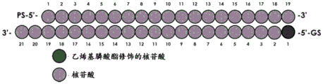
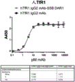
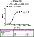


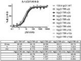


Classifications
-
- C—CHEMISTRY; METALLURGY
- C07—ORGANIC CHEMISTRY
- C07K—PEPTIDES
- C07K16/00—Immunoglobulins [IGs], e.g. monoclonal or polyclonal antibodies
- C07K16/18—Immunoglobulins [IGs], e.g. monoclonal or polyclonal antibodies against material from animals or humans
- C07K16/28—Immunoglobulins [IGs], e.g. monoclonal or polyclonal antibodies against material from animals or humans against receptors, cell surface antigens or cell surface determinants
- C07K16/2881—Immunoglobulins [IGs], e.g. monoclonal or polyclonal antibodies against material from animals or humans against receptors, cell surface antigens or cell surface determinants against CD71
-
- A—HUMAN NECESSITIES
- A61—MEDICAL OR VETERINARY SCIENCE; HYGIENE
- A61K—PREPARATIONS FOR MEDICAL, DENTAL OR TOILETRY PURPOSES
- A61K47/00—Medicinal preparations characterised by the non-active ingredients used, e.g. carriers or inert additives; Targeting or modifying agents chemically bound to the active ingredient
- A61K47/50—Medicinal preparations characterised by the non-active ingredients used, e.g. carriers or inert additives; Targeting or modifying agents chemically bound to the active ingredient the non-active ingredient being chemically bound to the active ingredient, e.g. polymer-drug conjugates
- A61K47/51—Medicinal preparations characterised by the non-active ingredients used, e.g. carriers or inert additives; Targeting or modifying agents chemically bound to the active ingredient the non-active ingredient being chemically bound to the active ingredient, e.g. polymer-drug conjugates the non-active ingredient being a modifying agent
- A61K47/68—Medicinal preparations characterised by the non-active ingredients used, e.g. carriers or inert additives; Targeting or modifying agents chemically bound to the active ingredient the non-active ingredient being chemically bound to the active ingredient, e.g. polymer-drug conjugates the non-active ingredient being a modifying agent the modifying agent being an antibody, an immunoglobulin or a fragment thereof, e.g. an Fc-fragment
- A61K47/6801—Drug-antibody or immunoglobulin conjugates defined by the pharmacologically or therapeutically active agent
- A61K47/6803—Drugs conjugated to an antibody or immunoglobulin, e.g. cisplatin-antibody conjugates
- A61K47/6807—Drugs conjugated to an antibody or immunoglobulin, e.g. cisplatin-antibody conjugates the drug or compound being a sugar, nucleoside, nucleotide, nucleic acid, e.g. RNA antisense
-
- A—HUMAN NECESSITIES
- A61—MEDICAL OR VETERINARY SCIENCE; HYGIENE
- A61K—PREPARATIONS FOR MEDICAL, DENTAL OR TOILETRY PURPOSES
- A61K47/00—Medicinal preparations characterised by the non-active ingredients used, e.g. carriers or inert additives; Targeting or modifying agents chemically bound to the active ingredient
- A61K47/50—Medicinal preparations characterised by the non-active ingredients used, e.g. carriers or inert additives; Targeting or modifying agents chemically bound to the active ingredient the non-active ingredient being chemically bound to the active ingredient, e.g. polymer-drug conjugates
- A61K47/51—Medicinal preparations characterised by the non-active ingredients used, e.g. carriers or inert additives; Targeting or modifying agents chemically bound to the active ingredient the non-active ingredient being chemically bound to the active ingredient, e.g. polymer-drug conjugates the non-active ingredient being a modifying agent
- A61K47/68—Medicinal preparations characterised by the non-active ingredients used, e.g. carriers or inert additives; Targeting or modifying agents chemically bound to the active ingredient the non-active ingredient being chemically bound to the active ingredient, e.g. polymer-drug conjugates the non-active ingredient being a modifying agent the modifying agent being an antibody, an immunoglobulin or a fragment thereof, e.g. an Fc-fragment
- A61K47/6801—Drug-antibody or immunoglobulin conjugates defined by the pharmacologically or therapeutically active agent
- A61K47/6803—Drugs conjugated to an antibody or immunoglobulin, e.g. cisplatin-antibody conjugates
- A61K47/6811—Drugs conjugated to an antibody or immunoglobulin, e.g. cisplatin-antibody conjugates the drug being a protein or peptide, e.g. transferrin or bleomycin
-
- A—HUMAN NECESSITIES
- A61—MEDICAL OR VETERINARY SCIENCE; HYGIENE
- A61K—PREPARATIONS FOR MEDICAL, DENTAL OR TOILETRY PURPOSES
- A61K47/00—Medicinal preparations characterised by the non-active ingredients used, e.g. carriers or inert additives; Targeting or modifying agents chemically bound to the active ingredient
- A61K47/50—Medicinal preparations characterised by the non-active ingredients used, e.g. carriers or inert additives; Targeting or modifying agents chemically bound to the active ingredient the non-active ingredient being chemically bound to the active ingredient, e.g. polymer-drug conjugates
- A61K47/51—Medicinal preparations characterised by the non-active ingredients used, e.g. carriers or inert additives; Targeting or modifying agents chemically bound to the active ingredient the non-active ingredient being chemically bound to the active ingredient, e.g. polymer-drug conjugates the non-active ingredient being a modifying agent
- A61K47/68—Medicinal preparations characterised by the non-active ingredients used, e.g. carriers or inert additives; Targeting or modifying agents chemically bound to the active ingredient the non-active ingredient being chemically bound to the active ingredient, e.g. polymer-drug conjugates the non-active ingredient being a modifying agent the modifying agent being an antibody, an immunoglobulin or a fragment thereof, e.g. an Fc-fragment
- A61K47/6835—Medicinal preparations characterised by the non-active ingredients used, e.g. carriers or inert additives; Targeting or modifying agents chemically bound to the active ingredient the non-active ingredient being chemically bound to the active ingredient, e.g. polymer-drug conjugates the non-active ingredient being a modifying agent the modifying agent being an antibody, an immunoglobulin or a fragment thereof, e.g. an Fc-fragment the modifying agent being an antibody or an immunoglobulin bearing at least one antigen-binding site
- A61K47/6849—Medicinal preparations characterised by the non-active ingredients used, e.g. carriers or inert additives; Targeting or modifying agents chemically bound to the active ingredient the non-active ingredient being chemically bound to the active ingredient, e.g. polymer-drug conjugates the non-active ingredient being a modifying agent the modifying agent being an antibody, an immunoglobulin or a fragment thereof, e.g. an Fc-fragment the modifying agent being an antibody or an immunoglobulin bearing at least one antigen-binding site the antibody targeting a receptor, a cell surface antigen or a cell surface determinant
-
- A—HUMAN NECESSITIES
- A61—MEDICAL OR VETERINARY SCIENCE; HYGIENE
- A61P—SPECIFIC THERAPEUTIC ACTIVITY OF CHEMICAL COMPOUNDS OR MEDICINAL PREPARATIONS
- A61P21/00—Drugs for disorders of the muscular or neuromuscular system
-
- A—HUMAN NECESSITIES
- A61—MEDICAL OR VETERINARY SCIENCE; HYGIENE
- A61P—SPECIFIC THERAPEUTIC ACTIVITY OF CHEMICAL COMPOUNDS OR MEDICINAL PREPARATIONS
- A61P35/00—Antineoplastic agents
-
- C—CHEMISTRY; METALLURGY
- C12—BIOCHEMISTRY; BEER; SPIRITS; WINE; VINEGAR; MICROBIOLOGY; ENZYMOLOGY; MUTATION OR GENETIC ENGINEERING
- C12N—MICROORGANISMS OR ENZYMES; COMPOSITIONS THEREOF; PROPAGATING, PRESERVING, OR MAINTAINING MICROORGANISMS; MUTATION OR GENETIC ENGINEERING; CULTURE MEDIA
- C12N15/00—Mutation or genetic engineering; DNA or RNA concerning genetic engineering, vectors, e.g. plasmids, or their isolation, preparation or purification; Use of hosts therefor
- C12N15/09—Recombinant DNA-technology
- C12N15/11—DNA or RNA fragments; Modified forms thereof; Non-coding nucleic acids having a biological activity
- C12N15/113—Non-coding nucleic acids modulating the expression of genes, e.g. antisense oligonucleotides; Antisense DNA or RNA; Triplex- forming oligonucleotides; Catalytic nucleic acids, e.g. ribozymes; Nucleic acids used in co-suppression or gene silencing
-
- A—HUMAN NECESSITIES
- A61—MEDICAL OR VETERINARY SCIENCE; HYGIENE
- A61K—PREPARATIONS FOR MEDICAL, DENTAL OR TOILETRY PURPOSES
- A61K39/00—Medicinal preparations containing antigens or antibodies
- A61K2039/505—Medicinal preparations containing antigens or antibodies comprising antibodies
-
- C—CHEMISTRY; METALLURGY
- C07—ORGANIC CHEMISTRY
- C07K—PEPTIDES
- C07K2317/00—Immunoglobulins specific features
- C07K2317/20—Immunoglobulins specific features characterized by taxonomic origin
- C07K2317/24—Immunoglobulins specific features characterized by taxonomic origin containing regions, domains or residues from different species, e.g. chimeric, humanized or veneered
-
- C—CHEMISTRY; METALLURGY
- C07—ORGANIC CHEMISTRY
- C07K—PEPTIDES
- C07K2317/00—Immunoglobulins specific features
- C07K2317/30—Immunoglobulins specific features characterized by aspects of specificity or valency
- C07K2317/33—Crossreactivity, e.g. for species or epitope, or lack of said crossreactivity
-
- C—CHEMISTRY; METALLURGY
- C07—ORGANIC CHEMISTRY
- C07K—PEPTIDES
- C07K2317/00—Immunoglobulins specific features
- C07K2317/50—Immunoglobulins specific features characterized by immunoglobulin fragments
- C07K2317/51—Complete heavy chain or Fd fragment, i.e. VH + CH1
-
- C—CHEMISTRY; METALLURGY
- C07—ORGANIC CHEMISTRY
- C07K—PEPTIDES
- C07K2317/00—Immunoglobulins specific features
- C07K2317/50—Immunoglobulins specific features characterized by immunoglobulin fragments
- C07K2317/52—Constant or Fc region; Isotype
-
- C—CHEMISTRY; METALLURGY
- C07—ORGANIC CHEMISTRY
- C07K—PEPTIDES
- C07K2317/00—Immunoglobulins specific features
- C07K2317/50—Immunoglobulins specific features characterized by immunoglobulin fragments
- C07K2317/56—Immunoglobulins specific features characterized by immunoglobulin fragments variable (Fv) region, i.e. VH and/or VL
- C07K2317/565—Complementarity determining region [CDR]
-
- C—CHEMISTRY; METALLURGY
- C07—ORGANIC CHEMISTRY
- C07K—PEPTIDES
- C07K2317/00—Immunoglobulins specific features
- C07K2317/70—Immunoglobulins specific features characterized by effect upon binding to a cell or to an antigen
- C07K2317/71—Decreased effector function due to an Fc-modification
-
- C—CHEMISTRY; METALLURGY
- C07—ORGANIC CHEMISTRY
- C07K—PEPTIDES
- C07K2317/00—Immunoglobulins specific features
- C07K2317/90—Immunoglobulins specific features characterized by (pharmaco)kinetic aspects or by stability of the immunoglobulin
- C07K2317/92—Affinity (KD), association rate (Ka), dissociation rate (Kd) or EC50 value
-
- C—CHEMISTRY; METALLURGY
- C07—ORGANIC CHEMISTRY
- C07K—PEPTIDES
- C07K2317/00—Immunoglobulins specific features
- C07K2317/90—Immunoglobulins specific features characterized by (pharmaco)kinetic aspects or by stability of the immunoglobulin
- C07K2317/94—Stability, e.g. half-life, pH, temperature or enzyme-resistance
-
- C—CHEMISTRY; METALLURGY
- C12—BIOCHEMISTRY; BEER; SPIRITS; WINE; VINEGAR; MICROBIOLOGY; ENZYMOLOGY; MUTATION OR GENETIC ENGINEERING
- C12N—MICROORGANISMS OR ENZYMES; COMPOSITIONS THEREOF; PROPAGATING, PRESERVING, OR MAINTAINING MICROORGANISMS; MUTATION OR GENETIC ENGINEERING; CULTURE MEDIA
- C12N2310/00—Structure or type of the nucleic acid
- C12N2310/10—Type of nucleic acid
- C12N2310/14—Type of nucleic acid interfering N.A.
-
- C—CHEMISTRY; METALLURGY
- C12—BIOCHEMISTRY; BEER; SPIRITS; WINE; VINEGAR; MICROBIOLOGY; ENZYMOLOGY; MUTATION OR GENETIC ENGINEERING
- C12N—MICROORGANISMS OR ENZYMES; COMPOSITIONS THEREOF; PROPAGATING, PRESERVING, OR MAINTAINING MICROORGANISMS; MUTATION OR GENETIC ENGINEERING; CULTURE MEDIA
- C12N2310/00—Structure or type of the nucleic acid
- C12N2310/30—Chemical structure
- C12N2310/35—Nature of the modification
- C12N2310/351—Conjugate
- C12N2310/3513—Protein; Peptide
Abstract
In certain embodiments, disclosed herein are anti-transferrin receptor antibodies, anti-transferrin receptor antibody conjugates, and pharmaceutical compositions comprising the anti-transferrin receptor antibodies or conjugates. Also disclosed herein, in some embodiments, are methods of delivering a payload using the anti-transferrin receptor antibodies described herein, as well as methods of treatment using the anti-transferrin receptor antibodies described herein.
Description
Cross-referencing
This application claims the benefit of U.S. provisional application No. 62/784,181 filed on 21.12.2018, which is incorporated herein by reference in its entirety.
Background
The invention belongs to the field of medicaments, and particularly relates to an antibody. The present invention provides anti-transferrin receptor antibodies and methods of making and using the anti-transferrin receptor antibodies.
In addition to known diagnostic uses, antibodies have proven useful as therapeutic agents. For example, immunotherapy, or the use of antibodies for therapeutic purposes, has been used to treat cancer and other disorders in recent years. The transferrin receptor is one of the most widely targeted receptors for developing targeted cancer diagnostics and therapy. This type II transmembrane glycoprotein is responsible for cellular iron transport and is found at low levels on the surface of many normal cell types. There is a need to develop improved anti-transferrin receptor antibodies for pharmaceutical use.
Disclosure of Invention
In certain embodiments, disclosed herein are anti-transferrin receptor antibodies, anti-transferrin receptor antibody conjugates, and pharmaceutical compositions comprising the anti-transferrin receptor antibodies or conjugates. Also disclosed herein, in some embodiments, are methods of delivering a payload using the anti-transferrin receptor antibodies described herein, as well as methods of treatment using the anti-transferrin receptor antibodies described herein.
In certain embodiments, disclosed herein is an anti-transferrin receptor antibody comprising a variable heavy chain (VH) region and a variable light chain (VL) region, wherein the VH region comprises a HCDR1 sequence comprising SEQ ID NO: 1; HCDR2 sequence EINPIX1GRSNYAX2KFQG, wherein X1Selected from N or Q and X2Selected from Q or E; and a HCDR3 sequence comprising SEQ ID NO 3. In some embodiments, the VH region comprises an HCDR1 sequence comprising SEQ ID NO:1, an HCDR2 sequence comprising SEQ ID NO:2, and an HCDR3 sequence comprising SEQ ID NO:3And (4) columns. In some embodiments, the VH region comprises a HCDR1 sequence comprising SEQ ID NO:1, a HCDR2 sequence comprising SEQ ID NO:4, and a HCDR3 sequence comprising SEQ ID NO: 3. In some embodiments, the VH region comprises a HCDR1 sequence comprising SEQ ID No. 1, a HCDR2 sequence comprising SEQ ID No. 5, and a HCDR3 sequence comprising SEQ ID No. 3. In some embodiments, the VL region comprises the sequence of LCDR1 RTSENIYX 3NLA, LCDR2 sequence AX4TNLAX5And LCDR3 sequence QHFWGTPLTX6Wherein X is3Selected from N or S, X4Selected from A or G, X5Is selected from D or E, and X6Present or absent, and if present, F. In some embodiments, the VL region comprises the LCDR1 sequence comprising SEQ ID NO 6, LCDR2 sequence AATNLAX5And LCDR3 sequence QHFWGTPLTX6Wherein X is5Is selected from D or E, and X6Present or absent, and if present, F. In some embodiments, the VL region comprises the LCDR1 sequence comprising SEQ ID NO. 6, the LCDR2 sequence comprising SEQ ID NO. 7, and the LCDR3 sequence comprising SEQ ID NO. 8. In some embodiments, the VL region comprises an LCDR1 sequence comprising SEQ ID NO. 6, an LCDR2 sequence comprising SEQ ID NO. 9, and an LCDR3 sequence comprising SEQ ID NO. 10. In some embodiments, the VL region comprises the LCDR1 sequence comprising SEQ ID NO. 11, the LCDR2 sequence comprising SEQ ID NO. 12, and the LCDR3 sequence comprising SEQ ID NO. 10. In some embodiments, the VH region comprises a HCDR1 sequence comprising SEQ ID No. 1, a HCDR2 sequence comprising SEQ ID No. 2, and a HCDR3 sequence comprising SEQ ID No. 3; and the VL region comprises the LCDR1 sequence comprising SEQ ID NO. 6, the LCDR2 sequence comprising SEQ ID NO. 7, and the LCDR3 sequence comprising SEQ ID NO. 8. In some embodiments, the VH region comprises a HCDR1 sequence comprising SEQ ID No. 1, a HCDR2 sequence comprising SEQ ID No. 4, and a HCDR3 sequence comprising SEQ ID No. 3; and the VL region comprises the LCDR1 sequence comprising SEQ ID NO. 6, the LCDR2 sequence comprising SEQ ID NO. 7, and the LCDR3 sequence comprising SEQ ID NO. 8. In some embodiments, the VH region comprises a HCDR1 sequence comprising SEQ ID No. 1, a HCDR2 sequence comprising SEQ ID No. 5, and a HCDR3 sequence comprising SEQ ID No. 3; and the VL region comprises the sequence LCDR1 comprising SEQ ID NO 6, comprising SEQ ID NO LCDR2 sequence of ID NO. 9 and LCDR3 sequence containing SEQ ID NO. 10. In some embodiments, the VH region comprises a HCDR1 sequence comprising SEQ ID No. 1, a HCDR2 sequence comprising SEQ ID No. 4, and a HCDR3 sequence comprising SEQ ID No. 3; and the VL region comprises the LCDR1 sequence comprising SEQ ID NO. 11, the LCDR2 sequence comprising SEQ ID NO. 12, and the LCDR3 sequence comprising SEQ ID NO. 10. In some embodiments, the VH region has at least 80%, 85%, 90%, 95%, 99%, or 100% sequence identity to a sequence selected from SEQ ID NOS 13-16. In some embodiments, the VL region has at least 80%, 85%, 90%, 95%, 99% or 100% sequence identity to a sequence selected from SEQ ID NOS 18-21. In some cases, the anti-transferrin receptor antibody comprises a humanized antibody or binding fragment thereof or a chimeric antibody or binding fragment thereof. In some cases, the anti-transferrin receptor antibody comprises a multispecific antibody or binding fragment thereof. In some embodiments, the anti-transferrin receptor antibody comprises a bispecific antibody or binding fragment thereof. In some embodiments, the anti-transferrin receptor antibody comprises an IgG-scFv, nanobody, BITE, diabody, DART, tandAb, scDiabody-CH3, triabody (triple body), minibody, TriBi minibody, scFv-CH3 KIH, Fab-scFv-Fc KIH, Fab-scFv, scFv-CH-CL-scFv, F (ab') 2-scFv2, scFv-KIH, Fab-scFv-Fc, tetravalent HCAb, scDiabody-Fc, diabody-Fc, tandem scFv-Fc, or intrabody. In some embodiments, the anti-transferrin receptor antibody comprises an IgG1 framework. In some embodiments, the anti-transferrin receptor antibody comprises an IgG2 framework. In some embodiments, the IgG2 framework is an IgG2b framework. In some embodiments, the anti-transferrin receptor antibody comprises an IgG4 framework. In some embodiments, the anti-transferrin receptor antibody further comprises at least one mutation in the Fc region. In some embodiments, the at least one mutation modulates effector function. In some embodiments, the at least one mutation reduces or eliminates Fc-gamma receptor binding. In some embodiments, the at least one mutation is at residue position D265, N297, K322, L328 or P3 29, wherein the residue position is reference IgG 1. In some embodiments, the Fc region comprises two or more, three or more, or four or more mutations. In some embodiments, the Fc region comprises mutations at L233 and L234, wherein the residues correspond to positions 233 and 234 of SEQ ID No. 23. In some embodiments, the Fc region comprises mutations at D265 and N297. In some embodiments, the anti-transferrin receptor antibody comprises a Heavy Chain (HC) sequence selected from SEQ ID NOS: 23-46 and a Light Chain (LC) sequence selected from SEQ ID NOS: 47-50. In some embodiments, the anti-transferrin receptor antibody specifically binds to human transferrin receptor (TfR).
In certain embodiments, disclosed herein is an anti-transferrin receptor antibody conjugate comprising an anti-transferrin receptor antibody described herein and a payload. In some embodiments, the payload comprises a small molecule, peptide, protein, or polynucleic acid molecule. In some embodiments, the payload comprises a polynucleic acid molecule. In some embodiments, the polynucleic acid molecule comprises a short interfering nucleic acid (siNA), a short interfering rna (sirna), a double-stranded rna (dsrna), a microrna (mirna), a short hairpin rna (shrna), an antisense oligonucleotide (ASO), a PMO, or an mRNA. In some embodiments, the payload comprises dsRNA. In some embodiments, the payload comprises an antisense oligonucleotide (ASO). In some embodiments, the payload comprises a small molecule, peptide, or protein. In some embodiments, the payload comprises a microtubule disrupting agent, a DNA modifying agent, or an Akt inhibitor. In some embodiments, the payload comprises an auristatin (auristatin) or a derivative thereof, dolastatin (dolastatin) or a derivative or analog thereof, a maytansinoid, or a pyrrolobenzodiazepine  Or a derivative thereof. In some embodiments, the auristatin or derivative thereof is monomethyl auristatin e (mmae) or monomethyl auristatin f (mmaf). In some embodiments, the maytansinoid is DM1 or
 DM 4. In some implementationsIn scheme (I), pyrrolobenzodiazepines Is a pyrrolobenzodiazepine
 A dimer. In some embodiments, the payload comprises an immunomodulatory agent or an immunomodulatory agent. In some embodiments, the immunomodulator comprises a cytokine. In some embodiments, the payload comprises a protein or peptide toxin or fragment thereof. In some embodiments, the payload is conjugated to the anti-transferrin receptor antibody via a linker. In some embodiments, the anti-transferrin receptor antibody is further conjugated to two or more payloads. In some embodiments, the ratio of the payload to the anti-transferrin receptor antibody is about 1:1, 2:1, 3:1, 4:1, 5:1, 6:1, 7:1, 8:1, 9:1, 10:1, 11:1, or 12: 1. In some embodiments, the anti-transferrin conjugate comprises a- (X)1-B)n(formula (I)) wherein a comprises the anti-transferrin antibody; b contains the payload; x 1Consisting of a bond or a linker; and n is an average value selected from 1 to 12. In some embodiments, the payload is a polynucleic acid molecule. In some embodiments, the polynucleic acid molecule comprises a passenger strand and a guide strand. In some embodiments, the guide strand comprises at least one modified internucleotide linkage, at least one inverted abasic moiety, at least one 5' -vinylphosphonate modified non-natural nucleotide, or a combination thereof. In some embodiments, the at least one 5 '-vinylphosphonate modified non-natural nucleotide is located about 1, 2, 3, 4, or 5 bases from the 5' terminus of the guide strand. In some embodiments, the polynucleic acid molecule further comprises a modification of the sugar moiety at the 2' position. In some embodiments, the modification at the 2' -position is selected from the group consisting of 2' -O-methyl, 2' -O-methoxyethyl (2 ' -O-MOE), 2' -deoxy, T-deoxy-2 ' -fluoro, 2' -O-aminopropyl (2 ' -O-AP), 2' -O-dimethylaminoethyl(2 '-O-DMAOE), 2' -O-dimethylaminopropyl (2 '-O-DMAP), T-O-dimethylaminoethoxyethyl (2' -O-DMAEOE) or 2 '-O-N-methylacetamido (2' -O-NMA) modified nucleotides. In some embodiments, the passenger strand comprises at least 6, 7, 8, 9, 10, 11, 12, 13, 14, 15, 16, 17, 18, 19, 20 or more phosphorodiamidate morpholino oligomer modified non-natural nucleotides. In some embodiments, the passenger strand is shorter in length than the guide strand, thereby creating a 5 'overhang, a 3' overhang, a blunt end, or a combination thereof at one end. In some embodiments, the passenger strand has a length equal to the guide strand, thereby generating blunt ends at each end of the polynucleic acid molecule. In some embodiments, the passenger chain is linked to A-X 1And (6) conjugation. In some embodiments, A-X1Conjugated to the 5' end of the passenger strand. In some embodiments, A-X1Conjugated to the 3' end of the passenger strand. In some embodiments, the anti-transferrin conjugate comprises: A-X1-(B-X2-C)n(formula (II)) wherein a comprises the anti-transferrin receptor antibody; b comprises the polynucleic acid molecule; c consists of a polymer; x1Consisting of a key or a first connector; x2Consisting of a bond or a second linker; and n is an average value selected from 1 to 12. In some embodiments, C is polyethylene glycol. In some embodiments, the polynucleic acid molecule comprises a passenger strand and a guide strand. In some embodiments, the passenger chain is linked to A-X1And X2-C conjugation. In some embodiments, A-X1Is conjugated to the 5' end of the passenger strand, and X2-C is conjugated to the 3' end of the passenger strand. In some embodiments, X2-C is conjugated to the 5' end of the passenger strand, and A-X1Conjugated to the 3' end of the passenger strand. In some embodiments, X1And X2Each independently is a non-polymeric linker. In some embodiments, the anti-transferrin receptor antibody conjugate further comprises D. In some embodiments, D is an endosomolytic (endosomolytic) moiety.
In certain embodiments, disclosed herein is a nucleic acid polymer encoding an anti-transferrin receptor antibody described herein.
In certain embodiments, disclosed herein is a vector comprising a nucleic acid polymer encoding an anti-transferrin receptor antibody described herein.
In certain embodiments, disclosed herein is a pharmaceutical composition comprising: an anti-transferrin receptor antibody described herein or an anti-transferrin receptor antibody conjugate described herein; and a pharmaceutically acceptable excipient. In some embodiments, the pharmaceutical composition is formulated for systemic administration. In some embodiments, the pharmaceutical composition is formulated for parenteral administration.
In certain embodiments, disclosed herein is a method of delivering a payload to a target site of interest of a subject, comprising: administering to the subject an anti-transferrin receptor antibody conjugate as described herein or a pharmaceutical composition as described herein to deliver a payload to a target site of interest. In some embodiments, the target site of interest is a cell comprising an overexpressed pathogenic protein. In some embodiments, the target site of interest is a tumor site. In some embodiments, the target site of interest is a site located within the brain.
In certain embodiments, disclosed herein is a method of treating cancer in a subject in need thereof, comprising: administering to the subject an anti-transferrin receptor antibody conjugate as described herein or a pharmaceutical composition as described herein to treat cancer in the subject. In some embodiments, the cancer is a solid cancer. In some embodiments, the cancer is a hematologic malignancy. In some embodiments, the cancer is bladder cancer, lung cancer, brain cancer, melanoma, breast cancer, non-hodgkin's lymphoma, cervical cancer, ovarian cancer, colorectal cancer, pancreatic cancer, esophageal cancer, prostate cancer, renal cancer, skin cancer, leukemia, thyroid cancer, liver cancer, or uterine cancer. In some embodiments, the cancer is metastatic cancer. In some embodiments, the cancer is a relapsed or refractory cancer.
In certain embodiments, disclosed herein is a method of treating muscle atrophy or myotonic dystrophy in a subject in need thereof, comprising: administering to the subject the anti-transferrin receptor antibody conjugate described herein or the pharmaceutical composition described herein, wherein the polynucleic acid molecule hybridizes to a target sequence of atrogene, and wherein the polynucleic acid molecule mediates RNA interference against atrogene, thereby treating muscle atrophy in the subject. In some embodiments, the muscle atrophy is diabetes-related muscle atrophy or cancer cachexia-related muscle atrophy. In some embodiments, the muscle atrophy is associated with insulin deficiency, chronic renal failure, congestive heart failure, chronic respiratory disease, chronic infection, fasting, denervation, sarcopenia, or type 1 myotonic dystrophy (DM 1). In some embodiments, there is no reduction in reticulocyte levels in the subject following administration of the anti-transferrin receptor antibody. In some embodiments, administration of the anti-transferrin receptor antibody conjugate down-regulates SSB siRNA or SSB mRNA levels in the subject. In some embodiments, the down-regulation of SSB siRNA or SSB mRNA occurs in muscle. In some embodiments, the muscle is skeletal muscle. In some embodiments, the muscle is cardiac muscle.
In some embodiments, the myotonic dystrophy is DM 1. In some embodiments, the atrogene comprises a gene that is upregulated within the IGF1-Akt-FoxO pathway, glucocorticoid-GR pathway, PGC1 α -FoxO pathway, TNF α -nfkb pathway, or myostatin-ActRIIb-Smad 2/3 pathway. In some embodiments, the atrogene encodes E3 ligase. In some embodiments, the atrogene encodes a forkhead box transcription factor. In some embodiments, the atrogene comprises an atrogin-1 gene (FBXO32), MuRF1 gene (TRIM63), FOXO1, FOXO3, or MSTN. In some embodiments, the atrogene comprises DMPK. In some embodiments, the subject is a human.
In certain embodiments, disclosed herein is a method of treating muscular dystrophy in a subject in need thereof, comprising: administering to the subject an anti-transferrin receptor antibody conjugate as described herein or a pharmaceutical composition as described herein, thereby treating muscular dystrophy in the subject. In some embodiments, the muscular dystrophy is duchenne muscular dystrophy, becker muscular dystrophy, facioscapulohumeral muscular dystrophy, congenital muscular dystrophy, or myotonic dystrophy. In some embodiments, the muscular dystrophy is duchenne muscular dystrophy. In some embodiments, the subject is a human.
In certain embodiments, disclosed herein is a kit comprising an anti-transferrin receptor antibody described herein, an anti-transferrin receptor antibody conjugate described herein, a nucleic acid polymer described herein, a vector described herein, or a pharmaceutical composition described herein.
Drawings
Various aspects of the disclosure are set forth with particularity in the appended claims. A better understanding of the features and advantages of the present disclosure will be obtained by reference to the following detailed description that sets forth illustrative embodiments, in which the principles of the disclosure are utilized, and the following drawings. This patent application document contains at least one drawing executed in color. Copies of this patent application publication with color drawing(s) will be provided by the office upon request and payment of the necessary fee.
Fig. 1 shows the structure of an exemplary SSB passenger chain.
FIG. 2 shows the structure of an exemplary blunt-ended duplex with 19 base complementarity and a 3' dinucleotide overhang. The purified single strand is duplexed to obtain double stranded siRNA.
Figure 3A shows in vitro binding of TfR1.igg2 mAb and TfR1.igg2 mAb-SSB to recombinant human TfR1.
Figure 3B shows in vitro binding of TfR1.igg2 mAb and TfR1.igg2 mAb-SSB to recombinant cynomolgus monkey TfR1.
Figure 4A shows SSB mRNA levels in siRNA delivery in hel92.1.7 cells treated with htfr1.igg2 mAb SSB or htfr1.igg2 mAb MSTN (negative control) conjugates.
Figure 4B shows SSB mRNA levels in siRNA delivery in immortalized human skeletal muscle cells treated with htfr1.igg2 mAb SSB or htfr1.igg2 mAb MSTN (negative control) conjugates.
Figure 5A shows SSB mRNA and SSB siRNA levels in gastrocnemius of cynomolgus monkeys after administration of htfr1.igg2 mAb-SSB conjugate at 30 and 60mg/kg (n-3).
Figure 5B shows SSB mRNA and SSB siRNA levels in the quadriceps of cynomolgus monkeys after administration of htfr1.igg2 mAb-SSB conjugate at 30 and 60mg/kg (n-3).
Figure 6 shows relative reticulocyte levels in cynomolgus monkeys before and after dosing with htfr1.igg2 mAb-SSB conjugate at 30 and 60 mg/kg.
Figure 7 shows the binding constants of exemplary anti-TfR antibodies to cynomolgus monkey CD 71.
Figure 8 shows the binding constants of exemplary anti-TfR antibodies to human CD 71.
Fig. 9A shows binding of an exemplary anti-TfR antibody to TfR in a competition setting.
Fig. 9B shows the binding constants of the test anti-TfR antibodies of fig. 9A.
Fig. 10A shows that binding of an exemplary anti-TfR antibody to TfR is maintained.
Fig. 10B shows the binding constants of the test anti-TfR antibodies of fig. 10A.
Figure 11 shows ADCC activity of exemplary anti-TfR antibodies.
Figure 12 shows that anti-TfR antibodies do not bind to TfR 2.
Figure 13A shows% SSB mRNA knockdown in HEL92 cells.
Fig. 13B shows EC50 for the test anti-TfR antibody of fig. 13A.
Figure 14 shows ADCC activity of exemplary anti-TfR antibodies.
Figure 15 shows CDC activity of exemplary anti-TfR antibodies.
Fig. 16 shows the uptake of tfr1.mab conjugate in primary human skeletal muscle cells (myotubes).
Fig. 17 shows SSB mRNA levels in primary human skeletal muscle cells treated with a tfr1.higg2 mAb or SSB or scrambled (scarmble) siRNA conjugate of a tfr1.higg1 mAb variant.
Fig. 18 shows absolute reticulocyte levels in cynomolgus monkeys before/after TfR1 targeted AOC administration (single dose on day 1).
Figure 19 shows SSB mRNA levels in cynomolgus monkey muscle 21 days after a single dose of TfR1 mAb SSB conjugate (n-3).
Figure 20A shows SSB siRNA levels in cynomolgus monkey tissues 21 days after a single 6mg/kg dose of hIgG1 TfR-Var2ii-SSB conjugate (n ═ 2).
Figure 20B shows SSB mRNA levels in cynomolgus monkey tissues 21 days after a single 6mg/kg dose of hIgG1 TfR-Var2ii-SSB conjugate (n ═ 2).
Detailed Description
The transferrin receptor (TfR) comprises a family of membrane glycoproteins and is encoded by the gene TFRC. TfR participates in iron metabolism by interacting with the iron-transferrin complex to facilitate the entry of iron into cells. TfR has two subtypes: transferrin receptor 1(TfR1 or CD71) and transferrin receptor 2(TfR 2). TfR1 is ubiquitously expressed in different cell types, whereas TfR2 is specifically expressed in hepatocytes.
In some cases, aberrant expression of TfR1 has been noted in various cancers. In fact, one study showed that Expression levels of TfR1 were elevated in breast cancer cells (Pizzamiglio et al, "Expression of iron-related proteins differential non-cancer and cancer breast cultures," Int J Mol Sci.2017; 18). In a separate study, TFR1 was also shown to be overexpressed in brain cancers (Rosager et al, "Transferrin receiver-1 and transferin help and light chains in inhibitory peptides tumors: Expression and viral value," PLoS One 12: e0182954 (2017)). Further studies have shown increased iron uptake in tumor initiating cells (Rychtarcikova et al, "tumor initiating cells of Breast and State origin colors in the expression of genes related to iron metabolism," oncogenetarget.8: 6376-6398 (2017)).
In some embodiments, disclosed herein are anti-transferrin receptor antibodies, anti-transferrin receptor antibody conjugates, and pharmaceutical compositions comprising the same. In additional embodiments, disclosed herein are methods of delivering a payload using an anti-transferrin receptor antibody, and methods of treating a disease or condition by targeted delivery using the presence of transferrin receptor.
Anti-transferrin receptor antibodies
In certain embodiments, disclosed herein are anti-transferrin receptor antibodies. In some cases, the anti-transferrin receptor antibody specifically binds to transferrin receptor (TfR). In some cases, the anti-transferrin receptor antibody specifically binds to human transferrin receptor (TfR). In some cases, the anti-transferrin receptor antibody specifically binds to transferrin receptor 1(TfR1) (or CD 71). In some cases, the anti-transferrin receptor antibody specifically binds to human transferrin receptor 1(TfR1) (or human CD 71).
In some cases, the anti-transferrin receptor antibody comprises a variable heavy chain (VH) region and a variable light chain (VL) region, wherein the VH region comprises a HCDR1 sequence comprising SEQ ID NO: 1; HCDR2 sequence EINPIX 1GRSNYAX2KFQG, wherein X1Selected from N or Q and X2Selected from Q or E; and a HCDR3 sequence comprising SEQ ID NO 3.
In some embodiments, the VH region of the anti-transferrin receptor antibody comprises a sequence selected from HCDR1, HCDR2, and HCDR3 of table 1.
Table 1.

13E4_ VH2 has the same HCDR1, HCDR2 and HCDR3 sequences as the anti-transferrin receptor antibody 13E4_ VH 4.
In some embodiments, the VH region comprises a HCDR1 sequence comprising SEQ ID NO:1, a HCDR2 sequence comprising SEQ ID NO:2, 4 or 5, and a HCDR3 sequence comprising SEQ ID NO: 3. In some cases, the VH region comprises a HCDR1 sequence comprising SEQ ID NO:1, a HCDR2 sequence comprising SEQ ID NO:2, and a HCDR3 sequence comprising SEQ ID NO: 3. In some cases, the VH region comprises a HCDR1 sequence comprising SEQ ID No. 1, a HCDR2 sequence comprising SEQ ID No. 4, and a HCDR3 sequence comprising SEQ ID No. 3. In some cases, the VH region comprises a HCDR1 sequence comprising SEQ ID No. 1, a HCDR2 sequence comprising SEQ ID No. 5, and a HCDR3 sequence comprising SEQ ID No. 3.
In some embodiments, the VL region of the anti-transferrin receptor antibody comprises the sequence of LCDR1 RTSENIYX3NLA, LCDR2 sequence AX4TNLAX5And LCDR3 sequence QHFWGTPLTX6Wherein X is 3Selected from N or S, X4Selected from A or G, X5Is selected from D or E, and X6Present or absent, and if present, F.
In some embodiments, the VL region of the anti-transferrin receptor antibody comprises an LCDR1, LCDR2, and LCDR3 sequence selected from table 2.
Table 2.

13E4_ VL1 has the same LCDR1, LCDR2 and LCDR3 sequences as the anti-transferrin receptor antibody 13E4_ VL 2.
In some cases, the VL region comprises the LCDR1 sequence RTSENIYX3NLA, LCDR2 sequence comprising SEQ ID NO 7, 9 or 12 and LCDR3 sequence comprising SEQ ID NO 8 or 10, wherein X3Is selected from N or S.
In some cases, the VL region comprises the LCDR1 sequence comprising SEQ ID NO 6 or 11, LCDR2 sequence AX4TNLAX5And LCDR3 sequence comprising SEQ ID NO 8 or 10, wherein X4Is selected from A or G, and X5Is selected from D or E.
In some cases, the VL region comprises the LCDR1 sequence comprising SEQ ID NO 6 or 11, the LCDR2 sequence SEQ ID NO 7, 9 or 12, and the LCDR3 sequence QHFWGTPLTX6Wherein X is6Present or absent, and if present, F.
In some cases, the VL region comprises an LCDR1 sequence comprising SEQ ID NO 6, an LCDR2 sequence AATNLAX5, and an LCDR3 sequence QHFWGTPLTX6Wherein X is5Is selected from D or E, and X6Is present or absent if If present, is F.
In some cases, the VL region comprises an LCDR1 sequence comprising SEQ ID No. 6, an LCDR2 sequence comprising SEQ ID No. 7, and an LCDR3 sequence comprising SEQ ID No. 8.
In some cases, the VL region comprises an LCDR1 sequence comprising SEQ ID No. 6, an LCDR2 sequence comprising SEQ ID No. 9, and an LCDR3 sequence comprising SEQ ID No. 10.
In some cases, the VL region comprises an LCDR1 sequence comprising SEQ ID No. 11, an LCDR2 sequence comprising SEQ ID No. 12, and an LCDR3 sequence comprising SEQ ID No. 10.
In some embodiments, the anti-transferrin receptor antibody comprises a VH region and a VL region, wherein the VH region comprises a HCDR1 sequence comprising SEQ ID NO: 1; HCDR2 sequence EINPIX1GRSNYAX2KFQG, wherein X1Is selected from N or Q, and X2Selected from Q or E; and a HCDR3 sequence comprising SEQ ID NO 3; and the VL region comprises the sequence LCDR1 RTSENIYX3NLA, LCDR2 sequence AX4TNLAX5And LCDR3 sequence QHFWGTPLTX6Wherein X is3Selected from N or S, X4Selected from A or G, X5Is selected from D or E, and X6Present or absent, and if present, F.
In some cases, the anti-transferrin receptor antibody comprises a VH region and a VL region, wherein the VH region comprises a HCDR1 sequence comprising SEQ ID NO: 1; HCDR2 sequence EINPIX 1GRSNYAX2KFQG, wherein X1Is selected from N or Q, and X2Selected from Q or E; and a HCDR3 sequence comprising SEQ ID NO 3; and the VL region comprises the sequence LCDR1 RTSENIYX3NLA, LCDR2 sequence comprising SEQ ID NO 7, 9 or 12 and LCDR3 sequence comprising SEQ ID NO 8 or 10, wherein X3Is selected from N or S.
In some cases, the anti-transferrin receptor antibody comprises a VH region and a VL region, wherein the VH region comprises a HCDR1 sequence comprising SEQ ID NO: 1; HCDR2 sequence EINPIX1GRSNYAX2KFQG, wherein X1Is selected from N or Q, and X2Selected from Q or E; and a HCDR3 sequence comprising SEQ ID NO 3; and the VL region comprises the LCDR1 sequence comprising SEQ ID NO 6 or 11LCDR2 sequence AX4TNLAX5And LCDR3 sequence comprising SEQ ID NO 8 or 10, wherein X4Is selected from A or G, and X5Is selected from D or E.
In some cases, the anti-transferrin receptor antibody comprises a VH region and a VL region, wherein the VH region comprises a HCDR1 sequence comprising SEQ ID NO: 1; HCDR2 sequence EINPIX1GRSNYAX2KFQG, wherein X1Is selected from N or Q, and X2Selected from Q or E; and a HCDR3 sequence comprising SEQ ID NO 3; and the VL domain comprises the sequence LCDR1 comprising the sequence SEQ ID NO 6 or 11, the sequence LCDR2 the sequence SEQ ID NO 7, 9 or 12 and the sequence LCDR3 QHFWGTPLTX6Wherein X is6Present or absent, and if present, F.
In some cases, the anti-transferrin receptor antibody comprises a VH region and a VL region, wherein the VH region comprises a HCDR1 sequence comprising SEQ ID NO: 1; HCDR2 sequence EINPIX1GRSNYAX2KFQG, wherein X1Is selected from N or Q, and X2Selected from Q or E; and a HCDR3 sequence comprising SEQ ID NO 3; and the VL domain comprises the sequence LCDR1, the sequence LCDR2, the sequence AATNLAX5 and the sequence LCDR3, the sequence QHFWGTPLTX, comprising SEQ ID NO 66Wherein X is5Is selected from D or E, and X6Present or absent, and if present, F.
In some cases, the anti-transferrin receptor antibody comprises a VH region and a VL region, wherein the VH region comprises a HCDR1 sequence comprising SEQ ID NO: 1; HCDR2 sequence EINPIX1GRSNYAX2KFQG, wherein X1Is selected from N or Q, and X2Selected from Q or E; and a HCDR3 sequence comprising SEQ ID NO 3; and the VL region comprises the sequence LCDR1 comprising SEQ ID NO. 6, LCDR2 comprising SEQ ID NO. 7, and LCDR3 comprising SEQ ID NO. 8.
In some cases, the anti-transferrin receptor antibody comprises a VH region and a VL region, wherein the VH region comprises a HCDR1 sequence comprising SEQ ID NO: 1; HCDR2 sequence EINPIX1GRSNYAX2KFQG, wherein X1Is selected from N or Q, and X2Selected from Q or E; and a HCDR3 sequence comprising SEQ ID NO 3; and the VL region comprises the sequence LCDR1 comprising SEQ ID NO 6, the VL region comprising SEQ ID NO 9 LCDR2 sequence and LCDR3 sequence containing SEQ ID NO 10.
In some cases, the anti-transferrin receptor antibody comprises a VH region and a VL region, wherein the VH region comprises a HCDR1 sequence comprising SEQ ID NO: 1; HCDR2 sequence EINPIX1GRSNYAX2KFQG, wherein X1Is selected from N or Q, and X2Selected from Q or E; and a HCDR3 sequence comprising SEQ ID NO 3; and the VL region comprises the sequence LCDR1 comprising SEQ ID NO. 11, LCDR2 comprising SEQ ID NO. 12 and LCDR3 comprising SEQ ID NO. 10.
In some cases, the anti-transferrin receptor antibody comprises a VH region and a VL region, wherein the VH region comprises a HCDR1 sequence comprising SEQ ID No. 1, a HCDR2 sequence comprising SEQ ID No. 2, and a HCDR3 sequence comprising SEQ ID No. 3; and the VL region comprises the sequence LCDR1 RTSENIYX3NLA, LCDR2 sequence comprising SEQ ID NO 7, 9 or 12 and LCDR3 sequence comprising SEQ ID NO 8 or 10, wherein X3Is selected from N or S.
In some cases, the anti-transferrin receptor antibody comprises a VH region and a VL region, wherein the VH region comprises a HCDR1 sequence comprising SEQ ID No. 1, a HCDR2 sequence comprising SEQ ID No. 2, and a HCDR3 sequence comprising SEQ ID No. 3; and the VL domain comprises the sequence LCDR1 comprising SEQ ID NO 6 or 11, LCDR2 sequence AX 4TNLAX5And LCDR3 sequence comprising SEQ ID NO 8 or 10, wherein X4Is selected from A or G, and X5Is selected from D or E.
In some cases, the anti-transferrin receptor antibody comprises a VH region and a VL region, wherein the VH region comprises a HCDR1 sequence comprising SEQ ID No. 1, a HCDR2 sequence comprising SEQ ID No. 2, and a HCDR3 sequence comprising SEQ ID No. 3; and the VL domain comprises the sequence LCDR1 comprising the sequence SEQ ID NO 6 or 11, the sequence LCDR2 the sequence SEQ ID NO 7, 9 or 12 and the sequence LCDR3 QHFWGTPLTX6Wherein X is6Present or absent, and if present, F.
In some cases, the anti-transferrin receptor antibody comprises a VH region and a VL region, wherein the VH region comprises a HCDR1 sequence comprising SEQ ID No. 1, a HCDR2 sequence comprising SEQ ID No. 2, and a HCDR3 sequence comprising SEQ ID No. 3; and the VL region comprises L comprising SEQ ID NO 6CDR1 sequence, LCDR2 sequence AATNLAX5 and LCDR3 sequence QHFWGTPLTX6Wherein X is5Is selected from D or E, and X6Present or absent, and if present, F.
In some cases, the anti-transferrin receptor antibody comprises a VH region and a VL region, wherein the VH region comprises a HCDR1 sequence comprising SEQ ID No. 1, a HCDR2 sequence comprising SEQ ID No. 2, and a HCDR3 sequence comprising SEQ ID No. 3; and the VL region comprises the sequence LCDR1 comprising SEQ ID NO. 6, LCDR2 comprising SEQ ID NO. 7, and LCDR3 comprising SEQ ID NO. 8.
In some cases, the anti-transferrin receptor antibody comprises a VH region and a VL region, wherein the VH region comprises a HCDR1 sequence comprising SEQ ID No. 1, a HCDR2 sequence comprising SEQ ID No. 2, and a HCDR3 sequence comprising SEQ ID No. 3; and the VL region comprises the sequence LCDR1 comprising SEQ ID NO. 6, LCDR2 comprising SEQ ID NO. 9 and LCDR3 comprising SEQ ID NO. 10.
In some cases, the anti-transferrin receptor antibody comprises a VH region and a VL region, wherein the VH region comprises a HCDR1 sequence comprising SEQ ID No. 1, a HCDR2 sequence comprising SEQ ID No. 2, and a HCDR3 sequence comprising SEQ ID No. 3; and the VL region comprises the sequence LCDR1 comprising SEQ ID NO. 11, LCDR2 comprising SEQ ID NO. 12 and LCDR3 comprising SEQ ID NO. 10.
In some cases, the anti-transferrin receptor antibody comprises a VH region and a VL region, wherein the VH region comprises a HCDR1 sequence comprising SEQ ID No. 1, a HCDR2 sequence comprising SEQ ID No. 4, and a HCDR3 sequence comprising SEQ ID No. 3; and the VL region comprises the sequence LCDR1 RTSENIYX3NLA, LCDR2 sequence comprising SEQ ID NO 7, 9 or 12 and LCDR3 sequence comprising SEQ ID NO 8 or 10, wherein X3Is selected from N or S.
In some cases, the anti-transferrin receptor antibody comprises a VH region and a VL region, wherein the VH region comprises a HCDR1 sequence comprising SEQ ID No. 1, a HCDR2 sequence comprising SEQ ID No. 4, and a HCDR3 sequence comprising SEQ ID No. 3; and the VL domain comprises the sequence LCDR1 comprising SEQ ID NO 6 or 11, LCDR2 sequence AX 4TNLAX5And LCDR3 sequence comprising SEQ ID NO 8 or 10, wherein X4Is selected from AOr G, and X5Is selected from D or E.
In some cases, the anti-transferrin receptor antibody comprises a VH region and a VL region, wherein the VH region comprises a HCDR1 sequence comprising SEQ ID No. 1, a HCDR2 sequence comprising SEQ ID No. 4, and a HCDR3 sequence comprising SEQ ID No. 3; and the VL domain comprises the sequence LCDR1 comprising the sequence SEQ ID NO 6 or 11, the sequence LCDR2 the sequence SEQ ID NO 7, 9 or 12 and the sequence LCDR3 QHFWGTPLTX6Wherein X is6Present or absent, and if present, F.
In some cases, the anti-transferrin receptor antibody comprises a VH region and a VL region, wherein the VH region comprises a HCDR1 sequence comprising SEQ ID No. 1, a HCDR2 sequence comprising SEQ ID No. 4, and a HCDR3 sequence comprising SEQ ID No. 3; and the VL domain comprises the sequence LCDR1, the sequence LCDR2, the sequence AATNLAX5 and the sequence LCDR3, the sequence QHFWGTPLTX, comprising SEQ ID NO 66Wherein X is5Is selected from D or E, and X6Present or absent, and if present, F.
In some cases, the anti-transferrin receptor antibody comprises a VH region and a VL region, wherein the VH region comprises a HCDR1 sequence comprising SEQ ID No. 1, a HCDR2 sequence comprising SEQ ID No. 4, and a HCDR3 sequence comprising SEQ ID No. 3; and the VL region comprises the sequence LCDR1 comprising SEQ ID NO. 6, LCDR2 comprising SEQ ID NO. 7, and LCDR3 comprising SEQ ID NO. 8.
In some cases, the anti-transferrin receptor antibody comprises a VH region and a VL region, wherein the VH region comprises a HCDR1 sequence comprising SEQ ID No. 1, a HCDR2 sequence comprising SEQ ID No. 4, and a HCDR3 sequence comprising SEQ ID No. 3; and the VL region comprises the sequence LCDR1 comprising SEQ ID NO. 6, LCDR2 comprising SEQ ID NO. 9 and LCDR3 comprising SEQ ID NO. 10.
In some cases, the anti-transferrin receptor antibody comprises a VH region and a VL region, wherein the VH region comprises a HCDR1 sequence comprising SEQ ID No. 1, a HCDR2 sequence comprising SEQ ID No. 4, and a HCDR3 sequence comprising SEQ ID No. 3; and the VL region comprises the sequence LCDR1 comprising SEQ ID NO. 11, LCDR2 comprising SEQ ID NO. 12 and LCDR3 comprising SEQ ID NO. 10.
In some cases, the antibodyThe transferrin receptor antibody comprises a VH region and a VL region, wherein the VH region comprises the sequence HCDR1 comprising SEQ ID NO:1, the sequence HCDR2 comprising SEQ ID NO:5, and the sequence HCDR3 comprising SEQ ID NO: 3; and the VL region comprises the sequence LCDR1 RTSENIYX3NLA, LCDR2 sequence comprising SEQ ID NO 7, 9 or 12 and LCDR3 sequence comprising SEQ ID NO 8 or 10, wherein X3Is selected from N or S.
In some cases, the anti-transferrin receptor antibody comprises a VH region and a VL region, wherein the VH region comprises a HCDR1 sequence comprising SEQ ID No. 1, a HCDR2 sequence comprising SEQ ID No. 5, and a HCDR3 sequence comprising SEQ ID No. 3; and the VL domain comprises the sequence LCDR1 comprising SEQ ID NO 6 or 11, LCDR2 sequence AX 4TNLAX5And LCDR3 sequence comprising SEQ ID NO 8 or 10, wherein X4Is selected from A or G, and X5Is selected from D or E.
In some cases, the anti-transferrin receptor antibody comprises a VH region and a VL region, wherein the VH region comprises a HCDR1 sequence comprising SEQ ID No. 1, a HCDR2 sequence comprising SEQ ID No. 5, and a HCDR3 sequence comprising SEQ ID No. 3; and the VL domain comprises the sequence LCDR1 comprising the sequence SEQ ID NO 6 or 11, the sequence LCDR2 the sequence SEQ ID NO 7, 9 or 12 and the sequence LCDR3 QHFWGTPLTX6Wherein X is6Present or absent, and if present, F.
In some cases, the anti-transferrin receptor antibody comprises a VH region and a VL region, wherein the VH region comprises a HCDR1 sequence comprising SEQ ID No. 1, a HCDR2 sequence comprising SEQ ID No. 5, and a HCDR3 sequence comprising SEQ ID No. 3; and the VL domain comprises the sequence LCDR1, the sequence LCDR2, the sequence AATNLAX5 and the sequence LCDR3, the sequence QHFWGTPLTX, comprising SEQ ID NO 66Wherein X is5Is selected from D or E, and X6Present or absent, and if present, F.
In some cases, the anti-transferrin receptor antibody comprises a VH region and a VL region, wherein the VH region comprises a HCDR1 sequence comprising SEQ ID No. 1, a HCDR2 sequence comprising SEQ ID No. 5, and a HCDR3 sequence comprising SEQ ID No. 3; and the VL region comprises the sequence LCDR1 comprising SEQ ID NO. 6, LCDR2 comprising SEQ ID NO. 7, and LCDR3 comprising SEQ ID NO. 8.
In some cases, the anti-transferrin receptor antibody comprises a VH region and a VL region, wherein the VH region comprises a HCDR1 sequence comprising SEQ ID No. 1, a HCDR2 sequence comprising SEQ ID No. 5, and a HCDR3 sequence comprising SEQ ID No. 3; and the VL region comprises the sequence LCDR1 comprising SEQ ID NO. 6, LCDR2 comprising SEQ ID NO. 9 and LCDR3 comprising SEQ ID NO. 10.
In some cases, the anti-transferrin receptor antibody comprises a VH region and a VL region, wherein the VH region comprises a HCDR1 sequence comprising SEQ ID No. 1, a HCDR2 sequence comprising SEQ ID No. 5, and a HCDR3 sequence comprising SEQ ID No. 3; and the VL region comprises the sequence LCDR1 comprising SEQ ID NO. 11, LCDR2 comprising SEQ ID NO. 12 and LCDR3 comprising SEQ ID NO. 10.
In some embodiments, an anti-transferrin receptor antibody comprises a VH region and a VL region, wherein the sequence of the VH region has about 80%, 85%, 90%, 95%, 96%, 97%, 98%, 99%, or 100% sequence identity to SEQ ID NOs 13-16 and the sequence of the VL region has about 80%, 85%, 90%, 95%, 96%, 97%, 98%, 99%, or 100% sequence identity to SEQ ID NOs 18-21.
In some embodiments, the VH region comprises a sequence selected from SEQ ID NOS: 13-16 (Table 3) and the VL region comprises a sequence selected from SEQ ID NOS: 18-21 (Table 4). The underlined regions in tables 3 and 4 represent the respective CDR1, CDR2 or CDR3 sequences.
TABLE 3

TABLE 4


In some embodiments, the anti-transferrin receptor antibody comprises a VH region and a VL region as set forth in table 5.
TABLE 5

In some embodiments, the anti-transferrin receptor antibody described above is a full length antibody. In other embodiments, the anti-transferrin receptor antibody is a binding fragment thereof. In some cases, the anti-transferrin receptor antibody is a humanized antibody or binding fragment thereof, a chimeric antibody or binding fragment thereof, a monoclonal antibody or binding fragment thereof, a multispecific antibody or binding fragment thereof, or a bispecific antibody or binding fragment thereof. In some cases, the anti-transferrin receptor antibody is a monovalent Fab', a bivalent Fab2、F(ab)'3Fragment, Single chain variable fragment (scFv), bis-scFv, (scFv)2A diabody, a minibody, a nanobody, a triabody, a tetrabody, a disulfide stabilized Fv protein ("dsFv"), a single domain antibody (sdAb), an Ig NAR, a camelid antibody or binding fragment thereof, or a chemically modified derivative thereof.
In some embodiments, the anti-transferrin receptor antibody is a multispecific antibody. In some cases, the multispecific antibody comprises two or more target binding moieties, wherein each of the two or more target binding moieties specifically binds to an antigen and the two or more antigens are different. In some cases, the multispecific antibody comprises a target binding moiety that specifically binds to three or more different antigens, four or more different antigens, or five or more different antigens.
In some embodiments, the anti-transferrin receptor antibody is a bispecific antibody. In some cases, the bispecific antibody or binding fragment comprises a Knobs-into-holes (KiH), asymmetric Re engineering-immunoglobulin (ART-Ig), Triomab quadroma, bispecific monoclonal antibody (BiMAb, BsmAb, BsAb, bsMab, BS-Mab, or Bi-MAb), Fc Δ Adp, XmAb, Azymetric, bispecific conjugation of T cell receptor-based antibodies (BEAT), bispecific T cell conjugate (BiTE), biclonics, Fab-scFv-Fc, Two-in-one/Double Action Fab (DAF), FinomAb, scFv-Fc- (Fab) -fusion, Dock-aNd-Lock (DNL), Adapter (formerly SCORPION), Tandem diAbody (tandAbody), parental-ReTargeting (DART) or nanobody.
In some cases, the bispecific antibody is a trifunctional antibody or a bispecific miniantibody. In some cases, the bispecific antibody is a trifunctional antibody. In some cases, the trifunctional antibody is a full-length monoclonal antibody that comprises binding sites for two different antigens.
In some cases, the bispecific antibody is a bispecific miniantibody. In some cases, the bispecific miniantibody comprises a bivalent Fab 2、F(ab)'3Fragment, bis-scFv, (scFv)2A diabody, a minibody, a triabody, a tetrabody, or a bispecific T cell engager (BiTE). In some embodiments, the bispecific T cell engager is a fusion protein comprising two single chain variable fragments (scfvs), wherein the two scfvs target epitopes of two different antigens.
In some embodiments, the anti-transferrin receptor antibody is a trispecific antibody. In some cases, the trispecific antibody comprises f (ab)'3Fragments or triabodies (triabodies). In some cases, the anti-transferrin receptor antibody is a trispecific antibody as described by Dimas et al, "Development of a trispecific antibody designed to a synergistic and effective target three differential antibodies on molecules," mol. pharmaceuticals, 12(9): 3490. 3501 (2015).
In some cases, The anti-transferrin receptor antibody comprises The antibody format shown in FIG. 2 of Brinkmann and Kontermann, "The making of bispecific antibodies," MABS 9(2):182-212 (2017).
In some embodiments, the anti-transferrin receptor antibodies described herein comprise an IgG framework, an IgA framework, an IgE framework, or an IgM framework. In some cases, the anti-transferrin receptor antibody comprises an IgG framework (e.g., IgG1, IgG2, IgG3, or IgG 4). In some cases, the anti-transferrin receptor antibody comprises an IgG1 framework. In some cases, the anti-transferrin receptor antibody comprises an IgG2 (e.g., IgG2a or IgG2b) framework. In some cases, the anti-transferrin receptor antibody comprises an IgG2a framework. In some cases, the anti-transferrin receptor antibody comprises an IgG2b framework. In some cases, the anti-transferrin receptor antibody comprises an IgG3 framework. In some cases, the anti-transferrin receptor antibody comprises an IgG4 framework.
In some cases, the anti-transferrin receptor antibody comprises one or more mutations in the framework regions, e.g., in the CH1 domain, the CH2 domain, the CH3 domain, the hinge region, or a combination thereof. In some cases, the one or more mutations are to stabilize the antibody and/or to increase half-life. In some cases, the one or more mutations are to modulate Fc receptor interactions to reduce or eliminate Fc effector functions, such as fcyr, antibody-dependent cell-mediated cytotoxicity (ADCC), or complement-dependent cytotoxicity (CDC). In other cases, the one or more mutations are to modulate glycosylation.
In some embodiments, the one or more mutations are located in the Fc region. In some cases, the Fc region comprises a mutation at residue position L234, L235, or a combination thereof. In some cases, the mutation comprises L234 and L235. In some cases, the mutations include L234A and L235A. In some cases, the residue position is reference IgG 1.
In some cases, the Fc region comprises a mutation at residue position L234, L235, D265, N297, K322, L328, or P329, or a combination thereof. In some cases, the mutation comprises L234 and L235 in combination with a mutation at residue position K322, L328, or P329. In some cases, the Fc region comprises mutations at L234, L235, and K322. In some cases, the Fc region comprises mutations at L234, L235, and L328. In some cases, the Fc region comprises mutations at L234, L235, and P329. In some cases, the Fc region comprises mutations at D265 and N297. In some cases, the residue position is reference IgG 1.
In some cases, the Fc region comprises L234A, L235A, D265A, N297G, K322G, L328R, or P329G, or a combination thereof. In some cases, the Fc region comprises L234A and L235A in combination with K322G, L328R, or P329G. In some cases, the Fc region comprises L234A, L235A, and K322G. In some cases, the Fc region comprises L234A, L235A, and L328R. In some cases, the Fc region comprises L234A, L235A, and P329G. In some cases, the Fc region comprises D265A and N297G. In some cases, the residue position is reference IgG 1.
In some cases, the Fc region comprises a mutation at residue position L235, L236, D265, N297, K322, L328, or P329, or a combination of mutations. In some cases, the Fc region comprises mutations at L235 and L236. In some cases, the Fc region comprises mutations at L235 and L236 in combination with mutations at residue positions K322, L328, or P329. In some cases, the Fc region comprises mutations at L235, L236, and K322. In some cases, the Fc region comprises mutations at L235, L236, and L328. In some cases, the Fc region comprises mutations at L235, L236, and P329. In some cases, the Fc region comprises mutations at D265 and N297. In some cases, the residue position is reference IgG2 b.
In some embodiments, the Fc region comprises L235A, L236A, D265A, N297G, K322G, L328R, or P329G, or a combination thereof. In some cases, the Fc region comprises L235A and L236A. In some cases, the Fc region comprises L235A and L236A in combination with K322G, L328R, or P329G. In some cases, the Fc region comprises L235A, L236A, and K322G. In some cases, the Fc region comprises L235A, L236A, and L328R. In some cases, the Fc region comprises L235A, L236A, and P329G. In some cases, the Fc region comprises D265A and N297G. In some cases, the residue position is reference IgG2 b.
In some embodiments, the Fc region comprises a mutation at residue positions L233, L234, D264, N296, K321, L327, or P328, wherein the residues correspond to positions 233, 234, 264, 296, 321, 327, and 328 of SEQ ID No. 23. In some cases, the Fc region comprises mutations at L233 and L234. In some cases, the Fc region comprises mutations at L233 and L234 in combination with mutations at residue positions K321, L327, or P328. In some cases, the Fc region comprises mutations at L233, L234, and K321. In some cases, the Fc region comprises mutations at L233, L234, and L327. In some cases, the Fc region comprises mutations at L233, L234, and K321. In some cases, the Fc region comprises mutations at L233, L234, and P328. In some cases, the Fc region comprises mutations at D264 and N296. In some cases, equivalent positions to residues L233, L234, D264, N296, K321, L327, or P328 in the IgG1, IgG2, IgG3, or IgG4 framework are contemplated. In some cases, mutations to residues corresponding to residues L233, L234, D264, N296, K321, L327, or P328 of SEQ ID NO. 23 in the IgG1, IgG2, or IgG4 framework are also contemplated.
In some embodiments, the Fc region comprises L233A, L234A, D264A, N296G, K321G, L327R, or P328G, wherein the residues correspond to positions 233, 234, 264, 296, 321, 327, and 328 of SEQ ID No. 23. In some cases, the Fc region comprises L233A and L234A. In some cases, the Fc region comprises L233A and L234A in combination with K321G, L327R, or P328G. In some cases, the Fc region comprises L233A, L234A, and K321G. In some cases, the Fc region comprises L233A, L234A, and L327R. In some cases, the Fc region comprises L233A, L234A, and K321G. In some cases, the Fc region comprises L233A, L234A, and P328G. In some cases, the Fc region comprises D264A and N296G.
In some embodiments, human IgG constant regions are modified to alter antibody-dependent cellular cytotoxicity (ADCC) and/or complement-dependent cytotoxicity (CDC), e.g., with amino acid modifications described in: natsume et al, 2008Cancer Res,68(10): 3863-72; idusogene et al, 2001J Immunol,166(4): 2571-5; moore et al, 2010mAbs,2(2): 181-189; lazar et al, 2006PNAS,103(11) 4005-; stavenhagen et al, 2007Cancer Res,67(18): 8882-; stavenhagen et al, 2008Advan. enzyme Regul, 48: 152-; alegr et al, 1992J Immunol,148: 3461-; reviewed in Kaneko and Niwa,2011Biodrugs,25(1): 1-11.
In some embodiments, the anti-transferrin receptor antibody described herein is a full length antibody comprising a Heavy Chain (HC) and a Light Chain (LC). In some cases, the Heavy Chain (HC) comprises a sequence selected from table 6. In some cases, the Light Chain (LC) comprises a sequence selected from table 7. The underlined regions indicate the respective CDRs.
TABLE 6


TABLE 7

In some embodiments, the anti-transferrin receptor antibodies described herein have an improved serum half-life compared to a reference anti-transferrin receptor antibody. In some cases, the improved serum half-life is at least 30 minutes, 1 hour, 1.5 hours, 2 hours, 3 hours, 4 hours, 5 hours, 6 hours, 7 hours, 8 hours, 9 hours, 10 hours, 12 hours, 18 hours, 24 hours, 2 days, 3 days, 4 days, 5 days, 6 days, 7 days, 14 days, 30 days, or more longer than a reference anti-transferrin receptor antibody.
Production of antibodies or binding fragments thereof
In some embodiments, the polypeptides described herein (e.g., antibodies and binding fragments thereof) are produced using any method known in the art that can be used for polypeptide (e.g., antibody) synthesis, particularly by chemical synthesis or by recombinant expression, and preferably by recombinant expression techniques.
In some cases, an antibody or binding fragment thereof is recombinantly expressed, and nucleic acid encoding the antibody or binding fragment thereof is assembled from chemically synthesized oligonucleotides (e.g., as described by Kutmeier et al, 1994, BioTechniques 17: 242), which comprises synthesizing overlapping oligonucleotides comprising portions of the sequence encoding the antibody, annealing and ligating the oligonucleotides, and then amplifying the ligated oligonucleotides by PCR.
Alternatively, nucleic acid molecules encoding the antibodies are optionally generated from a suitable source (e.g., an antibody cDNA library, or a cDNA library generated from any tissue or cell expressing immunoglobulins) by PCR amplification using synthetic primers that hybridize to the 3 'and 5' ends of the sequence, or by cloning using oligonucleotide probes specific for a particular gene sequence.
In some cases, the antibody or binding fragment thereof is optionally produced by immunizing an animal such as a rabbit to produce polyclonal Antibodies or more preferably by producing Monoclonal Antibodies, for example, as described by Kohler and Milstein (1975, Nature 256:495-497), or as described by Kozbor et al (1983, Immunology Today 4:72) or Cole et al (1985, Monoclonal Antibodies and Cancer Therapy, Alan R.Liss, Inc., pp.77-96). Alternatively, clones encoding at least the Fab portion of the antibody are optionally obtained by screening Fab expression libraries for clones that bind to a specific antigen (e.g., as described in Huse et al, 1989, Science 246: 1275-.
In some embodiments, techniques developed for the production of "chimeric antibodies" are used (Morrison et al, 1984, Proc. Natl. Acad. Sci.81: 851-855; Neuberger et al, 1984, Nature 312: 604-608; Takeda et al, 1985, Nature314:452-454) which involve splicing together genes from mouse antibody molecules with appropriate antigen specificity with genes from human antibody molecules with appropriate biological activity. Chimeric antibodies are molecules in which different portions are derived from different animal species, such as those having a variable region derived from a murine monoclonal antibody and a human immunoglobulin constant region, e.g., humanized antibodies.
In some embodiments, the techniques described for generating single chain antibodies (U.S. Pat. No. 4,694,778; Bird,1988, Science 242: 423-42; Huston et al, 1988, Proc. Natl. Acad. Sci. USA 85: 5879-. Single chain antibodies are formed by linking the heavy and light chain fragments of the Fv region via an amino acid bridge to produce a single chain polypeptide. The technique of assembling functional Fv fragments in E.coli is also optionally used (Skerra et al, 1988, Science 242: 1038-.
In some embodiments, the expression vector comprising the nucleotide sequence of the antibody or the nucleotide sequence of the antibody is transferred to a host cell by conventional techniques (e.g., electroporation, lipofection, and calcium phosphate precipitation), and the transfected cell is then cultured by conventional techniques to produce the antibody. In particular embodiments, expression of the antibody is regulated by a constitutive, inducible, or tissue-specific promoter.
In some embodiments, a variety of host expression vector systems are employed to express the antibodies or binding fragments thereof described herein. Such host expression systems represent the vehicle through which the coding sequence for the antibody is produced and subsequently purified, but also represent cells that express the antibody or binding fragment thereof in situ when transformed or transfected with the appropriate nucleotide coding sequence. These include, but are not limited to, microorganisms such as bacteria (e.g., escherichia coli and bacillus subtilis) transformed with recombinant phage DNA, plasmid DNA, or cosmid DNA expression vectors containing coding sequences for antibodies or binding fragments thereof; yeast (e.g., Pichia pastoris) transformed with a recombinant yeast expression vector containing the coding sequence for the antibody or binding fragment thereof; insect cell systems infected with recombinant viral expression vectors (e.g., baculovirus) containing antibody or binding fragment thereof coding sequences; plant cell systems infected with recombinant viral expression vectors (e.g., cauliflower mosaic virus (CaMV) and Tobacco Mosaic Virus (TMV)) or transformed with recombinant plasmid expression vectors (e.g., Ti plasmids) containing antibody or binding fragment thereof-encoding sequences; or a mammalian cell system (e.g., COS, CHO, BH, 293T, 3T3 cells) carrying a recombinant expression construct comprising a promoter derived from the genome of a mammalian cell (e.g., the metallothionein promoter) or a promoter derived from a mammalian virus (e.g., the adenovirus late promoter; the vaccinia virus 7.5K promoter).
For long-term, high-yield production of recombinant proteins, stable expression is preferred. In some cases, cell lines stably expressing the antibody are optionally engineered. Instead of using an expression vector containing a viral origin of replication, a host cell is transformed with DNA controlled by appropriate expression control elements (e.g., promoters, enhancers, sequences, transcription terminators, polyadenylation sites, etc.) and a selectable marker. After introduction of the exogenous DNA, the engineered cells are then grown in enriched media for 1-2 days, and then switched to selective media. The selectable marker in the recombinant plasmid confers resistance to the selection and allows cells to stably integrate the plasmid into their chromosomes and grow to form foci which are then cloned and expanded into cell lines. The method may advantageously be used to engineer cell lines expressing the antibody or binding fragment thereof.
In some cases, a number of selection systems are used, including but not limited to herpes simplex virus thymidine kinase (Wigler et al, 1977, Cell 11:223), hypoxanthine-guanine phosphoribosyltransferase (Szybalska & Szybalski,192, Proc. Natl. Acad. Sci. USA 48:202) and adenine phosphoribosyltransferase (Lowy et al, 1980, Cell22:817) genes, which are used in tk-, hgprt-or aprt-cells, respectively. In addition, antimetabolite resistance was used as a basis for selecting the following genes: dhfr, which confers resistance to methotrexate (Wigler et al, 1980, Proc. Natl. Acad. Sci. USA 77: 357; O' Hare et al, 1981, Proc. Natl. Acad. Sci. USA 78: 1527); gpt, which confers resistance to mycophenolic acid (Mulligan & Berg,1981, proc.Natl.Acad.Sci.USA 78: 2072); neo, which confers resistance to the aminoglycoside G-418 (Clinical Pharmacy 12: 488-505; Wu and Wu,1991, Biotherapy 3: 87-95; Tolstoshev,1993, Ann. Rev. Pharmacol. Toxicol.32: 573-596; Mulligan,1993, Science 260: 926-932; and Morgan and Anderson,1993, Ann. Rev. biochem.62: 191-217; 1993, 5 months 1993, TIB TECH 11(5):155-215), and hygro, which confers resistance to hygromycin (Santerre et al, 1984, Gene30: 147). Methods well known in the art of recombinant DNA technology that can be used are described in Ausubel et al (eds., 1993, Current Protocols in Molecular Biology, John Wiley & Sons, NY; Kriegler,1990, Gene Transfer and Expression, A Laboratory Manual, Stockton Press, NY; and chapters 12 and 13, Dracopoli et al (eds.), 1994, Current Protocols in Human Genetics, John Wiley & Sons, NY.; Colberre-Garapin et al, 1981, J.mol.biol.150: 1).
In some cases, The expression level of The antibody is increased by vector amplification (for a review, see Bebbington and Hentschel, The use of vector based on gene amplification for The expression of bound genes in mammalian cells in DNA binding, Vol.3 (Academic Press, New York, 1987)). When a marker in a vector system expressing an antibody is amplifiable, an increase in the level of inhibitor present in the host cell culture will increase the copy number of the marker gene. Since the amplified region is associated with the nucleotide sequence of the antibody, the production of the antibody will also increase (Crouse et al, 1983, mol. cell biol.3: 257).
In some cases, any method known in the art for purifying or analyzing an antibody or antibody conjugate is used, for example, by chromatography (e.g., ion exchange, affinity chromatography, particularly according to affinity for a particular antigen after protein a, and size column chromatography), centrifugation, differential solubility, or by any other standard protein purification technique. Exemplary chromatographic methods include, but are not limited to, strong anion exchange chromatography, hydrophobic interaction chromatography, size exclusion chromatography, and flash protein liquid chromatography.
Anti-transferrin receptor antibody conjugates
In some embodiments, the above anti-transferrin receptor antibody is further conjugated to a payload. In some cases, the payload includes a small molecule. In other cases, the payload includes a protein or peptide. In further cases, the payload includes a polynucleic acid molecule.
In some cases, the ratio of payload to anti-transferrin receptor antibody (drug to antibody or DAR ratio) is about 1:1, 2:1, 3:1, 4:1, 5:1, 6:1, 7:1, 8:1, 9:1, 10:1, 11:1, 12:1, 13:1, 14:1, 15:1, or 16: 1.
In some cases, an anti-transferrin receptor antibody conjugate comprises
A-(X1-B)n
Formula (I)
Wherein the content of the first and second substances,
a comprises the anti-transferrin receptor antibody;
b contains the payload;
x1 consists of a bond or linker; and is
n is an average value selected from 1 to 12.
In some cases, the DAR ratio of B to a (anti-transferrin receptor antibody) is about 1, 2, 3, 4, 5, 6, 7, 8, 9, 10, 11, 12, 13, 14, 15, or 16. In some cases, the DAR ratio of B to a is about 1. In some cases, the DAR ratio of B to a is about 2. In some cases, the DAR ratio of B to a is about 3. In some cases, the DAR ratio of B to a is about 4. In some cases, the DAR ratio of B to a is about 6. In some cases, the DAR ratio of B to a is about 8. In some cases, the DAR ratio of B to a is about 10. In some cases, the DAR ratio of B to a is about 12. In some cases, the DAR ratio of B to a is about 16.
In some cases, the DAR ratio of polynucleic acid molecule (B) to anti-transferrin receptor antibody a is about 1, 2, 3, 4, 5, 6, 7, 8, 9, 10, 11, 12, 13, 14, 15, or 16. In some cases, the DAR ratio of polynucleic acid molecule (B) to anti-transferrin receptor antibody a is about 1. In some cases, the DAR ratio of polynucleic acid molecule (B) to anti-transferrin receptor antibody a is about 2. In some cases, the DAR ratio of polynucleic acid molecule (B) to anti-transferrin receptor antibody a is about 3. In some cases, the DAR ratio of polynucleic acid molecule (B) to anti-transferrin receptor antibody a is about 4. In some cases, the DAR ratio of polynucleic acid molecule (B) to anti-transferrin receptor antibody a is about 5. In some cases, the DAR ratio of polynucleic acid molecule (B) to anti-transferrin receptor antibody a is about 6. In some cases, the DAR ratio of polynucleic acid molecule (B) to anti-transferrin receptor antibody a is about 7. In some cases, the DAR ratio of polynucleic acid molecule (B) to anti-transferrin receptor antibody a is about 8. In some cases, the DAR ratio of polynucleic acid molecule (B) to anti-transferrin receptor antibody a is about 9. In some cases, the DAR ratio of polynucleic acid molecule (B) to anti-transferrin receptor antibody a is about 10. In some cases, the DAR ratio of polynucleic acid molecule (B) to anti-transferrin receptor antibody a is about 11. In some cases, the DAR ratio of polynucleic acid molecule (B) to anti-transferrin receptor antibody a is about 12. In some cases, the DAR ratio of polynucleic acid molecule (B) to anti-transferrin receptor antibody a is about 13. In some cases, the DAR ratio of polynucleic acid molecule (B) to anti-transferrin receptor antibody a is about 14. In some cases, the DAR ratio of polynucleic acid molecule (B) to anti-transferrin receptor antibody a is about 15. In some cases, the DAR ratio of polynucleic acid molecule (B) to anti-transferrin receptor antibody a is about 16.
In some embodiments, B comprises a small molecule, peptide, or protein.
In some embodiments, B comprises a polynucleic acid molecule. In some cases, the polynucleic acid molecule comprises a passenger strand and a guide strand. In some cases, the passenger chain is linked to A-X1And (6) conjugation. In some cases, A-X1Conjugated to the 5' end of the passenger strand. In some cases, A-X1Conjugated to the 3' end of the passenger strand.
In some cases, an anti-transferrin receptor antibody conjugate comprises
A-X1-(B-X2-C)n
Formula (II)
Wherein the content of the first and second substances,
a comprises the anti-transferrin receptor antibody;
b contains the payload;
c consists of a polymer;
X1consisting of a key or a first connector;
X2consisting of a bond or a second linker; and is
n is an average value selected from 1 to 12.
In some cases, C is polyethylene glycol.
In some cases, B is a polynucleic acid molecule. In some cases, the polynucleic acid molecule comprises a passenger strand and a guide strand. In some cases, the passenger chain is linked to A-X1And X2-C conjugation. In some cases, A-X1Is conjugated to the 5' end of the passenger strand, and X2-C is conjugated to the 3' end of the passenger strand. In some cases, X2-C is conjugated to the 5' end of the passenger strand, and A-X1Conjugated to the 3' end of the passenger strand.
In some cases, X1And X2Each independently is a non-polymeric linker.
In some cases, formula (II) A-X1-(B-X2-C)nThe conjugate of (1) further comprises a D-endosomolytic moiety.
Conjugation chemistry
In some embodiments, B is conjugated to a by a chemical ligation process. In some cases, B is conjugated to B through a natural linkage. In some cases, the conjugation is as follows: "Synthesis of proteins by native chemical ligation," Science 1994,266, 776-779; dawson et al, "Modulation of Reactivity in Native Chemical Ligation of the Use of Chemical Additives," J.Am.chem.Soc.1997,119, 4325-4329; hackeng et al, "Protein synthesis by natural chemical ligation," Expanded scope by using engineering for methodological, Proc. Natl. Acad. Sci. USA 1999,96, 10068-; or Wu et al, "Building complex polypeptides: Development of a cyclic-free chemical ligation protocol," Angew. chem. int. Ed.2006,45, 4116-. In some cases, the conjugation is as described in us patent 8,936,910. In some embodiments, the polynucleic acid molecule is site-specifically or non-specifically conjugated to a binding moiety via native ligation chemistry.
In some cases, B is conjugated to a by site-directed methods using "traceless" coupling technology (philiochem). In some cases, this "traceless" coupling technique utilizes an N-terminal 1, 2-aminothiol group on a binding moiety, which is subsequently conjugated to a polynucleic acid molecule containing an aldehyde group (see Casi et al, "Site-specific primers to recombinant antibodies for pharmaceutical coding," JACS 134(13): 5887-.
In some cases, B is conjugated to a by site-directed methods utilizing unnatural amino acids incorporated into binding moieties. In some cases, the unnatural amino acid includes para-acetylphenylalanine (pAcPhe). In some cases, the keto group of pAcPPhe is selectively coupled to a conjugate moiety derived from an alkoxy-amine to form an oxime linkage (see Axup et al, "Synthesis of site-specific antibodies-drug conjugates using non-native amino acids," PNAS109(40):16101- > 16106 (2012)).
In some cases, B is conjugated to a by site-directed methods that utilize enzymatic processes. In some cases, the fixed point method utilizes smart tagTMTechnique (Catalent, Inc.). In some cases, smart ag TMThe technique involves the generation of formylglycine (FGly) residues from cysteine by a peroxidation process by a Formylglycine Generating Enzyme (FGE) in the presence of an aldehyde tag, followed by conjugation of FGly to an alkylhydrazine functionalized polynucleic acid molecule via a hydrazino-Picture-Spengler (HIPS) linkage (see Wu et al, "Site-specific chemical modification of recombinant protein production in macromolecular cells by using the genetic encoded aldehyde tag," PNAS 106(9):3000-3005 (2009); Agarwal et al, "A Picture-specific chemical for protein modification," PNAS 110(1):46-51 (2013)).
In some cases, the enzyme-catalyzed process comprises microbial transglutaminase (mTG). In some cases, B is conjugated to a using a microbial transglutaminase-catalyzed process. In some cases, mTG catalyzes the formation of a covalent bond between the amide side chain of glutamine within the recognition sequence and a primary amine of the functionalized polynucleic acid molecule. In some cases, mTG is produced by Streptomyces mobarensis (Strop et al, "Location information: site of conjugation models status and pharmacy of affinity drugs," Chemistry and Biology 20(2)161-167 (2013)).
In some cases, B is conjugated to a by a method that utilizes a sequence-specific transpeptidase, as described in PCT publication WO 2014/140317.
In some cases, B is conjugated to a by methods as described in U.S. patent publications 2015/0105539 and 2015/0105540.
Payload
Polynucleic acid molecules
In some embodiments, the payload is a polynucleic acid molecule. In some cases, the polynucleic acid molecule is involved in gene therapy, such as RNA interference (RNAi) or gene silencing (e.g., antisense oligonucleotide) therapy. In some cases, the polynucleic acid molecule modulates splicing of mRNA, thereby modulating subsequent production of the encoded protein.
In some embodiments, the polynucleic acid molecule comprises a short interfering nucleic acid (siNA), a short interfering rna (sirna), a double-stranded rna (dsrna), a microrna (mirna), or a short hairpin rna (shrna).
In other embodiments, the polynucleic acid molecule comprises an antisense oligonucleotide.
In other embodiments, the polynucleic acid molecule comprises a PMO.
In further embodiments, the polynucleic acid molecule comprises mRNA.
In some cases, the polynucleic acid molecule hybridizes to a target sequence of an atrophy-related gene (also known as atrogene). Atrogene, or an atrophy-related gene, is a gene that is up-or down-regulated in atrophied muscle. In some cases, the upregulated atrogene includes genes encoding ubiquitin ligase, forkhead box transcription factor, growth factor, deubiquitinase, or proteins involved in glucocorticoid-induced atrophy. In some cases, a polynucleic acid molecule described herein hybridizes to a target sequence of a ubiquitin ligase (e.g., E3 ubiquitin ligase or mitochondrial ubiquitin ligase). In some cases, a polynucleic acid molecule described herein hybridizes to a target sequence of a forkhead box transcription factor. In some cases, a polynucleic acid molecule described herein hybridizes to a target sequence of a growth factor. In some cases, a polynucleic acid molecule described herein hybridizes to a target sequence of a deubiquitinase.
In some embodiments, the polynucleic acid molecules described herein hybridize to a target sequence of FBXO32, TRIM63, TRAF6, FBXO30, FBXO40, NEDD4, TRIM32, MUL1, STUB1, FOXO1, FOXO3, MSTN, USP14, USP19, DDIT4, CTSL2, TGIF, MYOG, HDAC2, HDAC3, MT1L, MT1B, or DMPK. In some cases, a polynucleic acid molecule described herein hybridizes to a target sequence of FBXO32, TRIM63, FOXO1, FOXO3, or MSTN. In some cases, a polynucleic acid molecule described herein hybridizes to a target sequence of FBXO 32. In some cases, a polynucleic acid molecule described herein hybridizes to a target sequence of TRIM 63. In some cases, a polynucleic acid molecule described herein hybridizes to a target sequence of TRAF 6. In some cases, a polynucleic acid molecule described herein hybridizes to a target sequence of FBXO 30. In some cases, a polynucleic acid molecule described herein hybridizes to a target sequence of FBXO 40. In some cases, a polynucleic acid molecule described herein hybridizes to a target sequence of NEDD 4. In some cases, a polynucleic acid molecule described herein hybridizes to a target sequence of TRIM 32. In some cases, a polynucleic acid molecule described herein hybridizes to a target sequence of MUL 1. In some cases, a polynucleic acid molecule described herein hybridizes to a target sequence of STUB 1. In some cases, a polynucleic acid molecule described herein hybridizes to a target sequence of FOXO 1. In some cases, a polynucleic acid molecule described herein hybridizes to a target sequence of FOXO 3. In some cases, a polynucleic acid molecule described herein hybridizes to a target sequence of MSTN. In some cases, a polynucleic acid molecule described herein hybridizes to a target sequence of USP 14. In some cases, a polynucleic acid molecule described herein hybridizes to a target sequence of USP 19. In some cases, a polynucleic acid molecule described herein hybridizes to a target sequence of DDIT 4. In some cases, a polynucleic acid molecule described herein hybridizes to a target sequence of CTSL 2. In some cases, a polynucleic acid molecule described herein hybridizes to a target sequence of TGIF. In some cases, a polynucleic acid molecule described herein hybridizes to a target sequence of MYOG. In some cases, the polynucleic acid molecules described herein hybridize to a target sequence of HDAC 2. In some cases, the polynucleic acid molecules described herein hybridize to a target sequence of HDAC 3. In some cases, a polynucleic acid molecule described herein hybridizes to a target sequence of MT 1L. In some cases, a polynucleic acid molecule described herein hybridizes to a target sequence of MT 1B. In some cases, a polynucleic acid molecule described herein hybridizes to a target sequence of a DMPK.
In some cases, the polynucleic acid molecule hybridizes to a target region of a mis-spliced mRNA, which results in a disease or disorder not limited to a neuromuscular disease, a genetic disease, a cancer, a genetic disease, or a cardiovascular disease. In some cases, the neuromuscular disease or disorder is duchenne muscular dystrophy, becker muscular dystrophy, facioscapulohumeral muscular dystrophy, congenital muscular dystrophy, or myotonic dystrophy.
In some cases, the polynucleic acid molecule targets a mutated exon in the DMD gene that results in duchenne muscular dystrophy. Exemplary exons mutated in the DMD gene that cause duchenne muscular dystrophy include, but are not limited to, exons 2, 3, 4, 5, 6, 7, 18, 19, 20, 21, 22, 23, 24, 25, 26, 27, 28, 29, 30, 31, 32, 33, 34, 35, 36, 37, 38, 39, 40, 41, 42, 43, 44, 45, 46, 47, 48, 49, 50, 51, 52, 53, 54, 55, 56, 57, 58, 59, 60, 61, 62, 63, 64, 65, 66, 67, 68, 69, 70, 71, 72, 73, 74, 75, 76, 77, and 78.
In some cases, the polynucleic acid molecule hybridizes to a target region of an oncogene. Exemplary oncogenes include, but are not limited to, Abl, AKT-2, ALK, AML1 (or RUNX1), AR, AXL, BCL-2, 3, 6, BRAF, c-MYC, EGFR, ErbB-2(Her2, Neu), Fms, FOS, GLI1, HPRT1, IL-3, INTS2, JUN, KIT, KS3, K-sam, LBC (AKAP13), LCK, LMO1, LMO2, LYL1, MAS1, MDM2, MET, MLL (KMT2A), MOS, MYB, MYH11/CBFB, NOTCH1(TAN1), NT 1(TRK), OST (SLC51B), PAX5, PIM1, PRAD-1, RAF, KR/PML, AS, TIAS, NRRK/2, SRC 1, SRC 353654, or SRC 353626. In some cases, the polynucleic acid molecule hybridizes to a target region of KRAS, EGFR, AR, HPRT1, CNNTB1(β -catenin), or a β -catenin-associated gene.
In some embodiments, the polynucleic acid molecule comprises mRNA. In some cases, the mRNA encodes a cytotoxic protein or peptide. Exemplary cytotoxic proteins or peptides include bacterial cytotoxins such as alpha-pore forming toxins (e.g., cytolysin a from e.coli), beta-pore forming toxins (e.g., alpha-hemolysin, PVL-pantone leukocidin, aerolysin, clostridial epsilon-toxin, clostridium perfringens enterotoxin, binary toxins (anthrax toxin, edema toxin, clostridium botulinum (c.botulium) C2 toxin, spiriome toxin, clostridium perfringens (c.perfringens) iota toxin, clostridium difficile (c.difficile) cytotoxins (a and B)), prions, parachlorocins (parasporarin), cholesterol dependent cytolysins (e.g., pneumococcal), pore forming toxins (e.g., gramicidin a), cyanobacterial toxins (e.g., phycolysin, microcystins, spinosyns), and bacteriolysin, A hemolytic toxin, a neurotoxin (e.g. botulinum neurotoxin), a cytotoxin, a cholera toxin, a diphtheria toxin, pseudomonas exotoxin a, tetanus toxin or an immunotoxin (idarubicin, ricin A, CRM9, pokeweed antiviral protein, DT).
In some cases, the mRNA encodes a cytotoxic peptide or a peptide associated with the immune system, such as a cytotoxic T cell or B cell epitope, to stimulate a specific immune response by presenting complexes of such epitopes with MHC I, membrane attack complex proteins (MAC) of the complement system, perforin, granzyme, and granulysin.
In some cases, the mRNA encodes an apoptosis-triggering protein or peptide, such as apoptotic protease activator-1 (Apaf-1), cytochrome-c, caspase-initiating proteins (CASP2, CASP8, CASP9, CASP10), apoptosis-inducing factor (AIF)), P53, P73, P63, Bcl-2, Bax, granzyme B, Poly ADP Ribose Polymerase (PARP), and P21-activated kinase 2(PAK 2).
In some embodiments, the polynucleic acid molecule is a nucleic acid decoy. In some cases, the nucleic acid decoy is a mimetic of a protein-binding nucleic acid, such as an RNA-based protein-binding mimetic. Exemplary nucleic acid decoys include a transactivation zone (TAR) decoy and a Rev Response Element (RRE) decoy.
In some cases, the payload is an aptamer. The aptamers are small oligonucleotide or peptide molecules that bind to a specific target molecule. Exemplary nucleic acid aptamers include DNA aptamers, RNA aptamers, or XNA aptamers, which are RNA and/or DNA aptamers that comprise one or more non-natural nucleotides. Exemplary nucleic acid aptamers include ARC19499(Archemix Corp.), REG1(Regado Biosciences), and ARC1905 (ophthtech).
In some embodiments, the polynucleic acid molecule comprises natural or synthetic or artificial nucleotide analogues or bases. In some cases, the polynucleic acid molecule comprises a combination of DNA, RNA and/or nucleotide analogs. In some cases, the synthetic or artificial nucleotide analog or base comprises a modification at one or more of a ribose moiety, a phosphate moiety, a nucleoside moiety, or a combination thereof.
In some embodiments, the nucleotide analog or artificial nucleotide base comprises a nucleic acid having a modification at the 2' hydroxyl group of the ribose moiety. In some cases, the modification comprises H, OR, R, halogen, SH, SR, NH2, NHR, NR2, OR CN, wherein R is an alkyl moiety. Exemplary alkyl moieties include, but are not limited to, halogens, sulfur, thiols, thioethers, thioesters, amines (primary, secondary or tertiary), amides, ethers, esters, alcohols, and oxygen. In some cases, the alkyl moiety further comprises a modification. In some cases, the modification includes an azo group, a ketone group, an aldehyde group, a carboxyl group, a nitro group, a nitroso group, a nitrile group, a heterocyclic (e.g., imidazole, hydrazine, or hydroxyamino) group, an isocyanate or cyanate group, or a sulfur-containing group (e.g., sulfoxide, sulfone, sulfide, or disulfide). In some cases, the alkyl moiety further comprises a hetero substitution. In some cases, the carbon of the heterocyclic group is replaced with nitrogen, oxygen, or sulfur. In some cases, heterocyclic substitutions include, but are not limited to, morpholino, imidazole, and pyrrolidino (pyrrolidino).
In some cases, the modification at the 2 'hydroxyl group is a 2' -O-methyl modification or a 2 '-O-methoxyethyl (2' -O-MOE) modification. In some cases, the 2 '-O-methyl modification adds a methyl group to the 2' hydroxyl group of the ribose moiety, while the 2 'O-methoxyethyl modification adds a methoxyethyl group to the 2' hydroxyl group of the ribose moiety. Exemplary chemical structures of 2 '-O-methyl modifications of adenosine molecules and 2' -O-methoxyethyl modifications of uridine are shown below.

In some cases, the modification at the 2 ' hydroxyl group is a 2 ' -O-aminopropyl modification, wherein an extended amine group comprising a propyl linker binds an amine group to a 2 ' oxygen. In some cases, the modification neutralizes the phosphate-derived overall negative charge of the oligonucleotide molecule by introducing one positive charge per sugar from the amine group, and improves cellular uptake properties due to its zwitterionic nature. An exemplary chemical structure of the 2' -O-aminopropyl nucleoside phosphoramidite is shown below.

In some cases, the modification at the 2 ' hydroxyl group is a locked or bridged ribose modification (e.g., locked nucleic acid or LNA) in which an oxygen molecule bound to the 2 ' carbon is linked to the 4 ' carbon through a methylene group, thereby forming a 2 ' -C, 4 ' -C-oxy-methylene linked bicyclic ribonucleotide monomer. An exemplary representation of the chemical structure of the LNA is shown below. The representation shown on the left highlights the chemical connectivity of the LNA monomer. The right panel shows the 3' -endo highlighting the locking of the furanose ring of the LNA monomer: ( 3E) A conformation.

In some cases, the modification at the 2 ' hydroxyl group includes an Ethylene Nucleic Acid (ENA), such as a 2 ' -4 ' -ethylene bridged nucleic acid, which locks the sugar conformation to C3’-an internal sugar folding (puckering) conformation. The ENA is part of a modified nucleic acid of the class of bridged nucleic acids, which modified nucleic acid also comprises LNA. Exemplary chemical structures of ENA and bridged nucleic acids are shown below.

In some embodiments, additional modifications at the 2 'hydroxyl group include 2' -deoxy, T-deoxy-2 '-fluoro, 2' -O-aminopropyl (2 '-O-AP), 2' -O-dimethylaminoethyl (2 '-O-DMAOE), 2' -O-dimethylaminopropyl (2 '-O-DMAP), T-O-dimethylaminoethoxyethyl (2' -O-DMAEOE), or 2 '-O-N-methylacetamido (2' -O-NMA).
In some embodiments, the nucleotide analogs comprise modified bases such as, but not limited to, 5-propynyl uridine, 5-propynyl cytidine, 6-methyladenine, 6-methylguanine, N, -dimethyladenine, 2-propyladenine, 2-propylguanine, 2-aminoadenine, 1-methylinosine, 3-methyluridine, 5-methylcytidine, 5-methyluridine and other nucleotides having a modification at the 5 position, 5- (2-amino) propyluridine, 5-halocytidine, 5-halouridine, 4-acetylcytidine, 1-methyladenosine, 2-methyladenosine, 3-methylcytidine, 6-methyluridine, 2-methyluridine, 7-methyluridine, 2, 2-dimethyluridine, 5-methylaminoethyluridine, 5-methyloxyuridine, deaza nucleotides such as 7-deaza-adenosine, 6-azouridine, 6-azocytidine, 6-azothymidine, 5-methyl-2-thiouridine, other thiobases such as 2-thiouridine and 4-thiouridine and 2-thiocytidine, dihydrouridine, pseudouridine, stevioside, gunoside, naphthyl and substituted naphthyl, any O-alkylated and N-alkylated purines and pyrimidines such as N6-methyladenosine, 5-methylcarbonylmethyluridine, uridine 5-hydroxyacetic acid, pyridin-4-one, pyridin-2-one, phenyl and modified phenyl such as aminophenol or 2,4, 6-trimethoxybenzene, Modified cytosine acting as a G-clamp (clamp) nucleotide, 8-substituted adenines and guanines, 5-substituted uracils and thymines, azapyrimidines, carboxyalkylalkylribonucleotides, carboxyalkylaminoalkyl nucleotides, and alkylcarbonylalkylated nucleotides. Modified nucleotides also include nucleotides modified at the sugar moiety, as well as nucleotides having a non-ribosyl sugar or an analog thereof. For example, in some cases, the sugar moiety is or is based on mannose, arabinose, glucopyranose, galactopyranose, 4' -thioribose, and other sugars, heterocyclic or carbocyclic rings. The term nucleotide also encompasses universal bases known in the art. For example, universal bases include, but are not limited to, 3-nitropyrrole, 5-nitroindole, or nebularine.
In some embodiments, the modified nucleotide analog further comprises morpholinos, Peptide Nucleic Acids (PNAs), methylphosphonate nucleotides, thiol phosphonate nucleotides, 2 '-fluoro N3-P5' -phosphoramidites, 1 ', 5' -anhydrohexitol nucleic acids (HNAs), or combinations thereof. Morpholino or Phosphorodiamidate Morpholino Oligonucleotides (PMOs) include synthetic molecules whose structure mimics the structure of natural nucleic acids but deviates from the normal sugar and phosphate structures. In some cases, the five-membered ribose ring is substituted with a six-membered morpholino ring containing four carbons, one nitrogen, and one oxygen. In some cases, the ribose monomers are linked through a phosphorodiamidite group rather than a phosphate group. In such cases, the backbone change removes all positive and negative charges, enabling the morpholino neutral molecule to cross the cell membrane without the aid of a cell delivery agent, such as that used with charged oligonucleotides.

In some embodiments, the Peptide Nucleic Acids (PNAs) are free of sugar rings or phosphate linkages, and the bases are linked and appropriately spaced by oligoglycine-like molecules, thus eliminating backbone charges.

In some embodiments, one or more modifications optionally occur at the internucleotide linkage. In some cases, modified internucleotide linkages include, but are not limited to, phosphorothioate, phosphorodithioate, methylphosphonate, 5 '-alkylenephosphonate, 5' -methylphosphonate, 3 '-alkylenephosphonate, boron trifluoride, 3' -5 'linked or 2' -5 'linked boranophosphate and selenophosphate, phosphotriester, thiocarbonylalkylphosphotriester, hydrophosphonate linkages, alkylphosphonate, alkylphosphonothioate, arylphosphorothioate, phosphoroselenoate (phosphoroselenoate), phosphorodiselenoate, phosphinate, phosphoramidate, 3' -alkylaminophosphate, aminoalkylphosphoramidate, thiocarbonylaminophosphate, piperazine phosphate, aniline phosphorothioate (phosphoroanilothioate), aniline phosphate (phosphoranilidate), ketone, sulfone, sulfonamide, carbonate, carbamate, ketone, sulfone, and phosphate, Methylene hydrazine (methylene hydrazine), methylene dimethylhydrazine (methylene hydrazine), formacetal, thiometaldehyde, oxime, methylene imine, methylene methyl imine, thioamide, linkages with a ribose acetyl group, aminoethylglycine, silyl or siloxane linkages, alkyl or cycloalkyl linkages with or without heteroatoms, e.g., 1 to 10 saturated or unsaturated and/or substituted and/or heteroatom containing carbons, linkages with morpholino structures, amides, polyamides, wherein a base is directly or indirectly attached to an aza nitrogen of the backbone, and combinations thereof. Phosphorothioate antisense oligonucleotides (PS ASOs) are antisense oligonucleotides comprising phosphorothioate linkages. An exemplary PS ASO is shown below.

In some cases, the modification is a methyl or thiol modification, such as a methylphosphonate or thiol phosphonate modification. Exemplary thiol phosphonate nucleotides (left) and methylphosphonate nucleotides (right) are shown below.

In some cases, modified nucleotides include, but are not limited to, 2 '-fluoro-N3-P5' -phosphoramidite as shown below:

in some cases, modified nucleotides include, but are not limited to, hexitol nucleic acids (or 1 ', 5' -anhydrohexitol nucleic acids (HNAs)) as shown below:

in some embodiments, the above-described nucleotide analogs or artificial nucleotide bases comprise a nucleotide nucleic acid having a modified 5 '-vinylphosphonate modification at the 5' hydroxyl of the ribose moiety. In some embodiments, the 5' -vinylphosphonate modified nucleotide is selected from the nucleotides provided below, wherein X is O or S; and B is a heterocyclic base moiety.

In some cases, the modification at the 2 ' hydroxyl group is a 2 ' -O-aminopropyl modification, wherein an extended amine group comprising a propyl linker binds an amine group to a 2 ' oxygen. In some cases, this modification neutralizes the overall negative phosphate-derived charge of the oligonucleotide molecule by introducing one positive charge per sugar from the amine group, and improves cellular uptake properties due to its zwitterionic nature.
In some cases, 5 '-vinylphosphonate modified nucleotides are further modified at the 2' hydroxyl group in a locked or bridged ribose modification (e.g., locked nucleic acid or LNA), wherein the oxygen molecule bound to the 2 'carbon is linked to the 4' carbon through a methylene group, thereby forming a 2 '-C, 4' -C-oxy-methylene linked bicyclic ribonucleotide monomer. An exemplary representation of the chemical structure of the 5' -vinylphosphonate-modified LNA is shown below, wherein X is O or S; b is a heterocyclic base moiety; and J is an internucleotide linkage group attached to an adjacent nucleotide of the polynucleotide.


In some embodiments, additional modifications at the 2 'hydroxyl group include 2' -deoxy, T-deoxy-2 '-fluoro, 2' -O-aminopropyl (2 '-O-AP), 2' -O-dimethylaminoethyl (2 '-O-DMAOE), 2' -O-dimethylaminopropyl (2 '-O-DMAP), T-O-dimethylaminoethoxyethyl (2' -O-DMAEOE), or 2 '-O-N-methylacetamido (2' -O-NMA).
In some embodiments, the nucleotide analogs comprise modified bases such as, but not limited to, 5-propynyl uridine, 5-propynyl cytidine, 6-methyladenine, 6-methylguanine, N, -dimethyladenine, 2-propyladenine, 2-propylguanine, 2-aminoadenine, 1-methylinosine, 3-methyluridine, 5-methylcytidine, 5-methyluridine and other nucleotides having a modification at the 5 position, 5- (2-amino) propyluridine, 5-halocytidine, 5-halouridine, 4-acetylcytidine, 1-methyladenosine, 2-methyladenosine, 3-methylcytidine, 6-methyluridine, 2-methyluridine, 7-methyluridine, 2, 2-dimethyluridine, 5-methylaminoethyluridine, 5-methyloxyuridine, deaza-nucleotides (such as 7-deaza-adenosine, 6-azouridine, 6-azocytidine or 6-azothymidine), 5-methyl-2-thiouridine, other thiobases (such as 2-thiouridine, 4-thiouridine and 2-thiocytidine), dihydrouridine, pseudouridine, stevioside, gunoside, naphthyl and substituted naphthyl, any O-alkylated and N-alkylated purines and pyrimidines (such as N6-methyladenosine, 5-methylcarbonylmethyluridine, uridine 5-hydroxyacetic acid, pyridin-4-one or pyridin-2-one), phenyl and modified phenyl such as aminophenol or 2,4, 6-trimethoxybenzene, modified cytosine acting as a G-clamp (clamp) nucleotide, 8-substituted adenines and guanines, 5-substituted uracils and thymines, azapyrimidines, carboxyalkylalkylribonucleotides, carboxyalkylaminoalkyl nucleotides, and alkylcarbonylalkylated nucleotides. 5 '-vinylphosphonate modified nucleotides also include nucleotides modified at the sugar moiety, as well as 5' -vinylphosphonate modified nucleotides having a non-ribosyl sugar or an analog thereof. For example, in some cases, the sugar moiety is or is based on mannose, arabinose, glucopyranose, galactopyranose, 4' -thioribose, and other sugars, heterocyclic or carbocyclic rings. The term nucleotide also encompasses universal bases known in the art. For example, universal bases include, but are not limited to, 3-nitropyrrole, 5-nitroindole, or nebularine.
In some embodiments, the 5 ' -vinyl phosphonate modified nucleotide analog further comprises a morpholino, Peptide Nucleic Acid (PNA), methylphosphonate nucleotide, thiol phosphonate nucleotide, 2 ' -fluoro N3-P5 ' -phosphoramidite, or 1 ', 5 ' -anhydrohexitol nucleic acid (HNA). Morpholino or Phosphorodiamidate Morpholino Oligonucleotides (PMOs) include synthetic molecules whose structure mimics the structure of natural nucleic acids but deviates from the normal sugar and phosphate structures. In some cases, the five-membered ribose ring is substituted with a six-membered morpholino ring containing four carbons, one nitrogen, and one oxygen. In some cases, the ribose monomers are linked through a phosphorodiamidite group rather than a phosphate group. In such cases, the backbone change removes all positive and negative charges, enabling the morpholino neutral molecule to cross the cell membrane without the aid of a cell delivery agent, such as that used with charged oligonucleotides. Non-limiting examples of 5' -vinylphosphonate modified morpholino oligonucleotides are shown below, wherein X is O or S; and B is a heterocyclic base moiety.

In some embodiments, the 5' -vinylphosphonate modified morpholino or PMO described above is a PMO comprising a positive or cationic charge. In some cases, the PMO is fmoplus (sarepta). FMOplus refers to phosphorodiamidate morpholino oligomers comprising any number of (1-piperazino) phosphinideneoxy, (1- (4- (ω -guanidino-alkanoyl)) -piperazino) phosphinideneoxy linkages (e.g., as described in PCT publication WO 2008/036127). In some cases, the PMO is the PMO described in us patent 7943762.
In some embodiments, the morpholino or PMO described above is PMO-x (sarepta). In some cases, PMO-X refers to phosphorodiamidate morpholino oligomers comprising at least one linkage or at least one disclosed end modification, such as those described in PCT publication WO2011/150408 and U.S. publication 2012/0065169.
In some embodiments, the morpholino or PMO described above is a PMO as described in table 5 of U.S. publication 2014/0296321.
An exemplary representation of the chemical structure of a 5' -vinylphosphonate-modified nucleic acid is shown below, wherein X is O or S; b is a heterocyclic base moiety; and J is an internucleotide linkage.

In some embodiments, the Peptide Nucleic Acids (PNAs) are free of sugar rings or phosphate linkages, and the bases are linked and appropriately spaced by oligoglycine-like molecules, thus eliminating backbone charges.

In some embodiments, one or more modifications of the 5' -vinylphosphonate modified oligonucleotide optionally occur at internucleotide linkages. In some cases, modified internucleotide linkages include, but are not limited to, phosphorothioates; a phosphorodithioate ester; methylphosphonate esters; 5' -alkylene phosphonates; 5' -methylphosphonate; 3' -alkylene phosphonates; boron trifluoride; 3 '-5' linked or 2 '-5' linked borane phosphates and selenophosphates; a phosphoric acid triester; thiocarbonylalkylphosphotriester; a hydrogen phosphonate bond; an alkyl phosphonate; an alkyl thiophosphate; an aryl thiophosphate; selenophosphoric acid esters (phosphoroselenoate); a phosphoric acid diselenide ester; a phosphinate; a phosphoramidate; 3' -alkyl phosphoramidates; aminoalkyl phosphoramidates; a thiocarbonylaminophosphoric acid ester; piperazine phosphate; aniline thiophosphate (phosphoroanilothioate); aniline phosphate (phosphoranilidate); a ketone; a sulfone; a sulfonamide; a carbonate ester; a carbamate; methylene hydrazine (methylene hydrazine); methylene dimethylhydrazono (methylene dimethylhydrazo); formacetal; a thiometal; an oxime; a methylene imine; a methylene methyl imine; thioamides; a linkage with a riboacetyl group; aminoethylglycine; a silyl or siloxane linkage; alkyl or cycloalkyl with or without heteroatoms linking, for example, 1 to 10 saturated or unsaturated and/or substituted and/or heteroatom containing carbons; a linkage having a morpholino structure, an amide, or a polyamide, wherein a base is directly or indirectly attached to the nitrogen aza nitrogen of the backbone; and combinations thereof.
In some cases, the modification is a methyl or thiol modification, such as a methylphosphonate or thiol phosphonate modification. Exemplary thiol phosphonate nucleotides (left), phosphorodithioates (center), and methylphosphonate nucleotides (right) are shown below.

In some cases, 5' -vinylphosphonate modified nucleotides include, but are not limited to, phosphoramidites as shown below:

in some cases, the modified internucleotide linkage is a phosphorodiamidate linkage. Non-limiting examples of phosphorodiamidite linkages to morpholino systems are shown below.

In some cases, the modified internucleotide linkage is a methylphosphonate linkage. Non-limiting examples of methylphosphonate linkages are shown below.

In some cases, the modified internucleotide linkage is an amide linkage. Non-limiting examples of amide linkages are shown below.

In some cases, 5' -vinylphosphonate modified nucleotides include, but are not limited to, the following modified nucleic acids.
In some embodiments, the one or more modifications comprise a modified phosphate backbone, wherein the modification results in a neutral or uncharged backbone. In some cases, the phosphate backbone is modified by alkylation to produce an uncharged or neutral phosphate backbone. As used herein, alkylation includes methylation, ethylation, and propylation. In some cases, as used herein in the context of alkylation, alkyl refers to a straight or branched chain saturated hydrocarbon group containing 1 to 6 carbon atoms. In some cases, exemplary alkyl groups include, but are not limited to, methyl, ethyl, n-propyl, isopropyl, n-butyl, isobutyl, sec-butyl, tert-butyl, n-pentyl, isopentyl, neopentyl, hexyl, isohexyl, 1-dimethylbutyl, 2-dimethylbutyl, 3-dimethylbutyl, and 2-ethylbutyl. In some cases, the modified phosphate is a phosphate group as described in U.S. patent 9481905.
In some embodiments, the additional modified phosphate backbone comprises methylphosphonate, ethylphosphonate, methylphosphonothioate, or methoxyphosphonate. In some cases, the modified phosphate is methylphosphonate. In some cases, the modified phosphate is ethyl phosphonate. In some cases, the modified phosphate is a methyl thiophosphonate. In some cases, the modified phosphate is a methoxyphosphonate.
In some embodiments, the one or more modifications further optionally include modifications of the ribose moiety, the phosphate backbone, and the nucleoside, or modifications of the nucleotide analog at the 3 'or 5' terminus. For example, the 3 ' terminus optionally comprises a 3 ' cationic group, or the nucleoside is inverted with a 3 ' -3 ' linkage at the 3 ' terminus. In another alternative, the 3 'terminus is optionally conjugated to an aminoalkyl group, such as a 3' C5-aminoalkyl dT. In another alternative, the 3' terminus is optionally conjugated to an abasic site, for example to an apurinic or apyrimidinic site.
In some embodiments, the polynucleic acid molecule comprises one or more artificial nucleotide analogs described herein. In some cases, the polynucleic acid molecule comprises 1, 2, 3, 4, 5, 6, 7, 8, 9, 10, 11, 12, 13, 14, 15, 16, 17, 18, 20, 25 or more artificial nucleotide analogs described herein. In some embodiments, the artificial nucleotide analog comprises a 2 '-O-methyl, 2' -O-methoxyethyl (2 '-O-MOE), 2' -O-aminopropyl, 2 '-deoxy, T-deoxy-2' -fluoro, 2 '-O-aminopropyl (2' -O-AP), 2 '-O-dimethylaminoethyl (2' -O-DMAOE), 2 '-O-dimethylaminopropyl (2' -O-DMAP), T-O-dimethylaminoethoxyethyl (2 '-O-DMAEOE), or 2' -O-N-methylacetamido (2 '-O-NMA) modification, LNA, ENA, PNA, HNA, morpholino, N-methylacetamido (2' -O-NMA) modification, Methylphosphonate nucleotides, thiol phosphonate nucleotides, 2 '-fluoro N3-P5' -phosphoramidite, or combinations thereof. In some cases, the polynucleic acid molecule comprises 1, 2, 3, 4, 5, 6, 7, 8, 9, 10, 11, 12, 13, 14, 15, 16, 17, 18, 20, 25 or more artificial nucleotide analogs selected from the group consisting of 2 '-O-methyl, 2' -O-methoxyethyl (2 '-O-MOE), 2' -O-aminopropyl, 2 '-deoxy, T-deoxy-2' -fluoro, 2 '-O-aminopropyl (2' -O-AP), 2 '-O-dimethylaminoethyl (2' -O-DMAOE), 2 '-O-dimethylaminopropyl (2' -O-DMAP), T-O-dimethylaminoethoxyethyl (2 '-O-DMAEOE), or 2' -O-N-methylacetoacetyl Amino (2 ' -O-NMA) modification, LNA, ENA, PNA, HNA, morpholino, methylphosphonate nucleotide, thiol phosphonate nucleotide, 2 ' -fluoro N3-P5 ' -phosphoramidite, or a combination thereof. In some cases, the polynucleic acid molecule comprises 1, 2, 3, 4, 5, 6, 7, 8, 9, 10, 11, 12, 13, 14, 15, 16, 17, 18, 20, 25 or more 2' -O-methyl modified nucleotides. In some cases, the polynucleic acid molecule comprises 1, 2, 3, 4, 5, 6, 7, 8, 9, 10, 11, 12, 13, 14, 15, 16, 17, 18, 20, 25 or more 2 '-O-methoxyethyl (2' -O-MOE) modified nucleotides. In some cases, the polynucleic acid molecule comprises 1, 2, 3, 4, 5, 6, 7, 8, 9, 10, 11, 12, 13, 14, 15, 16, 17, 18, 20, 25 or more thiol phosphonate nucleotides.
In some embodiments, the polynucleic acid molecule comprises a plurality of phosphorodiamidate morpholino oligomers or a plurality of peptide nucleic acid modified non-natural nucleotides, and optionally comprises at least one inverted abasic moiety. In some cases, the polynucleic acid molecule comprises at least 6, 7, 8, 9, 10, 11, 12, 13, 14, 15, 16, 18, 19, 20 or more phosphorodiamidate morpholino oligomer modified non-natural nucleotides. In some cases, the polynucleic acid molecule comprises 100% phosphorodiamidate morpholino oligomer modified non-natural nucleotides.
In some cases, the polynucleic acid molecule comprises at least 6, 7, 8, 9, 10, 11, 12, 13, 14, 15, 16, 17, 18, 19, 20 or more peptide nucleic acid modified non-natural nucleotides. In some cases, the polynucleic acid molecule comprises 100% peptide nucleic acid modified non-natural nucleotides.
In some embodiments, the polynucleic acid molecule comprises one or more nucleotide analogs, wherein each nucleotide analog is in a stereochemically isomeric form. In such a case, the polynucleic acid molecule is a chiral molecule. In some cases, the nucleotide analog comprises a backbone stereochemistry. In other cases, the nucleotide analogs include chiral analogs described in U.S. Pat. nos. 9,982,257, 9,695,211, or 9,605,019.
In some cases, the polynucleic acid molecule comprises at least one of: about 5% to about 100% modification, about 10% to about 100% modification, about 20% to about 100% modification, about 30% to about 100% modification, about 40% to about 100% modification, about 50% to about 100% modification, about 60% to about 100% modification, about 70% to about 100% modification, about 80% to about 100% modification, and about 90% to about 100% modification.
In some cases, the polynucleic acid molecule comprises at least one of: about 10% to about 90% modification, about 20% to about 90% modification, about 30% to about 90% modification, about 40% to about 90% modification, about 50% to about 90% modification, about 60% to about 90% modification, about 70% to about 90% modification, and about 80% to about 100% modification.
In some cases, the polynucleic acid molecule comprises at least one of: about 10% to about 80% modification, about 20% to about 80% modification, about 30% to about 80% modification, about 40% to about 80% modification, about 50% to about 80% modification, about 60% to about 80% modification, and about 70% to about 80% modification.
In some cases, the polynucleic acid molecule comprises at least one of: about 10% to about 70% modification, about 20% to about 70% modification, about 30% to about 70% modification, about 40% to about 70% modification, about 50% to about 70% modification, and about 60% to about 70% modification.
In some cases, the polynucleic acid molecule comprises at least one of: about 10% to about 60% modification, about 20% to about 60% modification, about 30% to about 60% modification, about 40% to about 60% modification, and about 50% to about 60% modification.
In some cases, the polynucleic acid molecule comprises at least one of: about 10% to about 50% modification, about 20% to about 50% modification, about 30% to about 50% modification, and about 40% to about 50% modification.
In some cases, the polynucleic acid molecule comprises at least one of: about 10% to about 40% modification, about 20% to about 40% modification, and about 30% to about 40% modification.
In some cases, the polynucleic acid molecule comprises at least one of: about 10% to about 30% modification and about 20% to about 30% modification.
In some cases, the polynucleic acid molecule comprises from about 10% to about 20% modifications.
In some cases, the polynucleic acid molecule comprises from about 15% to about 90%, from about 20% to about 80%, from about 30% to about 70%, or from about 40% to about 60% of the modifications.
In further instances, the polynucleic acid molecule comprises at least about 15%, 20%, 30%, 40%, 50%, 60%, 70%, 80%, 90%, 95%, or 99% modifications.
In some embodiments, the polynucleic acid molecule comprises at least about 1, about 2, about 3, about 4, about 5, about 6, about 7, about 8, about 9, about 10, about 11, about 12, about 13, about 14, about 15, about 16, about 17, about 18, about 19, about 20, about 21, about 22 or more modifications.
In some cases, the polynucleic acid molecule comprises at least about 1, about 2, about 3, about 4, about 5, about 6, about 7, about 8, about 9, about 10, about 11, about 12, about 13, about 14, about 15, about 16, about 17, about 18, about 19, about 20, about 21, about 22 or more modified nucleotides.
In some cases, about 5% to about 100% of the polynucleic acid molecules comprise an artificial nucleotide analog described herein. In some cases, about 5%, 10%, 15%, 20%, 25%, 30%, 35%, 40%, 45%, 50%, 55%, 60%, 65%, 70%, 75%, 80%, 85%, 90%, 95%, or 100% of the polynucleic acid molecules comprise an artificial nucleotide analog described herein. In some cases, about 5% of the polynucleic acid molecules comprise an artificial nucleotide analog described herein. In some cases, about 10% of the polynucleic acid molecules comprise an artificial nucleotide analog described herein. In some cases, about 15% of the polynucleic acid molecules comprise an artificial nucleotide analog described herein. In some cases, about 20% of the polynucleic acid molecules comprise an artificial nucleotide analog described herein. In some cases, about 25% of the polynucleic acid molecules comprise an artificial nucleotide analog described herein. In some cases, about 30% of the polynucleic acid molecules comprise an artificial nucleotide analog described herein. In some cases, about 35% of the polynucleic acid molecules comprise an artificial nucleotide analog described herein. In some cases, about 40% of the polynucleic acid molecules comprise an artificial nucleotide analog described herein. In some cases, about 45% of the polynucleic acid molecules comprise an artificial nucleotide analog described herein. In some cases, about 50% of the polynucleic acid molecules comprise an artificial nucleotide analog described herein. In some cases, about 55% of the polynucleic acid molecules comprise an artificial nucleotide analog described herein. In some cases, about 60% of the polynucleic acid molecules comprise an artificial nucleotide analog described herein. In some cases, about 65% of the polynucleic acid molecules comprise an artificial nucleotide analog described herein. In some cases, about 70% of the polynucleic acid molecules comprise an artificial nucleotide analog described herein. In some cases, about 75% of the polynucleic acid molecules comprise an artificial nucleotide analog described herein. In some cases, about 80% of the polynucleic acid molecules comprise an artificial nucleotide analog described herein. In some cases, about 85% of the polynucleic acid molecules comprise an artificial nucleotide analog described herein. In some cases, about 90% of the polynucleic acid molecules comprise an artificial nucleotide analog described herein. In some cases, about 95% of the polynucleic acid molecules comprise an artificial nucleotide analog described herein. In some cases, about 96% of the polynucleic acid molecules comprise an artificial nucleotide analog described herein. In some cases, about 97% of the polynucleic acid molecules comprise an artificial nucleotide analog described herein. In some cases, about 98% of the polynucleic acid molecules comprise an artificial nucleotide analog described herein. In some cases, about 99% of the polynucleic acid molecules comprise an artificial nucleotide analog described herein. In some cases, about 100% of the polynucleic acid molecules comprise an artificial nucleotide analog described herein. In some embodiments, the artificial nucleotide analog comprises a 2 '-O-methyl, 2' -O-methoxyethyl (2 '-O-MOE), 2' -O-aminopropyl, 2 '-deoxy, T-deoxy-2' -fluoro, 2 '-O-aminopropyl (2' -O-AP), 2 '-O-dimethylaminoethyl (2' -O-DMAOE), 2 '-O-dimethylaminopropyl (2' -O-DMAP), T-O-dimethylaminoethoxyethyl (2 '-O-DMAEOE), or 2' -O-N-methylacetamido (2 '-O-NMA) modification, LNA, ENA, PNA, HNA, morpholino, N-methylacetamido (2' -O-NMA) modification, Methylphosphonate nucleotides, thiol phosphonate nucleotides, 2 '-fluoro N3-P5' -phosphoramidite, or combinations thereof.
In some embodiments, the polynucleic acid molecule comprises from about 1 to about 25 modifications, wherein the modifications comprise an artificial nucleotide analog described herein. In some embodiments, the polynucleic acid molecule comprises about 1 modification, wherein the modification comprises an artificial nucleotide analog described herein. In some embodiments, the polynucleic acid molecule comprises about 2 modifications, wherein the modifications comprise artificial nucleotide analogs described herein. In some embodiments, the polynucleic acid molecule comprises about 3 modifications, wherein the modifications comprise artificial nucleotide analogs described herein. In some embodiments, the polynucleic acid molecule comprises about 4 modifications, wherein the modifications comprise artificial nucleotide analogs described herein. In some embodiments, the polynucleic acid molecule comprises about 5 modifications, wherein the modifications comprise artificial nucleotide analogs described herein. In some embodiments, the polynucleic acid molecule comprises about 6 modifications, wherein the modifications comprise artificial nucleotide analogs described herein. In some embodiments, the polynucleic acid molecule comprises about 7 modifications, wherein the modifications comprise artificial nucleotide analogs described herein. In some embodiments, the polynucleic acid molecule comprises about 8 modifications, wherein the modifications comprise artificial nucleotide analogs described herein. In some embodiments, the polynucleic acid molecule comprises about 9 modifications, wherein the modifications comprise artificial nucleotide analogs described herein. In some embodiments, the polynucleic acid molecule comprises about 10 modifications, wherein the modifications comprise artificial nucleotide analogs described herein. In some embodiments, the polynucleic acid molecule comprises about 11 modifications, wherein the modifications comprise artificial nucleotide analogs described herein. In some embodiments, the polynucleic acid molecule comprises about 12 modifications, wherein the modifications comprise artificial nucleotide analogs described herein. In some embodiments, the polynucleic acid molecule comprises about 13 modifications, wherein the modifications comprise artificial nucleotide analogs described herein. In some embodiments, the polynucleic acid molecule comprises about 14 modifications, wherein the modifications comprise artificial nucleotide analogs described herein. In some embodiments, the polynucleic acid molecule comprises about 15 modifications, wherein the modifications comprise artificial nucleotide analogs described herein. In some embodiments, the polynucleic acid molecule comprises about 16 modifications, wherein the modifications comprise artificial nucleotide analogs described herein. In some embodiments, the polynucleic acid molecule comprises about 17 modifications, wherein the modifications comprise artificial nucleotide analogs described herein. In some embodiments, the polynucleic acid molecule comprises about 18 modifications, wherein the modifications comprise artificial nucleotide analogs described herein. In some embodiments, the polynucleic acid molecule comprises about 19 modifications, wherein the modifications comprise artificial nucleotide analogs described herein. In some embodiments, the polynucleic acid molecule comprises about 20 modifications, wherein the modifications comprise artificial nucleotide analogs described herein. In some embodiments, the polynucleic acid molecule comprises about 21 modifications, wherein the modifications comprise artificial nucleotide analogs described herein. In some embodiments, the polynucleic acid molecule comprises about 22 modifications, wherein the modifications comprise artificial nucleotide analogs described herein. In some embodiments, the polynucleic acid molecule comprises about 23 modifications, wherein the modifications comprise artificial nucleotide analogs described herein. In some embodiments, the polynucleic acid molecule comprises about 24 modifications, wherein the modifications comprise artificial nucleotide analogs described herein. In some embodiments, the polynucleic acid molecule comprises about 25 modifications, wherein the modifications comprise artificial nucleotide analogs described herein.
In some embodiments, a polynucleic acid molecule is assembled from two separate polynucleotides, wherein one polynucleotide comprises the sense strand and the second polynucleotide comprises the antisense strand of the polynucleic acid molecule. In other embodiments, the sense strand is linked to the antisense strand via a linker molecule, which in some cases is a polynucleotide linker or a non-nucleotide linker.
In some embodiments, the polynucleic acid molecule comprises a sense strand and an antisense strand, wherein the pyrimidine nucleotides in the sense strand comprise 2 '-O-methyl pyrimidine nucleotides and the purine nucleotides in the sense strand comprise 2' -deoxypurine nucleotides. In some embodiments, the polynucleic acid molecule comprises a sense strand and an antisense strand, wherein the pyrimidine nucleotides present in the sense strand comprise 2' -deoxy-2 ' -fluoro pyrimidine nucleotides, and wherein the purine nucleotides present in the sense strand comprise 2' -deoxy purine nucleotides.
In some embodiments, the polynucleic acid molecule comprises a sense strand and an antisense strand, wherein a pyrimidine nucleotide is a 2' -deoxy-2 ' -fluoro pyrimidine nucleotide when present in the antisense strand, and a purine nucleotide is a 2' -O-methyl purine nucleotide when present in the antisense strand.
In some embodiments, the polynucleic acid molecule comprises a sense strand and an antisense strand, wherein a pyrimidine nucleotide, when present in the antisense strand, is a 2' -deoxy-2 ' -fluoro pyrimidine nucleotide, and wherein a purine nucleotide, when present in the antisense strand, comprises a 2' -deoxy-purine nucleotide.
In some embodiments, the polynucleic acid molecule comprises a sense strand and an antisense strand, wherein the sense strand comprises a terminal cap moiety at the 5 'end, the 3' end, or both the 5 'and 3' ends of the sense strand. In other embodiments, the terminal headpiece is an inverted deoxyabasic moiety.
In some embodiments, the polynucleic acid molecule comprises a sense strand and an antisense strand, wherein the antisense strand comprises a phosphate backbone modification at the 3' end of the antisense strand. In some cases, the phosphate backbone modification is a phosphorothioate. In some cases, the passenger strand comprises more phosphorothioate modifications than the guide strand. In other cases, the guide strand contains more phosphorothioate modifications than the passenger strand. In further cases, the passenger strand comprises about 2, 3, 4, 5, 6, 7, 8, 9, 10, or more phosphorothioate modifications. In further instances, the guide strand comprises about 2, 3, 4, 5, 6, 7, 8, 9, 10, or more phosphorothioate modifications.
In some embodiments, the polynucleic acid molecule comprises a sense strand and an antisense strand, wherein the antisense strand comprises a glyceryl modification at the 3' end of the antisense strand.
In some embodiments, the polynucleic acid molecule comprises a sense strand and an antisense strand, wherein the sense strand comprises one or more, e.g., about 1, 2, 3, 4, 5, 6, 7, 8, 9, 10, 11, 12, 13, 14, 15, 16, 17, 18, 19, 20 or more phosphorothioate internucleotide linkages, and/or one or more (e.g., about 1, 2, 3, 4, 5, 6, 7, 8, 9, 10 or more) 2 '-deoxy, 2' -O-methyl, 2 '-deoxy-2' -fluoro, and/or about one or more (e.g., about 1, 2, 3, 4, 5, 6, 7, 8, 9, 10, or more) universal base modified nucleotides, and optionally a terminal cap molecule at the 3 'end, the 5' end, or both the 3 'and 5' ends of the sense strand; and wherein the antisense strand comprises about 1 to about 10 or more, particularly about 1, 2, 3, 4, 5, 6, 7, 8, 9, 10, 11, 12, 13, 14, 15, 16, 17, 18, 19, 20 or more phosphorothioate internucleotide linkages, and/or one or more (e.g., about 1, 2, 3, 4, 5, 6, 7, 8, 9, 10 or more) 2 '-deoxy, 2' -O-methyl, 2 '-deoxy-2' -fluoro, and/or one or more (e.g., about 1, 2, 3, 4, 5, 6, 7, 8, 9, 10 or more) universal base modified nucleotides, and optionally a terminal cap molecule at the 3 'end, 5' end, or both 3 'and 5' ends of the antisense strand. In other embodiments, one or more, e.g., about 1, 2, 3, 4, 5, 6, 7, 8, 9, 10 or more, pyrimidine nucleotides of the sense strand and/or antisense strand are chemically modified with 2 '-deoxy, 2' -O-methyl, and/or 2 '-deoxy-2' -fluoro nucleotides, with or without one or more, e.g., about 1, 2, 3, 4, 5, 6, 7, 8, 9, 10 or more phosphorothioate internucleotide linkages and/or terminal cap molecules present in the same or different strand at the 3 ', 5' end or both the 3 'and 5' ends.
In some embodiments, the polynucleic acid molecule comprises a sense strand and an antisense strand, wherein the sense strand comprises from about 1 to about 25, e.g., about 1, 2, 3, 4, 5, 6, 7, 8, 9, 10, 11, 12, 13, 14, 15, 16, 17, 18, 19, 20 or more phosphorothioate internucleotide linkages, and/or one or more (e.g., about 1, 2, 3, 4, 5, 6, 7, 8, 9, 10 or more) 2 '-deoxy, 2' -O-methyl, 2 '-deoxy-2' -fluoro, and/or one or more (e.g., about 1, 2, 3, 4, 5, 6, 7, 8, 9, 10 or more) universal base modified nucleotides, and optionally a terminal cap molecule at the 3 'end, the 5' end, or both the 3 'and 5' ends of the sense strand; and wherein the antisense strand comprises from about 1 to about 25 or more, for example about 1, 2, 3, 4, 5, 6, 7, 8, 9, 10, 11, 12, 13, 14, 15, 16, 17, 18, 19, 20 or more phosphorothioate internucleotide linkages, and/or one or more (for example, about 1, 2, 3, 4, 5, 6, 7, 8, 9, 10 or more) 2 '-deoxy, 2' -O-methyl, 2 '-deoxy-2' -fluoro, and/or one or more (for example, about 1, 2, 3, 4, 5, 6, 7, 8, 9, 10 or more) universal base modified nucleotides, and optionally a terminal cap molecule at the 3 'end, the 5' end, or both the 3 'and 5' ends of the antisense strand. In other embodiments, one or more, e.g., about 1, 2, 3, 4, 5, 6, 7, 8, 9, 10 or more, pyrimidine nucleotides of the sense strand and/or antisense strand are chemically modified with 2 '-deoxy, 2' -O-methyl, and/or 2 '-deoxy-2' -fluoro nucleotides, with or without about 1 to about 25 or more, e.g., about 1, 2, 3, 4, 5, 6, 7, 8, 9, 10, 11, 12, 13, 14, 15, 16, 17, 18, 19, 20 or more phosphorothioate internucleotide linkages and/or terminal cap molecules at the 3 ', 5' end or both the 3 'and 5' ends present in the same or different strands.
In some embodiments, the polynucleic acid molecule comprises a sense strand and an antisense strand, wherein the antisense strand comprises one or more, e.g., about 1, 2, 3, 4, 5, 6, 7, 8, 9, 10, 11, 12, 13, 14, 15, 16, 17, 18, 19, 20 or more phosphorothioate internucleotide linkages, and/or about one or more (e.g., about 1, 2, 3, 4, 5, 6, 7, 8, 9, 10 or more) 2 '-deoxy, 2' -O-methyl, 2 '-deoxy-2' -fluoro, and/or one or more (e.g., about 1, 2, 3, 4, 5, 6, 7, 8, 9, 10 or more) universal base modified nucleotides, and optionally a terminal cap molecule at the 3 'end, the 5' end, or both the 3 'and 5' ends of the sense strand; and wherein the antisense strand comprises about 1 to about 10 or more, particularly about 1, 2, 3, 4, 5, 6, 7, 8, 9, 10 or more phosphorothioate internucleotide linkages, and/or one or more (e.g., about 1, 2, 3, 4, 5, 6, 7, 8, 9, 10 or more) 2 '-deoxy, 2' -O-methyl, 2 '-deoxy-2' -fluoro, and/or one or more (e.g., about 1, 2, 3, 4, 5, 6, 7, 8, 9, 10 or more) universal base modified nucleotides, and optionally a terminal cap molecule at the 3 ', 5' end or both the 3 'and 5' ends of the antisense strand. In other embodiments, one or more, e.g., about 1, 2, 3, 4, 5, 6, 7, 8, 9, 10, 11, 12, 13, 14, 15, 16, 17, 18, 19, 20 or more, pyrimidine nucleotides of the sense strand and/or antisense strand are chemically modified with 2 '-deoxy, 2' -O-methyl, and/or 2 '-deoxy-2' -fluoro nucleotides, with or without one or more, e.g., about 1, 2, 3, 4, 5, 6, 7, 8, 9, 10 or more phosphorothioate internucleotide linkages and/or terminal cap molecules at the 3 ', 5' end or both the 3 'and 5' ends present in the same or different strands.
In some embodiments, the polynucleic acid molecule comprises a sense strand and an antisense strand, wherein the antisense strand comprises from about 1 to about 25 or more, e.g., about 1, 2, 3, 4, 5, 6, 7, 8, 9, 10, 11, 12, 13, 14, 15, 16, 17, 18, 19, 20 or more phosphorothioate internucleotide linkages, and/or one or more (e.g., about 1, 2, 3, 4, 5, 6, 7, 8, 9, 10 or more) 2 '-deoxy, 2' -O-methyl, 2 '-deoxy-2' -fluoro, and/or one or more (e.g., about 1, 2, 3, 4, 5, 6, 7, 8, 9, 10 or more) universal base modified nucleotides, and optionally a terminal cap molecule at the 3 'end, the 5' end, or both the 3 'and 5' ends of the sense strand; and wherein the antisense strand comprises from about 1 to about 25 or more, for example about 1, 2, 3, 4, 5, 6, 7, 8, 9, 10, 11, 12, 13, 14, 15, 16, 17, 18, 19, 20 or more phosphorothioate internucleotide linkages, and/or one or more (for example, about 1, 2, 3, 4, 5, 6, 7, 8, 9, 10 or more) 2 '-deoxy, 2' -O-methyl, 2 '-deoxy-2' -fluoro, and/or one or more (for example, about 1, 2, 3, 4, 5, 6, 7, 8, 9, 10 or more) universal base modified nucleotides, and optionally a terminal cap molecule at the 3 'end, the 5' end, or both the 3 'and 5' ends of the antisense strand. In other embodiments, one or more, e.g., about 1, 2, 3, 4, 5, 6, 7, 8, 9, 10 or more, pyrimidine nucleotides of the sense strand and/or antisense strand are chemically modified with 2 '-deoxy, 2' -O-methyl, and/or 2 '-deoxy-2' -fluoro nucleotides, with or without about 1 to about 5, e.g., about 1, 2, 3, 4, 5 or more phosphorothioate internucleotide linkages and/or terminal cap molecules present in the same or different strand, at the 3 'end, the 5' end, or both the 3 'and 5' ends.
In some embodiments, the polynucleic acid molecule is a duplex polynucleic acid molecule having one or more of the following properties: higher hepatocyte stability, reduced total charge, reduced hepatocyte uptake, or extended pharmacokinetics. In some embodiments, the duplex polynucleic acid molecule comprises a passenger strand (e.g., sense strand) and a guide strand (e.g., antisense strand) comprising a plurality of modifications.
In some embodiments, the duplex polynucleic acid molecule comprises a guide strand (e.g., antisense strand) having one or more of the above modifications and a passenger strand (e.g., sense strand) having a plurality of phosphorodiamidate morpholino oligomers or a plurality of peptide nucleic acid modified non-natural nucleotides.
In some embodiments, the polynucleic acid molecules described herein are chemically modified short interfering nucleic acid molecules having from about 1 to about 25, e.g., about 1, 2, 3, 4, 5, 6, 7, 8, 9, 10, 11, 12, 13, 14, 15, 16, 17, 18, 19, 20 or more phosphorothioate internucleotide linkages in each strand of the polynucleic acid molecule.
In another embodiment, a polynucleic acid molecule described herein comprises a 2 '-5' internucleotide linkage. In some cases, the 2 '-5' internucleotide linkage is at the 3 'end, the 5' end, or both the 3 'and 5' ends of one or both of the sequence strands. In other examples, the 2 '-5' internucleotide linkage is present at various other positions within one or both of the sequence strands, for example, about 1, 2, 3, 4, 5, 6, 7, 8, 9, 10 or more of the pyrimidine nucleotides in one or both strands of the polynucleic acid molecule, including each internucleotide linkage, comprises a 2 '-5' internucleotide linkage, or about 1, 2, 3, 4, 5, 6, 7, 8, 9, 10 or more of the purine nucleotides in one or both strands of the polynucleic acid molecule, including each internucleotide linkage, comprises a 2 '-5' internucleotide linkage.
In some cases, the polynucleic acid molecule is a polynucleotide having a duplex, asymmetric duplex, hairpin or asymmetric hairpin secondary structure, having self-complementary sense and antisense regions, wherein the antisense region comprises a nucleotide sequence complementary to a nucleotide sequence in the target nucleic acid molecule alone or a portion thereof, and the sense region has a nucleotide sequence corresponding to the target nucleic acid sequence or a portion thereof. In other cases, the polynucleic acid molecule is a circular single stranded polynucleotide having two or more loop structures and a stem comprising self-complementary sense and antisense regions, wherein the antisense region comprises a nucleotide sequence that is complementary to a nucleotide sequence in the target nucleic acid or a portion thereof, and the sense region has a nucleotide sequence corresponding to the target nucleic acid sequence or a portion thereof, and wherein the circular polynucleotide is processed in vivo or in vitro to generate an active polynucleic acid molecule capable of mediating RNAi. In further instances, the polynucleic acid molecule further comprises a single-stranded polynucleotide having a nucleotide sequence that is complementary to a nucleotide sequence in the target nucleic acid molecule or a portion thereof (e.g., where such a polynucleic acid molecule does not require the presence of a nucleotide sequence corresponding to the target nucleic acid sequence or a portion thereof within the polynucleic acid molecule), wherein the single-stranded polynucleotide further comprises a terminal phosphate group, such as a 5 ' -phosphate (see, e.g., Martinez et al, 2002, Cell.,110,563-574 and Schwarz et al, 2002, Molecular Cell,10,537-568) or a 5 ', 3 ' -diphosphate.
In some embodiments, the polynucleic acid molecule is a single stranded polynucleic acid molecule that mediates RNAi activity in a cell or reconstituted in vitro system, wherein the polynucleic acid molecule comprises a single stranded polynucleotide having complementarity to a target nucleic acid sequence, and wherein one or more of the pyrimidine nucleotides present in the polynucleic acid is a 2' -deoxy-2 ' -fluoropyrimidine nucleotide (e.g., wherein all of the pyrimidine nucleotides are 2' -deoxy-2 ' -fluoropyrimidine nucleotides, or a plurality of the pyrimidine nucleotides are 2' -deoxy-2 ' -fluoropyrimidine nucleotides), and wherein any purine nucleotides present in the polynucleic acid are 2' -deoxypurine nucleotides (e.g., wherein all of the purine nucleotides are 2' -deoxypurine nucleotides, or a plurality of purine nucleotides are 2' -deoxypurine nucleotides), and a terminal cap modification optionally present at the 3 ' end, the 5 ' end, or both the 3 ' and 5 ' ends of the antisense sequence, the polynucleic acid molecule optionally further comprising from about 1 to about 4 (e.g., about 1, 2, 3 or 4) terminal 2' -deoxynucleotides at the 3 ' end of the polynucleic acid molecule, wherein the terminal nucleotides further comprise one or more (e.g., 1, 2, 3 or 4) phosphorothioate internucleotide linkages, and wherein the polynucleic acid molecule optionally further comprises a terminal phosphate group, such as a 5 ' terminal phosphate group.
In some cases, an asymmetric hairpin is a linear polynucleic acid molecule comprising an antisense region, a loop portion comprising nucleotides or non-nucleotides, and a sense region comprising fewer nucleotides than the antisense region, such that the sense region has sufficient complementary nucleotides to base pair with the antisense region and form a duplex with the loop. For example, an asymmetric hairpin polynucleic acid molecule comprises an antisense region (e.g., about 19 to about 22 nucleotides) of sufficient length to mediate RNAi in a cell or in an in vitro system and a loop region comprising about 4 to about 8 nucleotides, and a sense region having about 3 to about 18 nucleotides complementary to the antisense region. In some cases, the asymmetric hairpin polynucleic acid molecule further comprises a chemically modified 5' terminal phosphate group. In further instances, the loop portion of the asymmetric hairpin polynucleic acid molecule comprises a nucleotide, a non-nucleotide, a linker molecule or a conjugate molecule.
In some embodiments, the asymmetric duplex is a polynucleic acid molecule having two separate strands comprising a sense region and an antisense region, wherein the sense region comprises fewer nucleotides than the antisense region, such that the sense region has sufficient complementary nucleotides to base pair with the antisense region and form a duplex. For example, an asymmetric double-stranded polynucleic acid molecule comprises an antisense region (e.g., about 19 to about 22 nucleotides) of sufficient length to mediate RNAi in a cell or in an in vitro system and a sense region having about 3 to about 18 nucleotides complementary to the antisense region.
In some cases, one or more of the artificial nucleotide analogs described herein are resistant to nucleases, e.g., ribonucleases such as rnase H, deoxyribonucleases such as dnase, or exonucleases such as 5 '-3' exonuclease and 3 '-5' exonuclease, as compared to the native polynucleic acid molecule. In some cases, 2 ' -O-methyl, 2 ' -O-methoxyethyl (2 ' -O-MOE), 2 ' -O-aminopropyl, 2 ' -deoxy, T-deoxy-2 ' -fluoro, 2 ' -O-aminopropyl (2 ' -O-AP), 2 ' -O-dimethylaminoethyl (2 ' -O-DMAOE), 2 ' -O-dimethylaminopropyl (2 ' -O-DMAP), T-O-dimethylaminoethoxyethyl (2 ' -O-DMAEOE), or 2 ' -O-N-methylacetamido (2 ' -O-NMA) modifications, LNA, ENA, PNA, HNA, morpholino, methylphosphonate nucleotides, LNA, ENA, PNA, HNA, morpholino, N-methyl phosphonate, N-methyl phosphonate, N-N, N-methyl phosphonate, N-N, N-N, artificial nucleotide analogs of thiol phosphonate nucleotides, 2 '-fluoro-N3-P5' -phosphoramidites, or combinations thereof are resistant to nucleases, e.g., ribonucleases such as RNase H, deoxyribonucleases such as DNase, or exonucleases such as 5 '-3' exonuclease and 3 '-5' exonuclease. In some cases, the 2 ' -O-methyl modified polynucleic acid molecule is nuclease resistant (e.g., rnase H, DNA enzyme, 5 ' -3 ' exonuclease, or 3 ' -5 ' exonuclease resistant). In some cases, the 2 'O-methoxyethyl (2' -O-MOE) modified polynucleic acid molecule is nuclease resistant (e.g., rnase H, DNA enzyme, 5 '-3' exonuclease, or 3 '-5' exonuclease resistant). In some cases, the 2 ' -O-aminopropyl modified polynucleic acid molecule is nuclease resistant (e.g., rnase H, DNA enzyme, 5 ' -3 ' exonuclease, or 3 ' -5 ' exonuclease resistant). In some cases, the 2 ' -deoxy modified polynucleic acid molecule is nuclease resistant (e.g., rnase H, DNA enzyme, 5 ' -3 ' exonuclease, or 3 ' -5 ' exonuclease resistant). In some cases, the T-deoxy-2 ' -fluoro modified polynucleic acid molecule is nuclease resistant (e.g., rnase H, DNA enzyme, 5 ' -3 ' exonuclease, or 3 ' -5 ' exonuclease resistant). In some cases, the 2 '-O-aminopropyl (2' -O-AP) modified polynucleic acid molecule is nuclease resistant (e.g., rnase H, DNA enzyme, 5 '-3' exonuclease, or 3 '-5' exonuclease resistant). In some cases, the 2 '-O-dimethylaminoethyl (2' -O-DMAOE) modified polynucleic acid molecule is nuclease resistant (e.g., rnase H, DNA enzyme, 5 '-3' exonuclease or 3 '-5' exonuclease resistant). In some cases, the 2 '-O-dimethylaminopropyl (2' -O-DMAP) -modified polynucleic acid molecule is nuclease resistant (e.g., rnase H, DNA enzyme, 5 '-3' exonuclease, or 3 '-5' exonuclease resistant). In some cases, the T-O-dimethylaminoethoxyethyl (2 ' -O-DMAEOE) modified polynucleic acid molecule is nuclease resistant (e.g., rnase H, DNA enzyme, 5 ' -3 ' exonuclease or 3 ' -5 ' exonuclease resistant). In some cases, the 2 '-O-N-methylacetamide (2' -O-NMA) modified polynucleic acid molecule is nuclease resistant (e.g., rnase H, DNA enzyme, 5 '-3' exonuclease or 3 '-5' exonuclease resistant). In some cases, the LNA-modified polynucleic acid molecule is nuclease resistant (e.g., rnase H, DNA enzyme, 5 '-3' exonuclease, or 3 '-5' exonuclease resistant). In some cases, the ENA-modified polynucleic acid molecule is nuclease resistant (e.g., rnase H, DNA enzyme, 5 '-3' exonuclease or 3 '-5' exonuclease resistant). In some cases, the HNA-modified polynucleic acid molecule is nuclease resistant (e.g., rnase H, DNA enzyme, 5 '-3' exonuclease, or 3 '-5' exonuclease resistant). In some cases, the morpholino is nuclease resistant (e.g., rnase H, DNA enzyme, 5 '-3' exonuclease, or 3 '-5' exonuclease resistant). In some cases, PNA-modified polynucleic acid molecules are nuclease resistant (e.g., rnase H, DNA enzyme, 5 '-3' exonuclease or 3 '-5' exonuclease resistant). In some cases, the methylphosphonate nucleotide modified polynucleic acid molecule is nuclease resistant (e.g., rnase H, DNA enzyme, 5 '-3' exonuclease or 3 '-5' exonuclease resistant). In some cases, the thiol phosphonate nucleotide modified polynucleic acid molecule is nuclease resistant (e.g., rnase H, DNA enzyme, 5 '-3' exonuclease or 3 '-5' exonuclease resistant). In some cases, the polynucleic acid molecule comprising 2 '-fluoro-N3-P5' -phosphoramidite is nuclease resistant (e.g., rnase H, DNA enzyme, 5 '-3' exonuclease, or 3 '-5' exonuclease resistant). In some cases, a 5 ' conjugate described herein inhibits 5 ' -3 ' exonucleolytic cleavage. In some cases, a 3 ' conjugate described herein inhibits 3 ' -5 ' exonucleolytic cleavage.
In some embodiments, one or more of the artificial nucleotide analogs described herein have increased binding affinity for its mRNA target relative to an equivalent native polynucleic acid molecule. Comprising 2 ' -O-methyl, 2 ' -O-methoxyethyl (2 ' -O-MOE), 2 ' -O-aminopropyl, 2 ' -deoxy, T-deoxy-2 ' -fluoro, 2 ' -O-aminopropyl (2 ' -O-AP), 2 ' -O-dimethylaminoethyl (2 ' -O-DMAOE), 2 ' -O-dimethylaminopropyl (2 ' -O-DMAP), T-O-dimethylaminoethoxyethyl (2 ' -O-DMAE) or 2 ' -O-N-methylacetamido (2 ' -O-NMA) modifications, LNA, ENA, PNA, HNA, morpholino, methylphosphonate nucleotides, amino acid esters, amino acid esters, amino esters, and derivatives, amino esters, One or more artificial nucleotide analogs of a thiol phosphonate nucleotide or 2 '-fluoro N3-P5' -phosphoramidite can have increased binding affinity for its mRNA target. In some cases, the 2' -O-methyl modified polynucleic acid molecule has increased binding affinity for its mRNA target relative to an equivalent native polynucleic acid molecule. In some cases, a 2 '-O-methoxyethyl (2' -O-MOE) modified polynucleic acid molecule has increased binding affinity for its mRNA target relative to an equivalent native polynucleic acid molecule. In some cases, the 2' -O-aminopropyl modified polynucleic acid molecule has increased binding affinity for its mRNA target relative to an equivalent native polynucleic acid molecule. In some cases, the 2' -deoxy modified polynucleic acid molecule has increased binding affinity for its mRNA target relative to an equivalent native polynucleic acid molecule. In some cases, a T-deoxy-2' -fluoro modified polynucleic acid molecule has increased binding affinity for its mRNA target relative to an equivalent native polynucleic acid molecule. In some cases, a 2 '-O-aminopropyl (2' -O-AP) modified polynucleic acid molecule has increased binding affinity for its mRNA target relative to an equivalent native polynucleic acid molecule. In some cases, a 2 '-O-dimethylaminoethyl (2' -O-DMAOE) modified polynucleic acid molecule has increased binding affinity for its mRNA target relative to an equivalent native polynucleic acid molecule. In some cases, a 2 '-O-dimethylaminopropyl (2' -O-DMAP) -modified polynucleic acid molecule has increased binding affinity for its mRNA target relative to an equivalent native polynucleic acid molecule. In some cases, a T-O-dimethylaminoethoxyethyl (2' -O-DMAEOE) -modified polynucleic acid molecule has increased binding affinity for its mRNA target relative to an equivalent native polynucleic acid molecule. In some cases, a 2 '-O-N-methylacetamide (2' -O-NMA) modified polynucleic acid molecule has increased binding affinity for its mRNA target relative to an equivalent native polynucleic acid molecule. In some cases, the LNA-modified polynucleic acid molecule has increased binding affinity for its mRNA target relative to an equivalent native polynucleic acid molecule. In some cases, the ENA modified polynucleic acid molecule has increased binding affinity for its mRNA target relative to an equivalent native polynucleic acid molecule. In some cases, a PNA-modified polynucleic acid molecule has increased binding affinity for its mRNA target relative to an equivalent native polynucleic acid molecule. In some cases, the HNA-modified polynucleic acid molecule has increased binding affinity for its mRNA target relative to an equivalent native polynucleic acid molecule. In some cases, a morpholino modified polynucleic acid molecule has increased binding affinity for its mRNA target relative to an equivalent native polynucleic acid molecule. In some cases, methylphosphonate nucleotide modified polynucleic acid molecules have increased binding affinity for their mRNA target relative to an equivalent native polynucleic acid molecule. In some cases, a thiol phosphonate nucleotide modified polynucleic acid molecule has increased binding affinity for its mRNA target relative to an equivalent native polynucleic acid molecule. In some cases, a polynucleic acid molecule comprising 2 '-fluoro N3-P5' -phosphoramidite has increased binding affinity for its mRNA target relative to an equivalent native polynucleic acid molecule. In some cases, increased affinity is demonstrated by a lower Kd, a higher melting temperature (Tm), or a combination thereof.
In some embodiments, the polynucleic acid molecules described herein are chirally pure (or stereopure) polynucleic acid molecules, or polynucleic acid molecules comprising a single enantiomer. In some cases, the polynucleic acid molecule comprises L-nucleotides. In some cases, the polynucleic acid molecule comprises D-nucleotides. In some cases, the polynucleic acid molecule composition comprises less than 30%, 25%, 20%, 15%, 10%, 5%, 4%, 3%, 2%, 1%, or less of its mirror image enantiomer. In some cases, the polynucleic acid molecule composition comprises less than 30%, 25%, 20%, 15%, 10%, 5%, 4%, 3%, 2%, 1% or less of the racemic mixture. In some cases, the polynucleic acid molecule is a polynucleic acid molecule described in U.S. patent publications 2014/194610 and 2015/211006 and PCT publication WO 2015107425.
In some embodiments, the polynucleic acid molecules described herein are further modified to comprise an aptamer conjugate moiety. In some cases, the aptamer conjugate moiety is a DNA aptamer conjugate moiety. In some cases, the aptamer-conjugated moiety is an alphamer (centauri therapeutics) that comprises an aptamer moiety that recognizes a particular cell surface target and a moiety that presents a particular epitope for attachment to circulating antibodies. In some cases, the polynucleic acid molecules described herein are further modified to include aptamer conjugate moieties as described in U.S. patents 8,604,184, 8,591,910 and 7,850,975.
In additional embodiments, the polynucleic acid molecules described herein are modified to increase their stability. In some embodiments, the polynucleic acid molecule is an RNA (e.g., siRNA). In some cases, the polynucleic acid molecule is modified to increase its stability by one or more of the above-described modifications. In some cases, the polynucleic acid molecule is modified at the 2 'hydroxyl position, such as by 2' -O-methyl, 2 '-O-methoxyethyl (2' -O-MOE), 2 '-O-aminopropyl, 2' -deoxy, T-deoxy-2 '-fluoro, 2' -O-aminopropyl (2 '-O-AP), 2' -O-dimethylaminoethyl (2 '-O-DMAOE), 2' -O-dimethylaminopropyl (2 '-O-DMAP), T-O-dimethylaminoethoxyethyl (2' -O-DMAEOE), or 2 '-O-N-methylacetamido (2' -O-NMA), or by locking or bridging the ribose conformation (e.g., LNA or ENA) modified polynucleic acid molecules. In some cases, the polynucleic acid molecule is modified with 2 '-O-methyl and/or 2' -O-methoxyethyl ribose. In some cases, the polynucleic acid molecule further comprises morpholinos, PNAs, HNAs, methylphosphonate nucleotides, thiol phosphonate nucleotides and/or 2 '-fluoro N3-P5' -phosphoramidites to increase its stability. In some cases, the polynucleic acid molecule is a chirally pure (or stereopure) polynucleic acid molecule. In some cases, a chirally pure (or stereopure) polynucleic acid molecule is modified to increase its stability. Suitable modifications to the RNA to increase delivery stability will be apparent to the skilled person.
In some cases, universal bases refer to nucleotide base analogs that form a base pair with each natural DNA/RNA base with little distinction. Non-limiting examples of universal bases include C-phenyl, C-naphthyl and other aromatic derivatives, inosine, azole carboxamide and nitroazole derivatives, such as 3-nitropyrrole, 4-nitroindole, 5-nitroindole and 6-nitroindole, as known in the art (see, e.g., Loakes,2001, Nucleic Acids Research,29, 2437-.
Small molecules, proteins and peptides
In some embodiments, the payload is a small molecule. In some cases, the small molecule is a cytotoxic payload. Exemplary cytotoxic payloads include, but are not limited to, microtubule disrupting agents, DNA modifying agents, or Akt inhibitors.
In some embodiments, the payload comprises a microtubule disrupting agent. Exemplary microtubule disrupting agents include, but are not limited to, 2-methoxyestradiol, auristatin, chalcones, colchicine, combretastatin, cryptophycin (cryptophycin), dictostatin, discodermolide (discodermolide), dolastatin, fuscinol (eleutherobin), epothilone (epothilone), halichondrin (halichondrin), lautimide, maytansinoid (maytansine), noscapinoid, paclitaxel, peloruside, phomopsin, podophyllotoxin, rhizoxin (rhizoxin), spongin, taxane, tubulysin (butysin), vinca alkaloids, vinorelbine, or derivatives or analogs thereof.
In some embodiments, the tubulysin is a tubulysin analog or derivative, such as described in U.S. patent nos. 8580820 and 8980833 and U.S. publication nos. 20130217638, 20130224228, and 201400363454.
In some embodiments, the maytansinoid is a maytansinoid (maytansinoid). In some embodiments, the maytansinoid is DM1, DM4, or ansamitocin. In some embodiments, the maytansinoid is DM 1. In some embodiments, the maytansinoid is DM 4. In some embodiments, the maytansinoid is ansamitocin. In some embodiments, the maytansinoid is a maytansinoid derivative or analog, for example, as described in U.S. patents 5208020, 5416064, 7276497, and 6716821 or U.S. publication 2013029900 and US 20130323268.
In some embodiments, the payload is dolastatin, or a derivative or analog thereof. In some embodiments, the dolastatin is dolastatin 10 or dolastatin 15, or a derivative or analog thereof. In some embodiments, the dolastatin 10 analog is an auristatin, solidottin (sobiditin), sympostatin 1, or sympostatin 3. In some embodiments, the urodoline 15 analog is cimadrol or tacrolidine.
In some embodiments, the dolastatin 10 analog is an auristatin or an auristatin derivative. In some embodiments, the auristatin or auristatin derivative is auristatin E (ae), auristatin f (af), auristatin E5-benzoylvalerate (AEVB), monomethyl auristatin E (mmae), monomethyl auristatin f (mmaf), or monomethyl auristatin d (mmad), auristatin PE, or auristatin PYE. In some embodiments, the auristatin derivative is monomethyl auristatin e (mmae). In some embodiments, the auristatin derivative is monomethyl auristatin f (mmaf). In some embodiments, the auristatin is an auristatin derivative or analog, such as described in U.S. patent nos. 6884869, 7659241, 7498298, 7964566, 7750116, 8288352, 8703714, and 887177.
In some embodiments, the payload comprises a DNA modifying agent. In some embodiments, the DNA modifying agent comprises a DNA cleaving agent, a DNA intercalating agent, a DNA transcription inhibitor, or a DNA cross-linking agent. In some cases, the DNA cleaving agent comprises bleomycin a2, calicheamicin (calicheamicin), or a derivative or analog thereof. In some cases, the DNA intercalator includes doxorubicin, epirubicin, PNU-159682, duocarmycin (duocarmycin), pyrrolobenzodiazepine  Oligomycin C, daunorubicin, valrubicin, topotecan, or derivatives or analogs thereof. In some cases, the DNA transcription inhibitor comprises actinomycin. In some cases, the DNA cross-linking agent comprises mitomycin C.
In some embodiments, the DNA modifying agent comprises amsacrine, anthracycline, camptothecin, doxorubicin, duocarmycin, enediyne, etoposide, indolophthalazidine, and a pharmaceutically acceptable salt thereof Spindle (netropsin), teniposide or a derivative or analogue thereof.
In some embodiments, the anthracycline is doxorubicin, daunorubicin, epirubicin, idarubicin, mitomycin-C, dactinomycin, mithramycin, nemorubicin, pixantrone, sabolocin (sabarubicin), or valrubicin.
In some embodiments, the analog of camptothecin is topotecan, irinotecan, silatecan, cistecan (cositecan), irinotecan (exatecan), lurtotecan (lurtotecan), gimatecan (gimatecan), belotecan (belotecan), rubitecan (rubitecan), or SN-38.
In some embodiments, the multi-kamicin is multi-kamicin a, multi-kamicin B1, multi-kamicin B2, multi-kamicin C1, multi-kamicin C2, multi-kamicin D, multi-kamicin SA, or CC-1065. In some embodiments, the enediyne is calicheamicin, esperamicin, or dynemicin a.
In some embodiments, the pyrrolobenzodiazepine Is selected from the group consisting of apramycin, abbeymycin, chicamycin, DC-81, methylanthromycin (mazethramycin), neoanisycin A, neoanisycin B, poratrycin, prothracarpin, sibamomycin (DC-102), sibirimycin (sibirimycin) and tomaymycin (tomaymycin). In some embodiments, the pyrrolobenzodiazepine
 Are tomaymycin derivatives such as described in us patent 8404678 and 8163736. In some embodiments, the pyrrolobenzodiazepine
 As described in U.S. patents 8426402, 8802667, 8809320, 6562806, 6608192, 7704924, 7067511, US7612062, 7244724, 7528126, 7049311, 8633185, 8501934, and 8697688 and U.S. publication US 20140294868.
In some embodiments, the pyrrolobenzodiazepine Is a pyrrolobenzodiazepine
 A dimer. In some embodiments, the PBD dimer is a symmetric dimer. Examples of symmetric PBD dimers include, but are not limited to, SJG-136(SG-2000), ZC-423(SG2285), SJG-720, SJG-738, ZC-207(SG2202), and DSB-120 (Table 2). In some embodiments, the PBD dimer is an asymmetric dimer. Examples of asymmetric PBD dimers include, but are not limited to, SJG-136 derivatives, such as described in U.S. patents 8697688 and 9242013 and U.S. publication 20140286970.
In some embodiments, the payload comprises an Akt inhibitor. In some cases, the Akt inhibitor comprises iptasertib (GDC-0068) or a derivative thereof.
In some embodiments, the payload comprises a polymerase inhibitor, including but not limited to polymerase II inhibitors, such as alpha-amanitine and poly (ADP-ribose) polymerase (PARP) inhibitors. Exemplary PARP inhibitors include, but are not limited to, Iniparib (BSI201), Talazoparib (BMN-673), Olaparib (Olaparib) (AZD-2281), Olaparib, Rucapenib (Rucaparib) (AG014699, PF-01367338), Veliparib (Veliparib) (ABT-888), CEP 9722, MK4827, BGB-290, or 3-aminobenzamide.
In some embodiments, the payload is an imaging agent. In some cases, the payload includes a "radiopaque" marker, such as a marker visualized using X-rays. Radiopaque materials are well known to those skilled in the art. Exemplary radiopaque materials include iodide, bromide, or barium salts. Additional radiopaque materials include, but are not limited to, organobismuth derivatives { see, e.g., U.S. patent 5,939,045), radiopaque polyurethanes (see, e.g., U.S. patent 5,346,981), organobismuth composites (see, e.g., U.S. patent 5,256,334), radiopaque barium polymer composites (see, e.g., U.S. patent 4,866,132), and the like.
In some cases, the payload includes a detectable label, e.g., for an immunoconjugate, including those that are spectroscopically, photochemically, biochemically, or by chemical means,Any composition detected by immunochemical, electrical, optical or chemical means. Useful labels include magnetic beads (e.g., DYNABEADS)TM) Fluorescent dyes (e.g., fluorescein isothiocyanate, texas red, rhodamine, green fluorescent protein, etc.), radioactive labels (e.g.,3H、125I、35S、14c or32P), enzymes (e.g., horseradish peroxidase, alkaline phosphatase, and other enzymes commonly used in ELISA) and colorimetric labels such as colloidal gold or colored glass or plastic (e.g., polystyrene, polypropylene, latex, etc.) beads, nanoparticles, quantum dots, and the like.
In some embodiments, suitable radioactive labels include, but are not limited to99Tc、203Pb、67Ga、68Ga、72As、111In、113mIn、97Ru、62Cu、64Cu、52Fe、52mMn、51Cr、186Re、188Re、77As、90Y、67Cu、169Er、121Sn、127Te、142Pr、143Pr、198Au、199Au、161Tb、109Pd、165Dy、149Pm、151Pm、153Sm、157Gd、159Gd、166Ho、172Tm、169Yb、175Yb、177Lu、105Rh and111Ag。
in some cases, the payload includes enhanced ionizing radiation (e.g., as may be produced by60Co or X-ray source) to the cytotoxic effect of the cell. Many radiosensitizers are known, including, but not limited to, benzoporphyrin derivative compounds (see, e.g., U.S. Pat. No. 5,945,439), 1,2, 4-benzotriazine oxides (see, e.g., U.S. Pat. No. 5,849,738), compounds containing certain diamines (see, e.g., U.S. Pat. No. 5,700,825), BCNTs (see, e.g., U.S. Pat. No. 5,872,107), radiosensitizing nitrobenzamide derivatives (see, e.g., U.S. Pat. No. 4,474,814), various heterocyclic derivatives (see, e.g., U.S. patent 5,064,849), platinum complexes (see, e.g., U.S. patent 4,921,963), and the like.
In some cases, the payload includes an alpha emitter, i.e., a radioisotope that emits alpha particles. Alpha-emitters have recently been shown to be effective in treating Cancer (see, e.g., McDeitt et al (2001) Science 294: 1537-. Suitable alpha emitters include, but are not limited to213Bi、211At, and the like.
In some cases, the payload comprises an immunomodulatory agent. Useful immunomodulators include anti-hormones that block the action of hormones on tumors and immunosuppressive agents that inhibit cytokine production, down-regulate autoantigen expression or mask MHC antigens. Representative anti-hormones include anti-estrogens, including, for example, tamoxifen, raloxifene, aromatase inhibiting 4(5) -imidazole, 4-hydroxytamoxifen, trioxifene, ketoxifene (keoxifene), LY 117018, onapristone (onapnstone), and toremifene; and antiandrogens, such as flutamide, nilutamide, bicalutamide, leuprorelin and goserelin; and anti-adrenal agents. Illustrative immunosuppressive agents include, but are not limited to, 2-amino-6-aryl-substituted pyrimidines, azathioprine, cyclophosphamide, bromocriptine, danazol, dapsone, glutaraldehyde, MHC antigens and anti-idiotypic antibodies to MHC fragments, cyclosporin a, steroids such as glucocorticoids, streptokinase, or rapamycin.
In some embodiments, the payload comprises a protein or peptide toxin or fragment thereof. Exemplary enzymatically active toxins and fragments thereof include diphtheria toxin a fragment, non-binding active fragments of diphtheria toxin, exotoxin a (from Pseudomonas aeruginosa), ricin a chain, abrin a chain, modeccin a chain, α -fumagillin (sarcin), certain Aleurites (Aleurites fordii) proteins, certain Dianthin (Dianthin) proteins, phytolacca americana (phytolacca americana) proteins (PAP, PAPII and PAP-S), momordica charantia (momordia) inhibitors, curcin (curcin), crotin (crotin), saponaria officinalis (sapaonaria officinalis) inhibitors, gelonin (gelonin), mitogelonin (mitogellin), restrictin (restrictin), trichothecin (irin), trichothecin (triomycin), and trichothecin (tenomycin).
In some cases, the payload is an immunomodulatory agent. Exemplary immunomodulators include, but are not limited to, ganciclovir, etanercept, tacrolimus, sirolimus, cyclosporine (voclosporin), cyclosporine, rapamycin, cyclophosphamide, azathioprine, mycophenolate, methotrexate, glucocorticoids and analogs thereof, xanthines, stem cell growth factor, lymphotoxins, hematopoietic factors, Tumor Necrosis Factor (TNF) (e.g., TNF α), interleukins (e.g., interleukin-1 (IL-1), IL-2, IL-3, IL-6, IL-10, IL-12, IL-18, and IL-21), colony stimulating factors (e.g., granulocyte colony-stimulating factor (G-CSF) and granulocyte macrophage colony-stimulating factor (GM-CSF)), interferons (e.g., interferon- α, interferon- β, interferon- γ), Stem cell growth factor, erythropoietin and thrombopoietin, or combinations thereof, referred to as "factor S1".
In some cases, the payload comprises a cytokine. In some embodiments, the cytokine includes IL-2, IL-3, IL-6, IL-10, IL-12, IL-18, IL-21, interferon (e.g., IFN alpha, IFN beta) or TNF alpha.
Polymer and method of making same
In some embodiments, the anti-transferrin receptor antibody conjugates described herein further comprise a polymer (polymer moiety C). In some cases, the polymer is a natural or synthetic polymer consisting of a long chain of branched or unbranched monomers and/or a two-or three-dimensional cross-linked network of monomers. In some cases, the polymer includes a polysaccharide, lignin, a rubber, or a polyalkylene oxide (e.g., polyethylene glycol). In some cases, the at least one polymer includes, but is not limited to, alpha-, omega-dihydroxy polyethylene glycol, biodegradable lactone-based polymers such as polyacrylic acid, polylactic acid (PLA), poly (glycolic acid) (PGA), polypropylene, polystyrene, polyolefins, polyamides, polycyanoacrylates, polyimides, polyethylene terephthalate (PET, PETG), polyethylene terephthalate (PETE), polytetramethylene glycol (PTG), or polyurethanes, and mixtures thereof. As used herein, a mixture refers to the use of different polymers within the same compound as well as in a block copolymer. In some cases, a block copolymer is a polymer in which at least a portion of the polymer is made up of monomers of another polymer. In some cases, the polymer includes a polyalkylene oxide. In some cases, polymer moiety C comprises PEG. In some cases, the polymer includes a Polyethyleneimine (PEI) or hydroxyethyl starch (HES).
In some cases, C is a PEG moiety. In some cases, the PEG moiety is conjugated at the 5 'terminus of the polynucleic acid molecule and the binding moiety is conjugated at the 3' terminus of the polynucleic acid molecule. In some cases, the PEG moiety is conjugated at the 3 'terminus of the polynucleic acid molecule and the binding moiety is conjugated at the 5' terminus of the polynucleic acid molecule. In some cases, the PEG moiety is conjugated to an internal site of the polynucleic acid molecule. In some cases, the PEG moiety, binding moiety, or combination thereof is conjugated to an internal site of the polynucleic acid molecule. In some cases, the conjugation is direct conjugation. In some cases, the conjugation is via a natural linkage.
In some embodiments, the polyalkylene oxide (e.g., PEG) is a polydisperse or monodisperse compound. In some cases, polydisperse materials comprise a dispersion distribution of materials of different molecular weights, characterized by an average weight (weight average) magnitude and dispersibility. In some cases, monodisperse PEG comprises molecules of one size. In some embodiments, C is a polydisperse or monodisperse polyalkylene oxide (e.g., PEG), and the indicated molecular weights represent the average of the molecular weights of the polyalkylene oxide (e.g., PEG) molecules.
In some embodiments, the polyalkylene oxide (e.g., PEG) has a molecular weight of about 200, 300, 400, 500, 600, 700, 800, 900, 1000, 1100, 1200, 1300, 1400, 1450, 1500, 1600, 1700, 1800, 1900, 2000, 2100, 2200, 2300, 2400, 2500, 2600, 2700, 2800, 2900, 3000, 3250, 3350, 3500, 3750, 4000, 4250, 4500, 4600, 4750, 5000, 5500, 6000, 6500, 7000, 7500, 8000, 10,000, 12,000, 20,000, 35,000, 40,000, 50,000, 60,000, or 100,000 Da.
In some embodiments, C is a polyalkylene oxide (e.g., PEG) and has a molecular weight of about 200, 300, 400, 500, 600, 700, 800, 900, 1000, 1100, 1200, 1300, 1400, 1450, 1500, 1600, 1700, 1800, 1900, 2000, 2100, 2200, 2300, 2400, 2500, 2600, 2700, 2800, 2900, 3000, 3250, 3350, 3500, 3750, 4000, 4250, 4500, 4600, 4750, 5000, 5500, 6000, 6500, 7000, 7500, 8000, 10,000, 12,000, 20,000, 35,000, 40,000, 50,000, 60,000, or 100,000 Da. In some embodiments, C is PEG and has a molecular weight of about 200, 300, 400, 500, 600, 700, 800, 900, 1000, 1100, 1200, 1300, 1400, 1450, 1500, 1600, 1700, 1800, 1900, 2000, 2100, 2200, 2300, 2400, 2500, 2600, 2700, 2800, 2900, 3000, 3250, 3350, 3500, 3750, 4000, 4250, 4500, 4600, 4750, 5000, 5500, 6000, 6500, 7000, 7500, 8000, 10,000, 12,000, 20,000, 35,000, 40,000, 50,000, 60,000, or 100,000 Da. In some cases, the molecular weight of C is about 200 Da. In some cases, C has a molecular weight of about 300 Da. In some cases, the molecular weight of C is about 400 Da. In some cases, the molecular weight of C is about 500 Da. In some cases, the molecular weight of C is about 600 Da. In some cases, the molecular weight of C is about 700 Da. In some cases, the molecular weight of C is about 800 Da. In some cases, C has a molecular weight of about 900 Da. In some cases, the molecular weight of C is about 1000 Da. In some cases, the molecular weight of C is about 1100 Da. In some cases, the molecular weight of C is about 1200 Da. In some cases, the molecular weight of C is about 1300 Da. In some cases, C has a molecular weight of about 1400 Da. In some cases, C has a molecular weight of about 1450 Da. In some cases, the molecular weight of C is about 1500 Da. In some cases, the molecular weight of C is about 1600 Da. In some cases, the molecular weight of C is about 1700 Da. In some cases, the molecular weight of C is about 1800 Da. In some cases, C has a molecular weight of about 1900 Da. In some cases, the molecular weight of C is about 2000 Da. In some cases, C has a molecular weight of about 2100 Da. In some cases, the molecular weight of C is about 2200 Da. In some cases, the molecular weight of C is about 2300 Da. In some cases, the molecular weight of C is about 2400 Da. In some cases, the molecular weight of C is about 2500 Da. In some cases, C has a molecular weight of about 2600 Da. In some cases, C has a molecular weight of about 2700 Da. In some cases, the molecular weight of C is about 2800 Da. In some cases, C has a molecular weight of about 2900 Da. In some cases, C has a molecular weight of about 3000 Da. In some cases, the molecular weight of C is about 3250 Da. In some cases, C has a molecular weight of about 3350 Da. In some cases, the molecular weight of C is about 3500 Da. In some cases, the molecular weight of C is about 3750 Da. In some cases, the molecular weight of C is about 4000 Da. In some cases, C has a molecular weight of about 4250 Da. In some cases, the molecular weight of C is about 4500 Da. In some cases, C has a molecular weight of about 4600 Da. In some cases, the molecular weight of C is about 4750 Da. In some cases, C has a molecular weight of about 5000 Da. In some cases, the molecular weight of C is about 5500 Da. In some cases, the molecular weight of C is about 6000 Da. In some cases, C has a molecular weight of about 6500 Da. In some cases, the molecular weight of C is about 7000 Da. In some cases, C has a molecular weight of about 7500 Da. In some cases, the molecular weight of C is about 8000 Da. In some cases, the molecular weight of C is about 10,000 Da. In some cases, the molecular weight of C is about 12,000 Da. In some cases, the molecular weight of C is about 20,000 Da. In some cases, the molecular weight of C is about 35,000 Da. In some cases, the molecular weight of C is about 40,000 Da. In some cases, the molecular weight of C is about 50,000 Da. In some cases, the molecular weight of C is about 60,000 Da. In some cases, the molecular weight of C is about 100,000 Da.
In some embodiments, the polyalkylene oxide (e.g., PEG) is a discrete PEG, wherein the discrete PEG is a polymeric PEG comprising more than one repeating ethylene oxide unit. In some cases, the discrete peg (dpeg) comprises 2 to 60, 2 to 50, or 2 to 48 repeating ethylene oxide units. In some cases, the dPEG comprises about 2, 3, 4, 5, 6, 7, 8, 9, 10, 11, 12, 13, 14, 15, 16, 17, 18, 19, 20, 22, 24, 26, 28, 30, 35, 40, 42, 48, 50 or more repeating ethylene oxide units. In some cases, the dPEG comprises about 2 or more repeating ethylene oxide units. In some cases, the dPEG comprises about 3 or more repeating ethylene oxide units. In some cases, the dPEG comprises about 4 or more repeating ethylene oxide units. In some cases, the dPEG comprises about 5 or more repeating ethylene oxide units. In some cases, the dPEG comprises about 6 or more repeating ethylene oxide units. In some cases, the dPEG comprises about 7 or more repeating ethylene oxide units. In some cases, the dPEG comprises about 8 or more repeating ethylene oxide units. In some cases, the dPEG comprises about 9 or more repeating ethylene oxide units. In some cases, the dPEG comprises about 10 or more repeating ethylene oxide units. In some cases, the dPEG comprises about 11 or more repeating ethylene oxide units. In some cases, the dPEG comprises about 12 or more repeating ethylene oxide units. In some cases, the dPEG comprises about 13 or more repeating ethylene oxide units. In some cases, the dPEG comprises about 14 or more repeating ethylene oxide units. In some cases, the dPEG comprises about 15 or more repeating ethylene oxide units. In some cases, the dPEG comprises about 16 or more repeating ethylene oxide units. In some cases, the dPEG comprises about 17 or more repeating ethylene oxide units. In some cases, the dPEG comprises about 18 or more repeating ethylene oxide units. In some cases, the dPEG comprises about 19 or more repeating ethylene oxide units. In some cases, the dPEG comprises about 20 or more repeating ethylene oxide units. In some cases, the dPEG comprises about 22 or more repeating ethylene oxide units. In some cases, the dPEG comprises about 24 or more repeating ethylene oxide units. In some cases, the dPEG comprises about 26 or more repeating ethylene oxide units. In some cases, the dPEG comprises about 28 or more repeating ethylene oxide units. In some cases, the dPEG comprises about 30 or more repeating ethylene oxide units. In some cases, the dPEG comprises about 35 or more repeating ethylene oxide units. In some cases, the dPEG comprises about 40 or more repeating ethylene oxide units. In some cases, dPEG comprises about 42 or more repeating ethylene oxide units. In some cases, the dPEG comprises about 48 or more repeating ethylene oxide units. In some cases, the dPEG comprises about 50 or more repeating ethylene oxide units. In some cases, dPEG is synthesized in a stepwise manner from pure (e.g., about 95%, 98%, 99%, or 99.5%) starting material as a single molecular weight compound. In some cases, dPEG has a particular molecular weight, not an average molecular weight. In some cases, the dPEG described herein is dPEG from Quanta Biodesign, LMD.
In some embodiments, polymer moiety C comprises a cationic mucic acid-based polymer (cMAP). In some cases, the cMAP comprises one or more subunits of at least one repeating subunit, and the subunit structure is represented by formula (III):

wherein m is independently at each occurrence 1, 2, 3, 4, 5, 6, 7, 8, 9 or 10, preferably 4 to 6 or 5; and n is independently at each occurrence 1, 2, 3, 4, or 5. In some embodiments, m and n are, for example, about 10.
In some cases, the cMAP is further conjugated to a PEG moiety, resulting in a cMAP-PEG copolymer, mPEG-cMAP-PEGm triblock polymer, or cMAP-PEG-cMAP triblock polymer. In some cases, the PEG moiety ranges from about 500Da to about 50,000 Da. In some cases, the PEG moiety ranges from about 500Da to about 1000Da, greater than 1000Da to about 5000Da, greater than 5000Da to about 10,000Da, greater than 10,000 to about 25,000Da, greater than 25,000Da to about 50,000Da, or any combination of two or more of these ranges.
In some cases, C is a cMAP-PEG copolymer, mPEG-cMAP-PEGm triblock polymer, or cMAP-PEG-cMAP triblock polymer. In some cases, C is a cMAP-PEG copolymer. In other cases, C is a mPEG-cMAP-PEGm triblock polymer. In other cases, C is a cMAP-PEG-cMAP triblock polymer.
Endosomal lysis fraction
In some embodiments, the anti-transferrin receptor antibody conjugate further comprises an additional conjugate moiety. In some cases, the additional conjugate moiety is an endosomolytic moiety. In some cases, the endosomolytic moiety is a component released from a cellular compartment, such as a compound capable of being released from any cellular compartment known in the art, e.g., an endosome, lysosome, Endoplasmic Reticulum (ER), golgi apparatus, microtubule, peroxisome, or other vesicle within a cell. In some cases, the endosomolytic moiety comprises an endosomolytic polypeptide, an endosomolytic polymer, an endosomolytic lipid, or an endosomolytic small molecule. In some cases, the endosomolytic moiety comprises an endosomolytic polypeptide. In other cases, the endosomolytic moiety comprises an endosomolytic polymer.
Endosomolytic polypeptides
In some embodiments, the anti-transferrin receptor antibody conjugate is further conjugated to an endosomolytic polypeptide. In some embodiments, formula (I) -A- (X)1-B)nOr formula (II) -A-X1-(B-X2-C)nFurther conjugated to an endosomolytic polypeptide. In some cases, the endosomolytic polypeptide is a pH-dependent membrane active peptide. In some cases, the endosomolytic polypeptide is an amphiphilic polypeptide. In other cases, the endosomolytic polypeptide is a peptidomimetic. In some cases, the endosomolytic polypeptide comprises INF, melittin, meucin, or a respective derivative thereof. In some cases, the endosomolytic polypeptide comprises INF or a derivative thereof. In other cases, the endosomolytic polypeptide comprises melittin or a derivative thereof. In further cases, the endosomolytic polypeptide comprises meucin or a derivative thereof.
In some cases, INF7 is a 24-residue polypeptide whose sequence comprises CGIFGEIEELIEEGLENLIDWGNA (SEQ ID NO:51) or GLFEAIEGFIENGWEGMIDGWYGC (SEQ ID NO: 52). In some cases, INF7 or a derivative thereof comprises the sequence: GLFEAIEGFIENGWEGMIWDYGSGSCG (SEQ ID NO:53), GLFEAIEGFIENGWEGMIDGWYG- (PEG)6-NH2(SEQ ID NO:54) or GLFEAIEGFIENGWEGMIWDYG-SGSC-K (GalNAc)2(SEQ ID NO: 55).
In some cases, melittin is a 26-residue polypeptide whose sequence comprises CLIGAILKVLATGLPTLISWIKNKRKQ (SEQ ID NO:56) or GIGAVLKVLTTGLPALISWIKRKRQQ (SEQ ID NO: 57). In some cases, melittin comprises a polypeptide sequence as described in U.S. patent 8,501,930.
In some cases, meucin is an antimicrobial peptide (AMP) derived from the venom glands of the scorpions (Mesobuthus eupetus). In some cases, the meucin includes meucin-13 and meucin-18, and the sequence of meucin-13 includes IFGAIAGLLKNIF-NH2(SEQ ID NO:58) the sequence of meucin-18 comprises FFGHLFKLATKIIPSLFQ (SEQ ID NO: 59).
In some cases, an endosomolytic polypeptide includes a polypeptide having at least 50%, 60%, 70%, 80%, 90%, 95%, or 99% sequence identity to INF7 or a derivative thereof, melittin or a derivative thereof, or meucin or a derivative thereof. In some cases, the endosomolytic moiety comprises INF7 or a derivative thereof, melittin or a derivative thereof, or meucin or a derivative thereof.
In some cases, the endosomolytic moiety comprises a sequence as shown in table 8.
TABLE 8


In some cases, the endosomolytic moiety comprises a Bak BH3 polypeptide that antagonizes an inhibitor target, such as Bcl-2 and/or Bcl-xLInducing apoptosis. In some cases, the endosomal solubilizing moiety comprises Albarran et al, "effective intracellular delivery of a pro-apoptotic peptide with a pH-responsive carrier," Reactive&Bak BH3 described by Functional Polymers 71:261-A peptide.
In some cases, the endosomolytic moiety comprises a polypeptide described in PCT publication WO2013/166155 or WO2015/069587 (e.g., a cell penetrating polypeptide).
Endosomolytic polymers
In some embodiments, formula (I) -A- (X)1-B)nOr formula (II) -A-X1-(B-X2-C)nFurther conjugated to an endosomolytic polymer. As used herein, endosomolytic polymers include linear, branched networks, star, comb, or ladder polymers. In some cases, the endosomolytic polymer is a homopolymer or copolymer comprising two or more different types of monomers. In some cases, the endosomolytic polymer is a polycationic polymer. In other cases, the endosomolytic polymer is a polyanionic polymer.
In some cases, the polycationic polymer comprises monomer units that are positively charged, neutral in charge, or negatively charged, and the net charge is positive. In other cases, the polycationic polymer comprises a non-polymeric molecule containing two or more positive charges. Exemplary cationic polymers include, but are not limited to, poly (L-lysine) (PLL), poly (L-arginine) (PLA), Polyethyleneimine (PEI), poly [ alpha- (4-aminobutyl) -L-glycolic acid ] (PAGA), 2- (dimethylamino) ethyl methacrylate (DMAEMA), or N, N-diethylaminoethyl methacrylate (DEAEMA).
In some cases, the polyanionic polymer comprises monomer units that are positively charged, neutral in charge, or negatively charged, and the net charge is negative. In other cases, the polyanionic polymer comprises a non-polymeric molecule comprising two or more negative charges. Exemplary anionic polymers include poly (alkyl acrylates) (e.g., poly (propyl acrylate) (PPAA)) or poly (N-isopropylacrylamide) (NIPAM). Additional examples include the L-phenylalanine-poly (L-lysine isophtalamide) polymers described in PP75, Khormain et al, "endoscopic analytical polymers for the cellular delivery of siRNAs in localized in vivo applications," Advanced Functional Materials 23: 565-.
In some embodiments, the endosomolytic polymer described herein is a pH-responsive endosomolytic polymer. pH-responsive polymers include polymers that increase (swell) or collapse depending on the pH of the environment. Polyacrylic acid and chitosan are examples of pH-responsive polymers.
In some cases, the endosomolytic moiety described herein is a membrane destructive polymer. In some cases, the membrane disruptive polymer comprises a cationic polymer, a neutral or hydrophobic polymer, or an anionic polymer. In some cases, the membrane disruptive polymer is a hydrophilic polymer.
In some cases, the endosomolytic moiety described herein is a pH-responsive membrane-disrupting polymer. Exemplary pH-responsive film-disrupting polymers include poly (alkylacrylic acid), poly (N-isopropylacrylamide) (NIPAM) copolymers, succinylated poly (glycidol), and poly (β -malic acid) polymers.
In some cases, the poly (alkylacrylic acid) includes poly (propylacrylic acid) (polyPAA), poly (methacrylic acid) (PMAA), poly (ethylacrylic acid) (PEAA), and poly (propylacrylic acid) (PPAA). In some cases, the poly (alkyl acrylic acid) includes poly (alkyl acrylic acid) as described in Jones et al, Biochemistry Journal 372:65-75 (2003).
In some embodiments, the pH-responsive membrane-disrupting polymer comprises poly (butyl acrylate-co-methacrylic acid). (see Bulmus et al, Journal of Controlled Release 93:105-120 (2003); and Yessing et al, Biochimica et Biophysica Acta 1613:28-38 (2003)).
In some embodiments, the pH-responsive film-disrupting polymer comprises a poly (styrene-maleic anhydride) alternating polymer. (see Henry et al, Biomacromolecules 7: 2407-.
In some embodiments, the pH-responsive film-disrupting polymer comprises a pyridyl disulfide acrylate (PDSA) polymer, such as a poly (MAA-co-PDSA), poly (EAA-co-PDSA), poly (PAA-co-PDSA), poly (MAA-co-BA-co-PDSA), poly (EAA-co-BA-co-PDSA), or poly (PAA-co-BA-co-PDSA) polymer. (see El-Sayed et al, "radial design of composition and activity coatings for pH-responsive and glutathione-responsive polymer therapeutics," Journal of Controlled Release 104:417-427 (2005); or Flanary et al, "antibiotic delivery with poly (proteolytic acid) coupling enhanced MHC-1presentation and T-cell activation," Bioconjugate chem.20:241-248 (2009)).
In some embodiments, the pH-responsive film-disrupting polymer comprises a soluble polymer comprising the basic structure:

in some cases, the endosomolytic moieties described herein are further conjugated to additional conjugates, such as polymers (e.g., PEG) or modified polymers (e.g., cholesterol modified polymers).
In some cases, the additional conjugate comprises a detergent (e.g., Triton X-100). In some cases, an endosomolytic moiety described herein comprises a polymer (e.g., a poly (amidoamine)) conjugated to a detergent (e.g., Triton X-100). In some cases, the endosomal solubilizing moiety described herein comprises a poly (amidoamine) -Triton X-100conjugate (Duncan et al, "A polymer-Triton X-100conjugate capable of pH-dependent red blood cell lysis: a model system has been used to administer the availability of Drug delivery with acidic intracellular complexes," Journal of Drug Targeting 2:341-347 (1994)).
Endosomolytic lipids
In some embodiments, the endosomolytic moiety is a lipid (e.g., a fusogenic lipid). In some embodiments, formula (I) -A- (X) 1-B)nOr formula (II) -A-X1-(B-X2-C)nThe conjugates of (a) are further conjugated to an endosomolytic lipid (e.g., a fusogenic lipid). Exemplary fusogenic lipids include 1, 2-dioleoyl-sn-3-phosphoethanolamine (DOPE), phosphatidylethanolamine (POPE), Palmitoyl Oleoyl Phosphatidylcholine (POPC), (6Z,9Z,28Z,31Z) -thirty-seven carbons-6,9,28, 31-tetraen-19-ol (Di-Lin), N-methyl (2, 2-bis ((9Z,12Z) -octadeca-9, 12-dienyl) -1, 3-dioxolan-4-yl) methylamine (DLin-k-DMA) and N-methyl-2- (2, 2-bis ((9Z,12Z) -octadeca-9, 12-dienyl) -1, 3-dioxolan-4-yl) ethylamine (XTC).
In some cases, the endosomolytic moiety is a lipid (e.g., a fusogenic lipid) described in PCT publication WO09/126,933.
Endosomolytic small molecules
In some embodiments, the endosomolytic moiety is a small molecule. In some embodiments, formula (I) -A- (X)1-B)nOr formula (II) -A-X1-(B-X2-C)nFurther conjugated to endosomolytic small molecules. Exemplary small molecules suitable as endosomolytic moieties include, but are not limited to, quinine, chloroquine, hydroxychloroquine, amoprazine, primaquine, mefloquine, nivaquine, halofantrine, quinonimine, or combinations thereof. In some cases, the quinoline endosomolytic moiety includes, but is not limited to, 7-chloro-4- (4-diethylamino-1-methylbutyl-amino) quinoline (chloroquine); 7-chloro-4- (4-ethyl- (2-hydroxyethyl) -amino-1-methylbutyl-amino) quinoline (hydroxychloroquine); 7-fluoro-4- (4-diethylamino-1-methylbutyl-amino) quinoline; 4- (4-diethylamino-1-methylbutylamino) quinoline; 7-hydroxy-4- (4-diethyl-amino-1-methylbutylamino) quinoline; 7-chloro-4- (4-diethylamino-1-butylamino) quinoline (demethylchloroquine); 7-fluoro-4- (4-diethylamino-1-butylamino) quinoline); 4- (4-diethyl-amino-1-butylamino) quinoline; 7-hydroxy-4- (4-diethylamino-1-butylamino) quinoline; 7-chloro-4- (1-carboxy-4-diethylamino-1-butylamino) quinoline; 7-fluoro-4- (1-carboxy-4-diethyl-amino-1-butylamino) quinoline; 4- (1-carboxy-4-diethylamino-1-butylamino) quinoline; 7-hydroxy-4- (1-carboxy-4-diethylamino-1-butylamino) quinoline; 7-chloro-4- (1-carboxy-4-diethylamino-1-methylbutylamino) quinoline; 7-fluoro-4- (1-carboxy-4-diethyl-amino-1-methylbutylamino) quinoline; 4- (1-carboxy-4-diethylamino-1-methylbutylamino) quinoline; 7-hydroxy-4- (1-carboxy-4-diethylamino-1-methylbutylamino) quinoline; 7-fluoro-4- (4-ethyl- (2-) Hydroxyethyl) -amino-1-methylbutylamino) quinoline; 4- (4-ethyl- (2-hydroxy-ethyl) -amino-1-methylbutylamino-) quinoline; 7-hydroxy-4- (4-ethyl- (2-hydroxyethyl) -amino-1-methylbutylamino) quinoline; hydroxychloroquine phosphate; 7-chloro-4- (4-ethyl- (2-hydroxyethyl-1) -amino-1-butylamino) quinoline (demethylhydroxychloroquine); 7-fluoro-4- (4-ethyl- (2-hydroxyethyl) -amino-1-butylamino) quinoline; 4- (4-ethyl- (2-hydroxyethyl) -amino-1-butylamino) quinoline; 7-hydroxy-4- (4-ethyl- (2-hydroxyethyl) -amino-1-butylamino) quinoline; 7-chloro-4- (1-carboxy-4-ethyl- (2-hydroxyethyl) -amino-1-butylamino) quinoline; 7-fluoro-4- (1-carboxy-4-ethyl- (2-hydroxyethyl) -amino-1-butylamino) quinoline; 4- (1-carboxy-4-ethyl- (2-hydroxyethyl) -amino-1-butylamino) quinoline; 7-hydroxy-4- (1-carboxy-4-ethyl- (2-hydroxyethyl) -amino-1-butylamino) quinoline; 7-chloro-4- (1-carboxy-4-ethyl- (2-hydroxyethyl) -amino-1-methylbutylamino) quinoline; 7-fluoro-4- (1-carboxy-4-ethyl- (2-hydroxyethyl) -amino-1-methylbutylamino) quinoline; 4- (1-carboxy-4-ethyl- (2-hydroxyethyl) -amino-1-methylbutylamino) quinoline; 7-hydroxy-4- (1-carboxy-4-ethyl- (2-hydroxyethyl) -amino-1-methylbutylamino) quinoline; 8- [ (4-aminopentyl) amino-6-methoxyquinoline dihydrochloride; 1-acetyl-1, 2,3, 4-tetrahydroquinoline; 8- [ (4-Aminopentyl) amino group ]-6-methoxyquinoline dihydrochloride; 1-butyryl-1, 2,3, 4-tetrahydroquinoline; 3-chloro-4- (4-hydroxy- α, α' -bis (2-methyl-1-pyrrolidinyl) -2, 5-xylenylaminoquinoline, 4- [ (4-diethyl-amino) -1-methylbutyl-amino)]-6-methoxyquinoline; 3-fluoro-4- (4-hydroxy- α, α' -bis (2-methyl-1-pyrrolidinyl) -2, 5-xylenylaminoquinoline, 4- [ (4-diethylamino) -1-methylbutyl-amino group]-6-methoxyquinoline; 4- (4-hydroxy-alpha, alpha' -bis (2-methyl-1-pyrrolidinyl) -2, 5-dimethylaminopyridine-quinoline, 4- [ (4-diethylamino) -1-methylbutyl-amino]-6-methoxyquinoline; 3, 4-dihydro-1- (2H) -quinolinecarboxaldehyde; 1, 1' -pentamethylenebisquinolinium diiodide; 8-hydroxyquinoline sulfate and its amino, aldehyde, carboxyl, hydroxyl, halogen, keto, mercapto and vinyl derivatives or analogs. In some cases, the endosomolytic moiety is Naisbitt et al (1997, J Pharmacol Exp Therapy 280:884-893) andsmall molecules described in us patent 5,736,557.
In some embodiments, the endosomolytic moiety is nigericin or a conjugate thereof, e.g., a folate-nigericin ester conjugate, a folate-nigericin amide conjugate, or a folate-nigericin carbamate conjugate. In some cases, the endosomolytic moiety is nigericin as described in Rangasamy et al, "New mechanism for release of endosomol contents: osmotic lysine via niger-mediated K +/H + exchange," Bioconjugate chem.29:1047-1059 (2018).
Connecting body
In some embodiments, the linker described herein is a cleavable linker or a non-cleavable linker. In some cases, the linker is a cleavable linker. In other cases, the linker is a non-cleavable linker.
In some cases, the linker is a non-polymeric linker. By non-polymeric linker is meant a linker that does not contain monomeric repeat units generated by the polymerization process. Exemplary non-polymeric linkers include, but are not limited to, C1-C6Alkyl (e.g. C)5、C4、C3、C2Or C1Alkyl), homobifunctional cross-linkers, heterobifunctional cross-linkers, peptide linkers, traceless linkers, self-immolative (self-immolative) linkers, maleimide-based linkers, or combinations thereof. In some cases, the non-polymeric linker comprises C1-C6Alkyl (e.g. C)5、C4、C3、C2Or C1Alkyl), homobifunctional crosslinks, heterobifunctional crosslinks, peptide linkers, traceless linkers, self-immolative linkers, maleimide-based linkers, or combinations thereof. In other cases, the non-polymeric linker does not comprise more than two linkers of the same type, e.g., more than two homobifunctional cross-linkers, or more than two peptide linkers. In other cases, the non-polymeric linker optionally comprises one or more reactive functional groups.
In some cases, the non-polymeric linker does not comprise a polymer as described above. In some cases, the non-polymeric linker does not comprise the polymer comprised by polymer moiety C. In some cases, the non-polymeric linker does not comprise a polyalkylene oxide (e.g., PEG). In some cases, the non-polymeric linker does not comprise PEG.
In some cases, the linker comprises a homobifunctional linker. Exemplary homobifunctional linkers include, but are not limited to, Lomant reagent dithiobis (succinimidyl propionate) DSP, 3'3' -dithiobis (sulfosuccinimidyl propionate) (DTSSP), disuccinimidyl suberate (DSS), bis (sulfosuccinimidyl) suberate (BS), disuccinimidyl tartrate (DST), disuccinimidyl tartrate (sulfoDST), ethylene glycol bis (succinimidyl succinate) (EGS), disuccinimidyl glutarate (DSG), N '-disuccinimidyl carbonate (DSC), dimethyl Diimidate (DMA), dimethyl pimidate (DMP), dimethyl suberate (DMS), dimethyl 3,3' -Dithiodipropionate (DTBP), 1, 4-bis- (3'- (2' -dithiopyridyl) propionamido) butane (DPDPDPDPPB), Bismaleimidohexane (BMH), aryl halide containing compounds (DFDNB), such as 1, 5-difluoro-2, 4-dinitrobenzene or 1, 3-difluoro-4, 6-dinitrobenzene, 4 '-difluoro-3, 3' -dinitrophenylsulfone (DFDNPS), bis- [ beta- (4-azidosalicylamido) ethyl ] disulfide (BASED), formaldehyde, glutaraldehyde, 1, 4-butanediol diglycidyl ether, adipic dihydrazide, carbohydrazide, o-toluidine, 3 '-dimethylbenzidine, benzidine, α' -diaminodiphenyl, diiodo-p-xylenesulfonic acid, N '-ethylene-bis (iodoacetamide) or N, N' -hexamethylene-bis (iodoacetamide).
In some embodiments, the linker comprises a heterobifunctional linker. Exemplary heterobifunctional linkers include, but are not limited to, amine-reactive and thiol-cross-linkers, such as N-succinimidyl 3- (2-pyridyldithio) propionate (sPDP), long chain N-succinimidyl 3- (2-pyridyldithio) propionate (LC-sPDP), water-soluble long chain N-succinimidyl 3- (2-pyridyldithio) propionate (sulfo-LC-sPDP), succinimidyloxycarbonyl-alpha-methyl-alpha- (2-pyridyldithio) propionateDithio) toluene (sMPT), sulfosuccinimidyl-6- [ alpha-methyl-alpha- (2-pyridyldithio)]-toluamide group]Hexanoates (sulfo-LC-sMPT), succinimidyl-4- (N-maleimidomethyl) cyclohexane-1-carboxylate (sMCC), sulfosuccinimidyl-4- (N-maleimidomethyl) cyclohexane-1-carboxylate (sulfo-sMCC), m-maleimidobenzoyl-N-hydroxysuccinimide ester (MBs), m-maleimidobenzoyl-N-hydroxysulfosuccinimidyl ester (sulfo-MBs), N-succinimidyl (4-iodoacetyl) aminobenzoate (sIAB), sulfosuccinimidyl (4-iodoacetyl) aminobenzoate (sulfo-sIAB), succinimidyl-4- (p-maleimidophenyl) butyrate (sMPB), Sulfosuccinimidyl-4- (p-maleimidophenyl) butyrate (sulfo-sMPB), N- (gamma-maleimidobutyryloxy) succinimide ester (GMBs), N- (gamma-maleimidobutyryloxy) sulfosuccinimidyl ester (sulfo-GMBs), succinimidyl 6- ((iodoacetyl) amino) hexanoate (sIAX), succinimidyl 6- [6- (((iodoacetyl) amino) hexanoyl) amino ]Caproate (sIAXX), succinimidyl 4- (((iodoacetyl) amino) methyl) cyclohexane-1-carboxylate (sIAC), succinimidyl 6- ((((4- (iodoacetyl) amino) methyl) cyclohexane-1-carbonyl) amino) caproate (sIACX), p-Nitrophenyliodoacetate (NPIA), carbonyl-and thiol-reactive crosslinkers, such as 4- (4-N-maleimidophenyl) butyric acid hydrazide (MPBH), 4- (N-maleimidomethyl) cyclohexane-1-carboxy-hydrazide-8 (M)2C2H) 3- (2-pyridyldithio) propionylhydrazide (PDPH), amine-reactive and photoreactive crosslinkers, such as N-hydroxysuccinimidyl-4-azidosalicylic acid (NHs-AsA), N-hydroxysulfosuccinimidyl-4-azidosalicylic acid (sulfo-NHs-AsA), sulfosuccinimidyl- (4-azidosalicylamido) hexanoate (sulfo-NHs-LC-AsA), sulfosuccinimidyl-2- (p-azidosalicylamido) ethyl-1, 3' -dithiopropionate (sAsD), N-hydroxysuccinimidyl-4-azidobenzoate (HsAB), N-hydroxysulfosuccinimidyl-4-azidobenzoate (sulfo-HsAB), N-succinimidyl-6- (4 '-azido-2' -nitrophenylamino) hexanoate (sANPAH),Sulfosuccinimidyl-6- (4' -azido-2 ' -nitrophenylamino) hexanoate (sulfo-sANPAH), N-5-azido-2-nitrobenzoyloxy succinimide (ANB-NOs), sulfosuccinimidyl-2- (m-azido-o-nitrobenzoylamino) -ethyl-1, 3' -dithiopropionate (sAND), N-succinimidyl-4 (4-azidophenyl) 1,3' -dithiopropionate (sADP), N-sulfosuccinimidyl (4-azidophenyl) -1,3' -dithiopropionate (sulfo-sADP), sulfosuccinimidyl 4- (p-azidophenyl) butyrate (sulfo-sAPB), Sulfosuccinimidyl 2- (7-azido-4-methylcoumarin-3-acetamide) ethyl-1, 3' -dithiopropionate (sAED), sulfosuccinimidyl 7-azido-4-methylcoumarin 3-acetate (sulfo-sAMCA), p-nitrophenylpyruvic acid (pNPDP), p-nitrophenyl-2-diazo-3, 3, 3-trifluoropropionate (PNP-DTP), thiol-reactive and photoreactive crosslinkers, such as 1- (p-azidosalicylamido) -4- (iodoacetamido) butane (AsIB), N- [4- (p-azidosalicylamido) butyl-propionate (sAED) ]-3'- (2' -pyridyldithio) propionamide (APDP), benzophenone-4-iodoacetamide, benzophenone-4-maleimidocarbonyl reactive and photoreactive crosslinks such as para-azidobenzoyl hydrazide (ABH), carboxylic acid reactive and photoreactive crosslinks such as 4- (para-azidosalicylamido) butylamine (AsBA), and arginine reactive and photoreactive crosslinks such as para-Azidophenylglyoxal (APG).
In some cases, the linker comprises a reactive functional group. In some cases, the reactive functional group comprises a nucleophilic group that is reactive with an electrophilic group present on the binding moiety. Exemplary electrophilic groups include carbonyl groups such as aldehydes, ketones, carboxylic acids, esters, amides, ketenes, acid halides, or acid anhydrides. In some embodiments, the reactive functional group is an aldehyde. Exemplary nucleophilic groups include hydrazide, oxime, amino, hydrazine, thiosemicarbazone, hydrazine carboxylate, and aryl hydrazide.
In some embodiments, the linker comprises a maleimide group. In some cases, the maleimide group is also referred to as a maleimide spacer. In some cases, the maleimide group further comprises hexanoic acid, thereby forming a maleimidocaproyl group (mc). In some cases, the linker comprises maleimidocaproyl (mc). In some cases, the linker is maleimidocaproyl (mc). In other instances, the maleimide group includes the maleimidomethyl group described above, such as succinimidyl-4- (N-maleimidomethyl) cyclohexane-1-carboxylate (stmcc) or sulfosuccinimidyl-4- (N-maleimidomethyl) cyclohexane-1-carboxylate (sulfo-stmcc).
In some cases, the maleimide group is a self-stabilizing maleimide. In some cases, the self-stabilized maleimide incorporates a basic amino group adjacent to the maleimide with diaminopropionic acid (DPR) to provide intramolecular catalysis of thiosuccinimide ring hydrolysis, thereby avoiding the maleimide from undergoing an elimination reaction through a retro-Michael reaction. In some cases, the Self-stabilizing maleimide is a maleimide group as described by Lyon et al, "Self-hydrolyzing maleimides improve the performance and pharmacological properties of antibody-drug conjugates," nat. Biotechnol.32(10):1059-1062 (2014). In some cases, the linker comprises a self-stabilizing maleimide. In some cases, the linker is a self-stabilizing maleimide.
In some embodiments, the linker comprises a peptide moiety. In some cases, the peptide moiety comprises at least 2, 3, 4, 5, or 6 or more amino acid residues. In some cases, the peptide moiety comprises up to 2, 3, 4, 5, 6, 7, or 8 or more amino acid residues. In some cases, the peptide moiety comprises about 2, about 3, about 4, about 5, or about 6 amino acid residues. In some cases, the peptide moiety is a cleavable peptide moiety (e.g., enzymatically or chemically). In some cases, the peptide moiety is a non-cleavable peptide moiety. In some cases, the peptide portion comprises Val-Cit (valine-citrulline), Gly-Gly-Phe-Gly, Phe-Lys, Val-Lys, Gly-Phe-Lys, Phe-Phe-Lys, Ala-Lys, Val-Arg, Phe-Cit, Phe-Arg, Leu-Cit, Ile-Cit, Trp-Cit, Phe-Ala, Ala-Leu-Ala-Leu, or Gly-Phe-Leu-Gly. In some cases, the linker comprises a peptide moiety, such as: Val-Cit (valine-citrulline), Gly-Gly-Phe-Gly, Phe-Lys, Val-Lys, Gly-Phe-Lys, Phe-Phe-Lys, Ala-Lys, Val-Arg, Phe-Cit, Phe-Arg, Leu-Cit, Ile-Cit, Trp-Cit, Phe-Ala, Ala-Leu-Ala-Leu, or Gly-Phe-Leu-Gly. In some cases, the linker comprises Val-Cit. In some cases, the linker is Val-Cit.
In some embodiments, the linker comprises a benzoic acid group or a derivative thereof. In some cases, the benzoic acid group or derivative thereof comprises para-aminobenzoic acid (PABA). In some cases, the benzoic acid group or derivative thereof comprises gamma aminobutyric acid (GABA).
In some embodiments, the linker comprises one or more of a maleimide group, a peptide moiety, and/or a benzoic acid group, in any combination. In some embodiments, the linker comprises a combination of maleimide groups, peptide moieties, and/or benzoic acid groups. In some cases, the maleimide group is a maleimide hexanoyl group (mc). In some cases, the peptide group is val-cit. In some cases, the benzoic acid group is PABA. In some cases, the linker comprises a mc-val-cit group. In some cases, the linker comprises a val-cit-PABA group. In further instances, the linker comprises a mc-val-cit-PABA group.
In some embodiments, the linker is a self-immolative linker or a self-eliminating linker. In some cases, the linker is a self-immolative linker. In other cases, the linker is a self-eliminating linker (e.g., a cyclized self-eliminating linker). In some cases, the linker includes a linker described in U.S. patent 9,089,614 or PCT publication WO 2015038426.
In some embodiments, the linker is a dendritic linker. In some cases, the dendritic linker comprises a branched multifunctional linker moiety. In some cases, the dendritic linker is used to increase the molar ratio of polynucleotide B to binding moiety a. In some cases, the dendritic linker comprises a PAMAM dendrimer.
In some embodiments, the linker is a traceless linker or a linker that does not leave a linker moiety (e.g., an atom or linker group) for binding moiety a, polynucleotide B, polymer C, or endosomolytic moiety D after cleavage. Exemplary traceless linkers include, but are not limited to, germanium linkers, silicon linkers, sulfur linkers, selenium linkers, nitrogen linkers, phosphorus linkers, boron linkers, chromium linkers, or phenylhydrazine linkers. In some cases, the linker is a traceless aryl-triazene linker as described by Hejesen et al, "A tracess aryl-triazene linker for DNA-directed chemistry," Org Biomol Chem 11(15):2493-2497 (2013). In some cases, the linker is a Traceless linker as described by Blaney et al, "Traceless solid-phase organic Synthesis," chem. Rev.102:2607-2024 (2002). In some cases, the linker is a traceless linker as described in us patent 6,821,783.
In some cases, the linker is a linker described in the following documents: us patent 6,884,869; 7,498,298, respectively; 8,288,352, respectively; 8,609,105, respectively; or 8,697,688; U.S. patent publication 2014/0127239; 2013/028919, respectively; 2014/286970, respectively; 2013/0309256, respectively; 2015/037360, respectively; or 2014/0294851; or PCT publication WO 2015057699; WO 2014080251; WO 2014197854; WO 2014145090; or WO 2014177042.
In some embodiments, X1And X2Each independently is a bond or a non-polymeric linker. In some cases, X1And X2Each independently a bond. In some cases, X1And X2Each independently is a non-polymeric linker.
In some cases, X1Is a bond or a non-polymeric linker. In some cases, X1Is a bond. In some cases, X1Is a non-polymeric linker. In some cases, the linker is C1-C6An alkyl group. In some cases, X1Is C1-C6Alkyl radicals, e.g. C5、C4、C3、C2Or C1An alkyl group. In some cases, this C1-C6Alkyl being unsubstituted C1-C6An alkyl group. E.g., in the context of a linker, particularly X1Alkyl, as used in the context of (a), means a saturated straight or branched hydrocarbon group containing up to six carbon atoms. In some cases, X1Including homobifunctional or heterobifunctional linkers as described above. In some cases, X 1Including heterobifunctional linkers. In some cases, X1Including the smacc. In other cases, X1Comprising optionally conjugated to C1-C6Heterobifunctional linkers for alkyl groups. In other cases, X1Comprising optionally conjugated to C1-C6Alkyl sMCC. In other cases, X1Homo-or heterobifunctional linkers as described above are not included.
In some cases, X2Is a bond or a linker. In some cases, X2Is a bond. In other cases, X2Is a connecting body. In some cases, X2Is a non-polymeric linker. In some embodiments, X2Is C1-C6An alkyl group. In some cases, X2Is the homobifunctional or heterobifunctional linker as described above. In some cases, X2Is the homobifunctional linker described above. In some cases, X2Is a heterobifunctional linker as described above. In some cases, X2Contain a maleimide group such as the maleimidocaproyl (mc) group described above or a self-stabilizing maleimide group. In some cases, X2Comprising a peptide moiety, such as Val-Cit. In some cases, X2Containing benzoic acid groups, such as PABA. In other cases, X2A combination comprising a maleimide group, a peptide moiety and/or a benzoic acid group. In other cases, X 2Containing a mc group. In other cases, X2Containing the mc-val-cit group. In other cases, X2Comprising a val-cit-PABA group. In other cases, X2Containing a mc-val-cit-PABA group.
Application method
In some embodiments, described herein are methods of delivering a payload to a target site of interest using an anti-transferrin receptor antibody described herein. In some cases, the target site of interest is a cell that overexpresses a pathogenic protein associated with a disease or condition. In some cases, the target site of interest is a cell comprising a mis-processed mRNA encoding a non-functional protein or having reduced expression of a protein that causes a disease or condition. In some cases, the target site of interest is a tumor site. In other cases, the target site of interest is a site located within the brain.
In some embodiments, described herein is a method of treating a disease or disorder characterized by overexpression of a protein. In some cases, the disease or disorder is muscle atrophy. In some cases, the disease or disorder is myotonic dystrophy.
In one embodiment, muscle atrophy refers to a significant loss of muscle strength. A significant loss of muscle strength refers to a decrease in the strength of diseased, injured, or disused muscle tissue in a subject relative to the same muscle tissue in a control subject. In one embodiment, a significant loss of muscle force is a reduction in force of at least 10%, at least 15%, at least 20%, at least 25%, at least 30%, at least 35%, at least 40%, at least 45%, at least 50% or more relative to the same muscle tissue in a control subject. In another embodiment, a significant loss of muscle strength refers to a reduction in the strength of spent muscle tissue relative to the muscle strength of the same muscle tissue in the same subject prior to a period of non-use. In one embodiment, a significant loss of muscle force is a reduction of at least 10%, at least 15%, at least 20%, at least 25%, at least 30%, at least 35%, at least 40%, at least 45%, at least 50% or more relative to the muscle force of the same muscle tissue in the same subject prior to a period of non-use.
In another embodiment, muscle atrophy refers to a significant loss of muscle mass. A significant loss of muscle mass refers to a reduction in muscle volume of diseased, injured, or disused muscle tissue in a subject relative to the same muscle tissue in a control subject. In one embodiment, the significant loss of muscle volume is at least 10%, at least 15%, at least 20%, at least 25%, at least 30%, at least 35%, at least 40%, at least 45%, at least 50% or more relative to the same muscle tissue in a control subject. In another embodiment, a significant loss of muscle mass refers to a reduction in muscle volume of spent muscle tissue relative to the muscle volume of the same muscle tissue in the same subject prior to a period of non-use. In one embodiment, the significant loss of muscle tissue is at least 10%, at least 15%, at least 20%, at least 25%, at least 30%, at least 35%, at least 40%, at least 45%, at least 50% or more relative to the muscle volume of the same muscle tissue in the same subject prior to a period of non-use. Optionally, the muscle volume is measured by assessing the cross-sectional area of the muscle, for example by magnetic resonance imaging (e.g., by a muscle volume/cross-sectional area (CSA) MRI method).
In some embodiments, the muscle atrophy comprises or is associated with cachexia, denervation, myopathy, motor neuron disease, diabetes, chronic obstructive pulmonary disease, liver disease, congestive heart failure, chronic renal failure, chronic infection, sepsis, fasting, sarcopenia, glucocorticoid-related muscle atrophy, or disuse-related muscle atrophy.
Cachexia is acquired, accelerated muscle loss due to underlying disease. In some cases, cachexia refers to weight loss that cannot be reversed by nutrition and is often associated with underlying diseases such as cancer, COPD, AIDS, heart failure, and the like. When cachexia is observed in patients with advanced cancer, it is called "cancer cachexia". Cancer cachexia affects most advanced cancer patients and is associated with decreased tolerance to treatment, response to treatment, quality of life, and survival time. In some cases, cancer cachexia is defined as a multifactorial syndrome characterized by a persistent reduction in skeletal muscle mass, a reduction or lack of reduction in fat mass, an inability to completely reverse conventional nutritional support, and resulting progressive functional impairment. In some cases, skeletal muscle loss appears to be the most important event in cancer cachexia. Furthermore, the classification of cancer cachexia suggests that the diagnostic criteria not only consider that weight loss is a signaling event for the cachectic process, but also that the patient's initial reserve, such as low BMI or low levels of muscle building, should be considered.
Denervation is the damage to peripheral motor neurons, the partial or complete interruption of nerve fibers between organs and the central nervous system, resulting in the interruption of nerve conduction and motor neuron impulses, which in turn prevent the contraction of skeletal muscles. This loss of neural function is localized or generalized due to the loss of the entire motor neuron unit. Failure of skeletal muscle to contract results in muscle atrophy. In some cases, the denervation is associated with or a result of a degenerative, metabolic, or inflammatory neuropathy (e.g., guillain-barre syndrome, peripheral neuropathy, or exposure to environmental toxins or drugs). In other cases, denervation is associated with physical injury such as surgery.
Myopathy is a general term that describes a disease of the muscle. In some cases, the myopathy comprises myotonia; congenital myopathies such as linear body myopathy, multiple/small nuclear myopathy, and myotubular (central nucleus) myopathy; mitochondrial myopathy; familial periodic paralysis; inflammatory myopathy; metabolic myopathies, e.g., caused by glycogen or lipid storage diseases; dermatomyositis; polymyositis; inclusion body myositis; myositis ossificans; dissolving striated muscle; and myoglobin proteinuria. In some cases, the myopathy is caused by a muscular dystrophy syndrome, such as duchenne, becker, ankylosis, facioscapulohumeral, Emery-Dreifuss, oculopharyngeal, scapulobrachial, limb girdle, fukushan, congenital muscular dystrophy, or hereditary distal myopathy. In some cases, the myopathy is caused by myotonic dystrophy (e.g., myotonic dystrophy type 1 or DM 1). In some cases, the myopathy is caused by DM 1.
Motor Neuron Diseases (MNDs) include neurological diseases that affect motor neurons, which are cells that control voluntary muscles of the body. Exemplary motor neuron diseases include, but are not limited to, adult motor neuron disease, infantile spinal muscular atrophy, amyotrophic lateral sclerosis, juvenile spinal muscular atrophy, autoimmune motor neuropathy with multifocal conduction block, paralysis due to stroke or spinal cord injury, or immobility of bone due to trauma.
Diabetes Mellitus (DM) includes type 1 diabetes, type 2 diabetes, type 3 diabetes, type 4 diabetes, dual diabetes, Latent Autoimmune Diabetes (LAD), gestational diabetes, Neonatal Diabetes (NDM), maturity onset diabetes of the young (MODY), Wolfram syndrome, diabetes mellitus, diabetes, Syndrome, pre-diabetes or diabetes insipidus.
 Type 2 diabetes, also known as non-insulin dependent diabetes mellitus, is the most common type of diabetes, accounting for 95% of all diabetes cases. In some cases, type 2 diabetes is caused by a variety of factors, including insulin resistance due to islet beta cell dysfunction, which in turn leads to high blood glucose levels. In some cases, elevated glucagon levels can stimulate the liver to produce abnormal amounts of unwanted glucose, resulting in high blood glucose levels. Type 1 diabetes, also known as insulin dependent diabetes mellitus, accounts for approximately 5% to 10% of all diabetes cases. Type 1 diabetes is an autoimmune disease in which T cells attack and destroy insulin-producing beta cells in the pancreas. In some embodiments, type 1 diabetes is caused by genetic and environmental factors. Type 4 diabetes is a type of diabetes that affects about 20% of patients with diabetes over the age of 65. In some embodiments, type 4 diabetes is characterized by age-related insulin resistance. Type 3 diabetes is used as a term for alzheimer's disease, which leads to insulin resistance in the brain.
Chronic Obstructive Pulmonary Disease (COPD) is a type of obstructive pulmonary disease characterized by long-term respiratory problems and poor airflow. Chronic bronchitis and emphysema are two different types of COPD.
Liver diseases (or liver diseases) include fibrosis, cirrhosis, hepatitis, alcoholic liver disease, hepatic steatosis, hereditary diseases or primary liver cancer.
Congestive heart failure refers to a condition in which the heart fails to pump sufficient blood and oxygen to the tissues of the body.
Chronic renal failure or chronic kidney disease is a condition characterized by gradual loss of kidney function over time.
In some embodiments, chronic infections such as AIDS further result in muscle atrophy.
Sepsis is an immune response to an infection that results in tissue damage, organ failure, and/or death.
Fasting is the withdrawal or reduction of some or all of the food, beverage, or both over a period of time.
Sarcopenia is a continuous process of muscle atrophy during normal aging and is characterized by gradual loss of muscle mass and muscle strength over a period of months and years. By conventional aging process is meant herein an aging process that is not affected or accelerated by the presence of disorders and diseases that promote skeletal muscle neurodegeneration.
In some cases, treatment with glucocorticoids further leads to muscle atrophy. Exemplary glucocorticoids include, but are not limited to, cortisol, dexamethasone, betamethasone, prednisone, methylprednisolone, and prednisolone.
When a limb is immobilized (e.g., due to a fracture of the limb or joint or orthopedic surgery such as hip or knee replacement surgery), muscle atrophy associated with disuse results. As used herein, "immobilized" or "immobilized" means that a limb, muscle, bone, tendon, joint, or any other body part is partially or completely restricted from movement for an extended period of time (e.g., 2 days, 3 days, 4 days, 5 days, 6 days, one week, two weeks, or more). In some cases, the fixed time includes a brief time or condition in which movement is not constrained, such as bathing, replacing an external device, or adjusting an external device. Limb immobilization is optionally performed by various external devices, including but not limited to braces, slings, plasters, bandages and splints (any of which optionally consists of hard or soft materials, including but not limited to cloth, gauze, fiberglass, plastic, plaster or metal) as well as any internal devices, including surgically implanted splints, plates, braces, and the like. In the case of limb immobilization, motion limitation involves a single joint or multiple joints (e.g., simple joints such as a shoulder or hip joint, compound joints such as a wrist joint) and complex joints such as a knee joint), including but not limited to one or more of the following: joints of the hand, shoulder joints, elbow joints, wrist joints, auxiliary joints, sternoclavicular joints, vertebral joints, temporomandibular joints, sacroiliac joints, hip joints, knee joints, and foot joints), a single tendon or ligament or tendons or ligaments (e.g., including, but not limited to, one or more of: anterior cruciate ligament, posterior cruciate ligament, rotator cuff tendon, elbow and knee joint collateral ligament, flexor tendon of the hand, collateral ligament of the ankle, and tendon and ligament of the jaw or temporomandibular joint), single bone or multiple bones (e.g., including, but not limited to, one or more of: skull, mandible, clavicle, rib, radius, ulna, humorous, pelvic, sacrum, femur, patella, phalanges, carpals, metacarpals, tarsal, metatarsals, fibula, tibia, scapula, and vertebrae), one or more muscles (e.g., including, but not limited to, one or more of: latissimus dorsi, trapezius, deltoid, pectoralis, biceps, triceps, external obliques, abdominal, gluteus maximus, popliteus, quadriceps, gastrocnemius, and diaphragm); single or multiple limbs (one or more arms and legs), or the entire skeletal muscle system or portions thereof (e.g., in the case of a whole body plaster or a herringbone plaster).
Myotonic dystrophy is a multi-system neuromuscular disease that includes two main types: type 1 myotonic dystrophy (DM1) and type 2 myotonic dystrophy (DM 2). DM1 is caused by a repeated expansion of a dominantly inherited "CTG" in the gene DM protein kinase (DMPK), which when transcribed into mRNA forms a hairpin that binds with high affinity to the blind muscle (muscleblindd) -like (MBNL) family of proteins. MBNL proteins are involved in post-transcriptional splicing and polyadenylation site regulation, and loss of function of MBNL proteins leads to downstream accumulation of nuclear foci and an increase in mis-splicing events, followed by myotonia and other clinical symptoms.
In some embodiments, described herein is a method of treating a disease or disorder characterized by mis-spliced mRNA. In some embodiments, the anti-transferrin receptor antibodies described herein deliver a polynucleic acid molecule to the site of a mis-spliced mRNA transcript to induce exon skipping or exon inclusion.
In some cases, diseases or disorders caused by incorrectly or partially spliced mrnas include, but are not limited to, neuromuscular diseases, genetic diseases, cancer, genetic diseases, or cardiovascular diseases.
In some cases, the genetic disease or disorder comprises an autosomal dominant disorder, an autosomal recessive disorder, an X-linked dominant disorder, an X-linked recessive disorder, a Y-linked disorder, a mitochondrial disease, or a multi-factorial or polygenic disorder.
In some cases, cardiovascular diseases such as hypercholesterolemia are caused by incorrectly or partially spliced mRNA. In hypercholesterolemia, a single nucleotide polymorphism in exon 12 of the Low Density Lipoprotein Receptor (LDLR) has been shown to promote exon skipping.
In some cases, incorrectly or partially spliced mRNA can lead to cancer. For example, incorrectly or partially spliced mRNA affects cellular processes associated with cancer, including but not limited to proliferation, motility, and drug response. In some cases, solid or hematologic cancers. In some cases, the cancer is bladder cancer, lung cancer, brain cancer, melanoma, breast cancer, non-hodgkin's lymphoma, cervical cancer, ovarian cancer, colorectal cancer, pancreatic cancer, esophageal cancer, prostate cancer, renal cancer, skin cancer, leukemia, thyroid cancer, liver cancer, or uterine cancer.
In some cases, incorrectly or partially spliced mRNA can lead to a neuromuscular disease or disorder. Exemplary neuromuscular diseases include muscular dystrophy, such as duchenne muscular dystrophy, becker muscular dystrophy, facioscapulohumeral muscular dystrophy, congenital muscular dystrophy or myotonic dystrophy. In some cases, the muscular dystrophy is genetic. In some cases, muscular dystrophy is caused by spontaneous mutations. Becker muscular dystrophy and duchenne muscular dystrophy have been shown to be associated with mutations in the DMD gene, which encodes dystrophin. Facioscapulohumeral muscular dystrophy has been shown to be associated with mutations in the double homeobox 4(DUX4) gene.
In some cases, incorrectly or partially spliced mRNA can lead to duchenne muscular dystrophy. Duchenne muscular dystrophy causes severe muscle weakness and is caused by mutations in the DMD gene that prevent the production of functional dystrophins. In some cases, duchenne muscular dystrophy is the result of an exon mutation in the DMD gene. In some cases, duchenne muscular dystrophy is the result of a mutation of at least one of exons 1, 2, 3, 4, 5, 6, 7, 18, 19, 20, 21, 22, 23, 24, 25, 26, 27, 28, 29, 30, 31, 32, 33, 34, 35, 36, 37, 38, 39, 40, 41, 42, 43, 44, 45, 46, 47, 48, 49, 50, 51, 52, 53, 54, 55, 56, 57, 58, 59, 60, 61, 62, 63, 64, 65, 66, 67, 68, 69, 70, 71, 72, 73, 74, 75, 76, 77, 78, and 79 in the DMD gene. In some cases, duchenne muscular dystrophy is the result of a mutation of at least one of exons 3, 4, 5, 6, 7, 18, 19, 20, 21, 22, 23, 24, 25, 26, 27, 28, 29, 30, 31, 32, 33, 34, 35, 36, 37, 38, 39, 40, 41, 42, 43, 44, 45, 46, 47, 48, 49, 50, 51, 52, 53, 54, 55, 56, 57, 58, 59, 60, 61, 62, and 63 in the DMD gene. In some cases, duchenne muscular dystrophy is the result of a mutation in at least one of exons 8, 23, 35, 43, 44, 45, 50, 51, 52, 53, and 55 in the DMD gene. In some cases, multiple exons are mutated. For example, mutations in exons 48-50 are common in Duchenne muscular dystrophy patients. In some cases, duchenne muscular dystrophy is the result of a mutation in exon 51. In some cases, duchenne muscular dystrophy is the result of a mutation in exon 23. In some cases, the mutation involves the deletion of one or more exons. In some cases, the mutation involves the replication of one or more exons. In some cases, the mutation involves a point mutation in an exon. For example, some patients have been shown to have a nonsense point mutation in exon 51 of the DMD gene.
Pharmaceutical preparation
In some embodiments, the pharmaceutical formulations described herein are administered to a subject by a variety of routes of administration, including, but not limited to, parenteral (e.g., intravenous, subcutaneous, intramuscular), oral, intranasal, buccal, rectal, or transdermal routes of administration. In some cases, the pharmaceutical compositions described herein are formulated for parenteral (e.g., intravenous, subcutaneous, intramuscular, intraarterial, intraperitoneal, intrathecal, intracerebral, intracerebroventricular, or intracranial) administration. In other instances, the pharmaceutical compositions described herein are formulated for oral administration. In still other cases, the pharmaceutical compositions described herein are formulated for intranasal administration.
In some embodiments, the pharmaceutical formulation includes, but is not limited to, aqueous liquid dispersions, self-emulsifying dispersions, solid solutions, liposomal dispersions, aerosols, solid dosage forms, powders, immediate release formulations, controlled release formulations, fast melt formulations, tablets, capsules, pills, delayed release formulations, extended release formulations, pulsed release formulations, multiparticulate formulations (e.g., nanoparticle formulations), and mixed immediate release and controlled release formulations.
In some cases, the pharmaceutical formulation comprises a multiparticulate formulation. In some cases, the pharmaceutical formulation comprises a nanoparticle formulation. In some cases, the nanoparticle comprises cMAP, cyclodextrin, or a lipid. In some cases, the nanoparticle comprises a solid lipid nanoparticle, a polymeric nanoparticle, a self-emulsifying nanoparticle, a liposome, a microemulsion, or a micellar solution. Other exemplary nanoparticles include, but are not limited to, paramagnetic nanoparticles, superparamagnetic nanoparticles, metal nanoparticles, fullerene-like materials, inorganic nanotubes, dendrimers (e.g., metal chelates with covalent linkages), nanofibers, nanohorns, nano-onions, nanorods, nanoropes, and quantum dots. In some cases, the nanoparticles are metal nanoparticles, such as nanoparticles of: scandium, titanium, vanadium, chromium, manganese, iron, cobalt, nickel, copper, zinc, yttrium, zirconium, niobium, molybdenum, ruthenium, rhodium, palladium, silver, cadmium, hafnium, tantalum, tungsten, rhenium, osmium, iridium, platinum, gold, gadolinium, aluminum, gallium, indium, tin, thallium, lead, bismuth, magnesium, calcium, strontium, barium, lithium, sodium, potassium, boron, silicon, phosphorus, germanium, arsenic, antimony, and combinations, alloys or oxides thereof.
In some cases, the nanoparticle comprises a core or a core and a shell, such as a core-shell nanoparticle.
In some cases, the nanoparticle is further coated with a molecule for attaching a functional element (e.g., to one or more of the polynucleic acid molecules or binding moieties described herein). In some cases, the coating comprises chondroitin sulfate, dextran sulfate, carboxymethyl dextran, alginic acid, pectin, carrageenan, fucoidan, agar gum, porphyran, karaya gum, gellan gum, xanthan gum, hyaluronic acid, glucosamine, galactosamine, chitin (or chitosan), polyglutamic acid, polyaspartic acid, lysozyme, cytochrome C, ribonuclease, trypsinogen, chymotrypsinogen, alpha-chymotrypsin, polylysine, polyarginine, histone, protamine, ovalbumin, dextrin, or cyclodextrin. In some cases, the nanoparticles comprise graphene-coated nanoparticles.
In some cases, the nanoparticle has at least one dimension less than about 500nm, 400nm, 300nm, 200nm, or 100 nm.
In some cases, the nanoparticle formulation comprises paramagnetic nanoparticles, superparamagnetic nanoparticles, metal nanoparticles, fullerene-like materials, inorganic nanotubes, dendrimers (e.g., metal chelates with covalent linkages), nanofibers, nanohorns, nano onions, nanorods, ropes, or quantum dots. In some cases, the polynucleic acid molecules or binding moieties described herein are conjugated to nanoparticles, either directly or indirectly. In some cases, at least 1, 5, 10, 15, 20, 30, 40, 50, 60, 70, 80, 90, 100, or more of the polynucleic acid molecules or binding moieties described herein are conjugated, directly or indirectly, to a nanoparticle.
In some embodiments, the pharmaceutical formulation comprises a delivery vector, such as a recombinant vector, to deliver the polynucleic acid molecule into the cell. In some cases, the recombinant vector is a DNA plasmid. In other cases, the recombinant vector is a viral vector. Exemplary viral vectors include vectors derived from adeno-associated virus, retrovirus, adenovirus, or alphavirus. In some cases, a recombinant vector capable of expressing a polynucleic acid molecule provides stable expression in a target cell. In other cases, viral vectors are used that provide for transient expression of the polynucleic acid molecules.
In some embodiments, the pharmaceutical formulation comprises a carrier or carrier material selected based on compatibility with the compositions disclosed herein and the desired release profile characteristics of the dosage form. Exemplary carrier materials include, for example, binders, suspending agents, disintegrants, fillers, surfactants, solubilizing agents, stabilizing agents, lubricants, wetting agents, diluents, and the like. Pharmaceutically compatible carrier materials include, but are not limited to, acacia, gelatin, colloidal silicon dioxide, calcium glycerophosphate, calcium lactate, maltodextrin, glycerol, magnesium silicate, polyvinylpyrrolidone (PVP), cholesterol esters, sodium caseinate, soy lecithin, taurocholic acid, phosphatidylcholine, sodium chloride, tricalcium phosphate, dipotassium phosphate, cellulose and cellulose conjugates, sugars, sodium stearoyl lactylate, carrageenan, monoglycerides, diglycerides, pregelatinized starch, and the like. See, for example, Remington: The Science and Practice of Pharmacy, nineteenth edition (Easton, Pa.: Mack Publishing Company,1995), Hoover, John E., Remington's Pharmaceutical Sciences, Mack Publishing Co., Easton, Pennsylvania 1975, Liberman, H.A., and Lachman, L.ed., Pharmaceutical document such as Marcel Decker, New York, N.Y.,1980, and Pharmaceutical document Forms and Drug Delivery Systems, seventh edition (Lippincott Williams & Wilkins 1999).
In some cases, the pharmaceutical formulation further comprises a pH adjusting agent or buffer, which includes acids such as acetic acid, boric acid, citric acid, lactic acid, phosphoric acid, and hydrochloric acid; bases such as sodium hydroxide, sodium phosphate, sodium borate, sodium citrate, sodium acetate, sodium lactate, and tris; and buffers such as citrate/dextrose, sodium bicarbonate, and ammonium chloride. Such acids, bases and buffers are included in amounts necessary to maintain the pH of the composition within an acceptable range.
In some cases, the pharmaceutical formulation includes one or more salts in an amount necessary to bring the osmolality of the composition within an acceptable range. Such salts include those having a sodium, potassium or ammonium cation and a chloride, citrate, ascorbate, borate, phosphate, bicarbonate, sulfate, thiosulfate or bisulfite anion; suitable salts include sodium chloride, potassium chloride, sodium thiosulfate, sodium bisulfite, and ammonium sulfate.
Treatment regimens
In some embodiments, the pharmaceutical compositions described herein are administered for therapeutic applications. In some embodiments, the pharmaceutical composition is administered once daily, twice daily, three times daily, or more frequently. The pharmaceutical composition is administered daily, every other day, five days a week, weekly, every other week, two weeks a month, three weeks a month, monthly, twice monthly, three times monthly, or more frequently. The pharmaceutical composition is administered for at least 1 month, 2 months, 3 months, 4 months, 5 months, 6 months, 7 months, 8 months, 9 months, 10 months, 11 months, 12 months, 18 months, 2 years, 3 years, or longer.
In some embodiments, one or more pharmaceutical compositions are administered simultaneously, sequentially or at intervals. In some embodiments, one or more pharmaceutical compositions are administered simultaneously. In some cases, one or more pharmaceutical compositions are administered sequentially. In further instances, one or more pharmaceutical compositions are administered at intervals (e.g., a first administration of a first pharmaceutical composition on a first day and then at least 1, 2, 3, 4, 5 or more days before administration of at least a second pharmaceutical composition).
In some embodiments, two or more different pharmaceutical compositions are co-administered. In some cases, two or more different pharmaceutical compositions are co-administered simultaneously. In some cases, two or more different pharmaceutical compositions are co-administered sequentially without a time interval between administrations. In other cases, two or more different pharmaceutical compositions are co-administered sequentially with an interval of about 0.5 hours, 1 hour, 2 hours, 3 hours, 12 hours, 1 day, 2 days, or more between administrations.
In the case where the condition of the patient is indeed improved, the administration of the composition is continued according to the discretion of the physician; alternatively, the dose of the composition administered is temporarily reduced or temporarily stopped for a certain length of time (i.e., the "drug holiday"). In some cases, the length of the drug holiday varies from 2 days to 1 year, including, by way of example only, 2 days, 3 days, 4 days, 5 days, 6 days, 7 days, 10 days, 12 days, 15 days, 20 days, 28 days, 35 days, 50 days, 70 days, 100 days, 120 days, 150 days, 180 days, 200 days, 250 days, 280 days, 300 days, 320 days, 350 days, or 365 days. The dose reduction during the drug holiday is 10% -100%, including, by way of example only, 10%, 15%, 20%, 25%, 30%, 35%, 40%, 45%, 50%, 55%, 60%, 65%, 70%, 75%, 80%, 85%, 90%, 95%, or 100%.
Once the patient's condition has improved, if necessary, a maintenance dose is administered. Subsequently, depending on the symptoms, the dosage or frequency of administration, or both, can be reduced to a level at which the improved disease, disorder, or condition is maintained.
In some embodiments, the amount of a given agent corresponding to this amount will vary depending on factors such as the particular compound, the severity of the disease, the characteristics (e.g., body weight) of the subject or host in need of treatment, but is nevertheless routinely determined in a manner known in the art according to the particular circumstances of the case, including, for example, the particular agent administered, the route of administration, and the subject or host being treated. In some cases, the desired dose is conveniently presented in a single dose or in divided doses administered simultaneously (or over a short period of time) or at appropriate intervals, for example two, three, four or more sub-doses per day.
The foregoing ranges are merely suggestive, as the number of variables relating to an individual treatment regimen is large, and it is not uncommon for substantial deviations from these recommended values. Such dosages will vary depending upon a number of variables not limited to the activity of the compound employed, the disease or condition being treated, the mode of administration, the requirements of the subject, the severity of the disease or condition being treated, and the judgment of the practitioner.
In some embodiments, toxicity and therapeutic efficacy of such therapeutic regimens are determined by standard pharmaceutical procedures in cell cultures or experimental animals, including but not limited to the determination of LD50 (the dose lethal to 50% of the population) and ED50 (the dose therapeutically effective in 50% of the population). The dose ratio between toxic and therapeutic effects is the therapeutic index and is expressed as the ratio between LD50 and ED 50. Compounds exhibiting high therapeutic indices are preferred. Data obtained from cell culture assays and animal studies are used to formulate dosage ranges for use in humans. The dose of such compounds is preferably in the range of circulating concentrations that include ED50 with minimal toxicity. The dosage will vary within this range depending upon the dosage form employed and the route of administration utilized.
Kit/article of manufacture
In certain embodiments, disclosed herein are kits and articles of manufacture for use with one or more of the compositions and methods described herein. Such kits include a carrier, package, or container that is compartmentalized to receive one or more containers, e.g., vials, tubes, and the like, each container comprising a separate element for use in the methods described herein. Suitable containers include, for example, bottles, vials, syringes, and test tubes. In one embodiment, the container is formed from a variety of materials, such as glass or plastic.
The articles provided herein contain packaging materials. Examples of pharmaceutical packaging materials include, but are not limited to, blister packs, bottles, tubes, bags, containers, bottles, and any packaging material suitable for the selected formulation and intended mode of administration and treatment.
For example, the container comprises an anti-transferrin receptor antibody described herein and optionally one or more target nucleic acid molecules. Such kits optionally comprise identifying descriptions or labels or instructions for their use in the methods described herein.
The kit typically includes a label and/or instructions for use listing the contents, and a package insert with instructions for use. A set of instructions is also typically included.
In one embodiment, the label is located on or associated with the container. In one embodiment, the label is on the container when the letters, numbers or other characters comprising the label are attached, molded or etched onto the container itself; a label is associated with a container when the label is present within a receptacle or carrier that also holds the container (e.g., as a package insert). In one embodiment, the label is used to indicate that the contents are to be used for a particular therapeutic application. The label also indicates instructions for use of the contents, such as in the methods described herein.
In certain embodiments, the pharmaceutical compositions are presented in a package or dispenser device comprising one or more unit dosage forms containing a compound provided herein. For example, the package comprises a metal or plastic foil, such as a blister pack. In one embodiment, the pack or dispenser device is accompanied by instructions for administration. In one embodiment, the package or dispenser further has affixed thereto a notice associated with the container in a form prescribed by a governmental agency regulating the manufacture, use or sale of pharmaceuticals, which notice reflects approval by the agency of the pharmaceutical form for human or veterinary administration. Such notice is, for example, a label approved by the U.S. food and drug administration for prescription drugs or an approved product insert. In one embodiment, compositions containing a compound provided herein formulated in a compatible pharmaceutical carrier are also prepared, placed in an appropriate container, and labeled for treatment of the indicated condition.
Certain terms
Unless defined otherwise, all technical and scientific terms used herein have the same meaning as commonly understood by one of ordinary skill in the art to which the claimed subject matter belongs. It is to be understood that both the foregoing general description and the following detailed description are exemplary and explanatory only and are not restrictive of any subject matter claimed. In this application, the use of the singular includes the plural unless specifically stated otherwise. It must be noted that, as used in this specification and the appended claims, the singular forms "a," "an," and "the" include plural referents unless the context clearly dictates otherwise. In this application, the use of "or" means "and/or" unless stated otherwise. Furthermore, the use of the terms "including" and other forms, such as "comprises," "comprising," and "having," are not limiting.
As used herein, ranges and amounts can be expressed as "about" a particular value or range. "about" also includes the exact amount. Thus, "about 5 μ L" means "about 5 μ L", and also means "5 μ L". Generally, the term "about" includes amounts that are expected to be within experimental error.
The section headings used herein are for organizational purposes only and are not to be construed as limiting the subject matter described.
"antibodies" and "immunoglobulins" (igs) are glycoproteins having the same structural features. These terms are used synonymously. In some cases, the antigen specificity of an immunoglobulin is known.
The term "antibody" is used in the broadest sense and encompasses fully assembled antibodies, antibody fragments that can bind antigen (e.g., Fab, F (ab')2Fv, single chain antibody, diabody (diabody), antibody chimera, hybrid antibody, diabody, humanized antibody, and the like) and recombinant peptides comprising the foregoing.
The terms "monoclonal antibody" and "mAb" as used herein refer to an antibody obtained from a substantially homogeneous population of antibodies, i.e., the individual antibodies comprising the population are identical except for possible naturally occurring mutations that may be present in minor amounts.
"native antibodies" and "native immunoglobulins" are typically heterotetrameric glycoproteins of about 150,000 daltons, consisting of two identical light (L) chains and two identical heavy (H) chains. Each light chain is linked to a heavy chain by one covalent disulfide bond, and the number of disulfide bonds varies with the heavy chains of different immunoglobulin isotypes. Each heavy and light chain also has regularly spaced intrachain disulfide bonds. Each heavy chain has a variable domain (V) at one endH) Followed by a plurality of constant domains. Each light chain is at one endThe terminal has a variable field (V)L) And a constant domain at the other end; the constant domain of the light chain is aligned with the first constant domain of the heavy chain, and the variable domain of the light chain is aligned with the variable domain of the heavy chain. Specific amino acid residues are thought to form the interface between the light and heavy chain variable domains.
The term "variable" refers to the fact that certain portions of the variable domains differ extensively in sequence among antibodies. The variable region confers antigen binding specificity. However, the variability is not evenly distributed throughout the variable domain of the antibody. It is concentrated in three segments called Complementarity Determining Regions (CDRs) or hypervariable regions in the light and heavy chain variable domains. The more highly conserved portions of the variable domains are called Framework (FR) regions. The variable domains of native heavy and light chains each comprise four FR regions, which primarily adopt a β -sheet configuration, connected by three CDRs, which form loops connecting, and in some cases forming part of, the β -sheet structure. The CDRs in each chain are held together in close proximity by the FR region and, together with the CDRs from the other chain, contribute to the formation of the antigen binding site of the antibody (see, Kabat et al (1991) NIH PubL. No.91-3242, Vol. I, p.647-669). The constant domains are not directly involved in binding of the antibody to the antigen, but exhibit a variety of effector functions, such as Fc receptor (FcR) binding, participation of the antibody in initiation of antibody-dependent cytotoxicity, complement-dependent cytotoxicity, and degranulation of mast cells.
As used herein, the term "hypervariable region" refers to the amino acid residues of an antibody which are responsible for antigen binding. The hypervariable regions comprise amino acid residues from the "complementarity determining regions" or "CDRs" (i.e., residues 24-34(L1), 50-56(L2) and 89-97(L3) in the light chain variable domain, and residues 31-35(H1), 50-65(H2) and 95-102(H3) in the heavy chain variable domain, Kabat et al (1991) Sequences of Proteins Interest, 5 th edition, Public Health Service, National Institute of Health, Bethesda, Md.), and/or those from the "hypervariable loops" (i.e., residues 26-32(L1), 50-52(L2) and 91-96(L3) in the light chain variable domain, and residues (H1), 53-55(H2) and 96-13; and 13. MoskJ. (19813) in the heavy chain variable domain, Biol 901: 901, 198196). "framework" or "FR" residues are those variable domain residues other than the hypervariable region residues considered herein.
An "antibody fragment" comprises a portion of an intact antibody, preferably the antigen binding or variable region of an intact antibody. Examples of antibody fragments include Fab, F (ab')2, and Fv fragments; a diabody; linear antibodies (Zapata et al (1995) Protein Eng.10: 1057-1062); a single chain antibody molecule; and multispecific antibodies formed from antibody fragments. Papain digestion of antibodies produces two identical antigen-binding fragments (referred to as "Fab" fragments, each bearing an antigen-binding site), and a residual "Fc" fragment, the name of which reflects its ability to crystallize readily. Pepsin treatment produces F (ab')2 fragments that have two antigen binding sites and are still capable of cross-linking antigens.
"Fv" is the smallest antibody fragment that contains the entire antigen recognition and binding site. This region consists of a dimer of one heavy and one light chain variable domain in close, non-covalent association. It is in this configuration that the three CDRs of each variable domain interact to define VH-VLAntigen binding sites on the surface of the dimer. Collectively, these six CDRs confer antigen binding specificity to the antibody. However, even a single variable domain (or half of an Fv, which comprises only three CDRs specific for an antigen) has the ability to recognize and bind antigen, albeit with a lower affinity than the entire binding site.
The Fab fragment also contains the constant domain of the light chain and the first constant domain of the heavy chain (C)H1). Fab fragments differ from Fab' fragments by the presence of heavy chain CH1Several residues are added at the carboxy terminus of the domain, including one or more cysteines from the antibody hinge region. Fab '-SH is understood here to mean Fab' in which the cysteine residues of the constant domains bear a free thiol group. Fab 'fragments are generated by reducing the heavy chain disulfide bond of the F (ab')2 fragment. Other chemical couplings of antibody fragments are also known.
The light chains of antibodies (immunoglobulins) from any vertebrate species can be assigned to one of two distinct types, called kappa (kappa) and lambda (lambda), based on the amino acid sequences of their constant domains.
Immunoglobulins can be assigned to different classes depending on the amino acid sequence of their heavy chain constant domains. There are five major classes of human immunoglobulins: IgA, IgD, IgE, IgG, IgM, and IgY, and several of them can be further divided into subclasses (isotypes), for example, IgG1, IgG2, IgG3, IgG4, IgA1, and IgA 2. The constant domains of the heavy chains corresponding to different classes of immunoglobulins are called α, δ, ε, γ, and μ, respectively. The subunit structures and three-dimensional configurations of different classes of immunoglobulins are well known. Different isoforms have different effector functions. For example, human IgG1 and IgG3 isotypes have ADCC (antibody dependent cell mediated cytotoxicity) activity.
In some cases, the CDRs of an antibody are determined according to the following: (i) the Kabat numbering system (Kabat et al (197) Ann. NY Acad. Sci.190:382-391, and Kabat et al (1991) Sequences of Proteins of Immunological Interest fifth edition, U.S. department of Health and Human Services, NIH Publication No. 91-3242); or (ii) the Chothia numbering scheme, which will be referred to herein as "Chothia CDRs" (see, e.g., Chothia and Lesk,1987, J.mol.biol.,196: 901-; or (iii) the ImMunogeGeneTiCs (IMGT) numbering system, for example, as follows: lefranc, M. -P.,1999, The Immunologist,7: 132-; or (iv) MacCallum et al, 1996, J.mol.biol.,262: 732-. See also, for example, Martin, A., "Protein Sequence and Structure Analysis of Antibody Variable Domains," in Antibody Engineering, Kontermann and Diibel, Chapter 31, pp.422-439, Springer-Verlag, Berlin (2001).
For the Kabat numbering system, CDRs within an antibody heavy chain molecule are typically present at amino acid positions 31 to 35 — optionally one or two additional amino acids (referred to as 35A and 35B in the Kabat numbering scheme) may be included after 35 (CDR1), amino acid positions 50 to 65 (CDR2), and amino acid positions 95 to 102 (CDR 3). Using the Kabat numbering system, CDRs within an antibody light chain molecule are typically present at amino acid positions 24 to 34 (CDR1), amino acid positions 50 to 56 (CDR2), and amino acid positions 89 to 97 (CDR 3). As is well known to those skilled in the art, the actual linear amino acid sequence of an antibody variable domain may comprise fewer or additional amino acids due to shortening or lengthening of the FRs and/or CDRs using the Kabat numbering system, and thus, the Kabat numbering of amino acids is not necessarily the same as its linear amino acid numbering.
For the Chotia numbering system, CDRs within the antibody heavy chain molecule are typically present at amino acid positions 26 to 31 — optionally one or two additional amino acids (referred to as 31A and 31B in the Chotia numbering scheme) (CDR1), amino acid positions 52 to 56 (CDR2), and amino acid positions 95 to 102 (CDR3) can be included after 31. Using the Chotia numbering system, CDRs within an antibody light chain molecule are typically present at amino acid positions 24 to 34 (CDR1), amino acid positions 50 to 56 (CDR2), and amino acid positions 89 to 97 (CDR 3). As is well known to those skilled in the art, using the Chotia numbering system, the actual linear amino acid sequence of an antibody variable domain may comprise fewer or additional amino acids due to shortening or lengthening of the FRs and/or CDRs, and thus, the Chotia numbering of amino acids is not necessarily the same as its linear amino acid numbering.
The term "chimeric" antibody refers to an antibody in which a portion of the heavy and/or light chain is derived from a particular source or species, while the remainder of the heavy and/or light chain is derived from a different source or species.
The term "humanized antibody" refers to an antibody in which the framework or CDRs have been modified to comprise immunoglobulin CDRs of different specificity compared to the parent immunoglobulin.
As used herein, the terms "individual," "subject," and "patient" refer to any mammal. In some embodiments, the mammal is a human. In some embodiments, the mammal is a non-human mammal. None of these terms need be, or are limited to, situations characterized by the supervision (e.g., continuous or intermittent) of a healthcare worker (e.g., a doctor, a registered nurse, a nurse practitioner, a physician's assistant, a caregiver, or a attending care worker).
Examples
These examples are provided for illustrative purposes only and are not intended to limit the scope of the claims provided herein.
Example 1: generation and characterization of humanized anti-TfR antibodies
Nucleic acids encoding exemplary anti-TfR antibodies were stably transfected into CHOK1SV GSKO cells, creating 3 stable pools for each product. Cell growth and protein a titers in the stabilization pools were monitored from day 8 post-transfection. Once the culture reached 0.6x10e at 70% viability 6Threshold of individual cells/mL, passage of stable pools. When the viability of the cells exceeded 97%, the highest yield pool of the test pools was used at 0.2x10e6Each product was inoculated with 600mL of fed-batch overgrowth culture (FOG) per cell/mL. FOG cultures were fed on days 4 and 8 and harvested by centrifugation on day 11 and filter sterilized. Sterile cell culture supernatants were purified by protein a purification using a 3x5mL MabSelectSuRE column (run at 10 mL/min) in tandem on an AKTA purifier. The column was equilibrated with 50mM sodium phosphate, 250mM sodium chloride (pH 7.0), washed with 50mM sodium phosphate and 1M sodium chloride (pH 7.0), and eluted with 10mM sodium formate (pH 3.5). The eluted fractions were neutralized by dilution with 2x PBS at 1:2, then pH adjusted to 7.4 using diluted NaOH.
The antibodies were analyzed by SE-HPLC and SDS-PAGE. Replicate samples were analyzed by SE-HPLC using a Zorbax GF-2509.4mm ID x 25cm column (Agilent). An 80. mu.l aliquot of the 1mg/ml sample was injected and run in 50mM sodium phosphate, 150mM sodium chloride, 500mM L-arginine (pH6.0) at 1ml/min for 15 minutes. All variants showed a small peak < 16.89% with a retention time of about 7.66 minutes, consistent with soluble aggregates. Soluble aggregate levels were analyzed using Empower v3 software.
Table 9 shows the construct design and HPLC analysis of the anti-TfR antibodies tested.

Binding kinetics of nine exemplary humanized anti-TfR antibodies and parental chimeric antibodies were characterized. Studies were performed on BioRad protein on XPR36 optical biosensors using GLM sensor chips coated with protein a for capture by mAb. The running buffer included 10mM HEPES, 150mM NaCl, pH 7.4, containing 0.05% Tween-20 and 0.2mg/ml BSA. Data were collected at 25 degrees celsius. All mabs were diluted to 2ug/ml in running buffer according to the stock concentration provided. And then captured on the protein a surface for 40 seconds each.
hTfR (100ug) was dissolved in 300uL water to give a stock concentration of 4.3 uM. hTfR was diluted to 43nM as the highest concentration and tested in a 3-fold dilution series. hTfR was injected at 200ul/min for 2 minutes, followed by a one hour dissociation phase.
Response data were processed by subtracting the data from the inner point reference surface and double referencing was performed using buffer injection.
Table 10 shows the binding constants determined at 25 ℃.

Numbers in parentheses represent the standard error of the last reported number based on the dataset fit.
Numbers in square brackets represent experimental standard deviations based on duplicate data sets.
For example, 9[ l ] e5 represents (9. + -. 1) e 5.
Example 2
In vivo gene downregulation using hIgG2 TfR1 chimeric antibody siRNA (SSB) conjugates
The CDRs of the mouse IgG2 antibody directed against hTfR1 were subcloned into a human IgG2 background and transfected into CHO-K1SP cells, see example 4 for sequence. Stable cell pools were selected, amplified and treated with Wave Bioreact (GE Healthcare) at 37 ℃ and 5% CO2Next, it was seeded in Dynamis medium (GIBCO) in cell bags (cellbags) (GE Healthcare). Starting on day 4, every two days with 8% of the final culture volume (25 liters) were fed for a total of 14 days. Collecting the culture supernatant, and concentratingFiltration and purification was performed using monosensitivity A Resin (GenScript) at a flow rate of 30 ml/min. The buffer from which the protein was eluted was changed to PBS, and the molecular weight and purity of the purified protein were analyzed by SDS-PAGE under reducing and non-reducing conditions and SEC-HPLC. Final protein purity>98%。
Conjugation of TfR1-IgG2 mAb chimera to SSB siRNA Using a bismaleimide (BisMal) linker
For the conjugates used in this experiment, SSB siRNA duplexes were used. The sequence of the 21-mer SSB guide/antisense strand is (5 'to 3') UUACAUUAAAGUCUGUUGUUU. Single strands were fully assembled on solid phase using standard phosphoramidite chemistry and purified using HPLC. Base, sugar and phosphate modifications well described in the art of RNAi are used to optimize duplex potency and reduce immunogenicity. The siRNA passenger strand contains C6-NH at the 5' end 2Conjugation handles, see fig. 1. The siRNA duplexes were designed as blunt-ended duplexes with 19 base complementarity and one 3' dinucleotide overhang. The conjugation handle is linked to the siRNA passenger strand through a phosphodiester at the terminal base, see figure 2.
Step 1: reduction of antibodies with TCEP
The antibody was buffer exchanged with 25mM borate buffer (pH 8) containing 1mM DTPA and brought to a concentration of 10 mg/ml. To this solution 4 equivalents of TCEP in the same borate buffer were added and incubated for 2 hours at 37 ℃. The resulting reaction mixture was combined with a solution of BisMal-siRNA (1.25 equiv) in 10mM acetate buffer at pH 6.0 at Room Temperature (RT) and maintained at 4 ℃ overnight. Analysis of the reaction mixture by analytical SAX column chromatography showed antibody siRNA conjugates as well as unreacted antibody and siRNA. The reaction mixture was treated with 10EQ of N-ethylmaleimide (10 mg/mL in DMSO) to cap any remaining free cysteine residues.
Step 2: purification of
The crude reaction mixture was purified by AKTA Pure FPLC using anion exchange chromatography (SAX) method-1. The DAR1 antibody-siRNA conjugate-containing fractions were isolated, concentrated, and buffer exchanged with pH 7.4 PBS.
Anion exchange chromatography method (SAX) -1.
Column: tosoh Bioscience, TSKGel SuperQ-5PW, 21.5mm ID X15 cm, 13 μm
Solvent A: 20mM TRIS buffer, pH 8.0; solvent B: 20mM TRIS, 1.5M NaCl, pH 8.0; flow rate: 6.0ml/min
Gradient:

strong anion exchange chromatography (SAX) method-2
Column: thermo Scientific, ProPacTM SAX-10,Bio LCTM,4X 250mm
Solvent A: 80% 10mM TRIS pH 8, 20% ethanol; solvent B: 80% 10mM TRIS pH 8, 20% ethanol, 1.5M NaCl; flow rate: 0.75ml/min
Gradient:

the purity of the conjugates was assessed by analytical HPLC using SAX method-2 (table 11).
| Conjugates | SAX Retention time (min) | % purity (according to peak area) |
| TfR- |
9.41 | 99 |
Analytical data table for conjugates used in this example: HPLC Retention Time (RT) in minutes and percent purity as determined by chromatographic peak area.
In vitro Activity of hTF 1-IgG2 mAb siRNA DAR1 conjugate
The ability of hTfR1-IgG2 mAb siRNA conjugates to bind to human and cynomolgus monkey TfR1 was assessed using an ELISA assay. Half-well high binding 96-well plates (Costar #3690) were coated with either recombinant human transferrin receptor protein (Sino Biological 11020-H07H) or recombinant cynomolgus transferrin receptor protein (Sino Biological 90253-C07H) at 1 ng/. mu.L in PBS (Gibco 14190) and incubated overnight at 4 ℃. The plates were washed four times with 100. mu.L Tris buffered saline + Tween (20 XTBST, Cell Signaling 9997S). mu.L of Superblock (ThermoFisher PI-37535) was added to each well and incubated for 1 hour at room temperature. The washing step was repeated before the addition of the sample. Samples were added at concentrations up to 10nM, 50. mu.L/well. The plate was incubated for an additional hour at room temperature and the washing step was repeated. The secondary antibody (peroxidase AffiniPure goat anti-human IgG, Fc gamma fragment specific, Jackson Immunoresearch, 109-. The plates were incubated at room temperature for 1 hour and washed once more. By adding 50. mu.L of 1-Step TMUltra TMB-ELISA (ThermoFisher, 34028), incubated for 5 min, and 25. mu.L of 2N sulfuric acid stop solution (R) was added&D Systems DY994) to measure binding. The absorbance was measured at 450nm, minus the reference wavelength 570 nm. Binding constants were determined using GraphPad Prism specific binding with Hill Slope (Hill Slope).
Unconjugated and conjugated htfr1.igg2 mAb antibodies bound recombinant human and cynomolgus monkey TfR1 with similar affinity (fig. 3A-fig. 3B).
The ability of tfr1.igg2 mAb-SSB conjugate to down-regulate SSB expression was monitored in HEL92.1.7 and human skeletal muscle cells. HEL92.1.7 cells (b) TIB-180TM) The cells were incubated in RPMI 1640 containing 10% fetal bovine serum (Nucleus Biologics FBS1824)And (5) nourishing. Cells were diluted to 100,000/mL and 100 μ L was added to each well of the plate. The antibody conjugate was diluted to a maximum concentration of 100 nM. mu.L of conjugate or PBS as a negative control was added to the wells of a 96-well plate, and the treated cells were placed at 37 ℃ and 5% CO2And then for 72 hours.
Immortalized human skeletal muscle cells (Institute of technology, Paris) were seeded in 500. mu.l of skeletal muscle cell growth medium (Promocell C-23260) on a 24-well collagen egg white plate (Thermo Fisher A1142802) and incubated at 37 ℃ in 5% CO 2Until the myoblasts become confluent. Differentiation into myotubes was induced at this time by incubation for 4 days in 500. mu.l of differentiation medium (DMEM (Gibco 10566-K016) supplemented with 10ug/ml insulin and 50. mu.g/ml gentamicin). The medium was refreshed and 50. mu.l TfR1.IgG2 mAb-SSB conjugate diluted in PBS was added. The treated cells were incubated for 72 hours. For harvesting and analysis of either cell type, the media was removed from the wells and 150 μ l Trizol (Ambion15596018) was added. Plates were frozen overnight or longer at-80 ℃ prior to analysis. RNA was isolated using the Direct-zol 96RNA kit according to the manufacturer's instructions and quantified by spectroscopy. RNA (100-200ng) was reverse transcribed using the High Capacity cDNA kit (Thermo Fisher #4368813) according to the manufacturer's instructions. mRNA levels were quantified using taqmaq PCR using appropriately designed primers and probes. PPIB (housekeeping gene) was used as an internal RNA loading control. % mRNA was calculated using the Δ Δ Ct method, and PBS treated cells were set to 100% expression. In these experiments, the tfr1.igg2 mAb-SSB conjugate down-regulated SSB by up to 60%, whereas SSB was up to 25% down-regulated in cells treated with the tfr1.igg2 mAb-MSTN conjugate (negative control) (fig. 4A-fig. 4B).
Activity and safety of hTF 1-IgG2 mAb SSB siRNA conjugates in cynomolgus monkeys
The PK, PD and safety profile of the hIgG2 tfr1.mab-siSSB conjugate were evaluated in cynomolgus monkeys. The animal is male, 2-3 years old, and 2-3kg body weight. The animals were given 30mg/kg or 60mg/kg (mAb concentration) of conjugate or PBS by 30 min (+/-3 min) Intravenous (IV) infusion. As summarized in table 12, blood samples and muscle biopsies were collected from the peripheral vein of a restrained, conscious animal or the gastrocnemius and quadriceps of a sedated animal, respectively, at different times.
Table 12: sampling schedule for cynomolgus monkeys treated with htfr1.igg2 mAb siRNA conjugate.


Plasma concentrations of hIgG2 tfr1.mab-siSSB conjugate were determined using stem-loop qPCR assay. Briefly, plasma samples were diluted directly in TE buffer + 0.1% v/v Triton X-100. A standard curve was generated by spiking siRNA into plasma from untreated animals and then serially diluted with TE buffer + 0.1% v/v Triton X-100. Reverse transcription of the antisense strand of the siRNA was performed using the TaqMan MicroRNA reverse transcription kit (Applied Biosystems) with a 25nM sequence specific stem-loop RT primer. The cDNA from the RT step was used for real-time PCR using TaqMan Fast Advanced Master Mix (Applied Biosystems) with 1.5. mu.M forward primer, 0.75. mu.M reverse primer and 0.2. mu.M probe. The sequences of the SSB siRNA antisense strand and all primers and probes used to measure it are shown in table 13. Quantitative PCR reactions were performed in a QuantStaudio 7Flex Real-Time PCR (Life Technologies) using standard cycling conditions. The Ct values were converted to plasma or tissue concentrations using a linear equation derived from a standard curve.
Table 13. sequences of all siRNA antisense strands, primers and probes used in stem-loop qPCR assays.

The clearance and half-life of the conjugates are shown in table 14. The PK properties of these conjugates are similar to the mouse anti-transferrin mAb conjugates tested in mice.
Table 14: PK parameters for htfr1.igg2 mAb-SSB conjugate were estimated after dosing at 30 and 60mg/kg by 30 min infusion in cynomolgus monkeys (n ═ 3).

To assess siRNA concentration and conjugate activity in muscle, muscle biopsies (gastrocnemius and quadriceps) were obtained according to the schedule shown in table 12. Muscle biopsies were taken by a 6mm punch, weighed, and snap frozen in liquid nitrogen. Frozen tissue samples were homogenized in 1ml cold TRIZOl (supplier). To determine mRNA knockdown, total RNA was extracted from tissues using the Direct-zol 96RNA kit according to the manufacturer's instructions and quantified by spectroscopy. RNA (100-200ng) was reverse transcribed using the High Capacity cDNA kit (Thermo Fisher #4368813) according to the manufacturer's instructions. SSB mRNA levels were quantified using TaqMan qPCR using appropriately designed primers and probes. PPIB (housekeeping gene) was used as an internal RNA loading control. % mRNA was calculated using the Δ Δ Ct method, and the SSB mRNA levels prior to treatment in the same animals or in animals treated with PBS were set to 100% expression.
Stem-loop qPCR assay was used to determine quantification of tissue siRNA concentration. Briefly, 15-50mg tissue pieces were homogenized in 500uL Trizol using a TissueLyser II plate based homogenizer (Qiagen) and then diluted in TE buffer + 0.1% v/v Triton X-100. A standard curve was generated by spiking siRNA into homogenized tissue from untreated animals followed by serial dilution with TE buffer + 0.1% v/v Triton X-100. The antisense strand of siRNA was reverse transcribed using TaqMan MicroRNA reverse transcription kit (Applied Biosystems) and 25nM sequence specific stem-loop RT primer. The cDNA from the RT step was used for real-time PCR using TaqMan Fast Advanced Master Mix (Applied Biosystems) and 1.5. mu.M forward primer, 0.75. mu.M reverse primer and 0.2. mu.M probe. The sequences of the SSB siRNA antisense strand and all primers and probes used to measure it are shown in table 13. Quantitative PCR reactions were performed in the QuantStudio 7Flex real-time PCR system (Life Technologies) using standard cycling conditions. The Ct values were converted to plasma or tissue concentrations using a linear equation derived from a standard curve.
Treatment of cynomolgus monkeys with the conjugates resulted in up to 62% downregulation of SSB mRNA in gastrocnemius and up to 75% downregulation of SSB mRNA in quadriceps (fig. 5A and 5B). The siRNA concentration in these tissues was dose-dependent, ranging from 0.6-1.9nM and 2.0-6.5nM for the 30mg/kg and 60mg/kg doses, respectively. The activity of the conjugate and the siRNA concentration in the tissue were similar when probed at 21 or 28 days post-administration. These results indicate that the selected TfR1 antibody can efficiently deliver siRNA into the muscle tissue of primates, and that the activity of the transferrin receptor targeting AOC translates between species.
To monitor the safety of selected anti-hTfR 1 antibodies in primates, hematology and clinical chemistry were analyzed according to the schedule shown in table x. No treatment-related effects on any hematologic or clinical chemistry parameters were observed until 28 days post-administration, except for the dose-dependent but transient depletion of reticulocytes (fig. 6). The observed transient downregulation of reticulocytes has been described as a side effect of the TfR1 antibody. The murine TfR1 antibody with intact effector function or complement-binding capacity has been shown to severely deplete TfR-expressing reticulocytes (Daniels-Wells et al, "Transfern receptor 1: a target for antibody-mediated cancer therapy," Immunotherapy 8(9):991-994 (2016)). Because of the low proportion of reticulocytes expressing high levels of TfR1 in primates, reticulocyte depletion is only transient and not as pronounced in rodents. Importantly, studies by others have shown that this activity can be successfully inhibited by mutations that eliminate the ADCC/CDC activity of the antibody (WO 2014/189973A 2).
Example 3
Generation, characterization and humanization of human/cynomolgus monkey cross-reactive anti-TfR 1 antibodies
Using modern computer antibody humanization and deimmunization procedures well described in the art, 16 variants of the chimeric anti-transferrin 1mAb tested in the NHP study in example 1 were designed, the sequences of these variants being found in table XYZ. As part of the design, the manufacturability was assessed by identifying high risk post-translational modifications (PTMs) and, where feasible, removing them by amino acid substitutions as part of the humanization campaign. Immunogenic risk assessment was also performed to identify and, where feasible, remove high risk epitopes. Mutations were also introduced into the Fc domain of the variants to remove effector functions (ADCC and CDC). These 16 variants were then expressed in mammalian cell culture using techniques well described in the art and purified using protein a resin based affinity chromatography. The mAb variants were then well characterized and siRNA conjugates were prepared as described below.
Human and cynomolgus monkey TfR1 ELISA assay
The goal of these assays was to verify that 16 variant human anti-TfR 1 antibodies bound to both human and cynomolgus monkey TfR 1. The protocol for the human or cynomolgus transferrin receptor ELISA assay is described below:
half-well high binding 96-well plates (Costar #3690) were coated with either recombinant human transferrin receptor protein (Sino Biological 11020-H07H) or recombinant cynomolgus transferrin receptor protein (Sino Biological 90253-C07H) at 1 ng/. mu.L in PBS (Gibco 14190) and incubated overnight at 4 ℃. The plates were washed four times with 100. mu.L Tris buffered saline + Tween (20 XTBST, Cell Signaling 9997S). mu.L of Superblock (ThermoFisher PI-37535) was added to each well and incubated for 1 hour at room temperature. The washing step was repeated before the addition of the sample. Samples were added at concentrations up to 10nM, 50. mu.L/well. The plate was incubated for an additional hour at room temperature and the washing step was repeated. The secondary antibody (peroxidase AffiniPure goat anti-human IgG, Fc gamma fragment specific, Jackson Immunoresearch, 109-. The plates were incubated at room temperature for 1 hour and washed once more. By adding 50. mu.L of 1-Step TMUltra TMB-ELISA (ThermoFisher, 34028), incubated for 5 min, and 25. mu.L of 2N sulfuric acid stop solution (R) was added&D Systems DY994) to measure binding. The absorbance was measured at 450nm, minus the reference wavelength 570 nm. Binding constants were determined using GraphPad Prism specific binding with Hill Slope (Hill Slope).
Fig. 7 and 8 show the binding results with cynomolgus monkey CD71 and human CD71, respectively.
Tf-TfR blocking ELISA assay
The goal of this assay is to verify that TfR antibody binds to TfR in the presence of holotransferrin.
The antibody was biotinylated according to the manufacturer's instructions using a 50-fold molar excess of EZ-Link No weight NHS-Biotin (Thermo Scientific A39256). Half-well high binding 96-well plates (Costar #3690) purified human holotransferrin (R) at 4 ℃ with 500ng/mL&D Systems 2914-HT) was included overnight in PBS. For comparison, plates were coated directly with hTfR. The plates were washed four times with 100. mu.L Tris buffered saline + Tween (20 XTBST, Cell Signaling 9997S). mu.L of Superblock (ThermoFisher PI-37535) was added to each well and incubated for 1 hour at room temperature. The washing step was repeated and incubated for 30 min before adding hTfR (200 ng/mL in 25 μ l) to the transferrin plate or Superblock to the hTfR plate. Biotinylated antibody was diluted to 20nM to obtain high concentration and added to the plate in 3-fold serial dilutions. Add 25 μ l/well to the existing 25 μ l in the plate. The plates were incubated for 1 hour and the washing step was repeated. streptavidin-HRP (R) was added at the dilution recommended on the package insert &D Systems DY998) and a final washing step. By adding 50. mu.L of 1-StepTMUltra TMB-ELISA (ThermoFisher, 34028), incubated for 5 min, and 25. mu.L of 2N sulfuric acid stop solution (R) was added&D Systems DY994) to measure binding. The absorbance was measured at 450nm, minus the reference wavelength 570 nm. Binding constants were determined using GraphPad Prism specific binding with Hill Slope (Hill Slope). Relative to a commercially available antibody (AF2474, R) known to have an overlapping epitope with transferrin&D Systems) to account for changes in antibody binding constants in the presence and absence of transferrin. See fig. 9A-9B.
HFE-TfR binding ELISA assay
The goal of this assay is to verify that TfR antibody remains bound when TfR binds to HFE.
The assay was performed following the same method as TfR binding in the presence of transferrin, which was replaced with the cofactor HFE (hereditary hemochromatosis protein, myiosource. com, MBS 953891). See fig. 10A-10B.
FcγRIIIA(CD16a)ELISA
The goal of this assay is to determine the potential for ADCC activity of an antibody by measuring binding to Fc γ RIIIA (CD16a) genotype V158. Half-well high binding 96-well plates (Costar #3690) were coated with recombinant CD16a protein (Sino Biological 10389-H27H1) at 2 ng/. mu.L in PBS (Gibco 14190) and incubated overnight at 4 ℃. The plates were washed four times with 100. mu.L Tris buffered saline + Tween (20 XTBST, Cell Signaling 9997S). mu.L of Superblock (ThermoFisher PI-37535) was added to each well and incubated for 1 hour at room temperature. The washing step was repeated before the addition of the sample. Samples were added at concentrations up to 1 μ M, 50 μ L/well. The plate was incubated for an additional hour at room temperature and the washing step was repeated. The secondary antibody (peroxidase AffiniPure goat anti-human IgG, Fc gamma fragment specific, Jackson Immunoresearch, 109-. The plates were incubated at room temperature for 1 hour and washed once more. By adding 50. mu.L of 1-Step TMUltra TMB-ELISA (ThermoFisher, 34028), incubated for 5 min, and 25. mu.L of 2N sulfuric acid stop solution (R) was added&D Systems DY994) to measure binding. The absorbance was measured at 450nm, minus the reference wavelength 570 nm. Binding constants were determined using GraphPad Prism specific binding with Hill Slope (Hill Slope). See fig. 11.
In vitro potency assay in HEL92.1.7 cells
The goal of this assay was to demonstrate that the TfR mAh conjugate is able to deliver siRNA and gene-specific down-regulation. mAb variants were conjugated with active sirna (ssb) or scrambled control (Scr). HEL92.1.7 cell line ( TIB-180TM) Cultured in RPMI1640(Gibco A10491) containing 10% fetal bovine serum (Nucleus Biologics FBS 1824). antibody-siRNA conjugates were diluted to a maximum dose of 100 nM. 20 μ l of conjugate was added to wells of a 96-well plate. Add 20 μ Ι PBS to some wells as an additional negative control.Cells were diluted to 100,000/mL and 100. mu.l was added to each well of the plate. Place the cells at 37 ℃ and 5% CO2And then for 72 hours. The medium was removed from the wells and 150. mu.l Trizol (Ambion 15596018) was added. Plates were frozen overnight or longer at-80 ℃ prior to analysis. RNA was isolated using the Direct-zol 96RNA isolation kit (Zymo Research R2056) according to the manufacturer's instructions. RNA was reverse transcribed using the High Capacity cDNA kit (Applied Biosystems 4368814) according to the manufacturer's instructions and qPCR was performed using Taqman Fast Advanced Master Mix (Applied Biosystems 4444558) and SSB and PPIB Taqman probe sets (ThermoFisher Hs04187362_ g1 and Hs00168719_ m 1). % mRNA was calculated using the Δ Δ Ct method, and PBS treated cells were set to 100% expression.
DAR1 conjugates containing active sirna (ssb) or inactive or scrambled sirna (scr) were prepared and characterized as described in example 2. For these conjugates, the SSB siRNA contained a Cy5 fluorescent tag conjugated at position 11 of the passenger strand on the 2' hydroxyl of ribose. This is introduced during solid phase synthesis, does not inhibit the activity of the guide strand, but allows uptake assays to be performed. The purity of the conjugates was assessed by analytical HPLC using anion exchange chromatography-2, the chromatographic retention times and purities are described in table 15 below.
| Conjugates | SAX Retention time (min) | % purity (according to peak area) |
| hIgG1 TfRVar2i- |
9.106 | 98.2 |
| hIgG1 TfR-Var2i- |
8.905 | 98.0 |
| hIgG1 TfR-Var2ii- |
9.059 | 98.3 |
| hIgG1 TfR-Var2ii- |
8.863 | 98.4 |
| hIgG1 TfR-Var2iii- |
9.069 | 98.2 |
| hIgG1 TfR-Var2iii- |
8.871 | 98.5 |
| hIgG1 TfR-Var9i- |
9.066 | 98.4 |
| hIgG1 TfR-Var9i- |
8.867 | 98.5 |
| hIgG1 TfR-Var9ii- |
9.048 | 98.6 |
| hIgG1 TfR-Var9ii- |
8.855 | 98.8 |
| hIgG1 TfR-Var9iii- |
9.069 | 98.6 |
| hIgG1 TfR-Var9iii- |
8.862 | 99.0 |
| hIgG1 TfR-Var15i- |
9.097 | 98.8 |
| hIgG1 TfR-Var15i- |
8.892 | 98.6 |
| hIgG1 TfR-Var15ii- |
9.082 | 98.2 |
| hIgG1 TfR-Var15ii- |
8.882 | 98.8 |
| hIgG1 TfR-Var15iii- |
9.078 | 98.3 |
| hIgG1 TfR-Var15iii- |
8.877 | 98.6 |
| hIgG1 TfR-WT- |
9.045 | 98.6 |
| hIgG1 TfR-WT- |
8.847 | 98.9 |
Analytical data table for conjugates used in this example: HPLC Retention Time (RT) in minutes and percent purity as determined by chromatographic peak area.
Antibody-dependent cellular cytotoxicity (ADCC) mediated by TfR1 antibody and Antibody SiRNA Conjugate (ASC) in PBMC
Studies in mice and non-human primates (NHPs) have shown that antibodies that bind murine/cynomolgus TfR with effector function and/or complement binding capacity selectively deplete reticulocytes that express TfR. To determine whether a variant has effector function, ADCC assays were performed using Peripheral Blood Mononuclear Cells (PBMCs) from healthy human donors as effector cells.
Materials:
com PBMC from buypbmc (batch No. 2010113378) had strong ADCC activity.
Target cell: HEL-92.1.7(ATCC, # TIB-180); HEL (# JCRB0062)
Cytotoxic LDH kit, Pierce (ThermoFisher), #88953
Serum-containing tissue culture Medium (complete Medium) RPMI 1640(Life Technologies) containing 10% heat-inactivated FBS (30 min at 56 ℃) and 2% L-glutamine
hIgG1 mAb variants
The procedure is as follows:
the PBMC cells were thawed in a 37 ℃ water bath with gentle agitation. After thawing, 1mL of warm medium was added dropwise to the vial over 30 seconds, thereby allowing the cells to adapt to environmental changes. Cells were slowly added to 15 or 50mL conical tubes containing 9mL of warm medium. Washing the original vial with 1mL of medium containing cells to recover cells that may adhere to the side walls; a flushing medium is added to the conical tube. Cells were pelleted by centrifugation at 350x g for 8-12 minutes. The supernatant was discarded. Resuspend the cell pellet by gentle tapping (avoid excessive shear force). The cells were rinsed again by adding 10mL of warm medium to the conical tube. Cells were pelleted by centrifugation at 350x g for 8-12 minutes. The supernatant from the second wash was discarded. Resuspend the cell pellet by gentle tapping (avoid excessive shear force). The cells were resuspended in 10mL of warm medium as needed. Cells were allowed to acclimate by incubation at 37 ℃ overnight in T75.
Harvesting target cells HEL-92 and washing 2x with cold assay medium; ensuring high activity. In a 96-well round bottom plate on ice at 4x10⁴Perwell, at 50. mu.l cold (8X 10)5) Assay medium (RPMI-1640 containing 1% BSA and 100 units/mL penicillin and streptomycin) was seeded with target cells. As shown in the table below, dilutions (6x, starting at 10 μ g/ml) of test and control antibodies/ASCs (10 μ l) were added to the plates containing the target cells, followed by incubation on ice for 30 minutes for conditioning. 10 μ l of medium was added to the control wells to maintain the volume.
Comparison:
background low control: target cell spontaneous LDH release control corrects for target cell spontaneous release (low control). The same number of target cells as used in the experimental wells were added. The final volume was adjusted to 100. mu.L/well with the medium.
Positive high control: a control of maximum LDH release from target cells was required in the calculation to determine 100% LDH release. The same number of target cells as used in the experimental wells were added. The final volume must be 100. mu.L/well (step 5 with 10. mu.L of 10 Xlysis buffer).
Antibody independent cytotoxicity (AICC) was measured in wells containing target and effector cells without added antibody.
The following two controls were used to monitor the assay conditions, and ADCC calculation was not required.
Effector cell spontaneous LDH release control corrects the spontaneous LDH release of effector cells. A different number of target cells than used in the experimental wells were added. The final volume was adjusted to 100. mu.L/well with the medium.
Media background controls are required to correct for effects caused by LDH activity that may be present in serum-containing media. 100 μ L of medium was added to triplicate wells (cell free).
After 30 min incubation on ice, 8 × 10 in 50 μ l of warm assay medium (RPMI-1640 containing 1% BSA and 100 units/mL penicillin and streptomycin)5PBMC effector cells were added to each well to generate an effector to target cell ratio of 20:1, and the plates were incubated at 37 ℃ for an additional 4 hours.
45 minutes before harvesting the supernatant, 10 μ L of lysis buffer (10 ×) was added to the target cell maximum LDH release control (positive control) and the volume correction control. 10 μ L of PBS was added to a background low control with cells, samples and other controls. After incubation the plates were centrifuged (350g, 10 min). 50ul of the supernatant was transferred to a 96-well flat transparent plate and 50. mu.L of the reaction mixture was added to each sample well and mixed by gentle tapping. Plates were incubated for 30 min at room temperature in the dark. To each sample well 50 μ L of stop solution was added and mixed by gentle tapping. The absorbance at 490nm and 680nm was measured. To determine LDH activity, 680nm absorbance values (background signal from instrument) were subtracted from 490nm absorbance values.
Specific ADCC activity was calculated as follows:
%ADCC=100x((A490(sample) -A490(AICC))/(A490(high control) -A490(Low control)
The results are shown in FIG. 14.
Complement dependent cellular cytotoxicity (ADCC) mediated by TfR1 antibody and Antibody SiRNA Conjugate (ASC) in rabbit serum
Materials:
rabbit complement, lyophilized. Reconstituted with ice cold distilled water. Gently stir to ensure that all lyophilized material was dissolved. Used within one hour after reconstitution. The reconstituted material was always placed on ice. If not fully active at 1/2, the aliquot is discarded.
Target cell: HEL-92.1.7(ATCC, # TIB-180)
Vitality 405/452 color-fixable dye
Serum-containing tissue culture Medium (complete Medium) RPMI 1640(Life Technologies) containing 10% heat-inactivated FBS (30 min at 56 ℃) and 2% L-glutamine
hIgG1 variants
The procedure is as follows:
HEL92.1.7 target cells were harvested and washed twice with cold assay medium. In a 96-well round bottom plate, at 5 × 10⁴Perwell, at 50. mu.l cold (8X 10)5) The assay medium (RPMI-1640 containing 1% BSA and 100 units/mL penicillin and streptomycin) was seeded with cells. Dilutions (5x, starting at 100ug/ml, and finally 50ug/ml) of test and control antibodies (25 μ l) were added to the plates containing 25 μ l of target cells, followed by incubation on ice for 30 min for conditioning.
Comparison:
background low control: target cell spontaneous LDH release control corrects for target cell spontaneous release (low control). The same number of target cells as used in the experimental wells were added. The final volume was adjusted to 100. mu.L/well with the medium.
Antibody independent cytotoxicity (AICC) was measured in wells containing target and CDC without added antibody.
After 30 minutes of incubation, 50 μ l complement was added to each well, except for medium and low control (50 μ l medium was used), and the plates were incubated for a further 60 minutes at 37 ℃. Plates were centrifuged (350g, 10min) at the end of incubation. Diluted viability 405/452 dye (0.5 μ L dye in 100 μ L staining buffer) was added. The plate was left at room temperature for 15 minutes in the dark. Cells were washed and fixed. Flow analysis was performed to measure dead cells.
CDC activity was calculated as follows:
% CDC-% dead cells in sample-% dead cells in cells
The results are shown in fig. 15.
In vitro uptake of human anti-TfR 1 IgG1 siRNA conjugate (ASC) into human skeletal muscle tubes
To monitor ASC uptake into myocytes, primary human skeletal myoblasts (Thermo Fisher Scientific a11440) were seeded in 24-well collagenEgg whiteboards (Thermo Fisher A1142802) were supplemented with 10% FBS (Nucleus Biologics FBS1824) and 1 XTS (Thermo Fisher Scientific 41400045) in 1mL DMEM (ATCC 30-2002). Cells were incubated at 37 ℃ in 5% CO 2Until the myoblasts become confluent. At this time, differentiation into myotubes was induced by incubation for 2 days in 1000. mu.l of 1mL DMEM (ATCC 30-2002) supplemented with 2% horse serum (ATCC 30-2040) and 1 XTS (Thermo Fisher Scientific 41400045). The medium was changed to 500 μ l differentiation medium and 50 μ l tfr1.igg2 mAb-SSB (Cy5) conjugate diluted in PBS was added to final concentrations of 1 and 10 nM. Cells were incubated at 5% CO2Incubated at 37 ℃ for 24 hours, then washed 3 times with 500ul PBS and lysed in 150ul T-PER lysis buffer (Thermo Fisher Scientific 78510) using a freeze-thaw cycle. Mu.l of lysed cells were diluted with 75. mu.l of nuclease-free water and fluorescence was measured using a TECAN plate reader (Ex 635nM-Em 675 nM). The results are presented relative to the fluorescence in the input cells (fig. 16).
In vitro gene downregulation in human skeletal myotubes mediated by a human anti-TfR 1 IgG1 siRNA conjugate (ASC)
To monitor the ability of tfr1.mab-SSB conjugate to down-regulate SSB mRNA levels, primary human skeletal myoblasts (Thermo Fisher Scientific a11440) were seeded in 1mL DMEM (ATCC 30-2002) supplemented with 10% FBS (nucleous Biologics FBS1824) and 1x ITS (Thermo Fisher Scientific 41400045) in 24-well collagen white plates (Thermo Fisher a 1142802). Cells were incubated at 37 ℃ in 5% CO 2Until the myoblasts become confluent. At this time, differentiation into myotubes was induced by incubation for 2 days in 1000. mu.l of 1mL DMEM (ATCC 30-2002) supplemented with 2% horse serum (ATCC 30-2040) and 1 XTS (Thermo Fisher Scientific 41400045). The medium was refreshed and 100ul of tfr1.igg2 mAb-SSB conjugate diluted in PBS was added. The treated cells were incubated for 72 hours. For harvest, media was removed from wells and 150uL Trizol (Ambion 15596018) was added. Plates were frozen overnight or longer at-80 ℃ prior to analysis. RNA was isolated using the Direct-zol 96RNA kit according to the manufacturer's instructions and quantified by spectroscopy. The High Capacity cDNA kit (Thermo Fisher #4368813) was used according to the manufacturerManufacturer's instructions reverse transcription of RNA (100-200 ng). mRNA levels were quantified using taqmaq PCR using appropriately designed primers and probes. PPIB (housekeeping gene) was used as an internal RNA loading control. % mRNA was calculated using the Δ Δ Ct method, and PBS treated cells were set to 100% expression. All SSB siRNA conjugates tested down-regulated SSB by 50%, with similar potency (fig. 17).
Example 4
hTfR1 heavy chain: 461aa
NruI-Kozak sequence- Artificial signal peptidehTfR1 mAb HC variable regionHuman IgG2 constant region (P01859)- -stop codon- -PmlI
MGWSCIILFLVATATGVHSQVQLQQPGAELVKPGASVKLSCKASGYTFTNYWMHWVKQRPGQGLEWIGEINPINGRSNYGERFKTKATLTVDKSSSTAYMQLSSLTSEDSAVYYCARGTRAMHYWGQGTSVTVSSASTKGPSVFPL APCSRSTSESTAALGCLVKDYFPEPVTVSWNSGALTSGVHTFPAVLQSSGLYSLSSVVTVPSSNFGTQTYTCNVDHK PSNTKVDKTVERKCCVECPPCPAPPVAGPSVFLFPPKPKDTLMISRTPEVTCVVVDVSHEDPEVQFNWYVDGVEVHN AKTKPREEQFNSTFRVVSVLTVVHQDWLNGKEYKCKVSNKGLPAPIEKTISKTKGQPREPQVYTLPPSREEMTKNQV SLTCLVKGFYPSDISVEWESNGQPENNYKTTPPMLDSDGSFFLYSKLTVDKSRWQQGNVFSCSVMHEALHNHYTQKS LSLSPGK
hTfR1 light chain: 233aa
AscI-Kozak sequence-Artificial signal peptidehTfR1 mAb LC variable regionHuman Ig kappa constant region (P01834)Termination codon FseI
MGWSCIILFLVATATGVHSDIQMTQSPASLSVSVGETVTITCRTSENIYNNLAWYQQKQGKSPQLLVYAATNLADGVPSRFSGSGSGTQYSLKINSLQSEDFGNYYCQHFWGTPLTFGAGTKLELKRTVAAPSVFIFPPSDEQLKS GTASVVCLLNNFYPREAKVQWKVDNALQSGNSQESVTEQDSKDSTYSLSSTLTLSKADYEKHKVYACEVTHQGLSSP VTKSFNRGEC
Example 5: SSB siRNA knockdown of exemplary anti-TfR antibodies in cynomolgus monkey studies
Treatment of cynomolgus monkeys with exemplary anti-TfR antibodies was tested to determine the percentage of SSB mRNA downregulation in gastrocnemius. Doses of 30mg/kg, 10mg/kg and 3mg/kg were tested. The activity of the antibody conjugates was probed 21 and/or 28 days after administration. Primate safety was also monitored by hematology and clinical chemistry analysis.
Example 6: SSB conjugates of hIgG1 TfR-Var2ii and hIgG1 TfR-Var9ii did not affect absolute reticulocyte levels in cynomolgus monkeys
hIgG1 TfR-Var2ii and hIgG1 TfR-Var9ii are humanized IgG1 antibodies targeting hTfR1, which contain mutations in the hinge region of the IgG1 heavy chain, aimed at removing effector function (LALA + L328R). In contrast to the chimeric hIgG2 TfR1 antibody, SSB conjugates of hIgG1 TfR-Var2ii and hIgG1 TfR-Var9ii did not reduce reticulocyte levels after administration in cynomolgus monkeys. This result is consistent with studies by others which demonstrate that depletion of immature reticulocytes by a TfR 1-targeted antibody can be successfully inhibited by mutations that abrogate the ADCC/CDC activity of the antibody (WO 2014/189973 a 2).
The method comprises the following steps:
cynomolgus monkeys (male; 2-3 years old; body weight 2-3kg) were dosed on the first day by 30 min (+/-3 min) Intravenous (IV) infusion. As shown in fig. 18, blood samples were collected from the peripheral vein of a restrained, conscious animal at various time points after dosing.
Example 7: SSB conjugates of hIgG1 TfR-Var2ii Ab and hIgG1 TfR-Var9ii Ab down-regulate SSB RNA levels in cynomolgus monkey muscle
A single dose of either 1 or 6mg/kg (sirna) of hIgG1 TfR-Var2ii or hIgG1 TfR-Var9ii SSB conjugate down-regulated SSB mRNA levels in gastrocnemius and quadriceps up to 72% 21 days post-administration compared to SSB mRNA levels prior to administration (figure 19). The activity of the humanized antibody was similar to that of the parent chimeric IgG2 TfR1 antibody. Unconjugated TfR-Var2ii Ab administered at 60mg/kg (which equates to a 6mg/kg AOC dose) did not show significant downregulation of SSB.
The method comprises the following steps:
cynomolgus monkeys (male; 2-3 years old; body weight 2-3kg) were dosed on the first day by 30 min (+/-3 min) Intravenous (IV) infusion. Muscle biopsies (gastrocnemius and quadriceps) were taken from sedated animals on days-10 and +21 post-dose by 6mm punches, weighed, and snap frozen in liquid nitrogen. Frozen tissue samples were homogenized in 1ml cold TRIZOL (Thermo Fisher # 15596026). To determine mRNA knockdown, total RNA was extracted from tissues using the Direct-zol 96RNA kit according to the manufacturer's instructions and quantified by spectroscopy. RNA (100-200ng) was reverse transcribed using the High Capacity cDNA kit (Thermo Fisher #4368813) according to the manufacturer's instructions. SSB mRNA levels were quantified using TaqMan qPCR using appropriately designed primers and probes. PPIB (housekeeping gene) was used as an internal RNA loading control. % mRNA was calculated using the Δ Δ Ct method, and the SSB mRNA levels prior to treatment in the same animals or in animals treated with PBS were set to 100% expression.
Example 8: AOC-mediated knockdown of SSB is muscle-specific, but siRNA delivery is not
21 days after a single 6mg/kg dose of hIgG1 TfR-Var2ii-SSB, most tissues exhibited SSB siRNA concentrations of 10-100 nM. The highest siRNA levels occur in the liver and adrenal glands (. gtoreq.1000 nM); lowest in brain (2 nM). siRNA concentrations in skeletal muscle range from 3-20 nM. Although siRNA exposure was relatively low, only skeletal muscle and heart showed > 50% reduction in SSB mRNA levels. This result indicates that TfR1 targeted antibody delivery oligonucleotide payloads are muscle specific and driven by cell specific trafficking pathways rather than siRNA exposure.
The method comprises the following steps:
cynomolgus monkeys (male; 2-3 years old; body weight 2-3kg) were dosed on the first day by 30 min (+/-3 min) Intravenous (IV) infusion. On day 21 post-dose, muscle biopsies (gastrocnemius and quadriceps femoris) were taken from sedated animals by 6mm punches. All other tissue samples were collected within 30 minutes post-mortem. Tissue samples were processed as described above and SSB mRNA levels were determined (fig. 20B). Tissue SSB siRNA concentrations were determined using stem-loop qPCR assays as described in the methods section (fig. 20A). And (3) carrying out reverse transcription on the antisense strand of the siRNA by using a sequence-specific stem-loop RT primer and adopting a TaqMan MicroRNA reverse transcription kit. The cDNA from the RT step is then used for real-time PCR and the Ct values are converted to plasma or tissue concentrations using a linear equation derived from a standard curve.
While preferred embodiments of the present disclosure have been shown and described herein, it will be obvious to those skilled in the art that such embodiments are provided by way of example only. Numerous variations, changes, and substitutions will occur to those skilled in the art without departing from the disclosure. It should be understood that various alternatives to the embodiments of the disclosure described herein may be employed in practicing the disclosure. It is intended that the scope of the disclosure be defined by the following claims and that the methods and structures within the scope of these claims and their equivalents be covered thereby.
Claims (106)
1. An anti-transferrin receptor antibody comprising a variable heavy chain (VH) region and a variable light chain (VL) region, wherein the VH region comprises a HCDR1 sequence comprising SEQ ID NO: 1; HCDR2 sequence EINPIX1GRSNYAX2KFQG, wherein X1Selected from N or Q and X2Selected from Q or E; and a HCDR3 sequence comprising SEQ ID NO 3.
2. The anti-transferrin receptor antibody as claimed in claim 1, wherein the VH region comprises the sequence HCDR1 comprising SEQ ID No. 1, HCDR2 comprising SEQ ID No. 2 and HCDR3 comprising SEQ ID No. 3.
3. The anti-transferrin receptor antibody as claimed in claim 1, wherein the VH region comprises the sequence HCDR1 comprising SEQ ID No. 1, HCDR2 comprising SEQ ID No. 4 and HCDR3 comprising SEQ ID No. 3.
4. The anti-transferrin receptor antibody as claimed in claim 1, wherein the VH region comprises the sequence HCDR1 comprising SEQ ID No. 1, HCDR2 comprising SEQ ID No. 5 and HCDR3 comprising SEQ ID No. 3.
5. The anti-transferrin receptor antibody according to any one of claims 1 to 4, wherein the VL domain comprises the sequence LCDR1 RTSENIYX3NLA, LCDR2 sequence AX4TNLAX5And LCDR3 sequence QHFWGTPLTX6Wherein X is3Selected from N or S, X4Selected from A or G, X5Is selected from D or E, and X6Present or absent, and if present, F.
6. The anti-transferrin receptor antibody as claimed in any one of claims 1 to 5, wherein the VL domain comprises the sequence LCDR1 comprising SEQ ID NO 6, LCDR2 sequence AATNLAX5And LCDR3 sequence QHFWGTPLTX6Wherein X is5Is selected from D or E, and X6Present or absent, and if present, F.
7. The anti-transferrin receptor antibody as claimed in any one of claims 1 to 6, wherein the VL region comprises the LCDR1 sequence comprising SEQ ID NO 6, LCDR2 sequence comprising SEQ ID NO 7 and LCDR3 sequence comprising SEQ ID NO 8.
8. The anti-transferrin receptor antibody as claimed in any one of claims 1 to 5, wherein the VL region comprises the LCDR1 sequence comprising SEQ ID NO 6, LCDR2 sequence comprising SEQ ID NO 9 and LCDR3 sequence comprising SEQ ID NO 10.
9. The anti-transferrin receptor antibody as claimed in any one of claims 1 to 5, wherein the VL domain comprises the LCDR1 sequence comprising SEQ ID NO 11, LCDR2 sequence comprising SEQ ID NO 12 and LCDR3 sequence comprising SEQ ID NO 10.
10. The anti-transferrin receptor antibody as claimed in any one of claims 1 to 9, wherein the VH region comprises the sequence HCDR1 comprising SEQ ID No. 1, HCDR2 comprising SEQ ID No. 2 and HCDR3 comprising SEQ ID No. 3; and the VL region comprises the LCDR1 sequence comprising SEQ ID NO. 6, the LCDR2 sequence comprising SEQ ID NO. 7, and the LCDR3 sequence comprising SEQ ID NO. 8.
11. The anti-transferrin receptor antibody as claimed in any one of claims 1 to 9, wherein the VH region comprises the sequence HCDR1 comprising SEQ ID No. 1, HCDR2 comprising SEQ ID No. 4 and HCDR3 comprising SEQ ID No. 3; and the VL region comprises the LCDR1 sequence comprising SEQ ID NO. 6, the LCDR2 sequence comprising SEQ ID NO. 7, and the LCDR3 sequence comprising SEQ ID NO. 8.
12. The anti-transferrin receptor antibody as claimed in any one of claims 1 to 9, wherein the VH region comprises the sequence HCDR1 comprising SEQ ID No. 1, HCDR2 comprising SEQ ID No. 5 and HCDR3 comprising SEQ ID No. 3; and the VL region comprises the LCDR1 sequence comprising SEQ ID NO. 6, the LCDR2 sequence comprising SEQ ID NO. 9, and the LCDR3 sequence comprising SEQ ID NO. 10.
13. The anti-transferrin receptor antibody as claimed in any one of claims 1 to 9, wherein the VH region comprises the sequence HCDR1 comprising SEQ ID No. 1, HCDR2 comprising SEQ ID No. 4 and HCDR3 comprising SEQ ID No. 3; and the VL region comprises the LCDR1 sequence comprising SEQ ID NO. 11, the LCDR2 sequence comprising SEQ ID NO. 12, and the LCDR3 sequence comprising SEQ ID NO. 10.
14. The anti-transferrin receptor antibody as claimed in any one of claims 1 to 13, wherein the VH region has at least 80%, 85%, 90%, 95%, 99% or 100% sequence identity to a sequence selected from SEQ ID NOs 13 to 16.
15. The anti-transferrin receptor antibody as claimed in any one of claims 1 to 14, wherein the VL region has at least 80%, 85%, 90%, 95%, 99% or 100% sequence identity to a sequence selected from SEQ ID NOs 18 to 21.
16. The anti-transferrin receptor antibody as claimed in any one of claims 1 to 15, wherein the anti-transferrin receptor antibody comprises a humanized antibody or binding fragment thereof or a chimeric antibody or binding fragment thereof.
17. The anti-transferrin receptor antibody according to any one of claims 1 to 16, wherein the anti-transferrin receptor antibody comprises a multispecific antibody or binding fragment thereof.
18. The anti-transferrin receptor antibody according to any one of claims 1 to 17, wherein the anti-transferrin receptor antibody comprises a bispecific antibody or binding fragment thereof.
19. The anti-transferrin receptor antibody as claimed in any one of claims 1 to 18, wherein the anti-transferrin receptor antibody comprises an IgG-scFv, nanobody, BiTE, diabody, DART, TandAb, scdiody-CH 3, triabody, minibody, TriBi minibody, scFv-CH3 KIH, Fab-scFv-Fc KIH, Fab-scFv, scFv-CH-CL-scFv, F (ab') 2-scFv2, scFv-KIH, Fab-scFv-Fc, tetravalent HCAb, scdiody-Fc, diabody-Fc, tandem scFv-Fc or intrabody.
20. The anti-transferrin receptor antibody according to any one of claims 1 to 19, wherein the anti-transferrin receptor antibody comprises an IgG1 framework.
21. The anti-transferrin receptor antibody according to any one of claims 1 to 19, wherein the anti-transferrin receptor antibody comprises an IgG2 framework.
22. The anti-transferrin receptor antibody as claimed in claim 21, wherein the IgG2 framework is an IgG2b framework.
23. The anti-transferrin receptor antibody according to any one of claims 1 to 19, wherein the anti-transferrin receptor antibody comprises an IgG4 framework.
24. The anti-transferrin receptor antibody according to any one of claims 1 to 23, wherein the anti-transferrin receptor antibody further comprises at least one mutation in the Fc region.
25. The anti-transferrin receptor antibody as claimed in claim 24, wherein the at least one mutation modulates effector function.
26. The anti-transferrin receptor antibody as claimed in claim 24 or 25, wherein the at least one mutation reduces or eliminates Fc- γ receptor binding.
27. The anti-transferrin receptor antibody as claimed in any one of claims 24 to 26, wherein the at least one mutation is located at residue position D265, N297, K322, L328 or P329, wherein the residue position is reference IgG 1.
28. The anti-transferrin receptor antibody as claimed in any one of claims 24 to 27, wherein the Fc region comprises two or more, three or more or four or more mutations.
29. The anti-transferrin receptor antibody according to any one of claims 24 to 28, wherein the Fc region comprises mutations at L233 and L234, wherein the residues correspond to positions 233 and 234 of SEQ ID NO 23.
30. The anti-transferrin receptor antibody according to any one of claims 24 to 28, wherein the Fc region comprises mutations at D265 and N297.
31. The anti-transferrin receptor antibody according to any one of claims 1 to 30, wherein the anti-transferrin receptor antibody comprises a Heavy Chain (HC) sequence selected from SEQ ID NOs 23-46 and a Light Chain (LC) sequence selected from SEQ ID NOs 47-50.
32. The anti-transferrin receptor antibody as claimed in any one of claims 1 to 31, wherein the anti-transferrin receptor antibody specifically binds to human transferrin receptor (TfR).
33. An anti-transferrin receptor antibody conjugate comprising the anti-transferrin receptor antibody of claims 1-32 and a payload.
34. The anti-transferrin receptor antibody conjugate according to claim 33, wherein the payload comprises a small molecule, peptide, protein or polynucleic acid molecule.
35. The anti-transferrin receptor antibody conjugate according to claim 33, wherein the payload comprises a polynucleic acid molecule.
36. The anti-transferrin receptor antibody conjugate according to claim 35, wherein the polynucleic acid molecule comprises short interfering nucleic acids (siNA), short interfering rnas (sirna), double stranded rnas (dsrna), micrornas (mirna), short hairpin rnas (shrna), antisense oligonucleotides (ASO), PMO or mRNA.
37. The anti-transferrin receptor antibody conjugate according to any one of claims 34 to 36, wherein the payload comprises dsRNA.
38. The anti-transferrin receptor antibody conjugate according to any one of claims 34 to 36, wherein the payload comprises an antisense oligonucleotide (ASO).
39. The anti-transferrin receptor antibody conjugate according to claim 33, wherein the payload comprises a small molecule, peptide or protein.
40. The anti-transferrin receptor antibody conjugate of claim 39, wherein the payload comprises a microtubule disrupting agent, a DNA modifying agent, or an Akt inhibitor.
41. The anti-transferrin receptor antibody conjugate according to claim 39, wherein the payload comprises an auristatin or a derivative thereof, a dolastatin or a derivative or analog thereof, a maytansinoid, or a pyrrolobenzodiazepine Or a derivative thereof.
42. The anti-transferrin receptor antibody conjugate according to claim 41, wherein the auristatin or derivative thereof is monomethyl auristatin E (MMAE) or monomethyl auristatin F (MMAF).
43. The anti-transferrin receptor antibody conjugate according to claim 41, wherein the maytansinoid is DM1 or DM 4.
44. The anti-transferrin receptor antibody conjugate according to claim 41, wherein the pyrrolobenzodiazepine  Is a pyrrolobenzodiazepine
 A dimer.
45. The anti-transferrin receptor antibody conjugate according to claim 39, wherein the payload comprises an immunomodulatory agent or an immunomodulatory agent.
46. The anti-transferrin receptor antibody conjugate according to claim 45, wherein the immunomodulator comprises a cytokine.
47. The anti-transferrin receptor antibody conjugate according to claim 39, wherein the payload comprises a protein or peptide toxin or fragment thereof.
48. The anti-transferrin receptor antibody conjugate according to any one of claims 33 to 47, wherein the payload is conjugated to the anti-transferrin receptor antibody by a linker.
49. The anti-transferrin receptor antibody conjugate according to any one of claims 33 to 48, wherein the anti-transferrin receptor antibody is further conjugated to two or more payloads.
50. The anti-transferrin receptor antibody conjugate according to any one of claims 33 to 49, wherein the ratio of the payload to the anti-transferrin receptor antibody is about 1:1, 2:1, 3:1, 4:1, 5:1, 6:1, 7:1, 8:1, 9:1, 10:1, 11:1, or 12: 1.
51. The anti-transferrin receptor antibody conjugate according to any one of claims 33 to 50, wherein the anti-transferrin conjugate comprises
A-(X1-B)n
Formula (I)
Wherein the content of the first and second substances,
a comprises the anti-transferrin receptor antibody;
b contains the payload;
X1consisting of a bond or a linker; and is
n is an average value selected from 1 to 12.
52. The anti-transferrin receptor antibody conjugate according to claim 51, wherein the payload is a polynucleic acid molecule.
53. The anti-transferrin receptor antibody conjugate according to claim 52, wherein the polynucleic acid molecule comprises a passenger chain and a guide chain.
54. The anti-transferrin receptor antibody conjugate according to claim 53, wherein the guide chain comprises at least one modified internucleotide linkage, at least one inverted abasic moiety, at least one 5' -vinylphosphonate modified non-natural nucleotide, or a combination thereof.
55. The anti-transferrin receptor antibody conjugate according to claim 54, wherein the at least one 5 '-vinylphosphonate modified non-natural nucleotide is located about 1, 2, 3, 4 or 5 bases from the 5' end of the guide chain.
56. The anti-transferrin receptor antibody conjugate according to any one of claims 51 to 55, wherein the polynucleic acid molecule further comprises a modification of a sugar moiety at the 2' position.
57. The anti-transferrin receptor antibody conjugate according to claim 56, wherein the modification at the 2 '-position is selected from the group consisting of 2' -O-methyl, 2 '-O-methoxyethyl (2' -O-MOE), 2 '-deoxy, T-deoxy-2' -fluoro, 2' -O-aminopropyl (2 ' -O-AP), 2' -O-dimethylaminoethyl (2 ' -O-DMAOE), 2' -O-dimethylaminopropyl (2 ' -O-DMAP), T-O-dimethylaminoethoxyethyl (2 ' -O-DMAEOE) or 2' -O-N-methylacetamido (2 ' -O-NMA) modified nucleotides.
58. The anti-transferrin receptor antibody conjugate according to claim 53, wherein the passenger chain comprises at least 6, 7, 8, 9, 10, 11, 12, 13, 14, 15, 16, 17, 18, 19, 20 or more phosphorodiamidate morpholino oligomer modified non-natural nucleotides.
59. The anti-transferrin receptor antibody conjugate according to any one of claims 52-58, wherein the passenger chain is shorter in length than the guide chain, thereby creating a 5 'overhang, a 3' overhang, a blunt end, or a combination thereof at one end.
60. The anti-transferrin receptor antibody conjugate according to any one of claims 52 to 58, wherein the passenger chain is equal in length to the guide chain, thereby generating blunt ends at each end of the polynucleic acid molecule.
61. The anti-transferrin receptor antibody conjugate according to any one of claims 52 to 60, wherein the passenger chain is attached to A-X1And (6) conjugation.
62. The anti-transferrin receptor antibody conjugate according to claim 61, wherein A-X1Conjugated to the 5' end of the passenger strand.
63. The anti-transferrin receptor antibody conjugate according to claim 61, wherein A-X1Conjugated to the 3' end of the passenger strand.
64. The anti-transferrin receptor antibody conjugate according to any one of claims 33 to 50, wherein the anti-transferrin conjugate comprises:
A-X1-(B-X2-C)n
formula (II)
Wherein the content of the first and second substances,
a comprises the anti-transferrin receptor antibody;
b comprises the polynucleic acid molecule;
c consists of a polymer;
X1consisting of a key or a first connector;
X2consisting of a bond or a second linker; and is
n is an average value selected from 1 to 12.
65. The anti-transferrin receptor antibody conjugate according to claim 64, wherein C is polyethylene glycol.
66. The anti-transferrin receptor antibody conjugate according to claim 64, wherein the polynucleic acid molecule comprises a passenger chain and a guide chain.
67. The anti-transferrin receptor antibody conjugate according to claim 66, wherein the passenger chain is attached to A-X 1And X2-C conjugation.
68. The anti-transferrin receptor antibody conjugate according to claim 67, wherein A-X1Is conjugated to the 5' end of the passenger strand, and X2-C and the 3' end affix of the passenger chainAnd (6) mixing.
69. The anti-transferrin receptor antibody conjugate according to claim 67, wherein X2-C is conjugated to the 5' end of the passenger strand, and A-X1Conjugated to the 3' end of the passenger strand.
70. The anti-transferrin receptor antibody conjugate according to any one of claims 51 to 69, wherein X1And X2Each independently is a non-polymeric linker.
71. The anti-transferrin receptor antibody conjugate according to claim 64, further comprising D.
72. The anti-transferrin receptor antibody conjugate according to claim 71, wherein D is an endosomolytic moiety.
73. A nucleic acid polymer encoding the anti-transferrin receptor antibody of claims 1 to 32.
74. A vector comprising the nucleic acid polymer of claim 73.
75. A pharmaceutical composition comprising:
the anti-transferrin receptor antibody of claims 1 to 32 or the anti-transferrin receptor antibody conjugate of claims 33 to 72; and
a pharmaceutically acceptable excipient.
76. The pharmaceutical composition of claim 75, wherein the pharmaceutical composition is formulated for systemic administration.
77. The pharmaceutical composition of claim 75 or 76, wherein the pharmaceutical composition is formulated for parenteral administration.
78. A method of delivering a payload to a target site of interest of a subject, comprising:
administering to the subject the anti-transferrin receptor antibody conjugate of claims 33-72 or the pharmaceutical composition of claims 75-77 to deliver the payload to the target site of interest.
79. The method of claim 78, wherein the target site of interest is a cell comprising an overexpressed pathogenic protein.
80. The method of claim 78, wherein the target site of interest is a tumor site.
81. The method of claim 78, wherein the target site of interest is a site located within the brain.
82. A method of treating cancer in a subject in need thereof, comprising:
administering to the subject the anti-transferrin receptor antibody conjugate of claims 33 to 72 or the pharmaceutical composition of claims 75 to 77 to treat cancer in the subject.
83. The method of claim 82, wherein the cancer is a solid cancer.
84. The method of claim 82, wherein the cancer is a hematological malignancy.
85. The method of any one of claims 82-84, wherein the cancer is bladder cancer, lung cancer, brain cancer, melanoma, breast cancer, non-Hodgkin's lymphoma, cervical cancer, ovarian cancer, colorectal cancer, pancreatic cancer, esophageal cancer, prostate cancer, renal cancer, skin cancer, leukemia, thyroid cancer, liver cancer, or uterine cancer.
86. The method of any one of claims 82-85, wherein the cancer is metastatic cancer.
87. The method of any one of claims 82-85, wherein the cancer is a relapsed or refractory cancer.
88. A method of treating muscular atrophy or myotonic dystrophy in a subject in need thereof comprising:
administering to the subject the anti-transferrin receptor antibody conjugate of claims 33 to 72 or the pharmaceutical composition of claims 75 to 77, wherein the polynucleic acid molecule hybridizes to a target sequence of atrogene and wherein the polynucleic acid molecule mediates RNA interference against atrogene, thereby treating muscle atrophy in the subject.
89. The method of claim 88, wherein the muscle atrophy is diabetes-related muscle atrophy or cancer cachexia-related muscle atrophy.
90. The method of claim 88, wherein the muscle atrophy is associated with insulin deficiency, chronic renal failure, congestive heart failure, chronic respiratory disease, chronic infection, fasting, denervation, sarcopenia, or myotonic dystrophy type 1 (DM 1).
91. The method of claim 88, wherein the myotonic dystrophy is DM 1.
92. The method of claim 88, wherein reticulocyte levels in the subject are not reduced following administration of the anti-transferrin receptor antibody.
93. The method of claim 88, wherein administration of the anti-transferrin receptor antibody conjugate down-regulates SSB siRNA or SSB mRNA levels in the subject.
94. The method of claim 93, wherein the downregulation of SSB siRNA or SSB mRNA occurs in muscle.
95. The method of claim 94, wherein the muscle is skeletal muscle.
96. The method of claim 94, wherein the muscle is cardiac muscle.
97. The method of claim 88 wherein the atrogene comprises a gene that is up-regulated within the IGF1-Akt-FoxO pathway, glucocorticoid-GR pathway, PGC1 a-FoxO pathway, TNF α -nfkb pathway, or myostatin-ActRIIb-Smad 2/3 pathway.
98. The method according to claim 88 wherein the atrogene encodes E3 ligase.
99. The method according to claim 88 wherein the atrogene encodes a forkhead box transcription factor.
100. The method according to claim 88 wherein the atrogene comprises atrogin-1 gene (FBXO32), MuRF1 gene (TRIM63), FOXO1, FOXO3 or MSTN.
101. The method according to claim 88 wherein the atrogene comprises DMPK.
102. A method of treating muscular dystrophy in a subject in need thereof comprising:
administering to the subject the anti-transferrin receptor antibody conjugate of claims 33 to 72 or the pharmaceutical composition of claims 75 to 77, thereby treating muscular dystrophy in the subject.
103. The method of claim 102, wherein the muscular dystrophy is duchenne muscular dystrophy, becker muscular dystrophy, facioscapulohumeral muscular dystrophy, congenital muscular dystrophy, or myotonic dystrophy.
104. The method of claim 102, wherein the muscular dystrophy is duchenne muscular dystrophy.
105. The method of any one of the preceding claims, wherein the subject is a human.
106. A kit comprising the anti-transferrin receptor antibody of claims 1 to 32, the anti-transferrin receptor antibody conjugate of claims 33 to 72, the nucleic acid polymer of claim 73, the vector of claim 74, or the pharmaceutical composition of claims 75 to 77.
Applications Claiming Priority (3)
| Application Number | Priority Date | Filing Date | Title |
|---|---|---|---|
| US201862784181P | 2018-12-21 | 2018-12-21 | |
| US62/784,181 | 2018-12-21 | ||
| PCT/US2019/068078 WO2020132584A1 (en) | 2018-12-21 | 2019-12-20 | Anti-transferrin receptor antibodies and uses thereof |
Publications (1)
| Publication Number | Publication Date |
|---|---|
| CN113508136A true CN113508136A (en) | 2021-10-15 |
Family
ID=71101936
Family Applications (1)
| Application Number | Title | Priority Date | Filing Date |
|---|---|---|---|
| CN201980092818.1A Pending CN113508136A (en) | 2018-12-21 | 2019-12-20 | Anti-transferrin receptor antibodies and uses thereof |
Country Status (12)
| Country | Link |
|---|---|
| US (3) | US10913800B2 (en) |
| EP (1) | EP3898693A4 (en) |
| JP (2) | JP2022513482A (en) |
| KR (2) | KR102630499B1 (en) |
| CN (1) | CN113508136A (en) |
| AU (1) | AU2019406199A1 (en) |
| CA (1) | CA3123619A1 (en) |
| IL (2) | IL297818A (en) |
| MX (1) | MX2021007368A (en) |
| SG (1) | SG11202106593UA (en) |
| TW (1) | TW202039573A (en) |
| WO (1) | WO2020132584A1 (en) |
Families Citing this family (23)
| Publication number | Priority date | Publication date | Assignee | Title |
|---|---|---|---|---|
| KR20190104381A (en) | 2017-01-06 | 2019-09-09 | 어비디티 바이오사이언시스 엘엘씨 | Nucleic Acid-Polypeptide Compositions and Methods of Inducing Exon Skipping |
| GB201711809D0 (en) | 2017-07-21 | 2017-09-06 | Governors Of The Univ Of Alberta | Antisense oligonucleotide |
| US11168141B2 (en) | 2018-08-02 | 2021-11-09 | Dyne Therapeutics, Inc. | Muscle targeting complexes and uses thereof for treating dystrophinopathies |
| US11911484B2 (en) | 2018-08-02 | 2024-02-27 | Dyne Therapeutics, Inc. | Muscle targeting complexes and uses thereof for treating myotonic dystrophy |
| US20220193250A1 (en) | 2018-08-02 | 2022-06-23 | Dyne Therapeutics, Inc. | Muscle targeting complexes and uses thereof for treating facioscapulohumeral muscular dystrophy |
| CA3108282A1 (en) | 2018-08-02 | 2020-02-06 | Dyne Therapeutics, Inc. | Muscle targeting complexes and uses thereof for treating dystrophinopathies |
| KR102630499B1 (en) | 2018-12-21 | 2024-01-30 | 어비디티 바이오사이언시스 인크. | Antitransferrin receptor antibodies and uses thereof |
| US11525137B2 (en) | 2020-03-19 | 2022-12-13 | Avidity Biosciences, Inc. | Compositions and methods of treating Facioscapulohumeral muscular dystrophy |
| AU2021241682A1 (en) | 2020-03-27 | 2022-10-13 | Avidity Biosciences, Inc. | Compositions and methods of treating muscle dystrophy |
| IL299356A (en) * | 2020-06-24 | 2023-02-01 | Sapreme Tech Bv | Hydrazone-based saponin derivatives |
| US20230346966A1 (en) * | 2020-07-23 | 2023-11-02 | Dyne Therapeutics, Inc. | Muscle-targeting complexes and uses thereof in treating muscle atrophy |
| US11638761B2 (en) | 2021-07-09 | 2023-05-02 | Dyne Therapeutics, Inc. | Muscle targeting complexes and uses thereof for treating Facioscapulohumeral muscular dystrophy |
| US11648318B2 (en) | 2021-07-09 | 2023-05-16 | Dyne Therapeutics, Inc. | Anti-transferrin receptor (TFR) antibody and uses thereof |
| US11771776B2 (en) * | 2021-07-09 | 2023-10-03 | Dyne Therapeutics, Inc. | Muscle targeting complexes and uses thereof for treating dystrophinopathies |
| US11633498B2 (en) | 2021-07-09 | 2023-04-25 | Dyne Therapeutics, Inc. | Muscle targeting complexes and uses thereof for treating myotonic dystrophy |
| KR20240032946A (en) * | 2021-07-09 | 2024-03-12 | 다인 세라퓨틱스, 인크. | Muscle targeting complex and its use to treat dystrophinopathy |
| WO2023287238A1 (en) * | 2021-07-15 | 2023-01-19 | 한양대학교 산학협력단 | Anti-human transferrin receptor antibody having improved blood-brain-barrier permeability, and multi-specific antibody and pharmaceutical composition which use same |
| AU2022345098A1 (en) | 2021-09-16 | 2024-04-04 | Avidity Biosciences, Inc. | Compositions and methods of treating facioscapulohumeral muscular dystrophy |
| US11931421B2 (en) | 2022-04-15 | 2024-03-19 | Dyne Therapeutics, Inc. | Muscle targeting complexes and formulations for treating myotonic dystrophy |
| WO2023201332A1 (en) * | 2022-04-15 | 2023-10-19 | Dyne Therapeutics, Inc. | Dosing of muscle targeting complexes for treating myotonic dystrophy |
| KR102638322B1 (en) | 2022-05-30 | 2024-02-19 | 주식회사 유기지능스튜디오 | Apparatus and method for producing first-person immersive audio content |
| WO2024006976A2 (en) * | 2022-07-01 | 2024-01-04 | Denali Therapeutics Inc. | Transferrin receptor binding molecule conjugates for delivery of oligonucleotides to cells |
| CN116735864B (en) * | 2023-08-15 | 2023-11-10 | 迦进生物医药(上海)有限公司 | Kit for evaluating blood safety of TfR1 antibody |
Citations (6)
| Publication number | Priority date | Publication date | Assignee | Title |
|---|---|---|---|---|
| CA2123307A1 (en) * | 1991-11-26 | 1993-06-10 | Phillip M. Friden | Process for the preparation of transferrin receptor specific antibody-neuro-pharmaceutical or diagnostic agent conjugates |
| KR20140026045A (en) * | 2012-08-24 | 2014-03-05 | 강원대학교산학협력단 | Humanized antibody against human l1cam and method for preparing the antibody |
| CN105358578A (en) * | 2013-05-20 | 2016-02-24 | 豪夫迈·罗氏有限公司 | Anti-transferrin receptor antibodies and methods of use |
| WO2016081643A1 (en) * | 2014-11-19 | 2016-05-26 | Genentech, Inc. | Anti-transferrin receptor antibodies and methods of use |
| JP2018520143A (en) * | 2015-06-24 | 2018-07-26 | エフ.ホフマン−ラ ロシュ アーゲーF. Hoffmann−La Roche Aktiengesellschaft | Anti-transferrin receptor antibody with affinity tailored to purpose |
| CN110248963A (en) * | 2017-02-03 | 2019-09-17 | 诺华股份有限公司 | Anti- CCR7 antibody drug conjugate |
Family Cites Families (119)
| Publication number | Priority date | Publication date | Assignee | Title |
|---|---|---|---|---|
| JPS5892644A (en) | 1981-11-26 | 1983-06-02 | Taiho Yakuhin Kogyo Kk | Novel nitrobenzoic acid amide derivative, its preparation and radiation sensitizing agent containing the same |
| US4694778A (en) | 1984-05-04 | 1987-09-22 | Anicon, Inc. | Chemical vapor deposition wafer boat |
| US5185444A (en) | 1985-03-15 | 1993-02-09 | Anti-Gene Deveopment Group | Uncharged morpolino-based polymers having phosphorous containing chiral intersubunit linkages |
| DE3650699T2 (en) | 1985-03-15 | 1999-04-15 | Antivirals Inc | Immunoassay for polynucleotide and methods |
| US4866132A (en) | 1986-04-17 | 1989-09-12 | The Research Foundation Of State University Of New York | Novel radiopaque barium polymer complexes, compositions of matter and articles prepared therefrom |
| US5616584A (en) | 1986-09-25 | 1997-04-01 | Sri International | 1,2,4-benzotriazine oxides as radiosensitizers and selective cytotoxic agents |
| US4921963A (en) | 1987-04-13 | 1990-05-01 | British Columbia Cancer Foundation | Platinum complexes with one radiosensitizing ligand |
| JPH0819111B2 (en) | 1987-10-22 | 1996-02-28 | ポーラ化成工業株式会社 | 2-Nitroimidazole derivative and radiosensitizer containing the same as active ingredient |
| US4828971A (en) | 1988-03-24 | 1989-05-09 | Eastman Kodak Company | Thermally processable element comprising a backing layer |
| CA1251835A (en) | 1988-04-05 | 1989-03-28 | Wai-Cheung Tang | Dielectric image-resonator multiplexer |
| US5256334A (en) | 1988-09-08 | 1993-10-26 | The Research Foundation Of The State University Of New York | Homogeneous radiopaque polymer-organobismuth composites |
| WO1991004753A1 (en) | 1989-10-02 | 1991-04-18 | Cetus Corporation | Conjugates of antisense oligonucleotides and therapeutic uses thereof |
| US5208020A (en) | 1989-10-25 | 1993-05-04 | Immunogen Inc. | Cytotoxic agents comprising maytansinoids and their therapeutic use |
| DE4216134A1 (en) | 1991-06-20 | 1992-12-24 | Europ Lab Molekularbiolog | SYNTHETIC CATALYTIC OLIGONUCLEOTIDE STRUCTURES |
| US5652094A (en) | 1992-01-31 | 1997-07-29 | University Of Montreal | Nucleozymes |
| AU4695493A (en) | 1992-08-17 | 1994-03-15 | Quadra Logic Technologies Inc. | Method for destroying or inhibiting growth of unwanted cells or tissues |
| US5346981A (en) | 1993-01-13 | 1994-09-13 | Miles Inc. | Radiopaque polyurethanes |
| US5599796A (en) | 1993-12-02 | 1997-02-04 | Emory University | Treatment of urogenital cancer with boron neutron capture therapy |
| US5627053A (en) | 1994-03-29 | 1997-05-06 | Ribozyme Pharmaceuticals, Inc. | 2'deoxy-2'-alkylnucleotide containing nucleic acid |
| ES2204956T3 (en) | 1994-06-17 | 2004-05-01 | F. Hoffmann-La Roche Ag | DERIVATIVES OF LAN, N'-BIS (QUINOLIN-4-IL) -DIAMINE, ITS PREPARATION AND ITS EMPLOYMENT AS ANTIMALARICS. |
| US5716824A (en) | 1995-04-20 | 1998-02-10 | Ribozyme Pharmaceuticals, Inc. | 2'-O-alkylthioalkyl and 2-C-alkylthioalkyl-containing enzymatic nucleic acids (ribozymes) |
| WO1996019487A1 (en) | 1994-12-22 | 1996-06-27 | Nissan Chemical Industries, Ltd. | Organobismuth derivatives and process for producing the same |
| US5700825A (en) | 1995-03-31 | 1997-12-23 | Florida State University | Radiosensitizing diamines and their pharmaceutical preparations |
| US5889136A (en) | 1995-06-09 | 1999-03-30 | The Regents Of The University Of Colorado | Orthoester protecting groups in RNA synthesis |
| DE69637256T2 (en) | 1996-01-16 | 2008-06-19 | Sirna Therapeutics, Inc., Boulder | Synthesis of methoxynucleosides and enzymatic nucleic acid molecules |
| DE69731289D1 (en) | 1996-03-18 | 2004-11-25 | Univ Texas | IMMUNGLOBULIN-LIKE DOMAIN WITH INCREASED HALF-VALUE TIMES |
| US5849902A (en) | 1996-09-26 | 1998-12-15 | Oligos Etc. Inc. | Three component chimeric antisense oligonucleotides |
| US6111086A (en) | 1998-02-27 | 2000-08-29 | Scaringe; Stephen A. | Orthoester protecting groups |
| AU3751299A (en) | 1998-04-20 | 1999-11-08 | Ribozyme Pharmaceuticals, Inc. | Nucleic acid molecules with novel chemical compositions capable of modulating gene expression |
| GB9816575D0 (en) | 1998-07-31 | 1998-09-30 | Zeneca Ltd | Novel compounds |
| EP1109812B1 (en) | 1998-08-27 | 2005-05-04 | Spirogen Limited | Pyrrolobenzodiazepines |
| GB9818730D0 (en) | 1998-08-27 | 1998-10-21 | Univ Portsmouth | Collections of compounds |
| GB9818731D0 (en) | 1998-08-27 | 1998-10-21 | Univ Portsmouth | Compounds |
| CA2328356A1 (en) | 1999-12-22 | 2001-06-22 | Itty Atcravi | Recreational vehicles |
| PT1242438E (en) | 1999-12-29 | 2007-02-28 | Immunogen Inc | Cytotoxic agents comprising modified doxorubicins and daunorubicins and their therapeutic use |
| US7833992B2 (en) | 2001-05-18 | 2010-11-16 | Merck Sharpe & Dohme | Conjugates and compositions for cellular delivery |
| US8202979B2 (en) | 2002-02-20 | 2012-06-19 | Sirna Therapeutics, Inc. | RNA interference mediated inhibition of gene expression using chemically modified short interfering nucleic acid |
| US8273866B2 (en) | 2002-02-20 | 2012-09-25 | Merck Sharp & Dohme Corp. | RNA interference mediated inhibition of gene expression using chemically modified short interfering nucleic acid (SINA) |
| US6884869B2 (en) | 2001-04-30 | 2005-04-26 | Seattle Genetics, Inc. | Pentapeptide compounds and uses related thereto |
| CA2491864C (en) | 2001-07-12 | 2012-09-11 | Jefferson Foote | Super humanized antibodies |
| US6716821B2 (en) | 2001-12-21 | 2004-04-06 | Immunogen Inc. | Cytotoxic agents bearing a reactive polyethylene glycol moiety, cytotoxic conjugates comprising polyethylene glycol linking groups, and methods of making and using the same |
| AU2003207708A1 (en) | 2002-02-20 | 2003-09-09 | Sirna Therapeutics, Inc. | Rna interference mediated inhibition of map kinase genes |
| US9657294B2 (en) | 2002-02-20 | 2017-05-23 | Sirna Therapeutics, Inc. | RNA interference mediated inhibition of gene expression using chemically modified short interfering nucleic acid (siNA) |
| US9181551B2 (en) | 2002-02-20 | 2015-11-10 | Sirna Therapeutics, Inc. | RNA interference mediated inhibition of gene expression using chemically modified short interfering nucleic acid (siNA) |
| US20040023220A1 (en) | 2002-07-23 | 2004-02-05 | Lawrence Greenfield | Integrated method for PCR cleanup and oligonucleotide removal |
| ES2544527T3 (en) | 2002-07-31 | 2015-09-01 | Seattle Genetics, Inc. | Drug conjugates and their use to treat cancer, an autoimmune disease or an infectious disease |
| SI3222724T1 (en) | 2002-08-05 | 2019-03-29 | Silence Therapeutics Gmbh | Further novel forms of interfering rna molecules |
| US7923547B2 (en) | 2002-09-05 | 2011-04-12 | Sirna Therapeutics, Inc. | RNA interference mediated inhibition of gene expression using chemically modified short interfering nucleic acid (siNA) |
| ES2440284T3 (en) | 2002-11-14 | 2014-01-28 | Thermo Fisher Scientific Biosciences Inc. | SiRNA directed to tp53 |
| US7250496B2 (en) | 2002-11-14 | 2007-07-31 | Rosetta Genomics Ltd. | Bioinformatically detectable group of novel regulatory genes and uses thereof |
| US7276497B2 (en) | 2003-05-20 | 2007-10-02 | Immunogen Inc. | Cytotoxic agents comprising new maytansinoids |
| GB0416511D0 (en) | 2003-10-22 | 2004-08-25 | Spirogen Ltd | Pyrrolobenzodiazepines |
| ES2605443T3 (en) | 2003-11-06 | 2017-03-14 | Seattle Genetics, Inc. | Auristatin conjugates with anti-HER2 or anti-CD22 antibodies and their use in therapy |
| WO2005085260A1 (en) | 2004-03-09 | 2005-09-15 | Spirogen Limited | Pyrrolobenzodiazepines |
| CA2586909A1 (en) | 2004-11-12 | 2006-12-14 | Seattle Genetics, Inc. | Auristatins having an aminobenzoic acid unit at the n terminus |
| BRPI0610284A8 (en) | 2005-04-21 | 2017-05-09 | Spirogen Ltd | PYROLOBENZODIAZEPINES |
| US8871720B2 (en) | 2005-07-07 | 2014-10-28 | Seattle Genetics, Inc. | Monomethylvaline compounds having phenylalanine carboxy modifications at the C-terminus |
| EP1813614B1 (en) | 2006-01-25 | 2011-10-05 | Sanofi | Cytotoxic agents comprising new tomaymycin derivatives |
| US7750116B1 (en) | 2006-02-18 | 2010-07-06 | Seattle Genetics, Inc. | Antibody drug conjugate metabolites |
| HUE036995T2 (en) | 2006-05-10 | 2018-08-28 | Sarepta Therapeutics Inc | Oligonucleotide analogs having cationic intersubunit linkages |
| DK2049664T3 (en) | 2006-08-11 | 2012-01-02 | Prosensa Technologies Bv | Single-stranded oligonucleotides, complementary to repetitive elements, for the treatment of DNA repetitive instability-associated disorders |
| EP2148886B1 (en) | 2007-05-10 | 2014-02-19 | R & D Biopharmaceuticals Gmbh | Tubulysine derivatives |
| SI2019104T1 (en) | 2007-07-19 | 2013-12-31 | Sanofi | Cytotoxic agents comprising new tomaymycin derivatives and their therapeutic use |
| US20110065774A1 (en) | 2008-01-31 | 2011-03-17 | Alnylam Pharmaceuticals | Chemically modified oligonucleotides and uses thereof |
| US20110081362A1 (en) | 2008-01-31 | 2011-04-07 | The Brigham And Women's Hospital, Inc. | Treatment of cancer |
| PL2842575T3 (en) | 2008-03-18 | 2018-02-28 | Seattle Genetics, Inc. | Auristatin drug linker conjugates |
| CA2721183C (en) | 2008-04-11 | 2019-07-16 | Alnylam Pharmaceuticals, Inc. | Site-specific delivery of nucleic acids by combining targeting ligands with endosomolytic components |
| GB0813432D0 (en) | 2008-07-22 | 2008-08-27 | Spirogen Ltd | Pyrrolobenzodiazepines |
| KR101881596B1 (en) | 2008-12-02 | 2018-07-24 | 웨이브 라이프 사이언시스 재팬 인코포레이티드 | Method for the synthesis of phosphorous atom modified nucleic acids |
| JP5977522B2 (en) | 2009-02-05 | 2016-08-24 | イミュノジェン・インコーポレーテッド | New benzodiazepine derivatives |
| CA2757354A1 (en) | 2009-04-02 | 2010-10-07 | Laura P.W. Ranum | Nucleotide repeat expansion-associated polypeptides and uses thereof |
| WO2010129666A1 (en) | 2009-05-05 | 2010-11-11 | Altermune Technologies, Llc | Chemically programmable immunity |
| US20110269665A1 (en) | 2009-06-26 | 2011-11-03 | Avi Biopharma, Inc. | Compound and method for treating myotonic dystrophy |
| IT1394860B1 (en) | 2009-07-22 | 2012-07-20 | Kemotech S R L | PHARMACEUTICAL COMPOUNDS |
| MX368648B (en) | 2010-04-15 | 2019-10-09 | Seattle Genetics Inc | Pyrrolobenzodiazepines used to treat proliferative diseases. |
| KR101671360B1 (en) | 2010-04-15 | 2016-11-01 | 시애틀 지네틱스, 인크. | Targeted pyrrolobenzodiazepine conjugates |
| NZ603606A (en) | 2010-05-28 | 2015-06-26 | Sarepta Therapeutics Inc | Oligonucleotide analogues having modified intersubunit linkages and/or terminal groups |
| US8936910B2 (en) | 2010-06-11 | 2015-01-20 | Antisense Pharma Gmbh | Method for selective oligonucleotide modification |
| EP2595664B1 (en) | 2010-07-19 | 2018-10-17 | Ionis Pharmaceuticals, Inc. | Modulation of nuclear-retained rna |
| EP2409983A1 (en) | 2010-07-19 | 2012-01-25 | Leibniz-Institut für Pflanzenbiochemie (IPB) | Tubulysin analogues |
| US9260471B2 (en) | 2010-10-29 | 2016-02-16 | Sirna Therapeutics, Inc. | RNA interference mediated inhibition of gene expression using short interfering nucleic acids (siNA) |
| BR112013010853A2 (en) | 2010-11-03 | 2016-08-16 | Immunogen Inc | "cytotoxic agents comprising ansamitocin derivatives, pharmaceutical composition and use thereof" |
| US8501930B2 (en) | 2010-12-17 | 2013-08-06 | Arrowhead Madison Inc. | Peptide-based in vivo siRNA delivery system |
| WO2012125850A1 (en) * | 2011-03-16 | 2012-09-20 | Amgen Inc. | Fc variants |
| MX350954B (en) | 2011-06-21 | 2017-09-26 | Immunogen Inc | Novel maytansinoid derivatives with peptide linker and conjugates thereof. |
| EP2732035A2 (en) | 2011-07-15 | 2014-05-21 | Sarepta Therapeutics, Inc. | Methods and compositions for manipulating translation of protein isoforms from alternative initiation start sites |
| EP2734208B1 (en) | 2011-07-19 | 2017-03-01 | Wave Life Sciences Ltd. | Methods for the synthesis of functionalized nucleic acids |
| JP6393617B2 (en) | 2011-10-14 | 2018-09-19 | シアトル ジェネティクス,インコーポレーテッド | Pyrrolobenzodiazepine and target conjugates |
| EP2780454A2 (en) | 2011-11-18 | 2014-09-24 | Alnylam Pharmaceuticals, Inc. | Modified rnai agents |
| CN104244718A (en) | 2011-12-05 | 2014-12-24 | 伊格尼卡生物治疗公司 | Antibody-drug conjugates and related compounds, compositions, and methods |
| US10010623B2 (en) | 2012-02-16 | 2018-07-03 | Ucl Business Plc | Lysosome-cleavable linker |
| AR090905A1 (en) | 2012-05-02 | 2014-12-17 | Merck Sharp & Dohme | CONJUGATES CONTAINING TETRAGALNAC AND PEPTIDES AND PROCEDURES FOR THE ADMINISTRATION OF OLIGONUCLEOTIDES, PHARMACEUTICAL COMPOSITION |
| US9504756B2 (en) | 2012-05-15 | 2016-11-29 | Seattle Genetics, Inc. | Self-stabilizing linker conjugates |
| WO2013173392A1 (en) | 2012-05-15 | 2013-11-21 | Concortis Biosystems, Corp | Drug-conjugates, conjugation methods, and uses thereof |
| CA2878945A1 (en) | 2012-07-13 | 2014-01-16 | Wave Life Sciences Pte. Ltd. | Chiral control |
| LT2839860T (en) | 2012-10-12 | 2019-07-10 | Medimmune Limited | Pyrrolobenzodiazepines and conjugates thereof |
| JP6133431B2 (en) | 2012-11-24 | 2017-05-24 | ハンジョウ ディーエーシー バイオテック シーオー.,エルティディ.Hangzhou Dac Biotech Co.,Ltd. | Use of hydrophilic conjugates and conjugation reactions between drug molecules and cell binding molecules |
| KR20150119848A (en) | 2012-12-21 | 2015-10-26 | 바이오얼라이언스 씨.브이. | Hydrophilic self-immolative linkers and conjugates thereof |
| CN105307685B (en) | 2013-03-13 | 2019-03-08 | 麦迪穆有限责任公司 | Pyrrolobenzodiazepines Zhuo and its conjugate |
| CN105188766B (en) | 2013-03-15 | 2019-07-12 | 瑞泽恩制药公司 | Bioactive molecule, its conjugate and therapeutical uses |
| EP2777714A1 (en) | 2013-03-15 | 2014-09-17 | NBE-Therapeutics LLC | Method of producing an immunoligand/payload conjugate by means of a sequence-specific transpeptidase enzyme |
| US9295731B2 (en) | 2013-04-01 | 2016-03-29 | Mark Quang Nguyen | Cleavable drug conjugates, compositions thereof and methods of use |
| US20180104349A9 (en) | 2013-04-28 | 2018-04-19 | Gang Qin | Novel linker, preparation method, and application thereof |
| US11229711B2 (en) | 2013-06-06 | 2022-01-25 | Magenta Therapeutics, Inc. | Linkers for antibody-drug conjugates and related compounds, compositions, and methods of use |
| US20140363454A1 (en) | 2013-06-06 | 2014-12-11 | Igenica Biotherapeutics, Inc. | Antibody-Drug Conjugates, Compositions and Methods of Use |
| TW201536329A (en) | 2013-08-09 | 2015-10-01 | Isis Pharmaceuticals Inc | Compounds and methods for modulation of dystrophia myotonica-protein kinase (DMPK) expression |
| WO2015038426A1 (en) | 2013-09-13 | 2015-03-19 | Asana Biosciences, Llc | Self-immolative linkers containing mandelic acid derivatives, drug-ligand conjugates for targeted therapies and uses thereof |
| US10836821B2 (en) | 2013-10-15 | 2020-11-17 | Sorrento Therapeutics, Inc. | Drug-conjugates with a targeting molecule and two different drugs |
| US11103593B2 (en) | 2013-10-15 | 2021-08-31 | Seagen Inc. | Pegylated drug-linkers for improved ligand-drug conjugate pharmacokinetics |
| EP3610884B1 (en) | 2013-11-06 | 2021-08-18 | Merck Sharp & Dohme Corp. | Peptide containing conjugates for dual molecular delivery of oligonucleotides |
| US10160969B2 (en) | 2014-01-16 | 2018-12-25 | Wave Life Sciences Ltd. | Chiral design |
| WO2016170348A2 (en) | 2015-04-22 | 2016-10-27 | Mina Therapeutics Limited | Sarna compositions and methods of use |
| MA45328A (en) | 2016-04-01 | 2019-02-06 | Avidity Biosciences Llc | NUCLEIC ACID-POLYPEPTIDE COMPOSITIONS AND USES THEREOF |
| WO2018002812A1 (en) | 2016-06-29 | 2018-01-04 | Crispr Therapeutics Ag | Materials and methods for treatment of myotonic dystrophy type 1 (dm1) and other related disorders |
| KR20190104381A (en) | 2017-01-06 | 2019-09-09 | 어비디티 바이오사이언시스 엘엘씨 | Nucleic Acid-Polypeptide Compositions and Methods of Inducing Exon Skipping |
| SG11202003126VA (en) | 2017-10-04 | 2020-05-28 | Avidity Biosciences Inc | Nucleic acid-polypeptide compositions and uses thereof |
| MX2020005860A (en) | 2017-12-06 | 2020-09-09 | Avidity Biosciences Inc | Compositions and methods of treating muscle atrophy and myotonic dystrophy. |
| CA3108282A1 (en) | 2018-08-02 | 2020-02-06 | Dyne Therapeutics, Inc. | Muscle targeting complexes and uses thereof for treating dystrophinopathies |
| KR102630499B1 (en) | 2018-12-21 | 2024-01-30 | 어비디티 바이오사이언시스 인크. | Antitransferrin receptor antibodies and uses thereof |
-
2019
- 2019-12-20 KR KR1020217022940A patent/KR102630499B1/en active IP Right Grant
- 2019-12-20 KR KR1020247002583A patent/KR20240015162A/en active Search and Examination
- 2019-12-20 IL IL297818A patent/IL297818A/en unknown
- 2019-12-20 WO PCT/US2019/068078 patent/WO2020132584A1/en unknown
- 2019-12-20 SG SG11202106593UA patent/SG11202106593UA/en unknown
- 2019-12-20 AU AU2019406199A patent/AU2019406199A1/en active Pending
- 2019-12-20 JP JP2021533671A patent/JP2022513482A/en active Pending
- 2019-12-20 TW TW108147094A patent/TW202039573A/en unknown
- 2019-12-20 CN CN201980092818.1A patent/CN113508136A/en active Pending
- 2019-12-20 MX MX2021007368A patent/MX2021007368A/en unknown
- 2019-12-20 CA CA3123619A patent/CA3123619A1/en active Pending
- 2019-12-20 EP EP19899130.9A patent/EP3898693A4/en active Pending
-
2020
- 2020-06-09 US US16/896,995 patent/US10913800B2/en active Active
- 2020-12-23 US US17/133,413 patent/US11028179B2/en active Active
-
2021
- 2021-04-28 US US17/243,362 patent/US11834510B2/en active Active
- 2021-06-20 IL IL284216A patent/IL284216B2/en unknown
-
2023
- 2023-11-16 JP JP2023195532A patent/JP2024026108A/en active Pending
Patent Citations (6)
| Publication number | Priority date | Publication date | Assignee | Title |
|---|---|---|---|---|
| CA2123307A1 (en) * | 1991-11-26 | 1993-06-10 | Phillip M. Friden | Process for the preparation of transferrin receptor specific antibody-neuro-pharmaceutical or diagnostic agent conjugates |
| KR20140026045A (en) * | 2012-08-24 | 2014-03-05 | 강원대학교산학협력단 | Humanized antibody against human l1cam and method for preparing the antibody |
| CN105358578A (en) * | 2013-05-20 | 2016-02-24 | 豪夫迈·罗氏有限公司 | Anti-transferrin receptor antibodies and methods of use |
| WO2016081643A1 (en) * | 2014-11-19 | 2016-05-26 | Genentech, Inc. | Anti-transferrin receptor antibodies and methods of use |
| JP2018520143A (en) * | 2015-06-24 | 2018-07-26 | エフ.ホフマン−ラ ロシュ アーゲーF. Hoffmann−La Roche Aktiengesellschaft | Anti-transferrin receptor antibody with affinity tailored to purpose |
| CN110248963A (en) * | 2017-02-03 | 2019-09-17 | 诺华股份有限公司 | Anti- CCR7 antibody drug conjugate |
Also Published As
| Publication number | Publication date |
|---|---|
| TW202039573A (en) | 2020-11-01 |
| US20200325237A1 (en) | 2020-10-15 |
| SG11202106593UA (en) | 2021-07-29 |
| US20210130486A1 (en) | 2021-05-06 |
| AU2019406199A1 (en) | 2021-07-29 |
| EP3898693A1 (en) | 2021-10-27 |
| IL297818A (en) | 2023-01-01 |
| CA3123619A1 (en) | 2020-06-25 |
| US11028179B2 (en) | 2021-06-08 |
| KR20240015162A (en) | 2024-02-02 |
| US20210261679A1 (en) | 2021-08-26 |
| US11834510B2 (en) | 2023-12-05 |
| IL284216B2 (en) | 2023-04-01 |
| EP3898693A4 (en) | 2022-09-21 |
| WO2020132584A1 (en) | 2020-06-25 |
| JP2024026108A (en) | 2024-02-28 |
| IL284216A (en) | 2021-08-31 |
| KR20210108982A (en) | 2021-09-03 |
| US10913800B2 (en) | 2021-02-09 |
| MX2021007368A (en) | 2021-07-15 |
| JP2022513482A (en) | 2022-02-08 |
| KR102630499B1 (en) | 2024-01-30 |
| IL284216B (en) | 2022-12-01 |
Similar Documents
| Publication | Publication Date | Title |
|---|---|---|
| US10913800B2 (en) | Anti-transferrin receptor antibodies and uses thereof | |
| US11497814B2 (en) | Compositions and methods of treating muscle atrophy and myotonic dystrophy | |
| CN111770757A (en) | Nucleic acid-polypeptide compositions and methods for inducing exon skipping | |
| JP2023537798A (en) | Compositions and methods for treating facioscapulohumeral muscular dystrophy | |
| US20230398231A1 (en) | Compositions and methods of treating muscle dystrophy | |
| AU2022345098A1 (en) | Compositions and methods of treating facioscapulohumeral muscular dystrophy |
Legal Events
| Date | Code | Title | Description |
|---|---|---|---|
| PB01 | Publication | ||
| PB01 | Publication | ||
| SE01 | Entry into force of request for substantive examination | ||
| SE01 | Entry into force of request for substantive examination | ||
| REG | Reference to a national code |
Ref country code: HK Ref legal event code: DE Ref document number: 40056619 Country of ref document: HK |