MUC1 protein quantitative detection method based on exosome detection and single-molecule fluorescence bleaching technology
Technical Field
The invention relates to the technical field of biomedical detection, in particular to a quantitative detection method for a human exosome MUC1 protein.
Background
The breast cancer is one of common malignant tumors seriously harming the health of women, the incidence rate of the breast cancer is always on the rise from the end of the 70 th 20 th century, and the breast cancer becomes a major public health problem of the current society. Some women die from this disease, mainly due to early metastasis of tumors, drug resistance, tumor recurrence, etc. Therefore, early diagnosis of breast cancer is critical to improve patient recurrence-free survival and disease-free survival.
Currently, imaging examinations such as ultrasound, full-digital X-ray breast molybdenum target, magnetic resonance imaging, and the like are widely applied to breast cancer screening, but these techniques are limited to the detection of larger tumors. Clinically, another conventional detection method is pathological examination, in which living tissues taken out of a patient are observed by staining, and whether or not cancer occurs is judged by the morphology and structure of cells. However, this method not only has the limitation of sampling position to cause great deviation of detection result, but also brings great pain to patients due to its invasiveness. The screening and treatment monitoring of the breast cancer can also be realized by detecting serological tumor markers and molecular biomarkers, but the tumor cells in the peripheral blood of early-stage breast cancer patients cannot be screened in time due to the problems of low sensitivity, poor specificity and the like. Therefore, it is especially important to develop a simple, efficient, noninvasive, low-cost, highly sensitive, and highly specific detection technique for early and rapid diagnosis of breast cancer. Liquid biopsy technology developed in recent years obtains Circulating Tumor Cells (CTCs), circulating tumor dna (ctdna) and exosomes (Exosome) with tumors shed in blood by extracorporeal non-invasive blood drawing, so as to realize early screening and dynamic monitoring of cancers. Compared with the former two, which have the defects of uncertain sources, low detection rate, poor specificity and the like, exosomes become more promising targets in early diagnosis of cancers.
Exosomes are lipid bilayer membrane vesicles that can be secreted by most cells in the body under both normal and pathological conditions, approximately 30-150nm in diameter, and are commonly found in saliva, serum, urine, tears, and other body fluids. Exosomes are rich in nucleic acids, proteins, cholesterol, etc., and play an important role in cellular communication. The tumor-derived exosomes can transfer the contents of tumor genes and regulate the gene expression of receptor cells, thereby playing a key role in the occurrence, development, metastasis and drug resistance of tumors. Furthermore, exosomes carry on their surface a variety of tumor-specific proteins, the protein levels of which are often associated with disease conditions, and which can predict the origin of the parental tumor. The specific expression proteins of the breast cancer include MUC1, HER2, EpCAM, CEA and the like, wherein the expression level of the MUC1 protein can reflect certain biological characteristics of the breast cancer and is closely related to the occurrence, the metastasis, the prognosis and the like of the breast cancer. MUC1 protein is expressed abnormally in the quality and quantity of breast cancer tissues, so that the MUC1 protein is an important cancer marker. The exosome is used as a novel biomarker and can be used for specific detection of breast cancer. In particular, the evaluation of the exosome surface protein has important research significance for the diagnosis and prognosis of tumors.
To analyze the entire exosome bioinformatics, a molecular sensor is required to distinguish subtle differences between cancer-derived exosomes and normal cells. At present, the conventional exosome detection mainly adopts methods such as an immunoblotting method, an enzyme-linked immunosorbent assay, electrochemical sensing, microfluidic sensing and the like. However, these methods have limitations such as high cost, large sample amount, complicated procedure, and low sensitivity. And the fluorescence detection has the advantages of low toxicity, low cost, diversity, high sensitivity and the like. Fluorescent molecules are widely used for antibody labeling and DNA probe labeling to facilitate detection of exosomes through antigen/antibody interaction and nucleic acid hybridization. Fluorescence sensing typically measures a large number of molecules. Often, it is difficult to notice subtle changes in these signals, which greatly limits the sensitivity of these methods. And the single molecule detection technology can continuously observe the intensity change of the fluorescent molecules in the solution on the basis of single fluorescent molecule imaging. Quantitative detection of fluorescent molecules can be achieved by single molecule fluorescent bleaching technology (SMP).
Disclosure of Invention
The invention aims to provide a method for quantitatively detecting MUC1 protein, which is a method for quantitatively detecting MUC1 protein on the surface of a human exosome based on exosome detection and a monomolecular fluorescence bleaching technology and can solve the problems of complex operation, high cost, low sensitivity, low specificity and the like in the prior art.
In order to solve the technical problems, the invention provides a MUC1 protein quantitative detection method based on exosome detection and single-molecule fluorescence bleaching technology, which comprises the following specific steps:
(1) extraction of exosomes from serum
a. Diluting serum with 1 × phosphate buffer solution, centrifuging for 5min at 800 × g centrifugal force, 10min at 2000 × g centrifugal force, and 30min at 10000 × g centrifugal force in sequence to remove intact cells and cell debris;
b. collecting supernatant, filtering with 0.22 μm filter, centrifuging the filtered supernatant with 100000 × g centrifugal force for 2h, removing supernatant, suspending the bottom precipitate in 1 × phosphate buffer solution, centrifuging with 100000 × g centrifugal force for 2h, and collecting precipitate as exosome; suspending the exosome in 1 x phosphate buffer solution, and storing at-80 ℃ for later use;
(2) capturing exosomes with antibodies, staining them with cell membrane fluorochromes
a. Adding a 1- (3-dimethylaminopropyl) -3-ethylcarbodiimide hydrochloride/N-hydroxysuccinimide (EDC/NHS) mixture into the channel of the carboxylic acid functionalized slide, and incubating for 2h at room temperature;
b. draining with filter paper, adding anti-CD 81 antibody, and incubating at room temperature for 2 h; draining with filter paper, adding phosphate buffer PBS (pH7.0) containing 5% Bovine Serum Albumin (BSA), incubating at room temperature for 2 hr, and washing with 1 × phosphate buffer for 3 times;
c. adding the exosome sample extracted in the step (1), incubating for 1h at room temperature, and washing for 3 times by using 1 Xphosphate buffer solution;
d. adding fluorescent dye DiO, and incubating at room temperature for 10min to allow the dye to react with the exosome membrane vesicle; then washing 3 times with 1 x phosphate buffer;
(3) adding a fluorescent dye Cy 3-labeled MUC1 protein aptamer sequence, incubating for 1h at room temperature, washing for 3 times by using 1 x phosphate buffer solution, and combining the MUC1 protein aptamer sequence with MUC1 protein on an exosome membrane to obtain a sample to be detected;
(4) co-localization of exosome and aptamer is observed by a laser total internal reflection fluorescence microscope, and a unimolecular fluorescence bleaching technology is further adopted to carry out quantitative detection on MUC1 protein
a. Exciting a sample to be detected by using light with excitation wavelengths of a fluorescent dye DiO and a fluorescent dye Cy3 respectively, observing the fluorescence of fluorescent molecules of the fluorescent dye DiO and the fluorescent dye Cy3 under a total internal reflection fluorescence microscope to obtain fluorescence images in a DiO channel and a Cy3 channel respectively, and recording a single-molecule photobleaching process in a Cy3 channel;
b. determination of the expression level of MUC1 protein in individual exosomes: observing the total bleaching steps of the fluorescent molecules through a Cy3 channel of a total internal reflection fluorescent microscope, thereby determining the expression quantity of the MUC1 protein on the surface of the exosome; calculating the number of exosomes through an image in a DiO channel of a total internal reflection fluorescence microscope; and calculating the ratio of the expression quantity of the exosome surface MUC1 protein to the number of exosomes to obtain the expression quantity of the single exosome surface MUC1 protein.
Wherein all centrifugation processes in the step (1) are differential ultracentrifugation and are finished at 4 ℃.
The excitation wavelength of the fluorescent dye DiO is 473 nm; the excitation wavelength of the fluorescent dye Cy3 was 532 nm.
The invention also aims to provide application of the method in quantitative detection of the expression of the MUC1 protein on the surface of the exosome.
Compared with the prior art, the invention has the beneficial effects that: the invention utilizes SMP technology to directly quantify the number of specific protein marked by fluorescent molecule. In particular, the SMP method enables an accurate determination of the number of proteins expressed when the number of specific proteins is low. Photobleaching event counting based on single-molecule fluorescence imaging is an emerging chemometric research technology and has obvious advantages compared with the traditional biological analysis method. The method has the characteristics of simple operation, low cost, high sensitivity, high specificity and the like, and is suitable for routine detection in a laboratory. The MUC1 expression quantity obtained by the detection result of the invention is compared with the single exosome MUC1 protein expression quantity in the serum of a healthy person, so as to carry out auxiliary evaluation on whether the detected object has breast cancer.
Drawings
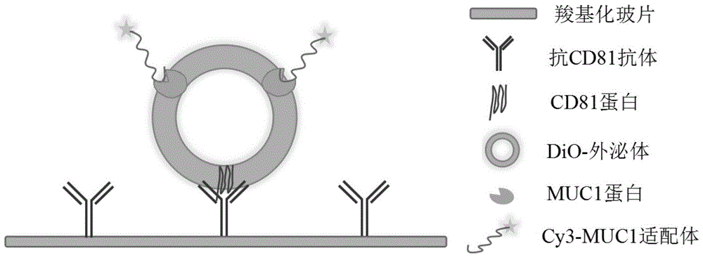
FIG. 1 is a schematic diagram of the detection method of the present invention;
FIG. 2 is a graph showing the effect of co-localization of the fluorochromes DiO and Cy3 according to the present invention;
FIG. 3 is a schematic representation of single molecule photobleaching of Cy3 fluorescent molecules in accordance with the present invention.
Detailed Description
The invention will be further described with reference to the following figures and specific examples, which are not intended to limit the invention in any way.
The MUC1 protein quantitative detection method based on exosome detection and single-molecule fluorescence bleaching technology mainly comprises the following specific steps: collecting the serum of a detected object and extracting exosomes; capturing exosomes with antibodies, and staining them with cell membrane fluorochromes; adding a fluorescent dye-labeled MUC1 protein aptamer, and combining the incubated protein with a breast cancer marker MUC1 protein on an exosome membrane; co-localization of exosome and aptamer is observed by a laser total internal reflection fluorescence microscope, and quantitative detection is further carried out on the breast cancer marker protein by adopting a monomolecular fluorescence bleaching technology. FIG. 1 shows a schematic diagram of the detection method of the present invention, the specific antibody used in the present invention is anti-CD 81 antibody, and the CD81 protein is an exosome transmembrane protein, which is widely and highly expressed on the surface of exosomes of various cell sources.
The method is based on exosome detection and single-molecule fluorescence bleaching technology, has the advantages of simple and convenient operation, high sensitivity, low detection cost, high specificity and the like, and compares the detected MUC1 expression quantity with the expression quantity of a single exosome MUC1 protein in serum of a healthy person, thereby diagnosing whether the detected object has breast cancer. The detection method can be used for carrying out auxiliary evaluation on whether the detected object is a breast cancer patient or a healthy person by detecting the expression quantity of the exosome surface protein on a single-molecule level, and has important significance for early diagnosis and curative effect monitoring and detection of the breast cancer.
Example 1:
the method for quantitatively detecting the MUC1 protein on the surface of the human exosome by the MUC1 protein based on exosome detection and single-molecule fluorescence bleaching technology is used for carrying out auxiliary evaluation on the breast cancer occurrence probability of a detected object by comparing the MUC1 expression quantity obtained by detecting a suspected breast cancer patient with the single exosome MUC1 protein expression quantity in the serum of a healthy person.
1 extraction of exosomes from serum of suspected breast cancer patient
And (3) carrying out venous blood collection on the suspected breast cancer patient, and standing for 60min to stratify the suspected breast cancer patient after blood collection is finished. Thereafter, the cells were centrifuged at 2000 Xg for 15min to obtain the supernatant as a serum sample. Serum was diluted with sterile 1 × PBS (phosphate buffered saline), and centrifuged sequentially at 800 × g for 5min, 2000 × g for 10min, and 10000 × g for 30min to remove precipitates such as intact cells and cell debris. The supernatant was taken and the medium was filtered through a 0.22 μm pore size filter. Centrifuging the filtered supernatant for 2h under 100000 Xg centrifugal force, removing the supernatant, and collecting the precipitate as exosome and contaminating protein. The bottom pellet was resuspended in 1 XPBS buffer and centrifuged at 100000g for 2h, and the pellet was made exosome. Finally the exosomes were suspended in sterile PBS solution and stored at-80 ℃ for use. All the above centrifugation processes were differential ultracentrifugation and were performed at 4 ℃.
2. Antibody capture exosomes
In this example, the specific antibody used was an anti-CD 81 antibody, and the CD81 protein was an exosome transmembrane protein, and was widely and highly expressed on the surface of exosomes derived from various cells.
First, 15. mu.L of a 5mM 1- (3-dimethylaminopropyl) -3-ethylcarbodiimide hydrochloride/N-hydroxysuccinimide (EDC/NHS) mixture was added to the channels of the carboxylic acid-functionalized slide and incubated at room temperature for 2 h; secondly, draining the solution by using filter paper, adding 15 mu L of anti-CD 81 antibody with the concentration of 10 mu g/mL, and incubating for 2h at room temperature; draining with filter paper, adding 15 μ L of 1 XPBS (pH7.0) containing 5% Bovine Serum Albumin (BSA), incubating at room temperature for 2h, and washing with 1 XPBS for 3 times; finally, 15 μ L of the extracted exosome sample was added, incubated for 1h at room temperature, and washed 3 times with 1 × PBS.
3. Exosome membrane staining
Adding DiO membrane dye with excitation wavelength of 473nm at concentration of 1 μ M15 μ L on the basis of the above step, incubating at room temperature for 10min to react the dye with the exosome membrane vesicles, and finally washing 3 times with 1 XPBS.
4. Fluorescent dye Cy 3-labeled MUC1 protein aptamer recognition exosome
The fluorescent dye labeling the MUC1 protein aptamer was orange fluorescent Cy3 with an excitation wavelength of 532 nm. The specific operation of recognizing the breast cancer marker MUC1 protein on an exosome membrane by using the Cy3 dye-labeled MUC1 protein aptamer is as follows: respectively adding 15 mu L of Cy 3-labeled MUC1 protein aptamer sequence with the concentration of 100pM on the basis of the previous step, incubating for 1h at room temperature, washing for 3 times by using 1 XPBS, and combining the MUC1 protein aptamer sequence with MUC1 protein on an exosome membrane to obtain a sample to be detected;
5. observing the co-localization of the exosome and the aptamer through a microscope, and detecting the exosome by adopting a monomolecular fluorescent bleaching technology
The samples to be tested are respectively excited by 473nm light and 532nm light, and then DiO and Cy3 molecular luminescence are observed under a total internal reflection fluorescence microscope, so that the co-localization condition of the two dyes and the Cy3 photobleaching process can be shown. Fluorescence images were obtained in the DiO and Cy3 channels, respectively, and the single molecule photobleaching process in the Cy3 channel was recorded. The co-localization effect of the fluorochromes DiO and Cy3 is shown in FIG. 2, and the single-molecule photobleaching of Cy3 fluorescent molecules is shown in FIG. 3.
6. Data processing, protein marker expression in individual exosomes
(I) Observing the total bleaching steps of the fluorescent molecules through a Cy3 channel of a total internal reflection fluorescent microscope, thereby determining the expression quantity of the MCU1 protein on the surface of the exosome;
(II) calculating the number of exosomes from images in the DiO channel of the total internal reflection fluorescence microscope;
and (III) calculating the ratio of the expression quantity of the determined exosome surface MUC1 protein in the step (I) to the ratio of the calculated exosome number in the step (II), and obtaining the expression quantity of the single exosome surface MUC1 protein in the detected sample.
6. Auxiliary evaluation of the probability of breast cancer of a subject
When the expression level of the single exosome MUC1 protein in the serum sample of the tested object is obviously higher than that of the single exosome MUC1 protein in the serum sample of the healthy human serum sample, the probability of the tested object to generate breast cancer is higher than that of the normal population.
While the present invention has been described with reference to the accompanying drawings, the present invention is not limited to the above-described embodiments, which are illustrative only and not restrictive, and various modifications which do not depart from the spirit of the present invention and which are intended to be covered by the claims of the present invention may be made by those skilled in the art.