CN112513070A - IL-15 variants and uses thereof - Google Patents
IL-15 variants and uses thereof Download PDFInfo
- Publication number
- CN112513070A CN112513070A CN201980028910.1A CN201980028910A CN112513070A CN 112513070 A CN112513070 A CN 112513070A CN 201980028910 A CN201980028910 A CN 201980028910A CN 112513070 A CN112513070 A CN 112513070A
- Authority
- CN
- China
- Prior art keywords
- antibody
- antibodies
- cancer
- amino acid
- variant
- Prior art date
- Legal status (The legal status is an assumption and is not a legal conclusion. Google has not performed a legal analysis and makes no representation as to the accuracy of the status listed.)
- Pending
Links
- 102000003812 Interleukin-15 Human genes 0.000 title claims abstract description 390
- 108090000172 Interleukin-15 Proteins 0.000 title claims abstract description 390
- 108020001507 fusion proteins Proteins 0.000 claims abstract description 201
- 102000037865 fusion proteins Human genes 0.000 claims abstract description 201
- 206010028980 Neoplasm Diseases 0.000 claims abstract description 100
- 238000000034 method Methods 0.000 claims abstract description 98
- 101001055157 Homo sapiens Interleukin-15 Proteins 0.000 claims abstract description 83
- 102000056003 human IL15 Human genes 0.000 claims abstract description 83
- 201000011510 cancer Diseases 0.000 claims abstract description 53
- 210000004027 cell Anatomy 0.000 claims description 199
- 102220104466 rs879254287 Human genes 0.000 claims description 178
- 102200042996 rs1057521927 Human genes 0.000 claims description 164
- 125000003275 alpha amino acid group Chemical group 0.000 claims description 133
- 238000009739 binding Methods 0.000 claims description 97
- 238000006467 substitution reaction Methods 0.000 claims description 97
- 230000027455 binding Effects 0.000 claims description 94
- 102220629430 Very-long-chain (3R)-3-hydroxyacyl-CoA dehydratase 1_E64Q_mutation Human genes 0.000 claims description 83
- 241000282414 Homo sapiens Species 0.000 claims description 74
- 101710089372 Programmed cell death protein 1 Proteins 0.000 claims description 56
- 108090000765 processed proteins & peptides Proteins 0.000 claims description 54
- 229920001184 polypeptide Polymers 0.000 claims description 53
- 102000004196 processed proteins & peptides Human genes 0.000 claims description 53
- 102220513430 SH3 domain-binding protein 4_I50A_mutation Human genes 0.000 claims description 45
- 102220485974 Dihydropteridine reductase_S51T_mutation Human genes 0.000 claims description 44
- 102220014105 rs368802003 Human genes 0.000 claims description 41
- 239000003814 drug Substances 0.000 claims description 40
- 230000002829 reductive effect Effects 0.000 claims description 39
- 230000000694 effects Effects 0.000 claims description 37
- 239000003112 inhibitor Substances 0.000 claims description 34
- 230000010056 antibody-dependent cellular cytotoxicity Effects 0.000 claims description 33
- 150000001413 amino acids Chemical class 0.000 claims description 32
- 230000004048 modification Effects 0.000 claims description 32
- 238000012986 modification Methods 0.000 claims description 32
- 230000004927 fusion Effects 0.000 claims description 30
- 229940124597 therapeutic agent Drugs 0.000 claims description 29
- 239000000556 agonist Substances 0.000 claims description 28
- 102220075004 rs758871507 Human genes 0.000 claims description 25
- 102000005962 receptors Human genes 0.000 claims description 24
- 108020003175 receptors Proteins 0.000 claims description 24
- 108040006849 interleukin-2 receptor activity proteins Proteins 0.000 claims description 23
- 206010012818 diffuse large B-cell lymphoma Diseases 0.000 claims description 22
- 102000004127 Cytokines Human genes 0.000 claims description 21
- 108090000695 Cytokines Proteins 0.000 claims description 21
- 108010017535 Interleukin-15 Receptors Proteins 0.000 claims description 21
- 102000004556 Interleukin-15 Receptors Human genes 0.000 claims description 21
- 102220640257 Proton channel OTOP1_L69D_mutation Human genes 0.000 claims description 20
- 208000037265 diseases, disorders, signs and symptoms Diseases 0.000 claims description 20
- 239000013604 expression vector Substances 0.000 claims description 18
- 239000008194 pharmaceutical composition Substances 0.000 claims description 18
- 238000011282 treatment Methods 0.000 claims description 18
- 102000010789 Interleukin-2 Receptors Human genes 0.000 claims description 16
- 208000031671 Large B-Cell Diffuse Lymphoma Diseases 0.000 claims description 16
- 210000001744 T-lymphocyte Anatomy 0.000 claims description 16
- 102220474891 LRP chaperone MESD_N4Q_mutation Human genes 0.000 claims description 15
- 102220607778 TYRO protein tyrosine kinase-binding protein_E64A_mutation Human genes 0.000 claims description 15
- 102220021170 rs80357116 Human genes 0.000 claims description 15
- 208000003950 B-cell lymphoma Diseases 0.000 claims description 12
- 239000007787 solid Substances 0.000 claims description 12
- FWMNVWWHGCHHJJ-SKKKGAJSSA-N 4-amino-1-[(2r)-6-amino-2-[[(2r)-2-[[(2r)-2-[[(2r)-2-amino-3-phenylpropanoyl]amino]-3-phenylpropanoyl]amino]-4-methylpentanoyl]amino]hexanoyl]piperidine-4-carboxylic acid Chemical compound C([C@H](C(=O)N[C@H](CC(C)C)C(=O)N[C@H](CCCCN)C(=O)N1CCC(N)(CC1)C(O)=O)NC(=O)[C@H](N)CC=1C=CC=CC=1)C1=CC=CC=C1 FWMNVWWHGCHHJJ-SKKKGAJSSA-N 0.000 claims description 11
- 108010047041 Complementarity Determining Regions Proteins 0.000 claims description 11
- 102220613496 Creatine kinase M-type_K10S_mutation Human genes 0.000 claims description 11
- 102220555409 Wnt inhibitory factor 1_H32G_mutation Human genes 0.000 claims description 11
- 201000010099 disease Diseases 0.000 claims description 11
- 102220616905 CCAAT/enhancer-binding protein alpha_I68A_mutation Human genes 0.000 claims description 10
- 102220526241 NACHT, LRR and PYD domains-containing protein 3_E64R_mutation Human genes 0.000 claims description 10
- 102220572951 Netrin receptor UNC5C_Y26H_mutation Human genes 0.000 claims description 10
- 102220466759 Olfactory receptor 13C5_L69M_mutation Human genes 0.000 claims description 10
- 102220536901 PRKC apoptosis WT1 regulator protein_L69I_mutation Human genes 0.000 claims description 10
- 206010035226 Plasma cell myeloma Diseases 0.000 claims description 10
- 102220645736 RNA-binding protein 7_L69E_mutation Human genes 0.000 claims description 10
- 102220608445 Suppressor of cytokine signaling 2_I68E_mutation Human genes 0.000 claims description 10
- 102220603933 TYRO protein tyrosine kinase-binding protein_K10A_mutation Human genes 0.000 claims description 10
- 102200072393 c.203T>C Human genes 0.000 claims description 10
- 150000007523 nucleic acids Chemical class 0.000 claims description 10
- 102220226506 rs1064796005 Human genes 0.000 claims description 10
- 102220309481 rs199473665 Human genes 0.000 claims description 10
- 102200082887 rs33950093 Human genes 0.000 claims description 10
- 102220072554 rs372717179 Human genes 0.000 claims description 10
- 102200164345 rs63750281 Human genes 0.000 claims description 10
- 102220067450 rs754763002 Human genes 0.000 claims description 10
- 102220277475 rs780141938 Human genes 0.000 claims description 10
- 102220021171 rs80357116 Human genes 0.000 claims description 10
- 208000011691 Burkitt lymphomas Diseases 0.000 claims description 9
- 102220487182 Growth/differentiation factor 9_E53Q_mutation Human genes 0.000 claims description 9
- 101001002657 Homo sapiens Interleukin-2 Proteins 0.000 claims description 9
- 102220496737 Lymphocyte function-associated antigen 3_V49Q_mutation Human genes 0.000 claims description 9
- 102220495079 Nucleolar and spindle-associated protein 1_E89Q_mutation Human genes 0.000 claims description 9
- 102220495764 Nucleolar and spindle-associated protein 1_E93Q_mutation Human genes 0.000 claims description 9
- 208000033559 Waldenström macroglobulinemia Diseases 0.000 claims description 9
- 229940127130 immunocytokine Drugs 0.000 claims description 9
- 239000007788 liquid Substances 0.000 claims description 9
- 102220026637 rs63750098 Human genes 0.000 claims description 9
- 102220101416 rs878855130 Human genes 0.000 claims description 9
- 208000024891 symptom Diseases 0.000 claims description 9
- 206010025323 Lymphomas Diseases 0.000 claims description 8
- 102000039446 nucleic acids Human genes 0.000 claims description 8
- 108020004707 nucleic acids Proteins 0.000 claims description 8
- 229940045513 CTLA4 antagonist Drugs 0.000 claims description 7
- 102100040061 Indoleamine 2,3-dioxygenase 1 Human genes 0.000 claims description 7
- 208000006664 Precursor Cell Lymphoblastic Leukemia-Lymphoma Diseases 0.000 claims description 7
- 239000003937 drug carrier Substances 0.000 claims description 7
- 229960005486 vaccine Drugs 0.000 claims description 7
- 208000024893 Acute lymphoblastic leukemia Diseases 0.000 claims description 6
- 208000014697 Acute lymphocytic leukaemia Diseases 0.000 claims description 6
- 208000032791 BCR-ABL1 positive chronic myelogenous leukemia Diseases 0.000 claims description 6
- 208000010833 Chronic myeloid leukaemia Diseases 0.000 claims description 6
- 208000017604 Hodgkin disease Diseases 0.000 claims description 6
- 208000025205 Mantle-Cell Lymphoma Diseases 0.000 claims description 6
- 208000034578 Multiple myelomas Diseases 0.000 claims description 6
- 208000033761 Myelogenous Chronic BCR-ABL Positive Leukemia Diseases 0.000 claims description 6
- 201000003444 follicular lymphoma Diseases 0.000 claims description 6
- 201000007924 marginal zone B-cell lymphoma Diseases 0.000 claims description 6
- 208000021937 marginal zone lymphoma Diseases 0.000 claims description 6
- KGRVJHAUYBGFFP-UHFFFAOYSA-N 2,2'-Methylenebis(4-methyl-6-tert-butylphenol) Chemical compound CC(C)(C)C1=CC(C)=CC(CC=2C(=C(C=C(C)C=2)C(C)(C)C)O)=C1O KGRVJHAUYBGFFP-UHFFFAOYSA-N 0.000 claims description 5
- 102220539786 HLA class II histocompatibility antigen, DQ alpha 1 chain_L91T_mutation Human genes 0.000 claims description 5
- 208000021519 Hodgkin lymphoma Diseases 0.000 claims description 5
- 208000010747 Hodgkins lymphoma Diseases 0.000 claims description 5
- 101000971533 Homo sapiens Killer cell lectin-like receptor subfamily G member 1 Proteins 0.000 claims description 5
- 102100021457 Killer cell lectin-like receptor subfamily G member 1 Human genes 0.000 claims description 5
- 208000007452 Plasmacytoma Diseases 0.000 claims description 5
- 102220531870 Polyisoprenoid diphosphate/phosphate phosphohydrolase PLPP6_S7G_mutation Human genes 0.000 claims description 5
- 208000026876 intravascular large B-cell lymphoma Diseases 0.000 claims description 5
- 102220081925 rs756442818 Human genes 0.000 claims description 5
- 208000015914 Non-Hodgkin lymphomas Diseases 0.000 claims description 4
- 206010039491 Sarcoma Diseases 0.000 claims description 4
- 208000005718 Stomach Neoplasms Diseases 0.000 claims description 4
- 206010017758 gastric cancer Diseases 0.000 claims description 4
- 210000004698 lymphocyte Anatomy 0.000 claims description 4
- 230000003211 malignant effect Effects 0.000 claims description 4
- 230000001394 metastastic effect Effects 0.000 claims description 4
- 206010061289 metastatic neoplasm Diseases 0.000 claims description 4
- 201000011549 stomach cancer Diseases 0.000 claims description 4
- 229940121358 tyrosine kinase inhibitor Drugs 0.000 claims description 4
- 239000005483 tyrosine kinase inhibitor Substances 0.000 claims description 4
- 208000017726 ALK-positive large B-cell lymphoma Diseases 0.000 claims description 3
- 208000031261 Acute myeloid leukaemia Diseases 0.000 claims description 3
- 208000010839 B-cell chronic lymphocytic leukemia Diseases 0.000 claims description 3
- 208000028564 B-cell non-Hodgkin lymphoma Diseases 0.000 claims description 3
- 208000032568 B-cell prolymphocytic leukaemia Diseases 0.000 claims description 3
- 208000005024 Castleman disease Diseases 0.000 claims description 3
- 206010007953 Central nervous system lymphoma Diseases 0.000 claims description 3
- 208000032612 Glial tumor Diseases 0.000 claims description 3
- 206010018338 Glioma Diseases 0.000 claims description 3
- 206010061218 Inflammation Diseases 0.000 claims description 3
- 208000002030 Merkel cell carcinoma Diseases 0.000 claims description 3
- 206010027406 Mesothelioma Diseases 0.000 claims description 3
- 206010029461 Nodal marginal zone B-cell lymphomas Diseases 0.000 claims description 3
- 102100034937 Poly(A) RNA polymerase, mitochondrial Human genes 0.000 claims description 3
- 206010065857 Primary Effusion Lymphoma Diseases 0.000 claims description 3
- 206010036711 Primary mediastinal large B-cell lymphomas Diseases 0.000 claims description 3
- 208000035416 Prolymphocytic B-Cell Leukemia Diseases 0.000 claims description 3
- 208000013056 classic Hodgkin lymphoma Diseases 0.000 claims description 3
- 208000017763 cutaneous neuroendocrine carcinoma Diseases 0.000 claims description 3
- 208000005017 glioblastoma Diseases 0.000 claims description 3
- 201000009277 hairy cell leukemia Diseases 0.000 claims description 3
- 210000003958 hematopoietic stem cell Anatomy 0.000 claims description 3
- 230000004054 inflammatory process Effects 0.000 claims description 3
- 201000007919 lymphoplasmacytic lymphoma Diseases 0.000 claims description 3
- 201000001441 melanoma Diseases 0.000 claims description 3
- 239000002829 mitogen activated protein kinase inhibitor Substances 0.000 claims description 3
- 208000025113 myeloid leukemia Diseases 0.000 claims description 3
- 244000309459 oncolytic virus Species 0.000 claims description 3
- 208000031223 plasma cell leukemia Diseases 0.000 claims description 3
- 210000003720 plasmablast Anatomy 0.000 claims description 3
- 208000016800 primary central nervous system lymphoma Diseases 0.000 claims description 3
- 201000006845 reticulosarcoma Diseases 0.000 claims description 3
- 208000029922 reticulum cell sarcoma Diseases 0.000 claims description 3
- 206010062113 splenic marginal zone lymphoma Diseases 0.000 claims description 3
- 206010061424 Anal cancer Diseases 0.000 claims description 2
- 229940122531 Anaplastic lymphoma kinase inhibitor Drugs 0.000 claims description 2
- 208000007860 Anus Neoplasms Diseases 0.000 claims description 2
- 208000036170 B-Cell Marginal Zone Lymphoma Diseases 0.000 claims description 2
- 206010003908 B-cell small lymphocytic lymphoma Diseases 0.000 claims description 2
- 206010004593 Bile duct cancer Diseases 0.000 claims description 2
- 206010005003 Bladder cancer Diseases 0.000 claims description 2
- 206010005949 Bone cancer Diseases 0.000 claims description 2
- 208000018084 Bone neoplasm Diseases 0.000 claims description 2
- 208000003174 Brain Neoplasms Diseases 0.000 claims description 2
- 206010006187 Breast cancer Diseases 0.000 claims description 2
- 208000026310 Breast neoplasm Diseases 0.000 claims description 2
- KLQLQDLJJUAEGT-ZBFHGGJFSA-N CO[C@H](C(=O)NC=1SC(=NN=1)N[C@H]1CN(CC1)C=1N=NC=CC=1)C1=CC=CC=C1 Chemical compound CO[C@H](C(=O)NC=1SC(=NN=1)N[C@H]1CN(CC1)C=1N=NC=CC=1)C1=CC=CC=C1 KLQLQDLJJUAEGT-ZBFHGGJFSA-N 0.000 claims description 2
- 201000009030 Carcinoma Diseases 0.000 claims description 2
- 206010008342 Cervix carcinoma Diseases 0.000 claims description 2
- 208000006332 Choriocarcinoma Diseases 0.000 claims description 2
- 206010009944 Colon cancer Diseases 0.000 claims description 2
- 208000001333 Colorectal Neoplasms Diseases 0.000 claims description 2
- 208000000461 Esophageal Neoplasms Diseases 0.000 claims description 2
- 241001502974 Human gammaherpesvirus 8 Species 0.000 claims description 2
- 208000008839 Kidney Neoplasms Diseases 0.000 claims description 2
- 206010058467 Lung neoplasm malignant Diseases 0.000 claims description 2
- 208000031422 Lymphocytic Chronic B-Cell Leukemia Diseases 0.000 claims description 2
- 201000003791 MALT lymphoma Diseases 0.000 claims description 2
- 229940124647 MEK inhibitor Drugs 0.000 claims description 2
- 208000032271 Malignant tumor of penis Diseases 0.000 claims description 2
- 208000003445 Mouth Neoplasms Diseases 0.000 claims description 2
- 206010052399 Neuroendocrine tumour Diseases 0.000 claims description 2
- 206010030155 Oesophageal carcinoma Diseases 0.000 claims description 2
- 206010033128 Ovarian cancer Diseases 0.000 claims description 2
- 206010061535 Ovarian neoplasm Diseases 0.000 claims description 2
- 206010061902 Pancreatic neoplasm Diseases 0.000 claims description 2
- 208000002471 Penile Neoplasms Diseases 0.000 claims description 2
- 206010034299 Penile cancer Diseases 0.000 claims description 2
- 206010036524 Precursor B-lymphoblastic lymphomas Diseases 0.000 claims description 2
- 206010060862 Prostate cancer Diseases 0.000 claims description 2
- 208000000236 Prostatic Neoplasms Diseases 0.000 claims description 2
- 206010038389 Renal cancer Diseases 0.000 claims description 2
- 208000004337 Salivary Gland Neoplasms Diseases 0.000 claims description 2
- 206010061934 Salivary gland cancer Diseases 0.000 claims description 2
- 208000000453 Skin Neoplasms Diseases 0.000 claims description 2
- 208000011778 T-cell/histiocyte rich large B cell lymphoma Diseases 0.000 claims description 2
- 208000024313 Testicular Neoplasms Diseases 0.000 claims description 2
- 206010057644 Testis cancer Diseases 0.000 claims description 2
- 208000000728 Thymus Neoplasms Diseases 0.000 claims description 2
- 208000024770 Thyroid neoplasm Diseases 0.000 claims description 2
- 208000007097 Urinary Bladder Neoplasms Diseases 0.000 claims description 2
- 208000006105 Uterine Cervical Neoplasms Diseases 0.000 claims description 2
- 208000002495 Uterine Neoplasms Diseases 0.000 claims description 2
- 206010047741 Vulval cancer Diseases 0.000 claims description 2
- 201000011165 anus cancer Diseases 0.000 claims description 2
- 208000026900 bile duct neoplasm Diseases 0.000 claims description 2
- 238000002659 cell therapy Methods 0.000 claims description 2
- 201000010881 cervical cancer Diseases 0.000 claims description 2
- 208000006990 cholangiocarcinoma Diseases 0.000 claims description 2
- 201000004101 esophageal cancer Diseases 0.000 claims description 2
- 208000024519 eye neoplasm Diseases 0.000 claims description 2
- 201000010536 head and neck cancer Diseases 0.000 claims description 2
- 208000014829 head and neck neoplasm Diseases 0.000 claims description 2
- 201000010982 kidney cancer Diseases 0.000 claims description 2
- 229940043355 kinase inhibitor Drugs 0.000 claims description 2
- 208000012987 lip and oral cavity carcinoma Diseases 0.000 claims description 2
- 201000007270 liver cancer Diseases 0.000 claims description 2
- 208000014018 liver neoplasm Diseases 0.000 claims description 2
- 201000005202 lung cancer Diseases 0.000 claims description 2
- 208000020816 lung neoplasm Diseases 0.000 claims description 2
- 208000015486 malignant pancreatic neoplasm Diseases 0.000 claims description 2
- 208000016065 neuroendocrine neoplasm Diseases 0.000 claims description 2
- 201000011519 neuroendocrine tumor Diseases 0.000 claims description 2
- 201000008106 ocular cancer Diseases 0.000 claims description 2
- 201000002528 pancreatic cancer Diseases 0.000 claims description 2
- 208000008443 pancreatic carcinoma Diseases 0.000 claims description 2
- 239000003757 phosphotransferase inhibitor Substances 0.000 claims description 2
- 210000004180 plasmocyte Anatomy 0.000 claims description 2
- 201000000849 skin cancer Diseases 0.000 claims description 2
- 201000002314 small intestine cancer Diseases 0.000 claims description 2
- 201000003120 testicular cancer Diseases 0.000 claims description 2
- 201000009377 thymus cancer Diseases 0.000 claims description 2
- 201000002510 thyroid cancer Diseases 0.000 claims description 2
- 150000004917 tyrosine kinase inhibitor derivatives Chemical class 0.000 claims description 2
- 201000005112 urinary bladder cancer Diseases 0.000 claims description 2
- 206010046766 uterine cancer Diseases 0.000 claims description 2
- 201000005102 vulva cancer Diseases 0.000 claims description 2
- 102200042487 rs141568342 Human genes 0.000 claims 12
- 102200085447 rs863225205 Human genes 0.000 claims 4
- 102100023990 60S ribosomal protein L17 Human genes 0.000 claims 2
- 230000001668 ameliorated effect Effects 0.000 claims 1
- 238000003259 recombinant expression Methods 0.000 claims 1
- 230000028993 immune response Effects 0.000 abstract description 14
- 241000124008 Mammalia Species 0.000 abstract description 13
- 238000004519 manufacturing process Methods 0.000 abstract description 13
- 230000002401 inhibitory effect Effects 0.000 abstract description 12
- 230000001225 therapeutic effect Effects 0.000 abstract description 12
- 230000004936 stimulating effect Effects 0.000 abstract description 4
- 230000007170 pathology Effects 0.000 abstract description 2
- 235000001014 amino acid Nutrition 0.000 description 90
- 239000000203 mixture Substances 0.000 description 69
- 102000040430 polynucleotide Human genes 0.000 description 61
- 108091033319 polynucleotide Proteins 0.000 description 61
- 239000002157 polynucleotide Substances 0.000 description 61
- 102100040678 Programmed cell death protein 1 Human genes 0.000 description 59
- 108090000623 proteins and genes Proteins 0.000 description 48
- 239000000427 antigen Substances 0.000 description 36
- 108091007433 antigens Proteins 0.000 description 36
- 102000036639 antigens Human genes 0.000 description 36
- 102000004169 proteins and genes Human genes 0.000 description 33
- 239000013598 vector Substances 0.000 description 33
- 102000002689 Toll-like receptor Human genes 0.000 description 32
- 108020000411 Toll-like receptor Proteins 0.000 description 32
- 235000018102 proteins Nutrition 0.000 description 32
- 229940024606 amino acid Drugs 0.000 description 28
- 239000003795 chemical substances by application Substances 0.000 description 28
- 230000014509 gene expression Effects 0.000 description 27
- 230000035772 mutation Effects 0.000 description 24
- 108020004414 DNA Proteins 0.000 description 21
- 102100023970 Keratin, type I cytoskeletal 10 Human genes 0.000 description 21
- -1 for example Chemical class 0.000 description 20
- 230000006870 function Effects 0.000 description 20
- 238000004458 analytical method Methods 0.000 description 19
- 241001465754 Metazoa Species 0.000 description 18
- FAPWRFPIFSIZLT-UHFFFAOYSA-M Sodium chloride Chemical compound [Na+].[Cl-] FAPWRFPIFSIZLT-UHFFFAOYSA-M 0.000 description 18
- 102220523591 Tumor necrosis factor receptor superfamily member 18_E64K_mutation Human genes 0.000 description 18
- 239000000126 substance Substances 0.000 description 18
- 125000003729 nucleotide group Chemical group 0.000 description 17
- 108060003951 Immunoglobulin Proteins 0.000 description 16
- 239000000872 buffer Substances 0.000 description 16
- 230000013595 glycosylation Effects 0.000 description 16
- 238000006206 glycosylation reaction Methods 0.000 description 16
- 102000018358 immunoglobulin Human genes 0.000 description 16
- 108010019670 Chimeric Antigen Receptors Proteins 0.000 description 15
- 150000001875 compounds Chemical class 0.000 description 15
- 239000012636 effector Substances 0.000 description 15
- 238000009472 formulation Methods 0.000 description 15
- 239000012634 fragment Substances 0.000 description 14
- 239000002773 nucleotide Substances 0.000 description 14
- 239000000546 pharmaceutical excipient Substances 0.000 description 14
- 238000001727 in vivo Methods 0.000 description 13
- 238000002347 injection Methods 0.000 description 13
- 239000007924 injection Substances 0.000 description 13
- 125000005647 linker group Chemical group 0.000 description 13
- 125000002496 methyl group Chemical group [H]C([H])([H])* 0.000 description 12
- 239000000243 solution Substances 0.000 description 12
- 108091032973 (ribonucleotides)n+m Proteins 0.000 description 11
- 101001003140 Homo sapiens Interleukin-15 receptor subunit alpha Proteins 0.000 description 11
- 210000004408 hybridoma Anatomy 0.000 description 11
- 239000000463 material Substances 0.000 description 11
- 108010006654 Bleomycin Proteins 0.000 description 10
- 241000699670 Mus sp. Species 0.000 description 10
- 230000004913 activation Effects 0.000 description 10
- 238000003556 assay Methods 0.000 description 10
- 229960001561 bleomycin Drugs 0.000 description 10
- OYVAGSVQBOHSSS-UAPAGMARSA-O bleomycin A2 Chemical compound N([C@H](C(=O)N[C@H](C)[C@@H](O)[C@H](C)C(=O)N[C@@H]([C@H](O)C)C(=O)NCCC=1SC=C(N=1)C=1SC=C(N=1)C(=O)NCCC[S+](C)C)[C@@H](O[C@H]1[C@H]([C@@H](O)[C@H](O)[C@H](CO)O1)O[C@@H]1[C@H]([C@@H](OC(N)=O)[C@H](O)[C@@H](CO)O1)O)C=1N=CNC=1)C(=O)C1=NC([C@H](CC(N)=O)NC[C@H](N)C(N)=O)=NC(N)=C1C OYVAGSVQBOHSSS-UAPAGMARSA-O 0.000 description 10
- 150000001720 carbohydrates Chemical group 0.000 description 10
- 230000000295 complement effect Effects 0.000 description 10
- LOKCTEFSRHRXRJ-UHFFFAOYSA-I dipotassium trisodium dihydrogen phosphate hydrogen phosphate dichloride Chemical compound P(=O)(O)(O)[O-].[K+].P(=O)(O)([O-])[O-].[Na+].[Na+].[Cl-].[K+].[Cl-].[Na+] LOKCTEFSRHRXRJ-UHFFFAOYSA-I 0.000 description 10
- 239000002953 phosphate buffered saline Substances 0.000 description 10
- 230000001603 reducing effect Effects 0.000 description 10
- 238000002560 therapeutic procedure Methods 0.000 description 10
- 210000004881 tumor cell Anatomy 0.000 description 10
- 102100035361 Cerebellar degeneration-related protein 2 Human genes 0.000 description 9
- 101000737796 Homo sapiens Cerebellar degeneration-related protein 2 Proteins 0.000 description 9
- 208000035475 disorder Diseases 0.000 description 9
- 229940079593 drug Drugs 0.000 description 9
- 230000009977 dual effect Effects 0.000 description 9
- 239000000839 emulsion Substances 0.000 description 9
- 210000002865 immune cell Anatomy 0.000 description 9
- 102000007863 pattern recognition receptors Human genes 0.000 description 9
- 108010089193 pattern recognition receptors Proteins 0.000 description 9
- 239000011780 sodium chloride Substances 0.000 description 9
- ZHNUHDYFZUAESO-UHFFFAOYSA-N Formamide Chemical compound NC=O ZHNUHDYFZUAESO-UHFFFAOYSA-N 0.000 description 8
- 101000611936 Homo sapiens Programmed cell death protein 1 Proteins 0.000 description 8
- 108090000174 Interleukin-10 Proteins 0.000 description 8
- 108010002350 Interleukin-2 Proteins 0.000 description 8
- 102000000588 Interleukin-2 Human genes 0.000 description 8
- 230000004988 N-glycosylation Effects 0.000 description 8
- 108091028043 Nucleic acid sequence Proteins 0.000 description 8
- 229960003722 doxycycline Drugs 0.000 description 8
- 238000002474 experimental method Methods 0.000 description 8
- 230000002163 immunogen Effects 0.000 description 8
- 229940072221 immunoglobulins Drugs 0.000 description 8
- 230000001939 inductive effect Effects 0.000 description 8
- 239000010410 layer Substances 0.000 description 8
- 239000002502 liposome Substances 0.000 description 8
- BASFCYQUMIYNBI-UHFFFAOYSA-N platinum Chemical compound [Pt] BASFCYQUMIYNBI-UHFFFAOYSA-N 0.000 description 8
- RXWNCPJZOCPEPQ-NVWDDTSBSA-N puromycin Chemical compound C1=CC(OC)=CC=C1C[C@H](N)C(=O)N[C@H]1[C@@H](O)[C@H](N2C3=NC=NC(=C3N=C2)N(C)C)O[C@@H]1CO RXWNCPJZOCPEPQ-NVWDDTSBSA-N 0.000 description 8
- 235000000346 sugar Nutrition 0.000 description 8
- SGKRLCUYIXIAHR-AKNGSSGZSA-N (4s,4ar,5s,5ar,6r,12ar)-4-(dimethylamino)-1,5,10,11,12a-pentahydroxy-6-methyl-3,12-dioxo-4a,5,5a,6-tetrahydro-4h-tetracene-2-carboxamide Chemical compound C1=CC=C2[C@H](C)[C@@H]([C@H](O)[C@@H]3[C@](C(O)=C(C(N)=O)C(=O)[C@H]3N(C)C)(O)C3=O)C3=C(O)C2=C1O SGKRLCUYIXIAHR-AKNGSSGZSA-N 0.000 description 7
- NFGXHKASABOEEW-UHFFFAOYSA-N 1-methylethyl 11-methoxy-3,7,11-trimethyl-2,4-dodecadienoate Chemical compound COC(C)(C)CCCC(C)CC=CC(C)=CC(=O)OC(C)C NFGXHKASABOEEW-UHFFFAOYSA-N 0.000 description 7
- 108010066719 Interleukin Receptor Common gamma Subunit Proteins 0.000 description 7
- 102000018682 Interleukin Receptor Common gamma Subunit Human genes 0.000 description 7
- 108010038453 Interleukin-2 Receptors Proteins 0.000 description 7
- 241000699666 Mus <mouse, genus> Species 0.000 description 7
- 239000002253 acid Substances 0.000 description 7
- 238000013459 approach Methods 0.000 description 7
- 230000001419 dependent effect Effects 0.000 description 7
- 210000000987 immune system Anatomy 0.000 description 7
- 230000001613 neoplastic effect Effects 0.000 description 7
- 239000013612 plasmid Substances 0.000 description 7
- 210000003491 skin Anatomy 0.000 description 7
- 238000007920 subcutaneous administration Methods 0.000 description 7
- 239000006228 supernatant Substances 0.000 description 7
- 238000012546 transfer Methods 0.000 description 7
- 210000003171 tumor-infiltrating lymphocyte Anatomy 0.000 description 7
- 230000003612 virological effect Effects 0.000 description 7
- WVDDGKGOMKODPV-UHFFFAOYSA-N Benzyl alcohol Chemical compound OCC1=CC=CC=C1 WVDDGKGOMKODPV-UHFFFAOYSA-N 0.000 description 6
- 108010062802 CD66 antigens Proteins 0.000 description 6
- 102100024533 Carcinoembryonic antigen-related cell adhesion molecule 1 Human genes 0.000 description 6
- SRBFZHDQGSBBOR-IOVATXLUSA-N D-xylopyranose Chemical compound O[C@@H]1COC(O)[C@H](O)[C@H]1O SRBFZHDQGSBBOR-IOVATXLUSA-N 0.000 description 6
- BCCRXDTUTZHDEU-VKHMYHEASA-N Gly-Ser Chemical compound NCC(=O)N[C@@H](CO)C(O)=O BCCRXDTUTZHDEU-VKHMYHEASA-N 0.000 description 6
- 101710120843 Indoleamine 2,3-dioxygenase 1 Proteins 0.000 description 6
- FBOZXECLQNJBKD-ZDUSSCGKSA-N L-methotrexate Chemical compound C=1N=C2N=C(N)N=C(N)C2=NC=1CN(C)C1=CC=C(C(=O)N[C@@H](CCC(O)=O)C(O)=O)C=C1 FBOZXECLQNJBKD-ZDUSSCGKSA-N 0.000 description 6
- 101100519207 Mus musculus Pdcd1 gene Proteins 0.000 description 6
- NBIIXXVUZAFLBC-UHFFFAOYSA-N Phosphoric acid Chemical compound OP(O)(O)=O NBIIXXVUZAFLBC-UHFFFAOYSA-N 0.000 description 6
- 239000002202 Polyethylene glycol Substances 0.000 description 6
- 241000700605 Viruses Species 0.000 description 6
- 239000004480 active ingredient Substances 0.000 description 6
- 238000007792 addition Methods 0.000 description 6
- 239000012491 analyte Substances 0.000 description 6
- 230000009286 beneficial effect Effects 0.000 description 6
- 230000004071 biological effect Effects 0.000 description 6
- 238000005119 centrifugation Methods 0.000 description 6
- 230000004540 complement-dependent cytotoxicity Effects 0.000 description 6
- 239000003623 enhancer Substances 0.000 description 6
- 229940121354 immunomodulator Drugs 0.000 description 6
- 230000001965 increasing effect Effects 0.000 description 6
- 230000003993 interaction Effects 0.000 description 6
- 238000001990 intravenous administration Methods 0.000 description 6
- 239000003550 marker Substances 0.000 description 6
- 230000001404 mediated effect Effects 0.000 description 6
- 229960000485 methotrexate Drugs 0.000 description 6
- 210000000822 natural killer cell Anatomy 0.000 description 6
- 229960003301 nivolumab Drugs 0.000 description 6
- 230000037361 pathway Effects 0.000 description 6
- 229920001223 polyethylene glycol Polymers 0.000 description 6
- 238000000746 purification Methods 0.000 description 6
- 102220061393 rs786203307 Human genes 0.000 description 6
- 241000894007 species Species 0.000 description 6
- 150000008163 sugars Chemical class 0.000 description 6
- 210000001519 tissue Anatomy 0.000 description 6
- 239000003981 vehicle Substances 0.000 description 6
- 210000001266 CD8-positive T-lymphocyte Anatomy 0.000 description 5
- 108010087819 Fc receptors Proteins 0.000 description 5
- 102000009109 Fc receptors Human genes 0.000 description 5
- PEDCQBHIVMGVHV-UHFFFAOYSA-N Glycerine Chemical compound OCC(O)CO PEDCQBHIVMGVHV-UHFFFAOYSA-N 0.000 description 5
- 102000003886 Glycoproteins Human genes 0.000 description 5
- 108090000288 Glycoproteins Proteins 0.000 description 5
- 241000282412 Homo Species 0.000 description 5
- 108010073807 IgG Receptors Proteins 0.000 description 5
- 102000009490 IgG Receptors Human genes 0.000 description 5
- 102220475986 Keratin, type I cytoskeletal 10_Y26F_mutation Human genes 0.000 description 5
- 239000002147 L01XE04 - Sunitinib Substances 0.000 description 5
- 230000000259 anti-tumor effect Effects 0.000 description 5
- 235000014633 carbohydrates Nutrition 0.000 description 5
- 230000008859 change Effects 0.000 description 5
- 238000010367 cloning Methods 0.000 description 5
- 238000002648 combination therapy Methods 0.000 description 5
- 238000007796 conventional method Methods 0.000 description 5
- 238000010494 dissociation reaction Methods 0.000 description 5
- 230000005593 dissociations Effects 0.000 description 5
- 238000000684 flow cytometry Methods 0.000 description 5
- SDUQYLNIPVEERB-QPPQHZFASA-N gemcitabine Chemical compound O=C1N=C(N)C=CN1[C@H]1C(F)(F)[C@H](O)[C@@H](CO)O1 SDUQYLNIPVEERB-QPPQHZFASA-N 0.000 description 5
- 229960005277 gemcitabine Drugs 0.000 description 5
- 238000001415 gene therapy Methods 0.000 description 5
- 239000001963 growth medium Substances 0.000 description 5
- 238000003018 immunoassay Methods 0.000 description 5
- 230000015788 innate immune response Effects 0.000 description 5
- 239000012528 membrane Substances 0.000 description 5
- 210000004379 membrane Anatomy 0.000 description 5
- KKZJGLLVHKMTCM-UHFFFAOYSA-N mitoxantrone Chemical compound O=C1C2=C(O)C=CC(O)=C2C(=O)C2=C1C(NCCNCCO)=CC=C2NCCNCCO KKZJGLLVHKMTCM-UHFFFAOYSA-N 0.000 description 5
- 230000002093 peripheral effect Effects 0.000 description 5
- 230000004044 response Effects 0.000 description 5
- 150000003839 salts Chemical class 0.000 description 5
- 102000035025 signaling receptors Human genes 0.000 description 5
- 108091005475 signaling receptors Proteins 0.000 description 5
- 239000003381 stabilizer Substances 0.000 description 5
- 229960001796 sunitinib Drugs 0.000 description 5
- WINHZLLDWRZWRT-ATVHPVEESA-N sunitinib Chemical compound CCN(CC)CCNC(=O)C1=C(C)NC(\C=C/2C3=CC(F)=CC=C3NC\2=O)=C1C WINHZLLDWRZWRT-ATVHPVEESA-N 0.000 description 5
- 230000004083 survival effect Effects 0.000 description 5
- XLYOFNOQVPJJNP-UHFFFAOYSA-N water Substances O XLYOFNOQVPJJNP-UHFFFAOYSA-N 0.000 description 5
- 230000003442 weekly effect Effects 0.000 description 5
- JKMHFZQWWAIEOD-UHFFFAOYSA-N 2-[4-(2-hydroxyethyl)piperazin-1-yl]ethanesulfonic acid Chemical compound OCC[NH+]1CCN(CCS([O-])(=O)=O)CC1 JKMHFZQWWAIEOD-UHFFFAOYSA-N 0.000 description 4
- CIWBSHSKHKDKBQ-JLAZNSOCSA-N Ascorbic acid Chemical compound OC[C@H](O)[C@H]1OC(=O)C(O)=C1O CIWBSHSKHKDKBQ-JLAZNSOCSA-N 0.000 description 4
- 208000031648 Body Weight Changes Diseases 0.000 description 4
- AOJJSUZBOXZQNB-TZSSRYMLSA-N Doxorubicin Chemical compound O([C@H]1C[C@@](O)(CC=2C(O)=C3C(=O)C=4C=CC=C(C=4C(=O)C3=C(O)C=21)OC)C(=O)CO)[C@H]1C[C@H](N)[C@H](O)[C@H](C)O1 AOJJSUZBOXZQNB-TZSSRYMLSA-N 0.000 description 4
- KCXVZYZYPLLWCC-UHFFFAOYSA-N EDTA Chemical compound OC(=O)CN(CC(O)=O)CCN(CC(O)=O)CC(O)=O KCXVZYZYPLLWCC-UHFFFAOYSA-N 0.000 description 4
- WQZGKKKJIJFFOK-GASJEMHNSA-N Glucose Natural products OC[C@H]1OC(O)[C@H](O)[C@@H](O)[C@@H]1O WQZGKKKJIJFFOK-GASJEMHNSA-N 0.000 description 4
- DHMQDGOQFOQNFH-UHFFFAOYSA-N Glycine Chemical compound NCC(O)=O DHMQDGOQFOQNFH-UHFFFAOYSA-N 0.000 description 4
- 239000007995 HEPES buffer Substances 0.000 description 4
- 102000006496 Immunoglobulin Heavy Chains Human genes 0.000 description 4
- 108010019476 Immunoglobulin Heavy Chains Proteins 0.000 description 4
- 206010027476 Metastases Diseases 0.000 description 4
- LRHPLDYGYMQRHN-UHFFFAOYSA-N N-Butanol Chemical compound CCCCO LRHPLDYGYMQRHN-UHFFFAOYSA-N 0.000 description 4
- 108091034117 Oligonucleotide Proteins 0.000 description 4
- 229910019142 PO4 Inorganic materials 0.000 description 4
- ISWSIDIOOBJBQZ-UHFFFAOYSA-N Phenol Chemical compound OC1=CC=CC=C1 ISWSIDIOOBJBQZ-UHFFFAOYSA-N 0.000 description 4
- 229920001213 Polysorbate 20 Polymers 0.000 description 4
- DBMJMQXJHONAFJ-UHFFFAOYSA-M Sodium laurylsulphate Chemical compound [Na+].CCCCCCCCCCCCOS([O-])(=O)=O DBMJMQXJHONAFJ-UHFFFAOYSA-M 0.000 description 4
- NKANXQFJJICGDU-QPLCGJKRSA-N Tamoxifen Chemical compound C=1C=CC=CC=1C(/CC)=C(C=1C=CC(OCCN(C)C)=CC=1)/C1=CC=CC=C1 NKANXQFJJICGDU-QPLCGJKRSA-N 0.000 description 4
- FOCVUCIESVLUNU-UHFFFAOYSA-N Thiotepa Chemical compound C1CN1P(N1CC1)(=S)N1CC1 FOCVUCIESVLUNU-UHFFFAOYSA-N 0.000 description 4
- 239000004473 Threonine Substances 0.000 description 4
- 125000000217 alkyl group Chemical group 0.000 description 4
- 229960003437 aminoglutethimide Drugs 0.000 description 4
- ROBVIMPUHSLWNV-UHFFFAOYSA-N aminoglutethimide Chemical compound C=1C=C(N)C=CC=1C1(CC)CCC(=O)NC1=O ROBVIMPUHSLWNV-UHFFFAOYSA-N 0.000 description 4
- 239000003242 anti bacterial agent Substances 0.000 description 4
- 229940088710 antibiotic agent Drugs 0.000 description 4
- 239000002246 antineoplastic agent Substances 0.000 description 4
- PYMYPHUHKUWMLA-UHFFFAOYSA-N arabinose Natural products OCC(O)C(O)C(O)C=O PYMYPHUHKUWMLA-UHFFFAOYSA-N 0.000 description 4
- 229960003005 axitinib Drugs 0.000 description 4
- RITAVMQDGBJQJZ-FMIVXFBMSA-N axitinib Chemical compound CNC(=O)C1=CC=CC=C1SC1=CC=C(C(\C=C\C=2N=CC=CC=2)=NN2)C2=C1 RITAVMQDGBJQJZ-FMIVXFBMSA-N 0.000 description 4
- 210000003719 b-lymphocyte Anatomy 0.000 description 4
- SRBFZHDQGSBBOR-UHFFFAOYSA-N beta-D-Pyranose-Lyxose Natural products OC1COC(O)C(O)C1O SRBFZHDQGSBBOR-UHFFFAOYSA-N 0.000 description 4
- 229930189065 blasticidin Natural products 0.000 description 4
- 230000037396 body weight Effects 0.000 description 4
- 230000004579 body weight change Effects 0.000 description 4
- 210000004899 c-terminal region Anatomy 0.000 description 4
- YCIMNLLNPGFGHC-UHFFFAOYSA-N catechol Chemical compound OC1=CC=CC=C1O YCIMNLLNPGFGHC-UHFFFAOYSA-N 0.000 description 4
- 238000004113 cell culture Methods 0.000 description 4
- HVYWMOMLDIMFJA-DPAQBDIFSA-N cholesterol Chemical compound C1C=C2C[C@@H](O)CC[C@]2(C)[C@@H]2[C@@H]1[C@@H]1CC[C@H]([C@H](C)CCCC(C)C)[C@@]1(C)CC2 HVYWMOMLDIMFJA-DPAQBDIFSA-N 0.000 description 4
- 239000002299 complementary DNA Substances 0.000 description 4
- 230000000875 corresponding effect Effects 0.000 description 4
- 230000009089 cytolysis Effects 0.000 description 4
- 229940127089 cytotoxic agent Drugs 0.000 description 4
- 238000012217 deletion Methods 0.000 description 4
- 230000037430 deletion Effects 0.000 description 4
- 239000002552 dosage form Substances 0.000 description 4
- 238000009396 hybridization Methods 0.000 description 4
- 239000012642 immune effector Substances 0.000 description 4
- 238000000338 in vitro Methods 0.000 description 4
- 239000004615 ingredient Substances 0.000 description 4
- RLSSMJSEOOYNOY-UHFFFAOYSA-N m-cresol Chemical compound CC1=CC=CC(O)=C1 RLSSMJSEOOYNOY-UHFFFAOYSA-N 0.000 description 4
- 229910052751 metal Inorganic materials 0.000 description 4
- 239000002184 metal Substances 0.000 description 4
- 230000009401 metastasis Effects 0.000 description 4
- 239000003094 microcapsule Substances 0.000 description 4
- 229960001156 mitoxantrone Drugs 0.000 description 4
- 239000003921 oil Substances 0.000 description 4
- 235000019198 oils Nutrition 0.000 description 4
- 150000002482 oligosaccharides Chemical class 0.000 description 4
- 244000052769 pathogen Species 0.000 description 4
- AQIXEPGDORPWBJ-UHFFFAOYSA-N pentan-3-ol Chemical compound CCC(O)CC AQIXEPGDORPWBJ-UHFFFAOYSA-N 0.000 description 4
- 239000010452 phosphate Substances 0.000 description 4
- 239000006187 pill Substances 0.000 description 4
- 229910052697 platinum Inorganic materials 0.000 description 4
- 239000000256 polyoxyethylene sorbitan monolaurate Substances 0.000 description 4
- 235000010486 polyoxyethylene sorbitan monolaurate Nutrition 0.000 description 4
- 239000000843 powder Substances 0.000 description 4
- 230000035755 proliferation Effects 0.000 description 4
- 230000000069 prophylactic effect Effects 0.000 description 4
- QELSKZZBTMNZEB-UHFFFAOYSA-N propylparaben Chemical compound CCCOC(=O)C1=CC=C(O)C=C1 QELSKZZBTMNZEB-UHFFFAOYSA-N 0.000 description 4
- 229950010131 puromycin Drugs 0.000 description 4
- 238000010188 recombinant method Methods 0.000 description 4
- 210000003289 regulatory T cell Anatomy 0.000 description 4
- GHMLBKRAJCXXBS-UHFFFAOYSA-N resorcinol Chemical compound OC1=CC=CC(O)=C1 GHMLBKRAJCXXBS-UHFFFAOYSA-N 0.000 description 4
- 150000003384 small molecules Chemical class 0.000 description 4
- 239000001509 sodium citrate Substances 0.000 description 4
- NLJMYIDDQXHKNR-UHFFFAOYSA-K sodium citrate Chemical compound O.O.[Na+].[Na+].[Na+].[O-]C(=O)CC(O)(CC([O-])=O)C([O-])=O NLJMYIDDQXHKNR-UHFFFAOYSA-K 0.000 description 4
- 235000019333 sodium laurylsulphate Nutrition 0.000 description 4
- 230000009870 specific binding Effects 0.000 description 4
- PVYJZLYGTZKPJE-UHFFFAOYSA-N streptonigrin Chemical compound C=1C=C2C(=O)C(OC)=C(N)C(=O)C2=NC=1C(C=1N)=NC(C(O)=O)=C(C)C=1C1=CC=C(OC)C(OC)=C1O PVYJZLYGTZKPJE-UHFFFAOYSA-N 0.000 description 4
- 239000000725 suspension Substances 0.000 description 4
- 238000013268 sustained release Methods 0.000 description 4
- 239000012730 sustained-release form Substances 0.000 description 4
- 230000008685 targeting Effects 0.000 description 4
- WYWHKKSPHMUBEB-UHFFFAOYSA-N tioguanine Chemical compound N1C(N)=NC(=S)C2=C1N=CN2 WYWHKKSPHMUBEB-UHFFFAOYSA-N 0.000 description 4
- 238000001890 transfection Methods 0.000 description 4
- 229960000575 trastuzumab Drugs 0.000 description 4
- 241000701161 unidentified adenovirus Species 0.000 description 4
- MTCFGRXMJLQNBG-REOHCLBHSA-N (2S)-2-Amino-3-hydroxypropansäure Chemical compound OC[C@H](N)C(O)=O MTCFGRXMJLQNBG-REOHCLBHSA-N 0.000 description 3
- 108700028369 Alleles Proteins 0.000 description 3
- UHOVQNZJYSORNB-UHFFFAOYSA-N Benzene Chemical compound C1=CC=CC=C1 UHOVQNZJYSORNB-UHFFFAOYSA-N 0.000 description 3
- CMSMOCZEIVJLDB-UHFFFAOYSA-N Cyclophosphamide Chemical compound ClCCN(CCCl)P1(=O)NCCCO1 CMSMOCZEIVJLDB-UHFFFAOYSA-N 0.000 description 3
- UHDGCWIWMRVCDJ-CCXZUQQUSA-N Cytarabine Chemical compound O=C1N=C(N)C=CN1[C@H]1[C@@H](O)[C@H](O)[C@@H](CO)O1 UHDGCWIWMRVCDJ-CCXZUQQUSA-N 0.000 description 3
- FBPFZTCFMRRESA-FSIIMWSLSA-N D-Glucitol Natural products OC[C@H](O)[C@H](O)[C@@H](O)[C@H](O)CO FBPFZTCFMRRESA-FSIIMWSLSA-N 0.000 description 3
- FBPFZTCFMRRESA-KVTDHHQDSA-N D-Mannitol Chemical compound OC[C@@H](O)[C@@H](O)[C@H](O)[C@H](O)CO FBPFZTCFMRRESA-KVTDHHQDSA-N 0.000 description 3
- FBPFZTCFMRRESA-JGWLITMVSA-N D-glucitol Chemical compound OC[C@H](O)[C@@H](O)[C@H](O)[C@H](O)CO FBPFZTCFMRRESA-JGWLITMVSA-N 0.000 description 3
- 102000004190 Enzymes Human genes 0.000 description 3
- 108090000790 Enzymes Proteins 0.000 description 3
- 241000588724 Escherichia coli Species 0.000 description 3
- GHASVSINZRGABV-UHFFFAOYSA-N Fluorouracil Chemical compound FC1=CNC(=O)NC1=O GHASVSINZRGABV-UHFFFAOYSA-N 0.000 description 3
- 108010010803 Gelatin Proteins 0.000 description 3
- 102100026120 IgG receptor FcRn large subunit p51 Human genes 0.000 description 3
- 101710177940 IgG receptor FcRn large subunit p51 Proteins 0.000 description 3
- 239000002146 L01XE16 - Crizotinib Substances 0.000 description 3
- 108010000817 Leuprolide Proteins 0.000 description 3
- 229930195725 Mannitol Natural products 0.000 description 3
- OKKJLVBELUTLKV-UHFFFAOYSA-N Methanol Chemical compound OC OKKJLVBELUTLKV-UHFFFAOYSA-N 0.000 description 3
- 108091061960 Naked DNA Proteins 0.000 description 3
- 230000004989 O-glycosylation Effects 0.000 description 3
- 241000700159 Rattus Species 0.000 description 3
- 102100037422 Receptor-type tyrosine-protein phosphatase C Human genes 0.000 description 3
- 101710138747 Receptor-type tyrosine-protein phosphatase C Proteins 0.000 description 3
- MTCFGRXMJLQNBG-UHFFFAOYSA-N Serine Natural products OCC(N)C(O)=O MTCFGRXMJLQNBG-UHFFFAOYSA-N 0.000 description 3
- CZMRCDWAGMRECN-UGDNZRGBSA-N Sucrose Chemical compound O[C@H]1[C@H](O)[C@@H](CO)O[C@@]1(CO)O[C@@H]1[C@H](O)[C@@H](O)[C@H](O)[C@@H](CO)O1 CZMRCDWAGMRECN-UGDNZRGBSA-N 0.000 description 3
- 229930006000 Sucrose Natural products 0.000 description 3
- CBPNZQVSJQDFBE-FUXHJELOSA-N Temsirolimus Chemical compound C1C[C@@H](OC(=O)C(C)(CO)CO)[C@H](OC)C[C@@H]1C[C@@H](C)[C@H]1OC(=O)[C@@H]2CCCCN2C(=O)C(=O)[C@](O)(O2)[C@H](C)CC[C@H]2C[C@H](OC)/C(C)=C/C=C/C=C/[C@@H](C)C[C@@H](C)C(=O)[C@H](OC)[C@H](O)/C(C)=C/[C@@H](C)C(=O)C1 CBPNZQVSJQDFBE-FUXHJELOSA-N 0.000 description 3
- 230000003213 activating effect Effects 0.000 description 3
- 229910000147 aluminium phosphate Inorganic materials 0.000 description 3
- 125000000539 amino acid group Chemical group 0.000 description 3
- 239000005557 antagonist Substances 0.000 description 3
- WQZGKKKJIJFFOK-VFUOTHLCSA-N beta-D-glucose Chemical compound OC[C@H]1O[C@@H](O)[C@H](O)[C@@H](O)[C@@H]1O WQZGKKKJIJFFOK-VFUOTHLCSA-N 0.000 description 3
- 230000015572 biosynthetic process Effects 0.000 description 3
- 239000000969 carrier Substances 0.000 description 3
- 239000002738 chelating agent Substances 0.000 description 3
- 239000003153 chemical reaction reagent Substances 0.000 description 3
- 239000003636 conditioned culture medium Substances 0.000 description 3
- 229920001577 copolymer Polymers 0.000 description 3
- KTEIFNKAUNYNJU-GFCCVEGCSA-N crizotinib Chemical compound O([C@H](C)C=1C(=C(F)C=CC=1Cl)Cl)C(C(=NC=1)N)=CC=1C(=C1)C=NN1C1CCNCC1 KTEIFNKAUNYNJU-GFCCVEGCSA-N 0.000 description 3
- 229960005061 crizotinib Drugs 0.000 description 3
- 229960004397 cyclophosphamide Drugs 0.000 description 3
- 230000006378 damage Effects 0.000 description 3
- STQGQHZAVUOBTE-VGBVRHCVSA-N daunorubicin Chemical compound O([C@H]1C[C@@](O)(CC=2C(O)=C3C(=O)C=4C=CC=C(C=4C(=O)C3=C(O)C=21)OC)C(C)=O)[C@H]1C[C@H](N)[C@H](O)[C@H](C)O1 STQGQHZAVUOBTE-VGBVRHCVSA-N 0.000 description 3
- 230000002708 enhancing effect Effects 0.000 description 3
- 230000002255 enzymatic effect Effects 0.000 description 3
- 229960002949 fluorouracil Drugs 0.000 description 3
- 239000008273 gelatin Substances 0.000 description 3
- 229920000159 gelatin Polymers 0.000 description 3
- 235000019322 gelatine Nutrition 0.000 description 3
- 235000011852 gelatine desserts Nutrition 0.000 description 3
- 238000001476 gene delivery Methods 0.000 description 3
- 239000008103 glucose Substances 0.000 description 3
- 239000003102 growth factor Substances 0.000 description 3
- 125000002887 hydroxy group Chemical group [H]O* 0.000 description 3
- 230000001900 immune effect Effects 0.000 description 3
- 230000003053 immunization Effects 0.000 description 3
- 230000003308 immunostimulating effect Effects 0.000 description 3
- 210000005008 immunosuppressive cell Anatomy 0.000 description 3
- 238000001802 infusion Methods 0.000 description 3
- 238000012933 kinetic analysis Methods 0.000 description 3
- 239000003446 ligand Substances 0.000 description 3
- 230000000670 limiting effect Effects 0.000 description 3
- 239000007937 lozenge Substances 0.000 description 3
- 201000000564 macroglobulinemia Diseases 0.000 description 3
- 239000000594 mannitol Substances 0.000 description 3
- 235000010355 mannitol Nutrition 0.000 description 3
- 239000002609 medium Substances 0.000 description 3
- GLVAUDGFNGKCSF-UHFFFAOYSA-N mercaptopurine Chemical compound S=C1NC=NC2=C1NC=N2 GLVAUDGFNGKCSF-UHFFFAOYSA-N 0.000 description 3
- 229910044991 metal oxide Inorganic materials 0.000 description 3
- 150000004706 metal oxides Chemical class 0.000 description 3
- 239000003607 modifier Substances 0.000 description 3
- 201000000050 myeloid neoplasm Diseases 0.000 description 3
- 231100000252 nontoxic Toxicity 0.000 description 3
- 230000003000 nontoxic effect Effects 0.000 description 3
- 229920001542 oligosaccharide Polymers 0.000 description 3
- 229960004390 palbociclib Drugs 0.000 description 3
- AHJRHEGDXFFMBM-UHFFFAOYSA-N palbociclib Chemical compound N1=C2N(C3CCCC3)C(=O)C(C(=O)C)=C(C)C2=CN=C1NC(N=C1)=CC=C1N1CCNCC1 AHJRHEGDXFFMBM-UHFFFAOYSA-N 0.000 description 3
- 239000002245 particle Substances 0.000 description 3
- 230000001717 pathogenic effect Effects 0.000 description 3
- 210000003819 peripheral blood mononuclear cell Anatomy 0.000 description 3
- NBIIXXVUZAFLBC-UHFFFAOYSA-K phosphate Chemical compound [O-]P([O-])([O-])=O NBIIXXVUZAFLBC-UHFFFAOYSA-K 0.000 description 3
- 229920000136 polysorbate Polymers 0.000 description 3
- 229920000036 polyvinylpyrrolidone Polymers 0.000 description 3
- 239000001267 polyvinylpyrrolidone Substances 0.000 description 3
- 235000013855 polyvinylpyrrolidone Nutrition 0.000 description 3
- 230000003389 potentiating effect Effects 0.000 description 3
- 238000002360 preparation method Methods 0.000 description 3
- 125000002924 primary amino group Chemical group [H]N([H])* 0.000 description 3
- HXCHCVDVKSCDHU-PJKCJEBCSA-N s-[(2r,3s,4s,6s)-6-[[(2r,3s,4s,5r,6r)-5-[(2s,4s,5s)-5-(ethylamino)-4-methoxyoxan-2-yl]oxy-4-hydroxy-6-[[(2s,5z,9r,13e)-9-hydroxy-12-(methoxycarbonylamino)-13-[2-(methyltrisulfanyl)ethylidene]-11-oxo-2-bicyclo[7.3.1]trideca-1(12),5-dien-3,7-diynyl]oxy]-2-m Chemical compound C1[C@H](OC)[C@@H](NCC)CO[C@H]1O[C@H]1[C@H](O[C@@H]2C\3=C(NC(=O)OC)C(=O)C[C@@](C/3=C/CSSSC)(O)C#C\C=C/C#C2)O[C@H](C)[C@@H](NO[C@@H]2O[C@H](C)[C@@H](SC(=O)C=3C(=C(OC)C(O[C@H]4[C@@H]([C@H](OC)[C@@H](O)[C@H](C)O4)O)=C(I)C=3C)OC)[C@@H](O)C2)[C@@H]1O HXCHCVDVKSCDHU-PJKCJEBCSA-N 0.000 description 3
- 239000000600 sorbitol Substances 0.000 description 3
- 235000010356 sorbitol Nutrition 0.000 description 3
- 239000000758 substrate Substances 0.000 description 3
- 239000005720 sucrose Substances 0.000 description 3
- 239000004094 surface-active agent Substances 0.000 description 3
- 229960000235 temsirolimus Drugs 0.000 description 3
- QFJCIRLUMZQUOT-UHFFFAOYSA-N temsirolimus Natural products C1CC(O)C(OC)CC1CC(C)C1OC(=O)C2CCCCN2C(=O)C(=O)C(O)(O2)C(C)CCC2CC(OC)C(C)=CC=CC=CC(C)CC(C)C(=O)C(OC)C(O)C(C)=CC(C)C(=O)C1 QFJCIRLUMZQUOT-UHFFFAOYSA-N 0.000 description 3
- 229960001196 thiotepa Drugs 0.000 description 3
- 238000011200 topical administration Methods 0.000 description 3
- 230000001988 toxicity Effects 0.000 description 3
- 231100000419 toxicity Toxicity 0.000 description 3
- 238000011269 treatment regimen Methods 0.000 description 3
- 230000004614 tumor growth Effects 0.000 description 3
- 230000005751 tumor progression Effects 0.000 description 3
- 229950005972 urelumab Drugs 0.000 description 3
- 238000005406 washing Methods 0.000 description 3
- HDTRYLNUVZCQOY-UHFFFAOYSA-N α-D-glucopyranosyl-α-D-glucopyranoside Natural products OC1C(O)C(O)C(CO)OC1OC1C(O)C(O)C(O)C(CO)O1 HDTRYLNUVZCQOY-UHFFFAOYSA-N 0.000 description 2
- FLWWDYNPWOSLEO-HQVZTVAUSA-N (2s)-2-[[4-[1-(2-amino-4-oxo-1h-pteridin-6-yl)ethyl-methylamino]benzoyl]amino]pentanedioic acid Chemical compound C=1N=C2NC(N)=NC(=O)C2=NC=1C(C)N(C)C1=CC=C(C(=O)N[C@@H](CCC(O)=O)C(O)=O)C=C1 FLWWDYNPWOSLEO-HQVZTVAUSA-N 0.000 description 2
- KYBXNPIASYUWLN-WUCPZUCCSA-N (2s)-5-hydroxypyrrolidine-2-carboxylic acid Chemical compound OC1CC[C@@H](C(O)=O)N1 KYBXNPIASYUWLN-WUCPZUCCSA-N 0.000 description 2
- CGMTUJFWROPELF-YPAAEMCBSA-N (3E,5S)-5-[(2S)-butan-2-yl]-3-(1-hydroxyethylidene)pyrrolidine-2,4-dione Chemical compound CC[C@H](C)[C@@H]1NC(=O)\C(=C(/C)O)C1=O CGMTUJFWROPELF-YPAAEMCBSA-N 0.000 description 2
- FPVKHBSQESCIEP-UHFFFAOYSA-N (8S)-3-(2-deoxy-beta-D-erythro-pentofuranosyl)-3,6,7,8-tetrahydroimidazo[4,5-d][1,3]diazepin-8-ol Natural products C1C(O)C(CO)OC1N1C(NC=NCC2O)=C2N=C1 FPVKHBSQESCIEP-UHFFFAOYSA-N 0.000 description 2
- IEXUMDBQLIVNHZ-YOUGDJEHSA-N (8s,11r,13r,14s,17s)-11-[4-(dimethylamino)phenyl]-17-hydroxy-17-(3-hydroxypropyl)-13-methyl-1,2,6,7,8,11,12,14,15,16-decahydrocyclopenta[a]phenanthren-3-one Chemical compound C1=CC(N(C)C)=CC=C1[C@@H]1C2=C3CCC(=O)C=C3CC[C@H]2[C@H](CC[C@]2(O)CCCO)[C@@]2(C)C1 IEXUMDBQLIVNHZ-YOUGDJEHSA-N 0.000 description 2
- FDKXTQMXEQVLRF-ZHACJKMWSA-N (E)-dacarbazine Chemical compound CN(C)\N=N\c1[nH]cnc1C(N)=O FDKXTQMXEQVLRF-ZHACJKMWSA-N 0.000 description 2
- AZQWKYJCGOJGHM-UHFFFAOYSA-N 1,4-benzoquinone Chemical compound O=C1C=CC(=O)C=C1 AZQWKYJCGOJGHM-UHFFFAOYSA-N 0.000 description 2
- DWZAEMINVBZMHQ-UHFFFAOYSA-N 1-[4-[4-(dimethylamino)piperidine-1-carbonyl]phenyl]-3-[4-(4,6-dimorpholin-4-yl-1,3,5-triazin-2-yl)phenyl]urea Chemical compound C1CC(N(C)C)CCN1C(=O)C(C=C1)=CC=C1NC(=O)NC1=CC=C(C=2N=C(N=C(N=2)N2CCOCC2)N2CCOCC2)C=C1 DWZAEMINVBZMHQ-UHFFFAOYSA-N 0.000 description 2
- JLPULHDHAOZNQI-ZTIMHPMXSA-N 1-hexadecanoyl-2-(9Z,12Z-octadecadienoyl)-sn-glycero-3-phosphocholine Chemical compound CCCCCCCCCCCCCCCC(=O)OC[C@H](COP([O-])(=O)OCC[N+](C)(C)C)OC(=O)CCCCCCC\C=C/C\C=C/CCCCC JLPULHDHAOZNQI-ZTIMHPMXSA-N 0.000 description 2
- IIZPXYDJLKNOIY-JXPKJXOSSA-N 1-palmitoyl-2-arachidonoyl-sn-glycero-3-phosphocholine Chemical compound CCCCCCCCCCCCCCCC(=O)OC[C@H](COP([O-])(=O)OCC[N+](C)(C)C)OC(=O)CCC\C=C/C\C=C/C\C=C/C\C=C/CCCCC IIZPXYDJLKNOIY-JXPKJXOSSA-N 0.000 description 2
- BGFTWECWAICPDG-UHFFFAOYSA-N 2-[bis(4-chlorophenyl)methyl]-4-n-[3-[bis(4-chlorophenyl)methyl]-4-(dimethylamino)phenyl]-1-n,1-n-dimethylbenzene-1,4-diamine Chemical compound C1=C(C(C=2C=CC(Cl)=CC=2)C=2C=CC(Cl)=CC=2)C(N(C)C)=CC=C1NC(C=1)=CC=C(N(C)C)C=1C(C=1C=CC(Cl)=CC=1)C1=CC=C(Cl)C=C1 BGFTWECWAICPDG-UHFFFAOYSA-N 0.000 description 2
- QCXJFISCRQIYID-IAEPZHFASA-N 2-amino-1-n-[(3s,6s,7r,10s,16s)-3-[(2s)-butan-2-yl]-7,11,14-trimethyl-2,5,9,12,15-pentaoxo-10-propan-2-yl-8-oxa-1,4,11,14-tetrazabicyclo[14.3.0]nonadecan-6-yl]-4,6-dimethyl-3-oxo-9-n-[(3s,6s,7r,10s,16s)-7,11,14-trimethyl-2,5,9,12,15-pentaoxo-3,10-di(propa Chemical compound C[C@H]1OC(=O)[C@H](C(C)C)N(C)C(=O)CN(C)C(=O)[C@@H]2CCCN2C(=O)[C@H](C(C)C)NC(=O)[C@H]1NC(=O)C1=C(N=C2C(C(=O)N[C@@H]3C(=O)N[C@H](C(N4CCC[C@H]4C(=O)N(C)CC(=O)N(C)[C@@H](C(C)C)C(=O)O[C@@H]3C)=O)[C@@H](C)CC)=C(N)C(=O)C(C)=C2O2)C2=C(C)C=C1 QCXJFISCRQIYID-IAEPZHFASA-N 0.000 description 2
- 125000000979 2-amino-2-oxoethyl group Chemical group [H]C([*])([H])C(=O)N([H])[H] 0.000 description 2
- NDMPLJNOPCLANR-UHFFFAOYSA-N 3,4-dihydroxy-15-(4-hydroxy-18-methoxycarbonyl-5,18-seco-ibogamin-18-yl)-16-methoxy-1-methyl-6,7-didehydro-aspidospermidine-3-carboxylic acid methyl ester Natural products C1C(CC)(O)CC(CC2(C(=O)OC)C=3C(=CC4=C(C56C(C(C(O)C7(CC)C=CCN(C67)CC5)(O)C(=O)OC)N4C)C=3)OC)CN1CCC1=C2NC2=CC=CC=C12 NDMPLJNOPCLANR-UHFFFAOYSA-N 0.000 description 2
- CLPFFLWZZBQMAO-UHFFFAOYSA-N 4-(5,6,7,8-tetrahydroimidazo[1,5-a]pyridin-5-yl)benzonitrile Chemical compound C1=CC(C#N)=CC=C1C1N2C=NC=C2CCC1 CLPFFLWZZBQMAO-UHFFFAOYSA-N 0.000 description 2
- 229940117976 5-hydroxylysine Drugs 0.000 description 2
- STQGQHZAVUOBTE-UHFFFAOYSA-N 7-Cyan-hept-2t-en-4,6-diinsaeure Natural products C1=2C(O)=C3C(=O)C=4C(OC)=CC=CC=4C(=O)C3=C(O)C=2CC(O)(C(C)=O)CC1OC1CC(N)C(O)C(C)O1 STQGQHZAVUOBTE-UHFFFAOYSA-N 0.000 description 2
- CEIZFXOZIQNICU-UHFFFAOYSA-N Alternaria alternata Crofton-weed toxin Natural products CCC(C)C1NC(=O)C(C(C)=O)=C1O CEIZFXOZIQNICU-UHFFFAOYSA-N 0.000 description 2
- 239000004475 Arginine Substances 0.000 description 2
- BFYIZQONLCFLEV-DAELLWKTSA-N Aromasine Chemical compound O=C1C=C[C@]2(C)[C@H]3CC[C@](C)(C(CC4)=O)[C@@H]4[C@@H]3CC(=C)C2=C1 BFYIZQONLCFLEV-DAELLWKTSA-N 0.000 description 2
- DCXYFEDJOCDNAF-UHFFFAOYSA-N Asparagine Natural products OC(=O)C(N)CC(N)=O DCXYFEDJOCDNAF-UHFFFAOYSA-N 0.000 description 2
- IJGRMHOSHXDMSA-UHFFFAOYSA-N Atomic nitrogen Chemical compound N#N IJGRMHOSHXDMSA-UHFFFAOYSA-N 0.000 description 2
- 108091008875 B cell receptors Proteins 0.000 description 2
- 102000006942 B-Cell Maturation Antigen Human genes 0.000 description 2
- 108010008014 B-Cell Maturation Antigen Proteins 0.000 description 2
- 102100024222 B-lymphocyte antigen CD19 Human genes 0.000 description 2
- 244000063299 Bacillus subtilis Species 0.000 description 2
- VGGGPCQERPFHOB-MCIONIFRSA-N Bestatin Chemical compound CC(C)C[C@H](C(O)=O)NC(=O)[C@@H](O)[C@H](N)CC1=CC=CC=C1 VGGGPCQERPFHOB-MCIONIFRSA-N 0.000 description 2
- BTBUEUYNUDRHOZ-UHFFFAOYSA-N Borate Chemical compound [O-]B([O-])[O-] BTBUEUYNUDRHOZ-UHFFFAOYSA-N 0.000 description 2
- 241000283690 Bos taurus Species 0.000 description 2
- 108091003079 Bovine Serum Albumin Proteins 0.000 description 2
- MBABCNBNDNGODA-LTGLSHGVSA-N Bullatacin Natural products O=C1C(C[C@H](O)CCCCCCCCCC[C@@H](O)[C@@H]2O[C@@H]([C@@H]3O[C@H]([C@@H](O)CCCCCCCCCC)CC3)CC2)=C[C@H](C)O1 MBABCNBNDNGODA-LTGLSHGVSA-N 0.000 description 2
- FERIUCNNQQJTOY-UHFFFAOYSA-N Butyric acid Chemical compound CCCC(O)=O FERIUCNNQQJTOY-UHFFFAOYSA-N 0.000 description 2
- 108090000342 C-Type Lectins Proteins 0.000 description 2
- 102000003930 C-Type Lectins Human genes 0.000 description 2
- 239000002126 C01EB10 - Adenosine Substances 0.000 description 2
- 102100038078 CD276 antigen Human genes 0.000 description 2
- 101100075831 Caenorhabditis elegans mab-7 gene Proteins 0.000 description 2
- GAGWJHPBXLXJQN-UORFTKCHSA-N Capecitabine Chemical compound C1=C(F)C(NC(=O)OCCCCC)=NC(=O)N1[C@H]1[C@H](O)[C@H](O)[C@@H](C)O1 GAGWJHPBXLXJQN-UORFTKCHSA-N 0.000 description 2
- GAGWJHPBXLXJQN-UHFFFAOYSA-N Capecitabine Natural products C1=C(F)C(NC(=O)OCCCCC)=NC(=O)N1C1C(O)C(O)C(C)O1 GAGWJHPBXLXJQN-UHFFFAOYSA-N 0.000 description 2
- SHHKQEUPHAENFK-UHFFFAOYSA-N Carboquone Chemical compound O=C1C(C)=C(N2CC2)C(=O)C(C(COC(N)=O)OC)=C1N1CC1 SHHKQEUPHAENFK-UHFFFAOYSA-N 0.000 description 2
- 102100025474 Carcinoembryonic antigen-related cell adhesion molecule 7 Human genes 0.000 description 2
- AOCCBINRVIKJHY-UHFFFAOYSA-N Carmofur Chemical compound CCCCCCNC(=O)N1C=C(F)C(=O)NC1=O AOCCBINRVIKJHY-UHFFFAOYSA-N 0.000 description 2
- DLGOEMSEDOSKAD-UHFFFAOYSA-N Carmustine Chemical compound ClCCNC(=O)N(N=O)CCCl DLGOEMSEDOSKAD-UHFFFAOYSA-N 0.000 description 2
- 102000000844 Cell Surface Receptors Human genes 0.000 description 2
- 108010001857 Cell Surface Receptors Proteins 0.000 description 2
- 108091007741 Chimeric antigen receptor T cells Proteins 0.000 description 2
- JWBOIMRXGHLCPP-UHFFFAOYSA-N Chloditan Chemical compound C=1C=CC=C(Cl)C=1C(C(Cl)Cl)C1=CC=C(Cl)C=C1 JWBOIMRXGHLCPP-UHFFFAOYSA-N 0.000 description 2
- KRKNYBCHXYNGOX-UHFFFAOYSA-K Citrate Chemical compound [O-]C(=O)CC(O)(CC([O-])=O)C([O-])=O KRKNYBCHXYNGOX-UHFFFAOYSA-K 0.000 description 2
- 102000003903 Cyclin-dependent kinases Human genes 0.000 description 2
- 108090000266 Cyclin-dependent kinases Proteins 0.000 description 2
- WQZGKKKJIJFFOK-QTVWNMPRSA-N D-mannopyranose Chemical compound OC[C@H]1OC(O)[C@@H](O)[C@@H](O)[C@@H]1O WQZGKKKJIJFFOK-QTVWNMPRSA-N 0.000 description 2
- 108010013198 Daptomycin Proteins 0.000 description 2
- 102000011799 Desmoglein Human genes 0.000 description 2
- 108050002238 Desmoglein Proteins 0.000 description 2
- RTZKZFJDLAIYFH-UHFFFAOYSA-N Diethyl ether Chemical compound CCOCC RTZKZFJDLAIYFH-UHFFFAOYSA-N 0.000 description 2
- SAMRUMKYXPVKPA-VFKOLLTISA-N Enocitabine Chemical compound O=C1N=C(NC(=O)CCCCCCCCCCCCCCCCCCCCC)C=CN1[C@H]1[C@@H](O)[C@H](O)[C@@H](CO)O1 SAMRUMKYXPVKPA-VFKOLLTISA-N 0.000 description 2
- 206010066919 Epidemic polyarthritis Diseases 0.000 description 2
- 241000283086 Equidae Species 0.000 description 2
- JOYRKODLDBILNP-UHFFFAOYSA-N Ethyl urethane Chemical compound CCOC(N)=O JOYRKODLDBILNP-UHFFFAOYSA-N 0.000 description 2
- PIICEJLVQHRZGT-UHFFFAOYSA-N Ethylenediamine Chemical compound NCCN PIICEJLVQHRZGT-UHFFFAOYSA-N 0.000 description 2
- 102100037362 Fibronectin Human genes 0.000 description 2
- 108010067306 Fibronectins Proteins 0.000 description 2
- 239000004471 Glycine Substances 0.000 description 2
- 108010017213 Granulocyte-Macrophage Colony-Stimulating Factor Proteins 0.000 description 2
- 102100039620 Granulocyte-macrophage colony-stimulating factor Human genes 0.000 description 2
- 206010018691 Granuloma Diseases 0.000 description 2
- NYHBQMYGNKIUIF-UUOKFMHZSA-N Guanosine Chemical compound C1=NC=2C(=O)NC(N)=NC=2N1[C@@H]1O[C@H](CO)[C@@H](O)[C@H]1O NYHBQMYGNKIUIF-UUOKFMHZSA-N 0.000 description 2
- 101000980825 Homo sapiens B-lymphocyte antigen CD19 Proteins 0.000 description 2
- 101000914324 Homo sapiens Carcinoembryonic antigen-related cell adhesion molecule 5 Proteins 0.000 description 2
- 101000914321 Homo sapiens Carcinoembryonic antigen-related cell adhesion molecule 7 Proteins 0.000 description 2
- 101000633786 Homo sapiens SLAM family member 6 Proteins 0.000 description 2
- 101000633784 Homo sapiens SLAM family member 7 Proteins 0.000 description 2
- 101000633782 Homo sapiens SLAM family member 8 Proteins 0.000 description 2
- 101000831007 Homo sapiens T-cell immunoreceptor with Ig and ITIM domains Proteins 0.000 description 2
- 101000831496 Homo sapiens Toll-like receptor 3 Proteins 0.000 description 2
- 101000851376 Homo sapiens Tumor necrosis factor receptor superfamily member 8 Proteins 0.000 description 2
- XDXDZDZNSLXDNA-TZNDIEGXSA-N Idarubicin Chemical compound C1[C@H](N)[C@H](O)[C@H](C)O[C@H]1O[C@@H]1C2=C(O)C(C(=O)C3=CC=CC=C3C3=O)=C3C(O)=C2C[C@@](O)(C(C)=O)C1 XDXDZDZNSLXDNA-TZNDIEGXSA-N 0.000 description 2
- DGAQECJNVWCQMB-PUAWFVPOSA-M Ilexoside XXIX Chemical class C[C@@H]1CC[C@@]2(CC[C@@]3(C(=CC[C@H]4[C@]3(CC[C@@H]5[C@@]4(CC[C@@H](C5(C)C)OS(=O)(=O)[O-])C)C)[C@@H]2[C@]1(C)O)C)C(=O)O[C@H]6[C@@H]([C@H]([C@@H]([C@H](O6)CO)O)O)O.[Na+] DGAQECJNVWCQMB-PUAWFVPOSA-M 0.000 description 2
- 108010021625 Immunoglobulin Fragments Proteins 0.000 description 2
- 102000008394 Immunoglobulin Fragments Human genes 0.000 description 2
- 108010050904 Interferons Proteins 0.000 description 2
- 108010065805 Interleukin-12 Proteins 0.000 description 2
- 108090001005 Interleukin-6 Proteins 0.000 description 2
- 108010002586 Interleukin-7 Proteins 0.000 description 2
- 102000015696 Interleukins Human genes 0.000 description 2
- 108010063738 Interleukins Proteins 0.000 description 2
- 108091092195 Intron Proteins 0.000 description 2
- 102000015617 Janus Kinases Human genes 0.000 description 2
- 108010024121 Janus Kinases Proteins 0.000 description 2
- 241001138401 Kluyveromyces lactis Species 0.000 description 2
- ODKSFYDXXFIFQN-BYPYZUCNSA-P L-argininium(2+) Chemical compound NC(=[NH2+])NCCC[C@H]([NH3+])C(O)=O ODKSFYDXXFIFQN-BYPYZUCNSA-P 0.000 description 2
- DCXYFEDJOCDNAF-REOHCLBHSA-N L-asparagine Chemical compound OC(=O)[C@@H](N)CC(N)=O DCXYFEDJOCDNAF-REOHCLBHSA-N 0.000 description 2
- WHUUTDBJXJRKMK-VKHMYHEASA-N L-glutamic acid Chemical compound OC(=O)[C@@H](N)CCC(O)=O WHUUTDBJXJRKMK-VKHMYHEASA-N 0.000 description 2
- ZDXPYRJPNDTMRX-VKHMYHEASA-N L-glutamine Chemical compound OC(=O)[C@@H](N)CCC(N)=O ZDXPYRJPNDTMRX-VKHMYHEASA-N 0.000 description 2
- HNDVDQJCIGZPNO-YFKPBYRVSA-N L-histidine Chemical compound OC(=O)[C@@H](N)CC1=CN=CN1 HNDVDQJCIGZPNO-YFKPBYRVSA-N 0.000 description 2
- KDXKERNSBIXSRK-YFKPBYRVSA-N L-lysine Chemical compound NCCCC[C@H](N)C(O)=O KDXKERNSBIXSRK-YFKPBYRVSA-N 0.000 description 2
- FFEARJCKVFRZRR-BYPYZUCNSA-N L-methionine Chemical compound CSCC[C@H](N)C(O)=O FFEARJCKVFRZRR-BYPYZUCNSA-N 0.000 description 2
- AYFVYJQAPQTCCC-GBXIJSLDSA-N L-threonine Chemical compound C[C@@H](O)[C@H](N)C(O)=O AYFVYJQAPQTCCC-GBXIJSLDSA-N 0.000 description 2
- 239000002145 L01XE14 - Bosutinib Substances 0.000 description 2
- 102000017578 LAG3 Human genes 0.000 description 2
- 101150030213 Lag3 gene Proteins 0.000 description 2
- 229920001491 Lentinan Polymers 0.000 description 2
- GQYIWUVLTXOXAJ-UHFFFAOYSA-N Lomustine Chemical compound ClCCN(N=O)C(=O)NC1CCCCC1 GQYIWUVLTXOXAJ-UHFFFAOYSA-N 0.000 description 2
- KDXKERNSBIXSRK-UHFFFAOYSA-N Lysine Natural products NCCCCC(N)C(O)=O KDXKERNSBIXSRK-UHFFFAOYSA-N 0.000 description 2
- 239000004472 Lysine Substances 0.000 description 2
- 108010046938 Macrophage Colony-Stimulating Factor Proteins 0.000 description 2
- 102100028123 Macrophage colony-stimulating factor 1 Human genes 0.000 description 2
- 229930192392 Mitomycin Natural products 0.000 description 2
- 241000187918 Mycobacterium obuense Species 0.000 description 2
- NWIBSHFKIJFRCO-WUDYKRTCSA-N Mytomycin Chemical compound C1N2C(C(C(C)=C(N)C3=O)=O)=C3[C@@H](COC(N)=O)[C@@]2(OC)[C@@H]2[C@H]1N2 NWIBSHFKIJFRCO-WUDYKRTCSA-N 0.000 description 2
- OVRNDRQMDRJTHS-CBQIKETKSA-N N-Acetyl-D-Galactosamine Chemical compound CC(=O)N[C@H]1[C@@H](O)O[C@H](CO)[C@H](O)[C@@H]1O OVRNDRQMDRJTHS-CBQIKETKSA-N 0.000 description 2
- MBLBDJOUHNCFQT-UHFFFAOYSA-N N-acetyl-D-galactosamine Natural products CC(=O)NC(C=O)C(O)C(O)C(O)CO MBLBDJOUHNCFQT-UHFFFAOYSA-N 0.000 description 2
- ZDZOTLJHXYCWBA-VCVYQWHSSA-N N-debenzoyl-N-(tert-butoxycarbonyl)-10-deacetyltaxol Chemical compound O([C@H]1[C@H]2[C@@](C([C@H](O)C3=C(C)[C@@H](OC(=O)[C@H](O)[C@@H](NC(=O)OC(C)(C)C)C=4C=CC=CC=4)C[C@]1(O)C3(C)C)=O)(C)[C@@H](O)C[C@H]1OC[C@]12OC(=O)C)C(=O)C1=CC=CC=C1 ZDZOTLJHXYCWBA-VCVYQWHSSA-N 0.000 description 2
- 102100038082 Natural killer cell receptor 2B4 Human genes 0.000 description 2
- 206010029266 Neuroendocrine carcinoma of the skin Diseases 0.000 description 2
- PXHVJJICTQNCMI-UHFFFAOYSA-N Nickel Chemical compound [Ni] PXHVJJICTQNCMI-UHFFFAOYSA-N 0.000 description 2
- 241000283973 Oryctolagus cuniculus Species 0.000 description 2
- 229940124060 PD-1 antagonist Drugs 0.000 description 2
- 229940123751 PD-L1 antagonist Drugs 0.000 description 2
- 229930012538 Paclitaxel Natural products 0.000 description 2
- 206010057249 Phagocytosis Diseases 0.000 description 2
- KMSKQZKKOZQFFG-HSUXVGOQSA-N Pirarubicin Chemical compound O([C@H]1[C@@H](N)C[C@@H](O[C@H]1C)O[C@H]1C[C@@](O)(CC=2C(O)=C3C(=O)C=4C=CC=C(C=4C(=O)C3=C(O)C=21)OC)C(=O)CO)[C@H]1CCCCO1 KMSKQZKKOZQFFG-HSUXVGOQSA-N 0.000 description 2
- DLRVVLDZNNYCBX-UHFFFAOYSA-N Polydextrose Polymers OC1C(O)C(O)C(CO)OC1OCC1C(O)C(O)C(O)C(O)O1 DLRVVLDZNNYCBX-UHFFFAOYSA-N 0.000 description 2
- ONIBWKKTOPOVIA-UHFFFAOYSA-N Proline Natural products OC(=O)C1CCCN1 ONIBWKKTOPOVIA-UHFFFAOYSA-N 0.000 description 2
- 108010029485 Protein Isoforms Proteins 0.000 description 2
- 102000001708 Protein Isoforms Human genes 0.000 description 2
- 108010076504 Protein Sorting Signals Proteins 0.000 description 2
- 102000007056 Recombinant Fusion Proteins Human genes 0.000 description 2
- 108010008281 Recombinant Fusion Proteins Proteins 0.000 description 2
- OWPCHSCAPHNHAV-UHFFFAOYSA-N Rhizoxin Natural products C1C(O)C2(C)OC2C=CC(C)C(OC(=O)C2)CC2CC2OC2C(=O)OC1C(C)C(OC)C(C)=CC=CC(C)=CC1=COC(C)=N1 OWPCHSCAPHNHAV-UHFFFAOYSA-N 0.000 description 2
- 241000710942 Ross River virus Species 0.000 description 2
- 102100029197 SLAM family member 6 Human genes 0.000 description 2
- 102100029198 SLAM family member 7 Human genes 0.000 description 2
- 102100029214 SLAM family member 8 Human genes 0.000 description 2
- 240000004808 Saccharomyces cerevisiae Species 0.000 description 2
- 235000014680 Saccharomyces cerevisiae Nutrition 0.000 description 2
- 108010071390 Serum Albumin Proteins 0.000 description 2
- 102000007562 Serum Albumin Human genes 0.000 description 2
- 244000000231 Sesamum indicum Species 0.000 description 2
- 102100029215 Signaling lymphocytic activation molecule Human genes 0.000 description 2
- VMHLLURERBWHNL-UHFFFAOYSA-M Sodium acetate Chemical compound [Na+].CC([O-])=O VMHLLURERBWHNL-UHFFFAOYSA-M 0.000 description 2
- 101000895926 Streptomyces plicatus Endo-beta-N-acetylglucosaminidase H Proteins 0.000 description 2
- 239000012505 Superdex™ Substances 0.000 description 2
- 102100024834 T-cell immunoreceptor with Ig and ITIM domains Human genes 0.000 description 2
- 102000013530 TOR Serine-Threonine Kinases Human genes 0.000 description 2
- 108010065917 TOR Serine-Threonine Kinases Proteins 0.000 description 2
- CGMTUJFWROPELF-UHFFFAOYSA-N Tenuazonic acid Natural products CCC(C)C1NC(=O)C(=C(C)/O)C1=O CGMTUJFWROPELF-UHFFFAOYSA-N 0.000 description 2
- AYFVYJQAPQTCCC-UHFFFAOYSA-N Threonine Natural products CC(O)C(N)C(O)=O AYFVYJQAPQTCCC-UHFFFAOYSA-N 0.000 description 2
- 108010060825 Toll-Like Receptor 7 Proteins 0.000 description 2
- 102000008236 Toll-Like Receptor 7 Human genes 0.000 description 2
- 108010060818 Toll-Like Receptor 9 Proteins 0.000 description 2
- 102100024324 Toll-like receptor 3 Human genes 0.000 description 2
- 102100033117 Toll-like receptor 9 Human genes 0.000 description 2
- HDTRYLNUVZCQOY-WSWWMNSNSA-N Trehalose Natural products O[C@@H]1[C@@H](O)[C@@H](O)[C@@H](CO)O[C@@H]1O[C@@H]1[C@H](O)[C@@H](O)[C@@H](O)[C@@H](CO)O1 HDTRYLNUVZCQOY-WSWWMNSNSA-N 0.000 description 2
- 102100040247 Tumor necrosis factor Human genes 0.000 description 2
- 102100022153 Tumor necrosis factor receptor superfamily member 4 Human genes 0.000 description 2
- 101710165473 Tumor necrosis factor receptor superfamily member 4 Proteins 0.000 description 2
- 102100036857 Tumor necrosis factor receptor superfamily member 8 Human genes 0.000 description 2
- ISAKRJDGNUQOIC-UHFFFAOYSA-N Uracil Chemical compound O=C1C=CNC(=O)N1 ISAKRJDGNUQOIC-UHFFFAOYSA-N 0.000 description 2
- DRTQHJPVMGBUCF-XVFCMESISA-N Uridine Chemical compound O[C@@H]1[C@H](O)[C@@H](CO)O[C@H]1N1C(=O)NC(=O)C=C1 DRTQHJPVMGBUCF-XVFCMESISA-N 0.000 description 2
- 241000700618 Vaccinia virus Species 0.000 description 2
- 102000005789 Vascular Endothelial Growth Factors Human genes 0.000 description 2
- 108010019530 Vascular Endothelial Growth Factors Proteins 0.000 description 2
- JXLYSJRDGCGARV-WWYNWVTFSA-N Vinblastine Natural products O=C(O[C@H]1[C@](O)(C(=O)OC)[C@@H]2N(C)c3c(cc(c(OC)c3)[C@]3(C(=O)OC)c4[nH]c5c(c4CCN4C[C@](O)(CC)C[C@H](C3)C4)cccc5)[C@@]32[C@H]2[C@@]1(CC)C=CCN2CC3)C JXLYSJRDGCGARV-WWYNWVTFSA-N 0.000 description 2
- 208000016025 Waldenstroem macroglobulinemia Diseases 0.000 description 2
- JLCPHMBAVCMARE-UHFFFAOYSA-N [3-[[3-[[3-[[3-[[3-[[3-[[3-[[3-[[3-[[3-[[3-[[5-(2-amino-6-oxo-1H-purin-9-yl)-3-[[3-[[3-[[3-[[3-[[3-[[5-(2-amino-6-oxo-1H-purin-9-yl)-3-[[5-(2-amino-6-oxo-1H-purin-9-yl)-3-hydroxyoxolan-2-yl]methoxy-hydroxyphosphoryl]oxyoxolan-2-yl]methoxy-hydroxyphosphoryl]oxy-5-(5-methyl-2,4-dioxopyrimidin-1-yl)oxolan-2-yl]methoxy-hydroxyphosphoryl]oxy-5-(6-aminopurin-9-yl)oxolan-2-yl]methoxy-hydroxyphosphoryl]oxy-5-(6-aminopurin-9-yl)oxolan-2-yl]methoxy-hydroxyphosphoryl]oxy-5-(6-aminopurin-9-yl)oxolan-2-yl]methoxy-hydroxyphosphoryl]oxy-5-(6-aminopurin-9-yl)oxolan-2-yl]methoxy-hydroxyphosphoryl]oxyoxolan-2-yl]methoxy-hydroxyphosphoryl]oxy-5-(5-methyl-2,4-dioxopyrimidin-1-yl)oxolan-2-yl]methoxy-hydroxyphosphoryl]oxy-5-(4-amino-2-oxopyrimidin-1-yl)oxolan-2-yl]methoxy-hydroxyphosphoryl]oxy-5-(5-methyl-2,4-dioxopyrimidin-1-yl)oxolan-2-yl]methoxy-hydroxyphosphoryl]oxy-5-(5-methyl-2,4-dioxopyrimidin-1-yl)oxolan-2-yl]methoxy-hydroxyphosphoryl]oxy-5-(6-aminopurin-9-yl)oxolan-2-yl]methoxy-hydroxyphosphoryl]oxy-5-(6-aminopurin-9-yl)oxolan-2-yl]methoxy-hydroxyphosphoryl]oxy-5-(4-amino-2-oxopyrimidin-1-yl)oxolan-2-yl]methoxy-hydroxyphosphoryl]oxy-5-(4-amino-2-oxopyrimidin-1-yl)oxolan-2-yl]methoxy-hydroxyphosphoryl]oxy-5-(4-amino-2-oxopyrimidin-1-yl)oxolan-2-yl]methoxy-hydroxyphosphoryl]oxy-5-(6-aminopurin-9-yl)oxolan-2-yl]methoxy-hydroxyphosphoryl]oxy-5-(4-amino-2-oxopyrimidin-1-yl)oxolan-2-yl]methyl [5-(6-aminopurin-9-yl)-2-(hydroxymethyl)oxolan-3-yl] hydrogen phosphate Polymers Cc1cn(C2CC(OP(O)(=O)OCC3OC(CC3OP(O)(=O)OCC3OC(CC3O)n3cnc4c3nc(N)[nH]c4=O)n3cnc4c3nc(N)[nH]c4=O)C(COP(O)(=O)OC3CC(OC3COP(O)(=O)OC3CC(OC3COP(O)(=O)OC3CC(OC3COP(O)(=O)OC3CC(OC3COP(O)(=O)OC3CC(OC3COP(O)(=O)OC3CC(OC3COP(O)(=O)OC3CC(OC3COP(O)(=O)OC3CC(OC3COP(O)(=O)OC3CC(OC3COP(O)(=O)OC3CC(OC3COP(O)(=O)OC3CC(OC3COP(O)(=O)OC3CC(OC3COP(O)(=O)OC3CC(OC3COP(O)(=O)OC3CC(OC3COP(O)(=O)OC3CC(OC3COP(O)(=O)OC3CC(OC3COP(O)(=O)OC3CC(OC3CO)n3cnc4c(N)ncnc34)n3ccc(N)nc3=O)n3cnc4c(N)ncnc34)n3ccc(N)nc3=O)n3ccc(N)nc3=O)n3ccc(N)nc3=O)n3cnc4c(N)ncnc34)n3cnc4c(N)ncnc34)n3cc(C)c(=O)[nH]c3=O)n3cc(C)c(=O)[nH]c3=O)n3ccc(N)nc3=O)n3cc(C)c(=O)[nH]c3=O)n3cnc4c3nc(N)[nH]c4=O)n3cnc4c(N)ncnc34)n3cnc4c(N)ncnc34)n3cnc4c(N)ncnc34)n3cnc4c(N)ncnc34)O2)c(=O)[nH]c1=O JLCPHMBAVCMARE-UHFFFAOYSA-N 0.000 description 2
- IEDXPSOJFSVCKU-HOKPPMCLSA-N [4-[[(2S)-5-(carbamoylamino)-2-[[(2S)-2-[6-(2,5-dioxopyrrolidin-1-yl)hexanoylamino]-3-methylbutanoyl]amino]pentanoyl]amino]phenyl]methyl N-[(2S)-1-[[(2S)-1-[[(3R,4S,5S)-1-[(2S)-2-[(1R,2R)-3-[[(1S,2R)-1-hydroxy-1-phenylpropan-2-yl]amino]-1-methoxy-2-methyl-3-oxopropyl]pyrrolidin-1-yl]-3-methoxy-5-methyl-1-oxoheptan-4-yl]-methylamino]-3-methyl-1-oxobutan-2-yl]amino]-3-methyl-1-oxobutan-2-yl]-N-methylcarbamate Chemical compound CC[C@H](C)[C@@H]([C@@H](CC(=O)N1CCC[C@H]1[C@H](OC)[C@@H](C)C(=O)N[C@H](C)[C@@H](O)c1ccccc1)OC)N(C)C(=O)[C@@H](NC(=O)[C@H](C(C)C)N(C)C(=O)OCc1ccc(NC(=O)[C@H](CCCNC(N)=O)NC(=O)[C@@H](NC(=O)CCCCCN2C(=O)CCC2=O)C(C)C)cc1)C(C)C IEDXPSOJFSVCKU-HOKPPMCLSA-N 0.000 description 2
- 150000007513 acids Chemical class 0.000 description 2
- DZBUGLKDJFMEHC-UHFFFAOYSA-N acridine Chemical compound C1=CC=CC2=CC3=CC=CC=C3N=C21 DZBUGLKDJFMEHC-UHFFFAOYSA-N 0.000 description 2
- 230000009471 action Effects 0.000 description 2
- 239000013543 active substance Substances 0.000 description 2
- OIRDTQYFTABQOQ-KQYNXXCUSA-N adenosine Chemical compound C1=NC=2C(N)=NC=NC=2N1[C@@H]1O[C@H](CO)[C@@H](O)[C@H]1O OIRDTQYFTABQOQ-KQYNXXCUSA-N 0.000 description 2
- 229960005305 adenosine Drugs 0.000 description 2
- 238000001042 affinity chromatography Methods 0.000 description 2
- 229940100198 alkylating agent Drugs 0.000 description 2
- 239000002168 alkylating agent Substances 0.000 description 2
- HDTRYLNUVZCQOY-LIZSDCNHSA-N alpha,alpha-trehalose Chemical compound O[C@@H]1[C@@H](O)[C@H](O)[C@@H](CO)O[C@@H]1O[C@@H]1[C@H](O)[C@@H](O)[C@H](O)[C@@H](CO)O1 HDTRYLNUVZCQOY-LIZSDCNHSA-N 0.000 description 2
- WQZGKKKJIJFFOK-PHYPRBDBSA-N alpha-D-galactose Chemical compound OC[C@H]1O[C@H](O)[C@H](O)[C@@H](O)[C@H]1O WQZGKKKJIJFFOK-PHYPRBDBSA-N 0.000 description 2
- 230000003321 amplification Effects 0.000 description 2
- 229960001220 amsacrine Drugs 0.000 description 2
- XCPGHVQEEXUHNC-UHFFFAOYSA-N amsacrine Chemical compound COC1=CC(NS(C)(=O)=O)=CC=C1NC1=C(C=CC=C2)C2=NC2=CC=CC=C12 XCPGHVQEEXUHNC-UHFFFAOYSA-N 0.000 description 2
- 229960002932 anastrozole Drugs 0.000 description 2
- YBBLVLTVTVSKRW-UHFFFAOYSA-N anastrozole Chemical compound N#CC(C)(C)C1=CC(C(C)(C#N)C)=CC(CN2N=CN=C2)=C1 YBBLVLTVTVSKRW-UHFFFAOYSA-N 0.000 description 2
- BBDAGFIXKZCXAH-CCXZUQQUSA-N ancitabine Chemical compound N=C1C=CN2[C@@H]3O[C@H](CO)[C@@H](O)[C@@H]3OC2=N1 BBDAGFIXKZCXAH-CCXZUQQUSA-N 0.000 description 2
- 229950000242 ancitabine Drugs 0.000 description 2
- 238000009175 antibody therapy Methods 0.000 description 2
- 210000000628 antibody-producing cell Anatomy 0.000 description 2
- 239000003963 antioxidant agent Substances 0.000 description 2
- 235000006708 antioxidants Nutrition 0.000 description 2
- 239000007864 aqueous solution Substances 0.000 description 2
- ODKSFYDXXFIFQN-UHFFFAOYSA-N arginine Natural products OC(=O)C(N)CCCNC(N)=N ODKSFYDXXFIFQN-UHFFFAOYSA-N 0.000 description 2
- 229960005070 ascorbic acid Drugs 0.000 description 2
- 235000010323 ascorbic acid Nutrition 0.000 description 2
- 239000011668 ascorbic acid Substances 0.000 description 2
- 229960001230 asparagine Drugs 0.000 description 2
- 235000009582 asparagine Nutrition 0.000 description 2
- 125000000613 asparagine group Chemical group N[C@@H](CC(N)=O)C(=O)* 0.000 description 2
- VSRXQHXAPYXROS-UHFFFAOYSA-N azanide;cyclobutane-1,1-dicarboxylic acid;platinum(2+) Chemical compound [NH2-].[NH2-].[Pt+2].OC(=O)C1(C(O)=O)CCC1 VSRXQHXAPYXROS-UHFFFAOYSA-N 0.000 description 2
- 229960000686 benzalkonium chloride Drugs 0.000 description 2
- 229960001950 benzethonium chloride Drugs 0.000 description 2
- UREZNYTWGJKWBI-UHFFFAOYSA-M benzethonium chloride Chemical compound [Cl-].C1=CC(C(C)(C)CC(C)(C)C)=CC=C1OCCOCC[N+](C)(C)CC1=CC=CC=C1 UREZNYTWGJKWBI-UHFFFAOYSA-M 0.000 description 2
- 235000019445 benzyl alcohol Nutrition 0.000 description 2
- CADWTSSKOVRVJC-UHFFFAOYSA-N benzyl(dimethyl)azanium;chloride Chemical compound [Cl-].C[NH+](C)CC1=CC=CC=C1 CADWTSSKOVRVJC-UHFFFAOYSA-N 0.000 description 2
- 239000000090 biomarker Substances 0.000 description 2
- 210000004204 blood vessel Anatomy 0.000 description 2
- UBPYILGKFZZVDX-UHFFFAOYSA-N bosutinib Chemical compound C1=C(Cl)C(OC)=CC(NC=2C3=CC(OC)=C(OCCCN4CCN(C)CC4)C=C3N=CC=2C#N)=C1Cl UBPYILGKFZZVDX-UHFFFAOYSA-N 0.000 description 2
- 229960003736 bosutinib Drugs 0.000 description 2
- 229940098773 bovine serum albumin Drugs 0.000 description 2
- MBABCNBNDNGODA-LUVUIASKSA-N bullatacin Chemical compound O1[C@@H]([C@@H](O)CCCCCCCCCC)CC[C@@H]1[C@@H]1O[C@@H]([C@H](O)CCCCCCCCCC[C@@H](O)CC=2C(O[C@@H](C)C=2)=O)CC1 MBABCNBNDNGODA-LUVUIASKSA-N 0.000 description 2
- 108700002839 cactinomycin Proteins 0.000 description 2
- 239000001506 calcium phosphate Substances 0.000 description 2
- 229930195731 calicheamicin Natural products 0.000 description 2
- 229960004117 capecitabine Drugs 0.000 description 2
- 239000002775 capsule Substances 0.000 description 2
- 229960004562 carboplatin Drugs 0.000 description 2
- 229960002115 carboquone Drugs 0.000 description 2
- 229960003261 carmofur Drugs 0.000 description 2
- 229960005243 carmustine Drugs 0.000 description 2
- 230000001413 cellular effect Effects 0.000 description 2
- 229960001602 ceritinib Drugs 0.000 description 2
- WRXDGGCKOUEOPW-UHFFFAOYSA-N ceritinib Chemical compound CC=1C=C(NC=2N=C(NC=3C(=CC=CC=3)NS(=O)(=O)C(C)C)C(Cl)=CN=2)C(OC(C)C)=CC=1C1CCNCC1 WRXDGGCKOUEOPW-UHFFFAOYSA-N 0.000 description 2
- 229960005395 cetuximab Drugs 0.000 description 2
- 238000006243 chemical reaction Methods 0.000 description 2
- 238000002512 chemotherapy Methods 0.000 description 2
- 210000004978 chinese hamster ovary cell Anatomy 0.000 description 2
- 229960004630 chlorambucil Drugs 0.000 description 2
- JCKYGMPEJWAADB-UHFFFAOYSA-N chlorambucil Chemical compound OC(=O)CCCC1=CC=C(N(CCCl)CCCl)C=C1 JCKYGMPEJWAADB-UHFFFAOYSA-N 0.000 description 2
- 235000012000 cholesterol Nutrition 0.000 description 2
- 238000000576 coating method Methods 0.000 description 2
- 230000024203 complement activation Effects 0.000 description 2
- 238000004590 computer program Methods 0.000 description 2
- HPXRVTGHNJAIIH-UHFFFAOYSA-N cyclohexanol Chemical compound OC1CCCCC1 HPXRVTGHNJAIIH-UHFFFAOYSA-N 0.000 description 2
- 229960000684 cytarabine Drugs 0.000 description 2
- 229960002465 dabrafenib Drugs 0.000 description 2
- BFSMGDJOXZAERB-UHFFFAOYSA-N dabrafenib Chemical compound S1C(C(C)(C)C)=NC(C=2C(=C(NS(=O)(=O)C=3C(=CC=CC=3F)F)C=CC=2)F)=C1C1=CC=NC(N)=N1 BFSMGDJOXZAERB-UHFFFAOYSA-N 0.000 description 2
- 229960003901 dacarbazine Drugs 0.000 description 2
- LVXJQMNHJWSHET-AATRIKPKSA-N dacomitinib Chemical compound C=12C=C(NC(=O)\C=C\CN3CCCCC3)C(OC)=CC2=NC=NC=1NC1=CC=C(F)C(Cl)=C1 LVXJQMNHJWSHET-AATRIKPKSA-N 0.000 description 2
- 229950002205 dacomitinib Drugs 0.000 description 2
- DOAKLVKFURWEDJ-QCMAZARJSA-N daptomycin Chemical compound C([C@H]1C(=O)O[C@H](C)[C@@H](C(NCC(=O)N[C@@H](CCCN)C(=O)N[C@@H](CC(O)=O)C(=O)N[C@H](C)C(=O)N[C@@H](CC(O)=O)C(=O)NCC(=O)N[C@H](CO)C(=O)N[C@H](C(=O)N1)[C@H](C)CC(O)=O)=O)NC(=O)[C@H](CC(O)=O)NC(=O)[C@@H](CC(N)=O)NC(=O)[C@H](CC=1C2=CC=CC=C2NC=1)NC(=O)CCCCCCCCC)C(=O)C1=CC=CC=C1N DOAKLVKFURWEDJ-QCMAZARJSA-N 0.000 description 2
- 229960005484 daptomycin Drugs 0.000 description 2
- 229960002204 daratumumab Drugs 0.000 description 2
- 230000002950 deficient Effects 0.000 description 2
- YSMODUONRAFBET-UHFFFAOYSA-N delta-DL-hydroxylysine Natural products NCC(O)CCC(N)C(O)=O YSMODUONRAFBET-UHFFFAOYSA-N 0.000 description 2
- 230000000779 depleting effect Effects 0.000 description 2
- 239000003085 diluting agent Substances 0.000 description 2
- 150000002016 disaccharides Chemical class 0.000 description 2
- 229960003668 docetaxel Drugs 0.000 description 2
- AMRJKAQTDDKMCE-UHFFFAOYSA-N dolastatin Chemical compound CC(C)C(N(C)C)C(=O)NC(C(C)C)C(=O)N(C)C(C(C)C)C(OC)CC(=O)N1CCCC1C(OC)C(C)C(=O)NC(C=1SC=CN=1)CC1=CC=CC=C1 AMRJKAQTDDKMCE-UHFFFAOYSA-N 0.000 description 2
- 229930188854 dolastatin Natural products 0.000 description 2
- 230000003828 downregulation Effects 0.000 description 2
- 238000012377 drug delivery Methods 0.000 description 2
- 229960005501 duocarmycin Drugs 0.000 description 2
- 229930184221 duocarmycin Natural products 0.000 description 2
- 229950009791 durvalumab Drugs 0.000 description 2
- FSIRXIHZBIXHKT-MHTVFEQDSA-N edatrexate Chemical compound C=1N=C2N=C(N)N=C(N)C2=NC=1CC(CC)C1=CC=C(C(=O)N[C@@H](CCC(O)=O)C(O)=O)C=C1 FSIRXIHZBIXHKT-MHTVFEQDSA-N 0.000 description 2
- 229950006700 edatrexate Drugs 0.000 description 2
- 230000002500 effect on skin Effects 0.000 description 2
- 238000004520 electroporation Methods 0.000 description 2
- JOZGNYDSEBIJDH-UHFFFAOYSA-N eniluracil Chemical compound O=C1NC=C(C#C)C(=O)N1 JOZGNYDSEBIJDH-UHFFFAOYSA-N 0.000 description 2
- 229950010213 eniluracil Drugs 0.000 description 2
- 229950011487 enocitabine Drugs 0.000 description 2
- 239000012055 enteric layer Substances 0.000 description 2
- 210000002615 epidermis Anatomy 0.000 description 2
- 229930013356 epothilone Natural products 0.000 description 2
- 150000003883 epothilone derivatives Chemical class 0.000 description 2
- YSMODUONRAFBET-UHNVWZDZSA-N erythro-5-hydroxy-L-lysine Chemical compound NC[C@H](O)CC[C@H](N)C(O)=O YSMODUONRAFBET-UHNVWZDZSA-N 0.000 description 2
- 229960001842 estramustine Drugs 0.000 description 2
- FRPJXPJMRWBBIH-RBRWEJTLSA-N estramustine Chemical compound ClCCN(CCCl)C(=O)OC1=CC=C2[C@H]3CC[C@](C)([C@H](CC4)O)[C@@H]4[C@@H]3CCC2=C1 FRPJXPJMRWBBIH-RBRWEJTLSA-N 0.000 description 2
- 210000003527 eukaryotic cell Anatomy 0.000 description 2
- 230000007717 exclusion Effects 0.000 description 2
- 229960000255 exemestane Drugs 0.000 description 2
- 229950011548 fadrozole Drugs 0.000 description 2
- 229960000390 fludarabine Drugs 0.000 description 2
- GIUYCYHIANZCFB-FJFJXFQQSA-N fludarabine phosphate Chemical compound C1=NC=2C(N)=NC(F)=NC=2N1[C@@H]1O[C@H](COP(O)(O)=O)[C@@H](O)[C@@H]1O GIUYCYHIANZCFB-FJFJXFQQSA-N 0.000 description 2
- 238000001943 fluorescence-activated cell sorting Methods 0.000 description 2
- 239000011724 folic acid Substances 0.000 description 2
- OVBPIULPVIDEAO-LBPRGKRZSA-N folic acid Chemical compound C=1N=C2NC(N)=NC(=O)C2=NC=1CNC1=CC=C(C(=O)N[C@@H](CCC(O)=O)C(O)=O)C=C1 OVBPIULPVIDEAO-LBPRGKRZSA-N 0.000 description 2
- 229960004421 formestane Drugs 0.000 description 2
- OSVMTWJCGUFAOD-KZQROQTASA-N formestane Chemical compound O=C1CC[C@]2(C)[C@H]3CC[C@](C)(C(CC4)=O)[C@@H]4[C@@H]3CCC2=C1O OSVMTWJCGUFAOD-KZQROQTASA-N 0.000 description 2
- 229960004783 fotemustine Drugs 0.000 description 2
- YAKWPXVTIGTRJH-UHFFFAOYSA-N fotemustine Chemical compound CCOP(=O)(OCC)C(C)NC(=O)N(CCCl)N=O YAKWPXVTIGTRJH-UHFFFAOYSA-N 0.000 description 2
- 229930182830 galactose Natural products 0.000 description 2
- CHPZKNULDCNCBW-UHFFFAOYSA-N gallium nitrate Chemical compound [Ga+3].[O-][N+]([O-])=O.[O-][N+]([O-])=O.[O-][N+]([O-])=O CHPZKNULDCNCBW-UHFFFAOYSA-N 0.000 description 2
- 230000002496 gastric effect Effects 0.000 description 2
- 229950008209 gedatolisib Drugs 0.000 description 2
- 229960003297 gemtuzumab ozogamicin Drugs 0.000 description 2
- ZDXPYRJPNDTMRX-UHFFFAOYSA-N glutamine Natural products OC(=O)C(N)CCC(N)=O ZDXPYRJPNDTMRX-UHFFFAOYSA-N 0.000 description 2
- 235000011187 glycerol Nutrition 0.000 description 2
- 230000012010 growth Effects 0.000 description 2
- 229940093915 gynecological organic acid Drugs 0.000 description 2
- BXWNKGSJHAJOGX-UHFFFAOYSA-N hexadecan-1-ol Chemical compound CCCCCCCCCCCCCCCCO BXWNKGSJHAJOGX-UHFFFAOYSA-N 0.000 description 2
- HNDVDQJCIGZPNO-UHFFFAOYSA-N histidine Natural products OC(=O)C(N)CC1=CN=CN1 HNDVDQJCIGZPNO-UHFFFAOYSA-N 0.000 description 2
- 238000001794 hormone therapy Methods 0.000 description 2
- 229920001477 hydrophilic polymer Polymers 0.000 description 2
- 125000002349 hydroxyamino group Chemical group [H]ON([H])[*] 0.000 description 2
- 229960001101 ifosfamide Drugs 0.000 description 2
- HOMGKSMUEGBAAB-UHFFFAOYSA-N ifosfamide Chemical compound ClCCNP1(=O)OCCCN1CCCl HOMGKSMUEGBAAB-UHFFFAOYSA-N 0.000 description 2
- 238000002649 immunization Methods 0.000 description 2
- 239000002955 immunomodulating agent Substances 0.000 description 2
- 230000002584 immunomodulator Effects 0.000 description 2
- 230000001506 immunosuppresive effect Effects 0.000 description 2
- 238000009169 immunotherapy Methods 0.000 description 2
- 230000005764 inhibitory process Effects 0.000 description 2
- 108010074108 interleukin-21 Proteins 0.000 description 2
- 238000007918 intramuscular administration Methods 0.000 description 2
- 238000007912 intraperitoneal administration Methods 0.000 description 2
- 238000002372 labelling Methods 0.000 description 2
- 239000000787 lecithin Substances 0.000 description 2
- 235000010445 lecithin Nutrition 0.000 description 2
- 229940067606 lecithin Drugs 0.000 description 2
- 229940115286 lentinan Drugs 0.000 description 2
- 229960003881 letrozole Drugs 0.000 description 2
- HPJKCIUCZWXJDR-UHFFFAOYSA-N letrozole Chemical compound C1=CC(C#N)=CC=C1C(N1N=CN=C1)C1=CC=C(C#N)C=C1 HPJKCIUCZWXJDR-UHFFFAOYSA-N 0.000 description 2
- GFIJNRVAKGFPGQ-LIJARHBVSA-N leuprolide Chemical compound CCNC(=O)[C@@H]1CCCN1C(=O)[C@H](CCCNC(N)=N)NC(=O)[C@H](CC(C)C)NC(=O)[C@@H](CC(C)C)NC(=O)[C@@H](NC(=O)[C@H](CO)NC(=O)[C@H](CC=1C2=CC=CC=C2NC=1)NC(=O)[C@H](CC=1N=CNC=1)NC(=O)[C@H]1NC(=O)CC1)CC1=CC=C(O)C=C1 GFIJNRVAKGFPGQ-LIJARHBVSA-N 0.000 description 2
- 229960004338 leuprorelin Drugs 0.000 description 2
- 150000002632 lipids Chemical class 0.000 description 2
- 239000012669 liquid formulation Substances 0.000 description 2
- 229960002247 lomustine Drugs 0.000 description 2
- 229960003538 lonidamine Drugs 0.000 description 2
- WDRYRZXSPDWGEB-UHFFFAOYSA-N lonidamine Chemical compound C12=CC=CC=C2C(C(=O)O)=NN1CC1=CC=C(Cl)C=C1Cl WDRYRZXSPDWGEB-UHFFFAOYSA-N 0.000 description 2
- YROQEQPFUCPDCP-UHFFFAOYSA-N losoxantrone Chemical compound OCCNCCN1N=C2C3=CC=CC(O)=C3C(=O)C3=C2C1=CC=C3NCCNCCO YROQEQPFUCPDCP-UHFFFAOYSA-N 0.000 description 2
- 229950008745 losoxantrone Drugs 0.000 description 2
- HQKMJHAJHXVSDF-UHFFFAOYSA-L magnesium stearate Chemical compound [Mg+2].CCCCCCCCCCCCCCCCCC([O-])=O.CCCCCCCCCCCCCCCCCC([O-])=O HQKMJHAJHXVSDF-UHFFFAOYSA-L 0.000 description 2
- MQXVYODZCMMZEM-ZYUZMQFOSA-N mannomustine Chemical compound ClCCNC[C@@H](O)[C@@H](O)[C@H](O)[C@H](O)CNCCCl MQXVYODZCMMZEM-ZYUZMQFOSA-N 0.000 description 2
- 229950008612 mannomustine Drugs 0.000 description 2
- 229960004296 megestrol acetate Drugs 0.000 description 2
- RQZAXGRLVPAYTJ-GQFGMJRRSA-N megestrol acetate Chemical compound C1=C(C)C2=CC(=O)CC[C@]2(C)[C@@H]2[C@@H]1[C@@H]1CC[C@@](C(C)=O)(OC(=O)C)[C@@]1(C)CC2 RQZAXGRLVPAYTJ-GQFGMJRRSA-N 0.000 description 2
- 229960001924 melphalan Drugs 0.000 description 2
- SGDBTWWWUNNDEQ-LBPRGKRZSA-N melphalan Chemical compound OC(=O)[C@@H](N)CC1=CC=C(N(CCCl)CCCl)C=C1 SGDBTWWWUNNDEQ-LBPRGKRZSA-N 0.000 description 2
- 210000003071 memory t lymphocyte Anatomy 0.000 description 2
- 229960001428 mercaptopurine Drugs 0.000 description 2
- 108020004999 messenger RNA Proteins 0.000 description 2
- 150000002739 metals Chemical class 0.000 description 2
- 229930182817 methionine Natural products 0.000 description 2
- 239000004292 methyl p-hydroxybenzoate Substances 0.000 description 2
- 235000010270 methyl p-hydroxybenzoate Nutrition 0.000 description 2
- 229960002216 methylparaben Drugs 0.000 description 2
- 244000005700 microbiome Species 0.000 description 2
- 210000000274 microglia Anatomy 0.000 description 2
- 239000004005 microsphere Substances 0.000 description 2
- VFKZTMPDYBFSTM-GUCUJZIJSA-N mitolactol Chemical compound BrC[C@H](O)[C@@H](O)[C@@H](O)[C@H](O)CBr VFKZTMPDYBFSTM-GUCUJZIJSA-N 0.000 description 2
- 229950010913 mitolactol Drugs 0.000 description 2
- 229960004857 mitomycin Drugs 0.000 description 2
- 229960000350 mitotane Drugs 0.000 description 2
- 238000002156 mixing Methods 0.000 description 2
- 150000002772 monosaccharides Chemical class 0.000 description 2
- FOYWNSCCNCUEPU-UHFFFAOYSA-N mopidamol Chemical compound C12=NC(N(CCO)CCO)=NC=C2N=C(N(CCO)CCO)N=C1N1CCCCC1 FOYWNSCCNCUEPU-UHFFFAOYSA-N 0.000 description 2
- 229950010718 mopidamol Drugs 0.000 description 2
- QZGIWPZCWHMVQL-UIYAJPBUSA-N neocarzinostatin chromophore Chemical compound O1[C@H](C)[C@H](O)[C@H](O)[C@@H](NC)[C@H]1O[C@@H]1C/2=C/C#C[C@H]3O[C@@]3([C@@H]3OC(=O)OC3)C#CC\2=C[C@H]1OC(=O)C1=C(O)C=CC2=C(C)C=C(OC)C=C12 QZGIWPZCWHMVQL-UIYAJPBUSA-N 0.000 description 2
- 229960001420 nimustine Drugs 0.000 description 2
- VFEDRRNHLBGPNN-UHFFFAOYSA-N nimustine Chemical compound CC1=NC=C(CNC(=O)N(CCCl)N=O)C(N)=N1 VFEDRRNHLBGPNN-UHFFFAOYSA-N 0.000 description 2
- 239000002736 nonionic surfactant Substances 0.000 description 2
- 238000003199 nucleic acid amplification method Methods 0.000 description 2
- 229950011093 onapristone Drugs 0.000 description 2
- 150000007524 organic acids Chemical class 0.000 description 2
- 235000005985 organic acids Nutrition 0.000 description 2
- LXCFILQKKLGQFO-UHFFFAOYSA-N p-hydroxybenzoic acid methyl ester Natural products COC(=O)C1=CC=C(O)C=C1 LXCFILQKKLGQFO-UHFFFAOYSA-N 0.000 description 2
- 238000004806 packaging method and process Methods 0.000 description 2
- 229960001592 paclitaxel Drugs 0.000 description 2
- 229960001972 panitumumab Drugs 0.000 description 2
- 229960002621 pembrolizumab Drugs 0.000 description 2
- 229960002340 pentostatin Drugs 0.000 description 2
- FPVKHBSQESCIEP-JQCXWYLXSA-N pentostatin Chemical compound C1[C@H](O)[C@@H](CO)O[C@H]1N1C(N=CNC[C@H]2O)=C2N=C1 FPVKHBSQESCIEP-JQCXWYLXSA-N 0.000 description 2
- 229960002087 pertuzumab Drugs 0.000 description 2
- 230000008782 phagocytosis Effects 0.000 description 2
- 239000002504 physiological saline solution Substances 0.000 description 2
- 229950010773 pidilizumab Drugs 0.000 description 2
- 229960001221 pirarubicin Drugs 0.000 description 2
- 229920001983 poloxamer Polymers 0.000 description 2
- 238000003752 polymerase chain reaction Methods 0.000 description 2
- 238000004321 preservation Methods 0.000 description 2
- 239000003755 preservative agent Substances 0.000 description 2
- CPTBDICYNRMXFX-UHFFFAOYSA-N procarbazine Chemical compound CNNCC1=CC=C(C(=O)NC(C)C)C=C1 CPTBDICYNRMXFX-UHFFFAOYSA-N 0.000 description 2
- 229960000624 procarbazine Drugs 0.000 description 2
- 238000012545 processing Methods 0.000 description 2
- 235000010232 propyl p-hydroxybenzoate Nutrition 0.000 description 2
- 239000004405 propyl p-hydroxybenzoate Substances 0.000 description 2
- 229960003415 propylparaben Drugs 0.000 description 2
- 125000006239 protecting group Chemical group 0.000 description 2
- 230000006916 protein interaction Effects 0.000 description 2
- ZCCUUQDIBDJBTK-UHFFFAOYSA-N psoralen Chemical compound C1=C2OC(=O)C=CC2=CC2=C1OC=C2 ZCCUUQDIBDJBTK-UHFFFAOYSA-N 0.000 description 2
- 150000003212 purines Chemical class 0.000 description 2
- 230000002285 radioactive effect Effects 0.000 description 2
- 238000001959 radiotherapy Methods 0.000 description 2
- 229960004622 raloxifene Drugs 0.000 description 2
- GZUITABIAKMVPG-UHFFFAOYSA-N raloxifene Chemical compound C1=CC(O)=CC=C1C1=C(C(=O)C=2C=CC(OCCN3CCCCC3)=CC=2)C2=CC=C(O)C=C2S1 GZUITABIAKMVPG-UHFFFAOYSA-N 0.000 description 2
- 230000009467 reduction Effects 0.000 description 2
- 230000001105 regulatory effect Effects 0.000 description 2
- 230000010076 replication Effects 0.000 description 2
- 238000012552 review Methods 0.000 description 2
- OWPCHSCAPHNHAV-LMONGJCWSA-N rhizoxin Chemical compound C/C([C@H](OC)[C@@H](C)[C@@H]1C[C@H](O)[C@]2(C)O[C@@H]2/C=C/[C@@H](C)[C@]2([H])OC(=O)C[C@@](C2)(C[C@@H]2O[C@H]2C(=O)O1)[H])=C\C=C\C(\C)=C\C1=COC(C)=N1 OWPCHSCAPHNHAV-LMONGJCWSA-N 0.000 description 2
- MBABCNBNDNGODA-WPZDJQSSSA-N rolliniastatin 1 Natural products O1[C@@H]([C@@H](O)CCCCCCCCCC)CC[C@H]1[C@H]1O[C@@H]([C@H](O)CCCCCCCCCC[C@@H](O)CC=2C(O[C@@H](C)C=2)=O)CC1 MBABCNBNDNGODA-WPZDJQSSSA-N 0.000 description 2
- 102220026974 rs111052004 Human genes 0.000 description 2
- FGDZQCVHDSGLHJ-UHFFFAOYSA-M rubidium chloride Chemical compound [Cl-].[Rb+] FGDZQCVHDSGLHJ-UHFFFAOYSA-M 0.000 description 2
- 239000000523 sample Substances 0.000 description 2
- 229940095743 selective estrogen receptor modulator Drugs 0.000 description 2
- 239000000333 selective estrogen receptor modulator Substances 0.000 description 2
- 238000000926 separation method Methods 0.000 description 2
- 210000002966 serum Anatomy 0.000 description 2
- 229940121497 sintilimab Drugs 0.000 description 2
- QFJCIRLUMZQUOT-HPLJOQBZSA-N sirolimus Chemical class C1C[C@@H](O)[C@H](OC)C[C@@H]1C[C@@H](C)[C@H]1OC(=O)[C@@H]2CCCCN2C(=O)C(=O)[C@](O)(O2)[C@H](C)CC[C@H]2C[C@H](OC)/C(C)=C/C=C/C=C/[C@@H](C)C[C@@H](C)C(=O)[C@H](OC)[C@H](O)/C(C)=C/[C@@H](C)C(=O)C1 QFJCIRLUMZQUOT-HPLJOQBZSA-N 0.000 description 2
- 229910052708 sodium Inorganic materials 0.000 description 2
- 239000011734 sodium Substances 0.000 description 2
- 239000001632 sodium acetate Substances 0.000 description 2
- 235000017281 sodium acetate Nutrition 0.000 description 2
- 239000008247 solid mixture Substances 0.000 description 2
- 239000003549 soybean oil Substances 0.000 description 2
- 235000012424 soybean oil Nutrition 0.000 description 2
- 210000000952 spleen Anatomy 0.000 description 2
- 230000003393 splenic effect Effects 0.000 description 2
- ICXJVZHDZFXYQC-UHFFFAOYSA-N spongistatin 1 Natural products OC1C(O2)(O)CC(O)C(C)C2CCCC=CC(O2)CC(O)CC2(O2)CC(OC)CC2CC(=O)C(C)C(OC(C)=O)C(C)C(=C)CC(O2)CC(C)(O)CC2(O2)CC(OC(C)=O)CC2CC(=O)OC2C(O)C(CC(=C)CC(O)C=CC(Cl)=C)OC1C2C ICXJVZHDZFXYQC-UHFFFAOYSA-N 0.000 description 2
- 238000010186 staining Methods 0.000 description 2
- 238000010561 standard procedure Methods 0.000 description 2
- SFVFIFLLYFPGHH-UHFFFAOYSA-M stearalkonium chloride Chemical compound [Cl-].CCCCCCCCCCCCCCCCCC[N+](C)(C)CC1=CC=CC=C1 SFVFIFLLYFPGHH-UHFFFAOYSA-M 0.000 description 2
- 230000000638 stimulation Effects 0.000 description 2
- 229960001052 streptozocin Drugs 0.000 description 2
- ZSJLQEPLLKMAKR-GKHCUFPYSA-N streptozocin Chemical compound O=NN(C)C(=O)N[C@H]1[C@@H](O)O[C@H](CO)[C@@H](O)[C@@H]1O ZSJLQEPLLKMAKR-GKHCUFPYSA-N 0.000 description 2
- 238000002198 surface plasmon resonance spectroscopy Methods 0.000 description 2
- 238000001356 surgical procedure Methods 0.000 description 2
- 238000003786 synthesis reaction Methods 0.000 description 2
- 230000009885 systemic effect Effects 0.000 description 2
- 239000003826 tablet Substances 0.000 description 2
- 229960001603 tamoxifen Drugs 0.000 description 2
- RCINICONZNJXQF-MZXODVADSA-N taxol Chemical compound O([C@@H]1[C@@]2(C[C@@H](C(C)=C(C2(C)C)[C@H](C([C@]2(C)[C@@H](O)C[C@H]3OC[C@]3([C@H]21)OC(C)=O)=O)OC(=O)C)OC(=O)[C@H](O)[C@@H](NC(=O)C=1C=CC=CC=1)C=1C=CC=CC=1)O)C(=O)C1=CC=CC=C1 RCINICONZNJXQF-MZXODVADSA-N 0.000 description 2
- NRUKOCRGYNPUPR-QBPJDGROSA-N teniposide Chemical compound COC1=C(O)C(OC)=CC([C@@H]2C3=CC=4OCOC=4C=C3[C@@H](O[C@H]3[C@@H]([C@@H](O)[C@@H]4O[C@@H](OC[C@H]4O3)C=3SC=CC=3)O)[C@@H]3[C@@H]2C(OC3)=O)=C1 NRUKOCRGYNPUPR-QBPJDGROSA-N 0.000 description 2
- 229960001278 teniposide Drugs 0.000 description 2
- 229960005353 testolactone Drugs 0.000 description 2
- BPEWUONYVDABNZ-DZBHQSCQSA-N testolactone Chemical compound O=C1C=C[C@]2(C)[C@H]3CC[C@](C)(OC(=O)CC4)[C@@H]4[C@@H]3CCC2=C1 BPEWUONYVDABNZ-DZBHQSCQSA-N 0.000 description 2
- 229960003087 tioguanine Drugs 0.000 description 2
- 229960005026 toremifene Drugs 0.000 description 2
- XFCLJVABOIYOMF-QPLCGJKRSA-N toremifene Chemical compound C1=CC(OCCN(C)C)=CC=C1C(\C=1C=CC=CC=1)=C(\CCCl)C1=CC=CC=C1 XFCLJVABOIYOMF-QPLCGJKRSA-N 0.000 description 2
- 229960004066 trametinib Drugs 0.000 description 2
- LIRYPHYGHXZJBZ-UHFFFAOYSA-N trametinib Chemical compound CC(=O)NC1=CC=CC(N2C(N(C3CC3)C(=O)C3=C(NC=4C(=CC(I)=CC=4)F)N(C)C(=O)C(C)=C32)=O)=C1 LIRYPHYGHXZJBZ-UHFFFAOYSA-N 0.000 description 2
- 238000013519 translation Methods 0.000 description 2
- 229930013292 trichothecene Natural products 0.000 description 2
- 150000003327 trichothecene derivatives Chemical class 0.000 description 2
- KVJXBPDAXMEYOA-CXANFOAXSA-N trilostane Chemical compound OC1=C(C#N)C[C@]2(C)[C@H]3CC[C@](C)([C@H](CC4)O)[C@@H]4[C@@H]3CC[C@@]32O[C@@H]31 KVJXBPDAXMEYOA-CXANFOAXSA-N 0.000 description 2
- 229960001670 trilostane Drugs 0.000 description 2
- NOYPYLRCIDNJJB-UHFFFAOYSA-N trimetrexate Chemical compound COC1=C(OC)C(OC)=CC(NCC=2C(=C3C(N)=NC(N)=NC3=CC=2)C)=C1 NOYPYLRCIDNJJB-UHFFFAOYSA-N 0.000 description 2
- 229960001099 trimetrexate Drugs 0.000 description 2
- 229950009811 ubenimex Drugs 0.000 description 2
- 241001430294 unidentified retrovirus Species 0.000 description 2
- 229960003862 vemurafenib Drugs 0.000 description 2
- GPXBXXGIAQBQNI-UHFFFAOYSA-N vemurafenib Chemical compound CCCS(=O)(=O)NC1=CC=C(F)C(C(=O)C=2C3=CC(=CN=C3NC=2)C=2C=CC(Cl)=CC=2)=C1F GPXBXXGIAQBQNI-UHFFFAOYSA-N 0.000 description 2
- 229960003048 vinblastine Drugs 0.000 description 2
- JXLYSJRDGCGARV-XQKSVPLYSA-N vincaleukoblastine Chemical compound C([C@@H](C[C@]1(C(=O)OC)C=2C(=CC3=C([C@]45[C@H]([C@@]([C@H](OC(C)=O)[C@]6(CC)C=CCN([C@H]56)CC4)(O)C(=O)OC)N3C)C=2)OC)C[C@@](C2)(O)CC)N2CCC2=C1NC1=CC=CC=C21 JXLYSJRDGCGARV-XQKSVPLYSA-N 0.000 description 2
- 229960004528 vincristine Drugs 0.000 description 2
- OGWKCGZFUXNPDA-XQKSVPLYSA-N vincristine Chemical compound C([N@]1C[C@@H](C[C@]2(C(=O)OC)C=3C(=CC4=C([C@]56[C@H]([C@@]([C@H](OC(C)=O)[C@]7(CC)C=CCN([C@H]67)CC5)(O)C(=O)OC)N4C=O)C=3)OC)C[C@@](C1)(O)CC)CC1=C2NC2=CC=CC=C12 OGWKCGZFUXNPDA-XQKSVPLYSA-N 0.000 description 2
- OGWKCGZFUXNPDA-UHFFFAOYSA-N vincristine Natural products C1C(CC)(O)CC(CC2(C(=O)OC)C=3C(=CC4=C(C56C(C(C(OC(C)=O)C7(CC)C=CCN(C67)CC5)(O)C(=O)OC)N4C=O)C=3)OC)CN1CCC1=C2NC2=CC=CC=C12 OGWKCGZFUXNPDA-UHFFFAOYSA-N 0.000 description 2
- 229960004355 vindesine Drugs 0.000 description 2
- UGGWPQSBPIFKDZ-KOTLKJBCSA-N vindesine Chemical compound C([C@@H](C[C@]1(C(=O)OC)C=2C(=CC3=C([C@]45[C@H]([C@@]([C@H](O)[C@]6(CC)C=CCN([C@H]56)CC4)(O)C(N)=O)N3C)C=2)OC)C[C@@](C2)(O)CC)N2CCC2=C1N=C1[C]2C=CC=C1 UGGWPQSBPIFKDZ-KOTLKJBCSA-N 0.000 description 2
- GBABOYUKABKIAF-GHYRFKGUSA-N vinorelbine Chemical compound C1N(CC=2C3=CC=CC=C3NC=22)CC(CC)=C[C@H]1C[C@]2(C(=O)OC)C1=CC([C@]23[C@H]([C@]([C@H](OC(C)=O)[C@]4(CC)C=CCN([C@H]34)CC2)(O)C(=O)OC)N2C)=C2C=C1OC GBABOYUKABKIAF-GHYRFKGUSA-N 0.000 description 2
- 229960002066 vinorelbine Drugs 0.000 description 2
- 239000013603 viral vector Substances 0.000 description 2
- 229960001771 vorozole Drugs 0.000 description 2
- XLMPPFTZALNBFS-INIZCTEOSA-N vorozole Chemical compound C1([C@@H](C2=CC=C3N=NN(C3=C2)C)N2N=CN=C2)=CC=C(Cl)C=C1 XLMPPFTZALNBFS-INIZCTEOSA-N 0.000 description 2
- 239000000080 wetting agent Substances 0.000 description 2
- 229950009268 zinostatin Drugs 0.000 description 2
- NNJPGOLRFBJNIW-HNNXBMFYSA-N (-)-demecolcine Chemical compound C1=C(OC)C(=O)C=C2[C@@H](NC)CCC3=CC(OC)=C(OC)C(OC)=C3C2=C1 NNJPGOLRFBJNIW-HNNXBMFYSA-N 0.000 description 1
- JNYAEWCLZODPBN-JGWLITMVSA-N (2r,3r,4s)-2-[(1r)-1,2-dihydroxyethyl]oxolane-3,4-diol Chemical compound OC[C@@H](O)[C@H]1OC[C@H](O)[C@H]1O JNYAEWCLZODPBN-JGWLITMVSA-N 0.000 description 1
- XGUBLMAEZDXFCM-VPCXQMTMSA-N (2r,3s,4r,5r)-2-(hydroxymethyl)-5-(2h-[1,3]thiazolo[4,5-d]pyrimidin-3-yl)oxolane-3,4-diol Chemical class O[C@@H]1[C@H](O)[C@@H](CO)O[C@H]1N1C2=NC=NC=C2SC1 XGUBLMAEZDXFCM-VPCXQMTMSA-N 0.000 description 1
- LMGGOGHEVZMZCU-FGJMKEJPSA-N (2s,4s)-4-[(2r,4s,5s,6s)-4-amino-5-hydroxy-6-methyloxan-2-yl]oxy-2,5,7,12-tetrahydroxy-6,11-dioxo-3,4-dihydro-1h-tetracene-2-carboxylic acid Chemical compound C1[C@H](N)[C@H](O)[C@H](C)O[C@H]1O[C@@H]1C2=C(O)C(C(=O)C3=C(O)C=CC=C3C3=O)=C3C(O)=C2C[C@@](O)(C(O)=O)C1 LMGGOGHEVZMZCU-FGJMKEJPSA-N 0.000 description 1
- MZOFCQQQCNRIBI-VMXHOPILSA-N (3s)-4-[[(2s)-1-[[(2s)-1-[[(1s)-1-carboxy-2-hydroxyethyl]amino]-4-methyl-1-oxopentan-2-yl]amino]-5-(diaminomethylideneamino)-1-oxopentan-2-yl]amino]-3-[[2-[[(2s)-2,6-diaminohexanoyl]amino]acetyl]amino]-4-oxobutanoic acid Chemical compound OC[C@@H](C(O)=O)NC(=O)[C@H](CC(C)C)NC(=O)[C@H](CCCN=C(N)N)NC(=O)[C@H](CC(O)=O)NC(=O)CNC(=O)[C@@H](N)CCCCN MZOFCQQQCNRIBI-VMXHOPILSA-N 0.000 description 1
- XRBSKUSTLXISAB-XVVDYKMHSA-N (5r,6r,7r,8r)-8-hydroxy-7-(hydroxymethyl)-5-(3,4,5-trimethoxyphenyl)-5,6,7,8-tetrahydrobenzo[f][1,3]benzodioxole-6-carboxylic acid Chemical compound COC1=C(OC)C(OC)=CC([C@@H]2C3=CC=4OCOC=4C=C3[C@H](O)[C@@H](CO)[C@@H]2C(O)=O)=C1 XRBSKUSTLXISAB-XVVDYKMHSA-N 0.000 description 1
- XRBSKUSTLXISAB-UHFFFAOYSA-N (7R,7'R,8R,8'R)-form-Podophyllic acid Natural products COC1=C(OC)C(OC)=CC(C2C3=CC=4OCOC=4C=C3C(O)C(CO)C2C(O)=O)=C1 XRBSKUSTLXISAB-UHFFFAOYSA-N 0.000 description 1
- JXVAMODRWBNUSF-KZQKBALLSA-N (7s,9r,10r)-7-[(2r,4s,5s,6s)-5-[[(2s,4as,5as,7s,9s,9ar,10ar)-2,9-dimethyl-3-oxo-4,4a,5a,6,7,9,9a,10a-octahydrodipyrano[4,2-a:4',3'-e][1,4]dioxin-7-yl]oxy]-4-(dimethylamino)-6-methyloxan-2-yl]oxy-10-[(2s,4s,5s,6s)-4-(dimethylamino)-5-hydroxy-6-methyloxan-2 Chemical compound O([C@@H]1C2=C(O)C=3C(=O)C4=CC=CC(O)=C4C(=O)C=3C(O)=C2[C@@H](O[C@@H]2O[C@@H](C)[C@@H](O[C@@H]3O[C@@H](C)[C@H]4O[C@@H]5O[C@@H](C)C(=O)C[C@@H]5O[C@H]4C3)[C@H](C2)N(C)C)C[C@]1(O)CC)[C@H]1C[C@H](N(C)C)[C@H](O)[C@H](C)O1 JXVAMODRWBNUSF-KZQKBALLSA-N 0.000 description 1
- LKJPYSCBVHEWIU-KRWDZBQOSA-N (R)-bicalutamide Chemical compound C([C@@](O)(C)C(=O)NC=1C=C(C(C#N)=CC=1)C(F)(F)F)S(=O)(=O)C1=CC=C(F)C=C1 LKJPYSCBVHEWIU-KRWDZBQOSA-N 0.000 description 1
- XUBOMFCQGDBHNK-JTQLQIEISA-N (S)-gatifloxacin Chemical compound FC1=CC(C(C(C(O)=O)=CN2C3CC3)=O)=C2C(OC)=C1N1CCN[C@@H](C)C1 XUBOMFCQGDBHNK-JTQLQIEISA-N 0.000 description 1
- AGNGYMCLFWQVGX-AGFFZDDWSA-N (e)-1-[(2s)-2-amino-2-carboxyethoxy]-2-diazonioethenolate Chemical compound OC(=O)[C@@H](N)CO\C([O-])=C\[N+]#N AGNGYMCLFWQVGX-AGFFZDDWSA-N 0.000 description 1
- MHCVCKDNQYMGEX-UHFFFAOYSA-N 1,1'-biphenyl;phenoxybenzene Chemical compound C1=CC=CC=C1C1=CC=CC=C1.C=1C=CC=CC=1OC1=CC=CC=C1 MHCVCKDNQYMGEX-UHFFFAOYSA-N 0.000 description 1
- FONKWHRXTPJODV-DNQXCXABSA-N 1,3-bis[2-[(8s)-8-(chloromethyl)-4-hydroxy-1-methyl-7,8-dihydro-3h-pyrrolo[3,2-e]indole-6-carbonyl]-1h-indol-5-yl]urea Chemical compound C1([C@H](CCl)CN2C(=O)C=3NC4=CC=C(C=C4C=3)NC(=O)NC=3C=C4C=C(NC4=CC=3)C(=O)N3C4=CC(O)=C5NC=C(C5=C4[C@H](CCl)C3)C)=C2C=C(O)C2=C1C(C)=CN2 FONKWHRXTPJODV-DNQXCXABSA-N 0.000 description 1
- BTOTXLJHDSNXMW-POYBYMJQSA-N 2,3-dideoxyuridine Chemical compound O1[C@H](CO)CC[C@@H]1N1C(=O)NC(=O)C=C1 BTOTXLJHDSNXMW-POYBYMJQSA-N 0.000 description 1
- FALRKNHUBBKYCC-UHFFFAOYSA-N 2-(chloromethyl)pyridine-3-carbonitrile Chemical compound ClCC1=NC=CC=C1C#N FALRKNHUBBKYCC-UHFFFAOYSA-N 0.000 description 1
- ASJSAQIRZKANQN-CRCLSJGQSA-N 2-deoxy-D-ribose Chemical compound OC[C@@H](O)[C@@H](O)CC=O ASJSAQIRZKANQN-CRCLSJGQSA-N 0.000 description 1
- YCNCRLKXSLARFT-UHFFFAOYSA-N 2-hydroxy-2-methyl-n-[4-nitro-3-(trifluoromethyl)phenyl]-3-[(2,2,2-trifluoroacetyl)amino]propanamide Chemical compound FC(F)(F)C(=O)NCC(O)(C)C(=O)NC1=CC=C([N+]([O-])=O)C(C(F)(F)F)=C1 YCNCRLKXSLARFT-UHFFFAOYSA-N 0.000 description 1
- LGEXGKUJMFHVSY-UHFFFAOYSA-N 2-n,4-n,6-n-trimethyl-1,3,5-triazine-2,4,6-triamine Chemical compound CNC1=NC(NC)=NC(NC)=N1 LGEXGKUJMFHVSY-UHFFFAOYSA-N 0.000 description 1
- CTRPRMNBTVRDFH-UHFFFAOYSA-N 2-n-methyl-1,3,5-triazine-2,4,6-triamine Chemical compound CNC1=NC(N)=NC(N)=N1 CTRPRMNBTVRDFH-UHFFFAOYSA-N 0.000 description 1
- PWMYMKOUNYTVQN-UHFFFAOYSA-N 3-(8,8-diethyl-2-aza-8-germaspiro[4.5]decan-2-yl)-n,n-dimethylpropan-1-amine Chemical compound C1C[Ge](CC)(CC)CCC11CN(CCCN(C)C)CC1 PWMYMKOUNYTVQN-UHFFFAOYSA-N 0.000 description 1
- VXGRJERITKFWPL-UHFFFAOYSA-N 4',5'-Dihydropsoralen Natural products C1=C2OC(=O)C=CC2=CC2=C1OCC2 VXGRJERITKFWPL-UHFFFAOYSA-N 0.000 description 1
- AOJJSUZBOXZQNB-VTZDEGQISA-N 4'-epidoxorubicin Chemical compound O([C@H]1C[C@@](O)(CC=2C(O)=C3C(=O)C=4C=CC=C(C=4C(=O)C3=C(O)C=21)OC)C(=O)CO)[C@H]1C[C@H](N)[C@@H](O)[C@H](C)O1 AOJJSUZBOXZQNB-VTZDEGQISA-N 0.000 description 1
- TVZGACDUOSZQKY-LBPRGKRZSA-N 4-aminofolic acid Chemical compound C1=NC2=NC(N)=NC(N)=C2N=C1CNC1=CC=C(C(=O)N[C@@H](CCC(O)=O)C(O)=O)C=C1 TVZGACDUOSZQKY-LBPRGKRZSA-N 0.000 description 1
- 150000005007 4-aminopyrimidines Chemical class 0.000 description 1
- IDPUKCWIGUEADI-UHFFFAOYSA-N 5-[bis(2-chloroethyl)amino]uracil Chemical compound ClCCN(CCCl)C1=CNC(=O)NC1=O IDPUKCWIGUEADI-UHFFFAOYSA-N 0.000 description 1
- FHIDNBAQOFJWCA-UAKXSSHOSA-N 5-fluorouridine Chemical compound O[C@@H]1[C@H](O)[C@@H](CO)O[C@H]1N1C(=O)NC(=O)C(F)=C1 FHIDNBAQOFJWCA-UAKXSSHOSA-N 0.000 description 1
- WYXSYVWAUAUWLD-SHUUEZRQSA-N 6-azauridine Chemical compound O[C@@H]1[C@H](O)[C@@H](CO)O[C@H]1N1C(=O)NC(=O)C=N1 WYXSYVWAUAUWLD-SHUUEZRQSA-N 0.000 description 1
- 229960005538 6-diazo-5-oxo-L-norleucine Drugs 0.000 description 1
- YCWQAMGASJSUIP-YFKPBYRVSA-N 6-diazo-5-oxo-L-norleucine Chemical compound OC(=O)[C@@H](N)CCC(=O)C=[N+]=[N-] YCWQAMGASJSUIP-YFKPBYRVSA-N 0.000 description 1
- VVIAGPKUTFNRDU-UHFFFAOYSA-N 6S-folinic acid Natural products C1NC=2NC(N)=NC(=O)C=2N(C=O)C1CNC1=CC=C(C(=O)NC(CCC(O)=O)C(O)=O)C=C1 VVIAGPKUTFNRDU-UHFFFAOYSA-N 0.000 description 1
- ZGXJTSGNIOSYLO-UHFFFAOYSA-N 88755TAZ87 Chemical compound NCC(=O)CCC(O)=O ZGXJTSGNIOSYLO-UHFFFAOYSA-N 0.000 description 1
- HDZZVAMISRMYHH-UHFFFAOYSA-N 9beta-Ribofuranosyl-7-deazaadenin Natural products C1=CC=2C(N)=NC=NC=2N1C1OC(CO)C(O)C1O HDZZVAMISRMYHH-UHFFFAOYSA-N 0.000 description 1
- 102100031585 ADP-ribosyl cyclase/cyclic ADP-ribose hydrolase 1 Human genes 0.000 description 1
- 102100033793 ALK tyrosine kinase receptor Human genes 0.000 description 1
- BUROJSBIWGDYCN-GAUTUEMISA-N AP 23573 Chemical compound C1C[C@@H](OP(C)(C)=O)[C@H](OC)C[C@@H]1C[C@@H](C)[C@H]1OC(=O)[C@@H]2CCCCN2C(=O)C(=O)[C@](O)(O2)[C@H](C)CC[C@H]2C[C@H](OC)/C(C)=C/C=C/C=C/[C@@H](C)C[C@@H](C)C(=O)[C@H](OC)[C@H](O)/C(C)=C/[C@@H](C)C(=O)C1 BUROJSBIWGDYCN-GAUTUEMISA-N 0.000 description 1
- 239000005660 Abamectin Substances 0.000 description 1
- 208000035657 Abasia Diseases 0.000 description 1
- QTBSBXVTEAMEQO-UHFFFAOYSA-M Acetate Chemical compound CC([O-])=O QTBSBXVTEAMEQO-UHFFFAOYSA-M 0.000 description 1
- GFFGJBXGBJISGV-UHFFFAOYSA-N Adenine Chemical class NC1=NC=NC2=C1N=CN2 GFFGJBXGBJISGV-UHFFFAOYSA-N 0.000 description 1
- 206010067484 Adverse reaction Diseases 0.000 description 1
- 108010088751 Albumins Proteins 0.000 description 1
- 102000009027 Albumins Human genes 0.000 description 1
- 239000012114 Alexa Fluor 647 Substances 0.000 description 1
- 235000019489 Almond oil Nutrition 0.000 description 1
- GUBGYTABKSRVRQ-XLOQQCSPSA-N Alpha-Lactose Chemical compound O[C@@H]1[C@@H](O)[C@@H](O)[C@@H](CO)O[C@H]1O[C@@H]1[C@@H](CO)O[C@H](O)[C@H](O)[C@H]1O GUBGYTABKSRVRQ-XLOQQCSPSA-N 0.000 description 1
- 241000710929 Alphavirus Species 0.000 description 1
- QGZKDVFQNNGYKY-UHFFFAOYSA-O Ammonium Chemical compound [NH4+] QGZKDVFQNNGYKY-UHFFFAOYSA-O 0.000 description 1
- 108010078554 Aromatase Proteins 0.000 description 1
- 241000972773 Aulopiformes Species 0.000 description 1
- NOWKCMXCCJGMRR-UHFFFAOYSA-N Aziridine Chemical compound C1CN1 NOWKCMXCCJGMRR-UHFFFAOYSA-N 0.000 description 1
- 102100029822 B- and T-lymphocyte attenuator Human genes 0.000 description 1
- 229940123944 B7-H3 antagonist Drugs 0.000 description 1
- 229940116375 B7-H4 antagonist Drugs 0.000 description 1
- 229940125565 BMS-986016 Drugs 0.000 description 1
- 229940125431 BRAF inhibitor Drugs 0.000 description 1
- 241000894006 Bacteria Species 0.000 description 1
- 229940122361 Bisphosphonate Drugs 0.000 description 1
- 229920002799 BoPET Polymers 0.000 description 1
- ZOXJGFHDIHLPTG-UHFFFAOYSA-N Boron Chemical compound [B] ZOXJGFHDIHLPTG-UHFFFAOYSA-N 0.000 description 1
- 244000056139 Brassica cretica Species 0.000 description 1
- 235000003351 Brassica cretica Nutrition 0.000 description 1
- 235000003343 Brassica rupestris Nutrition 0.000 description 1
- COVZYZSDYWQREU-UHFFFAOYSA-N Busulfan Chemical compound CS(=O)(=O)OCCCCOS(C)(=O)=O COVZYZSDYWQREU-UHFFFAOYSA-N 0.000 description 1
- 101710149863 C-C chemokine receptor type 4 Proteins 0.000 description 1
- 102100035875 C-C chemokine receptor type 5 Human genes 0.000 description 1
- 101710149870 C-C chemokine receptor type 5 Proteins 0.000 description 1
- 108700012439 CA9 Proteins 0.000 description 1
- ZTNQNZDNHUAVEI-UHFFFAOYSA-N CC=1SC2=C(N=1)C=CC(=C2)C1=C(NC=2N(C1=O)N=C(C=2C1=CC=CC=C1)C1=CC=CC=C1)NC1=NC=CC=C1 Chemical compound CC=1SC2=C(N=1)C=CC(=C2)C1=C(NC=2N(C1=O)N=C(C=2C1=CC=CC=C1)C1=CC=CC=C1)NC1=NC=CC=C1 ZTNQNZDNHUAVEI-UHFFFAOYSA-N 0.000 description 1
- 102100032976 CCR4-NOT transcription complex subunit 6 Human genes 0.000 description 1
- 102100024263 CD160 antigen Human genes 0.000 description 1
- 102100038077 CD226 antigen Human genes 0.000 description 1
- 229940122262 CD28 antagonist Drugs 0.000 description 1
- 101150013553 CD40 gene Proteins 0.000 description 1
- QCMYYKRYFNMIEC-UHFFFAOYSA-N COP(O)=O Chemical class COP(O)=O QCMYYKRYFNMIEC-UHFFFAOYSA-N 0.000 description 1
- 108010021064 CTLA-4 Antigen Proteins 0.000 description 1
- 102000000905 Cadherin Human genes 0.000 description 1
- 108050007957 Cadherin Proteins 0.000 description 1
- UXVMQQNJUSDDNG-UHFFFAOYSA-L Calcium chloride Chemical compound [Cl-].[Cl-].[Ca+2] UXVMQQNJUSDDNG-UHFFFAOYSA-L 0.000 description 1
- KLWPJMFMVPTNCC-UHFFFAOYSA-N Camptothecin Natural products CCC1(O)C(=O)OCC2=C1C=C3C4Nc5ccccc5C=C4CN3C2=O KLWPJMFMVPTNCC-UHFFFAOYSA-N 0.000 description 1
- 241000282465 Canis Species 0.000 description 1
- 241000282472 Canis lupus familiaris Species 0.000 description 1
- 241000283707 Capra Species 0.000 description 1
- 102100024423 Carbonic anhydrase 9 Human genes 0.000 description 1
- 102100025466 Carcinoembryonic antigen-related cell adhesion molecule 3 Human genes 0.000 description 1
- 102100025472 Carcinoembryonic antigen-related cell adhesion molecule 4 Human genes 0.000 description 1
- 102100025475 Carcinoembryonic antigen-related cell adhesion molecule 5 Human genes 0.000 description 1
- 102100025473 Carcinoembryonic antigen-related cell adhesion molecule 6 Human genes 0.000 description 1
- 102100025470 Carcinoembryonic antigen-related cell adhesion molecule 8 Human genes 0.000 description 1
- 108010009685 Cholinergic Receptors Proteins 0.000 description 1
- 241000251730 Chondrichthyes Species 0.000 description 1
- 108091026890 Coding region Proteins 0.000 description 1
- 108020004705 Codon Proteins 0.000 description 1
- 108700010070 Codon Usage Proteins 0.000 description 1
- 229920002261 Corn starch Polymers 0.000 description 1
- MIKUYHXYGGJMLM-GIMIYPNGSA-N Crotonoside Natural products C1=NC2=C(N)NC(=O)N=C2N1[C@H]1O[C@@H](CO)[C@H](O)[C@@H]1O MIKUYHXYGGJMLM-GIMIYPNGSA-N 0.000 description 1
- 108010069514 Cyclic Peptides Proteins 0.000 description 1
- 102000001189 Cyclic Peptides Human genes 0.000 description 1
- 102100039498 Cytotoxic T-lymphocyte protein 4 Human genes 0.000 description 1
- NYHBQMYGNKIUIF-UHFFFAOYSA-N D-guanosine Natural products C1=2NC(N)=NC(=O)C=2N=CN1C1OC(CO)C(O)C1O NYHBQMYGNKIUIF-UHFFFAOYSA-N 0.000 description 1
- HAIWUXASLYEWLM-UHFFFAOYSA-N D-manno-Heptulose Natural products OCC1OC(O)(CO)C(O)C(O)C1O HAIWUXASLYEWLM-UHFFFAOYSA-N 0.000 description 1
- HMFHBZSHGGEWLO-SOOFDHNKSA-N D-ribofuranose Chemical compound OC[C@H]1OC(O)[C@H](O)[C@@H]1O HMFHBZSHGGEWLO-SOOFDHNKSA-N 0.000 description 1
- 102000016928 DNA-directed DNA polymerase Human genes 0.000 description 1
- 108010014303 DNA-directed DNA polymerase Proteins 0.000 description 1
- 108090000626 DNA-directed RNA polymerases Proteins 0.000 description 1
- 102000004163 DNA-directed RNA polymerases Human genes 0.000 description 1
- WEAHRLBPCANXCN-UHFFFAOYSA-N Daunomycin Natural products CCC1(O)CC(OC2CC(N)C(O)C(C)O2)c3cc4C(=O)c5c(OC)cccc5C(=O)c4c(O)c3C1 WEAHRLBPCANXCN-UHFFFAOYSA-N 0.000 description 1
- 102100036466 Delta-like protein 3 Human genes 0.000 description 1
- NNJPGOLRFBJNIW-UHFFFAOYSA-N Demecolcine Natural products C1=C(OC)C(=O)C=C2C(NC)CCC3=CC(OC)=C(OC)C(OC)=C3C2=C1 NNJPGOLRFBJNIW-UHFFFAOYSA-N 0.000 description 1
- 241000702421 Dependoparvovirus Species 0.000 description 1
- 229920002307 Dextran Polymers 0.000 description 1
- 229920001353 Dextrin Polymers 0.000 description 1
- 239000004375 Dextrin Substances 0.000 description 1
- AUGQEEXBDZWUJY-ZLJUKNTDSA-N Diacetoxyscirpenol Chemical compound C([C@]12[C@]3(C)[C@H](OC(C)=O)[C@@H](O)[C@H]1O[C@@H]1C=C(C)CC[C@@]13COC(=O)C)O2 AUGQEEXBDZWUJY-ZLJUKNTDSA-N 0.000 description 1
- AUGQEEXBDZWUJY-UHFFFAOYSA-N Diacetoxyscirpenol Natural products CC(=O)OCC12CCC(C)=CC1OC1C(O)C(OC(C)=O)C2(C)C11CO1 AUGQEEXBDZWUJY-UHFFFAOYSA-N 0.000 description 1
- 235000019739 Dicalciumphosphate Nutrition 0.000 description 1
- ZQZFYGIXNQKOAV-OCEACIFDSA-N Droloxifene Chemical compound C=1C=CC=CC=1C(/CC)=C(C=1C=C(O)C=CC=1)\C1=CC=C(OCCN(C)C)C=C1 ZQZFYGIXNQKOAV-OCEACIFDSA-N 0.000 description 1
- 229930193152 Dynemicin Natural products 0.000 description 1
- 101150029707 ERBB2 gene Proteins 0.000 description 1
- 101000895909 Elizabethkingia meningoseptica Endo-beta-N-acetylglucosaminidase F1 Proteins 0.000 description 1
- 101000895912 Elizabethkingia meningoseptica Endo-beta-N-acetylglucosaminidase F2 Proteins 0.000 description 1
- 101000895922 Elizabethkingia meningoseptica Endo-beta-N-acetylglucosaminidase F3 Proteins 0.000 description 1
- 241000976823 Embernagra platensis Species 0.000 description 1
- 102100038083 Endosialin Human genes 0.000 description 1
- 101710144543 Endosialin Proteins 0.000 description 1
- HTIJFSOGRVMCQR-UHFFFAOYSA-N Epirubicin Natural products COc1cccc2C(=O)c3c(O)c4CC(O)(CC(OC5CC(N)C(=O)C(C)O5)c4c(O)c3C(=O)c12)C(=O)CO HTIJFSOGRVMCQR-UHFFFAOYSA-N 0.000 description 1
- 108010066687 Epithelial Cell Adhesion Molecule Proteins 0.000 description 1
- 102000018651 Epithelial Cell Adhesion Molecule Human genes 0.000 description 1
- OBMLHUPNRURLOK-XGRAFVIBSA-N Epitiostanol Chemical compound C1[C@@H]2S[C@@H]2C[C@]2(C)[C@H]3CC[C@](C)([C@H](CC4)O)[C@@H]4[C@@H]3CC[C@H]21 OBMLHUPNRURLOK-XGRAFVIBSA-N 0.000 description 1
- 208000016403 Epstein-Barr virus-positive diffuse large B-cell lymphoma of the elderly Diseases 0.000 description 1
- 208000000832 Equine Encephalomyelitis Diseases 0.000 description 1
- 241000283073 Equus caballus Species 0.000 description 1
- 229930189413 Esperamicin Natural products 0.000 description 1
- LFQSCWFLJHTTHZ-UHFFFAOYSA-N Ethanol Chemical compound CCO LFQSCWFLJHTTHZ-UHFFFAOYSA-N 0.000 description 1
- HKVAMNSJSFKALM-GKUWKFKPSA-N Everolimus Chemical compound C1C[C@@H](OCCO)[C@H](OC)C[C@@H]1C[C@@H](C)[C@H]1OC(=O)[C@@H]2CCCCN2C(=O)C(=O)[C@](O)(O2)[C@H](C)CC[C@H]2C[C@H](OC)/C(C)=C/C=C/C=C/[C@@H](C)C[C@@H](C)C(=O)[C@H](OC)[C@H](O)/C(C)=C/[C@@H](C)C(=O)C1 HKVAMNSJSFKALM-GKUWKFKPSA-N 0.000 description 1
- 108010021468 Fc gamma receptor IIA Proteins 0.000 description 1
- 108010021472 Fc gamma receptor IIB Proteins 0.000 description 1
- 241000282324 Felis Species 0.000 description 1
- 241000282326 Felis catus Species 0.000 description 1
- 229920001917 Ficoll Polymers 0.000 description 1
- MPJKWIXIYCLVCU-UHFFFAOYSA-N Folinic acid Natural products NC1=NC2=C(N(C=O)C(CNc3ccc(cc3)C(=O)NC(CCC(=O)O)CC(=O)O)CN2)C(=O)N1 MPJKWIXIYCLVCU-UHFFFAOYSA-N 0.000 description 1
- 241000287828 Gallus gallus Species 0.000 description 1
- SQSZANZGUXWJEA-UHFFFAOYSA-N Gandotinib Chemical compound N1C(C)=CC(NC2=NN3C(CC=4C(=CC(Cl)=CC=4)F)=C(C)N=C3C(CN3CCOCC3)=C2)=N1 SQSZANZGUXWJEA-UHFFFAOYSA-N 0.000 description 1
- 208000003098 Ganglion Cysts Diseases 0.000 description 1
- 102100041003 Glutamate carboxypeptidase 2 Human genes 0.000 description 1
- 102100025960 Glutaminase kidney isoform, mitochondrial Human genes 0.000 description 1
- SXRSQZLOMIGNAQ-UHFFFAOYSA-N Glutaraldehyde Chemical compound O=CCCCC=O SXRSQZLOMIGNAQ-UHFFFAOYSA-N 0.000 description 1
- JZNWSCPGTDBMEW-UHFFFAOYSA-N Glycerophosphorylethanolamin Natural products NCCOP(O)(=O)OCC(O)CO JZNWSCPGTDBMEW-UHFFFAOYSA-N 0.000 description 1
- 244000068988 Glycine max Species 0.000 description 1
- 235000010469 Glycine max Nutrition 0.000 description 1
- 102000051366 Glycosyltransferases Human genes 0.000 description 1
- 108700023372 Glycosyltransferases Proteins 0.000 description 1
- BLCLNMBMMGCOAS-URPVMXJPSA-N Goserelin Chemical compound C([C@@H](C(=O)N[C@H](COC(C)(C)C)C(=O)N[C@@H](CC(C)C)C(=O)N[C@@H](CCCN=C(N)N)C(=O)N1[C@@H](CCC1)C(=O)NNC(N)=O)NC(=O)[C@H](CO)NC(=O)[C@H](CC=1C2=CC=CC=C2NC=1)NC(=O)[C@H](CC=1NC=NC=1)NC(=O)[C@H]1NC(=O)CC1)C1=CC=C(O)C=C1 BLCLNMBMMGCOAS-URPVMXJPSA-N 0.000 description 1
- 108010069236 Goserelin Proteins 0.000 description 1
- 108010017080 Granulocyte Colony-Stimulating Factor Proteins 0.000 description 1
- 102000004269 Granulocyte Colony-Stimulating Factor Human genes 0.000 description 1
- 102100034458 Hepatitis A virus cellular receptor 2 Human genes 0.000 description 1
- 101710083479 Hepatitis A virus cellular receptor 2 homolog Proteins 0.000 description 1
- 102100038970 Histone-lysine N-methyltransferase EZH2 Human genes 0.000 description 1
- 101000777636 Homo sapiens ADP-ribosyl cyclase/cyclic ADP-ribose hydrolase 1 Proteins 0.000 description 1
- 101000864344 Homo sapiens B- and T-lymphocyte attenuator Proteins 0.000 description 1
- 101000761938 Homo sapiens CD160 antigen Proteins 0.000 description 1
- 101000884298 Homo sapiens CD226 antigen Proteins 0.000 description 1
- 101000914337 Homo sapiens Carcinoembryonic antigen-related cell adhesion molecule 3 Proteins 0.000 description 1
- 101000914325 Homo sapiens Carcinoembryonic antigen-related cell adhesion molecule 4 Proteins 0.000 description 1
- 101000914326 Homo sapiens Carcinoembryonic antigen-related cell adhesion molecule 6 Proteins 0.000 description 1
- 101000914320 Homo sapiens Carcinoembryonic antigen-related cell adhesion molecule 8 Proteins 0.000 description 1
- 101000916489 Homo sapiens Chondroitin sulfate proteoglycan 4 Proteins 0.000 description 1
- 101000928513 Homo sapiens Delta-like protein 3 Proteins 0.000 description 1
- 101000892862 Homo sapiens Glutamate carboxypeptidase 2 Proteins 0.000 description 1
- 101000856990 Homo sapiens Glutaminase kidney isoform, mitochondrial Proteins 0.000 description 1
- 101000882127 Homo sapiens Histone-lysine N-methyltransferase EZH2 Proteins 0.000 description 1
- 101001019455 Homo sapiens ICOS ligand Proteins 0.000 description 1
- 101000606465 Homo sapiens Inactive tyrosine-protein kinase 7 Proteins 0.000 description 1
- 101000959820 Homo sapiens Interferon alpha-1/13 Proteins 0.000 description 1
- 101000628547 Homo sapiens Metalloreductase STEAP1 Proteins 0.000 description 1
- 101000623901 Homo sapiens Mucin-16 Proteins 0.000 description 1
- 101000972282 Homo sapiens Mucin-5AC Proteins 0.000 description 1
- 101000972276 Homo sapiens Mucin-5B Proteins 0.000 description 1
- 101000934338 Homo sapiens Myeloid cell surface antigen CD33 Proteins 0.000 description 1
- 101000829725 Homo sapiens Phospholipid hydroperoxide glutathione peroxidase Proteins 0.000 description 1
- 101001126417 Homo sapiens Platelet-derived growth factor receptor alpha Proteins 0.000 description 1
- 101001136592 Homo sapiens Prostate stem cell antigen Proteins 0.000 description 1
- 101000932478 Homo sapiens Receptor-type tyrosine-protein kinase FLT3 Proteins 0.000 description 1
- 101000633778 Homo sapiens SLAM family member 5 Proteins 0.000 description 1
- 101000633792 Homo sapiens SLAM family member 9 Proteins 0.000 description 1
- 101001056234 Homo sapiens Sperm mitochondrial-associated cysteine-rich protein Proteins 0.000 description 1
- 101000914514 Homo sapiens T-cell-specific surface glycoprotein CD28 Proteins 0.000 description 1
- 101000669447 Homo sapiens Toll-like receptor 4 Proteins 0.000 description 1
- 101000669460 Homo sapiens Toll-like receptor 5 Proteins 0.000 description 1
- 101000669402 Homo sapiens Toll-like receptor 7 Proteins 0.000 description 1
- 101000830596 Homo sapiens Tumor necrosis factor ligand superfamily member 15 Proteins 0.000 description 1
- 101000801234 Homo sapiens Tumor necrosis factor receptor superfamily member 18 Proteins 0.000 description 1
- 101000666896 Homo sapiens V-type immunoglobulin domain-containing suppressor of T-cell activation Proteins 0.000 description 1
- 241000700588 Human alphaherpesvirus 1 Species 0.000 description 1
- VSNHCAURESNICA-UHFFFAOYSA-N Hydroxyurea Chemical compound NC(=O)NO VSNHCAURESNICA-UHFFFAOYSA-N 0.000 description 1
- 102100034980 ICOS ligand Human genes 0.000 description 1
- 101150106931 IFNG gene Proteins 0.000 description 1
- MPBVHIBUJCELCL-UHFFFAOYSA-N Ibandronate Chemical compound CCCCCN(C)CCC(O)(P(O)(O)=O)P(O)(O)=O MPBVHIBUJCELCL-UHFFFAOYSA-N 0.000 description 1
- XDXDZDZNSLXDNA-UHFFFAOYSA-N Idarubicin Natural products C1C(N)C(O)C(C)OC1OC1C2=C(O)C(C(=O)C3=CC=CC=C3C3=O)=C3C(O)=C2CC(O)(C(C)=O)C1 XDXDZDZNSLXDNA-UHFFFAOYSA-N 0.000 description 1
- 102000037982 Immune checkpoint proteins Human genes 0.000 description 1
- 108091008036 Immune checkpoint proteins Proteins 0.000 description 1
- 102000012745 Immunoglobulin Subunits Human genes 0.000 description 1
- 108010079585 Immunoglobulin Subunits Proteins 0.000 description 1
- 102100039813 Inactive tyrosine-protein kinase 7 Human genes 0.000 description 1
- 238000012695 Interfacial polymerization Methods 0.000 description 1
- 102100040019 Interferon alpha-1/13 Human genes 0.000 description 1
- 102000014150 Interferons Human genes 0.000 description 1
- 108010002386 Interleukin-3 Proteins 0.000 description 1
- 108010067003 Interleukin-33 Proteins 0.000 description 1
- 102000002698 KIR Receptors Human genes 0.000 description 1
- 108010043610 KIR Receptors Proteins 0.000 description 1
- 101150069255 KLRC1 gene Proteins 0.000 description 1
- 229940124785 KRAS inhibitor Drugs 0.000 description 1
- WTDRDQBEARUVNC-LURJTMIESA-N L-DOPA Chemical compound OC(=O)[C@@H](N)CC1=CC=C(O)C(O)=C1 WTDRDQBEARUVNC-LURJTMIESA-N 0.000 description 1
- HSNZZMHEPUFJNZ-UHFFFAOYSA-N L-galacto-2-Heptulose Natural products OCC(O)C(O)C(O)C(O)C(=O)CO HSNZZMHEPUFJNZ-UHFFFAOYSA-N 0.000 description 1
- 239000005551 L01XE03 - Erlotinib Substances 0.000 description 1
- UIARLYUEJFELEN-LROUJFHJSA-N LSM-1231 Chemical compound C12=C3N4C5=CC=CC=C5C3=C3C(=O)NCC3=C2C2=CC=CC=C2N1[C@]1(C)[C@](CO)(O)C[C@H]4O1 UIARLYUEJFELEN-LROUJFHJSA-N 0.000 description 1
- GUBGYTABKSRVRQ-QKKXKWKRSA-N Lactose Natural products OC[C@H]1O[C@@H](O[C@H]2[C@H](O)[C@@H](O)C(O)O[C@@H]2CO)[C@H](O)[C@@H](O)[C@H]1O GUBGYTABKSRVRQ-QKKXKWKRSA-N 0.000 description 1
- 241000222722 Leishmania <genus> Species 0.000 description 1
- 241000713666 Lentivirus Species 0.000 description 1
- 102100029204 Low affinity immunoglobulin gamma Fc region receptor II-a Human genes 0.000 description 1
- 102100029205 Low affinity immunoglobulin gamma Fc region receptor II-b Human genes 0.000 description 1
- 229940125535 MAT2A inhibitor Drugs 0.000 description 1
- 239000012515 MabSelect SuRe Substances 0.000 description 1
- 101100404845 Macaca mulatta NKG2A gene Proteins 0.000 description 1
- 239000004907 Macro-emulsion Substances 0.000 description 1
- 108010061593 Member 14 Tumor Necrosis Factor Receptors Proteins 0.000 description 1
- 108090000015 Mesothelin Proteins 0.000 description 1
- 102000003735 Mesothelin Human genes 0.000 description 1
- 102100026712 Metalloreductase STEAP1 Human genes 0.000 description 1
- VFKZTMPDYBFSTM-KVTDHHQDSA-N Mitobronitol Chemical compound BrC[C@@H](O)[C@@H](O)[C@H](O)[C@H](O)CBr VFKZTMPDYBFSTM-KVTDHHQDSA-N 0.000 description 1
- 102100023123 Mucin-16 Human genes 0.000 description 1
- 102100022494 Mucin-5B Human genes 0.000 description 1
- 241001529936 Murinae Species 0.000 description 1
- 101100490437 Mus musculus Acvrl1 gene Proteins 0.000 description 1
- 101001019594 Mus musculus Interleukin-15 receptor subunit alpha Proteins 0.000 description 1
- 101100346932 Mus musculus Muc1 gene Proteins 0.000 description 1
- 101000597780 Mus musculus Tumor necrosis factor ligand superfamily member 18 Proteins 0.000 description 1
- 102100025243 Myeloid cell surface antigen CD33 Human genes 0.000 description 1
- 239000005041 Mylar™ Substances 0.000 description 1
- NQTADLQHYWFPDB-UHFFFAOYSA-N N-Hydroxysuccinimide Chemical compound ON1C(=O)CCC1=O NQTADLQHYWFPDB-UHFFFAOYSA-N 0.000 description 1
- OVBPIULPVIDEAO-UHFFFAOYSA-N N-Pteroyl-L-glutaminsaeure Natural products C=1N=C2NC(N)=NC(=O)C2=NC=1CNC1=CC=C(C(=O)NC(CCC(O)=O)C(O)=O)C=C1 OVBPIULPVIDEAO-UHFFFAOYSA-N 0.000 description 1
- OVRNDRQMDRJTHS-UHFFFAOYSA-N N-acelyl-D-glucosamine Natural products CC(=O)NC1C(O)OC(CO)C(O)C1O OVRNDRQMDRJTHS-UHFFFAOYSA-N 0.000 description 1
- OVRNDRQMDRJTHS-RTRLPJTCSA-N N-acetyl-D-glucosamine Chemical compound CC(=O)N[C@H]1C(O)O[C@H](CO)[C@@H](O)[C@@H]1O OVRNDRQMDRJTHS-RTRLPJTCSA-N 0.000 description 1
- MBLBDJOUHNCFQT-LXGUWJNJSA-N N-acetylglucosamine Natural products CC(=O)N[C@@H](C=O)[C@@H](O)[C@H](O)[C@H](O)CO MBLBDJOUHNCFQT-LXGUWJNJSA-N 0.000 description 1
- 102100022682 NKG2-A/NKG2-B type II integral membrane protein Human genes 0.000 description 1
- 101710141230 Natural killer cell receptor 2B4 Proteins 0.000 description 1
- 102100035488 Nectin-2 Human genes 0.000 description 1
- 108700019961 Neoplasm Genes Proteins 0.000 description 1
- 102000048850 Neoplasm Genes Human genes 0.000 description 1
- SYNHCENRCUAUNM-UHFFFAOYSA-N Nitrogen mustard N-oxide hydrochloride Chemical compound Cl.ClCC[N+]([O-])(C)CCCl SYNHCENRCUAUNM-UHFFFAOYSA-N 0.000 description 1
- 208000019569 Nodular lymphocyte predominant Hodgkin lymphoma Diseases 0.000 description 1
- 241000192656 Nostoc Species 0.000 description 1
- 101710163270 Nuclease Proteins 0.000 description 1
- 108010038807 Oligopeptides Proteins 0.000 description 1
- 102000015636 Oligopeptides Human genes 0.000 description 1
- 239000012661 PARP inhibitor Substances 0.000 description 1
- VREZDOWOLGNDPW-ALTGWBOUSA-N Pancratistatin Chemical compound C1=C2[C@H]3[C@@H](O)[C@H](O)[C@@H](O)[C@@H](O)[C@@H]3NC(=O)C2=C(O)C2=C1OCO2 VREZDOWOLGNDPW-ALTGWBOUSA-N 0.000 description 1
- VREZDOWOLGNDPW-MYVCAWNPSA-N Pancratistatin Natural products O=C1N[C@H]2[C@H](O)[C@H](O)[C@H](O)[C@H](O)[C@@H]2c2c1c(O)c1OCOc1c2 VREZDOWOLGNDPW-MYVCAWNPSA-N 0.000 description 1
- 235000011096 Papaver Nutrition 0.000 description 1
- 240000001090 Papaver somniferum Species 0.000 description 1
- 229930040373 Paraformaldehyde Natural products 0.000 description 1
- 102100038551 Peptide-N(4)-(N-acetyl-beta-glucosaminyl)asparagine amidase Human genes 0.000 description 1
- 102000000447 Peptide-N4-(N-acetyl-beta-glucosaminyl) Asparagine Amidase Human genes 0.000 description 1
- 108010055817 Peptide-N4-(N-acetyl-beta-glucosaminyl) Asparagine Amidase Proteins 0.000 description 1
- ABLZXFCXXLZCGV-UHFFFAOYSA-N Phosphorous acid Chemical group OP(O)=O ABLZXFCXXLZCGV-UHFFFAOYSA-N 0.000 description 1
- 108091000080 Phosphotransferase Proteins 0.000 description 1
- 102100030485 Platelet-derived growth factor receptor alpha Human genes 0.000 description 1
- 102100029740 Poliovirus receptor Human genes 0.000 description 1
- 241000276498 Pollachius virens Species 0.000 description 1
- 229940121906 Poly ADP ribose polymerase inhibitor Drugs 0.000 description 1
- 229920001100 Polydextrose Polymers 0.000 description 1
- 229920001214 Polysorbate 60 Polymers 0.000 description 1
- HFVNWDWLWUCIHC-GUPDPFMOSA-N Prednimustine Chemical compound O=C([C@@]1(O)CC[C@H]2[C@H]3[C@@H]([C@]4(C=CC(=O)C=C4CC3)C)[C@@H](O)C[C@@]21C)COC(=O)CCCC1=CC=C(N(CCCl)CCCl)C=C1 HFVNWDWLWUCIHC-GUPDPFMOSA-N 0.000 description 1
- 241000288906 Primates Species 0.000 description 1
- 102100036735 Prostate stem cell antigen Human genes 0.000 description 1
- 102100034607 Protein arginine N-methyltransferase 5 Human genes 0.000 description 1
- 101710084427 Protein arginine N-methyltransferase 5 Proteins 0.000 description 1
- AHHFEZNOXOZZQA-ZEBDFXRSSA-N Ranimustine Chemical compound CO[C@H]1O[C@H](CNC(=O)N(CCCl)N=O)[C@@H](O)[C@H](O)[C@H]1O AHHFEZNOXOZZQA-ZEBDFXRSSA-N 0.000 description 1
- 102100020718 Receptor-type tyrosine-protein kinase FLT3 Human genes 0.000 description 1
- 108020004511 Recombinant DNA Proteins 0.000 description 1
- 208000006265 Renal cell carcinoma Diseases 0.000 description 1
- 241000220010 Rhode Species 0.000 description 1
- 108091028664 Ribonucleotide Proteins 0.000 description 1
- PYMYPHUHKUWMLA-LMVFSUKVSA-N Ribose Natural products OC[C@@H](O)[C@@H](O)[C@@H](O)C=O PYMYPHUHKUWMLA-LMVFSUKVSA-N 0.000 description 1
- URWAJWIAIPFPJE-UHFFFAOYSA-N Rickamicin Natural products O1CC(O)(C)C(NC)C(O)C1OC1C(O)C(OC2C(CC=C(CN)O2)N)C(N)CC1N URWAJWIAIPFPJE-UHFFFAOYSA-N 0.000 description 1
- 240000005255 Rubus parvifolius Species 0.000 description 1
- 235000018803 Rubus parvifolius Nutrition 0.000 description 1
- 101150036449 SIRPA gene Proteins 0.000 description 1
- 102100029216 SLAM family member 5 Human genes 0.000 description 1
- 102100029196 SLAM family member 9 Human genes 0.000 description 1
- 229910006124 SOCl2 Inorganic materials 0.000 description 1
- 102000001712 STAT5 Transcription Factor Human genes 0.000 description 1
- 108010029477 STAT5 Transcription Factor Proteins 0.000 description 1
- 235000019485 Safflower oil Nutrition 0.000 description 1
- 240000004274 Sarcandra glabra Species 0.000 description 1
- 235000010842 Sarcandra glabra Nutrition 0.000 description 1
- 241000235346 Schizosaccharomyces Species 0.000 description 1
- HAIWUXASLYEWLM-AZEWMMITSA-N Sedoheptulose Natural products OC[C@H]1[C@H](O)[C@H](O)[C@H](O)[C@@](O)(CO)O1 HAIWUXASLYEWLM-AZEWMMITSA-N 0.000 description 1
- 241000710961 Semliki Forest virus Species 0.000 description 1
- 229920001800 Shellac Polymers 0.000 description 1
- 229910008326 Si-Y Inorganic materials 0.000 description 1
- 241000710960 Sindbis virus Species 0.000 description 1
- 229930192786 Sisomicin Natural products 0.000 description 1
- 229910006773 Si—Y Inorganic materials 0.000 description 1
- 102100026503 Sperm mitochondrial-associated cysteine-rich protein Human genes 0.000 description 1
- 208000000102 Squamous Cell Carcinoma of Head and Neck Diseases 0.000 description 1
- 235000021355 Stearic acid Nutrition 0.000 description 1
- ZSJLQEPLLKMAKR-UHFFFAOYSA-N Streptozotocin Natural products O=NN(C)C(=O)NC1C(O)OC(CO)C(O)C1O ZSJLQEPLLKMAKR-UHFFFAOYSA-N 0.000 description 1
- 108091027544 Subgenomic mRNA Proteins 0.000 description 1
- 241000282887 Suidae Species 0.000 description 1
- 208000005400 Synovial Cyst Diseases 0.000 description 1
- 229940126530 T cell activator Drugs 0.000 description 1
- BXFOFFBJRFZBQZ-QYWOHJEZSA-N T-2 toxin Chemical compound C([C@@]12[C@]3(C)[C@H](OC(C)=O)[C@@H](O)[C@H]1O[C@H]1[C@]3(COC(C)=O)C[C@@H](C(=C1)C)OC(=O)CC(C)C)O2 BXFOFFBJRFZBQZ-QYWOHJEZSA-N 0.000 description 1
- 229940126547 T-cell immunoglobulin mucin-3 Drugs 0.000 description 1
- 102100027213 T-cell-specific surface glycoprotein CD28 Human genes 0.000 description 1
- 102100033447 T-lymphocyte surface antigen Ly-9 Human genes 0.000 description 1
- 101710114141 T-lymphocyte surface antigen Ly-9 Proteins 0.000 description 1
- 229940123803 TIM3 antagonist Drugs 0.000 description 1
- 239000004098 Tetracycline Substances 0.000 description 1
- 102100032802 Tetraspanin-8 Human genes 0.000 description 1
- 101710151636 Tetraspanin-8 Proteins 0.000 description 1
- JZRWCGZRTZMZEH-UHFFFAOYSA-N Thiamine Natural products CC1=C(CCO)SC=[N+]1CC1=CN=C(C)N=C1N JZRWCGZRTZMZEH-UHFFFAOYSA-N 0.000 description 1
- RYYWUUFWQRZTIU-UHFFFAOYSA-N Thiophosphoric acid Chemical class OP(O)(S)=O RYYWUUFWQRZTIU-UHFFFAOYSA-N 0.000 description 1
- 108010034949 Thyroglobulin Proteins 0.000 description 1
- 102100033504 Thyroglobulin Human genes 0.000 description 1
- 239000004012 Tofacitinib Substances 0.000 description 1
- 102000008233 Toll-Like Receptor 4 Human genes 0.000 description 1
- 108010060804 Toll-Like Receptor 4 Proteins 0.000 description 1
- 102000008237 Toll-Like Receptor 6 Human genes 0.000 description 1
- 108010060826 Toll-Like Receptor 6 Proteins 0.000 description 1
- 102000008208 Toll-Like Receptor 8 Human genes 0.000 description 1
- 108010060752 Toll-Like Receptor 8 Proteins 0.000 description 1
- 102000008228 Toll-like receptor 2 Human genes 0.000 description 1
- 108010060888 Toll-like receptor 2 Proteins 0.000 description 1
- 102000008230 Toll-like receptor 3 Human genes 0.000 description 1
- 108010060885 Toll-like receptor 3 Proteins 0.000 description 1
- 102100039360 Toll-like receptor 4 Human genes 0.000 description 1
- 102000008234 Toll-like receptor 5 Human genes 0.000 description 1
- 108010060812 Toll-like receptor 5 Proteins 0.000 description 1
- 102100039357 Toll-like receptor 5 Human genes 0.000 description 1
- 102100039390 Toll-like receptor 7 Human genes 0.000 description 1
- 102000004887 Transforming Growth Factor beta Human genes 0.000 description 1
- 108090001012 Transforming Growth Factor beta Proteins 0.000 description 1
- 108700019146 Transgenes Proteins 0.000 description 1
- UMILHIMHKXVDGH-UHFFFAOYSA-N Triethylene glycol diglycidyl ether Chemical compound C1OC1COCCOCCOCCOCC1CO1 UMILHIMHKXVDGH-UHFFFAOYSA-N 0.000 description 1
- FYAMXEPQQLNQDM-UHFFFAOYSA-N Tris(1-aziridinyl)phosphine oxide Chemical compound C1CN1P(N1CC1)(=O)N1CC1 FYAMXEPQQLNQDM-UHFFFAOYSA-N 0.000 description 1
- 101710162629 Trypsin inhibitor Proteins 0.000 description 1
- 229940122618 Trypsin inhibitor Drugs 0.000 description 1
- 108060008682 Tumor Necrosis Factor Proteins 0.000 description 1
- 102000000852 Tumor Necrosis Factor-alpha Human genes 0.000 description 1
- 102100024587 Tumor necrosis factor ligand superfamily member 15 Human genes 0.000 description 1
- 102100035283 Tumor necrosis factor ligand superfamily member 18 Human genes 0.000 description 1
- 102100028785 Tumor necrosis factor receptor superfamily member 14 Human genes 0.000 description 1
- 102100033728 Tumor necrosis factor receptor superfamily member 18 Human genes 0.000 description 1
- 102100040245 Tumor necrosis factor receptor superfamily member 5 Human genes 0.000 description 1
- 206010053613 Type IV hypersensitivity reaction Diseases 0.000 description 1
- 102100038282 V-type immunoglobulin domain-containing suppressor of T-cell activation Human genes 0.000 description 1
- RFQYSAASDBNNDZ-UCGHAGIGSA-N [(1s)-1-(chloromethyl)-3-[6-[(4-hydroxybenzoyl)amino]imidazo[1,2-a]pyridine-2-carbonyl]-9-methyl-1,2-dihydrobenzo[e]indol-5-yl] n-[2-[[4-[[(2s)-5-(carbamoylamino)-2-[[(2s)-2-[2-[2-(2,5-dioxopyrrol-1-yl)ethoxy]ethoxycarbonylamino]-3-methylbutanoyl]amino]pe Chemical compound N([C@@H](C(C)C)C(=O)N[C@@H](CCCNC(N)=O)C(=O)NC=1C=CC(COC(=O)N(C)CCN(CCOCCO)C(=O)OC=2C3=CC=CC(C)=C3C=3[C@H](CCl)CN(C=3C=2)C(=O)C=2N=C3C=CC(NC(=O)C=4C=CC(O)=CC=4)=CN3C=2)=CC=1)C(=O)OCCOCCN1C(=O)C=CC1=O RFQYSAASDBNNDZ-UCGHAGIGSA-N 0.000 description 1
- ZYVSOIYQKUDENJ-ASUJBHBQSA-N [(2R,3R,4R,6R)-6-[[(6S,7S)-6-[(2S,4R,5R,6R)-4-[(2R,4R,5R,6R)-4-[(2S,4S,5S,6S)-5-acetyloxy-4-hydroxy-4,6-dimethyloxan-2-yl]oxy-5-hydroxy-6-methyloxan-2-yl]oxy-5-hydroxy-6-methyloxan-2-yl]oxy-7-[(3S,4R)-3,4-dihydroxy-1-methoxy-2-oxopentyl]-4,10-dihydroxy-3-methyl-5-oxo-7,8-dihydro-6H-anthracen-2-yl]oxy]-4-[(2R,4R,5R,6R)-4-hydroxy-5-methoxy-6-methyloxan-2-yl]oxy-2-methyloxan-3-yl] acetate Chemical class COC([C@@H]1Cc2cc3cc(O[C@@H]4C[C@@H](O[C@@H]5C[C@@H](O)[C@@H](OC)[C@@H](C)O5)[C@H](OC(C)=O)[C@@H](C)O4)c(C)c(O)c3c(O)c2C(=O)[C@H]1O[C@H]1C[C@@H](O[C@@H]2C[C@@H](O[C@H]3C[C@](C)(O)[C@@H](OC(C)=O)[C@H](C)O3)[C@H](O)[C@@H](C)O2)[C@H](O)[C@@H](C)O1)C(=O)[C@@H](O)[C@@H](C)O ZYVSOIYQKUDENJ-ASUJBHBQSA-N 0.000 description 1
- SPJCRMJCFSJKDE-ZWBUGVOYSA-N [(3s,8s,9s,10r,13r,14s,17r)-10,13-dimethyl-17-[(2r)-6-methylheptan-2-yl]-2,3,4,7,8,9,11,12,14,15,16,17-dodecahydro-1h-cyclopenta[a]phenanthren-3-yl] 2-[4-[bis(2-chloroethyl)amino]phenyl]acetate Chemical compound O([C@@H]1CC2=CC[C@H]3[C@@H]4CC[C@@H]([C@]4(CC[C@@H]3[C@@]2(C)CC1)C)[C@H](C)CCCC(C)C)C(=O)CC1=CC=C(N(CCCl)CCCl)C=C1 SPJCRMJCFSJKDE-ZWBUGVOYSA-N 0.000 description 1
- IHGLINDYFMDHJG-UHFFFAOYSA-N [2-(4-methoxyphenyl)-3,4-dihydronaphthalen-1-yl]-[4-(2-pyrrolidin-1-ylethoxy)phenyl]methanone Chemical compound C1=CC(OC)=CC=C1C(CCC1=CC=CC=C11)=C1C(=O)C(C=C1)=CC=C1OCCN1CCCC1 IHGLINDYFMDHJG-UHFFFAOYSA-N 0.000 description 1
- XZSRRNFBEIOBDA-CFNBKWCHSA-N [2-[(2s,4s)-4-[(2r,4s,5s,6s)-4-amino-5-hydroxy-6-methyloxan-2-yl]oxy-2,5,12-trihydroxy-7-methoxy-6,11-dioxo-3,4-dihydro-1h-tetracen-2-yl]-2-oxoethyl] 2,2-diethoxyacetate Chemical compound O([C@H]1C[C@](CC2=C(O)C=3C(=O)C4=CC=CC(OC)=C4C(=O)C=3C(O)=C21)(O)C(=O)COC(=O)C(OCC)OCC)[C@H]1C[C@H](N)[C@H](O)[C@H](C)O1 XZSRRNFBEIOBDA-CFNBKWCHSA-N 0.000 description 1
- 230000001594 aberrant effect Effects 0.000 description 1
- 230000002159 abnormal effect Effects 0.000 description 1
- 125000002777 acetyl group Chemical group [H]C([H])([H])C(*)=O 0.000 description 1
- 230000021736 acetylation Effects 0.000 description 1
- 238000006640 acetylation reaction Methods 0.000 description 1
- 229940081735 acetylcellulose Drugs 0.000 description 1
- 102000034337 acetylcholine receptors Human genes 0.000 description 1
- 229930188522 aclacinomycin Natural products 0.000 description 1
- LJZPVWKMAYDYAS-QKKPTTNWSA-N aclacinomycin T Chemical class O([C@H]1C[C@]([C@@H](C2=CC=3C(=O)C4=CC=CC(O)=C4C(=O)C=3C(O)=C21)C(=O)OC)(O)CC)[C@H]1C[C@H](N(C)C)[C@H](O)[C@H](C)O1 LJZPVWKMAYDYAS-QKKPTTNWSA-N 0.000 description 1
- RJURFGZVJUQBHK-UHFFFAOYSA-N actinomycin D Chemical compound CC1OC(=O)C(C(C)C)N(C)C(=O)CN(C)C(=O)C2CCCN2C(=O)C(C(C)C)NC(=O)C1NC(=O)C1=C(N)C(=O)C(C)=C2OC(C(C)=CC=C3C(=O)NC4C(=O)NC(C(N5CCCC5C(=O)N(C)CC(=O)N(C)C(C(C)C)C(=O)OC4C)=O)C(C)C)=C3N=C21 RJURFGZVJUQBHK-UHFFFAOYSA-N 0.000 description 1
- 239000012190 activator Substances 0.000 description 1
- 125000002015 acyclic group Chemical group 0.000 description 1
- 230000033289 adaptive immune response Effects 0.000 description 1
- 239000002671 adjuvant Substances 0.000 description 1
- 229950004955 adozelesin Drugs 0.000 description 1
- BYRVKDUQDLJUBX-JJCDCTGGSA-N adozelesin Chemical compound C1=CC=C2OC(C(=O)NC=3C=C4C=C(NC4=CC=3)C(=O)N3C[C@H]4C[C@]44C5=C(C(C=C43)=O)NC=C5C)=CC2=C1 BYRVKDUQDLJUBX-JJCDCTGGSA-N 0.000 description 1
- 210000004100 adrenal gland Anatomy 0.000 description 1
- 229940009456 adriamycin Drugs 0.000 description 1
- 239000003463 adsorbent Substances 0.000 description 1
- 230000002411 adverse Effects 0.000 description 1
- 230000006838 adverse reaction Effects 0.000 description 1
- 239000000443 aerosol Substances 0.000 description 1
- 150000001336 alkenes Chemical class 0.000 description 1
- 125000003342 alkenyl group Chemical group 0.000 description 1
- 150000001345 alkine derivatives Chemical class 0.000 description 1
- 229940045714 alkyl sulfonate alkylating agent Drugs 0.000 description 1
- 150000008052 alkyl sulfonates Chemical class 0.000 description 1
- SHGAZHPCJJPHSC-YCNIQYBTSA-N all-trans-retinoic acid Chemical compound OC(=O)\C=C(/C)\C=C\C=C(/C)\C=C\C1=C(C)CCCC1(C)C SHGAZHPCJJPHSC-YCNIQYBTSA-N 0.000 description 1
- 239000008168 almond oil Substances 0.000 description 1
- HMFHBZSHGGEWLO-UHFFFAOYSA-N alpha-D-Furanose-Ribose Natural products OCC1OC(O)C(O)C1O HMFHBZSHGGEWLO-UHFFFAOYSA-N 0.000 description 1
- SRBFZHDQGSBBOR-STGXQOJASA-N alpha-D-lyxopyranose Chemical compound O[C@@H]1CO[C@H](O)[C@@H](O)[C@H]1O SRBFZHDQGSBBOR-STGXQOJASA-N 0.000 description 1
- 230000004075 alteration Effects 0.000 description 1
- 229960000473 altretamine Drugs 0.000 description 1
- 150000001412 amines Chemical class 0.000 description 1
- 125000004103 aminoalkyl group Chemical group 0.000 description 1
- 229960002749 aminolevulinic acid Drugs 0.000 description 1
- 229960003896 aminopterin Drugs 0.000 description 1
- 238000012870 ammonium sulfate precipitation Methods 0.000 description 1
- 239000003098 androgen Substances 0.000 description 1
- 229940030486 androgens Drugs 0.000 description 1
- 230000033115 angiogenesis Effects 0.000 description 1
- 239000004037 angiogenesis inhibitor Substances 0.000 description 1
- 229940121369 angiogenesis inhibitor Drugs 0.000 description 1
- 210000004102 animal cell Anatomy 0.000 description 1
- 238000010171 animal model Methods 0.000 description 1
- 230000002280 anti-androgenic effect Effects 0.000 description 1
- 229940124650 anti-cancer therapies Drugs 0.000 description 1
- 229940046836 anti-estrogen Drugs 0.000 description 1
- 230000001833 anti-estrogenic effect Effects 0.000 description 1
- 230000000340 anti-metabolite Effects 0.000 description 1
- 230000000692 anti-sense effect Effects 0.000 description 1
- 239000000051 antiandrogen Substances 0.000 description 1
- 229940030495 antiandrogen sex hormone and modulator of the genital system Drugs 0.000 description 1
- 230000009830 antibody antigen interaction Effects 0.000 description 1
- 238000011319 anticancer therapy Methods 0.000 description 1
- 230000000890 antigenic effect Effects 0.000 description 1
- 239000013059 antihormonal agent Substances 0.000 description 1
- 229940100197 antimetabolite Drugs 0.000 description 1
- 239000002256 antimetabolite Substances 0.000 description 1
- 229940045687 antimetabolites folic acid analogs Drugs 0.000 description 1
- 229940058303 antinematodal benzimidazole derivative Drugs 0.000 description 1
- 229940045719 antineoplastic alkylating agent nitrosoureas Drugs 0.000 description 1
- 239000013011 aqueous formulation Substances 0.000 description 1
- 239000003125 aqueous solvent Substances 0.000 description 1
- PYMYPHUHKUWMLA-WDCZJNDASA-N arabinose Chemical compound OC[C@@H](O)[C@@H](O)[C@H](O)C=O PYMYPHUHKUWMLA-WDCZJNDASA-N 0.000 description 1
- 150000008209 arabinosides Chemical class 0.000 description 1
- 239000003886 aromatase inhibitor Substances 0.000 description 1
- 229940046844 aromatase inhibitors Drugs 0.000 description 1
- 125000003118 aryl group Chemical group 0.000 description 1
- 229960003852 atezolizumab Drugs 0.000 description 1
- 230000002238 attenuated effect Effects 0.000 description 1
- RRZXIRBKKLTSOM-XPNPUAGNSA-N avermectin B1a Chemical compound C1=C[C@H](C)[C@@H]([C@@H](C)CC)O[C@]11O[C@H](C\C=C(C)\[C@@H](O[C@@H]2O[C@@H](C)[C@H](O[C@@H]3O[C@@H](C)[C@H](O)[C@@H](OC)C3)[C@@H](OC)C2)[C@@H](C)\C=C\C=C/2[C@]3([C@H](C(=O)O4)C=C(C)[C@@H](O)[C@H]3OC\2)O)C[C@H]4C1 RRZXIRBKKLTSOM-XPNPUAGNSA-N 0.000 description 1
- 229950011321 azaserine Drugs 0.000 description 1
- 150000001541 aziridines Chemical class 0.000 description 1
- 230000001580 bacterial effect Effects 0.000 description 1
- 208000013404 behavioral symptom Diseases 0.000 description 1
- YTLQFZVCLXFFRK-UHFFFAOYSA-N bendazol Chemical compound N=1C2=CC=CC=C2NC=1CC1=CC=CC=C1 YTLQFZVCLXFFRK-UHFFFAOYSA-N 0.000 description 1
- 230000008901 benefit Effects 0.000 description 1
- 150000001556 benzimidazoles Chemical class 0.000 description 1
- DRTQHJPVMGBUCF-PSQAKQOGSA-N beta-L-uridine Natural products O[C@H]1[C@@H](O)[C@H](CO)O[C@@H]1N1C(=O)NC(=O)C=C1 DRTQHJPVMGBUCF-PSQAKQOGSA-N 0.000 description 1
- 229960000397 bevacizumab Drugs 0.000 description 1
- 229960000997 bicalutamide Drugs 0.000 description 1
- 230000001588 bifunctional effect Effects 0.000 description 1
- 210000000013 bile duct Anatomy 0.000 description 1
- ACWZRVQXLIRSDF-UHFFFAOYSA-N binimetinib Chemical compound OCCONC(=O)C=1C=C2N(C)C=NC2=C(F)C=1NC1=CC=C(Br)C=C1F ACWZRVQXLIRSDF-UHFFFAOYSA-N 0.000 description 1
- 229950003054 binimetinib Drugs 0.000 description 1
- 239000003124 biologic agent Substances 0.000 description 1
- 230000033228 biological regulation Effects 0.000 description 1
- QKSKPIVNLNLAAV-UHFFFAOYSA-N bis(2-chloroethyl) sulfide Chemical compound ClCCSCCCl QKSKPIVNLNLAAV-UHFFFAOYSA-N 0.000 description 1
- 229950008548 bisantrene Drugs 0.000 description 1
- 150000004663 bisphosphonates Chemical class 0.000 description 1
- 229950006844 bizelesin Drugs 0.000 description 1
- 229960003008 blinatumomab Drugs 0.000 description 1
- IUEWAGVJRJORLA-HZPDHXFCSA-N bmn-673 Chemical compound CN1N=CN=C1[C@H]1C(NNC(=O)C2=CC(F)=C3)=C2C3=N[C@@H]1C1=CC=C(F)C=C1 IUEWAGVJRJORLA-HZPDHXFCSA-N 0.000 description 1
- 210000001124 body fluid Anatomy 0.000 description 1
- 239000010839 body fluid Substances 0.000 description 1
- 210000000988 bone and bone Anatomy 0.000 description 1
- 210000001185 bone marrow Anatomy 0.000 description 1
- 229910052796 boron Inorganic materials 0.000 description 1
- 210000004556 brain Anatomy 0.000 description 1
- 210000000481 breast Anatomy 0.000 description 1
- 229960000455 brentuximab vedotin Drugs 0.000 description 1
- 108010049223 bryodin Proteins 0.000 description 1
- 239000006172 buffering agent Substances 0.000 description 1
- 229960002092 busulfan Drugs 0.000 description 1
- DQXBYHZEEUGOBF-UHFFFAOYSA-N but-3-enoic acid;ethene Chemical compound C=C.OC(=O)CC=C DQXBYHZEEUGOBF-UHFFFAOYSA-N 0.000 description 1
- 229950009908 cactinomycin Drugs 0.000 description 1
- 239000001110 calcium chloride Substances 0.000 description 1
- 229910001628 calcium chloride Inorganic materials 0.000 description 1
- 229910000389 calcium phosphate Inorganic materials 0.000 description 1
- 235000011010 calcium phosphates Nutrition 0.000 description 1
- 238000004422 calculation algorithm Methods 0.000 description 1
- 244000309466 calf Species 0.000 description 1
- IVFYLRMMHVYGJH-PVPPCFLZSA-N calusterone Chemical compound C1C[C@]2(C)[C@](O)(C)CC[C@H]2[C@@H]2[C@@H](C)CC3=CC(=O)CC[C@]3(C)[C@H]21 IVFYLRMMHVYGJH-PVPPCFLZSA-N 0.000 description 1
- 229950009823 calusterone Drugs 0.000 description 1
- 229940127093 camptothecin Drugs 0.000 description 1
- VSJKWCGYPAHWDS-FQEVSTJZSA-N camptothecin Chemical compound C1=CC=C2C=C(CN3C4=CC5=C(C3=O)COC(=O)[C@]5(O)CC)C4=NC2=C1 VSJKWCGYPAHWDS-FQEVSTJZSA-N 0.000 description 1
- 229950007712 camrelizumab Drugs 0.000 description 1
- 150000004657 carbamic acid derivatives Chemical class 0.000 description 1
- 125000002837 carbocyclic group Chemical group 0.000 description 1
- 125000004432 carbon atom Chemical group C* 0.000 description 1
- 229930188550 carminomycin Natural products 0.000 description 1
- XREUEWVEMYWFFA-CSKJXFQVSA-N carminomycin Chemical compound C1[C@H](N)[C@H](O)[C@H](C)O[C@H]1O[C@@H]1C2=C(O)C(C(=O)C3=C(O)C=CC=C3C3=O)=C3C(O)=C2C[C@@](O)(C(C)=O)C1 XREUEWVEMYWFFA-CSKJXFQVSA-N 0.000 description 1
- XREUEWVEMYWFFA-UHFFFAOYSA-N carminomycin I Natural products C1C(N)C(O)C(C)OC1OC1C2=C(O)C(C(=O)C3=C(O)C=CC=C3C3=O)=C3C(O)=C2CC(O)(C(C)=O)C1 XREUEWVEMYWFFA-UHFFFAOYSA-N 0.000 description 1
- 229950001725 carubicin Drugs 0.000 description 1
- BBZDXMBRAFTCAA-AREMUKBSSA-N carzelesin Chemical compound C1=2NC=C(C)C=2C([C@H](CCl)CN2C(=O)C=3NC4=CC=C(C=C4C=3)NC(=O)C3=CC4=CC=C(C=C4O3)N(CC)CC)=C2C=C1OC(=O)NC1=CC=CC=C1 BBZDXMBRAFTCAA-AREMUKBSSA-N 0.000 description 1
- 229950007509 carzelesin Drugs 0.000 description 1
- 108010047060 carzinophilin Proteins 0.000 description 1
- 125000002091 cationic group Chemical group 0.000 description 1
- 230000005779 cell damage Effects 0.000 description 1
- 230000030833 cell death Effects 0.000 description 1
- 230000007910 cell fusion Effects 0.000 description 1
- 210000000170 cell membrane Anatomy 0.000 description 1
- 229920002301 cellulose acetate Polymers 0.000 description 1
- 229960000541 cetyl alcohol Drugs 0.000 description 1
- 230000005591 charge neutralization Effects 0.000 description 1
- 230000009920 chelation Effects 0.000 description 1
- 230000000973 chemotherapeutic effect Effects 0.000 description 1
- 235000013330 chicken meat Nutrition 0.000 description 1
- 238000004587 chromatography analysis Methods 0.000 description 1
- ZYVSOIYQKUDENJ-WKSBCEQHSA-N chromomycin A3 Chemical compound O([C@@H]1C[C@@H](O[C@H](C)[C@@H]1OC(C)=O)OC=1C=C2C=C3C[C@H]([C@@H](C(=O)C3=C(O)C2=C(O)C=1C)O[C@@H]1O[C@H](C)[C@@H](O)[C@H](O[C@@H]2O[C@H](C)[C@@H](O)[C@H](O[C@@H]3O[C@@H](C)[C@H](OC(C)=O)[C@@](C)(O)C3)C2)C1)[C@H](OC)C(=O)[C@@H](O)[C@@H](C)O)[C@@H]1C[C@@H](O)[C@@H](OC)[C@@H](C)O1 ZYVSOIYQKUDENJ-WKSBCEQHSA-N 0.000 description 1
- 239000013611 chromosomal DNA Substances 0.000 description 1
- 230000001684 chronic effect Effects 0.000 description 1
- 229960001380 cimetidine Drugs 0.000 description 1
- AQIXAKUUQRKLND-UHFFFAOYSA-N cimetidine Chemical compound N#C/N=C(/NC)NCCSCC=1N=CNC=1C AQIXAKUUQRKLND-UHFFFAOYSA-N 0.000 description 1
- 238000003776 cleavage reaction Methods 0.000 description 1
- ACSIXWWBWUQEHA-UHFFFAOYSA-N clodronic acid Chemical compound OP(O)(=O)C(Cl)(Cl)P(O)(O)=O ACSIXWWBWUQEHA-UHFFFAOYSA-N 0.000 description 1
- 229960002286 clodronic acid Drugs 0.000 description 1
- 239000013599 cloning vector Substances 0.000 description 1
- 238000005354 coacervation Methods 0.000 description 1
- 239000011248 coating agent Substances 0.000 description 1
- 229960002271 cobimetinib Drugs 0.000 description 1
- RESIMIUSNACMNW-BXRWSSRYSA-N cobimetinib fumarate Chemical compound OC(=O)\C=C\C(O)=O.C1C(O)([C@H]2NCCCC2)CN1C(=O)C1=CC=C(F)C(F)=C1NC1=CC=C(I)C=C1F.C1C(O)([C@H]2NCCCC2)CN1C(=O)C1=CC=C(F)C(F)=C1NC1=CC=C(I)C=C1F RESIMIUSNACMNW-BXRWSSRYSA-N 0.000 description 1
- 229950009660 cofetuzumab pelidotin Drugs 0.000 description 1
- 230000002301 combined effect Effects 0.000 description 1
- 230000004154 complement system Effects 0.000 description 1
- 238000012790 confirmation Methods 0.000 description 1
- 210000002808 connective tissue Anatomy 0.000 description 1
- 238000010276 construction Methods 0.000 description 1
- 239000000356 contaminant Substances 0.000 description 1
- 238000011443 conventional therapy Methods 0.000 description 1
- 235000005687 corn oil Nutrition 0.000 description 1
- 239000002285 corn oil Substances 0.000 description 1
- 239000008120 corn starch Substances 0.000 description 1
- 210000004351 coronary vessel Anatomy 0.000 description 1
- 235000012343 cottonseed oil Nutrition 0.000 description 1
- 239000002385 cottonseed oil Substances 0.000 description 1
- 238000010168 coupling process Methods 0.000 description 1
- 239000013078 crystal Substances 0.000 description 1
- 238000002425 crystallisation Methods 0.000 description 1
- 230000008025 crystallization Effects 0.000 description 1
- 238000012258 culturing Methods 0.000 description 1
- 125000000392 cycloalkenyl group Chemical group 0.000 description 1
- 125000000753 cycloalkyl group Chemical group 0.000 description 1
- 125000000151 cysteine group Chemical group N[C@@H](CS)C(=O)* 0.000 description 1
- 230000016396 cytokine production Effects 0.000 description 1
- 230000001461 cytolytic effect Effects 0.000 description 1
- 230000001086 cytosolic effect Effects 0.000 description 1
- 239000000824 cytostatic agent Substances 0.000 description 1
- 231100000433 cytotoxic Toxicity 0.000 description 1
- 230000001472 cytotoxic effect Effects 0.000 description 1
- 229960000975 daunorubicin Drugs 0.000 description 1
- 230000003247 decreasing effect Effects 0.000 description 1
- 230000003412 degenerative effect Effects 0.000 description 1
- 230000003111 delayed effect Effects 0.000 description 1
- 229940127276 delta-like ligand 3 Drugs 0.000 description 1
- 229960005052 demecolcine Drugs 0.000 description 1
- 239000005547 deoxyribonucleotide Substances 0.000 description 1
- 125000002637 deoxyribonucleotide group Chemical group 0.000 description 1
- 210000004207 dermis Anatomy 0.000 description 1
- 238000001514 detection method Methods 0.000 description 1
- 229950003913 detorubicin Drugs 0.000 description 1
- 238000011161 development Methods 0.000 description 1
- 229960002086 dextran Drugs 0.000 description 1
- 229960000633 dextran sulfate Drugs 0.000 description 1
- 235000019425 dextrin Nutrition 0.000 description 1
- 238000000502 dialysis Methods 0.000 description 1
- NEFBYIFKOOEVPA-UHFFFAOYSA-K dicalcium phosphate Chemical compound [Ca+2].[Ca+2].[O-]P([O-])([O-])=O NEFBYIFKOOEVPA-UHFFFAOYSA-K 0.000 description 1
- 229910000390 dicalcium phosphate Inorganic materials 0.000 description 1
- 229940038472 dicalcium phosphate Drugs 0.000 description 1
- 238000010790 dilution Methods 0.000 description 1
- 239000012895 dilution Substances 0.000 description 1
- NKDDWNXOKDWJAK-UHFFFAOYSA-N dimethoxymethane Chemical compound COCOC NKDDWNXOKDWJAK-UHFFFAOYSA-N 0.000 description 1
- 229960004497 dinutuximab Drugs 0.000 description 1
- NAGJZTKCGNOGPW-UHFFFAOYSA-N dithiophosphoric acid Chemical class OP(O)(S)=S NAGJZTKCGNOGPW-UHFFFAOYSA-N 0.000 description 1
- VSJKWCGYPAHWDS-UHFFFAOYSA-N dl-camptothecin Natural products C1=CC=C2C=C(CN3C4=CC5=C(C3=O)COC(=O)C5(O)CC)C4=NC2=C1 VSJKWCGYPAHWDS-UHFFFAOYSA-N 0.000 description 1
- 239000003534 dna topoisomerase inhibitor Substances 0.000 description 1
- 230000007783 downstream signaling Effects 0.000 description 1
- 229960004679 doxorubicin Drugs 0.000 description 1
- XQTWDDCIUJNLTR-CVHRZJFOSA-N doxycycline monohydrate Chemical compound O.O=C1C2=C(O)C=CC=C2[C@H](C)[C@@H]2C1=C(O)[C@]1(O)C(=O)C(C(N)=O)=C(O)[C@@H](N(C)C)[C@@H]1[C@H]2O XQTWDDCIUJNLTR-CVHRZJFOSA-N 0.000 description 1
- 229950004203 droloxifene Drugs 0.000 description 1
- NOTIQUSPUUHHEH-UXOVVSIBSA-N dromostanolone propionate Chemical compound C([C@@H]1CC2)C(=O)[C@H](C)C[C@]1(C)[C@@H]1[C@@H]2[C@@H]2CC[C@H](OC(=O)CC)[C@@]2(C)CC1 NOTIQUSPUUHHEH-UXOVVSIBSA-N 0.000 description 1
- 229950004683 drostanolone propionate Drugs 0.000 description 1
- 238000001647 drug administration Methods 0.000 description 1
- 210000001198 duodenum Anatomy 0.000 description 1
- VLCYCQAOQCDTCN-UHFFFAOYSA-N eflornithine Chemical compound NCCCC(N)(C(F)F)C(O)=O VLCYCQAOQCDTCN-UHFFFAOYSA-N 0.000 description 1
- XOPYFXBZMVTEJF-PDACKIITSA-N eleutherobin Chemical compound C(/[C@H]1[C@H](C(=CC[C@@H]1C(C)C)C)C[C@@H]([C@@]1(C)O[C@@]2(C=C1)OC)OC(=O)\C=C\C=1N=CN(C)C=1)=C2\CO[C@@H]1OC[C@@H](O)[C@@H](O)[C@@H]1OC(C)=O XOPYFXBZMVTEJF-PDACKIITSA-N 0.000 description 1
- XOPYFXBZMVTEJF-UHFFFAOYSA-N eleutherobin Natural products C1=CC2(OC)OC1(C)C(OC(=O)C=CC=1N=CN(C)C=1)CC(C(=CCC1C(C)C)C)C1C=C2COC1OCC(O)C(O)C1OC(C)=O XOPYFXBZMVTEJF-UHFFFAOYSA-N 0.000 description 1
- 230000008030 elimination Effects 0.000 description 1
- 238000003379 elimination reaction Methods 0.000 description 1
- 229960004137 elotuzumab Drugs 0.000 description 1
- 239000003995 emulsifying agent Substances 0.000 description 1
- 239000008393 encapsulating agent Substances 0.000 description 1
- 206010014599 encephalitis Diseases 0.000 description 1
- 230000012202 endocytosis Effects 0.000 description 1
- 238000005516 engineering process Methods 0.000 description 1
- 239000002702 enteric coating Substances 0.000 description 1
- 229960004671 enzalutamide Drugs 0.000 description 1
- WXCXUHSOUPDCQV-UHFFFAOYSA-N enzalutamide Chemical compound C1=C(F)C(C(=O)NC)=CC=C1N1C(C)(C)C(=O)N(C=2C=C(C(C#N)=CC=2)C(F)(F)F)C1=S WXCXUHSOUPDCQV-UHFFFAOYSA-N 0.000 description 1
- 108010087914 epidermal growth factor receptor VIII Proteins 0.000 description 1
- 102000052116 epidermal growth factor receptor activity proteins Human genes 0.000 description 1
- 108700015053 epidermal growth factor receptor activity proteins Proteins 0.000 description 1
- 230000001973 epigenetic effect Effects 0.000 description 1
- 229960001904 epirubicin Drugs 0.000 description 1
- 229950002973 epitiostanol Drugs 0.000 description 1
- 229960001433 erlotinib Drugs 0.000 description 1
- AAKJLRGGTJKAMG-UHFFFAOYSA-N erlotinib Chemical compound C=12C=C(OCCOC)C(OCCOC)=CC2=NC=NC=1NC1=CC=CC(C#C)=C1 AAKJLRGGTJKAMG-UHFFFAOYSA-N 0.000 description 1
- LJQQFQHBKUKHIS-WJHRIEJJSA-N esperamicin Chemical compound O1CC(NC(C)C)C(OC)CC1OC1C(O)C(NOC2OC(C)C(SC)C(O)C2)C(C)OC1OC1C(\C2=C/CSSSC)=C(NC(=O)OC)C(=O)C(OC3OC(C)C(O)C(OC(=O)C=4C(=CC(OC)=C(OC)C=4)NC(=O)C(=C)OC)C3)C2(O)C#C\C=C/C#C1 LJQQFQHBKUKHIS-WJHRIEJJSA-N 0.000 description 1
- 150000002148 esters Chemical class 0.000 description 1
- 229940011871 estrogen Drugs 0.000 description 1
- 239000000262 estrogen Substances 0.000 description 1
- 239000000328 estrogen antagonist Substances 0.000 description 1
- QSRLNKCNOLVZIR-KRWDZBQOSA-N ethyl (2s)-2-[[2-[4-[bis(2-chloroethyl)amino]phenyl]acetyl]amino]-4-methylsulfanylbutanoate Chemical compound CCOC(=O)[C@H](CCSC)NC(=O)CC1=CC=C(N(CCCl)CCCl)C=C1 QSRLNKCNOLVZIR-KRWDZBQOSA-N 0.000 description 1
- 125000001495 ethyl group Chemical group [H]C([H])([H])C([H])([H])* 0.000 description 1
- 239000005038 ethylene vinyl acetate Substances 0.000 description 1
- 229960005237 etoglucid Drugs 0.000 description 1
- VJJPUSNTGOMMGY-MRVIYFEKSA-N etoposide Chemical compound COC1=C(O)C(OC)=CC([C@@H]2C3=CC=4OCOC=4C=C3[C@@H](O[C@H]3[C@@H]([C@@H](O)[C@@H]4O[C@H](C)OC[C@H]4O3)O)[C@@H]3[C@@H]2C(OC3)=O)=C1 VJJPUSNTGOMMGY-MRVIYFEKSA-N 0.000 description 1
- 238000001704 evaporation Methods 0.000 description 1
- 229960005167 everolimus Drugs 0.000 description 1
- 230000001747 exhibiting effect Effects 0.000 description 1
- 210000003722 extracellular fluid Anatomy 0.000 description 1
- 239000000284 extract Substances 0.000 description 1
- 210000001508 eye Anatomy 0.000 description 1
- 210000003754 fetus Anatomy 0.000 description 1
- 238000001914 filtration Methods 0.000 description 1
- 238000009459 flexible packaging Methods 0.000 description 1
- ODKNJVUHOIMIIZ-RRKCRQDMSA-N floxuridine Chemical compound C1[C@H](O)[C@@H](CO)O[C@H]1N1C(=O)NC(=O)C(F)=C1 ODKNJVUHOIMIIZ-RRKCRQDMSA-N 0.000 description 1
- 108091006047 fluorescent proteins Proteins 0.000 description 1
- 102000034287 fluorescent proteins Human genes 0.000 description 1
- NBVXSUQYWXRMNV-UHFFFAOYSA-N fluoromethane Chemical compound FC NBVXSUQYWXRMNV-UHFFFAOYSA-N 0.000 description 1
- IJJVMEJXYNJXOJ-UHFFFAOYSA-N fluquinconazole Chemical compound C=1C=C(Cl)C=C(Cl)C=1N1C(=O)C2=CC(F)=CC=C2N=C1N1C=NC=N1 IJJVMEJXYNJXOJ-UHFFFAOYSA-N 0.000 description 1
- 229960002074 flutamide Drugs 0.000 description 1
- MKXKFYHWDHIYRV-UHFFFAOYSA-N flutamide Chemical compound CC(C)C(=O)NC1=CC=C([N+]([O-])=O)C(C(F)(F)F)=C1 MKXKFYHWDHIYRV-UHFFFAOYSA-N 0.000 description 1
- 239000011888 foil Substances 0.000 description 1
- 229940014144 folate Drugs 0.000 description 1
- 235000019152 folic acid Nutrition 0.000 description 1
- 229960000304 folic acid Drugs 0.000 description 1
- 150000002224 folic acids Chemical class 0.000 description 1
- VVIAGPKUTFNRDU-ABLWVSNPSA-N folinic acid Chemical compound C1NC=2NC(N)=NC(=O)C=2N(C=O)C1CNC1=CC=C(C(=O)N[C@@H](CCC(O)=O)C(O)=O)C=C1 VVIAGPKUTFNRDU-ABLWVSNPSA-N 0.000 description 1
- 235000008191 folinic acid Nutrition 0.000 description 1
- 239000011672 folinic acid Substances 0.000 description 1
- 230000005714 functional activity Effects 0.000 description 1
- 238000010230 functional analysis Methods 0.000 description 1
- 150000002243 furanoses Chemical class 0.000 description 1
- 229940044658 gallium nitrate Drugs 0.000 description 1
- 229950008908 gandotinib Drugs 0.000 description 1
- 150000002270 gangliosides Chemical class 0.000 description 1
- 239000007789 gas Substances 0.000 description 1
- 229960003923 gatifloxacin Drugs 0.000 description 1
- 239000000499 gel Substances 0.000 description 1
- 238000001502 gel electrophoresis Methods 0.000 description 1
- 230000002068 genetic effect Effects 0.000 description 1
- 229910052732 germanium Inorganic materials 0.000 description 1
- GNPVGFCGXDBREM-UHFFFAOYSA-N germanium atom Chemical compound [Ge] GNPVGFCGXDBREM-UHFFFAOYSA-N 0.000 description 1
- 210000004907 gland Anatomy 0.000 description 1
- SFNSLLSYNZWZQG-VQIMIIECSA-N glasdegib Chemical compound N([C@@H]1CCN([C@H](C1)C=1NC2=CC=CC=C2N=1)C)C(=O)NC1=CC=C(C#N)C=C1 SFNSLLSYNZWZQG-VQIMIIECSA-N 0.000 description 1
- 229950003566 glasdegib Drugs 0.000 description 1
- 150000008271 glucosaminides Chemical class 0.000 description 1
- 229960002989 glutamic acid Drugs 0.000 description 1
- 150000004676 glycans Chemical class 0.000 description 1
- 229930182470 glycoside Natural products 0.000 description 1
- 229960002913 goserelin Drugs 0.000 description 1
- 239000008187 granular material Substances 0.000 description 1
- 230000007773 growth pattern Effects 0.000 description 1
- 229940029575 guanosine Drugs 0.000 description 1
- 230000003394 haemopoietic effect Effects 0.000 description 1
- 210000003780 hair follicle Anatomy 0.000 description 1
- 201000000459 head and neck squamous cell carcinoma Diseases 0.000 description 1
- 230000036541 health Effects 0.000 description 1
- 210000002837 heart atrium Anatomy 0.000 description 1
- 238000010438 heat treatment Methods 0.000 description 1
- 235000008216 herbs Nutrition 0.000 description 1
- UUVWYPNAQBNQJQ-UHFFFAOYSA-N hexamethylmelamine Chemical compound CN(C)C1=NC(N(C)C)=NC(N(C)C)=N1 UUVWYPNAQBNQJQ-UHFFFAOYSA-N 0.000 description 1
- 238000004128 high performance liquid chromatography Methods 0.000 description 1
- 210000003701 histiocyte Anatomy 0.000 description 1
- 239000008240 homogeneous mixture Substances 0.000 description 1
- 230000003054 hormonal effect Effects 0.000 description 1
- 231100000508 hormonal effect Toxicity 0.000 description 1
- 102000048362 human PDCD1 Human genes 0.000 description 1
- 230000028996 humoral immune response Effects 0.000 description 1
- 239000000017 hydrogel Substances 0.000 description 1
- 229920001600 hydrophobic polymer Polymers 0.000 description 1
- 229960001330 hydroxycarbamide Drugs 0.000 description 1
- 229920003063 hydroxymethyl cellulose Polymers 0.000 description 1
- 229940031574 hydroxymethyl cellulose Drugs 0.000 description 1
- 229940015872 ibandronate Drugs 0.000 description 1
- 229940099279 idamycin Drugs 0.000 description 1
- 229960000908 idarubicin Drugs 0.000 description 1
- 239000012216 imaging agent Substances 0.000 description 1
- RAXXELZNTBOGNW-UHFFFAOYSA-N imidazole Substances C1=CNC=N1 RAXXELZNTBOGNW-UHFFFAOYSA-N 0.000 description 1
- 150000005232 imidazopyridines Chemical class 0.000 description 1
- 229940124669 imidazoquinoline Drugs 0.000 description 1
- 230000002519 immonomodulatory effect Effects 0.000 description 1
- 230000005847 immunogenicity Effects 0.000 description 1
- 230000016784 immunoglobulin production Effects 0.000 description 1
- 230000000091 immunopotentiator Effects 0.000 description 1
- DBIGHPPNXATHOF-UHFFFAOYSA-N improsulfan Chemical compound CS(=O)(=O)OCCCNCCCOS(C)(=O)=O DBIGHPPNXATHOF-UHFFFAOYSA-N 0.000 description 1
- 229950008097 improsulfan Drugs 0.000 description 1
- 230000001976 improved effect Effects 0.000 description 1
- 238000011503 in vivo imaging Methods 0.000 description 1
- 238000010348 incorporation Methods 0.000 description 1
- 230000006698 induction Effects 0.000 description 1
- 239000012678 infectious agent Substances 0.000 description 1
- 208000015181 infectious disease Diseases 0.000 description 1
- 230000002757 inflammatory effect Effects 0.000 description 1
- 229960000598 infliximab Drugs 0.000 description 1
- 238000003780 insertion Methods 0.000 description 1
- 230000037431 insertion Effects 0.000 description 1
- 230000010354 integration Effects 0.000 description 1
- 229940047124 interferons Drugs 0.000 description 1
- 108040002039 interleukin-15 receptor activity proteins Proteins 0.000 description 1
- 102000008616 interleukin-15 receptor activity proteins Human genes 0.000 description 1
- 229940047122 interleukins Drugs 0.000 description 1
- 230000004068 intracellular signaling Effects 0.000 description 1
- 229940028435 intralipid Drugs 0.000 description 1
- 230000008863 intramolecular interaction Effects 0.000 description 1
- 238000007919 intrasynovial administration Methods 0.000 description 1
- 238000007913 intrathecal administration Methods 0.000 description 1
- 238000004255 ion exchange chromatography Methods 0.000 description 1
- UWKQSNNFCGGAFS-XIFFEERXSA-N irinotecan Chemical compound C1=C2C(CC)=C3CN(C(C4=C([C@@](C(=O)OC4)(O)CC)C=4)=O)C=4C3=NC2=CC=C1OC(=O)N(CC1)CCC1N1CCCCC1 UWKQSNNFCGGAFS-XIFFEERXSA-N 0.000 description 1
- 229950007752 isatuximab Drugs 0.000 description 1
- 238000002955 isolation Methods 0.000 description 1
- 108010045069 keyhole-limpet hemocyanin Proteins 0.000 description 1
- 210000003734 kidney Anatomy 0.000 description 1
- 239000008101 lactose Substances 0.000 description 1
- 150000002605 large molecules Chemical class 0.000 description 1
- 210000002414 leg Anatomy 0.000 description 1
- 230000003902 lesion Effects 0.000 description 1
- 229950001845 lestaurtinib Drugs 0.000 description 1
- 229960001691 leucovorin Drugs 0.000 description 1
- 208000032839 leukemia Diseases 0.000 description 1
- 230000021633 leukocyte mediated immunity Effects 0.000 description 1
- RGLRXNKKBLIBQS-XNHQSDQCSA-N leuprolide acetate Chemical compound CC(O)=O.CCNC(=O)[C@@H]1CCCN1C(=O)[C@H](CCCNC(N)=N)NC(=O)[C@H](CC(C)C)NC(=O)[C@@H](CC(C)C)NC(=O)[C@@H](NC(=O)[C@H](CO)NC(=O)[C@H](CC=1C2=CC=CC=C2NC=1)NC(=O)[C@H](CC=1N=CNC=1)NC(=O)[C@H]1NC(=O)CC1)CC1=CC=C(O)C=C1 RGLRXNKKBLIBQS-XNHQSDQCSA-N 0.000 description 1
- 239000002960 lipid emulsion Substances 0.000 description 1
- 230000029226 lipidation Effects 0.000 description 1
- 238000001638 lipofection Methods 0.000 description 1
- 210000004185 liver Anatomy 0.000 description 1
- 210000004072 lung Anatomy 0.000 description 1
- 229940087857 lupron Drugs 0.000 description 1
- 210000002751 lymph Anatomy 0.000 description 1
- 201000011649 lymphoblastic lymphoma Diseases 0.000 description 1
- 239000008176 lyophilized powder Substances 0.000 description 1
- 125000003588 lysine group Chemical group [H]N([H])C([H])([H])C([H])([H])C([H])([H])C([H])([H])C([H])(N([H])[H])C(*)=O 0.000 description 1
- 229940124302 mTOR inhibitor Drugs 0.000 description 1
- 229920002521 macromolecule Polymers 0.000 description 1
- 210000002540 macrophage Anatomy 0.000 description 1
- 235000019359 magnesium stearate Nutrition 0.000 description 1
- 210000004962 mammalian cell Anatomy 0.000 description 1
- 239000003628 mammalian target of rapamycin inhibitor Substances 0.000 description 1
- 229950003135 margetuximab Drugs 0.000 description 1
- 230000008774 maternal effect Effects 0.000 description 1
- 230000007246 mechanism Effects 0.000 description 1
- 238000002844 melting Methods 0.000 description 1
- 230000008018 melting Effects 0.000 description 1
- 210000000716 merkel cell Anatomy 0.000 description 1
- 230000037353 metabolic pathway Effects 0.000 description 1
- 239000004530 micro-emulsion Substances 0.000 description 1
- 244000000010 microbial pathogen Species 0.000 description 1
- 229960005485 mitobronitol Drugs 0.000 description 1
- 238000010369 molecular cloning Methods 0.000 description 1
- 238000012544 monitoring process Methods 0.000 description 1
- 229940126128 monocarboxylate transporter 4 inhibitor Drugs 0.000 description 1
- 210000004877 mucosa Anatomy 0.000 description 1
- 235000010460 mustard Nutrition 0.000 description 1
- 210000004985 myeloid-derived suppressor cell Anatomy 0.000 description 1
- NJSMWLQOCQIOPE-OCHFTUDZSA-N n-[(e)-[10-[(e)-(4,5-dihydro-1h-imidazol-2-ylhydrazinylidene)methyl]anthracen-9-yl]methylideneamino]-4,5-dihydro-1h-imidazol-2-amine Chemical compound N1CCN=C1N\N=C\C(C1=CC=CC=C11)=C(C=CC=C2)C2=C1\C=N\NC1=NCCN1 NJSMWLQOCQIOPE-OCHFTUDZSA-N 0.000 description 1
- LBWFXVZLPYTWQI-IPOVEDGCSA-N n-[2-(diethylamino)ethyl]-5-[(z)-(5-fluoro-2-oxo-1h-indol-3-ylidene)methyl]-2,4-dimethyl-1h-pyrrole-3-carboxamide;(2s)-2-hydroxybutanedioic acid Chemical compound OC(=O)[C@@H](O)CC(O)=O.CCN(CC)CCNC(=O)C1=C(C)NC(\C=C/2C3=CC(F)=CC=C3NC\2=O)=C1C LBWFXVZLPYTWQI-IPOVEDGCSA-N 0.000 description 1
- YOHYSYJDKVYCJI-UHFFFAOYSA-N n-[3-[[6-[3-(trifluoromethyl)anilino]pyrimidin-4-yl]amino]phenyl]cyclopropanecarboxamide Chemical compound FC(F)(F)C1=CC=CC(NC=2N=CN=C(NC=3C=C(NC(=O)C4CC4)C=CC=3)C=2)=C1 YOHYSYJDKVYCJI-UHFFFAOYSA-N 0.000 description 1
- 239000002088 nanocapsule Substances 0.000 description 1
- 239000002105 nanoparticle Substances 0.000 description 1
- 239000006199 nebulizer Substances 0.000 description 1
- 229960000513 necitumumab Drugs 0.000 description 1
- 210000005170 neoplastic cell Anatomy 0.000 description 1
- 210000005036 nerve Anatomy 0.000 description 1
- 210000001640 nerve ending Anatomy 0.000 description 1
- 230000000955 neuroendocrine Effects 0.000 description 1
- 210000000440 neutrophil Anatomy 0.000 description 1
- 229910052759 nickel Inorganic materials 0.000 description 1
- 229960002653 nilutamide Drugs 0.000 description 1
- XWXYUMMDTVBTOU-UHFFFAOYSA-N nilutamide Chemical compound O=C1C(C)(C)NC(=O)N1C1=CC=C([N+]([O-])=O)C(C(F)(F)F)=C1 XWXYUMMDTVBTOU-UHFFFAOYSA-N 0.000 description 1
- YMVWGSQGCWCDGW-UHFFFAOYSA-N nitracrine Chemical compound C1=CC([N+]([O-])=O)=C2C(NCCCN(C)C)=C(C=CC=C3)C3=NC2=C1 YMVWGSQGCWCDGW-UHFFFAOYSA-N 0.000 description 1
- 229950008607 nitracrine Drugs 0.000 description 1
- IHRSXGONVFFQQF-SDXDJHTJSA-N nitrazine Chemical compound OS(=O)(=O)C1=CC2=CC(S(O)(=O)=O)=CC=C2C(=O)\C1=N/NC1=CC=C([N+]([O-])=O)C=C1[N+]([O-])=O IHRSXGONVFFQQF-SDXDJHTJSA-N 0.000 description 1
- 229910052757 nitrogen Inorganic materials 0.000 description 1
- 208000037916 non-allergic rhinitis Diseases 0.000 description 1
- 210000004882 non-tumor cell Anatomy 0.000 description 1
- 239000002777 nucleoside Substances 0.000 description 1
- 150000003833 nucleoside derivatives Chemical class 0.000 description 1
- QIQXTHQIDYTFRH-UHFFFAOYSA-N octadecanoic acid Chemical compound CCCCCCCCCCCCCCCCCC(O)=O QIQXTHQIDYTFRH-UHFFFAOYSA-N 0.000 description 1
- OQCDKBAXFALNLD-UHFFFAOYSA-N octadecanoic acid Natural products CCCCCCCC(C)CCCCCCCCC(O)=O OQCDKBAXFALNLD-UHFFFAOYSA-N 0.000 description 1
- 229950008516 olaratumab Drugs 0.000 description 1
- 238000006384 oligomerization reaction Methods 0.000 description 1
- 238000002515 oligonucleotide synthesis Methods 0.000 description 1
- 230000003287 optical effect Effects 0.000 description 1
- 239000003960 organic solvent Substances 0.000 description 1
- 230000003204 osmotic effect Effects 0.000 description 1
- 230000002611 ovarian Effects 0.000 description 1
- 230000002018 overexpression Effects 0.000 description 1
- 230000001590 oxidative effect Effects 0.000 description 1
- 238000006213 oxygenation reaction Methods 0.000 description 1
- 238000002638 palliative care Methods 0.000 description 1
- VREZDOWOLGNDPW-UHFFFAOYSA-N pancratistatine Natural products C1=C2C3C(O)C(O)C(O)C(O)C3NC(=O)C2=C(O)C2=C1OCO2 VREZDOWOLGNDPW-UHFFFAOYSA-N 0.000 description 1
- 229920002866 paraformaldehyde Polymers 0.000 description 1
- 238000007911 parenteral administration Methods 0.000 description 1
- 230000001575 pathological effect Effects 0.000 description 1
- 239000008188 pellet Substances 0.000 description 1
- 108040002068 peptide-N4-(N-acetyl-beta-glucosaminyl)asparagine amidase activity proteins Proteins 0.000 description 1
- 210000003516 pericardium Anatomy 0.000 description 1
- 239000011886 peripheral blood Substances 0.000 description 1
- 210000005105 peripheral blood lymphocyte Anatomy 0.000 description 1
- 229940083251 peripheral vasodilators purine derivative Drugs 0.000 description 1
- 239000008024 pharmaceutical diluent Substances 0.000 description 1
- 239000012071 phase Substances 0.000 description 1
- 125000002467 phosphate group Chemical group [H]OP(=O)(O[H])O[*] 0.000 description 1
- WTJKGGKOPKCXLL-RRHRGVEJSA-N phosphatidylcholine Chemical compound CCCCCCCCCCCCCCCC(=O)OC[C@H](COP([O-])(=O)OCC[N+](C)(C)C)OC(=O)CCCCCCCC=CCCCCCCCC WTJKGGKOPKCXLL-RRHRGVEJSA-N 0.000 description 1
- 150000008104 phosphatidylethanolamines Chemical class 0.000 description 1
- 150000004713 phosphodiesters Chemical class 0.000 description 1
- 150000003904 phospholipids Chemical class 0.000 description 1
- 150000008298 phosphoramidates Chemical class 0.000 description 1
- 230000026731 phosphorylation Effects 0.000 description 1
- 238000006366 phosphorylation reaction Methods 0.000 description 1
- 102000020233 phosphotransferase Human genes 0.000 description 1
- 230000004962 physiological condition Effects 0.000 description 1
- 229960000952 pipobroman Drugs 0.000 description 1
- NJBFOOCLYDNZJN-UHFFFAOYSA-N pipobroman Chemical compound BrCCC(=O)N1CCN(C(=O)CCBr)CC1 NJBFOOCLYDNZJN-UHFFFAOYSA-N 0.000 description 1
- NUKCGLDCWQXYOQ-UHFFFAOYSA-N piposulfan Chemical compound CS(=O)(=O)OCCC(=O)N1CCN(C(=O)CCOS(C)(=O)=O)CC1 NUKCGLDCWQXYOQ-UHFFFAOYSA-N 0.000 description 1
- 229950001100 piposulfan Drugs 0.000 description 1
- 208000010626 plasma cell neoplasm Diseases 0.000 description 1
- 229920003023 plastic Polymers 0.000 description 1
- 239000004033 plastic Substances 0.000 description 1
- 150000003057 platinum Chemical class 0.000 description 1
- 108010048507 poliovirus receptor Proteins 0.000 description 1
- 229920000729 poly(L-lysine) polymer Polymers 0.000 description 1
- 229920001200 poly(ethylene-vinyl acetate) Polymers 0.000 description 1
- 229920000747 poly(lactic acid) Polymers 0.000 description 1
- 229920001481 poly(stearyl methacrylate) Polymers 0.000 description 1
- 238000002264 polyacrylamide gel electrophoresis Methods 0.000 description 1
- 239000001259 polydextrose Substances 0.000 description 1
- 235000013856 polydextrose Nutrition 0.000 description 1
- 229940035035 polydextrose Drugs 0.000 description 1
- 229920000728 polyester Polymers 0.000 description 1
- 239000005020 polyethylene terephthalate Substances 0.000 description 1
- 229920000139 polyethylene terephthalate Polymers 0.000 description 1
- 229920002338 polyhydroxyethylmethacrylate Polymers 0.000 description 1
- 238000006116 polymerization reaction Methods 0.000 description 1
- 229920001282 polysaccharide Polymers 0.000 description 1
- 239000005017 polysaccharide Substances 0.000 description 1
- 229920002451 polyvinyl alcohol Polymers 0.000 description 1
- 239000011148 porous material Substances 0.000 description 1
- 229940124606 potential therapeutic agent Drugs 0.000 description 1
- 238000001556 precipitation Methods 0.000 description 1
- 239000002243 precursor Substances 0.000 description 1
- 229960004694 prednimustine Drugs 0.000 description 1
- 230000002265 prevention Effects 0.000 description 1
- 230000008569 process Effects 0.000 description 1
- 230000002035 prolonged effect Effects 0.000 description 1
- 210000002307 prostate Anatomy 0.000 description 1
- 238000000159 protein binding assay Methods 0.000 description 1
- 238000000455 protein structure prediction Methods 0.000 description 1
- 238000009163 protein therapy Methods 0.000 description 1
- WOLQREOUPKZMEX-UHFFFAOYSA-N pteroyltriglutamic acid Chemical compound C=1N=C2NC(N)=NC(=O)C2=NC=1CNC1=CC=C(C(=O)NC(CCC(=O)NC(CCC(=O)NC(CCC(O)=O)C(O)=O)C(O)=O)C(O)=O)C=C1 WOLQREOUPKZMEX-UHFFFAOYSA-N 0.000 description 1
- 230000005180 public health Effects 0.000 description 1
- 150000003214 pyranose derivatives Chemical class 0.000 description 1
- 150000003230 pyrimidines Chemical class 0.000 description 1
- 230000005855 radiation Effects 0.000 description 1
- 238000003127 radioimmunoassay Methods 0.000 description 1
- 229960002185 ranimustine Drugs 0.000 description 1
- ZAHRKKWIAAJSAO-UHFFFAOYSA-N rapamycin Natural products COCC(O)C(=C/C(C)C(=O)CC(OC(=O)C1CCCCN1C(=O)C(=O)C2(O)OC(CC(OC)C(=CC=CC=CC(C)CC(C)C(=O)C)C)CCC2C)C(C)CC3CCC(O)C(C3)OC)C ZAHRKKWIAAJSAO-UHFFFAOYSA-N 0.000 description 1
- BMKDZUISNHGIBY-UHFFFAOYSA-N razoxane Chemical compound C1C(=O)NC(=O)CN1C(C)CN1CC(=O)NC(=O)C1 BMKDZUISNHGIBY-UHFFFAOYSA-N 0.000 description 1
- 229960000460 razoxane Drugs 0.000 description 1
- 108010054624 red fluorescent protein Proteins 0.000 description 1
- 230000022532 regulation of transcription, DNA-dependent Effects 0.000 description 1
- 238000011160 research Methods 0.000 description 1
- 230000000241 respiratory effect Effects 0.000 description 1
- 108091008146 restriction endonucleases Proteins 0.000 description 1
- 229930002330 retinoic acid Natural products 0.000 description 1
- 230000002441 reversible effect Effects 0.000 description 1
- 239000002336 ribonucleotide Substances 0.000 description 1
- 125000002652 ribonucleotide group Chemical group 0.000 description 1
- 229960001302 ridaforolimus Drugs 0.000 description 1
- 229960004641 rituximab Drugs 0.000 description 1
- 229950004892 rodorubicin Drugs 0.000 description 1
- 102200048955 rs121434569 Human genes 0.000 description 1
- 229940102127 rubidium chloride Drugs 0.000 description 1
- VHXNKPBCCMUMSW-FQEVSTJZSA-N rubitecan Chemical compound C1=CC([N+]([O-])=O)=C2C=C(CN3C4=CC5=C(C3=O)COC(=O)[C@]5(O)CC)C4=NC2=C1 VHXNKPBCCMUMSW-FQEVSTJZSA-N 0.000 description 1
- 239000012146 running buffer Substances 0.000 description 1
- 235000005713 safflower oil Nutrition 0.000 description 1
- 239000003813 safflower oil Substances 0.000 description 1
- 210000003079 salivary gland Anatomy 0.000 description 1
- 235000019515 salmon Nutrition 0.000 description 1
- 229930182947 sarcodictyin Natural products 0.000 description 1
- 230000007017 scission Effects 0.000 description 1
- 238000012216 screening Methods 0.000 description 1
- 210000002374 sebum Anatomy 0.000 description 1
- 230000028327 secretion Effects 0.000 description 1
- HSNZZMHEPUFJNZ-SHUUEZRQSA-N sedoheptulose Chemical compound OC[C@@H](O)[C@@H](O)[C@@H](O)[C@H](O)C(=O)CO HSNZZMHEPUFJNZ-SHUUEZRQSA-N 0.000 description 1
- CYOHGALHFOKKQC-UHFFFAOYSA-N selumetinib Chemical compound OCCONC(=O)C=1C=C2N(C)C=NC2=C(F)C=1NC1=CC=C(Br)C=C1Cl CYOHGALHFOKKQC-UHFFFAOYSA-N 0.000 description 1
- 229950010746 selumetinib Drugs 0.000 description 1
- 230000035945 sensitivity Effects 0.000 description 1
- 238000002864 sequence alignment Methods 0.000 description 1
- 239000008159 sesame oil Substances 0.000 description 1
- 235000011803 sesame oil Nutrition 0.000 description 1
- 239000004208 shellac Substances 0.000 description 1
- 229940113147 shellac Drugs 0.000 description 1
- ZLGIYFNHBLSMPS-ATJNOEHPSA-N shellac Chemical compound OCCCCCC(O)C(O)CCCCCCCC(O)=O.C1C23[C@H](C(O)=O)CCC2[C@](C)(CO)[C@@H]1C(C(O)=O)=C[C@@H]3O ZLGIYFNHBLSMPS-ATJNOEHPSA-N 0.000 description 1
- 235000013874 shellac Nutrition 0.000 description 1
- 238000004904 shortening Methods 0.000 description 1
- 239000013605 shuttle vector Substances 0.000 description 1
- 108091006024 signal transducing proteins Proteins 0.000 description 1
- 102000034285 signal transducing proteins Human genes 0.000 description 1
- 230000019491 signal transduction Effects 0.000 description 1
- 230000011664 signaling Effects 0.000 description 1
- 238000002603 single-photon emission computed tomography Methods 0.000 description 1
- 229960002930 sirolimus Drugs 0.000 description 1
- 229960005456 sisomicin Drugs 0.000 description 1
- URWAJWIAIPFPJE-YFMIWBNJSA-N sisomycin Chemical compound O1C[C@@](O)(C)[C@H](NC)[C@@H](O)[C@H]1O[C@@H]1[C@@H](O)[C@H](O[C@@H]2[C@@H](CC=C(CN)O2)N)[C@@H](N)C[C@H]1N URWAJWIAIPFPJE-YFMIWBNJSA-N 0.000 description 1
- 238000001542 size-exclusion chromatography Methods 0.000 description 1
- FQENQNTWSFEDLI-UHFFFAOYSA-J sodium diphosphate Chemical compound [Na+].[Na+].[Na+].[Na+].[O-]P([O-])(=O)OP([O-])([O-])=O FQENQNTWSFEDLI-UHFFFAOYSA-J 0.000 description 1
- 239000001488 sodium phosphate Substances 0.000 description 1
- 229910000162 sodium phosphate Inorganic materials 0.000 description 1
- 239000012064 sodium phosphate buffer Substances 0.000 description 1
- 229940048086 sodium pyrophosphate Drugs 0.000 description 1
- 239000007790 solid phase Substances 0.000 description 1
- 239000002904 solvent Substances 0.000 description 1
- 230000000392 somatic effect Effects 0.000 description 1
- 229940083466 soybean lecithin Drugs 0.000 description 1
- 239000008347 soybean phospholipid Substances 0.000 description 1
- 229950006315 spirogermanium Drugs 0.000 description 1
- 210000004988 splenocyte Anatomy 0.000 description 1
- 239000012128 staining reagent Substances 0.000 description 1
- 238000012289 standard assay Methods 0.000 description 1
- 239000008117 stearic acid Substances 0.000 description 1
- 238000011146 sterile filtration Methods 0.000 description 1
- 210000002784 stomach Anatomy 0.000 description 1
- 238000003860 storage Methods 0.000 description 1
- 229940014800 succinic anhydride Drugs 0.000 description 1
- 239000001797 sucrose acetate isobutyrate Substances 0.000 description 1
- 235000010983 sucrose acetate isobutyrate Nutrition 0.000 description 1
- UVGUPMLLGBCFEJ-SWTLDUCYSA-N sucrose acetate isobutyrate Chemical compound CC(C)C(=O)O[C@H]1[C@H](OC(=O)C(C)C)[C@@H](COC(=O)C(C)C)O[C@@]1(COC(C)=O)O[C@@H]1[C@H](OC(=O)C(C)C)[C@@H](OC(=O)C(C)C)[C@H](OC(=O)C(C)C)[C@@H](COC(C)=O)O1 UVGUPMLLGBCFEJ-SWTLDUCYSA-N 0.000 description 1
- 229960002812 sunitinib malate Drugs 0.000 description 1
- 239000013589 supplement Substances 0.000 description 1
- 239000000829 suppository Substances 0.000 description 1
- 230000001629 suppression Effects 0.000 description 1
- 230000002459 sustained effect Effects 0.000 description 1
- 210000004243 sweat Anatomy 0.000 description 1
- 230000002889 sympathetic effect Effects 0.000 description 1
- 238000001308 synthesis method Methods 0.000 description 1
- 230000002194 synthesizing effect Effects 0.000 description 1
- 238000007910 systemic administration Methods 0.000 description 1
- 229950004550 talazoparib Drugs 0.000 description 1
- 239000000454 talc Substances 0.000 description 1
- 229910052623 talc Inorganic materials 0.000 description 1
- 235000012222 talc Nutrition 0.000 description 1
- 229950008461 talimogene laherparepvec Drugs 0.000 description 1
- 238000002626 targeted therapy Methods 0.000 description 1
- 229940066453 tecentriq Drugs 0.000 description 1
- 230000002381 testicular Effects 0.000 description 1
- 238000012360 testing method Methods 0.000 description 1
- 229930101283 tetracycline Natural products 0.000 description 1
- 229960002180 tetracycline Drugs 0.000 description 1
- 235000019364 tetracycline Nutrition 0.000 description 1
- 150000003522 tetracyclines Chemical class 0.000 description 1
- 235000019818 tetrasodium diphosphate Nutrition 0.000 description 1
- 239000001577 tetrasodium phosphonato phosphate Substances 0.000 description 1
- ZRKFYGHZFMAOKI-QMGMOQQFSA-N tgfbeta Chemical compound C([C@H](NC(=O)[C@H](C(C)C)NC(=O)CNC(=O)[C@H](CCC(O)=O)NC(=O)[C@H](CCCNC(N)=N)NC(=O)[C@H](CC(N)=O)NC(=O)[C@H](CC(C)C)NC(=O)[C@H]([C@@H](C)O)NC(=O)[C@H](CCC(O)=O)NC(=O)[C@H]([C@@H](C)O)NC(=O)[C@H](CC(C)C)NC(=O)CNC(=O)[C@H](C)NC(=O)[C@H](CO)NC(=O)[C@H](CCC(N)=O)NC(=O)[C@@H](NC(=O)[C@H](C)NC(=O)[C@H](C)NC(=O)[C@@H](NC(=O)[C@H](CC(C)C)NC(=O)[C@@H](N)CCSC)C(C)C)[C@@H](C)CC)C(=O)N[C@@H]([C@@H](C)O)C(=O)N[C@@H](C(C)C)C(=O)N[C@@H](CC=1C=CC=CC=1)C(=O)N[C@@H](C)C(=O)N1[C@@H](CCC1)C(=O)N[C@@H]([C@@H](C)O)C(=O)N[C@@H](CC(N)=O)C(=O)N[C@@H](CCC(O)=O)C(=O)N[C@@H](C)C(=O)N[C@@H](CC=1C=CC=CC=1)C(=O)N[C@@H](CCCNC(N)=N)C(=O)N[C@@H](C)C(=O)N[C@@H](CC(C)C)C(=O)N1[C@@H](CCC1)C(=O)N1[C@@H](CCC1)C(=O)N[C@@H](CCCNC(N)=N)C(=O)N[C@@H](CCC(O)=O)C(=O)N[C@@H](CCCNC(N)=N)C(=O)N[C@@H](CO)C(=O)N[C@@H](CCCNC(N)=N)C(=O)N[C@@H](CC(C)C)C(=O)N[C@@H](CC(C)C)C(O)=O)C1=CC=C(O)C=C1 ZRKFYGHZFMAOKI-QMGMOQQFSA-N 0.000 description 1
- 229960003495 thiamine Drugs 0.000 description 1
- KYMBYSLLVAOCFI-UHFFFAOYSA-N thiamine Chemical compound CC1=C(CCO)SCN1CC1=CN=C(C)N=C1N KYMBYSLLVAOCFI-UHFFFAOYSA-N 0.000 description 1
- 235000019157 thiamine Nutrition 0.000 description 1
- 239000011721 thiamine Substances 0.000 description 1
- 150000007970 thio esters Chemical class 0.000 description 1
- FYSNRJHAOHDILO-UHFFFAOYSA-N thionyl chloride Substances ClS(Cl)=O FYSNRJHAOHDILO-UHFFFAOYSA-N 0.000 description 1
- 125000000341 threoninyl group Chemical group [H]OC([H])(C([H])([H])[H])C([H])(N([H])[H])C(*)=O 0.000 description 1
- 229940104230 thymidine Drugs 0.000 description 1
- 210000001541 thymus gland Anatomy 0.000 description 1
- 229960002175 thyroglobulin Drugs 0.000 description 1
- 210000001685 thyroid gland Anatomy 0.000 description 1
- 229950007123 tislelizumab Drugs 0.000 description 1
- 229960001350 tofacitinib Drugs 0.000 description 1
- UJLAWZDWDVHWOW-YPMHNXCESA-N tofacitinib Chemical compound C[C@@H]1CCN(C(=O)CC#N)C[C@@H]1N(C)C1=NC=NC2=C1C=CN2 UJLAWZDWDVHWOW-YPMHNXCESA-N 0.000 description 1
- 230000000699 topical effect Effects 0.000 description 1
- 229940044693 topoisomerase inhibitor Drugs 0.000 description 1
- 229960000303 topotecan Drugs 0.000 description 1
- UCFGDBYHRUNTLO-QHCPKHFHSA-N topotecan Chemical compound C1=C(O)C(CN(C)C)=C2C=C(CN3C4=CC5=C(C3=O)COC(=O)[C@]5(O)CC)C4=NC2=C1 UCFGDBYHRUNTLO-QHCPKHFHSA-N 0.000 description 1
- 239000003053 toxin Substances 0.000 description 1
- 231100000765 toxin Toxicity 0.000 description 1
- 108700012359 toxins Proteins 0.000 description 1
- 238000013518 transcription Methods 0.000 description 1
- 230000035897 transcription Effects 0.000 description 1
- 230000007704 transition Effects 0.000 description 1
- 230000014621 translational initiation Effects 0.000 description 1
- 229960001727 tretinoin Drugs 0.000 description 1
- QORWJWZARLRLPR-UHFFFAOYSA-H tricalcium bis(phosphate) Chemical compound [Ca+2].[Ca+2].[Ca+2].[O-]P([O-])([O-])=O.[O-]P([O-])([O-])=O QORWJWZARLRLPR-UHFFFAOYSA-H 0.000 description 1
- 229950000212 trioxifene Drugs 0.000 description 1
- BZVJOYBTLHNRDW-UHFFFAOYSA-N triphenylmethanamine Chemical compound C=1C=CC=CC=1C(C=1C=CC=CC=1)(N)C1=CC=CC=C1 BZVJOYBTLHNRDW-UHFFFAOYSA-N 0.000 description 1
- RYFMWSXOAZQYPI-UHFFFAOYSA-K trisodium phosphate Chemical compound [Na+].[Na+].[Na+].[O-]P([O-])([O-])=O RYFMWSXOAZQYPI-UHFFFAOYSA-K 0.000 description 1
- 229960000875 trofosfamide Drugs 0.000 description 1
- UMKFEPPTGMDVMI-UHFFFAOYSA-N trofosfamide Chemical compound ClCCN(CCCl)P1(=O)OCCCN1CCCl UMKFEPPTGMDVMI-UHFFFAOYSA-N 0.000 description 1
- 239000002753 trypsin inhibitor Substances 0.000 description 1
- HDZZVAMISRMYHH-LITAXDCLSA-N tubercidin Chemical compound C1=CC=2C(N)=NC=NC=2N1[C@@H]1O[C@@H](CO)[C@H](O)[C@H]1O HDZZVAMISRMYHH-LITAXDCLSA-N 0.000 description 1
- 210000004981 tumor-associated macrophage Anatomy 0.000 description 1
- 238000000108 ultra-filtration Methods 0.000 description 1
- 229940035893 uracil Drugs 0.000 description 1
- DRTQHJPVMGBUCF-UHFFFAOYSA-N uracil arabinoside Natural products OC1C(O)C(CO)OC1N1C(=O)NC(=O)C=C1 DRTQHJPVMGBUCF-UHFFFAOYSA-N 0.000 description 1
- 229960001055 uracil mustard Drugs 0.000 description 1
- 229940045145 uridine Drugs 0.000 description 1
- 230000002792 vascular Effects 0.000 description 1
- 238000009736 wetting Methods 0.000 description 1
- 229940055760 yervoy Drugs 0.000 description 1
- 229960000641 zorubicin Drugs 0.000 description 1
- FBTUMDXHSRTGRV-ALTNURHMSA-N zorubicin Chemical compound O([C@H]1C[C@@](O)(CC=2C(O)=C3C(=O)C=4C=CC=C(C=4C(=O)C3=C(O)C=21)OC)C(\C)=N\NC(=O)C=1C=CC=CC=1)[C@H]1C[C@H](N)[C@H](O)[C@H](C)O1 FBTUMDXHSRTGRV-ALTNURHMSA-N 0.000 description 1
Images

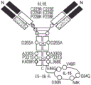


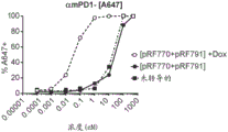
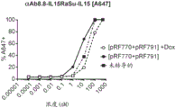

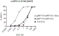


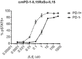


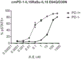
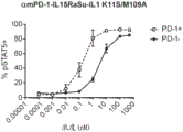
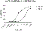
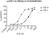
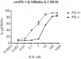


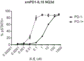


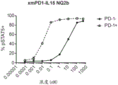


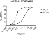
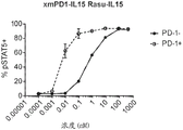
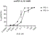
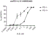

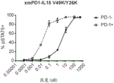


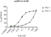

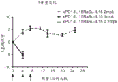

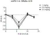
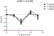
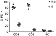

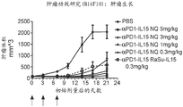


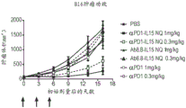
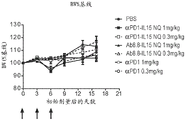
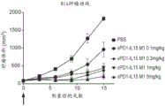

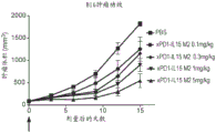
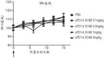
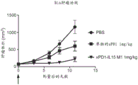

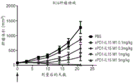

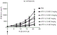
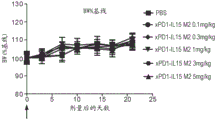
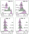

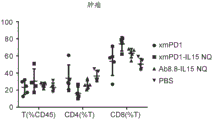


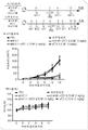


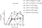


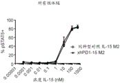

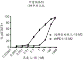
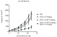
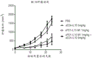

Classifications
-
- C—CHEMISTRY; METALLURGY
- C07—ORGANIC CHEMISTRY
- C07K—PEPTIDES
- C07K14/00—Peptides having more than 20 amino acids; Gastrins; Somatostatins; Melanotropins; Derivatives thereof
- C07K14/435—Peptides having more than 20 amino acids; Gastrins; Somatostatins; Melanotropins; Derivatives thereof from animals; from humans
- C07K14/52—Cytokines; Lymphokines; Interferons
- C07K14/54—Interleukins [IL]
- C07K14/5443—IL-15
-
- A—HUMAN NECESSITIES
- A61—MEDICAL OR VETERINARY SCIENCE; HYGIENE
- A61K—PREPARATIONS FOR MEDICAL, DENTAL OR TOILETRY PURPOSES
- A61K35/00—Medicinal preparations containing materials or reaction products thereof with undetermined constitution
- A61K35/12—Materials from mammals; Compositions comprising non-specified tissues or cells; Compositions comprising non-embryonic stem cells; Genetically modified cells
- A61K35/14—Blood; Artificial blood
- A61K35/17—Lymphocytes; B-cells; T-cells; Natural killer cells; Interferon-activated or cytokine-activated lymphocytes
-
- A—HUMAN NECESSITIES
- A61—MEDICAL OR VETERINARY SCIENCE; HYGIENE
- A61K—PREPARATIONS FOR MEDICAL, DENTAL OR TOILETRY PURPOSES
- A61K38/00—Medicinal preparations containing peptides
- A61K38/16—Peptides having more than 20 amino acids; Gastrins; Somatostatins; Melanotropins; Derivatives thereof
- A61K38/17—Peptides having more than 20 amino acids; Gastrins; Somatostatins; Melanotropins; Derivatives thereof from animals; from humans
- A61K38/177—Receptors; Cell surface antigens; Cell surface determinants
- A61K38/1793—Receptors; Cell surface antigens; Cell surface determinants for cytokines; for lymphokines; for interferons
-
- A—HUMAN NECESSITIES
- A61—MEDICAL OR VETERINARY SCIENCE; HYGIENE
- A61K—PREPARATIONS FOR MEDICAL, DENTAL OR TOILETRY PURPOSES
- A61K38/00—Medicinal preparations containing peptides
- A61K38/16—Peptides having more than 20 amino acids; Gastrins; Somatostatins; Melanotropins; Derivatives thereof
- A61K38/17—Peptides having more than 20 amino acids; Gastrins; Somatostatins; Melanotropins; Derivatives thereof from animals; from humans
- A61K38/19—Cytokines; Lymphokines; Interferons
- A61K38/191—Tumor necrosis factors [TNF], e.g. lymphotoxin [LT], i.e. TNF-beta
-
- A—HUMAN NECESSITIES
- A61—MEDICAL OR VETERINARY SCIENCE; HYGIENE
- A61K—PREPARATIONS FOR MEDICAL, DENTAL OR TOILETRY PURPOSES
- A61K38/00—Medicinal preparations containing peptides
- A61K38/16—Peptides having more than 20 amino acids; Gastrins; Somatostatins; Melanotropins; Derivatives thereof
- A61K38/17—Peptides having more than 20 amino acids; Gastrins; Somatostatins; Melanotropins; Derivatives thereof from animals; from humans
- A61K38/19—Cytokines; Lymphokines; Interferons
- A61K38/20—Interleukins [IL]
- A61K38/2086—IL-13 to IL-16
-
- A—HUMAN NECESSITIES
- A61—MEDICAL OR VETERINARY SCIENCE; HYGIENE
- A61K—PREPARATIONS FOR MEDICAL, DENTAL OR TOILETRY PURPOSES
- A61K39/00—Medicinal preparations containing antigens or antibodies
- A61K39/395—Antibodies; Immunoglobulins; Immune serum, e.g. antilymphocytic serum
-
- A—HUMAN NECESSITIES
- A61—MEDICAL OR VETERINARY SCIENCE; HYGIENE
- A61K—PREPARATIONS FOR MEDICAL, DENTAL OR TOILETRY PURPOSES
- A61K45/00—Medicinal preparations containing active ingredients not provided for in groups A61K31/00 - A61K41/00
- A61K45/06—Mixtures of active ingredients without chemical characterisation, e.g. antiphlogistics and cardiaca
-
- A—HUMAN NECESSITIES
- A61—MEDICAL OR VETERINARY SCIENCE; HYGIENE
- A61P—SPECIFIC THERAPEUTIC ACTIVITY OF CHEMICAL COMPOUNDS OR MEDICINAL PREPARATIONS
- A61P35/00—Antineoplastic agents
-
- C—CHEMISTRY; METALLURGY
- C07—ORGANIC CHEMISTRY
- C07K—PEPTIDES
- C07K14/00—Peptides having more than 20 amino acids; Gastrins; Somatostatins; Melanotropins; Derivatives thereof
- C07K14/435—Peptides having more than 20 amino acids; Gastrins; Somatostatins; Melanotropins; Derivatives thereof from animals; from humans
- C07K14/52—Cytokines; Lymphokines; Interferons
- C07K14/54—Interleukins [IL]
- C07K14/55—IL-2
-
- C—CHEMISTRY; METALLURGY
- C07—ORGANIC CHEMISTRY
- C07K—PEPTIDES
- C07K16/00—Immunoglobulins [IGs], e.g. monoclonal or polyclonal antibodies
- C07K16/18—Immunoglobulins [IGs], e.g. monoclonal or polyclonal antibodies against material from animals or humans
- C07K16/28—Immunoglobulins [IGs], e.g. monoclonal or polyclonal antibodies against material from animals or humans against receptors, cell surface antigens or cell surface determinants
- C07K16/2803—Immunoglobulins [IGs], e.g. monoclonal or polyclonal antibodies against material from animals or humans against receptors, cell surface antigens or cell surface determinants against the immunoglobulin superfamily
- C07K16/2815—Immunoglobulins [IGs], e.g. monoclonal or polyclonal antibodies against material from animals or humans against receptors, cell surface antigens or cell surface determinants against the immunoglobulin superfamily against CD8
-
- C—CHEMISTRY; METALLURGY
- C07—ORGANIC CHEMISTRY
- C07K—PEPTIDES
- C07K16/00—Immunoglobulins [IGs], e.g. monoclonal or polyclonal antibodies
- C07K16/18—Immunoglobulins [IGs], e.g. monoclonal or polyclonal antibodies against material from animals or humans
- C07K16/28—Immunoglobulins [IGs], e.g. monoclonal or polyclonal antibodies against material from animals or humans against receptors, cell surface antigens or cell surface determinants
- C07K16/2803—Immunoglobulins [IGs], e.g. monoclonal or polyclonal antibodies against material from animals or humans against receptors, cell surface antigens or cell surface determinants against the immunoglobulin superfamily
- C07K16/2818—Immunoglobulins [IGs], e.g. monoclonal or polyclonal antibodies against material from animals or humans against receptors, cell surface antigens or cell surface determinants against the immunoglobulin superfamily against CD28 or CD152
-
- C—CHEMISTRY; METALLURGY
- C07—ORGANIC CHEMISTRY
- C07K—PEPTIDES
- C07K16/00—Immunoglobulins [IGs], e.g. monoclonal or polyclonal antibodies
- C07K16/18—Immunoglobulins [IGs], e.g. monoclonal or polyclonal antibodies against material from animals or humans
- C07K16/28—Immunoglobulins [IGs], e.g. monoclonal or polyclonal antibodies against material from animals or humans against receptors, cell surface antigens or cell surface determinants
- C07K16/2803—Immunoglobulins [IGs], e.g. monoclonal or polyclonal antibodies against material from animals or humans against receptors, cell surface antigens or cell surface determinants against the immunoglobulin superfamily
- C07K16/2827—Immunoglobulins [IGs], e.g. monoclonal or polyclonal antibodies against material from animals or humans against receptors, cell surface antigens or cell surface determinants against the immunoglobulin superfamily against B7 molecules, e.g. CD80, CD86
-
- C—CHEMISTRY; METALLURGY
- C07—ORGANIC CHEMISTRY
- C07K—PEPTIDES
- C07K16/00—Immunoglobulins [IGs], e.g. monoclonal or polyclonal antibodies
- C07K16/18—Immunoglobulins [IGs], e.g. monoclonal or polyclonal antibodies against material from animals or humans
- C07K16/28—Immunoglobulins [IGs], e.g. monoclonal or polyclonal antibodies against material from animals or humans against receptors, cell surface antigens or cell surface determinants
- C07K16/2803—Immunoglobulins [IGs], e.g. monoclonal or polyclonal antibodies against material from animals or humans against receptors, cell surface antigens or cell surface determinants against the immunoglobulin superfamily
- C07K16/283—Immunoglobulins [IGs], e.g. monoclonal or polyclonal antibodies against material from animals or humans against receptors, cell surface antigens or cell surface determinants against the immunoglobulin superfamily against Fc-receptors, e.g. CD16, CD32, CD64
-
- C—CHEMISTRY; METALLURGY
- C07—ORGANIC CHEMISTRY
- C07K—PEPTIDES
- C07K16/00—Immunoglobulins [IGs], e.g. monoclonal or polyclonal antibodies
- C07K16/18—Immunoglobulins [IGs], e.g. monoclonal or polyclonal antibodies against material from animals or humans
- C07K16/28—Immunoglobulins [IGs], e.g. monoclonal or polyclonal antibodies against material from animals or humans against receptors, cell surface antigens or cell surface determinants
- C07K16/2887—Immunoglobulins [IGs], e.g. monoclonal or polyclonal antibodies against material from animals or humans against receptors, cell surface antigens or cell surface determinants against CD20
-
- C—CHEMISTRY; METALLURGY
- C07—ORGANIC CHEMISTRY
- C07K—PEPTIDES
- C07K16/00—Immunoglobulins [IGs], e.g. monoclonal or polyclonal antibodies
- C07K16/18—Immunoglobulins [IGs], e.g. monoclonal or polyclonal antibodies against material from animals or humans
- C07K16/28—Immunoglobulins [IGs], e.g. monoclonal or polyclonal antibodies against material from animals or humans against receptors, cell surface antigens or cell surface determinants
- C07K16/2896—Immunoglobulins [IGs], e.g. monoclonal or polyclonal antibodies against material from animals or humans against receptors, cell surface antigens or cell surface determinants against molecules with a "CD"-designation, not provided for elsewhere
-
- C—CHEMISTRY; METALLURGY
- C07—ORGANIC CHEMISTRY
- C07K—PEPTIDES
- C07K16/00—Immunoglobulins [IGs], e.g. monoclonal or polyclonal antibodies
- C07K16/18—Immunoglobulins [IGs], e.g. monoclonal or polyclonal antibodies against material from animals or humans
- C07K16/32—Immunoglobulins [IGs], e.g. monoclonal or polyclonal antibodies against material from animals or humans against translation products of oncogenes
-
- C—CHEMISTRY; METALLURGY
- C07—ORGANIC CHEMISTRY
- C07K—PEPTIDES
- C07K19/00—Hybrid peptides, i.e. peptides covalently bound to nucleic acids, or non-covalently bound protein-protein complexes
-
- C—CHEMISTRY; METALLURGY
- C12—BIOCHEMISTRY; BEER; SPIRITS; WINE; VINEGAR; MICROBIOLOGY; ENZYMOLOGY; MUTATION OR GENETIC ENGINEERING
- C12N—MICROORGANISMS OR ENZYMES; COMPOSITIONS THEREOF; PROPAGATING, PRESERVING, OR MAINTAINING MICROORGANISMS; MUTATION OR GENETIC ENGINEERING; CULTURE MEDIA
- C12N15/00—Mutation or genetic engineering; DNA or RNA concerning genetic engineering, vectors, e.g. plasmids, or their isolation, preparation or purification; Use of hosts therefor
- C12N15/09—Recombinant DNA-technology
- C12N15/11—DNA or RNA fragments; Modified forms thereof; Non-coding nucleic acids having a biological activity
- C12N15/62—DNA sequences coding for fusion proteins
-
- A—HUMAN NECESSITIES
- A61—MEDICAL OR VETERINARY SCIENCE; HYGIENE
- A61K—PREPARATIONS FOR MEDICAL, DENTAL OR TOILETRY PURPOSES
- A61K39/00—Medicinal preparations containing antigens or antibodies
- A61K2039/505—Medicinal preparations containing antigens or antibodies comprising antibodies
-
- A—HUMAN NECESSITIES
- A61—MEDICAL OR VETERINARY SCIENCE; HYGIENE
- A61K—PREPARATIONS FOR MEDICAL, DENTAL OR TOILETRY PURPOSES
- A61K2300/00—Mixtures or combinations of active ingredients, wherein at least one active ingredient is fully defined in groups A61K31/00 - A61K41/00
-
- C—CHEMISTRY; METALLURGY
- C07—ORGANIC CHEMISTRY
- C07K—PEPTIDES
- C07K2317/00—Immunoglobulins specific features
- C07K2317/50—Immunoglobulins specific features characterized by immunoglobulin fragments
- C07K2317/56—Immunoglobulins specific features characterized by immunoglobulin fragments variable (Fv) region, i.e. VH and/or VL
- C07K2317/565—Complementarity determining region [CDR]
-
- C—CHEMISTRY; METALLURGY
- C07—ORGANIC CHEMISTRY
- C07K—PEPTIDES
- C07K2319/00—Fusion polypeptide
- C07K2319/30—Non-immunoglobulin-derived peptide or protein having an immunoglobulin constant or Fc region, or a fragment thereof, attached thereto
-
- C—CHEMISTRY; METALLURGY
- C07—ORGANIC CHEMISTRY
- C07K—PEPTIDES
- C07K2319/00—Fusion polypeptide
- C07K2319/31—Fusion polypeptide fusions, other than Fc, for prolonged plasma life, e.g. albumin
Abstract
The present invention relates to human interleukin 15(IL-15) variants having therapeutic and diagnostic uses and methods for their manufacture. The invention also provides fusion proteins comprising human IL-15 variants. Also provided are methods of stimulating or inhibiting an immune response in a mammal and methods of treating a pathology (e.g., cancer) using an IL-15 variant or a fusion protein of such IL-15 variants.
Description
Technical Field
The present invention relates to interleukin 15(IL-15) variants having therapeutic and diagnostic uses and methods for their manufacture. The invention also provides fusion proteins comprising such IL-15 variants. Also provided are methods of stimulating or inhibiting an immune response in a mammal and methods of treating a pathology (e.g., cancer) using an IL-15 variant or a fusion protein of such IL-15 variants.
Background
Cytokines are potent modulators of immune responses and have the potential to significantly affect the outcome of immunooncological treatment approaches. However, previous efforts to utilize cytokines in human subjects have resulted in only modest efficacy and significant toxicity. Recent studies have shown that "targeted cytokines," such as antibody-cytokine fusion proteins, can deliver cytokines to desired cell types while minimizing peripheral exposure and thus toxicity. See, e.g., Guo et al, Cytokine Growth Factor Rev. 38:10-21 (2017); jakobisiak M, et al, Cytokine Growth Factor Rev. 22(2):99-108 (2011); robinson, t. & Schluns, k.s., immunol. lett. 190:159-168 (2017); rhode et al, Cancer Immunol. Res. 4(1): 49-60 (2016); conlon et al, J Clin. Oncol. 33(1): 74-82 (2015). Therefore, the development of therapeutic agents based on targeted cytokines would be of great value in the treatment of various diseases such as cancer.
Disclosure of Invention
The invention disclosed herein relates to human interleukin 15 (IL-15) variants and fusion proteins comprising the same. IL-15 variants of the invention have demonstrated reduced or no binding to IL-15 receptor alpha (CD215) and/or reduced interaction between IL-15 and its signaling receptor, consisting of IL-2 receptor beta (CD122) and common gamma chain (CD132), as compared to wild-type human IL-15 polypeptide or wild-type IL-15 receptor alpha-IL-15 fusion polypeptide. In a second aspect of the invention, these reduced affinity IL-15 variants (when presented as antibody fusion chimeric proteins) are selectively targeted to desired cell types (those expressing antibody targets). Cell types expressing the IL-15 receptor complex (but not the antibody target) are less or not activated compared to those expressing both components. Thus, the IL-15 variants and IL-15 fusion proteins of the present invention selectively modulate activation of cell subsets to effectively and safely promote biological activities, such as anti-tumor activity. In a third aspect of the invention, these reduced affinity IL-15 variants and IL-15 fusion proteins (in secreted or membrane tethered form when expressed as polynucleotides in CAR (chimeric antigen receptor) T cells) are used to enhance CAR T function, including activity and proliferation.
Accordingly, in one aspect, the present invention provides an isolated human interleukin 15 (IL-15) variant, which comprises the amino acid sequence of SEQ ID NO: 1 a) V49 and I51 or b) amino acid substitutions at V49, I50 and S51, and further comprises SEQ ID NO: 1, N1, N4, S7, K10, K11, Y26, S29, D30, V31, H32, E53, G55, E64, I68, L69, E89, L91, M109 and/or I111, wherein the IL-15 variant has reduced or no binding to human IL-15 receptor alpha (IL-15 Ra) and human IL-2 receptor beta/gamma (IL-2 Rss gamma) compared to the wild-type human IL-15 polypeptide or the wild-type IL-15 receptor alpha-IL-15 fusion polypeptide, and wherein the amino acid substitution at position V49 is glycosylated.
In another aspect, there is provided an isolated fusion protein comprising: 1) an antibody comprising an Fc domain; and b) a human interleukin 15 (IL-15) variant comprising amino acid substitutions at positions a) V49 and I51 or b) V49, I50 and S51 of SEQ ID NO: 1, wherein the amino acid substitution at position V49 of SEQ ID NO: 1 is glycosylated and further comprises one or more amino acid substitutions at positions N1, N4, S7, K10, K11, Y26, S29, D30, V31, H32, E53, G55, E64, I68, L69, E89, L91, M109 and/or I111 of SEQ ID NO: 1, wherein the IL-15 variant is covalently linked to the Fc domain of an antibody, and wherein the antibody-dependent cellular cytotoxicity (ADCC) activity of the Fc domain is reduced or absent compared to wild-type Fc.
In some embodiments, the IL-15 variant comprises an amino acid substitution at V49N, wherein V49N is glycosylated. In some embodiments, the amino acid substitutions at E53 and/or E89 of SEQ ID NO. 1 are also glycosylated.
In some embodiments, the IL-15 variant comprises an amino acid substitution of SEQ ID NO 1 at a position selected from the group consisting of: a) v49, I50, S51, N4, D30 and E64; b) v49, I50, S51, N4, D30, E64, and I68; c) v49, I50, S51, N4, D30, E64, M109; d) v49, I50, S51, N4, D30, E64, I68 and M109; e) v49, I50, S51, D30, E64 and I68; f) v49, I50, S51, D30, E64, M109; g) v49, I50, S51, D30, E64, I68 and M109; h) n1, V49, I50 and S51; i) n4, V49, I50 and S51; j) s7, V49, I50 and S51; k) k10, V49, I50 and S51; l) K11, V49, I50 and S51; m) S29, V49, I50 and S51; n) V31, V49, I50 and S51; o) H32, V49, I50 and S51; p) V49, I50, S51 and E64; q) V49, I50, S51 and I68; r) V49, I50, S51 and L69; s) V49, I50, S51 and I111; t) N4, V49, I50, S51 and E64; u) N1, D30, V49, I50 and S51; v) N4, D30, V49, I50 and S51; w) S7D 30, V49, I50 and S51; x) K10, D30, V49, I50 and S51; y) K11, D30, V49, I50 and S51; z) S29, D30, V49, I50 and S51; aa) D30, V49, I50, S51 and E64; bb) D30, V49, I50, S51 and I68; cc) D30, V49, I50, S51, and L69; and dd) D30, V49, I50, S51, and I111.
In some embodiments, the IL-15 variant comprises an amino acid substitution comprising one or more specific substitutions at the following positions: a) V49N, V49K, V49E, V49H, V49Q or V49R; b) I50A or I50G; c) S51T; d) N1K, N1G, N1Q, N1R, N1E, N1A, or N1D; e) N4K, N4G, N4A, N4S, N4D, N4E, N4L, N4R, N4T, or N4Q; f) S7E, S7G, S7D, S7K, S7N, S7R, S7H, or S7T; g) K10A, K10S, K10E, K10L, K10M, K10D, or K10G; h) K11D, K11S or K11W; i) D30N; j) E64Q, E64K, E64A, E64S, E64N, E64H, E64T, or E64R; k) E53N; l) G55S or G55T; m) E89N; n) L91S or L91T; o) Y26K, Y26R or Y26H; p) S29N; q) V31S, V31D or V31K; r) H32G; s) I68S, I68A, I68R, I68T, I68K, I68N, I68M, I68F, I68Y, I68E, or I68H; t) L69A, L69S, L69D, L69T, L69M, L69G, L69Q, L69I, L69E or L69V; u) M109A, M109S, M109D or M109K; and/or v) I111A, I111K, I111S, or I111D. In some embodiments, the IL-15 variant comprises an amino acid substitution at a position selected from the group consisting of: a) N4K, D30N, V49N, I50A, S51T and E64Q; b) N4Q, D30N, V49N, I50A and S51T; c) D30N, V49N, I50A, S51T and E64Q; d) N4Q, D30N, V49N, I50A, S51T and E64Q; e) N4Q, V49N, I50A and S51T; f) V49N, I50A, S51T and E64Q; and g) N4Q, V49N, I50A, S51T, and E64Q.
In another aspect, an isolated human interleukin 15 (IL-15) variant is provided comprising amino acid substitutions at positions E46 and V49 of SEQ ID NO: 1 and at least one or more amino acid substitutions at positions N1, N4, S7, K10, K11, D22, Y26, S29, D30, V31, H32, E53, G55, E64, I68, L69, E89, E93, M109, and/or I111 of SEQ ID NO: 1, wherein the IL-15 variant has NO binding to human IL-15 receptor alpha (IL-15 ra) and reduced binding to human IL-2 receptor beta/gamma (IL-2R β γ) compared to a wild-type human IL-15 polypeptide or a wild-type IL-15 receptor alpha-IL-15 fusion polypeptide.
In another aspect, there is provided an isolated fusion protein comprising: 1) an antibody comprising an Fc domain; and b) a human interleukin 15 (IL-15) variant comprising amino acid substitutions at positions E46 and V49 of SEQ ID NO: 1 and at least one or more amino acid substitutions at positions N1, N4, S7, K10, K11, D22, Y26, S29, D30, V31, H32, E53, G55, E64, I68, L69, E89, E93, M109 and/or I111 of SEQ ID NO: 1, wherein the IL-15 variant is covalently linked to an Fc domain of an antibody, and wherein the antibody-dependent cellular cytotoxicity (ADCC) activity of the Fc domain is reduced or absent ADCC activity compared to a wild-type Fc.
In some embodiments, the IL-15 variant comprises an amino acid substitution in SEQ ID NO 1 selected from the following positions: a) n1, E46 and V49; b) n4, E46 and V49; c) s7, E46 and V49; d) k10, E46 and V49; e) k11, E46 and V49; f) s29, E46 and V49; g) v31, E46 and V49; h) h32, E46 and V49; i) e46, V49 and E64; j) e46, V49 and I68; k) e46, V49 and L69; l) E46, V49 and I111; m) N4, E46, V49 and E64; n) E46, V49, N4, D30 and E64; o) E46, V49, N4, D30, E64 and I68; p) E46, V49, N4, D30, E64 and M109; q) E46, V49, N4, D30, E64, I68, and M109; r) N1, D30, E46 and V49; s) N4, D30, E46 and V49; t) S7, D30, E46 and V49; u) K10, D30, E46 and V49; v) K11, D30, E46 and V49; w) S29, D30, E46 and V49; x) D30, E46, V49 and E64; y) D30, E46, V49R and I68; z) D30, E46, V49R and L69; aa) D30, E46, V49R and I111; bb) N1, D30, E46, V49 and M109; cc) N4, D30, E46, V49 and M109; dd) S7, D30, E46, V49 and M109; ee) K10, D30, E46, V49 and M109; ff) K11, D30, E46, V49 and M109; gg) D30, E46, V49, E64 and M109; hh) D30, E46, V49, I68, and M109; ii) D30, E46, V49, L69 and M109; jj) D30, E46, V49, M109 and I111; kk) D30, E46, V49, E64, I68 and M109; ll) E46, V49, D30, E64 and I68; mm) E46, V49, E64 and M109; nn) E46, V49, D30, E64, I68 and M109; oo) D22, Y26, V49, E46, E53, E89, and E93; and pp) N1, D30, E46, V49, and E64.
In some embodiments, the IL-15 variant comprises an amino acid substitution comprising one or more specific substitutions at the following positions: a) N1Q, N1K, N1R, N1E, N1A, N1D, or N1G; b) N4K, N4G, N4A, N4S, N4D, N4E, N4R, N4T, N4I, N4L, N4W, or N4Q; c) S7E, S7G, S7D, S7K, S7N, S7R, S7H, or S7T; d) K10D, K10A, K10S, K10E, K10L, K10M, K10D, or K10G; e) K11D, K11S or K11W; f) D22N; g) Y26K, Y26R or Y26H; h) S29N; i) D30N; j) V31S, V31D or V31K; k) H32G; l) E46G or E46Q; m) V49N, V49K or V49R V49E, V49H or V49Q; n) E53Q; o) G55S or G55T; p) E64Q, E64K, E64A, E64S, E64N, E64H, E64T, or E64R; q) I68S, I68A, I68R, I68T, I68K, I68N, I68M, I68F, I68Y, I68E or I68H; r) L69S, L69A, L69D, L69T, L69M, L69G, L69Q, L69I, L69E or L69V; s) E89Q; t) E93Q; u) M109A, M109S, M109D or M109K; and/or v) I111A, I111K, I111S, or I111D. In some embodiments, the IL-15 variant comprises an amino acid substitution at a position selected from the group consisting of: a) N1K, E46G and V49R; b) N4K, E46G and V49R; c) N4Q, E46G and V49R; d) S7T, E46G and V49R; e) V31S, E46G and V49R; f) V31K, E46G and V49R; g) E46G, V49R and E64Q; h) E46G, V49R and E64K; i) N4Q, E46G, V49R and E64Q; j) N1G, D30N, E46G and V49R; k) N1K, D30N, E46G and V49R; l) N1Q, D30N, E46G and V49R; m) N4G, D30N, E46G and V49R; n) N4K, D30N, E46G and V49R; o) N4Q, D30N, E46G and V49R; p) S7E, D30N, E46G and V49R; q) S7G, D30N, E46G and V49R; r) S7T, D30N, E46G and V49R; s) K10D, D30N, E46G and V49R; t) D30N, E46G, V49R and E64A; u) D30N, E46G, V49R and E64Q; v) D30N, E46G, V49R and E64K; w) D30N, E46G, V49R and I68S; x) D30N, E46G, V49R and I68K; y) N4K, D30N, E46G, V49R, and E64K; z) N4Q, D30N, E46G, V49R and E64K; aa) N4K, D30N, E46G, V49R and E64Q; bb) N4Q, D30N, E46G, V49R and E64Q; cc) N4K, D30N, E46G, V49R, and I68S; dd) D30N, E46G, V49R, E64Q, and I68S; ee) N1A, D30N, E46G and V49R; and ff) N1G, D30N, E46G, V49R, and E64Q.
In another aspect, an isolated human IL-15 variant is provided comprising one or more amino acid substitutions at positions N1, N4, S7, K10, K11, D22, Y26, S29, D30, V31, H32, E46, E53, E64, I68, L69, E89, E93, M109, and/or I111 of SEQ ID NO: 1, wherein the IL-15 variant has reduced or NO binding to human IL-15 receptor alpha (IL-15 ra) and/or human IL-2 receptor beta (IL-2R β) and/or IL-2 receptor gamma (IL-2R γ) compared to a wild-type human IL-15 polypeptide or a wild-type IL-15 receptor alpha-IL-15 fusion polypeptide.
In another aspect, an isolated human IL-15 variant is provided comprising the amino acid sequence set forth in SEQ ID NO: 93. In some embodiments, the IL-15 variant further comprises a transmembrane domain.
In another aspect, there is provided an isolated fusion protein comprising: 1) an antibody comprising an Fc domain; and b) a human interleukin 15 (IL-15) variant comprising one or more amino acid substitutions at position N1, N4, S7, K10, K11, D22, Y26, S29, D30, V31, H32, E46, E53, E64, I68, L69, E89, E93, M109 and/or I111 of SEQ ID NO 1, wherein the IL-15 variant is covalently linked to an Fc domain of an antibody, and wherein the antibody-dependent cellular cytotoxicity (ADCC) activity of the Fc domain is reduced or absent as compared to wild-type Fc.
In another aspect, there is provided an isolated fusion protein comprising: 1) an IL-15 antibody comprising an Fc domain; and b) the human interleukin 15 (IL-15) protein of SEQ ID NO: 1, wherein IL-15 is covalently linked to the Fc domain of an antibody.
In some embodiments, the antibody in the IL-15 fusion protein of the invention can be a human antibody, a humanized antibody, a chimeric antibody, or a bispecific antibody.
In some embodiments, the antibody in the IL-15 fusion protein of the invention is a human IgG1、IgG2、IgG2Δa、IgG3、IgG4、IgG4Δb、IgG4Δc、IgG4 S228P、IgG4ΔbS228P and IgG4ΔcSubclass S228P.
In some embodiments, the antibody to the IL-15 fusion protein of the invention is a) IgG2, and the antibody variable domain comprises amino acid modifications at positions 223, 225, and 228 in the hinge region and at position 409 or 368 in the CH3 region (EU numbering scheme) of human IgG2 (SEQ ID NO: 3); b) IgG1, and the antibody variable domain comprises amino acid modifications at positions 221 and 228 in the hinge region and at position 409 or 368 (EU numbering scheme) in the CH3 region of human IgG1 (SEQ ID NO: 2); or c) IgG1, and the antibody variable domain comprises amino acid modifications at positions 349, 354, 366, 368 and/or 407 (EU numbering scheme) in the CH3 region of human IgG1 (SEQ ID NO: 2). In some embodiments, the antibody further comprises amino acid modifications at the following positions: a) one or more positions 265, 330 and/or 331 of human IgG2 (SEQ ID NO: 3); or b) one or more of positions 234, 235, 237 and/or 322 of human IgG1 (SEQ ID NO: 2). In some embodiments, the antibody further comprises amino acid modifications at one or more of positions 234, 235, 237, 349, 354, 366, 368 and/or 407 of human IgG1 (SEQ ID NOS: 74 and 75).
In some embodiments, the antibody for the IL-15 fusion protein is selected from the group consisting of: anti-CTLA-4 antibodies, anti-CD 3 antibodies, anti-CD 4 antibodies, anti-CD 8 antibodies, anti-4-1 BB antibodies, anti-PD-1 antibodies, anti-PD-L1 antibodies, anti-TIM 3 antibodies, anti-LAG 3 antibodies, anti-TIGIT antibodies, anti-OX 40 antibodies, anti-IL-7 Ra (CD127) antibodies, anti-IL-8 antibodies, anti-IL-15 antibodies, anti-HVEM antibodies, anti-BTLA antibodies, anti-CD 40 antibodies, anti-CD 40L antibodies, anti-CD 47 antibodies, anti-CSF 1 47 antibodies, anti-CSF 47 antibodies, anti-MARCO antibodies, anti-CXCR 47 antibodies, anti-VEGF antibodies, anti-VEGFR 47 antibodies, anti-TNFR 47 antibodies, anti-CD 47 bispecific antibodies, anti-CD 47 antibodies, anti-Her 47 antibodies, anti-EGFR antibodies, anti-CD 47 antibodies, anti-CD 363672 antibodies, anti-, anti-CD 163 antibody, anti-KLRG 1 antibody, anti-FLT 3 antibody, anti-B7-H4 antibody, anti-B7-H3 antibody, KLRG1 antibody, and anti-GITR antibody.
In some embodiments, the IL-15 fusion proteins of the invention are covalently linked to an antibody via a polypeptide linker and/or a polypeptide tag.
In another aspect, the invention provides a pharmaceutical composition comprising a therapeutically effective amount of an IL-15 variant or IL-15 fusion protein as described herein and a pharmaceutically acceptable carrier.
In another aspect, the invention provides an isolated polynucleotide comprising a nucleotide sequence encoding an IL-15 variant or IL-15 fusion protein as described herein. In another aspect, the present invention provides a vector comprising the polynucleotide.
In another aspect, the invention provides an isolated host cell or cell line that recombinantly produces an IL-15 variant or IL-15 fusion protein as described herein. In some embodiments, the host cell or cell line is an engineered immune cell, wherein the engineered immune cell comprises a Chimeric Antigen Receptor (CAR). In some embodiments, the CAR-expressing cell is a T cell, and the T cell expresses an IL-15 variant or IL-15 fusion protein as described herein, in secreted or membrane tethered form.
In another aspect, the invention provides a method of producing an IL-15 variant or IL-15 fusion protein, the method comprising: culturing a cell line recombinantly producing an IL-15 variant or IL-15 fusion protein as described herein under conditions wherein the protein variant or fusion protein is produced; and recovering the protein variant or fusion protein.
In another aspect, the present invention provides a method for treating a condition in a subject, comprising administering to a subject in need thereof an effective amount of a pharmaceutical composition as described herein. In some embodiments, the condition is cancer. In some embodiments, the cancer is a liquid cancer or a solid cancer. In some embodiments, the cancer is relapsed, refractory or metastatic.
In another aspect, the present invention provides a method of inhibiting tumor growth or progression in a subject having a tumor, comprising administering to the subject an effective amount of a pharmaceutical composition as described herein.
In another aspect, the present invention provides a method of inhibiting or preventing cancer metastasis of a cancer cell in a subject, comprising administering to a subject in need thereof an effective amount of a pharmaceutical composition as described herein.
In another aspect, the present invention provides a method of inducing tumor regression in a subject in need thereof, comprising administering to the subject an effective amount of a pharmaceutical composition as described herein.
In some embodiments, the IL-15 variants and IL-15 fusion proteins described herein can be administered parenterally in a subject. In some embodiments, the subject is a human.
In some embodiments, the method may further comprise administering an effective amount of a second therapeutic agent. In some embodiments, the second therapeutic agent is a biologic therapeutic agent.
In some embodiments, the second therapeutic agent is a cytokine, an immune cytokine (e.g., anti-EDA-IL 10 fusion protein), a TNF α, a PAP inhibitor, an oncolytic virus, a kinase inhibitor, an ALK inhibitor (e.g., sunitinib or crizotinib), a MEK inhibitor, an IDO inhibitor, a GLS1 inhibitor, a tyrosine kinase inhibitor (e.g., axitinib or palbociclib)), a CAR (chimeric antigen receptor) -T cell or T cell therapy, a PRR (pattern recognition receptor) agonist, such as a TLR (Toll-like receptor) agonist (e.g., TLR3, TLR4, TLR5, TLR7, TLR9), or a tumor vaccine.
Also provided is the use of any of the IL-15 variants or IL-15 fusion proteins provided herein in the manufacture of a medicament for treating cancer or inhibiting tumor growth or progression in a subject in need thereof.
In one aspect, the invention provides a method for treating cancer in a subject comprising administering to the subject a combination therapy comprising a first therapeutic agent and a second therapeutic agent, wherein the first therapeutic agent is an IL-15 variant or an IL-15 fusion protein.
In some embodiments, the first therapeutic agent is an IL-15 variant. In some embodiments, the first therapeutic agent is an IL-15 fusion protein. In some embodiments, the first therapeutic agent is any IL-15 variant or IL-15 fusion protein of the invention. In some embodiments, the first therapeutic agent comprises the amino acid sequence set forth in SEQ ID NO:84, 85, 86, 87, 89, or 90.
In some embodiments, the second therapeutic agent is an immunocytokine. In some embodiments, the second therapeutic agent is an immunocytokine comprising an antibody or fragment thereof conjugated or fused to a cytokine (e.g., a fusion protein). In some embodiments, the antibody or fragment thereof binds the extra domain-a (EDA) isoform of fibronectin (e.g., an anti-EDA antibody). In some embodiments, the anti-EDA antibody or fragment thereof comprises CDR1, CDR2, and CDR3 of the heavy chain Variable (VH) region shown in SEQ ID NO:94 and/or CDR1, CDR2, and CDR3 of the light chain Variable (VI) region shown in SEQ ID NO: 96. In some embodiments, the anti-EDA antibody or fragment thereof comprises a VH region having the amino acid sequence of SEQ ID NO. 94 and/or a VL region having the amino acid sequence of SEQ ID NO. 96. In some embodiments, the cytokine is IL-10. In some embodiments, IL-10 comprises the amino acid sequence of SEQ ID NO 98. In some embodiments, the immunocytokine comprises at least one linker. In some embodiments, the linker comprises SEQ ID NOs 95 and/or 97. In some embodiments, the immunocytokine is an anti-EDA-IL-10 fusion protein comprising the amino acid sequence shown in SEQ ID NO 99.
In some embodiments, the invention provides a method for treating cancer in a subject comprising administering to the subject a combination therapy comprising a first therapeutic agent and a second therapeutic agent, wherein the first therapeutic agent is an IL-15 fusion protein, and wherein the second agent is an anti-EDA-IL-10 fusion protein. In some embodiments, the IL-15 fusion protein comprises the amino acid sequence set forth in SEQ ID NO:84, 85, 86, 87, 89, or 90, and the anti-EDA-IL-10 fusion protein comprises the amino acid sequence set forth in SEQ ID NO: 99.
In some embodiments, the combination therapy may further comprise 1, 2, 3, 4, or 5 additional therapeutic agents. In some embodiments, the combination therapy further comprises an anti-PD-1 or anti-PD-L1 antibody. In some embodiments, the anti-PD-1 antibody is BCD-100, camlizumab (camrelizumab) (SHR-1210), gemipizumab (cemipimab) (REGN2810), gemlizumab (genilizumab) (CBT-501), MEDI0680, nivolumab (nivolumab) (OPDIVO)®) Pabolilizumab (KEYTRUDA)®) PF-06801591 (RN888), sidilizumab (sintilimab) (IBI-308), sipellizumab (PDR-001), STI-A1110, Tislelizumab (BGB-A317) or TSR-042. In some embodiments, the anti-PD-L1 antibody is atelizumab (atezolizumab) (TECENTRIQ) ®) Dewar monoclonal antibody (durvalumab) (IMFINZI)®) BMS-936559 (MDX-1105) or LY 3300054.
In some embodiments, the therapeutic agents in a combination therapy can be administered simultaneously (e.g., in the same drug or at the same time), concurrently (i.e., in separate drugs administered one after the other in any order), or sequentially in any order.
Drawings
Fig. 1A is a schematic depicting an anti-mouse PD1 antibody-IL-15 fusion protein construct comprising: fully human hIgG2 Δ a with a bivalent Fab at the N-terminus as targeting antibody arm; heterodimeric Fc with mutations at CH2 and CH3 (D265A, a330S, P331S) that abolish Fc γ R binding, bispecific mutations at hinges (C223R/E, E225R/E and P228R/E) and CH3 (K409R or L368E) for monovalent linkage of IL-15 mutein molecules at the C-terminus via a flexible glycine-serine (GS) -linker. Mutations in the IL-15 protein include N4K, D30N, V49N, I50A, S51T and E64Q (of SEQ ID NO: 1); V49N-I50A-S51T is an N-linked glycosylation site (as indicated by the schematic carbohydrate motif extending beyond the position of V49N).
Fig. 1B depicts another anti-mouse PD1 antibody-IL-15 mutant fusion protein construct, which has the same mutations as the antibody shown in fig. 1A, and the differences are mutations in the IL-15 protein, including N4K, D30N, E46G (or Y26K), V49R (or V49K), and E64Q.
Fig. 1C is a schematic depicting an anti-mouse PD1 antibody-IL-15 fusion protein construct ("M2") comprising: fully human IgG1 with bivalent Fab as targeting antibody arm at the N-terminus; bispecific mutations on hinges (L234A, L235A and G237A) and CH3 (Y349C, T366W, S354C, T366S, L368A and Y407V) for monovalent linkage of IL-15 mutein molecules at the C-terminus via a flexible glycine-serine (GS) -linker. Mutations in the IL-15 protein include E46G, V49R, E64Q, D30N, and N1G (of SEQ ID NO: 1).
FIG. 1D depicts another anti-mouse PD1 antibody-IL-15 mutant fusion protein construct ("M1") that has the same mutations as the antibody shown in FIG. 1C, and differs by mutations in the IL-15 protein, including E46G, V49R, D30N, and N1A (of SEQ ID NO: 1).
Fig. 2A, fig. 2B, fig. 2C, and fig. 2D show direct binding of an antibody or antibody-cytokine fusion molecule to cells expressing the IL-15 receptor (CD122 plus CD132) and in some cases also expressing PD-1. The molecule comprises: anti-mouse PD-1 antibody (fig. 2A); isotype control antibody (Ab8.8) -aSu-IL-15 (FIG. 2B); anti-mouse PD-1-IL-15RaSu-IL15 (fig. 2C); and anti-mouse PD-1-IL-15 NQ (fig. 2D).
Figures 3A, 3B and 3C show a comparison of non-targeted IL-15, xmPD-1-IL-15RaSu-IL15 (wild-type IL-15) and xmPD-1-IL15 NQ variants in a reporter assay by plotting the percentage of PD-1+ or PD-1 (low) cells that were also stained positive for pSTAT5 as analyzed by flow cytometry.
FIGS. 4A, 4B, 4C, 4D, 4E, 4F, 4G and 4H show graphs of a series of muteins (mutants or variants, used interchangeably) all based on xmPD-1-IL15RaSu-IL 15. The specific molecules analyzed are indicated in each figure.
FIGS. 5A, 5B, 5C, 5D, 5E, 5F and 5G show graphs of a series of muteins, all lacking the IL-15RaSu domain and containing mutations to reduce or eliminate IL-15Ra binding. The specific molecules analyzed are indicated in each figure.
FIGS. 6A, 6B, 6C, 6D, 6E, 6F and 6G show graphs of a series of muteins with altered activity (pSTAT5 activation) relative to IL-15 wild-type. The specific molecules analyzed are indicated in each figure.
FIGS. 7A, 7B, 7C, 7D, 7E, 7F, 7G and 7H show graphs of a series of muteins with altered activity relative to the IL-15 wild-type (pSTAT5 activation). The specific molecules analyzed are indicated in each figure.
FIGS. 8A, 8B, 8C and 8D show the effect on survival and body weight of mice treated with xmPD-1-IL15RaSu-IL15 or xmPD1-IL15 NQ. More specifically, figure 8A shows the body weight changes of animals dosed with 2, 1 or 0.2 mg/kg xmPD1-IL15RaSu-IL15 on the day indicated by the arrow (d0, 4, 7). Figure 8B shows the overall survival rate of these same animals. FIG. 8C shows body weight changes in animals dosed with 1, 0.8, or 0.5 mg/kg xmPD1-IL15RaSu-IL15 on days 0, 3, and 6. FIG. 8D shows body weight changes in animals dosed with 3, 2, or 1 mg/Kg xmPD1-IL15 NQ on days 0, 3, and 6.
Figures 9A and 9B plot the percentage of splenocytes or tumor infiltrating lymphocytes gated to CD4+ or CD8+ cells or NK cells and scored as PD-1+ (figure 9A); figure 9B shows the same study plotted as the actual mean fluorescence intensity.
FIGS. 10A, 10B, and 10C show in vivo B16F10 tumor efficacy study data for mice treated with 5, 3, 1, or 0.3 mg/kg xmPD1-IL15 NQ or 0.3 mg/kg xmPD1-IL15RaSu-IL15 and compared to the control (PBS treated) group. More specifically, in fig. 10A, primary tumor volumes for all groups are plotted. In fig. 10B, the overall survival of animals in the study group is plotted. In fig. 10C, the change in body weight (relative to the study starting body weight) for each group is plotted.
Figures 11A and 11B show data from in vivo B16F10 tumor efficacy studies in mice treated with anti-PD-1 antibody, xmPD1-IL15 NQ, or isotype control (ab8.8) -IL15 NQ, each at 1 or 0.3 mg/kg.
11C, 11D, 11E, and 11F show data from in vivo B16F10 tumor efficacy studies in mice treated with anti-PD 1-IL 15M 1 or M2 (as depicted in FIGS. 1D and 1C, respectively) at 0.1, 0.3, 1, or 5 mg/kg each.
FIGS. 11G and 11H show data from in vivo B16F10 tumor efficacy studies in mice treated with anti-PD-1 antibody or anti-PD 1-IL 15M 1 at 1 mg/Kg each.
11I, 11J, 11K and 11L show data from in vivo MC38 tumor efficacy studies in mice treated with anti-PD 1-IL 15M 1 or M2 at 0.1, 0.3, 1 or 5 mg/kg each. 11A, 11C, 11E, 11G, 11I, and 11K plot the volume of the primary tumor mass over time for each group; the lower arrow on the X-axis indicates the time course of administration. Fig. 11B, 11D, 11F, 11H, 11J, and 11L plot the mean change in body weight from baseline (D =0 at the start of dosing) for each group.
FIGS. 12A, 12B, 12C and 12D show the results of experiments analyzing changes in spleen and TIL lymphocytes due to IL-15 drug administration. FIG. 12A shows detection of cells from animals treated with the foregoing molecules via anti-human Fc against PD-1, Ab8.8-IL15 NQ or xmPD1-IL15 NQ. And (3) upper row: histograms of TIL CD8+ (left) or CD4+ (right) cells as indicated. And (3) lower row: histograms of splenic CD8+ (left) or CD4+ (right) cells as indicated. Figure 12B shows the percentage of splenic T cells, CD4+ T cells, and CD8+ T cells as indicated from animals sampled 1 day after 3 doses of the indicated compound. Figure 12C shows the percentage of TIL T cells, CD4+ T cells, and CD8+ T cells as indicated from animals sampled 1 day after 3 doses of the indicated compound. Fig. 12D plots the ratio of CD8 to CD 4T cells for the spleen and tumor as calculated from fig. 12B and 12C.
FIGS. 13A and 13B show the absolute counts of NK, CD8+ T and Treg cells in peripheral blood or tumor infiltrating lymphocytes on day 6, which show the greatest effect by xmPD1-IL 15M 1 and xmPD1-IL 15M 2, respectively.
Fig. 13C and 13D show the corresponding effects of depleting CD 8T and NK1.1+ cells on a B16F10 tumor therapy model. The treatment regimen and average weight of the mice during the study are also shown.
Figure 13E shows the effect of FTY420 treatment inhibiting T cell release on a B16F10 tumor efficacy model. The treatment regimen and average weight of the mice during the study are also shown.
FIGS. 14A and 14B show the effect on cytokine production in vivo in mice administered IL-15 containing compounds. Figure 14A shows the mean IFNg levels (n =3 animals) at 6, 24, 48, 72, 96 and 120 hours after a single dose of the indicated IL-15 compound. Figure 14B shows mean IL-6 levels (n =3 animals) at 6, 24, 48, 72, 96 and 120 hours after a single dose of the indicated IL-15 compound.
FIGS. 15A, 15B, 15C, 15D, 15E and 15F show the effect of adding a chimeric molecule targeting anti-human PD-1 (xhPD1) -IL 15M 1 or M2 or a non-targeting isotype control-antibody, IL15, to human peripheral blood mononuclear cells, where it is on human NK cells, CD8, respectively +Effector memory T cells and CD8+Central memory T cells produce a different activation of pSTAT 5.
FIGS. 16A, 16B and 16C show in vivo efficacy studies of PD-1-targeting IL-15 molecule (xPD1-IL 15M 1) in combination with anti-EDA-IL-10 fusion proteins. FIG. 16A shows the anti-tumor efficacy of 0.3 mg/kg xPD1- IL 15M 1 and 5 mg/kg anti-EDA-IL-10 fusion protein (xEDA-IL 10). FIG. 16B shows the anti-tumor efficacy of 1 mg/kg xPD1- IL 15M 1 and 5 mg/kg anti-EDA-IL-10 fusion protein (xEDA-IL 10). Figure 16C plots the average body weight change (relative to baseline, on day 0) for animals in each treatment group throughout the study.
Detailed description of the invention
The invention disclosed herein relates to human interleukin 15 (IL-15) variants and fusion proteins comprising the same. IL-15 variants of the invention have demonstrated reduced or no binding to IL-15 receptor alpha (CD215) and reduced interaction between IL-15 and its signaling receptor, consisting of IL-2 receptor beta (CD122) and common gamma chain (CD132), as compared to wild-type human IL-15 polypeptide or wild-type IL-15 receptor alpha-IL-15 fusion polypeptide. In a second aspect of the invention, these reduced affinity IL-15 variants (when presented as antibody fusion chimeric proteins) are selectively targeted to desired cell types (those expressing antibody targets). Cell types expressing the IL-15 receptor complex (but not the antibody target) are less or not activated compared to those expressing both components. It was also demonstrated that the IL-15 fusion proteins of the invention preferentially activate the downstream biomarker pSTAT5 in human peripheral CD 8T cells but not Natural Killer (NK) cells, and have potent and preferential activation of human CD8 tumor infiltrating T lymphocytes (TILs). Thus, the IL-15 variants and IL-15 fusion proteins of the present invention selectively modulate activation of cell subsets to effectively and safely promote biological activities, such as anti-tumor activity. In a third aspect of the invention, these reduced affinity IL-15 variants and IL-15 fusion proteins (in secreted or membrane tethered form when expressed as polynucleotides in CAR T cells) are used to enhance CAR T function, including activity and proliferation.
General techniques
The practice of the present invention will employ, unless otherwise indicated, conventional techniques of molecular biology (including recombinant techniques), microbiology, cell biology, biochemistry and immunology, which are within the skill of the art. Such techniques are fully explained in the literature, such as Molecular Cloning: A Laboratory Manual, second edition (Sambrook et al, 1989) Cold Spring Harbor Press, Oligonucleotide Synthesis (M.J. Gait eds., 1984); methods in Molecular Biology, Humana Press, Cell Biology: A Laboratory Notebook (J.E. Cellis, 1998) Academic Press, Animal Cell Culture (R.I. Freshney, 1987); introduction to Cell and Tissue Culture (J.P. Mather. and P.E. Roberts, 1998) Plenum Press, Cell and Tissue Culture Laboratory Procedures (A. Doyle, J.B. Griffiths and D.G. Newell eds., 1993 and 1998) J.Wiley and Sons, Methods in Enzymology (Academic Press, Inc.); handbook of Experimental Immunology (eds. D.M. Weir and C.C. Blackwell); gene Transfer Vectors for Mammalian Cells (J.M. Miller and M.P. Calos eds., 1987); current Protocols in Molecular Biology (F.M. Ausubel et al eds., 1987); PCR The Polymerase Chain Reaction, (Mullis et al eds., 1994); current Protocols in Immunology (J.E. Coligan et al, 1991); short Protocols in Molecular Biology (Wiley and Sons, 1999); immunology (C.A. Janeway and P. transitions, 1997); antibodies (p. Finch, 1997); antibodies a practical prophach (D. Catty. eds., IRL Press, 1988-1989); monoclonal antigens a practical proproach (P. Shepherd and C. Dean ed., Oxford University Press, 2000); using Antibodies a Laboratory manual (E. Harlow and D. Lane (Cold Spring Harbor Laboratory Press, 1999); The Antibodies (M. Zantetti and J.D. Capra, eds., Harwood Academic Publishers, 1995).
Definition of
Unless otherwise indicated, the following terms will be understood to have the following meanings: the term "isolated molecule" when referring to a molecule (wherein the molecule is, for example, a polypeptide, polynucleotide or antibody) by virtue of its derivative origin or source: (1) not associated with a naturally associated component with which it is associated in its natural state, (2) is substantially free of other molecules from the same source, e.g., species, cells expressing it, libraries, etc., (3) is expressed by cells from a different species, or (4) does not occur in nature. Thus, a molecule that is chemically synthesized or expressed in a cellular system other than that of natural origin will be "isolated" from its naturally associated components. The molecules may also be rendered substantially free of naturally associated components by isolation using purification techniques well known in the art. Molecular purity or uniformity can be analyzed by a variety of methods well known in the art. For example, the purity of a polypeptide sample can be analyzed using polyacrylamide gel electrophoresis and gel staining to visualize the polypeptide using techniques well known in the art. For some purposes, higher resolution may be provided by using HPLC or other well known methods used in the art for purification.
The term "IL-15" as used herein refers to any form of IL-15 and variants thereof that retain at least a portion of the activity of IL-15. Unless otherwise indicated, such as by specific reference to human IL-15, IL-15 includes all mammalian species, such as human, canine, feline, equine, and bovine native sequence IL-15. An exemplary wild-type human IL-15 is found at Uniprot accession number P40933 (SEQ ID NO: 5).
The terms "polypeptide", "oligopeptide", "peptide" and "protein" are used interchangeably herein to refer to a chain of amino acids of any length. The chain may be straight or branched, it may comprise modified amino acids, and/or may be interrupted by non-amino acids. The term also encompasses amino acid chains that have been modified either naturally or by intervention; the intervention is for example the formation of disulfide bonds, glycosylation, lipidation, acetylation, phosphorylation or any other manipulation or modification such as binding to a labeling component. Also included within the definition are, for example, polypeptides containing one or more analogs of an amino acid (including, for example, unnatural amino acids, etc.), as well as other modifications known in the art. It is understood that the polypeptide may occur as a single chain or as a linked chain.
An "antibody" is an immunoglobulin molecule capable of specifically binding to a target, such as a carbohydrate, polynucleotide, lipid, polypeptide, etc., via at least one antigen recognition site located in the variable region of the immunoglobulin molecule. As used herein, unless otherwise specified, the term encompasses not only intact polyclonal or monoclonal antibodies, but also any antigen binding portion thereof, fusion protein comprising an antigen binding portion, and any other modified configuration of an immunoglobulin molecule comprising an antigen recognition site that competes for specific binding with an intact antibody. Antigen binding portions include, for example, Fab ', F (ab') 2Fd, Fv, domain antibodies (dAbs, such as shark and camelid antibodies), fragments comprising Complementarity Determining Regions (CDRs), single chain variable fragment antibodies (scFv), maxiantibodies, miniantibodies, intraantibodies, diabodies, triabodies, tetradiabodies, v-NARs, and bi-scFvs, and polypeptides comprising at least a portion of an immunoglobulin sufficient for specific antigen binding to the polypeptide. Antibodies include any class of antibody, such as IgG, IgA, or IgM (or subclasses thereof), and the antibody need not be of any particular class. Immunoglobulins can be assigned to different classes depending on the antibody amino acid sequence of the constant region of their heavy chains. There are five main classes of immunoglobulins: IgA, IgD, IgE, IgG and IgM, and several of these antibodies may be further divided into subclasses (isotypes), e.g., IgG1、IgG2、IgG3、IgG4、IgA1And IgA2. The heavy chain constant regions corresponding to different classes of immunoglobulins are referred to as α, δ, ε, γ, and μ, respectively. The subunit structures and three-dimensional configurations of different classes of immunoglobulins are well known.
The "variable region" of an antibody refers to the variable region of an antibody light chain or the variable region of an antibody heavy chain, alone or in combination. As known in the art, the variable regions of the heavy and light chains each consist of four Framework Regions (FRs) connected by three Complementarity Determining Regions (CDRs), also known as hypervariable regions; and facilitates the formation of antigen binding sites for antibodies. If a variant of the variable region of interest is desired, particularly with substitutions in amino acid residues outside the CDR regions (i.e., in the framework regions), appropriate amino acid substitutions, preferably conservative amino acid substitutions, can be identified by comparing the variable region of interest to the variable regions of other antibodies containing CDR1 and CDR2 sequences in the same typical class as the variable region of interest (Chothia and Lesk, J Mol Biol 196(4): 901 and 917, 1987).
In certain embodiments, the deterministic delineation of CDRs and the confirmation of residues comprising the binding site of an antibody is achieved by resolving the structure of the antibody and/or resolving the structure of the antibody-ligand complex. In certain embodiments, this may be accomplished by any of a variety of techniques known to those skilled in the art, such as X-ray crystallization. In certain embodiments, various analytical methods can be employed to identify or approximately identify CDR regions. In certain embodiments, various analytical methods can be employed to identify or approximately identify CDR regions. Examples of such methods include, but are not limited to, Kabat definitions, Chothia definitions, AbM definitions, contact definitions, and conformation definitions.
Kabat is defined as a standard for numbering residues in antibodies and is typically used to identify CDR regions. See, e.g., Johnson and Wu, 2000, Nucleic Acids Res., 28: 214-8. The Chothia definition is similar to the Kabat definition, but the Chothia definition considers the position of certain structural loop regions. See, e.g., Chothia et al, 1986, J. mol. biol., 196: 901-17; chothia et al, 1989, Nature, 342: 877-83. AbM defines an integration suite using a computer program that simulates antibody structures generated by Oxford Molecular Group. See, e.g., Martin et al, 1989, Proc Natl Acad Sci (USA), 86: 9268-; "AbM" s, A Computer Program for Modeling Variable Regions of Antibodies, "Oxford, UK," Oxford Molecular, Ltd. AbM definition uses a combination of knowledge databases and a de novo (ab initio) algorithm to emulate the tertiary structure of antibodies from primary sequences, such as those described by: samdala et al, 1999, "Ab inito Protein Structure Prediction Using a Combined historical Approach," in PROTECTINS, Structure, Function and Genetics supply, 3:194- "198. The definition of contact is based on analysis of the available complex crystal structure. See, e.g., MacCallum et al, 1996, J. mol. biol., 5: 732-45. In another approach (referred to herein as "conformational definition" of CDRs), the position of the CDRs can be identified as residues that contribute enthalpy to antigen binding. See, e.g., Makabe et al, 2008, Journal of Biological Chemistry, 283: 1156-1166. While other CDR boundary definitions may not strictly follow one of the above approaches, but will still overlap with at least a portion of the kabat CDRs, they may be shortened or lengthened according to the following predicted or experimental findings: a particular residue or group of residues does not significantly affect antigen binding. As used herein, a CDR may refer to a CDR defined by any approach known in the art, including combinations of approaches. The methods used herein can utilize CDRs defined according to any of these pathways. For any given embodiment containing more than one CDR, the CDR can be defined according to any of Kabat, Chothia, extension, AbM, contact, and/or conformational definitions.
As known in the art, a "constant region" of an antibody refers to a constant region of an antibody light chain or a constant region of an antibody heavy chain, either alone or in combination.
As used herein, "monoclonal antibody" refers to an antibody obtained from a population of substantially homogeneous antibodies, i.e., the individual antibodies comprising the population are identical except for possible naturally occurring mutations that may be present in minor amounts. Monoclonal antibodies are highly specific for a single antigenic site. Furthermore, in contrast to polyclonal antibody preparations which typically include different antibodies directed against different determinants (epitopes), each monoclonal antibody is directed against a single determinant on the antigen. The modifier "monoclonal" indicates that the antibody is characterized as being obtained from a substantially homogeneous population of antibodies, and is not to be construed as requiring production of the antibody by any particular method. For example, monoclonal antibodies used in accordance with the present invention can be made by the hybridoma method first described by Kohler and Milstein, 1975, Nature 256:495, or can be made by recombinant DNA methods such as those described in U.S. Pat. No. 4,816,567. For example, monoclonal antibodies can also be isolated from phage libraries generated using the techniques described in McCafferty et al, 1990, Nature 348: 552-. As used herein, "humanized" antibody refers to the following non-human (e.g., murine) antibody forms: chimeric immunoglobulins, immunoglobulin chains or fragments thereof (such as Fv, Fab ', F (ab') 2Or other antigen binding subsequences of antibodies). Preferably, the humanized antibody is a human immunoglobulin of the desired specificity, affinity and capacity (recipient antibody) in which residues from a CDR of the recipient are replaced by residues from a CDR of a non-human species (donor antibody) such as mouse, rat or rabbit. The humanized antibody may comprise the following residues: neither present in the recipient antibody nor in the imported CDR or framework sequences, but including such residues to further improve and optimize antibody performance.
A "human antibody" is one having the following amino acid sequence: corresponding to an antibody amino acid sequence produced by a human and/or that has been made using any of the techniques for making human antibodies as disclosed herein. This definition of human antibody specifically excludes humanized antibodies comprising non-human antigen binding residues.
The term "chimeric antibody" means an antibody in which the variable region sequences are derived from one species and the constant region sequences are derived from another species, such as an antibody in which the variable region sequences are derived from a mouse antibody and the constant region sequences are derived from a human antibody.
As known in the art, "polynucleotide" or "nucleic acid" as used interchangeably herein refers to a chain of nucleotides of any length, and includes DNA and RNA. The nucleotides may be deoxyribonucleotides, ribonucleotides, modified nucleotides or bases, and/or analogs thereof or any substrate that can be incorporated into a strand by a DNA or RNA polymerase. Polynucleotides may comprise modified nucleotides, such as methylated nucleotides and analogs thereof. Modifications to the nucleotide structure, if present, may be imparted before or after strand assembly. The sequence of nucleotides may be interspersed with non-nucleotide components. The polynucleotide may be further modified after polymerization, such as by binding to a labeling component. Other types of modifications include, for example, "caps"; one or more of the naturally occurring nucleotides is substituted with an analog; internucleotide modifications such as those with uncharged bonds (e.g., methyl phosphonates, phosphotriesters, phosphoramidates, carbamates, etc.) and charged bonds (e.g., phosphorothioates, phosphorodithioates, etc.), those containing pendant moieties (such as proteins (e.g., nucleases, toxins, antibodies, signal peptides, poly-L-lysine, etc.)), those with intercalators (e.g., acridine, psoralen, etc.), those containing chelators (e.g., metals, radioactive metals, boron, metal oxides, etc.), those containing alkylating agents, those with modified bonds (e.g., alpha mutarotamers, etc.), and unmodified forms of polynucleotides. In addition, any hydroxyl groups typically present in the sugar may be replaced, for example, by phosphonate groups, phosphate groups, protected by standard protecting groups, or activated to make additional nucleotides with additional nucleotides A bond, or can be bound to a solid support. The 5 'and 3' terminal OH groups may be phosphorylated or substituted with an amine of 1 to 20 carbon atoms or an organic end capping group. Other hydroxyl groups may also be derivatized to standard protecting groups. Polynucleotides may also contain similar forms of ribose or deoxyribose as are generally known in the art, including, for example, 2 '-O-methyl-, 2' -O-allyl, 2 '-fluoro-or 2' -azido-ribose, carbocyclic sugar analogs, alpha-or beta-allosteric sugars, epimeric sugars (such as arabinose, xylose, or lyxose); pyranose, furanose, sedoheptulose, acyclic analogs, and abasic nucleoside analogs (such as methylriboside). One or more phosphodiester linkages may be replaced by an alternative linking group. Such alternative linking groups include, but are not limited to, embodiments wherein the phosphate ester is substituted with P (O) S ("thioester"), P (S) S ("dithioate"), (O) NR2("amidates"), P (O) R, P (O) OR', CO OR CH2 ("methylal") wherein each R or R' is independently H or a substituted or unsubstituted alkyl (1-20C) optionally containing an ether (-O-) linkage, an aryl, alkenyl, cycloalkyl, cycloalkenyl or arylaldehyde group. All linkages in a polynucleotide need not be identical. The foregoing description applies to all polynucleotides mentioned herein, including RNA and DNA.
Antibodies that "preferentially bind" or "specifically bind" (used interchangeably herein) to an epitope are terms that are well understood in the art, and methods for determining such specific or preferential binding are also well known in the art. A molecule is said to exhibit "specific binding" or "preferential binding" if it reacts or associates more frequently, more rapidly, for a longer duration, and/or with greater affinity with a particular cell or substance than it does with an alternative cell or substance. An antibody "specifically binds" or "preferentially binds" to a target if it binds with greater affinity/binding, more readily and/or for a longer duration to the target than it binds with other substances. For example, antibodies that specifically or preferentially bind to a target (e.g., PD-1) epitope are: an antibody that binds to this epitope with greater affinity, avidity, more readily, and/or for a longer duration than it binds to other target or non-target epitopes. It is also understood by reading this definition that, for example, an antibody (or portion or epitope) that specifically or preferentially binds to a first target may or may not specifically or preferentially bind to a second target. Thus, "specific binding" or "preferential binding" does not necessarily require (although it may include) exclusive binding. In general, reference to binding means preferential binding, but not necessarily so.
As used herein, "substantially pure" refers to a substance that is at least 50% pure (i.e., free of contaminants), preferably at least 90% pure, preferably at least 95% pure, yet preferably at least 98% pure, and optimally at least 99% pure.
"host cell" includes individual cells or cell cultures that may be or have become recipients of a vector for incorporation of a polynucleotide insert. Host cells include progeny of a single host cell, and the progeny may not necessarily be identical (in morphology or genomic DNA complementarity) to the original parent cell due to natural, accidental, or deliberate mutation. Host cells include cells transfected in vivo with a polynucleotide of the invention.
As known in the art, the term "Fc region" is used to define the C-terminal region of an immunoglobulin heavy chain. The "Fc region" can be a native sequence Fc region or a variant Fc region. Although the Fc region boundaries of immunoglobulin heavy chains may vary, the human IgG heavy chain Fc region is generally defined as extending from the amino acid residue at position Cys226 or from Pro230 to its carboxy-terminus. Residue numbering in the Fc region is that of the EU index as in Kabat. Kabat et al, Sequences of Proteins of Immunological Interest, 5 th edition Public Health Service, National Institutes of Health, Bethesda, Md., 1991. The Fc region of an immunoglobulin generally comprises two constant domains: CH2 and CH 3. As known in the art, the Fc region may exist in dimeric or monomeric form.
"Fc receptor" and "FcR" as used in the art describe receptors that bind to the Fc region of an antibody. Preferably the FcR is a native sequence human FcR. In addition, it is preferred that the FcR is one which binds an IgG antibody (gamma receptor) and includes receptors of the Fc γ RI, Fc γ RII, and Fc γ RIII subclasses, including allelic variants and alternatively spliced forms of these receptors. Fc γ RII receptors include Fc γ RIIA ("activating receptor") and Fc γ RIIB ("inhibiting receptor"), both of which have similar amino acid sequences that differ primarily in their cytoplasmic domains. FcR is reviewed in ravatch and Kinet, 1991, ann. rev. immunol., 9: 457-92; capel et al, 1994, immunology, 4: 25-34; and de Haas et al, 1995, J. Lab. Clin. Med., 126: 330-41. "FcR" also includes the neonatal receptor FcRn, which is responsible for delivery of maternal IgG to the fetus (Guyer et al, 1976, J. Immunol., 117: 587; and Kim et al, 1994, J. Immunol., 24: 249).
With respect to antibodies, the term "competes," as used herein, means a first antibody or antigen-binding portion thereof that binds to an epitope in a manner sufficiently similar to the binding of a second antibody or antigen-binding portion thereof such that the result of binding of the first antibody to its cognate epitope is detectably reduced in the presence of the second antibody as compared to the binding of the first antibody in the absence of the second antibody. Alternatives may, but need not, be: the binding rate of the second antibody to its epitope in the presence of the first antibody is also detectably reduced. That is, the first antibody may inhibit the binding of the second antibody to its epitope, while the second antibody does not inhibit the binding of the first antibody to its respective epitope. However, where each antibody detectably inhibits the binding of another antibody to its cognate epitope or ligand, the antibodies are said to "cross-compete" with each other for binding to their respective epitopes, whether to the same, greater or lesser extent. The present invention encompasses both competing and cross-competing antibodies. Regardless of the mechanism by which such competition or cross-competition occurs (e.g., steric hindrance, conformational change, or binding to a common epitope or portion thereof), one of skill in the art will appreciate based on the teachings provided herein that such competing and/or cross-competing antibodies are encompassed and applicable in the methods disclosed herein.
A "functional Fc region" possesses the effector function of at least one native sequence Fc region. Exemplary "effector functions" include C1q binding; complement-dependent cytotoxicity; fc receptor binding; antibody-dependent cell-mediated cytotoxicity; phagocytosis; down-regulation of cell surface receptors (e.g., B cell receptors), and the like. Such effector functions generally require an Fc region in combination with a binding domain (e.g., an antibody variable domain) and can be assessed using various assays known in the art in order to assess such antibody effector functions.
A "native sequence Fc region" comprises an amino acid sequence that is identical to the amino acid sequence of an Fc region found in nature. A "variant Fc region" comprises an amino acid sequence that differs from the amino acid sequence of a native sequence Fc region by means of at least one amino acid modification, but retains at least one effector function of the native sequence Fc region. Preferably, the variant Fc region has at least one amino acid substitution, for example from about one to about ten amino acid substitutions, and preferably from about one to about five amino acid substitutions, in the native sequence Fc region or in the Fc region of the parent polypeptide as compared to the native sequence Fc region or the Fc region of the parent polypeptide. In this context, the variant Fc region preferably has at least about 80% sequence identity to the native sequence Fc region and/or the Fc region of the parent polypeptide, and optimally has at least about 90% sequence identity thereto, preferably at least about 95%, at least about 96%, at least about 97%, at least about 98%, at least about 99% sequence identity.
As used herein, "treatment" is a means for obtaining a beneficial or desired clinical result. For purposes of the present invention, beneficial or desired clinical results include (but are not limited to) one or more of the following: reducing proliferation (or destruction) of neoplastic or cancer cells; inhibiting cancer metastasis of neoplastic cells; shrinking or reducing the size of the tumor; remission of cancer; reducing symptoms resulting from cancer; improving the quality of life of those suffering from cancer; reducing the dose of other drugs required to treat cancer; delay of cancer progression; curing the cancer; and/or extending the survival of patients with cancer.
By "improving" is meant reducing or ameliorating one or more symptoms as compared to not administering an IL-15 variant or IL-15 fusion protein as described herein. "improving" also includes shortening or reducing the duration of symptoms.
As used herein, an "effective dose" or "effective amount" of a drug, compound, or pharmaceutical composition is an amount sufficient to effect any one or more beneficial or desired results. In more particular aspects, the effective amount prevents, alleviates, or ameliorates symptoms of the disease and/or prolongs the survival of the treated subject. For prophylactic use, beneficial or desired results include elimination or reduction of the risk of, lessening the severity of, or delaying the onset of a disease, including the disease, its complications, and biochemical, tissue and/or behavioral symptoms of intermediate pathological phenotypes exhibited during the course of disease. For therapeutic use, beneficial or desired results include clinical results, such as reduction in one or more symptoms of a disease, such as solid and liquid cancers, including, for example, but not limited to, gastric, small bowel, sarcoma, head and neck, thymus, epithelial, salivary gland, liver, bile duct, neuroendocrine, gastric, thyroid, lung, mesothelioma, ovarian, breast, prostate, esophageal, pancreatic, glioma, kidney (e.g., renal cell), bladder, cervical, uterine, vulval, penile, testicular, anal, chorio, colorectal, oral, skin, Merkel cell carcinoma (Merkel cell sarcoma), glioblastoma, brain, bone, eye and melanoma, multiple myeloma, malignant plasma cell tumor, Hodgkin's lymphoma (Hodgkin's lymphoma), Hodgkin's lymphoma of nodular lymphocyte predominant type, kaller's disease and myelomatosis, plasma cell leukemia, plasmacytoma, B-cell prolymphocytic leukemia, hairy cell leukemia, B-cell non-hodgkin's lymphoma (NHL), Acute Myeloid Leukemia (AML), Chronic Lymphocytic Leukemia (CLL), Acute Lymphocytic Leukemia (ALL), Chronic Myeloid Leukemia (CML), follicular lymphoma, Burkitt's lymphoma, marginal zone lymphoma, mantle cell lymphoma, large cell lymphoma, precursor B lymphoblastic lymphoma, myeloid leukemia, Waldenstrom's macroglobulinemia, diffuse large B-cell lymphoma, follicular lymphoma, marginal zone lymphoma, mucosa-associated tissue lymphoma, small cell lymphoma, Mantle cell lymphoma, burkitt's lymphoma, primary mediastinal (thymic) large B-cell lymphoma, lymphoplasmacytic lymphoma, waldenstrom macroglobulinemia (Waldenstr baby macroglobulinemia), nodal marginal zone B-cell lymphoma, splenic marginal zone lymphoma, intravascular large B-cell lymphoma, primary effusion lymphoma, lymphoma-like granuloma, T-cell/histiocyte-rich large B-cell lymphoma, primary central nervous system lymphoma, primary cutaneous diffuse large B-cell lymphoma (leg type), EBV-positive diffuse large B-cell lymphoma in the elderly, diffuse large B-cell lymphoma associated with inflammation, casinos-large B-cell lymphoma, ALK-positive large B-cell lymphoma, plasmablast lymphoma, large B-cell lymphoma occurring in HHV 8-related multicenter Castleman castlemma (ematln disease), large B-cell lymphoma occurring in HHV8, Unclassified B-cell lymphoma having characteristics intermediate between diffuse large B-cell lymphoma and burkitt's lymphoma, unclassified B-cell lymphoma having characteristics intermediate between diffuse large B-cell lymphoma and classical hodgkin's lymphoma, and other hematopoietic cell-associated cancers; reducing the dose of other drugs required to treat the disease; enhancing the effect of another drug treatment; and/or delay the progression of cancer in the patient. An effective dose may be administered in one or more administration forms. For the purposes of the present invention, an effective dose of a drug, compound or pharmaceutical composition is an amount sufficient to effect prophylactic or therapeutic treatment, either directly or indirectly. As understood in the clinical context, an effective dose of a drug, compound, or pharmaceutical composition may or may not be achieved in conjunction with another drug, compound, or pharmaceutical composition. Thus, an "effective dose" can be considered to be where one or more therapeutic agents are administered, and a single agent can be considered to be given in an effective amount if, in conjunction with one or more other agents, the desired result can be achieved or has been achieved.
An "individual" or "subject" is a mammal, preferably a human. Mammals also include, but are not limited to, farm animals (e.g., cows, pigs, horses, chickens, etc.), sport animals, pets, primates, horses, dogs, cats, mice, and rats.
As used herein, "vector" means a construct capable of delivery in a host cell and preferably expressing one or more genes or sequences of interest. Examples of vectors include, but are not limited to, viral vectors, naked DNA or RNA expression vectors, plasmid, cosmid or phage vectors, DNA or RNA expression vectors bound to cationic condensing agents, DNA or RNA expression vectors encapsulated in liposomes, and certain eukaryotic cells, such as producer cells.
As used herein, "expression control sequence" means a nucleic acid sequence that directs transcription of a nucleic acid. The expression control sequence may be a promoter, such as a constitutive or inducible promoter; or an enhancer. The expression control sequence is operably linked to the nucleic acid sequence to be transcribed.
As used herein, "pharmaceutically acceptable carrier" or "pharmaceutically acceptable excipient" includes any material that, when combined with an active ingredient, allows the ingredient to retain biological activity and not react with the immune system of a subject. Examples include, but are not limited to, any of the standard pharmaceutical carriers such as phosphate buffered saline solutions, water, emulsions (such as oil/water emulsions), and various types of wetting agents. Preferred diluents for aerosol or parenteral administration are Phosphate Buffered Saline (PBS) or physiological saline (0.9%). Compositions comprising such carriers are formulated by well-known conventional methods (see, e.g., Remington's Pharmaceutical Sciences, 18 th edition, A. Gennaro, eds., Mack Publishing Co., Easton, PA, 1990; and Remington, The Science and Practice of Pharmacy, 20 th edition, Mack Publishing, 2000).
The term "effector function" refers to a biological activity attributable to the Fc region of an antibody. Examples of antibody effector functions include, but are not limited to, antibody-dependent cell-mediated cytotoxicity (ADCC), Fc receptor binding, complement-dependent cytotoxicity (CDC), phagocytosis, C1q binding, and down-regulation of cell surface receptors (e.g., B cell receptors; BCR). See, for example, U.S. Pat. No. 6,737,056. Such effector functions generally require an Fc region in combination with a binding domain (e.g., an antibody variable domain) and can be assessed using various assays known in the art in order to assess such antibody effector functions. One exemplary measure of effector function is via Fc γ 3 and/or C1q binding.
As used herein, "antibody-dependent cell-mediated cytotoxicity" or "ADCC" refers to a cell-mediated reaction in which nonspecific cytotoxic cells that express Fc receptors (FcRs), such as Natural Killer (NK) cells, neutrophils, and macrophages, recognize bound antibody on a target cell and subsequently cause lysis of the target cell. ADCC activity of a target molecule can be assessed using an in vitro ADCC assay, such as the assays described in U.S. patent No. 5,500,362 or 5,821,337. Effector cells suitable for use in such assays include Peripheral Blood Mononuclear Cells (PBMC) and NK cells. Alternatively or additionally, the ADCC activity of the molecule of interest may be assessed in vivo, for example in an animal model such as that disclosed in Clynes et al, 1998, PNAS (USA), 95: 652-.
By "complement dependent cytotoxicity" or "CDC" is meant that the target is solubilized in the presence of complement. The complement activation pathway begins with the binding of a first component of the complement system (C1q) to a molecule (e.g., an antibody) that complexes with a homologous antigen. To assess complement activation, CDC assays can be performed, for example, as described in Gazzano-Santoro et al, J. immunol. Methods, 202:163 (1996).
As used herein, the term "kCombination of Chinese herbs"or" ka"refers to the rate constant at which an antibody associates with an antigen. In particular, the rate constant (k) is measured using a complete antibody (i.e., bivalent) and a monomeric proteinCombination of Chinese herbsOr kaAnd kSeparation deviceOr kd) And equilibrium dissociation constants.
As used herein, the term "kSeparation device"or" kd"refers to the rate constant at which an antibody dissociates from an antibody/antigen complex.
As used herein, the term "KD"refers to the equilibrium dissociation constant of an antibody-antigen interaction.
Herein, reference to a value or parameter of "about" includes (and describes) embodiments directed to that value or parameter itself. For example, a description referring to "about X" includes a description of "X". Numerical ranges include the numbers defining the range. In general, the term "about" refers to a specified value of a variable and all values of the variable within experimental error of the specified value (e.g., within a 95% confidence interval of the mean) or within 10 percent of the specified value, whichever is greater. Where the term "about" is used within the context of a time period (year, month, week, day, etc.), the term "about" means that the time period plus or minus an amount of the next dependent time period (e.g., about 1 year means 11-13 months; about 6 months means 6 months plus or minus 1 week; about 1 week means 6-8 days, etc.) or within 10 percent of the specified value, whichever is greater.
The term "immune effector cell enhancer" or "IEC enhancer" refers to a substance that is capable of increasing or enhancing the number, quality, or function of one or more types of immune effector cells of a mammal. Examples of immune effector cells include cytolytic CD 8T cells, CD 4T cells, NK cells, and B cells.
The term "immunomodulator" refers to a substance that is capable of altering (e.g., inhibiting, reducing, increasing, enhancing or stimulating) an immune response (as defined herein) or processing any component of the innate, humoral or cellular immune system of a host mammal. Thus, the term "immunomodulator" encompasses "immune effector cell enhancer" as defined herein and "immunosuppressive cell inhibitor" as defined herein, as well as substances that affect other components of the immune system of a mammal.
The term "immune response" refers to any detectable response to a particular substance (such as an antigen or immunogen) by the immune system of a host mammal, such as an innate immune response (e.g., activation of the Toll receptor signaling cascade), a cell-mediated immune response (e.g., a response mediated by T cells, such as antigen-specific T cells, and non-specific cells of the immune system), and a humoral immune response (e.g., a response mediated by B cells, such as the production and secretion of antibodies into plasma, lymph, and/or interstitial fluid).
The term "immunogenic" refers to the ability of a substance to cause, induce, stimulate or elicit an immune response or modify, enhance or prolong a pre-existing immune response to a particular antigen, whether alone or when linked to a carrier, in the presence or absence of an adjuvant.
The term "immunosuppressive cytostatic agent" or "ISC inhibitor" refers to a substance capable of reducing or inhibiting the number or function of immunosuppressive cells in a mammal. Examples of immunosuppressive cells include regulatory T cells ("tregs"), bone marrow-derived suppressor cells, and tumor-associated macrophages.
In the context of administering a substance to a mammal, including a human, the terms "intradermally administering" or "intradermally administering" refer to delivering the substance into the dermal layer of the skin of the mammal. Mammalian skin is composed of an epidermal layer, a dermal layer, and a subcutaneous layer. The epidermis layer is the outer layer of the skin. The dermis, which is the middle layer of the skin, contains nerve endings, sweat and oil (sebum) glands, hair follicles and blood vessels. The subcutaneous layer is composed of fat and connective tissue that houses larger blood vessels and nerves. In contrast to intradermal administration, "subcutaneous administration" refers to the administration of a substance into the subcutaneous layer and "topical administration" refers to the application of a substance onto the surface of the skin.
The term "neoplastic disorder" refers to a condition in which cells proliferate at an abnormally high and uncontrolled rate that exceeds and is incompatible with the rate of surrounding normal tissue. It usually produces a solid lesion or mass known as a "tumor". This term encompasses benign and malignant neoplastic disorders. The term "malignant neoplastic disorder" used interchangeably with the term "cancer" in the present invention refers to a neoplastic disorder characterized by the ability of tumor cells to spread to other locations in the body (known as "cancer metastasis"). The term "benign neoplastic disorder" refers to a neoplastic disorder in which the tumor cells lack the ability to metastasize.
The terms "preventing" or "prevention" refer to (a) preventing the appearance of a disorder or (b) delaying the onset of a disorder or a symptom of a disorder.
The term "tumor-associated antigen" or "TAA" refers to an antigen that is specifically expressed by a tumor cell or is expressed by a tumor cell at a higher frequency or density than a non-tumor cell of the same tissue type. The tumor-associated antigen may be an antigen that is not normally expressed by the host; it may be a mutation, truncation, misfolding or other aberrant expression of the molecule normally expressed by the host; it may be consistent with molecules that are normally expressed but are expressed at abnormally high levels; or it may be expressed in an abnormal situation or environment. The tumor-associated antigen may be, for example, a protein or protein fragment, a complex carbohydrate, a ganglioside, a hapten, a nucleic acid, or any combination of these or other biomolecules.
The term "vaccine" refers to an immunogenic composition for administration to a mammal in order to elicit an immune response against a particular antigen in the mammal. Vaccines typically contain agents (known as "antigens" or "immunogens") that are similar to or derived from immune response targets, such as pathogenic microorganisms or tumor cells. Vaccines intended for the treatment of tumours, such as cancers, typically contain antigens derived from TAAs found on the target tumour and are capable of inducing immunogenicity against the TAAs on the target tumour.
It is to be understood that embodiments anywhere herein are described in the language "comprising," or similar embodiments described in the terms "consisting of and/or" consisting essentially of are also provided.
While aspects or implementations of the invention are described in terms of Markush groups (Markush groups) or other alternative groups, the invention encompasses not only all groups listed as a whole, but also each member of the independent group and all possible subgroups of the main group, and also main groups lacking one or more group members. The present invention also contemplates the explicit exclusion of one or more of any group member of the claimed invention.
Unless defined otherwise, all technical and scientific terms used herein have the same meaning as commonly understood by one of ordinary skill in the art to which this invention belongs. In case of conflict, the present specification, including definitions, will control. Throughout this specification and the claims, the word "comprise", or variations such as "comprises" or "comprising", will be understood to imply the inclusion of a stated integer or group of integers but not the exclusion of any other integer or group of integers. Unless the context requires otherwise, singular terms shall include the plural and plural terms shall include the singular. Any examples following the terms "such as (e.g.)" or "such as (for example)" are not intended to be exhaustive or limiting.
Exemplary methods and materials are described herein, but methods and materials similar or equivalent to those described herein can also be used in the practice or testing of the present invention. The materials, methods, and examples are illustrative only and not intended to be limiting.
IL-15 variants
Provided herein are IL-15 variants (e.g., human IL-15 variants) having reduced or no binding to IL-15 receptor alpha (CD215) and/or reduced interaction between IL-15 and its signaling receptor (consisting of IL-2 receptor beta (CD122) and common gamma chain (CD 132)) as compared to wild-type human IL-15 polypeptide or wild-type IL-15 receptor alpha-IL-15 fusion polypeptide, as illustrated in tables 1-2 below of the examples section.
In one aspect, the invention provides an isolated human interleukin 15 (IL-15) variant, which comprises the amino acid sequence of SEQ ID NO: 1 a) V49 and I51 or b) amino acid substitutions at V49, I50 and S51, and further comprises SEQ ID NO: 1, N1, N4, S7, K10, K11, Y26, S29, D30, V31, H32, E53, G55, E64, I68, L69, E89, L91, M109 and/or I111, wherein the IL-15 variant has reduced or no binding to human IL-15 receptor alpha (IL-15 Ra) and human IL-2 receptor beta/gamma (IL-2 Rss gamma) compared to the wild-type human IL-15 polypeptide or the wild-type IL-15 receptor alpha-IL-15 fusion polypeptide, and wherein the amino acid substitution at position V49 is glycosylated.
Glycosylation of IL-15 variants is typically via N-linkage or O-linkage. N-linked refers to the attachment of a carbohydrate moiety to the side chain of an asparagine residue. The tripeptide sequences asparagine-X-serine, asparagine-X-threonine, and asparagine-X-cysteine (where X is any amino acid except proline) are recognition sequences for enzymatic attachment of the carbohydrate moiety to the asparagine side chain. Thus, the presence of any of these tripeptide sequences in a polypeptide creates potential glycosylation sites. O-linked glycosylation refers to the attachment of one of the sugars N-acetylgalactosamine, galactose or xylose to a hydroxyamino acid, most commonly serine or threonine, although 5-hydroxyproline or 5-hydroxylysine may also be used.
Thus, in some embodiments, the IL-15 variant comprises an amino acid substitution at V49N, wherein V49N is glycosylated. In some embodiments, the amino acid substitutions at E53 and/or E89 of SEQ ID NO. 1 are also glycosylated.
In some embodiments, the IL-15 variant comprises an amino acid substitution of SEQ ID NO 1 at a position selected from the group consisting of: a) v49, I50, S51, N4, D30 and E64; b) v49, I50, S51, N4, D30, E64, and I68; c) v49, I50, S51, N4, D30, E64, M109; d) v49, I50, S51, N4, D30, E64, I68 and M109; e) v49, I50, S51, D30, E64 and I68; f) v49, I50, S51, D30, E64, M109; g) v49, I50, S51, D30, E64, I68 and M109; h) n1, V49, I50 and S51; i) n4, V49, I50 and S51; j) s7, V49, I50 and S51; k) k10, V49, I50 and S51; l) K11, V49, I50 and S51; m) S29, V49, I50 and S51; n) V31, V49, I50 and S51; o) H32, V49, I50 and S51; p) V49, I50, S51 and E64; q) V49, I50, S51 and I68; r) V49, I50, S51 and L69; s) V49, I50, S51 and I111; t) N4, V49, I50, S51 and E64; u) N1, D30, V49, I50 and S51; v) N4, D30, V49, I50 and S51; w) S7D 30, V49, I50 and S51; x) K10, D30, V49, I50 and S51; y) K11, D30, V49, I50 and S51; z) S29, D30, V49, I50 and S51; aa) D30, V49, I50, S51 and E64; bb) D30, V49, I50, S51 and I68; cc) D30, V49, I50, S51, and L69; and dd) D30, V49, I50, S51, and I111.
In some embodiments, the IL-15 variant comprises an amino acid substitution comprising one or more specific substitutions at the following positions: a) V49N, V49K, V49E, V49H, V49Q or V49R; b) I50A or I50G; c) S51T; d) N1K, N1G, N1Q, N1R, N1E, N1A, or N1D; e) N4K, N4G, N4A, N4S, N4D, N4E, N4I, N4L, N4R, N4T, N4W, or N4Q; f) S7E, S7G, S7D, S7K, S7N, S7R, S7H, or S7T; g) K10A, K10S, K10E, K10L, K10M, K10D, or K10G; h) K11D, K11S or K11W; i) D30N; j) E64Q, E64K, E64A, E64S, E64N, E64H, E64T, or E64R; k) E53N; l) G55S or G55T; m) E89N; n) L91S or L91T; o) Y26K, Y26R or Y26H; p) S29N; q) V31S, V31D or V31K; r) H32G; s) I68S, I68A, I68R, I68T, I68K, I68N, I68M, I68F, I68Y, I68E, or I68H; t) L69A, L69S, L69D, L69T, L69M, L69G, L69Q, L69I, L69E or L69V; u) M109A, M109S, M109D or M109K; and/or v) I111A, I111K, I111S, or I111D. In some embodiments, the IL-15 variant comprises an amino acid substitution at a position selected from the group consisting of: a) N4K, D30N, V49N, I50A, S51T and E64Q; b) N4Q, D30N, V49N, I50A and S51T; c) D30N, V49N, I50A, S51T and E64Q; d) N4Q, D30N, V49N, I50A, S51T and E64Q; e) N4Q, V49N, I50A and S51T; f) V49N, I50A, S51T and E64Q; and g) N4Q, V49N, I50A, S51T, and E64Q.
In another aspect, an isolated human interleukin 15 (IL-15) variant is provided comprising amino acid substitutions at positions E46 and V49 of SEQ ID NO: 1 and at least one or more amino acid substitutions at positions N1, N4, S7, K10, K11, D22, Y26, S29, D30, V31, H32, E53, G55, E64, I68, L69, E89, E93, M109, and/or I111 of SEQ ID NO: 1, wherein the IL-15 variant has NO binding to human IL-15 receptor alpha (IL-15 ra) and reduced binding to human IL-2 receptor beta/gamma (IL-2R β γ) compared to a wild-type human IL-15 polypeptide or a wild-type IL-15 receptor alpha-IL-15 fusion polypeptide.
In some embodiments, the IL-15 variant comprises an amino acid substitution in SEQ ID NO 1 selected from the following positions: a) n1, E46 and V49; b) n4, E46 and V49; c) s7, E46 and V49; d) k10, E46 and V49; e) k11, E46 and V49; f) s29, E46 and V49; g) v31, E46 and V49; h) h32, E46 and V49; i) e46, V49 and E64; j) e46, V49 and I68; k) e46, V49 and L69; l) E46, V49 and I111; m) N4, E46, V49 and E64; n) E46, V49, N4, D30 and E64; o) E46, V49, N4, D30, E64 and I68; p) E46, V49, N4, D30, E64 and M109; q) E46, V49, N4, D30, E64, I68, and M109; r) N1, D30, E46 and V49; s) N4, D30, E46 and V49; t) S7, D30, E46 and V49; u) K10, D30, E46 and V49; v) K11, D30, E46 and V49; w) S29, D30, E46 and V49; x) D30, E46, V49 and E64; y) D30, E46, V49R and I68; z) D30, E46, V49R and L69; aa) D30, E46, V49R and I111; bb) N1, D30, E46, V49 and M109; cc) N4, D30, E46, V49 and M109; dd) S7, D30, E46, V49 and M109; ee) K10, D30, E46, V49 and M109; ff) K11, D30, E46, V49 and M109; gg) D30, E46, V49, E64 and M109; hh) D30, E46, V49, I68, and M109; ii) D30, E46, V49, L69 and M109; jj) D30, E46, V49, M109 and I111; kk) D30, E46, V49, E64, I68 and M109; ll) E46, V49, D30, E64 and I68; mm) E46, V49, E64 and M109; nn) E46, V49, D30, E64, I68 and M109; oo) D22, Y26, V49, E46, E53, E89, and E93; and pp) N1, D30, E46, V49, and E64.
In some embodiments, the IL-15 variant comprises an amino acid substitution comprising one or more specific substitutions at the following positions: a) N1Q, N1K, N1R, N1E, N1A, N1D, or N1G; b) N4K, N4G, N4A, N4S, N4D, N4E, N4R, N4T, N4I, N4L, N4W, or N4Q; c) S7E, S7G, S7D, S7K, S7N, S7R, S7H, or S7T; d) K10D, K10A, K10S, K10E, K10L, K10M, K10D, or K10G; e) K11D, K11S or K11W; f) D22N; g) Y26K, Y26R or Y26H; h) S29N; i) D30N; j) V31S, V31D or V31K; k) H32G; l) E46G or E46Q; m) V49N, V49K or V49R V49E, V49H or V49Q; n) E53Q; o) G55S or G55T; p) E64Q, E64K, E64A, E64S, E64N, E64H, E64T, or E64R; q) I68S, I68A, I68R, I68T, I68K, I68N, I68M, I68F, I68Y, I68E or I68H; r) L69S, L69A, L69D, L69T, L69M, L69G, L69Q, L69I, L69E or L69V; s) E89Q; t) E93Q; u) M109A, M109S, M109S or M109K; and/or v) I111A, I111K, I111S, or I111D. In some embodiments, the IL-15 variant comprises an amino acid substitution at a position selected from the group consisting of: a) N1K, E46G and V49R; b) N4K, E46G and V49R; c) N4Q, E46G and V49R; d) S7T, E46G and V49R; e) V31S, E46G and V49R; f) V31K, E46G and V49R; g) E46G, V49R and E64Q; h) E46G, V49R and E64K; i) N4Q, E46G, V49R and E64Q; j) N1G, D30N, E46G and V49R; k) N1K, D30N, E46G and V49R; l) N1Q, D30N, E46G and V49R; m) N4G, D30N, E46G and V49R; n) N4K, D30N, E46G and V49R; o) N4Q, D30N, E46G and V49R; p) S7E, D30N, E46G and V49R; q) S7G, D30N, E46G and V49R; r) S7T, D30N, E46G and V49R; s) K10D, D30N, E46G and V49R; t) D30N, E46G, V49R and E64A; u) D30N, E46G, V49R and E64Q; v) D30N, E46G, V49R and E64K; w) D30N, E46G, V49R and I68S; x) D30N, E46G, V49R and I68K; y) N4K, D30N, E46G, V49R, and E64K; z) N4Q, D30N, E46G, V49R and E64K; aa) N4K, D30N, E46G, V49R and E64Q; bb) N4Q, D30N, E46G, V49R and E64Q; cc) N4K, D30N, E46G, V49R, and I68S; dd) D30N, E46G, V49R, E64Q, and I68S; ee) N1A, D30N, E46G and V49R; and ff) N1G, D30N, E46G, V49R, and E64Q.
In some embodiments, the IL-15 variant comprises the amino acid sequence of SEQ ID NO 84 or 85.
In another aspect, an isolated human IL-15 variant is provided comprising one or more amino acid substitutions at positions N1, N4, S7, K10, K11, D22, Y26, S29, D30, V31, H32, E46, E53, E64, I68, L69, E89, E93, M109, and/or I111 of SEQ ID NO: 1, wherein the IL-15 variant has reduced or NO binding to human IL-15 receptor alpha (IL-15 ra) and/or human IL-2 receptor beta (IL-2R β) and/or IL-2 receptor gamma (IL-2R γ) compared to a wild-type human IL-15 polypeptide or a wild-type IL-15 receptor alpha-IL-15 fusion polypeptide. For example, specific substitutions may include, but are not limited to, N1K, N4K, N4Q, S7T, K10S, K11S, D22N, Y26F, D30N, V31S, H32G, M109A, and/or I111A.
In some embodiments, the isolated human IL-15 variant comprises amino acid substitutions at positions D22N, Y26F, E46Q, E53Q, E89Q, and E93Q (see, e.g., SEQ ID NO:76), wherein the IL-15 variant has reduced or NO binding to human IL-15 receptor alpha (IL-15 Ra) and/or human IL-2 receptor beta (IL-2 Rss) and/or IL-2 receptor gamma (IL-2 Rgamma) as compared to the wild-type human IL-15 polypeptide.
In another aspect, an isolated human IL-15 variant is provided comprising the amino acid sequence set forth in SEQ ID NO: 93. In some embodiments, the IL-15 variant further comprises a transmembrane domain.
IL-15 fusion proteins
Provided herein are IL-15 fusion proteins (e.g., antibody-IL-15 fusion proteins) with reduced or no binding to IL-15 receptor alpha (CD215) and/or reduced interaction between IL-15 and its signaling receptor, consisting of IL-2 receptor beta (CD122) and common gamma chain (CD 132). Such IL-15 fusion proteins can deliver cytokines to desired cell types while minimizing peripheral exposure (e.g., NK cells that are the primary site of action) and thus toxicity. In addition, the IL-15 fusion proteins of the present invention preferentially activate the downstream biomarker pSTAT5 in human peripheral CD 8T cells but not Natural Killer (NK) cells, and have potent and preferential activation of human CD8 tumor infiltrating T lymphocytes (TILs). Thus, in some embodiments, an IL-15 variant may be conjugated to a PD-1 antibody that serves as a marker for antigen-specific tumor-resident CD8+ cells, thereby maximizing anti-tumor efficacy and minimizing exposure to a subpopulation of peripheral immune cells.
In one aspect, there is provided an isolated fusion protein comprising: 1) an antibody comprising an Fc domain; and b) any one of the IL-15 variants as described herein, wherein the IL-15 variant is covalently linked to the Fc domain of an antibody.
In some embodiments, there is provided an isolated fusion protein comprising: 1) an antibody comprising an Fc domain; and b) a human interleukin 15 (IL-15) variant comprising amino acid substitutions at positions a) V49 and I51 or b) V49, I50 and S51 of SEQ ID NO: 1, wherein the amino acid substitution at position V49 of SEQ ID NO: 1 is glycosylated and further comprises one or more amino acid substitutions at positions N1, N4, S7, K10, K11, Y26, S29, D30, V31, H32, E53, G55, E64, I68, L69, E89, L91, M109 and/or I111 of SEQ ID NO: 1, wherein the IL-15 variant is covalently linked to the Fc domain of an antibody, and wherein the antibody-dependent cellular cytotoxicity (ADCC) activity of the Fc domain is reduced or absent compared to wild-type Fc. In some embodiments, the IL-15 variant comprises an amino acid substitution at V49N, wherein V49N is glycosylated. In some embodiments, the amino acid substitutions at E53 and/or E89 of SEQ ID NO. 1 are also glycosylated.
In some embodiments, an IL-15 fusion protein comprises: an antibody comprising an Fc domain; and an IL-15 variant comprising an amino acid substitution of SEQ ID NO: 1 at a position selected from: a) v49, I50, S51, N4, D30 and E64; b) v49, I50, S51, N4, D30, E64, and I68; c) v49, I50, S51, N4, D30, E64, M109; d) v49, I50, S51, N4, D30, E64, I68 and M109; e) v49, I50, S51, D30, E64 and I68; f) v49, I50, S51, D30, E64, M109; g) v49, I50, S51, D30, E64, I68 and M109; h) n1, V49, I50 and S51; i) n4, V49, I50 and S51; j) s7, V49, I50 and S51; k) k10, V49, I50 and S51; l) K11, V49, I50 and S51; m) S29, V49, I50 and S51; n) V31, V49, I50 and S51; o) H32, V49, I50 and S51; p) V49, I50, S51 and E64; q) V49, I50, S51 and I68; r) V49, I50, S51 and L69; s) V49, I50, S51 and I111; t) N4, V49, I50, S51 and E64; u) N1, D30, V49, I50 and S51; v) N4, D30, V49, I50 and S51; w) S7D 30, V49, I50 and S51; x) K10, D30, V49, I50 and S51; y) K11, D30, V49, I50 and S51; z) S29, D30, V49, I50 and S51; aa) D30, V49, I50, S51 and E64; bb) D30, V49, I50, S51 and I68; cc) D30, V49, I50, S51, and L69; and dd) D30, V49, I50, S51, and I111. In some embodiments, the IL-15 variant comprises an amino acid substitution comprising one or more specific substitutions at the following positions: a) V49N, V49K, V49E, V49H, V49Q or V49R; b) I50A or I50G; c) S51T; d) N1K, N1G, N1Q, N1R, N1E, N1A, or N1D; e) N4K, N4G, N4A, N4S, N4D, N4E, N4I, N4L, N4R, N4W, N4T, or N4Q; f) S7E, S7G, S7D, S7K, S7N, S7R, S7H, or S7T; g) K10A, K10S, K10E, K10L, K10M, K10D, or K10G; h) K11D, K11S or K11W; i) D30N; j) E64Q, E64K, E64A, E64S, E64N, E64H, E64T, or E64R; k) E53N; l) G55S or G55T; m) E89N; n) L91S or L91T; o) Y26K, Y26R or Y26H; p) S29N; q) V31S, V31D or V31K; r) H32G; s) I68S, I68A, I68R, I68T, I68K, I68N, I68M, I68F, I68Y, I68E, or I68H; t) L69A, L69S, L69D, L69T, L69M, L69G, L69Q, L69I, L69E or L69V; u) M109A, M109S, M109D or M109K; and/or v) I111A, I111K, I111S, or I111D.
In some embodiments, there is provided an isolated fusion protein comprising: 1) an antibody comprising an Fc domain; and b) an IL-15 variant covalently linked to the Fc domain of the antibody, wherein the IL-15 variant comprises an amino acid substitution at a position selected from the group consisting of: a) N4K, D30N, V49N, I50A, S51T and E64Q; b) N4Q, D30N, V49N, I50A and S51T; c) D30N, V49N, I50A, S51T and E64Q; d) N4Q, D30N, V49N, I50A, S51T and E64Q; e) N4Q, V49N, I50A and S51T; f) V49N, I50A, S51T and E64Q; and g) N4Q, V49N, I50A, S51T, and E64Q.
In another aspect, there is provided an isolated fusion protein comprising: 1) an antibody comprising an Fc domain; and b) a human interleukin 15 (IL-15) variant comprising amino acid substitutions at positions E46 and V49 of SEQ ID NO: 1 and at least one or more amino acid substitutions at positions N1, N4, S7, K10, K11, D22, Y26, S29, D30, V31, H32, E53, G55, E64, I68, L69, E89, E93, M109 and/or I111 of SEQ ID NO: 1, wherein the IL-15 variant is covalently linked to an Fc domain of an antibody, and wherein the antibody-dependent cellular cytotoxicity (ADCC) activity of the Fc domain is reduced or absent ADCC activity compared to a wild-type Fc.
In some embodiments, an IL-15 fusion protein comprises: an antibody comprising an Fc domain; and an IL-15 variant comprising an amino acid substitution of SEQ ID NO: 1 at a position selected from: a) n1, E46 and V49; b) n4, E46 and V49; c) s7, E46 and V49; d) k10, E46 and V49; e) k11, E46 and V49; f) s29, E46 and V49; g) v31, E46 and V49; h) h32, E46 and V49; i) e46, V49 and E64; j) e46, V49 and I68; k) e46, V49 and L69; l) E46, V49 and I111; m) N4, E46, V49 and E64; n) E46, V49, N4, D30 and E64; o) E46, V49, N4, D30, E64 and I68; p) E46, V49, N4, D30, E64 and M109; q) E46, V49, N4, D30, E64, I68, and M109; r) N1, D30, E46 and V49; s) N4, D30, E46 and V49; t) S7, D30, E46 and V49; u) K10, D30, E46 and V49; v) K11, D30, E46 and V49; w) S29, D30, E46 and V49; x) D30, E46, V49 and E64; y) D30, E46, V49R and I68; z) D30, E46, V49R and L69; aa) D30, E46, V49R and I111; bb) N1, D30, E46, V49 and M109; cc) N4, D30, E46, V49 and M109; dd) S7, D30, E46, V49 and M109; ee) K10, D30, E46, V49 and M109; ff) K11, D30, E46, V49 and M109; gg) D30, E46, V49, E64 and M109; hh) D30, E46, V49, I68, and M109; ii) D30, E46, V49, L69 and M109; jj) D30, E46, V49, M109 and I111; kk) D30, E46, V49, E64, I68 and M109; ll) E46, V49, D30, E64 and I68; mm) E46, V49, E64 and M109; nn) E46, V49, D30, E64, I68 and M109; oo) D22, Y26, V49, E46, E53, E89, and E93; and pp) N1, D30, E46, V49, and E64. In some embodiments, the IL-15 variant comprises an amino acid substitution comprising one or more specific substitutions at the following positions: a) N1Q, N1K, N1R, N1E, N1A, N1D, or N1G; b) N4K, N4G, N4A, N4S, N4D, N4E, N4L, N4I, N4R, N4T, N4W, or N4Q; c) S7E, S7G, S7D, S7K, S7N, S7R, S7H, or S7T; d) K10D, K10A, K10S, K10E, K10L, K10M, K10D, or K10G; e) K11D, K11S or K11W; f) D22N; g) Y26K, Y26R or Y26H; h) S29N; i) D30N; j) V31S, V31D or V31K; k) H32G; l) E46G or E46Q; m) V49N, V49K or V49R V49E, V49H or V49Q; n) E53Q; o) G55S or G55T; p) E64Q, E64K, E64A, E64S, E64N, E64H, E64T, or E64R; q) I68S, I68A, I68R, I68T, I68K, I68N, I68M, I68F, I68Y, I68E or I68H; r) L69S, L69A, L69D, L69T, L69M, L69G, L69Q, L69I, L69E or L69V; s) E89Q; t) E93Q; u) M109A, M109S, M109D or M109K; and/or v) I111A, I111K, I111S, or I111D.
In some embodiments, there is provided an isolated fusion protein comprising: 1) an antibody comprising an Fc domain; and b) an IL-15 variant covalently linked to the Fc domain of the antibody, wherein the IL-15 variant comprises an amino acid substitution at a position selected from the group consisting of: a) N1K, E46G and V49R; b) N4K, E46G and V49R; c) N4Q, E46G and V49R; d) S7T, E46G and V49R; e) V31S, E46G and V49R; f) V31K, E46G and V49R; g) E46G, V49R and E64Q; h) E46G, V49R and E64K; i) N4Q, E46G, V49R and E64Q; j) N1G, D30N, E46G and V49R; k) N1K, D30N, E46G and V49R; l) N1Q, D30N, E46G and V49R; m) N4G, D30N, E46G and V49R; n) N4K, D30N, E46G and V49R; o) N4Q, D30N, E46G and V49R; p) S7E, D30N, E46G and V49R; q) S7G, D30N, E46G and V49R; r) S7T, D30N, E46G and V49R; s) K10D, D30N, E46G and V49R; t) D30N, E46G, V49R and E64A; u) D30N, E46G, V49R and E64Q; v) D30N, E46G, V49R and E64K; w) D30N, E46G, V49R and I68S; x) D30N, E46G, V49R and I68K; y) N4K, D30N, E46G, V49R, and E64K; z) N4Q, D30N, E46G, V49R and E64K; aa) N4K, D30N, E46G, V49R and E64Q; bb) N4Q, D30N, E46G, V49R and E64Q; cc) N4K, D30N, E46G, V49R, and I68S; dd) D30N, E46G, V49R, E64Q, and I68S; ee) N1A, D30N, E46G and V49R; and ff) N1G, D30N, E46G, V49R, and E64Q.
In some embodiments, the isolated fusion protein comprises the amino acid sequence of SEQ ID NO 86, 87, 89 or 90.
In another aspect, there is provided an isolated fusion protein comprising: 1) an antibody comprising an Fc domain; and b) a human IL-15 variant comprising one or more amino acid substitutions at positions N1, N4, S7, K10, K11, D22, Y26, S29, D30, V31, H32, E46, E53, E64, I68, L69, E89, E93, M109 and/or I111 of SEQ ID NO 1, wherein the IL-15 variant is covalently linked to the Fc domain of an antibody, and wherein the antibody-dependent cellular cytotoxicity (ADCC) activity of the Fc domain is reduced or absent as compared to wild-type Fc. In some embodiments, the human IL-15 variant comprises amino acid substitutions at positions D22N, Y26F, E46Q, E53Q, E89Q, and E93Q (see, e.g., SEQ ID NO:76), and the antibody is an anti-PD-1 antibody.
In another aspect, there is provided an isolated fusion protein comprising: 1) an IL-15 antibody comprising an Fc domain; and b) the human interleukin 15 (IL-15) protein of SEQ ID NO: 1, wherein IL-15 is covalently linked to the Fc domain of an antibody.
In some embodiments, one or more polypeptides (e.g., heterologous or homologous sequences) can be inserted between an antibody to an IL-15 fusion protein and an IL-15 variant as described herein. In some embodiments, the polypeptide may be inserted or conjugated at the amino terminus, the carboxy terminus, or both the amino terminus and the carboxy terminus of the antibody. In some embodiments, the polypeptide comprises a polypeptide linker conjugated to the antibody and the IL-15 variant, as depicted in fig. 1A, 1B, 1C, and 1D. For example, the polypeptide linker is a glycine-serine (GS) -linker as shown in SEQ ID NO 6, 23, 24, 25, or 77.
In some embodiments, the polypeptide comprises one or more linkers and a label. Examples of polypeptide tags include, but are not limited to, a FLAG tag, a 6His tag (i.e., SEQ ID NO: 27), an 8His tag (i.e., SEQ ID NO: 26), or an AVI tag (e.g., SEQ ID NO: 7).
Exemplary human IL-15 (hIL-15) variants having a polypeptide linker and a polypeptide tag are provided below.
hIL-15V 49R-GS-linker-8 xHis-Avi tag (SEQ ID NO: 9)

hIL-15V 49N/I50A/S51T-GS-linker-8 xHis-Avi tag (SEQ ID NO: 10)

hIL-15V 49N/I50A/S51T/N79Q-GS-linker-8 xHis-Avi tag (SEQ ID NO: 11)

Underlined sequences represent linkers, 8XHis and AVI tags; bold letters indicate amino acid mutations in the hIL-15 protein sequence.
Antibodies suitable for use in the IL-15 fusion proteins of the invention can encompass monoclonal antibodies, polyclonal antibodies, antibody fragments (e.g., Fab ', F (ab')2, Fv, Fc, etc.), chimeric antibodies, bispecific antibodies, heteroconjugate antibodies, single chain (ScFv), mutants thereof, fusion proteins comprising an antibody portion (e.g., a domain antibody), humanized antibodies, and any other modified conformation of an immunoglobulin molecule comprising an antigen recognition site with the desired specificity, including glycosylation variants of an antibody, amino acid sequence variants of an antibody, and covalently modified antibodies. The antibody may be mouse, rat, human, or any other source (including chimeric or humanized antibodies).
In some embodiments, an antibody described herein has an isotype selected from: IgG1、IgG2、IgG2Δa、IgG4、IgG4Δb、IgG4Δc、IgG4 S228P、IgG4ΔbS228P and IgG4Δc S228P。
In some embodiments, an antibody to an IL-15 fusion protein as described comprises an Fc domain. In some embodiments, the Fc domain may be human IgG1, IgG2, or IgG 4.
In some embodiments, an antibody as described herein comprises amino acid modifications at positions 223, 225, and 228 (e.g., (C223E or C223R), (E225R), and (P228E or P228R)) in the hinge region and at positions 409 or 368 (e.g., K409R or L368E (EU numbering scheme)) in the CH3 region of human IgG2 (SEQ ID NO: 3). In some embodiments, the antibodies described herein are bispecific antibodies.
In some embodiments, an antibody as described herein comprises amino acid modifications at positions 221 and 228 (e.g., (D221R or D221E) and (P228R or P228E)) in the hinge region and at positions 409 or 368 (e.g., K409R or L368E (EU numbering scheme)) in the CH3 region of human IgG1 (SEQ ID NO: 2). In some embodiments, the antibodies described herein are bispecific antibodies.
In some embodiments, an antibody as described herein comprises amino acid modifications at positions 349, 354, 366, 368, and/or 407 (EU numbering scheme) in the CH3 region of human IgG1 (SEQ ID NO: 2). For example, the amino acid modifications comprise Y349C, S354C, T366W, T366S, L368A and/or Y407V. In some embodiments, the antibodies described herein are bispecific antibodies.
In some embodiments, an antibody as described herein comprises amino acid modifications at position 228 (e.g., (S228D, S228E, S228R, or S228K)) in the hinge region and at position 409 or 368 (e.g., R409K, R409, or L368E (EU numbering scheme)) in the CH3 region of human IgG4 (SEQ ID NO: 4). In some embodiments, the antibodies described herein are bispecific antibodies.
In some embodiments, an antibody as described herein comprises one or more of positions 265 (e.g., D265A), 330 (e.g., a330S), and 331 (e.g., P331S) of human IgG2 (SEQ ID NO: 3); or amino acid modifications at one or more of positions 234, 235, 237 and/or 322 of human IgG1 (SEQ ID NO: 2). In some embodiments, an antibody as described herein comprises an amino acid modification at each of positions 265 (e.g., D265A), 330 (e.g., a330S), and 331 (e.g., P331S) of human IgG 2.
In some embodiments, an antibody as described herein comprises an amino acid modification at one or more of positions 234 (e.g., L234A), 235 (e.g., L235A), and 237 (e.g., G237A) of human IgG1 (SEQ ID NO: 2). In some embodiments, an antibody as described herein comprises an amino acid modification at each of positions 234 (e.g., L234A), 235 (e.g., L235A), and 237 (e.g., G237A) of human IgG1 (SEQ ID NO: 2). For example, an antibody as described herein comprises the amino acid sequence of SEQ ID NO: 88.
In some embodiments, an antibody as described herein comprises the amino acid modifications E233F234L235 to P233V234A235 (IgG) of human IgG4 (SEQ ID NO: 4)4Δc). In yet another embodiment, the amino acid modifications are E233F234L235 to P233V234A235 of human IgG4 (SEQ ID NO: 4) and G236 (IgG) is deleted4Δb)。
Exemplary antibodies for use in the invention include, but are not limited to, the following sequences.
TABLE 1.1

In some embodiments, an antibody described herein comprises a modified constant region having increased or decreased binding affinity for a human fey receptor; it is immunologically inert or partially inert, e.g., does not trigger complement-mediated lysis, does not stimulate antibody-dependent cell-mediated cytotoxicity (ADCC) or does not activate microglia; or reduced activity (as compared to an unmodified antibody) in any one or more of the following: triggering complement mediated lysis, stimulating ADCC, or activating microglia. Different modifications of the constant region can be used to achieve optimal levels and/or combinations of effector functions. See, e.g., Morgan et al, Immunology 86:319-324, 1995; lund et al, J. Immunology 157: 4963-9157: 4963-4969, 1996; idusogene et al, J. Immunology 164:4178-4184, 2000; tao et al, J. Immunology 143: 2595-2601, 1989; and Jefferis et al, Immunological Reviews 163:59-76, 1998. In some embodiments, the constant region is selected from the group consisting of eur. j. immunol, 1999, 29: 2613-2624; modifications described in PCT publication No. WO 99/058572.
In some embodiments, the antibody constant region may be modified to avoid interaction with Fc γ receptors and complement and the immune system. Techniques for making such antibodies are described in WO 99/58572. For example, if the antibody is used in clinical trials and treatments in humans, the constant region can be engineered to more closely resemble a human constant region, thereby avoiding an immune response. See, e.g., U.S. patent nos. 5,997,867 and 5,866,692.
In still other embodiments, the constant region is deglycosylated for N-linked glycosylation. In some embodiments, the constant region is deglycosylated for N-linked glycosylation by mutating oligosaccharide attachment residues and/or flanking residues that are part of the N-glycosylation recognition sequence in the constant region. For example, the N-glycosylation site N297 can be mutated to, for example, A, Q, K or H. See Tao et al, j. Immunology 143: 2595-2601, 1989; and Jefferis et al, Immunological Reviews 163:59-76, 1998. In some embodiments, the constant region is deglycosylated for N-linked glycosylation. The constant region may be deglycosylated for N-linked glycosylation either enzymatically (such as by removal of carbohydrates by the enzyme PNGase) or by expression in a glycosylation deficient host cell.
Other antibody modifications include antibodies that have been modified as described in PCT publication No. WO 99/58572. In addition to the binding domain for the target molecule, these antibodies also comprise an effector domain having an amino acid sequence that is substantially homologous to all or a portion of the constant region of a human immunoglobulin heavy chain. These antibodies are capable of binding to a target molecule without triggering significant complement-dependent lysis or cell-mediated destruction of the target. In some embodiments, the effector domain is capable of specifically binding FcRn and/or fcyriib. These are typically based on chimeric domains derived from two or more human immunoglobulin heavy chain CH2 domains. Antibodies modified in this manner are particularly useful in chronic antibody therapy to avoid inflammatory and other adverse reactions to conventional antibody therapy.
In some embodiments, the antibody comprises a modified constant region that has increased binding affinity for FcRn and/or increased serum half-life compared to an unmodified antibody.
Antibodies used in the IL-15 fusion proteins of the invention include, but are not limited to, anti-CTLA-4 antibodies, anti-CD 3 antibodies, anti-CD 4 antibodies, anti-CD 8 antibodies, anti-4-1 BB antibodies, anti-PD-1 antibodies, anti-PD-L1 antibodies, anti-TIM 3 antibodies, anti-LAG 3 antibodies, anti-TIGIT antibodies, anti-OX 40 antibodies, anti-IL-7 Ra (CD127) antibodies, anti-IL-8 antibodies, anti-IL-15 antibodies, anti-HVEM antibodies, anti-BTLA antibodies, anti-CD 40 antibodies, anti-CD 40L antibodies, anti-CD 47 antibodies, anti-CSF 1R antibodies, anti-CSF 1 antibodies, anti-MARCO antibodies, anti-CXCR 4 antibodies, anti-VEGFR 1 antibodies, anti-VEGFR 2 antibodies, anti-TNFR 1 antibodies, anti-TNFR 2 antibodies, anti-CD 3 bispecific antibodies, anti-CD 828456 antibodies, anti-CD 8653 antibodies, anti-Her 2 antibodies, anti-VEGFR 2 antibodies, anti-ICOS antibodies, anti-CCR 8672 antibodies, anti-TIGIT-TIT-, anti-VISG 4 antibody, anti-CCR 2 antibody, anti-LILRb 2 antibody, anti-CXCR 4 antibody, anti-CD 206 antibody, anti-CD 163 antibody, anti-KLRG 1 antibody, anti-FLT 3 antibody, anti-B7-H4 antibody, anti-B7-H3 antibody, KLRG1 antibody, anti-BTN 1A1 antibody, and anti-GITR antibody.
In some embodiments, the antibody in the IL-15 fusion protein is a PD-1 antibody. For example, a PD-1 antibody comprises: a heavy chain Variable (VH) region comprising (i) VH complementarity determining region 1(CDR1) comprising the amino acid sequence SEQ ID NO: 14, 15, 80, 81 or 91; a VH CDR2 comprising the amino acid sequence SEQ ID NO 16, 17, 82 or 83; and a VH CDR3 comprising the amino acid sequence set forth in SEQ ID NO 18 or 62; and/or a VL CDR1 comprising the amino acid sequence set forth in SEQ ID No. 19 or 31; a VL CDR2 comprising the amino acid sequence set forth in SEQ ID NO 20 or 32; and a VL CDR3 comprising the amino acid sequence set forth in SEQ ID NO 21 or 33. In some embodiments, the PD-1 antibody comprises: a VH region comprising CDR1, CDR2 and CDR3 of a VH having the amino acid sequence of SEQ ID NO 12, 34, 78 or 36; and/or a VL region comprising CDR1, CDR2, and CDR3 of a VL having the amino acid sequence of SEQ ID NOs 13, 35, 79, or 37.
Exemplary IL-15 fusion proteins include, but are not limited to, the following sequences. IL-15 variants are in bold; and the linker is underlined.
TABLE 1.2


IL-15 fusion proteins as described herein can be produced by methods known in the art, e.g., synthetically or recombinantly. Typically, although the fusion protein of the invention is made by making and expressing a polynucleotide encoding the fusion protein using recombinant methods described herein, it can also be made by other means known in the art, including, for example, chemical synthesis.
Antibodies as described herein can also be made by any method known in the art. To generate hybridoma cell lines, the route and time course of immunization of the host animal is generally consistent with established and conventional techniques for antibody stimulation and production, as further described herein. General techniques for producing human and mouse antibodies are known in the art and/or described herein.
It is contemplated that any mammalian subject (including humans) or antibody-producing cell derived therefrom can be manipulated to serve as a basis for the generation of mammalian (including humans) and hybridoma cell lines. Typically, the host animal is inoculated intraperitoneally, intramuscularly, orally, subcutaneously, intraplantarly, and/or transdermally with an amount of an immunogen comprising as described herein.
Hybridomas can be prepared from lymphocytes and immortalized myeloma cells using the general somatic hybridization technique of Kohler, B, and Milstein, C., Nature 256:495-497, 1975 or as modified by Buck, D. W. et al, In Vitro, 18:377-381, 1982. Available myeloma strains may be used for hybridization, including, but not limited to, X63-Ag8.653 and those obtained from Salk Institute, Cell Distribution Center, San Diego, Calif., USA. In general, the technique involves fusing myeloma cells with lymphocytes using a melting agent such as polyethylene glycol or by electrical means well known to those skilled in the art. Following fusion, the cells are isolated from the fusion medium and grown in a selective growth medium, such as hypoxanthine-aminopterin-thymidine (HAT) medium, to eliminate unhybridized parental cells. Any of the media described herein, supplemented or serum-free, can be used to culture hybridomas that secrete monoclonal antibodies. As another alternative to cell fusion techniques, EBV immortalized B cells can be used to produce the monoclonal antibodies of the invention. If necessary, the hybridomas are expanded and subcloned, and the supernatants are analyzed for anti-immunogen activity by conventional immunoassay procedures, such as radioimmunoassay, enzyme immunoassay, or fluorescence immunoassay.
Hybridomas that can be used as a source of antibodies encompass all derivatives, progeny cells of the parent hybridoma that produce the monoclonal antibody.
Hybridomas that produce antibodies for use in the invention can be grown in vitro or in vivo using known procedures. If desired, the monoclonal antibodies can be isolated from the culture medium or body fluids by conventional immunoglobulin purification procedures such as ammonium sulfate precipitation, gel electrophoresis, dialysis, chromatography, and ultrafiltration. Undesired activity (if present) can be removed, for example, by running the formulation on an adsorbent made of the immunogen attached to a solid phase and eluting or releasing the desired antibody from the immunogen. With cells expressing an antibody target (e.g., PD-1), a human target protein (e.g., PD-1), or with the use of bifunctional or derivatizing agents (e.g., maleimidobenzoyl sulfosuccinimidyl ester (conjugated to cysteine residue), N-hydroxysuccinimide (to lysine residue), glutaraldehyde, succinic anhydride, SOCl2Or R1N = C = NR (wherein R and R1A different alkyl group)) to a protein that is immunogenic in the species to be immunized (e.g., keyhole limpet hemocyanin, serum albumin, bovine thyroglobulin, or soybean trypsin inhibitor), immunizing the host animal to produce a population of antibodies (e.g., monoclonal antibodies).
If desired, the antibody of interest (monoclonal or polyclonal) may be sequenced and the polynucleotide sequence may then be cloned into a vector for expression or propagation. The sequences encoding the antibody of interest may be maintained in the host cell, in a vector, and the host cell may then be expanded and frozen for future use. Production of recombinant monoclonal antibodies in cell culture can be performed by cloning antibody genes from B cells using means known in the art. See, e.g., Tiller et al, j. immunol. Methods 329, 112, 2008; U.S. patent No. 7,314,622.
In some embodiments, the antibody can be made using hybridoma technology. It is contemplated that any mammalian subject (including humans) or antibody-producing cell derived therefrom can be manipulated to serve as a basis for the generation of mammalian (including humans) and hybridoma cell lines. As further described herein, the route and course of immunization of host animals is generally consistent with established and conventional techniques for antibody stimulation and production. Typically, the host animal is inoculated intraperitoneally, intramuscularly, orally, subcutaneously, intraplantarly, and/or transdermally with an amount of an immunogen comprising as described herein.
In some embodiments, an antibody as described herein is glycosylated at a conserved position in its constant region (Jefferis and Lund, 1997, chem. immunol. 65: 111-128; Wright and Morrison, 1997, TibTECH 15: 26-32). The oligosaccharide side chains of immunoglobulins influence the function of the protein (Boyd et al, 1996, mol. Immunol. 32: 1311-1318; Wittwe and Howard, 1990, biochem. 29:4175-4180) and the intramolecular interaction between parts of the glycoprotein, which can influence the conformation of the glycoprotein and the three-dimensional surface presented (Jefferis and Lund, supra; Wys and Wagner, 1996, Current Opin. Biotech. 7: 409-416). Oligosaccharides may also be used to target a given glycoprotein to certain molecules based on a specific recognition structure. Glycosylation of antibodies has also been reported to affect Antibody Dependent Cellular Cytotoxicity (ADCC). Specifically, it was reported that antibodies produced by CHO cells have improved ADCC activity in the presence of tetracycline-regulated expression of β (1,4) -N-acetylglucosaminyltransferase III (GnTIII), a glycosyltransferase that catalyzes the formation of bisecting GlcNAc (Umana et al, 1999, Nature Biotech.17: 176-180).
Glycosylation of antibodies is typically N-linked or O-linked. N-linked refers to the attachment of a carbohydrate moiety to the side chain of an asparagine residue. The tripeptide sequences asparagine-X-serine, asparagine-X-threonine, and asparagine-X-cysteine (where X is any amino acid except proline) are recognition sequences for enzymatic attachment of the carbohydrate moiety to the asparagine side chain. Thus, the presence of any of these tripeptide sequences in a polypeptide creates potential glycosylation sites. O-linked glycosylation refers to the attachment of one of the sugars N-acetylgalactosamine, galactose or xylose to a hydroxyamino acid, most commonly serine or threonine, although 5-hydroxyproline or 5-hydroxylysine may also be used.
Addition of glycosylation sites to the antibody is preferably accomplished by altering the amino acid sequence so that it contains one or more of the above-described tripeptide sequences (for N-linked glycosylation sites). Changes (to O-linked glycosylation sites) can also be made by adding one or more serine or threonine residues to or substituting the sequence of the original antibody with such residues.
The glycosylation pattern of an antibody can also be altered without altering the basic nucleotide sequence. Glycosylation is largely dependent on the host cell used to express the antibody. Since the cell type used for expression of recombinant glycoproteins (e.g., antibodies as potential therapeutic agents) is rarely a native cell, alterations in the glycosylation pattern of the antibody can be expected (see, e.g., Hse et al, 1997, J. biol. chem. 272: 9062-9070).
In addition to host cell selection, factors that influence glycosylation during recombinant antibody production include growth pattern, medium formulation, culture density, oxygenation, pH, purification protocols, and the like. Various approaches have been proposed to alter the glycosylation pattern achieved in a particular host organism, including the introduction or overexpression of certain enzymes involved in oligosaccharide production (U.S. Pat. Nos. 5,047,335; 5,510,261 and 5,278,299). Glycosylation or certain types of glycosylation can be enzymatically removed from the glycoprotein, for example using endoglycosidase H (endo H), N-glycosidase F, endoglycosidase F1, endoglycosidase F2, endoglycosidase F3. In addition, recombinant host cells can be genetically engineered to be deficient in processing certain types of polysaccharides. These and similar techniques are well known in the art.
Other methods of modification include the use of coupling techniques known in the art, including (but not limited to) enzymatic means, oxidative substitution, and chelation. The modifications can be used, for example, as labels for attachment immunoassays. Modified polypeptides are made using procedures established in the art and can be screened using standard assays known in the art, some of which are described below and in the examples.
The IL-15 fusion proteins or IL-15 variants of the invention may be linked to a labeled agent, such as a fluorescent molecule, a radioactive molecule, or any other label known in the art. Labels are known in the art and typically provide a signal (directly or indirectly).
Polynucleotides, vectors and host cells
The invention also provides polynucleotides encoding any of the IL-15 variants and IL-15 fusion proteins as described herein. In one aspect, the invention provides a method of making any of the polynucleotides described herein. Polynucleotides can be made and expressed by procedures known in the art.
In another aspect, the invention provides a composition (such as a pharmaceutical composition) comprising any one of the polynucleotides of the invention. In some embodiments, the composition comprises an expression vector comprising a polynucleotide encoding any of the IL-15 variants and IL-15 fusion proteins described herein.
In another aspect, an isolated cell line that produces an IL-15 variant and an IL-15 fusion protein as described herein is provided. In some embodiments, the cell line is an engineered immune cell, wherein the engineered immune cell comprises a Chimeric Antigen Receptor (CAR). In some embodiments, IL-15 variants and IL-15 fusion proteins (in secreted or membrane tethered form when expressed as polynucleotides in CAR T cells) are used to enhance CAR T function, including activity and proliferation. In some embodiments, the IL-15 variant or IL-15 fusion protein comprising the same comprises amino acid substitutions at positions D22N, Y26F, E46Q, E53Q, E89Q, and E93Q (see, e.g., SEQ ID NO: 76).
Immune cells producing IL-15 variants and IL-15 fusion proteins as described herein can be made by introducing a CAR into an immune cell, and expanding the cell. For example, immune cells can be engineered by: providing a cell and expressing at the cell surface at least one CAR and at least one IL-15 variant or IL-15 fusion protein as described herein. Methods for engineering immune cells are described, for example, in PCT patent application publication nos. WO/2014/039523, WO/2014/184741, WO/2014/191128, WO/2014/184744, and WO/2014/184143, each of which is incorporated herein by reference in its entirety. In some embodiments, a cell can be transformed with at least one polynucleotide encoding a CAR, one encoding an IL-15 variant or IL-15 fusion protein as described herein, and the polynucleotide in the cell is then expressed.
Polynucleotides complementary to any such sequence are also encompassed by the invention. Polynucleotides may be single-stranded (coding or antisense) or double-stranded, and may be DNA (genomic, cDNA, or synthetic) or RNA molecules. RNA molecules include HnRNA molecules, which contain introns and correspond to DNA molecules in a one-to-one manner; and mRNA molecules, which do not contain introns. Additional coding or non-coding sequences may (but need not) be present within the polynucleotides of the invention, and the polynucleotides may (but need not) be attached to other molecules and/or supports.
The polynucleotide may comprise a native sequence (i.e., an endogenous sequence encoding an antibody or fragment thereof) or may comprise a variant of such a sequence. Polynucleotide variants contain one or more substitutions, additions, deletions, and/or insertions such that the immunoreactivity of the encoded polypeptide is not reduced relative to the naturally immunoreactive molecule. The effect on the immunoreactivity of the encoded polypeptide can generally be assessed as described herein. Variants preferably exhibit at least about 70% identity, preferably at least about 80% identity, yet preferably at least about 90% identity, and most preferably at least about 95% identity to the polynucleotide sequence encoding the native antibody or fragment thereof.
Two polynucleotide or polypeptide sequences are said to be "identical" if the nucleotide or amino acid sequences in the two sequences are identical when aligned for maximum correspondence as described below. Comparison between two sequences is typically performed by comparing the sequences over a comparison window to identify and compare local regions of sequence similarity. As used herein, a "comparison window" refers to a segment of at least about 20 contiguous positions, typically 30 to about 75, or 40 to about 50 contiguous positions, where after optimal alignment of two sequences, a sequence can be compared to a reference sequence of the same number of contiguous positions.
Optimal sequence alignment for comparison Lasergene Using bioinformatic software®MegAlign in a kit®Program (DNASTAR)®Inc., Madison, WI) is performed using preset parameters. This program embodies several alignment schemes described in the following references: dayhoff, M.O., 1978, A model of evolution change in proteins-substrates for detecting distances. In Dayhoff, M.O. (eds.) Atlas of Protein sequences and structures, National Biomedical Research Foundation, Washington DC Vol.5, supplement 3, pp.345-358; hein J., 1990, United apparatus to Alignment and olefins, pp.626-645 Methods in Enzymology, volume 183, Academic Press, Inc., San Diego, Calif.; higgins, d.g. and Sharp, p.m., 1989, cabaos 5: 151-153; myers, E.W. and Muller W., 1988, CABIOS 4: 11-17; robinson, E.D., 1971, comb. Theor. 11: 105; santou, N., Nes, M., 1987, mol. biol. Evol. 4: 406-425; sneath, p.h.a. and Sokal, r.r., 1973, Numerical taxomones and Practice of Numerical taxomones, Freeman Press, San Francisco, CA; wilbur, W.J., and Lipman, D.J., 1983, Proc. Natl. Acad. Sci. USA 80: 726-730.
Preferably, "percent sequence identity" is determined by comparing two optimally aligned sequences to a comparison window of at least 20 positions, wherein the portion of the polynucleotide or polypeptide sequence in the comparison window may comprise 20 percent or less than 20 percent, typically 5 to 15 percent, or 10 to 12 percent additions or deletions (i.e., gaps) as compared to the reference sequence (which does not comprise additions or deletions) for optimal alignment of the two sequences. The percentage was calculated by: the number of positions at which the same nucleic acid base or amino acid residue occurs in both sequences is determined to give the number of matched positions, the number of matched positions is divided by the total number of positions in the reference sequence (i.e., the window size) and the result is multiplied by 100 to give the percentage of sequence identity.
Variants may also or alternatively be substantially homologous to the native gene or a portion or complement thereof. Such polynucleotide variants are capable of hybridizing under appropriately stringent conditions to a naturally occurring DNA sequence encoding a natural antibody (or a complementary sequence).
Suitable "moderately stringent conditions" include a pre-wash in a solution of 5 XSSC, 0.5% SDS, 1.0 mM EDTA (pH 8.0); hybridization at 50 ℃ to 65 ℃ overnight under 5 XSSC; followed by two washes at 65 ℃ with each of 2X, 0.5X, and 0.2X SSC containing 0.1% SDS for 20 minutes.
As used herein, "high stringency conditions" or "higher stringency conditions" are as follows: (1) use low ionic strength and high temperature for washing, e.g. 0.015M sodium chloride/0.0015M sodium citrate/0.1% sodium lauryl sulfate at 50 ℃; (2) a denaturing agent such as formamide, e.g., 50% (v/v) formamide with 0.1% bovine serum albumin/0.1% Ficoll/0.1% polyvinylpyrrolidone/50 mM sodium phosphate buffer pH 6.5 with 750 mM sodium chloride, 75 mM sodium citrate at 42 deg.C; or (3) sonicating salmon sperm DNA (50 μ g/ml), 0.1% SDS, and 10% dextran sulfate using 50% formamide, 5 XSSC (0.75M NaCl, 0.075M sodium citrate), 50 mM sodium phosphate (pH 6.8), 0.1% sodium pyrophosphate, 5 XDenhardt's solution, at 42 ℃ in 0.2 XSSC (sodium chloride/sodium citrate) and at 55 ℃ in 50% formamide followed by washing at 55 ℃ with a higher stringency wash consisting of EDTA-containing 0.1 XSSC. One skilled in the art will recognize how to adjust the temperature, ionic strength, etc., as needed to accommodate factors such as probe length and the like.
One of ordinary skill in the art will appreciate that due to the degeneracy of the genetic code, there are many nucleotide sequences that encode a polypeptide as described herein. Some of these polynucleotides carry minimal homology to the nucleotide sequence of any native gene. Nevertheless, the present invention specifically contemplates polynucleotides that vary due to differences in codon usage. In addition, alleles of genes comprising the polynucleotide sequences provided herein are within the scope of the invention. An allele is an endogenous gene that is altered due to one or more mutations (such as deletions, additions, and/or substitutions) of nucleotides. The resulting mRNA and protein may (but need not) have varying structures or functions. Alleles can be identified using standard techniques, such as hybridization, amplification, and/or database sequence comparison.
The polynucleotides of the invention may be obtained using chemical synthesis, recombinant methods or PCR. Chemical polynucleotide synthesis methods are well known in the art and need not be described in detail herein. One skilled in the art can use the sequences provided herein and a commercial DNA synthesizer to generate the desired DNA sequence.
To prepare a polynucleotide using recombinant methods, a polynucleotide comprising the desired sequence can be inserted into a suitable vector, and the vector can in turn be introduced into a suitable host cell for replication and amplification, as discussed further herein. The polynucleotide may be inserted into the host cell by any method known in the art. Cells are transformed by introducing exogenous polynucleotides using direct uptake, endocytosis, transfection, F-pairing, or electroporation. Once introduced, the exogenous polynucleotide can be maintained within the cell as a non-integrating vector (such as a plasmid) or integrated into the host cell genome. The polynucleotides so amplified can be isolated from the host cell by methods well known in the art. See, e.g., Sambrook et al, 1989.
Alternatively, PCR allows DNA sequences to replicate. PCR techniques are well known in The art and are described in U.S. Pat. Nos. 4,683,195, 4,800,159, 4,754,065 and 4,683,202 and PCR, The Polymerase Chain Reaction, eds by Mullis et al, Birkauswer Press, Boston, 1994.
RNA can be obtained by using the isolated DNA in a suitable vector and inserting it into a suitable host cell. When the cell replicates and the DNA is transcribed into RNA, the RNA can then be isolated using methods well known to those skilled in the art, for example, as set forth in Sambrook et al, 1989, supra.
Suitable cloning vectors may be constructed according to standard techniques, or may be selected from a wide variety of cloning vectors available in the art. Although the cloning vector chosen may vary depending on the host cell intended for use, suitable cloning vectors will generally be capable of self-replication, may have a single target of a particular restriction endonuclease, and/or may carry a gene which may be used as a marker for selection of clones containing the vector. Suitable examples include plasmids and bacterial viruses, such as pUC18, pUC19, Bluescript (e.g. pBS SK +) and derivatives thereof, mp18, mp19, pBR322, pMB9, ColE1, pCR1, RP4, phage DNA and shuttle vectors such as pSA3 and pAT 28. These and many other cloning vectors are available from commercial suppliers such as BioRad, Strategene and Invitrogen.
Further provides an expression vector. Expression vectors are typically replicable polynucleotide constructs comprising a polynucleotide according to the invention. It is implied that the expression vector must be replicable in the host cell, either as episomes or as an integral part of the chromosomal DNA. Suitable expression vectors include, but are not limited to, plasmids, viral vectors (including adenovirus, adeno-associated virus, retrovirus), cosmids, and expression vectors disclosed in PCT publication No. WO 87/04462. The carrier component may generally include (but is not limited to) one or more of the following: a signal sequence; an origin of replication; one or more marker genes; suitable transcription control components (such as promoters, enhancers, and terminators). For expression (i.e., translation), one or more translation control elements, such as ribosome binding sites, translation initiation sites, and stop codons, are also typically required.
Vectors containing the polynucleotide of interest can be introduced into the host cell by any of a number of suitable means, including electroporation, transfection with calcium chloride, rubidium chloride, calcium phosphate, DEAE-dextran, or other substances; bombardment by micro-bullet; carrying out liposome transfection; and infection (e.g., where the vector is an infectious agent such as vaccinia virus). The choice of introducing the vector or polynucleotide will often depend on the characteristics of the host cell.
The invention also provides host cells comprising any of the polynucleotides described herein. Any host cell capable of overexpressing heterologous DNA can be used for the purpose of isolating the gene encoding the antibody, polypeptide or protein of interest. Non-limiting examples of mammalian host cells include, but are not limited to, COS, HeLa, and CHO cells. See also PCT publication No. WO 87/04462. Suitable non-mammalian animalsHost cells include prokaryotes (such as E.coli (E.coli) (R))E . coli) Or Bacillus subtilis bacterium (B. subtillis) And yeast (such as Saccharomyces cerevisiae)(S. cerevisae)Schizosaccharomyces(S. pombe)Or Kluyveromyces lactis(K. lactis)). Preferably, the host cell expresses the cDNA in an amount that is about 5-fold, preferably 10-fold, even more preferably 20-fold that of the corresponding endogenous antibody or protein of interest (if present) in the host cell. Screening for host cell specific binding to IL-15 or IL-15 region is achieved by immunoassay or FACS. Cells that overexpress the antibody or protein of interest can be identified.
The expression vectors can be used to directly express IL-15 variants or IL-15 fusion proteins. The person skilled in the art is familiar with the administration of expression vectors to achieve expression of exogenous proteins in vivo. See, for example, U.S. patent nos. 6,436,908, 6,413,942, and 6,376,471. Administration of the expression vector includes local or systemic administration, including injection, oral administration, particle gun or catheter administration, and topical administration. In another embodiment, the expression vector is administered directly into the sympathetic trunk or ganglion, or into the coronary arteries, atria, ventricles or pericardium.
Targeted delivery of therapeutic compositions containing expression vectors or subgenomic polynucleotides may also be used. Receptor-mediated DNA delivery techniques are described, for example, in Findeis et al, Trends biotechnol, 1993, 11: 202; chiou et al, Gene Therapeutics, Methods And Applications Of Direct Gene Transfer, eds.A. Wolff, 1994; wu et al, J. biol. chem., 1988, 263: 621; wu et al, J. biol. chem., 1994, 269: 542; zenke et al, Proc. Natl. Acad. Sci. USA, 1990, 87: 3655; wu et al, J. biol. chem., 1991, 266: 338. For topical administration in a gene therapy regimen, the therapeutic composition containing the polynucleotide is administered in a range of about 100 ng to about 200 mg of DNA. Concentration ranges of about 500 ng to about 50 mg, about 1 μ g to about 2 mg, about 5 μ g to about 500 μ g, and about 20 μ g to about 100 μ g of DNA may also be used during gene therapy regimens. Therapeutic polynucleotides and polypeptides can be delivered using gene delivery vehicles. The Gene delivery vehicle can be a viral or non-viral origin (see generally Jolly, Cancer Gene Therapy, 1994, 1: 51; Kimura, Human Gene Therapy, 1994, 5: 845; Connelly, Human Gene Therapy, 1995, 1: 185; and Kaplitt, Nature Genetics, 1994, 6: 148). Expression of such coding sequences can be induced using endogenous mammalian or heterologous promoters. Expression of a coding sequence can be constitutive or regulated.
Viral-based vectors for delivering a desired polynucleotide and expressing in a desired cell are well known in the art. Exemplary viral-based vectors include, but are not limited to, recombinant retroviruses (see, e.g., PCT publication No. WO 90/07936; WO 94/03622; WO 93/25698; WO 93/25234; WO 93/11230; WO 93/10218; WO 91/02805; U.S. Pat. Nos. 5, 219,740 and 4,777,127; GB patent No. 2,200,651; and EP patent No. 0345242), alphavirus-based vectors (e.g., Sindbis virus vector), Senglio forest virus (Semliki forest virus) (ATCC VR-67; ATCC VR-1247), Ross River virus (Ross River virus) (ATCC VR-373; ATCC VR-1246), and Venerelin equine encephalitis virus (ATCC VR-derived encephalitis virus) (ATCC VR-923; ATCC-1250; ATCC VR 9; VR-1246), and Venerelin adenovirus-related vectors (AAV), PCT publication Nos. WO 94/12649, WO 93/03769; WO 93/19191; WO 94/28938; WO 95/11984 and WO 95/00655). Administration of killed adenovirus also can be with DNA ligation as described in Curiel, hum. Gene ther., 1992, 3: 147.
Non-viral delivery vehicles and methods can also be employed, including but not limited to polycationic condensed DNA alone linked or not linked to killed adenovirus (see, e.g., Curiel, hum. Gene ther., 1992, 3: 147); ligand-linked DNA (see, e.g., Wu, j. biol. chem., 1989, 264: 16985); eukaryotic cells deliver vector cells (see, e.g., U.S. Pat. No. 5,814,482; PCT publication No. WO 95/07994; WO 96/17072; WO 95/30763; and WO 97/42338) and nuclear charge neutralization or fusion with the cell membrane. Naked DNA may also be used. Exemplary naked DNA introduction methods are described in PCT publication No. WO 90/11092 and U.S. Pat. No. 5,580,859. Liposomes that can serve as gene delivery vehicles are described in U.S. Pat. nos. 5,422,120; PCT publication Nos. WO 95/13796; WO 94/23697; WO 91/14445; and EP 0524968. Additional methods are described in Philip, mol. Cell biol., 1994, 14:2411 and Wuffendin, Proc. Natl. Acad. Sci., 1994, 91: 1581.
Composition comprising a metal oxide and a metal oxide
The invention also provides pharmaceutical compositions comprising an effective amount of an IL-15 variant or IL-15 fusion protein as described herein. Examples of such compositions and how to formulate such compositions are also described herein. In some embodiments, the composition comprises one or more IL-15 variants or IL-15 fusion proteins. In some embodiments, the composition comprises: an IL-15 fusion protein comprising a PD-1 antibody; and a human IL-15 variant comprising amino acid substitutions at positions V49, I50, S51, N4, D30, and E64 (e.g., V49N, I50A, S51T, N4K, D30N, and E64Q), wherein the human IL-15 variant is covalently linked to the Fc domain of an antibody. In some embodiments, the composition comprises: a human IL-15 fusion protein comprising a PD-1 antibody; and a human IL-15 variant comprising amino acid substitutions at positions E46, V49, E64, D30, and N4 (e.g., E46G, V49R, E64Q, D30N, and N4K), wherein the IL-15 variant is covalently linked to the Fc domain of the antibody. In some embodiments, the composition comprises: a human IL-15 fusion protein comprising a PD-1 antibody; and human IL-15 variants comprising amino acid substitutions at positions N1, D30, E46, and V49 (e.g., N1A, D30N, E46G, and V49R), wherein the IL-15 variant is covalently linked to the Fc domain of an antibody. In some embodiments, the composition comprises: a human IL-15 fusion protein comprising a PD-1 antibody; and a human IL-15 variant comprising amino acid substitutions at positions N1, D30, E46, V49, and E64 (e.g., N1G, D30N, E46G, V49R, and E64Q), wherein the IL-15 variant is covalently linked to the Fc domain of the antibody.
It will be appreciated that the composition may comprise more than one IL-15 variant or IL-15 fusion protein (e.g., a mixture of IL-15 variants or IL-15 fusions comprising different IL-15 variants and/or different antibodies). For example, the composition comprises 1) a human IL-15 fusion protein comprising a PD-1 antibody and a human IL-15 variant comprising amino acid substitutions at positions E46G, V49R, E64Q, D30N, and N4K; and 2) an IL-15 fusion protein comprising a PD-1 antibody and a human IL-15 variant comprising amino acid substitutions at positions V49N, I50A, S51T, N4K, D30N and E64Q. In another example, the composition comprises 1) a human IL-15 fusion protein comprising a PD-1 antibody and a human IL-15 variant comprising amino acid substitutions at positions N1A, D30N, E46G, and V49R; and 2) an IL-15 fusion protein comprising a PD-1 antibody and a human IL-15 variant comprising amino acid substitutions at positions N1G, D30N, E46G, V49R, and E64Q.
The compositions used in The present invention may further comprise pharmaceutically acceptable carriers, excipients or stabilizers in The form of lyophilized formulations or aqueous solutions (Remington: The Science and practice of Pharmacy 20 th edition, 2000, Lippincott Williams and Wilkins K.E. Hoover). Acceptable carriers, excipients, or stabilizers are non-toxic to recipients at dosages and concentrations, and may include buffers such as phosphate, citrate, and other organic acids; antioxidants, including ascorbic acid and methionine; preservatives (such as octadecyl dimethyl benzyl ammonium chloride; hexa hydroxy quaternary ammonium chloride; benzalkonium chloride, benzethonium chloride; phenol, butanol or benzyl alcohol; alkyl parabens, such as methyl or propyl paraben; catechol; resorcinol; cyclohexanol; 3-pentanol; and m-cresol); low molecular weight (less than about 10 residues) polypeptides; proteins, such as serum albumin, gelatin, or immunoglobulins; hydrophilic polymers such as polyvinylpyrrolidone; amino acids such as glycine, glutamine, asparagine, histidine, arginine or lysine; monosaccharides, disaccharides, and other carbohydrates including glucose, mannose, or polydextrose; chelating agents, such as EDTA; sugars such as sucrose, mannitol, trehalose, or sorbitol; salt-forming counterions, such as sodium; metal complexes (e.g., Zn-protein complexes); and/or a nonionic surfactant, such as a TWEEN ™ polypeptide, a PLURONICS ™ polypeptide or polyethylene glycol (PEG). Pharmaceutically acceptable excipients are further described herein.
IL-15 variants, IL-15 fusion proteins, and compositions thereof may also be used in combination with other agents to enhance and/or complement the effect of the agent or administered separately, simultaneously, or sequentially from the other agents.
The invention also provides compositions (including pharmaceutical compositions) comprising any of the polynucleotides of the invention. In some embodiments, the composition comprises an expression vector comprising a polynucleotide encoding an IL-15 variant and an IL-15 fusion protein as described herein. In other embodiments, the composition comprises an expression vector comprising a polynucleotide encoding any of the IL-15 variants and IL-15 fusion proteins described herein.
Methods of preventing or treating conditions using IL-15 variants and IL-15 fusion proteins
The IL-15 variants and IL-15 fusion proteins of the invention are useful in a variety of applications, including, but not limited to, therapeutic and diagnostic therapies.
In one aspect, the invention provides a method for treating cancer. In some embodiments, a method of treating cancer in a subject comprises administering to a subject in need thereof an effective amount of a composition (e.g., a pharmaceutical composition) comprising any of an IL-15 variant and an IL-15 fusion protein as described herein. As used herein, a cancer may be a solid cancer or a liquid cancer. Solid cancers include, but are not limited to, gastric cancer, small intestine cancer, sarcoma, head and neck cancer (e.g., head and neck squamous cell carcinoma), thymus cancer, epithelial cancer, salivary gland cancer, liver cancer, bile duct cancer, neuroendocrine tumor, gastric cancer, thyroid cancer, lung cancer, mesothelioma, ovarian cancer, breast cancer, prostate cancer, esophageal cancer, pancreatic cancer, glioma, kidney cancer (e.g., renal cell carcinoma), bladder cancer, cervical cancer, uterine cancer, vulval cancer, penile cancer, testicular cancer, anal cancer, choriocarcinoma, colorectal cancer, oral cancer, skin cancer, merkel cell carcinoma, glioblastoma, brain tumor, bone cancer, eye cancer, and melanoma.
Liquid cancers include, but are not limited to, multiple myeloma, malignant plasmacytoma, Hodgkin's lymphoma, nodular lymphocyte-predominant Hodgkin's lymphoma, Kahler's disease, and myelomatosis, plasma cell leukemia, plasmacytoma, B-cell prolymphocytic leukemia, hairy cell leukemia, B-cell non-Hodgkin's lymphoma (NHL), Acute Myeloid Leukemia (AML), Chronic Lymphocytic Leukemia (CLL), Acute Lymphocytic Leukemia (ALL), Chronic Myeloid Leukemia (CML), follicular lymphoma, Burkitt's lymphoma, marginal zone lymphoma, mantle cell lymphoma, large cell lymphoma, B lymphoblastic lymphoma, myeloid leukemia, Waldenstrom's macroglobulinemia (Waldgkin's macroglobulinemia), leukemia, Diffuse large B-cell lymphoma, follicular lymphoma, marginal zone lymphoma, mucosa-associated lymphoid tissue lymphoma, small-cell lymphocytic lymphoma, mantle cell lymphoma, Burkitt's lymphoma, primary mediastinal (thymic) large B-cell lymphoma, lymphoplasmacytic lymphoma, Waldenstrom macroglobulinemia (Waldenstr) macroglobulinemia), nodal marginal zone B-cell lymphoma, splenic marginal zone lymphoma, intravascular large B-cell lymphoma, primary effusion lymphoma, lymphoma-like granuloma, large B-cell lymphoma rich in T-cells/histiocytes, primary central nervous system lymphoma, primary cutaneous diffuse large B-cell lymphoma (legged type), EBV-positive diffuse large B-cell lymphoma of the elderly, diffuse large B-cell lymphoma associated with inflammation, intravascular large B-cell lymphoma, ALK-positive large B-cell lymphoma, Plasmablast lymphoma, large B-cell lymphoma that occurs in HHV 8-associated multicenter Castleman's disease (Castleman disease), unclassified B-cell lymphoma with features intermediate between diffuse large B-cell lymphoma and burkitt's lymphoma, unclassified B-cell lymphoma with features intermediate between diffuse large B-cell lymphoma and classical hodgkin's lymphoma, and other hematopoietic cell-associated cancers.
In some embodiments, the cancer is relapsed, refractory or metastatic.
In some embodiments, a method of inhibiting tumor growth or progression in a subject is provided, comprising administering to a subject in need thereof an effective amount of a composition comprising an IL-15 variant or IL-15 fusion protein as described herein. In some embodiments, a method of inhibiting cancer metastasis of a cancer cell in a subject is provided, comprising administering to a subject in need thereof an effective amount of a composition comprising any of an IL-15 variant or IL-15 fusion protein as described herein. In other embodiments, a method of inducing tumor regression in a subject is provided, comprising administering to a subject in need thereof an effective amount of a composition comprising any of an IL-15 variant or IL-15 fusion protein as described herein.
In another aspect, a method of detecting, diagnosing and/or monitoring cancer is provided. For example, an IL-15 variant or IL-15 fusion protein as described herein can be labeled with a detectable moiety, such as an imaging agent and an enzyme-substrate. IL-15 variants or IL-15 fusion proteins as described herein can also be used in vivo diagnostic assays, such as in vivo imaging (e.g., PET or SPECT) or staining reagents.
In some embodiments, the methods described herein further comprise the step of treating the subject with other forms of therapy. In some embodiments, the other form of therapy is other anti-cancer therapies, including (but not limited to) chemotherapy, radiation, surgery, hormonal therapy, and/or other immunotherapy.
With respect to all methods described herein, reference to an IL-15 variant or IL-15 fusion protein also includes compositions comprising one or more additional agents. These compositions may further comprise suitable excipients well known in the art, such as pharmaceutically acceptable excipients, including buffers. The present invention may be used alone or in combination with other therapeutic methods.
The IL-15 variants or IL-15 fusion proteins as described herein can be administered to a subject via any suitable route. It should be apparent to those skilled in the art that the examples described herein are not intended to be limiting but rather to illustrate techniques that may be used. Thus, in some embodiments, an IL-15 variant or IL-15 fusion protein is administered to a subject according to known methods, such as intravenous administration, e.g., as a bolus injection or by continuous infusion over a period of time, by intramuscular, intraperitoneal, intracerobrospinal, transdermal, subcutaneous, intraarticular, sublingual, intrasynovial, by insufflation, intrathecal, oral, inhalation, or topical routes. Administration can be systemic (e.g., intravenous administration) or local. Commercially available sprayers for liquid formulations, including nozzle sprayers and ultrasonic sprayers, are suitable for application. Liquid formulations can be directly nebulized and lyophilized powders can be nebulized after reconstitution. Alternatively, the IL-15 variant or IL-15 fusion protein may be nebulized using fluorocarbon formulations and metered dose inhalers, or inhaled in lyophilized and milled powder form.
In some embodiments, the IL-15 variant or IL-15 fusion protein is administered via site-specific or targeted local delivery techniques. Examples of site-specific or targeted local delivery techniques include various implantable reservoir sources of IL-15 variants or IL-15 fusion proteins; or a local delivery catheter, such as an infusion catheter, an indwelling catheter or a needle catheter; synthesizing a graft; coating with an outer film; shunts and vascular stents or other implantable devices; site-specific vectors, directly injected or directly administered. See, e.g., PCT publication No. WO 00/53211 and U.S. Pat. No. 5,981,568.
Various formulations of IL-15 variants or IL-15 fusion proteins are useful for administration. In some embodiments, the IL-15 variant or IL-15 fusion protein can be administered alone. In some embodiments, the IL-15 variant or IL-15 fusion protein and pharmaceutically acceptable excipients may be in various formulations. Pharmaceutically acceptable excipients are known in the art and are relatively inert substances that facilitate administration of a pharmacologically effective substance. For example, an excipient may give a form or consistency, or act as a diluent. Suitable excipients include, but are not limited to, stabilizers, wetting and emulsifying agents, salts for varying the volume osmotic concentration, encapsulating agents, buffering agents, and skin permeation enhancers. Excipients and formulations for parenteral and enteral drug delivery are described in Remington, The Science and Practice of Pharmacy, 20 th edition Mack Publishing, 2000.
In some embodiments, these agents are formulated for administration by injection (e.g., intraperitoneally, intravenously, subcutaneously, intramuscularly, etc.). Thus, these agents may be combined with pharmaceutically acceptable vehicles such as physiological saline, Ringer's solution, dextrose solution, and the like. The particular dosage regimen, i.e., dosage, timing and repetition, will depend upon the particular individual and the medical history of that individual.
The IL-15 variants or IL-15 fusion proteins described herein can be administered using any suitable method, including by injection (e.g., intraperitoneal, intravenous, subcutaneous, intramuscular, etc.). The IL-15 variant or IL-15 fusion protein may also be administered topically or via inhalation, as described herein. In general, for administration of an IL-15 variant or IL-15 fusion protein, a candidate dose can be administered daily, weekly, every other week, every three weeks, every four weeks, every five weeks, every six weeks, every seven weeks, every eight weeks, every ten weeks, every twelve weeks, or more than every twelve weeks. When repeated administration is carried out over a period of days or longer, depending on the condition, treatment is continued until the desired suppression of symptoms occurs or until a sufficient level of treatment is achieved, for example, to alleviate symptoms associated with cancer. The progress of this therapy is readily monitored by conventional techniques and analysis. The dosing regimen (including the specific IL-15 variant or IL-15 fusion protein used) may be varied over time.
In some embodiments, the dose range of the candidate dose administered daily is any one of about 1 to 30 to 300 to 3 mg/kg, to 30 mg/kg, to 100 mg/kg or more, depending on the factors mentioned above. For example, daily doses of about 0.01 mg/kg, about 0.03 mg/kg, about 0.1 mg/kg, about 0.3 mg/kg, about 1 mg/kg, about 2.5 mg/kg, about 3 mg/kg, about 5 mg/kg, about 10 mg/kg, about 15 mg/kg and about 25 mg/kg may be used.
In some embodiments, the dose range of the candidate dose administered weekly is any of about 1 to 30 to 300 to 3 mg/kg, to 30 mg/kg, to 100 mg/kg or more, depending on the factors mentioned above. For example, weekly dosages of about 0.01 mg/kg, about 0.03 mg/kg, about 0.1 mg/kg, about 0.3 mg/kg, about 0.5 mg/kg, about 1 mg/kg, about 2.5 mg/kg, about 3 mg/kg, about 5 mg/kg, about 10 mg/kg, about 15 mg/kg, about 25 mg/kg and about 30 mg/kg may be used.
In some embodiments, the dose range of the candidate dose administered every two weeks is any one of about 1 to 30 to 300 to 3 mg/kg, to 30 mg/kg, to 100 mg/kg or more, depending on the factors mentioned above. For example, biweekly doses of about 0.1 mg/Kg, about 0.3 mg/Kg, about 1 mg/Kg, about 2.5 mg/Kg, about 3 mg/Kg, about 5 mg/Kg, about 10 mg/Kg, about 15 mg/Kg, about 25 mg/Kg and about 30 mg/Kg may be used.
In some embodiments, the dose range of the candidate dose administered every three weeks is any of about 1 to 30 to 300 to 3 mg/kg, to 30 mg/kg, to 100 mg/kg or more, depending on the factors mentioned above. For example, a once-every-three-week dose of about 0.1 mg/kg, about 0.3 mg/kg, about 1 mg/kg, about 2.5 mg/kg, about 3 mg/kg, about 5 mg/kg, about 10 mg/kg, about 15 mg/kg, about 25 mg/kg, about 30 mg/kg, about 35 mg/kg, about 40 mg/kg, about 45 mg/kg, and about 50 mg/kg may be used.
In some embodiments, the dose range for the candidate dose administered monthly or every four weeks is any of about 1 to 30 to 300 to 3 mg/kg, to 30 mg/kg, to 100 mg/kg or more, depending on the factors mentioned above. For example, monthly doses of about 0.1 mg/kg, about 0.3 mg/kg, about 1 mg/kg, about 2.5 mg/kg, about 3 mg/kg, about 5 mg/kg, about 10 mg/kg, about 15 mg/kg, about 25 mg/kg, about 30 mg/kg, about 35 mg/kg, about 40 mg/kg, about 45 mg/kg, and about 50 mg/kg may be used.
In other embodiments, the candidate dose is administered daily at a dose ranging from about 0.01 mg to about 1200 mg or more, depending on the factors mentioned above. For example, a daily dose of about 0.01 mg, about 0.1 mg, about 1 mg, about 10 mg, about 50 mg, about 100 mg, about 200 mg, about 300 mg, about 400 mg, about 500 mg, about 600 mg, about 700 mg, about 800 mg, about 900 mg, about 1000 mg, about 1100 mg, or about 1200 mg may be used.
In other embodiments, the candidate dose is administered weekly in a dose range of about 0.01 mg to about 2000 mg or more, depending on the factors described above. For example, a weekly dose of about 0.01 mg, about 0.1 mg, about 1 mg, about 10 mg, about 50 mg, about 100 mg, about 200 mg, about 300 mg, about 400 mg, about 500 mg, about 600 mg, about 700 mg, about 800 mg, about 900 mg, about 1000 mg, about 1100 mg, about 1200 mg, about 1300 mg, about 1400 mg, about 1500 mg, about 1600 mg, about 1700 mg, about 1800 mg, about 1900 mg, or 2000 mg may be used.
In other embodiments, the candidate dose is administered in a dose range of about 0.01 mg to about 2000 mg or more administered every two weeks, depending on the factors mentioned above. For example, a once every two weeks dose of about 0.01 mg, about 0.1 mg, about 1 mg, about 10 mg, about 50 mg, about 100 mg, about 200 mg, about 300 mg, about 400 mg, about 500 mg, about 600 mg, about 700 mg, about 800 mg, about 900 mg, about 1000 mg, about 1100 mg, about 1200 mg, about 1300 mg, about 1400 mg, about 1500 mg, about 1600 mg, about 1700 mg, about 1800 mg, about 1900 mg, or 2000 mg may be used.
In other embodiments, the candidate dose is administered every three weeks at a dose ranging from about 0.01 mg to about 2500 mg or more, depending on the factors mentioned above. For example, a once-every-three-week dose of about 0.01 mg, about 0.1 mg, about 1 mg, about 10 mg, about 50 mg, about 100 mg, about 200 mg, about 300 mg, about 400 mg, about 500 mg, about 600 mg, about 700 mg, about 800 mg, about 900 mg, about 1000 mg, about 1100 mg, about 1200 mg, about 1300 mg, about 1400 mg, about 1500 mg, about 1600 mg, about 1700 mg, about 1800 mg, about 1900 mg, about 2000 mg, about 2100 mg, about 2200 mg, about 2300 mg, about 2400 mg, or about 2500 mg may be used.
In other embodiments, the dose range for administering the candidate dose every four weeks or month is from about 0.01 mg to about 3000 mg or more, depending on the factors mentioned above. For example, a monthly dose of about 0.01 mg, about 0.1 mg, about 1 mg, about 10 mg, about 50 mg, about 100 mg, about 200 mg, about 300 mg, about 400 mg, about 500 mg, about 600 mg, about 700 mg, about 800 mg, about 900 mg, about 1000 mg, about 1100 mg, about 1200 mg, about 1300 mg, about 1400 mg, about 1500 mg, about 1600 mg, about 1700 mg, about 1800 mg, about 1900 mg, about 2000 mg, about 2100 mg, about 2200 mg, about 2300 mg, about 2400 mg, about 2500 mg, about 2600 mg, about 2700 mg, about 2800 mg, about 2900 mg, or about 3000 mg may be used.
In some embodiments, a therapeutic agent of the invention is administered at a dose in a range of about 1 to about 600 mug/kg or more, about 6 to about 600 mug/kg, about 6 to about 300 mug/kg, about 30 to about 600 mug/kg, or about 30 to about 300 mug/kg. For example, about 1. mu.g/kg, about 2. mu.g/kg, about 3. mu.g/kg, about 4. mu.g/kg, about 5. mu.g/kg, about 6. mu.g/kg, about 7. mu.g/kg, about 8. mu.g/kg, about 9. mu.g/kg, about 10. mu.g/kg, about 15. mu.g/kg, about 20. mu.g/kg, about 25. mu.g/kg, about 30. mu.g/kg, about 35. mu.g/kg, about 40. mu.g/kg, about 45. mu.g/kg, about 50. mu.g/kg, about 55. mu.g/kg, about 60. mu.g/kg, about 65. mu.g/kg, about 70. mu.g/kg, about 75. mu.g/kg, about 80. mu.g/kg, about 85. mu.g/kg, about 90. mu.g/kg, About 95. mu.g/kg, about 100. mu.g, about 110. mu.g/kg, about 120. mu.g/kg, about 130. mu.g/kg, about 140. mu.g/kg, about 150. mu.g/kg, about 160. mu.g/kg, about 170. mu.g/kg, about 180. mu.g/kg, about 190. mu.g/kg, about 200. mu.g/kg, about 210. mu.g/kg, about 220. mu.g/kg, an administration dose of about 230 μ g/kg, about 240 μ g/kg, about 250 μ g/kg, about 260 μ g/kg, about 270 μ g/kg, about 280 μ g/kg, about 290 μ g/kg, about 300 μ g/kg, about 350 μ g/kg, about 400 μ g/kg, about 450 μ g/kg, about 500 μ g/kg, about 550 μ g/kg, or about 600 μ g/kg.
For the purposes of the present invention, the appropriate dosage of an IL-15 variant or IL-15 fusion protein will depend on the IL-15 variant or IL-15 fusion protein (or composition thereof) employed, the type and severity of the condition being treated, whether the agent is administered for prophylactic or therapeutic purposes, previous therapy, the patient's clinical history and response to the agent, the patient's clearance of the agent administered, and the discretion of the attending physician. Typically, the clinician will administer the IL-15 variant or IL-15 fusion protein until a dose is reached that achieves the desired result. The dosage and/or frequency may vary over the course of treatment. Empirical considerations, such as half-life, will generally facilitate dosing. The frequency of administration can be determined and adjusted over the course of treatment, and is typically (but not necessarily) based on the treatment and/or inhibition and/or amelioration and/or delay of cancer. Alternatively, a sustained continuous release formulation of an IL-15 variant or IL-15 fusion protein may be suitable. Various formulations and devices for achieving sustained release are known in the art.
In one embodiment, the dosage of an IL-15 variant or IL-15 fusion protein can be determined empirically in a subject that has been administered one or more administrations of the IL-15 variant or IL-15 fusion protein. For example, the subject is administered incremental doses of an IL-15 variant or IL-15 fusion protein. To assess efficacy, indicators of disease can be followed.
Administration of an IL-15 variant or IL-15 fusion protein as described herein according to the methods of the invention may be continuous or intermittent, depending on, for example, the physiological condition of the recipient, the purpose of administration being a therapeutic or prophylactic agent, and other factors known to the skilled artisan. Administration of the IL-15 variant or IL-15 fusion protein may be substantially continuous over a preselected period of time or may be administered in a series of spaced doses.
In some embodiments, more than one IL-15 variant or IL-15 fusion protein may be present. At least one, at least two, at least three, at least four, at least five different or more IL-15 variants or IL-15 fusion proteins may be present. In general, those IL-15 variants or IL-15 fusion proteins may have complementary activities that do not adversely affect each other.
In some embodiments, the IL-15 variant or IL-15 fusion protein may be administered in combination with the administration of one or more additional therapeutic agents. These include, but are not limited to, administration of a biotherapeutic agent, a chemotherapeutic agent, a vaccine, CAR-T cell based therapy, radiotherapy, another cytokine therapy (e.g., an immunostimulatory cytokine, including various signaling proteins that stimulate an immune response, such as interferons, interleukins, and hematopoietic growth factors), a vaccine, other immunosuppressive pathway inhibitors, angiogenesis inhibitors, T cell activators, metabolic pathway inhibitors, mTOR (mechanistic target of rapamycin), inhibitors (e.g., rapamycin derivatives, sirolimus, temsirolimus, everolimus, and deforolimus), adenosine pathway inhibitors, tyrosine kinase inhibitors (including, but not limited to, catalpine), ALK (degenerative lymphoma kinase) inhibitors (e.g., critizotinib (critizotinib)), a (T-cell) inhibitors, Ceritinib (ceritinib), erlotinib (aletinib) and sunitinib (sunitinib)), a BRAF inhibitor (e.g. vemurafenib (vemurafenib) and dabrafenib (dabrafenib)), an epigenetic modifier, an inhibitor or depleting agent of Treg cells and/or myeloid derived suppressor cells (depletor), a JAK (Janus Kinase) inhibitor (e.g. luzolitinib and tovatinib), velitinib (varicitinib), non-gonitinib (filitinib), gandotinib (gandottinib), lestaurtinib (lestinib), mometacetib (mometacetib), palucinib (pacitinib) and paucitinib (attenuated trastutinib), a (aletinib) and a tumor cell activator (STAT), an antigen such as a tumor antigen for example, a STAT (STAT) and a tumor cell-dependent antigen (STAT), an inhibitor or a tumor-presenting agent (e.g. STAT) such as a tumor cell-dependent antigen (STAT) and/or a tumor-expressing protein (STAT) using a tumor-expressing inhibitor (e.g. a tumor cell-dependent on (STAT), e.g. a tumor cell (STAT), a tumor-expressing inhibitor, a tumor-expressing a tumor (STAT) and/or a tumor-expressing inhibitor such as a tumor-expressing tumor (STAT) inhibitor, a tumor-expressing inhibitor, e.g. a tumor-receptor (e, MEK inhibitors (e.g., trametinib (trametinib), cobinetinib (cobimetinib), binitetinib (binimetinib) and semetinib (selumetinib)), GLS1 inhibitors, PAP inhibitors, oncolytic viruses, IDO (indoleamine-pyrrole 2, 3-dioxygenase) inhibitors, PRR (pattern recognition receptor) agonists, and cells transfected with genes encoding immunostimulatory cytokines such as, but not limited to, GM-CSF.
In some embodiments, exemplary immunostimulatory cytokines include (but are not limited to) GM-CSF, G-CSF, IFN γ, IFN α; IL-2 (e.g., dinil interleukin), IL-6, IL-7, IL-10, IL-11, IL-12, IL-15, IL-18, IL-21, and TNF α. In some embodiments, the cytokine is pegylated (e.g., pegylated IL-2, IL-10, IFN γ, and IFN α).
Pattern Recognition Receptors (PRRs) are receptors that are expressed by cells of the immune system and recognize a variety of molecules associated with pathogen and/or cellular damage or death. PRR is involved in both innate and acquired immune responses. PRR agonists may be used to stimulate an immune response in a subject. There are a variety of classes of PRR molecules, including Toll-like receptors (TLRs), RIG-class I receptors (RLRs), nucleotide binding oligomerization region (NOD) receptors (NLRs), C-type lectin receptors (CLRs), and stimulators of interferon gene (STING) proteins.
The terms "TLR" and "Toll-like receptor" refer to any Toll-like receptor. Toll-like receptors are receptors involved in activating immune responses. TLRs recognize, for example, pathogen-associated molecular patterns (PAMPs) expressed in microorganisms and endogenous damage-associated molecular patterns (DAMPs) released from dead or dying cells.
Molecules that activate TLRs (and thus activate immune responses) are referred to herein as "TLR agonists". TLR agonists can include, for example, small molecules (e.g., organic molecules having a molecular weight of less than about 1000 daltons) as well as large molecules (e.g., oligonucleotides and proteins). Certain TLR agonists are specific for a single type of TLR (e.g., TLR3 or TLR9), while certain TLR agonists activate two or more types of TLR (e.g., both TLR7 and TLR 8).
Exemplary TLR agonists provided herein include agonists of TLR2, TLR3, TLR4, TLR5, TLR6, TLR7, TLR8, and TLR 9.
Exemplary small molecule TLR agonists include, for example, U.S. patent nos. 4,689,338, 4,929,624, 5,266,575, 5,268,376, 5,346,905, 5,352,784, 5,389,640, 5,446,153, 5,482,936, 5,756,747, 6,110,929, 6,194,425, 6,331,539, 6,376,669, 6,451,810, 6,525,064, 6,541,485, 6,545,016, 6,545,017, 6,573,273, 6,656,938, 6,660,735, 6,660,747, 6,664,260, 6,664,264, 6,664,265, 6,667,312, 6,670,372, 6,677,347, 6,677,348, 6,677,349, 6,683,088, 6,756,382, 6,797,718, 6,818,650, 7,7091,214; U.S. patent publication Nos. 2004/0091491, 2004/0176367, and 2006/0100229; and international publication nos. WO 2005/18551, WO 2005/18556, WO 2005/20999, WO 2005/032484, WO 2005/048933, WO 2005/048945, WO 2005/051317, WO 2005/051324, WO 2005/066169, WO 2005/066170, WO 2005/066172, WO 2005/076783, WO 2005/079195, WO 2005/094531, WO 2005/123079, WO 2005/080123009826, WO 2006/009832, WO 2006/026760, WO 2006/028760, WO 2006/028545, WO 2006/028962, WO 2006/029115, WO 2006/032006, WO 2006/065280, WO 2006/074003, WO 2006/083440, WO 2006/086449, WO 2006/091394, WO 2006/086633, WO 2006/086634, WO 08566637, WO 2005/0917, Those disclosed in WO 2006/091568, WO 2006/091647, WO 2006/093514 and WO 2006/098852.
Additional examples of small molecule TLR agonists include certain purine derivatives (such as those described in U.S. patent nos. 6,376,501 and 6,028,076), certain imidazoquinoline amide derivatives (such as those described in U.S. patent No. 6,069,149), certain imidazopyridine derivatives (such as those described in U.S. patent No. 6,518,265), certain benzimidazole derivatives (such as those described in U.S. patent No. 6,387,938), certain derivatives of 4-aminopyrimidines fused to five-membered nitrogen-containing heterocycles (such as adenine derivatives described in U.S. patent nos. 6,376,501, 6,028,076 and 6,329,381 and WO 02/08905), and certain 3- β -D-ribofuranosyl thiazolo [4,5-D ] pyrimidine derivatives (such as those described in U.S. publication No. 2003/0199461), and certain small molecule immunopotentiator compounds, such as those described, for example, in U.S. patent publication No. 2005/0136065.
Exemplary macromolecular TLR agonists include oligonucleotide sequences. Certain TLR agonist oligonucleotide sequences contain cytosine-guanine dinucleotides (CpG) and are described, for example, in U.S. patent nos. 6,194,388, 6,207,646, 6,239,116, 6,339,068, and 6,406,705. Certain CpG-containing oligonucleotides may include synthetic immunomodulatory structural motifs such as those described in, for example, U.S. patent nos. 6,426,334 and 6,476,000. Other TLR agonist nucleotide sequences lack CpG sequences and are described, for example, in international patent publication No. WO 00/75304. Still other TLR agonist nucleotide sequences include guanosine and uridine-rich single stranded RNA (ssRNA), such as those described, for example, in Heil et al, Science, volume 303, pages 1526 to 1529, 3 months 5, 2004.
Other TLR agonists include biomolecules such as Aminoalkyl Glucosaminide Phosphate (AGP) and are described, for example, in U.S. patent nos. 6,113,918, 6,303,347, 6,525,028 and 6,649,172.
TLR agonists also include inactivated pathogens or fractions thereof, which can activate a variety of different types of TLR receptors. Exemplary pathogen-derived TLR agonists include BCG, mycobacterium obuense: (mycobacterium obuense) Extracts, Talimoniella papaver (Talimogene laherparepvec, T-Vec) (from HSV-1) and Pexa-Vec (from vaccinia virus).
In some embodiments, the TLR agonist can be an agonist antibody that specifically binds to a TLR.
In some embodiments, the biologic therapeutic is an antibody, including, but not limited to, an anti-CTLA-4 antibody, an anti-CD 3 antibody, an anti-CD 4 antibody, an anti-CD 8 antibody, an anti-4-1 BB antibody, an anti-PD-1 antibody, an anti-PD-L1 antibody, an anti-TIM 3 antibody, an anti-LAG 3 antibody, an anti-TIGIT antibody, an anti-OX 40 antibody, an anti-IL-7 ra (CD127) antibody, an anti-IL-8 antibody, an anti-IL-15 antibody, an anti-HVEM antibody, an anti-BTLA antibody, an anti-CD 40 antibody, an anti-CD 40L antibody, an anti-CD 47 antibody, an anti-CSF 1R antibody, an anti-CSF 1 antibody, an anti-IL-7R antibody, an anti-MARCO antibody, an anti-CXCR 4 antibody, an anti-VEGF antibody, an anti-VEGFR 1 antibody, an anti-VEGFR 2 antibody, an anti-TNFR 1 antibody, an anti-TNFR 5 antibody, an anti-CD 3 antibody, an anti-CD bispecific antibody, a CD19 antibody, an anti-VEGF 24 antibody, an anti-hercd 2 antibody, an anti-EGFR 2, anti-CCR 8 antibody, anti-CD 200R antibody, anti-VISG 4 antibody, anti-CCR 2 antibody, anti-LILRb 2 antibody, anti-CXCR 4 antibody, anti-CD 206 antibody, anti-CD 163 antibody, anti-KLRG 1 antibody, anti-FLT 3 antibody, anti-B7-H4 antibody, anti-B7-H3 antibody, KLRG1 antibody, BTN1a1 antibody, BCMA antibody, or anti-GITR antibody.
In some embodiments, the IL-15 variant or IL-15 fusion protein is used in combination with an immunocytokine. In some embodiments, the immunocytokine comprises an antibody or fragment thereof conjugated or fused to a cytokine (e.g., a fusion protein). In some embodiments, the antibody or fragment thereof binds the extra domain-a (EDA) isoform of fibronectin (e.g., an anti-EDA antibody). For example, an anti-EDA antibody or fragment thereof comprises CDR1, CDR2, and CDR3 of the heavy chain Variable (VH) region shown in SEQ ID NO:94 and/or CDR1, CDR2, and CDR3 of the light chain Variable (VI) region shown in SEQ ID NO: 96. In some embodiments, the anti-EDA antibody or fragment thereof comprises a VH region having the amino acid sequence of SEQ ID NO. 94 and/or a VL region having the amino acid sequence of SEQ ID NO. 96. In some embodiments, the cytokine is IL-10. For example, IL-10 may comprise the amino acid sequence of SEQ ID NO 98. In some embodiments, the immunocytokine comprises at least one linker. For example, the linker may comprise SEQ ID NO 95 and/or 97. In some embodiments, the immunocytokine is an anti-EDA-IL-10 fusion protein comprising the amino acid sequence of SEQ ID NO:99 (Table 1.3, CDR underlined).
TABLE 1.3

Thus, in some embodiments, an IL-15 variant or IL-15 fusion protein is used in combination with, for example: anti-PD-L1 antagonist antibodies; anti-PD-1 antagonist antibodies such as nivolumab (OPDIVO), Pabolizumab (pembrizumab) (KEYTRUDA), mAb7 (e.g., as described in U.S. publication No. US20160159905, incorporated herein by reference), and pidilizumab (pidilizumab); anti-CTLA-4 antagonist antibodies, such as ipplilimumab (YERVOY); anti-LAG-3 antagonist antibodies such as BMS-986016 and IMP 701; anti-TIM-3 antagonist antibodies; anti-B7-H3 antagonist antibodies, such as MGA 271; anti-VISTA antagonist antibodies; anti-TIGIT antagonist antibodies; anti-CD 28 antagonist antibodies; anti-CD 80 antibodies; anti-CD 86 antibodies; anti-B7-H4 antagonist antibodies; anti-ICOS agonist antibodies; anti-CD 28 agonist antibodies; innate immune response modifiers (e.g., TLR, KIR, NKG2A) and IDO inhibitors. In some embodiments, an IL-15 variant or IL-15 fusion protein is used in combination with a 4-1BB (CD137) agonist, such as PF-05082566 or Urelumab (urelumab) (BMS-663513). In some embodiments, IL-15 variants or IL-15 fusion proteins are used in combination with an OX40 agonist, such as an anti-OX-40 agonist antibody. In some embodiments, an IL-15 variant or IL-15 fusion protein is used in combination with a GITR agonist, such as TRX 518. In some embodiments, the IL-15 variant or IL-15 fusion protein is used in combination with an IDO inhibitor. In some embodiments, IL-15 variants or IL-15 fusion proteins and cytokine therapy, such as (but not limited to) (polyethylene glycol or not) IL-2, IL-10, IL-12, IL-7, IL-15, IL-21, IL-33, CSF-1, MCSF-1 and the like use.
In some embodiments, for the combination of the IL-15 variants or IL-15 fusion protein used in other examples of antibodies can involve 5T 4; a33; alpha-folate receptor 1 (e.g., milvetoxin antarafatine); alk-1; CA-125 (e.g., abavomab); carbonic anhydrase IX; CCR 2; CCR4 (e.g., mogulizumab); CCR5 (e.g., lerronlomab); CCR 8; CD3 [ e.g. Bolemumab (blinatumomab) (CD3/CD19 bispecific), PF-06671008 (CD 3/P-cadherin bispecific), PF-06863135 (CD3/BCMA bispecific), CD 25; CD 28; CD30 (e.g., brentuximab vedotin); CD33 (e.g., gemtuzumab ozogamicin (gemtuzumab ozogamicin)); CD38 (e.g., daratumumab (daratumumab), isatuzumab (isatuximab)), CD44v 6; CD 63; CD79 (e.g., paucilizumab vedotin); CD 80; CD 123; CD276/B7-H3 (e.g., Obtuzumab); CDH 17; CEA; ClhCG; desmoglein (desmoglein) 4; DLL3 (e.g., pivotuzumab tesiline); DLL 4; e-cadherin; EDA; EDB; EFNA 4; EGFR (e.g., cetuximab (cetuximab), depatuzumab mfedutan (depatuzumab), leishmania (necitumumab), panitumumab (panitumumab)); EGFRvIII; endosialin (endosialilin); EpCAM (e.g., opuntizumab monnatox); FAP; an embryonic acetylcholine receptor; FLT3 (see, e.g., WO 2018/220584); GD2 (e.g., dinuprimab (dinutuximab), 3F 8); GD 3; GloboH; GM 1; GM 2; HER2/neu [ e.g. margemtuzumab (margetuximab), pertuzumab (pertuzumab), trastuzumab (trastuzumab); trastuzumab-maytansine conjugates, trastuzumab duocarmine (duocarmazine), PF-06804103 (see US8828401) ]; HER 3; HER 4; ICOS; IL-10; ITG-AvB 6; LAG-3 (e.g., relatumab); liu Yi Si-Y; LG; ly-6; M-CSF [ e.g. PD-0360324 (see US7326414) ]; MCSP; mesothelin; MUC 1; MUC 2; MUC 3; MUC 4; MUC5 AC; MUC 5B; MUC 7; MUC 16; notch 1; notch 3; adhesion molecule-4 (e.g., infliximab vedotin); p-cadherin [ e.g., PF-06671008 (see WO2016/001810) ]; PCDHB 2; PDGFRA (e.g., olaratumab); a plasma cell antigen; PolySA; PSCA; PSMA; PTK7 [ e.g. PF-06647020 (see US9409995) ]; ror 1; an SAS; SCRX 6; SLAMF7 (e.g., elotuzumab); SHH; SIRPa (e.g., ED9, Effi-DEM); STEAP; TGF-beta; TIGIT; TMPRSS 3; a TNF-alpha precursor; TROP-2 (e.g., sciitumomab govitecan); TSPAN 8; and Wue-1.
Examples of chemotherapeutic agents include alkylating agents such as thiotepa and cyclophosphamide; alkyl sulfonates such as busulfan, improsulfan and piposulfan; aziridines such as bendazole dopa (benzodopa), carboquone (carboquone), mitedopa (meteredopa), and ulidopa (uredopa); ethyleneimine and methyl melamine including hexamethylmelamine, tritylamine, triethylenephosphoramide, triethylenethiophosphoramide and trimethylmelamine; polyacetyl (especially bullatacin and bullatacin); camptothecin (including the synthetic analog topotecan); bryodin; marine chalone; CC-1065 (including the synthetic analogs of adozelesin, carzelesin, and bizelesin); nostoc cyclopeptides (in particular kretoxin 1 and kretoxin 8); dolastatin (dolastatin); duocarmycins (duocarmycins) (including the synthetic analogs KW-2189 and CBI-TMI); punicin (eleutherobin); discostatin (pancratistatin); sarcandra glabra alcohol (sarcodictyin); spongistatin (spongistatin); nitrogen mustards such as chlorambucil, chlorophosphamide, estramustine (estramustine), ifosfamide, mechlorethamine oxide hydrochloride, melphalan (melphalan), neomustard, benzene mustard cholesterol (phenesterine), prednimustine, trofosfamide, uracil mustard (uracil musard); nitrosoureas such as carmustine (carmustine), chlorouramicin, fotemustine (Fotemustine), lomustine (lomustine), nimustine (nimustine), ramustine (ranimustine); antibiotics, such as enediyne antibiotics (e.g. calicheamicin, especially calicheamicin gamma 1I and calicheamicin) φI1, see, e.g., Agnew, chem. Intl. Ed. Engl., 33:183- & 186 (1994); daptomycin (dynemicin), including daptomycin a; bisphosphonates, such as clodronate; epimeradycin (esperamicin; and neocarcinostatin chromophores and related chromene-diyne antibiotics chromogens), aclacinomycins, radiobacteriaExamples of antibiotics include, for example, adriamycin, azaserine, bleomycin (bleomycin), actinomycin C (cactinomycin), karabixin (carabic), carminomycin (caminomycin), carcinomycin (carzinophilin), chromomycin (chromomycins), actinomycin D (dactinomycin), daunomycin (daunorubicin), ditorexin (detorubicin), 6-diazo-5-oxo-L-norleucine, rubus parvifolius (doxorubicin) (including morpholinyl-rubus, cyano-N-morpholinyl-rubus, 2-pyrrolinyl-rubus and deoxyrubus parvifolius), epirubicin (epirubiubicin), isorubicin (esubucicin), idamycin (idarubicin), sisomicin (marulomycin), mitomycin (such as mitomycin), bleomycin (bleomycin), novomycin (bleomycin), and nonpalminomycin (bleomycin), bleomycin (nonpalmitomycin (bleomycin), and bleomycin (bleomycin), Puromycin (puromycin), quinamycin (quelemycin), rodobicin (rodorubicin), streptonigrin (streptonigrin), streptozotocin (streptozocin), tubercidin (tubicidin), ubenimex (ubenimex), setastatin (zinostatin), levorubicin (zorubicin); antimetabolites such as methotrexate and 5-fluorouracil (5-FU); folic acid analogs such as denopterin (denopterin), methotrexate, pteropterin, trimetrexate (trimetrexate); purine analogs such as fludarabine (fludarabine), 6-mercaptopurine, thiamine, thioguanine; pyrimidine analogs such as ancitabine (ancitabine), 6-azauridine, carmofur (carmofur), cytarabine (cytarabine), dideoxyuridine, deoxyfluorouridine, enocitabine (enocitabine), fluorouridine; androgens such as carotinone (calusterone), dromostanolone propionate, epitioandrostanol (epitiostanol), mepiquane (mepiquitane), testolactone (testolactone); anti-adrenals such as aminoglutethimide (aminoglutethimide), mitotane (mitotane), trilostane (trilostane); folic acid replenisher such as folinic acid; an acetyl glucuronyl ester; an aldehydic phosphoramide glycoside; (ii) aminolevulinic acid; eniluracil (eniluracil); amsacrine (amsacrine); bessburyl (beslabucil); bisantrene; edatrexed (edatraxate); delphamide (defofamine); dimecorsine (demecolcine); diazaquinone (diaziqutone); avermectin (elfortithi) ne); ammonium etitanium acetate; epothilone (epothilone); etoglut (etoglucid); gallium nitrate; a hydroxyurea; lentinan (lentinan); lonidamine (lonidamine); maytansinoids such as maytansinoids and ansamitocins; propiguanylhydrazone (mitoguzone); mitoxantrone (mitoxantrone); mopidamol (mopidamol); nitrazine (nitracrine); pentostatin (pentostatin); phenamet (phenamett); pirarubicin (pirarubicin); losoxantrone (losoxantrone); podophyllinic acid; 2-ethyl hydrazide; procarbazine (procarbazine); razoxane (rizoxane); rhizoxin (rhizoxin); azofurans (sizofurans); helical germanium (spirogermanium); tenuazonic acid (tenuazonic acid); a tri-imine quinone; 2,2',2' ' -trichlorotriethylamine; trichothecenes (trichothecenes), in particular T-2 toxin, verluculin A, rorodin A and aminocephaline (anguidine)); urethane (urethan); vindesine (vindesine); dacarbazine (dacarbazine); mannomustine (mannomustine); dibromomannitol; dibromodulcitol (mitolactol); pipobromane (pipobroman); gatifloxacin (gacytosine); arabinoside ("Ara-C"); cyclophosphamide; thiotepa (thiotepa); taxoids such as paclitaxel (paclitaxel) and docetaxel (docetaxel); chlorambucil butyric acid; gemcitabine (gemcitabine); 6-thioguanine; mercaptopurine; methotrexate; platinum analogs such as carboplatin (carboplatin); vinblastine (vinblastine); platinum; etoposide (VP-16); ifosfamide; mitoxantrone (mitoxantrone); vincristine (vincristine); vinorelbine (vinorelbine); nuantro (novantrone); teniposide (teniposide); edatrexate (edatrexate); daunorubicin (daunomycin); aminopterin; (xiloda); ibandronate; CPT-11; topoisomerase inhibitor RFS 2000; difluoromethyl ornithine (DMFO); retinoids, such as retinoic acid; capecitabine (capecitabine); and a pharmaceutically acceptable salt, acid or derivative of any of the foregoing. Also included are anti-hormonal agents used to modulate or inhibit hormonal effects on tumors, such as anti-estrogens and Selective Estrogen Receptor Modulators (SERMs), including, for example, tamoxifen (tamoxifen), Renoxifen (ii) a compound selected from the group consisting of fen (raloxifene), traloxifene (droloxifene), 4-hydroxyttamoxifen, troxifene (trioxifene), naloxifene (keoxifene), LY117018, onapristone (onapristone), and toremifene (toremifene); aromatase inhibitors which inhibit the enzyme aromatase, which regulate estrogen production in the adrenal gland, such as 4(5) -imidazole, aminoglutethimide (aminoglutethimide), megestrol acetate (megestrol acetate), exemestane (exemestane), formestane (formestane), fadrozole (fadrozole), vorozole (vorozole), letrozole (letrozole) and anastrozole (anastrozole); and antiandrogens such as flutamide, nilutamide, bicalutamide, leuprolide, fluridil, amber, enzalutamide, cimetidine, and goserelin; (iii) a KRAS inhibitor; an MCT4 inhibitor; MAT2a inhibitor; tyrosine kinase inhibitors such as sunitinib (sunitinib), axitinib (axitinib); alk/c-Met/ROS inhibitors such as crizotinib, Lauratinib; mTOR inhibitors, such as temsirolimus (temsirolimus), gedatolisib (gedatolisib); src/abl inhibitors, such as bosutinib (bosutinib); cyclin-dependent kinase (CDK) inhibitors such as pabociclib (palbociclib), PF-06873600; erb inhibitors such as dacomitinib (dacomitinib); PARP inhibitors such as taraparini (talazoparib); SMO inhibitors such as glargib (glasdegib), PF-5274857; EGFR T790M inhibitors, such as PF-06747775; EZH2 inhibitors, such as PF-06821497; PRMT5 inhibitors, such as PF-06939999; TGFR β r1 inhibitors such as PF-06952229; and a pharmaceutically acceptable salt, acid or derivative of any of the foregoing.
In some embodiments, an IL-15 variant or IL-15 fusion protein is used in combination with one or more other therapeutic agents that target immune checkpoint modulators, such as, but not limited to, PD-1, PD-L, CTLA-4, LAG-3, B-H, B-DC (PD-L), B-H, B-1, B-2, ICOS-L, TIGIT, CD226, CD155, CD112, LAIR, 2B, BTLA, CD160, TIM-3, VISTA (PD-H), OX40, GITRL, CD, 4-1BB, 4-BBL, DR TL, TL1, CD40, CD30, LIGHT, HVEM, SLAM (SLAMF, CD150), AMF (AMF), AMM (AMF), SLAMF3 (CD229), SLAMF4 (2B4, CD244), SLAMF5 (CD84), SLAMF6 (NTB-A), SLAMC 7 (CS1), SLAMF8 (BLAME), SLAMF9 (CD2F), CD28, CEACAM1(CD66 a), CEACAM3, CEACAM4, CEACAM5, CEACAM6, CEACAM7, CEACAM8, CEACAM1-3AS CEM 3C2, CEACAM1-15, PSG1-11, ACACAM 1-4C1, CEACAM1-4S, CEACAM1-4L, IDO, TDO, CCR S, CEACAM1, CD S, CEACAM 1-CD S, CEACAM 1-adenosine pathway (A2S, CEACAM 1), KSs, TIACA S, CEACAM1, CSF S, CEACAM1, CXCR S, CEACAM1, VEGF 72, S, CEACAM1, VEGF 1, an agent that modulates the innate response pathway or innate responses.
In some embodiments, the IL-15 variants or IL-15 fusion proteins are used in combination with biological and chemotherapeutic agents. For example, a method for treating cancer in a subject in need thereof is provided, comprising administering to the subject an effective amount of an IL-15 variant or IL-15 fusion protein as described herein, an anti-PD-L1 antagonist antibody, and a chemotherapeutic agent (e.g., gemcitabine, methotrexate, or a platinum analog). In some embodiments, there is provided a method for treating cancer in a subject in need thereof, comprising administering to the subject an effective amount of an IL-15 variant or IL-15 fusion protein as described herein, an anti-PD-1 antagonist antibody (e.g., nivolumab (opdivvo)), mAb7 (e.g., as described in U.S. publication No. US20160159905, incorporated herein by reference) or pabulizumab (keytreda) and a chemotherapeutic (e.g., gemcitabine, methotrexate or a platinum analog) Methotrexate or a platinum analog).
In some embodiments, the IL-15 variant or IL-15 fusion protein therapy may precede or follow other agent treatment with a time interval ranging from minutes to weeks. In embodiments where other agents and/or proteins or polynucleotides are administered alone, it will generally be ensured that a significant period of time does not expire between each delivery, such that the agents and compositions of the invention will still be able to exert a beneficial combined effect on the subject. In such cases, it is contemplated that the two modalities may be administered within about 12-24 hours of each other, and preferably within about 6-12 hours of each other. However, in some cases, it is desirable to significantly extend the period of time for administration, with days (2, 3, 4, 5, 6, or 7) to weeks (1, 2, 3, 4, 5, 6, 7, or 8) elapsing between each administration.
In some embodiments, the IL-15 variant or IL-15 fusion protein composition comprises a second agent selected from the group consisting of: crizotinib, palbociclib, gemcitabine, cyclophosphamide, fluorouracil, fufoske, aldehydic acid, oxerlipitin, axitinib, sunitinib malate, tofacitinib, bevacizumab, rituximab, and trastuzumab.
In some embodiments, the IL-15 variant or IL-15 fusion protein composition is combined with a treatment regimen further comprising a conventional therapy selected from the group consisting of: surgery, radiation therapy, chemotherapy, targeted therapy, immunotherapy, hormonal therapy, angiogenesis inhibition, and palliative care.
Preparation
Therapeutic formulations of IL-15 variants or IL-15 fusion proteins for use according to The invention are prepared by mixing The protein of The desired purity with optional pharmaceutically acceptable carriers, excipients or stabilizers (Remington, The Science and Practice of Pharmacy, Mack Publishing, 2000, 20 th edition) and stored as lyophilized formulations or as an aqueous solution. Acceptable carriers, excipients, or stabilizers are employed at dosages and concentrations that are non-toxic to the recipient, and may include buffers such as phosphate, citrate, and other organic acids; salts, such as sodium chloride; antioxidants, including ascorbic acid and methionine; preservatives (such as octadecyl dimethyl benzyl ammonium chloride; hexa hydroxy quaternary ammonium chloride; benzalkonium chloride, benzethonium chloride; phenol, butanol or benzyl alcohol; alkyl parabens, such as methyl or propyl paraben; catechol; resorcinol; cyclohexanol; 3-pentanol; and m-cresol); low molecular weight (less than about 10 residues) polypeptides; proteins, such as serum albumin, gelatin, or immunoglobulins; hydrophilic polymers such as polyvinylpyrrolidone; amino acids such as glycine, glutamine, asparagine, histidine, arginine or lysine; monosaccharides, disaccharides, and other carbohydrates including glucose, mannose, or dextrins; chelating agents, such as EDTA; sugars such as sucrose, mannitol, trehalose, or sorbitol; salt-forming counterions, such as sodium; metal complexes (e.g., Zn-protein complexes); and/or a nonionic surfactant, such as a TWEEN ™ polypeptide, a PLURONICS ™ polypeptide or polyethylene glycol (PEG).
Liposomes containing IL-15 variants or IL-15 fusion proteins are prepared by methods known in the art, such as those described in Epstein et al, proc. natl. acad. sci. USA 82:3688 (1985); hwang et al, Proc. Natl Acad. Sci. USA 77:4030 (1980); and U.S. patent nos. 4,485,045 and 4,544,545. Liposomes with increased circulation time are disclosed in U.S. Pat. No. 5,013,556. Particularly suitable liposomes can be produced by reverse phase evaporation methods using lipid compositions comprising phosphatidylcholine, cholesterol and PEG-derivatized phosphatidylethanolamine (PEG-PE). Liposomes are extruded through filters having defined pore sizes to produce liposomes having the desired diameter.
The active ingredients can also be embedded in microcapsules prepared, for example hydroxymethylcellulose or gelatin microcapsules and poly- (methylmethacylate) microcapsules, respectively in colloidal drug delivery systems (for example liposomes, albumin microspheres, microemulsions, nanoparticles and nanocapsules) or in macroemulsions, for example by coacervation techniques or by interfacial polymerization. Such techniques are disclosed in Remington, The Science and Practice of Pharmacy, Mack Publishing (2000) 20 th edition.
Sustained release formulations can be prepared. Suitable examples of sustained release formulations include semipermeable matrices of solid hydrophobic polymers containing the antibody, which matrices are in the form of shaped articles, e.g., films, or microcapsules. Examples of sustained release matrices include polyesters, hydrogels (e.g., poly (2-hydroxyethyl-methacrylate) or poly (vinyl alcohol)), polylactides (U.S. Pat. No. 3,773,919), copolymers of L-glutamic acid and ethyl 7-L-glutamate, non-degradable ethylene-vinyl Acetate, degradable lactic acid-glycolic acid copolymers (such as LUPRON degot @) (injectable microspheres composed of lactic acid-glycolic acid copolymer and Leuprolide Acetate), sucrose Acetate isobutyrate, and poly-D- (-) -3-hydroxybutyric acid.
Formulations for in vivo administration must be sterile. This is readily achieved by filtration, for example, through sterile filtration membranes. The therapeutic agent IL-15 variant or IL-15 fusion protein composition is typically placed in a container having a sterile access port, such as an intravenous solution bag or vial having a stopper pierceable by a hypodermic injection needle.
The compositions according to the invention may be in unit dosage forms such as lozenges, pills, capsules, powders, granules, solutions or suspensions or suppositories for oral, parenteral or rectal administration or administration by inhalation or insufflation.
For preparing solid compositions such as lozenges, the main active ingredient is mixed with a pharmaceutical carrier (e.g. conventional tableting ingredients such as corn starch, lactose, sucrose, sorbitol, talc, stearic acid, magnesium stearate, dicalcium phosphate or gums) and other pharmaceutical diluents (e.g. water) to form a solid preformulation composition containing a homogeneous mixture of a compound of the invention or a pharmaceutically acceptable non-toxic salt thereof. When referring to these preformulation compositions as homogeneous, it is meant that the active ingredient is dispersed evenly throughout the composition so that the composition may be readily subdivided into equally effective unit dosage forms such as tablets, pills and capsules. This solid preformulation composition is then subdivided into unit dosage forms of the type described above containing from about 0.1 mg to about 500 mg of the active ingredient of the present invention. The lozenges or pills of the novel composition can be coated or otherwise compounded to provide a dosage form affording the advantage of prolonged action. For example, a tablet or pill can comprise an inner dosage and an outer dosage component, the latter being in the form of an envelope over the former. The two components may be separated by an enteric layer to resist disintegration in the stomach and allow the inner component to pass intact into the duodenum or to be delayed in release. A variety of materials may be used for such enteric layers or coatings, including a variety of polymeric acids and mixtures of polymeric acids with materials such as shellac, cetyl alcohol and cellulose acetate.
Suitable surfactants include, inter alia, nonionic agents, such as polyoxyethylene sorbitan (e.g., Tween @)TM20. 40, 60, 80, or 85) and other sorbitan (e.g. Span)TM20. 40, 60, 80, or 85). Compositions with surfactants will suitably comprise between 0.05% and 5% surfactant, and may be between 0.1% and 2.5%. It will be appreciated that other ingredients, such as mannitol, or other pharmaceutically acceptable vehicles may be added as necessary.
Suitable emulsions are commercially available fat emulsions such as IntralipidTM、LiposynTM、InfonutrolTM、LipofundinTMAnd LipiphysanTMAnd (4) preparation. The active ingredient may be dissolved in the premix emulsion composition or it may be dissolved in an oil (e.g., soybean oil, safflower oil, cottonseed oil, sesame oil, corn oil or almond oil) and in an emulsion formed when mixed with a phospholipid (e.g., lecithin, soybean phospholipid or soybean lecithin) and water. It will be appreciated that other ingredients, such as glycerol or glucose, may be added to adjust the emulsion tonicity. Suitable emulsions will typically contain up to 20% oil, for example between 5% and 20%. The fat emulsion may comprise fat droplets of between 0.1 and 1.0 μm, in particular between 0.1 and 0.5 μm and has a pH value in the range of 5.5 to 8.0.
The emulsion composition may be prepared by combining an IL-15 variant or IL-15 fusion protein with an Intralipid TMOr those prepared by mixing the components thereof (soybean oil, lecithin, glycerin and water).
Compositions for inhalation or insufflation include solutions and suspensions in pharmaceutically acceptable aqueous or organic solvents or mixtures thereof, and powders. The liquid or solid compositions may contain suitable pharmaceutically acceptable excipients as set out above. In some embodiments, the composition is administered by oral or nasal respiratory route to achieve local or systemic effect. Preferably the composition in a sterile pharmaceutically acceptable solvent can be nebulized by the use of a gas. The nebulized solution may be breathed directly from the nebulizing device, or the nebulizing device may be attached to a face mask, a tampon (tent), or an intermittent positive pressure ventilator. The solution, suspension or powder composition may be administered from a device that delivers the formulation in a suitable manner, preferably orally or nasally.
Reagent kit
The invention also provides kits comprising any or all of the IL-15 variants or IL-15 fusion proteins described herein. The kits of the invention comprise one or more containers comprising an IL-15 variant or IL-15 fusion protein described herein and instructions for use according to any one of the methods of the invention described herein. In general, these instructions include descriptions of administering an IL-15 variant or IL-15 fusion protein for treatment with the therapeutic agents described above. In some embodiments, kits are provided for producing a single dose administration unit. In certain embodiments, a kit may contain a first container having a dried protein and a second container having an aqueous formulation. In certain embodiments, kits are included that contain single and multi-chamber pre-filled syringes (e.g., liquid syringes and lyophilisate syringes).
Instructions relating to the use of IL-15 variants or IL-15 fusion proteins will generally include information regarding the dosage, timing of administration, and route of administration of the intended treatment. The container may be a unit dose, a bulk (e.g., multi-dose pack), or a sub-unit dose. The instructions supplied in the kits of the invention are written instructions typically on a label or pharmaceutical instructions (e.g., a sheet of paper included in the kit), although computer readable instructions (e.g., instructions carried on a magnetized or optical storage disc) are also acceptable.
The kit of the invention is in a form suitable for packaging. Suitable packaging includes, but is not limited to, vials, bottles, jars, flexible packaging (e.g., sealed Mylar or plastic bags), and the like. Also encompassed are packages for combination with a specific device, such as an inhaler, a nasal administration device (e.g., a nebulizer) or an infusion device (such as a mini-pump). The kit may have a sterile access port (e.g., the container may be an intravenous solution bag or a vial having a stopper pierceable by a hypodermic injection needle). The container may also have a sterile access port (e.g., the container may be an intravenous solution bag or a vial having a stopper pierceable by a hypodermic injection needle). At least one active agent in the composition is an IL-15 variant or IL-15 fusion protein. The container may further comprise a second pharmaceutically active agent.
The kit may optionally provide additional components such as buffers and instructional information. Typically, a kit comprises a container and a label or package insert on or associated with the container.
Biological preservation
Representative materials of the present invention were deposited at 23.1.2019 in the American type culture Collection (10801 University Boulevard, Manassas, Va. 20110-. Vector VK1-39 with ATCC accession number PTA-125686 is a polynucleotide encoding the light chain variable region of antibody VK1-39 and vector VH1-69b-hole-M2 with ATCC accession number PTA-125685 is a polynucleotide encoding the VH1-69 heavy chain variable region and IL-15 variant M2. The deposit is made according to the budapest treaty on the international recognition of the deposit of microorganisms for the patent procedure and the regulations for its implementation (budapest treaty). This ensures that the preserved living cultures are maintained for 30 years from the date of preservation. Under the terms of the budapest treaty, the deposit will be provided by the ATCC and subject to an agreement between Pfizer, inc. and ATCC which ensures that progeny of the deposited culture are permanently and unrestrictedly made available to the public, after issuance of the relevant U.S. patent or after publication of any U.S. or foreign patent application by the public (to the assignee), and that such progeny are made available to authorized individuals as determined by U.S. patent and trademark specialists in accordance with 35 u.s.c. § 122 and specialist rules therefor, including 37 c.f.r. § 1.14, with specific reference 886 OG 638.
The assignee of the present application has agreed that if a culture of the deposited material dies or is lost or damaged when cultured under appropriate conditions, the material will be replaced with another serving of the same material immediately upon notification. The availability of the deposited material is not to be construed as a license to practice the invention in contravention of the rights granted under the authority of any government in accordance with its patent laws.
The following examples are provided for illustrative purposes only and are not intended to limit the scope of the present invention in any way. Indeed, various modifications of the invention in addition to those shown and described herein will become apparent to those skilled in the art from the foregoing description and fall within the scope of the appended claims.
Examples
Example 1: production of human IL-15 variants and purification of antibody-IL-15 fusion proteins in conditioned Medium
This example illustrates the production of human IL-15 variants in conditioned media and purification of various IL-15 fusion proteins as described herein.
At about 3X 10 per mL6Density of individual cells four micrograms of DNA of each carboxy-terminal 8-histidine-AVI-tagged IL-15 mutant with mutations as shown in tables 1 to 2 was transiently transfected into Expi-293 cells in each well of the 24-well box. Five days after transfection, conditioned medium supernatants of cells containing secreted IL-15 mutants were harvested by centrifugation, transferred to 96-well boxes and subsequently filtered and prepared for kinetic and affinity assays.
By heating at about 3X 10 per mL6The density of individual cells was transiently transfected with DNA plasmids for the antibody heavy chain-IL-15 chimera, antibody heavy chain and antibody light chain to produce antibody-IL-15 fusion proteins (e.g., PD-1 antibodies with IL-15 mutations as shown in tables 1-2) in Expi-293 cells. Five days after transfection, the supernatant of cells containing secreted antibody-IL-15 fusion protein was collected by centrifugation and subsequently filtered. antibody-IL-15 fusion proteins were subsequently purified from the cell supernatants using protein A affinity chromatography on a MabSelectSuRe column (GE Lifesciences, Marlborough, Mass.), nickel affinity chromatography, ion exchange chromatography on a Mono S5/50 GL or Mono S10/100 GL column (GE Lifesciences, Marlborough, Mass.), and size exclusion chromatography on a high 16/600 Superdex 200 preparative or high load 26/600 Superdex 200 preparative column (GE Lifesciences, Marlborough, Mass.).
Example 2: determination of the kinetics and affinity of the human IL-15 variant or anti-PD-1-hIL-15 fusion protein/IL-15 Ra and IL-2 Rbeta interactions at 37 ℃
This example illustrates the kinetics and affinity of various human IL-15 variants that bind to human IL-15R α and human IL-2R β at 37 ℃.
All experiments were performed on Biacore 8K or Biacore 4000 surface plasmon resonance based biosensors (GE Lifesciences, Marlborough, MA). An anti-AVI marker sensor chip was prepared with 10 mM HEPES, 150 mM NaCl, 0.05% (v/v) Tween-20 in running buffer (pH 7.4) at 25 ℃. All surfaces of the Biacore CM4 sensor chip were activated with a 1:1 (v/v) mixture of 400 mM EDC and 100 mM NHS at a flow rate of 10 μ L/min for 7 minutes. anti-Avi reagent (rabbit anti-Avi marker, Genscript catalog No. a00674-200) was diluted to 30 μ g/mL in 10 mM sodium acetate (pH 4.5) and injected at 20 μ L/min for 7 minutes on all flow cells. All flow cells were blocked with 100 mM ethylenediamine in 200 mM borate buffer pH 8.5 at 10 μ L/min for 7 minutes.
All protein interaction experiments were performed at 37 ℃ using 10 mM HEPES, 150 mM NaCl, 0.05% (v/v) Tween-20 (pH 7.4), 1 mg/mL BSA in working buffer. For experiments performed on Biacore 4000, Avi-labeled IL-15 variants were captured on points 1 and 5 on flow cells 1, 2, 3 and 4 at a flow rate of 10 µ L/min for 2 minutes from undiluted supernatant. Different mutants were captured at each point. Points 2 and 4 on flow cells 1, 2, 3 and 4 serve as reference surfaces. After capturing the IL15 variants, the analyte (buffer, human IL-15R α or human IL-2R β at 12.3 nM, 37 nM, 111 nM, 333 nM and 1 μ M concentrations) was injected in all flow cells at a flow rate of 30 μ L/min for two minutes. After each analyte injection, dissociation was monitored for 5 minutes, followed by three 30 second injections of 75 mM phosphoric acid to regenerate the entire flow cell. For dual reference purposes (e.g. Myszka, d.g., Improving biosensior analysis). J. Mol. Recognit.12, 279-284 (1999), and various captured IL-15V49R mutants were collected in circulating buffer. For kinetic analysis, software version 1.1 was evaluated using Biacore 4000 to fit a dual reference sensor profile ensemble to a single 1:1 Langmuir (Langmuir) mass transfer binding model. For steady state affinity analysis, software version 1.1 was evaluated using Biacore 4000 to fit a dual reference equilibrium binding reaction to 1:1 langerMuir steady state model.
For experiments performed on Biacore 8K, Avi-tagged IL-15 variants were captured from undiluted supernatant on flow cell 2 in each channel at 10 µ L/min for 2 minutes while flow cell 1 was used as a reference surface. Different IL-15 variants were captured in each channel. After capturing the IL-15V49R mutant, the analyte (buffer, 3.2 nM, 16 nM, 80 nM, 400 nM, 1840 nM human IL-15R α and 0.8 nM, 4 nM, 20 nM, 100 nM, 500 nM human IL-2R β) was injected at a flow rate of 30 μ L/min for 2 minutes on both flow cells. Dissociation was monitored for 10 minutes after each analyte injection, followed by three 30 second injections of 75 mM phosphoric acid to regenerate the entire flow cell. For dual reference purposes (e.g. Myszka, d.g., Improving biosensior analysis). J. Mol. Recognit.12, 279-284 (1999), collecting the circulating buffer of the various captured IL-15 variants. For kinetic analysis, the dual reference sensor profile was ensemble fitted to a single 1:1 langmuir mass transfer binding model using Biacore 8K evaluation software version 1.1.1.7442. For steady state affinity analysis, the dual reference equilibrium binding reaction was fitted to the 1:1 langmuir steady state model using Biacore 8K evaluation software version 1.1.1.7442.
The kinetics and affinity parameters of the IL-15 variants tested are shown in tables 1 to 2. The term "IL-15 Rasu" refers to the IL-15Ra sushi region.
The following example determines the kinetics and affinity of various anti-PD-1-hIL-15 fusion proteins that bind to human IL-15R α and human IL-2R β at 37 ℃. The experiments were performed on a Biacore T200 surface plasmon resonance based biosensor (GE life sciences, Marlborough, MA).
An anti-human Fc sensor chip was prepared at 25 ℃ with 10 mM HEPES, 150 mM NaCl, 0.05% (v/v) Tween-20 (pH 7.4) in the operating buffer. All surfaces of the Biacore CM4 sensor chip were activated with a 1:1 (v/v) mixture of 400 mM EDC and 100 mM NHS at a flow rate of 10 μ L/min for 7 minutes. Anti-human Fc reagent (goat anti-human IgG Fc- γ specific, southern biotech, Birmingham, AL, catalog No. 2014-01) was diluted to 50 μ g/mL in 10 mM sodium acetate (pH 4.5) and injected for 7 minutes at 20 μ L/min on all flow cells. All flow cells were blocked with 100 mM ethylenediamine in 200 mM borate buffer pH 8.5 at 10 μ L/min for 7 minutes.
All protein interaction experiments were performed at 37 ℃ using 10 mM HEPES, 150 mM NaCl, 0.05% (v/v) Tween-20 (pH 7.4), 1 mg/mL BSA in working buffer. anti-PD-1-hIL-15 fusion protein was captured at a flow rate of 10 μ L/min at 10 μ g/mL for 2 minutes. The different fusion proteins were captured in flow cells 2, 3 and 4. No protein was captured on the flow cell 1 used as a reference surface. After capture of the fusion protein, the analyte (buffer, human IL-15R α or human IL-2R β at 12.3 nM, 37 nM, 111 nM, 333 nM, 1000 nM and 3000 nM concentrations) was injected at a flow rate of 30 μ L/min for two minutes in all flow cells. After each analyte injection, dissociation was monitored for 5 minutes, followed by three 60 second injections of 75 mM phosphoric acid to regenerate the entire flow cell. For dual reference purposes (e.g. Myszka, d.g., Improving biosensior analysis).J. Mol. Recognit.12, 279-284 (1999), and the circulating buffer of each captured fusion protein was collected. For kinetic analysis, the software version 2.0 was evaluated using Biacore T200, and a dual reference sensor profile was ensemble-fitted to a single 1:1 langmuir mass transfer binding model. For steady-state affinity analysis, the software version 2.0 was evaluated using Biacore T200, fitting a dual reference equilibrium binding reaction to the 1:1 langmuir steady-state model.
The kinetic and affinity parameters for the anti-PD-1-hIL-15 variant fusion proteins are shown in tables 3-5.
TABLE 1 kinetics and affinity parameters of non-fused human IL-15 variants with reduced human IL-15 α binding:

steady state affinity analysis was used due to fast kinetics.
TABLE 2 affinity parameters of non-fused human IL-15 variants in the context of the V49R mutation with reduced human IL-2R β binding:


wild type IL-15 and IL-15Rasu-IL15 do not contain the V49R mutation
Reported affinities were from kinetic fits (all other K mentioned)DThe value is a steady state KDValue)
# due to Low reaction, no fit could be obtained
Low capture of LC supernatant.
TABLE 3 affinity parameters for human IL-2R β binding of anti-PD-1-hIL-15 variant fusion proteins:


steady state affinity analysis was used due to fast kinetics.
TABLE 4 affinity parameters for human IL-15Ra binding of anti-PD-1-hIL-15 variant fusion proteins:

steady state affinity analysis was used due to fast kinetics.
TABLE 5 affinity parameters for human PD-1-hIL-15 binding of anti-human PD-1 variant fusion proteins:

example 3: construction of reporter cell lines with constitutive expression of IL-15 receptor and inducible expression of PD-1
This example demonstrates a method for establishing a suitable reporter cell line to assess the biological activity of an IL-15 based molecule.
To analyze the IL-15 based molecules described herein, reporter cell lines with IL-15 functional responses were generated and used to analyze the effects of PD-1 target driving activity. To this end, reporter cell lines were constructed with the following properties: (1) functional IL-15 signaling receptors: constitutive expression of IL-2R β (also known as CD122) and common γ chain (also known as CD 132); and (2) inducible expression of human or mouse PD-1.
The lentivirus system was used as described previously in Metzger, T.et al, Cancer Res.7 month 1; 76 (13):3684-9 (2016)). Plasmid pRF791 was also generated for expression of full-length human IL-2R β. Briefly, the cDNA sequence encoding the TurboGFP fluorescent protein, followed by the short glycine-serine linker sequence, the viral T2A sequence, and the full-length human CD122 (IL-2R β) sequence, were cloned into a lentiviral transfer vector such that it was expressed under the control of the EF1 α promoter. pRF791 was used to generate lentiviral particles as described in t. et al (2016), and the resulting virus was used to transduce a mouse 32D cell line, which was typically grown in media as described by the American Type Culture Collection (ATCC). This cell line was chosen because it expresses only the common gamma chain and does not express either mouse IL2Rb or mouse IL-15R α, and also serves as a host for prior analyses of IL-15 biological activity. (see, e.g., Zhu, X. et al, J Immunol. 2009, 9/15; 183(6): 3598. 607. the transduced cells were identified by resistance to blasticidin at 100. mu.g/mL and GFP fluorescence. A population of blasticidin-resistant GFP-expressing cells was selected and maintained as polyclonal line 32D [ pRF791] line.
To express human or mouse PD-1 under the control of a doxycycline-inducible TetOn3G promoter, the cDNA sequence encoding the mCherry fluorescent protein, followed by a short glycine-serine linker sequence, a viral T2A sequence, and full-length human or mouse PD-1, was cloned into the lentiviral transfer vector pLVX-SFFV-Puro-P2A-TetOn3G (see, e.g., Metzger et al (2016)). The resulting vectors pRF768 (expressing human PD-1) and pRF770 (expressing mouse PD-1) were each used to generate lentiviral particles as described in Metzger, t. Transduced cells were identified by tolerance to 100 μ g/mL blasticidin plus 10 ug/mL puromycin and GFP and mCherry fluorescence. Blasticidin-resistant, puromycin-resistant, GFP-expressing cell populations were selected and maintained as polyclonal strains 32D [ pRF768+ pRF791] or 32D [ pRF770+ [ pRF791], depending on whether they were transduced by a virus from pRF768 or pRF770, respectively.
Inducible expression of mCherry-PD-1 mCherry expression was confirmed in 32D [ pRF768+ pRF791] and 32D [ pRF770+ pRF791] by addition of doxycycline (to a final concentration of 1 ug/mL) for 12-16 hours, followed by analysis of mCherry expression by flow cytometry. Expression of IL-2Rb and PD-1 was also confirmed by staining with antibodies specific for CD122 (IL-2R β) or PD-1, respectively.
To establish clonal cell lines, 32D [ pRF768+ pRF791] or 32D [ pRF770+ pRF791] cells were induced with 1 ug/mL doxycycline for 12-16 hours and then GFP and mCherry fluorescence were analyzed by flow cytometry. Individual units of each cell line within the first four fluorescent intensities were sorted into unique wells of a 96-well plate and cultured for 2 weeks until cells of smaller colonies appeared. These colonies were expanded to generate single cell clonal lines, which confirmed transgene expression by flow cytometry.
Example 4: binding assay of antibody-IL 15 chimeric molecules to reporter cell lines
This example describes a method to analyze the direct binding of antibody-cytokine fusion molecules to IL-15 receptor (CD122 plus CD132) expressing cells, with or without expression of antibody binding targets.
To detect binding of antibody-IL-15 molecules to cells, approximately 200 micrograms of each of the purified recombinant molecules was labeled with Alexa647 fluorophore using the Click-IT Alexa Fluor 647 sDIBO alkyne kit and following the manufacturer's protocol. The antibodies and chimeric molecules described in this example and all subsequent examples are summarized in table 6 below:
table 6: list of molecules utilized in all the following examples:
| Name (R) | Related SEQ ID | Brief description of the drawings |
| xmPD-1 | 12 (VH) + 64 (CH 1-hinge-IgG 2dA-D265A- CH2-CH3) +30 (LC: VK-CK) | Anti-mouse PD-1 with human IgG2dA constant region (clone F12.3) |
| xmPD-1- IL15RaSu- IL15* | 28 (R-arm) + 22 (E-arm) + 30 | Anti-mouse PD-1 (clone F12.3) IgG fusion with the human IL15Ra Sushi region-human IL15 (wild-type mature protein sequence) Compound (I) |
| xmPD-1-IL15 NQ* | 28 + 29 + 30 | Having the human IL15 NQ variant (6 mutations D22N/Y26F/E46Q/E53Q/E89Q/E93Q of SEQ ID NO: 1, eliminating the IL-15 Ra node HE) anti-mouse PD-1 (clone F12.3) IgG fusions |
| xmPD-1-IL15 NQ-2a | 28 + 30 + 58 | Anti-mouse PD-1 (clone F12.3) IgG fusions with human IL15 NQ-2a variants (reduced IL-15 Ra binding) |
| xmPD-1-IL15 NQ-2b | 28 + 30 + 59 | Anti-mouse PD-1 (clone F12.3) IgG fusions with human IL15 NQ variants (reduced IL-15 Ra binding) |
| xmPD-1-IL15 NQ-3d | 28 + 30 + 61 | Anti-mouse PD-1 (clone F12.3) IgG fusions with human IL15 NQ variants (reduced IL-15 Ra binding) |
| xmPD-1-IL15 m1 | 86 + 30 + 88 | Has human IL15 m1 variant (4 mutations V49R/E46G/N1A/D30N of SEQ ID NO: 1, eliminates IL-15 Ra binding, reduces IL-2R Beta-gamma binding) anti-mouse PD-1 (clone F12.3) IgG fusions |
| xmPD-1-IL15 m2 | 87+ 30 + 88 | Having the human IL15 m2 variant (5 mutations V49R/E46G/N1G/E64Q/D30N of SEQ ID NO: 1, eliminating IL-15R alphaCombine and reduce IL-2R beta-gamma binding) anti-mouse PD-1 (clone F12.3) IgG fusions |
| xhPD-1-IL15 m1 | 74+65+89 | Has human IL15 m1 variant (4 mutations V49R/E46G/N1A/D30N of SEQ ID NO: 1, eliminates IL-15 Ra binding, reduces IL-2R Beta-gamma binding) anti-human PD-1 (VH1-69b/VK1-39) IgG fusions |
| xhPD-1-IL15 m2 | 74+65+90 | Having the human IL15 m2 variant (5 mutations V49R/E46G/N1G/E64Q/D30N of SEQ ID NO: 1, eliminating IL-15 Ra binding, reducing IL-15 Ra binding IL-2R beta-gamma binding) anti-human PD-1 (VH1-69b/VK1-39) IgG fusions |
Mutant forms of these proteins are indicated in the text in the form "initial amino acid-number of residues-new amino acids", where the number of residues corresponds to the amino acid sequence list of human IL-15 as shown in SEQ ID 1.
The following cell lines were suspended in growth medium up to 0.7X 10^6 cells/ml and cultured for 16 hours: 1) untransduced 32D (parental line); 2) 32D [ pRF770+ pRF791 ]; and 3) growth of 32D [ pRF770+ pRF791] in the presence of 1 ug/mL doxycycline to induce PD-1 expression. The cultures were then resuspended to a final concentration of 1.0X 10^6 cells/mL and 0.1 mL of each suspension was seeded into individual wells of a sufficient number of 96-well round bottom plates for the experiments required. Each Alexa 647-labeled antibody or antibody-cytokine chimeric protein is then added to a particular well to achieve a final concentration of 500 nM, 100 nM, 10 mM, 1 nM, 0.1 nM, 0.01 nM, or 0.001 nM in a given well. The plates were incubated 30 'at 4 ℃ and the cells were then pelleted by centrifugation (300Xg, 5') and resuspended in 0.2 mL PBS. This washing step was repeated twice. After the final resuspension, cells were analyzed on a LSRII flow cytometer. Data was analyzed using FlowJo version 10. Cells exhibiting fluorescence in the Alexa647 channel of the background described above (as defined by cells not added with labeled antibody-cytokine chimera) scored positive for binding. The percentage of Alexa647 positive cells as a function of the concentration of marker protein added is shown in figures 2A-2D.
Each labeled molecule characterized by an anti-mouse PD-1 moiety was able to bind to PD-1 expressing cells ("[ pRF770+ pRF791] + Dox") in a concentration-dependent manner, even at lower picomolar final concentrations. xmPD1-IL15RaSu-IL-15 fusion molecule (fig. 2C) also exhibited more moderate binding to cells expressing CD122 plus CD132 alone ("[ pRF770+ pRF791 ]"); in contrast, the anti-mouse PD1-IL15 NQ variant showed reduced binding to these same types of cells (fig. 2D). The isotype control antibody version (ab8.8-IL15RaSu-IL15) only exhibited binding to any of the cells at the highest final protein concentration tested (fig. 2B). All four recombinant proteins tested also showed background levels of binding to the 32D parental line at relatively high concentrations (> =10nM of each compound).
This example demonstrates that the anti-PD-1 antibody portion of the antibody-IL-15 chimeric protein is a key determinant that directs protein binding to cells. The analysis also revealed differences between the wild-type IL-15 and NQ muteins in their respective ability to bind to cells expressing CD122 plus CD132 (in an antibody or PD-1 independent manner).
Example 5: in vitro functional analysis of anti-mouse PD-1-IL15 chimeric molecules using reporter cell lines
This example describes an assay of the functional activity of antibody-cytokine fusion molecules on IL-15 receptor (CD122 plus CD132) expressing cells with or without expression of antibody binding targets.
In this example, the anti-mouse PD-1- -IL15 chimeric fusion protein was measured to trigger the reporter cell line 32D [ pRF770+ pRF791 ]]Downstream intracellular signaling in (b). Cell line 32D [ pRF770+ pRF791 ]]Expresses human CD122 as well as endogenous mouse CD132 (common gamma chain), and may express doxycycline-inducible mouse PD-1. Pass through monitorThe relative change in phosphorylated STAT5 (pSTAT5), which is known as a downstream outcome of IL-15 signaling, was measured to measure activation of CD122/CD132 downstream signaling (see, e.g., Steel JC, Waldmann TA, Morris JC. Trends Pharmacol Sci, month 1 2012; 33(1): 35-41). Growth of 32D [ pRF770+ pRF791 in growth Medium]The cells were resuspended to a concentration of 0.4-0.8 × 10e6 cells/mL and grown overnight (12-16 hours) in the presence or absence of doxycycline at a final concentration of 1 μ g/mL. Cells were collected by centrifugation (230Xg, 5 min), washed once into growth medium without IL-3 and calf serum, and then resuspended at a final concentration of 1.0 x 10^6 cells/ml and incubated at 37 ℃ for 4 hours. In a single cell clonal line, approximately 40,000-50,000 doxycycline-induced cells and 150,000-160,000 non-doxycycline cells were combined and added to each well. For non-single cell sorting lines, cell populations are routinely utilized that exhibit about 20-30% detectable mCherry expression by flow cytometry analysis after doxycycline induction. The relevant recombinant protein compound was added to each well such that each final concentration was between 500 nM and 0.1 pM. Triplicate wells for each condition were set identically. The plate 30 'was incubated at 37 ℃ in a cell culture incubator and the cells were subsequently pelleted by centrifugation (230Xg, 5'). Paraformaldehyde is then immediately added to each well (4% final concentration in PBS), the cells are gently mixed by pipetting up and down to prevent clumping, and then incubated at 37 ℃ for 15 minutes. Cells were then pelleted by centrifugation (230xg, 5') and washed with Phosphate Buffered Saline (PBS) solution. This step was repeated two more times. The residual PBS was carefully aspirated and 0.1 mL of cold (-20 ℃) methanol was added followed by a light pipette to prevent clumping. The plates were sealed with foil and immediately placed in a-20 ℃ freezer for at least 1 hour and up to several days (typically 12-16 hours). On the day of analysis, cells were washed three times with PBS or FACS buffer (cells at 300 ×) g Spin under RCF to ensure precipitation). The final cell pellet was resuspended in 50 ul of a 1:50 dilution of anti-pSTAT 5 antibody (anti-Stat 5(pY694) -A647 (BD Biosciences, Clone: 47/Stat5(pY 694)). cells were incubated at room temperature for 1 hour in the darkNext, the process is carried out. Cells were resuspended in 125-. Data was analyzed using FlowJo version 10.
Because plasmid pRF770 contained the cassette for doxycycline inducible expression followed by mCherry of mouse PD-1 (isolated by the viral 2A cleavage sequence, which produced two non-independent polypeptides), cells expressing mCherry (as detected on a flow cytometer suitably equipped with PE-Texas Red channel) were reliably scored as "PD-1 +"; cells that do not fluoresce in this channel in the above background are considered "PD-1-" or "PD-1 (low)". Analysis of pSTAT5 positive cells when present in the same experimental reaction was therefore gated on PD-1+ or PD-1- (or PD-1 (low)) cells.
FIG. 3 depicts analysis of several IL-15 containing molecules using a 32D [ pRF770+791] reporter cell line according to the methods described above. FIG. 3A shows the effect of adding Ab8.8-IL15RaSu-IL15 to a mixed population of 32D [ pRF770+791] PD-1+ and PD-1 (low) reporter cells, where it results in equivalent pSTAT5 activation of both PD-1+ and PD-1 (low) cells. FIG. 3B depicts the effect of adding xmPD-1-IL15RaSu-IL15 to a mixed population of 32D [ pRF770+791] PD-1+ and PD-1 (low) reporter cells. In this case, PD-1+ cells exhibited a much greater sensitivity to xmPD-1-IL15RaSu-IL15 and a lower EC50, as measured by pSTAT5 levels, than PD-1 (low) cells. Figure 3C depicts the effect of adding xmPD-1-IL15 NQ to a mixed population of 32D [ pRF770+791] PD-1+ (i.e., doxycycline induced) and PD-1 (low) (i.e., non-doxycycline induced) reporter cells. PD-1+ cells exhibited much greater sensitivity to xmPD-1-IL15 NQ and lower EC50 as measured by pSTAT5 levels compared to PD-1 (low) cells. In addition, in addition to having the property of abolishing detectable binding to CD215 (IL-15 receptor α), the EC50 values for anti-mouse PD-1-IL15 NQ were approximately 10-fold greater than those of xmPD-1-IL15RaSu-IL15 on both PD-1+ and PD-1 (low) cells, indicating that xmPD-1-IL15 NQ represents a mutein variant with reduced activity (as measured by CD122/CD132 dependent pSTAT5 activation). Table 7 below summarizes the calculated EC50 values for the foregoing molecules, and also presents fold-changes with the EC50 ratio between PD-1+ and PD-1 (low) cells.
Table 7.

Fold change = EC50 (PD-1 (low))/EC 50 (PD-1 +).
To identify additional variants of antibodies targeting IL-15, wherein the IL-15 moiety contains mutations that confer a range of functional activities or bind to CD122 and/or CD132, the following experiments were performed:
as listed in table 8 (below) and as presented in fig. 4, various IL-15 mutein forms of xmPD1-IL15RaSu-IL15 were generated and analyzed using the 32D [ pRF770+ pRF791] reporter cell line as detailed above. It should be noted that for each mutein, the presence of a mutation (number of amino acid residues present in the original amino acid, e.g. in SEQ ID # 1, pattern of new amino acids at this position) relative to the wild-type mature human IL-15 protein sequence is indicated. The percentage of PD-1+ or PD-1 (low) cells that were pSTAT5 positive (analyzed by flow cytometry) were plotted in FIGS. 4A-H, and the resulting plots were used to calculate EC50 values, as shown in Table 8 below.
Table 8.

Fold change = EC50 (PD-1 (low))/EC 50 (PD-1 +).
Thus, the in vitro assay methods of the invention can identify those mutants that have non-target driving or antibody target driving activities that differ from the corresponding chimeric protein containing the wild-type IL-15 sequence.
Fig. 5 depicts experimental results in which a 32D [ pRF770+791] reporter cell line was used to analyze several additional antibody-IL-15 molecules according to the method described above in this example. In this case, none of the molecules includes the IL-15 receptor alpha Sushi region (IL-15 RaSu). The percentage of PD-1+ or PD-1 (low) cells positive for pSTAT5 is plotted in FIGS. 5A-G, and the resulting plots are used to calculate EC50 values, as shown in Table 9 below.
Table 9.

Fold change = EC50 (PD-1 (low))/EC 50 (PD-1 +).
Thus, in vitro assays were used to identify those mutants that lack IL-15RaSu and have non-target driving or antibody target driving activity different from the comparator (comparator) xmPD1-IL15 NQ.
Fig. 6 depicts the results of additional experiments in which a 32D [ pRF770+791] reporter cell line was used to analyze several additional antibody-IL-15 molecules according to the method described above in this example. In this case, most of the molecules lack the IL-15 receptor alpha Sushi region (IL-15 RaSu); xmPD-1-IL15RaSu-IL15 was included as a control comparator with IL15 RsSu. The percentage of PD-1+ or PD-1 (low) cells positive for pSTAT5 is plotted in FIGS. 6A-G, and the resulting plots are used to calculate EC50 values, as shown in Table 10 below.
Table 10.

Fold change = EC50 (PD-1 (low))/EC 50 (PD-1 +).
Fig. 7 depicts the results of additional experiments in which a 32D [ pRF770+791] reporter cell line was used to analyze several additional antibody-IL-15 molecules according to the method described above in this example. In this case, most molecules contain a mutation at position V49 that modulates the interaction between IL-15 and IL-15 Ra. The percentage of PD-1+ or PD-1 (low) cells positive for pSTAT5 is plotted in FIGS. 7A-7E, and the resulting plots are used to calculate EC50 values, as shown in Table 11 below.
Table 11.

Fold change = EC50 (PD-1 (low))/EC 50 (PD-1 +).
Fig. 7F-7H further depicts additional experimental results in which a 32D [ pRF770+791] reporter cell line was used to analyze several additional antibody-IL-15 molecules, including molecules containing mutations at positions N1, D30, and E64, that modulate the interaction between IL-15 and IL-2R β - γ, compared to IL-15 NQ. The percentage of PD-1+ or PD-1 (low) cells positive for pSTAT5 is plotted in FIGS. 7F-7H, and the resulting plots are used to calculate EC50 values, as shown in Table 11A below.
Table 11A.

Fold change = EC50 (PD-1 (low))/EC 50 (PD-1 +).
Example 6: determination of Maximum Tolerated Dose (MTD) in non-tumor bearing mice
This example describes the determination of the maximum tolerated dose in vivo of an antibody-IL-15 fusion protein as described herein.
6-8 week old healthy female C56/Bl6 mice were treated with 3 doses of each of xmPD-1-IL-15RaSu-IL15 or xmPD-1-IL-15 NQ, as indicated. Mice were injected subcutaneously (into the back of the neck) with enough recombinant protein to give the indicated concentration of each molecule based on the body weight of each animal on days 0, 3 and 6 of the experiment. Each compound was administered to a population of 10 animals. Individual animal changes in body weight (compared to their weight at the start of the study) were measured and the average for each cohort was recorded. FIGS. 8A and 8B plot the changes in individual body weight and overall survival of animals in the xmPD1-IL15RaSu-IL15 groups dosed at 2 mg/kg, 1 mg/kg, or 0.2 mg/kg. Mice receiving 2 mg/kg or 1 mg/kg experienced a dramatic weight loss and consequent death, such that by study day 8 all animals in these groups had died or required to be sacrificed. In an additional study shown in FIG. 8C, mice were treated with xmPD1-IL15RaSu-IL15 at a final dosing concentration of 1 mg/kg, 0.8 mg/kg, or 0.5 mg/kg. Mice in the 1 mg/kg and 0.8 mg/kg groups lost 15-18% of their initial body weight and developed hunchback and distress; animals in the 0.5 mg/kg dosing group experienced more moderate temporary weight loss. The MTD of xmPD1-IL15RaSu-IL15 has been established to be at or below 0.5 mg/kg. FIG. 8D depicts body weight changes following administration of xmPD1-IL15 NQ at a final concentration of 3 mg/kg, 2 mg/kg, or 1 mg/kg. Animals in all groups experienced temporary weight loss (which rebounded after discontinuation of dosing) that was more moderate than seen with the lower dose of xmPD1-IL15RaSu-IL15, and without any painful apparent signs. The MTD of xmPD1-IL15 NQ has thus been established to be at or below 3 mg/kg, indicating that it is better tolerated compared to xmPD1-IL15RaSu-IL 15.
Example 7: characterization of B16F10 mouse syngeneic tumor model
Various mouse tumor cell lines are generally available (e.g., purchased from the american type culture collection, ATCC) and immune cell infiltration characteristics have been evaluated against many of these (see, e.g., Mosley, s.i., et al, Cancer immune res.2017, month 1; 5(1): 29-41). Mosley et al and others have shown that the tumor cell line B16F10, when implanted subcutaneously into C57/Bl6 mice, produces tumors containing relatively low T cell abundance (e.g., total immune cell infiltration fraction) and also is not responsive to anti-PD-1 antibody therapy. To assess PD-1 expression on Tumor Infiltrating Lymphocytes (TILs) as well as peripheral immune cells, the following experiments were performed:
approximately 500,000B 16F10 cells were implanted subcutaneously into the thighs of female C57/Bl6 mice, freshly thawed from a single low passage vial (with 1 x 10^7 cells) and cultured to establish the minimum time required to implant enough cells. When obtaining the product with the thickness of 300-400 mm3 (e.g. (length. times. width)2) Defined as/2) tumor mass visible animal population, mice were euthanized and sacrificed and the spleen and primary tumor mass of each animal were obtained. Spleen cells were obtained by triturating the spleens through a 40 uM mesh filter, followed by addition of 10 mL PBS and collection by centrifugation (250xg, 5'). Cells were resuspended in 2 mL ACK buffer for 5 minutes, followed by addition of 10 mL PBS and collection by centrifugation as described above. The cell pellet was resuspended in 2 mL PBS, passed through a 40 uM mesh filter, and the total cells were counted. Tumor cell suspensions were obtained according to the manufacturer's protocol using the Miltenyi mouse tumor dissociation kit and Octomax dissociation agent, except that the amount of enzyme "R" at 1/10 was used . After dissociation, cells were washed in PBS and collected by centrifugation, and then counted as described above. Approximately 5 million splenocytes from each mouse and the entire collection of tumor cells (typically 1-5 million) were then stained using antibody cocktail as described in table 12 below:
table 12. antibodies used to stain spleen cells and tumor cell suspensions against flow cytometry.

For flow cytometry analysis, T cells were gated to CD4+ or CD8+ after cells were defined as viable singlet CD45+ lymphocytes; NK cells are defined as NKp46+ cells. Figure 9A shows the Mean Fluorescence Intensity (MFI) of anti-PD-1 staining on each of the foregoing cell populations; figure 9B shows the percentage of each population that stained positive (as defined by gating on FMO control samples).
Thus, despite the reported lower abundance of T cells among B16F10 TILs, the PD-1 expression patterns on TILs and peripheral T cells present an opportunity to assess the effect of PD-1 driven targeting of the IL-15 moiety.
Example 8: anti-tumor efficacy of targeted IL-15 molecules in mouse syngeneic B16F10 melanoma tumor model
This example demonstrates the effect of various compounds (including 3 PD-1 targeted IL-15 mutein) on tumor growth, weight change and overall survival in mice in a mouse isogenic B16F10 melanoma tumor model. To evaluate the effect of PD-1 targeting IL-15 muteins in this tumor model, the following experiments were performed:
Approximately 500,000B 16F10 cells were implanted subcutaneously into the thighs of female C57/Bl6 mice, freshly thawed from a single low passage vial (with 1 x 10^7 cells) and cultured to establish the minimum time required to implant enough cells. When sufficient particles having a thickness of 60-100 mm are obtained3 (e.g. (length. times. width)2) Per 2 as defined), mice were randomized into groups (n =10 animals) immediately prior to dosing. Subcutaneous injection (dorsum cervicales) of each related compound every 3-4 days for a period of timeA total of 3 doses (day 0, defined as the day of the first dose) were administered to each animal in a given group. Tumor volume, body weight and animal survival were followed throughout the experiment. Once the animal tumor volume is measured at least 2000 mm3The animals were sacrificed. The overall survival of the mice in each treatment group was recorded as well as the body weight of the animals within each treatment group.
FIG. 10 depicts the results of a study in which tumor bearing mice were dosed with xmPD1-IL15 NQ at a concentration of 5, 3, 1, or 0.3 mg/kg; or 0.3 mg/kg with xmPD1-IL15RaSu-IL 15; or administered with PBS as a vehicle control. Fig. 10A plots the size of the implanted tumor starting on day 0 (day first of drug injection). Animals in the group receiving 5, 3, 1 or 0.3 mg/kg anti-mouse PD-1-IL15 NQ experienced dose-dependent inhibition of mean tumor volume and tumor volume per individual animal. In addition, animals in various groups experienced persistent tumor regression (undetectable tumor mass more than 45 days from completion of drug administration), with long-term survival rates as indicated in table 13 below:
TABLE 13 Long term survival of animals treated with anti-PD-1-IL 15 compounds

Mice in each treatment group were scored for long-term survival (no tumor beyond cessation of drug treatment for more than 45 days).
Animals that experienced long-term survival following treatment with xmPD-1-IL15 NQ were restimulated with B16F10 tumor cells as described in this example. No detectable tumor growth was observed in any of the long-lived animals (in contrast, untreated mice with 100% of the implanted B16F10 cells had tumor growth).
Figure 10B plots the proportion of live animals throughout the study. In the 5 mg/kg xmPD-1-IL15 NQ-treated group, 4 of 10 animals died in the study, with tumor volumes less than 500 mm3. Figure 10C plots the average weight change (relative to baseline, on day 0) for animals in each treatment group throughout the study. Animals in the 5 mg/kg and 3 mg/kg treatment groups experienced a temporary weightThe loss (maximum, 15% reduction relative to day 0) returned to baseline after dosing was complete (fig. 9C). The maximum tolerated dose of xmPD-1-IL15 NQ was defined as being at or below 3 mg/kg (consistent with the observations described in example 6).
To compare and compare the effects of tumor bearing mice with: an anti-PD-1 antibody; non-targeted IL-15 NQ compounds; and PD-1 targeted IL-15 NQ, the following experiments were performed:
B16F10 tumor model studies have been established as described previously. The study included a cohort of n =10 animals, as summarized in table 14 below:
TABLE 14 treatment group for B16 tumor efficacy study.

Figure 11 depicts the results of a study in which tumor bearing mice were dosed with the compounds listed in table 14. Fig. 11A plots the size of the implanted tumor starting on day 0 (day first of drug injection). anti-PD-1 antibody or non-targeted IL-15 NQ (ab8.8-IL15 NQ) treatment did not significantly alter tumor growth characteristics compared to PBS-treated animals. In contrast, xmPD1-IL15 NQ-treated mice exhibited significant tumor growth inhibition in a dose-dependent manner. Figure 11B plots the average body weight change (relative to baseline, on day 0) for animals in each treatment group throughout the study. On day 6, immediately prior to receiving the third dose, mice treated with xmPD1-IL15 NQ or ab8.8-IL15 NQ at 1 mg/kg exhibited modest weight loss (less than 10% relative to baseline). This weight loss was temporary, with the animals returning to baseline weight after treatment was discontinued.
Thus, at equivalent doses, xmPD1-IL15 NQ provides superior tumor growth inhibition relative to xPD-1 or Ab8.8-IL15 NQ and is well tolerated.
To determine the effect of a single dose of xmPD 1-IL-15M 1, the following experiment was performed. The B16F10 tumor model study has been established as described previously except that instead of providing 3 doses of each compound previously, only a single dose is administered in the experiments described below. The study included a cohort of n =10 animals, as summarized in table 15 below.
TABLE 15 treatment group for tumor efficacy study B16.

FIGS. 11C and 11D depict the results of a study in which tumor bearing mice were dosed with either the concentration of xmPD 1-IL-15M 1 alone, or PBS as a vehicle control, as listed in Table 15. Figure 11C demonstrates that xPD1-IL 15M 1 reduced B16F10 tumor growth in a dose-dependent manner. All tested xmPD1-IL 15M 1 doses (0.1-5 mg/kg) exhibited significant inhibition of tumor growth relative to the control group. Table 15 below summarizes the number of mice determined to be tumor-free at the end of the study for each treatment. 5 mg/kg xmPD1-IL 15M 1 produced the highest number of tumor-free mice by the end of the study (6/10). Figure 11D plots the average body weight change (relative to baseline, on day 0) for animals in each treatment group throughout the study. On day 6, mice treated with a single dose of xmPD1-IL 15M 1 at 5 mg/kg exhibited an average weight loss of 13.5% relative to baseline. This weight loss was temporary, with body weight gain by the next measurement (after 3 days) and return to baseline after treatment was stopped. Study duration no other treatment groups exhibited any weight loss.
Table 16 number of tumor-free mice at the end of the study.

Tumor-free mice were determined based on mice whose tumor size at the end of the study was not significant or measured to be less than the D0 (start of dosing) tumor volume.
To determine the effect of a single dose of xmPD 1-IL-15M 2, the following experiment was performed. The B16F10 tumor model study has been established as described previously except that instead of providing 3 doses of each compound previously, only a single dose is administered in the experiments described below. This study included a cohort of n =10 animals, as summarized in table 17 below.
TABLE 17 treatment group for tumor efficacy study B16.

FIGS. 11E and 11F depict the results of a study in which tumor bearing mice were dosed with either the concentration of xmPD 1-IL-15M 2 alone, or PBS as a vehicle control, as listed in Table 17. Figure 11E demonstrates that xPD1-IL 15M 2 reduced B16F10 tumor growth in a dose-dependent manner. All tested xmPD1-IL 15M 2 doses (0.1-5 mg/kg) exhibited significant inhibition of tumor growth relative to the control group. Table 18 below summarizes the number of mice determined to be tumor-free at the end of the study for each treatment. 5 mg/kg xmPD1-IL 15M 2 produced the highest number of tumor-free mice by the end of the study (4/10). Figure 11F plots the average body weight change (relative to baseline, on day 0) for animals in each treatment group throughout the study. On day 6, mice treated with a single dose of xmPD1-IL 15M 2 at 5 mg/kg exhibited an average weight loss of 2.9% relative to baseline. This weight loss was temporary, with body weight returning to baseline by the next measurement (after 3 days). Study duration no other treatment groups exhibited any weight loss.
Table 18 number of tumor-free mice at the end of the study.

Tumor-free mice were determined based on mice whose tumor size at the end of the study was not significant or measured to be less than the D0 (start of dosing) tumor volume.
To compare the effect of a single dose of xmPD 1-IL-15M 1 with xmPD1 alone, the following experiment was performed. The B16F10 tumor model study has been established as described previously except that instead of providing 3 doses of each compound previously, only a single dose is administered in the experiments described below. The study included a cohort of n =10 animals, as summarized in table 19 below.
TABLE 19 treatment group for B16 tumor efficacy study.

As shown previously, 1 mg/kg xmPD1-IL 15M 1 administered as a single dose to B16F10 tumor-bearing mice exhibited significant tumor inhibition without any weight loss. Figure 11G shows that xmPD1-IL 15M 1 exhibited a significant improvement over xmPD1 alone when a single dose of 1 mg/kg xmPD1 can impair B16F10 tumor growth. Figure 11H plots the average body weight change (relative to baseline, on day 0) for animals in each treatment group throughout the study. Mice treated with a single dose of either xmPD1- IL 15M 1 or 1 mg/kg xmPD1 at 1 mg/kg did not exhibit any weight loss from baseline.
Example 9: anti-tumor efficacy of target IL-15 molecule in mouse syngeneic MC38 colon adenocarcinoma tumor model
This example demonstrates the effect of various compounds (including 2 PD-1 targeted IL-15 mutein) on mouse tumor growth and weight change in mice in a mouse isogenic MC38 colon adenocarcinoma tumor model. To evaluate the effect of PD-1 targeting IL-15 muteins in this tumor model, the following experiments were performed.
Approximately 500,000 MC38 cells were implanted subcutaneously into the thighs of female C57/Bl6 mice, freshly thawed from a single low passage vial (with 1 x 10^7 cells) and cultured to establish the minimum time required to implant enough cells. When sufficient particles having a diameter of 60-90 mm are obtained3 (e.g. (length. times. width)2) /2 defined), mice were randomized into groups (n =10 animals) immediately prior to dosing. Each relevant compound was injected subcutaneously (nape) once (day 0, defined as the day of first administration) into each animal of a given group. Tumor volume and body weight were tracked throughout the experiment. Once the animal tumor volume is measured at least 2000 mm3The animals were sacrificed.
FIGS. 11I and 11J depict the results of a study in which tumor bearing mice were dosed with xmPD1-IL 15M 1 at concentrations of 5, 1, 0.3, or 0.1 mg/kg; or PBS as vehicle control. Figure 11I plots the size of the implanted tumor starting on day 0 (day first of drug injection). Animals in the group receiving 5, 1 or 0.3 mg/kg anti-mouse PD-1-IL 15M 1 experienced dose-dependent inhibition of mean tumor volume and tumor volume per individual animal. Table 20 below summarizes the number of mice determined to be tumor-free at the end of the study for each treatment. 5 mg/kg xmPD1-IL 15M 1 produced the highest number of tumor-free mice by the end of the study (9/10). Figure 11J plots the average body weight change (relative to baseline, on day 0) for animals in each treatment group throughout the study. Animals receiving 5 mg/kg xmPD1-IL 15M 1 produced transient 13.2% weight loss relative to D0, returning to baseline levels by the end of the study.
Table 20 number of tumor-free mice at the end of the study.

Tumor-free mice were determined based on mice whose tumor size at the end of the study was not significant or measured to be less than the D0 (start of dosing) tumor volume.
FIGS. 11K and 11L depict the results of a study in which tumor bearing mice were dosed with xmPD1-IL 15M 2 at concentrations of 5, 1, 0.3, or 0.1 mg/kg; or PBS as vehicle control. Figure 11K plots the size of the implanted tumor starting on day 0 (day first of drug injection). Animals in the group receiving 5, 1 or 0.3 mg/kg anti-mouse PD-1-IL 15M 2 experienced dose-dependent inhibition of mean tumor volume and tumor volume per individual animal. Table 21 below summarizes the number of mice determined to be tumor-free at the end of the study for each treatment. 5 mg/kg xmPD1-IL 15M 2 produced the highest number of tumor-free mice by the end of the study (4/10). Figure 11L plots the average body weight change (relative to baseline, on day 0) for animals in each treatment group throughout the study. The dose of xmPD1-IL 15M 2 described above did not produce any weight loss throughout the study.
Table 21 number of tumor-free mice at the end of the study.

Tumor-free mice were determined based on mice whose tumor size at the end of the study was not significant or measured to be less than the D0 (start of dosing) tumor volume.
Example 10 characterization of the Effect of targeting IL-15 on mouse immune cell populations
This example describes a method of assessing the functional outcome of administering PD-1-targeted IL-15 muteins to mice using the immune cell phenotype of the spleen and tumor-infiltrating lymphocytes.
Mice bearing B16F10 tumors have been established essentially as described in example 8, table 14, with n =4 or 5 mice per treatment group. When table 22 lists the compounds received by each group and the number of animals per group:
table 22 treatment groups for immune cell analysis studies.

Spleen cells and tumor cell suspensions were obtained from each animal as described in example 7. The resulting cell suspension was then stained with the following mixture of fluorescently labeled antibodies or viability dyes, as indicated in table 23 below:
table 23. antibodies and surviving/dead dye staining groups for immune cell assay studies.

Staining, washing and cell analysis by flow cytometry were performed as described in example 7.
FIG. 12A shows the results of experiments designed to detect Ab8.8-IL15 NQ, xmPD1-IL15 NQ, or xPD-1 on isolated splenocytes or tumor infiltrating lymphocytes. T cells (live, singlet, CD90.2+) from either the tumor (histogram, shown in the top column) or the spleen (shown in the bottom column) were further subdivided into CD8+ (left column) or CD4+ (right column). The overlapping histograms for each cell population show the results of staining those cells with anti-human Fc from animals treated with compounds as listed immediately next to each figure (to detect the common human IgG Fc portion present in each of the recombinant molecules). The xmPD1-NQ molecule was detected on CD8+ TIL and to a lesser extent on CD4+ TIL, reflecting detection of the xmPD1 molecule, albeit at a lower intensity. Ab8.8-IL15 NQ molecules were not detected on CD8+ TIL at the background levels described above. Upon analysis, little above background staining signal was observed for all molecules from CD4+ or CD8+ splenocytes. Table 24 summarizes the results of staining splenocytes and TILs from all animals included in this experiment.
Table 24 summary of anti-human Fc + splenocytes or TILs from mice of various treatment groups.

In summary, anti-PD 1 and xmPD1-IL15 NQ showed similar targeting patterns: specifically, it was enriched on tumor-resident CD8+ T cells.
In addition, these same stained splenocytes and TILs were analyzed to assess changes in immune cell frequency due to the foregoing treatments. Fig. 12B and 12C depict changes in immune cell subpopulations due to various treatments of spleen cells or TILs, respectively. Figure 12D plots CD8: CD4 ratios of T cells in spleen and tumor. Notably, the CD8: CD 4T cell ratio in tumors was increased by xmPD1-IL15 NQ treatment, but not by ab8.8-IL15 NQ or xmPD1 treatment. No change in splenic CD8 to CD4 ratios in any of the treatment groups. Thus, the use of PD-1 targeted versions of IL-15 NQ muteins resulted in significant changes in the ratio of CD8: CD4 and significant differences in antitumor efficacy (fig. 10A).
FIG. 13 depicts a method of assessing the pharmacodynamic effects of PD-1-targeted IL-15 muteins M1 and M2 administration to mice using an immune cell phenotype analysis of peripheral blood and tumor-infiltrating lymphocytes.
Mice bearing B16F10 tumors have been established essentially as described in example 8, table 15, with n =5 mice per treatment group. When table 25 lists the compounds received by each group and the number of animals per group:
Table 25. treatment groups for immune cell profiling studies.

Peripheral blood and tumor cell suspensions were obtained from each animal on day 3, day 6, or day 9 after xmPD1-IL15 treatment as described in example 7. The resulting cell suspension was then stained with a mixture of fluorescently labeled antibodies or vital dyes, as indicated in table 23. Staining, washing and cell analysis by flow cytometry were performed as described in example 7.
Figures 13A and 13B show absolute counts of NK, CD8+ T, and Treg cells in peripheral blood or tumor infiltrating lymphocytes on day 6, which show maximal effect by xmPD1-IL 15M 1 and xmPD1-IL 15M 2, respectively. xmPD1-IL15 mutein did not expand Treg cells in peripheral blood and in tumors. NK cells expanded beyond CD8+ T cells in peripheral blood, especially at higher doses. This is due to NK cells having higher IL-2 receptor expression levels, whereas CD8+ T cells have no or lower PD-1 expression. However, CD8+ T cells expanded beyond NK cells in tumors, indicating that xmPD1-IL15 mutein targets tumors via PD-1 on CD8+ TIL relative to NK cells.
Three studies were performed to determine what immune cell type is required for anti-tumor efficacy of xmPD1-M1 and whether T cell migration is required for therapeutic efficacy: NK1.1+ depletion, CD 8T cell depletion, and FTY720 (T cell migration inhibitor) treatment (shown in fig. 13C, 13D, and 13E). CD 8T cells were depleted with the aCD8b antibody (clone 53-5.8, 350 mg i.p. dose, day 3 and day 1 before and weekly after M1 injection). NK cells were depleted with the aNK1.1 antibody (clone PK136, i.p. 200. mu.g, 1 mg/kg M1 2 days before and weekly thereafter injection). Controls included the IgG2a isotype (clone C1.18.4) and the IgG1 isotype (clone TNP6a 7). FTY720 compound was administered i.p. 1 mg/kg on day 3 and 1 before and every 3 days after the injection of 1 mg/kg M1.
FIGS. 13C and 13D show the corresponding effect of depletion of CD 8T and NK1.1+ cells on antitumor efficacy in the B16 model following NK1.1+ cell treatment with 1 mg/kg xmPD1-IL 15M 1. These studies indicate that xmPD1-M1 is impaired in efficacy in the absence of CD 8T cells but in the presence of NK1.1+ cells.
Figure 13E shows the effect of FTY420 treatment inhibiting T cell release on anti-tumor efficacy in the B16 model after 1 mg/kg xmPD1-IL 15M 1 treatment. The results show that no T cell migration is required for therapeutic efficacy.
Example 11 characterization of the Effect of Targeted IL-15 on cytokine production in vivo
This example describes a method to assess the functional consequences of administering PD-1 targeted IL-15 muteins to mice by assessing cytokines present in peripheral blood.
Female (8 week old) C57B6/6J mice (Jackson Laboratories, Sacramento, CA) were dosed subcutaneously with the compounds and at the doses listed in table 26 below:
TABLE 26 treatment groups for in vivo cytokine studies.

By submaxillary venipuncture (alive) or by cardiac puncture (under CO)2Immediately after euthanasia) blood was collected at 0, 2, 4, 6, 24, 48, 72, 96 and 120 hours post-dose (n =3 mice per time point) and sera were processed in micro 1.1 ml Z gel tubes (Sarstedt, numrecht, Germany). The appropriate volume of blood drawn from each mouse was in accordance with NIH Guidelines for Survival blanking of Mice and ratet http:// oacu. All animals were kept in an AAALAC approved facility and used according to recommendations as described in the Laboratory on Laboratory Animal Resources, Rockville, MD.
A variety of serum cytokines including IFN γ and IL-6 were measured using a multi-spot electrochemiluminescence U-plex assay (Meso Scale Discovery, Rockville, Md.). A1: 5 dilution of total serum was made for each mouse sample in assay buffer and added to the plates and incubated overnight at 4 ℃. The remaining steps are followed as described by the manufacturer. Data were plotted as groups with SEM and statistical data were performed by t-test when comparing 2 groups, or by two-way ANOVA followed by multiple comparison tests by dukay (Tukey's) (GraphPad Prism, La Jolla, CA).
Figure 14 shows the results obtained from analyzing serum cytokine levels in various treatment groups of animals. Figure 14A shows IFN gamma measurement level. Notably, the xmPD1-IL15RaSu-IL15 treated group animals had significantly higher IFN γ than the other treated group animals up to 48 hours post-dose. At 72 hours, xmPD1-IL15 NQ-treated animals exhibited slightly higher IFN γ than the xmPD1-IL15RaSu-IL 150.3 mg/kg group of animals; and at 96 hours, both animals of the xmPD1-IL15 NQ group had slightly higher IFN γ than any other group of mice. This difference was maintained until the end of the study (120 hours post-dose). In contrast, animals receiving the non-targeted NQ mutant IL15 (i.e., ab8.8-IL15 NQ animals) produced significantly lower IFNg at 48 and 72 hour time points. Thus, the kinetics and maximal response of IFN γ production differ between the tested molecules, with NQ variants producing reduced levels of IFNg compared to molecules containing wild-type IL-15 at equivalent doses. Furthermore, the presence of an anti-PD-1 targeting arm leads to significant differences in IFNg production, highlighting the value of using a target-driven cytokine modality.
Figure 14B shows the levels of cytokine IL-6 as measured in various treatment groups. When comparing all three test groups at 1 mg/kg, the mPD1-IL15RaSu-IL15 treated group animals had significantly higher IL-6 than the other treated group animals until 48 hours post-dose. In the 72-120 hour time point samples, the group xmPD1-IL15 NQ produced IL-6 levels comparable to those of the xmPD1-IL15RaSu-IL 151 mg/kg treated group. In contrast, animals of the Ab8.8-IL15 NQ group produced consistent amounts of IL-6, which were consistently equivalent to or lower than those produced by the PD-1 targeted form. A similar pattern of IL-6 production was seen in the 0.3 mg/kg group, with xmPD1-IL15RaSu-IL15 producing an early, higher peak followed by xmPD1-IL15 NQ producing a later emerging peak lasting longer. Thus, NQ variants differ from their wild-type IL-15 partial counterparts in the kinetics and maximal output of the two inflammatory cytokines produced in comparable doses in vivo; and targeting NQ produces more IFN γ and IL-6 at multiple time points than non-targeting NQ.
Example 12: in vitro functional analysis of anti-human PD-1-IL 15M 1 and M2 molecules using human peripheral blood mononuclear cells (hPBMC)
In this example, a chimeric molecule targeting anti-human PD-1- -IL15 versus a non-targeting isotype control-antibody- -IL15 was paired with human NK cells, CD8, from hPBMC+Effector memory (em) T cells and CD8+The effect of central memory (cm) T cells on induction of pSTAT5 was compared.
Blood was obtained from healthy volunteers and hpbmcs were isolated using a ficoll-paque (GE Healthcare) gradient, washed with PBS to remove platelets, and red blood cells were cleared using ACK lysis buffer (Gibco). The cells were then seeded at 2 × 10e6 cells/well in 50 ul serum-free RPMI 1640 medium (Gibco) and allowed to stand at 37 ℃ for 2-4 hours. After standing, the cells were treated with the listed molecules (table 18) at the given concentrations at 37 ℃ for 30 minutes. Immediately thereafter, the cells were fixed with 2% PFA (Electron Microcopy sciences) for 10 minutes at room temperature, after which they were stained for surface markers at 4 ℃ for 30 minutes. The cells were then fixed again with 2% PFA for 10 min at room temperature, then resuspended in cold permeation buffer III (BD), and stored at-20 ℃ overnight. The next day, cells were stained for intracellular markers in permeation buffer (ebioscience) using antibodies (as described in table 27 below) for 45 minutes, followed by analysis by flow cytometry.
TABLE 27 antibodies for staining different lymphocytes in hPBMC and pSTAT5 activity for flow cytometry.

FIGS. 15A-15F depict targeting anti-human PD-1 (xhPD1) - -IL15 and non-humanRole of IL15 chimeric molecule as a targeting isotype control-antibody to add to hPGMC, where it is in human NK cells, CD8+ em T cells and CD8+ Different activation of pSTAT5 was produced on cm T cells. In NK cells, xhPD 1-IL15 had no significant targeting activity compared to the isotype control-IL 15 (M1 in fig. 15A and M2 in fig. 15D), because NK cells either had no or lower PD-1 expression (table 28). In CD8+ em T cells and CD8+ In cm T cells, there was significant targeting activity of the xhPD1- -IL15 compared to the isotype control- -IL15 (M1 in FIGS. 15B-15C and M2 in FIGS. 15E-15F), because of CD8+ em T cells and CD8+ cm T cells have PD-1 expression (Table 28). Isotype control on NK cells-EC 50 value of IL15 vs CD8+ em T cells and CD8+ The values on cm T cells were 3-4 fold lower, confirming that NK cells are more sensitive to IL15 activation, generally due to their higher levels of CD122/CD132 expression. However, due to the targeting activity driven by PD-1 expression on those cell types, CD8+ cm T cells and CD8+ cm T cells are more sensitive to xPD1-IL15 activation. This example shows that xPD1-IL15 can also preferentially activate PD-1+ cells relative to PD-1 (low) cells in the human and mouse systems as shown in the previous examples. Table 28 below summarizes the calculated EC50 values for the foregoing molecules, and also presents fold-change in the EC50 ratio between isotype control-IL 15 and xhPD 1-IL 15.
Table 28.

Fold change = EC50 (isotype control — IL 15)/EC 50 (xhPD1 — IL 15).
Example 13: in vivo efficacy study of anti-mouse PD-1-IL 15M 1 and anti-EDA-IL-10 fusion protein in MC38 model
This example describes a method to assess the anti-tumor efficacy of administration of a PD-1-targeting IL-15 molecule (xPD1-IL 15M 1) and an anti-EDA-IL-10 fusion protein (xEDA-IL 10). Mice were dosed as follows: xPD1-IL 15M 1 was administered at 0.3 mg/kg or 1 mg/kg on day 0 and anti-EDA-IL-10 fusion protein (xEDA-IL 10) was administered at 5 mg/kg per dose on day 0 and 2 times weekly thereafter for a total of 5 doses. As shown in fig. 16A and 16B, synergy was observed in the two doses of xPD1-IL 15M 1 administered in combination with anti-EDA-IL-10 fusion protein (xEDA-IL 10) and especially a lower dose (0.3 mg/kg) of xPD1-IL 15M 1.
Figure 16C plots the average body weight change (relative to baseline, on day 0) for animals in each treatment group throughout the study. On day 6, mice treated with a single dose of 1 mg/kg xPD1- IL 15M 1 and 5 mg/kg anti-EDA-IL-10 fusion protein (xEDA-IL 10) exhibited an average body weight loss of 12% relative to baseline. This weight loss was temporary, with body weight returning to baseline by the next measurement (after 3 days). Study duration no other treatment groups exhibited any weight loss.
Although the disclosed teachings have been described with reference to different applications, methods, kits, and compositions, it should be understood that various changes and modifications may be made without departing from the teachings herein and the invention as hereinafter claimed. The foregoing embodiments are provided to better illustrate the disclosed teachings and are not intended to limit the scope of the teachings presented herein. While the teachings of the present invention have been described in terms of these exemplary embodiments, those skilled in the art will readily appreciate that many variations and modifications are possible in the exemplary embodiments without undue experimentation. All such variations and modifications are within the scope of the present teachings.
All references cited herein (including patents, patent applications, articles, texts, and the like) and references cited therein (to the extent they have not been attained) are hereby incorporated by reference in their entirety. In the event that one or more of the incorporated documents and similar materials, including but not limited to defined terms, usage of terms, described techniques, or the like, differ or contradict the present application, the present application controls.
The foregoing description and examples detail certain specific embodiments of the invention and describe the best mode contemplated by the inventors. It should be understood, however, that no matter how detailed the foregoing appears in text, the invention can be practiced in many ways and the invention should be construed in accordance with the appended claims and any equivalents thereof.


Claims (40)
1. An isolated human interleukin 15 (IL-15) variant comprising the amino acid sequence of SEQ ID NO: 1 a) V49 and I51 or b) amino acid substitutions at V49, I50 and S51, and further comprises SEQ ID NO: 1, N1, N4, S7, K10, K11, Y26, S29, D30, V31, H32, E53, G55, E64, I68, L69, E89, L91, M109 and/or I111, wherein compared to a wild-type human IL-15 polypeptide or a wild-type IL-15 receptor alpha-IL-15 fusion polypeptide, the IL-15 variant has reduced or no binding to human IL-15 receptor alpha (IL-15 ra) and human IL-2 receptor beta/gamma (IL-2R β γ), and wherein the amino acid substitution at position V49 is glycosylated.
2. The isolated IL-15 variant of claim 1, wherein the IL-15 variant comprises an amino acid substitution at a position selected from the group consisting of:
a) v49, I50, S51, N4, D30 and E64;
b) v49, I50, S51, N4, D30, E64, and I68;
c) V49、I50、S51、N4、D30、E64、M109;
d) v49, I50, S51, N4, D30, E64, I68 and M109;
e) v49, I50, S51, D30, E64 and I68;
f) V49、I50、S51、D30、E64、M109;
g) v49, I50, S51, D30, E64, I68 and M109;
h) n1, V49, I50 and S51;
i) n4, V49, I50 and S51;
j) s7, V49, I50 and S51;
k) k10, V49, I50 and S51;
l) K11, V49, I50 and S51;
m) S29, V49, I50 and S51;
n) V31, V49, I50 and S51;
o) H32, V49, I50 and S51;
p) V49, I50, S51 and E64;
q) V49, I50, S51 and I68;
r) V49, I50, S51 and L69;
s) V49, I50, S51 and I111;
t) N4, V49, I50, S51 and E64;
u) N1, D30, V49, I50 and S51;
v) N4, D30, V49, I50 and S51;
w) S7D 30, V49, I50 and S51;
x) K10, D30, V49, I50 and S51;
y) K11, D30, V49, I50 and S51;
z) S29, D30, V49, I50 and S51;
aa) D30, V49, I50, S51 and E64;
bb) D30, V49, I50, S51 and I68;
cc) D30, V49, I50, S51, and L69; and
dd) D30, V49, I50, S51, and I111.
3. The isolated IL-15 variant of any one of claims 1-2, wherein the amino acid substitution comprises one or more specific substitutions at the following positions:
a) V49N, V49K, V49E, V49H, V49Q or V49R;
b) I50A or I50G;
c) S51T;
d) N1K, N1G, N1Q, N1R, N1E, N1A, or N1D;
e) N4K, N4G, N4A, N4S, N4D, N4E, N4I, N4L, N4R, N4T, N4W, or N4Q;
f) S7E, S7G, S7D, S7K, S7N, S7R, S7H, or S7T;
g) K10A, K10S, K10E, K10L, K10M, K10D, or K10G;
h) K11D, K11S or K11W;
i) D30N;
j) E64Q, E64K, E64A, E64S, E64N, E64H, E64T, or E64R;
k) E53N;
l) G55S or G55T;
m) E89N;
n) L91S or L91T;
o) Y26K, Y26R or Y26H;
p) S29N;
q) V31S, V31D or V31K;
r) H32G;
s) I68S, I68A, I68R, I68T, I68K, I68N, I68M, I68F, I68Y, I68E, or I68H;
t) L69A, L69S, L69D, L69T, L69M, L69G, L69Q, L69I, L69E or L69V;
u) M109A, M109S, M109D or M109K; and/or
v) I111A, I111K, I111S or I111D.
4. The isolated IL-15 variant of any of claims 1-3, wherein the IL-15 variant comprises an amino acid substitution at a position selected from the group consisting of:
a) N4K, D30N, V49N, I50A, S51T and E64Q;
b) N4Q, D30N, V49N, I50A and S51T;
c) D30N, V49N, I50A, S51T and E64Q;
d) N4Q, D30N, V49N, I50A, S51T and E64Q;
e) N4Q, V49N, I50A and S51T;
f) V49N, I50A, S51T and E64Q; and
g) N4Q, V49N, I50A, S51T and E64Q.
5. An isolated human interleukin 15 (IL-15) variant comprising amino acid substitutions at positions E46 and V49 of SEQ ID NO: 1 and at least one or more amino acid substitutions at positions N1, N4, S7, K10, K11, D22, Y26, S29, D30, V31, H32, E53, G55, E64, I68, L69, E89, E93, M109, and/or I111 of SEQ ID NO: 1, wherein the IL-15 variant has NO binding to the human IL-15 receptor alpha (IL-15 ra) and reduced binding to the human IL-2 receptor beta/gamma (IL-2R β γ) compared to a wild-type human IL-15 polypeptide or a wild-type IL-15 receptor alpha-IL-15 fusion polypeptide.
6. The isolated IL-15 variant of claim 5, wherein the IL-15 variant comprises an amino acid substitution at a position selected from the group consisting of:
a) n1, E46 and V49;
b) n4, E46 and V49;
c) s7, E46 and V49;
d) k10, E46 and V49;
e) k11, E46 and V49;
f) s29, E46 and V49;
g) v31, E46 and V49;
h) h32, E46 and V49;
i) e46, V49 and E64;
j) e46, V49 and I68;
k) e46, V49 and L69;
l) E46, V49 and I111;
m) N4, E46, V49 and E64;
n) E46, V49, N4, D30 and E64;
o) E46, V49, N4, D30, E64 and I68;
p) E46, V49, N4, D30, E64 and M109;
q) E46, V49, N4, D30, E64, I68, and M109;
r) N1, D30, E46 and V49;
s) N4, D30, E46 and V49;
t) S7, D30, E46 and V49;
u) K10, D30, E46 and V49;
v) K11, D30, E46 and V49;
w) S29, D30, E46 and V49;
x) D30, E46, V49 and E64;
y) D30, E46, V49R and I68;
z) D30, E46, V49R and L69;
aa) D30, E46, V49R and I111;
bb) N1, D30, E46, V49 and M109;
cc) N4, D30, E46, V49 and M109;
dd) S7, D30, E46, V49 and M109;
ee) K10, D30, E46, V49 and M109;
ff) K11, D30, E46, V49 and M109;
gg) D30, E46, V49, E64 and M109;
hh) D30, E46, V49, I68, and M109;
ii) D30, E46, V49, L69 and M109;
jj) D30, E46, V49, M109 and I111;
kk) D30, E46, V49, E64, I68 and M109;
ll) E46, V49, D30, E64 and I68;
mm) E46, V49, E64 and M109;
nn) E46, V49, D30, E64, I68 and M109;
oo) D22, Y26, V49, E46, E53, E89, and E93; and
pp) N1, D30, E46, V49, and E64.
7. The isolated IL-15 variant of claim 5 or 6, wherein the amino acid substitution comprises one or more specific substitutions at the following positions:
a) N1Q, N1K, N1R, N1E, N1A, N1D, or N1G;
b) N4K, N4G, N4A, N4S, N4D, N4E, N4R, N4T, N4I, N4L, N4W, or N4Q;
c) S7E, S7G, S7D, S7K, S7N, S7R, S7H, or S7T;
d) K10D, K10A, K10S, K10E, K10L, K10M, K10D, or K10G;
e) K11D, K11S or K11W;
f) D22N;
g) Y26K, Y26R or Y26H;
h) S29N;
i) D30N;
j) V31S, V31D or V31K;
k) H32G;
l) E46G or E46Q;
m) V49N, V49K or V49R V49E, V49H or V49Q;
n) E53Q;
o) G55S or G55T
p) E64Q, E64K, E64A, E64S, E64N, E64H, E64T, or E64R;
q) I68S, I68A, I68R, I68T, I68K, I68N, I68M, I68F, I68Y, I68E or I68H;
r) L69S, L69A, L69D, L69T, L69M, L69G, L69Q, L69I, L69E or L69V;
s) E89Q;
t) E93Q;
u) M109A, M109S, M109D or M109K; and/or
v) I111A, I111K, I111S or I111D.
8. The isolated IL-15 variant of any of claims 5-7, wherein the IL-15 variant comprises an amino acid substitution at a position selected from the group consisting of:
a) N1K, E46G and V49R;
b) N4K, E46G and V49R;
c) N4Q, E46G and V49R;
d) S7T, E46G and V49R;
e) V31S, E46G and V49R;
f) V31K, E46G and V49R;
g) E46G, V49R and E64Q;
h) E46G, V49R and E64K;
i) N4Q, E46G, V49R and E64Q;
j) N1G, D30N, E46G and V49R;
k) N1K, D30N, E46G and V49R;
l) N1Q, D30N, E46G and V49R;
m) N4G, D30N, E46G and V49R;
n) N4K, D30N, E46G and V49R;
o) N4Q, D30N, E46G and V49R;
p) S7E, D30N, E46G and V49R;
q) S7G, D30N, E46G and V49R;
r) S7T, D30N, E46G and V49R;
s) K10D, D30N, E46G and V49R;
t) D30N, E46G, V49R and E64A;
u) D30N, E46G, V49R and E64Q;
v) D30N, E46G, V49R and E64K;
w) D30N, E46G, V49R and I68S;
x) D30N, E46G, V49R and I68K;
y) N4K, D30N, E46G, V49R, and E64K;
z) N4Q, D30N, E46G, V49R and E64K;
aa) N4K, D30N, E46G, V49R and E64Q;
bb) N4Q, D30N, E46G, V49R and E64Q;
cc) N4K, D30N, E46G, V49R, and I68S;
dd) D30N, E46G, V49R, E64Q, and I68S;
ee) N1A, D30N, E46G and V49R; and
ff) N1G, D30N, E46G, V49R, and E64Q.
9. An isolated fusion protein comprising: 1) an antibody comprising an Fc domain; and b) a human interleukin 15 (IL-15) variant comprising amino acid substitutions at positions a) V49 and I51 or b) V49, I50 and S51 of SEQ ID NO: 1, wherein the amino acid substitution at position V49 of SEQ ID NO: 1 is glycosylated and further comprises one or more amino acid substitutions at positions N1, N4, S7, K10, K11, Y26, S29, D30, V31, H32, E53, G55, E64, I68, L69, E89, L91, M109 and/or I111 of SEQ ID NO: 1, wherein the IL-15 variant is covalently linked to an Fc domain of the antibody, and wherein the Antibody Dependent Cellular Cytotoxicity (ADCC) activity of the Fc domain is reduced or absent compared to wild type Fc domain.
10. The fusion protein of claim 9, wherein the IL-15 variant comprises an amino acid substitution at a position selected from the group consisting of:
a) v49, I50, S51, N4, D30 and E64;
b) v49, I50, S51, N4, D30, E64, and I68;
c) V49、I50、S51、N4、D30、E64、M109;
d) v49, I50, S51, N4, D30, E64, I68 and M109;
e) V49, I50, S51, D30, E64 and I68;
f) V49、I50、S51、D30、E64、M109;
g) v49, I50, S51, D30, E64, I68 and M109;
h) n1, V49, I50 and S51;
i) n4, V49, I50 and S51;
j) s7, V49, I50 and S51;
k) k10, V49, I50 and S51;
l) K11, V49, I50 and S51;
m) S29, V49, I50 and S51;
n) V31, V49, I50 and S51;
o) H32, V49, I50 and S51;
p) V49, I50, S51 and E64;
q) V49, I50, S51 and I68;
r) V49, I50, S51 and L69;
s) V49, I50, S51 and I111;
t) N4, V49, I50, S51 and E64;
u) N1, D30, V49, I50 and S51;
v) N4, D30, V49, I50 and S51;
w) S7D 30, V49, I50 and S51;
x) K10, D30, V49, I50 and S51;
y) K11, D30, V49, I50 and S51;
z) S29, D30, V49, I50 and S51;
aa) D30, V49, I50, S51 and E64;
bb) D30, V49, I50, S51 and I68;
cc) D30, V49, I50, S51, and L69; and
dd) D30, V49, I50, S51, and I111.
11. The fusion protein of claim 9 or 10, wherein the amino acid substitutions comprise one or more specific substitutions at the following positions:
a) V49N, V49K or V49R;
b) I50A or I50G;
c) S51T;
d) N1K, N1G, N1Q, N1R, N1E, N1A, or N1D;
e) N4K, N4G, N4A, N4S, N4D, N4E, N4L, N4R, N4T, or N4Q;
f) S7E, S7G, S7D, S7K, S7N, S7R, S7H, or S7T;
g) K10A, K10S, K10E, K10L, K10M, K10D, or K10G;
h) K11D, K11S or K11W;
i) D30N;
j) E64Q, E64K, E64A, E64S, E64N, E64H, E64T, or E64R;
k) E53N;
l) G55S or G55T;
m) E89N;
n) L91S or L91T;
o) Y26K, Y26R or Y26H;
p) S29N;
q) V31S, V31D or V31K;
r) H32G;
s) I68S, I68A, I68R, I68T, I68K, I68N, I68M, I68F, I68Y, I68E, or I68H;
t) L69S, L69A, L69D, L69T, L69M, L69G, L69Q, L69I, L69E or L69V;
u) M109A, M109S, M109D or M109K; and/or
v) I111A, I111K, I111S or I111D.
12. The fusion protein of any one of claims 9-11, wherein the IL-15 variant comprises an amino acid substitution at a position selected from the group consisting of:
a) N4K, D30N, V49N, I50A, S51T and E64Q;
b) Y26K, V49R, N4K, D30N and E64Q;
c) Y26K, V49K, N4K, D30N and E64Q;
d) N4Q, D30N, V49N, I50A and S51T;
e) D30N, V49N, I50A, S51T and E64Q;
f) N4Q, D30N, V49N, I50A, S51T and E64Q;
g) N4Q, V49N, I50A and S51T;
h) V49N, I50A, S51T and E64Q; and
i) N4Q, V49N, I50A, S51T and E64Q.
13. An isolated fusion protein comprising: 1) an antibody comprising an Fc domain; and b) a human interleukin 15(IL-15) variant comprising amino acid substitutions at positions E46 and V49 of SEQ ID NO: 1 and at least one or more amino acid substitutions at positions N1, N4, S7, K10, K11, D22, Y26, S29, D30, V31, H32, E53, G55, E64, I68, L69, E89, E93, M109 and/or I111 of SEQ ID NO: 1, wherein the IL-15 variant is covalently linked to an Fc domain of the antibody, and wherein the antibody-dependent cellular cytotoxicity (ADCC) activity of the Fc domain is reduced or absent ADCC activity compared to a wild-type Fc.
14. The fusion protein of claim 13, wherein the IL-15 variant comprises an amino acid substitution at a position selected from the group consisting of:
a) n1, E46 and V49;
b) n4, E46 and V49;
c) s7, E46 and V49;
d) k10, E46 and V49;
e) k11, E46 and V49;
f) s29, E46 and V49;
g) v31, E46 and V49;
h) h32, E46 and V49;
i) e46, V49 and E64;
j) e46, V49 and I68;
k) e46, V49 and L69;
l) E46, V49 and I111;
m) N4, E46, V49 and E64;
n) E46, V49, N4, D30 and E64;
o) E46, V49, N4, D30, E64 and I68;
p) E46, V49, N4, D30, E64 and M109;
q) E46, V49, N4, D30, E64, I68, and M109;
r) N1, D30, E46 and V49;
s) N4, D30, E46 and V49;
t) S7, D30, E46 and V49;
u) K10, D30, E46 and V49;
v) K11, D30, E46 and V49;
w) S29, D30, E46 and V49;
x) D30, E46, V49 and E64;
y) D30, E46, V49R and I68;
z) D30, E46, V49R and L69;
aa) D30, E46, V49R and I111;
bb) N1, D30, E46, V49 and M109;
cc) N4, D30, E46, V49 and M109;
dd) S7, D30, E46, V49 and M109;
ee) K10, D30, E46, V49 and M109;
ff) K11, D30, E46, V49 and M109;
gg) D30, E46, V49, E64 and M109;
hh) D30, E46, V49, I68, and M109;
ii) D30, E46, V49, L69 and M109;
jj) D30, E46, V49, M109 and I111;
kk) D30, E46, V49, E64, I68 and M109;
ll) E46, V49, D30, E64 and I68;
mm) E46, V49, E64 and M109;
nn) E46, V49, D30, E64, I68 and M109;
oo) D22, Y26, V49, E46, E53, E89, and E93; and
pp) N1, D30, E46, V49, and E64.
15. The fusion protein of claim 13 or 14, wherein the IL-15 variant comprises an amino acid substitution at a position selected from the group consisting of:
a) N1Q, N1K, N1R, N1E, N1A, N1D, or N1G;
b) N4K N4G, N4a., N4S, N4D, N4E, N4L, N4R, N4T, or N4Q;
c) S7E, S7G, S7D, S7K, S7N, S7R, S7H, or S7T;
d) K10D、K10A、K10S、K10E、K10L、K10M、K10D、K10G;
e) K11D, K11S or K11W;
f) D22N;
g) Y26K, Y26R or Y26H;
h) S29N;
i) D30N;
j) V31S, V31D or V31K
k) H32G;
l) E46G or E46Q;
m) V49N, V49K or V49R V49E, V49H or V49Q;
n) E53Q;
o) G55S or G55T;
p) E64Q, E64K, E64A, E64S, E64N, E64H, E64T, or E64R;
q) I68S, I68A, I68R, I68T, I68K, I68N, I68M, I68F, I68Y, I68E or I68H;
r) L69S, L69A, L69D, L69T, L69M, L69G, L69Q, L69I, L69E or L69V;
s) E89Q;
t) E93Q;
u) M109A, M109S, M109D or M109K; and
v) I111A, I111K, I111S or I111D.
16. The fusion protein of any one of claims 13-15, wherein the IL-15 variant comprises an amino acid substitution at a position selected from the group consisting of:
a) N1K, E46G and V49R;
b) N4K, E46G and V49R;
c) N4Q, E46G and V49R;
d) S7T, E46G and V49R;
e) V31S, E46G and V49R;
f) V31K, E46G and V49R;
g) E46G, V49R and E64Q;
h) E46G, V49R and E64K;
i) N4Q, E46G, V49R and E64Q;
j) N1G, D30N, E46G and V49R;
k) N1K, D30N, E46G and V49R;
l) N1Q, D30N, E46G and V49R;
m) N4G, D30N, E46G and V49R;
n) N4K, D30N, E46G and V49R;
o) N4Q, D30N, E46G and V49R;
p) S7E, D30N, E46G and V49R;
q) S7G, D30N, E46G and V49R;
r) S7T, D30N, E46G and V49R;
s) K10D, D30N, E46G and V49R;
t) D30N, E46G, V49R and E64A;
u) D30N, E46G, V49R and E64Q;
v) D30N, E46G, V49R and E64K;
w) D30N, E46G, V49R and I68S;
x) D30N, E46G, V49R and I68K;
y) N4K, D30N, E46G, V49R, and E64K;
z) N4Q, D30N, E46G, V49R and E64K;
aa) N4K, D30N, E46G, V49R and E64Q;
bb) N4Q, D30N, E46G, V49R and E64Q;
cc) N4K, D30N, E46G, V49R, and I68S;
dd) D30N, E46G, V49R, E64Q, and I68S;
ee) N1A, D30N, E46G and V49R; and
ff) N1G, D30N, E46G, V49R, and E64Q.
17. The fusion protein of any one of claims 13-16, comprising the amino acid sequence of SEQ ID NOs 86, 87, 89 or 90.
18. The fusion protein of any one of claims 9-17, wherein the antibody has an isotype selected from: IgG1、IgG2、IgG2Δa、IgG4、IgG4Δb、IgG4Δc、IgG4 S228P、IgG4ΔbS228P and IgG4Δc S228P。
19. The fusion protein of claim 18, wherein the antibody is a) an IgG2, and the antibody variable domain comprises amino acid modifications at positions 223, 225 and/or 228 in the hinge region and at positions 409 and/or 368 in the CH3 region (EU numbering scheme) of human IgG2 (SEQ ID NO: 3); b) IgG1, and the antibody variable domain comprises amino acid modifications at positions 221 and/or 228 in the hinge region and at positions 409 and/or 368 in the CH3 region (EU numbering scheme) of human IgG1 (SEQ ID NO: 2); or c) IgG1, and the antibody variable domain comprises amino acid modifications at positions 349, 354, 366, 368 and/or 407 (EU numbering scheme) in the CH3 region of human IgG1 (SEQ ID NO: 2).
20. The fusion protein of claim 19, wherein the antibody further comprises at a) one or more of positions 265, 330 and/or 331 of human IgG2 (SEQ ID NO: 3); or b) amino acid modifications at one or more of positions 234, 235 and/or 237 of human IgG1 (SEQ ID NO: 2).
21. The fusion protein of any one of claims 9-20, wherein the antibody is selected from the group consisting of: anti-CTLA-4 antibodies, anti-CD 3 antibodies, anti-CD 4 antibodies, anti-CD 8 antibodies, anti-4-1 BB antibodies, anti-PD-1 antibodies, anti-PD-L1 antibodies, anti-TIM 3 antibodies, anti-LAG 3 antibodies, anti-TIGIT antibodies, anti-OX 40 antibodies, anti-IL-8 antibodies, anti-IL-7 Ra (CD127) antibodies, anti-IL 40 antibodies, anti-HVEM antibodies, anti-BTLA antibodies, anti-CD 40 antibodies, anti-CD 40 40 antibodies, anti-CD 40 antibodies, anti-CSF 1 40 antibodies, anti-CSF 40 antibodies, anti-MARCO antibodies, anti-CXCR 40 antibodies, anti-VEGFR 40 antibodies, anti-TNFR 40 antibodies, anti-CD 40 bispecific antibodies, anti-CD 40 antibodies, anti-Her 40 antibodies, anti-EGFR antibodies, anti-ICOS antibodies, anti-CD 40 antibodies, anti-LR 40 antibodies, anti-CCR 40 antibodies, anti-CD 40 antibodies, anti-LR 40 antibodies, anti-CCR 40 antibodies, anti-FLT 3 antibody, anti-B7-H4 antibody, anti-B7-H3 antibody, KLRG1 antibody, and anti-GITR antibody.
22. The fusion protein of claim 21, wherein the antibody is a PD-1 antibody comprising: a heavy chain Variable (VH) region comprising VH complementarity determining region 1(CDR1) comprising the amino acid sequence of SEQ ID NO: 14, 15, 80, 81, 91, or 92; a VH CDR2 comprising the amino acid sequence of SEQ ID NO 16, 17, 82 or 83; and a VH CDR3 comprising the amino acid sequence set forth in SEQ ID NO 18 or 62; and/or a VL CDR1 comprising the amino acid sequence set forth in SEQ ID No. 19 or 31; a VL CDR2 comprising the amino acid sequence set forth in SEQ ID NO 20 or 32; and a VL CDR3 comprising the amino acid sequence set forth in SEQ ID NO 21 or 33.
23. The fusion protein of claim 21, wherein the antibody is a PD-1 antibody comprising: a VH region comprising CDR1, CDR2 and CDR3 of a VH having the amino acid sequence of SEQ ID NO 12, 34, 78 or 36; and/or a VL region comprising CDR1, CDR2, and CDR3 of a VL having the amino acid sequence of SEQ ID NOs 13, 35, 79, or 37.
24. The fusion protein of any one of claims 9-23, wherein the IL-15 variant is covalently linked to the antibody via a polypeptide linker and/or a polypeptide tag.
25. An isolated cell line producing the IL-15 variant of any one of claims 1-8 or the IL-15 fusion protein of any one of claims 9-24.
26. An isolated nucleic acid encoding the IL-15 variant of any one of claims 1-8 or the fusion protein of any one of claims 9-24.
27. A recombinant expression vector comprising the nucleic acid of claim 26.
28. A host cell comprising the isolated nucleic acid of claim 26 or the expression vector of claim 27.
29. A pharmaceutical composition comprising the IL-15 variant of any one of claims 1-8 or the fusion protein of any one of claims 9-24, and a pharmaceutically acceptable carrier.
30. A kit for treating cancer comprising the pharmaceutical composition of claim 29.
31. A method for treating cancer in a subject in need thereof, the method comprising administering to the subject an effective amount of the IL-15 variant of any one of claims 1-8, the fusion protein of any one of claims 9-24, or the pharmaceutical composition of claim 29, such that one or more symptoms associated with the cancer are ameliorated in the subject.
32. The method of claim 31, wherein the cancer is a solid cancer or a liquid cancer.
33. The method of claim 32, wherein the solid cancer is selected from the group consisting of: gastric cancer, small intestine cancer, sarcoma, head and neck cancer, thymus cancer, epithelial cancer, salivary gland cancer, liver cancer, bile duct cancer, neuroendocrine tumor, gastric cancer, thyroid cancer, lung cancer, mesothelioma, ovarian cancer, breast cancer, prostate cancer, esophageal cancer, pancreatic cancer, glioma, kidney cancer, bladder cancer, cervical cancer, uterine cancer, vulval cancer, penile cancer, testicular cancer, anal cancer, choriocarcinoma, colorectal cancer, oral cancer, skin cancer, merkel cell cancer, glioblastoma, brain tumor, bone cancer, eye cancer, and melanoma.
34. The method of claim 32, wherein the liquid cancer is selected from the group consisting of: multiple myeloma, malignant plasmacytoma, hodgkin's lymphoma of which nodular lymphocytes are predominant, carring's disease and myelomatosis, plasmacytoma, B-cell prolymphocytic leukemia, hairy cell leukemia, B-cell non-hodgkin's lymphoma (NHL), Acute Myeloid Leukemia (AML), Chronic Lymphocytic Leukemia (CLL), Acute Lymphocytic Leukemia (ALL), Chronic Myeloid Leukemia (CML), follicular lymphoma, burkitt's lymphoma, marginal zone lymphoma, mantle cell lymphoma, large cell lymphoma, precursor B lymphoblastic lymphoma, myeloid leukemia, waldenstrom's macroglobulinemia, diffuse large B cell lymphoma, follicular lymphoma, marginal zone lymphoma, mucosa-associated lymphoid tissue lymphoma, small cell lymphocytic lymphoma, plasma cell leukemia, plasma cell lymphoma, and small cell lymphoma, Mantle cell lymphoma, burkitt's lymphoma, primary mediastinal (thymic) large B-cell lymphoma, lymphoplasmacytic lymphoma, waldenstrom's macroglobulinemia, nodal marginal zone B-cell lymphoma, splenic marginal zone lymphoma, intravascular large B-cell lymphoma, primary effusion lymphoma, lymphomatoid granulomatous tumors, T cell/histiocyte-rich large B-cell lymphoma, primary central nervous system lymphoma, primary cutaneous diffuse large B-cell lymphoma (leg-type), elderly human EBV-positive diffuse large B-cell lymphoma, inflammation-related diffuse large B-cell lymphoma, intravascular large B-cell lymphoma, ALK-positive large B-cell lymphoma, plasmablast lymphoma, large B-cell lymphoma that occurs in multicenter castleman disease associated with HHV8, unclassified B-cell lymphoma having characteristics between diffuse large B-cell lymphoma and burkitt's lymphoma, Unclassified B-cell lymphomas with characteristics intermediate between diffuse large B-cell lymphomas and classical hodgkin lymphomas, and other hematopoietic cell-associated cancers.
35. The method of any one of claims 29-34, wherein the cancer is relapsed, refractory or metastatic.
36. The method of any one of claims 29-35, wherein the method further comprises administering an effective amount of a second therapeutic agent, optionally wherein the administering is separate, sequential, or simultaneous.
37. The method of claim 36, wherein the second therapeutic agent is an antibody selected from the group consisting of: anti-CTLA-4 antibody, anti-CD 3 antibody, anti-CD 4 antibody, anti-CD 8 antibody, anti-4-1 BB antibody, anti-PD-1 antibody, anti-PD-L1 antibody, anti-TIM 3 antibody, anti-LAG 3 antibody, anti-TIGIT antibody, anti-OX 40 antibody, anti-IL-7 Ra (CD127) antibody, anti-IL-8 antibody, anti-IL-15 antibody, anti-HVEM antibody, anti-BTLA antibody, anti-CD 40 antibody, anti-CD L antibody, anti-CD 47 antibody, anti-CSF 1R antibody, anti-CSF 1 antibody, anti-IL-7R antibody, anti-MARCO antibody, anti-CXCR 4 antibody, anti-VEGF antibody, anti-VEGFR 1 antibody, anti-VEGFR 2 antibody, anti-TNFRR 1 antibody, anti-TNFR 1 antibody, anti-CD 1 bispecific antibody, anti-CD 1 antibody, anti-CD 36363672 antibody, anti-LR 1 antibody, anti-CD 36363672 antibody, anti-CCR 1 antibody, anti-CD 363672 bispecific antibody, anti-CD 363636363672 antibody, anti-VISCR 1 antibody, anti-CD 3636363672 antibody, anti-VISCR 1, anti-CXCR 4 antibody, anti-CD 206 antibody, anti-CD 163 antibody, anti-KLRG 1 antibody, anti-FLT 3 antibody, anti-B7-H4 antibody, anti-B7-H3 antibody, KLRG1 antibody, BTN1a1 antibody, and anti-GITR antibody.
38. The method of claim 36, wherein the second therapeutic agent is a cytokine, an immunocytokine, TNF α, a PAP inhibitor, an oncolytic virus, a kinase inhibitor, an ALK inhibitor, a MEK inhibitor, an IDO inhibitor, a GLS1 inhibitor, a tyrosine kinase inhibitor, CART cell or T cell therapy, a TLR agonist, or a tumor vaccine.
39. An IL-15 variant according to any one of claims 1-8, a fusion protein according to any one of claims 9-24 or a pharmaceutical composition according to claim 29 for use in the treatment of cancer, optionally wherein the cancer is a solid or liquid cancer and/or the cancer is relapsed, refractory or metastatic.
40. The IL-15 variant according to any one of claims 1 to 8, the fusion protein according to any one of claims 9 to 24 or the pharmaceutical composition according to claim 29 for use according to claim 39, wherein in combination with a second therapeutic agent, optionally wherein the combination is for simultaneous, concurrent or simultaneous administration.
Applications Claiming Priority (7)
| Application Number | Priority Date | Filing Date | Title |
|---|---|---|---|
| US201862636371P | 2018-02-28 | 2018-02-28 | |
| US201862636362P | 2018-02-28 | 2018-02-28 | |
| US62/636371 | 2018-02-28 | ||
| US62/636362 | 2018-02-28 | ||
| US201862784302P | 2018-12-21 | 2018-12-21 | |
| US62/784302 | 2018-12-21 | ||
| PCT/IB2019/051539 WO2019166946A1 (en) | 2018-02-28 | 2019-02-26 | Il-15 variants and uses thereof |
Publications (1)
| Publication Number | Publication Date |
|---|---|
| CN112513070A true CN112513070A (en) | 2021-03-16 |
Family
ID=65904506
Family Applications (1)
| Application Number | Title | Priority Date | Filing Date |
|---|---|---|---|
| CN201980028910.1A Pending CN112513070A (en) | 2018-02-28 | 2019-02-26 | IL-15 variants and uses thereof |
Country Status (17)
| Country | Link |
|---|---|
| US (2) | US11059876B2 (en) |
| EP (1) | EP3759129A1 (en) |
| JP (1) | JP2021516045A (en) |
| KR (1) | KR20200128116A (en) |
| CN (1) | CN112513070A (en) |
| AU (2) | AU2019228381B2 (en) |
| BR (1) | BR112020016859A2 (en) |
| CA (1) | CA3034912A1 (en) |
| IL (1) | IL276982A (en) |
| MX (2) | MX2020008455A (en) |
| PE (1) | PE20201342A1 (en) |
| PH (1) | PH12020500648A1 (en) |
| RU (1) | RU2020128440A (en) |
| SG (1) | SG11202007518RA (en) |
| TW (1) | TWI776024B (en) |
| WO (1) | WO2019166946A1 (en) |
| ZA (1) | ZA202005934B (en) |
Cited By (8)
| Publication number | Priority date | Publication date | Assignee | Title |
|---|---|---|---|---|
| CN113185600A (en) * | 2021-05-28 | 2021-07-30 | 苏州复融生物技术有限公司 | Development and application of novel interleukin 15 mutant polypeptide |
| CN113956346A (en) * | 2021-10-26 | 2022-01-21 | 西安龙腾景云生物科技有限公司 | Recombinant interleukin-15 variant |
| CN114316060A (en) * | 2021-12-15 | 2022-04-12 | 北京市肿瘤防治研究所 | Anti-human CD19 and CD206 bispecific antibody and preparation method and application thereof |
| WO2023280287A1 (en) * | 2021-07-09 | 2023-01-12 | Nanjing Legend Biotech Co., Ltd. | Interleukin-15 variants and uses thereof |
| WO2023051669A1 (en) * | 2021-09-30 | 2023-04-06 | 正大天晴药业集团南京顺欣制药有限公司 | Drug combination of quinoline derivative and anti-cd47 antibody |
| WO2023083379A1 (en) * | 2021-11-15 | 2023-05-19 | 中国科学院生物物理研究所 | Fusion protein construct taking interleukin 15 as active ingredient and use thereof |
| CN116178546A (en) * | 2022-10-13 | 2023-05-30 | 深圳市百士通科技开发有限公司 | Multifunctional recombinant antibody and preparation method and application thereof |
| WO2023160721A1 (en) * | 2022-02-28 | 2023-08-31 | Wuxi Biologics (Shanghai) Co., Ltd. | Heterodimeric polypeptide complexes comprising il-15 variants and uses thereof |
Families Citing this family (26)
| Publication number | Priority date | Publication date | Assignee | Title |
|---|---|---|---|---|
| PL1899364T3 (en) | 2005-05-17 | 2020-08-24 | University Of Connecticut | Compositions and methods for immunomodulation in an organism |
| EP3064507A1 (en) | 2015-03-06 | 2016-09-07 | Deutsches Krebsforschungszentrum Stiftung des öffentlichen Rechts | Fusion proteins comprising a binding protein and an interleukin-15 polypeptide having a reduced affinity for IL15ra and therapeutic uses thereof |
| AU2017342559B2 (en) | 2016-10-14 | 2022-03-24 | Xencor, Inc. | Bispecific heterodimeric fusion proteins containing IL-15/IL-15Ralpha Fc-fusion proteins and PD-1 antibody fragments |
| EP3648786A4 (en) | 2017-07-03 | 2021-12-15 | Torque Therapeutics, Inc. | Fusion molecules targeting immune regulatory cells and uses thereof |
| JP2021514974A (en) * | 2018-02-26 | 2021-06-17 | シンソークス, インコーポレイテッド | IL-15 conjugate and its use |
| AU2019256529A1 (en) | 2018-04-18 | 2020-11-26 | Xencor, Inc. | TIM-3 targeted heterodimeric fusion proteins containing IL-15/IL-15Ra Fc-fusion proteins and TIM-3 antigen binding domains |
| CA3097593A1 (en) | 2018-04-18 | 2019-10-24 | Xencor, Inc. | Pd-1 targeted heterodimeric fusion proteins containing il-15/il-15ra fc-fusion proteins and pd-1 antigen binding domains and uses thereof |
| JP2022502088A (en) | 2018-09-27 | 2022-01-11 | エクシリオ デベロップメント, インコーポレイテッド | Masked cytokine polypeptide |
| BR112021006783A2 (en) | 2018-10-12 | 2021-07-13 | Xencor, Inc. | targeted IL-15/r¿ heterodimeric fc fusion protein, nucleic acid composition, expression vector composition, host cell, and, targeted and treatment methods of producing IL-15/r¿ heterodimeric fc fusion protein of a cancer. |
| WO2020132646A1 (en) | 2018-12-20 | 2020-06-25 | Xencor, Inc. | Targeted heterodimeric fc fusion proteins containing il-15/il-15ra and nkg2d antigen binding domains |
| GB201913804D0 (en) * | 2019-09-25 | 2019-11-06 | Prostate Cancer Res Centre | Fusion polypeptides |
| TW202128757A (en) | 2019-10-11 | 2021-08-01 | 美商建南德克公司 | Pd-1 targeted il-15/il-15ralpha fc fusion proteins with improved properties |
| CN115151279A (en) * | 2019-12-13 | 2022-10-04 | 科优基因公司 | Cytokine-based bioactivatable agents and methods of use thereof |
| CA3166509A1 (en) | 2020-01-14 | 2021-07-22 | Synthekine, Inc. | Biased il2 muteins methods and compositions |
| TW202233673A (en) * | 2020-10-23 | 2022-09-01 | 美商雅雪生物治療公司 | Fusions with cd8 antigen binding molecules for modulating immune cell function |
| CA3198579A1 (en) | 2020-11-18 | 2022-05-27 | Nadine JAHCHAN | Anti-marco antibodies and uses thereof |
| US11459372B2 (en) | 2020-11-30 | 2022-10-04 | Crispr Therapeutics Ag | Gene-edited natural killer cells |
| WO2022140701A1 (en) | 2020-12-24 | 2022-06-30 | Xencor, Inc. | Icos targeted heterodimeric fusion proteins containing il-15/il-15ra fc-fusion proteins and icos antigen binding domains |
| TW202317623A (en) * | 2021-06-14 | 2023-05-01 | 美商再生元醫藥公司 | Il2-based therapeutics and methods of use thereof |
| BR112023027305A2 (en) | 2021-06-23 | 2024-03-12 | Cytune Pharma | INTERLEUCIN-15 VARIANT, CONJUGATE, FUSION PROTEIN, NUCLEIC ACID, VECTOR, HOST CELL AND PHARMACEUTICAL COMPOSITION |
| EP4359428A2 (en) | 2021-06-23 | 2024-05-01 | Cytune Pharma | Interleukin-15 based immunocytokines |
| KR20240043797A (en) | 2021-08-13 | 2024-04-03 | 싸이튠 파마 | Combination of IL-2/IL-15Rβγ agonist and antibody-drug conjugate for cancer treatment |
| WO2023088354A1 (en) * | 2021-11-18 | 2023-05-25 | 江苏先声药业有限公司 | Il-15 mutant fusion protein pharmaceutical composition |
| US20230312724A1 (en) * | 2021-12-01 | 2023-10-05 | Kadmon Corporation, Llc | B7-H4 Antibodies and Anti-B7-H4 Antibody/IL-15 Fusion Proteins |
| WO2023239226A1 (en) * | 2022-06-10 | 2023-12-14 | 메디맵바이오 주식회사 | Fusion protein comprising antibody binding specifically to tigit and interleukin-15, and use thereof |
| CN115806629A (en) * | 2022-08-03 | 2023-03-17 | 深圳市先康达生命科学有限公司 | Autocrine IL-15 and anti-CTLA4 combined fusion protein and application thereof |
Citations (2)
| Publication number | Priority date | Publication date | Assignee | Title |
|---|---|---|---|---|
| EP3064507A1 (en) * | 2015-03-06 | 2016-09-07 | Deutsches Krebsforschungszentrum Stiftung des öffentlichen Rechts | Fusion proteins comprising a binding protein and an interleukin-15 polypeptide having a reduced affinity for IL15ra and therapeutic uses thereof |
| EP3235830A1 (en) * | 2014-12-19 | 2017-10-25 | Jiangsu Hengrui Medicine Co., Ltd. | Interleukin 15 protein complex and use thereof |
Family Cites Families (169)
| Publication number | Priority date | Publication date | Assignee | Title |
|---|---|---|---|---|
| US3773919A (en) | 1969-10-23 | 1973-11-20 | Du Pont | Polylactide-drug mixtures |
| US4485045A (en) | 1981-07-06 | 1984-11-27 | Research Corporation | Synthetic phosphatidyl cholines useful in forming liposomes |
| US4816567A (en) | 1983-04-08 | 1989-03-28 | Genentech, Inc. | Recombinant immunoglobin preparations |
| US4544545A (en) | 1983-06-20 | 1985-10-01 | Trustees University Of Massachusetts | Liposomes containing modified cholesterol for organ targeting |
| IL73534A (en) | 1983-11-18 | 1990-12-23 | Riker Laboratories Inc | 1h-imidazo(4,5-c)quinoline-4-amines,their preparation and pharmaceutical compositions containing certain such compounds |
| US4754065A (en) | 1984-12-18 | 1988-06-28 | Cetus Corporation | Precursor to nucleic acid probe |
| US4683195A (en) | 1986-01-30 | 1987-07-28 | Cetus Corporation | Process for amplifying, detecting, and/or-cloning nucleic acid sequences |
| US4683202A (en) | 1985-03-28 | 1987-07-28 | Cetus Corporation | Process for amplifying nucleic acid sequences |
| US4777127A (en) | 1985-09-30 | 1988-10-11 | Labsystems Oy | Human retrovirus-related products and methods of diagnosing and treating conditions associated with said retrovirus |
| GB8601597D0 (en) | 1986-01-23 | 1986-02-26 | Wilson R H | Nucleotide sequences |
| US4800159A (en) | 1986-02-07 | 1989-01-24 | Cetus Corporation | Process for amplifying, detecting, and/or cloning nucleic acid sequences |
| IL85035A0 (en) | 1987-01-08 | 1988-06-30 | Int Genetic Eng | Polynucleotide molecule,a chimeric antibody with specificity for human b cell surface antigen,a process for the preparation and methods utilizing the same |
| GB8702816D0 (en) | 1987-02-07 | 1987-03-11 | Al Sumidaie A M K | Obtaining retrovirus-containing fraction |
| US5219740A (en) | 1987-02-13 | 1993-06-15 | Fred Hutchinson Cancer Research Center | Retroviral gene transfer into diploid fibroblasts for gene therapy |
| US5422120A (en) | 1988-05-30 | 1995-06-06 | Depotech Corporation | Heterovesicular liposomes |
| AP129A (en) | 1988-06-03 | 1991-04-17 | Smithkline Biologicals S A | Expression of retrovirus gag protein eukaryotic cells |
| US5047335A (en) | 1988-12-21 | 1991-09-10 | The Regents Of The University Of Calif. | Process for controlling intracellular glycosylation of proteins |
| WO1990007936A1 (en) | 1989-01-23 | 1990-07-26 | Chiron Corporation | Recombinant therapies for infection and hyperproliferative disorders |
| US5756747A (en) | 1989-02-27 | 1998-05-26 | Riker Laboratories, Inc. | 1H-imidazo 4,5-c!quinolin-4-amines |
| ES2200016T3 (en) | 1989-03-21 | 2004-03-01 | Vical Incorporated | EXPRESSION OF EXECUTIVE POLINUCLEOTIDIC SEQUENCES IN A VERTEBRATE. |
| US6673776B1 (en) | 1989-03-21 | 2004-01-06 | Vical Incorporated | Expression of exogenous polynucleotide sequences in a vertebrate, mammal, fish, bird or human |
| US5703055A (en) | 1989-03-21 | 1997-12-30 | Wisconsin Alumni Research Foundation | Generation of antibodies through lipid mediated DNA delivery |
| US4929624A (en) | 1989-03-23 | 1990-05-29 | Minnesota Mining And Manufacturing Company | Olefinic 1H-imidazo(4,5-c)quinolin-4-amines |
| EP1001032A3 (en) | 1989-08-18 | 2005-02-23 | Chiron Corporation | Recombinant retroviruses delivering vector constructs to target cells |
| US5585362A (en) | 1989-08-22 | 1996-12-17 | The Regents Of The University Of Michigan | Adenovirus vectors for gene therapy |
| US5013556A (en) | 1989-10-20 | 1991-05-07 | Liposome Technology, Inc. | Liposomes with enhanced circulation time |
| NZ237464A (en) | 1990-03-21 | 1995-02-24 | Depotech Corp | Liposomes with at least two separate chambers encapsulating two separate biologically active substances |
| US5389640A (en) | 1991-03-01 | 1995-02-14 | Minnesota Mining And Manufacturing Company | 1-substituted, 2-substituted 1H-imidazo[4,5-c]quinolin-4-amines |
| US5278299A (en) | 1991-03-18 | 1994-01-11 | Scripps Clinic And Research Foundation | Method and composition for synthesizing sialylated glycosyl compounds |
| ES2206447T3 (en) | 1991-06-14 | 2004-05-16 | Genentech, Inc. | HUMANIZED ANTIBODY FOR HEREGULINE. |
| GB9115364D0 (en) | 1991-07-16 | 1991-08-28 | Wellcome Found | Antibody |
| JP3534749B2 (en) | 1991-08-20 | 2004-06-07 | アメリカ合衆国 | Adenovirus-mediated gene transfer into the gastrointestinal tract |
| US5268376A (en) | 1991-09-04 | 1993-12-07 | Minnesota Mining And Manufacturing Company | 1-substituted 1H-imidazo[4,5-c]quinolin-4-amines |
| CA2078539C (en) | 1991-09-18 | 2005-08-02 | Kenya Shitara | Process for producing humanized chimera antibody |
| US5266575A (en) | 1991-11-06 | 1993-11-30 | Minnesota Mining And Manufacturing Company | 2-ethyl 1H-imidazo[4,5-ciquinolin-4-amines |
| WO1993010218A1 (en) | 1991-11-14 | 1993-05-27 | The United States Government As Represented By The Secretary Of The Department Of Health And Human Services | Vectors including foreign genes and negative selective markers |
| WO1993010260A1 (en) | 1991-11-21 | 1993-05-27 | The Board Of Trustees Of The Leland Stanford Junior University | Controlling degradation of glycoprotein oligosaccharides by extracellular glycosisases |
| GB9125623D0 (en) | 1991-12-02 | 1992-01-29 | Dynal As | Cell modification |
| FR2688514A1 (en) | 1992-03-16 | 1993-09-17 | Centre Nat Rech Scient | Defective recombinant adenoviruses expressing cytokines and antitumour drugs containing them |
| JPH07507689A (en) | 1992-06-08 | 1995-08-31 | ザ リージェンツ オブ ザ ユニバーシティ オブ カリフォルニア | Specific tissue targeting methods and compositions |
| WO1993025698A1 (en) | 1992-06-10 | 1993-12-23 | The United States Government As Represented By The | Vector particles resistant to inactivation by human serum |
| GB2269175A (en) | 1992-07-31 | 1994-02-02 | Imperial College | Retroviral vectors |
| AU680459B2 (en) | 1992-12-03 | 1997-07-31 | Genzyme Corporation | Gene therapy for cystic fibrosis |
| US5981568A (en) | 1993-01-28 | 1999-11-09 | Neorx Corporation | Therapeutic inhibitor of vascular smooth muscle cells |
| WO1994023697A1 (en) | 1993-04-22 | 1994-10-27 | Depotech Corporation | Cyclodextrin liposomes encapsulating pharmacologic compounds and methods for their use |
| ES2249761T3 (en) | 1993-06-24 | 2006-04-01 | Advec Inc. | ADENOVIRUS VECTORS FOR GENE THERAPY. |
| JPH09500128A (en) | 1993-07-15 | 1997-01-07 | ミネソタ マイニング アンド マニュファクチャリング カンパニー | Imidazo [4,5-c] pyridin-4-amine |
| US5352784A (en) | 1993-07-15 | 1994-10-04 | Minnesota Mining And Manufacturing Company | Fused cycloalkylimidazopyridines |
| CA2523216C (en) | 1993-09-15 | 2010-11-16 | Chiron Corporation | Recombinant alphavirus vectors |
| US6015686A (en) | 1993-09-15 | 2000-01-18 | Chiron Viagene, Inc. | Eukaryotic layered vector initiation systems |
| DK0797676T3 (en) | 1993-10-25 | 2006-04-18 | Canji Inc | Recombinant adenoviral vector and methods for its use |
| CA2176712C (en) | 1993-11-16 | 2000-05-23 | Mantripragada Sankaram | Synthetic membrane vesicles with controlled release of encapsulated biologically active substances |
| US6436908B1 (en) | 1995-05-30 | 2002-08-20 | Duke University | Use of exogenous β-adrenergic receptor and β-adrenergic receptor kinase gene constructs to enhance myocardial function |
| DE69535669T2 (en) | 1994-05-09 | 2008-12-04 | Oxford Biomedica (Uk) Ltd. | RETROVIRAL VECTORS WITH REDUCED RECOMBINATION RATE |
| US6239116B1 (en) | 1994-07-15 | 2001-05-29 | University Of Iowa Research Foundation | Immunostimulatory nucleic acid molecules |
| US6207646B1 (en) | 1994-07-15 | 2001-03-27 | University Of Iowa Research Foundation | Immunostimulatory nucleic acid molecules |
| CA2194761C (en) | 1994-07-15 | 2006-12-19 | Arthur M. Krieg | Immunomodulatory oligonucleotides |
| WO1996017072A2 (en) | 1994-11-30 | 1996-06-06 | Chiron Viagene, Inc. | Recombinant alphavirus vectors |
| US5482936A (en) | 1995-01-12 | 1996-01-09 | Minnesota Mining And Manufacturing Company | Imidazo[4,5-C]quinoline amines |
| ATE424463T1 (en) | 1996-05-06 | 2009-03-15 | Oxford Biomedica Ltd | RECOMBINATION-INCAPABLE RETROVIRAL VECTORS |
| ES2232871T3 (en) | 1996-07-03 | 2005-06-01 | Sumitomo Pharmaceuticals Company, Limited | NEW DERIVATIVES OF PURINA. |
| US6387938B1 (en) | 1996-07-05 | 2002-05-14 | Mochida Pharmaceutical Co., Ltd. | Benzimidazole derivatives |
| JP4101302B2 (en) | 1997-01-09 | 2008-06-18 | テルモ株式会社 | Novel amide derivatives and synthetic intermediates |
| US6406705B1 (en) | 1997-03-10 | 2002-06-18 | University Of Iowa Research Foundation | Use of nucleic acids containing unmethylated CpG dinucleotide as an adjuvant |
| US6426334B1 (en) | 1997-04-30 | 2002-07-30 | Hybridon, Inc. | Oligonucleotide mediated specific cytokine induction and reduction of tumor growth in a mammal |
| US6303347B1 (en) | 1997-05-08 | 2001-10-16 | Corixa Corporation | Aminoalkyl glucosaminide phosphate compounds and their use as adjuvants and immunoeffectors |
| US6113918A (en) | 1997-05-08 | 2000-09-05 | Ribi Immunochem Research, Inc. | Aminoalkyl glucosamine phosphate compounds and their use as adjuvants and immunoeffectors |
| EP1003531B1 (en) | 1997-05-20 | 2007-08-22 | Ottawa Health Research Institute | Processes for preparing nucleic acid constructs |
| US6376471B1 (en) | 1997-10-10 | 2002-04-23 | Johns Hopkins University | Gene delivery compositions and methods |
| US6329381B1 (en) | 1997-11-28 | 2001-12-11 | Sumitomo Pharmaceuticals Company, Limited | Heterocyclic compounds |
| UA67760C2 (en) | 1997-12-11 | 2004-07-15 | Міннесота Майнінг Енд Мануфакчурінг Компані | Imidazonaphthyridines and use thereof to induce the biosynthesis of cytokines |
| TW572758B (en) | 1997-12-22 | 2004-01-21 | Sumitomo Pharma | Type 2 helper T cell-selective immune response inhibitors comprising purine derivatives |
| GB9809951D0 (en) | 1998-05-08 | 1998-07-08 | Univ Cambridge Tech | Binding molecules |
| US6110929A (en) | 1998-07-28 | 2000-08-29 | 3M Innovative Properties Company | Oxazolo, thiazolo and selenazolo [4,5-c]-quinolin-4-amines and analogs thereof |
| JP2000119271A (en) | 1998-08-12 | 2000-04-25 | Hokuriku Seiyaku Co Ltd | 1h-imidazopyridine derivative |
| US6737056B1 (en) | 1999-01-15 | 2004-05-18 | Genentech, Inc. | Polypeptide variants with altered effector function |
| AU3734900A (en) | 1999-03-09 | 2000-09-28 | University Of Southern California | Method of promoting myocyte proliferation and myocardial tissue repair |
| WO2000075304A1 (en) | 1999-06-08 | 2000-12-14 | Aventis Pasteur | Immunostimulant oligonucleotide |
| US6541485B1 (en) | 1999-06-10 | 2003-04-01 | 3M Innovative Properties Company | Urea substituted imidazoquinolines |
| US6756382B2 (en) | 1999-06-10 | 2004-06-29 | 3M Innovative Properties Company | Amide substituted imidazoquinolines |
| US6451810B1 (en) | 1999-06-10 | 2002-09-17 | 3M Innovative Properties Company | Amide substituted imidazoquinolines |
| US6573273B1 (en) | 1999-06-10 | 2003-06-03 | 3M Innovative Properties Company | Urea substituted imidazoquinolines |
| US6331539B1 (en) | 1999-06-10 | 2001-12-18 | 3M Innovative Properties Company | Sulfonamide and sulfamide substituted imidazoquinolines |
| EP2314693A3 (en) | 1999-08-13 | 2012-11-28 | Idera Pharmaceuticals, Inc. | Modulation of oligonucleotide CpG-mediated immune stimulation by positional modification of nucleosides |
| US6376669B1 (en) | 1999-11-05 | 2002-04-23 | 3M Innovative Properties Company | Dye labeled imidazoquinoline compounds |
| WO2001070663A2 (en) | 2000-03-17 | 2001-09-27 | Corixa Corporation | Novel amphipathic aldehydes and their use as adjuvants and immunoeffectors |
| US6651132B1 (en) | 2000-07-17 | 2003-11-18 | Microsoft Corporation | System and method for emulating the operation of a translation look-aside buffer |
| US6664264B2 (en) | 2000-12-08 | 2003-12-16 | 3M Innovative Properties Company | Thioether substituted imidazoquinolines |
| US6677347B2 (en) | 2000-12-08 | 2004-01-13 | 3M Innovative Properties Company | Sulfonamido ether substituted imidazoquinolines |
| US6667312B2 (en) | 2000-12-08 | 2003-12-23 | 3M Innovative Properties Company | Thioether substituted imidazoquinolines |
| UA74852C2 (en) | 2000-12-08 | 2006-02-15 | 3M Innovative Properties Co | Urea-substituted imidazoquinoline ethers |
| US6677348B2 (en) | 2000-12-08 | 2004-01-13 | 3M Innovative Properties Company | Aryl ether substituted imidazoquinolines |
| US6545016B1 (en) | 2000-12-08 | 2003-04-08 | 3M Innovative Properties Company | Amide substituted imidazopyridines |
| US6664265B2 (en) | 2000-12-08 | 2003-12-16 | 3M Innovative Properties Company | Amido ether substituted imidazoquinolines |
| US6525064B1 (en) | 2000-12-08 | 2003-02-25 | 3M Innovative Properties Company | Sulfonamido substituted imidazopyridines |
| US6545017B1 (en) | 2000-12-08 | 2003-04-08 | 3M Innovative Properties Company | Urea substituted imidazopyridines |
| US6660735B2 (en) | 2000-12-08 | 2003-12-09 | 3M Innovative Properties Company | Urea substituted imidazoquinoline ethers |
| US6664260B2 (en) | 2000-12-08 | 2003-12-16 | 3M Innovative Properties Company | Heterocyclic ether substituted imidazoquinolines |
| US6660747B2 (en) | 2000-12-08 | 2003-12-09 | 3M Innovative Properties Company | Amido ether substituted imidazoquinolines |
| CN101033242A (en) | 2001-11-27 | 2007-09-12 | 安那迪斯药品股份有限公司 | Antenna system for a level measurement apparatus |
| US6677349B1 (en) | 2001-12-21 | 2004-01-13 | 3M Innovative Properties Company | Sulfonamide and sulfamide substituted imidazoquinolines |
| US6525028B1 (en) | 2002-02-04 | 2003-02-25 | Corixa Corporation | Immunoeffector compounds |
| MXPA04012199A (en) | 2002-06-07 | 2005-02-25 | 3M Innovative Properties Co | Ether substituted imidazopyridines. |
| EP1545597B1 (en) | 2002-08-15 | 2010-11-17 | 3M Innovative Properties Company | Immunostimulatory compositions and methods of stimulating an immune response |
| AU2003299082A1 (en) | 2002-09-26 | 2004-04-19 | 3M Innovative Properties Company | 1h-imidazo dimers |
| EP1605943A4 (en) | 2003-03-07 | 2008-01-16 | 3M Innovative Properties Co | 1-amino 1h-imidazoquinolines |
| US7893096B2 (en) | 2003-03-28 | 2011-02-22 | Novartis Vaccines And Diagnostics, Inc. | Use of small molecule compounds for immunopotentiation |
| TW200510412A (en) | 2003-08-12 | 2005-03-16 | 3M Innovative Properties Co | Oxime substituted imidazo-containing compounds |
| CA2536136C (en) | 2003-08-27 | 2012-10-30 | 3M Innovative Properties Company | Aryloxy and arylalkyleneoxy substituted imidazoquinolines |
| AR045563A1 (en) | 2003-09-10 | 2005-11-02 | Warner Lambert Co | ANTIBODIES DIRECTED TO M-CSF |
| US7544697B2 (en) | 2003-10-03 | 2009-06-09 | Coley Pharmaceutical Group, Inc. | Pyrazolopyridines and analogs thereof |
| JP5043435B2 (en) | 2003-10-03 | 2012-10-10 | スリーエム イノベイティブ プロパティズ カンパニー | Alkoxy substituted imidazoquinolines |
| NZ546274A (en) | 2003-10-03 | 2009-12-24 | 3M Innovative Properties Co | Pyrazolopyridines and analags thereof |
| CN1906193A (en) | 2003-11-14 | 2007-01-31 | 3M创新有限公司 | Oxime substituted imidazo ring compounds |
| AU2004291122A1 (en) | 2003-11-14 | 2005-06-02 | 3M Innovative Properties Company | Hydroxylamine substituted imidazo ring compounds |
| EP1686992A4 (en) | 2003-11-25 | 2009-11-04 | 3M Innovative Properties Co | Hydroxylamine and oxime substituted imidazoquinolines, imidazopyridines, and imidazonaphthyridines |
| AR046781A1 (en) | 2003-11-25 | 2005-12-21 | 3M Innovative Properties Co | IMIDAZOQUINOLINE DERIVATIVES. PHARMACEUTICAL COMPOSITIONS. |
| WO2005076783A2 (en) | 2003-12-04 | 2005-08-25 | 3M Innovative Properties Company | Sulfone substituted imidazo ring ethers |
| CA2552101A1 (en) | 2003-12-29 | 2005-07-21 | 3M Innovative Properties Company | Piperazine, [1,4]diazepane, [1,4]diazocane, and [1,5]diazocane fused imidazo ring compounds |
| WO2005066170A1 (en) | 2003-12-29 | 2005-07-21 | 3M Innovative Properties Company | Arylalkenyl and arylalkynyl substituted imidazoquinolines |
| WO2005066169A2 (en) | 2003-12-30 | 2005-07-21 | 3M Innovative Properties Company | Imidazoquinolinyl, imidazopyridinyl, and imidazonaphthyridinyl sulfonamides |
| ES2367027T3 (en) | 2004-02-27 | 2011-10-27 | Inserm (Institut National De La Santé Et De La Recherche Medicale) | IL-15 BINDING SITE FOR IL-15RALFA AND SPECIFIC IL-15 MUTANTS THAT HAVE AGONIST / ANTAGONIST ACTIVITY. |
| WO2005094531A2 (en) | 2004-03-24 | 2005-10-13 | 3M Innovative Properties Company | Amide substituted imidazopyridines, imidazoquinolines, and imidazonaphthyridines |
| US20080015184A1 (en) | 2004-06-14 | 2008-01-17 | 3M Innovative Properties Company | Urea Substituted Imidazopyridines, Imidazoquinolines, and Imidazonaphthyridines |
| US8017779B2 (en) | 2004-06-15 | 2011-09-13 | 3M Innovative Properties Company | Nitrogen containing heterocyclyl substituted imidazoquinolines and imidazonaphthyridines |
| WO2006009826A1 (en) | 2004-06-18 | 2006-01-26 | 3M Innovative Properties Company | Aryloxy and arylalkyleneoxy substituted thiazoloquinolines and thiazolonaphthyridines |
| US7915281B2 (en) | 2004-06-18 | 2011-03-29 | 3M Innovative Properties Company | Isoxazole, dihydroisoxazole, and oxadiazole substituted imidazo ring compounds and method |
| EP1765348B1 (en) | 2004-06-18 | 2016-08-03 | 3M Innovative Properties Company | Substituted imidazoquinolines, imidazopyridines, and imidazonaphthyridines |
| WO2006009832A1 (en) | 2004-06-18 | 2006-01-26 | 3M Innovative Properties Company | Substituted imidazo ring systems and methods |
| US7897609B2 (en) | 2004-06-18 | 2011-03-01 | 3M Innovative Properties Company | Aryl substituted imidazonaphthyridines |
| WO2006093514A2 (en) | 2004-06-18 | 2006-09-08 | 3M Innovative Properties Company | Aryl and arylalkylenyl substituted thiazoloquinolines and thiazolonaphthyridines |
| AU2005282523A1 (en) | 2004-09-02 | 2006-03-16 | 3M Innovative Properties Company | 2-amino 1H imidazo ring systems and methods |
| US20090270443A1 (en) | 2004-09-02 | 2009-10-29 | Doris Stoermer | 1-amino imidazo-containing compounds and methods |
| AU2005282726B2 (en) | 2004-09-02 | 2011-06-02 | 3M Innovative Properties Company | 1-alkoxy 1H-imidazo ring systems and methods |
| WO2006028451A1 (en) | 2004-09-03 | 2006-03-16 | 3M Innovative Properties Company | 1-amino 1-h-imidazoquinolines |
| MX2007006832A (en) | 2004-12-13 | 2007-08-07 | Cytos Biotechnology Ag | Il-15 antigen arrays and uses thereof. |
| WO2006074003A2 (en) | 2004-12-30 | 2006-07-13 | 3M Innovative Properties Company | CHIRAL FUSED [1,2]IMIDAZO[4,5-c] RING COMPOUNDS |
| US8034938B2 (en) | 2004-12-30 | 2011-10-11 | 3M Innovative Properties Company | Substituted chiral fused [1,2]imidazo[4,5-c] ring compounds |
| AU2006212765B2 (en) | 2005-02-09 | 2012-02-02 | 3M Innovative Properties Company | Alkyloxy substituted thiazoloquinolines and thiazolonaphthyridines |
| CA2597446A1 (en) | 2005-02-11 | 2006-08-31 | Coley Pharmaceutical Group, Inc. | Substituted imidazoquinolines and imidazonaphthyridines |
| AU2006213745A1 (en) | 2005-02-11 | 2006-08-17 | Coley Pharmaceutical Group, Inc. | Substituted fused [1,2]imidazo[4,5-c] ring compounds and methods |
| JP2008530113A (en) | 2005-02-11 | 2008-08-07 | コーリー ファーマシューティカル グループ,インコーポレイテッド | Oxime and hydroxyramine substituted imidazo [4,5-c] ring compounds and methods |
| JP2008531567A (en) | 2005-02-23 | 2008-08-14 | コーリー ファーマシューティカル グループ,インコーポレイテッド | Hydroxyalkyl-substituted imidazoquinoline compounds and methods |
| AU2006216686A1 (en) | 2005-02-23 | 2006-08-31 | Coley Pharmaceutical Group, Inc. | Method of preferentially inducing the biosynthesis of interferon |
| AU2006216799A1 (en) | 2005-02-23 | 2006-08-31 | Coley Pharmaceutical Group, Inc. | Hydroxyalkyl substituted imidazonaphthyridines |
| US8178677B2 (en) | 2005-02-23 | 2012-05-15 | 3M Innovative Properties Company | Hydroxyalkyl substituted imidazoquinolines |
| EP1871417B1 (en) | 2005-04-15 | 2013-09-11 | Precision Biologics, Inc. | Recombinant monoclonal antibodies and corresponding antigens for colon and pancreatic cancers |
| PL1899364T3 (en) | 2005-05-17 | 2020-08-24 | University Of Connecticut | Compositions and methods for immunomodulation in an organism |
| EP1777294A1 (en) | 2005-10-20 | 2007-04-25 | Institut National De La Sante Et De La Recherche Medicale (Inserm) | IL-15Ralpha sushi domain as a selective and potent enhancer of IL-15 action through IL-15Rbeta/gamma, and hyperagonist (IL15Ralpha sushi -IL15) fusion proteins |
| DK2769984T3 (en) | 2007-05-11 | 2017-10-16 | Altor Bioscience Corp | Fusion molecules and IL-15 variants |
| PT2918607T (en) | 2010-09-21 | 2018-01-11 | Altor Bioscience Corp | Multimeric il-15 soluble fusion molecules and methods of making and using same |
| SA112330278B1 (en) | 2011-02-18 | 2015-10-09 | ستيم سينتركس، انك. | Novel modulators and methods of use |
| EP2537933A1 (en) | 2011-06-24 | 2012-12-26 | Institut National de la Santé et de la Recherche Médicale (INSERM) | An IL-15 and IL-15Ralpha sushi domain based immunocytokines |
| SI2780039T1 (en) | 2011-11-17 | 2018-06-29 | Pfizer Inc. | Cytotoxic peptides and antibody drug conjugates thereof |
| US9492562B2 (en) | 2012-01-20 | 2016-11-15 | Vib Vzw | Targeted human-interferon fusion proteins |
| US20150017136A1 (en) | 2013-07-15 | 2015-01-15 | Cellectis | Methods for engineering allogeneic and highly active t cell for immunotherapy |
| AU2013312838B2 (en) | 2012-09-04 | 2018-11-29 | Cellectis | Multi-chain chimeric antigen receptor and uses thereof |
| KR102248157B1 (en) | 2013-05-13 | 2021-05-03 | 셀렉티스 | Cd19 specific chimeric antigen receptor and uses thereof |
| MX2015015638A (en) | 2013-05-13 | 2016-10-28 | Cellectis | Methods for engineering highly active t cell for immunotherapy. |
| US9890393B2 (en) | 2013-05-29 | 2018-02-13 | Cellectis | Methods for engineering T cells for immunotherapy by using RNA-guided CAS nuclease system |
| JP6474797B2 (en) | 2013-06-27 | 2019-02-27 | アンスティチュ ナショナル ドゥ ラ サンテ エ ドゥ ラ ルシェルシュ メディカル | Interleukin 15 (IL-15) antagonists and their use for the treatment of autoimmune and inflammatory diseases |
| WO2015018529A1 (en) | 2013-08-08 | 2015-02-12 | Cytune Pharma | Combined pharmaceutical composition |
| JP6649254B2 (en) | 2013-08-08 | 2020-02-19 | サイチューン ファーマ | Modulokines based on IL-15 and IL-15Rα sushi domains |
| CN113637692A (en) | 2014-01-15 | 2021-11-12 | 卡德门企业有限公司 | Immunomodulator |
| SG11201609707WA (en) | 2014-07-01 | 2017-01-27 | Pfizer | Bispecific heterodimeric diabodies and uses thereof |
| TWI595006B (en) | 2014-12-09 | 2017-08-11 | 禮納特神經系統科學公司 | Anti-pd-1 antibodies and methods of use thereof |
| PL3233192T3 (en) | 2014-12-15 | 2021-11-02 | Washington University | Compositions and methods for targeted cytokine delivery |
| AU2017342559B2 (en) | 2016-10-14 | 2022-03-24 | Xencor, Inc. | Bispecific heterodimeric fusion proteins containing IL-15/IL-15Ralpha Fc-fusion proteins and PD-1 antibody fragments |
| EP3630841A1 (en) | 2017-06-02 | 2020-04-08 | Pfizer Inc. | Antibodies specific for flt3 and their uses |
-
2019
- 2019-02-26 CA CA3034912A patent/CA3034912A1/en active Pending
- 2019-02-26 US US16/286,158 patent/US11059876B2/en active Active
- 2019-02-26 PE PE2020001279A patent/PE20201342A1/en unknown
- 2019-02-26 EP EP19713209.5A patent/EP3759129A1/en not_active Withdrawn
- 2019-02-26 AU AU2019228381A patent/AU2019228381B2/en not_active Ceased
- 2019-02-26 CN CN201980028910.1A patent/CN112513070A/en active Pending
- 2019-02-26 JP JP2020545148A patent/JP2021516045A/en not_active Withdrawn
- 2019-02-26 KR KR1020207028152A patent/KR20200128116A/en not_active Application Discontinuation
- 2019-02-26 BR BR112020016859-9A patent/BR112020016859A2/en not_active IP Right Cessation
- 2019-02-26 SG SG11202007518RA patent/SG11202007518RA/en unknown
- 2019-02-26 TW TW108106585A patent/TWI776024B/en active
- 2019-02-26 MX MX2020008455A patent/MX2020008455A/en unknown
- 2019-02-26 RU RU2020128440A patent/RU2020128440A/en unknown
- 2019-02-26 WO PCT/IB2019/051539 patent/WO2019166946A1/en active Application Filing
-
2020
- 2020-08-12 MX MX2021013126A patent/MX2021013126A/en unknown
- 2020-08-26 PH PH12020500648A patent/PH12020500648A1/en unknown
- 2020-08-27 IL IL276982A patent/IL276982A/en unknown
- 2020-09-25 ZA ZA2020/05934A patent/ZA202005934B/en unknown
-
2021
- 2021-06-24 US US17/356,754 patent/US20210324028A1/en active Pending
-
2022
- 2022-03-15 AU AU2022201762A patent/AU2022201762A1/en not_active Abandoned
Patent Citations (2)
| Publication number | Priority date | Publication date | Assignee | Title |
|---|---|---|---|---|
| EP3235830A1 (en) * | 2014-12-19 | 2017-10-25 | Jiangsu Hengrui Medicine Co., Ltd. | Interleukin 15 protein complex and use thereof |
| EP3064507A1 (en) * | 2015-03-06 | 2016-09-07 | Deutsches Krebsforschungszentrum Stiftung des öffentlichen Rechts | Fusion proteins comprising a binding protein and an interleukin-15 polypeptide having a reduced affinity for IL15ra and therapeutic uses thereof |
Cited By (10)
| Publication number | Priority date | Publication date | Assignee | Title |
|---|---|---|---|---|
| CN113185600A (en) * | 2021-05-28 | 2021-07-30 | 苏州复融生物技术有限公司 | Development and application of novel interleukin 15 mutant polypeptide |
| WO2023280287A1 (en) * | 2021-07-09 | 2023-01-12 | Nanjing Legend Biotech Co., Ltd. | Interleukin-15 variants and uses thereof |
| WO2023051669A1 (en) * | 2021-09-30 | 2023-04-06 | 正大天晴药业集团南京顺欣制药有限公司 | Drug combination of quinoline derivative and anti-cd47 antibody |
| CN113956346A (en) * | 2021-10-26 | 2022-01-21 | 西安龙腾景云生物科技有限公司 | Recombinant interleukin-15 variant |
| CN113956346B (en) * | 2021-10-26 | 2024-03-01 | 西安龙腾景云生物科技有限公司 | Recombinant interleukin-15 variant |
| WO2023083379A1 (en) * | 2021-11-15 | 2023-05-19 | 中国科学院生物物理研究所 | Fusion protein construct taking interleukin 15 as active ingredient and use thereof |
| CN114316060A (en) * | 2021-12-15 | 2022-04-12 | 北京市肿瘤防治研究所 | Anti-human CD19 and CD206 bispecific antibody and preparation method and application thereof |
| WO2023160721A1 (en) * | 2022-02-28 | 2023-08-31 | Wuxi Biologics (Shanghai) Co., Ltd. | Heterodimeric polypeptide complexes comprising il-15 variants and uses thereof |
| CN116178546A (en) * | 2022-10-13 | 2023-05-30 | 深圳市百士通科技开发有限公司 | Multifunctional recombinant antibody and preparation method and application thereof |
| WO2024077774A1 (en) * | 2022-10-13 | 2024-04-18 | 深圳市百士通科技开发有限公司 | Multifunctional recombinant antibody, and preparation method and use therefor |
Also Published As
| Publication number | Publication date |
|---|---|
| MX2020008455A (en) | 2021-10-26 |
| RU2020128440A (en) | 2022-03-28 |
| US20210324028A1 (en) | 2021-10-21 |
| US20190263877A1 (en) | 2019-08-29 |
| WO2019166946A1 (en) | 2019-09-06 |
| AU2019228381B2 (en) | 2021-12-16 |
| KR20200128116A (en) | 2020-11-11 |
| PH12020500648A1 (en) | 2021-06-07 |
| TWI776024B (en) | 2022-09-01 |
| MX2021013126A (en) | 2022-01-24 |
| BR112020016859A2 (en) | 2020-12-29 |
| PE20201342A1 (en) | 2020-11-25 |
| ZA202005934B (en) | 2022-03-30 |
| SG11202007518RA (en) | 2020-09-29 |
| TW201941785A (en) | 2019-11-01 |
| JP2021516045A (en) | 2021-07-01 |
| RU2020128440A3 (en) | 2022-03-28 |
| AU2019228381A1 (en) | 2020-09-03 |
| AU2022201762A1 (en) | 2022-04-07 |
| US11059876B2 (en) | 2021-07-13 |
| EP3759129A1 (en) | 2021-01-06 |
| IL276982A (en) | 2020-10-29 |
| CA3034912A1 (en) | 2019-08-28 |
Similar Documents
| Publication | Publication Date | Title |
|---|---|---|
| AU2019228381B2 (en) | IL-15 variants and uses thereof | |
| KR101968499B1 (en) | Cancer treatment and/or prevention drug composition | |
| TWI784373B (en) | Antibodies specific for cd47, pd-l1, and uses thereof | |
| KR102602329B1 (en) | Antibodies specific for CD3 and their uses | |
| EP4118105A2 (en) | Cd80-fc fusion protein and uses thereof | |
| TW202133881A (en) | Antibodies and antibody-drug conjugates specific for cd123 and uses thereof | |
| US20240010696A1 (en) | IL-12 Variants and Uses Thereof | |
| US20230365701A1 (en) | Anti-lymphotoxin beta receptor antibodies and methods of use thereof | |
| RU2780537C2 (en) | Cd3-specific antibodies and their use | |
| TW202409067A (en) | Il-12 variants, anti-pd1 antibodies, fusion proteins, and uses thereof |
Legal Events
| Date | Code | Title | Description |
|---|---|---|---|
| PB01 | Publication | ||
| PB01 | Publication | ||
| SE01 | Entry into force of request for substantive examination | ||
| SE01 | Entry into force of request for substantive examination | ||
| REG | Reference to a national code |
Ref country code: HK Ref legal event code: DE Ref document number: 40050712 Country of ref document: HK |