CN112512564A - Ferritin proteins - Google Patents
Ferritin proteins Download PDFInfo
- Publication number
- CN112512564A CN112512564A CN201980036305.9A CN201980036305A CN112512564A CN 112512564 A CN112512564 A CN 112512564A CN 201980036305 A CN201980036305 A CN 201980036305A CN 112512564 A CN112512564 A CN 112512564A
- Authority
- CN
- China
- Prior art keywords
- ferritin
- polypeptide
- seq
- cysteine
- amino acid
- Prior art date
- Legal status (The legal status is an assumption and is not a legal conclusion. Google has not performed a legal analysis and makes no representation as to the accuracy of the status listed.)
- Pending
Links
- 108050000784 Ferritin Proteins 0.000 title claims abstract description 758
- 102000008857 Ferritin Human genes 0.000 title claims abstract description 757
- 238000008416 Ferritin Methods 0.000 claims abstract description 735
- 108090000765 processed proteins & peptides Proteins 0.000 claims abstract description 442
- 229920001184 polypeptide Polymers 0.000 claims abstract description 420
- 102000004196 processed proteins & peptides Human genes 0.000 claims abstract description 420
- 230000035772 mutation Effects 0.000 claims abstract description 178
- 235000018417 cysteine Nutrition 0.000 claims abstract description 160
- XUJNEKJLAYXESH-UHFFFAOYSA-N cysteine Natural products SCC(N)C(O)=O XUJNEKJLAYXESH-UHFFFAOYSA-N 0.000 claims abstract description 149
- 235000001014 amino acid Nutrition 0.000 claims abstract description 135
- 230000003308 immunostimulating effect Effects 0.000 claims abstract description 112
- 150000001413 amino acids Chemical class 0.000 claims abstract description 109
- 230000000890 antigenic effect Effects 0.000 claims abstract description 102
- 210000004899 c-terminal region Anatomy 0.000 claims abstract description 10
- 229910052757 nitrogen Inorganic materials 0.000 claims abstract description 10
- 229940024606 amino acid Drugs 0.000 claims description 130
- 241000725643 Respiratory syncytial virus Species 0.000 claims description 103
- 239000002671 adjuvant Substances 0.000 claims description 93
- 241000701044 Human gammaherpesvirus 4 Species 0.000 claims description 92
- 239000000203 mixture Substances 0.000 claims description 88
- 206010022000 influenza Diseases 0.000 claims description 79
- 239000002245 particle Substances 0.000 claims description 65
- 241000590002 Helicobacter pylori Species 0.000 claims description 49
- 229940037467 helicobacter pylori Drugs 0.000 claims description 49
- 241000282414 Homo sapiens Species 0.000 claims description 37
- 125000000151 cysteine group Chemical group N[C@@H](CS)C(=O)* 0.000 claims description 36
- 239000000556 agonist Substances 0.000 claims description 34
- 241000589968 Borrelia Species 0.000 claims description 30
- 208000016604 Lyme disease Diseases 0.000 claims description 29
- 238000000034 method Methods 0.000 claims description 23
- -1 asparagine amino acid Chemical class 0.000 claims description 22
- DCXYFEDJOCDNAF-UHFFFAOYSA-N Asparagine Natural products OC(=O)C(N)CC(N)=O DCXYFEDJOCDNAF-UHFFFAOYSA-N 0.000 claims description 21
- 235000009582 asparagine Nutrition 0.000 claims description 21
- 229960001230 asparagine Drugs 0.000 claims description 21
- DCXYFEDJOCDNAF-REOHCLBHSA-N L-asparagine Chemical compound OC(=O)[C@@H](N)CC(N)=O DCXYFEDJOCDNAF-REOHCLBHSA-N 0.000 claims description 15
- 150000007523 nucleic acids Chemical class 0.000 claims description 15
- 102100039390 Toll-like receptor 7 Human genes 0.000 claims description 14
- 101000669402 Homo sapiens Toll-like receptor 7 Proteins 0.000 claims description 12
- 102220623777 Pentraxin-4_S72C_mutation Human genes 0.000 claims description 11
- 102220635826 Probable C-mannosyltransferase DPY19L1_S26C_mutation Human genes 0.000 claims description 10
- MTCFGRXMJLQNBG-UHFFFAOYSA-N Serine Natural products OCC(N)C(O)=O MTCFGRXMJLQNBG-UHFFFAOYSA-N 0.000 claims description 9
- 101000831567 Homo sapiens Toll-like receptor 2 Proteins 0.000 claims description 8
- 102100024333 Toll-like receptor 2 Human genes 0.000 claims description 8
- 108010002084 Apoferritins Proteins 0.000 claims description 7
- 102000000546 Apoferritins Human genes 0.000 claims description 7
- 108020004707 nucleic acids Proteins 0.000 claims description 7
- 102000039446 nucleic acids Human genes 0.000 claims description 7
- 108010060818 Toll-Like Receptor 9 Proteins 0.000 claims description 6
- 102100033117 Toll-like receptor 9 Human genes 0.000 claims description 6
- 229910052739 hydrogen Inorganic materials 0.000 claims description 6
- 239000001257 hydrogen Substances 0.000 claims description 6
- UFHFLCQGNIYNRP-UHFFFAOYSA-N Hydrogen Chemical compound [H][H] UFHFLCQGNIYNRP-UHFFFAOYSA-N 0.000 claims description 3
- 239000003937 drug carrier Substances 0.000 claims description 3
- 108020004999 messenger RNA Proteins 0.000 claims description 3
- 102220272160 rs768754175 Human genes 0.000 claims description 2
- 239000002105 nanoparticle Substances 0.000 description 158
- 125000005647 linker group Chemical group 0.000 description 113
- XUJNEKJLAYXESH-REOHCLBHSA-N L-Cysteine Chemical compound SC[C@H](N)C(O)=O XUJNEKJLAYXESH-REOHCLBHSA-N 0.000 description 102
- 101710154606 Hemagglutinin Proteins 0.000 description 76
- 101710093908 Outer capsid protein VP4 Proteins 0.000 description 76
- 101710135467 Outer capsid protein sigma-1 Proteins 0.000 description 76
- 101710176177 Protein A56 Proteins 0.000 description 76
- 239000000185 hemagglutinin Substances 0.000 description 75
- 101710105714 Outer surface protein A Proteins 0.000 description 70
- 230000021615 conjugation Effects 0.000 description 65
- 230000000875 corresponding effect Effects 0.000 description 64
- 230000005875 antibody response Effects 0.000 description 62
- 241000699670 Mus sp. Species 0.000 description 53
- 125000003275 alpha amino acid group Chemical group 0.000 description 53
- 230000003053 immunization Effects 0.000 description 52
- 238000002649 immunization Methods 0.000 description 51
- 101710170470 Glycoprotein 42 Proteins 0.000 description 48
- 229940046168 CpG oligodeoxynucleotide Drugs 0.000 description 44
- 230000013595 glycosylation Effects 0.000 description 42
- 238000006206 glycosylation reaction Methods 0.000 description 42
- 108090000623 proteins and genes Proteins 0.000 description 42
- 238000002965 ELISA Methods 0.000 description 41
- 239000000427 antigen Substances 0.000 description 41
- 102000036639 antigens Human genes 0.000 description 41
- 108091007433 antigens Proteins 0.000 description 41
- 238000009739 binding Methods 0.000 description 41
- 239000013638 trimer Substances 0.000 description 39
- 235000018102 proteins Nutrition 0.000 description 37
- 102000004169 proteins and genes Human genes 0.000 description 37
- 230000004927 fusion Effects 0.000 description 33
- 239000000178 monomer Substances 0.000 description 33
- 229920001223 polyethylene glycol Polymers 0.000 description 32
- 238000006386 neutralization reaction Methods 0.000 description 28
- 230000003472 neutralizing effect Effects 0.000 description 28
- 238000006243 chemical reaction Methods 0.000 description 26
- 230000004044 response Effects 0.000 description 26
- 239000000499 gel Substances 0.000 description 23
- 230000028993 immune response Effects 0.000 description 23
- 238000003556 assay Methods 0.000 description 22
- 210000004027 cell Anatomy 0.000 description 22
- 238000002296 dynamic light scattering Methods 0.000 description 21
- 241000700605 Viruses Species 0.000 description 20
- 239000003795 chemical substances by application Substances 0.000 description 20
- 239000002202 Polyethylene glycol Substances 0.000 description 19
- 102000002689 Toll-like receptor Human genes 0.000 description 19
- 108020000411 Toll-like receptor Proteins 0.000 description 19
- 230000002829 reductive effect Effects 0.000 description 19
- 238000001542 size-exclusion chromatography Methods 0.000 description 19
- 238000011282 treatment Methods 0.000 description 19
- 241000270934 Rana catesbeiana Species 0.000 description 15
- 238000004458 analytical method Methods 0.000 description 15
- 230000009467 reduction Effects 0.000 description 14
- 206010069767 H1N1 influenza Diseases 0.000 description 13
- 241001465754 Metazoa Species 0.000 description 13
- 108010006232 Neuraminidase Proteins 0.000 description 13
- 102000005348 Neuraminidase Human genes 0.000 description 13
- 150000004676 glycans Chemical class 0.000 description 13
- 238000004949 mass spectrometry Methods 0.000 description 13
- 244000052769 pathogen Species 0.000 description 13
- 201000010740 swine influenza Diseases 0.000 description 13
- 229960005486 vaccine Drugs 0.000 description 13
- DPGCCPXHQRGEBE-UHFFFAOYSA-N 4a,5,6,7-tetrahydro-1H-pteridine-2,4-dione Chemical compound N1C(NC(C2NCCN=C12)=O)=O DPGCCPXHQRGEBE-UHFFFAOYSA-N 0.000 description 12
- 238000007792 addition Methods 0.000 description 12
- 230000015572 biosynthetic process Effects 0.000 description 12
- 238000010186 staining Methods 0.000 description 12
- 241000712461 unidentified influenza virus Species 0.000 description 12
- BWGNESOTFCXPMA-UHFFFAOYSA-N Dihydrogen disulfide Chemical compound SS BWGNESOTFCXPMA-UHFFFAOYSA-N 0.000 description 11
- PEEHTFAAVSWFBL-UHFFFAOYSA-N Maleimide Chemical compound O=C1NC(=O)C=C1 PEEHTFAAVSWFBL-UHFFFAOYSA-N 0.000 description 11
- 241000699666 Mus <mouse, genus> Species 0.000 description 11
- 238000012512 characterization method Methods 0.000 description 11
- 125000003636 chemical group Chemical group 0.000 description 11
- 102000037865 fusion proteins Human genes 0.000 description 11
- 108020001507 fusion proteins Proteins 0.000 description 11
- 125000003396 thiol group Chemical group [H]S* 0.000 description 11
- 238000001262 western blot Methods 0.000 description 11
- 238000000635 electron micrograph Methods 0.000 description 10
- 230000001717 pathogenic effect Effects 0.000 description 10
- 238000000746 purification Methods 0.000 description 10
- 241000894006 Bacteria Species 0.000 description 9
- ZUHQCDZJPTXVCU-UHFFFAOYSA-N C1#CCCC2=CC=CC=C2C2=CC=CC=C21 Chemical compound C1#CCCC2=CC=CC=C2C2=CC=CC=C21 ZUHQCDZJPTXVCU-UHFFFAOYSA-N 0.000 description 9
- 241000124008 Mammalia Species 0.000 description 9
- 241000255993 Trichoplusia ni Species 0.000 description 9
- 229940037003 alum Drugs 0.000 description 9
- 238000010790 dilution Methods 0.000 description 9
- 239000012895 dilution Substances 0.000 description 9
- 239000012634 fragment Substances 0.000 description 9
- 238000002347 injection Methods 0.000 description 9
- 239000007924 injection Substances 0.000 description 9
- 230000011664 signaling Effects 0.000 description 9
- 150000003384 small molecules Chemical class 0.000 description 9
- 238000006467 substitution reaction Methods 0.000 description 9
- DHMQDGOQFOQNFH-UHFFFAOYSA-N Glycine Chemical compound NCC(O)=O DHMQDGOQFOQNFH-UHFFFAOYSA-N 0.000 description 8
- XEEYBQQBJWHFJM-UHFFFAOYSA-N Iron Chemical compound [Fe] XEEYBQQBJWHFJM-UHFFFAOYSA-N 0.000 description 8
- KDXKERNSBIXSRK-UHFFFAOYSA-N Lysine Natural products NCCCCC(N)C(O)=O KDXKERNSBIXSRK-UHFFFAOYSA-N 0.000 description 8
- 125000000539 amino acid group Chemical group 0.000 description 8
- 210000003719 b-lymphocyte Anatomy 0.000 description 8
- 210000004978 chinese hamster ovary cell Anatomy 0.000 description 8
- 239000003636 conditioned culture medium Substances 0.000 description 8
- 210000002966 serum Anatomy 0.000 description 8
- 229940044655 toll-like receptor 9 agonist Drugs 0.000 description 8
- MTCFGRXMJLQNBG-REOHCLBHSA-N (2S)-2-Amino-3-hydroxypropansäure Chemical compound OC[C@H](N)C(O)=O MTCFGRXMJLQNBG-REOHCLBHSA-N 0.000 description 7
- 241000589969 Borreliella burgdorferi Species 0.000 description 7
- WHUUTDBJXJRKMK-VKHMYHEASA-N L-glutamic acid Chemical compound OC(=O)[C@@H](N)CCC(O)=O WHUUTDBJXJRKMK-VKHMYHEASA-N 0.000 description 7
- 108091028043 Nucleic acid sequence Proteins 0.000 description 7
- 240000004808 Saccharomyces cerevisiae Species 0.000 description 7
- 101710196623 Stimulator of interferon genes protein Proteins 0.000 description 7
- 102220417854 c.293A>T Human genes 0.000 description 7
- 238000004422 calculation algorithm Methods 0.000 description 7
- 239000003153 chemical reaction reagent Substances 0.000 description 7
- 238000003776 cleavage reaction Methods 0.000 description 7
- 238000011260 co-administration Methods 0.000 description 7
- 238000004519 manufacturing process Methods 0.000 description 7
- 230000007017 scission Effects 0.000 description 7
- 238000002415 sodium dodecyl sulfate polyacrylamide gel electrophoresis Methods 0.000 description 7
- 210000003501 vero cell Anatomy 0.000 description 7
- LMDZBCPBFSXMTL-UHFFFAOYSA-N 1-ethyl-3-(3-dimethylaminopropyl)carbodiimide Chemical compound CCN=C=NCCCN(C)C LMDZBCPBFSXMTL-UHFFFAOYSA-N 0.000 description 6
- 238000011735 C3H mouse Methods 0.000 description 6
- WHUUTDBJXJRKMK-UHFFFAOYSA-N Glutamic acid Natural products OC(=O)C(N)CCC(O)=O WHUUTDBJXJRKMK-UHFFFAOYSA-N 0.000 description 6
- KDXKERNSBIXSRK-YFKPBYRVSA-N L-lysine Chemical compound NCCCC[C@H](N)C(O)=O KDXKERNSBIXSRK-YFKPBYRVSA-N 0.000 description 6
- 241000282339 Mustela Species 0.000 description 6
- 229940124613 TLR 7/8 agonist Drugs 0.000 description 6
- 229940124614 TLR 8 agonist Drugs 0.000 description 6
- 150000001540 azides Chemical class 0.000 description 6
- 230000008901 benefit Effects 0.000 description 6
- 238000012650 click reaction Methods 0.000 description 6
- 230000006240 deamidation Effects 0.000 description 6
- 238000001514 detection method Methods 0.000 description 6
- 239000013642 negative control Substances 0.000 description 6
- 239000002773 nucleotide Substances 0.000 description 6
- 125000003729 nucleotide group Chemical group 0.000 description 6
- 229940046166 oligodeoxynucleotide Drugs 0.000 description 6
- 239000000126 substance Substances 0.000 description 6
- 229940044616 toll-like receptor 7 agonist Drugs 0.000 description 6
- 238000002255 vaccination Methods 0.000 description 6
- 108010068327 4-hydroxyphenylpyruvate dioxygenase Proteins 0.000 description 5
- 239000004472 Lysine Substances 0.000 description 5
- 241001112090 Pseudovirus Species 0.000 description 5
- 108010073443 Ribi adjuvant Proteins 0.000 description 5
- 210000001744 T-lymphocyte Anatomy 0.000 description 5
- 230000001580 bacterial effect Effects 0.000 description 5
- 229910052799 carbon Inorganic materials 0.000 description 5
- 230000001268 conjugating effect Effects 0.000 description 5
- 238000013461 design Methods 0.000 description 5
- 238000002474 experimental method Methods 0.000 description 5
- 230000005847 immunogenicity Effects 0.000 description 5
- 208000015181 infectious disease Diseases 0.000 description 5
- 235000018977 lysine Nutrition 0.000 description 5
- 230000003647 oxidation Effects 0.000 description 5
- 238000007254 oxidation reaction Methods 0.000 description 5
- 125000002924 primary amino group Chemical group [H]N([H])* 0.000 description 5
- 230000037452 priming Effects 0.000 description 5
- 230000028327 secretion Effects 0.000 description 5
- RYYWUUFWQRZTIU-UHFFFAOYSA-K thiophosphate Chemical compound [O-]P([O-])([O-])=S RYYWUUFWQRZTIU-UHFFFAOYSA-K 0.000 description 5
- 238000005829 trimerization reaction Methods 0.000 description 5
- 230000003612 virological effect Effects 0.000 description 5
- FBFJOZZTIXSPPR-UHFFFAOYSA-N 1-(4-aminobutyl)-2-(ethoxymethyl)imidazo[4,5-c]quinolin-4-amine Chemical compound C1=CC=CC2=C(N(C(COCC)=N3)CCCCN)C3=C(N)N=C21 FBFJOZZTIXSPPR-UHFFFAOYSA-N 0.000 description 4
- KDCGOANMDULRCW-UHFFFAOYSA-N 7H-purine Chemical compound N1=CNC2=NC=NC2=C1 KDCGOANMDULRCW-UHFFFAOYSA-N 0.000 description 4
- IJGRMHOSHXDMSA-UHFFFAOYSA-N Atomic nitrogen Chemical compound N#N IJGRMHOSHXDMSA-UHFFFAOYSA-N 0.000 description 4
- 241000282693 Cercopithecidae Species 0.000 description 4
- 108091006027 G proteins Proteins 0.000 description 4
- 102000030782 GTP binding Human genes 0.000 description 4
- 108091000058 GTP-Binding Proteins 0.000 description 4
- 239000004471 Glycine Substances 0.000 description 4
- 102000003886 Glycoproteins Human genes 0.000 description 4
- 108090000288 Glycoproteins Proteins 0.000 description 4
- CKLJMWTZIZZHCS-REOHCLBHSA-N L-aspartic acid Chemical compound OC(=O)[C@@H](N)CC(O)=O CKLJMWTZIZZHCS-REOHCLBHSA-N 0.000 description 4
- 241000282560 Macaca mulatta Species 0.000 description 4
- 241000288906 Primates Species 0.000 description 4
- 108010076504 Protein Sorting Signals Proteins 0.000 description 4
- 102000002067 Protein Subunits Human genes 0.000 description 4
- 108010001267 Protein Subunits Proteins 0.000 description 4
- 229940123384 Toll-like receptor (TLR) agonist Drugs 0.000 description 4
- 108090000631 Trypsin Proteins 0.000 description 4
- 102000004142 Trypsin Human genes 0.000 description 4
- 239000002253 acid Substances 0.000 description 4
- IVRMZWNICZWHMI-UHFFFAOYSA-N azide group Chemical group [N-]=[N+]=[N-] IVRMZWNICZWHMI-UHFFFAOYSA-N 0.000 description 4
- 239000003814 drug Substances 0.000 description 4
- 238000001493 electron microscopy Methods 0.000 description 4
- 239000012678 infectious agent Substances 0.000 description 4
- 230000003993 interaction Effects 0.000 description 4
- 239000003446 ligand Substances 0.000 description 4
- 238000002156 mixing Methods 0.000 description 4
- 230000036961 partial effect Effects 0.000 description 4
- 229920001282 polysaccharide Polymers 0.000 description 4
- 239000005017 polysaccharide Substances 0.000 description 4
- 230000017854 proteolysis Effects 0.000 description 4
- 239000000243 solution Substances 0.000 description 4
- 239000002904 solvent Substances 0.000 description 4
- 230000000087 stabilizing effect Effects 0.000 description 4
- KZNICNPSHKQLFF-UHFFFAOYSA-N succinimide Chemical compound O=C1CCC(=O)N1 KZNICNPSHKQLFF-UHFFFAOYSA-N 0.000 description 4
- 239000012588 trypsin Substances 0.000 description 4
- XLYOFNOQVPJJNP-UHFFFAOYSA-N water Substances O XLYOFNOQVPJJNP-UHFFFAOYSA-N 0.000 description 4
- AUDYZXNUHIIGRB-UHFFFAOYSA-N 3-thiophen-2-ylpyrrole-2,5-dione Chemical group O=C1NC(=O)C(C=2SC=CC=2)=C1 AUDYZXNUHIIGRB-UHFFFAOYSA-N 0.000 description 3
- 241000238876 Acari Species 0.000 description 3
- 101001086191 Borrelia burgdorferi Outer surface protein A Proteins 0.000 description 3
- 241001148605 Borreliella garinii Species 0.000 description 3
- 102000004127 Cytokines Human genes 0.000 description 3
- 108090000695 Cytokines Proteins 0.000 description 3
- 241000238631 Hexapoda Species 0.000 description 3
- 241000712431 Influenza A virus Species 0.000 description 3
- 108010050904 Interferons Proteins 0.000 description 3
- QNAYBMKLOCPYGJ-REOHCLBHSA-N L-alanine Chemical compound C[C@H](N)C(O)=O QNAYBMKLOCPYGJ-REOHCLBHSA-N 0.000 description 3
- OUYCCCASQSFEME-QMMMGPOBSA-N L-tyrosine Chemical compound OC(=O)[C@@H](N)CC1=CC=C(O)C=C1 OUYCCCASQSFEME-QMMMGPOBSA-N 0.000 description 3
- 241000713666 Lentivirus Species 0.000 description 3
- 108010064548 Lymphocyte Function-Associated Antigen-1 Proteins 0.000 description 3
- 241000282567 Macaca fascicularis Species 0.000 description 3
- 241001406858 Monema Species 0.000 description 3
- 241000283973 Oryctolagus cuniculus Species 0.000 description 3
- 102000000447 Peptide-N4-(N-acetyl-beta-glucosaminyl) Asparagine Amidase Human genes 0.000 description 3
- 108010055817 Peptide-N4-(N-acetyl-beta-glucosaminyl) Asparagine Amidase Proteins 0.000 description 3
- 241000205156 Pyrococcus furiosus Species 0.000 description 3
- 229940044665 STING agonist Drugs 0.000 description 3
- 108091008874 T cell receptors Proteins 0.000 description 3
- 102000016266 T-Cell Antigen Receptors Human genes 0.000 description 3
- 235000004279 alanine Nutrition 0.000 description 3
- SQVRNKJHWKZAKO-UHFFFAOYSA-N beta-N-Acetyl-D-neuraminic acid Natural products CC(=O)NC1C(O)CC(O)(C(O)=O)OC1C(O)C(O)CO SQVRNKJHWKZAKO-UHFFFAOYSA-N 0.000 description 3
- SBTXYHVTBXDKLE-UHFFFAOYSA-N bicyclo[6.1.0]non-6-yne Chemical compound C1CCCC#CC2CC21 SBTXYHVTBXDKLE-UHFFFAOYSA-N 0.000 description 3
- 230000001588 bifunctional effect Effects 0.000 description 3
- 238000011097 chromatography purification Methods 0.000 description 3
- 238000012217 deletion Methods 0.000 description 3
- 230000037430 deletion Effects 0.000 description 3
- LOKCTEFSRHRXRJ-UHFFFAOYSA-I dipotassium trisodium dihydrogen phosphate hydrogen phosphate dichloride Chemical compound P(=O)(O)(O)[O-].[K+].P(=O)(O)([O-])[O-].[Na+].[Na+].[Cl-].[K+].[Cl-].[Na+] LOKCTEFSRHRXRJ-UHFFFAOYSA-I 0.000 description 3
- 125000002228 disulfide group Chemical group 0.000 description 3
- 230000000694 effects Effects 0.000 description 3
- 210000002919 epithelial cell Anatomy 0.000 description 3
- 238000011156 evaluation Methods 0.000 description 3
- 230000007717 exclusion Effects 0.000 description 3
- 238000011049 filling Methods 0.000 description 3
- 235000013922 glutamic acid Nutrition 0.000 description 3
- 239000004220 glutamic acid Substances 0.000 description 3
- 230000035931 haemagglutination Effects 0.000 description 3
- 229940124669 imidazoquinoline Drugs 0.000 description 3
- 210000000987 immune system Anatomy 0.000 description 3
- 230000005764 inhibitory process Effects 0.000 description 3
- 210000004962 mammalian cell Anatomy 0.000 description 3
- 230000003278 mimic effect Effects 0.000 description 3
- 239000002953 phosphate buffered saline Substances 0.000 description 3
- 239000000047 product Substances 0.000 description 3
- 102000005962 receptors Human genes 0.000 description 3
- 108020003175 receptors Proteins 0.000 description 3
- 238000002864 sequence alignment Methods 0.000 description 3
- SQVRNKJHWKZAKO-OQPLDHBCSA-N sialic acid Chemical compound CC(=O)N[C@@H]1[C@@H](O)C[C@@](O)(C(O)=O)OC1[C@H](O)[C@H](O)CO SQVRNKJHWKZAKO-OQPLDHBCSA-N 0.000 description 3
- 241000894007 species Species 0.000 description 3
- 150000003573 thiols Chemical group 0.000 description 3
- 210000001519 tissue Anatomy 0.000 description 3
- OUYCCCASQSFEME-UHFFFAOYSA-N tyrosine Natural products OC(=O)C(N)CC1=CC=C(O)C=C1 OUYCCCASQSFEME-UHFFFAOYSA-N 0.000 description 3
- YYGNTYWPHWGJRM-UHFFFAOYSA-N (6E,10E,14E,18E)-2,6,10,15,19,23-hexamethyltetracosa-2,6,10,14,18,22-hexaene Chemical compound CC(C)=CCCC(C)=CCCC(C)=CCCC=C(C)CCC=C(C)CCC=C(C)C YYGNTYWPHWGJRM-UHFFFAOYSA-N 0.000 description 2
- 108091032973 (ribonucleotides)n+m Proteins 0.000 description 2
- 241001148604 Borreliella afzelii Species 0.000 description 2
- 241000283690 Bos taurus Species 0.000 description 2
- FEWJPZIEWOKRBE-JCYAYHJZSA-N Dextrotartaric acid Chemical compound OC(=O)[C@H](O)[C@@H](O)C(O)=O FEWJPZIEWOKRBE-JCYAYHJZSA-N 0.000 description 2
- 241000233756 Fabriciana elisa Species 0.000 description 2
- 241000282326 Felis catus Species 0.000 description 2
- 102000004961 Furin Human genes 0.000 description 2
- 108090001126 Furin Proteins 0.000 description 2
- 241000282412 Homo Species 0.000 description 2
- 101000669447 Homo sapiens Toll-like receptor 4 Proteins 0.000 description 2
- 101000800483 Homo sapiens Toll-like receptor 8 Proteins 0.000 description 2
- 102000014150 Interferons Human genes 0.000 description 2
- 241000238681 Ixodes Species 0.000 description 2
- ONIBWKKTOPOVIA-BYPYZUCNSA-N L-Proline Chemical compound OC(=O)[C@@H]1CCCN1 ONIBWKKTOPOVIA-BYPYZUCNSA-N 0.000 description 2
- COLNVLDHVKWLRT-QMMMGPOBSA-N L-phenylalanine Chemical compound OC(=O)[C@@H](N)CC1=CC=CC=C1 COLNVLDHVKWLRT-QMMMGPOBSA-N 0.000 description 2
- AYFVYJQAPQTCCC-GBXIJSLDSA-N L-threonine Chemical compound C[C@@H](O)[C@H](N)C(O)=O AYFVYJQAPQTCCC-GBXIJSLDSA-N 0.000 description 2
- 230000004988 N-glycosylation Effects 0.000 description 2
- PXHVJJICTQNCMI-UHFFFAOYSA-N Nickel Chemical compound [Ni] PXHVJJICTQNCMI-UHFFFAOYSA-N 0.000 description 2
- 108700038250 PAM2-CSK4 Proteins 0.000 description 2
- 241000702437 Parvovirus H3 Species 0.000 description 2
- 241001494479 Pecora Species 0.000 description 2
- 241000255969 Pieris brassicae Species 0.000 description 2
- ONIBWKKTOPOVIA-UHFFFAOYSA-N Proline Natural products OC(=O)C1CCCN1 ONIBWKKTOPOVIA-UHFFFAOYSA-N 0.000 description 2
- LJUIOEFZFQRWJG-KKIBDXJDSA-N S-[2,3-bis(palmitoyloxy)propyl]-Cys-Ser-Lys-Lys-Lys-Lys Chemical group CCCCCCCCCCCCCCCC(=O)OCC(OC(=O)CCCCCCCCCCCCCCC)CSC[C@H](N)C(=O)N[C@@H](CO)C(=O)N[C@@H](CCCCN)C(=O)N[C@@H](CCCCN)C(=O)N[C@@H](CCCCN)C(=O)N[C@@H](CCCCN)C(O)=O LJUIOEFZFQRWJG-KKIBDXJDSA-N 0.000 description 2
- 229920002125 Sokalan® Polymers 0.000 description 2
- BHEOSNUKNHRBNM-UHFFFAOYSA-N Tetramethylsqualene Natural products CC(=C)C(C)CCC(=C)C(C)CCC(C)=CCCC=C(C)CCC(C)C(=C)CCC(C)C(C)=C BHEOSNUKNHRBNM-UHFFFAOYSA-N 0.000 description 2
- AYFVYJQAPQTCCC-UHFFFAOYSA-N Threonine Natural products CC(O)C(N)C(O)=O AYFVYJQAPQTCCC-UHFFFAOYSA-N 0.000 description 2
- 239000004473 Threonine Substances 0.000 description 2
- 241000723792 Tobacco etch virus Species 0.000 description 2
- 108010060825 Toll-Like Receptor 7 Proteins 0.000 description 2
- 102100039360 Toll-like receptor 4 Human genes 0.000 description 2
- 102100033110 Toll-like receptor 8 Human genes 0.000 description 2
- 241000607626 Vibrio cholerae Species 0.000 description 2
- 238000010521 absorption reaction Methods 0.000 description 2
- 230000033289 adaptive immune response Effects 0.000 description 2
- 150000001408 amides Chemical class 0.000 description 2
- 125000000129 anionic group Chemical group 0.000 description 2
- 125000000613 asparagine group Chemical group N[C@@H](CC(N)=O)C(=O)* 0.000 description 2
- 235000003704 aspartic acid Nutrition 0.000 description 2
- 230000006472 autoimmune response Effects 0.000 description 2
- OQFSQFPPLPISGP-UHFFFAOYSA-N beta-carboxyaspartic acid Natural products OC(=O)C(N)C(C(O)=O)C(O)=O OQFSQFPPLPISGP-UHFFFAOYSA-N 0.000 description 2
- 238000012742 biochemical analysis Methods 0.000 description 2
- 239000000872 buffer Substances 0.000 description 2
- RFCBNSCSPXMEBK-INFSMZHSSA-N c-GMP-AMP Chemical compound C([C@H]1O2)OP(O)(=O)O[C@H]3[C@@H](O)[C@H](N4C5=NC=NC(N)=C5N=C4)O[C@@H]3COP(O)(=O)O[C@H]1[C@@H](O)[C@@H]2N1C(N=C(NC2=O)N)=C2N=C1 RFCBNSCSPXMEBK-INFSMZHSSA-N 0.000 description 2
- 239000003054 catalyst Substances 0.000 description 2
- 125000002091 cationic group Chemical group 0.000 description 2
- 210000000170 cell membrane Anatomy 0.000 description 2
- 230000001413 cellular effect Effects 0.000 description 2
- 230000008859 change Effects 0.000 description 2
- SQQXRXKYTKFFSM-UHFFFAOYSA-N chembl1992147 Chemical compound OC1=C(OC)C(OC)=CC=C1C1=C(C)C(C(O)=O)=NC(C=2N=C3C4=NC(C)(C)N=C4C(OC)=C(O)C3=CC=2)=C1N SQQXRXKYTKFFSM-UHFFFAOYSA-N 0.000 description 2
- 230000000052 comparative effect Effects 0.000 description 2
- 150000001875 compounds Chemical class 0.000 description 2
- 239000013078 crystal Substances 0.000 description 2
- 125000004122 cyclic group Chemical group 0.000 description 2
- DTPCFIHYWYONMD-UHFFFAOYSA-N decaethylene glycol Chemical compound OCCOCCOCCOCCOCCOCCOCCOCCOCCOCCO DTPCFIHYWYONMD-UHFFFAOYSA-N 0.000 description 2
- 239000002274 desiccant Substances 0.000 description 2
- 238000011161 development Methods 0.000 description 2
- 201000010099 disease Diseases 0.000 description 2
- 208000037265 diseases, disorders, signs and symptoms Diseases 0.000 description 2
- PRAKJMSDJKAYCZ-UHFFFAOYSA-N dodecahydrosqualene Natural products CC(C)CCCC(C)CCCC(C)CCCCC(C)CCCC(C)CCCC(C)C PRAKJMSDJKAYCZ-UHFFFAOYSA-N 0.000 description 2
- 239000000839 emulsion Substances 0.000 description 2
- 125000000524 functional group Chemical group 0.000 description 2
- 229930195712 glutamate Natural products 0.000 description 2
- ZDXPYRJPNDTMRX-UHFFFAOYSA-N glutamine Natural products OC(=O)C(N)CCC(N)=O ZDXPYRJPNDTMRX-UHFFFAOYSA-N 0.000 description 2
- 102000035122 glycosylated proteins Human genes 0.000 description 2
- 108091005608 glycosylated proteins Proteins 0.000 description 2
- 230000002209 hydrophobic effect Effects 0.000 description 2
- 230000002163 immunogen Effects 0.000 description 2
- 238000010348 incorporation Methods 0.000 description 2
- 229960003971 influenza vaccine Drugs 0.000 description 2
- 238000003780 insertion Methods 0.000 description 2
- 230000037431 insertion Effects 0.000 description 2
- PGLTVOMIXTUURA-UHFFFAOYSA-N iodoacetamide Chemical class NC(=O)CI PGLTVOMIXTUURA-UHFFFAOYSA-N 0.000 description 2
- 238000005342 ion exchange Methods 0.000 description 2
- 229910052742 iron Inorganic materials 0.000 description 2
- 238000012417 linear regression Methods 0.000 description 2
- UYEUUXMDVNYCAM-UHFFFAOYSA-N lumazine Chemical compound N1=CC=NC2=NC(O)=NC(O)=C21 UYEUUXMDVNYCAM-UHFFFAOYSA-N 0.000 description 2
- 125000005439 maleimidyl group Chemical group C1(C=CC(N1*)=O)=O 0.000 description 2
- 238000001819 mass spectrum Methods 0.000 description 2
- 239000000463 material Substances 0.000 description 2
- 238000005259 measurement Methods 0.000 description 2
- 230000007246 mechanism Effects 0.000 description 2
- 230000034217 membrane fusion Effects 0.000 description 2
- 230000004048 modification Effects 0.000 description 2
- 238000012986 modification Methods 0.000 description 2
- 229960000402 palivizumab Drugs 0.000 description 2
- COLNVLDHVKWLRT-UHFFFAOYSA-N phenylalanine Natural products OC(=O)C(N)CC1=CC=CC=C1 COLNVLDHVKWLRT-UHFFFAOYSA-N 0.000 description 2
- 150000004713 phosphodiesters Chemical class 0.000 description 2
- 239000004584 polyacrylic acid Substances 0.000 description 2
- 229920000642 polymer Polymers 0.000 description 2
- 230000003389 potentiating effect Effects 0.000 description 2
- 238000011809 primate model Methods 0.000 description 2
- 230000008569 process Effects 0.000 description 2
- 230000001681 protective effect Effects 0.000 description 2
- 230000009257 reactivity Effects 0.000 description 2
- 238000001228 spectrum Methods 0.000 description 2
- 229940031439 squalene Drugs 0.000 description 2
- TUHBEKDERLKLEC-UHFFFAOYSA-N squalene Natural products CC(=CCCC(=CCCC(=CCCC=C(/C)CCC=C(/C)CC=C(C)C)C)C)C TUHBEKDERLKLEC-UHFFFAOYSA-N 0.000 description 2
- 238000007619 statistical method Methods 0.000 description 2
- 229940031626 subunit vaccine Drugs 0.000 description 2
- 229960002317 succinimide Drugs 0.000 description 2
- 229910052717 sulfur Inorganic materials 0.000 description 2
- 239000011593 sulfur Substances 0.000 description 2
- 229940095064 tartrate Drugs 0.000 description 2
- 238000012360 testing method Methods 0.000 description 2
- ILLKMACMBHTSHP-UHFFFAOYSA-N tetradecaethylene glycol Chemical compound OCCOCCOCCOCCOCCOCCOCCOCCOCCOCCOCCOCCOCCOCCO ILLKMACMBHTSHP-UHFFFAOYSA-N 0.000 description 2
- 150000007970 thio esters Chemical class 0.000 description 2
- 238000004627 transmission electron microscopy Methods 0.000 description 2
- XGOYIMQSIKSOBS-UHFFFAOYSA-N vadimezan Chemical compound C1=CC=C2C(=O)C3=CC=C(C)C(C)=C3OC2=C1CC(O)=O XGOYIMQSIKSOBS-UHFFFAOYSA-N 0.000 description 2
- 229950008737 vadimezan Drugs 0.000 description 2
- 239000013598 vector Substances 0.000 description 2
- 239000003981 vehicle Substances 0.000 description 2
- 229940118696 vibrio cholerae Drugs 0.000 description 2
- 230000007502 viral entry Effects 0.000 description 2
- 102000040650 (ribonucleotides)n+m Human genes 0.000 description 1
- 150000003923 2,5-pyrrolediones Chemical class 0.000 description 1
- VYMHBQQZUYHXSS-UHFFFAOYSA-N 2-(3h-dithiol-3-yl)pyridine Chemical compound C1=CSSC1C1=CC=CC=N1 VYMHBQQZUYHXSS-UHFFFAOYSA-N 0.000 description 1
- OWTQQPNDSWCHOV-UHFFFAOYSA-N 2-[2-[2-[2-[2-[2-[2-[2-[2-[2-[2-[2-[2-[2-(2-hydroxyethoxy)ethoxy]ethoxy]ethoxy]ethoxy]ethoxy]ethoxy]ethoxy]ethoxy]ethoxy]ethoxy]ethoxy]ethoxy]ethoxy]ethanol Chemical compound OCCOCCOCCOCCOCCOCCOCCOCCOCCOCCOCCOCCOCCOCCOCCO OWTQQPNDSWCHOV-UHFFFAOYSA-N 0.000 description 1
- DHORSBRLGKJPFC-UHFFFAOYSA-N 2-[2-[2-[2-[2-[2-[2-[2-[2-[2-[2-[2-[2-[2-[2-(2-hydroxyethoxy)ethoxy]ethoxy]ethoxy]ethoxy]ethoxy]ethoxy]ethoxy]ethoxy]ethoxy]ethoxy]ethoxy]ethoxy]ethoxy]ethoxy]ethanol Chemical compound OCCOCCOCCOCCOCCOCCOCCOCCOCCOCCOCCOCCOCCOCCOCCOCCO DHORSBRLGKJPFC-UHFFFAOYSA-N 0.000 description 1
- 125000000979 2-amino-2-oxoethyl group Chemical group [H]C([*])([H])C(=O)N([H])[H] 0.000 description 1
- 241000251468 Actinopterygii Species 0.000 description 1
- 235000006576 Althaea officinalis Nutrition 0.000 description 1
- 241000272813 Anser indicus Species 0.000 description 1
- 241000205042 Archaeoglobus fulgidus Species 0.000 description 1
- 239000004475 Arginine Substances 0.000 description 1
- 238000012935 Averaging Methods 0.000 description 1
- 241000271566 Aves Species 0.000 description 1
- NOWKCMXCCJGMRR-UHFFFAOYSA-N Aziridine Chemical compound C1CN1 NOWKCMXCCJGMRR-UHFFFAOYSA-N 0.000 description 1
- 241001034576 Borrelia burgdorferi N40 Species 0.000 description 1
- 241000690120 Borrelia mayonii Species 0.000 description 1
- 241000568336 Borreliella bavariensis Species 0.000 description 1
- 108090000835 CX3C Chemokine Receptor 1 Proteins 0.000 description 1
- 102100039196 CX3C chemokine receptor 1 Human genes 0.000 description 1
- 241000282836 Camelus dromedarius Species 0.000 description 1
- 241000589875 Campylobacter jejuni Species 0.000 description 1
- 241000283707 Capra Species 0.000 description 1
- OKTJSMMVPCPJKN-UHFFFAOYSA-N Carbon Chemical compound [C] OKTJSMMVPCPJKN-UHFFFAOYSA-N 0.000 description 1
- 102000019034 Chemokines Human genes 0.000 description 1
- 108010012236 Chemokines Proteins 0.000 description 1
- 102000047200 Collagen Type XVIII Human genes 0.000 description 1
- 108010001463 Collagen Type XVIII Proteins 0.000 description 1
- 208000035473 Communicable disease Diseases 0.000 description 1
- 108091035707 Consensus sequence Proteins 0.000 description 1
- RYGMFSIKBFXOCR-UHFFFAOYSA-N Copper Chemical compound [Cu] RYGMFSIKBFXOCR-UHFFFAOYSA-N 0.000 description 1
- 108020004414 DNA Proteins 0.000 description 1
- 206010015108 Epstein-Barr virus infection Diseases 0.000 description 1
- 241000283074 Equus asinus Species 0.000 description 1
- 241000283073 Equus caballus Species 0.000 description 1
- 102100020760 Ferritin heavy chain Human genes 0.000 description 1
- 102000009123 Fibrin Human genes 0.000 description 1
- 108010073385 Fibrin Proteins 0.000 description 1
- BWGVNKXGVNDBDI-UHFFFAOYSA-N Fibrin monomer Chemical compound CNC(=O)CNC(=O)CN BWGVNKXGVNDBDI-UHFFFAOYSA-N 0.000 description 1
- 101710189104 Fibritin Proteins 0.000 description 1
- 101710128254 Gene 42 protein Proteins 0.000 description 1
- SXRSQZLOMIGNAQ-UHFFFAOYSA-N Glutaraldehyde Chemical compound O=CCCCC=O SXRSQZLOMIGNAQ-UHFFFAOYSA-N 0.000 description 1
- 241000702620 H-1 parvovirus Species 0.000 description 1
- 101001002987 Homo sapiens Ferritin heavy chain Proteins 0.000 description 1
- 101000940887 Homo sapiens LINE-1 retrotransposable element ORF1 protein Proteins 0.000 description 1
- 101000604197 Homo sapiens Neuronatin Proteins 0.000 description 1
- 101000999322 Homo sapiens Putative insulin-like growth factor 2 antisense gene protein Proteins 0.000 description 1
- 101001073409 Homo sapiens Retrotransposon-derived protein PEG10 Proteins 0.000 description 1
- 101001094545 Homo sapiens Retrotransposon-like protein 1 Proteins 0.000 description 1
- 101000643024 Homo sapiens Stimulator of interferon genes protein Proteins 0.000 description 1
- 206010020751 Hypersensitivity Diseases 0.000 description 1
- 206010061218 Inflammation Diseases 0.000 description 1
- 241000713196 Influenza B virus Species 0.000 description 1
- 102100022339 Integrin alpha-L Human genes 0.000 description 1
- 102100027612 Kallikrein-11 Human genes 0.000 description 1
- HNDVDQJCIGZPNO-YFKPBYRVSA-N L-histidine Chemical compound OC(=O)[C@@H](N)CC1=CN=CN1 HNDVDQJCIGZPNO-YFKPBYRVSA-N 0.000 description 1
- AGPKZVBTJJNPAG-WHFBIAKZSA-N L-isoleucine Chemical compound CC[C@H](C)[C@H](N)C(O)=O AGPKZVBTJJNPAG-WHFBIAKZSA-N 0.000 description 1
- FFEARJCKVFRZRR-BYPYZUCNSA-N L-methionine Chemical compound CSCC[C@H](N)C(O)=O FFEARJCKVFRZRR-BYPYZUCNSA-N 0.000 description 1
- 102100031764 LINE-1 retrotransposable element ORF1 protein Human genes 0.000 description 1
- 241000270322 Lepidosauria Species 0.000 description 1
- 241000186781 Listeria Species 0.000 description 1
- 241000282553 Macaca Species 0.000 description 1
- 102000018697 Membrane Proteins Human genes 0.000 description 1
- 108010052285 Membrane Proteins Proteins 0.000 description 1
- GODBLDDYHFTUAH-CIUDSAMLSA-N Met-Asp-Glu Chemical compound CSCC[C@H](N)C(=O)N[C@@H](CC(O)=O)C(=O)N[C@H](C(O)=O)CCC(O)=O GODBLDDYHFTUAH-CIUDSAMLSA-N 0.000 description 1
- 102100029083 Minor histocompatibility antigen H13 Human genes 0.000 description 1
- 241000228347 Monascus <ascomycete fungus> Species 0.000 description 1
- XNPOFXIBHOVFFH-UHFFFAOYSA-N N-cyclohexyl-N'-(2-(4-morpholinyl)ethyl)carbodiimide Chemical compound C1CCCCC1N=C=NCCN1CCOCC1 XNPOFXIBHOVFFH-UHFFFAOYSA-N 0.000 description 1
- 102100038816 Neuronatin Human genes 0.000 description 1
- 108091034117 Oligonucleotide Proteins 0.000 description 1
- 108700026244 Open Reading Frames Proteins 0.000 description 1
- 102400001093 PAK-2p27 Human genes 0.000 description 1
- 229910019142 PO4 Inorganic materials 0.000 description 1
- 102000035195 Peptidases Human genes 0.000 description 1
- 108091005804 Peptidases Proteins 0.000 description 1
- 241000009328 Perro Species 0.000 description 1
- 239000004365 Protease Substances 0.000 description 1
- 102100036485 Putative insulin-like growth factor 2 antisense gene protein Human genes 0.000 description 1
- 102000007056 Recombinant Fusion Proteins Human genes 0.000 description 1
- 108010008281 Recombinant Fusion Proteins Proteins 0.000 description 1
- 102100035844 Retrotransposon-derived protein PEG10 Human genes 0.000 description 1
- 102100035123 Retrotransposon-like protein 1 Human genes 0.000 description 1
- 241000283984 Rodentia Species 0.000 description 1
- 238000012300 Sequence Analysis Methods 0.000 description 1
- 102100035533 Stimulator of interferon genes protein Human genes 0.000 description 1
- 238000000692 Student's t-test Methods 0.000 description 1
- NINIDFKCEFEMDL-UHFFFAOYSA-N Sulfur Chemical compound [S] NINIDFKCEFEMDL-UHFFFAOYSA-N 0.000 description 1
- 244000186561 Swietenia macrophylla Species 0.000 description 1
- 230000005867 T cell response Effects 0.000 description 1
- 238000003917 TEM image Methods 0.000 description 1
- 108010076818 TEV protease Proteins 0.000 description 1
- 241000204652 Thermotoga Species 0.000 description 1
- 101710152431 Trypsin-like protease Proteins 0.000 description 1
- 241000196259 Ulva pertusa Species 0.000 description 1
- 108010003533 Viral Envelope Proteins Proteins 0.000 description 1
- 230000010530 Virus Neutralization Effects 0.000 description 1
- 101710136524 X polypeptide Proteins 0.000 description 1
- JLCPHMBAVCMARE-UHFFFAOYSA-N [3-[[3-[[3-[[3-[[3-[[3-[[3-[[3-[[3-[[3-[[3-[[5-(2-amino-6-oxo-1H-purin-9-yl)-3-[[3-[[3-[[3-[[3-[[3-[[5-(2-amino-6-oxo-1H-purin-9-yl)-3-[[5-(2-amino-6-oxo-1H-purin-9-yl)-3-hydroxyoxolan-2-yl]methoxy-hydroxyphosphoryl]oxyoxolan-2-yl]methoxy-hydroxyphosphoryl]oxy-5-(5-methyl-2,4-dioxopyrimidin-1-yl)oxolan-2-yl]methoxy-hydroxyphosphoryl]oxy-5-(6-aminopurin-9-yl)oxolan-2-yl]methoxy-hydroxyphosphoryl]oxy-5-(6-aminopurin-9-yl)oxolan-2-yl]methoxy-hydroxyphosphoryl]oxy-5-(6-aminopurin-9-yl)oxolan-2-yl]methoxy-hydroxyphosphoryl]oxy-5-(6-aminopurin-9-yl)oxolan-2-yl]methoxy-hydroxyphosphoryl]oxyoxolan-2-yl]methoxy-hydroxyphosphoryl]oxy-5-(5-methyl-2,4-dioxopyrimidin-1-yl)oxolan-2-yl]methoxy-hydroxyphosphoryl]oxy-5-(4-amino-2-oxopyrimidin-1-yl)oxolan-2-yl]methoxy-hydroxyphosphoryl]oxy-5-(5-methyl-2,4-dioxopyrimidin-1-yl)oxolan-2-yl]methoxy-hydroxyphosphoryl]oxy-5-(5-methyl-2,4-dioxopyrimidin-1-yl)oxolan-2-yl]methoxy-hydroxyphosphoryl]oxy-5-(6-aminopurin-9-yl)oxolan-2-yl]methoxy-hydroxyphosphoryl]oxy-5-(6-aminopurin-9-yl)oxolan-2-yl]methoxy-hydroxyphosphoryl]oxy-5-(4-amino-2-oxopyrimidin-1-yl)oxolan-2-yl]methoxy-hydroxyphosphoryl]oxy-5-(4-amino-2-oxopyrimidin-1-yl)oxolan-2-yl]methoxy-hydroxyphosphoryl]oxy-5-(4-amino-2-oxopyrimidin-1-yl)oxolan-2-yl]methoxy-hydroxyphosphoryl]oxy-5-(6-aminopurin-9-yl)oxolan-2-yl]methoxy-hydroxyphosphoryl]oxy-5-(4-amino-2-oxopyrimidin-1-yl)oxolan-2-yl]methyl [5-(6-aminopurin-9-yl)-2-(hydroxymethyl)oxolan-3-yl] hydrogen phosphate Polymers Cc1cn(C2CC(OP(O)(=O)OCC3OC(CC3OP(O)(=O)OCC3OC(CC3O)n3cnc4c3nc(N)[nH]c4=O)n3cnc4c3nc(N)[nH]c4=O)C(COP(O)(=O)OC3CC(OC3COP(O)(=O)OC3CC(OC3COP(O)(=O)OC3CC(OC3COP(O)(=O)OC3CC(OC3COP(O)(=O)OC3CC(OC3COP(O)(=O)OC3CC(OC3COP(O)(=O)OC3CC(OC3COP(O)(=O)OC3CC(OC3COP(O)(=O)OC3CC(OC3COP(O)(=O)OC3CC(OC3COP(O)(=O)OC3CC(OC3COP(O)(=O)OC3CC(OC3COP(O)(=O)OC3CC(OC3COP(O)(=O)OC3CC(OC3COP(O)(=O)OC3CC(OC3COP(O)(=O)OC3CC(OC3COP(O)(=O)OC3CC(OC3CO)n3cnc4c(N)ncnc34)n3ccc(N)nc3=O)n3cnc4c(N)ncnc34)n3ccc(N)nc3=O)n3ccc(N)nc3=O)n3ccc(N)nc3=O)n3cnc4c(N)ncnc34)n3cnc4c(N)ncnc34)n3cc(C)c(=O)[nH]c3=O)n3cc(C)c(=O)[nH]c3=O)n3ccc(N)nc3=O)n3cc(C)c(=O)[nH]c3=O)n3cnc4c3nc(N)[nH]c4=O)n3cnc4c(N)ncnc34)n3cnc4c(N)ncnc34)n3cnc4c(N)ncnc34)n3cnc4c(N)ncnc34)O2)c(=O)[nH]c1=O JLCPHMBAVCMARE-UHFFFAOYSA-N 0.000 description 1
- 125000003647 acryloyl group Chemical group O=C([*])C([H])=C([H])[H] 0.000 description 1
- 230000002411 adverse Effects 0.000 description 1
- 238000001042 affinity chromatography Methods 0.000 description 1
- 210000001552 airway epithelial cell Anatomy 0.000 description 1
- HAXFWIACAGNFHA-UHFFFAOYSA-N aldrithiol Chemical compound C=1C=CC=NC=1SSC1=CC=CC=N1 HAXFWIACAGNFHA-UHFFFAOYSA-N 0.000 description 1
- 230000029936 alkylation Effects 0.000 description 1
- 238000005804 alkylation reaction Methods 0.000 description 1
- 208000026935 allergic disease Diseases 0.000 description 1
- 230000004075 alteration Effects 0.000 description 1
- WNROFYMDJYEPJX-UHFFFAOYSA-K aluminium hydroxide Chemical compound [OH-].[OH-].[OH-].[Al+3] WNROFYMDJYEPJX-UHFFFAOYSA-K 0.000 description 1
- 210000003423 ankle Anatomy 0.000 description 1
- 239000003242 anti bacterial agent Substances 0.000 description 1
- 229940088710 antibiotic agent Drugs 0.000 description 1
- 230000010056 antibody-dependent cellular cytotoxicity Effects 0.000 description 1
- 230000030741 antigen processing and presentation Effects 0.000 description 1
- 239000007864 aqueous solution Substances 0.000 description 1
- ODKSFYDXXFIFQN-UHFFFAOYSA-N arginine Natural products OC(=O)C(N)CCCNC(N)=N ODKSFYDXXFIFQN-UHFFFAOYSA-N 0.000 description 1
- 125000000637 arginyl group Chemical group N[C@@H](CCCNC(N)=N)C(=O)* 0.000 description 1
- 230000002238 attenuated effect Effects 0.000 description 1
- 230000005784 autoimmunity Effects 0.000 description 1
- 229940125717 barbiturate Drugs 0.000 description 1
- HNYOPLTXPVRDBG-UHFFFAOYSA-N barbituric acid Chemical compound O=C1CC(=O)NC(=O)N1 HNYOPLTXPVRDBG-UHFFFAOYSA-N 0.000 description 1
- 230000004888 barrier function Effects 0.000 description 1
- 238000010364 biochemical engineering Methods 0.000 description 1
- 230000003115 biocidal effect Effects 0.000 description 1
- 230000000903 blocking effect Effects 0.000 description 1
- 239000007434 bsk-medium Substances 0.000 description 1
- 238000004364 calculation method Methods 0.000 description 1
- 150000001718 carbodiimides Chemical class 0.000 description 1
- 125000003178 carboxy group Chemical group [H]OC(*)=O 0.000 description 1
- 230000015556 catabolic process Effects 0.000 description 1
- 230000007969 cellular immunity Effects 0.000 description 1
- 230000036755 cellular response Effects 0.000 description 1
- 239000002131 composite material Substances 0.000 description 1
- 238000004590 computer program Methods 0.000 description 1
- 238000012790 confirmation Methods 0.000 description 1
- 230000001276 controlling effect Effects 0.000 description 1
- 229910052802 copper Inorganic materials 0.000 description 1
- 239000010949 copper Substances 0.000 description 1
- 230000002596 correlated effect Effects 0.000 description 1
- 230000003247 decreasing effect Effects 0.000 description 1
- 230000004665 defense response Effects 0.000 description 1
- 238000006731 degradation reaction Methods 0.000 description 1
- 210000004443 dendritic cell Anatomy 0.000 description 1
- 239000010432 diamond Substances 0.000 description 1
- 238000009826 distribution Methods 0.000 description 1
- AFOSIXZFDONLBT-UHFFFAOYSA-N divinyl sulfone Chemical compound C=CS(=O)(=O)C=C AFOSIXZFDONLBT-UHFFFAOYSA-N 0.000 description 1
- WRZXKWFJEFFURH-UHFFFAOYSA-N dodecaethylene glycol Chemical compound OCCOCCOCCOCCOCCOCCOCCOCCOCCOCCOCCOCCO WRZXKWFJEFFURH-UHFFFAOYSA-N 0.000 description 1
- 231100000673 dose–response relationship Toxicity 0.000 description 1
- 229940079593 drug Drugs 0.000 description 1
- 238000012377 drug delivery Methods 0.000 description 1
- 238000007876 drug discovery Methods 0.000 description 1
- 229940126534 drug product Drugs 0.000 description 1
- 235000013601 eggs Nutrition 0.000 description 1
- 239000012039 electrophile Substances 0.000 description 1
- 238000005516 engineering process Methods 0.000 description 1
- 239000013604 expression vector Substances 0.000 description 1
- 229950003499 fibrin Drugs 0.000 description 1
- 239000012467 final product Substances 0.000 description 1
- 238000000684 flow cytometry Methods 0.000 description 1
- 230000002538 fungal effect Effects 0.000 description 1
- 230000002068 genetic effect Effects 0.000 description 1
- 238000010353 genetic engineering Methods 0.000 description 1
- 102000034238 globular proteins Human genes 0.000 description 1
- 108091005896 globular proteins Proteins 0.000 description 1
- 150000002306 glutamic acid derivatives Chemical class 0.000 description 1
- 125000000404 glutamine group Chemical class N[C@@H](CCC(N)=O)C(=O)* 0.000 description 1
- 239000001963 growth medium Substances 0.000 description 1
- 125000005179 haloacetyl group Chemical group 0.000 description 1
- XPJRQAIZZQMSCM-UHFFFAOYSA-N heptaethylene glycol Chemical compound OCCOCCOCCOCCOCCOCCOCCO XPJRQAIZZQMSCM-UHFFFAOYSA-N 0.000 description 1
- 239000000833 heterodimer Substances 0.000 description 1
- HNDVDQJCIGZPNO-UHFFFAOYSA-N histidine Natural products OC(=O)C(N)CC1=CN=CN1 HNDVDQJCIGZPNO-UHFFFAOYSA-N 0.000 description 1
- 230000004727 humoral immunity Effects 0.000 description 1
- 230000008348 humoral response Effects 0.000 description 1
- 230000000521 hyperimmunizing effect Effects 0.000 description 1
- 230000009610 hypersensitivity Effects 0.000 description 1
- 210000002865 immune cell Anatomy 0.000 description 1
- 229940127121 immunoconjugate Drugs 0.000 description 1
- 230000016784 immunoglobulin production Effects 0.000 description 1
- 238000000338 in vitro Methods 0.000 description 1
- 238000001727 in vivo Methods 0.000 description 1
- 230000006698 induction Effects 0.000 description 1
- 230000004054 inflammatory process Effects 0.000 description 1
- 230000015788 innate immune response Effects 0.000 description 1
- 229910052500 inorganic mineral Inorganic materials 0.000 description 1
- 229940079322 interferon Drugs 0.000 description 1
- 230000002427 irreversible effect Effects 0.000 description 1
- 230000006338 isoaspartate formation Effects 0.000 description 1
- AGPKZVBTJJNPAG-UHFFFAOYSA-N isoleucine Natural products CCC(C)C(N)C(O)=O AGPKZVBTJJNPAG-UHFFFAOYSA-N 0.000 description 1
- 229960000310 isoleucine Drugs 0.000 description 1
- 239000006194 liquid suspension Substances 0.000 description 1
- 210000004698 lymphocyte Anatomy 0.000 description 1
- 210000002540 macrophage Anatomy 0.000 description 1
- 230000014759 maintenance of location Effects 0.000 description 1
- 239000012528 membrane Substances 0.000 description 1
- 210000004779 membrane envelope Anatomy 0.000 description 1
- 229910052751 metal Inorganic materials 0.000 description 1
- 239000002184 metal Substances 0.000 description 1
- 229930182817 methionine Natural products 0.000 description 1
- 239000011707 mineral Substances 0.000 description 1
- 239000002480 mineral oil Substances 0.000 description 1
- 235000010446 mineral oil Nutrition 0.000 description 1
- 102000035118 modified proteins Human genes 0.000 description 1
- 108091005573 modified proteins Proteins 0.000 description 1
- 108010050062 mutacin GS-5 Proteins 0.000 description 1
- 239000007908 nanoemulsion Substances 0.000 description 1
- 229910052759 nickel Inorganic materials 0.000 description 1
- YZUUTMGDONTGTN-UHFFFAOYSA-N nonaethylene glycol Chemical compound OCCOCCOCCOCCOCCOCCOCCOCCOCCO YZUUTMGDONTGTN-UHFFFAOYSA-N 0.000 description 1
- 239000012038 nucleophile Substances 0.000 description 1
- 239000002777 nucleoside Substances 0.000 description 1
- 229940127073 nucleoside analogue Drugs 0.000 description 1
- 125000003835 nucleoside group Chemical class 0.000 description 1
- GLZWNFNQMJAZGY-UHFFFAOYSA-N octaethylene glycol Chemical compound OCCOCCOCCOCCOCCOCCOCCOCCO GLZWNFNQMJAZGY-UHFFFAOYSA-N 0.000 description 1
- 239000003921 oil Substances 0.000 description 1
- 238000001543 one-way ANOVA Methods 0.000 description 1
- 101800002712 p27 Proteins 0.000 description 1
- 230000035515 penetration Effects 0.000 description 1
- 230000002093 peripheral effect Effects 0.000 description 1
- 239000000825 pharmaceutical preparation Substances 0.000 description 1
- NBIIXXVUZAFLBC-UHFFFAOYSA-K phosphate Chemical compound [O-]P([O-])([O-])=O NBIIXXVUZAFLBC-UHFFFAOYSA-K 0.000 description 1
- 239000010452 phosphate Substances 0.000 description 1
- 230000006461 physiological response Effects 0.000 description 1
- 210000004896 polypeptide structure Anatomy 0.000 description 1
- 239000013641 positive control Substances 0.000 description 1
- 230000004481 post-translational protein modification Effects 0.000 description 1
- AYXYPKUFHZROOJ-ZETCQYMHSA-N pregabalin Chemical group CC(C)C[C@H](CN)CC(O)=O AYXYPKUFHZROOJ-ZETCQYMHSA-N 0.000 description 1
- 229960001233 pregabalin Drugs 0.000 description 1
- 230000035755 proliferation Effects 0.000 description 1
- 239000012264 purified product Substances 0.000 description 1
- 230000001105 regulatory effect Effects 0.000 description 1
- 238000009877 rendering Methods 0.000 description 1
- 238000010845 search algorithm Methods 0.000 description 1
- 125000003607 serino group Chemical group [H]N([H])[C@]([H])(C(=O)[*])C(O[H])([H])[H] 0.000 description 1
- 125000005629 sialic acid group Chemical group 0.000 description 1
- 108010061514 sialic acid receptor Proteins 0.000 description 1
- YYGNTYWPHWGJRM-AAJYLUCBSA-N squalene Chemical compound CC(C)=CCC\C(C)=C\CC\C(C)=C\CC\C=C(/C)CC\C=C(/C)CCC=C(C)C YYGNTYWPHWGJRM-AAJYLUCBSA-N 0.000 description 1
- 230000000638 stimulation Effects 0.000 description 1
- 238000012916 structural analysis Methods 0.000 description 1
- 150000003463 sulfur Chemical class 0.000 description 1
- 239000000725 suspension Substances 0.000 description 1
- 208000024891 symptom Diseases 0.000 description 1
- 230000009885 systemic effect Effects 0.000 description 1
- 231100000057 systemic toxicity Toxicity 0.000 description 1
- 229940124597 therapeutic agent Drugs 0.000 description 1
- 238000011287 therapeutic dose Methods 0.000 description 1
- 230000001225 therapeutic effect Effects 0.000 description 1
- 230000008719 thickening Effects 0.000 description 1
- 230000034005 thiol-disulfide exchange Effects 0.000 description 1
- 150000007944 thiolates Chemical class 0.000 description 1
- 230000009261 transgenic effect Effects 0.000 description 1
- 150000003852 triazoles Chemical class 0.000 description 1
- 230000001960 triggered effect Effects 0.000 description 1
- 230000001810 trypsinlike Effects 0.000 description 1
- 230000006433 tumor necrosis factor production Effects 0.000 description 1
- PSVXZQVXSXSQRO-UHFFFAOYSA-N undecaethylene glycol Chemical compound OCCOCCOCCOCCOCCOCCOCCOCCOCCOCCOCCO PSVXZQVXSXSQRO-UHFFFAOYSA-N 0.000 description 1
- 241001515965 unidentified phage Species 0.000 description 1
- 238000011870 unpaired t-test Methods 0.000 description 1
- 238000012800 visualization Methods 0.000 description 1
Images


Classifications
-
- A—HUMAN NECESSITIES
- A61—MEDICAL OR VETERINARY SCIENCE; HYGIENE
- A61K—PREPARATIONS FOR MEDICAL, DENTAL OR TOILETRY PURPOSES
- A61K39/00—Medicinal preparations containing antigens or antibodies
- A61K39/02—Bacterial antigens
- A61K39/105—Delta proteobacteriales, e.g. Lawsonia; Epsilon proteobacteriales, e.g. campylobacter, helicobacter
-
- A—HUMAN NECESSITIES
- A61—MEDICAL OR VETERINARY SCIENCE; HYGIENE
- A61K—PREPARATIONS FOR MEDICAL, DENTAL OR TOILETRY PURPOSES
- A61K39/00—Medicinal preparations containing antigens or antibodies
- A61K39/02—Bacterial antigens
- A61K39/0225—Spirochetes, e.g. Treponema, Leptospira, Borrelia
-
- A—HUMAN NECESSITIES
- A61—MEDICAL OR VETERINARY SCIENCE; HYGIENE
- A61K—PREPARATIONS FOR MEDICAL, DENTAL OR TOILETRY PURPOSES
- A61K39/00—Medicinal preparations containing antigens or antibodies
- A61K39/12—Viral antigens
-
- A—HUMAN NECESSITIES
- A61—MEDICAL OR VETERINARY SCIENCE; HYGIENE
- A61K—PREPARATIONS FOR MEDICAL, DENTAL OR TOILETRY PURPOSES
- A61K39/00—Medicinal preparations containing antigens or antibodies
- A61K39/12—Viral antigens
- A61K39/145—Orthomyxoviridae, e.g. influenza virus
-
- A—HUMAN NECESSITIES
- A61—MEDICAL OR VETERINARY SCIENCE; HYGIENE
- A61P—SPECIFIC THERAPEUTIC ACTIVITY OF CHEMICAL COMPOUNDS OR MEDICINAL PREPARATIONS
- A61P31/00—Antiinfectives, i.e. antibiotics, antiseptics, chemotherapeutics
- A61P31/04—Antibacterial agents
-
- A—HUMAN NECESSITIES
- A61—MEDICAL OR VETERINARY SCIENCE; HYGIENE
- A61P—SPECIFIC THERAPEUTIC ACTIVITY OF CHEMICAL COMPOUNDS OR MEDICINAL PREPARATIONS
- A61P31/00—Antiinfectives, i.e. antibiotics, antiseptics, chemotherapeutics
- A61P31/12—Antivirals
- A61P31/14—Antivirals for RNA viruses
-
- A—HUMAN NECESSITIES
- A61—MEDICAL OR VETERINARY SCIENCE; HYGIENE
- A61P—SPECIFIC THERAPEUTIC ACTIVITY OF CHEMICAL COMPOUNDS OR MEDICINAL PREPARATIONS
- A61P31/00—Antiinfectives, i.e. antibiotics, antiseptics, chemotherapeutics
- A61P31/12—Antivirals
- A61P31/14—Antivirals for RNA viruses
- A61P31/16—Antivirals for RNA viruses for influenza or rhinoviruses
-
- A—HUMAN NECESSITIES
- A61—MEDICAL OR VETERINARY SCIENCE; HYGIENE
- A61P—SPECIFIC THERAPEUTIC ACTIVITY OF CHEMICAL COMPOUNDS OR MEDICINAL PREPARATIONS
- A61P31/00—Antiinfectives, i.e. antibiotics, antiseptics, chemotherapeutics
- A61P31/12—Antivirals
- A61P31/20—Antivirals for DNA viruses
-
- A—HUMAN NECESSITIES
- A61—MEDICAL OR VETERINARY SCIENCE; HYGIENE
- A61P—SPECIFIC THERAPEUTIC ACTIVITY OF CHEMICAL COMPOUNDS OR MEDICINAL PREPARATIONS
- A61P37/00—Drugs for immunological or allergic disorders
- A61P37/02—Immunomodulators
- A61P37/04—Immunostimulants
-
- C—CHEMISTRY; METALLURGY
- C07—ORGANIC CHEMISTRY
- C07K—PEPTIDES
- C07K14/00—Peptides having more than 20 amino acids; Gastrins; Somatostatins; Melanotropins; Derivatives thereof
- C07K14/005—Peptides having more than 20 amino acids; Gastrins; Somatostatins; Melanotropins; Derivatives thereof from viruses
-
- C—CHEMISTRY; METALLURGY
- C07—ORGANIC CHEMISTRY
- C07K—PEPTIDES
- C07K14/00—Peptides having more than 20 amino acids; Gastrins; Somatostatins; Melanotropins; Derivatives thereof
- C07K14/195—Peptides having more than 20 amino acids; Gastrins; Somatostatins; Melanotropins; Derivatives thereof from bacteria
-
- A—HUMAN NECESSITIES
- A61—MEDICAL OR VETERINARY SCIENCE; HYGIENE
- A61K—PREPARATIONS FOR MEDICAL, DENTAL OR TOILETRY PURPOSES
- A61K39/00—Medicinal preparations containing antigens or antibodies
- A61K2039/555—Medicinal preparations containing antigens or antibodies characterised by a specific combination antigen/adjuvant
- A61K2039/55511—Organic adjuvants
- A61K2039/55555—Liposomes; Vesicles, e.g. nanoparticles; Spheres, e.g. nanospheres; Polymers
-
- A—HUMAN NECESSITIES
- A61—MEDICAL OR VETERINARY SCIENCE; HYGIENE
- A61K—PREPARATIONS FOR MEDICAL, DENTAL OR TOILETRY PURPOSES
- A61K39/00—Medicinal preparations containing antigens or antibodies
- A61K2039/555—Medicinal preparations containing antigens or antibodies characterised by a specific combination antigen/adjuvant
- A61K2039/55511—Organic adjuvants
- A61K2039/55577—Saponins; Quil A; QS21; ISCOMS
-
- A—HUMAN NECESSITIES
- A61—MEDICAL OR VETERINARY SCIENCE; HYGIENE
- A61K—PREPARATIONS FOR MEDICAL, DENTAL OR TOILETRY PURPOSES
- A61K39/00—Medicinal preparations containing antigens or antibodies
- A61K2039/60—Medicinal preparations containing antigens or antibodies characteristics by the carrier linked to the antigen
- A61K2039/6031—Proteins
-
- C—CHEMISTRY; METALLURGY
- C07—ORGANIC CHEMISTRY
- C07K—PEPTIDES
- C07K2319/00—Fusion polypeptide
-
- C—CHEMISTRY; METALLURGY
- C12—BIOCHEMISTRY; BEER; SPIRITS; WINE; VINEGAR; MICROBIOLOGY; ENZYMOLOGY; MUTATION OR GENETIC ENGINEERING
- C12N—MICROORGANISMS OR ENZYMES; COMPOSITIONS THEREOF; PROPAGATING, PRESERVING, OR MAINTAINING MICROORGANISMS; MUTATION OR GENETIC ENGINEERING; CULTURE MEDIA
- C12N2710/00—MICROORGANISMS OR ENZYMES; COMPOSITIONS THEREOF; PROPAGATING, PRESERVING, OR MAINTAINING MICROORGANISMS; MUTATION OR GENETIC ENGINEERING; CULTURE MEDIA dsDNA viruses
- C12N2710/00011—Details
- C12N2710/16011—Herpesviridae
- C12N2710/16211—Lymphocryptovirus, e.g. human herpesvirus 4, Epstein-Barr Virus
- C12N2710/16222—New viral proteins or individual genes, new structural or functional aspects of known viral proteins or genes
-
- C—CHEMISTRY; METALLURGY
- C12—BIOCHEMISTRY; BEER; SPIRITS; WINE; VINEGAR; MICROBIOLOGY; ENZYMOLOGY; MUTATION OR GENETIC ENGINEERING
- C12N—MICROORGANISMS OR ENZYMES; COMPOSITIONS THEREOF; PROPAGATING, PRESERVING, OR MAINTAINING MICROORGANISMS; MUTATION OR GENETIC ENGINEERING; CULTURE MEDIA
- C12N2710/00—MICROORGANISMS OR ENZYMES; COMPOSITIONS THEREOF; PROPAGATING, PRESERVING, OR MAINTAINING MICROORGANISMS; MUTATION OR GENETIC ENGINEERING; CULTURE MEDIA dsDNA viruses
- C12N2710/00011—Details
- C12N2710/16011—Herpesviridae
- C12N2710/16211—Lymphocryptovirus, e.g. human herpesvirus 4, Epstein-Barr Virus
- C12N2710/16234—Use of virus or viral component as vaccine, e.g. live-attenuated or inactivated virus, VLP, viral protein
-
- C—CHEMISTRY; METALLURGY
- C12—BIOCHEMISTRY; BEER; SPIRITS; WINE; VINEGAR; MICROBIOLOGY; ENZYMOLOGY; MUTATION OR GENETIC ENGINEERING
- C12N—MICROORGANISMS OR ENZYMES; COMPOSITIONS THEREOF; PROPAGATING, PRESERVING, OR MAINTAINING MICROORGANISMS; MUTATION OR GENETIC ENGINEERING; CULTURE MEDIA
- C12N2760/00—MICROORGANISMS OR ENZYMES; COMPOSITIONS THEREOF; PROPAGATING, PRESERVING, OR MAINTAINING MICROORGANISMS; MUTATION OR GENETIC ENGINEERING; CULTURE MEDIA ssRNA viruses negative-sense
- C12N2760/00011—Details
- C12N2760/16011—Orthomyxoviridae
- C12N2760/16111—Influenzavirus A, i.e. influenza A virus
- C12N2760/16122—New viral proteins or individual genes, new structural or functional aspects of known viral proteins or genes
-
- C—CHEMISTRY; METALLURGY
- C12—BIOCHEMISTRY; BEER; SPIRITS; WINE; VINEGAR; MICROBIOLOGY; ENZYMOLOGY; MUTATION OR GENETIC ENGINEERING
- C12N—MICROORGANISMS OR ENZYMES; COMPOSITIONS THEREOF; PROPAGATING, PRESERVING, OR MAINTAINING MICROORGANISMS; MUTATION OR GENETIC ENGINEERING; CULTURE MEDIA
- C12N2760/00—MICROORGANISMS OR ENZYMES; COMPOSITIONS THEREOF; PROPAGATING, PRESERVING, OR MAINTAINING MICROORGANISMS; MUTATION OR GENETIC ENGINEERING; CULTURE MEDIA ssRNA viruses negative-sense
- C12N2760/00011—Details
- C12N2760/16011—Orthomyxoviridae
- C12N2760/16111—Influenzavirus A, i.e. influenza A virus
- C12N2760/16134—Use of virus or viral component as vaccine, e.g. live-attenuated or inactivated virus, VLP, viral protein
-
- C—CHEMISTRY; METALLURGY
- C12—BIOCHEMISTRY; BEER; SPIRITS; WINE; VINEGAR; MICROBIOLOGY; ENZYMOLOGY; MUTATION OR GENETIC ENGINEERING
- C12N—MICROORGANISMS OR ENZYMES; COMPOSITIONS THEREOF; PROPAGATING, PRESERVING, OR MAINTAINING MICROORGANISMS; MUTATION OR GENETIC ENGINEERING; CULTURE MEDIA
- C12N2760/00—MICROORGANISMS OR ENZYMES; COMPOSITIONS THEREOF; PROPAGATING, PRESERVING, OR MAINTAINING MICROORGANISMS; MUTATION OR GENETIC ENGINEERING; CULTURE MEDIA ssRNA viruses negative-sense
- C12N2760/00011—Details
- C12N2760/18011—Paramyxoviridae
- C12N2760/18511—Pneumovirus, e.g. human respiratory syncytial virus
- C12N2760/18522—New viral proteins or individual genes, new structural or functional aspects of known viral proteins or genes
-
- C—CHEMISTRY; METALLURGY
- C12—BIOCHEMISTRY; BEER; SPIRITS; WINE; VINEGAR; MICROBIOLOGY; ENZYMOLOGY; MUTATION OR GENETIC ENGINEERING
- C12N—MICROORGANISMS OR ENZYMES; COMPOSITIONS THEREOF; PROPAGATING, PRESERVING, OR MAINTAINING MICROORGANISMS; MUTATION OR GENETIC ENGINEERING; CULTURE MEDIA
- C12N2760/00—MICROORGANISMS OR ENZYMES; COMPOSITIONS THEREOF; PROPAGATING, PRESERVING, OR MAINTAINING MICROORGANISMS; MUTATION OR GENETIC ENGINEERING; CULTURE MEDIA ssRNA viruses negative-sense
- C12N2760/00011—Details
- C12N2760/18011—Paramyxoviridae
- C12N2760/18511—Pneumovirus, e.g. human respiratory syncytial virus
- C12N2760/18534—Use of virus or viral component as vaccine, e.g. live-attenuated or inactivated virus, VLP, viral protein
-
- Y—GENERAL TAGGING OF NEW TECHNOLOGICAL DEVELOPMENTS; GENERAL TAGGING OF CROSS-SECTIONAL TECHNOLOGIES SPANNING OVER SEVERAL SECTIONS OF THE IPC; TECHNICAL SUBJECTS COVERED BY FORMER USPC CROSS-REFERENCE ART COLLECTIONS [XRACs] AND DIGESTS
- Y02—TECHNOLOGIES OR APPLICATIONS FOR MITIGATION OR ADAPTATION AGAINST CLIMATE CHANGE
- Y02A—TECHNOLOGIES FOR ADAPTATION TO CLIMATE CHANGE
- Y02A50/00—TECHNOLOGIES FOR ADAPTATION TO CLIMATE CHANGE in human health protection, e.g. against extreme weather
- Y02A50/30—Against vector-borne diseases, e.g. mosquito-borne, fly-borne, tick-borne or waterborne diseases whose impact is exacerbated by climate change
Abstract
Ferritin proteins comprising a mutation replacing a surface exposed amino acid with a cysteine, a cysteine containing N or C terminal linker, and/or one or more immunostimulatory moieties linked to the ferritin protein via a surface exposed amino acid are disclosed. The ferritin protein may further comprise a non-ferritin polypeptide and be antigenic, e.g. for use in eliciting antibodies against the non-ferritin polypeptide.
Description
The present application claims the following benefits: us provisional patent application No. 62/652,217 filed on 3.4.2018; us provisional patent application No. 62/652,199 filed on 3.4.2018; us provisional patent application No. 62/652,201 filed on 3.4.2018; us provisional patent application No. 62/652,210 filed on 3.4.2018; and us provisional patent application No. 62/652,204 filed 2018, 4, 3, incorporated herein by reference in its entirety.
This application contains a sequence listing that has been submitted electronically in ASCII format and is hereby incorporated by reference in its entirety. The ASCII copy created on day 27 of 3 months in 2019 was named 2019-03-27-01121-.
Even with many successes in the field of vaccinology, new breakthroughs are needed to protect humans from many life-threatening infectious diseases. Many licensed vaccines today rely on a decade-old technique to produce live attenuated or killed pathogens that have inherent safety issues and, in many cases, stimulate only a short, weak immune response, requiring multiple doses to be administered. While advances in genetic and biochemical engineering have made it possible to develop therapeutic agents against challenging disease targets, these applications in the field of vaccinology have not yet been fully realized.
Recombinant protein technology now allows the design of improved antigenic polypeptides. In addition, nanoparticles have increasingly demonstrated potential for efficient antigen presentation and targeted drug delivery. Ferritin particles have been shown to have increased binding affinity provided by virtue of multivalent display of their molecular cargo and the ability to more effectively cross biological barriers due to their nano-size. Helicobacter pylori (h. pyrori) ferritin particles fused to an influenza virus Hemagglutinin (HA) protein can improve antigen stability and increase immunogenicity in a mouse influenza model (see Kanekiyo et al, Nature 499: 102-. This fusion protein self-assembles into octahedral symmetry nanoparticles and presents 8 trimeric HA spikes, resulting in a robust immune response in various preclinical models when used with adjuvants. However, these particles are not self-adjuvanted and it is unclear whether ferritin particles can be used as a suitable platform for polypeptides other than HA influenza that may be less immunogenic than HA.
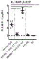
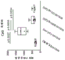
Herein, a group of novel polypeptides, nanoparticles, compositions, methods and uses involving ferritin are presented. Described herein are self-adjuvanted platforms in which an immunostimulatory moiety (such as an adjuvant) is conjugated to ferritin via a surface exposed amino acid or a linker between the ferritin and a non-ferritin polypeptide. An antigenic ferritin polypeptide is produced by combining a non-ferritin polypeptide with ferritin. Conjugation of an immunostimulatory moiety to ferritin in combination with a non-ferritin polypeptide allows for targeted co-delivery of the immunostimulatory moiety and non-ferritin polypeptide in a single macromolecular entity, which can greatly reduce the potential for systemic toxicity that is of concern in the case of more traditional vaccines comprising an antigen and an immunostimulatory molecule (e.g., an adjuvant) as separate molecules. Co-delivery of immunostimulatory moieties with non-ferritin polypeptides in macromolecular entities and their multivalent presentation on ferritin particles may also reduce the total dose of vaccine required, thereby reducing the production burden and cost. Also disclosed herein are antigenic ferritin polypeptides, nanoparticles and compositions for use in immunizing against infection by Respiratory Syncytial Virus (RSV), Epstein Barr Virus (EBV), influenza and lyme disease.
In addition, the polypeptides, nanoparticles, compositions, methods and uses disclosed herein enable co-delivery of non-ferritin polypeptides from pathogens and tailored immune signals that can elicit specific types of immune responses to match the desired immune outcome against a particular pathogen. One example is the induction of a Th 1-type response by TLR7/8 agonists conjugated to ferritin fused to Hemagglutinin (HA), which results in the production of IgG2a switch antibodies that are known to engage Fc γ rs more efficiently to clear virus-infected cells by ADCC mechanisms (see DiLillo et al, Nature Medicine 20: 143-. Furthermore, co-delivery of an immunostimulatory moiety conjugated to ferritin with a non-ferritin polypeptide in a single molecular entity may ensure that stimulation of immune cells occurs in the presence of the non-ferritin polypeptide. In contrast, incorporation of the same immunostimulatory molecule without conjugation results in a systemic distribution, which usually requires higher doses, and also risks undesired effects due to indiscriminate immunostimulation in cells not contacted by the antigen.
Also described herein is a platform wherein a plurality of polypeptides are incorporated into a ferritin particle, for example by providing a heavy and light ferritin chain comprising a first and second non-ferritin polypeptide. This platform can provide a single macromolecular entity that is divalent, and has other advantages associated with ferritin therapeutics, such as, for example, conjugation of an immunostimulatory moiety as described herein.
Disclosure of Invention
It is an object of the present disclosure to provide compositions, kits, methods and uses that may provide one or more of the advantages discussed above, or at least provide the public with a useful choice. Accordingly, the following embodiments are disclosed herein.
a. a mutation replacing a surface exposed amino acid with cysteine and an immunostimulatory moiety attached to the cysteine;
b. a mutation replacing the internal cysteine at position 31 of the helicobacter pylori ferritin with a non-cysteine, or a mutation replacing the internal cysteine at a position similar to position 31 in the non-helicobacter pylori ferritin with a non-cysteine, as determined by pair-wise or structural alignment;
c. A mutation replacing surface exposed asparagine with a non-asparagine amino acid; and
d. a non-ferritin polypeptide.
Embodiment 13a is the ferritin protein according to embodiment 13 wherein said ferritin comprises the E12C mutation in helicobacter pylori ferritin, or a corresponding mutation in a non-helicobacter pylori ferritin as determined by pairwise or structural alignment.
Embodiment 13b is the ferritin protein according to embodiment 13 wherein the ferritin comprises the S26C mutation in helicobacter pylori ferritin, or a corresponding mutation in a non-helicobacter pylori ferritin as determined by pairwise or structural alignment.
Embodiment 13c is the ferritin protein according to embodiment 13 wherein the ferritin comprises the S72C mutation in helicobacter pylori ferritin, or a corresponding mutation in a non-helicobacter pylori ferritin as determined by pairwise or structural alignment.
Embodiment 13d is the ferritin protein according to embodiment 13 wherein the ferritin comprises the a75C mutation in helicobacter pylori ferritin, or a corresponding mutation in a non-helicobacter pylori ferritin as determined by pairwise or structural alignment.
Embodiment 13e is the ferritin protein according to embodiment 13 wherein the ferritin comprises the K79C mutation of helicobacter pylori ferritin, or a corresponding mutation in a non-helicobacter pylori ferritin as determined by pairwise or structural alignment.
Embodiment 13f is the ferritin protein according to embodiment 13 wherein the ferritin comprises the S100C mutation in helicobacter pylori ferritin, or a corresponding mutation in a non-helicobacter pylori ferritin as determined by pairwise or structural alignment.
Embodiment 13g is the ferritin protein according to embodiment 13 wherein the ferritin comprises the S111C mutation in helicobacter pylori ferritin, or a corresponding mutation in a non-helicobacter pylori ferritin as determined by pairwise or structural alignment.
Embodiment 14a is the ferritin protein according to any one of embodiments 4-13g wherein the non-ferritin polypeptide comprises a polypeptide from influenza, optionally wherein the polypeptide comprises a hemagglutinin polypeptide.
Embodiment 14b is the ferritin protein of any one of embodiments 4-13g wherein the non-ferritin polypeptide comprises a polypeptide from epstein-barr virus, optionally wherein the polypeptide comprises a gL, gH, gL/gH, gp220 or gp42 polypeptide.
Embodiment 14c is the ferritin protein according to any one of embodiments 4-13G wherein the non-ferritin polypeptide comprises a polypeptide from respiratory syncytial virus, optionally wherein the polypeptide comprises an RSV F or RSV G polypeptide.
Embodiment 14d is the ferritin protein in accordance with any one of embodiments 4-13g, wherein the non-ferritin polypeptide comprises a polypeptide from borrelia, optionally wherein the polypeptide comprises an OspA polypeptide.
Embodiment 15a is the ferritin protein according to embodiment 15 wherein the RSV G polypeptide is unglycosylated.
Embodiment 15b is the ferritin protein of embodiment 15 or 15a wherein the RSV G polypeptide is chemically conjugated to the ferritin protein.
Embodiment 18a is the ferritin protein in accordance with any preceding embodiment comprising an immunostimulatory moiety that is an agonist of TLR2, optionally wherein the agonist is PAM2CSK4, FSL-1 or PAM3CSK 4.
Embodiment 18b is the ferritin protein in accordance with any preceding embodiment comprising an immunostimulatory moiety that is an agonist of TLR7/8, optionally wherein the agonist is single stranded RNA, imidazoquinoline, nucleoside analogue, 3M-012 or SM 7/8 a.
Embodiment 18C is a ferritin protein in accordance with any preceding embodiment comprising an immunostimulatory moiety that is an agonist of TLR9, optionally wherein the agonist is a CpH Oligodeoxynucleotide (ODN), an ODN comprising one or more 6-mer CpG motifs comprising a 5 'purine (Pu) -pyrimidine (Py) -C-G-Py-Pu 3', an ODN comprising the sequence SEQ ID NO:210, or ISS-1018.
Embodiment 18d is the ferritin protein of embodiment 18c wherein the agonist of TLR9 comprises a backbone comprising phosphorothioate linkages.
Embodiment 18e is the ferritin protein in accordance with any one of the preceding embodiments comprising an immunostimulatory moiety that is an agonist of STING, optionally wherein the agonist is a Cyclic Dinucleotide (CDN), cdA, cdG, cAMP-cGMP, and 2 '-5', 3 '-5' cGAMP or DMXAA.
Embodiment 19a is the ferritin protein in accordance with any preceding embodiment comprising an amino acid sequence 80%, 85%, 90%, 95%, 98% or 99% identical to SEQ ID NO: 201.
Embodiment 19b is a ferritin protein in accordance with any preceding embodiment comprising an amino acid sequence 80%, 85%, 90%, 95%, 98% or 99% identical to SEQ ID NO: 202.
Embodiment 19c is the ferritin protein in accordance with any preceding embodiment comprising an amino acid sequence 80%, 85%, 90%, 95%, 98% or 99% identical to SEQ ID No. 203.
Embodiment 19d is a ferritin protein in accordance with any one of the preceding embodiments comprising an amino acid sequence 80%, 85%, 90%, 95%, 98% or 99% identical to SEQ ID NO 201-207 or 211-215.
Embodiment 19e is the ferritin protein in accordance with any preceding embodiment comprising an amino acid sequence 80%, 85%, 90%, 95%, 98% or 99% identical to SEQ ID No. 204.
Embodiment 19f is a ferritin protein in accordance with any preceding embodiment comprising an amino acid sequence 80%, 85%, 90%, 95%, 98% or 99% identical to SEQ ID No. 205.
Embodiment 19g is a ferritin protein in accordance with any preceding embodiment comprising an amino acid sequence 80%, 85%, 90%, 95%, 98% or 99% identical to SEQ ID No. 206.
Embodiment 19h is the ferritin protein in accordance with any preceding embodiment comprising an amino acid sequence 80%, 85%, 90%, 95%, 98% or 99% identical to SEQ ID NO: 207.
Embodiment 19i is a ferritin protein in accordance with any preceding embodiment comprising an amino acid sequence 80%, 85%, 90%, 95%, 98% or 99% identical to SEQ ID NO 211.
Embodiment 19j is the ferritin protein in accordance with any preceding embodiment comprising an amino acid sequence 80%, 85%, 90%, 95%, 98% or 99% identical to SEQ ID NO: 212.
Embodiment 19k is a ferritin protein in accordance with any preceding embodiment comprising an amino acid sequence 80%, 85%, 90%, 95%, 98% or 99% identical to SEQ ID No. 213.
Embodiment 19l is a ferritin protein in accordance with any preceding embodiment comprising an amino acid sequence 80%, 85%, 90%, 95%, 98% or 99% identical to SEQ ID NO: 214.
Embodiment 19m is a ferritin protein in accordance with any one of the preceding embodiments comprising an amino acid sequence 80%, 85%, 90%, 95%, 98% or 99% identical to SEQ ID NO: 215.
Embodiment 26a is the ferritin protein, ferritin particle or composition of embodiment 24 or the method of embodiment 25 wherein said subject is a mammal, optionally wherein said mammal is a primate or a domestic mammal, further optionally wherein said primate is a non-human primate, monkey, macaque, rhesus monkey or cynomolgus monkey or ape or said domestic mammal is a dog, rabbit, cat, horse, sheep, cow, goat, camel or donkey.
Embodiment F1 is an antigenic influenza-ferritin polypeptide comprising (i) a mutated ferritin protein comprising a surface exposed amino acid substituted with cysteine, and (ii) an influenza polypeptide.
Embodiment F2 is an antigenic influenza-ferritin polypeptide comprising (i) a ferritin protein comprising a mutation replacing a surface exposed amino acid with a cysteine and an immunostimulatory moiety conjugated to the cysteine, and (ii) an influenza polypeptide.
Embodiment F3 is the antigenic influenza-ferritin polypeptide of embodiment F1 further comprising an immunostimulatory moiety conjugated to the ferritin protein via a cysteine.
Embodiment F4 is an antigenic influenza-ferritin polypeptide according to any one of embodiments F1-F3 wherein the influenza polypeptide comprises a Hemagglutinin (HA) or Neuraminidase (NA) polypeptide.
Embodiment F5 is an antigenic influenza-ferritin polypeptide according to embodiment F4 wherein the HA polypeptide comprises a conserved region.
Embodiment F6 is an antigenic influenza-ferritin polypeptide according to embodiment F5 wherein the conserved region comprises all or part of the stem region of HA.
Embodiment F7 is an antigenic influenza-ferritin polypeptide according to any one of embodiments F1-F6 wherein the influenza antigen comprises an HA antigen comprising the Y98F mutation.
Embodiment F8 is an antigenic influenza-ferritin polypeptide according to any one of embodiments F1-F7 further comprising a mutation replacing an internal cysteine with a non-cysteine amino acid.
Embodiment F9 is an antigenic influenza-ferritin polypeptide according to embodiment F8 wherein the internal cysteine is at position 31 of the helicobacter pylori ferritin or a position corresponding to position 31 of the helicobacter pylori ferritin as determined by pairwise or structural alignment, optionally wherein the internal cysteine is mutated to serine.
Embodiment F10 is an antigenic influenza-ferritin polypeptide of any one of embodiments F1-F9 further comprising a mutation replacing surface exposed asparagine with a non-asparagine amino acid, optionally wherein the non-asparagine amino acid is glutamine.
Embodiment F11 is an antigenic influenza-ferritin polypeptide according to any one of embodiments F1-F10 wherein the surface exposed amino acids are mutations of E12, S26, S72, a75, K79, S100 or S111 of helicobacter pylori ferritin or similar amino acids in non-helicobacter pylori ferritin as determined by pairwise or structural alignment.
Embodiment F12 is an antigenic influenza-ferritin polypeptide in accordance with embodiment F11 wherein the mutation at the surface exposed amino acid is an E12C, S26C, S72C, a75C, K79C, S100C or S111C of helicobacter pylori ferritin or a similar amino acid in a non-helicobacter pylori ferritin as determined by pair-wise or structural alignment.
Embodiment F13 is an antigenic influenza-ferritin polypeptide according to any one of embodiments F1-F12 wherein the immunostimulatory moiety is a TLR7 or an agonist of TLR 8.
Embodiment F14 is an antigenic influenza-ferritin polypeptide according to any one of embodiments F1-F13 wherein the immunostimulatory moiety is an agonist of TLR 9.
Embodiment F15 is an antigenic influenza-ferritin polypeptide in accordance with any one of embodiments F1 or F3-F14 further comprising a linker between the immunostimulatory moiety and the ferritin protein.
Embodiment F16 is an antigenic influenza-ferritin polypeptide of embodiment F15 wherein the linker comprises one, two or three of a maleimide moiety, a polyethylene glycol (PEG) moiety and a Dibenzocyclooctyne (DBCO) moiety.
Embodiment F17 is an antigenic influenza-ferritin polypeptide according to any one of embodiments F1-F16 further comprising a peptide linker between the ferritin and the influenza polypeptide.
Embodiment F18 is a ferritin particle comprising an antigenic influenza-ferritin polypeptide according to any one of embodiments F1-F17.
Embodiment F19 is a composition comprising an antigenic influenza-ferritin polypeptide or ferritin particles according to any one of embodiments F1-F18 and a pharmaceutically acceptable carrier.
Embodiment F20 is the composition of embodiment F19, further comprising a second antigenic influenza-ferritin polypeptide comprising a ferritin protein and a different influenza polypeptide.
Embodiment F21 is the composition of embodiment F20, wherein the influenza polypeptide is from influenza a and the influenza polypeptide of the second antigenic influenza-ferritin polypeptide is from influenza b, or wherein the influenza polypeptide of the influenza polypeptide and the influenza polypeptide of the second influenza-ferritin polypeptide are from subtypes H1, H2, H3, H4, H5, H6, H7, H8, H9, H10, H11, H12, H13, H14, H15, H16, H17, or H18, or wherein one or both of the influenza polypeptides comprise an engineered stabilized stem antigen from subtypes H1, H3, H7, or H10.
Embodiment F22 is an antigenic influenza-ferritin polypeptide, ferritin particles or composition according to any one of embodiments F1-F21 for use in a method of eliciting an immune response to influenza or protecting a subject from influenza infection.
Embodiment F23 is a method of eliciting an immune response to influenza or protecting a subject from influenza infection, the method comprising administering to the subject any one or more antigenic influenza-ferritin polypeptides, ferritin particles or compositions according to any one of embodiments F1-F22.
Embodiment F24 is an antigenic influenza-ferritin polypeptide, ferritin particles, composition or method according to any one of embodiments F1-F23 wherein the subject is a human.
Embodiment F25 is a nucleic acid encoding an antigenic influenza-ferritin polypeptide according to any one of embodiments F1-F17, optionally wherein the nucleic acid is mRNA.
Additional objects and advantages will be set forth in part in the description which follows, and in part will be obvious from the description, or may be learned by practice. The objects and advantages will be realized and attained by means of the elements and combinations particularly pointed out in the appended claims.
It is to be understood that both the foregoing general description and the following detailed description are exemplary and explanatory only and are not restrictive of the claims.
The accompanying drawings, which are incorporated in and constitute a part of this specification, illustrate several embodiments and together with the description, serve to explain the principles described herein.
Drawings
Fig. 1A to 1D show exemplary designs of OspA-ferritin nanoparticles. FIG. 1A. OspA is genetically fused to ferritin to form a fusion protein. The OspA and ferritin sequences are separated by a glycine-serine linker (-GS-). Figure 1b depicts the structure of the extracellular domain of OspA. Wherein OspA attachment to the C-terminus of ferritin is indicated by an asterisk. Figure 1c. exemplary ferritin nanoparticles composed of 24 monomers of helicobacter pylori ferritin. Fig. 1d. exemplary OspA-ferritin fusion protein nanoparticles. Ferritin (light grey), position of the glycine-serine linker (GS), and OspA (dark grey and black) (n: subunit number) are depicted.
Fig. 2A to 2D show expression and purification of exemplary OspA-ferritin. Figure 2a Size Exclusion Chromatography (SEC) graph of exemplary OspA-ferritin nanoparticles purified on a Superose 6 column. FIG. 2B SDS-PAGE gels of purified exemplary OspA-ferritin from Expi293 cells. Figure 2c. Dynamic Light Scattering (DLS) plot of exemplary OspA-ferritin nanoparticles. Radius 13nm, Pd% (measure of normalized polydispersity) 7.4, and mass 100%. Figure 2d. composite image of exemplary OspA-ferritin constructed from class averaging of transmission electron micrographs of 318 particles at 67,000x magnification. Ferritin nanoparticles appear on transmission electron microscopy as a strong circular density with a hollow center. Each nanoparticle is surrounded by a number of short shapes corresponding to OspA, which appear as circles or micro-ellipses.
Fig. 3A-3B show the production of alternative serotype OspA nanoparticles in e. FIG. 3A SDS-PAGE biochemical analysis of OspA-ferritin serotypes 1-5 and 7 purified by size exclusion chromatography. FIG. 3B Transmission Electron microscopy (98,000X) of OspA-ferritin serotypes 1-5 and 7.
FIG. 4 shows exemplary serotype 1 OspA-ferritin nanoparticles  Comparison of immunogenicity and duration of Lyme (Lyme), a liquid suspension of purified outer surface protein a (ospa) of Borrelia (Borrelia burgdorferi). At
 weeks 0 and 4, 1 μ g OspA-ferritin + Ribi adjuvant (Sigma adjuvant system catalog number S6322-1vl) or Lyme (Lyme) immunized C3H mice intramuscularly (n ═ 5). In the case of each composition, antibody responses were assessed by measuring endpoint titers via
 ELISA 2 weeks after 2 nd immunization (week 6) and 21 weeks after 2 nd immunization (week 25).
Fig. 5A-5C present information on exemplary OspA-ferritin in which the OspA polypeptide is modified at an epitope of OspA serotype 1 that is homologous to a sequence fragment of human leukocyte function-associated antigen-1 (hLFA-1). FIG. 5A shows the structure of the LFA-1 homology site (amino acids 165-173 of SEQ ID NO: 83) at the position within the OspA extracellular domain. FIG. 5B, dendrogram showing the relationship of OspA amino acid 165-173 of SEQ ID NO 83 (Borrelia borrelia serotype 1OspA) to the corresponding sequences in hLFA-1 and other Borrelia species and serotypes. FIG. 5C compares the nine amino acid segment (nonapeptide) (labeled "OspA") at amino acid 165-173 of SEQ ID NO:83 with the corresponding nonapeptides from serotype 2 and serotype 3OspA ("S2" (SEQ ID NO:79) and "S3" (SEQ ID NO:80), respectively), a rationally designed substituted nonapeptide ("RD 2"; SEQ ID NO:81) and the corresponding nonapeptide from hLFA-1 (SEQ ID NO: 78). FIG. 5C discloses SEQ ID NO 77, 79-81 and 78, respectively, in order of appearance.
FIG. 5 D.OspA serotype 1-ferritin nanoparticles with AddaVax at week 0 and week 4 with a 1 μ g doseTMC3H mice (n-5) were immunized Intramuscularly (IM) with adjuvant (squalene-based oil-in-water nanoemulsion; available from InvivoGen under catalog number vac-adx-10). The OspA sequence includes the wild-type hLFA-1 homologous site (i.e., amino acid 165-173; serotype 1 of SEQ ID NO: 83) or a substituted sequence as follows: 81 ("RD"); 80 ("serotype 3 replacement") of SEQ ID NO; SEQ ID NO:79 ("serotype 2 replacement"). Antibody responses were assessed via endpoint titers measured by ELISA 2 weeks after the 2 nd immunization of the indicated constructs.
Fig. 6A-6B present information on exemplary OspA-ferritin nanoparticles conjugated with an exemplary immunostimulatory partial TLR 7/8 agonist (3M-012). Figure 6a. 3M-012 was attached to ferritin using a 2-step click chemistry strategy. The DBCO-PEG 4-maleimide linker was first attached to the surface exposed cysteine on the ferritin. After removal of excess linker, azide-3M-012 was added. Figure 6b. C3H mice were immunized intramuscularly with 1 μ g of the indicated composition at weeks 0 and 4 (n ═ 5) and analyzed 2 weeks later. "conjugate" indicates OspA-ferritin-3M-012 conjugated nanoparticles. "blend (Admix)" indicates a non-conjugated mixture of the same OspA-ferritin given with 29ng or 20 μ g 3M-012 or alum. A29 ng "blended" mixture of OspA-ferritin and 3M-012 indicates the molar equivalent of 3M-012 on the conjugated nanoparticle.
FIGS. 7A-7C present information on exemplary OspA-ferritin nanoparticles conjugated to an exemplary immunostimulatory moiety ISS-1018CpG (SEQ ID NO: 210). Figure 7a. CPG is attached to ferritin using a 2-step click chemistry strategy. The DBCO-PEG 4-maleimide linker was first attached to the surface exposed cysteine on the ferritin. After removal of excess linker, azide-CpG was added. FIG. 7A discloses SEQ ID NO 534. Fig. 7b biochemical analysis of CpG-conjugation by SDS-PAGE gel revealed a shift in molecular weight after conjugation to CpG, where 92% of OspA-ferritin was conjugated to CpG. Figure 7℃ C3H mice were immunized intramuscularly with 1 μ g of the indicated composition at weeks 0 and 4 (n ═ 5) and analyzed 2 weeks later. "conjugate" indicates OspA-ferritin-CPG conjugated nanoparticles. "blend" indicates a non-conjugated mixture of the same OspA-ferritin given with 339ng or 50 μ g CpG or alum. 339ng of the "blended" mixture of OspA-ferritin and CPG represents the molar equivalents of CPG on the conjugated nanoparticles.
Figures 8A to 8F compare antibody responses to serotype 1 (figure 8A), serotype 2 (figure 8B), serotype 3 (figure 8C), serotype 4 (figure 8D), serotype 5 (figure 8E) and serotype 7 (figure 8F) following administration of: a monovalent serotype-matched OspA-ferritin (1 μ g/dose) with alum adjuvant ("monovalent") or a hexavalent composition ("hexavalent") containing each of serotype 1 OspA-ferritin, serotype 2 OspA-ferritin, serotype 3 OspA-ferritin, serotype 4 OspA-ferritin, serotype 5 OspA-ferritin, and serotype 7 OspA-ferritin (each at 1 μ g/dose) with alum adjuvant. C3H mice (n ═ 5) were immunized intramuscularly at weeks 0 and 4, and antibody responses were assessed after 2 weeks via endpoint titers measured by ELISA. ELISA plates were coated with the indicated OspA serotypes.
Figures 9A-9G show antibody responses to serotype 1 (figure 9A), serotype 2 (figure 9B), serotype 3 (figure 9C), serotype 4 (figure 9D), serotype 5 (figure 9E), serotype 6 (figure 9F), and serotype 7 (figure 9G) observed in mice following administration of conjugated and unconjugated hexavalent OspA-ferritin nanoparticle compositions. The hexavalent composition includes each of serotype 1 OspA-ferritin, serotype 2 OspA-ferritin, serotype 3 OspA-ferritin, serotype 4 OspA-ferritin, serotype 5 OspA-ferritin, and serotype 7 OspA-ferritin, as described for fig. 8A through F, except that "hexavalent-CPG" and "hexavalent-3M-012" indicate chemical conjugation of the nanoparticle to CPG and 3M-012 (see fig. 7A and 6A and the accompanying description). Antibody responses were assessed after 2 weeks via endpoint titers measured by ELISA. ELISA plates were coated with the indicated OspA serotypes.
Figures 10A to 10G show the antibody responses to serotypes 1-7, respectively, in rhesus monkeys (n-3/group) for hexavalent OspA-ferritin nanoparticle compositions as described in figures 9A-G, except at a total dose of 60 μ G (10 μ G per serotype) and containing unconjugated AF03 adjuvant. Monkeys were immunized intramuscularly at weeks 0 and 6. Antibody responses were analyzed 2 weeks after immunization via endpoint titers measured by ELISA.  Lyme was used as a comparative reference at a dose of 10 μ g. For all experiments, ELISA plates were coated with OspA serotypes indicated in each picture.
Fig. 10H to 10N show the antibody responses to serotypes 1-7, respectively, in rhesus monkeys (N3/group) for hexavalent OspA-ferritin nanoparticle compositions as described for fig. 10A-G, except that the nanoparticles were conjugated to 3M-012 or CpG without the use of AF03 adjuvant (see fig. 6A and 7A and accompanying description). A total dose of 60 μ g (10 μ g of each serotype) was used. Monkeys were immunized intramuscularly at weeks 0 and 6. Antibody responses were analyzed 2 weeks after immunization via endpoint titers measured by ELISA. For all experiments, ELISA plates were coated with OspA serotypes indicated in each picture.
Figure 11 shows the results of tick challenge tests of 3M-012 conjugated OspA-ferritin compositions. Mice were immunized with a 1 μ g dose of the indicated composition at weeks 0 and 4. The monovalent composition contained 1 μ g OspA-ferritin serotype 1 conjugated to 3M-012. The "hexavalent-3M-012" composition is as described for FIGS. 9A-G. Control particles lack OspA polypeptide. Two weeks after the second immunization, mice were challenged with 5-6 ticks infected with borrelia burgdorferi N40 strain (serotype 1) for 5 days, and the mice were sacrificed two weeks later. Tissue samples from heart, ankle and ear were cultured in BSK medium with antibiotics against borrelia burgdorferi for 6 weeks. Negative samples were tested for the presence of borrelia burgdorferi by PCR. Positive samples were positive by culture or PCR.
Figure 12 shows confirmation of OspA-ferritin nanoparticles conjugation to TLR 7/8 agonist 3M-012 by mass spectrometry. The top panel shows the unconjugated construct and the bottom panel shows the conjugated construct. The data shows a mass shift of 586.69 daltons, consistent with the addition of 3M-012.
Figure 13 shows the antibody response to the non-glycosylated mutant OspA-ferritin (NG-RD) compared to the glycosylated counterpart (RD) in mice measured by ELISA on the dilution lines as shown. RD 52. NG-RD SEQ ID NO: 53. Mice were vaccinated at 1 μ g dose at week 0 and week 4.
FIG. 14 shows neutralization in mice measured by ELISA on dilution series as shown Lyme control and negative (Pre-immune) controls were compared to OspA-ferritin (SEQ ID NO:52), OspA-ferritin glycosylation mutant N>Q (SEQ ID NO:53) and glycosylation mutant S/T>Antibody response to A (SEQ ID NO: 63).
FIGS. 15A-15E show the purification and characterization of OspA constructs comprising different linkers (GS, Gly-Ser linker; GS1, Gly-Gly-Gly-Ser linker (SEQ ID NO: 443); GS2, SEQ ID NO:91 linker; GS5, SEQ ID NO:92 linker; construct sequences SEQ ID NO:53 and 60-62, respectively). Figure 15a coomassie staining of purified OspA constructs containing linkers as indicated. FIG. 15A discloses SEQ ID NO 443-445, respectively, in order of appearance. FIG. 15B Dynamic Light Scattering (DLS) of OspA-ferritin nanoparticles comprising GS1 (SEQ ID NO: 60). FIG. 15C DLS of OspA-ferritin nanoparticles comprising GS2 (SEQ ID NO: 61). FIG. 15D Electron Micrograph (EM) of OspA-ferritin nanoparticles (SEQ ID NO:62) comprising GS 5. FIG. 15E DLS of OspA-ferritin nanoparticles comprising GS5 (SEQ ID NO: 62).
FIG. 16 shows neutralization in mice measured by ELISA on dilution series as shown Lyme compares antibody responses to an OspA-ferritin construct comprising different linkers (linker 1X GGGS construct, SEQ ID NO:60 ("1X GGGS" is disclosed as SEQ ID NO: 443); linker 2X GGGS construct, SEQ ID NO:61 ("2X GGGS" is disclosed as SEQ ID NO: 444); linker 5X GGGS construct, SEQ ID NO:62 ("5X GGGS" is disclosed as SEQ ID NO:445)) compared to a negative (pre-immune) control.
FIGS. 17A to 17C show the characterization of the OspA serotype 4 construct (SEQ ID NO:18) for 2, 4-dioxotetrahydropteridine synthase (lumazine synthsase). Dls data fig. 17a. Figure 17b coomassie gel of indicated fractions 22-64 of Size Exclusion Chromatography (SEC) traces. Em data fig. 17c.
FIG. 18 shows antibody responses in mice to the OspA serotype 4-ferritin construct (SEQ ID NO:4) and the OspA serotype 4-2, 4-dioxotetrahydropteridine synthase construct (SEQ ID NO:18) with or without alum.
FIGS. 19A-19C show the characterization of the OspA serotype 1-2, 4-dioxotetrahydropteridine synthase construct (SEQ ID NO: 12). Fig. 19a.em data. Coomassie gels of indicated fractions 20-40 of the sec traces of fig. 19b. Dls data fig. 19c.
FIGS. 20A-20C show the characterization of the OspA serotype 2-2, 4-dioxotetrahydropteridine synthase construct (SEQ ID NO: 16). Fig. 20a.em data. Coomassie gel of indicated fractions 27-56 of the sec trace of fig. 20b. Dls data fig. 20c.
FIGS. 21A-21B show the characterization of the OspA serotype 3-2, 4-dioxotetrahydropteridine synthase construct (SEQ ID NO: 17). Figure 21a. coomassie gel of indicated fractions 23-39 of sec trace. Dls data fig. 21b.
FIGS. 22A-22C show characterization of the OspA serotype 5-2, 4-dioxotetrahydropteridine synthase construct (SEQ ID NO: 19). Fig. 22a.em data. Coomassie gel of indicated fractions 22-38 of the sec trace of fig. 22b. Dls data fig. 22c.
FIGS. 23A-23C show the characterization of the OspA serotype 7-2, 4-dioxotetrahydropteridine synthase construct (SEQ ID NO: 21). Fig. 23a.em data. Coomassie gel of indicated fractions 20-38 of the sec trace of fig. 23b. Dls data in fig. 23c.
Fig. 24A to 24G show heptavalent OspA-ferritin nanoparticle compositions (total 7ug) for each OspA-ferritin nanoparticle containing 1ug of each OspA-ferritin nanoparticle corresponding to OspA serotypes 1-7 adjuvanted with alum or AF03 (total 7ug) Antibody responses to serotypes 1-7 in lyme, C3H mice (n-5/group), respectively. For all experiments, ELISA plates were coated with OspA serotype indicated as "S X" in each picture, where X is the serotype number.
Figure 25 shows the time course of the endpoint antibody titers in rhesus monkeys. Hexavalent OspA-ferritin vaccines ( OspA containing serotypes 1, 2, 3, 4, 5, and 7 in individual nanoparticles) with AF03 adjuvant at weeks 0 and 6 or Monkeys were immunized intramuscularly. ELISA plates were coated with
 OspA serotype 1.
26A-26B sequence comparison and homology of representative H1N1 influenza virus strains. (FIG. 26A) sequence alignment of candidate antigenic HA polypeptides using the vector NTI AlignX software. White background and black characters: consensus residues derived from fully conserved residues at a given position. Black background and white characters: consensus residues derived from single residues with an incidence of greater than 50% at a given position. White-backed black underlined and bold: residues that are weakly similar to the consensus residue at a given position. Black white with double underline and italics: consensus residues derived from a similar block of residues at a given position. And (3) thickening white background black characters: non-similar residues. The sequence shown is the HA portion of the following sequence plus the linker serine at the C-terminus: CA09 HA-Np ═ residues 1-519 of SEQ ID NO: 315. Residues 1-519 of SEQ ID NO:327 of COBRA P1 HA-Np. Residues 1-518 of COBRA X6 HA-Np SEQ ID No. 329. NC99 HA-Np is residues 1-518 of SEQ ID NO: 301. HK77 HA-Np ═ residues 1-519 of SEQ ID NO: 318. FM47 HA-Np ═ residues 1-519 of SEQ ID NO: 317. DV57 HA-Np is residues 1-518 of SEQ ID NO: 321. MAL54 HA-Np ═ residues 1-519 of SEQ ID NO: 316. (FIG. 26B) phylogenetic tree plots generated using the adjacency (Vector NTI) against HA protein sequences from the listed influenza strains. Arrows indicate strains selected as candidates for evaluation by generating HA-ferritin nanoparticles from these sequences and testing their immunogenicity in mice.
FIG. 27 engineered surface exposed cysteines (Cys) on ferritin. The position of the cysteine resulting from the mutation replacing the surface exposed amino acid is shown in the context of ferritin nanoparticles.
Figure 28A-figure 28b conjugation of toll-like receptor (TLR) agonists to ferritin. (fig. 28A) the results of conjugating SM7/8A small molecules via a PEG4 linker containing a maleimide reactive group to cysteines resulting from mutations replacing surface exposed amino acids in ferritin are illustrated in the context of ferritin nanoparticles. (FIG. 28B) the results of conjugation of CpG (SEQ ID NO:535) to cysteine resulting from mutation replacing a surface exposed amino acid in ferritin using 2-step click chemistry with a maleimide-DBCO bifunctional linker and an azide functionalized CpG reagent in the context of ferritin nanoparticles are illustrated.
Figure 29A-29 b conjugation of toll-like receptor (TLR) agonists to ferritin. (fig. 29A) model of ferritin nanoparticles comprising an HA polypeptide and 3M-012, said 3M-012 conjugated to a surface-exposed cysteine of ferritin using a 2-step click chemistry reaction via a maleimide-DBCO bifunctional linker and an azide-functionalized 3M-012 reagent. (fig. 29B) model of ferritin nanoparticles comprising HA polypeptides and CpG conjugated with a maleimide-DBCO bifunctional linker and an azide functionalized CpG reagent to surface exposed cysteine of ferritin using a 2-step click chemistry reaction. FIG. 29B discloses SEQ ID NO 534.
Figure 30. trypsin cleavage sites within the ferritin portion of certain ferritin nanoparticle constructs. Some ferritin nanoparticles contain a trypsin cleavage site in addition to the unpaired surface exposed cysteine used to conjugate the adjuvant. The amino acid sequence presented in figure 30 is a "universal" sequence showing a sequence common to multiple constructs. For example, residues 519-694 of SEQ ID NO 314 comprise the "universal" sequence shown in FIG. 30. The "XXX" sequence represents an influenza polypeptide. The position of the ferritin S111C mutation present in this sequence is also indicated.
Fig. 31A and 31b gel shift and Mass Spectrometry (MS) results with or without conjugation. (FIG. 31A) gel shift and mass spectrometry results for H1/stem-Np (SEQ ID NO:343) in the presence (+) or absence (-) conjugation to maleimide-PEG 4-SM7/8 a. (FIG. 31B) gel shift and mass spectrometry results for H5/hCobra2-Np (SEQ ID NO:332) in the presence (+) or absence (-) conjugation to maleimide-PEG 4-SM7/8 a.
Fig. 32A and 32b. further gel shift results with or without conjugation. (FIG. 32A) gel shift results of H1/stem-Np after peptide N-glycosidase (PNGase) treatment with or without conjugation to maleimide-PEG 4-SM7/8a or maleimide-PEG 4-DBCO and azide-CpG by 2-step click chemistry. (FIG. 32B) gel shift results for H1/stem-Np after trypsin treatment with or without conjugation to maleimide-PEG 4-SM7/8a or maleimide-PEG 4-DBCO and azide-CpG by 2-step click chemistry.
Fig. 33A and 33b mass spectra with and without conjugation of maleimide-PEG 4-SM7/8a to H1/stem-Np. (fig. 33A) MS data after PNGase treatment showed the mass of H1/stem-Np before and after conjugation with maleimide-PEG 4-SM7/8 a. (FIG. 33B) MS data after trypsinization of H1/stem-Np show the masses of cleaved ferritin before and after conjugation with maleimide-PEG 4-SM7/8 a.
FIG. 34 Mass Spectrometry with and without conjugation of the maleimide-PEG 4-DBCO linker to H1/stem-Np. The maleimide-PEG 4-DBCO linker addition to H1/stem-Np was confirmed by the mass change measured by MS after linker addition.
FIG. 35 SDS-PAGE of H1/stem-Np before and at various stages of conjugation of CpG by 2-step click chemistry. "after azide-CpG" refers to the final product of a 2-step click reaction.
FIG. 36 characterization of conjugation of linker comprising 3M-012 to NC99 HA-TEV-Np construct. The constructs in lanes WT and + TEV have the sequence SEQ ID NO 313; S26C refers to SEQ ID NO: 310; S72C refers to SEQ ID NO: 311; A75C refers to SEQ ID No. 312; and S111C refers to SEQ ID NO 309. All samples, except for WT, were treated with tobacco etch virus protease (TEV).
Fig. 37A to 37e. mass spectra of various constructs with or without reduction or conjugation. (FIG. 37A) H1/stem-Np comprising S111C prior to reduction. (FIG. 37B) H1/stem-Np containing S111C after reduction. The mass reduction observed after reduction (115Da) is consistent with the removal of post-translational modifications that inhibit cysteine reactivity. (FIG. 37C) Mass Spectrometry of NC99 HA-TEV-Np-S26C nanoparticles (SEQ ID NO:310) with or without conjugation to 3M 012. (FIG. 37D) Mass Spectrometry of NC99 HA-TEV-Np-A75C nanoparticles (SEQ ID NO:312) with or without conjugation to 3M 012. (FIG. 37E) Mass Spectrometry of NC99 HA-TEV-Np-S111C nanoparticles (SEQ ID NO:309) with and without conjugation to 3M 012.
Figures 38A to 38f negative staining Electron Microscope (EM) images of nanoparticles of H1/stem-Np or NC99 HA-Np with or without conjugation to SM7/8A, 3M012 or CpG. (FIG. 38A) unconjugated H1/stem-Np. (FIG. 38B) H1/stem-Np-SM 7/8a conjugate. (FIG. 38C) H1/stem-Np-CpG conjugate. (FIG. 38D) NC99 HA-Np (SEQ ID NO:309) ]. (FIG. 38E) NC99 HA-Np-3M012 conjugate. (FIG. 38F). NC99 HA-Np-CpG conjugate.
Dynamic Light Scattering (DLS) analysis of H1/stem-Np-SM 7/8a conjugates (fig. 39A), H1/stem-Np-CpG conjugates (fig. 39B) or unconjugated H1/stem-Np (fig. 39C).
Figure 40A-B antibody response to H1/stem-Np formulated with blended adjuvants as indicated or conjugated to TLR agonists (e.g., SM7/8A or CpG) via a PEG4 linker as shown in figure 28A-B. Sera were collected from mice (n ═ 5) at 5 weeks after immunization (weeks 0 and 3), and antibody titers were measured by enzyme-linked immunosorbent assay (ELISA). This data demonstrates the self-adjuvanting properties of the H1/stem-Np-TLR-agonist conjugate. FIG. 40A shows ELISA for H1/New Cardonia/20/1999 HA trimer. PAA ═ polyacrylic acid; mixing equimolar to 83.3ng SM7/8a (equivalent to the dose administered with SM7/8 conjugated ferritin nanoparticles); high dose-21.84 μ g (higher than the dose given with SM7/8 conjugated ferritin nanoparticles). FIG. 40B shows ELISA for H1/stem trimer. Mixing equimolar 850ng CpG (equivalent to the dose administered with CpG-conjugated ferritin nanoparticles); high dose ═ 20 μ g CpG.
FIGS. 41A to 41C comparison of titers obtained with NC99HA-Np (SEQ ID NO:309) with or without conjugation or with isolated 3M-012. Antibody responses to NC99HA-Np-3M 012-conjugated nanoparticles (0.22 μ g/dose) were tested in mice. Blend controls included a mixture of HA-Np (0.22. mu.g/dose) and 10. mu.g 3M012 (typical literature dose) as well as a mixture of HA-Np (0.22. mu.g/dose) and 1.7ng 3M012 (equimolar match of conjugate). Additional controls included unconjugated HA-Np and IIV dosed at matched HA levels (0.17 μ g HA/dose). Based on ELISA endpoint titers (fig. 41A), pseudovirus (PsV) neutralization IC50 titers (fig. 41B) and hemagglutination inhibition (HAI) titers (fig. 41C), the HA-Np-3M012 conjugate induced significantly stronger antibody responses than the equimolar blend control. The conjugated nanoparticles also induced a stronger response (ELISA) and a stronger neutralizing antibody response (PsV) compared to the unconjugated nanoparticles and the IIV standard of care, but the results were not statistically significant in the HAI analysis. The assay was for sera 2 weeks after the boost. All samples were run in triplicate. Median +/-SEM is plotted. P < 0.0001; p < 0.001; p < 0.01; p < 0.05.
FIGS. 42A to 42C comparison of titers obtained with NC99HA-Np (SEQ ID NO:309) with or without conjugated or isolated CpG. Antibody responses to HA-Np-CpG conjugated nanoparticles (0.22 μ g/dose) were tested in mice. Blend controls included a mixture of HA-Np (0.22. mu.g/dose) and 20. mu.g CpG (typical therapeutic dose) and a mixture of HA-Np (0.22. mu.g/dose) and 21ng CpG (equimolar match for the conjugate). Additional controls included unconjugated HA-Np and IIV dosed at matched HA levels (0.17 μ g HA/dose). Based on ELISA endpoint titers (fig. 42A) and pseudovirus neutralization IC50 titers (fig. 42B), the conjugates induced stronger binding and neutralizing antibody responses compared to the matched blend, unconjugated particles or IIV. The HA-Np-CpG conjugate also induced significantly stronger HAI titers than the equimolar blend control (fig. 42C). In addition, HAI is 2.6 and 3.0 fold more potent than unconjugated HA-Ferr and IIV, but these results are not significant. ELISA and PsV assays were for sera 2 weeks after the boost and HAI analysis was performed on sera 5 weeks after the boost. All samples were run in triplicate and plotted against median +/-SEM. P < 0.0001; p < 0.001; p < 0.01; p < 0.05.
Fig. 43A-43 c characterization of self-assembled HA-ferritin nanoparticles derived from six evolutionarily distinct H1 Hemagglutinin (HA) antigens and two computationally generated (COBRA) antigens. (fig. 43A) nanoparticle size and polydispersity ("dispersity") were measured by Dynamic Light Scattering (DLS). (FIG. 43B) the purity of HA nanoparticles was assessed by SDS-PAGE Coomassie staining. (fig. 43C) nanoparticle integrity was visualized by negative staining electron microscopy at 80,000x magnification. FM47 ═ A/Monema/1-JY 2/1947; MAL54 ═ a/malaysia/302/1954; DV57 ═ a/denver/1957 (DV 57); HK77 ═ a/hong kong/117/1977; NC99 ═ a/new karidonia/20/99; CA09 ═ a/california/4/2009. COBRA P1 and COBRA X6 are consensus sequences generated by calculation from multiple sequences, recently described by Carter DM et al, J Virol 90: 4720-. See the legend of FIGS. 26A-B for SEQ ID NO.
FIGS. 44A-44 H. the peptide sequence of various HA-ferritin nanoparticles (see FIGS. 26A-B for SEQ ID NO)Example) the efficacy and breadth of the immune response elicited. Hemagglutination inhibition (HAI) titers (log) against a panel of distinct H1N1 influenza viruses against sera from mice 6 weeks after immunization with the indicated HA-Np vaccines 2) And (4) carrying out measurement. Mice (n-5) were immunized with hemagglutinin nanoparticles (HA-Np) at weeks 0 and 3. The dashed line indicates the limit of detection of the assay. (FIG. 44A to FIG. 44E) data with Ribi adjuvant. (FIG. 44F-44H) data with AF03 adjuvant. The x-axis indicates the group of H1N1 influenza strains tested in the reference year from 1934 to 2013. The complete line names corresponding to each year are given in table 2. Asterisks indicate matched strains.
Figure 45A to figure 45f HA antibody responses induced by HA-ferritin nanoparticles. (fig. 45A) mice were immunized [ n ═ 5] with the indicated nanoparticles or nanoparticle combinations using the Sigma Adjuvant System (catalog number S6322). IIV refers to inactivated influenza vaccine. (FIGS. 45B-F) antibody responses were measured by determining ELISA titers against HA trimers from: A/Monema castle/1/1947 (FIG. 45B); A/Malaysia/302/1954 (FIG. 45C); a/hong Kong/117/1977 (FIG. 45D); A/New Cardonia/20/99 (FIG. 45E); or A/California/4/2009 (FIG. 45F). ELISA titers were measured 5 weeks after the first immunization. The lowest serum dilution tested set the assay detection limit, as indicated by the dashed line. Open circles indicate matches between the lines of origin of the nanoparticles administered to the mice and the lines determined by ELISA.
Fig. 46A-46 h antibody responses to HA mixtures of various ferritin nanoparticles administered to mice as measured by determination of HAI titers. (FIG. 46A to FIG. 46B) a bivalent combination. (FIGS. 46C to 46E) trivalent combinations. (FIGS. 46F to 46H) tetravalent combinations. HAI titers (log) for a diverse group of H1N1 influenza viruses were determined2). Mice (n-5) were immunized with the indicated HA-nanoparticle combinations at weeks 0 and 3 with adjuvant. The bivalent combination COBRA X6+ COBRA P1 HA-Np was tested in the presence of AF03 adjuvant to be consistent with the evaluation of monovalent COBRA HA-Np. Also, the combination of individual strain HA-ferritin nanoparticles used Ribi adjuvant to be consistent with the evaluation of monovalent individual strain HA-Np. Star signShows the match between the line of origin of the nanoparticles administered to the mice and the line determined by ELISA. The x-axis indicates the group of H1N1 influenza strains tested in the reference year from 1934 to 2013. Table 2 gives the complete line names for each year. The dashed line indicates the limit of detection of the assay.
Figures 47A to 47f antibody responses to compositions with Ribi or AF03 adjuvant as measured by determination of HAI titers. For the bivalent combination of NC99 and CA09 HA-Np (fig. 47A-47B), the trivalent combination of NC99, CA09 and HK77 HA-Np (fig. 47C-47D) and the trivalent combination of NC99, CA09 and FM47 HA-Np (fig. 47E-47F), the results obtained with Ribi adjuvant (fig. 47A, 47C and 47E) were similar compared to the results obtained with AF03 adjuvant (fig. 47B, 47D and 47F). HAI titers were determined in mouse sera 6 weeks after the priming dose, with booster immunizations performed at week 3. Mice were immunized with HA-Np combinations as indicated [ n-5 ] with Ribi or AF03 adjuvant. Asterisks indicate the match between the originating strain of nanoparticles administered to mice and the strain determined by ELISA. The x-axis indicates the group of H1N1 influenza strains tested in the reference year from 1934 to 2013. Table 2 gives the complete line names for each year. The dashed line indicates the limit of detection of the assay.
Figures 48A to 48c comparative antibody responses elicited by NC99 and CA09 Inactivated Influenza Vaccine (IIV) produced in eggs, as measured by determination of HAI titers. (FIG. 48A) NC99 IIV. (FIG. 48B) CA09 IIV. (FIG. 48C) NC99+ CA09 IIV. Hemagglutination inhibition (HAI) titers (log) of sera from mice against a diverse group of H1N1 influenza viruses 6 weeks after immunization with indicated IIV vaccines (either as single components or co-administered)2). Mice (n-5) were immunized with 170ng (HA content) of each vaccine at weeks 0 and 3 with Ribi adjuvant. The x-axis indicates the group of H1N1 influenza strains tested in the reference year from 1934 to 2013 (see also table 2). The x-axis indicates the group of H1N1 influenza strains tested in the reference year from 1934 to 2013. Table 2 gives the complete line names for each year. The dashed line indicates the limit of detection of the assay.
FIGS. 49A-49 D. as determined by determination of HAI titerAmount, antibody response against various influenza viruses after immunization of ferrets with HA-ferritin nanoparticle compositions or IIV. HAI titers (log) of sera from ferrets (n-12/group) after two immunizations with AF03 adjuvant incorporated in the following items 2): FIG. 49A: PBS alone; FIG. 49B: A/California/2009 IIV; FIG. 49C: NC99+ CA09+ HK77 HA-Np; FIG. 49D: COBRA-X6+ P1+ HK77 HA-Np.
Figure 49e heterologous challenge to ferrets immunized with HA-ferritin nanoparticle composition, IIV or PBS. Ferrets were immunized as described for fig. 49A-D, and challenged with 1947 montomosburg virus 4 weeks after the last immunization: infection dose of 10 in 50% tissue culture (TCID50) with 1mL of A/Monascus/1/1947 virus4.65Nasal vaccination was performed. Viral titers were quantified in the nasal wash over a seven day time course following challenge. The dashed line indicates the limit of detection of the assay. According to one-tailed unpaired t-test and one-way anova [ F (3,44) ═ 5.18, p ═ 0.00375]In ferrets combined with HA-Np but not immunized with CA09 IIV, the viral titer significantly decreased at day 5 post challenge compared to vehicle (PBS) control. P ≦ 0.001 according to student's t test.
Fig. 50A-c. antibody response of cynomolgus monkeys (Macaca fascicularis) immunized with: 50 μ g of H1/stem-Np, or 200 μ g H1/stem-Np (no adjuvant control), or 200 μ g H1/stem-Np-SM 7/8A conjugate (shown in FIG. 28A) or 200 μ g H1/stem-Np-CpG conjugate (shown in FIG. 28B) formulated with blended AF03 adjuvant. Figure 50A shows HA antibody titers measured by enzyme-linked immunosorbent assay (ELISA) on plates coated with H1/neocurdolizia/20/1999 HA trimer at the indicated time points, where immunization was performed at weeks 0, 4 and 10. FIG. 50B shows the neutralizing IC50 of lentiviruses pseudotyped with H1/New Cardonia/20/1999 HA and NA. Figure 50C shows the neutralizing IC50 of lentiviruses pseudotyped with H5/vietnam/1203/2004 HA and NA. This data demonstrates the self-adjuvanting properties of the H1/stem-Np-TLR-agonist conjugate in a primate model.
FIGS. 51A-51B comparison of titers obtained with NC99HA-Np (SEQ ID NO:309) with or without conjugation or with isolated 3M-012. Antibody responses to the following four doses of nanoparticles conjugated with NC99HA-Np-3M 012: 0.1. mu.g, 0.5. mu.g, 2.5. mu.g and 12.5. mu.g. Blend controls and unconjugated nanoparticles were included for comparison. The amount of TLR agonist used for the "equimolar" and "high dose" blend controls was calculated to maintain the antigen to adjuvant ratio used in the initial conjugation study (0.22 μ g HA-NP with 1.7ng 3M012 for "equimolar" or 10 μ g for "high dose"). Serum neutralization was measured in the NC99 lentivirus reporter assay (left, fig. 51A) and the NC99HAI assay (right, fig. 51B). The arrow highlights the 2.5 μ g dose where the HA-NP-3M012 conjugate induces similar antibody titers to HA-NP +3M012 despite containing over 5000-fold lower 3M012 adjuvant. Mean ± SEM were plotted.
FIGS. 52A-52B show purified single-chain gL and gH monomers (FIG. 52A) (SEQ ID NO:406) and trimers (FIG. 52B) (SEQ ID NO:411) with or without removal of the His tag by Coomassie and Western blot analysis. FIG. 52B also shows a sample from a size exclusion column (  6) UV absorption trace of purified fraction.
FIGS. 53A-53E show the purification and characterization of single-chain gL/gH-ferritin nanoparticles (SEQ ID NO: 414). Show that 6 UV absorption traces of purified fractions (FIG. 53A), and Coomassie (FIG. 53B) and Western blot (FIG. 53C) analysis of fractions selected from purification (L indicates molecular weight ladder; 150 and 250kDa bands are marked in the 150kDa and 250kDa band positions as indicated at the right panel 53B). Dynamic light scattering (fig. 53D) and electron microscopy (fig. 53E) analysis of single-chain nanoparticles are also presented.
FIG. 54 shows different representative single-chain gL/gH-ferritin constructs.
Figure 55 shows antibody titers after immunization of mice with single chain gL/gH trimers or Nanoparticles (NPs) admixed with AF03 adjuvant, which is a squalene emulsion based adjuvant. When NP constructs were compared to their corresponding trimeric constructs, the p-value was < 0.05. From left to right, the constructs are SEQ ID NOs 416, 410, 411, 413, 412 and 414.
Figures 56A-56B show anti-gL/gH antibody responses in mice to a bivalent composition comprising both gp220 nanoparticles (SEQ ID NO:401) and single-chain gL/gH nanoparticles ("gL _ gH _ C5 NP", SEQ ID NO:419), as compared to single-chain gL/gH nanoparticles and negative control naked ferritin (i.e., ferritin not conjugated to any non-ferritin polypeptide or immunostimulatory moiety). The results show that the use of a bivalent composition does not result in interference with the anti-gL/gH antibody response relative to the results in the case of single-chain gL/gH with the negative control naked ferritin. Both compositions contained AF03 adjuvant. ELISA results at various dilutions (fig. 56A) and binding titers (fig. 56B) are shown.
Figures 57A-57B show anti-gp 220 antibody responses to a bivalent composition comprising gp220 nanoparticles and single chain gL/gH nanoparticles as described for figures 57A-B. The results show that the use of a bivalent composition does not result in interference of the anti-gp 220 antibody response relative to the results in the case of gp220 nanoparticles with the negative control naked ferritin. Both compositions contained AF03 adjuvant. ELISA results at various dilutions (fig. 57A) and binding titers (fig. 57B) are shown.
Figure 58A shows the design of nanoparticles comprising an EBV polypeptide and ferritin comprising a mutation replacing a surface exposed amino acid with cysteine for conjugation to an immunostimulatory moiety, such as a toll-like receptor (TLR) agonist. For an exemplary sequence corresponding to this design, see SEQ ID NO:414, where a single-chain gL/gH antigen is linked to ferritin through a flexible 46 amino acid linker. Figure 58B shows a representative toll-like receptor (TLR) agonist (SM 7/8A with PEG 4-maleimide linker) suitable for conjugation to the construct according to figure 58A. Figure 58C shows an Electron Micrograph (EM) image of gL/gH nanoparticles with SM7/8a conjugated to them via cysteine and PEG 4-maleimide linker on the ferritin surface.
Fig. 59A shows the partial structure of ferritin comprising mutations replacing surface exposed amino acids with cysteines, where the position of the cysteines is indicated. FIG. 59B illustrates conjugation of CpG adjuvant (SEQ ID NO:535) to ferritin by juxtaposition of ferritin, linker and CpG adjuvant oriented to show the moieties of each moiety attached adjacent to each other.
FIGS. 60A-60B show Mass Spectrometry (MS) spectra of the unconjugated form of gL/gH-ferritin (FIG. 60A) and the SM7/8a conjugated form (FIG. 60B). The difference in mass of the main peak was 711Da, which is approximately equivalent to the predicted difference from conjugation of SM7/8a to the linker.
Fig. 61A-61B show Mass Spectrometry (MS) spectra of the unconjugated form of gp 220-ferritin (fig. 61A) and the SM7/8a conjugated form (fig. 61B). The difference in the mass of the main peak was 714.7Da, which is approximately equivalent to the predicted difference from conjugation of SM7/8a to the linker.
Fig. 62A to 62D show Electron Microscopy (EM) images of unconjugated (fig. 62A, C) and conjugated (fig. 62B, D) single-chain gL/gH (fig. 62A, B) and gp220 (fig. 62C, D) ferritin nanoparticles, demonstrating that conjugation of SM7/8a to these nanoparticles did not disrupt the nanoparticle structure.
Fig. 63A-63B show antibody responses in mice after treatment with ferritin nanoparticles comprising single chain gL/gH without conjugated SM7/8a or other adjuvants, with AF03 adjuvant as a separate molecule, or with conjugated SM7/8 a. ELISA results are shown as individual dilutions (fig. 63A) and binding titers (fig. 63B).
Fig. 64A-64B show antibody responses in mice after treatment with nanoparticles comprising AF03 adjuvant alone, accompanied by AF03 adjuvant as a separate molecule, or gp220 accompanied by conjugated SM7/8 a. ELISA results are shown as individual dilutions (fig. 64A) and binding titers (fig. 64B).
Fig. 65A-65B show anti-gL/gH antibody responses in mice after treatment with gp220 nanoparticles conjugated to SM7/8a and single-chain gL/gH ferritin nanoparticles conjugated to SM7/8a, compared to treatment with single-chain gL/gH ferritin nanoparticles conjugated to SM7/8a and naked ferritin, as measured by ELISA. Results of experiments for AF03 without (fig. 65A) or with (fig. 65B) blending are shown.
Fig. 66A-66B show anti-gp 220 antibody responses in mice treated with gp220 nanoparticles conjugated to SM7/8a and single chain gL/gH nanoparticles conjugated to SM7/8a, compared to treatment with gp220 nanoparticles and naked ferritin conjugated to SM7/8a, as measured by ELISA. Results of experiments for AF03 without (fig. 66A) or with (fig. 66B) blending are shown.
Figure 67 shows anti-gL/gH antibody responses in mice treated with single chain gL/gH nanoparticles (gL/gH _ C5; SEQ ID NO:419) and naked ferritin, with or without blended AF03 adjuvant and/or SM7/8a conjugated to single chain gL/gH nanoparticles, or both, as measured by ELISA endpoint titers.
Figure 68 shows anti-gp 220 antibody responses in mice after treatment with gp220 nanoparticles and naked ferritin, with or without blended AF03 adjuvant and/or either or both SM7/8a conjugated to gp220 nanoparticles.
Figure 69 shows anti-gL/gH antibody responses in mice after treatment with a bivalent composition comprising gp220 nanoparticles and single chain gL/gH nanoparticles. As indicated in the legend, some nanoparticles were conjugated to SM7/8a, and some nanoparticles were blended with AF 03. The symbol order from top to bottom in the legend matches the symbol order from left to right in the figure.
Figure 70 shows anti-gp 220 antibody responses in mice after treatment with a bivalent composition comprising single chain gL/gH nanoparticles and gp220 nanoparticles and/or naked ferritin. As indicated in the legend, some nanoparticles were conjugated to SM7/8a, and some nanoparticles were blended with AF 03. The symbol order from top to bottom in the legend matches the symbol order from left to right in the figure.
Fig. 71A-71D show antibody responses in mice after treatment with gL _ gH _ C7 nanoparticles (SEQ ID NO:420) with or without blended AF03 adjuvant and/or conjugation to SM7/8a (fig. 71A, 71C or 71D) or CpG oligodeoxynucleotides (fig. 71B). End-point titers from prime bleeds measured by ELISA (figure 71A), ELISA results from boost bleeds at individual dilutions (figure 71B), and from boost bleeds measured by ELISA (figure 71C) and final bleeds (figure 71D) are shown.
Figure 72 shows antibody responses in mice after treatment with gL _ gH _ C5 nanoparticles (SEQ ID NO:419) with or without blended AF03 adjuvant and/or conjugation to SM7/8a, with end-point titers from prime, boost and end-stage bleeds measured by ELISA.
Figure 73 shows antibody responses in mice after treatment with gp220 nanoparticles with or without blended AF03 adjuvant and/or conjugation to SM7/8a, with end-point titers from prime, boost and end-stage bleeds measured by ELISA.
Fig. 74A shows light and heavy chains of trichoplusia ni (t.ni) ferritin with or without fusion to gp220 or gL/gH visualized by coomassie staining. The 20, 25, 75, 100 and 150kDa markers in the rightmost lane of FIG. 74A are labeled. Figure 74B provides an illustration of a construct comprising a light chain and a heavy chain of trichoplusia ni ferritin with or without fusion to gp220 or gL/gH.
FIGS. 75A-75D show ion exchange (Q-column) chromatographic purification and Size Exclusion Chromatographic (SEC) purification of gp 220-Trichoplusion protein. Shown are the Q column (FIG. 75A) and SEC from pH 7 6,16/600 (fig. 75B); coomassie staining of fractions from the Q column at pH 7 (fig. 75C) (lanes from the left are input ("In"), flow-through ("FT"), molecular weight ladder markers (size marked on the left In kD), and selected fractions); and from SEC
  Coomassie staining of
 fractions 6,16/600 (fig. 75D) (lanes from the left are molecular weight ladder (size in kD marked on left) and selected fractions). Figure 75E shows an illustration of the construct.
Fig. 76A-76D show ion exchange (Q column) chromatographic purification and Size Exclusion Chromatography (SEC) purification of gL/gH (light)/gp 220 (heavy) -Trichoplusia ni ferritin. Shown are the Q column (FIG. 76A) and SEC from pH 7 6,16/600 (fig. 76B); coomassie staining of fractions from the Q column at pH 7 (fig. 76C) (lanes from the left are input ("In"), flow-through ("FT"), molecular weight ladder markers (size marked on the left In kD), and selected fractions); and from SEC
 Coomassie staining of
 fractions 6,16/600 (fig. 76D) (lanes from the left are molecular weight ladder (size in kD marked on left) and selected fractions). Figure 76E shows an illustration of the construct.
Fig. 77A-77H show coomassie visualization by coomassie staining (fig. 77A and 77E), schematically illustrated (fig. 77B and 77F), characterized by Dynamic Light Scattering (DLS) (fig. 77D and 77H), and visualized in electron micrographs (fig. 77C and 77G) gp 220-trichoplusia ni ferritin or gL/gH (light chain)/gp 220 (heavy chain) -trichoplusia ni ferritin constructs. Fig. 77A-77D present data in the context of gp220 fused to light and heavy chains (fig. 77B). Fig. 77E to 77H present data in the case of gp220 fused to the heavy chain and gL/gH fused to the light chain (the graphs in fig. 77F).
Fig. 78A-78C show naked cabbage looper ferritin particles (i.e., not fused to a non-ferritin polypeptide) visualized by coomassie staining (fig. 78A), visualized in an electron micrograph (fig. 78B), and characterized by DLS (fig. 78C).
FIG. 79A shows Size Exclusion Chromatography (SEC) peaks of SDS-denatured Coomassie stained gel (on the left), and (on the right) gH/gL/gp42 NP (on the right) in the case of a purified gH/gL/gp42 NP construct (SEQ ID NO:227) expressed in 293expi cells. The horizontal axis of the SEC chromatogram is in mL. FIG. 79B shows gH/gL/gp42 NP purified from CHO cells, having a dynamic light scattering radius of about 26.2 nm.
Figures 80A-B show the assessment of immune responses elicited by monovalent gH/gL/gp42 nanoparticle compositions in combination with naked ferritin nanoparticles or bivalent compositions (gH/gL/gp42 nanoparticles in combination with gp 220). Fig. 80A shows B cell neutralization. Fig. 80B shows epithelial cell neutralization.
Figures 81A-E show endpoint binding titers to indicated antigens.
Figures 81F-G show EBV virus neutralization assays (in B-cells and epithelial cells, respectively) from sera of ferrets vaccinated as indicated. Prime-inj.1 and boost-inj.2.
FIGS. 82A-B: FIG. 82A shows the purification of gH/gL/gp42_ NP _ C12(SEQ ID NO:228) using Superose 6 size exclusion chromatography. The arrows depict fractions collected from the peaks by denaturing coomassie gel analysis and western blot analysis using anti-ferritin antibodies. FIG. 82B is a dynamic light scattering analysis of the sample in FIG. 31A, showing a particle size radius of 20.6 nm.
FIGS. 83A-B: FIG. 83A shows the purification of gH/gL/gp42_ NP _ C13(SEQ ID NO:229) using Superose 6 size exclusion chromatography. The arrows depict fractions collected from the peaks by denaturing coomassie gel analysis and western blot analysis using anti-ferritin antibodies. FIG. 83B is a dynamic light scattering analysis of the sample in FIG. 32A, showing a particle size radius of 17.1 nm.
FIGS. 84A-B: FIG. 84A shows the purification of gH/gL/gp42_ NP _ C14(SEQ ID NO:230) using Superose 6 size exclusion chromatography. The arrows depict fractions collected from the peaks by denaturing coomassie gel analysis and western blot analysis using anti-ferritin antibodies. FIG. 84B is a dynamic light scattering analysis of the sample in FIG. 84A, showing a particle size radius of 16.9 nm.
FIG. 85: the SDS-reducing Coomassie gel on the left shows the purified single-stranded gH/gL/gp42-His product (SEQ ID NO: 226). Proteins were purified using nickel affinity chromatography. On the right is the 2.9 angstrom crystal structure of the single-chain gH/gL/gp42-His product (SEQ ID NO: 226). Gp42 (dark grey and indicated by arrows) interacts with the gH/gL heterodimer.
FIGS. 86A-E: a picture of the single-stranded construct of gH/gL/gp42 fused to ferritin (as in each of SEQ ID NO: 227-231) is shown in FIG. 86A. The fusion between each protein is via a flexible amino acid linker or a rigid amino acid linker as specified above. The single-chain gH/gL/gp42 molecule will ensure heterotrimer formation at a 1:1:1 ratio on the nanoparticle. The crystal structure of this heterotrimer has been resolved to show that single-chain gH/gL/gp42 can adopt a heterotrimer morphology similar to wild-type gH, gL and gp42 proteins found in nature (fig. 86B; see also fig. 85). FIG. 86C is a model of how this single-chain gH/gL/gp42 heterotrimer is displayed on a nanoparticle by fusion with ferritin. Twenty-four copies of single-chain gH/gL/gp42 were displayed on a single nanoparticle. FIG. 86D shows purification after expression of SEQ ID NO:227 in 293Expi cells. Denatured SDS Coomassie gels showed that the gH/gL/gp42 fused to ferritin was above 150kD in the case of glycosylation. Figure 35E shows negative staining electron microscopy analysis of the purified product, demonstrating that single-chain gH/gL/gp42 fused to ferritin can be successfully formed into nanoparticles displaying gH/gL/gp42 antigen on the surface.
Fig. 87A-87D show exemplary RSV Pre-F-NP polypeptide structures. (FIG. 87A) A linear plot of the number of residues corresponding to the N-terminus of each segment is listed. Numbering is according to SEQ ID NO: 526. Domains 1-3 are indicated with DI, DII and DIII, respectively, and are also labeled heptad repeat a (hra) and heptad repeat b (hrb). The C-terminal ferritin is labeled (ferritin nanoparticles). The F1 and F2 fragments of the RSV F segment are marked below the picture. The region between the F1 and F2 fragments, in which the peptide 27 fragment (p27) Fusion Peptide (FP) and the furin cleavage site (furin site) were deleted and replaced with a flexible linker to form a single-chain F construct, is depicted as a line and labeled above the panels. Asterisks above the figure indicate the approximate positions of the engineered glycosylation sites E328N, S348N, and R507N. (fig. 87B) structural model of RSV F fraction before fusion, indicating key neutralizing (Nab) epitopes of D25, AM14, 101F and palivizumab antibodies. The approximate regions of the shared pre-and post-fusion structural epitopes are indicated by white triangles. Exemplary engineered glycosylation sites E328N, S348N and R507N are labeled. Engineered glycosylation sites are mapped to regions structurally shared between the pre-and post-fusion conformations and away from key neutralizing epitopes recognized by antibodies (e.g., D25, AM14, 101F, and palivizumab). Thus, constructs containing these engineered glycan sites still bound the neutralizing antibodies above (data not shown). (FIG. 87C) structural model of RSV Pre-fusion F protein nanoparticles (Pre-F-NP) with more deeply shaded HRA and HRB regions. The resulting folded Pre-F-NP construct can form a 24 mer, which displays the key epitopes listed in fig. 87B. (FIG. 87D) 2D class mean of electron micrographs of RSV Pre-F-NP construct RF8085(SEQ ID NO:501) showing symmetry of the RSV F trimer fraction on 24-mer ferritin nanoparticles.
FIG. 88 shows small scale expression of several Pre-F-NP constructs expressed in 293 cell conditioned media as measured by Western blotting of the D25 antibody. RF8090 is SEQ ID NO:502, a clonal variant used in CHO expression, having the same sequence as RF8085, SEQ ID NO: 501. RF8085 and RF8090 are Pre-F-NP constructs containing disulfide of DS-CAV and cavity filling mutations, with deletions and single linkers described in fig. 87A, fused to the N-terminus of ferritin. RF8100-RF8105 and RF8108-RF8112 have the sequences SEQ ID NO:503-508 and 511-514, respectively. scF-pFirr ═ fusion protein of RSV F polypeptide and ferritin. Mutations that appear to improve construct expression relative to the RF8090 benchmark are indicated below the western blot. Notable mutations include the addition of glycan sites via E328N, S348N, and R507N mutations, as well as the central helix capping mutation I327P, which increases the expression and secretion of RSV F nanoparticles in conditioned media as measured by western blotting.
FIG. 89 shows the expression of RF8085(SEQ ID NO: 501; control construct) and RF8106(SEQ ID NO: 509; Disulfide (DS) mutation comprising I217P mutation as in RF8108 and lacking DS-CAV 1) as measured by Western blot analysis of conditioned media expressed at 293. Replacement of DS with the central helix capping mutation I217P significantly increased expression. Replacement of DS with central helix capping mutations did not affect the binding of the constructs to the pre-fusion specific antibodies D25 and AM 14.
FIG. 90 shows the results of size exclusion chromatographic purification of the RF8106 construct (SEQ ID NO: 509). The retention volume of approximately 65ml of RF8106 nanoparticles on the Superose 6 preparative SEC column was consistent with folded 24-mer nanoparticles, indicating that mutations in RF8106 did not impede nanoparticle formation.
Fig. 91A-91B show Dynamic Light Scattering (DLS) analysis of unreduced (79A) and reduced (79B) RF 8106. As with SEC analysis, DLS demonstrated that RSV Pre-F-NP formed the expected folded nanoparticles. The reduced data further showed that the particles were not destroyed by reduction prior to conjugation of the adjuvant to the surface exposed cysteine introduced on ferritin by mutation (see figure 92).
Figure 92 shows coomassie stained SDS-PAGE gel analysis of RF8106 with and without conjugation to the TLR9 agonist CpG. The increased gel shift of CpG treated nanoparticles demonstrated that CpG adjuvant can be added to RSV F nanoparticles to approximately 40% -50% completion. Conjugation of CpG or other immunostimulatory moieties (such as the TLR7/8 agonist SM7/8) did not inhibit the ability of the particles to bind to the pre-fusion specific antibodies D25 and AM 14.
FIG. 93 shows a Western blot of nanoparticles containing RSV F with (RF8117, SEQ ID NO:517) and without additional glycans (RF8085, SEQ ID NO: 501; and RF8113, SEQ ID NO: 516). RF8113 is similar to RF8106, but the S111C surface exposed cysteine from RF8106 (using ferritin residue numbering, i.e. the position in the ferritin sequence corresponding to SEQ ID NO: 208) has been replaced by K79C surface exposed cysteine (also using ferritin residue numbering) to distance the conjugation site from the Pre-F moiety. As with RF8106, RF8113 retains improved expression compared to the reference molecule RF 8085. RF8117 is similar to RF8113, but further includes the three glycosylation mutations identified in fig. 88, i.e., E328N, S348N, and R507N, to further improve expression and block the non-neutralizing epitope shared between the pre-fusion F conformation and the post-fusion F conformation as described in fig. 87B.
Figure 94 shows expression of RSV F constructs with different substitutions at the potential trypsin-like protease cleavage sites. In CHO cell line expression of RF8090 (the same protein sequence as RF8085, with a different DNA sequence suitable for CHO expression vectors) it was observed that the polypeptide between the F and ferritin moieties was cleaved, resulting in reduced expression. From the resulting mass of the F moiety, it was estimated that proteolysis might occur near the HRB, bullfrog junction region of the Pre-F-NP construct. Mutations of lysine and arginine residues in this region (residues about 450-550) were investigated to eliminate potential trypsin-like proteolysis of the construct. Mutations in RF8122(SEQ ID NO:518) relative to RF8117 (K498L and K508Q) provided improved expression in 293 cells and reduced or eliminated proteolysis in CHO cells. Alternative mutations limit expression.
Figure 95A-b. expression of RF8090, RF8117, and RF8140 in stably transfected CHO cells. Expression yield of RF8090(SEQ ID NO:501) was observed at low levels. As described above, mutations to replace the disulfide of DS-CAV1 and mutations to the linker between the F and ferritin moieties (to eliminate potential trypsin cleavage sites) were introduced into constructs RF8117(SEQ ID NO:517) and RF8140(SEQ ID NO:523) cloned into stably expressed CHO cells. (FIG. 95A) the expression of RF8117 and RF8140 from three and four CHO cell pools, respectively, in CHO conditioned medium was compared to the yield of RF8090 in CHO conditioned medium by D25-Western blot analysis. All three CHO pools of RF8117 and all four CHO pools of RF8140 have higher yields than RF 8090. (fig. 95B) expression of RF8117 in CHO conditioned medium as measured by Octet by F-specific antibody before D25 fusion. The left panel shows RF8140 responses purified from 293 media of known concentration plotted against the response to binding to D25 on protein a tips, providing a standard curve. The individual dots represent the response to D25 binding from RF8117 CHO conditioned media. The right panel shows the calculated yield of RF8117 or RF8140 in CHO cell conditioned media based on the D25 binding reaction. Both RF8117 and RF8140 were expressed in culture medium as measured by D25 and AM14 binding, demonstrating that CHO cells are capable of expressing Pre-F-NP in a folded fashion, as with 293 cells, which retains the Pre-fusion F trimer structure.
FIGS. 96A-B. neutralizing antibody response to Pre-F-NP RF 8117. (figure 96A) comparison of RSV neutralization titers elicited by high (1 μ g) and low (0.1 μ g) dose immunization of the following as measured by VERO cell assay: DS-CAV1(Pre-F trimer, SEQ ID NO:525), Post-fusion F trimer (Post-F trimer; SEQ ID NO:524) or Pre-F-NP with engineered glycosylation (Pre-F-NP; RF8117, SEQ ID NO: 517). All RSV polypeptides were administered with adjuvant AF03 as described herein. Throughout, AF03 was administered with, but not conjugated to, RSV polypeptides or nanoparticles unless otherwise indicated. RSV polypeptides and dose markers are below the x-axis. Statistical analysis of high dose response relative to Pre-F-NP immunization is indicated. (figure 96B) comparison of RSV neutralization titers elicited by high (1 μ g) and low (0.1 μ g) dose immunization with the following as measured by VERO cell assay: DS-CAV1(Pre-F trimer), Pre-F-NP without engineered glycosylation (RF8113, SEQ ID NO:516), or Pre-F-NP with engineered glycosylation (RF8117, SEQ ID NO: 517). All RSV polypeptides were administered with adjuvant AF03 (not conjugated to any polypeptide or nanoparticle) as described herein. RSV polypeptides and dose markers are below the x-axis.
FIGS. 97A-D comparison of RSV Pre-fusion F trimer (DS-CAV1) binding and RSV neutralizing antibodies in mouse or non-human primate models elicited by immunization with post-fusion F trimer (SEQ ID NO:524) or Pre-F-NP (RF8140 SEQ ID NO: 523). (FIG. 97A) the Pre-fusion F trimer-binding antibody response elicited by immunization between post-fusion F and Pre-F-NP (RF8140, SEQ ID NO:523) in mice was compared. (FIG. 97B) shows a neutralizing antibody response elicited by immunization with post-fusion F and Pre-F-NP (RF8140, SEQ ID NO:523) in mice. (FIG. 97C) compares the Pre-fusion F trimer binding antibody response elicited by Pre-F-NP in non-human primates with or without adjuvant (AF03, indicated in parentheses below). (FIG. 97D) comparison of RSV neutralization titers elicited by immunization with Pre-F-NP (RF8140, SEQ ID NO:523) with and without AF03 adjuvant. In mice, Pre-F-NP elicits higher Pre-fusion F binding responses and RSV neutralization responses than post-fusion trimers. In non-human primates, Pre-F-NP elicits a potent neutralizing response.
Fig. 98A-98B show engineered glycosylation sites blocking post-fusion epitopes. (FIG. 98A) shows the antibody response to Pre-fusion F (DS-CAV1) elicited by immunization with Pre-F-NP without or with engineered glycosylation (RF8113) at high (1 μ g) and low (0.1 μ g) doses as measured by Octet. (FIG. 98B) shows the antibody response to post-fusion trimers elicited by immunization with Pre-F-NP without engineered glycosylation (RF8113) or Pre-F-NP with engineered glycosylation (RF8117) at high (1 μ g) and low (0.1 μ g) doses as measured by Octet. As above, all RSV polypeptides were mixed with AF03 during immunization. While both RF8113 and RF8117 elicited robust antibody responses to pre-fusion F, the post-fusion F antibody response elicited by RF8117 was greatly reduced. This was due to engineered glycans mapped to shared pre-and post-fusion epitopes (fig. 88B).
FIGS. 99A-C show engineered glycosylation site blockade of non-neutralizing epitopes. (FIG. 99A) comparison of RSV neutralization titers in a mouse study elicited by immunization with Pre-F NP ("Wt glycan particle"; RF8113, SEQ ID NO:516) with a wild-type glycosylation site at a dose of 0.1 μ g and Pre-F NP ("+ glycan particle"; RF8117, SEQ ID NO:517) with an additional engineered glycosylation site, as measured by the VERO cell assay. (FIG. 99B) comparison of F trimer-binding antibody responses following RSV fusion elicited by immunization with Wt glycan particles (RF8113, SEQ ID NO:516) and + glycan particles (RF8117, SEQ ID NO:517) at a dose of 0.1 μ g in a mouse study. (fig. 99C) ratio of measured neutralization titers to binding titers in panels a and B, which demonstrates that engineered glycans do not reduce functional neutralizing antibody responses, but do reduce non-neutralizing antibodies elicited against the shared pre-/post-fusion epitope (fig. 87B), thereby increasing the neutralizing/binding antibody ratio.
Figures 100A-d. characterization of RSV G center domain peptide (Gcc) conjugated to ferritin nanoparticles. (FIG. 100A) Coomassie stained SDS-PAGE gel showing click conjugation of RSV G central domain (SEQ ID No.529) to ferritin nanoparticles, forming Gcc-NP antigen. (FIG. 100B) structural model of Gcc-NP. (fig. 100C) comparison of Gcc binding antibodies elicited by immunization with Gcc peptide alone (Gcc peptide, SEQ ID No.529) and Gcc peptide conjugated to nanoparticles (Gcc-NP) in a mouse study. Sera from naive state (  sera) are shown in white boxes, while responses from the second immunization are shown in light grey boxes, and responses from the third immunization are shown in dark grey boxes. (FIG. 100D) comparison of RSV neutralization titers triggered by immunization with Gcc peptide (SEQ ID No.529) and Gcc-NP, as measured by HAE cell assay, in a mouse study after the third injection. Sera from naive animals and sera from animals immunized with Gcc peptide were pooled and titers are shown as bars.
Fig. 101A-c. co-administration of rsv Pre-F-NP (RF8140) and Gcc-NP elicits a neutralizing response. Mice were immunized with Pre-F-NP alone (RF8140), Gcc-NP alone, or Pre-F-NP and Gcc-NP in combination at a 1 μ g dose per antigen. All immunizations were adjuvanted with AF03 as above. (FIG. 101A) immunization of mice with RF8140 alone (Pre-F-NP) or RF8140 and Gcc-NP (Pre-F-NP + Gcc-NP) elicited antibodies that bound to the Pre-fusion F trimer. (FIG. 101B) immunization of mice with Gcc-NP alone (Gcc-NP) or RF8140 and Gcc-NP (Pre-F-NO + Gcc-NP) elicited antibodies that bound Gcc peptide. (FIG. 101C) animals immunized with Pre-F-NP alone, Gcc-NP alone, or co-administration of Pre-F-NP and Gcc-NP elicited neutralizing responses after the second and third immunization, as measured by the HAE neutralization assay. Co-administration of Pre-F-NP + Gcc-NP elicited a neutralizing response that was superior to that elicited by immunization with Pre-F-NP alone.
Co-administration of pre-F-NP and Gcc-NP does not interfere with priming of antibodies that bind to the pre-fusion F trimer or Gcc-nanoparticle. Neutralization titers measured by the F-sensitive VERO cell assay are shown on the left side of FIG. 102A, while those measured by the F-and G-sensitive HAE assays are shown on the right side of FIG. 102B. Animal immunization is shown in figure 101. The RSV polypeptides used in immunization are below the horizontal axis. Black bars represent sera pooled from the immune combinations described in figure 101 and are similarly labeled. Sera from naive animals were also shown as black bars and labeled for comparison. Serum depleted with pre-fusion F trimer was white, just to the right of the corresponding black bar. Serum depleted with the G extracellular domain is in diagonal stripes, just to the right of the corresponding black bars. Sera depleted with the G ectodomain after depletion with the pre-fusion F trimer presented vertical stripes. (FIG. 102A) neutralization titers were observed in the VERO cell assay for sera from both RF8140 immunization and RF8140+ Gcc-NP co-administration, but not for naive sera or sera from Gcc-NP immunization alone. Depletion of sera from the RF8140 or RF8140+ Gcc-NP group with the pre-fusion F trimer reduced the measurable neutralization titer. (FIG. 102B) neutralization titers were observed in HAE cell assays for sera from animals immunized with RF8140, Gcc-NP, or RF8140 co-administered with Gcc-NP. Sera from naive animals did not have a neutralizing response. Sera from animals immunized with RF8140 depleted with pre-fusion F trimer had a measurable reduction in neutralization titer. Serum depleted of the G extracellular domain from animals immunized with Gcc-NP has a measurable reduction in neutralization titer. Sera from animals immunized with co-administration of RF8140 and Gcc-NP did not have a reduced measurable neutralizing titer when depleted with pre-fusion F trimer alone, but had a reduced measurable neutralizing titer when depleted with both pre-fusion F trimer and G ectodomain. Taken together, these data indicate that co-administration with Pre-F-NP and Gcc-NP does not interfere with the ability of the antigens to elicit neutralizing antibodies to Pre-fusion F or G, respectively.
Figures 103A-b. superior neutralization responses were elicited in mice with AF03, SPA09, or alum adjuvanted RF8117 or RF8140 relative to unadjuvanted RF 8117. (fig. 103A) shows the neutralizing titers of sera from mice immunized with RF8117 as measured by VERO cell assay under the following conditions: unadjuvanted (no adjuvant), adjuvanted with alum, or adjuvanted with AF 03. (fig. 103B) shows the neutralization titers of sera from mice immunized with: RF8117 unadjuvanted (without adjuvant), RF8117 adjuvanted with SPA09, or RF8140 adjuvanted with AF 03. In all cases of RF8117 or RF8140, the adjuvanted group elicited higher neutralization titers than the unadjuvanted group in naive mice.
Figures 104A-b. immunization with AF03 or SPA09 adjuvanted RF8140 elicited superior neutralization responses in non-human primates (NHPs) relative to unadjuvanted RF 8140. (FIG. 104A) pre-fusion F trimer binding responses measured in NHP sera after immunization with RF8140 as measured by ELISA: unadjuvanted (no adjuvant), adjuvanted with AF03, or adjuvanted with SPA09 (using two doses of SPA09, as indicated below). At all time points, superior neutralization responses were elicited with AF03 or SPA09 adjuvants. (fig. 104B) neutralization titers of sera from NHPs immunized with RF8140 as measured by VERO cell assay: unadjuvanted (no adjuvant), adjuvanted with AF03 or adjuvanted with SPA09 (using two doses of SPA09 as indicated below). In all cases, immunization with adjuvant-accompanied RF8140 elicited higher neutralization titers at all time points than the unadjuvanted group.
Figure 105A-b. conjugation of RF8140 to TLR7/8 agonist SM7/8 or TLR9 agonist CpG elicited superior pre-fusion F-binding titers compared to unadjuvanted RF8140 alone. (FIG. 105A) shows the pre-fusion F trimer binding response measured in sera from: naive mice, mice immunized with unadjuvanted RF8140, mice immunized with RF8140 conjugated with SM7/8 adjuvant, RF8140 adjuvanted with 130ng SM7/8, or RF8140 adjuvanted with 20 μ g SM 7/8. RF8140 conjugated with SM7/8 elicited higher pre-fusion F trimer binding titers compared to the unadjuvanted or SM7/8 adjuvanted group. (FIG. 105B) shows the pre-fusion F trimer binding response measured in sera from: naive mice, mice immunized with unadjuvanted RF8140, mice immunized with RF8140 conjugated with CpG adjuvant, with RF8140 adjuvanted with 680ng CpG, or with RF8140 adjuvanted with 20 μ g SM 7/8. RF8140 conjugated with SM7/8 elicited higher pre-fusion F trimer binding titers compared to the unadjuvanted or SM7/8 adjuvanted group.
FIGS. 106A-G F subunit vaccine candidates elicit pre-F directed neutralizing antibodies and Th1 CD4 in MIMIC system+T cell response. (figure 106A) anti-pre-F titers in MIMIC systems were measured by AF after priming with each Ag at molar equivalent concentrations of F for 10ng/ml pre-F NP (n-48-49 donors/group). (FIG. 106B) the microneutralization titers were measured and expressed in international units per ml (IU/ml). (FIG. 106C) ratio between anti-pre-F and post-F >1 indicates a higher level of pre-F binding antibody relative to post-F binding antibody, while the ratio value<1 indicates that the Ab response to post-F is greater. (FIG. 106D) measurement of activated CD154 restimulated with F protein-loaded target cells Using flow cytometry+/CD4+TNF α production in T cells (n-48). Determination of statistical significance (figure 106E) via Tukey-Kramer-HSD multiple comparisons preexisting antibody titers (serostatus) in human subjects were highly correlated with the intensity of RSV immune responses in the MIMIC system. Linear regression plots showing the relationship of anti-pre-F IgG in sera from each donor to total anti-pre-F IgG responses were generated by software or algorithm and the p-value of the common slope was analyzed by statistical methods (n-50). The Y-axis represents anti-pre-F IgG levels obtained after priming with RSV. (FIG. 106F) As in FIG. 106E, a linear regression plot of the relationship of anti-pre-F IgG to total anti-pre-F IgG in sera from each donor after priming with F subunit vaccine candidates (post-F with squares, pre-F-NP with circles, and DC-Cav1 with diamonds) is shown. The preexisting circulating titers of anti-pre-F IgG range from 199,800 to 3,037,600,000. Each dot represents the IgG value for each individual donor. (fig. 106G) comparison of Gcc binding antibody responses elicited in human B cells by treatment with Gcc peptide alone (Gcc peptide) and Gcc peptide conjugated to nanoparticles (Gcc-NP). For the comparison above, the untreated group is shown.
Figures 107A-c neutralizing antibody titers elicited by low dose (0.5 μ g) RSV Gcc-ferritin nanoparticles ("Gcc-NP"). Neutralizing titers of RSV a strain HAE elicited by immunization with RSV Gcc-NP containing the RSV a2 Gcc sequence (formulated with AF 03) in sera taken two weeks (2wp2) (fig. 107A) after the second immunization or two weeks (2wp3) (fig. 107B) after the third immunization are shown, with naive and hyperimmune sera as negative and positive controls. Also shown are neutralizing titers of RSV B strain HAE in sera taken two weeks (2wp3) after the third immunization, elicited by immunization with RSV Gcc-NP (formulated with AF 03) containing the RSV a2 Gcc sequence (fig. 107C).
Figure 108A-b RSV a2 strain antigen-binding antibody responses elicited by RSV Gcc-NP. (FIG. 108A) Gcc-binding antibody responses elicited against the Gcc A2 strain, measured two weeks after the second injection (light grey box) and two weeks after the third injection (dark grey box), primed by high dose (5 μ g) of RSV Gcc-NP. The naive mouse serum response was shown as a negative control. (FIG. 108B) Gcc-binding antibody responses elicited against the Gcc A2 strain were elicited by low dose (0.5 μ g) RSV Gcc-NP, measured two weeks after the second injection (light grey box) and two weeks after the third injection (dark grey box).
FIGS. 109A-B RSV B1 strain antigen-binding antibody responses elicited by RSV Gcc-NP. (FIG. 109A) Gcc-binding antibody responses elicited against the Gcc B1 strain, measured two weeks after the second injection (light grey box) and two weeks after the third injection (dark grey box), primed by high dose (5 μ g) of RSV Gcc-NP. The naive mouse serum response was shown as a negative control. (FIG. 109B) Gcc-binding antibody responses elicited against the Gcc B1 strain were elicited by low dose (0.5 μ g) RSV Gcc-NP, measured two weeks after the second injection (light grey box) and two weeks after the third injection (dark grey box).
Detailed Description
Provided herein are novel ferritin platform polypeptides and nanoparticles for immunization. Ferritin may comprise a mutation at the surface exposed amino acid to change a non-cysteine amino acid to cysteine, thereby allowing the immunostimulatory moiety to be conjugated directly to the engineered surface exposed cysteine. Ferritin polypeptides provided herein may further comprise a non-ferritin polypeptide component and may be antigenic when administered alone, with the adjuvant being a separate molecule, and/or being part of a nanoparticle that may be self-adjuvanted. The design of the ferritin platform proteins and nanoparticles may increase immunogenicity and/or eliminate or reduce the need for adjuvant to be administered alone, and may also reduce the amount of adjuvant/immunostimulatory moiety required to elicit an immune response to a non-ferritin polypeptide associated (e.g., fused) with ferritin. Also provided are nucleic acids encoding the polypeptides described herein.
I. Definition of
As used herein, "ferritin" or "ferritin protein" refers to a protein having detectable sequence identity to the helicobacter pylori ferritin (SEQ ID NO:208 or 209) or another ferritin discussed herein, such as Pyrococcus furiosus (P.furiosus) ferritin, Trichoplusia ni (Trichoplusia ni) ferritin, or human ferritin, which is used, for example, to store iron in cells or tissues or to carry iron in the bloodstream. Such exemplary ferritin proteins are discussed in detail below, including those that occur as two polypeptide chains (referred to as heavy and light chains) (e.g., Trichoplusia ni and human ferritin). In some embodiments, the ferritin comprises a sequence at least 15%, 20%, 30%, 40%, 50%, 60%, 70%, 75%, 80%, 85%, 90%, 95%, 97%, 98%, 99% or 99.5% identical to a ferritin sequence disclosed herein, e.g., in table 1 (sequence listing). Ferritin may be a fragment of the full-length naturally occurring sequence. As used herein, "wild-type ferritin" refers to a ferritin whose sequence consists of a naturally occurring sequence. Ferritin also includes full length ferritin or ferritin fragments having one or more differences in amino acid sequence from wild type ferritin.
As used herein, "ferritin monomer" refers to a single ferritin molecule (or, where applicable, a single ferritin heavy or light chain) that is not assembled with other ferritin molecules. A "ferritin multimer" comprises a plurality of associated ferritin monomers. "ferritin proteins" include monomeric ferritin and polymeric ferritin.
As used herein, "ferritin particles" refers to ferritin that has self-assembled into a globular form. Ferritin particles are sometimes referred to as "ferritin nanoparticles" or simply "nanoparticles". In some embodiments, the ferritin particle comprises 24 ferritin monomers (or, where applicable, a total of 24 heavy and light chains).
As used herein, "hybrid ferritin" refers to a ferritin comprising the amino terminal extension of H.pylori ferritin and bullfrog ferritin. An exemplary sequence for use as the amino-terminal extension of bullfrog ferritin is presented as SEQ ID NO 217. In the hybrid ferritin, the amino terminal extension of bullfrog ferritin may be fused to the H.pylori ferritin, thereby allowing the immunostimulatory moiety attachment sites to be evenly distributed on the surface of the ferritin particle. As used herein, a "bullfrog linker" is a linker comprising the sequence SEQ ID NO 217. Hybrid ferritin is sometimes also referred to as "bfpFerr" or "bfp ferritin". Any construct comprising a bullfrog sequence can be provided without a bullfrog sequence, such as, for example, without a linker or with an alternative linker. Exemplary bullfrog linker sequences are provided in table 1. In the case where table 1 shows bullfrog linkers, the same constructs can be made without linkers or with alternative linkers.
As used herein, "N-glycan" refers to a sugar chain attached to a protein at the amide nitrogen of an N (asparagine) residue of the protein. Thus, N-glycans are formed by the N-glycosylation process. The polysaccharide may be a polysaccharide.
As used herein, "glycosylation" refers to the addition of sugar units to a protein.
As used herein, "immune response" refers to the response of a cell of the immune system (e.g., a B cell, T cell, dendritic cell, macrophage or polymorphonuclear cell) to a stimulus (e.g., an antigen or vaccine). The immune response may include any cell of the body involved in the host defense response, including, for example, an epithelial cell that secretes an interferon or cytokine. Immune responses include, but are not limited to, innate and/or adaptive immune responses. As used herein, "protective immune response" refers to an immune response that protects a subject from an infection (e.g., prevents an infection or prevents the occurrence of a disease associated with an infection). Methods of measuring immune responses are well known in the art and include, for example, by measuring proliferation and/or activity of lymphocytes (e.g., B or T cells), secretion of cytokines or chemokines, inflammation, antibody production, and the like. An "antibody response" is an immune response in which antibodies are produced.
As used herein, "antigen" refers to a factor that elicits an immune response; and/or factors that are bound by T cell receptors (e.g., when presented by MHC molecules) or bound to antibodies (e.g., produced by B cells) when exposed or administered to an organism. In some embodiments, the antigen elicits a humoral response in the organism (e.g., including the production of antigen-specific antibodies). Alternatively or additionally, in some embodiments, the antigen elicits a cellular response in the organism (e.g., involving T cells whose receptor specifically interacts with the antigen). A particular antigen may elicit an immune response in one or several members of a target organism (e.g., mouse, rabbit, primate, human), but not in all members of the target organism species. In some embodiments, the antigen elicits an immune response in at least about 25%, 30%, 35%, 40%, 45%, 50%, 55%, 60%, 65%, 70%, 75%, 80%, 85%, 90%, 91%, 92%, 93%, 94%, 95%, 96%, 97%, 98%, 99% of the members of the target organism species. In some embodiments, the antigen binds to an antibody and/or T cell receptor, and may or may not induce a particular physiological response in the organism. In some embodiments, for example, an antigen may bind to an antibody and/or T cell receptor in vitro, whether or not such interaction occurs in vivo. In some embodiments, the antigen is reactive with products of a particular humoral or cellular immunity (including those induced by heterologous immunogens). Antigens include antigenic ferritin proteins as described herein comprising ferritin (e.g., comprising one or more mutations) and a non-ferritin polypeptide.
As used herein, an "immunostimulatory moiety" refers to a moiety that is covalently attached to ferritin or an antigenic ferritin polypeptide and that may activate a component of the immune system (either alone or when attached to ferritin or an antigenic ferritin polypeptide). Exemplary immunostimulatory moieties include agonists of toll-like receptors (TLRs), such as TLR4, 7, 8, or 9. In some embodiments, the immunostimulatory moiety is an adjuvant.
As used herein, "adjuvant" refers to a substance or vehicle that non-specifically enhances the immune response to an antigen. Adjuvants may include, without limitation, suspensions of minerals (alum, aluminum hydroxide, or phosphate) with antigens adsorbed thereon; water-in-oil or oil-in-water emulsions (e.g., Freund's incomplete adjuvant) in which the antigen solution is emulsified in mineral oil or water. Killed mycobacteria (e.g., freund's complete adjuvant) are sometimes included to further enhance antigenicity. Immunostimulatory oligonucleotides (e.g., CpG motifs) can also be used as adjuvants (see, e.g., U.S. Pat. nos. 6,194,388; 6,207,646; 6,214,806; 6,218,371; 6,239,116; 6,339,068; 6,406,705; and 6,429,199). Adjuvants may also include biomolecules such as Toll-like receptor (TLR) agonists and co-stimulatory molecules. Adjuvants may be administered in the composition as a separate molecule or covalently bound (conjugated) to ferritin or an antigenic ferritin polypeptide.
"antigenic ferritin polypeptide" and "antigenic ferritin protein" are used interchangeably herein to refer to polypeptides comprising ferritin and a non-ferritin polypeptide sufficiently long that the molecule is antigenic with respect to the non-ferritin polypeptide. The antigenic ferritin polypeptide may further comprise an immunostimulatory moiety. Antigenicity may be a characteristic of a non-ferritin sequence that is part of a larger construct. That is, it is sufficient that the construct can act as an antigen against a non-ferritin polypeptide, regardless of whether the non-ferritin polypeptide, without ferritin (and the immunostimulatory moiety, if applicable), can do so. The non-ferritin polypeptide may be a molecule obtained from, derived from, or similar to a polypeptide of the pathogen (e.g., an intact molecule or a fragment of a molecule from the pathogen) that can generate a protective immune response against the pathogen in its host in the context of the antigenic ferritin polypeptide. The non-ferritin polypeptide may comprise a naturally occurring sequence or may be artificially designed or modified such that its structure is different from that of a naturally occurring molecule. For example, the polypeptide may differ from its naturally occurring form, thereby rendering it more immunogenic or reduced risk of mediating an inappropriate response (e.g., an autoimmune response) in a subject. In some embodiments, the non-ferritin polypeptide is an RSV, influenza, EBV or OspA polypeptide, in which case the antigenic ferritin polypeptide is also an "antigenic X polypeptide" where X is RSV, influenza, EBV or OspA. However, it will be appreciated that antigenic RSV, influenza, EBV or OspA polypeptides need not comprise ferritin. "antigenic polypeptide" is used herein to refer to polypeptides that are either or both of an antigenic ferritin polypeptide and an antigenic RSV, EBV or OspA polypeptide.
"antigenic EBV polypeptide" is used herein to refer to a polypeptide comprising all or part of an EBV amino acid sequence long enough for the molecule to have antigenicity with respect to EBV. Antigenicity may be a characteristic of an EBV sequence that is part of a construct further comprising a heterologous sequence, such as a ferritin or 2, 4-dioxotetrahydropteridine synthase protein and/or an immunostimulatory moiety. That is, if the EBV sequence is part of a construct further comprising a heterologous sequence, it is sufficient that the construct can serve as an antigen to produce anti-EBV antibodies, regardless of whether the EBV sequence is not accompanied by a heterologous sequence.
An "antigenic RSV polypeptide" is used herein to refer to a polypeptide comprising all or part of an RSV amino acid sequence sufficiently long that the molecule is antigenic with respect to RSV. Antigenicity may be a characteristic of an RSV sequence as part of a construct further comprising a heterologous sequence (e.g., ferritin and/or an immunostimulatory moiety). That is, if the RSV sequence is part of a construct further comprising a heterologous sequence, it is sufficient that the construct can serve as an antigen to produce anti-RSV antibodies, whether or not the RSV sequence is not accompanied by a heterologous sequence.
"antigenic influenza-ferritin polypeptides" is used herein to refer to molecules comprising ferritin and an influenza polypeptide, wherein the molecule has antigenicity with respect to an influenza polypeptide. Antigenicity may be a characteristic of an influenza polypeptide that is part of a larger construct. That is, it is sufficient that the construct can serve as an antigen that generates antibodies to influenza polypeptides, regardless of whether an influenza polypeptide that is not accompanied by ferritin can do so. In some embodiments, the influenza polypeptide and ferritin are genetically fused as a fusion protein. In some embodiments, the influenza polypeptide and ferritin are non-genetically linked, e.g., by chemical conjugation.
An "antigenic OspA polypeptide" is used herein to refer to a polypeptide comprising all or a portion of OspA long enough for the polypeptide to have antigenicity with respect to OspA. Full-length OspA comprises a transmembrane domain and an extracellular domain as defined below. Antigenicity may be a characteristic of an OspA sequence that is part of a construct further comprising a heterologous sequence, such as ferritin or a 2, 4-dioxotetrahydropteridine synthase protein. That is, if OspA is part of a construct further comprising a heterologous sequence, it is sufficient that the construct can serve as an antigen for the production of anti-OspA antibodies, regardless of whether OspA sequences not accompanied by the heterologous sequence can do so.
As used herein, "self-adjuvanted" refers to a composition or polypeptide comprising ferritin and an immunostimulatory moiety conjugated directly to ferritin such that the ferritin and the immunostimulatory moiety are in the same molecular entity. An antigenic ferritin polypeptide comprising a non-ferritin polypeptide may be conjugated to an immunostimulatory moiety to be produced from the adjuvanted polypeptide.
As used herein, a "surface-exposed" amino acid refers to an amino acid residue in a protein (e.g., ferritin) that has a side chain that can be contacted by a solvent molecule when the protein is in its native three-dimensional conformation after multimerization (if applicable). Thus, for example, in the case of 24-mer ferritin, when ferritin is assembled into a 24-mer, for example into a ferritin multimer or ferritin particle, the surface exposed amino acid residues are those whose side chains can be contacted by solvent.
As used herein, "subject" refers to any member of the kingdom animalia. In some embodiments, a "subject" refers to a human. In some embodiments, a "subject" refers to a non-human animal. In some embodiments, the subject includes, but is not limited to, a mammal, a bird, a reptile, an amphibian, a fish, an insect, and/or a worm. In certain embodiments, the non-human subject is a mammal (e.g., a rodent, a mouse, a rat, a rabbit, a monkey, a dog, a cat, a sheep, a cow, a primate, and/or a pig). In some embodiments, the subject may be a transgenic animal, a genetically engineered animal, and/or a clone. In certain embodiments of the invention, the subject is an adult, adolescent or infant. In some embodiments, the term "individual" or "patient" is used and is intended to be interchangeable with "subject".
As used herein, the term "vaccination" or vaccinate "refers to the administration of a composition intended to generate an immune response, e.g., to a pathogenic agent. Vaccination may be administered before, during and/or after exposure to a pathogenic agent and/or development of one or more symptoms, and in some embodiments, shortly before, during and/or after exposure to the agent. In some embodiments, vaccination includes multiple administrations of the vaccination composition separated by appropriate times.
As used herein, an "EBV polypeptide" refers to a polypeptide comprising all or part of the amino acid sequence encoded by EBV. Similarly, gL, gH, gp42 and gp220 polypeptides refer to polypeptides comprising all or part of the gL, gH, gp42 or gp220 amino acid sequence encoded by EBV, respectively. A polypeptide having, for example, at least 80% identity to a polypeptide encoded by EBV will necessarily comprise a portion of the polypeptide encoded by EBV. The terms "gL polypeptide", "gH polypeptide", "gp 42 polypeptide", and "gp 220 polypeptide" are used interchangeably with "EBV gL polypeptide", "EBV gH polypeptide", "EBV gp42 polypeptide", and "EBV gp220 polypeptide", respectively. Immunization with an EBV polypeptide that is part or all of the antigenic polypeptide can confer protection from EBV infection. Unless the context indicates otherwise, any polypeptide disclosed herein comprising an EBV polypeptide can comprise all or a portion of the various sequences encoded by EBV (e.g., all or a portion of gL and gH of EBV, or all or a portion of gL, gH, and gp42 of EBV).
As used herein, in the context of an EBV polypeptide, a "monomer" or "monomer construct" refers to a construct that is expressed as a single chain protein. The monomer may comprise gL and gH of EBV expressed as a single chain, or gL, gH and gp42 of EBV expressed as a single chain.
As used herein, in the context of an EBV polypeptide, a "trimer" or "trimeric construct" refers to a construct comprising the gL and/or gH of EBV and a trimerization domain, such as the foldon trimerization domain derived from the T4 bacteriophage minor fibrin (fibritin). Other trimerization domains, such as the human collagen XVIII trimerization domain (see, e.g., Alvarez-Cienfuegos et al, Scientific Reports 2016; 6:28643) and the L1ORF1p trimerization domain (see, e.g., Khazina et al, Proc Natl Acad Sci U S A2009, 1 month 12, 106(3):731-36) are also known in the art and can be used in trimeric constructs.
As used herein, the "antigenic site 0" or "site 0 epitope" refers to a site located at the apex of the trimer of RSV F prior to fusion and which comprises amino acid residues 62-69 and 196-209 of wild-type RSV F (SEQ ID NO: 526). The site 0 epitope is the binding site for antibodies specific for RSV F prior to fusion (e.g., D25 and AM14), and binding of the antibody to the site 0 epitope blocks cell surface attachment of RSV (see McLellan et al, Science 340(6136): 1113-.
As used herein, "antigen stability" refers to the stability of an antigen over time or in solution.
As used herein, "cavity-filling substitution" refers to an engineered hydrophobic substitution for filling the cavity present in the RSV F trimer prior to fusion.
By "F protein" or "RSV F protein" is meant an RSV protein that is responsible for driving fusion of the viral envelope with the host cell membrane during viral entry.
By "RSV F polypeptide" or "F polypeptide" is meant a polypeptide comprising at least one epitope of the F protein.
As used herein, "glycan addition" refers to the addition of mutations that introduce glycosylation sites not present in the wild-type sequence (e.g., wild-type RSV F), which can be engineered to increase construct expression, increase construct stability, or block epitopes shared between the pre-and post-fusion conformations. Modified proteins containing glycan additions will have more glycosylation and therefore higher molecular weight. Glycan addition can reduce the extent to which RSV F polypeptides elicit antibodies against the post-fusion conformation of RSV F.
As used herein, "G protein" or "RSV G protein" refers to the attachment protein responsible for associating RSV with human airway epithelial cells. An exemplary wild-type RSV G amino acid sequence is provided as SEQ ID NO 527. The RSV G protein comprises an extracellular domain located extracellularly (roughly amino acids 66-297 of RSV G (SEQ ID NO: 527)). Within the extracellular domain of RSV G is a central conserved region (Gcc or CCR, roughly amino acids 151-193 of SEQ ID NO: 527). CCR of RSV G contains the CX3C motif. The CX3C motif mediates binding of G proteins to the CX3CR1 receptor.
As used herein, "helical PRO capping" or "helical proline capping" refers to the case where the helical cap comprises a proline that can stabilize helix formation.
As used herein, "intraprotomer stabilizing substitutions" describe amino acid substitutions in RSV F that stabilize the prefusion conformation by stabilizing interactions within the protomer of the RSV F trimer.
As used herein, "interproximal stabilizing substitutions" describe amino acid substitutions in RSV F that stabilize the prefusion conformation by stabilizing the interaction of the protomers of the RSV F trimer with each other.
As used herein, "protease cleavage" refers to the proteolysis (sometimes also referred to in the art as "cleavage") of a susceptible residue (e.g., lysine or arginine) in a polypeptide sequence.
As used herein with respect to RSV F, "post-fusion" refers to the stable conformation of RSV F that occurs after fusion of the viral and cellular membranes.
As used herein with respect to RSV F, "pre-fusion" refers to the conformation of RSV F that was adopted prior to the virus-cell interaction.
As used herein, "protomer" refers to a structural unit of an oligomeric protein. In the case of RSV F, the individual units of the RSV F trimer are the protomers.
As used herein, "hemagglutinin" or "HA" refers to the glycoprotein of any influenza virus that is responsible for binding to sialic acid on the host cell membrane (an exemplary hemagglutinin is UniProt accession No.: P03451). HA encompasses synthetic polypeptides that are recognized by or can elicit anti-HA antibodies, such as COBRA P1, COBRA X6, and COBRA X3, described below.
As used herein, "HA stem" refers to an engineered influenza polypeptide designed from a conserved region of HA within the extracellular domain of HA that lacks an intact HA head. By conserved, it is meant that the region retains significantly higher sequence identity between HA from different influenza strains with different HA subtypes compared to the sequence identity of HA as a whole. HA stem antigens are for example discussed in detail in the following documents: impagliazzo et al, Science 2015, 9/18, 349(6254) 1301-6; valkenburg et al, Sci Rep.2016, 3/7/6: 22666; mallajosya et al, Front immunol.2015,6: 329.
As used herein, "neuraminidase" or "NA" refers to a glycoprotein of any influenza virus that is responsible for catalyzing the removal of terminal sialic acid residues from viral and cellular glycoconjugates (an exemplary neuraminidase is UniProt accession No.: P03472).
As used herein in the context of hemagglutinin, the "Y98F mutation" refers to the replacement of the tyrosine in the wild-type HA sequence that is in direct contact with sialic acid with phenylalanine. The position of phenylalanine resulting from this mutation is shown in fig. 26A. Although the exact position may vary in some HA subtypes, it can be identified by sequence alignment or structural analysis. The presence of the Y98F mutation in the HA sequence means that the corresponding wild-type HA is a subtype containing a tyrosine in direct contact with sialic acid.
As used herein, an "immunostimulatory moiety" refers to a moiety that is covalently attached to ferritin or an antigenic ferritin polypeptide and that may activate a component of the immune system (either alone or when attached to ferritin or an antigenic ferritin polypeptide). Exemplary immunostimulatory moieties include agonists of toll-like receptors (TLRs), such as TLR4, 7, 8, or 9. In some embodiments, the immunostimulatory moiety is an adjuvant.
As used herein, the term "kit" refers to a packaged set of related components, such as one or more compounds or compositions and one or more related materials, such as solvents, solutions, buffers, instructions, or desiccants.
As used herein, "N-glycan" refers to a sugar chain attached to a protein at the amide nitrogen of an N (asparagine) residue of the protein. Thus, N-glycans are formed by the N-glycosylation process. The polysaccharide may be a polysaccharide.
As used herein, "OspA extracellular domain" refers to the approximate amino acid residues 27-273(UniProt accession number P0CL66) of Borrelia burgdorferi OspA or the corresponding positions of its homologs as identified by pairwise or structural alignment. Additional examples of OspA extracellular domains include positions 27-X of any of SEQ ID NOS 83-89, wherein X is the C-terminal position of the relevant sequence, optionally wherein the C-terminal Lys is omitted. In some embodiments, the extracellular domain further comprises at its N-terminus the 26 th residue or the 25 th and 26 th residues of the corresponding full-length wild-type sequence; in SEQ ID NOS: 83-89, residues 25 and 26 are Asp and Glu. Still further examples of the OspA extracellular domain include any one of SEQ ID NOs: 94-102, optionally wherein the N- terminal 1, 2 or 3 residues (Met-Asp-Glu) are omitted, further optionally wherein the C-terminal Lys is omitted.
As used herein, "OspA transmembrane domain" refers to the approximate amino acid residues 2-24 of Borrelia burgdorferi OspA (UniProt accession number P0CL66) or the corresponding position of its homolog as identified by pair-wise or structural alignment.
The present disclosure describes nucleic acid sequences and amino acid sequences that have a degree of identity to a given nucleic acid sequence or amino acid sequence (reference sequence), respectively.
"sequence identity" between two nucleic acid sequences indicates the percentage of nucleotides that are identical between the sequences. "sequence identity" between two amino acid sequences indicates the percentage of amino acids that are identical between the sequences.
The terms "identical%", "percent identity," or similar terms are intended to refer specifically to the percentage of nucleotides or amino acids that are identical in the best alignment between the sequences being compared. The percentages are purely statistical and the differences between the two sequences may be, but need not be, randomly distributed over the entire length of the sequences to be compared. The comparison of two sequences is generally carried out in the following manner: after optimal alignment, the sequences are compared with respect to segments or "comparison windows" to identify local regions of the corresponding sequences. The optimal alignment for comparison can be performed manually or by means of the local homology algorithm of Smith and Waterman,1981, Ads app. math.2,482, by means of the local homology algorithm of Needleman and Wunsch,1970, j.mol.biol.48,443, by means of the similarity search algorithm of Pearson and Lipman,1988, proc.natl acad.sci.usa 88,2444, or by means of Computer programs using said algorithms (GAP, BESTFIT, FASTA, blastp, BLASTN and TFASTA in the Wisconsin sequence analysis package (Genetics Computer Group,575Science Drive, madison, Wisconsin).
Percent identity is obtained by: the number of identical positions corresponding to the sequences to be compared is determined, this number is divided by the number of positions compared (e.g., the number of positions in the reference sequence), and this result is then multiplied by 100.
In some embodiments, the degree of identity is given for a region that is at least about 50%, at least about 60%, at least about 70%, at least about 80%, at least about 90%, or about 100% of the entire length of the reference sequence. For example, if a reference nucleic acid sequence consists of 200 nucleotides, a degree of identity is given for at least about 100, at least about 120, at least about 140, at least about 160, at least about 180, or about 200 nucleotides (consecutive nucleotides in some embodiments). In some embodiments, the degree of identity is given for the entire length of the reference sequence.
A nucleic acid sequence or amino acid sequence having a particular degree of identity to a given nucleic acid sequence or amino acid sequence, respectively, can have at least one functional property of the given sequence, e.g., and in some cases, is functionally equivalent to the given sequence. One important characteristic includes the ability to act as a cytokine, particularly when administered to a subject. In some embodiments, a nucleic acid sequence and an amino acid sequence having a degree of identity to a given nucleic acid sequence or amino acid sequence are functionally equivalent to the given sequence.
As used herein, the term "kit" refers to a packaged set of related components, such as one or more compounds or compositions and one or more related materials, such as solvents, solutions, buffers, instructions, or desiccants.
Exemplary ferritin, antigenic ferritin polypeptides, conjugates, compositions, methods and uses
Ferritin self-assembles into a globular protein complex comprising multiple individual monomers. Self-assembled ferritin complexes may be referred to as ferritin particles or nanoparticles.
Ferritin genes are present in many species and, despite sequence variations, generally exhibit a highly conserved alpha-helical structure. Thus, any ferritin may be used in the present invention, including bacterial, insect and human ferritin, but which has sequence identity to any of the specifically described ferritin proteins.
In some embodiments, the ferritin is bacterial, insect, fungal, avian or mammalian. In some embodiments, the ferritin is human. In some embodiments, the ferritin is bacterial. In some embodiments, the ferritin is helicobacter pylori ferritin.
In some embodiments, the ferritin is light chain and/or heavy chain ferritin. In some embodiments, the ferritin is human heavy chain ferritin (FTH1, GENE ID No:2495) or human light chain ferritin (FTL, GENE ID No:2512), optionally with one or more modifications described herein. In some embodiments, the ferritin is Trichoplusia ni heavy chain ferritin (GenBank: AY970291.1) or Trichoplusia ni light chain ferritin (AY970292.1), optionally with one or more mutations described herein. In some embodiments, the ferritin nanoparticles comprise 24 total subunits of heavy chain ferritin and light chain ferritin, for example 12 heavy chain subunits and 12 light chain subunits. In some embodiments, the ferritin comprises a mutation that replaces a surface exposed amino acid with cysteine.
In some embodiments, antigenic ferritin polypeptides comprising ferritin and a non-ferritin polypeptide are provided, the length of the non-ferritin polypeptide being sufficient for the molecule to have antigenicity with respect to the non-ferritin polypeptide.
In some embodiments, the antigenic ferritin polypeptide comprises a heavy chain ferritin or non-ferritin polypeptide and a light chain ferritin and non-ferritin polypeptide. Such polypeptides can be combined to allow expression of two identical or different non-ferritin polypeptides on a single ferritin multimer or particle. In some embodiments, the two different non-ferritin polypeptides are encoded by a single infectious agent. In some embodiments, the two different non-ferritin polypeptides are encoded by two different infectious agents. In some embodiments, the infectious agent is a virus or a bacterium. In some embodiments, two different non-ferritin polypeptides are encoded by two different infectious agents, e.g., different pathogens (such as influenza, borrelia, RSV, or EBV), different strains or types of pathogens (such as influenza, borrelia, RSV, or EBV), and are attached to the heavy and light chain ferritin for assembly into nanoparticles.
In some embodiments, the antigenic ferritin polypeptide comprises a heavy chain ferritin and a non-ferritin polypeptide assembled together with a light chain ferritin and a non-ferritin polypeptide to produce a bivalent composition. In some embodiments, the ferritin is a helicobacter pylori ferritin with one or more mutations described herein (for exemplary helicobacter pylori ferritin sequences see SEQ ID NO:208 or 209). In some embodiments, the lower sequence homology between H.pylori ferritin (or other bacterial ferritin) and human ferritin may reduce the potential for autoimmunity when used as a vaccine platform (see Kanekiyo et al, Cell 162, 1090-1100 (2015)).
In some embodiments, the ferritin is pyrococcus furiosus ferritin with one or more mutations described herein (NCBI seq WP _ 011011871.1).
In some embodiments, the ferritin comprises a sequence that is greater than 70%, greater than 75%, greater than 80%, greater than 85%, greater than 90%, greater than 95%, greater than 97%, greater than 98%, or greater than 99% identical to wild type ferritin.
In some embodiments, ferritin is replaced with a different protein capable of forming nanoparticles. In some embodiments, the protein is 2, 4-dioxotetrahydropteridine synthase (see Ra et al, Clin Exp Vaccine Res 3:227-234 (2014)). In some embodiments, the protein is 2, 4- dioxotetrahydropteridine synthase serotype 1, 2, 3, 4, 5, 6, or 7. Exemplary 2, 4-dioxotetrahydropteridine synthase sequences are provided as SEQ ID NOS: 216 and 219. In some embodiments, the 2, 4-dioxotetrahydropteridine synthase comprises a sequence having 80%, 85%, 90%, 95%, 98%, or 99% identity to SEQ ID NO 216 or 219.
A. Ferritin mutations
Ferritin comprising one or more mutations are disclosed herein. In some embodiments, the one or more mutations comprise an alteration in the amino acid sequence of wild-type ferritin and/or an insertion, for example at the N or C terminus. In some embodiments, one, two, three, four, five or more different amino acids in the ferritin are mutated as compared to the wild type ferritin (in some embodiments, except for any N-terminal insertion). One or more mutations may alter the functional properties of ferritin, for example, as discussed in detail below. In general, a mutation simply refers to a sequence difference (e.g., substitution, addition, or deletion of one or more amino acid residues) relative to the corresponding wild-type ferritin.
1. Cysteine for conjugation
In some embodiments, ferritin is mutated to provide a chemical treatment for conjugation of the immunostimulatory moiety and/or non-ferritin polypeptide. This can be achieved by a mutation replacing the surface exposed non-cysteine amino acids with cysteine. For the avoidance of doubt, the language such as "replacing a surface exposed amino acid with cysteine" necessarily means that the surface exposed amino acid in the wild type or pre-mutation sequence is not cysteine. Another method of providing a chemical treatment for conjugation to an immunostimulatory moiety or to a non-ferritin polypeptide is to include an amino acid segment (e.g., a linker) at the N or C terminus of ferritin, wherein the amino acid segment comprises a cysteine. In some embodiments, this cysteine (whether replacing a surface exposed amino acid or in an N or C terminal linker) is unpaired, meaning that it does not have the appropriate partner cysteine to form a disulfide bond. In some embodiments, this cysteine does not alter the secondary structure of ferritin. In some embodiments, this cysteine does not alter the tertiary structure of ferritin.
In some embodiments, this cysteine can be used to conjugate an agent (such as an immunostimulatory moiety) to ferritin. In some embodiments, this cysteine provides a reactive free thiol group. In some embodiments, the agent conjugated to this cysteine on ferritin is exposed on the surface of the assembled ferritin particle. In some embodiments, this cysteine can interact with molecules and cells of the subject following administration while assembling the ferritin particles.
In some embodiments, the presence of this cysteine allows conjugation of one or more immunostimulatory moieties, such as adjuvants. In some embodiments, conjugation of the immunostimulatory moiety does not occur in the absence of this cysteine.
In some embodiments, the non-cysteine amino acid substituted with cysteine is selected from E12, S72, a75, K79, S100, and S111 of helicobacter pylori ferritin. Thus, in some embodiments, the surface exposed amino acid substituted with cysteine is an amino acid residue corresponding to E12, S26, S72, a75, K79, S100, or S111 of helicobacter pylori ferritin. Similar amino acids can be found in non-H.pylori ferritin by pairwise or structural alignment. In some embodiments, the non-cysteine amino acids substituted with cysteine may be selected from the amino acids corresponding to S3, S19, S33, I82, a86, a102, and a120 of human light chain ferritin. In some embodiments, the surface exposed amino acids to be substituted with cysteine are selected based on the following understanding: if a natural amino acid is replaced by a cysteine, it will be reactive in the assembled ferritin polymer or particle and/or the cysteine will not disrupt the stability of the ferritin polymer or particle and/or the cysteine will not cause a reduction in ferritin expression levels.
In some embodiments, the ferritin comprises the E12C mutation. In some embodiments, the E12C residue can be used to conjugate an agent (e.g., an immunostimulatory moiety and/or a non-ferritin polypeptide) to ferritin. In some embodiments, the E12C residue provides a reactive free thiol group. In some embodiments, the agent conjugated to the E12C residue on the ferritin monomer is expressed on the surface of the assembled ferritin multimer or particle. In some embodiments, twenty-four E12C residues (one per monomer) are present on the surface of the ferritin multimer or particle.
In some embodiments, the ferritin comprises the S26C mutation. In some embodiments, the S26C residue can be used to conjugate an agent (e.g., an immunostimulatory moiety and/or a non-ferritin polypeptide) to ferritin. In some embodiments, the S26C residue provides a reactive free thiol group. In some embodiments, the agent conjugated to the S26C residue on the ferritin monomer is expressed on the surface of the assembled ferritin multimer or particle. In some embodiments, twenty-four residues of S26C (one per monomer) are present on the surface of the ferritin multimer or particle.
In some embodiments, the ferritin comprises the S72C mutation. In some embodiments, the S72C residue can be used to conjugate an agent (e.g., an immunostimulatory moiety and/or a non-ferritin polypeptide) to ferritin. In some embodiments, the S72C residue provides a reactive free thiol group. In some embodiments, the agent conjugated to the S72C residue on the ferritin monomer is expressed on the surface of the assembled ferritin multimer or particle. In some embodiments, twenty-four residues S72C (one per monomer) are present on the surface of the ferritin multimer or particle.
In some embodiments, the ferritin comprises the a75C mutation. In some embodiments, the a75C residue can be used to conjugate an agent (e.g., an immunostimulatory moiety and/or a non-ferritin polypeptide) to ferritin. In some embodiments, the a75C residue provides a reactive free thiol group. In some embodiments, the agent conjugated to the a75C residue on the ferritin monomer is expressed on the surface of the assembled ferritin multimer or particle. In some embodiments, twenty-four a75C residues (one per monomer) are present on the surface of the ferritin multimer or particle.
In some embodiments, the ferritin comprises the K79C mutation. In some embodiments, the K79C residue can be used to conjugate an agent (e.g., an immunostimulatory moiety and/or a non-ferritin polypeptide) to ferritin. In some embodiments, the K79C residue provides a reactive free thiol group. In some embodiments, the agent conjugated to the K79C residue on the ferritin monomer is expressed on the surface of the assembled ferritin multimer or particle. In some embodiments, twenty-four residues of K79C (one per monomer) are present on the surface of the ferritin multimer or particle.
In some embodiments, the ferritin comprises the S100C mutation. In some embodiments, the S100C residue can be used to conjugate an agent (e.g., an immunostimulatory moiety and/or a non-ferritin polypeptide) to ferritin. In some embodiments, the S100C residue provides a reactive free thiol group. In some embodiments, the agent conjugated to the S100C residue on the ferritin monomer is expressed on the surface of the assembled ferritin multimer or particle. In some embodiments, twenty-four residues of S100C (one per monomer) are present on the surface of the ferritin multimer or particle.
In some embodiments, the ferritin comprises the S111C mutation. In some embodiments, the S111C residue can be used to conjugate an agent (e.g., an immunostimulatory moiety and/or a non-ferritin polypeptide) to ferritin. In some embodiments, the S111C residue provides a reactive free thiol group. In some embodiments, the agent conjugated to the S111C residue on the ferritin monomer is expressed on the surface of the assembled ferritin multimer or particle. In some embodiments, twenty-four residues of S111C (one per monomer) are present on the surface of the ferritin multimer or particle.
2. Removal of internal cysteines
In some embodiments, the ferritin comprises a mutation replacing an internal cysteine with a non-cysteine amino acid. Removal of native internal cysteine residues may ensure that there is only one unpaired cysteine per ferritin monomer and avoid undesirable reactions (such as disulfide formation) and may lead to more stable and efficient results (e.g. adjuvant presentation). In some embodiments, C31 of the helicobacter pylori ferritin is replaced with a non-cysteine amino acid. In some embodiments, C31 of H.pylori ferritin is replaced with serine (C31S), but any non-cysteine residue may be used, such as alanine, glycine, threonine, or asparagine. Similar amino acids can be found in non-H.pylori ferritin by pairwise or structural alignment. Thus, in some embodiments, the internal cysteine substituted with a non-cysteine is an amino acid residue aligned with C31 of helicobacter pylori ferritin. Exemplary ferritin sequences showing the C31S mutation are shown in SEQ ID NO 201-207. In some embodiments, when more than one internal cysteine is present in the ferritin, two or more (e.g., each) internal cysteines are replaced with a non-cysteine amino acid, such as a serine or an amino acid selected from serine, alanine, glycine, threonine, or asparagine.
3. Glycosylation
Human-compatible glycosylation can contribute to the safety and efficacy of recombinant drug products. Regulatory approval may depend on quality attributes that prove that proper glycosylation is critical (see Zhang et al, Drug Discovery Today 21(5): 740-. N-glycans can result from glycosylation of asparagine side chains, and may differ in structure between humans and other organisms (e.g., bacteria and yeast). Thus, it may be desirable to reduce or eliminate non-human glycosylation and/or N-glycan formation in ferritin accordance with the present disclosure. In some embodiments, controlling the glycosylation of ferritin improves the efficacy and/or safety of the composition, particularly when used for human vaccination.
In some embodiments, ferritin is mutated to inhibit the formation of N-glycans. In some embodiments, the mutant ferritin has reduced glycosylation as compared to its corresponding wild type ferritin.
In some embodiments, the ferritin comprises a mutation that replaces surface exposed asparagine with a non-asparagine amino acid. In some embodiments, the surface exposed asparagine is N19 of helicobacter pylori ferritin or a position corresponding to position 31 of helicobacter pylori ferritin as determined by pairwise or structural alignment. In some embodiments, mutating such asparagine (e.g., N19 of helicobacter pylori ferritin) reduces glycosylation of ferritin. In some embodiments, the mutation is a substitution of glutamine for asparagine. In some embodiments, the ferritin is a helicobacter pylori ferritin comprising the N19Q mutation. 201-207 is an exemplary ferritin sequence comprising the N19Q mutation.
Mammals exposed to glycosylated proteins produced in bacteria or yeast may develop an immune response to the glycosylated protein because the glycosylation pattern of a given protein in bacteria or yeast may differ from the glycosylation pattern of the same protein in mammals. Thus, some glycosylated therapeutic proteins may not be suitable for production in bacteria or yeast.
In some embodiments, the reduction of ferritin glycosylation by amino acid mutation facilitates the production of the protein in bacteria or yeast. In some embodiments, reduced ferritin glycosylation reduces the likelihood of adverse effects in a mammal following administration of a mutant ferritin expressed in a bacterium or yeast. In some embodiments, the mutant ferritin produced in bacteria or yeast has reduced reactogenicity in human subjects because of reduced glycosylation. In some embodiments, the incidence of hypersensitivity in a human subject is lower after treatment with a mutant ferritin having reduced glycosylation compared to wild type ferritin.
In some embodiments, the degradation of a composition comprising a mutant ferritin with reduced glycosylation is slower in a subject compared to a composition comprising wild type ferritin or a composition comprising the corresponding ferritin with wild type glycosylation. In some embodiments, a composition comprising a mutant ferritin having reduced glycosylation has reduced clearance in a subject compared to a composition comprising wild type ferritin or a composition comprising a corresponding ferritin having wild type glycosylation. In some embodiments, a composition comprising a mutant ferritin having reduced glycosylation has a longer serum half-life than wild type ferritin or a composition comprising a corresponding ferritin having wild type glycosylation.
4. Combinations of mutations
In some embodiments, the ferritin comprises more than one type of mutation described herein. In some embodiments, the ferritin comprises one or more mutations independently selected from the group consisting of: mutations that reduce glycosylation, mutations that remove internal cysteines, and mutations that produce surface-exposed cysteines. In some embodiments, the ferritin comprises a mutation that reduces glycosylation, a mutation that removes an internal cysteine, and a mutation that produces a surface exposed cysteine.
In some embodiments, the ferritin comprises an N19Q mutation, a C31S mutation, and a mutation that produces a surface exposed cysteine. In some embodiments, the ferritin comprises the N19Q mutation, the C31S mutation, and the E12C mutation. In some embodiments, the ferritin comprises the N19Q mutation, the C31S mutation and the S72C mutation. In some embodiments, the ferritin comprises the N19Q mutation, the C31S mutation and the a75C mutation. In some embodiments, the ferritin comprises the N19Q mutation, the C31S mutation, and the K79C mutation. In some embodiments, the ferritin comprises the N19Q mutation, the C31S mutation, and the S100C mutation. In some embodiments, the ferritin comprises the N19Q mutation, the C31S mutation, and the S111C mutation. In some embodiments, the ferritin comprises a mutation corresponding to any one of the preceding sets of mutations, wherein the corresponding mutation changes N to Q, C to S, and a non-cysteine surface exposed amino acid to cysteine at a position determined by pairwise alignment of the ferritin amino acid sequence with the helicobacter pylori ferritin amino acid sequence (SEQ ID NO:208 or 209).
Exemplary ferritin proteins comprising more than one type of mutation are provided in SEQ ID NO 201-207.
5. Comparison of structures
As discussed above, the mutation positions corresponding to those described for H.pylori ferritin can be identified by pairwise or structural alignment. Structural alignments relate to large protein families (e.g., ferritin) where proteins share similar structures and many members of the family have been structurally characterized, despite considerable sequence variation; and can also be used to identify corresponding positions in different forms of other polypeptides described herein, such as influenza (e.g., hemagglutinin), borrelia (e.g., OspA), RSV (e.g., RSV F or G), and EBV (e.g., gL, gH, gp220 or gp 42). The Protein Database (PDB) includes the 3D structure of a number of ferritin proteins, including those listed below with their accession numbers.
2jd6, 2jd 7-PfFR-Pyrococcus furiosus. 2jd 8-PfFR + Zn. 3a 68-SoFR-soybean from the gene SferH 4. 3a9 q-SoFR (mutant) from the gene SferH 4. 3egm, 3bvf, 3bvi, 3bvk, 3 bvl-HpFR-H.pylori. 5c6 f-HpFR (mutant) + Fe. 1z4a, 1 vlg-FR-Thermotoga maritima (Thermotoga maritime). 1s3q, 1sq3, 3kx 9-FR-P.scintillans (Archaeoglobus fulgidus). 1 krq-FR-Campylobacter jejuni (Campylobacter jejuni). 1 eum-EcFR-Escherichia coli. 4 reu-EcFR + Fe. 4 xgs-EcFR (mutant) + Fe2O 2. 4 ztt-EcFR (mutant) + Fe2O + Fe2+ Fe + O2. 1 qgh-LiFR-Listeria lnokhei (Listeria innocula). 3qz3-VcFR Vibrio cholerae (Vibrio cholerae). 3 vnx-FR-Ulva pertusa (Ulva pertusa). 4ism, 4isp, 4itt, 4itw, 4iwj, 4iwk, 4ixk, 3e6 s-PnmFR-rhomboidium polybreviflorum (Pseudo-nitschia multiseries). 4zkh, 4zkw, 4zkx, 4zl5, 4zl6, 4zlw, 4 zmc-PnmFR (mutant) + Fe. 1z6 o-FR-Trichoplusia ni. 4 cmy-FR + Fe-P.style (Chlorobacterium tepidum). Ferritin light chain (FTL). 1lb3, 1h 96-mFTL-mice. 1rcc, 1rcd, 1 rci-bFTL + tartrate + Mg. 1rce, 1rcg-bFTL + tartrate + Mn. 3noz, 3np0, 3np2, 3o7 r-hoFTL (mutant) -horse. 3o7s, 3u 90-hoFTL. 4v1 w-hoFTL-Low temperature EM. 3rav, 3rd 0-hoFTL + barbiturate. Ferritin light + heavy chain: 5gn 8-hFTH + Ca.
Structural alignment involves identifying the corresponding residues in two (or more) polypeptide sequences by: (i) modeling the structure of the first sequence using the known structure of the second sequence or (ii) comparing the structures of the first and second sequences, both known, and identifying the residue in the first sequence that is most similarly located to the residue of interest in the second sequence. In some algorithms, the corresponding residues are identified based on minimization of the alpha-carbon distance in the stacked structure (e.g., which component provides the smallest root mean square deviation for the alpha carbon for the alignment). When identifying positions in the non-H.pylori ferritin that correspond to the positions described for H.pylori ferritin, the H.pylori ferritin may be a "second" sequence. In the case where the H.pylori ferritin of interest has no known structure available, but is closely related to another non-H.pylori ferritin of known structure, it may be most effective to use the known structure of the closely related non-H.pylori ferritin to model the non-H.pylori ferritin of interest, and then compare this model to the H.pylori ferritin structure to identify the desired corresponding residue in the ferritin of interest. There is a great deal of literature on structural modeling and alignment, representative disclosures including US 6859736; US 8738343; and Aslam et al, Electronic Journal of Biotechnology 20(2016) 9-13. For a discussion of structural modeling based on a known structure or structures of interest, see, e.g., Bordoli et al, Nature Protocols 4(2009) 1-13 and references cited therein.
B. An immunostimulatory moiety; an adjuvant; conjugated non-ferritin polypeptides
In some embodiments, a non-ferritin polypeptide and/or an immunostimulatory moiety (e.g., an adjuvant) is attached to the surface exposed amino acid. In some embodiments, the surface exposed amino acid is cysteine, e.g., cysteine resulting from a mutation discussed above. In some embodiments, the surface exposed amino acid is lysine, aspartic acid, or glutamic acid. Conjugation procedures using glutaraldehyde (for conjugating lysine to an amino-bearing linker or moiety) or a carbodiimide (e.g., 1-cyclohexyl-3- (2-morpholin-4-yl-ethyl) carbodiimide or 1-ethyl-3- (3-dimethyl-aminopropyl) carbodiimide (EDC; EDAC) for conjugating aspartic acid or glutamic acid to an amino-bearing linker or moiety or lysine to a carboxy-bearing linker or moiety) are described, for example, in chapter IV of Holtzhauer, M., Basic Methods for the Biochemical Lab, Springer 2006, ISBN978-3-540 well 32785-1, available from www.springer.com.
In some embodiments, an immunostimulatory moiety (such as an adjuvant) is attached to a surface exposed amino acid of ferritin. In some embodiments, more than one immunostimulatory moiety (e.g., adjuvant) is attached to a surface-exposed amino acid of ferritin. In some embodiments, twenty-four immunostimulatory moieties are attached to a ferritin multimer or particle (e.g., one moiety per monomer in a helicobacter pylori ferritin particle). In embodiments having multiple immunostimulatory moieties attached to a ferritin nanoparticle, all immunostimulatory moieties are the same. In embodiments having a plurality of immunostimulatory moieties attached to a ferritin nanoparticle, all immunostimulatory moieties are not identical.
1. The type of immunostimulatory moiety; adjuvant
Any immunostimulatory moiety that can be attached to a surface exposed amino acid (e.g., cysteine) can be used in ferritin according to the disclosure. In some embodiments, the immunostimulatory moiety is a B cell agonist.
In some embodiments, the immunostimulatory moiety is not hydrophobic. In some embodiments, the immunostimulatory moiety is hydrophilic. In some embodiments, the immunostimulatory moiety is polar. In some embodiments, the immunostimulatory moiety is capable of hydrogen bonding or ionic bonding, e.g., comprises a hydrogen bond donor, a hydrogen bond acceptor, a cationic moiety, or an anionic moiety. A moiety may be considered cationic or anionic if it is to be ionized in an aqueous solution at a physiologically relevant pH, such as pH 6, 7, 7.4 or 8.
In some embodiments, the immunostimulatory moiety is an adjuvant. In some embodiments, the adjuvant comprises a pathogen-associated molecular pattern (PAMP). In some embodiments, the adjuvant is a Toll-like receptor (TLR) agonist, or an interferon gene Stimulator (STING) agonist. In some embodiments, the adjuvant activates TLR signaling in B and/or T cells. In some embodiments, the adjuvant modulates the adaptive immune response.
a) TLR2 agonists
In some embodiments, the immunostimulatory moiety is a TLR2 agonist. In some embodiments, the immunostimulatory moiety stimulates TLR2 signaling. In some embodiments, the immunostimulatory moiety is a synthetic small molecule ligand to TLR 2. In some embodiments, the immunostimulatory moiety is a synthetic small molecule agonist of TLR2 signaling.
In some embodiments, the TLR2 agonist is PAM2CSK4, FSL-1, or PAM3CSK 4.
b) TLR7/8 agonists
In some embodiments, the immunostimulatory moiety is a TLR7 and/or TLR8 agonist (i.e., an agonist of at least one of TLR7 and TLR 8). In some embodiments, the immunostimulatory moiety stimulates TLR7 and/or TLR8 signaling. In some embodiments, the immunostimulatory moiety is a synthetic small molecule ligand for TLR7 and/or TLR 8. In some embodiments, the immunostimulatory moiety is a synthetic small molecule agonist of TLR7 and/or TLR8 signaling.
In some embodiments, the TLR7 and/or TLR8 agonist is single stranded (ssRNA). In some embodiments, the TLR7 and/or TLR8 agonist is an imidazoquinoline. In some embodiments, the TLR7 and/or TLR8 agonist is a nucleoside analog.
In some embodiments, the TLR7 and/or TLR8 agonist is an imidazoquinoline amine Toll-like receptor (TLR) agonist, such as 3M-012(3M Pharmaceuticals). The structure of free 3M-012 is:

In some embodiments, the TLR7 and/or TLR8 agonist is SM 7/8 a. The structure of free SM 7/8a is:

see, e.g., Nat biotechnol.2015 for 11 months; 33(11) 1201-10.doi 10.1038/nbt.3371.
c) TLR9 agonists
In some embodiments, the immunostimulatory moiety is a TLR9 agonist. In some embodiments, the immunostimulatory moiety stimulates TLR9 signaling. In some embodiments, the immunostimulatory moiety is a synthetic small molecule ligand to TLR 9. In some embodiments, the immunostimulatory moiety is a synthetic small molecule agonist of TLR9 signaling.
In some embodiments, the TLR9 agonist is a CpG Oligodeoxynucleotide (ODN). In some embodiments, the TLR9 agonist is an unmethylated CpG ODN. In some embodiments, CpG ODN comprise a partial or complete Phosphorothioate (PS) backbone rather than the native Phosphodiester (PO) backbone found in common DNA.
In some embodiments, the CpG ODN is a B-class ODN comprising one or more 6-mer CpG motifs containing 5 'purine (Pu) -pyrimidine (Py) -C-G-Py-Pu 3'; a backbone with a fully phosphorothioated (i.e., PS modified); and is 18-28 nucleotides in length. In some embodiments, the CpG ODN comprises the sequence of SEQ ID NO:210, optionally comprising phosphorothioate linkages in the backbone.
In some embodiments, the TLR9 agonist comprises an immunostimulatory sequence (ISS). In some embodiments, the TLR9 agonist is ISS-1018(Dynavax) (SEQ ID NO: 210).
d) STING agonists
In some embodiments, the immunostimulatory moiety is a STING (stimulator of interferon gene proteins, also known as endoplasmic reticulum IFN stimulator) agonist. In some embodiments, the immunostimulatory moiety stimulates STING signaling. In some embodiments, the immunostimulatory moiety is a synthetic small molecule ligand for STING. In some embodiments, the immunostimulatory moiety is a synthetic small molecule agonist of STING signaling.
In some embodiments, the STING agonist is a Cyclic Dinucleotide (CDN). See, e.g., Danilchanka et al, Cell 154: 962-. Exemplary CDNs include cdA, cdG, cAMP-cGMP, and 2'-5',3'-5' cGAMP (for structure, see Danilchanka et al). STING agonists also include synthetic agonists, such as DMXAA

2. Conjugated non-ferritin polypeptides
In some embodiments, the non-ferritin polypeptide is conjugated to a surface exposed amino acid of ferritin. In some embodiments, the non-ferritin polypeptide is a polypeptide from a pathogen and renders the ferritin antigenic. In some embodiments, the non-ferritin polypeptide is antigenic by itself, while in some embodiments, the non-ferritin polypeptide is antigenic due to its association with ferritin. In some embodiments, the non-ferritin polypeptide is any non-ferritin polypeptide described herein.
3. Conjugation
In some embodiments, surface exposed cysteines (e.g., resulting from mutations described herein) or cysteines in a peptide linker attached to ferritin (e.g., at the N-terminus of ferritin) are used to conjugate an immunostimulatory moiety (such as an adjuvant) or a non-ferritin polypeptide to ferritin. In some embodiments, a linker is conjugated to such a cysteine, which linker may then be conjugated to an immunostimulatory moiety (e.g., an adjuvant) or a non-ferritin polypeptide. In some embodiments, such cysteines result in chemical treatment for conjugation reactions to attach adjuvants, linkers, or non-ferritin polypeptides. In some embodiments, bioconjugates are produced in which an immunostimulatory moiety (e.g., an adjuvant) or a non-ferritin polypeptide is attached to ferritin after reduction of this cysteine. In some embodiments, the cysteine is an unpaired, surface exposed cysteine, i.e., the cysteine lacks a suitably positioned partner cysteine to form a disulfide bond. In some embodiments, the cysteine is an unpaired cysteine comprising a free thiol side chain.
a) Type of conjugation chemistry
Any type of chemistry, such as conjugation of an immunostimulatory moiety (e.g., an adjuvant) or a non-ferritin polypeptide to ferritin, may be used, e.g., via reaction of a surface exposed amino acid (e.g., cysteine) or another amino acid (e.g., Lys, Glu, or Asp).
In some embodiments, the conjugation is performed using click chemistry. As used herein, "click chemistry" refers to a reaction between a pair of functional groups that react rapidly and selectively with each other (i.e., "click"). In some embodiments, click chemistry may be performed under mild aqueous conditions. In some embodiments, the click chemistry reaction utilizes a cysteine on the surface of ferritin (e.g., a cysteine resulting from a surface exposed amino acid mutation) to perform click chemistry using a functional group that can react with the cysteine.
A variety of reactions that meet click chemistry criteria are known in the art, and one skilled in the art can use any of a number of published methods (see, e.g., Hein et al, Pharm Res 25(10):2216-2230 (2008)). A wide variety of commercially available click chemistry reagents may be used, such as those from Sigma Aldrich, Jena Bioscience, or Lumiprobe. In some embodiments, conjugation is performed using click chemistry as described in the examples below.
In some embodiments, the click chemistry reaction occurs after ferritin reduction.
In some embodiments, click chemistry may be a 1-step click reaction. In some embodiments, click chemistry may be a 2-step click reaction.
In some embodiments, the one or more reactions comprise metal-free click chemistry. In some embodiments, the one or more reactions include thiol-maleimide and/or disulfide exchange.
Metal free click chemistry
Metal-free click chemistry can be used for conjugation reactions to avoid potential oxidation of proteins. Metal-free click chemistry has been used to form antibody conjugates (see van Geel et al, Bioconjugate chem.2015,26, 2233-.
In some embodiments, metal-free click chemistry is used in the reaction to attach the adjuvant to the ferritin. In some embodiments, copper-free conjugation is used in the reaction to attach the adjuvant to the ferritin. In some embodiments, metal-free click chemistry uses bicyclo [6.1.0] nonyne (BCN). In some embodiments, the metal-free click chemistry uses Dibenzoazacyclooctyne (DBCO). In some embodiments, BCN or DBCO is reacted with an azide group.
DBCO has high specificity for azide groups via strain-promoted click reactions in the absence of a catalyst, resulting in high yields of stable triazoles. In some embodiments, DBCO is reacted with azide in the absence of a copper catalyst.
In some embodiments, metal-free click chemistry is used in a 1-step click reaction. In some embodiments, metal-free click chemistry is used in a 2-step click reaction.
Thiol-maleimide and disulfide exchange
Ferritin described herein may comprise a thiol (also known as a sulfhydryl) containing cysteine that is available for reaction with a sulfhydryl-reactive chemical group (or which may be made available by reduction). Thus, cysteine allows chemoselective modification to add an immunostimulatory moiety (such as an adjuvant) to ferritin. Under basic conditions, cysteine will be deprotonated to yield thiolate nucleophiles that can react with soft electrophiles (such as maleimides and iodoacetamides). The reaction of cysteine with maleimide or iodoacetamide produces a carbon-sulfur bond.
In some embodiments, the thiol-reactive chemical group reacts with a cysteine in a surface exposed cysteine or ferritin linker of ferritin. In some embodiments, the thiol-reactive chemical group is a haloacetyl, maleimide, aziridine, acryloyl, arylating agent, vinyl sulfone, pyridyl disulfide, or TNB-thiol.
In some embodiments, the thiol-reactive chemical group is conjugated to the thiol group of the cysteine through alkylation (i.e., formation of a thioether bond). In some embodiments, the thiol-reactive chemical group is conjugated to the thiol group of the cysteine through disulfide exchange (i.e., formation of a disulfide bond).
In some embodiments, the reaction of conjugating the immunostimulatory moiety (e.g., adjuvant) to ferritin is a thiol-maleimide reaction.
In some embodiments, the thiol-reactive chemical group is maleimide. In some embodiments, the reaction of maleimide with cysteine results in the formation of a stable thioester linkage, e.g., an irreversible thioester linkage. In some embodiments, the maleimide does not react with tyrosine, histidine, or methionine in ferritin. In some embodiments, unreacted maleimide is quenched at the end of the reaction by adding, for example, an excess of free thiol.
In some embodiments, the reaction of conjugating the immunostimulatory moiety (e.g., adjuvant) to ferritin is a thiol-disulfide exchange, also known as a disulfide interchange. In some embodiments, the reaction involves the formation of a mixed disulfide comprising a portion of the original disulfide. In some embodiments, the original disulfide is a cysteine introduced in the ferritin by mutation of a surface exposed amino acid or addition of an N-terminal linker.
In some embodiments, the thiol-reactive chemical group is a pyridyl dithiol. In some embodiments, the thiol-reactive chemical group is a TNB-thiol group.
b) Linkers for conjugation
In some embodiments, the immunostimulatory moiety (e.g., adjuvant) or the non-ferritin polypeptide is attached to the ferritin via a linker covalently bound to a surface exposed amino acid (e.g., cysteine). In some embodiments, the linker comprises polyethylene glycol, e.g., a PEG linker. In some embodiments, a polyethylene glycol (e.g., PEG) linker increases the water solubility and the efficiency of attachment of ferritin to an immunostimulatory moiety (e.g., adjuvant). PEG linkers are between 2 and 18 PEGs in length, such as PEG4, PEG5, PEG6, PEG7, PEG8, PEG9, PEG10, PEG11, PEG12, PEG13, PEG14, PEG15, PEG16, PEG17, and PEG 18.
In some embodiments, the linker comprises a maleimide. In some embodiments, the linker comprises an immunostimulatory moiety (ISM) -linker-maleimide component. In some embodiments, the ISM-linker-maleimide is conjugated to ferritin in a 1-step click chemistry reaction by reaction of maleimide with cysteine of ferritin. In some embodiments, the ISM of the adjuvant-linker-maleimide is SM7/8 a. In some embodiments, the ISM-linker-maleimide linker is PEG 4. In some embodiments, the ISM-linker-maleimide is SM7/8a-PEG 4-maleimide.
In some embodiments, a 2-step click chemistry scheme is used with a linker comprising a thiol-reactive chemical group at one end and an amine-reactive group at the other end. In this 2-step click chemistry scheme, a thiol-reactive chemical group reacts with the cysteine of ferritin, while an amine-reactive group reacts with a reagent attached to ISM. In this way, ISM was conjugated to ferritin via a set of 2 click chemistry reagents.
In some embodiments of the 2-step click chemistry scheme, the thiol-reactive chemical group is a maleimide. In some embodiments of the 2-step click chemistry protocol, the maleimide reacts with a cysteine introduced into the ferritin by mutation of a surface-exposed amino acid or addition of an N-terminal linker.
In some embodiments of the 2-step click chemistry scheme, the amine-reactive group is DBCO. In some embodiments of the 2-step click chemistry scheme, DBCO reacts with an azide group attached to the ISM.
In some embodiments, maleimide-linker-DBCO is used. In some embodiments, the maleimide-linker-DBCO is conjugated to ferritin after ferritin reduction. In some embodiments, the maleimide-linker-reagent is conjugated to ferritin by reaction of maleimide with the cysteine of ferritin in a first step. In some embodiments, DBCO is used to link to ISM attached to an azide. In some embodiments, the ISM coupled to the azide is ISS-1018. In some embodiments, the adjuvant coupled to the azide is 3M-012 or CpG.
In some embodiments, a linker with a reactive group is added to the ISM. In some embodiments, the linker is a PEG 4-azide linker or a PEG 4-maleimide linker.
In some embodiments, the PEG 4-azide linker is conjugated to 3M-012. An exemplary structure of 3M-012 conjugated to a PEG 4-azide linker is:

in some embodiments, the PEG 4-azide linker is conjugated to SM7/8 a. An exemplary structure of SM7/8a conjugated to PEG 4-azide linker is:

in some embodiments, the PEG 4-maleimide linker is conjugated to SM7/8 a. An exemplary structure of SM7/8a conjugated to PEG 4-maleimide linker is:

in some embodiments, the azide group is conjugated to ISS-1018. An exemplary structure of ISS-1018 conjugated to an NHS ester-azide linker is:

C. antigenic ferritin polypeptides
In some embodiments, the ferritin described herein is part of an antigenic ferritin polypeptide further comprising a non-ferritin polypeptide. In some embodiments, the antigenic ferritin polypeptide is a fusion protein comprising ferritin coupled to a non-ferritin polypeptide. In some embodiments, the non-ferritin polypeptide is fused to the N-terminus of ferritin. In some embodiments, the non-ferritin polypeptide is fused to the C-terminus of ferritin. Non-ferritin polypeptides may also be conjugated to ferritin as discussed above, for example, through a cysteine generated by a surface exposed amino acid mutation or introduced in the N or C terminal linker.
1. Joint
In some embodiments, the linker separates the amino acid sequence of the non-ferritin polypeptide from the amino acid sequence of ferritin. Any joint may be used. In some embodiments, the linker is a peptide linker that can facilitate expression of the antigenic ferritin polypeptide as a fusion protein (e.g., from a single open reading frame). In some embodiments, the linker is a glycine-serine linker. In some embodiments, the glycine-serine linker is GS, GGGS (SEQ ID NO:443), 2 XGGS (i.e., GGGSGGGS) (SEQ ID NO:444), or 5 XGGS (SEQ ID NO: 445). The linker may be at the N or C terminus of the ferritin.
In some embodiments, the linker is 2, 3, 4, 5, 6, 7, 8, 9, or 10 amino acids in length. In some embodiments, the linker is about 2-4, 2-6, 2-8, 2-10, 2-12, or 2-14 amino acids in length. In some embodiments, the linker is at least 15 amino acids in length. In some embodiments, the linker is at least 25 amino acids in length. In some embodiments, the linker is at least 30 amino acids in length. In some embodiments, the linker is at least 35 amino acids in length. In some embodiments, the linker is at least 40 amino acids in length. In some embodiments, the linker is less than or equal to 60 amino acids in length. In some embodiments, the linker is less than or equal to 50 amino acids in length. In some embodiments, the linker is about 16, 28, 40, 46, or 47 amino acids in length. In some embodiments, the linker is flexible. In some embodiments, the linker comprises a cysteine, e.g., to serve as a site for conjugation of an immunostimulatory moiety (e.g., adjuvant); an exemplary linker comprising a cysteine is provided as SEQ ID NO: 225. In some embodiments, the linker comprises a sequence having at least 75%, 80%, 85%, 90%, or 95% identity to SEQ ID NO:225, and further comprises a cysteine corresponding to the cysteine in SEQ ID NO: 225. In some embodiments, the linker comprises at least 25 amino acids (e.g., 25 to 60 amino acids), wherein the cysteine is located at a position ranging from the 8 th amino acid from the N-terminus to the 8 th amino acid from the C-terminus, or within 10 amino acids of the central residue or bond of the linker.
In some embodiments, the linker comprises glycine (G) and/or serine (S) amino acids. In some embodiments, the linker comprises or consists of: glycine (G), serine (S), asparagine (N) and/or alanine (a) amino acids and optionally cysteine as discussed above. In some embodiments, the linker comprises an amino acid sequence having at least 80%, 85%, 90%, 95%, 97%, 98%, 99%, or 100% identity to SEQ ID No. 222. In some embodiments, the linker comprises GGGGSGGGGSGGGGSG (SEQ ID NO:220), GGSGSGSNSSASSGASSGGASGGSGGSG (SEQ ID NO:221), GGSGSAS SGASASGSSNGSGSGSGSNSSASSGASSGGASGGSGGSG (SEQ ID NO:222), or GS. In some embodiments, the linker comprises FR1(SEQ ID NO:223) or FR2(SEQ ID NO: 224). In some embodiments, the linker comprises SEQ ID NO 233-238.
In some embodiments, the ferritin comprises helicobacter pylori ferritin (which will be referred to as hybrid ferritin) having the amino terminal extension of bullfrog ferritin. In some embodiments, this hybrid ferritin forms a multimer with non-ferritin polypeptide attachment sites uniformly distributed on the surface (see Kanekiyo 2015). In some embodiments, an N-terminal fusion protein with hybrid ferritin allows presentation of non-ferritin polypeptides on the surface of a ferritin nanoparticle. In some embodiments, the non-ferritin polypeptide is a viral or bacterial polypeptide. In some embodiments, the ferritin comprises a glutamate at a position corresponding to position 13 in SEQ ID NO:208 (hybrid ferritin, which comprises this glutamate) or at position 6 in SEQ ID NO:209 (wild type H.pylori ferritin, wherein position 6 is isoleucine). When bound to the bullfrog linker, this glutamic acid is thought to retain the conserved salt bridges found in human and bullfrog ferritin (6R and 14E in human light chain and bullfrog lower subunit ferritin). See Kanekiyo et al, Cell 162, 1090-1100 (2015)).
In some embodiments, the non-ferritin polypeptide is linked to ferritin via a cysteine-thrombin-histidine linker. In some embodiments, this linker is used to directly conjugate a moiety (e.g., an immunostimulatory moiety or a non-ferritin polypeptide) to ferritin via click chemistry. An exemplary sequence comprising a cysteine-thrombin-histidine linker is SEQ ID NO 218. Click chemistry suitable for conjugation reactions involving cysteine-thrombin-histidine linkers is discussed above.
In some embodiments, linkers comprising a cysteine as a conjugation site for an immunostimulatory moiety (e.g., an adjuvant) are used in constructs comprising ferritin molecules lacking an unpaired surface exposed cysteine, or in constructs comprising ferritin molecules comprising an unpaired surface exposed cysteine.
In some embodiments, the construct does not comprise a linker. In some embodiments, the construct comprises one linker. In some embodiments, the construct comprises two or more linkers.
Representative pathogens (viruses and bacteria) that can be used as sources of non-ferritin polypeptides for incorporation into antigenic ferritin polypeptides include epstein-barr virus (EBV), influenza, borrelia (e.g., the borrelia species that cause lyme disease, such as borrelia burgdorferi), and Respiratory Syncytial Virus (RSV). In some embodiments, the non-ferritin polypeptide from a pathogen comprises a peptide sequence from a protein expressed or encoded by the pathogen. In some embodiments, the amino acid of the non-ferritin polypeptide is linked to the amino acid sequence of the hybrid ferritin, wherein the non-ferritin polypeptide sequence precedes the N-terminal extension of the bullfrog ferritin.
In some embodiments, the amino acid of the non-ferritin polypeptide is attached to the amino acid sequence of the hybrid ferritin to produce a fusion protein. In some embodiments, the fusion protein comprises a hybrid ferritin and a non-ferritin polypeptide in such a way that the non-ferritin polypeptide is present on each monomer of ferritin. In some embodiments, these monomers self-assemble into ferritin nanoparticles. In some embodiments, a ferritin nanoparticle comprising a hybrid ferritin monomer comprises multiple copies of a non-ferritin polypeptide on the surface of the nanoparticle. In some embodiments, the assembly of twenty-four hybrid ferritin monomers forms a ferritin nanoparticle having twenty-four non-ferritin polypeptides on the surface of the nanoparticle.
In some embodiments, the antigenic ferritin polypeptide comprises the sequence of any one of SEQ ID NO 1-76, 301-343, 401-403, 410, 413-414, 417-427 or 501-523. In some embodiments, the fusion protein comprises a sequence having at least 80%, 85%, 90%, 95%, 98%, or 99% identity to any of the foregoing sequences, wherein the ferritin sequence comprises a mutation replacing a surface exposed amino acid with cysteine. Such a ferritin sequence may further comprise an Asn to Glu mutation at a position corresponding to position 19 of free H.pylori ferritin (SEQ ID NO:208) and/or a Cys to Ser mutation at a position corresponding to position 31 of free H.pylori ferritin (SEQ ID NO: 208). In some embodiments, the cysteine resulting from the surface exposed amino acid mutation corresponds to a cysteine position disclosed herein, e.g., any of positions 12, 26, 72, 75, 79, 100, or 111 of helicobacter pylori ferritin.
2. EBV polypeptides as non-ferritin polypeptides
In some embodiments, the ferritin polypeptides described herein further comprise an EBV polypeptide. In some embodiments, a non-ferritin polypeptide of the antigenic ferritin polypeptide described herein is an EBV polypeptide. In some embodiments, the antigenic ferritin polypeptides described herein are also antigenic EBV polypeptides.
a) EBV polypeptides comprising gL and gH polypeptides
EBV has three glycoproteins, glycoprotein b (gb), gH and gL, which form a core membrane fusion mechanism to allow penetration of the virus into the cell. gL and gH have previously been described in, for example, Matsuura et al, Proc Natl Acad Sci U S s.2010, 12 months 28; 107(52) 22641-6. Monomers and trimers of gL and gH for use as vaccines have been described, for example, in Cui et al, vaccine.2016, 7 months and 25 days; 34(34) 4050-5. gH and gL proteins associate to form heterodimeric complexes that are thought to be necessary for efficient membrane fusion and epithelial receptor binding required for viral entry.
In some embodiments, the EBV polypeptide comprises EBV gL and EBV gH. In some embodiments, the polypeptide is present as a single chain. In some embodiments, the polypeptide is formed into a nanoparticle (e.g., a ferritin or 2, 4-dioxotetrahydropteridine synthase particle), for example, by multimerization of ferritin or 2, 4-dioxotetrahydropteridine synthase. In some embodiments, an antigenic EBV polypeptide according to the present disclosure comprises an EBV gL polypeptide and an EBV gH polypeptide, and a linker separating the EBV gL polypeptide and the EBV gH polypeptide having a length of at least 15 amino acids. It has been found that relatively long linkers can provide benefits such as improved expression and/or immunogenicity.
In some embodiments, the EBV gH and/or gL polypeptide comprises full-length gH and/or gL (see GenBank accession numbers CEQ35765.1 and YP _001129472.1, respectively, for exemplary full-length sequences). In some embodiments, the EBV gH and/or gL polypeptide is a fragment of gH and/or gL. In some embodiments, the gL polypeptide is a gL (D7) construct having a 7 amino acid deletion at the terminal end of the gL C-terminus. In some embodiments, the gH polypeptide comprises a mutation at C137, such as the C137A mutation. In some embodiments, the C137 mutation removes the native unpaired cysteine to avoid non-specific conjugation. In some embodiments, the gH polypeptide comprises a mutation to remove the cysteine corresponding to cysteine 137 of SEQ ID No. 437, such as the C137A mutation. In some embodiments, the C137 mutation removes the native unpaired cysteine to avoid non-specific conjugation.
In some embodiments, the EBV gL polypeptide comprises an amino acid sequence that is at least 80%, 85%, 90%, 95%, 97%, 98%, 99%, or 100% identical to SEQ ID NO: 436. In some embodiments, the EBV gH polypeptide comprises an amino acid sequence having at least 80%, 85%, 90%, 95%, 97%, 98%, 99%, or 100% identity to SEQ ID No. 437.
In some embodiments, a mammalian leader sequence (also referred to as a signal sequence) is appended to, e.g., at the N-terminus of, an EBV polypeptide (such as a gH or gL polypeptide). In some embodiments, the mammalian leader sequence, when expressed in a mammalian cell, results in secretion of the protein.
The native EBV gH and/or gL sequences are shown in GenBank accession No. NC _009334.1 (human herpesvirus 4, complete genome, date 3/26 2010). Full-length or fragmented native EBV gH and/or gL can be used as the non-ferritin polypeptide. For some constructs disclosed herein, amino acids 23-137 of the gL amino acid sequence in NC _009334.1 were used as gL polypeptides, and the native signal peptide (amino acids 1-22 of the NCBI sequence) was replaced with the IgG kappa leader sequence. For some constructs, amino acids 19-678 of the gH amino acid sequence in NC _009334.1 were used as gH polypeptide. In some embodiments, gL and gH are linked via a linker as shown in the sequence listing herein.
In some embodiments, the gL and gH polypeptides are expressed as single chain monomers. In some embodiments, the monomer composition comprises or consists of the sequences shown in the sequence listing and designated as "monomers" in the specification. A single chain comprising gL and gH polypeptides may be referred to as "gL/gH," which may be used interchangeably with "gH _ gL," gL _ gH, "or" gL/gH.
The gL/gH polypeptide can be combined with any of the ferritin or 2, 4-dioxotetrahydropteridine synthases discussed herein. For example, in some embodiments, the antigenic EBV polypeptide comprises a monomeric or trimeric gL/gH polypeptide (+/-gp42 and/or gp220), and i) heavy or light chain ferritin (e.g., cabbage caterpillar heavy or light chain ferritin); or ii) ferritin, optionally comprising a surface exposed cysteine.
In addition, in some embodiments, any antigenic EBV polypeptide comprising an EBV gL/gH polypeptide and ferritin may be present in a composition comprising another polypeptide disclosed herein, such as another antigenic EBV polypeptide comprising ferritin and an EBV polypeptide other than gL/gH (e.g., gp220 and/or gp 42).
b) EBV polypeptides comprising gp220 polypeptides
In some embodiments, the EBV polypeptide comprises a gp220 polypeptide. gp 220-hybrid bullfrog/helicobacter pylori ferritin nanoparticles have been previously in Kanekiyo cell.2015 at 8 months 27; 162(5) 1090-100. In addition to other differences from certain ferritin proteins described herein, this nanoparticle does not comprise a mutation that provides a surface exposed cysteine or a linker comprising a cysteine.
In some embodiments, the gp220 polypeptide comprises an amino acid sequence having at least 80%, 85%, 90%, 95%, 97%, 98%, 99% or 100% identity to SEQ ID NO 438.
In some embodiments, a mammalian leader sequence (also referred to as a signal sequence) is appended to the N-terminus of the gp220 polypeptide. In some embodiments, the mammalian leader sequence, when expressed in a mammalian cell, results in secretion of the protein.
The gp220 polypeptide may be combined with any of the ferritin or 2, 4-dioxotetrahydropteridine synthases discussed herein. For example, in some embodiments, the antigenic EBV polypeptide comprises a gp220 polypeptide (+/-gL/gH and/or gp42), and i) heavy or light chain ferritin (e.g., cabbage caterpillar heavy or light chain ferritin); or ii) ferritin, optionally comprising a surface exposed cysteine as described herein.
Additionally, in some embodiments, any antigenic EBV polypeptide comprising a gp220 polypeptide and ferritin may be present in a composition comprising another polypeptide disclosed herein, such as another antigenic EBV polypeptide comprising ferritin and an EBV polypeptide other than gp220 (e.g., gL/gH and/or gp 42).
c) EBV polypeptides comprising gp42 polypeptides
In some embodiments, the EBV polypeptide comprises a gp42 polypeptide. An exemplary gp42 sequence is provided as SEQ ID NO: 434. An additional exemplary gp42 sequence suitable for inclusion in, for example, a fusion with gL and gH polypeptides is provided as SEQ ID NO: 239. Another exemplary gp42 sequence suitable for inclusion in, e.g., a fusion with gL and gH polypeptides, is provided as SEQ ID NO: 240.
In some embodiments, the gp42 polypeptide comprises an amino acid sequence having at least 80%, 85%, 90%, 95%, 97%, 98%, 99% or 100% identity to SEQ ID NO: 434. In some embodiments, the gp42 polypeptide comprises an amino acid sequence having at least 80%, 85%, 90%, 95%, 97%, 98%, 99% or 100% identity to SEQ ID NO 239. In some embodiments, the gp42 polypeptide comprises an amino acid sequence having at least 80%, 85%, 90%, 95%, 97%, 98%, 99% or 100% identity to SEQ ID NO: 240.
In some embodiments, a mammalian leader sequence (also referred to as a signal sequence) is appended to the N-terminus of the gp42 polypeptide. In some embodiments, the mammalian leader sequence, when expressed in a mammalian cell, results in secretion of the protein. An exemplary leader sequence is amino acids 1-22 of SEQ ID NO: 226.
In some embodiments, the antigenic EBV polypeptide comprising a gH and/or gL polypeptide further comprises a gp42 polypeptide. Any of the EBV polypeptides described above comprising a gH and/or gL polypeptide can further comprise a gp42 polypeptide. In some embodiments, the gp42 polypeptide is located at the C-terminus of the gH and/or gL polypeptide, as exemplified in SEQ ID NO:421 and 226-231. In some embodiments, the gp42 polypeptide is located at the N-terminus of ferritin, as also exemplified in SEQ ID NO 421 and 227-231. Thus, for example, an antigenic EBV polypeptide may comprise a gL polypeptide, a gH polypeptide, a gp42 polypeptide, and optionally ferritin, in an N-terminal to C-terminal orientation. Linkers such as those described herein can separate the gp42 polypeptide from the EBV polypeptide and/or ferritin at its N-terminus and/or C-terminus. In some embodiments, a linker separates each of the EBV polypeptides (e.g., gL polypeptide, gH polypeptide, and gp42 polypeptide) in the antigenic ferritin polypeptide, and an additional linker may be present between the ferritin, when present, and the EBV polypeptide (e.g., gp42 polypeptide) proximal thereto.
In some embodiments, a linker having a length of at least 15 amino acids separates the EBV gH polypeptide from the EBV gp42 polypeptide. Such linkers may have a length of 15 to 60 amino acids, 20 to 60 amino acids, 30 to 60 amino acids, 40 to 60 amino acids, 30 to 50 amino acids, or 40 to 50 amino acids. In some embodiments, the linker comprises an amino acid sequence having at least 80%, 85%, 90%, 95%, 97%, 98%, 99%, or 100% identity to SEQ ID NO: 234.
In some embodiments, the linker separates the EBV gp42 polypeptide from the ferritin, where gp42 and ferritin are present in the polypeptide. Such a linker may be at least 15 amino acids in length or have a length of 15 to 60 amino acids, 20 to 60 amino acids, 30 to 60 amino acids, 40 to 60 amino acids, 30 to 50 amino acids, or 40 to 50 amino acids. In some embodiments, such a linker comprises an amino acid sequence having at least 80%, 85%, 90%, 95%, 97%, 98%, 99% or 100% identity to any of SEQ ID NOs 233, 234, 235, 236, 237 or 238.
The gp42 polypeptide may be combined with any of the ferritin or 2, 4-dioxotetrahydropteridine synthases discussed herein. For example, in some embodiments, the polypeptide comprises a gp42 polypeptide (+/-gL/gH and/or gp220), and heavy or light chain ferritin (e.g., cabbage looper heavy or light chain ferritin); or ii) ferritin, optionally comprising a surface exposed cysteine as described herein.
In some embodiments, the antigenic EBV polypeptide comprises a sequence having at least 80%, 85%, 90%, 95%, 98%, or 99% identity to amino acids 23-1078 of SEQ ID NO: 226. In some embodiments, the antigenic EBV polypeptide comprises a sequence having 80%, 85%, 90%, 95%, 98%, or 99% identity to amino acids 1-1078 of SEQ ID NO: 226. In some embodiments, the antigenic EBV polypeptide comprises a sequence having 80%, 85%, 90%, 95%, 98%, or 99% identity to any one of SEQ ID NOs 226, 227, 228, 229, 230, or 231, optionally lacking a leader sequence (e.g., lacking any or all of amino acids 1-22 in these sequences).
Additionally, in some embodiments, any antigenic EBV polypeptide comprising a gp42 polypeptide and ferritin may be present in a composition comprising another polypeptide disclosed herein, such as another antigenic EBV polypeptide comprising ferritin and an EBV polypeptide other than gp42 (e.g., gL/gH and/or gp 220).
d) Mutations in the gL, gH, gp42, linker, and/or ferritin sequences to eliminate potential oxidation, deamidation, or isoaspartic acid formation sites
In some embodiments, the antigenic EBV polypeptide comprises one or more mutations to eliminate potential oxidation, deamidation or isoaspartic acid formation sites, such as the exemplary mutations shown in table 1 below.
For example, in some embodiments, the gL sequence comprises one or more mutations to eliminate potential succinimide/isoaspartic acid or deamidation sites. For example, the gL sequence may comprise a G to a mutation at a position corresponding to position 36 of SEQ ID No. 227; an N to Q mutation at a position corresponding to position 47 of SEQ ID NO 227; or an N to Q mutation at a position corresponding to position 105 of SEQ ID NO 227. A position in an amino acid sequence "corresponds to" a given position in SEQ ID NO:227 if the position in the amino acid sequence aligns to the given position in SEQ ID NO:227 according to a standard sequence alignment algorithm, such as the Smith-Waterman algorithm using default parameters.
In some embodiments, the linker comprises one or more mutations to eliminate potential deamidation sites. For example, the linker sequence may comprise an N to G mutation at a position corresponding to position 132 or 141 of SEQ ID NO 227.
In some embodiments, the gH sequence comprises one or more mutations to eliminate potential succinimide/isoaspartic acid or oxidation sites. For example, the gH sequence may comprise an M to L mutation at a position corresponding to position 189, 401 or 729 of SEQ ID NO 227; a D to E mutation at a position corresponding to position 368 of SEQ ID NO 227; an M to I mutation at a position corresponding to positions 499 or 639 of SEQ ID NO 227; or an N to Q mutation at a position corresponding to position 653 of SEQ ID NO 227.
In some embodiments, the gp42 sequence contains one or more mutations to eliminate potential deamidation sites. For example, the gp42 sequence may comprise an N to Q mutation at a position corresponding to position 959 or 990 of SEQ ID NO 227; or an N to S mutation at a position corresponding to position 988 of SEQ ID NO 227.
In some embodiments, the ferritin sequence comprises one or more mutations to eliminate potential deamidation, oxidation or isoaspartate formation sites. For example, the ferritin sequence may comprise a Q to S mutation at a position corresponding to position 1150 of SEQ ID NO 227; an M to I mutation at a position corresponding to position 1168 of SEQ ID NO 227; an M to L mutation at a position corresponding to position 1177 of SEQ ID NO 227; a G to A mutation at a position corresponding to position 1188 of SEQ ID NO 227; or an N to Q mutation at a position corresponding to position 1253 or 1296 of SEQ ID NO 227.
Exemplary mutations are shown in table 6 below. Position number corresponds to SEQ ID NO 227.

3. Influenza polypeptides as non-ferritin polypeptides
In some embodiments, the ferritin polypeptides described herein further comprise an influenza polypeptide. In some embodiments, a non-ferritin polypeptide of the antigenic ferritin polypeptides described herein is an influenza polypeptide. In some embodiments, the antigenic ferritin polypeptides described herein are antigenic influenza-ferritin polypeptides.
a) HA and NA polypeptides
In some embodiments, the influenza polypeptide is an HA or NA polypeptide comprising full or partial length HA or NA. Any HA or NA polypeptide may be used. The HA or NA polypeptide may be naturally occurring or altered from nature. In some embodiments, HA is from any one of H1-H18. In some embodiments, the NA is from any one of N1-N11.
In some embodiments, the HA polypeptide comprises an HA extracellular domain. The HA extracellular domain may be from any influenza subtype, including H1-H18.
In some embodiments, the HA polypeptide comprises the stem region of HA. The stem region of HA may be from any influenza subtype, including H1-H18.
In some embodiments, the HA polypeptide is from an influenza a virus. The influenza A virus may be A/Podoconic/ 1934, A/Weiss/1/1943, A/Monema castle/1/1947 (FM47), A/Malaysia/302/54 (MAL54), A/Denver/1/1957 (DV57), A/New Jersey/8/1976, A/USSR/90/1977, A/hong Kong/117/1977 (HK77), A/Brazilian/11/1978, A/Chilean/1/1983, A/Taiwan/1/1986, A/Texas/36/1991, A/Beijing/262/1995, A/New Karydonia/20/1999 (NC99), A/Solomon island/6/2006, A/Brisbane/59/2007, A/California/07/2009 (CA09), A/Bangladesh/2021/2012 or A/Vietnam/3050/2013.
In some embodiments, the HA polypeptide is from an H1 influenza virus. In some embodiments, the H1 virus is a/south carolina/1/18.
In some embodiments, the HA polypeptide is from an H2 influenza virus. In some embodiments, the H2 virus is a 1957 pandemic H2N2 influenza a virus.
In some embodiments, the HA polypeptide is from an H3 influenza virus. In some embodiments, the influenza H3 virus is an H3N8 virus. In some embodiments, the H3N8 virus is state 2003 in ohio. In some embodiments, the H3N8 virus is mabari 2005. In some embodiments, the H3N8 virus is mahogany 2003. In some embodiments, the influenza H3 virus is an H3N2 virus. In some embodiments, the H3N2 virus is bose 2009. In some embodiments, the H3N2 virus is victoria 2011.
In some embodiments, the HA polypeptide is from an H5 influenza virus. In some embodiments, the influenza H5 virus is an H5/N1 virus. In some embodiments, the H5/N1 virus is indonesia 2005. In some embodiments, the H5/N1 virus is anser indicus 2005. In some embodiments, the H5/N1 virus is swan 2005. In some embodiments, the H5/N1 virus is a mallard/east china 2003.
In some embodiments, the HA polypeptide is from influenza b virus. In some embodiments, the virus b is wisconsin 2010. In some embodiments, the virus b is massachusetts 2012. In some embodiments, the virus b is pregabalin 2013. In some embodiments, the virus b is brisban 2008. In some embodiments, the brisban 2008 sequence includes a D197N mutation. This mutation was found to improve the expression of this nanoparticle and is a naturally occurring mutation in other strains such as B/brisbane/2009 and B/prajima/2013. This amino acid may be involved in contacting sialic acid receptors.
In some embodiments, the HA polypeptide comprises a Computationally Optimized Broadly Reactive Antigen (COBRA) produced according to examples of Giles BM and Ross TM, Vaccine 29(16):3043-54(2011) or Carter DM et al, J Virol 90:4720-4734 (2016).
In some embodiments, the COBRA sequence is generated from a human H1N1 influenza sequence. In some embodiments, the COBRA sequence is generated from the human H1N1 influenza sequence spanning 1999-. An exemplary COBRA sequence generated from the human H1N1 influenza sequence spanning 1999-. In some embodiments, the COBRA sequences were generated from the human H1N1 strain spanning 1933-. An exemplary COBRA sequence generated from the human H1N1 strain spanning 1933- & 1957 and 2009- & 2011 plus the porcine H1N1 influenza strain from 1931- & 1998 is COBRA P1 contained in SEQ ID NO: 327.
In some embodiments, the COBRA sequence is X3. In some embodiments, the COBRA sequence is hCOBRA-2 produced from H5N 1.
Mutations that eliminate the HA receptor binding site (Y98F) are described in Whittle et al, Journal of Virology 11(8):4047-4057 (2014). In some embodiments, the HA polypeptide comprises the Y98F mutation. Any of the HA's set forth above may be modified to include the Y98F mutation. In some embodiments, the HA is from H1/new karlidonia/1999 (NC99) virus and comprises the Y98F mutation.
4. Borrelia and OspA polypeptides as non-ferritin polypeptides
In some embodiments, the ferritin polypeptides described herein further comprise borrelia polypeptides. In some embodiments, the non-ferritin polypeptide of the antigenic ferritin polypeptide described herein is a borrelia polypeptide. In some embodiments, the borrelia polypeptide is from borrelia burgdorferi. In some embodiments, the borrelia polypeptide is from a borrelia species corresponding to serotype 1, 2, 3, 4, 5, 6 or 7. In some embodiments, borrelia may be carried by ticks of the hard tick genus (Ixodes).
In some embodiments, the borrelia polypeptide is an OspA polypeptide. In some embodiments, the antigenic ferritin polypeptides described herein are also antigenic OspA polypeptides.
In some embodiments, the OspA polypeptide comprises a modified outer surface protein of borrelia a (OspA). OspA exists in a variety of serotypes as defined by its reactivity with monoclonal antibodies directed against different epitopes of OspA (see Wilske et al, J Clin Microbio 31(2):340-350 (1993)). These serotypes are associated with different genes of borrelia bacteria. In some embodiments, OspA is any one of serotypes 1-7. In some embodiments, the OspA is from Borrelia burgdorferi, Borrelia pallidum (Borrelia mayonii), Borrelia afzelii (Borrelia afzelii), Borrelia garinii (Borrelia garinii), or Borrelia bavaria (Borrelia bavariensis). In some embodiments, the OspA is borrelia burgdorferi OspA. In some embodiments, borrelia may be carried by ticks of the hard tick genus (Ixodes). In some embodiments, the borrelia is borrelia burgdorferi, borrelia meioti, borrelia afzei, borrelia garinii, or borrelia bavaria.
In some embodiments, the OspA polypeptide is an OspA serotype 1 polypeptide, such as an OspA serotype 1 extracellular domain. It has been reported in the literature that the epitope of OspA serotype 1 at amino acid 165-173 of SEQ ID NO 83 has homology with a sequence fragment of human leukocyte function-associated antigen 1(hLFA-1) (i.e., SEQ ID NO:78) (see Gross, D.M. et al, Science 281(5377): pages 703-6 (1998)). Amino acids 165-173 of SEQ ID NO 83 are shown as isolated nonapeptides in SEQ ID NO 77 and are referred to as hLFA-1 homology sites. 83 is an exemplary wild type serotype 1OspA sequence, which is used herein as a reference sequence to discuss amino acid positions in OspA. This homology site may play a role in the development of lyme arthritis, including antibiotic resistant lyme arthritis. In some embodiments, the OspA polypeptide comprises a modified OspA serotype 1 polypeptide of borrelia, wherein the modified OspA does not comprise the sequence of SEQ ID No. 77. Such polypeptides may have improved safety, e.g. reduced risk of triggering an autoimmune response, when used to elicit antibodies. In some embodiments, the OspA serotype 1 polypeptide has one or more modifications that reduce identity with hLFA-1. Any modification that reduces homology to SEQ ID NO. 78, reduces identity to SEQ ID NO. 78, or introduces one or more non-conservative substitutions relative to SEQ ID NO. 78 is contemplated.
In some embodiments, the OspA polypeptide comprises an OspA serotype 1 polypeptide of borrelia, wherein the OspA polypeptide does not comprise the sequence of SEQ ID NO: 77. In some embodiments, the OspA polypeptide comprises an extracellular domain of OspA serotype 1, wherein the extracellular domain does not comprise the sequence of SEQ ID No. 77. In some embodiments, the OspA serotype 1 polypeptide comprises a sequence at least 85%, 90%, 95%, 97%, 98%, 99%, 99.5%, or 100% identical to a sequence of any one of SEQ ID NOs 94-102.
"reducing homology" encompasses reducing sequence identity and/or reducing sequence similarity, wherein each member of a group of amino acids listed as conservative substitutions in table 4 below is considered similar to the original residue listed as well as to other members of the group; for example, the first row of the table indicates that alanine, valine, leucine, and isoleucine are similar to one another, and the eighth row indicates that alanine and glycine are similar to one another. Similarity is not transitive, so for example, isoleucine and glycine are not considered similar. In some embodiments, the OspA polypeptide comprises an OspA serotype 1 protein having reduced homology to hLFA-1 as compared to wild-type OspA serotype 1. In some embodiments, the modified OspA comprises a modified OspA serotype 1 comprising any one or more amino acids to SEQ ID No. 77. In some embodiments, the modification to SEQ ID NO 77 is a non-conservative amino acid substitution. Non-conservative substitutions are those that differ from the conservative substitutions shown in the table below.
Table 4: conservative amino acid substitutions


In some embodiments, the OspA polypeptide comprises an OspA serotype 1 protein in which one or more amino acids of SEQ ID NO:77 are substituted with one or more corresponding amino acids of a non-serotype 1OspA (e.g., serotype 2, 3, 4, 5, 6, or 7 OspA). In some embodiments, each amino acid of SEQ ID NO:77 is substituted with one or more corresponding amino acids of serotype 2, 3, 4, 5, 6, or 7 OspA. In some embodiments, the amino acid of SEQ ID NO:77 is replaced with the corresponding amino acid of serotype 2(S2, SEQ ID NO:79) or serotype 3(S3, SEQ ID NO: 80).
In some embodiments, the OspA polypeptide comprises SEQ ID NO 81. In some embodiments, the OspA polypeptide comprises SEQ ID NO 82. SEQ ID NO:81 and 82 are intended to replace SEQ ID NO:77 and thereby reduce homology to SEQ ID NO: 78.
In some embodiments, the OspA polypeptide is a full-length OspA (e.g., comprising a transmembrane domain and an extracellular domain, which may or may not comprise a modification to reduce homology to hLFA-1 as described herein).
In some embodiments, the OspA polypeptide lacks a transmembrane domain. In some embodiments, the polypeptide lacks a portion of a transmembrane domain, e.g., the N- terminal 2, 3, 4, 5, 6, 7, 8, 9, 10, 11, 12, 13, 14, 15, 16, 17, 18, 19, 20, 21, 22, 23, or 24 amino acids of a wild-type OspA sequence. In some embodiments, the OspA polypeptide lacks a segment comprising amino acid 17 of OspA serotype 1 or the corresponding position of a homolog thereof as identified by pairwise or structural alignment. In some embodiments, the OspA polypeptide lacks at least amino acids 1-17 of OspA (e.g., OspA serotype 1), or the corresponding amino acids in its homologs as identified by pair-wise or structural alignment. In some embodiments, the OspA polypeptide lacks at least the N- terminal 18, 19, 20, 21, 22, 23, or 24 amino acids of OspA (e.g., OspA serotype 1), or the corresponding amino acids in a homolog thereof as identified by pair-wise or structural alignment. In some embodiments, the OspA polypeptide lacks amino acids 1-25 of OspA (e.g., OspA serotype 1), or the corresponding amino acids in a homolog thereof as identified by pair-wise or structural alignment. In some embodiments, the OspA polypeptide lacks amino acids 1-26 of OspA serotype 1, or the corresponding amino acids in a homolog thereof as identified by pair-wise or structural alignment. For the avoidance of doubt, the absence of a transmembrane domain does not require that the polypeptide lacks an N-terminal methionine; for example, a polypeptide in which the first residue is methionine and the second residue corresponds to residue 26 of wild-type OspA, followed by residues corresponding to residues 27, 28, etc. wild-type OspA is believed to lack a transmembrane domain. In some embodiments, the polypeptide comprising an OspA polypeptide lacks a lipidation site, such as that contained within the transmembrane domain of wild-type OspA serotype 1. In some embodiments, the OspA polypeptide lacks cysteine 17 of OspA serotype 1. In some embodiments, the OspA polypeptide does not comprise a cysteine corresponding to any of positions 1-25 of wild-type OspA (e.g., any of SEQ ID NOS: 83-89). In some embodiments, the polypeptide lacks or has a substitution at cysteine 17 of OspA serotype 1. In some embodiments, the OspA polypeptide lacks at least a portion of a wild-type OspA transmembrane domain, thereby rendering it deficient in a lipidation site. In some embodiments, the OspA polypeptide lacks amino acids aligned with amino acids 1-17 of OspA serotype 1.
In some embodiments, the OspA polypeptide does not comprise a palmitoyl group. In some embodiments, the OspA polypeptide does not comprise a diacylglycerol group. In some embodiments, the OspA polypeptide is non-lipidated. In some embodiments, the OspA polypeptide lacks a lipidation site. In some embodiments, the lipidation site is contained within a transmembrane domain. In some embodiments, the lipidation site removed is cysteine 17 of OspA serotype 1. In some embodiments, the OspA polypeptide lacks or has a substitution at cysteine 17 of OspA serotype 1.
In some embodiments, removal of OspA lipidation sites and/or transmembrane domains or portions thereof, and/or the absence of palmitoyl and/or diacylglycerol groups makes protein purification easier, for example, by improving the solubility of the protein and/or making the protein more suitable for purification by techniques such as ion exchange and other forms of chromatography.
In some embodiments, the OspA polypeptide comprises a mammalian leader sequence (also referred to as a signal sequence). In some embodiments, the mammalian leader sequence, when expressed in a mammalian cell, results in secretion of the polypeptide.
In some embodiments, the OspA polypeptide lacks a glycosylation site. Modifications to remove glycosylation sites are described in detail herein. OspA polypeptides according to the disclosure may comprise any such modification, which may be combined with any other modification described herein (including modification of the hLFA-1 homology site and/or deletion of part or all of the transmembrane domain). In some embodiments, the polypeptide does not comprise SEQ ID NO:77 (e.g., has reduced homology to hLFA-1 a), and has modifications to reduce glycosylation and/or lack a transmembrane domain.
a) Modification of glycosylation
N-linked glycosylation is the attachment of a glycan to the amide nitrogen of an asparagine (Asn; N) residue of a protein. The attachment process results in a glycosylated protein. Glycosylation can occur at any asparagine residue in the protein that is accessible and recognizable by glycosylases post-translationally, and is most commonly found at accessible asparagine that is part of the NXS/TX site (where the second amino acid residue after asparagine is serine or threonine). Non-human glycosylation patterns (e.g., resulting from expression of polypeptides comprising glycosylation sites in certain non-human cell types) can result in undesirable reactogenicity of the polypeptide when used to elicit antibodies. In addition, glycosylation of a polypeptide that is not normally glycosylated can alter its immunogenicity. For example, glycosylation can mask important immunogenic epitopes within a protein. Thus, to reduce or eliminate glycosylation, asparagine residues or serine/threonine residues can be modified, for example, by substitution to another amino acid.
In some embodiments, the polypeptide comprising an OspA polypeptide is modified to reduce or eliminate glycosylation. In some embodiments, one or more N-glycosylation sites in OspA are removed. In some embodiments, removal of the N-glycosylation site reduces OspA glycosylation. In some embodiments, the polypeptide has reduced glycosylation relative to wild-type OspA (e.g., wild-type serotype 1 OspA). In some embodiments, the removal of the N-glycosylation site eliminates OspA glycosylation.
In some embodiments, one or more asparagine in OspA is replaced with a non-asparagine amino acid. In some embodiments, each asparagine in OspA is substituted with a non-asparagine amino acid. Any natural or unnatural amino acid found in proteins (e.g., glutamine) can be used to replace asparagine. In some embodiments, the modification to reduce or eliminate glycosylation modifies the NXS/TX glycosylation site (where the second residue after N is S or T). In some embodiments, the first X of the NXS/TX site is not proline and/or the second X of the NXS/TX site is not proline. In some embodiments, the modification to reduce or eliminate glycosylation is an N to Q substitution. In some embodiments, the modification to reduce or eliminate glycosylation is a S/T to a substitution.
Positions that can be modified to reduce or eliminate glycosylation are discussed in detail below. Position numbering refers to the position in the full-length OspA sequence provided as SEQ ID NOs 83-89. It will be appreciated that position numbering should be appropriately adjusted for the partial and modified OspA sequences (e.g., if an N-terminal deletion results in a net 25 amino acid residue shortening, position numbering should be reduced by 25).
In some embodiments, the modification to reduce or eliminate glycosylation comprises a substitution of any one or more of N20, N71, N190, N202, and N251 of OspA serotype 1(SEQ ID NO: 83). In some embodiments, the modification comprises a modification at each of N71, N190, N202, and N251 of OspA serotype 1. In some embodiments, the modifications to reduce or eliminate glycosylation comprise one or more of N20Q, N71Q, N190Q, N202Q, or N251Q of OspA serotype 1. By means of pairwise alignment, corresponding amino acids can be found in OspA of different serotypes. Thus, in some embodiments, the asparagine residue that is replaced in OspA of serotypes 2-7 is an amino acid residue that aligns with N20, N71, N190, N202, and N251 of OspA serotype 1. In some embodiments, the modification to reduce or eliminate glycosylation comprises a substitution of any one or more of the Ser or Thr residues at positions 22, 73, 192, 204, and 253 of OspA serotype 1. In some embodiments, the modification comprises substituting alanine for one or more of the Ser or Thr residues at positions 22, 73, 192, 204, and 253 of OspA serotype 1.
In some embodiments, the modification to reduce or eliminate glycosylation comprises a substitution of any one or more of N20, N71, N141, N164, N202, and N205 of OspA serotype 2(SEQ ID NO: 84). In some embodiments, the modification comprises a modification at each of N20, N71, N141, N164, N202, and N205 of OspA serotype 2. In some embodiments, the modifications to reduce or eliminate glycosylation comprise one or more of N20Q, N71Q, N141Q, N164Q, N202Q, or N205Q of OspA serotype 2. By pairwise alignment, similar amino acids can be found in OspA of different serotypes. Thus, in some embodiments, the asparagine residue that is replaced in OspA of serotype 1 or 3-7 is an amino acid residue that aligns with N20, N71, N141, N164, N202, and N205 of OspA serotype 2. In some embodiments, the modification to reduce or eliminate glycosylation comprises a substitution of any one or more of the Ser or Thr residues at positions 22, 73, 143, 166, 204, and 207 of OspA serotype 2. In some embodiments, the modification comprises substituting alanine for one or more of the Ser or Thr residues at positions 22, 73, 143, 166, 204, and 207 of OspA serotype 2.
In some embodiments, the modification to reduce or eliminate glycosylation comprises a substitution of any one or more of N20, N71, N95, N141, N191, and N203 of OspA serotype 3(SEQ ID NO: 85). In some embodiments, the modification comprises a modification at each of N20, N20, N71, N95, N141, N191, and N203 of OspA serotype 3. In some embodiments, the modifications to reduce or eliminate glycosylation comprise one or more of N20Q, N71Q, N95Q, N141Q, N191Q, or N203Q of OspA serotype 3. By pairwise alignment, similar amino acids can be found in OspA of different serotypes. Thus, in some embodiments, the asparagine residue that is replaced in OspA of serotypes 1-2 or 4-7 is an amino acid residue that aligns with N20, N20, N71, N95, N141, N191, and N203 of OspA serotype 3. In some embodiments, the modification to reduce or eliminate glycosylation comprises a substitution of any one or more of the Ser or Thr residues at positions 22, 73, 97, 143, 193, and 205 of OspA serotype 3. In some embodiments, the modification comprises substituting alanine for one or more of the Ser or Thr residues at positions 22, 73, 97, 143, 193, and 205 of OspA serotype 3.
In some embodiments, the modification to reduce or eliminate glycosylation comprises a substitution of any one or more of N20, N71, N141, N202, N205, and N219 of OspA serotype 4(SEQ ID NO: 86). In some embodiments, the modification comprises a modification at each of N20, N71, N141, N202, N205, and N219 of OspA serotype 4. In some embodiments, the modifications to reduce or eliminate glycosylation comprise one or more of N20Q, N71Q, N141Q, N202Q, N205Q, or N219Q of OspA serotype 4. By pairwise alignment, similar amino acids can be found in OspA of different serotypes. Thus, in some embodiments, the asparagine residue that is replaced in OspA of serotypes 1-3 or 5-7 is an amino acid residue that aligns with N20, N71, N141, N202, N205, and N219 of OspA serotype 4. In some embodiments, the modification to reduce or eliminate glycosylation comprises a substitution of any one or more of the Ser or Thr residues at positions 22, 73, 143, 204, 207, and 221 of OspA serotype 4. In some embodiments, the modification comprises substituting alanine for one or more of the Ser or Thr residues at positions 22, 73, 143, 204, 207, and 221 of OspA serotype 4.
In some embodiments, the modification to reduce or eliminate glycosylation comprises substitution of any one or more of N20, N71, and N141 of OspA serotype 5. (certain serotypes, including serotypes 5-7, contain fewer glycosylation sites than certain other OspA sequences (e.g., serotype 1)). In some embodiments, the modification comprises a modification at each of N20, N71, and N141 of OspA serotype 5(SEQ ID NO: 87). In some embodiments, the modification to reduce or eliminate glycosylation comprises one or more of N20Q, N71Q, or N141Q of OspA serotype 5. By pairwise alignment, similar amino acids can be found in OspA of different serotypes. Thus, in some embodiments, the asparagine residues that are replaced in OspA of serotypes 1-4 or 6-7 are amino acid residues that align with N20, N71, and N141 of OspA serotype 5. In some embodiments, the modification to reduce or eliminate glycosylation comprises substitution of any one or more of the Ser or Thr residues at positions 22, 73 and 143 of OspA serotype 5. In some embodiments, the modification comprises substituting alanine for one or more of the Ser or Thr residues at positions 22, 73, and 143 of OspA serotype 5.
In some embodiments, the modification to reduce or eliminate glycosylation comprises a substitution of any one or more of N20, N71, and N141 of OspA serotype 6(SEQ ID NO: 88). In some embodiments, the modification comprises a modification at each of N20, N71, and N141 of OspA serotype 6. In some embodiments, the modification to reduce or eliminate glycosylation comprises one or more of N20Q, N71Q, or N141Q of OspA serotype 6. By pairwise alignment, similar amino acids can be found in OspA of different serotypes. Thus, in some embodiments, the asparagine residues that are replaced in OspA of serotypes 1-5 or 7 are amino acid residues that align with N20, N71, and N141 of OspA serotype 6. In some embodiments, the modification to reduce or eliminate glycosylation comprises substitution of any one or more of the Ser or Thr residues at positions 22, 73 and 143 of OspA serotype 6. In some embodiments, the modification comprises substituting alanine for one or more of the Ser or Thr residues at positions 22, 73, and 143 of OspA serotype 6.
In some embodiments, the modification to reduce or eliminate glycosylation comprises a substitution of any one or more of N20, N71, N141, and N191 of OspA serotype 7(SEQ ID NO: 89). In some embodiments, the modification comprises a modification at each of N20, N71, N141, and N191 of OspA serotype 7. In some embodiments, the modification to reduce or eliminate glycosylation comprises one or more of N20Q, N71Q, N141Q, or N191Q of OspA serotype 7. By pairwise alignment, similar amino acids can be found in OspA of different serotypes. Thus, in some embodiments, the asparagine residues that are replaced in OspA of serotypes 1-6 are amino acid residues that align with N20, N71, N141, and N191 of OspA serotype 7. In some embodiments, the modification to reduce or eliminate glycosylation comprises a substitution of any one or more of the Ser or Thr residues at positions 22, 73, 143, and 193 of OspA serotype 7. In some embodiments, the modification comprises substituting alanine for one or more of the Ser or Thr residues at positions 22, 73, 143, and 193 of OspA serotype 7.
5. RSV polypeptides as non-ferritin polypeptides
In some embodiments, a ferritin polypeptide described herein further comprises an RSV polypeptide. In some embodiments, the non-ferritin polypeptide of the antigenic ferritin polypeptide described herein is an RSV polypeptide. In some embodiments, an antigenic ferritin polypeptide described herein is an antigenic RSV polypeptide. The RSV polypeptide can be a RSV F polypeptide, such as any of the RSV F polypeptides described herein. The RSV F polypeptides can comprise the entire sequence of RSV F or a portion of RSV F. The RSV F polypeptides can comprise one or more modifications (e.g., amino acid substitutions) as compared to the wild-type sequence. The RSV polypeptide can be an RSV G polypeptide, such as any of the RSV G polypeptides described herein.
a) RSV F polypeptides
In some embodiments, the RSV F polypeptide is a full-length or fragment wild-type RSV F polypeptide. In some embodiments, the epitope of the RSV polypeptide shared between the pre-fusion RSV F and the post-fusion RSV F is blocked. Blocking an epitope reduces or eliminates the production of antibodies to the epitope when the antigenic RSV polypeptide is administered to a subject. This may increase the proportion of antibodies that target epitopes unique to the particular conformation of F (e.g., the prefusion conformation). Because F has a pre-fusion conformation in a virus that has not entered the cell, an increased proportion of antibody targeted to the pre-fusion F can provide a greater degree of neutralization (e.g., expressed as a neutralization to binding ratio, as described herein). Blocking can be achieved by engineering bulky moieties (such as N-glycans) near the shared epitope. For example, an N-glycosylation site that is not present in wild-type F can be added, e.g., by mutating the appropriate residue to asparagine. In some embodiments, the blocked epitope is an epitope of antigenic site 1 of RSV F. In some embodiments, two or more epitopes shared between pre-fusion RSV F and post-fusion RSV F are blocked. In some embodiments, two or more epitopes of antigenic site 1 of RSV F are blocked. In some embodiments, one or more, or all, of the epitopes topologically overlapping with the blocked epitope are also blocked, optionally wherein the blocked epitope is an epitope of antigenic site 1 of RSV F.
In some embodiments, the RSV F polypeptide comprises an asparagine corresponding to position 328, 348, or 507 of SEQ ID NO: 526. In some embodiments, the polypeptide comprises an asparagine corresponding to at least two of positions 328, 348, or 507 of SEQ ID NO: 526. In some embodiments, the polypeptide comprises an asparagine corresponding to position 328, 348, or 507 of SEQ ID NO: 526. As described in the examples, it has been found that such asparagine can serve as a glycosylation site. Furthermore, without wishing to be bound by any particular theory, glycans at these sites may inhibit the production of antibodies to nearby epitopes, including epitopes common to RSV F proteins before and after fusion, when the polypeptide is administered to a subject. In some embodiments, glycosylation of the asparagine corresponding to position 328, 348 or 507 of SEQ ID NO:26 blocks at least one epitope shared between pre-fusion RSV F and post-fusion RSV F, such as the epitope of antigenic site 1. Inhibiting the production of antibodies against epitopes common to both pre-and post-fusion RSV F proteins may be beneficial because it can direct the production of antibodies against epitopes unique to the pre-fusion RSV F protein (e.g., site 0 epitopes) that may have more potent neutralizing activity than antibodies against other RSV F epitopes. The site 0 epitope relates to amino acid residues 62-69 and 196-209 of SEQ ID NO 526. Thus, in some embodiments, the RSV F polypeptide comprises amino acid residues 62-69 or 196-209 of SEQ ID NO 526.
It should be noted that the constructs described herein may have deletions or substitutions of varying lengths relative to wild-type RSV F. For example, in the constructs of SEQ ID NO:523 et al, the substitution of positions 98-144 of the wild type sequence (SEQ ID NO:526) to GSGNVGL (positions 98-104 of SEQ ID NO: 523; also SEQ ID NO:531) results in a net removal of 40 amino acids, thereby making 328, 348 or 507 of SEQ ID NO:526 correspond to positions 288, 308 and 467 of SEQ ID NO: 523. In general, positions in the constructs described herein can be mapped onto the wild-type sequence of SEQ ID NO:526 by pairwise alignment, for example using the Needleman-Wunsch algorithm with standard parameters (EBLOSUM62 matrix, gap penalty of 10, gap extension penalty of 0.5). See also the discussion of structural alignments as provided herein as an alternative method for identifying corresponding positions.
In some embodiments, the RSV F polypeptides comprise mutations that add glycans to block epitopes on the pre-fusion antigen that are structurally similar to those on the surface of the post-fusion RSV F. In some embodiments, glycans are added to specifically block epitopes that may be present in the post-fusion conformation of RSV F. In some embodiments, glycans are added that block epitopes that may be present in the post-fusion conformation of RSV F, but do not affect one or more epitopes (e.g., site 0 epitopes) present in the pre-fusion conformation of RSV F.
In some embodiments, the glycans added at the one or more glycosylation sites discussed above increase secretion in an expression system (e.g., a mammalian cell) as compared to other constructs.
In some embodiments, the RSV F polypeptide comprises a sequence that is at least 85%, 90%, 95%, 97%, 98%, 99%, or 99.5% identical to amino acids 1-478 of SEQ ID NO: 517. In some embodiments, the RSV F polypeptide comprises a sequence that is at least 85%, 90%, 95%, 97%, 98%, 99%, or 99.5% identical to the sequence of SEQ ID NO: 517. In some embodiments, the RSV F polypeptide comprises amino acids 1-478 of SEQ ID NO 517. In some embodiments, the RSV F polypeptide comprises the sequence of SEQ ID NO 517.
In some embodiments, the RSV F polypeptide comprises a sequence having at least 85%, 90%, 95%, 97%, 98%, 99%, or 99.5% identity to amino acids 1-478 of SEQ ID NO 523. In some embodiments, the RSV F polypeptide comprises a sequence that is at least 85%, 90%, 95%, 97%, 98%, 99%, or 99.5% identical to the sequence of SEQ ID No. 523. In some embodiments, the RSV F polypeptide comprises amino acids 1-478 of SEQ ID NO 523. In some embodiments, the RSV F polypeptide comprises the sequence of SEQ ID NO 523.
In some embodiments, the RSV F polypeptide comprises a DS-CAV1 sequence (as described, for example, in McLellan, J.S. et al, Science 342(6158):592-598 (2013)) (SEQ ID NO:525) with further modifications including at least one, two, or three asparagines as described above.
In some embodiments, the polypeptide further comprises ferritin albumin. The ferritin protein may further comprise any of the features described below in the section on ferritin, or a combination thereof.
The RSV F polypeptides can alternatively or additionally comprise any of the additional features set forth in the discussion below, or any feasible combination of such features.
Single-stranded constructs
In some embodiments, the RSV F polypeptide is a single chain construct, e.g., an RSV F polypeptide lacking a furin cleavage site. In some embodiments, RSV F lacks one or more furin cleavage sites. Constructs lacking furin cleavage sites are expressed as a single polypeptide that does not cleave into the biological F1/F2 fragment of the native F protein.
Amino acid substitutions
In some embodiments, RSV F comprises a single amino acid substitution relative to the wild-type sequence. In some embodiments, RSV F comprises more than one single amino acid substitution relative to the wild-type sequence, e.g., 2, 3, 4, 5, 6, 7, 8, 9, 10, 11, 12, 13, 14, or 15 substitutions. An exemplary wild-type sequence is SEQ ID NO 526.
In some embodiments, the amino acid substitution or amino acid substitution pair is one or more intersubject stabilizing substitutions. Exemplary substitutions that may be made for the intersubmer stabilization are V207L; N228F; I217V and E218F; I221L and E222M; or Q224A and Q225L (using the position numbering of SEQ ID NO: 526).
In some embodiments, the amino acid substitution or pair of amino acid substitutions is in vivo stabilized. An exemplary substitution that may be intraplasma stabilized is V220I; and A74L and Q81L (using the position numbering of SEQ ID NO: 526).
In some embodiments, the amino acid substitution is helix stabilized, i.e., predicted to stabilize the helical domain of RSV F. Stabilization of the helical domain can generally contribute to the stability of the site 0 epitope and the prefusion conformation of RSV F. Exemplary substitutions that can be helix stabilized are N216P or I217P (using position numbering of SEQ ID NO: 526).
In some embodiments, the amino acid substitution is helical capping. In some embodiments, the amino acid substitution is helical PRO capping. Spiral capping is based on the following biophysical observations: while mutated placement of proline residues in the alpha helix may disrupt helix formation, proline at the N-terminus of the helical region may help induce helix formation by stabilizing the PHI/PSI bond angle. Exemplary substitutions that can be helical capped are N216P or I217P (using the position numbering of SEQ ID NO: 526).
In some embodiments, the amino acid substitution replaces a disulfide mutation of DS-CAV 1. In some embodiments, the engineered disulfide of DS-CAV1 is reverted to the wild-type of DS-CAV1 (C69S and/or C212S mutations) (using the position numbering of SEQ ID NO: 526). In some embodiments, one or more C residues of DS-CAV1 are substituted with S residues to eliminate disulfide bonds. In some embodiments, the disulfide bond is eliminated using a C69S or C212S substitution at position number SEQ ID NO: 526. In some embodiments, the RSV F polypeptide comprises both C69S and C212S using position numbering of SEQ ID NO: 526. In some embodiments, replacement of such cysteines and thereby elimination of disulfide bonds prevents reduction (i.e., acceptance of electrons from the reducing agent) of the RSV F polypeptide. In some embodiments, the substitution of I217P using position number of SEQ ID NO:526 is included in the antigen rather than the substitution at C69 and/or C212. Position 217 in SEQ ID NO:526 corresponds to position 177 in SEQ ID NO: 523.
In some embodiments, the amino acid substitution prevents proteolysis by trypsin or trypsin-like protease. In some embodiments, the amino acid substitutions that prevent such proteolysis are in the heptad repeat b (hrb) region of RSV F. The appearance of a fragment consistent with proteolysis of the RSV F-ferritin construct comprising the wild-type HRB region indicates that lysine or arginine in this region is targeted for proteolysis. Amino acid substitutions that remove either a K or R residue may be referred to as Knockouts (KOs). In some embodiments, K or R is substituted with L or Q. In some embodiments, K is substituted with L or Q. In some embodiments, the RSV F polypeptide comprises K498L and/or K508Q using position numbering of SEQ ID NO: 526. The corresponding positions in SEQ ID NO 523 are 458 and 468, respectively. In some embodiments, the RSV F polypeptides comprise both K498L and K508Q.
In some embodiments, the amino acid substitution adds a glycan. In some embodiments, the amino acid substitutions increase glycosylation by adding glycans to the RSV F polypeptide. Substitution to add glycans may also be referred to as engineered glycosylation, as compared to native glycosylation (without additional glycans).
In some embodiments, the amino acid substitution to add a glycan is by N. In some embodiments, the substitution of an amino acid by N allows for N-linked glycosylation. In some embodiments, the substitution by N is accompanied by a substitution by T or S at the second amino acid position at the C-terminus of N, thereby forming an NxT/S glycosylation motif. In some embodiments, N is surface exposed. As shown in the examples below, mutations that increase glycosylation can provide increased expression of polypeptides comprising RSV F polypeptides. Modification-based alteration of properties of RSV F polypeptides
Modifications to the amino sequence of RSV F can alter the properties of the RSV F polypeptide. The characteristics of the RSV F polypeptides can include any structural or functional feature of the RSV F polypeptides.
In some embodiments, a single modification to the amino acid sequence alters multiple properties of the RSV F polypeptide. In some embodiments, the RSV F polypeptides can comprise multiple modifications that alter different properties of the RSV F polypeptides. In some embodiments, the plurality of modifications results in greater changes in the properties of the RSV F polypeptide.
In some embodiments, multiple modifications can have an additive effect on a particular property. For example, two amino acid substitutions for the addition of glycans can result in a greater increase in glycosylation of the RSV F polypeptide compared to any single amino acid substitution.
In some embodiments, the plurality of modifications affect different properties of the RSV F polypeptide. For example, one or more amino acid substitutions may be made to increase glycosylation while one or more amino acid substitutions are made to prevent reduction.
In some embodiments, the modification to the RSV F polypeptide results in conformational stabilization prior to fusion.
In some embodiments, the modification stabilizes the site 0 epitope (also referred to as antigenic site 0) of RSV F prior to fusion, as described, for example, in McLellan et al, Science340(6136):1113-1117 (2013). In some embodiments, the modification that stabilizes the site 0 epitope is interplasmatic stabilization. In some embodiments, the modification that stabilizes the site 0 epitope stabilizes prefusion F as measured by site 0 and site V binding as measured by binding to antibody D25 or AM14, respectively.
In some embodiments, the modification increases expression of RSV F in an expression system. In some embodiments, the modification increases secretion of RSV F in an expression system. In some embodiments, the modification increases the stability of recombinant RSV F after expression. Such changes may be present in any type of expression system, such as bacteria, fungi, insects or mammals.
In some embodiments, the proline-introduced amino acid substitution increases expression compared to other constructs. In some embodiments, the addition of amino acid substitutions of glycans increases expression compared to other constructs. In some embodiments, amino acid substitutions that substitute K or R for other amino acids increase expression compared to other constructs. The observable increase in expression may result from any mechanism that increases the yield of the fermentation run or other production process, including relative inhibition of protease cleavage or degradation and/or increased stability in the host cell or extracellular environment. In some embodiments, amino acid substitutions that substitute one or more K residues in the HRB region of RSV F to other amino acids increase expression compared to other constructs.
In some embodiments, amino acid substitutions that substitute K for other amino acids increase the stability of the RSV F polypeptide. In some embodiments, amino acid substitutions that substitute one or more K residues in the HRB region of RSV F to other amino acids increase the stability of the RSV F polypeptide. In some embodiments, this increased stability is due to a decrease in protease cleavage.
In some embodiments, RSV F comprises one or more mutations that remove a disulfide (e.g., to prevent conjugation upon reduction). In some embodiments, the I217P substitution prevents reduction of RSV F polypeptide. In some embodiments, amino acid substitutions that substitute K for other amino acids prevent reduction of the RSV F polypeptide in the presence of a reducing agent.
In some embodiments, the single-stranded construct increases expression compared to other constructs.
In some embodiments, the RSV F polypeptide comprises the DS-CAV1 sequence (SEQ ID NO:525) (as described, for example, in McLellan, J.S. et al, Science 342(6158):592-598 (2013)). In some embodiments, the RSV F polypeptides comprise the sequence of DS-CAV1 with further modifications therein (e.g., including at least one, two, or three asparagines as described above).
b) RSV G polypeptides
As used herein, an RSV G polypeptide can comprise the entire sequence of RSV G or a portion of RSV G. The RSV G polypeptides may comprise modifications compared to the wild-type sequence. In some embodiments, the RSV G polypeptide is a modified RSV G as compared to wild-type RSV G (SEQ ID NO: 527). In some embodiments, the modifications are changes to amino acids of the RSV G polypeptide compared to wild-type RSV G.
In some embodiments, the RSV G polypeptide comprises all or a portion of the extracellular domain of RSV G (SEQ ID NO:528 or a position corresponding thereto). In some embodiments, the RSV G polypeptide comprises all or a portion of the Gcc region (amino acids 151-193 of RSV G (SEQ ID NO: 527)). In some embodiments, the RSV G polypeptides comprise a CX3C motif. In some embodiments, the RSV G polypeptides bind to the CX3CR1 receptor. The Gcc region is both conserved and immunogenic and therefore can be used to elicit antibodies with broad activity against RSV strains. In some embodiments, RSV Gcc strain A is provided as set forth in SEQ ID NO: 536. In some embodiments, RSV Gcc strain B is provided as set forth in SEQ ID NO: 537.
In some embodiments, the RSV G polypeptides are not glycosylated. For example, an RSV G polypeptide can lack NXS/TX glycosylation sites due to truncation or mutation of N or S/T residues (e.g., to Q or a, respectively), or a combination thereof.
In some embodiments, the RSV G polypeptides can be conjugated to ferritin as described herein, such as via a surface exposed cysteine on the ferritin. In some embodiments, this ferritin nanoparticle is a fusion protein further comprising an RSV F polypeptide, any polypeptide comprising an RSV F polypeptide and a ferritin protein as described above.
D. Exemplary compositions, kits, nucleic acids, uses and methods
In some embodiments, the invention provides methods of immunizing a subject against infection by a pathogen. The invention further provides methods of eliciting an immune response against a pathogen in a subject. In some embodiments, the method comprises administering to the subject an effective amount of a pharmaceutical composition described herein. In some embodiments, the method comprises administering to a subject an effective amount of an antigenic ferritin polypeptide or nanoparticle described herein.
In some embodiments, the compositions described herein are administered to a subject (including a human) to immunize them from infection by a pathogen. In some embodiments, the compositions described herein are for use in immunizing a subject (e.g., a human). In some embodiments, the administered composition comprises a polypeptide comprising a sequence of any one of SEQ ID NOs 1-76, 301-343, 401-403, 410, 413-414, 417-427, or 501-523. In some embodiments, administration is such that it is immune to influenza, EBV, RSV, or borrelia.
Likewise, the compositions may be administered to generate a protective immune response against future infections. In some embodiments, the future infection is an influenza, EBV, RSV, or borrelia infection.
In some embodiments, the protective immune response reduces the incidence of hospitalization. In some embodiments, where the composition comprises an influenza polypeptide, the protective immune response reduces the incidence of laboratory confirmed influenza infection. In some embodiments, where the composition comprises an EBV polypeptide, the protective immune response reduces the incidence of EBV infection, mononucleosis, complications resulting from mononucleosis (e.g., hepatitis, encephalitis, severe hemolytic anemia, or splenomegaly), nasopharyngeal carcinoma, gastric carcinoma, or B lymphoma (including burkitt lymphoma or hodgkin lymphoma). In some embodiments, where the composition comprises an RSV polypeptide, the protective immune response reduces the incidence of RSV, pneumonia, bronchiolitis, or asthma infection. In some embodiments, where the composition comprises a borrelia polypeptide, the protective immune response reduces the incidence of acute or chronic lyme disease (including joint inflammation, neurological symptoms, cognitive deficits, or arrhythmia).
1. Test subject
In some embodiments, the subject is a mammal. In some embodiments, the subject is a human.
In some embodiments, the subject is an adult (greater than or equal to 18 years of age). In some embodiments, the subject is a child or adolescent (less than 18 years of age). In some embodiments, the subject is an elderly human (older than 60 years). In some embodiments, the subject is a non-elderly human (greater than or equal to 18 years old and less than or equal to 60 years old).
In some embodiments, the subject is administered more than one administration of the composition. In some embodiments, the booster administration improves the immune response.
In some embodiments, any one or more of the antigenic polypeptides or compositions described herein is for use in a mammal, such as a primate (e.g., a non-human primate, such as a monkey (e.g., a macaque, such as a rhesus monkey or a cynomolgus monkey) or ape), a rodent (e.g., a mouse or rat), or a domesticated mammal (e.g., a dog, rabbit, cat, horse, sheep, cow, goat, camel, or donkey). In some embodiments, any one or more of the antigenic polypeptides or compositions described herein are for use in a bird, such as an avian (e.g., a chicken, turkey, duck, goose, guinea pig, or swan).
2. Adjuvant
As described herein, the adjuvant may be conjugated to ferritin via a surface exposed amino acid, such as cysteine. Unconjugated adjuvants may also be administered to a subject with the antigenic ferritin polypeptides described herein. In some embodiments, administration of the adjuvant with the antigenic ferritin polypeptide results in a higher titer of antibodies to the non-ferritin polypeptide in the subject as compared to administration of the non-ferritin polypeptide alone or the antigenic ferritin polypeptide alone without the adjuvant. Adjuvants may promote an earlier, more potent or longer lasting immune response against the antigenic polypeptide.
In some embodiments, the composition comprises an adjuvant. In some embodiments, the composition comprises more than one adjuvant. In some embodiments, the composition does not comprise an adjuvant.
In some embodiments, the adjuvant comprises aluminum. In some embodiments, the adjuvant is aluminum phosphate. In some embodiments, the adjuvant is alum (Alyhydrogel' 852%; Brentag-Cat # 21645-51-2).
In some embodiments, the adjuvant is an organic adjuvant. In some embodiments, the adjuvant is an oil-based adjuvant. In some embodiments, the adjuvant is an oil-in-water nanoemulsion.
In some embodiments, the adjuvant comprises squalene. In some embodiments, the squalene-containing adjuvant is Ribi (Sigma adjuvant system catalog number S6322-1vl), AddavaxTMMF59, AS03 or AF03 (see US 9703095). In some embodiments, the adjuvant comprising squalene is a nanoemulsion.
In some embodiments, the adjuvant comprises a polyacrylic acid Polymer (PAA). In some embodiments, the adjuvant comprising PAA is SPA09 (see WO 2017218819).
In some embodiments, the adjuvant comprises a non-metabolizable oil. In some embodiments, the adjuvant is Incomplete Freund's Adjuvant (IFA).
In some embodiments, the adjuvant comprises a non-metabolizable oil and killed mycobacterium tuberculosis. In some embodiments, the adjuvant is Complete Freund's Adjuvant (CFA).
In some embodiments, the adjuvant is a lipopolysaccharide. In some embodiments, the adjuvant is monophosphoryl a (MPL or MPLA).
3. Pharmaceutical composition
In various embodiments, pharmaceutical compositions are provided comprising an antigenic ferritin polypeptide and/or related entity described herein. In some embodiments, the pharmaceutical composition is an immunogenic composition (e.g., a vaccine) capable of eliciting an immune response, such as a protective immune response against a pathogen.
For example, in some embodiments, the pharmaceutical composition may comprise one or more of the following: (1) an antigenic ferritin protein comprising (i) a mutation replacing a surface exposed amino acid with cysteine and (ii) a non-ferritin polypeptide; (2) an antigenic ferritin protein comprising (i) a mutation replacing a surface exposed amino acid with a cysteine and an immunostimulatory moiety attached to the cysteine; and (ii) a non-ferritin polypeptide; (3) an antigenic ferritin protein comprising (i) a surface exposed cysteine, (ii) a peptide linker at the N-terminus of the ferritin protein, and (iii) a non-ferritin polypeptide at the N-terminus of the peptide linker; (4) an antigenic ferritin protein comprising: (i) a mutation replacing a surface exposed amino acid with a cysteine and an immunostimulatory moiety linked to said cysteine, (ii) a mutation replacing the internal cysteine at position 31 of the helicobacter pylori ferritin with a non-cysteine amino acid, or a mutation replacing the internal cysteine at a position similar to position 31 in a non-helicobacter pylori ferritin as determined by pair-wise or structural alignment with a non-cysteine amino acid, (iii) a mutation replacing a surface exposed asparagine with a non-asparagine amino acid, and (iv) a non-ferritin polypeptide; or (5) ferritin particles comprising any one of the foregoing ferritin proteins.
In some embodiments, the invention provides pharmaceutical compositions comprising antibodies or other agents related to the antigenic polypeptides described herein. In one embodiment, the pharmaceutical composition comprises an antibody that binds to and/or competes with an antigenic polypeptide described herein. Alternatively, the antibody may recognize a viral particle or bacterium comprising a non-ferritin polypeptide component of the antigenic polypeptide described herein.
In some embodiments, a pharmaceutical composition as described herein is administered alone or in combination with one or more agents that enhance the immune response (e.g., adjuvants described above). In some embodiments, the pharmaceutical composition further comprises an adjuvant as described above.
In some embodiments, the pharmaceutical composition further comprises a pharmaceutically acceptable carrier or excipient. As used herein, the term "carrier" refers to a diluent, adjuvant, excipient, or vehicle with which the pharmaceutical composition is administered. In exemplary embodiments, the carrier can comprise a sterile liquid, such as, for example, water and oils, including those of petroleum, animal, vegetable or synthetic origin, such as, for example, peanut oil, soybean oil, mineral oil, sesame oil and the like. In some embodiments, the carrier is or includes one or more solid components. Pharmaceutically acceptable carriers may also include, but are not limited to, saline, buffered saline, dextrose, glycerol, ethanol, and combinations thereof. As used herein, an excipient is any non-therapeutic agent that may be included in a pharmaceutical composition, for example, to provide or assist a desired consistency or stabilization effect. Suitable pharmaceutical excipients include, but are not limited to, starch, glucose, lactose, sucrose, gelatin, malt, rice, flour, chalk, silica gel, sodium stearate, glycerol monostearate, talc, sodium chloride, dried skim milk, glycerol, propylene, glycol, water, ethanol and the like. In various embodiments, the pharmaceutical composition is sterile.
In some embodiments, the pharmaceutical composition contains a minor amount of a wetting or emulsifying agent, or a pH buffering agent. In some embodiments, the pharmaceutical composition may comprise any of a variety of additives (e.g., stabilizers, buffers, or preservatives). In addition, adjuvants, stabilizers, thickeners, lubricants and colorants may be included.
In various embodiments, the pharmaceutical composition may be formulated to suit any desired mode of administration. For example, the pharmaceutical composition may take the form of: solutions, suspensions, emulsions, drops, tablets, pills, pellets, capsules containing liquids, gelatin capsules, powders, sustained release formulations, suppositories, emulsions, aerosols, sprays, suspensions, lyophilized powders, frozen suspensions, dried powders or any other form suitable for use. General considerations in the formulation and manufacture of Pharmaceutical agents can be found, for example, in Remington's Pharmaceutical Sciences, 19 th edition, Mack Publishing co., easton, pa (incorporated herein by reference), 1995.
The pharmaceutical composition may be administered via any route of administration. Routes of administration include, for example, oral, intradermal, intramuscular, intraperitoneal, intravenous, subcutaneous, intranasal, mucosal, epidural, sublingual, intranasal, intracerebral, intravaginal, transdermal, rectal, by intratracheal instillation, bronchial instillation, inhalation, or topical. Administration can be local or systemic. In some embodiments, the administration is oral. In another embodiment, the administration is by parenteral injection. In some cases, administration results in release of the antigenic ferritin polypeptides described herein into the bloodstream. The mode of administration may be determined by the practitioner.
In some embodiments, the pharmaceutical composition is suitable for parenteral administration (e.g., intravenous, intramuscular, intraperitoneal, and subcutaneous). Such compositions may be formulated, for example, as solutions, suspensions, dispersions, emulsions, and the like. They may also be produced in the form of sterile solid compositions (e.g., lyophilized compositions) which may be dissolved or suspended in a sterile injectable medium just prior to use. For example, parenteral administration can be achieved by injection. In such embodiments, the injectable formulations are prepared in conventional form, i.e., as liquid solutions or suspensions, solid forms suitable for dissolution or suspension in a liquid prior to injection, or as emulsions. In some embodiments, injection solutions and suspensions are prepared from sterile powders, lyophilized powders, or granules.
In another embodiment, the pharmaceutical composition is formulated for delivery by inhalation (e.g., for direct delivery to the lungs and respiratory system). For example, the composition may take the form of a nasal spray or any other known aerosol formulation. In some embodiments, a formulation for inhalation or aerosol delivery comprises a plurality of particles. In some embodiments, such formulations may have an average particle size of about 1, about 2, about 3, about 4, about 5, about 6, about 7, about 8, about 9, about 10, about 11, about 12, or about 13 microns. In some embodiments, formulations for inhalation or aerosol delivery are formulated as dry powders. In some embodiments, formulations for inhalation or aerosol delivery are formulated as wet powders, for example, by including a humectant. In some embodiments, the wetting agent is selected from water, saline, or other liquid at physiological pH.
In some embodiments, the pharmaceutical composition according to the invention is administered in the form of drops to the nasal or buccal cavity. In some embodiments, a dose can comprise multiple drops (e.g., 1-100, 1-50, 1-20, 1-10, 1-5, etc.).
The pharmaceutical compositions of the present invention may be administered in any dosage suitable to achieve the desired result. In some embodiments, the desired result is the induction of a durable adaptive immune response against a pathogen, such as the source of the non-ferritin polypeptides in the antigenic ferritin polypeptides present in the composition. In some embodiments, the desired result is a reduction in the intensity, severity, frequency, and/or delay in the onset of one or more symptoms of the infection. In some embodiments, the desired result is inhibition or prevention of infection. The required dose will vary from subject to subject according to: the species, age, weight and general condition of the subject, the severity of the infection being prevented or treated, the particular composition used and the mode of administration thereof.
In some embodiments, the pharmaceutical composition according to the invention is administered in a single dose or in multiple doses. In some embodiments, the pharmaceutical composition is administered in multiple doses on different days (e.g., a prime-boost vaccination strategy). In some embodiments, the pharmaceutical composition is administered as part of a boosting regimen.
In various embodiments, the pharmaceutical composition is co-administered with one or more additional therapeutic agents. If the therapeutic agents are administered at a timing such that the pharmacological activities of the additional therapeutic agent and one or more of the active ingredients in the pharmaceutical composition overlap in time to exert a combined therapeutic effect, co-administration does not require simultaneous administration of the therapeutic agents. Typically, each agent will be administered at a dose and schedule determined for the agent.
4. Nucleic acid/mRNA
Also provided are nucleic acids encoding the antigenic polypeptides described herein. In some embodiments, the nucleic acid is mRNA. For the purposes of this disclosure, any nucleic acid capable of undergoing translation to produce a polypeptide is considered an mRNA.
5. Reagent kit
Also provided herein are kits comprising one or more antigenic polypeptides, nucleic acids, antigenic ferritin particles, antigenic 2, 4-dioxopteridine synthase particles, compositions, or pharmaceutical compositions described herein. In some embodiments, the kit further comprises one or more of a solvent, a solution, a buffer, instructions, or a desiccant.
***
This description and the exemplary embodiments should not be considered in a limiting sense. For the purposes of the present specification and appended claims, unless otherwise indicated, all numbers expressing quantities, percentages or proportions used in the specification and claims, and other numerical values, are to be understood as being modified in all instances by the term "about" to the extent that they are not so modified. "about" indicates a degree of variation that does not substantially affect the characteristics of the subject matter, e.g., within 10%, 5%, 2%, or 1%. Accordingly, unless indicated to the contrary, the numerical parameters set forth in the following specification and attached claims are approximations that may vary depending upon the desired properties sought to be obtained. At the very least, and not as an attempt to limit the application of the doctrine of equivalents to the scope of the claims, each numerical parameter should at least be construed in light of the number of reported significant digits and by applying ordinary rounding techniques.
Note that as used in this specification and the appended claims, the singular forms "a," "an," and "the" and any singular use of any word include plural referents unless expressly and unequivocally limited to one referent. As used herein, the term "include" and grammatical variations thereof are intended to be non-limiting such that recitation of items in a list is not to the exclusion of other like items that can be substituted or added to the listed items. The word "or" is used in an inclusive sense, i.e., equivalent to "and/or," unless the context indicates otherwise.


Examples
The following examples are provided to illustrate certain disclosed embodiments and should not be construed as limiting the scope of the disclosure in any way.
Example 1 preparation of OspA-ferritin antigenic Polypeptides
An antigenic polypeptide comprising OspA and ferritin is produced.
OspA was synthesized by Genescript from the following sequence: borrelia burgdorferi strain B31 (serotype 1) NBCI sequence ID WP _010890378.1, borrelia afzei strain PKO (serotype 2) NCBI sequence: WP _011703777.1, borrelia garinii strain PBr (serotype 3) GenBank: CAA56549.1, borrelia bavaria (serotype 4) NCBI sequence WP _011187157.1, borrelia garinii (serotype 5) GenBank CAA59727.1, borrelia garinii (serotype 6) GenBank: CAA45010.1 and borrelia garinii (serotype 7) GenBank CAA 56547.1. Helicobacter pylori ferritin was synthesized from Genescript with an inserted N-terminal bullfrog ferritin sequence similar to that previously studied (see Kanekiyo, M. et al, Cell162(5):1090-100 (2015)). Pet21a vector was used to express His-tagged OspA and OspA-ferritin nanoparticles in E.coli. Mammalian expression vectors similar to those previously used were used for expression in Expi293 cells (see Xu, l. et al, Science 358(6359):85-90 (2017)).
OspA-ferritin nanoparticles were produced by genetically fusing the extracellular domain of OspA to the amino terminus of ferritin to produce an antigenic polypeptide (fig. 1A). OspA is a 31-kDa lipoprotein with an extended β -sheet structure consisting of 21 consecutive antiparallel β -strands with only one carboxy-terminal α -helix (fig. 1B) (see Kitahara, r. et al, biophysis J102(4):916-26 (2012)). The carboxy terminus of OspA also has an unusually large cavity (about 200A) that represents a site of compatibility for ligation with ferritin using a glycine-serine sequence (fig. 1B). 24 subunits of ferritin spontaneously assembled into hollow spherical nanoparticles (fig. 1C). The ferritin used in this study contained a bullfrog ferritin amino terminal sequence fused to helicobacter pylori ferritin to produce a chimera minimally associated with human ferritin (see Kanekiyo 2015). The amino-terminal bullfrog ferritin sequence protrudes radially from the nanoparticle core (see Trikha, J. et al J Mol Biol248(5):949-67(1995)) facilitating uniform presentation of OspA on the nanoparticle surface.
Three additional changes to the ferritin structure were made to improve its functionality: N19Q, C31S and S111C. The N19Q substitution removes the potential amino-terminal glycosylation site. The S111C substitution introduces a surface exposed cysteine on ferritin, which can be used to conjugate adjuvants, for example by click chemistry. Finally, cysteine 31 is modified to serine such that only one cysteine will be modified by conjugation. The display of OspA on the nanoparticle surface provided a 24-mer antigenic nanoparticle (fig. 1D).
For purification from E.coli, we used BL21 Star (DE3) (Invitrogen Cat. No. C601003). We induced the protein with 100. mu.M IPTG overnight at 16 ℃. The cell pellet was lysed using sonication in Tris buffer (pH 8), 50mM NaCl. The filter-sterilized supernatant was purified on an anion exchange column (HiTrap Q HP, GE) by collecting OspA-ferritin from the flow-through. Endotoxin was then removed by repeating 6 times of 1% Triton X114 extraction. The aqueous phase was then concentrated using an Amicon 100MW cut-off filter (Millipore catalog No. UFC910096) and the nanoparticles were further purified on a 120ml Superose 6 preparative SEC column at 4 ℃. For purification from mammalian cell cultures, Expi293 cells were transfected with plasmid DNA using the fetopro transfection reagent (Polyplus, catalog No. 116-. Transfected cells were cultured and the supernatant was collected and filtered on day 5. Endotoxin-free reagents and glassware were used to follow an endotoxin-free protocol. Q Sepharose fast flow beads (GE, Cat. No. 17-0510-01) were prepared with 50mM Tris (pH 7), 50mM NaCl and applied to filter sterilized supernatant by gravity flow. The flow through was collected and concentrated to 4ml using Amicon 100MW cut off filter. The nanoparticles were then further purified on a 120ml Superose 6 preparative SEC column at room temperature.
For with His6Purification of OspA tag (SEQ ID NO:442), serotypes 1, 4, 5 and 7OspA were purified from E.coli BL21(DE3) (Invitrogen catalog No. C600003), and blood was purifiedCleaner types 2 and 3 were purified from Expi293 cells. These constructs lack a transmembrane domain and comprise a C-terminal His6The tag (SEQ ID NO:442), and otherwise wild type. For E.coli purification, the protein was induced under 500. mu.M IPTG for 5 hours, and the cells were pelleted and frozen at-20 ℃. The pellet was resuspended in 1% Triton in TBS buffer with complete protease inhibitor (Sigma-Aldrich, catalog No. 11697498001) and sonicated to lyse the cells. The supernatant was filter sterilized. For mammalian cell culture, supernatants were collected and filter sterilized on day 5 post transfection. The supernatant was run on a GE HiTrap HP 5ml column (Cat No. 17-5248-02) attached to AKTA Pure FPLC. The column was washed and loaded and washed again with 20mM imidazole in TBS. The final protein was eluted with 250mM imidazole in TBS.
Dynamic Light Scattering (DLS) analysis was also performed. The purified nanoparticles were loaded at a concentration of about 0.4 μ g/ml into a black 384 well plate with a transparent bottom (Corning, catalog No. 3540). Samples were read with a DynaPro plate reader ii (wyatt) at a controlled temperature of 25 ℃. DLS showed a particle size of 13nm and a low polydispersity (7.4%) that was pure and free of aggregates (fig. 2C). Elimination of the OspA transmembrane domain containing lipidation sites (aa 1-25) improves the ease of purification. The OspA sequence contains four potential amino-linked glycosylation sites, and OspA-ferritin purified from mammalian cells migrates at higher molecular weights, consistent with glycan addition (fig. 2B).
Transmission electron microscopy negative stain imaging and 2D class averaging analysis were performed on OspA-ferritin nanoparticles (fig. 2D). OspA-ferritin nanoparticle samples were diluted 300-fold in 1xTBS and supported on nitrocellulose on 400 mesh copper gridsImaging on the continuous carbon layer. The grid is prepared by: mu.l of the sample suspension was applied to a clean grid, blotted off with filter paper and immediately stained with uranyl formate. Electron microscopy was performed using a FEI Tecnai T12 electron microscope equipped with a FEI Eagle 4K x 4K CCD camera. A high magnification image was acquired at a magnification of 67,000(0.16 nm/pixel). The images are at a nominal weak focus of-1.9 μm to-0.8 μm and an approximation  Is obtained under electron dose of (1). Individual particles in 67,000x high magnification images were selected using an automatic pick-up scheme (see Lander, g.c. et al, J Struct Biol,166(1):95-102 (2009)). A reference-free alignment strategy was used based on the XMIPP processing software package (see Sorzano, C.O. et al, J Structure Biol 148(2): p.194-204 (2004)). The algorithm in this package aligns the selected particles and sorts them into self-similar groups or classes.
Ferritin nanoparticles appeared as a strong circular density with a hollow center in the middle (fig. 2D). Each nanoparticle is surrounded by a plurality of short, uniform OspA spikes, the shape of which appears as a micro-ellipse. The total diameter of the particles is about Within a range of having
 Diameter ferritin core. The spikes extend uniformly in size, shape and orientation from the particle surface for as much as possible
 The width of the OspA spikes was about
 And tapers to a minimum density at the glycine-serine linker of ferritin.
When LYMERixTMWhen the vaccine was inactivated in 2002, it was of interest that the vaccine contained an epitope (amino acid 165-173 of SEQ ID NO: 83) homologous to the nonapeptide segment (SEQ ID NO:78) from human leukocyte function-associated antigen-1 (hLFA-1, see Gross, D.M. et al, Science 281(5377): pages 703-6 (1998)) in FIG. 5A. Amino acid 165-173 of SEQ ID NO 83 is referred to as the hLFA-1 homology site. OspA serotype 1 was the only serotype containing this sequence homology (fig. 5B). To avoid any potential problems associated with this sequence, the hLFA-1 homology site was replaced with the corresponding OspA serotype 2(SEQ ID NO:79) or serotype 3(SEQ ID NO:80) nonapeptide sequence, or point substitutions (RD2, SEQ ID NO:81) were introduced that reduced the similarity to hLFA-1 and aimed at preventing the production of antibodies that bind to hLFA-1 (fig. 5C). For point substitutions, surface exposed amino acids were substituted to reduce similarity to hLFA-1, while avoiding or minimizing destabilization of the β -sheet structure.
The immunogenicity of hLFA-1 nanoparticles with modified hLFA-1 homology sites was tested in mice to compare the immune response relative to OspA-ferritin nanoparticles without such modification (fig. 5D). The antibody titers elicited by nanoparticles with modified hLFA-1 homology sites were robust and not significantly different from those with unmodified hLFA-1 homology sites.
Example 2 characterization of immunogenicity of OspA-ferritin nanoparticles
To assess the immunogenicity of OspA-ferritin nanoparticles, serotype 1 OspA-ferritin nanoparticles or in the presence of Ribi adjuvant were used Lyme (liquid suspension of purified outer surface protein a (OspA) of borrelia) (a canine vaccine in which OspA is full-length, lipidated, recombinant and is serotype 1) immunized C3H mice twice (fig. 4). C3H/HeN mice were vaccinated intramuscularly at
 weeks 0 and 4. ELISA was performed on sera 2 weeks after dose 2. Ribi (Sigma adjivant system catalog # S6322-1vl) was resuspended in 1ml PBS and vortexed for 1 min, thenThen added to the antigen in equal volume prior to immunization.
The antibody response to recombinant OspA was determined using an enzyme-linked immunosorbent assay (ELISA). Briefly, 96-well plates were coated with 1 μ g/ml OspA-His diluted in PBS and incubated overnight at 4 ℃. OspA-His was removed and the plate was blocked with 5% skim milk dissolved in PBST. After removal of blocking reagents, the original serum samples were added after serial dilution in PBST. The original sample was added to the blocking solution in an equal volume to achieve the final 50% blocking solution concentration. After 1 hour incubation, plates were washed with PBST and incubated with goat anti-mouse IgG, HRP-linked secondary antibody (1: 5,000 dilution in blocking solution) for 1 hour at room temperature. Secondary antibodies were aspirated and washed, and plates were incubated with Sure Blue TMB peroxidase substrate (KPL, gaithersburg, maryland) followed by an equal volume of stop solution (0.5N sulfuric acid). Absorbance was measured at 450 nm.
At week six, immunization with OspA-ferritin induced the ratio End point titer (p) 4.4 times higher for Lyme<0.001). Antibody titers at
 week 25 were also significantly higher than Lime (p)<0.005) (fig. 4).
Example 3. glycosylation mutants; evaluation of efficacy
To evaluate the protective efficacy of this OspA-ferritin, a challenge model was used in which immunized or control mice were infected by borrelia-bearing ticks (see Rosa, p.a. et al Nat Rev Microbiol 3(2): pages 129-43 (2005)).
AddaVax and AddaVaxTM1:1 mixed 1. mu.g OspA-ferritin nanoparticles or 1. mu.g ferritin nanoparticles C3H/HeN mice were vaccinated intramuscularly. Mice were vaccinated at week 0 and week 4. Serotype 1 OspA-ferritin nanoparticles (SEQ id no) modified with rational design modification of hLFA-1 homology sites and removal of all potential N-glycosylation sites using modificationID NO:53) were immunized against mice because OspA expressed by native bacteria was not glycosylated at these positions. The sequence contains the following N>Q substitution to prevent glycosylation: N71Q, N190Q, N202Q and N251Q. Its immunogenicity was similar to that of glycosylated nanoparticles (fig. 13).
Glycosylation mutants of OspA-ferritin were also tested when the glycosylation site serine/threonine was mutated to alanine (SEQ ID NO: 63). This construct and the N > Q construct discussed above both gave a strong immune response and are superior to OspA-ferritin (SEQ ID NO:52) with a wild-type glycosylation site  Lyme control (fig. 14).
In a further experiment to evaluate the protective efficacy of OspA-ferritin nanoparticles in a tick challenge model, scleroderma scaphigera (Ixodes scapularis) tick larvae were obtained from the national tick research and education center at Oklahoma State University (Oklahoma State University) (stewart, Oklahoma). Borrelia-infected nymphs were generated by feeding uninfected larvae to satiety on borrelia strain N40-infected SCID mice. Saturated blood larvae were collected and allowed to molt to nymphs at room temperature and high relative humidity within 4-6 weeks. The prevalence of borrelia burgdorferi infection in the fed larvae was determined by culturing a portion of the ticks recovered from each batch.
AddaVax was administered at a dose of 1. mu.g at weeks 0 and 4TMAdjuvanted OspA-ferritin (SEQ ID NO:53) or control ferritin was immunized twice to mice and used Lyme parallels the immunodiagnosis group. Mice were challenged at week 6 (i.e., 2 weeks after the second vaccination dose) by feeding 5 to 6 borrelia burgdorferi-infected nymphs to satiety. The feeding nymphs were collected and borrelia burgdorferi infection was determined by culture in BSK medium. Two weeks after challenge, mice were sacrificed and passed through ear, ankle and heart Dirty cultures were assayed for borrelia burgdorferi infection. The presence of borrelia burgdorferi was determined by observing the cultures under dark field microscopy. Mice were defined as infected with borrelia burgdorferi if one or more organ cultures were found positive by dark field microscopy. Negative cultures were also tested by borrelia burgdorferi-specific PCR.
Mice were sacrificed two weeks later. Tissue samples from heart, ankle and ear were cultured in BSK medium with antibiotics against borrelia burgdorferi for 6 weeks. Negative samples were tested for the presence of borrelia burgdorferi by PCR. All negative cultures were PCR negative. Protection was calculated as the percentage of uninfected mice.
In contrast to the negative control ferritin (of which 4 of 5 animals were infected) comprising OspA-ferritin and AddaVaxTMAdjuvant compositions showed no infection (0/4) (Table 5; p)<0.01)。
Table 5: protective efficacy of OspA-ferritin nanoparticles

Example 4 evaluation of the efficacy of OspA-ferritin conjugated with an immunostimulatory moiety
Self-adjuvanted constructs were generated by engineering cysteines (S111C) on the surface of ferritin nanoparticles that allow direct conjugation of immunostimulatory moieties, such as TLR agonists (FIG. 6A) or CpG (SEQ ID NO: 210; ISS-1018, FIG. 7A) by click chemistry. The procedure for direct conjugation was as follows: mammalian-generated material was reduced with 10mM TCEP (Amresco K831-10G) in 50mM Tris (pH 8.5) for 1 hour to remove cysteinylation. The protein was then dialyzed into 100mM Tris (pH 8), 50mM NaCl to remove TCEP. There is no need to reduce the substances produced by E.coli. DBCO-PEG 4-maleimide linker (Sigma-Aldrich Cat No. 760676-5mg) was resuspended at 5mg/ml in DMSO. 2.5mg linker was added to 3mg protein in a 10ml volume (final DMSO concentration is 5%). The linker was incubated with the reduced protein for 30 minutes at room temperature. Excess adapters were removed by buffer exchange (Millipore catalog No. UFC910096) using an Ambicon 100MW cut-off filtration concentrator. azide-PEG 4-3M-012 (self-synthesized) and azide-CPG (ISS-1018 custom-synthesized by IDT) were used for the final click chemistry step. 0.5mg of adjuvant was added to 0.5mg of protein for the final conjugation step and incubated at 37 ℃ for 6 hours and then at 4 ℃ overnight. Excess adjuvant was removed by buffer exchange using an Ambicon 100MW cut-off filtration concentrator. Conjugation efficiency was confirmed by mass spectrometry against 3M-012 and SDS-PAGE analysis against CPG.
The TLR 7/8 agonist 3M-012 was used, which has previously been shown to increase antibody response when conjugated directly to HIV Gag protein (see Wille-Reece, U. et al, Proc Natl Acad Sci U S a 102(42): pages 15190-4 (2005)). 3M-012 was attached to the nanoparticle of SEQ ID NO:53 using a two-step click chemistry approach. First, DBCO-PEG 4-maleimide linker was attached to cysteine, and then modified 3M-012 with PEG 4-azide linker was added by copper-free azide-alkyne cycloaddition (fig. 6A). Conjugation efficiency > 99% was confirmed by mass spectrometry with a mass shift of 587 daltons (fig. 12). In addition to azide-3M-012, azide-CPG was successfully added (fig. 7A), and conjugation could be confirmed by gel displacement (fig. 7B).
Almost complete conjugation of ferritin was observed, indicating that most nanoparticles carry 24 agonist molecules. The immunogenicity of the conjugated OspA ferritin nanoparticles in mice was then assessed. C3H/HeN mice were vaccinated intramuscularly at weeks 0 and 4. ELISA was performed on sera 2 weeks after dose 2. Before immunization, alum (Alyhydrogel' 852%; Brentag-Cat # 21645-51-2) was added to the antigen in equal volume. Ribi (Sigma adjuvant system catalog # S6322-1vl) was resuspended in 1ml PBS and vortexed for 1 min, then added to the antigen in an equal volume prior to immunization.
Mice immunized with 3M-012 conjugated particles produced an OspA antibody response 4.5-fold higher than unconjugated material (fig. 6B, p < 0.05). The antibody response elicited by the 3M-012 conjugated particles was higher compared to the molar equivalent amount of 3M-012(29ng) mixed particles and compared to particles mixed with 1,000 times higher dose (20 μ g) of 3M-012 or standard alum (Alyhydrogel' 852%; Brenntag-Cat # 21645-51-2) adjuvant.
A similar enhancement in antibody production was observed for CPG-conjugated OspA-ferritin nanoparticles (SEQ ID NO:53), with a 6.3 fold increase in immune response compared to the unconjugated particles and a 4.7 fold increase relative to the equivalent amount of unconjugated CPG mixed with the nanoparticles (FIG. 7C).
Thus, targeted delivery of adjuvants conjugated to OspA-ferritin nanoparticles allows the amount of adjuvant to be greatly reduced while stimulating an effective and specific antibody response.
Example 5 evaluation of immunogenicity of OspA-ferritin nanoparticles comprising different serotypes
Although the serotype 1OspA strain borrelia burgdorferi causes disease in the united states, borrelia afzelii (serotype 2), borrelia garinii ( serotypes 3, 5, 6, 7) and borrelia bavaria (serotype 4) cause disease in europe, asia and elsewhere. To generate a broad range of cross-protective compositions, OspA-ferritin nanoparticles were designed against serotypes 1, 2, 3, 4, 5 and 7. (SEQ ID NOS: 1, 5, 6, 7, 8 and 10). These particles were expressed and purified from E.coli using anion exchange and size exclusion chromatography (FIG. 3A). All OspA-ferritin antigenic polypeptides had the expected molecular weight of 47kDa, and DLS analysis and transmission electron microscopy also confirmed the formation of all six OspA nanoparticles (fig. 3B).
Six-component compositions were produced by combining each of serotypes 1-5 and 7 of OspA-ferritin in equimolar proportions.
The immunogenicity of a six-component composition (i.e. hexavalent) with alum was compared in mice with a single serotype particle (i.e. monovalent) with the same adjuvant (fig. 8A to 8F). The monovalent composition was administered at a dose of 1 μ g, and the hexavalent composition was administered at 1 μ g per serotype (total 6 μ g dose). Before immunization, alum (Alyhydrogel' 852%; Brentag-Cat # 21645-51-2) was added to the antigen in equal volume. The six-component composition induced robust antibody responses against all six of OspA serotypes 1-5 and 7. Furthermore, the response to the single serotype control was similar to the mixture, indicating that there was no interference between the six serotype combinations. An improved immune response against serotype 4 was seen in the case of the hexavalent composition relative to the one-component composition (see fig. 8D, serotype 4).
After determining that the hexavalent composition is immunogenic, and in some cases superior to the monovalent composition, 3M-012 and CpG conjugates for each of the six OspA-ferritin nanoparticles were prepared. Two six-component conjugate compositions were produced by combining six OspA-ferritin nanoparticles conjugated with 3M-012 and separately six OspA-ferritin nanoparticles conjugated with CpG. For all seven OspA serotypes found worldwide, CpG-conjugated and 3M-012 conjugated hexavalent compositions showed a significant increase in antibody responses in mice compared to unconjugated hexavalent compositions (fig. 9A-9G), indicating that the hexavalent formulations also conferred protection against serotype 6, even though there were no OspA serotype 6 polypeptides in the compositions.
When in non-human primates (NHP [ rhesus monkey)]) The hexavalent nanoparticle composition (unconjugated) with AF03 adjuvant produced Ab titers against all seven circulating borrelia serotypes when tested in (a) a test of 11 to 200 fold of lyme control (fig. 10A to 10G). Similar to mice, hexavalent compositions elicit high titer Ab responses in the presence of an adjuvant. In NHP, 3M-012 and CpG conjugated compositions induce conjugation with
 Similar responses to the Lyme control (compare FIGS. 10A-G to FIGS. 10H-N, respectively). Antibody titers of hexavalent vaccines in NHPs were robust up to 19 weeks after booster doses and retained compared to
 Advantage of lyme control (figure 25).
The conjugate compositions were also tested in a tick challenge model. Mice were vaccinated with 1 μ g dose of antigen at weeks 0 and 4. The monovalent composition contained 1 μ g OspA-ferritin serotype 1 conjugated to 3M-012. The hexavalent composition contains 1 μ g of OspA from serotypes 1, 2, 3, 4, 5, and 7, each OspA conjugated to 3M-012. Two weeks after the second immunization, mice were challenged with 5-6 ticks infected with borrelia burgdorferi N40 strain (serotype 1) for 5 days, and the mice were sacrificed two weeks later. Tissue samples from heart, ankle and ear were cultured in BSK medium with antibiotics against borrelia burgdorferi for 6 weeks. Negative samples were tested for the presence of borrelia burgdorferi by PCR. Positive samples were positive for culture or PCR. (FIG. 11).
In addition, we tested a heptavalent vaccine containing all seven serotypes in mice. Mice were immunized intramuscularly at weeks 0 and 4 with: a heptavalent OspA-ferritin nanoparticle composition containing 1ug of each OspA-ferritin nanoparticle corresponding to OspA serotypes 1-7 adjuvanted with alum or AF03 (total 7ug), or Lyme (1 μ g dose). Antibody responses were analyzed 2 weeks after immunization via endpoint titers measured by ELISA. And
 in contrast, robust immune responses were displayed (fig. 24A-G).
Thus, OspA-ferritin nanoparticles elicit high titer antibody responses to the seven major serotypes. In addition, seven-component lyme vaccine candidates offer the potential to control the global spread of lyme disease.
Example 6: characterization of OspA-ferritin constructs with different flexible linkers
Several different linkers were tested to provide flexibility between OspA and ferritin. The constructs varied within one to five-GGGS- (SEQ ID NO:443) sequences. The various linker constructs were purified and nanoparticles of uniform size were formed.
OspA-linker-ferritin constructs comprising GS1(GGGS) (SEQ ID NO:443), GS2(SEQ ID NO:91) or GS5(SEQ ID NO:92) linkers were all expressible (FIG. 15A) and showed consistent DLS (FIG. 15B, FIG. 15C and FIG. 15E) and EM (FIG. 15D) profiles.
In addition, the different-GGGS-linker constructs ("GGGS" is disclosed as SEQ ID NO:443) (linker 1x GGGS ("1 x GGGS" is disclosed as SEQ ID NO:443) [ SEQ ID NO:60], linker 2x GGGS ("2 x GGGS" is disclosed as SEQ ID NO:444) [ SEQ ID NO:61], and linker 5x GGGS ("5 x GGGS" is disclosed as SEQ ID NO:445) [ SEQ ID NO:62]) all showed strong immune responses in C3H mice (FIG. 16).
Example 7: characterization of 2, 4-dioxotetrahydropteridine synthase OspA nanoparticles
Another nanoparticle, 2, 4-dioxotetrahydropteridine synthase from Aquifex aeolicus, was studied for antigenic display of OspA. OspA-2, 4-dioxotetrahydropteridine synthase particles containing different serotypes were readily purified from E.coli cells by anion exchange and size exclusion chromatography. A construct comprising: OspA serotype 1(SEQ ID NO:12, FIGS. 19A-19C); OspA serotype 2(SEQ ID NO:16, FIGS. 20A-20C); OspA serotype 3(SEQ ID NO:17, FIGS. 21A-21B); OspA serotype 4(SEQ ID NO:18, FIGS. 17A-17C); OspA serotype 5(SEQ ID NO:19, FIGS. 22A-22C); and OspA serotype 7(SEQ ID NO:21, FIGS. 23A to 23C). OspA-2, 4-dioxotetrahydropteridine synthase particles formed 15.8nm particles according to EM and were uniform in size according to DLS.
The immunogenicity of OspA serotype 42, 4-dioxotetrahydropteridine synthase particles (SEQ ID NO:18) was tested in mice (FIG. 18). OspA 2, 4-dioxotetrahydropteridine synthase particles with and without alum produced strong immune responses that appeared to be at least as robust as that of similar OspA serotype 4 ferritin nanoparticles (SEQ ID NO: 7).
Thus, antigenic polypeptides comprising 2, 4-dioxotetrahydropteridine synthase and an OspA polypeptide can also be used to elicit anti-OspA antibody responses.
Example 8: design, purification and characterization of HA-ferritin nanoparticles
Producing HA nanoparticles (HA-Np) by: the HA ectodomain sequence (lacking 48C-terminal transmembrane residues) was fused to the N-terminus of ferritin to generate nanoparticles that self-assemble in mammalian cells. Ferritin contains mutations (resulting from S26C, a75C, or S111C mutations relative to the ferritin sequence of SEQ ID NO: 208) that replace surface exposed amino acids with cysteine to allow conjugation of an adjuvant to HA-Np. A representation of such cysteines is presented in figure 27.
The cysteines resulting from the mutations described above can be used to conjugate an immunostimulatory moiety. Figure 28A illustrates the results of an exemplary 1-step click chemistry reaction for conjugating TLR agonists to ferritin using maleimide-PEG 4-SM7/8A click reagent. In this reaction, maleimide is reacted with unpaired cysteines to covalently conjugate the TLR7/8 agonist molecule to ferritin via an intermediate linker at the surface exposed cysteine, resulting in a covalent TLR 7/8-agonist-ferritin conjugate. In this example, a cysteine on the surface of a monomer is indicated. Ferritin nanoparticles are multimeric, for example consisting of 24 monomers. Thus, the ferritin nanoparticles may contain an amount of surface exposed cysteines equal to the amount of monomers, and each surface exposed cysteine may be conjugated.
Fig. 28B and fig. 29A-29B illustrate exemplary 2-step click chemistry that can be used to conjugate adjuvants to ferritin nanoparticles, and illustrate the manner in which CpG and 3M-012 (also sometimes referred to as 3M012) can also be conjugated to ferritin via an intermediate bifunctional linker by a 2-step click chemistry reaction. An exemplary linker is a Sigma PEG linker containing maleimide and DBCO reactive groups (cat # 760676). In this example, the TLR agonist is functionalized with a reactive azide group.
Mass Spectrometry (MS) was used in the experiments described below to characterize influenza-ferritin with and without conjugated immunostimulatory moieties. In some cases, the analyte for MS is a tryptic digest of influenza-ferritin. Trypsin will generally cleave after lysine and arginine residues, unless not when followed by proline. However, the structured nanoparticles (C-terminal to the indicated trypsin site) are resistant to proteolysis. The most distal (C-terminal) trypsin site that can be cleaved by trypsin in a bullfrog-H.pylori ferritin construct (as in SEQ ID NO:314) under native conditions is indicated in FIG. 30. Thus, the tryptic digest released proteolytically resistant ferritin particles suitable for MS analysis.
Thus, where the linker sequence is trypsin accessible, simplified MS analysis (regardless of the N-terminal antigen present in the uncleaved polypeptide) can be performed after trypsin digestion to assess conjugation to the nanoparticle. This approach was designed to overcome the complexity of glycoprotein antigens for MS analysis. For example, H1/stem-Np could originally be analyzed by MS only after PNGase treatment (fig. 33A).
The HA sequences from the following influenza strains were genetically fused to the N-terminus of the helicobacter pylori-bullfrog hybrid ferritin to construct HA-nanoparticles: A/Monema esculentum/1-JY 2/1947(GenBank CY147342, amino acids 1-518, Y108F); A/Malaysia/302/1954 (GenBank CY009340.1, amino acids 1-518, Y108F); A/Denver/1957 (GenBank CY008988, amino acids 1-517, Y108F); a/hong Kong/117/1977 (GenBank CY009292, amino acids 1-518, Y108F); A/New Caledonia/20/99 (GenBank AHJ09883.1, amino acids 1-518); A/California/4/2009 (GenBank AHJ09884.1, amino acids 1-518); COBRA P1 (SEQ ID NO:302, amino acids 1-518, Y108F in U.S. patent publication No. US20150017196A 1) and COBRA X6 (U.S. patent publication No. US20140127248A1, amino acids 1-517, Y108F). COBRA P1 and COBRA X6 were generated by a computational method of hierarchical sequence averaging (Carter DM, et al, J Virol 90: 4720-. COBRA X6 was generated from the human H1N1 influenza sequence spanning 1999 + 2012 and COBRA P1 was generated from the human H1N1 strain spanning 1933 + 1957 and 2009 + 2011 plus the porcine H1N1 influenza strain from 1931 + 1998 (Carter 2016). The HA-ferritin gene was cloned into the XbaI/BamHI site of SIB002 vector for mammalian expression with gccacc kozak sequence in front of ATG start codon. All sequences were codon optimized for expression in human cell lines.
The HA-Np plasmid was purified using Powerprep kit (Origene catalog NP100009) and used to transfect Expi293 cells (ThermoFisher catalog A14635). FectoPRO DNA transfection reagent (Polyplus #116-100) was used under standard conditions (0.5. mu.g DNA/mL, 0.75. mu.l FectoPRO reagent/mL and 0.45. mu.l enhancer/mL). 4-6 days after transfection, nanoparticles were harvested from Expi293 supernatant by centrifugation at 3,488g for 15min at 4 degrees and filtered through a 0.45 μm vacuum driven filter unit (Thermo Scientific catalog number 167-. The HA-Np was passed through a Q-Sepharose fast flow column (GE catalog No. 17051001) by gravity flow and the flow through was collected and diluted 3-fold with water and the pH was adjusted by the addition of Tris buffer (pH 8.5) at a final concentration of 50 mM; or the supernatant was diluted 5-fold with buffer A (50mM Tris pH 8.5, 5mM NaCl) without using an initial Q-Sepharose column. The sample was then loaded onto a Q-sepharose column (HiTrap Q HP, GE catalog No. 17115401) and the proteins were eluted on a NaCl gradient using 0% -60% mixing of buffer a (50mM Tris pH 8.5, 5mM NaCl) and buffer B (50mM Tris, pH 8.5, 1M NaCl) over 30 column volumes. The HA-nanoparticle protein fraction was collected and concentrated using an Amicon Ultra-15 centrifugal filter device (Millipore Cat. No. UFC910024) and further purified by size exclusion chromatography using a Superose 6 column PG XK 16/70,60-65cm (Cat. No. 90100042) in phosphate buffer. The final fraction was concentrated and filter sterilized through a 0.22 μm filter (Millipore SLGV004 SL). The final protein was tested using endotoxin-free solution and using a Charles Rivers Endosafe PTS instrument with LAL cassette with a detection limit of 0.05 EU/ml.
Tryptic digestion of H1/stem-Np (SEQ ID NO:343, FIG. 31A) and H5/COBRA-Np (SEQ ID NO:332, FIG. 31B) is shown. In both cases, the molecular weight shift in the case of tryptic digestion (indicated with "+") corresponds to the molecular weight of SM7/8a (711 daltons). Thus, these data indicate successful conjugation of SM7/8a to ferritin nanoparticles containing surface exposed cysteines.
The resolution of ferritin nanoparticle weight change after conjugation to low molecular weight molecules (e.g. maleimide-PEG 4-SM7/8a) was not clear in gel shift experiments using PNGase to remove glycosylation of H1/stem-Np without trypsin treatment (fig. 32A). As shown in fig. 32B, trypsin treatment allowed confirmation of the conjugation of small molecules such as SM7/8a to H1/Stem-Np by gel shift assay. Conjugation of higher molecular weight molecules (e.g. CpG) was observed by gel shift assay of deglycosylated H1/Stem-Np (fig. 32A), and also by gel shift assay of ferritin nanoparticles released from H1/Stem-Np by trypsinization under native conditions. Thus, digestion of ferritin nanoparticles using trypsin under native conditions provides a method to characterize core fragments conjugated to TLR agonists that might otherwise be difficult to analyze by these analytical assays on a given antigen with properties that might interfere with gel shift or Mass Spectrometry (MS) experiments as a platform technology.
After conjugation of SM7/8a-PEG 4-maleimide to H1/stem-Np and PNGase treatment for analytical purposes only, a conjugation product with a mass of 42930Da was seen on MS, which was about 715kDa heavier than unconjugated H1/stem-Np (fig. 33A). After trypsinization, the reduced ferritin nanoparticle weight was 19510Da (based on cleavage of the H1/stem portion of the nanoparticle) (fig. 33B). After conjugation, the weight of the trypsin digested conjugated nanoparticles also weighed about 715kDa more. Thus, both fig. 33A and 33B support a conjugation efficiency of about 100% in conjugating an adjuvant-containing linker to a ferritin nanoparticle.
In a 2-step click chemistry reaction, a maleimide-PEG 4-DBCO linker was conjugated to H1/stem-Np, where the maleimide reacted with surface-exposed cysteines on ferritin nanoparticles, with a weight increase consistent with the molecular weight of linker addition (approximately 675Da) seen by MS analysis after trypsin digestion (fig. 34).
Next, azide-CpG was added to H1/stem-Np-PEG 4-DBCO intermediate (after labeling "Mal-PEG 4-DBCO" in fig. 35) and confirmed by gel shift assay. The azide-CpG is ordered from IDT (SEQ ID NO: 345). The conjugates and controls were treated with PNGase prior to SDS-PAGE. CpG conjugation caused upward gel shift due to increased mass (approximately 7.5kDa) compared to linker conjugation. An example of the 50% conjugation efficiency quantified by densitometry of the two bands labeled "stem-Np-CpG" and "stem-Np" is shown in figure 36. Stem-Np is H1/stem-Np (SEQ ID NO: 343). Untreated means H1/stem-Np before reduction. TCEP reduced refers to H1/stem-Np treated with 3mM TCEP for 1 hour at room temperature and dialyzed overnight against PBS at 4 ℃.
The Tobacco Etch Virus (TEV) protease cleavage site (SEQ ID NO:344) was also used to evaluate conjugation. 3M-012 was conjugated to various NC99HA-TEV-Np constructs (SEQ ID NO:309-312) using a 2-step click chemistry approach. The HA-TEV-Np construct was reduced with 2 or 10mM TCEP. TCEP was removed by dialysis (3 mL dialysis cartridge with 10kDa MWCO, Thermo #87730) into TRIS buffer (100mM TRIS pH 8.0, 50mM NaCl). DBCO-PEG 4-maleimide linker (Sigma #760676) was added. Excess linker was removed by ultrafiltration (100kDa MWCO, Millipore Amicon # UFC810024 or # UFC910096) while also buffer-exchanging into PBS 7.4. Finally, azide-functionalized TLR agonists (CpG or 3M-012) were clicked onto the molecule. Excess drug was removed by ultrafiltration (100kDa MWCO, Millipore Amicon # UFC810024 or # UFC910096) or gel filtration. The TEV cleavage site in HA-TEV-Np allows TEV selective cleavage for analysis of conjugation.
After conjugation of 3M-012 (about 500Da + about 600Da linker) to the HA-TEV-Np construct, the construct was treated with TEV. After TEV cleavage, the resulting HA-band on gel-shift SDS-PAGE was about 57kDa plus glycans and Ferr about 20kDa (FIG. 36, lane labeled "+ TEV"). The double bands of about 20 and 21kDa (lanes labeled "S26C", "S72C", "A75C" and "S111C", respectively (SEQ ID NOS: 310, 311, 312 and 309), indicate successful conjugation. The conjugation potency corresponds to the band intensity, with the lower band being unconjugated and the upper band being conjugated ferritin.
H1/stem-Np comprising S111C was found to have a post-translational modification of 109 daltons (consistent with cysteinylation) when expressed in 293Expi cells (fig. 37A), which was removed by: reduction with TCEP (tris (2-carboxyethyl) phosphine) at a concentration of 3mM for 1 hour at room temperature released the reactive thiol (FIG. 37B). Thus, the modification can be reduced to expose the surface exposed cysteine as a free thiol with high reactivity to maleimide groups on commercial linkers or custom small molecules.
The MS data also confirmed the successful conjugation of 3M-012 to HA-Np. HA-TEV-Np-S26C (SEQ ID NO: 310; FIG. 37C), A75C (SEQ ID NO: 312; FIG. 37D) or S111C (SEQ ID NO: 309; FIG. 37E). Nanoparticles comprising A75C and S111C ferritin modifications (SEQ ID NOS: 312 and 309, respectively) showed greater than 95% conjugation to 3M-012. Thus, MS data indicate that adjuvants can be linked to various surface exposed cysteines resulting from mutations in ferritin, thereby successfully producing an adjuvant-linked antigenic ferritin polypeptide.
Negative staining Electron Microscopy (EM) confirmed that conjugation did not affect nanoparticle integrity. H1/stem-Np alone (SEQ ID NO:343) (FIG. 38A), H1/stem-Np conjugated with maleimide-PEG 4-SM7/8A (FIG. 38B), H1/stem-Np conjugated with CpG (FIG. 38C), NC99 HA-Np alone (SEQ ID NO:309) (FIG. 38D), NC99 HA-Np conjugated with 3M-012 via DBCO-maleimide linker (FIG. 38E), and NC99 HA-Np conjugated with CpG via DBCO-maleimide linker (FIG. 38F) are shown. Furthermore, conjugation of TLR 78 agonist (maleimide-PEG 4-SM7/8A, fig. 38A) or TLR9 agonist (CpG, fig. 38B) via a maleimide-PEG 4-DBCO linker to H1/stem-Np did not affect nanoparticle integrity as measured by Dynamic Light Scattering (DLS) compared to unconjugated H1/stem-Np (fig. 39C).
These data demonstrate the successful production of antigenic polypeptides comprising ferritin and HA polypeptides. Furthermore, adjuvant-containing linkers can be successfully conjugated to these ferritin nanoparticles.
Example 9: characterization of immunogenicity of HA-ferritin nanoparticle compositions
The effect of various adjuvants and conjugation of TLR agonists to H1/stem-Np on the immunogenicity of H1/stem-Np compositions was evaluated. Figure 40A shows IgG antibody titers against the H1/neocurdolia/1999 (NC99) HA trimer induced by immunization with H1/stem-Np TLR 7/8-agonist conjugate. The H1/stem-Np was administered with 10 μ g H1/stem-Np at weeks 0 and 3, without adjuvant, with equimolar blend of free SM7/8a adjuvant (83.3ng), with a high dose of blended free SM7/8a adjuvant (21.84 μ g), or with PAA or AF03 adjuvant in a 1:1 volume ratio. The H1/stem-Np-SM 7/8a conjugate was administered in the absence of any other adjuvant. Sera were measured 2 weeks after the second dose. Figure 40B shows IgG antibody titers against H1/stem trimer induced by immunization with H1/stem-Np TLR 9-agonist conjugate. The H1/stem-Np was administered without adjuvant, with equimolar incorporation of free CpG adjuvant (850ng), with a high dose of incorporated free CpG adjuvant (20 μ g), or with a 1:1 volume ratio of AF03 adjuvant at weeks 0 and 3 with 10 μ g H1/stem-Np administration. The H1/stem-Np-CpG conjugate was administered in the absence of any other adjuvant. Sera were measured 2 weeks after the second dose.
The data in figure 40A show that SM7/8 a-conjugated H1/stem-Np shows comparable immunogenicity to unconjugated nanoparticles blended with polyacrylic acid (PAA) or AF03 adjuvants. SM7/8a conjugated directly to ferritin nanoparticles was more effective than an equimolar dose of SM7/8a administered as a compound separate from the ferritin nanoparticles ("equimolar mixture"). Furthermore, SM7/8 conjugated directly to ferritin nanoparticles was more effective than higher doses of SM7/8a administered as a compound separate from ferritin nanoparticles ("high mixing"). Thus, conjugation of SM7/8a to an antigenic polypeptide comprising an HA polypeptide and ferritin renders the compound immunogenic and self-adjuvanting. Administration of a self-adjuvanted composition may allow for the reduction or elimination of adjuvant alone. Similarly, fig. 40B shows that conjugation of the TLR9 agonist CpG to H1/stem-Np improved its immunogenicity to a level comparable to administration of 23-fold higher doses of the incorporated CpG molecule compared to unconjugated and equimolar-blended controls.
The ability of serum to neutralize H1/New Cardonia/1999 (NC99) HA/NA pseudotyped lentiviruses was also evaluated in vitro. Pseudotyping neutralization assays were performed as previously described (Kanekiyo et al, Nature 499:102-106 (2013)). Briefly, lentiviruses were packaged in HEK293T cells by transfection of 5 plasmids, 400ng HA, 100ng NA, 50ng TMPRSS2, 7 μ g CMV Δ R8.2 and 7 μ g pHR' CMV-Luc, with the Profection mammalian transfection kit (Promega catalog No. E1200). Viral particles were collected 48 and 72 hours post transfection and filtered through a 0.45 μm filter.
Mice (n-5/group) were immunized twice at three-week intervals. NC99 HA-Np (SEQ ID NO:309) (HA-Ferr-3M-012, 0.22. mu.g/dose) conjugated to 3M-012 was administered in parallel with the following incorporation controls: NC99 HA-Np (0.22. mu.g/dose) and 10. mu.g 3M-012 (typical therapeutic dose), and separately NC99 HA-Np (0.22. mu.g/dose) and 1.7ng3M-012 (equimolar match of conjugate). Additional controls were unconjugated NC99 HA-Np dosed with a matching HA content (0.17 μ g HA/dose) and Inactivated Influenza Vaccine (IIV). At week 5, serial dilutions of sera from these mice were assayed for neutralizing activity against lentiviruses pseudotyped with HA and Neuraminidase (NA) genes from the indicated strains. Antisera at a series of dilutions were preincubated with a fixed amount of lentivirus and used to infect target 293A cells. Infection was quantified 72 hours later using the Promega luciferase assay system (cat. No. E1500). IC was calculated from these neutralization curves using Graphpad Prism software50Values to determine the serum dilution factor to achieve 50% PsV neutralization. Endpoint titers (ELISA) and hemagglutination inhibition (HAI) titers were also determined at week 5 and week 8, respectively. For exemplary ELISA and HAI procedures, see the examples below. All samples were run in triplicate.
Neutralization of IC based on ELISA endpoint titers (FIG. 41A) and pseudoviruses50Potency (figure 41B), NC99 HA-Np conjugated with 3M-012 induced stronger binding and neutralizing antibody responses compared to the matched blend, unconjugated particles or IIV. The HA-Np-3M-012 conjugate also induced significantly stronger HAI titers than the equimolar blend control (fig. 41C). In addition, HAI is 3.7 and 1.6 times more potent than unconjugated HA-Ferr and IIV, but these results are not significant.
The immunogenicity of the H1/stem-Np-TLR agonist conjugates was also evaluated in a non-human primate model. Twelve cynomolgus monkeys (cynomolgus monkeys) were kept and kept under control by the Bioqual corporation under all federal regulations, including USDA regulations and animal welfare. Animals were pre-screened and animals lacking reactivity to Fluzone tetravalent antigen (A/California/07/2009; A/hong Kong/4801/2014X-263B; B/Pregiglands/3073/2013; B/Brisban/60/2008) in 2016-2017 were selected by ELISA. NHP subjects (10 females, 2 males) between 5 and 13 years of age, weighing 3.5kg to 7.8kg, were randomly assigned to each immunization group (n ═ 3 animals/group). Each group received three doses at weeks 0, 4 and 10: 50 μ g of H1-SS-np or 200 μ g H1-SS-np with AF03 (no adjuvant control), or 200 μ g H1-SS-np-SM7/8 conjugate, or 200 μ g H1-SS-np-CpG. Blood samples of 10mL were collected at weeks 0, 2, 4, 6, 8, 10 and 12 for serum separation. Blood samples of 10-16mL were collected at weeks 0, 2, 5, 6 and 12 for PBMC isolation. Serum and PBMCs were isolated and cryopreserved according to standard procedures. Cynomolgus monkeys were assigned to immunize at weeks 0, 4 and 10 with: 50 μ g of H1/stem-Np, or 200 μ g H1/stem-Np (no adjuvant control), or 200 μ g H1/stem-Np-SM 7/8A conjugate (shown in FIG. 28A) or 200 μ g H1/stem-Np-CpG conjugate (shown in FIG. 28B) formulated with blended AF03 adjuvant. Sera isolated at various time points were assayed for anti-HA antibody titers by ELISA (fig. 50A), as well as neutralization of lentiviruses pseudotyped with H1 subtype HA and NA (fig. 50B) or with H5 subtype HA and NA (fig. 25C). Taken together, the results shown in figures 50A-C demonstrate that conjugation of TLR agonists improves the immunogenicity of H1/stem-Np in non-human primate models. In this model, TLR7/8 agonist conjugates developed higher titers than CpG conjugates, and AF03 blended adjuvants elicited the highest titers under the conditions tested.
The immunogenicity of NC99 HA-Np (SEQ ID NO:309) (HA-Ferr-CpG, 0.22. mu.g/dose) conjugated to CpG was also assessed compared to the incorporation of controls (a mixture of HA-Np (0.22. mu.g/dose) and 20. mu.g CpG (typical therapeutic dose) and a mixture of HA-Ferr (0.22. mu.g/dose) and 21ng CpG (equimolar match of conjugate)). Additional controls were unconjugated HA-Np and IIV dosed with matching HA content (0.17 μ g HA/dose). Dosing was performed at weeks 0 and 3 as described above. ELISA and PsV assays were performed on sera 2 weeks after the boost and HAI was performed on sera 5 weeks after the boost. All samples were run in triplicate.
Neutralization of IC based on ELISA endpoint titers (FIG. 42A) or pseudoviruses50Titers (figure 42B), the conjugates induced stronger binding and neutralizing antibody responses compared to the matched blend, unconjugated particles or IIV. The HA-Np-CpG conjugate induced significantly stronger HAI titers than the equimolar blend control (fig. 42C). In addition, HAI is 2.6 and 3.0 fold more potent than unconjugated HA-Ferr and IIV, but these results are not significant.
Next, it was determined whether the immunogenicity of the 3M012 conjugate could be improved when administered at higher doses. To this end, the conjugates, equimolar blends, higher dose blends and unconjugated nanoparticles were tested at 0.1 μ g, 0.5 μ g, 2.5 μ g and 12.5 μ g doses of HA-NP (fig. 51A-B). At 0.1 μ g and 0.5 μ g HA-NP doses, minimal response was seen in the neutralization and HAI assays, except for the high blend dose. At the 2.5 μ g dose, both unconjugated HA-NP and an equimolar blend of HA-NP and 3M012 induced minimal antibody responses. Compared to the high dose blend group, 3M 012-conjugated HA-NPs, although containing > 5000-fold less 3M012, induced similar antibody responses, demonstrating the efficacy of direct conjugation and TLR agonist targeted delivery.
Example 10: characterization of nanoparticles comprising candidate polypeptides by EM and DLS
The influenza polypeptide-containing HA-Np presented in fig. 26A was expressed using the methods outlined above and released into the culture medium (sequence alignment of the HA portion shown in fig. 26A and phylogenetic tree diagram shown in fig. 26B). The arrows in fig. 26B indicate candidate polypeptides. The HA-Np listed in FIG. 43A was purified from 293Expi cell culture supernatant by anion exchange and size exclusion chromatography. Coomassie staining results confirming successful production of nanoparticles are shown in fig. 43B.
Electron Microscopy (EM) and Dynamic Light Scattering (DLS) analyses were performed as follows.
Dynamic light scattering was measured at 25 ℃ on a DynaPro plate reader II from Wyatt. The purified HA-Np showed the expected size between 16 and 18 nanometers as a function of dynamic light scattering (fig. 43A, indicated by the "size" metric). Nanoparticle formation was also confirmed by negative staining Transmission Electron Microscopy (TEM) (fig. 43C). For EM, the HA-ferritin nanoparticle sample was adsorbed to the carbon coated grid, which had been made hydrophilic by exposure to glow discharge for 30 seconds, for 1 minute. Excess liquid was removed with filter paper (Whatman #1) and the sample was stained with 0.75% uranyl formate for 30 seconds. After removal of excess uranyl formate with filter paper, the grid was examined in a TecnaiG2 Spirit BioTWIN and images were recorded using an AMT 2k CCD camera.
Example 11: immunogenicity of single strain HA-ferritin nanoparticles against a representative set of distinct H1N1 influenza viruses
To investigate the immunogenicity of HA-Np from specific virus strains, mice were immunized twice with nanoparticles from the strain of interest and sera were assayed 3 weeks after the second immunization using the hemagglutinin inhibition assay (HAI). All immunizations followed established guidelines for animal handling. Balb/C mice (5/group) were immunized with 220ng HA-ferritin nanoparticles (HA content 170ng HA) at weeks 0 and 3 and mixed with adjuvant 1:1 as applicable just before intramuscular injection (50 μ L per hindlimb). Ribi (Sigma Adjuvant System, catalog number S6322-1vl) or AF03(Sanofi Pasteur) were used as indicated in the legend. For the divalent, trivalent, and tetravalent combinations, 220ng of each nanoparticle was premixed prior to injection. Sera were collected 2 and 3 weeks after the booster injections.
The HA inhibition assay (HAI) uses influenza seed stock from the Centers for Disease Control and Prevention (Atlanta, Georgia, USA). Immune sera were pretreated with receptor-destroying enzyme (RDE) by diluting one serum with three enzyme dilutions and incubated overnight in a 37 ℃ water bath. The enzyme was inactivated by incubation at 56 ℃ for 30 minutes, followed by the addition of six PBS to a final dilution of 1/10. HAI assays were performed in V-bottomed 96-well microtiter plates with 4 viral HA units (HAU) in 0.5% turkey erythrocytes. HAI titers were determined as the highest serum dilution that resulted in complete inhibition of hemagglutination.
For all data, error bars represent the standard error of the mean obtained by measuring samples from each animal in a given treatment group; for mice, n is 5, and for ferrets, n is 12. Student's T-test was calculated using Microsoft Excel. Analysis of variance was calculated using VassarStats (available through website at VassarStats.
A set of 16 representative influenza strains spanning 78 years of viral evolution was used. The viruses in this group are listed in table 2.
Table 2: H1N1 influenza strain group for HAI
| H1N1 HAI influenza group |
| A/puerto Rico/1934 |
| A/Weiss/1/1943 |
| A/FM/1/1947 |
| A/Danfo/1/1957 |
| A/New Jersey/8/1976 |
| A/USSR/90/1977 |
| A/Brazil/11/1978 |
| A/Chilean/1/1983 |
| A/Taiwan/1/1986 |
| A/Texas/36/1991 |
| A/Beijing/262/1995 |
| A/New Cardonia/20/1999 |
| A/Solomon island/6/2006 |
| A/Brisbane/59/2007 |
| A/California/07/2009 |
Potent neutralization of matched lines was observed in all cases (CA09, NC99, FM47 and HK77, fig. 44A to 44D).
Serological responses to the matching antigens were also confirmed by ELISA (fig. 45A to 45F). The immunogens administered to each group are listed in figure 45A. Trimers were used in ELISA, as indicated by fig. 45B to 45F.
For ELISA assays, Nunc MaxiSorp 96 well plates (Cat. No. 44-2404-21) were coated with 100 ng/well trimeric HA or stabilized stem protein overnight at 4 ℃ and blocked with 5% skim milk in PBST. Antisera were diluted as indicated and incubated at room temperature for 1 hour, and bound antibody was detected with anti-mouse HRP antibody (Cat. No. NA931, at 1:5,000) or anti-monkey HRP antibody (Southern Biotech Cat. No. 4700-05, lot No. A3814-P907, at 1:5,000) in 5% milk-PBST, also incubated at room temperature for 1 hour. After 5 washes with PBST, the HRP was developed with SureBlueTMB substrate (Cat. No. 52-00-02) and stopped with 0.5N sulfuric acid. The absorbance was read at 450nm (Spectramax M5). Endpoint titers were calculated using Graphpad prism at a threshold of 0.2 and a typical background level of 0.05.
Neutralization of HA/NA pseudotyped lentiviruses was also evaluated as described above and the results are shown in table 3. In all cases tested, strong neutralizing activity was observed for the matched lines and these values were used as thresholds. The combination of HA-Np with complementary neutralizing activity leads to an extended cross-reactivity in an additive fashion.
Table 3: neutralizing IC50 against HA/NA pseudotyped lentivirus (PsV) in mice after two immunizations with HA-ferritin nanoparticles given as single component or in combination


CA09 HA-Np elicited strong immune responses, but these were limited to contemporary (after 2009) strains and to 1976 isolates also derived from pigs and having close homology to CA09 (fig. 44A) (see Gaydos et al 2006.Emerg infection Dis 12:23-28 (1976)). NC99 HA-Np elicited a potent neutralization against influenza virus from the late 1990 s to the early 2000 s (fig. 44B). The immune response to FM47 HA-Np was mainly extended to matched strains, but it also showed moderate cross-reactivity with the 1977 strain (FIG. 44C). This level of cross-reactivity has clinical relevance to the 1977 outbreak, which affects mainly the population under 26 years of age, suggesting that exposure to influenza virus from the 1940-50 outbreak conferred protection against the 1977 strain (Kilbourne 2006). Interestingly, of the HA sequences, HK77 HA-Np appeared out of order because of its cross-reactivity extending to strains from 1947, 1978 and 1983 (FIG. 44D). On the other hand, the immune response to MAL54 HA-Np and DV57 HA-Np was limited to matched strains (FIG. 44E, FIG. 44F and Table 3).
The cross-reactivity observed with the COBRA P1 and COBRA X6 nanoparticles was consistent with their virus-like particle (VLP) counterparts (see Carter DM et al, J Virol 90: 4720-. The immune response elicited by COBRA P1 HA-Np was similar to CA09 HA-Np (FIGS. 44A and 44G), and COBRA X6 HA-Np showed a similar immune profile to NC99 HA-Np (FIGS. 44B and 44H).
Example 12: monovalent, divalent, trivalent, and tetravalent formulations of HA-ferritin nanoparticles
HAI cross-reactivity triggered by selected combinations of HA-Np was evaluated. Mice were immunized and tested as described above with bivalent, trivalent, or tetravalent formulations prepared by combining individual nanoparticles. The bivalent combination of NC99 and CA09 HA-Np showed amplified cross-reactivity relative to either monovalent composition (fig. 46A). However, this bivalent combination did not elicit detectable antibody titers against earlier heterologous strains from 1934-1957 and 1977-1991. The immunogenicity of the bivalent combination of COBRA X6 and COBRA P1 followed the same trend (fig. 46B). This combination showed an increased breadth compared to the NC99/CA09 bivalent composition, but the HAI titers against several strains were moderate. For the trivalent combination, when the third component was FM47 HA-Np (FIG. 46C) or HK77 HA-Np (FIG. 46E), inclusion of the third component into NC99 and CA09 HA-Np increased cross-reactivity, but MAL54HA-Np (FIG. 46D) did not enhance breadth. The addition of the fourth component in the tetravalent formulation did not result in additional observable cross-reactivity (fig. 46F-46H) compared to the trivalent combination of NC99, CA09, and HK77 HA-Np.
Comparable results were observed when different adjuvants were used (i.e., Ribi versus AF03, fig. 47A-47F). Importantly, there is no evidence of antigenic competition caused by co-administration of different HA-Nps. These data indicate that the cross-reactivity profile is additive for cases where there is a high degree of complementarity in their individual HAI profiles. HAI profiles obtained with NC99 and CA09 immunogens delivered as Inactivated Influenza Vaccine (IIV) produced in ovo using a normalized dose of HA were also measured (fig. 48A-48C).
Example 13: protection against attacks in ferrets with nanoparticle compositions
The efficacy of certain HA-Np combinations was tested in ferrets, an animal model associated with human disease. Ferrets (n-12/group) were immunized with phosphate buffer (fig. 49A), CA09 inactivated influenza vaccine (IIV, fig. 49B), a trivalent combination of wild-type HA-Np (NC99+ CA09+ HK77, fig. 49C), or a combination of COBRA P1+ COBRA X6+ HK77 HA-Np (fig. 49D). Prior to intramuscular injection, these compositions were mixed 1:1 with AF03 adjuvant to achieve a final injection volume of 1 ml.
After two immunizations, significant HAI titers were observed against the matched strains (fig. 49B to 49D). Both groups of ferrets immunized with nanoparticle combinations showed significant HAI titers against FM47, HK77 and NC 99. As observed in mice, CA09 IIV did not elicit cross-neutralizing titers against FM47 and HK77 strains in ferrets (fig. 49B).
After immunization, ferrets were challenged with the mismatched dissimilar strain H1/montocembro/1947 virus. Influenza challenge was performed 4 weeks after the second immunization by: infection of 10 doses (TCID50) with 1ml of A/Monascus/1/1947 virus at 50% tissue culture infection4.65Nasal vaccination was performed. Follow clinical symptoms daily for 2 weeks, and collect nasal washes daily for 7 days post challenge and pass standard TCID50The viral load was tested. After challenge, viral titers were quantified from the nasal washes. Ferret groups receiving the trivalent NC99+ CA09+ HK77 or COBRA-P1+ X6+ HK77 nanoparticle combination cleared the virus faster than the control group, showing significantly reduced virus titers at day 5 post-infection (fig. 49E, P ≦ 0.001). In contrast, the CA09 IIV immunized group did not clear the virus significantly faster than the PBS immunized control group. In all groups, the FM47 strain replicated successfully and was cleared no more than one week after infection. Thus, in animal models of infection associated with human disease, trivalent combinations stimulate an effective HAI response that also protects against a heterogeneous viral challenge.
Example 14: antigenic EBV polypeptides eliciting antibodies against EBV
Antigenic polypeptides that elicit antibodies against EBV were developed. Self-assembled ferritin nanoparticles displaying EBV gL and gH polypeptides in single chains were developed and the immunogenicity of these nanoparticles in mice was evaluated.
Expression and purification of monomer and trimer gL/gH constructionAnd (3) a body. FIG. 52A shows single-stranded gL and gH monomers (SEQ ID NO:406) +/-His tag cleavage by Coomassie and Western blot (anti-His) analysis. FIG. 52B shows the absorbance trace and Coomassie-derived gL and gH trimer (SEQ ID NO:411) at Fractionation on SEC columns, and western blots confirming His-tag cleavage by thrombin protease. For the construct of SEQ ID NO:406, the final concentration of the sample was 1mg/mL, the total volume was 15mL, and the endotoxin level was 1.48 EU/mL.
Single-chain gL/gH ferritin nanoparticles (SEQ ID NO:414) were expressed and purified. FIGS. 53A to 53E show the passage thereof 6SEC fractionation (53A), Coomassie (53B) of SEC fractions, Western blotting (53C) of SEC fractions with anti-ferritin primary Ab, dynamic light scattering (DLS, 53D) and purification and characterization by electron microscopy (53E).
Exemplary constructs of single chain EBV gL and gH fused to ferritin are shown in figure 54. Conjugation sites for immunostimulatory moieties such as toll-like receptor 7/8 agonists (TLR7/8a) may be present on ferritin or in a linker (see, e.g., SEQ ID NOs: 414, 419, 422, 420, 423 and 433 for exemplary sequences).
gL/gH trimers or nanoparticles with different linkers were injected into mice and immune sera were evaluated (fig. 55). Mice were given two 2- μ g injections with 3 weeks between doses, accompanied by the adjuvant AF03 (a squalene emulsion based adjuvant). anti-gL/gH antibody end-point titers were measured by ELISA at week 6. For gH-16 _ gL, the nanoparticle (SEQ ID NO:410) outperformed the trimeric construct (SEQ ID NO: 416). The gL-28-gH nanoparticles (SEQ ID NO:413) performed NO significantly differently than the trimeric construct (SEQ ID NO: 411). gL 46_ gH nanoparticles (SEQ ID NO:414) outperformed gL 46_ gH trimers (SEQ ID NO: 412).
These data indicate that single-chain gL/gH nanoparticles can elicit robust immune responses against EBV.
Example 15: bivalent immunity against gL/gH and gp220
Bivalent immunization was performed using a composition comprising single-chain gL/gH nanoparticles and gp220 nanoparticles. The immune response elicited by single-chain gL/gH nanoparticles (gL-gH _ C5 NP [ SEQ ID NO:419]) was not significantly interfered with including gp220 nanoparticles (SEQ ID NO:401), as measured by ELISA binding assays using sera from vaccinated mice as described above. (FIGS. 56A to 56B, showing the measurements at the respective dilutions and binding titers, respectively). Similarly, no interference was observed in the immune response to gp220 nanoparticles when administered in combination with single-chain gL/gH nanoparticles as measured by ELISA (figures 57A-57B, showing measurements at various dilutions and binding titers, respectively).
Thus, immunization with both single-chain gL/gH nanoparticles and gp220 nanoparticles did not reduce the immune response to either polypeptide.
Example 16: conjugation of adjuvants to ferritin nanoparticles
Next, the conjugation of the adjuvant to ferritin nanoparticles was evaluated. Fig. 58A illustrates a construct in which ferritin comprises a mutation replacing a surface exposed amino acid with cysteine, which may be used for conjugation. Figure 58B shows an exemplary immunostimulatory moiety (SM7/8a, TLR-7/8 agonist) linked to a PEG4 linker and maleimide. This maleimide can be used to covalently conjugate a linker (itself attached to SM7/8a) to the surface exposed cysteine of ferritin. The polypeptide comprising a single chain gL/gH polypeptide fused to ferritin conjugated to SM7/8a is shown in the electron micrograph of figure 58C.
The cysteines resulting from surface exposed amino acid mutations are illustrated in the ferritin molecular structure in fig. 59A. Conjugation of CpG adjuvant (SEQ ID NO:535) to ferritin by juxtaposition of ferritin, linker and CpG adjuvant is illustrated in FIG. 59B, oriented to show the moieties of each moiety attached adjacent to each other.
The gL/gH nanoparticles (SEQ ID NO:419) were reduced using 2mM TCEP and then oxidized via addition of 1 XPBS and removal of TCEP using a 100kD microspin column. SM7/8a was then incubated with gL/gH nanoparticles for conjugation. Excess SM7/8a was removed from the reaction by a 100kD microspin column. Mass Spectrometry (MS) data showed that, based on the shift of the main MS peak relative to the spectrum of the unconjugated polypeptide (FIG. 60A), approximately 100% of the polypeptide containing single chain gL/gH and ferritin (SEQ ID NO:419) was conjugated to SM7/8a (FIG. 60B). The difference in mass between the conjugated and unconjugated polypeptide corresponds to the molecular weight of the SM7/8 a-linker-maleimide adduct (711 Da).
Gp220 nanoparticles (SEQ ID NO:401) were reduced using 2mM TCEP and then oxidized via addition of 1 XPBS and removal of TCEP using a 100kD microspin column. SM7/8a was then incubated with gL/gH nanoparticles for conjugation. Excess SM7/8a was removed from the reaction by a 100kD microspin column. The MS data indicate that about 100% of the conjugated polypeptide comprising gp220 and ferritin (SEQ ID NO:401) was conjugated to SM7/8a (FIG. 61B) based on the shift in the main MS peak relative to the spectrum of the unconjugated polypeptide (FIG. 61A).
Electron Microscopy (EM) data also confirmed that SM7/8a was assembled with conjugated non-disrupted nanoparticles of polypeptides comprising single chain gL/gH and ferritin (fig. 62B, compared to the unconjugated sample in fig. 62A) or gp220 and ferritin (fig. 62D, compared to the unconjugated sample in fig. 62C).
Antibody responses were determined by ELISA after immunization with 1 μ g of nanoparticles containing single-chain gL/gH (gL _ gH _ C5 NP, fig. 63A and 63B) or gp220 (fig. 64A and 64B). The nanoparticles were combined with 1 μ g naked ferritin and were not conjugated or conjugated to SM7/8 a. Unconjugated nanoparticles were administered with or without blended AF03 adjuvant. Each mouse received 100 μ L of the nanoparticle composition described above. For mice receiving AF03 adjuvant, 1:1 volume of AF03 was mixed with nanoparticles. BALB/c mice (n-5/group) were immunized twice with 3 weeks between doses. Blood was taken at week 5 for ELISA analysis. The most robust ELISA response was seen for the nanoparticles administered in AF03 adjuvant. Conjugation with SM7/8a produced a more robust ELISA response compared to the unconjugated nanoparticles without adjuvant.
The effect of co-administration of 1 μ g each of gL _ gH _ C5 nanoparticles conjugated to SM7/8a and gp220 nanoparticles conjugated to SM7/8a was also evaluated compared to the single administration of any of the nanoparticles with naked ferritin nanoparticles in fig. 65A to 65B and fig. 66A to 66B. No interference with the immune response to single-chain gL/gH (fig. 65A-65B, without and with AF03, respectively) or gp220 (fig. 66A-66B, without and with AF03, respectively) was observed.
Example 17: long term immunogenicity Studies
A study was conducted to evaluate immunogenicity 3 months after administration with nanoparticles comprising single-chain gL/gH (gL/gH _ C5, SEQ ID NO: 419). BALB/c mice (n-5/group) were immunized twice with 3 weeks between doses. Naked ferritin (i.e., ferritin not conjugated to any polypeptide or adjuvant) was administered at 1 μ g with 1 μ g of nanoparticles comprising single chain gL/gH, and the nanoparticles were formulated with or without the blended AF03 adjuvant. Blood was taken at week 13 for ELISA analysis. For mice receiving AF03 adjuvant, 1:1 volume of AF03 was mixed with the nanoparticle composition. Each mouse received 100 μ L of the nanoparticle composition described above. Some mice received nanoparticles containing single chain gL/gH in which ferritin was conjugated to SM7/8a ("7/8 a" in fig. 67-68).
As shown in figure 67, nanoparticles comprising single-chain gL/gH conjugated to SM7/8a produced the greatest immune response when formulated in AF 03. A robust immune response was also seen for these nanoparticles without AF 03.
Parallel experiments were performed using gp220 nanoparticles (SEQ ID NO:401) (with or without conjugation to SM7/8 a) instead of nanoparticles containing single-chain gL/gH. Similar results were observed for the nanoparticles, with the formulation containing blended AF03 producing the most robust response, and a robust immune response was also seen for the nanoparticles not accompanied by AF03 (fig. 68).
The immune response elicited by a bivalent composition comprising nanoparticles containing single-chain gL/gH (gL/gH _ C5; SEQ ID NO:419) and nanoparticles comprising gp220 (SEQ ID NO:401) was evaluated. BALB/c mice (n-5/group) were immunized with 3 weeks between doses. 100 μ L of nanoparticle composition containing 1 μ g of each nanoparticle was administered. For mice receiving AF03 adjuvant, 1:1 volume of AF03 was mixed with the vaccine. Final end-13 week blood was taken for ELISA analysis. For immune responses against both single-chain gL/gH (fig. 69) and gp220 (fig. 70), no interference due to administration of nanoparticle combinations was seen compared to administration of either nanoparticle in combination with naked ferritin.
Further experiments performed using gL/gH _ C5 nanoparticles (SEQ ID NO:419) confirmed that long-term immune responses were seen when nanoparticles were conjugated with SM7/8a (7/8a) or formulated in AF03 (fig. 72). BALB/c mice (n-5/group) were immunized with 3 weeks between doses. 100 μ L of nanoparticle composition containing 1 μ g of nanoparticles was administered. For mice receiving AF03 adjuvant, 1:1 volume of AF03 was mixed with the vaccine. Week 2 (prime), week 5 (boost) and week 13 (final) bleeds were performed for ELISA analysis. Parallel experiments were performed using gp220 nanoparticles (SEQ ID NO:401) and similar long-term responses were seen for gp220 nanoparticles as well (FIG. 73).
Different nanoparticles comprising single-chain gL/gH were also evaluated (gL _ gH _ C7: SEQ ID NO: 420). The gL _ gH _ C7 construct comprises a flexible linker between the gH polypeptide and ferritin, having a cysteine to serve as a conjugation site for the immunostimulatory moiety. The linker may be used with ferritin (shown in SEQ ID NO:420) lacking surface exposed cysteines. Conjugation of SM7/8a to gL _ gH _ C7 was performed by: the protein was reduced using 2mM TCEP and then oxidized by adding 1 XPBS and removing TCEP using a 100kD microspin column. Then SM7/8a was incubated with gL/gH nanoparticles. After conjugation, excess SM7/8a was removed from the reaction by a 100kD microclone column.
Mice received 1 μ g of these gL/gH nanoparticles (conjugated to 7/8a or unconjugated) plus 1 μ g of naked ferritin. 100 μ L of nanoparticle composition containing 1 μ g of nanoparticles was administered. BALB/c mice (n-5/group) were immunized with 3 weeks between doses. For mice receiving AF03 adjuvant, 1:1 volume of AF03 was mixed with the nanoparticle composition. Week 2 (prime), week 5 (boost) and week 13 (final) bleeds were performed for ELISA analysis. These nanoparticles elicited an immune response when formulated in AF03 or when conjugated with SM7/8a, as measured by ELISA endpoint titers at the time of priming bleeds (fig. 71A). Similar results were seen for the booster (fig. 67C) or terminal (fig. 71D) bleed samples. These nanoparticles were also conjugated with CpG oligodeoxynucleotides and administered in the same manner. At week 5, the results for CpG conjugates were similar to the unconjugated nanoparticles (fig. 71B).
Example 18: characterization of nanoparticles comprising Trichoplusia ni ferritin
Nanoparticles comprising Trichoplusia ni ferritin and gp220 and/or gL/gH polypeptides have also been developed. The Trichoplusia ni ferritin nanoparticles contain heavy and light chains that self-assemble in a 1:1 ratio. It was found that combining one non-ferritin polypeptide with a light chain with another non-ferritin polypeptide on the heavy chain allows presentation of two different polypeptides on the surface of separate nanoparticles. Thus, for example, a self-assembled Trichoplusion ni nanoparticle may present both gp220 and gL/gH.
A Trichoplusia ni ferritin nanoparticle was generated and purified with a heavy chain fused to gp220(SEQ ID NO:424) or single chain gL/gH (SEQ ID NO:425) and a light chain fused to gp220(SEQ ID NO:426) or single chain gL/gH (SEQ ID NO:427) (the constructs are shown in FIG. 74B and visualized by Coomassie gel staining in FIG. 74A showing the expected molecular weight increase relative to the light and heavy chains alone). The combination of light and heavy chains fused to gL/gH and gp220, respectively, or vice versa, results in a single multivalent nanoparticle that can present two different EBV polypeptides.
Two hepialus plutella ferritin nanoparticles with gp220 only in both the heavy and light chains (as shown in figure 75E) or gp220 in the heavy chain and gH _ gL in the light chain (as shown in figure 76E) were also generated. The purification follows two steps: the first purification step was an ion exchange chromatography step (Q column, see coomassie results in fig. 75A and 75C and coomassie results in fig. 76A and 76C). This step was followed by size exclusion chromatography (see coomassie results in fig. 75B and 75D and coomassie results in fig. 76B and 76D).
Nanoparticles comprising Trichoplusia ni light and heavy chains fused to gp220(SEQ ID NOS: 424 and 426; shown in FIG. 77B) showed a profile consistent with the formation of nanoparticles comprising a heterologous gp220 polypeptide (FIG. 77B) based on Coomassie staining (FIG. 77A), DLS radius increase relative to naked Trichoplusia ni protein (FIG. 78C) (FIG. 77D), and EM analysis (FIG. 77C) in which additional perimeter density around the nanoparticle core is revealed relative to naked nanoparticles. Similar results indicating the presence of heterologous gL/gH and gp220 polypeptides in the nanoparticles were observed for SEQ ID NOs 424 and 427 (trichoplusia ni light chain with gL/gH polypeptide and heavy chain with gp220 polypeptide; see fig. 77E to 77H for visualization by coomassie staining, display of the constructs, electron micrographs and characterization by DLS, respectively). For comparison, figures 78A-78C show coomassie staining (figure 78A), DLS radius (figure 78B), and EM analysis (figure 78C) of naked plusia ni ferritin (i.e., not conjugated to any polypeptide).
Thus, the use of hepialus haeformis ferritin allows the presentation of 2 polypeptides on a single nanoparticle.
Example 19: gH/gL/gp42 construct
A picture of the single-stranded construct of gH/gL/gp42 fused to ferritin (as in each of SEQ ID NO:227-231 and 241-242) is shown in FIG. 86A. The fusion between each protein is via a flexible amino acid linker or a rigid amino acid linker. The single-chain gH/gL/gp42 molecule provides heterotrimer formation at a 1:1:1 ratio on the nanoparticle.
The crystal structure of the His-tagged fusion of gH/gL/gp42 (SEQ ID NO:226) has been resolved, showing that single-chain gH/gL/gp42 can adopt a heterotrimeric conformation similar to wild-type gH, gL and gp42 proteins found in nature (FIGS. 85 and 86B). In fig. 85 and 86B, Gp42 (dark gray in fig. 85 and indicated by arrows) interacts with gH/gL heterodimers. FIG. 86C is a model showing how this single-chain gH/gL/gp42 heterotrimer fused to ferritin is displayed on a nanoparticle. There are twenty-four copies of single-chain gH/gL/gp42 that will be displayed on a single nanoparticle.
The gH/gL/gp42 NP construct (SEQ ID NO:227) was expressed in 293expi cells and purified (FIG. 79A). The gH/gL/gp42 NP purified from the CHO cell had a dynamic light scattering radius of about 26.2nm (FIG. 79B).
Immune responses elicited by either monovalent (gH/gL/gp42 NP + naked ferritin nanoparticles) or bivalent (gH/gL/gp42 NP + gp220 NP) compositions were evaluated. gH/gL/gp42 NP has the sequence SEQ ID NO:227 and gp220 NP has the sequence of SEQ ID NO: 1. BALB/c mice (n-5/group) were immunized with 3 weeks between doses. 100 μ L of nanoparticle composition containing 1 μ g of each nanoparticle was administered with AF03 adjuvant (1:1 volume of AF03 mixed with vaccine). Booster immunizations indicated that sera were collected at week 5 after the second immunization. Serum collected from mice at week 5 was used for EBV virus neutralization assay analysis in B cells (fig. 80A) and epithelial cells (fig. 80B). In contrast to the administration of the monovalent form (gH/gL/gp42 with naked ferritin), no interference due to administration of the nanoparticles as a bivalent formulation was seen.
Ferrets were bivalent immunized with a composition comprising single-chain gL/gH nanoparticles (gL _ gH _ C137A _ bfpFerr nanoparticles N19Q/C31S/S111C [ SEQ ID NO:22]) and gp220 nanoparticles (SEQ ID NO:1) in the presence of adjuvant AF03 (fig. 81A to 81B) or gL/gH/gp42 NP (SEQ ID NO:227) and gp220 nanoparticles (SEQ ID NO:1) in the presence of adjuvant AF03 (fig. 81C-81E). Inj.1 ═ injection 1 (sera were collected from 6 ferrets 2 weeks after injection 1). Inj.2 ═ injection 2 (sera were collected from 6 ferrets 2 weeks after injection 2). The ELISA binding assay measured end-point binding titers against the antigens indicated in figures 81A to 81E. FIGS. 81F-G show EBV virus neutralization assays (in B and epithelial cells, respectively) from sera from ferrets receiving bivalent vaccination with gL/gH/gp42 NP (SEQ ID NO:227) and gp220 nanoparticles (SEQ ID NO:1) in the presence of adjuvant AF 03. Prime-inj.1 and boost-inj.2.
gH/gL/gp42_ NP-C12 (SEQ ID NO:228) was expressed and purified using Superose6 size exclusion chromatography (FIG. 82A). Dynamic light scattering analysis of the sample in fig. 82A showed a particle size radius of 20.6nm (fig. 82B).
gH/gL/gp42_ NP-C13 (SEQ ID NO:229) was expressed and purified using Superose6 size exclusion chromatography (FIG. 83A). Dynamic light scattering analysis of the sample in fig. 83A showed a particle size radius of 17.1nm (fig. 83B).
gH/gL/gp42_ NP-C14 (SEQ ID NO:230) was expressed and purified using Superose6 size exclusion chromatography (FIG. 84A). Dynamic light scattering analysis of the sample in fig. 84A showed a particle size radius of 16.9nm (fig. 84B).
FIG. 86D shows purification of SEQ ID NO 227 after expression in 293Expi cells. Denatured SDS Coomassie gels showed that the gH/gL/gp42 fused to ferritin was above 150kD in the case of glycosylation. Negative staining electron microscopy analysis of the purified product showed that single-chain gH/gL/gp42 fused to ferritin could successfully form nanoparticles displaying gH/gL/gp42 antigen on the surface (fig. 86E). The potentially labile sequence of the single-chain gH/gL/gp42 nanoparticle of SEQ ID NO:227 has been identified by sequence analysis or mass spectrometry by testing for temperature, oxidative and/or deamidation stress at day 0, 3, 7 or 14. In order to improve the vaccine stability, expression and/or immunogenicity of this vaccine construct, conservative amino acid substitution mutations will be made in SEQ ID NO 227 in different combinations, in particular at the positions listed in table 1. Conservative amino acid mutations at corresponding positions in the specific gene will also be tested in SEQ ID NO 228-230, which differs from SEQ ID NO 227 only in the linker sequence fusing the C-terminus of gp42 to the N-terminus of the ferritin sequence.
Example 20: design and characterization of modifications to RSV F polypeptides
Like other paramyxoviral F proteins, RSV F is expressed as a precursor protein having an N-terminal signal peptide and a C-terminal transmembrane region that anchors the protein to the surface of the virus. RSV F undergoes intracellular cleavage by the protease furin to release the hydrophobic fusion peptide ("FP" in fig. 87A), which functions to attach to target cells during infection. Adjacent to the fusion peptide is heptad repeat region A (HRA), and heptad repeat region B (HRB) is adjacent to the transmembrane domain.
The crystal structure of RSV F ectodomain trimers in pre-and post-fusion conformations shows how the HRA and HRB regions undergo important rearrangements to drive the cell fusion event (FIG. 87B) (see Swanson, K.A. et al, Proc Natl Acad Sci U S A108 (23): pages 9619-24 (2011); McLellan, J.S. et al, Science 342(6158):592-598 (2013); McLellan, J.S. et al, J Virol 85(15):7788-96 (2011); and McLellan, J.S. et al, Science 342(6158): pages 592-8 (2013)). In the prefusion conformation, the heptad repeat a (hra) region is associated with a globular head, and the tip of the fusion peptide is largely embedded in the center of the protein. The pre-fusion conformation contains multiple helices and includes some contact between protomers to form the pre-fusion trimer.
A series of amino acid substitutions are designed to be stable across pathogens. Exemplary substitutions include V207L; N228F; I217V and E218F; I221L and E222M; or Q224A and Q225L. All RSV F amino acid sequence numbering in the examples uses the numbering of SEQ ID NO 526.
Amino acid substitutions are designed to be helix stabilized. Thus, these substitutions are expected to stabilize the helical domain of RSV F. Exemplary substitutions include N216P or I217P.
Amino acid substitutions are designed to be stable in vivo. Exemplary substitutions include V220I; or a74L and Q81L.
Amino acid substitutions are designed to be helically capped. Exemplary substitutions include N216P or I217P.
Amino acid substitutions were designed to reduce aggregation. Exemplary substitutions include V192E and L61Q.
Other amino acid substitutions were designed for cavity filling by introducing hydrophobic amino acids (e.g., N228F).
Amino acid substitutions E328N, S348N and R507N were designed to add glycosylation sites by replacing non-asparagine residues with asparagine. It was hypothesized that the addition of non-natural glycans could be used to block epitopes on the surface of the pre-fusion F protein that were exposed in post-fusion RSV F (fig. 87B).
The RSV F construct of interest was produced as a single chain (scF) fusion protein with a hybrid ferritin comprising an N-terminal bullfrog ferritin linker and helicobacter pylori ferritin (pFerr) (fig. 87A). Ferritin contains a surface exposed cysteine resulting from a K79C or S111C mutation (ferritin sequence numbering corresponds to SEQ ID NO: 208).
Standard cloning practices known in the art are used to generate various RSV Pre-F-NP and ferritin coding sequences. Generally, DNA for the RSV F construct with the substitutions is synthesized by Genscript and cloned into a mammalian expression vector. The production of RSV F DS-CAV1 and F trimers after fusion was similar to previously published protocols (see McLellan, J.S. et al, Science 342(6158):592-598 (2013)). The DS-CAV1 construct retained the C-terminal trimerization domain of RSV F and combined it with hydrophobic substitutions that filled the cavity. RSV F DS-CAV1 contained S155C-S290C disulfide mutation (DS) and S190F-V207L (CAV 1).
Vectors encoding RSV F-ferritin nanoparticles, naked ferritin (i.e., not coupled to RSV F), and RSV F trimer were transfected into 293EXPI cells and the expression products were harvested from conditioned media after 4 days. RSV F nanoparticles were purified by: a series of anionic Q-column purifications (GE Healthcare, cat. No. 17-1154-01) were performed at pH 7.0 and 8.5, followed by Superose 6SEC purification in PBS using conventional chromatographic methods (GE Healthcare cat. No. 90-1000-42). The DS-CAV1 pre-fusion trimer and post-fusion trimer were stored at-80 ℃ and RSV F nanoparticles were stored at 4 ℃.
To determine the conformation of RSV F nanoparticles, electron microscopy was performed. RSV F nanoparticle formulations (30. mu.g/mL in 25mM Tris, 50mM NaCl) were absorbed onto 400 mesh carbon-coated grids (Electron Microcopy Sciences) and stained with 0.75% uranium formate. The samples were analyzed using a JEOL 1200EX microscope operating at 80 kV. Micrographs were taken at 65,000 x magnification by EM company, Nanoimaging Services, INC (san diego, california) and 2D class averages were prepared using methods conventional in the art (fig. 87D).
Expression and secretion of polypeptides comprising these RSV F polypeptides and ferritin (SEQ ID NO:501-508 and 511-515) by transiently transfected 293EXPI cells (Invitrogen) were assessed by anti-RSV F Western blotting. All Western blots against RSV F used the site 0-specific D25 antibody described in McLellan et al, Science 340(6136):1113-1117(2013) and U.S. Pat. No. 8,562,996. As shown in fig. 88, many constructs were successfully expressed and secreted.
The RF8085 polypeptide (SEQ ID NO:501) represents a single-stranded mutant of the published DS-CAV1 RSV F fused to the N-terminus of a ferritin nanoparticle (see McLellan, J.S. et al, Science 342(6158):592-598 (2013)). This construct comprises the S155C-S290C double mutant (DS) of RSV F retaining antigenic site 0.
The RF8106 polypeptide (SEQ ID NO:509) has the I217P substitution instead of the 2 cysteines substituted into DS-CAV 1. As shown in figure 89, the RF8106 construct had significantly better expression in transiently transfected 293EXPI cells as assessed by anti-RSV F western blot from conditioned media after 4 days.
Size Exclusion Chromatography (SEC) of RF8106 showed that the major peak eluted at retention times consistent with the assembled ferritin particles fused to the RSV antigen, consistent with the fusion protein nanoparticles (Pre-F-NP, fig. 90). Dynamic Light Scattering (DLS) analysis of RF8106 was performed in the reduced state (fig. 91B) and the non-reduced state (fig. 91A). Reduction was performed by treatment with 2mM TCEP. RF8106 has a radius of about 15nm, which is consistent with incorporation into nanoparticles (24-mers) in both the reduced and non-reduced states. The stability of the fusion protein to the reducing agent facilitates conjugation of the adjuvant to the fusion protein to form self-adjuvanted nanoparticles, as described below.
Next, conjugation of adjuvant to a fusion protein of RSV F polypeptide and ferritin (Pre-F-NP) was evaluated. It was found that a free surface cysteine on ferritin could be used to attach an additional moiety to the scF-pFerr fusion protein. Figure 92 shows successful conjugation of CpG Oligodeoxynucleotides (ODNs) having the sequence T × G × a × C × G × T × C × G a G × a × T × G (SEQ ID NO: 530; asterisks indicate phosphorothioate linkages) to RF8106 as evidenced by an increase in molecular weight as assessed by coomassie-stained SDS-PAGE gels.
The effect of using E328N, S348N and R507N in place of the addition of glycosylation sites (RF8117, SEQ ID NO:517) was evaluated in 293EXPI cells transiently transfected with this construct (i.e., the Pre-F-NP construct) as a fusion protein with ferritin. As in RF8113, RF8117 also includes an I217P substitution. As shown in figure 93, increased expression was seen for RF8117 compared to both the RF8085 control construct and the RF8113 construct (SEQ ID NO:516, which included the proline substitution of I217P but not the E328N, S348N, and R507N substitutions). RF8113 is similar to RF8106 described previously, except that the engineered ferritin cysteine is located at ferritin residue K79C instead of S111C. The RF8117 construct also showed an increase in the molecular weight of RF8113 and RF8117, indicating successful glycan addition.
Figure 94 summarizes modifications to RSV F nanoparticles that increase the proteolytic stability of Pre-F-NP. The starting construct was RF8117 (above). When the earlier construct RF8085 was cloned into CHO vectors and transfected into CHO cells as RF8090, some material was observed to be sheared between the F and ferritin moieties. Arginine or lysine residues in the linker between the HRB region or F portion and the ferritin portion are suspected to be cleaved by trypsin-like proteases. The region was tested for mutations in lysine and arginine residues for expression in 293 cells. FIG. 94 identifies the mutations K498L and R508Q (in RF8122, SEQ ID NO:518) as not affecting or increasing expression relative to RF 8117. These mutations with R523Q were combined with the mutations of RF8117 mentioned herein to form the construct RF8140(SEQ ID NO: 523).
In 293 cell expression, a greater improvement (approximately 5-fold) in expression was observed for the combination of single-chain and proline (I217P) modifications (exemplary constructs with these substitutions include RF8106(SEQ ID NO:509) and RF8113(SEQ ID NO:516)), and resulted in further improvements in expression and solubility due to the added glycosylation site modification of RSV F (exemplary constructs RF8117(SEQ ID NO:517) and RF8140(SEQ ID NO: 23)). These constructs all had a fusion peptide and a p27 peptide region (amino acids 98-144 of SEQ ID NO: 526) replaced by the sequence GSGNVGL (SEQ ID NO: 531). However, when RF8090 was expressed in CHO producer cell lines, an additional RSV F band was observed in western blots, indicating that the construct was susceptible to proteolysis, which could be trypsin-like cleavage at arginine or lysine residues.
The potential role of protease susceptibility was also investigated. Substitutions (knockouts or KOs) of K residues in the HRB region and in the linker between the F and ferritin moieties were made as the K residues were predicted to be likely sites for K-mediated cleavage originally observed in CHO production cell lines. As shown in fig. 95A and 95B, in CHO production cell lines, both RF8117 and RF8140 were expressed to higher levels relative to RF8090, as measured by D25 western blot or D25 and AM14 Octet analysis.
These data indicate that single chain constructs and amino acid modifications for helical capping, increased glycosylation, and elimination of lysine or arginine susceptible to protease cleavage can improve expression of RSV F polypeptides, including RSV Pre-F-NP antigen.
Example 21: characterization of fusion proteins of RSV F and ferritin nanoparticles
Prior to animal studies, DS-CAV1 and RSV F nanoparticle concentrations were analyzed by binding using Octet. Binding of the pre-fusion antigen to the pre-fusion specific antibodies D25 and AM14 was also measured using the Fort Bio Octet instrument. All assays were performed in PBS at 30 ℃. The antibody was loaded onto a protein a (proa) sensor tip (fortebio #18-5013) for 400 seconds to allow capture to reach near saturation. The biosensor tips were then equilibrated in PBS for 90 seconds, followed by antigen association in PBS for 300 seconds at known concentrations, followed by antigen dissociation in PBS. Data analysis and curve fitting (assuming 1:1 interaction) were performed using Octet data analysis HT10.0 software using an external standard curve of binding of a known concentration of purified Pre-F-NP. Exemplary assay results for determining the concentration of Pre-F-NP in CHO conditioned media are shown in FIG. 95B.
Example 22: in vivo characterization of immune response to RSV F polypeptides
To evaluate in vivo responses to RSV antigen in mice, female BALBc mice were immunized intramuscularly with the indicated dose of RSV antigen at weeks 0, 3 and 6. Unless otherwise indicated, RSV antigens (e.g., particularly in the experiments of fig. 96A-B and 12A-B) were adjuvanted with AF03 using a bedside mixing strategy. That is, just before injecting 50. mu.l into each hindlimb, 50. mu.l of the relevant protein solution was mixed with 50. mu.l of Sanofi adjuvant AF03 (squalene based emulsion; see Klucker et al, J Pharm Sci.2012, 12 months; 101(12): 4490-500). For the unadjuvanted group, the antigens were mixed as above, but AF03 was replaced with an equal volume of PBS. For antigens mixed with SPA09 or alum, the above procedure was performed with equal volumes of SPA09 or alum replacing AF03, respectively. No adverse effects from immunization were observed for any of the formulations. Blood was collected 1 day prior to the first immunization and at least 2 weeks after each injection (i.e., weeks 2, 5, and 8). Unless otherwise stated, data shown are for 2 weeks after the third injection (week 8, also denoted as 2wp 3). Typically, sera from pre-immunized animals (shown as naive), two weeks after the second injection (2 nd or 2wp2), or two weeks after the third injection (3 rd or 2wp3) were analyzed.
For the Vero cell neutralization assay, sera were heat-inactivated at 56 ℃ for 30 minutes. Four-fold serial dilution series of inactivated serum were prepared in Dartbuck Modified Eagle Medium (DMEM) supplemented with 2% Fetal Bovine Serum (FBS), 1% GlutaMAX and 1% antibiotic-antimitotic agent. Stock RSV virus was combined with serum dilutions 1:1 and incubated for 1.5 hours at 37 ℃. The virus-serum mixture was then added at 100 μ L/well to 24-well plates containing confluent Vero cell monolayers and 5% CO at 37 ℃2Incubate for 1.5 hours. The inoculum was then covered with 1 mL/well of 0.75% methylcellulose in DMEM supplemented with 2% FBS and 2% GlutaMAX and 2% antibiotic-antimitotic agent. After incubation at 37 ℃ under 5% CO2 for 5 days, the cover layer was removed and the single layer was fixed with ice-cold methanol for 20 minutes.
The plates were then washed once in water and blocked with 5% skim milk powder in Phosphate Buffered Saline (PBS) for 30 minutes at room temperature under gentle agitation. The blocking solution was then replaced with 200 μ L/well of 2% milk powder in PBS containing a 1:2000 dilution of anti-RSV antibody conjugated to horseradish peroxidase (Abcam AB 20686). After incubation for 3 hours at room temperature, plates were washed 2 times with water, developed with TrueBlue HRP substrate, washed twice more in water and air dried.
Stained plaques were counted using a dissecting microscope. The neutralizing antibody titer was determined at the 60% reduction endpoint of the mock neutralizing virus control using the following formula: the 60% plaque reduction titer is (C/V x 0.4-low)/(high-low) x (HSD-LSD) + LSD, where C/V is the average of RSV plaques in mock neutralized virus control wells, low and high are the average number of RSV plaques in two dilutions of serum samples (C/V x 0.4 values are included), and HSD and LSD are higher and lower serum dilutions.
For the HAE neutralization assay, sera were heat-inactivated at 56 ℃ for 30 minutes. In the presence of PneumaCultTMALI 10X supplement (Stem Cell Technologies; 05003) and PneumaCult of 1% antibiotic/antifungal agentTMFour-fold serial dilution series of inactivated serum were prepared in ALI basal medium (Stem Cell Technologies; 05002), henceforth referred to as medium. Stock RSV virus was combined with serum dilutions 1:1 and incubated for 1.5 hours at 37 ℃. The virus-serum mixture was then added at 50 μ L/well to a 24-well plate containing fully differentiated HAE cells and 5% CO at 37 ℃2Incubate for 1.5 hours. After incubation, the inoculum was removed, the wells were washed twice with medium to remove unbound virus, and incubated at 37 ℃ with 5% CO 2Followed by incubation for another 20 hours. Infection events in cultures infected with RSV expressing mKate (TagFP635) reporter were counted by fluorescence microscopy.
To detect infection with RSV that does not express the mKate reporter, sham epithelium was washed extensively with media to remove mucus, then fixed with 4% paraformaldehyde for 30 minutes at room temperature, permeabilized with 0.25% Triton X-100 for 30 minutes, and blocked with DMEM supplemented with 2% FBS for 1 hour at 37 ℃. The blocking solution was replaced with 100. mu.L/well mouse anti-RSV monoclonal Ab mix (Millipore; MAB 858-4) diluted 1:200 in DMEM supplemented with 2% FBS, and the plates were incubated at 37 ℃ for 2 hours. The plates were then washed 3 times with PBS supplemented with 0.05% Tween 20. mu.L of goat anti-mouse IgG (H + L) (Invitrogen; A11001) diluted 1:200 in DMEM supplemented with 2% FBS was added to each well and the plates were incubated overnight at 4 ℃. The following morning, plates were washed 3 times with PBS supplemented with 0.05% Tween 20, fluorescence signals were stabilized with ProLong Gold antipide with DAPI (Thermo Fisher Scientific; P36935), and counted with a fluorescence microscope. Neutralizing antibody titers were determined at the 60% reduction endpoint as above.
For anti-F binding, either pre-fusion F (DS-CAV1) or post-fusion F was bound to the anti-HIS antibody tip on Octet. All anti-F binding, unless otherwise indicated, refers to anti-prefusion F trimer (DS-CAV1) binding. Will carry His 6The RSV F trimer (DS-CAV1 or post-fusion F) of the tag (SEQ ID NO:442) was preloaded onto the anti-Penta-HIS (HIS1K) sensor tip (Forte Bio #18-5122) for 400 seconds to allow capture to reach near saturation. The biosensor tips were then equilibrated in Octet wash buffer for 90 seconds, after which the diluted serum was allowed to associate for 300 seconds. Analysis of the HT10.0 software using Octet data the association curve final response was measured and multiplied by the dilution factor (100 or 300) to obtain the final reported response.
For anti-Gcc binding, trimerized dimers of Gcc peptide with C-terminal HIS tag were used on Octet tips similar to those described above. Will carry His6Gcc (strain A2) of the tag (SEQ ID NO:442) was preloaded on an anti-Penta-HIS (HIS1K) sensor tip (Forte Bio #18-5122) for 400 seconds to allow capture to reach near saturation. The biosensor tips were then equilibrated in Octet wash buffer for 90 seconds, after which the diluted serum was allowed to associate for 300 seconds. Analysis of the HT10.0 software using Octet data the association curve final response was measured and multiplied by the dilution factor (100 or 300) to obtain the final reported response.
For non-human primate (NHP) studies, NHPs were pre-screened for RSV response (baseline was found to be below the detection limit of all assays). NHPs were immunized with 50 μ g RF8140 with indicated adjuvants, similar to the mouse protocol above, but using larger adjuvant volumes (fig. 97C-D and fig. 104).
For the non-human primate study, VERO neutralization assays were performed as described above. Pre-F binding was assessed by the following ELISA assay.
NHP serum samples were serially diluted 2-fold (initial dilution 1:100) and incubated for 1h at 37 ℃ on blocked RSV soluble F (Sinobiological #11049-V08B) coated plates (1. mu.g/mL, 100. mu.L/well). RSV F-specific IgG was detected at 37 ℃ for 90 min using horseradish peroxidase conjugated anti-monkey IgG (BioRad AAI42P, 1:10,000 dilution). The plate was developed using 3,3',5,5' -tetramethylbenzidine (TMB Tebu-Bio) and terminated with 1N hydrochloric acid (Prolabo # 30024290). Optical Density (OD) was measured at 450nm-650nm with a microplate reader (SpectraMax). RSV sF-specific IgG titers were calculated from titration curves (standard mouse hyperimmune serum placed on each plate) using SoftmaxPro software for OD value range from 0.2 to 3.0.
The IgG titers (expressed in arbitrary ELISA Units (EU)) of this reference correspond to the log10 of the reciprocal dilution given a 1.0OD value. The threshold for antibody detection was 20(1.3log10) EU. All final titers are expressed as log10 for the purpose of plotting. For each titer <1.3log10, any titer of 1.0log10 was assigned.
To assess cell-mediated immunity in the NHP study, the IFN γ/IL-2FluoroSpot kit (FS-2122-10, Mabtech) was used according to the manufacturer's instructions. Briefly, membranes of IPFL plates were pre-wetted with 35% ethanol and capture antibodies (anti-IFN γ and anti-IL-2) were coated overnight at 4 ℃.
The plates were then blocked with 200. mu.L/well of cell incubation medium containing 10% Fetal Calf Serum (FCS) for 2 hours at 37 ℃. The medium was removed and the wells were filled with stimuli: full-length F antigen (antigen-specific stimulation), anti-CD 3 (positive control), or cell culture medium (unstimulated control). Macaque Peripheral Blood Mononuclear Cells (PBMCs) were thawed and counted. 400,000 cells per well were added and incubated for 24h at 37 ℃ in a humidified incubator with 5% CO 2.
For detection, cells were removed and detection antibodies (conjugated anti-IFN γ and anti-IL-2) were added and incubated at room temperature for 2 h. Fluorophore conjugated reagents were then added and incubated for 1h at room temperature. The plates were emptied, dried and stored at room temperature in the dark until analysis. An anti-CD 3 mAb was used as a positive control and a response of >500 Spot Formation Counts (SFC) per million PBMCs was found in all samples, validating acceptable sample quality. For cells stimulated with F antigen, spots detected in unstimulated wells (cell culture medium) should be subtracted.
For human cell (or B-cell) analysis, experimental and reference experiments were performed-Dauner et al Vaccine 2017, 10 months and 4 days; 35(41) 5487-5494 (FIG. 106) are similar. Cells were either untreated (treated with PBS) or treated with 100ng doses of RSV F or RSV G polypeptide as indicated. F-binding and G-binding responses were performed using luminex assays described in the literature using beads coated with pre-F-trimer (DS-CAV1) or the G extracellular domain, respectively.
RF8117(SEQ ID NO:517) contains engineered glycosylation sites at E328N, S348N and R507N that, as mentioned above, do not prevent D25 or AM14 binding. To demonstrate that this pre-fusion nanoparticle elicits similar immune responses to other pre-fusion antigens (DS-CAV1), we immunized mice in groups (5 per group) three times with 1 μ g or 0.1 μ g doses of pre-F trimer (DS-CAV1), post-fusion F or RF8117 (all adjuvanted with AF 03), with 3 weeks between injections. The neutralizing titer of the test sera was determined using VERO cells two weeks after the third immunization. The neutralization titers elicited by RF8117 at higher doses were similar to the pre-fusion control and superior to the post-fusion control. At lower doses, the neutralization titers elicited by RF8117 were higher than both the pre-and post-fusion controls (fig. 96A).
RSV Pre-F-NP contains a glycosylation site engineered to block epitopes shared between the Pre-fusion and post-fusion conformations. Whether these glycans inhibit the neutralization response was evaluated. RF8117 with engineered glycan (SEQ ID NO:517) was compared to RF8113 (similar to RF8117 but lacking the engineered glycan; SEQ ID NO:516) and the pre-fusion trimer control (DS-CAV 1). Mice in groups (5 per group) were immunized three times with 1 μ g or 0.1 μ g doses (all adjuvanted with AF03) for three weeks between injections. The neutralizing titer of the test sera was determined using VERO cells two weeks after the third immunization. There were no significant differences between the RF8113 and RF8117 constructs at either dose as judged by neutralization titers (fig. 96B).
To demonstrate that the lysine and arginine knockouts of RF8140(SEQ ID NO:523), mentioned herein, did not disrupt the ability of the antigen to elicit a neutralizing response, we compared the immunogenicity of RF8140(SEQ ID NO:525) to the immunogenicity of the F trimer (SEQ ID NO:524) after fusion in mice (FIGS. 97A and B). At low doses (0.1. mu.g), RF8140(SEQ ID NO:525) elicited superior neutralization titers to the post-fusion trimer (SEQ ID NO: 524). To demonstrate that RF8140(SEQ ID NO:523) elicits an immune response in NHPs, we immunized NHPs with RF8140(SEQ ID NO:525) with or without adjuvant (AF 03). FIG. 97C shows RSV F binding response (ELISA titers), while FIG. 97D compares RSV neutralization titers elicited by immunization with Pre-F-NP (RF8140, SEQ ID NO: 523). Both unadjuvanted and adjuvanted RF8140(SEQ ID NO:525) elicit an immune response in NHPs.
After showing that the engineered glycosylation sites of RF8117(SEQ ID NO:517) and RF8140(SEQ ID NO:523) do not prevent these antigens from eliciting a neutralizing response, we wanted to demonstrate that they do block either the non-or weakly neutralizing epitopes shared between the pre-and post-fusion conformations (FIG. 98). Antibody responses to Pre-fusion F (DS-CAV1, SEQ ID NO:525) elicited by immunization with Pre-F-NP (RF8113, SEQ ID NO:516) without or with engineered glycosylation (engineered Gly particles, RF8117 SEQ ID NO:517) at high (1. mu.g) and low (0.1. mu.g) doses were measured by Octet (FIG. 98A). The responses elicited by either Pre-F-NP were similar. Antibody responses to the post-fusion trimer elicited by immunization with Pre-F-NP without engineered glycosylation (RF8113, SEQ ID NO:516) or Pre-F-NP with engineered glycosylation (RF8117, SEQ ID NO:517) at high (1. mu.g) and low (0.1. mu.g) doses were measured by Octet (FIG. 98B). The post-fusion F binding response elicited by RF8117(SEQ ID NO:517) was significantly lower than those elicited by RF8113(SEQ ID NO: 516). Thus, while both RF8113 and RF8117 elicit robust antibody responses to pre-fusion F, the post-fusion F antibody response elicited by RF8117 is greatly suppressed. This was due to engineered glycans mapped to shared pre-and post-fusion epitopes (fig. 88B).
To further demonstrate that engineered glycosylation sites block non-neutralizing epitopes, but bias neutralizing antibody titers towards non-neutralizing antibody titers, we analyzed the above data in different ways (fig. 99A-C). Comparison of RSV neutralization titers as measured by VERO cell assay elicited by immunization with Pre-F NP with wild-type glycosylation sites (Wt glycan particles; RF8113, SEQ ID NO:516) versus Pre-F NP with additional engineered glycosylation sites (+ glycan particles; RF8117, SEQ ID NO:517) in a mouse study was measured and did not show significant differences (fig. 99A). In a mouse study, comparison of the F trimer-binding antibody response after RSV fusion elicited by immunization with Wt glycan particles (RF8113, SEQ ID NO:516) and + glycan particles (RF8117, SEQ ID NO:517) showed that the F-binding response after fusion was suppressed for Pre-F-NP with engineered glycans (FIG. 99B). To demonstrate that engineered glycans do not reduce functional neutralizing antibody responses, but reduce non-neutralizing antibodies elicited against shared pre-/post-fusion epitopes, thereby increasing the ratio of neutralizing antibodies to total antibodies elicited by the engineered glycan constructs, the ratio of neutralizing titers to F-binding responses was plotted (fig. 99C). Thus, in mouse studies, Pre-F-NP with engineered glycans elicits a neutralizing antibody profile that is superior to the binding antibody profile.
To demonstrate that ferritin nanoparticles can be used to improve the immunogenicity of RSV G central domain antigens, we developed a method of chemically conjugating the Gcc peptide (SEQ ID NO:529) to ferritin nanoparticles. Ferritin containing the S111C mutation described herein may be conjugated to a Gcc peptide (SEQ ID NO:529) synthesized with a maleimide group attached to the N-terminus via a NHS group on a PEG4 linker. Gcc peptide with N-terminal maleimide was synthesized and purified by HPLC from Peptides International (lewis verval, kentucky, usa). When the maleimide-Gcc antigen was added to the ferritin S111C particles, the maleimide was conjugated to the free cysteine and Gcc-NPs were formed, which could be observed by coomassie stained SDS-PAGE gels (figure 100A). While conjugation is typically 50% to 90% effective, a model of the Gcc peptide ferritin nanoparticles (100% conjugated) is shown in figure 100B.
To determine whether Gcc-NP elicited an immune response superior to that of the Gcc peptide (SEQ ID NO:529), 5 mice of each group were immunized with the Gcc peptide or Gcc-NP (mixed with RIBI 1:1 for each immunization dose of 1.3. mu.g). Gcc binding responses (octets) two weeks after the second and two weeks after the third immunization were compared to the naive mouse sera of the representative group (fig. 100C). The neutralizing response elicited by immunization with Gcc peptide (SEQ ID No.529) and Gcc-NP in the mouse study after the third injection was also compared in the HAE neutralization assay (figure 100D). Gcc-NP elicits an immune response that is superior to the Gcc peptide alone, as judged by the Gcc binding and neutralizing responses.
To demonstrate that co-administration of RSV Pre-F-NP (RF8140) and Gcc-NP did not interfere with the ability of either antigen to elicit an immune response, mice were immunized with Pre-F-NP alone (RF8140, SEQ ID NO:523), Gcc-NP (ferritin conjugated to Gcc peptide (SEQ ID NO: 529)), or a combination of Pre-F-NP (RF8140, SEQ ID NO:523) and Gcc-NP (FIGS. 101A-C). All immunizations were adjuvanted with AF 03. Mice immunized with RF8140(Pre-F-NP) alone or RF8140 and Gcc-NP (Pre-F-NP + Gcc-NP) developed antibodies that bound to the Pre-fusion F trimer (DS-CAV1, SEQ ID NO:525), whereas mice immunized with Gcc-NP did not. Mice immunized with Gcc-NP alone (Gcc-NP) or RF8140 and Gcc-NP developed antibodies that bound Gcc peptide, whereas mice immunized with RF8140 alone did not. Animals immunized with Pre-F-NP alone, Gcc-NP alone, or co-administration of Pre-F-NP and Gcc-NP all produced a neutralizing response after the second and third immunization as measured by the HAE neutralization assay.
To determine whether co-administration of RSV Pre-F-NP and Gcc-NP interfered with the ability of either antigen to elicit neutralizing antibodies, neutralizing antibodies against both F and G were studied in a depletion assay (fig. 102A-B). To demonstrate that the addition of Gcc-NP does not interfere with the ability of Pre-F-NP to elicit a neutralization response, the neutralization titers of the above-mentioned groups were measured by F-sensitive VERO cell assay (fig. 102A). Sera from naive animals were also tested to determine the quality of antigen depletion. In the VERO assay, sera from mice immunized with RF8140 alone (SEQ ID NO:523) or RF8140 mixed with Gcc-NP elicited similar neutralizing responses, while Gcc-NP appeared to not elicit neutralizing responses in the F antibody-sensitive VERO assay. A decrease in measurable neutralization titers was observed in the VERO assay when antibodies binding to the pre-fusion trimer (DS-CAV1, SEQ ID NO:525) were depleted from pooled sera from animals immunized with RF8140(SEQ ID NO:523) alone or with RF8140(SEQ ID NO:523) and Gcc-NP. When the neutralization titers of the above groups were measured in the HAE cell assay, neutralization responses were observed in both F and G sensitivity assays for all immunization groups (fig. 102B). Pooled sera from animals immunized with RF8140 alone (SEQ ID NO:523) elicited a neutralizing response in the HAE assay that could be cleared with the pre-fusion F trimer (DS-CAV1, SEQ ID NO: 525). Pooled sera from animals immunized with Gcc-NP alone elicited a neutralizing response in the HAE assay that could be cleared with the G ectodomain (SEQ ID NO: 528). Pooled sera from animals immunized with both Pre-F-NP (RF8140, SEQ ID NO:523) and Gcc-NP elicited a neutralizing response in the HAE assay that was not completely cleared by DS-CAV1(SEQ ID NO:525), but was completely cleared by subsequent depletion with DS-CAV1 followed by G extracellular domain (SEQ ID NO: 528). Taken together, these data indicate that co-administration with Pre-F-NP and Gcc-NP does not interfere with the ability of either antigen to elicit neutralizing antibodies to Pre-fusion F or G, respectively.
To demonstrate the effect of adjuvanted RF8117(SEQ ID NO:517) or RF8140(SEQ ID NO:523), mice were administered these constructs mixed with AF03, SPA09 or alum. In FIG. 103A, mice were immunized with 10 μ g of antigen mixed with adjuvant, while in FIG. 103B, mice were immunized with 1 μ g of antigen mixed with adjuvant. In fig. 103A, neutralization titers were measured by VERO cell assay at two week time points after the third immunization. Sera from mice immunized with RF8117(SEQ ID NO:517) are shown: unadjuvanted (no adjuvant), adjuvanted with alum or adjuvanted with AF 03. In fig. 103B, the neutralizing titer of serum from mice immunized with: RF8117 adjuvanted with AF03 (SEQ ID NO:517), RF8117 adjuvanted with SPA09 (SEQ ID NO:517) or RF8140 adjuvanted with AF 03. In all cases of RF8117(SEQ ID NO:517) or RF8140(SEQ ID NO:523), the adjuvanted group elicited higher neutralization titers in naive mice than the unadjuvanted group. Mice immunized with RF8117(SEQ ID NO:517) or RF8140(SEQ ID NO:523) mixed with AF03 elicited similar neutralization responses, indicating that the added lysine and arginine mutations in RF8140(SEQ ID NO:523) did not interfere with the ability of Pre-F-NP to elicit a neutralization response.
To further explore the adjuvanting effect of AF03 and SPA09, non-human primates (NHPs) were immunized with RF8140 under the following conditions: unadjuvanted, adjuvanted with AF03, or adjuvanted with two doses of SPA09 (fig. 104A). On days 0 and 29, NHPs were immunized with 50 μ g of antigen mixed with indicated adjuvants and the immune response was measured by ELISA or VERO neutralization responses at the indicated time points. Pre-fusion F trimer ELISA responses were measured in NHP sera after immunization with RF8140 under the following conditions: unadjuvanted (no adjuvant), adjuvanted with AF03 or adjuvanted with SPA09 (using SPA09 at doses of 500 μ g and 2000 μ g). At all time points, superior neutralization responses were elicited with AF03 or SPA09 adjuvants. The neutralizing titers of sera against the above NHP group were also measured by VERO cell assay (fig. 104B). In all cases, immunization with adjuvant-accompanied RF8140 elicited higher neutralization titers at all time points than the unadjuvanted group.
The effect of direct conjugation of RF8140(SEQ ID NO:523) to the TLR7/8 agonist SM7/8 or the TLR9 agonist CpG was tested. The antigen was conjugated to the small molecule SM7/8 or CpG and 10 μ g was administered to mice. RF8140 contains a mutation in its ferritin sequence that replaces the surface exposed amino acids with cysteines (K79C), which can be used for conjugation by click chemistry. For comparison, mice were administered RF8140 unadjuvanted (no adjuvant) or adjuvanted by mixing with small molecules at high or low doses (unconjugated), as indicated in figure 105. Serum from animals after the second and third immunizations was tested for pre-fusion F trimer binding.
In figure 105A, the pre-fusion F trimer binding response was measured in sera from: naive mice, mice immunized with unadjuvanted RF8140(SEQ ID NO:523), mice immunized with RF8140(SEQ ID NO:523) conjugated with SM7/8 adjuvant, with RF8140(SEQ ID NO:523) adjuvanted with 130ng SM7/8, or with RF8140(SEQ ID NO:523) adjuvanted with 20 μ g SM 7/8. RF8140 conjugated with SM7/8 (SEQ ID NO:523) elicited higher pre-fusion F trimer binding titers compared to the unadjuvanted or SM7/8 adjuvanted group.
In figure 105B, the pre-fusion F trimer binding response was also measured in sera from: naive mice, mice immunized with unadjuvanted RF8140(SEQ ID NO:523), mice immunized with RF8140 conjugated with CpG adjuvant (SEQ ID NO:523), with 680ng CpG-adjuvanted RF8140(SEQ ID NO:523), or with 20 μ g CpG-adjuvanted RF8140(SEQ ID NO: 523). RF8140 conjugated with SM7/8 (SEQ ID NO:523) elicited higher pre-fusion F trimer binding titers compared to the unadjuvanted or SM7/8 adjuvanted group.
To demonstrate the ability of the Pre-F-NP antigen and the Gcc-NP antigen to elicit a response in human cells, experiments were performed with the MIMIC platform (FIGS. 106A-D). The MIMIC platform consists solely of autologous human immune cells that are capable of rapidly and reproducibly producing antigen-specific innate and adaptive responses upon challenge. Previous work demonstrated the ability of the MIMIC system to reproduce an in vivo immune profile against different targets such as HBV, tetanus toxoid, monoclonal antibodies, YF-VAX and influenza B cell responses. The RSV Pre-fusion F trimer binding antibody responses elicited by treatment with Pre-F-NP RF8140(SEQ ID NO:523) and post-fusion F trimer (SEQ ID NO:524) were compared in human B cells, and a representative baseline response is shown for comparison (untreated) (FIG. 106A). The ratio of the binding response measured in human B cells to the Pre-fusion F trimer (DS-CAV1, SEQ ID NO:525) and the post-fusion F trimer (SEQ ID NO:524) triggered by treatment with Pre-F-NP (RF8140, SEQ ID NO:523) and post-fusion F (SEQ ID NO:524) is shown in FIG. 106C. Antibodies from MIMIC elicited by treatment with different F antigens were measured using VERO cell assay (figure 106C). The neutralization titers elicited by treatment with Pre-F NP (RF8140, SEQ ID NO:523) and post-fusion F trimer (SEQ ID NO:524) in human B cells were compared to the untreated group, showing that RF8140(SEQ ID NO:523) elicited a superior neutralization response in human cells.
The magnitude of the Ab response to RSV infection or to an F subunit vaccine candidate was determined based on the serum status of human subjects studied in the MIMIC study and evaluated by linear regression analysis. Donors with higher pre-existing circulating titers of anti-pre-F IgG produced significantly more anti-pre-F IgG after RSV treatment (fig. 106E, p 0.0041) and after priming after F (fig. 106F, p 0.0019). pre-F also showed a relationship between induced Ab levels and pre-existing Ab levels, although the correlation did not reach statistical significance. Notably, unlike other treatments, pre-F-NP induced high levels of anti-pre-F-IgG from donors with low pre-existing anti-pre-F-IgG comparable to donors with high pre-existing Ab (fig. 106F). This indicates that pre-F-NP can effectively rescue (or enhance) Ab responses even from donors with low pre-existing IgG levels.
To demonstrate that Gcc-NP elicits a superior G antibody response compared to Gcc peptide alone (SEQ ID NO:529), human B cells were treated with Gcc peptide alone (SEQ ID NO:529) or Gcc peptide conjugated to nanoparticles (Gcc-NP) in human B cells. Gcc-NP elicited a superior G-binding antibody response (FIG. 106G). Taken together, these data indicate that Pre-F-NP and Gcc-NP will elicit an immune response in human immunity.
Example 23: in vivo characterization of immune response to RSV Gcc-ferritin nanoparticles
RSV Gcc-NP was prepared as described above. To assess in vivo responses to RSV Gcc-NP in mice, female BALBc mice were immunized intramuscularly with the indicated dose of RSV antigen at either a high dose (5 μ g) or a low dose (0.5 μ g) of antigen at weeks 0, 3 and 6 with the indicated dose of RSV antigen. RSV Gcc-NP was adjuvanted with AF03 using a bedside mixing strategy unless otherwise stated. That is, just prior to injecting 50. mu.l into each hindlimb, 50. mu.l of the protein solution was mixed with 50. mu.l of Sanofi adjuvant AF03 (squalene based emulsion; see Klucker et al, J Pharm Sci.2012, 12 months; 101(12): 4490-500). No adverse effects from immunization were observed. Blood was collected 1 day prior to the first immunization and at least 2 weeks after each injection (i.e., weeks 2, 5, and 8). Unless otherwise stated, data shown are for 2 weeks after the third injection (week 8, also denoted as 2wp 3). Typically, sera from pre-immunized animals (shown as naive), two weeks after the second injection (2 nd or 2wp2), or two weeks after the third injection (3 rd or 2wp3) were analyzed.
For the HAE neutralization assay, sera were heat-inactivated at 56 ℃ for 30 minutes. In the presence of PneumaCult TMALI 10X supplement (Stem Cell Technologies; 05003) and PneumaCult of 1% antibiotic/antifungal agentTMFour-fold serial dilution series of inactivated serum were prepared in ALI basal medium (Stem Cell Technologies; 05002), henceforth referred to as medium. Stock RSV virus was combined with serum dilutions 1:1 and incubated for 1.5 hours at 37 ℃. The virus-serum mixture was then added at 50 μ L/well to a 24-well plate containing fully differentiated HAE cells and 5% CO at 37 ℃2Incubate for 1 hour. After incubation, the inoculum was removed, the wells were washed twice with medium to remove unbound virus, and incubated at 37 ℃ with 5% CO2Followed by incubation for another 20 hours. Infection events in cultures infected with RSV expressing mKate (TagFP635) reporter were counted by fluorescence microscopy.
To detect infection with RSV that does not express the mKate reporter (RSV B strain neutralization), sham-stratified epithelia were washed extensively with medium to remove mucus, then fixed with 4% paraformaldehyde for 30 minutes at room temperature, permeabilized with 0.25% Triton X-100 for 30 minutes, and blocked with DMEM supplemented with 2% FBS for 1 hour at 37 ℃. The blocking solution was replaced with 100. mu.L/well mouse anti-RSV monoclonal Ab mix (Millipore; MAB 858-4) diluted 1:200 in DMEM supplemented with 2% FBS, and the plates were incubated at 37 ℃ for 2 hours. The plates were then washed 3 times with PBS supplemented with 0.05% Tween 20. mu.L of goat anti-mouse IgG (H + L) (Invitrogen; A11001) diluted 1:200 in DMEM supplemented with 2% FBS was added to each well and the plates were incubated overnight at 4 ℃. The following morning, plates were washed 3 times with PBS supplemented with 0.05% Tween 20, fluorescence signals were stabilized with ProLong Gold antipide with DAPI (Thermo Fisher Scientific; P36935), and counted with a fluorescence microscope. The neutralizing antibody titer was determined at the end of 60% reduction.
To demonstrate that higher multivalency improves the priming of the RSV G antigen for the neutralizing response, mice were immunized with RSV F antigen. All immunizations were adjuvanted with AF 03. Mice were immunized with RSV Gcc-NP formulated with AF03 and neutralization titers were measured 2 weeks after the second and 2 weeks after the third injection (fig. 107A-C). RSV Gcc-NP elicits a neutralizing response relative to the sera of naive mice. Mice immunized with Gcc-NP containing Gcc from a2 strain showed a neutralizing response to the RSV a strain at 2 weeks after the second immunization (fig. 107A) and 2 weeks after the third immunization (fig. 107B). Gcc-NP also elicited a neutralizing response to RSV B1 strain 2 weeks after the third injection (fig. 107C).
For anti-Gcc binding, trimerized dimers of Gcc peptide with C-terminal HIS tag were used on Octet tips. Will carry His6Tagged Gcc (A2 strain) hexamer or His with tag6The labeled Gcc (strain B1) hexamer was preloaded on the anti-Penta-HIS (HIS1K) sensor tip (Forte Bio #18-5122) for 400 seconds to allow capture to reach near saturation. The biosensor tips were then equilibrated in Octet wash buffer for 90 seconds, after which the diluted serum was allowed to associate for 300 seconds. Analysis of the HT10.0 software using Octet data the association curve final response was measured and multiplied by the dilution factor (100 or 300) to obtain the final reported response.
To determine whether RSV Gcc-NP elicited a Gcc-binding immune response, sera from the immunizations described above were tested for their ability to bind either the Gcc a2 hexamer or the Gcc B1 hexamer. Gcc binding responses at high dose (fig. 108A and 109A) and low dose (fig. 108B and 109B) were tested 2 weeks after the second and 2 weeks after the third immunization. For strains A2 (FIGS. 108A-B) and B1 (FIGS. 109A-B), RSV Gcc-NP elicited a binding response relative to naive mouse serum.
Example 24: response to Pre-F-NP and Gcc-NP in human cells
To demonstrate the ability of Pre-F-NP and Gcc-NP to elicit a response in human cells, experiments were performed using the MIMIC platform. The MIMIC platform consists solely of autologous human immune cells that are capable of rapidly and reproducibly producing antigen-specific innate and adaptive responses upon challenge. Previous work demonstrated the ability of the MIMIC system to reproduce an in vivo immune profile against different targets such as HBV, tetanus toxoid, monoclonal antibodies, YF-VAX and influenza B cell responses. The RSV Pre-fusion F trimer binding antibody response elicited by treatment with Pre-F-NP RF8140(SEQ ID NO:23) and post-fusion F trimer (SEQ ID NO:24) was compared in human B cells and compared to a representative baseline response. The ratio of the measured binding responses in human B cells to the Pre-fusion F trimer (DS-CAV1, SEQ ID NO:25) and the post-fusion F trimer (SEQ ID NO:24) elicited by treatment with Pre-F-NP (RF8140, SEQ ID NO:23) and post-fusion F (SEQ ID NO:24) was determined. Antibodies from MIMIC elicited by treatment with different F antigens were measured using VERO cell assay. The neutralization titers elicited by treatment with Pre-F NP (RF8140, SEQ ID NO:23) and post-fusion F trimer (SEQ ID NO:24) in human B cells were compared to the untreated group, showing that RF8140(SEQ ID NO:23) elicited a superior neutralization response in human cells. To demonstrate that Gcc-NP elicits a superior G antibody response compared to Gcc peptide alone (SEQ ID NO:29), human B cells were treated with Gcc peptide alone (SEQ ID NO:29) or Gcc peptide conjugated to nanoparticles (Gcc-NP) in human B cells. Gcc-NP elicits a superior G-binding antibody response. Thus, Pre-F-NP and Gcc-NP will elicit an immune response in human immunity.
Claims (27)
1. A ferritin protein comprising a mutation replacing a surface exposed amino acid with cysteine.
2. A ferritin protein comprising an N or C terminal linker comprising a cysteine.
3. A ferritin protein comprising one or more immunostimulatory moieties linked to the ferritin protein via a surface exposed amino acid.
4. The ferritin protein according to any preceding claim being an antigenic ferritin protein further comprising a non-ferritin polypeptide.
5. An antigenic ferritin protein comprising (i) a mutation replacing a surface exposed amino acid with a cysteine and an immunostimulatory moiety attached to said cysteine; and (ii) a non-ferritin polypeptide.
6. An antigenic ferritin protein comprising (i) a surface exposed cysteine, (ii) a peptide linker N-terminal to said ferritin protein, and (iii) a non-ferritin polypeptide N-terminal to said peptide linker.
7. The ferritin protein according to any preceding claim further comprising a mutation replacing surface exposed asparagine with a non-asparagine amino acid.
8. The ferritin protein according to any preceding claim further comprising a mutation replacing an internal cysteine with a non-cysteine amino acid.
9. The ferritin protein according to claim 8 wherein the internal cysteine is at position 31 of H.pylori ferritin or a position corresponding to position 31 of H.pylori ferritin as determined by pairwise or structural alignment.
10. An antigenic ferritin protein comprising:
a. a mutation replacing a surface exposed amino acid with cysteine and an immunostimulatory moiety attached to the cysteine;
b. a mutation replacing the internal cysteine at position 31 of the helicobacter pylori ferritin with a non-cysteine, or a mutation replacing the internal cysteine at a position similar to position 31 in the non-helicobacter pylori ferritin with a non-cysteine, as determined by pair-wise or structural alignment;
c. a mutation replacing surface exposed asparagine with a non-asparagine amino acid; and
d. a non-ferritin polypeptide.
11. The ferritin protein according to any one of claims 8-10 wherein the non-cysteine amino acid is serine.
12. The ferritin protein according to claims 7-11 wherein the asparagine is at position 19 of the H.pylori ferritin or at a similar position in a non-H.pylori ferritin as determined by pairwise or structural alignment.
13. The ferritin protein according to any preceding claim wherein the ferritin comprises one or more of the E12C, S26C, S72C, A75C, K79C, S100C and S111C mutations of H.pylori ferritin or one or more corresponding mutations in non-H.pylori ferritin as determined by pairwise or structural alignment.
14. The ferritin protein according to any one of claims 4-13 wherein the non-ferritin polypeptide is a polypeptide from influenza, epstein-barr virus, respiratory syncytial virus or borrelia.
15. The ferritin protein according to claim 14, wherein the non-ferritin polypeptide comprises an RSV G polypeptide, optionally wherein the RSV G polypeptide comprises a G polypeptide central conserved region.
16. The ferritin protein according to any one of claims 4-5 or 7-15 further comprising a peptide linker between the ferritin and the non-ferritin polypeptide.
17. The ferritin protein according to any preceding claim comprising an immunostimulatory moiety linked to the cysteine and comprising a moiety capable of hydrogen or ionic bonding.
18. The ferritin protein according to any preceding claim comprising an immunostimulatory moiety that is an agonist of TLR2, TLR7/8, TLR9 or STING.
19. The ferritin protein according to any one of the preceding claims comprising an amino acid sequence 80%, 85%, 90%, 95%, 98% or 99% identical to any one of SEQ ID NO 201-207 or 211-215.
20. A ferritin particle comprising a ferritin protein according to any preceding claim.
21. A composition comprising a ferritin or ferritin particles according to any preceding claim and a pharmaceutically acceptable carrier.
22. A composition comprising a first ferritin protein and a second ferritin protein, wherein the first ferritin protein comprises a ferritin heavy chain and a first non-ferritin polypeptide and the second ferritin protein comprises a ferritin light chain and a second non-ferritin polypeptide and the first non-ferritin polypeptide and the second non-ferritin polypeptide are different, optionally wherein a ferritin particle comprises the first ferritin protein and the second ferritin protein.
23. The composition of claim 21 or 22, further comprising an adjuvant.
24. A ferritin protein, ferritin particle or composition according to any one of claims 4 to 23 for use in vaccinating a subject.
25. A method of vaccinating a subject, the method comprising administering to the subject a ferritin protein, ferritin particles or composition according to any one of claims 4 to 23.
26. A ferritin protein, ferritin particle or composition according to claim 24 or a method according to claim 25 wherein the subject is a human.
27. A nucleic acid encoding the ferritin protein according to any one of claims 1 to 19 optionally wherein the nucleic acid is mRNA.
Applications Claiming Priority (11)
| Application Number | Priority Date | Filing Date | Title |
|---|---|---|---|
| US201862652199P | 2018-04-03 | 2018-04-03 | |
| US201862652201P | 2018-04-03 | 2018-04-03 | |
| US201862652204P | 2018-04-03 | 2018-04-03 | |
| US201862652217P | 2018-04-03 | 2018-04-03 | |
| US201862652210P | 2018-04-03 | 2018-04-03 | |
| US62/652,204 | 2018-04-03 | ||
| US62/652,201 | 2018-04-03 | ||
| US62/652,217 | 2018-04-03 | ||
| US62/652,210 | 2018-04-03 | ||
| US62/652,199 | 2018-04-03 | ||
| PCT/US2019/025422 WO2019195316A1 (en) | 2018-04-03 | 2019-04-02 | Ferritin proteins |
Publications (1)
| Publication Number | Publication Date |
|---|---|
| CN112512564A true CN112512564A (en) | 2021-03-16 |
Family
ID=66397427
Family Applications (1)
| Application Number | Title | Priority Date | Filing Date |
|---|---|---|---|
| CN201980036305.9A Pending CN112512564A (en) | 2018-04-03 | 2019-04-02 | Ferritin proteins |
Country Status (5)
| Country | Link |
|---|---|
| US (1) | US11904009B2 (en) |
| EP (1) | EP3773698A1 (en) |
| JP (2) | JP2021519596A (en) |
| CN (1) | CN112512564A (en) |
| WO (1) | WO2019195316A1 (en) |
Families Citing this family (8)
| Publication number | Priority date | Publication date | Assignee | Title |
|---|---|---|---|---|
| WO2019195316A1 (en) | 2018-04-03 | 2019-10-10 | Sanofi | Ferritin proteins |
| CN112512566A (en) | 2018-04-03 | 2021-03-16 | 赛诺菲 | Antigenic epstein-barr virus polypeptides |
| WO2020205986A1 (en) * | 2019-04-02 | 2020-10-08 | Sanofi | Antigenic multimeric respiratory syncytial virus polypeptides |
| WO2021205077A1 (en) | 2020-04-09 | 2021-10-14 | Finncure Oy | Mimetic nanoparticles for preventing the spreading and lowering the infection rate of novel coronaviruses |
| EP4139331A1 (en) * | 2020-04-22 | 2023-03-01 | Medicago Inc. | Suprastructure comprising modified influenza hemagglutinin with reduced interaction with sialic acid |
| CA3199937A1 (en) | 2020-10-28 | 2022-05-05 | Sanofi Pasteur | Liposomes containing tlr4 agonist, preparation and uses thereof |
| CN112094355B (en) * | 2020-11-23 | 2021-05-11 | 南京佰抗生物科技有限公司 | Composite quality control product for clinical diagnosis and preparation method thereof |
| AU2022251938A1 (en) | 2021-04-02 | 2023-11-16 | Bureau D’Études Biologiques Scientifiques Et Médicales | IMMUNOGENIC COMPOSITION CONTAINING AN ANTIGEN AND AN ADJUVANT COMPRISING AL-MOFs |
Family Cites Families (45)
| Publication number | Priority date | Publication date | Assignee | Title |
|---|---|---|---|---|
| US5585250A (en) | 1993-08-20 | 1996-12-17 | The United States Of America As Represented By The Department Of Health & Human Services | Dampening of an immunodominant epitope of an antigen for use in plant, animal and human compositions and immunotherapies |
| US6429199B1 (en) | 1994-07-15 | 2002-08-06 | University Of Iowa Research Foundation | Immunostimulatory nucleic acid molecules for activating dendritic cells |
| US6207646B1 (en) | 1994-07-15 | 2001-03-27 | University Of Iowa Research Foundation | Immunostimulatory nucleic acid molecules |
| CA2194761C (en) | 1994-07-15 | 2006-12-19 | Arthur M. Krieg | Immunomodulatory oligonucleotides |
| US6239116B1 (en) | 1994-07-15 | 2001-05-29 | University Of Iowa Research Foundation | Immunostimulatory nucleic acid molecules |
| WO1998037919A1 (en) | 1997-02-28 | 1998-09-03 | University Of Iowa Research Foundation | USE OF NUCLEIC ACIDS CONTAINING UNMETHYLATED CpG DINUCLEOTIDE IN THE TREATMENT OF LPS-ASSOCIATED DISORDERS |
| US6406705B1 (en) | 1997-03-10 | 2002-06-18 | University Of Iowa Research Foundation | Use of nucleic acids containing unmethylated CpG dinucleotide as an adjuvant |
| EP1003531B1 (en) | 1997-05-20 | 2007-08-22 | Ottawa Health Research Institute | Processes for preparing nucleic acid constructs |
| ES2284247T3 (en) | 1998-04-03 | 2007-11-01 | University Of Iowa Research Foundation | METHODS AND PRODUCTS TO STIMULATE THE IMMUNITY SYSTEM USING OLIGONUCLEOTIDES AND IMMUNOTHERAPEUTIC CYTOQUINS. |
| WO2001075436A1 (en) | 2000-04-03 | 2001-10-11 | Board Of Trustees Of The Leland Stanford Junior University | A method for protein structure alignment |
| AU2001285049A1 (en) | 2000-08-18 | 2002-03-04 | Brookhaven Sciences Associates, Llc | Altered ospa of borrelia burgdorferi |
| US7097841B2 (en) | 2002-05-10 | 2006-08-29 | New Century Pharmaceuticals, Inc. | Ferritin fusion proteins for use in vaccines and other applications |
| US7774185B2 (en) | 2004-09-14 | 2010-08-10 | International Business Machines Corporation | Protein structure alignment using cellular automata |
| EP1997830A1 (en) | 2007-06-01 | 2008-12-03 | AIMM Therapeutics B.V. | RSV specific binding molecules and means for producing them |
| US20100285051A1 (en) | 2007-12-21 | 2010-11-11 | Dominique Ingrid Lemoine | Vaccine |
| US20110117131A1 (en) | 2008-04-09 | 2011-05-19 | Ning Huang | Production of OspA for Lyme Disease Control |
| CA2798331C (en) | 2010-05-14 | 2022-03-15 | Baxter International Inc. | Ospa chimeras and use thereof in vaccines |
| WO2012006180A1 (en) | 2010-06-29 | 2012-01-12 | The United States Of America, As Represented By The Secretary, Department Of Health And Human Services | Hiv immunogens |
| US20120258041A1 (en) | 2011-04-07 | 2012-10-11 | Basi Guriqbal S | Compositions and methods for treating diseases of protein aggregation involving ic3b deposition |
| EP2515112B1 (en) | 2011-04-22 | 2015-08-12 | Sysmex Corporation | Method for electrochemically detecting analyte |
| JP5795184B2 (en) * | 2011-04-22 | 2015-10-14 | 国立大学法人 奈良先端科学技術大学院大学 | Electrochemical detection method for test substances |
| CA2839995A1 (en) | 2011-06-20 | 2012-12-27 | University Of Pittsburgh-Of The Commonwealth System Of Higher Education | Computationally optimized broadly reactive antigens for h1n1 influenza |
| US20140348865A1 (en) | 2011-09-12 | 2014-11-27 | The United States Of America, As Represented By The Secretary, Department Of Health And Human Ser. | Immunogens based on an hiv-1 v1v2 site-of-vulnerability |
| MX2014003308A (en) | 2011-09-20 | 2015-03-09 | Sinai School Medicine | Influenza virus vaccines and uses thereof. |
| EP2758038B1 (en) * | 2011-09-23 | 2018-05-30 | The United States of America, as represented by The Secretary, Department of Health & Human Services | Novel influenza hemagglutinin protein-based vaccines |
| CN104395336B (en) | 2012-03-30 | 2018-01-23 | 高等教育联邦系统-匹兹堡大学 | With the extensive reactive antigen of the H5N1 and H1N1 influenza viruses of calculation optimization |
| US9962436B2 (en) | 2012-07-26 | 2018-05-08 | The Henry M. Jackson Foundation For The Advancement Of Military Medicine, Inc. | Multimeric fusion protein vaccine and immunotherapeutic |
| US10017543B2 (en) * | 2013-03-13 | 2018-07-10 | The United States Of America, As Represented By The Secretary, Department Of Health And Human Services | Prefusion RSV F proteins and their use |
| WO2015054639A1 (en) | 2013-10-11 | 2015-04-16 | The United States Of America, As Represented By The Secretary, Department Of Health And Human Services | Epstein-barr virus vaccines |
| CZ2014320A3 (en) | 2014-05-09 | 2015-11-18 | VÝZKUMNÝ ÚSTAV VETERINÁRNÍHO LÉKAŘSTVÍ, v.v.i. | Polyepitopic antigen, vaccination construct and vaccine for prevention of Lyme disease |
| PL3148578T3 (en) | 2014-05-27 | 2022-10-17 | The U.S.A. As Represented By The Secretary, Department Of Health And Human Services | Stabilized influenza hemagglutinin stem region trimers and uses thereof |
| US9703095B2 (en) | 2015-01-05 | 2017-07-11 | Edward Pakhchyan | Light modulator for MEMS display |
| US10301377B2 (en) * | 2015-02-24 | 2019-05-28 | The United States Of America, As Represented By The Secretary, Department Of Health And Human Services | Middle east respiratory syndrome coronavirus immunogens, antibodies, and their use |
| CR20230116A (en) * | 2015-12-04 | 2023-05-05 | Dana Farber Cancer Inst Inc | Vaccination with mica/b alpha 3 domain for the treatment of cancer |
| CN115960263A (en) | 2016-03-29 | 2023-04-14 | 美国政府(由卫生和人类服务部的部长所代表) | Substitution-modified pre-fusion RSV F proteins and uses thereof |
| EP3463444A1 (en) * | 2016-06-07 | 2019-04-10 | Deutsches Krebsforschungszentrum | Improvement of hpv l2 peptide immunogenicity |
| CA3027877A1 (en) | 2016-06-17 | 2017-12-21 | Merial, Inc. | Novel immunogenic formulations comprising linear or branched polyacrylic acid polymer adjuvants |
| EP3474893A1 (en) | 2016-06-27 | 2019-05-01 | The U.S.A. as represented by the Secretary, Department of Health and Human Services | Self-assembling insect ferritin nanoparticles for display of co-assembled trimeric antigens |
| CA3058979A1 (en) | 2017-04-19 | 2018-10-25 | Institute For Research In Biomedicine | Novel malaria vaccines and antibodies binding to plasmodium sporozoites |
| CA3083102A1 (en) | 2017-11-21 | 2019-05-31 | Modernatx, Inc. | Epstein-barr virus vaccines |
| CN112512566A (en) | 2018-04-03 | 2021-03-16 | 赛诺菲 | Antigenic epstein-barr virus polypeptides |
| JP2021519599A (en) | 2018-04-03 | 2021-08-12 | サノフイSanofi | Antigenic OSPA polypeptide |
| CN112512565A (en) | 2018-04-03 | 2021-03-16 | 赛诺菲 | Antigenic influenza-ferritin polypeptides |
| MX2020010199A (en) | 2018-04-03 | 2021-01-08 | Sanofi Sa | Antigenic respiratory syncytial virus polypeptides. |
| WO2019195316A1 (en) | 2018-04-03 | 2019-10-10 | Sanofi | Ferritin proteins |
-
2019
- 2019-04-02 WO PCT/US2019/025422 patent/WO2019195316A1/en unknown
- 2019-04-02 EP EP19721896.9A patent/EP3773698A1/en active Pending
- 2019-04-02 JP JP2020554442A patent/JP2021519596A/en active Pending
- 2019-04-02 CN CN201980036305.9A patent/CN112512564A/en active Pending
-
2020
- 2020-10-01 US US17/061,136 patent/US11904009B2/en active Active
-
2024
- 2024-02-09 JP JP2024018289A patent/JP2024054277A/en active Pending
Also Published As
| Publication number | Publication date |
|---|---|
| WO2019195316A1 (en) | 2019-10-10 |
| EP3773698A1 (en) | 2021-02-17 |
| US20210113681A1 (en) | 2021-04-22 |
| US11904009B2 (en) | 2024-02-20 |
| JP2021519596A (en) | 2021-08-12 |
| JP2024054277A (en) | 2024-04-16 |
Similar Documents
| Publication | Publication Date | Title |
|---|---|---|
| US11904009B2 (en) | Ferritin proteins | |
| US10137190B2 (en) | Nucleic acid molecules encoding ferritin-hemagglutinin fusion proteins | |
| EP3166963B1 (en) | Influenza virus vaccines and uses thereof | |
| EP3148578B1 (en) | Stabilized influenza hemagglutinin stem region trimers and uses thereof | |
| KR101983989B1 (en) | Influenza virus vaccines and uses thereof | |
| US10517941B2 (en) | Influenza virus vaccines and uses thereof | |
| TW202144384A (en) | Designer peptides and proteins for the detection, prevention and treatment of coronavirus disease, 2019 (covid-19) | |
| US20210017237A1 (en) | Antigenic respiratory syncytial virus polypeptides | |
| US20210017238A1 (en) | Antigenic OspA Polypeptides | |
| JP2024041980A (en) | Antigenic influenza - ferritin polypeptide | |
| RU2807992C2 (en) | Antigenic polypeptides based on the respiratory syncytial virus sequence | |
| RU2816208C2 (en) | Antigenic polypeptides based on ospa sequence | |
| WO2023064631A1 (en) | Engineering antigen binding to, and orientation on, adjuvants for enhanced humoral responses and immunofocusing | |
| TW202320845A (en) | Sars-cov-2 multitope peptide/protein vaccine for the prevention and treatment of coronavirus disease, 2019 (covid-19) |
Legal Events
| Date | Code | Title | Description |
|---|---|---|---|
| PB01 | Publication | ||
| PB01 | Publication | ||
| SE01 | Entry into force of request for substantive examination | ||
| SE01 | Entry into force of request for substantive examination |