CN112480253B - anti-PD-L1 nano antibody and derivative and application thereof - Google Patents
anti-PD-L1 nano antibody and derivative and application thereof Download PDFInfo
- Publication number
- CN112480253B CN112480253B CN201910863109.0A CN201910863109A CN112480253B CN 112480253 B CN112480253 B CN 112480253B CN 201910863109 A CN201910863109 A CN 201910863109A CN 112480253 B CN112480253 B CN 112480253B
- Authority
- CN
- China
- Prior art keywords
- nanobody
- amino acid
- acid sequence
- cancer
- protein
- Prior art date
- Legal status (The legal status is an assumption and is not a legal conclusion. Google has not performed a legal analysis and makes no representation as to the accuracy of the status listed.)
- Active
Links
Images


Classifications
-
- C—CHEMISTRY; METALLURGY
- C07—ORGANIC CHEMISTRY
- C07K—PEPTIDES
- C07K16/00—Immunoglobulins [IGs], e.g. monoclonal or polyclonal antibodies
- C07K16/18—Immunoglobulins [IGs], e.g. monoclonal or polyclonal antibodies against material from animals or humans
- C07K16/28—Immunoglobulins [IGs], e.g. monoclonal or polyclonal antibodies against material from animals or humans against receptors, cell surface antigens or cell surface determinants
- C07K16/2803—Immunoglobulins [IGs], e.g. monoclonal or polyclonal antibodies against material from animals or humans against receptors, cell surface antigens or cell surface determinants against the immunoglobulin superfamily
- C07K16/2827—Immunoglobulins [IGs], e.g. monoclonal or polyclonal antibodies against material from animals or humans against receptors, cell surface antigens or cell surface determinants against the immunoglobulin superfamily against B7 molecules, e.g. CD80, CD86
-
- A—HUMAN NECESSITIES
- A61—MEDICAL OR VETERINARY SCIENCE; HYGIENE
- A61K—PREPARATIONS FOR MEDICAL, DENTAL OR TOILETRY PURPOSES
- A61K47/00—Medicinal preparations characterised by the non-active ingredients used, e.g. carriers or inert additives; Targeting or modifying agents chemically bound to the active ingredient
- A61K47/50—Medicinal preparations characterised by the non-active ingredients used, e.g. carriers or inert additives; Targeting or modifying agents chemically bound to the active ingredient the non-active ingredient being chemically bound to the active ingredient, e.g. polymer-drug conjugates
- A61K47/51—Medicinal preparations characterised by the non-active ingredients used, e.g. carriers or inert additives; Targeting or modifying agents chemically bound to the active ingredient the non-active ingredient being chemically bound to the active ingredient, e.g. polymer-drug conjugates the non-active ingredient being a modifying agent
- A61K47/68—Medicinal preparations characterised by the non-active ingredients used, e.g. carriers or inert additives; Targeting or modifying agents chemically bound to the active ingredient the non-active ingredient being chemically bound to the active ingredient, e.g. polymer-drug conjugates the non-active ingredient being a modifying agent the modifying agent being an antibody, an immunoglobulin or a fragment thereof, e.g. an Fc-fragment
- A61K47/6801—Drug-antibody or immunoglobulin conjugates defined by the pharmacologically or therapeutically active agent
- A61K47/6803—Drugs conjugated to an antibody or immunoglobulin, e.g. cisplatin-antibody conjugates
- A61K47/6811—Drugs conjugated to an antibody or immunoglobulin, e.g. cisplatin-antibody conjugates the drug being a protein or peptide, e.g. transferrin or bleomycin
-
- A—HUMAN NECESSITIES
- A61—MEDICAL OR VETERINARY SCIENCE; HYGIENE
- A61K—PREPARATIONS FOR MEDICAL, DENTAL OR TOILETRY PURPOSES
- A61K47/00—Medicinal preparations characterised by the non-active ingredients used, e.g. carriers or inert additives; Targeting or modifying agents chemically bound to the active ingredient
- A61K47/50—Medicinal preparations characterised by the non-active ingredients used, e.g. carriers or inert additives; Targeting or modifying agents chemically bound to the active ingredient the non-active ingredient being chemically bound to the active ingredient, e.g. polymer-drug conjugates
- A61K47/51—Medicinal preparations characterised by the non-active ingredients used, e.g. carriers or inert additives; Targeting or modifying agents chemically bound to the active ingredient the non-active ingredient being chemically bound to the active ingredient, e.g. polymer-drug conjugates the non-active ingredient being a modifying agent
- A61K47/68—Medicinal preparations characterised by the non-active ingredients used, e.g. carriers or inert additives; Targeting or modifying agents chemically bound to the active ingredient the non-active ingredient being chemically bound to the active ingredient, e.g. polymer-drug conjugates the non-active ingredient being a modifying agent the modifying agent being an antibody, an immunoglobulin or a fragment thereof, e.g. an Fc-fragment
- A61K47/6835—Medicinal preparations characterised by the non-active ingredients used, e.g. carriers or inert additives; Targeting or modifying agents chemically bound to the active ingredient the non-active ingredient being chemically bound to the active ingredient, e.g. polymer-drug conjugates the non-active ingredient being a modifying agent the modifying agent being an antibody, an immunoglobulin or a fragment thereof, e.g. an Fc-fragment the modifying agent being an antibody or an immunoglobulin bearing at least one antigen-binding site
- A61K47/6849—Medicinal preparations characterised by the non-active ingredients used, e.g. carriers or inert additives; Targeting or modifying agents chemically bound to the active ingredient the non-active ingredient being chemically bound to the active ingredient, e.g. polymer-drug conjugates the non-active ingredient being a modifying agent the modifying agent being an antibody, an immunoglobulin or a fragment thereof, e.g. an Fc-fragment the modifying agent being an antibody or an immunoglobulin bearing at least one antigen-binding site the antibody targeting a receptor, a cell surface antigen or a cell surface determinant
-
- A—HUMAN NECESSITIES
- A61—MEDICAL OR VETERINARY SCIENCE; HYGIENE
- A61P—SPECIFIC THERAPEUTIC ACTIVITY OF CHEMICAL COMPOUNDS OR MEDICINAL PREPARATIONS
- A61P35/00—Antineoplastic agents
-
- A—HUMAN NECESSITIES
- A61—MEDICAL OR VETERINARY SCIENCE; HYGIENE
- A61P—SPECIFIC THERAPEUTIC ACTIVITY OF CHEMICAL COMPOUNDS OR MEDICINAL PREPARATIONS
- A61P35/00—Antineoplastic agents
- A61P35/02—Antineoplastic agents specific for leukemia
-
- A—HUMAN NECESSITIES
- A61—MEDICAL OR VETERINARY SCIENCE; HYGIENE
- A61P—SPECIFIC THERAPEUTIC ACTIVITY OF CHEMICAL COMPOUNDS OR MEDICINAL PREPARATIONS
- A61P35/00—Antineoplastic agents
- A61P35/04—Antineoplastic agents specific for metastasis
-
- C—CHEMISTRY; METALLURGY
- C07—ORGANIC CHEMISTRY
- C07K—PEPTIDES
- C07K14/00—Peptides having more than 20 amino acids; Gastrins; Somatostatins; Melanotropins; Derivatives thereof
- C07K14/435—Peptides having more than 20 amino acids; Gastrins; Somatostatins; Melanotropins; Derivatives thereof from animals; from humans
- C07K14/705—Receptors; Cell surface antigens; Cell surface determinants
- C07K14/71—Receptors; Cell surface antigens; Cell surface determinants for growth factors; for growth regulators
-
- G—PHYSICS
- G01—MEASURING; TESTING
- G01N—INVESTIGATING OR ANALYSING MATERIALS BY DETERMINING THEIR CHEMICAL OR PHYSICAL PROPERTIES
- G01N33/00—Investigating or analysing materials by specific methods not covered by groups G01N1/00 - G01N31/00
- G01N33/48—Biological material, e.g. blood, urine; Haemocytometers
- G01N33/50—Chemical analysis of biological material, e.g. blood, urine; Testing involving biospecific ligand binding methods; Immunological testing
- G01N33/53—Immunoassay; Biospecific binding assay; Materials therefor
- G01N33/574—Immunoassay; Biospecific binding assay; Materials therefor for cancer
- G01N33/57484—Immunoassay; Biospecific binding assay; Materials therefor for cancer involving compounds serving as markers for tumor, cancer, neoplasia, e.g. cellular determinants, receptors, heat shock/stress proteins, A-protein, oligosaccharides, metabolites
- G01N33/57492—Immunoassay; Biospecific binding assay; Materials therefor for cancer involving compounds serving as markers for tumor, cancer, neoplasia, e.g. cellular determinants, receptors, heat shock/stress proteins, A-protein, oligosaccharides, metabolites involving compounds localized on the membrane of tumor or cancer cells
-
- G—PHYSICS
- G01—MEASURING; TESTING
- G01N—INVESTIGATING OR ANALYSING MATERIALS BY DETERMINING THEIR CHEMICAL OR PHYSICAL PROPERTIES
- G01N33/00—Investigating or analysing materials by specific methods not covered by groups G01N1/00 - G01N31/00
- G01N33/48—Biological material, e.g. blood, urine; Haemocytometers
- G01N33/50—Chemical analysis of biological material, e.g. blood, urine; Testing involving biospecific ligand binding methods; Immunological testing
- G01N33/68—Chemical analysis of biological material, e.g. blood, urine; Testing involving biospecific ligand binding methods; Immunological testing involving proteins, peptides or amino acids
-
- A—HUMAN NECESSITIES
- A61—MEDICAL OR VETERINARY SCIENCE; HYGIENE
- A61K—PREPARATIONS FOR MEDICAL, DENTAL OR TOILETRY PURPOSES
- A61K39/00—Medicinal preparations containing antigens or antibodies
- A61K2039/505—Medicinal preparations containing antigens or antibodies comprising antibodies
-
- A—HUMAN NECESSITIES
- A61—MEDICAL OR VETERINARY SCIENCE; HYGIENE
- A61K—PREPARATIONS FOR MEDICAL, DENTAL OR TOILETRY PURPOSES
- A61K38/00—Medicinal preparations containing peptides
-
- C—CHEMISTRY; METALLURGY
- C07—ORGANIC CHEMISTRY
- C07K—PEPTIDES
- C07K2317/00—Immunoglobulins specific features
- C07K2317/30—Immunoglobulins specific features characterized by aspects of specificity or valency
- C07K2317/34—Identification of a linear epitope shorter than 20 amino acid residues or of a conformational epitope defined by amino acid residues
-
- C—CHEMISTRY; METALLURGY
- C07—ORGANIC CHEMISTRY
- C07K—PEPTIDES
- C07K2317/00—Immunoglobulins specific features
- C07K2317/50—Immunoglobulins specific features characterized by immunoglobulin fragments
- C07K2317/56—Immunoglobulins specific features characterized by immunoglobulin fragments variable (Fv) region, i.e. VH and/or VL
- C07K2317/565—Complementarity determining region [CDR]
-
- C—CHEMISTRY; METALLURGY
- C07—ORGANIC CHEMISTRY
- C07K—PEPTIDES
- C07K2317/00—Immunoglobulins specific features
- C07K2317/50—Immunoglobulins specific features characterized by immunoglobulin fragments
- C07K2317/56—Immunoglobulins specific features characterized by immunoglobulin fragments variable (Fv) region, i.e. VH and/or VL
- C07K2317/569—Single domain, e.g. dAb, sdAb, VHH, VNAR or nanobody®
-
- C—CHEMISTRY; METALLURGY
- C07—ORGANIC CHEMISTRY
- C07K—PEPTIDES
- C07K2317/00—Immunoglobulins specific features
- C07K2317/70—Immunoglobulins specific features characterized by effect upon binding to a cell or to an antigen
- C07K2317/76—Antagonist effect on antigen, e.g. neutralization or inhibition of binding
-
- C—CHEMISTRY; METALLURGY
- C07—ORGANIC CHEMISTRY
- C07K—PEPTIDES
- C07K2317/00—Immunoglobulins specific features
- C07K2317/90—Immunoglobulins specific features characterized by (pharmaco)kinetic aspects or by stability of the immunoglobulin
- C07K2317/92—Affinity (KD), association rate (Ka), dissociation rate (Kd) or EC50 value
-
- C—CHEMISTRY; METALLURGY
- C07—ORGANIC CHEMISTRY
- C07K—PEPTIDES
- C07K2317/00—Immunoglobulins specific features
- C07K2317/90—Immunoglobulins specific features characterized by (pharmaco)kinetic aspects or by stability of the immunoglobulin
- C07K2317/94—Stability, e.g. half-life, pH, temperature or enzyme-resistance
-
- C—CHEMISTRY; METALLURGY
- C07—ORGANIC CHEMISTRY
- C07K—PEPTIDES
- C07K2319/00—Fusion polypeptide
-
- C—CHEMISTRY; METALLURGY
- C07—ORGANIC CHEMISTRY
- C07K—PEPTIDES
- C07K2319/00—Fusion polypeptide
- C07K2319/30—Non-immunoglobulin-derived peptide or protein having an immunoglobulin constant or Fc region, or a fragment thereof, attached thereto
-
- C—CHEMISTRY; METALLURGY
- C07—ORGANIC CHEMISTRY
- C07K—PEPTIDES
- C07K2319/00—Fusion polypeptide
- C07K2319/32—Fusion polypeptide fusions with soluble part of a cell surface receptor, "decoy receptors"
-
- C—CHEMISTRY; METALLURGY
- C07—ORGANIC CHEMISTRY
- C07K—PEPTIDES
- C07K2319/00—Fusion polypeptide
- C07K2319/33—Fusion polypeptide fusions for targeting to specific cell types, e.g. tissue specific targeting, targeting of a bacterial subspecies
-
- G—PHYSICS
- G01—MEASURING; TESTING
- G01N—INVESTIGATING OR ANALYSING MATERIALS BY DETERMINING THEIR CHEMICAL OR PHYSICAL PROPERTIES
- G01N2333/00—Assays involving biological materials from specific organisms or of a specific nature
- G01N2333/435—Assays involving biological materials from specific organisms or of a specific nature from animals; from humans
- G01N2333/705—Assays involving receptors, cell surface antigens or cell surface determinants
- G01N2333/70503—Immunoglobulin superfamily, e.g. VCAMs, PECAM, LFA-3
- G01N2333/70532—B7 molecules, e.g. CD80, CD86
Abstract
The invention relates to an anti-PD-L1 nano antibody, a derivative thereof and application thereof. Specifically, the invention provides an anti-PD-L1 nano antibody which has the function of blocking the combination of PD-L1 and a receptor PD-1. Moreover, the invention provides the gene sequence for coding the nano antibody, a corresponding expression vector, a host cell capable of expressing the nano antibody and a production method of the nano antibody. The fusion protein simultaneously provided with the PD-L1 nano antibody and the immune regulatory molecule part can inhibit a PD-1/PD-L1 pathway on the basis of targeting and neutralizing TGF-beta of a tumor microenvironment, can restore the activity of T cells, enhance immune response and more effectively improve the effect of inhibiting the occurrence and development of tumors.
Description
Technical Field
The invention relates to the technical field of biomedicine or biological pharmacy, in particular to an anti-PD-L1 nano antibody and a derivative and application thereof.
Background
Programmed death factor 1 ligand 1 (PD-L1), also known as CD274, is a member of the B7 family and is a ligand for PD-1. PD-L1 belongs to type I transmembrane protein, 290 amino acids, contains 1 IgV-like region, 1 IgC-like region, 1 transmembrane hydrophobic region and 1 consisting of 30 amino acid intracellular region.
Unlike other B7 family molecules, PD-L1 has the effect of negatively regulating the immune response. The research shows that PD-L1 is mainly expressed in activated T cells, B cells, macrophages, dendritic cells and the like, besides lymphocytes, PD-L1 is also expressed in other various tissues such as endothelial cells of thymus, heart, placenta and the like, and various non-lymphoid systems such as melanoma, liver cancer, gastric cancer, renal cell carcinoma, ovarian cancer, colon cancer, breast cancer, esophageal cancer, head and neck cancer and the like. PD-L1 has some versatility in regulating autoreactive T, B cells and immune tolerance, and plays a role in peripheral tissue T and B cell responses. High expression of PD-L1 on tumor cells is associated with poor prognosis in cancer patients.
Programmed death factor 1 (PD-1) combined with PD-L1, also known as CD279, is a member of the CD28 family, the cytoplasmic domain of which contains 2 tyrosine residues, with 1 located near the N-terminus in the Immunoreceptor Tyrosine Inhibition Motif (ITIM) and 1 located near the C-terminus in the immunoreceptor tyrosine transformation motif (ITSM). PD-1 is expressed predominantly on the surface of activated T lymphocytes, B lymphocytes and macrophages. Under normal conditions, PD-1 can inhibit the function of T lymphocytes and promote the function of Tregs, thereby inhibiting autoimmune response and preventing autoimmune diseases. However, in the occurrence of tumors, PD-L1 expressed by tumor cells can be combined with PD-1 to promote the immune escape of the tumors through the inhibitory action on lymphocytes. The binding of PD-L1 to PD-1 can lead to a variety of biological changes, resulting in immune regulation, such as the ability to inhibit the proliferation and activation of lymphocytes, inhibit the differentiation of CD4+ T cells into Th1 and Th17 cells, inhibit the release of inflammatory cytokines, and the like.
The successful application of monoclonal antibodies in cancer detection and biological targeted therapy has led to a revolution in tumor therapy. However, the conventional monoclonal antibody (150kD) has too large molecular mass and is difficult to penetrate tissues, so that the effective concentration of a tumor region is low, and the treatment effect is insufficient; the traditional antibody has high immunogenicity, and the modified antibody has difficulty in reaching the original affinity. In addition, the traditional fully humanized antibody has long development period, high production cost, insufficient stability and other factors, which limit the clinical application and popularization.
Nanobodies are the smallest antibody molecules at present, with molecular weights 1/10 of common antibodies. The nano antibody not only has the antigen reactivity of the monoclonal antibody, but also has some unique functional characteristics, such as small molecular weight, strong stability, good solubility, easy expression, weak immunogenicity, strong penetrability, strong targeting property, simple humanization, low preparation cost and the like, and almost perfectly overcomes the defects of long development period, low stability, harsh storage conditions and the like of the traditional antibody.
However, there is currently no satisfactory nanobody against PD-L1 in the art. Therefore, there is an urgent need in the art to develop new specific nanobodies effective against PD-L1.
Disclosure of Invention
The invention aims to provide a novel specific nano antibody which is effectively directed to PD-L1.
In a first aspect of the invention, there is provided an anti-PD-L1 nanobody VHH chain complementarity determining region CDR region consisting of:
the amino acid sequence is shown as SEQ ID NO: CDR1 shown as 5n + 1;
the amino acid sequence is shown as SEQ ID NO: 5n +2, or a CDR2 whose amino acid sequence is identical to SEQ ID NO: 2 has a CDR2 of greater than 85% sequence identity; and
the amino acid sequence is shown as SEQ ID NO: CDR3 shown by 5n + 3;
wherein each n is independently 0, 1, 2, 3, 4, 5, 6, 7, 8, 9, 10, 11, 12, 13, 14, or 15.
In another preferred embodiment, n is 0 or 1.
In another preferred embodiment, the amino acid sequence of CDR2 is as set forth in SEQ ID NO: 2. 7, 81, 84, 87, 90, 93 or 96.
In another preferred embodiment, the CDRs 1, 2 and 3 are separated by framework regions FR1, FR2, FR3 and FR4 of the VHH chain.
In a second aspect of the invention, there is provided a VHH chain of an anti-PD-L1 nanobody comprising a CDR1, a CDR2 and a CDR3 as described in the first aspect of the invention in the VHH chain of the anti-PD-L1 nanobody.
In another preferred example, the amino acid sequence of the VHH chain of the anti-PD-L1 nanobody is as set forth in SEQ ID NO: 5n +4, 82, 85, 88, 91, 94 or 97;
wherein n is 0, 1, 2, 3, 4, 5, 6, 7, 8, 9, 10, 11, 12, 13, 14 or 15;
wherein, any one of the amino acid sequences also comprises a derivative sequence which is optionally added, deleted, modified and/or substituted by 1-8 (preferably 1-5, more preferably 1-3) amino acid residues and can retain the PD-L1 binding affinity of the PD-L1 nano antibody.
In another preferred embodiment, n is 0 or 1.
In another preferred example, the amino acid sequence of the VHH chain of the anti-PD-L1 nanobody is as set forth in SEQ ID NO: 4. 9, 82, 85, 88, 91, 94 or 97.
In a third aspect of the invention, there is provided an anti-PD-L1 nanobody that is a nanobody against a PD-L1 epitope and has a VHH chain of the anti-PD-L1 nanobody as described in the second aspect of the invention.
In a fourth aspect of the invention, there is provided a polynucleotide encoding a protein selected from the group consisting of: the CDR regions of the anti-PD-L1 nanobody VHH chain according to the first aspect of the invention, the VHH chain of the anti-PD-L1 nanobody according to the second aspect of the invention, or the anti-PD-L1 nanobody according to the third aspect of the invention.
In another preferred embodiment, the polynucleotide has the sequence as set forth in SEQ ID NO: 5n, 83, 86, 89, 92, 95 or 98;
wherein n is 1, 2, 3, 4, 5, 6, 7, 8, 9, 10, 11, 12, 13, 14, 15 or 16.
In another preferred embodiment, the polynucleotide comprises DNA or RNA.
In a fifth aspect of the invention, there is provided an expression vector comprising a polynucleotide according to the fourth aspect of the invention.
In another preferred embodiment, the expression vector further comprises a nucleotide sequence encoding an Fc fragment of an immunoglobulin.
In another preferred embodiment, the immunoglobulin is IgG1, IgG2, IgG3, IgG 4.
In a sixth aspect of the invention, there is provided a host cell comprising an expression vector according to the fifth aspect of the invention, or having integrated into its genome a polynucleotide according to the fourth aspect of the invention.
In another preferred embodiment, the host cell comprises a prokaryotic cell or a eukaryotic cell.
In another preferred embodiment, the host cell is selected from the group consisting of: escherichia coli, yeast cells, mammalian cells.
In a seventh aspect of the present invention, there is provided a method of producing an anti-PD-L1 nanobody, comprising the steps of:
(a) culturing a host cell according to the sixth aspect of the invention under conditions suitable for the production of nanobodies, thereby obtaining a culture comprising said anti-PD-L1 nanobody; and
(b) isolating or recovering the anti-PD-L1 nanobody from the culture.
In another preferred example, the anti-PD-L1 nanobody has the amino acid sequence shown in SEQ ID NO: 5n +4, 82, 85, 88, 91, 94 or 97;
wherein n is 0, 1, 2, 3, 4, 5, 6, 7, 8, 9, 10, 11, 12, 13, 14 or 15.
In an eighth aspect of the present invention, there is provided a nanobody fusion protein having a structure as shown in formula I from N-terminus to C-terminus:
Z1-Z2-L-Z3 (formula I)
In the formula (I), the compound is shown in the specification,
z1 is a VHH chain of an anti-PD-L1 nanobody according to the second aspect of the invention;
z2 is the Fc fragment of an immunoglobulin;
l is a linker sequence;
z3 is an immunomodulatory molecular moiety.
In another preferred embodiment, the immunoglobulin is IgG1, IgG2, IgG3, IgG 4.
In another preferred embodiment, the amino acid sequence of Z2 is as set forth in SEQ ID NO: 99, respectively.
In another preferred embodiment, the amino acid sequence of Z2 is identical to the amino acid sequence set forth in SEQ ID NO: 99 is identical or substantially identical.
In another preferred embodiment, the L has an amino acid sequence selected from the group consisting of: GGGGS, (GGS) 2 、(GGGGS) 3 、(GGGGS) 4 、(GGGGS) 5 Or a combination thereof.
In another preferred embodiment, the amino acid sequence of L is as set forth in SEQ ID NO: shown at 100.
In another preferred embodiment, the amino acid sequence of L is identical to the amino acid sequence as set forth in SEQ ID NO: 100 are identical or substantially identical.
In another preferred embodiment, the immunomodulatory molecule is a TGF β RII extracellular domain.
In another preferred embodiment, the amino acid sequence of Z3 is as set forth in SEQ ID NO: shown at 101.
In another preferred embodiment, the amino acid sequence of Z3 is identical to the amino acid sequence set forth in SEQ ID NO: 101 are identical or substantially identical.
In another preferred embodiment, the substantial identity is a difference of up to 50 (preferably 1 to 20, more preferably 1 to 10, still more preferably 1 to 5, and most preferably 1 to 3) amino acids, wherein the difference comprises a substitution, deletion or addition of an amino acid.
In another preferred embodiment, the substantial identity is at least 70%, at least 75%, at least 80%, at least 85%, at least 86%, at least 87%, at least 88%, at least 89%, at least 90%, at least 91%, at least 92%, at least 93%, at least 94%, at least 95%, at least 96%, at least 97%, at least 98%, or at least 99% sequence identity of the amino acid sequence to the corresponding amino acid sequence.
In another preferred example, the amino acid sequence of the nanobody fusion protein is as shown in SEQ ID NO: 102, respectively.
In a ninth aspect of the invention, there is provided an immunoconjugate comprising:
(a) a VHH chain of an anti-PD-L1 nanobody according to the second aspect of the invention, an anti-PD-L1 nanobody according to the third aspect of the invention, or a nanobody fusion protein according to the eighth aspect of the invention; and
(b) a coupling moiety selected from the group consisting of: a detectable label, a drug, a toxin, a cytokine, a radionuclide, or an enzyme.
In another preferred embodiment, the coupling moiety is a drug or toxin.
In another preferred embodiment, the conjugated moiety is a detectable label.
In another preferred embodiment, the conjugate is selected from the group consisting of: fluorescent or luminescent labels, radioactive labels, MRI (magnetic resonance imaging) or CT (computed tomography) contrast agents, or enzymes capable of producing detectable products, radionuclides, biotoxins, cytokines (e.g., IL-2, etc.), antibodies, antibody Fc fragments, antibody scFv fragments, gold nanoparticles/nanorods, viral particles, liposomes, nanomagnetic particles, prodrug-activating enzymes (e.g., DT-diaphorase (DTD) or biphenyl hydrolase-like protein (BPHL)), chemotherapeutic agents (e.g., cisplatin), or any form of nanoparticles, and the like.
In another preferred embodiment, the immunoconjugate comprises: a multivalent (e.g. bivalent) VHH chain of an anti-PD-L1 nanobody according to the second aspect of the invention, an anti-PD-L1 nanobody according to the third aspect of the invention, or a nanobody fusion protein according to the eighth aspect of the invention.
In another preferred embodiment, the multivalent is the VHH chain of the anti-PD-L1 nanobody according to the second aspect of the invention, the anti-PD-L1 nanobody according to the third aspect of the invention, or the nanobody fusion protein according to the eighth aspect of the invention, comprising multiple repeats in the amino acid sequence of the immunoconjugate.
In a tenth aspect of the invention, there is provided the use of an anti-PD-L1 nanobody according to the third aspect of the invention or a nanobody fusion protein according to the eighth aspect of the invention for the preparation of (a) a reagent for the detection of PD-L1 molecules; (b) can be used for treating tumor.
In another preferred embodiment, the detection comprises flow detection and cell immunofluorescence detection.
In an eleventh aspect of the present invention, there is provided a pharmaceutical composition comprising:
(i) the complementarity determining region CDRs of an anti-PD-L1 nanobody VHH chain according to the first aspect of the invention, a VHH chain of an anti-PD-L1 nanobody according to the second aspect of the invention, or an anti-PD-L1 nanobody according to the third aspect of the invention, a nanobody fusion protein according to the eighth aspect of the invention, or an immunoconjugate according to the ninth aspect of the invention; and
(ii) a pharmaceutically acceptable carrier.
In another preferred embodiment, the pharmaceutical composition is in the form of injection.
In another preferred embodiment, the pharmaceutical composition is used for preparing a medicament for treating tumors selected from the group consisting of: gastric cancer, liver cancer, leukemia, kidney tumor, lung cancer, small intestine cancer, bone cancer, prostate cancer, colorectal cancer, breast cancer, large intestine cancer, prostate cancer, cervical cancer, lymph cancer, adrenal gland tumor, bladder tumor, or a combination thereof.
In a twelfth aspect of the invention, there is provided a use of one or more of an anti-PD-L1 nanobody according to the third aspect of the invention or a nanobody fusion protein according to the eighth aspect of the invention:
(i) for detecting human PD-L1 molecules;
(ii) for streaming detection;
(iii) for cellular immunofluorescence detection;
(iv) for the treatment of tumors; and
(v) can be used for tumor diagnosis.
In another preferred embodiment, the use is non-diagnostic and non-therapeutic.
In a thirteenth aspect of the present invention, there is provided a recombinant protein having:
(i) a sequence of a heavy chain variable region VHH according to the second aspect of the invention or a sequence of a nanobody according to the third aspect of the invention, or a nanobody fusion protein according to the eighth aspect of the invention; and
(ii) optionally a tag sequence to facilitate expression and/or purification.
In another preferred embodiment, the tag sequence comprises: a 6His tag, an HA tag, a Flag tag, an Fc tag, or a combination thereof.
In another preferred embodiment, the recombinant protein specifically binds to PD-L1 protein.
In a fourteenth aspect of the invention, there is provided the use of a VHH chain according to the second aspect of the invention, a nanobody according to the third aspect of the invention, a nanobody fusion protein according to the eighth aspect of the invention, or an immunoconjugate according to the ninth aspect of the invention, for the preparation of a medicament, a reagent, a test panel or a kit;
wherein the reagent, assay plate or kit is for: detecting the PD-L1 protein in the sample;
wherein the medicament is used for treating or preventing tumors expressing PD-L1 protein (namely PD-L1 positive).
In another preferred embodiment, the tumor comprises: gastric cancer, lymphoma, liver cancer, leukemia, kidney tumor, lung cancer, small intestine cancer, bone cancer, prostate cancer, colorectal cancer, breast cancer, large intestine cancer, prostate cancer, adrenal tumor, or a combination thereof.
In a fifteenth aspect of the present invention, there is provided a method for detecting PD-L1 protein in a sample, comprising the steps of:
(1) contacting a sample with a nanobody according to the third aspect of the present invention or a nanobody fusion protein according to the eighth aspect of the present invention;
(2) detecting the formation of an antigen-antibody complex, wherein the formation of the complex indicates the presence of PD-L1 protein in the sample.
In another preferred embodiment, the detection includes qualitative detection and quantitative detection.
In a sixteenth aspect of the invention, there is provided a method of treating a disease, the method comprising administering to a subject in need thereof a nanobody according to the third aspect of the invention, a nanobody fusion protein according to the eighth aspect of the invention, or an immunoconjugate according to the ninth aspect of the invention.
In another preferred embodiment, the subject comprises a mammal.
In another preferred embodiment, the mammal is a human.
It is to be understood that within the scope of the present invention, the above-described features of the present invention and those specifically described below (e.g., in the examples) may be combined with each other to form new or preferred embodiments. Not to be reiterated herein, but to the extent of space.
Drawings
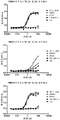
FIG. 1 shows that the nanobody of the present invention can bind to human PD-L1 protein on the surface of cell, and the binding effect of partial antibody is similar to that of positive control.
FIG. 2 shows that the engineered nanobody can still bind to human PD-L1 protein on the cell surface, and the binding effect of the antibody is similar to that of the positive control.
FIG. 3 shows that the engineered nanobody can still block the binding of PD-L1 protein and human PD-1 protein on the cell surface, and the blocking effect of the antibody is similar to that of the positive control.
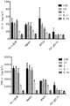
Fig. 4 shows that the nanobody of the present invention can effectively activate T cells, and the activation effect is similar to or better than that of the positive control group antibody.
FIG. 5 shows a schematic of the structure of the fusion protein.
FIG. 6 shows that the fusion protein of the present invention can bind to human PD-L1 protein on the cell surface.
FIG. 7 shows that the fusion protein of the present invention can block the binding between PD-L1 protein and human PD-1 protein on the cell surface, and the blocking effect of the antibody is similar to that of the positive control.
FIG. 8 shows that the fusion protein of the present invention can bind to TGF-beta 1, TGF-beta 2, and TGF-beta 3.
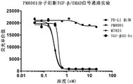
FIG. 9 shows that fusion proteins of the invention can effectively block the TGF-beta/SMAD signaling pathway.
FIG. 10 shows that the fusion protein of the present invention can efficiently activate T cells with similar or superior activation effect to that of the positive control antibody.
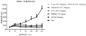
FIG. 11 shows that the fusion protein of the present invention can effectively inhibit tumor growth in mice.
Detailed Description
The inventor develops a class of anti-PD-L1 nano-antibodies for the first time through extensive and intensive research and a large amount of screening. Experimental results show that the PD-L1 nano antibody and the mutant derivative thereof obtained by the invention can effectively block the interaction between PD-L1 and PD-1 and have better thermal stability.
Specifically, the invention utilizes the human PD-L1 antigen protein to immunize alpaca (Llama) to obtain a high-quality immune nano antibody gene library. Among them, the present inventors have surprisingly screened the immune nanobody gene library for nanobody gene sequences with higher humanization level (sequence identity > 85%). The PD-L1 protein molecule is biotinylated and screened by using a yeast display technology to screen an immune nano antibody gene library, so that a nano antibody candidate molecule antibody gene with PD-L1 specificity is obtained. The obtained gene and the engineered mutant thereof are transferred into Expi-CHO cells, and further screening is carried out on the aspects of antibody affinity, the capacity of blocking the combination of PD-L1 and PD-1, thermal stability, the activity of activated T cells and the like, so that a nano antibody strain which can be efficiently expressed in vitro and has high specificity is obtained.
In addition, the experimental result shows that the fusion protein produced by fusing the nano antibody sequence (as a targeting part), IgG1 Fc fragment (as a connecting part) and TGF beta RII extracellular domain (as an immune regulatory molecule part) has high activity with PD-L1, can effectively block the interaction between PD-L1 and PD-1, can effectively block a TGF-beta/SMAD signaling pathway, can effectively activate human T lymphocytes, and can effectively inhibit the growth of tumors in mice.
On the basis of this, the present invention has been completed.
The invention of the nanometer antibody
As used herein, the terms "nanobody of the invention", "anti-PD-L1 nanobody of the invention", "PD-L1 nanobody of the invention" are used interchangeably and all refer to nanobodies that specifically recognize and bind to PD-L1 (including human PD-L1). Particularly preferred is a VHH chain having an amino acid sequence as set forth in SEQ ID NO: 4. 9, 82, 85, 88, 91, 94, or 97.
As used herein, the term "antibody" or "immunoglobulin" is an heterotetrameric glycan protein of about 150000 daltons with the same structural features, consisting of two identical light chains (L) and two identical heavy chains (H). Each light chain is linked to a heavy chain by one covalent disulfide bond, while the number of disulfide bonds varies between heavy chains of different immunoglobulin isotypes. Each heavy and light chain also has regularly spaced intrachain disulfide bonds. Each heavy chain has at one end a variable region (VH) followed by a plurality of constant regions. Each light chain has a variable domain (VL) at one end and a constant domain at the other end; the constant region of the light chain is opposite the first constant region of the heavy chain, and the variable region of the light chain is opposite the variable region of the heavy chain. Particular amino acid residues form the interface between the variable regions of the light and heavy chains.
As used herein, the terms "single domain antibody (VHH)", "nanobody" (nanobody) have the same meaning, referring to the variable region of the cloned antibody heavy chain, and the construction of a single domain antibody (VHH) consisting of only one heavy chain variable region, which is the smallest antigen-binding fragment with full function. Typically, single domain antibodies (VHHs) consisting of only one heavy chain variable region are constructed by first obtaining an antibody that naturally lacks the light and heavy chain constant region 1(CH1) and then cloning the variable region of the antibody heavy chain.
As used herein, the term "variable" means that certain portions of the variable regions of an antibody differ in sequence, which results in the binding and specificity of each particular antibody for its particular antigen. However, the variability is not evenly distributed throughout the antibody variable region. It is concentrated in three segments called Complementarity Determining Regions (CDRs) or hypervariable regions in the light and heavy chain variable regions. The more conserved portions of the variable regions are called Framework Regions (FR). The variable regions of native heavy and light chains each comprise four FR regions, which are in a substantially β -sheet configuration, connected by three CDRs that form a connecting loop, and in some cases may form part of a β -sheet structure. The CDRs in each chain are held close together by the FR region and form the antigen binding site of the antibody with the CDRs of the other chain (see Kabat et al, NIH Publ. No.91-3242, Vol I, 647-669 (1991)). The constant regions are not directly involved in the binding of antibodies to antigens, but they exhibit different effector functions, such as participation in antibody-dependent cytotoxicity of antibodies.
As known to those skilled in the art, immunoconjugates and fusion expression products include: drugs, toxins, cytokines (cytokines), radionuclides, enzymes, and other diagnostic or therapeutic molecules are conjugated to the antibodies or fragments thereof of the present invention. The invention also comprises a cell surface marker or antigen combined with the anti-PD-L1 protein antibody or the fragment thereof.
As used herein, the terms "heavy chain variable region" and "V H "may be used interchangeably.
As used herein, the term "variable region" is used interchangeably with "Complementary Determining Region (CDR)".
In a preferred embodiment of the invention, the heavy chain variable region of the antibody comprises three complementarity determining regions CDR1, CDR2, and CDR 3.
In a preferred embodiment of the invention, the heavy chain of the antibody comprises the above-described heavy chain variable region and heavy chain constant region.
In the present invention, the terms "antibody of the present invention", "protein of the present invention", or "polypeptide of the present invention" are used interchangeably and refer to a polypeptide that specifically binds to PD-L1 protein, e.g., a protein or polypeptide having a heavy chain variable region. They may or may not contain the initial methionine.
The invention also provides other proteins or fusion expression products having an antibody of the invention. In particular, the invention includes any protein or protein conjugate and fusion expression product (i.e., immunoconjugate and fusion expression product) having a heavy chain comprising a variable region, provided that the variable region is identical or at least 90% homologous, preferably at least 95% homologous, to the heavy chain variable region of an antibody of the invention.
In general, the antigen binding properties of an antibody can be described by 3 specific regions in the heavy chain variable region, called variable regions (CDRs), which are separated into 4 Framework Regions (FRs), the amino acid sequences of the 4 FRs being relatively conserved and not directly involved in the binding reaction. These CDRs form a loop structure, and the β -sheets formed by the FRs between them are spatially close to each other, and the CDRs on the heavy chain and the CDRs on the corresponding light chain constitute the antigen binding site of the antibody. It is possible to determine which amino acids constitute the FR or CDR regions by comparing the amino acid sequences of antibodies of the same type.
The variable regions of the heavy chains of the antibodies of the invention are of particular interest because at least some of them are involved in binding to antigen. Thus, the invention includes those molecules having an antibody heavy chain variable region with CDRs whose homology to the CDRs identified herein is greater than 90% (preferably greater than 95%, most preferably greater than 98%).
The invention includes not only intact antibodies, but also fragments of antibodies with immunological activity or fusion proteins of antibodies with other sequences. Accordingly, the invention also includes fragments, derivatives and analogs of the antibodies.
As used herein, the terms "fragment," "derivative," and "analog" refer to a polypeptide that retains substantially the same biological function or activity as an antibody of the invention. A polypeptide fragment, derivative or analogue of the invention may be (i) a polypeptide in which one or more conserved or non-conserved amino acid residues (preferably conserved amino acid residues) are substituted, and such substituted amino acid residues may or may not be encoded by the genetic code, or (ii) a polypeptide having a substituent group in one or more amino acid residues, or (iii) a polypeptide formed by fusing the mature polypeptide to another compound (such as a compound that increases the half-life of the polypeptide, e.g. polyethylene glycol), or (iv) a polypeptide formed by fusing an additional amino acid sequence to the sequence of the polypeptide (such as a leader or secretory sequence or a sequence used to purify the polypeptide or a proprotein sequence, or a fusion protein with a 6His tag). Such fragments, derivatives and analogs are within the purview of those skilled in the art in view of the teachings herein.
The antibody of the present invention refers to a polypeptide having a binding activity to PD-L1 protein, which includes the above-mentioned CDR region. The term also includes variants of the polypeptides comprising the CDR regions described above that have the same function as the antibodies of the invention. These variants include (but are not limited to): deletion, insertion and/or substitution of one or more (usually 1 to 50, preferably 1 to 30, more preferably 1 to 20, most preferably 1 to 10) amino acids, and addition of one or several (usually up to 20, preferably up to 10, more preferably up to 5) amino acids at the C-terminus and/or N-terminus. For example, in the art, substitutions with amino acids that are similar or analogous in performance do not typically alter the function of the protein. Also, for example, addition of one or several amino acids at the C-terminus and/or N-terminus does not generally alter the function of the protein. The term also includes active fragments and active derivatives of the antibodies of the invention.
Variants of the polypeptide include: homologous sequences, conservative variants, allelic variants, natural mutants, induced mutants, proteins encoded by DNA that hybridizes under high or low stringency conditions with DNA encoding an antibody of the invention, and polypeptides or proteins obtained using antisera raised against an antibody of the invention.
The invention also provides other polypeptides, such as fusion proteins comprising nanobodies or fragments thereof. In addition to nearly full-length polypeptides, fragments of the nanobodies of the invention are also encompassed by the present invention. Typically, the fragment has at least about 50 contiguous amino acids of the antibody of the invention, preferably at least about 50 contiguous amino acids, more preferably at least about 80 contiguous amino acids, and most preferably at least about 100 contiguous amino acids.
In the present invention, "conservative variant of the antibody of the present invention" means that at most 10, preferably at most 8, more preferably at most 5, and most preferably at most 3 amino acids are substituted by amino acids having similar or similar properties as compared with the amino acid sequence of the antibody of the present invention to form a polypeptide. These conservative variant polypeptides are preferably generated by amino acid substitutions according to Table 1.
TABLE 1


The invention also provides polynucleotide molecules encoding the above antibodies or fragments or fusion proteins thereof. The polynucleotide of the present invention may be in the form of DNA or RNA. The form of DNA includes cDNA, genomic DNA or artificially synthesized DNA. The DNA may be single-stranded or double-stranded. The DNA may be the coding strand or the non-coding strand.
Polynucleotides encoding the mature polypeptides of the invention include: a coding sequence encoding only the mature polypeptide; the coding sequence for the mature polypeptide and various additional coding sequences; the coding sequence (and optionally additional coding sequences) as well as non-coding sequences for the mature polypeptide.
The term "polynucleotide encoding a polypeptide" may include a polynucleotide encoding the polypeptide, and may also include additional coding and/or non-coding sequences.
The present invention also relates to polynucleotides which hybridize to the sequences described above and which have at least 50%, preferably at least 70%, and more preferably at least 80% identity between the two sequences. The present invention particularly relates to polynucleotides which hybridize under stringent conditions to the polynucleotides of the present invention. In the present invention, "stringent conditions" mean: (1) hybridization and elution at lower ionic strength and higher temperature, such as 0.2 XSSC, 0.1% SDS, 60 ℃; or (2) adding denaturant during hybridization, such as 50% (v/v) formamide, 0.1% calf serum/0.1% Ficoll, 42 deg.C, etc.; or (3) hybridization occurs only when the identity between two sequences is at least 90% or more, preferably 95% or more. Also, the polypeptides encoded by the hybridizable polynucleotides have the same biological functions and activities as the mature polypeptides.
The full-length nucleotide sequence of the antibody of the present invention or a fragment thereof can be obtained by a PCR amplification method, a recombinant method, or an artificial synthesis method. One possibility is to use synthetic methods to synthesize the sequence of interest, especially when the fragment length is short. Generally, fragments with long sequences are obtained by first synthesizing a plurality of small fragments and then ligating them. Alternatively, the coding sequence for the heavy chain and an expression tag (e.g., 6His) can be fused together to form a fusion protein.
Once the sequence of interest has been obtained, it can be obtained in large quantities by recombinant methods. This is usually done by cloning it into a vector, transferring it into a cell, and isolating the relevant sequence from the propagated host cell by conventional methods. The biomolecules (nucleic acid, protein, etc.) to which the present invention relates include biomolecules in an isolated form.
At present, DNA sequences encoding the proteins of the present invention (or fragments or derivatives thereof) have been obtained completely by chemical synthesis. The DNA sequence may then be introduced into various existing DNA molecules (or vectors, for example) and cells known in the art. Furthermore, mutations can also be introduced into the protein sequences of the invention by chemical synthesis.
The invention also relates to a vector comprising a suitable DNA sequence as described above and a suitable promoter or control sequence. These vectors may be used to transform an appropriate host cell so that it can express the protein.
The host cell may be a prokaryotic cell, such as a bacterial cell; or lower eukaryotic cells, such as yeast cells; or higher eukaryotic cells, such as mammalian cells. Representative examples are: escherichia coli, streptomyces; bacterial cells of salmonella typhimurium; fungal cells such as yeast; insect cells of Drosophila S2 or Sf 9; CHO, COS7, 293 cells, etc.
Transformation of a host cell with recombinant DNA can be carried out using conventional techniques well known to those skilled in the art. When the host is prokaryotic, e.g., E.coli, competent cells capable of DNA uptake can be harvested after exponential growth phase using CaCl 2 Methods, the steps used are well known in the art. Another method is to use MgCl 2 . If desired, transformation can also be carried out by electroporation. When the host is a eukaryote, the following DNA transfection methods may be used: calcium phosphate coprecipitation, conventional mechanical methods such as microinjection, electroporation, liposome packaging, etc.
The obtained transformant can be cultured by a conventional method to express the polypeptide encoded by the gene of the present invention. The medium used in the culture may be selected from various conventional media depending on the host cell used. The culturing is performed under conditions suitable for growth of the host cell. After the host cells have been grown to an appropriate cell density, the selected promoter is induced by suitable means (e.g., temperature shift or chemical induction) and the cells are cultured for an additional period of time.
The recombinant polypeptide in the above method may be expressed intracellularly or on the cell membrane, or secreted extracellularly. If necessary, the recombinant protein can be isolated and purified by various separation methods using its physical, chemical and other properties. These methods are well known to those skilled in the art. Examples of such methods include, but are not limited to: conventional renaturation treatment, treatment with a protein precipitant (such as salt precipitation), centrifugation, cell lysis by osmosis, sonication, ultracentrifugation, molecular sieve chromatography (gel filtration), adsorption chromatography, ion exchange chromatography, High Performance Liquid Chromatography (HPLC), and other various liquid chromatography techniques, and combinations thereof.
The antibodies of the invention may be used alone or in combination or conjugated with detectable labels (for diagnostic purposes), therapeutic agents, PK (protein kinase) modifying moieties or combinations of any of the above.
Detectable labels for diagnostic purposes include, but are not limited to: a fluorescent or luminescent label, a radioactive label, an MRI (magnetic resonance imaging) or CT (computed tomography) contrast agent, or an enzyme capable of producing a detectable product.
Therapeutic agents that may be conjugated or conjugated to the antibodies of the invention include, but are not limited to: 1. a radionuclide; 2. biological toxicity; 3. cytokines such as IL-2, etc.; 4. gold nanoparticles/nanorods; 5. a viral particle; 6. a liposome; 7. nano magnetic particles; 8. prodrug activating enzymes (e.g., DT-diaphorase (DTD) or biphenyl hydrolase-like protein (BPHL)); 10. chemotherapeutic agents (e.g., cisplatin) or nanoparticles in any form, and the like.
Fusion proteins of the invention
As used herein, "fusion protein of the present invention" refers to a bifunctional fusion protein having both an anti-PD-L1 nanobody of the first aspect of the present invention and an immunomodulatory molecular moiety.
In the invention, a fusion protein is provided, and the nano antibody fusion protein has a structure shown in a formula I from an N end to a C end:
Z1-Z2-L-Z3 (formula I)
In the formula (I), the compound is shown in the specification,
z1 is a VHH chain of an anti-PD-L1 nanobody according to the second aspect of the invention;
z2 is the Fc fragment of an immunoglobulin;
l is a linker sequence;
z3 is an immunomodulatory molecular moiety.
Preferably, the immunoglobulin may be IgG1, IgG2, IgG3, IgG4, or the like.
In a preferred embodiment, the immunoglobulin is IgG1 and the amino acid sequence of Z2 is as set forth in SEQ ID NO: 99, respectively. In other embodiments, the amino acid sequence of Z2 is identical to the amino acid sequence set forth in SEQ ID NO: 99 is identical or substantially identical.
In the present invention, the L is a flexible amino acid linker. Preferably, L has an amino acid sequence selected from the group consisting of: GGGGS, (GGS) 2 、(GGGGS) 3 、(GGGGS) 4 、(GGGGS) 5 Or a combination thereof.
In a preferred embodiment, the amino acid sequence of L is as set forth in SEQ ID NO: shown at 100. In other embodiments, the amino acid sequence of L is identical to the amino acid sequence set forth in SEQ ID NO: 100 are identical or substantially identical.
In one embodiment of the invention, the immunomodulatory molecule is a TGF β RII extracellular domain. Preferably, the amino acid sequence of Z3 is as set forth in SEQ ID NO: shown at 101. In other embodiments, the amino acid sequence of Z3 is identical to the amino acid sequence set forth in SEQ ID NO: 101 are identical or substantially identical.
In the present invention, the substantial identity is a difference of up to 50 (preferably 1 to 20, more preferably 1 to 10, still more preferably 1 to 5, and most preferably 1 to 3) amino acids, wherein the difference includes amino acid substitution, deletion, or addition.
Preferably, the substantial identity is at least 70%, at least 75%, at least 80%, at least 85%, at least 86%, at least 87%, at least 88%, at least 89%, at least 90%, at least 91%, at least 92%, at least 93%, at least 94%, at least 95%, at least 96%, at least 97%, at least 98%, or at least 99% sequence identity of the amino acid sequence to the corresponding amino acid sequence.
In a preferred embodiment, the amino acid sequence of the nanobody fusion protein is as set forth in SEQ ID NO: 102, respectively.
TGF β is a key inducer of Epithelial-mesenchymal transition (EMT). Meanwhile, TGF beta has a strong immunosuppressive effect in a tumor microenvironment, and further has an important regulation effect on tumor occurrence, development, metastasis and drug resistance.
Thus, in one embodiment of the invention, TGF-beta receptor II is selected as the immunomodulatory molecule in the fusion protein. The fusion protein has the advantages of high double-target binding affinity and strong specificity, thereby further enhancing the anti-tumor immune function.
Pharmaceutical composition
The invention also provides a composition. Preferably, the composition is a pharmaceutical composition comprising the above antibody or an active fragment thereof or a fusion protein thereof, and a pharmaceutically acceptable carrier. Generally, these materials will be formulated in a non-toxic, inert and pharmaceutically acceptable aqueous carrier medium, wherein the pH is generally from about 5 to about 8, preferably from about 6 to about 8, although the pH will vary depending on the nature of the material being formulated and the condition being treated. The formulated pharmaceutical compositions may be administered by conventional routes including, but not limited to: intratumoral, intraperitoneal, intravenous, or topical administration.
The pharmaceutical composition of the invention can be directly used for binding PD-L1 protein molecules, thus being used for treating tumors. In addition, other therapeutic agents may also be used simultaneously.
The pharmaceutical composition of the present invention comprises a safe and effective amount (e.g., 0.001-99 wt%, preferably 0.01-90 wt%, more preferably 0.1-80 wt%) of the nanobody (or its conjugate) of the present invention as described above and a pharmaceutically acceptable carrier or excipient. Such vectors include (but are not limited to): saline, buffer, glucose, water, glycerol, ethanol, and combinations thereof. The pharmaceutical formulation should be compatible with the mode of administration. The pharmaceutical composition of the present invention can be prepared in the form of injection, for example, by a conventional method using physiological saline or an aqueous solution containing glucose and other adjuvants. Pharmaceutical compositions such as injections, solutions are preferably manufactured under sterile conditions. The amount of active ingredient administered is a therapeutically effective amount, for example from about 10 micrograms per kilogram of body weight to about 50 milligrams per kilogram of body weight per day. In addition, the polypeptides of the invention may also be used with other therapeutic agents.
In the case of pharmaceutical compositions, a safe and effective amount of the immunoconjugate is administered to the mammal, wherein the safe and effective amount is typically at least about 10 micrograms/kg body weight, and in most cases no more than about 50 mg/kg body weight, preferably the dose is from about 10 micrograms/kg body weight to about 10mg/kg body weight. Of course, the particular dosage will also take into account such factors as the route of administration, the health of the patient, and the like, which are within the skill of the skilled practitioner.
Labeled nanobodies
In a preferred embodiment of the invention, the nanobody carries a detectable label. More preferably, the marker is selected from the group consisting of: isotopes, colloidal gold labels, coloured labels or fluorescent labels.
The colloidal gold labeling can be performed by methods known to those skilled in the art. In a preferred embodiment of the present invention, the nanobody against PD-L1 is labeled with colloidal gold to obtain a gold-labeled nanobody.
The anti-PD-L1 nano antibody has good specificity and high titer.
Detection method
The invention also relates to a method for detecting the PD-L1 protein. The method comprises the following steps: obtaining a cell and/or tissue sample; dissolving the sample in a medium; detecting the level of PD-L1 protein in the solubilized sample.
The sample used in the detection method of the present invention is not particularly limited, and a typical example is a cell-containing sample present in a cell preservation solution.
Reagent kit
The present invention also provides a kit comprising an antibody (or fragment thereof) or assay plate of the invention, and in a preferred embodiment of the invention, the kit further comprises a container, instructions for use, a buffer, and the like.
The invention also provides a detection kit for detecting the level of PD-L1, which comprises an antibody for identifying PD-L1 protein, a lysis medium for dissolving a sample, and general reagents and buffers required by detection, such as various buffers, detection markers, detection substrates and the like. The test kit may be an in vitro diagnostic device.
Applications of the invention
As described above, the nanobody of the present invention has wide biological and clinical application values, and its application relates to diagnosis and treatment of diseases related to PD-L1, basic medical research, biological research and other fields. One preferred application is for clinical diagnosis and targeted therapy against PD-L1.
The main advantages of the invention include:
1) the nano antibody is high in specificity and is directed to human PD-L1 protein with a correct space structure.
2) The nano antibody has strong affinity.
3) The production of the nano antibody is simple and convenient.
4) The inhibition of the PD-1/PD-L1 pathway on the basis of targeting and neutralizing TGF-beta of a tumor microenvironment can restore the activity of T cells, enhance immune response and more effectively improve the effect of inhibiting the occurrence and development of tumors.
The invention will be further illustrated with reference to the following specific examples. It should be understood that these examples are for illustrative purposes only and are not intended to limit the scope of the present invention. Experimental procedures without specific conditions noted in the following examples, generally followed by conventional conditions, such as Sambrook et al, molecular cloning: the conditions described in the Laboratory Manual (New York: Cold Spring Harbor Laboratory Press, 1989), or according to the manufacturer's recommendations. Unless otherwise indicated, percentages and parts are percentages and parts by weight.
Example 1: construction of Nanobody library
Animal immunization
2 alpacas (Llama) were immunized by mixing 1mg of human PD-L1 antigen (purchased from Acrobiosystems) with equal volume of Freund's adjuvant once a week for 4 total immunizations to stimulate B cells to express antigen-specific nanobodies. After 4 times of immunization, 50ml of alpaca peripheral blood is extracted, and lymphocyte is obtained by separating lymphocyte separating medium. Total RNA was extracted using the RNA extraction reagent Trizol (from Invitrogen). Total alpaca cDNA was obtained by reverse transcription using a cDNA synthesis kit (purchased from Invitrogen).
Nano antibody gene amplification
First round PCR, IgG2, IgG3 sequences were amplified from cDNA:
TABLE 1 first round PCR primers
| Name (R) | Sequences (5 'to 3') |
| Upstream primer | GTCCTGGCTGCTCTTCTACAAGG |
| Downstream primer | GGTACGTGCTGTTGAACTGTTCC |
The first round of PCR products was subjected to agarose gel electrophoresis, and the 750bp fragment was recovered by gel cutting for the second round of amplification of the VHH sequence. The primers for the second round of PCR amplification were as follows:
TABLE 2 second round PCR primers
| Name (R) | Sequences (5 'to 3') |
| Upstream primer | CTAGTGCGGCCGCcTGGAGACGGTGACCTGGGT |
| Downstream primer | CGCGGATCCCAGGTGCAGCTGCAGGAGTCTGGRGGAGG |
And (3) performing third PCR by taking the second round PCR product as a template to add a homology arm to the VHH gene, wherein third PCR amplification primers are as follows:
TABLE 3 third round PCR primers

The desired fragment was recovered using a PCR purification kit (purchased from QIAGEN).
Library construction
The linearized yeast display vector and the third round of PCR product were mixed and electrically transformed into Saccharomyces cerevisiae (purchased from ATCC), anti-PD-L1 nanobody libraries from two animals were constructed and the library volumes were determined, the sizes of the library volumes were 4.47X 10, respectively 7 And 4.14X 10 7 。
Example 2: screening of PD-L1 nanometer antibody
Biotinylation labeling of human PD-L1 protein
Appropriate volumes of double distilled water were taken to dissolve human PD-L1 protein (purchased from Acrobiosystems), biotin was dissolved and mixed with the protein solution according to the Biotin labeling kit (purchased from Thermo) product instructions, and incubated at 4 ℃ for 2 hours. Excess biotin was removed using a desalting column (from Thermo) with reference to the product instructions for pretreatment and sample collection.
MACS enrichment of Yeast that specifically binds to PD-L1
The VHH library constructed in example 2 was inoculated into SD-CAA amplification medium (6.7 g YNB, 5g casamino acid, 13.62g Na in 1L SD-CAA amplification medium) 2 HPO 4 ·12H 2 O、7.44g NaH 2 PO 4 And 2% glucose), the number of inoculated yeast cells > 10 × library capacity (initial amplification concentration 0.5 OD) 600 /ml), incubated at 30 ℃ overnight at 225 rpm. Collecting yeast cells with 10 × storage volume, centrifuging at 3000rpm × 5min (the same shall apply hereinafter) to remove the culture medium, resuspending the yeast cells with SD-CAA induction culture medium, and adjusting the initial concentration to 0.5OD 600 Induction overnight at/ml. The concentration of the library after induction was measured, and yeast cells of 10 Xthe library volume were taken and centrifuged to remove the medium. The yeast cells were resuspended in 50ml of wash buffer (PBS + 0.5% BSA +2mM EDTA) and the supernatant removed by centrifugation. The yeast cells were resuspended in 10ml of wash buffer.
Biotin-labeled PD-L1 protein (final concentration 100mM) was added, incubated at room temperature for 30min, yeast cells were collected by centrifugation, and the yeast was washed 3 times with 50ml of washing buffer. The yeast cells were resuspended in 5ml of wash buffer and 200. mu.l of SA magnetic beads (purchased from America and whirlwind) were added and incubated for 10min by inversion. The yeast and magnetic bead mixture was washed 3 times with wash buffer and the mixture was added to an LS column (purchased from Edison). The LS column was placed on a magnetic rack and washed with washing buffer to remove non-specifically bound yeast cells. The column was removed from the magnetic rack and the yeast was eluted by adding wash buffer. The eluted yeast was centrifuged and transferred to 200ml SD-CAA amplification medium for amplification.
Flow cytometric separation washing to obtain high affinity yeast cell
Inoculating MACS-enriched yeast cells into SD-CAA amplification medium, wherein the initial amplification concentration is 0.5OD 600 And/ml. Shaking the flask at 225rpm at 30 ℃ overnight. The SD-CAA induction medium (1L of SD-CAA induction medium contains 6.7g YNB, 5g casamino acid, and 13.62g Na 2 HPO 4 ·12H 2 O,7.44g NaH 2 PO 4 And 2% galactose, 2% raffinose and 0.1% glucose) were resuspended at an initial concentration of 0.5OD 600 Perml, induction overnight. 1: 200 dilution of anti-c-Myc murine antibody (purchased from Thermo) and 100nM biotinylated PD-L1 antigen were added and incubated for 10min at room temperature. The yeast was washed 3 times with PBS, added 1: 500 diluted goat anti-mouse IgG (H + L) Alexa Fluor Plus 488 (from Invitrogen) and streptavidin APC-conjugated fluorescent antibody (from Invitrogen) and incubated at 4 ℃ in the absence of light for 15 min. 2ml PBS was added to resuspend the cells, and the cells were sorted using a BD FACSAraiII instrument to obtain yeasts with high binding capacity to PD-L1 antigen.
Calling of PD-L1 nano antibody candidate molecule antibody gene
The yeast liquid which is obtained by MACS and FACS enrichment and has higher binding capacity with PD-L1 antigen is cultured in SD-CAA amplification culture medium at 30 ℃ and 225rpm overnight, and yeast plasmid is extracted according to the operation of a yeast plasmid extraction kit (purchased from Tiangen). Plasmids were transformed into Top10 competent cells (purchased from Tiangen) by electroporation, plated with ampicillin resistant plates, and cultured overnight at 37 ℃. And (3) selecting a monoclonal for sequencing to obtain a VHH gene sequence.
Example 3: construction and expression purification of heavy chain antibody
Construction of antibody Gene into pCDNA3.1 expression vector
The VHH gene sequence was ligated to the Fc fragment of human IgG1(LALA mutant), linearized pCDNA3.1 vector by double digestion with homologous recombinase (purchased from Vazyme) and EcoR I/Not I, according to the commercial instructions. The homologous recombination products were transformed into Top10 competent cells, plated with ampicillin resistant plates, incubated overnight at 37 ℃ and single clones were selected for sequencing.
Cell transfection
Using ExpicHO TM The expression system kit (purchased from Thermo) transfers the plasmid into Expi-CHO cells, and the transfection method is according to the commercial instruction, and after 5 days of cell culture, collects the supernatant and purifies the target protein by using a protein A magnetic bead (purchased from Kinsley) sorting method. The beads were resuspended (1-4 bead volumes) in an appropriate volume of binding buffer (PBS + 0.1% Tween 20, pH 7.4) and added to the sample to be purified and incubated for 1 hour at room temperature with gentle shaking. The sample was placed on a magnetic stand (purchased from beaver), the supernatant was discarded, and the beads were washed 3 times with binding buffer. Adding elution buffer (0.1M sodium citrate, pH 3.2) according to 3-5 times of the volume of the magnetic beads, shaking at room temperature for 5-10min, placing on a magnetic frame, collecting the elution buffer, transferring to a collection tube added with neutralization buffer (1M Tris, pH 8.54), and mixing.
Example 4: purified anti-PD-L1 antibodies bind to human PD-L1
CHO cells overexpressing human PD-L1 (CHO-hPD-L1 cells) were generated by transfecting pCHO1.0 vector (purchased from Invitrogen) with human PD-L1 cDNA (purchased from Nano Biological) cloned into MCS. The expanded CHO-hPD-L1 cells were adjusted to a cell density of 2X 10 6 Cells/ml, 100. mu.l/well were added to a 96-well flow plate and centrifuged for use. Purified PD-L1 antibody was diluted with PBS at 12 points 3-fold at 400nM, and 100. mu.l/well of the diluted sample was added to the 96-well flow plate with cells at 4 deg.CIncubate for 30min and wash twice with PBS. 100 μ l/well of sheep F (ab') 2 anti-human IgG-Fc (PE) (purchased from Abcam) diluted 100-fold with PBS was added, incubated at 4 ℃ for 30min, and washed twice with PBS. PBS was added at 100. mu.l/well to resuspend the cells, detected on a CytoFlex (Bechman) flow cytometer and the corresponding MFI calculated.
In the assay experiments as described above, the results are shown in FIG. 1, all of the purified samples of the present invention and CHO-hPD-L1 cells had binding activity, and the binding activity of the partially purified samples was similar to that of the control antibody ATE (US 20130034559).
Example 5: PD-L1 antibody affinity assay
ForteBio affinity assays were performed according to the current method (Estep, P et al, measurement of high throughput antibody-antigen affinity and epitope fractionation based on solutions, MAbs, 2013.5 (2): p.270-8). Briefly, the sensor was equilibrated in assay buffer for 30min under the line, then the baseline was established by on-line detection for 60s, and the purified antibody obtained as described above was loaded on-line onto the AHQ sensor. The sensor was then exposed to 100nM PD-L1 antigen for 5min before being transferred to PBS for dissociation for 5 min. Kinetic analysis was performed using a 1: 1 binding model.
TABLE 4 candidate molecule affinities
| Numbering | KD(M) | Kon(1/Ms) | Koff(1/s) |
| C-Ye-02 | 7.98E-08 | 4.69E+04 | 3.75E-03 |
| C-Ye-04 | 4.89E-09 | 1.03E+05 | 5.05E-04 |
| C-Ye-06 | 1.34E-07 | 4.35E+04 | 5.81E-03 |
| C-Ye-17 | 3.96E-07 | 2.58E+04 | 1.02E-02 |
| C-Ye-18 | 5.39E-09 | 8.33E+04 | 4.49E-04 |
| C-Ye-20 | 4.86E-08 | 9.06E+04 | 4.40E-03 |
| C-Ye-24 | 1.95E-08 | 7.71E+04 | 1.51E-03 |
| C-Ye-26 | 3.41E-08 | 9.41E+04 | 3.21E-03 |
| C-Ye-27 | 5.79E-08 | 5.45E+04 | 3.15E-03 |
| C-Ye-30 | 3.94E-08 | 5.32E+04 | 2.10E-03 |
| C-Ye-32 | 4.43E-08 | 5.83E+04 | 2.58E-03 |
| C-Ye-34 | 2.44E-08 | 7.81E+04 | 1.91E-03 |
| C-Ye-39 | 5.03E-07 | 2.12E+04 | 1.07E-02 |
| C-Ye-42 | 3.89E-08 | 5.70E+04 | 2.22E-03 |
Example 6: PD-L1 antibody gene modification
To remove potential glycosylation sites in C-Ye-18, the CDRH2 portion of the C-Ye-18 amino acid sequence was point mutated to 6 forms in Table 5:
TABLE 5C-Ye-18 CDR region mutant sequences
| Numbering | CDRH2 region |
| C-Ye-18 | SINSSSSSTYYRDSVKG |
| C-Ye-18-1 | SINSGSSSTYYRDSVKG |
| C-Ye-18-2 | SISSSSSSTYYRDSVKG |
| C-Ye-18-3 | SIGSSSSSTYYRDSVKG |
| C-Ye-18-4 | SIYSGSSSTYYRDSVKG |
| C-Ye-18-5 | SINSDSSSTYYRDSVKG |
| C-Ye-18-6 | SISGSSSSTYYRDSVKG |
The humanized water of the C-Ye-18 CDR region mutation sequence is evaluated by IMGT (http:// www.imgt.org), and the result is shown in Table 6, and the humanized water of all the C-Ye-18 mutation sequences is higher than 87% on average, thereby meeting the requirements of later-stage drug development.
TABLE 6C-Ye-18 CDR region mutant sequences and human homology
| Numbering | Germline | Homology of |
| C-Ye-18 | IGHV3-74*01 | 87.80% |
| C-Ye-18-1 | IGHV3-74*01 | 88.80% |
| C-Ye-18-2 | IGHV3-74*01 | 87.80% |
| C-Ye-18-3 | IGHV3-74*01 | 87.80% |
| C-Ye-18-4 | IGHV3-74*01 | 87.80% |
| C-Ye-18-5 | IGHV3-74*01 | 87.80% |
| C-Ye-18-6 | IGHV3-74*01 | 87.80% |
The protein construction and expression purification method was the same as example 3, and the purity of the protein was obtained by HPLC detection. HPLC method as follows, mobile phase: 150mM Na 2 HPO 4 ·12H 2 O, pH 7.0. Chromatographic conditions are as follows: detection wavelength: 280nm, column temperature: 25 ℃, flow rate: 0.35ml/min, detection time: 20min, Zenix-C SEC-300 column (SEPAX 4.6X 300mm, 3 μm).
TABLE 7 purity assay results for C-Ye-10 mutant antibodies
| Numbering | Proportion of monomer (%) |
| C-Ye-18 | 90.70 |
| C-Ye-18-1 | 97.40 |
| C-Ye-18-2 | 96.40 |
| C-Ye-18-3 | 98.50 |
| C-Ye-18-4 | 73.70 |
| C-Ye-18-5 | 83.00 |
| C-Ye-18-6 | 95.80 |
Example 7: binding of the C-Ye-18 mutant sample to human PD-L1
The experiment detects the binding activity of the purified C-Ye-18 mutant sample and CHO-hPD-L1 cells, the experimental method is the same as that in example 4, the experimental result is shown in figure 2, the C-Ye-18 mutant sample has good binding activity with CHO-hPD-L1 cells, and the level is equivalent to that of C-Ye-18 and control antibody ATE.
Example 8: affinity assay for C-Ye-18 mutant samples
The experiment detects the binding affinity of the purified C-Ye-18 mutant sample and human PD-L1, the experimental method is the same as that in example 5, the experimental result is shown in Table 8, and the C-Ye-18 mutant sample and human PD-L1 protein have good binding activity.
TABLE 8 affinity of C-Ye-18 mutant samples
| Numbering | KD(M) | kon(1/Ms) | kdis(1/s) |
| C-Ye-18-1 | 4.13E-09 | 2.46E+05 | 1.02E-03 |
| C-Ye-18-2 | 5.41E-09 | 2.34E+05 | 1.27E-03 |
| C-Ye-18-3 | 7.03E-09 | 2.41E+05 | 1.70E-03 |
| C-Ye-18-4 | 6.40E-09 | 2.41E+05 | 1.54E-03 |
| C-Ye-18-5 | 4.08E-09 | 2.72E+05 | 1.11E-03 |
| C-Ye-18-6 | 6.00E-09 | 2.32E+05 | 1.39E-03 |
Example 9: C-Ye-18 mutant sample blocks PD-L1 binding to PD-1
CHO cells expressing human PD-1 (CHO-hPD-1 cells) were generated by transfection of pCHO1.0 vector (purchased from Invitrogen) with human PD-1 cDNA (purchased from Nano Biological) cloned into MCS. The expanded CHO-hPD-1 cells were adjusted to a cell density of 2X 10 6 cells/ml, 100. mu.l/well were added to a 96-well flow plate and centrifuged for use. The purified mutant samples were diluted with PBS for 12 points at the beginning of a 3-fold dilution at 400nM, and 60. mu.l/well of the diluted samples were added to a 96-well sample dilution plate, while 60. mu.l/well were added with Biotinylated human PD-L1 protein (purchased from Acro biosystems) at a final concentration of 500ng/ml, and incubated with the mutant samples for 30min at 4 ℃. Co-incubated samplesAdd 100. mu.l/well to the above 96-well flow plate with cells, incubate at 4 ℃ for 30min, and wash twice with PBS. A100-fold dilution of streptomycin R-phycoerythrin conjugate (purchased from Thermo Fisher) in PBS was added to each well at 100. mu.l, incubated at 4 ℃ for 30min, and washed twice with PBS. PBS was added at 100. mu.l/well to resuspend the cells, detected on a CytoFlex (Bechman) flow cytometer and the corresponding MFI calculated.
In the above assay, the results are shown in FIG. 3, and all the mutant samples of the present invention can block the binding of PD-L1 to PD-1 at a level comparable to that of C-Ye-18 and the control antibody ATE.
Example 10: thermal stability of C-Ye-18 mutant samples
The thermostability of the different antibodies was determined by DSC (Differential scanning calorimetry). The sample was concentrated and diluted to 1mg/ml with PBS; a5000 Xfluorescent color developing reagent Cypro Orange (purchased from Bio-Rad) was diluted 50-fold with ultrapure water to obtain 100 Xfluorescent color developing reagent Sypro Orange. Taking 50 mu l of sample with the concentration of 1mg/ml, adding 10 mu l of 100 Xfluorescent color developing agent Sypro Orange and 40 mu l of ultrapure water, uniformly mixing, taking 30 mu l of sample, adding the sample into a 96-hole PCR plate, making 3 multiple holes for each sample, putting the sample into a PCR instrument, and setting the temperature-raising program as follows: keeping the temperature at 25 ℃ for 5min, and heating to 99 ℃ at the speed of 0.5 ℃/min. And after the program is finished, reading the temperature value at the lowest point of the Curve in the 'Melt Curve' graph, namely the Tm value of the sample. The results are shown in Table 9 below
TABLE 9 Tm values of C-Ye-18 mutant antibodies


Example 11: mixed lymphocyte reaction experiment
This example examines the T cell activating activity of C-Ye-18 mutant samples by mixed lymphocyte reaction assay (MLR). The specific experimental method is as follows.
PBMC cells (purchased from SALLY BIO, SLB-HPB) were recovered, centrifuged, and PBMC were resuspended in 10ml of X-VIVO-15 medium (purchased from LONZA), cultured for 2h adherent culture at 37 ℃ in a cell culture chamber, and nonadherent cells were aspirated. Add 10ml of DC medium: X-VIVO-15 Medium 10ng/ml GM-CSF (purchased from R) was added&D) 20ng/ml IL-4, culturing for 3 days, supplementing 5ml DC culture medium, continuing culturing to 6 days, adding DC maturation culture medium: X-VIVO-15 Medium TNF-. alpha. (purchased from R) was added at 1000U/ml&D) 10ng/ml IL-6 (from R)&D) 5ng/ml IL-1 beta (from R)&D) mu.M PGE2 (purchased from Tocris), cultured for 2 days, mature DC cells were collected and adjusted to a cell density of 2X 10 with X-VIVO-15 medium 5 Cells/ml.
PBMC cells from another donor (purchased from SALLY BIO, SLB-HPB) were revived, centrifuged, and PBMC were resuspended in 10ml X-VIVO-15 medium. Using CD4 + T cell sorting kit (purchased from Stemcell) enriched CD4 + T cells, X-VIVO-15 resuspension of CD4 + T cells adjusted to a cell density of 2X 10 6 cell/ml. CD4 + T cells were mixed with the above collected mature DC cells at a ratio of 1: 1, and 100. mu.l/well was added to a 96-well U-bottom plate.
The C-Ye-18 mutant samples were diluted with X-VIVO-15 medium, 3-fold diluted at 200nM for 9 spots, 100. mu.l/well were added to the mixed cell wells, cultured for 5 days, the supernatant was collected, and IFN-. gamma.and IL2 expression were measured by ELISA (ex eBioscience).
As shown in FIG. 4, the C-Ye-18 mutant samples, C-Ye-18-1, C-Ye-18-5 and C-Ye-18-6, all showed better biological activity in MLR experiment, and the activation level was similar to or better than that of the control antibody ATE.
Example 12 cloning and expression of the fusion protein PD-Ll/TGF-b trap
In this example, a PD-L1 antibody/TGF β RII extracellular domain fusion protein (PM8001) was formed using TGF β RII extracellular domain (SEQ ID NO: 101) as part of the immunomodulatory molecule in the fusion protein and PD-L1 antibody as the targeting moiety for the fusion protein.
The C-terminal amino acid of the PD-L1 single-chain antibody of the invention is passed through by molecular cloning technology (G) 4 S) 4 G-linked TGF-. beta.RII extracellularThe expression and purification method is the same as that of example 3 by performing conventional expression by an Expi-CHO expression system to obtain a fusion protein PM8001 with a structure shown in FIG. 5.
Example 13 binding of PM8001 molecule to human PD-L1
The procedure for determining the binding activity of purified PD-L1 antibody (C-Ye-18-5), PM8001 molecule, TGF-. beta.R 2-Fc fusion protein, positive control M7824(WO 2015/118175A 2), and negative control IgG protein to cell surface PD-L1 was the same as in example 4. In the assay experiments as described above, the results are shown in FIG. 6, and the PM8001 molecule of the present invention and CHO-hPD-L1 cells have binding activity similar to that of the positive control molecule M7824.
Example 14 PM8001 molecule blocks binding of PD-L1 protein to PD-1 cells
The purified PD-L1 antibody (C-Ye-18-5), PM8001 molecule, TGF-. beta.R 2-Fc fusion protein, positive control M7824 and negative control IgG protein were tested for blocking the binding activity of PD-L1 protein and PD-1 cells as in example 9. In the assay experiments as described above, the results are shown in fig. 7, and the PM8001 molecule of the present invention can block the binding of PD-L1 protein to PD-1 cells at a level comparable to the positive control molecule M7824.
Example 15 binding experiments of PM8001 molecule to ELISA levels of human TGF- β family proteins
Human TGF-. beta.1 (antibodies, TG1-H421), TGF-. beta.2 (PeproTech, 100-35B), and TGF-. beta.3 (PeproTech, 100-36E) proteins were diluted with ELISA coating solution, added to ELISA plates, and coated overnight at 4 ℃. The coating solution was discarded, and PBST was added to 250. mu.l/well and washed 3 times, and blocked with 5% BSA at room temperature for 1 hour for use. Purified PD-L1 antibody (C-Ye-18-5), PM8001 molecule, TGF-. beta.R 2-Fc fusion protein, positive control M7824 were diluted with a gradient and added to a blocked ELISA plate and incubated at room temperature for 2 hours. Adding PBST for washing for 3 times, adding sheep anti-human Fc-HRP (abcam, ab97225) into the holes, incubating for 1 hour at room temperature, adding PBST for washing for 3 times, adding an ELISA developing solution, standing for 3min at room temperature, adding an ELISA stopping solution, and reading the absorbance value at 450 nm.
In the assay experiments using the above method, the results are shown in FIG. 8, and the PM8001 molecule of the present invention binds to human TGF- β 1 and TGF- β 3 proteins well at the ELISA level, has weak binding activity to human TGF- β 2 protein, and has a binding level comparable to that of the positive control molecule M7824.
Example 16 PM8001 molecule blocks TGF- β/SMAD signaling pathway experiments
Inoculating 293-TGF-beta/SMAD effector cells to a 96-well cell culture white bottom plate, placing at 37 ℃ and 5% CO 2 The incubator was incubated overnight. Purified PD-L1 antibody (C-Ye-18-5), PM8001 molecule, TGF-. beta.R 2-Fc fusion protein, positive control M7824 was mixed with TGF-. beta.1 (microorganisms, TG1-H421) after gradient dilution and incubated at room temperature for 30 min. The mixture was added to a white plate with cells and the culture was continued overnight. Bio-Glo (TM) reagent (Promega) was added to each well and the fluorescence signal was read using a multifunctional microplate reader.
In the assay experiments as described above, the results are shown in fig. 9, and the PM8001 molecule of the present invention can block TGF- β/SMAD signaling pathways in vitro at levels comparable to the positive control molecule M7824.
Example 17 Mixed lymphocyte reaction experiments
Mixed lymphocyte assay purified PD-L1 antibody (C-Ye-18-5), PM8001 molecule, TGF-. beta.R 2-Fc fusion protein, positive control M7824, and negative control IgG protein activated human T lymphocytes in the same manner as in example 9. The results are shown in fig. 10, the PM8001 molecule of the present invention shows better biological activity in MLR experiments, and the level of activation is comparable or superior to the positive control molecule M7824.
Example 18PM8001 rat pharmacokinetic evaluation
6 SD rats for experiments are respectively male and female, light/dark adjustment is carried out for 12/12 hours, the temperature is 24 +/-2 ℃, the humidity is 40-70%, and the rats are fed with water freely. Purchased from Zhejiang vitamin Tonglihua laboratory technologies, Ltd. On the day of experiment, SD rats are injected with PM8001 molecule by a single tail vein injection, and the injection dose is 10 mg/Kg.
Blood sampling time points: rat jugular vein blood was collected 3min, 4 hours, 10 hours, 24 hours, 48 hours, 72 hours, 120 hours, 168 hours, 240 hours, 336 hours, 504 hours, 672 hours after administration. Standing the whole blood sample at 2-8 deg.C for 30min, centrifuging at 12000rpm for 5min to collect serum, centrifuging the obtained serum at 2-8 deg.C at 12000rpm for 5min, storing at-80 deg.C, and detecting the molecular weight of free PM8001 in the serum by ELISA. The results are shown in table 10, where the PM8001 free molecule of the present invention has a half-life of about 146 hours in SD rats in vivo.
TABLE 10 PM 8001T 1/2 in SD rats
| Test drug | Mode of administration | T1/2 |
| PM8001 | IV | 146 hours |
Example 19 study of tumor suppressor Activity of PM8001
In this experiment, MC38 cells (h-PD-L1 KI MC38) expressing human PD-L1 were used to measure the antitumor effect of PM8001 in PD-L1 transgenic mice. Firstly, a h-PD-L1 KI MC38 tumor-bearing mouse model is established in a subcutaneous inoculation mode, and the average tumor volume is 80-120mm 3 Grouping is carried out, different antibodies and different doses of treatment are given to each group of mice by single intraperitoneal injection, the tumor volume and the body weight change of each group of mice are monitored, the monitoring frequency is 2 times/week, the mice are continuously monitored for 3 weeks, the administration dose and the administration mode are shown in a table 11, and the tumor volume change of the mice is shown in a figure 11.
Table 11 protocol for tumor suppressive activity of PM8001

The results of the experiment are shown in FIG. 11. After h-PD-L1 KI MC38 is inoculated, the tumor volume of a negative control group is continuously increased, the tumor growth of a TGF-beta R II-Fc group and a C-Ye-18-5 single-use group is inhibited, and compared with a TGF-beta R II-Fc group and a C-Ye-18-5 group, the PM8001 group is better controlled, so that the PM8001 has obvious tumor inhibition effect and is equivalent to or slightly better than that of a positive control group.
All documents referred to herein are incorporated by reference into this application as if each were individually incorporated by reference. Furthermore, it should be understood that various changes and modifications of the present invention can be made by those skilled in the art after reading the above teachings of the present invention, and these equivalents also fall within the scope of the present invention as defined by the appended claims.
Sequence information of the invention
SEQ ID NO: 1C-Ye-18 CDR1 amino acid sequence

SEQ ID NO: 2C-Ye-18 CDR2 amino acid sequence

The amino acid sequence of SEQ ID NO: 3C-Ye-18 CDR3 amino acid sequence

SEQ ID NO: 4C-Ye-18 VHH amino acid sequence

SEQ ID NO: 5C-Ye-18 VHH nucleotide sequence

SEQ ID NO: 6C-Ye-04 CDR1 amino acid sequence

SEQ ID NO: 7C-Ye-04 CDR2 amino acid sequence

SEQ ID NO: 8C-Ye-04 CDR3 amino acid sequence

The amino acid sequence of SEQ ID NO: 9C-Ye-04 VHH amino acid sequence

SEQ ID NO: 10C-Ye-04 VHH nucleotide sequence


SEQ ID NO: 11C-Ye-02 CDR1 amino acid sequence

SEQ ID NO: 12C-Ye-02 CDR2 amino acid sequence

SEQ ID NO: 13C-Ye-02 CDR3 amino acid sequence

SEQ ID NO: 14C-Ye-02 VHH amino acid sequence

SEQ ID NO: 15C-Ye-02 VHH nucleotide sequence

SEQ ID NO: 16C-Ye-06 CDR1 amino acid sequence

The amino acid sequence of SEQ ID NO: 17C-Ye-06 CDR2 amino acid sequence

SEQ ID NO: 18C-Ye-06 CDR3 amino acid sequence

SEQ ID NO: 19C-Ye-06 VHH amino acid sequence

SEQ ID NO: 20C-Ye-06 VHH nucleotide sequence


SEQ ID NO: 21C-Ye-09 CDR1 amino acid sequence

SEQ ID NO: 22C-Ye-09 CDR2 amino acid sequence

SEQ ID NO: 23C-Ye-09 CDR3 amino acid sequence

SEQ ID NO: 24C-Ye-09 VHH amino acid sequence

SEQ ID NO: 25C-Ye-09 VHH nucleotide sequence

SEQ ID NO: 26C-Ye-10 CDR1 amino acid sequence

SEQ ID NO: 27C-Ye-10 CDR2 amino acid sequence

SEQ ID NO: 28C-Ye-10 CDR3 amino acid sequence

The amino acid sequence of SEQ ID NO: 29C-Ye-10 VHH amino acid sequence


SEQ ID NO: 30C-Ye-10 VHH nucleotide sequence

SEQ ID NO: 31C-Ye-17 CDR1 amino acid sequence

SEQ ID NO: 32C-Ye-17 CDR2 amino acid sequence

SEQ ID NO: 33C-Ye-17 CDR3 amino acid sequence

The amino acid sequence of SEQ ID NO: 34C-Ye-17 VHH amino acid sequence

SEQ ID NO: 35C-Ye-17 VHH nucleotide sequence

The amino acid sequence of SEQ ID NO: 36C-Ye-20 CDR1 amino acid sequence

The amino acid sequence of SEQ ID NO: 37C-Ye-20 CDR2 amino acid sequence

SEQ ID NO: 38C-Ye-20 CDR3 amino acid sequence

SEQ ID NO: 39C-Ye-20 VHH amino acid sequence

SEQ ID NO: 40C-Ye-20 VHH nucleotide sequence

SEQ ID NO: 41C-Ye-24 CDR1 amino acid sequence

SEQ ID NO: 42C-Ye-24 CDR2 amino acid sequence

SEQ ID NO: 43C-Ye-24 CDR3 amino acid sequence

SEQ ID NO: 44C-Ye-24 VHH amino acid sequence

SEQ ID NO: 45C-Ye-24 VHH nucleotide sequence

SEQ ID NO: 46C-Ye-26 CDR1 amino acid sequence

The amino acid sequence of SEQ ID NO: 47C-Ye-26 CDR2 amino acid sequence

SEQ ID NO: 48C-Ye-26 CDR3 amino acid sequence

SEQ ID NO: 49C-Ye-26 VHH amino acid sequence

SEQ ID NO: 50C-Ye-26 VHH nucleotide sequence

SEQ ID NO: 51C-Ye-27 CDR1 amino acid sequence

The amino acid sequence of SEQ ID NO: 52C-Ye-27 CDR2 amino acid sequence

SEQ ID NO: 53C-Ye-27 CDR3 amino acid sequence

SEQ ID NO: 54C-Ye-27 VHH amino acid sequence

SEQ ID NO: 55C-Ye-27 VHH nucleotide sequence

SEQ ID NO: 56C-Ye-30 CDR1 amino acid sequence

The amino acid sequence of SEQ ID NO: 57C-Ye-30 CDR2 amino acid sequence

SEQ ID NO: 58C-Ye-30 CDR3 amino acid sequence

SEQ ID NO: 59C-Ye-30 VHH amino acid sequence

The amino acid sequence of SEQ ID NO: 60C-Ye-30 VHH nucleotide sequence

SEQ ID NO: 61C-Ye-32 CDR1 amino acid sequence

SEQ ID NO: 62C-Ye-32 CDR2 amino acid sequence

SEQ ID NO: 63C-Ye-32 CDR3 amino acid sequence

SEQ ID NO: 64C-Ye-32 VHH amino acid sequence

SEQ ID NO: 65C-Ye-32 VHH nucleotide sequence

SEQ ID NO: 66C-Ye-34 CDR1 amino acid sequence

SEQ ID NO: 67C-Ye-34 CDR2 amino acid sequence

SEQ ID NO: 68C-Ye-34 CDR3 amino acid sequence

SEQ ID NO: 69C-Ye-34 VHH amino acid sequence

SEQ ID NO: 70C-Ye-34 VHH nucleotide sequence

SEQ ID NO: 71C-Ye-39 CDR1 amino acid sequence

The amino acid sequence of SEQ ID NO: 72C-Ye-39 CDR2 amino acid sequence

SEQ ID NO: 73C-Ye-39 CDR3 amino acid sequence

SEQ ID NO: 74C-Ye-39 VHH amino acid sequence

SEQ ID NO: 75C-Ye-39 VHH nucleotide sequence


SEQ ID NO: 76C-Ye-42 CDR1 amino acid sequence

SEQ ID NO: 77C-Ye-42 CDR2 amino acid sequence

SEQ ID NO: 78C-Ye-42 CDR3 amino acid sequence

SEQ ID NO: 79C-Ye-42 VHH amino acid sequence

SEQ ID NO: 80C-Ye-42 VHH nucleotide sequence

SEQ ID NO: 81C-Ye-18-1 CDR2 amino acid sequence

SEQ ID NO: 82C-Ye-18-1 VHH amino acid sequence

SEQ ID NO: 83C-Ye-18-1 VHH nucleotide sequence

SEQ ID NO: 84C-Ye-18-2 CDR2 amino acid sequence

SEQ ID NO: 85C-Ye-18-2 VHH amino acid sequence

The amino acid sequence of SEQ ID NO: 86C-Ye-18-2 VHH nucleotide sequence

SEQ ID NO: 87C-Ye-18-3 CDR2 amino acid sequence

SEQ ID NO: 88C-Ye-18-3 VHH amino acid sequence

SEQ ID NO: 89C-Ye-18-3 VHH nucleotide sequence

SEQ ID NO: 90C-Ye-18-4 CDR2 amino acid sequence

The amino acid sequence of SEQ ID NO: 91C-Ye-18-4 VHH amino acid sequence

SEQ ID NO: 92C-Ye-18-4 VHH nucleotide sequence


SEQ ID NO: 93C-Ye-18-5 CDR2 amino acid sequence

The amino acid sequence of SEQ ID NO: 94C-Ye-18-5 VHH amino acid sequence

SEQ ID NO: 95C-Ye-18-5 VHH nucleotide sequence

SEQ ID NO: 96C-Ye-18-6 CDR2 amino acid sequence

SEQ ID NO: 97C-Ye-18-6 VHH amino acid sequence

SEQ ID NO: 98C-Ye-18-6 VHH nucleotide sequence

SEQ ID NO: 99 IgG1 Fc fragment amino acid sequence


SEQ ID NO: 100 fusion protein linker amino acid sequence

SEQ ID NO: 101 TGF-beta RII extracellular domain amino acid sequence

SEQ ID NO: 102 PM8001 amino acid sequence


Claims (18)
1. An anti-PD-L1 nanobody comprising the complementarity determining regions CDRs of a VHH chain, characterized in that they consist of:
CDR1 having an amino acid sequence shown in SEQ ID NO. 66;
CDR2 having an amino acid sequence set forth in SEQ ID NO. 67; and
CDR3 having an amino acid sequence shown in SEQ ID NO. 68.
2. A VHH chain of an anti-PD-L1 nanobody, characterized in that it comprises in its VHH chain CDR1, CDR2 and CDR3 as defined in claim 1.
3. The VHH chain of the anti-PD-L1 nanobody of claim 2, wherein the amino acid sequence of the VHH chain of the anti-PD-L1 nanobody is as set forth in SEQ ID NO 69.
4. An anti-PD-L1 nanobody, wherein the anti-PD-L1 nanobody is a nanobody against a PD-L1 epitope and has the VHH chain of the anti-PD-L1 nanobody of claim 2.
5. A polynucleotide encoding a protein selected from the group consisting of: the anti-PD-L1 nanobody of claim 1, the VHH chain of the anti-PD-L1 nanobody of claim 2, or the anti-PD-L1 nanobody of claim 4.
6. An expression vector comprising the polynucleotide of claim 5.
7. A host cell comprising the expression vector of claim 6, or having the polynucleotide of claim 5 integrated into its genome.
8. A method of producing an anti-PD-L1 nanobody, comprising the steps of:
(a) culturing the host cell of claim 7 under conditions suitable for nanobody production, thereby obtaining a culture comprising the anti-PD-L1 nanobody; and
(b) isolating or recovering the anti-PD-L1 nanobody from the culture.
9. A nanobody fusion protein, having a structure from N-terminus to C-terminus as shown in formula I:
Z1-Z2-L-Z3 (formula I)
In the formula (I), the compound is shown in the specification,
z1 is a VHH chain of the anti-PD-L1 nanobody of claim 2;
z2 is the Fc fragment of an immunoglobulin;
l is a linker sequence;
z3 is an immunomodulatory molecular moiety.
10. An immunoconjugate, comprising:
(a) the VHH chain of the anti-PD-L1 nanobody of claim 2, the anti-PD-L1 nanobody of claim 4, or the nanobody fusion protein of claim 9; and
(b) a coupling moiety selected from the group consisting of: a detectable label, a drug, a toxin, a cytokine, a radionuclide, or an enzyme.
11. The immunoconjugate of claim 10, wherein the immunoconjugate comprises: a multivalent VHH chain of an anti-PD-L1 nanobody of claim 2, an anti-PD-L1 nanobody of claim 4, or a nanobody fusion protein of claim 9.
12. The immunoconjugate of claim 10, wherein the immunoconjugate comprises: a bivalent VHH chain of the anti-PD-L1 nanobody of claim 2, the anti-PD-L1 nanobody of claim 4, or the nanobody fusion protein of claim 9.
13. Use of the anti-PD-L1 nanobody of claim 4 or the nanobody fusion protein of claim 9 for the preparation of (a) reagents for the detection of PD-L1 molecules; (b) a medicament for the treatment of tumors;
wherein the tumor is selected from the group consisting of: gastric cancer, liver cancer, kidney tumor, lung cancer, small intestine cancer, bone cancer, colorectal cancer, breast cancer, large intestine cancer, prostate cancer, cervical cancer, adrenal gland tumor, bladder tumor, or a combination thereof.
14. A pharmaceutical composition, comprising:
(i) the anti-PD-L1 nanobody of claim 1, the VHH chain of the anti-PD-L1 nanobody of claim 2, or the anti-PD-L1 nanobody of claim 4, the nanobody fusion protein of claim 9, or the immunoconjugate of claim 10; and the number of the first and second groups,
(ii) a pharmaceutically acceptable carrier.
15. The pharmaceutical composition of claim 14, for use in the preparation of a medicament for treating a tumor selected from the group consisting of: gastric cancer, liver cancer, kidney tumor, lung cancer, small intestine cancer, bone cancer, colorectal cancer, breast cancer, large intestine cancer, prostate cancer, cervical cancer, adrenal gland tumor, bladder tumor, or a combination thereof.
16. A recombinant protein, said recombinant protein having:
(i) the sequence of the VHH chain of the anti-PD-L1 nanobody of claim 2 or the sequence of the nanobody of claim 4, or the sequence of the nanobody fusion protein of claim 9; and
(ii) optionally a tag sequence to facilitate expression and/or purification.
17. Use of a VHH chain of an anti-PD-L1 nanobody according to claim 2, a nanobody according to claim 4, a nanobody fusion protein according to claim 9, or an immunoconjugate according to claim 10, for the preparation of a medicament, a reagent, a detection plate or a kit;
wherein the reagent, assay plate or kit is for: detecting the PD-L1 protein in the sample;
wherein the medicament is used for treating or preventing PD-L1 positive tumors.
18. A non-disease diagnostic method for detecting PD-L1 protein in a sample, comprising the steps of:
(1) contacting a sample with the nanobody of claim 4 or the nanobody fusion protein of claim 9;
(2) detecting the formation of an antigen-antibody complex, wherein the formation of the complex indicates the presence of PD-L1 protein in the sample.
Priority Applications (10)
| Application Number | Priority Date | Filing Date | Title |
|---|---|---|---|
| CN202211164309.5A CN115947846A (en) | 2019-09-12 | 2019-09-12 | anti-PD-L1 nano antibody and derivative and application thereof |
| CN201910863109.0A CN112480253B (en) | 2019-09-12 | 2019-09-12 | anti-PD-L1 nano antibody and derivative and application thereof |
| EP20863990.6A EP4028423A4 (en) | 2019-09-12 | 2020-09-07 | Anti-pd-l1 single-domain antibody and derivatives and use thereof |
| US17/642,171 US20220315658A1 (en) | 2019-09-12 | 2020-09-07 | Anti-pd-l1 single-domain antibody and derivatives and use thereof |
| KR1020227011569A KR20220064986A (en) | 2019-09-12 | 2020-09-07 | Anti-PD-L1 single-domain antibodies and derivatives and uses thereof |
| PCT/IB2020/058303 WO2021048725A1 (en) | 2019-09-12 | 2020-09-07 | Anti-pd-l1 single-domain antibody and derivatives and use thereof |
| JP2022516220A JP2022548066A (en) | 2019-09-12 | 2020-09-07 | Anti-PD-L1 Single Domain Antibodies and Derivatives and Uses Thereof |
| CN202080064136.2A CN114502588A (en) | 2019-09-12 | 2020-09-07 | anti-PD-L1 single-domain antibody and derivative and application thereof |
| AU2020344237A AU2020344237A1 (en) | 2019-09-12 | 2020-09-07 | Anti-PD-L1 single-domain antibody and derivatives and use thereof |
| CA3150886A CA3150886A1 (en) | 2019-09-12 | 2020-09-07 | Anti-pd-l1 single-domain antibody and derivatives and use thereof |
Applications Claiming Priority (1)
| Application Number | Priority Date | Filing Date | Title |
|---|---|---|---|
| CN201910863109.0A CN112480253B (en) | 2019-09-12 | 2019-09-12 | anti-PD-L1 nano antibody and derivative and application thereof |
Related Child Applications (1)
| Application Number | Title | Priority Date | Filing Date |
|---|---|---|---|
| CN202211164309.5A Division CN115947846A (en) | 2019-09-12 | 2019-09-12 | anti-PD-L1 nano antibody and derivative and application thereof |
Publications (2)
| Publication Number | Publication Date |
|---|---|
| CN112480253A CN112480253A (en) | 2021-03-12 |
| CN112480253B true CN112480253B (en) | 2022-09-20 |
Family
ID=74866641
Family Applications (3)
| Application Number | Title | Priority Date | Filing Date |
|---|---|---|---|
| CN202211164309.5A Pending CN115947846A (en) | 2019-09-12 | 2019-09-12 | anti-PD-L1 nano antibody and derivative and application thereof |
| CN201910863109.0A Active CN112480253B (en) | 2019-09-12 | 2019-09-12 | anti-PD-L1 nano antibody and derivative and application thereof |
| CN202080064136.2A Pending CN114502588A (en) | 2019-09-12 | 2020-09-07 | anti-PD-L1 single-domain antibody and derivative and application thereof |
Family Applications Before (1)
| Application Number | Title | Priority Date | Filing Date |
|---|---|---|---|
| CN202211164309.5A Pending CN115947846A (en) | 2019-09-12 | 2019-09-12 | anti-PD-L1 nano antibody and derivative and application thereof |
Family Applications After (1)
| Application Number | Title | Priority Date | Filing Date |
|---|---|---|---|
| CN202080064136.2A Pending CN114502588A (en) | 2019-09-12 | 2020-09-07 | anti-PD-L1 single-domain antibody and derivative and application thereof |
Country Status (8)
| Country | Link |
|---|---|
| US (1) | US20220315658A1 (en) |
| EP (1) | EP4028423A4 (en) |
| JP (1) | JP2022548066A (en) |
| KR (1) | KR20220064986A (en) |
| CN (3) | CN115947846A (en) |
| AU (1) | AU2020344237A1 (en) |
| CA (1) | CA3150886A1 (en) |
| WO (1) | WO2021048725A1 (en) |
Families Citing this family (14)
| Publication number | Priority date | Publication date | Assignee | Title |
|---|---|---|---|---|
| CN113461824A (en) * | 2020-03-31 | 2021-10-01 | 普米斯生物技术(珠海)有限公司 | Platform for constructing multispecific antibody |
| WO2022042715A1 (en) * | 2020-08-31 | 2022-03-03 | 杭州九源基因工程有限公司 | BIFUNCTIONAL FUSION PROTEIN TARGETING PD-L1 AND TGFβ, PREPARATION METHOD THEREFOR, AND APPLICATION THEREOF |
| CN114106190A (en) * | 2020-08-31 | 2022-03-01 | 普米斯生物技术(珠海)有限公司 | anti-VEGF/PD-L1 bispecific antibody and application thereof |
| CN114195900B (en) | 2020-09-17 | 2024-02-23 | 普米斯生物技术(珠海)有限公司 | Anti-4-1 BB/PD-L1 bispecific antibody and application thereof |
| CN115124625A (en) * | 2021-03-24 | 2022-09-30 | 圣安生医股份有限公司 | Immune regulation and anti-tumor related nano antibody, nucleic acid coding sequence and application thereof |
| CN115246884A (en) * | 2021-04-27 | 2022-10-28 | 上海君实生物医药科技股份有限公司 | anti-PD-L1 single domain antibody and use thereof |
| TWI833227B (en) * | 2021-05-28 | 2024-02-21 | 大陸商和鉑醫藥(上海)有限責任公司 | Specific binding protein targeting pd-l1 and cd73 and application thereof |
| CN116135884A (en) * | 2021-11-17 | 2023-05-19 | 普米斯生物技术(珠海)有限公司 | anti-TIGIT-anti-PD-L1 bispecific antibodies, pharmaceutical compositions and uses thereof |
| CN116410314A (en) * | 2021-12-31 | 2023-07-11 | 博生吉医药科技(苏州)有限公司 | Development of novel PDL1 single domain antibody |
| CN114349861B (en) * | 2022-01-06 | 2023-07-07 | 华汤思圆 | anti-PD 1 nano antibody and preparation method and application thereof |
| CN114395046B (en) * | 2022-01-10 | 2023-06-23 | 宁夏医科大学 | anti-PD-1 nanobody, coding gene, recombinant nanobody, recombinant vector, recombinant strain and application |
| CN115433281B (en) * | 2022-05-27 | 2023-09-12 | 华兰基因工程有限公司 | Humanized nanometer antibody for resisting PD-L1 and application thereof |
| WO2024036184A2 (en) * | 2022-08-10 | 2024-02-15 | International Aids Vaccine Initiative | A human vh-based scaffold for the production of single domain antibodies and their use |
| CN117264054B (en) * | 2023-11-21 | 2024-02-06 | 中国人民解放军军事科学院军事医学研究院 | IL-6 targeting nanobodies, compositions, methods and uses |
Family Cites Families (5)
| Publication number | Priority date | Publication date | Assignee | Title |
|---|---|---|---|---|
| CN106397592A (en) * | 2015-07-31 | 2017-02-15 | 苏州康宁杰瑞生物科技有限公司 | Single-domain antibody directed at programmed death ligand (PD-L1) and derived protein thereof |
| CN107216389B (en) * | 2016-03-18 | 2022-03-29 | 和迈生物科技有限公司 | anti-PD-L1 nano antibody and coding sequence and application thereof |
| CN109096396B (en) * | 2017-06-20 | 2022-01-04 | 华兰生物工程股份有限公司 | anti-PD-L1 humanized nano antibody and application thereof |
| WO2019096121A1 (en) * | 2017-11-17 | 2019-05-23 | Nanjing Legend Biotech Co., Ltd. | Single-domain antibodies and variants thereof against pd-l1 |
| CN110003333B (en) * | 2019-04-12 | 2022-11-18 | 深圳普瑞金生物药业股份有限公司 | Polypeptide, PD-L1 single domain antibody, nucleotide sequence and kit |
-
2019
- 2019-09-12 CN CN202211164309.5A patent/CN115947846A/en active Pending
- 2019-09-12 CN CN201910863109.0A patent/CN112480253B/en active Active
-
2020
- 2020-09-07 EP EP20863990.6A patent/EP4028423A4/en active Pending
- 2020-09-07 CA CA3150886A patent/CA3150886A1/en active Pending
- 2020-09-07 US US17/642,171 patent/US20220315658A1/en active Pending
- 2020-09-07 KR KR1020227011569A patent/KR20220064986A/en unknown
- 2020-09-07 WO PCT/IB2020/058303 patent/WO2021048725A1/en unknown
- 2020-09-07 JP JP2022516220A patent/JP2022548066A/en active Pending
- 2020-09-07 CN CN202080064136.2A patent/CN114502588A/en active Pending
- 2020-09-07 AU AU2020344237A patent/AU2020344237A1/en active Pending
Also Published As
| Publication number | Publication date |
|---|---|
| EP4028423A4 (en) | 2023-09-20 |
| CN112480253A (en) | 2021-03-12 |
| JP2022548066A (en) | 2022-11-16 |
| US20220315658A1 (en) | 2022-10-06 |
| AU2020344237A1 (en) | 2022-04-28 |
| CN115947846A (en) | 2023-04-11 |
| WO2021048725A1 (en) | 2021-03-18 |
| EP4028423A1 (en) | 2022-07-20 |
| KR20220064986A (en) | 2022-05-19 |
| CN114502588A (en) | 2022-05-13 |
| CA3150886A1 (en) | 2021-03-18 |
Similar Documents
| Publication | Publication Date | Title |
|---|---|---|
| CN112480253B (en) | anti-PD-L1 nano antibody and derivative and application thereof | |
| AU2017305366B2 (en) | Anti-PD-L1 nanobody and use thereof | |
| US11466085B2 (en) | Anti-PD-L1 nanobody, coding sequence and use thereof | |
| CN109096396B (en) | anti-PD-L1 humanized nano antibody and application thereof | |
| US11292841B2 (en) | Anti-PD-1 nano-antibody and application thereof | |
| US11912770B2 (en) | Blocking type PD-L1 single-domain camel antibody and application thereof | |
| CN110835371A (en) | anti-CCR 8 monoclonal antibody and application thereof | |
| CN114106190A (en) | anti-VEGF/PD-L1 bispecific antibody and application thereof | |
| WO2021197358A1 (en) | Anti-pd-l1 and pd-l2 antibody and derivatives and use thereof | |
| CN114195894A (en) | Antibody targeting 4-1BB and application thereof | |
| CN114685670A (en) | CLDN18.2 antibodies and uses thereof | |
| CN114539415A (en) | anti-PD-L1/VEGF/TGF-beta multispecific antibody and application thereof | |
| CN115124621A (en) | Nano antibody targeting PD-L1 and preparation method and application thereof | |
| CN113321730A (en) | CLDN18.2 antibodies and uses thereof |
Legal Events
| Date | Code | Title | Description |
|---|---|---|---|
| PB01 | Publication | ||
| PB01 | Publication | ||
| SE01 | Entry into force of request for substantive examination | ||
| SE01 | Entry into force of request for substantive examination | ||
| GR01 | Patent grant | ||
| GR01 | Patent grant |