Method for selecting antibodies against BCMA
The present application is a divisional application of chinese patent application CN201480007526.0 "method for selecting antibodies against BCMA" having application date 2014, month 02 and 05.
Technical Field
The present invention relates to methods for selecting antibodies against BCMA, novel antibodies against BCMA and their manufacture and use.
Background
The HUMAN B cell maturation target, also known as BCMA, TR17_ HUMAN, TNFRSF17 (UniProt Q02223), is a member of the tumor necrosis receptor superfamily that is preferentially expressed in differentiated plasma cells [ Laabi et al 1992; Madry et al 1998 ]. BCMA is a non-glycosylated type III transmembrane protein that is involved in B cell maturation, growth, and survival. BCMA is a receptor for two ligands of the TNF superfamily: APRIL (proliferation-inducing ligand), which is a high affinity ligand for BCMA, and B-cell activator BAFF, which is a low affinity ligand for BCMA (THANK, BlyS, B-lymphocyte stimulator, TALL-1, and zTNF 4). APRIL and BAFF show structural similarity and overlapping but different receptor binding specificities. The negative regulator TACI also binds BAFF and APRIL. Synergistic binding of APRIL and BAFF to BCMA and/or TACI activates the transcription factor NF- κ B and increases expression of pro-survival (pro-survival) Bcl-2 family members (e.g., Bcl-2, Bcl-xL, Bcl-w, Mcl-1, a1) and down-regulation of pro-apoptotic factors (e.g., Bid, Bad, Bik, Bim, etc.), thereby inhibiting programmed cell death and promoting survival. This combination promotes B cell differentiation, proliferation, survival and antibody production (as reviewed in Rickert RC et al, Immunol Rev (2011) 244 (1): 115-.
Antibodies against BCMA are described, for example, in Gras M-P et al Int Immunol.7 (1995) 1093-1106, WO200124811, WO200124812, WO2010104949 and WO 2012163805. Antibodies against BCMA and their use for the treatment of lymphoma and multiple myeloma are described, for example, in WO2002066516 and WO 2010104949. WO2013154760 relates to chimeric antigen receptors comprising a BCMA recognition moiety and a T-cell activation moiety.
Ryan, MC et al mol. cancer ther.6 (2007) 3009-3018 relates to anti-BCMA antibodies with ligand blocking activity that can promote cytotoxicity of Multiple Myeloma (MM) cell lines as naked antibodies or antibody-drug conjugates. Ryan showed that SG1 (inhibitory BCMA antibody) blocked APRIL-dependent activation of nuclear factor- κ B in vitro in a dose-dependent manner. Ryan also mentions antibody SG2 which does not significantly inhibit APRIL binding to BCMA.
Over the last few years, a wide variety of recombinant bispecific antibody formats have been developed, e.g., by fusing, e.g., an IgG antibody format and a single chain domain (see, e.g., Kontermann RE, mAbs 4:2, (2012) 1-16). Bispecific antibodies in which the variable domains VL and VH or the constant domains CL and CH1 are replaced by each other are described in WO2009080251 and WO 2009080252.
A method to circumvent the problem of mismatched byproducts (termed 'knob-entry-holes)') aims at forcing the pairing of two different antibody heavy chains by introducing mutations into the CH3 domain to modify the contact interface. On one strand, a large number of amino acids are replaced by amino acids with short side chains to create 'holes'. Conversely, amino acids with large side chains were introduced into other CH3 domains to create "bumps". By co-expressing the two heavy chains (and two identical light chains, which must be suitable for both heavy chains), high yields of heterodimer formation ('knob-hole') relative to homodimer formation ('hole-hole' or 'knob-knob') were observed (Ridgway JB, Presta LG, Carter P; and WO 1996027011). The percentage of heterodimers can be further increased by reshaping the interaction interface of the two CH3 domains using phage display methods and introducing disulfide bonds to stabilize the heterodimers (Merchant A.M, et al, Nature Biotech 16 (1998)677-681; A τ well S, Ridgway JB, Wells JA, Carter P., J MoI Biol 270 (1997) 26-35). New methods for the protrusion-entry-hole technique are described in e.g. EP 1870459a 1. Although this format appears very attractive, no clinically developed data is currently available. An important limitation of this strategy is that the light chains of the two parent antibodies must be identical to prevent mismatching and formation of inactive molecules. Therefore, this technique is not suitable for the easy development of recombinant bispecific antibodies against both targets starting from two antibodies against the first and the second target, since the heavy chains and/or the same light chains of these antibodies have to be optimized. Xie, z, et al, J immunological Methods 286 (2005)95-101 mentions the combination of a bispecific antibody format using scFv with a protrusion-entry-well technique for the FC moiety.
The TCR/CD3 complex of T lymphocytes consists of either TCR α (α)/β (β) or TCR γ (γ)/() heterodimers co-expressed on the cell surface with the invariant subunits of γ (γ), (+), ζ (ζ) and η (η) labeled CD 3. HUMAN CD3 ɛ is described under UniProt P07766 (CD3E _ HUMAN).
An anti-CD 3 ɛ antibody described in The prior art is SP34 (Yang SJ, The Journal of Immunology (1986) 137; 1097-1100). SP34 reacts with primate and human CD 3. SP34 is available from Pharmingen. Other anti-CD 3 antibodies described in the prior art are UCHT-1 (see WO 2000041474). Other anti-CD 3 antibodies described in the prior art are BC-3 (Fred Hutchinson Cancer Research Institute; phase I/II test for GvHD, Anasetti et al, Transplantation 54:844 (1992)). SP34 differs from UCHT-1 and BC-3 in that SP-34 recognizes an epitope that is present only on the chain of CD3 (see Salmeron et al (1991) J. Immunol.147:3047), whereas UCHT-1 and BC-3 recognize epitopes contributed to by the gamma chain. Antibody sequences having the same sequence as antibody SP34 are described in WO2008119565, WO2008119566, WO2008119567, WO2010037836, WO2010037837 and WO 2010037838. A sequence with 96% identity to the VH of antibody SP34 is described in US8236308(WO 2007042261). The VH and VL sequences of other antibodies with the same sequence as SP34 are shown in SEQ ID NO 7 and 8.
Bispecific antibodies against CD3 and BCMA are described in WO2007117600, WO2009132058, WO2012066058 and WO 2012143498.
The cell-mediated effector functions of monoclonal antibodies, like antibody-dependent cellular cytotoxicity (ADCC), can be enhanced by engineering their oligosaccharide component at Asn297, as described at Uma ñ a, p., et al, nature biotechnol, 17 (1999) 176-180; and US 6602684. WO1999054342, WO2004065540, WO2007031875 and WO2007039818, hiristodorov D, Fischer R, Linden l., molbiotechnol.2012 Oct 25 (Epub) also mention glycosylation engineering of antibodies to enhance Fc-mediated cellular cytotoxicity.
Several amino acid residues in the hinge region and CH2 domains also affect the cell-mediated effector function of monoclonal antibodies (eur.j. immunol., 23, 1098 (1993), Immunology, 86, 319 (1995), Chemical Immunology, 65, 88 (1997) ], thus, such antibody modifications that increase cell-mediated effector function are described in EP1931709, WO200042072 and include substitutions at amino acid positions 234, 235, 236, 239, 267, 268, 293, 295, 324, 327, 328, 330 and 332 in the Fc portion other antibody modifications that increase cell-mediated effector function are described in EP 16997415 and include amino acid substitutions of charged, polar or non-polar amino acids to EU amino acid positions 277, 289, 306, 344 or 378.
Antibody formats and formats of bispecific and multispecific antibodies are also pepnodies (WO200244215), novel antigen receptors ("NAR") (WO2003014161), diabody-diabody dimers "TandAbs" (WO2003048209), polyalkylene oxide (polyalkylene oxide) modified scFv (US7150872), humanized rabbit antibody (WO2005016950), synthetic immunoglobulin domains (WO2006072620), covalent diabodies (WO2006113665), exflibodes (WO2003025018), domain antibodies, dAb (WO2004058822), vaccibody (WO2004076489), antibodies with a new world primate framework (WO 2007019619620), antibody-drug conjugates with a linker (WO2009117531), IgG4 antibodies with a removed hinge region (WO 0063785), bispecific antibodies with IgG 42-like CH3 domains (WO 20081193764853), bispecific antibodies (WO 20081765754), bispecific antibodies against camelid 37579, bispecific antibodies (WO 20091579), bispecific antibodies against boc antigens (US 725759), chimeric antibodies against CD 36585759), chimeric antibodies (CD 725759), chimeric antibodies against antigens (CD 72579), chimeric antibodies (WO 200505759), chimeric antibodies against antigens (CD 72579) and chimeric antibodies, minibodies (US5837821), IgNAR (US2009148438), antibodies with modified hinge and Fc regions (US2008227958, US20080181890), trifunctional antibodies (US5273743), triomas (US6551592), troybodes (US 6294654).
Disclosure of Invention
The invention comprises a monoclonal antibody specifically binding to BCMA, characterized in that the binding of said antibody at a concentration of 6.25nM is not reduced by 140 ng/ml murine APRIL by more than 10%, preferably not by more than 1%, compared to the binding of said antibody to human BCMA in the absence of APRIL, measured as OD at 450nM in an ELISA assay. Preferably, the antibody is characterized in that the binding of said antibody at a concentration of 50nM is not reduced by 140 ng/ml murine APRIL by more than 10% compared to the binding of said antibody to human BCMA in the absence of APRIL, measured as OD at 450nM in an ELISA assay.
Preferably, the antibody of the invention is characterized in that the binding of the anti-BCMA antibody to H929 cells (ATCC ® CRL-9068) exhibits an EC50 value of 15 nM or less.
a) (ii) the binding of the antibody is not reduced by 100 ng/ml APRIL by more than 20% compared to the binding of the antibody to human BCMA in the absence of APRIL, measured as OD at 405nm in an ELISA assay,
b) the antibody does not alter APRIL-dependent NF- κ B activation by more than 20% as compared to APRIL, and
c) the antibody does not alter NF- κ B activation by more than 20% without APRIL as compared to the absence of the antibody.
The present invention relates to antibodies that specifically bind to human BCMA, characterized in that
a) (ii) the binding of the antibody is not reduced by 100 ng/ml APRIL and not reduced by 100 ng/ml BAFF by more than 20% compared to the binding of the antibody to human BCMA without APRIL or BAFF, respectively, as measured by OD at 405nm in an ELISA assay,
b) the antibodies do not alter APRIL-dependent NF- κ B activation by more than 20% as compared to APRIL alone,
c) the antibody does not alter BAFF-dependent NF- κ B activation by more than 20% as compared to BAFF alone, and
d) the antibody does not alter NF- κ B activation by more than 20% without BAFF and APRIL as compared to without the antibody.
Preferably, the antibody is further characterized in that the binding of said antibody to human BCMA is not reduced by 100 ng/ml APRIL by more than 15%, as measured in said ELISA. Preferably, the antibody is further characterized in that the binding of said antibody to human BCMA is not reduced by 1000 ng/ml APRIL by more than 20%, as measured in said ELISA. Preferably, the antibody is further characterized in that the binding of said antibody to human BCMA is not reduced by 1000 ng/ml april by more than 15%, as measured in said ELISA.
Preferably, the antibody is further characterized in that the binding of said antibody to human BCMA is not reduced by 100 ng/ml APRIL and not reduced by 100 ng/ml BAFF by more than 15%, as measured in said ELISA. Preferably, the antibody is further characterized in that the binding of said antibody to human BCMA is not reduced by 1000ng/ml april and is not reduced by 1000ng/ml BAFF by more than 20%, as measured in said ELISA. Preferably, the antibody is further characterized in that the binding of said antibody to human BCMA is not reduced by 1000ng/ml APRIL and is not reduced by 1000ng/ml BAFF by more than 15%, as measured in said ELISA.
Preferably, the antibodies of the invention do not alter APRIL-dependent NF- κ B activation by more than 15%. Preferably, the antibodies of the invention do not alter BAFF-dependent NF- κ B activation by more than 15%. Preferably, the antibodies of the invention do not alter NF-. kappa.B activation by more than 15% in the absence of APRIL and BAFF.
Preferably, the antibody of the invention is characterized in that its binding to BCMA is not reduced by APRIL and preferably not reduced by BAFF by more than 25%, preferably not more than 20%, preferably not more than 10%, measured as binding of said antibody to NCI-H929 cells (ATCC CRL-9068) at a concentration of 5nM, preferably 50nM, and preferably 140nM, in the presence or absence of APRIL at a concentration of 2.5 μ g/ml or BAFF, respectively, compared to the binding of said antibody to NCI-H929 cells in the absence of APRIL or BAFF, respectively.
Preferably, the antibody of the invention is further characterized in that it also specifically binds cynomolgus monkey BCMA.
In other preferred embodiments of the invention, the antibodies of the invention are bispecific antibodies with or without Fc, including single chain variable fragments (scFv), such as bispecific T-cell engagers, diabodies, or tandem scfvs, antibody mimetics such as DARPins, naked monospecific antibodies, or antibody drug conjugates. Preferably, the bispecific antibody specifically binds BCMA and CD 3.
The invention further relates to a method for selecting an antibody specifically binding to human BCMA, characterized in that
a) (ii) the binding of the antibody to human BCMA is not reduced by 100 ng/ml APRIL by more than 20% compared to the binding of the antibody to human BCMA in the absence of APRIL, measured as OD at 405nm in an ELISA assay,
b) the antibody does not alter APRIL-dependent NF- κ B activation by more than 20% as compared to APRIL, and
c) the antibody does not alter NF- κ B activation by more than 20% without APRIL as compared to the absence of the antibody,
then an antibody is selected that specifically binds human BCMA.
Preferably, the method is characterized by selecting antibodies that activate more than 15% of NF- κ B that do not alter APRIL-dependence. Preferably, the method is characterized by selecting antibodies that do not alter NF-. kappa.B activation by more than 15% in the absence of APRIL.
Preferably, the method is characterized in that, furthermore, the antibody is selected if its binding to cynomolgus monkey and human BCMA is not reduced by 100 ng/ml APRIL by more than 20% compared to its binding to human BCMA in the absence of APRIL, as measured as OD at 405nm in an ELISA assay. Preferably, the method is further characterized in that the antibody is selected if its binding to cynomolgus monkey and human BCMA is not reduced by more than 15% by 100 ng/ml APRIL, as measured in the ELISA. Preferably, the method is further characterized in that the antibody is selected if its binding to cynomolgus monkey and human BCMA is not reduced by 1000 ng/ml APRIL and is not reduced by 1000 ng/ml by more than 20%, as measured in the ELISA. Preferably, the method is further characterized in that the antibody is selected if its binding to cynomolgus monkey and human BCMA is not reduced by more than 15% by 1000 ng/ml APRIL, as measured in the ELISA.
The invention further relates to a method for selecting an antibody specifically binding to human BCMA, characterized in that
a) (ii) the binding of the antibody to human BCMA is not reduced by 100 ng/ml APRIL and not reduced by 100 ng/ml BAFF by more than 20% compared to the binding of the antibody to human BCMA in the absence of APRIL or BAFF, respectively, as measured as OD at 405nm in an ELISA assay,
b) The antibodies do not alter APRIL-dependent NF- κ B activation by more than 20% as compared to APRIL alone,
c) the antibody does not alter BAFF-dependent NF- κ B activation by more than 20% as compared to BAFF alone, and
d) the antibody does not alter NF- κ B activation by more than 20% without BAFF and APRIL as compared to without the antibody,
then an antibody is selected that specifically binds human BCMA.
Preferably, the method is characterized by selecting antibodies that activate more than 15% of NF- κ B that do not alter APRIL-dependence. Preferably, the method is characterized by selecting antibodies that activate more than 15% of NF- κ B that do not alter BAFF dependence. Preferably, the method is characterized by selecting antibodies that do not alter NF-. kappa.B activation by more than 15% in the absence of APRIL and BAFF. Preferably, the method is characterized by selecting antibodies that specifically bind cynomolgus monkey and human BCMA.
Preferably, the method is characterized in that, furthermore, the antibody is selected if its binding to cynomolgus monkey and human BCMA is not reduced by 100 ng/ml APRIL and not reduced by 100 ng/ml BAFF by more than 20% compared to its binding to human BCMA without APRIL or BAFF, respectively, as measured as OD at 405nm in an ELISA assay. Preferably, the method is further characterized in that the antibody is selected if its binding to cynomolgus monkey and human BCMA is not reduced by 100 ng/ml APRIL and not by 100 ng/ml BAFF by more than 15%, as measured in the ELISA. Preferably, the method is further characterized in that the antibody is selected if its binding to cynomolgus monkey and human BCMA is not reduced by 1000 ng/ml APRIL and is not reduced by 1000 ng/ml by more than 20%, as measured in the ELISA. Preferably, the method is further characterized in that the antibody is selected if its binding to cynomolgus monkey and human BCMA is not reduced by 1000 ng/ml APRIL and not by 1000 ng/ml BAFF by more than 15%, as measured in the ELISA.
The invention also relates to a method for selecting an antibody specifically binding to human BCMA, characterized in that an antibody is selected which is characterized in that its binding to BCMA is not reduced by APRIL and preferably not reduced by BAFF by more than 25%, preferably not more than 20%, preferably not more than 10%, measured as the binding of the antibody to NCI-H929 cells (ATCC CRL-9068) in the presence or absence of APRIL at a concentration of 2.5 μ g/ml and preferably BAFF, compared to the binding of the antibody to NCI-H929 cells in the absence of APRIL and preferably BAFF.
Based on the present invention, antibodies of the invention against BCMA, antibody-drug conjugates against BCMA, and bispecific antibodies against BCMA and different forms of other targets with or without Fc moieties known in the art (see, e.g., above "background of the invention"), single chain variable fragments (scFv), such as bispecific T cell adaptors, diabodies, tandem scFvs, and antibody mimetics, such as DARPins, can be generated. Bispecific antibody formats are well known in the art and are described, for example, in Kontermann RE, mAbs 4: 21-16 (2012), Holliger P, Hudson PJ, NatureBiotech.23 (2005) 1126-1136 and Chan AC, Carter PJ Nature Reviews Immunology10, 301-316 (2010) and Cuesta AM et al, Trends Biotech 28 (2011) 355-362.
Other embodiments of the invention are bispecific antibodies against the two targets extracellular domains of human CD3 ɛ (further also named "CD 3") and human BCMA (further also named "BCMA"), characterized in comprising an anti-BCMA antibody of the invention as an antibody against BCMA.
The present invention preferably relates to bispecific antibodies against BCMA and CD3, characterized in that
a) (ii) the binding of the antibody is not reduced by 100 ng/ml APRIL by more than 20% compared to the binding of the antibody to human BCMA in the absence of APRIL, measured as OD at 405nm in an ELISA assay,
b) the antibody does not alter APRIL-dependent NF- κ B activation by more than 20% as compared to APRIL, and
c) the antibody does not alter NF- κ B activation by more than 20% without APRIL as compared to the absence of the antibody.
The present invention preferably relates to bispecific antibodies against BCMA and CD3, characterized in that
a) (ii) the binding of the antibody is not reduced by 100 ng/ml APRIL and not reduced by 100 ng/ml BAFF by more than 20% compared to the binding of the antibody to human BCMA without APRIL or BAFF, respectively, as measured by OD at 405nm in an ELISA assay,
b) The antibodies do not alter APRIL-dependent NF- κ B activation by more than 20% as compared to APRIL alone,
c) the antibody does not alter BAFF-dependent NF- κ B activation by more than 20% as compared to BAFF alone, and
d) the antibody does not alter NF- κ B activation by more than 20% without BAFF and APRIL as compared to without the antibody.
Bispecific antibodies against BCMA and CD3 are preferably characterized by comprising an anti-BCMA antibody and an anti-CD 3 antibody of the invention, wherein
a) The light and heavy chains of the antibody specifically bind to one of the targets; and is
b) The light and heavy chains of the antibody specifically bind to the other of the targets, with the variable domains VL and VH or the constant domains CL and CH1 replacing each other.
Preferably, the variable domain VH comprises the heavy chain CDRs of SEQ ID NO: 1, 2 and 3, respectively, the heavy chain CDR1, CDR2 and CDR3 of the anti-CD 3 ɛ antibody portion of the bispecific antibody, and the variable domain VL comprises the light chain CDRs of SEQ ID NO: 4, 5 and 6, respectively, the light chain CDR1, CDR2 and CDR3 of the anti-CD 3 ɛ antibody portion of the bispecific antibody.
Preferably, the bispecific antibody of the invention is characterized in that the variable domain of the anti-CD 3 ɛ antibody part is SEQ ID NO 7 and 8.
Preferably, the antibodies of the invention are characterized by comprising a variable domain VH comprising the heavy chain CDRs of SEQ ID NOS: 37-45, 47-55, 57-65 being the heavy chain CDR1, CDR2 and CDR3, respectively, of an anti-BCMA antibody and a variable domain VL comprising the light chain CDRs of SEQ ID NOS: 67-75, 77-85, 87-95 being the light chain CDR1, CDR2 and CDR3, respectively, of an anti-BCMA antibody. Preferably, the antibody of the invention is characterized in that the variable domain VH is selected from SEQ ID NO 17-25 and the variable domain VL is selected from SEQ ID NO 27-35, respectively.
Preferably, the antibody of the invention is characterized by comprising CDR1H of SEQ ID NO:37, CDR2H of SEQ ID NO:47, CDR3H of SEQ ID NO:57, and CDR1L of SEQ ID NO:67, CDR2L of SEQ ID NO:77, CDR3L of SEQ ID NO: 87. Preferably, the antibody of the invention is characterized by comprising CDR1H of SEQ ID NO. 38, CDR2H of SEQ ID NO. 48, CDR3H of SEQ ID NO. 58, and CDR1L of SEQ ID NO. 68, CDR2L of SEQ ID NO. 78, CDR3L of SEQ ID NO. 88. Preferably, the antibody of the invention is characterized by comprising CDR1H of SEQ ID NO:39, CDR2H of SEQ ID NO:49, CDR3H of SEQ ID NO:59, and CDR1L of SEQ ID NO:69, CDR2L of SEQ ID NO:79, CDR3L of SEQ ID NO: 89. Preferably, the antibody of the invention is characterized by comprising CDR1H of SEQ ID NO. 40, CDR2H of SEQ ID NO. 50, CDR3H of SEQ ID NO. 60, and CDR1L of SEQ ID NO. 70, CDR2L of SEQ ID NO. 80, CDR3L of SEQ ID NO. 90. Preferably, the antibody of the invention that specifically binds human BCMA is characterized by comprising CDR1H of SEQ ID NO:41, CDR2H of SEQ ID NO:51, CDR3H of SEQ ID NO:61, and CDR1L of SEQ ID NO:71, CDR2L of SEQ ID NO:81, CDR3L of SEQ ID NO: 91. Preferably, the antibody of the invention is characterized by comprising CDR1H of SEQ ID NO. 42, CDR2H of SEQ ID NO. 52, CDR3H of SEQ ID NO. 62, and CDR1L of SEQ ID NO. 72, CDR2L of SEQ ID NO. 82, CDR3L of SEQ ID NO. 92. Preferably, the antibody of the invention is characterized by comprising CDR1H of SEQ ID NO 43, CDR2H of SEQ ID NO 53, CDR3H of SEQ ID NO 63, and CDR1L of SEQ ID NO 73, CDR2L of SEQ ID NO 83, CDR3L of SEQ ID NO 93. Preferably, the antibody of the invention is characterized by comprising CDR1H of SEQ ID NO. 44, CDR2H of SEQ ID NO. 54, CDR3H of SEQ ID NO. 64, and CDR1L of SEQ ID NO. 74, CDR2L of SEQ ID NO. 84, CDR3L of SEQ ID NO. 94. Preferably, the antibody of the invention is characterized by comprising CDR1H of SEQ ID NO. 45, CDR2H of SEQ ID NO. 55, CDR3H of SEQ ID NO. 65, and CDR1L of SEQ ID NO. 75, CDR2L of SEQ ID NO. 85, CDR3L of SEQ ID NO. 95.
Preferably, the antibody of the invention is characterized by comprising a VH selected from SEQ ID NO 17-25 and/or comprising a VL selected from SEQ ID NO 27-35.
Preferably, the antibody of the invention is characterized by comprising the VH of SEQ ID NO. 17 and the VL of SEQ ID NO. 27. Preferably, the antibody of the invention is characterized by comprising the VH of SEQ ID NO. 18 and the VL of SEQ ID NO. 28. Preferably, the antibody of the invention is characterized by comprising the VH of SEQ ID NO. 19 and the VL of SEQ ID NO. 29. Preferably, the antibody of the invention is characterized by comprising the VH of SEQ ID NO: 20 and the VL of SEQ ID NO: 30. Preferably, the antibody of the invention is characterized by comprising the VH of SEQ ID NO: 21 and the VL of SEQ ID NO: 31. Preferably, the antibody of the invention is characterized by comprising the VH of SEQ ID NO. 22 and the VL of SEQ ID NO. 32. Preferably, the antibody of the invention is characterized by comprising the VH of SEQ ID NO: 23 and the VL of SEQ ID NO: 33. Preferably, the antibody of the invention is characterized by comprising the VH of SEQ ID NO. 24 and the VL of SEQ ID NO. 34. Preferably, the antibody of the invention is characterized by comprising the VH of SEQ ID NO. 25 and the VL of SEQ ID NO. 35.
In other embodiments of the invention, the antibody is characterized by comprising CDR1H of SEQ ID NO. 46, CDR2H of SEQ ID NO. 56, CDR3H of SEQ ID NO. 66, and CDR1L of SEQ ID NO. 76, CDR2L of SEQ ID NO. 86, CDR3L of SEQ ID NO. 96. In other embodiments of the invention, the antibody is characterized by comprising the VH of SEQ ID NO. 26 and the VL of SEQ ID NO. 36. The binding of antibody MAB 13a7 was reduced by more than 20% by 100 ng/ml APRIL as measured in an ELISA assay.
Preferably, the bispecific antibody of the invention is characterized in that the CH3 domain of one heavy chain and the CH3 domain of the other heavy chain meet at an interface comprising the original interface between the antibody CH3 domains; wherein the interface is altered to facilitate formation of a bispecific antibody, wherein the alteration is characterized by:
a) altering the CH3 domain of one heavy chain such that within the bispecific antibody, within the original interface of the CH3 domain of one heavy chain that meets the original interface of the CH3 domain of the other heavy chain, an amino acid residue is replaced with an amino acid residue having a larger side chain volume, thereby creating a protuberance within the interface of the CH3 domain of one heavy chain that can be positioned in a cavity within the interface of the CH3 domain of the other heavy chain, and
b) the CH3 domain of the other heavy chain was altered such that within the bispecific antibody, within the original interface of this second CH3 domain that meets the original interface of the first CH3 domain, amino acid residues were replaced with amino acid residues having a smaller side chain volume, thereby creating a cavity within the interface of the second CH3 domain into which a protuberance within the interface of the first CH3 domain can be located.
Preferably, the bispecific antibody is characterized in that said amino acid residue with a larger side chain volume is selected from the group consisting of arginine (R), phenylalanine (F), tyrosine (Y), tryptophan (W).
Preferably, the bispecific antibody is characterized in that the amino acid residues with smaller side chain volume are selected from the group consisting of alanine (a), serine (S), threonine (T), valine (V).
Preferably, the bispecific antibody is characterized in that the two CH3 domains are further altered by introducing cysteine (C) as the amino acid at the corresponding position of each CH3 domain.
Preferably, the bispecific antibody is characterized in that one of the constant heavy chain domains CH3 of both heavy chains is replaced by a constant heavy chain domain CH 1; and the other constant heavy chain constant domain CH3 is replaced by a constant light chain domain CL.
The invention further relates to an antibody of the invention comprising a modified Fc portion which induces cell death of 20% or more of cells expressing the preparation BCMA after 24 hours by ADCC at a concentration of said antibody of 100 nM relative to a control using the same antibody with the parent Fc portion as a control under the same conditions. The antibody is preferably a naked antibody.
Preferably, the antibody of the invention is an antibody having an amount of fucose at Asn297 of 60% or less of the total amount of oligosaccharides (sugars) (see e.g. US 20120315268).
A further embodiment of the invention is a method of making an antibody of the invention comprising the steps of:
a) transformation of host cells Using the following
b) A vector comprising nucleic acid molecules encoding the light and heavy chains of an antibody of the invention,
c) culturing the host cell under conditions that allow synthesis of the antibody molecule; and
d) recovering the antibody molecule from the culture.
A further embodiment of the invention is a method of making a bispecific antibody of the invention comprising the steps of:
e) transformation of host cells Using the following
f) Vector comprising nucleic acid molecules encoding the light and heavy chains of an antibody specifically binding to a first target
g) A vector comprising nucleic acid molecules encoding the light and heavy chains of an antibody that specifically binds a second target, wherein the variable domains VL and VH or the constant domains CL and CH1 are replaced with each other;
h) culturing the host cell under conditions that allow synthesis of the antibody molecule; and
i) recovering the antibody molecule from the culture.
Other embodiments of the invention are host cells comprising a vector comprising a nucleic acid molecule encoding an antibody of the invention. Other embodiments of the invention are host cells comprising a vector comprising nucleic acid molecules encoding the light and heavy chains of an antibody that specifically binds to a first target, and further comprising a vector comprising nucleic acid molecules encoding the light and heavy chains of an antibody that specifically binds to a second target, wherein the variable domains VL and VH or the constant domains CL and CH1 are replaced with each other.
Other preferred embodiments of the invention are pharmaceutical compositions comprising an antibody of the invention and a pharmaceutically acceptable excipient.
Other preferred embodiments of the invention are pharmaceutical compositions comprising the antibodies of the invention for use as a medicament.
Other preferred embodiments of the invention are pharmaceutical compositions comprising the antibodies of the invention for use as medicaments in the treatment of plasma cell disorders.
A further preferred embodiment of the invention is a pharmaceutical composition comprising an antibody of the invention for use as a medicament in the treatment of multiple myeloma.
Other preferred embodiments of the invention are pharmaceutical compositions comprising the antibodies of the invention for use as a medicament in the treatment of systemic lupus erythematosus.
Other preferred embodiments of the invention are pharmaceutical compositions comprising an antibody of the invention (including monospecific antibodies, ADCC enhanced naked antibodies, antibody-drug conjugates or bispecific antibodies) for use as a medicament in the treatment of antibody mediated rejection.
Preferably, the antibodies of the invention can be used to treat plasma cell disorders like multiple myeloma MM or other B-cell disorders that express BCMA. MM is a B-cell malignancy characterized by a monoclonal expansion in the bone marrow compartment and accumulation of abnormal plasma cells. MM also relates to circulating cloned B cells with the same IgG gene rearrangement and somatic hypermutation. MM originates from an asymptomatic premalignant condition, termed unnoticeable Monoclonal Gammoproteopathy (MGUS), characterized by low levels of bone marrow plasma cells and monoclonal proteins. MM cells proliferate at a low rate. MM results from the progressive occurrence of various structural chromosomal changes (e.g., unbalanced translocations). MM involves the interaction of malignant plasma cells with the bone marrow microenvironment (e.g., normal bone marrow stromal cells). Clinical signs of active MM include monoclonal antibody clusters (spike), plasma cells that overly surround the bone marrow, osteolytic damage and bone destruction due to osteoclast hyperstimulation (Dimopulos & Terpos, Ann Oncol 2010; 21 suppl7: vii 143-150). Another B cell disorder in which plasma cells are involved, i.e., which express BCMA, is Systemic Lupus Erythematosus (SLE), also known as lupus. SLE is a systemic autoimmune disease that can affect any part of the body and is represented by the immune system that attacks the body's own cells and tissues, leading to chronic inflammation and tissue damage. It is a type III hypersensitivity reaction in which antibody-immunocomplexes precipitate and elicit other immune responses (Inaki & Lee, Nat Rev Rheumatotol 2010; 6: 326-. Other embodiments of the invention are antibodies of the invention for use in treating antibody-mediated allograft rejection, which involves plasma cells and alloantibodies, including acute and chronic antibody-mediated rejection (AMR). Acute AMR is characterized by graft abnormalities that occur within a few days and are the result of post-transplant (post-transplant) development of pre-formed or de novo donor-specific antibodies. It occurs in about 5-7% of all kidney transplants and causes an acute rejection episode of 20-48% in pre-sensitized positive cross-matched patients (Colvin and Smith, Nature Rev Immunol2005; 5 (10): 807-. Histopathology in patients with acute AMR often exhibits endothelial cell swelling, neutrophil infiltration of glomeruli and peritubular capillaries, fibrin thrombosis, interstitial edema and hemorrhage (Trpkov et al Transplantation 1996; 61 (11): 1586-. AMR can be identified in allograft biopsy using C4d staining or other improved methods of antibody detection. Another form of AMR, also known as chronic allograft injury, also involves donor-specific antibodies, but manifests itself months, or even years, after transplantation. It looks like graft glomerulopathy (also known as chronic allograft glomerulopathy) on renal biopsy and is characterized by expansion of the glomerular mesangium and duplication of the basement membrane of the capillaries (regel et al J Am Soc Nephrol 2002; 13 (9): 2371-. Clinical manifestations vary in patients with nephropathic range proteinuria, hypertension and allograft abnormalities from early asymptomatic to late stage. Disease progression can be very rapid, especially progression of progressive acute AMR, leading to graft failure within months (Fotheringham et al Nephron-Clin practice 2009; 113 (1): c1-c 7). The prevalence of Transplant glomerulopathy in patient biopsies varies from 5% at 1 year to 20% at 5 years (Cosio et al Am J Transplant2008; 8: 292-.
A further preferred embodiment of the invention is a pharmaceutical composition comprising a naked antibody of the invention for use as a medicament.
A further preferred embodiment of the invention is a pharmaceutical composition comprising an antibody of the invention with increased effector function for use as a medicament.
A further preferred embodiment of the invention is a pharmaceutical composition comprising an antibody of the invention with reduced effector function for use as a medicament.
Other preferred embodiments of the invention are pharmaceutical compositions comprising an antibody of the invention as bispecific antibody for use as a medicament.
Other preferred embodiments of the invention are pharmaceutical compositions comprising the antibodies of the invention as conjugates (drug conjugates) with a therapeutic agent, e.g. a cytotoxic agent or a radiolabel, for use as a medicament.
A further preferred embodiment of the invention is a pharmaceutical composition comprising an antibody of the invention as a diabody for use as a medicament.
The inventors recognize that the efficacy of the antibodies (BCMA mabs), preferably Fc glycoengineered monospecific antibodies (preferably naked antibodies) of the invention to prevent BCMA mabs from eradicating BCMA positive tumor cells in MM patients is not negatively affected by the concentration of APRIL and BAFF in serum or in tumors, said antibodies 1) do not block or increase APRIL-dependent NF- κ B activation, 2) do not block or increase BAFF-dependent NF- κ B activation, and 3) do not induce NF- κ B activation in the absence of BAFF and APRIL. Furthermore, because BCMA Mab does not induce NF- κ B activation in the absence of BAFF and APRIL, 1) no activation and increased survival of BCMA-positive resp. tumor cells occurs; 2) receptor internalisation also did not occur, which could reduce the potency of BCMA-mabs. Because the potency of antibodies generally increases with tumor occupancy/antibody concentration, the results using antibodies against BCMA, rather than the anti-BCMA antibodies of the invention, can have considerable inter-patient variability in potency (e.g., overall less potency).
With respect to bispecific antibodies against BCMA and CD3, the inventors recognized that bispecific antibodies directed against BCMA and capable of specifically binding to an activated T-cell antigen (BCMA-TCB) that 1) do not block or increase APRIL-dependent NF- κ B activation, 2) preferably do not block or increase BAFF-dependent NF- κ B activation, and 3) do not induce NF- κ B activation in the absence of APRIL and preferably in the absence of BAFF avoid the efficacy of BCMA-TCB in MM patients to eradicate BCMA-positive tumor cells negatively affected by APRIL and BAFF concentrations in serum or in tumors (see the description of fig. 1 and 2 and fig. 1 and 2). Furthermore, since BCMA-TCB does not induce NF- κ B activation in the absence of APRIL and preferably in the absence of BAFF, no activation and increased survival of BCMA-positive resp. tumor cells occurs in the case of BCMA-TCB, for example by not binding CD3 but only binding to tumor cells, but not killing them for whatever reason. In addition, receptor internalisation is unlikely to occur, which may reduce the efficacy of BCMA-TCB. Because the potency of the antibodies generally increases with tumor occupancy/concentration of TCB, the results using BCMA-TCB without the BCMA antibodies of the invention have considerable inter-patient variability in potency (e.g., overall less potency, see also figures 1 and 2).
Preferably, the antibody of the invention in the case of a T cell bispecific antibody is administered preferably once weekly or twice weekly by subcutaneous administration (e.g.preferably at 0.25-2.5, preferably to 25 mg/m)2Dose range per week). Due to the excellent cytotoxic activity of the antibodies of the invention, they can be administered in the same extent of the clinical dose range (or even lower) compared to conventional monospecific antibodies or conventional bispecific antibodies which are not T cell bispecific (i.e. do not bind CD3 on one arm). It is envisaged that for the antibodies of the invention, subcutaneous administration (e.g. 1-100 mg/m) is preferred in a clinical setting2Dose range per week). Furthermore, in patients with high levels of serum APRIL and BAFF (e.g., multiple myeloma patients), it may not be necessary to increase the dose of the antibody of the invention, as it may not be affected by ligand competition. Conversely, it may be necessary to increase the dosage of other ligand-blocking/competing anti-BCMA antibodies in those patients. An additional advantage of the antibodies of the invention is the elimination of a half-life of about 1-12 days, which allows at least one or two administrations per week.
Preferably, the antibody of the invention is, in the case of naked/unconjugated ADCC-enhancing monospecific antibody, an antibody having the property of allowing passage through the intravenous route but preferably by subcutaneous administration once/twice a week (e.g. at a dose in the range of 200-. Due to the superior ADCC and cell-depleting activity of glycoengineered antibodies relative to conventional antibodies (e.g., glycoengineered anti-CD 20 antibody GA101 in EC 50Is 25-fold more effective than anti-CD 20 rituximab in terms of ADCC assay, and 2-fold more effective than it in terms of absolute B-cell depletion; mossner et al Blood 2010; 115 (22): 2293-. For example, at 375 mg/m2Rituximab (anti-CD 20) was given by slow infusion/week for 4 or 8 weeks for the treatment of relapsed/stubborn non-hodgkin lymphoma (RITUXAN @ (rituximab) full prescription information, Genentech, inc., 2012). Because glycoengineered antibodies can exert high potency in patients at a given dose (Salles et al Blood 2012; 119 (22): 5126-It is envisaged that for the antibodies of the invention, subcutaneous administration in a clinical setting is possible and preferred (e.g. at 100-1000 mg/m)2Dose range per week, depending on disease indication). Furthermore, in patients with high levels of serum APRIL and BAFF (e.g., multiple myeloma patients), it is not necessary to increase the dose of the antibody of the invention (e.g., non-ligand blocking/competing antibody) as it may not be affected by ligand competition. Conversely, it may be desirable to increase the dosage of other ligand-blocking/competing anti-BCMA antibodies in those patients, making subcutaneous administration technically more challenging (e.g., pharmaceutical). Another advantage of the antibodies of the invention is based on the inclusion of an Fc moiety that correlates with an elimination half-life of 12 days and allows at least one or two administrations per week.
Other preferred embodiments of the invention are diagnostic compositions comprising the antibodies of the invention.
The invention also relates to an antibody that specifically binds human BCMA, characterized in that the binding of the antibody is not reduced by 100 ng/ml APRIL by more than 20% as compared to the binding of the antibody to human BCMA in the absence of APRIL, as measured by OD at 405nm in an ELISA assay, the antibody does not alter APRIL-dependent NF- κ B activation by more than 20% as compared to APRIL alone, and the antibody does not alter NF- κ B activation in the absence of APRIL by more than 20% as compared to the absence of the antibody. Preferably, the antibody is further characterized in that the binding of said antibody to human BCMA is not reduced by 100 ng/ml APRIL by more than 15%, as measured in said ELISA. Preferably, the antibody is further characterized in that the binding of said antibody to human BCMA is not reduced by 1000 ng/ml APRIL by more than 20%, as measured in said ELISA. Preferably, the antibody is further characterized in that the binding of said antibody to human BCMA is not reduced by 1000 ng/ml APRIL by more than 15%, as measured in said ELISA.
Preferably, the antibodies of the invention do not alter APRIL-dependent NF- κ B activation by more than 15%. Preferably, the antibodies of the invention do not alter NF-. kappa.B activation by more than 15% in the absence of APRIL.
According to the invention, OD can be measured at 405nm or 450nm (preferably with the same relative results, comparison without APRIL or BAFF). OD can be measured according to the invention using human or murine APRIL or BAFF (preferably with the same relative results, comparison without APRIL or BAFF). The present invention relates to antibodies that specifically bind human BCMA.
Brief description of the drawings
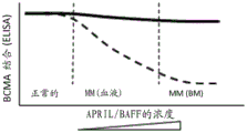
Figure 1 non-ligand blocking/non-competitive anti-BCMA antibody versus ligand blocking/competitive anti-BCMA antibody by ELISA; or superior binding properties of TCB-containing non-ligand blocking/non-competitive anti-BCMA on plate-bound BCMA cells relative to TCB-containing ligand blocking/competitive anti-BCMA. In this figure, increasing concentrations of soluble APRIL or BAFF (i.e., 10, 100, 1000 ng/mL), representing levels found in the blood and bone marrow of multiple myeloma patients, did not alter the binding of non-ligand blocking/non-competitive anti-BCMA antibodies or TCB-containing non-ligand blocking/non-competitive anti-BCMA to plate-bound BCMA (continuous line). In contrast, high concentrations (i.e., 100 ng/mL to 1000 ng/mL) of soluble APRIL or BAFF, which represent the levels found in the blood and bone marrow of multiple myeloma patients, reduced the binding of ligand blocking/competitive anti-BCMA antibodies or TCB-containing ligands blocking/competitive anti-BCMA to plate-bound BCMA (dashed line). The concentration of anti-BCMA antibody or TCB containing anti-BCMA with different properties is preferably a concentration in the range of 0.1 pM-200 nM, since the level of additional circulating APRIL or BAFF is in the range of 1 ng/mL (healthy normal) -100 ng/mL (MM, blood) and beyond (MM, tumor in bone marrow).
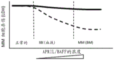
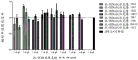
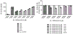
Figure 2. superior potency of BCMA-expressing MM cells mediated by T cell bispecific antibodies containing non-ligand blocking/non-competitive anti-BCMA antibodies versus ligand blocking/competitive anti-BCMA antibodies in redirected T cell cytotoxicity in LDH release assays. In this figure, increasing concentrations of soluble APRIL or BAFF (i.e., 10, 100, 1000 ng/mL), representing levels found in the blood and bone marrow of multiple myeloma patients, did not alter the killing potential of T cell bispecific antibodies containing non-ligand blocking/non-competitive anti-BCMA antibodies specific for BCMA-expressing MM cells (continuous line). In contrast, high concentrations (i.e., 100 ng/mL to 1000 ng/mL) of soluble APRIL or BAFF, representing the levels found in the blood and bone marrow of multiple myeloma patients, reduced the killing potential of a T cell bispecific antibody containing a ligand blocking/competing anti-BCMA antibody specific for BCMA-expressing MM cells (dashed line). The concentration of T cell bispecific containing anti-BCMA antibodies with different properties is preferably a concentration of 0.1 pM-200 nM, since the level of additional circulating APRIL or BAFF is in and beyond the range of 1 ng/mL (healthy normal) -100 ng/mL (MM, blood).
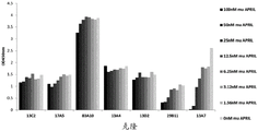
Figure 3 BCMA expression on multiple myeloma cell lines. Increasing concentrations of anti-BCMA antibody (0.3-10 μ g/mL) as detected by flow cytometry increased the median fluorescence intensity after binding to H929 cells.
Figure 4 binding of anti-BCMA antibodies on BCMA-positive multiple myeloma cells. The mean fluorescence intensity of anti-BCMA IgG clones was plotted as a function of anti-BCMA antibody concentration (0.2-40 μ g/mL); (A) clones 13C2, 17a5, 83a10 on H929 cells, (B) clones 13C2, 17a5, 83a10 on MKN45 cells, (C) clones 13a4, 13D2, 14E1, 13a7, 14B11 on H929 cells, and (D) clones 13a4, 13D2, 14E1, 13a7, 14B11 on MKN45 cells.
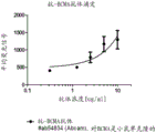
FIG. 5 competition ELISA. The ELISA results of 7 selected anti-BCMA Fab clones (13C2, 17a5, 83a19, 13a4, 13D2, 29B11, 13a7) binding to the saturation concentration of immobilized human BCMA in the presence of the concentration range murine APRIL (1.56-100 nM) are shown. In the non-competitive case, the signal remained constant over the assay variability over the span of the concentration range, and the signal in the presence of murine APRIL was comparable to those from control wells in which murine APRIL was not added. In the case of competition, the concentration dependence of the measurement signal decreases.
FIG. 6 binding competition by FACS. Competition of Δ -APRIL with anti-BCMA antibody as detected by flow cytometry. Relative median fluorescence intensity of Δ -APRIL (FITC signal) used at a concentration of 1000 ng/mL was measured as a function of the concentration of anti-BCMA antibody clones 13a4, 13D2, 14E1, 14B11 on H929 cells (1, 16 and 40 μ g/mL). Setting the median fluorescence intensity of Δ -APRIL after binding in the presence of an isotype control to 1; other signals are normalized to it. Detection of APRIL binding to BCMA positive H929 cells in the presence of anti-BCMA antibody was measured by anti-HA fluorochrome conjugated antibodies.
FIG. 7 binding competition by FACS. Competition of anti-BCMA antibodies with Δ -APRIL as detected by flow cytometry. Relative median fluorescence intensity of anti-BCMA antibody clones 13a4, 13C7, 13D2, 14B11, 17a5, 83a10 on RPMI cells used at a concentration of 40 μ g/mL (alexa. fluor 647 signal) tested in the absence or presence of Δ -APRIL 1000 ng/mL. Setting the median fluorescence intensity after anti-BCMA antibody binding in the absence of Δ -APRIL to 1; the respective other signals of the anti-BCMA antibodies in the presence of Δ -APRIL were normalized against it. Detection of anti-BCMA antibodies that bind to BCMA positive RPMI cells in the presence of Δ -APRIL was measured by anti-human Fc fluorochrome-conjugated antibodies.
Figure 8 competition of anti-BCMA antibody with Δ -APRIL after simultaneous incubation as detected by flow cytometry. The mean fluorescence intensity and relative fluorescence signal of (a) anti-BCMA antibody clone 14B11, 13D2, 13a4, 17a5 and 83a10 at a concentration of 20 μ g/mL in the presence or absence of 2.5 μ g/mL Δ APRIL (alexa. fluor 647 signal) or (B) Δ APRIL at a concentration of 2.5 μ g/mL Δ APRIL (FITC signal) and anti-BCMA antibody clone 83a10 (20 μ g/mL) (alexa. fluor 647 signal) were measured. Detection of anti-BCMA antibody in the presence of Δ -APRIL using a FITC-conjugated anti-human Fc antibody was normalized to the signal of anti-BCMA antibody clones in the absence of Δ -APRIL. Detection of Δ -APRIL in the presence of anti-BCMA antibody clones using alexa. fluor 647-conjugated anti-HA antibodies was normalized to Δ -APRIL signal in the presence of isotype control.
Detailed Description
The term "BCMA, target BCMA, human BCMA" as used herein refers to the human B cell maturation target, also known as BCMA; TR17_ HUMAN, TNFRSF17 (UniProt Q02223), which is a member of the tumor necrosis receptor superfamily that is preferentially expressed in differentiated plasma cells. The extracellular domain of BCMA consists of amino acids 1-54 (or 5-51) according to UniProt. The term "antibody against BCMA, anti-BCMA antibody" as used herein refers to an antibody that specifically binds the extracellular domain of BCMA.
By "specifically binds BCMA" is meant an antibody that is capable of binding the target BCMA with sufficient affinity such that the antibody is useful as a therapeutic agent in targeting BCMA. In some embodiments, the anti-BCMA antibody binds to an unrelated, non-BCMA protein to a degree that is about 10 times less than the binding of the antibody to BCMA, e.g., as measured by Surface Plasmon Resonance (SPR), e.g., Biacore, Enzyme Linked Immunosorbent (ELISA) or flow cytometry (FACS), preferably about 10 times less than>100 times. Preferably, the antibody that binds BCMA has 10-8M or less, preferably 10-8M-10-13M, preferably 10-9M-10-13Dissociation constant (Kd) of M. Preferably, the anti-BCMA antibody binds to an epitope of BCMA that is conserved between BCMA from different species, preferably human and cynomolgus monkey. "bispecific antibody that specifically binds to CD3 and BCMA" refers to the respective definitions of binding to two targets. Antibodies that specifically bind BCMA (or BCMA and CD 3) do not bind other human antigens. Thus, in an ELISA, the OD value of such unrelated targets will be equal to or below the detection limit of the particular assay, preferably>0.3 ng/mL, or equal to or lower than the OD value of BCMA without plate binding or control samples with untransfected HEK293 cells.
The term "APRIL" as used herein refers to recombinant truncated murine APRIL (amino acids 106-241; NP-076006). APRIL can be produced as described in Ryan, 2007 (Mol Cancer Ther; 6 (11): 3009-18).
The term "BAFF" as used herein refers to recombinant truncated HUMAN BAFF (UniProt Q9Y275 (TN13B _ HUMAN), which can be produced as described in Gordon, 2003 (Biochemistry; 42 (20): 5977-.
The binding of anti-BCMA antibodies to human BCMA was analyzed by ELISA using plate-bound BCMA in the presence and absence of APRIL and/or BAFF. For this assay, the amount of plate-bound BCMA and the concentration of 0.1pM-200 nM anti-BCMA antibody, preferably 1.5. mu.g/mL, are used. BCMA antibodies whose BCMA binding is not inhibited according to the invention are anti-BCMA antibodies which "do not inhibit APRIL and/or BAFF binding to human BCMA in an ELISA assay".
The term "NF-. kappa.B" as used herein refers to recombinant NF-. kappa. B P50 (accession No. (P19838). NF-. kappa.B activity was measured by DNA binding ELISA of extracts of NCI-H929MM cells (CRL-9068. the control of HT-truncated-BAFF, 1000 ng/mL heat-treated HT-truncated-BAFF, 1000 ng/mL truncated-BAFF, 0.1pM-200 nM isotype, and NCI-H929MM cells untreated or treated with no anti-BCMA antibody 0.1pM-200 nM were incubated for 20 minutes. NF-. kappa.B activity was determined using a functional ELISA that detects the chemiluminescent signal from P65 bound to the NF-. kappa.B consensus sequence (US 6150090).
Antibodies that do not block APRIL-dependent NF-. kappa.B activation by more than 20% and do not reduce APRIL-dependent NF-. kappa.B activation by more than 20% and do not increase APRIL-dependent NF-. kappa.B activation by more than 20% are considered to "not alter APRIL-dependent NF-. kappa.B activation" by more than 20% compared to APRIL-induced NF-. kappa.B activation without the antibodies of the invention (control); 20% represents the mean standard variation between experiments. Preferably, the antibodies of the invention do not alter APRIL-dependent NF-B activation by more than 15%.
Antibodies that do not block BAFF-dependent NF- κ B activation by more than 20% and do not reduce BAFF-dependent NF- κ B activation by more than 20% and do not increase BAFF-dependent NF- κ B activation by more than 20% are considered to "not alter BAFF-dependent NF- κ B activation" by more than 20% compared to BAFF-induced NF- κ B activation without the antibody of the invention (control); 20% represents the mean standard variation between experiments. Preferably, the antibodies of the invention do not alter BAFF-dependent NF- κ B activation by more than 15%.
An antibody that does not block NF-. kappa.B activation by more than 20% without APRIL and BAFF and does not decrease NF-. kappa.B activation by more than 20% without APRIL and BAFF and does not increase NF-. kappa.B activation by more than 20% without APRIL and BAFF is considered to "not alter NF-. kappa.B activation without APRIL and BAFF" by more than 20% compared to APRIL-induced NF-. kappa.B activation without the antibody of the invention (control); 20% represents the mean standard variation between experiments. Preferably, the antibodies of the invention do not alter NF-. kappa.B activation by more than 15% in the absence of APRIL and BAFF.
Likewise, if the antibody of the invention is used in large excess, it is preferred that the binding of the antibody up to 500 nM or 1000 nM is not reduced by 100 ng/ml APRIL and preferably not reduced by BAFF by more than 20% and does not alter APRIL-dependent NF- κ B activation by more than 20% with and without APRIL, and preferably with and without BAFF.
The term "other target" as used herein denotes preferably CD 3. The terms "first target and second target" mean CD3 as the first target and BCMA as the second target or BCMA as the first target and CD3 as the second target.
The term "CD 3 ɛ or CD 3" as used herein refers to HUMAN CD3 ɛ described under UniProt P07766 (CD3E _ HUMAN). The term "antibody against CD3 ɛ, anti-CD 3 ɛ antibody" refers to an antibody that specifically binds CD3 ɛ. Preferably, the antibody comprises a variable domain VH comprising the heavy chain CDRs of SEQ ID NOs 1, 2 and 3 as heavy chain CDR1, CDR2 and CDR3, respectively, and a variable domain VL comprising the light chain CDRs of SEQ ID NOs 4, 5 and 6 as light chain CDR1, CDR2 and CDR3, respectively. Preferably, the antibody comprises the variable domains of SEQ ID NO:7 (VH) and SEQ ID NO:8 (VL).
The term "antibody" as used herein refers to a monoclonal antibody. An antibody consists of two pairs of "light chains" (LC) and "heavy chains" (HC) (the Light Chain (LC)/heavy chain pair is abbreviated herein as LC/HC). The light and heavy chains of such antibodies are polypeptides consisting of several domains. Each heavy chain comprises a heavy chain variable region (abbreviated herein as HCVR or VH) and a heavy chain constant region. The heavy chain constant region comprises heavy chain constant domains CH1, CH2 and CH3 (antibody classes IgA, IgD and IgG) and optionally heavy chain constant domain CH4 (antibody classes IgE and IgM). Each light chain comprises a light chain variable domain VL and a light chain constant domain CL. The variable domains VH and VL can be further subdivided into regions of hypervariability, termed Complementarity Determining Regions (CDRs), interspersed with regions that are more conserved, termed Framework Regions (FR). Each VH and VL is composed of three CDRs and four FRs, arranged from amino-terminus to carboxy-terminus in the following order: FR1, CDR1, FR2, CDR2, FR3, CDR3, FR 4. The "constant domains" of the heavy and light chains are not directly involved in binding the antibody to the target, but exhibit multiple effector functions.
The term "antibody" includes, for example, mouse antibodies, human antibodies, chimeric antibodies, humanized antibodies, and genetically engineered antibodies (variant or mutant antibodies) so long as their characteristic properties are retained. Especially preferred are human or humanized antibodies, especially as recombinant human or humanized antibodies.
The term "bispecific antibody" as used herein preferably refers to an antibody wherein one of the two pairs of heavy and light chains (HC/LC) specifically binds CD3 and the other pair specifically binds BCMA. The term also refers to other forms of bispecific antibodies of the prior art, preferably to bispecific single chain antibodies.
The term "naked antibody" as used herein refers to an antibody that specifically binds BCMA, which comprises an Fc moiety and is not conjugated to a therapeutic agent, e.g., a cytotoxic agent or a radiolabel. The term "conjugated antibody, drug conjugate" as used herein refers to an antibody that specifically binds BCMA and is conjugated to a therapeutic agent, e.g., a cytotoxic agent or a radiolabel.
The term "bispecific single chain antibody" as used herein refers to a single polypeptide chain preferably comprising two binding domains, one specifically binding to BCMA and the other preferably specifically binding to CD 3. Each binding domain comprises a variable region ("VH region") from a heavy chain of an antibody, wherein the VH region of the first binding domain specifically binds to the CD3 molecule and the VH region of the second binding domain specifically binds to BCMA. The two binding domains are optionally linked to each other by a short polypeptide spacer. Non-limiting examples of polypeptide spacers are Gly-Gly-Gly-Gly-Ser (G-G-G-G-S) and repeats thereof. Each binding domain may additionally comprise a variable region ("VL region") from the light chain of the antibody, the VH and VL regions in each of the first and second binding domains being linked to each other by a polypeptide linker which is sufficiently long to allow the VH and VL regions of the first binding domain and the VH and VL regions of the second binding domain to pair with each other such that together they are capable of specifically binding the respective first and second binding domains (see, e.g., EP 0623679). Bispecific single chain antibodies are also mentioned, for example, in Choi BD et al, Expert Opin biol Ther. 2011 Jul, 11(7), 843-53 and Wolf E. et al, Drug Discov today. 2005 Sep 15, 10(18), 1237-44.
The term "diabodies" as used herein refers to small bivalent and bispecific antibody fragments comprising a heavy (VH) chain variable domain connected to a light chain variable domain (VL) on the same polypeptide chain (VH-VL) by a peptide linker which is too short to allow pairing of the two domains on the same chain (Kipriyanov, int. j. Cancer 77(1998), 763-. This forces pairing with the complementary domains of the other chain and facilitates assembly of the dimeric molecule with two functional antigen binding sites. To construct the bispecific diabodies of the invention, the V domains of the anti-CD 3 antibody and anti-BCMA antibody are fused to produce the two chains VH (CD3) -VL (BCMA), VH (BCMA) -VL (CD 3). Each chain is not capable of binding its own antigen, but pair with the other chain regenerates the functional antigen binding site of the anti-CD 3 and anti-BCMA antibodies. The two scFv molecules are co-expressed with a linker that is too short between the heavy and light chain variable domains to dimerize intramolecularly and self-assemble to form a bispecific molecule with two binding sites at opposite ends. For example, the variable regions encoding the binding domains of BCMA and CD3, respectively, can be PCR amplified from DNA constructs obtained as described above, so that they can be cloned into vectors like pHOG, as described in Kipiriyanov et al, J. Immunol, Methods, 200, 69-77(1997 a). The two scFV constructs were then combined in one expression vector in the desired orientation, thereby shortening the VH-VL linker to prevent the chains from folding back onto themselves. The DNA segment is separated by a STOP codon and a Ribosome Binding Site (RBS). The RBS allows mRNA to be transcribed as a dicistronic message, which is translated by ribosomes into two proteins that interact non-covalently to form a diabody molecule. Diabodies, like other antibody fragments, have the advantage that they can be expressed in functional form and in high yield (up to Ig/l) in bacteria (e. coli) and yeast (Pichia pastoris).
The term "tandem scFVs" as used herein refers to single chain Fv molecules (i.e. molecules formed by the association of immunoglobulin heavy and light chain variable domains, VH and VL, respectively), as described, for example, in WO 03/025018 and WO 03/048209. Such Fv molecules, termed TandAbs @, comprise four antibody variable domains, wherein (i) the first two or the last two of the four variable domains intramolecularly bind to each other within the same chain by forming an antigen that binds to the scFv in positioning VH/VL or VL/VH, (ii) the other two domains intermolecularly bind to the corresponding VH or VL domains of the other chain to form an antigen-binding VH/VL pair. In a preferred embodiment, the monomers of such Fv molecules comprise at least four variable domains, wherein two adjacent domains of one monomer form an antigen-binding VH-VL or VL-VH scFv unit, as suggested in WO 03/025018.
The term "DARPins" as used herein refers to bispecific ankyrin repeat molecules as described, for example, in US 2009082274. These molecules are derived from natural ankyrin proteins, which can be found in the human genome and are one of the most abundant types of binding proteins. DARPin library modules were defined by native ankyrin repeat protein sequences using 229 ankyrin repeats for initial design and another 2200 for subsequent refinement improvement (refinement). The module serves as a building block for a DARPin library. The library modules are similar to human genomic sequences. DARPin consists of 4-6 modules. Since each module is approximately 3.5 kDa, the average DARPin size is 16-21 kDa. Selection of binders was accomplished by ribosome display, which is completely cell-free and is described in He M and TaussigMJ., Biochem Soc Trans, 2007, Nov;35(Pt 5): 962-5.
The term "T cell bispecific adapter" is a fusion protein consisting of the amino acid sequences of two single chain variable fragments (scFvs) of different antibodies, or different genes on a single peptide chain of about 55 kilodaltons. One of the scFvs binds T cells via the CD3 receptor, while the other binds BCMA.
Five types of mammalian antibody heavy chains with greek letter designations: α, ɛ, γ and μ (Janeway CA, Jr et al (2001). immunobiology. 5 th edition, Garland Publishing). The type of heavy chain present defines the class of antibody; these chains are found in IgA, IgD, IgE, IgG and IgM, respectively (Rhoades RA, Pflanzer RG (2002); Human Physiology, 4 th edition, Thomson Learning). The different heavy chains differ in size and composition; alpha and gamma contain approximately 450 amino acids, while mu and ɛ have approximately 550 amino acids.
Each heavy chain has two regions, a constant region and a variable region. The constant regions are the same in all antibodies of the same isotype, but differ in antibodies of different isotypes. Heavy chains gamma, alpha and have a constant region consisting of three constant domains, CH1, CH2 and CH3 (in line), and a hinge region for additional flexibility (Woof J, Burton D Nat rev immunol 4 (2004) 89-99); heavy chains μ and ɛ have constant regions consisting of four constant domains, CH1, CH2, CH3 and CH4 (Janeway CA, Jr et al (2001). immunobiology. 5 th edition, Garland Publishing). The variable region of the heavy chain differs among antibodies produced by different B cells, but is the same for all antibodies produced by a single B cell or B cell clone. The variable region of each heavy chain is approximately 110 amino acids long and consists of a single antibody domain.
In mammals, there are only two types of light chains, which are called λ (λ) and κ (κ). The light chain has two contiguous domains: one constant domain CL and one variable domain VL. The approximate length of the light chain is 211-217 amino acids. Preferably, the light chain is a kappa (kappa) light chain, and the constant domain CL is preferably derived from a kappa (K) light chain (constant domain CK).
The term "monoclonal antibody" or "monoclonal antibody composition" as used herein refers to an antibody molecule preparation of a single amino acid composition.
The "antibody" of the present invention may be of any class (e.g., IgA, IgD, IgE, IgG and IgM, preferably IgG or IgE), or subclass (e.g., IgG1, IgG2, IgG3, IgG4, IgA1 and IgA2, preferably IgG 1), whereby two antibodies derived from the bivalent bispecific antibody of the present invention have Fc portions of the same subclass (e.g., IgG1, IgG4, etc., preferably IgG 1), preferably Fc portions of the same allotype (e.g., Caucasian).
The "Fc portion of an antibody" is a term well known to those skilled in the art and defined based on papain cleavage of antibodies. The antibodies of the invention contain an Fc portion, preferably from human origin and preferably all other portions of human constant regions. The Fc portion of the antibody is directly involved in complement activation, C1q binding, C3 activation, and Fc receptor binding. Although the effect of the antibody on the complement system depends on certain conditions, binding to C1q is caused by a defined binding site in the Fc portion. Such binding sites are known in the art and are described, for example, by Lukas, TJ., et al, J. Immunol 127 (1981) 2555-.
Such binding sites are for example L234, L235, D270, N297, E318, K320, K322, P331 and P329 (numbering according to EU index of Kabat, see below). Antibodies of subclasses IgG1, IgG2, and IgG3 generally show complement activation, C1q binding, and C3 activation, whereas IgG4 does not activate the complement system, does not bind C1q, and does not activate C3. Preferably, the Fc portion is a human Fc portion.
Preferably, the antibody of the invention comprises an Fc variant of a wild-type human IgG Fc region comprising an amino acid substitution and at least one other amino acid substitution at position Pro329, wherein the residues are numbered according to the EU index of Kabat, and wherein said antibody exhibits reduced affinity for human fcyriiia and/or fcyriia and/or fcyri compared to an antibody comprising a wild-type IgG Fc region, and wherein the antibody induces ADCC that is reduced to at least 20% of the ADCC induced by an antibody comprising a wild-type human IgGFc region. In a particular embodiment, Pro329 of the wild type human Fc region in the antibodies of the invention is replaced by glycine or arginine or an amino acid residue sufficiently large to disrupt the proline sandwich within the Fc/Fc γ receptor interface formed between proline 329 of Fc and tryptophan residues Trp 87 and Tip 110 of Fc γ RIII (Sondermann et al: Nature 406, 267-273 (20 July 2000)). In other aspects of the invention, the at least one additional amino acid substitution in the Fc variant is S228P, E233P, L234A, L235A, L235E, N297A, N297D, or P331S, and in yet another embodiment, the at least one additional amino acid substitution is L234A and L235A of the human IgGl Fc region or S228P and L235E of the human IgG4 Fc region. Such Fc variants are described in detail in WO 2012130831.
As used herein, "effector function" refers to a biochemical event resulting from the interaction of an antibody Fc region with an Fc receptor or ligand. Effector functions include, but are not limited to, ADCC, ADCP and CDC. As used herein, "effector cell" means a cell of the immune system that expresses one or more Fc receptors and mediates one or more effector functions. Effector cells include, but are not limited to, monocytes, macrophages, neutrophils, dendritic cells, eosinophils, mast cells, platelets, B cells, large granular lymphocytes, langerhans cells, Natural Killer (NK) cells, and gamma T cells, and can be from any organism, including, but not limited to, humans, mice, rats, rabbits, and monkeys. By "library" herein is meant a set of Fc variants in any form, including but not limited to a series of nucleic acid or amino acid sequences, a series of nucleic acid or amino acid substitutions at variable positions, a physical library comprising nucleic acids encoding the library sequences, or a physical library comprising Fc variant proteins in purified or unpurified form.
As used herein, "Fc γ receptor" or "Fc γ R" refers to any member of a family of proteins that bind the Fc region of IgG antibodies and are substantially encoded by Fc γ R genes. In humans, this family includes, but is not limited to, Fc γ RI (CD64), including isoforms Fc γ RIa, Fc γ RIb, and Fc γ RIc; fc γ RII (CD32), including isoforms Fc γ Rlla (including allotype H131 and R131), Fc γ Rllb (including Fc γ Rllb-1 and Fc γ Rllb-2), and Fc γ Rllc; and Fc γ RIII (CD16) including isoforms Fc γ Rllla (including allotypes V158 and F158) and Fc γ rllb (including allotype Fc γ rllb-NA 1 and Fc γ rllb-NA 2) (Jefferis et al, 2002, Immunol Lett 82:57-65), as well as any undiscovered human Fc γ Rs or Fc γ R isoforms or allotypes. The Fc γ R may be from any organism, including but not limited to human, mouse, rat, rabbit, and monkey. Mouse Fc γ Rs include, but are not limited to, Fc γ RI (CD64), Fc γ RII (CD32), Fc γ RIII (CD16), and Fc γ RIII-2 (CD16-2), as well as any undiscovered mouse Fc γ Rs or Fc γ R isoforms or allotypes.
As used herein, "Fc variant with increased effector function" means an Fc sequence that differs from a parent Fc sequence due to at least one amino acid modification or is associated with other modifications, such as an alteration in glycosylation at, for example, Asn279 (which increases effector function). For example, in Duncan et al, 1988, Nature 332:563-564, Lund et al, 1991, Jimmunol147:2657-2662, Lund et al, 1992, Mol Immunol 29:53-59, Alegre et al, 1994, Transplantation 57:1537-1543, Hutchins et al, 1995, Proc Natl Acad Sci U A92: 11980-11984, Jefferis et al, 1995// 77 muno/Lett 44:111-117, Lund et al, 1995, FaseeJ 9:115-119, Jefferis et al, 1996, Immunol Lett 54:101-104, Lund et al, 1996, Jimmunol 157: 4963-4969, Armour et al, 1999, Eur J29: 2613-2624, Idusie et al, 412000: 41164, Cell 412000: 4116-412000, 2001, J Immunol 166:2571-2575, Shields et al, 2001, J Biol Chem 276: 6591-6604, Jefferis et al, 2002, Immunol Lett 82:57-65, Presta et al, 2002, Biochem Soc Trans 30:487-490, US5624821, US5885573, US6194551, WO200042072, WO 199958572. According to the invention, such Fc modifications also include engineered glycoforms of the Fc portion. As used herein, "engineered glycoform" refers to a carbohydrate composition covalently linked to an Fc polypeptide, wherein the carbohydrate composition is chemically distinct from a parent Fc polypeptide. The engineered glycoforms can be produced by any method, e.g., by using engineered or variant expression strains, by co-expression with one or more enzymes, e.g., D1-4-N-acetylglucosaminyltransferase iii (gntiii), by expressing an Fc polypeptide in multiple organisms or cell lines derived from multiple organisms, or by modifying carbohydrates after the Fc polypeptide has been expressed. Methods for producing engineered glycoforms are known in the art and are mentioned in Umana et al, 1999, Nat Biotechnology 17:176-180, Davies et al, 2001, Biotechnology Bioeng 74:288-294, Shiels et al, 2002, J BiolChem 277:26733-26740, Shinkawa et al, 2003, J Biol Chem 278:3466-3473) US6602684, WO200061739, WO200129246, WO200231140, WO200230954, Potelligent technology (Biowa, Inc., Princeton, N.J.), GlycoMAb glycosylation engineering (GLYCARTBiotechnology AG, Zurich, Switzerland)). Engineered glycoforms typically involve a carbohydrate or oligosaccharide composition that is different from the parent Fc polypeptide.
Antibodies of the invention comprising Fc variants with increased effector function exhibit high binding affinity to Fc γ receptor III (Fc γ RIII, CD16 a). High binding affinity to Fc γ RIII indicates at least a 10-fold increase in binding for CD16a/F158, in relation to the parent antibody (95% fucosylation) as a reference for expression in CHO host cells, such as CHO DG44 or CHO K1 cells, and/or at least a 20-fold increase in binding for CD16a/V158, in relation to the parent antibody measured at an antibody concentration of 100nM using immobilized CD16a by Surface Plasmon Resonance (SPR). Fc γ RIII binding can be increased by prior art methods, e.g. by modifying the amino acid sequence of the Fc portion or glycosylation of the Fc portion of the antibody (see e.g. EP 2235061). Mori, K et al, Cytotechnology 55 (2007)109 and Satoh M et al, Expert Opin Biol ther. 6 (2006) 1161-1173 involved the FUT8 (α -1, 6-fucosyltransferase) gene knockout CHO line for the production of afucosylated (afucosylated) antibodies.
The term "chimeric antibody" refers to an antibody comprising a variable region, i.e., a binding region, from one source or species and at least a portion of a constant region from a different source or species, typically prepared by recombinant DNA techniques. Chimeric antibodies comprising murine variable regions and human constant regions are preferred. Other preferred forms of "chimeric antibodies" encompassed by the invention are those in which the constant region has been modified or altered compared to the constant region of the original antibody to produce the properties of the invention, particularly with respect to C1q binding and/or Fc receptor (FcR) binding. Such chimeric antibodies are also referred to as "type-switch antibodies". Chimeric antibodies are the product of an expressed immunoglobulin gene comprising a DNA segment encoding an immunoglobulin variable region and a DNA segment encoding an immunoglobulin constant region. Methods for producing chimeric antibodies include conventional recombinant DNA and gene transfection techniques well known in the art. See, e.g., Morrison, S.L., et al, Proc.Natl.Acad.Sci.USA 81 (1984) 6851-6855; U.S. patent nos. 5,202,238 and 5,204,244.
The term "humanized antibody" refers to an antibody in which the framework regions or "complementarity determining regions" (CDRs) have been modified to comprise CDRs of an immunoglobulin of different specificity compared to the CDRs of a parent immunoglobulin. In a preferred embodiment, murine CDRs are grafted into the framework regions of a human antibody to make a "humanized antibody". See, for example, Riechmann, L., et al, Nature 332 (1988) 323-327 and Neuberger, M.S., et al, Nature 314 (1985) 268-270. Other forms of "humanized antibodies" encompassed by the invention are those in which the constant regions have been additionally modified or altered compared to the constant regions of the original antibody to produce the properties of the invention, particularly with respect to C1q binding and/or Fc receptor (FcR) binding.
As used herein, the term "human antibody" is intended to include antibodies having variable and constant regions derived from human germline immunoglobulin sequences. Human antibodies are well known in the art (van Dijk, m.a., and van de Winkel, j.g., curr. opin. chem. biol.5 (2001) 368-374). Human antibodies can also be produced in transgenic animals (e.g., mice) that are capable of producing all or selected portions of human antibodies (selection) after immunization in the absence of endogenous immunoglobulin production. Transfer of human germline immunoglobulin gene arrays in such germline mutant mice will result in the production of human antibodies (see, e.g., Jakobovits, A., et al, Proc. Natl. Acad. Sci. USA 90 (1993) 2551-2555; Jakobovits, A., et al, Nature 362 (1993) 255-258; Bruggemann, M., et al, Yeast immune. 7 (1993) 33-40). Human antibodies can also be generated in phage display libraries (Hoogenboom, H.R., and Winter, G., J. MoI. biol. 227 (1992)) 381-. The techniques of Cole et al and Boerner et al can also be used to prepare human Monoclonal Antibodies (Cole et al, Monoclonal Antibodies and Cancer Therapy, Alan R. Liss, p.77 (1985); and Boerner, P.et al, J. Immunol.147 (1991) 86-95). As already mentioned for the chimeric and humanized antibodies of the invention, the term "human antibody" as used herein also comprises antibodies which are modified within the constant region to produce the properties of the invention, in particular with respect to C1q binding and/or FcR binding, for example by "type conversion", i.e. altering or mutating the Fc part (e.g. from IgG1 to IgG4 and/or IgG1/IgG4 mutations).
As used herein, the term "recombinant human antibody" is intended to include all human antibodies prepared, expressed, produced or isolated by recombinant methods, such as antibodies isolated from a host cell, such as an NSO or CHO cell or an animal (e.g., mouse) that is transgenic for human immunoglobulin genes, or antibodies expressed using a recombinant expression vector transfected into a host cell. Such recombinant human antibodies have variable and constant regions in rearranged form. The recombinant human antibodies of the invention have undergone somatic hypermutation in vivo. Thus, the amino acid sequences of the VH and VL regions of a recombinant antibody are sequences that, although derived from and related to human germline VH and VL sequences, may not naturally occur in vivo in human antibody germline repertoires.
As used herein, "variable domain" (light chain variable domain (VL), variable region of a heavy chain (VH)) means each pair of light and heavy chains directly involved in binding to an antibody of the invention. The variable human light and heavy chain domains have the same general structure and each domain comprises four Framework (FR) regions, the sequences of which are largely conserved, connected by three "hypervariable regions" (or complementarity determining regions, CDRs). The framework regions adopt a β -sheet conformation and the CDRs may form a loop connecting the β -sheet structures. The CDRs in each chain retain their three-dimensional structure through the framework regions and form binding sites with the CDRs from the other chain. The antibody heavy and light chain CDR3 regions play a particularly important role in the binding specificity/affinity of the antibodies of the invention and thus provide another object of the invention.
As used herein, the term "hypervariable region" or "target-binding portion of an antibody" refers to the amino acid residues of an antibody which are responsible for target binding. The hypervariable regions comprise amino acid residues from the "complementarity determining regions" or "CDRs". The "framework" or "FR" regions are those variable domain regions other than the hypervariable region residues defined herein. Thus, the light and heavy chains of an antibody comprise, from N-terminus to C-terminus, the domains FR1, CDR1, FR2, CDR2, FR3, CDR3 and FR 4. The CDRs on each chain are separated by such framework amino acids. In particular, CDR3 of the heavy chain is the region most contributing to target binding. CDR and FR regions were determined according to the standard definition of Kabat et al, Sequences of proteins of Immunological Interest, 5 th edition, Public Health Service, national institutes of Health, Bethesda, MD (1991).
The constant heavy chain domain CH1 with which the heavy chain constant domain CH3 is replaced can be of any Ig class (e.g., IgA, IgD, IgE, IgG, and IgM), or subclass (e.g., IgG1, IgG2, IgG3, IgG4, IgA1, and IgA 2).
The constant light chain domain CL, with which the heavy chain domain CH3 is replaced, may be of the λ (λ) or κ (κ) type, preferably of the κ (κ) type.
The terms "target" or "target molecule" as used herein are used interchangeably and refer to human BCMA. With respect to bispecific antibodies, the term refers to BCMA and a second target. Preferably, in reference to bispecific antibodies, the term refers to BCMA and CD 3.
The term "epitope" includes any polypeptide determinant capable of specifically binding to an antibody. In certain embodiments, epitope determinants include chemically active surface groups of the molecule, such as amino acids, sugar side chains, phosphoryl, or sulfonyl (sulfonyl), and in certain embodiments, may have specific three-dimensional structural features, and/or specific charge characteristics. An epitope is the region of the target to which an antibody binds.
Generally, there are two vectors encoding the light and heavy chains of the antibodies of the invention. For bispecific antibodies, there are two vectors encoding the light and heavy chains of the antibody that specifically bind a first target, and two other vectors encoding the light and heavy chains of the antibody that specifically bind a second target. One of the two vectors encodes a respective light chain, and the other of the two vectors encodes a respective heavy chain. However, in an alternative method for producing the antibodies of the invention, only one first vector encoding the light and heavy chains of an antibody that specifically binds to a first target and only one second vector encoding the light and heavy chains of an antibody that specifically binds to a second target may be used to transform a host cell.
The term "nucleic acid or nucleic acid molecule" as used herein is intended to include both DNA molecules and RNA molecules. The nucleic acid molecule may be single-stranded or double-stranded, but is preferably double-stranded DNA.
As used herein, the expressions "cell," "cell line," and "cell culture" are used interchangeably, and all of these designations include progeny. Thus, the words "transformant" and "transformed cell" include both primary subject cells and cultures derived therefrom without regard to the number of transformations. It is also understood that all progeny may not be identical in DNA content due to deliberate or inadvertent mutations. Variant progeny that have the same function or biological activity as screened for in the originally transformed cell are included. Where different designations are desired, will be apparent from the context.
The term "transformation" as used herein refers to the process of transferring a vector/nucleic acid into a host cell. If cells without a strong cell wall barrier are used as host cells, transfection is carried out, for example, by the calcium phosphate precipitation method described by Graham and Van der Eh, Virology 52(1978) 546 ff. However, other methods for introducing DNA into cells may also be used, for example by nuclear injection or by protoplast fusion. If prokaryotic cells or cells containing a parenchymal cell wall structure are used, for example, one method of transfection is calcium treatment with calcium chloride as described by Cohen SN et al, PNAS 1972, 69 (8): 2110-2114.
The use of transforming recombinant antibodies is well known in the art and is described, for example, in Makrides, S.C, Protein Expr. purify. 17 (1999) 183-.
As used herein, "expression" refers to the process of transcribing a nucleic acid into mRNA and/or the subsequent translation of the transcribed mRNA (also referred to as transcript) into a peptide, polypeptide or protein. The transcripts and the encoded polypeptides are collectively referred to as gene products. If the polynucleotide is from genomic DNA, expression in a eukaryotic cell may include splicing of the mRNA.
A "vector" is a nucleic acid molecule, particularly autonomously replicating, which transfers an inserted nucleic acid molecule into and/or between host cells. The term includes vectors that are used primarily for the insertion of DNA or RNA into a cell (e.g., chromosomal integration), replication vectors that are used primarily for the replication of DNA or RNA, and expression vectors that are used for the transcription and/or translation of DNA or RNA. Also included are vectors providing more than one of said functions.
An "expression vector" is a polynucleotide that, when introduced into an appropriate host cell, can be transcribed and translated into a polypeptide. An "expression system" is generally meant to include a suitable host cell that can act on an expression vector to produce a desired expression product.
Preferably, the antibodies of the invention are produced by recombinant means. Such methods are well known in the art and include expression of proteins in prokaryotic and eukaryotic cells, followed by isolation and purification of the antibody polypeptide generally to a pharmaceutically acceptable purity. For protein expression, nucleic acids encoding the light and heavy chains or fragments thereof are inserted into the expression vector by standard methods. Expression is carried out in suitable prokaryotic or eukaryotic host cells, like CHO cells, NSO cells, SP2/0 cells, HEK293 cells, COS cells, yeast or E.coli cells, and the antibody is recovered from said cells (supernatant or cells after lysis). The bispecific antibody can be present in intact cells, in cell lysates, or in partially purified or substantially pure form. Purification is carried out by standard techniques, including alkali/SDS treatment, column chromatography, and other techniques well known in the art, to eliminate other cellular components or other contaminants, such as other cellular nucleic acids or proteins. See Ausubel, F., et al, eds, Current protocols in Molecular Biology, Greene Publishing and Wiley Interscience, New York (1987).
From, for example, Barnes, L.M., et al, Cytotechnology 32 (2000) 109-; and Barnes, L.M., et al, Biotech. Bioeng.73 (2001) 261-270 describe expression in NS0 cells. Transient expression is described, for example, by Durocher, Y., et al, Nucl. acids. Res.30 (2002) E9. Cloning of variable domains is described by Orlandi, R.et al, Proc.Natl.Acad.Sci.USA 86 (1989) 3833-3837, Carter, P.et al, Proc.Natl.Acad.Sci.USA 89 (1992) 4285-4289, and Norderhaug, L.et al, J.Immunol.methods 204 (1997) 77-87. Preferred transient expression systems (HEK293) are described by Schlaeger, E.J., and Christensen, K., in Cytotechnology 30 (1999) 71-83 and Schlaeger, E.J., in J.Immunol.methods 194 (1996) 191-199.
Control sequences suitable for use in prokaryotes include, for example, a promoter, optionally an operator sequence, and a ribosome binding site. Eukaryotic cells are known to utilize promoters, enhancers, and polyadenylation signals.
The antibodies are suitably separated from the culture medium by conventional immunoglobulin purification methods, such as, for example, protein a-sepharose, hydroxylapatite chromatography, gel electrophoresis, dialysis or affinity chromatography. DNA or RNA encoding the monoclonal antibody is readily isolated and sequenced using conventional methods. Hybridoma cells can serve as a source of such DNA and RNA. Once isolated, the DNA may be inserted into an expression vector which is then transfected into a host cell that does not otherwise produce immunoglobulins, such as HEK293 cells, CHO cells, or myeloma cells, to obtain synthesis of recombinant monoclonal antibodies in the host cell.
Amino acid sequence variants (or mutants) of the antibodies of the invention are prepared by introducing appropriate nucleotide changes into the antibody DNA, or by nucleotide synthesis. However, such modifications may be made only within a very limited range, for example as described above. For example, the modifications do not alter the above-described antibody characteristics such as IgG isotype and target binding, but may improve the yield of recombinant production, protein stability, or facilitate purification.
T Cell Bispecific (TCB) conjugates have very high concentration/tumor-cell-receptor-occupancy-dependent potency in cell killing (e.g., sub-picomolar or low picomolar range EC in vitro cell killing assays50(ii) a Dreier et al IntJ Cancer 2002), giving T-cell bispecific binders (TCB) at much lower doses than traditional monospecific antibodies. For example, at 5-15. mu.g/m2Continuous intravenous dose per day (i.e. only 0.35-0.105 mg/m/week) giving blinatumomab (CD19xCD3), for treating acute lymphocytic leukemia or with 60 μ g/m/week2The serum concentrations at these doses were in the range of 0.5-4 ng/ml given for treatment of non-Hodgkin's lymphoma/day (Klinger et al, Blood 2012; Topp et al, J Clin Oncol 2011; Goebeler et al Ann Oncol 2011). Because low doses of TCB can exert high efficiency in patients, it is envisaged that subcutaneous administration in a clinical setting is possible and preferred (preferably 0.25-2.5 mg/m) for the antibodies of the invention 2Dose range per week). Even at these low concentrations/doses/receptor occupancy, TCB can cause a number of adverse events (Klinger et al, Blood 2012). Thus, control of tumor cell occupancy/coverage is crucial. In patients with high and variable levels of serum APRIL and BAFF (e.g., multiple myeloma patients, Moreaux et al 2004; Blood 103(8): 3148-. However, by using the antibodies of the invention, tumor cell occupancy is individually efficient/safe, and it may not be necessary to increase the dose of the antibodies of the invention, as the antibodies may not be affected by APRIL/BAFF ligand competition. Another advantage of the antibodies of the invention is based on the inclusion of an Fc moiety that increases the clearing half-life of 12 days and allows for at least one or two/week administrations compared to TCBs without an Fc moiety (e.g., blinatumomab, which requires intravenous and continuous administration using a pump carried by the patient).
TABLE 1
The following examples, sequence listing and figures are provided to aid the understanding of the present invention, the true scope of which is set forth in the appended claims. It is understood that modifications may be made in the methods shown without departing from the spirit of the invention.
Materials and general methods
General information on the nucleotide Sequences of human immunoglobulin light and heavy chains is given in Kabat, E.A., et al, Sequences of Proteins of Immunological Interest, 5 th edition, Public Health Service, National Institutes of Health, Bethesda, MD (1991). Amino acids of an antibody chain are numbered according to EU numbering and referenced (Edelman, G.M., et al, Proc. Natl. Acad. Sci. USA 63 (1969) 78-85; Kabat, E.A., et al, Sequences of Proteins of Immunological Interest, 5 th edition, public Health Service, National Institutes of Health, Bethesda, Md. (1991)).
Recombinant DNA technology
Standard methods are described in Sambrook et al, Molecular cloning: A Laboratory Manual, Cold Spring Harbor Laboratory Press, Cold Spring Harbor, New York, 1989 for manipulating DNA. Molecular biological reagents were used according to the manufacturer's instructions. In Kabat, E.A. et al, (1991) Sequences of proteins of Immunological Interest, 5 th edition, NIH Publication number 91-3242.Gene synthesisGeneral information on the nucleotide sequences of the human immunoglobulin light and heavy chains is given in (1).
The desired gene segment is prepared from oligonucleotides made by chemical synthesis. The 600-1800 bp long gene segment flanked by single restriction endonuclease cleavage sites was assembled by oligonucleotide annealing and ligation, including PCR amplification, and subsequently cloned into pPCRScript (Stratagene) based on the pGA4 cloning vector via the indicated restriction sites, e.g., Kpnl/Sad or Ascl/Pacl. The DNA sequence of the subcloned gene fragments was confirmed by DNA sequencing. The gene synthesis fragments were ordered according to the specifications specified by Geneart (Regensburg, Germany).
DNA sequencing
The DNA sequence was determined by double-strand sequencing. DNA and protein sequence analysis and sequence data management
GCG's (Genetics Computer Group, Madison, Wisconsin) software package version 10.2 and Infmax's Vector NTl Advance suite version 8.0 are used for sequence generation, mapping, analysis, annotation and illustration.
Expression vector
a) For expression of the antibody variants of the expression plasmids for transient expression (e.g.in HEK293 EBNA or HEK 293-F), cells based on cDNA organization with the CMV-Intron A promoter or on genomic organization with the CMV promoter are used.
In addition to the antibody expression cassette, the vector also contains:
-an origin of replication allowing the plasmid to replicate in E.coli, and a beta-lactamase gene conferring ampicillin resistance in E.coli.
The transcription unit of the antibody gene is composed of the following elements:
the only restriction site at the 5' end-the immediate early enhancer and promoter from human cytomegalovirus,
in the case of cDNA organization, followed by an intron A sequence,
-the 5' -untranslated region of a human antibody gene,
an immunoglobulin heavy chain signal sequence,
human antibody chains (wild type or with domain swapping) as cDNA or as genomic organization with immunoglobulin exon-intron organization
-a 3' untranslated region having a polyadenylation sequence, and
-a unique restriction site at the 3' end.
Fusion genes comprising the antibody chains as described below are generated by PCR and/or gene synthesis and assembled using known recombinant methods and techniques, for example by ligating corresponding nucleic acid segments using unique restriction sites in each vector. The subcloned nucleic acid sequences were verified by DNA sequencing. For transient transfection, larger quantities of plasmid were prepared by plasmid preparation from transformed E.coli cultures (Nucleobond AX, Macherey-Nagel).
b) Production of antibodies and antigen expression vectors
The variable regions of the heavy and light chain DNA sequences were subcloned in frame with either the human IgG1 constant heavy chain or the human IgG1 constant light chain pre-inserted into each recipient mammalian expression vector. Antibody expression is driven by a chimeric MPSV promoter comprising a CMV enhancer and MPSV promoter followed by a 5' UTR, intron, and kappa MAR element. Transcription is terminated by a synthetic polyA signal sequence at the 3' end of the CDS. All vectors carry a 5' -terminal DNA sequence encoding a leader peptide that targets the secretion of proteins in eukaryotic cells. In addition, each vector contains the EBV OriP sequence for episomal plasmid replication in EBV EBNA expressing cells.
Antigens that have been used for phage display selection activities and to characterize the binding properties of selected antibodies are expressed from mammalian antigen expression vectors having pre-inserted DNA sequences encoding C-terminal tags. Avi tags have been used for in vivo or in vitro biotinylation of individual antigens. For purification and homodimerization or heterodimerization of antigens, human IgG1 Fc wt or Fc protrusion was fused to the C-terminus of the antigen expression cassette. Antigen expression was driven by a chimeric MPSV promoter comprising a CMV enhancer and MPSV promoter followed by a 5' UTR, intron, and kappa MAR element. Transcription is terminated by a synthetic polyA signal sequence at the 3' end of the CDS. All vectors carry a 5' -terminal DNA sequence encoding a leader peptide that targets the secretion of proteins in eukaryotic cells. In addition, each vector contains the EBV OriP sequence for episomal plasmid replication in EBV EBNA expressing cells.
Cell culture technique
Standard Cell culture techniques are used as described in Current Protocols in Cell Biology (2000), Bonifacino, J.S., Dasso, M., Harford, J.B., Lippincott-Schwartz, J.and Yamada, K.M. (eds.), John Wiley & Sons, Inc.
Transient expression in HEK293 cells
Bispecific antibodies were expressed by transient co-transfection of each expression plasmid in adherently grown HEK293-EBNA or HEK293-F cells grown in suspension as described below.
a) Transient transfection in the HEK293-EBNA System
Bispecific antibodies were expressed by transient co-transfection of each expression plasmid (e.g., encoding heavy and modified heavy chains, and the corresponding light and modified light chains) in adherently growing HEK293-EBNA cells (human embryonic kidney cell line 293, expressing EB virus nuclear targets; American type culture collection (American type culture collection) accession No. ATCC # CRL-10852, lot. 959218) cultured in DMEM (Dulbecco's modified Eagle's medium, Gibco) supplemented with 10% Ultra Low IgG FCS (fetal calf serum, Gibco), 2 mM L-glutamine (Gibco) and 250 μ g/ml geneticin (Gibco). For Transfection, a FuGENE ™ 6 transfer Reagent (Roche molecular biochemicals) was used at a ratio of FuGENE ™ Reagent (. mu.l) to DNA (. mu.g) 4:1 (range 3:1-6: 1).
Proteins were expressed from each plasmid using a molar ratio of 1:1 (equimolar) (ranging from 1:2 to 2: 1) of the plasmids encoding the (modified and wild type) light and heavy chains, respectively. Cells were fed on day 3 with L-glutamine ad 4 mM, glucose [ Sigma ] and NAA [ Gibco ]. Cell culture supernatants containing bispecific antibody were collected by centrifugation from day 5 to day 11 post transfection and stored at-200C. General information on recombinant expression of human immunoglobulins in, for example, HEK293 cells is given in Meissner, P.et al, Biotechnol. Bioeng.75 (2001) 197-203.
b) Transient transfection in the HEK293-F System
Bispecific antibodies were generated by transient transfection of each plasmid (e.g., encoding the heavy chain and modified heavy chain, and the corresponding light chain and modified light chain) using the HEK293-F system (Invitrogen) according to the manufacturer's instructions. Briefly, HEK293-F cells (Invitrogen) grown in suspension in serum-free FreeStyle 293 expression medium (Invitrogen) in shake flasks or stirred fermentors were transfected with a mixture of four expression plasmids and 293fectin or fectin (Invitrogen). For 2L shake flasks (Corning), HEK293-F cells were plated at 1.0X 10⁶cells/mL were seeded at a density of 600 mL and at 120 rpm, 8% CO2And (4) incubating. The next day, utilize A)20 mL of Opti-MEM (Invitrogen) mixed with 600. mu.g of total plasmid DNA (1. mu.g/mL) encoding an equimolar ratio of heavy or modified heavy chain and corresponding light chain, respectively, and B) 20 mL of Opti-MEM + 1.2 mL of 293fectin or fectin (2. mu.l/mL) at approximately 42 mL, at approximately 1.5X 10⁶Cells were transfected at a cell density of cells/mL. Depending on the glucose consumption, a glucose solution is added during the fermentation. After 5-10 days the supernatant containing the secreted antibody is collected and the antibody is purified directly from the supernatant or the supernatant is frozen and stored.
Protein assay
The Protein concentration of purified proteins and derivatives was determined by measuring the Optical Density (OD) at 280 nm using the molar extinction coefficient calculated on the basis of the amino acid sequence according to Pace et al, Protein Science, 1995, 4, 2411-1423.
Determination of antibody concentration in supernatant
The concentration of antibodies and derivatives in cell culture supernatants was assessed by immunoprecipitation using protein a sepharose-beads (Roche). 60 μ L of protein A agarose beads were washed three times in TBS-NP40 (50 mM Tris, pH 7.5, 150 mM NaCl, 1% Nonidet-P40). Subsequently, 1-15 mL of cell culture supernatant was applied to protein a agarose beads pre-equilibrated in TBS-NP 40. After 1 hour incubation at room temperature, the beads were washed once on an Ultrafree-MC-filtration column (Amicon) with 0.5 mL TBS-NP40, twice with 0.5 mL 2XPBS, Roche and four times briefly with 0.5 mL 100 mM sodium citrate pH 5,0 bound antibody was eluted by adding 35. mu.l NuPAGE LDS sample buffer (Invitrogen) half of the samples were combined or kept unreduced with NuPAGE sample reductant, respectively, thus, 5-30 μ l were applied to 4-12% NuPAGE Bis-Tris SDS-PAGE (Invitrogen) (MOPS buffer for non-reducing SDS-PAGE and MES buffer with NuPAGE antioxidant running buffer additive (Invitrogen) for reducing SDS-PAGE) and stained with Coomassie blue.
The concentration of antibodies and derivatives in the cell culture supernatant was quantitatively measured by affinity HPLC chromatography. In brief, will contain bindingApplication of cell culture supernatant of protein A antibodies and derivatives to 200 mM KH2PO4100 mM sodium citrate, pH 7.4 in an Applied Biosystems Poros A/20 column and eluted from the matrix on an Agilent HPLC 1100 system using 200 mM NaCl, 100 mM citric acid, pH 2, 5. Eluted protein was quantified by integration of UV absorbance and peak area. Purified standard IgG1 antibody served as the standard.
Alternatively, the concentration of antibodies and derivatives in the cell culture supernatant was measured by sandwich-IgG-ELISA. Briefly, StreptaWell High Bind streptavidin A-96 well microtiter plates (Roche) were coated with 0.1. mu.L/well of 100. mu.L/well biotinylated anti-human IgG capture molecule F (ab')2< h-Fc γ > BI (Dianova) for 1 hour at room temperature or overnight at 4 ℃ followed by three washes with 200. mu.L/well PBS, 0.05% Tween (PBST, Sigma). To the wells 100 μ L/well of cell culture supernatant containing each antibody in a dilution series in pbs (sigma) was added and incubated for 1-2 hours at room temperature on a microtiter plate shaker. The wells were washed three times with 200 μ L/well PBST and bound antibody was detected on a microtiter plate shaker at room temperature for 1-2 hours using 100 μ L F (ab')2< hFc γ > POD (Dianova) at 0.1 μ g/mL as the detection antibody. Unbound detection antibody was washed off three times with 200 μ L/well PBST and bound detection antibody was detected by adding 100 μ L ABTS/well. The absorbance measurements were carried out on a Tecan FluorSpectrometry instrument at a measurement wavelength of 405 nm (reference wavelength 492 nm).
Protein purification
Proteins were purified from filtered cell culture supernatant according to standard protocols. Briefly, antibodies were applied to a protein a sepharose column (GE healthcare) and washed with PBS. Elution of the antibody was completed at pH 2.8, followed by immediate neutralization of the sample. Aggregated proteins were separated from monomeric antibodies by size exclusion chromatography (Superdex200, GE Healthcare) in PBS or in 20 mM histidine, 150 mM NaCl pH 6.0. Monomeric antibody fractions are pooled, concentrated if necessary using, for example, a MILLIPORE Amicon Ultra (30 MWCO) centrifugal concentrator, frozen and stored at-20 ℃. Part of the sample is provided for subsequent protein analysis and analytical characterization, e.g., by SDS-PAGE, size exclusion chromatography, or mass spectrometry.
SDS-PAGE
The NuPAGE Pre-Cast gel system (Invitrogen) was used according to the manufacturer's instructions. Specifically, 10% or 4-12% NuPAGE Novex Bis-TRIS Pre-Cast gel (pH 6.4) and NuPAGE MES (reducing gel, NuPAGE antioxidant running buffer additive) or MOPS (non-reducing gel) running buffer were used.
Analytical size exclusion chromatography
Size exclusion chromatography was performed by HPLC chromatography for determination of the aggregation and oligomerization status of the antibody. Briefly, protein A purified antibody was applied to 300 mM NaCl, 50 mM KH on an Agilent HPLC 1100 system 2PO4/K2HPO4On a Tosoh TSKgel G3000SW column at pH7.5 or on a Superdex 200 column (GE Healthcare) in 2X PBS on a Dionex HPLC system. Eluted protein was quantified by integration of UV absorbance and peak area. BioRad GelFilation Standard 151-.
Mass spectrometry
The mass of total deglycosylation of exchanged antibodies (crossover antibodies) was determined and verified by electrospray ionization mass spectrometry (ESI-MS). Briefly, at 100 mM KH2PO4/K2HPO4100 μ G of purified antibody was deglycosylated with 50 mil N-glycosidase F (PNGaseF, ProZyme) at 37 ℃ at protein concentrations up to 2 mg/ml in pH7 and subsequently desalted by HPLC on a Sephadex G25 column (GE Healthcare). The mass of each heavy and light chain was determined by ESI-MS after deglycosylation and reduction. Briefly, 50. mu.g of antibody in 115. mu.l was incubated with 60. mu.l IM TCEP and 50. mu.l 8M guanidine hydrochloride, followed by desalting. The total mass and mass of the reduced heavy and light chains were determined by ESI-MS on a Q-Star Elite MS system equipped with a NanoMate @source.
Examples
Example 1 Generation of anti-BCMA antibodies
Example 1A Generation of antigens and tool reagents
Example 1A1. recombinant soluble human BCMA extracellular domain
a) Recombinant soluble human BCMA extracellular domain ("BCMA ECD") was generated as described in Ryan, 2007 (Mol Cancer Ther; 6 (11): 3009-18). In short, by incorporating
BamForward primer at position H1 (bold, underlined)
And a reverse primer incorporating a stop codon (italics) and a NotI site (bold, underlined)
Human BCMA extracellular domain (ECD; amino acids 5-51; NP-001183) was amplified using IMAGE clone 687194 (Invitrogen) as a PCR template. The PCR product was cloned into an expression vector upstream of the glutathione S transferase gene comprising Glutathione S Transferase (GST), transformed into an escherichia coli strain comprising the T7 RNA polymerase gene under the control of lacUV5 promoter, and the induced protein was purified at 4 ℃ on Ä kta explorer (ge healthcare). The cell pellet was lysed in 1:15 w/v B-PER buffer (Pierce) containing protease inhibitor and lysozyme. The extract was supplemented with 1-2. mu.g/mL DNase I (Sigma), stirred for an additional 20 minutes, and adjusted to pH 7.5. Soluble fusion proteins were collected after centrifugation at 31,000 Xg for 20 minutes (Beckman) and loaded onto a
glutathione Sepharose 4 FF column (GE Healthcare) pre-equilibrated with B-PER buffer. The column was washed with 4 Column Volumes (CV) B-PER buffer (3 CV each
wash buffer 1 and 2 (Pierce)), followed by a final wash of the column with
5CV 50 mmol/L Tris (pH 8.0), 0.15 mol/L NaCl. GST-tagged BCMA was eluted with 20 mmol/L reduced glutathione in 50 mmol/L Tris (pH 8.0) and dialyzed against PBS (pH 7.4) using 3500 MWCO slide-A-lyzer (Pierce). To remove the GST tag, BCMA: GST was treated with 50 mmol/L Tris (pH 8.0), 0.15 mol/L NaCl in thrombin while binding to glutathione Sepharose. The released thrombin was then captured by a benzamide Sepharose column (GE Healthcare). GST-cleaved BCMA was eluted from the column using 3-
5CV 50 mmol/L Tris (pH 8.0), 0.15 mol/L NaCl and dialyzed against PBS (pH 7.4). By passing Thrombin removal was verified by analysis of a portion of thrombin activity using chromogenic substrate S-2238(Chromogenix, DiaPharma). Protein concentration was determined by a 280. All purified proteins were analyzed by SDS-PAGE and by TSK-Gel G3000SW HPLC size exclusion chromatography (Tosoh Bioscience). The same method with the following modifications was used to generate biotinylated variants of BCMA ECD ("BCMA-ECD-biot") as described above. The DNA sequence encoding the Avi-His tag was added in-frame downstream of the 3' end of the first PCR product as described above by PCR amplification. This new second PCR product was then subcloned into the pGEX4T1 expression vector and then co-transformed into bacteria together with a vector expressing BirA enzyme (for Avi tag biotinylation in vivo). The remaining production and purification steps were performed for BCMA-ECD as described above.
b) The extracellular domains of human, cynomolgus and murine BCMA used as antigens for phage display selection were transiently expressed as N-terminal monomeric Fc-fusions in HEKEBNA cells and site-specifically biotinylated in vivo by co-expressing BirA biotin ligase on an avi tag recognition sequence located on the C-terminus of the Fc portion carrying the receptor chain (Fc knob chain). The extracellular domains of human, cynomolgus monkey and murine BCMA contain methionine 4-asparagine 53, methionine 4-asparagine 52, and alanine 2-threonine 49, respectively. These were fused at the N-terminus to the hinge of human IgG1, allowing heterodimerization with the unfused human IgG1 Fc portion (pore chain) by the protrusion-entry-pore technique.
Example 1A2. recombinant truncated murine APRIL
a) Recombinant truncated murine APRIL were produced as described in Ryan 2007 (Mol Cancer Ther; 6 (11): 3009-18). Briefly, murine APRIL (residue 106-241; NP-076006) was amplified from IMAGE clone 5290965 (Invitrogen) and cloned into a bacterial expression vector, fused to the COOH terminus, with a gene-specific forward primer
And a reverse primer
For amplification. The BglII and NotI sites (bold, underlined) in the forward and reverse primers, respectively, were used to clone the resulting PCR fragment fused to thioredoxin at the COOH terminus into a bacterial expression vector. The construct was transformed into E.coli (Escherichia coli) strain K-12, which contained a mutation in the thioredoxin reductase gene, cultured at 25 ℃ until A600. ang. 0.6, induced with 1 mmol/L isopropyl-L-thio- β -D-galactopyranoside, and then cultured overnight at 25 ℃. The E.coli cell pellet (past) was resuspended and stirred at 4 ℃ in 1:10 w/v B-PER lysis buffer containing the protease inhibitor completely free of EDTA. The mixture was then diluted to a final concentration of 50 mmol/L Tris-HCl, 0.4 mol/L NaCl, 1% Triton-X100, 5% glycerol and 10 mmol/L imidazole (pH 8-9) using 5 Xstorage buffer. Using lysozyme, DNase I and 2 mmol/L MgCl
2(Sigma) supplement the sample, stir for 30 min, adjust to 4 mmol/L EDTA, stir for 20 min, then centrifuge to remove cell debris. The sample was adjusted to 40 mmol/L MgCl
2And stirred for 30 minutes before loading on a Ni-IMAC column (GEHealthcare). The column was washed successively with 10mmol/L imidazole in 3-5 CV 20mmol/L Tris-HCl (pH 8.0), 0.5% v/v Triton X-100 in 2-3 CV 20mmol/L Tris-HCl (pH 8.0), then 70 mmol/L imidazole in 5-10 CV 20mmol/L Tris-HCl (pH 8.0). The truncated APRIL was eluted using a linear gradient of 70-500 mmol/L imidazole in 20mmol/L Tris-HCl, 5% glycerol (pH 8.0). The pooled protein fractions were dialyzed against PBS buffer containing 50 mmol/L imidazole, 0.1 mol/L L-Arg, 5% glycerol, 1 mmol/L EDTA (pH 8.0). Spectrophotometrically determined protein concentration [280 (1%) = 0.94]。
b) Recombinant truncated murine APRIL used as a tool (competitor) for phage display selection and ELISA was transiently expressed as an N-terminal monomeric Fc fusion in HEKEBNA cells. Murine APRIL contains histidine 106-leucine 241. It was fused at the N-terminus to the hinge of human IgG1, allowing heterodimerization with the unfused human IgG1 Fc portion (pore chain) by a protrusion-into-pore technique.
Example 1A3 recombinant truncated human BAFF
Such as Gordon, 2003 (Biochemistry; 42)(20) 5977-5983) to produce recombinant truncated humans. Briefly, the DNA fragment encoding BAFF residues 82-285 was cloned into the pBr322 vector containing a His-tag at the N-terminus followed by a thrombin cleavage site, resulting in a fusion with the N-terminal His-tag followed by a thrombin cleavage site. Coli strain containing the T7 RNA polymerase gene under the control of the lacUV5 promoter was cultured to mid-log phase at 37 ℃ in LB medium with 50 mg/L carbenicillin and then cooled to 16 ℃ before induction with 1.0 mM IPTG. After further incubation for 12 hours the cells were harvested by centrifugation and stored at-80 ℃. The cell pellet was resuspended in 50 mM Tris, pH 8.0, and 500 mM NaCl and sonicated on ice. After centrifugation, the supernatant was loaded onto a Ni-NTA agarose column (Qiagen). The column was washed with 50 mM Tris, pH 8.0, 500 mM NaCl, and 20 mM imidazole, and then eluted with a discontinuous gradient in the same buffer with 250 mM imidazole. Fractions containing BAFF were pooled, thrombin was added, and samples were spiked with 20 mM Tris, pH 8.0, and 5 mM CaCl 2Dialyzed at 4 ℃ overnight. On a MonoQ (pharmacia) column and finally in 20 mM Tris, 150 mM NaCl, and 5 mM MgCl2The protein was further purified on an S-200 size exclusion column.
Example 1B recombinant cells expressing human BCMA on their surface
Recombinant cells that express human BCMA on their surface ("HEK 293-BCMA cells") were generated as described in Ryan, 2007 (Mol Cancer Ther; 6 (11): 3009-18). Briefly, a forward primer comprising a HindIII restriction site (bold, underlined) and a Kozak consensus sequence was used
And a reverse primer comprising a 3 stop codon and an XbaI restriction site (bold, underlined)
Full-length human BCMA was amplified using IMAGE clone 687194 (Invitrogen) as a PCR template. Cloning the amplification product into an E.coli expression vector comprising the human Cytomegalovirus (CMV) immediate early enhancer/promoter, polyhistidine (6)xHis), and a linearized neomycin resistance gene, and transfected into human embryonic kidney 293 (HEK293) cells. These cells expressing human BCMA on their surface were selected and high expression stable clones were selected by fluorescence activated cell sorting analysis.
Example 1C human myeloma cell line expressing BCMA on its surface
a) Cell source and culture conditions. Human MM cell line NCI-H929 was obtained from the American type culture Collection (ATCC CRL-9068). NCI-H929 cells were cultured in RPMI 1640 supplemented with 10% fetal calf serum, 2mM L-glutamine, 1mM sodium pyruvate. U266B1 (ATCC TIB-196), human B lymphocyte myeloma cell line cultured in RPMI high glucose, 10% FCS, 1% glutamine, 1% sodium pyruvate, 10mM HEPES. RPMI 8226 (ATCC CCL-155), human B lymphocyte myeloma cell line cultured in DMEM, 10% FCS, 1% glutamine. MKN45 (DSMZ ACC 409), human gastric adenocarcinoma cell line cultured in DMEM containing 10% FCS and 1% glutamine. BCMA expression on MM cell lines was verified by flow cytometry using anti-human BCMA antibodies conjugated to fluorescent dyes (BD Biosciences).
b) BCMA expression was assessed by flow cytometry on three human myeloma cell lines (H929, RPMI-8226 and U266B 1). Briefly, cells were collected, washed, cell viability counted, resuspended at 50000 cells/well in a 96-well round bottom plate and incubated with 10 μ g/ml of anti-human BCMA antibody (Abcam, # ab54834, mouse IgG1) for 30 minutes at 4 ℃ (to prevent internalization). Mouse IgG1 was used as isotype control (BD Biosciences, # 554121). The cells were then centrifuged (5 min at 350 x g), washed twice and incubated with FITC-conjugated anti-mouse secondary antibody for 30 min at 4 ℃. At the end of incubation, cells were centrifuged (5 min at 350 x g), washed twice with FACS buffer, resuspended in 100 ul FACS buffer and analyzed on a cantonii instrument running FACS Diva software. Figure 3 shows the increase in median fluorescence intensity following binding of increasing concentrations of anti-BCMA antibody to H929 cells. Quantification of the number of BCMA receptors on the membrane surface of H929, RPMI-8226 and U266B1 myeloma cell lines was assessed by QFIKIT analysis (Dako, # K0078, following the manufacturer's instructions).
Table 2: quantification of the amount of BCMA receptors on the membrane surface of H929, RPMI-8226 and U266B1 myeloma cell lines
| Myeloma cell line
|
BCMA receptor n °
|
| H929
|
6085
|
| RPMI-8226
|
6253
|
| U266(B1)
|
2865 |
Example 1D obtaining anti-BCMA antibodies by immunization
Immunization of rats by BCMA ECD produced anti-BCMA antibodies as described in Ryan, 2007 (Mol Cancer Ther; 6 (11): 3009-18). Briefly, a conjugate keyway was utilized
BCMA ECD of Vandin (amino acids 5-54; NP-001183), Sprague-Dawley rats subcutaneously immunized with TiterMax adjuvant (Sigma). Keyhole limpet with lysine residues using Imject mcKLHV (Pierce)

And (4) carrying out conjugation on the haemocyanin. Due to the high degree of sequence homology between human and mouse BCMA proteins, rats are preferred for antibody production. B cells were harvested from immunized spleens and fused to P3-X63.Ag8 myeloma cells using standard polyethylene glycol fusion protocols (Goding 1996; Monoclonal antibodies: primers and practice. No.; supra)Academic Press version 3). Hybridomas were cultured in 80% Iscove's modified Dulbecco's medium supplemented with 10% fetal clone I, 4 mmol/L L-glutamine, 10% cloning factor, and also including penicillin, streptomycin, and 1 Xhypoxanthine sodium, aminopterin, and thymidine. ELISA assays were performed to detect binding of hybridoma culture supernatants to BCMA. Positive BCMA binding hybridomas were further screened by flow cytometry for cell-based binding to BCMA transfectants (HEK293-BCMA cells). Selected hybridomas were cloned in two rounds of limiting dilution and further expanded for purification. In addition, antibodies from those same selected hybridomas are converted to chimeric antibodies with human constant regions by standard methods. Briefly, cdnas encoding the heavy and light chain variable regions were amplified by RT-PCR from mRNA from hybridomas and then ligated in-frame with cdnas encoding the heavy chain constant region and the human kappa light chain constant region of human IgG1, respectively. These cdnas were cloned into mammalian transient expression vectors and plasmid DNA was generated in e. HEK293 cells were transfected by standard transfection methods (calcium phosphate based transfection) and after 7 days the IgG1 antibody was purified from the culture supernatant by affinity chromatography on a protein a column followed by size exclusion chromatography to separate the monomeric antibody fraction.
Example 1E anti-BCMA antibodies obtained in vitro recombinant libraries
Example 1E1 construction of a generic Fab library
A general antibody library in Fab format was constructed on the basis of human germline genes using the following V domain pairings: VH3 for the DP47-3 library—23 heavy chain and Vk3—20 kappa light chain and VH1 for the DP88-3 library—69 heavy chain and Vk1—17 kappa light chain. Two libraries were randomized in CDR3 of the light chain (L3) and CDR3 of the heavy chain (H3) and assembled from 3 fragments of each library by splicing by overlap extension (SOE) PCR. Fragment 1 contained the 5 'end of the antibody gene (which included randomized L3), fragment 2 was a central constant fragment spanning L3 to H3, and fragment 3 contained randomized H3 and the 3' portion of the antibody gene. The following primer combinations were used to generate library fragments for the DP47-3 library: segment 1(LMB3-LibL1b _ new), segment 2(MS63-MS64),Fragment 3(Lib 2H-fdseqlong). See table 1 of WO 2012020038. The following primer combinations were used to generate library fragments for the DP88-3 library: segment 1(LMB 3-RJH _ LIB3), segment 2 (RJH31-RJH32) and segment 3(LIB 88)—2-fdseqlong). See tables 3 and 4 of WO 2012020038. The PCR protocol used to generate the library fragments included: initial denaturation at 94 ℃ for 5 min; 1 minute at 94 ℃, 1 minute at 58 ℃, and 1 minute at 72 ℃, 25 cycles; and 10 min end extension at 72 ℃. For assembly PCR, an equimolar ratio of 3 fragments was used as template. The assembly PCR protocol included: initial denaturation at 94 ℃ for 3 min; and 94 ℃ for 30 seconds, 58 ℃ for 1 minute, and 72 ℃ for 2 minutes, 5 cycles. At this stage, primers complementary to sequences outside of fragments 1-3 were added and an additional 20 cycles were performed before 10 minutes of end extension at 72 ℃. After assembling a sufficient amount of the full-length randomized Fab construct, the Fab construct was digested with the NcoI/NotI DP47-3 library and with the NcoI/NheI DP88-3 library, together with a similarly treated recipient phagemid vector. For the DP47-3 library, 22.8. mu.g of the Fab library was ligated with 16.2. mu.g of the phagemid vector. For the DP88-3 library, 30.6. mu.g of the Fab library was ligated with 30.6. mu.g of the phagemid vector. Separately, purified adaptors were used for 68 transformations for the DP47-3 library and 64 transformations for the DP88-3 library to obtain the final DP47-3 and DP88-3 libraries. Phagemid particles displaying the Fab library were rescued and purified by PEG/NaCl purification for selection of anti-BCMA Fab clones.
Example 1E2 selection of anti-BCMA Fab clones
a) Selection was made for BCMA-ECD-biot. The antigen was biotinylated in vivo after expression. Selection in solution was performed according to the following protocol: (i) ˜ 10 of library DP88-312Phagemid particles were bound to 100 nM BCMA-ECD-biot in a total volume of 1ml for 0.5 h; (ii) by adding 5.4X 10⁷The streptavidin-coated magnetic beads captured BCMA-ECD-biot and attached phage for 10 min; (iii) washing the beads with 5X 1ml PBS/Tween 20 and 5X 1ml PBS; (iv) the phage particles were eluted by adding 1mL of 100 mM TEA (triethylamine) for 10 minutes and neutralized by adding 500. mu.L of 1M Tris/HCl pH 7.4; and (v) reinfection of E.coli TG1 cells in log phase (Zymo Research)Infection with the helper phage VCSM13 (Stratagene) and subsequent PEG/NaCl precipitation of the phagemid particles for subsequent rounds of selection. Selection was performed in 3 rounds using a constant BCMA-ECD-biot concentration of 100 nM. In round 2, antigens were performed on neutravidin plates instead of streptavidin beads: and (4) capturing the phage complex. Specific binders were identified by ELISA using: 100 μ l of 100 nM BCMA-ECD-biot was coated in each well of neutravidin plates. Fab-containing bacterial supernatants were added and bound Fabs were detected via their Flag tags by using anti-Flag/HRP secondary antibodies. Once identified, the bacteria expressed anti-BCMA ECD clones in a 0.5 liter culture volume, were affinity purified and further characterized by SPR analysis using a BIACORE ® instrument.
b) anti-BCMA Fabs were established by phage display from a synthetic Fab library consisting of VL and VH pairings from different V domain families. Clones 13C2, 17a5, 83a10, 13D2, 14B11, 14E1, 29B11 and 29F3 were generated from the Vk3_20/VH3_23 sub-library, clone 13a4 was generated from the Vk2D _28/VH5_1 sub-library, and clone 13a7 was generated from the Vk2D _28/VH3_23 sub-library, respectively (table 3). These libraries are based on fully human frameworks with sequence diversity in the CDRs 3 of the VL (3 different lengths) and VH domains (6 different lengths).
Table 3: anti-BCMA clones and respective VL/VH pairings
Selection rounds (biopanning) were performed in solution according to the following: 1. pre-clearing of each library pool-1012 phagemid particles in 10ug/ml irrelevant human IgG coated immune tubes to exclude antibody libraries that recognize the Fc portion of the antigen, 2. non-Fc binding phagemid particles are incubated with 100nM biotinylated BCMA in the presence of 100nM irrelevant non-biotinylated Fc protrusion access pore constructs for 0.5 h for further exclusion of Fc binders in a total volume of 2ml, 3. capture of biotinylated BCMA and specifically binding phage by dividing the panning reactions and transferring into 16 wells on neutravidin or streptavidin pre-coated microtiter plates for 20 min on a shaker, 4. washing each well 10-30 times with PBS/Tween20 using a plate washer and 10-30 times with PBS, 5. optional competition washing steps by adding 230nM murine APRIL, to replace the Fab clones recognizing the binding sites for the natural ligands, therefore APRIL non-competing phage antibodies were selected, 6. the phage particles were eluted by adding 125ul of 100mM TEA (triethylamine) per well for 5-10 min and neutralized by adding an equal volume of 1M Tris/HCl pH 7.4, 7. log phase e.coli TG1 cells were re-infected with the eluted phage particles, infected with the helper phage VCSM13, incubated overnight at 30 ℃ on a shaker, followed by PEG/NaCl precipitation of the phagemid particles for the next round of selection.
Selection was performed in 3-5 rounds using a constant antigen concentration of 100 nM. In addition to the selection activity where only human BCMA was used as antigen, an additional selection activity was performed, where cynomolgus monkey or murine BCMA was also used in an alternating fashion with human BCMA to select cross-reactive antibodies. Furthermore, as an alternative to streptavidin plate based capture, antigen was performed by adding 5.4 × 10⁷ streptavidin coated magnetic beads to the panning reaction: the capture of phage complexes was followed by a washing step using each magnet under the conditions described above.
Cell culture supernatants containing Fab were screened by surface plasmon resonance and specific binders were identified using a BioRad's ProteOnXPR36 biosensor. Briefly, after infection of log phase E.coli TG1 cells with eluted phage particles, single colony forming units (cfu) were plated and picked for inoculation of 1ml expression cultures in 96-well plates. Fabs were captured from the supernatant in the vertical direction on a ProteOn GLM chip derivatized with 8.000-10.000 RU goat anti-human IgG, F (ab') 2 fragment specific polyclonal antibody (jackson immunoresearch, # 109-005-006). Subsequently, human, cynomolgus and murine BCMA and irrelevant Fc protuberance entrance pore constructs were injected in the horizontal direction as analytes. The bacteria expressed clones that displayed significant binding response to BCMA and did not bind the Fc portion of the antigen in 0.5 liter culture volumes, were affinity purified and kinetically characterized by SPR analysis on a BioRad' sProteOn XPR36 biosensor using a one-shot kinetic protocol.
Example 1f. BCMA binding assay: surface plasmon resonance
a) To measure the binding affinity of the BCMA antibody to immobilized BCMA, surface plasmon resonance measurements were performed on Biacore 3000 instruments (Pharmacia Biosensor). Receptor BCMA (BCMA-ECD) was coupled to the sensor chip at 400 resonance units using an amine coupling protocol provided by the manufacturer. Alternative BCMA-ECD-biot was also coupled to the streptavidin-sensor chip in 400 resonance units using the manufacturer's protocol. In all experiments, flow cell 1 was used as a reference cell. Sensorgrams (Sensorgrams) were recorded for Fab solutions at concentrations ranging from 0.1 pM to 200 nM. Non-linear regression analysis was used, with kinetic and binding constants calculated using the manufacturer's software. Fab clones with a monovalent binding affinity for BCMA-ECD of 100 nM or less were converted to IgGs by standard methods. Briefly, cdnas encoding the heavy and light chain variable regions were linked in-frame to cdnas encoding the heavy chain constant region and the human kappa light chain constant region of human IgG1, respectively. These cdnas were cloned into mammalian transient expression vectors and plasmid DNA was generated in e. HEK293 cells were transfected by standard transfection methods (calcium phosphate based transfection) and after 7 days the IgG1 antibody was purified from the culture supernatant by affinity chromatography on a protein a column followed by size exclusion chromatography to separate the monomeric antibody fraction.
b) The affinity (KD) of the anti-BCMA Fab clones was measured by surface plasmon resonance using a ProteOn XPR36 instrument (Biorad) at 25 ℃ with biotinylated human, cynomolgus monkey and murine BCMA immobilized on NLC chip by neutravidin capture (table 4). An irrelevant biotinylated Fc protrusion-entry pore construct was immobilized in a similar manner as a negative control. Immobilization of antigen (ligand): the recombinant antigen was diluted to 10 ug/ml using PBST (10 mM phosphate, 150 mM sodium chloride pH 7.4, 0.005% Tween 20) and then injected in the vertical direction at 40 ul/min for 300 seconds. Injection of the analyte: for one injection kinetics measurement, the injection direction was changed to horizontal while injecting a two-fold dilution series of purified Fab (multiple concentration range) along channels 1-5 at 40 ul/min with an association time of 200 or 300 seconds and a dissociation time of 300 seconds. Buffer (PBST) was injected along the sixth channel to provide an "in-line" blank for reference. A simple one-to-one Langmuir binding model was used in the ProteOn manager vv 3.1 software, while the association rate constant (kon) and dissociation rate constant (koff) were calculated by fitting the association and dissociation sensorgrams. The equilibrium dissociation constant (KD) is calculated as the ratio koff/kon. Regeneration was carried out using 10mM glycine-HCl pH 1.5 at a flow rate of 100 ul/min in the horizontal direction for a contact time of 18 seconds.
Table 4: monovalent affinity of anti-BCMA Fab clones
c) Binding of anti-BCMA antibodies to recombinant BCMA was assessed by Surface Plasmon Resonance (SPR) as follows. All SPR experiments were performed on Biacore T200 at 25 ℃ using HBS-EP as running buffer (0.01M HEPES pH 7.4, 0.15M NaCl, 3mM EDTA, 0.005% surfactant P20, Biacore, Freiburg/Germany). The affinity of the interaction between anti-BCMA antibodies and recombinant bcmac (kih) (human, cynomolgus monkey and murine) was determined. Biotinylated recombinant human, cynomolgus and murine BCMA Fc (kih) were directly coupled to SA chips according to the instructions (Biacore, Freiburg/Germany). The immobilization level ranged between 200 and 700 RU. anti-BCMA antibody in a 2-fold concentration range (1.95-500 nM) was passed through the flow cell at a flow rate of 30 μ L/min for 120 seconds. Dissociation was monitored for 180 seconds. The bulk refractive index difference was corrected by subtracting the response obtained on the reference flow cell. Herein, anti-BCMA antibody is flowed over an empty surface that was previously activated and inactivated as described in standard amine coupling kits. The apparent kinetic constants were derived using Biacore T200 Evaluation software (vAA, Biacore AB, Uppsala/Sweden) to fit the rate equation for 1:1 Langmuir binding by numerical integration, regardless of the bivalent of the interaction for comparison purposes.
The affinity of the interaction between the anti-BCMA antibody and recombinant human BCMA Fc (kih) was also determined. Anti-human Fab antibodies (GE Healthcare) were coupled directly onto CM5 chips at pH 5.0 using a standard amine coupling kit (Biacore, Freiburg/Germany). The immobilization level was about 6500 RU. anti-BCMA antibody was captured at 25 nM for 90 seconds. Recombinant human BCMA Fc (kih) in a 4-fold concentration range (1.95-500 nM) was passed through the flow cell at a flow rate of 30 μ L/min over 120 seconds. Dissociation was monitored for 120 seconds. The bulk refractive index difference was corrected by subtracting the response obtained on the reference flow cell. Herein, recombinant BCMA was flowed over a surface with immobilized anti-human Fab antibodies, but onto which HBS-EP, but not anti-BCMA antibodies, had been injected. Kinetic constants were derived using Biacore T100 Evaluation software (vAA, Biacore AB, Uppsala/Sweden) to fit the rate equation for 1:1 Langmuir binding by numerical integration. Binding of 83a10 anti-BCMA antibody to recombinant cynomolgus monkey BCMA Fc (kih) and murine BCMA Fc (kih) was also measured (table 6).
Table 5: determination of affinity constants by fitting the Rate equation for 1:1 Langmuir binding
Table 6: binding of recombinant BCMA Fc (kih) to 83a10 anti-BCMA antibody. a) Cynomolgus BCMA Fc (kih). b) Murine BCMA Fc (kih)
Example 1g. BCMA-signaling assay: NF-kB activation
a) NCI-H929 cells were washed and incubated in RPMI supplemented with 0.25% fetal bovine serum for 24 hours prior to treatment as described in Ryan, 2007 (Mol Cancer Ther; 6 (11): 3009-18). Cells were then untreated or treated with 0.1. mu.g/mL of LTNF- α, 100 ng/mL, preferably 1000ng/mL of heat-treated HT-truncated-APRIL, 100 ng/mL, preferably 1000ng/mL of truncated-APRIL, 0.1 pM to 200 nM isotope control, or 0.1 pM to 200 nM anti-BCMA antibody for 20 minutes. To assess ligand blocking, cells pretreated with 0.1 pM-200 nM anti-BCMA antibody or isotype control antibody were treated with 1000ng/mL truncated-APRIL for 20 min. Then receiveCells were pooled, washed and treated with 50 mmol/L Tris-HCl (pH 7.5), 1% NP supplemented with protease and phosphatase inhibitors 40, 150 mmol/L NaCl, 1mmol/L EDTA, 1mmol/L EGTA for cleavage. The protein extracts were then analyzed for NF-. kappa.B activity using the TransAM chemiluminescence assay kit (Active Motif) and luminescence signal readings were performed using a Fusion HT plate reader (packard Instruments).
b) Briefly, H929 cells were starved for 24 hours in a cell incubator at 37 ℃ in RPMI1640 with 0.25% FCS. At the end of the starvation time, cells were collected, counted and assessed for cell viability using ViCell. Viable cells were adjusted to 4 x 10⁶ cells/ml in FACS staining buffer (BD Biosciences) containing BSA. 30 μ l of this cell suspension was further aliquoted per well into a round bottom 96 well plate and pre-incubated with anti-BCMA antibody (15 or 50 ug/ml) or isotype control antibody (10, 20 and 40 ug/ml) for 20 minutes in a cell incubator. Cells were then supplemented with 1 ug/ml recombinant mouse Δ -APRIL tagged with Hemagglutinin (HA) (R & D Systems Europe, #7907-AP-010) for 40 min at 37 ℃. Heat-inactivated Δ -APRIL (hi APRIL) was used in the assay to verify the specificity of Δ -APRIL-induced NFkB signaling (heat-inactivation by treatment of Δ -APRIL at 60 ℃ for 1 hour). At the end of the incubation, the cells were collected, washed, lysed and processed according to the manufacturer's protocol of the nucleoextract Kit (Active Motif, # 40410). Protein extracts were analyzed for NF-. kappa.B activity using the TransAm NF-. kappa. B p65 Chemi assay kit (Active Motif, #40097) according to the manufacturer's instructions. Luminescence signals were read using a Spectra Max M5 luminometer (Molecular Devices).
Example 1H screening of anti-BCMA antibodies to select antibodies whose binding to BCMA is not affected by 100 ng/mL, preferably 1000 ng/mL, of APRIL or BAFF and which do not promote or block signaling through the BCMA intracellular domain
The present invention relates to the generation of anti-human BCMA antibodies which 1) bind to human BCMA, 2) bind to BCMA independently of APRIL and BAFF at 100 ng/mL, preferably 1000 ng/mL, 3) do not block or decrease by >20%, preferably by >15% or increase by >20%, preferably by >15% APRIL-dependent NF- κ B activation, 4) do not block or decrease by >20%, preferably by >15% or increase by >20%, preferably by >15% BAFF-dependent NF- κ B activation, 5) do not themselves induce NF- κ B activation in the absence of APRIL or BAFF. Table 10 shows a screening paradigm for selecting BCMA antibodies with desired novel properties: non-ligand binding/blocking, non-ligand competition. Importantly, antibodies were selected whose binding to BCMA was not blocked by APRIL or BAFF.
Example 1H1 plate-bound BCMA or BCMA-positive multiple myeloma cell lines on HEK293-BCMA cells binding to BCMA (flow cytometry and ELISA)
a) The binding of anti-BCMA antibodies from immunization methods and/or from screening of the above recombinant in vitro libraries to human BCMA on HEK293-BCMA cells was analyzed by flow cytometry. Briefly, cultured cells were collected, counted and cell viability assessed using the trypan blue exclusion method. Viable cells were then adjusted to 2 x 10 in FACS staining buffer (BD Biosciences) containing BSA 6Cells/ml. Further aliquots of 90 μ l of this cell suspension per well into a round bottom 96 well plate. To the wells containing cells, 10 μ l of anti-BCMA antibody or corresponding IgG control was added to obtain a final concentration of 0.1 pM-200 nM. All constructs and control IgG were used at the same molar concentration. After incubation at 4 ℃ for 30 minutes, the cells were centrifuged (5 minutes, 350 Xg), washed with 150 μ l/well FACS staining buffer (BD Biosciences), resuspended and incubated with 12 μ l/well fluorochrome conjugated AffiniPure F (ab') 2 fragment goat anti-human IgG Fc γ fragment specific (Jackson ImmunoResearch Lab; working solution: 1:20) at 4 ℃ for an additional 30 minutes. Cells were then washed with 120 μ l/well staining buffer (BDbiosciences) and pelleted by centrifugation at 350 x g for 5 minutes. A second wash step was performed using FACS staining buffer 150 μ l/well. Samples were resuspended in 200 μ l/well FACS staining buffer and obtained and analyzed using LSR II flow cytometer with FACSDiva @software (BD Biosciences). The mean fluorescence intensity was plotted as a function of anti-BCMA antibody concentration to obtain a binding curve and the effective antibody concentration was calculated to achieve 50% maximum binding (EC) 50). anti-BCMA antibodies determined to bind BCMA on cells from this assay were selected for use as followsOne screening step, a competitive BCMA binding assay for APRIL and BAFF (step below (example 1H 2)).
The properties of antibodies that showed binding to human BCMA on HEK293-BCMA cells were verified using the ELISA method described by Ryan et al (2007). Briefly, immunoadsorption 96 well plates were coated with 1.5. mu.g/mL GST-BCMA-ECD, washed with PBS +1% Tween (PBS-T), and blocked with PBS-T plus 1% serum albumin. BCMA coated plates were incubated with hybridoma culture supernatants for 2 hours at room temperature, washed 5 times with PBS-T, and incubated with peroxidase-conjugated goat anti-rat IgG. After incubation with secondary antibody, plates were washed and incubated with 3,3,5, 5-tetramethylbenzidine substrate using an equal volume of 1 mol/L H2SO4And (6) terminating.
b) anti-BCMA IgG antibodies (clones 13C2, 17a5, 83a10, 13a4, 13D2, 14E1, 13a7, 14B11) were analyzed for binding to human BCMA on BCMA-expressing H929 cells by flow cytometry. MKN45 (human gastric adenocarcinoma cell line that does not express BCMA) was used as a negative control. Briefly, cultured cells were collected, counted and cell viability assessed using ViCell. Viable cells were then adjusted to 2 x 10 in FACS staining buffer (BD Biosciences) containing BSA 6Cells/ml. 100 μ l of this cell suspension was further aliquoted per well into a round bottom 96 well plate and incubated with 30 μ l of anti-BCMA antibody or corresponding IgG control for 30 minutes at 4 ℃. All anti-BCMA antibodies (and isotype control) were titrated and analyzed at a final concentration range of 0.1-40 ug/ml. The cells were then centrifuged (5 min, 350 x g), washed with 120 μ l/well FACS staining buffer (BDbiosciences), resuspended and incubated with a fluorochrome-conjugated PE-conjugated AffiniPure F (ab') 2 fragment goat anti-human IgG Fc fragment specific (Jackson Immuno Research Lab; 109-. The cells were then washed twice with staining buffer (BD Biosciences), fixed using 100 ul BD fixation buffer/well (# BD Biosciences, 554655) for 20 minutes at 4 ℃, resuspended in 120 μ l FACS buffer and analyzed using BD FACS cantonii. FIG. 4 shows the mean fluorescence intensity of anti-BCMA IgG clones plotted as a function of anti-BCMA antibody concentration; (A) h92Clones 13C2, 17a5, 83a10 on 9 cells, (B) clones 13C2, 17a5, 83a10 on MKN45 cells, (C) clones 13a4, 13D2, 14E1, 13a7, 14B11 on H929 cells, (D) clones 13a4, 13D2, 14E1, 13a7, 14B11 on MKN45 cells. The EC50 values for binding of clones 13C2, 17a5, 83a10 to H929 cells (representing the concentration of antibody required to achieve 50% maximum binding) are summarized in table 7.
Table 7: EC50 values for binding of anti-BCMA antibodies to H929 multiple myeloma cells
| |
anti-BCMA antibody clone 13C2
|
anti-BCMA antibody clone 83a10
|
anti-BCMA antibody clone 17a5
|
| EC50 (nM)
|
13.9
|
12.5
|
9.0
|
| EC50 (ug/ml)
|
2.0
|
1.8
|
1.3 |
Example 1H 2.100 ng/mL, preferably 1000 ng/mL, of APRIL or BAFF without altering BCMA antibody binding to human BCMA (flow cytometry and ELISA)
a) The anti-BCMA antibodies selected from the above procedure (example 1H 1) were then analyzed by flow cytometryBinding to human BCMA in the presence or absence of 100 ng/mL, preferably 1000 ng/mL APRIL or BAFF on HEK293-BCMA cells. Live 293-BCMA cells were adjusted to 2X 10 in FACS staining buffer (BD Biosciences) containing BSA6Cells/ml. Further aliquots of 90 μ l of this cell suspension per well into a round bottom 96 well plate. To the wells containing cells, 10 μ l of anti-BCMA antibody or corresponding IgG control was added to obtain a final concentration of 0.1 pM-200 nM. All constructs and control IgG were used at the same molar concentration. After incubation at 37 ℃ for 30 minutes, the cells were centrifuged (5 minutes, 350 x g), washed with 150 μ l/well FACS staining buffer (BD Biosciences), resuspended and incubated with goat anti-human IgG Fc γ fragment specific for AffiniP F (ab') 2 fragment conjugated with 12 μ l/well fluorochrome (Jackson Immuno Research Lab; working solution: 1:20) for an additional 30 minutes at 4 ℃ in the presence and absence of 100 ng/mL, preferably 1000 ng/mL, of APRIL and BAFF, respectively. Cells were then washed with 120 μ l/well staining buffer (BD Biosciences) and pelleted by centrifugation at 350 x g for 5 minutes. A second wash step was performed using FACS staining buffer 150 μ l/well. Samples were resuspended in 200 μ l/well FACS staining buffer and obtained and analyzed using LSR II flow cytometer with FACSDiva @software (BD Biosciences). The mean fluorescence intensity was plotted as a function of anti-BCMA antibody concentration to obtain a binding curve and calculate the maximum binding (EC) to 50% 50) Effective antibody concentration of (a). One binding curve was completed in the presence of APRIL, the other in the absence of APRIL, and the same binding curve was completed for the presence and absence of BAFF. Those antibodies whose binding to BCMA is not affected by APRIL at 100 ng/mL, preferably 1000 ng/mL, and also not affected by BAFF at 100 ng/mL, preferably 1000 ng/mL, are selected for the next step below. Representative binding curves for antibodies that do not compete with ligands APRIL and BAFF for binding to BCMA and antibodies that compete with these ligands for binding to BCMA are shown in figure 1.
The properties of antibodies that bind human BCMA were shown in the presence of 100 ng/mL APRIL or BAFF on HEK293-BCMA cells were verified using ELISA methods described by Ryan et al (2007). Simple and convenientIn other words, a 1.5. mu.g/mL GST-BCMA-ECD coated immunoadsorption 96-well plate was washed with PBS + 1% Tween (PBS-T) and blocked with PBS-T plus 1% serum albumin. BCMA coated plates were incubated with hybridoma culture supernatants for 2 hours at room temperature, washed 5 times with PBS-T, and incubated with peroxidase-conjugated goat anti-rat IgG. After incubation with secondary antibody, plates were washed and incubated with 3,3,5, 5-tetramethylbenzidine substrate using an equal volume of 1 mol/L H 2SO4And (6) terminating. For plate-based ligand blocking, plates were coated with 1. mu.g/mL GST-BCMA-ECD as described above. The coated plates were preincubated with specific concentrations of purified antibody, washed with PBS-T, and then incubated with 3. mu.g/mL recombinant human MegaAPRIL (Alexis biochemicals) or recombinant human BAFF (R)&D Systems). APRIL or BAFF binding was detected using peroxidase-conjugated anti-FLAG followed by chromogenic development with 3,3 ', 5, 5' tetramethylbenzidine as described above.
b) non-APRIL competitive anti-BCMA Fabs or antibodies were identified by ELISA. Fabs were evaluated for binding to immobilized human BCMA in the presence of increasing concentrations of murine APRIL. 25nM biotinylated human BCMA (100ul/well) was coated on neutravidin plates and incubated for 1 hour at room temperature on a shaker. Purified Fabs at 500nM or 1000nM were added to saturate the coated human BCMA for 1 hour at room temperature. The plates were washed 3 times with PBS and murine APRIL was added in 8 different concentrations (ranging from 0-100 nM) in PBS buffer using a two-fold dilution series and incubated for 30 minutes on a shaker. The plates were washed 3 times with PBS and anti-FLAG-HRP secondary antibody (1:4000) was added for 1 hour. Again, the plates were washed 3 times with PBS and developed by adding 100ul/well BM Blue POD (Roche). The reaction was stopped by adding 50 ul/well 1M H2SO4 and the OD read at 450nm (referenced at 650nm) for final readout of OD 450-650. The results for the selected Fabs are shown in fig. 5. The reduction (%) of OD values measured with anti-BCMA clones in the absence versus the presence of 50nM (1000 ng/mL) or 6.25 nM (140ng/mL) muAPRIL is summarized in Table 8.
Table 8: reduction of OD value measured in the absence versus the presence of muAPRIL (450 nm)
c) Competition of Δ -APRIL with anti-BCMA antibody as detected by flow cytometry. The assessment of the final competition between Δ -APRIL and anti-BCMA antibodies was performed on H929 cells by quantifying the binding of Δ -APRIL in the presence of increasing concentrations of anti-BCMA antibodies (clones 13C2, 17a5, 83a10, 13a4, 13D2, 14E1, 13a7, 14B 11). Briefly, cultured cells were collected, counted and cell viability assessed using ViCell. Viable cells were adjusted to 1 x 10 in FACS staining buffer (BDBiosciences) containing BSA6Cells/ml. 100 μ l of this cell suspension was further aliquoted per well into a round bottom 96 well plate and incubated with 30 μ l of anti-BCMA antibody or corresponding IgG control for 30 minutes at 4 ℃. All anti-BCMA antibodies (and isotype control) were titrated and analyzed at final concentrations of 1, 16, and 40 ug/ml. The cells were then centrifuged (5 min, 350 x g), washed with 120 μ l/well FACS staining buffer (BD Biosciences), resuspended and mixed with Hemagglutinin (HA) -labeled (R)&D Systems Europe, #7907-AP-010) 1 ug/ml recombinant mouse Δ -APRIL was incubated at 4 ℃ for an additional 30 minutes. Cells were then washed once with 120 μ l/well FACS buffer and incubated with FITC-conjugated anti-HA antibody (sigmaldrich, # H7411) for 30 minutes at 4 ℃. At the end of incubation, cells were washed with 120 μ l/well FACS buffer, fixed for 20 minutes at 4 ℃ using 100 ul BD fixation buffer/well (# BD Biosciences, 554655), resuspended in 80 μ l FACS buffer and analyzed using BD FACS Fortessa. Fig. 6 shows the relative median fluorescence intensity of Δ -APRIL (FITC signal) detected on H929 cells as a function of increasing concentrations of anti-BCMA antibody clones 13a4, 13D2, 14E1, 13a7, 14B 11. Setting the median fluorescence intensity of Δ -APRIL after binding in the presence of an isotype control to 1; other signals are normalized to it.
d) Competition of anti-BCMA antibodies with Δ -APRIL as detected by flow cytometry. Binding of anti-BCMA antibodies (clones 13a4, 13C2, 13D2, 14B11, 17a5, 83a 10) to RPMI fine particles was quantified by quantifying in the presence or absence of delta-APRILEvaluation of the final competition between Δ -APRIL and anti-BCMA antibody was performed on cells. Briefly, cultured cells were collected, counted and cell viability assessed using ViCell. Viable cells were adjusted to 1 × 10 in FACS staining buffer (BD Biosciences) containing BSA6Cells/ml. 100 μ l of this cell suspension was further aliquoted per well into a round bottom 96 well plate and incubated with 30 μ l of anti-BCMA antibody or corresponding IgG control for 20 minutes at 4 ℃. All anti-BCMA antibodies and isotype controls were analyzed at a final concentration of 40 ug/ml. The cells were then centrifuged (5 min, 350 x g), washed with 120 μ l/well FACS staining buffer (BD Biosciences), resuspended and labeled with Hemagglutinin (HA) (R)&D Systems Europe, #7907-AP-010) 1 ug/ml recombinant mouse Δ -APRIL was incubated at 4 ℃ for an additional 40 minutes. Cells were then washed once with 120 μ l/well FACS buffer and incubated with alexa. fluor 647 conjugated anti-human Fc antibody (Jackson ImmunoResearch Lab, #109-606-008) for 30 minutes at 4 ℃. At the end of incubation, cells were washed with 120 μ l/well FACS buffer, fixed for 20 minutes at 4 ℃ using 100 ul BD fixation buffer/well (# BD Biosciences, 554655), resuspended in 80 μ l FACS buffer and analyzed using BD FACS Fortessa. Fig. 7 shows the relative mean fluorescence intensity of anti-BCMA antibody (alexa. fluor 647 signal) clones 13a4, 13C7, 13D2, 14B11, 17a5, 83a10 on RPMI cells detected in the absence or presence of 1000 ng/mL Δ -APRIL. Setting the median fluorescence intensity after anti-BCMA antibody binding in the absence of Δ -APRIL to 1; the respective other signals of the anti-BCMA antibodies in the presence of Δ -APRIL were normalized against it.
e) Competition of anti-BCMA antibody with Δ -APRIL after simultaneous incubation as detected by flow cytometry. Evaluation of the final competition between Δ -APRIL and anti-BCMA antibodies was performed on H9292 cells by quantifying the binding of anti-BCMA antibodies (clones 14B11, 13D2, 13a4, 17a5, 83a 10) in the presence or absence of Δ -APRIL. Briefly, cultured cells were collected, counted and cell viability assessed using ViCell. Viable cells were adjusted to 1 x 10 in FACS staining buffer (BDBiosciences) containing BSA6Cells/ml. Further equally dividing 100 mul of the cell suspension into each well to a round bottomHemagglutinin (HA) (R) was labelled with 30 μ l anti-BCMA antibody or corresponding IgG control and 30 μ l in 96-well plates&D SystemsEurope, #7907-AP-010) was incubated at 4 ℃ for 40 minutes. All anti-BCMA antibodies and isotype controls were analyzed at a final concentration of 20 ug/ml; Delta-APRIL was analyzed at a final concentration of 2.5 ug/ml. The cells were then centrifuged (5 min, 350x g) and washed with 120 μ l/well FACS staining buffer (BD Biosciences). Then, the cells were incubated with Alexa. Fluor 647 conjugated anti-human Fc antibody (Jackson immune Research Lab, # 109-. At the end of incubation, cells were washed with 120 μ l/well FACS buffer, fixed for 20 minutes at 4 ℃ using 100 ul BD fixation buffer/well (# BD Biosciences,554655), resuspended in 80 μ l FACS buffer and analyzed using BD FACS cantonii. Fig. 8A shows the mean fluorescence intensity and relative fluorescence signal of anti-BCMA antibody clones (alexa. fluor 647 signal) and fig. 8B shows the mean fluorescence intensity and relative fluorescence signal of Δ -APRIL (FITC signal) and anti-BCMA antibody clones (alexa. fluor 647 signal). Detection of anti-BCMA antibody in the presence of Δ -APRIL using a FITC-conjugated anti-human Fc antibody was normalized to the signal of anti-BCMA antibody clones in the absence of Δ -APRIL. Detection of Δ -APRIL in the presence of anti-BCMA antibody clones using alexa. fluor 647-conjugated anti-HA antibodies was normalized to Δ -APRIL signal in the presence of isotype control. The reduction in binding of anti-BCMA antibody (20 μ g/mL) clones 14B11, 13D2, 13a4, 17a5, and 83a10 in the presence of Δ -APRIL (2.5 μ g/mL) as detected with fluorochrome-conjugated anti-human Fc antibody is summarized in table 9.
Table 9: reduction of binding of anti-BCMA antibodies to H929 cells in the presence of APRIL
| anti-BCMA antibody cloning
|
In the presence of AReduction of binding of anti-BCMA antibodies in the case of PRIL (↓)
|
| 14B11
|
50%
|
| 13D2
|
25%
|
| 13A4
|
25%
|
| 17A5
|
20%
|
| 83A10
|
10% |
Example 1H3 BCMA antibodies do not block or increase APRIL-dependent NF- κ B activation
a) The antibodies selected as non-competitive in the above step (example 1H 2), i.e. their BCMA binding curves were not affected by the presence of APRIL at 100 ng/mL, preferably 1000 ng/mL, and also not affected by the presence of BAFF at 100 ng/mL, preferably 1000 ng/mL, were then tested in step (example 1H 3) for their effect on APRIL, BAFF and BCMA mediated NF- κ B activation. Since APRIL is a high affinity ligand for BCMA, the blocking or agonistic properties of anti-BCMA antibodies to APRIL signaling were first examined. To verify whether anti-BCMA antibodies block or increase APRIL downstream signaling, NCI-H929 human Multiple Myeloma (MM) cells were washed and incubated in serum-free RPMI for 24 hours prior to treatment, as described in Ryan 2007 (Mol Cancer Ther; 6 (11): 3009-18). Cells were then untreated or treated with 0.1. mu.g/mL TNF-. alpha. (used as a positive control), 100 ng/mL, preferably 1000 ng/mL heat-treated HT-truncated-APRIL, 100 ng/mL, preferably 1000 ng/mL truncated-APRIL, 0.1 pM-200 nM isotype control, or 0.1 pM-200 nM anti-BCMA antibody for 20 minutes. To evaluate APRIL blockade, 100 ng/mL, Excellent was used Cells pretreated for 20 minutes with 0.1 pM to 200 nM anti-BCMA antibody or each isotype control antibody were treated with 1000 ng/mL truncated-APRIL. The cells were then harvested, washed and used with 50 mmol/L Tris-HCl (pH 7.5), 1% NP supplemented with protease and phosphatase inhibitors 40, 150 mmol/L NaCl, 1 mmol/L EDTA, 1 mmol/L EGTA for cleavage. The protein extracts were then analyzed for NF-. kappa.B activity using the TransAM chemiluminescence assay kit (Active Motif) and luminescence signal readings were performed using a Fusion HT plate reader (packard Instruments).
NF- κ B activity was determined using a functional ELISA that detects chemiluminescent signals from p65 bound to the NF- κ B consensus sequence, selecting anti-BCMA antibodies that did not alter APRIL-mediated downstream signaling and NF- κ B activation (i.e., the average luminescent signal detected by ELISA in nuclear extracts from APRIL-treated NCI-H929 MM cells alone was similar, without significant decrease or increase, to the average luminescent signal from nuclear extracts of NCI-H929 MM cells treated with APRIL and anti-BCMA antibodies) for use in the following steps.
b) It was evaluated whether binding of anti-BCMA antibodies (clone 13C2, 17a5, 83a10, 13a4, 13D2, 14B11) interfered with APRIL-induced NFkB activation, a known signaling pathway downstream of BCMA. Briefly, H929 cells were starved for 24 hours in a cell incubator at 37 ℃ in RPMI1640 with 0.25% FCS. At the end of the starvation time, cells were collected, counted and assessed for cell viability using ViCell. Viable cells were adjusted to 4 x 10 in FACS staining buffer (BDBiosciences) containing BSA 6Cells/ml. 30 μ l of this cell suspension was further aliquoted per well into a round bottom 96 well plate and pre-incubated with anti-BCMA antibody (15 or 50 ug/ml) or isotype control antibody (10, 20 and 40 ug/ml) for 20 minutes in a cell incubator. Then using the labeled Hemagglutinin (HA) (R)&D Systems Europe, #7907-AP-010) 1 ug/ml recombinant mouse Δ -APRIL supplemented cells for 40 min at 37 ℃. Heat-inactivated Δ -APRIL (hi APRIL) was used in the assay to verify the specificity of Δ -APRIL-induced NFkB signaling (heat-inactivation by treatment of Δ -APRIL at 60 ℃ for 1 hour). At the end of the incubation, according to Nuclear EThe manufacturer's protocol for the xtract Kit (Active Motif, #40410) collects, washes, lyses and processes the cells. Protein extracts were analyzed for NF-kB activity using a TransAm NfkB p65 Chemi assay kit (Active Motif, #40097) according to the manufacturer's instructions. Luminescence signals were read using a Spectra Max M5 luminometer (Molecular Devices). The relative luminescence signal intensity obtained using the H929 cells treated as described above was measured. Setting the luminescence signal obtained after binding of Δ -APRIL in the presence of an isotype control to 1; other signals are normalized to it.
Example 1H4 BCMA antibodies do not block or increase BAFF-dependent NF- κ B activation
The effect of antibodies selected to not block and not increase APRIL-dependent NF- κ B activation in step (example 1H 3) above on BAFF-mediated NF- κ B activation was then tested in step (example 1H 3). To verify whether BCMA antibodies block or increase BAFF downstream signaling, leading to NF- κ B activation, NCI-H929MM cells (CRL-9068) were washed and incubated in serum-free RPMI medium for 24 hours prior to treatment, as described in Ryan 2007, (Mol Cancer Ther; 6 (11): 3009-18), HT-truncated-BAFF heat-treated without treatment or with 0.1. mu.g/mL TNF-. alpha.100 ng/mL, preferably 1000 ng/mL, truncated-BAFF 100 ng/mL, preferably 1000 ng/mL, isotype control 1-0.1 pM-200 nM, or 0.1 pM-200 nM anti-BCMA antibody for 20 minutes. To assess BAFF blockade, cells pretreated with 0.1 pM-200 nM anti-BCMA antibody or each isotype control antibody were treated with 1 μ g/mL truncated-BAFF for 20 minutes. The cells were then harvested, washed and lysed using 50mmol/L Tris-HCl (pH 7.5), 1% NP40, 150mmol/L NaCl, 1 mmol/L EDTA, 1 mmol/L EGTA supplemented with protease and phosphatase inhibitors. Protein extracts were then analyzed for NF-. kappa.B activity using the TransAM chemiluminescence assay kit (Active Motif) and luminescence signal readings were performed using a Fusion HT plate reader (Packard Instruments). anti-BCMA antibodies that did not alter BAFF-mediated downstream signaling and NF- κ B activation (i.e., the mean luminescent signal from p65 bound to the NF- κ B consensus sequence as detected by ELISA in nuclear extracts from BAFF-treated NCI-H929MM cells alone was similar, without significant decrease or increase, to the mean luminescent signal from nuclear extracts from NCI-H929MM cells treated with BAFF and anti-BCMA antibodies) were selected for use in the following steps.
Example 1H5 BCMA antibodies do not induce NF-. kappa.B activation by themselves
a) Antibodies selected in step (example 1H 4) above as not blocking and not increasing BAFF-dependent NF- κ B activation were then tested in step (example 1H 5) for their intrinsic agonistic effect on mediating NF- κ B activation. To verify whether the BCMA antibody is agonistic and induces downstream signaling by itself, NCI-H929 cells were washed and incubated in serum-free RPMI medium for 24 hours prior to treatment. Cells were then untreated or treated with 0.1. mu.g/mL TNF-. alpha.0.1 pM-200 nM isotype control or 0.1 pM-200 nM anti-BCMA antibody for 20 minutes. The cells were then harvested, washed, and treated with 50 mmol/L Tris-HCl (pH 7.5), 1% NP supplemented with protease and phosphatase inhibitors 40, 150 mmol/L NaCl, 1mmol/L EDTA, 1mmol/L EGTA for cleavage. The protein extracts were then analyzed for NF-. kappa.B activity using the TransAM chemiluminescence assay kit (Active Motif) and luminescence signal readings were performed using a Fusion HT plate reader (packard Instruments). anti-BCMA antibodies that did not induce downstream signaling and NF- κ B activation (i.e., the mean luminescent signal from p65 bound to the NF- κ B consensus sequence as detected by ELISA in nuclear extracts from NCI-H929 MM cells treated with anti-BCMA antibody alone was similar, without significant increase, to that of nuclear extracts from NCI-H929 MM cells treated with isotype control antibody) were finally selected for further production and in vitro and in vivo characterization. They represent anti-BCMA antibodies that are non-ligand blocking, non-competitive and non-signaling (see table 10).
b) It was evaluated whether binding of anti-BCMA antibodies (clone 13C2, 17a5, 83a10, 13a4, 13D2, 14B11) to BCMA-expressing H929 cells induces NFkB activation, a known signaling pathway downstream of BCMA. Briefly, H929 cells were starved for 24 hours in a cell incubator at 37 ℃ in RPMI1640 with 0.25% FCSThen (c) is performed. At the end of the starvation time, cells were collected, counted and assessed for cell viability using ViCell. Viable cells were adjusted to 4 x 10 in FACS staining buffer (BDBiosciences) containing BSA6Cells/ml. Mu.l of this cell suspension was further aliquoted per well into a round bottom 96-well plate and incubated with 30 mu.l of 100 or 350 nM (14 or 50 ug/ml) anti-BCMA antibody for 20 minutes at 37 ℃. As a negative control, cells were left untreated or incubated with the corresponding IgG isotype control antibody 100 nM (14 ug/ml) for 20 min at 37 ℃. As a positive control, cells were labeled with Hemagglutinin (HA) (R)&D Systems Europe, #7907-AP-010) 1 ug/ml recombinant mouse Δ -APRIL was incubated at 37 ℃ for 20 minutes. At the end of the incubation, the cells were collected, washed, lysed and processed according to the manufacturer's protocol of the Nuclear Extract Kit (Active Motif, # 40410). Protein extracts were analyzed for NF-kB activity using a TransAm NfkB p65 Chemi assay kit (Active Motif, #40097) according to the manufacturer's instructions. Luminescence signals were read using a Spectra Max M5 luminometer (Molecular Devices). The relative luminescence signal intensity obtained from the H929 cells treated as above was measured. Setting the luminescence signal obtained after binding of Δ -APRIL in the presence of an isotype control to 1; other signals are normalized to it.
Table 10: screening paradigm for BCMA antibody selection
| Evaluation procedure (chronological order)
|
Selection criteria
|
Description of the technology
|
| 1) Binding to BCMA
|
Bonding of
|
Binding of plate-bound BCMA cells (ELISA)
|
| 2) Is not selected from 100 ng/mL, preferably 1000 ng-
mL APRIL or BAFF reduced binding to BCMA
|
Is not reduced
|
Not limited to 100 ng/mL, preferably 1000 ng/mL APRIL or
Binding of BAFF-influenced BCMA to plates (ELISA)
|
|
|
All three cases
All under the condition of no change
|
APRIL/BAFF-dependent agonism in the MM cell line NCI-H929
Living (chemiluminescence ELISA) |
Example 1H6 BCMA antibodies are cross-reactive with cynomolgus monkeys
The human BCMA antibodies produced in example 1 were also tested for their cross-reactivity with cynomolgus monkey and their ability to bind cynomolgus monkey plasma cells. Briefly, PBMC and/or bone marrow aspirates were collected from cynomolgus monkeys and cells were cultured, counted and assessed for cell viability using the trypan blue exclusion method. Viable cells were then adjusted to 2 x 10 in PBS containing 0.1% BSA6Cells/ml. Further aliquots of 90 μ l of this cell suspension per well into a round bottom 96 well plate. 10 μ l of BCMA antibody or corresponding IgG control was added to the wells containing cells to obtain a final concentration of 0.1 pM to 200 nM. BCMA antibody and control IgG were used at the same molar concentration. After incubation at 4 ℃ for 30 minutes, cells were centrifuged (5 minutes, 350 x g), washed with 150 μ l/well FACS staining buffer containing BSA (BD Biosciences), resuspended and incubated with 12 μ l/well fluorochrome-conjugated anti-His antibody (Lucerna) at 4 ℃ for an additional 30 minutes for detection of BCMA antibodies. Cynomolgus monkey cells can also be stained with fluorochrome-conjugated CD38, CD19, and CD20 antibodies. Then pass through Cells were washed by adding 120 μ l/well FACS staining buffer and centrifuging at 350x g for 5 minutes. A second wash step was performed using 150 μ l/well FACS staining buffer. Samples were resuspended in 200 μ l/well FACS staining buffer, obtained and analyzed using LSR II flow cytometer with FACSDiva @software (BD Biosciences). BCMA antibodies were then evaluated against CD38+CD19loCD20-Plasma cell binding and binding in CD38+CD19loCD20-Plasma cell or total CD19+Mean fluorescence intensity was gated on B cells and plotted as a bar graph or dot plot.
Example 2-generation of therapeutic anti-BCMA antibodies that do not block ligand (APRIL, BAFF) binding and do not promote or block signaling through BCMA intracellular domains and whose binding to BCMA is not affected by APRIL at 100 ng/mL, preferably 1000 ng/mL, or BAFF at 100 ng/mL, preferably 1000 ng/mL
If the selected antibodies after the above step (example 1H 6) were derived from in vitro selection from a recombinant antibody library, they were already unconjugated human IgG1 antibodies. Those selected antibodies which follow the above step of immunization (example 1H 6) are in rat-human chimeric form and are then preferably humanized to enable their use in therapy. In this case, standard antibody humanization methods were applied by transferring the complementarity determining regions of those rat variable regions into a human antibody variable region framework. Additional mutations were introduced into the variable regions, which, if desired, could restore binding to BCMA compared to the chimeric parent antibody.
Example 2A. Generation of therapeutic anti-BCMA antibodies
To generate unconjugated IgG1 antibody-mediated ADCC (e.g., glycoengineered antibody) (see example 3A below) or conjugated antibody delivery cytotoxic small molecule moieties (e.g., antibody-drug conjugates) (see example 4A below), cells were co-transfected with four plasmids in a 4:4:1:1 ratio, respectively, two for antibody expression (one for expressing the heavy chain of the antibody and the other for expressing the light chain of the antibody), one for GnTIII expression and one for mannosidase II expression. The cells are inT flasks were cultured as adherent monolayer cultures using DMEM medium supplemented with 10% FCS, and transfected when they were 50-80% confluent. To transfect T75 flasks, 800 ten thousand cells were seeded 24 hours prior to transfection in 14 ml DMEM medium supplemented with FCS (final 10% V/V), 250. mu.g/ml neomycin and placed in a medium with 5% CO2The atmosphere was incubated overnight at 37 ℃. For each T75 flask to be transfected, DNA, CaCl were prepared by mixing 47. mu.g of total plasmid vector DNA, split equally between light and heavy chain expression vectors, 235. mu.l of 1M CaCl2 solution, and adding water to a final volume of 469. mu.l 2And a solution of water. To this solution was added 469. mu.l of 50 mM HEPES, 280 mM NaCl, 1.5 mM Na2HPO4The solution, pH 7.05, was immediately mixed for 10 seconds and kept still at room temperature for 20 seconds. The suspension was diluted with 12 ml of DMEM supplemented with 2% FCS and added to T75, replacing the existing medium. Cells at 37 ℃ in 5% CO2Incubated for about 17-20 hours, and then the medium was replaced with 12 ml DMEM, 10% FCS. Conditioned media were collected 5-7 days after transfection, centrifuged at 1200rpm for 5 minutes, followed by a second centrifugation at 4000 rpm for 10 minutes and stored at 4 ℃.
To generate a T cell bispecific antibody (see example 5 below), cells were co-transfected with two plasmids in a 1:1 ratio, respectively (one for expressing the heavy chain of the antibody and the other for expressing the light chain of the antibody). Cells were cultured as adherent monolayer cultures in T flasks using DMEM medium supplemented with 10% FCS and transfected when they were 50-80% confluent. To transfect T75 flasks, 800 ten thousand cells were seeded 24 hours prior to transfection in 14 ml DMEM medium supplemented with FCS (final 10% V/V), 250. mu.g/ml neomycin and placed in a medium with 5% CO2The atmosphere was incubated overnight at 37 ℃. For each T75 flask to be transfected, 47. mu.g of total plasmid vector DNA, 235. mu.l of 1M CaCl, bisected between the light and heavy chain expression vectors, were mixed 2DNA, CaCl were prepared from the solution by adding water to a final volume of 469. mu.l2And a solution of water. To this solution was added 469. mu.l of 50 mM HEPES, 280 mM NaCl, 1.5 mM Na2HPO4The solution, pH 7.05, was immediately mixed for 10 seconds and kept still at room temperature for 20 seconds. The suspension was diluted with 12 ml of DMEM supplemented with 2% FCS and added to T75, replacing the existing medium. Cells at 37 ℃ in 5% CO2Incubated for about 17-20 hours, and then the medium was replaced with 12 ml DMEM, 10% FCS. Conditioned media were collected 5-7 days after transfection, centrifuged at 1200 rpm for 5 minutes, followed by a second centrifugation at 4000 rpm for 10 minutes and stored at 4 ℃.
Secreted antibodies were purified by protein a affinity chromatography followed by cation exchange chromatography and a final size exclusion chromatography step on a Superdex 200 column (amersham pharmacia) to exchange the buffer into phosphate buffered saline and collect pure monomeric IgG1 antibody. The antibody concentration was estimated from the absorbance at 280 nm using a spectrophotometer. Antibodies were formulated in 25mM dipotassium phosphate, 125 mM sodium chloride, 100 mM glycine solution (pH 6.7).
Example 3.Unconjugated IgG1 antibody mediated ADCC
Example 3A. production of Fc engineered IgG1
The Fc engineered IgG1 antibody produced in example 2A did not require additional steps and was ready for use in the in vitro test in step example 3B.
Example 3b. in vitro testing of unconjugated IgG1 antibodies: ADCC
ADCC ability of anti-BCMA unconjugated IgG1 generated against MM tumor cells (target cells) in example 2A (see above) was determined in a cellular assay. Human PBMCs were used as effector cells and were prepared using cell preparation tubes with sodium citrate (Vacutainer CPT tubes, BD) using density gradient centrifugation according to the manufacturer's instructions. Briefly, venous blood was collected directly in Vacutainer CPT tubes. The gradient was centrifuged at 400 Xg at Room Temperature (RT) without interruption for 30 min. The interface containing PBMCs was collected and washed with PBS (50 ml/cell from both gradients) and collected by centrifugation at 300 × g for 10 min at RT. After resuspending the pellet with PBS, PBMCs were counted and washed a second time by centrifugation at 200 × g for 10 min at RT. The cells are then resuspended in an appropriate medium for subsequent procedures.
The effector to target ratio for ADCC assay was 25:1 to PBMC. Effector cells were prepared in AIM-V medium at appropriate concentrations to add 50. mu.l/well round bottom 96 well plates. The target cells are human BCMA-expressing cells (e.g., NCI-H929) grown in RPMI-1640 supplemented with 10% fetal bovine serum. Target cells were washed in PBS, counted and resuspended at 30 ten thousand/ml in complete RPMI-1640, and 30,000 cells were added at 100 μ Ι/microwell. The antibody was diluted in complete RPMI-1640, pre-plated target cells were added in 50. mu.l and allowed to bind to the target for 10 min at room temperature. Effector cells were then added and the plates were plated at 5% CO 2Was incubated at 37 ℃ for 4 hours in a humid atmosphere. Killing of target cells was assessed by measuring the release of Lactate Dehydrogenase (LDH) from injured cells using a cytotoxicity detection kit (Roche Diagnostics). After 4 hours incubation, the plates were centrifuged at 800 × g. Transfer 100 μ l of supernatant from each well to a new clear flat bottom 96 well plate. 100 μ l of chromogenic substrate buffer from the kit was added to each well. Vmax values of the chromogenic reactions were determined at 490 nm in an ELISA reader using SOFTMax PRO software (Molecular Devices). Spontaneous LDH release was measured from wells containing only target and effector cells but no antibody. Maximum release was determined from wells containing only target cells and 1% triton x-100. The percentage of specific antibody-mediated killing was calculated as follows: ((x-SR)/(MR-SR) × 100, where x is the mean Vmax at a particular antibody concentration, SR is the mean Vmax released naturally, and MR is the mean Vmax released maximally.
Example 4.Conjugated antibodies for delivery of cytotoxic small molecule moieties
Example 4A. production of antibody drug-conjugates
The unconjugated IgG1 antibody that mediated ADCC was further synthesized as generated in step example 2A. Maleimidohexanoyl-valine-citrulline-p-aminobenzoyl-monomethyl auristatin F (vcMAF) was synthesized and conjugated to cysteine residues on IgG1 antibodies selected after step (e) above following reduction of DTT, as previously described in Ryan, 2007(Mol Cancer Ther; 6 (11): 3009-18) and Doronina 2006 (bioconjugate Chem 17: 144-24).
Example 4 in vitro testing of antibody drug conjugates: cytotoxicity assays
BCMA expressing NCI-H929 multiple myeloma target cells were cultured in 5,000/well plates in round bottom 96-well plates in the presence or absence of antibody or antibody-drug conjugate and at 37 ℃, 5% CO2And (4) incubating. Cell viability was assessed 96 hours after exposure to antibody or antibody-drug conjugate using a luminescent cell viability assay (CellTiter-Glo, Promega). The luminescence signal was read using a Fusion HT plate reader (Packard Instruments). The concentration of the antibody-drug conjugate was then plotted as a dose-response curve against the mean luminescence signal. Will IC50Values were determined as antibody-drug conjugate concentration (Prism, GraphPad) that produced 50% of the cell viability of the untreated control wells.
Example 5 Generation of anti-BCMA/anti-CD 3T cell bispecific antibodies
Example 5A. Generation of anti-CD 3 antibodies
The following protein sequences of the VH and VL regions were used to generate human and cynomolgus monkey cross-reactive CD3 antibodies as described in WO 2007/042261.
Briefly, for T cell bispecific without Fc (see example 5B below), oligonucleotides encoding the above sequences were linked together by PCR to synthesize a gene encoding single chain fv (scfv), wherein the VH and VL sequences are linked by a flexible linker containing serine and glycine residues, as described in WO 2007/042261. The fragments were ligated in the anti-CD 3-VL anti-CD 3 x VH orientation. This was called anti-CD 3 ScFv.
Briefly, for T cell bispecific with Fc (see example 5C below), oligonucleotides encoding the above sequences were ligated together by PCR to synthesize cdnas encoding the VH and VL sequences, respectively, of the anti-CD 3 antibody.
Example 5B production of anti-BCMA/anti-CD 3T cell bispecific antibodies without Fc
Example 5B1 generation of anti-BCMA/anti-CD 3T cell bispecific: two scFvs fusion proteins
Example 5B1a. Generation of anti-BCMA ScFvs
The anti-BCMA-IgG 1 antibody, i.e. the antibody selected for the human IgG1 antibody selected after the step of example 1 above (example 1H 6) and the humanized IgG1 antibody produced after the step above (example 2), produces cDNA fragments encoding single chain fv (scfv), said anti-BCMA-IgG 1 antibody does not block ligand (APRIL, BAFF) binding, nor promote or block signaling through the BCMA intracellular domain, and its binding to BCMA is not affected by 100 ng/mL, preferably 1000 ng/mL APRIL, or 100 ng/mL, preferably 1000 ng/mL BAFF. This was done by PCR amplification of cDNA segments encoding the respective VH and VL regions of each antibody. In each case, the VH and VL regions are connected by an 18 amino acid linker as disclosed in WO2004106383 using primers similar to those described therein. The fragments were cloned in the VH anti-BCMA-VL anti-BCMA orientation.
Example 5B1b cloning of VH anti-BCMA-VL anti-BCMA Direction constructs
The generation of cDNA encoding anti-CD 3 x anti-BCMA bispecific antibody for each anti-BCMA antibody in the above step (example 5B1 a) by in-frame linkage of the sequences of the individual ScFv described in the above step (example 5B1 a) with the sequence of the anti-CD 3 ScFv described in the above step (example 3A). Cloning was done in the VH anti-CD 3-VL anti-CD 3 x VH anti-BCMA-VL anti-BCMA orientation. To each cDNA encoding an anti-CD 3 x anti-BCMA bispecific antibody was added in-frame a mammalian leader peptide coding sequence and then each final construct was subcloned into a mammalian expression vector. The same vectors and transfection methods used for the IgG1 antibody described in example 1 above were used. Alternatively, each of the final constructs (including leader sequences) encoding anti-CD 3 x anti-BCMA bispecific antibodies was subcloned into the mammalian expression vector pEFDHFR (as described in WO 20041106383) and bispecific antibodies were generated as described below in step (example 5B1 c).
Example 5 expression and characterization of bispecific single chain binders of B1c
After confirmation of the desired sequence by DNA sequencing, each construct obtained in 5B1B was transfected into e.g. dihydrofolate reductase negative CHO cells and expressed for characterization as described in WO 20041106383. For example, flow cytometry experiments were performed for CD3 binding to Jurkat cells (ATCC) and BCMA binding to NCI-H929 (ATCC). Cells were incubated with supernatant of BCMA/CD3 bispecific construct expressing cells for approximately 1 hour at 4 ℃, washed 2 times in FACS buffer (phosphate buffered saline containing fetal bovine serum and 0.05 sodium azide) and bound constructs were detected by 6xHIS tag integrated into the expression vector pEFDHFR using HIS antibodies such as (Dianova). To detect bound anti-HIS antibodies, cells are washed as described above and incubated with, for example, goat anti-mouse FITC conjugated antibody (BD 550003) or anti-mouse PE conjugated antibody (IgG) (Sigma, P8547) and analyzed, for example, on a FACS Canto (BD Biosciences). The functional activity of the constructs was then analysed using a flow cytometry based assay after the constructs had been purified by a two-step purification method including Immobilized Metal Affinity Chromatography (IMAC) and gel filtration as described in WO20041106383, but using a CHO cell line transfected with a DNA construct expressing full-length BCMA on its surface.
Example 5C Generation of bispecific 1+1 form of Fc-bearing anti-BCMA/anti-CD 3T cells
anti-BCMA/anti-CD 3T cell bispecific was generated for the selected human or humanized anti-BCMA antibodies after step (example 1H 6). Cdnas encoding the full-length heavy and light chains of the corresponding anti-BCMA IgG1 antibody as described in example 2 and anti-CD 3 VH and VL cdnas as described in example 3A were used as starting materials. For each bispecific antibody, four protein chains were involved, comprising the heavy and light chains of the corresponding anti-BCMA antibody and the heavy and light chains of the above-described anti-CD 3 antibody, respectively. To minimize the formation of by-products with mismatched heavy chains (e.g. with both heavy chains of an anti-CD 3 antibody), a mutated heterodimeric Fc region is used, carrying a "protuberance-into-hole mutation" and engineered disulfide bonds as described in WO2009080251 and WO 2009080252. To minimize the formation of by-products with mismatched light chains (e.g., with two light chains of an anti-CD 3 antibody), the CH1 x constant κ was cross-applied to the heavy and light chains of an anti-CD 3 antibody using the methods described in WO2009080251 and WO 2009080252.
Briefly, each bispecific antibody was generated by co-transfecting four mammalian expression vectors encoding, respectively: a) full-length light chain cDNA of a corresponding BCMA antibody, b) full-length heavy chain cDNA of a corresponding BCMA antibody, which carries a "pore mutation" in the Fc region to produce a heterodimeric antibody (see details below), C) fusion cDNA produced by standard molecular biological methods, such as splice-overlap-splice (SPLICE-OVERLAP-extension) PCR, which encodes a fusion protein consisting of the secretory leader sequence, VL of the above-mentioned anti-CD 3 antibody, and the human CH1 domain of IgG1 antibody (in N-to C-terminal order), and d) fusion cDNA produced by standard molecular biological methods, such as splice-overlap-splice PCR, which encodes a fusion protein consisting of the secretory leader sequence, VH of the above-mentioned anti-CD 3 antibody, the constant kappa domain of the human light chain cDNA, the hinge region of human IgG1 antibody, and the Fc region (CH 2 and CH3 domains) (in N-to C-terminal order) of human CH1 antibody, it includes "protuberance mutations" (see details below) in the Fc region to produce heterodimeric antibodies. Mammalian cells were co-transfected using the methods described above and antibody production and purification for production of human or humanized IgG1 antibodies (see example 2). The "protuberance-into-hole mutation" in the human IgG1Fc region consists of: T366W, referred to as "abrupt change"; and T366S, L368A and Y407V, collectively referred to as "pore mutations". In addition, disulfides may be included to increase stability and yield as well as additional residues to form ionic bridges and increase heterodimerization yield (EP 1870459a 1).
Example 6-Simultaneous binding of anti-BCMA/anti-CD 3T cell bispecific antibody to BCMA and CD3 (surface plasmon resonance)
The binding properties of the bispecific anti-BCMA/anti-CD 3T cell bispecific antibody produced in example 5 to BCMA and CD3 were analyzed by Surface Plasmon Resonance (SPR) technique using a Biacore T100 instrument (Biacore AB) using HBS-EP as running buffer (0.01M HEPES pH 7.4, 0.15M NaCl, 3 mM EDTA, 0.005% surfactant P20, Biacore). The system is well established for studying molecular interactions. It allows continuous real-time monitoring of ligand/analyte binding and thus determination of the association rate constant (ka), dissociation rate constant (KD), and equilibrium constant (KD) in a variety of assay environments. The SPR technique is based on the measurement of the refractive index close to the gold-coated biosensor chip surface. The change in refractive index indicates a change in the quality of the surface caused by the interaction of the immobilized ligand with the analyte injected in solution. If the molecule binds to an immobilized ligand on the surface, the mass increases and in the case of dissociation, the mass decreases.
The captured anti-His-tag antibody was immobilized onto the surface of the CM5 biosensor chip using amine coupling chemistry. The flow cell was activated with a 1:1 mixture of 0.1M N-hydroxysuccinimide and 0.1M 3- (N, N-dimethylamino) propyl-N-ethylcarbodiimide at a flow rate of 5 μ l/min, and anti-human IgG antibody was injected at 10 μ g/ml into sodium acetate, pH 5.0, which resulted in a surface density of approximately 12000 Resonance Units (RU). The reference control flow chamber was treated in the same manner, but with only the vehicle buffer instead of the capture antibody. The surface was blocked by injection of 1M ethanolamine/HCl pH 8.5. The anti-BCMA/anti-CD 3T cell bispecific antibody was diluted in HBS-P and injected at a flow rate of 5 μ l/min. The contact time (binding phase) for the antibody at concentrations between 1-100 nM (for BCMA-ECD binding) and between 1-200 nM (for CD3 interaction) was 1 minute. BCMA-ECD was injected at increasing concentrations of 3.125, 6.25, 12.5, 25, 50, and 100 nM, and CD3 was injected at concentrations of 0.21, 0.62, 1.85, 5.6, 16.7, 50, 100, and 200 nM. For two molecules at a flow rate of 30 μ l/min, the contact time (binding phase) was 3 min and the dissociation time (washing with running buffer) was 5 min. All interactions were performed at 25 ℃ (standard temperature). A 3M magnesium chloride regeneration solution was injected in a stream of 5 μ Ι/min to remove any non-covalently bound proteins after each binding cycle. The signals are detected at a rate of one signal per second. Samples were injected at increasing concentrations. SPR plots showing the rate of signal (i.e. resonance units) plotted against contact time were determined.
Example 7-binding of anti-BCMA/anti-CD 3T cell bispecific antibody to BCMA on MM cells or CD3 on T cells (flow cytometry)
The anti-BCMA/anti-CD 3T cell bispecific antibody produced in example 5 was also analyzed by flow cytometry for its binding properties to human BCMA expressed on NCI-H929 multiple myeloma cells or human CD3 expressed on human leukemia T cells jurkat (atcc). Briefly, cultured cells were collected, counted and cell viability assessed using the trypan blue exclusion method. Viable cells were then adjusted to 2 x 10 in PBS containing 0.1% BSA6Cells/ml. Further aliquots of 90 μ l of this cell suspension per well into a round bottom 96 well plate. 10 μ l of T cell bispecific antibody or corresponding IgG control was added to the wells containing cells to obtain a final concentration of 0.1 pM-200 nM. The anti-BCMA/anti-CD 3T cell bispecific antibody and control IgG were used at the same molar concentrations by volume. After incubation at 4 ℃ for 30 minutes, cells were centrifuged (5 minutes, 350 x g), washed with 150 μ l/well FACS staining buffer containing BSA (BD Biosciences), resuspended and incubated with 12 μ l/well fluorochrome-conjugated anti-His antibody (Lucerna) at 4 ℃ for an additional 30 minutes for detection of T cell bispecific antibodies. Cells were then washed by adding 120 μ l/well FACS staining buffer and centrifuging at 350 x g for 5 minutes. A second wash step was performed using 150 μ l/well FACS staining buffer. Samples were resuspended in 200 μ l/well FACS staining buffer, obtained and analyzed using LSR II flow cytometer with FACSDiva @software (BD Biosciences). The binding of the anti-BCMA/anti-CD 3T cell bispecific antibody to MM cells and T cells was evaluated and the mean fluorescence intensity was determined gated on BCMA-expressing NCI-H929 MM cells or CD 3-expressing Jurkat T cells and plotted as a bar graph or a dot graph.
Example 8 activation of T cells following engagement of anti-BCMA/anti-CD 3T cell bispecific antibody (flow cytometry)
The anti-BCMA/anti-CD 3 produced in example 5 was also analyzed by flow cytometryT cell bispecific antibody by assessing CD4 in the presence or absence of MM cells expressing human BCMA+And CD8+The potential for inducing T cell activation by surface expression of the early activation marker CD69 or the late activation marker CD25 on T cells. Briefly, BCMA expressing NCI-H929 MM cells were collected using CellDissociation buffer, counted and cell viability was verified using Trypan blue. Live cells were adjusted to 0.2X 10 in complete RPMI-1640 medium6cells/mL, 100 μ l of this cell suspension/well was pipetted into a round bottom 96 well plate. To wells containing MM cells, 50 μ l of T cell bispecific construct was added to obtain a final concentration of 1 nM. The 96-well plate was set aside and maintained at 37 ℃ with 5% CO2And down to further operations.
PBMCs were isolated from fresh blood using density gradient centrifugation using Cell preparation tubes with sodium citrate (Vacutainer CPT tubes, BD Biosciences). Total human T cells were then isolated using the Pan T Cell Isolation Kit II (Miltenyi Biotec) according to the manufacturer's instructions. Human total T cells (effector cells) were then adjusted to 2 x 10 in complete RPMI-1640 medium 6Individual cells/ml. 50 μ l of this cell suspension was added per well in assay plates already containing BCMA expressing MM cells to obtain a final E: T ratio of 5: 1. To test whether the T cell bispecific constructs were able to activate T cells in the presence of only BCMA expressing MM tumor target cells, wells containing the respective bispecific molecules with effector cells at a final concentration in the range of 0.1 pM-200nM, but no MM tumor target cells, were also included. At 37 ℃ 5% CO2After 5 days of incubation, cells were pelleted by centrifugation (5 min, 350 x g) and washed twice with 150 μ l/well FACS staining buffer (BD Biosciences). Surface staining of effector cells with selected fluorochrome-conjugated antibodies (BD Biosciences) against human CD4, CD8, CD69 or CD25 was performed in FACS staining buffer (BD Biosciences) protected from light for 30 min at 4 ℃ according to the manufacturer's protocol. Cells were washed twice with 150 μ l/well FACS staining buffer, resuspended in 200 μ l/well FACS staining buffer, and obtained and analyzed using LSRII flow cytometer supplemented with FACSDiva software (BDBiosciences). By measuring at CD4+And CD8+Mean fluorescence intensity gated on the T cell population the expression of CD69 and CD25 activation markers was determined as represented in the bar or dot plots.
Example 9 proliferation of T cells following engagement of anti-BCMA/anti-CD 3T cell bispecific antibody (CFSE dilution)
The anti-BCMA/anti-CD 3T cell bispecific antibody produced in example 5, which induced CD8 in the presence or absence of MM cells expressing human BCMA, was also analyzed by flow cytometry+Or CD4+Potential for T cell proliferation. Briefly, BCMA expressing NCI-H929 MM cells were collected using Cell discovery buffer, counted and viability was sought using Trypan blue. Live MM cells were adjusted to 0.2X 10 in complete RPMI medium6Cells/ml, 100 μ l of this cell suspension/well was pipetted into a round bottom 96 well plate. To the wells containing MM cells, 50 μ l of T cell bispecific construct was added to obtain a final concentration in the range of 0.1pM-200 nM. The well plate was set aside and maintained at 37 ℃ with 5% CO2The following steps.
PBMCs were isolated from fresh blood using density gradient centrifugation using Cell preparation tubes with sodium citrate (Vacutainer CPT tubes, BD Biosciences). Total human T cells were then isolated using the Pan T Cell Isolation Kit II (Miltenyi Biotec) according to the manufacturer's instructions. The total T cells were then adjusted to 100 ten thousand cells/ml in pre-warmed serum-free RPMI (37 ℃) and stained with 1 μ M CFSE for 6 minutes at room temperature in the dark. CFSE staining was then terminated by doubling the staining volume by adding RPMI-1640 medium supplemented with 10% FCS and 1% GlutaMax. After further incubation at room temperature for 20 minutes, the cells were washed three times with pre-warmed serum-containing medium to remove residual CFSE. CFSE stained total T cells (effectors) were then adjusted to 2 x 10 in complete RPMI-1640 medium 6Individual cells/mL. 50 μ l of this cell suspension was added per well to assay plates already containing NCI-H929 MM cells expressing BCMA to obtain a final E: T ratio of 5: 1. To test whether the T cell bispecific constructs were able to activate T cells in the presence of BCMA-expressing MM tumor target cells alone, T cell bispecific anti-bodies containing concentrations in the range of 0.1 pM to 200 nM were also includedBody and effector cells, but no wells of MM tumor target cells. At 37 ℃ 5% CO2After 5 days of incubation, cells were pelleted by centrifugation (5 min, 350 Xg) and washed twice with 150 μ l/well FACS staining buffer (BD Biosciences). Surface staining of effector cells using selected fluorochrome-conjugated antibodies (BD) against human CD4, CD8 or CD25 was performed in FACS staining buffer, protected from light at 4 ℃ for 30 minutes according to the manufacturer's protocol. The cells were washed twice with 150 μ l/well FACS staining buffer, resuspended in 200 μ l/well FACS staining buffer, and obtained and analyzed using LSR II flow cytometer (BD) supplemented with FACSDiva @ software. The percentage of non-proliferating cells was determined by gating on the far right end undiluted CFSE peak in the group (wells containing BCMA expressing MM cells and CFSE stained T cells, but no T cell bispecific antibody) and compared to the other experimental groups (wells). The percentage of proliferating cells was measured by gating all diluted CFSE peaks, excluding the far right end peaks (if observable). First, CD4 was determined by gating on the population +And CD8+Proliferation levels of T cells, followed by further CFSE dilution peaks.
Example 10 cytokine production from activated T cells following engagement of anti-BCMA/anti-CD 3T cell bispecific antibody
Example 10A Interferon-gamma production
The anti-BCMA/anti-CD 3T cell bispecific antibody produced in example 5 was also analyzed for its potential to induce interferon-gamma (IFN- γ) production by T cells in the presence or absence of MM cells expressing human BCMA. Briefly, BCMA expressing NCI-H929 MM cells were collected using CellDissociation buffer, counted and viability was sought using Trypan blue. Approximately 20,000 viable cells per well were plated in round bottom 96-well plates and each antibody dilution was added to obtain a final concentration in the range of 0.1 pM to 200 nM. Anti-human BCMA and anti-CD 3 IgG adjusted to the same molarity was used as controls. Human total T effector cells were added to obtain a final E: T ratio of 5: 1. At 37 ℃ 5% CO2After 20 hours of incubation, the human IFN-. gamma.levels in the supernatants were measured by ELISA according to the manufacturer's instructions (human IFN-. gamma.ELISA Kit II, BD Biosciences). Measurement of anti-BCMA/anti-BCMAThe level of IFN- γ produced by T cells in the presence of CD 3T cell bispecific antibody and BCMA expressing MM cells is plotted in a histogram and compared to the level of IFN- γ produced by T cells in the presence of anti-BCMA/anti-CD 3T cell bispecific antibody but without BCMA expressing MM cells.
Example 10B cytokine Release assay (CBA assay)
The anti-BCMA/anti-CD 3T cell bispecific antibody produced in example 5 was also analyzed for its potential to induce T cell-mediated cytokine production in the presence or absence of MM cells expressing human BCMA. PBMCs were isolated from fresh blood using density gradient centrifugation using Cell preparation tubes with sodium citrate (Vacutainer CPT tubes, BD Biosciences), and final Cell concentrations of 30 ten thousand cells/well were plated into round bottom 96-well plates. Then adding BCMA expressing NCI-H929 MM cells to obtain a final E: T ratio of 10:1, and adding T cell bispecific construct and IgG control to obtain a final concentration in the range of 0.1 pM-200 nM for 5% CO at 37 ℃2Incubate for 24 hours. The next day, cells were centrifuged at 350x g for 5 minutes and the supernatant was transferred to a new deep well 96-well plate for further analysis. CBA analyses were performed using the human Th1/Th2 Cytokine Kit II (BD Biosciences) including human IL-2, human IL-4, human IL-6, human IL-10, human TNF-. alpha.and human IFN-. gamma.according to the manufacturer's instructions for LSR II flow cytometry. The levels of cytokines produced by T cells in the presence of anti-BCMA/anti-CD 3T cell bispecific antibody and BCMA-expressing MM cells were measured and plotted in a histogram and compared to the levels of cytokines produced by T cells in the presence of anti-BCMA/anti-CD 3T cell bispecific antibody but without BCMA-expressing MM cells.
Example 11-redirection of anti-BCMA/anti-CD 3T cell bispecific antibodies T cell cytotoxicity of MM cells after Cross-linking with CD3 on T cells and with BCMA on MM cells (Redirected)
The anti-BCMA/anti-CD 3T cell bispecific antibody produced in example 5, which induces T cell-mediated induction in BCMA-expressing MM cells after the construct cross-links BCMA on the cells by binding to the antigen-binding moiety, was also analyzedThe potential for programmed cell death. Briefly, human BCMA-expressing NCI-H929 multiple myeloma target cells were collected using Cell discovery buffer, washed and resuspended in rpmi (invitrogen) supplemented with 10% fetal bovine serum. Approximately 30,000 cells/well were plated in round bottom 96-well plates and at the desired final concentration (in triplicate); final concentrations in the range of 0.1 pM-200 nM were added to each dilution of the construct. For appropriate comparison, all T cell bispecific constructs and controls were adjusted to the same molarity. Human total T cells (effectors) were added to the wells to obtain a final E: T ratio of 5: 1. When human PBMC are used as effector cells, a final E: T ratio of 10:1 is used. PHA-L (Sigma) at a concentration of 1 μ g/ml was used as a positive control for human T cell activation. The negative control group was represented by effector or target cells only. For normalization, the maximal lysis (=100%) of NCI-H929 MM target cells was determined by induction of cell death by incubation of the target cells with a final concentration of 1% Triton X-100. Minimal lysis (=0%) was represented by only target cells co-incubated with effector cells, i.e. without any T cell bispecific antibody. At 37 ℃ 5% CO 2After 20 hours of incubation, LDH release from apoptotic/necrotic MM target cells into the supernatant was then measured using an LDH detection kit (Roche Applied Science) following the manufacturer's instructions. The percentage of LDH release was plotted in a concentration-response curve against the concentration of anti-BCMA/anti-CD 3T cell bispecific antibody. IC measurement Using Prism software (GraphPad)50Values were determined as the concentration of T cell bispecific antibody (which resulted in 50% LDH release).
Example 12-comparison of killing potential of T cell bispecific containing non-ligand blocking/non-competitive anti-BCMA antibody versus ligand blocking/competitive anti-BCMA antibody in BCMA-expressing MM cells
In certain hematologic malignancies, such as multiple myeloma, levels of circulating BCMA-ligands APRIL and BAFF are elevated (Moreaux et al 2004; Blood 103(8): 3148-. Thus, the inventors recognized that high levels of ligand in serum might interfere with the binding of anti-BCMA antibodies to BCMA on tumor cells. Levels of circulating APRIL (a high affinity ligand for BCMA) in MM patients were-100 ng/mL compared to-10 ng/mL compared to healthy donors. For BAFF (a low affinity ligand for BCMA), levels can fluctuate between 1-1000 ng/mL compared to 3 ng/mL in healthy donors. Near the tumor cells, APRIL/BAFF concentrations may be suitably even higher than measured in serum. In certain autoimmune diseases, such as systemic lupus erythematosus, circulating APRIL levels are also elevated, which is-85 ng/mL (Koyama et al 2005; Ann Rheum Dis64: 1065-1067).
The anti-BCMA/anti-CD 3T cell bispecific antibody containing a non-ligand blocking/non-competitive anti-BCMA antibody produced in example 5 was also analyzed for its potential to induce T cell-mediated apoptosis after cross-linking of BCMA on cells by binding an antigen-binding moiety in BCMA-expressing MM cells in the presence of elevated concentrations (i.e., 100ng/mL to 1000 ng/mL) of APRIL or BAFF, as compared to an anti-BCMA/anti-CD 3T cell bispecific antibody containing the same form of a ligand blocking/competitive anti-BCMA antibody.
As shown in figure 1, increasing concentrations of soluble APRIL or BAFF (i.e., 10, 100, 1000 ng/mL), representing levels found in the blood and bone marrow of multiple myeloma patients, did not alter the binding of non-ligand blocking/non-competitive anti-BCMA antibodies to plate-bound BCMA (continuous line). In contrast, high concentrations of soluble APRIL or BAFF, representing the levels found in the blood and bone marrow of multiple myeloma patients (i.e., 100ng/mL to 1000 ng/mL), reduced the binding of ligand blocking/competitive anti-BCMA antibodies to bound BCMA (dashed line).
As shown in figure 2, increasing concentrations of soluble APRIL or BAFF (i.e., 10, 100, 1000 ng/mL), representing levels found in the blood and bone marrow of multiple myeloma patients, did not alter the killing potential of T cell bispecific antibodies containing non-ligand blocking/non-competitive anti-BCMA antibodies specific for BCMA-expressing MM cells (continuous line). In contrast, high concentrations (i.e., 100ng/mL to 1000 ng/mL) of soluble APRIL or BAFF, representing the levels found in the blood and bone marrow of multiple myeloma patients, reduced the killing potential of a T cell bispecific antibody containing a ligand blocking/competing anti-BCMA antibody specific for BCMA-expressing MM cells (dashed line).
Example 12a. binding properties of anti-BCMA/anti-CD 3T cell bispecific antibodies with non-ligand binding/blocking, non-competitive anti-BCMA antibodies in the presence of 10, 100, 1000 ng/mL APRIL or BAFF to BCMA-expressing MM cells
The anti-BCMA/anti-CD 3T cell bispecific antibody with non-ligand binding/blocking, non-competitive anti-BCMA antibody generated in example 5 was analyzed by flow cytometry for its binding properties to human BCMA expressed on NCI-H929 multiple myeloma cells in the presence of APRIL or BAFF at 10, 100 and 1000 ng/mL. Briefly, cultured cells were collected, counted and cell viability assessed using the trypan blue exclusion method. Viable cells were then adjusted to 2 x 10 in PBS containing 0.1% BSA6Cells/ml. Further aliquots of 90 μ l of this cell suspension per well into a round bottom 96 well plate. 10 μ l of T cell bispecific antibody or corresponding IgG control was added to the cell-containing wells to preferably obtain a final concentration of 0.1 pM-200 nM. The anti-BCMA/anti-CD 3T cell bispecific antibody and control IgG were used at the same molar concentrations by volume. After incubation at 4 ℃ for 30 minutes, cells were centrifuged (5 minutes, 350 x g), washed with 150 μ l/well FACS staining buffer containing BSA (BD Biosciences), resuspended and incubated with 12 μ l/well fluorochrome-conjugated anti-His antibody (Lucerna) at 4 ℃ for an additional 30 minutes for detection of T cell bispecific antibodies. Cells were then washed by adding 120 μ l/well FACS staining buffer and centrifuging at 350 x g for 5 minutes. A second wash step was performed using 150 μ l/well FACS staining buffer. Samples were resuspended in 200 μ l/well FACS staining buffer, obtained and analyzed using LSR II flow cytometer with FACSDiva @software (BD Biosciences). The binding of the anti-BCMA/anti-CD 3T cell bispecific antibody to MM cells and T cells was evaluated and the mean fluorescence intensity was determined gated on BCMA-expressing NCI-H929 MM cells and plotted as a bar or dot graph. Binding of an anti-BCMA/anti-CD 3T cell bispecific antibody with non-ligand binding/blocking, non-competitive anti-BCMA antibody to MM cells (e.g., MFI) then in the presence of 0, 10, 100, and 1000 ng/mL of APRIL or BAFF Binding to MM cells was compared with an anti-BCMA/anti-CD 3T cell bispecific antibody with ligand binding/blocking, competitive anti-BCMA antibody.
Example 12b killing properties of anti-BCMA/anti-CD 3T cell bispecific antibody with non-ligand binding/blocking, non-competitive anti-BCMA antibody in the presence of 10, 100, 1000ng/mL APRIL or BAFF: redirected T cell cytotoxicity (LDH release assay) of BCMA-expressing MM cells
The anti-BCMA/anti-CD 3T cell bispecific antibody with non-ligand binding/blocking, non-competitive anti-BCMA antibody generated in example 5 was analyzed for its potential to induce T cell mediated apoptosis in BCMA-expressing MM cells after the construct cross-links BCMA on cells by binding to antigen binding moieties in the presence or absence of elevated concentrations (i.e., 10, 100, 1000ng/mL) of APRIL or BAFF. Briefly, human BCMA-expressing NCI-H929 multiple myeloma target cells were collected using Cell discovery Buffer, washed and resuspended in rpmi (invitrogen) supplemented with 10% fetal bovine serum. Approximately 30000 cells/well were plated in round bottom 96-well plates and each dilution of T cell bispecific antibody (in triplicate) was preferably added for a fixed concentration in the 1 nM range of 0.1 nM-10 nM. For proper comparison, all T cell bispecific antibodies and controls were adjusted to the same molarity. Increasing concentrations (i.e., 10, 100, 1000ng/mL) of soluble human recombinant APRIL or BAFF were added to the cell culture. Wells without APRIL or BAFF added were also included as controls in the plates. Human total T cells (effectors) were then added to the wells to obtain a final E: T ratio of 5: 1. When human PBMC are used as effector cells, a final E: T ratio of 10:1 is used. PHA-L (Sigma) at a concentration of 1 μ g/ml was used as a positive control for human T cell activation. The negative control group was represented by effector or target cells only. For normalization, the maximal lysis (=100%) of NCI-H929 MM target cells was determined by induction of cell death by incubation of the target cells with a final concentration of 1% Triton X-100. Minimal lysis (= 0%) was represented by only target cells co-incubated with effector cells, i.e. without any construct or antibody. At 37 ℃ 5% CO 2Incubation underAfter 20 hours, LDH release from apoptotic/necrotic MM target cells into the supernatant was then measured using the LDH detection kit (roche applied Science) following the manufacturer's instructions. The percentage of LDH release is preferably plotted against the concentration of APRIL or BAFF in the presence of a fixed concentration of anti-BCMA/anti-CD 3T cell bispecific antibody in a concentration-response curve at a concentration range of 0.1 pM-200 nM. IC was then measured using Prism software (GraphPad)50The value is obtained. The IC of an anti-BCMA/anti-CD 3T cell bispecific antibody with non-ligand binding/blocking, non-competitive anti-BCMA antibody was then subjected to in the presence of 0, 10, 100, 1000 ng/mL of APRIL or BAFF50IC of anti-BCMA/anti-CD 3T cell bispecific antibody with ligand binding/blocking, competitive anti-BCMA antibody50And (6) comparing the values.
Example 13-evaluation of the therapeutic efficacy of anti-BCMA/anti-CD 3T cell bispecific antibodies in a Vk MYC multiple myeloma mouse model
Murine cross-reactive anti-BCMA/anti-CD 3T cell bispecific antibodies were tested for their potential to prevent multiple myeloma in Vk × MYC multiple myeloma-susceptible mice as described in Chesi, 2012 (Chesi et al 2012; Blood 120: 376-385). Multiple myeloma is a hematological malignancy involving uncontrolled expansion of plasma cells in the bone marrow. Because BCMA is strongly expressed on malignant plasma cells, we hypothesize that the anti-BCMA/anti-CD 3T cell bispecific antibody is effective for treating multiple myeloma. Vk × MYC multiple myeloma mouse model is highly representative of human myeloma and predicts the drug response used in the clinic; represents an excellent tool for preclinical proof-of-concept testing of anti-BCMA/anti-CD 3T cell bispecific antibodies. Briefly, Vk MYC mice obtained by academic collaboration at Mayo clinical Arizona were crossed with human CD3 transgenic (huCD3Tg) mice. T cells from huCD3Tg x Vk MYC mice expressed human CD3 and mouse CD3 on the cell surface, and the mice thus responded to the anti-BCMA/anti-CD 3T cell bispecific antibody. Vk-MYC mice begin to develop uniformly monoclonal gammopathy at approximately 30 weeks of age, which progresses slowly over time and is associated with clinical signs representing human myeloma, such as anemia, osteoporosis and nephropathy. Mice were periodically bled by abrasion tail and blood was collected into Microtainer tubes (BD Biosciences), allowed to clot at room temperature, and then spun at 2,300g for 10 minutes. In saline buffer at a ratio of 1: serum was diluted 2 and analyzed on a QuickGel Chamber apparatus using pre-cast QuickGels (Helena laboratories) according to the manufacturer's instructions. The gamma/albumin ratio and serum fractions were measured by densitometric analysis.
For treatment studies, Vk + MYC mice were enrolled and randomized into different treatment groups (n = 5-8/group): for example, 1) control IgGs; 2) anti-BCMA/anti-CD 3T cell bispecific antibodies; 500 μ g/kg/week or 10 μ g/mouse/week by intravenous administration via the tail vein; 3) bortezomib was injected intraperitoneally (i.p.) at 1, 4, 8, 11 days 1 mg/kg for use as a standard of care. Preferably, the dose of anti-BCMA/anti-CD 3T cell bispecific antibody may be multiple and vary within the range of 200-1000. mu.g/kg/week. In each group, at least three old (> 1 year) Vk MYC mice with a gamma/albumin ratio between 0.3-2.0 correspond to a predominant M-spike between approximately 10-70g/l as measured by densitometry. Serum Protein Electrophoresis (SPEP) was performed at day 0 and day 14 post-treatment to measure treatment-mediated reduction in M-spike as a marker of tumor response, as done in the clinic. In some treatment studies, transplanted Vk MYC mice with registered M-spike of approximately 10-70g/l and bone marrow plasmacytosis of greater than 5% were assigned to different treatment groups. The efficacy of the anti-BCMA/anti-CD 3T cell bispecific antibody to reduce M-spike was evaluated.
Example 14 evaluation of therapeutic efficacy in NZB/W lupus susceptibility mouse model of systemic lupus erythematosus
Murine cross-reactive anti-BCMA/anti-CD 3T cell bispecific antibodies were tested for their potential to prevent Systemic Lupus Erythematosus (SLE) in NZB/W susceptible mice (a well established model) (Hass et al 2010; J Immunol 184(9): 4789-4800). There is accumulating evidence suggesting that autoreactive plasma cells play an important role in SLE and that bispecific depletion of autoreactive plasma cells using anti-BCMA/anti-CD 3T cells would be beneficial to SLE patients. Briefly, NZB and NZW mice were purchased from Jackson Laboratory and crossed with huCD3Tg mice. The NZB x huCD3Tg mice and NZW x huCD3Tg mice were then crossed with each other and female huCD3Tg x NZB/W F1 mice were selected for later study. The proteinuria of the mice was semi-quantitatively tested every other week using Albustix paper (Siemens Healthcare Diagnostics Inc.) and scored on a protein concentration basis (0- > 20 g/l) over a range of 0-4. 7-8 months old huCD3Tg x NZB/W F1 female mice were enrolled in the treatment study and randomized into different treatment groups (n = 16/group): for example, 1) control IgGs; 2) anti-BCMA/anti-CD 3T cell bispecific antibodies; 500 μ g/kg/week or 10 μ g/mouse/week by intravenous administration via the tail vein; 3) anti-BAFF 20 mg/kg/week, used as standard of care. Preferably, the dose of anti-BCMA/anti-CD 3T cell bispecific antibody may be multiple and vary within the range of 200-1000. mu.g/kg/week. Baseline protein levels at the start of the treatment study were between 30-300 mg/dL.
Clinical endpoints representing SLE consist of proteinuria, renal disease and manifestations such as glomerulonephritis, increased glomerular cell structure and size, periodic acid-schiff (pas) -positive deposits and manifestations of autoantibodies such as dsDNA, total IgA, IgG and IgM in serum as measured by ELISA.
Example 15-evaluation of therapeutic efficacy in an Experimental mouse model of acute antibody-mediated humoral rejection of renal allografts
Murine cross-reactive anti-BCMA antibodies, preferably unconjugated IgG1 antibodies (example 3) and conjugated antibodies delivering cytotoxic small molecule moieties (example 4) submitted from example 2 were tested for their potential to treat acute antibody-mediated humoral rejection of allografts (including plasma cells and alloantibodies) in an experimental mouse model of acute humoral rejection of renal allografts, as described in Bickerstaff, 2008 (Bickerstaff et al, Am J Pathol 2008; 173(2):347 357).
Example 15A. Pre-sensitization in mice by skin transplantation
C57BL/6 (H-2b) and DBA/2 (H-2d) adult mice were obtained from Jackson Laboratory. The C57BL/6 recipient mice were first pre-sensitized with skin allografts from DBA/2 donor mice. Briefly, full-thickness skin allografts (-8 x10 mm) were placed on the prepared recipient-side graft bed and sutured at the four corners using 5-0 silk thread (Ethicon). The skin allografts were then covered with a protective bandage for 7 days. Graft rejection is expected to occur at day 7 post-transplantation and is characterized by dark staining, scabbing (scabbing) and necrotic degeneration of the skin graft. The pre-sensitized recipient mice subsequently received renal allografts.
Example 15B mouse Kidney transplantation
Pre-sensitized mice from example 15A were used as renal allograft recipients and enrolled in the treatment study and randomized into different treatment groups (n = 10/group): 1) control IgGs; 2) anti-BCMA antibodies; 20 mg/kg/week or 400 μ g/mouse/week administered intravenously via the tail vein. Donor left kidneys from DBA/2 mice were isolated using 10-0 wire microvascular suture (Ethicon) to join and divide adrenal and testicular vessels as described by Bickerstaff et al (2008). The left renal artery and vein are used to anchor the aorta and inferior vena cava at their junction. The aorta is connected under the renal vessels. One bladder containing the left uretero-vesical junction was severed. Grafts were perfused in situ with 0.2-0.4 ml of cold heparinized Ringer's lactate. Finally, the kidneys with the vascular junction and ureters attached to the bladder pieces were collected in one piece. Pre-sensitized C57BL/6 mice served as recipients of renal allografts. The subject's right native kidney was removed immediately prior to transplantation. The infrarenal aorta and inferior vena cava were carefully isolated and cross-clamped. An end-to-side anastomosis between the donor renal vein and the recipient inferior vena cava was performed using a continuous 10-0 suture. An arterial anastomosis between the donor aortic cuff and the recipient aorta is also performed. Transient perfusion of the kidney allograft proved to be a successful anastomosis. Then the urinary tract reconstruction is performed by cystocervioscope anastomosis. The left native kidney was removed on day 5 after kidney transplantation. Following kidney allograft survival, whole animal health is checked daily and creatinine levels are measured and compared to levels of normal naive (meive) mice (e.g., approximately 20 μmol/L). Renal allograft rejection is considered when mice show signs of disease (e.g., weight loss, moribund status, crouched posture, etc.) with creatinine levels greater than-100 μmol/L for two consecutive days; the recipients were then anesthetized and kidney allografts were collected for histopathological analysis.
Example 15C circulating alloantibody assay
The presence of donor response antibodies or alloantibodies was determined by the ability of serum to bind to DBA/2 splenocytes. Binding was detected by flow cytometry using FITC conjugated goat anti-mouse IgG (gamma-chain-specific), rat anti-mouse IgG1, rat anti-mouse IgG2a, or rat anti-mouse IgG2b (BD Biosciences) to detect the antibody. Antibody binding was measured as mean fluorescence intensity using DBA/2 splenocytes as targets for Ig binding. Background fluorescence was determined for each subtype in control experiments by taking the MFI value obtained from the binding of five naive C57BL/6 sera. Negative staining controls included splenocytes plus each detection antibody.
Sequence listing
<110> Engrammab Limited liability company
<120> method for selecting antibodies against BCMA
<130>MAB-PCT
<150>EPEP13000570
<151>2013-02-05
<150>EPEP13000571
<151>2013-02-05
<160>96
<170>BiSSAP 1.2
<210>1
<211>14
<212>PRT
<213> little mouse (Mus musculus)
<400>1
Gly Ser Ser Thr Gly Ala Val Thr Ser Gly Tyr Tyr Pro Asn
1 5 10
<210>2
<211>7
<212>PRT
<213> mouse
<400>2
Gly Thr Lys Phe Leu Ala Pro
1 5
<210>3
<211>9
<212>PRT
<213> mouse
<400>3
Ala Leu Trp Tyr Ser Asn Arg Trp Val
1 5
<210>4
<211>10
<212>PRT
<213> mouse
<400>4
Gly Phe Thr Phe Asn Lys Tyr Ala Met Asn
1 5 10
<210>5
<211>19
<212>PRT
<213> mouse
<400>5
Arg Ile Arg Ser Lys Tyr Asn Asn Tyr Ala Thr Tyr Tyr Ala Asp Ser
1 5 10 15
Val Lys Asp
<210>6
<211>14
<212>PRT
<213> mouse
<400>6
His Gly Asn Phe Gly Asn Ser Tyr Ile Ser Tyr Trp Ala Tyr
15 10
<210>7
<211>109
<212>PRT
<213> mouse
<400>7
Gln Thr Val Val Thr Gln Glu Pro Ser Leu Thr Val Ser Pro Gly Gly
1 5 10 15
Thr Val Thr Leu Thr Cys Gly Ser Ser Thr Gly Ala Val Thr Ser Gly
20 25 30
Tyr Tyr Pro Asn Trp Val Gln Gln Lys Pro Gly Gln Ala Pro Arg Gly
35 40 45
Leu Ile Gly Gly Thr Lys Phe Leu Ala Pro Gly Thr Pro Ala Arg Phe
50 55 60
Ser Gly Ser Leu Leu Gly Gly Lys Ala Ala Leu Thr Leu Ser Gly Val
65 70 75 80
Gln Pro Glu Asp Glu Ala Glu Tyr Tyr Cys Ala Leu Trp Tyr Ser Asn
85 90 95
Arg Trp Val Phe Gly Gly Gly Thr Lys Leu Thr Val Leu
100 105
<210>8
<211>125
<212>PRT
<213> mouse
<400>8
Glu Val Gln Leu Val Glu Ser Gly Gly Gly Leu Val Gln Pro Gly Gly
1 5 10 15
Ser Leu Lys Leu Ser Cys Ala Ala Ser Gly Phe Thr Phe Asn Lys Tyr
20 25 30
Ala Met Asn Trp Val Arg Gln Ala Pro Gly Lys Gly Leu Glu Trp Val
35 40 45
Ala Arg Ile Arg Ser Lys Tyr Asn Asn Tyr Ala Thr Tyr Tyr Ala Asp
50 55 60
Ser Val Lys Asp Arg Phe Thr Ile Ser Arg Asp Asp Ser Lys Asn Thr
65 70 75 80
Ala Tyr Leu Gln Met Asn Asn Leu Lys Thr Glu Asp Thr Ala Val Tyr
85 90 95
Tyr Cys Val Arg His Gly Asn Phe Gly Asn Ser Tyr Ile Ser Tyr Trp
100 105 110
Ala Tyr Trp Gly Gln Gly Thr Leu Val Thr Val Ser Ser
115 120 125
<210>9
<211>105
<212>PRT
<213> mouse
<400>9
Val Ala Ala Pro Ser Val Phe Ile PhePro Pro Ser Asp Glu Gln Leu
1 5 10 15
Lys Ser Gly Thr Ala Ser Val Val Cys Leu Leu Asn Asn Phe Tyr Pro
20 25 30
Arg Glu Ala Lys Val Gln Trp Lys Val Asp Asn Ala Leu Gln Ser Gly
35 40 45
Asn Ser Gln Glu Ser Val Thr Glu Gln Asp Ser Lys Asp Ser Thr Tyr
50 55 60
Ser Leu Ser Ser Thr Leu Thr Leu Ser Lys Ala Asp Tyr Glu Lys His
65 70 75 80
Lys Val Tyr Ala Cys Glu Val Thr His Gln Gly Leu Ser Ser Pro Val
85 90 95
Thr Lys Ser Phe Asn Arg Gly Glu Cys
100 105
<210>10
<211>103
<212>PRT
<213> mouse
<400>10
Ala Ser Thr Lys Gly Pro Ser Val Phe Pro Leu Ala Pro Ser Ser Lys
1 5 10 15
Ser Thr Ser Gly Gly Thr Ala Ala Leu Gly Cys Leu Val Lys Asp Tyr
20 25 30
Phe Pro Glu Pro Val Thr Val Ser Trp Asn Ser Gly Ala Leu Thr Ser
35 40 45
Gly Val His Thr Phe Pro Ala Val Leu Gln Ser Ser Gly Leu Tyr Ser
50 55 60
Leu Ser Ser Val Val Thr Val Pro Ser Ser Ser Leu Gly Thr Gln Thr
65 70 75 80
Tyr Ile Cys Asn Val Asn His Lys Pro Ser Asn Thr Lys Val Asp Lys
85 90 95
Lys Val Glu Pro Lys Ser Cys
100
<210>11
<211>39
<212>DNA
<213> Artificial sequence
<220>
<221> sources
<222>1..39
<223>/mol _ type = "unspecified DNA"
Note = "primer"
Biological = "artificial sequence"
<400>11
aagcttggat ccatgttgca gatggctggg cagtgctcc 39
<210>12
<211>49
<212>DNA
<213> Artificial sequence
<220>
<221> sources
<222>1..49
<223>/mol _ type = "unspecified DNA"
Note = "primer"
Biological = "artificial sequence"
<400>12
gaattcgcgg ccgctcatcc tttcactgaa ttggtcacac ttgcattac 49
<210>13
<211>42
<212>DNA
<213> Artificial sequence
<220>
<221> sources
<222>1..42
<223>/mol _ type = "unspecified DNA"
Note = "primer"
Biological = "artificial sequence"
<400>13
acgttagatc tccactcagt cctgcatctt gttccagtta ac 42
<210>14
<211>33
<212>DNA
<213> Artificial sequence
<220>
<221> sources
<222>1..33
<223>/mol _ type = "unspecified DNA"
Note = "primer"
Biological = "artificial sequence"
<400>14
aacgttgcgg ccgctagttt cacaaacccc agg 33
<210>15
<211>45
<212>DNA
<213> Artificial sequence
<220>
<221> sources
<222>1..45
<223>/mol _ type = "unspecified DNA"
Note = "primer"
Biological = "artificial sequence"
<400>15
gaattcaagc ttgccaccat gttgcagatg gctgggcagt gctcc 45
<210>16
<211>48
<212>DNA
<213> Artificial sequence
<220>
<221> sources
<222>1..48
<223>/mol _ type = "unspecified DNA"
Note = "primer"
Biological = "artificial sequence"
<400>16
gaattctcta gattacctag cagaaattga tttctctatc tccgtagc 48
<210>17
<211>118
<212>PRT
<213> Intelligent (Homo sapiens)
<400>17
Glu Val Gln Leu Leu Glu Ser Gly Gly Gly Leu Val Gln Pro Gly Gly
1 5 10 15
Ser Leu Arg Leu Ser Cys Ala Ala Ser Gly Phe Thr Phe Ser Ser Tyr
20 25 30
Ala Met Ser Trp Val Arg Gln Ala Pro Gly Lys Gly Leu Glu Trp Val
35 40 45
Ser Ala Ile Ser Gly Ser Gly Gly Ser Thr Tyr Tyr Ala Asp Ser Val
50 55 60
Lys Gly Arg Phe Thr Ile Ser Arg Asp Asn Ser Lys Asn Thr Leu Tyr
65 70 75 80
Leu Gln Met Asn Ser Leu Arg Ala Glu Asp Thr Ala Val Tyr Tyr Cys
85 90 95
Ala Lys Leu Ser Val Arg Gly Ala Phe Asp Tyr Trp Gly Gln Gly Thr
100 105 110
Leu Val Thr Val Ser Ser
115
<210>18
<211>119
<212>PRT
<213> Intelligent people
<400>18
Glu Val Gln Leu Leu Glu Ser Gly Gly Gly Leu Val Gln Pro Gly Gly
1 5 10 15
Ser Leu Arg Leu Ser Cys Ala Ala Ser Gly Phe Thr Phe Ser Ser Tyr
20 25 30
Ala Met Ser Trp Val Arg Gln Ala Pro Gly Lys Gly Leu Glu Trp Val
35 40 45
Ser Ala Ile Ser Gly Ser Gly Gly Ser Thr Tyr Tyr Ala Asp Ser Val
50 55 60
Lys Gly Arg Phe Thr Ile Ser Arg Asp Asn Ser Lys Asn Thr Leu Tyr
65 70 75 80
Leu Gln Met Asn Ser Leu Arg Ala Glu Asp Thr Ala Val Tyr Tyr Cys
85 90 95
Ala Lys Val Ala Pro Tyr Phe Ala Pro Phe Asp Tyr Trp Gly Gln Gly
100 105 110
Thr Leu Val Thr Val Ser Ser
115
<210>19
<211>116
<212>PRT
<213> Intelligent people
<400>19
Glu Val Gln Leu Leu Glu Ser Gly Gly Gly Leu Val Gln Pro Gly Gly
1 5 10 15
Ser Leu Arg Leu Ser Cys Ala Ala Ser Gly Phe Thr Phe Ser Ser Tyr
2025 30
Ala Met Ser Trp Val Arg Gln Ala Pro Gly Lys Gly Leu Glu Trp Val
35 40 45
Ser Ala Ile Ser Gly Ser Gly Gly Ser Thr Tyr Tyr Ala Asp Ser Val
50 55 60
Lys Gly Arg Phe Thr Ile Ser Arg Asp Asn Ser Lys Asn Thr Leu Tyr
65 70 75 80
Leu Gln Met Asn Ser Leu Arg Ala Glu Asp Thr Ala Val Tyr Tyr Cys
85 90 95
Ala Lys Val Leu Gly Trp Phe Asp Tyr Trp Gly Gln Gly Thr Leu Val
100 105 110
Thr Val Ser Ser
115
<210>20
<211>116
<212>PRT
<213> Intelligent people
<400>20
Glu Val Gln Leu Val Gln Ser Gly Ala Glu Val Lys Lys Pro Gly Glu
1 5 10 15
Ser Leu Lys Ile Ser Cys Lys Gly Ser Gly Tyr Ser Phe Thr Ser Tyr
20 25 30
Trp Ile Gly Trp Val Arg Gln Met Pro Gly Lys Gly Leu Glu Trp Met
3540 45
Gly Ile Ile Tyr Pro Gly Asp Ser Asp Thr Arg Tyr Ser Pro Ser Phe
50 55 60
Gln Gly Gln Val Thr Ile Ser Ala Asp Lys Ser Ile Ser Thr Ala Tyr
65 70 75 80
Leu Gln Trp Ser Ser Leu Lys Ala Ser Asp Thr Ala Met Tyr Tyr Cys
85 90 95
Ala Arg Asn Gly Tyr Leu Gly Asp Tyr Trp Gly Gln Gly Thr Leu Val
100 105 110
Thr Val Ser Ser
115
<210>21
<211>118
<212>PRT
<213> Intelligent people
<400>21
Glu Val Gln Leu Leu Glu Ser Gly Gly Gly Leu Val Gln Pro Gly Gly
1 5 10 15
Ser Leu Arg Leu Ser Cys Ala Ala Ser Gly Phe Thr Phe Ser Ser Tyr
20 25 30
Ala Met Ser Trp Val Arg Gln Ala Pro Gly Lys Gly Leu Glu Trp Val
35 40 45
Ser Ala Ile Ser Gly Ser Gly Gly Ser Thr Tyr Tyr Ala Asp Ser Val
50 55 60
Lys Gly Arg Phe Thr Ile Ser Arg Asp Asn Ser Lys Asn Thr Leu Tyr
65 70 75 80
Leu Gln Met Asn Ser Leu Arg Ala Glu Asp Thr Ala Val Tyr Tyr Cys
85 90 95
Ala Lys Val Ser Phe Gly Gly Gly Phe Asp Tyr Trp Gly Gln Gly Thr
100 105 110
Leu Val Thr Val Ser Ser
115
<210>22
<211>117
<212>PRT
<213> Intelligent people
<400>22
Glu Val Gln Leu Leu Glu Ser Gly Gly Gly Leu Val Gln Pro Gly Gly
1 5 10 15
Ser Leu Arg Leu Ser Cys Ala Ala Ser Gly Phe Thr Phe Ser Ser Tyr
20 25 30
Ala Met Ser Trp Val Arg Gln Ala Pro Gly Lys Gly Leu Glu Trp Val
35 40 45
Ser Ala Ile Ser Gly Ser Gly Gly Ser Thr Tyr Tyr Ala Asp Ser Val
50 55 60
Lys Gly Arg Phe Thr Ile Ser Arg Asp Asn Ser Lys Asn Thr Leu Tyr
65 70 75 80
Leu Gln Met Asn Ser Leu Arg Ala Glu Asp Thr Ala Val Tyr Tyr Cys
85 90 95
Ala Lys Val Arg Ser Gly Tyr Phe Asp Tyr Trp Gly Gln Gly Thr Leu
100 105 110
Val Thr Val Ser Ser
115
<210>23
<211>117
<212>PRT
<213> Intelligent people
<400>23
Glu Val Gln Leu Leu Glu Ser Gly Gly Gly Leu Val Gln Pro Gly Gly
1 5 10 15
Ser Leu Arg Leu Ser Cys Ala Ala Ser Gly Phe Thr Phe Ser Ser Tyr
20 25 30
Ala Met Ser Trp Val Arg Gln Ala Pro Gly Lys Gly Leu Glu Trp Val
35 40 45
Ser Ala Ile Ser Gly Ser Gly Gly Ser Thr Tyr Tyr Ala Asp Ser Val
50 55 60
Lys Gly Arg Phe Thr Ile Ser Arg Asp Asn Ser Lys Asn Thr Leu Tyr
65 70 75 80
Leu Gln Met Asn Ser Leu Arg Ala Glu Asp Thr Ala Val Tyr Tyr Cys
85 90 95
Ala Lys Val Ser Ser Gly Tyr Phe Asp Tyr Trp Gly Gln Gly Thr Leu
100 105 110
Val Thr Val Ser Ser
115
<210>24
<211>118
<212>PRT
<213> Intelligent people
<400>24
Glu Val Gln Leu Leu Glu Ser Gly Gly Gly Leu Val Gln Pro Gly Gly
1 5 10 15
Ser Leu Arg Leu Ser Cys Ala Ala Ser Gly Phe Thr Phe Ser Ser Tyr
20 25 30
Ala Met Ser Trp Val Arg Gln Ala Pro Gly Lys Gly Leu Glu Trp Val
35 40 45
Ser Ala Ile Ser Gly Ser Gly Gly Ser Thr Tyr Tyr Ala Asp Ser Val
50 55 60
Lys Gly Arg Phe Thr Ile Ser Arg Asp Asn Ser Lys Asn Thr Leu Tyr
65 70 75 80
Leu Gln Met Asn Ser Leu Arg Ala Glu Asp Thr Ala Val Tyr Tyr Cys
85 90 95
Ala Arg Val Ser Phe Trp Gly Ser Leu Val Tyr Trp Gly Gln Gly Thr
100 105 110
Leu Val Thr Val Ser Ser
115
<210>25
<211>117
<212>PRT
<213> Intelligent people
<400>25
Glu Val Gln Leu Leu Glu Ser Gly Gly Gly Leu Val Gln Pro Gly Gly
1 5 10 15
Ser Leu Arg Leu Ser Cys Ala Ala Ser Gly Phe Thr Phe Ser Ser Tyr
20 25 30
Ala Met Ser Trp Val Arg Gln Ala Pro Gly Lys Gly Leu Glu Trp Val
35 40 45
Ser Ala Ile Ser Gly Ser Gly Gly Ser Thr Tyr Tyr Ala Asp Ser Val
50 55 60
Lys Gly Arg Phe Thr Ile Ser Arg Asp Asn Ser Lys Asn Thr Leu Tyr
65 70 75 80
Leu Gln Met Asn Ser Leu Arg Ala Glu Asp Thr Ala Val Tyr Tyr Cys
85 90 95
Ala Lys Val Asn Phe Gly Tyr Phe Asp Tyr Trp Gly Gln Gly Thr Leu
100 105 110
Val Thr Val Ser Ser
115
<210>26
<211>121
<212>PRT
<213> Intelligent people
<400>26
Glu Val Gln Leu Leu Glu Ser Gly Gly Gly Leu Val Gln Pro Gly Gly
1 5 10 15
Ser Leu Arg Leu Ser Cys Ala Ala Ser Gly Phe Thr Phe Ser Ser Tyr
20 25 30
Ala Met Ser Trp Val Arg Gln Ala Pro Gly Lys Gly Leu Glu Trp Val
35 40 45
Ser Ala Ile Ser Gly Ser Gly Gly Ser Thr Tyr Tyr Ala Asp Ser Val
50 55 60
Lys Gly Arg Phe Thr Ile Ser Arg Asp Asn Ser Lys Asn Thr Leu Tyr
65 70 75 80
Leu Gln Met Asn Ser Leu Arg Ala Glu Asp Thr Ala Val Tyr Tyr Cys
85 90 95
Ala Lys Ser Tyr Gly Tyr Ser Ala Ser Gly Gly Phe Asp Tyr Trp Gly
100 105 110
Gln Gly Thr Leu Val Thr Val Ser Ser
115 120
<210>27
<211>109
<212>PRT
<213> Intelligent people
<400>27
Glu Ile Val Leu Thr Gln Ser Pro Gly Thr Leu Ser Leu Ser Pro Gly
1 5 10 15
Glu Arg Ala Thr Leu Ser Cys Arg Ala Ser Gln Ser Val Ser Ser Ser
20 25 30
Tyr Leu Ala Trp Tyr Gln Gln Lys Pro Gly Gln Ala Pro Arg Leu Leu
35 40 45
Ile Tyr Gly Ala Ser Ser Arg Ala Thr Gly Ile Pro Asp Arg Phe Ser
50 55 60
Gly Ser Gly Ser Gly Thr Asp Phe Thr Leu Thr Ile Ser Arg Leu Glu
65 70 75 80
Pro Glu Asp Phe Ala Val Tyr Tyr Cys Gln Gln Tyr Gly Tyr Pro Pro
85 90 95
Arg Val Thr Phe Gly Gln Gly Thr Lys Val Glu Ile Lys
100 105
<210>28
<211>109
<212>PRT
<213> Intelligent people
<400>28
Glu Ile Val Leu Thr Gln Ser Pro Gly Thr Leu Ser Leu Ser Pro Gly
1 5 10 15
Glu Arg Ala Thr Leu Ser Cys Arg Ala Ser Gln Ser Val Ser Ser Ser
20 25 30
Tyr Leu Ala Trp Tyr Gln Gln Lys Pro Gly Gln Ala Pro Arg Leu Leu
35 40 45
Ile Tyr Gly Ala Ser Ser Arg Ala Thr Gly Ile Pro Asp Arg Phe Ser
50 55 60
Gly Ser Gly Ser Gly Thr Asp Phe Thr Leu Thr Ile Ser Arg Leu Glu
65 70 75 80
Pro Glu Asp Phe Ala Val Tyr Tyr Cys Gln Gln Tyr Gly Asn Pro Pro
85 90 95
Leu Tyr Thr Phe Gly Gln Gly Thr Lys Val Glu Ile Lys
100 105
<210>29
<211>109
<212>PRT
<213> Intelligent people
<400>29
Glu Ile Val Leu Thr Gln Ser Pro Gly Thr Leu Ser Leu Ser Pro Gly
1 5 10 15
Glu Arg Ala Thr Leu Ser Cys Arg Ala Ser Gln Ser Val Ser Ser Ser
20 2530
Tyr Leu Ala Trp Tyr Gln Gln Lys Pro Gly Gln Ala Pro Arg Leu Leu
35 40 45
Ile Tyr Gly Ala Ser Ser Arg Ala Thr Gly Ile Pro Asp Arg Phe Ser
50 55 60
Gly Ser Gly Ser Gly Thr Asp Phe Thr Leu Thr Ile Ser Arg Leu Glu
65 70 75 80
Pro Glu Asp Phe Ala Val Tyr Tyr Cys Gln Gln Tyr Gly Tyr Pro Pro
85 90 95
Asp Phe Thr Phe Gly Gln Gly Thr Lys Val Glu Ile Lys
100 105
<210>30
<211>111
<212>PRT
<213> Intelligent people
<400>30
Asp Ile Val Met Thr Gln Ser Pro Leu Ser Leu Pro Val Thr Pro Gly
1 5 10 15
Glu Pro Ala Ser Ile Ser Cys Arg Ser Ser Gln Ser Leu Leu His Ser
20 25 30
Asn Gly Tyr Asn Tyr Leu Asp Trp Tyr Leu Gln Lys Pro Gly Gln Ser
35 40 45
Pro Gln Leu Leu Ile Tyr Leu Gly Ser Asn Arg Ala Ser Gly Val Pro
50 55 60
Asp Arg Phe Ser Gly Ser Gly Ser Gly Thr Asp Phe Thr Leu Lys Ile
65 70 75 80
Ser Arg Val Glu Ala Glu Asp Val Gly Val Tyr Tyr Cys Met Gln Ala
85 90 95
Met Gln Ile Pro Thr Phe Gly Gln Gly Thr Lys Val Glu Ile Lys
100 105 110
<210>31
<211>109
<212>PRT
<213> Intelligent people
<400>31
Glu Ile Val Leu Thr Gln Ser Pro Gly Thr Leu Ser Leu Ser Pro Gly
1 5 10 15
Glu Arg Ala Thr Leu Ser Cys Arg Ala Ser Gln Ser Val Ser Ser Ser
20 25 30
Tyr Leu Ala Trp Tyr Gln Gln Lys Pro Gly Gln Ala Pro Arg Leu Leu
35 40 45
Ile Tyr Gly Ala Ser Ser Arg Ala Thr Gly Ile Pro Asp Arg Phe Ser
50 55 60
Gly Ser Gly Ser Gly Thr Asp Phe Thr Leu Thr Ile Ser Arg Leu Glu
65 7075 80
Pro Glu Asp Phe Ala Val Tyr Tyr Cys Gln Gln Tyr Gly Tyr Pro Pro
85 90 95
Ser Phe Thr Phe Gly Gln Gly Thr Lys Val Glu Ile Lys
100 105
<210>32
<211>109
<212>PRT
<213> Intelligent people
<400>32
Glu Ile Val Leu Thr Gln Ser Pro Gly Thr Leu Ser Leu Ser Pro Gly
1 5 10 15
Glu Arg Ala Thr Leu Ser Cys Arg Ala Ser Gln Ser Val Ser Ser Ser
20 25 30
Tyr Leu Ala Trp Tyr Gln Gln Lys Pro Gly Gln Ala Pro Arg Leu Leu
35 40 45
Ile Tyr Gly Ala Ser Ser Arg Ala Thr Gly Ile Pro Asp Arg Phe Ser
50 55 60
Gly Ser Gly Ser Gly Thr Asp Phe Thr Leu Thr Ile Ser Arg Leu Glu
65 70 75 80
Pro Glu Asp Phe Ala Val Tyr Tyr Cys Gln Gln Tyr Phe Asn Pro Pro
85 90 95
Ser Ile Thr Phe Gly Gln Gly Thr LysVal Glu Ile Lys
100 105
<210>33
<211>109
<212>PRT
<213> Intelligent people
<400>33
Glu Ile Val Leu Thr Gln Ser Pro Gly Thr Leu Ser Leu Ser Pro Gly
1 5 10 15
Glu Arg Ala Thr Leu Ser Cys Arg Ala Ser Gln Ser Val Ser Ser Ser
20 25 30
Tyr Leu Ala Trp Tyr Gln Gln Lys Pro Gly Gln Ala Pro Arg Leu Leu
35 40 45
Ile Tyr Gly Ala Ser Ser Arg Ala Thr Gly Ile Pro Asp Arg Phe Ser
50 55 60
Gly Ser Gly Ser Gly Thr Asp Phe Thr Leu Thr Ile Ser Arg Leu Glu
65 70 75 80
Pro Glu Asp Phe Ala Val Tyr Tyr Cys Gln Gln Tyr Gly Tyr Pro Pro
85 90 95
Ala Phe Thr Phe Gly Gln Gly Thr Lys Val Glu Ile Lys
100 105
<210>34
<211>109
<212>PRT
<213> Intelligent people
<400>34
Glu Ile Val Leu Thr Gln Ser Pro Gly Thr Leu Ser Leu Ser Pro Gly
1 5 10 15
Glu Arg Ala Thr Leu Ser Cys Arg Ala Ser Gln Ser Val Ser Ser Ser
20 25 30
Tyr Leu Ala Trp Tyr Gln Gln Lys Pro Gly Gln Ala Pro Arg Leu Leu
35 40 45
Ile Tyr Gly Ala Ser Ser Arg Ala Thr Gly Ile Pro Asp Arg Phe Ser
50 55 60
Gly Ser Gly Ser Gly Thr Asp Phe Thr Leu Thr Ile Ser Arg Leu Glu
65 70 75 80
Pro Glu Asp Phe Ala Val Tyr Tyr Cys Gln Gln Tyr Gly Asn Pro Pro
85 90 95
Leu Phe Thr Phe Gly Gln Gly Thr Lys Val Glu Ile Lys
100 105
<210>35
<211>109
<212>PRT
<213> mouse
<400>35
Glu Ile Val Leu Thr Gln Ser Pro Gly Thr Leu Ser Leu Ser Pro Gly
1 5 10 15
Glu Arg Ala Thr Leu Ser Cys Arg Ala Ser Gln Ser Val Ser Ser Ser
20 25 30
Tyr Leu Ala Trp Tyr Gln Gln Lys Pro Gly Gln Ala Pro Arg Leu Leu
35 40 45
Ile Tyr Gly Ala Ser Ser Arg Ala Thr Gly Ile Pro Asp Arg Phe Ser
50 55 60
Gly Ser Gly Ser Gly Thr Asp Phe Thr Leu Thr Ile Ser Arg Leu Glu
65 70 75 80
Pro Glu Asp Phe Ala Val Tyr Tyr Cys Gln Gln Tyr Gly Tyr Pro Pro
85 90 95
Phe Phe Thr Phe Gly Gln Gly Thr Lys Val Glu Ile Lys
100 105
<210>36
<211>113
<212>PRT
<213> Intelligent people
<400>36
Asp Ile Val Met Thr Gln Ser Pro Leu Ser Leu Pro Val Thr Pro Gly
1 5 10 15
Glu Pro Ala Ser Ile Ser Cys Arg Ser Ser Gln Ser Leu Leu His Ser
20 25 30
Asn Gly Tyr Asn Tyr Leu Asp Trp Tyr Leu Gln Lys Pro Gly Gln Ser
35 40 45
Pro Gln Leu Leu Ile Tyr Leu Gly Ser Asn Arg Ala Ser Gly Val Pro
50 55 60
Asp Arg Phe Ser Gly Ser Gly Ser Gly Thr Asp Phe Thr Leu Lys Ile
65 70 75 80
Ser Arg Val Glu Ala Glu Asp Val Gly Val Tyr Tyr Cys Met Gln Ala
85 90 95
Leu Thr Thr Pro Thr Tyr Thr Phe Gly Gln Gly Thr Lys Val Glu Ile
100 105 110
Lys
<210>37
<211>5
<212>PRT
<213> Intelligent people
<400>37
Ser Tyr Ala Met Ser
1 5
<210>38
<211>5
<212>PRT
<213> Intelligent people
<400>38
Ser Tyr Ala Met Ser
1 5
<210>39
<211>5
<212>PRT
<213> Intelligent people
<400>39
Ser Tyr Ala Met Ser
1 5
<210>40
<211>5
<212>PRT
<213> Intelligent people
<400>40
Ser Tyr Trp Ile Gly
1 5
<210>41
<211>5
<212>PRT
<213> Intelligent people
<400>41
Ser Tyr Ala Met Ser
1 5
<210>42
<211>5
<212>PRT
<213> Intelligent people
<400>42
Ser Tyr Ala Met Ser
1 5
<210>43
<211>5
<212>PRT
<213> Intelligent people
<400>43
Ser Tyr Ala Met Ser
1 5
<210>44
<211>5
<212>PRT
<213> Intelligent people
<400>44
Ser Tyr Ala Met Ser
1 5
<210>45
<211>5
<212>PRT
<213> Intelligent people
<400>45
Ser Tyr Ala Met Ser
1 5
<210>46
<211>5
<212>PRT
<213> Intelligent people
<400>46
Ser Tyr Ala Met Ser
1 5
<210>47
<211>17
<212>PRT
<213> Intelligent people
<400>47
Ala Ile Ser Gly Ser Gly Gly Ser Thr Tyr Tyr Ala Asp Ser Val Lys
1 5 10 15
Gly
<210>48
<211>17
<212>PRT
<213> Intelligent people
<400>48
Ala Ile Ser Gly Ser Gly Gly Ser Thr Tyr Tyr Ala Asp Ser Val Lys
1 5 10 15
Gly
<210>49
<211>17
<212>PRT
<213> Intelligent people
<400>49
Ala Ile Ser Gly Ser Gly Gly Ser Thr Tyr Tyr Ala Asp Ser Val Lys
1 5 10 15
Gly
<210>50
<211>17
<212>PRT
<213> Intelligent people
<400>50
Ile Ile Tyr Pro Gly Asp Ser Asp Thr Arg Tyr Ser Pro Ser Phe Gln
1 5 10 15
Gly
<210>51
<211>17
<212>PRT
<213> Intelligent people
<400>51
Ala Ile Ser Gly Ser Gly Gly Ser Thr Tyr Tyr Ala Asp Ser Val Lys
1 5 10 15
Gly
<210>52
<211>17
<212>PRT
<213> Intelligent people
<400>52
Ala Ile Ser Gly Ser Gly Gly Ser Thr Tyr Tyr Ala Asp Ser Val Lys
1 5 10 15
Gly
<210>53
<211>17
<212>PRT
<213> Intelligent people
<400>53
Ala Ile Ser Gly Ser Gly Gly Ser Thr Tyr Tyr Ala Asp Ser Val Lys
1 5 10 15
Gly
<210>54
<211>17
<212>PRT
<213> Intelligent people
<400>54
Ala Ile Ser Gly Ser Gly Gly Ser Thr Tyr Tyr Ala Asp Ser Val Lys
1 5 10 15
Gly
<210>55
<211>17
<212>PRT
<213> Intelligent people
<400>55
Ala Ile Ser Gly Ser Gly Gly Ser Thr Tyr Tyr Ala Asp Ser Val Lys
1 5 10 15
Gly
<210>56
<211>17
<212>PRT
<213> Intelligent people
<400>56
Ala Ile Ser Gly Ser Gly Gly Ser Thr Tyr Tyr Ala Asp Ser Val Lys
1 5 10 15
Gly
<210>57
<211>9
<212>PRT
<213> Intelligent people
<400>57
Leu Ser Val Arg Gly Ala Phe Asp Tyr
1 5
<210>58
<211>10
<212>PRT
<213> Intelligent people
<400>58
Val Ala Pro Tyr Phe Ala Pro Phe Asp Tyr
1 5 10
<210>59
<211>7
<212>PRT
<213> Intelligent people
<400>59
Val Leu Gly Trp Phe Asp Tyr
1 5
<210>60
<211>7
<212>PRT
<213> Intelligent people
<400>60
Asn Gly Tyr Leu Gly Asp Tyr
1 5
<210>61
<211>9
<212>PRT
<213> Intelligent people
<400>61
Val Ser Phe Gly Gly Gly Phe Asp Tyr
1 5
<210>62
<211>8
<212>PRT
<213> Intelligent people
<400>62
Val Arg Ser Gly Tyr Phe Asp Tyr
1 5
<210>63
<211>8
<212>PRT
<213> Intelligent people
<400>63
Val Ser Ser Gly Tyr Phe Asp Tyr
1 5
<210>64
<211>9
<212>PRT
<213> Intelligent people
<400>64
Val Ser Phe Trp Gly Ser Leu Val Tyr
1 5
<210>65
<211>8
<212>PRT
<213> Intelligent people
<400>65
Val Asn Phe Gly Tyr Phe Asp Tyr
1 5
<210>66
<211>12
<212>PRT
<213> Intelligent people
<400>66
Ser Tyr Gly Tyr Ser Ala Ser Gly Gly Phe Asp Tyr
1 5 10
<210>67
<211>12
<212>PRT
<213> Intelligent people
<400>67
Arg Ala Ser Gln Ser Val Ser Ser Ser Tyr Leu Ala
1 5 10
<210>68
<211>12
<212>PRT
<213> Intelligent people
<400>68
Arg Ala Ser Gln Ser Val Ser Ser Ser Tyr Leu Ala
1 5 10
<210>69
<211>12
<212>PRT
<213> Intelligent people
<400>69
Arg Ala Ser Gln Ser Val Ser Ser Ser Tyr Leu Ala
1 5 10
<210>70
<211>16
<212>PRT
<213> Intelligent people
<400>70
Arg Ser Ser Gln Ser Leu Leu His Ser Asn Gly Tyr Asn Tyr Leu Asp
1 5 10 15
<210>71
<211>12
<212>PRT
<213> Intelligent people
<400>71
Arg Ala Ser Gln Ser Val Ser Ser Ser Tyr Leu Ala
1 5 10
<210>72
<211>12
<212>PRT
<213> Intelligent people
<400>72
Arg Ala Ser Gln Ser Val Ser Ser Ser Tyr Leu Ala
1 5 10
<210>73
<211>12
<212>PRT
<213> Intelligent people
<400>73
Arg Ala Ser Gln Ser Val Ser Ser Ser Tyr Leu Ala
1 5 10
<210>74
<211>12
<212>PRT
<213> Intelligent people
<400>74
Arg Ala Ser Gln Ser Val Ser Ser Ser Tyr Leu Ala
1 5 10
<210>75
<211>12
<212>PRT
<213> mouse
<400>75
Arg Ala Ser Gln Ser Val Ser Ser Ser Tyr Leu Ala
1 5 10
<210>76
<211>16
<212>PRT
<213> Intelligent people
<400>76
Arg Ser Ser Gln Ser Leu Leu His Ser Asn Gly Tyr Asn Tyr Leu Asp
1 5 10 15
<210>77
<211>7
<212>PRT
<213> Intelligent people
<400>77
Gly Ala Ser Ser Arg Ala Thr
1 5
<210>78
<211>7
<212>PRT
<213> Intelligent people
<400>78
Gly Ala Ser Ser Arg Ala Thr
1 5
<210>79
<211>7
<212>PRT
<213> Intelligent people
<400>79
Gly Ala Ser Ser Arg Ala Thr
1 5
<210>80
<211>7
<212>PRT
<213> mouse
<400>80
Leu Gly Ser Asn Arg Ala Ser
1 5
<210>81
<211>7
<212>PRT
<213> mouse
<400>81
Gly Ala Ser Ser Arg Ala Thr
1 5
<210>82
<211>7
<212>PRT
<213> mouse
<400>82
Gly Ala Ser Ser Arg Ala Thr
1 5
<210>83
<211>7
<212>PRT
<213> Intelligent people
<400>83
Gly Ala Ser Ser Arg Ala Thr
15
<210>84
<211>7
<212>PRT
<213> Intelligent people
<400>84
Gly Ala Ser Ser Arg Ala Thr
1 5
<210>85
<211>7
<212>PRT
<213> Intelligent people
<400>85
Gly Ala Ser Ser Arg Ala Thr
1 5
<210>86
<211>7
<212>PRT
<213> Intelligent people
<400>86
Leu Gly Ser Asn Arg Ala Ser
1 5
<210>87
<211>10
<212>PRT
<213> Intelligent people
<400>87
Gln Gln Tyr Gly Tyr Pro Pro Arg Val Thr
1 5 10
<210>88
<211>10
<212>PRT
<213> Intelligent people
<400>88
Gln Gln Tyr Gly Asn Pro Pro Leu Tyr Thr
1 5 10
<210>89
<211>10
<212>PRT
<213> Intelligent people
<400>89
Gln Gln Tyr Gly Tyr Pro Pro Asp Phe Thr
1 5 10
<210>90
<211>8
<212>PRT
<213> Intelligent people
<400>90
Met Gln Ala Met Gln Ile Pro Thr
1 5
<210>91
<211>10
<212>PRT
<213> mouse
<400>91
Gln Gln Tyr Gly Tyr Pro Pro Ser Phe Thr
1 5 10
<210>92
<211>10
<212>PRT
<213> Intelligent people
<400>92
Gln Gln Tyr Phe Asn Pro Pro Ser Ile Thr
1 5 10
<210>93
<211>10
<212>PRT
<213> Intelligent people
<400>93
Gln Gln Tyr Gly Tyr Pro Pro Ala Phe Thr
1 5 10
<210>94
<211>10
<212>PRT
<213> Intelligent people
<400>94
Gln Gln Tyr Gly Asn Pro Pro Leu Phe Thr
1 5 10
<210>95
<211>10
<212>PRT
<213> Intelligent people
<400>95
Gln Gln Tyr Gly Tyr Pro Pro Phe Phe Thr
1 5 10
<210>96
<211>10
<212>PRT
<213> Intelligent people
<400>96
Met Gln Ala Leu Thr Thr Pro Thr Tyr Thr
1 5 10