CN109963872B - Diagnostic anti-PD-L1 antibodies and uses thereof - Google Patents
Diagnostic anti-PD-L1 antibodies and uses thereof Download PDFInfo
- Publication number
- CN109963872B CN109963872B CN201780071407.5A CN201780071407A CN109963872B CN 109963872 B CN109963872 B CN 109963872B CN 201780071407 A CN201780071407 A CN 201780071407A CN 109963872 B CN109963872 B CN 109963872B
- Authority
- CN
- China
- Prior art keywords
- seq
- antibody
- antigen
- binding fragment
- amino acid
- Prior art date
- Legal status (The legal status is an assumption and is not a legal conclusion. Google has not performed a legal analysis and makes no representation as to the accuracy of the status listed.)
- Active
Links
- 239000000427 antigen Substances 0.000 claims abstract description 168
- 108091007433 antigens Proteins 0.000 claims abstract description 168
- 102000036639 antigens Human genes 0.000 claims abstract description 168
- 239000012634 fragment Substances 0.000 claims abstract description 164
- 230000027455 binding Effects 0.000 claims abstract description 157
- 108010074708 B7-H1 Antigen Proteins 0.000 claims abstract description 133
- 238000000034 method Methods 0.000 claims abstract description 77
- 230000014509 gene expression Effects 0.000 claims abstract description 66
- 101001117317 Homo sapiens Programmed cell death 1 ligand 1 Proteins 0.000 claims abstract description 22
- 102000048776 human CD274 Human genes 0.000 claims abstract description 21
- 102000008096 B7-H1 Antigen Human genes 0.000 claims abstract description 4
- 125000003275 alpha amino acid group Chemical group 0.000 claims description 176
- 210000004027 cell Anatomy 0.000 claims description 171
- 206010028980 Neoplasm Diseases 0.000 claims description 127
- 239000000523 sample Substances 0.000 claims description 111
- 201000011510 cancer Diseases 0.000 claims description 53
- 239000013604 expression vector Substances 0.000 claims description 48
- WSFSSNUMVMOOMR-UHFFFAOYSA-N Formaldehyde Chemical compound O=C WSFSSNUMVMOOMR-UHFFFAOYSA-N 0.000 claims description 44
- 238000003364 immunohistochemistry Methods 0.000 claims description 44
- 108091033319 polynucleotide Proteins 0.000 claims description 30
- 102000040430 polynucleotide Human genes 0.000 claims description 30
- 239000002157 polynucleotide Substances 0.000 claims description 30
- 241000283973 Oryctolagus cuniculus Species 0.000 claims description 20
- 239000012188 paraffin wax Substances 0.000 claims description 20
- 238000001514 detection method Methods 0.000 claims description 17
- 210000001744 T-lymphocyte Anatomy 0.000 claims description 13
- 102000004190 Enzymes Human genes 0.000 claims description 11
- 108090000790 Enzymes Proteins 0.000 claims description 11
- 239000012472 biological sample Substances 0.000 claims description 10
- 238000002360 preparation method Methods 0.000 claims description 10
- 238000001262 western blot Methods 0.000 claims description 9
- 238000002965 ELISA Methods 0.000 claims description 8
- 239000003153 chemical reaction reagent Substances 0.000 claims description 8
- 230000036039 immunity Effects 0.000 claims description 8
- 208000037581 Persistent Infection Diseases 0.000 claims description 6
- 230000001154 acute effect Effects 0.000 claims description 5
- 230000001747 exhibiting effect Effects 0.000 claims description 5
- 208000015181 infectious disease Diseases 0.000 claims description 5
- 230000004064 dysfunction Effects 0.000 claims description 4
- 238000000684 flow cytometry Methods 0.000 claims description 4
- 238000013517 stratification Methods 0.000 abstract description 3
- 102100024216 Programmed cell death 1 ligand 1 Human genes 0.000 description 132
- 210000001519 tissue Anatomy 0.000 description 123
- LFQSCWFLJHTTHZ-UHFFFAOYSA-N Ethanol Chemical compound CCO LFQSCWFLJHTTHZ-UHFFFAOYSA-N 0.000 description 37
- 238000010186 staining Methods 0.000 description 35
- 108090000765 processed proteins & peptides Proteins 0.000 description 28
- 229920001184 polypeptide Polymers 0.000 description 24
- 102000004196 processed proteins & peptides Human genes 0.000 description 24
- 210000004881 tumor cell Anatomy 0.000 description 24
- 238000011532 immunohistochemical staining Methods 0.000 description 21
- 108010001336 Horseradish Peroxidase Proteins 0.000 description 19
- 150000007523 nucleic acids Chemical class 0.000 description 19
- 108090000623 proteins and genes Proteins 0.000 description 16
- 210000004369 blood Anatomy 0.000 description 15
- 239000008280 blood Substances 0.000 description 15
- WZUVPPKBWHMQCE-UHFFFAOYSA-N Haematoxylin Chemical compound C12=CC(O)=C(O)C=C2CC2(O)C1C1=CC=C(O)C(O)=C1OC2 WZUVPPKBWHMQCE-UHFFFAOYSA-N 0.000 description 14
- 108010073969 valyllysine Proteins 0.000 description 14
- UPURLDIGQGTUPJ-ZKWXMUAHSA-N Cys-Gly-Ile Chemical compound CC[C@H](C)[C@@H](C(=O)O)NC(=O)CNC(=O)[C@H](CS)N UPURLDIGQGTUPJ-ZKWXMUAHSA-N 0.000 description 13
- 150000001413 amino acids Chemical class 0.000 description 13
- 108010054155 lysyllysine Proteins 0.000 description 13
- UGXYFDQFLVCDFC-CIUDSAMLSA-N Asn-Ser-Lys Chemical compound NCCCC[C@@H](C(O)=O)NC(=O)[C@H](CO)NC(=O)[C@@H](N)CC(N)=O UGXYFDQFLVCDFC-CIUDSAMLSA-N 0.000 description 12
- UTLCRGFJFSZWAW-OLHMAJIHSA-N Asp-Thr-Asn Chemical compound C[C@H]([C@@H](C(=O)N[C@@H](CC(=O)N)C(=O)O)NC(=O)[C@H](CC(=O)O)N)O UTLCRGFJFSZWAW-OLHMAJIHSA-N 0.000 description 12
- UICOTGULOUGGLC-NUMRIWBASA-N Gln-Asp-Thr Chemical compound C[C@H]([C@@H](C(=O)O)NC(=O)[C@H](CC(=O)O)NC(=O)[C@H](CCC(=O)N)N)O UICOTGULOUGGLC-NUMRIWBASA-N 0.000 description 12
- JKXVPNCSAMWUEJ-GUBZILKMSA-N Met-Met-Asp Chemical compound [H]N[C@@H](CCSC)C(=O)N[C@@H](CCSC)C(=O)N[C@@H](CC(O)=O)C(O)=O JKXVPNCSAMWUEJ-GUBZILKMSA-N 0.000 description 12
- CRJZZXMAADSBBQ-SRVKXCTJSA-N Ser-Lys-Lys Chemical compound NCCCC[C@@H](C(O)=O)NC(=O)[C@H](CCCCN)NC(=O)[C@@H](N)CO CRJZZXMAADSBBQ-SRVKXCTJSA-N 0.000 description 12
- YMTOEGGOCHVGEH-IHRRRGAJSA-N Val-Lys-Lys Chemical compound CC(C)[C@H](N)C(=O)N[C@@H](CCCCN)C(=O)N[C@@H](CCCCN)C(O)=O YMTOEGGOCHVGEH-IHRRRGAJSA-N 0.000 description 12
- 108010016616 cysteinylglycine Proteins 0.000 description 12
- GGBQDSHTXKQSLP-NHCYSSNCSA-N Asp-Val-Lys Chemical compound CC(C)[C@@H](C(=O)N[C@@H](CCCCN)C(=O)O)NC(=O)[C@H](CC(=O)O)N GGBQDSHTXKQSLP-NHCYSSNCSA-N 0.000 description 11
- VIIBEIQMLJEUJG-LAEOZQHASA-N Gly-Ile-Gln Chemical compound [H]NCC(=O)N[C@@H]([C@@H](C)CC)C(=O)N[C@@H](CCC(N)=O)C(O)=O VIIBEIQMLJEUJG-LAEOZQHASA-N 0.000 description 11
- GECLQMBTZCPAFY-PEFMBERDSA-N Ile-Gln-Asp Chemical compound CC[C@H](C)[C@@H](C(=O)N[C@@H](CCC(=O)N)C(=O)N[C@@H](CC(=O)O)C(=O)O)N GECLQMBTZCPAFY-PEFMBERDSA-N 0.000 description 11
- RLZDUFRBMQNYIJ-YUMQZZPRSA-N Lys-Cys-Gly Chemical compound C(CCN)C[C@@H](C(=O)N[C@@H](CS)C(=O)NCC(=O)O)N RLZDUFRBMQNYIJ-YUMQZZPRSA-N 0.000 description 11
- NNCDAORZCMPZPX-GUBZILKMSA-N Lys-Gln-Ser Chemical compound C(CCN)C[C@@H](C(=O)N[C@@H](CCC(=O)N)C(=O)N[C@@H](CO)C(=O)O)N NNCDAORZCMPZPX-GUBZILKMSA-N 0.000 description 11
- AHFOKDZWPPGJAZ-SRVKXCTJSA-N Lys-Lys-Cys Chemical compound C(CCN)C[C@@H](C(=O)N[C@@H](CCCCN)C(=O)N[C@@H](CS)C(=O)O)N AHFOKDZWPPGJAZ-SRVKXCTJSA-N 0.000 description 11
- PYFNONMJYNJENN-AVGNSLFASA-N Lys-Lys-Gln Chemical compound C(CCN)C[C@@H](C(=O)N[C@@H](CCCCN)C(=O)N[C@@H](CCC(=O)N)C(=O)O)N PYFNONMJYNJENN-AVGNSLFASA-N 0.000 description 11
- OJRNZRROAIAHDL-LKXGYXEUSA-N Thr-Asn-Ser Chemical compound [H]N[C@@H]([C@@H](C)O)C(=O)N[C@@H](CC(N)=O)C(=O)N[C@@H](CO)C(O)=O OJRNZRROAIAHDL-LKXGYXEUSA-N 0.000 description 11
- 235000019441 ethanol Nutrition 0.000 description 11
- 102000004169 proteins and genes Human genes 0.000 description 11
- UTOQQOMEJDPDMX-ACZMJKKPSA-N Gln-Ser-Asp Chemical compound [H]N[C@@H](CCC(N)=O)C(=O)N[C@@H](CO)C(=O)N[C@@H](CC(O)=O)C(O)=O UTOQQOMEJDPDMX-ACZMJKKPSA-N 0.000 description 10
- GQZMPWBZQALKJO-UWVGGRQHSA-N Lys-Gly-Arg Chemical compound [H]N[C@@H](CCCCN)C(=O)NCC(=O)N[C@@H](CCCNC(N)=N)C(O)=O GQZMPWBZQALKJO-UWVGGRQHSA-N 0.000 description 10
- FVKRQMQQFGBXHV-QXEWZRGKSA-N Met-Asp-Val Chemical compound CSCC[C@H](N)C(=O)N[C@@H](CC(O)=O)C(=O)N[C@@H](C(C)C)C(O)=O FVKRQMQQFGBXHV-QXEWZRGKSA-N 0.000 description 10
- 108091028043 Nucleic acid sequence Proteins 0.000 description 10
- 230000000875 corresponding effect Effects 0.000 description 10
- 238000010790 dilution Methods 0.000 description 10
- 239000012895 dilution Substances 0.000 description 10
- XKUKSGPZAADMRA-UHFFFAOYSA-N glycyl-glycyl-glycine Chemical compound NCC(=O)NCC(=O)NCC(O)=O XKUKSGPZAADMRA-UHFFFAOYSA-N 0.000 description 10
- 238000002493 microarray Methods 0.000 description 10
- 239000000243 solution Substances 0.000 description 10
- 239000013598 vector Substances 0.000 description 10
- 229940088598 enzyme Drugs 0.000 description 9
- 239000013074 reference sample Substances 0.000 description 9
- ZRNWJUAQKFUUKV-SRVKXCTJSA-N Arg-Met-Met Chemical compound [H]N[C@@H](CCCNC(N)=N)C(=O)N[C@@H](CCSC)C(=O)N[C@@H](CCSC)C(O)=O ZRNWJUAQKFUUKV-SRVKXCTJSA-N 0.000 description 8
- 229940076838 Immune checkpoint inhibitor Drugs 0.000 description 8
- 108091008026 Inhibitory immune checkpoint proteins Proteins 0.000 description 8
- 102000037984 Inhibitory immune checkpoint proteins Human genes 0.000 description 8
- BTPAWKABYQMKKN-LKXGYXEUSA-N Ser-Asp-Thr Chemical compound [H]N[C@@H](CO)C(=O)N[C@@H](CC(O)=O)C(=O)N[C@@H]([C@@H](C)O)C(O)=O BTPAWKABYQMKKN-LKXGYXEUSA-N 0.000 description 8
- 108020004459 Small interfering RNA Proteins 0.000 description 8
- 229950002916 avelumab Drugs 0.000 description 8
- 239000012274 immune-checkpoint protein inhibitor Substances 0.000 description 8
- 238000011534 incubation Methods 0.000 description 8
- 108010085203 methionylmethionine Proteins 0.000 description 8
- 102000039446 nucleic acids Human genes 0.000 description 8
- 108020004707 nucleic acids Proteins 0.000 description 8
- 239000000758 substrate Substances 0.000 description 8
- 210000003171 tumor-infiltrating lymphocyte Anatomy 0.000 description 8
- XAPPCWUWHNWCPQ-PBCZWWQYSA-N Asp-Thr-His Chemical compound N[C@@H](CC(=O)O)C(=O)N[C@@H]([C@H](O)C)C(=O)N[C@@H](CC1=CNC=N1)C(=O)O XAPPCWUWHNWCPQ-PBCZWWQYSA-N 0.000 description 7
- MXXXVOYFNVJHMA-IUCAKERBSA-N Gly-Arg-Met Chemical compound CSCC[C@@H](C(=O)O)NC(=O)[C@H](CCCN=C(N)N)NC(=O)CN MXXXVOYFNVJHMA-IUCAKERBSA-N 0.000 description 7
- MHAJPDPJQMAIIY-UHFFFAOYSA-N Hydrogen peroxide Chemical compound OO MHAJPDPJQMAIIY-UHFFFAOYSA-N 0.000 description 7
- 108010004729 Phycoerythrin Proteins 0.000 description 7
- 102100040678 Programmed cell death protein 1 Human genes 0.000 description 7
- 101710089372 Programmed cell death protein 1 Proteins 0.000 description 7
- 239000007984 Tris EDTA buffer Substances 0.000 description 7
- 239000011521 glass Substances 0.000 description 7
- 108010025306 histidylleucine Proteins 0.000 description 7
- 239000000463 material Substances 0.000 description 7
- 239000004065 semiconductor Substances 0.000 description 7
- 238000006467 substitution reaction Methods 0.000 description 7
- 108060003951 Immunoglobulin Proteins 0.000 description 6
- 208000031671 Large B-Cell Diffuse Lymphoma Diseases 0.000 description 6
- 241000880493 Leptailurus serval Species 0.000 description 6
- OKKJLVBELUTLKV-UHFFFAOYSA-N Methanol Chemical compound OC OKKJLVBELUTLKV-UHFFFAOYSA-N 0.000 description 6
- 108010076504 Protein Sorting Signals Proteins 0.000 description 6
- AYCQVUUPIJHJTA-IXOXFDKPSA-N Thr-His-Leu Chemical compound [H]N[C@@H]([C@@H](C)O)C(=O)N[C@@H](CC1=CNC=N1)C(=O)N[C@@H](CC(C)C)C(O)=O AYCQVUUPIJHJTA-IXOXFDKPSA-N 0.000 description 6
- 230000000903 blocking effect Effects 0.000 description 6
- 239000011162 core material Substances 0.000 description 6
- 206010012818 diffuse large B-cell lymphoma Diseases 0.000 description 6
- 238000005516 engineering process Methods 0.000 description 6
- 239000007850 fluorescent dye Substances 0.000 description 6
- 210000004408 hybridoma Anatomy 0.000 description 6
- 210000002865 immune cell Anatomy 0.000 description 6
- 102000018358 immunoglobulin Human genes 0.000 description 6
- 108010044311 leucyl-glycyl-glycine Proteins 0.000 description 6
- -1 marina Blue Chemical compound 0.000 description 6
- 239000013642 negative control Substances 0.000 description 6
- 208000002154 non-small cell lung carcinoma Diseases 0.000 description 6
- 238000011084 recovery Methods 0.000 description 6
- 239000011257 shell material Substances 0.000 description 6
- 102000002260 Alkaline Phosphatase Human genes 0.000 description 5
- 108020004774 Alkaline Phosphatase Proteins 0.000 description 5
- LVMUGODRNHFGRA-AVGNSLFASA-N Arg-Leu-Arg Chemical compound NC(N)=NCCC[C@H](N)C(=O)N[C@@H](CC(C)C)C(=O)N[C@@H](CCCN=C(N)N)C(O)=O LVMUGODRNHFGRA-AVGNSLFASA-N 0.000 description 5
- 241000283707 Capra Species 0.000 description 5
- 241000238631 Hexapoda Species 0.000 description 5
- 108010065920 Insulin Lispro Proteins 0.000 description 5
- SITLTJHOQZFJGG-UHFFFAOYSA-N N-L-alpha-glutamyl-L-valine Natural products CC(C)C(C(O)=O)NC(=O)C(N)CCC(O)=O SITLTJHOQZFJGG-UHFFFAOYSA-N 0.000 description 5
- 229930040373 Paraformaldehyde Natural products 0.000 description 5
- 102000003992 Peroxidases Human genes 0.000 description 5
- KOSRFJWDECSPRO-UHFFFAOYSA-N alpha-L-glutamyl-L-glutamic acid Natural products OC(=O)CCC(N)C(=O)NC(CCC(O)=O)C(O)=O KOSRFJWDECSPRO-UHFFFAOYSA-N 0.000 description 5
- 229960003852 atezolizumab Drugs 0.000 description 5
- 210000005266 circulating tumour cell Anatomy 0.000 description 5
- 229950009791 durvalumab Drugs 0.000 description 5
- 108010055341 glutamyl-glutamic acid Proteins 0.000 description 5
- 108010089804 glycyl-threonine Proteins 0.000 description 5
- 230000012010 growth Effects 0.000 description 5
- 238000000338 in vitro Methods 0.000 description 5
- 239000003550 marker Substances 0.000 description 5
- 229960003301 nivolumab Drugs 0.000 description 5
- 229920002866 paraformaldehyde Polymers 0.000 description 5
- 229960002621 pembrolizumab Drugs 0.000 description 5
- 108040007629 peroxidase activity proteins Proteins 0.000 description 5
- 239000013641 positive control Substances 0.000 description 5
- 239000002096 quantum dot Substances 0.000 description 5
- 150000003839 salts Chemical class 0.000 description 5
- 208000029729 tumor suppressor gene on chromosome 11 Diseases 0.000 description 5
- 108010020532 tyrosyl-proline Proteins 0.000 description 5
- 238000005406 washing Methods 0.000 description 5
- VGIRNWJSIRVFRT-UHFFFAOYSA-N 2',7'-difluorofluorescein Chemical compound OC(=O)C1=CC=CC=C1C1=C2C=C(F)C(=O)C=C2OC2=CC(O)=C(F)C=C21 VGIRNWJSIRVFRT-UHFFFAOYSA-N 0.000 description 4
- HSHNITRMYYLLCV-UHFFFAOYSA-N 4-methylumbelliferone Chemical compound C1=C(O)C=CC2=C1OC(=O)C=C2C HSHNITRMYYLLCV-UHFFFAOYSA-N 0.000 description 4
- 208000024893 Acute lymphoblastic leukemia Diseases 0.000 description 4
- 208000014697 Acute lymphocytic leukaemia Diseases 0.000 description 4
- 108010032595 Antibody Binding Sites Proteins 0.000 description 4
- NGTYEHIRESTSRX-UWVGGRQHSA-N Arg-Lys-Gly Chemical compound NCCCC[C@@H](C(=O)NCC(O)=O)NC(=O)[C@@H](N)CCCN=C(N)N NGTYEHIRESTSRX-UWVGGRQHSA-N 0.000 description 4
- 208000032791 BCR-ABL1 positive chronic myelogenous leukemia Diseases 0.000 description 4
- 208000010833 Chronic myeloid leukaemia Diseases 0.000 description 4
- SHERTACNJPYHAR-ACZMJKKPSA-N Gln-Ala-Ser Chemical compound OC[C@@H](C(O)=O)NC(=O)[C@H](C)NC(=O)[C@@H](N)CCC(N)=O SHERTACNJPYHAR-ACZMJKKPSA-N 0.000 description 4
- PABFFPWEJMEVEC-JGVFFNPUSA-N Gly-Gln-Pro Chemical compound C1C[C@@H](N(C1)C(=O)[C@H](CCC(=O)N)NC(=O)CN)C(=O)O PABFFPWEJMEVEC-JGVFFNPUSA-N 0.000 description 4
- DHMQDGOQFOQNFH-UHFFFAOYSA-N Glycine Chemical compound NCC(O)=O DHMQDGOQFOQNFH-UHFFFAOYSA-N 0.000 description 4
- OQDLKDUVMTUPPG-AVGNSLFASA-N His-Leu-Glu Chemical compound [H]N[C@@H](CC1=CNC=N1)C(=O)N[C@@H](CC(C)C)C(=O)N[C@@H](CCC(O)=O)C(O)=O OQDLKDUVMTUPPG-AVGNSLFASA-N 0.000 description 4
- 208000017604 Hodgkin disease Diseases 0.000 description 4
- 208000021519 Hodgkin lymphoma Diseases 0.000 description 4
- 208000010747 Hodgkins lymphoma Diseases 0.000 description 4
- 108010054477 Immunoglobulin Fab Fragments Proteins 0.000 description 4
- 102000001706 Immunoglobulin Fab Fragments Human genes 0.000 description 4
- 108020004684 Internal Ribosome Entry Sites Proteins 0.000 description 4
- PMGDADKJMCOXHX-UHFFFAOYSA-N L-Arginyl-L-glutamin-acetat Natural products NC(=N)NCCCC(N)C(=O)NC(CCC(N)=O)C(O)=O PMGDADKJMCOXHX-UHFFFAOYSA-N 0.000 description 4
- BRTVHXHCUSXYRI-CIUDSAMLSA-N Leu-Ser-Ser Chemical compound CC(C)C[C@H](N)C(=O)N[C@@H](CO)C(=O)N[C@@H](CO)C(O)=O BRTVHXHCUSXYRI-CIUDSAMLSA-N 0.000 description 4
- LINKCQUOMUDLKN-KATARQTJSA-N Leu-Thr-Cys Chemical compound C[C@H]([C@@H](C(=O)N[C@@H](CS)C(=O)O)NC(=O)[C@H](CC(C)C)N)O LINKCQUOMUDLKN-KATARQTJSA-N 0.000 description 4
- LJBVRCDPWOJOEK-PPCPHDFISA-N Leu-Thr-Ile Chemical compound [H]N[C@@H](CC(C)C)C(=O)N[C@@H]([C@@H](C)O)C(=O)N[C@@H]([C@@H](C)CC)C(O)=O LJBVRCDPWOJOEK-PPCPHDFISA-N 0.000 description 4
- QESXLSQLQHHTIX-RHYQMDGZSA-N Leu-Val-Thr Chemical compound CC(C)C[C@H](N)C(=O)N[C@@H](C(C)C)C(=O)N[C@@H]([C@@H](C)O)C(O)=O QESXLSQLQHHTIX-RHYQMDGZSA-N 0.000 description 4
- 208000025205 Mantle-Cell Lymphoma Diseases 0.000 description 4
- 206010027406 Mesothelioma Diseases 0.000 description 4
- 241001529936 Murinae Species 0.000 description 4
- 208000033761 Myelogenous Chronic BCR-ABL Positive Leukemia Diseases 0.000 description 4
- CTQNGGLPUBDAKN-UHFFFAOYSA-N O-Xylene Chemical compound CC1=CC=CC=C1C CTQNGGLPUBDAKN-UHFFFAOYSA-N 0.000 description 4
- 206010033128 Ovarian cancer Diseases 0.000 description 4
- 206010061535 Ovarian neoplasm Diseases 0.000 description 4
- 206010035226 Plasma cell myeloma Diseases 0.000 description 4
- 208000006664 Precursor Cell Lymphoblastic Leukemia-Lymphoma Diseases 0.000 description 4
- 239000004365 Protease Substances 0.000 description 4
- 240000004808 Saccharomyces cerevisiae Species 0.000 description 4
- 235000014680 Saccharomyces cerevisiae Nutrition 0.000 description 4
- 206010041067 Small cell lung cancer Diseases 0.000 description 4
- PAXANSWUSVPFNK-IUKAMOBKSA-N Thr-Ile-Asn Chemical compound CC[C@H](C)[C@@H](C(=O)N[C@@H](CC(=O)N)C(=O)O)NC(=O)[C@H]([C@@H](C)O)N PAXANSWUSVPFNK-IUKAMOBKSA-N 0.000 description 4
- BZDGLJPROOOUOZ-XGEHTFHBSA-N Val-Thr-Cys Chemical compound C[C@H]([C@@H](C(=O)N[C@@H](CS)C(=O)O)NC(=O)[C@H](C(C)C)N)O BZDGLJPROOOUOZ-XGEHTFHBSA-N 0.000 description 4
- LLJLBRRXKZTTRD-GUBZILKMSA-N Val-Val-Ser Chemical compound CC(C)[C@@H](C(=O)N[C@@H](C(C)C)C(=O)N[C@@H](CO)C(=O)O)N LLJLBRRXKZTTRD-GUBZILKMSA-N 0.000 description 4
- 108010069020 alanyl-prolyl-glycine Proteins 0.000 description 4
- 108010008355 arginyl-glutamine Proteins 0.000 description 4
- 238000001574 biopsy Methods 0.000 description 4
- 239000007979 citrate buffer Substances 0.000 description 4
- 230000001086 cytosolic effect Effects 0.000 description 4
- MHMNJMPURVTYEJ-UHFFFAOYSA-N fluorescein-5-isothiocyanate Chemical compound O1C(=O)C2=CC(N=C=S)=CC=C2C21C1=CC=C(O)C=C1OC1=CC(O)=CC=C21 MHMNJMPURVTYEJ-UHFFFAOYSA-N 0.000 description 4
- 108010019832 glycyl-asparaginyl-glycine Proteins 0.000 description 4
- 108010067216 glycyl-glycyl-glycine Proteins 0.000 description 4
- 239000001963 growth medium Substances 0.000 description 4
- 229940072221 immunoglobulins Drugs 0.000 description 4
- 230000003834 intracellular effect Effects 0.000 description 4
- 230000036210 malignancy Effects 0.000 description 4
- 210000004940 nucleus Anatomy 0.000 description 4
- 230000037361 pathway Effects 0.000 description 4
- 108010070409 phenylalanyl-glycyl-glycine Proteins 0.000 description 4
- 108010020755 prolyl-glycyl-glycine Proteins 0.000 description 4
- 108010031719 prolyl-serine Proteins 0.000 description 4
- 230000006337 proteolytic cleavage Effects 0.000 description 4
- 230000002829 reductive effect Effects 0.000 description 4
- 230000004044 response Effects 0.000 description 4
- 238000012216 screening Methods 0.000 description 4
- 208000000587 small cell lung carcinoma Diseases 0.000 description 4
- 238000007619 statistical method Methods 0.000 description 4
- 239000006228 supernatant Substances 0.000 description 4
- 239000008399 tap water Substances 0.000 description 4
- 235000020679 tap water Nutrition 0.000 description 4
- 239000008096 xylene Substances 0.000 description 4
- KJDSORYAHBAGPP-UHFFFAOYSA-N 4-(3,4-diaminophenyl)benzene-1,2-diamine;hydron;tetrachloride Chemical compound Cl.Cl.Cl.Cl.C1=C(N)C(N)=CC=C1C1=CC=C(N)C(N)=C1 KJDSORYAHBAGPP-UHFFFAOYSA-N 0.000 description 3
- QTBSBXVTEAMEQO-UHFFFAOYSA-N Acetic acid Chemical compound CC(O)=O QTBSBXVTEAMEQO-UHFFFAOYSA-N 0.000 description 3
- CSCPPACGZOOCGX-UHFFFAOYSA-N Acetone Chemical compound CC(C)=O CSCPPACGZOOCGX-UHFFFAOYSA-N 0.000 description 3
- YIGLXQRFQVWFEY-NRPADANISA-N Ala-Gln-Val Chemical compound [H]N[C@@H](C)C(=O)N[C@@H](CCC(N)=O)C(=O)N[C@@H](C(C)C)C(O)=O YIGLXQRFQVWFEY-NRPADANISA-N 0.000 description 3
- CREYEAPXISDKSB-FQPOAREZSA-N Ala-Thr-Tyr Chemical compound [H]N[C@@H](C)C(=O)N[C@@H]([C@@H](C)O)C(=O)N[C@@H](CC1=CC=C(O)C=C1)C(O)=O CREYEAPXISDKSB-FQPOAREZSA-N 0.000 description 3
- 241000894006 Bacteria Species 0.000 description 3
- 108020004705 Codon Proteins 0.000 description 3
- 108010047041 Complementarity Determining Regions Proteins 0.000 description 3
- BVFQOPGFOQVZTE-ACZMJKKPSA-N Cys-Gln-Ala Chemical compound [H]N[C@@H](CS)C(=O)N[C@@H](CCC(N)=O)C(=O)N[C@@H](C)C(O)=O BVFQOPGFOQVZTE-ACZMJKKPSA-N 0.000 description 3
- 108020004414 DNA Proteins 0.000 description 3
- 108020004437 Endogenous Retroviruses Proteins 0.000 description 3
- LPIKVBWNNVFHCQ-GUBZILKMSA-N Gln-Ser-Leu Chemical compound [H]N[C@@H](CCC(N)=O)C(=O)N[C@@H](CO)C(=O)N[C@@H](CC(C)C)C(O)=O LPIKVBWNNVFHCQ-GUBZILKMSA-N 0.000 description 3
- JVZLZVJTIXVIHK-SXNHZJKMSA-N Glu-Trp-Ile Chemical compound CC[C@H](C)[C@@H](C(=O)O)NC(=O)[C@H](CC1=CNC2=CC=CC=C21)NC(=O)[C@H](CCC(=O)O)N JVZLZVJTIXVIHK-SXNHZJKMSA-N 0.000 description 3
- JNGHLWWFPGIJER-STQMWFEESA-N Gly-Pro-Tyr Chemical compound NCC(=O)N1CCC[C@H]1C(=O)N[C@H](C(O)=O)CC1=CC=C(O)C=C1 JNGHLWWFPGIJER-STQMWFEESA-N 0.000 description 3
- SOEGEPHNZOISMT-BYPYZUCNSA-N Gly-Ser-Gly Chemical compound NCC(=O)N[C@@H](CO)C(=O)NCC(O)=O SOEGEPHNZOISMT-BYPYZUCNSA-N 0.000 description 3
- SBVMXEZQJVUARN-XPUUQOCRSA-N Gly-Val-Ser Chemical compound NCC(=O)N[C@@H](C(C)C)C(=O)N[C@@H](CO)C(O)=O SBVMXEZQJVUARN-XPUUQOCRSA-N 0.000 description 3
- CIWILNZNBPIHEU-DCAQKATOSA-N His-Arg-Asn Chemical compound [H]N[C@@H](CC1=CNC=N1)C(=O)N[C@@H](CCCNC(N)=N)C(=O)N[C@@H](CC(N)=O)C(O)=O CIWILNZNBPIHEU-DCAQKATOSA-N 0.000 description 3
- VGSPNSSCMOHRRR-BJDJZHNGSA-N Ile-Ser-Lys Chemical compound CC[C@H](C)[C@@H](C(=O)N[C@@H](CO)C(=O)N[C@@H](CCCCN)C(=O)O)N VGSPNSSCMOHRRR-BJDJZHNGSA-N 0.000 description 3
- YOZCKMXHBYKOMQ-IHRRRGAJSA-N Leu-Arg-Lys Chemical compound CC(C)C[C@@H](C(=O)N[C@@H](CCCN=C(N)N)C(=O)N[C@@H](CCCCN)C(=O)O)N YOZCKMXHBYKOMQ-IHRRRGAJSA-N 0.000 description 3
- VWHGTYCRDRBSFI-ZETCQYMHSA-N Leu-Gly-Gly Chemical compound CC(C)C[C@H](N)C(=O)NCC(=O)NCC(O)=O VWHGTYCRDRBSFI-ZETCQYMHSA-N 0.000 description 3
- LCNASHSOFMRYFO-WDCWCFNPSA-N Leu-Thr-Gln Chemical compound CC(C)C[C@H](N)C(=O)N[C@@H]([C@@H](C)O)C(=O)N[C@H](C(O)=O)CCC(N)=O LCNASHSOFMRYFO-WDCWCFNPSA-N 0.000 description 3
- QWWPYKKLXWOITQ-VOAKCMCISA-N Leu-Thr-Leu Chemical compound CC(C)C[C@H](N)C(=O)N[C@@H]([C@@H](C)O)C(=O)N[C@H](C(O)=O)CC(C)C QWWPYKKLXWOITQ-VOAKCMCISA-N 0.000 description 3
- 108091034117 Oligonucleotide Proteins 0.000 description 3
- 108090000526 Papain Proteins 0.000 description 3
- 102000057297 Pepsin A Human genes 0.000 description 3
- 108090000284 Pepsin A Proteins 0.000 description 3
- 229920001213 Polysorbate 20 Polymers 0.000 description 3
- HAAQQNHQZBOWFO-LURJTMIESA-N Pro-Gly-Gly Chemical compound OC(=O)CNC(=O)CNC(=O)[C@@H]1CCCN1 HAAQQNHQZBOWFO-LURJTMIESA-N 0.000 description 3
- PCWLNNZTBJTZRN-AVGNSLFASA-N Pro-Pro-Lys Chemical compound NCCCC[C@@H](C(O)=O)NC(=O)[C@@H]1CCCN1C(=O)[C@H]1NCCC1 PCWLNNZTBJTZRN-AVGNSLFASA-N 0.000 description 3
- UIGMAMGZOJVTDN-WHFBIAKZSA-N Ser-Gly-Ser Chemical compound OC[C@H](N)C(=O)NCC(=O)N[C@@H](CO)C(O)=O UIGMAMGZOJVTDN-WHFBIAKZSA-N 0.000 description 3
- KQNDIKOYWZTZIX-FXQIFTODSA-N Ser-Ser-Arg Chemical compound OC[C@H](N)C(=O)N[C@@H](CO)C(=O)N[C@H](C(O)=O)CCCNC(N)=N KQNDIKOYWZTZIX-FXQIFTODSA-N 0.000 description 3
- PYTKULIABVRXSC-BWBBJGPYSA-N Ser-Ser-Thr Chemical compound [H]N[C@@H](CO)C(=O)N[C@@H](CO)C(=O)N[C@@H]([C@@H](C)O)C(O)=O PYTKULIABVRXSC-BWBBJGPYSA-N 0.000 description 3
- VLMIUSLQONKLDV-HEIBUPTGSA-N Ser-Thr-Thr Chemical compound [H]N[C@@H](CO)C(=O)N[C@@H]([C@@H](C)O)C(=O)N[C@@H]([C@@H](C)O)C(O)=O VLMIUSLQONKLDV-HEIBUPTGSA-N 0.000 description 3
- BDMWLJLPPUCLNV-XGEHTFHBSA-N Ser-Thr-Val Chemical compound [H]N[C@@H](CO)C(=O)N[C@@H]([C@@H](C)O)C(=O)N[C@@H](C(C)C)C(O)=O BDMWLJLPPUCLNV-XGEHTFHBSA-N 0.000 description 3
- XXNLGZRRSKPSGF-HTUGSXCWSA-N Thr-Gln-Phe Chemical compound C[C@H]([C@@H](C(=O)N[C@@H](CCC(=O)N)C(=O)N[C@@H](CC1=CC=CC=C1)C(=O)O)N)O XXNLGZRRSKPSGF-HTUGSXCWSA-N 0.000 description 3
- BVOVIGCHYNFJBZ-JXUBOQSCSA-N Thr-Leu-Ala Chemical compound [H]N[C@@H]([C@@H](C)O)C(=O)N[C@@H](CC(C)C)C(=O)N[C@@H](C)C(O)=O BVOVIGCHYNFJBZ-JXUBOQSCSA-N 0.000 description 3
- VRUFCJZQDACGLH-UVOCVTCTSA-N Thr-Leu-Thr Chemical compound [H]N[C@@H]([C@@H](C)O)C(=O)N[C@@H](CC(C)C)C(=O)N[C@@H]([C@@H](C)O)C(O)=O VRUFCJZQDACGLH-UVOCVTCTSA-N 0.000 description 3
- KERCOYANYUPLHJ-XGEHTFHBSA-N Thr-Pro-Ser Chemical compound C[C@@H](O)[C@H](N)C(=O)N1CCC[C@H]1C(=O)N[C@@H](CO)C(O)=O KERCOYANYUPLHJ-XGEHTFHBSA-N 0.000 description 3
- YOPQYBJJNSIQGZ-JNPHEJMOSA-N Thr-Tyr-Tyr Chemical compound C([C@H](NC(=O)[C@@H](N)[C@H](O)C)C(=O)N[C@@H](CC=1C=CC(O)=CC=1)C(O)=O)C1=CC=C(O)C=C1 YOPQYBJJNSIQGZ-JNPHEJMOSA-N 0.000 description 3
- UUJHRSTVQCFDPA-UFYCRDLUSA-N Tyr-Tyr-Val Chemical compound C([C@@H](C(=O)N[C@@H](C(C)C)C(O)=O)NC(=O)[C@@H](N)CC=1C=CC(O)=CC=1)C1=CC=C(O)C=C1 UUJHRSTVQCFDPA-UFYCRDLUSA-N 0.000 description 3
- ZLFHAAGHGQBQQN-GUBZILKMSA-N Val-Ala-Pro Natural products CC(C)[C@H](N)C(=O)N[C@@H](C)C(=O)N1CCC[C@H]1C(O)=O ZLFHAAGHGQBQQN-GUBZILKMSA-N 0.000 description 3
- UGFMVXRXULGLNO-XPUUQOCRSA-N Val-Ser-Gly Chemical compound CC(C)[C@H](N)C(=O)N[C@@H](CO)C(=O)NCC(O)=O UGFMVXRXULGLNO-XPUUQOCRSA-N 0.000 description 3
- UEXPMFIAZZHEAD-HSHDSVGOSA-N Val-Thr-Trp Chemical compound C[C@H]([C@@H](C(=O)N[C@@H](CC1=CNC2=CC=CC=C21)C(=O)O)NC(=O)[C@H](C(C)C)N)O UEXPMFIAZZHEAD-HSHDSVGOSA-N 0.000 description 3
- AOILQMZPNLUXCM-AVGNSLFASA-N Val-Val-Lys Chemical compound CC(C)[C@H](N)C(=O)N[C@@H](C(C)C)C(=O)N[C@H](C(O)=O)CCCCN AOILQMZPNLUXCM-AVGNSLFASA-N 0.000 description 3
- 241001454727 Xenos Species 0.000 description 3
- 108010086434 alanyl-seryl-glycine Proteins 0.000 description 3
- 238000003491 array Methods 0.000 description 3
- 229930189065 blasticidin Natural products 0.000 description 3
- 210000001124 body fluid Anatomy 0.000 description 3
- 239000013592 cell lysate Substances 0.000 description 3
- 238000010367 cloning Methods 0.000 description 3
- 229920000547 conjugated polymer Polymers 0.000 description 3
- 210000000805 cytoplasm Anatomy 0.000 description 3
- 238000003745 diagnosis Methods 0.000 description 3
- 230000004069 differentiation Effects 0.000 description 3
- 230000029087 digestion Effects 0.000 description 3
- 238000004043 dyeing Methods 0.000 description 3
- 108010010147 glycylglutamine Proteins 0.000 description 3
- 230000001900 immune effect Effects 0.000 description 3
- 210000002540 macrophage Anatomy 0.000 description 3
- 210000004962 mammalian cell Anatomy 0.000 description 3
- 238000004519 manufacturing process Methods 0.000 description 3
- 239000012528 membrane Substances 0.000 description 3
- 238000005457 optimization Methods 0.000 description 3
- 210000000056 organ Anatomy 0.000 description 3
- 235000019834 papain Nutrition 0.000 description 3
- 229940055729 papain Drugs 0.000 description 3
- 244000052769 pathogen Species 0.000 description 3
- 230000001717 pathogenic effect Effects 0.000 description 3
- 239000013610 patient sample Substances 0.000 description 3
- 229940111202 pepsin Drugs 0.000 description 3
- 239000008363 phosphate buffer Substances 0.000 description 3
- 239000000256 polyoxyethylene sorbitan monolaurate Substances 0.000 description 3
- 235000010486 polyoxyethylene sorbitan monolaurate Nutrition 0.000 description 3
- 239000002244 precipitate Substances 0.000 description 3
- 238000011160 research Methods 0.000 description 3
- 239000000725 suspension Substances 0.000 description 3
- XLYOFNOQVPJJNP-UHFFFAOYSA-N water Substances O XLYOFNOQVPJJNP-UHFFFAOYSA-N 0.000 description 3
- YBJHBAHKTGYVGT-ZKWXMUAHSA-N (+)-Biotin Chemical compound N1C(=O)N[C@@H]2[C@H](CCCCC(=O)O)SC[C@@H]21 YBJHBAHKTGYVGT-ZKWXMUAHSA-N 0.000 description 2
- MDNRBNZIOBQHHK-KWBADKCTSA-N (2s)-2-[[(2s)-2-[[2-[[(2s)-2-amino-5-(diaminomethylideneamino)pentanoyl]amino]acetyl]amino]-3-carboxypropanoyl]amino]-3-methylbutanoic acid Chemical compound CC(C)[C@@H](C(O)=O)NC(=O)[C@H](CC(O)=O)NC(=O)CNC(=O)[C@@H](N)CCCN=C(N)N MDNRBNZIOBQHHK-KWBADKCTSA-N 0.000 description 2
- URDCARMUOSMFFI-UHFFFAOYSA-N 2-[2-[bis(carboxymethyl)amino]ethyl-(2-hydroxyethyl)amino]acetic acid Chemical compound OCCN(CC(O)=O)CCN(CC(O)=O)CC(O)=O URDCARMUOSMFFI-UHFFFAOYSA-N 0.000 description 2
- ALYNCZNDIQEVRV-UHFFFAOYSA-N 4-aminobenzoic acid Chemical compound NC1=CC=C(C(O)=O)C=C1 ALYNCZNDIQEVRV-UHFFFAOYSA-N 0.000 description 2
- LRFVTYWOQMYALW-UHFFFAOYSA-N 9H-xanthine Chemical compound O=C1NC(=O)NC2=C1NC=N2 LRFVTYWOQMYALW-UHFFFAOYSA-N 0.000 description 2
- 208000031261 Acute myeloid leukaemia Diseases 0.000 description 2
- JBGSZRYCXBPWGX-BQBZGAKWSA-N Ala-Arg-Gly Chemical compound OC(=O)CNC(=O)[C@@H](NC(=O)[C@@H](N)C)CCCN=C(N)N JBGSZRYCXBPWGX-BQBZGAKWSA-N 0.000 description 2
- XQGIRPGAVLFKBJ-CIUDSAMLSA-N Ala-Asn-Lys Chemical compound N[C@@H](C)C(=O)N[C@@H](CC(N)=O)C(=O)N[C@@H](CCCCN)C(=O)O XQGIRPGAVLFKBJ-CIUDSAMLSA-N 0.000 description 2
- WQVYAWIMAWTGMW-ZLUOBGJFSA-N Ala-Asp-Cys Chemical compound C[C@@H](C(=O)N[C@@H](CC(=O)O)C(=O)N[C@@H](CS)C(=O)O)N WQVYAWIMAWTGMW-ZLUOBGJFSA-N 0.000 description 2
- KXEVYGKATAMXJJ-ACZMJKKPSA-N Ala-Glu-Asp Chemical compound C[C@H](N)C(=O)N[C@@H](CCC(O)=O)C(=O)N[C@@H](CC(O)=O)C(O)=O KXEVYGKATAMXJJ-ACZMJKKPSA-N 0.000 description 2
- VNYMOTCMNHJGTG-JBDRJPRFSA-N Ala-Ile-Ser Chemical compound [H]N[C@@H](C)C(=O)N[C@@H]([C@@H](C)CC)C(=O)N[C@@H](CO)C(O)=O VNYMOTCMNHJGTG-JBDRJPRFSA-N 0.000 description 2
- WQLDNOCHHRISMS-NAKRPEOUSA-N Ala-Pro-Ile Chemical compound [H]N[C@@H](C)C(=O)N1CCC[C@H]1C(=O)N[C@@H]([C@@H](C)CC)C(O)=O WQLDNOCHHRISMS-NAKRPEOUSA-N 0.000 description 2
- ARHJJAAWNWOACN-FXQIFTODSA-N Ala-Ser-Val Chemical compound [H]N[C@@H](C)C(=O)N[C@@H](CO)C(=O)N[C@@H](C(C)C)C(O)=O ARHJJAAWNWOACN-FXQIFTODSA-N 0.000 description 2
- IETUUAHKCHOQHP-KZVJFYERSA-N Ala-Thr-Val Chemical compound CC(C)[C@H](NC(=O)[C@@H](NC(=O)[C@H](C)N)[C@@H](C)O)C(O)=O IETUUAHKCHOQHP-KZVJFYERSA-N 0.000 description 2
- IKYJCHYORFJFRR-UHFFFAOYSA-N Alexa Fluor 350 Chemical compound O=C1OC=2C=C(N)C(S(O)(=O)=O)=CC=2C(C)=C1CC(=O)ON1C(=O)CCC1=O IKYJCHYORFJFRR-UHFFFAOYSA-N 0.000 description 2
- JLDSMZIBHYTPPR-UHFFFAOYSA-N Alexa Fluor 405 Chemical compound CC[NH+](CC)CC.CC[NH+](CC)CC.CC[NH+](CC)CC.C12=C3C=4C=CC2=C(S([O-])(=O)=O)C=C(S([O-])(=O)=O)C1=CC=C3C(S(=O)(=O)[O-])=CC=4OCC(=O)N(CC1)CCC1C(=O)ON1C(=O)CCC1=O JLDSMZIBHYTPPR-UHFFFAOYSA-N 0.000 description 2
- WEJVZSAYICGDCK-UHFFFAOYSA-N Alexa Fluor 430 Chemical compound CC[NH+](CC)CC.CC1(C)C=C(CS([O-])(=O)=O)C2=CC=3C(C(F)(F)F)=CC(=O)OC=3C=C2N1CCCCCC(=O)ON1C(=O)CCC1=O WEJVZSAYICGDCK-UHFFFAOYSA-N 0.000 description 2
- 239000012103 Alexa Fluor 488 Substances 0.000 description 2
- WHVNXSBKJGAXKU-UHFFFAOYSA-N Alexa Fluor 532 Chemical compound [H+].[H+].CC1(C)C(C)NC(C(=C2OC3=C(C=4C(C(C(C)N=4)(C)C)=CC3=3)S([O-])(=O)=O)S([O-])(=O)=O)=C1C=C2C=3C(C=C1)=CC=C1C(=O)ON1C(=O)CCC1=O WHVNXSBKJGAXKU-UHFFFAOYSA-N 0.000 description 2
- ZAINTDRBUHCDPZ-UHFFFAOYSA-M Alexa Fluor 546 Chemical compound [H+].[Na+].CC1CC(C)(C)NC(C(=C2OC3=C(C4=NC(C)(C)CC(C)C4=CC3=3)S([O-])(=O)=O)S([O-])(=O)=O)=C1C=C2C=3C(C(=C(Cl)C=1Cl)C(O)=O)=C(Cl)C=1SCC(=O)NCCCCCC(=O)ON1C(=O)CCC1=O ZAINTDRBUHCDPZ-UHFFFAOYSA-M 0.000 description 2
- IGAZHQIYONOHQN-UHFFFAOYSA-N Alexa Fluor 555 Chemical compound C=12C=CC(=N)C(S(O)(=O)=O)=C2OC2=C(S(O)(=O)=O)C(N)=CC=C2C=1C1=CC=C(C(O)=O)C=C1C(O)=O IGAZHQIYONOHQN-UHFFFAOYSA-N 0.000 description 2
- 239000012110 Alexa Fluor 594 Substances 0.000 description 2
- 239000012099 Alexa Fluor family Substances 0.000 description 2
- PNQWAUXQDBIJDY-GUBZILKMSA-N Arg-Glu-Glu Chemical compound [H]N[C@@H](CCCNC(N)=N)C(=O)N[C@@H](CCC(O)=O)C(=O)N[C@@H](CCC(O)=O)C(O)=O PNQWAUXQDBIJDY-GUBZILKMSA-N 0.000 description 2
- YCYXHLZRUSJITQ-SRVKXCTJSA-N Arg-Pro-Pro Chemical compound NC(=N)NCCC[C@H](N)C(=O)N1CCC[C@H]1C(=O)N1[C@H](C(O)=O)CCC1 YCYXHLZRUSJITQ-SRVKXCTJSA-N 0.000 description 2
- ASQKVGRCKOFKIU-KZVJFYERSA-N Arg-Thr-Ala Chemical compound C[C@H]([C@@H](C(=O)N[C@@H](C)C(=O)O)NC(=O)[C@H](CCCN=C(N)N)N)O ASQKVGRCKOFKIU-KZVJFYERSA-N 0.000 description 2
- DXVMJJNAOVECBA-WHFBIAKZSA-N Asn-Gly-Asn Chemical compound NC(=O)C[C@H](N)C(=O)NCC(=O)N[C@@H](CC(N)=O)C(O)=O DXVMJJNAOVECBA-WHFBIAKZSA-N 0.000 description 2
- OLVIPTLKNSAYRJ-YUMQZZPRSA-N Asn-Gly-Lys Chemical compound C(CCN)C[C@@H](C(=O)O)NC(=O)CNC(=O)[C@H](CC(=O)N)N OLVIPTLKNSAYRJ-YUMQZZPRSA-N 0.000 description 2
- HPASIOLTWSNMFB-OLHMAJIHSA-N Asn-Thr-Asp Chemical compound [H]N[C@@H](CC(N)=O)C(=O)N[C@@H]([C@@H](C)O)C(=O)N[C@@H](CC(O)=O)C(O)=O HPASIOLTWSNMFB-OLHMAJIHSA-N 0.000 description 2
- IPPFAOCLQSGHJV-WFBYXXMGSA-N Asn-Trp-Ala Chemical compound [H]N[C@@H](CC(N)=O)C(=O)N[C@@H](CC1=CNC2=C1C=CC=C2)C(=O)N[C@@H](C)C(O)=O IPPFAOCLQSGHJV-WFBYXXMGSA-N 0.000 description 2
- DATSKXOXPUAOLK-KKUMJFAQSA-N Asn-Tyr-Leu Chemical compound [H]N[C@@H](CC(N)=O)C(=O)N[C@@H](CC1=CC=C(O)C=C1)C(=O)N[C@@H](CC(C)C)C(O)=O DATSKXOXPUAOLK-KKUMJFAQSA-N 0.000 description 2
- CBWCQCANJSGUOH-ZKWXMUAHSA-N Asn-Val-Ala Chemical compound [H]N[C@@H](CC(N)=O)C(=O)N[C@@H](C(C)C)C(=O)N[C@@H](C)C(O)=O CBWCQCANJSGUOH-ZKWXMUAHSA-N 0.000 description 2
- QXNGSPZMGFEZNO-QRTARXTBSA-N Asn-Val-Trp Chemical compound [H]N[C@@H](CC(N)=O)C(=O)N[C@@H](C(C)C)C(=O)N[C@@H](CC1=CNC2=C1C=CC=C2)C(O)=O QXNGSPZMGFEZNO-QRTARXTBSA-N 0.000 description 2
- KRXIWXCXOARFNT-ZLUOBGJFSA-N Asp-Ala-Ala Chemical compound OC(=O)[C@H](C)NC(=O)[C@H](C)NC(=O)[C@@H](N)CC(O)=O KRXIWXCXOARFNT-ZLUOBGJFSA-N 0.000 description 2
- LKIYSIYBKYLKPU-BIIVOSGPSA-N Asp-Asp-Pro Chemical compound C1C[C@@H](N(C1)C(=O)[C@H](CC(=O)O)NC(=O)[C@H](CC(=O)O)N)C(=O)O LKIYSIYBKYLKPU-BIIVOSGPSA-N 0.000 description 2
- SNDBKTFJWVEVPO-WHFBIAKZSA-N Asp-Gly-Ser Chemical compound [H]N[C@@H](CC(O)=O)C(=O)NCC(=O)N[C@@H](CO)C(O)=O SNDBKTFJWVEVPO-WHFBIAKZSA-N 0.000 description 2
- CJUKAWUWBZCTDQ-SRVKXCTJSA-N Asp-Leu-Lys Chemical compound OC(=O)C[C@H](N)C(=O)N[C@@H](CC(C)C)C(=O)N[C@@H](CCCCN)C(O)=O CJUKAWUWBZCTDQ-SRVKXCTJSA-N 0.000 description 2
- PDIYGFYAMZZFCW-JIOCBJNQSA-N Asp-Thr-Pro Chemical compound C[C@H]([C@@H](C(=O)N1CCC[C@@H]1C(=O)O)NC(=O)[C@H](CC(=O)O)N)O PDIYGFYAMZZFCW-JIOCBJNQSA-N 0.000 description 2
- 208000010839 B-cell chronic lymphocytic leukemia Diseases 0.000 description 2
- 208000003950 B-cell lymphoma Diseases 0.000 description 2
- 206010005003 Bladder cancer Diseases 0.000 description 2
- 241000589969 Borreliella burgdorferi Species 0.000 description 2
- 206010006187 Breast cancer Diseases 0.000 description 2
- 208000026310 Breast neoplasm Diseases 0.000 description 2
- 101100463133 Caenorhabditis elegans pdl-1 gene Proteins 0.000 description 2
- AMRLSQGGERHDHJ-FXQIFTODSA-N Cys-Ala-Arg Chemical compound [H]N[C@@H](CS)C(=O)N[C@@H](C)C(=O)N[C@@H](CCCNC(N)=N)C(O)=O AMRLSQGGERHDHJ-FXQIFTODSA-N 0.000 description 2
- ZJBWJHQDOIMVLM-WHFBIAKZSA-N Cys-Cys-Gly Chemical compound SC[C@H](N)C(=O)N[C@@H](CS)C(=O)NCC(O)=O ZJBWJHQDOIMVLM-WHFBIAKZSA-N 0.000 description 2
- WZJLBUPPZRZNTO-CIUDSAMLSA-N Cys-Ser-Lys Chemical compound C(CCN)C[C@@H](C(=O)O)NC(=O)[C@H](CO)NC(=O)[C@H](CS)N WZJLBUPPZRZNTO-CIUDSAMLSA-N 0.000 description 2
- 108010014303 DNA-directed DNA polymerase Proteins 0.000 description 2
- 102000016928 DNA-directed DNA polymerase Human genes 0.000 description 2
- KCXVZYZYPLLWCC-UHFFFAOYSA-N EDTA Chemical compound OC(=O)CN(CC(O)=O)CCN(CC(O)=O)CC(O)=O KCXVZYZYPLLWCC-UHFFFAOYSA-N 0.000 description 2
- 241000588724 Escherichia coli Species 0.000 description 2
- OYTPNWYZORARHL-XHNCKOQMSA-N Gln-Ala-Pro Chemical compound C[C@@H](C(=O)N1CCC[C@@H]1C(=O)O)NC(=O)[C@H](CCC(=O)N)N OYTPNWYZORARHL-XHNCKOQMSA-N 0.000 description 2
- DHNWZLGBTPUTQQ-QEJZJMRPSA-N Gln-Asp-Trp Chemical compound C1=CC=C2C(=C1)C(=CN2)C[C@@H](C(=O)O)NC(=O)[C@H](CC(=O)O)NC(=O)[C@H](CCC(=O)N)N DHNWZLGBTPUTQQ-QEJZJMRPSA-N 0.000 description 2
- MADFVRSKEIEZHZ-DCAQKATOSA-N Gln-Gln-Lys Chemical compound C(CCN)C[C@@H](C(=O)O)NC(=O)[C@H](CCC(=O)N)NC(=O)[C@H](CCC(=O)N)N MADFVRSKEIEZHZ-DCAQKATOSA-N 0.000 description 2
- ZQPOVSJFBBETHQ-CIUDSAMLSA-N Gln-Glu-Gln Chemical compound [H]N[C@@H](CCC(N)=O)C(=O)N[C@@H](CCC(O)=O)C(=O)N[C@@H](CCC(N)=O)C(O)=O ZQPOVSJFBBETHQ-CIUDSAMLSA-N 0.000 description 2
- WPJDPEOQUIXXOY-AVGNSLFASA-N Gln-Tyr-Asn Chemical compound C1=CC(=CC=C1C[C@@H](C(=O)N[C@@H](CC(=O)N)C(=O)O)NC(=O)[C@H](CCC(=O)N)N)O WPJDPEOQUIXXOY-AVGNSLFASA-N 0.000 description 2
- SBCYJMOOHUDWDA-NUMRIWBASA-N Glu-Asp-Thr Chemical compound [H]N[C@@H](CCC(O)=O)C(=O)N[C@@H](CC(O)=O)C(=O)N[C@@H]([C@@H](C)O)C(O)=O SBCYJMOOHUDWDA-NUMRIWBASA-N 0.000 description 2
- PVBBEKPHARMPHX-DCAQKATOSA-N Glu-Gln-Leu Chemical compound CC(C)C[C@@H](C(O)=O)NC(=O)[C@H](CCC(N)=O)NC(=O)[C@@H](N)CCC(O)=O PVBBEKPHARMPHX-DCAQKATOSA-N 0.000 description 2
- HTTSBEBKVNEDFE-AUTRQRHGSA-N Glu-Gln-Val Chemical compound CC(C)[C@@H](C(=O)O)NC(=O)[C@H](CCC(=O)N)NC(=O)[C@H](CCC(=O)O)N HTTSBEBKVNEDFE-AUTRQRHGSA-N 0.000 description 2
- PHONAZGUEGIOEM-GLLZPBPUSA-N Glu-Glu-Thr Chemical compound [H]N[C@@H](CCC(O)=O)C(=O)N[C@@H](CCC(O)=O)C(=O)N[C@@H]([C@@H](C)O)C(O)=O PHONAZGUEGIOEM-GLLZPBPUSA-N 0.000 description 2
- QDMVXRNLOPTPIE-WDCWCFNPSA-N Glu-Lys-Thr Chemical compound [H]N[C@@H](CCC(O)=O)C(=O)N[C@@H](CCCCN)C(=O)N[C@@H]([C@@H](C)O)C(O)=O QDMVXRNLOPTPIE-WDCWCFNPSA-N 0.000 description 2
- ZNOHKCPYDAYYDA-BPUTZDHNSA-N Glu-Trp-Gln Chemical compound [H]N[C@@H](CCC(O)=O)C(=O)N[C@@H](CC1=CNC2=C1C=CC=C2)C(=O)N[C@@H](CCC(N)=O)C(O)=O ZNOHKCPYDAYYDA-BPUTZDHNSA-N 0.000 description 2
- YQPFCZVKMUVZIN-AUTRQRHGSA-N Glu-Val-Gln Chemical compound [H]N[C@@H](CCC(O)=O)C(=O)N[C@@H](C(C)C)C(=O)N[C@@H](CCC(N)=O)C(O)=O YQPFCZVKMUVZIN-AUTRQRHGSA-N 0.000 description 2
- KRRMJKMGWWXWDW-STQMWFEESA-N Gly-Arg-Phe Chemical compound NC(=N)NCCC[C@H](NC(=O)CN)C(=O)N[C@H](C(O)=O)CC1=CC=CC=C1 KRRMJKMGWWXWDW-STQMWFEESA-N 0.000 description 2
- GGEJHJIXRBTJPD-BYPYZUCNSA-N Gly-Asn-Gly Chemical compound NCC(=O)N[C@@H](CC(N)=O)C(=O)NCC(O)=O GGEJHJIXRBTJPD-BYPYZUCNSA-N 0.000 description 2
- LXXLEUBUOMCAMR-NKWVEPMBSA-N Gly-Asp-Pro Chemical compound C1C[C@@H](N(C1)C(=O)[C@H](CC(=O)O)NC(=O)CN)C(=O)O LXXLEUBUOMCAMR-NKWVEPMBSA-N 0.000 description 2
- UQJNXZSSGQIPIQ-FBCQKBJTSA-N Gly-Gly-Thr Chemical compound C[C@@H](O)[C@@H](C(O)=O)NC(=O)CNC(=O)CN UQJNXZSSGQIPIQ-FBCQKBJTSA-N 0.000 description 2
- MIIVFRCYJABHTQ-ONGXEEELSA-N Gly-Leu-Val Chemical compound [H]NCC(=O)N[C@@H](CC(C)C)C(=O)N[C@@H](C(C)C)C(O)=O MIIVFRCYJABHTQ-ONGXEEELSA-N 0.000 description 2
- PDUHNKAFQXQNLH-ZETCQYMHSA-N Gly-Lys-Gly Chemical compound NCCCC[C@H](NC(=O)CN)C(=O)NCC(O)=O PDUHNKAFQXQNLH-ZETCQYMHSA-N 0.000 description 2
- QAMMIGULQSIRCD-IRXDYDNUSA-N Gly-Phe-Tyr Chemical compound C([C@H](NC(=O)C[NH3+])C(=O)N[C@@H](CC=1C=CC(O)=CC=1)C([O-])=O)C1=CC=CC=C1 QAMMIGULQSIRCD-IRXDYDNUSA-N 0.000 description 2
- NSVOVKWEKGEOQB-LURJTMIESA-N Gly-Pro-Gly Chemical compound NCC(=O)N1CCC[C@H]1C(=O)NCC(O)=O NSVOVKWEKGEOQB-LURJTMIESA-N 0.000 description 2
- OOCFXNOVSLSHAB-IUCAKERBSA-N Gly-Pro-Pro Chemical compound NCC(=O)N1CCC[C@H]1C(=O)N1[C@H](C(O)=O)CCC1 OOCFXNOVSLSHAB-IUCAKERBSA-N 0.000 description 2
- HAOUOFNNJJLVNS-BQBZGAKWSA-N Gly-Pro-Ser Chemical compound NCC(=O)N1CCC[C@H]1C(=O)N[C@@H](CO)C(O)=O HAOUOFNNJJLVNS-BQBZGAKWSA-N 0.000 description 2
- DBUNZBWUWCIELX-JHEQGTHGSA-N Gly-Thr-Glu Chemical compound [H]NCC(=O)N[C@@H]([C@@H](C)O)C(=O)N[C@@H](CCC(O)=O)C(O)=O DBUNZBWUWCIELX-JHEQGTHGSA-N 0.000 description 2
- HUFUVTYGPOUCBN-MBLNEYKQSA-N Gly-Thr-Ile Chemical compound [H]NCC(=O)N[C@@H]([C@@H](C)O)C(=O)N[C@@H]([C@@H](C)CC)C(O)=O HUFUVTYGPOUCBN-MBLNEYKQSA-N 0.000 description 2
- GJHWILMUOANXTG-WPRPVWTQSA-N Gly-Val-Arg Chemical compound [H]NCC(=O)N[C@@H](C(C)C)C(=O)N[C@@H](CCCNC(N)=N)C(O)=O GJHWILMUOANXTG-WPRPVWTQSA-N 0.000 description 2
- HIAHVKLTHNOENC-HGNGGELXSA-N His-Glu-Ala Chemical compound [H]N[C@@H](CC1=CNC=N1)C(=O)N[C@@H](CCC(O)=O)C(=O)N[C@@H](C)C(O)=O HIAHVKLTHNOENC-HGNGGELXSA-N 0.000 description 2
- ZVKDCQVQTGYBQT-LSJOCFKGSA-N His-Pro-Ala Chemical compound [H]N[C@@H](CC1=CNC=N1)C(=O)N1CCC[C@H]1C(=O)N[C@@H](C)C(O)=O ZVKDCQVQTGYBQT-LSJOCFKGSA-N 0.000 description 2
- CSTDQOOBZBAJKE-BWAGICSOSA-N His-Tyr-Thr Chemical compound C[C@H]([C@@H](C(=O)O)NC(=O)[C@H](CC1=CC=C(C=C1)O)NC(=O)[C@H](CC2=CN=CN2)N)O CSTDQOOBZBAJKE-BWAGICSOSA-N 0.000 description 2
- 101001056180 Homo sapiens Induced myeloid leukemia cell differentiation protein Mcl-1 Proteins 0.000 description 2
- YPWHUFAAMNHMGS-QSFUFRPTSA-N Ile-Ala-His Chemical compound CC[C@H](C)[C@@H](C(=O)N[C@@H](C)C(=O)N[C@@H](CC1=CN=CN1)C(=O)O)N YPWHUFAAMNHMGS-QSFUFRPTSA-N 0.000 description 2
- JHNJNTMTZHEDLJ-NAKRPEOUSA-N Ile-Ser-Arg Chemical compound CC[C@H](C)[C@H](N)C(=O)N[C@@H](CO)C(=O)N[C@@H](CCCN=C(N)N)C(O)=O JHNJNTMTZHEDLJ-NAKRPEOUSA-N 0.000 description 2
- JZNVOBUNTWNZPW-GHCJXIJMSA-N Ile-Ser-Asp Chemical compound CC[C@H](C)[C@@H](C(=O)N[C@@H](CO)C(=O)N[C@@H](CC(=O)O)C(=O)O)N JZNVOBUNTWNZPW-GHCJXIJMSA-N 0.000 description 2
- JDCQDJVYUXNCGF-SPOWBLRKSA-N Ile-Ser-Trp Chemical compound CC[C@H](C)[C@@H](C(=O)N[C@@H](CO)C(=O)N[C@@H](CC1=CNC2=CC=CC=C21)C(=O)O)N JDCQDJVYUXNCGF-SPOWBLRKSA-N 0.000 description 2
- 108010021625 Immunoglobulin Fragments Proteins 0.000 description 2
- 102000008394 Immunoglobulin Fragments Human genes 0.000 description 2
- 108700005091 Immunoglobulin Genes Proteins 0.000 description 2
- 108010067060 Immunoglobulin Variable Region Proteins 0.000 description 2
- 102100026539 Induced myeloid leukemia cell differentiation protein Mcl-1 Human genes 0.000 description 2
- 208000008839 Kidney Neoplasms Diseases 0.000 description 2
- 241001138401 Kluyveromyces lactis Species 0.000 description 2
- 241000235058 Komagataella pastoris Species 0.000 description 2
- UGTHTQWIQKEDEH-BQBZGAKWSA-N L-alanyl-L-prolylglycine zwitterion Chemical compound C[C@H](N)C(=O)N1CCC[C@H]1C(=O)NCC(O)=O UGTHTQWIQKEDEH-BQBZGAKWSA-N 0.000 description 2
- TYYLDKGBCJGJGW-UHFFFAOYSA-N L-tryptophan-L-tyrosine Natural products C=1NC2=CC=CC=C2C=1CC(N)C(=O)NC(C(O)=O)CC1=CC=C(O)C=C1 TYYLDKGBCJGJGW-UHFFFAOYSA-N 0.000 description 2
- HASRFYOMVPJRPU-SRVKXCTJSA-N Leu-Arg-Glu Chemical compound CC(C)C[C@H](N)C(=O)N[C@@H](CCCN=C(N)N)C(=O)N[C@@H](CCC(O)=O)C(O)=O HASRFYOMVPJRPU-SRVKXCTJSA-N 0.000 description 2
- KSZCCRIGNVSHFH-UWVGGRQHSA-N Leu-Arg-Gly Chemical compound [H]N[C@@H](CC(C)C)C(=O)N[C@@H](CCCNC(N)=N)C(=O)NCC(O)=O KSZCCRIGNVSHFH-UWVGGRQHSA-N 0.000 description 2
- PVMPDMIKUVNOBD-CIUDSAMLSA-N Leu-Asp-Ser Chemical compound CC(C)C[C@H](N)C(=O)N[C@@H](CC(O)=O)C(=O)N[C@@H](CO)C(O)=O PVMPDMIKUVNOBD-CIUDSAMLSA-N 0.000 description 2
- WIDZHJTYKYBLSR-DCAQKATOSA-N Leu-Glu-Glu Chemical compound [H]N[C@@H](CC(C)C)C(=O)N[C@@H](CCC(O)=O)C(=O)N[C@@H](CCC(O)=O)C(O)=O WIDZHJTYKYBLSR-DCAQKATOSA-N 0.000 description 2
- BTNXKBVLWJBTNR-SRVKXCTJSA-N Leu-His-Asn Chemical compound [H]N[C@@H](CC(C)C)C(=O)N[C@@H](CC1=CNC=N1)C(=O)N[C@@H](CC(N)=O)C(O)=O BTNXKBVLWJBTNR-SRVKXCTJSA-N 0.000 description 2
- NRFGTHFONZYFNY-MGHWNKPDSA-N Leu-Ile-Tyr Chemical compound CC(C)C[C@H](N)C(=O)N[C@@H]([C@@H](C)CC)C(=O)N[C@H](C(O)=O)CC1=CC=C(O)C=C1 NRFGTHFONZYFNY-MGHWNKPDSA-N 0.000 description 2
- YOKVEHGYYQEQOP-QWRGUYRKSA-N Leu-Leu-Gly Chemical compound CC(C)C[C@H](N)C(=O)N[C@@H](CC(C)C)C(=O)NCC(O)=O YOKVEHGYYQEQOP-QWRGUYRKSA-N 0.000 description 2
- INCJJHQRZGQLFC-KBPBESRZSA-N Leu-Phe-Gly Chemical compound [H]N[C@@H](CC(C)C)C(=O)N[C@@H](CC1=CC=CC=C1)C(=O)NCC(O)=O INCJJHQRZGQLFC-KBPBESRZSA-N 0.000 description 2
- VULJUQZPSOASBZ-SRVKXCTJSA-N Leu-Pro-Glu Chemical compound [H]N[C@@H](CC(C)C)C(=O)N1CCC[C@H]1C(=O)N[C@@H](CCC(O)=O)C(O)=O VULJUQZPSOASBZ-SRVKXCTJSA-N 0.000 description 2
- UCBPDSYUVAAHCD-UWVGGRQHSA-N Leu-Pro-Gly Chemical compound CC(C)C[C@H](N)C(=O)N1CCC[C@H]1C(=O)NCC(O)=O UCBPDSYUVAAHCD-UWVGGRQHSA-N 0.000 description 2
- SVBJIZVVYJYGLA-DCAQKATOSA-N Leu-Ser-Val Chemical compound [H]N[C@@H](CC(C)C)C(=O)N[C@@H](CO)C(=O)N[C@@H](C(C)C)C(O)=O SVBJIZVVYJYGLA-DCAQKATOSA-N 0.000 description 2
- AEDWWMMHUGYIFD-HJGDQZAQSA-N Leu-Thr-Asn Chemical compound [H]N[C@@H](CC(C)C)C(=O)N[C@@H]([C@@H](C)O)C(=O)N[C@@H](CC(N)=O)C(O)=O AEDWWMMHUGYIFD-HJGDQZAQSA-N 0.000 description 2
- GZRABTMNWJXFMH-UVOCVTCTSA-N Leu-Thr-Thr Chemical compound [H]N[C@@H](CC(C)C)C(=O)N[C@@H]([C@@H](C)O)C(=O)N[C@@H]([C@@H](C)O)C(O)=O GZRABTMNWJXFMH-UVOCVTCTSA-N 0.000 description 2
- VUBIPAHVHMZHCM-KKUMJFAQSA-N Leu-Tyr-Ser Chemical compound CC(C)C[C@H](N)C(=O)N[C@H](C(=O)N[C@@H](CO)C(O)=O)CC1=CC=C(O)C=C1 VUBIPAHVHMZHCM-KKUMJFAQSA-N 0.000 description 2
- AIMGJYMCTAABEN-GVXVVHGQSA-N Leu-Val-Glu Chemical compound [H]N[C@@H](CC(C)C)C(=O)N[C@@H](C(C)C)C(=O)N[C@@H](CCC(O)=O)C(O)=O AIMGJYMCTAABEN-GVXVVHGQSA-N 0.000 description 2
- 208000031422 Lymphocytic Chronic B-Cell Leukemia Diseases 0.000 description 2
- 206010025323 Lymphomas Diseases 0.000 description 2
- 208000030289 Lymphoproliferative disease Diseases 0.000 description 2
- IXHKPDJKKCUKHS-GARJFASQSA-N Lys-Ala-Pro Chemical compound C[C@@H](C(=O)N1CCC[C@@H]1C(=O)O)NC(=O)[C@H](CCCCN)N IXHKPDJKKCUKHS-GARJFASQSA-N 0.000 description 2
- MWVUEPNEPWMFBD-SRVKXCTJSA-N Lys-Cys-Lys Chemical compound NCCCC[C@H](N)C(=O)N[C@@H](CS)C(=O)N[C@H](C(O)=O)CCCCN MWVUEPNEPWMFBD-SRVKXCTJSA-N 0.000 description 2
- DUTMKEAPLLUGNO-JYJNAYRXSA-N Lys-Glu-Phe Chemical compound [H]N[C@@H](CCCCN)C(=O)N[C@@H](CCC(O)=O)C(=O)N[C@@H](CC1=CC=CC=C1)C(O)=O DUTMKEAPLLUGNO-JYJNAYRXSA-N 0.000 description 2
- GQFDWEDHOQRNLC-QWRGUYRKSA-N Lys-Gly-Leu Chemical compound CC(C)C[C@@H](C(O)=O)NC(=O)CNC(=O)[C@@H](N)CCCCN GQFDWEDHOQRNLC-QWRGUYRKSA-N 0.000 description 2
- NNKLKUUGESXCBS-KBPBESRZSA-N Lys-Gly-Tyr Chemical compound [H]N[C@@H](CCCCN)C(=O)NCC(=O)N[C@@H](CC1=CC=C(O)C=C1)C(O)=O NNKLKUUGESXCBS-KBPBESRZSA-N 0.000 description 2
- XIZQPFCRXLUNMK-BZSNNMDCSA-N Lys-Leu-Phe Chemical compound CC(C)C[C@@H](C(=O)N[C@@H](CC1=CC=CC=C1)C(=O)O)NC(=O)[C@H](CCCCN)N XIZQPFCRXLUNMK-BZSNNMDCSA-N 0.000 description 2
- RMOKGALPSPOYKE-KATARQTJSA-N Lys-Thr-Ser Chemical compound [H]N[C@@H](CCCCN)C(=O)N[C@@H]([C@@H](C)O)C(=O)N[C@@H](CO)C(O)=O RMOKGALPSPOYKE-KATARQTJSA-N 0.000 description 2
- 241000124008 Mammalia Species 0.000 description 2
- DJBCKVNHEIJLQA-GMOBBJLQSA-N Met-Ile-Asn Chemical compound CC[C@H](C)[C@@H](C(=O)N[C@@H](CC(=O)N)C(=O)O)NC(=O)[C@H](CCSC)N DJBCKVNHEIJLQA-GMOBBJLQSA-N 0.000 description 2
- CIIJWIAORKTXAH-FJXKBIBVSA-N Met-Thr-Gly Chemical compound CSCC[C@H](N)C(=O)N[C@@H]([C@@H](C)O)C(=O)NCC(O)=O CIIJWIAORKTXAH-FJXKBIBVSA-N 0.000 description 2
- 208000034578 Multiple myelomas Diseases 0.000 description 2
- 241000699670 Mus sp. Species 0.000 description 2
- 201000003793 Myelodysplastic syndrome Diseases 0.000 description 2
- 108010079364 N-glycylalanine Proteins 0.000 description 2
- 241000588652 Neisseria gonorrhoeae Species 0.000 description 2
- 241000588650 Neisseria meningitidis Species 0.000 description 2
- 241000320412 Ogataea angusta Species 0.000 description 2
- 206010061902 Pancreatic neoplasm Diseases 0.000 description 2
- 108091005804 Peptidases Proteins 0.000 description 2
- 102000035195 Peptidases Human genes 0.000 description 2
- NAXPHWZXEXNDIW-JTQLQIEISA-N Phe-Gly-Gly Chemical compound OC(=O)CNC(=O)CNC(=O)[C@@H](N)CC1=CC=CC=C1 NAXPHWZXEXNDIW-JTQLQIEISA-N 0.000 description 2
- YDUGVDGFKNXFPL-IXOXFDKPSA-N Phe-Thr-Cys Chemical compound C[C@H]([C@@H](C(=O)N[C@@H](CS)C(=O)O)NC(=O)[C@H](CC1=CC=CC=C1)N)O YDUGVDGFKNXFPL-IXOXFDKPSA-N 0.000 description 2
- ABEFOXGAIIJDCL-SFJXLCSZSA-N Phe-Thr-Trp Chemical compound C([C@H](N)C(=O)N[C@@H]([C@H](O)C)C(=O)N[C@@H](CC=1C2=CC=CC=C2NC=1)C(O)=O)C1=CC=CC=C1 ABEFOXGAIIJDCL-SFJXLCSZSA-N 0.000 description 2
- 206010036711 Primary mediastinal large B-cell lymphomas Diseases 0.000 description 2
- UIMCLYYSUCIUJM-UWVGGRQHSA-N Pro-Gly-Lys Chemical compound NCCCC[C@@H](C(O)=O)NC(=O)CNC(=O)[C@@H]1CCCN1 UIMCLYYSUCIUJM-UWVGGRQHSA-N 0.000 description 2
- ZLXKLMHAMDENIO-DCAQKATOSA-N Pro-Lys-Asp Chemical compound [H]N1CCC[C@H]1C(=O)N[C@@H](CCCCN)C(=O)N[C@@H](CC(O)=O)C(O)=O ZLXKLMHAMDENIO-DCAQKATOSA-N 0.000 description 2
- INDVYIOKMXFQFM-SRVKXCTJSA-N Pro-Lys-Gln Chemical compound C1C[C@H](NC1)C(=O)N[C@@H](CCCCN)C(=O)N[C@@H](CCC(=O)N)C(=O)O INDVYIOKMXFQFM-SRVKXCTJSA-N 0.000 description 2
- WOIFYRZPIORBRY-AVGNSLFASA-N Pro-Lys-Val Chemical compound [H]N1CCC[C@H]1C(=O)N[C@@H](CCCCN)C(=O)N[C@@H](C(C)C)C(O)=O WOIFYRZPIORBRY-AVGNSLFASA-N 0.000 description 2
- AJBQTGZIZQXBLT-STQMWFEESA-N Pro-Phe-Gly Chemical compound C([C@@H](C(=O)NCC(=O)O)NC(=O)[C@H]1NCCC1)C1=CC=CC=C1 AJBQTGZIZQXBLT-STQMWFEESA-N 0.000 description 2
- KWMZPPWYBVZIER-XGEHTFHBSA-N Pro-Ser-Thr Chemical compound [H]N1CCC[C@H]1C(=O)N[C@@H](CO)C(=O)N[C@@H]([C@@H](C)O)C(O)=O KWMZPPWYBVZIER-XGEHTFHBSA-N 0.000 description 2
- MDAWMJUZHBQTBO-XGEHTFHBSA-N Pro-Thr-Cys Chemical compound C[C@H]([C@@H](C(=O)N[C@@H](CS)C(=O)O)NC(=O)[C@@H]1CCCN1)O MDAWMJUZHBQTBO-XGEHTFHBSA-N 0.000 description 2
- RMJZWERKFFNNNS-XGEHTFHBSA-N Pro-Thr-Ser Chemical compound [H]N1CCC[C@H]1C(=O)N[C@@H]([C@@H](C)O)C(=O)N[C@@H](CO)C(O)=O RMJZWERKFFNNNS-XGEHTFHBSA-N 0.000 description 2
- FIODMZKLZFLYQP-GUBZILKMSA-N Pro-Val-Ser Chemical compound [H]N1CCC[C@H]1C(=O)N[C@@H](C(C)C)C(=O)N[C@@H](CO)C(O)=O FIODMZKLZFLYQP-GUBZILKMSA-N 0.000 description 2
- YDTUEBLEAVANFH-RCWTZXSCSA-N Pro-Val-Thr Chemical compound C[C@@H](O)[C@@H](C(O)=O)NC(=O)[C@H](C(C)C)NC(=O)[C@@H]1CCCN1 YDTUEBLEAVANFH-RCWTZXSCSA-N 0.000 description 2
- 101710094000 Programmed cell death 1 ligand 1 Proteins 0.000 description 2
- 206010060862 Prostate cancer Diseases 0.000 description 2
- 208000000236 Prostatic Neoplasms Diseases 0.000 description 2
- 238000011529 RT qPCR Methods 0.000 description 2
- 108020004511 Recombinant DNA Proteins 0.000 description 2
- 206010038389 Renal cancer Diseases 0.000 description 2
- 208000006265 Renal cell carcinoma Diseases 0.000 description 2
- 241000235347 Schizosaccharomyces pombe Species 0.000 description 2
- QGMLKFGTGXWAHF-IHRRRGAJSA-N Ser-Arg-Phe Chemical compound [H]N[C@@H](CO)C(=O)N[C@@H](CCCNC(N)=N)C(=O)N[C@@H](CC1=CC=CC=C1)C(O)=O QGMLKFGTGXWAHF-IHRRRGAJSA-N 0.000 description 2
- QVOGDCQNGLBNCR-FXQIFTODSA-N Ser-Arg-Ser Chemical compound [H]N[C@@H](CO)C(=O)N[C@@H](CCCNC(N)=N)C(=O)N[C@@H](CO)C(O)=O QVOGDCQNGLBNCR-FXQIFTODSA-N 0.000 description 2
- SWSRFJZZMNLMLY-ZKWXMUAHSA-N Ser-Asp-Val Chemical compound [H]N[C@@H](CO)C(=O)N[C@@H](CC(O)=O)C(=O)N[C@@H](C(C)C)C(O)=O SWSRFJZZMNLMLY-ZKWXMUAHSA-N 0.000 description 2
- YMTLKLXDFCSCNX-BYPYZUCNSA-N Ser-Gly-Gly Chemical compound OC[C@H](N)C(=O)NCC(=O)NCC(O)=O YMTLKLXDFCSCNX-BYPYZUCNSA-N 0.000 description 2
- SFTZWNJFZYOLBD-ZDLURKLDSA-N Ser-Gly-Thr Chemical compound C[C@@H](O)[C@@H](C(O)=O)NC(=O)CNC(=O)[C@@H](N)CO SFTZWNJFZYOLBD-ZDLURKLDSA-N 0.000 description 2
- XXXAXOWMBOKTRN-XPUUQOCRSA-N Ser-Gly-Val Chemical compound [H]N[C@@H](CO)C(=O)NCC(=O)N[C@@H](C(C)C)C(O)=O XXXAXOWMBOKTRN-XPUUQOCRSA-N 0.000 description 2
- CLKKNZQUQMZDGD-SRVKXCTJSA-N Ser-His-Lys Chemical compound NCCCC[C@@H](C(O)=O)NC(=O)[C@@H](NC(=O)[C@@H](N)CO)CC1=CN=CN1 CLKKNZQUQMZDGD-SRVKXCTJSA-N 0.000 description 2
- MUJQWSAWLLRJCE-KATARQTJSA-N Ser-Leu-Thr Chemical compound [H]N[C@@H](CO)C(=O)N[C@@H](CC(C)C)C(=O)N[C@@H]([C@@H](C)O)C(O)=O MUJQWSAWLLRJCE-KATARQTJSA-N 0.000 description 2
- LRZLZIUXQBIWTB-KATARQTJSA-N Ser-Lys-Thr Chemical compound [H]N[C@@H](CO)C(=O)N[C@@H](CCCCN)C(=O)N[C@@H]([C@@H](C)O)C(O)=O LRZLZIUXQBIWTB-KATARQTJSA-N 0.000 description 2
- FLMYSKVSDVHLEW-SVSWQMSJSA-N Ser-Thr-Ile Chemical compound [H]N[C@@H](CO)C(=O)N[C@@H]([C@@H](C)O)C(=O)N[C@@H]([C@@H](C)CC)C(O)=O FLMYSKVSDVHLEW-SVSWQMSJSA-N 0.000 description 2
- DYEGLQRVMBWQLD-IXOXFDKPSA-N Ser-Thr-Phe Chemical compound C[C@H]([C@@H](C(=O)N[C@@H](CC1=CC=CC=C1)C(=O)O)NC(=O)[C@H](CO)N)O DYEGLQRVMBWQLD-IXOXFDKPSA-N 0.000 description 2
- VAIWUNAAPZZGRI-IHPCNDPISA-N Ser-Trp-Phe Chemical compound C1=CC=C(C=C1)C[C@@H](C(=O)O)NC(=O)[C@H](CC2=CNC3=CC=CC=C32)NC(=O)[C@H](CO)N VAIWUNAAPZZGRI-IHPCNDPISA-N 0.000 description 2
- IAOHCSQDQDWRQU-GUBZILKMSA-N Ser-Val-Arg Chemical compound [H]N[C@@H](CO)C(=O)N[C@@H](C(C)C)C(=O)N[C@@H](CCCNC(N)=N)C(O)=O IAOHCSQDQDWRQU-GUBZILKMSA-N 0.000 description 2
- BEBVVQPDSHHWQL-NRPADANISA-N Ser-Val-Glu Chemical compound [H]N[C@@H](CO)C(=O)N[C@@H](C(C)C)C(=O)N[C@@H](CCC(O)=O)C(O)=O BEBVVQPDSHHWQL-NRPADANISA-N 0.000 description 2
- JZRYFUGREMECBH-XPUUQOCRSA-N Ser-Val-Gly Chemical compound [H]N[C@@H](CO)C(=O)N[C@@H](C(C)C)C(=O)NCC(O)=O JZRYFUGREMECBH-XPUUQOCRSA-N 0.000 description 2
- RCOUFINCYASMDN-GUBZILKMSA-N Ser-Val-Met Chemical compound [H]N[C@@H](CO)C(=O)N[C@@H](C(C)C)C(=O)N[C@@H](CCSC)C(O)=O RCOUFINCYASMDN-GUBZILKMSA-N 0.000 description 2
- HNDMFDBQXYZSRM-IHRRRGAJSA-N Ser-Val-Phe Chemical compound [H]N[C@@H](CO)C(=O)N[C@@H](C(C)C)C(=O)N[C@@H](CC1=CC=CC=C1)C(O)=O HNDMFDBQXYZSRM-IHRRRGAJSA-N 0.000 description 2
- JGUWRQWULDWNCM-FXQIFTODSA-N Ser-Val-Ser Chemical compound [H]N[C@@H](CO)C(=O)N[C@@H](C(C)C)C(=O)N[C@@H](CO)C(O)=O JGUWRQWULDWNCM-FXQIFTODSA-N 0.000 description 2
- 208000005718 Stomach Neoplasms Diseases 0.000 description 2
- KKEYFWRCBNTPAC-UHFFFAOYSA-N Terephthalic acid Chemical compound OC(=O)C1=CC=C(C(O)=O)C=C1 KKEYFWRCBNTPAC-UHFFFAOYSA-N 0.000 description 2
- WDLRUFUQRNWCPK-UHFFFAOYSA-N Tetraxetan Chemical compound OC(=O)CN1CCN(CC(O)=O)CCN(CC(O)=O)CCN(CC(O)=O)CC1 WDLRUFUQRNWCPK-UHFFFAOYSA-N 0.000 description 2
- PQLXHSACXPGWPD-GSSVUCPTSA-N Thr-Asn-Thr Chemical compound [H]N[C@@H]([C@@H](C)O)C(=O)N[C@@H](CC(N)=O)C(=O)N[C@@H]([C@@H](C)O)C(O)=O PQLXHSACXPGWPD-GSSVUCPTSA-N 0.000 description 2
- UZJDBCHMIQXLOQ-HEIBUPTGSA-N Thr-Cys-Thr Chemical compound C[C@H]([C@@H](C(=O)N[C@@H](CS)C(=O)N[C@@H]([C@@H](C)O)C(=O)O)N)O UZJDBCHMIQXLOQ-HEIBUPTGSA-N 0.000 description 2
- CRZNCABIJLRFKZ-IUKAMOBKSA-N Thr-Ile-Asp Chemical compound CC[C@H](C)[C@@H](C(=O)N[C@@H](CC(=O)O)C(=O)O)NC(=O)[C@H]([C@@H](C)O)N CRZNCABIJLRFKZ-IUKAMOBKSA-N 0.000 description 2
- FQPDRTDDEZXCEC-SVSWQMSJSA-N Thr-Ile-Ser Chemical compound [H]N[C@@H]([C@@H](C)O)C(=O)N[C@@H]([C@@H](C)CC)C(=O)N[C@@H](CO)C(O)=O FQPDRTDDEZXCEC-SVSWQMSJSA-N 0.000 description 2
- FIFDDJFLNVAVMS-RHYQMDGZSA-N Thr-Leu-Met Chemical compound [H]N[C@@H]([C@@H](C)O)C(=O)N[C@@H](CC(C)C)C(=O)N[C@@H](CCSC)C(O)=O FIFDDJFLNVAVMS-RHYQMDGZSA-N 0.000 description 2
- NCXVJIQMWSGRHY-KXNHARMFSA-N Thr-Leu-Pro Chemical compound C[C@H]([C@@H](C(=O)N[C@@H](CC(C)C)C(=O)N1CCC[C@@H]1C(=O)O)N)O NCXVJIQMWSGRHY-KXNHARMFSA-N 0.000 description 2
- JMBRNXUOLJFURW-BEAPCOKYSA-N Thr-Phe-Pro Chemical compound C[C@H]([C@@H](C(=O)N[C@@H](CC1=CC=CC=C1)C(=O)N2CCC[C@@H]2C(=O)O)N)O JMBRNXUOLJFURW-BEAPCOKYSA-N 0.000 description 2
- WTMPKZWHRCMMMT-KZVJFYERSA-N Thr-Pro-Ala Chemical compound [H]N[C@@H]([C@@H](C)O)C(=O)N1CCC[C@H]1C(=O)N[C@@H](C)C(O)=O WTMPKZWHRCMMMT-KZVJFYERSA-N 0.000 description 2
- XKWABWFMQXMUMT-HJGDQZAQSA-N Thr-Pro-Glu Chemical compound [H]N[C@@H]([C@@H](C)O)C(=O)N1CCC[C@H]1C(=O)N[C@@H](CCC(O)=O)C(O)=O XKWABWFMQXMUMT-HJGDQZAQSA-N 0.000 description 2
- IEZVHOULSUULHD-XGEHTFHBSA-N Thr-Ser-Val Chemical compound [H]N[C@@H]([C@@H](C)O)C(=O)N[C@@H](CO)C(=O)N[C@@H](C(C)C)C(O)=O IEZVHOULSUULHD-XGEHTFHBSA-N 0.000 description 2
- MFMGPEKYBXFIRF-SUSMZKCASA-N Thr-Thr-Gln Chemical compound [H]N[C@@H]([C@@H](C)O)C(=O)N[C@@H]([C@@H](C)O)C(=O)N[C@@H](CCC(N)=O)C(O)=O MFMGPEKYBXFIRF-SUSMZKCASA-N 0.000 description 2
- UQCNIMDPYICBTR-KYNKHSRBSA-N Thr-Thr-Gly Chemical compound [H]N[C@@H]([C@@H](C)O)C(=O)N[C@@H]([C@@H](C)O)C(=O)NCC(O)=O UQCNIMDPYICBTR-KYNKHSRBSA-N 0.000 description 2
- NJGMALCNYAMYCB-JRQIVUDYSA-N Thr-Tyr-Asn Chemical compound [H]N[C@@H]([C@@H](C)O)C(=O)N[C@@H](CC1=CC=C(O)C=C1)C(=O)N[C@@H](CC(N)=O)C(O)=O NJGMALCNYAMYCB-JRQIVUDYSA-N 0.000 description 2
- OGOYMQWIWHGTGH-KZVJFYERSA-N Thr-Val-Ala Chemical compound C[C@@H](O)[C@H](N)C(=O)N[C@@H](C(C)C)C(=O)N[C@@H](C)C(O)=O OGOYMQWIWHGTGH-KZVJFYERSA-N 0.000 description 2
- MNYNCKZAEIAONY-XGEHTFHBSA-N Thr-Val-Ser Chemical compound C[C@@H](O)[C@H](N)C(=O)N[C@@H](C(C)C)C(=O)N[C@@H](CO)C(O)=O MNYNCKZAEIAONY-XGEHTFHBSA-N 0.000 description 2
- KZTLZZQTJMCGIP-ZJDVBMNYSA-N Thr-Val-Thr Chemical compound [H]N[C@@H]([C@@H](C)O)C(=O)N[C@@H](C(C)C)C(=O)N[C@@H]([C@@H](C)O)C(O)=O KZTLZZQTJMCGIP-ZJDVBMNYSA-N 0.000 description 2
- 241000589884 Treponema pallidum Species 0.000 description 2
- 208000003721 Triple Negative Breast Neoplasms Diseases 0.000 description 2
- HYVLNORXQGKONN-NUTKFTJISA-N Trp-Ala-Lys Chemical compound C1=CC=C2C(C[C@H](N)C(=O)N[C@@H](C)C(=O)N[C@@H](CCCCN)C(O)=O)=CNC2=C1 HYVLNORXQGKONN-NUTKFTJISA-N 0.000 description 2
- OBAMASZCXDIXSS-SZMVWBNQSA-N Trp-Glu-Lys Chemical compound C1=CC=C2C(=C1)C(=CN2)C[C@@H](C(=O)N[C@@H](CCC(=O)O)C(=O)N[C@@H](CCCCN)C(=O)O)N OBAMASZCXDIXSS-SZMVWBNQSA-N 0.000 description 2
- MBLJBGZWLHTJBH-SZMVWBNQSA-N Trp-Val-Arg Chemical compound C1=CC=C2C(C[C@H](N)C(=O)N[C@@H](C(C)C)C(=O)N[C@@H](CCCN=C(N)N)C(O)=O)=CNC2=C1 MBLJBGZWLHTJBH-SZMVWBNQSA-N 0.000 description 2
- SCZJKZLFSSPJDP-ACRUOGEOSA-N Tyr-Phe-Leu Chemical compound [H]N[C@@H](CC1=CC=C(O)C=C1)C(=O)N[C@@H](CC1=CC=CC=C1)C(=O)N[C@@H](CC(C)C)C(O)=O SCZJKZLFSSPJDP-ACRUOGEOSA-N 0.000 description 2
- WURLIFOWSMBUAR-SLFFLAALSA-N Tyr-Phe-Pro Chemical compound C1C[C@@H](N(C1)C(=O)[C@H](CC2=CC=CC=C2)NC(=O)[C@H](CC3=CC=C(C=C3)O)N)C(=O)O WURLIFOWSMBUAR-SLFFLAALSA-N 0.000 description 2
- NHOVZGFNTGMYMI-KKUMJFAQSA-N Tyr-Ser-Lys Chemical compound NCCCC[C@@H](C(O)=O)NC(=O)[C@H](CO)NC(=O)[C@@H](N)CC1=CC=C(O)C=C1 NHOVZGFNTGMYMI-KKUMJFAQSA-N 0.000 description 2
- XFEMMSGONWQACR-KJEVXHAQSA-N Tyr-Thr-Met Chemical compound C[C@H]([C@@H](C(=O)N[C@@H](CCSC)C(=O)O)NC(=O)[C@H](CC1=CC=C(C=C1)O)N)O XFEMMSGONWQACR-KJEVXHAQSA-N 0.000 description 2
- 208000007097 Urinary Bladder Neoplasms Diseases 0.000 description 2
- JIODCDXKCJRMEH-NHCYSSNCSA-N Val-Arg-Gln Chemical compound CC(C)[C@@H](C(=O)N[C@@H](CCCN=C(N)N)C(=O)N[C@@H](CCC(=O)N)C(=O)O)N JIODCDXKCJRMEH-NHCYSSNCSA-N 0.000 description 2
- TZVUSFMQWPWHON-NHCYSSNCSA-N Val-Asp-Leu Chemical compound CC(C)C[C@@H](C(=O)O)NC(=O)[C@H](CC(=O)O)NC(=O)[C@H](C(C)C)N TZVUSFMQWPWHON-NHCYSSNCSA-N 0.000 description 2
- BWVHQINTNLVWGZ-ZKWXMUAHSA-N Val-Cys-Asp Chemical compound CC(C)[C@@H](C(=O)N[C@@H](CS)C(=O)N[C@@H](CC(=O)O)C(=O)O)N BWVHQINTNLVWGZ-ZKWXMUAHSA-N 0.000 description 2
- LMSBRIVOCYOKMU-NRPADANISA-N Val-Gln-Cys Chemical compound CC(C)[C@@H](C(=O)N[C@@H](CCC(=O)N)C(=O)N[C@@H](CS)C(=O)O)N LMSBRIVOCYOKMU-NRPADANISA-N 0.000 description 2
- PWRITNSESKQTPW-NRPADANISA-N Val-Gln-Ser Chemical compound CC(C)[C@@H](C(=O)N[C@@H](CCC(=O)N)C(=O)N[C@@H](CO)C(=O)O)N PWRITNSESKQTPW-NRPADANISA-N 0.000 description 2
- OQWNEUXPKHIEJO-NRPADANISA-N Val-Glu-Ser Chemical compound CC(C)[C@@H](C(=O)N[C@@H](CCC(=O)O)C(=O)N[C@@H](CO)C(=O)O)N OQWNEUXPKHIEJO-NRPADANISA-N 0.000 description 2
- OACSGBOREVRSME-NHCYSSNCSA-N Val-His-Asn Chemical compound CC(C)[C@H](N)C(=O)N[C@@H](Cc1cnc[nH]1)C(=O)N[C@@H](CC(N)=O)C(O)=O OACSGBOREVRSME-NHCYSSNCSA-N 0.000 description 2
- FMQGYTMERWBMSI-HJWJTTGWSA-N Val-Phe-Ile Chemical compound CC[C@H](C)[C@@H](C(=O)O)NC(=O)[C@H](CC1=CC=CC=C1)NC(=O)[C@H](C(C)C)N FMQGYTMERWBMSI-HJWJTTGWSA-N 0.000 description 2
- DEGUERSKQBRZMZ-FXQIFTODSA-N Val-Ser-Ala Chemical compound CC(C)[C@H](N)C(=O)N[C@@H](CO)C(=O)N[C@@H](C)C(O)=O DEGUERSKQBRZMZ-FXQIFTODSA-N 0.000 description 2
- RYHUIHUOYRNNIE-NRPADANISA-N Val-Ser-Gln Chemical compound CC(C)[C@@H](C(=O)N[C@@H](CO)C(=O)N[C@@H](CCC(=O)N)C(=O)O)N RYHUIHUOYRNNIE-NRPADANISA-N 0.000 description 2
- PZTZYZUTCPZWJH-FXQIFTODSA-N Val-Ser-Ser Chemical compound CC(C)[C@@H](C(=O)N[C@@H](CO)C(=O)N[C@@H](CO)C(=O)O)N PZTZYZUTCPZWJH-FXQIFTODSA-N 0.000 description 2
- LCHZBEUVGAVMKS-RHYQMDGZSA-N Val-Thr-Leu Chemical compound CC(C)C[C@H](NC(=O)[C@@H](NC(=O)[C@@H](N)C(C)C)[C@@H](C)O)C(O)=O LCHZBEUVGAVMKS-RHYQMDGZSA-N 0.000 description 2
- OFTXTCGQJXTNQS-XGEHTFHBSA-N Val-Thr-Ser Chemical compound C[C@H]([C@@H](C(=O)N[C@@H](CO)C(=O)O)NC(=O)[C@H](C(C)C)N)O OFTXTCGQJXTNQS-XGEHTFHBSA-N 0.000 description 2
- HTONZBWRYUKUKC-RCWTZXSCSA-N Val-Thr-Val Chemical compound CC(C)[C@H](N)C(=O)N[C@@H]([C@@H](C)O)C(=O)N[C@@H](C(C)C)C(O)=O HTONZBWRYUKUKC-RCWTZXSCSA-N 0.000 description 2
- 208000036142 Viral infection Diseases 0.000 description 2
- 241000700605 Viruses Species 0.000 description 2
- 241000235015 Yarrowia lipolytica Species 0.000 description 2
- 108010081404 acein-2 Proteins 0.000 description 2
- DZBUGLKDJFMEHC-UHFFFAOYSA-N acridine Chemical compound C1=CC=CC2=CC3=CC=CC=C3N=C21 DZBUGLKDJFMEHC-UHFFFAOYSA-N 0.000 description 2
- 208000002517 adenoid cystic carcinoma Diseases 0.000 description 2
- 108010008685 alanyl-glutamyl-aspartic acid Proteins 0.000 description 2
- 108010070944 alanylhistidine Proteins 0.000 description 2
- 108010050025 alpha-glutamyltryptophan Proteins 0.000 description 2
- 125000000539 amino acid group Chemical group 0.000 description 2
- 230000003321 amplification Effects 0.000 description 2
- 239000003242 anti bacterial agent Substances 0.000 description 2
- 108010052670 arginyl-glutamyl-glutamic acid Proteins 0.000 description 2
- 108010006195 arginyl-glycyl-aspartyl-cysteine Proteins 0.000 description 2
- 108010091818 arginyl-glycyl-aspartyl-valine Proteins 0.000 description 2
- 108010029539 arginyl-prolyl-proline Proteins 0.000 description 2
- 108010059459 arginyl-threonyl-phenylalanine Proteins 0.000 description 2
- 108010062796 arginyllysine Proteins 0.000 description 2
- 108010040443 aspartyl-aspartic acid Proteins 0.000 description 2
- QVGXLLKOCUKJST-UHFFFAOYSA-N atomic oxygen Chemical compound [O] QVGXLLKOCUKJST-UHFFFAOYSA-N 0.000 description 2
- 230000006470 autoimmune attack Effects 0.000 description 2
- 210000003719 b-lymphocyte Anatomy 0.000 description 2
- 230000001580 bacterial effect Effects 0.000 description 2
- 239000010839 body fluid Substances 0.000 description 2
- 230000037396 body weight Effects 0.000 description 2
- 238000012512 characterization method Methods 0.000 description 2
- 239000002738 chelating agent Substances 0.000 description 2
- 239000002299 complementary DNA Substances 0.000 description 2
- 230000002596 correlated effect Effects 0.000 description 2
- 230000008878 coupling Effects 0.000 description 2
- 238000010168 coupling process Methods 0.000 description 2
- 238000005859 coupling reaction Methods 0.000 description 2
- 210000000448 cultured tumor cell Anatomy 0.000 description 2
- 238000012258 culturing Methods 0.000 description 2
- 208000035250 cutaneous malignant susceptibility to 1 melanoma Diseases 0.000 description 2
- 108010060199 cysteinylproline Proteins 0.000 description 2
- 210000004443 dendritic cell Anatomy 0.000 description 2
- 238000011161 development Methods 0.000 description 2
- 230000018109 developmental process Effects 0.000 description 2
- 150000004985 diamines Chemical class 0.000 description 2
- 239000000539 dimer Substances 0.000 description 2
- 239000003814 drug Substances 0.000 description 2
- 230000009977 dual effect Effects 0.000 description 2
- 239000000975 dye Substances 0.000 description 2
- IINNWAYUJNWZRM-UHFFFAOYSA-L erythrosin B Chemical compound [Na+].[Na+].[O-]C(=O)C1=CC=CC=C1C1=C2C=C(I)C(=O)C(I)=C2OC2=C(I)C([O-])=C(I)C=C21 IINNWAYUJNWZRM-UHFFFAOYSA-L 0.000 description 2
- 239000004174 erythrosine Substances 0.000 description 2
- 229940011411 erythrosine Drugs 0.000 description 2
- 235000012732 erythrosine Nutrition 0.000 description 2
- DEFVIWRASFVYLL-UHFFFAOYSA-N ethylene glycol bis(2-aminoethyl)tetraacetic acid Chemical compound OC(=O)CN(CC(O)=O)CCOCCOCCN(CC(O)=O)CC(O)=O DEFVIWRASFVYLL-UHFFFAOYSA-N 0.000 description 2
- 210000003527 eukaryotic cell Anatomy 0.000 description 2
- 238000002474 experimental method Methods 0.000 description 2
- GNBHRKFJIUUOQI-UHFFFAOYSA-N fluorescein Chemical compound O1C(=O)C2=CC=CC=C2C21C1=CC=C(O)C=C1OC1=CC(O)=CC=C21 GNBHRKFJIUUOQI-UHFFFAOYSA-N 0.000 description 2
- 238000001943 fluorescence-activated cell sorting Methods 0.000 description 2
- 201000003444 follicular lymphoma Diseases 0.000 description 2
- 201000006585 gastric adenocarcinoma Diseases 0.000 description 2
- 206010017758 gastric cancer Diseases 0.000 description 2
- 230000013595 glycosylation Effects 0.000 description 2
- 238000006206 glycosylation reaction Methods 0.000 description 2
- VPZXBVLAVMBEQI-UHFFFAOYSA-N glycyl-DL-alpha-alanine Natural products OC(=O)C(C)NC(=O)CN VPZXBVLAVMBEQI-UHFFFAOYSA-N 0.000 description 2
- 108010051307 glycyl-glycyl-proline Proteins 0.000 description 2
- 108010050848 glycylleucine Proteins 0.000 description 2
- 108010015792 glycyllysine Proteins 0.000 description 2
- 230000002710 gonadal effect Effects 0.000 description 2
- 238000003119 immunoblot Methods 0.000 description 2
- 238000010166 immunofluorescence Methods 0.000 description 2
- 230000016784 immunoglobulin production Effects 0.000 description 2
- 238000012151 immunohistochemical method Methods 0.000 description 2
- 230000003993 interaction Effects 0.000 description 2
- 238000002955 isolation Methods 0.000 description 2
- QQVIHTHCMHWDBS-UHFFFAOYSA-N isophthalic acid Chemical compound OC(=O)C1=CC=CC(C(O)=O)=C1 QQVIHTHCMHWDBS-UHFFFAOYSA-N 0.000 description 2
- 201000010982 kidney cancer Diseases 0.000 description 2
- 238000002372 labelling Methods 0.000 description 2
- 108010034529 leucyl-lysine Proteins 0.000 description 2
- 108010009298 lysylglutamic acid Proteins 0.000 description 2
- 108010064235 lysylglycine Proteins 0.000 description 2
- 108010017391 lysylvaline Proteins 0.000 description 2
- 208000015486 malignant pancreatic neoplasm Diseases 0.000 description 2
- 238000005259 measurement Methods 0.000 description 2
- 230000007246 mechanism Effects 0.000 description 2
- 230000001404 mediated effect Effects 0.000 description 2
- 239000002609 medium Substances 0.000 description 2
- 201000001441 melanoma Diseases 0.000 description 2
- 230000001394 metastastic effect Effects 0.000 description 2
- 206010061289 metastatic neoplasm Diseases 0.000 description 2
- 108010005942 methionylglycine Proteins 0.000 description 2
- HQCYVSPJIOJEGA-UHFFFAOYSA-N methoxycoumarin Chemical compound C1=CC=C2OC(=O)C(OC)=CC2=C1 HQCYVSPJIOJEGA-UHFFFAOYSA-N 0.000 description 2
- 244000005700 microbiome Species 0.000 description 2
- 238000002156 mixing Methods 0.000 description 2
- 239000000203 mixture Substances 0.000 description 2
- 239000012120 mounting media Substances 0.000 description 2
- 210000004877 mucosa Anatomy 0.000 description 2
- 230000035772 mutation Effects 0.000 description 2
- 201000000050 myeloid neoplasm Diseases 0.000 description 2
- 239000002159 nanocrystal Substances 0.000 description 2
- 238000013188 needle biopsy Methods 0.000 description 2
- 210000000440 neutrophil Anatomy 0.000 description 2
- MGFYIUFZLHCRTH-UHFFFAOYSA-N nitrilotriacetic acid Chemical compound OC(=O)CN(CC(O)=O)CC(O)=O MGFYIUFZLHCRTH-UHFFFAOYSA-N 0.000 description 2
- FSVCQIDHPKZJSO-UHFFFAOYSA-L nitro blue tetrazolium dichloride Chemical compound [Cl-].[Cl-].COC1=CC(C=2C=C(OC)C(=CC=2)[N+]=2N(N=C(N=2)C=2C=CC=CC=2)C=2C=CC(=CC=2)[N+]([O-])=O)=CC=C1[N+]1=NC(C=2C=CC=CC=2)=NN1C1=CC=C([N+]([O-])=O)C=C1 FSVCQIDHPKZJSO-UHFFFAOYSA-L 0.000 description 2
- 238000003199 nucleic acid amplification method Methods 0.000 description 2
- 229910052760 oxygen Inorganic materials 0.000 description 2
- 239000001301 oxygen Substances 0.000 description 2
- VYNDHICBIRRPFP-UHFFFAOYSA-N pacific blue Chemical compound FC1=C(O)C(F)=C2OC(=O)C(C(=O)O)=CC2=C1 VYNDHICBIRRPFP-UHFFFAOYSA-N 0.000 description 2
- 201000002528 pancreatic cancer Diseases 0.000 description 2
- 208000008443 pancreatic carcinoma Diseases 0.000 description 2
- 230000036961 partial effect Effects 0.000 description 2
- 230000002085 persistent effect Effects 0.000 description 2
- 108010051242 phenylalanylserine Proteins 0.000 description 2
- XNGIFLGASWRNHJ-UHFFFAOYSA-N phthalic acid Chemical compound OC(=O)C1=CC=CC=C1C(O)=O XNGIFLGASWRNHJ-UHFFFAOYSA-N 0.000 description 2
- 210000002826 placenta Anatomy 0.000 description 2
- 238000012545 processing Methods 0.000 description 2
- 239000000047 product Substances 0.000 description 2
- 108010077112 prolyl-proline Proteins 0.000 description 2
- 108010070643 prolylglutamic acid Proteins 0.000 description 2
- 108010053725 prolylvaline Proteins 0.000 description 2
- RXWNCPJZOCPEPQ-NVWDDTSBSA-N puromycin Chemical compound C1=CC(OC)=CC=C1C[C@H](N)C(=O)N[C@H]1[C@@H](O)[C@H](N2C3=NC=NC(=C3N=C2)N(C)C)O[C@@H]1CO RXWNCPJZOCPEPQ-NVWDDTSBSA-N 0.000 description 2
- 239000002516 radical scavenger Substances 0.000 description 2
- 230000001105 regulatory effect Effects 0.000 description 2
- 108091008146 restriction endonucleases Proteins 0.000 description 2
- 201000007416 salivary gland adenoid cystic carcinoma Diseases 0.000 description 2
- 238000013077 scoring method Methods 0.000 description 2
- 238000012163 sequencing technique Methods 0.000 description 2
- 210000002966 serum Anatomy 0.000 description 2
- 108010026333 seryl-proline Proteins 0.000 description 2
- 206010041823 squamous cell carcinoma Diseases 0.000 description 2
- 208000017572 squamous cell neoplasm Diseases 0.000 description 2
- 201000011549 stomach cancer Diseases 0.000 description 2
- 239000000126 substance Substances 0.000 description 2
- 238000012360 testing method Methods 0.000 description 2
- 238000002560 therapeutic procedure Methods 0.000 description 2
- 208000008732 thymoma Diseases 0.000 description 2
- 238000001890 transfection Methods 0.000 description 2
- 238000003151 transfection method Methods 0.000 description 2
- 208000022679 triple-negative breast carcinoma Diseases 0.000 description 2
- 108010044292 tryptophyltyrosine Proteins 0.000 description 2
- 201000005112 urinary bladder cancer Diseases 0.000 description 2
- 230000009385 viral infection Effects 0.000 description 2
- 239000001993 wax Substances 0.000 description 2
- 230000003442 weekly effect Effects 0.000 description 2
- 108010027345 wheylin-1 peptide Proteins 0.000 description 2
- 210000005253 yeast cell Anatomy 0.000 description 2
- MTCFGRXMJLQNBG-REOHCLBHSA-N (2S)-2-Amino-3-hydroxypropansäure Chemical compound OC[C@H](N)C(O)=O MTCFGRXMJLQNBG-REOHCLBHSA-N 0.000 description 1
- PKOHVHWNGUHYRE-ZFWWWQNUSA-N (2s)-1-[2-[[(2s)-2-amino-3-(1h-indol-3-yl)propanoyl]amino]acetyl]pyrrolidine-2-carboxylic acid Chemical compound O=C([C@H](CC=1C2=CC=CC=C2NC=1)N)NCC(=O)N1CCC[C@H]1C(O)=O PKOHVHWNGUHYRE-ZFWWWQNUSA-N 0.000 description 1
- FQVLRGLGWNWPSS-BXBUPLCLSA-N (4r,7s,10s,13s,16r)-16-acetamido-13-(1h-imidazol-5-ylmethyl)-10-methyl-6,9,12,15-tetraoxo-7-propan-2-yl-1,2-dithia-5,8,11,14-tetrazacycloheptadecane-4-carboxamide Chemical compound N1C(=O)[C@@H](NC(C)=O)CSSC[C@@H](C(N)=O)NC(=O)[C@H](C(C)C)NC(=O)[C@H](C)NC(=O)[C@@H]1CC1=CN=CN1 FQVLRGLGWNWPSS-BXBUPLCLSA-N 0.000 description 1
- 108091032973 (ribonucleotides)n+m Proteins 0.000 description 1
- IOOMXAQUNPWDLL-UHFFFAOYSA-N 2-[6-(diethylamino)-3-(diethyliminiumyl)-3h-xanthen-9-yl]-5-sulfobenzene-1-sulfonate Chemical compound C=12C=CC(=[N+](CC)CC)C=C2OC2=CC(N(CC)CC)=CC=C2C=1C1=CC=C(S(O)(=O)=O)C=C1S([O-])(=O)=O IOOMXAQUNPWDLL-UHFFFAOYSA-N 0.000 description 1
- WYMDDFRYORANCC-UHFFFAOYSA-N 2-[[3-[bis(carboxymethyl)amino]-2-hydroxypropyl]-(carboxymethyl)amino]acetic acid Chemical compound OC(=O)CN(CC(O)=O)CC(O)CN(CC(O)=O)CC(O)=O WYMDDFRYORANCC-UHFFFAOYSA-N 0.000 description 1
- VLEIUWBSEKKKFX-UHFFFAOYSA-N 2-amino-2-(hydroxymethyl)propane-1,3-diol;2-[2-[bis(carboxymethyl)amino]ethyl-(carboxymethyl)amino]acetic acid Chemical compound OCC(N)(CO)CO.OC(=O)CN(CC(O)=O)CCN(CC(O)=O)CC(O)=O VLEIUWBSEKKKFX-UHFFFAOYSA-N 0.000 description 1
- QTWJRLJHJPIABL-UHFFFAOYSA-N 2-methylphenol;3-methylphenol;4-methylphenol Chemical compound CC1=CC=C(O)C=C1.CC1=CC=CC(O)=C1.CC1=CC=CC=C1O QTWJRLJHJPIABL-UHFFFAOYSA-N 0.000 description 1
- MJKVTPMWOKAVMS-UHFFFAOYSA-N 3-hydroxy-1-benzopyran-2-one Chemical class C1=CC=C2OC(=O)C(O)=CC2=C1 MJKVTPMWOKAVMS-UHFFFAOYSA-N 0.000 description 1
- FWBHETKCLVMNFS-UHFFFAOYSA-N 4',6-Diamino-2-phenylindol Chemical compound C1=CC(C(=N)N)=CC=C1C1=CC2=CC=C(C(N)=N)C=C2N1 FWBHETKCLVMNFS-UHFFFAOYSA-N 0.000 description 1
- YRNWIFYIFSBPAU-UHFFFAOYSA-N 4-[4-(dimethylamino)phenyl]-n,n-dimethylaniline Chemical compound C1=CC(N(C)C)=CC=C1C1=CC=C(N(C)C)C=C1 YRNWIFYIFSBPAU-UHFFFAOYSA-N 0.000 description 1
- NJYVEMPWNAYQQN-UHFFFAOYSA-N 5-carboxyfluorescein Chemical compound C12=CC=C(O)C=C2OC2=CC(O)=CC=C2C21OC(=O)C1=CC(C(=O)O)=CC=C21 NJYVEMPWNAYQQN-UHFFFAOYSA-N 0.000 description 1
- IDLISIVVYLGCKO-UHFFFAOYSA-N 6-carboxy-4',5'-dichloro-2',7'-dimethoxyfluorescein Chemical compound O1C(=O)C2=CC=C(C(O)=O)C=C2C21C1=CC(OC)=C(O)C(Cl)=C1OC1=C2C=C(OC)C(O)=C1Cl IDLISIVVYLGCKO-UHFFFAOYSA-N 0.000 description 1
- BZTDTCNHAFUJOG-UHFFFAOYSA-N 6-carboxyfluorescein Chemical compound C12=CC=C(O)C=C2OC2=CC(O)=CC=C2C11OC(=O)C2=CC=C(C(=O)O)C=C21 BZTDTCNHAFUJOG-UHFFFAOYSA-N 0.000 description 1
- NSCSTVDHBJNMNR-UHFFFAOYSA-N 7-amino-4-methylchromen-2-one Chemical compound Cc1cc(=O)oc2cc(N)ccc12.Cc1cc(=O)oc2cc(N)ccc12 NSCSTVDHBJNMNR-UHFFFAOYSA-N 0.000 description 1
- FWEOQOXTVHGIFQ-UHFFFAOYSA-N 8-anilinonaphthalene-1-sulfonic acid Chemical compound C=12C(S(=O)(=O)O)=CC=CC2=CC=CC=1NC1=CC=CC=C1 FWEOQOXTVHGIFQ-UHFFFAOYSA-N 0.000 description 1
- 229920000936 Agarose Polymers 0.000 description 1
- JBVSSSZFNTXJDX-YTLHQDLWSA-N Ala-Ala-Thr Chemical compound C[C@@H](O)[C@@H](C(O)=O)NC(=O)[C@H](C)NC(=O)[C@H](C)N JBVSSSZFNTXJDX-YTLHQDLWSA-N 0.000 description 1
- LWUWMHIOBPTZBA-DCAQKATOSA-N Ala-Arg-Lys Chemical compound NC(=N)NCCC[C@H](NC(=O)[C@@H](N)C)C(=O)N[C@@H](CCCCN)C(O)=O LWUWMHIOBPTZBA-DCAQKATOSA-N 0.000 description 1
- OYJCVIGKMXUVKB-GARJFASQSA-N Ala-Leu-Pro Chemical compound C[C@@H](C(=O)N[C@@H](CC(C)C)C(=O)N1CCC[C@@H]1C(=O)O)N OYJCVIGKMXUVKB-GARJFASQSA-N 0.000 description 1
- WQKAQKZRDIZYNV-VZFHVOOUSA-N Ala-Ser-Thr Chemical compound [H]N[C@@H](C)C(=O)N[C@@H](CO)C(=O)N[C@@H]([C@@H](C)O)C(O)=O WQKAQKZRDIZYNV-VZFHVOOUSA-N 0.000 description 1
- WNHNMKOFKCHKKD-BFHQHQDPSA-N Ala-Thr-Gly Chemical compound [H]N[C@@H](C)C(=O)N[C@@H]([C@@H](C)O)C(=O)NCC(O)=O WNHNMKOFKCHKKD-BFHQHQDPSA-N 0.000 description 1
- 102100034035 Alcohol dehydrogenase 1A Human genes 0.000 description 1
- GGTRJJRCUQAQLU-UHFFFAOYSA-K Alexa Fluor 480 Chemical compound [Li+].[Li+].[Li+].C=12C=CC(N)=C(S([O-])(=O)=O)C2=[O+]C2=C(S([O-])(=O)=O)C(N)=CC=C2C=1C1=CC(C([O-])=O)=CC=C1C([O-])=O GGTRJJRCUQAQLU-UHFFFAOYSA-K 0.000 description 1
- 239000012111 Alexa Fluor 610 Substances 0.000 description 1
- 239000012115 Alexa Fluor 660 Substances 0.000 description 1
- 239000012116 Alexa Fluor 680 Substances 0.000 description 1
- 102100035248 Alpha-(1,3)-fucosyltransferase 4 Human genes 0.000 description 1
- 102100027971 Arachidonate 12-lipoxygenase, 12R-type Human genes 0.000 description 1
- PRLPSDIHSRITSF-UNQGMJICSA-N Arg-Phe-Thr Chemical compound [H]N[C@@H](CCCNC(N)=N)C(=O)N[C@@H](CC1=CC=CC=C1)C(=O)N[C@@H]([C@@H](C)O)C(O)=O PRLPSDIHSRITSF-UNQGMJICSA-N 0.000 description 1
- GXMSVVBIAMWMKO-BQBZGAKWSA-N Asn-Arg-Gly Chemical compound NC(=O)C[C@H](N)C(=O)N[C@H](C(=O)NCC(O)=O)CCCN=C(N)N GXMSVVBIAMWMKO-BQBZGAKWSA-N 0.000 description 1
- HPBNLFLSSQDFQW-WHFBIAKZSA-N Asn-Ser-Gly Chemical compound NC(=O)C[C@H](N)C(=O)N[C@@H](CO)C(=O)NCC(O)=O HPBNLFLSSQDFQW-WHFBIAKZSA-N 0.000 description 1
- QNNBHTFDFFFHGC-KKUMJFAQSA-N Asn-Tyr-Lys Chemical compound C1=CC(=CC=C1C[C@@H](C(=O)N[C@@H](CCCCN)C(=O)O)NC(=O)[C@H](CC(=O)N)N)O QNNBHTFDFFFHGC-KKUMJFAQSA-N 0.000 description 1
- NAPNAGZWHQHZLG-ZLUOBGJFSA-N Asp-Asp-Ala Chemical compound C[C@@H](C(=O)O)NC(=O)[C@H](CC(=O)O)NC(=O)[C@H](CC(=O)O)N NAPNAGZWHQHZLG-ZLUOBGJFSA-N 0.000 description 1
- ZSJFGGSPCCHMNE-LAEOZQHASA-N Asp-Gln-Val Chemical compound CC(C)[C@@H](C(=O)O)NC(=O)[C@H](CCC(=O)N)NC(=O)[C@H](CC(=O)O)N ZSJFGGSPCCHMNE-LAEOZQHASA-N 0.000 description 1
- ORRJQLIATJDMQM-HJGDQZAQSA-N Asp-Leu-Thr Chemical compound C[C@@H](O)[C@@H](C(O)=O)NC(=O)[C@H](CC(C)C)NC(=O)[C@@H](N)CC(O)=O ORRJQLIATJDMQM-HJGDQZAQSA-N 0.000 description 1
- MNQMTYSEKZHIDF-GCJQMDKQSA-N Asp-Thr-Ala Chemical compound [H]N[C@@H](CC(O)=O)C(=O)N[C@@H]([C@@H](C)O)C(=O)N[C@@H](C)C(O)=O MNQMTYSEKZHIDF-GCJQMDKQSA-N 0.000 description 1
- RKXVTTIQNKPCHU-KKHAAJSZSA-N Asp-Val-Thr Chemical compound C[C@@H](O)[C@@H](C(O)=O)NC(=O)[C@H](C(C)C)NC(=O)[C@@H](N)CC(O)=O RKXVTTIQNKPCHU-KKHAAJSZSA-N 0.000 description 1
- 241000606660 Bartonella Species 0.000 description 1
- 241001518086 Bartonella henselae Species 0.000 description 1
- 241000588807 Bordetella Species 0.000 description 1
- 241000589562 Brucella Species 0.000 description 1
- 101150091609 CD274 gene Proteins 0.000 description 1
- 108010021064 CTLA-4 Antigen Proteins 0.000 description 1
- 102000008203 CTLA-4 Antigen Human genes 0.000 description 1
- 229940045513 CTLA4 antagonist Drugs 0.000 description 1
- 101100180402 Caenorhabditis elegans jun-1 gene Proteins 0.000 description 1
- 241000589876 Campylobacter Species 0.000 description 1
- 101800005309 Carboxy-terminal peptide Proteins 0.000 description 1
- 102100026548 Caspase-8 Human genes 0.000 description 1
- 241000195597 Chlamydomonas reinhardtii Species 0.000 description 1
- 208000005443 Circulating Neoplastic Cells Diseases 0.000 description 1
- 108700010070 Codon Usage Proteins 0.000 description 1
- 241001445332 Coxiella <snail> Species 0.000 description 1
- 241000699800 Cricetinae Species 0.000 description 1
- KIHRUISMQZVCNO-ZLUOBGJFSA-N Cys-Asp-Asp Chemical compound SC[C@H](N)C(=O)N[C@@H](CC(O)=O)C(=O)N[C@@H](CC(O)=O)C(O)=O KIHRUISMQZVCNO-ZLUOBGJFSA-N 0.000 description 1
- IZUNQDRIAOLWCN-YUMQZZPRSA-N Cys-Leu-Gly Chemical compound CC(C)C[C@@H](C(=O)NCC(=O)O)NC(=O)[C@H](CS)N IZUNQDRIAOLWCN-YUMQZZPRSA-N 0.000 description 1
- OHLLDUNVMPPUMD-DCAQKATOSA-N Cys-Leu-Val Chemical compound CC(C)C[C@@H](C(=O)N[C@@H](C(C)C)C(=O)O)NC(=O)[C@H](CS)N OHLLDUNVMPPUMD-DCAQKATOSA-N 0.000 description 1
- ZXCAQANTQWBICD-DCAQKATOSA-N Cys-Lys-Val Chemical compound CC(C)[C@@H](C(=O)O)NC(=O)[C@H](CCCCN)NC(=O)[C@H](CS)N ZXCAQANTQWBICD-DCAQKATOSA-N 0.000 description 1
- 230000004544 DNA amplification Effects 0.000 description 1
- 238000000018 DNA microarray Methods 0.000 description 1
- XPDXVDYUQZHFPV-UHFFFAOYSA-N Dansyl Chloride Chemical compound C1=CC=C2C(N(C)C)=CC=CC2=C1S(Cl)(=O)=O XPDXVDYUQZHFPV-UHFFFAOYSA-N 0.000 description 1
- 206010011968 Decreased immune responsiveness Diseases 0.000 description 1
- 101100118093 Drosophila melanogaster eEF1alpha2 gene Proteins 0.000 description 1
- 101150084967 EPCAM gene Proteins 0.000 description 1
- 241000196324 Embryophyta Species 0.000 description 1
- 108010059378 Endopeptidases Proteins 0.000 description 1
- 102000005593 Endopeptidases Human genes 0.000 description 1
- 241000206602 Eukaryota Species 0.000 description 1
- OZLGRUXZXMRXGP-UHFFFAOYSA-N Fluo-3 Chemical compound CC1=CC=C(N(CC(O)=O)CC(O)=O)C(OCCOC=2C(=CC=C(C=2)C2=C3C=C(Cl)C(=O)C=C3OC3=CC(O)=C(Cl)C=C32)N(CC(O)=O)CC(O)=O)=C1 OZLGRUXZXMRXGP-UHFFFAOYSA-N 0.000 description 1
- OUVXYXNWSVIOSJ-UHFFFAOYSA-N Fluo-4 Chemical compound CC1=CC=C(N(CC(O)=O)CC(O)=O)C(OCCOC=2C(=CC=C(C=2)C2=C3C=C(F)C(=O)C=C3OC3=CC(O)=C(F)C=C32)N(CC(O)=O)CC(O)=O)=C1 OUVXYXNWSVIOSJ-UHFFFAOYSA-N 0.000 description 1
- 102100039556 Galectin-4 Human genes 0.000 description 1
- 101000892220 Geobacillus thermodenitrificans (strain NG80-2) Long-chain-alcohol dehydrogenase 1 Proteins 0.000 description 1
- RMOCFPBLHAOTDU-ACZMJKKPSA-N Gln-Asn-Ser Chemical compound [H]N[C@@H](CCC(N)=O)C(=O)N[C@@H](CC(N)=O)C(=O)N[C@@H](CO)C(O)=O RMOCFPBLHAOTDU-ACZMJKKPSA-N 0.000 description 1
- LVNILKSSFHCSJZ-IHRRRGAJSA-N Gln-Gln-Phe Chemical compound C1=CC=C(C=C1)C[C@@H](C(=O)O)NC(=O)[C@H](CCC(=O)N)NC(=O)[C@H](CCC(=O)N)N LVNILKSSFHCSJZ-IHRRRGAJSA-N 0.000 description 1
- JXFLPKSDLDEOQK-JHEQGTHGSA-N Gln-Gly-Thr Chemical compound C[C@@H](O)[C@@H](C(O)=O)NC(=O)CNC(=O)[C@@H](N)CCC(N)=O JXFLPKSDLDEOQK-JHEQGTHGSA-N 0.000 description 1
- XZLLTYBONVKGLO-SDDRHHMPSA-N Gln-Lys-Pro Chemical compound C1C[C@@H](N(C1)C(=O)[C@H](CCCCN)NC(=O)[C@H](CCC(=O)N)N)C(=O)O XZLLTYBONVKGLO-SDDRHHMPSA-N 0.000 description 1
- ZEEPYMXTJWIMSN-GUBZILKMSA-N Gln-Lys-Ser Chemical compound NCCCC[C@@H](C(=O)N[C@@H](CO)C(O)=O)NC(=O)[C@@H](N)CCC(N)=O ZEEPYMXTJWIMSN-GUBZILKMSA-N 0.000 description 1
- AQPZYBSRDRZBAG-AVGNSLFASA-N Gln-Phe-Asn Chemical compound C1=CC=C(C=C1)C[C@@H](C(=O)N[C@@H](CC(=O)N)C(=O)O)NC(=O)[C@H](CCC(=O)N)N AQPZYBSRDRZBAG-AVGNSLFASA-N 0.000 description 1
- DRNMNLKUUKKPIA-HTUGSXCWSA-N Gln-Phe-Thr Chemical compound C[C@@H](O)[C@H](NC(=O)[C@H](Cc1ccccc1)NC(=O)[C@@H](N)CCC(N)=O)C(O)=O DRNMNLKUUKKPIA-HTUGSXCWSA-N 0.000 description 1
- XQDGOJPVMSWZSO-SRVKXCTJSA-N Gln-Pro-Leu Chemical compound CC(C)C[C@@H](C(=O)O)NC(=O)[C@@H]1CCCN1C(=O)[C@H](CCC(=O)N)N XQDGOJPVMSWZSO-SRVKXCTJSA-N 0.000 description 1
- WLRYGVYQFXRJDA-DCAQKATOSA-N Gln-Pro-Pro Chemical compound NC(=O)CC[C@H](N)C(=O)N1CCC[C@H]1C(=O)N1[C@H](C(O)=O)CCC1 WLRYGVYQFXRJDA-DCAQKATOSA-N 0.000 description 1
- JILRMFFFCHUUTJ-ACZMJKKPSA-N Gln-Ser-Ser Chemical compound [H]N[C@@H](CCC(N)=O)C(=O)N[C@@H](CO)C(=O)N[C@@H](CO)C(O)=O JILRMFFFCHUUTJ-ACZMJKKPSA-N 0.000 description 1
- VEYGCDYMOXHJLS-GVXVVHGQSA-N Gln-Val-Leu Chemical compound [H]N[C@@H](CCC(N)=O)C(=O)N[C@@H](C(C)C)C(=O)N[C@@H](CC(C)C)C(O)=O VEYGCDYMOXHJLS-GVXVVHGQSA-N 0.000 description 1
- RDDSZZJOKDVPAE-ACZMJKKPSA-N Glu-Asn-Ser Chemical compound [H]N[C@@H](CCC(O)=O)C(=O)N[C@@H](CC(N)=O)C(=O)N[C@@H](CO)C(O)=O RDDSZZJOKDVPAE-ACZMJKKPSA-N 0.000 description 1
- BKMOHWJHXQLFEX-IRIUXVKKSA-N Glu-Tyr-Thr Chemical compound C[C@H]([C@@H](C(=O)O)NC(=O)[C@H](CC1=CC=C(C=C1)O)NC(=O)[C@H](CCC(=O)O)N)O BKMOHWJHXQLFEX-IRIUXVKKSA-N 0.000 description 1
- YPHPEHMXOYTEQG-LAEOZQHASA-N Glu-Val-Asp Chemical compound OC(=O)C[C@@H](C(O)=O)NC(=O)[C@H](C(C)C)NC(=O)[C@@H](N)CCC(O)=O YPHPEHMXOYTEQG-LAEOZQHASA-N 0.000 description 1
- SOYWRINXUSUWEQ-DLOVCJGASA-N Glu-Val-Val Chemical compound CC(C)[C@@H](C(O)=O)NC(=O)[C@H](C(C)C)NC(=O)[C@@H](N)CCC(O)=O SOYWRINXUSUWEQ-DLOVCJGASA-N 0.000 description 1
- SXRSQZLOMIGNAQ-UHFFFAOYSA-N Glutaraldehyde Chemical compound O=CCCCC=O SXRSQZLOMIGNAQ-UHFFFAOYSA-N 0.000 description 1
- IXKRSKPKSLXIHN-YUMQZZPRSA-N Gly-Cys-Leu Chemical compound [H]NCC(=O)N[C@@H](CS)C(=O)N[C@@H](CC(C)C)C(O)=O IXKRSKPKSLXIHN-YUMQZZPRSA-N 0.000 description 1
- LHYJCVCQPWRMKZ-WEDXCCLWSA-N Gly-Leu-Thr Chemical compound [H]NCC(=O)N[C@@H](CC(C)C)C(=O)N[C@@H]([C@@H](C)O)C(O)=O LHYJCVCQPWRMKZ-WEDXCCLWSA-N 0.000 description 1
- ZZWUYQXMIFTIIY-WEDXCCLWSA-N Gly-Thr-Leu Chemical compound [H]NCC(=O)N[C@@H]([C@@H](C)O)C(=O)N[C@@H](CC(C)C)C(O)=O ZZWUYQXMIFTIIY-WEDXCCLWSA-N 0.000 description 1
- 239000004471 Glycine Substances 0.000 description 1
- 208000031886 HIV Infections Diseases 0.000 description 1
- 102100028972 HLA class I histocompatibility antigen, A alpha chain Human genes 0.000 description 1
- 102100028976 HLA class I histocompatibility antigen, B alpha chain Human genes 0.000 description 1
- 102100028971 HLA class I histocompatibility antigen, C alpha chain Human genes 0.000 description 1
- 108010075704 HLA-A Antigens Proteins 0.000 description 1
- 108010058607 HLA-B Antigens Proteins 0.000 description 1
- 108010052199 HLA-C Antigens Proteins 0.000 description 1
- 208000005176 Hepatitis C Diseases 0.000 description 1
- 208000005331 Hepatitis D Diseases 0.000 description 1
- 241000282412 Homo Species 0.000 description 1
- 101000780443 Homo sapiens Alcohol dehydrogenase 1A Proteins 0.000 description 1
- 101001022185 Homo sapiens Alpha-(1,3)-fucosyltransferase 4 Proteins 0.000 description 1
- 101000578469 Homo sapiens Arachidonate 12-lipoxygenase, 12R-type Proteins 0.000 description 1
- 101000937544 Homo sapiens Beta-2-microglobulin Proteins 0.000 description 1
- 101100407305 Homo sapiens CD274 gene Proteins 0.000 description 1
- 101000914324 Homo sapiens Carcinoembryonic antigen-related cell adhesion molecule 5 Proteins 0.000 description 1
- 101000914321 Homo sapiens Carcinoembryonic antigen-related cell adhesion molecule 7 Proteins 0.000 description 1
- 101000983528 Homo sapiens Caspase-8 Proteins 0.000 description 1
- 101000608765 Homo sapiens Galectin-4 Proteins 0.000 description 1
- 101000617725 Homo sapiens Pregnancy-specific beta-1-glycoprotein 2 Proteins 0.000 description 1
- 101001012157 Homo sapiens Receptor tyrosine-protein kinase erbB-2 Proteins 0.000 description 1
- 101000914514 Homo sapiens T-cell-specific surface glycoprotein CD28 Proteins 0.000 description 1
- 101001046426 Homo sapiens cGMP-dependent protein kinase 1 Proteins 0.000 description 1
- GRRNUXAQVGOGFE-UHFFFAOYSA-N Hygromycin-B Natural products OC1C(NC)CC(N)C(O)C1OC1C2OC3(C(C(O)C(O)C(C(N)CO)O3)O)OC2C(O)C(CO)O1 GRRNUXAQVGOGFE-UHFFFAOYSA-N 0.000 description 1
- 206010020751 Hypersensitivity Diseases 0.000 description 1
- QYZYJFXHXYUZMZ-UGYAYLCHSA-N Ile-Asn-Asn Chemical compound CC[C@H](C)[C@@H](C(=O)N[C@@H](CC(=O)N)C(=O)N[C@@H](CC(=O)N)C(=O)O)N QYZYJFXHXYUZMZ-UGYAYLCHSA-N 0.000 description 1
- RSDHVTMRXSABSV-GHCJXIJMSA-N Ile-Asn-Cys Chemical compound CC[C@H](C)[C@@H](C(=O)N[C@@H](CC(=O)N)C(=O)N[C@@H](CS)C(=O)O)N RSDHVTMRXSABSV-GHCJXIJMSA-N 0.000 description 1
- NCSIQAFSIPHVAN-IUKAMOBKSA-N Ile-Asn-Thr Chemical compound CC[C@H](C)[C@@H](C(=O)N[C@@H](CC(=O)N)C(=O)N[C@@H]([C@@H](C)O)C(=O)O)N NCSIQAFSIPHVAN-IUKAMOBKSA-N 0.000 description 1
- RGSOCXHDOPQREB-ZPFDUUQYSA-N Ile-Asp-Leu Chemical compound CC[C@H](C)[C@@H](C(=O)N[C@@H](CC(=O)O)C(=O)N[C@@H](CC(C)C)C(=O)O)N RGSOCXHDOPQREB-ZPFDUUQYSA-N 0.000 description 1
- BEWFWZRGBDVXRP-PEFMBERDSA-N Ile-Glu-Asn Chemical compound [H]N[C@@H]([C@@H](C)CC)C(=O)N[C@@H](CCC(O)=O)C(=O)N[C@@H](CC(N)=O)C(O)=O BEWFWZRGBDVXRP-PEFMBERDSA-N 0.000 description 1
- KIMHKBDJQQYLHU-PEFMBERDSA-N Ile-Glu-Asp Chemical compound CC[C@H](C)[C@@H](C(=O)N[C@@H](CCC(=O)O)C(=O)N[C@@H](CC(=O)O)C(=O)O)N KIMHKBDJQQYLHU-PEFMBERDSA-N 0.000 description 1
- FHPZJWJWTWZKNA-LLLHUVSDSA-N Ile-Phe-Pro Chemical compound CC[C@H](C)[C@@H](C(=O)N[C@@H](CC1=CC=CC=C1)C(=O)N2CCC[C@@H]2C(=O)O)N FHPZJWJWTWZKNA-LLLHUVSDSA-N 0.000 description 1
- GNXGAVNTVNOCLL-SIUGBPQLSA-N Ile-Tyr-Gln Chemical compound CC[C@H](C)[C@@H](C(=O)N[C@@H](CC1=CC=C(C=C1)O)C(=O)N[C@@H](CCC(=O)N)C(=O)O)N GNXGAVNTVNOCLL-SIUGBPQLSA-N 0.000 description 1
- NUEHSWNAFIEBCQ-NAKRPEOUSA-N Ile-Val-Cys Chemical compound CC[C@H](C)[C@@H](C(=O)N[C@@H](C(C)C)C(=O)N[C@@H](CS)C(=O)O)N NUEHSWNAFIEBCQ-NAKRPEOUSA-N 0.000 description 1
- 102000037982 Immune checkpoint proteins Human genes 0.000 description 1
- 108091008036 Immune checkpoint proteins Proteins 0.000 description 1
- 102000017727 Immunoglobulin Variable Region Human genes 0.000 description 1
- 206010061218 Inflammation Diseases 0.000 description 1
- 102100034343 Integrase Human genes 0.000 description 1
- 102000014150 Interferons Human genes 0.000 description 1
- 108010050904 Interferons Proteins 0.000 description 1
- WSGXUIQTEZDVHJ-GARJFASQSA-N Leu-Ala-Pro Chemical compound CC(C)C[C@H](N)C(=O)N[C@@H](C)C(=O)N1CCC[C@@H]1C(O)=O WSGXUIQTEZDVHJ-GARJFASQSA-N 0.000 description 1
- BQSLGJHIAGOZCD-CIUDSAMLSA-N Leu-Ala-Ser Chemical compound CC(C)C[C@H](N)C(=O)N[C@@H](C)C(=O)N[C@@H](CO)C(O)=O BQSLGJHIAGOZCD-CIUDSAMLSA-N 0.000 description 1
- FEHQLKKBVJHSEC-SZMVWBNQSA-N Leu-Glu-Trp Chemical compound C1=CC=C2C(C[C@H](NC(=O)[C@H](CCC(O)=O)NC(=O)[C@@H](N)CC(C)C)C(O)=O)=CNC2=C1 FEHQLKKBVJHSEC-SZMVWBNQSA-N 0.000 description 1
- VZBIUJURDLFFOE-IHRRRGAJSA-N Leu-His-Arg Chemical compound [H]N[C@@H](CC(C)C)C(=O)N[C@@H](CC1=CNC=N1)C(=O)N[C@@H](CCCNC(N)=N)C(O)=O VZBIUJURDLFFOE-IHRRRGAJSA-N 0.000 description 1
- PPGBXYKMUMHFBF-KATARQTJSA-N Leu-Ser-Thr Chemical compound [H]N[C@@H](CC(C)C)C(=O)N[C@@H](CO)C(=O)N[C@@H]([C@@H](C)O)C(O)=O PPGBXYKMUMHFBF-KATARQTJSA-N 0.000 description 1
- HWMQRQIFVGEAPH-XIRDDKMYSA-N Leu-Ser-Trp Chemical compound C1=CC=C2C(C[C@H](NC(=O)[C@H](CO)NC(=O)[C@@H](N)CC(C)C)C(O)=O)=CNC2=C1 HWMQRQIFVGEAPH-XIRDDKMYSA-N 0.000 description 1
- ILDSIMPXNFWKLH-KATARQTJSA-N Leu-Thr-Ser Chemical compound CC(C)C[C@H](N)C(=O)N[C@@H]([C@@H](C)O)C(=O)N[C@@H](CO)C(O)=O ILDSIMPXNFWKLH-KATARQTJSA-N 0.000 description 1
- 206010067125 Liver injury Diseases 0.000 description 1
- KCXUCYYZNZFGLL-SRVKXCTJSA-N Lys-Ala-Leu Chemical compound [H]N[C@@H](CCCCN)C(=O)N[C@@H](C)C(=O)N[C@@H](CC(C)C)C(O)=O KCXUCYYZNZFGLL-SRVKXCTJSA-N 0.000 description 1
- RZHLIPMZXOEJTL-AVGNSLFASA-N Lys-Gln-Leu Chemical compound CC(C)C[C@@H](C(=O)O)NC(=O)[C@H](CCC(=O)N)NC(=O)[C@H](CCCCN)N RZHLIPMZXOEJTL-AVGNSLFASA-N 0.000 description 1
- IPSDPDAOSAEWCN-RHYQMDGZSA-N Lys-Met-Thr Chemical compound [H]N[C@@H](CCCCN)C(=O)N[C@@H](CCSC)C(=O)N[C@@H]([C@@H](C)O)C(O)=O IPSDPDAOSAEWCN-RHYQMDGZSA-N 0.000 description 1
- PDIDTSZKKFEDMB-UWVGGRQHSA-N Lys-Pro-Gly Chemical compound [H]N[C@@H](CCCCN)C(=O)N1CCC[C@H]1C(=O)NCC(O)=O PDIDTSZKKFEDMB-UWVGGRQHSA-N 0.000 description 1
- JOSAKOKSPXROGQ-BJDJZHNGSA-N Lys-Ser-Ile Chemical compound [H]N[C@@H](CCCCN)C(=O)N[C@@H](CO)C(=O)N[C@@H]([C@@H](C)CC)C(O)=O JOSAKOKSPXROGQ-BJDJZHNGSA-N 0.000 description 1
- BDFHWFUAQLIMJO-KXNHARMFSA-N Lys-Thr-Pro Chemical compound C[C@H]([C@@H](C(=O)N1CCC[C@@H]1C(=O)O)NC(=O)[C@H](CCCCN)N)O BDFHWFUAQLIMJO-KXNHARMFSA-N 0.000 description 1
- QLFAPXUXEBAWEK-NHCYSSNCSA-N Lys-Val-Asp Chemical compound [H]N[C@@H](CCCCN)C(=O)N[C@@H](C(C)C)C(=O)N[C@@H](CC(O)=O)C(O)=O QLFAPXUXEBAWEK-NHCYSSNCSA-N 0.000 description 1
- RIPJMCFGQHGHNP-RHYQMDGZSA-N Lys-Val-Thr Chemical compound C[C@H]([C@@H](C(=O)O)NC(=O)[C@H](C(C)C)NC(=O)[C@H](CCCCN)N)O RIPJMCFGQHGHNP-RHYQMDGZSA-N 0.000 description 1
- 206010027480 Metastatic malignant melanoma Diseases 0.000 description 1
- 241001465754 Metazoa Species 0.000 description 1
- 102100034256 Mucin-1 Human genes 0.000 description 1
- 108010008707 Mucin-1 Proteins 0.000 description 1
- 241000699666 Mus <mouse, genus> Species 0.000 description 1
- 101100407308 Mus musculus Pdcd1lg2 gene Proteins 0.000 description 1
- 241000186359 Mycobacterium Species 0.000 description 1
- QPCDCPDFJACHGM-UHFFFAOYSA-N N,N-bis{2-[bis(carboxymethyl)amino]ethyl}glycine Chemical compound OC(=O)CN(CC(O)=O)CCN(CC(=O)O)CCN(CC(O)=O)CC(O)=O QPCDCPDFJACHGM-UHFFFAOYSA-N 0.000 description 1
- YBAFDPFAUTYYRW-UHFFFAOYSA-N N-L-alpha-glutamyl-L-leucine Natural products CC(C)CC(C(O)=O)NC(=O)C(N)CCC(O)=O YBAFDPFAUTYYRW-UHFFFAOYSA-N 0.000 description 1
- KZNQNBZMBZJQJO-UHFFFAOYSA-N N-glycyl-L-proline Natural products NCC(=O)N1CCCC1C(O)=O KZNQNBZMBZJQJO-UHFFFAOYSA-N 0.000 description 1
- AWZJFZMWSUBJAJ-UHFFFAOYSA-N OG-514 dye Chemical compound OC(=O)CSC1=C(F)C(F)=C(C(O)=O)C(C2=C3C=C(F)C(=O)C=C3OC3=CC(O)=C(F)C=C32)=C1F AWZJFZMWSUBJAJ-UHFFFAOYSA-N 0.000 description 1
- 229910019142 PO4 Inorganic materials 0.000 description 1
- 206010033701 Papillary thyroid cancer Diseases 0.000 description 1
- 241001494479 Pecora Species 0.000 description 1
- 241000206744 Phaeodactylum tricornutum Species 0.000 description 1
- HXSUFWQYLPKEHF-IHRRRGAJSA-N Phe-Asn-Arg Chemical compound C1=CC=C(C=C1)C[C@@H](C(=O)N[C@@H](CC(=O)N)C(=O)N[C@@H](CCCN=C(N)N)C(=O)O)N HXSUFWQYLPKEHF-IHRRRGAJSA-N 0.000 description 1
- UEHNWRNADDPYNK-DLOVCJGASA-N Phe-Cys-Ala Chemical compound C[C@@H](C(=O)O)NC(=O)[C@H](CS)NC(=O)[C@H](CC1=CC=CC=C1)N UEHNWRNADDPYNK-DLOVCJGASA-N 0.000 description 1
- IILUKIJNFMUBNF-IHRRRGAJSA-N Phe-Gln-Gln Chemical compound [H]N[C@@H](CC1=CC=CC=C1)C(=O)N[C@@H](CCC(N)=O)C(=O)N[C@@H](CCC(N)=O)C(O)=O IILUKIJNFMUBNF-IHRRRGAJSA-N 0.000 description 1
- BPCLGWHVPVTTFM-QWRGUYRKSA-N Phe-Ser-Gly Chemical compound [H]N[C@@H](CC1=CC=CC=C1)C(=O)N[C@@H](CO)C(=O)NCC(O)=O BPCLGWHVPVTTFM-QWRGUYRKSA-N 0.000 description 1
- FGWUALWGCZJQDJ-URLPEUOOSA-N Phe-Thr-Ile Chemical compound [H]N[C@@H](CC1=CC=CC=C1)C(=O)N[C@@H]([C@@H](C)O)C(=O)N[C@@H]([C@@H](C)CC)C(O)=O FGWUALWGCZJQDJ-URLPEUOOSA-N 0.000 description 1
- 241000235648 Pichia Species 0.000 description 1
- 241000276498 Pollachius virens Species 0.000 description 1
- 101710182846 Polyhedrin Proteins 0.000 description 1
- 102100022019 Pregnancy-specific beta-1-glycoprotein 2 Human genes 0.000 description 1
- DZZCICYRSZASNF-FXQIFTODSA-N Pro-Ala-Ala Chemical compound OC(=O)[C@H](C)NC(=O)[C@H](C)NC(=O)[C@@H]1CCCN1 DZZCICYRSZASNF-FXQIFTODSA-N 0.000 description 1
- ODPIUQVTULPQEP-CIUDSAMLSA-N Pro-Gln-Asn Chemical compound NC(=O)C[C@@H](C(O)=O)NC(=O)[C@H](CCC(=O)N)NC(=O)[C@@H]1CCCN1 ODPIUQVTULPQEP-CIUDSAMLSA-N 0.000 description 1
- JMVQDLDPDBXAAX-YUMQZZPRSA-N Pro-Gly-Gln Chemical compound NC(=O)CC[C@@H](C(O)=O)NC(=O)CNC(=O)[C@@H]1CCCN1 JMVQDLDPDBXAAX-YUMQZZPRSA-N 0.000 description 1
- AFXCXDQNRXTSBD-FJXKBIBVSA-N Pro-Gly-Thr Chemical compound [H]N1CCC[C@H]1C(=O)NCC(=O)N[C@@H]([C@@H](C)O)C(O)=O AFXCXDQNRXTSBD-FJXKBIBVSA-N 0.000 description 1
- FMLRRBDLBJLJIK-DCAQKATOSA-N Pro-Leu-Ala Chemical compound OC(=O)[C@H](C)NC(=O)[C@H](CC(C)C)NC(=O)[C@@H]1CCCN1 FMLRRBDLBJLJIK-DCAQKATOSA-N 0.000 description 1
- HFNPOYOKIPGAEI-SRVKXCTJSA-N Pro-Leu-Glu Chemical compound OC(=O)CC[C@@H](C(O)=O)NC(=O)[C@H](CC(C)C)NC(=O)[C@@H]1CCCN1 HFNPOYOKIPGAEI-SRVKXCTJSA-N 0.000 description 1
- RFWXYTJSVDUBBZ-DCAQKATOSA-N Pro-Pro-Glu Chemical compound OC(=O)CC[C@@H](C(O)=O)NC(=O)[C@@H]1CCCN1C(=O)[C@H]1NCCC1 RFWXYTJSVDUBBZ-DCAQKATOSA-N 0.000 description 1
- SBVPYBFMIGDIDX-SRVKXCTJSA-N Pro-Pro-Pro Chemical compound OC(=O)[C@@H]1CCCN1C(=O)[C@H]1N(C(=O)[C@H]2NCCC2)CCC1 SBVPYBFMIGDIDX-SRVKXCTJSA-N 0.000 description 1
- GMJDSFYVTAMIBF-FXQIFTODSA-N Pro-Ser-Asp Chemical compound [H]N1CCC[C@H]1C(=O)N[C@@H](CO)C(=O)N[C@@H](CC(O)=O)C(O)=O GMJDSFYVTAMIBF-FXQIFTODSA-N 0.000 description 1
- SNGZLPOXVRTNMB-LPEHRKFASA-N Pro-Ser-Pro Chemical compound C1C[C@H](NC1)C(=O)N[C@@H](CO)C(=O)N2CCC[C@@H]2C(=O)O SNGZLPOXVRTNMB-LPEHRKFASA-N 0.000 description 1
- IALSFJSONJZBKB-HRCADAONSA-N Pro-Tyr-Pro Chemical compound C1C[C@H](NC1)C(=O)N[C@@H](CC2=CC=C(C=C2)O)C(=O)N3CCC[C@@H]3C(=O)O IALSFJSONJZBKB-HRCADAONSA-N 0.000 description 1
- 108700030875 Programmed Cell Death 1 Ligand 2 Proteins 0.000 description 1
- 102100024213 Programmed cell death 1 ligand 2 Human genes 0.000 description 1
- 108010029485 Protein Isoforms Proteins 0.000 description 1
- 102000001708 Protein Isoforms Human genes 0.000 description 1
- 241001510071 Pyrrhocoridae Species 0.000 description 1
- 238000002123 RNA extraction Methods 0.000 description 1
- 108010092799 RNA-directed DNA polymerase Proteins 0.000 description 1
- 239000012980 RPMI-1640 medium Substances 0.000 description 1
- 241000700159 Rattus Species 0.000 description 1
- 102100030086 Receptor tyrosine-protein kinase erbB-2 Human genes 0.000 description 1
- 102000018120 Recombinases Human genes 0.000 description 1
- 108010091086 Recombinases Proteins 0.000 description 1
- 101100269369 Saccharomyces cerevisiae (strain ATCC 204508 / S288c) AGE1 gene Proteins 0.000 description 1
- 241000607142 Salmonella Species 0.000 description 1
- 101001000154 Schistosoma mansoni Phosphoglycerate kinase Proteins 0.000 description 1
- 241001123650 Schwanniomyces occidentalis Species 0.000 description 1
- QPFJSHSJFIYDJZ-GHCJXIJMSA-N Ser-Asp-Ile Chemical compound CC[C@H](C)[C@@H](C(O)=O)NC(=O)[C@H](CC(O)=O)NC(=O)[C@@H](N)CO QPFJSHSJFIYDJZ-GHCJXIJMSA-N 0.000 description 1
- BQWCDDAISCPDQV-XHNCKOQMSA-N Ser-Gln-Pro Chemical compound C1C[C@@H](N(C1)C(=O)[C@H](CCC(=O)N)NC(=O)[C@H](CO)N)C(=O)O BQWCDDAISCPDQV-XHNCKOQMSA-N 0.000 description 1
- FMDHKPRACUXATF-ACZMJKKPSA-N Ser-Gln-Ser Chemical compound OC[C@H](N)C(=O)N[C@@H](CCC(N)=O)C(=O)N[C@@H](CO)C(O)=O FMDHKPRACUXATF-ACZMJKKPSA-N 0.000 description 1
- XZKQVQKUZMAADP-IMJSIDKUSA-N Ser-Ser Chemical compound OC[C@H](N)C(=O)N[C@@H](CO)C(O)=O XZKQVQKUZMAADP-IMJSIDKUSA-N 0.000 description 1
- FZXOPYUEQGDGMS-ACZMJKKPSA-N Ser-Ser-Gln Chemical compound [H]N[C@@H](CO)C(=O)N[C@@H](CO)C(=O)N[C@@H](CCC(N)=O)C(O)=O FZXOPYUEQGDGMS-ACZMJKKPSA-N 0.000 description 1
- SRSPTFBENMJHMR-WHFBIAKZSA-N Ser-Ser-Gly Chemical compound OC[C@H](N)C(=O)N[C@@H](CO)C(=O)NCC(O)=O SRSPTFBENMJHMR-WHFBIAKZSA-N 0.000 description 1
- MTCFGRXMJLQNBG-UHFFFAOYSA-N Serine Natural products OCC(N)C(O)=O MTCFGRXMJLQNBG-UHFFFAOYSA-N 0.000 description 1
- 102220497176 Small vasohibin-binding protein_T47D_mutation Human genes 0.000 description 1
- 208000000102 Squamous Cell Carcinoma of Head and Neck Diseases 0.000 description 1
- 230000006044 T cell activation Effects 0.000 description 1
- 102100027213 T-cell-specific surface glycoprotein CD28 Human genes 0.000 description 1
- 108010076818 TEV protease Proteins 0.000 description 1
- NLSNVZAREYQMGR-HJGDQZAQSA-N Thr-Asp-Leu Chemical compound [H]N[C@@H]([C@@H](C)O)C(=O)N[C@@H](CC(O)=O)C(=O)N[C@@H](CC(C)C)C(O)=O NLSNVZAREYQMGR-HJGDQZAQSA-N 0.000 description 1
- UHBPFYOQQPFKQR-JHEQGTHGSA-N Thr-Gln-Gly Chemical compound [H]N[C@@H]([C@@H](C)O)C(=O)N[C@@H](CCC(N)=O)C(=O)NCC(O)=O UHBPFYOQQPFKQR-JHEQGTHGSA-N 0.000 description 1
- KGKWKSSSQGGYAU-SUSMZKCASA-N Thr-Gln-Thr Chemical compound C[C@H]([C@@H](C(=O)N[C@@H](CCC(=O)N)C(=O)N[C@@H]([C@@H](C)O)C(=O)O)N)O KGKWKSSSQGGYAU-SUSMZKCASA-N 0.000 description 1
- QQWNRERCGGZOKG-WEDXCCLWSA-N Thr-Gly-Leu Chemical compound [H]N[C@@H]([C@@H](C)O)C(=O)NCC(=O)N[C@@H](CC(C)C)C(O)=O QQWNRERCGGZOKG-WEDXCCLWSA-N 0.000 description 1
- KBBRNEDOYWMIJP-KYNKHSRBSA-N Thr-Gly-Thr Chemical compound C[C@H]([C@@H](C(=O)NCC(=O)N[C@@H]([C@@H](C)O)C(=O)O)N)O KBBRNEDOYWMIJP-KYNKHSRBSA-N 0.000 description 1
- XTCNBOBTROGWMW-RWRJDSDZSA-N Thr-Ile-Glu Chemical compound CC[C@H](C)[C@@H](C(=O)N[C@@H](CCC(=O)O)C(=O)O)NC(=O)[C@H]([C@@H](C)O)N XTCNBOBTROGWMW-RWRJDSDZSA-N 0.000 description 1
- KZSYAEWQMJEGRZ-RHYQMDGZSA-N Thr-Leu-Val Chemical compound [H]N[C@@H]([C@@H](C)O)C(=O)N[C@@H](CC(C)C)C(=O)N[C@@H](C(C)C)C(O)=O KZSYAEWQMJEGRZ-RHYQMDGZSA-N 0.000 description 1
- MXDOAJQRJBMGMO-FJXKBIBVSA-N Thr-Pro-Gly Chemical compound C[C@@H](O)[C@H](N)C(=O)N1CCC[C@H]1C(=O)NCC(O)=O MXDOAJQRJBMGMO-FJXKBIBVSA-N 0.000 description 1
- WPSKTVVMQCXPRO-BWBBJGPYSA-N Thr-Ser-Ser Chemical compound [H]N[C@@H]([C@@H](C)O)C(=O)N[C@@H](CO)C(=O)N[C@@H](CO)C(O)=O WPSKTVVMQCXPRO-BWBBJGPYSA-N 0.000 description 1
- RVMNUBQWPVOUKH-HEIBUPTGSA-N Thr-Ser-Thr Chemical compound [H]N[C@@H]([C@@H](C)O)C(=O)N[C@@H](CO)C(=O)N[C@@H]([C@@H](C)O)C(O)=O RVMNUBQWPVOUKH-HEIBUPTGSA-N 0.000 description 1
- GRIUMVXCJDKVPI-IZPVPAKOSA-N Thr-Thr-Tyr Chemical compound [H]N[C@@H]([C@@H](C)O)C(=O)N[C@@H]([C@@H](C)O)C(=O)N[C@@H](CC1=CC=C(O)C=C1)C(O)=O GRIUMVXCJDKVPI-IZPVPAKOSA-N 0.000 description 1
- LECUEEHKUFYOOV-ZJDVBMNYSA-N Thr-Thr-Val Chemical compound CC(C)[C@@H](C(O)=O)NC(=O)[C@H]([C@@H](C)O)NC(=O)[C@@H](N)[C@@H](C)O LECUEEHKUFYOOV-ZJDVBMNYSA-N 0.000 description 1
- XGUAUKUYQHBUNY-SWRJLBSHSA-N Thr-Trp-Glu Chemical compound [H]N[C@@H]([C@@H](C)O)C(=O)N[C@@H](CC1=CNC2=C1C=CC=C2)C(=O)N[C@@H](CCC(O)=O)C(O)=O XGUAUKUYQHBUNY-SWRJLBSHSA-N 0.000 description 1
- XVHAUVJXBFGUPC-RPTUDFQQSA-N Thr-Tyr-Phe Chemical compound [H]N[C@@H]([C@@H](C)O)C(=O)N[C@@H](CC1=CC=C(O)C=C1)C(=O)N[C@@H](CC1=CC=CC=C1)C(O)=O XVHAUVJXBFGUPC-RPTUDFQQSA-N 0.000 description 1
- BKVICMPZWRNWOC-RHYQMDGZSA-N Thr-Val-Leu Chemical compound CC(C)C[C@@H](C(O)=O)NC(=O)[C@H](C(C)C)NC(=O)[C@@H](N)[C@@H](C)O BKVICMPZWRNWOC-RHYQMDGZSA-N 0.000 description 1
- 241000723792 Tobacco etch virus Species 0.000 description 1
- GYDJEQRTZSCIOI-UHFFFAOYSA-N Tranexamic acid Chemical compound NCC1CCC(C(O)=O)CC1 GYDJEQRTZSCIOI-UHFFFAOYSA-N 0.000 description 1
- 102100022387 Transforming protein RhoA Human genes 0.000 description 1
- ZHDQRPWESGUDST-JBACZVJFSA-N Trp-Phe-Gln Chemical compound C([C@H](NC(=O)[C@H](CC=1C2=CC=CC=C2NC=1)N)C(=O)N[C@@H](CCC(N)=O)C(O)=O)C1=CC=CC=C1 ZHDQRPWESGUDST-JBACZVJFSA-N 0.000 description 1
- NXRGXTBPMOGFID-CFMVVWHZSA-N Tyr-Ile-Asn Chemical compound [H]N[C@@H](CC1=CC=C(O)C=C1)C(=O)N[C@@H]([C@@H](C)CC)C(=O)N[C@@H](CC(N)=O)C(O)=O NXRGXTBPMOGFID-CFMVVWHZSA-N 0.000 description 1
- NSGZILIDHCIZAM-KKUMJFAQSA-N Tyr-Leu-Ser Chemical compound CC(C)C[C@@H](C(=O)N[C@@H](CO)C(=O)O)NC(=O)[C@H](CC1=CC=C(C=C1)O)N NSGZILIDHCIZAM-KKUMJFAQSA-N 0.000 description 1
- SINRIKQYQJRGDQ-MEYUZBJRSA-N Tyr-Lys-Thr Chemical compound C[C@@H](O)[C@@H](C(O)=O)NC(=O)[C@H](CCCCN)NC(=O)[C@@H](N)CC1=CC=C(O)C=C1 SINRIKQYQJRGDQ-MEYUZBJRSA-N 0.000 description 1
- QFHRUCJIRVILCK-YJRXYDGGSA-N Tyr-Thr-Cys Chemical compound C[C@H]([C@@H](C(=O)N[C@@H](CS)C(=O)O)NC(=O)[C@H](CC1=CC=C(C=C1)O)N)O QFHRUCJIRVILCK-YJRXYDGGSA-N 0.000 description 1
- ANHVRCNNGJMJNG-BZSNNMDCSA-N Tyr-Tyr-Cys Chemical compound C1=CC(=CC=C1C[C@@H](C(=O)N[C@@H](CC2=CC=C(C=C2)O)C(=O)N[C@@H](CS)C(=O)O)N)O ANHVRCNNGJMJNG-BZSNNMDCSA-N 0.000 description 1
- AEOFMCAKYIQQFY-YDHLFZDLSA-N Tyr-Val-Asn Chemical compound NC(=O)C[C@@H](C(O)=O)NC(=O)[C@H](C(C)C)NC(=O)[C@@H](N)CC1=CC=C(O)C=C1 AEOFMCAKYIQQFY-YDHLFZDLSA-N 0.000 description 1
- ZLFHAAGHGQBQQN-AEJSXWLSSA-N Val-Ala-Pro Chemical compound C[C@@H](C(=O)N1CCC[C@@H]1C(=O)O)NC(=O)[C@H](C(C)C)N ZLFHAAGHGQBQQN-AEJSXWLSSA-N 0.000 description 1
- QHDXUYOYTPWCSK-RCOVLWMOSA-N Val-Asp-Gly Chemical compound CC(C)[C@@H](C(=O)N[C@@H](CC(=O)O)C(=O)NCC(=O)O)N QHDXUYOYTPWCSK-RCOVLWMOSA-N 0.000 description 1
- BMGOFDMKDVVGJG-NHCYSSNCSA-N Val-Asp-Lys Chemical compound CC(C)[C@@H](C(=O)N[C@@H](CC(=O)O)C(=O)N[C@@H](CCCCN)C(=O)O)N BMGOFDMKDVVGJG-NHCYSSNCSA-N 0.000 description 1
- XJFXZQKJQGYFMM-GUBZILKMSA-N Val-Cys-Val Chemical compound CC(C)[C@@H](C(=O)N[C@@H](CS)C(=O)N[C@@H](C(C)C)C(=O)O)N XJFXZQKJQGYFMM-GUBZILKMSA-N 0.000 description 1
- UJMCYJKPDFQLHX-XGEHTFHBSA-N Val-Ser-Thr Chemical compound C[C@H]([C@@H](C(=O)O)NC(=O)[C@H](CO)NC(=O)[C@H](C(C)C)N)O UJMCYJKPDFQLHX-XGEHTFHBSA-N 0.000 description 1
- WUFHZIRMAZZWRS-OSUNSFLBSA-N Val-Thr-Ile Chemical compound CC[C@H](C)[C@@H](C(=O)O)NC(=O)[C@H]([C@@H](C)O)NC(=O)[C@H](C(C)C)N WUFHZIRMAZZWRS-OSUNSFLBSA-N 0.000 description 1
- QHSSPPHOHJSTML-HOCLYGCPSA-N Val-Trp-Gly Chemical compound CC(C)[C@@H](C(=O)N[C@@H](CC1=CNC2=CC=CC=C21)C(=O)NCC(=O)O)N QHSSPPHOHJSTML-HOCLYGCPSA-N 0.000 description 1
- ZLNYBMWGPOKSLW-LSJOCFKGSA-N Val-Val-Asp Chemical compound CC(C)[C@H](N)C(=O)N[C@@H](C(C)C)C(=O)N[C@@H](CC(O)=O)C(O)=O ZLNYBMWGPOKSLW-LSJOCFKGSA-N 0.000 description 1
- ZHWZDZFWBXWPDW-GUBZILKMSA-N Val-Val-Cys Chemical compound CC(C)[C@H](N)C(=O)N[C@@H](C(C)C)C(=O)N[C@@H](CS)C(O)=O ZHWZDZFWBXWPDW-GUBZILKMSA-N 0.000 description 1
- 235000005811 Viola adunca Nutrition 0.000 description 1
- 240000009038 Viola odorata Species 0.000 description 1
- 235000013487 Viola odorata Nutrition 0.000 description 1
- 235000002254 Viola papilionacea Nutrition 0.000 description 1
- 244000172533 Viola sororia Species 0.000 description 1
- 108010084455 Zeocin Proteins 0.000 description 1
- 230000001594 aberrant effect Effects 0.000 description 1
- 230000002159 abnormal effect Effects 0.000 description 1
- 238000002835 absorbance Methods 0.000 description 1
- 230000009471 action Effects 0.000 description 1
- 230000006978 adaptation Effects 0.000 description 1
- 230000001464 adherent effect Effects 0.000 description 1
- 108010087924 alanylproline Proteins 0.000 description 1
- 208000026935 allergic disease Diseases 0.000 description 1
- 230000007815 allergy Effects 0.000 description 1
- 108010004469 allophycocyanin Proteins 0.000 description 1
- 239000000956 alloy Substances 0.000 description 1
- 229910045601 alloy Inorganic materials 0.000 description 1
- 230000004075 alteration Effects 0.000 description 1
- 125000003277 amino group Chemical group 0.000 description 1
- 229960004050 aminobenzoic acid Drugs 0.000 description 1
- 238000012870 ammonium sulfate precipitation Methods 0.000 description 1
- 239000004410 anthocyanin Substances 0.000 description 1
- 229940088710 antibiotic agent Drugs 0.000 description 1
- 230000030741 antigen processing and presentation Effects 0.000 description 1
- 230000000890 antigenic effect Effects 0.000 description 1
- 239000002246 antineoplastic agent Substances 0.000 description 1
- 230000006907 apoptotic process Effects 0.000 description 1
- 230000001174 ascending effect Effects 0.000 description 1
- 108010047857 aspartylglycine Proteins 0.000 description 1
- 108010092854 aspartyllysine Proteins 0.000 description 1
- 238000003556 assay Methods 0.000 description 1
- 239000012298 atmosphere Substances 0.000 description 1
- OHDRQQURAXLVGJ-HLVWOLMTSA-N azane;(2e)-3-ethyl-2-[(e)-(3-ethyl-6-sulfo-1,3-benzothiazol-2-ylidene)hydrazinylidene]-1,3-benzothiazole-6-sulfonic acid Chemical compound [NH4+].[NH4+].S/1C2=CC(S([O-])(=O)=O)=CC=C2N(CC)C\1=N/N=C1/SC2=CC(S([O-])(=O)=O)=CC=C2N1CC OHDRQQURAXLVGJ-HLVWOLMTSA-N 0.000 description 1
- 230000004888 barrier function Effects 0.000 description 1
- 229940092524 bartonella henselae Drugs 0.000 description 1
- 210000003651 basophil Anatomy 0.000 description 1
- 102000005936 beta-Galactosidase Human genes 0.000 description 1
- 108010005774 beta-Galactosidase Proteins 0.000 description 1
- 230000003115 biocidal effect Effects 0.000 description 1
- 239000000090 biomarker Substances 0.000 description 1
- 229960002685 biotin Drugs 0.000 description 1
- 235000020958 biotin Nutrition 0.000 description 1
- 239000011616 biotin Substances 0.000 description 1
- 239000001045 blue dye Substances 0.000 description 1
- 210000001185 bone marrow Anatomy 0.000 description 1
- 101150046240 bsd gene Proteins 0.000 description 1
- 238000010804 cDNA synthesis Methods 0.000 description 1
- 102100022422 cGMP-dependent protein kinase 1 Human genes 0.000 description 1
- 239000011575 calcium Substances 0.000 description 1
- 239000001506 calcium phosphate Substances 0.000 description 1
- 229910000389 calcium phosphate Inorganic materials 0.000 description 1
- 235000011010 calcium phosphates Nutrition 0.000 description 1
- 230000005907 cancer growth Effects 0.000 description 1
- 208000035269 cancer or benign tumor Diseases 0.000 description 1
- JGPOSNWWINVNFV-UHFFFAOYSA-N carboxyfluorescein diacetate succinimidyl ester Chemical compound C=1C(OC(=O)C)=CC=C2C=1OC1=CC(OC(C)=O)=CC=C1C2(C1=C2)OC(=O)C1=CC=C2C(=O)ON1C(=O)CCC1=O JGPOSNWWINVNFV-UHFFFAOYSA-N 0.000 description 1
- CZPLANDPABRVHX-UHFFFAOYSA-N cascade blue Chemical compound C=1C2=CC=CC=C2C(NCC)=CC=1C(C=1C=CC(=CC=1)N(CC)CC)=C1C=CC(=[N+](CC)CC)C=C1 CZPLANDPABRVHX-UHFFFAOYSA-N 0.000 description 1
- 238000004113 cell culture Methods 0.000 description 1
- 230000024245 cell differentiation Effects 0.000 description 1
- 230000010261 cell growth Effects 0.000 description 1
- 210000003855 cell nucleus Anatomy 0.000 description 1
- 230000004663 cell proliferation Effects 0.000 description 1
- 239000013522 chelant Substances 0.000 description 1
- 238000006243 chemical reaction Methods 0.000 description 1
- 210000004978 chinese hamster ovary cell Anatomy 0.000 description 1
- 238000004587 chromatography analysis Methods 0.000 description 1
- 230000001684 chronic effect Effects 0.000 description 1
- 208000037976 chronic inflammation Diseases 0.000 description 1
- 230000006020 chronic inflammation Effects 0.000 description 1
- 239000011248 coating agent Substances 0.000 description 1
- 238000000576 coating method Methods 0.000 description 1
- 230000000295 complement effect Effects 0.000 description 1
- 239000000356 contaminant Substances 0.000 description 1
- 229930003836 cresol Natural products 0.000 description 1
- 125000000853 cresyl group Chemical group C1(=CC=C(C=C1)C)* 0.000 description 1
- 210000004748 cultured cell Anatomy 0.000 description 1
- 238000012303 cytoplasmic staining Methods 0.000 description 1
- 230000002950 deficient Effects 0.000 description 1
- 239000000412 dendrimer Substances 0.000 description 1
- 229920000736 dendritic polymer Polymers 0.000 description 1
- 238000013461 design Methods 0.000 description 1
- 239000010432 diamond Substances 0.000 description 1
- 238000007865 diluting Methods 0.000 description 1
- 239000003085 diluting agent Substances 0.000 description 1
- 201000010099 disease Diseases 0.000 description 1
- 208000037265 diseases, disorders, signs and symptoms Diseases 0.000 description 1
- 229940079593 drug Drugs 0.000 description 1
- 230000000694 effects Effects 0.000 description 1
- 230000005670 electromagnetic radiation Effects 0.000 description 1
- 238000004520 electroporation Methods 0.000 description 1
- 210000002889 endothelial cell Anatomy 0.000 description 1
- 108010048367 enhanced green fluorescent protein Proteins 0.000 description 1
- 239000003623 enhancer Substances 0.000 description 1
- 239000002532 enzyme inhibitor Substances 0.000 description 1
- YQGOJNYOYNNSMM-UHFFFAOYSA-N eosin Chemical compound [Na+].OC(=O)C1=CC=CC=C1C1=C2C=C(Br)C(=O)C(Br)=C2OC2=C(Br)C(O)=C(Br)C=C21 YQGOJNYOYNNSMM-UHFFFAOYSA-N 0.000 description 1
- 210000003979 eosinophil Anatomy 0.000 description 1
- 102000052116 epidermal growth factor receptor activity proteins Human genes 0.000 description 1
- 108700015053 epidermal growth factor receptor activity proteins Proteins 0.000 description 1
- 210000002919 epithelial cell Anatomy 0.000 description 1
- GWQVMPWSEVRGPY-UHFFFAOYSA-N europium cryptate Chemical compound [Eu+3].N=1C2=CC=CC=1CN(CC=1N=C(C=CC=1)C=1N=C(C3)C=CC=1)CC(N=1)=CC(C(=O)NCCN)=CC=1C(N=1)=CC(C(=O)NCCN)=CC=1CN3CC1=CC=CC2=N1 GWQVMPWSEVRGPY-UHFFFAOYSA-N 0.000 description 1
- 230000005284 excitation Effects 0.000 description 1
- 230000007717 exclusion Effects 0.000 description 1
- 239000000834 fixative Substances 0.000 description 1
- 238000001917 fluorescence detection Methods 0.000 description 1
- 108010021843 fluorescent protein 583 Proteins 0.000 description 1
- 239000008098 formaldehyde solution Substances 0.000 description 1
- 230000006870 function Effects 0.000 description 1
- 230000002538 fungal effect Effects 0.000 description 1
- 230000004927 fusion Effects 0.000 description 1
- 108010063718 gamma-glutamylaspartic acid Proteins 0.000 description 1
- 108010050475 glycyl-leucyl-tyrosine Proteins 0.000 description 1
- 108010077515 glycylproline Proteins 0.000 description 1
- 210000003128 head Anatomy 0.000 description 1
- 201000010536 head and neck cancer Diseases 0.000 description 1
- 208000014829 head and neck neoplasm Diseases 0.000 description 1
- 201000000459 head and neck squamous cell carcinoma Diseases 0.000 description 1
- 238000010438 heat treatment Methods 0.000 description 1
- 210000003958 hematopoietic stem cell Anatomy 0.000 description 1
- 231100000234 hepatic damage Toxicity 0.000 description 1
- 208000005252 hepatitis A Diseases 0.000 description 1
- 239000000833 heterodimer Substances 0.000 description 1
- 210000003630 histaminocyte Anatomy 0.000 description 1
- 210000003701 histiocyte Anatomy 0.000 description 1
- 210000005260 human cell Anatomy 0.000 description 1
- 238000009396 hybridization Methods 0.000 description 1
- 230000002209 hydrophobic effect Effects 0.000 description 1
- GRRNUXAQVGOGFE-NZSRVPFOSA-N hygromycin B Chemical compound O[C@@H]1[C@@H](NC)C[C@@H](N)[C@H](O)[C@H]1O[C@H]1[C@H]2O[C@@]3([C@@H]([C@@H](O)[C@@H](O)[C@@H](C(N)CO)O3)O)O[C@H]2[C@@H](O)[C@@H](CO)O1 GRRNUXAQVGOGFE-NZSRVPFOSA-N 0.000 description 1
- 229940097277 hygromycin b Drugs 0.000 description 1
- 238000010191 image analysis Methods 0.000 description 1
- RAXXELZNTBOGNW-UHFFFAOYSA-N imidazole Substances C1=CNC=N1 RAXXELZNTBOGNW-UHFFFAOYSA-N 0.000 description 1
- 238000007654 immersion Methods 0.000 description 1
- 230000008004 immune attack Effects 0.000 description 1
- 239000012642 immune effector Substances 0.000 description 1
- 230000008073 immune recognition Effects 0.000 description 1
- 230000028993 immune response Effects 0.000 description 1
- 230000037451 immune surveillance Effects 0.000 description 1
- 210000000987 immune system Anatomy 0.000 description 1
- 230000002163 immunogen Effects 0.000 description 1
- 238000013115 immunohistochemical detection Methods 0.000 description 1
- 229940121354 immunomodulator Drugs 0.000 description 1
- 230000001506 immunosuppresive effect Effects 0.000 description 1
- 230000001939 inductive effect Effects 0.000 description 1
- 230000004054 inflammatory process Effects 0.000 description 1
- 230000002401 inhibitory effect Effects 0.000 description 1
- 230000005764 inhibitory process Effects 0.000 description 1
- 239000012194 insect media Substances 0.000 description 1
- 229940079322 interferon Drugs 0.000 description 1
- 210000001739 intranuclear inclusion body Anatomy 0.000 description 1
- 238000004255 ion exchange chromatography Methods 0.000 description 1
- 238000012332 laboratory investigation Methods 0.000 description 1
- 108010057821 leucylproline Proteins 0.000 description 1
- 210000000265 leukocyte Anatomy 0.000 description 1
- 239000003446 ligand Substances 0.000 description 1
- 230000000670 limiting effect Effects 0.000 description 1
- 150000002632 lipids Chemical class 0.000 description 1
- 238000001638 lipofection Methods 0.000 description 1
- 229940057995 liquid paraffin Drugs 0.000 description 1
- 210000004185 liver Anatomy 0.000 description 1
- 230000008818 liver damage Effects 0.000 description 1
- 210000004072 lung Anatomy 0.000 description 1
- 201000005243 lung squamous cell carcinoma Diseases 0.000 description 1
- 210000004698 lymphocyte Anatomy 0.000 description 1
- 201000001268 lymphoproliferative syndrome Diseases 0.000 description 1
- 239000006166 lysate Substances 0.000 description 1
- 239000011159 matrix material Substances 0.000 description 1
- 210000002901 mesenchymal stem cell Anatomy 0.000 description 1
- 108020004999 messenger RNA Proteins 0.000 description 1
- 229910021645 metal ion Inorganic materials 0.000 description 1
- 208000021039 metastatic melanoma Diseases 0.000 description 1
- NIQQIJXGUZVEBB-UHFFFAOYSA-N methanol;propan-2-one Chemical compound OC.CC(C)=O NIQQIJXGUZVEBB-UHFFFAOYSA-N 0.000 description 1
- HPNSFSBZBAHARI-UHFFFAOYSA-N micophenolic acid Natural products OC1=C(CC=C(C)CCC(O)=O)C(OC)=C(C)C2=C1C(=O)OC2 HPNSFSBZBAHARI-UHFFFAOYSA-N 0.000 description 1
- 210000001616 monocyte Anatomy 0.000 description 1
- 239000000178 monomer Substances 0.000 description 1
- 210000005087 mononuclear cell Anatomy 0.000 description 1
- 238000010172 mouse model Methods 0.000 description 1
- 229960000951 mycophenolic acid Drugs 0.000 description 1
- HPNSFSBZBAHARI-RUDMXATFSA-N mycophenolic acid Chemical compound OC1=C(C\C=C(/C)CCC(O)=O)C(OC)=C(C)C2=C1C(=O)OC2 HPNSFSBZBAHARI-RUDMXATFSA-N 0.000 description 1
- 208000025113 myeloid leukemia Diseases 0.000 description 1
- YOHYSYJDKVYCJI-UHFFFAOYSA-N n-[3-[[6-[3-(trifluoromethyl)anilino]pyrimidin-4-yl]amino]phenyl]cyclopropanecarboxamide Chemical compound FC(F)(F)C1=CC=CC(NC=2N=CN=C(NC=3C=C(NC(=O)C4CC4)C=CC=3)C=2)=C1 YOHYSYJDKVYCJI-UHFFFAOYSA-N 0.000 description 1
- 210000000822 natural killer cell Anatomy 0.000 description 1
- 230000001338 necrotic effect Effects 0.000 description 1
- 230000007935 neutral effect Effects 0.000 description 1
- 230000009871 nonspecific binding Effects 0.000 description 1
- 239000002773 nucleotide Substances 0.000 description 1
- 125000003729 nucleotide group Chemical group 0.000 description 1
- 229940127084 other anti-cancer agent Drugs 0.000 description 1
- 244000045947 parasite Species 0.000 description 1
- 230000001575 pathological effect Effects 0.000 description 1
- 230000007170 pathology Effects 0.000 description 1
- 108010024607 phenylalanylalanine Proteins 0.000 description 1
- CWCMIVBLVUHDHK-ZSNHEYEWSA-N phleomycin D1 Chemical compound N([C@H](C(=O)N[C@H](C)[C@@H](O)[C@H](C)C(=O)N[C@@H]([C@H](O)C)C(=O)NCCC=1SC[C@@H](N=1)C=1SC=C(N=1)C(=O)NCCCCNC(N)=N)[C@@H](O[C@H]1[C@H]([C@@H](O)[C@H](O)[C@H](CO)O1)O[C@@H]1[C@H]([C@@H](OC(N)=O)[C@H](O)[C@@H](CO)O1)O)C=1N=CNC=1)C(=O)C1=NC([C@H](CC(N)=O)NC[C@H](N)C(N)=O)=NC(N)=C1C CWCMIVBLVUHDHK-ZSNHEYEWSA-N 0.000 description 1
- NBIIXXVUZAFLBC-UHFFFAOYSA-K phosphate Chemical compound [O-]P([O-])([O-])=O NBIIXXVUZAFLBC-UHFFFAOYSA-K 0.000 description 1
- 239000010452 phosphate Substances 0.000 description 1
- IEQIEDJGQAUEQZ-UHFFFAOYSA-N phthalocyanine Chemical compound N1C(N=C2C3=CC=CC=C3C(N=C3C4=CC=CC=C4C(=N4)N3)=N2)=C(C=CC=C2)C2=C1N=C1C2=CC=CC=C2C4=N1 IEQIEDJGQAUEQZ-UHFFFAOYSA-N 0.000 description 1
- 230000004962 physiological condition Effects 0.000 description 1
- 239000013612 plasmid Substances 0.000 description 1
- 230000008488 polyadenylation Effects 0.000 description 1
- 229920000136 polysorbate Polymers 0.000 description 1
- 230000035935 pregnancy Effects 0.000 description 1
- 230000008569 process Effects 0.000 description 1
- 238000004393 prognosis Methods 0.000 description 1
- 210000001236 prokaryotic cell Anatomy 0.000 description 1
- 108010029020 prolylglycine Proteins 0.000 description 1
- 235000019833 protease Nutrition 0.000 description 1
- 235000019419 proteases Nutrition 0.000 description 1
- 238000000746 purification Methods 0.000 description 1
- 229950010131 puromycin Drugs 0.000 description 1
- 238000004445 quantitative analysis Methods 0.000 description 1
- 229910052761 rare earth metal Inorganic materials 0.000 description 1
- 150000002910 rare earth metals Chemical class 0.000 description 1
- 108010054624 red fluorescent protein Proteins 0.000 description 1
- 230000009467 reduction Effects 0.000 description 1
- 230000008844 regulatory mechanism Effects 0.000 description 1
- 208000015347 renal cell adenocarcinoma Diseases 0.000 description 1
- 210000001525 retina Anatomy 0.000 description 1
- 230000001177 retroviral effect Effects 0.000 description 1
- 230000002441 reversible effect Effects 0.000 description 1
- PYWVYCXTNDRMGF-UHFFFAOYSA-N rhodamine B Chemical compound [Cl-].C=12C=CC(=[N+](CC)CC)C=C2OC2=CC(N(CC)CC)=CC=C2C=1C1=CC=CC=C1C(O)=O PYWVYCXTNDRMGF-UHFFFAOYSA-N 0.000 description 1
- 229920006395 saturated elastomer Polymers 0.000 description 1
- 230000009758 senescence Effects 0.000 description 1
- 239000004017 serum-free culture medium Substances 0.000 description 1
- 108010048397 seryl-lysyl-leucine Proteins 0.000 description 1
- 230000019491 signal transduction Effects 0.000 description 1
- 238000001542 size-exclusion chromatography Methods 0.000 description 1
- 210000002027 skeletal muscle Anatomy 0.000 description 1
- 241000894007 species Species 0.000 description 1
- 210000000952 spleen Anatomy 0.000 description 1
- 230000000087 stabilizing effect Effects 0.000 description 1
- 230000004936 stimulating effect Effects 0.000 description 1
- 230000000638 stimulation Effects 0.000 description 1
- 238000003860 storage Methods 0.000 description 1
- 230000004083 survival effect Effects 0.000 description 1
- JGVWCANSWKRBCS-UHFFFAOYSA-N tetramethylrhodamine thiocyanate Chemical compound [Cl-].C=12C=CC(N(C)C)=CC2=[O+]C2=CC(N(C)C)=CC=C2C=1C1=CC=C(SC#N)C=C1C(O)=O JGVWCANSWKRBCS-UHFFFAOYSA-N 0.000 description 1
- MPLHNVLQVRSVEE-UHFFFAOYSA-N texas red Chemical compound [O-]S(=O)(=O)C1=CC(S(Cl)(=O)=O)=CC=C1C(C1=CC=2CCCN3CCCC(C=23)=C1O1)=C2C1=C(CCC1)C3=[N+]1CCCC3=C2 MPLHNVLQVRSVEE-UHFFFAOYSA-N 0.000 description 1
- ANRHNWWPFJCPAZ-UHFFFAOYSA-M thionine Chemical compound [Cl-].C1=CC(N)=CC2=[S+]C3=CC(N)=CC=C3N=C21 ANRHNWWPFJCPAZ-UHFFFAOYSA-M 0.000 description 1
- 108010061238 threonyl-glycine Proteins 0.000 description 1
- 210000001685 thyroid gland Anatomy 0.000 description 1
- 230000000451 tissue damage Effects 0.000 description 1
- 231100000827 tissue damage Toxicity 0.000 description 1
- 238000010361 transduction Methods 0.000 description 1
- 230000026683 transduction Effects 0.000 description 1
- 230000010474 transient expression Effects 0.000 description 1
- 108091005703 transmembrane proteins Proteins 0.000 description 1
- 102000035160 transmembrane proteins Human genes 0.000 description 1
- QORWJWZARLRLPR-UHFFFAOYSA-H tricalcium bis(phosphate) Chemical compound [Ca+2].[Ca+2].[Ca+2].[O-]P([O-])([O-])=O.[O-]P([O-])([O-])=O QORWJWZARLRLPR-UHFFFAOYSA-H 0.000 description 1
- 210000002993 trophoblast Anatomy 0.000 description 1
- 108010080629 tryptophan-leucine Proteins 0.000 description 1
- 108010003137 tyrosyltyrosine Proteins 0.000 description 1
- 238000000108 ultra-filtration Methods 0.000 description 1
- 241000701161 unidentified adenovirus Species 0.000 description 1
- 241000701447 unidentified baculovirus Species 0.000 description 1
- 241001430294 unidentified retrovirus Species 0.000 description 1
- 230000003827 upregulation Effects 0.000 description 1
- 238000012795 verification Methods 0.000 description 1
- 230000003612 virological effect Effects 0.000 description 1
- 239000011534 wash buffer Substances 0.000 description 1
- 229940075420 xanthine Drugs 0.000 description 1
Images


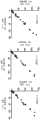


Classifications
-
- C—CHEMISTRY; METALLURGY
- C07—ORGANIC CHEMISTRY
- C07K—PEPTIDES
- C07K16/00—Immunoglobulins [IGs], e.g. monoclonal or polyclonal antibodies
- C07K16/18—Immunoglobulins [IGs], e.g. monoclonal or polyclonal antibodies against material from animals or humans
- C07K16/28—Immunoglobulins [IGs], e.g. monoclonal or polyclonal antibodies against material from animals or humans against receptors, cell surface antigens or cell surface determinants
- C07K16/2803—Immunoglobulins [IGs], e.g. monoclonal or polyclonal antibodies against material from animals or humans against receptors, cell surface antigens or cell surface determinants against the immunoglobulin superfamily
- C07K16/2827—Immunoglobulins [IGs], e.g. monoclonal or polyclonal antibodies against material from animals or humans against receptors, cell surface antigens or cell surface determinants against the immunoglobulin superfamily against B7 molecules, e.g. CD80, CD86
-
- A—HUMAN NECESSITIES
- A61—MEDICAL OR VETERINARY SCIENCE; HYGIENE
- A61K—PREPARATIONS FOR MEDICAL, DENTAL OR TOILETRY PURPOSES
- A61K39/00—Medicinal preparations containing antigens or antibodies
- A61K39/395—Antibodies; Immunoglobulins; Immune serum, e.g. antilymphocytic serum
-
- A—HUMAN NECESSITIES
- A61—MEDICAL OR VETERINARY SCIENCE; HYGIENE
- A61K—PREPARATIONS FOR MEDICAL, DENTAL OR TOILETRY PURPOSES
- A61K45/00—Medicinal preparations containing active ingredients not provided for in groups A61K31/00 - A61K41/00
- A61K45/06—Mixtures of active ingredients without chemical characterisation, e.g. antiphlogistics and cardiaca
-
- A—HUMAN NECESSITIES
- A61—MEDICAL OR VETERINARY SCIENCE; HYGIENE
- A61P—SPECIFIC THERAPEUTIC ACTIVITY OF CHEMICAL COMPOUNDS OR MEDICINAL PREPARATIONS
- A61P35/00—Antineoplastic agents
-
- C—CHEMISTRY; METALLURGY
- C07—ORGANIC CHEMISTRY
- C07K—PEPTIDES
- C07K16/00—Immunoglobulins [IGs], e.g. monoclonal or polyclonal antibodies
- C07K16/18—Immunoglobulins [IGs], e.g. monoclonal or polyclonal antibodies against material from animals or humans
- C07K16/28—Immunoglobulins [IGs], e.g. monoclonal or polyclonal antibodies against material from animals or humans against receptors, cell surface antigens or cell surface determinants
- C07K16/30—Immunoglobulins [IGs], e.g. monoclonal or polyclonal antibodies against material from animals or humans against receptors, cell surface antigens or cell surface determinants from tumour cells
-
- G—PHYSICS
- G01—MEASURING; TESTING
- G01N—INVESTIGATING OR ANALYSING MATERIALS BY DETERMINING THEIR CHEMICAL OR PHYSICAL PROPERTIES
- G01N33/00—Investigating or analysing materials by specific methods not covered by groups G01N1/00 - G01N31/00
- G01N33/48—Biological material, e.g. blood, urine; Haemocytometers
- G01N33/50—Chemical analysis of biological material, e.g. blood, urine; Testing involving biospecific ligand binding methods; Immunological testing
- G01N33/53—Immunoassay; Biospecific binding assay; Materials therefor
- G01N33/574—Immunoassay; Biospecific binding assay; Materials therefor for cancer
- G01N33/57484—Immunoassay; Biospecific binding assay; Materials therefor for cancer involving compounds serving as markers for tumor, cancer, neoplasia, e.g. cellular determinants, receptors, heat shock/stress proteins, A-protein, oligosaccharides, metabolites
- G01N33/57492—Immunoassay; Biospecific binding assay; Materials therefor for cancer involving compounds serving as markers for tumor, cancer, neoplasia, e.g. cellular determinants, receptors, heat shock/stress proteins, A-protein, oligosaccharides, metabolites involving compounds localized on the membrane of tumor or cancer cells
-
- C—CHEMISTRY; METALLURGY
- C07—ORGANIC CHEMISTRY
- C07K—PEPTIDES
- C07K2317/00—Immunoglobulins specific features
- C07K2317/10—Immunoglobulins specific features characterized by their source of isolation or production
-
- C—CHEMISTRY; METALLURGY
- C07—ORGANIC CHEMISTRY
- C07K—PEPTIDES
- C07K2317/00—Immunoglobulins specific features
- C07K2317/20—Immunoglobulins specific features characterized by taxonomic origin
-
- C—CHEMISTRY; METALLURGY
- C07—ORGANIC CHEMISTRY
- C07K—PEPTIDES
- C07K2317/00—Immunoglobulins specific features
- C07K2317/30—Immunoglobulins specific features characterized by aspects of specificity or valency
- C07K2317/34—Identification of a linear epitope shorter than 20 amino acid residues or of a conformational epitope defined by amino acid residues
-
- C—CHEMISTRY; METALLURGY
- C07—ORGANIC CHEMISTRY
- C07K—PEPTIDES
- C07K2317/00—Immunoglobulins specific features
- C07K2317/50—Immunoglobulins specific features characterized by immunoglobulin fragments
- C07K2317/51—Complete heavy chain or Fd fragment, i.e. VH + CH1
-
- C—CHEMISTRY; METALLURGY
- C07—ORGANIC CHEMISTRY
- C07K—PEPTIDES
- C07K2317/00—Immunoglobulins specific features
- C07K2317/50—Immunoglobulins specific features characterized by immunoglobulin fragments
- C07K2317/515—Complete light chain, i.e. VL + CL
-
- C—CHEMISTRY; METALLURGY
- C07—ORGANIC CHEMISTRY
- C07K—PEPTIDES
- C07K2317/00—Immunoglobulins specific features
- C07K2317/50—Immunoglobulins specific features characterized by immunoglobulin fragments
- C07K2317/54—F(ab')2
-
- C—CHEMISTRY; METALLURGY
- C07—ORGANIC CHEMISTRY
- C07K—PEPTIDES
- C07K2317/00—Immunoglobulins specific features
- C07K2317/50—Immunoglobulins specific features characterized by immunoglobulin fragments
- C07K2317/55—Fab or Fab'
-
- C—CHEMISTRY; METALLURGY
- C07—ORGANIC CHEMISTRY
- C07K—PEPTIDES
- C07K2317/00—Immunoglobulins specific features
- C07K2317/60—Immunoglobulins specific features characterized by non-natural combinations of immunoglobulin fragments
- C07K2317/62—Immunoglobulins specific features characterized by non-natural combinations of immunoglobulin fragments comprising only variable region components
- C07K2317/622—Single chain antibody (scFv)
-
- G—PHYSICS
- G01—MEASURING; TESTING
- G01N—INVESTIGATING OR ANALYSING MATERIALS BY DETERMINING THEIR CHEMICAL OR PHYSICAL PROPERTIES
- G01N2333/00—Assays involving biological materials from specific organisms or of a specific nature
- G01N2333/435—Assays involving biological materials from specific organisms or of a specific nature from animals; from humans
- G01N2333/705—Assays involving receptors, cell surface antigens or cell surface determinants
- G01N2333/70503—Immunoglobulin superfamily, e.g. VCAMs, PECAM, LFA-3
- G01N2333/70532—B7 molecules, e.g. CD80, CD86
-
- G—PHYSICS
- G01—MEASURING; TESTING
- G01N—INVESTIGATING OR ANALYSING MATERIALS BY DETERMINING THEIR CHEMICAL OR PHYSICAL PROPERTIES
- G01N2333/00—Assays involving biological materials from specific organisms or of a specific nature
- G01N2333/435—Assays involving biological materials from specific organisms or of a specific nature from animals; from humans
- G01N2333/705—Assays involving receptors, cell surface antigens or cell surface determinants
- G01N2333/70596—Molecules with a "CD"-designation not provided for elsewhere in G01N2333/705
Abstract
The present invention provides an antibody or antigen-binding fragment thereof that binds to an epitope within human PD-L1 with high specificity and reproducibility. The antibodies or antigen binding fragments thereof of the invention can be used to assess PD-L1 expression in tissue samples to aid in patient stratification. The invention further provides methods of producing the antibodies of the invention.
Description
Technical Field
The present invention relates to the field of cancer diagnosis, in particular to the use of diagnostic antibodies for in vitro diagnosis to detect the presence of a PD-L1 epitope in a tumor sample.
Background
Programmed cell death 1 ligand 1 (PD-L1), also known as cluster of differentiation (CD 274) or B7 homolog 1 (B7-H1), is a protein encoded by the CD274 gene in humans and is immediately adjacent to PD-L2 (CD 273), one of the ligands of PD-1 (CD 279). PD-L1 is a 40kDa type 1 transmembrane protein that is expressed on a number of hematopoietic cells, including dendritic cells, macrophages, mesenchymal stem cells and bone marrow-derived mast cells. It was found that the expression of PD-L1 was induced on epithelial and endothelial cells by the action of interferon. The sites found immune privileged such as the syncytiotrophoblast in placenta and retina constitutively express PD-L1.
Expression of PD-L1 in the placenta increases at the beginning of second trimester of pregnancy, is upregulated by increased oxygen, and is rapidly lost at low oxygen concentrations. Experiments have shown that the PD-1-PD-L pathway regulates the balance between stimulating and inhibiting signals required for an effective immune response to microorganisms and for maintaining self-tolerance, respectively. Many chronically infected microorganisms utilize the PD-1-PD-L1 pathway to evade host immune effector mechanisms. Based on liver infection in a mouse model, the PD-L1 pathway is also thought to modulate immune-mediated tissue damage during viral infection, as PD-1 deficient mice show increased liver damage following adenovirus clearance compared to wild type mice, which is thought to be caused by highly active and aggressive T cells.
Immune attack by IFN γ release leads to up-regulation of PD-L1, inducible by the mucosa, creating an "immune barrier" to prevent autoimmune attack in the case of chronic inflammation or infection. The up-regulated PD-L1 on these cells binds to PD-1 on T cells, leading to the development of T cell depletion.
Tumor cells have adapted to this PD-1-PD-L1 regulatory mechanism that protects mucosa from autoimmune attack under normal physiological circumstances, and instead promote cancer growth by overexpressing PD-L1 to avoid immune surveillance.
This immunosuppressive mechanism can be hijacked by PD-L1 positive tumor cells, eventually leading to tumor escape from the clearance of the immune system. Inhibition of the PD-1/PD-L1 interaction by monoclonal antibodies provides a promising concept for the treatment of tumors with PD-L1 expression. Clinical trials of monoclonal antibodies blocking PD-1 and PD-L1 are currently being conducted in patients with various malignancies.
Statistical analysis of published anti-PD-L1 clinical trial data comparing clinical response rates of PD-L1 positive or PD-L1 negative patients indicates that PD-L1 expression is a predictive marker of clinical response to certain cancer types and other relevant biomarkers (Gandini et al, crit Rev Oncol hematol.2016 Apr; 100. For example, in metastatic melanoma, PD-L1 expression in tumor tissue correlates with a significantly better prognosis, as PD-L1 positive patients receiving anti-PD-L1 treatment show a 53% reduction in mortality.
Studies have also shown that, in a variety of cancer types, a response to anti-PD-L1 treatment is observed in patients with tumors that express high levels of PD-L1, particularly when PD-L1 is expressed on tumor-infiltrating immune cells (see, e.g., herbst et al, nature 2014 Nov 27 515 (7528): 563-7, lie et al, annals of Oncology 27 147-153, 2016. In papillary thyroid carcinomas, PD-L1 expression was found to be associated with relapse and shortened disease-free survival, supporting the use of PD-L1 expression as a prognostic marker in this tumor type (Chowdhury et al, oncotarget.2016 Apr 12).
Detection and scoring of PD-L1 expression in tumor tissue samples is typically performed by immunohistochemistry on frozen or formalin-fixed paraffin-embedded (FFPE) tumor tissue sections. Scoring of PD-L1 expression can be performed using different methods: for example, one method employs a binary endpoint score for a sample positive or negative for PD-L1 expression, where a positive result is defined in terms of the percentage of tumor cells that exhibit histological evidence of cell surface membrane staining. Two different cut-off values, 1% and 5% of total tumor cells, at which the tumor samples were scored as PD-L1 positive (cancer.2011 May 15 (10): 2192-201 n Engl J Med 2012. PD-L1 expression in tumor samples was also quantified by scoring tumor cells exhibiting membrane staining and tumor-infiltrating immune cells, as compared to tumor cells exhibiting membrane and prominent cytoplasmic staining. A sample score based on PD-L1 expression is then performed, thereby giving an IHC score of 0, 1, 2 or 3. The sample score is "0" if less than 1% of the cells are PD-L1 positive, "1" is greater than 1% but less than 5% of the cells are PD-L1 positive, "2" is greater than 5% but less than 10% of the cells are PD-L1 positive, or "3" is if more than 10% of the cells are PD-L1 positive (Herbst et al Nature.2014 Nov 27 515 (7528): 563-7. PD-L1 expression in Tumor Infiltrating Mononuclear Cells (TIMC) has been evaluated using a semi-quantitative method according to three categories, which are scored as 0, 1 or 2, depending on the amount of TIMC in the sample, respectively: 0% =0, < 5% =1, ≧ 5% =2.
The specificity and reproducibility of most commercially available anti-PD-L1 antibodies have not been thoroughly evaluated, and limitations of some widely used antibodies have been reported (see, e.g., cancer 2011 117-2192-201, carvajal-Hausorf et al, laboratory Investigation (2015) 95, 385-396. For example, WO 2014/165422 A1, WO 2016/007235 A1 disclose anti-PD-L1 antibodies and scoring guidelines for assessing PD-L1 expression using the corresponding antibodies.
Therefore, there is a continuing need to expand the pool of available anti-PD-L1 antibodies (repotoreies) that are highly specific for PD-L1 and produce reproducible results to aid in the stratification of tumor patients eligible for anti-PD-L1 based therapy.
Summary of The Invention
The inventors have surprisingly found that a polypeptide directed to a polypeptide comprised in a sequence according to SEQ ID NO:1 or an antigen-binding fragment thereof binds human PD-L1 with high specificity and reproducibility.
In a first embodiment, the invention provides an antibody or antigen binding fragment thereof that binds to a polypeptide comprised in a sequence according to SEQ ID NO:1 with high specificity and produces reproducible results, whereby the antibody or antigen-binding fragment thereof comprises in its light chain sequence the amino acid sequence of SEQ ID NO: 3. SEQ ID NO: 4.SEQ ID NO: 5. SEQ ID NO: 6. the amino acid sequence of SEQ ID NO: 7. SEQ ID NO:8, comprising in its heavy chain sequence at least three amino acid sequences of SEQ ID NO: 9. SEQ ID NO: 10. SEQ ID NO: 11. SEQ ID NO: 12. SEQ ID NO: 13. SEQ ID NO:14, or a pharmaceutically acceptable salt thereof.
According to one embodiment, the antibody or antigen binding fragment thereof of the invention may be a Fab fragment.
According to one embodiment, the antigen-binding fragment of the invention is F (ab') 2 And (3) fragment.
According to one embodiment, the antigen binding fragment of the invention is a Fab' fragment.
In one embodiment, the antibody of the invention is an scFv.
According to one embodiment, the antibody of the invention is a di-scFv.
According to one embodiment, the antibody of the invention is a monoclonal antibody.
According to one embodiment, the antibody of the invention is an antibody of the IgG class.
According to one embodiment, the antibody light chain or antigen-binding fragment thereof of the invention comprises all of the amino acid sequences of SEQ ID NOs: 3. SEQ ID NO: 4.SEQ ID NO: 5. SEQ ID NO: 6. SEQ ID NO: 7. SEQ ID NO:8.
according to one embodiment, the antibody heavy chain or antigen-binding fragment thereof of the invention comprises all of the amino acid sequences of SEQ ID NOs: 9. the amino acid sequence of SEQ ID NO: 10. SEQ ID NO: 11. the amino acid sequence of SEQ ID NO: 12. SEQ ID NO: 13. SEQ ID NO:14.
in one embodiment, the antibody of the invention comprises a heavy chain variable region comprising an amino acid sequence according to SEQ ID NO:110 or an antigen-binding fragment thereof and a light chain comprising an amino acid sequence according to SEQ ID NO:111, or an antigen-binding fragment thereof.
According to a preferred embodiment, the antibody light chain of the invention comprises the amino acid sequence according to SEQ ID NO:15, and the heavy chain comprises an amino acid sequence according to SEQ ID NO: 16.
In a preferred embodiment, the antibody light chain of the invention comprises the amino acid sequence according to SEQ ID NO:114, and the antibody heavy chain comprises an amino acid sequence according to SEQ ID NO: 115.
In a preferred embodiment, the antibody of the invention is a rabbit antibody or an antibody of rabbit origin.
According to one embodiment, the antibody or antigen-binding fragment thereof of the invention as disclosed above is further coupled to a detectable label.
According to one embodiment, the detectable label of the antibody or antigen-binding fragment thereof of the invention is an enzyme or a fluorophore or one of the enzyme substrates.
According to one embodiment, the antibody or antigen-binding fragment thereof of the present invention as disclosed above is used for detecting a polypeptide comprised in SEQ ID NO:1, said sample according to one embodiment is a biological sample, preferably a tissue sample, a fixed tissue sample or a Formaldehyde Fixed Paraffin Embedded (FFPE) tissue, more preferably a Formaldehyde Fixed Paraffin Embedded (FFPE) tissue of tumor origin.
In one embodiment, the antibody or antigen binding fragment thereof of the invention as disclosed above is used to detect a polypeptide comprising the amino acid sequence as disclosed above in SEQ ID NO:1, or the presence or absence of an epitope in 1.
According to a preferred embodiment, the antibody or antigen binding fragment thereof of the invention as disclosed above is used to detect the antigen binding sequence comprised in SEQ ID NO:1, or expression of an epitope in (1).
In one embodiment, the present invention provides a method for detecting a presence of a nucleic acid comprising a sequence according to SEQ ID NO:1 or any fragment thereof, wherein the method of the invention comprises the steps of: contacting a sample with an antibody or antigen-binding fragment thereof of the invention and detecting the presence of the bound antibody or antigen-binding fragment thereof.
In one embodiment, the in vitro method of the invention as disclosed above is used on a biological sample, a tissue sample, a fixed tissue sample, preferably a Formaldehyde Fixed Paraffin Embedded (FFPE) tissue sample, more preferably a Formaldehyde Fixed Paraffin Embedded (FFPE) tumor tissue sample.
According to one embodiment, the sample used in the in vitro method of the invention as disclosed above is derived from a subject suffering from or at risk of: cancer, T cell dysfunction, acute or chronic infection, or tumor immunity.
In one embodiment, the invention relates to the use of an antibody or antigen-binding fragment of the invention as disclosed above in the manufacture of a medicament comprising the amino acid sequence set forth in SEQ ID NO:1 or expression of the epitope in (1).
In one embodiment, the invention provides an isolated polynucleotide encoding an antibody or antigen-binding fragment thereof of the invention as disclosed above.
According to one embodiment, the invention provides an expression vector comprising a polynucleotide encoding an antibody or antigen-binding fragment thereof of the invention.
In one embodiment, the invention provides an expression vector as disclosed above for use in the production of an antibody of the invention.
In one embodiment, the invention provides at least one host cell comprising at least one expression vector according to the invention.
In one embodiment, the invention provides at least one host cell according to the invention for use in the preparation of an antibody of the invention.
According to one embodiment, the present invention provides a method of treating cancer in a patient comprising the steps of: detecting the presence or expression of human PD-L1 in a sample from the patient using the anti-PD-L1 antibodies of the invention as disclosed above, comparing the expression of PD-L1 in the patient sample with a reference sample, and administering an immune checkpoint inhibitor (e.g., an anti-PD-L1 or anti-PD-1 antibody) to the patient if the expression of PD-L1 in the patient sample is increased as compared to the reference sample.
Drawings
FIG. 1: ELISA results for antibody clone MKP1A07310 of the invention, which coated the wells of the ELISA plate with 50. Mu.l/well of 1. Mu.g/ml immunogen.
FIG. 2: western blotting of the indicated cell lysates was used with the antibody clone MKP1A07310 of the invention and a control antibody (anti-CD 247, dilution 1: 250) at dilutions 1: 10000 and 1: 25000. Western blots were performed using cell lysates of PD-L1-transfected HEK293, PD-L1-expressing MDA-MB231, siRNA PDL 1-knockdown MDA-MB231, and low PD-L1 cell line A549.
FIG. 3: immunohistochemical staining using the antibody of the invention (MKP 1a 07310) and different anti-PD-L1 antibodies directed against an extracellular epitope of human PD-L1 in siRNA PDL 1-depleted MDA-MB231 cells. PD-L1 staining was reduced in siRNA knockdown cells.
FIG. 4: the antibody of the invention (MKP 1A 07310) and one antibody directed against an extracellular epitope of human PD-L1 (MKP 1B 19610) were characterized on an array of cancer cell lines in 70 cell lines using MKP1A7310 at a concentration of 1. Mu.g/ml and three different concentrations (2. Mu.g/ml, 5. Mu.g/ml and 10. Mu.g/ml) of MKP1B19610 to assess and compare their respective binding characteristics and determine the optimal working concentration for further studies. An array of cancer cell lines (CAX 09_70_MB, manufactured by Multiblock GmbH and Zytomed Systems GmbH) was used to evaluate antibody properties in IHC. Cancer cell lines used to prepare custom cancer cell line arrays were fixed in 4% paraformaldehyde buffered in phosphate (pH 7) at room temperature for 16 to 48 hours and subsequently embedded in paraffin.
FIG. 5: the intra-run variability of the antibody of the invention (MKP 1a 07310) on the tissue array CAX08_60, the correlation coefficient r =0.99 between different slides.
FIG. 6: the characterization of the antibody of the invention (MKP 1A07310, labeled "#") and one antibody directed against an extracellular epitope of human PD-L1 (MKP 1B 19610) on cancer cell line array CAX09_70 was tested using MKP1A7310 at a concentration of 1. Mu.g/ml and MKP1B19610 at a concentration of 10. Mu.g/ml.
FIG. 7 is a schematic view of: (A) IHC detection of PD-L1 positive cells using the antibodies of the invention with DAB as chromogen (right panel); detecting chromogens (middle picture) by using computer software; blue staining of the nucleus and cytoplasm was detected (right panel). Arrows indicate positive cells. (B) Positive and negative control tissues were IHC stained with the antibody MKP1a07310 of the invention at a concentration of 1 μ g/ml.
FIG. 8: (A) Immunohistochemistry was performed using antibody MKP1a07310 of the present invention at a concentration of 2 μ g/ml on FFPE tissue of xenograft tissue microarray TMA _ Xenos _ 12. (B) Immunohistochemical staining was performed on FFPE tissue of xenograft tissue microarray TMA _ Xenos _12 using an antibody against an extracellular epitope of human PD-L1 (MKP 1B 19610) at a concentration of 10 μ g/ml. Exemplary PD-L1-positive cells are indicated by arrows
FIG. 9: (A) Immunohistochemical staining was performed on FFPE tissue of the cancer cell line array CAX08 — 60 using the antibody MKP1a07310 of the invention at a concentration of 1 μ g/ml. (B) Immunohistochemical staining was performed on FFPE tissues of cancer cell line array CAX08 — 60 using an antibody against the extracellular epitope of human PD-L1 (MKP 1B 19610) at a concentration of 10 μ g/ml. Arrows indicate exemplary PD-L1 positive cells.
FIG. 10: immunohistochemical staining was performed on FFPE fixed cell lines NCI-H2009, lox and a2780 (NCI-H2900, lox cancer cell line expressing high levels of PD-L1, cancer cell line a2780 expressing only a small amount of PD-L1) on a discovery XT staining instrument using the antibody MKP1a07310 of the invention at a concentration of 1 μ g/ml and the antibody MKP1B19610 against the extracellular epitope of PD-L1 at a concentration of 10 μ g/ml.
FIG. 11: immunohistochemical staining was performed on the FFPE fixed cell lines NCI-H2009, lox and a2780 on an AutostainerLink staining instrument using the antibody MKP1a07310 of the invention at a concentration of 0.5 μ g/ml and the antibody MKP1B19610 against the extracellular epitope of PD-L1 at a concentration of 1 μ g/ml.
FIG. 12: use of MKP1B19610 in Discovery Correlation of IHC results on FFPE cancer cell lines stained by the system (Ventana) and AutostainerLink system (DAKO). (A) Results of IHC runs using MKP1B19610 at low and high pH using different IHC platforms and concentrations are plotted against each other as shown. (B) Statistical analysis of the graph, wherein the Pearson correlation coefficients are 0.6186 (2. Mu.g/ml, high pH), 0.6969 (1. Mu.g/ml, high pH) and 0.4309 (5. Mu.g/ml), 0.6631 (2. Mu.g/ml) at low pH.
FIG. 13: (A) Discovery using anti-PD-L1 antibody MKP1A07310 of the invention at high pH using the indicated antibody concentrations Correlation of IHC results with staining of either the system (Ventana) or AutostainerLink (DAKO) in FFPE cancer cell lines. (B) Statistical analysis of the correlation of IHC results with correlation coefficients r =0.9474 (1 μ g/ml) and r =0.9695 (2 μ g/ml).
FIG. 14 is a schematic view of: (A) IHC correlation of anti-PD-L1 antibody MKP1B19610 and antibody MKP1A07310 of the invention on FFPE cancer cell lines stained with AutostainerLink (DAKO) at the indicated concentrations (diamonds: 1. Mu.g/ml; circles: 2. Mu.g/ml) and antibody of the invention was used at a concentration of 0.5. Mu.g/ml as indicated. (B) Statistical analysis of the correlation of IHC results, with correlation coefficients r =0.6503 (2 μ g/ml MKP1B 19610) and r =0.7088 (1 μ g/ml MKP1B 19610).
Sequence listing


Detailed Description
Although the invention is described in detail below, it is to be understood that this invention is not limited to the particular methodology, protocols, and reagents described herein as these may vary. It is also to be understood that the terminology used herein is for the purpose of describing particular embodiments only, and is not intended to limit the scope of the present invention which will be limited only by the appended claims. Unless defined otherwise, all technical and scientific terms used herein have the same meaning as commonly understood by one of ordinary skill in the art.
Hereinafter, elements of the present invention will be described. These elements are listed with particular embodiments, however, it should be understood that they may be combined in any manner and in any number to produce additional embodiments. The various described examples and preferred embodiments should not be construed as limiting the invention to only the explicitly described embodiments. This description should be understood to support and encompass embodiments that combine the explicitly described embodiments with any number of the disclosed and/or preferred elements. Moreover, any permutation and combination of all described elements in this application should be considered disclosed by the specification of this application unless the context indicates otherwise.
Throughout this specification and the claims which follow, unless the context requires otherwise, the terms "comprise" and variations such as "comprises" and "comprising" will be understood to imply the inclusion of a stated member, component (integer) or step but not the exclusion of any other non-stated member, component or step. The term "comprising" is a specific embodiment of the term "comprising" and excludes any other unspecified members, components or steps. In the context of the present invention, the term "comprising" includes the term "consisting of.
The use of the terms "a" and "an" and "the" and similar referents in the context of describing the invention (especially in the context of the following claims) are to be construed to cover both the singular and the plural, unless otherwise indicated herein or clearly contradicted by context. Recitation of ranges of values herein are merely intended to serve as a shorthand method of referring individually to each separate value falling within the range. Unless otherwise indicated herein, each individual value is incorporated into the specification as if it were individually recited herein. No language in the specification should be construed as indicating any non-claimed element as essential to the practice of the invention.
Several documents are cited throughout the present specification. Each of the documents cited herein (including all patents, patent applications, scientific publications, manufacturer's specifications, etc.), whether supra or infra, is hereby incorporated by reference in its entirety. Nothing herein is to be construed as an admission that the invention is not entitled to antedate such disclosure by virtue of prior invention.
The described objects are solved by the present invention, preferably by the subject matter of the appended claims.
The inventors have surprisingly found that the antibodies of the invention, or antigen binding fragments thereof, are highly specific for PD-L1 and produce reproducible results in PD-L1 detection by immunohistochemistry to aid in stratification of tumor patients eligible for anti-PD-L1 based therapy.
According to a first embodiment, the objects described are solved by an antibody or antigen-binding fragment thereof according to the invention, which binds to a polypeptide comprised in a sequence according to SEQ ID NO:1, wherein the light chain of the antibody or antigen-binding fragment thereof comprises an amino acid sequence according to SEQ ID NO: 3. SEQ ID NO: 4.SEQ ID NO: 5. the amino acid sequence of SEQ ID NO: 6. the amino acid sequence of SEQ ID NO: 7. SEQ ID NO:8, and the heavy chain of the antibody or antigen-binding fragment thereof comprises an amino acid sequence according to SEQ ID NO: 9. the amino acid sequence of SEQ ID NO: 10. SEQ ID NO: 11. SEQ ID NO: 12. the amino acid sequence of SEQ ID NO: 13. SEQ ID NO:14, or a pharmaceutically acceptable salt thereof. For example, an antibody or antigen-binding fragment thereof according to the invention comprises in its light chain the amino acid sequence of SEQ ID NO: 3. SEQ ID NO: 4.SEQ ID NO: 5. SEQ ID NO:6,SEQ ID NO: 7. SEQ ID NO:8, or e.g. SEQ ID NO: 3. SEQ ID NO: 4. the amino acid sequence of SEQ ID NO: 5. the amino acid sequence of SEQ ID NO: 6. SEQ ID NO: 7. the amino acid sequence of SEQ ID NO: two, three, four, five, or all of 8. For example, the light chain may comprise SEQ ID NO: 3. SEQ ID NO: 4.SEQ ID NO: 5. SEQ ID NO: 6. SEQ ID NO: 7. the amino acid sequence of SEQ ID NO:8, or e.g. SEQ ID NO: 3. the amino acid sequence of SEQ ID NO: 4. the amino acid sequence of SEQ ID NO:5; SEQ ID NO: 4.SEQ ID NO: 5. SEQ ID NO:6; the amino acid sequence of SEQ ID NO: 5. SEQ ID NO: 6. the amino acid sequence of SEQ ID NO:7; SEQ ID NO: 6. the amino acid sequence of SEQ ID NO: 7. SEQ ID NO:8, or e.g. SEQ ID NO: 3. the amino acid sequence of SEQ ID NO: 4. the amino acid sequence of SEQ ID NO: 5. the amino acid sequence of SEQ ID NO:6; SEQ ID NO: 4.SEQ ID NO: 5. SEQ ID NO: 6. SEQ ID NO:7; SEQ ID NO: 5. SEQ ID NO: 6. SEQ ID NO: 7. SEQ ID NO:8, or e.g. SEQ ID NO: 3. SEQ ID NO: 4.SEQ ID NO: 5. SEQ ID NO: 6. the amino acid sequence of SEQ ID NO: 7. SEQ ID NO:8. SEQ ID NO:116, and the heavy chain or antibody antigen-binding fragment thereof comprises SEQ ID NO: 9. the amino acid sequence of SEQ ID NO: 10. SEQ ID NO: 11. SEQ ID NO: 12. SEQ ID NO: 13. SEQ ID NO:14, such as SEQ ID NO: 9. SEQ ID NO: 10. the amino acid sequence of SEQ ID NO:11, SEQ ID NO: 12. the amino acid sequence of SEQ ID NO: 13. SEQ ID NO:14, such as two, three, four, five, or all of SEQ ID NOs: 9. SEQ ID NO: 10. SEQ ID NO: 10. SEQ ID NO: 11. SEQ ID NO: 11. the amino acid sequence of SEQ ID NO:12; SEQ ID NO: 12. SEQ ID NO:13; the amino acid sequence of SEQ ID NO: 13. SEQ ID NO:14, or e.g. SEQ ID NO: 9. SEQ ID NO: 10. SEQ ID NO:11; SEQ ID NO: 10. the amino acid sequence of SEQ ID NO: 11. SEQ ID NO:12; SEQ ID NO: 11. SEQ ID NO: 12. SEQ ID NO:13; the amino acid sequence of SEQ ID NO: 12. SEQ ID NO: 13. the amino acid sequence of SEQ ID NO:14. or e.g. SEQ ID NO: 9. SEQ ID NO: 10. SEQ ID NO: 11. SEQ ID NO:12; SEQ ID NO: 10. the amino acid sequence of SEQ ID NO: 11. SEQ ID NO: 12. SEQ ID NO:13; SEQ ID NO: 11. SEQ ID NO: 12. the amino acid sequence of SEQ ID NO: 13. SEQ ID NO:14; or e.g. SEQ ID NO: 9. SEQ ID NO: 10. the amino acid sequence of SEQ ID NO: 11. SEQ ID NO: 12. SEQ ID NO: 13. SEQ ID NO:14, or e.g. SEQ ID NO: 9. SEQ ID NO: 10. the amino acid sequence of SEQ ID NO: 11. SEQ ID NO: 12. SEQ ID NO: 13. SEQ ID NO:14. the amino acid sequence of SEQ ID NO:117. the light and heavy chain amino acid sequence elements as disclosed above may be displayed, for example, in numerical ascending order of the listed SEQ ID numbers, and if all heavy and light chain sequence elements are present in the antibody or antigen-binding fragment of the invention, it is preferred that the light chain sequence elements are represented in SEQ ID nos: 3. SEQ ID NO: 4.SEQ ID NO: 5. SEQ ID NO: 6. SEQ ID NO:7, SEQ ID NO:8. SEQ ID NO:116, and the heavy chain sequence element is represented by SEQ ID NO: 9. SEQ ID NO: 10. SEQ ID NO: 11. the amino acid sequence of SEQ ID NO: 12. SEQ ID NO: 13. the amino acid sequence of SEQ ID NO:14. SEQ ID NO:117 are included. As used in the antibodies of the present invention, the term "antibody" refers to a polypeptide comprising a framework region from an immunoglobulin gene or a fragment thereof that specifically binds to and recognizes an antigen. Recognized immunoglobulin genes include the kappa, lambda, alpha, gamma, delta, epsilon and mu constant region genes, as well as immunoglobulin variable region genes. Antibody light chains are classified as either kappa or lambda. Antibody heavy chains are classified as gamma, mu, alpha, delta, or epsilon, which in turn define the immunoglobulin classes, igG, igM, igA, igD, and IgE, respectively. Generally, the antigen binding region of an antibody is most critical in terms of specificity and affinity of binding. An exemplary immunoglobulin (antibody) building block comprises a tetramer. Each tetramer is composed of two identical pairs of polypeptide chains, each pair having one "light" (about 25 kD) and one "heavy" (about 50-70 kD) (see, e.g., J Allergy Clin Immunol.2010 February;125 (2 0 2): S41-S52). N + per chainThe termini define a variable region of about 100-110 or more amino acids, primarily responsible for antigen recognition. The term variable light chain (V) L ) And a variable heavy chain (V) H ) These refer to the light and heavy chains, respectively.
According to one embodiment, the antibody or antigen binding fragment thereof of the invention is a monoclonal antibody, fab, F (ab') 2 Fab', scFv or di-scFv. Antibodies exist as intact immunoglobulins or, for example, as well-characterized antigen-binding fragments produced by digestion with peptidases such as pepsin or papain: pepsin will cause proteolytic cleavage below the disulfide bond and yield F (ab') 2 Antibody fragments, whereas proteolytic cleavage by papain cleaves over disulfide bonds, resulting in two Fab fragments. Thus, F (ab') 2 The fragment is a dimer of Fab, which is itself linked to V by a disulfide bond H -C H1 The light chain of (1). The F (ab') 2 Can be reduced under mild conditions to disrupt disulfide bonds in the hinge region, thereby converting F (ab') 2 The dimer is converted to Fab' monomer. The above antibody fragments are defined based on digestion of the whole antibody with pepsin and papain, however, these fragments can be synthesized de novo by chemical methods or by using recombinant DNA methods.
The term "antigen-binding fragment thereof" according to the present invention refers to (i) a Fab fragment, a monovalent fragment consisting of the VL, VH, CL and CH1 domains, (ii) F (ab') 2 A fragment, a bivalent fragment comprising two Fab fragments linked by a disulfide bond at the hinge region, (iii) an Fd fragment consisting of VH and CH1 domains, (iv) an Fv fragment consisting of VL and VH domains of a single arm of an antibody, (v) a dAb fragment (see, e.g., ward et al (1989) Nature 341 544-46), which comprises a VH domain, and (vi) an isolated Complementarity Determining Region (CDR). The term "scFv" as used in the present invention refers to a molecule comprising an antibody heavy chain variable domain (or region; VH) and an antibody light chain variable domain (or region; VL) linked by a linker and lacking a constant domain, e.g.an scFv fragment according to the present invention may e.g.comprise a junction consisting of one light chain variable domain (VL) or part thereof and one heavy chain variable domain (VH) or part thereofA synthetic molecule wherein each variable domain (or part thereof) is derived from the same or different antibody. The scFv molecule preferably comprises a linker interposed between the VH domain and the VL domain, which may for example comprise a peptide sequence consisting of the amino acids glycine and serine. For example, the peptide sequence may comprise the amino acid sequence (Gly) 4 Ser) n Where n is an integer from 1 to 6, for example n may be 1, 2, 3, 4,5 or 6, preferably n =4.scFv molecules and methods of obtaining them are known in the art and are described, for example, in U.S. Pat. nos. 4,522,517, ho et al 1989.Gene 77:51; bird et al.1988 Science 242:423; pantoliano et al 1991.Biochemistry 30:10117; milenic et al 1991 cancer Research 51:6363; takkien et al 1991.Protein Engineering 4:837. the term "di-scFv" as used in the antigen-binding fragments of the present invention refers to two scFv fragments which are coupled to each other by a linker, as described, for example, in Cancer Research 54, 6176-618, december 1, 1994, or Chem Commun (Camb) 2007 Feb 21; (7): 695-7.
According to one embodiment, the antibody or antigen-binding fragment according to the invention is a monoclonal antibody. The term "monoclonal antibody" as used in the antibodies of the invention refers to an antibody obtained from a substantially homogeneous population of antibodies. Monoclonal antibodies are identical except for possible naturally occurring mutations or minor differences in their glycosylation patterns that may be present in minor amounts. Monoclonal antibodies, such as those of the invention, are highly specific, are directed to a single antigenic site and specifically bind to a single epitope within an antigen, unlike polyclonal antibody preparations which typically include different antibodies directed to different epitopes. Monoclonal antibodies can be obtained, for example, by hybridoma culture, e.g., kohler et al, (1975) Nature,256:495, etc., or can be prepared by recombinant DNA methods (see, e.g., U.S. patent No.4,816,567).
According to one embodiment, the antibody of the invention is a monoclonal antibody of the IgG type, for example the antibody of the invention may be a monoclonal antibody of the human IgG1, igG2, igG3 or IgG4 type, or for example murine IgG1, igG2a, igG2b, igG2c, igG3 or for example rat IgG1, igG2a, igG2b, igG2c. For example, the IgG-type monoclonal antibodies of the invention may be of any origin, e.g., murine, goat, sheep, hamster, rat, or rabbit origin.
According to a preferred embodiment, the antibody or antigen-binding fragment thereof of the invention comprises an amino acid sequence according to SEQ ID NO: 3. the amino acid sequence of SEQ ID NO: 4.SEQ ID NO: 5. SEQ ID NO: 6. SEQ ID NO: 7. SEQ ID NO:8, or a light chain amino acid sequence of seq id no.
In a preferred embodiment, the antibody or antigen-binding fragment thereof of the invention comprises an amino acid sequence according to SEQ ID NO: 9. SEQ ID NO: 10. SEQ ID NO: 11. the amino acid sequence of SEQ ID NO: 12. the amino acid sequence of SEQ ID NO: 13. the amino acid sequence of SEQ ID NO:14, or a light chain amino acid sequence of seq id no.
In one embodiment, the antibody or antigen binding fragment thereof of the present invention comprises three CDRs from the light chain variable region (the light chain comprising the amino acid sequence according to SEQ ID NO: 111) and three CDRs from the heavy chain variable region (the heavy chain comprising the amino acid sequence according to SEQ ID NO: 110).
In a preferred embodiment, the antibody or antigen-binding fragment thereof of the invention comprises three CDRs from the light chain variable region (the light chain comprising the amino acid sequence according to SEQ ID NO: 111) and three CDRs from the heavy chain variable region (the heavy chain comprising the amino acid sequence according to SEQ ID NO: 110), and the CDRs of the light chain comprise or are contained in a sequence according to SEQ ID NO: 4. the amino acid sequence of SEQ ID NO:6 and SEQ ID NO:8 and the CDRs of the heavy chain comprise or are comprised in the amino acid sequences according to SEQ ID NO: 10. SEQ ID NO:12 and SEQ ID NO:14 in the amino acid sequence of seq id no.
In a preferred embodiment, the antibody or antigen binding fragment thereof of the invention as disclosed above comprises a light chain and a heavy chain comprising CDRs as defined above, wherein the heavy chain variable region comprises the amino acid sequence according to SEQ ID NO:111 further comprises four Framework Regions (FRs) and the light chain variable region according to SEQ ID NO:110 further comprises four framework regions, wherein the framework regions of the light chain variable region comprise or are comprised in a sequence according to SEQ ID NO: 3. SEQ ID NO: 5. SEQ ID NO: 7. the amino acid sequence of SEQ ID NO:116 and the framework region of the heavy chain variable region comprises or is comprised in the amino acid sequence according to SEQ ID NO: 9. SEQ ID NO: 11. SEQ ID NO: 13. SEQ ID NO:117 in the amino acid sequence of seq id no. .
According to a preferred embodiment, the heavy chain of an antibody of the invention (or e.g. an antigen-binding fragment thereof) comprises a variable region comprising a sequence according to SEQ ID NO:110, and the light chain of an antibody of the invention (or e.g., an antigen-binding fragment thereof) comprises a variable region comprising an amino acid sequence according to SEQ ID NO: 111.
According to a more preferred embodiment, the antibody or antigen binding fragment thereof of the invention comprises a light chain comprising an amino acid sequence according to SEQ ID NO:114, wherein the signal sequence has been removed, e.g., by proteolytic cleavage, e.g., according to SEQ ID NO:112, and a heavy chain comprising an amino acid sequence according to SEQ ID NO:115, wherein the signal sequence has been removed, e.g. by proteolytic cleavage, e.g. according to SEQ ID NO:113, or a pharmaceutically acceptable salt thereof.
In one embodiment, for example, comprising a nucleic acid sequence according to SEQ ID NO:110 and SEQ ID NO:111 of the amino acid sequence of the variable light chain (V) of the antibody of the invention L ) And a variable heavy chain (V) H ) May be included in scFv or e.g.Fab, e.g.which may be further linked to GA-SEED or AG-SEED. The term "SEED" refers to e.g. WO 2007/110205 A2, protein Engineering, design&Strand Exchange Engineered Domain (SEED) C disclosed in Selection vol.23 No.4pp.195-202, 2010 H 3 heterodimer. These heterodimeric molecules are derivatives of the human IgG and IgA CH3 domains and produce complementary human SEED C H 3 heterodimers consisting of human IgA and IgG C H 3 sequences. When expressed in mammalian cells to form "SEED bodies" (Sb), the resulting SEED C H The 3-domain pairs preferentially bind to form heterodimers in a 1:1 ratio. The term "GA-SEED" means herein that the SEED molecule starts with an IgG sequence and then an IgA sequence, whereas "AG-SEED" means that the SEED molecule starts with an IgA derived sequence and then an IgG derived sequence. Comprising a sequence according to SEQ ID NO:110 and SEQ ID NO:110 may be linked to GA-or AG-SEED, e.g. by a peptide linker, e.g. by a glycine-serine linker (Gly) of the amino acid sequence as described above 4 Ser) n 。
According to a more preferred embodiment, the antibody light chain or antigen-binding fragment thereof of the invention comprises an amino acid sequence according to SEQ ID NO:15, and the antibody heavy chain or antigen-binding fragment thereof of the invention comprises an amino acid sequence according to SEQ ID NO: 16. For example, an antibody or antigen-binding fragment thereof of the invention may comprise in its light chain an amino acid sequence according to SEQ ID NO:15, and comprising in its heavy chain an amino acid sequence according to SEQ ID NO: 16. For example, an antibody or antigen-binding fragment thereof of the invention may comprise a heavy chain variable region according to SEQ ID NO:15 and may comprise in its heavy chain the amino acid sequence according to SEQ ID NO: 16. For example, both the heavy and light chain sequences of an antibody or antigen-binding fragment thereof of the invention may comprise sequences that are not included in the light chain SEQ ID NOs: 3. SEQ ID NO: 4.SEQ ID NO: 5. SEQ ID NO: 6. the amino acid sequence of SEQ ID NO: 7. SEQ ID NO:8 or SEQ ID NO:111 and heavy chain SEQ ID NO: 9. the amino acid sequence of SEQ ID NO: 10. SEQ ID NO: 11. SEQ ID NO: 12. SEQ ID NO: 13. SEQ ID NO:14 or SEQ ID NO:110, e.g. in amino acids that do not form part of the light or heavy chain variable region, or e.g. any light or heavy chain CDR or framework sequence, e.g. the light chain of an antibody of the invention SEQ ID NO: 3. SEQ ID NO: 4. the amino acid sequence of SEQ ID NO: 5. SEQ ID NO: 6. SEQ ID NO: 7. the amino acid sequence of SEQ ID NO:8 or e.g. SEQ ID NO:111 or heavy chain SEQ ID NO: 9. SEQ ID NO: 10. SEQ ID NO: 11. the amino acid sequence of SEQ ID NO: 12. SEQ ID NO: 13. SEQ ID NO:14 or SEQ ID NO:110, amino-terminal or carboxy-terminal. Amino acid substitutions may be, for example, non-conservative amino acid substitutions or conservative amino acid substitutions. The term "conservative amino acid substitution" as used herein refers to an amino acid substitution in which the substituted amino acid residue has a charge similar to that of the substituted residue and a size similar to or smaller than that of the substituted residue. Conservative substitutions of amino acids include, for example, substitutions made in amino acids within the following groups:


according to a preferred embodiment, the monoclonal IgG antibodies of the invention are rabbit antibodies, or antibodies of e.g. rabbit origin. The rabbit IgG monoclonal antibody of the invention may be used, for example, according to Antibodies: a Laboratory Manual, second Edition, chapter 7, edited by Edward A.Greenfield, CSH Press, ISBN 978-1-936113-81-1, or produced, for example, according to any suitable method in the art, such as those described in Proc. Natl. Acad. Sci.USA, vol.92, pp.9348-9352, september 1995, or, for example, U.S. Pat. No. 7,732,168 B2. The term "rabbit-derived" as used in the antibodies of the invention refers to antibodies, e.g., antibodies of the invention, produced in a heterologous expression system (e.g., CHO cells, HEK293 cells) using polynucleotide sequences obtained from rabbit hybridomas producing the corresponding antibodies. For example, rabbit-derived antibodies of the invention include antibodies produced in CHO or HEK cells that can be transfected with an expression vector comprising a polynucleotide encoding the amino acid sequence (e.g., according to SEQ ID NO:15 and/or SEQ ID NO: 16) of an antibody produced by a rabbit hybridoma cell. For example, a recombinant monoclonal antibody according to the invention can be produced by PLoS one.2016 Mar 29;11 (3): e0152282 discloses methods produced from rabbit hybridoma cells producing the antibodies of the invention. For example, cDNA from a single B cell can be prepared using Superscript III reverse transcriptase (Invitrogen) primed with oligo (dT). Antibody variable region genes can then be recovered by two rounds of PCR using KOD DNA polymerase (EMD Millipore) or TaqPlus Precision DNA polymerase (Agilent). First PCR may, for example, use gene-specific primers at the 5 'and 3' ends of the antibody variable region. The 5' oligonucleotide set can be attached, for example, at the 5' end of the leader sequence, and the 3' reverse primer set can anneal to the CH1 or Ckappa regions, respectively. In secondary PCR, a single 5' forward oligonucleotide that anneals to the "tail" encoded at the 5' end of the primary PCR product can be used with a 3' primer set that anneals in the J region. The second oligonucleotide may, for example, be used to introduce restriction sites to facilitate downstream cloning. The heavy and light chains can then be combined, for exampleThe strand PCR fragment was subcloned into an appropriate expression vector (e.g., pCMV) and transfected into Expi293 cells (Life Technologies) using Expi293-fectamine (Life Technologies) according to the manufacturer's instructions. The transfected cells can then be treated with growth medium, e.g., at 37 ℃, 5% CO 2 For 7 days to allow for the production of antibodies of the invention. The resulting supernatant may be harvested, for example, after 5 to 7 days.
In one embodiment, the antibody or antigen-binding fragment thereof of the invention is further conjugated to a detectable label. The term "detectable label" as used in the antibodies or antigen-binding fragments thereof of the present invention refers to a molecule capable of detection, which may, for example, include radioisotopes, fluorescent probes, chemiluminescence, enzymes, enzyme substrates, enzyme cofactors, enzyme inhibitors, dyes, metal ions, or biotin. The term "conjugated" as used with respect to an antibody or antigen-binding fragment thereof of the invention means that the dye, radioisotope, may be, for example, non-covalently attached to an antibody or antigen-binding fragment thereof of the invention, for example, by ionic or hydrophobic interactions. The coupling of a detectable label as disclosed above (e.g., a fluorescent probe, dye, or enzyme) to an antibody of the invention or antigen-binding fragment thereof as disclosed above can be performed, for example, according to Methods known in the art, e.g., methods Cell biol.2001;63:185 to 204; methods Mol biol.2010;588:43-8; curr Protoc Mol biol.2001 May; chapter 11: those disclosed in Unit 11.1.
According to one embodiment, the detectable label conjugated to the antibody or antigen-binding fragment thereof of the invention is an enzyme or a fluorophore or one of the enzyme substrates. For example, a detectable label conjugated to an antibody or antigen-binding fragment thereof of the invention can be alkaline phosphatase, horseradish peroxidase, beta-galactosidase, tobacco etch virus nuclear inclusion body-a endopeptidase ("TEV protease"). For example, a fluorophore that can be conjugated to an antibody of the invention as described above can be one of the following: 1,8-ANS, 4-methylumbelliferone, 7-amino-4-methylcoumarin (7-amino-4-methylcoumarin), 7-hydroxy-4-methylcoumarin, acridine, alexa Fluor 350 TM 、Alexa Fluor 405 TM 、AMCA、AMCA-X、ATTO Rho6G、ATTO Rho11、ATTO Rho12、ATTO Rho13、ATTO Rho14、ATTO Rho101、Pacific Blue、Alexa Fluor 430 TM 、Alexa Fluor 480 TM 、Alexa Fluor 488 TM 、BODIPY 492/515、Alexa Fluor 532 TM 、Alexa Fluor 546 TM 、Alexa Fluor 555 TM 、Alexa Fluor 594 TM 、BODIPY 505/515、Cy2、cyQUANT GR、FITC、Fluo-3、Fluo-4、GFP(EGFP)、mHoneydew、Oregon Green TM 488、Oregon Green TM 514. EYFP, dsRed2, dTomato, cy3.5, phycoerythrin (PE), rhodamine Red, mTangerine, mStrawberry, mOrange, mBanana, tetramethylrhodomine (TRITC), R-phycoerythrin, ROX, dylight594, calcum Crimson, alexa Fluor 594 TM 、Alexa Fluor 610 TM 、Texas Red、mCherry、mKate、Alexa Fluor 660 TM 、Alexa Fluor 680 TM Allophycocyanin, DRAQ-5, carboxynaphtofluorescein, C7, dyLight 750, cellvue NIR780, DM-NERF, eosin, erythrosine, fluorescein, FAM, hydroxycoumarins, IRDyEs (IRD 40, IRD700, IRD 800), JOE, lissamine rhodamine B, marina Blue, methoxycoumarin, naptho fluorescein, pyMPO, 5-carboxy-4 ',5' -dichloro-2 ',7' -dimethoxyfluorescein, 5-carboxy-2 ',4',5',7' -tetrachlorofluorescein, 5-carboxyfluorescein, 5-carboxyrhodamine, 6-carboxytetramethylamino, cascade Blue, cy2, cy3, cy5, 6-FAM, dansyl chloride, HEX, 6-JOE, NBD (7-nitrophenyl-2-oxa-1, 3-diazole), oregon Green 488, oregon Green 500, oregon Green 514, pacific Blue, phthalic acid, terephthalic acid, isophthalic acid, antiferrol violet, brilliant cresyl Blue violet, brilliant cresol Blue, p-aminobenzoic acid, erythrosine, phthalocyanine, azomethine, cyanine, xanthine, succinyl fluorescein, rare earth cryptate, europedium trisbipyridine diamine, europium cryptate or chelate, diamine, bispyriside, or La Jolla Blue dye. Fluorophores that can be conjugated to the antibodies or antigen binding fragments of the invention can, for example, also include quantum dots. The term quantum dot as used in the present invention refers to a single spherical nanocrystal of a semiconductor material, whereinThe radius of the nanocrystals is less than or equal to the magnitude of the exciton bohr radius of the semiconductor material (the value of exciton bohr radius can be calculated from data found in handbooks containing information on semiconductor properties, such as CRC Handbook of Chemistry and Physics,83rd ed., lite, david r. (Editor), CRC Press, boca Raton, fla. (2002)). Quantum dots are known in the art and are described in references such as Weller, angelw.chem.int.ed.engl.32: 41-53 (1993), alivisatos, J.Phys.Chem.100:13226-13239 (1996) and Alivisatos, science 271:933-937 (1996). The quantum dots may, for example, have a diameter of from about 1nm to about 1000nm, such as 10nm, 20nm, 30nm, 40nm, 50nm, 60nm, 70nm, 80nm, 90nm, 100nm, 150nm, 200nm, 250nm, 300nm, 350nm, 400nm, 450nm or 500nm, preferably a diameter of at least about 2nm to about 50nm, more preferably a diameter of at least about 2nm to about 20nm (e.g., about 2, 3, 4,5, 6, 7, 8, 9, 10, 11, 12, 13, 14, 15, 16, 17, 18, 19 or 20 nm) for the QD. QDs are characterized by their substantially uniform nanometer size, typically exhibiting a polydispersity or size range of about 10% to 15%. The QDs are capable of emitting electromagnetic radiation upon excitation (i.e., the QDs are photoluminescent) and comprise a "core" of one or more first semiconductor materials, and may be surrounded by a "shell" of a second semiconductor material. The QD core surrounded by a semiconductor shell is referred to as a "core/shell" QD. The surrounding "shell" material preferably has a bandgap energy that is greater than that of the core material, and may be selected to have an atomic spacing close to that of the "core" matrix. The core and/or shell may be a semiconductor material including, but not limited to, group II-VI (ZnS, znSe, znTe, US, cdSe, cdTe, hgS, hgSe, hgTe, mgS, mgSe, mgTe, caS, caSe, caTe, srS, srSe, srTe, baS, baSe, baTe, etc.) and group III-V (GaN, gaP, gaAs, gaSb, inN, inP, inAs, inSb, etc.) and group IV (Ge, si, etc.) materials, pbS, pbSe, and alloys or mixtures thereof. Preferred shell materials include ZnS. Quantum dots can be conjugated to the antibodies of the invention or antigen-binding fragments thereof, for example, by any method known in the art (e.g., nanotechnology.2011 Dec 9 (49): 494006.
In one example, an antibody or antigen-binding fragment thereof of the invention can be conjugated to a radioisotope, e.g. 47 Ca、 14 C、 137 Cs、 157 Cr、 57 Co、 60 Co、 67 Cu、 67 Ga、 123 I、 125 I、 129 I、 131 I、 32 P、 75 Se、 85 Sr、 35 S、 201 Th、 3 H, preferably, the radioisotope is incorporated into another molecule, such as a chelator. Typical chelating agents which can be used according to the invention are, for example, DPTA, EDTA (ethylenediaminetetraacetic acid), EGTA (ethylene glycol-O, O ' -bis (2-aminoethyl) -N, N, N ', N ' -tetraacetic acid), NTA (nitrilotriacetic acid), HEDTA (N- (2-hydroxyethyl) -ethylenediamine-N, N ', N ' -triacetic acid), DTPA (2- [ bis [2- [ bis (carboxymethyl) amino ] acetic acid)]-ethyl radical]Amino group]Acetic acid), or DOTA (1, 4,7, 10-tetraazacyclododecane-1, 4,7, 10-tetraacetic acid).
In one embodiment, the antibody or antigen-binding fragment thereof of the invention is used to detect a polypeptide comprised in SEQ ID NO:1 or expression of the epitope in (1). For example, the antibodies or antigen binding fragments thereof of the invention are used to detect a polypeptide comprised in a sequence according to SEQ ID NO:1, wherein the amino acid sequence of SEQ ID NO:1 corresponds to the intracellular portion of human PD-L1 spanning amino acid residues 260-290 of GenBank accession No. AAH 69381. Comprising the amino acid sequence according to SEQ ID NO:1 (e.g. within the intracellular domain of human PD-L1) can be reliably and reproducibly detected by means of immunohistochemistry, for example on e.g. an FFPE tissue sample or a tumor tissue sample, for example by using an antibody or antigen binding fragment thereof of the invention. Comprises the amino acid sequence shown in SEQ ID NO: the term "epitope" as used in the present invention in 1 (RLRKGRMMDVKKCGIQDTNSKKQSDTHLEET) refers to an epitope of SEQ ID NO:1, according to the invention, the sequence contained in SEQ ID NO: the epitope in 1 can be, for example, a linear epitope or a conformational epitope. A conformational epitope may consist, for example, of SEQ ID NO:1, and may beFor example based on its three-dimensional surface features and shape or SEQ ID NO:1 interacts with the paratope of the antibody or antigen-binding fragment of the invention disclosed above. The nucleic acid molecule according to the invention comprised in SEQ ID NO:1 may also be an epitope consisting of, for example, SEQ ID NO:1, based on the amino acid sequence of SEQ ID NO:1 (RLRKGRMMDVKKCGIQDTNSKKQSDTHLEET) interacts with the paratope of the antibody or antigen-binding fragment thereof of the invention. Thus, the paratope of an antibody or antigen-binding fragment thereof of the invention may, for example, bind to a polypeptide comprised in a sequence according to SEQ ID NO:1, and may, for example, have about 1, 2, 3, 4,5, 6, 7, 8, 9, 10 to about 11, 12, 13, 14, 15, 16, 17, 18, 19, 20 amino acids in length, or about 2 to about 3, 4,5, 6, 7, 8, 9, 10 amino acids, or, for example, about 8 amino acids to about 11 amino acids. For example, a linear epitope that interacts with the paratope of an antibody or antigen-binding fragment thereof of the invention may comprise, or may be comprised in, an amino acid sequence according to: SEQ ID NO: 19. SEQ ID NO: 20. SEQ ID NO: 21. SEQ ID NO: 22. SEQ ID NO: 23. SEQ ID NO: 24. SEQ ID NO: 25. SEQ ID NO: 26. SEQ ID NO: 27. SEQ ID NO: 28. SEQ ID NO: 29. SEQ ID NO: 30. SEQ ID NO: 31. SEQ ID NO: 31. SEQ ID NO: 32. SEQ ID NO: 33. SEQ ID NO: 34. SEQ ID NO: 35. SEQ ID NO: 36. SEQ ID NO: 37. SEQ ID NO: 38. SEQ ID NO: 39. SEQ ID NO: 40. SEQ ID NO: 41. SEQ ID NO: 42. SEQ ID NO: 43. SEQ ID NO: 44. SEQ ID NO: 45. SEQ ID NO: 46. SEQ ID NO: 47. SEQ ID NO: 48. SEQ ID NO: 49. SEQ ID NO: 50. SEQ ID NO: 51. SEQ ID NO: 52. SEQ ID NO: 53. SEQ ID NO: 54. SEQ ID NO: 55. SEQ ID NO:56. SEQ ID NO: 57. SEQ ID NO: 58. SEQ ID NO: 59. SEQ ID NO: 60. the amino acid sequence of SEQ ID NO: 61. the amino acid sequence of SEQ ID NO: 62. the amino acid sequence of SEQ ID NO: 63. SEQ ID NO:64.SEQ ID NO: 65. SEQ ID NO: 66. the amino acid sequence of SEQ ID NO: 67. the amino acid sequence of SEQ ID NO: 68. the amino acid sequence of SEQ ID NO: 69. SEQ ID NO: 70. the amino acid sequence of SEQ ID NO: 71. SEQ ID NO: 72. SEQ ID NO: 73. SEQ ID NO: 74. SEQ ID NO: 75. SEQ ID NO: 76. SEQ ID NO:77. SEQ ID NO: 78. SEQ ID NO: 79. the amino acid sequence of SEQ ID NO: 80. SEQ ID NO: 81. SEQ ID NO: 82. SEQ ID NO: 83. SEQ ID NO: 84. SEQ ID NO: 85. the amino acid sequence of SEQ ID NO: 86. the amino acid sequence of SEQ ID NO: 87. SEQ ID NO: 88. SEQ ID NO: 89. SEQ ID NO: 90. SEQ ID NO: 91. SEQ ID NO: 92. SEQ ID NO: 93. SEQ ID NO: 94. the amino acid sequence of SEQ ID NO: 95. SEQ ID NO: 96. SEQ ID NO: 97. SEQ ID NO: 98. SEQ ID NO: 99. SEQ ID NO: 99. SEQ ID NO: 100. SEQ ID NO: 101. SEQ ID NO: 102. SEQ ID NO: 103. SEQ ID NO: 104. SEQ ID NO: 105. SEQ ID NO, 106, SEQ ID NO: 107. SEQ ID NO:108. for example, the antibodies of the invention or antigen binding fragments thereof as disclosed above may be used to detect the presence or expression of the above epitopes in a sample, which may be, for example, a biological sample, such as a body fluid or blood sample. For example, a biological sample (e.g., a bodily fluid or blood sample) can be subjected to flow cytometry, or e.g., FACS, using an antibody or antigen-binding fragment thereof of the invention to detect a polypeptide comprised in SEQ ID NO:1 or comprised in, for example, SEQ ID NO:19-SEQ ID NO:108, or a epitope in any amino acid sequence of seq id no. For example, cells obtained by needle biopsy may be treated with an FcR blocker (e.g., truStain FcX) at room temperature TM Biolegend, san Diego; or for example Human BD Fc Block TM BD Biosciences) at a dilution of 1: 1000 for 10 minutes and then incubated with an antibody or antigen-binding fragment thereof of the invention, whereby the antibody or antigen-binding fragment thereof of the invention can be diluted in PBS +2% serum at a dilution of about 1: 50 to about 1: 500 (e.g., from about 1: 75, 1: 100, 1: 125, 1: 150, 1: 175, 1: 200 to about 1: 250, 1: 275, 1: 300, 1: 350, 1: 400, 1: 450, or, e.g., 1: 25, 1: 50, 1: 100, 1: 150, 1: 200, 1: 250, 1: 300, 1: 400, 1: 500) at room temperature with a fluorescent probe (e.g., phycoerythrin [ PE: PE ] dark]) Coupling was carried out for 15 minutes. Cells can then be analyzed, for example, on an Attune flow cytometer (Life Technologies, grand Island, NY), and the results assessed using FlowJo 10.0 software (Tree Star, inc., ashland, OR). For example, the cells may additionally be treated with carboxyfluorescein diacetate succinimidyl ester (CFSE, bioleg)end, san Diego, calif.) was counterstained at a final concentration of 5. Mu.M for 10 minutes at 37 ℃ in the dark and then washed twice with RPMI-1640 medium.
According to a preferred embodiment, the antibodies or antigen binding fragments thereof of the present invention can be used to detect a protein comprised in SEQ ID NO:1 (e.g. in a linear epitope comprised in any one of the amino acid sequences according to SEQ ID NO:19-SEQ ID NO: 108). The term "tissue sample" according to the present invention may for example refer to a single cell, or at least about 10, derived from a tumor tissue or a suspected tumor tissue 2 、10 3 、10 4 、10 5 、10 6 One or more cells derived from a tumor tissue or suspected tumor tissue, which may, for example, further comprise cultured tumor cells, e.g., as described in Trends biotechnol.2013 June;31 (6): 347 to 354; cancer Cell Culture: methods and Protocols (Methods in Molecular Medicine); langdon (ed.), humana Press, ISBN: 978-1588290793.
The cells or tissue may be obtained, for example, by non-invasive means, including but not limited to fine needle aspiration and needle biopsy, or by, for example, invasive methods, including but not limited to surgical biopsy. The method of obtaining a tissue sample does not form part of the present invention. The term "fixed tissue sample" according to the present invention refers to a tissue sample that has been chemically tissue fixed. For example, chemical fixation may include fixation with methanol, ethanol, acetone, methanol-acetone mixtures, buffered formalin solutions, glutaraldehyde, or buffered paraformaldehyde. For example, fixation may include treatment (fixation) of a tissue sample as described above, wherein a 10% saturated aqueous formaldehyde solution is buffered with 100mM phosphate buffer or 10% Neutral Buffered Formalin (NBF) to a pH of 6.8-7.2 for various times at temperatures ranging from 4 ° -45 ℃, e.g., room temperature (20-25 ℃), or e.g., from about 5 ℃,6 ℃,7 ℃,8 ℃, 9 ℃, 10 ℃ to about 15 ℃,17.5 ℃,20 ℃, or from about 12 ℃, 15 ℃,17.5 ℃,20 ℃ to about 25 ℃, 27 ℃, 30 ℃, 35 ℃, 40 ℃, 45 ℃ for various lengths of time. For example, inTissue samples as disclosed above may be fixed for about 5 minutes to about 24 hours, or from about 10min, 15min, 30min, 45min, 60min, 90min, 120min, 180min to about 15min, 30min, 45min, 1h, 4h, 5h, 6h, 7h, 8h, 9h, 10h, 11h, 12h, 14h, 16h, 18h, 20h, 24h, 48h. For example, a fixed tissue sample may comprise a fixed tissue sample that is fixed by immersion in 10% nbf for about 18-24 hours. For example, a fixed tissue sample according to the present invention may include a tissue sample as described above, which has been fixed or otherwise processed according to standard protocols available in the art, such as those provided on the www.ihcwold.com website, e.g., as described in Hopwood d.fixatives and hybridization: histochem j.1969;1 (4): 323-60, or as described, for example, in the "immunological stabilizing Methods",5 th edition (2009), published by DAKO North America, inc. For example, formaldehyde-fixed paraffin-embedded (FFPE) tissue can be obtained by the following procedure:
tumor tissue as disclosed above may for example be placed in at least 10 volumes of buffered formalin (3.7% formaldehyde: 10mM phosphate buffer, pH-7.4) or buffered paraformaldehyde (3.7% paraformaldehyde: 10mM phosphate buffer, pH-7.4) and then incubated in buffered formalin or NBF for different periods of time depending on the thickness of the tissue sample as described above, e.g. 2-3h room temperature for 1-2mM tissue samples, -5h room temperature for 5-10mM thick tissue samples, or e.g. > 1 mM 2-3h room temperature for tissue samples, or e.g. overnight at 4 ℃. Subsequently, the tissue can be rinsed 1-2X, e.g., with PBS, and H in 70% ethanol at 4 deg.C 2 Stored in O, and then placed in tissue processing histology cassettes and labeled for later identification. The histological cassettes can then be paraffin embedded for 1 hour, for example, using a commercial tissue processor 70% ethanol; 95% ethanol (95% ethanol/5% methanol) for 1 hour; the first time of absolute ethyl alcohol is 1 hour; second time of anhydrous ethanol 1 1 / 2 Hours; third time anhydrous ethanol 1 1 / 2 Hours; absolute ethyl alcohol for the fourth time for 2 hours; first scavenger (xylene or substitute) 1 hour; second scavenger (xylene or substitute) 1 hour; first wax (Paraplast X-tra) at 581 hour at the temperature; a second wax (Paraplast X-tra) was applied at 58 ℃ for 1 hour. The formalin fixed paraffin embedded tissue may then be placed in a mold for further processing.
In one embodiment, the antibody or antigen binding fragment thereof of the invention as disclosed above is used for detecting a protein comprised in a tissue sample (e.g. tissue obtained from a tumor by biopsy as disclosed above) as described above in a tissue sample, fixed tissue sample or FFPE tissue sample derived from a tumor (e.g. tissue obtained from a tumor by biopsy as disclosed above) according to SEQ ID NO:1, for example an epitope comprised in an amino acid sequence according to SEQ ID NO:19-SEQ ID NO:108, or a pharmaceutically acceptable salt thereof. The term "derived from a tumor" according to the present invention may also for example comprise tumor cells obtained by needle aspiration, or cells obtained for example by culturing tumor cells aspirated by a needle.
According to one embodiment, the detection of the protein contained in the nucleic acid sequence according to SEQ ID NO:1 in the amino acid sequence of seq id no. For example, a tissue sample or tumor tissue sample according to the invention may be subjected to immunoblotting to detect a polypeptide comprising the amino acid sequence of SEQ ID NO:1, as described in Current Protocols in Molecular Biology 10.8.1-10.8.28, july 2008.
For example, if ELISA is used, the antibody or antigen-binding fragment thereof of the present invention is used to detect a polypeptide comprised in SEQ ID NO:1, can be subjected to ELISA according to standard Protocols, e.g., as disclosed in Current Protocols in Molecular Biology (1991) 11.2.1-11.2.22. Preferably, the antibody or antigen binding fragment thereof of the invention is used (e.g., as a primary antibody) to detect an antibody comprising the amino acid sequence set forth in SEQ ID NO: 1: for example, FFPE tumor tissue samples can be deparaffinized by placing dried paraffin sections on slides in an oven at 60 ℃ for 1 hour. Subsequently, the slide can be placed, for example, in a Sakura staining rack and immersed in Tissue- In a dyeing dish: three times in xylene, 5min each time, twice in 100% ethanol, at least 1min each time, twice in 95% ethanol, at least 1min each time, once in 70% ethanol, at least 1min each time. The slide can then be rinsed gently with tap water for about 5 minutes. Depending on the tumor tissue, a sample derived therefrom may, for example, require blocking of endogenous peroxidase activity by placing the slide in a 3% hydrogen peroxide solution for 10 minutes at room temperature, followed by rinsing with water. Subsequent antigen retrieval can be carried out, for example, in a water bath according to the following procedure: the slides may be placed in Coplin jars with antigen Retrieval solutions (e.g., target Retrieval Solution, enhanced citrate buffer Solution (Dako, S1699 or S1700), and Target Retrieval Solution, high pH (Dako, S3308), or 0.05M citrate buffer, pH6, or e.g., tris EDTA buffer, pH 8). The slides can then be equilibrated to 75 ° to 95 ℃ in a water bath and incubated for about 40 minutes, for example. The slides are then allowed to cool, for example, at room temperature for 20 minutes, after which the solution is poured off, and then placed in a staining dish containing TBS/0.6
% Tween 20 for at least 5 minutes. Antigen retrieval can also be accomplished, for example, using PT link pretreatment module (DAKO) using Tris-EDTA buffer pH9 at 97 ℃ for 20 minutes. After antigen retrieval, an automated instrument (e.g., discovery @) can then be used according to the manufacturer's instructions> Or Autostainer Link 48) were subjected to the staining procedure. For example, slides can also be manually processed as described in Current Protocols in Molecular Biology 14.6.1-14.6.23, january 2008. For example, the slide may be covered with 400 to 500. Mu.l of an antibody according to the invention, e.g., diluted with a commercially available antibody diluent (e.g., from DAKO) to a concentration of about 0.2. Mu.g/ml to about 5. Mu.g/ml, e.g., from about 0.2. Mu.g/ml, 0.3. Mu.g/ml, 0.4. Mu.g/ml, 0.5. Mu.g/ml, 0.6. Mu.g/ml, 0.7. Mu.g/ml, 0.8. Mu.g/ml, 0.9. Mu.g/ml, 1.0. Mu.g/ml, 1.25. Mu.g/ml, 1.5. Mu.g/ml, 1.75. Mu.g/ml, 2.0. Mu.g/ml to about 3. Mu.g/ml, 3.5. Mu.g/ml, 4. Mu.g/ml, 4.5. Mu.g/ml, 5. Mu.g/ml, 6.0. Mu.g/ml, 7.0. Mu.g/ml, 8.0 g/ml, 9.g/ml, 10. Mu.g/ml, at room temperature and at room temperatureAnd incubated for about 30 minutes. Then may be Tween%>
 The primary antibody was washed away. The slide can then be drained, for example gently, and any remaining wash solution removed. Thereafter, a secondary antibody for detecting the antibody of the present invention may be immediately added and incubated at room temperature for about 30 minutes. Secondary antibody dilutions may be, for example, from about 1: 100 to about 1: 10.000, e.g., from about 1: 100, 1: 150, 1: 200, 1: 250, 1: 300, 1: 400, 1: 500, 1: 750, 1: 1000 to about 1: 1500, 1: 2000, 1: 2500, 1: 3000, 1: 3500, 1: 4000, 1: 5000, 1: 5500, 1: 6000, 1: 7000, 1: 8000, 1: 9000, or e.g., from about 1: 100, 1: 150, 1: 200, 1: 250, 1: 300, 1: 400, 1: 500, 1: 750 to about 1: 1.000, 1: 2000. Secondary antibodies that can be used, for example, to detect bound antibodies according to the invention can include polyclonal goat anti-rabbit HRP-conjugated immunoglobulins at dilutions of about 1: 50, 1: 175 to about 1: 200, or, for example, polyclonal goat anti-rabbit HRP-conjugated immunoglobulins at dilutions of about 1: 20, 1: 50, 1: 100 to about 1: 100, 1: 200, 1: 250 goat anti-rabbit Alkaline Phosphatase (AP) -conjugated immunoglobulins, depending on the selection of detection method and substrate used, e.g., if horseradish peroxidase (HRP) -conjugated secondary antibodies are used, 3 '-Diaminobenzidine (DAB) can be used, for example, for chromogenic detection, or a substrate combination of e.g., 2' -azino-bis (3-ethylbenzothiazoline-6) -sulfonic Acid (ABTS), or e.g., 3', 5' -Tetramethylbenzidine (TMB), or if e.g., AP-conjugated secondary antibodies are used, nitro blue tetrazolium chloride (NBT) and 5-bromo-4-chloro-3-indole phosphate (BCIP). General principles and guidelines for chromogenic immunohistochemistry can be found, for example, in Current Protocols in Immunology 21.4.21-21.4.26, november 2013.
Then by covering the slide with the washing solution for about 5min, with TBS/0.6% The slides were washed twice, after which chromogen solution was added and incubated according to manufacturer's recommended protocolIncubate for about 4 to 5 minutes. Counterstaining can be performed, for example, using Harris' hematoxylin for about 5 minutes, after which the slides can be rinsed under running tap water, then rinsed for about 1 additional minute, for example, in TBS/0.6
% tween 20. The sections can then be dehydrated, for example, by gently submerging the slide up and down in each solution for a few seconds, and then transferred to: 70% ethanol, 90% ethanol, 100% ethanol, and xylene twice. These sections can then be mounted, for example, using mounting reagents and coverslips.
In one aspect, the invention provides a method for detecting a presence of a nucleic acid sequence contained in SEQ ID NO:1, such as an epitope comprised in any one of: SEQ ID NO: 19. SEQ ID NO: 20. SEQ ID NO: 21. the amino acid sequence of SEQ ID NO: 22. the amino acid sequence of SEQ ID NO: 23. SEQ ID NO: 24. SEQ ID NO: 25. SEQ ID NO: 26. SEQ ID NO: 27. the amino acid sequence of SEQ ID NO: 28. SEQ ID NO: 29. SEQ ID NO: 30. SEQ ID NO: 31. SEQ ID NO: 31. SEQ ID NO: 32. SEQ ID NO: 33. SEQ ID NO: 34. SEQ ID NO: 35. SEQ ID NO: 36. the amino acid sequence of SEQ ID NO: 37. SEQ ID NO: 38. SEQ ID NO: 39. the amino acid sequence of SEQ ID NO: 40. SEQ ID NO: 41. the amino acid sequence of SEQ ID NO: 42. SEQ ID NO: 43. the amino acid sequence of SEQ ID NO: 44. SEQ ID NO: 45. SEQ ID NO: 46. SEQ ID NO: 47. SEQ ID NO: 48. SEQ ID NO: 49. the amino acid sequence of SEQ ID NO: 50. SEQ ID NO: 51. the amino acid sequence of SEQ ID NO: 52. the amino acid sequence of SEQ ID NO: 53. the amino acid sequence of SEQ ID NO: 54. the amino acid sequence of SEQ ID NO: 55. the amino acid sequence of SEQ ID NO:56. and SEQ ID NO: 57. the amino acid sequence of SEQ ID NO: 58. SEQ ID NO: 59. the amino acid sequence of SEQ ID NO: 60. SEQ ID NO: 61. SEQ ID NO: 62. the amino acid sequence of SEQ ID NO: 63. SEQ ID NO:64.SEQ ID NO: 65. the amino acid sequence of SEQ ID NO: 66. SEQ ID NO: 67. the amino acid sequence of SEQ ID NO: 68. the amino acid sequence of SEQ ID NO: 69. the amino acid sequence of SEQ ID NO: 70. the amino acid sequence of SEQ ID NO: 71. the amino acid sequence of SEQ ID NO: 72. SEQ ID NO: 73. the amino acid sequence of SEQ ID NO: 74. SEQ ID NO: 75. the amino acid sequence of SEQ ID NO: 76. the amino acid sequence of SEQ ID NO: 77. the amino acid sequence of SEQ ID NO: 78. the amino acid sequence of SEQ ID NO: 79. SEQ ID NO: 80. SEQ ID NO: 81. SEQ ID NO: 82. the amino acid sequence of SEQ ID NO: 83. SEQ ID NO: 84. SEQ ID NO: 85. SEQ ID NO: 86. SEQ ID NO: 87. SEQ ID NO: 88. SEQ ID NO: 89. SEQ ID NO: 90. SEQ ID NO: 91. the amino acid sequence of SEQ ID NO: 92. SEQ ID NO: 93. SEQ ID NO: 94. SEQ ID NO: 95. SEQ ID NO: 96. SEQ ID NO: 97. SEQ ID NO: 98. SEQ ID NO: 99. SEQ ID NO: 99. SEQ ID NO: 100. SEQ ID NO: 101. SEQ ID NO: 102. SEQ ID NO: 103. SEQ ID NO: 104. SEQ ID NO: 105. SEQ ID NO, 106, SEQ ID NO: 107. SEQ ID NO:108, wherein the method comprises the steps of contacting the sample with an antibody or antigen-binding fragment according to the invention, and detecting the presence of binding to the antibody or antigen-binding fragment according to the invention. For example, detecting a nucleic acid sequence contained in SEQ ID NO:1, or an automated immunohistochemical detection using a commercially available IHC platform, such as, for example, intelliPath FLX (Biocare Medical), WAVE RPD (cells Diagnostics), omnis (DAKO), autostainer Link48 (DAKO), benchmark XT (Ventana), or Benchmark Ultra (Ventana) according to the instructions and/or protocols of the respective manufacturers. Thus, the method of the invention may be used to detect the expression of human PD-L1 in a sample according to the invention.
In one embodiment, the method of the invention may be used for detecting a polypeptide according to SEQ ID NO:1 or any fragment thereof, wherein said method comprises the steps of contacting said sample with an antibody or antigen-binding fragment of the invention as described above, and detecting binding of the antibody or antigen-binding fragment according to the invention, e.g. by immunohistochemical methods, or by automated immunohistochemical methods. For example, a fragment of human PD-L1 according to the invention may comprise human PD-L1 that has been proteolytically cleaved, wherein the cleaved PD-L1 fragment comprises a peptide according to SEQ ID NO: 1. For example, a fragment of human PD-L1 according to the invention may comprise human PD-L1 lacking the extracellular domain, e.g. only the transmembrane domain and the intracellular domain (e.g. amino acids 239-290 of GenBank accession No. AAH 69381), or a PD-L1 fragment which may be detected, e.g. using an antibody of the invention, may only comprise the intracellular domain of human PD-L1 (e.g. amino acids 260-290).
In one aspect, the antibodies or antigen-binding fragments thereof of the invention are used to detect a polypeptide comprised in SEQ ID NO:1, e.g. the epitope comprised in SEQ ID NO:19-SEQ ID NO:108. For example, the sample may be a biological sample, such as, for example, a body fluid or blood sample. For example, the nucleic acid sequence contained in the blood sample is SEQ ID NO:1 can be accomplished using the antibodies or antigen binding fragments of the invention as disclosed above. The blood sample used may be, for example, a blood sample of mammalian origin, preferably human blood, preferably a blood sample from a cancer patient, or a blood sample from an individual at risk of cancer. In one aspect, the antibodies or antigen-binding fragments thereof of the invention can be used, for example, to detect the presence or expression of PD-L1 expression on circulating tumor cells or, for example, leukocytes (e.g., neutrophils, eosinophils, basophils, lymphocytes, or monocytes).
For example, a peripherical blood sample (e.g., from a tumor patient, where the term "tumor" refers to a neoplasm, i.e., abnormal growth of tissue that may also form a tumor mass) can be detected using a flow cytometer using phycoerythrin-Texas Red (ECD) -conjugated-anti-CD 3, phycoerythrin-anthocyanin 5 (PC 5) -conjugated-anti-CD 15 (BD Biosciences, san Diego, CA, USA), fluorescein Isothiocyanate (FITC) -conjugated anti-CD 80 (MIH clones, eBioscience, san Diego, CA, USA) to bind to an antibody or antigen binding fragment of the present invention conjugated to Phycoerythrin (PE) to identify a tumor expressing a polypeptide or antigen binding fragment of the invention comprised in SEQ ID NO: 1. For example, neutrophils can be identified as CD15 + CD3 - Cells, labeling of which can be accomplished, for example, by incubating 50 μ L of fresh heparinized whole blood with 5 μ L ECD-conjugated anti-CD 3, 5 μ L PC 5-conjugated anti-CD 15 simultaneously for 30 minutes on ice in the dark. Cells incubated with PE-and FITC-conjugated mouse IgG can be used, for example, as isotype controls. The samples can then be analyzed using a cell sorter according to the manufacturer's instructions, such as the CYTOMICS FC 500 flow cytometer (Beckman Coulter inc., break, CA, USA) and related software program (CXP).
A tumor cell (CTC) comprising a sequence according to SEQ ID NO:1, or an epitope comprised in an amino acid sequence according to SEQ ID NO:19-SEQ ID NO:108 using the antibodies of the invention or antigen binding thereof as disclosed aboveFragments may be labeled, for example, by flow cytometry as disclosed above using one or more of the following: epCAM, ephB4, EGFR, CEA, HER2, or MUC-1 are analyzed in combination with the antibodies of the invention as described above, wherein one or more of the antibodies used to label CTCs has a different fluorescent label than the antibodies of the invention disclosed above. CTCs can be used, for example, by commercially available isolation methods (e.g., cell Search) or CTC chips using, for example, a Clear-based assay Enrichment of FX CTCs, for example, isolated from a blood sample (e.g., a human blood sample as disclosed above) according to the manufacturer's instructions. In the above method, the antibody of the present invention or an antigen-binding fragment thereof conjugated to the above fluorescent probe or detectable label may be used at a concentration of, for example, about 0.01. Mu.g/ml to about 50. Mu.g/ml, for example from about 0.025. Mu.g/ml, 0.05. Mu.g/ml, 0.075. Mu.g/ml, 0.1mg/ml, 0.25. Mu.g/ml, 0.5. Mu.g/ml, 0.75. Mu.g/ml, 1. Mu.g/ml, 1.50. Mu.g/ml, 1.75. Mu.g/ml, 2. Mu.g/ml, 2.5. Mu.g/ml, 3. Mu.g/ml, 3.5. Mu.g/ml, 4. Mu.g/ml, 4.5. Mu.g/ml, 5. Mu.g/ml, 5.5. Mu.g/ml, 6. Mu.g/ml, 7. Mu.g/ml, 8. Mu.g/ml, 9. Mu.g/ml, 10. Mu.g/ml to about 11. Mu.g/ml, 12. Mu.g/ml, 15. Mu.g/ml, 17.5. Mu.g/ml, 20. Mu.g/ml, 22.5. Mu.g/ml, 25. Mu.g/ml, 30. Mu.g/ml, 35. Mu.g/ml, 37.5. Mu.g/ml, 40. Mu.g/ml, 45. Mu.g/ml, 50. Mu.g/ml, or, for example, from about 0.5. Mu.g/ml, 0.75. Mu.g/ml, 1. Mu.g/ml, 1.50. Mu.g/ml, 1.75. Mu.g/ml, 2. Mu.g/ml, 2.5. Mu.g/ml, 3. Mu.g/ml, 3.5. Mu.g/ml, 4. Mu.g/ml, 4.5. Mu.g/ml, 5. Mu.g/ml to about 6. Mu.g/ml, 7. Mu.g/ml, 8. Mu.g/ml, 9. Mu.g/ml, 10. Mu.g/ml, or from about 0.5. Mu.g/ml, 0.75. Mu.g/ml to about 1. Mu.g/ml, 1.50. Mu.g/ml, 1.75. Mu.g/ml, 2. Mu.g/ml, 2.5. Mu.g/ml, 3. Mu.g/ml, 3.5. Mu.g/ml, 4. Mu.g/ml, 4.5. Mu.g/ml, 5. Mu.g/ml, 5.5. Mu.g/ml, 6. Mu.g/ml, 7. Mu.g/ml, 8. Mu.g/ml, 9. Mu.g/ml, 10. Mu.g/ml, or, for example, 0.5. Mu.g/ml, 0.75. Mu.g/ml, 1. Mu.g/ml, 1.50. Mu.g/ml, 1.75. Mu.g/ml, 2. Mu.g/ml, 2.5. Mu.g/ml, 3. Mu.g/ml, 3.5. Mu.g/ml, 4. G/ml, 4.5. Mu.g/ml, 5. Mu.g/ml, 6. Mu.g/ml, 7. Mu.g/ml, 8. Mu.g/ml, 9. Mu.g/ml, 10. Mu.g/ml.
For example, in one aspect of the invention, the sample (e.g. blood sample) comprises a nucleic acid sequence according to SEQ ID NO:1, or e.g. comprised in an amino acid sequence according to SEQ ID NO:19-SEQ ID NO:108, can be performed by ELISA using the antibodies or antigen binding fragments of the invention. For example, the antibody of the invention may be used at a concentration of about 0.25. Mu.g/ml, 0.5. Mu.g/ml, 0.75. Mu.g/ml, 1. Mu.g/ml, 1.50. Mu.g/ml, 1.75. Mu.g/ml, 2. Mu.g/ml, 2.5. Mu.g/ml, 3. Mu.g/ml, 3.5. Mu.g/ml, 4. Mu.g/ml, 4.5. Mu.g/ml, 5. Mu.g/ml, 5.5. Mu.g/ml, 6. Mu.g/ml, 7. Mu.g/ml, 8. Mu.g/ml, 9. Mu.g/ml, 10. Mu.g/ml, or a control antibody (e.g.g.mIgG 2a in PBS) coating an ELISA plate overnight at 4 ℃ and then blocking with PBS containing 10. Mu.g of FBS. Blood samples (e.g., from cancer patients and/or from healthy individuals as control or reference samples) can be diluted 1: 1.000, e.g., in PBS, in triplicate, and then added to the plate. Subsequently, the hole can for example in 0.1% Tween-20 PBS washing six times. Bound antibodies of the invention can be measured by incubating horseradish peroxidase-conjugated secondary rabbit IgG Ab at a dilution of 1: 2.000 for 1.5 hours at room temperature and then reacted with tetramethylbenzidine, followed by measuring absorbance at 450nm wavelength using a plate reader. Non-specific binding of serum to plates coated with control Ig can be corrected for background, for example, subtracted from the measurements for each sample.
According to a preferred embodiment, the antibody or antigen binding fragment thereof of the invention may be used for detecting a protein comprised in a tissue sample, a fixed tissue sample or a Formaldehyde Fixed Paraffin Embedded (FFPE) tissue according to SEQ ID NO:1, or an epitope comprised in an amino acid sequence according to SEQ ID NO:19-SEQ ID NO:108, or an epitope in any amino acid sequence of seq id no. Preferably, according to one embodiment, the formaldehyde-fixed paraffin-embedded (FFPE) tissue is a tumor tissue sample, or a sample derived from a tumor tissue, such as a tumor cell, a cultured tumor cell line, or a cultured tumor tissue as described above. For example, in one embodiment, the method of the invention may further comprise performing the method of the invention on a control tissue that does not express or expresses only a small amount of PD-L1, preferably in parallel with the tissue sample disclosed above. For example, the control tissue may include thyroid tissue or skeletal muscle tissue, or cultured PD-L1 negative cancer cells, such as A2780, colo205, or IGROV-1. The term "small amount" as used above refers to expression of PD-L1 by tumor cells or tumor tissue that is at least 5-fold, 10-fold, 20-fold, 25-fold, 40-fold, 50-fold, or 100-fold less than PD-L1 positive tumor tissue or tumor cells (e.g., hs746T, MDA-MB231, NCI-H2009, human spleen) when compared by semi-quantitative immunoblotting or, e.g., qPCR, such as Journal of Immunological Methods 345 (2009) 40-48; journal of Immunological Methods 353 (2010) 148-150; hematology.2016 Mar 31: 1-6. For example, RNA isolation and cDNA synthesis may be performed on a tissue sample or tumor tissue sample as defined above, which may then be used, for example, in qPCR to determine the relative expression level of PD-L1.
In one aspect, the antibodies of the invention can be used, for example, to mount on tissue samples on glass slides to form tissue arrays, where different tumor tissue samples can be placed adjacent to each other. For example, the tissue array includes at least one, two, three, or four slices of each tissue sample mounted on glass slides at different locations (e.g., to avoid detection artifacts). For example, the tissue array may comprise at least 4,5, 6, 7, 8, 9, 10, 11, 12, 14, 16, 18, 20 to about 22, 24, 26, 28, 30, 32, 34, 36, 38, 40, 42, 44, 46, 48, 50, 55, or 60, or form about 22, 24, 28, 30, 36, 40, 44, 48, 52, 56, 60 to about 64, 68, 72, 76, 78, 80, 82, 84, 86, 90, 100 different tissue samples. For example, in one embodiment, in addition to a tumor tissue sample as described above, the tissue array may comprise at least one or more, such as 5, 10, 20, 30, 40, 50 or more, or, for example, all of the following cancer cell lines: ZR-75-1, WM164, U-87MG, U-118MG, T47D, SW707, SW620, SNB-78, NCI-H69, NCI-H569, NCI-H460, NCI-H292, NCI-H2009, NCI-H1975, mx-1, meWo, PC-3, raji, ramos, suit7, MDA-MB-468, MDA-MB-435, MDA-MB-231, MCF7, M24 MDA, lox, loVo, kyse 30, jurkat, IGROV-1, HT29, hs746T, hs68, HL60, faDu, EBC-1, DU145, DLD-1, COLO205, calu 6, calu 3, BT474, AGS, A549, A-431, A2780, miffei, miaCa-2, pfME-H2, and PfSA-H-1. Cancer cell lines as disclosed above can be obtained, for example, from ATCC or, for example, DSMZ (Leipniz Institute DSMZ) and cultured according to established methods for each respective cell line. A tissue array or tumor tissue array comprising at least one formaldehyde-fixed paraffin-embedded (FFPE) tissue sample or FFPE tumor tissue sample as disclosed above may be prepared, for example, according to Nat med.1998 Jul;4 (7): 844-7.
According to one embodiment, the Formaldehyde Fixed Paraffin Embedded (FFPE) tissue sample or the formaldehyde fixed paraffin embedded tumor tissue sample is derived from a subject at risk for cancer or having cancer, T cell dysfunction, acute or chronic infection or exhibiting tumor immunity. The term "cancer" as used in the methods of the present invention refers to or describes a physiological condition in a mammal, preferably a human, which is generally characterized by unregulated cell growth/proliferation with the potential to invade or spread to other parts of the body. Examples of cancer include, but are not limited to, non-small cell lung cancer (NSCLC), mesothelioma, unresectable mesothelioma, breast cancer, gastric adenocarcinoma or GEJ, gastric cancer, thymoma, ovarian cancer, adenoid cystic cancer, metastatic gonadal cystic cancer, bladder cancer, clear cell renal cancer, head and neck squamous cell cancer, lung squamous cell cancer, malignant melanoma, ovarian cancer, pancreatic cancer, prostate cancer, renal cell cancer, small Cell Lung Cancer (SCLC) or triple negative breast cancer, lymphoproliferative disease, acute Lymphocytic Leukemia (ALL), acute Myeloid Leukemia (AML), chronic Lymphocytic Leukemia (CLL), chronic Myeloid Leukemia (CML), diffuse large B-cell lymphoma (DLBCL), EBV-positive DLBCL, primary mediastinal large B-cell lymphoma, large B-cell lymphoma rich in T cells/histiocytic cells, follicular lymphoma, hodgkin Lymphoma (HL), mantle Cell Lymphoma (MCL), multiple Myeloma (MM), myeloid cell leukemia-1 protein (shl-1), myelodysplasia (mccl-1), non-squamous cell lymphoma (mccl), head and neck cancer (MCC), or small cell lymphoma (MCC-cell lymphoma). The above cancer types may also be referred to as malignant tumors, for example. The term "T cell dysfunction" as disclosed above for the methods of the invention refers to a pathological state of CD 8T cells, which may be caused, for example, if a tightly controlled differentiation process of CD8 positive T cells is altered, such as for example, changes in the nature, background and duration of the antigen may result in a substantial alteration of the T cell activation and differentiation process, which may also be referred to as T cell depletion, tolerance, anergy or senescence. The term "acute infection" as used in the methods of the invention refers to an acute infection caused by a bacterium, virus or parasite, and which occurs, for example, about one, two, three or four weeks prior to diagnosis. The term "chronic infection" as used in the methods of the invention refers to a condition in which the pathogen is not rapidly eliminated but persists for an extended period of time, e.g., more than three, four, five, six, seven, eight, nine, or ten weeks. Persistent pathogen exposure can lead to chronic antigen stimulation and persistent inflammation, which can lead to depletion and/or clonal depletion of pathogen-specific CD 8T cells. If not treated in time, chronic infections may for example include viral infections such as hepatitis A, hepatitis C or hepatitis D, HIV infections, or for example infections with Neisseria meningitidis (neisseria meningitidis), neisseria gonorrhoeae (neisseria gonorrhoeae), bartonella (bartonella henselae), borrelia burgdorferi (Borrelia burgdorferi), salmonella species, brucella species, campylobacter species, mycobacterium species, treponema pallidum (treponema pallidum) or Bordetella bumetii (coxiella bumetii). In one embodiment, the FFPE tissue sample or FFPE tumor tissue sample is derived from a subject, e.g., a human, characterized by or having tumor immunity. The term "tumor immunity" as used in the methods of the invention refers to the process by which a tumor evades immune recognition and clearance. It is thought that tumor-derived or tumor-associated factors affect dendritic cell differentiation and prevent the development of cells with antigen presenting functions, ultimately leading to tumor immunity. Expression of Endogenous Retroviruses (ERVs), particularly tumor-specific endogenous retroviruses (TERVs), has been implicated in tumor immunity. Other factors that may contribute to tumor immunity are, for example, gene amplification of PD-L1 (CD 274) and ALOX12B/15B, or mutations in any of the following genes B2M, HLA-A, HLA-B, HLA-C or CASP8, for example, leading to loss of antigen presentation (e.g., tumor-associated neoantigens) or to blockade of exogenous apoptosis.
In one aspect, the invention provides a method of predicting whether a mammal, preferably a human, having a malignant tumor is a likely candidate for treatment with an immune checkpoint inhibitor (e.g., an anti-PD-L1 antibody, or an anti-PD-1 antibody), comprising detecting the presence or absence of a nucleic acid molecule comprised in seq no:1 or e.g. as comprised in SEQ ID NO:19-SEQ ID NO:108, such as the epitope in any one of SEQ ID NOs: 20. SEQ ID NO: 21. SEQ ID NO: 22. SEQ ID NO: 23. SEQ ID NO: 24. SEQ ID NO: 25. SEQ ID NO: 26. SEQ ID NO: 27. SEQ ID NO: 28. SEQ ID NO: 29. SEQ ID NO: 30. SEQ ID NO: 31. SEQ ID NO: 32. SEQ ID NO: 33. SEQ ID NO: 34. SEQ ID NO: 35. SEQ ID NO: 36. the amino acid sequence of SEQ ID NO: 37. SEQ ID NO: 38. SEQ ID NO: 39. SEQ ID NO: 40. SEQ ID NO: 41. SEQ ID NO: 42. SEQ ID NO: 43. SEQ ID NO: 44. SEQ ID NO: 45. SEQ ID NO: 46. SEQ ID NO: 47. the amino acid sequence of SEQ ID NO: 48. SEQ ID NO: 49. SEQ ID NO: 50. SEQ ID NO: 51. SEQ ID NO: 52. SEQ ID NO: 53. SEQ ID NO: 54. the amino acid sequence of SEQ ID NO: 55. SEQ ID NO:56. SEQ ID NO: 57. SEQ ID NO: 58. SEQ ID NO: 59. SEQ ID NO: 60. SEQ ID NO: 61. SEQ ID NO: 62. SEQ ID NO: 63. the amino acid sequence of SEQ ID NO:64.SEQ ID NO: 65. SEQ ID NO: 66. SEQ ID NO: 67. SEQ ID NO: 68. SEQ ID NO: 69. the amino acid sequence of SEQ ID NO: 70. SEQ ID NO: 71. the amino acid sequence of SEQ ID NO: 72. SEQ ID NO: 73. SEQ ID NO: 74. the amino acid sequence of SEQ ID NO:75, SEQ ID NO: 76. SEQ ID NO: 77. SEQ ID NO: 78. SEQ ID NO: 79. SEQ ID NO: 80. SEQ ID NO: 81. SEQ ID NO: 82. SEQ ID NO: 83. SEQ ID NO: 84. SEQ ID NO: 85. SEQ ID NO: 86. the amino acid sequence of SEQ ID NO: 87. SEQ ID NO: 88. SEQ ID NO: 89. SEQ ID NO: 90. SEQ ID NO: 91. SEQ ID NO: 92. SEQ ID NO: 93. SEQ ID NO: 94. SEQ ID NO: 95. SEQ ID NO: 96. SEQ ID NO: 97. SEQ ID NO: 98. SEQ ID NO: 99. SEQ ID NO: 100. the amino acid sequence of SEQ ID NO: 101. the amino acid sequence of SEQ ID NO: 102. SEQ ID NO: 103. the amino acid sequence of SEQ ID NO: 104. SEQ ID NO: 105. the amino acid sequence of SEQ ID NO: 106. the amino acid sequence of SEQ ID NO:107, wherein the method comprises using an antibody of the invention for performing, e.g., flow cytometry, FACS or preferably immunohistochemistry, wherein the antibody comprising the amino acid sequence set forth in SEQ ID NO:1, or an epitope comprised, for example, in SEQ ID NO:19 to SEQ ID NO:108, e.g., greater than about 0.25%, 0.5%, 0.75%, 1%, 1.5%, 2.0%, 2.5%, 3.0%, 3.5%, 4.0%, 4.5%, 5.0%, 6.0%, 6.5%, 7.0%, 7.5%, 8.0%, 8.5%, 9.0%, 9.5%, or 10.0%, or about 0.5%, 0.75%, 1%, 1.5%, 2.0%, 2.5%, 3.0%, 3.5%, 4.0%, 4.5%, 5.0%, 6.0%, 6.5%, 7.0%, 7.5%, 8.0%, 8.5%, 9%, 9.5%, 10%, 10.5%, 11%, 12%, 13%, 14%, 15% to less than about 50%, 55%, 60%, 65%, 70%, 75%, 80%, 85%, 90%, 95% in a sample, or, e.g., from about 20%, 25%, 30%, 35%, 40%, 45% to less than 65%, 70%, 75%, 80%, 85%, 90%, 95%, or from about 0.25%, 0.5%, 0.75%, 1% to less than about 1.5%, 2.0%, 2.5%, 3.0%, 3.5%, 4.0%, 4.5%, 5.0%, 6.0%, 6.5%, 7.0%, 7.5%, 8.0%, 8.5%, 9.0%, 9.5%, or 10.0%, or from about 0.25% to less than about 0.5%, 0.75%, 1% of the cells indicate that the patient is likely to respond to treatment with an immune checkpoint inhibitor (e.g., treatment with an anti-PD-L1 antibody, or treatment with an anti-PD-1 antibody). The term "immune checkpoint inhibitor" as used herein has its general meaning in the art and refers to molecules expressed by T cells that either amplify signals (molecules that stimulate checkpoints) or down-regulate signals (molecules that inhibit checkpoints) such as, for example, the PD-1/PD-L1 and CTLA-4/CD28 signaling pathways (both of which are considered in the art to constitute immune checkpoint pathways) (see, for example, pardol, 2012.nature Rev Cancer 12. anti-PD-L1 antibodies including, for example, avelumab, durvalumab, atezolizumab, or, for example, anti-PD-1 antibodies such as pembrolizumab, nivolumab, or Pf-06801591 may be administered, for example, to patients who have been identified as potential candidates for treatment with immune checkpoint inhibitors using the methods of the invention disclosed above.
PD-L1 positive cells in a sample according to the invention may for example comprise tumor cells or immune cells, such as Tumor Infiltrating Lymphocytes (TILs) (e.g. CD8 positive T cells, macrophages, NK cells or B cells). For example, depending on the tumor type of the FFPE tumor tissue sample obtained or derived as disclosed above, different methods of determining the relative abundance of PD-L1 positive cells in the sample (scoring methods) may be used. For example, using Immunohistochemistry (IHC) as disclosed above using the antibodies of the invention to detect PD-L1 positive cells, a PD-L1 score may comprise determining the relative abundance of PD-L1 positive tumor cells, or the relative abundance of PD-L1 positive TILs in a sample, or e.g., determining the relative abundance of a combination of PD-L1 positive tumor cells and TILs. The total number of cells or any sub-fraction thereof used to determine the number of PD-L1 positive cells in a sample can be obtained, for example, by counterstaining the sample with hematoxylin to visualize the nucleus and cytoplasm, or, for example, if a fluorescence detection method is used, staining with 4', 6-diamidino-2-phenylindole (DAPI) to visualize the nucleus. The term "PD-L1 positive cell" for use in the methods of the invention refers to a cell that detectably expresses a protein comprised in a sequence according to SEQ ID NO:1, to which an antibody of the invention specifically binds, and can then be detected (e.g., as described above) with, for example, an HRP-or AP-conjugated secondary antibody that specifically binds to an antibody of the invention and allows chromogenic or fluorescent detection when reacted with a corresponding substrate. For example, HRP-conjugated secondary antibodies can be used to label PD-L1 positive cells by incubation with DAB as a substrate. The score for PD-L1 positive cells may, for example, include individual cell or cell nucleus counts to determine the number of PD-L1 positive cells and the total number of cells (or, for example, in any sub-portion thereof) in the FFPE tumor tissue sample analyzed. Alternatively, the surface regions of PD-L1 positive cells and hematoxylin stained cells can be used, for example, to determine the relative abundance of PD-L1 positive cells.
In one aspect, the methods of the invention as disclosed above may be used to identify potential candidates for treatment with anti-PD-L1 antibodies, whereby the malignancy according to the invention may be one or more of the following: non-small cell lung cancer (NSCLC), mesothelioma, unresectable mesothelioma, breast cancer, gastric adenocarcinoma or GEJ, gastric cancer, thymoma, ovarian cancer, adenoid cystic cancer, metastatic gonadal cystic cancer, bladder cancer, clear cell renal cancer, head and neck squamous cell carcinoma, lung squamous cell carcinoma, malignant melanoma, ovarian cancer, pancreatic cancer, prostate cancer, renal cell carcinoma, small Cell Lung Cancer (SCLC) or triple negative breast cancer, lymphoproliferative disease, acute Lymphocytic Leukemia (ALL), acute Myeloid Leukemia (AML), chronic Lymphocytic Leukemia (CLL), chronic Myeloid Leukemia (CML), diffuse large B-cell lymphoma (DLBCL), EBV-positive DLBCL, primary mediastinal large B-cell lymphoma, large B-cell lymphoma enriched in T-cells/histiocytes, follicular lymphoma, hodgkin Lymphoma (HL), mantle Cell Lymphoma (MCL), multiple Myeloma (MM), myeloid leukemia-1 protein (MCL-1), lymphoproliferative syndrome (MDS), myelodysplasia (slc) or small cell lymphoma (mrnc).
According to one aspect of the invention, the methods of the invention may be used to identify or predict likely candidates (e.g., cancer patients) that are responsive to treatment with an immune checkpoint inhibitor, such as an anti-PD-L1 antibody or an anti-PD-1 antibody and one of the following antibodies: anti-PD-L1 antibodies avelumab, atezolizumab, durvalumab, LY300054, BMS-936559, or anti-PD-1 antibodies pembrolizumab, nivolumab or Pf-06801591. For example, if by applying the above inventive method an individual in need thereof, e.g., a human cancer patient, or e.g., a human patient suffering from a malignancy as disclosed above, or a human at risk of developing cancer, is found to be a possible candidate for treatment with an anti-PD-L1 antibody (e.g., one of avelumab, atezolizumab, durvalumab, LY300054, BMS-936559) or with an anti-PD-1 antibody (e.g., one of pembrolizumab, nivolumab, or PF-06801591), an anti-cancer agent may be administered alone or in combination with other anti-cancer agents, the dose is from about 0.1mg/kg to about 50mg/kg, from about 0.5mg/kg, 0.75mg/kg, 1mg/kg, 1.5mg/kg, 2.5mg/kg, 5mg/kg, 7.5mg/kg, 10mg/kg to about 12.5mg/kg, 15mg/kg, 17.5mg/kg, 20mg/kg, 25mg/kg, 27.5mg/kg, 30mg/kg, 35mg/kg, 37.5mg/kg, 40mg/kg, 42.5mg/kg, 45mg/kg, or such as 1mg/kg, 2.5mg/kg, 5mg/kg, 7.5mg/kg, 10mg/kg, 12.5mg/kg, 15mg/kg, 17.5mg/kg, 20mg/kg, 22.5mg/kg, 25mg/kg, 30mg/kg, 35mg/kg. <xnotran> , PD-L1 avelumab, atezolizumab, durvalumab, PD-1 pembrolizumab, nivolumab Pf-06801591 , , avelumab 500-800mg , 600mg-750mg, 650-850mg, 700-900mg, 525mg, 550mg, 600mg, 600mg, 650mg, 700mg, 700mg, 725mg, 750mg, 900 1600mg, 1000mg 1500mg (q 2 w), 1250mg 1400mg q2w, 1000mg q2w 1250mg q2w, 925mg q2w, 950mg q2w, 1000mg q2w, 1125mg q2w, 1200mg q2w, 1250mg q2w, 1300mg q2w, 1350mg q2w, 1400mg q2w, 1450mg q2w, 1500mg q2w, 1550mg q2w, 1250-2400mg (q 3 w), 1500mg 2250mg q3w, 1750mg 2000mg q3w, 1800mg 2300mg q3w, 1250mg 1750mg q3w, 1300mg 1500mg q3w, 150mg q3w, 1350mg q3w, 1450mg q3w, 1550mg q3w, 1650mg q3w, 1750mg q3w, 1800mg q3w, 1850mg q3w, 1900mg q3w, 1950mg q3w, 2000mg q3w, 2125mg q3w, 2250mg q3w, 2350mg q3w. </xnotran> The term "mg/kg" as used above refers to milligrams of anti-PD-L1 or anti-PD-1 antibody per kilogram body weight of the patient to be treated with the respective antibody. The terms "q2w" and "q3w" as used herein mean administration once every two weeks or once every three weeks. The term "mg/kg" as used above refers to milligrams of anti-PD-L1 (or e.g., anti-PD-1) antibody per kilogram of body weight.
According to one aspect, the invention relates to the use of an antibody or antigen-binding fragment thereof of the invention in a method of the invention for predicting that a patient suffering from a malignancy is a likely candidate for treatment with an anti-PD-L1 antibody or an anti-PD-1 antibody. For example, the antibodies or antigen binding fragments of the invention can be used in immunohistochemistry to detect PD-L1 expression in FFPE tumor tissue samples, and the samples are then visually evaluated (scored) for PD-L1 expression to determine the number of PD-L1 positive cells (e.g., tumor cells, or TILs as described above). The antibodies or antigen-binding fragments thereof of the present invention may also be used, for example, in two-color immunofluorescence to label TILs. For example, the dual color fluorescence may include an antibody of the invention and one of: anti-CD 8 antibody, anti-CD 163 antibody, anti-CD 3 antibody, anti-CD 11c antibody, anti-CD 56 antibody, anti-CD 68 antibody, anti-granzyme B antibody or anti-FoxP 3 antibody, wherein the antibodies are derived from different species to allow detection of both the antibody of the invention and the TIL specific antibody. Double immunofluorescence can be performed, for example, using a commercial kit, e.g., using the Novocasta PowerVision Poly-HRP IHC detection system and then Alexa Fluor 594-or Alexa Fluor 488 Type Signal Amplification (TSA) kit according to the manufacturer's instructions. The scoring of PD-L1 positive cells may be performed, for example, as described above, or may, for example, include assigning an IHC score based on the overall scores of PD-L1 positive tumor cells, PD-L1 positive immune cells (e.g., TIL, macrophages), or PD-L1 positive tumor and immune cells, e.g., the IHC score is 0, 1, 2, or 3, corresponding to < 1% PD-L1 positive cells (IHC score 0), > 1% but < 5% (IHC score 1), > 5% but < 10% (IHC score 2), or > 10% (IHC score 3). For example, if more than one sample of a given patient is subjected to the in vitro method of the invention as disclosed above using the anti-PD-L1 antibodies of the invention as disclosed above, the IHC score may be the average score of the samples analyzed, or for example, the IHC score may correspond to the IHC score of the sample with the highest IHC score. The IHC score can also be determined, for example, by scoring the percentage of two tumor cells expressing PD-L1 as a total tumor cells and the percentage of tumor-infiltrating immune cells expressing PD-L1 as a tumor area using an antibody or antigen-binding fragment thereof of the invention. A sample may for example score 3 if > 50% of the tumor cells are PD-L1 positive; scoring 2 if greater than or equal to 5% but less than 50% of the tumor cells are positive for PD-L1; scoring 1 if > 1% but < 5% of the tumor cells are positive for PD-L1; and a score of 0 if < 1% of the tumor cells express PD-L1.
In one embodiment, the invention provides a method of treating cancer in a patient who has been identified as a likely candidate for response to treatment with an immune checkpoint inhibitor by applying the method of the invention as disclosed above to a sample obtained from or derived from said patient and determining PD-L1 expression in the sample and in a second step comparing the expression of PD-L1 in the sample with PD-L1 expression in a reference sample and in a third step administering an immune checkpoint inhibitor, e.g. an anti-PD-L1 or an anti-PD-1 antibody, to said patient if the expression of PD-L1 in the sample of said patient is found to be increased or above a certain threshold. The term "threshold value" as used in the treatment methods of the invention refers to the relative abundance of PD-L1 expression of a patient-derived sample relative to a reference sample, e.g., as determined by the methods of the invention disclosed above. For example, the threshold may be defined as a relative increase of PD-L1 positive cells in a patient sample compared to a reference sample, or may for example be defined as an absolute or relative number of PD-L1 positive cells in said sample, e.g. 0.5%, 1%, 2%, 3%, 4%, 5%, 10%, 15%, 20%, 25%, 40%, 50%, 75%, 80%, 1%, 5%, 10%, 20%, 50%, 75%, 80% of the cells in the sample, or the threshold may for example be based on the IHC score or scoring method as disclosed above. The reference sample for use in the treatment methods of the invention may, for example, be derived from a healthy donor not suffering from cancer and the same method of detecting PD-L1 expression as described above using the antibodies or antigen binding fragments of the invention, such as, for example, IHC on FFPE tissue samples. For example, a reference sample may be obtained from a tissue and location corresponding to a sample obtained from a patient, and preferably processed in parallel with the sample from the patient. If it is obtained from an undiseased area or location, the reference sample may for example also originate from the same tissue or organ as the tumor tissue sample, or the reference sample may for example originate from a contralateral healthy organ. anti-PD-L1, which may be administered to a patient, for example, according to the treatment methods of the present invention, includes avelumab, atezolizumab, durvalumab, or, for example, an anti-PD-1 antibody, such as pembrolizumab, nivolumab, or Pf-06801591. In the treatment methods of the invention, the anti-PD-L1 or anti-PD-1 antibody (such as, for example, avelumab) is administered as described above, for example at a dose of about 5mg/kg to about 30mg/kg, or for example at a dose of 5mg/kg, 7.5mg/kg, 10mg/kg, to about 12.5mg/kg, 15mg/kg, 17.5mg/kg, 20mg/kg, 25mg/kg, 27.5mg/kg, 30mg/kg, wherein the antibody is administered once per week (q 1 w). As noted above, the anti-PD-L1 antibody (e.g., avelumab) can also be administered weekly, or biweekly, or every three weeks at a fixed dose, e.g., a fixed dose of 500-800mg weekly, or 900-1600mg every two weeks, or 1250-2400mg every three weeks.
In one embodiment, the invention provides an isolated polynucleotide encoding an antibody or antigen-binding fragment thereof. The term "isolated polynucleotide" as used in the present invention refers to a polynucleotide that is at least 70%, 75%, 80%, 85%, 90%, preferably 95%, 96%, 97%, 98%, 99% pure and free of contaminants, such as, for example, proteins and/or lipids. The antibodies of the invention or antigen binding fragments thereof may be encoded, for example, by two separate polynucleotides encoding the light and heavy chains of the antibodies of the invention. The isolated polynucleotide may, for example, comprise a sequence according to SEQ ID NO:17 and a polynucleotide sequence according to SEQ ID NO: 18. Depending on the expression system used, the expression according to SEQ ID NO:17 and SEQ ID NO:18 to allow better expression of the polynucleotides of the invention, resulting in higher antibody production of the invention in a corresponding expression system. Codon optimization can be performed, for example, using Nucleic Acids res.1988 Mar 11;16 (5): 1715-28, nucleic Acids Res.1988 Sep 12;16 (17): 8207-11; methods Cell biol.1991;36:675-7 can be performed using the codon usage table; or such as available computer-embedded codon optimization tools, such as those described in, for example, bioinformatics.2014 Aug 1;30 (15): 2210-2. Codon optimization of at least one polynucleotide of the invention may be carried out taking into account the respective expression system used, e.g.a murine or human cell line, a yeast or an insect cell line.
In one embodiment, the present invention provides an expression vector comprising a polynucleotide of the present invention as disclosed above. The term "expression vector" according to the present invention refers to a nucleic acid vector comprising a gene expression control sequence (e.g., a promoter or promoter component operably linked in a5 'to 3' direction to a nucleotide sequence encoding at least one polypeptide, e.g., SEQ ID NO:17 and/or SEQ ID NO: 18). For example, an expression vector according to the invention may comprise a sequence according to SEQ ID NO:17 or SEQ ID NO:18, or may comprise a polynucleotide according to SEQ ID NO:17 and SEQ ID NO: 18. Nucleic acid sequences necessary for expression in prokaryotes include promoters, ribosome binding sites, optional operator sequences and possibly other sequences. Eukaryotic cells utilize promoters such as the CMV promoter, SV40 promoter, EF1a or CAG promoter, and typically enhancers and polyadenylation signals. Exemplary mammalian expression vectors are pCMV, pCMV-SPORT, pcDNA, or Nat commun.2010 Nov 16; 1: 120, or pLKO.1-hygro, pBABE-puro, pBABE-zeo, pLenti CMV/TO zeo DEST, pCW57.1, for example, if the virus is expected TO express an antibody or antigen binding fragment thereof of the invention. Optionally, the expression vector in eukaryotes may comprise an Internal Ribosome Entry Site (IRES) between the polynucleotide sequences of the invention, which provides a bicistronic expression construct to allow expression of a polypeptide according to SEQ ID NO:17 and SEQ ID NO:18, or a pharmaceutically acceptable salt thereof. For example, IRES sequences that may be used may be included in PLoS one.2013dec 9;8 (12): e82100, e.g. 5' AUGAUAAUAUGGCCACACACAUUG. For example, suitable expression vectors for expressing the polynucleotides of the invention may comprise pCMV-based expression vectors, or pD 912-based vectors and variants thereof, Expression vectors, pcDNA-based expression vectors, or PJ. Omega. Vectors (see, for example, nucleic Acids Research, vol.18, no. 4), or for retroviruses orLentivirus-produced vectors (see, e.g., front biosci.1999
 Jun 1. Suitable yeast expression vectors can, for example, express the polynucleotides of the invention using the GAL4, PGK, ADH1, ADE2 or TRP1 promoter, such as expression vectors pRS420, pLEX, pACT2.2, pTEF1/zeo, pAG425GDP-ccdb, pBEVY-T, pAG300, pGBK T7, pEZY202, pCETT, pRG201, pXP120, pXP320, p2GLex, or p426 GAL. For example, the polynucleotide of the present invention as disclosed above may also be expressed, for example, in insect cells using an expression vector comprising a polyhedrin promoter, such as, for example, pFastBac DUAL, pVL 1392, pVL 1393, or AcNPV, for expression of the polynucleotide of the present invention in baculovirus. In a preferred aspect, the expression vector comprising a polynucleotide of the invention is a mammalian expression vector.
In one embodiment, the invention relates to an expression vector according to the invention for producing a polypeptide comprising an amino acid sequence according to SEQ ID NO:15 and SEQ ID NO:16 in an antibody of the invention. For example, at least one vector according to the invention comprises a nucleic acid sequence encoding a polypeptide according to SEQ ID NO:15 and SEQ ID NO:16, can be used in a transient expression system to express an antibody of the invention. For example, methods.2014 Jan 1;65 (1): 5-10. The expression vector of the invention or at least one expression vector according to the invention can also be used, for example, for the generation of stable cell lines expressing the antibody of the invention. For example, a method for developing a stable cell line may e.g. start with transfecting a suitable host cell with an expression vector of the invention, or with at least one expression vector of the invention, e.g. two expression vectors, wherein the first vector comprises the sequence according to SEQ ID NO:17 and the second expression vector comprises a polynucleotide according to SEQ ID NO: 18. After transfection with an expression vector comprising polynucleotides encoding the light and heavy chains of the antibody and at least one selectable marker, the transfected cells can be incubated for 24 hours to 72 hours under suitable growth conditions to allow expression of the antibody of the invention and screened for cells with high productivity of the antibody of the invention after growth recovery, serum-free suspension adaptation and amplification and clonal selection.
For example, an expression vector of the invention, or for example comprising a sequence according to SEQ ID NO:17 and/or SEQ ID NO:18 may be used to produce or prepare an antibody of the invention as described above. For example, comprising a polypeptide comprising a sequence according to SEQ ID NO:17 and/or SEQ ID NO:18 can be used to transfect at least one suitable host cell for expression of an antibody of the invention as described above. For example, according to SEQ ID NO:17 and SEQ ID NO:18 may be contained in one expression vector or, for example, may be contained in two separate expression vectors, which together are used to produce the antibody of the invention.
Comprising a sequence according to SEQ ID NO:17 and SEQ ID NO:18 may be used, for example, in a retroviral or lentiviral vector to transduce at least one suitable host cell for expression of the polynucleotide of the invention.
For example, to make or produce an antibody of the invention, a polypeptide encoding an amino acid sequence according to SEQ ID NO:15 and a polynucleotide encoding a light chain amino acid sequence according to SEQ ID NO:16 may be contained on two separate expression vectors such that both expression vectors must be transfected into a suitable host cell to produce or produce the antibody of the invention. The transfection method used may, for example, depend on the host cell line used. Various transfection methods have been developed to stably introduce vector DNA into mammalian cells, including, for example, calcium phosphate, electroporation, cationic lipid-based lipofection, and polymer-or dendrimer-based methods. In one example, the expression vector of the invention or at least one expression vector according to the invention may be used for recombinase-mediated cassette exchange (RMCE), using CHOK1SV cells to generate stable cell lines for the preparation of the antibodies of the invention, e.g. according to Biotechnol prog.2015 Nov-Dec;31 (6): 1645-56.
According to an embodiment, the present invention provides a host cell comprising at least one expression vector as described above. For example, a host cell according to the invention may comprise at least one expression vector as described above encoding a polypeptide comprising an amino acid sequence according to SEQ ID NO:17 and SEQ ID NO:18, e.g. a host cell may comprise an antibody of the invention encoding an amino acid sequence according to SEQ ID NO:17 and an expression vector as described above encoding the amino acid sequence according to SEQ ID NO: 18. The expression vector may comprise a selectable marker, such as an antibiotic resistance gene, for example G418, zeocin, blasticidin, hygromycin B, mycophenolic acid or puromycin. The antibodies of the invention may also be encoded, for example, by more than one expression vector of the invention, each of which may comprise a different selectable marker to allow selection of host cells that have been transfected with at least one of each expression vector. For example, if comprising a sequence according to SEQ ID NO:17, and the second expression vector of the invention may comprise a bsd gene conferring resistance to blasticidin. Thus, host cells comprising both expression vectors of the invention will confer resistance to G418 and blasticidin and can be selected in growth media containing both antibiotics. In one aspect, a host cell according to the invention may comprise a polynucleotide of the invention as disclosed above stably integrated into the genome of the host cell, e.g. by viral transduction of a nucleic acid molecule according to SEQ ID NO:15 and SEQ ID NO: 16. For example, in one aspect, a host cell according to the invention may comprise a nucleic acid sequence according to SEQ ID NO:15 and SEQ ID NO:16 as an episome, e.g., with at least one, two different selectable markers.
For example, a host cell according to the invention may be a bacterial cell, an insect cell, a yeast cell or a mammalian cell. For example, a host cell according to the invention comprising at least one polynucleotide of the invention or an expression vector according to the invention as disclosed above refers to any type of cell which may contain a vector of the invention. The host cell may be a eukaryotic cell, such as a plant, animal, fungal or algal (e.g., phaeodactylum tricornutum, chlamydomonas reinhardtii) or may be a prokaryotic cell, such as a bacterium or protozoan. The host cell may be a cultured cell or a primary cell, i.e. isolated directly from an organism (e.g. a human). The host cell may be, for example, an adherent cell or a suspension cell, i.e., a cell that grows in suspension. The host cell according to the invention may for example comprise Saccharomyces cerevisiae (Saccharomyces cerevisiae), hansenula polymorpha (Hansenula polymorpha), schizosaccharomyces pombe (Schizosaccharomyces pombe), schwanniomyces occidentalis, kluyveromyces lactis (Kluyveromyces lactis), yarrowia lipolytica (Yarrowia lipolytica) and Pichia pastoris (Pichia pastoris), or for example Sf9, sf21, S2, hi5 or BTI-TN-5B1-4 cells, or for example DH 5. Alpha. Escherichia coli. Preferably, the host cell according to the invention is one of the following: HEK293, HEK293T, HEK293E, HEK 293F, NS0, per.C6, MCF-7, heLa, cos-1, cos-7, PC-12, 3T3, vero, vero-76, PC3, U87, SAOS-2, LNCAP, DU145, A431, A549, B35, H1299, HUVEC, jurkat, MDA-MB-231, CPA-MB-468, MDA-MB-435, caco-2, CHO-K1, CHO-B11, CHO-DG44, BT K, AGE1.HN, namalwa, WI-38, MRC-5, heMDC 2, L-929, RAB-9, SIRC, RK13, 11B11, 1D3, BT 2.4G2, A-10, B-35, C-6, HEMDC 4/80, heMDC 2, L-929, NRC-1, NRK-11, NRK-1K, NRK-17K, NRK, MCK-17K, MCK, MCA-17, MCA-11, MCA-K, and NRK 49.
According to one embodiment, the invention provides the use of a host cell of the invention as disclosed above for the preparation of an antibody of the invention, e.g. an antibody of the invention comprising a heavy chain variable region according to SEQ ID NO:15 (light chain) and SEQ ID NO:16 (heavy chain) amino acid sequence. For example, the use of a host cell according to the invention in the preparation of an antibody of the invention may comprise culturing at least one host cell comprising at least one expression vector or polynucleotide of the invention comprising a nucleic acid sequence according to SEQ ID NO:15 and/or SEQ ID NO: 16. In one example, at least one host cell of the invention comprises a nucleic acid sequence according to SEQ ID NO:17 and SEQ ID NO:18, which comprises regulatory sequences required for expression of the polynucleotide, which is stably integrated into the genome of the host cell.
For example, at least one host cell of the invention, e.g., at least 10 2 ,10 3 ,10 4 ,10 5 ,10 6 ,10 7 ,10 8 ,10 9 (ii) cells allowed to be subjected to 10% CO at 37 ℃ in 10% FBS-containing DEME 2 Growth in the atmosphere or, for example, in serum-free media, to facilitate subsequent isolation and purification, for example FreeStyle TM 293 expression Medium or OptiCHO TM And (4) a culture medium. For example, if at least one of the host cells of the invention is an insect cell as described above, the insect medium, express of Grace may be used SFM (Life Technologies) or High->
 Culture medium (Life Technologies) or YNM medium, YPD broth if at least one of the host cells of the invention is a yeast cell, or for example Pichia Pink (Life Technologies). For example, suitable growth conditions may include allowing at least one host cell of the invention to grow between 12-408h, e.g., between about 12 to about 400h, e.g., between 14h, 16h, 18h, 20h, 24h, 36h, 48h, 72h, 96h to about 120h, 144h, 168h, 192, 216h, 240h, 264h, 288h, 312h, 336h, 360h, 384h, 408 h. Subsequently, the antibodies or antigen binding fragments of the invention as disclosed above can be isolated and purified. For example, the antibodies or antigen binding fragments of the invention may be purified and isolated by chromatography, such as ion exchange chromatography, size exclusion chromatography, ammonium sulfate precipitation, ultrafiltration, or using protein A agarose, for example as described in Current Protocols in Molecular Biology (1997) 11.11.1-11.11.5.
Examples
The following examples are intended to further illustrate the invention. They are not intended to limit the subject matter or scope of the invention.
Example 1 antibody production
The monoclonal rabbit antibodies of the invention were generated against the KLH-coupled carboxy-terminal peptide of PD-L1 (SEQ ID NO:1, RLRKGRMMDVKKCGIQDTNSKKQSDTHLEET) using the same monoclonal antibody rationale as for the preparation of the murine monoclonal antibody. Briefly, the generation of rabbit monoclonal antibodies of the invention follows a four-step procedure:
1) Rabbits were immunized and the polyclonal sera were screened,
2) Fusion to generate hybridoma cells and screening the supernatants for polyclonal antibodies.
3) Subcloning and screening the supernatants for subcloning.
4) Recombinant antibodies were cloned and produced in HEK-293 cells.
Screening by enzyme-linked immunosorbent assay for binding to PD-L1 antigen with a sequence according to SEQ ID NO:1 (fig. 1) and the supernatant of the polyclonal, subcloned and recombinant antibodies. Initial screening resulted in the identification of one antibody clone MKP1a07310, which was then further characterized by western blotting and immunohistochemistry on FFPE tissue sections.
Example 2: characterization of antibodies by western blot
The antibody clone MKP1a07310 was analyzed by western blotting for lysates of cell lines a549, MDA-MB231, PD-L1 knockdown MDA-MB231, and human PD-L1 transfected HEK293 (= HEK293_ PDL 1) cell lines to assess their specificity. A549 and MDA-MB231 cell lines were obtained from cell banks of Merck KGaA, darmstadt. Knockdown of human PD-L1 in MDA-MB231 cells was performed using siRNA against PD-L1 (siGENOME SMART pool, dharmacon) at Merck, darmstadt. Polyclonal antibody anti-CD 274 (Abcam, cat No. 58810) was used as a positive control antibody for detection of PD-L1 (= CD 274). Primary anti-rabbit antibodies were detected with Alexa Fluor conjugated goat anti-rabbit antibodies (Invitrogen, cat No. 21076). Western blots were scanned using an Odyssey scanner (LI-COR, USA).
Western blots were performed on cell lysates of PD-L1-transfected HEK293, PD-L1-expressing MDA-MB231, siRNA PDL 1-knockdown MDA-MB231, and low PD-L1 cell line A549. Western blot shows specific labeling of PD-L1, which is reduced in MDA-MB231 with siRNA PD-L1 knockdown (see FIG. 2). The MW of the PD-L1 protein is approximately 40kDa. Due to the high degree of glycosylation, the MW of glycosylated PD-L1 is 52kDa. The polyclonal positive control antibody (anti-CD 274, diluted 1: 250) showed several bands other than the specific 52kDa, indicating low selectivity of this antibody for PD-L1. Monoclonal recombinant antibodies are highly selective compared to polyclonal antibodies.
Example 3: antibody cloning
mRNA from hybridoma cells of clone MKP1A07310 of the present invention was isolated using TurboCapture kit (Qiagen: catalog # 72232) according to the manufacturer's recommendations and reverse-transcribed into cDNA using oligo-dT primers. The heavy chain variable region (VH) was PCR amplified using proprietary Epitomics primers OYZ64-2 and OYVh 3. The entire Light Chain (LC) was PCR amplified using proprietary primers OYZ62 and OYZ 71. The VH region of the PCR fragment was digested with the restriction enzymes HindIII and KpnI. The LC of the PCR fragment was digested with HindIII and NotI. Following restriction digestion, the resulting fragments were purified using the Qiagen PCR clean-up kit (Cat # 28016) and the resulting purified VH and LC fragments ligated into the heavy or light chain of the corresponding proprietary pTT expression vector and transformed into E.coli DH5 α competent cells (MC Lab, cat # DA-100). The resulting bacterial colonies were picked and the inserts confirmed using the corresponding restriction enzymes (by the expected size: VH about 440bp, LC about 740 bp). Plasmids with inserts of the expected size were sequenced using TT 5-specific sequencing primers. After sequencing verification, the entire light and heavy chain fragments were excised from the corresponding vectors with HindIII and NotI and subsequently purified using Qiagen PCR clean-up kits.
Example 4: assessment of antibody specificity
To determine that antibodies MKP1A07310 and MKP1B19610 of the present invention specifically bind to human PD-L1, siRNA experiments were performed using MDA-MB231 cells. For MDA-MB231 cells, this cell line has been shown to have high baseline PD-L1 expression (see, e.g., cancer Immunol Res.2014 April;2 (4): 361-370). Immunohistochemical staining of siRNA transfected MDA-MB231 cells showed that both antibodies tested specifically recognized and bound to PD-L1 (see fig. 3): antibody staining of PD-L1 was significantly reduced after transfection with siRNA1 or siRNA2 (see fig. 3, "PDL1_ SP +," PDL1_ SP "). Preliminary measurements of the binding specificity of these two antibodies have shown that MKP1B19610 appears to be less sensitive, since higher antibody concentrations (5. Mu.g/ml or higher) are required to produce comparable staining intensities to those observed for the antibody MKP1A07310 of the invention (e.g., at concentrations of 0.2. Mu.g/ml).
To assess the specificity of the antibody clone MKP1a07310 of the invention by immunohistochemistry, tissue Microarrays (TMAs) generated from positive and negative control tissues with known PD-L1 protein expression were used. To construct tissue microarrays, human normal and tumor tissues with different PD-L1 expression levels were selected. The anti-PD-L1 antibodies of the invention were previously used to analyze PD-L1 expression on frozen tissue microarrays of human organs (BioChip, indivumed GmbH) and frozen human tumors. Paraffin blocks of PD-L1 positive and negative normal human tissues were purchased from provitro GmbH (Germany). Paraffin blocks from corresponding non-small cell lung carcinoma (NSCLC) tumors from selected patients with high or low PD-L1 expression were purchased from Indivumed GmbH (Germany). Two perforations of 1.5mm diameter were taken from each paraffin block and mounted upright on tape and integrated into liquid paraffin (see e.g. fig. 7).
Immunohistochemistry of FFPE tissue or tissue/cell line arrays
Sections of 3 μm FFPE tissue or tissue/cell microarrays were mounted on positively charged Plus slide (` Dev `)>
 Braunschweig, germany). Removing paraffin from the starting section by using a stainer TM Or->
 XT (Ventana medical systems, inc., tucson, USA; see figure below) for immunohistochemical staining procedures. After deparaffinization, the sections were heated to recover the epitope in Tris-EDTA buffer at pH 8. By mixing in 3% hydrogen peroxide (Omnimap) TM Part of Kit, ventana Medical Systems) to block endogenous peroxidationAnd (3) a physical enzyme. Sections were incubated with PBS diluted antibody and then with secondary antibody (HRP conjugated polymer of OmniMap kit) for 16 min at 37 ℃. Horseradish peroxidase (HRP) catalyzed 3,3' -diaminobenzidine tetrahydrochloride (DAB)/H 2 O 2 Reacted to produce an insoluble dark brown precipitate that could be observed. Monoclonal rabbit IgG antibodies were used as negative controls. Sections were counterstained with hematoxylin. The slides were washed in tap water, dehydrated, and mounted with glass coverslips in permanent mounting media
 Neu (VWR, germany).
TM Immunohistochemical staining procedure on Discovery instruments
For immunohistochemical staining of FFPE tissue, the following Discovery was used TM Platform (Ventana Medical Systems, inc.): directory number 750-200, 750-800, directory number F-distt-750-000, according to the following steps:
tissue/probe: the expression of human tissue, in vitro cancer cell lines,
and (3) storage: FFPE (4% paraformaldehyde pH7, paraffin embedding)
Slice thickness: FFPE tissue is 3 mu m
1. Baking and dewaxing of cut pieces
Baking the glass slide at 75 deg.C for 8 minutes
Paraffin removal was performed on the slides at 75 ℃ for 8 minutes
2. Blocking endogenous peroxidase blocking was prior to protease antigen recovery and after thermal antigen recovery in a Discovery staining instrument
H by 3% at 37 ℃ 2 O 2 Blocking endogenous peroxidase
3. Pretreatment of sections for antigen retrieval
Standard CC1 (Tris EDTA buffer pH 8) at 96 ℃ for 48 min
4. Incubation with primary antibody
A first antibody: PDL1 (antibody MKP1A07310 of the invention)
Company: merck KGaA, darmstadt
Product numbering: MKP-1A-73-10
Isoform: rabbit monoclonal recombinants
Cloning: MKP1A07310
Dilution or concentration: 1-2. Mu.g/ml
Incubation at 37 ℃ for 32 min
Negative control of primary antibody
The method comprises the following steps: rabbit mAb IgG isotype control, clone DA1E (NEB, # 3900)
5. Incubation with Secondary antibody
Secondary antibody: anti-rabbit IgG from Omnimap anti-rabbit HRP
Company: ventana
Catalog number: 760-4311
Diluting: VMSI distributor (unknown)
Incubation at 37 ℃ for 16 min
6. Incubation with three antibodies or detection kit
Antibody/kit:
omnimap anti-rabbit HRP (# 760-4311, VMSI), chromomap (# 760-159, VMSI)
Incubation duration, temperature and washing steps of ChromoMap (DAB) were set by the Discovery instrument
7. Counterstaining with hematoxylin II (# 790-2208, VMSI) was performed at 37 ℃ for 8 minutes
Bluing by instrument
The washing steps are not included in the chart. Reagents for paraffin removal, antigen retrieval, and slide washing were from VMSI (Ventana Medical Systems, inc.). The antibody was diluted in PBS.
Image analysis
Immunohistochemical staining was scanned with the help of MiraxSCAN (Zeiss, germany), resolution x/y:1 pixel =0.23x 0.23 μm 2 . The MiraxScan instrument calibrates brightness for each slide prior to scanning. The images were taken with the help of MiraxViewer software. The scan was analyzed using image analysis software Visiopharmam Integrator System (VIS). Outlines living tissue regions, avoids significant necrotic regions andconnective tissue.
To determine the amount of antigen present, the area of positive brown staining was calculated as the area percentage of the viable tissue area. Antibody staining (in arbitrary units) was calculated as
Antibody staining (AU) = positive area (%) + brown (255-intensity)/100, where positive area is calculated as positive area (%) =100 [% brown area/(brown area + blue area) ], and the intensity is set as the mean gray value for the brown area (see fig. 7A, arrows in the rightmost image indicate brown cells, middle image arrows indicate software-based brown chromogen detection, right panel: blue staining of the nucleus and cytoplasm is detected).
Example 5: intra-run (intra-run) and inter-run (inter-run) variability of MKP1A07310 and MKP1B19610
Antibody MKP1a07310 and antibody MKP1B19610 of the invention were characterized at two concentrations on an array of cancer cell lines in 70 cell lines, respectively, to compare their binding characteristics and determine the optimal concentration for further study. Cancer cell line array CAX09 — 70 shows a continuous range of antibody binding. In about 50% of the cell lines, the signal was zero or very low. The highest binding of the antibody showed Hs746T cells (Met expanded cell line). Antibody MKP1A07310 showed only a slight increase in signal intensity when its concentration was increased from 1. Mu.g/ml to 2. Mu.g/ml, indicating that 1. Mu.g/ml had approached the saturation concentration. Antibody MKP1B19610 showed a significant increase in signal from 5 to 10 μ g/ml. Higher concentrations were not tested because the antibody tended to bind non-specifically to tissue and glass at concentrations > 10. Mu.g/ml. Binding of antibodies MKP1a07310 and MKP1B19610 was correlated in 70 cell lines with Pearson correlation coefficient r =0.95.
Intra-and inter-run variability of immunohistochemical staining was performed on a Discovery XT staining instrument using two antibodies on a cell line array of 60 cell lines CAX08 — 60 (fig. 6). Antibody MKP1A07310 was tested at a concentration of 1. Mu.g/ml, and antibody MKP1B19610 was tested at 10. Mu.g/ml. Staining of MKP1a07310 on the CAX08_60 cell line was associated with MKP19610, with Pearson correlation coefficient r =0.97. In cell lines that show moderate to low overall staining signals, such as A549, PC-3, or U-87MG, cells that show high staining were scattered between negative cells (FIG. 9A.
The intra-run variability of the antibody MKP1a07310 of the invention was low, with a correlation coefficient r =0.99 between different slides (fig. 5). MKP1B19610 showed slightly higher intra-run variability with correlation coefficients between different slides of 0.93 to 0.99. The inter-operation variability between 3 different runs was low, with r =0.99 for MKP1a07310 and r > =0.98 for MKP1B19610.
Recombinant antibodies (TMA _ Xenos _12, fig. 8) were also tested on tissue microarrays consisting of 48 different xenografts. Staining with MKP1a07310 correlated with MKP1B19610 with a correlation coefficient r =0.90. High expressing xenografts are Hs746T, MDA-MB231, NCI-H2009 and H1975. Examples of negatives are A2780 and Colo205.
Example 6: comparison of MKP1A07310 and MKP1B19610 Using different IHC platforms
The objective of this study was to compare the binding characteristics of antibodies using AutostainerLink48, recover antigen from Dako (Dako GmbH, germany) with PTLink module, and stain using the DiscoveryXT instrument. The Dako or Ventana Medical Systems, inc reagent (VMSI) was used for deparaffinization, antigen retrieval, antibody dilution on glass slides, washing buffers, and the detection system was different and may affect the "epitope world" of the antibody "
Substrate
Tissue microarrays TMA _ PDL1_11 and cell line microarrays CAX08_60 of 59 human cancer cell lines and 1 insect cell line used in the positive and negative control tissues for PD-L1 were used as test matrices. TMA _ PDL1_11 was used to select the antibodies produced. Mounting 3 μm Formaldehyde fixed Paraffin-Embedded (FFPE) tissue or sections of tissue/cell microarrays on positively charged Super Plus slide (Menzel- & gt)>
 Braunschweig, germany).
The antibodies used were:
a first antibody: using the antibodies of the invention MKP1a07310 ("' 7310 mAb") and MKP1B19610 diluted in PBS: MKP1A07310 was used at concentrations of 1, 2 and 5 μ g/ml; antibody MKP1B19610 at concentrations of 2,5 and 10. Mu.g/ml;
second antibody

Dyeing procedure
Antigen retrieval
Antigen retrieval was performed as follows: antigen recovery was performed using "high pH" antigen recovery conditions (Tris-EDTA pH 8) for DiscoveryXT and "high pH" start test (Tris-EDTA buffer, pH 9) for AutostainerLink. For antibody PD-L1 (MKP 1B 19610), antigen retrieval was additionally performed using "Low pH" (citrate buffer, pH 6.1).
Staining procedure for DiscoveryXT staining Instrument
By dyeing apparatus XT (Ventana Medical Systems, inc. ("VMSI") Tucson, USA) immunohistochemical staining procedures were performed starting from deparaffinized sections, which were heat recovered at 96 ℃ in Tris-EDTA buffer pH8 for 48 minutes. By mixing in 3% hydrogen peroxide (Omnimap) TM Kit, part of Ventana Medical Systems) to block endogenous peroxidase. Sections were incubated with PBS diluted antibody and then with secondary antibody (HRP conjugated polymer of OmniMap kit) for 16 min at 37 ℃. Horse Radish Peroxidase (HRP) catalyzed 3,3' -diaminobenzidine tetrahydrochloride (DAB)/H 2 O 2 React to produce an insoluble dark brown precipitate that can be observed. Sections were counterstained with hematoxylin. The anti-PD-L1 antibodies of the invention are used at concentrations of 1, 2 and 5. Mu.g/ml; antibody MKP1B19610 was used at 2,5 and 10 μ g/ml; to pairDetection was performed using Omnimap anti-rabbit HRP (# 760-4311, VMSI) and Chromomap (# 760-159) for both antibodies.
Staining procedure for AutostainerLink48
Deparaffinization and antigen retrieval of sections were performed in the PT link pretreatment module (Dako GmbH, germany). The slides were deparaffinized during heating to recover the epitopes at 97 ℃ for 20 minutes in either Tris-EDTA buffer at pH9 (high pH) or citrate buffer at pH6.1 (low pH). After the thermoantigen is recovered, the slides are transferred to an AutostainerLink48 instrument. Endogenous peroxidase was blocked by incubation in 3% hydrogen peroxide. Sections were incubated with PBS diluted antibody and then with secondary antibody (HRP conjugated polymer of EnVision FLEX (SM 802, dako)) for 30min at room temperature. Horse Radish Peroxidase (HRP) catalyzed 3,3' -diaminobenzidine tetrahydrochloride (DAB)/H 2 O 2 Reaction, resulting in an insoluble dark brown precipitate that could be observed. Sections were counterstained with hematoxylin. The slides were then washed in tap water, dehydrated, and mounted with glass coverslips in permanent mounting media Neu (VWR, germany). MKP1A07310 was used at concentrations of 0.5, 1 and 2 μ g/ml; MKP1B19610 was used at concentrations of 0.2, 0.5, 1, 2 and 5. Mu.g/ml.
Comparison of MKP1A07310 and MKP1B19610 on different staining platforms
The correlation of IHC results obtained with anti-PD-L1 antibody MKP1B19610 and anti-PD-L1 antibody MKP1A07310 of the invention using AutostainerLink was low, r < 0.71 (FIGS. 11 and 14). There is still a correlation of staining intensity in certain cell lines, such as NCI-H2009 and Lox (see: fig. 11), but for a2780 cells as a negative control for PD-L1 expression, the anti-PD-L1 antibody MKP1B19610 indicates expression of PD-L1, whereas with the antibodies of the invention, the same cell lines are negative for PD-L1 expression (see: fig. 11).
Comparing the staining profile of the anti-PD-L1 antibody of the invention (MKP 1a 07310) on the discoverylt system with the profile using AutostainerLink revealed a high correlation of IHC staining intensity on the cancer cell lines used with a correlation coefficient of r =0.96 at 0.5 μ g/ml (fig. 13). When compared to results of IHC staining using the DiscoveryXT platform of Ventana and Ventana reagents, based on current data, reagents used according to the Dako's guidelines were used with AutostainerLink for deparaffinization, antigen retrieval and staining without altering IHC staining pattern except for increased IHC staining signal by, for example, revealing aberrant binding sites of the anti-PD-L1 antibodies of the invention (MKP 1a 07310).
Comparing the staining profile of anti-PD-L1 antibody MKP1B19610 against the extracellular epitope of human PD-L1 on the Discovery XT system with that using AutostainerLink (Dako), it was found that the correlation of IHC staining intensity was much lower in the cancer cell lines used, with a correlation coefficient r < 0.67 (fig. 12). Recovery using "low pH" antigen was even lower in relevance than IHC staining conditions "high pH" (see, e.g., fig. 12B, "low pH", r =0.43 at 5 μ g/ml MKP1B19610 antibody concentration, r =0.66 at 1 μ g/ml MKP1B19610 antibody concentration). There is still a correlation of IHC staining intensity in many cell lines evaluated, but the panel of cells determined to be negative for PD-L1 expression using the AutostainerLink on the DiscoveryXT system was positive as shown by a2780 (see, e.g., fig. 11).
In summary, using the AutostainerLink platform of reagents from Dako, the antibody of the invention (MKP 1a 07310) directed against the cytoplasmic domain of PD-L1 did show the same binding characteristics in the 55 FFPE cancer cell lines as the DiscoveryXT staining platform from Ventana Medical Systems. In contrast, the use of AutostainerLink instead of DiscoveryXT revealed additional binding sites for antibody MKP1B19610 generated against the extracellular domain. Thus, the antibodies of the invention can be surprisingly used in the widely used commercial IHC platform, autostainerLink platform (DAKO), and discovery xt staining platform of Ventana Medical Systems.
In addition, as shown in fig. 5, the run-to-run variability of staining results obtained using the antibodies of the invention is surprisingly low, as shown by the correlation coefficient r =0.99, such that IHC staining and e.g. as disclosed herein, resultsDerivative scores for PD-L1 expression in FFPE tumor tissue were comparable between different staining runs. Thus, the antibodies of the invention meet the unmet need for anti-PD-L1 antibodies that can reliably be used on two commonly used IHC platforms (e.g., IHC platform) The system, ventana and AutostainerLink, dako), detects PD-L1 expression in FFPE tumor tissue samples routinely used for pathology (e.g., derived from biopsy samples). />
Sequence listing
<110> Merck patent Ltd
PFIZER Inc.
<120> diagnostic anti-PD-L1 antibodies and uses thereof
<130> P16/184
<160> 117
<170> BiSSAP 1.3.6
<210> 1
<211> 31
<212> PRT
<213> Artificial Sequence
<220>
<223> PD-L1 cytoplasmic domain (partial)
<400> 1
Arg Leu Arg Lys Gly Arg Met Met Asp Val Lys Lys Cys Gly Ile Gln
1 5 10 15
Asp Thr Asn Ser Lys Lys Gln Ser Asp Thr His Leu Glu Glu Thr
20 25 30
<210> 2
<211> 15
<212> PRT
<213> Artificial Sequence
<220>
<223> PD-L1 cytoplasmic partial cytoplasmic domain
<400> 2
Cys Gly Ile Gln Asp Thr Asn Ser Lys Lys Gln Ser Asp Thr His
1 5 10 15
<210> 3
<211> 26
<212> PRT
<213> Artificial Sequence
<220>
<223> light chain sequence element
<400> 3
Ala Gln Val Leu Thr Gln Thr Pro Ser Pro Val Ser Ala Ser Val Gly
1 5 10 15
Ser Thr Val Thr Ile Asn Cys Gln Ala Ser
20 25
<210> 4
<211> 8
<212> PRT
<213> Artificial Sequence
<220>
<223> light chain sequence element
<400> 4
Gln Ser Leu His Arg Asn Asn Tyr
1 5
<210> 5
<211> 17
<212> PRT
<213> Artificial Sequence
<220>
<223> light chain sequence element
<400> 5
Leu Ser Trp Phe Gln Gln Lys Pro Gly Gln Pro Pro Lys Gln Leu Ile
1 5 10 15
Tyr
<210> 6
<211> 4
<212> PRT
<213> Artificial Sequence
<220>
<223> light chain sequence element with amino acid Xaa being absent
<400> 6
Gln Ala Ser Xaa
1
<210> 7
<211> 36
<212> PRT
<213> Artificial Sequence
<220>
<223> light chain sequence element
<400> 7
Thr Leu Ala Ser Gly Val Ser Ser Arg Phe Ser Gly Ser Gly Ser Gly
1 5 10 15
Thr Gln Phe Thr Leu Thr Ile Ser Asp Val Val Cys Asp Asp Ala Ala
20 25 30
Thr Tyr Tyr Cys
35
<210> 8
<211> 10
<212> PRT
<213> Artificial Sequence
<220>
<223> light chain sequence element
<400> 8
Leu Gly Gly Val Ser Gly Gly Pro Tyr Pro
1 5 10
<210> 9
<211> 24
<212> PRT
<213> Artificial Sequence
<220>
<223> heavy chain sequence element
<400> 9
Glu Gln Leu Val Glu Ser Gly Gly Gly Leu Val Thr Pro Gly Gly Ser
1 5 10 15
Leu Thr Leu Thr Cys Thr Val Ser
20
<210> 10
<211> 8
<212> PRT
<213> Artificial Sequence
<220>
<223> heavy chain sequence element
<400> 10
Thr Ile Asp Leu Ser Thr Phe Ala
1 5
<210> 11
<211> 17
<212> PRT
<213> Artificial Sequence
<220>
<223> heavy chain sequence element
<400> 11
Ile Ser Trp Val Arg Gln Ala Pro Gly Lys Gly Leu Glu Trp Ile Gly
1 5 10 15
Thr
<210> 12
<211> 7
<212> PRT
<213> Artificial Sequence
<220>
<223> heavy chain sequence element
<400> 12
Ile Asn Thr Asp Leu Thr Thr
1 5
<210> 13
<211> 37
<212> PRT
<213> Artificial Sequence
<220>
<223> heavy chain sequence element
<400> 13
Tyr Tyr Val Asn Trp Ala Lys Gly Arg Phe Thr Ile Ser Lys Thr Ser
1 5 10 15
Ser Thr Thr Val Asp Leu Lys Met Thr Gly Leu Thr Ile Glu Asp Thr
20 25 30
Ala Thr Tyr Phe Cys
35
<210> 14
<211> 10
<212> PRT
<213> Artificial Sequence
<220>
<223> heavy chain sequence element
<400> 14
Ala Arg Lys Leu Phe Gly Asn Gly Asn Val
1 5 10
<210> 15
<211> 236
<212> PRT
<213> Artificial Sequence
<220>
<223> light chain sequence
<400> 15
Met Asp Thr Arg Ala Pro Thr Gln Leu Leu Gly Leu Leu Leu Leu Trp
1 5 10 15
Leu Pro Gly Ala Thr Val Ala Gln Val Leu Thr Gln Thr Pro Ser Pro
20 25 30
Val Ser Ala Ser Val Gly Ser Thr Val Thr Ile Asn Cys Gln Ala Ser
35 40 45
Gln Ser Leu His Arg Asn Asn Tyr Leu Ser Trp Phe Gln Gln Lys Pro
50 55 60
Gly Gln Pro Pro Lys Gln Leu Ile Tyr Gln Ala Ser Thr Leu Ala Ser
65 70 75 80
Gly Val Ser Ser Arg Phe Ser Gly Ser Gly Ser Gly Thr Gln Phe Thr
85 90 95
Leu Thr Ile Ser Asp Val Val Cys Asp Asp Ala Ala Thr Tyr Tyr Cys
100 105 110
Leu Gly Gly Val Ser Gly Gly Pro Tyr Pro Phe Gly Gly Gly Thr Glu
115 120 125
Val Val Val Lys Gly Asp Pro Val Ala Pro Thr Val Leu Ile Phe Pro
130 135 140
Pro Ala Ala Asp Gln Val Ala Thr Gly Thr Val Thr Ile Val Cys Val
145 150 155 160
Ala Asn Lys Tyr Phe Pro Asp Val Thr Val Thr Trp Glu Val Asp Gly
165 170 175
Thr Thr Gln Thr Thr Gly Ile Glu Asn Ser Lys Thr Pro Gln Asn Ser
180 185 190
Ala Asp Cys Thr Tyr Asn Leu Ser Ser Thr Leu Thr Leu Thr Ser Thr
195 200 205
Gln Tyr Asn Ser His Lys Glu Tyr Thr Cys Lys Val Thr Gln Gly Thr
210 215 220
Thr Ser Val Val Gln Ser Phe Asn Arg Gly Asp Cys
225 230 235
<210> 16
<211> 457
<212> PRT
<213> Artificial Sequence
<220>
<223> heavy chain sequence
<400> 16
Met Glu Thr Gly Leu Arg Trp Leu Leu Leu Val Ala Val Leu Lys Gly
1 5 10 15
Val Gln Cys Gln Glu Gln Leu Val Glu Ser Gly Gly Gly Leu Val Thr
20 25 30
Pro Gly Gly Ser Leu Thr Leu Thr Cys Thr Val Ser Thr Ile Asp Leu
35 40 45
Ser Thr Phe Ala Ile Ser Trp Val Arg Gln Ala Pro Gly Lys Gly Leu
50 55 60
Glu Trp Ile Gly Thr Ile Asn Thr Asp Leu Thr Thr Tyr Tyr Val Asn
65 70 75 80
Trp Ala Lys Gly Arg Phe Thr Ile Ser Lys Thr Ser Ser Thr Thr Val
85 90 95
Asp Leu Lys Met Thr Gly Leu Thr Ile Glu Asp Thr Ala Thr Tyr Phe
100 105 110
Cys Ala Arg Lys Leu Phe Gly Asn Gly Asn Val Trp Gly Pro Gly Thr
115 120 125
Leu Val Thr Val Ser Ser Gly Gln Pro Lys Ala Pro Ser Val Phe Pro
130 135 140
Leu Ala Pro Cys Cys Gly Asp Thr Pro Ser Ser Thr Val Thr Leu Gly
145 150 155 160
Cys Leu Val Lys Gly Tyr Leu Pro Glu Pro Val Thr Val Thr Trp Asn
165 170 175
Ser Gly Thr Leu Thr Asn Gly Val Arg Thr Phe Pro Ser Val Arg Gln
180 185 190
Ser Ser Gly Leu Tyr Ser Leu Ser Ser Val Val Ser Val Thr Ser Ser
195 200 205
Ser Gln Pro Val Thr Cys Asn Val Ala His Pro Ala Thr Asn Thr Lys
210 215 220
Val Asp Lys Thr Val Ala Pro Ser Thr Cys Ser Lys Pro Thr Cys Pro
225 230 235 240
Pro Pro Glu Leu Leu Gly Gly Pro Ser Val Phe Ile Phe Pro Pro Lys
245 250 255
Pro Lys Asp Thr Leu Met Ile Ser Arg Thr Pro Glu Val Thr Cys Val
260 265 270
Val Val Asp Val Ser Gln Asp Asp Pro Glu Val Gln Phe Thr Trp Tyr
275 280 285
Ile Asn Asn Glu Gln Val Arg Thr Ala Arg Pro Pro Leu Arg Glu Gln
290 295 300
Gln Phe Asn Ser Thr Ile Arg Val Val Ser Thr Leu Pro Ile Ala His
305 310 315 320
Gln Asp Trp Leu Arg Gly Lys Glu Phe Lys Cys Lys Val His Asn Lys
325 330 335
Ala Leu Pro Ala Pro Ile Glu Lys Thr Ile Ser Lys Ala Arg Gly Gln
340 345 350
Pro Leu Glu Pro Lys Val Tyr Thr Met Gly Pro Pro Arg Glu Glu Leu
355 360 365
Ser Ser Arg Ser Val Ser Leu Thr Cys Met Ile Asn Gly Phe Tyr Pro
370 375 380
Ser Asp Ile Ser Val Glu Trp Glu Lys Asn Gly Lys Ala Glu Asp Asn
385 390 395 400
Tyr Lys Thr Thr Pro Ala Val Leu Asp Ser Asp Gly Ser Tyr Phe Leu
405 410 415
Tyr Ser Lys Leu Ser Val Pro Thr Ser Glu Trp Gln Arg Gly Asp Val
420 425 430
Phe Thr Cys Ser Val Met His Glu Ala Leu His Asn His Tyr Thr Gln
435 440 445
Lys Ser Ile Ser Arg Ser Pro Gly Lys
450 455
<210> 17
<211> 711
<212> DNA
<213> Artificial Sequence
<220>
<223> light chain sequence
<400> 17
atggacacga gggcccccac tcagctgctg gggctcctgc tgctctggct cccaggtgcc 60
acagttgccc aagtgctgac ccagacccca tcccccgtgt ctgcatctgt gggaagcaca 120
gtcaccatca attgccaggc cagtcagagt cttcatcgca acaactactt atcctggttt 180
cagcagaaac cagggcagcc tcccaagcaa ctgatctatc aggcatccac tctggcatct 240
ggggtctcat cgcggttcag tggcagtgga tctgggacac agttcactct caccatcagc 300
gatgtggtgt gtgacgatgc tgccacttac tactgtctgg gcggtgttag tggtggtcct 360
tatcctttcg gcggagggac cgaggtggtc gtcaaaggtg atccagttgc acctactgtc 420
ctcatcttcc caccagctgc tgatcaggtg gcaactggaa cagtcaccat cgtgtgtgtg 480
gcgaataaat actttcccga tgtcaccgtc acctgggagg tggatggcac cacccaaaca 540
actggcatcg agaacagtaa aacaccgcag aattctgcag attgtaccta caacctcagc 600
agcactctga cactgaccag cacacagtac aacagccaca aagagtacac ctgcaaggtg 660
acccagggca cgacctcagt cgtccagagc ttcaataggg gtgactgtta g 711
<210> 18
<211> 1374
<212> DNA
<213> Artificial Sequence
<220>
<223> heavy chain sequence
<400> 18
atggagactg ggctgcgctg gcttctcctg gtcgctgtgc tcaaaggtgt ccagtgtcag 60
gagcagctgg tggaatccgg aggaggcctg gtcacgcctg ggggatccct gacactcacc 120
tgcacagtct ctacaatcga cctcagtacc tttgcaataa gctgggtccg ccaggctcca 180
gggaaggggc tggagtggat cggaaccatt aatactgatc ttaccacata ctatgtgaat 240
tgggcgaaag gccgattcac catctccaaa acctcgtcga ccacggtgga tctgaaaatg 300
accggtctga caatcgagga cacggccacc tatttctgtg ccagaaaatt atttggaaat 360
ggtaatgtct ggggcccagg caccctggtc accgtctctt cagggcaacc taaggctcca 420
tcagtcttcc cactggcccc ctgctgcggg gacacaccca gctccacggt gaccctgggc 480
tgcctggtca aagggtacct cccggagcca gtgaccgtga cctggaactc gggcaccctc 540
accaatgggg tacgcacctt cccgtccgtc cggcagtcct caggcctcta ctcgctgagc 600
agcgtggtga gcgtgacctc aagcagccag cccgtcacct gcaacgtggc ccacccagcc 660
accaacacca aagtggacaa gaccgttgcg ccctcgacat gcagcaagcc cacgtgccca 720
ccccctgaac tcctgggggg accgtctgtc ttcatcttcc ccccaaaacc caaggacacc 780
ctcatgatct cacgcacccc cgaggtcaca tgcgtggtgg tggacgtgag ccaggatgac 840
cccgaggtgc agttcacatg gtacataaac aacgagcagg tgcgcaccgc ccggccgccg 900
ctacgggagc agcagttcaa cagcacgatc cgcgtggtca gcaccctccc catcgcgcac 960
caggactggc tgaggggcaa ggagttcaag tgcaaagtcc acaacaaggc actcccggcc 1020
cccatcgaga aaaccatctc caaagccaga gggcagcccc tggagccgaa ggtctacacc 1080
atgggccctc cccgggagga gctgagcagc aggtcggtca gcctgacctg catgatcaac 1140
ggcttctacc cttccgacat ctcggtggag tgggagaaga acgggaaggc agaggacaac 1200
tacaagacca cgccggccgt gctggacagc gacggctcct acttcctcta cagcaagctc 1260
tcagtgccca cgagtgagtg gcagcggggc gacgtcttca cctgctccgt gatgcacgag 1320
gccttgcaca accactacac gcagaagtcc atctcccgct ctccgggtaa atga 1374
<210> 19
<211> 8
<212> PRT
<213> Artificial Sequence
<220>
<223> epitope
<400> 19
Arg Leu Arg Lys Gly Arg Met Met
1 5
<210> 20
<211> 8
<212> PRT
<213> Artificial Sequence
<220>
<223> epitope
<400> 20
Leu Arg Lys Gly Arg Met Met Asp
1 5
<210> 21
<211> 8
<212> PRT
<213> Artificial Sequence
<220>
<223> epitope
<400> 21
Arg Lys Gly Arg Met Met Asp Val
1 5
<210> 22
<211> 8
<212> PRT
<213> Artificial Sequence
<220>
<223> epitope
<400> 22
Gly Arg Met Met Asp Val Lys Lys
1 5
<210> 23
<211> 8
<212> PRT
<213> Artificial Sequence
<220>
<223> epitope
<400> 23
Lys Gly Arg Met Met Asp Val Lys
1 5
<210> 24
<211> 8
<212> PRT
<213> Artificial Sequence
<220>
<223> epitope
<400> 24
Arg Met Met Asp Val Lys Lys Cys
1 5
<210> 25
<211> 8
<212> PRT
<213> Artificial Sequence
<220>
<223> epitope
<400> 25
Met Met Asp Val Lys Lys Cys Gly
1 5
<210> 26
<211> 8
<212> PRT
<213> Artificial Sequence
<220>
<223> epitope
<400> 26
Met Asp Val Lys Lys Cys Gly Ile
1 5
<210> 27
<211> 8
<212> PRT
<213> Artificial Sequence
<220>
<223> epitope
<400> 27
Asp Val Lys Lys Cys Gly Ile Gln
1 5
<210> 28
<211> 8
<212> PRT
<213> Artificial Sequence
<220>
<223> epitope
<400> 28
Val Lys Lys Cys Gly Ile Gln Asp
1 5
<210> 29
<211> 8
<212> PRT
<213> Artificial Sequence
<220>
<223> epitope
<400> 29
Lys Lys Cys Gly Ile Gln Asp Thr
1 5
<210> 30
<211> 8
<212> PRT
<213> Artificial Sequence
<220>
<223> epitope
<400> 30
Lys Cys Gly Ile Gln Asp Thr Asn
1 5
<210> 31
<211> 8
<212> PRT
<213> Artificial Sequence
<220>
<223> epitope
<400> 31
Cys Gly Ile Gln Asp Thr Asn Ser
1 5
<210> 32
<211> 8
<212> PRT
<213> Artificial Sequence
<220>
<223> epitope
<400> 32
Gly Ile Gln Asp Thr Asn Ser Lys
1 5
<210> 33
<211> 8
<212> PRT
<213> Artificial Sequence
<220>
<223> epitope
<400> 33
Ile Gln Asp Thr Asn Ser Lys Lys
1 5
<210> 34
<211> 8
<212> PRT
<213> Artificial Sequence
<220>
<223> epitope
<400> 34
Gln Asp Thr Asn Ser Lys Lys Gln
1 5
<210> 35
<211> 8
<212> PRT
<213> Artificial Sequence
<220>
<223> epitope
<400> 35
Asp Thr Asn Ser Lys Lys Gln Ser
1 5
<210> 36
<211> 8
<212> PRT
<213> Artificial Sequence
<220>
<223> epitope
<400> 36
Thr Asn Ser Lys Lys Gln Ser Asp
1 5
<210> 37
<211> 8
<212> PRT
<213> Artificial Sequence
<220>
<223> epitope
<400> 37
Asn Ser Lys Lys Gln Ser Asp Thr
1 5
<210> 38
<211> 8
<212> PRT
<213> Artificial Sequence
<220>
<223> epitope
<400> 38
Ser Lys Lys Gln Ser Asp Thr His
1 5
<210> 39
<211> 8
<212> PRT
<213> Artificial Sequence
<220>
<223> epitope
<400> 39
Lys Lys Gln Ser Asp Thr His Leu
1 5
<210> 40
<211> 8
<212> PRT
<213> Artificial Sequence
<220>
<223> epitope
<400> 40
Lys Gln Ser Asp Thr His Leu Glu
1 5
<210> 41
<211> 8
<212> PRT
<213> Artificial Sequence
<220>
<223> epitope
<400> 41
Gln Ser Asp Thr His Leu Glu Glu
1 5
<210> 42
<211> 8
<212> PRT
<213> Artificial Sequence
<220>
<223> epitope
<400> 42
Ser Asp Thr His Leu Glu Glu Thr
1 5
<210> 43
<211> 9
<212> PRT
<213> Artificial Sequence
<220>
<223> epitope
<400> 43
Arg Leu Arg Lys Gly Arg Met Met Asp
1 5
<210> 44
<211> 9
<212> PRT
<213> Artificial Sequence
<220>
<223> epitope
<400> 44
Leu Arg Lys Gly Arg Met Met Asp Val
1 5
<210> 45
<211> 9
<212> PRT
<213> Artificial Sequence
<220>
<223> epitope
<400> 45
Arg Lys Gly Arg Met Met Asp Val Lys
1 5
<210> 46
<211> 9
<212> PRT
<213> Artificial Sequence
<220>
<223> epitope
<400> 46
Lys Gly Arg Met Met Asp Val Lys Lys
1 5
<210> 47
<211> 9
<212> PRT
<213> Artificial Sequence
<220>
<223> epiope
<400> 47
Gly Arg Met Met Asp Val Lys Lys Cys
1 5
<210> 48
<211> 9
<212> PRT
<213> Artificial Sequence
<220>
<223> epitope
<400> 48
Arg Met Met Asp Val Lys Lys Cys Gly
1 5
<210> 49
<211> 9
<212> PRT
<213> Artificial Sequence
<220>
<223> epitope
<400> 49
Met Met Asp Val Lys Lys Cys Gly Ile
1 5
<210> 50
<211> 9
<212> PRT
<213> Artificial Sequence
<220>
<223> epitope
<400> 50
Met Asp Val Lys Lys Cys Gly Ile Gln
1 5
<210> 51
<211> 9
<212> PRT
<213> Artificial Sequence
<220>
<223> epitope
<400> 51
Asp Val Lys Lys Cys Gly Ile Gln Asp
1 5
<210> 52
<211> 9
<212> PRT
<213> Artificial Sequence
<220>
<223> epitope
<400> 52
Val Lys Lys Cys Gly Ile Gln Asp Thr
1 5
<210> 53
<211> 9
<212> PRT
<213> Artificial Sequence
<220>
<223> epitope
<400> 53
Lys Lys Cys Gly Ile Gln Asp Thr Asn
1 5
<210> 54
<211> 9
<212> PRT
<213> Artificial Sequence
<220>
<223> epitope
<400> 54
Lys Cys Gly Ile Gln Asp Thr Asn Ser
1 5
<210> 55
<211> 9
<212> PRT
<213> Artificial Sequence
<220>
<223> epitope
<400> 55
Cys Gly Ile Gln Asp Thr Asn Ser Lys
1 5
<210> 56
<211> 9
<212> PRT
<213> Artificial Sequence
<220>
<223> epitope
<400> 56
Gly Ile Gln Asp Thr Asn Ser Lys Lys
1 5
<210> 57
<211> 9
<212> PRT
<213> Artificial Sequence
<220>
<223> epitope
<400> 57
Ile Gln Asp Thr Asn Ser Lys Lys Gln
1 5
<210> 58
<211> 9
<212> PRT
<213> Artificial Sequence
<220>
<223> epitope
<400> 58
Gln Asp Thr Asn Ser Lys Lys Gln Ser
1 5
<210> 59
<211> 9
<212> PRT
<213> Artificial Sequence
<220>
<223> epitope
<400> 59
Asp Thr Asn Ser Lys Lys Gln Ser Asp
1 5
<210> 60
<211> 9
<212> PRT
<213> Artificial Sequence
<220>
<223> epitope
<400> 60
Thr Asn Ser Lys Lys Gln Ser Asp Thr
1 5
<210> 61
<211> 9
<212> PRT
<213> Artificial Sequence
<220>
<223> epitope
<400> 61
Asn Ser Lys Lys Gln Ser Asp Thr His
1 5
<210> 62
<211> 9
<212> PRT
<213> Artificial Sequence
<220>
<223> epitope
<400> 62
Ser Lys Lys Gln Ser Asp Thr His Leu
1 5
<210> 63
<211> 9
<212> PRT
<213> Artificial Sequence
<220>
<223> epitope
<400> 63
Lys Lys Gln Ser Asp Thr His Leu Glu
1 5
<210> 64
<211> 9
<212> PRT
<213> Artificial Sequence
<220>
<223> epitope
<400> 64
Lys Gln Ser Asp Thr His Leu Glu Glu
1 5
<210> 65
<211> 9
<212> PRT
<213> Artificial Sequence
<220>
<223> epitope
<400> 65
Gln Ser Asp Thr His Leu Glu Glu Thr
1 5
<210> 66
<211> 10
<212> PRT
<213> Artificial Sequence
<220>
<223> epitope
<400> 66
Arg Leu Arg Lys Gly Arg Met Met Asp Val
1 5 10
<210> 67
<211> 10
<212> PRT
<213> Artificial Sequence
<220>
<223> epitope
<400> 67
Leu Arg Lys Gly Arg Met Met Asp Val Lys
1 5 10
<210> 68
<211> 10
<212> PRT
<213> Artificial Sequence
<220>
<223> epitope
<400> 68
Arg Lys Gly Arg Met Met Asp Val Lys Lys
1 5 10
<210> 69
<211> 10
<212> PRT
<213> Artificial Sequence
<220>
<223> epitope
<400> 69
Lys Gly Arg Met Met Asp Val Lys Lys Cys
1 5 10
<210> 70
<211> 10
<212> PRT
<213> Artificial Sequence
<220>
<223> epitope
<400> 70
Gly Arg Met Met Asp Val Lys Lys Cys Gly
1 5 10
<210> 71
<211> 10
<212> PRT
<213> Artificial Sequence
<220>
<223> epitope
<400> 71
Arg Met Met Asp Val Lys Lys Cys Gly Ile
1 5 10
<210> 72
<211> 10
<212> PRT
<213> Artificial Sequence
<220>
<223> epitope
<400> 72
Met Met Asp Val Lys Lys Cys Gly Ile Gln
1 5 10
<210> 73
<211> 10
<212> PRT
<213> Artificial Sequence
<220>
<223> epitope
<400> 73
Met Asp Val Lys Lys Cys Gly Ile Gln Asp
1 5 10
<210> 74
<211> 10
<212> PRT
<213> Artificial Sequence
<220>
<223> epitope
<400> 74
Asp Val Lys Lys Cys Gly Ile Gln Asp Thr
1 5 10
<210> 75
<211> 10
<212> PRT
<213> Artificial Sequence
<220>
<223> epitope
<400> 75
Val Lys Lys Cys Gly Ile Gln Asp Thr Asn
1 5 10
<210> 76
<211> 10
<212> PRT
<213> Artificial Sequence
<220>
<223> epitope
<400> 76
Lys Lys Cys Gly Ile Gln Asp Thr Asn Ser
1 5 10
<210> 77
<211> 10
<212> PRT
<213> Artificial Sequence
<220>
<223> epitope
<400> 77
Lys Cys Gly Ile Gln Asp Thr Asn Ser Lys
1 5 10
<210> 78
<211> 10
<212> PRT
<213> Artificial Sequence
<220>
<223> epitope
<400> 78
Cys Gly Ile Gln Asp Thr Asn Ser Lys Lys
1 5 10
<210> 79
<211> 10
<212> PRT
<213> Artificial Sequence
<220>
<223> epitope
<400> 79
Gly Ile Gln Asp Thr Asn Ser Lys Lys Gln
1 5 10
<210> 80
<211> 10
<212> PRT
<213> Artificial Sequence
<220>
<223> epitope
<400> 80
Ile Gln Asp Thr Asn Ser Lys Lys Gln Ser
1 5 10
<210> 81
<211> 10
<212> PRT
<213> Artificial Sequence
<220>
<223> epitope
<400> 81
Gln Asp Thr Asn Ser Lys Lys Gln Ser Asp
1 5 10
<210> 82
<211> 10
<212> PRT
<213> Artificial Sequence
<220>
<223> epitope
<400> 82
Asp Thr Asn Ser Lys Lys Gln Ser Asp Thr
1 5 10
<210> 83
<211> 10
<212> PRT
<213> Artificial Sequence
<220>
<223> epitope
<400> 83
Thr Asn Ser Lys Lys Gln Ser Asp Thr His
1 5 10
<210> 84
<211> 10
<212> PRT
<213> Artificial Sequence
<220>
<223> epitope
<400> 84
Asn Ser Lys Lys Gln Ser Asp Thr His Leu
1 5 10
<210> 85
<211> 10
<212> PRT
<213> Artificial Sequence
<220>
<223> epitope
<400> 85
Ser Lys Lys Gln Ser Asp Thr His Leu Glu
1 5 10
<210> 86
<211> 10
<212> PRT
<213> Artificial Sequence
<220>
<223> epitope
<400> 86
Lys Lys Gln Ser Asp Thr His Leu Glu Glu
1 5 10
<210> 87
<211> 10
<212> PRT
<213> Artificial Sequence
<220>
<223> epitope
<400> 87
Lys Gln Ser Asp Thr His Leu Glu Glu Thr
1 5 10
<210> 88
<211> 11
<212> PRT
<213> Artificial Sequence
<220>
<223> epitope
<400> 88
Arg Leu Arg Lys Gly Arg Met Met Asp Val Lys
1 5 10
<210> 89
<211> 11
<212> PRT
<213> Artificial Sequence
<220>
<223> epitope
<400> 89
Leu Arg Lys Gly Arg Met Met Asp Val Lys Lys
1 5 10
<210> 90
<211> 11
<212> PRT
<213> Artificial Sequence
<220>
<223> epitope
<400> 90
Arg Lys Gly Arg Met Met Asp Val Lys Lys Cys
1 5 10
<210> 91
<211> 11
<212> PRT
<213> Artificial Sequence
<220>
<223> epitope
<400> 91
Lys Gly Arg Met Met Asp Val Lys Lys Cys Gly
1 5 10
<210> 92
<211> 11
<212> PRT
<213> Artificial Sequence
<220>
<223> epitope
<400> 92
Gly Arg Met Met Asp Val Lys Lys Cys Gly Ile
1 5 10
<210> 93
<211> 11
<212> PRT
<213> Artificial Sequence
<220>
<223> epitope
<400> 93
Arg Met Met Asp Val Lys Lys Cys Gly Ile Gln
1 5 10
<210> 94
<211> 11
<212> PRT
<213> Artificial Sequence
<220>
<223> epitope
<400> 94
Met Met Asp Val Lys Lys Cys Gly Ile Gln Asp
1 5 10
<210> 95
<211> 11
<212> PRT
<213> Artificial Sequence
<220>
<223> epitope
<400> 95
Met Asp Val Lys Lys Cys Gly Ile Gln Asp Thr
1 5 10
<210> 96
<211> 11
<212> PRT
<213> Artificial Sequence
<220>
<223> epitope
<400> 96
Asp Val Lys Lys Cys Gly Ile Gln Asp Thr Asn
1 5 10
<210> 97
<211> 11
<212> PRT
<213> Artificial Sequence
<220>
<223> epitope
<400> 97
Val Lys Lys Cys Gly Ile Gln Asp Thr Asn Ser
1 5 10
<210> 98
<211> 11
<212> PRT
<213> Artificial Sequence
<220>
<223> epitope
<400> 98
Lys Lys Cys Gly Ile Gln Asp Thr Asn Ser Lys
1 5 10
<210> 99
<211> 11
<212> PRT
<213> Artificial Sequence
<220>
<223> epitope
<400> 99
Lys Cys Gly Ile Gln Asp Thr Asn Ser Lys Lys
1 5 10
<210> 100
<211> 11
<212> PRT
<213> Artificial Sequence
<220>
<223> epitope
<400> 100
Cys Gly Ile Gln Asp Thr Asn Ser Lys Lys Gln
1 5 10
<210> 101
<211> 11
<212> PRT
<213> Artificial Sequence
<220>
<223> epitope
<400> 101
Gly Ile Gln Asp Thr Asn Ser Lys Lys Gln Ser
1 5 10
<210> 102
<211> 11
<212> PRT
<213> Artificial Sequence
<220>
<223> epitope
<400> 102
Ile Gln Asp Thr Asn Ser Lys Lys Gln Ser Asp
1 5 10
<210> 103
<211> 11
<212> PRT
<213> Artificial Sequence
<220>
<223> epitope
<400> 103
Gln Asp Thr Asn Ser Lys Lys Gln Ser Asp Thr
1 5 10
<210> 104
<211> 11
<212> PRT
<213> Artificial Sequence
<220>
<223> epitope
<400> 104
Asp Thr Asn Ser Lys Lys Gln Ser Asp Thr His
1 5 10
<210> 105
<211> 11
<212> PRT
<213> Artificial Sequence
<220>
<223> epitope
<400> 105
Thr Asn Ser Lys Lys Gln Ser Asp Thr His Leu
1 5 10
<210> 106
<211> 11
<212> PRT
<213> Artificial Sequence
<220>
<223> epitope
<400> 106
Asn Ser Lys Lys Gln Ser Asp Thr His Leu Glu
1 5 10
<210> 107
<211> 11
<212> PRT
<213> Artificial Sequence
<220>
<223> epitope
<400> 107
Ser Lys Lys Gln Ser Asp Thr His Leu Glu Glu
1 5 10
<210> 108
<211> 11
<212> PRT
<213> Artificial Sequence
<220>
<223> epitope
<400> 108
Lys Lys Gln Ser Asp Thr His Leu Glu Glu Thr
1 5 10
<210> 109
<211> 23
<212> RNA
<213> Artificial Sequence
<220>
<223> IRES
<400> 109
augauaauau ggccacaacc aug 23
<210> 110
<211> 114
<212> PRT
<213> Artificial Sequence
<220>
<223> heavy chain variable region
<400> 110
Glu Gln Leu Val Glu Ser Gly Gly Gly Leu Val Thr Pro Gly Gly Ser
1 5 10 15
Leu Thr Leu Thr Cys Thr Val Ser Thr Ile Asp Leu Ser Thr Phe Ala
20 25 30
Ile Ser Trp Val Arg Gln Ala Pro Gly Lys Gly Leu Glu Trp Ile Gly
35 40 45
Thr Ile Asn Thr Asp Leu Thr Thr Tyr Tyr Val Asn Trp Ala Lys Gly
50 55 60
Arg Phe Thr Ile Ser Lys Thr Ser Ser Thr Thr Val Asp Leu Lys Met
65 70 75 80
Thr Gly Leu Thr Ile Glu Asp Thr Ala Thr Tyr Phe Cys Ala Arg Lys
85 90 95
Leu Phe Gly Asn Gly Asn Val Trp Gly Pro Gly Thr Leu Val Thr Val
100 105 110
Ser Ser
<210> 111
<211> 110
<212> PRT
<213> Artificial Sequence
<220>
<223> light chain variable region
<400> 111
Ala Gln Val Leu Thr Gln Thr Pro Ser Pro Val Ser Ala Ser Val Gly
1 5 10 15
Ser Thr Val Thr Ile Asn Cys Gln Ala Ser Gln Ser Leu His Arg Asn
20 25 30
Asn Tyr Leu Ser Trp Phe Gln Gln Lys Pro Gly Gln Pro Pro Lys Gln
35 40 45
Leu Ile Tyr Gln Ala Ser Thr Leu Ala Ser Gly Val Ser Ser Arg Phe
50 55 60
Ser Gly Ser Gly Ser Gly Thr Gln Phe Thr Leu Thr Ile Ser Asp Val
65 70 75 80
Val Cys Asp Asp Ala Ala Thr Tyr Tyr Cys Leu Gly Gly Val Ser Gly
85 90 95
Gly Pro Tyr Pro Phe Gly Gly Gly Thr Glu Val Val Val Lys
100 105 110
<210> 112
<211> 23
<212> PRT
<213> Artificial Sequence
<220>
<223> light chain signal sequence
<400> 112
Met Asp Thr Arg Ala Pro Thr Gln Leu Leu Gly Leu Leu Leu Leu Trp
1 5 10 15
Leu Pro Gly Ala Thr Val Ala
20
<210> 113
<211> 19
<212> PRT
<213> Artificial Sequence
<220>
<223> heavy chain signal sequence
<400> 113
Met Glu Thr Gly Leu Arg Trp Leu Leu Leu Val Ala Val Leu Lys Gly
1 5 10 15
Val Gln Cys
<210> 114
<211> 213
<212> PRT
<213> Artificial Sequence
<220>
<223> MKP1A07310 light chain sequence with signal sequence removed
<400> 114
Gln Val Leu Thr Gln Thr Pro Ser Pro Val Ser Ala Ser Val Gly Ser
1 5 10 15
Thr Val Thr Ile Asn Cys Gln Ala Ser Gln Ser Leu His Arg Asn Asn
20 25 30
Tyr Leu Ser Trp Phe Gln Gln Lys Pro Gly Gln Pro Pro Lys Gln Leu
35 40 45
Ile Tyr Gln Ala Ser Thr Leu Ala Ser Gly Val Ser Ser Arg Phe Ser
50 55 60
Gly Ser Gly Ser Gly Thr Gln Phe Thr Leu Thr Ile Ser Asp Val Val
65 70 75 80
Cys Asp Asp Ala Ala Thr Tyr Tyr Cys Leu Gly Gly Val Ser Gly Gly
85 90 95
Pro Tyr Pro Phe Gly Gly Gly Thr Glu Val Val Val Lys Gly Asp Pro
100 105 110
Val Ala Pro Thr Val Leu Ile Phe Pro Pro Ala Ala Asp Gln Val Ala
115 120 125
Thr Gly Thr Val Thr Ile Val Cys Val Ala Asn Lys Tyr Phe Pro Asp
130 135 140
Val Thr Val Thr Trp Glu Val Asp Gly Thr Thr Gln Thr Thr Gly Ile
145 150 155 160
Glu Asn Ser Lys Thr Pro Gln Asn Ser Ala Asp Cys Thr Tyr Asn Leu
165 170 175
Ser Ser Thr Leu Thr Leu Thr Ser Thr Gln Tyr Asn Ser His Lys Glu
180 185 190
Tyr Thr Cys Lys Val Thr Gln Gly Thr Thr Ser Val Val Gln Ser Phe
195 200 205
Asn Arg Gly Asp Cys
210
<210> 115
<211> 438
<212> PRT
<213> Artificial Sequence
<220>
<223> MKP1A07310 heavy chain sequence with signal peptide removed
<400> 115
Gln Glu Gln Leu Val Glu Ser Gly Gly Gly Leu Val Thr Pro Gly Gly
1 5 10 15
Ser Leu Thr Leu Thr Cys Thr Val Ser Thr Ile Asp Leu Ser Thr Phe
20 25 30
Ala Ile Ser Trp Val Arg Gln Ala Pro Gly Lys Gly Leu Glu Trp Ile
35 40 45
Gly Thr Ile Asn Thr Asp Leu Thr Thr Tyr Tyr Val Asn Trp Ala Lys
50 55 60
Gly Arg Phe Thr Ile Ser Lys Thr Ser Ser Thr Thr Val Asp Leu Lys
65 70 75 80
Met Thr Gly Leu Thr Ile Glu Asp Thr Ala Thr Tyr Phe Cys Ala Arg
85 90 95
Lys Leu Phe Gly Asn Gly Asn Val Trp Gly Pro Gly Thr Leu Val Thr
100 105 110
Val Ser Ser Gly Gln Pro Lys Ala Pro Ser Val Phe Pro Leu Ala Pro
115 120 125
Cys Cys Gly Asp Thr Pro Ser Ser Thr Val Thr Leu Gly Cys Leu Val
130 135 140
Lys Gly Tyr Leu Pro Glu Pro Val Thr Val Thr Trp Asn Ser Gly Thr
145 150 155 160
Leu Thr Asn Gly Val Arg Thr Phe Pro Ser Val Arg Gln Ser Ser Gly
165 170 175
Leu Tyr Ser Leu Ser Ser Val Val Ser Val Thr Ser Ser Ser Gln Pro
180 185 190
Val Thr Cys Asn Val Ala His Pro Ala Thr Asn Thr Lys Val Asp Lys
195 200 205
Thr Val Ala Pro Ser Thr Cys Ser Lys Pro Thr Cys Pro Pro Pro Glu
210 215 220
Leu Leu Gly Gly Pro Ser Val Phe Ile Phe Pro Pro Lys Pro Lys Asp
225 230 235 240
Thr Leu Met Ile Ser Arg Thr Pro Glu Val Thr Cys Val Val Val Asp
245 250 255
Val Ser Gln Asp Asp Pro Glu Val Gln Phe Thr Trp Tyr Ile Asn Asn
260 265 270
Glu Gln Val Arg Thr Ala Arg Pro Pro Leu Arg Glu Gln Gln Phe Asn
275 280 285
Ser Thr Ile Arg Val Val Ser Thr Leu Pro Ile Ala His Gln Asp Trp
290 295 300
Leu Arg Gly Lys Glu Phe Lys Cys Lys Val His Asn Lys Ala Leu Pro
305 310 315 320
Ala Pro Ile Glu Lys Thr Ile Ser Lys Ala Arg Gly Gln Pro Leu Glu
325 330 335
Pro Lys Val Tyr Thr Met Gly Pro Pro Arg Glu Glu Leu Ser Ser Arg
340 345 350
Ser Val Ser Leu Thr Cys Met Ile Asn Gly Phe Tyr Pro Ser Asp Ile
355 360 365
Ser Val Glu Trp Glu Lys Asn Gly Lys Ala Glu Asp Asn Tyr Lys Thr
370 375 380
Thr Pro Ala Val Leu Asp Ser Asp Gly Ser Tyr Phe Leu Tyr Ser Lys
385 390 395 400
Leu Ser Val Pro Thr Ser Glu Trp Gln Arg Gly Asp Val Phe Thr Cys
405 410 415
Ser Val Met His Glu Ala Leu His Asn His Tyr Thr Gln Lys Ser Ile
420 425 430
Ser Arg Ser Pro Gly Lys
435
<210> 116
<211> 10
<212> PRT
<213> Artificial Sequence
<220>
<223> MKP1A-7310 Light chain FR4
<400> 116
Phe Gly Gly Gly Thr Glu Val Val Val Lys
1 5 10
<210> 117
<211> 11
<212> PRT
<213> Artificial Sequence
<220>
<223> MKP1A-7310 heavy chain FR4
<400> 117
Trp Gly Pro Gly Thr Leu Val Thr Val Ser Ser
1 5 10
Claims (50)
1. An antibody or antigen-binding fragment thereof that binds PD-L1, which binds to an epitope contained in an amino acid sequence according to SEQ ID NO. 1, wherein the light chain of the antibody or antigen-binding fragment thereof comprises light chain CDR1 shown in SEQ ID NO.4, light chain CDR2 shown in SEQ ID NO. 6, and light chain CDR3 shown in SEQ ID NO. 8, and the heavy chain of the antibody or antigen-binding fragment thereof comprises heavy chain CDR1 shown in SEQ ID NO. 10, heavy chain CDR2 shown in SEQ ID NO. 12, and heavy chain CDR3 shown in SEQ ID NO. 14.
2. The antibody or antigen-binding fragment thereof of claim 1, wherein the antigen-binding fragment is a Fab.
3. The antibody or antigen-binding fragment thereof of claim 1, wherein the antigen-binding fragment is F (ab') 2.
4. The antibody or antigen-binding fragment thereof of claim 1, wherein the antigen-binding fragment is a Fab'.
5. The antibody or antigen-binding fragment thereof of claim 1, wherein the antigen-binding fragment is an scFv.
6. The antibody or antigen-binding fragment thereof of claim 1, wherein the antigen-binding fragment is a di-scFv.
7. The antibody or antigen binding fragment thereof of claim 1, wherein the antibody is a monoclonal antibody.
8. The antibody or antigen-binding fragment thereof of claim 7, wherein the monoclonal antibody is an IgG-type monoclonal antibody.
9. The antibody or antigen-binding fragment thereof of any one of claims 2-8, wherein the light chain of the antibody or antigen-binding fragment thereof comprises framework region 1 of SEQ ID No. 3, light chain CDR1 of SEQ ID No.4, framework region 2 of SEQ ID No. 5, light chain CDR2 of SEQ ID No. 6, framework region 3 of SEQ ID No. 7, and light chain CDR3 of SEQ ID No. 8.
10. The antibody or antigen-binding fragment thereof of any one of claims 2-8, wherein the heavy chain of the antibody or antigen-binding fragment thereof comprises framework region 1 of SEQ ID No. 9, heavy chain CDR1 of SEQ ID No. 10, framework region 2 of SEQ ID No. 11, heavy chain CDR2 of SEQ ID No. 12, framework region 3 of SEQ ID No. 13, and heavy chain CDR3 of SEQ ID No. 14.
11. The antibody or antigen-binding fragment thereof of any one of claims 1-8, wherein the heavy chain of the antibody or antigen-binding fragment thereof comprises an amino acid sequence according to SEQ ID No. 110 and the light chain or antigen-binding fragment of the antibody comprises an amino acid sequence according to SEQ ID No. 111.
12. The antibody or antigen-binding fragment thereof of claim 11, wherein the light chain of the antibody comprises an amino acid sequence according to SEQ ID NO:15 and the heavy chain of said antibody comprises the amino acid sequence according to SEQ ID No. 16.
13. The antibody or antigen-binding fragment thereof of claim 11, wherein the light chain of the antibody comprises an amino acid sequence according to SEQ ID No. 114 and the heavy chain of the antibody comprises an amino acid sequence according to SEQ ID No. 115.
14. The antibody or antigen-binding fragment thereof of claim 13, wherein the antibody is a rabbit antibody.
15. The antibody or antigen-binding fragment thereof of claim 13, wherein the antibody is a rabbit derived antibody.
16. The antibody or antigen-binding fragment thereof of any one of claims 1-8 and 12-15, wherein the antibody or antigen-binding fragment thereof is further coupled to a detectable label.
17. The antibody or antigen-binding fragment thereof of claim 9, wherein the antibody or antigen-binding fragment thereof is further conjugated to a detectable label.
18. The antibody or antigen-binding fragment thereof of claim 10, wherein the antibody or antigen-binding fragment thereof is further conjugated to a detectable label.
19. The antibody or antigen-binding fragment thereof of claim 11, wherein the antibody or antigen-binding fragment thereof is further conjugated to a detectable label.
20. The antibody or antigen-binding fragment thereof of claim 16, wherein the detectable label is an enzyme.
21. The antibody or antigen-binding fragment thereof of any one of claims 17-19, wherein the detectable label is an enzyme.
22. The antibody or antigen-binding fragment thereof of claim 16, wherein the detectable label is a fluorophore.
23. The antibody or antigen-binding fragment thereof of any one of claims 17-19, wherein the detectable label is a fluorophore.
24. The antibody or antigen-binding fragment thereof of claim 16, wherein the detectable label is a radioisotope.
25. The antibody or antigen-binding fragment thereof of any one of claims 17-19, wherein the detectable label is a radioisotope.
26. Use of an antibody or antigen-binding fragment thereof according to any one of claims 1-25 in the preparation of a detection reagent for detecting the presence or expression of an epitope comprised in SEQ ID No. 1 in a sample.
27. The use of claim 26, wherein the sample is a biological sample.
28. The use of claim 27, wherein the biological sample is a tissue sample.
29. The use of claim 27, wherein the sample is a fixed tissue sample.
30. The use of claim 27, wherein the sample is formaldehyde-fixed paraffin-embedded tissue.
31. The use of claim 28, wherein the tissue sample is derived from tumor tissue.
32. The use of any one of claims 26-31, wherein the detecting is by flow cytometry.
33. The use of any one of claims 26-31, wherein the detection is by ELISA.
34. The use of any one of claims 26-31, wherein the detecting is by western blot.
35. The use of any one of claims 26-31, wherein the detecting is by immunohistochemistry.
36. Use of an antibody or antigen-binding fragment thereof according to any one of claims 1-25 in the preparation of a kit for detecting the presence or expression of human PD-L1 or any fragment thereof comprising an amino acid sequence according to SEQ ID NO:1 in a sample by a method comprising the steps of: contacting the sample with an antibody or antigen-binding fragment thereof according to any one of claims 1-25 and detecting the presence of bound antibody or antigen-binding fragment thereof.
37. The use of claim 36, wherein the sample is a biological sample.
38. The use of claim 37, wherein the biological sample is a tissue sample.
39. The use of claim 37, wherein the biological sample is a fixed tissue sample.
40. The use of claim 39, wherein the fixed tissue sample is a formaldehyde fixed paraffin embedded tissue sample.
41. The use of claim 39, wherein the fixed tissue sample is a formaldehyde fixed paraffin embedded tumor tissue sample.
42. The use according to any of claims 36-41, wherein the sample is derived from a subject having or at risk of having cancer.
43. The use according to any of claims 36-41, wherein the sample is derived from a subject suffering from T cell dysfunction.
44. The use according to any one of claims 36-41, wherein the sample is derived from a subject suffering from an acute infection or a subject suffering from a chronic infection.
45. The use according to any of claims 36-41, wherein the sample is derived from a subject at risk of or exhibiting tumor immunity.
46. An isolated polynucleotide encoding the antibody or antigen-binding fragment thereof of any one of claims 1-25.
47. An expression vector comprising the polynucleotide of claim 46.
48. Use of the expression vector of claim 47 in the preparation of an antibody according to any one of claims 11-15, wherein said antibody comprises an amino acid sequence according to SEQ ID No. 15 and SEQ ID No. 16.
49. A host cell comprising at least one expression vector according to claim 47.
50. Use of a host cell according to claim 49 in the preparation of an antibody according to claim 12.
Applications Claiming Priority (3)
| Application Number | Priority Date | Filing Date | Title |
|---|---|---|---|
| EP16189804.4 | 2016-09-20 | ||
| EP16189804 | 2016-09-20 | ||
| PCT/EP2017/073712 WO2018054940A1 (en) | 2016-09-20 | 2017-09-20 | Diagnostic anti-pd-l1 antibody and use thereof |
Publications (2)
| Publication Number | Publication Date |
|---|---|
| CN109963872A CN109963872A (en) | 2019-07-02 |
| CN109963872B true CN109963872B (en) | 2023-04-11 |
Family
ID=56979447
Family Applications (1)
| Application Number | Title | Priority Date | Filing Date |
|---|---|---|---|
| CN201780071407.5A Active CN109963872B (en) | 2016-09-20 | 2017-09-20 | Diagnostic anti-PD-L1 antibodies and uses thereof |
Country Status (8)
| Country | Link |
|---|---|
| US (1) | US10738120B2 (en) |
| EP (1) | EP3515942A1 (en) |
| JP (2) | JP2019532633A (en) |
| CN (1) | CN109963872B (en) |
| AU (1) | AU2017329780A1 (en) |
| CA (1) | CA3037230A1 (en) |
| IL (1) | IL265485B1 (en) |
| WO (1) | WO2018054940A1 (en) |
Families Citing this family (7)
| Publication number | Priority date | Publication date | Assignee | Title |
|---|---|---|---|---|
| GB201403775D0 (en) | 2014-03-04 | 2014-04-16 | Kymab Ltd | Antibodies, uses & methods |
| US9567399B1 (en) | 2016-06-20 | 2017-02-14 | Kymab Limited | Antibodies and immunocytokines |
| AU2017339856A1 (en) * | 2016-10-06 | 2019-05-23 | Merck Patent Gmbh | Dosing regimen of avelumab for the treatment of cancer |
| US11779604B2 (en) | 2016-11-03 | 2023-10-10 | Kymab Limited | Antibodies, combinations comprising antibodies, biomarkers, uses and methods |
| WO2018209270A1 (en) | 2017-05-11 | 2018-11-15 | Northwestern University | Adoptive cell therapy using spherical nucleic acids (snas) |
| BR112019025188A2 (en) | 2017-06-01 | 2020-06-23 | Cytomx Therapeutics, Inc. | ACTIVABLE ANTI-PDL1 ANTIBODIES AND METHODS OF USE OF THE SAME |
| CN116400075B (en) * | 2022-10-14 | 2024-01-05 | 江西烈冰生物科技有限公司 | Reagent and method for detecting lupus nephritis marker |
Citations (4)
| Publication number | Priority date | Publication date | Assignee | Title |
|---|---|---|---|---|
| CN101248089A (en) * | 2005-07-01 | 2008-08-20 | 米德列斯公司 | Human monoclonal antibodies to programmed death ligand 1(PD-L1) |
| CN104470949A (en) * | 2012-05-15 | 2015-03-25 | 百时美施贵宝公司 | Cancer immunotherapy by disrupting pd-1/pd-l1 signaling |
| WO2016007235A1 (en) * | 2014-07-11 | 2016-01-14 | Genentech, Inc. | Anti-pd-l1 antibodies and diagnostic uses thereof |
| CN105579471A (en) * | 2012-12-21 | 2016-05-11 | 默沙东公司 | Antibodies that bind to human programmed death ligand 1 (pd-l1) |
Family Cites Families (7)
| Publication number | Priority date | Publication date | Assignee | Title |
|---|---|---|---|---|
| US4816567A (en) | 1983-04-08 | 1989-03-28 | Genentech, Inc. | Recombinant immunoglobin preparations |
| US5892019A (en) | 1987-07-15 | 1999-04-06 | The United States Of America, As Represented By The Department Of Health And Human Services | Production of a single-gene-encoded immunoglobulin |
| US7429487B2 (en) | 2005-07-05 | 2008-09-30 | Epitomics, Inc. | Fusion partner for production of monoclonal rabbit antibodies |
| AU2007229698B9 (en) | 2006-03-24 | 2012-11-08 | Merck Patent Gmbh | Engineered heterodimeric protein domains |
| US20160084839A1 (en) * | 2013-04-02 | 2016-03-24 | Marisa Dolled-Filhart | Immunohistochemical assay for detecting expression of programmed death ligand 1 (pd-l1) in tumor tissue |
| EP3149042B1 (en) * | 2014-05-29 | 2019-08-28 | Spring Bioscience Corporation | Pd-l1 antibodies and uses thereof |
| WO2016124558A1 (en) * | 2015-02-03 | 2016-08-11 | Ventana Medical Systems, Inc. | Histochemical assay for evaluating expression of programmed death ligand 1 (pd-l1) |
-
2017
- 2017-09-20 WO PCT/EP2017/073712 patent/WO2018054940A1/en unknown
- 2017-09-20 CN CN201780071407.5A patent/CN109963872B/en active Active
- 2017-09-20 EP EP17778222.4A patent/EP3515942A1/en active Pending
- 2017-09-20 IL IL265485A patent/IL265485B1/en unknown
- 2017-09-20 AU AU2017329780A patent/AU2017329780A1/en active Pending
- 2017-09-20 US US16/334,699 patent/US10738120B2/en active Active
- 2017-09-20 JP JP2019515405A patent/JP2019532633A/en active Pending
- 2017-09-20 CA CA3037230A patent/CA3037230A1/en active Pending
-
2022
- 2022-09-30 JP JP2022158138A patent/JP2023011568A/en active Pending
Patent Citations (4)
| Publication number | Priority date | Publication date | Assignee | Title |
|---|---|---|---|---|
| CN101248089A (en) * | 2005-07-01 | 2008-08-20 | 米德列斯公司 | Human monoclonal antibodies to programmed death ligand 1(PD-L1) |
| CN104470949A (en) * | 2012-05-15 | 2015-03-25 | 百时美施贵宝公司 | Cancer immunotherapy by disrupting pd-1/pd-l1 signaling |
| CN105579471A (en) * | 2012-12-21 | 2016-05-11 | 默沙东公司 | Antibodies that bind to human programmed death ligand 1 (pd-l1) |
| WO2016007235A1 (en) * | 2014-07-11 | 2016-01-14 | Genentech, Inc. | Anti-pd-l1 antibodies and diagnostic uses thereof |
Non-Patent Citations (1)
| Title |
|---|
| Companion diagnostic assays for PD-1/PD-L1 checkpoint inhibitors in NSCLC;Jan Trøst Jørgensen;《EXPERT REVIEW OF MOLECULAR DIAGNOSTICS》;20151127;第16卷(第2期);第131-133页 * |
Also Published As
| Publication number | Publication date |
|---|---|
| AU2017329780A1 (en) | 2019-05-02 |
| IL265485A (en) | 2019-05-30 |
| CA3037230A1 (en) | 2018-03-29 |
| JP2023011568A (en) | 2023-01-24 |
| CN109963872A (en) | 2019-07-02 |
| US10738120B2 (en) | 2020-08-11 |
| US20190211104A1 (en) | 2019-07-11 |
| IL265485B1 (en) | 2024-03-01 |
| WO2018054940A1 (en) | 2018-03-29 |
| JP2019532633A (en) | 2019-11-14 |
| EP3515942A1 (en) | 2019-07-31 |
Similar Documents
| Publication | Publication Date | Title |
|---|---|---|
| CN109963872B (en) | Diagnostic anti-PD-L1 antibodies and uses thereof | |
| EP3149042B1 (en) | Pd-l1 antibodies and uses thereof | |
| US10273308B2 (en) | Methods of producing antibodies specific for p95 | |
| EP2501724B1 (en) | Monoclonal antibodies and diagnostic uses thereof | |
| EP2387711B1 (en) | Methods of determining patient response by measurement of her-3 | |
| EP3254110B1 (en) | Histochemical assay for evaluating expression of programmed death ligand 1 (pd-l1) | |
| WO2014165082A2 (en) | Antibodies and methods of detection | |
| JP6944925B2 (en) | How to detect tissue-infiltrating NK cells | |
| EP3029069A1 (en) | Anti-human cd26 monoclonal antibody or antigen-binding fragment thereof | |
| US20160194720A1 (en) | Differential gene expression in physiological and pathological angiogenesis | |
| US10359428B2 (en) | Tumor marker, monoclonal antibodies and methods of use thereof | |
| EP3203238A1 (en) | Reagents for detecting or diagnosing cancer cells having high invasive capacity | |
| WO2012119989A2 (en) | Methods and antibodies for the diagnosis and treatment of cancer | |
| JP2022520383A (en) | Methods for treating or preventing cancer by targeting the extracellular portion of keratin 14 (KRT14) present on cancer cells | |
| Murata et al. | Immunohistochemistry | |
| EP3131930B1 (en) | Antibodies recognizing the central domain of dna polymerase pol theta, and use thereof for diagnosing cancer | |
| AU2014200453B2 (en) | Differential gene expression in physiological and pathological angiogenesis | |
| JP2023540452A (en) | Detection of cell surface MICA and MICB using antibodies | |
| EP4217399A1 (en) | Antibodies specific for alpha-1,6-core-fucosylated psa and fucosylated fragments thereof |
Legal Events
| Date | Code | Title | Description |
|---|---|---|---|
| PB01 | Publication | ||
| PB01 | Publication | ||
| SE01 | Entry into force of request for substantive examination | ||
| SE01 | Entry into force of request for substantive examination | ||
| GR01 | Patent grant | ||
| GR01 | Patent grant |