CN109640645B - Genetically modified cells, tissues and organs for the treatment of diseases - Google Patents
Genetically modified cells, tissues and organs for the treatment of diseases Download PDFInfo
- Publication number
- CN109640645B CN109640645B CN201780049966.6A CN201780049966A CN109640645B CN 109640645 B CN109640645 B CN 109640645B CN 201780049966 A CN201780049966 A CN 201780049966A CN 109640645 B CN109640645 B CN 109640645B
- Authority
- CN
- China
- Prior art keywords
- cells
- genetically modified
- cell
- human
- hla
- Prior art date
- Legal status (The legal status is an assumption and is not a legal conclusion. Google has not performed a legal analysis and makes no representation as to the accuracy of the status listed.)
- Active
Links
Images


Classifications
-
- A—HUMAN NECESSITIES
- A01—AGRICULTURE; FORESTRY; ANIMAL HUSBANDRY; HUNTING; TRAPPING; FISHING
- A01K—ANIMAL HUSBANDRY; CARE OF BIRDS, FISHES, INSECTS; FISHING; REARING OR BREEDING ANIMALS, NOT OTHERWISE PROVIDED FOR; NEW BREEDS OF ANIMALS
- A01K67/00—Rearing or breeding animals, not otherwise provided for; New breeds of animals
- A01K67/027—New breeds of vertebrates
- A01K67/0275—Genetically modified vertebrates, e.g. transgenic
- A01K67/0278—Humanized animals, e.g. knockin
-
- A—HUMAN NECESSITIES
- A01—AGRICULTURE; FORESTRY; ANIMAL HUSBANDRY; HUNTING; TRAPPING; FISHING
- A01K—ANIMAL HUSBANDRY; CARE OF BIRDS, FISHES, INSECTS; FISHING; REARING OR BREEDING ANIMALS, NOT OTHERWISE PROVIDED FOR; NEW BREEDS OF ANIMALS
- A01K67/00—Rearing or breeding animals, not otherwise provided for; New breeds of animals
- A01K67/027—New breeds of vertebrates
-
- A—HUMAN NECESSITIES
- A61—MEDICAL OR VETERINARY SCIENCE; HYGIENE
- A61K—PREPARATIONS FOR MEDICAL, DENTAL OR TOILETRY PURPOSES
- A61K39/00—Medicinal preparations containing antigens or antibodies
- A61K39/0005—Vertebrate antigens
- A61K39/0008—Antigens related to auto-immune diseases; Preparations to induce self-tolerance
-
- A—HUMAN NECESSITIES
- A61—MEDICAL OR VETERINARY SCIENCE; HYGIENE
- A61K—PREPARATIONS FOR MEDICAL, DENTAL OR TOILETRY PURPOSES
- A61K39/00—Medicinal preparations containing antigens or antibodies
- A61K39/39—Medicinal preparations containing antigens or antibodies characterised by the immunostimulating additives, e.g. chemical adjuvants
-
- A—HUMAN NECESSITIES
- A61—MEDICAL OR VETERINARY SCIENCE; HYGIENE
- A61K—PREPARATIONS FOR MEDICAL, DENTAL OR TOILETRY PURPOSES
- A61K39/00—Medicinal preparations containing antigens or antibodies
- A61K39/395—Antibodies; Immunoglobulins; Immune serum, e.g. antilymphocytic serum
- A61K39/39533—Antibodies; Immunoglobulins; Immune serum, e.g. antilymphocytic serum against materials from animals
-
- A—HUMAN NECESSITIES
- A61—MEDICAL OR VETERINARY SCIENCE; HYGIENE
- A61P—SPECIFIC THERAPEUTIC ACTIVITY OF CHEMICAL COMPOUNDS OR MEDICINAL PREPARATIONS
- A61P43/00—Drugs for specific purposes, not provided for in groups A61P1/00-A61P41/00
-
- C—CHEMISTRY; METALLURGY
- C07—ORGANIC CHEMISTRY
- C07K—PEPTIDES
- C07K14/00—Peptides having more than 20 amino acids; Gastrins; Somatostatins; Melanotropins; Derivatives thereof
- C07K14/435—Peptides having more than 20 amino acids; Gastrins; Somatostatins; Melanotropins; Derivatives thereof from animals; from humans
- C07K14/705—Receptors; Cell surface antigens; Cell surface determinants
- C07K14/70503—Immunoglobulin superfamily
- C07K14/70539—MHC-molecules, e.g. HLA-molecules
-
- C—CHEMISTRY; METALLURGY
- C07—ORGANIC CHEMISTRY
- C07K—PEPTIDES
- C07K16/00—Immunoglobulins [IGs], e.g. monoclonal or polyclonal antibodies
- C07K16/18—Immunoglobulins [IGs], e.g. monoclonal or polyclonal antibodies against material from animals or humans
- C07K16/28—Immunoglobulins [IGs], e.g. monoclonal or polyclonal antibodies against material from animals or humans against receptors, cell surface antigens or cell surface determinants
- C07K16/2878—Immunoglobulins [IGs], e.g. monoclonal or polyclonal antibodies against material from animals or humans against receptors, cell surface antigens or cell surface determinants against the NGF-receptor/TNF-receptor superfamily, e.g. CD27, CD30, CD40, CD95
-
- C—CHEMISTRY; METALLURGY
- C12—BIOCHEMISTRY; BEER; SPIRITS; WINE; VINEGAR; MICROBIOLOGY; ENZYMOLOGY; MUTATION OR GENETIC ENGINEERING
- C12N—MICROORGANISMS OR ENZYMES; COMPOSITIONS THEREOF; PROPAGATING, PRESERVING, OR MAINTAINING MICROORGANISMS; MUTATION OR GENETIC ENGINEERING; CULTURE MEDIA
- C12N15/00—Mutation or genetic engineering; DNA or RNA concerning genetic engineering, vectors, e.g. plasmids, or their isolation, preparation or purification; Use of hosts therefor
- C12N15/09—Recombinant DNA-technology
- C12N15/63—Introduction of foreign genetic material using vectors; Vectors; Use of hosts therefor; Regulation of expression
- C12N15/79—Vectors or expression systems specially adapted for eukaryotic hosts
- C12N15/85—Vectors or expression systems specially adapted for eukaryotic hosts for animal cells
- C12N15/8509—Vectors or expression systems specially adapted for eukaryotic hosts for animal cells for producing genetically modified animals, e.g. transgenic
-
- A—HUMAN NECESSITIES
- A01—AGRICULTURE; FORESTRY; ANIMAL HUSBANDRY; HUNTING; TRAPPING; FISHING
- A01K—ANIMAL HUSBANDRY; CARE OF BIRDS, FISHES, INSECTS; FISHING; REARING OR BREEDING ANIMALS, NOT OTHERWISE PROVIDED FOR; NEW BREEDS OF ANIMALS
- A01K2217/00—Genetically modified animals
- A01K2217/07—Animals genetically altered by homologous recombination
- A01K2217/075—Animals genetically altered by homologous recombination inducing loss of function, i.e. knock out
-
- A—HUMAN NECESSITIES
- A01—AGRICULTURE; FORESTRY; ANIMAL HUSBANDRY; HUNTING; TRAPPING; FISHING
- A01K—ANIMAL HUSBANDRY; CARE OF BIRDS, FISHES, INSECTS; FISHING; REARING OR BREEDING ANIMALS, NOT OTHERWISE PROVIDED FOR; NEW BREEDS OF ANIMALS
- A01K2217/00—Genetically modified animals
- A01K2217/15—Animals comprising multiple alterations of the genome, by transgenesis or homologous recombination, e.g. obtained by cross-breeding
-
- A—HUMAN NECESSITIES
- A01—AGRICULTURE; FORESTRY; ANIMAL HUSBANDRY; HUNTING; TRAPPING; FISHING
- A01K—ANIMAL HUSBANDRY; CARE OF BIRDS, FISHES, INSECTS; FISHING; REARING OR BREEDING ANIMALS, NOT OTHERWISE PROVIDED FOR; NEW BREEDS OF ANIMALS
- A01K2227/00—Animals characterised by species
- A01K2227/10—Mammal
- A01K2227/106—Primate
-
- A—HUMAN NECESSITIES
- A01—AGRICULTURE; FORESTRY; ANIMAL HUSBANDRY; HUNTING; TRAPPING; FISHING
- A01K—ANIMAL HUSBANDRY; CARE OF BIRDS, FISHES, INSECTS; FISHING; REARING OR BREEDING ANIMALS, NOT OTHERWISE PROVIDED FOR; NEW BREEDS OF ANIMALS
- A01K2227/00—Animals characterised by species
- A01K2227/10—Mammal
- A01K2227/108—Swine
-
- A—HUMAN NECESSITIES
- A01—AGRICULTURE; FORESTRY; ANIMAL HUSBANDRY; HUNTING; TRAPPING; FISHING
- A01K—ANIMAL HUSBANDRY; CARE OF BIRDS, FISHES, INSECTS; FISHING; REARING OR BREEDING ANIMALS, NOT OTHERWISE PROVIDED FOR; NEW BREEDS OF ANIMALS
- A01K2267/00—Animals characterised by purpose
- A01K2267/02—Animal zootechnically ameliorated
- A01K2267/025—Animal producing cells or organs for transplantation
-
- C—CHEMISTRY; METALLURGY
- C12—BIOCHEMISTRY; BEER; SPIRITS; WINE; VINEGAR; MICROBIOLOGY; ENZYMOLOGY; MUTATION OR GENETIC ENGINEERING
- C12N—MICROORGANISMS OR ENZYMES; COMPOSITIONS THEREOF; PROPAGATING, PRESERVING, OR MAINTAINING MICROORGANISMS; MUTATION OR GENETIC ENGINEERING; CULTURE MEDIA
- C12N15/00—Mutation or genetic engineering; DNA or RNA concerning genetic engineering, vectors, e.g. plasmids, or their isolation, preparation or purification; Use of hosts therefor
- C12N15/09—Recombinant DNA-technology
- C12N15/63—Introduction of foreign genetic material using vectors; Vectors; Use of hosts therefor; Regulation of expression
- C12N15/79—Vectors or expression systems specially adapted for eukaryotic hosts
- C12N15/85—Vectors or expression systems specially adapted for eukaryotic hosts for animal cells
- C12N15/8509—Vectors or expression systems specially adapted for eukaryotic hosts for animal cells for producing genetically modified animals, e.g. transgenic
- C12N2015/8518—Vectors or expression systems specially adapted for eukaryotic hosts for animal cells for producing genetically modified animals, e.g. transgenic expressing industrially exogenous proteins, e.g. for pharmaceutical use, human insulin, blood factors, immunoglobulins, pseudoparticles
-
- C—CHEMISTRY; METALLURGY
- C12—BIOCHEMISTRY; BEER; SPIRITS; WINE; VINEGAR; MICROBIOLOGY; ENZYMOLOGY; MUTATION OR GENETIC ENGINEERING
- C12N—MICROORGANISMS OR ENZYMES; COMPOSITIONS THEREOF; PROPAGATING, PRESERVING, OR MAINTAINING MICROORGANISMS; MUTATION OR GENETIC ENGINEERING; CULTURE MEDIA
- C12N2800/00—Nucleic acids vectors
- C12N2800/10—Plasmid DNA
- C12N2800/106—Plasmid DNA for vertebrates
- C12N2800/107—Plasmid DNA for vertebrates for mammalian
Abstract
Genetically modified cells, tissues and organs for treating or preventing disease are disclosed herein. Methods of making the genetically modified cells and non-human animals are also disclosed. The genetic modification may comprise a nucleic acid that is transcribed into human leukocyte antigen G (HLA-G) mRNA comprising a deletion in the 3' untranslated region, or a nucleic acid comprising a CD47 gene that is codon optimized for expression in porcine cells.
Description
Cross-referencing
This application claims the benefit of U.S. provisional application No. 62/350,048 filed on 14.6.2016, which is hereby incorporated by reference in its entirety.
Background
In recipients such as humans, there is a shortage of organs, tissues or cells available for transplantation. Xenotransplantation or allogeneic transplantation of organs, tissues or cells into humans has the potential to meet this need and help thousands of people every year. Non-human animals may be selected as organ donors based on anatomical and physiological similarities to humans. Furthermore, xenotransplantation is not only related to humans, but also to veterinary applications.
However, unmodified wild-type non-human animal tissue may be rejected by a recipient (such as a human) via the immune system. It is believed that rejection is caused, at least in part, by tissue-to-antibody binding and cell-mediated immunity, resulting in graft loss. For example, porcine transplants can be rejected by cellular mechanisms mediated by adaptive immune cells.
Is incorporated by reference
All publications, patents, and patent applications herein are incorporated by reference to the same extent as if each individual publication, patent, or patent application was specifically and individually indicated to be incorporated by reference. In the event that a term herein conflicts with a term in an incorporated reference, the term herein controls.
Disclosure of Invention
In a first aspect, disclosed herein is a genetically modified non-human animal comprising an exogenous nucleic acid sequence that is at least 95% identical to SEQ ID NO 359 or SEQ ID NO 502.
In some embodiments of the first aspect, the exogenous nucleic acid is at least 96% identical to SEQ ID NO 359 or SEQ ID NO 502. In some embodiments, the exogenous nucleic acid is at least 97% identical to SEQ ID NO 359 or SEQ ID NO 502. In some embodiments, the exogenous nucleic acid is at least 98% identical to SEQ ID NO 359 or SEQ ID NO 502. In some embodiments, the exogenous nucleic acid is at least 99% identical to SEQ ID NO 359 or SEQ ID NO 502. In some embodiments, the exogenous nucleic acid is% identical to SEQ ID NO 359 or SEQ ID NO 502100.
In a second aspect, disclosed herein are genetically modified non-human animals comprising an exogenous nucleic acid transcribed into human leukocyte antigen G (HLA-G) mRNA having a modified 3' untranslated region.
In some embodiments of the second aspect, the modified 3' untranslated region comprises one or more deletions. In some embodiments, the modified 3' untranslated region increases the stability of an unmodified HLA-G mRNA compared to the mRNA. In some embodiments, the HLA-G is HLA-G1, HLA-G2, HLA-G3, HLA-G4, HLA-G5, HLA-G6, or HLA-G7. In some embodiments, the HLA-G is HLA-G1. In some embodiments, the HLA-G is HLA-G2.
In some embodiments of the first or second aspect, at least one cell of the genetically modified non-human animal expresses an HLA-G protein. In some embodiments, the HLA-G protein is HLA-G1.
Some embodiments of the first or second aspect further comprise a second exogenous nucleic acid encoding a beta-2-microglobulin (B2M) protein. In some embodiments, the B2M protein is a human B2M protein.
In a third aspect, disclosed herein is a genetically modified non-human animal comprising an exogenous nucleic acid sequence at least 75% identical to SEQ ID NO: 240.
In some embodiments of the third aspect, the exogenous nucleic acid sequence is at least 80% identical to SEQ ID NO: 240. In some embodiments, the exogenous nucleic acid sequence is at least 85% identical to SEQ ID NO 240. In some embodiments, the exogenous nucleic acid sequence is at least 90% identical to SEQ ID NO 240. In some embodiments, the exogenous nucleic acid sequence is at least 95% identical to SEQ ID NO: 240. In some embodiments, the exogenous nucleic acid sequence is identical to SEQ ID NO: 240.
In some embodiments of the third aspect, the at least one cell of the genetically modified non-human animal expresses human CD47 protein.
Some embodiments of the third aspect further comprise a second exogenous nucleic acid sequence transcribed into a human leukocyte antigen G (HLA-G) mRNA having a modified 3' untranslated region. In some embodiments, the modified 3' untranslated region comprises one or more deletions. In some embodiments, the modified 3' untranslated region increases the stability of an unmodified HLA-G mRNA compared to the mRNA. In some embodiments, the HLA-G is HLA-G1, HLA-G2, HLA-G3, HLA-G4, HLA-G5, HLA-G6, or HLA-G7. In some embodiments, the HLA-G is HLA-G1. In some embodiments, the HLA-G is HLA-G2.
In some embodiments of the third aspect, the second exogenous nucleic acid sequence is at least 95%, 96%, 97%, 98%, 99% or 100% identical to SEQ ID NO 359 or SEQ ID NO 502.
In some embodiments of the first, second or third aspect, the exogenous nucleic acid sequence is operably linked to a constitutively active endogenous promoter.
In some embodiments of the first, second, or third aspect, the exogenous nucleic acid sequence is inserted into the genome of the genetically modified non-human animal at the ROSA 26 gene site.
In some embodiments of the first, second, or third aspects, the exogenous nucleic acid sequence is inserted into the genome of the genetically modified non-human animal at a site effective to reduce expression of glycoprotein galactosyltransferase alpha 1,3(GGTA1), putative cytidine monophosphate-N-acetylneuraminic acid hydroxylase-like protein (CMAH), beta 1, 4N-acetylaminogalactosyltransferase (B4GALNT2), C-X-C motif chemokine 10(CXCL10), MHC class I polypeptide-related sequence A (MICA), MHC class I polypeptide-related sequence B (B), antigen processing-associated transporter 1(TAP1), CARD domain-containing NOD-like receptor family member 5(NLRC5), or a combination thereof, the reduced expression is compared to an animal of the same species without the exogenous nucleic acid sequence or an animal of the same species with the exogenous nucleic acid inserted at a different site.
In some embodiments of the first, second or third aspect, the exogenous nucleic acid sequence is inserted into the genome of the genetically modified non-human animal at a site effective to reduce expression of glycoprotein galactosyltransferase alpha 1,3(GGTA 1).
In some embodiments of the first, second or third aspect, the genetically modified non-human animal further comprises a genome disruption in one or more genes selected from the group consisting of: glycoprotein galactosyltransferase alpha 1,3(GGTA1), putative cytidine monophosphate-N-acetylneuraminic acid hydroxylase-like protein (CMAH), beta 1, 4N-acetylaminogalactosyltransferase (B4GALNT2), C-X-C motif chemokine 10(CXCL10), MHC class I polypeptide-related sequence a (mica), MHC class I polypeptide-related sequence B (micb), antigen processing-related transporter 1(TAP1), CARD domain-containing member of the NOD-like receptor family 5 (NLRC5), and any combination thereof.
In some embodiments of the first, second or third aspect, the genetically modified non-human animal further comprises a genome disruption in one or more genes selected from the group consisting of: a component of an MHC I specificity enhancer, a transporter for an MHC I binding peptide, a natural killer cell (NK) group 2D ligand, a CXC chemokine receptor (CXCR)3 ligand, an MHC II transactivating factor (CIITA), C3, an endogenous gene not expressed in humans, and any combination thereof. Some embodiments include genomic disruption of a component of an MHC I-specificity enhancer, wherein the component of the MHC I-specificity enhancer is CARD domain-containing member 5 of the NOD-like receptor family (NLRC 5). Some embodiments include genomic disruption of a transporter of MHC I binding peptides, wherein the transporter is antigen processing associated transporter 1(TAP 1). Some embodiments include genome disruption of C3. Some embodiments include genomic disruption of an NK group 2D ligand, wherein the NK group 2D ligand is MHC class I polypeptide-related sequence a (mica) or MHC class I polypeptide-related sequence b (micb). Some embodiments include genomic disruption of an endogenous gene not expressed in humans, wherein the endogenous gene not expressed in humans is glycoprotein galactosyltransferase alpha 1,3(GGTA1), putative cytidine monophosphate-N-acetylneuraminic acid hydroxylase-like protein (CMAH), or beta 1, 4N-acetylaminogalactosyltransferase (B4GALNT 2). Some embodiments include genomic disruption of a CXCR3 ligand, wherein the CXCR3 ligand is C-X-C motif chemokine 10(CXCL 10).
In some embodiments, the genome disruption reduces expression of a disrupted gene as compared to an animal of the same species without the genome disruption.
In some embodiments, the genome disruption reduces protein expression from the disrupted gene as compared to an animal of the same species without the genome disruption.
Some embodiments of the first, second or third aspect further comprise an additional exogenous nucleic acid sequence encoding an infectious cell protein 47(ICP 47).
In some embodiments of the first, second or third aspect, the genetically modified non-human animal is a member of the lawsonia beast (Laurasiatheria) general order.
In some embodiments of the first, second, or third aspect, the genetically modified non-human animal is an ungulate.
In some embodiments of the first, second or third aspect, the genetically modified non-human animal is a pig.
In some embodiments of the first, second or third aspect, the genetically modified non-human animal is a non-human primate.
In some embodiments of the first, second or third aspect, the genetically modified non-human animal is a fetus.
Also disclosed herein are cells isolated from the genetically modified non-human animal of any embodiment of the first, second, or third aspect. In some embodiments, the cell is a pancreatic islet cell. In some embodiments, the cell is a stem cell.
Also disclosed herein is a tissue isolated from the genetically modified non-human animal of any embodiment of the first, second, or third aspect. In some embodiments, the tissue is a solid organ graft. In some embodiments, the tissue is all or a portion of the liver. In some embodiments, the tissue is all or a portion of a kidney.
In a fourth aspect, disclosed herein is a non-human cell comprising an exogenous nucleic acid sequence that is at least 95% identical to SEQ ID NO 359 or SEQ ID NO 502.
In some embodiments of the fourth aspect, the exogenous nucleic acid is at least 96% identical to SEQ ID NO 359 or SEQ ID NO 502. In some embodiments, the exogenous nucleic acid is at least 97% identical to SEQ ID NO 359 or SEQ ID NO 502. In some embodiments, the exogenous nucleic acid is at least 98% identical to SEQ ID NO 359 or SEQ ID NO 502. In some embodiments, the exogenous nucleic acid is at least 90% identical to SEQ ID NO 359 or SEQ ID NO 502. In some embodiments, the exogenous nucleic acid is% identical to SEQ ID NO 359 or SEQ ID NO 502100.
In some embodiments of the fourth aspect, the non-human cells express human leukocyte antigen G1(HLA-G1) on the cell surface.
In a fifth aspect, disclosed herein is a non-human cell comprising an exogenous nucleic acid transcribed into a human leukocyte antigen G (HLA-G) mRNA having a modified 3' untranslated region.
In some embodiments of the fifth aspect, the modified 3' untranslated region comprises one or more deletions. In some embodiments, the modified 3' untranslated region increases the stability of an unmodified HLA-G mRNA compared to the mRNA.
In some embodiments of the fifth aspect, the HLA-G is HLA-G1, HLA-G2, HLA-G3, HLA-G4, HLA-G5, HLA-G6, or HLA-G7. In some embodiments, the HLA-G is HLA-G1. In some embodiments, the HLA-G is HLA-G2.
In some embodiments of the fourth or fifth aspect, the non-human cell further comprises a second exogenous nucleic acid encoding a beta-2-microglobulin (B2M) protein. In some embodiments, the B2M protein is a human B2M protein.
In a sixth aspect, disclosed herein is a non-human cell comprising an exogenous nucleic acid that is at least 75% identical to SEQ ID NO: 240.
In some embodiments of the sixth aspect, the exogenous nucleic acid sequence is at least 80% identical to SEQ ID NO: 240.
In some embodiments, the exogenous nucleic acid sequence is at least 85% identical to SEQ ID NO 240. In some embodiments, the exogenous nucleic acid sequence is at least 90% identical to SEQ ID NO 240. In some embodiments, the exogenous nucleic acid sequence is at least 95% identical to SEQ ID NO: 240. In some embodiments, the exogenous nucleic acid sequence is 240100% identical to SEQ ID NO.
In some embodiments of the sixth aspect, the at least one non-human cell expresses human CD47 protein.
In some embodiments of the sixth aspect, the non-human cell further comprises a second exogenous nucleic acid sequence transcribed as a human leukocyte antigen G (HLA-G) mRNA having a modified 3' untranslated region. In some embodiments, the modified 3' untranslated region comprises one or more deletions. In some embodiments, the modified 3' untranslated region increases the stability of an unmodified HLA-G mRNA compared to the mRNA. In some embodiments, the HLA-G is HLA-G1, HLA-G2, HLA-G3, HLA-G4, HLA-G5, HLA-G6, or HLA-G7. In some embodiments, the HLA-G is HLA-G1. In some embodiments, the HLA-G is HLA-G2. In some embodiments, the second exogenous nucleic acid sequence is at least 95%, 96%, 97%, 98%, 99% or 100% identical to SEQ ID NO 359 or SEQ ID NO 502.
In some embodiments of the fourth, fifth or sixth aspect, the exogenous nucleic acid sequence is operably linked to a constitutively active endogenous promoter.
In some embodiments of the fourth, fifth or sixth aspect, the exogenous nucleic acid sequence is inserted into the genome of the non-human cell at the ROSA 26 gene site.
In some embodiments of the fourth, fifth, or sixth aspect, the exogenous nucleic acid sequence is inserted into the genome of the non-human cell at a site effective to reduce expression of glycoprotein galactosyltransferase alpha 1,3 (GGTA1), putative cytidine monophosphate-N-acetylneuraminic acid hydroxylase-like protein (CMAH), beta 1, 4N-acetylaminogalactosyltransferase (B4GALNT2), C-X-C motif chemokine 10(CXCL10), MHC class I polypeptide-related sequence A (MICA), MHC class I polypeptide-related sequence B (MICB), antigen processing-associated transporter 1(TAP1), CARD domain-containing NOD-like receptor family member 5(NLRC5), or a combination thereof, the reduced expression is compared to a cell of the same species without the exogenous nucleic acid sequence or a cell of the same species in which the exogenous nucleic acid is inserted at a different site.
In some embodiments of the fourth, fifth or sixth aspect, the exogenous nucleic acid sequence is inserted into the genome of the non-human cell at a site that reduces expression of glycoprotein galactosyltransferase alpha 1,3 (GGTA 1).
In some embodiments of the fourth, fifth or sixth aspect, the non-human cell further comprises a genome disruption in one or more genes selected from the group consisting of: glycoprotein galactosyltransferase alpha 1,3(GGTA1), putative cytidine monophosphate-N-acetylneuraminic acid hydroxylase-like protein (CMAH), beta 1, 4N-acetylaminogalactosyltransferase (B4GALNT2), C-X-C motif chemokine 10(CXCL10), MHC class I polypeptide-related sequence a (mica), MHC class I polypeptide-related sequence B (micb), antigen processing-related transporter 1(TAP1), CARD domain-containing member of the NOD-like receptor family 5(NLRC5), and any combination thereof.
In some embodiments of the fourth, fifth or sixth aspect, the non-human cell further comprises a genome disruption in one or more genes selected from the group consisting of: a component of an MHC I specificity enhancer, a transporter for an MHC I binding peptide, a natural killer cell (NK) group 2D ligand, a CXC chemokine receptor (CXCR)3 ligand, an MHC II transactivating factor (CIITA), C3, an endogenous gene not expressed in humans, and any combination thereof. In some embodiments, the non-human cell comprises a genomic disruption of a component of an MHC I-specificity enhancer, wherein the component of the MHC I-specificity enhancer is CARD domain-containing NOD-like receptor family member 5(NLRC 5). In some embodiments, the non-human cell comprises a genomic disruption of a transporter for MHC I binding peptides, wherein the transporter is antigen processing associated transporter 1(TAP 1).
In some embodiments, the non-human cell comprises a genomic disruption of C3.
In some embodiments, the non-human cell comprises a genomic disruption of an NK group 2D ligand, wherein the NK group 2D ligand is MHC class I polypeptide-related sequence a (mica) or MHC class I polypeptide-related sequence b (micb).
In some embodiments, the non-human cell comprises a genomic disruption of an endogenous gene not expressed in a human, wherein the endogenous gene not expressed in a human is glycoprotein galactosyltransferase alpha 1,3(GGTA1), putative cytidine monophosphate-N-acetylneuraminic acid hydroxylase-like protein (CMAH), or beta 1, 4N-acetylaminogalactosyltransferase (B4GALNT 2). In some embodiments, the non-human cell comprises a genomic disruption of a CXCR3 ligand, wherein the CXCR3 ligand is C-X-C motif chemokine 10(CXCL 10). In some embodiments, the genome disruption reduces expression of a disrupted gene as compared to a cell from the same species without the genome disruption.
In some embodiments, the genome disruption reduces protein expression from a disrupted gene as compared to a cell from the same species without the genome disruption.
Some embodiments of the fourth, fifth or sixth aspect further comprise an additional exogenous nucleic acid sequence encoding an infectious cell protein 47(ICP 47).
In some embodiments of the fourth, fifth or sixth aspect, the non-human cell is a laoya veterinary order cell.
In some embodiments of the fourth, fifth or sixth aspect, the non-human cell is an ungulate cell.
In some embodiments of the fourth, fifth or sixth aspect, the non-human cell is a porcine cell.
In some embodiments of the fourth, fifth or sixth aspect, the non-human cell is a non-human primate cell.
In some embodiments of the fourth, fifth or sixth aspect, the non-human cell is a fetal cell.
In some embodiments of the fourth, fifth or sixth aspect, the non-human cell is a stem cell.
In some embodiments of the fourth, fifth or sixth aspect, the non-human cell is a pancreatic islet cell.
Also disclosed herein is a solid organ transplant comprising the non-human cells of any embodiment of the fourth, fifth or sixth aspect.
Also disclosed herein are embryos comprising the non-human cell of any embodiment of the fourth, fifth or sixth aspect.
In a seventh aspect, disclosed herein is a method comprising providing to a subject at least one non-human cell of any embodiment of the fourth, fifth or sixth aspect. In some embodiments, the at least one non-human cell is a solid organ transplant. In some embodiments, the at least one non-human cell is a stem cell transplant. In some embodiments, the at least one non-human cell is an islet cell graft.
Some embodiments of the seventh aspect comprise providing a tolerogenic vaccine to the subject. In some embodiments, the tolerogenic vaccine is provided before, concurrently with, or after providing at least one non-human cell to the subject. In some embodiments, the tolerogenic vaccine comprises apoptotic cells. In some embodiments, the tolerogenic vaccine comprises cells from the same species as the at least one non-human cell provided to the subject. In some embodiments, the tolerogenic vaccine comprises cells that are genetically identical to the at least one non-human cell provided to the subject.
Some embodiments of the seventh aspect comprise providing an anti-CD 40 antibody to the subject. In some embodiments, the anti-CD 40 antibody is provided prior to, concurrently with, or after providing at least one non-human cell to the subject. In some embodiments, the anti-CD 40 antibody specifically binds to an epitope within the amino acid sequence of SEQ ID NO: 487. In some embodiments, the anti-CD 40 antibody specifically binds to an epitope within the amino acid sequence SEQ ID No. 488.
In an eighth aspect, disclosed herein is a system for xenotransplantation, comprising: a) at least one cell isolated from the genetically modified non-human animal of any embodiment of the first, second, or third aspect; and b) a tolerance vaccine, an anti-CD 40 antibody, or a combination thereof. In some embodiments, the at least one cell comprises an islet cell, a stem cell, or a combination thereof. In some embodiments, the at least one cell is a solid organ transplant. In some embodiments, the at least one cell is all or a portion of a liver. In some embodiments, the at least one cell is all or a portion of a kidney.
Some embodiments of the eighth aspect include the tolerogenic vaccine. In some embodiments, the tolerogenic vaccine comprises apoptotic cells. In some embodiments, the tolerogenic vaccine comprises cells from the same species as the at least one cell. In some embodiments, the tolerogenic vaccine comprises a cell that is genetically identical to the at least one cell.
Some embodiments of the eighth aspect include or further include the anti-CD 40 antibody. In some embodiments, the anti-CD 40 antibody specifically binds to an epitope within the amino acid sequence of SEQ ID NO: 487. In some embodiments, the anti-CD 40 antibody specifically binds to an epitope within the amino acid sequence SEQ ID No. 488.
In a ninth aspect, disclosed herein is a system for xenotransplantation, the system comprising: a) at least one non-human cell according to any one of claims 58-108; and b) a tolerance vaccine, an anti-CD 40 antibody, or a combination thereof. In some embodiments, the at least one cell comprises an islet cell, a stem cell, or a combination thereof. In some embodiments, the at least one cell is a solid organ transplant. In some embodiments, the at least one cell is all or a portion of a liver. In some embodiments, the at least one cell is all or a portion of a kidney.
Some embodiments of the ninth aspect include the tolerogenic vaccine. In some embodiments, the tolerogenic vaccine comprises apoptotic cells. In some embodiments, the tolerogenic vaccine comprises cells from the same species as the at least one cell. In some embodiments, the tolerogenic vaccine comprises a cell that is genetically identical to the at least one cell.
Some embodiments of the ninth aspect include or further include the anti-CD 40 antibody. In some embodiments, the anti-CD 40 antibody specifically binds to an epitope within the amino acid sequence of SEQ ID NO: 487. In some embodiments, the anti-CD 40 antibody specifically binds to an epitope within the amino acid sequence SEQ ID No. 488.
Provided herein are methods comprising providing at least one engineered cell to an individual; wherein the engineered cells comprise at least two genomic modifications that result in an inhibition of the immune response of the individual to the at least one engineered cell, as compared to the immune response of the individual in contact with the non-engineered counterpart cells, as measured by: reduced effector function of at least one endogenous cell selected from the group consisting of T cells, B cells, monocytes, macrophages, Natural Killer (NK) cells, dendritic cells, and combinations thereof; and/or increased immune cell modulation of at least one endogenous cell selected from the group including, but not limited to, CD4+ regulatory T cells, CD8+ regulatory T cells, CD8+ natural suppressor cells, Tr1 cells, regulatory B cells, B10 cells, bone marrow-derived suppressor cells, and any combination thereof. In some cases, the at least one engineered cell may be a solid organ graft. In other cases, the at least one engineered cell may be a stem cell graft. In some cases, the at least one engineered cell may be an islet cell graft. The subject may be tolerized to at least one engineered cell. In some cases, tolerization (tolerization) may occur before, during, or after the at least one engineered cell may be provided to the individual.
In some cases, tolerisation may be promoted by administration of a vaccine. In some cases, tolerization may be administration of at least one engineered cell. In some cases, the tolerization can be administration of a vaccine and administration of at least one engineered cell. The vaccine may comprise apoptotic cells. The vaccine may also comprise live cells. In some cases, the reduced effector function may be selected from reduced proliferation in response to exposure to the at least one engineered cell; reduced cytokine expression, reduced expression of cytolytic effector molecules, reduced persistence, deletion, anergy induction, increased immune cell modulation, and any combination thereof.
It is disclosed herein that at least one additional treatment step may also be administered to the individual. In some cases, the at least one additional treatment step may be immunosuppressive therapy. The immunosuppressive therapy may be selected from anti-CD 40 antibody, anti-CD 20 antibody, anti-IL 6 receptor antibody, C51H79NO13(rapamycin), soluble tumor necrosis factor receptor (sTNFR), C66H99N23O17S2(compstatin) and any combination thereof. The individual may not be sensitive to the Major Histocompatibility Complex (MHC). The anti-CD 40 antibody can be an antagonist antibody. The anti-CD 40 antibody can be an anti-CD 40 antibody that specifically binds to an epitope within amino acid sequence EPPTACREKQYLINSQCCSLCQPGQKLVSDCTEFTETECLPCGESEFLD TWNRETHCHQHKYCDPNLGLRVQQKGTSETDTICTCEEGWHCTSEA CESCV (SEQ ID NO: 487). The anti-CD 40 antibody can be an anti-CD 40 antibody that specifically binds to an epitope within amino acid sequence EKQYLINSQCCSLCQPGQKLVSDCTEFTETECL (SEQ ID NO: 488). The anti-CD 40 antibody may be Fab' anti-CD 40L monoclonal antibody fragment CDP 7657. The anti-CD 40 antibody may be an FcR engineered, Fc silent anti-CD 40L monoclonal domain antibody. In some cases, an individual may be sensitive to Major Histocompatibility Complex (MHC). The MHC may be a Human Leukocyte Antigen (HLA). Individuals may be sensitive to the Major Histocompatibility Complex (MHC) as determined by a positive response to a Population Reactive Antibody (PRA) screening assay.
In some cases, an individual may have a calculated population reactive antibody (cPRA) score of 0.1% to 100%. In some cases, the reduced effector function may be reduced effector function of at least two endogenous cell types selected from T cells, B cells, monocytes, macrophages, Natural Killer (NK) cells, dendritic cells, and any combination thereof. The genomic modification may be a gene disruption, deletion, anergy induction, increased immune cell modulation, or a combination thereof. The gene may be selected from C-X-C motif chemokine 10(CXCL10), antigen processing associated transporter 1 (TAP1), CARD domain containing member of the NOD-like receptor family 5(NLRC5), and any combination thereof. In some cases, the at least one engineered cell is a xenograft.
Disclosed herein can be an engineered polynucleic acid comprising at least two sequences encoding targeting oligonucleotides; wherein the targeting oligonucleotide comprises a complementary sequence of at least one non-human genomic sequence adjacent to a Protospacer Adjacent Motif (PAM) sequence. In some cases, the targeting oligonucleotide may be a guide rna (grna). The gRNA may comprise a complementary sequence of a gene selected from GGTA1, Gal2-2, NLRC5, and any combination thereof. In some cases, a gRNA may comprise the complement of GGTA1 and/or Gal 2. The gRNA may comprise the complement of NLRC5 and Gal 2. In some cases, the targeting oligonucleotide may bind to the first exon of the gene. The non-human genome may be a laoya animal of the order totales or may be from a non-human primate. The animals of the order laoya animals may be ungulates. In some cases, the ungulate may be a pig. The PAM sequence may be 5 '-NGG-3' (SEQ ID NO: 265).
In some cases, the guide RNA may comprise at least one modification. The modification is selected from the group consisting of 5 ' adenylate, 5 ' guanosine triphosphate, 5 ' N7-methylguanosine-triphosphate cap, 5 '-triphosphate cap, 3' phosphate, 3 'phosphorothioate, 5' -phosphate, 5 'phosphorothioate, cis-Syn thymidine dimer, trimer, C12 spacer, C3 spacer, C6 spacer, d spacer (dSpacer), PC spacer, r spacer (rSpacer), spacer 18, spacer 9, 3' -3 'modification, 5' -5 'modification, abasic, acridine, azobenzene, biotin BB, biotin TEG, cholesterol TEG, desthiobiotin TEG, DNP-X, DOTA, dT-biotin, bisbiotin, PC biotin, psoralen C2, psoralen C6, TINA, 3' DABCYL, black hole quencher 1, black hole quencher 2, DAYL, dTYL, IRE-DABCSE-1, QSY-21, QSY-35, QSY-7, QSY-9, carboxy linker, thiol linker, 2 'deoxyribonucleoside analog purine, 2' deoxyribonucleoside analog pyrimidine, ribonucleoside analog, 2 '-O-methyl ribonucleoside analog, sugar modified analog, wobble (wobble)/universal base, fluorescent dye label, 2' -fluoro-substituted RNA, 2 ' O-methyl RNA, methylphosphonate, phosphodiester DNA, phosphodiester RNA, phosphorothioate DNA, phosphorothioate RNA, UNA, pseudouridine-5 ' -triphosphate, 5-methylcytidine-5 ' -triphosphate and any combination thereof.
Disclosed herein can be a graft for xenotransplantation comprising at least one genomic disruption of SEQ ID NO 261.
Disclosed herein can be a graft for xenotransplantation comprising at least one genomic disruption of SEQ ID NO: 262.
In some cases, the graft for xenograft may further comprise at least one transgene. The transgene may be endogenous. The transgene may be engineered. The transgene may encode a Human Leukocyte Antigen (HLA). The HLA may be HLA-G. The transgene may be CD 47.
Provided herein is a genetically modified animal having a genome disruption in two or more genes selected from the group consisting of: a component of an MHC I specificity enhancer, a transporter for an MHC I binding peptide, a natural killer cell (NK) family 2D ligand, a CXC chemokine receptor (CXCR) 3 ligand, an MHC II transactivating factor (CIITA), C3, an endogenous gene that is not expressed in a human, and any combination thereof, wherein the genetically modified animal has reduced expression of the gene as compared to a non-genetically modified corresponding animal. In some cases, the genetically modified animal may be a member of the laonia order, wherein the member of the laonia order is an ungulate. The ungulate may be a pig.
In some cases, protein expression of the two or more genes may not be present in the genetically modified animal. In some cases, the reduction in protein expression inactivates the function of the two or more genes. In some cases, a genetically modified animal can have reduced protein expression of three or more genes. The genetically modified animal may have reduced protein expression of a component of an MHC I specificity enhancer, wherein the component of the MHC I specificity enhancer may be a CARD domain containing member 5 of the NOD-like receptor family (NLRC 5). The genetically modified animal may comprise reduced protein expression of an MHC I-binding peptide transporter, wherein the transporter may be antigen processing associated transporter 1(TAP 1).
In some cases, the genetically modified animal may have reduced protein expression of C3. In some cases, a decrease in protein expression may inactivate the function of two or more genes. In some cases, the NK group 2D ligand with reduced protein expression may be MHC class I polypeptide-related sequence a (mica) or MHC class I polypeptide-related sequence b (micb). In some cases, the endogenous gene with reduced protein expression may not be expressed in humans, wherein the endogenous gene that may not be expressed in humans may be glycoprotein galactosyltransferase alpha 1,3 (GGTA1), putative cytidine monophosphate-N-acetylneuraminic acid hydroxylase-like protein (CMAH), or beta 1, 4N-acetylaminogalactosyltransferase (B4GALNT 2).
In some genetically modified animals described herein, at least two genomic disruptions result in reduced protein expression of a CXCR3 ligand, which can be C-X-C motif chemokine 10(CXCL 10).
Provided herein is at least one genetically modified animal further comprising one or more exogenous transgenes encoding at least one protein or functional fragment thereof, wherein the at least one protein is selected from the group consisting of an MHC I formation suppressor, a complement activation regulator, an inhibitory ligand of NK cells, a B7 family member, CD47, a serine protease inhibitor, a galectin, and any combination thereof.
In some cases, the at least one protein may be at least one human protein. The one or more exogenous transgenes encoding a suppressor of MHC I formation may be infectious cell protein 47(ICP 47). In some cases, the one or more exogenous transgenes encoding complement activation regulators may be clade 46(CD46), clade 55(CD55), or clade 59 (CD 59). In some cases, the one or more exogenous transgenes encoding inhibitory ligands for NK cells may be leukocyte antigen E (HLA-E), human leukocyte antigen G (HLA-G), or β -2-microglobulin (B2M). In other cases, the one or more exogenous transgenes encode HLA-G, wherein HLA-G can be HLA-G1, HLA-G2, HLA-G3, HLA-G4, HLA-G5, HLA-G6, or HLA-G7. In some cases, the HLA-G can be HLA-G1.
In some genetically modified animals provided herein, one or more exogenous transgenes encoding a B7 family member are provided, wherein the B7 family member can be a programmed death ligand. The programmed death ligand may be programmed death ligand 1(PD-L1) or programmed death ligand 2 (PD-L2). In some cases, the one or more exogenous transgenes may encode both PD-L1 and PD-L2. In some cases, the one or more exogenous transgenes may encode a serpin, wherein the serpin may be serpin 9(Spi 9). In some cases, the one or more exogenous transgenes may encode a galectin, wherein the galectin may be galectin-9. In some cases, one or more exogenous transgenes can be inserted near a ubiquitous promoter. The ubiquitous promoter may be the Rosa26 promoter.
In some cases, one or more exogenous transgenes may be inserted near the promoter of the target gene, within the target gene, or near the Protospacer Adjacent Motif (PAM) sequence. In some cases, the CRISPR/Cas system can be used to reduce protein expression of two or more genes.
Provided herein is a genetically modified animal having a genome disruption in at least one gene selected from the group consisting of: a component of an MHC I specificity enhancer, a transporter for an MHC I binding peptide, a natural killer cell (NK) family 2D ligand, a CXC chemokine receptor (CXCR)3 ligand, an MHC II transactivating factor (CIITA), C3, an endogenous gene that is not expressed in a human, and any combination thereof, wherein the genetically modified animal has reduced expression of the gene compared to a non-genetically modified corresponding animal, and the genetically modified animal survives at least 22 days after birth. In some cases, the genetically modified animal can survive at least 23 days, 30 days, 35 days, 50 days, 70 days, 100 days, 150 days, 200 days, 250 days, 300 days, 350 days, or 400 days after birth.
Drawings
The novel features believed characteristic of the invention are set forth with particularity in the appended claims. A better understanding of the features and advantages of the present invention will be obtained by reference to the following detailed description that sets forth illustrative embodiments, in which the principles of the invention are utilized, and the accompanying drawings of which:
figure 1 shows an immunotherapy strategy centered on the use of genetically modified cells and organ transplants lacking MHC class I functional expression. When transplantation of genetically modified cells and organs is combined with transient use of antagonistic anti-CD 40 antibodies, the need to maintain immunosuppression required to prevent graft rejection is progressively reduced (or the suitability of transplantation of cell and organ xenografts and transplantation of stem cell-derived cellular allografts and xenografts is progressively increased) and more when combined with administration of tolerogenic vaccines comprising apoptotic donor cells under the masking of anti-CD 40 antibodies.
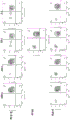
Figure 2 shows a strategy for preparing genetically modified porcine islet cells and a tolerogenic vaccine. Two clonal populations of swine were generated. At least one population of GGTA1 knockouts can be used to generate a tolerance vaccine. Another clonal population of pigs with at least GGTA1 and MHC I gene (e.g., NRLC5) knockouts is available for cell, tissue, and/or organ donors.
Figure 3 shows the use of positive and resistant vaccines (also referred to as negative vaccines).
Figure 4 shows an exemplary method of prolonging survival of a xenograft in a subject by infusing apoptotic donor splenocytes for tolerance to vaccination, masked by transient immunosuppression.
Fig. 5 illustrates an exemplary method of preventing rejection of a xenograft in a recipient or prolonging survival of a xenograft in the absence of long-term and extensive immunosuppression of the xenograft recipient. The exemplary method includes and integrates three components: i) lack and/or reduction of expression of α Gal, MHC class I, complement C3, and CXCL10, and genetically engineered islets with HLA-G transgene expression; ii) deficient and/or reduced expression of α Gal, Neu5Gc, and Sda/CAD and genetically engineered donor apoptotic and non-apoptotic monocytes (e.g., splenocytes) (e.g., genetically engineered vaccines) with or without HLA-G transgene expression of human CD47, human PD-L1, human PD-L2; and iii) administration of transient immunosuppression including antagonistic anti-CD 40 mAbs, anti-CD 20 mAbs, rapamycin, and transient anti-inflammatory therapy including compstatin (e.g., the compstatin derivative APL-2), anti-IL-6 receptor mAb, and soluble TNF receptor.
Figure 6 shows an exemplary protocol for preventing transplant rejection in porcine to cynomolgus monkey islet xenotransplantation. IE: islet equivalents; sTNFR: soluble TNF receptors (e.g., etanercept); α -IL-6R: anti-interleukin 6 receptor; tx'd: and (4) transplanting.
FIGS. 7A-7E show the strategy for cloning the px330-Gal2-1 plasmid targeting GGTA 1. FIG. 7A shows the cloning strategy and oligonucleotides (SEQ ID NO: 266-. FIG. 7B shows the insertion site on the px330 plasmid (SEQ ID NO: 268). FIG. 7C shows a flow diagram showing a cloning and verification policy. FIG. 7D shows the cloning site (SEQ ID NO:270) and sequencing primers (SEQ ID NO:269 and 271, respectively, in order of appearance). FIG. 7E shows the sequencing results (SEQ ID NO: 272-.
FIGS. 8A-8E show the strategy used to clone the px330-CM1F plasmid targeting CMAH. FIG. 8A shows the cloning strategy and oligonucleotides (SEQ ID NOS: 275 and 276, respectively, in order of appearance) used to prepare a guide RNA targeting CMAH 1. FIG. 8B shows the insertion site on the px330 plasmid (SEQ ID NO: 277). FIG. 8C illustrates a flow chart showing a cloning and verification strategy. FIG. 8D shows the cloning site (SEQ ID NO:279) and sequencing primers (SEQ ID NO:278 and 280, respectively, in order of appearance). FIG. 8E shows the sequencing results (SEQ ID NO:281-283, respectively, in the order of appearance).
Fig. 9A-9E show the strategy for cloning px330-NL1_ first plasmid targeting NLRC 5. FIG. 9A shows the cloning strategy and oligonucleotides (SEQ ID NOS: 284 and 285 in order of appearance, respectively) used to prepare a guide RNA targeting NLRC 5. FIG. 9B shows the insertion site on the px330 plasmid (SEQ ID NO: 286). FIG. 9C illustrates a flow diagram showing a cloning and verification policy. FIG. 9D shows the cloning site (SEQ ID NO:288) and sequencing primers (SEQ ID NO:287 and 289, respectively, in order of appearance). FIG. 9E shows the sequencing results (SEQ ID NO: 290-.
FIGS. 10A-10E show the strategy for cloning the px330/C3-5 plasmid targeting C3. FIG. 10A shows the cloning strategy and oligonucleotides (SEQ ID NOS: 293 and 294, respectively, in order of appearance) used to prepare C3-targeted guide RNAs. FIG. 10B shows the insertion site on the px330 plasmid (SEQ ID NO: 295). FIG. 10C illustrates a flow diagram showing a cloning and verification policy. FIG. 10D shows the cloning site (SEQ ID NO:297) and sequencing primers (SEQ ID NO:296 and 298, respectively, in order of appearance). FIG. 10E shows the sequencing results (SEQ ID NO:299-301, respectively, in order of appearance).
FIGS. 11A-11E show strategies for cloning the px330/B41_ second plasmid targeting B4GALNT 2. FIG. 11A shows the cloning strategy and oligonucleotides (SEQ ID NOS: 302 and 303, respectively, in order of appearance) used to prepare a guide RNA targeting B4GALNT 2. FIG. 11B shows the insertion site on the px330 plasmid (SEQ ID NO: 304). FIG. 11C illustrates a flow diagram showing a cloning and verification policy. FIG. 11D shows the cloning site (SEQ ID NO:306) and sequencing primers (SEQ ID NO:305 and 307, respectively, in order of appearance). FIG. 11E shows the sequencing results (SEQ ID NO: 308-310, respectively, in order of appearance).
Fig. 12 shows a map of Rosa26 locus sequenced in example 2.
Fig. 13A-13E illustrate strategies for cloning the px330/Rosa exon 1 plasmid targeting Rosa 26. FIG. 13A shows the cloning strategy and oligonucleotides (SEQ ID NO:311-312, respectively, in order of appearance) used to prepare a guide RNA targeting Rosa 26. FIG. 13B shows the insertion site on the px330 plasmid (SEQ ID NO: 313). FIG. 13C illustrates a flow chart showing a cloning and verification strategy. FIG. 13D shows the cloning site (SEQ ID NO:315) and sequencing primers (SEQ ID NO:314 and 316, respectively, in order of appearance). FIG. 13E shows the sequencing results (SEQ ID NO: 317-.
FIG. 14A shows a map of the genomic sequence of HLA-G. FIG. 14B shows a map of the cDNA sequence of HLA-G.
FIG. 15 shows an exemplary microscopic view of porcine fetal fibroblasts transfected with pSpCas9(BB) -2A-GFP.
FIG. 16 shows Fluorescence In Situ Hybridization (FISH) to GGTA1 gene by specific probes revealing position on chromosome 1.
Fig. 17A-17B show examples of phenotypic selection of cells with cas9/sgRNA mediated GGTA1/NLCR5 disruption. Figure 17A shows a genetically modified cell that does not express a-galactosidase. Figure 17B shows non-genetically modified cells expressing alpha-galactosidase and labeled with ferrous beads linked with Isolectin B4 (IB).
FIGS. 18A-18B show sequencing of DNA isolated from fetal cells from two litters of individual litters (gestation 1: FIG. 18A, or gestation 2: FIG. 18B) subjected to PCR amplification of the GGTA1 (Sscofa 10.2NCBI, reference sequence: NC-010443.4) target region compared to wild boar breed mixed chromosome 1, and the resulting amplicons were isolated on a 1% agarose gel. Amplicons were also analyzed by Sanger sequencing using separate forward primers from each reaction. In FIG. 18A, the results of alignment of fetuses 1-7 (SEQ ID NO: 322-328, respectively) from fetus 1 of pregnancy 1 with the reference and target gene sequences (SEQ ID NO:320-321, respectively) are shown. Fetuses 1, 2, 4, 5, 6 and 7 were truncated by 6 nucleotides after the target site of GGTA 1. Fetal 3 was truncated 17 nucleotides after the cleavage site, followed by 2,511(668-3179) nucleotide deletions, followed by single base substitutions. Truncations, deletions, and substitutions from a single sequencing experiment containing two copies of an allele from a target gene may only indicate that a genetic modification has occurred, without revealing the exact sequence of each allele. From this analysis, all 7 fetuses appeared to have a single allelic modification of GGTA 1. In FIG. 18B, the results of alignment of fetuses 1-5 (SEQ ID NO:331-335, respectively) from the fetal DNA sample from pregnancy 2 with the reference and target gene sequences (SEQ ID NO:329-330, respectively) are shown. Fetuses 1, 3, 4 and 5 were truncated by 3 nucleotides from the GGTA1 gene target site. Fetal 2 has variability in Sanger sequencing, suggesting either complex variability of DNA mutations or poor sample quality. However, the fetal DNA template quality was sufficient for performing GGTA1 gene screening experiments as described above.
FIGS. 19A-19B show sequencing of DNA isolated from fetal cells from two litters of individual pups (gestation 1: FIG. 19A, or gestation 2: FIG. 19B) subjected to PCR amplification of the NLRC5 (consensus) target region, and the resulting amplicons were separated on a 1% agarose gel. Amplicons were also analyzed by Sanger sequencing using separate forward primers from each reaction. In FIG. 19A, the results of alignment of fetuses 1, 3, 5, 6 and 7 (SEQ ID NO: 338-. Sequence analysis of the NLRC5 target site failed to show consistent alignment, suggesting different DNA modifications between NLRC5 alleles of unknown complexity in the sequencing reaction or complicating Sanger sequencing reactions and analysis. In FIG. 19B, the results of the alignment of fetuses 1-5 from pregnancy 2 (SEQ ID NO: 345-. The NLRC5 gene amplicons of fetuses 1-5 were all truncated 120 nucleotides downstream of the NLRC5 gene cleavage site.
FIGS. 20A-20B show data from fetal DNA isolated from hindlimb biopsies (wild type (WT) and 1-7 (FIG. 20A: gestation 1) or 1-5 (FIG. 20B: gestation 2)). The target gene was amplified by PCR and the PCR products were separated on a 1% agarose gel and visualized by fluorescent DNA stain. The amplicon band present in the WT lane represents the unmodified DNA sequence. An increase or decrease in amplicon size indicates an insertion or deletion, respectively, within the amplicon. The variation in DNA modification between alleles in a sample can make the bands appear more dispersed. Pregnancy 1 (fig. 20A) produced 7 fetuses, whereas pregnancy 2 (fig. 20B) produced 5 fetuses, harvested on days 45 and 43, respectively. The absence of a band in the NLRC5 gel (bottom gel) in fetuses 1, 3, and 4 of fig. 20A indicates that modification of the target region has disrupted DNA amplification primer binding. The presence of all bands in GGTA1 in fig. 20A (top gel) indicates that the DNA quality is sufficient to produce DNA amplicons in NLRC5 targeted PCR reactions. Fetuses 1, 2, 4 and 5 of pregnancy 1 (fig. 20A) had larger GGTA1 amplicons than WT, indicating an insertion in the target region. In fetus 3 of pregnancy 1 (fig. 20A), GGTA1 amplicon migrated faster than the WT control, indicating a deletion in the target region. In fetuses 6 and 7 of pregnancy 1 (fig. 20A), the NLRC5 amplicon migrated faster than the WT, indicating a deletion in the target region. GGTA1 amplicons of fetuses 1-5 (fig. 20B) were difficult to interpret by size and were scattered compared to WT controls. Size and density of NLRC5 amplicons were uniform for fetuses 1-5 (fig. 20B) compared to wild type controls.
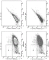
Fig. 21A-21E show phenotypic analysis from littermate individual piglets (fig. 21A, 21B, 21C: pregnancy 1, or 21D-21E: pregnancy 2). Fetuses were harvested on either day 45 (gestation 1) or day 43 (gestation 2) and processed for DNA and culture cell isolation. Tissue debris and cells were plated in culture for 2 days to allow fetal cells to attach and grow. Wild type cells (non-genetically modified foetal cells) and foetal cells from pregnancies 1 and 2 were removed from the plates and labelled with IB4 lectin conjugated to Alexa fluor 488 or anti-porcine MHC class I antibody conjugated to FITC. Flow cytometry analysis is shown as a histogram depicting the intensity of the label of the test cells. Histograms of WT cells were included in each frame to emphasize the reduction in overall intensity of fetal cells per group. There was a decrease in α Gal and MHC class I markers in pregnancy 1 (fig. 21A), expressed as a decrease in peak intensity. In pregnancy 2 (fig. 21B), fetuses 1 and 3 had a greater reduction in α gal markers and a significant reduction in MHC class 1 markers compared to WT fetal cells.
FIGS. 22A-22C show the effect of reduced MHC class I expression in cells from fetus 3 (gestation 1) compared to wild type foetal cells from a genetic clone. Proliferative responses of human CD8+ cells and CD4T cells to porcine control fibroblasts and NLRC5 knockout fetal cells were measured. Figure 22a. cells were gated to CD4 or CD8 prior to assessing proliferation. Figure 22b. decreased CD8T cell proliferation following therapeutic stimulation by porcine fetal GGTA1/NLRC5 knockout cells compared to control unmodified porcine fibroblasts. When human responders were treated with porcine fetal GGTA1/NLRC5 knock-out cells at a ratio of 1:1, almost a 55% reduction in CD8T cell proliferation was observed. Wild type foetal cells induced 17.2% proliferation in human CD8T cells, whereas MHC class I deficient cells from foetus 3 (gestation 1) induced only 7.6% proliferation. Figure 22c no difference was observed in the CD8T cell proliferation response at the 1:5 and 1:10 ratios compared to unmodified fetal cells. At all ratios of the study, no changes were observed in CD4T cell proliferation in response to the NLRC5 knockout and control unmodified porcine fetal cells.
Figure 23 shows live birth of GGTA1/NLRC5 knockout piglets produced using CRISPR/Cas technology.
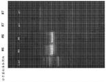
Fig. 24A-24C show DNA gel analysis of genotypes of piglets produced in example 6. Fig. 24A shows the results of the first PCR experiment in example 6. Fig. 24B shows the results of the second PCR experiment in example 6. FIG. 24C shows the results of the third PCR experiment in example 6.
FIG. 25A shows sequencing data and sequence determination (SEQ ID NO:350) of a portion of the NLRC5 gene of piglet # 1. FIG. 25B shows sequencing data and sequence determination (SEQ ID NO:351) of a portion of the NLRC5 gene of piglet # 2. FIG. 25C shows sequencing data and sequence determination (SEQ ID NO:352) of a portion of the NLRC5 gene of piglet # 4. FIG. 25D shows sequencing data and sequence determination (SEQ ID NO:353) of a portion of the NLRC5 gene of piglet # 5. FIG. 25E shows sequencing data and sequence determination (SEQ ID NO:354) of a portion of the NLRC5 gene of piglet # 6. FIG. 25F shows sequencing data and sequence determination (SEQ ID NO:355) of a portion of the NLRC5 gene of piglet # 7.
FIG. 26A shows the left arm of Rosa26 in example 8 (SEQ ID NO: 356). FIG. 26B shows a DNA gel analysis of the constructs used for homologous recombination in example 8. FIG. 26C shows the consensus sequence of the amplicon (SEQ ID NO:357) based on the pairwise read analysis in example 8. FIG. 26D (SEQ ID NO:358), FIG. 26E (SEQ ID NO:359) and FIG. 26F (SEQ ID NO:360) show homology-directed recombinant constructs for insertion of HLA-G1 at the Rosa26 locus in example 8.
FIG. 27A shows the sequence (SEQ ID NO:362) and sequencing primers (SEQ ID NO:361 and 363, respectively, in order of appearance) of the correct px330 plasmid containing an oligonucleotide targeting Rosa26 generated in example 8. Fig. 27B shows the sequencing results of the px330 plasmid containing an oligonucleotide targeting Rosa26 constructed in example 8. SEQ ID NO 364-366 is disclosed in order of appearance. Figure 27C shows restriction digestion of the px330 plasmid containing an oligonucleotide targeting Rosa26 constructed in example 8.
FIG. 28 shows a map of the GalMet plasmid used in example 8 and oligonucleotides (SEQ ID NO: 367-.
Figure 29 shows an in vitro Cas 9-mediated cleavage reaction of an in vitro transcribed gRNA. Lane 1: uncut pig Rosa26(2000 bp). Lane 2: design gRNA-directed Cas9 cleavage of porcine Rosa 26; lane 3: uncut pig GGTA 1; lane 4: design of GGTA1 template gRNA-directed Cas9 cleavage.
Figure 30 shows sorting of the genetically modified cells produced in example 8 by flow cytometry.
FIG. 31 shows the construct (SEQ ID NO:369) generated in example 9 for the homologous recombination of CD47 with the GGTA1 locus.
FIG. 32 shows the sequences of the right arm (FIG. 32A; SEQ ID NO:370) and left arm (FIG. 32B; SEQ ID NO:371) of the GGTA1 locus in example 9.
Fig. 33A, 33B and 33C show sorting of unstained cells in example 9.
Fig. 34A, 34B and 34C show sorting of px 330-stained cells in example 9.
Fig. 35A, 35B and 35C show sorting of IB4 stained cells in example 9.
FIGS. 36A, 36B and 36C show sorting of CD47/IB4 stained cells in example 9.
Fig. 37A, 37B and 37C show IB4 stained cells, CD47/IB4 stained cells sorted in example 9.
FIGS. 38A, 38B and 38C show CD47/IB4 stained cells sorted in example 9.
Figure 39 shows the gating strategy used to select single cells and live cells for analysis. Total CD3+ cells were observed, and CD4+ and CD8+ cells were selected and counted in this population for experimental parameters.
Fig. 40A and 40B show that a. unstimulated cells in quadrant 2 show insignificant expansion when under the same culture conditions as the same cells stimulated with PHA. Pha stimulation induced proliferation of 20.7% (CD3), 24.7% (CD4), 18.4% (CD8) and 21% (CD20) in lymphocyte samples, indicating that there may be a maximum amount of stimulation in this assay.
Figure 41 shows flow cytometry results of co-culture assays in which CD8+ T cells were added at dilutions of 100:1, 50:1, 10:1, or 1:1 to cultures of attached wild-type or genetically engineered porcine fibroblasts. Wild type cells stimulate T cell proliferation at ratios of 50:1, 10:1 and 1: 1. Genetically Modified (GM) cells # 3 and #4 showed little effect in stimulating T cells at ratios of 100:1, 50:1 and 10:1, indicating that the T cell proliferative response was completely abolished.
Figure 42 shows flow cytometry results of co-culture assays in which CD4+ T cells were added at dilutions of 100:1, 50:1, 10:1, and 1:1 to cultures of attached wild-type or genetically engineered porcine fibroblasts. GM cells # 3 and #4 showed little effect in stimulating T cells at ratios of 100:1, 50:1 and 10:1, indicating that the T cell proliferative response was completely abrogated.
Figure 43 shows flow cytometry results of co-culture assays in which CD3+ T cells (total CD4 and CD8) were added at dilutions of 100:1, 50:1, 10:1, and 1:1 to cultures of attached wild-type or genetically engineered porcine fibroblasts. GM cells # 3 and #4 showed little effect in stimulating T cells at ratios of 100:1, 50:1 and 10:1, indicating that the T cell proliferative response was completely abrogated.
FIG. 44 shows that B cell proliferation was inhibited by about 50% when incubated with GGTA1/NLRC5 knockout cells compared to wild type cells.
Figure 45 shows the flow cytometry results of a co-culture assay in which cytokines were measured by incubating human lymphocytes with wild-type or GM cells, followed by introduction of brefeldin a to block endocytosis resulting in intracellular accumulation of 4 cytokines in endosomes. The fixation and permeabilization of the cells allows intracellular measurements of cytokine accumulation. In the CD8T cell population, no IL2 stimulation was observed at the 100:1 ratio, and modest reductions in CD107a, perforin and granzyme were observed at the 100:1 ratio. Perforin and granzyme B double positive cells were significantly inhibited at 100:1 and 10:1 ratios.
Figure 46 shows flow cytometry results of coculture assays of human lymphocytes with wild-type or genetically modified porcine fibroblasts at a T cell to FC ratio of 10: 1. In the CD8T cell population, IL2 was stimulated at a ratio of 10:1, and thus reduced by about 40% in culture of porcine cells with genetic modifications. CD107a expression was reduced by about 25%. Perforin expression was reduced by about 40%, while granzyme was unaffected at this incubation rate.
Figure 47 shows flow cytometry results of coculture assays of human lymphocytes with wild-type or genetically modified porcine fibroblasts at a T cell to FC ratio of 10: 1. CD107a was reduced by about 50% in CD3 cells. Perforin and granzyme B were also reduced after incubation with genetically modified cells and reflected when compared to double positive cells withdrawn from quadrant 2.
Figure 48 shows flow cytometry results of coculture assays of human lymphocytes with wild-type or genetically modified porcine fibroblasts at a T cell to FC ratio of 10: 1. CD4+ T cells are less activated in the presence of GM cells to produce cytokines. IL2 expression was reduced by 40%. CD107a was reduced by approximately 50%. Perforin and granzyme B were reduced by about 50% and 30%, respectively.
Figure 49 shows flow cytometry results of coculture assays of human lymphocytes with wild-type or genetically modified porcine fibroblasts at a T cell to FC ratio of 10: 1. In CD3 cells, IFN γ expression was significantly reduced when lymphocytes were cultured with GM porcine fibroblasts at a ratio of 10: 1. TNF α expression was low in culture with wild type cells, but decreased in culture with GM cells. In this experiment, granzyme B was also significantly reduced when incubated with GM cells compared to wild type cells.
Figure 50 shows flow cytometry results of coculture assays of human lymphocytes with wild-type or genetically modified porcine fibroblasts at a T cell to FC ratio of 10: 1. In CD4 cells, IFN γ expression was significantly reduced when lymphocytes were cultured with GM porcine fibroblasts at a ratio of 10: 1. TNF α expression was low in culture with wild type cells, but decreased in culture with GM cells. In this experiment, granzyme B was also significantly reduced when incubated with GM cells compared to wild type cells.
Figure 51 shows flow cytometry results of coculture assays of human lymphocytes with wild-type or genetically modified porcine fibroblasts at a T cell to FC ratio of 10: 1. In CD8 cells, IFN γ expression was significantly reduced when lymphocytes were cultured with GM porcine fibroblasts at a ratio of 10: 1. TNF α expression was low in culture with wild type cells, but decreased in culture with GM cells. In this experiment, granzyme B was also significantly reduced when incubated with GM cells compared to wild type cells.
Figure 52 shows flow cytometry results of coculture assays of human lymphocytes with wild-type or genetically modified porcine fibroblasts at a T cell to FC ratio of 10: 1. NK cells (CD56+) have been shown to be activated in the absence of MHC class I expression on the cells. IFN γ (y-axis) and granzyme B (x-axis) were expressed in co-culture with wild type cells, but were significantly reduced when co-cultured with GGTA1/NLRC5 knock-out cells. No change in GM or TNF α expression was observed in GM cells compared to wild type cells.
Figure 53 shows that human PBMCs incubated with wild type porcine fibroblasts had a normal background percentage (11%) of CD4 positive T cells expressing IL 10. GGTA1/NLRC5 knock-out cells (13.3% and 20.2%) labeled #3 and #4, respectively, had a slight effect on IL10 expression. Porcine fibroblasts expressing human challenge HLAG1 protein optimized for expression in pigs induced production of IL10 by 60.7% of human CD4+ T cells.
FIG. 54 shows that soluble HLA-G (100ng/ml) blocks proliferation of CD8+, CD8-, and PBMCs in cultures with wild-type porcine islets. Q1 and Q2 show the proliferative (CFSE lo) and non-proliferative (CFSE hi) fractions, respectively.
Figure 55 shows flow cytometry gating strategies for analysis of cytokines from CD3, CD4, or CD8 populations, and effector function molecule analysis of human T cells cultured with genetically modified porcine fibroblasts (expressing HLAG1), wild-type or wild-type plus PT85 antibody.
Figure 56 shows flow cytometric data for the CD4 population co-cultured with wild-type porcine fibroblasts, wild-type porcine fibroblasts with PT85 antibody, or porcine fibroblasts expressing HLAG 1. With cells blocking PT85 or expressing HLAG1, a large reduction in cytokine levels (IL-2) and effector molecule secretion was observed in MLR cultures at 10:1 and 1:1 ratios. PT85 blocking antibody was used to determine how much the immunosuppressive effect observed was due to NLRC5 knock-out (MHC class 1 null) or GGTA1 knock-out. The PT85 antibody mimics the effect of NLRC5 knockdown in the presence of normal wild-type α -Gal surface expression. The expression of HLAG1 protein on the cell surface has obvious inhibition effect on the generation of CD4+ and CD8+ T cell cytokines and effector functions.
Figure 57 shows flow cytometric data for the CD8 population co-cultured with wild-type porcine fibroblasts, wild-type porcine fibroblasts with PT85 antibody, or porcine fibroblasts expressing HLAG 1. With cells blocking PT85 or expressing HLAG1, a large reduction in cytokine levels (IL-2) and effector molecule secretion was observed in MLR cultures at 10:1 and 1:1 ratios. PT85 blocking antibody was used to determine how much the immunosuppressive effect observed was due to NLRC5 knock-out (MHC class 1 null) or GGTA1 knock-out. The PT85 antibody mimics the effects of NLRC5 knockdown in the presence of normal wild-type α -Gal surface expression within the CD8 population. The expression of HLAG1 protein on the cell surface has obvious inhibition effect on the generation of CD4+ and CD8+ T cell cytokines and effector functions. The expression of HLAG1 protein on the cell surface has obvious inhibition effect on the generation of CD4+ and CD8+ T cell cytokines and effector functions.
Figure 58 shows flow cytometric data for the CD4 population co-cultured with wild-type porcine fibroblasts, wild-type porcine fibroblasts with PT85 antibody, or porcine fibroblasts expressing HLAG 1. With cells blocking PT85 or expressing HLAG1, a large reduction in cytokine levels (TNF-a, IFN-g) and effector molecule secretion was observed in MLR cultures at 10:1 and 1:1 ratios. PT85 blocking antibody was used to determine how much the immunosuppressive effect observed was due to NLRC5 knock-out (MHC class 1 null) or GGTA1 knock-out. The PT85 antibody mimics the effect of NLRC5 knockdown in the presence of normal wild-type α -Gal surface expression. The expression of HLAG1 protein on the cell surface has obvious inhibition effect on the generation of CD4+ and CD8+ T cell cytokines and effector functions.
Figure 59 shows flow cytometric data for the CD8 population co-cultured with wild-type porcine fibroblasts, wild-type porcine fibroblasts with PT85 antibody, or porcine fibroblasts expressing HLAG 1. With cells blocking PT85 or expressing HLAG1, a large reduction in cytokine levels (TNF-a, IFN-g) and effector molecule secretion was observed in MLR cultures at 10:1 and 1:1 ratios. PT85 blocking antibody was used to determine how much the immunosuppressive effect observed was due to NLRC5 knock-out (MHC class 1 null) or GGTA1 knock-out. The PT85 antibody mimics the effect of NLRC5 knockdown in the presence of normal wild-type α -Gal surface expression. The expression of HLAG1 protein on the cell surface has obvious inhibition effect on the generation of CD4+ and CD8+ T cell cytokines and effector functions.
Figure 60 shows a flow gating scheme for cell proliferation/CFSE low population analysis.
Fig. 61A and 61B show flow cytometric analysis of cell proliferation (CFSE dilution) experiments of CD3, CD4, or CD8 populations in a.
Figure 62 shows that T cell proliferation is reduced following stimulation of porcine fibroblasts treated with a PT-85 blocking antibody (Ab) compared to control unmodified porcine fibroblasts/wild type, at a ratio of 10:1 for human PBMC to FC, respectively. A substantial reduction in T cell (CD3) proliferation was observed when human responders were treated with SLA-I blocking PT-85 antibody or HLA-G expression at 10:1 and 1:1 ratios. There was not much difference in T cell proliferative response at 100:1 and 50:1 ratios compared to unmodified/wild type porcine fibroblasts.
Figure 63 shows that T cell proliferation is reduced following stimulation of porcine fibroblasts treated with a PT-85 blocking antibody compared to control unmodified porcine fibroblasts/wild type, at a ratio of 10:1 for human PBMC to FC, respectively. A substantial reduction in T cell (CD4) proliferation was observed when human responders were treated with SLA-I blocking PT-85 antibody or HLA-G expression at 10:1 and 1:1 ratios. There was not much difference in T cell proliferative response at 100:1 and 50:1 ratios compared to unmodified/wild type porcine fibroblasts.
Figure 64 shows reduced T cell proliferation following stimulation of porcine fibroblasts treated with a PT-85 blocking antibody compared to control unmodified porcine fibroblasts/wild type, at a ratio of 10:1 of human PBMC to FC, respectively. A substantial reduction in T cell (CD8) proliferation was observed when human responders were treated with SLA-I blocking PT-85 antibody or HLA-G expression at 10:1 and 1:1 ratios. There was not much difference in T cell proliferative response at 100:1 and 50:1 ratios compared to unmodified/wild type porcine fibroblasts.
Figure 65 shows reduced T cell proliferation following stimulation of porcine fibroblasts treated with a PT-85 blocking antibody compared to control unmodified porcine fibroblasts/wild type, at a ratio of 10:1 of human PBMC to FC, respectively. By blocking SLA-I or HLA-G expression with PT-85, B cell proliferation was not greatly reduced.
Figure 66 shows that IFN γ is produced primarily by Natural Killer (NK) and natural killer t (nkt) cells as part of the innate immune response. DKO # 3 and #4 are genetically and phenotypically GGTA1/NLRC5 knockout cells prepared separately. After antigen-specific immunity has developed, IFN γ is also produced by CD4Th1 and CD8 Cytotoxic T Lymphocyte (CTL) effector T cells.
FIG. 67 shows GMC-SF production in genetically modified cells cultured with human immune cells and controls. Double Knock Out (DKO) cells failed to stimulate GM-CSF production. Expression of HLAG1 was significantly reduced. DKO # 3 and #4 are genetically and phenotypically GGTA1/NLRC5 knockout cells prepared separately.
FIG. 68 shows IL-17A expression in genetically modified cells cultured with human immune cells. Neither DKO nor HLAG1 transgenic cells were able to induce a pro-inflammatory response in human PBMCs.
Figure 69 shows CXXXC chemokine (Fractalkine) expression in genetically modified porcine cells cultured with human immune cells. Although expressed on a logarithmic scale, HLAG1 expression remains an important inhibitor of T cell activation and CXXXC chemokine production.
Figure 70 shows TNF α expression in genetically modified porcine cells cultured with human immune cells.
FIG. 71 shows IL-6 production in genetically modified porcine cells cultured with human immune cells.
FIG. 72 shows IL-4 production in genetically modified porcine cells cultured with human immune cells.
Figure 73 shows MIP1 a production in genetically modified porcine cells cultured with human immune cells.
Figure 74 shows MIP1 β production in genetically modified porcine cells cultured with human immune cells.
Figure 75 shows that T cell proliferation is reduced following stimulation of porcine fibroblasts treated with a PT-85 blocking antibody compared to control unmodified porcine fibroblasts/wild type, at a ratio of 10:1 for human PBMC to FC, respectively. A substantial reduction in T cell (CD3) proliferation was observed when human responders were treated with SLA-I blocking PT-85 antibody or HLA-G expression at 10:1 and 1:1 ratios. There was not much difference in T cell proliferative response at 100:1 and 50:1 ratios compared to unmodified/wild type porcine fibroblasts.
Figure 76 shows that T cell proliferation is reduced following stimulation of porcine fibroblasts treated with a PT-85 blocking antibody compared to control unmodified porcine fibroblasts/wild type, at a ratio of 10:1 for human PBMC to FC, respectively. A substantial reduction in T cell (CD4) proliferation was observed when human responders were treated with SLA-I blocking PT-85 antibody or HLA-G expression at 10:1 and 1:1 ratios. There was not much difference in T cell proliferative response at 100:1 and 50:1 ratios compared to unmodified/wild type porcine fibroblasts.
Figure 77 shows reduced T cell proliferation following stimulation of porcine fibroblasts treated with a PT-85 blocking antibody compared to control unmodified porcine fibroblasts/wild type, at a ratio of 10:1 of human PBMC to FC, respectively. A substantial reduction in T cell (CD8) proliferation was observed when human responders were treated with SLA-I blocking PT-85 antibody or HLA-G expression at 10:1 and 1:1 ratios. There was not much difference in T cell proliferative response at 100:1 and 50:1 ratios compared to unmodified/wild type porcine fibroblasts.
Figure 78 shows reduced T cell proliferation following stimulation of porcine fibroblasts treated with a PT-85 blocking antibody compared to control unmodified porcine fibroblasts/wild type, at a ratio of 10:1 of human PBMC to FC, respectively. By blocking SLA-I or HLA-G expression with PT-85, B cell proliferation was not greatly reduced.
Figure 79 shows IFN γ expression after co-culture of human mixed lymphocytes and porcine genetically modified cells. Double Knockouts (DKO) #3 and #4 are genetically and phenotypically GGTA1/NLRC5 knockouts prepared, respectively. HLA-G1 transgenic cells were performed in separate experiments with human donor #2 (FIG. 79B) (but not human donor # 1; FIG. 79A), thus including matched unstimulated and wild-type cell controls.
FIG. 80 shows GM-CSF γ expression following co-culture of human mixed lymphocytes and porcine genetically modified cells. Double Knockouts (DKO) #3 and #4 are genetically and phenotypically GGTA1/NLRC5 knockouts prepared, respectively. HLA-G1 transgenic cells were performed in separate experiments with human donor #2 (FIG. 80B) (but not human donor # 1; FIG. 80A), thus including matched unstimulated and wild-type cell controls.
FIG. 81 shows IL-2 expression after co-culture of human mixed lymphocytes and porcine genetically modified cells. Double Knockouts (DKO) #3 and #4 are genetically and phenotypically GGTA1/NLRC5 knockouts prepared, respectively.
FIG. 82 shows IL-17 expression after co-culture of human mixed lymphocytes and porcine genetically modified cells from two donors (FIG. 80A and FIG. 80B). Double Knockouts (DKO) #3 and #4 are genetically and phenotypically GGTA1/NLRC5 knockouts prepared, respectively. Neither DKO nor HLA-G1 transgenic cells induced a pro-inflammatory response in human PBMCs.
Figure 83 shows CXXXC chemokine expression after coculture of human mixed lymphocytes and porcine genetically modified cells. Double Knockouts (DKO) #3 and #4 are genetically and phenotypically GGTA1/NLRC5 knockouts prepared, respectively. HLA-G1 transgenic cells were performed in separate experiments with human donor #2 (FIG. 83B) (but not human donor # 1; FIG. 83A), thus including matched unstimulated and wild-type cell controls. Despite being expressed on a logarithmic scale, HLA-G1 expression remains an important inhibitor of T cell activation and CXXXC chemokine production.
Figure 84 shows TNF α expression after co-culture of human mixed lymphocytes and porcine genetically modified cells. Double Knockouts (DKO) #3 and #4 are genetically and phenotypically GGTA1/NLRC5 knockouts prepared, respectively. HLA-G1 transgenic cells were performed in separate experiments with human donor #2 (FIG. 84B) (but not human donor # 1; FIG. 84A), thus including matched unstimulated and wild-type cell controls.
FIG. 85 shows IL-6 expression after co-culture of human mixed lymphocytes and porcine genetically modified cells. Double Knockouts (DKO) #3 and #4 are genetically and phenotypically GGTA1/NLRC5 knockouts prepared, respectively. HLA-G1 transgenic cells were performed in separate experiments with human donor #2 (FIG. 85B) (but not human donor # 1; FIG. 85A), thus including matched unstimulated and wild-type cell controls.
FIG. 86 shows IL-4 expression after co-culture of human mixed lymphocytes and porcine genetically modified cells. Double Knockouts (DKO) #3 and #4 are genetically and phenotypically GGTA1/NLRC5 knockouts prepared, respectively. HLA-G1 transgenic cells were performed in separate experiments with human donor #2 (FIG. 86B) (but not human donor # 1; FIG. 86A), thus including matched unstimulated and wild-type cell controls.
FIG. 87 shows MIP-1 α expression after co-culture of human mixed lymphocytes and porcine genetically modified cells. Double Knockouts (DKO) #3 and #4 are genetically and phenotypically GGTA1/NLRC5 knockouts prepared, respectively. HLA-G1 transgenic cells were performed in separate experiments with human donor #2 (FIG. 87B) instead of human donor # 1; FIG. 87A, thus including matched unstimulated and wild-type cell controls.
FIG. 88 shows MIP-1 β expression after co-culture of human mixed lymphocytes and porcine genetically modified cells. Double Knockouts (DKO) #3 and #4 are genetically and phenotypically GGTA1/NLRC5 knockouts prepared, respectively. HLA-G1 transgenic cells were performed in separate experiments with human donor #2 (FIG. 88B) (but not human donor # 1; FIG. 88A), thus including matched unstimulated and wild-type cell controls.
Figure 89 shows CRISPR/Cas constructs within PX333 vector.
Fig. 90 shows a schematic transfection of primary porcine fibroblasts using the following constructs: GGTA1-10/B4GALNT2 (condition 2), NLRC5-6/B4GALNT2 (condition 3), GGTA1-10/B4GALNT2 and NLRC5-6/B4GALNT2 (condition 4), condition 1 (wild type): only cells.
Figure 91 shows selection of genetic modifications using magnetic bead sorting.
FIG. 92 shows the selection of genetic modifications for Cell sorting (Cell Sort) using SLA I +/IB4+ (top right), SLAI +/IB 4- (bottom right), SLA I-/IB4+ (top left), and SLA I-/IB4 (bottom left).
Figure 93 shows flow cytometry analysis for condition 2: GGTA1-10/B4GALNT 2.
Figure 94 shows flow cytometry analysis for condition 3: NLRC5-6/B4GALNT 2.
Figure 95 shows flow cytometric analysis of condition 4: GGTA1-10/B4GALNT2+ NLRC5-6/B4GALNT 2.
Figure 96 shows flow cytometry analysis for condition 2: GGTA1-10/B4GALNT2 after sorting. Each population was sorted to confirm that the correct population was obtained after sorting and that there were no cross samples from other gates (gates).
Figure 97 shows flow cytometry analysis for condition 3: NLRC5-6/B4GALNT 2. Each population was sorted to confirm that the correct population was obtained after sorting and that there were no cross-samples from other gates.
Figure 98 shows flow cytometry analysis for condition 4: GGTA1-10/B4GALNT2+ NLRC5-6/B4GALNT 2. Each population was sorted to confirm that the correct population was obtained after sorting and that there were no cross-samples from other gates.
Fig. 99A and 99B show the following flow cytometric analysis of IB4 lectin: A. unstained wild type, all cells unstained, wild type negative and condition #2Gal negative fractions cultured with wild type or PFF 1. B. Side scatter and forward scatter of condition #4Gal negative fraction, wild type positive, condition #2Gal positive fraction, condition #3Gal positive fraction, or condition #4Gal positive fraction cultured with wild type or PFF 1.
Figure 100 shows flow cytometric quantification of cells genetically modified under conditions 1 (wild type), 2, 3, and 4 (left to right, respectively).
Fig. 101A and 101B show the following flow cytometric analysis of IB4 lectin: A. unstained wild type, all cells unstained, wild type negative and condition #2Gal negative fractions cultured with wild type or PFF 1. B. Side scatter and forward scatter of condition #4Gal negative fraction, wild type positive, condition #2Gal positive fraction, condition #3Gal positive fraction, or condition #4Gal positive fraction cultured with wild type or PFF 1.
Fig. 102 shows flow cytometric quantification of SLA1(FITC) as follows: A. condition 3 cells, and b. condition 4 cells.
Fig. 103A and 103B show the following confocal microscopy: A. imaging results of wild type porcine cells and genetically modified conditional 2, 3 and 4 cells. B. An imaged slide is produced.
Figure 104 shows sequencing results of NLRC5 sequencing of condition and condition 4 cell lines. 372-376 in the order of occurrence are disclosed.
FIG. 105 shows tables of PCR oligonucleotides and target sequences (second column) for GG1, Gal2-1, Gal 2-2, Gal 2-3, Gal 2-4, Gal 2-5, GGTA1-10, GGTA1-11, GGTA1-16, NL1, NLRC5-6, NLRC5-7, NLRC 5-8. 377-404 are disclosed in the order of appearance in column 2, respectively. In column 4, 405-413 SEQ ID NOS are disclosed, respectively, in order of appearance. 414-422 in the order of appearance are disclosed in column 6, respectively.
Figure 106 shows a table of PCR oligonucleotides and target sequences (second column) for CM1F, CM2RS, CM3RS, CM4 RS. In column 2, 423-430 are disclosed in the order of appearance, respectively. 431-434 in the order of appearance are disclosed in column 4, respectively. In column 6, SEQ ID NO 435-437 is disclosed in the order of appearance, respectively.
Fig. 107A and 107B show the following tables: A. target sequences for gRNAs for B41, C3-9_1, C3-9_2, C3-5_1, C3-5_2, C3-15RS _1, C3-15RS _ 2. In column 2, the sequences of occurrence are disclosed as SEQ ID NO 438-447, respectively. B. Deletion of the screening primer sequences and their respective target sequences for Gal 1. 448-453 are disclosed in the order of appearance in column 2, respectively.
FIG. 108 shows an overview of the Gal2-2(B4GALNT2) vector and cloning strategy. The nucleotide sequence of a portion of the vector (SEQ ID NO:454), and two oligonucleotides: gal2-2_ Forward (SEQ ID NO:455) and Gal2-2_ reverse (SEQ ID NO: 456).
FIG. 109 shows the expected Gal2-2(B4GALNT2) cloning sequence after correct insertion based on the vector and cloning strategy of FIG. 113 (top panel). Respectively discloses SEQ ID NO 457-459 in the order of appearance. In the following figure, the sequencing result of the constructed plasmid (SEQ ID NO:462) was aligned with the expected sequence (SEQ ID NO: 460-461).
Fig. 110A and 110B. A. The Gal2-1(B4GALNT2) target site and two oligonucleotides (Gal 2-1-screener-Forward-1, SEQ ID NO: 463; and Gal 2-1-screener-Reversal-1, SEQ ID NO:465) within the GGTA1 gene (SEQ ID NO:464) are shown. B. Gal2-1_ Screen _1 primer set, Gal2-1_ Screen primer set PCR product observed on the gel and expected amplicon size of 303bp are shown. The strong single band observed at the expected amplicon size product was sequence verified and shown to include the Gal2-1 target cleavage site required for the screen.
Fig. 111A and 111B. A. The CM1F target site and two oligonucleotides (CM 1F-1-screenout-Forward-1, SEQ ID NO: 466; and CM 1F-1-screenout-reverse-1, SEQ ID NO:468) within the CMAH gene (SEQ ID NO:467) are shown. B. The CM1F _ screen _1 primer set, the CM1F _ screen primer set PCR product observed on the gel, with the expected amplicon size of 309bp, is shown. A strong band was observed at the expected amplicon size; faint bands were also observed at-600 bp. The product of approximately 300bp was sequence verified and shown to include the target cleavage site required for screening.
Fig. 112A and 112B. A. NL 1-first target site and two oligonucleotides (NLR amp2 forward, SEQ ID NO: 469; and NLR amp2 reverse, SEQ ID NO:471) within the NLRC5 gene (SEQ ID NO:470) are shown. B. The NLR amp2 primer set with the expected amplicon size of 217bp, the NLR amp2 primer set PCR product observed on the gel, the strong single band observed at the expected amplicon size are shown. The product was sequence verified and shown to include the NL1 — first target cleavage site required for the screen.
FIGS. 113A to 113I represent exon 1 genomic modifications of Gal2-2 and NLRC5 genes. A. The positions of the screening primers for Gal are shown. B. Gal2-2PCR screening using Gal2-2 screening 1 primer. Sequence result of Gal2-2. 472-478 in the order of appearance are disclosed, respectively. D. GGTA1-10PCR screening using GGTA1-10,11 screening primers. LRC5-6 screening primer position. NLRC5-6 group A (NLRC5-678 screening primer). NLRC5-6 sequence results from group a. SEQ ID NO 479-486 are disclosed in order of appearance, respectively. NLRC5-6 group B (NLRC5-678 forward and NLR first screen 2 reverse screen primers). NLRC5-6 group C (NLRC5-678 forward and NLR first screen 2 reverse screen primers).
Fig. 114A-114C show live birth of GGTA1/NLRC5 knockout/HLA-G1 knock-in piglets generated using CRISPR/Cas technology.
FIG. 115 shows the sequencing results confirming the insertion of HLA-G1 into the ROSA gene site. 499 is disclosed.
FIG. 116 shows the sequence results confirming that the homology-directed recombinant construct for insertion of HLA-G1 at the Rosa26 locus was correctly constructed in example 8. 500 is disclosed in SEQ ID NO.
Figure 117 shows the left arm sequence corresponding to the Rosa26 locus, which can be used to construct a homology targeting vector for inserting HLA-G1 or another sequence into the Rosa26 locus. 501, SEQ ID NO.
Figure 118 shows the sequence of a modified HLA-G coding sequence that can be used to construct a homology targeting vector for inserting HLA-G1 into a genetic locus, such as the Rosa26 locus. 502 is disclosed.
Figure 119 shows the right arm sequence corresponding to the Rosa26 locus, which can be used to construct a homology targeting vector for inserting HLA-G1 or another sequence into the Rosa26 locus. SEQ ID NO 503 is disclosed.
Detailed Description
The following description and examples set forth in detail embodiments of the invention. It is to be understood that this invention is not limited to the particular embodiments described herein, and that modifications may be made thereto. Those skilled in the art will recognize that there are numerous variations and modifications of the present invention, which are encompassed within the scope of the present invention.
Graft rejection may be prevented by methods of modulating an immune response, including those described herein. For example, a method of preventing or extending the time to transplant rejection with or without minimized use of immunosuppressive drugs as described herein can, for example, genetically alter an animal, e.g., a donor non-human animal. Subsequently, cells, organs, and/or tissues of the altered animal (e.g., a donor non-human animal) can be harvested and used for allogeneic or xenogeneic transplantation. Alternatively, the cells may be extracted from an animal, such as a human or non-human animal (including but not limited to primary cells), or the cells may be previously extracted animal cells, such as a cell line. These cells can be used to generate genetically altered cells.
Graft rejection (e.g., T cell-mediated graft rejection) can be prevented by chronic immunosuppression. However, immunosuppression is expensive and is associated with the risk of serious side effects. To circumvent the need for chronic immunosuppression, a multifaceted T cell-targeted rejection prevention approach was developed (fig. 1), which:
i) interference with CD8 with direct specificity using genetically modified grafts lacking MHC class I functional expression+Activation of T cells and prevention of these CD8+The cytotoxic effector function of the T-cells,
ii) interference with B cell (and other APC) -mediated priming and memory production of anti-donor T cells using induced immunotherapy (and depletion of anti-CD 20 mAb and mTOR inhibitor) comprising antagonist anti-CD 40mAb, and/or
iii) infusion of apoptotic donor cell vaccines via peripheral transplantation depletes anti-donor T cells with indirect specificity.
Genetically modified non-human animals (such as non-human primates or genetically modified animals that are members of the lawsonia superfamily, e.g., ungulates), and organs, tissues or cells isolated from the animals, tolerance vaccines, and methods for treating or preventing a disease in a recipient in need thereof by transplanting an organ, tissue or cell isolated from a non-human animal are described herein. Organs, tissues or cells isolated from a non-human animal (such as a non-human primate or a genetically modified animal that is a member of the lawsonia order, e.g., an ungulate) can be transplanted into a recipient in need thereof from the same species (allograft) or a different species (xenograft). The recipient may be tolerized with a tolerizing vaccine and/or one or more immunomodulators (e.g., antibodies). In embodiments involving xenotransplantation, the recipient may be a human. A suitable disease that can be treated is any disease in which the recipient's organ, tissue, or cells are defective or injured (e.g., heart, lung, liver, blood vessels, skin, or islet cells), and the recipient can be treated by transplantation of organs/tissues or cells isolated from a non-human animal.
Human leukocyte antigen G (HLA-G) HLA-G may be a potent immunosuppressive tolerogenic molecule. Thus, in one aspect, disclosed herein are genetically modified non-human animals and cells comprising an exogenous nucleic acid sequence encoding an HLA-G protein. The genetically modified non-human animals and cells can also comprise one or more additional genetic modifications, such as any of the genetic modifications disclosed herein (e.g., knockins, knockouts, gene disruptions, etc.).
Definition of
As used herein, the term "about" and grammatical equivalents thereof with respect to a reference value can include the value itself and a range of values plus or minus 10% of the value. For example, an amount of "about 10" includes 10 and any amount from 9 to 11. For example, the term "about" with respect to a reference numerical value can also include a range of values plus or minus 10%, 9%, 8%, 7%, 6%, 5%, 4%, 3%, 2%, or 1% of that numerical value.
As used herein, the term "non-human animal" and grammatical equivalents thereof include all animal species other than human, including non-human mammals, which may be natural animals or genetically modified non-human animals. Non-human mammals include ungulates such as artiodactyls (e.g., pigs, hippopotamus, camels, llamas, traggins (murine deer), deer, giraffes, pronghorn antelope, sheep (including sheep, goats, etc.) or cattle), or odgettes (e.g., horses, tapirs, and rhinoceros), non-human primates (e.g., monkeys or chimpanzees), canines (e.g., dogs), or cats. The non-human animal may be a member of the laoya beast order. The Laoya animal general order may include a group of mammals as described in Waddell et al, Towards research the International Relationships of Central Mammals.systematic Biology 48(1): 1-5 (1999). Members of the lawsonia order may include the orders eutanomala (euripotypla) (hedgehog, suncus murinus, and mole), mirabilis (persisoladactyla) (rhinoceros, horses, and tapirs), Carnivora (Carnivora), cetacea (cetrimida) (artiodactyla and cetacea), pterodactyla (Chiroptera) (bat), and lepidoptera (phyllodata) (dacinum). Members of the lawsonia order may be ungulates, e.g., odd or even ungulates, as described herein. The ungulate may be a pig. The member may be a member of the order carnivora, such as a cat or dog. In some cases, the member of the laoya beast order may be a pig.
As used herein, the term "swine" and grammatical equivalents thereof can refer to animals of the genus swine (Sus) in the family of swine (Suidae) that belong to the genus artiodactyla. For example, the pig may be a wild pig, a domestic pig, a mini-pig, a wild pig (Sus scrofa), a domestic pig (Sus scrofa domesticus), or a congeneric pig.
As used herein, the term "transgene" and grammatical equivalents thereof can refer to a gene or genetic material that can be transferred into an organism. For example, a transgene may be a fragment or segment of DNA containing a gene introduced into an organism. The gene or genetic material may be from different species. The gene or genetic material may be synthetic. When a transgene is transferred into an organism, the organism may then be referred to as a transgenic organism. The transgene may retain its ability to produce RNA or a polypeptide (e.g., a protein) in the transgenic organism. A transgene may comprise a polynucleotide encoding a protein or a fragment (e.g., a functional fragment) thereof. The transgenic polynucleotide may be an exogenous polynucleotide. A fragment (e.g., a functional fragment) of a protein can comprise at least or at least about 5%, 10%, 20%, 30%, 40%, 50%, 60%, 70%, 80%, 90%, 95%, or 99% of the amino acid sequence of the protein. A fragment of a protein may be a functional fragment of a protein. Functional fragments of a protein may retain some or all of the function of the protein.
The term "exogenous nucleic acid sequence" may refer to a gene or genetic material derived from outside a cell or animal that is transferred into the cell or animal. The exogenous nucleic acid sequence may be produced synthetically. The exogenous nucleic acid sequence may be from a different species, or a different member of the same species. The exogenous nucleic acid sequence may be another copy of the endogenous nucleic acid sequence.
As used herein, the term "genetic modification" and grammatical equivalents thereof can refer to one or more alterations of a nucleic acid (e.g., a nucleic acid located within the genome of an organism). For example, a genetic modification may refer to an alteration, addition, and/or deletion of a gene. Genetically modified cells may also refer to cells having added, deleted, and/or altered genes. The genetically modified cell can be from a genetically modified non-human animal. The genetically modified cells from the genetically modified non-human animal can be cells isolated from such genetically modified non-human animals. The genetically modified cell from a genetically modified non-human animal can be a cell derived from such a genetically modified non-human animal.
The term "gene knockout" or "knockout" can refer to any genetic modification that reduces the expression of a gene that is "knocked out". Reduced expression may include no expression. The genetic modification may comprise a genome disruption.
As used herein, the term "pancreatic islets" or "islet cells" and grammatical equivalents thereof can refer to endocrine (e.g., hormone producing) cells present in the pancreas of an organism. For example, islet cells can include different types of cells, including, but not limited to, pancreatic alpha cells, pancreatic beta cells, pancreatic delta cells, pancreatic F cells, and/or pancreatic epsilon cells. Islet cells may also refer to a group of cells, clusters of cells, etc.
As used herein, the term "condition" and grammatical equivalents thereof can refer to a change in a disease, event, or health state.
As used herein, the term "diabetes" and grammatical equivalents thereof can refer to a disease characterized by prolonged elevated blood glucose levels. For example, as used herein, the term "diabetes" and grammatical equivalents thereof can refer to all or any type of diabetes, including, but not limited to, type 1 diabetes, type 2 diabetes, cystic fibrosis related diabetes, surgical diabetes, gestational diabetes, and mitochondrial diabetes. In some cases, the diabetes may be a form of hereditary diabetes.
As used herein, the term "phenotype" and grammatical equivalents thereof can refer to a combination of observable features or characteristics of an organism, such as the results of its morphological, developmental, biochemical or physiological properties, phenology, behavior, and behavior. Depending on the context, the term "phenotype" may sometimes refer to a combination of observable characteristics or traits of a population.
As used herein, the term "disruption" and grammatical equivalents thereof can refer to a process of altering a gene, e.g., by deletion, insertion, mutation, rearrangement, or any combination thereof. For example, a gene can be disrupted by knockout. Disruption of a gene can be partial reduction or complete inhibition of expression (e.g., mRNA and/or protein expression) of the gene. Disruption may also include inhibition techniques such as shRNA, siRNA, microrna, dominant negative, or any other means of inhibiting the functionality or expression of a gene or protein.
As used herein, the term "gene editing" and grammatical equivalents thereof can refer to genetic engineering of insertion, substitution, or removal of one or more nucleotides from a genome. For example, gene editing can be performed using nucleases (e.g., naturally occurring nucleases or artificially engineered nucleases).
As used herein, the term "transplant rejection" and grammatical equivalents thereof can refer to one or more processes by which an immune response of an organ transplant recipient responds to the transplant material (e.g., cells, tissues, and/or organs) sufficiently to impair or destroy the function of the transplant material.
As used herein, the term "hyperacute rejection" and grammatical equivalents thereof may refer to rejection of transplanted material or tissue that occurs or begins within the first 24 hours after transplantation. For example, hyperacute rejection may include, but is not limited to, "acute humoral rejection" and "antibody-mediated rejection.
As used herein, the terms "negative vaccine," "tolerance vaccine," and grammatical equivalents thereof are used interchangeably. If used under the mask of appropriate immunotherapy, a tolerogenic vaccine may tolerize the recipient to the graft or aid in tolerization of the recipient to the graft. This may help prevent graft rejection.
As used herein, the terms "recipient," "subject," and grammatical equivalents thereof are used interchangeably. The recipient or subject may be a human or non-human animal. The recipient or subject may be a human or non-human animal that will receive, is receiving, or has received a transplant, a tolerance vaccine, and/or other compositions disclosed herein. The recipient or subject may also be in need of a transplant, a tolerance vaccine, and/or other compositions disclosed in the present application. In some cases, the recipient may be a human or non-human animal that will receive, is receiving, or has received a transplant.
Some of the numerical values disclosed throughout are stated, for example, "X is at least or at least about 100; or 200[ or any numerical value ]. "this value includes the number itself and all of the following:
i) x is at least 100;
ii) X is at least 200;
iii) X is at least about 100; and
iv) X is at least about 200.
The numerical values disclosed throughout refer to all of these various combinations. Unless expressly indicated to the contrary, all numerical values disclosed are to be interpreted in this manner, whether such numerical values refer to administration of the therapeutic agent or to days, months, years, weights, dosages, and the like.
Ranges disclosed throughout are sometimes expressed, for example, "X is at or about day 1 to day 2; or days 2 to 3 [ or any numerical range]And (4) application. "the range includes the numerical value per seFor example, the end points of the range) And all of the following:
i) x is administered between day 1 and day 2;
ii) X is administered between day 2 and day 3;
iii) X is administered between about day 1 and day 2;
iv) X is administered between about day 2 and day 3;
v) X is administered between day 1 and about day 2;
vi) X is administered between day 2 and about day 3;
vii) X is administered between about day 1 and about day 2; and
viii) X is administered between about day 2 and about day 3.
The scope of the disclosure throughout relates to all of these different combinations. Unless expressly indicated to the contrary, all ranges disclosed are to be interpreted in this manner, whether the range refers to administration of the therapeutic agent or to days, months, years, weights, dosages, and the like.
As used herein, the terms "and/or" and "any combination thereof" and grammatical equivalents thereof are used interchangeably. These terms may be expressed, specifically taking into account any combination. For illustrative purposes only, the following phrases "A, B and/or C" or "A, B, C or any combination thereof" may refer to "a alone; b alone; c alone; a and B; b and C; a and C; and A, B and C ".
The term "or" may be used in conjunction or separately unless the context specifically indicates a separate use.
I. Genetically modified non-human animals
Provided herein are genetically modified non-human animals that can be donors of cells, tissues and/or organs for transplantation. The genetically modified non-human animal can be of any desired species. For example, the genetically modified non-human animal described herein can be a genetically modified non-human mammal. The genetically modified non-human mammal can be a genetically modified ungulate, including a genetically modified artiodactyl (e.g., a pig, tayama, hippopotamus, camel, llama, traggu (murine deer), deer, giraffe, pronghorn, antelope (including sheep, goat, etc.) or cow), or a genetically modified exotic (e.g., horse, cat, and rhinoceros), a genetically modified non-human primate (e.g., monkey or chimpanzee), or a genetically modified canine (e.g., dog). The genetically modified non-human animal may be a member of the laoya beast order. The genetically modified non-human animal can be a non-human primate, e.g., a monkey or a chimpanzee. If the non-human animal is a pig, the pig may be at least or at least about 1, 5, 50, 100, or 300 pounds, for example, the pig may be or about 5 pounds to 50 pounds; 25 to 100 pounds; or 75 to 300 pounds. In some cases, the non-human animal is a pig that fares at least once.
The genetically modified non-human animal can be of any age. For example, the genetically modified non-human animal can be a fetus; is or is about 1 day to 1 month old; is or is about 1 month to 3 months of age; is or is about 3 months to 6 months of age; is or is about 6 months to 9 months of age; at or about 9 months of age to 1 year of age; at or about 1 to 2 years of age. The genetically modified non-human animal can be a non-human fetal animal, a perinatal non-human animal, a neonatal non-human animal, a pre-weaning non-human animal, a young non-human animal, or an adult non-human animal.
The genetically modified non-human animal may survive for at least a period of time after birth. For example, a genetically modified non-human animal can survive at least 1 day, 2 days, 3 days, 1 week, 2 weeks, 3 weeks, 1 month, 2 months, 4 months, 8 months, 1 year, 2 years, 5 years, or 10 years after birth. A plurality of genetically modified animals (e.g., pigs) may be born in a litter. A litter of genetically modified animals may have a survival rate of at least 30%, 50%, 60%, 80%, or 90%, e.g., the number of animals surviving after birth in a litter divided by the total number of animals in a litter.
A genetically modified non-human animal may have reduced expression of one or more genes as compared to a non-genetically modified counterpart animal. The reduction in gene expression may be caused by a mutation on one or more alleles of the gene. For example, a genetically modified animal can comprise mutations in two or more alleles of a gene. In some cases, such genetically modified animals may be diploid animals.
A genetically modified non-human animal may have reduced expression of one or more genes as compared to a non-genetically modified counterpart animal. A genetically modified non-human animal can have reduced expression of two or more genes compared to a non-genetically modified counterpart animal. The genetically modified animal may have a genome disruption in at least one gene selected from the group consisting of: a component of an MHC I specificity enhancer, a transporter for an MHC I binding peptide, a natural killer cell (NK) group 2D ligand, a CXC chemokine receptor (CXCR)3 ligand, an MHC II transactivating factor (CIITA), C3, an endogenous gene not expressed in humans, and any combination thereof.
In some cases, a genetically modified animal has reduced gene expression as compared to a non-genetically modified counterpart animal. In some cases, the genetically modified animal survives for at least 22 days after birth. In other cases, the genetically modified animal can survive at least or at least about 23 to 30, 25 to 35, 35 to 45, 45 to 55, 55 to 65, 65 to 75, 75 to 85, 85 to 95, 95 to 105, 105 to 115, 115 to 225, 225 to 235, 235 to 245, 245 to 255, 255 to 265, 265 to 275, 275 to 285, 285 to 295, 295 to 305, 305 to 315, 315 to 325, 325 to 335, 335 to 345, 345 to 355, 355 to 365, 365 to 375, 375 to 385, 385 to 395, or 395 to 400 days after birth.
A corresponding non-genetically modified animal can be an animal that is substantially identical to the genetically modified animal but that has no genetic modification in its genome. For example, the non-genetically modified counterpart animal may be a wild-type animal of the same species as the genetically modified animal.
Genetically modified non-human animals can provide cells, tissues, or organs for transplantation into a recipient or subject in need thereof. A recipient or subject in need thereof can be a recipient or subject known or suspected of having a condition. This condition can be treated, prevented, reduced, eliminated or enhanced by the methods and compositions disclosed herein. The recipient may exhibit a low or no immune response to the transplanted cells, tissues or organs. The transplanted cells, tissues or organs may not be recognized by CD8+ T cells, NK cells, or CD4+ T cells of the recipient (e.g., a human or another animal). Genes with reduced expression can include MHC molecules, MHC molecule expression regulators, and genes differentially expressed between a donor non-human animal and a recipient (e.g., a human or another animal). The reduced expression may be mRNA expression or protein expression of one or more genes. For example, the reduced expression may be protein expression of one or more genes. Reduced expression may also include no expression. For example, an animal, cell, tissue, or organ with reduced expression of a gene may not have expression of the gene (e.g., mRNA and/or protein expression). Reduced expression of a gene can inactivate the function of the gene. In some cases, when the expression of a gene is decreased in a genetically modified animal, the expression of the gene is absent from the genetically modified animal.
A genetically modified non-human animal may have reduced expression of one or more MHC molecules compared to a non-genetically modified counterpart animal. For example, the non-human animal can be an ungulate, such as a pig, having reduced expression of one or more porcine leukocyte antigen (SLA) class I and/or SLA class II molecules.
The genetically modified non-human animal can have reduced expression of any gene that modulates a Major Histocompatibility Complex (MHC) molecule (e.g., an MHC I molecule and/or an MHC II molecule) as compared to a non-genetically modified counterpart animal. Reducing the expression of such genes can result in reduced expression and/or function of MHC molecules (e.g., MHC I molecules and/or MHC II molecules). In some cases, the one or more genes whose expression is reduced in the non-human animal can include one or more of: components of MHC I specificity enhancers, transporters of MHC I binding peptides, natural killer cell family 2D ligands, CXC chemoreceptor (CXCR)3 ligands, complement component 3(C3), and major histocompatibility complex II transactivating factor (CIITA). In some cases, a component of the MHC I specificity enhancer may be NLRC 5. In some cases, components of MHC I specificity enhancers can also include Regulatory Factor X (RFX) (e.g., RFX1), nuclear transcription factor y (nfy), and cAMP response element binding protein (CREB). In some cases, the transporter of MHC I binding peptides may be antigen processing associated transporter 1(TAP 1). In some cases, natural killer cell (NK) family 2D ligands can include MICA and MICB. For example, a genetically modified non-human animal may have reduced expression of one or more of the following genes: CARD domain containing member 5 of the NOD-like receptor family (NLRC5), antigen processing associated transporter 1(TAP1), C-X-C motif chemokine 10(CXCL10), MHC class I polypeptide related sequence a (mica), MHC class I polypeptide related sequence b (micb), complement component 3(C3), and CIITA. The genetically modified animal may have reduced expression of one or more of the following genes: components of MHC I-specificity enhancers (e.g., NLRC5), MHC I-binding peptide transporter (TAP1), and C3.
A genetically modified non-human animal can have reduced expression of one or more genes expressed at different levels between the non-human animal and a recipient that receives cells, tissues, or organs from the non-human animal as compared to a non-genetically modified counterpart animal. For example, the one or more genes may be expressed at a lower level in humans than in non-human animals. In some cases, the one or more genes may be endogenous to the non-human animal. In some cases, the endogenous gene is a gene that is not expressed in another species. For example, the endogenous gene of the non-human animal may be a gene that is not expressed in a human. For example, in some cases, a homolog (e.g., ortholog) of the one or more genes is not present in the human. In another example, a homolog (e.g., ortholog) of the one or more genes may be present in the human but not expressed.
In some cases, the non-human animal may be a pig and the recipient may be a human. In these cases, the one or more genes may be any gene expressed in pigs but not in humans. For example, the one or more genes may include glycoprotein galactosyltransferase alpha 1,3(GGTA1), putative cytidine monophosphate-N-acetylneuraminic acid hydroxylase-like protein (CMAH), and beta 1, 4N-acetylaminogalactosyltransferase (B4GALNT 2). The genetically modified non-human animal can have reduced expression of B4GALNT2, GGTA1, or CMAH, wherein the reduced expression is compared to a non-genetically modified counterpart animal. The genetically modified non-human animal may have reduced expression of B4GALNT2 and GGTA1, wherein the reduced expression is compared to a non-genetically modified counterpart animal. The genetically modified non-human animal can have reduced expression of B4GALNT2 and CMAH, wherein the reduced expression is compared to a non-genetically modified counterpart animal. The genetically modified non-human animal may have reduced expression of B4GALNT2, GGTA1, and CMAH, wherein the reduced expression is compared to a non-genetically modified counterpart animal.
The genetically modified non-human animal can have reduced expression of one or more of any of the genes disclosed herein, including NLRC5, TAP1, CXCL10, MICA, MICB, C3, CIITA, GGTA1, CMAH, and B4GALNT2, as compared to a non-genetically modified counterpart animal.
A genetically modified non-human animal can comprise one or more genes with reduced expression (e.g., reduced gene expression). One or more genes with reduced expression include, but are not limited to: CARD domain containing member 5(NLRC5) of the NOD-like receptor family, antigen processing associated transporter 1(TAP1), glycoprotein galactosyltransferase α 1,3(GGTA1), putative cytidine monophosphate-N-acetylneuraminic acid hydroxylase-like protein (CMAH), C-X-C motif chemokine 10(CXCL10), MHC class I polypeptide-related sequence a (mica), MHC class I polypeptide-related sequence B (micb), class II major histocompatibility complex transactivator (CIITA), β -1, 4-N-acetylamino galactosyltransferase 2(B4GALNT2), complement component 3(C3), and/or any combination thereof.
The genetically modified non-human animal can comprise 1, 2, 3, 4, 5, 6, 7, 8, 9, 10, 11, 12, 13, 14, 15, 16, 17, 18, 19, 20 or more genes whose expression is disrupted. For illustrative purposes, and without limiting the many different combinations that one of skill in the art could envision, a genetically modified non-human animal may have a single disrupted NLRC5 and TAP 1. Genetically modified non-human animals may also have both NLRC5 and TAP1 disrupted. In addition to disrupting one or more of the following GGTA1, CMAH, CXCL10, MICA, MICB, B4GALNT2, or CIITA genes, the genetically modified non-human animal may have disrupted NLRC5 and TAP 1; for example, "NLRC 5, TAP1 and GGTA 1" or "NLRC 5, TAP1 and CMAH" can be disrupted. Genetically modified non-human animals may also have disrupted NLRC5, TAP1, GGTA1 and CMAH. Alternatively, genetically modified non-human animals may also have disrupted NLRC5, TAP1, GGTA1, B4GALNT2, and CMAH. In some cases, genetically modified non-human animals may have disrupted C3 and GGTA 1. In some cases, the genetically modified non-human animal can have reduced expression of NLRC5, C3, GGTA1, B4GALNT2, CMAH, and CXCL 10. In some cases, the genetically modified non-human animal can have reduced expression of TAP1, C3, GGTA1, B4GALNT2, CMAH, and CXCL 10. In some cases, the genetically modified non-human animal can have reduced expression of NLRC5, TAP1, C3, GGTA1, B4GALNT2, CMAH, and CXCL 10. The B4GALNT2 gene may be Gal2-2 or Gal 2-1.
Lack of MHC class I expression on transplanted human cells can lead to passive activation of Natural Killer (NK) cells (Ohlen et al, 1989). The lack of MHC class I expression may be due to deletion of NLRC5, TAP1, or B2M genes. Cell killing can be prevented by overcoming NK cell cytotoxicity through expression of human MHC class 1 gene HLA-E, which stimulates the inhibitory receptor CD94/NKG2A on NK cells (Weiss et al, 2009; Lilienfeld et al, 2007; Sasaki et al, 1999). Successful expression of the HLA-E gene may depend on co-expression of the human B2M (β 2 microglobulin) gene and the homologous peptide (Weiss et al, 2009; Lilienfeld et al, 2007; Sasaki et al, 1999; Pascasova et al, 1999). Nuclease-mediated cleavage in stem cell DNA can allow insertion of one or more genes via homology-directed repair. The contiguous HLA-E and hB2M genes can be integrated into the region of nuclease-mediated DNA fragmentation, thereby preventing expression of target genes (e.g., NLRC5) upon insertion of the transgene.
The expression level of the gene can be reduced to various degrees. For example, the expression of one or more genes may be reduced or decreased by about 100%. In some cases, expression of one or more genes can be reduced or decreased by about 99%, 95%, 90%, 85%, 80%, 75%, 70%, 65%, 60%, 55%, or 50% of normal expression, e.g., as compared to expression of a non-modified control. In some cases, expression of one or more genes can decrease by at least or about 99% to 90% of normal expression; 89 to 80 percent; 79 to 70 percent; 69% to 60%; 59% to 50%, e.g., as compared to expression of a non-modified control. For example, expression of one or more genes can reduce normal expression by at least or at least about 90% to 99%.
Expression may be measured by any known method, such as quantitative PCR (qpcr), including but not limited to PCR, real-time PCR (e.g., Sybr-green), and/or thermal PCR. In some cases, expression of one or more genes can be measured by detecting transcript levels of the genes. For example, expression of one or more genes can be measured by Northern blotting, nuclease protection analysis (e.g., rnase protection analysis), reverse transcription PCR, quantitative PCR (e.g., real-time PCR, such as real-time quantitative reverse transcription PCR), in situ hybridization (e.g., Fluorescence In Situ Hybridization (FISH)), dot blot analysis, differential display, sequential analysis of gene expression, subtractive hybridization, microarrays, nanostring, and/or sequencing (e.g., next generation sequencing). In some cases, expression of one or more genes can be measured by detecting the level of protein encoded by the gene. For example, expression of one or more genes can be measured by protein immunostaining, protein immunoprecipitation, electrophoresis (e.g., SDS-PAGE), Western blotting, bisquinolinecarboxylic acid assay, spectrophotometry, mass spectrometry, enzymatic assays (e.g., enzyme-linked immunosorbent assay), immunohistochemistry, flow cytometry, and/or immunocytochemistry. Expression of one or more genes can also be measured by microscopy. The microscopy may be optical, electron or scanning probe microscopy. Optical microscopy may include the use of bright field, oblique illumination, cross-polarized light, dispersive staining, dark field, phase contrast, differential interference contrast, interference reflection microscopy, fluorescence (e.g., when immunostaining particles such as cells), confocal, uniplanar illumination microscopy, light sheet fluorescence microscopy, deconvolution, or continuous time encoded magnification microscopy. Expression of MHC I molecules can also be detected by any method for testing expression as described herein.
Disrupted gene
Cells, organs, and/or tissues having different combinations of disrupted genes as described herein can result in cells, organs, and/or tissues that are less susceptible to rejection when transplanted into a recipient. For example, the inventors have found that disrupting (e.g., reducing expression of) certain genes (such as NLRC5, TAP1, GGTA1, B4GALNT2, CMAH, CXCL10, MICA, MICB, C3, and/or CIITA) can increase the likelihood of graft survival. In some cases, at least two genes are disrupted. For example, GGTA1-10 and Gal2-2 can be disrupted. In some cases, GGTA1-10, Gal2-2, and NLRC5-6 can be disrupted. In other cases, NLRC5-6 and Gal2-2 can be destroyed.
In some cases, disruption is not limited to only these genes. Genetic homologs of the genes (e.g., any mammalian form of the gene) are contemplated for inclusion herein. For example, a disrupted gene can exhibit some identity and/or homology to a gene disclosed herein (e.g., NLRC5, TAP1, GGTA1, B4GALNT2, CMAH, CXCL10, MICA, MICB, C3, and/or CIITA). Thus, it is contemplated that genes exhibiting at least or at least about 50%, 55%, 60%, 65%, 70%, 75%, 80%, 85%, 90%, 95%, 99% or 100% homology (at the nucleic acid or protein level) may be disrupted, e.g., exhibiting at least or at least about 50% to 60%; 60% to 70%; 70% to 80%; 80% to 90%; or a gene that is 90% to 99% homologous. It is also contemplated that a gene exhibiting at least or at least about 50%, 55%, 60%, 65%, 70%, 75%, 80%, 85%, 90%, 99%, or 100% identity (at the nucleic acid or protein level) may be disrupted, e.g., exhibiting at least or at least about 50% to 60%; 60% to 70%; 70% to 80%; 80% to 90%; or a gene of 90% to 99% identity. Some genetic homologs are known in the art, however in some cases, the homolog is unknown. However, homologous genes can be found between mammals by comparing nucleic acid (DNA or RNA) sequences or protein sequences using publicly available databases such as NCBI BLAST. The genomic sequence, cDNA, and protein sequences of exemplary genes are shown in table 1.
Gene suppression can also be performed in a variety of ways. For example, gene expression can be reduced by knocking out, altering the promoter of the gene, and/or by administering interfering RNA (knock-down). This can be done at the organism level or at the tissue, organ and/or cell level. If one or more genes are knocked down in a non-human animal, cell, tissue, and/or organ, the one or more genes can be reduced by administering an RNA interfering agent (e.g., siRNA, shRNA, or microRNA). For example, nucleic acids that can express shRNA can be stably transfected into cells to knock down expression. In addition, a nucleic acid that can express the shRNA can be inserted into the genome of the non-human animal, thereby knocking down the gene in the non-human animal.
Disruption methods may also include overexpression of a dominant negative protein. This approach may result in an overall reduction in the function of the functional wild-type gene. In addition, expression of a dominant negative gene can result in a phenotype similar to a knockout and/or knockdown phenotype.
In some cases, a stop codon may be inserted or generated (e.g., by nucleotide substitution) in one or more genes, which may result in a non-functional transcript or protein (sometimes referred to as a knockout). For example, if a stop codon is generated in the middle of one or more genes, the resulting transcript and/or protein may be truncated and may be non-functional. However, in some cases, truncation may result in an active (partially or overactive) protein. In some cases, if the protein is overactive, this may result in a dominant negative protein, e.g., a mutant polypeptide that disrupts the activity of the wild-type protein.
The dominant negative protein may be expressed in nucleic acid under the control of any promoter. For example, the promoter can be a ubiquitous promoter. The promoter may also be an inducible promoter, a tissue-specific promoter, and/or a developmental-specific promoter.
The nucleic acid encoding the dominant-negative protein can then be inserted into a cell or non-human animal. Any known method may be used. For example, stable transfection may be used. In addition, a nucleic acid encoding a dominant-negative protein can be inserted into the genome of a non-human animal.
One or more genes in a non-human animal can be knocked out using any method known in the art. For example, knocking out one or more genes can include deleting one or more genes from the genome of the non-human animal. Knock-outs may also include removal of all or part of a gene sequence from a non-human animal. It is also contemplated that the knockout can include the replacement of all or a portion of a gene in the genome of the non-human animal with one or more nucleotides. Knocking out one or more genes may also include inserting sequences in one or more genes, thereby disrupting expression of the one or more genes. For example, the insertion sequence may produce a stop codon in the middle of one or more genes. The insertion sequence may also shift the open reading frame of the one or more genes. In some cases, the knockout can be made in the first exon of the gene. In other cases, the knockout can be made in the second exon of the gene.
The knockout can be performed in any cell, organ, and/or tissue of the non-human animal. For example, the knockout can be a systemic knockout, e.g., expression of one or more genes is reduced in all cells of the non-human animal. Knockouts may also be specific for one or more cells, tissues and/or organs of the non-human animal. This can be achieved by conditional gene knockout, wherein expression of one or more genes is selectively reduced in one or more organs, tissues or cell types. Conditional gene knockouts can be performed by the Cre-lox system, where Cre is expressed under the control of a cell, tissue and/or organ specific promoter. For example, one or more genes (or expression may be reduced) may be knocked out in one or more tissues or organs, wherein the one or more tissues or organs may include brain, lung, liver, heart, spleen, pancreas, small intestine, large intestine, skeletal muscle, smooth muscle, skin, bone, adipose tissue, hair, thyroid, trachea, gall bladder, kidney, ureter, bladder, aorta, vein, esophagus, septum, stomach, rectum, adrenal, bronchus, ear, eye, retina, genitalia, hypothalamus, larynx, nose, tongue, spinal cord or ureter, uterus, ovary, testis, and/or any combination thereof. One or more genes may also be knocked out (or expression reduced) in one type of cell, where the one or more types of cell include hair cells, keratinocytes, gonadotropic cells, adrenocorticotropic cells, thyrotropin cells, growth hormone cells, lactating cells, pheochromocytes, parafollicular cells, melanocytes, nevi cells, merkel cells, odontoblasts, corneal cells, retinal muller cells, retinal pigmented epithelial cells, neurons, glial cells (e.g., oligodendrocytes, astrocytes), ependymal cells, pineal cells, lung cells (e.g., type I and type II lung cells), clara cells, goblet cells, G cells, D cells, enterochromaffin-like cells, gastral cells, parietal cells, pancreatic cells, examples of suitable cells include, but are not limited to, foveal cells, K cells, D cells, I cells, goblet cells, paneth cells, intestinal epithelial cells, microfold cells, hepatocytes, hepatic stellate cells (e.g., kupffer cells from mesoderm), gall bladder cells, centromere cells, pancreatic stellate cells, pancreatic alpha cells, pancreatic beta cells, pancreatic delta cells, pancreatic F cells, pancreatic epsilon cells, thyroid cells (e.g., follicular cells), parathyroid cells (e.g., parathyroid chief cells), eosinophils, urothelial epithelial cells, osteoblasts, osteocytes, chondroblasts, chondrocytes, fibroblasts, myoblasts, myocytes, tendon cells, cardiomyocytes, adipoblasts, adipocytes, cajal interstitial cells, angioblasts, endothelial cells, mesangial cells (e.g., intraglomerular mesangial cells and extraglomerular mesangial cells), Periglomerular cells, compact plaque cells, stromal cells, mesenchymal cells, terminal simple epithelial cells, podocytes, renal proximal tubule brush border cells, supporting cells, leydig cells, granulosa cells, emboietic cells, germ cells, sperm, ova, lymphocytes, bone marrow cells, endothelial progenitor cells, endothelial stem cells, hemangioblasts, pericellular cells, and/or any combination thereof.
Conditional gene knockouts can be inducible, for example, by using tetracycline-inducible promoters, development-specific promoters. This may allow for elimination or suppression of gene/protein expression at any time or at a particular time. For example, in the case of a tetracycline-inducible promoter, tetracycline can be administered to the non-human animal at any time after birth. If the non-human animal is a life developing in the uterus, the promoter may be induced by administering tetracycline to the mother during pregnancy. If the non-human animal develops in ovo, the promoter may be induced by injecting tetracycline or incubating in tetracycline. Once tetracycline is administered to the non-human animal, the tetracycline will cause the expression of cre, which will subsequently cause excision of the gene of interest.
The cre/lox system may also be under the control of a developmentally specific promoter. For example, some promoters are turned on after birth, even after the onset of puberty. These promoters can be used to control cre expression and thus can be used for development specific knockouts.
It is also contemplated that any combination of knockout techniques may be combined. For example, tissue-specific knockouts can be combined with induction techniques to produce tissue-specific inducible knockouts. In addition, other systems such as development specific promoters can be used in combination with tissue specific promoters and/or inducible knockouts.
In some cases, gene editing can be used to design knockouts. For example, gene editing can be performed using nucleases including CRISPR-associated proteins (Cas proteins, e.g., Cas9), Zinc Finger Nucleases (ZFNs), transcription activator-like effector nucleases (TALENs), and meganucleases. The nuclease may be a naturally occurring nuclease, a genetically modified nuclease, and/or a recombinant nuclease. For example, a CRISPR/Cas system may be suitable as a gene editing system.
It is also contemplated that less than all alleles of one or more genes of the non-human animal can be knocked out. For example, in diploid non-human animals, knock-out of one of the two alleles is contemplated. This can result in reduced expression of the gene and reduced protein levels. Compared to when both alleles are functional (e.g., no knock-out and/or knock-down); the total reduced expression may be less than or less than about 99%, 95%, 90%, 85%, 80%, 75%, 70%, 65%, 60%, 55%, 50%, 45%, 40%, 35%, 30%, 25%, or 20%; for example, is or about 99% to 90%; 90% to 80%; 80% to 70%; 70% to 60%; 60% to 50%; 50% to 40%; from 40% to 30% or from 30% to 20%. Furthermore, the overall reduction in protein levels may be the same as the reduction in total expression. Compared to when both alleles are functional (e.g., no knock-out and/or knock-down); the total reduction in protein levels may be about or less than about 99%, 95%, 90%, 80%, 70%, 60%, 50%, 40%, 30% or 20%, e.g., is or is about 99% to 90%; 90% to 80%; 80% to 70%; 70% to 60%; 60% to 50%; 50% to 40%; from 40% to 30% or from 30% to 20%. However, it is also contemplated that all alleles of one or more genes in a non-human animal can be knocked out.
Knock-outs of one or more genes can be verified by genotyping. Methods for genotyping may include sequencing, Restriction Fragment Length Polymorphism Identification (RFLPI), Random Amplified Polymorphism Detection (RAPD), amplified fragment length polymorphism detection (afldp), PCR (e.g., long fragment PCR or segmented PCR), allele-specific oligonucleotide (ASO) probes, and hybridization to DNA microarrays or beads. For example, genotyping can be performed by sequencing. In some cases, sequencing may be high fidelity sequencing. Sequencing methods can include Maxam-Gilbert sequencing, chain termination methods (e.g., Sanger sequencing), shotgun sequencing, and bridge PCR. In some cases, genotyping can be performed by next generation sequencing. Methods of next generation sequencing may include massively parallel tag sequencing, colony sequencing, pyrosequencing (e.g., pyrosequencing developed by 454Life Sciences), single molecule real-time sequencing (e.g., Pacific Biosciences), Ion semiconductor sequencing (e.g., Ion Torrent semiconductor sequencing), sequencing by synthesis (e.g., Solexa sequencing by Illumina), sequencing by ligation (e.g., SOLID sequencing by Applied Biosystems), DNA nanosphere sequencing, and helioscope single molecule sequencing. In some cases, genotyping of the non-human animals herein can include whole genome sequencing analysis. In some cases, gene knock-out in an animal can be verified by sequencing a portion of the gene or all of the gene (e.g., next generation sequencing). For example, knockout of NLRC5 gene in swine can be verified by next generation sequencing of all NLRC 5. Next generation sequencing of NLRC5 can be performed using, for example, forward primer 5'-gctgtggcatatggcagttc-3' (SEQ ID No.1) and reverse primer 5'-tccatgtataagtctttta-3' (SEQ ID No.2) or forward primer 5'-ggcaatgccagatcctcaac-3' (SEQ ID No.3) and reverse primer 5'-tgtctgatgtctttctcatg-3' (SEQ ID No. 4).
TABLE 1 genomic sequence, cDNA and protein of exemplary disruption genes

The sequences of table 1 can be found in table 18.
Transgenosis
The transgene or exogenous nucleic acid sequence can be used to overexpress an endogenous gene at a higher level than would be the case without the transgene. In addition, the exogenous nucleic acid sequence can be used to express an exogenous gene. Transgenes may also include other types of genes, for example, dominant negative genes.
A transgene for protein X may refer to a transgene comprising an exogenous nucleic acid sequence encoding protein X. As used herein, in some cases, a transgene encoding protein X can be a transgene encoding 100% or about 100% of the amino acid sequence of protein X. In some cases, the transgene encoding protein X can encode all or part of the amino acid sequence of protein X. For example, a transgene may encode at least or at least about 99%, 95%, 90%, 80%, 70%, 60%, 50%, 40%, 30%, 20%, 10%, or 5%, e.g., at or about 99% to 90%, of protein X; 90% to 80%; 80% to 70%; 70% to 60%; or 60% to 50% of the amino acid sequence. Expression of the transgene can ultimately result in a functional protein, e.g., a partially or fully functional protein. As discussed above, if a partial sequence is expressed, the end result may in some cases be a non-functional protein or a dominant negative protein. Non-functional or dominant negative proteins may also compete with functional (endogenous or exogenous) proteins. The transgene may also encode an RNA (e.g., mRNA, shRNA, siRNA, or microrna). In some cases, when the transgene encodes mRNA, the mRNA can in turn be translated into a polypeptide (e.g., a protein). Thus, it is contemplated that the transgene may encode a protein. In some cases, the transgene may encode a protein or a portion of a protein. In addition, the protein may have one or more mutations (e.g., deletions, insertions, amino acid substitutions or rearrangements) as compared to the wild-type polypeptide. The protein may be a native polypeptide or an artificial polypeptide (e.g., a recombinant polypeptide). The transgene may encode a fusion protein formed from two or more polypeptides.
When the transgene or exogenous nucleic acid sequence encodes an mRNA based on a naturally occurring mRNA (e.g., an mRNA typically found in another species), the mRNA may comprise one or more modifications in the 5 'or 3' untranslated region. The one or more modifications may comprise one or more insertions, one or more deletions, or one or more nucleotide changes, or a combination thereof. The one or more modifications can increase the stability of the mRNA. The one or more modifications can remove the binding site of a miRNA molecule (e.g., a miRNA molecule that can inhibit translation or stimulate mRNA degradation). For example, an mRNA encoding an HLA-G protein can be modified to remove the binding site for a miR148 family miRNA. Removal of this binding site can increase mRNA stability.
The transgene may be placed in an organism, cell, tissue, or organ in a manner that results in the production of a transgene product. For example, disclosed herein are non-human animals comprising one or more transgenes. One or more transgenes may be combined with one or more disruptions as described herein. The transgene may be incorporated into the cell. For example, the transgene may be incorporated into a germ cell line of the organism. When inserted into a cell, a transgene may be a segment of complementary DNA (cdna), which is a copy of messenger rna (mrna), or the gene itself, which is located in its original region of genomic DNA (with or without introns).
A transgene may comprise a polynucleotide that encodes a protein of a species and expresses the protein in an animal of a different species. For example, a transgene may comprise a polynucleotide encoding a human protein. Such polynucleotides are useful for expressing human proteins (e.g., CD47) in non-human animals (e.g., pigs). In some cases, the polynucleotide may be synthetic, e.g., different in sequence and/or chemical characteristics from any natural polynucleotide.
Polynucleotides encoding proteins of species X can be optimized for expression of the protein in animals of species Y. There may be codon usage bias (e.g., differences in the frequency of occurrence of synonymous codons in the encoding DNA). A codon can be a series of nucleotides (e.g., a series of 3 nucleotides) that encodes a particular amino acid residue in a polypeptide chain or that is used to terminate translation (a stop codon). Different species may have different preferences in DNA codons. In some cases, the optimized polynucleotide may encode a protein of species X having codons of species Y, such that the polynucleotide may more efficiently express the protein in species Y than the native gene encoding the protein of species X. In some cases, the optimized polynucleotide may express the protein at least 5%, 10%, 20%, 40%, 80%, 90%, 1.5-fold, 2-fold, 5-fold, or 10-fold more efficiently in species Y than the native gene of species X encoding the same protein.
Human leukocyte antigen G (HLA-G)
HLA-G can be a potent immunosuppressive tolerogenic molecule. HLA-G expression in the human fetus may enable the human fetus to evade the maternal immune response. To date, no stimulation function and response to allogeneic HLA-G has been reported. HLA-G can be a non-classical HLA class I molecule. It can differ from classical MHC class I molecules in its genetic diversity, expression, structure and function. HLA-G may be characterized by low allelic polymorphism. HLA-G expression can be limited to trophoblast cells, adult thymic medulla, and stem cells. However, HLA-G novel expression can be induced in pathological conditions such as cancer, multiple sclerosis, inflammatory diseases or viral infections.
7 HLA-G isoforms have been identified. The different isoforms may be products of alternative splicing. Of these 4 (HLA-G1 to-G4) may be membrane bound and 3 (HLA-G5 to-G7) may be soluble isoforms. HLA-G1 and HLA-G5 isoforms present the typical structure of a classical HLA class I molecule formed by a 3 globular domain (α 1- α 3) heavy chain, which is non-covalently bound to β -2-microglobulin (B2M) and nonapeptide. Truncated isoforms lack 1 or 2 domains, but they all contain an α 1 domain, and they are all isoforms that do not contain B2M.
HLA-G can exert immunosuppressive functions by directly binding to inhibitory receptors, such as ILT2/CD85j/LILRB1, ILT4/CD85d/LILRB2, or KIR2DL4/CD158 d.
ILT2 may be expressed by B cells, some T cells, some NK cells, and monocytes/dendritic cells. ILT4 may be bone marrow specific and its expression may be restricted to monocytes/dendritic cells. KIR2DL4 can be a specific receptor for HLA-G. It can be derived from CD56 of NK cellsBright Light (LIGHT)A subset expression. The ILT2 and ILT4 receptors can bind a wide range of classical HLA molecules via the α 3 domain and B2M. However, HLA-G may be their highest affinity ligand.
ILT2-HLA-G interaction may mediate inhibition of, for example: i) NK and antigen-specific CD8+ T cell cytolytic function, ii) allogenic proliferative response of CD4+ T cells, and iii) dendritic cell maturation and function. ILT2-HLA-G interaction can block the function of both naive and memory B cells in vitro and in vivo. In T cell dependent and independent models of B cell activation, HLA-G can inhibit B cell proliferation, differentiation and Ig secretion. In regulating B cell Ab secretion, HLA-G can act as a negative B cell regulator. HLA-G can also induce the differentiation of regulatory T cells, which can then suppress the alloresponse itself, which may be involved in tolerance of the allograft.
The expression of HLA-G by tumor cells can achieve the avoidance of host T lymphocyte and NK cell mediated immune surveillance. Thus, the expression of HLA-G by malignant tumor cells can prevent immune eradication of tumors by inhibiting the activities of tumor-infiltrating NK cells, Cytotoxic T Lymphocytes (CTLs), and Antigen Presenting Cells (APCs).
HLA-G structural variations, particularly its monomer/multimer state and its binding to B2M, may play a role in the biological function of HLA-G, its regulation and its interaction with the inhibitory receptors ILT2 and ILT 4.
ILT2 and ILT4 inhibitory receptors may have a higher affinity for HLA-G multimers compared to the monomeric structure. HLA-G1 and HLA-G5(HLA-G1/5) can form dimers through a disulfide bond between unique cysteine residues (Cys42-Cys42) at position 42 within the α 1 domain. Dimers of B2M-related HLA-G1 may bind ILT2 and ILT4 with higher affinity than monomers. This increase in the affinity of the dimer may be due to the oblique orientation of the ILT2 and ILT4 binding sites that expose the α 3 domain, making it more accessible to the receptor. Both ILT2 and ILT4 can bind to HLA-ga 3 domain at the level of F195 and Y197 residues.
ILT2 and ILT4 bind differentially to their HLA-G isoforms. ILT2 recognizes only B2M-associated HLA-G structures, while ILT4 recognizes B2M-associated and B2M-free HLA-G heavy chains. Heavy chains devoid of B2M have been detected on the cell surface and in the culture supernatant of HLA-G expressing cells. Furthermore, the B2M-free HLA-G heavy chain can be the major structure produced by human choriotrophoblast cells. (none of B2M) the presence of α 1- α 3 structures (HLA-G2 and G-6 isoforms) was shown in circulation in human heart transplant recipients and may be associated with better allograft acceptance. The α 1- α 3 structure can only bind to ILT4, not ILT 2. However, the α 1- α 3 dimer (dimerization of α 1- α 3 monomers via disulfide bond between two free cysteines in position 42) may be tolerogenic in vivo in an allogeneic murine skin graft model. The (. alpha.1-. alpha.3) x2 synthetic molecule can inhibit the proliferation of tumor cell lines that do not express ILT 4. This may indicate the presence of an unknown HLA-G receptor.
Thus, in one aspect, disclosed herein are genetically modified non-human animals and cells comprising an exogenous nucleic acid sequence encoding an HLA-G protein. The genetically modified non-human animals and cells can also comprise one or more additional genetic modifications, such as any of the genetic modifications disclosed herein (e.g., knockins, knockouts, gene disruptions, etc.). For example, the genetically modified non-human animals and cells may also comprise another exogenous nucleic acid sequence encoding a B2M protein.
The non-human animal may comprise one or more transgenes comprising one or more polynucleotide inserts. The polynucleotide insert may encode one or more proteins or functional fragments thereof. For example, a non-human genetically modified animal can comprise one or more exogenous nucleic acid sequences encoding one or more proteins or functional fragments thereof. In some cases, the non-human animal can comprise one or more transgenes comprising one or more polynucleotide inserts encoding proteins that can reduce expression and/or function of an MHC molecule (e.g., an MHC I molecule and/or an MHC II molecule). The one or more transgenes may comprise one or more polynucleotide inserts encoding an MHC I formation repressor, a complement activation regulatory factor, an inhibitory ligand for NK cells, a B7 family member, CD47, a serine protease inhibitor, galectin, and/or any fragment thereof. In some cases, the MHC I formation repressor may be infectious cell protein 47(ICP 47). In some cases, complement activation regulators may include differentiation group 46(CD46), differentiation group 55(CD55), and differentiation group 59(CD 59). In some cases, inhibitory ligands for NK cells may include leukocyte antigen E (HLA-E), human leukocyte antigen G (HLA-G), and β -2-microglobulin (B2M). The inhibitory ligand of NK cells may be an isotype of HLA-G, for example, HLA-G1, HLA-G2, HLA-G3, HLA-G4, HLA-G5, HLA-G6 or HLA-G7. For example, the inhibitory ligand of NK cells may be HLA-G1. A transgene of HLA-G (e.g., HLA-G1, HLA-G2, HLA-G3, HLA-G4, HLA-G5, HLA-G6, or HLA-G7) may refer to a transgene comprising a nucleotide sequence encoding HLA-G (e.g., HLA-G1, HLA-G2, HLA-G3, HLA-G4, HLA-G5, HLA-G6, or HLA-G7). As used herein, in some cases, a transgene encoding HLA-G (e.g., HLA-G1, HLA-G2, HLA-G3, HLA-G4, HLA-G5, HLA-G6, or HLA-G7) can be a transgene encoding 100% or about 100% of the amino acid sequence of HLA-G (e.g., HLA-G1, HLA-G2, HLA-G3, HLA-G4, HLA-G5, HLA-G6, or HLA-G7). In other cases, the transgene encoding HLA-G (e.g., HLA-G1, HLA-G2, HLA-G3, HLA-G4, HLA-G5, HLA-G6, or HLA-G7) can be a transgene encoding all or part of the sequence of HLA-G (e.g., HLA-G1, HLA-G2, HLA-G3, HLA-G4, HLA-G5, HLA-G6, or HLA-G7). For example, the transgene may encode at least or at least about 99%, 95%, 90%, 80%, 70%, 60%, or 50% of the amino acid sequence of an HLA-G (e.g., HLA-G1, HLA-G2, HLA-G3, HLA-G4, HLA-G5, HLA-G6, or HLA-G7). For example, the transgene may encode 90% of the HLA-G amino acid sequence. The transgene may comprise a polynucleotide encoding a functional (e.g., partially or fully functional) HLA-G (e.g., HLA-G1, HLA-G2, HLA-G3, HLA-G4, HLA-G5, HLA-G6, or HLA-G7). In some cases, the one or more transgenes may comprise a polynucleotide insert encoding one or more of ICP47, CD46, CD55, CD59, HLA-E, HLA-G (e.g., HLA-G1, HLA-G2, HLA-G3, HLA-G4, HLA-G5, HLA-G6, or HLA-G7), and B2M. HLA-G genomic DNA sequences may have 8 exons, from which alternative splicing yields 7 isoforms. The HLA-G1 isoform excludes exon 7. The HLA-G2 isoform excludes exons 3 and 7. Translation of intron 2 or intron 4 may result in a secreted isoform due to loss of expression of the transmembrane domain. The genomic sequence and cDNA map of HLA-G are shown in FIGS. 14A-14B. In some cases, B7 family members may include CD80, CD86, programmed death ligand 1(PD-L1), programmed death ligand 2 (PD-L2), CD275, CD276, V-set domain-containing T cell activation inhibitor 1 (VTCN1), platelet receptor Gi24, natural cytotoxicity trigger receptor 3 ligand 1(NR3L1), and HERV-H LTR-related 2(HHLA 2). For example, a B7 family member may be PD-L1 or PD-L2. In some cases, the serpin may be serpin 9(Spi 9). In some cases, galectins may include galectin-1, galectin-2, galectin-3, galectin-4, galectin-5, galectin-6, galectin-7, galectin-8, galectin-9, galectin-10, galectin-11, galectin-12, galectin-13, galectin-14 and galectin-15. For example, the galectin may be galectin-9.
The genetically modified non-human animal can have reduced expression of one or more genes and one or more transgenes disclosed herein. In some cases, the genetically modified non-human animal can have reduced expression of one or more of NLRC5, TAP1, CXCL10, MICA, MICB, C3, CIITA, GGTA1, CMAH, and B4GALNT2, and one or more transgenes comprising one or more polynucleotide inserts encoding one or more of ICP47, CD46, CD55, CD59, HLA-E, HLA-G (e.g., one or more of HLA-G1, HLA-G2, HLA-G3, HLA-G4, HLA-G5, HLA-G6, or HLA-G7), B2M, PD-L1, PD-L2, CD47, Spi9, and galectin-9. In some cases, a genetically modified non-human animal can have reduced expression of GGTA1, CMAH, and B4GALNT2, and an exogenous polynucleotide encoding HLA-G (e.g., HLA-G1, HLA-G2, HLA-G3, HLA-G4, HLA-G5, HLA-G6, or HLA-G7), CD47 (e.g., human CD47), PD-L1 (e.g., human PD-L1), and PD-L2 (e.g., human PD-L2). In some cases, a genetically modified non-human animal can have reduced expression of GGTA1, CMAH, and B4GALNT2, and exogenous polynucleotides encoding HLA-E, CD47 (e.g., human CD47), PD-L1 (e.g., human PD-L1), and PD-L2 (e.g., human PD-L2). In some cases, a genetically modified non-human animal can have reduced expression of NLRC5, C3, CXC10, GGTA1, CMAH, and B4GALNT2, and exogenous polynucleotides encoding HLA-G (e.g., HLA-G1, HLA-G2, HLA-G3, HLA-G4, HLA-G5, HLA-G6, or HLA-G7), CD47 (e.g., human CD47), PD-L1 (e.g., human PD-L1), and PD-L2 (e.g., human PD-L2). In some cases, a genetically modified non-human animal can have reduced expression of TAP1, C3, CXC10, GGTA1, CMAH, and B4GALNT2, and exogenous polynucleotides encoding HLA-G (e.g., HLA-G1, HLA-G2, HLA-G3, HLA-G4, HLA-G5, HLA-G6, or HLA-G7), CD47 (e.g., human CD47), PD-L1 (e.g., human PD-L1), and PD-L2 (e.g., human PD-L2). In some cases, a genetically modified non-human animal can have reduced expression of NLRC5, C3, CXC10, GGTA1, CMAH, and B4GALNT2, and an exogenous polynucleotide encoding HLA-E, CD47 (e.g., human CD47), PD-L1 (e.g., human PD-L1), and PD-L2 (e.g., human PD-L2). In some cases, the genetically modified non-human animal can have reduced expression of TAP1, C3, CXC10, GGTA1, CMAH, and B4GALNT2, as well as an exogenous polynucleotide encoding HLA-E. In some cases, the genetically modified non-human animal may have reduced expression of GGTA1 and a transgene comprising one or more polynucleotide inserts encoding HLA-E. In some cases, a genetically modified non-human animal can have reduced expression of GGTA1, and a transgene comprising one or more polynucleotide inserts encoding HLA-G (e.g., HLA-G1, HLA-G2, HLA-G3, HLA-G4, HLA-G5, HLA-G6, or HLA-G7). In some cases, a genetically modified non-human animal can comprise a transgene comprising one or more polynucleotide inserts encoding an HLA-G (e.g., HLA-G1, HLA-G2, HLA-G3, HLA-G4, HLA-G5, HLA-G6, or HLA-G7) inserted adjacent to a Rosa26 promoter, e.g., a porcine Rosa26 promoter. In some cases, the genetically modified non-human animal may have reduced expression of NLRC5, C3, GGTA1, CMAH, and B4GALNT2, and a transgene comprising a polynucleotide encoding a protein or a functional fragment thereof, wherein the protein includes HLA-G1, Spi9, PD-L1, PD-L2, CD47, and galectin-9. In some cases, a genetically modified non-human animal can have reduced expression of TAP1, C3, GGTA1, CMAH, and B4GALNT2, and a transgene comprising a polynucleotide encoding a protein or a functional fragment thereof, wherein the protein comprises HLA-G1, Spi9, PD-L1, PD-L2, CD47, and galectin-9. In some cases, a genetically modified non-human animal can have reduced expression of NLRC5, TAP1, C3, GGTA1, CMAH, and B4GALNT2, and a transgene comprising a polynucleotide encoding a protein or a functional fragment thereof, wherein the protein includes HLA-G1, Spi9, PD-L1, PD-L2, CD47, and galectin-9. In some cases, the genetically modified non-human animal can have reduced protein expression of NLRC5, C3, GGTA1, and CXCL10, and a transgene comprising a polynucleotide encoding a protein or a functional fragment thereof, wherein the protein comprises HLA-G1 or HLA-E. In some cases, the genetically modified non-human animal can have reduced protein expression of TAP1, C3, GGTA1, and CXCL10, and a transgene comprising a polynucleotide encoding a protein or a functional fragment thereof, wherein the protein comprises HLA-G1 or HLA-E. In some cases, the genetically modified non-human animal can have reduced protein expression of NLRC5, TAP1, C3, GGTA1, and CXCL10, and a transgene comprising a polynucleotide encoding a protein or a functional fragment thereof, wherein the protein comprises HLA-G1 or HLA-E. In some cases, CD47, PD-L1, and PD-L2 encoded by the transgenes herein may be human CD47, human PD-L1, and human PD-L2.
A genetically modified non-human animal can comprise a transgene inserted into a locus of the genome of the animal. In some cases, the transgene may be inserted near the promoter of the target gene or within the target gene. In some cases, insertion of the transgene can reduce expression of the target gene. The target gene may be a gene with reduced expression as disclosed herein. For example, a transgene may be inserted near the promoter of one or more of NLRC5, TAP1, CXCL10, MICA, MICB, C3, CIITA, GGTA1, CMAH, and B4GALNT2 or within one or more of NLRC5, TAP1, CXCL10, MICA, MICB, C3, CIITA, GGTA1, CMAH, and B4GALNT 2. In some cases, the transgene may be inserted near the promoter of GGTA1 or within GGTA 1. In some cases, a transgene (e.g., a CD47 transgene) may be inserted near a promoter that allows for the selective expression of the transgene in certain types of cells. For example, the CD47 transgene may be inserted near a promoter that allows for selective expression of the CD47 transgene in blood and spleen cells. One such promoter may be the GGTA1 promoter.
For example, the non-human animal may comprise one or more transgenes (e.g., exogenous nucleic acid sequences) comprising one or more polynucleotide inserts of infectious cell protein 47(ICP47), differentiation group 46(CD46), differentiation group 55(CD55), differentiation group 59(CD59), HLA-E, HLA-G (e.g., HLA-G1, HLA-G2, HLA-G3, HLA-G4, HLA-G5, HLA-G6, or HLA-G7), B2M, Spi9, PD-L1, PD-L2, CD47, galectin-9, any functional fragment thereof, or any combination thereof. Polynucleotides encoding ICP47, CD46, CD55, CD59, HLA-E, HLA-G (e.g., HLA-G1, HLA-G2, HLA-G3, HLA-G4, HLA-G5, HLA-G6, or HLA-G7) or B2M may encode one or more of ICP47, CD46, CD55, CD59, HLA-E, HLA-G (e.g., HLA-G1, HLA-G2, HLA-G3, HLA-G4, HLA-G5, HLA-G6, or HLA-G7), B2M, Spi9, PD-L1, PD-L2, CD47, or galectin-9 human proteins. The non-human animal can comprise 1, 2, 3, 4, 5, 6, 7, 8, 9, 10, 11, 12, 13, 14, 15, 16, 17, 18, 19, 20 or more transgenes. For example, the non-human animal may comprise one or more transgenes comprising ICP47, CD46, CD55, CD59, HLA-E, HLA-G (e.g., HLA-G1, HLA-G2, HLA-G3, HLA-G4, HLA-G5, HLA-G6, or HLA-G7), B2M, Spi9, PD-L1, PD-L2, CD47, galectin-9, any functional fragment thereof, or any combination thereof. The non-human animal may also comprise a single transgene encoding ICP 47. Non-human animals may sometimes contain a single transgene encoding CD 59. Non-human animals can sometimes include a single transgene encoding HLA-G (e.g., HLA-G1, HLA-G2, HLA-G3, HLA-G4, HLA-G5, HLA-G6, or HLA-G7). Non-human animals may sometimes contain a single transgene encoding HLA-E. Non-human animals may sometimes contain a single transgene encoding B2M. The non-human animal can further comprise two or more transgenes, wherein the two or more transgenes are ICP47, CD46, CD55, CD59, and/or any combination thereof. For example, the two or more transgenes may comprise CD59 and CD46, or CD59 and CD 55. The non-human animal can further comprise three or more transgenes, wherein the three or more transgenes can comprise ICP47, CD46, CD55, CD59, or any combination thereof. For example, the three or more transgenes may include CD59, CD46, and CD 55. The non-human animal can further comprise four or more transgenes, wherein the four or more transgenes can include ICP47, CD46, CD55, and CD 59. The non-human animal may comprise four or more transgenes comprising ICP47, CD46, CD55 and CD 59.
A combination of transgenics and gene disruptions may be used. The non-human animal may comprise one or more reduced genes and one or more transgenes. For example, the one or more genes with reduced expression can include any of NLRC5, TAP1, GGTA1, B4GALNT2, CMAH, CXCL10, MICA, MICB, C3, CIITA, and/or any combination thereof, and the one or more transgenes can include ICP47, CD46, CD55, CD59, any functional fragment thereof, and/or any combination thereof. For example, merely to illustrate various combinations, one or more genes whose expression is disrupted can include NLRC5, and one or more transgenes include ICP 47. The one or more genes whose expression is disrupted may further include TAP1, and the one or more transgenes include ICP 47. The one or more genes whose expression is disrupted may further include NLRC5 and TAP1, and the one or more transgenes include ICP 47. The one or more genes whose expression is disrupted may further include NLRC5, TAP1, and GGTA1, and the one or more transgenes include ICP 47. The one or more genes whose expression is disrupted may further include NLRC5, TAP1, B4GALNT2, and CMAH, and the one or more transgenes include ICP 47. The one or more genes whose expression is disrupted may further include NLRC5, TAP1, GGTA1, B4GALNT2, and CMAH, and the one or more transgenes include ICP 47. The one or more genes whose expression is disrupted may further include NLRC5, and the one or more transgenes include CD 59. The one or more genes whose expression is disrupted may further include TAP1, and the one or more transgenes include CD 59. The one or more genes whose expression is disrupted may further include NLRC5 and TAP1, and the one or more transgenes include CD 59. The one or more genes whose expression is disrupted may further include NLRC5, TAP1, and GGTA1, and the one or more transgenes include CD 59. The one or more genes whose expression is disrupted may further include NLRC5, TAP1, B4GALNT2, and CMAH, and the one or more transgenes include CD 59. The one or more genes whose expression is disrupted may further include NLRC5, TAP1, GGTA1, B4GALNT2, and CMAH, and the one or more transgenes include CD 59.
In some cases, the first exon of the gene is genetically modified. For example, the one or more first exons of the gene that may be genetically modified may be a gene selected from the group consisting of NLRC5, TAP1, GGTA1, B4GALNT2, CMAH, CXCL10, MICA, MICB, C3, CIITA, and any combination thereof. For example, figure 112A shows a guide RNA targeting the first exon of the NLCR5 gene. In other cases, a second exon of the gene is targeted. For example, fig. 105, 106, and 107 show the relevant sequences of primer pairs for generating guide RNAs targeting the first and second exons, as well as primer sequences for determining genetic modifications by sequencing.
Transgenes that may be used and are particularly contemplated may include those that exhibit some identity and/or homology to a gene disclosed herein, e.g., ICP47, CD46, CD55, CD59, HLA-E, HLA-G (e.g., HLA-G1, HLA-G2, HLA-G3, HLA-G4, HLA-G5, HLA-G6, or HLA-G7), B2M, Spi9, PD-L1, PD-L2, CD47, galectin-9, any functional fragment thereof, and/or any combination thereof. Thus, it is contemplated that if a gene exhibits at least or at least about 60%, 70%, 80%, 90%, 95%, 98% or 99% homology, e.g., at least or at least about 99% to 90%; 90% to 80%; 80% to 70%; 70% to 60% homology (at the nucleic acid or protein level), the gene can be used as a transgene. It is also contemplated that at least or at least about 60%, 65%, 70%, 75%, 80%, 85%, 90%, 95%, 98%, or 99% identity is exhibited, e.g., at least or at least about 99% to 90%; 90% to 80%; 80% to 70%; genes with 70% to 60% identity (at the nucleic acid or protein level) can be used as transgenes.
The non-human animal can further comprise 1, 2, 3, 4, 5, 6, 7, 8, 9, 10, 11, 12, 13, 14, 15, 16, 17, 18, 19, 20 or more dominant negative transgenes. Expression of the dominant negative transgene can inhibit expression and/or function of the wild-type counterpart of the dominant negative transgene. Thus, for example, a non-human animal comprising a dominant negative transgene X can have a phenotype similar to a different non-human animal comprising a reduced expression of the X gene. The one or more dominant-negative transgenes may be dominant-negative NLRC5, dominant-negative TAP1, dominant-negative GGTA1, dominant-negative CMAH, dominant-negative B4GALNT2, dominant-negative CXCL10, dominant-negative MICA, dominant-negative MICB, dominant-negative CIITA, dominant-negative C3, or any combination thereof.
Also provided are non-human animals comprising one or more transgenes encoding one or more nucleic acids that can inhibit gene expression, e.g., can knock down a gene. RNAs that inhibit gene expression may include, but are not limited to, shRNA, siRNA, RNAi, and microRNA. For example, siRNA, RNAi and/or microrna can be administered to a non-human animal to inhibit gene expression. In addition, the non-human animal can comprise one or more transgenes encoding shRNA. The shRNA may be specific for a particular gene. For example, the shRNA may be specific for any gene described herein (including but not limited to NLRC5, TAP1, GGTA1, B4GALNT2, CMAH, CXCL10, MICA, MICB, B4GALNT2, CIITA, C3, and/or any combination thereof).
When transplanted into a subject, cells, tissues or organs from a genetically modified non-human animal may trigger a lower immune response (e.g., transplant rejection) in the subject than cells, tissues or organs from a corresponding non-genetically modified animal. In some cases, the immune response may include activation, proliferation, and cytotoxicity of T cells (e.g., CD8+ T cells and/or CD4+ T cells) and NK cells. Thus, the phenotype of the genetically modified cells disclosed herein can be measured by co-culturing the genetically modified cells with NK cells, T cells (e.g., CD8+ T cells or CD4+ T cells) and testing the NK or T cells for activation, proliferation, and cytotoxicity. In some cases, T cell or NK cell activation, proliferation, and cytotoxicity induced by the genetically modified cells may be lower than that induced by non-genetically modified cells. In some cases, the phenotype of the genetically modified cells herein can be measured by an enzyme-linked immunospot (ELISPOT) assay.
The one or more transgenes may be from different species. For example, the one or more transgenes can include a human gene, a mouse gene, a rat gene, a pig gene, a cow gene, a dog gene, a cat gene, a monkey gene, a chimpanzee gene, or any combination thereof. For example, the transgene may be from a human, and thus have a human gene sequence. The one or more transgenes may comprise a human gene. In some cases, the one or more transgenes are not adenoviral genes.
The transgene may be inserted into the genome of the non-human animal in a random or site-specific manner. For example, a transgene can be inserted into a random locus in the genome of a non-human animal. These transgenes may be fully functional if inserted anywhere in the genome. For example, the transgene may encode its own promoter, or may be inserted in a location under the control of an endogenous promoter. Alternatively, the transgene may be inserted into a gene, such as an intron of a gene or an exon of a gene, a promoter, or a non-coding region. The transgene may be integrated into the first exon of the gene.
Sometimes, more than one copy of a transgene can be inserted into more than one random locus in the genome. For example, multiple copies can be inserted into random loci in the genome. This can lead to an increase in overall expression compared to when the transgene is inserted once at random. Alternatively, one copy of the transgene may be inserted into a gene, while another copy of the transgene may be inserted into a different gene. The transgene can be targeted so that it can be inserted at a specific locus in the genome of the non-human animal.
Expression of the transgene may be controlled by one or more promoters. The promoter may be a ubiquitous tissue-specific promoter or an inducible promoter. The expression of a transgene inserted near the promoter can be regulated. For example, if a transgene is inserted near or around a ubiquitous promoter, the transgene will be expressed in all cells of a non-human animal. Some ubiquitous promoters may be the CAGGS promoter, hCMV promoter, PGK promoter, SV40 promoter, or Rosa26 promoter.
Promoters may be endogenous or exogenous. For example, one or more transgenes can be inserted near an endogenous or exogenous Rosa26 promoter. In addition, the promoter may be specific for a non-human animal. For example, one or more transgenes can be inserted near the porcine Rosa26 promoter.
A tissue-specific promoter (which may be synonymous with a cell-specific promoter) may be used to control the location of expression. For example, one or more transgenes may be inserted near a tissue-specific promoter. The tissue specific promoter may be FABP promoter, Lck promoter, CamKII promoter, CD19 promoter, keratin promoter, albumin promoter, aP2 promoter, insulin promoter, MCK promoter, MyHC promoter, WAP promoter, or Col2A promoter. For example, the promoter may be a pancreas-specific promoter, such as an insulin promoter.
Inducible promoters may also be used. These inducible promoters can be turned on and off as needed by adding or removing an inducing agent. It is contemplated that inducible promoters may be Lac, tac, trc, trp, araBAD, phoA, recA, proU, cst-1, tetA, cadA, nar, PL, cspA, T7, VHB, Mx, and/or Trex.
The non-human animal or cell described herein can comprise a transgene encoding insulin. The transgene encoding insulin may be a human gene, a mouse gene, a rat gene, a pig gene, a bovine gene, a dog gene, a cat gene, a monkey gene, a chimpanzee gene, or any other mammalian gene. For example, the transgene encoding insulin may be a human gene. The transgene encoding insulin may also be a chimeric gene, such as a partially human gene.
Expression of the transgene can be measured by detecting the transcript level of the transgene. For example, expression of a transgene can be measured by Northern blotting, nuclease protection analysis (e.g., rnase protection analysis), reverse transcription PCR, quantitative PCR (e.g., real-time PCR such as real-time quantitative reverse transcription PCR), in situ hybridization (e.g., Fluorescence In Situ Hybridization (FISH)), dot blot analysis, differential display, continuous analysis of gene expression, subtractive hybridization, microarrays, nano-sequences, and/or sequencing (e.g., next generation sequencing). In some cases, expression of a transgene can be measured by detecting the protein encoded by the gene. For example, expression of one or more genes can be measured by protein immunostaining, protein immunoprecipitation, electrophoresis (e.g., SDS-PAGE), Western blotting, bisquinolinecarboxylic acid assay, spectrophotometry, mass spectrometry, enzymatic assays (e.g., enzyme-linked immunosorbent assay), immunohistochemistry, flow cytometry, and/or immunocytochemistry. In some cases, expression of the transgene can be measured by microscopy. The microscopy may be optical, electron or scanning probe microscopy. In some cases, optical microscopy includes the use of bright field, oblique illumination, cross-polarized light, dispersive staining, dark field, phase contrast, differential interference contrast, interference reflection microscopy, fluorescence (e.g., when immunostaining particles such as cells), confocal, uniplanar illumination microscopy, light sheet fluorescence microscopy, deconvolution, or continuous time encoded magnification microscopy.
Insertion of the transgene can be verified by genotyping. Methods for genotyping may include sequencing, Restriction Fragment Length Polymorphism Identification (RFLPI), Random Amplified Polymorphism Detection (RAPD), amplified fragment length polymorphism detection (afldp), PCR (e.g., long fragment PCR or segmented PCR), allele-specific oligonucleotide (ASO) probes, and hybridization to DNA microarrays or beads. In some cases, genotyping can be performed by sequencing. In some cases, sequencing may be high fidelity sequencing. Sequencing methods can include Maxam-Gilbert sequencing, chain termination methods (e.g., Sanger sequencing), shotgun sequencing, and bridge PCR. In some cases, genotyping can be performed by next generation sequencing. Methods of next generation sequencing may include massively parallel tag sequencing, colony sequencing, pyrosequencing (e.g., pyrosequencing developed by 454Life Sciences), single molecule real-time sequencing (e.g., Pacific Biosciences), Ion semiconductor sequencing (e.g., Ion Torrent semiconductor sequencing), sequencing by synthesis (e.g., Solexa sequencing by Illumina), sequencing by ligation (e.g., SOLID sequencing by Applied Biosystems), DNA nanosphere sequencing, and helioscope single molecule sequencing. In some cases, genotyping of the non-human animals herein can include whole genome sequencing analysis.
In some cases, transgene insertion in an animal can be verified by sequencing a portion of the transgene or all of the transgene (e.g., next generation sequencing). For example, insertion of a transgene near the porcine Rosa26 promoter can be verified by next generation sequencing of Rosa exons 1 to 4, e.g., using forward primer 5'-cgcctagagaagaggctgtg-3' (SEQ ID No.35) and reverse primer 5'-ctgctgtggctgtggtgtag-3' (SEQ ID No. 36).
Table 2. cDNA sequences of exemplary transgenes
| SEQ ID No. | | Login number | |
| 37 | CD46 | NM_213888 | |
| 38 | CD55 | AF228059.1 | |
| 39 | | AF020302 | |
| 40 | ICP47 | EU445532.1 | |
| 41 | HLA-G1 | NM_002127.5 | |
| 42 | HLA-E | NM_005516.5 | |
| 43 | Human beta-2-microglobulin | NM_004048.2 | |
| 44 | Human PD-L1 | NM_001267706.1 | |
| 45 | Human PD-L2 | NM_025239.3 | |
| 46 | Human Spi9 | NM_004155.5 | |
| 47 | Human CD47 | NM_001777.3 | |
| 48 | Human galectin-9 | NM_009587.2 |
The sequences of table 2 can be found in table 18.
Table 3. sequence of proteins encoded by exemplary transgenes
| SEQ ID No. | | Login number | |
| 49 | CD46 | NP_999053.1 | |
| 50 | CD55 | AAG14412.1 | |
| 51 | CD59 | AAC67231.1 | |
| 52 | ICP47 | ACA28836.1 | |
| 53 | HLA-G1 | NP_002118.1 | |
| 54 | HLA-E | NP_005507.3 | |
| 55 | Human beta-2-microglobulin | NP_004039.1 | |
| 56 | Human PD-L1 | NP_001254635.1 | |
| 57 | Human PD-L2 | NP_079515.2 | |
| 58 | Human Spi9 | NP_004146.1 | |
| 59 | Human CD47 | NP_001768.1 | |
| 60 | Human galectin-9 | NP_033665.1 |
The sequences of table 3 can be found in table 18.
Non-human animal population
Provided herein are individual non-human animals and also provided are populations of non-human animals. The non-human animal population may be genetically identical. The non-human animal population may also be phenotypically identical. The non-human animal population may be both phenotypically and genetically identical.
Further provided herein are populations of non-human animals that can be genetically modified. For example, a population can comprise at least or at least about 2, 5, 10, 50, 100, or 200 non-human animals as disclosed herein. The non-human animals of the population may have the same phenotype. For example, the non-human animals of the population may be clones. The non-human animal population may have the same physical characteristics. Non-human animals of a population having the same phenotype may comprise the same transgene. Non-human animals of a population having the same phenotype may also comprise the same gene with reduced expression. Non-human animals of a population having the same phenotype may also comprise the same gene with reduced expression and comprise the same transgene. The population of non-human animals may comprise at least or at least about 2, 5, 10, 50, 100 or 200 non-human animals having the same phenotype. For example, the phenotype of any particular litter group can have the same phenotype (e.g., in one example, any number of 1 to about 20 non-human animals). The non-human animals of the population may be pigs having the same phenotype.
The non-human animals of the population may have the same genotype. For example, all nucleic acid sequences in chromosomes of non-human animals in a population can be identical. Non-human animals of a population having the same genotype may comprise the same transgene. Non-human animals of a population having the same genotype may also comprise the same gene with reduced expression. Non-human animals of a population having the same genotype may also comprise the same gene with reduced expression and comprise the same transgene. The population of non-human animals may comprise at least or at least about 2, 5, 50, 100 or 200 non-human animals having the same genotype. The non-human animals of the population may be pigs having the same genotype.
Cells from two or more non-human animals having the same genotype and/or phenotype can be used in a tolerance vaccine. In some cases, a tolerogenic vaccine disclosed herein can comprise a plurality of cells (e.g., genetically modified cells) from two or more non-human animals (e.g., pigs) having the same genotype and/or phenotype. A method for tolerizing a recipient to a transplant may include administering to the recipient a tolerizing vaccine comprising a plurality of cells (e.g., genetically modified cells) from two or more non-human animals having the same genotype or phenotype.
Cells from two or more non-human animals having the same genotype and/or phenotype can be used in transplantation. In some cases, a transplant (e.g., a xenograft or an allograft) can comprise a plurality of cells from two or more non-human animals having the same genotype and/or phenotype. In embodiments of the methods described herein (e.g., methods for treating a disease in a subject in need thereof), transplantation of a plurality of cells (e.g., genetically modified cells) from two or more non-human animals having the same genotype and/or phenotype may be included.
The non-human animal population can be generated using any method known in the art. In some cases, a population of non-human animals can be produced by breeding. For example, inbreeding can be used to produce a phenotypically or genetically identical population of non-human animals or non-human animals. Homologous breeding may be used, for example, siblings and siblings, or parents and children, or grandchildren and grandparents, or great grandchildren and great grandparents. Successive rounds of homologous reproduction can ultimately result in phenotypically or genetically identical non-human animals. For example, homologous breeding for at least or at least about 2, 3, 4, 5, 10, 20, 30, 40, or 50 generations can result in phenotypically and/or genetically identical non-human animals. It is believed that the genetic make-up of the non-human animal is at least 99% pure after 10-20 generations of homologous reproduction. Since the non-human animals may not have identical twins, continuous homologous propagation may result in non-human animals that are substantially syngeneic or nearly syngeneic.
Non-human animals with the same genotype can be used for breeding. For example, a non-human animal has the same gene with reduced expression and/or carries the same transgene. Non-human animals with different genotypes can also be used for breeding. The breeding can be carried out using genetically modified non-human animals and non-genetically modified non-human animals, for example, genetically modified female pigs and wild type male pigs, or genetically modified male pigs and wild type female pigs. All of these reproductive combinations can be used to produce the desired non-human animal.
Genetically modified populations of non-human animals may also be generated by cloning. For example, a population of genetically modified non-human animal cells can produce a similar population of genetically or phenotypically identical individual non-human animals in an asexual manner. Cloning can be performed by various methods, such as twinning (e.g., dividing one or more cells from an embryo and growing them into a new embryo), somatic cell nuclear transfer, or artificial insemination. Further details of these methods are provided throughout the disclosure.
Genetically modified cells
Disclosed herein are one or more genetically modified cells useful for treating or preventing a disease. These genetically modified cells may be from genetically modified non-human animals. For example, a genetically modified non-human animal as disclosed above can be treated to isolate one or more cells to produce isolated genetically modified cells. These isolated cells may also in some cases be further genetically modified cells. However, cells may be modified ex vivo (e.g., outside an animal) using modified or unmodified human or non-human animal cells. For example, cells (including human and non-human animal cells) can be modified in culture. It is also contemplated that genetically modified cells can be used to produce genetically modified non-human animals as described herein. In some cases, genetically modified cells can be isolated from genetically modified animals. In some cases, the genetically modified cells can be derived from cells from non-genetically modified animals. Isolation of the cells can be performed by methods known in the art, including methods of primary cell isolation and culture. It is specifically contemplated that the genetically modified cells are not extracted from humans.
Thus, any method that can be applied to genetically modified non-human animals, including the various methods of preparation as described throughout herein, can also be applied herein. For example, all disrupted genes and overexpressed transgenes can be used to prepare genetically modified cells for use herein. In addition, any method for testing the genotype and expression of genes in genetically modified non-human animals described throughout this document can be used to test the genetic modification of the cells.
The genetically modified cell may be from a member of the order laoya beast or a non-human primate. Such genetically modified cells can be isolated from members of the lawsonia order or non-human primates. Alternatively, such genetically modified cells may be derived from a member of the lawsonia beast order or a non-human primate. For example, genetically modified cells can be prepared from cells isolated from members of the lawsonia order or non-human primates, e.g., using cell culture or genetic modification methods.
Genetically modified cells, such as cells from genetically modified animals or cells prepared ex vivo, can be analyzed and sorted. In some cases, genetically modified cells can be analyzed and sorted by flow cytometry, such as fluorescence activated cell sorting. For example, genetically modified cells expressing a transgene can be detected and purified from other cells using flow cytometry based on a label (e.g., a fluorescent label) that recognizes the polypeptide encoded by the transgene.
In some cases, a genetically modified cell can reduce, suppress, or eliminate an immune response. For example, the genetic modification may reduce cellular effector function, reduce proliferation, reduce persistence, and/or reduce expression of cytolytic effector molecules such as granzyme B and CD107 a in immune cells. The immune cells may be monocytes and/or macrophages. In some cases, T cell-derived cytokines, such as IFN-g, can activate macrophages via secretion of IFN- γ. In some cases, T cell activation is inhibited and may result in macrophages also being inhibited.
Stem cells, including non-human animal stem cells and human stem cells, can be used. Stem cells do not have the ability to produce living humans. For example, stem cells may irreversibly differentiate, rendering them incapable of producing a living human. Stem cells may be pluripotent, but it is noted that stem cells are not capable of producing a living human.
As discussed above in the section on genetically modified non-human animals, genetically modified cells may comprise one or more genes with reduced expression. The same genes as disclosed above for genetically modified non-human animals can be disrupted. For example, a genetically modified cell comprises one or more genes whose expression is disrupted, e.g., reduced, wherein the one or more genes include NLRC5, TAP1, GGTA1, B4GALNT2, CMAH, CXCL10, MICA, MICB, C3, CIITA, and/or any combination thereof. In addition, the genetically modified cell may comprise one or more transgenes comprising one or more polynucleotide inserts. For example, the genetically modified cell may comprise one or more transgenes comprising one or more polynucleotide inserts of ICP47, CD46, CD55, CD 59, HLA-E, HLA-G (e.g., HLA-G1, HLA-G2, HLA-G3, HLA-G4, HLA-G5, HLA-G6, or HLA-G7), B2M, Spi9, PD-L1, PD-L2, CD47, galectin-9, any functional fragment thereof, or any combination thereof. The genetically modified cell may comprise one or more reduced genes and one or more transgenes. For example, the one or more genes with reduced expression can include any of NLRC5, TAP1, GGTA1, B4GALNT2, CMAH, CXCL10, MICA, MICB, CIITA, and/or any combination thereof, and the one or more transgenes can include ICP47, CD46, CD55, CD 59, HLA-E, HLA-G (e.g., HLA-G1, HLA-G2, HLA-G3, HLA-G4, HLA-G5, HLA-G6, or HLA-G7), B2M, Spi9, PD-L1, PD-L2, CD47, galectin-9, any functional fragment thereof, and/or any combination thereof. In some cases, the genetically modified cell can have reduced expression of NLRC5, C3, GGTA1, CMAH, and B4GALNT2, and a transgene comprising a polynucleotide encoding a protein or a functional fragment thereof, wherein the protein includes HLA-G1, Spi9, PD-L1, PD-L2, CD47, and galectin-9. In some cases, the genetically modified cell may have reduced expression of TAP1, C3, GGTA1, CMAH, and B4GALNT2, and a transgene comprising a polynucleotide encoding a protein or a functional fragment thereof, wherein the protein comprises HLA-G1, Spi9, PD-L1, PD-L2, CD47, and galectin-9. In some cases, the genetically modified cell can have reduced expression of NLRC5, TAP1, C3, GGTA1, CMAH, and B4GALNT2, and a transgene comprising a polynucleotide encoding a protein or a functional fragment thereof, wherein the protein comprises HLA-G1, Spi9, PD-L1, PD-L2, CD47, and galectin-9. In some cases, CD47, PD-L1, and PD-L2 encoded by the transgenes herein may be human CD47, human PD-L1, and human PD 0-L2. In some cases, the genetically modified cells may be coated on their surface with CD 47. Coating of CD47 on the cell surface can be achieved by biotinylating the cell surface and then incubating the biotinylated cells with a streptavidin-CD 47 chimeric protein. Coated CD47 may be human CD 47.
As discussed above in the section on genetically modified non-human animals, the genetically modified cells can comprise 1, 2, 3, 4, 5, 6, 7, 8, 9, 10, 11, 12, 13, 14, 15, 16, 17, 18, 19, 20 or more disrupted genes. The genetically modified cell may further comprise 1, 2, 3, 4, 5, 6, 7, 8, 9, 10, 11, 12, 13, 14, 15, 16, 17, 18, 19, 20 or more transgenes.
As discussed in detail above, a genetically modified cell, such as a porcine cell, can further comprise a dominant negative transgene and/or a transgene expressing one or more knockdown genes. Also as discussed above, expression of the transgene may be controlled by one or more promoters.
The genetically modified cell can be one or more cells from a tissue or organ including brain, lung, liver, heart, spleen, pancreas, small intestine, large intestine, skeletal muscle, smooth muscle, skin, bone, adipose tissue, hair, thyroid, trachea, gall bladder, kidney, ureter, bladder, aorta, vein, esophagus, septum, stomach, rectum, adrenal gland, bronchus, ear, eye, retina, genitalia, hypothalamus, larynx, nose, tongue, spinal cord or ureter, uterus, ovary, and testis. For example, a genetically modified cell, such as a porcine cell, can be from brain, heart, liver, skin, intestine, lung, kidney, eye, small intestine, or pancreas. In some cases, the genetically modified cell may be from a pancreas. More specifically, the pancreatic cells may be islet cells. Further, the one or more cells can be pancreatic alpha cells, pancreatic beta cells, pancreatic delta cells, pancreatic F cells (e.g., PP cells), or pancreatic epsilon cells. For example, the genetically modified cell can be a pancreatic beta cell. The tissue or organ disclosed herein may comprise one or more genetically modified cells. The tissue or organ may be from one or more genetically modified animals described herein, e.g., pancreatic tissue such as pancreatic islets from one or more genetically modified pigs.
Genetically modified cells, such as porcine cells, can include one or more types of cells, wherein the one or more types of cells include hair cells, keratinocytes, gonadotropic cells, corticotropin cells, thyrotropin cells, growth hormone cells, lactation cells, chromaffin cells, parafollicular cells, melanocytes, nevi cells, merkel cells, odontoblasts, corneal cells, retinal Muller cells, retinal pigmented epithelial cells, neurons, glial cells (e.g., oligodendrocytes, astrocytes), ependymal cells, pineal cells, lung cells (e.g., type I and type II lung cells), clara cells, goblet cells, G cells, D cells, ECL cells, gastric chief cells, parietal cells, fovea cells, and the like, K cells, D cells, I cells, goblet cells, Panert cells, intestinal epithelial cells, microfold cells, hepatocytes, hepatic stellate cells (e.g., kupffer cells from mesoderm), gall bladder cells, centromere cells, pancreatic stellate cells, pancreatic alpha cells, pancreatic beta cells, pancreatic delta cells, pancreatic F cells (e.g., PP cells), pancreatic epsilon cells, thyroid cells (e.g., follicular cells), parathyroid cells (e.g., parathyroid chief cells), eosinophils, urothelial epithelial cells, osteoblasts, osteocytes, chondroblasts, chondrocytes, fibroblasts, myoblasts, myocytes, myosatellite cells, tendon cells, cardiomyocytes, adipoblasts, adipocytes, cajal interstitial cells, angioblasts, endothelial cells, mesangial cells (e.g., mesangial cells and extramesangial cells), Pericyte, compact plaque, stromal, mesenchymal, terminal simple epithelial, podocyte, proximal tubular brush border, sertoli, leydig, granulosa, embryocytic, germ, sperm, ovum, lymphocyte, myeloid, endothelial progenitor, endothelial stem, hemangioblast, and pericyte. The genetically modified cell can potentially be any cell used in cell therapy. For example, the cell therapy may be pancreatic beta cell supplementation or replacement for a disease such as diabetes.
Genetically modified cells, such as porcine cells, can be derived (e.g., extracted) from a non-human animal. The one or more cells may be from a mature adult non-human animal. However, the one or more cells may be from fetal or neonatal tissue.
Depending on the disease, one or more cells may be from a transgenic non-human animal that has been grown to a sufficient size to be useful as a donor of the year, e.g., a donor of islet cells. In some cases, the non-human animal may have been through the weaning age. For example, the non-human animal may be at least or at least about six months of age. In some cases, the non-human animal may be at least or at least about 18 months of age. In some cases, the non-human animal survives to reach reproductive age. For example, islets for xenotransplantation may be from newborn (e.g., 3-7 days of age) or pre-weaning (e.g., 14 to 21 days of age) donor pigs. The one or more genetically modified cells, e.g., porcine cells, can be cultured cells. For example, the cultured cells can be from wild-type cells or from genetically modified cells (as described herein). Furthermore, the cultured cells may be primary cells. The primary cells can be extracted and frozen, for example, in liquid nitrogen or at-20 ℃ to-80 ℃. The cultured cells can also be immortalized by known methods and can be frozen and stored, for example, in liquid nitrogen or at-20 ℃ to-80 ℃.
Genetically modified cells, e.g., porcine cells, as described herein can have a lower risk of rejection than when wild-type non-genetically modified cells are transplanted.
Disclosed herein are vectors comprising polynucleotide sequences for ICP47, CD46, CD55, CD59, HLA-E, HLA-G (e.g., HLA-G1, HLA-G2, HLA-G3, HLA-G4, HLA-G5, HLA-G6, or HLA-G7), B2M, Spi9, PD-L1, PD-L2, CD47, galectin-9, any functional fragment thereof, or any combination thereof. These vectors can be inserted into the genome of a cell (by transfection, transformation, viral delivery or any other known method). These vectors may encode ICP47, CD46, CD55, CD59, HLA-E, HLA-G (e.g., HLA-G1, HLA-G2, HLA-G3, HLA-G4, HLA-G5, HLA-G6 or HLA-G7), B2M, Spi9, PD-L1, PD-L2, CD47 and/or galectin-9 proteins or functional fragments thereof.
Contemplated vectors include, but are not limited to, plasmid vectors, artificial/minichromosomes, transposons, and viral vectors. Further disclosed herein are isolated or synthetic nucleic acids comprising an RNA, wherein the RNA is encoded by any of the sequences in table 2. The RNA can also encode any sequence that exhibits at least or at least about 50%, 60%, 70%, 80%, 90%, 95%, 99%, or 100% homology to any sequence in table 2. The RNA can also encode any sequence that exhibits at least or at least about 50%, 60%, 70%, 80%, 90%, 95%, 99%, or 100% identity to any of the sequences in table 2.
The RNA may be a single stranded guide RNA. The present disclosure also provides isolated or synthetic nucleic acids comprising any of the sequences in table 1. RNA can also provide isolated or synthetic nucleic acids that exhibit at least or at least about 50%, 60%, 70%, 80%, 90%, 95%, 99%, or 100% homology to any of the sequences in table 1. RNA can also provide isolated or synthetic nucleic acids that exhibit at least or at least about 50%, 60%, 70%, 80%, 90%, 95%, 99%, or 100% identity to any of the sequences in table 1.
The guide RNA sequences can be used to target one or more genes in the genome of the non-human animal. For example, the guide RNA sequence may target a single gene in the genome of the non-human animal. In some cases, the guide RNA sequence may target one or more target sites of each of one or more genes in the genome of the non-human animal.
The genetically modified cell may also be a leukocyte, a lymphocyte, a B lymphocyte or any other cell such as an islet cell, an islet beta cell or a hepatocyte. These cells may be fixed or apoptotic by any of the methods disclosed herein, for example, by ECDI fixation.
The genetically modified cells can be derived (e.g., obtained) from a non-human fetal animal, a perinatal non-human animal, a neonatal non-human animal, a pre-weaning non-human animal, a young non-human animal, an adult non-human animal, or any combination thereof. In some cases, the genetically modified non-human animal cell can be derived from embryonic tissue, such as embryonic pancreatic tissue. For example, the genetically modified cells can be derived (e.g., obtained) from embryonic porcine pancreatic tissue at day 42 (E42) of the embryo.
The term "fetal animal" and grammatical equivalents thereof can refer to any unborn child of an animal. The term "perinatal animal" and grammatical equivalents thereof can refer to an animal that is prenatally or shortly after birth. For example, perinatal period may begin from week 20 to week 28 of pregnancy and end from 1 to 4 weeks after birth. The term "neonatal animal" and grammatical equivalents thereof may refer to any newborn animal. For example, a neonatal animal may be an animal born within one month. The term "pre-weaning non-human animal" and grammatical equivalents thereof can refer to any animal prior to weaning from breast milk.
The genetically modified non-human animal cell can be formulated into a pharmaceutical composition. For example, genetically modified non-human animal cells can be combined with a pharmaceutically acceptable excipient. A useful excipient is saline. The pharmaceutical composition may be used to treat a patient in need of transplantation.
The genetically modified cell can have reduced expression of any gene and/or any transgene disclosed herein. Genetic modification of the cells can be performed by using the same methods as described herein for obtaining genetically modified animals. In some cases, methods of making a genetically modified cell derived from a non-human animal can include reducing the expression of one or more genes and/or inserting one or more transgenes. The reduction of gene expression and/or transgene insertion can be performed using any of the methods described herein, such as gene editing.
Genetically modified cells derived from stem cells
The genetically modified cell may be a stem cell. These genetically modified stem cells can be used to prepare a potentially unlimited supply of cells that can subsequently be processed into fixed or apoptotic cells by the methods disclosed herein. As discussed above, stem cells are incapable of producing a living human.
The production of hundreds of millions of insulin-producing glucose-responsive pancreatic beta cells from human pluripotent stem cells provides an unprecedented source of cells for cell transplantation therapy for diabetes (Pagliuca et al, 2014). Other human stem cell (embryonic, pluripotent, placental, induced pluripotent stem cells, etc.) derived cell sources are being developed for cell transplantation therapy for diabetes and other diseases.
These stem cell-derived cell transplants are susceptible to rejection. Rejection may be mediated by CD8+ T cells. Human stem cell-derived functional beta cells are susceptible to rejection and autoimmune recurrence in type 1 diabetic recipients. Both are thought to be mediated by CD8+ T cells.
To interfere with the activation and effector functions of these homoreactive and autoreactive CD8+ T cells, established gene modification molecular approaches, including CRISP/Cas9 gene targeting, can be used to mutate the NLRC5, TAP1, and/or B2M genes in human stem cells for the purpose of preventing cell surface expression of functional MHC class I in stem cell-derived, partially or fully differentiated cell transplants. Thus, transplantation of human stem cell-derived cell grafts that lack functional expression of MHC class I can minimize the need for immunosuppression to prevent rejection and autoimmune recurrence.
However, lack of MHC class I expression on transplanted human cells will likely result in passive activation of Natural Killer (NK) cells (Ohlen et al, 1989). Cytotoxicity of NK cells can be overcome by expression of the human MHC class 1 gene HLA-E, which stimulates the inhibitory receptor CD94/NKG2A on NK cells, thereby preventing cell killing (Weiss et al, 2009; Lilienfeld et al, 2007; Sasaki et al, 1999). Successful expression of the HLA-E gene depends on co-expression of the human B2M (. beta.2 microglobulin) gene and the homologous peptide (Weiss et al, 2009; Lilienfeld et al, 2007; Sasaki et al, 1999; Pascasova et al, 1999). Nuclease-mediated cleavage in stem cell DNA allows insertion of one or more genes via homology-directed repair. The contiguous HLA-E and hB2M genes can be integrated into a region of nuclease-mediated DNA fragmentation, thereby preventing expression of a target gene (e.g., NLRC5) upon insertion of the transgene.
To further minimize, if not eliminate, the need to maintain immunosuppression in recipients of stem cell-derived cell transplants lacking MHC class I functional expression, recipients of these transplants may also be treated with the tolerogenic apoptotic donor cells disclosed herein.
The method for producing insulin-producing pancreatic beta cells (Pagliuca et al, 2014) can potentially be applied to non-human (e.g., porcine) primary isolated pluripotent embryonic or stem-like cells (Goncalves et al, 2014; Hall et al V.2008). However, recipients of these insulin-producing pancreatic beta cells may have an active immune response that threatens the success of the transplantation. To overcome antibody-mediated and CD8+ T cell immune attack, donor animals may be genetically modified prior to isolation of primary non-human pluripotent embryonic or stem-like cells to prevent expression of GGTA1, CMAH, B4GalNT2 or MHC class I related genes as disclosed throughout the application. Pluripotent embryonic or stem-like cells isolated from genetically modified animals can then differentiate into millions of insulin-producing pancreatic beta cells.
In some cases, xenogenic stem cell-derived cell transplantation may be desirable. For example, the use of human embryonic stem cells may be ethically objectionable to recipients. Thus, a human recipient may feel more comfortable receiving a cell transplant derived from embryonic stem cells of non-human origin.
The non-human stem cells may include porcine stem cells. These stem cells may be derived from wild-type pigs or genetically engineered pigs. If derived from wild type swine, genetic engineering using established methods of genetically modifying molecules, including CRISP/Cas9 gene targeting, can best be performed at the stem cell stage. The genetic engineering may be targeted disruption of the expressed NLRC5, TAP1, and/or B2M genes to prevent functional expression of MHC class I. Disruption of genes such as NLRC5, TAP1, and B2M in the graft can result in loss of functional expression of MHC class I on the transplanted cells, including on islet beta cells, thereby interfering with post-transplant activation of autoreactive CD8+ T cells. This may therefore protect the graft, e.g. transplanted islet beta cells, from the cytolytic effector function of autoreactive CD8+ T cells.
However, since genetic engineering of stem cells can alter their differentiation potential, one approach may be to generate stem cell lines from genetically engineered pigs (including those in which the expression of NLRC5, TAP1, and/or B2M genes has been disrupted).
The production of stem cells from pigs genetically modified to also prevent the expression of GGTA1, CMAH, B4GalNT2 genes or modified to express transgenes encoding complement regulatory proteins CD46, CD55 or CD59 as disclosed throughout the application may further improve the therapeutic use of insulin producing pancreatic beta cells or other cell therapy products. Likewise, the same strategies as described herein can be used for other methods and compositions described throughout.
As in recipients of human stem cell-derived cell grafts lacking MHC class I functional expression, the need to maintain immunosuppression in recipients of porcine stem cell-derived grafts may be further minimized by peripheral transplantation therapy with tolerogenic apoptotic donor cells.
Tolerance vaccines
Traditionally, vaccines are used to confer immunity to a host. For example, injection of inactivated virus with an adjuvant under the skin can result in temporary or permanent immunity to active and/or malignant species of virus. This may be referred to as a positive vaccine (fig. 3). However, intravenous injection of inactivated cells (e.g., cells from a donor or an animal genetically different from a donor) can result in tolerance of the donor cells or cells with similar cellular markers. This may be referred to as a tolerance vaccine (also referred to as a negative vaccine) (fig. 3). Inactive cells can be injected without adjuvant. Alternatively, inactive cells can be injected in the presence of an adjuvant. These tolerogenic vaccines can be advantageous in transplantation, for example in xenotransplantation, by tolerizing the recipient and preventing rejection. Tolerance can be conferred to a recipient without the use of immunosuppressive therapy. However, in some cases, other immunosuppressive treatments in combination with tolerogenic vaccines can reduce graft rejection.
Fig. 4 illustrates an exemplary method of prolonging survival of a transplanted graft (e.g., xenograft) in a subject (e.g., human or non-human primate), wherein apoptotic cells from a donor are infused (e.g., intravenously infused) under the mask of transient immunosuppression for the purpose of tolerizing the vaccination. The donor can provide xenografts (e.g., pancreatic islets) for transplantation and cells (e.g., splenocytes) as a tolerance vaccine. The tolerogenic vaccine cells can be apoptotic cells (e.g., fixed by ECDI) and administered to the recipient before (e.g., first vaccine, on day 7 before) and after (e.g., booster vaccine, on day 1 after) transplantation. A tolerogenic vaccine may provide transient immunosuppression that prolongs the survival time of transplanted grafts (e.g., pancreatic islets).
A tolerogenic vaccine may comprise one or more of the following types of cells: i) apoptotic cells comprising cells of the same genotype with reduced expression of single GGTA1 or reduced expression of GGTA1 and CMAH, or reduced expression of GGTA1, CMAH and B4GALNT 2. This can minimize or eliminate cell-mediated immunity and cell-dependent antibody-mediated immunity from animals of the same genotype as the apoptotic cell vaccine donor animal, or from animals that have undergone additional genetic modification (e.g., suppression of NLRC5, TAP1, MICA, MICB, CXCL10, C3, CIITA genes or expression of transgenes comprising two or more polynucleotide inserts in ICP47, CD46, CD55, HLA-E, HLA-G (e.g., HLA-G1, HLA-G2, HLA-G3, HLA-G4, HLA-G5, HLA-G6, or HLA-G7), B2M, CD59, or any functional fragment thereof) but that are similar to the genotype of the donor animal from which the apoptotic cell vaccine was derived; ii) apoptotic stem cell (e.g., embryo, pluripotent, placental, induced pluripotent, etc.) derived donor cells (e.g., leukocytes, lymphocytes, T lymphocytes, B lymphocytes, erythrocytes, transplanted cells, or any other donor cell) for use in treating an organ from an animal of the same genotype as the apoptotic cell vaccine donor animal, or from an animal that has undergone additional genetic modification (e.g., inhibition of NLRC5, TAP1, MICA, MICB, CXCL10, C3, CIITA genes or transgenes comprising two or more of ICP47, CD46, CD55, HLA-E, HLA-G (e.g., expression of a transgene of HLA-G1, HLA-G2, HLA-G3, HLA-G4, HLA-G5, HLA-G6, or HLA-G7), B2M, CD59, or any functional fragment thereof) but is similar to the apoptotic stem cell derived donor animal's derived vaccine donor animal's genotype, Minimizing or eliminating cell-mediated immunity and cell-dependent antibody-mediated immunity of tissue, cell and cell line grafts (e.g., xenografts); iii) apoptotic stem cell (e.g., embryo, pluripotent, placenta, induced pluripotent, etc.) derived donor cells (leukocytes, lymphocytes, T lymphocytes, B lymphocytes, erythrocytes, transplanted cells such as functional islet beta cells, or any other donor cell) for minimizing or eliminating cell-mediated immunity and cell-dependent antibody-mediated immunity to organs, tissues, cells, and cell grafts (e.g., allografts) that are of the same genotype as human stem cell lines, or to grafts (e.g., allografts) derived from the same stem cell lines that have undergone genetic modification (e.g., inhibition of NLRC5, TAP1, MICA, MICB, CXCL10, C3, CIITA genes) but are otherwise similar to the apoptotic human stem cell-derived donor cell vaccine genotype; iv) apoptotic donor cells, wherein these cells are apoptotic by UV irradiation, gamma irradiation or other methods that do not involve incubation in the presence of ECDI. In some cases, the subject in need thereof can be administered, e.g., infused (in some cases repeated infusion) with the tolerogenic vaccine cells. A vaccine can be made resistant by disrupting (e.g., reducing expression of) one or more genes from the cell. For example, genetically modified cells as described throughout the application can be used to prepare a tolerance vaccine. For example, a cell may have one or more genes that can be disrupted (e.g., reduced expression), including glycoprotein galactosyltransferase alpha 1,3 (GGTA1), putative cytidine monophosphate-N-acetylneuraminic acid hydroxylase-like protein (CMAH), B4GALNT2, and/or any combination thereof. For example, a cell may have only disrupted GGTA1, or only disrupted CMAH, or only disrupted B4GALNT 2. The cell may also have disrupted GGTA1 and CMAH, disrupted GGTA1 and B4GALNT2, or disrupted CMAH and B4GALNT 2. Cells may have disrupted GGTA1, CMAH, and B4GALNT 2. In some cases, the disrupted gene does not include GGTA 1. The cells may also express NLRC5 (endogenous or exogenous), while GGTA1 and/or CMAH are disrupted. The cells may also have disrupted C3.
A tolerogenic vaccine can be produced with cells that comprise additionally expressed one or more transgenes, e.g., as described throughout the application. For example, a tolerizing vaccine may include cells containing one or more transgenes comprising one or more polynucleotide inserts of infectious cell protein 47 (ICP47), differentiation group 46(CD46), differentiation group 55(CD55), differentiation group 59(CD59), HLA-E, HLA-G (e.g., HLA-G1, HLA-G2, HLA-G3, HLA-G4, HLA-G5, HLA-G6, or HLA-G7), B2M, PD-L1, PD-L2, CD47, any functional fragment thereof, or any combination thereof. In some cases, a tolerogenic vaccine may comprise genetically modified cells having reduced protein expression of GGTA1, CMAH, and B4GALNT2, and a transgene comprising a polynucleotide encoding a protein or functional fragment thereof, wherein the protein includes HLA-G1, PD-L1, PD-L2, and CD 47. In some cases, a tolerogenic vaccine may comprise genetically modified cells having reduced protein expression of GGTA1, CMAH, and B4GALNT2, and a transgene comprising a polynucleotide encoding a protein or functional fragment thereof, wherein the protein comprises HLA-E, PD-L1, PD-L2, and CD 47. In some cases, a tolerogenic vaccine may comprise cells coated on their surface with CD 47. Coating of CD47 on the cell surface can be achieved by biotinylating the cell surface and then incubating these biotinylated cells with a streptavidin-CD 47 chimeric protein. For example, a tolerogenic vaccine may comprise cells coated on their surface with CD47, wherein the cells have reduced protein expression of GGTA1, CMAH, and B4GALNT2, and a transgene comprising a polynucleotide encoding a protein or a functional fragment thereof, wherein the protein comprises HLA-G1, PD-L1, and PD-L2. The CD 47-coated cells may be non-apoptotic cells. Alternatively, the CD 47-coated cells may be apoptotic cells.
In some cases, tolerisation may comprise administration of a genetically modified graft. The graft may be a cell, a tissue, an organ, or a combination thereof. In some cases, immunosuppression is combined with a vaccine or tolerogenic graft. In some cases, expression of HLA-G1 on the graft and MHC or HLA class I deficiency of the graft may have tolerogenic activity independent of vaccine administration.
When administered in a subject, the cells of the tolerogenic vaccine may have a circulating half-life. The cells of the tolerogenic vaccine can have a circulating half-life of at least or at least about 0.1, 0.5, 1, 2, 3, 4, 5, 6, 7, 8, 9, 10, 12, 18, 24, 36, 48, 60, or 72 hours. For example, the circulating half-life of a tolerogenic vaccine may be at or about 0.1 to 0.5; 0.5 to 1.0; 1.0 to 2.0; 1.0 to 3.0; 1.0 to 4.0; 1.0 to 5.0; 5 to 10; 10 to 15; 15 to 24; 24 to 36; 36 to 48; 48 to 60; or 60 to 72 hours. Cells in a tolerogenic vaccine can be treated to increase their circulating half-life. Such treatment may include coating the cells with a protein such as CD 47. The cells treated to increase their circulating half-life may be non-apoptotic cells. The cells treated to increase their circulating half-life may be apoptotic cells. Alternatively, the cells in a tolerance vaccine can be genetically modified (e.g., to insert a transgene such as CD47 in their genome) to increase their circulating half-life. The cells genetically modified to increase their circulating half-life may be non-apoptotic cells. The cells genetically modified to increase their circulating half-life may be apoptotic cells.
A tolerance vaccine can have one or more disrupted genes (e.g., reduced expression) and one or more transgenes. Any gene and/or transgene as described herein may be used.
Cells comprising one or more disrupted genes (e.g., reduced expression) can be used as a tolerance vaccine or can be part of a tolerance vaccine. In other words, the cells comprising the one or more disrupted genes can be a tolerance vaccine or can be made into a tolerance vaccine.
A tolerogenic vaccine may have the same genotype and/or phenotype as the cells, organs and/or tissues used in the transplantation. Sometimes, the genotype and/or phenotype of the tolerogenic vaccine and the graft are different. A tolerogenic vaccine for a transplant recipient may comprise cells from a transplant donor that is transplanted. A tolerogenic vaccine for transplant recipients may comprise cells that are genetically and/or phenotypically distinct from the transplanted graft. In some cases, a tolerogenic vaccine for a transplant recipient may comprise cells from a transplant donor and cells that are genetically and/or phenotypically different from the transplanted graft. Cells that are genetically and/or phenotypically different from the transplanted graft may be from an animal of the same species as the donor of the graft being transplanted.
The source of cells for the tolerogenic vaccine may be from a human or non-human animal.
Cells as disclosed throughout the application can be made into a vaccine that is resistant. For example, a tolerogenic vaccine can be made from one or more of the transplanted cells disclosed herein. Alternatively, the tolerogenic vaccine may be made of one or more cells other than any transplanted cells. For example, the cells from which the tolerogenic vaccine is made may be genotypically and/or phenotypically different from any transplanted cells. However, in some cases, a tolerogenic vaccine will express NLRC5 (endogenous or exogenous). A tolerogenic vaccine can promote survival of cells, organs and/or tissues in transplantation. A tolerogenic vaccine may be derived from a non-human animal that is genotypically identical or similar to the donor cells, organs, and/or tissues. For example, a tolerogenic vaccine can be a cell derived from a pig that is genotypically identical or similar to a donor pig cell, organ, and/or tissue (e.g., an apoptotic pig cell). The donor cells, organs and/or tissues can then be used in allografts or xenografts. In some cases, cells for use in a tolerance vaccine can be from a genetically modified animal (e.g., a pig) with reduced expression of GGTA1, CMAH, and B4GalNT2, and with transgenes encoding HLA-G (or HLA-E), human CD47, human PD-L1, and human PD-L2. The transplant donor animal can be generated by further genetic modification of the animal (e.g., pig) for the tolerizing vaccine cells. For example, transplant donor animals can be generated by disrupting additional genes in the animals described above for the tolerogenic vaccine cells (e.g., NLRC5 (or TAP1), C3, and CXCL10) (fig. 5).
A tolerizing vaccine can comprise non-human animal cells (e.g., non-human mammalian cells). For example, the non-human animal cell can be from a pig, cat, cow, deer, dog, ferret, Indian bison, goat, horse, mouse, European sheep, mule, rabbit, rat, sheep, or primate. Specifically, the non-human animal cell may be a porcine cell. The tolerizing vaccine may also comprise genetically modified non-human animal cells. For example, the genetically modified non-human animal cell can be a dead cell (e.g., an apoptotic cell). A tolerogenic vaccine may also comprise any of the genetically modified cells disclosed herein. Treatment of cells to produce a tolerogenic vaccine
A tolerogenic vaccine may comprise cells treated with a chemical. In some cases, the treatment may induce apoptosis. Without being bound by theory, apoptotic cells may be taken up by host antigen presenting cells (e.g., in the spleen) and presented to host immune cells (e.g., T cells) in a non-immunogenic manner, resulting in the induction of anergy in the immune cells (e.g., T cells).
A tolerance vaccine may comprise apoptotic cells and non-apoptotic cells. The apoptotic cells in the tolerance vaccine may be genetically identical to the non-apoptotic cells in the tolerance vaccine. Alternatively, the apoptotic cells in the tolerance vaccine may be genetically distinct from the non-apoptotic cells in the tolerance vaccine. A tolerogenic vaccine may comprise fixed cells and non-fixed cells. The fixed cells in the tolerizing vaccine may be genetically identical to the non-fixed cells in the tolerizing vaccine. Alternatively, the fixed cells in the tolerizing vaccine may be genetically distinct from the non-fixed cells in the tolerizing vaccine. In some cases, the fixed cells may be 1-ethyl-3- (3-dimethylaminopropyl) -carbodiimide (ECDI) fixed cells.
Chemicals such as ECDI can be used to fix cells in a tolerogenic vaccine. Immobilization may result in apoptosis. Tolerogenic vaccines, cells, kits, and methods disclosed herein can include ECDI and/or ECDI treatment. For example, a tolerogenic vaccine can be a cell treated with 1-ethyl-3- (3-dimethylaminopropyl) -carbodiimide (ECDI), e.g., a genetically modified cell as disclosed herein. In other words, genetically modified cells as described throughout can be treated with ECDI to produce a vaccine that is resistant. A tolerizing vaccine may then be used in the transplant to promote survival of the transplanted cells, organs and/or tissues. It is also contemplated that ECDI derivatives, functionalized ECDI, and/or substituted ECDI may also be used to treat cells for a tolerizing vaccine. In some cases, cells for use in a tolerance vaccine may be treated with any suitable carbodiimide derivative, for example, ECDI, N '-Diisopropylcarbodiimide (DIC), N' -Dicyclohexylcarbodiimide (DCC), and other carbodiimide derivatives known to those skilled in the art.
Methods that do not involve incubation in the presence of ECDI, e.g., other chemicals or irradiation such as UV irradiation or gamma irradiation, can also be used for apoptosis in a resistant vaccine.
ECDI can chemically crosslink free amine and carboxyl groups and is effective to induce apoptosis in cells, organs, and/or tissues of, for example, animals from the production of resistant vaccines and donor non-human animals. In other words, the same genetically modified animal can produce a tolerance vaccine and cells, tissues and/or organs for transplantation. For example, a genetically modified cell as disclosed herein can be treated with ECDI. Such ECDI fixation can lead to the production of a vaccine that is resistant.
Genetically modified cells useful for preparing a tolerogenic vaccine may be derived from: spleen (including spleen B cells), liver, peripheral blood (including peripheral blood B cells), lymph nodes, thymus, bone marrow, or any combination thereof. For example, the cell can be a spleen cell, such as a porcine spleen cell. In some cases, the cells may be expanded ex vivo. In some cases, the cells may be derived from a fetus, perinatal, neonatal, pre-weaning, and/or young non-human animal. In some cases, the cells may be derived from an embryo of a non-human animal.
The cells in the tolerance vaccine can further comprise two or more disrupted (e.g., reduced expression) genes, wherein the two or more disrupted genes can be glycoprotein galactosyltransferase alpha 1,3(GGTA1), putative cytidine monophosphate-N-acetylneuraminic acid hydroxylase-like protein (CMAH), HLA-E, HLA-G (e.g., HLA-G1, HLA-G2, HLA-G3, HLA-G4, HLA-G5, HLA-G6, or HLA-G7), B2M, and B4GALNT2, any functional fragment thereof, or any combination thereof. In some cases, the two or more disrupted genes do not include GGTA 1. As described above, the disruption may be a knock-out or suppression of gene expression. Knockouts can be made by gene editing, for example, by using a CRISPR/Cas system. Alternatively, suppression of gene expression can be performed by knock-down, e.g., using RNA interference, shRNA, one or more dominant negative transgenes. In some cases, the cell may further comprise one or more transgenes as disclosed herein. For example, the one or more transgenes may be CD46, CD55, CD59, or any combination thereof.
The cells in the tolerogenic vaccine may also be derived from one or more donor non-human animals. In some cases, the cells may be derived from the same donor non-human animal. The cells may be derived from one or more recipient non-human animals. In some cases, the cells may be derived from two or more non-human animals (e.g., pigs).
A tolerogenic vaccine may comprise or comprise from about 0.001 to about 5.0, for example, from about 0.001 to 1.0 endotoxin units per kg body weight of the intended recipient. For example, a tolerogenic vaccine may comprise or comprise about 0.01 to 5.0, 0.01 to 4.5, 0.01 to 4.0, 0.01 to 3.5, 0.01 to 3.0, 0.01 to 2.5, 0.01 to 2.0, 0.01 to 1.5, 0.01 to 1.0, 0.01 to 0.9, 0.01 to 0.8, 0.01 to 0.7, 0.01 to 0.6, 0.01 to 0.5, 0.01 to 0.4, 0.01 to 0.3, 0.01 to 0.2, or 0.01 to 0.1 endotoxin units per kg of body weight of the intended recipient.
The tolerance vaccine may comprise or comprise about 1 to 100 aggregates/μ Ι. For example, a tolerance vaccine can comprise or comprise about 1 to 5, 1 to 10, or 1 to 20 aggregates per μ l. A tolerance vaccine can comprise at least or at least about 1, 5, 10, 20, 50, or 100 aggregates.
When about 50,000 frozen to thawed human peripheral blood mononuclear cells are incubated with about 160,000 cells of a tolerogenic vaccine (e.g., porcine cells), the tolerogenic vaccine can elicit or elicit a release of about 0.001pg/ml to 10.0pg/ml, e.g., about 0.001pg/ml to 1.0pg/ml of IL-1 β. For example, when about 50,000 frozen to thawed human peripheral blood mononuclear cells are incubated with about 160,000 cells of a tolerogenic vaccine (e.g., porcine cells), the tolerogenic vaccine elicits or triggers the release of about 0.001 to 10.0, 0.001 to 5.0, 0.001 to 1.0, 0.001 to 0.8, 0.001 to 0.2, or 0.001 to 0.1pg/ml IL-1 β. When about 50,000 frozen to thawed human peripheral blood mononuclear cells are incubated with about 160,000 cells of a tolerogenic vaccine (e.g., porcine cells), the tolerogenic vaccine can elicit or elicit a release of about 0.001 to 2.0pg/ml, e.g., about 0.001 to 0.2pg/ml IL-6. For example, when about 50,000 frozen to thawed human peripheral blood mononuclear cells are incubated with about 160,000 cells of a tolerizing vaccine (e.g., porcine cells), the tolerizing vaccine can prime or prime about 0.001 to 2.0; 0.001 to 1.0, 0.001 to 0.5 or 0.001 to 0.1pg/ml IL-6 release.
The tolerogenic vaccine may comprise more or more than about 60%, e.g., more or more than about 85% annexin V positive apoptotic cells after 4 hours or after about 4 hours after release of incubation at 37 ℃. For example, a tolerogenic vaccine comprises more than 60%, 70%, 80%, 90% or 99% annexin V positive apoptotic cells after about 4 hours after release of incubation at 37 ℃.
The tolerogenic vaccine may comprise or comprise about 0.01% to 10%, for example, about 0.01% to 2% necrotic cells. For example, a tolerogenic vaccine comprises or consists of about 0.01% to 10%, 0.01% to 7.5%, 0.01% to 5%, 0.01% to 2.5%, or 0.01% to 1% necrotic cells.
Administration of a tolerizing vaccine comprising ECDI-treated cells, organs, and/or tissues before, during, and/or after administration of donor cells can induce tolerance to the cells, organs, and/or tissues in a recipient (e.g., a human or non-human animal). ECDI treated cells can be administered by intravenous infusion.
Tolerance induced by infusion of a tolerance vaccine comprising ECDI-treated spleen cells may depend on the intact programmed death 1 receptor programmed death ligand 1 signaling pathway and CD4 +CD25+Foxp3+Regulating the synergistic effects between T cells.
Cells in a resistant vaccine can be made apoptotic cells (e.g., a resistant vaccine) not only by ECDI fixation, but also by other methods. For example, any of the genetically modified cells, e.g., non-human animal cells or human cells (including stem cells), as disclosed throughout can be apoptotic by exposing the genetically modified cells to ultraviolet radiation. Genetically modified cells can also be made to apoptosis by exposing the cells to gamma irradiation. Other methods not involving ECDI are also contemplated, for example, fixation by EtOH.
Cells in a tolerance vaccine, e.g., ECDI-treated cells, antigen-conjugated cells, and/or epitope-conjugated cells, can include donor cells (e.g., cells from a donor of a transplant being transplanted). Cells in a tolerance vaccine, e.g., ECDI-treated cells, antigen-coupled cells, and/or epitope-coupled cells, can include recipient cells (e.g., cells from a recipient of a transplanted graft). Cells in a tolerance vaccine, e.g., ECDI-treated cells, antigen-conjugated cells, and/or epitope-conjugated cells, can include third party (e.g., neither donor nor recipient) cells. In some cases, the third party cell is from a non-human animal of the same species as the recipient and/or donor. In other cases, the third party cell is from a non-human animal of a different species than the recipient and/or donor.
ECDI treatment of cells can be performed in the presence of one or more antigens and/or epitopes. ECDI-treated cells can include donor, recipient, and/or third party cells. Likewise, antigens and/or epitopes may include donor, recipient and/or third party antigens and/or epitopes. In some cases, the donor cell is coupled to a recipient antigen and/or epitope (e.g., ECDI-induced coupling). For example, soluble donor antigens derived from genetically engineered same genotype donor cells (e.g., porcine cells) are coupled to recipient peripheral blood mononuclear cells using ECDI, and the ECDI-coupled cells are administered via intravenous infusion.
In some cases, the recipient cell is coupled to a donor antigen and/or epitope (e.g., ECDI-induced coupling). In some cases, the recipient cell is conjugated to a third party antigen and/or epitope (e.g., ECDI-induced conjugation). In some cases, the donor cell is coupled to a recipient antigen and/or epitope (e.g., ECDI-induced coupling). In some cases, the donor cell is conjugated to a third party antigen and/or epitope (e.g., ECDI-induced conjugation). In some cases, the third party cell is conjugated to a donor antigen and/or epitope (e.g., ECDI-induced conjugation). In some cases, the third party cell is coupled to a recipient antigen and/or epitope (e.g., ECDI-induced coupling). For example, soluble donor antigens derived from genetically engineered same genotype donor cells (e.g., porcine cells) are coupled to polystyrene nanoparticles using ECDI, and the ECDI-coupled cells are administered via intravenous infusion.
The tolerogenic potency of any of these tolerogenic cellular vaccines can be further optimized by conjugation to one or more of the following molecules on the cell surface: IFN-g, NF-kB inhibitors (e.g., curcumin, triptolide, Bay-117085), vitamin D3, siCD40, protoporphyrincobalt, insulin B9-23, or other immunomodulatory molecules that alter host antigen presenting cell and host lymphocyte functions.
These apoptotic cell vaccines can also be supplemented by donor cells engineered to be displayed on surface molecules (e.g., FasL, PD-L1, galectin-9, CD8 a) that trigger apoptotic death of donor-reactive cells.
The tolerance vaccines disclosed herein can increase the duration of survival of a graft (e.g., a xenograft or an allograft) in a recipient. The tolerogenic vaccines disclosed herein may also reduce or eliminate the need for post-transplant immunosuppression. The xenograft or allograft can be an organ, tissue, cell, or cell line. The xenografts and tolerance vaccines can also be from different species. Alternatively, the xenograft and the tolerogenic vaccine may be from the same species. For example, the xenograft and the tolerance vaccine can be from substantially genetically identical individuals (e.g., the same individual).
In some cases, a tolerance vaccine or a negative vaccine can produce a synergistic effect in a subject administered the tolerance or negative vaccine. In other cases, a tolerance or negative vaccine can produce an antagonistic effect in a subject administered the tolerance or negative vaccine.
The ECDI-immobilized cells can be formulated into a pharmaceutical composition. For example, ECDI-fixed cells can be combined with a pharmaceutically acceptable excipient. A useful excipient is saline. A useful excipient is Phosphate Buffered Saline (PBS). The pharmaceutical composition can then be used to treat a patient in need of transplantation.
Method for obtaining a genetically modified non-human animal
To obtain genetically modified non-human animals as described above, various techniques can be used. Some examples of producing genetically modified animals are disclosed herein. It should be understood that the methods disclosed herein are merely examples and are not intended to be limiting in any way.
Gene disruption
Gene disruption can be performed by any of the methods described above, e.g., by knock-out, knock-down, RNA interference, dominant-negative, etc. Detailed descriptions of these methods are disclosed above in the section on genetically modified non-human animals.
CRISPR/Cas system
The methods described herein can utilize CRISPR/Cas systems. For example, a double-stranded break (DSB) can be created using a CRISPR/Cas system, e.g., a type II CRISPR/Cas system. The Cas enzyme used in the methods disclosed herein may be Cas9 that catalyzes DNA cleavage. Enzymatic action of Cas9 or any closely related Cas9 derived from Streptococcus pyogenes (Streptococcus pyogenes) can produce a double strand break at the target site sequence that hybridizes to 20 nucleotides of the guide sequence and has a Protospacer Adjacent Motif (PAM) located 20 nucleotides after the target sequence.
The vector may be operably linked to an enzyme coding sequence encoding a CRISPR enzyme, such as a Cas protein. Cas proteins that may be used herein include class 1 and class 2. Non-limiting examples of Cas proteins include Cas1, Cas1B, Cas2, Cas3, Cas4, Cas5, Cas5d, Cas5t, Cas5h, Cas5a, Cas6, Cas7, Cas8, Cas9 (also referred to as Csn1 or Csx12), Cas10, Csy1, Csy2, Csy3, Csy4, Cse1, Cse2, Cse3, Cse4, Cse5e, Csc1, Csc2, Csa5, Csn1, cstm 1, Csm1, cs3672, csoc36363672, csoc3636363672, csoc3636363636363672, csoc363636363672, csoc3636363636363672, cscsoc36363636363672, cscscsoc3636363672, cscsoc3672, cs3636363636363636363636363672, cscscsoc3636363636363672, cscsoc36363636363672, cscscscsoc363636363636363672, cscscscscscscs36363636363636363672, cscscscscscscscsoc3636363672, cscscscs36363636363636363636363636363636363672, cscscscscscscscscscs36363636363636363636363636363636363672, cs3636363636363636363636363672, cscscs363636363636363672, cscs3636363672, cscscs3636363636363672, cscscscscs36363636363636363636363636363672, cscscscscs3672, cs3672, cs363636363636363636363672, cscscs3636363636363636363636363636363636363672, cscscscscs3636363636363636363636363636363636363636363672, cs3636363636363636363672, cscscscscscs36363672, cs3636363636363636363672, cscscs3672, cs3672, cscscscs3672, cs3636363672, cscscs363636363636363636363636363672, cscscscscscs3672, cs3672, cs363672, cs3636363636363636363636363636363636363636363672, cs3636363672, cs363636363672, cs3672, cs363636363636363672, cs3672, cs36363672, cs3672, cs363636363636363636363672, cs3672, cs3636363672, cs36363672, cs3672, cs363636363672, cs36363672, cs3636363672, cs3672, cs363636363636363636363672, cs3672. The unmodified CRISPR enzyme may have DNA cleaving activity, such as Cas 9. CRISPR enzymes can direct cleavage of one or both strands at a target sequence, e.g., within the target sequence and/or within a complementary sequence of the target sequence. For example, a CRISPR enzyme can direct cleavage of one or both strands at about 1, 2, 3, 4, 5, 6, 7, 8, 9, 10, 15, 20, 25, 50, 100, 200, 500 or more bases from the first or last nucleotide of a target sequence. Vectors encoding CRISPR enzymes that are mutated relative to the corresponding wild-type enzyme such that the mutated CRISPR enzyme lacks the ability to cleave one or both strands of a target polynucleotide comprising the target sequence can be used.
Cas9 may refer to a polypeptide having at least or at least about 50%, 60%, 70%, 80%, 90%, 91%, 92%, 93%, 94%, 95%, 96%, 97%, 98%, 99% or 100% sequence identity and/or sequence homology to a wild-type exemplary Cas9 polypeptide, such as Cas9 from streptococcus pyogenes. Cas9 may refer to a polypeptide having at most or at most about 50%, 60%, 70%, 80%, 90%, 91%, 92%, 93%, 94%, 95%, 96%, 97%, 98%, 99% or 100% sequence identity and/or sequence homology to a wild-type exemplary Cas9 polypeptide (e.g., from streptococcus pyogenes). Cas9 may refer to a wild-type or modified form of Cas9 protein that may contain amino acid changes such as deletions, insertions, substitutions, variations, mutations, fusions, chimerism, or any combination thereof.
Streptococcus pyogenes Cas9(SpCas9) can be used as CRISPR endonuclease for genome engineering. But other analogues may be used. In some cases, different endonucleases can be used to target certain genomic targets. In some cases, synthetic SpCas 9-derived variants with non-NGG PAM sequences may be used. In addition, other Cas9 orthologs from various species have been identified, and these "non-SpCas 9" can bind to various PAM sequences that can also be used in the present invention. For example, the relatively large size of SpCas9 (approximately 4kb coding sequence) may result in a plasmid carrying SpCas9cDNA that may not be efficiently expressed in cells. In contrast, the coding sequence of Staphylococcus aureus (Staphylococcus aureus) Cas9(SaCas9) is about 1 kilobase shorter than SpCas9, making it possible for it to be expressed efficiently in cells. Similar to SpCas9, the SaCas9 endonuclease is able to modify target genes in mammalian cells (in vitro) and in mice (in vivo). In some cases, the Cas protein may target different PAM sequences. In some cases, a target gene such as NLRC5 may be adjacent to, for example, Cas9PAM, 5' -NGG. In other cases, other Cas9 orthologs may have different PAM requirements. For example, other PAMs such as Streptococcus thermophilus (S.thermophilus) (5 ' -NNAGAA for CRISPR1 and 5' -NGGNG for CRISPR 3) and Neisseria meningitidis (Neisseria meningitidis) (5 ' -NNGATT) may also be found in the vicinity of a target gene such as NLRC 5. The transgene of the invention can be inserted near any PAM sequence of any Cas or Cas-derived protein. In some cases, a PAM can be found in every 8 to 12 base pairs or about every 8 to 12 base pairs in a genome. PAM can be found in every 1 to 15 base pairs in the genome. PAM can also be found in every 5 to 20 base pairs in the genome. In some cases, a PAM can be found in every 5, 6, 7, 8, 9, 10, 11, 12, 13, 14, 15, 16, 17, 18, 19, 20 or more base pairs in a genome. PAM can be found in every 5-100 base pairs or between.
For example, for the streptococcus pyogenes system, the target gene sequence may precede (i.e., 5 'to) the 5' -NGG PAM, and the 20-nt guide RNA sequence may base pair with the opposite strand to mediate Cas9 cleavage adjacent to the PAM. In some cases, the adjacent nicks may be or may be about 3 base pairs upstream of the PAM. In some cases, the adjacent nicks may be or may be about 10 base pairs upstream of the PAM. In some cases, the adjacent nicks may be or may be about 0-20 base pairs upstream of the PAM. For example, adjacent nicks may be alongside 1, 2, 3, 4, 5, 6, 7, 8, 9, 10, 11, 12, 13, 14, 15, 16, 17, 18, 19, 20, 21, 22, 23, 24, 25, 26, 27, 28, 29, or 30 base pairs upstream of the PAM. Adjacent nicks may also be 1 to 30 base pairs downstream of the PAM.
An alternative to streptococcus pyogenes Cas9 may include RNA-guided endonucleases from the Cpf1 family that exhibit cleavage activity in mammalian cells. Unlike Cas9 nuclease, the result of Cpf 1-mediated DNA cleavage is a double strand break with a short 3' overhang. The staggered cleavage pattern of Cpf1 may open the possibility of targeted gene transfer, similar to traditional restriction enzyme cloning, which may improve the efficiency of gene editing. As with the Cas9 variants and orthologs described above, Cpf1 may also extend the number of sites that can be targeted by CRISPR to AT-rich regions or AT-rich genomes that lack the NGG PAM site favored by SpCas 9.
Vectors encoding CRISPR enzymes comprising one or more Nuclear Localization Sequences (NLS) can be used. For example, about 1, 2, 3, 4, 5, 6, 7, 8, 9, 10 NLS may be used or used. The CRISPR enzyme can comprise an NLS at or near the amino-terminus, about or more than about 1, 2, 3, 4, 5, 6, 7, 8, 9, 10 NLS at or near the carboxy-terminus, or any combination of these (e.g., one or more NLS at the amino-terminus and one or more NLS at the carboxy-terminus). When there is more than one NLS, each can be selected independently of the other NLS, such that a single NLS can exist in more than one copy and/or in combination with one or more other NLS in one or more copies.
The CRISPR enzyme used in the method may comprise up to 6 NLS. An NLS is considered to be located near the N-terminus or C-terminus when the amino acid closest to the NLS is located within about 50 amino acids along the polypeptide chain from the N-terminus or C-terminus, e.g., within 1, 2, 3, 4, 5, 10, 15, 20, 25, 30, 40, or 50 amino acids.
Guide RNA
As used herein, the term "guide RNA" and grammatical equivalents thereof can refer to an RNA that can be specific for a target DNA and can form a complex with a Cas protein. The RNA/Cas complex can assist in "directing" the Cas protein to the target DNA.
The methods disclosed herein can further comprise introducing into the cell or embryo at least one guide RNA or nucleic acid, e.g., DNA encoding at least one guide RNA. The guide RNA can interact with an RNA-guided endonuclease to direct the endonuclease to a specific target site where the 5' end of the guide RNA base-pairs with a specific pre-spacer sequence in the chromosomal sequence.
The guide RNA may comprise two RNAs, e.g., CRISPR RNA (crRNA) and transactivating crRNA (tracrrna). Guide RNAs sometimes may include a single-stranded RNA or a single guide RNA (sgrna) formed by fusion of a portion (e.g., a functional portion) of a crRNA and a tracrRNA. The guide RNA may also be a duplex RNA comprising crRNA and tracrRNA. In addition, crRNA can hybridize to target DNA.
As discussed above, the guide RNA can be an expression product. For example, the DNA encoding the guide RNA may be a vector comprising a sequence encoding the guide RNA. The guide RNA can be transferred into a cell or organism by transfecting the cell or organism with an isolated guide RNA or plasmid DNA comprising a sequence encoding the guide RNA and a promoter. The guide RNA may also be transferred into the cell or organism in other ways, such as using viral-mediated gene delivery.
The guide RNA can be isolated. For example, the guide RNA can be transfected into a cell or organism in the form of an isolated RNA. The guide RNA can be prepared by in vitro transcription using any in vitro transcription system known in the art. The guide RNA may be transferred into the cell in the form of isolated RNA rather than in the form of a plasmid containing the guide RNA coding sequence.
The guide RNA may comprise three regions: a first region at the 5 'end that may be complementary to a target site in a chromosomal sequence, a second, inner region that may form a stem-loop structure, and a third, 3' region that may be single stranded. The first region of each guide RNA may also be different such that each guide RNA directs the fusion protein to a specific target site. Furthermore, the second and third regions of each guide RNA may be the same in all guide RNAs.
The first region of the guide RNA may be complementary to a sequence at a target site in the chromosomal sequence, such that the first region of the guide RNA can base pair with the target site. In some cases, the first region of the guide RNA can comprise or comprise about 10 nucleotides to 25 nucleotides (i.e., 10nt to 25 nt; or about 10nt to about 25 nt; or about 10nt to 25nt) or more nucleotides. For example, the base pairing region between the first region of the guide RNA and the target site in the chromosomal sequence may be or may be about 10, 11, 12, 13, 14, 15, 16, 17, 18, 19, 20, 22, 23, 24, 25 or more nucleotides in length. In some cases, the first region of the guide RNA may be or may be about 19, 20, or 21 nucleotides in length.
The guide RNA may further comprise a second region that forms a secondary structure. For example, the secondary structure formed by the guide RNA may include a stem (or hairpin) and a loop. The length of the loop and stem may vary. For example, the loop may be in the range of about 3 to 10 nucleotides in length, while the stem may be in the range of about 6 to 20 base pairs in length. The stem may comprise one or more protrusions of 1 to 10 or about 10 nucleotides. The total length of the second region may be or be in the range of about 16 to 60 nucleotides in length. For example, the loop may be or may be about 4 nucleotides in length, while the stem may be or may be about 12 base pairs in length.
The guide RNA may also comprise a third region, which may be substantially single-stranded, at the 3' end. For example, the third region is sometimes not complementary to any chromosomal sequence in the cell of interest, and is sometimes not complementary to the remainder of the guide RNA. Further, the length of the third region may vary. The third region may be more or more than about 4 nucleotides in length. For example, the length of the third region can be at or in the range of about 5 to 60 nucleotides in length.
The guide RNA may target any exon or intron of the gene target. In some cases, the guide RNA may target exon 1 or 2 of the gene, in other cases; the guide RNA may target exon 3 or 4 of the gene. A composition may comprise multiple guide RNAs that all target the same exon, or in some cases, multiple guide RNAs that may target different exons. Exons and introns of a gene may be targeted.
The guide RNA may target a nucleic acid sequence of 20 nucleotides or about 20 nucleotides. The target nucleic acid can be less than or less than about 20 nucleotides. The target nucleic acid can be at least or at least about 5, 10, 15, 16, 17, 18, 19, 20, 21, 22, 23, 24, 25, 30, or any number of nucleotides between 1 and 100 in length. The target nucleic acid can be up to or up to about 5, 10, 15, 16, 17, 18, 19, 20, 21, 22, 23, 24, 25, 30, 40, 50, or any number of nucleotides between 1 and 100 in length. The target nucleic acid sequence may be 20 bases or about 20 bases immediately 5' to the first nucleotide of the PAM. The guide RNA may be targeted to a nucleic acid sequence. The target nucleic acid can be at least or about 1-10, 1-20, 1-30, 1-40, 1-50, 1-60, 1-70, 1-80, 1-90, or 1-100.
A guide nucleic acid, such as a guide RNA, can refer to a nucleic acid that can hybridize to another nucleic acid, such as a target nucleic acid or a prepro-spacer region in the genome of a cell. The guide nucleic acid may be RNA. The guide nucleic acid may be DNA. The guide nucleic acid can be programmed or designed to bind site-specifically to a nucleic acid sequence. A guide nucleic acid may comprise one polynucleotide strand and may be referred to as a single guide nucleic acid. A guide nucleic acid may comprise two polynucleotide strands and may be referred to as a dual guide nucleic acid. The guide RNA can be introduced into the cell or embryo as an RNA molecule. For example, RNA molecules can be transcribed in vitro and/or can be chemically synthesized. The DNA can be synthesized from synthetic DNA molecules, for example,  The gene fragment transcribes the RNA. The guide RNA can then be introduced into the cell or embryo as an RNA molecule. The guide RNA may also be introduced into the cell or embryo in the form of a non-RNA nucleic acid molecule, such as a DNA molecule. For example, DNA encoding a guide RNA may be operably linked to a promoter control sequence for expression in a cell or embryo of interestTo obtain guide RNA. The RNA coding sequence may be operably linked to a promoter sequence recognized by RNA polymerase iii (pol iii). Plasmid vectors that can be used for expression of the guide RNA include, but are not limited to, the px330 vector and the px333 vector (FIGS. 11 and 89). In some cases, a plasmid vector (e.g., a px333 vector) may comprise at least two DNA sequences encoding a guide RNA. For example, a px333 vector may be used to introduce GGTA1-10 and Gal2-2, or GGTA1-10, Gal2-2 and NLRC 5-6. In other cases, the px333 vector can be used to introduce NLRC5-6 and Gal 2-2.
The DNA sequence encoding the guide RNA may also be part of a vector. In addition, the vector may comprise additional expression control sequences (e.g., enhancer sequences, Kozak sequences, polyadenylation sequences, transcription termination sequences, etc.), selectable marker sequences (e.g., antibiotic resistance genes), origins of replication, and the like. The DNA molecule encoding the guide RNA may also be linear. The DNA molecule encoding the guide RNA may also be circular.
When DNA sequences encoding an RNA-guided endonuclease and a guide RNA are introduced into a cell, each DNA sequence can be part of a different molecule (e.g., one vector containing the coding sequence for the RNA-guided endonuclease and a second vector containing the coding sequence for the guide RNA), or both can be part of the same molecule (e.g., one vector containing the coding (and regulatory) sequences for both the RNA-guided endonuclease and the guide RNA).
The guide RNA may target a gene in a pig or pig cell. In some cases, the guide RNA may target a porcine NLRC5 gene, e.g., the sequence listed in table 4. In some cases, the guide RNA may be designed to target the porcine NLRC5, GGTA1, or CMAH gene. Exemplary oligonucleotides for use in preparing guide RNAs are listed in table 5. In some cases, at least two guide RNAs are introduced. The at least two guide RNAs may each target two genes. For example, in some cases, a first guide RNA may target GGTA1 and a second guide RNA may target Gal 2-2. In some cases, the first guide RNA may target NLRC5 and the second guide RNA may target Gal 2-2. In other cases, the first guide RNA may target GGTA1-10 and the second guide RNA may target Gal 2-2.
The guide nucleic acid may comprise one or more modifications to provide a new or enhanced feature to the nucleic acid. The guide nucleic acid may comprise a nucleic acid affinity tag. The guide nucleic acid may comprise synthetic nucleotides, synthetic nucleotide analogs, nucleotide derivatives, and/or modified nucleotides.
In some cases, a gRNA may comprise a modification. Modifications can be made at any position of the gRNA. More than one modification may be made to a single gRNA. Quality control of the gRNA can be performed after modification. In some cases, the quality control may include PAGE, HPLC, MS, or any combination thereof.
Modifications of the gRNA may be substitutions, insertions, deletions, chemical modifications, physical modifications, stabilization, purification, or any combination thereof.
gRNA can also be modified by 5 ' adenylic acid, 5 ' guanosine-triphosphate cap, 5 ' N7-methylguanosine-triphosphate cap, 5 '-triphosphate cap, 3' phosphate, 3 'phosphorothioate, 5' -phosphate, 5 'phosphorothioate, cis-Syn thymidine dimer, trimer, C12 spacer, C3 spacer, C6 spacer, d spacer, PC spacer, r spacer, spacer 18, spacer 9, 3' -3 'modification, 5' -5 'modification, abasic, acridine, azobenzene, biotin BB, biotin TEG, cholesterol TEG, desthiobiotin TEG, DNP-X, DOTA, dT-biotin, bisbiotin, PC biotin, psoralen C2, psoralen C6, TINA, 3' DABCYL, black hole quencher 1, black hole quencher 2, DABCSE, DAdTBCYL, QSIRDye-1, QSY-21, Y-35 QS, QSY-7, QSY-9, carboxy linker, thiol linker, 2 ' deoxyribonucleoside analog purine, 2 ' deoxyribonucleoside analog pyrimidine, ribonucleoside analog, 2 ' -O-methyl ribonucleoside analog, sugar modified analog, wobble/universal base, fluorescent dye label, 2 ' -fluoro RNA, 2 ' O-methyl RNA, methylphosphonate, phosphodiester DNA, phosphodiester RNA, phosphorothioate DNA, phosphorothioate RNA, UNA, pseudouridine-5 ' -triphosphate, 5-methylcytidine-5 ' -triphosphate, or any combination thereof.
In some cases, the modification is permanent. In other cases, the modification is transient. In some cases, multiple modifications are made to the gRNA. gRNA modifications can alter the physicochemical properties of nucleotides, such as their conformation, polarity, hydrophobicity, chemical reactivity, base-pairing interactions, or any combination thereof.
The modification may also be a phosphorothioate surrogate. In some cases, native phosphodiester bonds may be susceptible to rapid degradation by cellular nucleases, and modification of internucleotide linkages using Phosphorothioate (PS) bond substitutes may be more stable to hydrolysis by cellular degradation. Modifications can increase the stability of the gRNA. The modification may also enhance biological activity. In some cases, a phosphorothioate-enhanced RNA gRNA may inhibit rnase A, RNA enzyme T1, calf serum nuclease, or any combination thereof. These properties may allow PS-RNA grnas to be used in applications where exposure to nucleases is highly likely in vivo or in vitro. For example, Phosphorothioate (PS) linkages can be introduced between the last 3-5 nucleotides of the 5 '-or 3' -end of the gRNA, which can inhibit exonuclease degradation. In some cases, phosphorothioate linkages may be added throughout the gRNA to reduce endonuclease attack.
TABLE 4 exemplary sequences of NLRC5 Gene to be targeted by guide RNA
| SEQ ID No. | Sequence (5 '-3') |
| 61 | ggggaggaagaacttcacct |
| 62 | gtaggacgaccctctgtgtg |
| 63 | |
| 64 | ggctcggttccattgcaaga |
| 65 | gctcggttccattgcaagat |
| 66 | ggttccattgcaagatgggc |
| 67 | |
| 68 | gcctcaggtacagatcaaaa |
| 69 | ggacctgggtgccaggaacg |
| 70 | gtacccagagtcagatcacc |
| 71 | gtacccagagtcagatcacc |
| 72 | gtgcccttcgacactcagga |
| 73 | gtgcccttcgacactcagga |
| 74 | |
| 75 | gggggccccaaggcagaaga |
| 76 | ggcagtcttccagtacctgg |
TABLE 5 exemplary oligonucleotides for making guide RNA constructs

Homologous recombination
Homologous recombination can also be used for any relevant genetic modification as disclosed herein. Homologous recombination can allow site-specific modifications in endogenous genes, and thus novel modifications can be engineered into the genome. For example, the ability of homologous recombination (gene conversion and classical strand breaks/rejoins) to transfer genetic sequence information between DNA molecules can lead to targeted homologous recombination and can be a powerful approach in genetic engineering and gene manipulation.
Cells that have undergone homologous recombination can be identified by a number of methods. For example, the selection method may detect the absence of an immune response against the cells, e.g., by human anti-gal antibodies. The selection method may further comprise assessing the level of coagulation in human blood upon exposure to the cells or tissue. Selection via antibiotic resistance can be used for screening.
Obtaining transgenic non-human animals
Random insertion
The transgene or transgenes of the methods described herein may be randomly inserted into any locus in the genome of a cell. These transgenes may be functional when inserted anywhere in the genome. For example, the transgene may encode its own promoter, or may be inserted in a location under the control of an endogenous promoter. Alternatively, the transgene may be inserted into a gene, such as an intron of a gene or an exon of a gene, a promoter, or a non-coding region. The transgene may be integrated into the first exon of the gene.
DNA encoding the transgene sequence can be randomly inserted into the chromosome of the cell. Random integration can be generated by any method known to those skilled in the art for introducing DNA into a cell. This may include, but is not limited to, electroporation, sonoporation, the use of gene guns, lipofection, calcium phosphate transfection, the use of dendrimers, microinjection, the use of viral vectors including adenoviral, AAV and retroviral vectors, and/or group II ribozymes.
The DNA encoding the transgene may also be designed to include a reporter gene so that the presence of the transgene or its expression product can be detected via activation of the reporter gene. Any reporter gene known in the art, such as those disclosed above, can be used. By selecting those cells in cell culture in which the reporter gene is activated, cells containing the transgene can be selected.
DNA encoding the transgene can be introduced into the cell via electroporation (fig. 90). DNA can also be introduced into cells via lipofection, infection, or transformation. Electroporation and/or lipofection can be used to transfect fibroblasts.
Expression of the transgene can be verified by expression assays, such as qPCR, or by measuring RNA levels. The expression level may also indicate copy number. For example, if the expression level is very high, this may indicate that more than one copy of the transgene is integrated into the genome. Alternatively, high expression may indicate integration of the transgene into a high transcription region, e.g., near a highly expressed promoter. Expression can also be verified by measuring protein levels, such as by Western blotting.
Site-specific insertion
Insertion of one or more transgenes by any of the methods disclosed herein may be site-specific. For example, one or more transgenes can be inserted near a promoter, e.g., near the Rosa26 promoter or near the Rosa26 promoter.
Modification of a cell-targeted locus can be produced by introducing DNA into the cell, wherein the DNA has homology to the target locus. The DNA may comprise a marker gene, allowing selection of cells comprising the integrated construct. The homologous DNA in the target vector can be recombined with the chromosomal DNA at the target locus. The marker gene may be flanked by homologous DNA sequences, a 3 'recombination arm and a 5' recombination arm.
Various enzymes catalyze the insertion of foreign DNA into the host genome. For example, site-specific recombinases may cluster into two protein families with different biochemical properties, namely tyrosine recombinases (where DNA is covalently linked to a tyrosine residue) and serine recombinases (where covalent linkage occurs at a serine residue). In some cases, the recombinase may comprise Cre, fC31 integrase (a serine recombinase derived from streptomycete phage fC 31) or a bacteriophage-derived site-specific recombinase (including Flp λ integrase, bacteriophage HK022 recombinase, bacteriophage R4 integrase, and bacteriophage TP901-1 integrase).
Expression control sequences may also be used in the constructs. For example, the expression control sequence may comprise a constitutive promoter for expression in a wide variety of cell types. For example, suitable strong constitutive promoters and/or enhancers are expression control sequences from DNA viruses (e.g., SV40, polyoma, adenovirus, adeno-associated virus, poxvirus, CMV, HSV, etc.) or from retroviral LTRs. Tissue-specific promoters may also be used and may be used to direct expression to a particular cell lineage. While experiments discussed in the examples below were performed using the Rosa26 gene promoter, it will be apparent to those skilled in the art that similar results can be obtained using other Rosa26 related promoters capable of directing gene expression. Accordingly, the description herein is not intended to be limiting but rather to disclose one of many possible examples. In some cases, a shorter Rosa 265' upstream sequence can be used, which still achieves the same degree of expression. Also useful are minor DNA sequence variants of the Rosa26 promoter, such as point mutations, partial deletions, or chemical modifications.
The Rosa26 promoter is expressible in mammals. For example, sequences similar to the 5' flanking sequences of the porcine Rosa26 gene, including but not limited to promoters of Rosa26 homologs of other species (e.g., human, bovine, mouse, sheep, goat, rabbit, and rat), can also be used. The Rosa26 gene can be well conserved in different mammalian species, and other mammalian Rosa26 promoters can also be used.
The CRISPR/Cas system can be used for site-specific insertion. For example, a nick can be made on the insertion site of the genome by CRISPR/Cas to facilitate insertion of the transgene at the insertion site.
The methods described herein may utilize techniques that may be used to allow the DNA or RNA construct into the host cell, including but not limited to calcium phosphate/DNA co-precipitation, microinjection of DNA into the nucleus, electroporation, fusion of bacterial protoplasts with intact cells, transfection, lipofection, infection, particle bombardment, sperm-mediated gene transfer, or any other technique known to those skilled in the art.
Certain aspects disclosed herein may utilize a vector. Any plasmids and vectors can be used, as long as they are replicable and viable in the host of choice. Vectors known in the art and commercially available vectors (and variants or derivatives thereof) can be engineered to contain one or more recombination sites for use in the method. Vectors that may be used include, but are not limited to, eukaryotic expression vectors such as pFastBacac, pFastBacHT, pFastBacDUAL, pSFV and pTet-Splice (Invitrogen), pEUK-C1, pPUR, pMAM, pMAMneo, pBI101, pBI121, pDR2, pCMVEBNA, and pYACneo (Clontech), pSVK3, pSVL, pMSG, pCH110 and pKK232-8(Pharmacia, Inc.), p 3' SS, pXT1, pSG5, pPbac, pMbacbacClneo, and pOG44(Stratagene, Inc.) and pYES2, pAC360, pBlueBa-cHis A, B and C, pVL1392, pBlueBac111, pC 8, pc 1, pJov 5392, pYES 4, and BV 4 DNA variants thereof.
These vectors can be used to express a gene, such as a transgene, or a portion of a gene of interest. A portion of a gene or gene can be inserted by using known methods such as restriction enzyme-based techniques.
Non-human animals with similar genetic modifications obtained using nuclear transfer
An alternative method of obtaining a genetically modified non-human animal may be by nuclear transfer. Methods of obtaining a genetically modified non-human animal can comprise: a) producing a cell having reduced expression of one or more genes and/or comprising an exogenous polynucleotide disclosed herein; b) providing a second cell and transferring nuclei from the resulting cell of a) to the second cell to produce an embryo; c) the embryo is grown into a genetically modified non-human animal. The cell in this method may be an enucleated cell. The cells in a) can be prepared using any method, e.g., gene disruption and/or insertion, as described herein or known in the art.
This method can be used to obtain a genetically modified non-human animal similar to that disclosed herein. For example, a method of obtaining a genetically modified non-human animal can comprise: a) producing a cell with reduced expression of NLRC5, TAP1, and/or C3; b) providing a second cell and transferring nuclei from the resulting cell of a) to the second cell to produce an embryo; and c) growing the embryo into a genetically modified non-human animal. The cell in this method may be an enucleated cell.
The cells used in the method can be from any of the disclosed genetically modified cells as described herein. For example, the disrupted gene is not limited to NRLC5, TAP1, and/or C3. Other combinations of gene disruption and transgenes can be found throughout the disclosure herein. For example, a method may comprise: providing a first cell from any non-human animal disclosed herein; providing a second cell; transferring nuclei of the first cell in a) to the second cell in b); producing an embryo from the product in c); and growing the embryo into a genetically modified non-human animal.
In the methods disclosed herein, the cell in a) may be a zygote. The zygote can be formed by the following steps: conjugating i) sperm from a wild-type non-human animal and ovum from a wild-type non-human animal; ii) sperm from a wild-type non-human animal and genetically modified ovum of a non-human animal; iii) genetically modified sperm of a non-human animal and eggs of a wild-type non-human animal; and/or iv) genetically modified sperm of a non-human animal and genetically modified ovum of a non-human animal. The non-human animal may be a pig.
One or more genes in the cell in a) of the methods disclosed herein can be disrupted by creating a break at a desired location in the genome. For example, the break may be a Double Strand Break (DSB). DSBs can be generated using nucleases, including Cas (e.g., Cas9), ZFNs, TALENs, and meganucleases. The nuclease may be a naturally occurring or modified nuclease. Nucleic acids encoding nucleases can be delivered to cells in which the nucleases are expressed. Cas9 and a guide RNA that targets a gene in a cell can be delivered to the cell. In some cases, an mRNA molecule encoding Cas9 and a guide RNA may be injected into a cell. In some cases, a plasmid encoding Cas9 and a different plasmid encoding a guide RNA can be delivered into a cell (e.g., by infection). In some cases, plasmids encoding both Cas9 and the guide RNA can be delivered into the cell (e.g., by infection).
As described above, after DSB, one or more genes can be disrupted by DNA repair mechanisms such as Homologous Recombination (HR) and/or non-homologous end joining (NHEJ). A method may comprise inserting one or more transgenes into the genome of the cell in a). The one or more transgenes may comprise ICP47, CD46, CD55, CD59, HLA-E, HLA-G (e.g., HLA-G1, HLA-G2, HLA-G3, HLA-G4, HLA-G5, HLA-G6, or HLA-G7), B2M, any functional fragment thereof, and/or any combination thereof.
The methods provided herein can include inserting one or more transgenes, wherein the one or more transgenes can be any transgene in any non-human animal or genetically modified cell disclosed herein.
Also disclosed herein are methods of obtaining a non-human animal using cells from a genetically modified non-human animal. The cell may be from any genetically modified non-human animal disclosed herein. The method can comprise the following steps: a) providing a cell from a genetically modified non-human animal; b) providing a cell; c) transferring nuclei of the cells in a) to the cells in b); c) producing an embryo from the product in c); and d) growing the embryo into a genetically modified non-human animal. The cell in this method may be an enucleated cell.
Furthermore, the cell in a) in the method may be any cell from a genetically modified non-human animal. For example, the cell in a) in the methods disclosed herein can be a somatic cell, such as a fibroblast or a fetal fibroblast.
The enucleated cell in the method may be any cell from an organism. For example, the enucleated cell is a porcine cell. The enucleated cell may be an ovum, e.g., an enucleated unfertilized ovum.
Any suitable technique known in the art can be used to obtain the genetically modified non-human animals disclosed herein. For example, such techniques include, but are not limited to, microinjection (e.g., of the pronuclei), sperm-mediated gene transfer, electroporation of egg cells or zygotes, and/or nuclear transfer.
Methods of obtaining a similar genetically modified non-human animal can include: a) disrupting one or more genes in the cell; b) producing an embryo using the cells obtained in a); and c) growing the embryo into a genetically modified non-human animal.
The cell in a) in the methods disclosed herein may be a somatic cell. There is no limitation on the type or source of somatic cells. For example, the somatic cells may be from pigs or from cultured cell lines or any other living cells. The cell can also be a dermal cell, a neural cell, a cumulus cell, an oviduct epithelial cell, a fibroblast (e.g., a fetal fibroblast), or a hepatocyte. The cells in a) in the methods disclosed herein can be from a wild-type non-human animal, a genetically modified non-human animal, or a genetically modified cell. Furthermore, the cell in b) may be an enucleated egg (e.g., an enucleated unfertilized egg).
The enucleation can also be performed by a known method. For example, metaphase II oocytes may be placed in HECM optionally containing or containing about 7-10. mu.g/ml cytochalasin B for immediate enucleation, or the oocytes may be placed in a suitable medium (e.g., embryo culture medium such as CRla plus 10% estrus serum) and then subsequently enucleated (e.g., enucleated after no more than 24 hours or 16-18 hours). Enucleation can also be accomplished microscopically using a micropipette to remove the polar body and nearby cytoplasm. Oocytes may then be screened to identify oocytes that have been successfully enucleated. One way to screen oocytes may be to stain oocytes with 3-10 micrograms/ml or about 3-10 micrograms/ml 33342 Hoechst dye in a suitable holding medium and then observe the oocytes for less than 10 seconds under ultraviolet irradiation. Oocytes that have been successfully enucleated may then be placed in a suitable medium, e.g., CRlaa plus 10% serum. The handling of oocytes may also be optimized for nuclear transfer.
The embryos produced herein can be transferred to a surrogate non-human animal (e.g., a pig) to produce offspring (e.g., a piglet). For example, embryos may be transferred into the oviduct of a recipient sow on the day following estrus or on day 1, e.g., after midline laparotomy under general anesthesia. Pregnancy may be diagnosed, for example, by ultrasound. Pregnancy may be diagnosed 28 days or about 28 days after distance transfer. Pregnancy can then be examined by ultrasound examination at 2 week or about 2 week intervals. All microinjected offspring (e.g., piglets) can be born by natural delivery. Information of pregnancy and delivery (e.g., time of pregnancy, pregnancy rate, number of offspring, survival rate, etc.) may be recorded. The genotype and phenotype of the progeny can be measured using any of the methods described herein, such as sequencing (e.g., next generation sequencing). The sequencing may also be Zas 258 sequencing, as shown in FIG. 109 and FIG. 110A. The sequencing product can also be verified by electrophoresis of the amplification product, FIG. 110B. For example, CM1F sequencing is shown in fig. 111A, while the electrophoresis product is shown in fig. 111B.
The cultured cells can be used immediately for nuclear transfer (e.g., somatic cell nuclear transfer), embryo transfer, and/or to induce pregnancy, thereby allowing stable genetically modified derived embryos to produce offspring (e.g., piglets). Such methods may reduce time and cost, for example, months of expensive cell screening that may result in genetically modified cells failing to produce viable and/or healthy piglets.
Embryo growth and transfer can be performed using standard procedures used in the embryo growth and transfer industry. For example, surrogate mothers may be used. For example, embryos can also be grown and transferred in culture by using an incubator. In some cases, embryos can be transferred to animals, such as surrogate animals, to establish pregnancy.
It may be desirable to replicate or generate multiple genetically modified non-human animals having the same genotype and/or phenotype as disclosed herein. For example, genetically modified non-human animals can be replicated by breeding (e.g., selective breeding). Genetically modified non-human animals can be replicated by nuclear transfer (e.g., somatic cell nuclear transfer) or by introducing DNA into cells (e.g., oocytes, sperm, zygotes, or embryonic stem cells). These methods can be repeated multiple times to replicate or generate multiple genetically modified non-human animals disclosed herein. In some cases, the cells may be isolated from a fetus of a pregnant genetically modified non-human animal. The isolated cells (e.g., fetal cells) can be used to generate a plurality of genetically modified non-human animals that are similar or identical to a pregnant animal. For example, the isolated fetal cells can be provided by nuclear transfer (e.g., somatic cell nuclear transfer) for use in generating donor nuclei for genetically modified animals.
Method of use
Cells, organs, and/or tissues can be extracted from a non-human animal as described herein. Cells, organs and/or tissues may be genetically altered ex vivo and used accordingly. These cells, organs and/or tissues can be used for cell-based therapies. These cells, organs and/or tissues can be used to treat or prevent a disease in a recipient (e.g., a human or non-human animal). Surprisingly, genetic modifications as described herein can help prevent rejection. In addition, cells, organs and/or tissues can be made into tolerance vaccines to also help the immune system tolerate transplantation. In addition, a tolerogenic vaccine may modulate the immune system, including eliminating autoimmune responses.
Disclosed herein are methods for treating a disease in a subject in need thereof, which can include: administering a tolerogenic vaccine to the subject; administering to the subject an agent that inhibits T cell activation; and transplanting the genetically modified cells into a subject. The agent that inhibits T cell activation may be an antibody. The antibody may be an anti-CD 40 antibody disclosed herein. The anti-CD 40 antibody can be an antagonist antibody. The anti-CD 40 antibody can be an antibody that specifically binds to an epitope within amino acid sequence EPPTACREKQYLINSQCCSLCQPGQKLVSDCTEFTETECLPCGESEFLD TWNRETHCHQHKYCDPNLGLRVQQKGTSETDTICTCEEGWHCTSEA CESCV (SEQ ID NO: 487). The anti-CD 40 antibody can be an antibody that specifically binds to an epitope within amino acid sequence EKQYLINSQCCSLCQPGQKLVSDCTEFTETECL (SEQ ID NO: 488). The anti-CD 40 antibody may be Fab' anti-CD 40L monoclonal antibody fragment CDP 7657. The anti-CD 40 antibody may be an FcR engineered, Fc silent anti-CD 40L monoclonal domain antibody. The cells transplanted into the subject can be any of the genetically modified cells described throughout the application. The tissue or organ transplanted into the subject may comprise one or more genetically modified cells. In some cases, the method may further comprise administering one or more immunosuppressive agents described herein, such as further comprising providing to the recipient one or more of a B cell depleting antibody, an mTOR inhibitor, a TNF-a inhibitor, an IL-6 inhibitor, a nitrogen mustard alkylating agent (e.g., cyclophosphamide), and a complement C3 or C5 inhibitor.
Also disclosed herein are methods for treating a disease comprising transplanting one or more cells into a subject in need thereof. The one or more cells can be any genetically modified cell disclosed herein. In some cases, the method can include transplanting a tissue or organ comprising the one or more cells (e.g., genetically modified cells) into a subject in need thereof.
Described herein are methods of treating or preventing a disease in a recipient (e.g., a human or non-human animal) comprising transplanting into the recipient (e.g., a human or non-human animal) one or more cells (including organs and/or tissues) derived from a genetically modified non-human animal comprising one or more genes with reduced expression. One or more cells can be derived from a genetically modified non-human animal as described throughout.
The methods disclosed herein may be used to treat or prevent diseases including, but not limited to, diabetes, cardiovascular disease, lung disease, liver disease, skin disease, or neurological disorders. For example, the method may be used to treat or prevent parkinson's disease or alzheimer's disease. The method can also be used to treat or prevent diabetes, including type 1 diabetes, type 2 diabetes, cystic fibrosis related diabetes, surgical diabetes, gestational diabetes, mitochondrial diabetes, or a combination thereof. In some cases, the methods can be used to treat or prevent hereditary diabetes or a form of hereditary diabetes. In addition, the method can be used to treat or prevent type 1 diabetes. The method can also be used for treating or preventing type 2 diabetes. The method can be used for treating or preventing prediabetes.
For example, when treating diabetes, genetically modified splenocytes can be fixed using ECDI and administered to a recipient. In addition, genetically modified islet cells can be transplanted into the same recipient to produce insulin. The genetically modified spleen cells and islet cells can be genetically identical, and can also be derived from the same genetically modified non-human animal.
Provided herein include i) a genetically modified cell, tissue, or organ for administration to a subject in need thereof to treat a condition of the subject; ii) a tolerance vaccine for immunotolerizing a subject to a graft, wherein the tolerance vaccine comprises a genetically modified cell, tissue or organ; iii) one or more agents for inhibiting T cell activation, B cell activation, dendritic cell activation, or a combination thereof in a subject; or iv) any combination thereof.
Also provided herein are genetically modified cells, tissues, or organs for administration to a subject in need thereof to treat a condition of the subject. The subject may become or become tolerant to the genetically modified cell, tissue or organ through the use of a tolerance vaccine. In addition, one or more agents that inhibit T cell activation, B cell activation, dendritic cell activation, or a combination thereof can be administered to the subject.
Transplantation
The methods disclosed herein may include transplantation. The transplantation may be an autograft, an allograft, a xenograft or any other transplantation. For example, the transplantation may be a xenotransplantation. The transplantation may also be an allogeneic transplantation.
As used herein, "xenograft" and grammatical equivalents thereof can include any procedure involving the transplantation, implantation, or infusion of cells, tissues, or organs into a recipient, wherein the recipient and donor are different species. The transplantation of cells, organs, and/or tissues described herein can be used for xenotransplantation into the human body. Xenografts include, but are not limited to, vascularized xenografts, partially vascularized xenografts, non-vascularized xenografts, xenogeneic dressings (xenogeneic dressings), xenogeneic bandages (xenogeneic bandages), and nanostructures.
As used herein, "allograft" and grammatical equivalents thereof may include any procedure involving the transplantation, implantation or infusion of cells, tissues or organs into a recipient, wherein the recipient and donor are of the same species. The transplantation of cells, organs and/or tissues described herein may be used for allogeneic transplantation into the human body. Allografts include, but are not limited to, vascularized allografts, partially vascularized allografts, non-vascularized allografts, allodressings (allodressings), allobandages (allobandagages), and allogenic structures.
Following treatment (e.g., any of the treatments disclosed herein), graft rejection may be improved as compared to transplanting one or more wild-type cells into a recipient. For example, the transplant rejection may be a hyperacute rejection. Transplant rejection may also be acute rejection. Other types of rejection may include chronic rejection. Transplant rejection may also be cell-mediated rejection or T cell-mediated rejection. Transplant rejection may also be natural killer cell-mediated rejection.
In some cases, the subject is sensitive to Major Histocompatibility Complex (MHC) or Human Leukocyte Antigen (HLA). For example, a subject may have a positive result on a Population Reactive Antibody (PRA) screen. In some cases, the subject may have a calculated pra (cpra) score of 0.1% to 100%. The cPRA score may be or may be about 0.1% to 10%, 5% to 30%, 10% to 50%, 20% to 80%, 40% to 90%, 50% to 100%. In some cases, subjects positive for PRA screening may be transplanted with genetically modified cells of the invention.
In some cases, the subject can quantify their PRA levels by a Single Antigen Bead (SAB) test. The SAB test can identify MHC or HLA to which a subject has antibodies.
As used herein, "improve" and grammatical equivalents thereof can mean any improvement as recognized by one of skill in the art. For example, improving transplantation may mean reducing hyperacute rejection, which may include reduction, alleviation, or attenuation of adverse effects or symptoms.
The present disclosure describes methods of treating or preventing diabetes or pre-diabetes. For example, the method includes, but is not limited to, administering to a recipient or a recipient in need thereof one or more islet cells from a donor non-human animal as described herein. The method may be transplantation, or in some cases, xenotransplantation. The donor animal can be a non-human animal. The recipient may be a primate, e.g., a non-human primate, including but not limited to a monkey. The recipient may be a human, and in some cases, a human with diabetes or pre-diabetes. In some cases, for example, as Diabetes Care 2015; 38: 1016-1029 (which is incorporated herein by reference in its entirety) to determine whether a patient with diabetes or pre-diabetes can be treated with a transplant.
The methods can also include methods of xenotransplantation, wherein transgenic cells, tissues and/or organs, e.g., pancreatic tissue or cells, provided herein are transplanted into a primate, e.g., a human, and after transplantation, the primate requires less or no immunosuppressive therapy. Less or no immunosuppressive therapy is required including, but not limited to, a reduction (or complete elimination) of the dose of immunosuppressive drugs/agents as compared to the dose required by other methods; the number of immunosuppressive drug/agent types is reduced (or eliminated altogether) compared to the number required by other approaches; the duration of immunosuppressive therapy is reduced (or eliminated altogether) compared to the duration required by other methods; and/or maintain immunosuppression reduced (or eliminated altogether) as compared to that required by other methods.
The methods disclosed herein can be used to treat or prevent a disease in a recipient (e.g., a human or non-human animal). The recipient may be any non-human animal or human. For example, the recipient may be a mammal. Other examples of recipients include, but are not limited to, primates, e.g., monkeys, chimpanzees, baboons (bambooo), or humans. If the recipient is a human, the recipient may be a human in need thereof. The methods described herein may also be used in a non-primate, non-human recipient, for example, the recipient may be a pet animal including, but not limited to, a dog, cat, horse, wolf, rabbit, ferret, gerbil, hamster, chinchilla, brown mouse, guinea pig, canary, parakeet, or parrot. If the recipient is a pet animal, the pet animal may be a pet animal in need thereof. For example, the recipient may be a dog in need or a cat in need.
The transplant may be any transplant known in the art. The graft may be transplanted to various sites of the recipient. Sites may include, but are not limited to, the subcapsular space of the liver, the subcapsular space of the spleen, the subcapsular space of the kidney, the omentum, the reticulum sac, the gastric or intestinal submucosa, the small intestine vessel segment, the venous sac, the testis, the brain, the spleen, or the cornea. For example, the graft may be a subcapsular graft. The transplantation may also be intramuscular. The graft may be an intra-portal vein graft.
The transplantation may be of one or more cells, tissues and/or organs from a human or non-human animal. For example, the tissue and/or organ can be, or the one or more cells can be from, brain, heart, lung, eye, stomach, pancreas, kidney, liver, intestine, uterus, bladder, skin, hair, nail, ear, gland, nose, mouth, lip, spleen, gum, tooth, tongue, salivary gland, tonsil, pharynx, esophagus, large intestine, small intestine, rectum, anus, thyroid, thymus, bone, cartilage, tendon, ligament, suprarenal capsule, skeletal muscle, smooth muscle, blood vessel, blood, spinal cord, trachea, ureter, urethra, hypothalamus, pituitary, pylorus, adrenal gland, ovary, fallopian tube, uterus, vagina, breast, testis, seminal vesicle, penis, lymph node, or lymphatic vessel. The one or more cells may also be from brain, heart, liver, skin, intestine, lung, kidney, eye, small intestine, or pancreas. The one or more cells are from pancreas, kidney, eye, liver, small intestine, lung, or heart. The one or more cells may be from the pancreas. The one or more cells may be islet cells, e.g., pancreatic beta cells. Further, the one or more cells can be islet cells and/or cell clusters, and the like, including, but not limited to, pancreatic alpha cells, pancreatic beta cells, pancreatic delta cells, pancreatic F cells (e.g., PP cells), or pancreatic epsilon cells. In one example, the one or more cells can be pancreatic alpha cells. In another example, the one or more cells can be pancreatic beta cells.
As discussed above, the genetically modified non-human animal can be used for xenograft (e.g., cell, tissue, and/or organ) donation. For illustrative purposes only, genetically modified non-human animals such as pigs may be used as donors of pancreatic tissue, including but not limited to islets and/or islet cells. Pancreatic tissue or cells derived from such tissue may include islet cells or islets or clusters of islet cells. For example, the cells may be islets that may be transplanted. More specifically, the cell may be a pancreatic beta cell. The cells may also be insulin producing cells. Alternatively, the cells may be islet-like cells. The islet cell cluster may comprise any one or more of alpha, beta, delta, PP, or epsilon cells. The disease to be treated by the methods and compositions herein may be diabetes. The implantable grafts may be islets and/or cells derived from islets. The modification to the transgenic animal may be a modification of pancreatic islets or cells derived from pancreatic islets. In some cases, the islets or cells derived from the islets may be porcine. In some cases, the cells from the islets of langerhans comprise pancreatic beta cells.
The donor non-human animal may be at any stage of development including, but not limited to, embryonic, fetal, neonatal, cubed, and adult stages. For example, donor cell islet cells can be isolated from an adult non-human animal. Donor cells, such as islet cells, can also be isolated from fetal or neonatal non-human animals. The donor non-human animal may be 10, 9, 8, 7, 6, 5, 4, 3, 2, or 1 year of age or less. For example, islet cells can be isolated from a non-human animal under 6 years of age. Islet cells can also be isolated from non-human animals under 3 years of age. The donor may be a non-human animal and may be or may be of any age from about 0 (including fetal) to 2, 2 to 4, 4 to 6, 6 to 8, or 8 to 10 years old. The non-human animal may be greater than or equal to about 10 years old. The donor cells may also be from a human.
Islet cells can be isolated from non-human animals of different ages. For example, islet cells can be isolated from or from a non-human animal from about a newborn to 2 years of age. Islet cells can also be isolated from or from a non-human animal from about a fetus to 2 years of age. Islet cells can be isolated from or from a non-human animal from about 6 months of age to 2 years of age. Islet cells can also be isolated from or from a non-human animal from about 7 months of age to 1 year of age. Islet cells can be isolated from or from a non-human animal of about 2-3 years of age. In some cases, the non-human animal may be less than 0 years old (e.g., a fetus or embryo). In some cases, neonatal islets may be more viable and consistent than adult islets, may better resist oxidative stress, may exhibit significant growth potential (possibly from a subpopulation of neonatal islet stem cells), so that they may have the capacity to proliferate after transplantation and engraftment into the site of transplantation.
For the treatment of diabetes, the islets of langerhans of newborn may have the following disadvantages: it may take up to a maximum of about 4-6 weeks to reach sufficient maturation for them to produce significant levels of insulin, but this disadvantage can be overcome by treatment with exogenous insulin for a period of time sufficient to mature the islets of the neonate. In xenograft transplantation, survival and functional engraftment of new islets can be determined by measuring the level of donor-specific c-peptide that readily distinguishes from endogenous c-peptide in any recipient.
As discussed above, adult cells can be isolated. For example, adult non-human animal islets, such as adult porcine cells, can be isolated. Islets can then be cultured for 1-3 days, or about 1-3 days, prior to transplantation, in order to deplete preparations contaminating the exocrine tissue. Prior to treatment, islets can be counted and viability assessed by double fluorescent calcein-AM and propidium iodide staining. Islet cell viability > 75% can be used. However, cell viability greater than or greater than about 40%, 50%, 60%, 70%, 80%, 90%, 95%, 99% may be used. For example, cells that exhibit or exhibit about 40% to 50%, 50% to 60%, 60% to 70%, 70% to 80%, 80% to 90%, 90% to 95%, or 90% to 100% viability may be used. In addition, the purity may be greater than or greater than about 80% islets per whole tissue. Purity can also be at least or at least about 40%, 50%, 60%, 70%, 80%, 90%, 95%, or 99% islets per whole tissue. For example, the purity may be or may be about 40% to 50%; 50% to 60%; 60% to 70%; 70% to 80%; 80% to 90%; 90% to 100%; 90% to 95%, or 95% to 100%.
Functional properties of islets, including viability and glucose-stimulated insulin secretion assessed by dynamic peripheral perfusion, can be determined in vitro prior to treatment (Balamurugan, 2006). For example, non-human animal islet cells, such as transgenic porcine islet cells, can be cultured in vitro to expand, mature, and/or purify them, thereby making them suitable for transplantation.
Islet cells can also be isolated by standard collagenase digestion of minced pancreas. For example, using aseptic techniques, glands can be expanded using tissue dissociation enzymes (a mixture of purified enzymes formulated for rapid pancreatic dissociation and maximum recovery of healthy, intact and functional langerhans islets, where the target substrates for these enzymes have not been fully identified, but are presumed to be collagen and non-collagenous proteins), which include the intercellular matrix of pancreatic acinar tissue (1.5mg/ml), excess fat, vascular and connective tissue is trimmed, the glands are minced and digested in a shaking water bath at 37 ℃, 120rpm for 15 minutes. Digestion can be achieved using lignocaine mixed with tissue dissociation enzymes to avoid cell damage during digestion. After digestion, the cells may be passed through a sterile 50mm to 1000mm mesh, e.g., 100mm, 200mm, 300mm, 400mm, 500mm, 600mm, 700mm, 800mm, 900mm, or 1000mm mesh, into a sterile beaker. In addition, the second digestion process may be used for any undigested tissue.
Islets can also be isolated from adult porcine pancreas (Brandhorst et al, 1999). The pancreas was removed from pigs of appropriate origin, peripheral pancreatic tissue was removed, the pancreas was divided into spleen and duodenal/connective leaves, the ducts of each leaf were cannulated, and the leaves were dilated with tissue dissociation enzymes. The pancreatic leaves were placed in a Ricordi chamber, the temperature was gradually raised to 28 to 32 ℃, and the pancreatic leaves were dissociated by means of enzyme activity and mechanical force. The released islets are separated from the acinar and ductal tissues using a continuous density gradient. Purified islets were cultured for 2 to 7 days, or about 2 to 7 days, characterized, and islet product meeting all specifications was released for transplantation (Korbutt et al, 2009).
The donor cells, organs, and/or tissues before, after, and/or during transplantation can be functional. For example, the transplanted cells, organs, and/or tissues may be functional at least or at least about 1, 5, 10, 20, 30 days after transplantation. The transplanted cells, organs, and/or tissues may be functional at least or at least about 1, 2, 3, 4, 5, 6, 7, 8, 9, 10, 11, or 12 months after transplantation. The transplanted cells, organs, and/or tissues may be functional at least or at least about 1, 2, 3, 4, 5, 6, 7, 8, 9, 10, 15, 20, 25, or 30 years after transplantation. In some cases, the transplanted cells, organs, and/or tissues may be functional throughout the life of the longest recipient. This may indicate that the migration was successful. This may also indicate that the transplanted cells, tissues and/or organs are not rejection.
In addition, transplanted cells, organs and/or tissues may perform 100% of their normal intended operations. The transplanted cells, organs, and/or tissues may also function at least or at least about 50%, 60%, 65%, 70%, 75%, 80%, 85%, 90%, 95%, 99%, or 100% of its normal intended operation, e.g., function or function about 50% to 60%, 60% to 70%, 70% to 80%, 80% to 90%, 90% to 100%. In some cases, transplanted cells, organs, and/or tissues may perform more than 100% of their normal intended operations (when compared to normally functioning non-transplanted cells, organs, or tissues as determined by the U.S. medical association). For example, transplanted cells, organs, and/or tissues may perform 110%, 120%, 130%, 140%, 150%, 175%, 200% or more of their function or about 110%, 120%, 130%, 140%, 150%, 175%, 200% or more of their function as normally intended, e.g., perform or perform about 100% to 125%, 125% to 150%, 150% to 175%, 175% to 200% of their function.
In certain instances, the transplanted cells may be functional for at least, or at least about, 1 day. The transplanted cells may also be functional for at least or at least about 7 days. The transplanted cells may be functional for at least or at least about 14 days. The transplanted cells may be functional for at least or at least about 21 days. The transplanted cells may be functional for at least or at least about 28 days. The transplanted cells may be functional for at least or at least about 60 days.
Another indication of successful transplantation may be the number of days that the recipient does not require immunosuppressive therapy. For example, following a treatment (e.g., transplantation) provided herein, the recipient may not require immunosuppressive treatment for at least, or at least about, 1, 5, 10, 100, 365, 500, 800, 1000, 2000, 4000 or more days. This may indicate that the migration was successful. This may also indicate that the transplanted cells, tissues and/or organs are not rejection.
In some cases, the recipient may not require immunosuppressive therapy for at least, or at least about, 1 day. The recipient may also not require immunosuppressive treatment for at least or at least about 7 days. The recipient may not require immunosuppressive therapy for at least, or at least about, 14 days. The recipient may not require immunosuppressive therapy for at least, or at least about, 21 days. The recipient may not require immunosuppressive treatment for at least or at least about 28 days. The recipient may not require immunosuppressive therapy for at least or at least about 60 days. Further, the recipient may not require immunosuppressive treatment for at least or at least about 1, 2, 3, 4, 5, 6, 7, 8, 9, 10, 15, 20, 30, 40, or 50 years, e.g., for at least or at least about 1 to 2, 2 to 3, 3 to 4, 4 to 5, 1 to 5, 5 to 10, 10 to 15, 15 to 20, 20 to 25, 25 to 50 years.
Another indication of successful transplantation may be the number of days that the recipient requires reduced immunosuppressive therapy. For example, following the treatment provided herein, the recipient may be in need of reduced immunosuppressive treatment for at least or at least about 1, 5, 10, 50, 100, 200, 300, 365, 400, 500 days, e.g., at least or at least about 1 to 30, 30 to 120, 120 to 365, 365 to 500 days. This may indicate that the migration was successful. This may also indicate that the transplanted cells, tissues and/or organs have no or minimal rejection.
For example, the recipient may be in need of reduced immunosuppressive therapy for at least, or at least about, 1 day. The recipient may also be in need of reduced immunosuppressive therapy for at least 7 days. The recipient may be in need of reduced immunosuppressive treatment for at least, or at least about, 14 days. The recipient may be in need of reduced immunosuppressive treatment for at least, or at least about, 21 days. The recipient may be in need of reduced immunosuppressive treatment for at least, or at least about, 28 days. The recipient may be in need of reduced immunosuppressive treatment for at least, or at least about, 60 days. In addition, the recipient may be in need of reduced immunosuppressive treatment for at least or at least about 1, 2, 3, 4, 5, 6, 7, 8, 9, 10, 15, 20, 30, 40, or 50 years, e.g., at least or at least about 1 to 2, 2 to 3, 3 to 4, 4 to 5, 1 to 5, 5 to 10, 10 to 15, 15 to 20, 20 to 25, 25 to 50 years.
As used herein, "reduced" and grammatical equivalents thereof can refer to less immunosuppressive therapy than is required when one or more wild-type cells are transplanted into a recipient.
The donor (e.g., the donor of the transplanted graft and/or cells in the tolerogenic vaccine) may be a mammal. The donor of the allograft may be unmodified human cells, tissues and/or organs, including but not limited to pluripotent stem cells. The donor of the xenograft can be any cell, tissue, and/or organ from a non-human animal, such as a mammal. In some cases, the mammal may be a pig.
The methods herein may further comprise treating a disease by transplanting one or more donor cells to an immune-tolerant recipient (e.g., a human or non-human animal).
Examples
Example 1: generation of plasmids expressing guide RNA for disruption of GGTA1, CMAH, NLRC5, B4GALNT2 and/or C3 genes in pigs
The genetically modified swine will provide a graft that induces low or no immune rejection in the recipient, and/or cells that are a tolerizing vaccine that enhances immune tolerance in the recipient. Such pigs will have reduced expression of any genes that regulate MHC molecules (e.g., MHC I molecules and/or MHC II molecules) compared to non-genetically modified counterpart animals. Reducing the expression of such genes will result in a reduction of the expression and/or function of MHC molecules. These genes will be one or more of the following: components of MHC I specificity enhancers, transporters of MHC I binding peptides, natural killer cell family 2D ligands, CXCR 3 ligands, C3, and CIITA. Additionally or alternatively, such pigs will have reduced protein expression of endogenous genes (e.g., CMAH, GGTA1, and/or B4GALNT2) that are not expressed in humans. For example, the pig will have reduced protein expression of one or more of the following genes: NLRC5, TAP1, C3, CXCL10, MICA, MICB, CIITA, CMAH, GGTA1 and/or B4GALNT 2. In some cases, the pig will have reduced protein expression of NLRC5, C3, CXCL10, CMAH, GGTA1, and/or B4GALNT 2.
This example illustrates an exemplary method of generating plasmids for disrupting the GGTA1, CMAH, NLRC5, B4GALNT2 and/or C3 genes in pigs using the CRISPR/cas9 system. The px330 vector was used to generate a plasmid that simultaneously expresses a Cas9DNA endonuclease and a guide RNA.
The plasmid px330-U6-Chimeric _ BB-CBh-hSpCas9(#42230) was obtained from Addgene as a bacterial puncture culture. The puncture culture was streaked onto pre-warmed LB agar plates with ampicillin and incubated overnight at 37 ℃. The next day, single colonies were selected and inoculated into liquid LB with ampicillin for overnight culture (5mL for mini-prep, or 80-100mL for maxi-prep). The Qiagen kit was used for minification according to the manufacturer's instructions. The plasmid was eluted in nuclease-free water and the stock solution was stored at-20 ℃. Oligonucleotides designed to target GGTA1, CMAH, NLRC5, C3, and B4GALNT2 are shown in table 6. These oligonucleotides were synthesized by IDT. FIGS. 7A-7E, 8A-8E, 9A-9E, 10A-10E, and 11A-11E illustrate cloning strategies for cloning plasmids targeting GGTA1 (i.e., px330/Gal2-1) (FIGS. 7A-7E), CMAH (i.e., px330/CM1F) (FIGS. 8A-8E), NLRC5 (i.e., px330/NL 1-first) (FIGS. 9A-9E), C3 (i.e., px330/C3-5) (FIGS. 10A-10E), and B4GALNT2 (i.e., px 330/B41-second) (FIGS. 11A-11E). The constructed px330 plasmid was verified by sequencing using the sequencing primers shown in table 7 and by sequencing as shown in fig. 109. The oligonucleotides were resuspended in 100. mu.M nuclease-free water and stored in a freezer at-20 ℃.
And (3) digesting the carrier: the px330 vector was digested in a reaction solution containing 5. mu.g of px330 stock solution, 5. mu.L of 10 XFastDiget reaction buffer, 35. mu.L of nuclease-free water and 5. mu.L of FastDiget BbsI enzyme (cleavage site: GAAGAC). The reaction solution was incubated at 37 ℃ for 15 minutes and heat inactivated at 65 ℃ for 15 minutes. To dephosphorylate the vector, 0.2. mu.L (2U; 1U/1pmol DNA ends) CIP was added and the resulting mixture was incubated at 37 ℃ for 60 minutes. The linearized plasmid was purified using the Qiagen PCR clean kit and the plasmid was eluted with nuclease-free water and stored at-20 ℃ until use.
Oligonucleotide annealing and phosphorylation: a solution was prepared by mixing 1. mu.L of 100uM forward oligonucleotide, 1. mu.L of 100uM reverse oligonucleotide, 1. mu.L of 10X T4 ligase buffer, 6. mu.L of nuclease-free water, 1. mu.L of polynucleotide kinase (PNK). The resulting solution was incubated on a thermocycler running the following program: 30 minutes at 37 ℃ and 5 minutes at 95 ℃ and then the temperature is reduced to 25 ℃ at a rate of 0.1 ℃/sec.
And (3) connection reaction: a solution was prepared by mixing annealed oligonucleotides (diluted 1:250 with nuclease-free water), 2. mu.L of diluted annealed oligonucleotides, 100ng of linearized/dephosphorylated px330 vector, 5. mu.L of 10X T4 ligase buffer, nuclease-free water to reach a final volume of 50. mu.L, and 2.5. mu. L T4DNA ligase. The solution was incubated at room temperature for 4 hours and then heat inactivated at 65 ℃ for 10 minutes.
And (3) transformation: prior to transformation, TOP10 e.coli (e.coli) vials were removed from the-80 ℃ refrigerator and thawed on ice for 15 minutes. mu.L of ligation reaction product was added to the cells and mixed by flicking the tube. The tubes were incubated on ice for 5 minutes, heat shocked in a water bath at 42 ℃ for 30 seconds, and placed back on ice for 2 minutes after heat shock. 50 μ L of the transformed cells were plated on LB agar plates with ampicillin and plated with pipette tips. The plates were incubated at 37 ℃ overnight.
Colony PCR screening for correctly inserted oligonucleotides: 3X colonies were selected from the plate and labeled 1-3 at the bottom of the plate. A master mix for the PCR reaction was prepared by mixing 15. mu.L of 10 XTaq reaction buffer, 3. mu.L of 10mM dNTP mix, 0.5. mu.L of 100uM px330-F1 primer (SEQ ID No.161 in Table 7), 0.5. mu.L of 100uM px330-R1 primer (SEQ ID No.162 in Table 7), 130. mu.L of nuclease-free water and 1. mu.L of standard Taq polymerase. The master mix was briefly vortexed, and then 50 μ L was aliquoted into 3X PCR tubes labeled 1-3. The pipette tip was gently touched to colony # 1 on the agar plate and then pipetted up and down in PCR tube # 1. New tips were used for each colony to repeat the screening of each colony. The tube was placed in a thermal cycler to run the following program: 5min at 95 ℃, 30 sec at 52 ℃, 30 sec at 68 ℃, 30 cycles in the cycle step 2-4, 5min at 68 ℃ and keeping at 4 ℃ until use. PCR clean-up was performed using Qiagen PCR clean kit and following the manufacturer's protocol. The product was eluted in nuclease-free water.
Preparing a sequencing sample: a solution was prepared by mixing 120ng of PCR product, 6.4pmol of px330-F1 primer (1. mu.L of 6.4. mu.M stock solution) and nuclease-free water to reach a final volume of 12. mu.L. After obtaining the sequence data, the correct sequence insert was identified. A glycerol stock of colonies with the correct insert was prepared. On LB agar plates labeled with #1-3 during colony PCR, correctly inserted colonies were inoculated into 5mL of LB medium with ampicillin by tapping with a pipette tip and spraying into a tube of medium. Liquid cultures were grown until an OD of 1.0 to 1.4 was reached. mu.L of the bacterial culture was added to 500. mu.L of sterile 50% glycerol in a cryovial and immediately placed on dry ice until transferred to a-80 ℃ freezer.
TABLE 6 exemplary oligonucleotides for making guide RNA constructs targeting GGTA1, CMAH, NLRC5, C3, GG1, and B4GALNT2


TABLE 7 exemplary sequencing primers for the px330 plasmid

Example 2: plasmid for generating guide RNA expressing a targeting Rosa26 locus in pigs
Pigs with MHC defects will provide a transplant that induces low or no immune rejection in the recipient. Foreign proteins that inhibit MHC function will be expressed in pigs to cause MHC deficiency. Another object we have seen further in this project is the insertion of one or more exogenous polynucleotides encoding one or more proteins under the control of a ubiquitous promoter which will direct the ubiquitous expression of said one or more proteins. This example illustrates the generation of a plasmid expressing a guide RNA targeted to one such ubiquitous promoter, Rosa 26. The Rosa26 promoter will direct the universal expression of the gene at the Rosa26 locus. Thus, transgenic pigs are generated by inserting a transgene encoding a foreign protein at the Rosa26 locus such that the gene product will be expressed in all cells of the pig. A plasmid expressing a guide RNA targeting Rosa26 will be used to facilitate insertion of the transgene into the Rosa26 locus. This example illustrates an exemplary method of generating a plasmid for targeting the Rosa26 locus in swine using the CRISPR/cas9 system. The px330 vector was used to generate a plasmid for simultaneous expression of Cas9DNA endonuclease and guide RNA.
Sequencing Rosa26:
To design a guide RNA targeting the Rosa26locus in pigs, Rosa26 in pigs was sequenced to provide accurate sequence information.
Designing a primer: the Rosa26 reference sequence used to generate primers was taken from Kong et al, Rosa26Locus Supports Tissue-Specific Promoter expressing specificity in pig. PLoS ONE 2014; 9(9) e107945, Li et al, Rosa26-targeted thread models for stable gene over-expression and Cre-mediated thread Research 2014; 24 (501) -504, and Li et al, Identification and locking of the pore ROSA26promoter and its roll in transformation Technology 2014:2 (1). The reference sequence was then expanded by searching the porcine Genome database (NCBI) and by using the Ensembl Genome Browser. The base sequence was divided into four 1218 base pair regions to facilitate primer design. Primers were designed using PrimerQuest tool from Integrated DNA Technologies, and then wild boar (Sus scrofa) reference genomic sequences were searched using standard nucleotide BLAST to check for specificity. The primer length is limited to 200-250 base pairs. Primer annealing temperatures were calculated using a New England Tm calculator for 1000nM primer concentration and Taq DNA polymerase kit.
And (3) PCR: PCR was performed using Taq DNA polymerase with standard Taq buffer (New England Biolabs). DNA templates for PCR were extracted from cells isolated from cloned pigs. PCR conditions were 30 cycles or less: 30 seconds at 95 ℃; 30 seconds at 50 ℃, 30 seconds at 51 ℃, 30 seconds at 52 ℃, 30 seconds at 53 ℃, 30 seconds at 54 ℃ and 30 seconds at 55 ℃; and an extension step at 68 ℃ for 30 seconds. The PCR products were purified using the QIAquick PCR purification kit (Qiagen). Primers used for sequencing are listed in table 8.
Table 8: exemplary PCR primers for sequencing Rosa26


Sequencing analysis: the DNA sequences were aligned using SnapGene software. Upon receipt of the DNA sequence results from the university of minnesota biological genomics center, the results were uploaded to SnapGene software and aligned by the software for analysis. Base pair substitutions, deletions and insertions were determined by reference to chromatograms and confirmed by comparing the sequences of DNA fragments amplified using different primers. When all edits and validations are complete, the resulting new DNA parent sequence is made by replacing the original parent DNA sequence (SEQ ID NO:224, map shown in FIG. 12) with the aligned sequence. The Rosa26 sequence differs from the reference Rosa26 sequence. For example, there are base pair substitutions at positions 223, 420, 3927, 4029 and 4066 and a base pair deletion between positions 2692 and 2693. Nucleotide substitutions and deletions made the sequence unique (fig. 12). Thus, sequencing data provides more accurate sequence information for designing guide RNAs that target the Rosa26 locus.
Generation of plasmids expressing guide RNAs targeting Rosa26
Oligonucleotides targeting Rosa26 were designed and synthesized by IDT. The sequences of the guide RNAs are shown in table 9. The method described in example 1 was used to generate a px330 plasmid expressing a guide RNA targeting Rosa 26. FIGS. 13A-13E illustrate cloning strategies for cloning plasmids targeting Rosa26 (i.e., px330/ROSA exon 1). The constructed px330 plasmid was verified by sequencing using the sequencing primers shown in table 7.
TABLE 9 exemplary oligonucleotides for making guide RNA constructs targeting Rosa26

Example 3: generation of plasmids for simultaneous expression of two guide RNAs
Alternative vectors that express both guide RNAs simultaneously (e.g., px333) would also be useful for expressing guide RNAs that target both regions of a single gene. Targeting two regions of a single gene by the CRISPR/Cas9 system will result in the removal of the entire gene between the two cleavage sites when the DNA is repaired together. Targeting both regions will increase the likelihood of generating a biallelic knockout, resulting in better sorting, more biallelic deletions, and an overall higher likelihood of generating pigs with a negative genotype than targeting only one locus in the gene.
The oligonucleotide pairs used in the construction of the px333 plasmid will contain higher G content, lower a content and as many GGGG tetrads as possible, compared to the oligonucleotides used for the px330 plasmid. The GGTA1 target will span almost the entire GGTA1 gene, which will remove the entire gene from the genome. Furthermore, targeting multiple sites using this strategy would be used when inserting transgenes, another goal we are deeper in this project.
Example 4: isolation, culture and transfection of porcine fetal fibroblasts for obtaining genetically modified pigs
To generate genetically modified pigs using the px330 plasmid expressing the guide RNA of the targeted gene, the px330 plasmid was transfected into porcine fetal fibroblasts. Transfected fibroblasts will express a guide RNA that results in the disruption of one or more target genes. The resulting fibroblasts are useful, for example, for obtaining genetically modified pigs by somatic cell nuclear transfer. This example shows the isolation and culture of porcine fetal fibroblasts and transfection of fibroblasts with px330 plasmid.
Cell culture
A fetal fibroblast cell line for generating a genetically modified pig comprising: karoline fetus (derived from female pig donor P1101, which provides high islet yield after islet isolation), Marie Louise fetus (derived from female pig donor P1102, which provides high islet yield after islet isolation), slaughterhouse pig #41 (male; showing a large number of islets in the native pancreas (assessed by a very high Dithizone (DTZ) score)), slaughterhouse pig #53 (showing a large number of islets in the native pancreas (assessed by a high Dithizone (DTZ) score)).
Muscle and skin tissue samples taken from each of these pigs were dissected and cultured to grow fibroblasts. The cells were then harvested and used for Somatic Cell Nuclear Transfer (SCNT) to generate clones. Multiple fetuses (up to 8) were harvested on day 30. Fetuses were dissected separately and plated on 150mm dishes to grow fetal fibroblasts. The fetal cell lines are kept separate throughout the culture and are labeled with a fetal number on each tube or culture vessel. When pooled, cells were harvested and frozen in FBS with 10% DMSO at approximately 100 ten thousand cells/mL for liquid nitrogen frozen storage.
Preparing a culture medium: 5mL of Glutamax, 5mL of penicillin/streptomycin, and 25mL of HI-FBS (10% FBS for standard 5% FBS medium; for sorted cells) were added to DMEM, high glucose, glutamine-free, phenol red-free 500mL bottles. The centrifuge used to centrifuge (spinning down) all fetal fibroblasts was set at 0.4rcf (1600rpm) for 5 minutes at 4 ℃. Cells were thawed from liquid nitrogen storage by rapidly warming to 37 ℃ in a water bath. Thawed cells were quickly transferred to approximately 25mL of fresh pre-warmed medium (sufficient to fully dilute DMSO). The cells are then centrifuged, the supernatant removed, and the cells resuspended in 1-5mL of fresh medium for counting or plating. The cells were changed every 3-4 days with pre-warmed medium and passaged using TrypLE Express dissociation reagent when 90-100% of the cells had pooled.
Harvesting adherent fibroblasts: the medium was aspirated from the cells. DPBS was added to wash the cells. Pre-warmed (37 ℃) TrypLE Express reagent was added to the cells. Cells are covered in a thin layer using a minimal amount of reagents. Cells were incubated at 37 ℃ for 10 minutes. A volume of FBS-containing medium was added to the TrypLE cell suspension to neutralize the enzyme. The cell suspension was aspirated up and down to remove all cells from the culture surface. The cell suspension was transferred to a 15mL or 50mL conical tube on ice. The plate/flask was examined under a microscope to ensure that all cells were collected. Sometimes media washing aids in the collection of the remaining cells. Cells were centrifuged and then resuspended in fresh medium (1-5mL for counting). If counted, a 1:5 dilution of the cell suspension was prepared by adding 20. mu.L of the cell suspension to 80. mu.L of 0.2% Trypan blue. The suspension was mixed by blowing and sucking up and down. 12-14 μ L of the dilution was added to a hemocytometer to count 4 corners. The numbers are averaged. For example, the counts 20, 24, 22 for each corner produce an average 22. This number is multiplied by a dilution factor of 5 to give 110x 10 4Individual cells/mL. The number is adjusted to 10 by shifting the decimal place to the left by two digits6I.e. 1.10x 10⁶Individual cells/mL. Finally, this number is multiplied by the number of mL's taken from the original sample to obtain the total number of cells.
Transfection of fetal fibroblasts
The experiment consisted in transfecting fetal fibroblasts. Transfected fetal fibroblasts are used to generate genetically modified animals using somatic cell nuclear transfer techniques.
The GFP plasmid used for transfection (pSpCas9(BB) -2A-GFP) was an exact copy of the px330 plasmid, except that the GFP plasmid contained a GFP expression region.
GFP transfected control cells: transfection was performed using the Neon transfection system from Invitrogen. The kit had 10. mu.L and 100. mu.L tip specifications. One or two days prior to the experiment, cells were plated in appropriate culture vessels depending on the scale of the experiment and the number and density of cells desired. Approximately 80% confluence was reached on the day of transfection.
On the day of the experiment, the Neon module and pipette rack were set up in a biological workstation (biohood). The Neon tube was placed in a pipette rack and 3mL of buffer E (Neon kit) was added to the Neon tube. The module was opened and adjusted to the desired setting (1300V, 30ms, 1 pulse for fetal porcine fibroblasts). Cells were harvested using TrypLE and counted to determine experimental settings. The required amount of cells was transferred to a new tube and the remaining cells were replated. After counting, the cells were centrifuged and resuspended in PBS for washing. Cells were centrifuged and resuspended in buffer R (Neon kit) according to the cell number and tip specifications of table 10.
Table 10: exemplary embodiments of the invention Plate style, volume and recommendation kit


The appropriate amount of DNA according to Table 10 was added to the cell suspension and mixed by pipetting up and down. The Neon tips were loaded from the reagent cartridge to the Neon pipette to aspirate a volume of cell suspension into the Neon tips. The pipette was placed into a Neon tube in a pipette rack so that the Neon tip was immersed in buffer E. The START key is pressed on the module interface until a "complete" message appears. The pipette is removed from the pipette rack to spray the cell suspension into a volume of pre-heated medium without antibiotics in appropriately sized wells according to table 10.
The above steps are repeated until the whole cell suspension is used up. The Neon tips were replaced every 2 transfections and the Neon tubes were replaced every 10 transfections. Cells were incubated at 37 ℃ for 24 hours, and then the medium was replaced with normal medium containing antibiotics. The resulting cells were cultured for about 5 days to allow cleavage of Cas9, complete recovery of surface proteins after knockout, and appropriate cell division prior to sorting. Cells transfected with the GFP plasmid are shown in figure 15.
Example 5: fluorescence In Situ Hybridization (FISH) to GGTA1 Gene
FISH was used to verify gene disruption of CRISPR/cas9 in cells. This example shows an exemplary method for detecting the GGTA1 gene using Fluorescence In Situ Hybridization (FISH). The methods herein are used to verify the presence or absence of the GGTA1 gene in cells from an animal (e.g., an animal with a GGTA1 knockout).
Preparation of FISH probe:GGTA1DNA was extracted from RP-44 porcine BAC clone (RP44-324B21) using the Invitrogen PureLink kit. The DNA was labeled by nick translation reaction (nick translation kit-Abbott Molecular) using Orange-552dUTP (Enzo Life science). The size of the nick-translated fragments was checked by electrophoresis on a 1% TBE gel. The labeled DNA was precipitated in COT-1DNA, salmon sperm DNA, sodium acetate and 95% ethanol, then dried and resuspended in 50% formamide hybridization buffer.
Hybridization of FISH probe:the probe/hybridization buffer mixture and cytogenetic slides from porcine fibroblasts (15AS27) were denatured. The probes were applied to slides and the slides were hybridized in a wet chamber at 37 ℃ for 24 hours.
FISH detection,VisualizationAnd image capture:after hybridization, FISH slides were washed in 2xSSC solution at 72 ℃ for 15 seconds and counterstained with DAPI stain. The fluorescence signal was visualized on an Olympus BX61 microscope workstation (Applied Spectral Imaging, Vista, CA) with DAPI and FITC filter sheet assembly. FISH images were captured using an interferometer-based CCD cooled camera (ASI) and FISH view ASI software. FISH images are shown in fig. 16.
Example 6: phenotypic selection of cells with Cas 9/guide RNA mediated GGTA1 knockdown
Disruption of the GGTA1 gene by the Cas 9/guide RNA system was verified by labeling the GGTA1 gene product. GGTA1 knockdown will be used as a knock-down experimentLabeling for phenotypic sorting. The GGTA1 gene encodes a glycoprotein found on the surface of porcine cells, and if the gene is knocked out, it will result in the absence of the glycoprotein on the cell surface. The lectin used for sorting GGTA1 negative cells was isolectin GS-IB4A biotin-XX conjugate that selectively binds to a terminal α -D-galactosyl residue, such as a glycoprotein produced by the GGTA1 gene.
Porcine fetal fibroblasts were transfected with px330 plasmid (generated in example 1) expressing guide RNA targeting GGTA 1.
To select for negative cells after transfection, the cells were grown for about 5 days to recover their surface proteins. The cells were then harvested and used IB4The lectin labels the cells. The cells were then coated with DynaBeads biotin-binder, 2.8 micron super magnetic beads with a streptavidin tail that bound very tightly to biotin-coupled lectin on the cell surface. When placed in the magnet, "positive" cells with lectin/beads bound to the surface adhere to the side wall of the tube, whereas "negative" cells do not bind any beads and remain floating in suspension for easy separation.
In detail, cells were harvested from plates using the TrypLE protocol and collected into single tubes. Cells were centrifuged and resuspended in 1mL of sorting medium (DMEM, no supplements) for counting. If less than 1000 ten thousand cells, the cells are centrifuged and the supernatant discarded. In a separate tube, 5. mu.L of IB4Lectin (1. mu.g/. mu.L) was diluted into 1mL of sorting medium (final concentration 5. mu.g/mL). The cell pellet was resuspended with 1mL of diluted lectin. The cell suspension was incubated on ice for about 15-20 minutes with gentle shaking every few minutes.
Biotin beads were prepared during the incubation. A vial of beads was vortexed for 30 seconds. Add 20. mu.L of beads/1M cells to 5mL of sort medium in a 15mL conical tube. The tube was vortexed, placed in a DynaMag-15 magnet and allowed to stand for 3 minutes. The medium was removed. 1mL of fresh sort medium was added and the tube was vortexed to wash the beads. The washed beads were placed on ice until use.
After cell incubation, the cell suspension was brought to a volume of 15mL with sorting medium to dilute the lectin. The cells were centrifuged and resuspended with 1mL of washed biotin beads. The suspension was incubated on ice for 30 minutes at 125rpm in a shaking incubator. The cell suspension was taken out of the shaking incubator and examined. Small aggregates may be observed.
5mL of sorting medium was added to the cell suspension, and the tube was placed in DynaMag-15 for 3 minutes. The first portion of "negative" cells was collected and transferred to a new 15mL conical tube. An additional 5mL of sorting medium was added to wash the "positive" tubes still on the magnet. The magnet was inverted several times to mix the cell suspension again. The tube was left to stand for 3 minutes to separate the cells. The second "negative" portion is then removed and combined with the first portion. Add 10mL of sort media to the "positive" tube. The tube was removed from the magnet and placed in an ice bath until ready for use.
The "negative" portion of the tube was placed on a magnet to provide secondary separation and to remove any bead-bound cells that may cross the first tube. The tube was held on the magnet for 3 minutes. The cells were removed from the magnet with a pipette and transferred to a new 15mL conical tube. The original "positive" tubes were equilibrated with the double sorted "negative" tubes and the cells in these tubes were centrifuged. The "positive" precipitate appeared dark rusty red. The "negative" precipitate was not visible or appeared white.
Each pellet was resuspended in 1mL fresh medium (10% FBS) and plated in separate wells on a 24-well plate. The wells were examined under a microscope and diluted to more wells if necessary. The cells were cultured at 37 ℃. Genetically modified cells, i.e., unlabeled cells, were cells that were negatively selected by the magnet (fig. 17A). Non-genetically modified cells, i.e., labeled cells, accumulated iron-containing beads on the cell surface (fig. 17B).
Example 7 Generation and characterization of GGTA1/NLRC5 knockout pigs
This example illustrates an exemplary method for generating a knockout pig. The knockout pig may have reduced protein expression of two or more of the following genes: NLRC5, TAP1, C3, CXCL10, MICA, MICB, CIITA, CMAH, GGTA1 and/or B4GALNT 2. One such knockout pig is the GGTA1/CMAH/NLRC5 knockout pig obtained using CRISPR/cas9 system. The pigs provide islets for transplantation. Porcine islets with disrupted GGTA1/CMAH/NLRC5 have MHC class I deficiency and will induce low or no immune rejection when transplanted into a recipient.
Transfection of fetal fibroblasts
The px330 plasmid generated in example 1 expressing guide RNAs targeting GGTA1, CMAH and NLRC5 was transfected into porcine fetal fibroblasts. Porcine fetal fibroblasts were cultured in DMEM containing 5-10% serum, glutamine and penicillin/streptomycin. Fibroblasts were co-transfected with two or three plasmids expressing Cas9 and sgRNA targeting GGTA1, CMAH or NLRC5 genes using the Lipofectamine 3000 system (Life Technologies, Grand Island, NY) according to the manufacturer's instructions.
Counter selection of GGTA1KO cells
4 days after transfection, transfected cells were harvested and labeled with isolectin B4(IB4) -biotin. Cells expressing α Gal were labeled with biotin-conjugated IB4 and aspirated in a magnetic field through streptavidin-coated dynabeads (life technologies) (fig. 91). Cells deficient in α Gal were selected from the supernatant. Cells were examined microscopically. Cells that contained no or very little bound beads after sorting were identified as negative cells. DNA sequencing analysis of CRISPR/Cas 9-targeted GGTA1 and NLRC5 genes
Genomic DNA from IB4 counter-selected cells and cloned pig fetuses was extracted using Qiagen DNeasy mini kit. PCR was performed using the GGTA1 and NLRC5 specific primer pairs as shown in Table 11. DNA polymerase, dNTPack (New England Biolabs) were used, and PCR conditions for GGTA1 were based on ideal annealing and melting temperatures for these primers. The PCR products were separated on a 1% agarose gel, purified by Qiagen gel extraction kit, and sequenced using the Sanger method (DNA Sequencing Core Facility, University of Minnesota) with specific Sequencing primers shown in Table 7.
TABLE 11 exemplary PCR primers for amplification of genomic DNA from genetically modified cells and animals

Somatic Cell Nuclear Transfer (SCNT)
SCNT is performed as described in Whitworth et al, Biology of Reproduction 91(3):78, 1-13, (2014). SCNT was performed using in vitro matured oocytes (DeSoto Biosciences inc., st. seymour, TN). Cumulus cells were removed from oocytes by pipetting in 0.1% hyaluronidase. Only oocytes with normal morphology and visible polar bodies were selected for SCNT. Oocytes were incubated for 15min in working medium (Ca-free NCSU-23 with 5% FBS) containing 5. mu.g/mL of bisphenylimide and 7.5. mu.g/mL of cytochalasin B. The oocytes were enucleated by removing the first polar body plus metaphase II plate. Single cells were injected into each enucleated oocyte, fused, and passed through a cell culture in 280mM mannitol, 0.1mM CaCl2And 0.05mM MgCl2Two 180V DC pulses 50 μ sec (BTX cell electroporator, Harvard Apparatus, Hollison, MA, USA) were applied to simultaneously activate the cells. The activated embryos were placed back in NCSU-23 medium with 0.4% Bovine Serum Albumin (BSA) and incubated at 38.5 deg.C with 5% CO2The cells were incubated in a humid atmosphere for less than 1 hour and transferred to surrogate pigs.
Use of embryos to produce genetically modified pigs
Embryos for transfer to surrogate pigs were added to covered petri dishes containing embryo transfer medium. A 0.25ml sterile pipette for cell cryopreservation was also used. Aspiration of embryos was performed at 25-35 ℃.
Aspiration of embryos was performed in the following order: medium layer-air layer-germ layer-air layer-medium layer. When using a pipette sterilized with EO gas, the inside of the pipette is washed by repeating aspiration and dispensing the medium for embryo transfer 1-3 times before embryo aspiration. After aspiration, the top end of the straw was sealed with a plastic cap. To keep the aspirated and sealed pipette sterile, a plastic pipette (Falcon, 2ml) was cut to a slightly larger size than the pipette, placed in it, and sealed with parafilm. The temperature of the sealed straw is maintained using a portable incubator until shortly before use.
Preparing a surrogate mother with synchronized embryo and estrus. The transfer of embryos will be performed by exposing the ovaries through laparotomy of the surrogate mother. After anesthesia, the midline of the abdomen was incised to expose the uterus, ovaries, fallopian tubes and umbilicus. The pipetted embryos are aseptically removed from the portable incubator and inserted into the oviduct access. The inserted straw is moved to the ampulla-isthmus junction area. After the insertion procedure, the pipette was cut at the opposite air-containing layer using scissors. A1 cc syringe was mounted to the cut end and approximately 0.3cc of air was injected to release the embryos and media from the pipette into the oviduct. At this time, the top end of the 0.2ml yellow tip was cut 5mm, and the cut top end was used to connect the syringe and the pipette.
Following embryo transfer, the exposed uterus, ovaries, fallopian tubes, and umbellate are placed into the abdominal cavity, and the abdominal fascia is closed with absorbable suture material. The surgical site was then cleaned with Betadine and treated with antibiotics and anti-inflammatory analgesic drugs. Pregnancy tests were performed on surrogate mothers transplanted with embryos, and then non-human animals of successful pregnancy were induced to give birth.
Pregnancy and fetus
Two litter fetuses were obtained (7 from pregnancy 1 and 5 from pregnancy 2). Fetuses were harvested on either day 45 (gestation 1) or day 43 (gestation 2) and subjected to DNA and cultured cell isolation. Tissue debris and cells were plated in culture for 2 days to allow fetal cells to attach and grow. Wild type cells (non-genetically modified foetal cells) and foetal cells from pregnancy 1 or pregnancy 2 were removed from the plates and labelled with IB4 lectin conjugated to alexa fluor 488 or anti-porcine MHC class I antibody conjugated to FITC. Flow cytometry analysis was performed and the data are shown in fig. 21A-21C: pregnancy 1, or fig. 21D-fig. 21E: in pregnancy 2. Each group contained histograms of WT cells to highlight the reduction in overall intensity of fetal cells in each group. Of particular interest is the reduction of α Gal and MHC class I markers in pregnancy 1, which indicates a reduction in peak intensity. In gestation 2, fetus 1 and fetus 3 had a large reduction in α gal markers and a significant reduction in MHC class 1 markers compared to WT fetal cells.
Genotype of fetus
DNA from foetal cells was subjected to PCR amplification of either the GGTA1 (compare with wild boar breed hybrid chromosome 1, Sscrofa10.2NCBI reference sequence: NC-010443.4) or NLRC5 (consensus sequence) target region and the resulting amplicons were isolated on a 1% agarose gel (FIG. 18A, FIG. 18B, FIG. 19A and FIG. 19B). Amplicons were analyzed by Sanger sequencing using only the forward primer from each reaction. The results show that pregnancies 1, 2, 4, 5, 6 and 7 are 6 nucleotides truncated after the GGTA1 target site. Fetal 3 was truncated 17 nucleotides after the cleavage site, followed by 2,511(668-3179) nucleotide deletions, followed by single base substitutions. Truncations, deletions and substitutions in a single sequencing experiment containing two copies of an allele from a target gene would only indicate that a genetic modification has occurred, but would not reveal the exact sequence of each allele. From this analysis, all 7 fetuses appeared to contain a single allelic modification. Sequence analysis of the NLRC5 target site from fetuses of pregnancy 1 failed to show consistent alignment, suggesting unknown complexity in the sequencing reaction or different DNA modifications between the NLRC5 alleles that complicate the Sanger sequencing reaction and analysis. Gestation 2 fetal DNA samples 1, 3, 4 and 5 were truncated 3 nucleotides from GGTA1 gene target site. Fetal 2 has variability in Sanger sequencing, suggesting either complex variability of DNA mutations or poor sample quality. However, the fetal DNA template quality was sufficient for carrying out the GGTA1 gene screening experiments described above. The NLRC5 gene amplicons are all truncated by 120 nucleotides downstream of the NLRC5 gene cleavage site.
Fetal DNA was isolated from hind limb biopsies (from Wild Type (WT) control, fetuses 1-7 from pregnancy 1) and the target genes NLRC5 and GGTA were amplified by PCR. The PCR products were separated on a 1% agarose gel and visualized by fluorescent DNA staining. The amplicon band in the WT lane represents the unmodified DNA sequence. An increase or decrease in amplicon size indicates an insertion or deletion, respectively, within the amplicon. The variation in DNA modification between alleles in a sample can make the bands appear more dispersed. It was possible to resolve minor changes in DNA modification by 1% agarose gel. The results are shown in fig. 20A-20B. The absence of bands in the NLRC5 gel ( fetuses 1, 3 and 4 of pregnancy 1; bottom of FIG. 20A) indicates that modification of the target region disrupts the binding of DNA amplification primers. The presence of all bands in the GGTA1 targeting experiment indicated that the DNA quality was sufficient to generate DNA amplicons in NLRC5 targeted PCR reactions. Fetuses 1, 2, 4 and 5 of pregnancy 1 (fig. 20A, top) had larger GGTA1 amplicons, indicating insertion in the target region. For fetus 3 of pregnancy 1 (fig. 20A, top), GGTA1 amplicon migrated faster than the WT control, indicating a deletion in the target region. For fetuses 6 and 7 of pregnancy 1 (fig. 20A, bottom), the NLRC5 amplicon migrated faster than WT, indicating deletion in the target region. The GGTA1 amplicons of fetuses 1-5 (fig. 20B, top) of pregnancy 2 were difficult to interpret by size and were scattered compared to WT controls. Size and density of the foetus 1-5 (FIG. 20B, bottom) NLRC5 amplicons were uniform compared to wild type controls.
Given the variation in phenotypic outcomes of α Gal and MHC class 1 flow cytometry markers, there was considerable variation in biallelic mutations in GGTA1 and NLRC5 genes. This observation is supported by band size differences in agarose gels, truncated gene products, and sequencing changes (FIG. 18A-FIG. 18B, FIG. 19A-FIG. 19B, FIG. 20A-FIG. 20B, and FIG. 21A-FIG. 21E). Cloning of the individual alleles will be performed to completely decipher the sequence modifications. However, phenotypic, DNA sequencing and functional analysis of fetuses supported the generation of biallelic GGTA1 and NLRC5 gene modifications in fetal pigs.
Effect of Gene knockout on human immune cell proliferation
Next, using cells from fetus 3 of pregnancy 1, a co-culture assay was performed to assess the effect of reduced MHC class I expression on human immune cell proliferation.
Mixed Lymphocyte Reaction (MLR)
In flat-bottomed 96-well platesAnd (4) co-culturing. Using 0.3-0.9X 10⁵Individual cells/well of human PBMC labeled with carboxyfluorescein succinimidyl ester (CFSE) (2.5. mu.M/ml) were used as responders. Using 0.1-0.3X 10⁵Individual cells/well of wild-type or porcine fibroblasts (from wild-type pigs or GGTA1/NLRC5 knockout fetuses) were used as stimuli with stimulus-responder ratios of 1:1, 1:5 and 1: 10. MLR co-cultures were performed for 4 days in all MLR assays. In another parallel experiment, total PBMC cells were stimulated with Phytohemagglutinin (PHA) (2ug/ml) as a positive control.
The cultured cells were washed and stained with anti-CD 3 antibody, anti-CD 4 antibody, and anti-CD 8 antibody, followed by formaldehyde fixation and washing. BD FACS Canto II flow cytometry was used to assess the proliferative capacity of CD8+ and CD4+ T cells in response to fibroblasts from GGTA1/NLRC5 knockout fetuses compared to unmodified porcine fibroblasts. The data were analyzed using FACS diva/Flow Jo software (Tri star, San Diego, CA, USA) and the percentage of CFSE dark/low was determined on pre-gated CD8T cells and CD4T cells.
Proliferative responses of human CD8+ cells and CD4+ T cells to wild-type and GGTA1/NLRC5 knockout fetal cells are shown in fig. 22A-22C. Prior to assessing proliferation, cells were gated to CD4+ or CD8+ (fig. 22A). CD8T cell proliferation was reduced after stimulation treatment with fetal cells with GGTA1/NLRC5 knockout fibroblasts compared to wild type fetal cells. When human responders were treated with GGTA1/NLRC5 knockout foetal cells at a 1:1 ratio, an almost 55% reduction in CD8+ T cell proliferation was observed (figure 22B). Wild type foetal cells induced 17.2% proliferation in human CD8+ T cells, whereas GGTA1/NLRC5 knockout foetal cells from foetus 3 (gestation 1) induced only 7.6% proliferation (FIG. 22B). No difference was observed in the CD8+ T cell proliferation response compared to wild type foetal cells at the 1:5 and 1:10 ratios (figure 22B). No change in CD4+ T cell proliferation in response to GGTA1/NLRC5 knockdown was observed compared to wild-type fetal cells (fig. 22C).
Parturition of live piglets
One pregnancy obtained above was allowed to complete the pregnancy. 7 live piglets were delivered by caesarean section at term (fig. 23). Ear clippings (clipping) and tail skin samples were taken and analyzed for screening for mutations at or near the GGTA1 and NLRC5 genes. The genotype of the piglets was determined by PCR. Three PCR experiments were performed using different primer pairs to confirm the genotype of piglets.
First PCR experiment: PCR was performed using samples from piglets # 6 and # 7. NLR amplification of piglet # 6 produced a strong band, while #7 produced several bands when run on gel (fig. 24A). The strongest band was gel extracted from each piglet, thereby producing enough DNA for sequencing. The PCR product of piglet # 6 showed a strong band at the predicted PCR product. The PCR product of piglet # 7 showed bands at a different size than the predicted PCR product. The results indicate that piglet # 6 is a single allele mutant, while piglet # 7 is a double allele mutant at the NLRC5 gene site. Primer sets for GGTA1 genotyping were: gal amp1 forward: gagcagagctcactagaacttg (SEQ ID NO:153), and Gal amp1 in reverse: AAGAGACAAGCCTCAGACTAAAC (SEQ ID NO:154) (644bp amplicon). The primer sets used for NLRC5 genotyping were: NL1_ first _ screening positive: ctgctctgcaaacactcaga (SEQ ID NO:155), and NLRC5-678 reverse: gtggtcttgcccatgcc (SEQ ID NO:156) (630bp amplicon).
Second PCR experiment: PCR was performed using samples from piglets # 5, #6 and # 7. Only the NLRC5 gene was tested. The same PCR amplification as in the first PCR experiment was performed. The PCR products of piglets # 5 and #6 showed bands at the expected sizes (fig. 24B). The PCR product of piglet # 5 showed a second faint band (fig. 24B). The PCR product from piglet # 7 showed some bands as in the first PCR experiment described above. These results indicate that NLRC5 gene has both single and double allele mutations in piglets # 5, #6 and #7 of these piglets.
Third PCR experiment: PCR was performed using samples from piglets # 1, #2, #4, #5, #6 and # 7. The primer sets used for GGTA1 genotyping were SEQ ID numbers 193 and 194(303bp amplicon) in Table 11. The primer sets used for NLRC5 genotyping are SEQ ID NO.197 and 198(217bp amplicon) in Table 11. The NLRC5 gene amplification for piglets # 1 and #2 was not as strong as the rest of the piglets and produced a weaker band (fig. 24C). Piglet # 5 produced a more blurry band than the remaining piglets (fig. 24C). GGTA1 selection produced consistent bands. The NLRC5 gene amplification products were small and different in this experiment and produced different products in piglets # 1 and #2, #4 and #5, #6 and #7, indicating the presence of different mutations leading to loss of MHC class 1 expression.
Genotyping piglets by sequencing
The genotype of the piglets was determined by sequencing. As shown in fig. 25A-25F, piglets # 1, #2, #4, #5, #6 and #7 had one or more mutations in the NLRC5 gene.
Example 8 Generation and characterization of GGTA1/NLRC5 knockout/HLA-G1 knock-in cells used to obtain genetically modified pigs.
One strategy to enhance survival of porcine xenografts when transplanted into a recipient (e.g., a primate such as a human) is to inhibit both the levels of Gal α - (1,3) Gal antigen (Gal antigen) and SLA1, while inhibiting proliferation of graft-activated natural killer cells (NK cells) in the absence of SLA 1. To this end, cells with GGTA1 knockout (to inhibit Gal antigens), NLRC5 knockout (to inhibit SLA1) and HLA-G1 knock-in (to inhibit NK cell proliferation) were generated using CRISPR-Cas9 mediated gene editing techniques.
In order to obtain optimal expression of HLA-G1, HLA-G1cDNA was integrated into the first exon of porcine Rosa 26. The exact sequence of exon 1 of Rosa26 was determined as described in example 2 above. We first confirmed the 1000bp DNA sequence of the 5 'and 3' sequences of the cleavage site on Rosa 26. The 1000bp upstream of the cleavage site was designed as the left homology arm, while the 1000bp downstream was designated as the right homology arm. The sequence of the left homology arm was modified by Li et al (Li P. et al, Identification and cloning of the pore rock 26promoter and its roll in transformation Technology 2014, doi:10.7243/2053-6623-2-1), and later confirmed by amplification using sequence-specific primers. Primers were designed for the right homology arm (including exon 1 and cleavage site) and 1000bp products were amplified based on the sequences available in the database using Long Amp (NEB). The following are primers used to amplify the left and right homology arms: left Rosa26 positive direction: gcagccatctgagataggaaccctgaaaacgagagg (SEQ ID NO:157), left Rosa26 reverse: acagcctcttctctaggcggcccc (SEQ ID NO: 158); right Rosa26 positive direction: cgcctagagaagaggctgtg (SEQ ID NO:263) and right Rosa26 in the reverse orientation: actcccataaaggtattg (SEQ ID NO: 264).
The sequence of the arms was verified by performing next generation sequencing. Amplicons of the Rosa26 gene from pigs were obtained after long range PCR (Qiagen, USA) according to the manufacturer's instructions. The amplification products were run on a 0.8% agarose gel (FIG. 26B, lane: molecular weight standard: 1kb DNA ladder; 1 and 2 Rosa26 amplicons run in duplicate). The amplified fragments were eluted from the gel using a gel extraction kit (Invitrogen, USA) according to the manufacturer's instructions. The eluted fractions were quantified by nanodroplets and subjected to next generation sequencing. The consensus sequences of the amplicons based on the paired read analysis are shown in fig. 26C. Homology directed recombinant constructs for insertion of HLA-G1 at the Rosa26 locus are shown in fig. 26D, 26E and 26F, and fig. 117-119.
Generation of homology-directed fragments containing HLAG1 directed against the Rosa26 locus
The insertion of HLA-G1 at the Rosa26 locus using Gibson assembly technology allows for the successful assembly of multiple DNA fragments in a single-tube isothermal reaction regardless of fragment length or end compatibility. The Gibson assembly master mix has three different enzymatic activities for execution in a single reaction buffer: exonucleases generate single-stranded 3' overhangs, which facilitate annealing of fragments that share complementarity at one end (the overlapping region); filling in gaps in each annealing fragment by DNA polymerase; and DNA ligase seals the nicks in the assembled DNA.
PCR was performed to generate homologous left and right arms (with appropriate base overlap with HLA-G1 sequence). Chemically synthesized gBLOCK for HLA-G1 was resuspended in nuclease-free water at a concentration of 10 ng/mL. Since HLA-G1 was large enough to add 50bp further as an overlap marker, we added an additional 50bp overlap for HL-G1 using the left and right arms. We added a 50bp overlap in the reverse primer of fragment 1 (left arm for Homology Directed Repair (HDR)) and the forward primer of fragment 2 (right arm of HDR). Thus, the left and right arms are 1050bp in length.
The reaction settings for the left arm fragment were as follows: mu.L of DNA (concentration 298ng/ml), 1. mu.L of forward primer (GLF) (10. mu.M), 1. mu.L of reverse primer (GLR) (10. mu.M) and 21. mu.L of nuclease-free water (NFW) were mixed. The mixture was added to High Yield PCR EcoRry Premix (obtained from Clontech). PCR was performed. The predicted amplicon size is 1050 bp. The Tm is 61.5 ℃. The PCR product produced multiple bands on an agarose gel. The 1050bp band was eluted from the agarose gel for assembly and better representation of the image.
The reaction settings for the right arm fragment were as follows: mu.L of 10 Xlong range buffer, 1. mu.L of dNTP, 2. mu.L of DNA (concentration 298ng/ml), 1. mu.L of forward primer (10. mu.M), 1. mu.L of reverse primer (10. mu.M), 2. mu.L of long range Amp were mixed with nuclease-free water to make the total volume 50. mu.L. The Tm was 67 ℃. The expected amplicon size was 987 bp.
The reaction profile of the intermediate fragment (HLA-G1) was as follows: mu.L of 10 Xbuffer, 1. mu.L of dNTP, 1. mu.L or 2. mu.L of gBlock concentration, 1. mu.L of forward primer (10. mu.M), 1. mu.L of reverse primer (10. mu.M), and 2. mu.L of long-range Amp were mixed with nuclease-free water to make the total volume 50. mu.L. The Tm was 67 ℃.
According to Instructions for the Quick Gel Extraction kit (Invitrogen) the left, right and middle arms were purified from agarose gels. The concentration of all fragments was measured using a nanodrop spectrophotometer. 23.5 ng/. mu.L, 30 ng/. mu.L and 28.3 ng/. mu.L are the concentrations of the left, right and middle fragments eluted from a 1.2% agarose gel. mu.L of each fragment was mixed with 10. mu.L of GA master mix (NEB) and 4. mu.L of nuclease-free water in a 0.2ml PCR tube to a final volume of 20. mu.L according to the Gibson assembly instructions and incubated for one hour at 50 ℃ on a thermal cycler.
Then, 2. mu.L of the assembly product was subjected to Long-range PCR using Long Amp (NEB) with the forward primer of the left arm and the reverse primer of the right arm. The reaction settings for the long-range PCR were as follows: mu.L of 5 Xlong range buffer, 1. mu.L of dNTP (100. mu.M; NEB), 2. mu.L of amplified gblock HLA-G1, 1. mu.L of forward primer (10. mu.M), 1. mu.L of reverse primer (10. mu.M) were mixed with nuclease-free water to a final volume of 50. mu.L. PCR was performed and the expected amplicon size was approximately 3000 bp.
Grnas were designed and cloned to target exon 1 of porcine Rosa26 exon 1, GGTA1 and NLRC5 (for SLA1 knockout).
Http:// ZiFiT. paratners. org/ZiFiT/csquare9 nucleic. aspx was used to design specific oligonucleotides for the preparation of grnas that nicked in exon 1 of porcine ROSA26, immediately adjacent to the first codon of GGTA1, or NLRC 5. The cDNA sequence of HLA-G1 is shown in Table 2, while the genomic sequence of HLA-G is shown in SEQ ID No. 191. The genomic sequence and cDNA map of HLA-G are shown in FIGS. 14A-14B.
Briefly, oligonucleotides were synthesized and resuspended in the corresponding amount of nuclease-free water to achieve a concentration of 100. mu.M each. mu.L of each oligonucleotide (forward and reverse) was reacted with 1. mu.L of 10XT4 polynucleotide kinase reaction buffer, 0.5. mu. L T4 polynucleotide kinase and 6.5. mu.L of dH in a 0.2. mu.L tube2O mixed to make a total volume of 10. mu.L. The tube with the reaction solution was placed in a thermal cycler. The following procedure was run to perform the appropriate annealing of the forward and reverse oligonucleotides: 30 minutes at 37 °; 5 minutes at 95 °; ramping down to 25 ℃ at a rate of 0.1 ℃/sec. The annealed oligonucleotides were diluted 1: 100.
The annealed oligonucleotides were cloned using plasmid pX330-U6-Chimeric _ BB-CBh-hsspcas 9 (addge) to generate grnas for CRISPR-associated Cas9 nuclease system. 1 microgram of plasmid pX330 was digested with BbsI (New England Biolabs, Ipswich, Mass.) at 37 ℃ for 15 minutes using rapid digestion buffer and then held for 15 minutes to inactivate BbsI. Then 0.2 μ L of calf intestinal alkaline phosphatase (CIP) was added and incubated for 1 hour to avoid self-ligation of the digested vector. Digested pX330 was purified using the Plasmid Extraction mini-prep kit (Qiagen). The digested vector was mixed with 300. mu.L of PB buffer, then added to the purification column of the kit, and then centrifuged at 8000rpm for 1 min. The effluent was removed, the column was washed with PE buffer (containing absolute ethanol), and finally eluted with 50. mu.L of EB buffer. mu.L (50ng) of digested px330 vector was mixed with 1.0. mu.L of diluted oligonucleotide, 5. mu.L of 10X T4 ligase buffer and 2.5. mu. L T4DNA ligase and finally brought to a volume of 50. mu.L by the addition of 39.9. mu.L nuclease-free water. Negative controls were performed without adding any oligonucleotides to the reaction mixture. The ligase was then inactivated at 65 ℃ for 5 minutes before transformation in TOP10 competent cells (Invitrogen) according to the manufacturer's protocol. DNA clones were sequenced.
Fig. 27A, 27B, and 27C show evidence of Rosa26 oligonucleotide ligation in px330 vector juxtaposed to a gRNA. The sequence of the correct clone is shown in fig. 27A, while the RNC1_ E02_008 sequencing results for the constructed plasmid are shown in fig. 27B.
Restriction digests of the ligation products were also performed to verify the success of the ligation. The purified ligation product was digested with two restriction enzymes (BsbI and AgeI). As the oligonucleotides were ligated at Bsbl sites, BsbI sites were disrupted in the px300 vector with oligonucleotides (FIG. 27C, lane 1: complete vector; lane 2: ligation vector with disrupted BsbI sites).
In Vitro Transcription (IVT) and in vitro Cas 9-mediated cleavage of target DNA
To examine the cleavage potential of gRNAs designed for the Rosa26, GGTA1 and NLRC5 sites, Guide-it was usedTMsgRNA in vitro transcription and screening systems direct in vitro transcription of RNA according to the manufacturer's protocol. The corresponding cleavage potential of the guide RNA was also examined. Cleavage of gRNA for GGTA1 was performed using GalMet oligonucleotide (forward: acaccggagaaaataatgaatgtcaag (SEQ ID NO: 367); reverse: aaaacttgacattcattattttctccg (SEQ ID NO:368)) (FIG. 28). Gal (Met) targets the first methionine of GGTA1cDNA transcripts, but not any other regulatory methyl groups outside the promoter region.
A: for amplification of the target (about 2000kb) of gRNA.
Amplicons of approximately 2kb length containing the target sequences for the grnas for Rosa26, GGTA1, and NLRC5 were amplified using specific primers according to the instructions of the kit. Pig DNA and primers were mixed with nuclease-free water to a total volume of 25. mu.L. This mixture was then mixed with the dry PCR mixture. The Tm of the reactions of Rosa26, GGTA1 and NLRC5 were 61.5 ℃, 60 ℃ and 63 ℃, respectively. Purlink was used; the Quick Gel Extraction kit (Invitrogen) eluted all amplicons from the agarose Gel.
B: in vitro transcription
A chemically synthesized DNA template containing a designed sgRNA coding sequence under the control of the T7 promoter and a universal gRNA sequence were obtained from IDT. This template was amplified by PCR using the d Guide-it scaffold template provided in the kit.
IVT templates for Rosa26, NLRC5 and GGTA1 are as follows: rosa 26: gccgcctctaatacgactcactatagggccgccggggccgcctagagagttttagagctagaaatagca (SEQ ID NO: 233); NLRC 5: gccgcctctaatacgactcactatagggccggcctcagaccccacacagaggttttagagctagaaatagca (SEQ ID NO: 234); GGTA 1: gcggcctctaatacgactcactataggggagaaaataatgaatgtcaagttttagagctagaaatagca (SEQ ID NO: 235).
Mu.l of Guide-it scaffold template (provided in the kit) with 1. mu.l of the above template was mixed at a concentration of 10. mu.M and diluted with RNase-free water to a final volume of 25. mu.l. The solutions were mixed by gentle pipetting. The entire 25. mu.l of the mixture was added to a tube of High Yield PCR EcoRry Premix. The thermal cycling was performed using the following procedure: 1min at 95 ℃; 33 cycles (30 sec at 95 ℃, 1min at 68 ℃ and 1min at 68 ℃).
The resulting PCR product was electrophoresed on a 1.8% agarose gel. For each of the three IVT templates, a single band of about 140bp was obtained. These bands were then purified by NucleoSpin gel provided with the kit.
In vitro transcription was then performed as follows: 100ng of PCR product was mixed with Guide-it in vitro transcription buffer and Guide-it T7 polymerase mix. The final volume was made 20. mu.L by adding nuclease-free water and incubated at 42 ℃ for 1 hour.
C: purification and quantification of sgrnas transcribed in vitro
(1) Mu.l of RNase-free DNase I was added to the whole 20. mu.l of in vitro transcription reaction and incubated at 37 ℃ for 0.5 h.
(2) RNase-free water was added to the reaction mixture to a final volume of 100. mu.l.
(3) Mu.l of phenol saturated with 10mM Tris, pH 8.0, 1mM EDTA: chloroform: isoamyl alcohol (25: 24: 1) was added to the diluted reaction mixture of step (2) and vortexed thoroughly.
(4) The solution was centrifuged at 12,000rpm for 2min at room temperature. The supernatant was transferred to a new tube, to which an equal volume of chloroform was added.
(5) The solution was vortexed extensively and then centrifuged at 12,000rpm for 2min at room temperature.
(6) The supernatant was transferred to a new tube, 1/10 volumes of 3M sodium acetate and an equal volume of isopropanol were added, and vortexed thoroughly.
(7) The solution of step (6) was incubated at room temperature for 5min and then centrifuged at 15,000rpm at room temperature for 5 min.
(8) The supernatant was carefully removed. The precipitate was washed with 80% ethanol and centrifuged at 15,000rpm for 5min at room temperature.
(9) The pellet was air dried for about 15min and resuspended in 20 μ l of rnase-free water and the concentration checked using nanodroplets.
D: cas 9-mediated cleavage of a 2kb template (part a) of a purified gRNA with Rosa26, NLRC5, and GGTA1
(1) Cleavage reactions were established containing the sgRNAs described above (specific for the target) and amplified experimental templates (each gene approximately 2kb long; Rosa26, NLRC5(NL1) and GGTA1, containing the target sequence).
(2) The experimental cleavage template (total 100ng) was mixed with the experimental sgRNA sample (total 20ng from above), 1 μ L of Guide-it recombinant Cas9 nuclease, 1 μ L of 10X Cas9 reaction buffer, 1 μ L of 10X BSA, and made to a final volume of 10 μ L with nuclease-free water. The mixture was incubated at 37 ℃ for 1 hour. The reaction was stopped by incubating the solution at 70 ℃ for 10 min. The entire 10. mu.l reaction was analyzed on a 1% agarose gel together with a negative control (100ng of uncut 2kb control fragment) (FIG. 29).
Electroporation and flow sorting
Cryopreserved cells were plated at 1 × 10 per culture dish6The density of individual cells was seeded into 10% complete DEEM medium. After inoculation of cellsChanging the medium every 24 hours and bringing the plates to confluence (>70%). Cells were then harvested using PBS, TRYPLE Express, and then resuspended in 100 μ L of R buffer provided by the Neon system to electroporate. 1.5 μ g of gRNA-containing px330 plasmid (for Rosa26, GGTA1, or NLRC5) was added to a 1.5ml tube and mixed by gentle tapping. Electroporation was then performed in 100 μ L tubes with 1300V × 30 ms × 1 pulses. Cells were seeded in 15% complete DMEM medium and monitored after every 12 hours. After 12 hours of electroporation, evidence of cell attachment was visible.
Electroporation of porcine fetal fibroblasts with px330U6-gRNA (met, GGTA1), px330U6-gRNA (Rosa26) and px330U6-gRNA (NLRC 5); the amplicon of Gibson-assembled HLA-G1 with Rosa26 homologous left and right arms (designed for insertion at the porcine Rosa26 locus) was harvested on day 5 post-transfection using 1 xPBS-/-and Triple Express.
We transfected in three different tubes and recovered approximately 1X 10 from each dish 6And (4) cells. The cells were stained with 1. mu.g of IB4-APC (biolegend), 1. mu.g of anti-porcine SLA1-FITC (Novus biologicals), 5. mu.L of anti-HLA-G1-PE in 100. mu.L flow buffer (PBS-1% BSA), and incubated in the dark at 4 ℃ for 30 min. Negative unstained controls were also kept at 4 ℃ and all treatments were performed as stained cells. In addition, we made single dye tubes: IB4-APC and SLA1-FITC for positive control of the corresponding fluorescent dyes. Thereafter, the porcine fibroblasts were spun in a microcentrifuge at 2000rpm for 5min to remove additional antibodies. Next, the cells were resuspended in flow buffer (100 μ Ι _ and passed through the flow tube with filter (BD) again. After staining, we carefully capped all tubes to avoid the possibility of contamination when going to the flow sort facility (CCRB, University of Minnesota). The collection media was 2.5% complete DMEM (Pen-Step, Glutamax and FBS) according to the flow sort core facility specifications. The sorting results are shown in fig. 30. Parturition of live piglets
FIGS. 114A-114C show live birth pictures of GGTA1/NLRC5 knockout/HLA-G1 knock-in piglets.
Genotyping by sequencing
Next generation sequencing was performed to confirm the correct insertion of the HLA-G1 sequence into the ROSA site. Skin samples from live piglets were sequenced. A confirmed sequence of the HLA-G1 knock-in at the ROSA site is shown in FIG. 115 (SEQ ID NO: 499).
Example 9 generation and characterization of GGTA1 knockout/CD 47 knock-in cells used to obtain genetically modified pigs.
One strategy to enhance survival of porcine xenografts when transplanted into a recipient (e.g., a primate such as a human) is to inhibit both the levels of Gal α - (1,3) Gal antigen (Gal antigen) and SLA1, and to inhibit macrophage activation. To this end, cells with GGTA1 knockout (to inhibit Gal antigen) and human CD47 knock-in (to inhibit macrophage activation) were generated using CRISPR-Cas 9-mediated gene editing techniques.
GGTA1 knockout/CD 47 knock-in cells were generated using a method similar to that described in example 26. GGTA1 was transfected with a Gibson-assembled GGTA1-CD47 gene hybrid targeting a gRNA vector in which a GGTA1 specific gRNA (with a binding site in exon 1) was cloned under the U6 promoter in px 330. In the GGTA1-CD47 gene hybrid, the CD47 gene was sandwiched between the 1000bp homology arms (5 'and 3' side of cleavage site) of GGTA 1.
The left and right arms of the GGTA1 locus were used to assemble CD47 cDNAs.
The primers used for assembly were: CD47 assembles the right forward primer: ttgagcctgtgcatcgcagcgt (SEQ ID NO: 236); CD47 assembly right reverse primer: ctacttttaatgcaagctggtgacttggctgataactagg (SEQ ID NO: 237); CD47 assembles the left forward primer: aaattaaggtagaacgcactccttagcgctcgt (SEQ ID NO: 238); CD47 assembles the left reverse primer: attttgggcttccatgttggtgacaaaacaaggg (SEQ ID NO: 239).
The sequence of the resulting assembled construct comprising the left arm, CD47 coding sequence and right arm is shown in figure 31 (left and right arms are underlined). The CD47 sequence was optimized for porcine codon usage and was synthetically prepared and assembled. The sequence is not derived from human cells. It is designed for expression in pigs with the correct amino acid profile. The CD47 sequence (table 12) was optimized for porcine codon usage and was synthetically prepared and assembled.
TABLE 12 synthetic CD47 for expression in swine

The CD47 gene is directed against a GGTA1 gene cleavage site having left and right arms homologous to the GGTA1 gene. The GGTA1 gene is inactive in adult islets, but turns on the promoter in adult porcine blood cells and spleen cells. Therefore, pigs expressing CD47 (from GGTA1 site) would be good vaccine donors.
Assembly was confirmed by sequencing. The sequence of assembled left and right arms is shown in fig. 32.
The phenotype of the cells was examined by cell sorting. Gal antigen was detected by IB4-APC staining. CD47 was detected with CD47-Brilliant Violet 421-A. Cell sorting results are shown in fig. 33A-33C (unstained), fig. 34A-34C (px330), fig. 35A-35C (IB4), and fig. 36A-36C (CD47/IB 4). Cells with GGTA1 knockout and cells with CD47 knock-in/GGTA 1 knockout were sorted and purified for somatic cell nuclear transfer. The cell sorting results for the sorted cells are shown in fig. 37A-37C (IB4) and fig. 38A-38C (CD47/IB 4).
Example 10: effect of MHC class I deficient porcine fibroblasts (fibroblasts) on immune activation of human lymphocytes
A. Proliferation (CFSE): SLA-I/Gal-2 knockouts
One strategy to determine the human immune response of xenografts may be to co-culture genetically modified MHC class I deficient porcine fibroblasts with human PBMCs. Mixed lymphocyte reaction co-cultures were performed in flat-bottom 96-well plates. Using 1-2X 10⁵Individual cells/well/200 ul of human CFSE-labeled (2.5. mu.M/ml) PBMC were used as responders. Using 1000 to 1X 10⁵Individual cell/well (with or without SLA-I/Gal-2 knockdown) porcine fibroblasts were used as stimulators with stimulator-responder ratios of 100:1, 50:1, 10:1 and 1: 10. MLR cocultures were performed for 24 hours for cytokine (Il-2, TNF-alpha and IFN-g) effector molecules (perforin, TNF-alpha and IFN-g),Granzyme B LAMP-1/CD107a) expression and T and B cell proliferation for 5-6 days. Fig. 39 and 55 show gating strategies for analyzing proliferation data. The results of one human donor are shown in fig. 40 and fig. 41-44. The results for the additional donors are shown in fig. 56-59.
B. Proliferation (CFSE): NLRC5-6/Gal-2-2 construct: SLA-I/Gal-2 knockdown; NLRC5-6/Gal-2 construct and GGTA1-1/Gal2-2 construct SLA-I/Gal-2 knockdown.
Human PBMC: pre-labeled with CFSE, and cultured with the following cells: comparison: porcine fibroblasts: a wild type; conditional #3MLF cells with the NLRC5-6/Gal-2-2 construct: SLA-I/Gal-2 knockdown; conditional #4MLF cells with NLRC5-6/Gal-2 construct and GGTA1-1/Gal2-2 construct SLA-I/Gal-2 knockdown; density of cultured cells: MLF cells 4X10⁴Individual cells/ml; human PBMC 1x10⁶Individual cells/ml; cell density of MLR culture: 2x10 in 96-well flat bottom plate5To 1.4x10⁵Individual cells/200 ul/well, in duplicate or triplicate (table 13).
TABLE 13 test board configuration


C. Intracellular cytokine staining
In a parallel experiment (Table 14), total PBMC cells were stimulated with and without PHA (2ug/ml) as positive and unstimulated controls, respectively. Cultured cells were washed and stained with anti-CD 3, anti-CD 4, and anti-CD 8, followed by formaldehyde fixation and washing, and intracellular staining with anti-perforin, granzyme B, IL-2, TNF- α, and IFN-g (FIGS. 45-52). The proliferative capacity of CD8 and CD4T cells in response to SLA-I knockout porcine fibroblasts (F3) compared to unmodified porcine fibroblasts was evaluated using BD FACS Canto II flow. The data were analyzed using FACS diva/Flow Jo software (Tri star, San Diego, CA, USA) and the percentage of CFSE dark/low was determined on pre-gated CD8T cells and CD4T cells.
TABLE 14 ICCS Experimental configuration

Example 11: a method for mixed cell culture comprising PT85 antibody.
Mixed lymphocyte reaction co-cultures were performed in flat-bottom 96-well plates. Using 1-2X 10⁵Individual cells/well/200 ul of human CFSE-labeled (2.5. mu.M/ml) PBMC were used as responders. Using 2000 to 1X 10⁵Wild type or HLA-G transduced porcine fibroblasts per cell/well (with or without PT85 antibody/blocking antibody, 10ug/ml) were used as stimuli with stimulus-responder ratios of 100:1, 50:1, 10:1 and 1: 10. MLR co-cultures were performed for 24 hours for 5-6 day measurements of cytokine (Il-2, TNF-a and IFN-g) effector molecule (Perforin, granzyme B LAMP-1/CD107a) expression and T and B cell proliferation. In another parallel experiment, total PBMC cells were stimulated with and without PHA (2ug/ml) as positive and unstimulated controls, respectively. Cultured cells were washed and stained with anti-CD 3, anti-CD 4, and anti-CD 8, followed by formaldehyde fixation and washing, and intracellular staining with anti-perforin, granzyme B, IL-2, TNF-a, and IFN-g. The proliferative capacity of CD8 and CD4T cells in response to SLA-I knockout porcine fibroblasts (F3) compared to unmodified porcine fibroblasts was evaluated using BD FACS Canto II flow. The data were analyzed using FACS diva/Flow Jo software (Tri star, San Diego, Calif., USA) (Table 15).
Table 15: flow cytometry experimental configuration

Example 12: PT-85 antibody for blocking MHC class 1 molecule/TCR interaction
Mixed lymphocyte reaction co-cultures were performed in flat-bottom 96-well plates. Using 1-2X 10⁵Individual cells/well/200 ul of human CFSE-labeledPBMC (2.5. mu.M/ml) were used as the responder. Using 2000 to 1X 10⁵Individual cells/well of porcine fibroblasts (with or without SLA blocking of PT85, 10ug/ml) or HLA-G transduced porcine fibroblasts/MLF cells were used as stimuli with a ratio of stimuli-to-responders of 100:1, 50:1, 10:1 and 1:10 (fig. 53 and 54). MLR co-cultures were performed for 24 hours for 5-6 day measurements of cytokine (Il-2, TNF-a and IFN-g) effector molecule (perforin, granzyme B LAMP-1/CD107a) expression and T and B cell proliferation. In another parallel experiment, total PBMC cells were stimulated with and without PHA (2ug/ml) as positive and unstimulated controls, respectively. Cultured cells were washed and stained with anti-CD 3, anti-CD 4, and anti-CD 8, followed by formaldehyde fixation and washing, and intracellular staining with anti-perforin, granzyme B, IL-2, TNF-a, and IFN-g (fig. 66-74 and 79-86). The proliferative capacity of CD8 and CD4T cells in response to SLA-I knockout porcine fibroblasts (F3) compared to unmodified porcine fibroblasts was evaluated using BD FACS Canto II flow (fig. 61-65 and 75-78). The data were analyzed using FACS diva/Flow Jo software (Tri star, San Diego, CA, USA) and the percentage of CFSE dark/low was determined on pre-gated CD8T cells and CD4T cells, fig. 61-65. The gating strategy used to analyze the data is shown in fig. 60.
Example 13: porcine cells expressing the HLA-G transgene were tested to inhibit human T cell proliferative responses.
In the case of human PBMC to FC ratios of 10:1, respectively, T cell proliferation was reduced following stimulation of porcine fibroblasts treated with PT-85 blocking antibody compared to control unmodified porcine fibroblasts/wild type. When human responders were treated with SLA-I blocking PT-85 antibody or HLA-G expression at 10:1 and 1:1 ratios, a dramatic decrease in T cell proliferation (CD3/CD4/CD8) was observed. There was not much difference in T cell proliferative response at 100:1 and 50:1 ratios compared to unmodified/wild type porcine fibroblasts. By blocking SLA-I or HLA-G expression with PT-85, B cell proliferation was not greatly reduced.
Example 14: the secreted cytokine profile after mixed lymphocyte assay was measured by the Luminex human cytokine group (HSTCMAG-28SK human high-sensitivity T cells).
To determine the cytokine profile of mixed lymphocytes on genetically modified cells of swine, a co-culture assay was performed in which supernatants from mixed cell cultures and controls from day 24 were collected and subjected to a luminex assay. A portion of the supernatant was removed and incubated with luminex beads for each cytokine, washed, and measured on a factory-maintained luminex instrument, according to the manufacturer's protocol. Double Knockouts (DKO) #3 and #4 are genetically and phenotypically GGTA1/NLRC5 knockouts prepared, respectively. HLAG1 transgenic cells were performed in separate experiments, thus including matched unstimulated and wild-type cell controls.
Example 15: genomic modifications of GGTA1-10, Gal2-2 and NLRC5-6
Primary porcine cells were transfected with: GGTA1-10/Gal2-2 (condition 2), NLRC5-6/Gal2-2 (condition 3), GGTA1-10/Gal2-2 and NLRC5-6/Gal2-2 (condition 4) or condition 1: cells only (fig. 90). Bead selection of negative cells by magnetic bead sorting was performed using IB4 lectin selective for terminal α -D-galactosyl residues such as the product of GGTA1 (fig. 91). The first bead selection was performed 5 days later, followed by the second bead selection on day 8. Cell sorting selection of negative cells was performed 7 days after transfection using a sorter from the university of MN. Cells were stained with IB4 lectin Alexa Fluor 467 and SLA I FITC and analyzed by flow cytometry (fig. 92 to fig. 102). Confocal microscopy of the cultures is shown in figure 103A. Additional data shows the sequencing confirmed electrophoresis, shown in fig. 113A-113I.
TABLE 16 exemplary sequencing primers for the px333 plasmid

Table 17 exemplary sequences of the first exon of the NLRC5 and/or B4GALNT2 gene to be targeted by a guide RNA.

Example 16: generation and characterization of HLA-G knock-in cells for obtaining genetically modified animals of the Lauraria order
Animal cells of the laoya beast order with HLA-G knockin can be generated using CRISPR/Cas 9-mediated gene editing techniques. The knockin of HLA-G may include HLA-G1, HLA-G2, HLA-G3, HLA-G4, HLA-G5, HLA-G6, or HLA-G7. HLA-G can be inserted at the target locus. For example, HLA-G can be inserted into the Rosa26 locus of animals of the Lauraria order. Alternatively, HLA-G can be inserted into another target locus, such as glycoprotein galactosyltransferase alpha 1,3(GGTA1), putative cytidine monophosphate-N-acetylneuraminic acid hydroxylase-like protein (CMAH), beta 1, 4N-acetylaminogalactosyltransferase (B4GALNT2), C-X-C motif chemokine 10(CXCL10), MHC class I polypeptide related sequence A (MICA), MHC class I polypeptide related sequence B (MICB), antigen processing associated transporter 1(TAP1), CARD domain containing member of the NOD-like receptor family 5(NLRC 5). Knock-in of the HLA-G coding sequence for another gene can disrupt or knock-out the gene.
The target region for HLA-G insertion was sequenced essentially as described in example 2 above. As described in example 2, accurate sequence information was used to design a guide RNA specific for the target region. Plasmids expressing the target region-specific guide RNA, such as px330, were generated using the methods described in example 1 and example 2. Alternatively, a plasmid can be generated that expresses both target region-specific guide RNAs simultaneously, such as px333, as described in example 3.
DNA sequences of 1000bp upstream (5 ') and downstream (3') of the cleavage site at the target locus were confirmed as described in example 8. The left homology arm is designated 1000bp upstream of the cleavage site and the right homology arm is designated 1000bp downstream of the cleavage site. Homology guidance fragments containing HLA-G were generated and HLA-G inserted at the target locus as described for HLA-G1 insertion at the Rosa26 locus in example 8. The HLA-G sequence used may be transcribed into mRNA with modifications in the 5 'and/or 3' untranslated regions. Such modifications may increase mRNA stability.
Cells of animals of the Laoya order may have gene knockouts combined with HLA-G knockins. For example, GGTA1 and/or NLRC5 can be knocked out and HLA-G can be knocked in. Thus, lawsonia beast general purpose GGTA1/NLRC5 knockout/HLA-G knock-in animals can be generated using a method similar to that described in example 8. As described above, knock-in of the HLA-G coding sequence can disrupt or knock out another gene (e.g., GGTA1 and/or NLRC 5).
Animals of the order lawsonia beast may include ungulates such as artiodactyls (e.g., swine, hippopotamus, camels, llamas, traggins (murine deer), deer, giraffes, pronghorn antelopes, sheep (including sheep, goats, etc.) or cattle), or strange ungulates (e.g., horses, tapirs and rhinoceros), non-human primates (e.g., monkeys or chimpanzees), canines (e.g., dogs) or cats. Members of the lawsonia beast order may include the orders eublindia (eulipopyphla) (hedgehog, suncus murinus, and mole), mirabilis (persisoladactyla) (rhinoceros, horses, and tapirs), Carnivora (Carnivora) (carnivores such as cats, dogs, and bears), cetacea (cetonidactyla) (artiodactyla and cetacea), pterodactyla (Chiroptera) (bat), and lepidoptera (philiodata) (dace carp).
TABLE 18 sequences of SEQ ID NO 5-60


Although a few embodiments have been shown and described herein, such embodiments are provided by way of example only. Numerous variations, changes, and substitutions will now occur to those skilled in the art without departing from the invention. It should be understood that various alternatives to the embodiments of the invention described herein may be employed in practicing the invention.


Claims (30)
1. A method of producing a genetically modified non-human mammal, wherein the genetically modified non-human mammal comprises an exogenous nucleic acid sequence that is at least 95% identical to SEQ ID NO 359 or SEQ ID NO 502.
2. The method of claim 1, wherein the exogenous nucleic acid sequence is at least 96% identical to SEQ ID NO 359 or SEQ ID NO 502.
3. The method of claim 1, wherein the exogenous nucleic acid sequence is at least 99% identical to SEQ ID NO 359 or SEQ ID NO 502.
4. The method of claim 1, wherein the exogenous nucleic acid is 359100% identical to SEQ ID NO.
5. The method of claim 1, wherein the exogenous nucleic acid is 502100% identical to SEQ ID NO.
6. The method of claim 1, wherein the genetically modified non-human mammal further comprises an exogenous nucleic acid sequence encoding a β -2-microglobulin (B2M) protein.
7. The method of claim 1, wherein the genetically modified non-human mammal further comprises an exogenous nucleic acid sequence encoding a β -2-microglobulin (B2M) protein, wherein the B2M protein is fused to the protein encoded by the exogenous nucleic acid sequence.
8. The method of claim 1, wherein the non-human mammal is a pig.
9. The method of claim 1, wherein the exogenous nucleic acid sequence is inserted into the genome of the genetically modified non-human mammal at a site effective to reduce expression of at least one of glycoprotein galactosyltransferase alpha 1,3 (GGTA 1) and CARD domain containing NOD-like receptor family member 5 (NLRC 5), or a combination thereof, as compared to an animal of the same species without the exogenous nucleic acid sequence or an animal of the same species with the exogenous nucleic acid inserted at a different site.
10. The method of claim 1, wherein the genetically modified non-human mammal is a member of the lawsonia beast order.
11. A genetically modified non-human cell, tissue or organ comprising an exogenous nucleic acid sequence at least 95% identical to SEQ ID NO 359 or SEQ ID NO 502, wherein the exogenous nucleic acid sequence is inserted into the genome of the genetically modified non-human cell, tissue or organ at a site effective to reduce expression of at least one of glycoprotein galactosyltransferase alpha 1,3 (GGTA 1) and CARD domain containing member of the NOD-like receptor family 5 (NLRC 5), wherein the genetically modified non-human cell, tissue or organ is incapable of developing into an individual animal.
12. The genetically modified non-human cell, tissue or organ of claim 11, wherein the exogenous nucleic acid sequence is at least 96% identical to SEQ ID NO 359 or SEQ ID NO 502.
13. The genetically modified non-human cell, tissue or organ of claim 11, wherein the exogenous nucleic acid sequence is at least 99% identical to SEQ ID NO 359 or SEQ ID NO 502.
14. The genetically modified non-human cell, tissue or organ according to claim 11, wherein the exogenous nucleic acid sequence is 359100% identical to SEQ ID NO.
15. The genetically modified non-human cell, tissue or organ according to claim 11, wherein the exogenous nucleic acid sequence is 502100% identical to SEQ ID NO.
16. The genetically modified non-human cell, tissue or organ of claim 11, further comprising an exogenous nucleic acid sequence encoding a β -2-microglobulin (B2M) protein.
17. The genetically modified non-human cell, tissue or organ of claim 11, further comprising an exogenous nucleic acid sequence encoding a β -2-microglobulin (B2M) protein, wherein the B2M protein is fused to a protein encoded by the exogenous nucleic acid sequence.
18. The genetically modified non-human cell, tissue or organ according to claim 11, wherein the genetically modified non-human cell is an islet cell.
19. The genetically modified non-human cell, tissue or organ according to claim 11, wherein the genetically modified non-human cell is a stem cell.
20. The genetically modified non-human cell, tissue or organ of claim 11, wherein the genetically modified non-human tissue is a solid organ transplant.
21. The genetically modified non-human cell, tissue or organ according to claim 11, wherein the genetically modified non-human tissue is all or a portion of the liver.
22. The genetically modified non-human cell, tissue or organ of claim 11, wherein the genetically modified non-human tissue is all or a portion of a kidney.
23. The genetically modified non-human cell, tissue or organ of claim 11, wherein the genetically modified non-human cell, tissue or organ is a porcine cell, tissue or organ, respectively.
24. The genetically modified non-human cell, tissue or organ of claim 11, wherein the genetically modified non-human cell, tissue or organ is a cell, tissue or organ, respectively, of a member of the lawsonia order.
25. Use of the genetically modified non-human cell, tissue or organ according to claim 11 in the preparation of a graft for xenotransplantation or allograft transplantation in a subject.
26. A method of making a genetically modified pig comprising:
a) obtaining porcine fetal fibroblasts;
b) genetically modifying the porcine fetal fibroblast cell with CRISPR/Cas by (i) disrupting a gene encoding GGTA1 in the porcine fetal fibroblast cell and (ii) inserting into the porcine fetal fibroblast cell comprising the disrupted gene encoding GGTA1 an exogenous nucleic acid sequence that is at least 95% identical to SEQ ID NO: 359 or SEQ ID NO: 502;
c) transferring the nucleus of the genetically modified porcine fetal fibroblast cell to a porcine enucleated oocyte to generate an embryo; and
d) transferring the embryo into a surrogate pig, and growing the transferred embryo into a genetically modified pig in the surrogate pig.
27. The method of claim 26, wherein the exogenous nucleic acid sequence is at least 96% identical to SEQ ID NO 359 or SEQ ID NO 502.
28. The method of claim 26, wherein the exogenous nucleic acid sequence is at least 99% identical to SEQ ID NO 359 or SEQ ID NO 502.
29. The method of claim 26, wherein the exogenous nucleic acid sequence is 359100% identical to SEQ ID NO.
30. The method of claim 26, wherein the exogenous nucleic acid sequence is 502100% identical to SEQ ID NO.
Priority Applications (1)
| Application Number | Priority Date | Filing Date | Title |
|---|---|---|---|
| CN202111165647.6A CN114176043B (en) | 2016-06-14 | 2017-06-14 | Genetically modified cells, tissues and organs for the treatment of diseases |
Applications Claiming Priority (3)
| Application Number | Priority Date | Filing Date | Title |
|---|---|---|---|
| US201662350048P | 2016-06-14 | 2016-06-14 | |
| US62/350,048 | 2016-06-14 | ||
| PCT/US2017/037566 WO2017218714A1 (en) | 2016-06-14 | 2017-06-14 | Genetically modified cells, tissues, and organs for treating disease |
Related Child Applications (1)
| Application Number | Title | Priority Date | Filing Date |
|---|---|---|---|
| CN202111165647.6A Division CN114176043B (en) | 2016-06-14 | 2017-06-14 | Genetically modified cells, tissues and organs for the treatment of diseases |
Publications (2)
| Publication Number | Publication Date |
|---|---|
| CN109640645A CN109640645A (en) | 2019-04-16 |
| CN109640645B true CN109640645B (en) | 2021-10-15 |
Family
ID=60663278
Family Applications (2)
| Application Number | Title | Priority Date | Filing Date |
|---|---|---|---|
| CN202111165647.6A Active CN114176043B (en) | 2016-06-14 | 2017-06-14 | Genetically modified cells, tissues and organs for the treatment of diseases |
| CN201780049966.6A Active CN109640645B (en) | 2016-06-14 | 2017-06-14 | Genetically modified cells, tissues and organs for the treatment of diseases |
Family Applications Before (1)
| Application Number | Title | Priority Date | Filing Date |
|---|---|---|---|
| CN202111165647.6A Active CN114176043B (en) | 2016-06-14 | 2017-06-14 | Genetically modified cells, tissues and organs for the treatment of diseases |
Country Status (8)
| Country | Link |
|---|---|
| US (1) | US20190335726A1 (en) |
| EP (1) | EP3468356A4 (en) |
| JP (3) | JP2019517803A (en) |
| KR (2) | KR20190017985A (en) |
| CN (2) | CN114176043B (en) |
| AU (1) | AU2017285224B2 (en) |
| CA (1) | CA3027428A1 (en) |
| WO (1) | WO2017218714A1 (en) |
Families Citing this family (26)
| Publication number | Priority date | Publication date | Assignee | Title |
|---|---|---|---|---|
| US9950282B2 (en) | 2012-03-15 | 2018-04-24 | Flodesign Sonics, Inc. | Electronic configuration and control for acoustic standing wave generation |
| US9458450B2 (en) | 2012-03-15 | 2016-10-04 | Flodesign Sonics, Inc. | Acoustophoretic separation technology using multi-dimensional standing waves |
| US10967298B2 (en) | 2012-03-15 | 2021-04-06 | Flodesign Sonics, Inc. | Driver and control for variable impedence load |
| US10704021B2 (en) | 2012-03-15 | 2020-07-07 | Flodesign Sonics, Inc. | Acoustic perfusion devices |
| US10231817B2 (en) | 2013-09-24 | 2019-03-19 | Giner Life Sciences, Inc. | System for gas treatment of a cell implant |
| CN105939767B (en) | 2014-01-08 | 2018-04-06 | 弗洛设计声能学公司 | Sound electrophoretic apparatus with alliteration electrophoresis chamber |
| US9888673B2 (en) | 2014-12-10 | 2018-02-13 | Regents Of The University Of Minnesota | Genetically modified cells, tissues, and organs for treating disease |
| US11708572B2 (en) | 2015-04-29 | 2023-07-25 | Flodesign Sonics, Inc. | Acoustic cell separation techniques and processes |
| US11377651B2 (en) | 2016-10-19 | 2022-07-05 | Flodesign Sonics, Inc. | Cell therapy processes utilizing acoustophoresis |
| US11021699B2 (en) | 2015-04-29 | 2021-06-01 | FioDesign Sonics, Inc. | Separation using angled acoustic waves |
| US11474085B2 (en) | 2015-07-28 | 2022-10-18 | Flodesign Sonics, Inc. | Expanded bed affinity selection |
| US11459540B2 (en) | 2015-07-28 | 2022-10-04 | Flodesign Sonics, Inc. | Expanded bed affinity selection |
| US11085035B2 (en) | 2016-05-03 | 2021-08-10 | Flodesign Sonics, Inc. | Therapeutic cell washing, concentration, and separation utilizing acoustophoresis |
| US11214789B2 (en) | 2016-05-03 | 2022-01-04 | Flodesign Sonics, Inc. | Concentration and washing of particles with acoustics |
| EP3529347A1 (en) | 2016-10-19 | 2019-08-28 | Flodesign Sonics, Inc. | Affinity cell extraction by acoustics |
| AU2017361260B2 (en) | 2016-11-15 | 2023-06-08 | Giner, Inc. | Percutaneous gas diffusion device suitable for use with a subcutaneous implant |
| US11642501B2 (en) | 2017-05-04 | 2023-05-09 | Giner, Inc. | Robust, implantable gas delivery device and methods, systems and devices including same |
| AU2018385759B2 (en) | 2017-12-14 | 2021-10-21 | Flodesign Sonics, Inc. | Acoustic transducer driver and controller |
| CN113423411A (en) * | 2018-10-19 | 2021-09-21 | 明尼苏达大学董事会 | Tolerance vaccine treatment with carbodiimide induces graft tolerance |
| EP3760718A1 (en) * | 2019-07-04 | 2021-01-06 | Medizinische Hochschule Hannover | Tissue for use as allogeneic or xenogeneic transplant and method for its production |
| CN111264469B (en) * | 2020-02-27 | 2022-03-25 | 西安交通大学医学院第一附属医院 | Construction method of thyroid-associated ophthalmopathy animal model induced by gene immunity and application of rapamycin medicaments |
| WO2022005463A1 (en) * | 2020-06-30 | 2022-01-06 | National Yang-Ming University | A glia cell and neuron co-culture system and method |
| CN111500693B (en) * | 2020-07-02 | 2020-09-22 | 中南大学 | Application of reagent for detecting RFX1 expression level in macrophage in preparing macrophage typing detection preparation |
| CA3227964A1 (en) * | 2021-08-06 | 2023-02-09 | Shengkan Jin | Method for producing genetically modified cells |
| WO2023019226A1 (en) * | 2021-08-11 | 2023-02-16 | Sana Biotechnology, Inc. | Genetically modified cells for allogeneic cell therapy |
| CN114958768B (en) * | 2022-06-02 | 2023-03-24 | 健颐生物科技发展(山东)有限公司 | Preparation method of FGF10 paracrine general human fibroblast preparation |
Citations (6)
| Publication number | Priority date | Publication date | Assignee | Title |
|---|---|---|---|---|
| US6156306A (en) * | 1994-08-17 | 2000-12-05 | Albert Einstein College Of Medicine Of Yeshiva University | Pancreatic β-cells for allogeneic transplantation without immunosuppression |
| CN1407851A (en) * | 1999-11-19 | 2003-04-02 | 韩国生命工学研究院 | Improved method for production porcine clone embryos via somatic cell nuclear transfer |
| CN1486365A (en) * | 2000-11-17 | 2004-03-31 | Expression of xenogenous (human) immunoglobulins in cloned, transgenic ungulates | |
| CN104046593A (en) * | 2013-03-14 | 2014-09-17 | 浙江大学 | Human cell with low immunogenicity and preparation method thereof |
| CN104789592A (en) * | 2015-03-19 | 2015-07-22 | 中国食品药品检定研究院 | Method of preparing alpha-1,3GT gene knockout non-human mammal and application |
| CN105612176A (en) * | 2012-07-31 | 2016-05-25 | 伊斯凯普治疗有限公司 | Hla g-modified cells and methods |
Family Cites Families (30)
| Publication number | Priority date | Publication date | Assignee | Title |
|---|---|---|---|---|
| US5770201A (en) * | 1994-12-23 | 1998-06-23 | Rijsuniversiteit Te Leiden | HA-2 antigenic peptide |
| EP0842266A4 (en) * | 1995-08-04 | 1999-07-21 | Gen Hospital Corp | Transgenic swine and swine cells having human hla genes |
| WO1997034633A1 (en) * | 1996-03-20 | 1997-09-25 | Bristol-Myers Squibb Company | Methods for inhibiting an immune response by blocking the gp39/cd40 and ctla4/cd28/b7 pathways and compositions for use therewith |
| FR2760023B1 (en) * | 1997-02-21 | 2004-05-07 | Commissariat Energie Atomique | EUKARYOTIC CELLS EXPRESSING AT THEIR SURFACE AT LEAST ONE ISOFORM OF HLA-G AND THEIR APPLICATIONS |
| JP2003509031A (en) * | 1999-09-13 | 2003-03-11 | ユニバシティ オブ マサチューセッツ、ア パブリック インスチチユーション オブ ハイアー エデュケイション オブ ザ コモンウエルス オブ マサチューセッツ、アズ リプリゼンテッド バイ イッツ アマースト キャンパス | Cloning of pigs using nuclei from donor cells or differentiated cells and production of pluripotent pigs |
| EP1367899A4 (en) * | 2001-02-14 | 2004-07-28 | Leo T Furcht | Multipotent adult stem cells, sources thereof, methods of obtaining and maintaining same, methods of differentiation thereof, methods of use thereof and cells derived thereof |
| US20040038373A1 (en) * | 2001-12-07 | 2004-02-26 | Platz Matthew S. | Apoptotic EBV-transformed lymphocytes, a therapeutic agent for post-transplant lymphoproliferative disorder |
| US20050123525A1 (en) * | 2003-11-13 | 2005-06-09 | Ulrich Martin | Composition and method for inducing immune tolerance towards cell, tissue and/or organ transplants |
| KR20090068646A (en) * | 2007-12-24 | 2009-06-29 | (주)엠젠 | Transgenic pig expressing hla-g and daf and the method of producing thereof |
| KR20100022684A (en) * | 2008-08-20 | 2010-03-03 | (주)엠젠 | Multi-transgenic pig expressing three different genes and method of producing thereof |
| WO2010037408A1 (en) * | 2008-09-30 | 2010-04-08 | Curevac Gmbh | Composition comprising a complexed (m)rna and a naked mrna for providing or enhancing an immunostimulatory response in a mammal and uses thereof |
| EP2184070A1 (en) * | 2008-11-07 | 2010-05-12 | Hla-G Technologies | HLA-G proteins and pharmaceutical uses thereof |
| EP2184297A1 (en) * | 2008-11-07 | 2010-05-12 | Hla-G Technologies | HLA-G polypeptides and pharmaceutical uses thereof |
| AU2009322474A1 (en) * | 2008-12-01 | 2011-07-21 | Lifespan Extension Llc | Methods and compositions for altering health, wellbeing, and lifespan |
| KR102262704B1 (en) * | 2009-08-11 | 2021-06-09 | 상가모 테라퓨틱스, 인코포레이티드 | Organisms homozygous for targeted modification |
| CA2771299C (en) * | 2009-08-14 | 2021-02-16 | Revivicor, Inc. | Multi-transgenic pigs for diabetes treatment |
| US8734786B2 (en) * | 2009-09-16 | 2014-05-27 | Northwestern University | Use of ECDI-fixed cell tolerance as a method for preventing allograft rejection |
| US20130177577A1 (en) * | 2010-07-12 | 2013-07-11 | Dana-Farber Cancer Institute, Inc. | Nlrc5 as a target to intervene mhc class 1-mediated immune responses |
| US20140045915A1 (en) * | 2010-08-31 | 2014-02-13 | The General Hospital Corporation | Cancer-related biological materials in microvesicles |
| WO2012072096A1 (en) * | 2010-12-03 | 2012-06-07 | Biontech Ag | Method for cellular rna expression |
| EP2675901B1 (en) * | 2011-02-14 | 2018-05-30 | Revivicor, Inc. | Genetically modified pigs for xenotransplantation of vascularized xenografts and derivatives thereof |
| BR112013021785A8 (en) * | 2011-02-25 | 2018-07-03 | Recombinetics Inc | genetically modified animals and methods for doing the same |
| CN102911270A (en) * | 2011-08-05 | 2013-02-06 | 浙江大学 | Preparation and purification method of antibody for inhibiting chronic rejection of transplanted organ |
| PL2830662T3 (en) * | 2012-03-29 | 2019-02-28 | The Trustees Of Columbia University In The City Of New York | Methods for treating hair loss disorders |
| US9980471B2 (en) * | 2012-06-12 | 2018-05-29 | The General Hospital Corporation | Miniature swine transgenic for one or more coagulation factors |
| US20140115728A1 (en) * | 2012-10-24 | 2014-04-24 | A. Joseph Tector | Double knockout (gt/cmah-ko) pigs, organs and tissues |
| TWI654206B (en) * | 2013-03-16 | 2019-03-21 | 諾華公司 | Treatment of cancer with a humanized anti-CD19 chimeric antigen receptor |
| CN105586389A (en) * | 2014-10-21 | 2016-05-18 | 天津华大基因科技有限公司 | Kit and application thereof in detection on hereditary bone disease genes |
| EP4129308A1 (en) * | 2014-10-22 | 2023-02-08 | Indiana University Research & Technology Corporation | Triple transgenic pigs suitable for xenograft |
| US9888673B2 (en) * | 2014-12-10 | 2018-02-13 | Regents Of The University Of Minnesota | Genetically modified cells, tissues, and organs for treating disease |
-
2017
- 2017-06-14 KR KR1020197001275A patent/KR20190017985A/en not_active IP Right Cessation
- 2017-06-14 CN CN202111165647.6A patent/CN114176043B/en active Active
- 2017-06-14 WO PCT/US2017/037566 patent/WO2017218714A1/en unknown
- 2017-06-14 JP JP2018565376A patent/JP2019517803A/en active Pending
- 2017-06-14 AU AU2017285224A patent/AU2017285224B2/en active Active
- 2017-06-14 CN CN201780049966.6A patent/CN109640645B/en active Active
- 2017-06-14 CA CA3027428A patent/CA3027428A1/en active Pending
- 2017-06-14 KR KR1020237022996A patent/KR20230110373A/en not_active Application Discontinuation
- 2017-06-14 US US16/312,052 patent/US20190335726A1/en not_active Abandoned
- 2017-06-14 EP EP17814052.1A patent/EP3468356A4/en active Pending
-
2021
- 2021-12-27 JP JP2021212142A patent/JP2022031487A/en active Pending
-
2022
- 2022-08-15 JP JP2022129299A patent/JP2022160703A/en active Pending
Patent Citations (6)
| Publication number | Priority date | Publication date | Assignee | Title |
|---|---|---|---|---|
| US6156306A (en) * | 1994-08-17 | 2000-12-05 | Albert Einstein College Of Medicine Of Yeshiva University | Pancreatic β-cells for allogeneic transplantation without immunosuppression |
| CN1407851A (en) * | 1999-11-19 | 2003-04-02 | 韩国生命工学研究院 | Improved method for production porcine clone embryos via somatic cell nuclear transfer |
| CN1486365A (en) * | 2000-11-17 | 2004-03-31 | Expression of xenogenous (human) immunoglobulins in cloned, transgenic ungulates | |
| CN105612176A (en) * | 2012-07-31 | 2016-05-25 | 伊斯凯普治疗有限公司 | Hla g-modified cells and methods |
| CN104046593A (en) * | 2013-03-14 | 2014-09-17 | 浙江大学 | Human cell with low immunogenicity and preparation method thereof |
| CN104789592A (en) * | 2015-03-19 | 2015-07-22 | 中国食品药品检定研究院 | Method of preparing alpha-1,3GT gene knockout non-human mammal and application |
Non-Patent Citations (1)
| Title |
|---|
| "Suppression of human macrophage-mediated cytotoxicity by transgenic swine endothelial cell expression of HLA-G";Emilio L.Esquivel 等;《Transplant Immunology》;20150103;第32卷(第2期);第109-115页 * |
Also Published As
| Publication number | Publication date |
|---|---|
| KR20190017985A (en) | 2019-02-20 |
| KR20230110373A (en) | 2023-07-21 |
| JP2019517803A (en) | 2019-06-27 |
| EP3468356A1 (en) | 2019-04-17 |
| US20190335726A1 (en) | 2019-11-07 |
| JP2022160703A (en) | 2022-10-19 |
| CN114176043B (en) | 2024-04-23 |
| AU2017285224A1 (en) | 2019-01-17 |
| JP2022031487A (en) | 2022-02-18 |
| EP3468356A4 (en) | 2020-02-26 |
| CN114176043A (en) | 2022-03-15 |
| CA3027428A1 (en) | 2017-12-21 |
| CN109640645A (en) | 2019-04-16 |
| AU2017285224B2 (en) | 2023-05-18 |
| WO2017218714A1 (en) | 2017-12-21 |
Similar Documents
| Publication | Publication Date | Title |
|---|---|---|
| CN109640645B (en) | Genetically modified cells, tissues and organs for the treatment of diseases | |
| US20220104467A1 (en) | Genetically modified cells, tissues, and organs for treating disease | |
| JP6928604B2 (en) | Genome modification of pluripotent cells | |
| US20080003560A1 (en) | Tailor-made pluripotent stem cell and use of the same | |
| Denning et al. | New frontiers in gene targeting and cloning: success, application and challenges in domestic animals and human embryonic stem cells | |
| JP2021530999A (en) | Chimeric antigen receptor T cells derived from immunomodulated pluripotent stem cells | |
| KR20180091821A (en) | How to manipulate humanized CAR T-cells and platelets by genetic complementarity | |
| CN111182921A (en) | Immune-privileged bioactive kidney cells for the treatment of renal diseases | |
| JP2022043344A (en) | Methods relating to pluripotent cells | |
| JP2018522553A (en) | Humanized skeletal muscle | |
| CN113423411A (en) | Tolerance vaccine treatment with carbodiimide induces graft tolerance | |
| US20220061279A1 (en) | Genetically modified cells, tissues, and organs for treating disease | |
| JP2003509028A (en) | Method for producing immunocompatible cells and tissues using nuclear transfer technology | |
| EP4112720A1 (en) | Genetically modified megakaryocyte, modified platelet, and methods respectively for producing said genetically modified megakaryocyte and said modified platelet | |
| Sper et al. | Allogeneic and xenogeneic lymphoid reconstitution in a RAG2−/− IL2RGy/− severe combined immunodeficient pig: A preclinical model for intrauterine hematopoietic transplantation |
Legal Events
| Date | Code | Title | Description |
|---|---|---|---|
| PB01 | Publication | ||
| PB01 | Publication | ||
| SE01 | Entry into force of request for substantive examination | ||
| SE01 | Entry into force of request for substantive examination | ||
| GR01 | Patent grant | ||
| GR01 | Patent grant |