CN108948177B - Fusion molecules and IL-15 variants - Google Patents
Fusion molecules and IL-15 variants Download PDFInfo
- Publication number
- CN108948177B CN108948177B CN201810136797.6A CN201810136797A CN108948177B CN 108948177 B CN108948177 B CN 108948177B CN 201810136797 A CN201810136797 A CN 201810136797A CN 108948177 B CN108948177 B CN 108948177B
- Authority
- CN
- China
- Prior art keywords
- ser
- fusion protein
- cells
- tcr
- leu
- Prior art date
- Legal status (The legal status is an assumption and is not a legal conclusion. Google has not performed a legal analysis and makes no representation as to the accuracy of the status listed.)
- Active
Links
- 102000003812 Interleukin-15 Human genes 0.000 title claims abstract description 321
- 108090000172 Interleukin-15 Proteins 0.000 title claims abstract description 314
- 230000004927 fusion Effects 0.000 title abstract description 76
- 108020001507 fusion proteins Proteins 0.000 claims abstract description 393
- 102000037865 fusion proteins Human genes 0.000 claims abstract description 392
- 108090000623 proteins and genes Proteins 0.000 claims abstract description 147
- 102000004169 proteins and genes Human genes 0.000 claims abstract description 68
- 230000028993 immune response Effects 0.000 claims abstract description 33
- 230000004936 stimulating effect Effects 0.000 claims abstract description 13
- 108090000765 processed proteins & peptides Proteins 0.000 claims description 175
- 102000004196 processed proteins & peptides Human genes 0.000 claims description 101
- 229920001184 polypeptide Polymers 0.000 claims description 90
- 108010053727 Interleukin-15 Receptor alpha Subunit Proteins 0.000 claims description 63
- 230000027455 binding Effects 0.000 claims description 62
- 230000000694 effects Effects 0.000 claims description 42
- 108010017535 Interleukin-15 Receptors Proteins 0.000 claims description 25
- 230000001965 increasing effect Effects 0.000 claims description 25
- 102000005962 receptors Human genes 0.000 claims description 23
- 108020003175 receptors Proteins 0.000 claims description 23
- 238000006467 substitution reaction Methods 0.000 claims description 22
- 101001055157 Homo sapiens Interleukin-15 Proteins 0.000 claims description 18
- 102000004556 Interleukin-15 Receptors Human genes 0.000 claims description 15
- 102000056003 human IL15 Human genes 0.000 claims description 15
- 241000124008 Mammalia Species 0.000 claims description 14
- 238000002360 preparation method Methods 0.000 claims description 10
- 239000003814 drug Substances 0.000 claims description 9
- 229940082586 Interleukin 15 agonist Drugs 0.000 claims 1
- 238000000034 method Methods 0.000 abstract description 112
- 230000001225 therapeutic effect Effects 0.000 abstract description 16
- 210000004027 cell Anatomy 0.000 description 297
- 108091008874 T cell receptors Proteins 0.000 description 276
- 102000016266 T-Cell Antigen Receptors Human genes 0.000 description 260
- 108020004414 DNA Proteins 0.000 description 84
- 239000013598 vector Substances 0.000 description 71
- 208000037265 diseases, disorders, signs and symptoms Diseases 0.000 description 69
- 239000000427 antigen Substances 0.000 description 65
- 108091007433 antigens Proteins 0.000 description 65
- 102000036639 antigens Human genes 0.000 description 65
- 235000018102 proteins Nutrition 0.000 description 59
- 201000010099 disease Diseases 0.000 description 58
- 239000012636 effector Substances 0.000 description 49
- 239000012634 fragment Substances 0.000 description 48
- 239000013604 expression vector Substances 0.000 description 43
- 210000002865 immune cell Anatomy 0.000 description 43
- 206010028980 Neoplasm Diseases 0.000 description 42
- 230000014509 gene expression Effects 0.000 description 40
- 238000003752 polymerase chain reaction Methods 0.000 description 39
- 241000282414 Homo sapiens Species 0.000 description 38
- 108040002039 interleukin-15 receptor activity proteins Proteins 0.000 description 38
- 102000008616 interleukin-15 receptor activity proteins Human genes 0.000 description 38
- 102100025064 Cellular tumor antigen p53 Human genes 0.000 description 31
- 239000005557 antagonist Substances 0.000 description 30
- 108700018351 Major Histocompatibility Complex Proteins 0.000 description 28
- 235000001014 amino acid Nutrition 0.000 description 28
- 108010001064 glycyl-glycyl-glycyl-glycine Proteins 0.000 description 28
- 230000020382 suppression by virus of host antigen processing and presentation of peptide antigen via MHC class I Effects 0.000 description 28
- WHUUTDBJXJRKMK-UHFFFAOYSA-N Glutamic acid Natural products OC(=O)C(N)CCC(O)=O WHUUTDBJXJRKMK-UHFFFAOYSA-N 0.000 description 24
- 239000000556 agonist Substances 0.000 description 24
- 150000001413 amino acids Chemical class 0.000 description 24
- 239000001963 growth medium Substances 0.000 description 24
- 239000000047 product Substances 0.000 description 24
- 150000007523 nucleic acids Chemical group 0.000 description 23
- 239000002299 complementary DNA Substances 0.000 description 22
- 238000004519 manufacturing process Methods 0.000 description 22
- 210000001744 T-lymphocyte Anatomy 0.000 description 21
- 229940024606 amino acid Drugs 0.000 description 21
- 210000000822 natural killer cell Anatomy 0.000 description 21
- 241000699670 Mus sp. Species 0.000 description 19
- 108091028043 Nucleic acid sequence Proteins 0.000 description 18
- 239000003446 ligand Substances 0.000 description 18
- 239000002953 phosphate buffered saline Substances 0.000 description 18
- 150000001875 compounds Chemical class 0.000 description 17
- SOEGEPHNZOISMT-BYPYZUCNSA-N Gly-Ser-Gly Chemical compound NCC(=O)N[C@@H](CO)C(=O)NCC(O)=O SOEGEPHNZOISMT-BYPYZUCNSA-N 0.000 description 16
- 235000004279 alanine Nutrition 0.000 description 16
- 102000039446 nucleic acids Human genes 0.000 description 16
- 108020004707 nucleic acids Proteins 0.000 description 16
- 238000011282 treatment Methods 0.000 description 16
- 238000001712 DNA sequencing Methods 0.000 description 15
- 241000880493 Leptailurus serval Species 0.000 description 15
- 241000699666 Mus <mouse, genus> Species 0.000 description 15
- 102000004127 Cytokines Human genes 0.000 description 14
- 108090000695 Cytokines Proteins 0.000 description 14
- TYYLDKGBCJGJGW-UHFFFAOYSA-N L-tryptophan-L-tyrosine Natural products C=1NC2=CC=CC=C2C=1CC(N)C(=O)NC(C(O)=O)CC1=CC=C(O)C=C1 TYYLDKGBCJGJGW-UHFFFAOYSA-N 0.000 description 14
- 240000004808 Saccharomyces cerevisiae Species 0.000 description 14
- 235000014680 Saccharomyces cerevisiae Nutrition 0.000 description 14
- RDFQNDHEHVSONI-ZLUOBGJFSA-N Ser-Asn-Ser Chemical compound OC[C@H](N)C(=O)N[C@@H](CC(N)=O)C(=O)N[C@@H](CO)C(O)=O RDFQNDHEHVSONI-ZLUOBGJFSA-N 0.000 description 14
- YMTLKLXDFCSCNX-BYPYZUCNSA-N Ser-Gly-Gly Chemical compound OC[C@H](N)C(=O)NCC(=O)NCC(O)=O YMTLKLXDFCSCNX-BYPYZUCNSA-N 0.000 description 14
- UQMPYVLTQCGRSK-IFFSRLJSSA-N Val-Thr-Gln Chemical compound C[C@H]([C@@H](C(=O)N[C@@H](CCC(=O)N)C(=O)O)NC(=O)[C@H](C(C)C)N)O UQMPYVLTQCGRSK-IFFSRLJSSA-N 0.000 description 14
- 125000003275 alpha amino acid group Chemical group 0.000 description 14
- 238000004458 analytical method Methods 0.000 description 14
- 108010030074 endodeoxyribonuclease MluI Proteins 0.000 description 14
- 101001003140 Homo sapiens Interleukin-15 receptor subunit alpha Proteins 0.000 description 13
- 230000001105 regulatory effect Effects 0.000 description 13
- KDXKERNSBIXSRK-UHFFFAOYSA-N Lysine Natural products NCCCCC(N)C(O)=O KDXKERNSBIXSRK-UHFFFAOYSA-N 0.000 description 12
- 210000000227 basophil cell of anterior lobe of hypophysis Anatomy 0.000 description 12
- 238000010367 cloning Methods 0.000 description 12
- 239000000203 mixture Substances 0.000 description 12
- 230000009870 specific binding Effects 0.000 description 12
- 230000009258 tissue cross reactivity Effects 0.000 description 12
- 108010002350 Interleukin-2 Proteins 0.000 description 11
- 102000000588 Interleukin-2 Human genes 0.000 description 11
- QNAYBMKLOCPYGJ-REOHCLBHSA-N L-alanine Chemical compound C[C@H](N)C(O)=O QNAYBMKLOCPYGJ-REOHCLBHSA-N 0.000 description 11
- 238000010276 construction Methods 0.000 description 11
- 238000012258 culturing Methods 0.000 description 11
- 208000035475 disorder Diseases 0.000 description 11
- 238000000684 flow cytometry Methods 0.000 description 11
- 238000001727 in vivo Methods 0.000 description 11
- 239000003550 marker Substances 0.000 description 11
- 230000008569 process Effects 0.000 description 11
- 239000000126 substance Substances 0.000 description 11
- 238000012546 transfer Methods 0.000 description 11
- 230000003321 amplification Effects 0.000 description 10
- 239000000872 buffer Substances 0.000 description 10
- 238000001962 electrophoresis Methods 0.000 description 10
- 238000003199 nucleic acid amplification method Methods 0.000 description 10
- 230000035755 proliferation Effects 0.000 description 10
- 230000004083 survival effect Effects 0.000 description 10
- 230000009466 transformation Effects 0.000 description 10
- 210000004881 tumor cell Anatomy 0.000 description 10
- IOFVWPYSRSCWHI-JXUBOQSCSA-N Ala-Thr-Leu Chemical compound CC(C)C[C@@H](C(O)=O)NC(=O)[C@H]([C@@H](C)O)NC(=O)[C@H](C)N IOFVWPYSRSCWHI-JXUBOQSCSA-N 0.000 description 9
- DCXYFEDJOCDNAF-UHFFFAOYSA-N Asparagine Natural products OC(=O)C(N)CC(N)=O DCXYFEDJOCDNAF-UHFFFAOYSA-N 0.000 description 9
- 208000023275 Autoimmune disease Diseases 0.000 description 9
- 238000002965 ELISA Methods 0.000 description 9
- 235000009582 asparagine Nutrition 0.000 description 9
- 229960001230 asparagine Drugs 0.000 description 9
- 239000006143 cell culture medium Substances 0.000 description 9
- 239000003795 chemical substances by application Substances 0.000 description 9
- 239000000412 dendrimer Substances 0.000 description 9
- 229920000736 dendritic polymer Polymers 0.000 description 9
- 238000010586 diagram Methods 0.000 description 9
- 230000006870 function Effects 0.000 description 9
- 210000000987 immune system Anatomy 0.000 description 9
- 230000003993 interaction Effects 0.000 description 9
- 230000002147 killing effect Effects 0.000 description 9
- 108010009298 lysylglutamic acid Proteins 0.000 description 9
- 239000002773 nucleotide Substances 0.000 description 9
- 230000012743 protein tagging Effects 0.000 description 9
- 230000008685 targeting Effects 0.000 description 9
- 210000001519 tissue Anatomy 0.000 description 9
- 108700012359 toxins Proteins 0.000 description 9
- 238000001890 transfection Methods 0.000 description 9
- 108010073969 valyllysine Proteins 0.000 description 9
- 239000011534 wash buffer Substances 0.000 description 9
- NCQMBSJGJMYKCK-ZLUOBGJFSA-N Ala-Ser-Ser Chemical compound [H]N[C@@H](C)C(=O)N[C@@H](CO)C(=O)N[C@@H](CO)C(O)=O NCQMBSJGJMYKCK-ZLUOBGJFSA-N 0.000 description 8
- DHMQDGOQFOQNFH-UHFFFAOYSA-N Glycine Chemical compound NCC(O)=O DHMQDGOQFOQNFH-UHFFFAOYSA-N 0.000 description 8
- 108060003951 Immunoglobulin Proteins 0.000 description 8
- CKLJMWTZIZZHCS-REOHCLBHSA-N L-aspartic acid Chemical compound OC(=O)[C@@H](N)CC(O)=O CKLJMWTZIZZHCS-REOHCLBHSA-N 0.000 description 8
- YBAFDPFAUTYYRW-UHFFFAOYSA-N N-L-alpha-glutamyl-L-leucine Natural products CC(C)CC(C(O)=O)NC(=O)C(N)CCC(O)=O YBAFDPFAUTYYRW-UHFFFAOYSA-N 0.000 description 8
- 101000762949 Pseudomonas aeruginosa (strain ATCC 15692 / DSM 22644 / CIP 104116 / JCM 14847 / LMG 12228 / 1C / PRS 101 / PAO1) Exotoxin A Proteins 0.000 description 8
- 239000011543 agarose gel Substances 0.000 description 8
- 235000003704 aspartic acid Nutrition 0.000 description 8
- 238000003556 assay Methods 0.000 description 8
- OQFSQFPPLPISGP-UHFFFAOYSA-N beta-carboxyaspartic acid Natural products OC(=O)C(N)C(C(O)=O)C(O)=O OQFSQFPPLPISGP-UHFFFAOYSA-N 0.000 description 8
- 230000004663 cell proliferation Effects 0.000 description 8
- 238000002512 chemotherapy Methods 0.000 description 8
- 238000012217 deletion Methods 0.000 description 8
- 230000037430 deletion Effects 0.000 description 8
- 238000011161 development Methods 0.000 description 8
- 230000018109 developmental process Effects 0.000 description 8
- 102000018358 immunoglobulin Human genes 0.000 description 8
- 125000003729 nucleotide group Chemical group 0.000 description 8
- 238000000746 purification Methods 0.000 description 8
- 108010026333 seryl-proline Proteins 0.000 description 8
- 238000010186 staining Methods 0.000 description 8
- 108091032973 (ribonucleotides)n+m Proteins 0.000 description 7
- UZOVYGYOLBIAJR-UHFFFAOYSA-N 4-isocyanato-4'-methyldiphenylmethane Chemical compound C1=CC(C)=CC=C1CC1=CC=C(N=C=O)C=C1 UZOVYGYOLBIAJR-UHFFFAOYSA-N 0.000 description 7
- 229920000936 Agarose Polymers 0.000 description 7
- FUSPCLTUKXQREV-ACZMJKKPSA-N Ala-Glu-Ala Chemical compound [H]N[C@@H](C)C(=O)N[C@@H](CCC(O)=O)C(=O)N[C@@H](C)C(O)=O FUSPCLTUKXQREV-ACZMJKKPSA-N 0.000 description 7
- FBHOPGDGELNWRH-DRZSPHRISA-N Ala-Glu-Phe Chemical compound [H]N[C@@H](C)C(=O)N[C@@H](CCC(O)=O)C(=O)N[C@@H](CC1=CC=CC=C1)C(O)=O FBHOPGDGELNWRH-DRZSPHRISA-N 0.000 description 7
- MPLOSMWGDNJSEV-WHFBIAKZSA-N Ala-Gly-Asp Chemical compound [H]N[C@@H](C)C(=O)NCC(=O)N[C@@H](CC(O)=O)C(O)=O MPLOSMWGDNJSEV-WHFBIAKZSA-N 0.000 description 7
- CHFFHQUVXHEGBY-GARJFASQSA-N Ala-Lys-Pro Chemical compound C[C@@H](C(=O)N[C@@H](CCCCN)C(=O)N1CCC[C@@H]1C(=O)O)N CHFFHQUVXHEGBY-GARJFASQSA-N 0.000 description 7
- HOVPGJUNRLMIOZ-CIUDSAMLSA-N Ala-Ser-Leu Chemical compound CC(C)C[C@@H](C(O)=O)NC(=O)[C@H](CO)NC(=O)[C@H](C)N HOVPGJUNRLMIOZ-CIUDSAMLSA-N 0.000 description 7
- ZDILXFDENZVOTL-BPNCWPANSA-N Ala-Val-Tyr Chemical compound [H]N[C@@H](C)C(=O)N[C@@H](C(C)C)C(=O)N[C@@H](CC1=CC=C(O)C=C1)C(O)=O ZDILXFDENZVOTL-BPNCWPANSA-N 0.000 description 7
- NUBPTCMEOCKWDO-DCAQKATOSA-N Arg-Asn-His Chemical compound C1=C(NC=N1)C[C@@H](C(=O)O)NC(=O)[C@H](CC(=O)N)NC(=O)[C@H](CCCN=C(N)N)N NUBPTCMEOCKWDO-DCAQKATOSA-N 0.000 description 7
- AOHKLEBWKMKITA-IHRRRGAJSA-N Arg-Phe-Ser Chemical compound C1=CC=C(C=C1)C[C@@H](C(=O)N[C@@H](CO)C(=O)O)NC(=O)[C@H](CCCN=C(N)N)N AOHKLEBWKMKITA-IHRRRGAJSA-N 0.000 description 7
- UVTGNSWSRSCPLP-UHFFFAOYSA-N Arg-Tyr Natural products NC(CCNC(=N)N)C(=O)NC(Cc1ccc(O)cc1)C(=O)O UVTGNSWSRSCPLP-UHFFFAOYSA-N 0.000 description 7
- QLSRIZIDQXDQHK-RCWTZXSCSA-N Arg-Val-Thr Chemical compound [H]N[C@@H](CCCNC(N)=N)C(=O)N[C@@H](C(C)C)C(=O)N[C@@H]([C@@H](C)O)C(O)=O QLSRIZIDQXDQHK-RCWTZXSCSA-N 0.000 description 7
- ZVUMKOMKQCANOM-AVGNSLFASA-N Asn-Phe-Gln Chemical compound [H]N[C@@H](CC(N)=O)C(=O)N[C@@H](CC1=CC=CC=C1)C(=O)N[C@@H](CCC(N)=O)C(O)=O ZVUMKOMKQCANOM-AVGNSLFASA-N 0.000 description 7
- SNYCNNPOFYBCEK-ZLUOBGJFSA-N Asn-Ser-Ser Chemical compound [H]N[C@@H](CC(N)=O)C(=O)N[C@@H](CO)C(=O)N[C@@H](CO)C(O)=O SNYCNNPOFYBCEK-ZLUOBGJFSA-N 0.000 description 7
- GISFCCXBVJKGEO-QEJZJMRPSA-N Asp-Glu-Trp Chemical compound [H]N[C@@H](CC(O)=O)C(=O)N[C@@H](CCC(O)=O)C(=O)N[C@@H](CC1=CNC2=C1C=CC=C2)C(O)=O GISFCCXBVJKGEO-QEJZJMRPSA-N 0.000 description 7
- HJZLUGQGJWXJCJ-CIUDSAMLSA-N Asp-Pro-Gln Chemical compound [H]N[C@@H](CC(O)=O)C(=O)N1CCC[C@H]1C(=O)N[C@@H](CCC(N)=O)C(O)=O HJZLUGQGJWXJCJ-CIUDSAMLSA-N 0.000 description 7
- PLOKOIJSGCISHE-BYULHYEWSA-N Asp-Val-Asn Chemical compound [H]N[C@@H](CC(O)=O)C(=O)N[C@@H](C(C)C)C(=O)N[C@@H](CC(N)=O)C(O)=O PLOKOIJSGCISHE-BYULHYEWSA-N 0.000 description 7
- 108091003079 Bovine Serum Albumin Proteins 0.000 description 7
- ODDOYXKAHLKKQY-MMWGEVLESA-N Cys-Ile-Pro Chemical compound CC[C@H](C)[C@@H](C(=O)N1CCC[C@@H]1C(=O)O)NC(=O)[C@H](CS)N ODDOYXKAHLKKQY-MMWGEVLESA-N 0.000 description 7
- SRIRHERUAMYIOQ-CIUDSAMLSA-N Cys-Leu-Ser Chemical compound [H]N[C@@H](CS)C(=O)N[C@@H](CC(C)C)C(=O)N[C@@H](CO)C(O)=O SRIRHERUAMYIOQ-CIUDSAMLSA-N 0.000 description 7
- JUUMIGUJJRFQQR-KKUMJFAQSA-N Cys-Lys-Tyr Chemical compound C1=CC(=CC=C1C[C@@H](C(=O)O)NC(=O)[C@H](CCCCN)NC(=O)[C@H](CS)N)O JUUMIGUJJRFQQR-KKUMJFAQSA-N 0.000 description 7
- CKNUKHBRCSMKMO-XHNCKOQMSA-N Gln-Asn-Pro Chemical compound C1C[C@@H](N(C1)C(=O)[C@H](CC(=O)N)NC(=O)[C@H](CCC(=O)N)N)C(=O)O CKNUKHBRCSMKMO-XHNCKOQMSA-N 0.000 description 7
- CITDWMLWXNUQKD-FXQIFTODSA-N Gln-Gln-Asn Chemical compound C(CC(=O)N)[C@@H](C(=O)N[C@@H](CCC(=O)N)C(=O)N[C@@H](CC(=O)N)C(=O)O)N CITDWMLWXNUQKD-FXQIFTODSA-N 0.000 description 7
- XJKAKYXMFHUIHT-AUTRQRHGSA-N Gln-Glu-Val Chemical compound CC(C)[C@@H](C(=O)O)NC(=O)[C@H](CCC(=O)O)NC(=O)[C@H](CCC(=O)N)N XJKAKYXMFHUIHT-AUTRQRHGSA-N 0.000 description 7
- FGYPOQPQTUNESW-IUCAKERBSA-N Gln-Gly-Leu Chemical compound CC(C)C[C@@H](C(=O)O)NC(=O)CNC(=O)[C@H](CCC(=O)N)N FGYPOQPQTUNESW-IUCAKERBSA-N 0.000 description 7
- HWEINOMSWQSJDC-SRVKXCTJSA-N Gln-Leu-Arg Chemical compound [H]N[C@@H](CCC(N)=O)C(=O)N[C@@H](CC(C)C)C(=O)N[C@@H](CCCNC(N)=N)C(O)=O HWEINOMSWQSJDC-SRVKXCTJSA-N 0.000 description 7
- PSERKXGRRADTKA-MNXVOIDGSA-N Gln-Leu-Ile Chemical compound [H]N[C@@H](CCC(N)=O)C(=O)N[C@@H](CC(C)C)C(=O)N[C@@H]([C@@H](C)CC)C(O)=O PSERKXGRRADTKA-MNXVOIDGSA-N 0.000 description 7
- FNAJNWPDTIXYJN-CIUDSAMLSA-N Gln-Pro-Ala Chemical compound OC(=O)[C@H](C)NC(=O)[C@@H]1CCCN1C(=O)[C@@H](N)CCC(N)=O FNAJNWPDTIXYJN-CIUDSAMLSA-N 0.000 description 7
- JILRMFFFCHUUTJ-ACZMJKKPSA-N Gln-Ser-Ser Chemical compound [H]N[C@@H](CCC(N)=O)C(=O)N[C@@H](CO)C(=O)N[C@@H](CO)C(O)=O JILRMFFFCHUUTJ-ACZMJKKPSA-N 0.000 description 7
- LKDIBBOKUAASNP-FXQIFTODSA-N Glu-Ala-Glu Chemical compound OC(=O)CC[C@H](N)C(=O)N[C@@H](C)C(=O)N[C@@H](CCC(O)=O)C(O)=O LKDIBBOKUAASNP-FXQIFTODSA-N 0.000 description 7
- OWVURWCRZZMAOZ-XHNCKOQMSA-N Glu-Cys-Pro Chemical compound C1C[C@@H](N(C1)C(=O)[C@H](CS)NC(=O)[C@H](CCC(=O)O)N)C(=O)O OWVURWCRZZMAOZ-XHNCKOQMSA-N 0.000 description 7
- OCJRHJZKGGSPRW-IUCAKERBSA-N Glu-Lys-Gly Chemical compound NCCCC[C@@H](C(=O)NCC(O)=O)NC(=O)[C@@H](N)CCC(O)=O OCJRHJZKGGSPRW-IUCAKERBSA-N 0.000 description 7
- ZIYGTCDTJJCDDP-JYJNAYRXSA-N Glu-Phe-Lys Chemical compound C1=CC=C(C=C1)C[C@@H](C(=O)N[C@@H](CCCCN)C(=O)O)NC(=O)[C@H](CCC(=O)O)N ZIYGTCDTJJCDDP-JYJNAYRXSA-N 0.000 description 7
- LXXLEUBUOMCAMR-NKWVEPMBSA-N Gly-Asp-Pro Chemical compound C1C[C@@H](N(C1)C(=O)[C@H](CC(=O)O)NC(=O)CN)C(=O)O LXXLEUBUOMCAMR-NKWVEPMBSA-N 0.000 description 7
- AQLHORCVPGXDJW-IUCAKERBSA-N Gly-Gln-Lys Chemical compound C(CCN)C[C@@H](C(=O)O)NC(=O)[C@H](CCC(=O)N)NC(=O)CN AQLHORCVPGXDJW-IUCAKERBSA-N 0.000 description 7
- GNPVTZJUUBPZKW-WDSKDSINSA-N Gly-Gln-Ser Chemical compound [H]NCC(=O)N[C@@H](CCC(N)=O)C(=O)N[C@@H](CO)C(O)=O GNPVTZJUUBPZKW-WDSKDSINSA-N 0.000 description 7
- PDUHNKAFQXQNLH-ZETCQYMHSA-N Gly-Lys-Gly Chemical compound NCCCC[C@H](NC(=O)CN)C(=O)NCC(O)=O PDUHNKAFQXQNLH-ZETCQYMHSA-N 0.000 description 7
- WZSHYFGOLPXPLL-RYUDHWBXSA-N Gly-Phe-Glu Chemical compound NCC(=O)N[C@@H](Cc1ccccc1)C(=O)N[C@@H](CCC(O)=O)C(O)=O WZSHYFGOLPXPLL-RYUDHWBXSA-N 0.000 description 7
- WCORRBXVISTKQL-WHFBIAKZSA-N Gly-Ser-Ser Chemical compound NCC(=O)N[C@@H](CO)C(=O)N[C@@H](CO)C(O)=O WCORRBXVISTKQL-WHFBIAKZSA-N 0.000 description 7
- LCRDMSSAKLTKBU-ZDLURKLDSA-N Gly-Ser-Thr Chemical compound C[C@@H](O)[C@@H](C(O)=O)NC(=O)[C@H](CO)NC(=O)CN LCRDMSSAKLTKBU-ZDLURKLDSA-N 0.000 description 7
- DBUNZBWUWCIELX-JHEQGTHGSA-N Gly-Thr-Glu Chemical compound [H]NCC(=O)N[C@@H]([C@@H](C)O)C(=O)N[C@@H](CCC(O)=O)C(O)=O DBUNZBWUWCIELX-JHEQGTHGSA-N 0.000 description 7
- XHVONGZZVUUORG-WEDXCCLWSA-N Gly-Thr-Lys Chemical compound NCC(=O)N[C@@H]([C@H](O)C)C(=O)N[C@H](C(O)=O)CCCCN XHVONGZZVUUORG-WEDXCCLWSA-N 0.000 description 7
- YDIDLLVFCYSXNY-RCOVLWMOSA-N Gly-Val-Asn Chemical compound CC(C)[C@@H](C(=O)N[C@@H](CC(=O)N)C(=O)O)NC(=O)CN YDIDLLVFCYSXNY-RCOVLWMOSA-N 0.000 description 7
- FLXCRBXJRJSDHX-AVGNSLFASA-N His-Pro-Val Chemical compound [H]N[C@@H](CC1=CNC=N1)C(=O)N1CCC[C@H]1C(=O)N[C@@H](C(C)C)C(O)=O FLXCRBXJRJSDHX-AVGNSLFASA-N 0.000 description 7
- ZHHLTWUOWXHVQJ-YUMQZZPRSA-N His-Ser-Gly Chemical compound C1=C(NC=N1)C[C@@H](C(=O)N[C@@H](CO)C(=O)NCC(=O)O)N ZHHLTWUOWXHVQJ-YUMQZZPRSA-N 0.000 description 7
- SYPULFZAGBBIOM-GVXVVHGQSA-N His-Val-Glu Chemical compound CC(C)[C@@H](C(=O)N[C@@H](CCC(=O)O)C(=O)O)NC(=O)[C@H](CC1=CN=CN1)N SYPULFZAGBBIOM-GVXVVHGQSA-N 0.000 description 7
- CCHSQWLCOOZREA-GMOBBJLQSA-N Ile-Asp-Met Chemical compound CC[C@H](C)[C@@H](C(=O)N[C@@H](CC(=O)O)C(=O)N[C@@H](CCSC)C(=O)O)N CCHSQWLCOOZREA-GMOBBJLQSA-N 0.000 description 7
- CSQNHSGHAPRGPQ-YTFOTSKYSA-N Ile-Ile-Lys Chemical compound CC[C@H](C)[C@@H](C(=O)N[C@@H]([C@@H](C)CC)C(=O)N[C@@H](CCCCN)C(=O)O)N CSQNHSGHAPRGPQ-YTFOTSKYSA-N 0.000 description 7
- GLYJPWIRLBAIJH-UHFFFAOYSA-N Ile-Lys-Pro Natural products CCC(C)C(N)C(=O)NC(CCCCN)C(=O)N1CCCC1C(O)=O GLYJPWIRLBAIJH-UHFFFAOYSA-N 0.000 description 7
- SHVFUCSSACPBTF-VGDYDELISA-N Ile-Ser-His Chemical compound CC[C@H](C)[C@@H](C(=O)N[C@@H](CO)C(=O)N[C@@H](CC1=CN=CN1)C(=O)O)N SHVFUCSSACPBTF-VGDYDELISA-N 0.000 description 7
- JZBVBOKASHNXAD-NAKRPEOUSA-N Ile-Val-Ser Chemical compound CC[C@H](C)[C@@H](C(=O)N[C@@H](C(C)C)C(=O)N[C@@H](CO)C(=O)O)N JZBVBOKASHNXAD-NAKRPEOUSA-N 0.000 description 7
- PMGDADKJMCOXHX-UHFFFAOYSA-N L-Arginyl-L-glutamin-acetat Natural products NC(=N)NCCCC(N)C(=O)NC(CCC(N)=O)C(O)=O PMGDADKJMCOXHX-UHFFFAOYSA-N 0.000 description 7
- LHSGPCFBGJHPCY-UHFFFAOYSA-N L-leucine-L-tyrosine Natural products CC(C)CC(N)C(=O)NC(C(O)=O)CC1=CC=C(O)C=C1 LHSGPCFBGJHPCY-UHFFFAOYSA-N 0.000 description 7
- SENJXOPIZNYLHU-UHFFFAOYSA-N L-leucyl-L-arginine Natural products CC(C)CC(N)C(=O)NC(C(O)=O)CCCN=C(N)N SENJXOPIZNYLHU-UHFFFAOYSA-N 0.000 description 7
- VCSBGUACOYUIGD-CIUDSAMLSA-N Leu-Asn-Asp Chemical compound [H]N[C@@H](CC(C)C)C(=O)N[C@@H](CC(N)=O)C(=O)N[C@@H](CC(O)=O)C(O)=O VCSBGUACOYUIGD-CIUDSAMLSA-N 0.000 description 7
- KAFOIVJDVSZUMD-DCAQKATOSA-N Leu-Gln-Gln Chemical compound [H]N[C@@H](CC(C)C)C(=O)N[C@@H](CCC(N)=O)C(=O)N[C@@H](CCC(N)=O)C(O)=O KAFOIVJDVSZUMD-DCAQKATOSA-N 0.000 description 7
- KAFOIVJDVSZUMD-UHFFFAOYSA-N Leu-Gln-Gln Natural products CC(C)CC(N)C(=O)NC(CCC(N)=O)C(=O)NC(CCC(N)=O)C(O)=O KAFOIVJDVSZUMD-UHFFFAOYSA-N 0.000 description 7
- HPBCTWSUJOGJSH-MNXVOIDGSA-N Leu-Glu-Ile Chemical compound [H]N[C@@H](CC(C)C)C(=O)N[C@@H](CCC(O)=O)C(=O)N[C@@H]([C@@H](C)CC)C(O)=O HPBCTWSUJOGJSH-MNXVOIDGSA-N 0.000 description 7
- LXKNSJLSGPNHSK-KKUMJFAQSA-N Leu-Leu-Lys Chemical compound CC(C)C[C@@H](C(=O)N[C@@H](CC(C)C)C(=O)N[C@@H](CCCCN)C(=O)O)N LXKNSJLSGPNHSK-KKUMJFAQSA-N 0.000 description 7
- MAXILRZVORNXBE-PMVMPFDFSA-N Leu-Phe-Trp Chemical compound C([C@H](NC(=O)[C@@H](N)CC(C)C)C(=O)N[C@@H](CC=1C2=CC=CC=C2NC=1)C(O)=O)C1=CC=CC=C1 MAXILRZVORNXBE-PMVMPFDFSA-N 0.000 description 7
- RGUXWMDNCPMQFB-YUMQZZPRSA-N Leu-Ser-Gly Chemical compound CC(C)C[C@H](N)C(=O)N[C@@H](CO)C(=O)NCC(O)=O RGUXWMDNCPMQFB-YUMQZZPRSA-N 0.000 description 7
- HWMQRQIFVGEAPH-XIRDDKMYSA-N Leu-Ser-Trp Chemical compound C1=CC=C2C(C[C@H](NC(=O)[C@H](CO)NC(=O)[C@@H](N)CC(C)C)C(O)=O)=CNC2=C1 HWMQRQIFVGEAPH-XIRDDKMYSA-N 0.000 description 7
- UWKNTTJNVSYXPC-CIUDSAMLSA-N Lys-Ala-Ser Chemical compound OC[C@@H](C(O)=O)NC(=O)[C@H](C)NC(=O)[C@@H](N)CCCCN UWKNTTJNVSYXPC-CIUDSAMLSA-N 0.000 description 7
- ABHIXYDMILIUKV-CIUDSAMLSA-N Lys-Asn-Asn Chemical compound [H]N[C@@H](CCCCN)C(=O)N[C@@H](CC(N)=O)C(=O)N[C@@H](CC(N)=O)C(O)=O ABHIXYDMILIUKV-CIUDSAMLSA-N 0.000 description 7
- ULUQBUKAPDUKOC-GVXVVHGQSA-N Lys-Glu-Val Chemical compound [H]N[C@@H](CCCCN)C(=O)N[C@@H](CCC(O)=O)C(=O)N[C@@H](C(C)C)C(O)=O ULUQBUKAPDUKOC-GVXVVHGQSA-N 0.000 description 7
- XNKDCYABMBBEKN-IUCAKERBSA-N Lys-Gly-Gln Chemical compound NCCCC[C@H](N)C(=O)NCC(=O)N[C@H](C(O)=O)CCC(N)=O XNKDCYABMBBEKN-IUCAKERBSA-N 0.000 description 7
- URBJRJKWSUFCKS-AVGNSLFASA-N Lys-Met-Arg Chemical compound CSCC[C@@H](C(=O)N[C@@H](CCCN=C(N)N)C(=O)O)NC(=O)[C@H](CCCCN)N URBJRJKWSUFCKS-AVGNSLFASA-N 0.000 description 7
- WUGMRIBZSVSJNP-UHFFFAOYSA-N N-L-alanyl-L-tryptophan Natural products C1=CC=C2C(CC(NC(=O)C(N)C)C(O)=O)=CNC2=C1 WUGMRIBZSVSJNP-UHFFFAOYSA-N 0.000 description 7
- AUEJLPRZGVVDNU-UHFFFAOYSA-N N-L-tyrosyl-L-leucine Natural products CC(C)CC(C(O)=O)NC(=O)C(N)CC1=CC=C(O)C=C1 AUEJLPRZGVVDNU-UHFFFAOYSA-N 0.000 description 7
- 108010079364 N-glycylalanine Proteins 0.000 description 7
- YMORXCKTSSGYIG-IHRRRGAJSA-N Phe-Arg-Cys Chemical compound C1=CC=C(C=C1)C[C@@H](C(=O)N[C@@H](CCCN=C(N)N)C(=O)N[C@@H](CS)C(=O)O)N YMORXCKTSSGYIG-IHRRRGAJSA-N 0.000 description 7
- OWCLJDXHHZUNEL-IHRRRGAJSA-N Phe-Cys-Val Chemical compound [H]N[C@@H](CC1=CC=CC=C1)C(=O)N[C@@H](CS)C(=O)N[C@@H](C(C)C)C(O)=O OWCLJDXHHZUNEL-IHRRRGAJSA-N 0.000 description 7
- VADLTGVIOIOKGM-BZSNNMDCSA-N Phe-His-Leu Chemical compound C([C@@H](C(=O)N[C@@H](CC(C)C)C(O)=O)NC(=O)[C@@H](N)CC=1C=CC=CC=1)C1=CN=CN1 VADLTGVIOIOKGM-BZSNNMDCSA-N 0.000 description 7
- KZRQONDKKJCAOL-DKIMLUQUSA-N Phe-Leu-Ile Chemical compound [H]N[C@@H](CC1=CC=CC=C1)C(=O)N[C@@H](CC(C)C)C(=O)N[C@@H]([C@@H](C)CC)C(O)=O KZRQONDKKJCAOL-DKIMLUQUSA-N 0.000 description 7
- AAERWTUHZKLDLC-IHRRRGAJSA-N Phe-Pro-Asp Chemical compound [H]N[C@@H](CC1=CC=CC=C1)C(=O)N1CCC[C@H]1C(=O)N[C@@H](CC(O)=O)C(O)=O AAERWTUHZKLDLC-IHRRRGAJSA-N 0.000 description 7
- UTAUEDINXUMHLG-FXQIFTODSA-N Pro-Asp-Ala Chemical compound C[C@@H](C(=O)O)NC(=O)[C@H](CC(=O)O)NC(=O)[C@@H]1CCCN1 UTAUEDINXUMHLG-FXQIFTODSA-N 0.000 description 7
- SFECXGVELZFBFJ-VEVYYDQMSA-N Pro-Asp-Thr Chemical compound [H]N1CCC[C@H]1C(=O)N[C@@H](CC(O)=O)C(=O)N[C@@H]([C@@H](C)O)C(O)=O SFECXGVELZFBFJ-VEVYYDQMSA-N 0.000 description 7
- HQVPQXMCQKXARZ-FXQIFTODSA-N Pro-Cys-Ser Chemical compound C1C[C@H](NC1)C(=O)N[C@@H](CS)C(=O)N[C@@H](CO)C(=O)O HQVPQXMCQKXARZ-FXQIFTODSA-N 0.000 description 7
- MRYUJHGPZQNOAD-IHRRRGAJSA-N Pro-Leu-Lys Chemical compound CC(C)C[C@@H](C(=O)N[C@@H](CCCCN)C(=O)O)NC(=O)[C@@H]1CCCN1 MRYUJHGPZQNOAD-IHRRRGAJSA-N 0.000 description 7
- 238000012181 QIAquick gel extraction kit Methods 0.000 description 7
- HRNQLKCLPVKZNE-CIUDSAMLSA-N Ser-Ala-Leu Chemical compound [H]N[C@@H](CO)C(=O)N[C@@H](C)C(=O)N[C@@H](CC(C)C)C(O)=O HRNQLKCLPVKZNE-CIUDSAMLSA-N 0.000 description 7
- WDXYVIIVDIDOSX-DCAQKATOSA-N Ser-Arg-Leu Chemical compound CC(C)C[C@@H](C(O)=O)NC(=O)[C@@H](NC(=O)[C@@H](N)CO)CCCN=C(N)N WDXYVIIVDIDOSX-DCAQKATOSA-N 0.000 description 7
- WXUBSIDKNMFAGS-IHRRRGAJSA-N Ser-Arg-Tyr Chemical compound NC(N)=NCCC[C@H](NC(=O)[C@H](CO)N)C(=O)N[C@H](C(O)=O)CC1=CC=C(O)C=C1 WXUBSIDKNMFAGS-IHRRRGAJSA-N 0.000 description 7
- ICHZYBVODUVUKN-SRVKXCTJSA-N Ser-Asn-Tyr Chemical compound [H]N[C@@H](CO)C(=O)N[C@@H](CC(N)=O)C(=O)N[C@@H](CC1=CC=C(O)C=C1)C(O)=O ICHZYBVODUVUKN-SRVKXCTJSA-N 0.000 description 7
- MMAPOBOTRUVNKJ-ZLUOBGJFSA-N Ser-Asp-Ser Chemical compound C([C@@H](C(=O)N[C@@H](CO)C(=O)O)NC(=O)[C@H](CO)N)C(=O)O MMAPOBOTRUVNKJ-ZLUOBGJFSA-N 0.000 description 7
- SQBLRDDJTUJDMV-ACZMJKKPSA-N Ser-Glu-Asn Chemical compound [H]N[C@@H](CO)C(=O)N[C@@H](CCC(O)=O)C(=O)N[C@@H](CC(N)=O)C(O)=O SQBLRDDJTUJDMV-ACZMJKKPSA-N 0.000 description 7
- HJEBZBMOTCQYDN-ACZMJKKPSA-N Ser-Glu-Asp Chemical compound [H]N[C@@H](CO)C(=O)N[C@@H](CCC(O)=O)C(=O)N[C@@H](CC(O)=O)C(O)=O HJEBZBMOTCQYDN-ACZMJKKPSA-N 0.000 description 7
- YRBGKVIWMNEVCZ-WDSKDSINSA-N Ser-Glu-Gly Chemical compound OC[C@H](N)C(=O)N[C@@H](CCC(O)=O)C(=O)NCC(O)=O YRBGKVIWMNEVCZ-WDSKDSINSA-N 0.000 description 7
- FPCGZYMRFFIYIH-CIUDSAMLSA-N Ser-Lys-Ser Chemical compound [H]N[C@@H](CO)C(=O)N[C@@H](CCCCN)C(=O)N[C@@H](CO)C(O)=O FPCGZYMRFFIYIH-CIUDSAMLSA-N 0.000 description 7
- VVKVHAOOUGNDPJ-SRVKXCTJSA-N Ser-Tyr-Ser Chemical compound [H]N[C@@H](CO)C(=O)N[C@@H](CC1=CC=C(O)C=C1)C(=O)N[C@@H](CO)C(O)=O VVKVHAOOUGNDPJ-SRVKXCTJSA-N 0.000 description 7
- VFEHSAJCWWHDBH-RHYQMDGZSA-N Thr-Arg-Leu Chemical compound [H]N[C@@H]([C@@H](C)O)C(=O)N[C@@H](CCCNC(N)=N)C(=O)N[C@@H](CC(C)C)C(O)=O VFEHSAJCWWHDBH-RHYQMDGZSA-N 0.000 description 7
- WLDUCKSCDRIVLJ-NUMRIWBASA-N Thr-Gln-Asp Chemical compound C[C@H]([C@@H](C(=O)N[C@@H](CCC(=O)N)C(=O)N[C@@H](CC(=O)O)C(=O)O)N)O WLDUCKSCDRIVLJ-NUMRIWBASA-N 0.000 description 7
- LAFLAXHTDVNVEL-WDCWCFNPSA-N Thr-Gln-Lys Chemical compound C[C@H]([C@@H](C(=O)N[C@@H](CCC(=O)N)C(=O)N[C@@H](CCCCN)C(=O)O)N)O LAFLAXHTDVNVEL-WDCWCFNPSA-N 0.000 description 7
- HJOSVGCWOTYJFG-WDCWCFNPSA-N Thr-Glu-Lys Chemical compound C[C@H]([C@@H](C(=O)N[C@@H](CCC(=O)O)C(=O)N[C@@H](CCCCN)C(=O)O)N)O HJOSVGCWOTYJFG-WDCWCFNPSA-N 0.000 description 7
- ZTPXSEUVYNNZRB-CDMKHQONSA-N Thr-Gly-Phe Chemical compound [H]N[C@@H]([C@@H](C)O)C(=O)NCC(=O)N[C@@H](CC1=CC=CC=C1)C(O)=O ZTPXSEUVYNNZRB-CDMKHQONSA-N 0.000 description 7
- XIHGJKFSIDTDKV-LYARXQMPSA-N Thr-Phe-Trp Chemical compound [H]N[C@@H]([C@@H](C)O)C(=O)N[C@@H](CC1=CC=CC=C1)C(=O)N[C@@H](CC1=CNC2=C1C=CC=C2)C(O)=O XIHGJKFSIDTDKV-LYARXQMPSA-N 0.000 description 7
- MUAFDCVOHYAFNG-RCWTZXSCSA-N Thr-Pro-Arg Chemical compound [H]N[C@@H]([C@@H](C)O)C(=O)N1CCC[C@H]1C(=O)N[C@@H](CCCNC(N)=N)C(O)=O MUAFDCVOHYAFNG-RCWTZXSCSA-N 0.000 description 7
- YGZWVPBHYABGLT-KJEVXHAQSA-N Thr-Pro-Tyr Chemical compound C[C@@H](O)[C@H](N)C(=O)N1CCC[C@H]1C(=O)N[C@H](C(O)=O)CC1=CC=C(O)C=C1 YGZWVPBHYABGLT-KJEVXHAQSA-N 0.000 description 7
- VYVBSMCZNHOZGD-RCWTZXSCSA-N Thr-Val-Val Chemical compound [H]N[C@@H]([C@@H](C)O)C(=O)N[C@@H](C(C)C)C(=O)N[C@@H](C(C)C)C(O)=O VYVBSMCZNHOZGD-RCWTZXSCSA-N 0.000 description 7
- HQJOVVWAPQPYDS-ZFWWWQNUSA-N Trp-Gly-Arg Chemical compound [H]N[C@@H](CC1=CNC2=C1C=CC=C2)C(=O)NCC(=O)N[C@@H](CCCNC(N)=N)C(O)=O HQJOVVWAPQPYDS-ZFWWWQNUSA-N 0.000 description 7
- NXQAOORHSYJRGH-AAEUAGOBSA-N Trp-Gly-Ser Chemical compound C1=CC=C2C(C[C@H](N)C(=O)NCC(=O)N[C@@H](CO)C(O)=O)=CNC2=C1 NXQAOORHSYJRGH-AAEUAGOBSA-N 0.000 description 7
- XKTWZYNTLXITCY-QRTARXTBSA-N Trp-Val-Asn Chemical compound C1=CC=C2C(C[C@H](N)C(=O)N[C@@H](C(C)C)C(=O)N[C@@H](CC(N)=O)C(O)=O)=CNC2=C1 XKTWZYNTLXITCY-QRTARXTBSA-N 0.000 description 7
- KCPFDGNYAMKZQP-KBPBESRZSA-N Tyr-Gly-Leu Chemical compound [H]N[C@@H](CC1=CC=C(O)C=C1)C(=O)NCC(=O)N[C@@H](CC(C)C)C(O)=O KCPFDGNYAMKZQP-KBPBESRZSA-N 0.000 description 7
- AVIQBBOOTZENLH-KKUMJFAQSA-N Tyr-Leu-Cys Chemical compound CC(C)C[C@@H](C(=O)N[C@@H](CS)C(=O)O)NC(=O)[C@H](CC1=CC=C(C=C1)O)N AVIQBBOOTZENLH-KKUMJFAQSA-N 0.000 description 7
- CDKZJGMPZHPAJC-ULQDDVLXSA-N Tyr-Leu-Val Chemical compound CC(C)[C@@H](C(O)=O)NC(=O)[C@H](CC(C)C)NC(=O)[C@@H](N)CC1=CC=C(O)C=C1 CDKZJGMPZHPAJC-ULQDDVLXSA-N 0.000 description 7
- AUZADXNWQMBZOO-JYJNAYRXSA-N Tyr-Pro-Arg Chemical compound C([C@H](N)C(=O)N1[C@@H](CCC1)C(=O)N[C@@H](CCCN=C(N)N)C(O)=O)C1=CC=C(O)C=C1 AUZADXNWQMBZOO-JYJNAYRXSA-N 0.000 description 7
- AGDDLOQMXUQPDY-BZSNNMDCSA-N Tyr-Tyr-Ser Chemical compound [H]N[C@@H](CC1=CC=C(O)C=C1)C(=O)N[C@@H](CC1=CC=C(O)C=C1)C(=O)N[C@@H](CO)C(O)=O AGDDLOQMXUQPDY-BZSNNMDCSA-N 0.000 description 7
- KLOZTPOXVVRVAQ-DZKIICNBSA-N Tyr-Val-Gln Chemical compound NC(=O)CC[C@@H](C(O)=O)NC(=O)[C@H](C(C)C)NC(=O)[C@@H](N)CC1=CC=C(O)C=C1 KLOZTPOXVVRVAQ-DZKIICNBSA-N 0.000 description 7
- LHADRQBREKTRLR-DCAQKATOSA-N Val-Cys-Leu Chemical compound CC(C)C[C@@H](C(=O)O)NC(=O)[C@H](CS)NC(=O)[C@H](C(C)C)N LHADRQBREKTRLR-DCAQKATOSA-N 0.000 description 7
- JXGWQYWDUOWQHA-DZKIICNBSA-N Val-Gln-Phe Chemical compound CC(C)[C@@H](C(=O)N[C@@H](CCC(=O)N)C(=O)N[C@@H](CC1=CC=CC=C1)C(=O)O)N JXGWQYWDUOWQHA-DZKIICNBSA-N 0.000 description 7
- MJXNDRCLGDSBBE-FHWLQOOXSA-N Val-His-Trp Chemical compound CC(C)[C@@H](C(=O)N[C@@H](CC1=CN=CN1)C(=O)N[C@@H](CC2=CNC3=CC=CC=C32)C(=O)O)N MJXNDRCLGDSBBE-FHWLQOOXSA-N 0.000 description 7
- WNZSAUMKZQXHNC-UKJIMTQDSA-N Val-Ile-Gln Chemical compound CC[C@H](C)[C@@H](C(=O)N[C@@H](CCC(=O)N)C(=O)O)NC(=O)[C@H](C(C)C)N WNZSAUMKZQXHNC-UKJIMTQDSA-N 0.000 description 7
- BCBFMJYTNKDALA-UFYCRDLUSA-N Val-Phe-Phe Chemical compound N[C@@H](C(C)C)C(=O)N[C@@H](CC1=CC=CC=C1)C(=O)N[C@@H](CC1=CC=CC=C1)C(=O)O BCBFMJYTNKDALA-UFYCRDLUSA-N 0.000 description 7
- JMCOXFSCTGKLLB-FKBYEOEOSA-N Val-Phe-Trp Chemical compound CC(C)[C@@H](C(=O)N[C@@H](CC1=CC=CC=C1)C(=O)N[C@@H](CC2=CNC3=CC=CC=C32)C(=O)O)N JMCOXFSCTGKLLB-FKBYEOEOSA-N 0.000 description 7
- DEGUERSKQBRZMZ-FXQIFTODSA-N Val-Ser-Ala Chemical compound CC(C)[C@H](N)C(=O)N[C@@H](CO)C(=O)N[C@@H](C)C(O)=O DEGUERSKQBRZMZ-FXQIFTODSA-N 0.000 description 7
- UJMCYJKPDFQLHX-XGEHTFHBSA-N Val-Ser-Thr Chemical compound C[C@H]([C@@H](C(=O)O)NC(=O)[C@H](CO)NC(=O)[C@H](C(C)C)N)O UJMCYJKPDFQLHX-XGEHTFHBSA-N 0.000 description 7
- VVIZITNVZUAEMI-DLOVCJGASA-N Val-Val-Gln Chemical compound CC(C)[C@H](N)C(=O)N[C@@H](C(C)C)C(=O)N[C@H](C(O)=O)CCC(N)=O VVIZITNVZUAEMI-DLOVCJGASA-N 0.000 description 7
- 230000000259 anti-tumor effect Effects 0.000 description 7
- 230000008901 benefit Effects 0.000 description 7
- 230000004071 biological effect Effects 0.000 description 7
- 229940098773 bovine serum albumin Drugs 0.000 description 7
- FSXRLASFHBWESK-UHFFFAOYSA-N dipeptide phenylalanyl-tyrosine Natural products C=1C=C(O)C=CC=1CC(C(O)=O)NC(=O)C(N)CC1=CC=CC=C1 FSXRLASFHBWESK-UHFFFAOYSA-N 0.000 description 7
- 210000003527 eukaryotic cell Anatomy 0.000 description 7
- 239000000499 gel Substances 0.000 description 7
- 108010078144 glutaminyl-glycine Proteins 0.000 description 7
- 108010073628 glutamyl-valyl-phenylalanine Proteins 0.000 description 7
- 108010049041 glutamylalanine Proteins 0.000 description 7
- VPZXBVLAVMBEQI-UHFFFAOYSA-N glycyl-DL-alpha-alanine Natural products OC(=O)C(C)NC(=O)CN VPZXBVLAVMBEQI-UHFFFAOYSA-N 0.000 description 7
- 108010089804 glycyl-threonine Proteins 0.000 description 7
- 108010037850 glycylvaline Proteins 0.000 description 7
- 230000012010 growth Effects 0.000 description 7
- 239000003102 growth factor Substances 0.000 description 7
- 108010092114 histidylphenylalanine Proteins 0.000 description 7
- 230000001506 immunosuppresive effect Effects 0.000 description 7
- 230000000977 initiatory effect Effects 0.000 description 7
- 108010047926 leucyl-lysyl-tyrosine Proteins 0.000 description 7
- 108010000761 leucylarginine Proteins 0.000 description 7
- 108010012058 leucyltyrosine Proteins 0.000 description 7
- 108010038320 lysylphenylalanine Proteins 0.000 description 7
- 210000004962 mammalian cell Anatomy 0.000 description 7
- 108010073025 phenylalanylphenylalanine Proteins 0.000 description 7
- 108010051242 phenylalanylserine Proteins 0.000 description 7
- 108010070643 prolylglutamic acid Proteins 0.000 description 7
- JUJBNYBVVQSIOU-UHFFFAOYSA-M sodium;4-[2-(4-iodophenyl)-3-(4-nitrophenyl)tetrazol-2-ium-5-yl]benzene-1,3-disulfonate Chemical compound [Na+].C1=CC([N+](=O)[O-])=CC=C1N1[N+](C=2C=CC(I)=CC=2)=NC(C=2C(=CC(=CC=2)S([O-])(=O)=O)S([O-])(=O)=O)=N1 JUJBNYBVVQSIOU-UHFFFAOYSA-M 0.000 description 7
- 238000002560 therapeutic procedure Methods 0.000 description 7
- 108010061238 threonyl-glycine Proteins 0.000 description 7
- 108010045269 tryptophyltryptophan Proteins 0.000 description 7
- 108010071635 tyrosyl-prolyl-arginine Proteins 0.000 description 7
- 239000004475 Arginine Substances 0.000 description 6
- 108010074032 HLA-A2 Antigen Proteins 0.000 description 6
- 102000025850 HLA-A2 Antigen Human genes 0.000 description 6
- 241000238631 Hexapoda Species 0.000 description 6
- MTCFGRXMJLQNBG-UHFFFAOYSA-N Serine Natural products OCC(N)C(O)=O MTCFGRXMJLQNBG-UHFFFAOYSA-N 0.000 description 6
- PXIPVTKHYLBLMZ-UHFFFAOYSA-N Sodium azide Chemical compound [Na+].[N-]=[N+]=[N-] PXIPVTKHYLBLMZ-UHFFFAOYSA-N 0.000 description 6
- FAPWRFPIFSIZLT-UHFFFAOYSA-M Sodium chloride Chemical compound [Na+].[Cl-] FAPWRFPIFSIZLT-UHFFFAOYSA-M 0.000 description 6
- 241000700605 Viruses Species 0.000 description 6
- ODKSFYDXXFIFQN-UHFFFAOYSA-N arginine Natural products OC(=O)C(N)CCCNC(N)=N ODKSFYDXXFIFQN-UHFFFAOYSA-N 0.000 description 6
- 235000009697 arginine Nutrition 0.000 description 6
- 201000011510 cancer Diseases 0.000 description 6
- 230000008859 change Effects 0.000 description 6
- 238000003776 cleavage reaction Methods 0.000 description 6
- XKUKSGPZAADMRA-UHFFFAOYSA-N glycyl-glycyl-glycine Natural products NCC(=O)NCC(=O)NCC(O)=O XKUKSGPZAADMRA-UHFFFAOYSA-N 0.000 description 6
- 230000002401 inhibitory effect Effects 0.000 description 6
- 108040006849 interleukin-2 receptor activity proteins Proteins 0.000 description 6
- 230000001404 mediated effect Effects 0.000 description 6
- 239000002609 medium Substances 0.000 description 6
- 210000001236 prokaryotic cell Anatomy 0.000 description 6
- 230000007017 scission Effects 0.000 description 6
- 235000004400 serine Nutrition 0.000 description 6
- 231100000765 toxin Toxicity 0.000 description 6
- 239000003053 toxin Substances 0.000 description 6
- 241000894006 Bacteria Species 0.000 description 5
- 108010012236 Chemokines Proteins 0.000 description 5
- 102000019034 Chemokines Human genes 0.000 description 5
- 108091026890 Coding region Proteins 0.000 description 5
- 102000053602 DNA Human genes 0.000 description 5
- 241000196324 Embryophyta Species 0.000 description 5
- 241000588724 Escherichia coli Species 0.000 description 5
- 241000235058 Komagataella pastoris Species 0.000 description 5
- DCXYFEDJOCDNAF-REOHCLBHSA-N L-asparagine Chemical compound OC(=O)[C@@H](N)CC(N)=O DCXYFEDJOCDNAF-REOHCLBHSA-N 0.000 description 5
- DBMJMQXJHONAFJ-UHFFFAOYSA-M Sodium laurylsulphate Chemical compound [Na+].CCCCCCCCCCCCOS([O-])(=O)=O DBMJMQXJHONAFJ-UHFFFAOYSA-M 0.000 description 5
- 125000003295 alanine group Chemical group N[C@@H](C)C(=O)* 0.000 description 5
- 210000003719 b-lymphocyte Anatomy 0.000 description 5
- 230000003115 biocidal effect Effects 0.000 description 5
- 210000004369 blood Anatomy 0.000 description 5
- 239000008280 blood Substances 0.000 description 5
- BQRGNLJZBFXNCZ-UHFFFAOYSA-N calcein am Chemical compound O1C(=O)C2=CC=CC=C2C21C1=CC(CN(CC(=O)OCOC(C)=O)CC(=O)OCOC(C)=O)=C(OC(C)=O)C=C1OC1=C2C=C(CN(CC(=O)OCOC(C)=O)CC(=O)OCOC(=O)C)C(OC(C)=O)=C1 BQRGNLJZBFXNCZ-UHFFFAOYSA-N 0.000 description 5
- 230000001413 cellular effect Effects 0.000 description 5
- 238000004587 chromatography analysis Methods 0.000 description 5
- 230000003247 decreasing effect Effects 0.000 description 5
- 229940079593 drug Drugs 0.000 description 5
- 239000000975 dye Substances 0.000 description 5
- 108010067216 glycyl-glycyl-glycine Proteins 0.000 description 5
- 239000012642 immune effector Substances 0.000 description 5
- 229940121354 immunomodulator Drugs 0.000 description 5
- 201000001441 melanoma Diseases 0.000 description 5
- 230000035772 mutation Effects 0.000 description 5
- 244000052769 pathogen Species 0.000 description 5
- 210000003819 peripheral blood mononuclear cell Anatomy 0.000 description 5
- 210000002706 plastid Anatomy 0.000 description 5
- 108091033319 polynucleotide Proteins 0.000 description 5
- 102000040430 polynucleotide Human genes 0.000 description 5
- 239000002157 polynucleotide Substances 0.000 description 5
- -1 rRNA Proteins 0.000 description 5
- 230000009467 reduction Effects 0.000 description 5
- 230000028327 secretion Effects 0.000 description 5
- 238000012163 sequencing technique Methods 0.000 description 5
- 125000003607 serino group Chemical group [H]N([H])[C@]([H])(C(=O)[*])C(O[H])([H])[H] 0.000 description 5
- 239000013605 shuttle vector Substances 0.000 description 5
- 238000001542 size-exclusion chromatography Methods 0.000 description 5
- 241000894007 species Species 0.000 description 5
- 238000013518 transcription Methods 0.000 description 5
- 230000035897 transcription Effects 0.000 description 5
- 238000011830 transgenic mouse model Methods 0.000 description 5
- 238000013519 translation Methods 0.000 description 5
- MTCFGRXMJLQNBG-REOHCLBHSA-N (2S)-2-Amino-3-hydroxypropansäure Chemical compound OC[C@H](N)C(O)=O MTCFGRXMJLQNBG-REOHCLBHSA-N 0.000 description 4
- DXVMJJNAOVECBA-WHFBIAKZSA-N Asn-Gly-Asn Chemical compound NC(=O)C[C@H](N)C(=O)NCC(=O)N[C@@H](CC(N)=O)C(O)=O DXVMJJNAOVECBA-WHFBIAKZSA-N 0.000 description 4
- ITGFVUYOLWBPQW-KKHAAJSZSA-N Asp-Thr-Val Chemical compound [H]N[C@@H](CC(O)=O)C(=O)N[C@@H]([C@@H](C)O)C(=O)N[C@@H](C(C)C)C(O)=O ITGFVUYOLWBPQW-KKHAAJSZSA-N 0.000 description 4
- 244000063299 Bacillus subtilis Species 0.000 description 4
- 235000014469 Bacillus subtilis Nutrition 0.000 description 4
- 108091008048 CMVpp65 Proteins 0.000 description 4
- 108091035707 Consensus sequence Proteins 0.000 description 4
- AOJJSUZBOXZQNB-TZSSRYMLSA-N Doxorubicin Chemical compound O([C@H]1C[C@@](O)(CC=2C(O)=C3C(=O)C=4C=CC=C(C=4C(=O)C3=C(O)C=21)OC)C(=O)CO)[C@H]1C[C@H](N)[C@H](O)[C@H](C)O1 AOJJSUZBOXZQNB-TZSSRYMLSA-N 0.000 description 4
- 102000004190 Enzymes Human genes 0.000 description 4
- 108090000790 Enzymes Proteins 0.000 description 4
- JVSBYEDSSRZQGV-GUBZILKMSA-N Glu-Asp-Leu Chemical compound CC(C)C[C@@H](C(O)=O)NC(=O)[C@H](CC(O)=O)NC(=O)[C@@H](N)CCC(O)=O JVSBYEDSSRZQGV-GUBZILKMSA-N 0.000 description 4
- SWRVAQHFBRZVNX-GUBZILKMSA-N Glu-Lys-Asn Chemical compound [H]N[C@@H](CCC(O)=O)C(=O)N[C@@H](CCCCN)C(=O)N[C@@H](CC(N)=O)C(O)=O SWRVAQHFBRZVNX-GUBZILKMSA-N 0.000 description 4
- BFEZQZKEPRKKHV-SRVKXCTJSA-N Glu-Pro-Lys Chemical compound C1C[C@H](N(C1)C(=O)[C@H](CCC(=O)O)N)C(=O)N[C@@H](CCCCN)C(=O)O BFEZQZKEPRKKHV-SRVKXCTJSA-N 0.000 description 4
- 239000004471 Glycine Substances 0.000 description 4
- AYFVYJQAPQTCCC-GBXIJSLDSA-N L-threonine Chemical compound C[C@@H](O)[C@H](N)C(O)=O AYFVYJQAPQTCCC-GBXIJSLDSA-N 0.000 description 4
- QVFGXCVIXXBFHO-AVGNSLFASA-N Leu-Glu-Leu Chemical compound CC(C)C[C@H](N)C(=O)N[C@@H](CCC(O)=O)C(=O)N[C@@H](CC(C)C)C(O)=O QVFGXCVIXXBFHO-AVGNSLFASA-N 0.000 description 4
- IEVXCWPVBYCJRZ-IXOXFDKPSA-N Lys-Thr-His Chemical compound NCCCC[C@H](N)C(=O)N[C@@H]([C@H](O)C)C(=O)N[C@H](C(O)=O)CC1=CN=CN1 IEVXCWPVBYCJRZ-IXOXFDKPSA-N 0.000 description 4
- 239000004472 Lysine Substances 0.000 description 4
- RSOMVHWMIAZNLE-HJWJTTGWSA-N Met-Phe-Ile Chemical compound [H]N[C@@H](CCSC)C(=O)N[C@@H](CC1=CC=CC=C1)C(=O)N[C@@H]([C@@H](C)CC)C(O)=O RSOMVHWMIAZNLE-HJWJTTGWSA-N 0.000 description 4
- 241000699660 Mus musculus Species 0.000 description 4
- 206010029260 Neuroblastoma Diseases 0.000 description 4
- 108091034117 Oligonucleotide Proteins 0.000 description 4
- SWSRFJZZMNLMLY-ZKWXMUAHSA-N Ser-Asp-Val Chemical compound [H]N[C@@H](CO)C(=O)N[C@@H](CC(O)=O)C(=O)N[C@@H](C(C)C)C(O)=O SWSRFJZZMNLMLY-ZKWXMUAHSA-N 0.000 description 4
- FLONGDPORFIVQW-XGEHTFHBSA-N Ser-Pro-Thr Chemical compound C[C@@H](O)[C@@H](C(O)=O)NC(=O)[C@@H]1CCCN1C(=O)[C@@H](N)CO FLONGDPORFIVQW-XGEHTFHBSA-N 0.000 description 4
- PPCZVWHJWJFTFN-ZLUOBGJFSA-N Ser-Ser-Asp Chemical compound [H]N[C@@H](CO)C(=O)N[C@@H](CO)C(=O)N[C@@H](CC(O)=O)C(O)=O PPCZVWHJWJFTFN-ZLUOBGJFSA-N 0.000 description 4
- VUXIQSUQQYNLJP-XAVMHZPKSA-N Thr-Ser-Pro Chemical compound C[C@H]([C@@H](C(=O)N[C@@H](CO)C(=O)N1CCC[C@@H]1C(=O)O)N)O VUXIQSUQQYNLJP-XAVMHZPKSA-N 0.000 description 4
- 208000036142 Viral infection Diseases 0.000 description 4
- 230000004913 activation Effects 0.000 description 4
- KOSRFJWDECSPRO-UHFFFAOYSA-N alpha-L-glutamyl-L-glutamic acid Natural products OC(=O)CCC(N)C(=O)NC(CCC(O)=O)C(O)=O KOSRFJWDECSPRO-UHFFFAOYSA-N 0.000 description 4
- 210000000612 antigen-presenting cell Anatomy 0.000 description 4
- 125000000613 asparagine group Chemical group N[C@@H](CC(N)=O)C(=O)* 0.000 description 4
- 108010092854 aspartyllysine Proteins 0.000 description 4
- 230000001580 bacterial effect Effects 0.000 description 4
- 238000001516 cell proliferation assay Methods 0.000 description 4
- 238000005119 centrifugation Methods 0.000 description 4
- 238000006243 chemical reaction Methods 0.000 description 4
- 239000003153 chemical reaction reagent Substances 0.000 description 4
- 239000013599 cloning vector Substances 0.000 description 4
- 230000021615 conjugation Effects 0.000 description 4
- 230000001472 cytotoxic effect Effects 0.000 description 4
- 230000003013 cytotoxicity Effects 0.000 description 4
- 231100000135 cytotoxicity Toxicity 0.000 description 4
- 238000001514 detection method Methods 0.000 description 4
- 238000004520 electroporation Methods 0.000 description 4
- 230000002708 enhancing effect Effects 0.000 description 4
- 230000005714 functional activity Effects 0.000 description 4
- 108010057083 glutamyl-aspartyl-leucine Proteins 0.000 description 4
- 108010055341 glutamyl-glutamic acid Proteins 0.000 description 4
- 239000000833 heterodimer Substances 0.000 description 4
- 238000000338 in vitro Methods 0.000 description 4
- 230000005764 inhibitory process Effects 0.000 description 4
- 238000002955 isolation Methods 0.000 description 4
- 208000032839 leukemia Diseases 0.000 description 4
- 239000002502 liposome Substances 0.000 description 4
- 235000018977 lysine Nutrition 0.000 description 4
- 230000005291 magnetic effect Effects 0.000 description 4
- 238000011580 nude mouse model Methods 0.000 description 4
- BASFCYQUMIYNBI-UHFFFAOYSA-N platinum Chemical compound [Pt] BASFCYQUMIYNBI-UHFFFAOYSA-N 0.000 description 4
- 238000011160 research Methods 0.000 description 4
- 230000004044 response Effects 0.000 description 4
- 108091008146 restriction endonucleases Proteins 0.000 description 4
- 206010039073 rheumatoid arthritis Diseases 0.000 description 4
- 238000002415 sodium dodecyl sulfate polyacrylamide gel electrophoresis Methods 0.000 description 4
- 238000010561 standard procedure Methods 0.000 description 4
- 235000000346 sugar Nutrition 0.000 description 4
- 239000000725 suspension Substances 0.000 description 4
- 230000002103 transcriptional effect Effects 0.000 description 4
- 230000014621 translational initiation Effects 0.000 description 4
- 230000004614 tumor growth Effects 0.000 description 4
- 230000009385 viral infection Effects 0.000 description 4
- 238000005406 washing Methods 0.000 description 4
- HKZAAJSTFUZYTO-LURJTMIESA-N (2s)-2-[[2-[[2-[[2-[(2-aminoacetyl)amino]acetyl]amino]acetyl]amino]acetyl]amino]-3-hydroxypropanoic acid Chemical compound NCC(=O)NCC(=O)NCC(=O)NCC(=O)N[C@@H](CO)C(O)=O HKZAAJSTFUZYTO-LURJTMIESA-N 0.000 description 3
- LBJYAILUMSUTAM-ZLUOBGJFSA-N Ala-Asn-Asn Chemical compound [H]N[C@@H](C)C(=O)N[C@@H](CC(N)=O)C(=O)N[C@@H](CC(N)=O)C(O)=O LBJYAILUMSUTAM-ZLUOBGJFSA-N 0.000 description 3
- DYXOFPBJBAHWFY-JBDRJPRFSA-N Ala-Ser-Ile Chemical compound CC[C@H](C)[C@@H](C(O)=O)NC(=O)[C@H](CO)NC(=O)[C@H](C)N DYXOFPBJBAHWFY-JBDRJPRFSA-N 0.000 description 3
- OCOZPTHLDVSFCZ-BPUTZDHNSA-N Arg-Asn-Trp Chemical compound C1=CC=C2C(=C1)C(=CN2)C[C@@H](C(=O)O)NC(=O)[C@H](CC(=O)N)NC(=O)[C@H](CCCN=C(N)N)N OCOZPTHLDVSFCZ-BPUTZDHNSA-N 0.000 description 3
- AMGQTNHANMRPOE-LKXGYXEUSA-N Asn-Thr-Ser Chemical compound [H]N[C@@H](CC(N)=O)C(=O)N[C@@H]([C@@H](C)O)C(=O)N[C@@H](CO)C(O)=O AMGQTNHANMRPOE-LKXGYXEUSA-N 0.000 description 3
- KVMPVNGOKHTUHZ-GCJQMDKQSA-N Asp-Ala-Thr Chemical compound [H]N[C@@H](CC(O)=O)C(=O)N[C@@H](C)C(=O)N[C@@H]([C@@H](C)O)C(O)=O KVMPVNGOKHTUHZ-GCJQMDKQSA-N 0.000 description 3
- 206010006187 Breast cancer Diseases 0.000 description 3
- 208000026310 Breast neoplasm Diseases 0.000 description 3
- 201000009030 Carcinoma Diseases 0.000 description 3
- ZXCAQANTQWBICD-DCAQKATOSA-N Cys-Lys-Val Chemical compound CC(C)[C@@H](C(=O)O)NC(=O)[C@H](CCCCN)NC(=O)[C@H](CS)N ZXCAQANTQWBICD-DCAQKATOSA-N 0.000 description 3
- 102000016607 Diphtheria Toxin Human genes 0.000 description 3
- 108010053187 Diphtheria Toxin Proteins 0.000 description 3
- KCXVZYZYPLLWCC-UHFFFAOYSA-N EDTA Chemical compound OC(=O)CN(CC(O)=O)CCN(CC(O)=O)CC(O)=O KCXVZYZYPLLWCC-UHFFFAOYSA-N 0.000 description 3
- LFQSCWFLJHTTHZ-UHFFFAOYSA-N Ethanol Chemical compound CCO LFQSCWFLJHTTHZ-UHFFFAOYSA-N 0.000 description 3
- SDSMVVSHLAAOJL-UKJIMTQDSA-N Gln-Val-Ile Chemical compound CC[C@H](C)[C@@H](C(=O)O)NC(=O)[C@H](C(C)C)NC(=O)[C@H](CCC(=O)N)N SDSMVVSHLAAOJL-UKJIMTQDSA-N 0.000 description 3
- ZOXBSICWUDAOHX-GUBZILKMSA-N Glu-Asn-Leu Chemical compound CC(C)C[C@@H](C(O)=O)NC(=O)[C@H](CC(N)=O)NC(=O)[C@@H](N)CCC(O)=O ZOXBSICWUDAOHX-GUBZILKMSA-N 0.000 description 3
- MUSGDMDGNGXULI-DCAQKATOSA-N Glu-Glu-Leu Chemical compound CC(C)C[C@@H](C(O)=O)NC(=O)[C@H](CCC(O)=O)NC(=O)[C@@H](N)CCC(O)=O MUSGDMDGNGXULI-DCAQKATOSA-N 0.000 description 3
- 229930186217 Glycolipid Natural products 0.000 description 3
- 102000003886 Glycoproteins Human genes 0.000 description 3
- 108090000288 Glycoproteins Proteins 0.000 description 3
- IWXMHXYOACDSIA-PYJNHQTQSA-N His-Ile-Val Chemical compound [H]N[C@@H](CC1=CNC=N1)C(=O)N[C@@H]([C@@H](C)CC)C(=O)N[C@@H](C(C)C)C(O)=O IWXMHXYOACDSIA-PYJNHQTQSA-N 0.000 description 3
- YEKYGQZUBCRNGH-DCAQKATOSA-N His-Pro-Ser Chemical compound C1C[C@H](N(C1)C(=O)[C@H](CC2=CN=CN2)N)C(=O)N[C@@H](CO)C(=O)O YEKYGQZUBCRNGH-DCAQKATOSA-N 0.000 description 3
- 241000282412 Homo Species 0.000 description 3
- 206010020751 Hypersensitivity Diseases 0.000 description 3
- YBJWJQQBWRARLT-KBIXCLLPSA-N Ile-Gln-Ser Chemical compound CC[C@H](C)[C@H](N)C(=O)N[C@@H](CCC(N)=O)C(=O)N[C@@H](CO)C(O)=O YBJWJQQBWRARLT-KBIXCLLPSA-N 0.000 description 3
- TWPSALMCEHCIOY-YTFOTSKYSA-N Ile-Ile-Leu Chemical compound CC[C@H](C)[C@@H](C(=O)N[C@@H]([C@@H](C)CC)C(=O)N[C@@H](CC(C)C)C(=O)O)N TWPSALMCEHCIOY-YTFOTSKYSA-N 0.000 description 3
- ADDYYRVQQZFIMW-MNXVOIDGSA-N Ile-Lys-Glu Chemical compound CC[C@H](C)[C@@H](C(=O)N[C@@H](CCCCN)C(=O)N[C@@H](CCC(=O)O)C(=O)O)N ADDYYRVQQZFIMW-MNXVOIDGSA-N 0.000 description 3
- 102000013463 Immunoglobulin Light Chains Human genes 0.000 description 3
- 108010065825 Immunoglobulin Light Chains Proteins 0.000 description 3
- KDXKERNSBIXSRK-YFKPBYRVSA-N L-lysine Chemical compound NCCCC[C@H](N)C(O)=O KDXKERNSBIXSRK-YFKPBYRVSA-N 0.000 description 3
- KPYAOIVPJKPIOU-KKUMJFAQSA-N Leu-Lys-Lys Chemical compound CC(C)C[C@H](N)C(=O)N[C@@H](CCCCN)C(=O)N[C@@H](CCCCN)C(O)=O KPYAOIVPJKPIOU-KKUMJFAQSA-N 0.000 description 3
- BGGTYDNTOYRTTR-MEYUZBJRSA-N Leu-Tyr-Thr Chemical compound C[C@H]([C@@H](C(=O)O)NC(=O)[C@H](CC1=CC=C(C=C1)O)NC(=O)[C@H](CC(C)C)N)O BGGTYDNTOYRTTR-MEYUZBJRSA-N 0.000 description 3
- 102000003960 Ligases Human genes 0.000 description 3
- 108090000364 Ligases Proteins 0.000 description 3
- 108090001030 Lipoproteins Proteins 0.000 description 3
- 102000004895 Lipoproteins Human genes 0.000 description 3
- AIPHUKOBUXJNKM-KKUMJFAQSA-N Lys-Cys-Phe Chemical compound [H]N[C@@H](CCCCN)C(=O)N[C@@H](CS)C(=O)N[C@@H](CC1=CC=CC=C1)C(O)=O AIPHUKOBUXJNKM-KKUMJFAQSA-N 0.000 description 3
- CRNNMTHBMRFQNG-GUBZILKMSA-N Lys-Glu-Cys Chemical compound C(CCN)C[C@@H](C(=O)N[C@@H](CCC(=O)O)C(=O)N[C@@H](CS)C(=O)O)N CRNNMTHBMRFQNG-GUBZILKMSA-N 0.000 description 3
- NHDMNXBBSGVYGP-PYJNHQTQSA-N Met-His-Ile Chemical compound CSCC[C@H](N)C(=O)N[C@H](C(=O)N[C@@H]([C@@H](C)CC)C(O)=O)CC1=CN=CN1 NHDMNXBBSGVYGP-PYJNHQTQSA-N 0.000 description 3
- 241001465754 Metazoa Species 0.000 description 3
- XMBSYZWANAQXEV-UHFFFAOYSA-N N-alpha-L-glutamyl-L-phenylalanine Natural products OC(=O)CCC(N)C(=O)NC(C(O)=O)CC1=CC=CC=C1 XMBSYZWANAQXEV-UHFFFAOYSA-N 0.000 description 3
- RSPUIENXSJYZQO-JYJNAYRXSA-N Phe-Leu-Gln Chemical compound NC(=O)CC[C@@H](C(O)=O)NC(=O)[C@H](CC(C)C)NC(=O)[C@@H](N)CC1=CC=CC=C1 RSPUIENXSJYZQO-JYJNAYRXSA-N 0.000 description 3
- 108020004511 Recombinant DNA Proteins 0.000 description 3
- 108010039491 Ricin Proteins 0.000 description 3
- MUARUIBTKQJKFY-WHFBIAKZSA-N Ser-Gly-Asp Chemical compound [H]N[C@@H](CO)C(=O)NCC(=O)N[C@@H](CC(O)=O)C(O)=O MUARUIBTKQJKFY-WHFBIAKZSA-N 0.000 description 3
- SVWQEIRZHHNBIO-WHFBIAKZSA-N Ser-Gly-Cys Chemical compound [H]N[C@@H](CO)C(=O)NCC(=O)N[C@@H](CS)C(O)=O SVWQEIRZHHNBIO-WHFBIAKZSA-N 0.000 description 3
- ZIFYDQAFEMIZII-GUBZILKMSA-N Ser-Leu-Glu Chemical compound [H]N[C@@H](CO)C(=O)N[C@@H](CC(C)C)C(=O)N[C@@H](CCC(O)=O)C(O)=O ZIFYDQAFEMIZII-GUBZILKMSA-N 0.000 description 3
- YUJLIIRMIAGMCQ-CIUDSAMLSA-N Ser-Leu-Ser Chemical compound [H]N[C@@H](CO)C(=O)N[C@@H](CC(C)C)C(=O)N[C@@H](CO)C(O)=O YUJLIIRMIAGMCQ-CIUDSAMLSA-N 0.000 description 3
- QMCDMHWAKMUGJE-IHRRRGAJSA-N Ser-Phe-Val Chemical compound [H]N[C@@H](CO)C(=O)N[C@@H](CC1=CC=CC=C1)C(=O)N[C@@H](C(C)C)C(O)=O QMCDMHWAKMUGJE-IHRRRGAJSA-N 0.000 description 3
- 108010090804 Streptavidin Proteins 0.000 description 3
- 239000012505 Superdex™ Substances 0.000 description 3
- 230000006044 T cell activation Effects 0.000 description 3
- 230000005867 T cell response Effects 0.000 description 3
- GFDUZZACIWNMPE-KZVJFYERSA-N Thr-Ala-Met Chemical compound [H]N[C@@H]([C@@H](C)O)C(=O)N[C@@H](C)C(=O)N[C@@H](CCSC)C(O)=O GFDUZZACIWNMPE-KZVJFYERSA-N 0.000 description 3
- AYFVYJQAPQTCCC-UHFFFAOYSA-N Threonine Natural products CC(O)C(N)C(O)=O AYFVYJQAPQTCCC-UHFFFAOYSA-N 0.000 description 3
- 239000004473 Threonine Substances 0.000 description 3
- GLNADSQYFUSGOU-GPTZEZBUSA-J Trypan blue Chemical compound [Na+].[Na+].[Na+].[Na+].C1=C(S([O-])(=O)=O)C=C2C=C(S([O-])(=O)=O)C(/N=N/C3=CC=C(C=C3C)C=3C=C(C(=CC=3)\N=N\C=3C(=CC4=CC(=CC(N)=C4C=3O)S([O-])(=O)=O)S([O-])(=O)=O)C)=C(O)C2=C1N GLNADSQYFUSGOU-GPTZEZBUSA-J 0.000 description 3
- DBOXBUDEAJVKRE-LSJOCFKGSA-N Val-Asn-Val Chemical compound CC(C)[C@@H](C(=O)N[C@@H](CC(=O)N)C(=O)N[C@@H](C(C)C)C(=O)O)N DBOXBUDEAJVKRE-LSJOCFKGSA-N 0.000 description 3
- UVHFONIHVHLDDQ-IFFSRLJSSA-N Val-Thr-Glu Chemical compound C[C@H]([C@@H](C(=O)N[C@@H](CCC(=O)O)C(=O)O)NC(=O)[C@H](C(C)C)N)O UVHFONIHVHLDDQ-IFFSRLJSSA-N 0.000 description 3
- JLCPHMBAVCMARE-UHFFFAOYSA-N [3-[[3-[[3-[[3-[[3-[[3-[[3-[[3-[[3-[[3-[[3-[[5-(2-amino-6-oxo-1H-purin-9-yl)-3-[[3-[[3-[[3-[[3-[[3-[[5-(2-amino-6-oxo-1H-purin-9-yl)-3-[[5-(2-amino-6-oxo-1H-purin-9-yl)-3-hydroxyoxolan-2-yl]methoxy-hydroxyphosphoryl]oxyoxolan-2-yl]methoxy-hydroxyphosphoryl]oxy-5-(5-methyl-2,4-dioxopyrimidin-1-yl)oxolan-2-yl]methoxy-hydroxyphosphoryl]oxy-5-(6-aminopurin-9-yl)oxolan-2-yl]methoxy-hydroxyphosphoryl]oxy-5-(6-aminopurin-9-yl)oxolan-2-yl]methoxy-hydroxyphosphoryl]oxy-5-(6-aminopurin-9-yl)oxolan-2-yl]methoxy-hydroxyphosphoryl]oxy-5-(6-aminopurin-9-yl)oxolan-2-yl]methoxy-hydroxyphosphoryl]oxyoxolan-2-yl]methoxy-hydroxyphosphoryl]oxy-5-(5-methyl-2,4-dioxopyrimidin-1-yl)oxolan-2-yl]methoxy-hydroxyphosphoryl]oxy-5-(4-amino-2-oxopyrimidin-1-yl)oxolan-2-yl]methoxy-hydroxyphosphoryl]oxy-5-(5-methyl-2,4-dioxopyrimidin-1-yl)oxolan-2-yl]methoxy-hydroxyphosphoryl]oxy-5-(5-methyl-2,4-dioxopyrimidin-1-yl)oxolan-2-yl]methoxy-hydroxyphosphoryl]oxy-5-(6-aminopurin-9-yl)oxolan-2-yl]methoxy-hydroxyphosphoryl]oxy-5-(6-aminopurin-9-yl)oxolan-2-yl]methoxy-hydroxyphosphoryl]oxy-5-(4-amino-2-oxopyrimidin-1-yl)oxolan-2-yl]methoxy-hydroxyphosphoryl]oxy-5-(4-amino-2-oxopyrimidin-1-yl)oxolan-2-yl]methoxy-hydroxyphosphoryl]oxy-5-(4-amino-2-oxopyrimidin-1-yl)oxolan-2-yl]methoxy-hydroxyphosphoryl]oxy-5-(6-aminopurin-9-yl)oxolan-2-yl]methoxy-hydroxyphosphoryl]oxy-5-(4-amino-2-oxopyrimidin-1-yl)oxolan-2-yl]methyl [5-(6-aminopurin-9-yl)-2-(hydroxymethyl)oxolan-3-yl] hydrogen phosphate Polymers Cc1cn(C2CC(OP(O)(=O)OCC3OC(CC3OP(O)(=O)OCC3OC(CC3O)n3cnc4c3nc(N)[nH]c4=O)n3cnc4c3nc(N)[nH]c4=O)C(COP(O)(=O)OC3CC(OC3COP(O)(=O)OC3CC(OC3COP(O)(=O)OC3CC(OC3COP(O)(=O)OC3CC(OC3COP(O)(=O)OC3CC(OC3COP(O)(=O)OC3CC(OC3COP(O)(=O)OC3CC(OC3COP(O)(=O)OC3CC(OC3COP(O)(=O)OC3CC(OC3COP(O)(=O)OC3CC(OC3COP(O)(=O)OC3CC(OC3COP(O)(=O)OC3CC(OC3COP(O)(=O)OC3CC(OC3COP(O)(=O)OC3CC(OC3COP(O)(=O)OC3CC(OC3COP(O)(=O)OC3CC(OC3COP(O)(=O)OC3CC(OC3CO)n3cnc4c(N)ncnc34)n3ccc(N)nc3=O)n3cnc4c(N)ncnc34)n3ccc(N)nc3=O)n3ccc(N)nc3=O)n3ccc(N)nc3=O)n3cnc4c(N)ncnc34)n3cnc4c(N)ncnc34)n3cc(C)c(=O)[nH]c3=O)n3cc(C)c(=O)[nH]c3=O)n3ccc(N)nc3=O)n3cc(C)c(=O)[nH]c3=O)n3cnc4c3nc(N)[nH]c4=O)n3cnc4c(N)ncnc34)n3cnc4c(N)ncnc34)n3cnc4c(N)ncnc34)n3cnc4c(N)ncnc34)O2)c(=O)[nH]c1=O JLCPHMBAVCMARE-UHFFFAOYSA-N 0.000 description 3
- 238000011481 absorbance measurement Methods 0.000 description 3
- 208000017733 acquired polycythemia vera Diseases 0.000 description 3
- 238000001042 affinity chromatography Methods 0.000 description 3
- 230000007815 allergy Effects 0.000 description 3
- 125000000539 amino acid group Chemical group 0.000 description 3
- 238000013459 approach Methods 0.000 description 3
- 239000007864 aqueous solution Substances 0.000 description 3
- 125000000637 arginyl group Chemical group N[C@@H](CCCNC(N)=N)C(=O)* 0.000 description 3
- 108010001271 arginyl-glutamyl-arginine Proteins 0.000 description 3
- 206010003246 arthritis Diseases 0.000 description 3
- 230000006472 autoimmune response Effects 0.000 description 3
- 238000002869 basic local alignment search tool Methods 0.000 description 3
- 239000011324 bead Substances 0.000 description 3
- 230000015572 biosynthetic process Effects 0.000 description 3
- 230000000903 blocking effect Effects 0.000 description 3
- 238000004113 cell culture Methods 0.000 description 3
- 230000010261 cell growth Effects 0.000 description 3
- 230000003833 cell viability Effects 0.000 description 3
- 238000007796 conventional method Methods 0.000 description 3
- 229940127089 cytotoxic agent Drugs 0.000 description 3
- 231100000599 cytotoxic agent Toxicity 0.000 description 3
- 230000006378 damage Effects 0.000 description 3
- 230000001627 detrimental effect Effects 0.000 description 3
- 235000014113 dietary fatty acids Nutrition 0.000 description 3
- 239000003623 enhancer Substances 0.000 description 3
- 229930195729 fatty acid Natural products 0.000 description 3
- 239000000194 fatty acid Substances 0.000 description 3
- 238000009472 formulation Methods 0.000 description 3
- 150000004676 glycans Chemical class 0.000 description 3
- 238000009396 hybridization Methods 0.000 description 3
- RAXXELZNTBOGNW-UHFFFAOYSA-N imidazole Natural products C1=CNC=N1 RAXXELZNTBOGNW-UHFFFAOYSA-N 0.000 description 3
- 238000011502 immune monitoring Methods 0.000 description 3
- 229940072221 immunoglobulins Drugs 0.000 description 3
- 239000004615 ingredient Substances 0.000 description 3
- 239000007924 injection Substances 0.000 description 3
- 238000002347 injection Methods 0.000 description 3
- 238000003780 insertion Methods 0.000 description 3
- 230000037431 insertion Effects 0.000 description 3
- 238000002372 labelling Methods 0.000 description 3
- 150000002632 lipids Chemical class 0.000 description 3
- 238000002826 magnetic-activated cell sorting Methods 0.000 description 3
- 230000007246 mechanism Effects 0.000 description 3
- 229910052751 metal Inorganic materials 0.000 description 3
- 239000002184 metal Substances 0.000 description 3
- 238000002156 mixing Methods 0.000 description 3
- 230000004048 modification Effects 0.000 description 3
- 238000012986 modification Methods 0.000 description 3
- 239000000178 monomer Substances 0.000 description 3
- VMGAPWLDMVPYIA-HIDZBRGKSA-N n'-amino-n-iminomethanimidamide Chemical compound N\N=C\N=N VMGAPWLDMVPYIA-HIDZBRGKSA-N 0.000 description 3
- 239000002105 nanoparticle Substances 0.000 description 3
- 208000002154 non-small cell lung carcinoma Diseases 0.000 description 3
- 230000001717 pathogenic effect Effects 0.000 description 3
- 230000002093 peripheral effect Effects 0.000 description 3
- 229920000768 polyamine Polymers 0.000 description 3
- 208000037244 polycythemia vera Diseases 0.000 description 3
- 229920001282 polysaccharide Polymers 0.000 description 3
- 239000005017 polysaccharide Substances 0.000 description 3
- 230000003389 potentiating effect Effects 0.000 description 3
- 238000001556 precipitation Methods 0.000 description 3
- 231100000654 protein toxin Toxicity 0.000 description 3
- 230000002829 reductive effect Effects 0.000 description 3
- BOLDJAUMGUJJKM-LSDHHAIUSA-N renifolin D Natural products CC(=C)[C@@H]1Cc2c(O)c(O)ccc2[C@H]1CC(=O)c3ccc(O)cc3O BOLDJAUMGUJJKM-LSDHHAIUSA-N 0.000 description 3
- 230000010076 replication Effects 0.000 description 3
- 238000010839 reverse transcription Methods 0.000 description 3
- 150000003839 salts Chemical class 0.000 description 3
- 238000012216 screening Methods 0.000 description 3
- 210000002966 serum Anatomy 0.000 description 3
- 108010071207 serylmethionine Proteins 0.000 description 3
- 230000011664 signaling Effects 0.000 description 3
- 239000011780 sodium chloride Substances 0.000 description 3
- 210000000952 spleen Anatomy 0.000 description 3
- 229940124597 therapeutic agent Drugs 0.000 description 3
- 235000008521 threonine Nutrition 0.000 description 3
- 238000010361 transduction Methods 0.000 description 3
- 230000026683 transduction Effects 0.000 description 3
- OUYCCCASQSFEME-UHFFFAOYSA-N tyrosine Natural products OC(=O)C(N)CC1=CC=C(O)C=C1 OUYCCCASQSFEME-UHFFFAOYSA-N 0.000 description 3
- 235000002374 tyrosine Nutrition 0.000 description 3
- YBJHBAHKTGYVGT-ZKWXMUAHSA-N (+)-Biotin Chemical compound N1C(=O)N[C@@H]2[C@H](CCCCC(=O)O)SC[C@@H]21 YBJHBAHKTGYVGT-ZKWXMUAHSA-N 0.000 description 2
- SBKVPJHMSUXZTA-MEJXFZFPSA-N (2S)-2-[[(2S)-2-[[(2S)-1-[(2S)-5-amino-2-[[2-[[(2S)-1-[(2S)-6-amino-2-[[(2S)-2-[[(2S)-5-amino-2-[[(2S)-2-[[(2S)-2-[[(2S)-2-[[(2S)-2-amino-3-(1H-indol-3-yl)propanoyl]amino]-3-(1H-imidazol-4-yl)propanoyl]amino]-3-(1H-indol-3-yl)propanoyl]amino]-4-methylpentanoyl]amino]-5-oxopentanoyl]amino]-4-methylpentanoyl]amino]hexanoyl]pyrrolidine-2-carbonyl]amino]acetyl]amino]-5-oxopentanoyl]pyrrolidine-2-carbonyl]amino]-4-methylsulfanylbutanoyl]amino]-3-(4-hydroxyphenyl)propanoic acid Chemical compound C([C@@H](C(=O)N[C@@H](CC=1C2=CC=CC=C2NC=1)C(=O)N[C@@H](CC(C)C)C(=O)N[C@@H](CCC(N)=O)C(=O)N[C@@H](CC(C)C)C(=O)N[C@@H](CCCCN)C(=O)N1CCC[C@H]1C(=O)NCC(=O)N[C@@H](CCC(N)=O)C(=O)N1CCC[C@H]1C(=O)N[C@@H](CCSC)C(=O)N[C@@H](CC=1C=CC(O)=CC=1)C(O)=O)NC(=O)[C@@H](N)CC=1C2=CC=CC=C2NC=1)C1=CNC=N1 SBKVPJHMSUXZTA-MEJXFZFPSA-N 0.000 description 2
- QKNYBSVHEMOAJP-UHFFFAOYSA-N 2-amino-2-(hydroxymethyl)propane-1,3-diol;hydron;chloride Chemical compound Cl.OCC(N)(CO)CO QKNYBSVHEMOAJP-UHFFFAOYSA-N 0.000 description 2
- 101150090724 3 gene Proteins 0.000 description 2
- NDMPLJNOPCLANR-UHFFFAOYSA-N 3,4-dihydroxy-15-(4-hydroxy-18-methoxycarbonyl-5,18-seco-ibogamin-18-yl)-16-methoxy-1-methyl-6,7-didehydro-aspidospermidine-3-carboxylic acid methyl ester Natural products C1C(CC)(O)CC(CC2(C(=O)OC)C=3C(=CC4=C(C56C(C(C(O)C7(CC)C=CCN(C67)CC5)(O)C(=O)OC)N4C)C=3)OC)CN1CCC1=C2NC2=CC=CC=C12 NDMPLJNOPCLANR-UHFFFAOYSA-N 0.000 description 2
- XYJODUBPWNZLML-UHFFFAOYSA-N 5-ethyl-6-phenyl-6h-phenanthridine-3,8-diamine Chemical compound C12=CC(N)=CC=C2C2=CC=C(N)C=C2N(CC)C1C1=CC=CC=C1 XYJODUBPWNZLML-UHFFFAOYSA-N 0.000 description 2
- 108010066676 Abrin Proteins 0.000 description 2
- 206010000871 Acute monocytic leukaemia Diseases 0.000 description 2
- 206010000890 Acute myelomonocytic leukaemia Diseases 0.000 description 2
- 208000036762 Acute promyelocytic leukaemia Diseases 0.000 description 2
- GKAZXNDATBWNBI-DCAQKATOSA-N Ala-Met-Lys Chemical compound C[C@@H](C(=O)N[C@@H](CCSC)C(=O)N[C@@H](CCCCN)C(=O)O)N GKAZXNDATBWNBI-DCAQKATOSA-N 0.000 description 2
- RTZCUEHYUQZIDE-WHFBIAKZSA-N Ala-Ser-Gly Chemical compound C[C@H](N)C(=O)N[C@@H](CO)C(=O)NCC(O)=O RTZCUEHYUQZIDE-WHFBIAKZSA-N 0.000 description 2
- 108091023037 Aptamer Proteins 0.000 description 2
- QJWLLRZTJFPCHA-STECZYCISA-N Arg-Tyr-Ile Chemical compound [H]N[C@@H](CCCNC(N)=N)C(=O)N[C@@H](CC1=CC=C(O)C=C1)C(=O)N[C@@H]([C@@H](C)CC)C(O)=O QJWLLRZTJFPCHA-STECZYCISA-N 0.000 description 2
- KXFCBAHYSLJCCY-ZLUOBGJFSA-N Asn-Asn-Ser Chemical compound [H]N[C@@H](CC(N)=O)C(=O)N[C@@H](CC(N)=O)C(=O)N[C@@H](CO)C(O)=O KXFCBAHYSLJCCY-ZLUOBGJFSA-N 0.000 description 2
- BZWRLDPIWKOVKB-ZPFDUUQYSA-N Asn-Leu-Ile Chemical compound [H]N[C@@H](CC(N)=O)C(=O)N[C@@H](CC(C)C)C(=O)N[C@@H]([C@@H](C)CC)C(O)=O BZWRLDPIWKOVKB-ZPFDUUQYSA-N 0.000 description 2
- HPBNLFLSSQDFQW-WHFBIAKZSA-N Asn-Ser-Gly Chemical compound NC(=O)C[C@H](N)C(=O)N[C@@H](CO)C(=O)NCC(O)=O HPBNLFLSSQDFQW-WHFBIAKZSA-N 0.000 description 2
- YQPSDMUGFKJZHR-QRTARXTBSA-N Asn-Trp-Val Chemical compound CC(C)[C@@H](C(=O)O)NC(=O)[C@H](CC1=CNC2=CC=CC=C21)NC(=O)[C@H](CC(=O)N)N YQPSDMUGFKJZHR-QRTARXTBSA-N 0.000 description 2
- CBWCQCANJSGUOH-ZKWXMUAHSA-N Asn-Val-Ala Chemical compound [H]N[C@@H](CC(N)=O)C(=O)N[C@@H](C(C)C)C(=O)N[C@@H](C)C(O)=O CBWCQCANJSGUOH-ZKWXMUAHSA-N 0.000 description 2
- JNCRAQVYJZGIOW-QSFUFRPTSA-N Asn-Val-Ile Chemical compound [H]N[C@@H](CC(N)=O)C(=O)N[C@@H](C(C)C)C(=O)N[C@@H]([C@@H](C)CC)C(O)=O JNCRAQVYJZGIOW-QSFUFRPTSA-N 0.000 description 2
- IJGRMHOSHXDMSA-UHFFFAOYSA-N Atomic nitrogen Chemical compound N#N IJGRMHOSHXDMSA-UHFFFAOYSA-N 0.000 description 2
- 206010071155 Autoimmune arthritis Diseases 0.000 description 2
- 108090001008 Avidin Proteins 0.000 description 2
- 208000010839 B-cell chronic lymphocytic leukemia Diseases 0.000 description 2
- 108010006654 Bleomycin Proteins 0.000 description 2
- 210000001266 CD8-positive T-lymphocyte Anatomy 0.000 description 2
- 102000053642 Catalytic RNA Human genes 0.000 description 2
- 108090000994 Catalytic RNA Proteins 0.000 description 2
- 241000701489 Cauliflower mosaic virus Species 0.000 description 2
- 102000009016 Cholera Toxin Human genes 0.000 description 2
- 108010049048 Cholera Toxin Proteins 0.000 description 2
- 241000284156 Clerodendrum quadriloculare Species 0.000 description 2
- 108020004705 Codon Proteins 0.000 description 2
- 206010009944 Colon cancer Diseases 0.000 description 2
- CMSMOCZEIVJLDB-UHFFFAOYSA-N Cyclophosphamide Chemical compound ClCCN(CCCl)P1(=O)NCCCO1 CMSMOCZEIVJLDB-UHFFFAOYSA-N 0.000 description 2
- RESAHOSBQHMOKH-KKUMJFAQSA-N Cys-Phe-Leu Chemical compound CC(C)C[C@@H](C(=O)O)NC(=O)[C@H](CC1=CC=CC=C1)NC(=O)[C@H](CS)N RESAHOSBQHMOKH-KKUMJFAQSA-N 0.000 description 2
- 102000018832 Cytochromes Human genes 0.000 description 2
- 108010052832 Cytochromes Proteins 0.000 description 2
- 229920002307 Dextran Polymers 0.000 description 2
- 101150005585 E3 gene Proteins 0.000 description 2
- 206010015866 Extravasation Diseases 0.000 description 2
- 108700004714 Gelonium multiflorum GEL Proteins 0.000 description 2
- 108700039691 Genetic Promoter Regions Proteins 0.000 description 2
- OKQLXOYFUPVEHI-CIUDSAMLSA-N Gln-Ser-Met Chemical compound CSCC[C@@H](C(=O)O)NC(=O)[C@H](CO)NC(=O)[C@H](CCC(=O)N)N OKQLXOYFUPVEHI-CIUDSAMLSA-N 0.000 description 2
- PKYAVRMYTBBRLS-FXQIFTODSA-N Glu-Cys-Glu Chemical compound OC(=O)CC[C@H](N)C(=O)N[C@@H](CS)C(=O)N[C@@H](CCC(O)=O)C(O)=O PKYAVRMYTBBRLS-FXQIFTODSA-N 0.000 description 2
- VXQOONWNIWFOCS-HGNGGELXSA-N Glu-His-Ala Chemical compound C[C@@H](C(=O)O)NC(=O)[C@H](CC1=CN=CN1)NC(=O)[C@H](CCC(=O)O)N VXQOONWNIWFOCS-HGNGGELXSA-N 0.000 description 2
- IRXNJYPKBVERCW-DCAQKATOSA-N Glu-Leu-Glu Chemical compound [H]N[C@@H](CCC(O)=O)C(=O)N[C@@H](CC(C)C)C(=O)N[C@@H](CCC(O)=O)C(O)=O IRXNJYPKBVERCW-DCAQKATOSA-N 0.000 description 2
- KQDMENMTYNBWMR-WHFBIAKZSA-N Gly-Asp-Ala Chemical compound [H]NCC(=O)N[C@@H](CC(O)=O)C(=O)N[C@@H](C)C(O)=O KQDMENMTYNBWMR-WHFBIAKZSA-N 0.000 description 2
- LGQZOQRDEUIZJY-YUMQZZPRSA-N Gly-Cys-Lys Chemical compound NCCCC[C@H](NC(=O)[C@H](CS)NC(=O)CN)C(O)=O LGQZOQRDEUIZJY-YUMQZZPRSA-N 0.000 description 2
- JBCLFWXMTIKCCB-UHFFFAOYSA-N H-Gly-Phe-OH Natural products NCC(=O)NC(C(O)=O)CC1=CC=CC=C1 JBCLFWXMTIKCCB-UHFFFAOYSA-N 0.000 description 2
- 102000006354 HLA-DR Antigens Human genes 0.000 description 2
- 108010058597 HLA-DR Antigens Proteins 0.000 description 2
- VTZYMXGGXOFBMX-DJFWLOJKSA-N His-Ile-Asp Chemical compound [H]N[C@@H](CC1=CNC=N1)C(=O)N[C@@H]([C@@H](C)CC)C(=O)N[C@@H](CC(O)=O)C(O)=O VTZYMXGGXOFBMX-DJFWLOJKSA-N 0.000 description 2
- YERBCFWVWITTEJ-NAZCDGGXSA-N His-Trp-Thr Chemical compound C[C@H]([C@@H](C(=O)O)NC(=O)[C@H](CC1=CNC2=CC=CC=C21)NC(=O)[C@H](CC3=CN=CN3)N)O YERBCFWVWITTEJ-NAZCDGGXSA-N 0.000 description 2
- 208000017604 Hodgkin disease Diseases 0.000 description 2
- 208000010747 Hodgkins lymphoma Diseases 0.000 description 2
- KLBVGHCGHUNHEA-BJDJZHNGSA-N Ile-Leu-Ala Chemical compound CC[C@H](C)[C@@H](C(=O)N[C@@H](CC(C)C)C(=O)N[C@@H](C)C(=O)O)N KLBVGHCGHUNHEA-BJDJZHNGSA-N 0.000 description 2
- RKQAYOWLSFLJEE-SVSWQMSJSA-N Ile-Thr-Cys Chemical compound CC[C@H](C)[C@@H](C(=O)N[C@@H]([C@@H](C)O)C(=O)N[C@@H](CS)C(=O)O)N RKQAYOWLSFLJEE-SVSWQMSJSA-N 0.000 description 2
- MGUTVMBNOMJLKC-VKOGCVSHSA-N Ile-Trp-Val Chemical compound CC[C@H](C)[C@@H](C(=O)N[C@@H](CC1=CNC2=CC=CC=C21)C(=O)N[C@@H](C(C)C)C(=O)O)N MGUTVMBNOMJLKC-VKOGCVSHSA-N 0.000 description 2
- AUIYHFRUOOKTGX-UKJIMTQDSA-N Ile-Val-Gln Chemical compound CC[C@H](C)[C@@H](C(=O)N[C@@H](C(C)C)C(=O)N[C@@H](CCC(=O)N)C(=O)O)N AUIYHFRUOOKTGX-UKJIMTQDSA-N 0.000 description 2
- 108010050904 Interferons Proteins 0.000 description 2
- 102000014150 Interferons Human genes 0.000 description 2
- 108010002352 Interleukin-1 Proteins 0.000 description 2
- XUJNEKJLAYXESH-REOHCLBHSA-N L-Cysteine Chemical compound SC[C@H](N)C(O)=O XUJNEKJLAYXESH-REOHCLBHSA-N 0.000 description 2
- FBOZXECLQNJBKD-ZDUSSCGKSA-N L-methotrexate Chemical compound C=1N=C2N=C(N)N=C(N)C2=NC=1CN(C)C1=CC=C(C(=O)N[C@@H](CCC(O)=O)C(O)=O)C=C1 FBOZXECLQNJBKD-ZDUSSCGKSA-N 0.000 description 2
- GUBGYTABKSRVRQ-QKKXKWKRSA-N Lactose Natural products OC[C@H]1O[C@@H](O[C@H]2[C@H](O)[C@@H](O)C(O)O[C@@H]2CO)[C@H](O)[C@@H](O)[C@H]1O GUBGYTABKSRVRQ-QKKXKWKRSA-N 0.000 description 2
- GPICTNQYKHHHTH-GUBZILKMSA-N Leu-Gln-Ser Chemical compound CC(C)C[C@H](N)C(=O)N[C@@H](CCC(N)=O)C(=O)N[C@@H](CO)C(O)=O GPICTNQYKHHHTH-GUBZILKMSA-N 0.000 description 2
- WQWSMEOYXJTFRU-GUBZILKMSA-N Leu-Glu-Ser Chemical compound [H]N[C@@H](CC(C)C)C(=O)N[C@@H](CCC(O)=O)C(=O)N[C@@H](CO)C(O)=O WQWSMEOYXJTFRU-GUBZILKMSA-N 0.000 description 2
- BRTVHXHCUSXYRI-CIUDSAMLSA-N Leu-Ser-Ser Chemical compound CC(C)C[C@H](N)C(=O)N[C@@H](CO)C(=O)N[C@@H](CO)C(O)=O BRTVHXHCUSXYRI-CIUDSAMLSA-N 0.000 description 2
- LFSQWRSVPNKJGP-WDCWCFNPSA-N Leu-Thr-Glu Chemical compound CC(C)C[C@H](N)C(=O)N[C@@H]([C@@H](C)O)C(=O)N[C@H](C(O)=O)CCC(O)=O LFSQWRSVPNKJGP-WDCWCFNPSA-N 0.000 description 2
- 208000031422 Lymphocytic Chronic B-Cell Leukemia Diseases 0.000 description 2
- 102000008072 Lymphokines Human genes 0.000 description 2
- 108010074338 Lymphokines Proteins 0.000 description 2
- XFIHDSBIPWEYJJ-YUMQZZPRSA-N Lys-Ala-Gly Chemical compound OC(=O)CNC(=O)[C@H](C)NC(=O)[C@@H](N)CCCCN XFIHDSBIPWEYJJ-YUMQZZPRSA-N 0.000 description 2
- KNKHAVVBVXKOGX-JXUBOQSCSA-N Lys-Ala-Thr Chemical compound [H]N[C@@H](CCCCN)C(=O)N[C@@H](C)C(=O)N[C@@H]([C@@H](C)O)C(O)=O KNKHAVVBVXKOGX-JXUBOQSCSA-N 0.000 description 2
- DUTMKEAPLLUGNO-JYJNAYRXSA-N Lys-Glu-Phe Chemical compound [H]N[C@@H](CCCCN)C(=O)N[C@@H](CCC(O)=O)C(=O)N[C@@H](CC1=CC=CC=C1)C(O)=O DUTMKEAPLLUGNO-JYJNAYRXSA-N 0.000 description 2
- GAHJXEMYXKLZRQ-AJNGGQMLSA-N Lys-Lys-Ile Chemical compound [H]N[C@@H](CCCCN)C(=O)N[C@@H](CCCCN)C(=O)N[C@@H]([C@@H](C)CC)C(O)=O GAHJXEMYXKLZRQ-AJNGGQMLSA-N 0.000 description 2
- YRNRVKTYDSLKMD-KKUMJFAQSA-N Lys-Ser-Tyr Chemical compound [H]N[C@@H](CCCCN)C(=O)N[C@@H](CO)C(=O)N[C@@H](CC1=CC=C(O)C=C1)C(O)=O YRNRVKTYDSLKMD-KKUMJFAQSA-N 0.000 description 2
- RIPJMCFGQHGHNP-RHYQMDGZSA-N Lys-Val-Thr Chemical compound C[C@H]([C@@H](C(=O)O)NC(=O)[C@H](C(C)C)NC(=O)[C@H](CCCCN)N)O RIPJMCFGQHGHNP-RHYQMDGZSA-N 0.000 description 2
- 102220639870 Lysine-specific demethylase RSBN1L_N65A_mutation Human genes 0.000 description 2
- 108010038049 Mating Factor Proteins 0.000 description 2
- GGXZOTSDJJTDGB-GUBZILKMSA-N Met-Ser-Val Chemical compound [H]N[C@@H](CCSC)C(=O)N[C@@H](CO)C(=O)N[C@@H](C(C)C)C(O)=O GGXZOTSDJJTDGB-GUBZILKMSA-N 0.000 description 2
- 208000035489 Monocytic Acute Leukemia Diseases 0.000 description 2
- 208000033835 Myelomonocytic Acute Leukemia Diseases 0.000 description 2
- DMEYUTSDVRCWRS-ULQDDVLXSA-N Phe-Lys-Arg Chemical compound NC(=N)NCCC[C@@H](C(O)=O)NC(=O)[C@H](CCCCN)NC(=O)[C@@H](N)CC1=CC=CC=C1 DMEYUTSDVRCWRS-ULQDDVLXSA-N 0.000 description 2
- FXEKNHAJIMHRFJ-ULQDDVLXSA-N Phe-Val-His Chemical compound CC(C)[C@@H](C(=O)N[C@@H](CC1=CN=CN1)C(=O)O)NC(=O)[C@H](CC2=CC=CC=C2)N FXEKNHAJIMHRFJ-ULQDDVLXSA-N 0.000 description 2
- SBVPYBFMIGDIDX-SRVKXCTJSA-N Pro-Pro-Pro Chemical compound OC(=O)[C@@H]1CCCN1C(=O)[C@H]1N(C(=O)[C@H]2NCCC2)CCC1 SBVPYBFMIGDIDX-SRVKXCTJSA-N 0.000 description 2
- CZCCVJUUWBMISW-FXQIFTODSA-N Pro-Ser-Cys Chemical compound C1C[C@H](NC1)C(=O)N[C@@H](CO)C(=O)N[C@@H](CS)C(=O)O CZCCVJUUWBMISW-FXQIFTODSA-N 0.000 description 2
- 208000033826 Promyelocytic Acute Leukemia Diseases 0.000 description 2
- 206010060862 Prostate cancer Diseases 0.000 description 2
- 108010029485 Protein Isoforms Proteins 0.000 description 2
- 102000001708 Protein Isoforms Human genes 0.000 description 2
- 241000283984 Rodentia Species 0.000 description 2
- 108010084592 Saporins Proteins 0.000 description 2
- 206010039491 Sarcoma Diseases 0.000 description 2
- YUSRGTQIPCJNHQ-CIUDSAMLSA-N Ser-Arg-Glu Chemical compound [H]N[C@@H](CO)C(=O)N[C@@H](CCCNC(N)=N)C(=O)N[C@@H](CCC(O)=O)C(O)=O YUSRGTQIPCJNHQ-CIUDSAMLSA-N 0.000 description 2
- BGOWRLSWJCVYAQ-CIUDSAMLSA-N Ser-Asp-Leu Chemical compound [H]N[C@@H](CO)C(=O)N[C@@H](CC(O)=O)C(=O)N[C@@H](CC(C)C)C(O)=O BGOWRLSWJCVYAQ-CIUDSAMLSA-N 0.000 description 2
- BEAFYHFQTOTVFS-VGDYDELISA-N Ser-Ile-His Chemical compound CC[C@H](C)[C@@H](C(=O)N[C@@H](CC1=CN=CN1)C(=O)O)NC(=O)[C@H](CO)N BEAFYHFQTOTVFS-VGDYDELISA-N 0.000 description 2
- JWOBLHJRDADHLN-KKUMJFAQSA-N Ser-Leu-Tyr Chemical compound [H]N[C@@H](CO)C(=O)N[C@@H](CC(C)C)C(=O)N[C@@H](CC1=CC=C(O)C=C1)C(O)=O JWOBLHJRDADHLN-KKUMJFAQSA-N 0.000 description 2
- 108010079723 Shiga Toxin Proteins 0.000 description 2
- UIIMBOGNXHQVGW-UHFFFAOYSA-M Sodium bicarbonate Chemical compound [Na+].OC([O-])=O UIIMBOGNXHQVGW-UHFFFAOYSA-M 0.000 description 2
- 230000006052 T cell proliferation Effects 0.000 description 2
- ONNSECRQFSTMCC-XKBZYTNZSA-N Thr-Glu-Ser Chemical compound [H]N[C@@H]([C@@H](C)O)C(=O)N[C@@H](CCC(O)=O)C(=O)N[C@@H](CO)C(O)=O ONNSECRQFSTMCC-XKBZYTNZSA-N 0.000 description 2
- KERCOYANYUPLHJ-XGEHTFHBSA-N Thr-Pro-Ser Chemical compound C[C@@H](O)[C@H](N)C(=O)N1CCC[C@H]1C(=O)N[C@@H](CO)C(O)=O KERCOYANYUPLHJ-XGEHTFHBSA-N 0.000 description 2
- WPSKTVVMQCXPRO-BWBBJGPYSA-N Thr-Ser-Ser Chemical compound [H]N[C@@H]([C@@H](C)O)C(=O)N[C@@H](CO)C(=O)N[C@@H](CO)C(O)=O WPSKTVVMQCXPRO-BWBBJGPYSA-N 0.000 description 2
- IQFYYKKMVGJFEH-XLPZGREQSA-N Thymidine Chemical compound O=C1NC(=O)C(C)=CN1[C@@H]1O[C@H](CO)[C@@H](O)C1 IQFYYKKMVGJFEH-XLPZGREQSA-N 0.000 description 2
- 102100040247 Tumor necrosis factor Human genes 0.000 description 2
- 206010067584 Type 1 diabetes mellitus Diseases 0.000 description 2
- LVFZXRQQQDTBQH-IRIUXVKKSA-N Tyr-Thr-Glu Chemical compound [H]N[C@@H](CC1=CC=C(O)C=C1)C(=O)N[C@@H]([C@@H](C)O)C(=O)N[C@@H](CCC(O)=O)C(O)=O LVFZXRQQQDTBQH-IRIUXVKKSA-N 0.000 description 2
- OVBMCNDKCWAXMZ-NAKRPEOUSA-N Val-Ile-Ser Chemical compound CC[C@H](C)[C@@H](C(=O)N[C@@H](CO)C(=O)O)NC(=O)[C@H](C(C)C)N OVBMCNDKCWAXMZ-NAKRPEOUSA-N 0.000 description 2
- FEXILLGKGGTLRI-NHCYSSNCSA-N Val-Leu-Asn Chemical compound CC(C)C[C@@H](C(=O)N[C@@H](CC(=O)N)C(=O)O)NC(=O)[C@H](C(C)C)N FEXILLGKGGTLRI-NHCYSSNCSA-N 0.000 description 2
- 208000027418 Wounds and injury Diseases 0.000 description 2
- 230000002159 abnormal effect Effects 0.000 description 2
- 238000002835 absorbance Methods 0.000 description 2
- 230000009471 action Effects 0.000 description 2
- 230000003213 activating effect Effects 0.000 description 2
- 230000001154 acute effect Effects 0.000 description 2
- 208000011912 acute myelomonocytic leukemia M4 Diseases 0.000 description 2
- 238000007792 addition Methods 0.000 description 2
- 208000009956 adenocarcinoma Diseases 0.000 description 2
- 210000001789 adipocyte Anatomy 0.000 description 2
- 229940009456 adriamycin Drugs 0.000 description 2
- 230000009824 affinity maturation Effects 0.000 description 2
- 108010086434 alanyl-seryl-glycine Proteins 0.000 description 2
- 108010070944 alanylhistidine Proteins 0.000 description 2
- 230000000735 allogeneic effect Effects 0.000 description 2
- 229960000723 ampicillin Drugs 0.000 description 2
- AVKUERGKIZMTKX-NJBDSQKTSA-N ampicillin Chemical compound C1([C@@H](N)C(=O)N[C@H]2[C@H]3SC([C@@H](N3C2=O)C(O)=O)(C)C)=CC=CC=C1 AVKUERGKIZMTKX-NJBDSQKTSA-N 0.000 description 2
- 210000004102 animal cell Anatomy 0.000 description 2
- 229940124599 anti-inflammatory drug Drugs 0.000 description 2
- 230000000692 anti-sense effect Effects 0.000 description 2
- 230000000840 anti-viral effect Effects 0.000 description 2
- 230000030741 antigen processing and presentation Effects 0.000 description 2
- 239000002246 antineoplastic agent Substances 0.000 description 2
- 230000006907 apoptotic process Effects 0.000 description 2
- 239000012062 aqueous buffer Substances 0.000 description 2
- 108010062796 arginyllysine Proteins 0.000 description 2
- 238000011717 athymic nude mouse Methods 0.000 description 2
- 210000003651 basophil Anatomy 0.000 description 2
- 230000033228 biological regulation Effects 0.000 description 2
- 229960001561 bleomycin Drugs 0.000 description 2
- OYVAGSVQBOHSSS-UAPAGMARSA-O bleomycin A2 Chemical compound N([C@H](C(=O)N[C@H](C)[C@@H](O)[C@H](C)C(=O)N[C@@H]([C@H](O)C)C(=O)NCCC=1SC=C(N=1)C=1SC=C(N=1)C(=O)NCCC[S+](C)C)[C@@H](O[C@H]1[C@H]([C@@H](O)[C@H](O)[C@H](CO)O1)O[C@@H]1[C@H]([C@@H](OC(N)=O)[C@H](O)[C@@H](CO)O1)O)C=1N=CNC=1)C(=O)C1=NC([C@H](CC(N)=O)NC[C@H](N)C(N)=O)=NC(N)=C1C OYVAGSVQBOHSSS-UAPAGMARSA-O 0.000 description 2
- 239000001506 calcium phosphate Substances 0.000 description 2
- 229910000389 calcium phosphate Inorganic materials 0.000 description 2
- 235000011010 calcium phosphates Nutrition 0.000 description 2
- 239000002775 capsule Substances 0.000 description 2
- 150000001720 carbohydrates Chemical class 0.000 description 2
- 239000000969 carrier Substances 0.000 description 2
- 230000007910 cell fusion Effects 0.000 description 2
- 230000007969 cellular immunity Effects 0.000 description 2
- 230000003196 chaotropic effect Effects 0.000 description 2
- 238000012512 characterization method Methods 0.000 description 2
- 208000032852 chronic lymphocytic leukemia Diseases 0.000 description 2
- 238000000975 co-precipitation Methods 0.000 description 2
- 239000011248 coating agent Substances 0.000 description 2
- 238000000576 coating method Methods 0.000 description 2
- 239000000356 contaminant Substances 0.000 description 2
- 239000002872 contrast media Substances 0.000 description 2
- 229960004397 cyclophosphamide Drugs 0.000 description 2
- 125000000151 cysteine group Chemical group N[C@@H](CS)C(=O)* 0.000 description 2
- 108010060199 cysteinylproline Proteins 0.000 description 2
- 102000003675 cytokine receptors Human genes 0.000 description 2
- 108010057085 cytokine receptors Proteins 0.000 description 2
- 230000009089 cytolysis Effects 0.000 description 2
- 231100000433 cytotoxic Toxicity 0.000 description 2
- 210000001151 cytotoxic T lymphocyte Anatomy 0.000 description 2
- 239000002254 cytotoxic agent Substances 0.000 description 2
- 230000002939 deleterious effect Effects 0.000 description 2
- 210000004443 dendritic cell Anatomy 0.000 description 2
- 230000001419 dependent effect Effects 0.000 description 2
- 238000013461 design Methods 0.000 description 2
- 238000003745 diagnosis Methods 0.000 description 2
- 238000010790 dilution Methods 0.000 description 2
- 239000012895 dilution Substances 0.000 description 2
- LOKCTEFSRHRXRJ-UHFFFAOYSA-I dipotassium trisodium dihydrogen phosphate hydrogen phosphate dichloride Chemical compound P(=O)(O)(O)[O-].[K+].P(=O)(O)([O-])[O-].[Na+].[Na+].[Cl-].[K+].[Cl-].[Na+] LOKCTEFSRHRXRJ-UHFFFAOYSA-I 0.000 description 2
- 239000003937 drug carrier Substances 0.000 description 2
- 230000008030 elimination Effects 0.000 description 2
- 238000003379 elimination reaction Methods 0.000 description 2
- 230000002255 enzymatic effect Effects 0.000 description 2
- 210000003979 eosinophil Anatomy 0.000 description 2
- 210000003743 erythrocyte Anatomy 0.000 description 2
- 230000007717 exclusion Effects 0.000 description 2
- 230000036251 extravasation Effects 0.000 description 2
- 238000005194 fractionation Methods 0.000 description 2
- 230000002538 fungal effect Effects 0.000 description 2
- 230000002068 genetic effect Effects 0.000 description 2
- ZDXPYRJPNDTMRX-UHFFFAOYSA-N glutamine Natural products OC(=O)C(N)CCC(N)=O ZDXPYRJPNDTMRX-UHFFFAOYSA-N 0.000 description 2
- 235000004554 glutamine Nutrition 0.000 description 2
- 125000000404 glutamine group Chemical group N[C@@H](CCC(N)=O)C(=O)* 0.000 description 2
- 108010081551 glycylphenylalanine Proteins 0.000 description 2
- 208000025750 heavy chain disease Diseases 0.000 description 2
- HNDVDQJCIGZPNO-UHFFFAOYSA-N histidine Natural products OC(=O)C(N)CC1=CN=CN1 HNDVDQJCIGZPNO-UHFFFAOYSA-N 0.000 description 2
- 239000005556 hormone Substances 0.000 description 2
- 229940088597 hormone Drugs 0.000 description 2
- 230000006058 immune tolerance Effects 0.000 description 2
- 238000000099 in vitro assay Methods 0.000 description 2
- 238000010348 incorporation Methods 0.000 description 2
- 238000011534 incubation Methods 0.000 description 2
- 230000006698 induction Effects 0.000 description 2
- 230000001939 inductive effect Effects 0.000 description 2
- 230000002757 inflammatory effect Effects 0.000 description 2
- 230000028709 inflammatory response Effects 0.000 description 2
- 208000014674 injury Diseases 0.000 description 2
- 229940079322 interferon Drugs 0.000 description 2
- 239000007927 intramuscular injection Substances 0.000 description 2
- 238000010255 intramuscular injection Methods 0.000 description 2
- 238000001990 intravenous administration Methods 0.000 description 2
- 108010027338 isoleucylcysteine Proteins 0.000 description 2
- 229930027917 kanamycin Natural products 0.000 description 2
- 229960000318 kanamycin Drugs 0.000 description 2
- SBUJHOSQTJFQJX-NOAMYHISSA-N kanamycin Chemical compound O[C@@H]1[C@@H](O)[C@H](O)[C@@H](CN)O[C@@H]1O[C@H]1[C@H](O)[C@@H](O[C@@H]2[C@@H]([C@@H](N)[C@H](O)[C@@H](CO)O2)O)[C@H](N)C[C@@H]1N SBUJHOSQTJFQJX-NOAMYHISSA-N 0.000 description 2
- 229930182823 kanamycin A Natural products 0.000 description 2
- 239000008101 lactose Substances 0.000 description 2
- 108010034529 leucyl-lysine Proteins 0.000 description 2
- 230000000670 limiting effect Effects 0.000 description 2
- 238000001638 lipofection Methods 0.000 description 2
- 239000012160 loading buffer Substances 0.000 description 2
- 238000011068 loading method Methods 0.000 description 2
- HQKMJHAJHXVSDF-UHFFFAOYSA-L magnesium stearate Chemical compound [Mg+2].CCCCCCCCCCCCCCCCCC([O-])=O.CCCCCCCCCCCCCCCCCC([O-])=O HQKMJHAJHXVSDF-UHFFFAOYSA-L 0.000 description 2
- 210000003622 mature neutrocyte Anatomy 0.000 description 2
- 108020004999 messenger RNA Proteins 0.000 description 2
- 229960000485 methotrexate Drugs 0.000 description 2
- 239000011325 microbead Substances 0.000 description 2
- 238000000520 microinjection Methods 0.000 description 2
- 238000012544 monitoring process Methods 0.000 description 2
- 201000006417 multiple sclerosis Diseases 0.000 description 2
- 230000007935 neutral effect Effects 0.000 description 2
- 229910052757 nitrogen Inorganic materials 0.000 description 2
- 239000002736 nonionic surfactant Substances 0.000 description 2
- 239000003921 oil Substances 0.000 description 2
- 235000019198 oils Nutrition 0.000 description 2
- 210000000056 organ Anatomy 0.000 description 2
- 230000002018 overexpression Effects 0.000 description 2
- 208000008443 pancreatic carcinoma Diseases 0.000 description 2
- 239000012188 paraffin wax Substances 0.000 description 2
- 230000005298 paramagnetic effect Effects 0.000 description 2
- 230000001575 pathological effect Effects 0.000 description 2
- 239000008194 pharmaceutical composition Substances 0.000 description 2
- 230000004962 physiological condition Effects 0.000 description 2
- 229910052697 platinum Inorganic materials 0.000 description 2
- 229920000642 polymer Polymers 0.000 description 2
- 229920000036 polyvinylpyrrolidone Polymers 0.000 description 2
- 239000001267 polyvinylpyrrolidone Substances 0.000 description 2
- 235000013855 polyvinylpyrrolidone Nutrition 0.000 description 2
- 239000003755 preservative agent Substances 0.000 description 2
- 238000012545 processing Methods 0.000 description 2
- 230000002062 proliferating effect Effects 0.000 description 2
- 108010031719 prolyl-serine Proteins 0.000 description 2
- 230000001902 propagating effect Effects 0.000 description 2
- 210000001938 protoplast Anatomy 0.000 description 2
- 239000002096 quantum dot Substances 0.000 description 2
- 238000003757 reverse transcription PCR Methods 0.000 description 2
- 201000009410 rhabdomyosarcoma Diseases 0.000 description 2
- 108091092562 ribozyme Proteins 0.000 description 2
- 238000004062 sedimentation Methods 0.000 description 2
- 238000000926 separation method Methods 0.000 description 2
- 238000002741 site-directed mutagenesis Methods 0.000 description 2
- 239000001509 sodium citrate Substances 0.000 description 2
- NLJMYIDDQXHKNR-UHFFFAOYSA-K sodium citrate Chemical compound O.O.[Na+].[Na+].[Na+].[O-]C(=O)CC(O)(CC([O-])=O)C([O-])=O NLJMYIDDQXHKNR-UHFFFAOYSA-K 0.000 description 2
- 239000000243 solution Substances 0.000 description 2
- 239000002904 solvent Substances 0.000 description 2
- 231100000617 superantigen Toxicity 0.000 description 2
- 230000009885 systemic effect Effects 0.000 description 2
- 239000000454 talc Substances 0.000 description 2
- 229910052623 talc Inorganic materials 0.000 description 2
- 235000012222 talc Nutrition 0.000 description 2
- 102000035160 transmembrane proteins Human genes 0.000 description 2
- 108091005703 transmembrane proteins Proteins 0.000 description 2
- 230000032258 transport Effects 0.000 description 2
- QORWJWZARLRLPR-UHFFFAOYSA-H tricalcium bis(phosphate) Chemical compound [Ca+2].[Ca+2].[Ca+2].[O-]P([O-])([O-])=O.[O-]P([O-])([O-])=O QORWJWZARLRLPR-UHFFFAOYSA-H 0.000 description 2
- YNJBWRMUSHSURL-UHFFFAOYSA-N trichloroacetic acid Chemical compound OC(=O)C(Cl)(Cl)Cl YNJBWRMUSHSURL-UHFFFAOYSA-N 0.000 description 2
- 230000005740 tumor formation Effects 0.000 description 2
- 108010087967 type I signal peptidase Proteins 0.000 description 2
- 125000001493 tyrosinyl group Chemical group [H]OC1=C([H])C([H])=C(C([H])=C1[H])C([H])([H])C([H])(N([H])[H])C(*)=O 0.000 description 2
- 229960005486 vaccine Drugs 0.000 description 2
- 210000003462 vein Anatomy 0.000 description 2
- 230000035899 viability Effects 0.000 description 2
- 229960003048 vinblastine Drugs 0.000 description 2
- OGWKCGZFUXNPDA-XQKSVPLYSA-N vincristine Chemical compound C([N@]1C[C@@H](C[C@]2(C(=O)OC)C=3C(=CC4=C([C@]56[C@H]([C@@]([C@H](OC(C)=O)[C@]7(CC)C=CCN([C@H]67)CC5)(O)C(=O)OC)N4C=O)C=3)OC)C[C@@](C1)(O)CC)CC1=C2NC2=CC=CC=C12 OGWKCGZFUXNPDA-XQKSVPLYSA-N 0.000 description 2
- 229960004528 vincristine Drugs 0.000 description 2
- OGWKCGZFUXNPDA-UHFFFAOYSA-N vincristine Natural products C1C(CC)(O)CC(CC2(C(=O)OC)C=3C(=CC4=C(C56C(C(C(OC(C)=O)C7(CC)C=CCN(C67)CC5)(O)C(=O)OC)N4C=O)C=3)OC)CN1CCC1=C2NC2=CC=CC=C12 OGWKCGZFUXNPDA-UHFFFAOYSA-N 0.000 description 2
- UGGWPQSBPIFKDZ-KOTLKJBCSA-N vindesine Chemical compound C([C@@H](C[C@]1(C(=O)OC)C=2C(=CC3=C([C@]45[C@H]([C@@]([C@H](O)[C@]6(CC)C=CCN([C@H]56)CC4)(O)C(N)=O)N3C)C=2)OC)C[C@@](C2)(O)CC)N2CCC2=C1N=C1[C]2C=CC=C1 UGGWPQSBPIFKDZ-KOTLKJBCSA-N 0.000 description 2
- 229960004355 vindesine Drugs 0.000 description 2
- 230000003612 virological effect Effects 0.000 description 2
- MZOFCQQQCNRIBI-VMXHOPILSA-N (3s)-4-[[(2s)-1-[[(2s)-1-[[(1s)-1-carboxy-2-hydroxyethyl]amino]-4-methyl-1-oxopentan-2-yl]amino]-5-(diaminomethylideneamino)-1-oxopentan-2-yl]amino]-3-[[2-[[(2s)-2,6-diaminohexanoyl]amino]acetyl]amino]-4-oxobutanoic acid Chemical compound OC[C@@H](C(O)=O)NC(=O)[C@H](CC(C)C)NC(=O)[C@H](CCCN=C(N)N)NC(=O)[C@H](CC(O)=O)NC(=O)CNC(=O)[C@@H](N)CCCCN MZOFCQQQCNRIBI-VMXHOPILSA-N 0.000 description 1
- PNDPGZBMCMUPRI-HVTJNCQCSA-N 10043-66-0 Chemical compound [131I][131I] PNDPGZBMCMUPRI-HVTJNCQCSA-N 0.000 description 1
- WUAPFZMCVAUBPE-NJFSPNSNSA-N 188Re Chemical compound [188Re] WUAPFZMCVAUBPE-NJFSPNSNSA-N 0.000 description 1
- ZIIUUSVHCHPIQD-UHFFFAOYSA-N 2,4,6-trimethyl-N-[3-(trifluoromethyl)phenyl]benzenesulfonamide Chemical compound CC1=CC(C)=CC(C)=C1S(=O)(=O)NC1=CC=CC(C(F)(F)F)=C1 ZIIUUSVHCHPIQD-UHFFFAOYSA-N 0.000 description 1
- IVLXQGJVBGMLRR-UHFFFAOYSA-N 2-aminoacetic acid;hydron;chloride Chemical compound Cl.NCC(O)=O IVLXQGJVBGMLRR-UHFFFAOYSA-N 0.000 description 1
- HVCOBJNICQPDBP-UHFFFAOYSA-N 3-[3-[3,5-dihydroxy-6-methyl-4-(3,4,5-trihydroxy-6-methyloxan-2-yl)oxyoxan-2-yl]oxydecanoyloxy]decanoic acid;hydrate Chemical compound O.OC1C(OC(CC(=O)OC(CCCCCCC)CC(O)=O)CCCCCCC)OC(C)C(O)C1OC1C(O)C(O)C(O)C(C)O1 HVCOBJNICQPDBP-UHFFFAOYSA-N 0.000 description 1
- DHSSDEDRBUKTQY-UHFFFAOYSA-N 6-prop-2-enyl-4,5,7,8-tetrahydrothiazolo[4,5-d]azepin-2-amine Chemical group C1CN(CC=C)CCC2=C1N=C(N)S2 DHSSDEDRBUKTQY-UHFFFAOYSA-N 0.000 description 1
- 206010069754 Acquired gene mutation Diseases 0.000 description 1
- 206010000830 Acute leukaemia Diseases 0.000 description 1
- 208000024893 Acute lymphoblastic leukemia Diseases 0.000 description 1
- 208000014697 Acute lymphocytic leukaemia Diseases 0.000 description 1
- 208000031261 Acute myeloid leukaemia Diseases 0.000 description 1
- 208000003200 Adenoma Diseases 0.000 description 1
- 206010001233 Adenoma benign Diseases 0.000 description 1
- 108010054404 Adenylyl-sulfate kinase Proteins 0.000 description 1
- LGQPPBQRUBVTIF-JBDRJPRFSA-N Ala-Ala-Ile Chemical compound [H]N[C@@H](C)C(=O)N[C@@H](C)C(=O)N[C@@H]([C@@H](C)CC)C(O)=O LGQPPBQRUBVTIF-JBDRJPRFSA-N 0.000 description 1
- GRIFPSOFWFIICX-GOPGUHFVSA-N Ala-His-Trp Chemical compound [H]N[C@@H](C)C(=O)N[C@@H](CC1=CNC=N1)C(=O)N[C@@H](CC1=CNC2=C1C=CC=C2)C(O)=O GRIFPSOFWFIICX-GOPGUHFVSA-N 0.000 description 1
- NHWYNIZWLJYZAG-XVYDVKMFSA-N Ala-Ser-His Chemical compound C[C@@H](C(=O)N[C@@H](CO)C(=O)N[C@@H](CC1=CN=CN1)C(=O)O)N NHWYNIZWLJYZAG-XVYDVKMFSA-N 0.000 description 1
- KUFVXLQLDHJVOG-SHGPDSBTSA-N Ala-Thr-Thr Chemical compound C[C@H]([C@@H](C(=O)N[C@@H]([C@@H](C)O)C(=O)O)NC(=O)[C@H](C)N)O KUFVXLQLDHJVOG-SHGPDSBTSA-N 0.000 description 1
- 229920000856 Amylose Polymers 0.000 description 1
- 201000003076 Angiosarcoma Diseases 0.000 description 1
- HJDNZFIYILEIKR-OSUNSFLBSA-N Arg-Ile-Thr Chemical compound [H]N[C@@H](CCCNC(N)=N)C(=O)N[C@@H]([C@@H](C)CC)C(=O)N[C@@H]([C@@H](C)O)C(O)=O HJDNZFIYILEIKR-OSUNSFLBSA-N 0.000 description 1
- YVTHEZNOKSAWRW-DCAQKATOSA-N Arg-Lys-Ala Chemical compound [H]N[C@@H](CCCNC(N)=N)C(=O)N[C@@H](CCCCN)C(=O)N[C@@H](C)C(O)=O YVTHEZNOKSAWRW-DCAQKATOSA-N 0.000 description 1
- BSYKSCBTTQKOJG-GUBZILKMSA-N Arg-Pro-Ala Chemical compound [H]N[C@@H](CCCNC(N)=N)C(=O)N1CCC[C@H]1C(=O)N[C@@H](C)C(O)=O BSYKSCBTTQKOJG-GUBZILKMSA-N 0.000 description 1
- VKCOHFFSTKCXEQ-OLHMAJIHSA-N Asn-Asn-Thr Chemical compound [H]N[C@@H](CC(N)=O)C(=O)N[C@@H](CC(N)=O)C(=O)N[C@@H]([C@@H](C)O)C(O)=O VKCOHFFSTKCXEQ-OLHMAJIHSA-N 0.000 description 1
- RCFGLXMZDYNRSC-CIUDSAMLSA-N Asn-Lys-Ala Chemical compound [H]N[C@@H](CC(N)=O)C(=O)N[C@@H](CCCCN)C(=O)N[C@@H](C)C(O)=O RCFGLXMZDYNRSC-CIUDSAMLSA-N 0.000 description 1
- FHCRKXCTKSHNOE-QEJZJMRPSA-N Asn-Trp-Glu Chemical compound C1=CC=C2C(=C1)C(=CN2)C[C@@H](C(=O)N[C@@H](CCC(=O)O)C(=O)O)NC(=O)[C@H](CC(=O)N)N FHCRKXCTKSHNOE-QEJZJMRPSA-N 0.000 description 1
- UZNSWMFLKVKJLI-VHWLVUOQSA-N Asp-Ile-Trp Chemical compound [H]N[C@@H](CC(O)=O)C(=O)N[C@@H]([C@@H](C)CC)C(=O)N[C@@H](CC1=CNC2=C1C=CC=C2)C(O)=O UZNSWMFLKVKJLI-VHWLVUOQSA-N 0.000 description 1
- RQHLMGCXCZUOGT-ZPFDUUQYSA-N Asp-Leu-Ile Chemical compound [H]N[C@@H](CC(O)=O)C(=O)N[C@@H](CC(C)C)C(=O)N[C@@H]([C@@H](C)CC)C(O)=O RQHLMGCXCZUOGT-ZPFDUUQYSA-N 0.000 description 1
- XQFLFQWOBXPMHW-NHCYSSNCSA-N Asp-Val-His Chemical compound N[C@@H](CC(=O)O)C(=O)N[C@@H](C(C)C)C(=O)N[C@@H](CC1=CNC=N1)C(=O)O XQFLFQWOBXPMHW-NHCYSSNCSA-N 0.000 description 1
- 206010003571 Astrocytoma Diseases 0.000 description 1
- 241000271566 Aves Species 0.000 description 1
- 208000032791 BCR-ABL1 positive chronic myelogenous leukemia Diseases 0.000 description 1
- 241000193830 Bacillus <bacterium> Species 0.000 description 1
- 206010004146 Basal cell carcinoma Diseases 0.000 description 1
- DWRXFEITVBNRMK-UHFFFAOYSA-N Beta-D-1-Arabinofuranosylthymine Natural products O=C1NC(=O)C(C)=CN1C1C(O)C(O)C(CO)O1 DWRXFEITVBNRMK-UHFFFAOYSA-N 0.000 description 1
- 206010005003 Bladder cancer Diseases 0.000 description 1
- 241000283690 Bos taurus Species 0.000 description 1
- 108050005711 C Chemokine Proteins 0.000 description 1
- 102000017483 C chemokine Human genes 0.000 description 1
- 102100021943 C-C motif chemokine 2 Human genes 0.000 description 1
- 101710155857 C-C motif chemokine 2 Proteins 0.000 description 1
- 108091028026 C-DNA Proteins 0.000 description 1
- 125000001433 C-terminal amino-acid group Chemical group 0.000 description 1
- 102100032528 C-type lectin domain family 11 member A Human genes 0.000 description 1
- 101710167766 C-type lectin domain family 11 member A Proteins 0.000 description 1
- 241000282472 Canis lupus familiaris Species 0.000 description 1
- 208000010667 Carcinoma of liver and intrahepatic biliary tract Diseases 0.000 description 1
- 102000014914 Carrier Proteins Human genes 0.000 description 1
- 108010078791 Carrier Proteins Proteins 0.000 description 1
- 102000016289 Cell Adhesion Molecules Human genes 0.000 description 1
- 108010067225 Cell Adhesion Molecules Proteins 0.000 description 1
- 102000000844 Cell Surface Receptors Human genes 0.000 description 1
- 108010001857 Cell Surface Receptors Proteins 0.000 description 1
- 102000001327 Chemokine CCL5 Human genes 0.000 description 1
- 108010055166 Chemokine CCL5 Proteins 0.000 description 1
- 102000009410 Chemokine receptor Human genes 0.000 description 1
- 108050000299 Chemokine receptor Proteins 0.000 description 1
- 108010019670 Chimeric Antigen Receptors Proteins 0.000 description 1
- 208000005243 Chondrosarcoma Diseases 0.000 description 1
- 201000009047 Chordoma Diseases 0.000 description 1
- 208000006332 Choriocarcinoma Diseases 0.000 description 1
- 208000010833 Chronic myeloid leukaemia Diseases 0.000 description 1
- 208000015943 Coeliac disease Diseases 0.000 description 1
- 102000000503 Collagen Type II Human genes 0.000 description 1
- 108010041390 Collagen Type II Proteins 0.000 description 1
- 108010047041 Complementarity Determining Regions Proteins 0.000 description 1
- 229920002261 Corn starch Polymers 0.000 description 1
- 241000702141 Corynephage beta Species 0.000 description 1
- 208000011231 Crohn disease Diseases 0.000 description 1
- CPTUXCUWQIBZIF-ZLUOBGJFSA-N Cys-Asn-Ser Chemical compound SC[C@H](N)C(=O)N[C@@H](CC(N)=O)C(=O)N[C@@H](CO)C(O)=O CPTUXCUWQIBZIF-ZLUOBGJFSA-N 0.000 description 1
- KSMSFCBQBQPFAD-GUBZILKMSA-N Cys-Pro-Pro Chemical compound SC[C@H](N)C(=O)N1CCC[C@H]1C(=O)N1[C@H](C(O)=O)CCC1 KSMSFCBQBQPFAD-GUBZILKMSA-N 0.000 description 1
- KZZYVYWSXMFYEC-DCAQKATOSA-N Cys-Val-Leu Chemical compound [H]N[C@@H](CS)C(=O)N[C@@H](C(C)C)C(=O)N[C@@H](CC(C)C)C(O)=O KZZYVYWSXMFYEC-DCAQKATOSA-N 0.000 description 1
- 101710112752 Cytotoxin Proteins 0.000 description 1
- 230000004543 DNA replication Effects 0.000 description 1
- 102000009058 Death Domain Receptors Human genes 0.000 description 1
- 108010049207 Death Domain Receptors Proteins 0.000 description 1
- 101710088194 Dehydrogenase Proteins 0.000 description 1
- BWGNESOTFCXPMA-UHFFFAOYSA-N Dihydrogen disulfide Chemical compound SS BWGNESOTFCXPMA-UHFFFAOYSA-N 0.000 description 1
- 238000012286 ELISA Assay Methods 0.000 description 1
- 201000009051 Embryonal Carcinoma Diseases 0.000 description 1
- 206010014967 Ependymoma Diseases 0.000 description 1
- 241000206602 Eukaryota Species 0.000 description 1
- 208000006168 Ewing Sarcoma Diseases 0.000 description 1
- 108700024394 Exon Proteins 0.000 description 1
- 108010087819 Fc receptors Proteins 0.000 description 1
- 102000009109 Fc receptors Human genes 0.000 description 1
- 241000282326 Felis catus Species 0.000 description 1
- 201000008808 Fibrosarcoma Diseases 0.000 description 1
- 102100020997 Fractalkine Human genes 0.000 description 1
- 241000233866 Fungi Species 0.000 description 1
- 208000018522 Gastrointestinal disease Diseases 0.000 description 1
- 108010010803 Gelatin Proteins 0.000 description 1
- 208000032612 Glial tumor Diseases 0.000 description 1
- 206010018338 Glioma Diseases 0.000 description 1
- WLRYGVYQFXRJDA-DCAQKATOSA-N Gln-Pro-Pro Chemical compound NC(=O)CC[C@H](N)C(=O)N1CCC[C@H]1C(=O)N1[C@H](C(O)=O)CCC1 WLRYGVYQFXRJDA-DCAQKATOSA-N 0.000 description 1
- GCYFUZJHAXJKKE-KKUMJFAQSA-N Glu-Arg-Tyr Chemical compound [H]N[C@@H](CCC(O)=O)C(=O)N[C@@H](CCCNC(N)=N)C(=O)N[C@@H](CC1=CC=C(O)C=C1)C(O)=O GCYFUZJHAXJKKE-KKUMJFAQSA-N 0.000 description 1
- DNPCBMNFQVTHMA-DCAQKATOSA-N Glu-Leu-Gln Chemical compound [H]N[C@@H](CCC(O)=O)C(=O)N[C@@H](CC(C)C)C(=O)N[C@@H](CCC(N)=O)C(O)=O DNPCBMNFQVTHMA-DCAQKATOSA-N 0.000 description 1
- VNCNWQPIQYAMAK-ACZMJKKPSA-N Glu-Ser-Ser Chemical compound [H]N[C@@H](CCC(O)=O)C(=O)N[C@@H](CO)C(=O)N[C@@H](CO)C(O)=O VNCNWQPIQYAMAK-ACZMJKKPSA-N 0.000 description 1
- GRIRDMVMJJDZKV-RCOVLWMOSA-N Gly-Asn-Val Chemical compound [H]NCC(=O)N[C@@H](CC(N)=O)C(=O)N[C@@H](C(C)C)C(O)=O GRIRDMVMJJDZKV-RCOVLWMOSA-N 0.000 description 1
- YLEIWGJJBFBFHC-KBPBESRZSA-N Gly-Phe-Lys Chemical compound NCCCC[C@@H](C(O)=O)NC(=O)[C@@H](NC(=O)CN)CC1=CC=CC=C1 YLEIWGJJBFBFHC-KBPBESRZSA-N 0.000 description 1
- MYXNLWDWWOTERK-BHNWBGBOSA-N Gly-Thr-Pro Chemical compound C[C@H]([C@@H](C(=O)N1CCC[C@@H]1C(=O)O)NC(=O)CN)O MYXNLWDWWOTERK-BHNWBGBOSA-N 0.000 description 1
- FFALDIDGPLUDKV-ZDLURKLDSA-N Gly-Thr-Ser Chemical compound [H]NCC(=O)N[C@@H]([C@@H](C)O)C(=O)N[C@@H](CO)C(O)=O FFALDIDGPLUDKV-ZDLURKLDSA-N 0.000 description 1
- IZVICCORZOSGPT-JSGCOSHPSA-N Gly-Val-Tyr Chemical compound [H]NCC(=O)N[C@@H](C(C)C)C(=O)N[C@@H](CC1=CC=C(O)C=C1)C(O)=O IZVICCORZOSGPT-JSGCOSHPSA-N 0.000 description 1
- 108010017080 Granulocyte Colony-Stimulating Factor Proteins 0.000 description 1
- 102000004269 Granulocyte Colony-Stimulating Factor Human genes 0.000 description 1
- 108010017213 Granulocyte-Macrophage Colony-Stimulating Factor Proteins 0.000 description 1
- 102100039620 Granulocyte-macrophage colony-stimulating factor Human genes 0.000 description 1
- 108010043121 Green Fluorescent Proteins Proteins 0.000 description 1
- 102000004144 Green Fluorescent Proteins Human genes 0.000 description 1
- 102000009465 Growth Factor Receptors Human genes 0.000 description 1
- 108010009202 Growth Factor Receptors Proteins 0.000 description 1
- 101150069554 HIS4 gene Proteins 0.000 description 1
- 239000012981 Hank's balanced salt solution Substances 0.000 description 1
- 241000125500 Hedypnois rhagadioloides Species 0.000 description 1
- 208000001258 Hemangiosarcoma Diseases 0.000 description 1
- 206010073069 Hepatic cancer Diseases 0.000 description 1
- STGQSBKUYSPPIG-CIUDSAMLSA-N His-Ser-Asp Chemical compound OC(=O)C[C@@H](C(O)=O)NC(=O)[C@H](CO)NC(=O)[C@@H](N)CC1=CN=CN1 STGQSBKUYSPPIG-CIUDSAMLSA-N 0.000 description 1
- 101000854520 Homo sapiens Fractalkine Proteins 0.000 description 1
- 101100179540 Homo sapiens IL15 gene Proteins 0.000 description 1
- 101001002657 Homo sapiens Interleukin-2 Proteins 0.000 description 1
- 101000611183 Homo sapiens Tumor necrosis factor Proteins 0.000 description 1
- 102000039990 IL-2 family Human genes 0.000 description 1
- 108091069192 IL-2 family Proteins 0.000 description 1
- DMHGKBGOUAJRHU-UHFFFAOYSA-N Ile-Arg-Pro Natural products CCC(C)C(N)C(=O)NC(CCCN=C(N)N)C(=O)N1CCCC1C(O)=O DMHGKBGOUAJRHU-UHFFFAOYSA-N 0.000 description 1
- YKZAMJXNJUWFIK-JBDRJPRFSA-N Ile-Ser-Ala Chemical compound CC[C@H](C)[C@@H](C(=O)N[C@@H](CO)C(=O)N[C@@H](C)C(=O)O)N YKZAMJXNJUWFIK-JBDRJPRFSA-N 0.000 description 1
- JODPUDMBQBIWCK-GHCJXIJMSA-N Ile-Ser-Asn Chemical compound [H]N[C@@H]([C@@H](C)CC)C(=O)N[C@@H](CO)C(=O)N[C@@H](CC(N)=O)C(O)=O JODPUDMBQBIWCK-GHCJXIJMSA-N 0.000 description 1
- JZNVOBUNTWNZPW-GHCJXIJMSA-N Ile-Ser-Asp Chemical compound CC[C@H](C)[C@@H](C(=O)N[C@@H](CO)C(=O)N[C@@H](CC(=O)O)C(=O)O)N JZNVOBUNTWNZPW-GHCJXIJMSA-N 0.000 description 1
- 102000006496 Immunoglobulin Heavy Chains Human genes 0.000 description 1
- 108010019476 Immunoglobulin Heavy Chains Proteins 0.000 description 1
- 206010062016 Immunosuppression Diseases 0.000 description 1
- 208000026350 Inborn Genetic disease Diseases 0.000 description 1
- 206010061218 Inflammation Diseases 0.000 description 1
- 102100026720 Interferon beta Human genes 0.000 description 1
- 102100037850 Interferon gamma Human genes 0.000 description 1
- 108010047761 Interferon-alpha Proteins 0.000 description 1
- 102000006992 Interferon-alpha Human genes 0.000 description 1
- 108090000467 Interferon-beta Proteins 0.000 description 1
- 108010074328 Interferon-gamma Proteins 0.000 description 1
- 108010066719 Interleukin Receptor Common gamma Subunit Proteins 0.000 description 1
- 102000018682 Interleukin Receptor Common gamma Subunit Human genes 0.000 description 1
- 108090001007 Interleukin-8 Proteins 0.000 description 1
- 102000004890 Interleukin-8 Human genes 0.000 description 1
- 108091092195 Intron Proteins 0.000 description 1
- XEEYBQQBJWHFJM-UHFFFAOYSA-N Iron Chemical compound [Fe] XEEYBQQBJWHFJM-UHFFFAOYSA-N 0.000 description 1
- 241000235649 Kluyveromyces Species 0.000 description 1
- 241001138401 Kluyveromyces lactis Species 0.000 description 1
- ONIBWKKTOPOVIA-BYPYZUCNSA-N L-Proline Chemical compound OC(=O)[C@@H]1CCCN1 ONIBWKKTOPOVIA-BYPYZUCNSA-N 0.000 description 1
- ODKSFYDXXFIFQN-BYPYZUCNSA-P L-argininium(2+) Chemical compound NC(=[NH2+])NCCC[C@H]([NH3+])C(O)=O ODKSFYDXXFIFQN-BYPYZUCNSA-P 0.000 description 1
- WHUUTDBJXJRKMK-VKHMYHEASA-N L-glutamic acid Chemical compound OC(=O)[C@@H](N)CCC(O)=O WHUUTDBJXJRKMK-VKHMYHEASA-N 0.000 description 1
- ZDXPYRJPNDTMRX-VKHMYHEASA-N L-glutamine Chemical compound OC(=O)[C@@H](N)CCC(N)=O ZDXPYRJPNDTMRX-VKHMYHEASA-N 0.000 description 1
- HNDVDQJCIGZPNO-YFKPBYRVSA-N L-histidine Chemical compound OC(=O)[C@@H](N)CC1=CN=CN1 HNDVDQJCIGZPNO-YFKPBYRVSA-N 0.000 description 1
- AGPKZVBTJJNPAG-WHFBIAKZSA-N L-isoleucine Chemical compound CC[C@H](C)[C@H](N)C(O)=O AGPKZVBTJJNPAG-WHFBIAKZSA-N 0.000 description 1
- ROHFNLRQFUQHCH-YFKPBYRVSA-N L-leucine Chemical compound CC(C)C[C@H](N)C(O)=O ROHFNLRQFUQHCH-YFKPBYRVSA-N 0.000 description 1
- FFEARJCKVFRZRR-BYPYZUCNSA-N L-methionine Chemical compound CSCC[C@H](N)C(O)=O FFEARJCKVFRZRR-BYPYZUCNSA-N 0.000 description 1
- COLNVLDHVKWLRT-QMMMGPOBSA-N L-phenylalanine Chemical compound OC(=O)[C@@H](N)CC1=CC=CC=C1 COLNVLDHVKWLRT-QMMMGPOBSA-N 0.000 description 1
- QIVBCDIJIAJPQS-VIFPVBQESA-N L-tryptophane Chemical compound C1=CC=C2C(C[C@H](N)C(O)=O)=CNC2=C1 QIVBCDIJIAJPQS-VIFPVBQESA-N 0.000 description 1
- OUYCCCASQSFEME-QMMMGPOBSA-N L-tyrosine Chemical compound OC(=O)[C@@H](N)CC1=CC=C(O)C=C1 OUYCCCASQSFEME-QMMMGPOBSA-N 0.000 description 1
- KZSNJWFQEVHDMF-BYPYZUCNSA-N L-valine Chemical compound CC(C)[C@H](N)C(O)=O KZSNJWFQEVHDMF-BYPYZUCNSA-N 0.000 description 1
- 208000018142 Leiomyosarcoma Diseases 0.000 description 1
- DCGXHWINSHEPIR-SRVKXCTJSA-N Leu-Lys-Cys Chemical compound CC(C)C[C@@H](C(=O)N[C@@H](CCCCN)C(=O)N[C@@H](CS)C(=O)O)N DCGXHWINSHEPIR-SRVKXCTJSA-N 0.000 description 1
- ZJZNLRVCZWUONM-JXUBOQSCSA-N Leu-Thr-Ala Chemical compound CC(C)C[C@H](N)C(=O)N[C@@H]([C@@H](C)O)C(=O)N[C@@H](C)C(O)=O ZJZNLRVCZWUONM-JXUBOQSCSA-N 0.000 description 1
- ROHFNLRQFUQHCH-UHFFFAOYSA-N Leucine Natural products CC(C)CC(N)C(O)=O ROHFNLRQFUQHCH-UHFFFAOYSA-N 0.000 description 1
- 206010058467 Lung neoplasm malignant Diseases 0.000 description 1
- 206010025323 Lymphomas Diseases 0.000 description 1
- 208000030289 Lymphoproliferative disease Diseases 0.000 description 1
- DEFGUIIUYAUEDU-ZPFDUUQYSA-N Lys-Asn-Ile Chemical compound [H]N[C@@H](CCCCN)C(=O)N[C@@H](CC(N)=O)C(=O)N[C@@H]([C@@H](C)CC)C(O)=O DEFGUIIUYAUEDU-ZPFDUUQYSA-N 0.000 description 1
- SFQPJNQDUUYCLA-BJDJZHNGSA-N Lys-Cys-Ile Chemical compound CC[C@H](C)[C@@H](C(=O)O)NC(=O)[C@H](CS)NC(=O)[C@H](CCCCN)N SFQPJNQDUUYCLA-BJDJZHNGSA-N 0.000 description 1
- 208000007054 Medullary Carcinoma Diseases 0.000 description 1
- 102000018697 Membrane Proteins Human genes 0.000 description 1
- 108010052285 Membrane Proteins Proteins 0.000 description 1
- 206010027406 Mesothelioma Diseases 0.000 description 1
- NHXXGBXJTLRGJI-GUBZILKMSA-N Met-Pro-Ser Chemical compound [H]N[C@@H](CCSC)C(=O)N1CCC[C@H]1C(=O)N[C@@H](CO)C(O)=O NHXXGBXJTLRGJI-GUBZILKMSA-N 0.000 description 1
- 206010027458 Metastases to lung Diseases 0.000 description 1
- 208000033761 Myelogenous Chronic BCR-ABL Positive Leukemia Diseases 0.000 description 1
- 208000033776 Myeloid Acute Leukemia Diseases 0.000 description 1
- 125000000729 N-terminal amino-acid group Chemical group 0.000 description 1
- 241000244206 Nematoda Species 0.000 description 1
- 208000005890 Neuroma Diseases 0.000 description 1
- 101710163270 Nuclease Proteins 0.000 description 1
- 108020004711 Nucleic Acid Probes Proteins 0.000 description 1
- 201000010133 Oligodendroglioma Diseases 0.000 description 1
- 206010033128 Ovarian cancer Diseases 0.000 description 1
- 238000010222 PCR analysis Methods 0.000 description 1
- 229910019142 PO4 Inorganic materials 0.000 description 1
- 206010061902 Pancreatic neoplasm Diseases 0.000 description 1
- 208000030852 Parasitic disease Diseases 0.000 description 1
- 241001494479 Pecora Species 0.000 description 1
- WKTSCAXSYITIJJ-PCBIJLKTSA-N Phe-Ile-Asn Chemical compound [H]N[C@@H](CC1=CC=CC=C1)C(=O)N[C@@H]([C@@H](C)CC)C(=O)N[C@@H](CC(N)=O)C(O)=O WKTSCAXSYITIJJ-PCBIJLKTSA-N 0.000 description 1
- 102000015439 Phospholipases Human genes 0.000 description 1
- 108010064785 Phospholipases Proteins 0.000 description 1
- 108010004729 Phycoerythrin Proteins 0.000 description 1
- 241000235648 Pichia Species 0.000 description 1
- 206010035226 Plasma cell myeloma Diseases 0.000 description 1
- 239000004698 Polyethylene Substances 0.000 description 1
- 208000006664 Precursor Cell Lymphoblastic Leukemia-Lymphoma Diseases 0.000 description 1
- ZPPVJIJMIKTERM-YUMQZZPRSA-N Pro-Gln-Gly Chemical compound OC(=O)CNC(=O)[C@H](CCC(=O)N)NC(=O)[C@@H]1CCCN1 ZPPVJIJMIKTERM-YUMQZZPRSA-N 0.000 description 1
- WLJYLAQSUSIQNH-GUBZILKMSA-N Pro-Met-Ser Chemical compound CSCC[C@@H](C(=O)N[C@@H](CO)C(=O)O)NC(=O)[C@@H]1CCCN1 WLJYLAQSUSIQNH-GUBZILKMSA-N 0.000 description 1
- MKGIILKDUGDRRO-FXQIFTODSA-N Pro-Ser-Ser Chemical compound OC[C@@H](C(O)=O)NC(=O)[C@H](CO)NC(=O)[C@@H]1CCCN1 MKGIILKDUGDRRO-FXQIFTODSA-N 0.000 description 1
- ONIBWKKTOPOVIA-UHFFFAOYSA-N Proline Natural products OC(=O)C1CCCN1 ONIBWKKTOPOVIA-UHFFFAOYSA-N 0.000 description 1
- 208000000236 Prostatic Neoplasms Diseases 0.000 description 1
- 102220479509 Proteasome inhibitor PI31 subunit_D8A_mutation Human genes 0.000 description 1
- 108010076504 Protein Sorting Signals Proteins 0.000 description 1
- 239000012980 RPMI-1640 medium Substances 0.000 description 1
- 108010008281 Recombinant Fusion Proteins Proteins 0.000 description 1
- 102000007056 Recombinant Fusion Proteins Human genes 0.000 description 1
- 208000006265 Renal cell carcinoma Diseases 0.000 description 1
- 201000000582 Retinoblastoma Diseases 0.000 description 1
- 108091028664 Ribonucleotide Proteins 0.000 description 1
- 241001334141 Rugopharynx alpha Species 0.000 description 1
- 241000235346 Schizosaccharomyces Species 0.000 description 1
- 229920002684 Sepharose Polymers 0.000 description 1
- OJPHFSOMBZKQKQ-GUBZILKMSA-N Ser-Gln-Leu Chemical compound CC(C)C[C@@H](C(O)=O)NC(=O)[C@H](CCC(N)=O)NC(=O)[C@@H](N)CO OJPHFSOMBZKQKQ-GUBZILKMSA-N 0.000 description 1
- KJMOINFQVCCSDX-XKBZYTNZSA-N Ser-Gln-Thr Chemical compound [H]N[C@@H](CO)C(=O)N[C@@H](CCC(N)=O)C(=O)N[C@@H]([C@@H](C)O)C(O)=O KJMOINFQVCCSDX-XKBZYTNZSA-N 0.000 description 1
- MUJQWSAWLLRJCE-KATARQTJSA-N Ser-Leu-Thr Chemical compound [H]N[C@@H](CO)C(=O)N[C@@H](CC(C)C)C(=O)N[C@@H]([C@@H](C)O)C(O)=O MUJQWSAWLLRJCE-KATARQTJSA-N 0.000 description 1
- AZWNCEBQZXELEZ-FXQIFTODSA-N Ser-Pro-Ser Chemical compound OC[C@H](N)C(=O)N1CCC[C@H]1C(=O)N[C@@H](CO)C(O)=O AZWNCEBQZXELEZ-FXQIFTODSA-N 0.000 description 1
- NVNPWELENFJOHH-CIUDSAMLSA-N Ser-Ser-His Chemical compound C1=C(NC=N1)C[C@@H](C(=O)O)NC(=O)[C@H](CO)NC(=O)[C@H](CO)N NVNPWELENFJOHH-CIUDSAMLSA-N 0.000 description 1
- 102100039024 Sphingosine kinase 1 Human genes 0.000 description 1
- 241000256251 Spodoptera frugiperda Species 0.000 description 1
- 229920002472 Starch Polymers 0.000 description 1
- 101100309436 Streptococcus mutans serotype c (strain ATCC 700610 / UA159) ftf gene Proteins 0.000 description 1
- 230000024932 T cell mediated immunity Effects 0.000 description 1
- ZUXQFMVPAYGPFJ-JXUBOQSCSA-N Thr-Ala-Lys Chemical compound C[C@@H](O)[C@H](N)C(=O)N[C@@H](C)C(=O)N[C@H](C(O)=O)CCCCN ZUXQFMVPAYGPFJ-JXUBOQSCSA-N 0.000 description 1
- PZVGOVRNGKEFCB-KKHAAJSZSA-N Thr-Asn-Val Chemical compound C[C@H]([C@@H](C(=O)N[C@@H](CC(=O)N)C(=O)N[C@@H](C(C)C)C(=O)O)N)O PZVGOVRNGKEFCB-KKHAAJSZSA-N 0.000 description 1
- JMGJDTNUMAZNLX-RWRJDSDZSA-N Thr-Glu-Ile Chemical compound [H]N[C@@H]([C@@H](C)O)C(=O)N[C@@H](CCC(O)=O)C(=O)N[C@@H]([C@@H](C)CC)C(O)=O JMGJDTNUMAZNLX-RWRJDSDZSA-N 0.000 description 1
- KBBRNEDOYWMIJP-KYNKHSRBSA-N Thr-Gly-Thr Chemical compound C[C@H]([C@@H](C(=O)NCC(=O)N[C@@H]([C@@H](C)O)C(=O)O)N)O KBBRNEDOYWMIJP-KYNKHSRBSA-N 0.000 description 1
- GXDLGHLJTHMDII-WISUUJSJSA-N Thr-Ser Chemical compound C[C@@H](O)[C@H](N)C(=O)N[C@@H](CO)C(O)=O GXDLGHLJTHMDII-WISUUJSJSA-N 0.000 description 1
- ZESGVALRVJIVLZ-VFCFLDTKSA-N Thr-Thr-Pro Chemical compound C[C@H]([C@@H](C(=O)N[C@@H]([C@@H](C)O)C(=O)N1CCC[C@@H]1C(=O)O)N)O ZESGVALRVJIVLZ-VFCFLDTKSA-N 0.000 description 1
- KPMIQCXJDVKWKO-IFFSRLJSSA-N Thr-Val-Glu Chemical compound [H]N[C@@H]([C@@H](C)O)C(=O)N[C@@H](C(C)C)C(=O)N[C@@H](CCC(O)=O)C(O)=O KPMIQCXJDVKWKO-IFFSRLJSSA-N 0.000 description 1
- 102000002262 Thromboplastin Human genes 0.000 description 1
- 108010000499 Thromboplastin Proteins 0.000 description 1
- 108020000411 Toll-like receptor Proteins 0.000 description 1
- 102000002689 Toll-like receptor Human genes 0.000 description 1
- 101710120037 Toxin CcdB Proteins 0.000 description 1
- 102000004887 Transforming Growth Factor beta Human genes 0.000 description 1
- 108090001012 Transforming Growth Factor beta Proteins 0.000 description 1
- 206010052779 Transplant rejections Diseases 0.000 description 1
- QIVBCDIJIAJPQS-UHFFFAOYSA-N Tryptophan Natural products C1=CC=C2C(CC(N)C(O)=O)=CNC2=C1 QIVBCDIJIAJPQS-UHFFFAOYSA-N 0.000 description 1
- 108060008682 Tumor Necrosis Factor Proteins 0.000 description 1
- 102000000852 Tumor Necrosis Factor-alpha Human genes 0.000 description 1
- 102000014384 Type C Phospholipases Human genes 0.000 description 1
- 108010079194 Type C Phospholipases Proteins 0.000 description 1
- RWOKVQUCENPXGE-IHRRRGAJSA-N Tyr-Ser-Arg Chemical compound [H]N[C@@H](CC1=CC=C(O)C=C1)C(=O)N[C@@H](CO)C(=O)N[C@@H](CCCNC(N)=N)C(O)=O RWOKVQUCENPXGE-IHRRRGAJSA-N 0.000 description 1
- MQGGXGKQSVEQHR-KKUMJFAQSA-N Tyr-Ser-Leu Chemical compound CC(C)C[C@@H](C(O)=O)NC(=O)[C@H](CO)NC(=O)[C@@H](N)CC1=CC=C(O)C=C1 MQGGXGKQSVEQHR-KKUMJFAQSA-N 0.000 description 1
- 241000700618 Vaccinia virus Species 0.000 description 1
- YDPFWRVQHFWBKI-GVXVVHGQSA-N Val-Glu-His Chemical compound CC(C)[C@@H](C(=O)N[C@@H](CCC(=O)O)C(=O)N[C@@H](CC1=CN=CN1)C(=O)O)N YDPFWRVQHFWBKI-GVXVVHGQSA-N 0.000 description 1
- JAKHAONCJJZVHT-DCAQKATOSA-N Val-Lys-Ser Chemical compound CC(C)[C@@H](C(=O)N[C@@H](CCCCN)C(=O)N[C@@H](CO)C(=O)O)N JAKHAONCJJZVHT-DCAQKATOSA-N 0.000 description 1
- SJRUJQFQVLMZFW-WPRPVWTQSA-N Val-Pro-Gly Chemical compound CC(C)[C@H](N)C(=O)N1CCC[C@H]1C(=O)NCC(O)=O SJRUJQFQVLMZFW-WPRPVWTQSA-N 0.000 description 1
- KZSNJWFQEVHDMF-UHFFFAOYSA-N Valine Natural products CC(C)C(N)C(O)=O KZSNJWFQEVHDMF-UHFFFAOYSA-N 0.000 description 1
- JXLYSJRDGCGARV-WWYNWVTFSA-N Vinblastine Natural products O=C(O[C@H]1[C@](O)(C(=O)OC)[C@@H]2N(C)c3c(cc(c(OC)c3)[C@]3(C(=O)OC)c4[nH]c5c(c4CCN4C[C@](O)(CC)C[C@H](C3)C4)cccc5)[C@@]32[C@H]2[C@@]1(CC)C=CCN2CC3)C JXLYSJRDGCGARV-WWYNWVTFSA-N 0.000 description 1
- 208000033559 Waldenström macroglobulinemia Diseases 0.000 description 1
- 208000008383 Wilms tumor Diseases 0.000 description 1
- 108010093038 XCL-2 calpain Proteins 0.000 description 1
- VWQVUPCCIRVNHF-OUBTZVSYSA-N Yttrium-90 Chemical compound [90Y] VWQVUPCCIRVNHF-OUBTZVSYSA-N 0.000 description 1
- 239000002253 acid Substances 0.000 description 1
- 230000002378 acidificating effect Effects 0.000 description 1
- 239000004480 active ingredient Substances 0.000 description 1
- 108010047495 alanylglycine Proteins 0.000 description 1
- 150000001298 alcohols Chemical class 0.000 description 1
- 208000026935 allergic disease Diseases 0.000 description 1
- 230000004075 alteration Effects 0.000 description 1
- 238000010171 animal model Methods 0.000 description 1
- 125000000129 anionic group Chemical group 0.000 description 1
- 239000003945 anionic surfactant Substances 0.000 description 1
- 238000000137 annealing Methods 0.000 description 1
- 239000003242 anti bacterial agent Substances 0.000 description 1
- 230000001745 anti-biotin effect Effects 0.000 description 1
- 229940088710 antibiotic agent Drugs 0.000 description 1
- 238000009175 antibody therapy Methods 0.000 description 1
- 238000011230 antibody-based therapy Methods 0.000 description 1
- 210000000628 antibody-producing cell Anatomy 0.000 description 1
- 125000003118 aryl group Chemical group 0.000 description 1
- 239000012752 auxiliary agent Substances 0.000 description 1
- IQFYYKKMVGJFEH-UHFFFAOYSA-N beta-L-thymidine Natural products O=C1NC(=O)C(C)=CN1C1OC(CO)C(O)C1 IQFYYKKMVGJFEH-UHFFFAOYSA-N 0.000 description 1
- 239000011230 binding agent Substances 0.000 description 1
- 230000000975 bioactive effect Effects 0.000 description 1
- 229960002685 biotin Drugs 0.000 description 1
- 235000020958 biotin Nutrition 0.000 description 1
- 239000011616 biotin Substances 0.000 description 1
- JCXGWMGPZLAOME-AKLPVKDBSA-N bismuth-212 Chemical compound [212Bi] JCXGWMGPZLAOME-AKLPVKDBSA-N 0.000 description 1
- 201000001531 bladder carcinoma Diseases 0.000 description 1
- 201000008275 breast carcinoma Diseases 0.000 description 1
- 201000010983 breast ductal carcinoma Diseases 0.000 description 1
- 230000003139 buffering effect Effects 0.000 description 1
- DEGAKNSWVGKMLS-UHFFFAOYSA-N calcein Chemical compound O1C(=O)C2=CC=CC=C2C21C1=CC(CN(CC(O)=O)CC(O)=O)=C(O)C=C1OC1=C2C=C(CN(CC(O)=O)CC(=O)O)C(O)=C1 DEGAKNSWVGKMLS-UHFFFAOYSA-N 0.000 description 1
- 235000014633 carbohydrates Nutrition 0.000 description 1
- 239000012876 carrier material Substances 0.000 description 1
- 230000030833 cell death Effects 0.000 description 1
- 230000032823 cell division Effects 0.000 description 1
- 230000022534 cell killing Effects 0.000 description 1
- 239000006285 cell suspension Substances 0.000 description 1
- 230000004700 cellular uptake Effects 0.000 description 1
- 208000019065 cervical carcinoma Diseases 0.000 description 1
- 239000013043 chemical agent Substances 0.000 description 1
- 238000010382 chemical cross-linking Methods 0.000 description 1
- 238000007385 chemical modification Methods 0.000 description 1
- 230000035605 chemotaxis Effects 0.000 description 1
- 229940044683 chemotherapy drug Drugs 0.000 description 1
- 210000000349 chromosome Anatomy 0.000 description 1
- DQLATGHUWYMOKM-UHFFFAOYSA-L cisplatin Chemical compound N[Pt](N)(Cl)Cl DQLATGHUWYMOKM-UHFFFAOYSA-L 0.000 description 1
- 229960004316 cisplatin Drugs 0.000 description 1
- 239000000571 coke Substances 0.000 description 1
- 208000029742 colonic neoplasm Diseases 0.000 description 1
- 239000003086 colorant Substances 0.000 description 1
- 238000012875 competitive assay Methods 0.000 description 1
- 230000009918 complex formation Effects 0.000 description 1
- 238000004590 computer program Methods 0.000 description 1
- 238000005094 computer simulation Methods 0.000 description 1
- 235000009508 confectionery Nutrition 0.000 description 1
- 239000000470 constituent Substances 0.000 description 1
- NKLPQNGYXWVELD-UHFFFAOYSA-M coomassie brilliant blue Chemical compound [Na+].C1=CC(OCC)=CC=C1NC1=CC=C(C(=C2C=CC(C=C2)=[N+](CC)CC=2C=C(C=CC=2)S([O-])(=O)=O)C=2C=CC(=CC=2)N(CC)CC=2C=C(C=CC=2)S([O-])(=O)=O)C=C1 NKLPQNGYXWVELD-UHFFFAOYSA-M 0.000 description 1
- 239000008120 corn starch Substances 0.000 description 1
- 238000012937 correction Methods 0.000 description 1
- 239000003246 corticosteroid Substances 0.000 description 1
- 229960001334 corticosteroids Drugs 0.000 description 1
- 108091008034 costimulatory receptors Proteins 0.000 description 1
- 238000004132 cross linking Methods 0.000 description 1
- 239000013078 crystal Substances 0.000 description 1
- 210000004748 cultured cell Anatomy 0.000 description 1
- 208000002445 cystadenocarcinoma Diseases 0.000 description 1
- XUJNEKJLAYXESH-UHFFFAOYSA-N cysteine Natural products SCC(N)C(O)=O XUJNEKJLAYXESH-UHFFFAOYSA-N 0.000 description 1
- 235000018417 cysteine Nutrition 0.000 description 1
- LEVWYRKDKASIDU-IMJSIDKUSA-N cystine group Chemical group C([C@@H](C(=O)O)N)SSC[C@@H](C(=O)O)N LEVWYRKDKASIDU-IMJSIDKUSA-N 0.000 description 1
- 230000001086 cytosolic effect Effects 0.000 description 1
- 239000002619 cytotoxin Substances 0.000 description 1
- 230000007812 deficiency Effects 0.000 description 1
- 239000005547 deoxyribonucleotide Substances 0.000 description 1
- 125000002637 deoxyribonucleotide group Chemical group 0.000 description 1
- 239000000032 diagnostic agent Substances 0.000 description 1
- 229940039227 diagnostic agent Drugs 0.000 description 1
- 238000000502 dialysis Methods 0.000 description 1
- 230000004069 differentiation Effects 0.000 description 1
- 208000010643 digestive system disease Diseases 0.000 description 1
- 229940042399 direct acting antivirals protease inhibitors Drugs 0.000 description 1
- 239000002552 dosage form Substances 0.000 description 1
- 238000004043 dyeing Methods 0.000 description 1
- 238000004836 empirical method Methods 0.000 description 1
- 239000003995 emulsifying agent Substances 0.000 description 1
- 239000000839 emulsion Substances 0.000 description 1
- 108010028531 enomycin Proteins 0.000 description 1
- 238000001976 enzyme digestion Methods 0.000 description 1
- 208000037828 epithelial carcinoma Diseases 0.000 description 1
- RTZKZFJDLAIYFH-UHFFFAOYSA-N ether Substances CCOCC RTZKZFJDLAIYFH-UHFFFAOYSA-N 0.000 description 1
- 208000021045 exocrine pancreatic carcinoma Diseases 0.000 description 1
- 108010052305 exodeoxyribonuclease III Proteins 0.000 description 1
- 238000002474 experimental method Methods 0.000 description 1
- 210000001723 extracellular space Anatomy 0.000 description 1
- 238000001125 extrusion Methods 0.000 description 1
- 230000002349 favourable effect Effects 0.000 description 1
- 238000011049 filling Methods 0.000 description 1
- 239000000796 flavoring agent Substances 0.000 description 1
- 235000019634 flavors Nutrition 0.000 description 1
- MHMNJMPURVTYEJ-UHFFFAOYSA-N fluorescein-5-isothiocyanate Chemical compound O1C(=O)C2=CC(N=C=S)=CC=C2C21C1=CC=C(O)C=C1OC1=CC(O)=CC=C21 MHMNJMPURVTYEJ-UHFFFAOYSA-N 0.000 description 1
- 210000003108 foot joint Anatomy 0.000 description 1
- 125000000524 functional group Chemical group 0.000 description 1
- 208000018685 gastrointestinal system disease Diseases 0.000 description 1
- 238000001502 gel electrophoresis Methods 0.000 description 1
- 239000008273 gelatin Substances 0.000 description 1
- 229920000159 gelatin Polymers 0.000 description 1
- 235000019322 gelatine Nutrition 0.000 description 1
- 235000011852 gelatine desserts Nutrition 0.000 description 1
- 238000002523 gelfiltration Methods 0.000 description 1
- 208000016361 genetic disease Diseases 0.000 description 1
- 102000054766 genetic haplotypes Human genes 0.000 description 1
- 208000005017 glioblastoma Diseases 0.000 description 1
- 235000013922 glutamic acid Nutrition 0.000 description 1
- 239000004220 glutamic acid Substances 0.000 description 1
- 239000005090 green fluorescent protein Substances 0.000 description 1
- 230000036541 health Effects 0.000 description 1
- 201000002222 hemangioblastoma Diseases 0.000 description 1
- 206010073071 hepatocellular carcinoma Diseases 0.000 description 1
- 108010040030 histidinoalanine Proteins 0.000 description 1
- 108010085325 histidylproline Proteins 0.000 description 1
- 239000000710 homodimer Substances 0.000 description 1
- 102000055277 human IL2 Human genes 0.000 description 1
- 230000028996 humoral immune response Effects 0.000 description 1
- 230000002209 hydrophobic effect Effects 0.000 description 1
- 125000001165 hydrophobic group Chemical group 0.000 description 1
- 229920003063 hydroxymethyl cellulose Polymers 0.000 description 1
- 229940031574 hydroxymethyl cellulose Drugs 0.000 description 1
- 238000003384 imaging method Methods 0.000 description 1
- 230000002519 immonomodulatory effect Effects 0.000 description 1
- 230000005931 immune cell recruitment Effects 0.000 description 1
- 230000006450 immune cell response Effects 0.000 description 1
- 208000026278 immune system disease Diseases 0.000 description 1
- 230000036039 immunity Effects 0.000 description 1
- 238000003119 immunoblot Methods 0.000 description 1
- 210000000428 immunological synapse Anatomy 0.000 description 1
- 238000001114 immunoprecipitation Methods 0.000 description 1
- 230000003308 immunostimulating effect Effects 0.000 description 1
- 238000009169 immunotherapy Methods 0.000 description 1
- 239000007943 implant Substances 0.000 description 1
- 230000001976 improved effect Effects 0.000 description 1
- 230000006872 improvement Effects 0.000 description 1
- 230000005917 in vivo anti-tumor Effects 0.000 description 1
- 238000005462 in vivo assay Methods 0.000 description 1
- 239000012678 infectious agent Substances 0.000 description 1
- 208000027866 inflammatory disease Diseases 0.000 description 1
- 230000004054 inflammatory process Effects 0.000 description 1
- 238000011081 inoculation Methods 0.000 description 1
- 239000002198 insoluble material Substances 0.000 description 1
- 230000000968 intestinal effect Effects 0.000 description 1
- 230000003834 intracellular effect Effects 0.000 description 1
- 238000007912 intraperitoneal administration Methods 0.000 description 1
- 239000007928 intraperitoneal injection Substances 0.000 description 1
- 108010028930 invariant chain Proteins 0.000 description 1
- 238000005342 ion exchange Methods 0.000 description 1
- 238000004255 ion exchange chromatography Methods 0.000 description 1
- PGHMRUGBZOYCAA-UHFFFAOYSA-N ionomycin Natural products O1C(CC(O)C(C)C(O)C(C)C=CCC(C)CC(C)C(O)=CC(=O)C(C)CC(C)CC(CCC(O)=O)C)CCC1(C)C1OC(C)(C(C)O)CC1 PGHMRUGBZOYCAA-UHFFFAOYSA-N 0.000 description 1
- PGHMRUGBZOYCAA-ADZNBVRBSA-N ionomycin Chemical compound O1[C@H](C[C@H](O)[C@H](C)[C@H](O)[C@H](C)/C=C/C[C@@H](C)C[C@@H](C)C(/O)=C/C(=O)[C@@H](C)C[C@@H](C)C[C@@H](CCC(O)=O)C)CC[C@@]1(C)[C@@H]1O[C@](C)([C@@H](C)O)CC1 PGHMRUGBZOYCAA-ADZNBVRBSA-N 0.000 description 1
- 238000001155 isoelectric focusing Methods 0.000 description 1
- AGPKZVBTJJNPAG-UHFFFAOYSA-N isoleucine Natural products CCC(C)C(N)C(O)=O AGPKZVBTJJNPAG-UHFFFAOYSA-N 0.000 description 1
- 229960000310 isoleucine Drugs 0.000 description 1
- 238000005304 joining Methods 0.000 description 1
- 239000004816 latex Substances 0.000 description 1
- 229920000126 latex Polymers 0.000 description 1
- 231100000518 lethal Toxicity 0.000 description 1
- 230000001665 lethal effect Effects 0.000 description 1
- 210000000265 leukocyte Anatomy 0.000 description 1
- 230000021633 leukocyte mediated immunity Effects 0.000 description 1
- 206010024627 liposarcoma Diseases 0.000 description 1
- 239000007788 liquid Substances 0.000 description 1
- 201000002250 liver carcinoma Diseases 0.000 description 1
- 244000144972 livestock Species 0.000 description 1
- 230000004807 localization Effects 0.000 description 1
- 239000000314 lubricant Substances 0.000 description 1
- 201000005296 lung carcinoma Diseases 0.000 description 1
- 208000020816 lung neoplasm Diseases 0.000 description 1
- 208000037841 lung tumor Diseases 0.000 description 1
- 208000037829 lymphangioendotheliosarcoma Diseases 0.000 description 1
- 208000012804 lymphangiosarcoma Diseases 0.000 description 1
- 210000004698 lymphocyte Anatomy 0.000 description 1
- 210000003563 lymphoid tissue Anatomy 0.000 description 1
- 210000003810 lymphokine-activated killer cell Anatomy 0.000 description 1
- 239000012139 lysis buffer Substances 0.000 description 1
- 201000000564 macroglobulinemia Diseases 0.000 description 1
- 235000019359 magnesium stearate Nutrition 0.000 description 1
- 238000012423 maintenance Methods 0.000 description 1
- 230000003211 malignant effect Effects 0.000 description 1
- 208000015486 malignant pancreatic neoplasm Diseases 0.000 description 1
- 239000000463 material Substances 0.000 description 1
- 239000011159 matrix material Substances 0.000 description 1
- 230000010534 mechanism of action Effects 0.000 description 1
- 208000023356 medullary thyroid gland carcinoma Diseases 0.000 description 1
- 239000012528 membrane Substances 0.000 description 1
- 210000003071 memory t lymphocyte Anatomy 0.000 description 1
- 206010027191 meningioma Diseases 0.000 description 1
- 229910021645 metal ion Inorganic materials 0.000 description 1
- 230000001394 metastastic effect Effects 0.000 description 1
- 206010061289 metastatic neoplasm Diseases 0.000 description 1
- MYWUZJCMWCOHBA-VIFPVBQESA-N methamphetamine Chemical compound CN[C@@H](C)CC1=CC=CC=C1 MYWUZJCMWCOHBA-VIFPVBQESA-N 0.000 description 1
- 229930182817 methionine Natural products 0.000 description 1
- 244000005700 microbiome Species 0.000 description 1
- 230000009149 molecular binding Effects 0.000 description 1
- 238000010369 molecular cloning Methods 0.000 description 1
- 210000001616 monocyte Anatomy 0.000 description 1
- 230000004660 morphological change Effects 0.000 description 1
- 238000002887 multiple sequence alignment Methods 0.000 description 1
- 210000000663 muscle cell Anatomy 0.000 description 1
- 230000000869 mutational effect Effects 0.000 description 1
- 201000000050 myeloid neoplasm Diseases 0.000 description 1
- 208000001611 myxosarcoma Diseases 0.000 description 1
- OHDXDNUPVVYWOV-UHFFFAOYSA-N n-methyl-1-(2-naphthalen-1-ylsulfanylphenyl)methanamine Chemical compound CNCC1=CC=CC=C1SC1=CC=CC2=CC=CC=C12 OHDXDNUPVVYWOV-UHFFFAOYSA-N 0.000 description 1
- 210000000581 natural killer T-cell Anatomy 0.000 description 1
- 230000009826 neoplastic cell growth Effects 0.000 description 1
- 230000001613 neoplastic effect Effects 0.000 description 1
- 208000007538 neurilemmoma Diseases 0.000 description 1
- 239000002853 nucleic acid probe Substances 0.000 description 1
- 239000002777 nucleoside Substances 0.000 description 1
- 125000003835 nucleoside group Chemical group 0.000 description 1
- 229960002378 oftasceine Drugs 0.000 description 1
- 238000002515 oligonucleotide synthesis Methods 0.000 description 1
- 230000003204 osmotic effect Effects 0.000 description 1
- 201000008968 osteosarcoma Diseases 0.000 description 1
- 201000002528 pancreatic cancer Diseases 0.000 description 1
- 208000004019 papillary adenocarcinoma Diseases 0.000 description 1
- 244000045947 parasite Species 0.000 description 1
- 230000007170 pathology Effects 0.000 description 1
- 239000000137 peptide hydrolase inhibitor Substances 0.000 description 1
- 239000002304 perfume Substances 0.000 description 1
- 102000013415 peroxidase activity proteins Human genes 0.000 description 1
- 108040007629 peroxidase activity proteins Proteins 0.000 description 1
- 239000003208 petroleum Substances 0.000 description 1
- 239000000546 pharmaceutical excipient Substances 0.000 description 1
- 239000000825 pharmaceutical preparation Substances 0.000 description 1
- 230000000144 pharmacologic effect Effects 0.000 description 1
- COLNVLDHVKWLRT-UHFFFAOYSA-N phenylalanine Natural products OC(=O)C(N)CC1=CC=CC=C1 COLNVLDHVKWLRT-UHFFFAOYSA-N 0.000 description 1
- NBIIXXVUZAFLBC-UHFFFAOYSA-K phosphate Chemical compound [O-]P([O-])([O-])=O NBIIXXVUZAFLBC-UHFFFAOYSA-K 0.000 description 1
- 239000010452 phosphate Substances 0.000 description 1
- 239000006187 pill Substances 0.000 description 1
- 239000013612 plasmid Substances 0.000 description 1
- 229920002401 polyacrylamide Polymers 0.000 description 1
- 230000000581 polycythemic effect Effects 0.000 description 1
- 229920001223 polyethylene glycol Polymers 0.000 description 1
- 230000035409 positive regulation of cell proliferation Effects 0.000 description 1
- 230000004481 post-translational protein modification Effects 0.000 description 1
- 229920001592 potato starch Polymers 0.000 description 1
- 230000002265 prevention Effects 0.000 description 1
- 230000003449 preventive effect Effects 0.000 description 1
- 108010077112 prolyl-proline Proteins 0.000 description 1
- 108010087846 prolyl-prolyl-glycine Proteins 0.000 description 1
- 201000001514 prostate carcinoma Diseases 0.000 description 1
- 238000000159 protein binding assay Methods 0.000 description 1
- 235000004252 protein component Nutrition 0.000 description 1
- 230000012846 protein folding Effects 0.000 description 1
- 230000004853 protein function Effects 0.000 description 1
- 230000009145 protein modification Effects 0.000 description 1
- 238000001742 protein purification Methods 0.000 description 1
- 230000005855 radiation Effects 0.000 description 1
- 239000000941 radioactive substance Substances 0.000 description 1
- 238000002708 random mutagenesis Methods 0.000 description 1
- 230000008707 rearrangement Effects 0.000 description 1
- 238000010188 recombinant method Methods 0.000 description 1
- 239000012925 reference material Substances 0.000 description 1
- 102000037983 regulatory factors Human genes 0.000 description 1
- 108091008025 regulatory factors Proteins 0.000 description 1
- 238000004007 reversed phase HPLC Methods 0.000 description 1
- 239000002336 ribonucleotide Substances 0.000 description 1
- 125000002652 ribonucleotide group Chemical group 0.000 description 1
- 102220053806 rs121918461 Human genes 0.000 description 1
- 102220311640 rs1382779104 Human genes 0.000 description 1
- 101150025220 sacB gene Proteins 0.000 description 1
- 239000012266 salt solution Substances 0.000 description 1
- 239000000523 sample Substances 0.000 description 1
- 206010039667 schwannoma Diseases 0.000 description 1
- 238000003345 scintillation counting Methods 0.000 description 1
- 201000008407 sebaceous adenocarcinoma Diseases 0.000 description 1
- 239000013049 sediment Substances 0.000 description 1
- 238000002864 sequence alignment Methods 0.000 description 1
- 239000012679 serum free medium Substances 0.000 description 1
- 108010048818 seryl-histidine Proteins 0.000 description 1
- 230000019491 signal transduction Effects 0.000 description 1
- RMAQACBXLXPBSY-UHFFFAOYSA-N silicic acid Chemical compound O[Si](O)(O)O RMAQACBXLXPBSY-UHFFFAOYSA-N 0.000 description 1
- 235000012239 silicon dioxide Nutrition 0.000 description 1
- 210000002027 skeletal muscle Anatomy 0.000 description 1
- 208000000587 small cell lung carcinoma Diseases 0.000 description 1
- 239000011734 sodium Substances 0.000 description 1
- 235000017557 sodium bicarbonate Nutrition 0.000 description 1
- 229910000030 sodium bicarbonate Inorganic materials 0.000 description 1
- 239000007787 solid Substances 0.000 description 1
- 238000005063 solubilization Methods 0.000 description 1
- 230000007928 solubilization Effects 0.000 description 1
- 230000000392 somatic effect Effects 0.000 description 1
- 230000037439 somatic mutation Effects 0.000 description 1
- 238000000527 sonication Methods 0.000 description 1
- 210000004989 spleen cell Anatomy 0.000 description 1
- 210000004988 splenocyte Anatomy 0.000 description 1
- 206010041823 squamous cell carcinoma Diseases 0.000 description 1
- 239000003381 stabilizer Substances 0.000 description 1
- 235000019698 starch Nutrition 0.000 description 1
- 239000008107 starch Substances 0.000 description 1
- 230000003637 steroidlike Effects 0.000 description 1
- 230000000638 stimulation Effects 0.000 description 1
- 238000007920 subcutaneous administration Methods 0.000 description 1
- 239000000758 substrate Substances 0.000 description 1
- 150000008163 sugars Chemical class 0.000 description 1
- 239000006228 supernatant Substances 0.000 description 1
- 239000000829 suppository Substances 0.000 description 1
- 238000002198 surface plasmon resonance spectroscopy Methods 0.000 description 1
- 238000001356 surgical procedure Methods 0.000 description 1
- 201000010965 sweat gland carcinoma Diseases 0.000 description 1
- 230000002195 synergetic effect Effects 0.000 description 1
- 206010042863 synovial sarcoma Diseases 0.000 description 1
- 238000003786 synthesis reaction Methods 0.000 description 1
- 238000010189 synthetic method Methods 0.000 description 1
- 235000020357 syrup Nutrition 0.000 description 1
- 239000006188 syrup Substances 0.000 description 1
- 231100000057 systemic toxicity Toxicity 0.000 description 1
- 239000003826 tablet Substances 0.000 description 1
- 229950008418 talipexole Drugs 0.000 description 1
- 230000002381 testicular Effects 0.000 description 1
- 238000012360 testing method Methods 0.000 description 1
- MPLHNVLQVRSVEE-UHFFFAOYSA-N texas red Chemical compound [O-]S(=O)(=O)C1=CC(S(Cl)(=O)=O)=CC=C1C(C1=CC=2CCCN3CCCC(C=23)=C1O1)=C2C1=C(CCC1)C3=[N+]1CCCC3=C2 MPLHNVLQVRSVEE-UHFFFAOYSA-N 0.000 description 1
- ZRKFYGHZFMAOKI-QMGMOQQFSA-N tgfbeta Chemical compound C([C@H](NC(=O)[C@H](C(C)C)NC(=O)CNC(=O)[C@H](CCC(O)=O)NC(=O)[C@H](CCCNC(N)=N)NC(=O)[C@H](CC(N)=O)NC(=O)[C@H](CC(C)C)NC(=O)[C@H]([C@@H](C)O)NC(=O)[C@H](CCC(O)=O)NC(=O)[C@H]([C@@H](C)O)NC(=O)[C@H](CC(C)C)NC(=O)CNC(=O)[C@H](C)NC(=O)[C@H](CO)NC(=O)[C@H](CCC(N)=O)NC(=O)[C@@H](NC(=O)[C@H](C)NC(=O)[C@H](C)NC(=O)[C@@H](NC(=O)[C@H](CC(C)C)NC(=O)[C@@H](N)CCSC)C(C)C)[C@@H](C)CC)C(=O)N[C@@H]([C@@H](C)O)C(=O)N[C@@H](C(C)C)C(=O)N[C@@H](CC=1C=CC=CC=1)C(=O)N[C@@H](C)C(=O)N1[C@@H](CCC1)C(=O)N[C@@H]([C@@H](C)O)C(=O)N[C@@H](CC(N)=O)C(=O)N[C@@H](CCC(O)=O)C(=O)N[C@@H](C)C(=O)N[C@@H](CC=1C=CC=CC=1)C(=O)N[C@@H](CCCNC(N)=N)C(=O)N[C@@H](C)C(=O)N[C@@H](CC(C)C)C(=O)N1[C@@H](CCC1)C(=O)N1[C@@H](CCC1)C(=O)N[C@@H](CCCNC(N)=N)C(=O)N[C@@H](CCC(O)=O)C(=O)N[C@@H](CCCNC(N)=N)C(=O)N[C@@H](CO)C(=O)N[C@@H](CCCNC(N)=N)C(=O)N[C@@H](CC(C)C)C(=O)N[C@@H](CC(C)C)C(O)=O)C1=CC=C(O)C=C1 ZRKFYGHZFMAOKI-QMGMOQQFSA-N 0.000 description 1
- 230000002992 thymic effect Effects 0.000 description 1
- 229940104230 thymidine Drugs 0.000 description 1
- 230000000699 topical effect Effects 0.000 description 1
- 230000005026 transcription initiation Effects 0.000 description 1
- 230000001052 transient effect Effects 0.000 description 1
- 230000007704 transition Effects 0.000 description 1
- 230000001960 triggered effect Effects 0.000 description 1
- 239000003656 tris buffered saline Substances 0.000 description 1
- 208000035408 type 1 diabetes mellitus 1 Diseases 0.000 description 1
- 108010020532 tyrosyl-proline Proteins 0.000 description 1
- 238000000108 ultra-filtration Methods 0.000 description 1
- 238000002604 ultrasonography Methods 0.000 description 1
- 241000701161 unidentified adenovirus Species 0.000 description 1
- 241000701447 unidentified baculovirus Species 0.000 description 1
- 208000010570 urinary bladder carcinoma Diseases 0.000 description 1
- 206010046766 uterine cancer Diseases 0.000 description 1
- 208000012991 uterine carcinoma Diseases 0.000 description 1
- 239000004474 valine Substances 0.000 description 1
- 235000015112 vegetable and seed oil Nutrition 0.000 description 1
- 239000008158 vegetable oil Substances 0.000 description 1
- 239000003981 vehicle Substances 0.000 description 1
- JXLYSJRDGCGARV-CFWMRBGOSA-N vinblastine Chemical compound C([C@H](C[C@]1(C(=O)OC)C=2C(=CC3=C([C@]45[C@H]([C@@]([C@H](OC(C)=O)[C@]6(CC)C=CCN([C@H]56)CC4)(O)C(=O)OC)N3C)C=2)OC)C[C@@](C2)(O)CC)N2CCC2=C1NC1=CC=CC=C21 JXLYSJRDGCGARV-CFWMRBGOSA-N 0.000 description 1
- JXLYSJRDGCGARV-XQKSVPLYSA-N vincaleukoblastine Chemical compound C([C@@H](C[C@]1(C(=O)OC)C=2C(=CC3=C([C@]45[C@H]([C@@]([C@H](OC(C)=O)[C@]6(CC)C=CCN([C@H]56)CC4)(O)C(=O)OC)N3C)C=2)OC)C[C@@](C2)(O)CC)N2CCC2=C1NC1=CC=CC=C21 JXLYSJRDGCGARV-XQKSVPLYSA-N 0.000 description 1
- XLYOFNOQVPJJNP-UHFFFAOYSA-N water Substances O XLYOFNOQVPJJNP-UHFFFAOYSA-N 0.000 description 1
- 239000000080 wetting agent Substances 0.000 description 1
Images

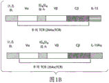

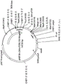


Classifications
-
- C—CHEMISTRY; METALLURGY
- C07—ORGANIC CHEMISTRY
- C07K—PEPTIDES
- C07K14/00—Peptides having more than 20 amino acids; Gastrins; Somatostatins; Melanotropins; Derivatives thereof
- C07K14/435—Peptides having more than 20 amino acids; Gastrins; Somatostatins; Melanotropins; Derivatives thereof from animals; from humans
- C07K14/52—Cytokines; Lymphokines; Interferons
- C07K14/54—Interleukins [IL]
- C07K14/5443—IL-15
-
- A—HUMAN NECESSITIES
- A61—MEDICAL OR VETERINARY SCIENCE; HYGIENE
- A61K—PREPARATIONS FOR MEDICAL, DENTAL OR TOILETRY PURPOSES
- A61K38/00—Medicinal preparations containing peptides
- A61K38/16—Peptides having more than 20 amino acids; Gastrins; Somatostatins; Melanotropins; Derivatives thereof
- A61K38/17—Peptides having more than 20 amino acids; Gastrins; Somatostatins; Melanotropins; Derivatives thereof from animals; from humans
- A61K38/177—Receptors; Cell surface antigens; Cell surface determinants
- A61K38/1774—Immunoglobulin superfamily (e.g. CD2, CD4, CD8, ICAM molecules, B7 molecules, Fc-receptors, MHC-molecules)
-
- A—HUMAN NECESSITIES
- A61—MEDICAL OR VETERINARY SCIENCE; HYGIENE
- A61K—PREPARATIONS FOR MEDICAL, DENTAL OR TOILETRY PURPOSES
- A61K38/00—Medicinal preparations containing peptides
- A61K38/16—Peptides having more than 20 amino acids; Gastrins; Somatostatins; Melanotropins; Derivatives thereof
- A61K38/17—Peptides having more than 20 amino acids; Gastrins; Somatostatins; Melanotropins; Derivatives thereof from animals; from humans
- A61K38/177—Receptors; Cell surface antigens; Cell surface determinants
- A61K38/1793—Receptors; Cell surface antigens; Cell surface determinants for cytokines; for lymphokines; for interferons
-
- A—HUMAN NECESSITIES
- A61—MEDICAL OR VETERINARY SCIENCE; HYGIENE
- A61K—PREPARATIONS FOR MEDICAL, DENTAL OR TOILETRY PURPOSES
- A61K38/00—Medicinal preparations containing peptides
- A61K38/16—Peptides having more than 20 amino acids; Gastrins; Somatostatins; Melanotropins; Derivatives thereof
- A61K38/17—Peptides having more than 20 amino acids; Gastrins; Somatostatins; Melanotropins; Derivatives thereof from animals; from humans
- A61K38/19—Cytokines; Lymphokines; Interferons
- A61K38/20—Interleukins [IL]
- A61K38/2086—IL-13 to IL-16
-
- A—HUMAN NECESSITIES
- A61—MEDICAL OR VETERINARY SCIENCE; HYGIENE
- A61P—SPECIFIC THERAPEUTIC ACTIVITY OF CHEMICAL COMPOUNDS OR MEDICINAL PREPARATIONS
- A61P29/00—Non-central analgesic, antipyretic or antiinflammatory agents, e.g. antirheumatic agents; Non-steroidal antiinflammatory drugs [NSAID]
-
- A—HUMAN NECESSITIES
- A61—MEDICAL OR VETERINARY SCIENCE; HYGIENE
- A61P—SPECIFIC THERAPEUTIC ACTIVITY OF CHEMICAL COMPOUNDS OR MEDICINAL PREPARATIONS
- A61P31/00—Antiinfectives, i.e. antibiotics, antiseptics, chemotherapeutics
- A61P31/12—Antivirals
-
- A—HUMAN NECESSITIES
- A61—MEDICAL OR VETERINARY SCIENCE; HYGIENE
- A61P—SPECIFIC THERAPEUTIC ACTIVITY OF CHEMICAL COMPOUNDS OR MEDICINAL PREPARATIONS
- A61P31/00—Antiinfectives, i.e. antibiotics, antiseptics, chemotherapeutics
- A61P31/12—Antivirals
- A61P31/14—Antivirals for RNA viruses
-
- A—HUMAN NECESSITIES
- A61—MEDICAL OR VETERINARY SCIENCE; HYGIENE
- A61P—SPECIFIC THERAPEUTIC ACTIVITY OF CHEMICAL COMPOUNDS OR MEDICINAL PREPARATIONS
- A61P35/00—Antineoplastic agents
-
- A—HUMAN NECESSITIES
- A61—MEDICAL OR VETERINARY SCIENCE; HYGIENE
- A61P—SPECIFIC THERAPEUTIC ACTIVITY OF CHEMICAL COMPOUNDS OR MEDICINAL PREPARATIONS
- A61P35/00—Antineoplastic agents
- A61P35/02—Antineoplastic agents specific for leukemia
-
- A—HUMAN NECESSITIES
- A61—MEDICAL OR VETERINARY SCIENCE; HYGIENE
- A61P—SPECIFIC THERAPEUTIC ACTIVITY OF CHEMICAL COMPOUNDS OR MEDICINAL PREPARATIONS
- A61P37/00—Drugs for immunological or allergic disorders
- A61P37/02—Immunomodulators
-
- A—HUMAN NECESSITIES
- A61—MEDICAL OR VETERINARY SCIENCE; HYGIENE
- A61P—SPECIFIC THERAPEUTIC ACTIVITY OF CHEMICAL COMPOUNDS OR MEDICINAL PREPARATIONS
- A61P37/00—Drugs for immunological or allergic disorders
- A61P37/02—Immunomodulators
- A61P37/04—Immunostimulants
-
- A—HUMAN NECESSITIES
- A61—MEDICAL OR VETERINARY SCIENCE; HYGIENE
- A61P—SPECIFIC THERAPEUTIC ACTIVITY OF CHEMICAL COMPOUNDS OR MEDICINAL PREPARATIONS
- A61P37/00—Drugs for immunological or allergic disorders
- A61P37/02—Immunomodulators
- A61P37/06—Immunosuppressants, e.g. drugs for graft rejection
-
- C—CHEMISTRY; METALLURGY
- C07—ORGANIC CHEMISTRY
- C07K—PEPTIDES
- C07K14/00—Peptides having more than 20 amino acids; Gastrins; Somatostatins; Melanotropins; Derivatives thereof
- C07K14/435—Peptides having more than 20 amino acids; Gastrins; Somatostatins; Melanotropins; Derivatives thereof from animals; from humans
- C07K14/705—Receptors; Cell surface antigens; Cell surface determinants
- C07K14/70503—Immunoglobulin superfamily
- C07K14/7051—T-cell receptor (TcR)-CD3 complex
-
- C—CHEMISTRY; METALLURGY
- C07—ORGANIC CHEMISTRY
- C07K—PEPTIDES
- C07K14/00—Peptides having more than 20 amino acids; Gastrins; Somatostatins; Melanotropins; Derivatives thereof
- C07K14/435—Peptides having more than 20 amino acids; Gastrins; Somatostatins; Melanotropins; Derivatives thereof from animals; from humans
- C07K14/705—Receptors; Cell surface antigens; Cell surface determinants
- C07K14/715—Receptors; Cell surface antigens; Cell surface determinants for cytokines; for lymphokines; for interferons
- C07K14/7155—Receptors; Cell surface antigens; Cell surface determinants for cytokines; for lymphokines; for interferons for interleukins [IL]
-
- C—CHEMISTRY; METALLURGY
- C07—ORGANIC CHEMISTRY
- C07K—PEPTIDES
- C07K16/00—Immunoglobulins [IGs], e.g. monoclonal or polyclonal antibodies
- C07K16/18—Immunoglobulins [IGs], e.g. monoclonal or polyclonal antibodies against material from animals or humans
- C07K16/28—Immunoglobulins [IGs], e.g. monoclonal or polyclonal antibodies against material from animals or humans against receptors, cell surface antigens or cell surface determinants
-
- C—CHEMISTRY; METALLURGY
- C07—ORGANIC CHEMISTRY
- C07K—PEPTIDES
- C07K19/00—Hybrid peptides, i.e. peptides covalently bound to nucleic acids, or non-covalently bound protein-protein complexes
-
- C—CHEMISTRY; METALLURGY
- C12—BIOCHEMISTRY; BEER; SPIRITS; WINE; VINEGAR; MICROBIOLOGY; ENZYMOLOGY; MUTATION OR GENETIC ENGINEERING
- C12N—MICROORGANISMS OR ENZYMES; COMPOSITIONS THEREOF; PROPAGATING, PRESERVING, OR MAINTAINING MICROORGANISMS; MUTATION OR GENETIC ENGINEERING; CULTURE MEDIA
- C12N15/00—Mutation or genetic engineering; DNA or RNA concerning genetic engineering, vectors, e.g. plasmids, or their isolation, preparation or purification; Use of hosts therefor
- C12N15/09—Recombinant DNA-technology
- C12N15/11—DNA or RNA fragments; Modified forms thereof; Non-coding nucleic acids having a biological activity
- C12N15/62—DNA sequences coding for fusion proteins
-
- G—PHYSICS
- G01—MEASURING; TESTING
- G01N—INVESTIGATING OR ANALYSING MATERIALS BY DETERMINING THEIR CHEMICAL OR PHYSICAL PROPERTIES
- G01N33/00—Investigating or analysing materials by specific methods not covered by groups G01N1/00 - G01N31/00
- G01N33/48—Biological material, e.g. blood, urine; Haemocytometers
- G01N33/50—Chemical analysis of biological material, e.g. blood, urine; Testing involving biospecific ligand binding methods; Immunological testing
- G01N33/53—Immunoassay; Biospecific binding assay; Materials therefor
- G01N33/569—Immunoassay; Biospecific binding assay; Materials therefor for microorganisms, e.g. protozoa, bacteria, viruses
- G01N33/56966—Animal cells
- G01N33/56977—HLA or MHC typing
-
- A—HUMAN NECESSITIES
- A61—MEDICAL OR VETERINARY SCIENCE; HYGIENE
- A61K—PREPARATIONS FOR MEDICAL, DENTAL OR TOILETRY PURPOSES
- A61K38/00—Medicinal preparations containing peptides
-
- C—CHEMISTRY; METALLURGY
- C07—ORGANIC CHEMISTRY
- C07K—PEPTIDES
- C07K16/00—Immunoglobulins [IGs], e.g. monoclonal or polyclonal antibodies
- C07K16/18—Immunoglobulins [IGs], e.g. monoclonal or polyclonal antibodies against material from animals or humans
-
- C—CHEMISTRY; METALLURGY
- C07—ORGANIC CHEMISTRY
- C07K—PEPTIDES
- C07K2319/00—Fusion polypeptide
-
- C—CHEMISTRY; METALLURGY
- C07—ORGANIC CHEMISTRY
- C07K—PEPTIDES
- C07K2319/00—Fusion polypeptide
- C07K2319/31—Fusion polypeptide fusions, other than Fc, for prolonged plasma life, e.g. albumin
-
- C—CHEMISTRY; METALLURGY
- C07—ORGANIC CHEMISTRY
- C07K—PEPTIDES
- C07K2319/00—Fusion polypeptide
- C07K2319/33—Fusion polypeptide fusions for targeting to specific cell types, e.g. tissue specific targeting, targeting of a bacterial subspecies
-
- C—CHEMISTRY; METALLURGY
- C07—ORGANIC CHEMISTRY
- C07K—PEPTIDES
- C07K2319/00—Fusion polypeptide
- C07K2319/70—Fusion polypeptide containing domain for protein-protein interaction
- C07K2319/74—Fusion polypeptide containing domain for protein-protein interaction containing a fusion for binding to a cell surface receptor
- C07K2319/75—Fusion polypeptide containing domain for protein-protein interaction containing a fusion for binding to a cell surface receptor containing a fusion for activation of a cell surface receptor, e.g. thrombopoeitin, NPY and other peptide hormones
-
- C—CHEMISTRY; METALLURGY
- C12—BIOCHEMISTRY; BEER; SPIRITS; WINE; VINEGAR; MICROBIOLOGY; ENZYMOLOGY; MUTATION OR GENETIC ENGINEERING
- C12P—FERMENTATION OR ENZYME-USING PROCESSES TO SYNTHESISE A DESIRED CHEMICAL COMPOUND OR COMPOSITION OR TO SEPARATE OPTICAL ISOMERS FROM A RACEMIC MIXTURE
- C12P21/00—Preparation of peptides or proteins
Abstract
The invention discloses fusion molecules and IL-15 variants. In particular, the invention provides soluble fusion protein complexes and IL-15 variants having therapeutic and diagnostic uses, and methods of making such proteins. The invention further provides methods of stimulating or suppressing an immune response using the fusion protein complexes of the invention and IL-15 variants.
Description
The invention relates to a divisional application of Chinese patent application with the application number of 201410242394.1, the application date of 2008-5/9 and the invented name of fusion molecule and IL-15 variant, and the patent application 201410242394.1 is a divisional application of Chinese patent application with the application number of 200880024119.5, the application date of 2008-5/9 and the invented name of fusion molecule and IL-15 variant.
References to related applications
This application claims priority to U.S. provisional application serial No. 60/928,900 at 11/5/2007, which is hereby incorporated by reference in its entirety.
Technical Field
The present invention relates to soluble fusion protein complexes, and in particular to fusion molecules and IL-15 variants.
Background
The T Cell Receptor (TCR), the most prominent effector of the immune system (effector), has unique advantages as a platform for developing therapeutic approaches. Antibody therapy is limited to recognizing pathogens in the blood and extracellular space or protein targets on the cell surface, while T cell receptors can recognize antigens displayed on the cell surface by MHC molecules (including antigens derived from intracellular proteins). Based on the subtype of T cells that become activated by recognition of the presented antigen, T cell receptors, as well as T cells with T cell receptors, may be involved in the regulation of various immune responses. For example, T cells are involved in humoral immune responses via the induction of B cells to differentiate into antibody producing cells. In addition, activated T cells function to initiate a cell-mediated immune response. Thus, T cell receptors can identify other targets that cannot be recognized using antibodies.
T cells are a sub-population of cells that, together with other immune cell types (polymorphonuclear neutrophils, eosinophils, basophils, adipocytes, B cells, NK cells), constitute the cellular component of the immune system. Under physiological conditions, T cells play a role in immune monitoring and the elimination of foreign antigens. However, under pathological conditions, there is strong evidence that T cells play an important role in the initiation and spread of disease. In these conditions, disruption of T cell immune tolerance (central or peripheral) is a major fundamental process in the initiation of autoimmune diseases.
TCRs are believed to play an important role in the development and function of the immune system. For example, TCR-mediated cytotoxicity (cell killing), enhancement of B cell proliferation, and affecting the development and severity of various disorders, including cancer, allergy, viral infection, and autoimmune disorders, have been reported.
It is therefore desirable to provide novel targeting agents based on T cell receptors, and methods of making and using such agents for therapeutic and diagnostic settings. Accordingly, it would be particularly desirable to provide molecules having certain advantages over prior art antibody targeting-based complexes.
Furthermore, it is desirable to use TCRs to target various effector molecules to disease sites where they can provide therapeutic benefit without the side effects associated with non-targeted systemic activity. One such class is IL-15, a member of the four alpha helix bundle family of lymphokines (lymphokines). IL-15 plays a multi-role in the development and regulation of the immune system. More particularly, IL-15 affects the function, development, survival and proliferation of CD8+ T cells, NK cells, cytotoxic T cells, B cells, epidermal interstitial lymphocytes (IEL) of the small intestinal wall and Antigen Presenting Cells (APC). Both IL-15-/-transgenic mice and IL-15 Ra-/-transgenic mice have been shown to lack the peripheral NK cell and cytotoxic T cell populations, certain IEL subsets, and a large proportion of memory phenotype CD8+ T cells, suggesting that IL-15 functions in the development, proliferation and/or survival of these cell types. The IL-15 receptor (R) is composed of three polypeptides: type specific IL-15R alpha ("IL-15R alpha" or "IL-15Ra"), IL-2/IL-15R beta ("IL-2R beta" or "IL-15Rb"), and a common gamma chain (gamma chain) ("gamma C" or "gC", which is common to multiple cytokine receptors).
IL-15 signaling can be via heterotrimeric complexes of IL-15R α, IL-2R β, and γ C; produced via the heterotrimeric complex of IL-2R β and γ C. The novel mechanism of action of IL-15 is a transducibility (transduction) mechanism in which IL-15 and IL-15R α are synergistically expressed by antigen presenting cells (monocytes and dendritic cells), and IL-15 bound to IL-15R α is transduced to adjacent NK cells or CD 8T cells expressing only IL-15R β γ C receptor. In the case of co-stimulatory events (co-stimulatory events) occurring at the immunological synapse, IL-15 transcriptional presentation is currently shown to be the major mechanism of IL-15 action in vivo, and it appears to play a major role in tumor immune monitoring (Waldmann, TA,2006, Nature Rev. Immunol.6: 595-601). The soluble IL-2 ra subunit, which results in a isoform (isoform) containing a deletion of exon 3(exon3) and a so-called "sushi" domain at the N-terminus, has been shown to carry most of the structural elements responsible for cytokine binding. IL-2R α alone is a low affinity receptor for IL-2 (Kd 10nM), while IL-15R α binds IL-15 with high affinity (Kd 100 pM). Thus, soluble IL-2R α and IL-15 are able to form stable heterodimeric complexes in solution, and such complexes are able to modulate (i.e., stimulate or block) immune responses via the intermediate or high affinity IL-15R complexes (Mortier et al 2006J Biol Chem 281: 1612-1619; Stoklasek et al 2006J Immunol 177: 6072-6080; Rubinstein et al 2006 Proc Natl Acad Sci U S A103: 9166-9171).
Given the known efficacy of IL-15 on the immune system, various IL-15-based approaches have been explored to manipulate the immune system for host efficiency. IL-15 administration has been used to support immune responses or to enhance immune system reconstitution, while blocking IL-15 activity suppresses autoimmune responses or other inappropriate immune responses (Waldmann, TA,2006, Nature Rev. Immunol.6: 595-601). Indeed, one of the limitations of systemic IL-15 treatment is the potential to induce autoimmune diseases. Other limitations include the difficulty of manufacturing such cytokines in standard mammalian cell expression systems, and their very short half-life in vivo. Thus, there is a need to produce IL-15 in a suitable immunostimulatory therapeutic format that exhibits a longer half-life in vivo, increased immune cell binding activity, or enhanced biological activity. In addition, there is also a need for potent IL-15 antagonists. Ideally, these molecules should be targeted selectively to the disease site to avoid unnecessary systemic toxicity and provide more effective therapeutic benefit.
Disclosure of Invention
The present invention provides various IL-15 variants and soluble fusion complexes having therapeutic utility, as well as methods for making such proteins. The invention provides methods of killing a target cell using the soluble fusion complexes of the invention. The IL-15 variants and soluble complexes described herein have potential therapeutic utility.
Thus, in one aspect, the invention provides a soluble fusion protein complex comprising at least two soluble fusion proteins, wherein a first fusion protein comprises a first biologically active polypeptide covalently linked to interleukin-15 (IL-15) or a functional fragment thereof, and a second fusion protein comprises a second biologically active polypeptide covalently linked to a soluble interleukin-15 receptor alpha (IL-15Ra) polypeptide or a functional fragment thereof, wherein the IL-15 domain of the first fusion protein binds to the soluble IL-15Ra domain of the second fusion protein to form the soluble fusion protein complex.
In one embodiment, one of the first and second biologically active polypeptides comprises a first soluble T Cell Receptor (TCR) or functional fragment thereof. In another embodiment, another of the biologically active polypeptides comprises the first soluble TCR or functional fragment thereof, thereby creating a multivalent TCR fusion protein complex with increased binding activity. In yet another embodiment, the other biologically active polypeptide comprises a second soluble TCR, or a functional fragment thereof, different from the first soluble TCR.
In another embodiment of this protocol, the TCR is specific for recognition of a particular antigen.
In yet another embodiment of this embodiment, the TCR is a heterodimer comprising an a and b chain TCR.
In yet another embodiment of this embodiment, the TCR comprises a single chain TCR polypeptide. In yet another embodiment, the single chain TCR polypeptide comprises a TCR V-alpha chain covalently linked to a TCR V-beta chain by a peptide linker sequence. In yet another embodiment, the single-chain TCR further comprises a soluble TCR cp chain fragment covalently linked to a TCR V- β chain.
In another embodiment, the single chain TCR further comprises a soluble TCR ca chain fragment covalently linked to a TCR V-a chain.
In yet another embodiment, one or both of the first and second biologically active polypeptides comprises an antibody or functional fragment thereof.
In yet another embodiment, the antibody is specific for recognition of a particular antigen. In yet another embodiment, the antibody is a single chain antibody or a single chain Fv. In another specific embodiment, the single chain antibody comprises an immunoglobulin light chain variable domain (immunoglobulin light chain variable domin) covalently linked to an immunoglobulin heavy chain variable domain by a polypeptide linker sequence.
In one embodiment of the foregoing embodiment, the first biologically active polypeptide is covalently linked to IL-15 (or a functional fragment thereof) through a polypeptide linker sequence.
In another embodiment of the foregoing embodiment, the second biologically active polypeptide is covalently linked to the IL-15Ra polypeptide (or functional fragment thereof) through a polypeptide linker sequence.
In another embodiment, the antigen of the TCR domain comprises a peptide antigen presented on an MHC or HLA molecule. In yet another embodiment, the peptide antigen is derived from a tumor-associated polypeptide or a virus-encoded polypeptide.
In another embodiment, the antigen of the antibody domain comprises a cell surface receptor or ligand.
In yet another embodiment, the antigen comprises a CD antigen, a cytokine or chemokine receptor or ligand, a growth factor receptor or ligand, a tissue factor, a cell adhesion molecule, an MHC/MHC-like molecule, an FC receptor, a Toll-like receptor, an NK receptor, a TCR, a BCR, a positive/negative co-stimulatory receptor or ligand, a death receptor or ligand, a tumor-associated antigen, or a virally encoded antigen.
In another embodiment of the foregoing embodiment, the IL-15Ra polypeptide includes an extracellular domain of IL-15 receptor alpha that binds IL-15.
In another embodiment of the above protocol, the IL-15Ra polypeptide includes the IL-15a sushi domain (Wei et al. Journal of Immunology,2001,167: 277-) -or the IL-15a Δ E3 domain (Anderson et al 1995.J.biol. chem.270:29862-29869, Dubois et al 1999.J.biol. chem.274: 26978-26984).
In another aspect, the invention provides an IL-15 variant (also referred to herein as an IL-15 mutant) having an amino acid sequence that differs from a native (or wild-type) IL-15 protein and binds to an IL-15Ra polypeptide, and which acts as an IL-15 agonist or antagonist. Embodiments of the invention provide an IL-15 variant as a non-fusion protein or as a soluble fusion protein, comprising the biologically active polypeptide described above. Wherein the IL-15 domain is replaced with the IL-15 variant.
In one embodiment of the foregoing, the invention features a nucleic acid sequence encoding a first fusion protein of any of the embodiments or embodiments described herein.
In one embodiment of the foregoing, the invention features a nucleic acid sequence encoding a second fusion protein of any of the embodiments or embodiments described herein.
In one embodiment of the foregoing, the invention features a nucleic acid sequence encoding an IL-15 variant of any of the embodiments or embodiments described herein.
In one embodiment, the nucleic acid sequence further comprises a promoter, translation initiation signal, and leader sequence operably linked to the sequence encoding the fusion protein or IL-15 variant. In another embodiment, any of the above nucleic acid sequences is included in a DNA vector.
In another aspect, the invention features a method of making a soluble fusion protein complex of the above aspects, the method comprising: introducing a DNA vector encoding the first fusion protein of the above schemes and embodiments into a first host cell, culturing the first host cell in a culture medium under conditions sufficient for expression of the first fusion protein in the cell or culture medium, purifying the first fusion protein from the host cell or culture medium, introducing the DNA vector encoding the second fusion protein of the schemes and embodiments above into a second host cell, culturing the second host cell in a culture medium under conditions sufficient to express the second fusion protein in the cell or culture medium, purifying the second fusion protein from the host cell or culture medium, and mixing the first and second fusion proteins under conditions sufficient for binding to occur between the IL-15 domain of the first fusion protein and the soluble IL-15Ra domain of the second fusion protein to form the soluble fusion protein complex.
In another aspect, the invention features a method of making a soluble fusion protein complex of the above aspects, the method comprising: introducing a DNA vector encoding the first fusion protein of the above schemes and embodiments and a DNA vector encoding the second fusion protein of the above schemes and embodiments into a host cell, culturing the host cell in a culture medium under conditions sufficient for expression of the fusion protein in the cell or the culture medium and allowing association between the IL-15 domain of the first fusion protein and the soluble IL-15Ra domain of the second fusion protein to form the soluble fusion protein complex, and purifying the soluble fusion protein complex from the host cell or the culture medium.
In yet another aspect, the invention features a method of making a soluble fusion protein complex of the above aspects, the method comprising: introducing the DNA vector of claim 28 encoding the first and second fusion proteins into a host cell, culturing the host cell in culture under conditions sufficient for expression of the fusion protein in the cell or culture medium and allowing association between the IL-15 domain of the first fusion protein and the soluble IL-15Ra domain of the second fusion protein to form the soluble fusion protein complex, and purifying the soluble fusion protein complex from the host cell or culture medium.
In other versions of the above methods, a DNA vector encoding the IL-15 variant is used in place of the DNA vector encoding the first fusion protein to generate a host cell capable of expressing the IL-15 variant and the IL-15 variant is combined with the IL-15Ra domain of a second fusion protein to form a soluble fusion protein complex.
In another aspect, the invention features a method of making an IL-15 variant of the above aspect, the method comprising: introducing a DNA vector encoding an IL-15 variant of the above schemes and embodiments into a host cell, culturing the host cell in a culture medium under conditions sufficient for expression of the IL-15 variant in the cell or culture medium, and purifying the IL-15 variant from the host cell or culture medium.
In another aspect, the invention features a method of killing a target cell, the method including: contacting a plurality of cells with the soluble fusion protein complex or IL-15 variant of any of the embodiments or embodiments above, wherein the plurality of cells further comprises immune cells bearing an IL-15R chain recognized by the IL-15 domain of the embodiments above, and target cells bearing an antigen recognized by at least one of the biologically active polypeptides of the embodiments above; forming a specific binding complex (bridge) between the antigen of the target cell and the IL-15R chain of the immune cell sufficient to bind to and activate the immune cell; and killing the target cell by the bound activated immune cell.
In one embodiment of the method, the target cell is a tumor cell or a virus-infected cell.
In another embodiment of the method, the biologically active polypeptide comprises a TCR.
In yet another embodiment of the method, the antigen of the target cell comprises a tumor or virus-encoded peptide antigen presented on an MHC or HLA molecule and recognized by the TCR.
In yet another embodiment of the method, the immune cell is a T cell, LAK cell, or NK cell.
In another aspect, the invention features a method of preventing or treating a disease in a patient, wherein the disease cells express a disease-associated antigen, the method comprising: administering to the patient a soluble fusion protein complex or IL-15 variant of any of the above embodiments or embodiments comprising a biologically active polypeptide that recognizes a disease-associated antigen forms a specific binding complex (bridge) between the antigen-expressing disease cells and the IL-15R-expressing immune cells sufficient to localize the immune cells and to injure or kill the disease cells sufficient to prevent or treat disease in the patient.
In one aspect, the invention features a method of preventing or treating a disease in a patient, wherein the disease cells express a disease-associated antigen, the method comprising: mixing immune cells bearing the IL-15R chain with the soluble fusion protein complex of claims 1-22 comprising a biologically active polypeptide that recognizes a disease-associated antigen, administering the immune cell-fusion protein complex to the patient, forming a specific binding complex (bridge) between the antigen-expressing disease cells and the IL-15R-expressing immune cells sufficient to localize the immune cells, and to injure or kill the disease cells sufficient to prevent or treat disease in the patient.
In another aspect, the invention features a method of preventing or treating a disease in a patient, wherein the patient's cells express a disease-associated antigen, the method comprising: administering to the patient a soluble fusion protein complex or IL-15 variant of any of the above embodiments or embodiments comprising a biologically active polypeptide that recognizes a disease-associated antigen, localizing the soluble fusion protein complex or IL-15 variant to the disease cell, wherein the IL-15 domain or IL-15 variant of the soluble fusion protein complex binds to an immune cell bearing the IL-15R chain, and suppressing the immune response of the immune cell.
In one embodiment of the method, the disease is cancer or a viral infection.
In another embodiment of the method, the disease is an immune disorder, an autoimmune disease, or an inflammatory disorder.
In another embodiment of the method, the disease-associated antigen is a peptide/MHC complex.
In another embodiment, the invention features a method of stimulating an immune response in a mammal, comprising administering to the mammal an effective amount of the soluble fusion protein complex or IL-15 variant of any of the above regimens or embodiments.
In another embodiment, the invention features a method of inhibiting an immune response in a mammal, comprising administering to the mammal an effective amount of the soluble fusion protein complex or IL-15 variant of any of the above regimens or embodiments.
Drawings
Fig. 1A and 1B are schematic diagrams. FIG. 1A is a schematic diagram depicting an example of a fusion protein complex comprising a single chain TCR polypeptide. FIG. 1B depicts a schematic of a representative fusion protein construct comprising a fusion protein complex.
Fig. 2A to 2C are composed of three diagrams. FIG. 2A depicts a map of the pNEF38-c264scTCR/huIL15 expression vector. FIG. 2B shows the sequence of the c264scTCR/huIL15 fusion gene. FIG. 2C shows the sequence of the C264scTCR/huIL15 fusion protein, which includes a leader sequence.
Fig. 3A to 3C are composed of three diagrams. FIG. 3A depicts a map of the pNEF38-c264scTCR-hinge-huIL15 expression vector. FIG. 3B shows the sequence of the c264scTCR-hinge-huIL15 fusion gene. FIG. 3C shows the sequence of the C264scTCR-hinge-huIL15 fusion protein, which includes a leader sequence.
Fig. 4A to 4C are composed of three diagrams. FIG. 4A depicts a map of the pNEF38-c264scTCR/huIL15RaDE3 expression vector. FIG. 4B shows the sequence of the c264scTCR/huIL15R α Δ E3 fusion gene. FIG. 4C shows the sequence of the C264scTCR/huIL15R α Δ E3 fusion protein, which includes a leader sequence.
Fig. 5A to 5C are composed of three diagrams. FIG. 5A depicts a map of pNEF38-c264scTCR/huIL15RaSushi expression vector. FIG. 5B shows c264scTCR/huIL15RαSusSequence of hi fusion gene. FIG. 5C showsc264scTCR/huThe sequence of IL15R α Sushi fusion protein, comprising a leader sequence.
Fig. 6A to 6C are composed of three diagrams. FIG. 6A depicts the pNEF38-c264 scTCR-change-huIL 15Rasushi expression vector. FIG. 6B shows the sequence of the c264scTCR-hinge-huIL15R α Sushi fusion gene. FIG. 6C shows the sequence of the C264scTCR-hinge-huIL15R α Sushi fusion protein, which includes a leader sequence.
FIG. 7 is a map of the pSun-c264scTCRIL15/c264scTCRIL15Rasushi expression vector.
FIG. 8 is a map of the pSun-c264scTCRIL15/c264scTCRIL15RaDE3 expression vector.
FIGS. 9A and 9B illustrate the properties of transfected cells expressing the TCR/IL15R α fusion protein. FIG. 9A is two graphs showing flow cytometry analysis of fusion protein expressing cells. FIG. 9B is a graph showing TCR-based ELISA results generated by fusion proteins.
FIGS. 10A and 10B show analysis of TCR/IL15 and TCR/IL15R α fusion proteins by reducing SDS PAGE. FIG. 10A shows cell culture suspensions containing c264scTCR/huIL15 or c264scTCR/huIL15 RaSushi. FIG. 10B shows cell culture suspensions containing c264scTCR/huIL15 or c264scTCR/huIL15R α Δ E3.
FIGS. 11A-11C show analysis of TCR/IL15, TCR/IL15Ra, and fusion protein complex by size exclusion chromatography (size exclusion chromatography). FIG. 11A is a graph showing the SEC chromatogram of c264scTCR/huIL 15. FIG. 11B is a graph showing the SEC chromatogram of c264scTCR/huIL15R α Sushi. FIG. 11C is a graph showing the SEC chromatogram of the C264scTCR/huIL15+ C264scTCR/huIL15R α Sushi fusion protein complex.
FIGS. 12A and 12B are analyses of TCR/IL15R α and fusion protein complexes by size exclusion chromatography. FIG. 12A is a graph illustrating the SEC chromatogram of c264scTCR/huIL15R α Δ E3. FIG. 12B is a graph illustrating the SEC chromatogram of the c264scTCR/huIL15+ c264scTCR/huIL15R α Δ E3 fusion protein complex.
FIG. 13 is a graph showing the binding of TCR/IL15, TCR/IL15R α and fusion protein complexes to cell-presented peptide/MHC complexes as measured by flow cytometry.
Fig. 14A to 14D are composed of four diagrams. FIG. 14A shows the sequence of mature human IL15 protein (SEQ ID NO:1), with the residues in the blue bottom line being the ones in the IL-15 variants shown in Table 1. FIG. 14B depicts pNEF38-c264 scTCR-change-huIL 15D8A and pNEF38-c264 scTCR-change-huIL 15D8N expression vectors. FIG. 14C shows the sequences of pNEF38-C264 scTCR-change-huIL 15D8A and pNEF38-C264 scTCR-change-huIL 15D8N genes in section C-1 and section C-2. Portions D-1 and D-2 in FIG. 14D show the sequences of pNEF38-c264 scTCR-change-huIL 15D8A and pNEF38-c264 scTCR-change-huIL 15D8N fusion proteins, which include leader sequences. The blue bottom line nucleotides were changed to produce the indicated IL-15 variants.
FIG. 15 is a diagram showing flow cytometry analysis of IL-15R-loaded CTLL2 cells stained with fusion proteins and complexes, followed by TCR-specific peptide/MHC reagents.
FIG. 16 is a graph showing the binding of the dimeric fusion protein complex of TCR/IL15R α Sushi and TCR/IL15 (including native and variant IL15) to the cognate peptide/MHC complex presented on peptide-loaded cells as measured by flow cytometry. The background binding rate of dimeric fusion protein complexes for peptide-unloaded cells is also shown.
FIG. 17(A to C) is a graph showing the binding activity of wild type, antagonist and agonist TCR/IL15 fusion proteins to the cognate peptide/MHC complex and IL15R α as measured by ELISA. (A) An assay showing the binding activity of the fusion protein to the homologous peptide/MHC complex is shown. (B) To show the fusion protein for IL15R alpha binding activity analysis. (C) To show the binding activity of the fusion protein to the IL-15R β γ C receptor on 32D β cells.
FIG. 18 is a graph showing that fusion proteins and fusion protein complexes maintain the growth of cells bearing IL15R as measured by a cell proliferation assay.
FIGS. 19A-19C are graphs showing the ability of TCR/IL-15 fusion proteins including IL-15 variants to inhibit or enhance growth of cells bearing IL15R as measured by a cell proliferation assay. FIG. 19A is a graph showing the activity of fusion proteins including IL-15 variants to inhibit proliferation of CTLL-2 cells bearing high affinity IL15R (α β γ receptor complex). FIG. 19B is a graph showing the activity of fusion proteins including IL-15 variants to inhibit or enhance proliferation of 32D beta cells bearing low affinity IL15R (beta gamma receptor complex). FIG. 19C is a graph showing that fusion proteins including IL-15 variants block TCR/IL15 wt-stimulatory proliferation activity of CTLL-2 cells bearing high affinity IL15R (α β γ receptor complex).
FIG. 20 depicts the effect of in vitro cultured NK cells on survival of xenograft tumor-bearing nude mice on dimeric fusion protein complexes of TCR/IL15R α Sushi and TCR/IL 15. Athymic nude mice were injected with human NSCLC a549-a2 cells to establish lung metastases. Purified NK cells isolated from the spleen of allogeneic donor mice were cultured in vitro with rhIL-2, MART1scTCR-IL2, c264scTCR-IL2 or c264scTCR-IL15/c264scTCR-IL15R α and adoptive transfer (adoptive transfer) to tumor bearing mice pretreated with Cyclophosphamide (CTX) as indicated in the figure. Percent survival after treatment is plotted.
FIG. 21 illustrates Table 1, which shows amino acid substitutions in IL-15 variants and the effect of such variations on IL-15 activity.
FIGS. 22A-22B illustrate the amino acid sequence of IL-15 (SEQ ID NO:1) and the nucleic acid sequence of IL-15 (SEQ ID NO:2), respectively.
Detailed Description
It has been established that IL-15 stably binds to the extracellular domain of IL-15ra, and that the resulting complex is capable of modulating (i.e., stimulating or blocking) an immune response via the intermediate or high affinity IL-15R complex (1 to 4). In addition, it has been demonstrated that single chain TCR or antibody polypeptides can be fused to cytokine and other immune effector domains, and that these bispecific fusion molecules retain the functional activity of both fusion domains (5 to 8). Furthermore, it has been shown that multivalent TCRs provide enhanced binding to their ligands (9).
Definition of
The following definitions are provided for specific terms used in the following written description.
As used in the specification and in the claims, the singular form of "a", "an", and "the" include plural referents unless the content clearly dictates otherwise. For example, the term "cell" includes a plurality of cells, including mixtures thereof. The term "nucleic acid molecule" includes a plurality of nucleic acid molecules.
The term "comprising" as used herein means that the compositions and methods include the recited elements but not excluding others. When used to define compositions and methods, "consisting essentially of …" shall mean excluding other elements of any substantial importance to the combination. Thus, a composition as defined herein consisting essentially of elements will not exclude trace contaminants from isolation and purification processes and pharmaceutically acceptable carriers such as phosphate buffered saline, preservatives, and the like. "consisting of …" shall mean excluding more than trace amounts of other ingredients and elements used in the actual method steps for administering the compositions of the invention. The embodiments defined by each of these transitions are within the scope of the present invention.
An "antibody" is any immunoglobulin that binds to a particular epitope (epitope), including antibodies and fragments thereof. The term encompasses polyclonal, monoclonal, chimeric and single chain antibodies, as well as bispecific antibodies.
The term "antigen" as used herein means any substance that will elicit an immune system to produce antibodies or a specific cellular vector immune response against which it is directed. A disease-associated antigen is any substance associated with any disease.
The term "biologically active polypeptide" as used herein means an amino acid sequence, such as a protein, polypeptide or peptide; a sugar or polysaccharide; lipids or glycolipids, glycoproteins, or lipoproteins that can produce the desired effects discussed herein, including the binding activity of the TCR or antibody fusion protein complex to an antigen.
The term "cell" as used herein is intended to encompass any prokaryotic cell, eukaryotic cell, primary cell or immortalized cell line, any cluster of such cells in a tissue or organ. Preferably, the cells are derived from mammalian (especially human) sources and may be infected by one or more pathogens. A "host cell" according to the present invention may be a transfected (transfected), transformed (transduced), transduced (transduced) or infected (infected) cell of any origin, including prokaryotic, eukaryotic, mammalian, avian, insect, plant or bacterial cells, or it may be a cell of any origin that may be used to propagate nucleic acids as described herein.
The term "conjugated molecule" as used herein means a TCR or antibody molecule and an effector molecule (typically a chemical molecule or a synthetic molecule) covalently linked (i.e., fused) by chemical means or other suitable means. If desired, the conjugate molecule may be fused at one or several positions via a peptide linker sequence or a carrier molecule. Alternatively, a peptide linker or carrier may be used to assist in the construction of the conjugated molecule. Particularly preferred conjugated molecules are conjugated toxins or detectable labels.
The term "effector molecule" as used herein means an amino acid sequence, such as a protein, polypeptide or peptide; a sugar or polysaccharide; lipids or glycolipids, glycoproteins, or lipoproteins that can produce the desired effects discussed herein, including an IL-15 domain, IL-15 variant or IL-15 receptor (e.g., IL-15R α, IL-2R β, or γ C), or functional fragment thereof.
The terms "fusion molecule" and "fusion protein" are used interchangeably and refer to a biologically active polypeptide (typically a TCR or antibody) and an effector molecule (typically a protein or peptide sequence) covalently linked (i.e., fused) by recombinant means, chemical means, or other suitable means. If desired, the fusion molecules may be fused at one or several positions via a peptide linker sequence. Alternatively, peptide linkers can be used to assist in the construction of fusion molecules. Particularly preferred fusion molecules are fusion proteins. In general, fusion molecules may also include conjugated molecules.
The term "host cell" means any prokaryotic or eukaryotic cell containing a cloning or expression vector. The term also includes prokaryotic or eukaryotic cells that have been genetically engineered to contain the cloned gene in the chromosome or genome of the host cell.
The term "immune response" as used herein means the process of stimulating immune cells and recruiting immune cells from the blood to lymphoid and non-lymphoid tissues via a multifactorial process involving distinct adhesion and activation steps. The activation conditions trigger the release of cytokines, growth factors, chemokines and other factors, up-regulate the expression of adhesion and other activating molecules on immune cells, promote adhesion, morphological changes and/or extravasation simultaneously with chemotaxis (extravasation) through tissues, increase cell proliferation and cytotoxic activity, stimulate antigen presentation and provide other phenotypic changes including the generation of memory cell types. An immune response also means an activity of an immune cell to suppress or modulate the inflammatory or cytotoxic activity of other immune cells.
The terms "polynucleotide" and "nucleic acid molecule" are used interchangeably herein to mean a polymeric form of nucleotides of any length. The polynucleotide may comprise deoxyribonucleotides, ribonucleotides, and/or analogs thereof. The nucleotide can have any three-dimensional structure and can perform any known or unknown function. The term "polynucleotide" encompasses, for example: single, double and triple helix molecules, genes or gene fragments, exons (exon), introns (intron), mRNA, tRNA, rRNA, ribozymes (ribozymes), antisense molecules (antisense molecules), cDNA, recombinant polynucleotides, branched polynucleotides, aptamers (aptamers), plastids, vectors, isolated DNA of any sequence, isolated RNA of any sequence, nucleic acid probes, and primers. Nucleic acid molecules can also include modified nucleic acid molecules (e.g., including modified bases, sugars, and/or internucleotide linkers).
The term "polypeptide" means any polymer, regardless of its size, preferably consisting essentially of any 20 natural amino acids. Although the term "protein" is often used to refer to relatively large proteins, and "peptide" is often used to refer to small polypeptides, such terms are often used in the art with some overlap. The term "polypeptide" generally refers to proteins, polypeptides, and peptides, unless otherwise indicated. Peptides generally suitable according to the invention are generally between about 0.1 to 100kD or more and up to about 1000kD, preferably between about 0.1, 0.2, 0.5, 1, 2, 5, 10, 20, 30 and 50kD, as determined by standard molecular fractionation techniques, such as centrifugation or SDS-polyacrylamide gel electrophoresis.
The terms "prevent", "preventing", "prevention", "preventive treatment", and the like, mean reducing the likelihood of developing a disorder or condition in an individual who does not have the disorder or condition, but who is at risk of developing the disorder or condition or who is susceptible to developing the disorder or condition.
The term "single chain antibody" means an antibody that is predominantly in single chain form. Single chain antibodies may consist of the smallest binding subunit of an antibody. Single chain antibodies can combine only those antigen binding regions of an antibody on a single stably folded polypeptide chain. Thus, single chain antibodies have a relatively small size compared to typical immunoglobulins, but retain the antigen-specific binding properties of the antibody. Single chain antibodies can be linked to a wide range of ligands, such as effector molecules or drug conjugates.
The term "soluble" as used herein means that the fusion molecule (particularly the fusion protein) does not readily sediment from an aqueous buffer (e.g., cell culture medium) under low G-force centrifugation (e.g., less than about 30,000 revolutions per minute in a standard centrifuge). Furthermore, the fusion molecule is soluble if it remains in aqueous solution at temperatures above about 5 to 37 ℃ and at or near neutral pH in the presence of low concentrations or in the absence of anionic or nonionic surfactants. Under these conditions, soluble proteins typically have low sedimentation values, e.g., below about 10 to 50 sedimentation units (svedberg units).
The aqueous solutions described herein typically have a buffering compound to establish a pH value, typically in the range of about 5 to 9, and an ionic strength in the range of between about 2mM and 500 mM. Sometimes, protease inhibitors or mild nonionic surfactants are added. In addition, if desired, a carrier protein, such as Bovine Serum Albumin (BSA), may be added to achieve small amounts of mg/ml. Exemplary aqueous buffers include standard phosphate buffered saline, tris buffered saline, or other well known buffers and cell culture media formulations.
The term "stimulation" or "stimulating" means increasing, amplifying, enhancing, or increasing an immune response. The stimulus may be a positive change. Exemplary increases can be, for example, 5%, 10%, 25%, 50%, 75%, or even 90 to 100% increases. Other exemplary increases include increases of 2-fold, 5-fold, 10-fold, 20-fold, 40-fold, or even 100-fold.
The term "inhibit" or "inhibiting" means to reduce, attenuate, reduce, suppress or suppress an immune response. Inhibition may be a negative change. Exemplary reductions may be, for example, reductions of 5%, 10%, 25%, 50%, 75%, or even 90 to 100%. Exemplary reductions include 2-fold, 5-fold, 10-fold, 20-fold, 40-fold, or even 100-fold reductions.
The term "T cell receptor" (TCR) means a polypeptide of the integral membrane protein complex involved in the activation of T cell responses to antigen presentation. T cells recognize peptides bound to MHC products via the α β heterodimeric T Cell Receptor (TCR). The TCR library has a wide diversity created by the same gene rearrangement mechanism used in antibody heavy and light chain genes [ Tonegawa, S. (1988) biosci. Rep.8:3-26]. Most of the diversity is generated by the junctions of the variable (V) region encoding the complementarity determining region 3 (CDR 3) of the alpha and beta strands and the junction of the junction (J) region (or the variant (D)) region [ Davis and Bjorkman (1988) Nature 334:395-]. However, TCRs do not behave like antibodies and may not, coincidentally, undergo somatic point mutations. T isCR also did not undergo affinity maturation (affinity maturation) to the same extent as antibodies. TCRs appear to have a range of 10 when they are naturally occurring5To 107M-1Whereas antibodies typically have an affinity in the range of 105To 109M-1(iv) affinity [ Davis et al (1998) Annu. Rev. Immunol.16: 523-544; eisen et al (1996) adv. protein chem.49:1-56]. Although the lack of somatic mutations in the TCR may be associated with lower affinity, it is also claimed that the TCR has higher affinity and does not have a selective advantage. Indeed, sequence triggering of T cell activation (serial-triggering) [ Vallititi et al (1995) Nature 375: 148-]And dynamic correction (kinetic proofing) [ Rabinowitz et al (1996) Proc. Natl. Acad. Sci. USA 93:1401-]Models all suggest that longer separation rates (associated with higher affinity) will be detrimental to the messaging process. Higher affinity TCRs may also fail to maintain the peptide specificity required for T cell responses. For example, peptides that bind in the MHC groove (MHC groove) present a limited accessible surface (accessible surface) [ Bjorkman, P.J, (1997) Cell 89:167-]Which may in turn limit the amount of energy that can be generated from the interaction. On the other hand, increasing the affinity of the TCR by directing energy to the MHC helix may lead to thymic deletion during negative selection [ Bevan, M.J. (1997) Immunity 7:175-]. The term "TCR" encompasses multiple T cell receptors, single T cell receptors, chimeric T cell receptors, humanized T cell receptors, heterodimeric T cell receptors, and single chain T cell receptors or functional fragments thereof, including molecules comprising TCR Va and ν β domains. The term "TCR" also encompasses T CELL RECEPTORs disclosed in, for example, U.S. provisional application entitled "T CELL RECEPTOR FUSIONS AND CONJUGATES AND METHOD OF USE THEREOF," filed 3/19.2008, AND U.S. patent publication No. US 200301-44474A 1.
The term "vector" is a nucleic acid molecule capable of self-replication in a host cell and accepting foreign DNA. The vector carries its own origin of replication, unique recognition sites for one or more restriction enzymes that can be used to insert foreign DNA, and often a selectable marker (e.g., a gene encoding antibiotic resistance), and often a recognition sequence (e.g., a promoter) for expression of the inserted DNA. Common vectors include plastid vectors and phage vectors.
T Cell Receptor (TCR)
T cells are a sub-population of cells that, together with other immune cell types (polymorphonuclear neutrophils, eosinophils, basophils, adipocytes, B cells, NK cells), constitute the cellular component of the immune system. Under physiological conditions, T cells play a role in immune monitoring and the elimination of foreign antigens. However, under pathological conditions, there is strong evidence that T cells play an important role in the initiation and spread of disease. In these conditions, disruption of T cell immune tolerance (central or peripheral) is a major fundamental process in the initiation of autoimmune diseases.
The TCR is composed of at least 7 transmembrane proteins. Disulfide-linked (. alpha.beta.) heterodimers form a haplotype antigen recognition unit, while the invariant chain of CD3 (which consists of the epsilon (. epsilon.), gamma.,. delta.,. zeta.,. eta.) -chains) is responsible for binding to ligands that bind to the signaling pathway, causing T-cell activation and exerting a cellular immune response. Regardless of the genetic diversity of the TCR chain, all known subunits share two structural features. First, the subunits are transmembrane proteins with a single transmembrane domain (possibly an α -helix). Secondly, all TCR chains have the unusual characteristic of having charged amino acids within a predetermined transmembrane domain. The constant chains have a single negative charge, which is conserved between mouse and human, while the variable chains possess one (TCR-. beta.) or two (TCR-. alpha.) positive charges. The transmembrane sequence of TCR- α is highly conserved across a variety of species and can therefore play a major functional role in phylogeny. The octapeptide sequence (octapeptide sequence) containing the hydrophilic amino acids arginine and lysine is identical between species.
T cell responses are regulated by antigens bound to the TCR. One type of TCR is a membrane-bound heterodimer composed of α and β chains (immunoglobulin-like variable (V) and constant (C) regions). The α chain of the TCR comprises covalently linked V- α and C- α chains, while the β chain comprises covalently linked V- β chains to C- β chains. V-alpha and V-beta chains form capsules or notches that can bind to superantigens (superantigens) or antigens in the context of the Major Histocompatibility Complex (MHC), known in humans as HLA complex. See Davis an. Rev. of Immunology 3:537 (1985); fundamental Immunology 3rd Ed., W.Paul Ed.Rsen Press LTD.New York (1993).
Fusion proteins
The soluble fusion proteins and conjugate molecule complexes of the invention include at least two soluble fusion proteins, wherein a first fusion protein comprises a first biologically active polypeptide covalently linked to interleukin-15 (IL-15) or a functional fragment thereof, and a second fusion protein comprises a second biologically active polypeptide covalently linked to a soluble interleukin-15 receptor alpha (IL-15Ra) polypeptide or a functional fragment thereof, wherein the IL-15 domain of the first fusion protein binds to the soluble IL-15Ra domain of the second fusion protein to form a soluble fusion protein complex.
In certain examples, one of the biologically active polypeptides includes a first soluble TCR or a fragment thereof. The other of the biologically active polypeptides or a second biologically active polypeptide comprises a first soluble TCR or functional fragment thereof, thereby creating a multivalent TCR fusion protein complex having increased binding activity for a homologous ligand as compared to a monovalent TCR. Furthermore, the another biologically active polypeptide comprises a second soluble TCR, or a functional fragment thereof, that is different from the first soluble TCR. In some instances, the resulting TCR has a higher affinity, or increased binding activity to a homologous ligand, than the native TCR. The soluble TCRs of the invention described herein are suitable for use as specific probes for cell surface-bound antigens if they have a high binding (affinity) or affinity for their ligands. In certain preferred embodiments of the invention, the recognition of a TCR-specific antigen is specific.
In an exemplary embodiment, the TCR is a heterodimer comprising an alpha chain (referred to herein as alpha, or a chain) and a beta chain (referred to herein as beta, or b chain). In other exemplary embodiments, the TCR comprises a single chain TCR polypeptide. Single chain TCRs may include a TCR V-alpha chain covalently linked to a TCR V-beta chain by a peptide linker sequence. The single chain TCR may further comprise a soluble TCR cp chain fragment covalently linked to the TCR V- β chain. The single chain TCR may further comprise a soluble TCR ca chain fragment covalently linked to a TCR V-a chain.
In yet another embodiment, one or both of the first and second biologically active polypeptides comprises an antibody or functional fragment thereof.
The term "biologically active polypeptide" or "effector molecule" as used herein means an amino acid sequence that can produce the desired effect discussed herein, e.g., a protein, polypeptide, or peptide; a sugar or polysaccharide; a lipid or glycolipid, glycoprotein, or lipoprotein. Effector molecules also include chemical agents. Also encompassed are effector molecule nucleic acids encoding biologically active or effector proteins, polypeptides, or peptides. Thus, suitable molecules include regulatory factors, enzymes, antibodies or drugs, as well as DNA, RNA and oligonucleotides. The biologically active polypeptide or effector molecule may be naturally occurring or it may be synthesized from known ingredients, for example, by recombinant or chemical synthesis, and may comprise heterologous components. Biologically active polypeptide or effector molecules are generally between about 0.1 to 100kD or greater and up to about 1000kD, preferably between about 0.1, 0.2, 0.5, 1, 2, 5, 10, 20, 30 and 50kD, as determined by standard molecular fractionation techniques, such as centrifugation or SDS-polyacrylamide gel electrophoresis. Desirable effects of the present invention include, but are not limited to, for example: formation of TCR fusion protein complexes with increased binding in preventing or treating disease, killing of target cells, e.g., to trigger cell proliferation or cell death, initiation of an immune response, or as a detection molecule for diagnostic purposes. Assays may be used for such detection, for example assays comprising the following sequential steps: cells are cultured to proliferate, contacted with a TCR fusion complex of the invention, and then evaluated for whether the TCR fusion complex inhibits further development of the cell.
Covalently linking effector molecules to TCR peptides according to the present invention provides a number of significant advantages. The TCR fusion complexes of the invention can be made to contain a single effector molecule, including peptides of known structure. In addition, a wide variety of effector molecules can be made in similar DNA vectors. In other words, a library of different effector molecules may be linked to the TCR molecules for presentation to infected or diseased cells. Furthermore, for therapeutic applications, a DNA expression vector encoding a TCR molecule linked to an effector peptide can be administered to a subject to express a TCR fusion complex in vivo, without administering a TCR molecule to the subject. Such methods avoid the costly purification steps typically associated with the production of recombinant proteins and avoid the complex steps of antigen uptake and processing associated with conventional methods.
As noted, the components of the fusion proteins disclosed herein (e.g., effector molecules such as cytokines, chemokines, growth factors, protein toxins, immunoglobulin domains or other biologically active molecules, and any peptide linkers) can be organized in almost any way, provided that the fusion protein has the function for which it is intended. In particular, the components of the fusion protein can be separated from each other by at least one suitable peptide linker sequence, if desired. Furthermore, the fusion protein may comprise a marker (tag), e.g., to aid in the modification, discrimination and/or purification of the fusion protein. More specific fusion proteins are described in the examples below.
[ Joint ]
The fusion complex of the invention preferably also comprises a flexible linker sequence interposed between the IL-15 or IL-15Ra domain and the biologically active polypeptide. The linker sequence allows for efficient localization of the IL-15 or IL-15Ra domain by the bioactive polypeptide, allowing the functional activity of both domains to be exerted. In embodiments where the biologically active polypeptide is a TCR, the linker sequence positions the TCR molecule binding groove so that the T cell receptor can recognize and present the MHC-peptide complex and can transport the effector molecule to the desired location. Successful presentation of the effector molecule modulates the activity of the cell or initiates or inhibits T cell proliferation, or initiates or inhibits an immune response to a specific location as determined by an assay disclosed below comprising an in vitro assay comprising the following sequential steps: culturing the T cell to proliferate, contacting the T cell with a TCR fusion complex of the invention, and then assessing whether the TCR fusion complex inhibits further development of the cell.
In certain embodiments, the soluble fusion protein complex has a linker, wherein the first biologically active polypeptide is covalently linked to IL-15 (or a functional fragment thereof) through a polypeptide linker sequence.
In other embodiments, the soluble fusion protein complexes described herein have a linker wherein the second biologically active polypeptide is covalently linked to the IL-15Ra polypeptide (or functional fragment thereof) through a polypeptide linker sequence.
The linker sequence is preferably encoded by a nucleotide sequence that provides a peptide that can be efficiently positioned to recognize the binding groove of the TCR molecule presenting the antigen. As used herein, the phrase "effective location of IL-15 or IL-15Ra domain by a biologically active polypeptide" or other similar phrase means that the biologically active polypeptide linked to the IL-15 or IL-15Ra domain is positioned such that the IL-15 or IL-15Ra domain is capable of interacting with each other to form a protein complex. In certain embodiments, the IL-15 or IL-15Ra domain is operatively positioned to interact with immune cells to initiate or suppress an immune response, or to inhibit or stimulate the development of cells.
Preferably, the linker sequence comprises about 7 to 20 amino acids, more preferably about 8 to 16 amino acids. The linker sequence is preferably flexible so as not to fix the biologically active polypeptide or effector molecule in a single, undesired configuration. Linker sequences may be used, for example, to separate the recognition site from the fusion molecule. In particular, peptide linker sequences may be positioned between the biologically active polypeptide and the effector molecule, for example to chemically cross-link them and to provide molecular flexibility. Linkers preferably include mostly amino acids with small side chains, such as glycine, alanine and serine, to provide flexibility. Preferably, about 80 or 90% or more of the linker sequences comprise glycine, alanine or serine residues, especially glycine and serine residues. For fusion protein complexes comprising heterodimeric TCRs, the linker sequence is preferably linked to the b chain of the TCR molecule, although the linker sequence may also be attached to the a chain of the TCR molecule. Alternatively, the linker sequence may be linked to both the a chain and the b chain of the TCR molecule. When these β -peptide chains are expressed simultaneously with the a-chain, the linked TCR-effector peptide folds to produce a functional TCR molecule as outlined in figure 1. One suitable linker sequence is ASGGGGSGGG (i.e., Ala Ser Gly Gly Gly Gly Ser Gly Gly Gly), preferably linked to the first amino acid of the b domain of the TCR. Different linker sequences comprising any number of flexible linker designs that have been successfully used to link antibody variable regions together can be used, see Whitlow, M.et al, (1991) Methods: A company to Methods in Enzymology 2: 97-105. In some instances, for covalently linking an effector molecule to a TCR b chain molecule, the amino acid sequence of the linker should span an appropriate distance from the C-terminal residue of the TCR β chain to the N-terminal residue of the effector molecule. Appropriate linker sequences can be readily identified empirically. In addition, the appropriate size and sequence of the linker sequence can also be determined by conventional computer simulation techniques based on the predetermined size and shape of the TCR molecule.
In general, the preparation of the fusion protein complexes of the invention can be accomplished by the procedures disclosed herein as well as by recognized recombinant DNA techniques involving, for example: polymerase chain amplification (PCR), preparation of plastid DNA, restriction of DNA, preparation of oligonucleotides, DNA ligation (ligation), isolation of mRNA, introduction of DNA into appropriate cells, transformation or transfection of a host, and cultivation of a host. In addition, the fusion molecule can be isolated and purified using chaotropic agents (chaotropic agents) and well-known electrophoresis, centrifugation and chromatography. See Sambrook et al, Molecular Cloning: A Laboratory Manual (2nd ed. (1989); and Ausubel et al, Current Protocols in Molecular Biology, John Wiley & Sons, New York (1989) for details relating to these methods.
The biologically active polypeptide or effector molecule of the invention as used herein may comprise factors such as cytokines, chemotactic hormones, growth factors, protein toxins, immunoglobulin domains or other active proteins (e.g., enzymes). Biologically active polypeptides may also comprise conjugates of other compounds, such as non-protein toxins, cytotoxic agents, chemotherapeutic agents, detectable labels, radioactive substances, and the like.
The cytokines of the present invention are defined as any factor produced by a cell and affecting other cells and responsible for the multiple effects of what number of cellular immunity. Examples of cytokines include, but are not limited to: IL-2 family, Interferon (IFN), IL-10, IL-1, IL-17, TGF, and TNF cytokine families, as well as IL-1 through IL-35, IFN-alpha, IFN-beta, IFN-gamma, TGF-beta, TNF-alpha, and TNF beta.
In one aspect of the invention, the first fusion protein comprises a first biologically active polypeptide covalently linked to an interleukin-15 (IL-15) domain or a functional fragment thereof. IL-15 is a cellular hormone that affects T cell activation and proliferation. IL-15 activity is similar in some respects to IL2 in affecting immune cell activation and proliferation, although the major differences have been well characterized (Waldmann, TA,2006, Nature Rev. Immunol.6: 595-601).
In another embodiment of the invention, the first fusion protein includes an interleukin-15 (IL-15) domain that is an IL-15 variant (also referred to herein as an IL-15 mutant). IL-15 variants preferably include amino acid sequences that differ from the native (or wild-type) IL-15 protein. The IL-15 variant preferably binds to an IL-15Ra polypeptide and acts as an IL-15 agonist or antagonist. Preferably, IL-15 variants with agonist activity have substantial agonist activity. In certain embodiments, the IL-15 variant may act as an IL-15 agonist or antagonist, independently of its combined action with IL-15 Ra. An IL-15 agonist may be exemplified by: those with comparable or increased biological activity compared to wild-type IL-15. An IL-15 antagonist may be exemplified by: those with reduced biological activity compared to wild-type IL-15, or those with the ability to inhibit IL-15 mediated responses. In certain examples, the IL-15 variant binds to the IL-15 RbetaGamma C receptor with increased or decreased activity. In certain embodiments, the sequence of the IL-15 variant has at least one amino acid variation, such as a substitution or deletion, compared to the native IL-2 sequence, which results in IL-15 agonist or antagonist activity. Preferably, the amino acid substitution/deletion is in the domain where IL-15 interacts with IL-15R β and/or γ C. More preferably, amino acid substitutions/deletions do not affect the binding of the IL-15Ra polypeptide or the ability to produce IL-15 variants. Appropriate amino acid substitutions/deletions to generate IL-15 variants can be identified by rational or random mutagenesis and functional determination, based on putative or known IL-15 structures, comparison of IL-15 to homologous molecules (e.g., IL-2 of known structure), as provided herein, or can be identified using other empirical methods. In addition, appropriate amino acid substitutions may be conservative or non-conservative changes and insertions of additional amino acids. Preferably, the IL-15 variants of the invention contain one or more amino acid substitutions/deletions at position 8, 61, 65, 72, 92, 101, 108 or 111 of the mature human IL-15 sequence; in particular, D8N ("D8" means amino acid and residue positions in the natural mature human IL-15 sequence and "N" means the substituted amino acid residue at that position in the IL-15 variant), D8A, D61A, N65A, N72R or Q108A substitutions will produce IL-15 variants with antagonist activity, while N72D substitutions will produce IL-15 variants with agonist activity.
Although in one embodiment of the invention the IL-15 variant is a component of a fusion protein complex, in other embodiments the IL-15 variant is a non-fusion protein. Preferably, the non-fused form of the IL-15 variant is a soluble cytokine which acts as an IL-15 agonist or antagonist. In some embodiments, the non-fusing IL-15 variant forms a complex with IL-15Ra, while in other embodiments, the non-fusing IL-15 variant acts independently of IL-15 Ra.
Similar to cytokines, the chemokines of the invention are defined as any chemical factor or molecule that is responsible for any number of multiple effects of cellular immunity when exposed to other cells. Suitable chemokines may include, but are not limited to: CXC, CC, C and CX3The C chemokine family, and CCL-1 to CCL-28, and CXC-1 to CXC-17,XCL-1, XCL-2, CX3CL1, MIP-1b, IL-8, MCP-1, and Rantes.
Growth factors include any molecule that, when exposed to a particular cell, triggers proliferation and/or differentiation of the affected cell. Growth factors include proteins and chemical molecules, some of which include: GM-CSF, G-CSF, human growth factor and stem cell growth factor. Other growth factors may also be suitable for use as described herein.
Toxins or cytotoxic agents include any substance that has a lethal or inhibitory effect on growth when exposed to a cell. More in detail, the effector molecule may be, for example, a cytotoxin of plant or bacterial origin, such as Diphtheria Toxin (DT), shiga toxin (shiga toxin), abrin (abrin), cholera toxin (cholera toxin), ricin (ricin), saporin (saporin), Pseudomonas Exotoxin (PE), antiviral protein from commerce (spoken antiviral protein) or gelonin (gelonin), for example. Biologically active fragments of such toxins are well known in the art and include, for example, the DT A chain and the ricin A chain. Furthermore, the toxin may be an agent active on the cell surface, such as, for example, a phospholipase (e.g., phospholipase C).
Furthermore, the effector molecule may be a chemotherapeutic drug, such as, for example, vindesine (vindesine), vincristine (vincristine), vinblastine (vinblastin), methotrexate (methotrexate), adriamycin (adriamycin), bleomycin (bleomycin) or cisplatin (cissplatin).
Furthermore, the effector molecule may be a detectable marker molecule suitable for diagnostic or imaging studies. Such labels include biotin or streptavidin/avidin, detectable nanoparticles (nanoparticles) or crystals, enzymes or catalytically active fragments thereof, fluorescent labels (e.g., green fluorescent protein, FITC, phycoerythrin, cytochrome (cytochrome), texas red, or quantum dots (quantum dots)); radionuclides (radionuclides), such as iodine-131, yttrium-90, rhenium-188 or bismuth-212; phosphorescent or chemiluminescent molecules, or labels detectable by PET, ultrasound or MRI (e.g., Gd-based contrast agents or paramagnetic metal ion (paramagnetic metal) based contrast agents). See, e.g., Moskaug, et al.j.biol.chem.264,15709 (1989); patan, i.et al.cell 47,641,1986; pastan et al, Recombinant Toxins as Novel Therapeutic Agents, Ann.Rev.biochem.61, 331, (1992); "Chimeric Toxins" oils and Phil, pharmaceutical. Ther.,25,355 (1982); PCT publication nos. WO 94/29350; PCT publication nos. WO 94/04689; PCT publication No. WO2005046449 and U.S. patent No. 5,620,939 are directed to the manufacture and use of proteins comprising effectors or markers.
Protein fusion or conjugate complexes comprising covalently linked IL-15 and IL-15Ra domains have a variety of important uses. For example, protein fusion or conjugate complexes comprising a TCR may be used to deliver an IL-15/IL-15Ra complex to certain cells capable of specifically binding to the TCR. Thus, the protein fusion or conjugate complex provides a means of selectively injuring or killing a cell that includes the ligand. Examples of cells or tissues that can be injured or killed by a protein fusion or conjugate complex that includes a TCR include tumors and virally or bacterially infected cells that express one or more ligands that can be specifically bound by the TCR. Cells or tissues that are susceptible to injury or killing can be readily detected by the methods disclosed herein.
The IL-15 and IL-15Ra polypeptides of the invention suitably correspond in amino acid sequence to naturally occurring IL-15 and IL-15Ra molecules, e.g., human, mouse or other rodent, or other mammalian IL-15 and IL-15Ra molecules.
In certain instances, it may be advantageous to make the protein fusion or conjugate complexes of the invention multivalent (e.g., to increase the valency of the sc-TCR). In particular, the interaction between IL-15 and the IL-15Ra domain of the fusion protein complex provides a means for generating multivalent complexes. In addition, by using, for example, standard bio-streptavidin labeling techniques, or by conjugation to an appropriate solid support (e.g., latex beads), multivalent fusion proteins can be made via covalent or non-covalent linkages together between one and four proteins (the same or different). Chemically cross-linked proteins, for example, cross-linked to dendrimers (dendrimers), are also suitable multivalent species. For example, proteins can be modified by including amino acid residues with chemically reactive side chains (e.g., Cys or His) or sequences encoding marker sequences that can be modified (e.g., biotinylated BirA markers). Such amino acid markers or chemically reactive amino acids may be located at various positions of the fusion protein, preferably at the end of the active position of the biologically active polypeptide or effector molecule. For example, the C-terminus of the soluble fusion protein can be covalently linked to a marker or other fusion protein comprising such reactive amino acids. Appropriate side chains may be included to chemically link two or more fusion proteins to appropriate dendrimers or other nanoparticles to obtain a multivalent molecule. Dendrimers are synthetic chemical polymers that can have any of a variety of different functional groups on their surface (d. tomalia, aldrich Acta,26:91:101 (1993)). Exemplary dendritic polymers for use in accordance with the present invention include, for example: e9 starburst polyamine dendrimers (E9 starburst polyamine dendrimers) and E9 combus polyamine dendrimers (E9 combuspolyamine dendrimers), which can link cystine residues.
Nucleic acids and vectors
(nucleic acid)
The invention further provides nucleic acid sequences (in particular DNA sequences) encoding the fusion proteins of the invention. Preferably, the DNA sequence is carried by a vector suitable for extrachromosomal replication, such as a phage, virus, plasmid, phagemid (phagemid), cosmid (cosmid), YAC, or episome (episome). In particular, DNA vectors encoding the desired fusion proteins can be used to enhance the methods of preparation described herein and to obtain significantly larger quantities of the fusion proteins. The DNA sequence may be inserted into an appropriate expression vector, that is, a vector containing elements necessary for transcription and translation of the inserted protein-coding sequence. Various host-vector systems can be used to express a protein coding sequence. Such systems include mammalian cell systems infected with a virus (e.g., vaccinia virus, adenovirus, etc.); insect cell systems infected with viruses (e.g., baculovirus); microorganisms, such as yeast containing yeast vectors, or bacteria transformed with phage DNA, plastid DNA, or cosmid DNA. Any of a variety of suitable transcription and translation elements may be used depending on the host-vector system used. See Sambrook et al, supra, and Ausubel et al, supra.
The invention encompasses a method of making a soluble fusion protein complex, the method comprising: introducing a DNA vector encoding the first and second fusion proteins described herein into a host cell, culturing the host cell in culture under conditions sufficient for expression of the fusion protein in the cell or culture medium and allowing association between the IL-15 domain of the first fusion protein and the soluble IL-15Ra domain of the second fusion protein to form a soluble fusion protein complex, and purifying the soluble fusion protein complex from the host cell or culture medium.
In general, preferred DNA vectors according to the invention comprise nucleotide sequences linked by phosphodiester bonds, which comprise in the 5 'towards 3' direction a first cloning site for an introduced first nucleotide sequence encoding a TCR chain operatively linked to a sequence encoding an effector molecule.
The fusion protein component encoded by the DNA vector may be provided in a cassette format. The term "cassette" means that each component can be easily substituted for another component by standard recombinant methods. In particular, a DNA vector configured in a cartridge format is particularly suitable when the encoded fusion complex is used against a pathogen that may have or has the ability to develop a serogroup (serotype).
The sequence encoding the TCR molecule is linked to the sequence encoding the effector peptide by using an appropriate ligase (ligase) to create a vector encoding the TCR fusion complex. The DNA encoding the presented peptide may be obtained by isolating the DNA from natural sources (e.g., from an appropriate cell line) or by known synthetic methods (e.g., the phosphotriester method). See, e.g., Oligonucleotide Synthesis, IRL Press (mj. gate, ed., 1984). Synthetic oligonucleotides can also be prepared using commercially available automated oligonucleotide synthesizers. Once isolated, the gene encoding the TCR molecule can be amplified by Polymerase Chain Reaction (PCR) or other means known in the art. Appropriate PCR primers for amplifying the TCR peptide gene can incorporate restriction site (restriction site) into the PCR product. The PCR product preferably contains the splice sites (splice sites) required for the effector peptide and the leader sequence required for proper expression and secretion of the TCR-effector fusion complex. The PCR product also preferably comprises sequences encoding linker sequences, or restriction enzyme cleavage sites for binding such sequences.
The fusion proteins described herein are preferably made by standard recombinant DNA techniques. For example, once a DNA molecule encoding a TCR protein is isolated, the sequence may be linked to another DNA molecule encoding an effector polypeptide. The nucleotide sequence encoding the TCR molecule may be joined directly to the DNA sequence encoding the effector peptide, or, more particularly, the DNA sequence encoding the linker sequence discussed herein may be interposed between the sequence encoding the TCR molecule and the sequence encoding the effector peptide and joined using a suitable ligase. The resulting hybrid DNA molecule can be expressed in an appropriate host cell to produce a TCR fusion complex. The DNA molecules are bound to each other in a5 'to 3' orientation such that, upon binding, the translational reading frame (translational frame) of the encoded polypeptide is not altered (and, the DNA molecules are linked to each other in-frame). The resulting DNA molecule encodes a fusion protein in frame.
Other nucleotide sequences may also be incorporated into the gene construct. For example, a promoter sequence (which controls expression of a sequence encoding a TCR peptide fused to an effector peptide) or a leader sequence (which directs the TCR fusion complex to the cell surface or culture medium) can be incorporated into the construct or present in an expression vector into which the construct is inserted. Particularly preferred are immunoglobulin or CMV promoters.
In obtaining variant TCR coding sequences, one of skill in the art will appreciate that TCR-derived proteins can be modified by substitution, addition, deletion, and post-translational modification of certain amino acids without loss or reduction of biological activity. In particular, it is well known that conservative amino acid substitutions (i.e., substitution of one amino acid for another with similar size, charge, polarity, and configuration) are unlikely to significantly alter protein function. The 20 standard amino acids that are a constituent of protein constructs can be roughly divided into the following four conserved amino acid groups: a non-polar (hydrophobic) group comprising alanine, isoleucine, leucine, methionine, phenylalanine, proline, tryptophan, and valine; polar (uncharged, neutral) group comprising asparagine, cysteine, glutamine, glycine, serine, threonine, and tyrosine; a positively charged (basic) group comprising arginine, histidine and lysine; and a negatively charged (acidic) group comprising aspartic acid and glutamic acid. It is unlikely that the substitution of an amino acid for another within the same group in a protein will have a detrimental effect on the biological activity of the protein.
Homology (homology) between nucleotide sequences can be determined by DNA shuffling analysis, where the stability of a double-stranded DNA hybrid depends on the degree of base pairing that occurs. Conditions of high temperature and/or low salt content reduce the stability of the hybrids and these conditions can be altered to prevent slow cold pairing (annealing) of sequences with less than a selected degree of homology. For example, for sequences with about 55% G-C content (G-C content), hybridization and washing conditions of 40 to 50 ℃, 6-fold SSC (sodium chloride/sodium citrate buffer) and 0.1% SDS (sodium dodecyl sulfate) represent about 60 to 70% homology; hybridization and washing conditions of 50 to 65 ℃, 1-fold SSC and 0.1% SDS represent about 82 to 97% homology; and hybridization and washing conditions of 52 ℃, 0.1-fold SSC and 0.1% SDS represent about 99 to 100% homology. A wide range of computer programs for comparing nucleotide and amino acid sequences (and determining the degree of homology) can also be used to provide a list of commercially available software and free software resources as can be found in Ausubel et al (1999). The algorithms for sequence Alignment and multiple sequence Alignment that can be obtained in real time are Basic Local Alignment Search Tool (BLAST) (Altschul et al, 1997) and ClustalW program (ClustalW programs), respectively. BLAST is available on the world wide web at ncbi.nlm.nih.gov, while ClustalW is available at 2. ebi.ac.uk.
The components of the fusion protein can be arranged in almost any order, but each is capable of performing its intended function. For example, in one embodiment, the TCR is located at the C or N terminus of the effector molecule.
The preferred effector molecules of the present invention are those of a size that contributes to the intended function of those domains. The effector molecules of the present invention can be made and fused to the TCR by a variety of methods, including well-known chemical crosslinking methods. See, e.g., Means, G.E. and Feeney, R.E, (1974) in Chemical Modification of Proteins, Holden-Day. See also S.S.Wong (1991) in Chemistry of Protein Conjugation and Cross-Linking, CRC Press. However, in general, it is preferred to use recombinant manipulation to make in-frame fusion proteins.
As mentioned, the fusion molecule or the conjugate molecule according to the invention can be configured in several ways. In an exemplary configuration, the C-terminus of the TCR is operatively linked to the N-terminus of the effector molecule. If desired, the linkage can be achieved by recombinant means. However, in another configuration, the N-terminus of the TCR is linked to the C-terminus of the effector molecule.
Alternatively, or in addition, one or more additional effector molecules may be inserted into the TCR fusion complex or the conjugate complex, if desired.
(vector and expression)
Various strategies can be utilized to express the protein fusion complexes of the invention. For example, the TCR gene fusion constructs described above can be incorporated into an appropriate vector by known means (e.g., by making a cut in the vector using restriction enzymes for insertion of the construct, followed by ligation). Next, the vector containing the genetic construct is introduced into an appropriate host for expression of the TCR fusion peptide. See Sambrook et al, supra. The appropriate vector may be selected empirically based on factors associated with the cloning procedure. For example, the vector should be compatible with the host used and have the appropriate replication unit (replicon) for that host. Furthermore, the vector must be able to accommodate the DNA sequence encoding the TCR fusion complex to be expressed. Suitable host cells include eukaryotic and prokaryotic cells, preferably those that can be readily transformed and exhibit rapid growth in culture. Particularly preferred host cells include prokaryotes such as escherichia coli (e.coli) and Bacillus subtilis (Bacillus subtilis), and eukaryotes such as animal cells and yeast strains (e.g., saccharomyces cerevisiae). Generally, mammalian cells are preferred, especially J558, NSO, SP2-O or CHO. Other suitable hosts include, for example, insect cells, such as Sf 9. Conventional culture conditions are used. See Sambrook, supra. Subsequently, stable transformed or transfected cell lines can be selected. Cells expressing a TCR fusion complex of the invention can be assayed by known procedures. For example, expression of TCR fusion complexes linked to immunoglobulins can be determined by ELISA specific for the linked immunoglobulins and/or by immunoblotting. Other methods for detecting expression of fusion proteins comprising a TCR linked to an IL-15 or IL-15Ra domain are disclosed in the examples.
As outlined above, the host cell may be used for production purposes to propagate nucleic acid encoding the desired fusion protein. Thus, the host cell may comprise a prokaryotic cell or a eukaryotic cell in particular intended for the production of the fusion protein. Thus, in particular, host cells comprise yeast, fly, insect, plant, frog, mammalian cells and organs capable of propagating nucleic acids encoding the fusions. Non-limiting examples of mammalian cell lines that can be used include CHO dhfr-cells (Urlaub and Chasm, Proc. Natl. Acad. Sci. USA,77:4216(1980)), 293 cells (Graham et al, J Gen. Virol.,36:59(1977)), or myeloma cells such as SP2 or NSO (Galfre and Milstein, meth.enzymol.,73(B):3 (1981)).
Host cells capable of propagating nucleic acids encoding the desired fusion proteins also include non-mammalian eukaryotic cells, including insects (e.g., spodoptera frugiperda (sp.)), yeast (e.g., saccharomyces cerevisiae (s. cerevisiae), schizosaccharomyces (s. pombe), pichia pastoris (p. pastoris), kluyveromyces (k. lactis), hansenula (h. polymorpha), as outlined in Fleer, r., Current Opinion in Biotechnology,3(5):486496 (1992)), fungi, and plant cells. Also included are certain prokaryotes, such as E.coli and Bacillus.
Nucleic acids encoding the desired fusion protein can be introduced into the host cell by standard techniques for transfecting cells. The terms "transfection" or "transfection" are intended to encompass all conventional techniques for introducing nucleic acids into a host cell, including calcium phosphate co-precipitation (calcium phosphate co-precipitation), DEAE-dextran-mediated transfection (DEAE-dextran-mediated transfection), lipofection (lipofection), electroporation, microinjection (microinjection), viral transduction, and/or insertion. Suitable methods for transfecting host cells can be found in Sambrook et al and other laboratory texts, as described above.
According to the present invention, various promoters (transcription initiation regulatory regions) can be used. The choice of the appropriate promoter depends on the proposed expression host. Promoters derived from heterologous sources may be used, so long as they are functional in the host of choice.
The choice of promoter will also depend on the efficiency and extent of production desired for the peptide or protein. Inducible promoters (e.g., tac) are often used to dramatically increase the extent of protein expression in e. Overexpression of the protein (overexpression) may be detrimental to the host cell. As a result, the growth of the host cell may be restricted. The use of inducible promoter systems allows host cells to be grown to an acceptable density prior to induction of gene expression, thereby facilitating higher product yields.
Various message sequences may be used in accordance with the present invention. A message sequence homologous to the TCR coding sequence can be used. Alternatively, a message sequence selected or designed for efficient secretion and manufacturing processing in an expression host may also be used. For example, suitable message sequence/host cell pairs include the bacillus subtilis sacB message sequence for expression in bacillus subtilis, and the Saccharomyces cerevisiae alpha-mating factor (Saccharomyces cerevisiae alpha-mating factor) or the pichia pastoris acid phosphate phoI message sequence for pichia pastoris secretion. The message sequence may be linked to the protein coding sequence directly, via a sequence encoding the position of cleavage by the signal peptidase (signal peptidase), or via a short nucleotide bridge (bridge) consisting of typically less than ten codons (codon), wherein the bridge ensures the correct reading frame for the downstream TCR sequence.
Elements for enhancing transcription and translation have been identified for eukaryotic protein expression systems. For example, positioning a cauliflower mosaic virus (CaMV) promoter 1000 base pairs (bp) on either side of a heterologous promoter can increase transcription 10 to 400-fold in a plant cell. The expression construct should also include appropriate translation initiation sequences. Modification of the expression construct to incorporate a Kozak consensus sequence (Kozak consensus sequence) for proper translation initiation increased the degree of translation by a factor of 10.
Typically, a selection marker is used, which may be part of the expression construct or separate from the expression construct (e.g., carried by an expression vector), such that the marker can be inserted into a location other than the gene of interest. Examples of markers include markers that confer resistance to antibiotics (e.g., bla confers resistance to ampicillin (ampicillin) on e.coli host cells, nptll confers resistance to kanamycin (kanamycin) on a wide variety of prokaryotic and eukaryotic cells) or markers that allow host growth on minimal medium (e.g., HIS4 enables pichia pastoris or histidine-supplemented saccharomyces cerevisiae (his.sup. -s.cerevisiae) to grow in histidine deficiency). The selection marker has its own transcriptional and translational initiation and termination regulatory regions, allowing the marker to be expressed independently. If antibiotic resistance is used as a marker, the concentration of antibiotic used for screening will vary depending on the antibiotic, and generally ranges from 10 to 600 μ g antibiotic per medium (mL).
Expression constructs are assembled by using known recombinant DNA techniques (Sambrook et al, 1989; Ausubel et al, 1999). Restriction enzyme digestion and binding are basic steps for joining two DNA fragments. The ends of the DNA fragments may need to be modified prior to binding, and this modification can be accomplished by filling in the overhang (overlap), deleting the terminal portion of the fragment with a nuclease (e.g., ExoIII), site directed mutagenesis (site directed mutagenesis), or by adding new base pairs by PCR. Polylinkers (polylinkers) and adapters (adaptors) can be used to facilitate ligation of selected fragments. Expression constructs are typically assembled in stages utilizing cycles of enzymatic cleavage, adhesion, and transformation of E.coli. A variety of cloning vectors suitable for use in constructing expression constructs are known in the art (. lambda.ZAP and pBLUESCRIPT SK-1, Stratagene, LaJoIIa, Calif, pET, Novagen Inc., Madison, Wis. -as described in Ausubel et al, 1999), and the particular choice is not critical to the present invention. The choice of cloning vector will be influenced by the gene transfer system (gene transfer system) selected to introduce the expression construct into the host cell. At the end of each stage, the resulting constructs can be analyzed by enzymatic cleavage, DNA sequence, shuffling and PCR analysis.
The expression construct may be transformed into the host in the form of a cloning vector construct (linear or circular), or may be removed from the cloning vector and used as such or introduced into a delivery vector (delivery vector). The delivery vector facilitates the introduction and maintenance of the expression construct in the selected host cell type. Expression constructs are introduced into host cells by any of a variety of known gene transfer systems (e.g., natural competence, transformation with chemical media, protoplast transformation, electroporation, biolistic transformation, transfection or conjugation) (Ausubel et al, 1999; Sambrook et al, 1989). The gene transfer system chosen depends on the host cell and the vector system used.
For example, the expression construct can be introduced into Saccharomyces cerevisiae by protoplast transformation or electroporation. Electroporation of Saccharomyces cerevisiae can be easily accomplished and yields transformation efficiencies comparable to spheroplast transformation (spheroplast transformation).
The invention further provides a process for isolating the fusion protein of interest. In this process, a host cell (e.g., yeast, fungal, insect, bacterial or animal cell) into which has been introduced a nucleic acid encoding a protein of interest operably linked to a regulatory sequence is grown on a production scale in culture in the presence of the fusion protein to stimulate transcription of the nucleotide sequence encoding the fusion protein of interest. Subsequently, the fusion protein of interest is isolated from the harvested host cells or from the culture medium. The protein of interest can be isolated from the culture medium or from the harvested host cells using standard protein purification techniques. In particular, purification techniques can be used to express and purify the desired fusion protein on a large scale (i.e., in at least milligram quantities) from a variety of instruments, including roller bottles (roller bottles), spinner flasks (spinner flasks), tissue culture plates, bioreactors, or fermentors.
The expressed protein fusion complex can be isolated and purified by known methods. Typically, the culture medium is centrifuged, followed by purification of the supernatant by affinity or immunoaffinity chromatography (e.g., Protein-a or Protein-G affinity chromatography), or an immunoaffinity protocol that includes the use of a monoclonal antibody that binds to the fusion complex (e.g., the linked TCR or immunoglobulin region thereof). The fusion protein of the present invention can be isolated and purified by an appropriate combination of known techniques. Such techniques include, for example: methods utilizing solubility, such as salt precipitation and solvent precipitation; using different methods of molecular weight, such as dialysis, ultrafiltration, gel filtration and SDS-polyacrylamide gel electrophoresis; different methods of using charge, such as ion exchange tube chromatography; methods utilizing specific affinities, such as affinity chromatography; various methods utilizing hydrophobicity, such as reverse phase high performance liquid chromatography; and various methods using isoelectric points, such as isoelectric focusing electrophoresis, metal affinity columns, such as Ni-NTA. See, e.g., Sambrook et al and Ausubel et al, supra, for contents of such methods.
Preferably, the fusion protein of the invention is substantially pure. In other words, the fusion protein has been isolated from naturally accompanying cellular substitutes such that the fusion protein is preferably present at least 80% or 90% to 95% homogeneity (w/w). For many pharmaceutical, clinical and research applications, fusion proteins having at least 98 to 99% homogeneity (w/w) are most preferred. Once substantially purified, the fusion protein should be substantially free of contaminants (for therapeutic applications). Once partially purified or purified to substantial purity, the soluble fusion proteins can be used therapeutically, or in performing in vitro or in vivo assays as disclosed herein. Substantial purity can be determined by various standard techniques, such as chromatography and gel electrophoresis.
The truncated TCR fusion complexes of the invention contain sufficient TCR molecules to be truncated so that the TCR fusion complex can be secreted into the culture medium following expression. Thus, a truncated TCR fusion complex will not contain regions rich in hydrophobic residues, typically the transmembrane and cytoplasmic domains of the TCR molecule. Thus, for example, for the preferred truncated DRl TCR molecules of the invention, the b chain of the TCR molecule is preferably not included in the truncated TCR fusion complex, from about residues 199-237 and a chain from about residues 193-230.
The TCR fusion and conjugate complexes of the invention are suitable for use in vitro or in vivo with infected cells or cells that may become infected due to one or more diseases.
Method
(treatment)
The invention encompasses a method of preventing or treating a disease in a patient, wherein the disease cells express a disease-associated antigen, the method comprising: administering to the patient a soluble fusion protein complex comprising a biologically active polypeptide that recognizes a disease-associated antigen, forming a specific binding complex (bridge) between the antigen-expressing disease cells and the IL-15R-expressing immune cells sufficient to localize the immune cells and to injure or kill the disease cells sufficient to prevent or treat the disease in the patient.
The invention encompasses a method of preventing or treating a disease in a patient, wherein the disease cells express a disease-associated antigen, the method comprising: mixing the immune cells bearing the IL-15R chain with a soluble fusion protein complex (e.g., peptide/MHC complex) comprising a biologically active polypeptide that recognizes a disease-associated antigen, administering the immune cell-fusion protein complex to the patient, forming a specific binding complex (bridge) between the disease cells expressing the antigen and the immune cells expressing IL-15R sufficient to localize the immune cells, and to injure or kill the disease cells sufficient to prevent or treat disease in the patient.
The invention also includes a method of killing a target cell, the method comprising: contacting a plurality of cells with a soluble fusion protein complex, wherein the plurality of cells further comprises an immune cell bearing an IL-15R chain recognized by the IL-15 domain of claim 1, and a target cell bearing at least one recognized antigen of a biologically active polypeptide described herein; forming a specific binding complex (bridge) between the antigen of the target cell and the IL-15R chain of the immune cell sufficient to bind to and activate the immune cell; and killing the target cell by the bound activated immune cell.
The invention also includes methods of increasing the in vivo half-life of IL-15 and/or enhancing its ability to bind immune cells stably (e.g., increasing cell surface residence time) by producing soluble fusion protein complexes. For example, as described herein, an assessment of the pharmacokinetic parameters and cell surface residence time of the fusion protein complex is performed and compared to IL-15. Based on the improvement of its therapeutic utility, fusion protein complexes with increased serum half-life or cell surface residence time are preferred.
Examples of diseases that can be treated include, but are not limited to: tumor formation (neoplasma) (including cancer), or viral infection. By "neoplasia" is meant any disease caused by, or resulting in, an unduly high degree of cell division, an unduly low degree of cell apoptosis (apoptosis), or both. For example, cancer is an example of tumor formation. Examples of cancers include, but are not limited to: leukemia (e.g., acute leukemia, acute lymphocytic leukemia, acute myelogenous leukemia, acute promyelocytic leukemia, acute myelomonocytic leukemia, acute monocytic leukemia, acute erythrocytic leukemia, chronic myelogenous leukemia, chronic lymphocytic leukemia), polycythemia vera, lymphoma (Hodgkin's disease), non-Hodgkin's disease), macroglobulinemia (Waldenstrom's macroglobulinia), heavy chain disease (heavy chain disease), and solid tumors such as sarcoma and malignant tumors (e.g., fibrosarcoma, myxosarcoma, liposarcoma, chondrosarcoma, osteogenic sarcoma, chordoma, angiosarcoma, endotheliosarcoma, lymphangiosarcoma, lymphangioendotheliosarcoma, promyelocytosis, acute promyelocytic leukemia, acute myelomonocytic leukemia, acute monocytic leukemia, acute erythrocytic leukemia, chronic lymphocytic leukemia, polycythemic leukemia, polycythemia vera, lymphomatosis (polycythemia vera), lymphomatosis, Hodgkin, heam, heaven disease, and solid tumors such as, Synovioma, mesothelioma, Ewing's tumor, leiomyosarcoma, rhabdomyosarcoma, colon carcinoma, pancreatic carcinoma, breast carcinoma, ovarian carcinoma, prostate carcinoma, squamous cell carcinoma, basal cell carcinoma, adenocarcinoma, sweat gland carcinoma, sebaceous gland carcinoma, papillary adenocarcinoma, cystadenocarcinoma, medullary carcinoma, bronchopulmonary carcinoma, renal cell carcinoma, liver carcinoma, biliary duct carcinoma, choriocarcinoma, glioblastoma, embryonal carcinoma, Wilm's tumor, cervical carcinoma, uterine carcinoma, testicular carcinoma, lung carcinoma, small cell lung carcinoma, bladder carcinoma, epithelial carcinoma, glioma, astrocytoma, neuroblastoma, cranioma, ependymoma, pineal adenoma, hemangioblastoma, auditory neuroma, oligodendroglioma, schwannoma, meningioma, melanoma, neuroblastoma, rhabdomyosarcoma, neuroblastoma, colon tumor, melanoma, neuroblastoma, sarcoma, melanoma, and other tumors, And retinoblastoma). Lymphoproliferative disorders are also considered proliferative diseases.
The invention also includes a method of stimulating an immune response in a mammal, the method comprising: administering to the mammal an effective amount of a soluble fusion protein complex or IL-15 variant as described herein. The invention also includes a method of inhibiting an immune response in a mammal, the method comprising: administering to the mammal an effective amount of a soluble fusion protein complex or IL-15 variant as described herein. In the case of immunosuppression, fusion protein complexes or IL-15 variants comprising an IL-15 antagonist or an IL-15 domain lacking the ability to bind to the IL-15 β γ c complex may be particularly advantageous.
For instructions on the use of fusion protein complex therapy, cultured cells can be infected with a pathogen of a single serotype. Subsequently, the infected cells are contacted with the specified fusion protein complex in vitro. As discussed above, the fusion protein complex is configured such that the cytotoxic domain is presented to the infected cell by association of the TCRs. After introduction of the biologically active molecule into the cell (generally, less than about 30 minutes), the cell is allowed to elicit the desired effect for a period of up to about 2 to 24 hours, typically about 18 hours. After this time, the cells are washed in an appropriate buffer or cell culture medium, followed by assessment of viability. The allotted time for cytotoxic killing or injury by the fusion protein complex will vary depending on the particular effector molecule selected. However, survival rates can often be assessed after about 2 to 6 hours, and up to about 24 hours. As will be described in more detail below, cell viability can be readily determined and quantified by monitoring the uptake of certain well-known dyes (e.g., trypan blue) or luminophores.
Cells cultured with the fusion protein complexes of the invention can be assayed for viability by standard methods. In an exemplary method, cell viability can be readily analyzed by measuring DNA replication after or during culture. For example, a preferred assay involves cellular uptake of one or more detectably labeled nucleosides (e.g., radiolabeled thymidine). The uptake can be conveniently measured by a variety of well-known methods, including trichloroacetic acid (TCA) precipitation followed by scintillation counting. Other methods of cell viability include the widely known trypan blue exclusion technique (trypan blue exclusion technique) or the WST-1-based proliferation assay.
The TCR molecules of the fusion complexes of the invention suitably correspond in amino acid sequence to naturally occurring TCR molecules, e.g., human, mouse or other and rodent, or other mammalian TCR molecules.
Thus, the therapeutic methods of the invention for inhibiting an autoimmune or inflammatory response comprise a fusion protein complex comprising a T cell receptor or antibody with binding specificity for a disease-associated antigen. Preferably, a "truncated" soluble TCR complex is administered, i.e., the TCR complex does not contain a transmembrane moiety. Fusion protein complexes also include IL-15 variants that act as IL-15 antagonists to suppress unwanted immune responses. Following administration, the TCR or antibody domain targets the fusion protein complex to the disease site where the IL-15 antagonist inhibits the autoimmune or inflammatory response. Such fusion protein complexes may be particularly useful in the treatment of allergies, autoimmune diseases (e.g., multiple sclerosis), insulin-dependent diabetes mellitus and rheumatoid arthritis or transplant rejection. Similar non-targeted approaches can be performed using antagonist IL-15 variants as non-fusion proteins.
Other therapeutic methods of the invention for eliciting an immune response are to administer an effective amount of a protein fusion complex of the invention in the presence of any co-stimulatory effector molecule (e.g., a cytokine), thereby eliciting the desired immune response at the site of the presented antigen that is bound to the biologically active polypeptide.
The various therapies of the invention may also be used in combination with other known therapeutic agents (e.g., anti-inflammatory drugs) to provide more effective treatment of the condition. For example, immunosuppressive protein fusion complexes or IL-15 variants can be used in combination with anti-inflammatory drugs (e.g., corticosteroids and non-steroidal drugs) for the treatment of autoimmune disorders and allergies.
The compounds of the invention are particularly useful in human patients having or suspected of having a malignant disease, disorder or condition. The compounds of the invention are particularly useful for targeting specific tumor antigens in human patients. Specific examples of diseases that can be treated according to the present invention include cancers, such as breast cancer, prostate cancer, and the like; viral infections such as HCV, HIV, etc.; and other specific disorders described and claimed herein.
Without wishing to be bound by theory, it is believed that the multiple and different covalently linked compounds of the invention (i.e., at least IL-15 in combination with at least one TCR) can significantly enhance the efficacy of IL-15, for example, by increasing the targeting of IL-15 to a target antigen in an individual.
Furthermore, by virtue of covalent linkage, the conjugates of the invention present both IL-15 and TCR to cells of the subject substantially simultaneously, which may not be readily achieved by administering the same compound in a pharmaceutical "cocktail" formulation without covalently linked compounds.
It has also been reported that treatment with one drug may in turn sensitize a patient to another drug. Thus, substantially simultaneous presentation of IL-15 and TCR to cells of a subject via the conjugates of the invention may enhance pharmaceutical activity, for example, by providing synergistic results and/or by enhancing the generation of an immune response.
(diagnosis)
High affinity or multivalent TCR proteins specific for a particular pMHC ligand are useful in diagnosing animals, including humans, who are believed to have diseases associated with a particular pMHC. The fusion protein complexes of the invention are useful for detecting virtually any antigen, including but not limited to those associated with neoplastic disorders, abnormal proteins, autoimmune diseases or bacterial, fungal, viral, protozoal, yeast, nematode or other parasitic infections or parasites.
In a method for detecting a tumor or virus-infected cell or tissue in an individual, wherein the cell or tissue comprises a tumor or virus-associated peptide antigen presented on the cell or tissue in the context of an MHC complex, the method comprising: a) administering to the individual a soluble fusion protein complex of the invention comprising a soluble TCR under conditions such that a specific binding complex is formed between the presented peptide antigen and the TCR; and b) detecting the specific binding complex as an indication of tumor or virus infected cells or tissues including the presented tumor or virus associated peptide antigen.
Fusion protein complexes may also be used to diagnose certain genetic disorders that produce abnormal proteins. Exemplary applications of the fusion protein complexes are the diagnosis and treatment of autoimmune diseases with known pmhcs. Type I diabetes has been fairly fully characterized with respect to autoantigens (autoantigens) that cause immune destruction. Multiple sclerosis, celiac disease, inflammatory gastrointestinal disease, Crohn's disease, and rheumatoid arthritis are other candidate diseases for such uses.
Fusion protein complexes of the invention that include IL-15 variant polypeptides may be particularly useful for such applications. For example, as described herein, for fusion protein complexes comprising TCR molecules, the interaction between the IL-15 variant domain and the IL-15Ra polypeptide results in multivalent TCR molecules with enhanced antigen binding activity. Furthermore, IL-15 variants contain amino acid variations that may eliminate binding to cells bearing IL-15R β γ C receptors, thereby reducing non-specific or non-targeted binding to immune cells. As a result, enhanced TCR-specific antigen detection can be achieved using these fusion protein complexes. Furthermore, as described herein, the fusion protein complexes of the invention can additionally be multimerized via a peptide marker sequence or conjugated to a detectable label.
Dosage and administration
Administration of the compounds of the present invention may be by a variety of suitable routes, including oral, topical (including transdermal, buccal or sublingual), nasal and parenteral (including intraperitoneal, subcutaneous, intravenous, intradermal or intramuscular injection), with oral or parenteral generally being preferred. It will also be appreciated that the preferred method of administration and dosage may vary depending, for example, on the condition and age of the recipient.
The compounds of the invention may be used in therapy alone or in combination with other agents, e.g. those having recognized pharmacological activity, to treat a desired indication. Exemplary agents include approved therapies such as surgery, radiation, chemotherapy and other forms of immunotherapy (e.g., vaccines, antibody-based therapies). If desired, the compounds of the present invention may be administered before, during or after such treatment.
Although one or more compounds of the invention may be administered alone, they may also be present as part of a pharmaceutical composition in admixture with conventional excipients, i.e., pharmaceutically acceptable organic or inorganic carrier materials, which are suitable for parenteral, oral or other intended administration and which do not deleteriously react with the active compound and which are not deleterious to the recipient thereof. The pharmaceutical compositions of the invention generally include one or more fusion protein complexes or IL-15 variants of the invention or DNA constructs encoding such compounds, together with one or more acceptable carriers. A carrier must be "acceptable" in the sense of being compatible with the other ingredients of the formulation and not deleterious to the recipient thereof. Suitable pharmaceutically acceptable carriers include, but are not limited to: water, salt solutions, alcohols, vegetable oils, polyethylene glycols, gelatin, lactose, starch saccharides (amylose), magnesium stearate, talc, silicic acid, viscous paraffin (visco paraffin), perfume oils, fatty acid monoglycerides and diglycerides, petroleum ether fatty acid esters (petroethral fatty acid ester), hydroxymethylcellulose, polyvinylpyrrolidone (polyvinylpyrrolidone), and the like. The pharmaceutical preparations can be sterilized and, if desired, mixed with auxiliary agents, such as lubricants, preservatives, stabilizers, wetting agents, emulsifiers, salts for influencing osmotic pressure, buffers, colorants, flavors, and/or aromatic substances, which do not deleteriously react with the active compounds.
For parenteral applications, solutions (preferably oily or aqueous solutions) and suspensions, emulsions or implants (including suppositories) are particularly suitable. Ampoules are convenient unit dosage forms.
For enteral applications, especially suitable are tablets, pills or capsules with talc and/or carbohydrate carriers, binders and the like, the carrier preferably being lactose and/or corn starch and/or potato starch. Syrups, elixirs and the like may be employed, with a sweet vehicle being employed in the preparation. Slow release compositions can be formulated comprising those materials in which the active ingredient is protected by a differentially degradable coating, e.g., by microencapsulation, multiple coating, etc.
The therapeutic compounds of the invention may also be incorporated into liposomes (liposomes). The incorporation can be performed according to known liposome preparation procedures, such as sonication and extrusion. Suitable conventional methods for preparing liposomes are also disclosed, for example, in a.d. bangham et al, j.moi.biol.,23: 238-; olson et al, biochim. biophysis. acta,557:9-23 (1979); szoka et al, Proc.Nat. Acad.Sci.,75: 4194-; S.Kim et al, Biochim.Biophys.acta,728: 339-; and Mayer et al, Biochim.Biophys.acta,858:161-168 (1986).
The invention also provides methods of eliciting an immune response in a mammal (e.g., a human) comprising injecting a vaccine against a pathogen (infectious agent) or a target disorder (e.g., cancer) into the mammal (e.g., a human).
Such methods comprise administering to a mammal an effective amount of a DNA sequence comprising a DNA vector encoding a fusion protein complex or IL-15 variant of the invention. The preparation of expression vectors for fusion protein complexes and IL-15 variants is described above and in the examples below. Methods of administering plastid DNA, methods of uptake of such DNA by cells of an individual to whom it is administered, and methods of expressing proteins have been reported. See Ulmer, J.B., et al, Science (1993)259: 1745-1749.
The DNA vectors encoding the fusion protein complexes of the invention and IL-15 variants are suitably administered to mammals (including humans), preferably by intramuscular injection. Administration of cDNA to skeletal muscle of a mammal followed by uptake of the administered expression vector by muscle cells and expression of the protein encoded by the DNA is described in detail in Ulmer et al and represents an exemplary protocol [ Ulmer, J.B., et al, Science 259:1745-1749 ]. The optimal dosage for a given therapeutic application can be determined by conventional means.
In addition to treating human disorders, the fusion protein complexes and IL-15 variants of the invention, as well as the DNA constructs of the invention encoding such molecules, will have important uses for veterinary applications, for example, in the treatment of disorders in livestock (e.g., cattle, sheep, etc.) and pets (e.g., dogs and cats).
It will be appreciated that the actual preferred amounts of the fusion protein complexes and IL-15 variants specified in the invention, or of the DNA constructs encoding the fusion protein complexes and IL-15 variants, for a given therapy will vary from: the particular active compound or compounds to be used, the particular composition formulated, the mode of application, the particular location of administration, the weight of the patient, general health, sex, and the like, the particular indication to be treated, and the like, as well as other such factors recognized by those skilled in the art, including the attending physician or veterinarian. The optimal dosing rate for a given dosing regimen can be readily determined by those skilled in the art using conventional dose determination tests, such as those conducted in light of the foregoing guidelines and the analyses disclosed herein.
Examples
It should be understood that the invention is not intended to be limited to the embodiments; or rather, the invention is intended to encompass any and all applications provided herein, as well as all equivalent variations within the skill of the art that would be known to one of ordinary skill in the art.
Example 1 design of fusion protein complexes comprising scTCR/huIL15 and scTCR/huIL15R α fusion proteins
It has been established that IL-15 stably binds to the extracellular domain of IL-15ra, and that the resulting complex is capable of modulating (i.e., stimulating or blocking) an immune response via the intermediate or high affinity IL-15R complex (1 to 4). In addition, it has been demonstrated that single chain TCR or antibody polypeptides can be fused to cytokine and other immune effector domains, and that these bispecific fusion molecules retain the functional activity of both fusion domains (5 to 8). Furthermore, it has been shown that multivalent TCRs provide enhanced binding to their ligands (9). Accordingly, the invention features a fusion protein complex comprising: at least one fusion protein, wherein the first TCR polypeptide is fused to IL-15; and at least one fusion, wherein the second TCR polypeptide fuses the extracellular domain of IL-15Ra such that the two fusion proteins form a complex via a binding interaction between IL-15 and the IL-15Ra domain. In such fusion protein complexes, the TCR polypeptides may be the same or different and be in single chain or heterodimeric form.
An example summary of a fusion protein complex containing a single chain TCR polypeptide is shown in figure 1A. In this fusion protein complex, the multivalent TCR domain provides increased binding/affinity for its ligand. Exemplary fittingBodies include, but are not limited to, peptide/MHC complexes. The IL-15/IL-15Ra domain provides immunomodulatory activity. A summary of representative fusion protein constructs comprising the fusion protein complex is shown in FIG. 1B. In these constructs, the TCR polypeptide is composed of a peptide linker sequence ((G)4S)4) A single chain TCR (264scTCR) consisting of linked TCR-Va and TCR-V β -C β domains. The scTCR polypeptide is fused to the IL-15 or IL-15Ra domain, either directly or via a peptide linker sequence. The starting scTCR polypeptide is a signaling peptide (or leader peptide) sequence that allows soluble expression. The signaling peptide is then cleaved during protein transport to produce the mature fusion protein. In other examples of fusion protein complexes, the antibody domain may be substituted for the TCR domain depicted in fig. 1A and 1B. Such antibodies may be in single chain or heterodimeric form. For any of the above fusion protein complexes, the sequence can be a human sequence or a non-human sequence (e.g., without limitation, a mouse sequence). Such sequences may be used as part or all of the fusion protein domains. In addition, the configuration of the domains may vary, as long as the fusion protein retains solubility and function.
Example 2 construction of c264scTCR/huIL15 Gene fusions in expression vectors
Isolation and characterization of the TCR gene for p53(aa264-272) -specific TCRs was as described previously (5 to 7). To obtain human IL15 and IL15R α genes, human PBMC were isolated from 200mL of Blood of a transfusionist (Lot #2238789, Community Blood Bank, Miami, FL) using HISTOPAGEUE-1077 (Sigma). In CO2Incubator, cells (1.5X 10) were incubated in IMDM containing 10% FBS with 30ng/ml PMA (Sigma), 200ng/ml enomycin (ionomycin), and 220ng/ml recombinant human IL27) Activating for 10 days. Activated cells (1X10 per mL)7) Frozen at-70C for further use. To purify total RNA from activated PBMCs, RNEASY PLUS MINI (Qiagen) was used according to the manufacturer's protocol. The human IL15 gene, which contained the coding region and portions of the 5 'and 3' side regions (cloning regions), was amplified from total RNA using a preprojector (front primer)
5'-CACCTTGCCATAGCCAGCTCTTC-3'
With rear primer (back primer)
5'-GTCTAAGCAGCAGAGTGATGTTTG-3'
The reaction was carried out by SUPERSCRIPT III One-Step RT-PCR Platinum Tag HiFi (Invitrogen) according to the following conditions: for Reverse Transcription (RT); 55C, 30 minutes; 94C, 2 minutes; for amplified cDNA; 94C, 30 seconds; 53C, 30 seconds; 68C, 1 minute; x40 cycles; 68C, 5 minutes. The 600bp human IL15PCR-cDNA product was separated and isolated by electrophoresis on a 1% agarose gel (agarose gel). The cDNA products were purified from agarose using the Qiaquick Gel Extraction Kit (Qiagen). Amplification of the Gene of the mature human IL15 protein from a 600bp human IL15cDNA by means of a Pre-primer
5'-TGGTTAACAACTGGGTGAATGTAATAAGTG-3'
With rear primer
5'-ACGCGTTTATCAAGAAGTGTTGATGAACATTTGGAC-3'
By Pfuultra (Stratagene) under the following PCR conditions: 94C, 1 minute; 63C, 1 minute; 72C, 1 minute; x35 cycles; 72C, 10 minutes. The mature human IL15 protein gene was purified from the gel and cloned into a shuttle vector (shuttle vector) pcDNA3.1Directional TOPO expression vector (Invitrogen) by TOPO reaction according to the manufacturer's protocol. Identification of clones containing the mature human IL15 protein Gene insert based on diagnostic PCR (diagnostic PCR) Using a Pre-primer
5'-TGGTTAACAACTGGGTGAATGTAATAAGTG-3'
With rear primer
5'-ACGCGTTTATCAAGAAGTGTTGATGAACATTTGGAC-3'
By RedTag (Sigma) under the following conditions: 94C, 1 minute; 63C, 1 minute; 72C, 1 minute; x35 cycles; 72C, 10 minutes. DNA Sequencing was performed using a genome Dye Termination Cycle Sequencing using QUICK START KIT (Beckman Coulter) to confirm the sequence of the correct clone according to the manufacturer's protocol. The mature human IL15 protein gene was removed from the shuttle vector by cleavage with HpaI and MluI and ligated to HpaI and MluI cleaved expression vector pNEF38-c264 scTCR. The pNEF38-c264scTCR expression vector contains a gene fragment (5) encoding an immunoglobulin light chain leader (or secretion signal) sequence (c264scTCR) linked to a p53(aa264-272) peptide-specific soluble chimeric single chain TCR protein. The vector also contains a5 'regulatory/enhancer (enhancer) region, transcriptional regulatory and promoter regions, translational regulatory/initiation/termination sequences (including a Kozak consensus sequence and a poly-a termination region), and a 3' regulatory region with putative matrix attachment regulatory elements. The vector also contains a DNA sequence allowing selective growth in mammalian cells (SV40 promoter/neoR gene/poly-A) and bacteria (ori/amp gene). Cloning a DNA fragment encoding mature human IL15 protein into pNEF38-c264scTCR vector to obtain a c264scTCR/huIL15 fusion gene comprising the following sequence: 3' -immunoglobulin light chain leader-264 TCR V-alpha-peptide linker-264 TCR V-beta-human TCR C-beta-human IL-15. The resulting vector (pNEF38-c264scTCR/huIL15) (shown in FIG. 2A) was identified based on diagnostic PCR and reconfirmed by DNA sequencing. The sequences of the C264scTCR/huIL15 fusion gene and protein (including leader sequence) are shown in FIG. 2B and FIG. 2C, respectively.
Example 3 construction of a c264scTCR/huIL15 Gene fusion containing a mutated human IgG1 hinge region (hinge region) in an expression vector
The construction of the pNEF38-c264scTCR/huIL15 vector is described in detail in example 2. A mutated hinge region from the human IgG1H chain, in which three cysteine residues were substituted by three serine residues, was used to link to c264scTCR and huIL 15. The hinge region was mutated and amplified from the 264scTCR/IgG1 gene described above (7) using a preproIne
5'-TGGTGGGTTAACGAGCCCAAATCTTCTG-3'
With rear primer
5'-ATTATTACGCGTTGGAGACGGTGGAGATG-3'
By Pfuultra (Stratagene) under the following PCR conditions: 94C, 30 seconds; 65C, 30 seconds; 70C, 1 minute; x35 cycles; 72C, 10 minutes. The 70bp mutated human IgG1 hinge PCR-cDNA products were separated and isolated by electrophoresis on a 1% agarose gel. The cDNA products were purified from agarose using the Qiaquick gel extraction kit (Qiagen). The mutant hinge region gene was cut with HpaI and MluI and bound to HpaI and MluI cut pNEF38-c264 scTCR. Identification of clones containing a mutated hinge region gene insert based on diagnostic PCR using a preproPrimer
5'-TGAGTGATCGATACCACCATGGAGACAGACAC-3'
With rear primer
5'-ATTATTACGCGTTGGAGACGGTGGAGATG-3'
By RedTag (Sigma) under the following conditions: 94C, 30 seconds; 64C, 30 seconds; 70C, 1 minute; x35 cycles; 72C, 10 minutes. Amplification of huIL15 from the pNEF38-c264scTCR/huIL15 vector described in example 2 using a preproPrime
5'-TGGTGGACGCGTAACTGGGTGAATG-3'
With rear primer
5'-TGGTGGTCTAGAATTATCAAGAAGTGTTGATG-3'
By Pfuultra (Stratagene) under the following PCR conditions: 94C, 30 seconds; 65C, 30 seconds; 70C, 1 minute; x35 cycles; 72C, 10 minutes. The 380bp huIL15 PCR-cDNA products were separated and isolated by electrophoresis on a 1% agarose gel. The cDNA product was purified from agarose using the Qiaquick gel extraction kit (Qiagen). The huIL15 gene was digested with MluI and XbaI and bound to MluI and XbaI digested pNEF38-c264scTCR containing the mutated hinge gene. Identification of clones containing the huIL15 gene insert based on diagnostic PCR using a preproPrimer
5'-TGAGTGATCGATACCACCATGGAGACAGACAC-3'
With rear primer
5'-TGGTGGTCTAGAATTATCAAGAAGTGTTGATG-3'
By RedTag (Sigma) under the following conditions: 94C, 30 seconds; 64C, 2 minutes; 70C, 2 minutes; x35 cycles; 72C, 10 minutes. DNA Sequencing was performed using a genome Dye Termination Cycle Sequencing using QUICK START KIT (Beckman Coulter) to confirm the sequence of the correct clone according to the manufacturer's protocol. The pNEF38-c264scTCR expression vector is described in detail in example 2 above. Cloning of a DNA fragment encoding the mutated human IgG1 hinge region and mature human IL15 protein into pNEF38-c264scTCR vector to give a c264scTCR-hmt-huIL15 fusion gene comprising the following sequence: 3' -immunoglobulin light chain leader-264 TCR V- α -peptide linker-264 TCR V- β -human TCR C- β -mutated human IgG1 hinge-human IL-15. The resulting vector (pNEF38-c264scTCR-hmt-huIL15) (shown in FIG. 3A) was identified based on diagnostic PCR and reconfirmed by DNA sequencing. The sequences of the C264scTCR-hmt-huIL15 fusion gene and protein (including leader sequence) are shown in FIGS. 3B and 3C, respectively.
Example 4 construction of c264scTCR/huIL 15R. alpha. DELTA.E 3 Gene fusions in expression vectors
Total RNA for PBMCs was prepared as described above. Amplification of the human IL15R alpha Gene containing the coding region and 5 'and 3' side region portions from Total RNA of PBMC using a Pre-primer
5'-AGTCCAGCGGTGTCCTGTGG-3'
With rear primer
5'-TGACGCGTTTAAGTGGTGTCGCTGTGCCCTG-3'
The reaction was carried out by SUPERSCRIPT III One-Step RT-PCR Platinum Tag HiFi (Invitrogen) according to the following conditions: for RT; 55C, 30 minutes; 94C, 2 minutes; for amplified cDNA; 94C, 1 minute; 66C, 1 minute; 72C, 1 minute; x35 cycles; 72C, 5 minutes. The 970bp human IL15R alpha PCR cDNA product was separated and isolated by electrophoresis on a 1% agarose gel. The cDNA products were purified from agarose using the Qiaquick gel extraction kit (Qiagen). Amplification of human IL15R alpha extracellular domain gene from 970bp human IL15R alpha cDNA using a prepro primer
5'-TGGTTAACATCACGTGCCCTCCCCCCATG-3'
With rear primer
5'-TGACGCGTTTAAGTGGTGTCGCTGTGCCCTG-3'
By PfiULTRA (Stratagene) under the following PCR conditions: 94C, 1 minute; 72C, 2 minutes; x35 cycles, 72C, 10 min. The human IL15R alpha extracellular domain gene was purified from the gel and cloned into the shuttle vector pcDNA3.1Directional TOPO expression vector (Invitrogen) in a TOPO reaction according to the manufacturer's protocol. Clones containing the correct human IL15R α extracellular domain gene insert were selected based on diagnostic PCR and confirmed by DNA Sequencing using a genome Lab Dye Termination Cycle Sequencing using QUICK START KIT according to the manufacturer's protocol. The gene is judged to be human IL15R alpha delta E3 extracellular domain gene. The human IL15ra Δ E3 extracellular domain gene was removed from the shuttle vector by cleavage with HpaI and MluI and bound to HpaI and MluI cleaved pNEF38-c264 scTCR. Cloning a DNA fragment encoding the extracellular domain of human IL15ra Δ E3 into a pNEF38-c264scTCR vector to give a c264scTCR/huIL15R α fusion gene comprising the following sequences: 3' -immunoglobulin light chain leader-264 TCR V-alpha-peptide linker-264 TCR V-beta-human TCR C-beta-IL 15R alpha delta E3 extracellular domain. The resulting vector containing the correct IL15R α Δ E3 extracellular domain gene insert (pNEF38-c264scTCR/huIL15RaDE3) (shown in FIG. 4A) was identified based on diagnostic PCR and reconfirmed by DNA sequencing. The C264scTCR/huIL15R α Δ E3 gene and protein are shown in FIG. 4B and FIG. 4C, respectively.
Example 5 construction of c264scTCR/huIL15R α Sushi Gene fusions in expression vectors
Total RNA for PBMCs was prepared as described above. Amplification of the human IL 15R. alpha. Sushi Gene from a 970bp human IL15 R.alpha.cDNA (see example 3) using a preproIne
5'-TGGTTAACATCACGTGCCCTCCCCCCATG-3'
With rear primer
5'-TTGTTGACGCGTTTATCTAATGCATTTGAGACTGG-3'
By Pfuultra (Stratagene) under the following PCR conditions: 94C, 1 minute; 66C, 1 minute; 70C, 1 minute; x35 cycles; 72C, 10 minutes. The PCR product of the human IL15R α Sushi gene was purified from the gel and cut with HpaI and MluI. This gene was ligated to HpaI and MluI cut pNEF38-c264 scTCR. Cloning a DNA fragment encoding the human IL15R α Sushi domain into pNEF38-c264scTCR vector to obtain a c264scTCR/huIL15R α fusion gene comprising the following sequences: 3' -immunoglobulin light chain leader-264 TCR V-alpha-peptide linker-264 TCR V-beta-human TCR C-beta-human IL15R alpha Sushi. The resulting vector containing the correct human IL15R α Sushi gene insert (as shown in figure 5A) was identified based on diagnostic PCR and reconfirmed by DNA sequencing. The sequences of the C264scTCR/huIL15R α Sushi gene and protein are shown in FIG. 5B and FIG. 5C, respectively.
Example 6 construction of c264scTCR/huIL 15R. alpha. Sushi Gene fusion containing a mutated human IgG1 hinge region in expression vectors
The pNEF38-c264scTCR/huIL 15R. alpha. Sushi vector was constructed as described above. A mutated hinge region from the human IgG1H chain, in which three cysteine residues were substituted by three serine residues, was used to link to c264scTCR and huIL15ra Sushi. The hinge region was mutated, amplified, bound, and confirmed as described above. Amplification of huIL 15R. alpha. Sushi from the pNEF38-c264scTCR/huIL 15R. alpha. Sushi vector described above using a preproPrime
5'-TAATAAACGCGTATCACGTGCCCTC-3'
With rear primer
5'-TGGTGGTCTAGATTATCATCTAATGCATTTG-3'
By Pfuultra (Stratagene) under the following PCR conditions: 94C, 30 seconds; 65C, 30 seconds; 70C, 1 minute; x35 cycles; 72C, 10 minutes. The 250bp huIL 15R. alpha. Sushi PCR-cDNA product was separated and isolated by electrophoresis on a 1% agarose gel. The cDNA products were purified from agarose using the Qiaquick gel extraction kit (Qiagen). The huIL15R α Sushi gene was digested with MluI and XbaI and bound to MluI and XbaI digested pNEF38-c264scTCR containing the mutated hinge gene. Identification of clones containing the huIL15 gene insert based on diagnostic PCR using a preproPrimer
5'-TGGTGGGTTAACGAGCCCAAATCTTCTG-3'
With rear primer
5'-TGGTGGTCTAGATTATCATCTAATGCATTTG-3'
By RedTag (Sigma) under the following conditions: 94C, 30 seconds; 65C, 1 minute; 70C, 1 minute; x35 cycles; 72C, 10 minutes. DNA Sequencing was performed using a genome Dye Termination Cycle Sequencing using QUICK START KIT (Beckman Coulter) to confirm the sequence of the correct clone according to the manufacturer's protocol. The pNEF38-c264scTCR expression vector was as described above. Cloning a DNA fragment encoding a mutated human IgG1 hinge region and human IL15R Sushi protein into pNEF38-c264scTCR vector to obtain a c264scTCR-hmt-huIL15R Sushi fusion gene comprising the following sequences: 3' -immunoglobulin light chain leader-264 TCR V- α -peptide linker-264 TCR V- β -human TCR C- β -mutated human IgG1 hinge-human IL15R Sushi. The resulting vector (pNEF38-c264scTCR-hmt-huIL15R Sushi) (shown in FIG. 6) was identified based on diagnostic PCR and reconfirmed by DNA sequencing. The sequences of the C264scTCR-hmt-huIL15R Sushi fusion gene and protein (including leader sequence) are shown in FIGS. 6B and 6C, respectively.
Example 7 construction of c264scTCR/huIL15R α Sushi and c264scTCR/huIL15 genes in a Single expression vector
To achieve expression of the two fusion proteins of the invention in a single expression host cell, the genes encoding c264scTCR/huIL15R α Sushi and c264scTCR/huIL15 were cloned into a single expression vector. The c264scTCR/huIL15R α Sushi gene was amplified from the template described in example 5 by Pfuultra (Stratagene) using a prepro-primer 5'-TGAGTGTCCGGAACCACCATGGAGACAGACAC-3' and a postprimer 5'-TTGTTGGCGGCCGCTTATCATCTAATGCATTTGAG-3' under the following conditions: 94C, 1 minute; 68C, 1 minute; 72C, 2 minutes; x35 cycles; 72C, 10 minutes. The PCR product of the 264scTCR/huIL15R α Sushi gene was purified from the gel, digested with BspEI and NotI and ligated to the expression vector of the pSUN34R1 digested with BspEI and NotI. The pSUN34R1 expression vector contains two positions for cloning the gene of interest and a5 'regulatory/enhancer region, a transcriptional regulatory region and promoter region, a translation regulatory/initiation/termination sequence (including a Kozak consensus sequence and a poly-A termination region), and an intron and a 3' region with regulatory elements. This vector also contains a DNA sequence allowing selective growth in mammalian cells (SV40 promoter/neoR gene/poly-A) and bacteria (ori/amp gene). Vectors containing the correct c264scTCR/IL15R Sushi gene insert were identified based on diagnostic PCR and reconfirmed by DNA sequencing. Amplification of the c264scTCR/huIL15 Gene from the template described in example 2 using a Pre-primer
5'-TGAGTGATCGATACCACCATGGAGACAGACAC-3'
With rear primer
5'-TGAGTGTTCGAATTATCAAGAAGTGTTGATGAAC-3'
The process was carried out under the following conditions: 94C, 1 minute; 65C, 1 minute; 72C, 2 minutes; x35 cycles; 72C, 10 minutes. The PCR product of the c264scTCR/huIL15 gene was purified from the gel, cleaved with ClaI and Csp45I and ligated to the ClaI and Csp45I cleaved pSUN34R1-c264scTCR/huIL 15R. alpha. Sushi expression vector. The resulting vector containing the correct c264scTCR/huIL15 gene insert (pSun-c264scTCRIL 15/c264scTCRIL15RaSushi) (shown in FIG. 7) was identified based on diagnostic PCR and reconfirmed by DNA sequencing. This vector contained the c264scTCR/huIL15R α Sushi and c264scTCR/huIL15 genes.
Example 8 construction of c264scTCR/huIL 15R. alpha. DELTA.E 3 and c264scTCR/huIL15 genes in a Single expression vector
Amplification of the c264scTCR/huIL 15R. alpha. DELTA.E 3 fusion gene from the template described in example 4 by Pfuultra (Stratagene) using a preproPrimer
5'-TGAGTGTCCGGAACCACCATGGAGACAGACAC-3'
With rear primer
5'-TTGTTGGCGGCCGCTTATCAAGTGGTGTCGCTG-3'
The process was carried out under the following conditions: 94C, 1 minute; 68C, 1 minute; 72C, 2 minutes; x35 cycles; 72C, 10 minutes. The PCR product of the c264scTCR/huIL15R α Δ E3 gene was purified from the gel, digested with BspEI and NotI and ligated to BspEI and NotI digested expression vector pSUN34R 1. Vectors containing the correct c264scTCR/huIL 15R. alpha. DELTA.E 3 gene insert were identified based on diagnostic PCR and reconfirmed by DNA sequencing. The c264scTCR/huIL15 gene was amplified and cloned into an expression vector as described in example 7. The resulting vector (pSun-c264scTCRIL 15/c264scTCRIL15 RaDE3) containing the correct c264scTCR/huIL15 gene insert (as shown in FIG. 8) was identified based on diagnostic PCR and reconfirmed by DNA sequencing. This vector contains the c264scTCR/huIL 15R. alpha. DELTA.E 3 and c264scTCR/huIL15 genes.
Example 9 Generation of transfected host cell lines producing fusion proteins
Expression may be by a variety of different transformation, transfection or transduction methodsThe vectors are introduced into various host cell lines. In one such method, CHO-K1 cells (5X 10)4) Inoculating in 6-well plate and in CO2Incubate overnight in an incubator. The cells were transfected with 5. mu.g of expression vector containing TCR/IL15 and/or TCR/IL15R α fusion genes using 10. mu.L Mirus TransIT-LT1 reagent (Mirus), according to the manufacturer's protocol. Cells were selected at 4mg/mL G418(Invitrogen) the day after transfection. Cells resistant to G418 were expanded and enriched (enrich) TCR fusion protein expressing cells by 3 to 5 rounds of MACS screening as described below. Cells were exfoliated in 10mM EDTA and washed once with IMDM containing 10% FBS. Resuspending the cells (10)7Cells at 100. mu.L) and incubated with 5. mu.g of R-Phycoerythrin (PE) -conjugated p53(aa264-272)/HLA-A2 tetramer reagent at 4C for 15 min. Cells were washed once and incubated with anti-PE antibody conjugated magnetic beads (Miltenyi Biotec) for 15 min at 4C. Cells were loaded into magnetic columns (in a magnetic field) and unbound cells were removed with wash buffer (PBS containing 0.5% BSA). After removing the column from the magnetic field, the cells bound to the column were washed out with IMDM containing 10% FBS. This procedure enriches fusion protein expressing cells based on the transient presentation of soluble fusion proteins on the cell surface during the manufacturing/secretion process. After each enrichment, cell surface association of the fusion proteins was monitored. The concentration of fusion protein bound to the cell surface as measured by flow cytometry was compared to the concentration of soluble fusion protein present in the cell culture medium as measured by ELISA. An example of this comparison is shown in fig. 9A and 9B. In this example, pNEF38-c264scTCR/huIL15R α Sushi transfected CHO-K1 cells were enriched one to five times by MACS followed by inoculation (1X 10)6Individual cells/well) in a 6-well plate. After 24 hours, the cells were then stripped with 10mM EDTA, washed once with IMDM + 10% FBS, and stained at 4C (2X 10) with 0.6. mu.g of PE-conjugated p53(aa264-272)/HLA-A2 tetramer or the same amount of PE-conjugated control CMVpp65 (aa495-503)/HLA-A2 tetramer (2X 10)5Individual cells/100 μ L IMDM + 10% FBS) for 30 min. The cells were washed once and analyzed by flow cytometry for soluble fusion protein concentration associated with the cell surface,as shown in fig. 9A. The concentration of soluble fusion protein secreted into the cell culture medium was also determined by TCR-specific ELISA as described above using a capture antibody (anti-human TCR C β antibody (BF1)) and a detection antibody (biotinylated anti-human TCR C β antibody (W4F)) (5), as shown in fig. 9B. The results show that the concentration of soluble fusion protein produced by the transfectants obtained from the bead-based enrichment process is increased. Subsequently, the enriched transfected cells were sub-selected (sublone) three times by limiting dilution (limiting dilution) and a production cell line (production cell line) was selected based on the concentration of soluble fusion protein secreted into the culture medium (measured by the aforementioned ELISA). The production cell line was expanded and grown in IMDM + 10% FBS or serum-free medium under conditions suitable for the production of soluble fusion proteins (i.e., flasks, spinners, fermentors, culture bags, culture flasks).
In some cases, host cells are co-transfected with different expression vectors to generate transfectants capable of expressing multiple fusion proteins. Transfectants expressing one fusion protein can also be re-transfected with one or more expression vectors to generate transfectants expressing multiple fusion proteins. Cells were also transfected with an expression vector containing more than one fusion protein gene (as exemplified in examples 7 and 8) to generate transfectants expressing multiple fusion proteins. The resulting cells can be used to make the multi-component fusion protein complexes of the invention in the form of soluble molecules in cell culture media.
High concentrations of fusion protein or fusion protein complexes can also be produced by the cell transfection and screening methods described in U.S. n.09/204,979.
Example 10 purification of TCR/IL15 and TCR/IL15R alpha fusion proteins or fusion protein complexes
The soluble fusion proteins or fusion protein complexes of the invention can be purified from host cells or cell culture media using a variety of methods, including methods that utilize selective solubilization or solubility in solvents, or methods that utilize separation based on charge, hydrophobicity, hydrophilicity, size, and/or selective or semi-selective binding of ligands (i.e., via chromatography). Soluble fusion proteins or fusion protein complexes can be generated from insoluble materials by using appropriate protein folding conditions. In one example, the C264scTCR/IL15 fusion protein was purified from cell culture media by affinity chromatography using an antibody (BF1) that recognizes the human TCR-C β domain. Typically, the column containing BF1 conjugated Sepharose is first equilibrated with 20mM Tris-HCl pH 8.0 (loading buffer), followed by 2 ml/min loading of pH adjusted cell culture medium containing c264scTCR/IL15 fusion protein. Next, the column was washed with 5 column volumes of loading buffer to remove unbound protein, and the c264scTCR/IL15 fusion protein was eluted with 4 column volumes of 0.5M sodium citrate (pH 4). After collection, the eluate was adjusted to pH 8.0 by 2M Tris-HCl (pH 8.0). The purified protein was buffer exchanged (buffer exchange) into PBS and filtered using a 0.22 μm filter. The BF1 column was stripped with 50mM glycine-HCl (pH 3.0) and stored in 20% ethanol, 4C, for further use. The fusion protein may be further purified by ion exchange and/or size exclusion chromatography. Cell culture suspensions containing the c264scTCR/IL15, c264scTCR/IL 15R. alpha. Sushi and c264scTCR/IL 15R. alpha. DELTA.E 3 fusion proteins were purified by the methods described above, and samples of the purified fusion proteins were analyzed by electrophoresis on SDS polyacrylamide gels under reducing conditions, followed by staining with Coomassie Brilliant blue. Examples of such cokes are shown in fig. 10. The major protein band corresponds to the correct molecular weight expected based on the fusion protein sequence.
Example 11 Generation of fusion protein complexes of TCR/IL15 and TCR/IL15R alpha fusion proteins
IL15 specifically binds with high affinity to the extracellular IL15R α domain (4). Thus, a complex of a fusion protein bearing IL-15 and the IL15R a domain may be formed under various conditions, either within the expressing cell or outside the cell as a fusion protein without or with purification. In one example, equimolar amounts (molar amount) of the purified fusion protein can be mixed under appropriate conditions (i.e., 10 minutes at room temperature) to form a fusion protein complex. The formation of complexes can be monitored using a variety of techniques including direct binding assays, competitive binding assays, immunoprecipitation, surface plasmon resonance, or assays based on complex size, activity, or other properties. For example, as shown in fig. 11, size exclusion chromatography can be based on molecular weight monitoring of the formation of complexes including c264scTCR/huIL15 and c264scTCR/huIL15R α Sushi fusion proteins. In this study, approximately 100. mu.g of c264scTCR/huIL15(0.5mg/ml) was loaded onto a Superdex 200 HR 10/30 column for analysis. The calculated molecular weight of c264scTCR/huIL15 was about 57 kD. Based on the SEC profile (fig. 11), the molecular weight was estimated to be about 98kD, showing that this fusion protein appears to be a monomer. Similarly, about 60. mu.g of c264scTCR/huIL15R α Sushi fusion protein (0.3mg/ml) was loaded onto a Superdex column. The calculated molecular weight of c264scTCR/huIL15R α Sushi is about 52 kD. Based on the SEC profile (fig. 11B), the estimated molecular weight of the fusion protein was about 81kD, again showing that this fusion protein is a monomer. Past SEC analysis of other TCR-based fusion proteins showed a difference between the calculated and estimated molecular weights of similar glycated fusion proteins. The profile shown in FIG. 11 was obtained when the c264scTCR/huIL15 and the c264scTCR/huIL 15R. alpha. Sushi fusion proteins were mixed in equimolar amounts and approximately 126. mu.g of the mixed protein (0.63mg/ml) was loaded onto the column. The molecular weights of the two main peaks were estimated: one is about 170kD, which is a heterodimer of two fusion proteins; and the other about 91kD, which appears to be a mixture of monomeric fusion proteins. Thus, the presence of a 170kD species in a mixed c264scTCR/huIL15+ c264scTCR/huIL15R α Sushi fusion protein preparation is evidence that the fusion protein complex of the invention can be generated.
Assays for fusion protein complexes including c264scTCR/huIL15 and c264scTCR/huIL 15R. alpha. DELTA.E 3 fusion proteins were also performed. Approximately 100. mu.g of the c264scTCR/huIL 15R. alpha. DELTA.E 3 fusion protein (0.5mg/ml) was loaded onto a Superdex column. The calculated molecular weight of c264scTCR/huIL15R α Δ E3 was about 60 kD. Based on the SEC profile (FIG. 12A), the estimated molecular weight of the protein was approximately 173kD, indicating that the protein is present as a homodimer (homomodimer). When the c264scTCR/huIL15 and c264scTCR/huIL 15R. alpha. DELTA.E 3 fusion proteins were mixed in equimolar amounts and about 118. mu.g of the mixed protein (0.59mg/ml) was loaded on the column, the pattern shown in FIG. 12B was obtained. The molecular weights of the two main peaks were estimated: one is >210kD, which appears to be a tetramer of two heterodimers; while the other is about 93kD, which appears to be a c264scTCR/huIL15 monomer. Thus, the presence of a 170kD species in a mixed c264scTCR/huIL15+ c264scTCR/huIL15R α Δ E3 fusion protein preparation is evidence that the fusion protein complex of the invention can be generated.
Example 12 fusion protein complexes of TCR/IL15 and TCR/IL15R α fusion proteins exhibit enhanced binding to peptide/MHC complexes
The ability of the fusion protein complex generated above to bind the TCR-specific antigen p53(aa264-272)/HLA-A2.1 was characterized. To generate cells presenting this antigen, HLA-A2.1 positive T2 cells were loaded with p53(aa264-272) peptide at 26C overnight, followed by 5X10⁶cells/mL were stored in liquid nitrogen. T2 cells not co-cultured with the peptide were used as a control group. T2 cells loaded with p53 peptide or control T2 cells were thawed and resuspended in 1mL of IMDM + 10% FBS. Next, cells (5X 10) were treated with 0.5. mu.g of the following fusion protein5/100 μ L) for 30 min at room temperature: c264scTCR/huIL15, c264scTCR/huIL15R alpha Sushi, c264scTCR/huIL15+ c264scTCR/huIL15R alpha Sushi complex. Cells were washed once with wash buffer (PBS containing 0.5% BSA and 0.05% sodium azide) and stained with 0.1. mu.g of biotinylated mouse monoclonal anti-human TCR C.beta.antibody (BF1) in 100. mu.L of wash buffer for 30 minutes at room temperature. Cells were washed once and stained with 0.5. mu.g of R-phycoerythrin conjugated streptavidin in 100. mu.L of wash buffer for 30 minutes at room temperature. The cells were washed and resuspended for analysis by flow cytometry. As shown in fig. 13, each fusion protein was capable of specifically staining cells loaded with the p53 peptide. Furthermore, the c264scTCR/huIL15+ c264scTCR/huIL15R α Sushi fusion protein complex exhibits enhanced specific binding to the p53(aa264-272)/HLA-a2.1 complex presented on T2 cells via a multivalent c264scTCR domain. In particular, the dimeric fusion protein complex appears to be more potent on p53 peptide loaded T2 cells than monomeric c264scTCR/huIL15 or c264scTCR/huIL15R α Sushi fusion proteinsAnd (4) excellent dyeing. These data show that multimeric fusion protein complexes will provide better antigen recognition properties compared to monomeric fusion proteins.
Example 13 Generation of the huIL-15 mutant Gene and construction of the c264scTCR-hmt-huIL15 mutant Gene expression vector
As described above, the c264scTCR/huIL15+ c264scTCR/huIL15R α Sushi polypeptide is capable of forming a complex via interaction of IL-15 with the IL-15Ra domain, and the multivalent fusion protein complex has enhanced binding to peptide/MHC complexes. Based on the enhanced binding activity, the fusion protein complex has the advantages of being used as a diagnostic and therapeutic agent for antigen specificity or targeted research. As indicated herein, the ability of the IL-15/IL-15Ra domain of the fusion protein to bind to cells expressing the IL-15 receptor is also a desirable feature. However, in some applications it would be advantageous to increase or decrease the ability of the IL-15/IL-15Ra domain to interact with cells expressing the IL15 receptor and/or to influence the response of cells expressing the IL15 receptor. For example, in applications where the primary goal is to use fusion protein complexes to specifically detect peptide/MHC complexes (i.e., research and diagnostic uses), it may be desirable to reduce this interaction. In therapeutic applications, it may also be desirable to generate fusion protein complexes containing an IL-15 domain that are capable of increasing or decreasing IL-15 mediated responses. To address this problem, mutational analysis has been performed to identify residues in IL-15 that affect its binding to the IL-2/15R β γ c complex but not its interaction with IL-15R α. The resulting mutations may create IL-15 variants, including antagonists or agonists. In addition to the fusion proteins used in the present invention, the resulting IL-15 antagonists and agonists may also have utility as soluble cytokines (i.e., non-fusion proteins) or as complexes having an IL-15Ra domain for research, diagnostic or therapeutic applications. For example, in therapeutic strategies, IL-15 antagonists may be used to suppress unwanted immune responses, while IL-15 agonists may be used to stimulate immune responses to treat various diseases.
Based on comparisons between the amino acid sequences and structures of IL-15 and IL-2, several amino acids were identified as being capable of affecting the interaction between IL-15 and IL-15Ra, IL-15Rbeta and/or gammac. As shown in table 1 and fig. 14A, the potential binding positions of the mature human IL-15 protein to the IL-15R β γ c receptor in the created IL-15 variants, the amino acids at positions 8, 61, 65, 72 and 108 (numbered based on the mature native human IL-15 sequence), were substituted individually or in combination with two or more other substitutions individually. The aspartic acid at position 8 is substituted with alanine or asparagine. The aspartic acid at position 61 is substituted with alanine. The asparagine at position 65 is substituted with alanine or aspartic acid. The asparagine at position 72 is substituted with arginine or aspartic acid. Glutamine at position 108 is substituted with alanine. Asp at position 8 and Gln at position 108 are each substituted with alanine. Asp at position 8 and Asn at position 65 are each substituted with an asparagine or alanine. Asp at position 8 and Asn at position 65 are each substituted by a serine or an arginine. Overlapping PCR (overlapping PCR) was used to generate IL-15 mutants.
For example, to generate mutants in which Asp at position 8 is substituted with an alanine or asparagine residue, two overlapping c-DNA fragments were amplified using pNEF38-c264scTCR/huIL15 vector as template, using the preproIne of fragment 1
5'-TGGTGGACGCGTAACTGGGTGAATG-3'
With the rear primer of fragment 1
5'-AGATCTTCAATTTTTTTCAAMKHACTTATTACATTCACCCAG-3'
And using the leader of fragment 2
5'-ACTGGGTGAATGTAATAAGTDMKTTGAAAAAAATTGAAGATC-3'
With the rear primer of fragment 2
5'-TGGTGGTCTAGATTATCAAGAAGTGTTGATG-3'
By Pfuultra (Stratagene) under the following PCR conditions: 94C, 1 minute; 66C, 1.5 minutes; 72C, 1.5 minutes; x35 cycles; 72C, 10 minutes. The PCR-cDNA products of fragment 1 and fragment 2 were separated and isolated by electrophoresis on a 1% agarose gel. The cDNA products were purified from agarose using the Qiaquick gel extraction kit (Qiagen). The PCR-cDNA products of fragment 1 and fragment 2 were fused together using Pfuultra (Stratagene) under the following PCR conditions: 94C, 1 minute; 66C, 1.5 minutes; 72C, 1.5 minutes; x10 cycles. Amplification of overlapping PCR-cDNA fragments using a prepro primer
5'-TGGTGGACGCGTAACTGGGTGAATG-3'
With rear primer
5'-TGGTGGTCTAGATTATCAAGAAGTGTTGATG-3'
By Pfuultra (Stratagene) under the following PCR conditions: 94C, 1 minute; 64C, 1.5 minutes; 69C, 1.5 minutes; x30 cycles; 72C, 10 minutes. The PCR-cDNA products of the huIL-15 mutation were separated and isolated by electrophoresis on a 1% agarose gel. The cDNA products were purified from agarose using the Qiaquick gel extraction kit (Qiagen). The huIL15 mutant gene was digested with MluI and XbaI and bound to MluI and XbaI digested pNEF38-c264 scTCR-hmt. Pure lines containing huIL15 gene with alanine or asparagine substitutions at position 8 were confirmed by DNA Sequencing using QUICK START KIT (Beckman Coulter) with genome color Termination Cycle Sequencing according to the manufacturer's protocol. pNEF38-c264scTCR expression vectors are described above. Cloning a DNA fragment encoding a mutated human IL-15 protein into pNEF38-c264scTCR vector to obtain a c264scTCR-hmt-huIL15D8A or c264scTCR-hmt-huIL15D8N fusion gene comprising the following sequences: 3' -immunoglobulin light chain leader-64 TCR V-alpha peptide linker-264 TCR V-beta-human TCR C-beta-mutated human IgG1 hinge-human IL15D8A or-human IL15D 8N. The resulting vectors (pNEF38-c264scTCR-hmt-huIL15D8A or pNEF38-c264scTCR-hmt-huIL15D8N) (as shown in FIG. 14B) were confirmed by DNA sequencing. The sequences of the pNEF38-C264scTCR-hmt-huIL15D8A or pNEF38-C264scTCR-hmt-huIL15D8N fusion genes and proteins (including leader sequences) are shown in FIGS. 14C and 14D, respectively.
Other mutations were introduced into the constructed expression vector in a similar manner as described above. The expression vector was introduced into cho. k1 cells to generate stable transfectants as described in example 9. The manufacture and purification of TCR/IL15 fusion proteins and fusion protein complexes including IL-15 variants was performed using methods similar to those described in examples 10 and 11. The production of IL-15 variants in the form of soluble cytokines can be carried out by various methods known in the art, including production in prokaryotic and eukaryotic expression systems (see, e.g., WO 9527722; Hsu et al.2005J.Immunol.175: 7226).
Example 14 functional determinacy of TCR/IL15 and TCR/IL15R α fusion proteins and fusion protein complexes
Functional binding of the TCR domain of the fusion protein was confirmed based on ELISA and cell staining using p53(aa264-272)/HLA-A2.1 reagents as described in example 9, and antigen presenting cell staining as described in example 12. The ability of the fusion protein IL15 to interact with the IL15R alpha domain was demonstrated as described in example 11. Furthermore, the functional activity of IL15 and IL15R a domains can be assessed by various methods, including binding to the IL-2/15R β γ c receptor or modulating the activity of immune cells bearing the IL-15 receptor. In one example, CTLL-2 cells expressing heterotrimeric IL-15R (α β γ c chain) were incubated with 0.5 μ g of the following individual fusion proteins at room temperature for 30 minutes: c264scTCR/huIL15, 264scTCR/huIL15R alpha Sushi or c264scTCR/huIL15+ c264scTCR/huIL15R alpha Sushi complex. Cells were washed once with wash buffer (PBS containing 0.5% BSA and 0.05% sodium azide) and stained with 0.5 μ g of R-Phycoerythrin (PE) -conjugated p53(aa264-272)/HLA-A2 tetramer for 30 min at room temperature. The cells were washed and resuspended for analysis by flow cytometry. As shown in FIG. 15, the combination of the c264scTCR/huIL15 fusion protein and the c264scTCR/huIL15+ c264scTCR/huIL15R α Sushi complex via its huIL15 domain with the IL-15 receptor on CTLL-2 cells can be detected using a PE-conjugated p53(aa264-272)/HLA-A2 tetramer that recognizes the c264scTCR domain of the bound fusion protein. These results show that both IL-15 and TCR domains of the fusion protein/fusion protein complex are capable of functionally interacting with their cognate ligands.
Furthermore, CTLL-2 cells are dependent on cytokine growth and can respond to recombinant human IL-15. A cell-based WST-1 proliferation assay using CTLL-2 cells was developed to assess the IL-15 bioactivity of fusion proteins and fusion protein complexes. WST-1(Roche) is KoutongAn agent that converts to formazan (formazan) by the dehydrogenase enzyme found in hypermetabolically active cells. In the WST-1 assay, the amount of formazan in the culture medium, as measured by the amount of absorbance at 440 to 450nm, is directly proportional to the number of viable cells in the culture. At 37C, at CO2In the incubator, CTLL-2 cells (2X 10)4/200 μ L) with the following fusion proteins at the indicated concentrations (0 to 28 ng/mL) in 96-well plates for 3 days: c264scTCR/huIL15, c264scTCR/huIL15+ c264scTCR/huIL15R alpha Sushi complex or c264scTCR/huIL15+ c264scTCR/huIL15R alpha delta E3 complex. After culturing the cells with 10. mu.L of WST-1 for 4 hours, 100. mu.L of the medium was collected and absorbance measurement was performed at 440 to 450nm using a microtiter plate reader. As shown in FIG. 16, the c264scTCR/huIL15 fusion protein can maintain CTLL-2 cell proliferation at concentrations as low as 1.8ng/mL (-31.25 pM), showing that the c264scTCR/huIL15 fusion protein activates CTLL-2 cells via the high affinity IL15 receptor. Interestingly, the fusion protein complex also maintained CTLL-2 cell proliferation, but to a lesser extent, showing that the c264scTCR/huIL15 stimulatory activity was inhibited (by a factor of one or four, respectively) after complex formation with c264scTCR/huIL15R α Sushi or c264scTCR/huIL15R α Δ E3. This result shows that the binding of c264scTCR/huIL15 to the high affinity IL15 receptor is inhibited by the c264scTCR/huIL15R α Sushi or c264scTCR/huIL15R α Δ E3 fusion proteins. These results provide evidence that fusion proteins and fusion protein complexes can activate or inhibit immune cell responses under different conditions.
Similar analyses were performed on cell lines that only express the intermediate affinity IL-15 β γ c receptor (e.g., 32D β cell line (see below)). In some cases, the biological activity of IL15 in stimulating the proliferation of IL-15R bearing cells may be enhanced when IL15 is in the form of a complex with the IL15R α domain (1 to 3). The stimulation of cell proliferation by the c264scTCR/huIL15+ c264scTCR/huIL15R alpha Sushi or c264scTCR/huIL15+ c264scTCR/huIL15R alpha delta E3 complex was evaluated, which may provide additional evidence that the fusion protein complex may stimulate or activate the immune response of immune cells.
Example 15 dimeric fusion protein complexes of TCR/IL15R α Sushi and TCR/IL15 variants show TCR-specific binding to peptide/MHC complexes, but lower binding to IL-15 Rbetagammac receptors
The ability of a fusion protein complex comprising an IL-15 variant to bind the TCR-specific antigen p53(aa264-272)/HLA-A2.1 as described above was characterized. To generate cells presenting p53(aa264-272)/HLA-A2.1, HLA-A2.1-positive T2 cells (2x 10) were loaded with 20 μ M p53(aa264-272) peptide in the presence of 1x PLE (Altor Bioscience) at 37C6/mL) for 2 to 3 hours. T2 cells not co-cultured with the peptide and 32D β cells expressing IL-2/15R β γ c were used as controls. Next, T2 cells, control T2 cells or 32D beta cells (2X 10) loaded with p53 peptide 5100 μ L) with 320nM of the following dimeric fusion protein complex at 4C for 30 min: 1) c264scTCR/huIL15+ c264scTCR/huIL15R alpha Sushi, 2) c264scTCR/huIL15D8A + c264scTCR/huIL15R alpha Sushi, and 3) c264scTCR/huIL15D8N + c264scTCR/huIL15R alpha Sushi. These complexes were generated by culturing 160nM purified C264scTCRhuIL15 fusion protein and 160nM purified C264scTCRhuIL15R α Sushi fusion protein in 4C for 3 hours. After staining, cells were washed once with wash buffer (PBS containing 0.5% BSA and 0.05% sodium azide) and stained for 30 min at 4C with 0.5 μ g of biotinylated mouse monoclonal anti-human TCR cbp antibody (BF1) in 100 μ L of wash buffer. Cells were washed once and stained with 0.5 μ g of R-phycoerythrin conjugated streptavidin in 100 μ L of wash buffer for 30 min at 4C. The cells were washed and resuspended for analysis by flow cytometry. As shown in fig. 17A, for specific staining of p53 peptide-loaded T2 cells, the c264scTCR/huIL15D8A + c264scTCR/huIL15R α Sushi complex and the c264scTCR/huIL15D8N + c264scTCR/huIL15R α Sushi complex exhibited activity equivalent to that of the c264scTCR/huIL15+ c264scTCR/huIL15R α Sushi complex. These results show that the multivalent scTCR domains in each of these fusion complexes are fully functional. However, as shown in fig. 17B and 17C, the mutant C264scTCR/huIL15 fusion protein complex exhibited lower background staining for control T2 cells (fig. 17B) and IL-15R β γ C-positive 32D β cells (fig. 17C) than the wild-type C264scTCR/IL15 fusion protein complex. Thus, these includeThe fusion protein complexes of IL-15 variants (D8A and D8N) did not exhibit binding activity to the IL-15R β γ c receptor presented on 32D β cells. Similar studies of IL-15R β γ c receptor binding were performed using other fusion proteins including IL-15 variants and are summarized in Table 1. The results show that the fusion proteins and fusion protein complexes of the invention, including IL-15 variants, retain the activity of recognizing peptide/MHC complexes and exhibit reduced or increased binding activity to the IL-15R β γ c receptor.
To confirm the functional TCR and IL-15 domains of the fusion proteins, peptide/MHC and IL-15Ra binding activity was determined by ELISA assay. At 4C, during 3 hours, in a carbonation buffer (pH 9.1) (sodium bicarbonate 35mM, Na)2CO317.5mM, NaCl 50mM), 20nM BF1, anti-TCR C β antibody, or 20nM TCR/IL15R α Sushi were pre-coated with 96-well microtiter plates. The plates were washed 4 times with wash buffer (imidazole 40mM, NaCl 150mM) and blocked (block) with 1% BSA-PBS for 10 min. The indicated fusion proteins were added to the dish at a concentration of 0.03-4nM and incubated for 30 min at room temperature. The dish was washed 4 times. The BF 1-captured fusion protein was incubated with 1. mu.g/mL HRP-conjugated p53/HLA-A2.1 tetramer for 45 minutes at room temperature, and the TCR/IL15 Rasushi-captured fusion protein was incubated with 50ng/mL biotinylated mouse anti-human IL-15 for 30 minutes at room temperature. After 4 washes, the plates co-incubated with biotinylated mouse anti-human IL-15 were incubated with 0.25. mu.g/mL HRP-streptavidin for 15 minutes. The plates were washed 4 times, incubated with peroxidase substrate ABT for 1 to 10 minutes and developed for absorbance determination at 405nm using a microtiter plate reader. As shown in FIGS. 18A and 18B, the various fusion proteins shared similar TCR-specific binding activity for the p53/HLA-A2 tetramer and equivalent IL-15 binding activity for IL 15R. alpha. Sushi. Similar studies of IL-15Ra binding were performed using other fusion proteins including IL-15 variants and are summarized in Table 1. The results show that the fusion proteins and fusion protein complexes of the invention, including IL-15 variants, retain the activity of recognizing peptide/MHC complexes and IL-15R alpha receptors.
Example 16 functional characterization of TCR/IL15 mutant fusion proteins and fusion protein complexes
As aboveFusion proteins comprising IL-15 antagonists or agonists are indicated to be useful as targeting agents for inhibiting or stimulating IL-15 mediated responses (i.e., T cell or NK cell activity) at the site of disease. To determine the bioactivity of these fusion proteins to affect IL-15 in the immune response, cell proliferation studies were performed using CTLL-2 cells expressing high affinity IL-15R (α β γ c chain) and using 32D β cells expressing intermediate IL-15R (β γ c chain). At 37C, at CO2In the incubator, cells (2X 10)4/200. mu.L) were incubated with 0.4 to 40nM of the above TCR/IL15 fusion protein in 96-well plates for 3 days. After culturing the cells with 10. mu.L of WST-1 for 4 hours, 150. mu.L of the medium was collected and absorbance measurement at 440nm was carried out using a microtiter plate reader. As shown in fig. 19A and 19B, the c264scTCR/huIL15 fusion protein including the wild-type IL-15 domain maintained proliferation of CTLL-2 and 32D β cells at concentrations as low as 40pM or 1nM, respectively. Interestingly, the fusion protein comprising the IL15 variant with the amino acid asparagine substituted with aspartic acid at position 72(c264scTCR/huIL15N 72D) was more active in maintaining 32D β cell line proliferation, showing biological activity at concentrations as low as 80pM (fig. 19B) compared to the fusion protein comprising the wild-type IL-15 domain. In this regard, the fusion protein (huIL15N72D) comprising the IL-15 variant showed excellent agonist activity. In the complex with a one-to-one ratio of C264scTCR/IL15R α Sushi, C264scTCR/huIL15N72D had binding capacity similar to that of C264scTCR/huIL15wt to the p53/HLA-a2.1 complex on T2 cells (fig. 17A), but exhibited increased binding capacity to the IL-15R β γ C receptor on 32D β cells (fig. 17C). In contrast, fusion proteins including IL-15 variants with substitutions at position 8(c264scTCR/huIL15D8N or c264scTCR/huIL15D8A), position 65 (c264scTCR/huIL15N65A), position 108(c264scTCR/huIL15Q108A), or IL-15 variants with different substitutions at position 72(c264scTCR/huIL15N72R) had lower activity in maintaining proliferation of both CTLL-2 and 32D β cells compared to the c264scTCR/huIL15wt fusion proteins (fig. 19A and 19B). Similar studies of IL-15-dependent proliferative activity were performed using other fusion proteins including IL-15 variants and summarized in Table 1. The data supports the followingSuppose that: mutations of the IL-15 protein at positions 8, 61, 65, 72, and 108 may result in IL-15 antagonists with reduced binding to IL-15R and little or no activity in stimulating an immune response. The result of the substitution at position 72 unexpectedly resulted in a mutant (c264scTCR/huIL15N72R) that acted as an IL-15 antagonist, while a different mutant (c264scTCR/huIL15N72D) showed increased binding to IL-15R and enhanced activity at stimulating an immune response.
In a typical case, IL-15 is presented by IL15R α trans-presentation (trans-present) on the surface of dendritic cells to IL-15R β γ c receptors on memory T cells, NKT cells or NK cells to maintain cell survival and stimulate an immune response. Antagonists should block the transductive effect of IL-15 by binding to IL15R α. To assess whether the antagonist fusion protein could compete with c264scTCR/huIL15wt to block its activity in maintaining CTLL-2 cell growth, 4X10 was added in the presence or absence of 50nM (100-fold molar excess) of each of the c264scTCR/huIL15 mutant fusion proteins4CTLL-2 cells were incubated with 0.5nM of C264scTCR/huIL15wt at 37C in CO2The culture was carried out in an incubator for 24 hours. After culturing the cells with 10. mu.L of WST-1 for 4 hours, 150. mu.L of the medium was collected and absorbance measurement at 440nm was carried out using a microtiter plate reader. As shown in fig. 19C, the ability of C264scTCR/huIL15wt to maintain CTLL-2 cell proliferation was completely blocked in the presence of more than 100-fold of C264 sctcrryhuil 15D8N, C264scTCR/huIL15D8A, C264scTCR/huIL15D8A/Q108A, C264scTCR7huIL15Q108A, or C264scTCR/huIL15D8N/N65A, while it was decreased by 62% in the presence of the C264scTCR/huIL15N72R fusion protein. This result shows that these fusion proteins are antagonists of the c264scTCR/IL15 fusion protein. Based on the expected ability of these proteins to bind to IL-15Ra but not to the IL-15 Rbetagammac receptor, this data indicates that the c264scTCR/huIL15 mutant fusion protein is a functional antagonist of IL-15 activity.
Similar studies were performed using other TCR/IL15 fusion proteins and IL-15 variants described herein to demonstrate IL-15 antagonist and agonist activity. As summarized in Table 1, substitutions of IL-15 at positions 8, 61, 65, 72 and 108 showed the ability to affect IL-15 binding to the IL-15R (. beta.gammac chain). Other substitutions of IL-15 at positions 92, 101 and 111 will also be evaluated as possible binding sites for IL-15R interaction. In addition, combinations of changes comprising substitutions of all or several of these residues can create potent IL-15 antagonists or agonists. Comprising the above-described molecules, the IL-15 variants to be evaluated comprise those having the following alterations: a change to alanine, asparagine, serine, lysine, threonine or tyrosine at position 8; a change to alanine, asparagine, serine, lysine, threonine or tyrosine at position 61; a change to alanine, aspartic acid, or arginine at position 65; a change to alanine, aspartic acid, or arginine at position 72; and an alanine or serine at position 92, 101, 108 or 111.
Example 17 cell-cell conjugation and immune cell retargeting by TCR/IL15 and TCR/IL15R alpha fusion proteins and fusion protein complexes
To confirm that the fusion protein or fusion protein complex can bridge IL-15 receptor bearing cells to peptide/MHC bearing target cells, T2 cells were loaded with the p53(aa264-272) peptide or the control CMVpp65(aa495-503) peptide, followed by labeling with dihydroethidium (dihydroethidium). CTLL-2 cells were labeled with calcein am (calcein am), and the two labeled populations of cells were mixed and cultured in the presence or absence of a fusion protein or fusion protein complex. Cells were expected to maintain two clearly distinct populations when assessed by flow cytometry in the absence of fusion protein complexes or when T2 cells were loaded into control peptides. However, when T2 cells were loaded into p53(aa264-272) and cultured with CTLL-2 cells in the presence of a fusion protein or complex, the appearance of the doubly stained population of cells was an indicator of T2 cells being joined to CTLL-2 cells via the fusion protein or fusion protein complex.
Similarly, studies can be performed to demonstrate that the fusion protein complex can bridge an IL-15 receptor-bearing immune cell with a peptide/MHC-bearing target cell and direct immune cytotoxicity to the target cell. For example, T2 cells were loaded with p53(aa264-272) peptide or control CMVpp65(aa495-503) peptide, followed by labeling with calcein AM. Immune effector cells bearing IL-15 receptors (i.e., activated NK cells or T cells) were mixed in varying proportions and cultured under appropriate conditions (i.e., at 37C for 2 to 4 hours) in the presence or absence of fusion protein complexes. Cytotoxicity was assessed by standard methods based on the release of calcein from T2 target cells into the culture medium. The specific release of calcein-AM was determined or compared with a non-specific control group that naturally released calcein-AM. Low concentrations of target cell cytotoxicity are expected in the absence of fusion protein complexes or when T2 cells are loaded into control peptides. However, when T2 cells were loaded into p53(aa264-272) and cultured with immune effector cells in the presence of a fusion protein or complex, specific lysis (lysis) of T2 cells was an indicator that the immune effector cells re-target p53 peptide presenting cells via the fusion protein complex. Similar studies were performed using tumor cell lines presenting the p53(aa264-272)/HLA-A2.1 complex as target cells.
Example 18 in vivo demonstration of the antitumor efficacy of IL-15 variant agonists, TCR/IL15 fusion proteins and fusion protein complexes
Experimental xenograft tumor models were used to determine whether fusion protein complexes or IL-15 variant agonists had in vivo anti-tumor activity. Human tumor cell lines expressing the p53(aa264-272)/HLA-A2.1 complex (e.g., A375 melanoma, MDA-MB-231 breast cancer, PANC1 pancreatic cancer) that have been used in similar animal efficacy studies (5 to 7) of other TCR-based fusion proteins. For example, a375 human melanoma cells were injected subcutaneously into the flank of nude mice and tumors were allowed to establish for 3 days. The tumor-bearing mice were injected daily with c264scTCR/huIL15+ c264scTCR/huIL15R alpha Sushi complex or IL-15 variant agonist (dose range-0.1 to 2mg/kg), or dose volume equivalent (dose volume equivalent) of PBS for 4 or more days. During the study, tumor size was measured and tumor volume was calculated. All PBS-treated mice were expected to develop tumors. In some or all of the mice treated with the fusion protein complex or IL-15 variant agonist, inhibition of tumor growth or complete tumor regression (tumor regression) is an indicator of the anti-tumor effect of the treatment. Other alternative dosing schemes, including multi-cycle dosing, may also demonstrate the anti-tumor efficacy of the fusion protein or IL-15 variant agonist. For antigen-specific recognition by the c264 scTCR-domain of the fusion protein complex, tumor cell lines lacking p53(aa264-272)/HLA-A2.1 complex (e.g., HT-29 or AspC-1(5,9)) can be used as controls. Alternative fusion protein complexes including other TCR domains, i.e., specific for CMVpp65(aa495-503) peptide (9), may be used as non-tumor targeting controls.
In addition, donor cell transfer studies were performed in xenograft tumor-bearing mice. For example, immune cells bearing IL-15 receptors (e.g., naive) Or activated (or memory) spleen cells, NK cells or T cells) and cultured with a c264scTCR/huIL15+ c264scTCR7huIL15R α Sushi complex or agonist IL-15 variant under conditions that allow binding to the cells. In some cases, fusion protein complexes or agonist IL-15 variants will be used to activate immune cells. Next, cells activated with IL-15 variant or cells coated with the fusion protein complex were transferred to nude mice bearing A375 tumors. The control group contained transfer untreated immune cells, fusion protein complex alone, and PBS. Tumor growth was monitored and all PBS-treated mice were expected to develop tumors. In some or all of the cells activated with the IL-15 variant or coated with the fusion protein complex, inhibition of tumor growth or complete tumor regression is an indicator of the anti-tumor effect of the treatment. Alternatively, the IL-15 variant or fusion protein complex and the immune cells are administered simultaneously, or separately, at the same time or at different times. For tumor-bearing hosts, the immune cells may be autologous (autologous) or heterologous (allogenic). The number of cells transferred and the dosing schedule were varied to assess and optimize anti-tumor efficacy. As described above, the use of other tumor cell lines or fusion protein complexes to determine antigen targeting at any observed anti-tumorRole played in tumor activity.
Example 19 treatment of immune cells ex vivo with TCR/IL15 TCR/IL15R alpha fusion protein Complex followed by permissive cell transfer to provide improved survival in a xenograft tumor animal model
To confirm the antitumor efficacy of enriched allogeneic mouse NK cells on tumor growth previously co-cultured with the TCR/IL15 TCR/IL15R alpha fusion protein complex, the following study was performed in an experimental metastatic model of nude mice using human NSCLC A549A2 tumor cells.
Athymic nude mice (each group n-4, female, 5 to 6 weeks old) were aged 5x10 via the lateral tail vein6Each cell/mouse was injected Intravenously (IV) with the human NSCLC tumor cell line a549-a 2. The A549-A2 cell line represents a transfectant of the p53 positive A549 parent with a vector expressing human HLA-A2.1cDNA.
Spleens from a2 mice (B6 background) were collected and NK cells were isolated using an NK cell isolation kit from Miltenyi Biotech, inc. Briefly, a single cell suspension of splenocytes was prepared by homogenizing the spleen in HBSS through a metal mesh (60 mesh). Erythrocytes were lysed in ACK erythrocyte lysis buffer. Cells were mixed with biotin-antibody cocktail (10. mu.L for 10)7Cells) were incubated at 4 to 8C for 10 minutes. The number of leukocytes was determined every 10 th7Each cell was added with 30. mu.L of buffer (PBS pH 7.2, 0.5% BSA and 2mM EDTA) and 20. mu.L of avidin microbeads (anti-biotin MicroBead), and the mixture was incubated at 4 to 8 ℃ for 15 minutes. Cells were washed in 2mL buffer and centrifuged at 300xg for 10 min. Cells were resuspended in 500. mu.L buffer for loading into MACS columns. Flow through was collected and purity of NK cells was determined using FACScan analysis.
To activate the cells, NK cells (5X 10) are cultured in the presence or absence of c264scTCR/IL15: c264scTCR7IL15R alpha fusion protein complex, TCR-IL2 fusion protein or rhIL-26) Cultured overnight at 37C in 10ml RPMI1640 supplemented with 10% FBS in a T25 flask. C264scTCR/IL15: c264scTCR/IL15R alpha fusion was added at a concentration of 0.8. mu.g/mLThe synthetic protein complex, the TCR-IL2 fusion protein and rhIL-2 with the addition concentration of 0.2 mug/mL. After overnight incubation, cells were harvested and pre-incubated on ice for 30 min at 100. mu.L of 0.5mg/mL c264scTCR/IL15: c264scTCR/IL15R α fusion protein complex or TCR-IL2 fusion protein or 0.125mg/mL rhIL-2. After washing in PBS (1mL), cells were washed at 10X106one/mL was resuspended in PBS for permissive transfer.
Mice were injected intravenously with a549a2 tumor cells (5x 10) via tail vein on day 16) To establish a lung tumor. On day 14 after tumor cell injection, mice were randomly assigned to 5 groups (n-4). On days 14 and 21, mice were treated with Cyclophosphamide (CTX) by intraperitoneal injection at a dose of 200 mg/kg. NK cells (1X 10) pre-cultured with different fusion proteins or rhIL-2 on days 16 and 226One/mouse) was injected intravenously, and the mouse receiving PBS was used as a control group. The treatment plan is summarized as follows:

survival of tumor bearing mice was monitored daily. Moribund mice were sacrificed and counted as dead. Mice that survived more than 100 days after tumor injection were considered to have healed.
Median survival values for mice in the CTX, CTX + NK/rhIL-2, CTX + NK/MART1scTCR-IL2, CTX + NK/c264scTCR-IL2 and c264scTCR/IL15: c264scTCR/IL15R α fusion protein complex treated groups were 52, 67.5, 64.5, 85.5 and 80 days, respectively (FIG. 20). Thus, the permissive transfer of NK cells activated with c264scTCR/IL15: c264scTCR/IL15R α resulted in a longer median survival time than that observed in tumor-bearing animals treated with chemotherapy alone or NK cells activated with non-targeted MARTscTCR-IL2 or rhIL-2. The results of this experimental experiment indicate that activation and targeting of c264scTCR/IL15: c264scTCR/IL15R α to mouse NK cells provides enhanced antitumor activity.
Example 20 demonstration of the enhanced binding of the TCR/IL15 TCR/IL15R alpha fusion protein Complex to IL-15R-bearing immune cells by extended cell surface residence time
Cell surface residence time of the fusion protein complex on IL-15R-bearing cells may affect the ability of the fusion protein to target effector cells to and bridge TCR-specific tumor cells. To investigate this, the binding of scTCR/IL-15 fusion protein, TCR/IL15 TCR/IL15R α fusion protein complex and recombinant IL-15 to CTLL-2 cells bearing IL-15 RbetaGamma C receptor and CTLL-2 cells bearing IL-15 RbetaGamma C receptor 32 Dbeta cells was directly compared by flow cytometry. After incubation with the respective proteins, the cells were washed and incubated in medium at 37 ℃ for up to 180 minutes, and the concentration of the protein remaining on the cell surface was measured using PE-labeled anti-IL-15 monoclonal antibody. Initial time zero staining was compared to subsequent times to determine the cell surface residence time for each protein to bind to IL-15R. An increase in cell surface residence time of the scTCR/IL-15 fusion protein or TCR/IL15 TCR/IL15R alpha fusion protein complex compared to IL-15 is an indicator of increased and more stable receptor binding activity.
Example 21-increase in vivo half-Life of TCR/IL15 fusion protein and TCR/IL15 TCR/IL15R alpha fusion protein Complex in mice compared to IL-15
Pharmacokinetic parameters of c264scTCR/IL-15, c264scTCR/IL15: c264scTCR/IL15R alpha complex, recombinant IL-15 or soluble IL-15: IL-15Ra complex were evaluated in HLA-A2.1/Kb-transgenic mouse strains. The presence of the HLA-A2.1 domain (c264scTCR/IL-2 restriction) may affect the pharmacokinetics of the fusion protein, and may provide more relevant pharmacokinetics in a "humanized" view than other mouse strains. Mice were injected intravenously with equimolar amounts of c264scTCR/IL-15, c264scTCR/IL15: c264scTCR/IL15R α complex, recombinant IL-15 or soluble IL-15: IL-15Ra complex and blood was collected at various time points ranging from 5 minutes to 2 weeks post injection. The serum concentration of the fusion protein was assessed using the ELISA format described above. The concentration of IL-15 was measured using a standard IL-15 specific ELISA.
In vivo pharmacokinetic parameters of c264scTCR/IL-15, c264scTCR/IL15: c264scTCR/IL15R alpha complex, recombinant IL-15 or soluble IL-15: IL-15Ra complex were determined using a curvilinear chimeric software (e.g., WinNonlin). Increased Cmax value, increased serum half-life or decreased clearance compared to recombinant IL-15 or soluble IL-15: IL-15Ra complex, c264scTCR/IL-15 or c264scTCR/IL15: c264scTCR/IL15R α complex are indicators of: TCR-IL15 fusion or TCR/IL15 TCR/IL-15Ra complex production will provide more favorable pharmacokinetics than observed with IL-15 alone.
Example 22 in vivo demonstration of the immunosuppressive Effect of IL-15 variant antagonists, TCR/IL15 fusion proteins and fusion protein complexes
Experimental autoimmune arthritis models were used to determine whether fusion protein complexes or IL-15 variant antagonists had immunosuppressive activity in vivo. Autoimmune arthritis has been shown to be triggered following administration of type II Collagen (CII) in HLA-DR 4-transgenic mice (Roslonicec et al 1998.J Immunol. 160: 2573-8). In addition, CII-specific T cells involved in the pathology of this disease have been characterized. TCR genes from these T cells were used to construct appropriate expression vectors and host cell lines to generate CIIscTCR/IL15 and CIIscTCR/IL15R α Sushi fusion proteins including IL-15 variant antagonists, as described in previous examples. After arthritis was initiated by CII administration, the HLA-DR 4-transgenic mice were given daily intravenous CIIscTCR/IL 15-antagonist + CIIscTCR/IL 15R. alpha. Sushi complex or IL-15 variant antagonist (dose range-0.1 to 2mg/kg), or equal dose volumes of PBS for 4 or more days. The degree of inflammation of the mouse foot joints was evaluated and scored on a scale of 0 to 4 during the study. All PBS-treated mice were expected to develop arthritis. Arthritis inhibition (e.g., decreased incidence or clinical score) in some or all of the mice treated with the fusion protein complex or IL-15 variant is an indicator of immunosuppressive effects of the treatment. Other alternative dosing schemes, including multiple cycles of dosing, may also demonstrate immunosuppressive efficacy of the fusion protein or IL-15 variant. Alternative fusion protein complexes including other TCR domains (i.e., specific for the p53 peptide) can be used as non-disease-targeting controls to demonstrate the ability of targeting the specificity of TCR fusion proteins to direct immunosuppressive activity to disease sites.
Incorporation of reference material
The contents of all references, patents in application, and published patents cited throughout this application are hereby expressly incorporated by reference.
Equivalents of the same
Those skilled in the art will recognize, or be able to ascertain using no more than routine experimentation, many equivalents to the specific embodiments of the invention described herein. Such equivalents are intended to be encompassed by the following claims.
Reference data
1.Mortier,E.,A.Quemener,P.Vusio,I.Lorenzen,Y.Boublik,J.Grotzinger,A.Plet, and Y.Jacques.2006.Soluble interleukin-15 receptor alpha(IL-15R alpha)-sushi as a selective and potent agonist of IL-15 action through IL-15R beta/gamma.Hyperagonist IL-15 x IL-15R alpha fusion proteins.J Biol Chem 281:1612-1619.
2.Stoklasek,T.A.,K.S.Schluns,and L.Lefrancois.2006.Combined IL-15/IL-15Ralpha immunotherapy maximizes IL-15 activity in vivo.J Immunol 177: 6072-6080.
3.Rubinstein,M.P.,M.Kovar,J.F.Purton,J.H.Cho,O.Boyman,C.D.Surh,and J. Sprent.2006.Converting IL-15 to a superagonist by binding to soluble IL-15R{alpha}. Proc Natl Acad Sci U S A 103:9166-9171.
4.Waldmann,T.A.2006.The biology of interleukin-2 and interleukin-15:implications for cancer therapy and vaccine design.Nat Rev Immunol 6:595-601.
5.Belmont,H.J.,S.Price-Schiavi,B.Liu,K.F.Card,H.I.Lee,K.P.Han,J.Wen,S. Tang,X.Zhu,J.Merrill,P.A.Chavillaz,J.L.Wong,P.R.Rhode,and H.C.Wong. 2006.Potent antitumor activity of a tumor-specific soluble TCR/IL-2 fusion protein. Clin Immunol 121:29-39.
6.Card,K.F.,S.A.Price-Schiavi,B.Liu,E.Thomson,E.Nieves,H.Belmont,J.Builes, J.A.Jiao,J.Hernandez,J.Weidanz,L.Sherman,J.L.Francis,A.Amirkhosravi,and H. C.Wong.2004.A soluble single-chain T-cell receptor IL-2 fusion protein retains MHC-restricted peptide specificity and IL-2 bioactivity.Cancer Immunol Immunother 53:345-357.
7.Mosquera,L.A.,K.F.Card,S.A.Price-Schiavi,H.J.Belmont,B.Liu,J.Builes,X. Zhu,P.A.Chavaillaz,H.I.Lee,J.A.Jiao,J.L.Francis,A.Amirkhosravi,R.L.Wong, and H.C.Wong.2005.In Vitro and In Vivo Characterization of a Novel Antibody-Like Single-Chain TCR Human IgG1 fusionProtein.J Immunol 174:4381-4388.
8.Penichet,M.L.,E.T.Harvill,and S.L.Morrison.1997.Antibody-IL-2 fusion proteins:a novel strategy for immune protection.Hum Antibodies 8:106-118.
9.Zhu,X.,H.J.Belmont,S.Price-Schiavi,B.Liu,H.I.Lee,M.Fernandez,R.L.Wong, J.Builes,P.R.Rhode,and H.C.Wong.2006.Visualization of p53264-272/HLA-A*0201 Complexes Naturally Presented on Tumor Cell Surface by a Multimeric Soluble Single-Chain T Cell Receptor.J Immunol 176:3223-3232.
Sequence listing
<110> Eltt Biotech Co
<120> fusion molecules and IL-15 variants
<130> 68295WO(48340)
<140> PCT/US08/005918
<141> 2008-05-19
<150> 60/928,900
<151> 2007-05-11
<160> 40
<170> PatentIn Ver. 3.3
<210> 1
<211> 114
<212> PRT
<213> human (Homo sapiens)
<400> 1
Asn Trp Val Asn Val Ile Ser Asp Leu Lys Lys Ile Glu Asp Leu Ile
1 5 10 15
Gln Ser Met His Ile Asp Ala Thr Leu Tyr Thr Glu Ser Asp Val His
20 25 30
Pro Ser Cys Lys Val Thr Ala Met Lys Cys Phe Leu Leu Glu Leu Gln
35 40 45
Val Ile Ser Leu Glu Ser Gly Asp Ala Ser Ile His Asp Thr Val Glu
50 55 60
Asn Leu Ile Ile Leu Ala Asn Asn Ser Leu Ser Ser Asn Gly Asn Val
65 70 75 80
Thr Glu Ser Gly Cys Lys Glu Cys Glu Glu Leu Glu Glu Lys Asn Ile
85 90 95
Lys Glu Phe Leu Gln Ser Phe Val His Ile Val Gln Met Phe Ile Asn
100 105 110
Thr Ser
<210> 2
<211> 345
<212> DNA
<213> human (Homo sapiens)
<400> 2
aactgggtga atgtaataag tgatttgaaa aaaattgaag atcttattca atctatgcat 60
attgatgcta ctttatatac ggaaagtgat gttcacccca gttgcaaagt aacagcaatg 120
aagtgctttc tcttggagtt acaagttatt tcacttgagt ccggagatgc aagtattcat 180
gatacagtag aaaatctgat catcctagca aacaacagtt tgtcttctaa tgggaatgta 240
acagaatctg gatgcaaaga atgtgaggaa ctggaggaaa aaaatattaa agaatttttg 300
cagagttttg tacatattgt ccaaatgttc atcaacactt cttga 345
<210> 3
<211> 20
<212> PRT
<213> Artificial sequence
<220>
<223> description of artificial sequences: synthetic peptides
<400> 3
Gly Gly Gly Gly Ser Gly Gly Gly Gly Ser Gly Gly Gly Gly Ser Gly
1 5 10 15
Gly Gly Gly Ser
20
<210> 4
<211> 1563
<212> DNA
<213> Artificial sequence
<220>
<223> description of artificial sequences: synthetic constructs
<400> 4
accaccatgg agacagacac actcctgtta tgggtactgc tgctctgggt tccaggttcc 60
accggtcagt cagtgacgca gcccgatgct cgcgtcactg tctctgaagg agcctctctg 120
cagctgagat gcaagtattc ctactctggg acaccttatc tgttctggta tgtccagtac 180
ccgcggcagg ggctgcagct gctcctcaag tactattcag gagacccagt ggttcaagga 240
gtgaatggct tcgaggctga gttcagcaag agtaactctt ccttccacct gcggaaagcc 300
tctgtgcact ggagcgactc tgctgtgtac ttctgtgttt tgagcgagga tagcaactat 360
cagttgatct ggggctctgg gaccaagcta attataaagc cagacactag tggtggcggt 420
ggcagcggcg gtggtggttc cggtggcggc ggttctggcg gtggcggttc ctcgagcaat 480
tcaaaagtca ttcagactcc aagatatctg gtgaaagggc aaggacaaaa agcaaagatg 540
aggtgtatcc ctgaaaaggg acatccagtt gtattctggt atcaacaaaa taagaacaat 600
gagtttaaat ttttgattaa ctttcagaat caagaagttc ttcagcaaat agacatgact 660
gaaaaacgat tctctgctga gtgtccttca aactcacctt gcagcctaga aattcagtcc 720
tctgaggcag gagactcagc actgtacctc tgtgccagca gtctgtcagg gggcggcaca 780
gaagttttct ttggtaaagg aaccagactc acagttgtag aggacctgaa caaggtgttc 840
ccacccgagg tcgctgtgtt tgagccatca gaagcagaga tctcccacac ccaaaaggcc 900
acactggtgt gcctggccac aggcttcttc cctgaccacg tggagctgag ctggtgggtg 960
aatgggaagg aggtgcacag tggggtcagc acggacccgc agcccctcaa ggagcagccc 1020
gccctcaatg actccagata ctgcctgagc agccgcctga gggtctcggc caccttctgg 1080
cagaaccccc gcaaccactt ccgctgtcaa gtccagttct acgggctctc ggagaatgac 1140
gagtggaccc aggatagggc caaacccgtc acccagatcg tcagcgccga ggcctggggt 1200
agagcagacg ttaacaactg ggtgaatgta ataagtgatt tgaaaaaaat tgaagatctt 1260
attcaatcta tgcatattga tgctacttta tatacggaaa gtgatgttca ccccagttgc 1320
aaagtaacag caatgaagtg ctttctcttg gagttacaag ttatttcact tgagtccgga 1380
gatgcaagta ttcatgatac agtagaaaat ctgatcatcc tagcaaacaa cagtttgtct 1440
tctaatggga atgtaacaga atctggatgc aaagaatgtg aggaactgga ggaaaaaaat 1500
attaaagaat ttttgcagag ttttgtacat attgtccaaa tgttcatcaa cacttcttga 1560
taa 1563
<210> 5
<211> 517
<212> PRT
<213> Artificial sequence
<220>
<223> description of artificial sequences: synthetic constructs
<400> 5
Met Glu Thr Asp Thr Leu Leu Leu Trp Val Leu Leu Leu Trp Val Pro
1 5 10 15
Gly Ser Thr Gly Gln Ser Val Thr Gln Pro Asp Ala Arg Val Thr Val
20 25 30
Ser Glu Gly Ala Ser Leu Gln Leu Arg Cys Lys Tyr Ser Tyr Ser Gly
35 40 45
Thr Pro Tyr Leu Phe Trp Tyr Val Gln Tyr Pro Arg Gln Gly Leu Gln
50 55 60
Leu Leu Leu Lys Tyr Tyr Ser Gly Asp Pro Val Val Gln Gly Val Asn
65 70 75 80
Gly Phe Glu Ala Glu Phe Ser Lys Ser Asn Ser Ser Phe His Leu Arg
85 90 95
Lys Ala Ser Val His Trp Ser Asp Ser Ala Val Tyr Phe Cys Val Leu
100 105 110
Ser Glu Asp Ser Asn Tyr Gln Leu Ile Trp Gly Ser Gly Thr Lys Leu
115 120 125
Ile Ile Lys Pro Asp Thr Ser Gly Gly Gly Gly Ser Gly Gly Gly Gly
130 135 140
Ser Gly Gly Gly Gly Ser Gly Gly Gly Gly Ser Ser Ser Asn Ser Lys
145 150 155 160
Val Ile Gln Thr Pro Arg Tyr Leu Val Lys Gly Gln Gly Gln Lys Ala
165 170 175
Lys Met Arg Cys Ile Pro Glu Lys Gly His Pro Val Val Phe Trp Tyr
180 185 190
Gln Gln Asn Lys Asn Asn Glu Phe Lys Phe Leu Ile Asn Phe Gln Asn
195 200 205
Gln Glu Val Leu Gln Gln Ile Asp Met Thr Glu Lys Arg Phe Ser Ala
210 215 220
Glu Cys Pro Ser Asn Ser Pro Cys Ser Leu Glu Ile Gln Ser Ser Glu
225 230 235 240
Ala Gly Asp Ser Ala Leu Tyr Leu Cys Ala Ser Ser Leu Ser Gly Gly
245 250 255
Gly Thr Glu Val Phe Phe Gly Lys Gly Thr Arg Leu Thr Val Val Glu
260 265 270
Asp Leu Asn Lys Val Phe Pro Pro Glu Val Ala Val Phe Glu Pro Ser
275 280 285
Glu Ala Glu Ile Ser His Thr Gln Lys Ala Thr Leu Val Cys Leu Ala
290 295 300
Thr Gly Phe Phe Pro Asp His Val Glu Leu Ser Trp Trp Val Asn Gly
305 310 315 320
Lys Glu Val His Ser Gly Val Ser Thr Asp Pro Gln Pro Leu Lys Glu
325 330 335
Gln Pro Ala Leu Asn Asp Ser Arg Tyr Cys Leu Ser Ser Arg Leu Arg
340 345 350
Val Ser Ala Thr Phe Trp Gln Asn Pro Arg Asn His Phe Arg Cys Gln
355 360 365
Val Gln Phe Tyr Gly Leu Ser Glu Asn Asp Glu Trp Thr Gln Asp Arg
370 375 380
Ala Lys Pro Val Thr Gln Ile Val Ser Ala Glu Ala Trp Gly Arg Ala
385 390 395 400
Asp Val Asn Asn Trp Val Asn Val Ile Ser Asp Leu Lys Lys Ile Glu
405 410 415
Asp Leu Ile Gln Ser Met His Ile Asp Ala Thr Leu Tyr Thr Glu Ser
420 425 430
Asp Val His Pro Ser Cys Lys Val Thr Ala Met Lys Cys Phe Leu Leu
435 440 445
Glu Leu Gln Val Ile Ser Leu Glu Ser Gly Asp Ala Ser Ile His Asp
450 455 460
Thr Val Glu Asn Leu Ile Ile Leu Ala Asn Asn Ser Leu Ser Ser Asn
465 470 475 480
Gly Asn Val Thr Glu Ser Gly Cys Lys Glu Cys Glu Glu Leu Glu Glu
485 490 495
Lys Asn Ile Lys Glu Phe Leu Gln Ser Phe Val His Ile Val Gln Met
500 505 510
Phe Ile Asn Thr Ser
515
<210> 6
<211> 1614
<212> DNA
<213> Artificial sequence
<220>
<223> description of artificial sequences: synthetic constructs
<400> 6
accaccatgg agacagacac actcctgtta tgggtactgc tgctctgggt tccaggttcc 60
accggtcagt cagtgacgca gcccgatgct cgcgtcactg tctctgaagg agcctctctg 120
cagctgagat gcaagtattc ctactctggg acaccttatc tgttctggta tgtccagtac 180
ccgcggcagg ggctgcagct gctcctcaag tactattcag gagacccagt ggttcaagga 240
gtgaatggct tcgaggctga gttcagcaag agtaactctt ccttccacct gcggaaagcc 300
tctgtgcact ggagcgactc tgctgtgtac ttctgtgttt tgagcgagga tagcaactat 360
cagttgatct ggggctctgg gaccaagcta attataaagc cagacactag tggtggcggt 420
ggcagcggcg gtggtggttc cggtggcggc ggttctggcg gtggcggttc ctcgagcaat 480
tcaaaagtca ttcagactcc aagatatctg gtgaaagggc aaggacaaaa agcaaagatg 540
aggtgtatcc ctgaaaaggg acatccagtt gtattctggt atcaacaaaa taagaacaat 600
gagtttaaat ttttgattaa ctttcagaat caagaagttc ttcagcaaat agacatgact 660
gaaaaacgat tctctgctga gtgtccttca aactcacctt gcagcctaga aattcagtcc 720
tctgaggcag gagactcagc actgtacctc tgtgccagca gtctgtcagg gggcggcaca 780
gaagttttct ttggtaaagg aaccagactc acagttgtag aggacctgaa caaggtgttc 840
ccacccgagg tcgctgtgtt tgagccatca gaagcagaga tctcccacac ccaaaaggcc 900
acactggtgt gcctggccac aggcttcttc cctgaccacg tggagctgag ctggtgggtg 960
aatgggaagg aggtgcacag tggggtcagc acggacccgc agcccctcaa ggagcagccc 1020
gccctcaatg actccagata ctgcctgagc agccgcctga gggtctcggc caccttctgg 1080
cagaaccccc gcaaccactt ccgctgtcaa gtccagttct acgggctctc ggagaatgac 1140
gagtggaccc aggatagggc caaacccgtc acccagatcg tcagcgccga ggcctggggt 1200
agagcagacg ttaacgagcc caaatcttct gacaaaactc acacatctcc accgtctcca 1260
acgcgtaact gggtgaatgt aataagtgat ttgaaaaaaa ttgaagatct tattcaatct 1320
atgcatattg atgctacttt atatacggaa agtgatgttc accccagttg caaagtaaca 1380
gcaatgaagt gctttctctt ggagttacaa gttatttcac ttgagtccgg agatgcaagt 1440
attcatgata cagtagaaaa tctgatcatc ctagcaaaca acagtttgtc ttctaatggg 1500
aatgtaacag aatctggatg caaagaatgt gaggaactgg aggaaaaaaa tattaaagaa 1560
tttttgcaga gttttgtaca tattgtccaa atgttcatca acacttcttg ataa 1614
<210> 7
<211> 534
<212> PRT
<213> Artificial sequence
<220>
<223> description of artificial sequences: synthetic constructs
<400> 7
Met Glu Thr Asp Thr Leu Leu Leu Trp Val Leu Leu Leu Trp Val Pro
1 5 10 15
Gly Ser Thr Gly Gln Ser Val Thr Gln Pro Asp Ala Arg Val Thr Val
20 25 30
Ser Glu Gly Ala Ser Leu Gln Leu Arg Cys Lys Tyr Ser Tyr Ser Gly
35 40 45
Thr Pro Tyr Leu Phe Trp Tyr Val Gln Tyr Pro Arg Gln Gly Leu Gln
50 55 60
Leu Leu Leu Lys Tyr Tyr Ser Gly Asp Pro Val Val Gln Gly Val Asn
65 70 75 80
Gly Phe Glu Ala Glu Phe Ser Lys Ser Asn Ser Ser Phe His Leu Arg
85 90 95
Lys Ala Ser Val His Trp Ser Asp Ser Ala Val Tyr Phe Cys Val Leu
100 105 110
Ser Glu Asp Ser Asn Tyr Gln Leu Ile Trp Gly Ser Gly Thr Lys Leu
115 120 125
Ile Ile Lys Pro Asp Thr Ser Gly Gly Gly Gly Ser Gly Gly Gly Gly
130 135 140
Ser Gly Gly Gly Gly Ser Gly Gly Gly Gly Ser Ser Ser Asn Ser Lys
145 150 155 160
Val Ile Gln Thr Pro Arg Tyr Leu Val Lys Gly Gln Gly Gln Lys Ala
165 170 175
Lys Met Arg Cys Ile Pro Glu Lys Gly His Pro Val Val Phe Trp Tyr
180 185 190
Gln Gln Asn Lys Asn Asn Glu Phe Lys Phe Leu Ile Asn Phe Gln Asn
195 200 205
Gln Glu Val Leu Gln Gln Ile Asp Met Thr Glu Lys Arg Phe Ser Ala
210 215 220
Glu Cys Pro Ser Asn Ser Pro Cys Ser Leu Glu Ile Gln Ser Ser Glu
225 230 235 240
Ala Gly Asp Ser Ala Leu Tyr Leu Cys Ala Ser Ser Leu Ser Gly Gly
245 250 255
Gly Thr Glu Val Phe Phe Gly Lys Gly Thr Arg Leu Thr Val Val Glu
260 265 270
Asp Leu Asn Lys Val Phe Pro Pro Glu Val Ala Val Phe Glu Pro Ser
275 280 285
Glu Ala Glu Ile Ser His Thr Gln Lys Ala Thr Leu Val Cys Leu Ala
290 295 300
Thr Gly Phe Phe Pro Asp His Val Glu Leu Ser Trp Trp Val Asn Gly
305 310 315 320
Lys Glu Val His Ser Gly Val Ser Thr Asp Pro Gln Pro Leu Lys Glu
325 330 335
Gln Pro Ala Leu Asn Asp Ser Arg Tyr Cys Leu Ser Ser Arg Leu Arg
340 345 350
Val Ser Ala Thr Phe Trp Gln Asn Pro Arg Asn His Phe Arg Cys Gln
355 360 365
Val Gln Phe Tyr Gly Leu Ser Glu Asn Asp Glu Trp Thr Gln Asp Arg
370 375 380
Ala Lys Pro Val Thr Gln Ile Val Ser Ala Glu Ala Trp Gly Arg Ala
385 390 395 400
Asp Val Asn Glu Pro Lys Ser Ser Asp Lys Thr His Thr Ser Pro Pro
405 410 415
Ser Pro Thr Arg Asn Trp Val Asn Val Ile Ser Asp Leu Lys Lys Ile
420 425 430
Glu Asp Leu Ile Gln Ser Met His Ile Asp Ala Thr Leu Tyr Thr Glu
435 440 445
Ser Asp Val His Pro Ser Cys Lys Val Thr Ala Met Lys Cys Phe Leu
450 455 460
Leu Glu Leu Gln Val Ile Ser Leu Glu Ser Gly Asp Ala Ser Ile His
465 470 475 480
Asp Thr Val Glu Asn Leu Ile Ile Leu Ala Asn Asn Ser Leu Ser Ser
485 490 495
Asn Gly Asn Val Thr Glu Ser Gly Cys Lys Glu Cys Glu Glu Leu Glu
500 505 510
Glu Lys Asn Ile Lys Glu Phe Leu Gln Ser Phe Val His Ile Val Gln
515 520 525
Met Phe Ile Asn Thr Ser
530
<210> 8
<211> 1647
<212> DNA
<213> Artificial sequence
<220>
<223> description of artificial sequences: synthetic constructs
<400> 8
accaccatgg agacagacac actcctgtta tgggtactgc tgctctgggt tccaggttcc 60
accggtcagt cagtgacgca gcccgatgct cgcgtcactg tctctgaagg agcctctctg 120
cagctgagat gcaagtattc ctactctggg acaccttatc tgttctggta tgtccagtac 180
ccgcggcagg ggctgcagct gctcctcaag tactattcag gagacccagt ggttcaagga 240
gtgaatggct tcgaggctga gttcagcaag agtaactctt ccttccacct gcggaaagcc 300
tctgtgcact ggagcgactc tgctgtgtac ttctgtgttt tgagcgagga tagcaactat 360
cagttgatct ggggctctgg gaccaagcta attataaagc cagacactag tggtggcggt 420
ggcagcggcg gtggtggttc cggtggcggc ggttctggcg gtggcggttc ctcgagcaat 480
tcaaaagtca ttcagactcc aagatatctg gtgaaagggc aaggacaaaa agcaaagatg 540
aggtgtatcc ctgaaaaggg acatccagtt gtattctggt atcaacaaaa taagaacaat 600
gagtttaaat ttttgattaa ctttcagaat caagaagttc ttcagcaaat agacatgact 660
gaaaaacgat tctctgctga gtgtccttca aactcacctt gcagcctaga aattcagtcc 720
tctgaggcag gagactcagc actgtacctc tgtgccagca gtctgtcagg gggcggcaca 780
gaagttttct ttggtaaagg aaccagactc acagttgtag aggacctgaa caaggtgttc 840
ccacccgagg tcgctgtgtt tgagccatca gaagcagaga tctcccacac ccaaaaggcc 900
acactggtgt gcctggccac aggcttcttc cctgaccacg tggagctgag ctggtgggtg 960
aatgggaagg aggtgcacag tggggtcagc acggacccgc agcccctcaa ggagcagccc 1020
gccctcaatg actccagata ctgcctgagc agccgcctga gggtctcggc caccttctgg 1080
cagaaccccc gcaaccactt ccgctgtcaa gtccagttct acgggctctc ggagaatgac 1140
gagtggaccc aggatagggc caaacccgtc acccagatcg tcagcgccga ggcctggggt 1200
agagcagacg ttaacatcac gtgccctccc cccatgtccg tggaacacgc agacatctgg 1260
gtcaagagct acagcttgta ctccagggag cggtacattt gtaactctgg tttcaagcgt 1320
aaagccggca cgtccagcct gacggagtgc gtgttgaaca aggccacgaa tgtcgcccac 1380
tggacaaccc ccagtctcaa atgcattaga cccgcagctt catctcccag ctcaaacaac 1440
acagcggcca caacagcagc tattgtcccg ggctcccagc tgatgccttc aaaatcacct 1500
tccacaggaa ccacagagat aagcagtcat gagtcctccc acggcacccc ctctcagaca 1560
acagccaaga actgggaact cacagcatcc gcctcccacc agccgccagg tgtgtatcca 1620
cagggccaca gcgacaccac ttgataa 1647
<210> 9
<211> 545
<212> PRT
<213> Artificial sequence
<220>
<223> description of artificial sequences: synthetic constructs
<400> 9
Met Glu Thr Asp Thr Leu Leu Leu Trp Val Leu Leu Leu Trp Val Pro
1 5 10 15
Gly Ser Thr Gly Gln Ser Val Thr Gln Pro Asp Ala Arg Val Thr Val
20 25 30
Ser Glu Gly Ala Ser Leu Gln Leu Arg Cys Lys Tyr Ser Tyr Ser Gly
35 40 45
Thr Pro Tyr Leu Phe Trp Tyr Val Gln Tyr Pro Arg Gln Gly Leu Gln
50 55 60
Leu Leu Leu Lys Tyr Tyr Ser Gly Asp Pro Val Val Gln Gly Val Asn
65 70 75 80
Gly Phe Glu Ala Glu Phe Ser Lys Ser Asn Ser Ser Phe His Leu Arg
85 90 95
Lys Ala Ser Val His Trp Ser Asp Ser Ala Val Tyr Phe Cys Val Leu
100 105 110
Ser Glu Asp Ser Asn Tyr Gln Leu Ile Trp Gly Ser Gly Thr Lys Leu
115 120 125
Ile Ile Lys Pro Asp Thr Ser Gly Gly Gly Gly Ser Gly Gly Gly Gly
130 135 140
Ser Gly Gly Gly Gly Ser Gly Gly Gly Gly Ser Ser Ser Asn Ser Lys
145 150 155 160
Val Ile Gln Thr Pro Arg Tyr Leu Val Lys Gly Gln Gly Gln Lys Ala
165 170 175
Lys Met Arg Cys Ile Pro Glu Lys Gly His Pro Val Val Phe Trp Tyr
180 185 190
Gln Gln Asn Lys Asn Asn Glu Phe Lys Phe Leu Ile Asn Phe Gln Asn
195 200 205
Gln Glu Val Leu Gln Gln Ile Asp Met Thr Glu Lys Arg Phe Ser Ala
210 215 220
Glu Cys Pro Ser Asn Ser Pro Cys Ser Leu Glu Ile Gln Ser Ser Glu
225 230 235 240
Ala Gly Asp Ser Ala Leu Tyr Leu Cys Ala Ser Ser Leu Ser Gly Gly
245 250 255
Gly Thr Glu Val Phe Phe Gly Lys Gly Thr Arg Leu Thr Val Val Glu
260 265 270
Asp Leu Asn Lys Val Phe Pro Pro Glu Val Ala Val Phe Glu Pro Ser
275 280 285
Glu Ala Glu Ile Ser His Thr Gln Lys Ala Thr Leu Val Cys Leu Ala
290 295 300
Thr Gly Phe Phe Pro Asp His Val Glu Leu Ser Trp Trp Val Asn Gly
305 310 315 320
Lys Glu Val His Ser Gly Val Ser Thr Asp Pro Gln Pro Leu Lys Glu
325 330 335
Gln Pro Ala Leu Asn Asp Ser Arg Tyr Cys Leu Ser Ser Arg Leu Arg
340 345 350
Val Ser Ala Thr Phe Trp Gln Asn Pro Arg Asn His Phe Arg Cys Gln
355 360 365
Val Gln Phe Tyr Gly Leu Ser Glu Asn Asp Glu Trp Thr Gln Asp Arg
370 375 380
Ala Lys Pro Val Thr Gln Ile Val Ser Ala Glu Ala Trp Gly Arg Ala
385 390 395 400
Asp Val Asn Ile Thr Cys Pro Pro Pro Met Ser Val Glu His Ala Asp
405 410 415
Ile Trp Val Lys Ser Tyr Ser Leu Tyr Ser Arg Glu Arg Tyr Ile Cys
420 425 430
Asn Ser Gly Phe Lys Arg Lys Ala Gly Thr Ser Ser Leu Thr Glu Cys
435 440 445
Val Leu Asn Lys Ala Thr Asn Val Ala His Trp Thr Thr Pro Ser Leu
450 455 460
Lys Cys Ile Arg Pro Ala Ala Ser Ser Pro Ser Ser Asn Asn Thr Ala
465 470 475 480
Ala Thr Thr Ala Ala Ile Val Pro Gly Ser Gln Leu Met Pro Ser Lys
485 490 495
Ser Pro Ser Thr Gly Thr Thr Glu Ile Ser Ser His Glu Ser Ser His
500 505 510
Gly Thr Pro Ser Gln Thr Thr Ala Lys Asn Trp Glu Leu Thr Ala Ser
515 520 525
Ala Ser His Gln Pro Pro Gly Val Tyr Pro Gln Gly His Ser Asp Thr
530 535 540
Thr
545
<210> 10
<211> 1410
<212> DNA
<213> Artificial sequence
<220>
<223> description of artificial sequences: synthetic constructs
<400> 10
accaccatgg agacagacac actcctgtta tgggtactgc tgctctgggt tccaggttcc 60
accggtcagt cagtgacgca gcccgatgct cgcgtcactg tctctgaagg agcctctctg 120
cagctgagat gcaagtattc ctactctggg acaccttatc tgttctggta tgtccagtac 180
ccgcggcagg ggctgcagct gctcctcaag tactattcag gagacccagt ggttcaagga 240
gtgaatggct tcgaggctga gttcagcaag agtaactctt ccttccacct gcggaaagcc 300
tctgtgcact ggagcgactc tgctgtgtac ttctgtgttt tgagcgagga tagcaactat 360
cagttgatct ggggctctgg gaccaagcta attataaagc cagacactag tggtggcggt 420
ggcagcggcg gtggtggttc cggtggcggc ggttctggcg gtggcggttc ctcgagcaat 480
tcaaaagtca ttcagactcc aagatatctg gtgaaagggc aaggacaaaa agcaaagatg 540
aggtgtatcc ctgaaaaggg acatccagtt gtattctggt atcaacaaaa taagaacaat 600
gagtttaaat ttttgattaa ctttcagaat caagaagttc ttcagcaaat agacatgact 660
gaaaaacgat tctctgctga gtgtccttca aactcacctt gcagcctaga aattcagtcc 720
tctgaggcag gagactcagc actgtacctc tgtgccagca gtctgtcagg gggcggcaca 780
gaagttttct ttggtaaagg aaccagactc acagttgtag aggacctgaa caaggtgttc 840
ccacccgagg tcgctgtgtt tgagccatca gaagcagaga tctcccacac ccaaaaggcc 900
acactggtgt gcctggccac aggcttcttc cctgaccacg tggagctgag ctggtgggtg 960
aatgggaagg aggtgcacag tggggtcagc acggacccgc agcccctcaa ggagcagccc 1020
gccctcaatg actccagata ctgcctgagc agccgcctga gggtctcggc caccttctgg 1080
cagaaccccc gcaaccactt ccgctgtcaa gtccagttct acgggctctc ggagaatgac 1140
gagtggaccc aggatagggc caaacccgtc acccagatcg tcagcgccga ggcctggggt 1200
agagcagacg ttaacatcac gtgccctccc cccatgtccg tggaacacgc agacatctgg 1260
gtcaagagct acagcttgta ctccagggag cggtacattt gtaactctgg tttcaagcgt 1320
aaagccggca cgtccagcct gacggagtgc gtgttgaaca aggccacgaa tgtcgcccac 1380
tggacaaccc ccagtctcaa atgcattaga 1410
<210> 11
<211> 468
<212> PRT
<213> Artificial sequence
<220>
<223> description of artificial sequences: synthetic constructs
<400> 11
Met Glu Thr Asp Thr Leu Leu Leu Trp Val Leu Leu Leu Trp Val Pro
1 5 10 15
Gly Ser Thr Gly Gln Ser Val Thr Gln Pro Asp Ala Arg Val Thr Val
20 25 30
Ser Glu Gly Ala Ser Leu Gln Leu Arg Cys Lys Tyr Ser Tyr Ser Gly
35 40 45
Thr Pro Tyr Leu Phe Trp Tyr Val Gln Tyr Pro Arg Gln Gly Leu Gln
50 55 60
Leu Leu Leu Lys Tyr Tyr Ser Gly Asp Pro Val Val Gln Gly Val Asn
65 70 75 80
Gly Phe Glu Ala Glu Phe Ser Lys Ser Asn Ser Ser Phe His Leu Arg
85 90 95
Lys Ala Ser Val His Trp Ser Asp Ser Ala Val Tyr Phe Cys Val Leu
100 105 110
Ser Glu Asp Ser Asn Tyr Gln Leu Ile Trp Gly Ser Gly Thr Lys Leu
115 120 125
Ile Ile Lys Pro Asp Thr Ser Gly Gly Gly Gly Ser Gly Gly Gly Gly
130 135 140
Ser Gly Gly Gly Gly Ser Gly Gly Gly Gly Ser Ser Ser Asn Ser Lys
145 150 155 160
Val Ile Gln Thr Pro Arg Tyr Leu Val Lys Gly Gln Gly Gln Lys Ala
165 170 175
Lys Met Arg Cys Ile Pro Glu Lys Gly His Pro Val Val Phe Trp Tyr
180 185 190
Gln Gln Asn Lys Asn Asn Glu Phe Lys Phe Leu Ile Asn Phe Gln Asn
195 200 205
Gln Glu Val Leu Gln Gln Ile Asp Met Thr Glu Lys Arg Phe Ser Ala
210 215 220
Glu Cys Pro Ser Asn Ser Pro Cys Ser Leu Glu Ile Gln Ser Ser Glu
225 230 235 240
Ala Gly Asp Ser Ala Leu Tyr Leu Cys Ala Ser Ser Leu Ser Gly Gly
245 250 255
Gly Thr Glu Val Phe Phe Gly Lys Gly Thr Arg Leu Thr Val Val Glu
260 265 270
Asp Leu Asn Lys Val Phe Pro Pro Glu Val Ala Val Phe Glu Pro Ser
275 280 285
Glu Ala Glu Ile Ser His Thr Gln Lys Ala Thr Leu Val Cys Leu Ala
290 295 300
Thr Gly Phe Phe Pro Asp His Val Glu Leu Ser Trp Trp Val Asn Gly
305 310 315 320
Lys Glu Val His Ser Gly Val Ser Thr Asp Pro Gln Pro Leu Lys Glu
325 330 335
Gln Pro Ala Leu Asn Asp Ser Arg Tyr Cys Leu Ser Ser Arg Leu Arg
340 345 350
Val Ser Ala Thr Phe Trp Gln Asn Pro Arg Asn His Phe Arg Cys Gln
355 360 365
Val Gln Phe Tyr Gly Leu Ser Glu Asn Asp Glu Trp Thr Gln Asp Arg
370 375 380
Ala Lys Pro Val Thr Gln Ile Val Ser Ala Glu Ala Trp Gly Arg Ala
385 390 395 400
Asp Val Asn Ile Thr Cys Pro Pro Pro Met Ser Val Glu His Ala Asp
405 410 415
Ile Trp Val Lys Ser Tyr Ser Leu Tyr Ser Arg Glu Arg Tyr Ile Cys
420 425 430
Asn Ser Gly Phe Lys Arg Lys Ala Gly Thr Ser Ser Leu Thr Glu Cys
435 440 445
Val Leu Asn Lys Ala Thr Asn Val Ala His Trp Thr Thr Pro Ser Leu
450 455 460
Lys Cys Ile Arg
465
<210> 12
<211> 1467
<212> DNA
<213> Artificial sequence
<220>
<223> description of artificial sequences: synthetic constructs
<400> 12
accaccatgg agacagacac actcctgtta tgggtactgc tgctctgggt tccaggttcc 60
accggtcagt cagtgacgca gcccgatgct cgcgtcactg tctctgaagg agcctctctg 120
cagctgagat gcaagtattc ctactctggg acaccttatc tgttctggta tgtccagtac 180
ccgcggcagg ggctgcagct gctcctcaag tactattcag gagacccagt ggttcaagga 240
gtgaatggct tcgaggctga gttcagcaag agtaactctt ccttccacct gcggaaagcc 300
tctgtgcact ggagcgactc tgctgtgtac ttctgtgttt tgagcgagga tagcaactat 360
cagttgatct ggggctctgg gaccaagcta attataaagc cagacactag tggtggcggt 420
ggcagcggcg gtggtggttc cggtggcggc ggttctggcg gtggcggttc ctcgagcaat 480
tcaaaagtca ttcagactcc aagatatctg gtgaaagggc aaggacaaaa agcaaagatg 540
aggtgtatcc ctgaaaaggg acatccagtt gtattctggt atcaacaaaa taagaacaat 600
gagtttaaat ttttgattaa ctttcagaat caagaagttc ttcagcaaat agacatgact 660
gaaaaacgat tctctgctga gtgtccttca aactcacctt gcagcctaga aattcagtcc 720
tctgaggcag gagactcagc actgtacctc tgtgccagca gtctgtcagg gggcggcaca 780
gaagttttct ttggtaaagg aaccagactc acagttgtag aggacctgaa caaggtgttc 840
ccacccgagg tcgctgtgtt tgagccatca gaagcagaga tctcccacac ccaaaaggcc 900
acactggtgt gcctggccac aggcttcttc cctgaccacg tggagctgag ctggtgggtg 960
aatgggaagg aggtgcacag tggggtcagc acggacccgc agcccctcaa ggagcagccc 1020
gccctcaatg actccagata ctgcctgagc agccgcctga gggtctcggc caccttctgg 1080
cagaaccccc gcaaccactt ccgctgtcaa gtccagttct acgggctctc ggagaatgac 1140
gagtggaccc aggatagggc caaacccgtc acccagatcg tcagcgccga ggcctggggt 1200
agagcagacg ttaacgagcc caaatcttct gacaaaactc acacatctcc accgtctcca 1260
acgcgtatca cgtgccctcc ccccatgtcc gtggaacacg cagacatctg ggtcaagagc 1320
tacagcttgt actccaggga gcggtacatt tgtaactctg gtttcaagcg taaagccggc 1380
acgtccagcc tgacggagtg cgtgttgaac aaggccacga atgtcgccca ctggacaacc 1440
cccagtctca aatgcattag atgataa 1467
<210> 13
<211> 485
<212> PRT
<213> Artificial sequence
<220>
<223> description of artificial sequences: synthetic constructs
<400> 13
Met Glu Thr Asp Thr Leu Leu Leu Trp Val Leu Leu Leu Trp Val Pro
1 5 10 15
Gly Ser Thr Gly Gln Ser Val Thr Gln Pro Asp Ala Arg Val Thr Val
20 25 30
Ser Glu Gly Ala Ser Leu Gln Leu Arg Cys Lys Tyr Ser Tyr Ser Gly
35 40 45
Thr Pro Tyr Leu Phe Trp Tyr Val Gln Tyr Pro Arg Gln Gly Leu Gln
50 55 60
Leu Leu Leu Lys Tyr Tyr Ser Gly Asp Pro Val Val Gln Gly Val Asn
65 70 75 80
Gly Phe Glu Ala Glu Phe Ser Lys Ser Asn Ser Ser Phe His Leu Arg
85 90 95
Lys Ala Ser Val His Trp Ser Asp Ser Ala Val Tyr Phe Cys Val Leu
100 105 110
Ser Glu Asp Ser Asn Tyr Gln Leu Ile Trp Gly Ser Gly Thr Lys Leu
115 120 125
Ile Ile Lys Pro Asp Thr Ser Gly Gly Gly Gly Ser Gly Gly Gly Gly
130 135 140
Ser Gly Gly Gly Gly Ser Gly Gly Gly Gly Ser Ser Ser Asn Ser Lys
145 150 155 160
Val Ile Gln Thr Pro Arg Tyr Leu Val Lys Gly Gln Gly Gln Lys Ala
165 170 175
Lys Met Arg Cys Ile Pro Glu Lys Gly His Pro Val Val Phe Trp Tyr
180 185 190
Gln Gln Asn Lys Asn Asn Glu Phe Lys Phe Leu Ile Asn Phe Gln Asn
195 200 205
Gln Glu Val Leu Gln Gln Ile Asp Met Thr Glu Lys Arg Phe Ser Ala
210 215 220
Glu Cys Pro Ser Asn Ser Pro Cys Ser Leu Glu Ile Gln Ser Ser Glu
225 230 235 240
Ala Gly Asp Ser Ala Leu Tyr Leu Cys Ala Ser Ser Leu Ser Gly Gly
245 250 255
Gly Thr Glu Val Phe Phe Gly Lys Gly Thr Arg Leu Thr Val Val Glu
260 265 270
Asp Leu Asn Lys Val Phe Pro Pro Glu Val Ala Val Phe Glu Pro Ser
275 280 285
Glu Ala Glu Ile Ser His Thr Gln Lys Ala Thr Leu Val Cys Leu Ala
290 295 300
Thr Gly Phe Phe Pro Asp His Val Glu Leu Ser Trp Trp Val Asn Gly
305 310 315 320
Lys Glu Val His Ser Gly Val Ser Thr Asp Pro Gln Pro Leu Lys Glu
325 330 335
Gln Pro Ala Leu Asn Asp Ser Arg Tyr Cys Leu Ser Ser Arg Leu Arg
340 345 350
Val Ser Ala Thr Phe Trp Gln Asn Pro Arg Asn His Phe Arg Cys Gln
355 360 365
Val Gln Phe Tyr Gly Leu Ser Glu Asn Asp Glu Trp Thr Gln Asp Arg
370 375 380
Ala Lys Pro Val Thr Gln Ile Val Ser Ala Glu Ala Trp Gly Arg Ala
385 390 395 400
Asp Val Asn Glu Pro Lys Ser Ser Asp Lys Thr His Thr Ser Pro Pro
405 410 415
Ser Pro Thr Arg Ile Thr Cys Pro Pro Pro Met Ser Val Glu His Ala
420 425 430
Asp Ile Trp Val Lys Ser Tyr Ser Leu Tyr Ser Arg Glu Arg Tyr Ile
435 440 445
Cys Asn Ser Gly Phe Lys Arg Lys Ala Gly Thr Ser Ser Leu Thr Glu
450 455 460
Cys Val Leu Asn Lys Ala Thr Asn Val Ala His Trp Thr Thr Pro Ser
465 470 475 480
Leu Lys Cys Ile Arg
485
<210> 14
<211> 1614
<212> DNA
<213> Artificial sequence
<220>
<223> description of artificial sequences: synthetic constructs
<400> 14
accaccatgg agacagacac actcctgtta tgggtactgc tgctctgggt tccaggttcc 60
accggtcagt cagtgacgca gcccgatgct cgcgtcactg tctctgaagg agcctctctg 120
cagctgagat gcaagtattc ctactctggg acaccttatc tgttctggta tgtccagtac 180
ccgcggcagg ggctgcagct gctcctcaag tactattcag gagacccagt ggttcaagga 240
gtgaatggct tcgaggctga gttcagcaag agtaactctt ccttccacct gcggaaagcc 300
tctgtgcact ggagcgactc tgctgtgtac ttctgtgttt tgagcgagga tagcaactat 360
cagttgatct ggggctctgg gaccaagcta attataaagc cagacactag tggtggcggt 420
ggcagcggcg gtggtggttc cggtggcggc ggttctggcg gtggcggttc ctcgagcaat 480
tcaaaagtca ttcagactcc aagatatctg gtgaaagggc aaggacaaaa agcaaagatg 540
aggtgtatcc ctgaaaaggg acatccagtt gtattctggt atcaacaaaa taagaacaat 600
gagtttaaat ttttgattaa ctttcagaat caagaagttc ttcagcaaat agacatgact 660
gaaaaacgat tctctgctga gtgtccttca aactcacctt gcagcctaga aattcagtcc 720
tctgaggcag gagactcagc actgtacctc tgtgccagca gtctgtcagg gggcggcaca 780
gaagttttct ttggtaaagg aaccagactc acagttgtag aggacctgaa caaggtgttc 840
ccacccgagg tcgctgtgtt tgagccatca gaagcagaga tctcccacac ccaaaaggcc 900
acactggtgt gcctggccac aggcttcttc cctgaccacg tggagctgag ctggtgggtg 960
aatgggaagg aggtgcacag tggggtcagc acggacccgc agcccctcaa ggagcagccc 1020
gccctcaatg actccagata ctgcctgagc agccgcctga gggtctcggc caccttctgg 1080
cagaaccccc gcaaccactt ccgctgtcaa gtccagttct acgggctctc ggagaatgac 1140
gagtggaccc aggatagggc caaacccgtc acccagatcg tcagcgccga ggcctggggt 1200
agagcagacg ttaacgagcc caaatcttct gacaaaactc acacatctcc accgtctcca 1260
acgcgtaact gggtgaatgt aataagtgct ttgaaaaaaa ttgaagatct tattcaatct 1320
atgcatattg atgctacttt atatacggaa agtgatgttc accccagttg caaagtaaca 1380
gcaatgaagt gctttctctt ggagttacaa gttatttcac ttgagtccgg agatgcaagt 1440
attcatgata cagtagaaaa tctgatcatc ctagcaaaca acagtttgtc ttctaatggg 1500
aatgtaacag aatctggatg caaagaatgt gaggaactgg aggaaaaaaa tattaaagaa 1560
tttttgcaga gttttgtaca tattgtccaa atgttcatca acacttcttg ataa 1614
<210> 15
<211> 1614
<212> DNA
<213> Artificial sequence
<220>
<223> description of artificial sequences: synthetic constructs
<400> 15
accaccatgg agacagacac actcctgtta tgggtactgc tgctctgggt tccaggttcc 60
accggtcagt cagtgacgca gcccgatgct cgcgtcactg tctctgaagg agcctctctg 120
cagctgagat gcaagtattc ctactctggg acaccttatc tgttctggta tgtccagtac 180
ccgcggcagg ggctgcagct gctcctcaag tactattcag gagacccagt ggttcaagga 240
gtgaatggct tcgaggctga gttcagcaag agtaactctt ccttccacct gcggaaagcc 300
tctgtgcact ggagcgactc tgctgtgtac ttctgtgttt tgagcgagga tagcaactat 360
cagttgatct ggggctctgg gaccaagcta attataaagc cagacactag tggtggcggt 420
ggcagcggcg gtggtggttc cggtggcggc ggttctggcg gtggcggttc ctcgagcaat 480
tcaaaagtca ttcagactcc aagatatctg gtgaaagggc aaggacaaaa agcaaagatg 540
aggtgtatcc ctgaaaaggg acatccagtt gtattctggt atcaacaaaa taagaacaat 600
gagtttaaat ttttgattaa ctttcagaat caagaagttc ttcagcaaat agacatgact 660
gaaaaacgat tctctgctga gtgtccttca aactcacctt gcagcctaga aattcagtcc 720
tctgaggcag gagactcagc actgtacctc tgtgccagca gtctgtcagg gggcggcaca 780
gaagttttct ttggtaaagg aaccagactc acagttgtag aggacctgaa caaggtgttc 840
ccacccgagg tcgctgtgtt tgagccatca gaagcagaga tctcccacac ccaaaaggcc 900
acactggtgt gcctggccac aggcttcttc cctgaccacg tggagctgag ctggtgggtg 960
aatgggaagg aggtgcacag tggggtcagc acggacccgc agcccctcaa ggagcagccc 1020
gccctcaatg actccagata ctgcctgagc agccgcctga gggtctcggc caccttctgg 1080
cagaaccccc gcaaccactt ccgctgtcaa gtccagttct acgggctctc ggagaatgac 1140
gagtggaccc aggatagggc caaacccgtc acccagatcg tcagcgccga ggcctggggt 1200
agagcagacg ttaacgagcc caaatcttct gacaaaactc acacatctcc accgtctcca 1260
acgcgtaact gggtgaatgt aataagtaat ttgaaaaaaa ttgaagatct tattcaatct 1320
atgcatattg atgctacttt atatacggaa agtgatgttc accccagttg caaagtaaca 1380
gcaatgaagt gctttctctt ggagttacaa gttatttcac ttgagtccgg agatgcaagt 1440
attcatgata cagtagaaaa tctgatcatc ctagcaaaca acagtttgtc ttctaatggg 1500
aatgtaacag aatctggatg caaagaatgt gaggaactgg aggaaaaaaa tattaaagaa 1560
tttttgcaga gttttgtaca tattgtccaa atgttcatca acacttcttg ataa 1614
<210> 16
<211> 534
<212> PRT
<213> Artificial sequence
<220>
<223> description of artificial sequences: synthetic constructs
<400> 16
Met Glu Thr Asp Thr Leu Leu Leu Trp Val Leu Leu Leu Trp Val Pro
1 5 10 15
Gly Ser Thr Gly Gln Ser Val Thr Gln Pro Asp Ala Arg Val Thr Val
20 25 30
Ser Glu Gly Ala Ser Leu Gln Leu Arg Cys Lys Tyr Ser Tyr Ser Gly
35 40 45
Thr Pro Tyr Leu Phe Trp Tyr Val Gln Tyr Pro Arg Gln Gly Leu Gln
50 55 60
Leu Leu Leu Lys Tyr Tyr Ser Gly Asp Pro Val Val Gln Gly Val Asn
65 70 75 80
Gly Phe Glu Ala Glu Phe Ser Lys Ser Asn Ser Ser Phe His Leu Arg
85 90 95
Lys Ala Ser Val His Trp Ser Asp Ser Ala Val Tyr Phe Cys Val Leu
100 105 110
Ser Glu Asp Ser Asn Tyr Gln Leu Ile Trp Gly Ser Gly Thr Lys Leu
115 120 125
Ile Ile Lys Pro Asp Thr Ser Gly Gly Gly Gly Ser Gly Gly Gly Gly
130 135 140
Ser Gly Gly Gly Gly Ser Gly Gly Gly Gly Ser Ser Ser Asn Ser Lys
145 150 155 160
Val Ile Gln Thr Pro Arg Tyr Leu Val Lys Gly Gln Gly Gln Lys Ala
165 170 175
Lys Met Arg Cys Ile Pro Glu Lys Gly His Pro Val Val Phe Trp Tyr
180 185 190
Gln Gln Asn Lys Asn Asn Glu Phe Lys Phe Leu Ile Asn Phe Gln Asn
195 200 205
Gln Glu Val Leu Gln Gln Ile Asp Met Thr Glu Lys Arg Phe Ser Ala
210 215 220
Glu Cys Pro Ser Asn Ser Pro Cys Ser Leu Glu Ile Gln Ser Ser Glu
225 230 235 240
Ala Gly Asp Ser Ala Leu Tyr Leu Cys Ala Ser Ser Leu Ser Gly Gly
245 250 255
Gly Thr Glu Val Phe Phe Gly Lys Gly Thr Arg Leu Thr Val Val Glu
260 265 270
Asp Leu Asn Lys Val Phe Pro Pro Glu Val Ala Val Phe Glu Pro Ser
275 280 285
Glu Ala Glu Ile Ser His Thr Gln Lys Ala Thr Leu Val Cys Leu Ala
290 295 300
Thr Gly Phe Phe Pro Asp His Val Glu Leu Ser Trp Trp Val Asn Gly
305 310 315 320
Lys Glu Val His Ser Gly Val Ser Thr Asp Pro Gln Pro Leu Lys Glu
325 330 335
Gln Pro Ala Leu Asn Asp Ser Arg Tyr Cys Leu Ser Ser Arg Leu Arg
340 345 350
Val Ser Ala Thr Phe Trp Gln Asn Pro Arg Asn His Phe Arg Cys Gln
355 360 365
Val Gln Phe Tyr Gly Leu Ser Glu Asn Asp Glu Trp Thr Gln Asp Arg
370 375 380
Ala Lys Pro Val Thr Gln Ile Val Ser Ala Glu Ala Trp Gly Arg Ala
385 390 395 400
Asp Val Asn Glu Pro Lys Ser Ser Asp Lys Thr His Thr Ser Pro Pro
405 410 415
Ser Pro Thr Arg Asn Trp Val Asn Val Ile Ser Ala Leu Lys Lys Ile
420 425 430
Glu Asp Leu Ile Gln Ser Met His Ile Asp Ala Thr Leu Tyr Thr Glu
435 440 445
Ser Asp Val His Pro Ser Cys Lys Val Thr Ala Met Lys Cys Phe Leu
450 455 460
Leu Glu Leu Gln Val Ile Ser Leu Glu Ser Gly Asp Ala Ser Ile His
465 470 475 480
Asp Thr Val Glu Asn Leu Ile Ile Leu Ala Asn Asn Ser Leu Ser Ser
485 490 495
Asn Gly Asn Val Thr Glu Ser Gly Cys Lys Glu Cys Glu Glu Leu Glu
500 505 510
Glu Lys Asn Ile Lys Glu Phe Leu Gln Ser Phe Val His Ile Val Gln
515 520 525
Met Phe Ile Asn Thr Ser
530
<210> 17
<211> 534
<212> PRT
<213> Artificial sequence
<220>
<223> description of artificial sequences: synthetic constructs
<400> 17
Met Glu Thr Asp Thr Leu Leu Leu Trp Val Leu Leu Leu Trp Val Pro
1 5 10 15
Gly Ser Thr Gly Gln Ser Val Thr Gln Pro Asp Ala Arg Val Thr Val
20 25 30
Ser Glu Gly Ala Ser Leu Gln Leu Arg Cys Lys Tyr Ser Tyr Ser Gly
35 40 45
Thr Pro Tyr Leu Phe Trp Tyr Val Gln Tyr Pro Arg Gln Gly Leu Gln
50 55 60
Leu Leu Leu Lys Tyr Tyr Ser Gly Asp Pro Val Val Gln Gly Val Asn
65 70 75 80
Gly Phe Glu Ala Glu Phe Ser Lys Ser Asn Ser Ser Phe His Leu Arg
85 90 95
Lys Ala Ser Val His Trp Ser Asp Ser Ala Val Tyr Phe Cys Val Leu
100 105 110
Ser Glu Asp Ser Asn Tyr Gln Leu Ile Trp Gly Ser Gly Thr Lys Leu
115 120 125
Ile Ile Lys Pro Asp Thr Ser Gly Gly Gly Gly Ser Gly Gly Gly Gly
130 135 140
Ser Gly Gly Gly Gly Ser Gly Gly Gly Gly Ser Ser Ser Asn Ser Lys
145 150 155 160
Val Ile Gln Thr Pro Arg Tyr Leu Val Lys Gly Gln Gly Gln Lys Ala
165 170 175
Lys Met Arg Cys Ile Pro Glu Lys Gly His Pro Val Val Phe Trp Tyr
180 185 190
Gln Gln Asn Lys Asn Asn Glu Phe Lys Phe Leu Ile Asn Phe Gln Asn
195 200 205
Gln Glu Val Leu Gln Gln Ile Asp Met Thr Glu Lys Arg Phe Ser Ala
210 215 220
Glu Cys Pro Ser Asn Ser Pro Cys Ser Leu Glu Ile Gln Ser Ser Glu
225 230 235 240
Ala Gly Asp Ser Ala Leu Tyr Leu Cys Ala Ser Ser Leu Ser Gly Gly
245 250 255
Gly Thr Glu Val Phe Phe Gly Lys Gly Thr Arg Leu Thr Val Val Glu
260 265 270
Asp Leu Asn Lys Val Phe Pro Pro Glu Val Ala Val Phe Glu Pro Ser
275 280 285
Glu Ala Glu Ile Ser His Thr Gln Lys Ala Thr Leu Val Cys Leu Ala
290 295 300
Thr Gly Phe Phe Pro Asp His Val Glu Leu Ser Trp Trp Val Asn Gly
305 310 315 320
Lys Glu Val His Ser Gly Val Ser Thr Asp Pro Gln Pro Leu Lys Glu
325 330 335
Gln Pro Ala Leu Asn Asp Ser Arg Tyr Cys Leu Ser Ser Arg Leu Arg
340 345 350
Val Ser Ala Thr Phe Trp Gln Asn Pro Arg Asn His Phe Arg Cys Gln
355 360 365
Val Gln Phe Tyr Gly Leu Ser Glu Asn Asp Glu Trp Thr Gln Asp Arg
370 375 380
Ala Lys Pro Val Thr Gln Ile Val Ser Ala Glu Ala Trp Gly Arg Ala
385 390 395 400
Asp Val Asn Glu Pro Lys Ser Ser Asp Lys Thr His Thr Ser Pro Pro
405 410 415
Ser Pro Thr Arg Asn Trp Val Asn Val Ile Ser Asn Leu Lys Lys Ile
420 425 430
Glu Asp Leu Ile Gln Ser Met His Ile Asp Ala Thr Leu Tyr Thr Glu
435 440 445
Ser Asp Val His Pro Ser Cys Lys Val Thr Ala Met Lys Cys Phe Leu
450 455 460
Leu Glu Leu Gln Val Ile Ser Leu Glu Ser Gly Asp Ala Ser Ile His
465 470 475 480
Asp Thr Val Glu Asn Leu Ile Ile Leu Ala Asn Asn Ser Leu Ser Ser
485 490 495
Asn Gly Asn Val Thr Glu Ser Gly Cys Lys Glu Cys Glu Glu Leu Glu
500 505 510
Glu Lys Asn Ile Lys Glu Phe Leu Gln Ser Phe Val His Ile Val Gln
515 520 525
Met Phe Ile Asn Thr Ser
530
<210> 18
<211> 10
<212> PRT
<213> Artificial sequence
<220>
<223> description of artificial sequences: synthetic peptides
<400> 18
Ala Ser Gly Gly Gly Gly Ser Gly Gly Gly
1 5 10
<210> 19
<211> 23
<212> DNA
<213> Artificial sequence
<220>
<223> description of artificial sequences: synthetic primer
<400> 19
caccttgcca tagccagctc ttc 23
<210> 20
<211> 24
<212> DNA
<213> Artificial sequence
<220>
<223> description of artificial sequences: synthetic primer
<400> 20
gtctaagcag cagagtgatg tttg 24
<210> 21
<211> 30
<212> DNA
<213> Artificial sequence
<220>
<223> description of artificial sequences: synthetic primer
<400> 21
tggttaacaa ctgggtgaat gtaataagtg 30
<210> 22
<211> 36
<212> DNA
<213> Artificial sequence
<220>
<223> description of artificial sequences: synthetic primer
<400> 22
acgcgtttat caagaagtgt tgatgaacat ttggac 36
<210> 23
<211> 28
<212> DNA
<213> Artificial sequence
<220>
<223> description of artificial sequences: synthetic primer
<400> 23
tggtgggtta acgagcccaa atcttctg 28
<210> 24
<211> 29
<212> DNA
<213> Artificial sequence
<220>
<223> description of artificial sequences: synthetic primer
<400> 24
attattacgc gttggagacg gtggagatg 29
<210> 25
<211> 32
<212> DNA
<213> Artificial sequence
<220>
<223> description of artificial sequences: synthetic primer
<400> 25
tgagtgatcg ataccaccat ggagacagac ac 32
<210> 26
<211> 25
<212> DNA
<213> Artificial sequence
<220>
<223> description of artificial sequences: synthetic primer
<400> 26
tggtggacgc gtaactgggt gaatg 25
<210> 27
<211> 32
<212> DNA
<213> Artificial sequence
<220>
<223> description of artificial sequences: synthetic primer
<400> 27
tggtggtcta gaattatcaa gaagtgttga tg 32
<210> 28
<211> 20
<212> DNA
<213> Artificial sequence
<220>
<223> description of artificial sequences: synthetic primer
<400> 28
<210> 29
<211> 31
<212> DNA
<213> Artificial sequence
<220>
<223> description of artificial sequences: synthetic primer
<400> 29
tgacgcgttt aagtggtgtc gctgtgccct g 31
<210> 30
<211> 29
<212> DNA
<213> Artificial sequence
<220>
<223> description of artificial sequences: synthetic primer
<400> 30
tggttaacat cacgtgccct ccccccatg 29
<210> 31
<211> 35
<212> DNA
<213> Artificial sequence
<220>
<223> description of artificial sequences: synthetic primer
<400> 31
ttgttgacgc gtttatctaa tgcatttgag actgg 35
<210> 32
<211> 25
<212> DNA
<213> Artificial sequence
<220>
<223> description of artificial sequences: synthetic primer
<400> 32
taataaacgc gtatcacgtg ccctc 25
<210> 33
<211> 31
<212> DNA
<213> Artificial sequence
<220>
<223> description of artificial sequences: synthetic primer
<400> 33
tggtggtcta gattatcatc taatgcattt g 31
<210> 34
<211> 32
<212> DNA
<213> Artificial sequence
<220>
<223> description of artificial sequences: synthetic primer
<400> 34
tgagtgtccg gaaccaccat ggagacagac ac 32
<210> 35
<211> 35
<212> DNA
<213> Artificial sequence
<220>
<223> description of artificial sequences: synthetic primer
<400> 35
ttgttggcgg ccgcttatca tctaatgcat ttgag 35
<210> 36
<211> 34
<212> DNA
<213> Artificial sequence
<220>
<223> description of artificial sequences: synthetic primer
<400> 36
tgagtgttcg aattatcaag aagtgttgat gaac 34
<210> 37
<211> 33
<212> DNA
<213> Artificial sequence
<220>
<223> description of artificial sequences: synthetic primer
<400> 37
ttgttggcgg ccgcttatca agtggtgtcg ctg 33
<210> 38
<211> 42
<212> DNA
<213> Artificial sequence
<220>
<223> description of artificial sequences: synthetic primer
<400> 38
agatcttcaa tttttttcaa mkhacttatt acattcaccc ag 42
<210> 39
<211> 42
<212> DNA
<213> Artificial sequence
<220>
<223> description of artificial sequences: synthetic primer
<400> 39
actgggtgaa tgtaataagt dmkttgaaaa aaattgaaga tc 42
<210> 40
<211> 31
<212> DNA
<213> Artificial sequence
<220>
<223> description of artificial sequences: synthetic primer
<400> 40
tggtggtcta gattatcaag aagtgttgat g 31
Claims (3)
1. A soluble fusion protein complex comprising at least two soluble proteins, wherein a first protein consists of an isolated human interleukin-15 variant consisting of a substitution of N to D at position 72 of the mature human interleukin-15 sequence shown in SEQ ID NO:1, wherein said human interleukin-15 variant has increased binding activity for the β/γ chain receptor of the interleukin-15 receptor as compared to the native interleukin-15 polypeptide; and a second protein consisting of a soluble interleukin-15 receptor alpha (IL-15Ra) polypeptide, wherein the IL-15 domain of the first protein binds to the soluble IL-15Ra domain of the second protein to form the soluble fusion protein complex.
2. The soluble fusion protein complex of claim 1 wherein the interleukin-15 variant has interleukin-15 agonist activity.
3. Use of an effective amount of the soluble fusion protein complex of claim 1 in the preparation of a medicament for stimulating an immune response in a mammal.
Applications Claiming Priority (4)
| Application Number | Priority Date | Filing Date | Title |
|---|---|---|---|
| US92890007P | 2007-05-11 | 2007-05-11 | |
| US60/928,900 | 2007-05-11 | ||
| CN200880024119.5A CN101743249B (en) | 2007-05-11 | 2008-05-09 | Fusion molecule and the variants of IL 15 |
| PCT/US2008/005918 WO2008143794A1 (en) | 2007-05-11 | 2008-05-09 | Fusion molecules and il-15 variants |
Related Parent Applications (1)
| Application Number | Title | Priority Date | Filing Date |
|---|---|---|---|
| CN200880024119.5A Division CN101743249B (en) | 2007-05-11 | 2008-05-09 | Fusion molecule and the variants of IL 15 |
Publications (2)
| Publication Number | Publication Date |
|---|---|
| CN108948177A CN108948177A (en) | 2018-12-07 |
| CN108948177B true CN108948177B (en) | 2022-04-22 |
Family
ID=40122019
Family Applications (3)
| Application Number | Title | Priority Date | Filing Date |
|---|---|---|---|
| CN201810136797.6A Active CN108948177B (en) | 2007-05-11 | 2008-05-09 | Fusion molecules and IL-15 variants |
| CN201410242394.1A Active CN104109200B (en) | 2007-05-11 | 2008-05-09 | Fusion molecule and the variants of IL 15 |
| CN200880024119.5A Active CN101743249B (en) | 2007-05-11 | 2008-05-09 | Fusion molecule and the variants of IL 15 |
Family Applications After (2)
| Application Number | Title | Priority Date | Filing Date |
|---|---|---|---|
| CN201410242394.1A Active CN104109200B (en) | 2007-05-11 | 2008-05-09 | Fusion molecule and the variants of IL 15 |
| CN200880024119.5A Active CN101743249B (en) | 2007-05-11 | 2008-05-09 | Fusion molecule and the variants of IL 15 |
Country Status (10)
| Country | Link |
|---|---|
| US (10) | US8492118B2 (en) |
| EP (4) | EP2160401B1 (en) |
| JP (3) | JP5501222B2 (en) |
| CN (3) | CN108948177B (en) |
| AU (1) | AU2008253720B2 (en) |
| CA (2) | CA2690825C (en) |
| DK (3) | DK2160401T3 (en) |
| ES (3) | ES2470772T3 (en) |
| PT (3) | PT2769984T (en) |
| WO (1) | WO2008143794A1 (en) |
Cited By (1)
| Publication number | Priority date | Publication date | Assignee | Title |
|---|---|---|---|---|
| US11845783B2 (en) | 2010-09-21 | 2023-12-19 | Altor BioScience, LLC. | Multimeric IL-15 soluble fusion molecules and methods of making and using same |
Families Citing this family (129)
| Publication number | Priority date | Publication date | Assignee | Title |
|---|---|---|---|---|
| NZ563263A (en) | 2005-05-17 | 2010-04-30 | Univ Connecticut | Composition and methods for immunomodulation in an organism |
| PL2251034T3 (en) | 2005-12-02 | 2018-07-31 | Icahn School Of Medicine At Mount Sinai | Chimeric newcastle disease viruses presenting non-native surface proteins and uses thereof |
| BRPI0707106B1 (en) | 2006-01-13 | 2022-06-07 | The Government Of The United States, As Represented By The Secretary Of The Department Of Health And Human Services, National Institutes Of Health | Interleukin-15 polynucleotide expression vector and pharmaceutical composition |
| CN108948177B (en) | 2007-05-11 | 2022-04-22 | 阿尔托生物科学有限公司 | Fusion molecules and IL-15 variants |
| EP2173378B1 (en) * | 2007-06-27 | 2014-03-19 | Admune Therapeutics LLC | Complexes of il-15 and il-15ralpha and uses thereof |
| DK2406286T3 (en) | 2009-03-10 | 2016-08-22 | Baylor Res Inst | Anti-cd40 antibodies and uses thereof |
| CA2754764A1 (en) | 2009-03-10 | 2010-09-16 | Baylor Research Institute | Antigen presenting cell targeted cancer vaccines |
| EP2258719A1 (en) * | 2009-05-19 | 2010-12-08 | Max-Delbrück-Centrum für Molekulare Medizin (MDC) | Multiple target T cell receptor |
| US8871191B2 (en) | 2009-08-14 | 2014-10-28 | The United States Of America As Represented By The Secretary Of The Department Of Health And Human Services | Use of IL-15 preparations to treat lymphopenia |
| WO2011100460A2 (en) * | 2010-02-11 | 2011-08-18 | Ecole Polytechnique Federale De Lausanne | Ccr7 ligand delivery and co-delivery in immunotherapy |
| US9850296B2 (en) | 2010-08-10 | 2017-12-26 | Ecole Polytechnique Federale De Lausanne (Epfl) | Erythrocyte-binding therapeutics |
| WO2012021512A2 (en) | 2010-08-10 | 2012-02-16 | Ecole Polytechnique Federale De Lausanne | Erythrocyte-binding therapeutics |
| US9517257B2 (en) | 2010-08-10 | 2016-12-13 | Ecole Polytechnique Federale De Lausanne (Epfl) | Erythrocyte-binding therapeutics |
| WO2018112219A1 (en) * | 2016-12-14 | 2018-06-21 | Nant Holdings Ip, Llc | Superkine |
| US11053299B2 (en) | 2010-09-21 | 2021-07-06 | Immunity Bio, Inc. | Superkine |
| WO2012054509A2 (en) * | 2010-10-22 | 2012-04-26 | Dana-Farber Cancer Institute, Inc. | Discovery of regulatory t cells programmed to suppress an immune response |
| EP2537933A1 (en) | 2011-06-24 | 2012-12-26 | Institut National de la Santé et de la Recherche Médicale (INSERM) | An IL-15 and IL-15Ralpha sushi domain based immunocytokines |
| AU2013220079B2 (en) | 2012-02-15 | 2017-10-05 | Ecole Polytechnique Federale De Lausanne | Erythrocyte-binding therapeutics |
| WO2014023315A1 (en) | 2012-08-10 | 2014-02-13 | Aarhus Universitet | Multimerization through beta-strand swapping in ccp domains |
| WO2014052545A2 (en) | 2012-09-28 | 2014-04-03 | Dana-Farber Cancer Institute, Inc. | Targeted expansion of qa-1-peptide-specific regulatory cd8 t cells to ameliorate arthritis |
| JP6359019B2 (en) * | 2012-10-24 | 2018-07-18 | ノバルティス アーゲー | IL-15Rα type, cells expressing IL-15Rα type, and therapeutic use of IL-15Rα and IL-15 / IL-15Rα complex |
| MA38406B1 (en) | 2013-03-14 | 2020-03-31 | Icahn School Med Mount Sinai | Newcastle disease viruses and their uses |
| US11401312B2 (en) | 2013-04-19 | 2022-08-02 | Cytune Pharma | Cytokine derived treatment with reduced vascular leak syndrome |
| CN105658669A (en) * | 2013-07-19 | 2016-06-08 | 弗拉芒区生物技术研究所 | Targeting of cytokine antagonists |
| PL3444271T3 (en) | 2013-08-08 | 2022-03-07 | Cytune Pharma | Il-15 and il-15ralpha sushi domain based modulokines |
| CN106103483A (en) | 2014-01-13 | 2016-11-09 | 贝勒研究院 | The novel vaccine of the related disease of anti-HPV with HPV |
| US10946079B2 (en) | 2014-02-21 | 2021-03-16 | Ecole Polytechnique Federale De Lausanne | Glycotargeting therapeutics |
| US10953101B2 (en) | 2014-02-21 | 2021-03-23 | École Polytechnique Fédérale De Lausanne (Epfl) | Glycotargeting therapeutics |
| US10046056B2 (en) | 2014-02-21 | 2018-08-14 | École Polytechnique Fédérale De Lausanne (Epfl) | Glycotargeting therapeutics |
| WO2015140648A2 (en) | 2014-02-21 | 2015-09-24 | Ecole Polytecnique Federale De Lausanne (Epfl) Epfl-Tto | Glycotargeting therapeutics |
| AU2015222685A1 (en) | 2014-02-27 | 2016-09-08 | Viralytics Limited | Combination method for treatment of cancer |
| EP2915569A1 (en) | 2014-03-03 | 2015-09-09 | Cytune Pharma | IL-15/IL-15Ralpha based conjugates purification method |
| WO2015164107A1 (en) | 2014-04-23 | 2015-10-29 | Emory University | Compositions of gm-csf and interleukin fusions for immune modulation and uses related thereto |
| CN106659775B (en) | 2014-06-30 | 2021-08-06 | 阿尔托生物科学有限公司 | IL-15-based molecules, methods and uses thereof |
| EP3174974B1 (en) | 2014-07-29 | 2020-05-20 | Novartis AG | Il-15 and il-15ralpha heterodimer dose escalation regimens for treating conditions |
| AU2015308899B2 (en) | 2014-08-27 | 2020-05-28 | Dana-Farber Cancer Institute, Inc. | Intracellular osteopontin regulates the lineage commitment of lymphoid subsets |
| EP3206713A4 (en) * | 2014-10-14 | 2018-06-27 | Armo Biosciences, Inc. | Interleukin-15 compositions and uses thereof |
| EP3235830B1 (en) * | 2014-12-19 | 2020-09-02 | Jiangsu Hengrui Medicine Co., Ltd. | Interleukin 15 protein complex and use thereof |
| EP3064507A1 (en) * | 2015-03-06 | 2016-09-07 | Deutsches Krebsforschungszentrum Stiftung des öffentlichen Rechts | Fusion proteins comprising a binding protein and an interleukin-15 polypeptide having a reduced affinity for IL15ra and therapeutic uses thereof |
| JP6800219B2 (en) * | 2015-09-16 | 2020-12-16 | アンスティチュ ナショナル ドゥ ラ サンテ エ ドゥ ラ ルシェルシュ メディカル | Specific interleukin-15 (IL-15) antagonist polypeptides and their use for the treatment of inflammatory and autoimmune diseases |
| KR20180125435A (en) * | 2015-09-25 | 2018-11-23 | 알토 바이오사이언스 코포레이션 | Interleukin-15 superagens that significantly improve graft-versus-tumor activity |
| WO2017059900A1 (en) * | 2015-10-07 | 2017-04-13 | Biontech Cell & Gene Therapies Gmbh | Antigen receptors and uses thereof |
| WO2017158436A1 (en) * | 2016-03-17 | 2017-09-21 | Oslo Universitetssykehus Hf | Fusion proteins targeting tumour associated macrophages for treating cancer |
| AU2017238172A1 (en) * | 2016-03-21 | 2018-09-13 | Marengo Therapeutics, Inc. | Multispecific and multifunctional molecules and uses thereof |
| US20190117690A1 (en) * | 2016-04-06 | 2019-04-25 | The University State Of America As Represented By The Secretary Of The Department Of Health And Hum | Use of heterodimeric il-15 in adoptive cell transfer |
| AU2017268399B2 (en) | 2016-05-18 | 2023-01-12 | Modernatx, Inc. | mRNA combination therapy for the treatment of cancer |
| AU2017271593B2 (en) * | 2016-05-27 | 2021-04-22 | Altor Bioscience Corporation | Construction and characterization of multimeric IL-15-based molecules with CD3 binding domains |
| CA3029813A1 (en) | 2016-06-13 | 2017-12-21 | Torque Therapeutics, Inc. | Methods and compositions for promoting immune cell function |
| WO2018013855A2 (en) | 2016-07-14 | 2018-01-18 | The United States Of America As Represented By The Secretary Of The Department Of Health And Human Services | Il-15/il-15 receptor alpha treatment regimens and use with therapeutic vaccines |
| SG11201903302UA (en) | 2016-10-14 | 2019-05-30 | Xencor Inc | Bispecific heterodimeric fusion proteins containing il-15/il-15ralpha fc-fusion proteins and pd-1 antibody fragments |
| CA3200275A1 (en) | 2016-10-21 | 2018-04-26 | Nantcell, Inc. | Multimeric il-15-based molecules |
| CN108250302A (en) * | 2016-12-29 | 2018-07-06 | 天津天锐生物科技有限公司 | A kind of multifunctional protein |
| CN108250301A (en) * | 2016-12-29 | 2018-07-06 | 天津天锐生物科技有限公司 | A kind of multiple target point Chimeric antigen receptor |
| KR20190105584A (en) | 2017-01-20 | 2019-09-17 | 노파르티스 아게 | Combination Therapy for Cancer Treatment |
| US20200237874A1 (en) | 2017-01-20 | 2020-07-30 | Novartis Ag | Combination therapy for the treatment of cancer |
| CN110785435B (en) | 2017-03-06 | 2024-01-23 | 艾尔特生物科技公司 | IL-15-based fusions with IL-12 and IL-18 |
| WO2018166589A1 (en) | 2017-03-15 | 2018-09-20 | Biontech Cell & Gene Therapies Gmbh | Antigen receptors and uses thereof |
| AR111651A1 (en) | 2017-04-28 | 2019-08-07 | Novartis Ag | CONJUGATES OF ANTIBODIES THAT INCLUDE TOLL TYPE RECEIVER AGONISTS AND COMBINATION THERAPIES |
| WO2018229706A1 (en) | 2017-06-16 | 2018-12-20 | Novartis Ag | Combination therapy for the treatment of cancer |
| WO2018232176A1 (en) | 2017-06-16 | 2018-12-20 | The University Of Chicago | Compositions and methods for inducing immune tolerance |
| AU2018288715A1 (en) | 2017-06-20 | 2019-12-12 | Immunitybio, Inc. | NK-92 cells and IL-15 agonist combination therapy |
| WO2018235056A1 (en) | 2017-06-22 | 2018-12-27 | Novartis Ag | Il-1beta binding antibodies for use in treating cancer |
| EP3641812A1 (en) | 2017-06-22 | 2020-04-29 | Novartis AG | Antibody molecules to cd73 and uses thereof |
| WO2018237173A1 (en) | 2017-06-22 | 2018-12-27 | Novartis Ag | Antibody molecules to cd73 and uses thereof |
| JP2020524694A (en) | 2017-06-22 | 2020-08-20 | ノバルティス アーゲー | IL-1β binding antibodies for use in the treatment of cancer |
| CA3066747A1 (en) | 2017-06-27 | 2019-01-03 | Novartis Ag | Dosage regimens for anti-tim-3 antibodies and uses thereof |
| MA49517A (en) | 2017-06-30 | 2020-05-06 | Xencor Inc | TARGETED HETERODIMERIC FC FUSION PROTEINS CONTAINING IL-15 / IL-15RA AND AREAS OF ANTIGEN BINDING |
| WO2019046313A1 (en) * | 2017-08-28 | 2019-03-07 | Altor Bioscience Llc | Il-15-based fusions to il-7 and il-21 |
| US11524033B2 (en) | 2017-09-05 | 2022-12-13 | Torque Therapeutics, Inc. | Therapeutic protein compositions and methods of making and using the same |
| US10610585B2 (en) | 2017-09-26 | 2020-04-07 | Inserm (Institut National De La Sante Et De La Recherche Medicale) | Methods and compositions for treating and preventing HIV |
| CN111655288A (en) | 2017-11-16 | 2020-09-11 | 诺华股份有限公司 | Combination therapy |
| KR20200096253A (en) | 2017-11-30 | 2020-08-11 | 노파르티스 아게 | BCMA-targeting chimeric antigen receptor, and uses thereof |
| WO2019126240A1 (en) * | 2017-12-19 | 2019-06-27 | Blaze Bioscience, Inc. | Tumor homing and cell penetrating peptide-immuno-oncology agent complexes and methods of use thereof |
| AU2019213843A1 (en) | 2018-02-02 | 2020-08-20 | Aduro Biotech Inc. | Combination of STING agonist and IL-15/IL15-Ra for the treatment of cancer |
| EP3752203A1 (en) | 2018-02-13 | 2020-12-23 | Novartis AG | Chimeric antigen receptor therapy in combination with il-15r and il15 |
| MA52426A (en) | 2018-02-26 | 2021-06-02 | Synthorx Inc | IL-15 CONJUGATES AND THEIR USES |
| RU2020128440A (en) | 2018-02-28 | 2022-03-28 | Пфайзер Инк. | IL-15 OPTIONS AND THEIR APPLICATIONS |
| EP3759144A1 (en) * | 2018-03-01 | 2021-01-06 | Glycotope GmbH | Fusion protein constructs comprising an anti-muc1 antibody and il-15 |
| US20210147547A1 (en) | 2018-04-13 | 2021-05-20 | Novartis Ag | Dosage Regimens For Anti-Pd-L1 Antibodies And Uses Thereof |
| JP2021520829A (en) | 2018-04-18 | 2021-08-26 | ゼンコア インコーポレイテッド | TIM-3 targeted heterodimer fusion protein containing IL-15 / IL-15RA Fc fusion protein and TIM-3 antigen binding domain |
| CN112867734A (en) | 2018-04-18 | 2021-05-28 | Xencor股份有限公司 | PD-1 targeting heterodimeric fusion proteins comprising an IL-15/IL-15Ra Fc fusion protein and a PD-1 antigen binding domain and uses thereof |
| UY38247A (en) | 2018-05-30 | 2019-12-31 | Novartis Ag | ANTIBODIES AGAINST ENTPD2, COMBINATION THERAPIES AND METHODS OF USE OF ANTIBODIES AND COMBINATION THERAPIES |
| US20210214459A1 (en) | 2018-05-31 | 2021-07-15 | Novartis Ag | Antibody molecules to cd73 and uses thereof |
| CN112384531A (en) | 2018-06-01 | 2021-02-19 | 诺华股份有限公司 | Binding molecules against BCMA and uses thereof |
| AR116109A1 (en) | 2018-07-10 | 2021-03-31 | Novartis Ag | DERIVATIVES OF 3- (5-AMINO-1-OXOISOINDOLIN-2-IL) PIPERIDINE-2,6-DIONA AND USES OF THE SAME |
| WO2020021465A1 (en) | 2018-07-25 | 2020-01-30 | Advanced Accelerator Applications (Italy) S.R.L. | Method of treatment of neuroendocrine tumors |
| WO2020077276A2 (en) | 2018-10-12 | 2020-04-16 | Xencor, Inc. | Pd-1 targeted il-15/il-15ralpha fc fusion proteins and uses in combination therapies thereof |
| KR20210090203A (en) * | 2018-11-09 | 2021-07-19 | 넥타르 테라퓨틱스 | Long acting interleukin-15 receptor agonists in combination with another pharmacologically active agent |
| EP3897853A1 (en) | 2018-12-20 | 2021-10-27 | Xencor, Inc. | Targeted heterodimeric fc fusion proteins containing il-15/il-15ra and nkg2d antigen binding domains |
| CA3123511A1 (en) | 2018-12-20 | 2020-06-25 | Novartis Ag | Dosing regimen and pharmaceutical combination comprising 3-(1-oxoisoindolin-2-yl)piperidine-2,6-dione derivatives |
| WO2020128613A1 (en) | 2018-12-21 | 2020-06-25 | Novartis Ag | Use of il-1beta binding antibodies |
| WO2020128637A1 (en) | 2018-12-21 | 2020-06-25 | Novartis Ag | Use of il-1 binding antibodies in the treatment of a msi-h cancer |
| KR20210108422A (en) | 2018-12-21 | 2021-09-02 | 노파르티스 아게 | Uses of IL-1β Binding Antibodies |
| US20200369762A1 (en) | 2018-12-21 | 2020-11-26 | Novartis Ag | Use of il-1beta binding antibodies |
| US20220144807A1 (en) | 2019-02-15 | 2022-05-12 | Novartis Ag | 3-(1-oxo-5-(piperidin-4-yl)isoindolin-2-yl)piperidine-2,6-dione derivatives and uses thereof |
| AU2020222346B2 (en) | 2019-02-15 | 2021-12-09 | Novartis Ag | Substituted 3-(1-oxoisoindolin-2-yl)piperidine-2,6-dione derivatives and uses thereof |
| CA3136241A1 (en) | 2019-05-20 | 2020-11-26 | Ulrich Moebius | Il-2/il-15r.beta.y agonist dosing regimens for treating cancer or infectious diseases |
| CN113646328B (en) * | 2019-08-19 | 2024-03-26 | 南通壹宸生物医药科技有限公司 | Immunocytokine and preparation and application thereof |
| CA3150224A1 (en) | 2019-09-10 | 2021-03-18 | Obsidian Therapeutics, Inc. | Ca2 compositions and methods for tunable regulation |
| EP4031578A1 (en) | 2019-09-18 | 2022-07-27 | Novartis AG | Entpd2 antibodies, combination therapies, and methods of using the antibodies and combination therapies |
| TW202128757A (en) | 2019-10-11 | 2021-08-01 | 美商建南德克公司 | Pd-1 targeted il-15/il-15ralpha fc fusion proteins with improved properties |
| TW202128166A (en) | 2019-10-21 | 2021-08-01 | 瑞士商諾華公司 | Combination therapies |
| BR112022007179A2 (en) | 2019-10-21 | 2022-08-23 | Novartis Ag | TIM-3 INHIBITORS AND USES THEREOF |
| CN111690069B (en) * | 2019-11-13 | 2022-04-19 | 中国科学技术大学 | IL-15/SuIL-15R alpha-mFc-gamma 4 complex protein and construction method and application thereof |
| US20210188933A1 (en) * | 2019-12-18 | 2021-06-24 | Nantcell, Inc | Methods of treating pancytopenia |
| CN115052662A (en) | 2019-12-20 | 2022-09-13 | 诺华股份有限公司 | Use of anti-TGF-beta antibodies and checkpoint inhibitors for treating proliferative diseases |
| US20230058489A1 (en) | 2020-01-17 | 2023-02-23 | Novartis Ag | Combination comprising a tim-3 inhibitor and a hypomethylating agent for use in treating myelodysplastic syndrome or chronic myelomonocytic leukemia |
| WO2021156720A1 (en) * | 2020-02-05 | 2021-08-12 | Novartis Ag | Cho cell expressing il-15 heterodimers |
| EP4138782A1 (en) | 2020-04-22 | 2023-03-01 | Novartis AG | Pharmaceutical compositions and pharmaceutical products of heterodimeric human interleukin-15 (hetil-15) |
| EP4168007A1 (en) | 2020-06-23 | 2023-04-26 | Novartis AG | Dosing regimen comprising 3-(1-oxoisoindolin-2-yl)piperidine-2,6-dione derivatives |
| CN114057889B (en) * | 2020-07-30 | 2023-11-10 | 深圳市北科生物科技有限公司 | Disulfide-bond-stabilized IL15-IL15Rα complex and application thereof |
| CN116437944A (en) | 2020-08-03 | 2023-07-14 | 南特细胞公司 | Drug-specific pharmacokinetic assay for IL-15 superagonists |
| US20230271940A1 (en) | 2020-08-03 | 2023-08-31 | Novartis Ag | Heteroaryl substituted 3-(1-oxoisoindolin-2-yl)piperidine-2,6-dione derivatives and uses thereof |
| WO2022043558A1 (en) | 2020-08-31 | 2022-03-03 | Advanced Accelerator Applications International Sa | Method of treating psma-expressing cancers |
| US20230321285A1 (en) | 2020-08-31 | 2023-10-12 | Advanced Accelerator Applications International Sa | Method of treating psma-expressing cancers |
| MX2023004879A (en) | 2020-10-26 | 2023-05-11 | Cytune Pharma | Il-2/il-15rî²ò¯ agonist for treating squamous cell carcinoma. |
| IL302313A (en) | 2020-10-26 | 2023-06-01 | Cytune Pharma | IL-2/IL-15Rβү AGONIST FOR TREATING NON-MELANOMA SKIN CANCER |
| AU2021373366A1 (en) | 2020-11-06 | 2023-06-01 | Novartis Ag | Cd19 binding molecules and uses thereof |
| US11661459B2 (en) | 2020-12-03 | 2023-05-30 | Century Therapeutics, Inc. | Artificial cell death polypeptide for chimeric antigen receptor and uses thereof |
| WO2022147444A2 (en) | 2020-12-30 | 2022-07-07 | Alaunos Therapeutics, Inc. | Recombinant vectors comprising polycistronic expression cassettes and methods of use thereof |
| TW202304979A (en) | 2021-04-07 | 2023-02-01 | 瑞士商諾華公司 | USES OF ANTI-TGFβ ANTIBODIES AND OTHER THERAPEUTIC AGENTS FOR THE TREATMENT OF PROLIFERATIVE DISEASES |
| AR125874A1 (en) | 2021-05-18 | 2023-08-23 | Novartis Ag | COMBINATION THERAPIES |
| CN116655771A (en) * | 2021-05-28 | 2023-08-29 | 苏州复融生物技术有限公司 | Development and application of novel interleukin 15 mutant polypeptide |
| CA3220418A1 (en) | 2021-06-23 | 2022-12-29 | Guy Luc Michel De Martynoff | Interleukin 15 variants |
| AU2022317124A1 (en) * | 2021-07-28 | 2024-03-14 | Anwita Biosciences, Inc. | Interleukin-2 muteins, fusion proteins, pharmaceutical compositions, and therapeutic applications |
| CA3225815A1 (en) | 2021-08-13 | 2023-02-16 | Cytune Pharma | Il-2/il-15rbetagamma agonist combination with antibody-drug conjugates for treating cancer |
| US20230151095A1 (en) | 2021-11-12 | 2023-05-18 | Xencor, Inc. | Bispecific antibodies that bind to b7h3 and nkg2d |
| WO2023141505A1 (en) | 2022-01-22 | 2023-07-27 | Nantcell, Inc. | Fusion molecules of ctla4 and il-15 |
| WO2023214325A1 (en) | 2022-05-05 | 2023-11-09 | Novartis Ag | Pyrazolopyrimidine derivatives and uses thereof as tet2 inhibitors |
Citations (3)
| Publication number | Priority date | Publication date | Assignee | Title |
|---|---|---|---|---|
| WO1997041232A1 (en) * | 1996-04-26 | 1997-11-06 | Beth Israel Deaconess Medical Center | Antagonists of interleukin-15 |
| CN1441675A (en) * | 2000-05-12 | 2003-09-10 | 贝斯以色列护理医疗中心有限公司 | Compositions and methods for achieving immune suppression |
| US20040253587A1 (en) * | 1995-02-22 | 2004-12-16 | Immunex Corporation | Antagonists of interleukin-15 |
Family Cites Families (58)
| Publication number | Priority date | Publication date | Assignee | Title |
|---|---|---|---|---|
| JPS551222U (en) | 1978-06-15 | 1980-01-07 | ||
| JPS5841623U (en) | 1981-09-17 | 1983-03-18 | 日産デイ−ゼル工業株式会社 | Cooling of engine room and hot air exhaust system in rear engine bus |
| JPS6152156U (en) | 1984-09-12 | 1986-04-08 | ||
| JPS6251570U (en) | 1985-09-18 | 1987-03-31 | ||
| JPS648039U (en) | 1987-06-29 | 1989-01-17 | ||
| US5116964A (en) | 1989-02-23 | 1992-05-26 | Genentech, Inc. | Hybrid immunoglobulins |
| US5314995A (en) | 1990-01-22 | 1994-05-24 | Oncogen | Therapeutic interleukin-2-antibody based fusion proteins |
| WO1994004689A1 (en) | 1992-08-14 | 1994-03-03 | The Government Of The United States Of America, As Represented By The Secretary Of The Department Of Health And Human Services | Recombinant toxin with increased half-life |
| US5747654A (en) | 1993-06-14 | 1998-05-05 | The United States Of America As Represented By The Department Of Health And Human Services | Recombinant disulfide-stabilized polypeptide fragments having binding specificity |
| AU680909B2 (en) * | 1994-04-06 | 1997-08-14 | Immunex Corporation | Interleukin-15 |
| CA2186747C (en) | 1994-04-06 | 2009-01-27 | Kenneth H. Grabstein | Interleukin-15 |
| US5541087A (en) | 1994-09-14 | 1996-07-30 | Fuji Immunopharmaceuticals Corporation | Expression and export technology of proteins as immunofusins |
| US5795966A (en) | 1995-02-22 | 1998-08-18 | Immunex Corp | Antagonists of interleukin-15 |
| US6096871A (en) | 1995-04-14 | 2000-08-01 | Genentech, Inc. | Polypeptides altered to contain an epitope from the Fc region of an IgG molecule for increased half-life |
| US5534592A (en) | 1995-09-22 | 1996-07-09 | The Goodyear Tire & Rubber Company | High performance blend for tire treads |
| CA2249195A1 (en) | 1996-03-18 | 1997-09-25 | Board Of Regents, The University Of Texas System | Immunoglobin-like domains with increased half lives |
| DE69810091T2 (en) | 1997-02-21 | 2003-10-09 | Vlaams Interuniv Inst Biotech | USE OF INTERLEUKIN-15 |
| WO2001093913A2 (en) | 2000-06-05 | 2001-12-13 | Sunol Molecular Corporation | T cell receptor fusions and conjugates and methods of use thereof |
| IL154925A0 (en) | 2000-09-14 | 2003-10-31 | Beth Israel Hospital | Modulation of il-2 and il-15-mediated t cell responses |
| US20030180888A1 (en) * | 2000-11-03 | 2003-09-25 | Millennium Pharmaceuticals, Inc. | CD2000 and CD2001 molecules, and uses thereof |
| EP1532175B1 (en) | 2002-03-22 | 2012-10-17 | Aprogen, Inc. | Humanized antibody and process for preparing the same |
| AU2003275029A1 (en) * | 2002-09-27 | 2004-04-19 | Brigham And Women's Hospital, Inc. | Treatment of patients with multiple sclerosis based on gene expression changes in central nervous system tissues |
| ZA200506724B (en) | 2003-02-26 | 2007-03-28 | Genmab As | Human antibodies specific for interleukin 15 (IL-15) |
| DE10324708A1 (en) | 2003-05-30 | 2004-12-16 | Ltn Servotechnik Gmbh | Slip ring element and method for its production |
| CN1233822C (en) | 2003-09-05 | 2005-12-28 | 中国科学技术大学 | Interleukin-15 gene modified natural killing cell strain and its preparation method |
| AU2004289316B2 (en) | 2003-11-10 | 2011-03-31 | Altor Bioscience Corporation | Soluble TCR molecules and methods of use |
| ATE516305T1 (en) | 2004-02-27 | 2011-07-15 | Inst Nat Sante Rech Med | IL-15 BINDING SITE FOR IL-15-RALPHA AND SPECIFIC IL-15 MUTANTS ACTING AS AGONISTS/ANTAGONISTS |
| EP1586585A1 (en) | 2004-04-14 | 2005-10-19 | F. Hoffmann-La Roche Ag | Expression system for the production of IL-15/Fc fusion proteins and their use |
| CN100334112C (en) | 2004-10-15 | 2007-08-29 | 上海海欣生物技术有限公司 | Interfusion protein of human interleukin 15 and Fe |
| CA2587136A1 (en) | 2004-11-24 | 2006-06-22 | Fred Hutchinson Cancer Research Center | Methods of using il-21 for adoptive immunotherapy and identification of tumor antigens |
| CA2590778A1 (en) | 2004-12-13 | 2006-06-22 | Cytos Biotechnology Ag | Il-15 antigen arrays and uses thereof |
| NZ563263A (en) | 2005-05-17 | 2010-04-30 | Univ Connecticut | Composition and methods for immunomodulation in an organism |
| KR20070000252A (en) | 2005-06-27 | 2007-01-02 | 삼성에스디아이 주식회사 | Electrode for fuel cell and fuel cell system comprising same |
| EP1777294A1 (en) * | 2005-10-20 | 2007-04-25 | Institut National De La Sante Et De La Recherche Medicale (Inserm) | IL-15Ralpha sushi domain as a selective and potent enhancer of IL-15 action through IL-15Rbeta/gamma, and hyperagonist (IL15Ralpha sushi -IL15) fusion proteins |
| ATE480568T1 (en) | 2006-06-30 | 2010-09-15 | Conaris Res Inst Ag | IMPROVED SGP 130FC DIMERE |
| US7965180B2 (en) | 2006-09-28 | 2011-06-21 | Semiconductor Energy Laboratory Co., Ltd. | Wireless sensor device |
| ES2663323T3 (en) | 2007-03-30 | 2018-04-12 | Memorial Sloan-Kettering Cancer Center | Constitutive expression of costimulatory ligands in T lymphocytes transferred in an adoptive manner |
| CN108948177B (en) | 2007-05-11 | 2022-04-22 | 阿尔托生物科学有限公司 | Fusion molecules and IL-15 variants |
| AU2013273643C1 (en) | 2007-05-11 | 2016-08-11 | Altor Bioscience Corporation | Fusion molecules and il-15 variants |
| EP2167528B1 (en) | 2007-06-21 | 2018-01-10 | Angelica Therapeutics, INC. | Modified toxins |
| EP2173378B1 (en) | 2007-06-27 | 2014-03-19 | Admune Therapeutics LLC | Complexes of il-15 and il-15ralpha and uses thereof |
| WO2009117117A1 (en) * | 2008-03-19 | 2009-09-24 | Altor Bioscience Corporation | T cell receptor fusions and conjugates and methods of use there of |
| US11053299B2 (en) | 2010-09-21 | 2021-07-06 | Immunity Bio, Inc. | Superkine |
| US8507222B2 (en) | 2010-09-21 | 2013-08-13 | Altor Bioscience Corporation | Multimeric IL-15 soluble fusion molecules and methods of making and using same |
| EP2537933A1 (en) | 2011-06-24 | 2012-12-26 | Institut National de la Santé et de la Recherche Médicale (INSERM) | An IL-15 and IL-15Ralpha sushi domain based immunocytokines |
| US20130302274A1 (en) | 2011-11-25 | 2013-11-14 | Roche Glycart Ag | Combination therapy |
| US9682143B2 (en) | 2012-08-14 | 2017-06-20 | Ibc Pharmaceuticals, Inc. | Combination therapy for inducing immune response to disease |
| WO2014028776A1 (en) | 2012-08-15 | 2014-02-20 | Zyngenia, Inc. | Monovalent and multivalent multispecific complexes and uses thereof |
| JP6359019B2 (en) | 2012-10-24 | 2018-07-18 | ノバルティス アーゲー | IL-15Rα type, cells expressing IL-15Rα type, and therapeutic use of IL-15Rα and IL-15 / IL-15Rα complex |
| CN113637692A (en) | 2014-01-15 | 2021-11-12 | 卡德门企业有限公司 | Immunomodulator |
| CN106659775B (en) | 2014-06-30 | 2021-08-06 | 阿尔托生物科学有限公司 | IL-15-based molecules, methods and uses thereof |
| CN104672325A (en) | 2015-03-11 | 2015-06-03 | 福建农林大学 | Method for preparing phycocyanin from fresh spirulina |
| KR20180125435A (en) | 2015-09-25 | 2018-11-23 | 알토 바이오사이언스 코포레이션 | Interleukin-15 superagens that significantly improve graft-versus-tumor activity |
| AU2017271593B2 (en) | 2016-05-27 | 2021-04-22 | Altor Bioscience Corporation | Construction and characterization of multimeric IL-15-based molecules with CD3 binding domains |
| CA3200275A1 (en) | 2016-10-21 | 2018-04-26 | Nantcell, Inc. | Multimeric il-15-based molecules |
| CN110785435B (en) * | 2017-03-06 | 2024-01-23 | 艾尔特生物科技公司 | IL-15-based fusions with IL-12 and IL-18 |
| WO2019046313A1 (en) * | 2017-08-28 | 2019-03-07 | Altor Bioscience Llc | Il-15-based fusions to il-7 and il-21 |
| TW202003011A (en) | 2018-03-26 | 2020-01-16 | 美商艾爾特生物科技責任有限公司 | Anti-PDL1, IL-15 and TGF-beta receptor combination molecules |
-
2008
- 2008-05-09 CN CN201810136797.6A patent/CN108948177B/en active Active
- 2008-05-09 CA CA2690825A patent/CA2690825C/en active Active
- 2008-05-09 EP EP08743432.0A patent/EP2160401B1/en active Active
- 2008-05-09 CN CN201410242394.1A patent/CN104109200B/en active Active
- 2008-05-09 ES ES11178091.2T patent/ES2470772T3/en active Active
- 2008-05-09 EP EP11178091.2A patent/EP2388266B1/en active Active
- 2008-05-09 EP EP17187942.2A patent/EP3305805A1/en not_active Withdrawn
- 2008-05-09 ES ES08743432.0T patent/ES2518393T3/en active Active
- 2008-05-09 CA CA3028038A patent/CA3028038C/en active Active
- 2008-05-09 CN CN200880024119.5A patent/CN101743249B/en active Active
- 2008-05-09 DK DK08743432.0T patent/DK2160401T3/en active
- 2008-05-09 PT PT141622480T patent/PT2769984T/en unknown
- 2008-05-09 US US12/151,980 patent/US8492118B2/en active Active
- 2008-05-09 DK DK14162248.0T patent/DK2769984T3/en active
- 2008-05-09 PT PT87434320T patent/PT2160401E/en unknown
- 2008-05-09 DK DK11178091.2T patent/DK2388266T3/en active
- 2008-05-09 EP EP14162248.0A patent/EP2769984B1/en active Active
- 2008-05-09 JP JP2010507463A patent/JP5501222B2/en active Active
- 2008-05-09 PT PT111780912T patent/PT2388266E/en unknown
- 2008-05-09 ES ES14162248.0T patent/ES2642008T3/en active Active
- 2008-05-09 WO PCT/US2008/005918 patent/WO2008143794A1/en active Application Filing
- 2008-05-09 AU AU2008253720A patent/AU2008253720B2/en active Active
-
2010
- 2010-02-05 US US12/700,949 patent/US8163879B2/en active Active
-
2012
- 2012-03-23 US US13/428,776 patent/US8940289B2/en active Active
-
2013
- 2013-07-19 US US13/946,438 patent/US9464127B2/en active Active
-
2014
- 2014-01-22 JP JP2014009393A patent/JP5841623B2/en active Active
- 2014-12-12 US US14/568,573 patent/US9593152B2/en active Active
-
2015
- 2015-11-13 JP JP2015222615A patent/JP6152156B2/en active Active
-
2016
- 2016-09-01 US US15/254,713 patent/US10450359B2/en active Active
-
2019
- 2019-10-16 US US16/654,958 patent/US11365231B2/en active Active
- 2019-10-21 US US16/659,311 patent/US11498950B1/en active Active
-
2022
- 2022-04-08 US US17/716,986 patent/US11673932B2/en active Active
-
2023
- 2023-04-24 US US18/305,965 patent/US20230257437A1/en active Pending
Patent Citations (3)
| Publication number | Priority date | Publication date | Assignee | Title |
|---|---|---|---|---|
| US20040253587A1 (en) * | 1995-02-22 | 2004-12-16 | Immunex Corporation | Antagonists of interleukin-15 |
| WO1997041232A1 (en) * | 1996-04-26 | 1997-11-06 | Beth Israel Deaconess Medical Center | Antagonists of interleukin-15 |
| CN1441675A (en) * | 2000-05-12 | 2003-09-10 | 贝斯以色列护理医疗中心有限公司 | Compositions and methods for achieving immune suppression |
Cited By (1)
| Publication number | Priority date | Publication date | Assignee | Title |
|---|---|---|---|---|
| US11845783B2 (en) | 2010-09-21 | 2023-12-19 | Altor BioScience, LLC. | Multimeric IL-15 soluble fusion molecules and methods of making and using same |
Also Published As
Similar Documents
| Publication | Publication Date | Title |
|---|---|---|
| CN108948177B (en) | Fusion molecules and IL-15 variants | |
| EP1289564B1 (en) | T cell receptor fusions and conjugates and methods of use thereof | |
| US20110070191A1 (en) | T cell receptor fusions and conjugates and methods of use there of | |
| AU2013273643C1 (en) | Fusion molecules and il-15 variants |
Legal Events
| Date | Code | Title | Description |
|---|---|---|---|
| PB01 | Publication | ||
| PB01 | Publication | ||
| SE01 | Entry into force of request for substantive examination | ||
| SE01 | Entry into force of request for substantive examination | ||
| GR01 | Patent grant | ||
| GR01 | Patent grant |