CN108602885B - Anti-human IP-10 antibodies and uses thereof - Google Patents
Anti-human IP-10 antibodies and uses thereof Download PDFInfo
- Publication number
- CN108602885B CN108602885B CN201680080273.9A CN201680080273A CN108602885B CN 108602885 B CN108602885 B CN 108602885B CN 201680080273 A CN201680080273 A CN 201680080273A CN 108602885 B CN108602885 B CN 108602885B
- Authority
- CN
- China
- Prior art keywords
- antibody
- seq
- antigen
- antibodies
- human
- Prior art date
- Legal status (The legal status is an assumption and is not a legal conclusion. Google has not performed a legal analysis and makes no representation as to the accuracy of the status listed.)
- Active
Links
Images


Classifications
-
- C—CHEMISTRY; METALLURGY
- C07—ORGANIC CHEMISTRY
- C07K—PEPTIDES
- C07K16/00—Immunoglobulins [IGs], e.g. monoclonal or polyclonal antibodies
- C07K16/18—Immunoglobulins [IGs], e.g. monoclonal or polyclonal antibodies against material from animals or humans
- C07K16/24—Immunoglobulins [IGs], e.g. monoclonal or polyclonal antibodies against material from animals or humans against cytokines, lymphokines or interferons
-
- A—HUMAN NECESSITIES
- A61—MEDICAL OR VETERINARY SCIENCE; HYGIENE
- A61K—PREPARATIONS FOR MEDICAL, DENTAL OR TOILETRY PURPOSES
- A61K47/00—Medicinal preparations characterised by the non-active ingredients used, e.g. carriers or inert additives; Targeting or modifying agents chemically bound to the active ingredient
- A61K47/50—Medicinal preparations characterised by the non-active ingredients used, e.g. carriers or inert additives; Targeting or modifying agents chemically bound to the active ingredient the non-active ingredient being chemically bound to the active ingredient, e.g. polymer-drug conjugates
- A61K47/51—Medicinal preparations characterised by the non-active ingredients used, e.g. carriers or inert additives; Targeting or modifying agents chemically bound to the active ingredient the non-active ingredient being chemically bound to the active ingredient, e.g. polymer-drug conjugates the non-active ingredient being a modifying agent
- A61K47/68—Medicinal preparations characterised by the non-active ingredients used, e.g. carriers or inert additives; Targeting or modifying agents chemically bound to the active ingredient the non-active ingredient being chemically bound to the active ingredient, e.g. polymer-drug conjugates the non-active ingredient being a modifying agent the modifying agent being an antibody, an immunoglobulin or a fragment thereof, e.g. an Fc-fragment
- A61K47/6835—Medicinal preparations characterised by the non-active ingredients used, e.g. carriers or inert additives; Targeting or modifying agents chemically bound to the active ingredient the non-active ingredient being chemically bound to the active ingredient, e.g. polymer-drug conjugates the non-active ingredient being a modifying agent the modifying agent being an antibody, an immunoglobulin or a fragment thereof, e.g. an Fc-fragment the modifying agent being an antibody or an immunoglobulin bearing at least one antigen-binding site
- A61K47/6845—Medicinal preparations characterised by the non-active ingredients used, e.g. carriers or inert additives; Targeting or modifying agents chemically bound to the active ingredient the non-active ingredient being chemically bound to the active ingredient, e.g. polymer-drug conjugates the non-active ingredient being a modifying agent the modifying agent being an antibody, an immunoglobulin or a fragment thereof, e.g. an Fc-fragment the modifying agent being an antibody or an immunoglobulin bearing at least one antigen-binding site the antibody targeting a cytokine, e.g. growth factors, VEGF, TNF, a lymphokine or an interferon
-
- A—HUMAN NECESSITIES
- A61—MEDICAL OR VETERINARY SCIENCE; HYGIENE
- A61K—PREPARATIONS FOR MEDICAL, DENTAL OR TOILETRY PURPOSES
- A61K9/00—Medicinal preparations characterised by special physical form
- A61K9/0012—Galenical forms characterised by the site of application
- A61K9/0019—Injectable compositions; Intramuscular, intravenous, arterial, subcutaneous administration; Compositions to be administered through the skin in an invasive manner
-
- A—HUMAN NECESSITIES
- A61—MEDICAL OR VETERINARY SCIENCE; HYGIENE
- A61P—SPECIFIC THERAPEUTIC ACTIVITY OF CHEMICAL COMPOUNDS OR MEDICINAL PREPARATIONS
- A61P1/00—Drugs for disorders of the alimentary tract or the digestive system
- A61P1/02—Stomatological preparations, e.g. drugs for caries, aphtae, periodontitis
-
- A—HUMAN NECESSITIES
- A61—MEDICAL OR VETERINARY SCIENCE; HYGIENE
- A61P—SPECIFIC THERAPEUTIC ACTIVITY OF CHEMICAL COMPOUNDS OR MEDICINAL PREPARATIONS
- A61P1/00—Drugs for disorders of the alimentary tract or the digestive system
- A61P1/04—Drugs for disorders of the alimentary tract or the digestive system for ulcers, gastritis or reflux esophagitis, e.g. antacids, inhibitors of acid secretion, mucosal protectants
-
- A—HUMAN NECESSITIES
- A61—MEDICAL OR VETERINARY SCIENCE; HYGIENE
- A61P—SPECIFIC THERAPEUTIC ACTIVITY OF CHEMICAL COMPOUNDS OR MEDICINAL PREPARATIONS
- A61P11/00—Drugs for disorders of the respiratory system
-
- A—HUMAN NECESSITIES
- A61—MEDICAL OR VETERINARY SCIENCE; HYGIENE
- A61P—SPECIFIC THERAPEUTIC ACTIVITY OF CHEMICAL COMPOUNDS OR MEDICINAL PREPARATIONS
- A61P11/00—Drugs for disorders of the respiratory system
- A61P11/06—Antiasthmatics
-
- A—HUMAN NECESSITIES
- A61—MEDICAL OR VETERINARY SCIENCE; HYGIENE
- A61P—SPECIFIC THERAPEUTIC ACTIVITY OF CHEMICAL COMPOUNDS OR MEDICINAL PREPARATIONS
- A61P13/00—Drugs for disorders of the urinary system
- A61P13/12—Drugs for disorders of the urinary system of the kidneys
-
- A—HUMAN NECESSITIES
- A61—MEDICAL OR VETERINARY SCIENCE; HYGIENE
- A61P—SPECIFIC THERAPEUTIC ACTIVITY OF CHEMICAL COMPOUNDS OR MEDICINAL PREPARATIONS
- A61P17/00—Drugs for dermatological disorders
- A61P17/06—Antipsoriatics
-
- A—HUMAN NECESSITIES
- A61—MEDICAL OR VETERINARY SCIENCE; HYGIENE
- A61P—SPECIFIC THERAPEUTIC ACTIVITY OF CHEMICAL COMPOUNDS OR MEDICINAL PREPARATIONS
- A61P19/00—Drugs for skeletal disorders
- A61P19/02—Drugs for skeletal disorders for joint disorders, e.g. arthritis, arthrosis
-
- A—HUMAN NECESSITIES
- A61—MEDICAL OR VETERINARY SCIENCE; HYGIENE
- A61P—SPECIFIC THERAPEUTIC ACTIVITY OF CHEMICAL COMPOUNDS OR MEDICINAL PREPARATIONS
- A61P25/00—Drugs for disorders of the nervous system
-
- A—HUMAN NECESSITIES
- A61—MEDICAL OR VETERINARY SCIENCE; HYGIENE
- A61P—SPECIFIC THERAPEUTIC ACTIVITY OF CHEMICAL COMPOUNDS OR MEDICINAL PREPARATIONS
- A61P25/00—Drugs for disorders of the nervous system
- A61P25/14—Drugs for disorders of the nervous system for treating abnormal movements, e.g. chorea, dyskinesia
- A61P25/16—Anti-Parkinson drugs
-
- A—HUMAN NECESSITIES
- A61—MEDICAL OR VETERINARY SCIENCE; HYGIENE
- A61P—SPECIFIC THERAPEUTIC ACTIVITY OF CHEMICAL COMPOUNDS OR MEDICINAL PREPARATIONS
- A61P25/00—Drugs for disorders of the nervous system
- A61P25/28—Drugs for disorders of the nervous system for treating neurodegenerative disorders of the central nervous system, e.g. nootropic agents, cognition enhancers, drugs for treating Alzheimer's disease or other forms of dementia
-
- A—HUMAN NECESSITIES
- A61—MEDICAL OR VETERINARY SCIENCE; HYGIENE
- A61P—SPECIFIC THERAPEUTIC ACTIVITY OF CHEMICAL COMPOUNDS OR MEDICINAL PREPARATIONS
- A61P29/00—Non-central analgesic, antipyretic or antiinflammatory agents, e.g. antirheumatic agents; Non-steroidal antiinflammatory drugs [NSAID]
-
- A—HUMAN NECESSITIES
- A61—MEDICAL OR VETERINARY SCIENCE; HYGIENE
- A61P—SPECIFIC THERAPEUTIC ACTIVITY OF CHEMICAL COMPOUNDS OR MEDICINAL PREPARATIONS
- A61P3/00—Drugs for disorders of the metabolism
- A61P3/08—Drugs for disorders of the metabolism for glucose homeostasis
- A61P3/10—Drugs for disorders of the metabolism for glucose homeostasis for hyperglycaemia, e.g. antidiabetics
-
- A—HUMAN NECESSITIES
- A61—MEDICAL OR VETERINARY SCIENCE; HYGIENE
- A61P—SPECIFIC THERAPEUTIC ACTIVITY OF CHEMICAL COMPOUNDS OR MEDICINAL PREPARATIONS
- A61P31/00—Antiinfectives, i.e. antibiotics, antiseptics, chemotherapeutics
- A61P31/04—Antibacterial agents
-
- A—HUMAN NECESSITIES
- A61—MEDICAL OR VETERINARY SCIENCE; HYGIENE
- A61P—SPECIFIC THERAPEUTIC ACTIVITY OF CHEMICAL COMPOUNDS OR MEDICINAL PREPARATIONS
- A61P31/00—Antiinfectives, i.e. antibiotics, antiseptics, chemotherapeutics
- A61P31/12—Antivirals
-
- A—HUMAN NECESSITIES
- A61—MEDICAL OR VETERINARY SCIENCE; HYGIENE
- A61P—SPECIFIC THERAPEUTIC ACTIVITY OF CHEMICAL COMPOUNDS OR MEDICINAL PREPARATIONS
- A61P37/00—Drugs for immunological or allergic disorders
-
- A—HUMAN NECESSITIES
- A61—MEDICAL OR VETERINARY SCIENCE; HYGIENE
- A61P—SPECIFIC THERAPEUTIC ACTIVITY OF CHEMICAL COMPOUNDS OR MEDICINAL PREPARATIONS
- A61P37/00—Drugs for immunological or allergic disorders
- A61P37/02—Immunomodulators
- A61P37/06—Immunosuppressants, e.g. drugs for graft rejection
-
- A—HUMAN NECESSITIES
- A61—MEDICAL OR VETERINARY SCIENCE; HYGIENE
- A61P—SPECIFIC THERAPEUTIC ACTIVITY OF CHEMICAL COMPOUNDS OR MEDICINAL PREPARATIONS
- A61P5/00—Drugs for disorders of the endocrine system
- A61P5/14—Drugs for disorders of the endocrine system of the thyroid hormones, e.g. T3, T4
-
- A—HUMAN NECESSITIES
- A61—MEDICAL OR VETERINARY SCIENCE; HYGIENE
- A61P—SPECIFIC THERAPEUTIC ACTIVITY OF CHEMICAL COMPOUNDS OR MEDICINAL PREPARATIONS
- A61P9/00—Drugs for disorders of the cardiovascular system
-
- A—HUMAN NECESSITIES
- A61—MEDICAL OR VETERINARY SCIENCE; HYGIENE
- A61P—SPECIFIC THERAPEUTIC ACTIVITY OF CHEMICAL COMPOUNDS OR MEDICINAL PREPARATIONS
- A61P9/00—Drugs for disorders of the cardiovascular system
- A61P9/10—Drugs for disorders of the cardiovascular system for treating ischaemic or atherosclerotic diseases, e.g. antianginal drugs, coronary vasodilators, drugs for myocardial infarction, retinopathy, cerebrovascula insufficiency, renal arteriosclerosis
-
- A—HUMAN NECESSITIES
- A61—MEDICAL OR VETERINARY SCIENCE; HYGIENE
- A61K—PREPARATIONS FOR MEDICAL, DENTAL OR TOILETRY PURPOSES
- A61K39/00—Medicinal preparations containing antigens or antibodies
- A61K2039/505—Medicinal preparations containing antigens or antibodies comprising antibodies
-
- A—HUMAN NECESSITIES
- A61—MEDICAL OR VETERINARY SCIENCE; HYGIENE
- A61K—PREPARATIONS FOR MEDICAL, DENTAL OR TOILETRY PURPOSES
- A61K39/00—Medicinal preparations containing antigens or antibodies
- A61K2039/505—Medicinal preparations containing antigens or antibodies comprising antibodies
- A61K2039/507—Comprising a combination of two or more separate antibodies
-
- A—HUMAN NECESSITIES
- A61—MEDICAL OR VETERINARY SCIENCE; HYGIENE
- A61K—PREPARATIONS FOR MEDICAL, DENTAL OR TOILETRY PURPOSES
- A61K39/00—Medicinal preparations containing antigens or antibodies
- A61K2039/545—Medicinal preparations containing antigens or antibodies characterised by the dose, timing or administration schedule
-
- C—CHEMISTRY; METALLURGY
- C07—ORGANIC CHEMISTRY
- C07K—PEPTIDES
- C07K2317/00—Immunoglobulins specific features
- C07K2317/20—Immunoglobulins specific features characterized by taxonomic origin
- C07K2317/21—Immunoglobulins specific features characterized by taxonomic origin from primates, e.g. man
-
- C—CHEMISTRY; METALLURGY
- C07—ORGANIC CHEMISTRY
- C07K—PEPTIDES
- C07K2317/00—Immunoglobulins specific features
- C07K2317/20—Immunoglobulins specific features characterized by taxonomic origin
- C07K2317/24—Immunoglobulins specific features characterized by taxonomic origin containing regions, domains or residues from different species, e.g. chimeric, humanized or veneered
-
- C—CHEMISTRY; METALLURGY
- C07—ORGANIC CHEMISTRY
- C07K—PEPTIDES
- C07K2317/00—Immunoglobulins specific features
- C07K2317/30—Immunoglobulins specific features characterized by aspects of specificity or valency
- C07K2317/31—Immunoglobulins specific features characterized by aspects of specificity or valency multispecific
-
- C—CHEMISTRY; METALLURGY
- C07—ORGANIC CHEMISTRY
- C07K—PEPTIDES
- C07K2317/00—Immunoglobulins specific features
- C07K2317/30—Immunoglobulins specific features characterized by aspects of specificity or valency
- C07K2317/33—Crossreactivity, e.g. for species or epitope, or lack of said crossreactivity
-
- C—CHEMISTRY; METALLURGY
- C07—ORGANIC CHEMISTRY
- C07K—PEPTIDES
- C07K2317/00—Immunoglobulins specific features
- C07K2317/30—Immunoglobulins specific features characterized by aspects of specificity or valency
- C07K2317/34—Identification of a linear epitope shorter than 20 amino acid residues or of a conformational epitope defined by amino acid residues
-
- C—CHEMISTRY; METALLURGY
- C07—ORGANIC CHEMISTRY
- C07K—PEPTIDES
- C07K2317/00—Immunoglobulins specific features
- C07K2317/50—Immunoglobulins specific features characterized by immunoglobulin fragments
- C07K2317/56—Immunoglobulins specific features characterized by immunoglobulin fragments variable (Fv) region, i.e. VH and/or VL
- C07K2317/565—Complementarity determining region [CDR]
-
- C—CHEMISTRY; METALLURGY
- C07—ORGANIC CHEMISTRY
- C07K—PEPTIDES
- C07K2317/00—Immunoglobulins specific features
- C07K2317/60—Immunoglobulins specific features characterized by non-natural combinations of immunoglobulin fragments
- C07K2317/62—Immunoglobulins specific features characterized by non-natural combinations of immunoglobulin fragments comprising only variable region components
- C07K2317/622—Single chain antibody (scFv)
-
- C—CHEMISTRY; METALLURGY
- C07—ORGANIC CHEMISTRY
- C07K—PEPTIDES
- C07K2317/00—Immunoglobulins specific features
- C07K2317/70—Immunoglobulins specific features characterized by effect upon binding to a cell or to an antigen
- C07K2317/76—Antagonist effect on antigen, e.g. neutralization or inhibition of binding
-
- C—CHEMISTRY; METALLURGY
- C07—ORGANIC CHEMISTRY
- C07K—PEPTIDES
- C07K2317/00—Immunoglobulins specific features
- C07K2317/90—Immunoglobulins specific features characterized by (pharmaco)kinetic aspects or by stability of the immunoglobulin
- C07K2317/92—Affinity (KD), association rate (Ka), dissociation rate (Kd) or EC50 value
-
- C—CHEMISTRY; METALLURGY
- C07—ORGANIC CHEMISTRY
- C07K—PEPTIDES
- C07K2317/00—Immunoglobulins specific features
- C07K2317/90—Immunoglobulins specific features characterized by (pharmaco)kinetic aspects or by stability of the immunoglobulin
- C07K2317/94—Stability, e.g. half-life, pH, temperature or enzyme-resistance
Abstract
The present invention provides isolated monoclonal antibodies, particularly human antibodies, that bind to IP-10 with high affinity, inhibit the binding of IP-10 to its receptor, inhibit IP-10-induced calcium flux, and inhibit IP-10-induced cell migration. Nucleic acid molecules encoding the antibodies of the invention, expression vectors, host cells and methods for expressing the antibodies of the invention are also provided. Immunoconjugates, bispecific molecules and pharmaceutical compositions comprising the antibodies of the invention are also provided. The invention also provides methods of inhibiting IP-10 activity using the antibodies of the invention, including methods for treating various inflammatory and autoimmune diseases.
Description
Cross Reference to Related Applications
This application claims priority from U.S. provisional application No. 62/261,210 filed on 30/11/2015 and U.S. provisional application No. 62/374,622 filed on 12/08/2016. The contents of any patents, patent applications, and references cited throughout this specification are hereby incorporated by reference in their entirety.
Background
Interferon gamma inducible protein 10(IP-10) (also known as CXCL10) is a 10 kDa chemokine originally identified based on the expression of the IP-10 gene in cells treated with interferon gamma (IFN-. gamma.) (Luster, A.D. et al (1985) Nature 315: 672-. IP-10 shows homology to proteins with chemotactic activity such as platelet factor 4 and beta-thrombocyte globulin, and to proteins with mitogenic activity such as connective tissue activating peptide III (Luster, A.D. et al (1987) Proc. Natl. Acad. Sci. USA 84: 2868-2871). A variety of cells, including endothelial cells, monocytes, fibroblasts and keratinocytes, secrete IP-10 in response to IFN- γ (Luster, A.D. and ravatch, J.V (1987) J.exp. Med.166: 1084-. It has also been shown that IP-10 is present in dermal macrophages and endothelial cells in the Delayed Type Hypersensitivity (DTH) response of human skin (Kaplan, G. et al (1987) J.Exp.Med.166: 1098-1108). Although originally identified for its induction by IFN- γ, IP-10 can also be induced by IFN- α, for example in dendritic cells (Padovan, E. et al (2002) J.Leukoc.biol.71: 669-676). IP-10 expression can also be induced in cells of the central nervous system such as astrocytes and microglia by stimuli such as IFN-. gamma., viruses and lipopolysaccharides (Vanguri, R. and Farber, J.M. (1994) J.Immunol.152: 1411-1418; Ren, L.Q. et al (1998) Brain Res.mol. Brain Res.59: 256-263). Immunobiology of IP-10 in Neville, L.F et al (1997) Cytokine Growth Factor Rev.8: 207-.
The receptor for IP-10 has been identified as CXCR3, a seven transmembrane receptor (Loetscher, M. et al (1996) J exp. Med. 184: 963-969). CXCR3 has been shown to be expressed on activated T lymphocytes but not on resting T lymphocytes nor on B lymphocytes, monocytes or granulocytes (Loetscher, m. It has been shown that expression of CXCR3 is upregulated in NK cells by stimulation with TGF-. beta.1 (Inngjerdinggen, M. et al (2001) Blood 97: 367-375). Two additional ligands for CXCR3 were also identified: MIG (Loetseher, M. et al, supra) and ITAC (Cole, K.E. et al (1998) J.exp. Med.187: 2009-.
Binding of IP-10 to CXCR3 has been shown to mediate calcium mobilization and chemotaxis in activated T cells (Loetscher, m. IP-10 binds to CXCR3 on activated NK cells and also induces chemotaxis and intracellular calcium mobilization (Maghazachi, A.A. et al (1997) FASEB J.11: 765-774). In the thymus, IP-10 has been shown to be TCR α β+CD8+T cell, TCR alpha beta+Chemoattractants for T cells and NK-type cells (Romagnani, P. et al (2001) Blood 97: 601-607).
IP-10 or its receptor CXCR3 has been identified in a number of different inflammatory and autoimmune disorders, including: multiple sclerosis (see, e.g., Sorensen, T.L. et al (1999) J.Clin.Invest.103: 807-815), rheumatoid Arthritis (see, e.g., Patel, D.D. et al (2001) Clin.Immunol.98: 39-45), ulcerative colitis (see, e.g., Uguccioni, M.et al (1999) am.J.Pathol.155: 331-336), hepatitis (see, e.g., Narumi, S. et al (1997) J.Immunol.158: 5536-5544), spinal cord injury (see, e.g., McTigue, D.M. et al (1998) J.Neurosis.Res.53: 368-376; Gonzalez et al 2003. exp.Neurol.184: 463-463), lupus erythematosus (see, e.g., Nanrumi, Cytis et al (2002) Cytokin.2000: 15612), and systemic lupus erythematosus (see, Rnitrine et al (275) transplant rejection (see, Rmor.32: 2746, Rgur.32-168, 1: 32-168, 94-168, et al (Rgur) and Rgur).
Antibodies that bind to IP-10 for use in the treatment of such disorders are known in the art, for example as described in WO 2005/058815. However, there is a need for improved therapeutic agents (e.g., antibodies) that inhibit the activity of IP-10, particularly agents suitable for use in humans.
Disclosure of Invention
The present invention provides isolated monoclonal antibodies (e.g., human monoclonal antibodies) that bind to interferon gamma-inducible protein 10(IP-10) (also known as CXCL10), e.g., human IP-10, and have optimized physical stability and improved functional characteristics compared to previously described anti-IP-10 antibodies. In particular, the invention relates to modified forms of antibody IP10.1(WO 2005/058815, also known as antibody 6a5) that exhibit significantly improved stability and activity compared to the unmodified antibody. In particular, by altering the heavy chain CDR threshold of antibody IP10.1, it was demonstrated that the modified antibody exhibits higher thermostability and thermoreversibility, e.g., higher thermostability, and has a first melting temperature of 70.2 ℃ (TM 1 at 64 ℃ for IP10.1) and thermoreversibility of 41.2% at 73 ℃. At the same time, it was unexpectedly observed that the modified antibodies exhibited at least a 50-fold increase in binding affinity to human IP-10 as compared to the unmodified antibodies, as well as improvements in other functional characteristics, including, for example, blocking at least a 5-fold increase in binding of exogenous IP-10 to its target cells, such as CXCR3 expressing cells (CXCR3/300.19) and intestinal epithelial cells (KM12SM), inhibiting at least a 6-fold increase in endogenous IP-10 mediated IFN α/γ stimulation of IL-6 secretion by hPBMC, inhibiting at least a 4-fold higher potency of endogenous IP-10 mediated IFN γ/LPS stimulation of IL-12p40 secretion by hPBMC, and at least a 150-fold higher potency in pharmacokinetic/pharmacodynamic (PK/PD) modeling. Other improved functional characteristics exhibited by the modified antibodies described herein compared to their unmodified counterparts include:
(a) increased potency (e.g., at least 4-fold higher) to inhibit endogenous IP-10 mediated secretion of IL-12p40 by hBMC;
(b) increased inhibition of IL-6 and IL-12p40 in blood (colitis model);
(c) increased inhibition of free IP-10, e.g., up to 10 days;
(d) reduced human CXCR3+ NK cell frequency in the spleen of a mouse;
(e) increased potency (e.g., at least 8-fold higher) to inhibit mouse IP-10 induced calcium flux in CXCR3/300.19 cells;
(f) a decrease in circulating levels of increased cytokines; and/or
(g) Increased body serum Clearance (CLT) (e.g., at least 2-fold higher).
In addition, the modified antibody (e.g., antibody IP10.1) lacks substantial cross-reactivity with human MIG, human ITAC, or mouse IP-10. The combined improvement in stability and binding/biological activity of the modified antibody is unexpected, particularly in view of the criticality of the CDR regions to the function of the antibody.
Thus, the antibodies of the invention exhibit improved physical properties (i.e., thermal and chemical stability) as compared to the antibody IP10.1, as well as improved functional characteristics (e.g., binding affinity and potency to human IP-10).
In certain embodiments, an isolated monoclonal antibody (e.g., a human antibody), or antigen-binding portion thereof, comprises heavy and light chain variable regions, wherein the heavy chain CDR1, CDR2, and CDR3 regions are from SEQ ID NOs: 16. 28, 40, 52, 64, 76, 88, 100, 112, 124, 136, or 148, such as a heavy chain variable region from SEQ ID NO: 16 (e.g., as shown in SEQ ID NOs 13, 14, and 15, respectively).
In another embodiment, the light chain CDR1, CDR2 and CDR3 regions are from SEQ ID NO: 22. 34, 46, 58, 70, 82, 94, 106, 118, 130, 142, or 154, such as a light chain variable region from SEQ ID NO: 22 (e.g., as shown in SEQ ID NOs 19, 20, and 21, respectively).
In yet another embodiment, the heavy chain variable region comprises SEQ ID NO: 16. 28, 40, 52, 64, 76, 88, 100, 112, 124, 136, or 148, and/or the light chain variable region comprises the amino acid sequence of SEQ ID NO: 22. 34, 46, 58, 70, 82, 94, 106, 118, 130, 142, or 154 (e.g., the amino acid sequence of SEQ ID NOs 16 and/or 22), or a polypeptide having an amino acid sequence identical to SEQ ID NOs: 16. 28, 40, 52, 64, 76, 88, 100, 112, 124, 136, or 148 and/or SEQ ID NO: 22. 34, 46, 58, 70, 82, 94, 106, 118, 130, 142, or 154 (e.g., sequences having at least 95% amino acid identity to SEQ ID NOs 16 and/or 22, respectively). Alternatively, the heavy chain variable region comprises the amino acid sequence as set forth in SEQ ID NO: 166. 167 or 168, respectively.
In another embodiment, the heavy and light chain CDR1, CDR2 and CDR3 regions comprise the amino acid sequences of:
(a) SEQ ID NO: 13. 14 and 15 and SEQ ID NO: 19. 20 and 21;
(b) SEQ ID NO: 25. 26 and 27 and SEQ ID NO: 31. 32 and 33;
(c) SEQ ID NO: 37. 38 and 39 and SEQ ID NO: 43. 44 and 45;
(d) SEQ ID NO: 49. 50 and 51 and SEQ ID NO: 55. 56 and 57;
(e) SEQ ID NO: 61. 62 and 63 and SEQ ID NO: 67. 68 and 69;
(f) SEQ ID NO: 73. 74 and 75 and SEQ ID NO: 79. 80 and 81;
(g) SEQ ID NO: 85. 86 and 87 and SEQ ID NO: 91. 92 and 93;
(h) SEQ ID NO: 97. 98 and 99 and SEQ ID NO: 103. 104 and 105;
(i) SEQ ID NO: 109. 110 and 111 and SEQ ID NO: 115. 116 and 117;
(j) SEQ ID NO: 121. 122 and 123 and SEQ ID NO: 127. 128 and 129;
(k) SEQ ID NO: 133. 134 and 135 and SEQ ID NO: 139. 140 and 141; or
(1) SEQ ID NO: 145. 146 and 147 and SEQ ID NO: 152. 152 and 153.
The antibodies (or antigen-binding portions thereof) described herein can be used in a variety of applications, including inhibition of inflammatory or autoimmune responses mediated by activated T cells or NK cells, inhibition of viral or bacterial infections involving adverse IP-10 activity, and detection of IP-10 proteins.
In a particular embodiment, human IP-10 comprises a polypeptide having the sequence as set forth in SEQ ID NO: 157 (Genbank acc. No. np _ 001556); CXCR3 includes a polypeptide having the sequence set forth in SEQ ID NO: 158 (Genbank acc.no. np _ 001495); rhesus monkey IP-10 includes a peptide having the sequence shown in SEQ ID NO: 159 (Genbank acc.no. aak95955); mouse IP-10 includes a polypeptide having the sequence set forth in SEQ ID NO: 160 (Genbank acc. No. np _ 067249); human MIG includes a MIG having the sequence set forth in SEQ ID NO: 161 (Genbank acc.no. np _ 002407); and/or the human ITAC comprises a polypeptide having the sequence set forth in SEQ ID NO: 162 (Genbank acc. No. np _ 005400).
In another embodiment, the antibody, or antigen-binding portion thereof, further exhibits at least one of the following properties:
(a) inhibit binding of IP-10 to CXCR 3;
(b) inhibition of IP-10 induced calcium flux;
(c) inhibiting IP-10 induced cell migration;
(d) cross-reacting with rhesus monkey IP-10;
(e) does not cross react with the mouse IP-10;
(f) does not cross-react with human MIG; and/or
(g) Does not cross-react with human ITACs.
For example, an antibody or antigen-binding portion thereof exhibits at least two of properties (a), (b), (c), (d), (e), (f), and (g). Alternatively, the antibody, or antigen-binding portion thereof, exhibits at least three of properties (a), (b), (c), (d), (e), (f), and (g), or at least four, five, six, or all seven of properties (a), (b), (c), (d), (e), (f), and (g).
In another embodiment, the antibody, or antigen binding portion thereof, is at 1x10⁻⁹K of M or lessDFor example, 1x10⁻¹⁰K of M or lessDOr 1x10-11K of M or lessDIn combination with human IP-10.
In yet another embodiment, the antibody or antigen-binding portion thereof binds to amino acid residues within SISNQP (SEQ ID NO: 163), VNPRSLEKL (SEQ ID NO: 164), and/or IIPASQFCPRVEIIA (SEQ ID NO: 165) of human IP-10.
The antibody of the invention may be a full length antibody, for example, of the IgG1, IgG2 or IgG4 isotype, e.g., IgG1 isotype, optionally with a serine to proline mutation in the heavy chain constant region hinge region (e.g., Angal et al (1993) mol.30: 105-108 at a position corresponding to position 241) such that inter-heavy chain disulfide heterogeneity is reduced or eliminated. In one aspect, the constant region isotype is IgG4 with a mutation at amino acid residue 228, e.g., S228P. Alternatively, the antibody can be an antibody fragment (e.g., a binding fragment), such as a Fab, Fab ', or Fab' 2 fragment, or a single chain antibody.
In another aspect, the antibody (or antigen-binding portion thereof) is part of an immunoconjugate comprising a therapeutic agent (e.g., a cytotoxin or a radioisotope) linked to the antibody. In another aspect, the antibody is part of a bispecific molecule comprising a second functional moiety (e.g., a second antibody) having a different binding specificity than the antibody or antigen-binding portion thereof.
Also provided are compositions comprising an antibody or antigen-binding portion thereof, immunoconjugate or bispecific molecule of the invention, optionally formulated in a pharmaceutically acceptable carrier.
Also provided are nucleic acid molecules encoding the antibodies or antigen-binding portions thereof (e.g., variable regions and/or CDRs), as well as expression vectors comprising such nucleic acids and host cells comprising such expression vectors. Also provided are methods for producing anti-IP-10 antibodies using host cells comprising such expression vectors, and which may include the steps of (i) expressing the antibody in the host cell and (ii) isolating the antibody from the host cell.
In another aspect, the invention provides a method of inhibiting an inflammatory or autoimmune response mediated by activated T cells or NK cells, comprising contacting the T cells or NK cells with an antibody, or antigen-binding portion thereof, such that the inflammatory or autoimmune response is inhibited.
In yet another aspect, the invention provides a method of treating an inflammatory or autoimmune disease in a subject in need thereof, comprising administering to the subject an antibody, or antigen-binding portion thereof, of the invention, such that the inflammatory or autoimmune disease in the subject is treated. The disease can be, for example, multiple sclerosis, rheumatoid arthritis, inflammatory bowel disease (e.g., ulcerative colitis, crohn's disease), systemic lupus erythematosus, type I diabetes, inflammatory skin diseases (e.g., psoriasis, lichen planus), autoimmune thyroid diseases (e.g., graves' disease, hashimoto's thyroiditis), sjogren's syndrome, pulmonary inflammation (e.g., asthma, chronic obstructive pulmonary disease, pulmonary sarcoidosis, lymphocytic alveolitis), transplant rejection, spinal cord injury, brain injury (e.g., stroke), neurodegenerative diseases (e.g., alzheimer's disease, parkinson's disease), gingivitis, gene therapy-induced inflammation, angiogenic diseases, inflammatory kidney diseases (e.g., IgA nephropathy, membranoproliferative glomerulonephritis, aggressive glomerulonephritis), and atherosclerosis.
In a further aspect, the invention provides a method of treating a viral or bacterial infection involving adverse IP-10 activity in a subject in need of treatment, comprising administering to the subject an antibody, or antigen-binding portion thereof, of the invention, such that the viral or bacterial infection of the subject is treated. For example, the antibodies may be used to treat viral meningitis, viral encephalitis, or bacterial meningitis. Viral infections treated by the methods of the invention can be mediated by, for example, Human Immunodeficiency Virus (HIV), Hepatitis C Virus (HCV), herpes simplex virus type I (HSV-1), or Severe Acute Respiratory Syndrome (SARS) virus.
In one embodiment, the method comprises administering a single dose of 30-450mg of an anti-IP-10 antibody (or antigen-binding portion thereof), e.g., 30mg, 40mg, 50mg, 60mg, 70mg, 80mg, 90mg, 100mg, 110mg, 120mg, 130mg, 140mg, 150mg, 160mg, 170mg, 180mg, 190mg, 200mg, 210mg, 220mg, 230mg, 240mg, 250mg, 260mg, 270mg, 280mg, 290mg, 300mg, 310mg, 320mg, 330mg, 340mg, 350mg, 360mg, 370mg, 380mg, 390mg, 400mg, 450mg or a dose of 35mg, 45mg, 55mg, 65mg, 75mg, 85mg, 95mg, 105mg, 115mg, 125mg, 135mg, 145mg, 155mg, 165mg, 175mg, 185mg, 195mg, 205mg, 215mg, 225mg, 235mg, 255mg, 275mg, 255mg, 295mg, 285mg, 265mg, 150mg, 170mg, 180mg, 200mg, 210mg, 180mg, or a, 315mg, 325mg, 335mg, 345mg, 355mg, 365mg, 375mg, 385mg, 395mg, 405mg, or 445mg of the antibody.
In another embodiment, the antibody is administered weekly or biweekly. In yet another embodiment, the antibody is administered for a period of about twelve weeks, e.g., on days 1, 15, 29, 43, 57, and 71.
The antibody may be administered to the subject by any suitable means, e.g., by intravenous administration or subcutaneous administration.
In one embodiment, the method is for treating ulcerative colitis, comprising intravenously administering a single dose of about 40mg of the antibody, or antigen-binding portion thereof, every two weeks for a period of about twelve weeks.
In yet another embodiment, the anti-IP-10 antibody is administered as a first line ("front line") therapy (e.g., initial or first treatment). In another embodiment, the anti-IP-10 antibody is administered as a second line therapy (e.g., after initial treatment with the same or a different therapeutic agent, including after relapse and/or in the event of failure of the first therapy).
The efficacy of the treatment methods provided herein can be evaluated using any suitable means. In one embodiment, the treatment produces at least one therapeutic effect, e.g., a fully responsive, partially responsive, and stable disease.
Also provided are kits comprising a pharmaceutical composition comprising an anti-IP-10 antibody, such as IP10.44(BMS-986184), in a therapeutically effective amount suitable for use in the methods described herein, and a pharmaceutically acceptable carrier. In one embodiment, the kit comprises:
(a) a dose of an anti-IP-10 antibody comprising a heavy chain variable region having SEQ ID NO: 16. 28, 40, 52, 64, 76, 88, 100, 112, 124, 136, or 148, as from SEQ ID NO: 16 (e.g., as shown in SEQ ID NOs: 13, 14, and 15, respectively), and a CDR1, CDR2, and CDR3 sequences comprising a CDR sequence having the amino acid sequence of SEQ ID NOs: 22. 34, 46, 58, 70, 82, 94, 106, 118, 130, 142 or 154, such as from SEQ ID NO: 22 (e.g., as shown in SEQ ID NOs 19, 20, and 21, respectively); and
(b) instructions for using the anti-IP-10 antibodies in the methods of the invention.
In another embodiment, the methods comprise administering a composition, bispecific molecule, or immunoconjugate of the invention.
In another aspect, the invention provides anti-IP-10 antibodies and compositions of the invention for use in or for the manufacture of a medicament (e.g., for treatment) for use in the aforementioned methods.
Other features and advantages of the present disclosure will be apparent from the following detailed description and examples, which are not to be construed as limiting. The contents of all references, Genbank entries, patents and published patent applications cited throughout this application are expressly incorporated herein by reference.
Drawings
FIG. 1A shows the nucleotide sequence (SEQ ID NO: 5) and amino acid sequence (SEQ ID NO: 4) of the heavy chain variable region of the IP10.1(6A5) human monoclonal antibody. The CDR1(SEQ ID NO: 1), CDR2(SEQ ID NO: 2) and CDR3(SEQ ID NO: 3) regions are delineated and indicate V, D and J germline derivatives (germline derivative).
FIG. 1B shows the nucleotide sequence (SEQ ID NO: 11) and amino acid sequence (SEQ ID NO: 10) of the variable region of the light chain of the 1P10.1 human monoclonal antibody. The CDR1(SEQ ID NO: 7), CDR2(SEQ ID NO: 8) and CDR3(SEQ ID NO: 9) regions are delineated and indicate V and J germline derivations.
FIG. 2A shows the nucleotide sequence (SEQ ID NO: 17) and amino acid sequence (SEQ ID NO: 16) of the heavy chain variable region of the IP10.44 human monoclonal antibody. The CDR1(SEQ ID NO: 13), CDR2(SEQ ID NO: 14) and CDR3(SEQ ID NO: 15) regions are delineated and indicate V, D and J germline derivations.
FIG. 2B shows the nucleotide sequence (SEQ ID NO: 23) and amino acid sequence (SEQ ID NO: 22) of the light chain variable region of the IP10.44 human monoclonal antibody. The CDR1(SEQ ID NO: 19), CDR2(SEQ ID NO: 20) and CDR3(SEQ ID NO: 21) regions are delineated and indicate V and J germline derivations.
FIG. 3A shows the nucleotide sequence (SEQ ID NO: 29) and amino acid sequence (SEQ ID NO: 28) of the heavy chain variable region of the IP10.45 human monoclonal antibody. The CDR1(SEQ ID NO: 25), CDR2(SEQ ID NO: 26) and CDR3(SEQ ID NO: 27) regions are delineated and indicate V, D and J germline derivations.
FIG. 3B shows the nucleotide sequence (SEQ ID NO: 35) and amino acid sequence (SEQ ID NO: 34) of the light chain variable region of the IP10.45 human monoclonal antibody. The CDR1(SEQ ID NO: 31), CDR2(SEQ ID NO: 32) and CDR3(SEQ ID NO: 33) regions are delineated and indicate V and J germline derivations.
FIG. 4A shows the nucleotide sequence (SEQ ID NO: 41) and amino acid sequence (SEQ ID NO: 40) of the heavy chain variable region of the IP10.46 human monoclonal antibody. The CDR1(SEQ ID NO: 37), CDR2(SEQ ID NO: 38) and CDR3(SEQ ID NO: 39) regions are delineated and V, D and J germline derivatives are indicated.
FIG. 4B shows the nucleotide sequence (SEQ ID NO: 47) and amino acid sequence (SEQ ID NO: 46) of the light chain variable region of the IP10.46 human monoclonal antibody. The CDR1(SEQ ID NO: 43), CDR2(SEQ ID NO: 44) and CDR3(SEQ ID NO: 45) regions are delineated and indicate V and J germline derivations.
FIG. 5A shows the nucleotide sequence (SEQ ID NO: 53) and amino acid sequence (SEQ ID NO: 52) of the heavy chain variable region of the IP10.52 human monoclonal antibody. The CDR1(SEQ ID NO: 49), CDR2(SEQ ID NO: 50) and CDR3(SEQ ID NO: 51) regions are delineated and indicate V, D and J germline derivations.
FIG. 5B shows the nucleotide sequence (SEQ ID NO: 59) and amino acid sequence (SEQ ID NO: 58) of the light chain variable region of the IP10.52 human monoclonal antibody. The CDR1(SEQ ID NO: 55), CDR2(SEQ ID NO: 56) and CDR3(SEQ ID NO: 57) regions are delineated and indicate V and J germline derivations.
FIG. 6A shows the nucleotide sequence (SEQ ID NO: 65) and amino acid sequence (SEQ ID NO: 64) of the heavy chain variable region of the IP10.53 human monoclonal antibody. The CDR1(SEQ ID NO: 61), CDR2(SEQ ID NO: 62) and CDR3(SEQ ID NO: 63) regions are delineated and indicate V, D and J germline derivations.
FIG. 6B shows the nucleotide sequence (SEQ ID NO: 71) and amino acid sequence (SEQ ID NO: 70) of the light chain variable region of the IP10.53 human monoclonal antibody. The CDR1(SEQ ID NO: 67), CDR2(SEQ ID NO: 68) and CDR3(SEQ ID NO: 69) regions are delineated and indicate V and J germline derivations.
FIG. 7A shows the nucleotide sequence (SEQ ID NO: 77) and amino acid sequence (SEQ ID NO: 76) of the heavy chain variable region of the IP10.43 human monoclonal antibody. The CDR1(SEQ ID NO: 73), CDR2(SEQ ID NO: 74) and CDR3(SEQ ID NO: 75) regions are delineated and indicate V, D and J germline derivations.
FIG. 7B shows the nucleotide sequence (SEQ ID NO: 83) and amino acid sequence (SEQ ID NO: 82) of the light chain variable region of the IP10.43 human monoclonal antibody. The CDR1(SEQ ID NO: 79), CDR2(SEQ ID NO: 80) and CDR3(SEQ ID NO: 81) regions are delineated and indicate V and J germline derivations.
FIG. 8A shows the nucleotide sequence (SEQ ID NO: 89) and amino acid sequence (SEQ ID NO: 88) of the heavy chain variable region of the IP10.47 human monoclonal antibody. CDRL (SEQ ID NO: 85), CDR2(SEQ ID NO: 86) and CDR3(SEQ ID NO: 87) regions are delineated and V, D and J germline derivatives are indicated.
FIG. 8B shows the nucleotide sequence (SEQ ID NO: 95) and amino acid sequence (SEQ ID NO: 94) of the light chain variable region of the IP10.47 human monoclonal antibody. The CDR1(SEQ ID NO: 91), CDR2(SEQ ID NO: 92) and CDR3(SEQ ID NO: 93) regions are delineated and indicate V and J germline derivations.
FIG. 9A shows the nucleotide sequence (SEQ ID NO: 101) and amino acid sequence (SEQ ID NO: 100) of the heavy chain variable region of the IP10.48 human monoclonal antibody. The CDR1(SEQ ID NO: 97), CDR2(SEQ ID NO: 98) and CDR3(SEQ ID NO: 99) regions are delineated and indicate V, D and J germline derivations.
FIG. 9B shows the nucleotide sequence (SEQ ID NO: 107) and amino acid sequence (SEQ ID NO: 106) of the light chain variable region of the IP10.48 human monoclonal antibody. The CDR1(SEQ ID NO: 103), CDR2(SEQ ID NO: 104) and CDR3(SEQ ID NO: 105) regions are delineated and indicate V and J germline derivations.
FIG. 10A shows the nucleotide sequence (SEQ ID NO: 113) and amino acid sequence (SEQ ID NO: 112) of the heavy chain variable region of the IP10.49 human monoclonal antibody. CDR1(SEQ ID NO: 109), CDR2(SEQ ID NO: 110) and CDR3(SEQ ID NO: 111) regions are drawn and indicated V, D and J germline derivatives.
FIG. 10B shows the nucleotide sequence (SEQ ID NO: 119) and amino acid sequence (SEQ ID NO: 118) of the light chain variable region of the IP10.49 human monoclonal antibody. The CDR1(SEQ ID NO: 115), CDR2(SEQ ID NO: 116) and CDR3(SEQ ID NO: 117) regions are delineated and indicate V and J germline derivations.
FIG. 11A shows the nucleotide sequence (SEQ ID NO: 125) and amino acid sequence (SEQ ID NO: 124) of the heavy chain variable region of the IP10.50 human monoclonal antibody. The CDR1(SEQ ID NO: 121), CDR2(SEQ ID NO: 122) and CDR3(SEQ ID NO: 123) regions are delineated and indicate V, D and J germline derivations.
FIG. 11B shows the nucleotide sequence (SEQ ID NO: 131) and amino acid sequence (SEQ ID NO: 130) of the light chain variable region of the IP10.50 human monoclonal antibody. The CDR1(SEQ ID NO: 127), CDR2(SEQ ID NO: 128) and CDR3(SEQ ID NO: 129) regions are delineated and indicate V and J germline derivations.
FIG. 12A shows the nucleotide sequence (SEQ ID NO: 137) and amino acid sequence (SEQ ID NO: 136) of the heavy chain variable region of the IP10.51 human monoclonal antibody. The CDR1(SEQ ID NO: 133), CDR2(SEQ ID NO: 134) and CDR3(SEQ ID NO: 135) regions are delineated and indicate V, D and J germline derivations.
FIG. 12B shows the nucleotide sequence (SEQ ID NO: 143) and amino acid sequence (SEQ ID NO: 142) of the light chain variable region of the IP10.51 human monoclonal antibody. The CDR1(SEQ ID NO: 139), CDR2(SEQ ID NO: 140) and CDR3(SEQ ID NO: 141) regions are delineated and indicate V and J germline derivations.
FIG. 13A shows the nucleotide sequence (SEQ ID NO: 149) and amino acid sequence (SEQ ID NO: 148) of the heavy chain variable region of the IP10.54 human monoclonal antibody. CDR1(SEQ ID NO: 145), CDR2(SEQ ID NO: 146) and CDR3(SEQ ID NO: 147) regions are drawn and indicated V, D and J germline derivatives.
FIG. 13B shows the nucleotide sequence (SEQ ID NO: 155) and amino acid sequence (SEQ ID NO: 154) of the light chain variable region of the IP10.54 human monoclonal antibody. The CDR1(SEQ ID NO: 151), CDR2(SEQ ID NO: 152) and CDR3(SEQ ID NO: 153) regions are delineated and indicate V and J germline derivations.
FIG. 14 is a graph showing the activity of antibodies IP10.1 and IP10.44 to inhibit free serum IP-10.
Fig. 15 is a graph showing the activity of antibodies IP10.1 and IP10.44 to reduce NK cell frequency.
Fig. 16A (TNBS colitis) and 16B (CD 40-induced colitis) are graphs showing the efficacy of the antibody replacement of IP10.1(6A1) and IP10.44(18G2) in two different colitis models.
FIG. 17A (IFN γ), 17B (TNF α), 17C (IL-12p40) and 17D (IL-6) are graphs showing the activity of antibody surrogates of IP10.1(6A1) and IP10.44(18G2) to reduce circulating levels of cytokines in a model of CD 40-induced colitis.
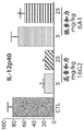
FIGS. 18A and 18B (IL-6), 18C and 18D (IFN γ), and 18E and 18F (IL-12p40) are graphs showing the activity of antibody surrogates of IP10.44(18G2) and anti-TNF α antibodies to reduce circulating levels of cytokines in serum and inflamed intestine using the CD 40-induced colitis model.
Fig. 19A (IP10.44) and 19B (IP10.1) are graphs showing the time profiles of free serum IP-10 after intravenous doses of IP10.44 and IP 10.1.
Figure 20 is a graph of free serum IP-10 inhibition (% baseline) compared to serum IP10.44 in cynomolgus monkeys.
FIGS. 21A to 21F are graphs showing the predicted free and total serum IP-10 in vs obtained by PK/PD modeling (for free serum IP-10, LLOQ at 1 pM; if the value is lower than LLOQ, LLOQ is used for plotting).
FIG. 22 is a graph of a higher affinity anti-IP-10 mouse surrogate (18G2) versus an anti-TNF α surrogate.
Detailed Description
In order that the invention may be more readily understood, certain terms are first defined. Other definitions are set forth throughout the detailed description.
The terms "6 a 5", "antibody 6a 5", "antibody IP 10.1", "IP 10.1" and "eldeluumab" refer to the anti-human IP-10 antibody described in WO2005/058815, 6a 5. The nucleotide sequence (SEQ ID NO: 5) and corresponding amino acid sequence (SEQ ID NO: 4) encoding the heavy chain variable region of IP10.1 are shown in FIG. 1A (CDR sequences are designated SEQ ID NO: 1,2 and 3, respectively). The nucleotide sequence (SEQ ID NO: 11) and corresponding amino acid sequence (SEQ ID NO: 10) encoding the light chain variable region of IP10.1 are shown in FIG. 1B (CDR sequences are designated SEQ ID NO: 7, 8 and 9, respectively).
The terms "interferon gamma inducible protein 10", "IP-10" and "CXCL 10" are used interchangeably and include variants, isoforms and species homologs of human IP-10. Thus, the human antibodies of the invention may, in some cases, cross-react with IP-10 from a species other than human. In other cases, the antibody may be completely specific for human IP-10 and may not exhibit species or other types of cross-reactivity. The complete amino acid sequence of human IP-10 has Genbank accession number NP-001556 (SEQ ID NO: 157). The Genbank accession number for the complete amino acid sequence of rhesus monkey IP-10 is AAK95955(SEQ ID NO: 159). The complete amino acid sequence of mouse IP-10 has Genbank accession number NP-067249 (SEQ ID NO: 160).
The term "CXCR 3" refers to the receptor for IP-10(CXCL 10). The complete amino acid sequence of human CXCR3 has Genbank accession number NP-001495 (SEQ ID NO: 158).
The term "MIG" refers to a ligand of CXCR3, also known as gamma-interferon inducible monokine, which is different from IP-10. The complete amino acid sequence of human MIG was Genbank accession NP-002407 (SEQ ID NO: 161).
The term "ITAC" refers to a ligand of CXCR3, also known as an interferon-inducible T cell alpha chemoattractant, which is different from IP-10. The complete amino acid sequence of human IATC has been Genbank accession NP-005400 (SEQ ID NO: 162).
The term "immune response" refers to the action of, for example, lymphocytes, antigen presenting cells, phagocytic cells, granulocytes, and soluble macromolecules produced by the above cells or liver (including antibodies, cytokines, and complement) that result in the selective damage, destruction, or elimination from the human body of invading pathogens, pathogen-infected cells or tissues, cancer cells, or in the case of autoimmunity or pathological inflammation, normal human cells or tissues.
"Signal transduction pathway" refers to the biochemical relationship between a variety of signal transduction molecules that function in the transmission of a signal from one part of a cell to another. As used herein, the phrase "cell surface receptor" includes, for example, molecules and molecular complexes that are capable of receiving a signal and propagating such a signal across the plasma membrane of a cell. An example of a "cell surface receptor" of the present invention is the CXCR3 receptor to which the IP-10 molecule binds.
The term "antibody" as referred to herein includes whole antibodies and any antigen-binding fragment (i.e., "antigen-binding portion") or single chain thereof. An "antibody" refers to a glycoprotein comprising at least two heavy (H) chains and two light (L) chains that are linked to each other by disulfide bonds, or an antigen-binding portion thereof. Each heavy chain is composed of a heavy chain variable region (abbreviated herein as V)H) And a heavy chain constant region. The heavy chain constant region is composed of three domains CH1、CH2And CH3And (4) forming. Each light chain is composed of a light chain variable region (abbreviated herein as V)L) And a light chain constant region. The light chain constant region consists of a domain CLAnd (4) forming. VHAnd VLRegions may be further subdivided into hypervariable regions, termed Complementarity Determining Regions (CDRs), interspersed with more conserved regions termed Framework Regions (FRs). VHAnd VLEach consisting of three CDRs and four FRs, arranged from amino-terminus to carboxy-terminus in the following order: FR1, CDR1, FR2, CDR2, FR3, CDR3, FR 4. The variable regions of the heavy and light chains contain binding domains that interact with antigens. The constant region of the antibody may mediate the binding of the immunoglobulin to host tissues or factors, including various cells of the immune system (e.g., effector cells) and the first component of the classical complement system (C1 q).
As used herein, the term "antigen-binding portion" of an antibody (or simply "antibody portion") refers to one or more antibody fragments that retain the ability to specifically bind to an antigen (e.g., IP-10). It has been shown that the antigen binding function of an antibody can be performed by fragments of a full-length antibody. The term "antigen-binding portion" of an antibody includes binding fragmentsExamples include: (i) fab fragments, i.e. consisting of VL、VH、CLAnd CH1Monovalent fragments consisting of domains; (ii) f (ab')2Fragments, i.e. bivalent fragments comprising two Fab fragments linked by disulfide bonds of the hinge region; (iii) from VHAnd CH1Domain-forming Fd fragments; (iv) v with one arm consisting of antibodyLAnd VH(iii) a domain consisting of an Fv fragment; (v) from VHdAb fragments consisting of domains (Ward et al (1989) Nature 341: 544-546); and (vi) an isolated Complementarity Determining Region (CDR). Furthermore, despite the two domains V of the Fv fragmentLAnd VHAre encoded by separate genes, but they can be joined together using recombinant methods by synthetic linkers that enable them to be made into a single protein chain, where VLAnd VHThe regions are paired to form monovalent molecules (known as single chain fv (scFv); see, e.g., Bird et al (1988) Science 242: 423-426; and Huston et al (1988) Proc. Natl. Acad. Sci. USA 85: 5879-5883). Such single chain antibodies are also intended to be encompassed within the term "antigen-binding portion" of an antibody. These antibody fragments are obtained by conventional techniques known to those skilled in the art and are subjected to practical screening in the same manner as for intact antibodies.
As used herein, "isolated antibody" refers to an antibody that is substantially free of other antibodies having different antigenic specificities (e.g., an isolated antibody that specifically binds IP-10 is substantially free of antibodies that specifically bind antigens other than IP-10). However, isolated antibodies that specifically bind IP-10 are cross-reactive with other antigens, such as IP-10 molecules from other species. Moreover, the isolated antibody may be substantially free of other cellular material and/or chemicals.
As used herein, the term "monoclonal antibody" or "monoclonal antibody composition" refers to a preparation of antibody molecules of single molecular composition. Monoclonal antibody compositions exhibit a single binding specificity and affinity for a particular epitope.
As used herein, the term "human antibody" includes antibodies having variable regions in which both the framework and CDR regions are derived from human germline immunoglobulin sequences. Furthermore, if the antibody contains constant regions, the constant regions are also derived from human germline immunoglobulin sequences. The human antibodies of the invention may comprise amino acid residues not encoded by human germline immunoglobulin sequences (e.g., mutations introduced by random or site-directed mutagenesis in vitro or by somatic mutation in vivo). However, as used herein, the term "human antibody" is not intended to include antibodies in which CDR sequences derived from the germline of another mammalian species, such as a mouse, have been grafted onto human framework sequences.
The term "human monoclonal antibody" refers to an antibody that exhibits a single binding specificity, having variable regions in which both the framework and CDR regions are derived from human germline immunoglobulin sequences. In one embodiment, the human monoclonal antibody is produced by a hybridoma comprising a B cell obtained from a transgenic non-human animal, such as a transgenic mouse, having a genome comprising a human heavy chain transgene and a light chain transgene fused to an immortalized cell.
As used herein, the term "recombinant human antibody" includes all human antibodies prepared, expressed, produced or isolated by recombinant methods, such as (a) antibodies isolated from animals (e.g., mice) that are transgenic or transchromosomal for human immunoglobulin genes or hybridomas prepared therefrom (described further below), (b) antibodies isolated from host cells transformed to express human antibodies (e.g., from transfectomas), (c) antibodies isolated from recombinant combinatorial human antibody libraries, and (d) antibodies prepared, expressed, produced or isolated by any other method including splicing of human immunoglobulin gene sequences with other DNA sequences. These recombinant human antibodies have variable regions in which both the framework and CDR regions are derived from human germline immunoglobulin sequences. However, in certain embodiments, such recombinant human antibodies can be subjected to in vitro mutagenesis (or, when transgenic animals using human Ig sequences are used, in vivo somatic mutagenesis) and, thus, the V of the recombinant antibodyHAnd VLThe amino acid sequence of the region although derived from human germline VHAnd VLSequences are related to, but may not naturally occur in the human antibody germline repertoire (repotoreie) in vivo.
As used herein, "isotype" refers to the class of antibodies (e.g., IgM or IgG1) encoded by the heavy chain constant region genes.
The phrases "antibody that recognizes an antigen" and "antigen-specific antibody" are used interchangeably herein with the term "antibody that specifically binds to an antigen".
As used herein, an antibody that "specifically binds to human IP-10" is intended to mean a 5X 10 antibody-9M or less, more preferably 1X10⁻¹⁰M or less and even more preferably 1X10-11K of M or lessDAn antibody that binds to human IP-10. An antibody that "cross-reacts with rhesus monkey IP-10" is at 1X10⁻⁹M or less, more preferably 1X10⁻¹⁰M or less and even more preferably 1X10-11K of M or lessDAn antibody that binds to rhesus monkey IP-10. An antibody that "does not cross-react with mouse IP-10" or "does not cross-react with human MIG" or "does not cross-react with human ITAC" is intended to mean at 1.5X 10⁻⁸K of M or greaterDMore preferably 5 to 10X 10-8M or greater and even more preferably 1X10⁻⁷K of M or greaterDAn antibody that binds to mouse IP-10, human MIG, or human ITAC. In certain embodiments, these antibodies that do not cross-react with mouse IP-10, human MIG, and/or human ITAC exhibit substantially undetectable binding to these proteins in a standard binding assay.
As used herein, an antibody that "inhibits the binding of IP-10 to CXCR 3" refers to an antibody that has a K of 1nM or less, more preferably 0.75nM or less, even more preferably 0.5nM or less and even more preferably 0.25nM or lessiAntibodies that inhibit the binding of IP-10 to CXCR 3.
As used herein, an antibody that "inhibits IP-10 induced calcium flux" refers to an IC at 10nM or less, more preferably 7.5nM or less, even more preferably 5nM or less, and even more preferably 2.5nM or less50Antibodies that inhibit IP-10 induced calcium flux.
As used herein, an antibody that "inhibits IP-10 induced cell migration" refers to an IC at 2. mu.g/ml or less, more preferably 1. mu.g/ml or less, even more preferably 0.5. mu.g/ml or less, or even more preferably 0.25. mu.g/ml or less50Inhibition of human IP-10 induced finesseAntibodies to cell migration.
As used herein, the term "Kassoc'OR' Ka"refers to the binding rate of a particular antibody-antigen interaction, and as used herein, the term" Kdis"or" Kd"refers to the off-rate of a particular antibody-antigen interaction. As used herein, the term "KD"refers to the dissociation constant, which is represented by KdAnd KaRatio of (i.e. K)d/Ka) Obtained and expressed as molar concentration (M). K of antibodyDValues may be determined by methods well known in the art. For determination of antibody KDA preferred method of (3) is to use surface plasmon resonance, preferably with a biosensor system, e.g. Provided is a system.
As used herein, the term "high affinity" of an IgG antibody refers to the K of the antibody for the target antigenDIs 10-8M or less, more preferably 10-9M or less and even more preferably 10-10M or less. However, for other antibody isotypes, "high affinity" binding may differ. For example, "high affinity" binding to an IgM isotype means that the antibody has 10-7M or less, more preferably 10-8K of M or lessD。
As used herein, the term "subject" includes any human or non-human animal. The term "non-human animal" includes all vertebrates, e.g., mammals and non-mammals, such as non-human primates, sheep, dogs, cats, horses, cows, chickens, amphibians, reptiles, and the like.
Various aspects of the invention are described in further detail in the following sections.
anti-IP-10 antibodies
The antibodies of the invention specifically bind to human IP-10 and are characterized by particularly improved functional characteristics or properties of the antibodies, as described above. In addition, the antibody can cross-react with IP-10 from one or more non-human primates, such as rhesus monkey. Preferably, the antibody does not cross-react with mouse IP-10. Furthermore, although MIG and ITAC are also ligands of the CXCR3 receptor, the antibodies of the invention preferably do not cross-react with human MIG or human ITAC.
Preferably, the antibodies of the invention bind to IP-10 with high affinity, e.g., KDIs 10-8M is less than or equal to 10-9M or less, or even 10-10M or less.
In addition, the antibodies of the invention are capable of inhibiting one or more functional activities of IP-10. For example, in one embodiment, the antibody inhibits the binding of IP-10 to CXCR 3. In another embodiment, the antibody inhibits IP-10 induced calcium flow. In yet another embodiment, the antibody inhibits IP-10-induced cell migration (chemotaxis).
Standard assays for assessing the ability of an antibody to bind IP-10 and/or MIG or ITAC of various species are well known in the art and include, for example, ELISA, Western blot analysis and RIA. Suitable assays are described in detail in the examples. The binding kinetics (e.g., binding affinity) of an antibody can also be assessed by standard assays well known in the art, such as by Biacore analysis. Assays to assess the effect of antibodies on functional properties of IP-10 (e.g., receptor binding, calcium flux, chemotaxis) are described in further detail in the examples.
Thus, an antibody that "inhibits" one or more of these IP-10 functional properties (e.g., biochemical, immunochemical, cellular, physiological or other biological activity, etc.) as determined according to methods well known in the art and described herein, should be understood to relate to a statistically significant reduction in a particular activity relative to that seen in the absence of the antibody (e.g., or when a control antibody of unrelated specificity is present). Preferably, an antibody that inhibits IP-10 activity results in at least a 10%, more preferably at least a 20%, 30%, 40%, 50%, 60%, 70%, 80% or 90% reduction in such statistically significant measured parameter, and in certain preferred embodiments, an antibody of the invention inhibits more than 92%, 94%, 95%, 96%, 97%, 98% or 99% of IP-10 functional activity.
Monoclonal antibodies IP10.44, IP10.52, IP10.45, IP10.46, IP10.53, IP10.43, IP10.47,
IP10.48, IP10.49, IP10.50, IP10.51, and IP10.54
Preferred antibodies of the invention are human monoclonal antibodies IP10.44, IP10.43, IP10.45, IP10.46, IP10.47, IP10.48, IP10.49, IP10.50, IP10.51, IP10.52, IP10.53 or IP 10.54. V for IP10.44, IP10.43, IP10.45, IP10.46, IP10.47, IP10.48, IP10.49, IP10.50, IP10.51, IP10.52, IP10.53, and IP10.54HThe amino acid sequence is shown in SEQ ID NO: 16. 28, 40, 52, 64, 76, 88, 100, 112, 124, 136 and 148. V for IP10.44, IP10.43, IP10.45, IP10.46, IP10.47, IP10.48, IP10.49, IP10.50, IP10.51, IP10.52, IP10.53, and IP10.54LThe amino acid sequence is shown in SEQ ID NO: 22. 34, 46, 58, 70, 82, 94, 106, 118, 130, 142 and 154.
A particular antibody of the invention is the human monoclonal antibody IP10.44 (also referred to herein as BMS-986184) which is structurally and chemically characterized as described below and in the examples below. V of IP10.44HThe amino acid sequence is shown in SEQ ID NO: 16 (fig. 2A). V of IP10.44LThe amino acid sequence is shown in SEQ ID NO: 22 (fig. 2B).
V of the antibodies described herein that bind to human IP-10HAnd VLThe sequences (or CDR sequences) can be combined with the V of other antibodies that bind to human IP-10HAnd VLSequences (or CDR sequences) "mix and match". Preferably, when VHAnd VLChains (or CDRs within these chains) when mixed and matched, are derived from a particular VH/VLPaired VHSequence is replaced by a structurally similar VHAnd (4) sequencing. Also, preferably, from a particular VH/VLPaired VLSequence is replaced by a structurally similar VLAnd (4) sequencing.
For example, an antibody or antigen-binding portion thereof of the invention comprises:
(a) comprising an amino acid sequence of IP10.44, IP10.43, IP10.45, IP10.46, IP10.47, IP10.48, IP10.49, IP10.50, IP10.51, IP10.52, IP10.53 or IP10.54Heavy chain variable regions of the columns, e.g., SEQ ID NOs: 16 (i.e., V for IP10.44)H) (ii) a And
(b) a light chain variable region comprising the amino acid sequence of IP10.44, IP10.43, IP10.45, IP10.46, IP10.47, IP10.48, IP10.49, IP10.50, IP10.51, IP10.52, IP10.53, or IP10.54, e.g., SEQ ID NO: 22 (i.e., V for IP10.44)L) Or another anti-IP-10 antibody (i.e., which is different from V for IP10.44, IP10.43, IP10.45, IP10.46, IP10.47, IP10.48, IP10.49, IP10.50, IP10.51, IP10.52, IP10.53, or IP10.54)L;
Wherein the antibody specifically binds to human IP-10.
In another embodiment, an antibody or antigen-binding portion thereof of the invention comprises:
(a) the CDR1, CDR2, and CDR3 regions of the heavy chain variable region of IP10.44, IP10.43, IP10.45, IP10.46, IP10.47, IP10.48, IP10.49, IP10.50, IP10.51, IP10.52, IP10.53, or IP10.54, e.g., comprising the amino acid sequence of SEQ ID NO: 16 (i.e., the CDR sequences of IP10.44, SEQ ID NOs 13, 14 and 15, respectively); and
(b) the CDR1, CDR2, and CDR3 regions of the light chain variable region of IP10.44, IP10.43, IP10.45, IP10.46, IP10.47, IP10.48, IP10.49, IP10.50, IP10.51, IP10.52, IP10.53, or IP10.54, e.g., comprising the amino acid sequence of SEQ ID NO: 22 (i.e., the CDR sequence of IP10.44, SEQ ID NOs 19, 20 and 21, respectively), or the CDR of another anti-IP-10 antibody (i.e., which is different from the CDR of IP10.44, IP10.43, IP10.45, IP10.46, IP10.47, IP10.48, IP10.49, IP10.50, IP10.51, IP10.52, IP10.53 or IP 10.54); wherein the antibody specifically binds to human IP-10. For example, an antibody or antigen-binding portion thereof can include heavy chain variable CDR1, CDR2, and CDR3 regions of IP10.44 in combination with one or more of the light chain CDR1, CDR2, and/or CDR3 regions of other antibodies that bind human IP-10.
In addition, it is well known in the art that individual CDR3 domains, independent of the CDR1 and/or CDR2 domains, can determine the binding specificity of an antibody for a cognate antigen, and can be predictably generated based on a common CDR3 sequenceA plurality of antibodies having the same binding specificity. See, e.g., Klimka et al, British J.of Cancer83(2): 252-260 (2000); beiboer et al, J.mol.biol.296: 833-849 (2000); rader et al, Proc.Natl.Acad.Sci.U.S.A.95: 8910-8915 (1998); barbas et al, j.am.chem.soc.116: 2161-2162 (1994); barbas et al, proc.natl.acad.sci.u.s.a.92: 2529 2533 (1995); ditzel et al, J.Immunol.157: 739-; berezov et al, BIAjournal8: scientific Review 8 (2001); igarashi et al, J.biochem (Tokyo)117: 452-7 (1995); bourgeois et al, J.Virol72: 807-10 (1998); levi et al, Proc.Natl.Acad.Sci.U.S.A.90: 4374-8 (1993); polymenis and Stoller, j.152: 5218-13: 37-45(2000). See also US patent No.6,951,646; 6,914,128, respectively; 6,090,382; 6,818,216, respectively; 6,156,313, respectively; 6,827,925, respectively; 5,833,943, respectively; 5,762,905, and 5,760,185. Each of these references is incorporated herein by reference in its entirety.
Thus, in another embodiment, an antibody of the invention comprises at least the CDR3 region of the heavy chain variable region of IP10.44, IP10.43, IP10.45, IP10.46, IP10.47, IP10.48, IP10.49, IP10.50, IP10.51, IP10.52, IP10.53, or IP10.54 and at least the CDR3 of the heavy chain variable region of IP10.44, IP10.43, IP10.45, IP10.46, IP10.47, IP10.48, IP10.49, IP10.50, IP10.51, IP10.52, IP10.53, or IP10.54 (e.g., SEQ ID NOs 15 and 21, CDR3 of the heavy and light chain variable regions of IP 10.44). These antibodies preferably (a) compete for binding with the antibody from which the CDR3 sequence is derived (e.g., antibody IP 10.44); (b) retains the functional characteristics of the antibody from which the CDR3 sequence is derived (e.g., antibody IP 10.44); (c) binds to the same epitope as the antibody from which the CDR3 sequence is derived (e.g., antibody IP 10.44); and/or (d) has the same binding affinity as an antibody from which the CDR3 sequence is derived (e.g., antibody IP 10.44).
Amino acid modification
In another embodiment, the antibody of the invention comprises a heavy chain variable region that hybridizes to IP10.44, IP10.43, IP10.45, IP10.46, IP10.47, IP10.48, IP10.49, IP10.50, IP10.51, or a light chain variable region,IP10.52, IP10.53, or IP10.54 (e.g., IP10.44) differ in the heavy and/or light chain variable region sequences of one or more conservatively modified CDR1, CDR2, and CDR3 sequences. However, in a preferred embodiment, (a) V for IP10.44HGlutamic acid and tyrosine residues of CDR1 (as in the following sequence)EYUnderlined in GMH) unmodified, (b) V of IP10.44HGlycine, alanine, leucine, isoleucine, glycine and alanine residues of CDR2 (as in sequence VI below)GFAGLIKGYAUnderlined in DSVKG) unmodified and (c) V at IP10.44HAlanine and asparagine residues of CDR3 (as in the following sequence EGAGSNIYYYYGMDV underlined) was unmodified. It is understood in the art that certain conservative sequence modifications may be made that do not eliminate antigen binding. See, e.g., Brummell et al, (1993) Biochem32: 1180-8; de Wildt et al, (1997) prot.10: 835-41; komissarov et al, (1997) J.biol.chem.272: 26864-26870; hall et al (1992) J.Immunol.149: 1605-12; kelley and O' Connell (1993) Biochem.32: 6862-35; Adib-Conquy et al, (1998) int. Immunol.10: 341-6 and Beers et al, (2000) Clin.6: 2835-43. Thus, in one embodiment, the antibody comprises a heavy chain variable region comprising the sequences of CDR1, CDR2 and CDR3 and/or a light chain variable region comprising the sequences of CDR1, CDR2 and CDR3, wherein:
(a) the heavy chain variable region CDR1 sequence comprises SEQ ID NO: 13 and/or conservative modifications thereof, except for V of IP10.44HGlutamic acid and tyrosine residues of CDR1 (as in the following sequence)EYUnderlined in GMH) unmodified; and/or
(b) The heavy chain variable region CDR2 sequence comprises SEQ ID NO: 14 and/or conservative modifications thereof, except for V at IP10.44HGlycine, alanine, leucine, isoleucine, glycine and alanine residues of CDR2 (as in sequence VI below)GFAGLIKGYAUnderlined in DSVKG) unmodified; and/or
(c) The heavy chain variable region CDR3 sequence comprises SEQ ID NO: 15 and conservative modifications thereof, except for V at IP10.44HAlanine and asparagine of CDR3Amine residues (e.g. in the following sequence EG)AGSNIYYYYGMDV underlined) is unmodified; and/or
(d) The light chain variable region CDR1 and/or CDR2 and/or CDR3 sequence comprises SEQ ID NO: 19 and/or SEQ ID NO: 20 and/or SEQ ID NO: 21, and/or conservative modifications thereof; and
(e) the antibody specifically binds to human IP-10.
Additionally or alternatively, the antibody may have one or more of the following functional properties as described above, such as high affinity binding to human IP-10, the ability to bind monkey IP-10 (e.g., cynomolgus monkey, rhesus monkey) but not substantially to mouse IP-10, the ability not to cross-react with human MIG or human ITAC, and the ability to inhibit (a) binding of IP-10 to CXCR3, (b) IP-10 induced calcium flux, and/or (c) IP-10 induced cell migration (chemotaxis).
In various embodiments, the antibody can be, for example, a human, humanized, or chimeric antibody.
As used herein, the term "conservative sequence modification" refers to an amino acid modification that does not significantly affect or alter the binding characteristics of an antibody comprising the amino acid sequence. Such conservative modifications include amino acid substitutions, additions and deletions. Modifications can be introduced into the antibodies of the invention by standard techniques known in the art, such as site-directed mutagenesis and PCR-mediated mutagenesis. Conservative amino acid substitutions are those that replace an amino acid residue with one having a similar side chain. Families of amino acid residues with similar side chains have been defined in the art. These families include: amino acids having basic side chains (e.g., lysine, arginine, histidine), acidic side chains (e.g., aspartic acid, glutamic acid), uncharged polar side chains (e.g., glycine, asparagine, glutamine, serine, threonine, tyrosine, cysteine, tryptophan), nonpolar side chains (e.g., alanine, valine, leucine, isoleucine, proline, phenylalanine, methionine), beta-branched side chains (e.g., threonine, valine, isoleucine) and aromatic side chains (e.g., tyrosine, phenylalanine tryptophan, histidine). Thus, one or more amino acid residues within a CDR region of an antibody of the invention can be substituted for other amino acid residues from the same side chain family, and the altered antibody can be tested for retained function (i.e., the function described above) using the functional assays described herein.
Engineered and modified antibodies
The antibodies of the invention may be raised against one or more V having IP10.44, IP10.43, IP10.45, IP10.46, IP10.47, IP10.48, IP10.49, IP10.50, IP10.51, IP10.52, IP10.53, or IP10.54 (e.g., antibody IP10.44)HAnd/or VLAntibodies of sequence were prepared as starting materials to engineer the modified antibodies. Can be modified by modifying one or both variable regions (i.e., V)HAnd/or VL) Antibodies are engineered with one or more residues within (e.g., within one or more CDR regions and/or within one or more framework regions). Additionally or alternatively, an antibody may be engineered by modifying residues within the constant region, for example to alter the effector function of the antibody.
In certain embodiments, CDR grafting can be used to engineer the variable regions of antibodies. Antibodies interact with the target antigen primarily through amino acid residues located in the six heavy and light chain Complementarity Determining Regions (CDRs). For this reason, the amino acid sequences within the CDRs are more diverse between individual antibodies than sequences outside the CDRs. Since the CDR sequences are responsible for most of the antibody-antigen interactions, it is possible to express recombinant antibodies that mimic the properties of a particular naturally occurring antibody by constructing expression vectors that comprise CDR sequences from that antibody grafted onto framework sequences from different antibodies having different properties (see, e.g., Riechmann et al, (1998) Nature 332: 323-327; Jones et al, (1986) Nature 321: 522-525; Queen et al, (1989) Proc. Natl. Acad. Sci. U.S.A.86: 10029-10033; U.S. Pat. Nos. 5,225,539; 5,530,101; 5,585,089; 5,693,762 and 6,180,370).
Thus, another embodiment of the invention relates to an isolated monoclonal antibody, or antigen binding portion thereof, comprising CDR1, CDR2, and CDR3 sequences comprising ipl0.44, IP10.43, IP10.45, IP10.46, IP10.47, IP10.48, IP10.49, IP10.50, IP10.51, IP10.52, IP10.53, or IP10.54The heavy chain variable regions of the columns (e.g., SEQ ID NOS: 13, 14 and 15, respectively) and/or the light chain variable regions comprising the CDR1, CDR2 and CDR3 sequences of IP10.44, IP10.43, IP10.45, IP10.46, IP10.47, IP10.48, IP10.49, IP10.50, IP10.51, IP10.52, IP10.53 or IP10.54 (e.g., SEQ ID NOS: 19, 20 and 21, respectively). Although these antibodies comprise the V of monoclonal antibody IP10.44 or other antibodies described hereinHAnd VLCDR sequences, which may comprise different framework sequences.
These framework sequences can be obtained from public DNA databases or published references comprising germline antibody gene sequences. For example, germline DNA sequences for human heavy and light chain variable region genes can be found in: "VBase" human germline sequence database (available from the Internet)www.mrc-cpe.cam.ac.uk/vbaseObtained), and Kabat et al, supra; tomlinson et al, (1992) "The retransmission of Human Germine VH Sequences improvements about Fifty Groups of VH Sequences with Different variants Hypervariable Loops" J.mol.biol.227: 776-798; and Cox et al, (1994) "A Directory of Human Germ-line VHSegments vectors a Strong Bias in the use "Eur.J. Immunol.24: 827-836; the contents of each of which are incorporated herein by reference. As another example, germline DNA sequences for human heavy and light chain variable region genes can be found in the Genbank database. For example, the following heavy chain germline sequences found in HCo7 HuMAb mice can be found in the accompanying Genbank accession numbers: 1-69(NG _0010109, NT _024637)&BC070333)、3-33(NG_0010109&NT _024637) and 3-7(NG _ 0010109)&NT _ 024637). As another example, the following heavy chain germline sequences found in HCol2 HuMAb mice can be found at the accompanying Genbank accession numbers: 1-69(NG _0010109, NT _024637)&BC070333)、5-51(NG_0010109&NT_024637)、4-34(NG_0010109&NT_024637)、3-30.3(CAJ556644)&3-23(AJ 406678).
Antibody protein sequences are compared against compiled protein sequence databases using one of the sequence similarity search methods known as Gapped BLAST (Altschul et al, (1997), supra), as is well known to those skilled in the art.
Used in the inventionPreferred framework sequences for antibodies are those that are structurally similar to the framework sequences selected for use with the antibodies of the invention, e.g., similar to V used in IP10.44H3-33 framework sequences and/or VKA27 framework sequence. VHCDR1, CDR2 and CDR3 sequences and VKThe CDR1, CDR2, and CDR3 sequences can be grafted onto framework regions having the same sequences as present in the germline immunoglobulin gene from which the framework sequences are derived, or the CDR sequences can be grafted onto framework regions containing one or more mutations compared to the germline sequence. For example, it has been found to be advantageous in certain instances to mutate residues within the framework regions to maintain or enhance the antigen binding ability of the antibody (see, e.g., U.S. Pat. Nos. 5,530, 101; 5,585, 089; 5,693,762, and 6,180,370).
Another type of variable region modification is mutation VHAnd/or VLAmino acid sequences within the CDR1, CDR2, and/or CDR3 regions, thereby improving one or more binding properties (e.g., affinity) of the antibody of interest. Site-directed mutagenesis or PCR-mediated mutagenesis can be performed to introduce mutations, and the effect on antibody binding or other functional properties of interest can be assessed using in vitro or in vivo assays described herein and provided in the examples. Preferably conservative modifications (as described above) are introduced. The mutation may be an amino acid substitution, addition or deletion, but is preferably a substitution. Moreover, typically no more than 1,2, 3,4, or 5 residues within a CDR region are altered.
Thus, in another embodiment, the invention provides an isolated anti-IP-10 monoclonal antibody, or antigen-binding portion thereof, comprising heavy and/or light chain variable region sequences as described herein, wherein these sequences comprise one or more amino acid substitutions, deletions, or additions. For example, an isolated anti-IP-10 monoclonal antibody, or antigen-binding portion thereof, comprising a heavy chain variable region comprising: (a) comprises the amino acid sequence of SEQ ID NO: 13 or a variant of SEQ ID NO: 13 compared to V having an amino acid sequence of one, two or three amino acid substitutions, deletions or additionsHCDR1 region (preferably wherein V of IP10.44HGlutamic acid and tyrosine residues of CDR1 (as in the following sequence)EYGMH underlined) and SEO ID NO: 13 are the same); (b) comprises SEO ID NO: 14 or a sequence identical to SEQ ID NO: 14 vs V having an amino acid sequence of one, two, three, four or five amino acid substitutions, deletions or additionsHCDR2 region (preferably wherein V of IP10.44HGlycine, alanine, leucine, isoleucine, glycine and alanine residues of CDR2 (as in sequence VI below)GFAGLIKGYADSVKG underlined) was compared to SEQ ID NO: 14 are the same); (c) comprises the amino acid sequence of SEQ ID NO: 15 or a variant of SEQ ID NO: 15 vs V having an amino acid sequence of one, two, three, four or five amino acid substitutions, deletions or additionsHCDR3 region (preferably where V of IP10.44HAlanine and asparagine residues of CDR3 (as in the following sequence EGAGSNIYYYYGMDV underlined) is similar to SEQ ID NO: 15 are the same); (d) comprises the amino acid sequence of SEQ ID NO: 19 or a variant of SEQ ID NO: 19 compared to V having an amino acid sequence of one, two, three, four or five amino acid substitutions, deletions or additionsLA CDR1 region; (e) comprises the amino acid sequence of SEQ ID NO: 20 or a sequence identical to SEQ ID NO: 20 vs V having an amino acid sequence of one, two, three, four or five amino acid substitutions, deletions or additionsLA CDR2 region; and (f) comprises SEQ ID NO: 21 or a sequence identical to SEQ ID NO: 21 compared to V having an amino acid sequence of one, two, three, four or five amino acid substitutions, deletions or additionsLA CDR3 region.
Engineered antibodies of the invention include, for example, V directed to antibodies for the purpose of improving their propertiesHAnd/or VLAntibodies in which the framework residues within are modified. Such framework modifications are typically made to reduce the immunogenicity of the antibody. For example, one approach is to "back mutate" one or more framework residues to the corresponding germline sequence. More particularly, an antibody undergoing somatic mutation may contain framework residues that differ from the germline sequence from which the antibody is derived. These residues can be identified by comparing the antibody framework sequences to the germline sequence from which the antibody was derived.
Another type of framework modification involves mutating one or more residues within the framework regions or even within one or more CDR regions to remove T cell epitopes, thereby reducing the potential immunogenicity of the antibody. This method is also called "deimmunization" (deimmunization), and is described in detail in U.S. patent publication No. 20030153043.
In addition to, or as an alternative to, modifications made within the framework or CDR regions, the antibodies of the invention may be engineered to include modifications within the Fc region, typically in order to alter one or more functional properties of the antibody, such as serum half-life, complement fixation, Fc receptor binding and/or antigen-dependent cellular cytotoxicity. In addition, the antibodies of the invention may also be chemically modified (e.g., one or more chemical moieties may be attached to the antibody), or modified to alter glycosylation thereof, which in turn alters one or more functional properties of the antibody. Each of these embodiments is described in detail below. The numbering of residues in the Fc region is that of the EU index of Kabat.
In a preferred embodiment, the antibody is an IgG4 isotype antibody comprising a serine to proline mutation (S228P; EU index) at a position corresponding to position 228 in the hinge region of the heavy chain constant region. This mutation has been reported to eliminate the heterogeneity of the disulfide bonds between heavy chains in the hinge region (Angal et al, supra; position 241 is based on the Kabat numbering system).
In one embodiment, the hinge region of CH1 is modified such that the number of cysteine residues in the hinge region is altered, e.g., increased or decreased. This process is also described in U.S. patent No. 5,677,425. The number of cysteines in the hinge region of CH1 is altered, for example, to facilitate assembly of the light and heavy chains, or to increase or decrease the stability of the antibody.
In another embodiment, the Fc hinge region of an antibody is mutated to reduce the biological half-life of the antibody. More specifically, one or more amino acid mutations are introduced into the CH2-CH3 domain interface region of the Fc-hinge fragment such that SpA binding of the antibody is reduced relative to staphylococcal protein a (SpA) binding of the native Fc-hinge domain. This method is also described in U.S. Pat. No.6,165,745.
In another embodiment, the antibody is modified to increase its biological half-life. Various approaches are possible. For example, one or more of the following mutations may be introduced: T252L, T254S, T256F as described in U.S. patent No.6,277,375. Alternatively, to increase biological half-life, the antibody may be altered within the CHl or CL region to include a salvage receptor binding epitope from two loops of the CH2 domain of the IgG Fc region, as described in U.S. Pat. nos. 5,869,046 and 6,121,022.
In still other embodiments, the Fc region is altered by substituting at least one amino acid residue for a different amino acid residue to alter the effector function of the antibody. For example, one or more amino acids selected from amino acid residues 234, 235, 236, 237, 297, 318, 320, 322 may be substituted with a different amino acid residue such that the antibody has altered affinity for an effector ligand, but retains the antigen binding ability of the parent antibody. The effector ligand that alters its affinity may be, for example, an Fc receptor or the C1 component of complement. This method is also described in U.S. Pat. nos. 5,624,821 and 5,648,260.
In another example, one or more amino acids selected from amino acid residues 329, 331, and 322 can be substituted with a different amino acid residue such that the antibody has altered C1q binding and/or reduced or eliminated Complement Dependent Cytotoxicity (CDC). This method is described in more detail in U.S. Pat. No.6,194,551.
In another embodiment, one or more of the amino acid residues in amino acid positions 231 and 239 are altered, thereby altering the ability of the antibody to fix complement. This method is further described in PCT publication WO 94/29351.
In yet another embodiment, to increase the ability of an antibody to mediate antibody-dependent cellular cytotoxicity (ADCC) and/or to increase the affinity of an antibody for fey receptors, the Fc region is modified by modifying one or more amino acids at the following positions: 238. 239, 248, 249, 252, 254, 255, 256, 258, 265, 267, 268, 269, 270, 272, 276, 278, 280, 283, 285, 286, 289, 290, 292, 293, 294, 295, 296, 298, 301, 303, 305, 307, 309, 312, 315, 320, 322, 324, 326, 327, 329, 330, 331, 333, 334, 335, 337, 338, 340, 360, 373, 376, 378, 382, 388, 389, 398, 414, 416, 419, 430, 434, 435, 437, 438 or 439. This method is further described in PCT publication WO 00/42072. Furthermore, the binding sites on human IgG1 for Fc γ RI, Fc γ RII, Fc γ RIII and FcRn have been mapped and variants with improved binding have been described (see, Shield et al (2001) J.biol.chem.276: 6591-6604). Specific mutations at positions 256, 290, 298, 333, 334 and 339 were shown to improve binding to Fc γ RIII. In addition, the following combination mutants were shown to improve Fc γ RIII binding: T256A/S298A, S298A/E333A, S298A/K224A and S298A/E333A/K334A.
In yet another embodiment, the glycosylation of the antibody is altered. For example, an antibody can be made that is aglycosylated (i.e., the antibody lacks glycosylation). For example, glycosylation can be altered in order to increase the affinity of an antibody for an antigen. Such sugar modifications can be achieved, for example, by altering one or more glycosylation sites within the antibody sequence. For example, one or more amino acid substitutions may be made which result in the elimination of one or more variable region framework glycosylation sites, thereby eliminating glycosylation at such sites. This glycosylation can increase the affinity of the antibody for the antigen. See, for example, U.S. Pat. nos. 5,714,350 and 6,350,861.
Additionally or alternatively, antibodies with altered glycosylation patterns can be prepared, such as low fucosylated antibodies with reduced amounts of fucosyl residues, or antibodies with increased bisecting GlcNac structures. This altered glycosylation pattern has been demonstrated to enhance the ADCC ability of the antibody. Such sugar modifications can be achieved, for example, by expressing the antibody in a host cell with an altered glycosylation machinery. Cells with altered glycosylation machinery have been described in the art and can be used as host cells in which to express the recombinant antibodies of the invention, thereby producing antibodies with altered glycosylation. For example, the cell lines Ms704, Ms705 and Ms709 lack the glycosyltransferase gene FUT8(α (1, 6) -glycosyltransferase) such that antibodies expressed in the Ms704, Ms705 and Ms709 cell lines lack fucose on their sugars. Ms704, Ms705 and Ms709FUT8-/-Cell lines Using two substituted vectors through CHO/DGTargeted disruption of the FUT8 gene in 44 cells resulted (see U.S. patent publication No.20040110704 and Yamane-Ohnuki et al, (2004) Biotechnol Bioeng87: 614-22). As another example, EP 1,176,195 describes a cell line with a functionally disrupted FUT8 gene encoding a fucosyltransferase such that antibodies expressed in such cell line exhibit low fucosylation by reducing and eliminating alpha-1, 6 linkage related enzymes. EP 1,176,195 also describes cell lines with low or no enzymatic activity for the addition of fucose to N-acetylglucosamine, which binds to the Fc region of antibodies, such as the rat myeloma cell line YB2/0(ATCC CRL 1662). PCT publication WO 03/035835 describes a variant CHO cell line, Lec 13 cell, which has a reduced ability to attach fucose to an Asn (297) -linked sugar, also resulting in low fucosylation of antibodies expressed in the host cell (see also, Shield et al (2001) J. biol. chem. 277: 26733-. Antibodies with altered glycosylation characteristics can also be produced in chicken eggs, as described in PCT publication WO 06/089231. Alternatively, antibodies with altered glycosylation characteristics can be produced in plant cells such as Lemna (Lemna). A method for producing antibodies in a plant system is disclosed corresponding to Alston, filed on 8/11/2006&Bird LLP attorney docket No.040989/314911, U.S. patent application. PCT publication WO 99/54342 describes cell lines engineered to express glycoprotein-modified glycosyltransferases, such as β (1, 4) -acetylglucosaminyltransferase III (GnTIII), such that antibodies expressed in the engineered cell lines exhibit increased bisection GlcNac structures that result in increased ADCC activity of the antibody (see also, Umana et al (1999) nat. Biotech.17: 176-180). Alternatively, the fucose residue of the antibody may be cleaved using fucosidase; for example, fucosidase α -L-fucosidase removes fucose residues from antibodies (Tarentino et al, (1975) Biochem.14:5516-23)。
Another modification of the antibodies described herein contemplated by the present disclosure is pegylation. For example, the antibody can be pegylated, e.g., to increase the biological (e.g., serum) half-life of the antibody. To pegylate an antibody, the antibody or fragment thereof is typically reacted with a polyethylene glycol (PEG), such as a reactive ester or aldehyde derivative of PEG, under conditions in which one or more PEG groups become attached to the antibody or antibody fragment. Preferably, pegylation is performed by acylation or alkylation with a reactive PEG molecule (or similar reactive water-soluble polymer). As used herein, the term "polyethylene glycol" is intended to include any PEG form used to derivatize other proteins, such as mono (C1-C10) alkoxy-or aryloxy-polyethylene glycol or polyethylene glycol-maleimide. In certain embodiments, the antibody to be pegylated is an aglycosylated antibody. Methods of pegylating proteins are known in the art and may be applied to the antibodies of the invention. See, for example, EP 0154316 and EP 0401384.
Physical Properties of antibody
The antibodies of the invention may be characterized by their various physical properties to detect and/or distinguish their different classes.
For example, an antibody may contain one or more glycosylation sites in the light or heavy chain variable region. Such glycosylation sites can lead to increased immunogenicity of the antibody or to a change in the pK of the antibody due to altered antigen binding (Marshall et al (1972) Annu Rev Biochem41: 673-702; gala and Morrison (2004) J Immunol172: 5489-94; wallick et al (1988) J Exp Med168:1099-109;Spiro(2002)Glycobiology 12: 43R-56R; parekh et al (1985) Nature316: 452-7; mimura et al (2000) Mol Immunol37: 697-706). Glycosylation is known to occur at motifs comprising N-X-S/T sequences. In some cases, it is preferred to have an anti-IP-10 antibody that does not contain variable region glycosylation. This can be achieved by selecting antibodies that do not contain glycosylation motifs in the variable region or by mutating residues in the glycosylation region.
In a preferred embodiment, the antibody does not comprise an asparagine isomerization site. Deamidation of asparagine can occur at the N-G or D-G sequence and result in the production of isoaspartic acid, which introduces kinks into the polypeptide chain and reduces its stability (isoaspartic acid effect).
Each antibody has a unique isoelectric point (pI) which typically falls within a pH range of 6 to 9.5. The pI of the IgG1 antibody typically falls within a pH range of 7-9.5 and the pI of the IgG4 antibody typically falls within a pH range of 6-8. It is speculated that antibodies with a pI outside the normal range may have some unfolding and instability under in vivo conditions. Therefore, preferred are anti-IP-10 antibodies having pI values that fall within the normal range. This can be achieved by selecting antibodies with a normal range of pI or by mutating charged surface residues.
Nucleic acid molecules encoding the antibodies of the invention
Another aspect of the invention relates to a nucleic acid molecule encoding an antibody of the invention. The nucleic acid may be present in whole cells, cell lysates, or in partially purified or substantially purified form. Nucleic acids are "isolated" or "rendered substantially pure" when separated and purified from other cellular components or other contaminants, such as other cellular nucleic acids or proteins, by standard techniques, including alkali/SDS treatment, CsCl banding, column chromatography, agarose gel electrophoresis, and other methods well known in the art. See, e.g., Ausubel et al (1987) Current Protocols in Molecular Biology, Greene Publishing and Wiley Interscience, New York. The nucleic acids of the invention may be, for example, DNA or RNA, and may or may not contain intron sequences. In a preferred embodiment, the nucleic acid is a cDNA molecule.
The nucleic acids of the invention can be obtained using standard molecular biology techniques. For antibodies expressed by a hybridoma (e.g., a hybridoma prepared from a transgenic mouse carrying human immunoglobulin genes as described further below), cdnas encoding the light and heavy chains of the antibody prepared by the hybridoma can be obtained using standard PCR amplification or cDNA cloning techniques. For antibodies obtained from immunoglobulin gene libraries (e.g., using phage display technology), nucleic acids encoding the antibodies can be recovered from the libraries.
Preferred nucleic acid molecules of the invention are those encoding the VH and VL sequences of the IP10.44 monoclonal antibody. The DNA sequences encoding the VH and VL sequences of IP10.44 are shown in SEQ ID NO: 17 and 23.
Once the DNA fragments encoding the VH and VL fragments are obtained, these DNA fragments can be further manipulated by standard recombinant DNA techniques, for example to convert the variable region genes to full length antibody chain genes, to Fab fragment genes or to scFv genes. In these manipulations, a DNA fragment encoding a VL or VH is operably linked to another DNA fragment encoding another protein such as an antibody constant region or a flexible linker. As used herein, the term "operably linked" means that two DNA segments are linked such that the amino acid sequences encoded by the two DNA segments remain in frame.
Isolated DNA encoding a VH region can be converted to a full-length heavy chain gene by operably linking the DNA encoding the VH to another DNA molecule encoding the heavy chain constant region (CH1, CH2, and CH 3). The sequence of the Human heavy chain constant region gene is known in the art (see, for example, Kabat, E.A. et al (1991) Sequences of Proteins of immunologic Interest, fifth edition, U.S. department of Health and Human Services, NIH publication No. 91-3242), and DNA fragments comprising these regions can be obtained by standard PCR amplification. The heavy chain constant region may be an IgG1, IgG2, IgG3, IgG4, IgA, IgE, IgM, or IgD constant region, but is most preferably an IgG1 or IgG4 constant region. For a Fab fragment heavy chain gene, the DNA encoding the VH may be operably linked to another DNA molecule encoding only the heavy chain CH1 constant region.
The isolated DNA encoding the VL region can be converted into a full-length light chain gene (as well as the Fab light chain gene) by operably linking the DNA encoding the VL to another DNA molecule encoding the light chain constant region CL. The sequence of the Human light chain constant region gene is known in the art (see, for example, Kabat, E.A. et al (1991) Sequences of Proteins of immunologic Interest, fifth edition, U.S. department of Health and Human Services, NIH publication No. 91-3242), and DNA fragments comprising these regions can be obtained by standard PCR amplification. The light chain constant region may be a kappa or lambda constant region, but is most preferably a kappa constant region.
To produce scFv genes, DNA fragments encoding VH and VL may be ligated with a linker encoding a flexible linker, e.g.an amino acid sequence (Gly)4-Ser)3Is operably linked such that VH and VL areThe sequence may be expressed as a continuous single-stranded protein with the VL and VH regions linked by a flexible linker (see, e.g., Bird et al (1988) Science 242: 423-.
Generation of monoclonal antibodies of the invention
Monoclonal antibodies (mabs) of the invention can be prepared by a variety of techniques, including conventional monoclonal antibody methods, e.g., Kohler and Milstein (1975) Nature 256: 495 by standard somatic hybridization technique. Although somatic cell hybridization methods are preferred, in principle, other techniques for preparing monoclonal antibodies, such as viral or oncogenic transformation of B lymphocytes, can be used.
The preferred animal system for preparing hybridomas is the murine system. Hybridoma production in mice is a well established procedure. Immunization protocols and techniques for isolating immune splenocytes for fusion are known in the art. Fusion partners (e.g., murine myeloma cells) and fusion methods are also known.
The chimeric or humanized antibody of the present invention can be prepared based on the sequence of the murine monoclonal antibody prepared as described above. DNA encoding the heavy and light chain immunoglobulins can be obtained from a murine hybridoma of interest and engineered to contain non-murine (e.g., human) immunoglobulin sequences using standard molecular biology techniques. For example, to generate chimeric antibodies, murine variable regions can be joined to human constant regions using methods well known in the art (see, e.g., U.S. Pat. No.4,816,567 to Cabilly et al). To generate humanized antibodies, murine CDR regions can be inserted into human frameworks using methods well known in the art (see, e.g., U.S. Pat. No. 5,225,539 to Winter, and U.S. Pat. Nos. 5,530,101; 5,585,089; 5,693,762 and 6,180,370 to Queen et al).
In a preferred embodiment, the antibodies of the invention are human monoclonal antibodies. Such human monoclonal antibodies to IP-10 can be produced using transgenic or transchromosomal mice carrying portions of the human immune system rather than the mouse system. These transgenic and transchromosomal mice include mice referred to herein as HuMab mice and KM mice, respectively, and are generally referred to herein as "human Ig mice".
HuMab mice (Metarex, Inc.) comprises a human immunoglobulin gene minilocus encoding unrearranged human heavy (mu and gamma) and kappa light chain immunoglobulin sequences, and a targeted mutation that inactivates the endogenous mu and kappa chain loci (see, e.g., Lonberg et al (1994) Nature 368 (6474): 856-. Thus, this mouse shows reduced expression of mouse IgM or kappa, and in response to immunization, the introduced human heavy and light chain transgenes undergo class switching and somatic mutation to generate high affinity human IgG kappa monoclonal (Lonberg, N. et al (1994), supra; review Lonberg, N. (1994) Handbook of Experimental Pharmacology 113: 49-101; Lonberg, N. and Huszar, D. (1995) Intern.Rev.Immunol.13: 65-93, and Harding, F. and Lonberg, N. (1995) Ann.N.Y.Acad.Sci 764: 536-546). The preparation and use of HuMab mice, and the genomic modifications carried by such mice, are further described in the following references: taylor.l. et al (1992) nucleic acids Research 20: 6287-6295; chen, J. et al (1993) International Immunology 5: 647-656; tuaillon et al (1993) Proc. Natl. Acad. Sci USA 90: 3720-3724; choi et al (1993) Nature Genetics 4: 117-; chen, J. et al (1993) EMBO J.12: 821-830; tuaillon et al (1994) J.Immunol.152: 2912-2920; taylor, L. et al (1994) International Immunology 6: 579-; and Fishwild, D. et al (1996) Nature Biotechnology 14: 845-851, the contents of which are incorporated herein by reference in their entirety. See, further, U.S. Pat. nos. 5,545,806; 5,569,825; 5,625,126, respectively; 5,633,425, respectively; 5,789,650, respectively; 5,877,397, respectively; 5,661,016, respectively; 5,814, 318; 5,874,299, respectively; and 5,770,429; both Lonberg and Kay; and U.S. patent 5,545,807 to Surani et al; PCT publications WO 92/03918, WO 93/12227, WO 94/25585, WO 97/13852, WO 98/24884 and WO 99/45962, all belonging to Lonberg and Kay; and PCT publication No. WO 01/14424 to Korman et al.
In another embodiment, the human antibodies of the invention can be produced using mice carrying human immunoglobulin sequences on transgenes and transchromosomes, such as mice carrying human heavy chain transgenes and human light chain transchromosomes. Such mice are referred to herein as "KM mice" and are described in detail in PCT publication WO 02/43478 to Ishida et al.
Furthermore, alternative transgenic animal systems expressing human immunoglobulin genes are available in the art and can be used to produce the anti-IP-10 antibodies of the invention. For example, an alternative transgenic system known as Xenomouse (Abgenix, Inc.) may be used, such mice being described in, for example, U.S. Pat. nos. 5,939,598 to Kucherlapati et al; 6,075,181; 6,114,598, respectively; 6,150,584 and 6,162,963.
Furthermore, alternative transchromosomal animal systems for expressing human immunoglobulin genes are available in the art and can be used to produce the anti-IP-10 antibodies of the invention. For example, a mouse carrying a human heavy chain transchromosome and a human light chain transchromosome, referred to as a "TC mouse," can be used; such mice are described in Tomizuka et al (2000) proc.natl.acad.sci.usa 97: 722-727. In addition, cows carrying human heavy and light chain transfectants have been described in the art (Kuroiwa et al (2002) Nature Biotechnology 20: 889-894) and can be used to produce anti-IP-10 antibodies of the invention.
The human monoclonal antibodies of the invention can also be prepared using phage display methods for screening human immunoglobulin gene libraries. These phage display methods for isolating human antibodies have been established in the art. See, for example, U.S. Pat. Nos. 5,223,409 to Ladner et al; 5,403,484; and 5,571,698; dower et al, U.S. patent nos. 5,427,908 and 5,580,717; McCafferty et al, U.S. Pat. Nos. 5,969,108 and 6,172,197; and Griffiths et al, U.S. Pat. No. 5,885,793; 6,521,404; 6,544,731, respectively; 6,555,313, respectively; 6,582,915, and 6,593,081.
The human monoclonal antibodies of the invention can also be prepared using SCID mice in which human immune cells are reconstituted to enable the generation of human antibody responses upon immunization. Such mice are described, for example, in U.S. patents 5,476,996 and 5,698,767 to Wilson et al.
Human Ig mouseImmunization of
When human antibodies of the invention are generated using human Ig mice, the antibodies are expressed according to Lonberg, N. et al (1994) Nature 368 (6474): 856-859; fishwild, D. et al (1996) Nature Biotechnology 14: 845-; and PCT publications WO 98/24884 and WO 01/14424, the mice can be immunized with purified or enriched preparations of IP-10 antigen and/or recombinant IP-10 or IP-10 fusion protein. Preferably, the mice are 6-16 weeks old at the time of the first infusion. For example, preparations of purified or recombinant IP-10 antigen (5-50 μ g) can be used to immunize human Ig mice intraperitoneally.
The detailed procedure for generating fully human monoclonal antibodies against IP-10 is described in example 1 below. Experience with various antigen accumulations demonstrated that transgenic mice respond when initially immunized Intraperitoneally (IP) with antigen in freund's complete adjuvant, followed by intraperitoneal immunization with antigen in freund's incomplete adjuvant every other week (up to six times total). However, adjuvants other than Freund's adjuvant have been found to be effective. In addition, whole cells were found to be highly immunogenic in the absence of adjuvant. The immune response can be monitored during the course of the immunization protocol using plasma samples obtained from retroorbital bleeds. Plasma can be screened by ELISA (described below), and mice with sufficient anti-IP-10 human immunoglobulin titers can be used for fusion. Mice were boosted intravenously with antigen 3 days prior to sacrifice and spleens were removed. It is expected that 2-3 fusions may be required for each immunization. Each antigen was used to immunize 6-24 mice. In general, strains HCo7 and HCo12 are used. In addition, the HCo7 and HCo12 transgenes can be bred together into a single mouse with two different human heavy chain transgenes (HCo7/HCo 12).
Preparation of hybridomas producing human monoclonal antibodies of the present invention
To prepare hybridomas producing the human monoclonal antibodies of the present invention, spleen cells and/or lymph node cells can be isolated from the immunized mice and fused with a suitable immortalized cell line, such as a mouse myeloma cell line. The resulting hybridomas are screened for the production of antigen-specific antibodies. For example, spleen lymph from immunized mice using 50% PEGA single cell suspension of cells can be fused with one-sixth the number of P3X63-Ag8.653 non-secreting mouse myeloma cells (ATCC, CRL 1580). The cells were cultured at approximately 2X 10⁵Inoculated into flat-bottomed microtiter plates, followed by two weeks of incubation in selective medium (Sigma; HAT added 24 hours after fusion) containing 20% fetal clone serum, 18% "653" conditioned medium, 5% origen (IGEN), 4mM L-glutamine, 1mM sodium pyruvate, 5mM HEPES, 0.055mM 2-mercaptoethanol, 50 units/ml penicillin, 50mg/ml streptomycin, 50mg/ml gentamicin, and 1 XHAT. After about two weeks, the cells can be cultured in a medium in which HAT is replaced with HT. Individual wells can then be screened for human monoclonal IgM and IgG antibodies by ELISA. Once extensive hybridoma growth has occurred, the medium is typically observed after 10-14 days. Antibody secreting hybridomas were plated again, screened again, and monoclonal antibodies could be subcloned at least twice by limiting dilution if still positive for human IgG. The stable subclones can then be cultured in vitro to produce small amounts of antibody in tissue culture medium for characterization.
To purify human monoclonal antibodies, selected hybridomas can be grown in two-liter spinner flasks for monoclonal antibody purification. The supernatant was filtered and concentrated before affinity chromatography on protein a-sepharose (Pharmacia, Piscataway, n.j.). The eluted IgG can be checked by gel electrophoresis and high performance liquid chromatography to ensure purity. The buffer solution can be exchanged for PBS and the concentration can be determined from OD280 with an extinction coefficient of 1.43. Monoclonal antibodies can be divided into aliquots and stored at-80 ℃.
Preparation of transfectomas producing monoclonal antibodies of the invention
Antibodies of the invention can also be produced in host cell transfectomas using, for example, a combination of recombinant DNA techniques and gene transfection methods well known in the art (e.g., Morrison, S. (1985) science 229: 1202).
For example, to express an antibody or antibody fragment thereof, the coding portion can be obtained by standard molecular biology techniques (e.g., PCR amplification or cDNA cloning using a hybridoma expressing the antibody of interest)DNA for both the full-length light and heavy chains, and can be inserted into expression vectors such that the genes are operably linked to transcriptional and translational control sequences. In this context, the term "operably linked" means that the antibody genes are linked into a vector such that transcriptional and translational control sequences within the vector perform their intended functions of regulating the transcription and translation of the antibody genes. The expression vector and expression control sequences are selected to be compatible with the expression host cell used. The antibody light chain gene and the antibody heavy chain gene may be inserted into separate vectors or, more typically, both genes are inserted into the same expression vector. The antibody gene is inserted into the expression vector by standard methods (e.g., ligation of the antibody gene fragment and complementary restriction sites on the vector, or blunt end ligation if no restriction sites are present). By inserting the antibody light and heavy chain variable regions described herein into an expression vector that already encodes the heavy and light chain constant regions of the desired isotype such that the VHFragment and C in vectorHThe fragments are operably linked and VLFragment and C in vectorLThe fragments are operably linked, and the antibody light and heavy chain variable regions described herein can be used to generate full-length antibody genes of any antibody isotype. Additionally or alternatively, the recombinant expression vector may encode a signal peptide that promotes secretion of the antibody chain of the host cell. The antibody chain gene can be cloned into a vector such that the signal peptide is linked in-frame with the amino terminus of the antibody chain gene. The signal peptide may be an immunoglobulin signal peptide or a heterologous signal peptide (i.e., a signal peptide from a non-immunoglobulin protein).
In addition to the antibody chain gene, the recombinant expression vector of the present invention carries a regulatory sequence that controls the expression of the antibody chain gene in a host cell. The term "regulatory sequence" is intended to include promoters, enhancers and other expression control elements (e.g., polyadenylation signals) that control the transcription or translation of antibody chain genes. Such regulatory sequences are described, for example, in Goeddel (Gene Expression technology in Enzymology 185, Academic Press, San Diego, Calif. (1990)). One skilled in the art will recognize that the design of the expression vector, including the choice of regulatory sequences, may depend on factors such as the choice of the host cell to be transformed, the level of protein expression desired, and the like. Preferred regulatory sequences for expression in mammalian host cells include viral elements that direct high levels of protein expression in mammalian cells, such as promoters and/or enhancers from Cytomegalovirus (CMV), simian virus 40(SV40), adenoviruses (e.g., adenovirus major late promoter (AdMLP)), and polyoma viruses. Alternatively, non-viral regulatory sequences may be used, such as the ubiquitin promoter or the beta-globin promoter. Still further, the regulatory elements consist of sequences from different sources, such as the SR α promoter system, which contains the sequence from the SV40 early promoter and the long terminal repeat of human T cell leukemia virus type 1 (Takebe, Y., et al (1988) mol.cell.biol.8: 466-472).
In addition to antibody chain genes and regulatory sequences, the recombinant expression vectors of the invention may carry additional sequences, such as sequences that regulate replication of the vector in a host cell (e.g., an origin of replication) and a selectable marker gene. The selectable marker gene facilitates screening of host cells into which the vector has been introduced (see, e.g., U.S. Pat. Nos. 4,399,216, 4,634,665 and 5,179,017, all of which are Axel et al). For example, selectable marker genes typically confer resistance to drugs (e.g., G418, hygromycin or methotrexate) on the host cell into which the vector is introduced. Preferred selectable marker genes include the dihydrofolate reductase (DHFR) gene (for use in DHFR-host cells and methotrexate selection/amplification) and the neo gene (for G418 selection).
For expression of the light and heavy chains, expression vectors encoding the heavy and light chains are transfected into the host cell by standard techniques. The term "transfection" in its various forms is intended to encompass the very diverse techniques commonly used to introduce foreign DNA into prokaryotic or eukaryotic host cells, e.g., electroporation, calcium phosphate precipitation, DEAE-dextran transfection, and the like. Although it is theoretically possible to express the antibodies of the invention in prokaryotic or eukaryotic host cells, it is preferred to express the antibodies in eukaryotic cells, most preferably in mammalian host cells, since such eukaryotic cells, and in particular mammalian cells, are more likely than prokaryotic cells to assemble and secrete correctly folded and immunologically active antibodies. Prokaryotic expression of antibody genes has been reported to fail to produce active antibodies in high yields (Boss, M.A. and Wood, C.R. (1985) Immunology Today 6: 12-13).
Preferred mammalian host cells for expression of recombinant antibodies of the invention include Chinese hamster ovary (CHO cells) (including DHFR-CHO cells, described in Urlaub and Chasin, (1980) Proc. Natl. Acad. Sci. USA 77: 4216-. In particular, another preferred expression system for use in NSO myeloma cells is the GS gene expression system disclosed in WO 87/04462, WO 89/01036 and EP 338,841. When a recombinant expression vector encoding a gene for an antibody is introduced into a mammalian host cell, the antibody is produced by culturing the host cell for a time sufficient for the antibody to be expressed in the host cell, or more preferably, for the antibody to be secreted into the medium in which the host cell is grown. The antibody can be recovered from the culture medium using standard protein purification methods.
Immunoconjugates
In another aspect, the invention features an anti-IP-10 antibody or fragment thereof conjugated to a therapeutic moiety such as a cytotoxin, a drug (e.g., an immunosuppressant), or a radiotoxin. These conjugates are referred to herein as "immunoconjugates". Immunoconjugates comprising one or more cytotoxins are referred to as "immunotoxins". A cytotoxin or cytotoxic agent includes any agent that is harmful to (e.g., kills) a cell. Examples include paclitaxel, cytochalasin B, gramicidin D, ethidium bromide, emetine, mitomycin, epipodophyllotoxin glucopyranoside, epipodophyllotoxin thiophenoside, vincristine, vinblastine, colchicine, doxorubicin, daunorubicin, dihydroxyanthrax dione, mitoxantrone, mithramycin, actinomycin D, 1-dehydrotestosterone, glucocorticoids, procaine, tetracaine, lidocaine, propranolol, and puromycin, and analogs or homologs thereof. Therapeutic agents also include, for example, antimetabolites (e.g., methotrexate, 6-mercaptopurine, 6-thioguanine, cytarabine, 5-fluorouracil, dacarbazine (decarbazine)), alkylating agents (e.g., mechlorethamine, thiotepa (thioepa) chlorambucil, melphalan, carmustine (BSNU) and lomustine (CCNU), cyclophosphamide, busulfan, dibromomannitol, streptozocin, mitomycin C and cis-dichlorodiammineplatinum (II) (DDP) cisplatin), anthranilones (e.g., erythromycin (previously known as daunomycin) and doxorubicin), antibiotics (e.g., actinomycin D (previously known as actinomycin), bleomycin, mithramycin and Ampramycin (AMC)), and antimitotics (e.g., vincristine and vinblastine).
Other preferred examples of therapeutic cytotoxins that can be conjugated to the antibodies of the invention include duocarmycin, calicheamicin, maytansine, auristatin, and derivatives thereof. An example of a calicheamicin antibody conjugate is commercially available (Mylotarg)TM;Wyeth-Ayerst)。
Cytotoxins may be conjugated to antibodies of the invention using linker technology available in the art. Examples of the types of linkers that have been used to couple cytotoxins to antibodies include, but are not limited to, hydrazones, thioethers, esters, disulfides, and peptide-containing linkers. Linkers can be selected, for example, that are susceptible to cleavage by low pH or by proteases, such as proteases preferentially expressed in tumor tissue, e.g., cathepsin (e.g., cathepsin B, C, D), within the lysosome compartment.
See also Saito, g, et al (2003) adv.drug deliv.rev.55: 199-; trail, p.a. et al (2003) cancer.immunol.immunoher.52: 328-337; payne, G. (2003) Cancer Cell 3: 207-212; allen, t.m. (2002) nat. rev. cancer 2: 750- > 763; patan, i, and Kreitman, R.J, (2002) curr. opin. investig. drugs 3: 1089-; senter, P.D. and Springer, C.J. (2001) adv.drug Deliv.Rev.53: 247-264.
The antibodies of the invention may also be conjugated with radioisotopes to produce cytotoxic radiopharmaceuticals, also known as radionuclidesAn immunoconjugate. Examples of radioisotopes that can be conjugated to antibodies for diagnostic or therapeutic use include, but are not limited to, iodine131Indium, indium111Yttrium, yttrium90And lutetium177. Methods for preparing radioimmunoconjugates are established in the art. Examples of radioimmunoconjugates are available as commercial products, including ZevalinTM(IDEC Pharmaceuticals) and BexxarTM(Corixa Pharmaceuticals) and similar methods can be used to prepare radioimmunoconjugates using the antibodies of the invention.
The antibody conjugates of the invention may be used to alter a given biological response, and the drug moiety should not be construed as limited to classical chemotherapeutic agents. For example, the drug moiety may be a protein or polypeptide having a desired biological activity. Such proteins may include, for example, enzymatically active toxins or active fragments thereof, such as abrin, ricin a, pseudomonas exotoxin, or diphtheria toxin; proteins, such as tumor necrosis factor or interferon-gamma; or biological response modifiers, such as lymphokines, interleukin-1 ("IL-1"), interleukin-2 ("IL-2"), interleukin-6 ("IL-6"), granulocyte macrophage colony stimulating factor ("GM-CSF"), granulocyte colony stimulating factor ("G-CSF"), or other growth factors.
Techniques For coupling such therapeutic moieties to Antibodies are well known, see, e.g., Arnon et al, "Monoclonal Antibodies For Immunotargeting Of Drugs In Cancer Therapy", In Monoclonal Antibodies And Cancer Therapy, Reisfeld et al (eds.), pp.243-56(Alan R.Liss, Inc.1985); hellstrom et al, "Antibodies For Drug Delivery", in Controlled Drug Delivery (second edition), Robinson et al (eds.), pp.623-53(Marcel Dekker, Inc.1987); thorpe, "Antibody Carriers Of Cytotoxin Agents In Cancer Therapy: a Review ", in Monoclonal Antibodies' 84: biological And Clinical applications, Pincher et al (eds.), pp.475-506 (1985); "Analysis, Results, And" real Future Therapeutic Of The Therapeutic Use Of radioactive anti-Cancer Therapy ", In Monoclonal Antibodies For Cancer Detection And Therapy, Baldwin et al (eds.), pp.303-16(Academic Press 1985), And" thope et al, "The prediction And Cytotoxic Properties Of anti-body-Toxin Conjugates", Immunol.Rev., 62: 119-58(1982).
Bispecific molecules
In another aspect, the invention features a bispecific molecule comprising an anti-IP-10 antibody or fragment thereof of the invention. The antibodies of the invention or antigen-binding portions thereof can be derivatized or linked to another functional molecule, such as another peptide or protein (e.g., another antibody or ligand for a receptor), to generate bispecific molecules that bind to at least two different binding sites or target molecules. The antibodies of the invention may in fact be derivatized or linked to more than one other functional molecule to generate multispecific molecules that bind to more than two different binding sites and/or target molecules; such multispecific molecules are also intended to be encompassed within the term "bispecific molecule" as used herein. To produce a bispecific molecule of the invention, an antibody of the invention can be functionally linked (e.g., by chemical coupling, genetic fusion, monovalent association, etc.) to one or more other binding molecules, such as another antibody, antibody fragment, peptide, or binding mimetic, to thereby yield a bispecific molecule.
Accordingly, the present invention includes bispecific molecules comprising at least a first binding specificity for IP-10 and a second binding specificity for a second epitope of interest. In a particular embodiment of the invention, the second epitope of interest is an Fc receptor, such as human fcyri (CD64) or human fcalpha receptor (CD 89). Accordingly, the invention includes bispecific molecules capable of binding to both effector cells (e.g., monocytes, macrophages or polymorphonuclear cells (PMNs)) expressing fcy R, Fc ar or fcer and target cells expressing IP-10. These bispecific molecules target IP-10 expressing cells to effector cells and trigger Fc receptor mediated effector cell activities such as phagocytosis of IP-10 expressing cells, antibody dependent cell mediated cytotoxicity (ADCC), cytokine release or superoxide anion production.
In embodiments of the invention in which the bispecific molecule is multispecific, the molecule may further comprise a third binding specificity in addition to the anti-Fc binding specificity and the anti-IP-10 binding specificity. In one embodiment, the third binding specificity is an anti-Enhancement Factor (EF) moiety, such as a molecule that binds to a surface protein involved in cytotoxic activity and thereby enhances an immune response against a target cell. An "anti-enhancer moiety" can be an antibody, functional antibody fragment or ligand that binds to a given molecule (e.g., an antigen or receptor) and thus results in an enhanced effect of the binding determinant on the Fc receptor or target cell antigen. The "anti-enhancer moiety" can bind to an Fc receptor or a target cell antigen. Alternatively, the anti-enhancer moiety may be bound to an entity that is different from the entity to which the first and second binding specificities bind. For example, the anti-enhancer element moiety can bind to cytotoxic T cells (e.g., by causing enhanced immune responses to target cells such as CD2, CD3, CD8, CD28, CD4, CD40, ICAM-1, or other immune cells).
In one embodiment, the bispecific molecules of the invention comprise as binding specificity at least one antibody or antibody fragment thereof, including e.g. Fab, Fab ', F (ab')2Fv, or single-chain Fv. The antibody may also be a light or heavy chain dimer, or any minimal fragment thereof, such as an Fv or a single chain construct, such as described in U.S. Pat. No.4,946,778 to Ladner et al, the contents of which are expressly incorporated by reference.
In one embodiment, the binding specificity for the Fc γ receptor is provided by a monoclonal antibody, the binding of which is not blocked by human immunoglobulin g (igg). As used herein, the term "IgG receptor" refers to any of the 8 γ -chain genes located on chromosome 1. These genes encode a total of 12 transmembrane or soluble receptor subtypes, which are divided into 3 Fc γ receptor classes: fc γ RI (CD64), Fc γ RII (CD32), and Fc γ RIII (CD 16). In a preferred embodiment, the Fc γ receptor is a human high affinity Fc γ RI. Human Fc γ RI is a 72kDa molecule that exhibits high affinity for monomeric IgG (10)8-109M-1)。
The preparation and characterization of certain preferred anti-Fc γ monoclonal antibodies is described by Fanger et al in PCT application WO 88/00052 and U.S. patent No.4,954,617, the teachings of which are incorporated herein by reference in their entirety. These antibodies bind to an epitope of Fc γ RI, Fc γ RII or Fc γ RIII at a site that is distinct from the Fc γ binding site of the receptor, and thus, their binding is not substantially blocked by physiological levels of IgG. Specific anti-Fc γ RI antibodies useful in the present invention are mAb 22, mAb 32, mAb 44, mAb 62, and mAb 197. The mAb 32-producing hybridoma is available from the American type culture Collection, ATCC accession number HB 9469. In another embodiment, the anti-Fc γ receptor antibody is a humanized form of monoclonal antibody 22 (H22). The generation and characterization of H22 antibodies is described in Graziano, r.f. et al (1995) j.immunol 155 (10): 4996-5002 and PCT publication WO 94/10332. The cell line producing the H22 antibody was deposited with the American type culture Collection under the name HA022CL1 and deposited under number CRL 11177.
In still other preferred embodiments, the binding specificity for an Fc receptor is provided by an antibody that binds to a human IgA receptor, such as an Fc-alpha receptor (Fc α RI (CD89)), the binding of which is preferably not blocked by human immunoglobulin a (IgA). The term "IgA receptor" is intended to include the gene product of one α -gene (Fc α RI) located on chromosome 19. This gene is known to encode several alternatively spliced transmembrane isoforms of 55-110 kDa. Fc α RI (CD89) is constitutively expressed on monocytes/macrophages, eosinophils and neutrophils, but not on non-effector cell populations. Fc α RI has moderate affinity for both IgA 1 and IgA2 (about 5X 10⁷M-1) Which increases upon exposure to cytokines such as G-CSF or GM-CSF (Morton, H.C. et al (1996) Critical Reviews in Immunology 16: 423-440). 4 Fc α RI-specific monoclonal antibodies have been described, identified as A3, A59, A62 and A77, which bind to Fc α RI outside of the IgA ligand binding domain (Monteiro, R.C. et al (1992) J.Immunol.148: 1764).
Fc α RI and Fc γ RI are preferred trigger receptors for bispecific molecules of the invention because they (1) are expressed predominantly on immune effector cells, such as monocytes, PMNs, macrophages and dendritic cells; (2) at high levels (e.g., 5,000-100,000 per cell); (3) are mediators of cytotoxic activity (e.g., ADCC, phagocytosis); (4) mediate enhanced antigen presentation of antigens targeted to them, including autoantigens.
Although human monoclonal antibodies are preferred, other antibodies that can be used in bispecific molecules of the invention are murine, chimeric and humanized monoclonal antibodies.
Bispecific molecules of the invention can be prepared by coupling component binding specificities, such as anti-FcR and anti-IP-10 binding specificities, using methods known in the art. For example, each binding specificity of a bispecific molecule can be generated separately and then coupled to each other. When the binding specificity is a protein or peptide, a variety of coupling or crosslinking agents may be used for covalent coupling. Examples of crosslinking agents include protein A, carbodiimide, N-succinimidyl-S-acetyl-thioacetate (SATA), 5' -dithiobis (2-nitrobenzoic acid) (DTNB), o-phenylenebismaleimide (oPDM), N-succinimidyl-3- (2-pyridyldithio) propionate (SPDP), and sulfosuccinimidyl 4- (N-maleimidomethyl) cyclohexane-1-carboxylate (sulfo-SMCC) (see, e.g., Karpovsky et al (1984) J.exp.Med.160: 1686; Liu, MA et al (1985) Proc.Natl.Acad.Sci.USA 82: 8648). Other methods include Paulus (1985) Behring ins. Mitt. No.78, 118-); brennan et al (1985) Science 229: 81-83 and Glennie et al (1987) J.Immunol.139: 2367 and 2375. Preferred coupling agents are SATA and sulfo-SMCC, both available from Pierce Chemical Co.
When the binding specificities are antibodies, they may be coupled by sulfhydryl bonding of the two heavy chain C-terminal hinge regions. In a particularly preferred embodiment, the hinge region is modified so as to contain an odd number of thiol residues, preferably 1, prior to coupling.
Alternatively, both binding specificities can be encoded in the same vector and expressed and assembled in the same host cell. When the bispecific molecule is mAb x mAb, mAb x Fab, Fab x F (ab')2Or ligand x Fab fusion proteins, the method is particularly useful. The bispecific molecules of the invention may be single chain antibodies comprising a single chain antibody and a binding determinantA molecule, or a single chain bispecific molecule comprising two binding determinants. The bispecific molecule may comprise at least two single chain molecules. Methods for making bispecific molecules are described, for example, in U.S. Pat. No. 5,260,203, U.S. Pat. No. 5,455,030, U.S. Pat. No.4,881,175, U.S. Pat. No. 5,132,405, U.S. Pat. No. 5,091,513, U.S. Pat. No. 5,476,786, U.S. Pat. No. 5,013,653, U.S. Pat. No. 5,258,498, and U.S. Pat. No. 5,482,858.
Binding of a bispecific molecule to its specific target can be demonstrated by, for example, enzyme-linked immunosorbent assay (ELISA), Radioimmunoassay (RIA), FACS analysis, bioassay (e.g., growth inhibition), or western blot analysis. Each of these assays typically detects the presence of a protein-antibody complex of particular interest by using a labeled reagent (e.g., an antibody) specific for the complex of interest. For example, an FcR-antibody complex can be detected using, e.g., an enzyme-linked antibody or antibody fragment that recognizes and specifically binds to the antibody-FcR complex. Alternatively, these complexes can be detected using any of a variety of other immunoassays. For example, antibodies can be radiolabeled and used in Radioimmunoassays (RIA) (see, e.g., Weintraub, B., Principles of Radioimmunoassays, seven Training couse on radio ligand and Assay technologies, The Endocrine Society, 3 months 1986, incorporated herein by reference). The radioisotope can be detected, for example, by methods using a gamma counter or scintillation counter, or by autoradiography.
Pharmaceutical composition
In another aspect, the invention provides a composition, e.g., a pharmaceutical composition, comprising one of the monoclonal antibodies of the invention, or antigen-binding portions or combinations thereof, formulated with a pharmaceutically acceptable carrier. Such compositions may comprise one or a combination of the antibodies or immunoconjugates or bispecific molecules of the invention (e.g., two or more different antibodies or immunoconjugates or bispecific molecules of the invention). For example, the pharmaceutical compositions of the invention may contain a combination of antibodies (or immunoconjugates or bispecific agents) that bind to different epitopes on the target antigen or have complementary activity.
The pharmaceutical compositions of the present invention may also be administered in combination therapy, i.e. in combination with other agents. For example, a combination therapy may include an anti-IP-10 antibody of the invention in combination with at least one other anti-inflammatory agent or immunosuppressive agent. Examples of therapeutic agents that can be used in combination therapy are described in more detail below in the section on the use of the antibodies of the invention.
As used herein, "pharmaceutically acceptable carrier" includes any and all solvents, dispersion media, coatings, antibacterial and antifungal agents, isotonic and absorption delaying agents, and the like that are physiologically compatible. Preferably, the carrier is suitable for intravenous, intramuscular, subcutaneous, parenteral, spinal or epidermal administration (e.g., by injection or infusion). Depending on the route of administration, the active compound, i.e., antibody, immunoconjugate or bispecific molecule, may be coated in a material to protect the compound from the action of acids and other natural conditions that may inactivate the compound.
The pharmaceutical compositions of the present invention may comprise one or more pharmaceutically acceptable salts. "pharmaceutically acceptable salt" refers to a salt that retains the desired biological activity of the parent compound and does not cause any unwanted toxicological effects (see, e.g., Berge, s.m. et al (1977) j.pharm.sci.66: 1-19). Examples of such salts include acid addition salts and base addition salts. Acid addition salts include those derived from non-toxic inorganic acids such as hydrochloric, nitric, phosphoric, sulfuric, hydrobromic, hydroiodic, phosphorous, and the like, as well as those derived from non-toxic organic acids such as aliphatic mono-and dicarboxylic acids, phenyl-substituted alkanoic acids, hydroxyalkanoic acids, aromatic acids, aliphatic and aromatic sulfonic acids, and the like. Base addition salts include those derived from alkaline earth metals such as sodium, potassium, magnesium, calcium, and the like, as well as salts derived from non-toxic organic amines such as N, N' -dibenzylethylenediamine, N-methylglucamine, chloroprocaine, choline, diethanolamine, ethylenediamine, procaine, and the like.
The pharmaceutical compositions of the present invention may also contain a pharmaceutically acceptable antioxidant. Examples of pharmaceutically acceptable antioxidants include: (1) water-soluble antioxidants such as ascorbic acid, cysteine hydrochloride, sodium bisulfate, sodium metabisulfite, sodium sulfite, and the like; (2) oil-soluble antioxidants such as ascorbyl palmitate, Butylated Hydroxyanisole (BHA), Butylated Hydroxytoluene (BHT), lecithin, propyl gallate, alpha-tocopherol, and the like; and (3) metal chelating agents such as citric acid, ethylenediaminetetraacetic acid (EDTA), sorbitol, tartaric acid, phosphoric acid, and the like.
Examples of suitable aqueous or nonaqueous carriers which may be employed in the pharmaceutical compositions of the invention include water, ethanol, polyols (such as glycerol, propylene glycol, polyethylene glycol, and the like), and suitable mixtures thereof, vegetable oils, such as olive oil, and injectable organic esters, such as ethyl oleate. Proper fluidity can be maintained, for example, by the application of a coating material, such as lecithin, by the maintenance of the required particle size in the case of dispersions, and by the use of surfactants.
These compositions may also contain adjuvants such as preserving, wetting, emulsifying, and dispersing agents. Prevention of the presence of microorganisms can be ensured by the sterilization procedures described above or by the inclusion of various antibacterial and antifungal agents such as parabens, chlorobutanol, phenol sorbic acid, and the like. It may also be desirable to include isotonic agents, for example, sugars, sodium chloride, and the like in the compositions. In addition, prolonged absorption of the injectable pharmaceutical form can be brought about by the inclusion of agents which delay absorption, such as aluminum monostearate and gelatin.
Pharmaceutically acceptable carriers include sterile aqueous solutions or dispersions and sterile powders for the extemporaneous preparation of sterile injectable solutions or dispersions. The use of such media and agents for pharmaceutically active substances is known in the art. Except insofar as any conventional media or agent is incompatible with the active compound, its use in the pharmaceutical compositions of the invention is contemplated. Supplementary active compounds may also be incorporated into the composition.
Therapeutic compositions generally must be sterile and stable under the conditions of manufacture and storage. The compositions may be formulated as solutions, microemulsions, liposomes or other ordered structures suitable for high drug concentrations. The carrier can be a solvent or dispersion medium containing, for example, water, ethanol, polyol (for example, glycerol, propylene glycol, and liquid polyethylene glycol, and the like), and suitable mixtures thereof. Proper fluidity can be maintained, for example, by the use of a coating, such as lecithin, by the maintenance of the required particle size in the case of dispersions and by the use of surfactants. In many cases, it will be preferable to include isotonic agents, for example, sugars, polyalcohols such as mannitol, sorbitol, or sodium oxide in the composition. Prolonged absorption of the injectable compositions can be brought about by the addition to the compositions of agents delaying absorption, for example, monostearate salts and gelatin.
Sterile injectable solutions can be prepared by incorporating the active compound in the required amount in an appropriate solvent with one or a combination of ingredients enumerated above, as required, followed by sterile microfiltration. Generally, dispersions are prepared by incorporating the active compound into a sterile vehicle which contains the basic dispersion medium and the other desired ingredients from those enumerated above. In the case of sterile powders for the preparation of sterile injectable solutions, the preferred methods of preparation are vacuum drying and freeze-drying (lyophilization) that yield a powder of the active ingredient plus any additional desired ingredient from a sterile-filtered solution of the foregoing.
The amount of active ingredient that can be combined with the carrier materials to produce a single dosage form will vary depending upon the subject to be treated and the particular mode of administration. The amount of active ingredient that can be combined with the carrier materials to produce a single dosage form is generally that amount of the composition which produces a therapeutic effect. Typically, the amount ranges from about 0.01% to about 99% of the active ingredient, preferably from about 0.1% to about 70%, most preferably from about 1% to about 30%, by 100%, in combination with a pharmaceutically acceptable carrier.
The dosage regimen is adjusted to provide the optimal desired response (e.g., therapeutic response). For example, a single bolus may be administered, several divided doses may be administered over time, or the dose may be scaled down or up as indicated by the urgency of the treatment condition. It is particularly advantageous to formulate parenteral compositions in unit dosage form in order to obtain ease of administration and uniformity of dosage. Unit dosage form as used herein refers to physically discrete units suitable as unitary dosages for the subject to be treated; each unit containing a predetermined quantity of active compound calculated to produce the desired therapeutic effect in association with the required pharmaceutical carrier. The specific specifications for a unit dosage form of the present invention are dictated by and directly dependent upon (a) the unique characteristics of the active compound and the particular therapeutic effect to be achieved, and (b) the limitations inherent in the art with respect to the therapeutic sensitivity of formulating such an active compound for use in an individual.
For administration of the antibody, the dosage range is about 0.0001 to 100mg/kg, more usually 0.01 to 5mg/kg of host body weight. For example, the dose may be 0.3mg/kg body weight, 1mg/kg body weight, 3mg/kg body weight, 5mg/kg body weight, or 10mg/kg body weight, or in the range of 1-10mg/kg body weight. An exemplary treatment regimen entails administering the antibody once a week, once every two weeks, once every three weeks, once every four weeks, once a month, once every 3 months, once every 3-6 months. For example, a dosage regimen of an antibody comprising 1mg/kg body weight or 3mg/kg body weight administered intravenously, the antibody is administered using one of the following dosing schedules: (i) 6 doses every 4 weeks, then every 3 months; (ii) every 3 weeks; (iii) once at 3mg/kg body weight and then 1mg/kg body weight every 3 weeks. A preferred dosage regimen for the antibody further comprises administering a single dose of 30-450mg of the anti-IP-10 antibody (or antigen-binding portion thereof). For example, 30mg, 40mg, 50mg, 60mg, 70mg, 80mg, 90mg, 100mg, 110mg, 120mg, 130mg, 140mg, 150mg, 160mg, 170mg, 180mg, 190mg, 200mg, 210mg, 220mg, 230mg, 240mg, 250mg, 260mg, 270mg, 280mg, 290mg, 300mg, 310mg, 320mg, 330mg, 340mg, 350mg, 360mg, 370mg, 380mg, 390mg, 400mg, 450mg of a single dose of antibody, or a 35mg, 45mg, 55mg, 65mg, 75mg, 85mg, 95mg, 105mg, 115mg, 125mg, 135mg, 145mg, 155mg, 165mg, 175mg, 185mg, 195mg, 205mg, 215mg, 225mg, 235mg, 245mg, 255mg, 265mg, 275mg, 285mg, 295mg, 305mg, 315mg, 325mg, 335mg, 375mg, or 445 mg. In some methods, the antibody is administered weekly or biweekly. In yet another method, the antibody is administered for a period of about twelve weeks, e.g., on days 1, 15, 29, 43, 57, and 71. For example, the method may comprise a single dose of about 40mg of the antibody, or antigen-binding portion thereof, every two weeks for a period of about twelve weeks.
In some methods, two or more monoclonal antibodies with different binding specificities are administered simultaneously, in which case the dose of each antibody administered falls within the indicated ranges. Antibodies are typically administered in multiple doses. The interval between single doses may be, for example, weekly, monthly, every three months or annually. The intervals may also be irregular, for example as indicated by measuring blood levels of antibodies to the target antigen in the patient. In some methods, the dose is adjusted to achieve a plasma antibody concentration of about 1-1000 μ g/ml, and in some methods about 25-300 μ g/ml.
Alternatively, the antibody may be administered as a sustained release formulation, in which case less frequent administration is required. The dose and frequency will vary depending on the half-life of the antibody in the patient. Typically, human antibodies exhibit the longest half-life, followed by humanized, chimeric, and non-human antibodies. The dosage and frequency of administration may vary depending on whether the treatment is prophylactic or therapeutic. In prophylactic applications, relatively low doses are administered at less frequent intervals over a long period of time. Some patients continue to receive treatment for the remainder of their lives. In therapeutic applications, it is sometimes desirable to administer relatively higher doses at relatively shorter intervals until the patient shows a reduction or cessation of progression of the disease, preferably until the patient shows a partial or complete improvement in the symptoms of the disease. Thereafter, the patient may be administered a prophylactic regimen.
The actual dosage level of the antibody (and other active ingredients) in the pharmaceutical compositions of the invention can be varied to obtain an amount of antibody that is effective to achieve the desired therapeutic response for a particular patient, composition, and mode of administration without toxicity to the patient. The selected dosage level will depend upon a variety of pharmacokinetic factors including the activity of the particular composition of the invention or ester, salt or amide thereof employed, the route of administration, the time of administration, the rate of excretion of the particular compound employed, the duration of the treatment, other drugs, compounds and/or materials used in conjunction with the particular composition employed, the age, sex, body weight, condition, general health and prior medical history of the patient being treated, and like factors well known in the medical arts.
A "therapeutically effective dose" of an anti-IP-10 antibody preferably results in a reduction in the severity of disease symptoms, an increase in the frequency and duration of disease symptom-free periods, or prevention of injury or disability due to disease affliction. In the case of Rheumatoid Arthritis (RA), a therapeutically effective dose preferably prevents further worsening of RA-related physical symptoms such as pain, weakness, morning stiffness (lasting more than 1 hour), diffuse muscle pain, loss of appetite, weakness, joint pain with warmth, swelling, tenderness, and joint stiffness after inactivity. A therapeutically effective dose preferably also prevents or delays the onset of RA, which may be desirable, for example, when early or preliminary signs of disease are present. Likewise, delaying the chronic progression associated with RA is also included. Laboratory tests used in the diagnosis of RA include chemistry (including determination of IP-10 levels), hematology, serology and radiology. Thus, any clinical or biochemical test that monitors any of the above-described indicators can be used to determine whether a particular treatment is a therapeutically effective dose for treating RA. One skilled in the art will be able to determine such amounts based on factors such as the size of the subject, the severity of the subject's symptoms, and the particular composition or route of administration selected.
The compositions used in the present invention may be administered by one or more routes of administration using one or more of a number of methods well known in the art. It will be appreciated by those skilled in the art that the route and/or mode of administration will vary depending on the desired result. Preferred routes of administration for the antibody include intravenous, intramuscular, intradermal, intraperitoneal, subcutaneous, spinal or other parenteral routes of administration, e.g., by injection or infusion. The phrase "parenteral administration" as used herein refers to modes of administration other than enteral first and topical administration, typically by injection, and includes, but is not limited to, intravenous, intramuscular, intraarterial, intrathecal, intracapsular, intraorbital, intracardiac, intradermal, intraperitoneal, transtracheal, subcutaneous, subcuticular, intraarticular, subcapsular, subarachnoid, intraspinal, epidural, and intrasternal injection and infusion.
Alternatively, the antibody may be administered by a non-parenteral route, such as a topical, epidermal or mucosal route, e.g., intranasal, oral, vaginal, rectal, sublingual or topical.
The active compounds can be prepared using carriers that protect the compound from rapid release, such as controlled release formulations, including implants, transdermal patches, and microencapsulated delivery systems. Biodegradable, biocompatible polymers may be used, such as ethylene vinyl acetate, polyanhydrides, polyglycolic acid, collagen, polyorthoesters, and polylactic acid. Many methods of preparing such formulations are patented or generally known to those skilled in the art. See, e.g., Sustainated and controlled Release Drug Delivery Systems, J.R. Robinson, Marcel Dekker, Inc., New York, 1978.
The therapeutic composition may be administered using medical devices known in the art. For example, in one preferred embodiment, the therapeutic compositions of the present invention may be administered using a needleless hypodermic injection device, such as those described in U.S. Pat. nos. 5,399,163; 5,383,851, respectively; 5,312,335; 5,064,413, respectively; 4,941,880, respectively; 4,790,824 or 4,596,556. Examples of known implants and modules that may be used in the present invention include: U.S. patent No.4,487,603, which discloses an implantable micro-infusion pump for dispensing a drug at a controlled rate; U.S. patent No.4,486,194, which discloses a therapeutic device for administering a drug through the skin; U.S. Pat. No.4,447,233, which discloses a medical infusion pump for delivering a drug at a precise infusion rate; U.S. patent No.4,447,224, which discloses a variable flow implantable infusion device for continuous delivery of a drug; U.S. Pat. No.4,439,196, which discloses an osmotic drug delivery system having multiple lumen compartments; and U.S. patent No.4,475,196, which discloses an osmotic drug delivery system. These patents are incorporated herein by reference. Many other such implants, delivery systems, and modules are known to those skilled in the art.
In certain embodiments, the antibodies used in the present invention may be formulated to ensure proper distribution in vivo. For example, the blood-brain barrier (BBB) excludes many highly hydrophilic compounds. To ensure that the therapeutic compounds of the invention cross the BBB (if desired), they can be formulated, for example, in liposomes. For methods of preparing liposomes, see, for example, U.S. Pat. nos. 4,522,811; 5,374,548, and 5,399,331. Liposomes can comprise one or more moieties that selectively translocate into a particular cell or organ, thereby enhancing targeted drug delivery (see, e.g., v.v. ranade (1989) j.clin.pharmacol.29: 685). Exemplary targeting moieties include folate or biotin (see, e.g., U.S. Pat. No. 5,416,016 to Low et al); mannoside (Umezawa et al (1988) biochem. Biophys. Res. Commun.153: 1038); antibodies (P.G.Blueman et al (1995) FEBS Lett.357: 140; M.Owais et al (1995) antibodies.Agents Chemother.39: 180); the surfactant protein A receptor (Briscoe et al (1995) am. J. physiol.1233: 134); p120(Schreier et al (1994) J.biol.chem.269: 9090); see also k.keinanen; m.l. laukkanen (1994) FEBS lett.346: 123; j.j.killion; fidler (1994) Immunomethods 4: 273.
uses and methods of the invention
The antibodies (and immunoconjugates and bispecific molecules) of the invention have in vitro and in vivo diagnostic and therapeutic applications. For example, these molecules can be administered to cells in culture (e.g., in vitro or ex vivo) or in a subject (e.g., in vivo) to treat, prevent, or diagnose a variety of diseases. As used herein, the term "subject" is intended to include both human and non-human animals. Non-human animals include all vertebrates, such as mammals and non-mammals, such as non-human primates, sheep, dogs, cats, cows, horses, chickens, amphibians, and reptiles. These methods are particularly suitable for treating human patients suffering from diseases associated with aberrant IP-10 expression. When the anti-IP-10 antibody is administered with another agent, the two may be administered sequentially or simultaneously.
In one embodiment, the antibodies (and immunoconjugates and bispecific molecules) of the invention can be used to detect levels of IP-10 or levels of cells containing IP-10. This can be achieved as follows: for example, a sample (e.g., an in vitro sample) and a control sample are contacted with an anti-IP-10 antibody under conditions that allow for the formation of a complex between the antibody and IP-10. Any complexes formed between the antibody and IP-10 in the sample and control were detected and compared. For example, standard detection methods well known in the art, such as ELISA and flow cytometry can be performed using the compositions of the invention.
Thus, in one aspect, the invention further provides a method of detecting the presence of IP-10 (e.g., human IP-10 antigen) or determining the amount of IP-10 in a sample, comprising contacting the sample and a control sample with an antibody of the invention, or an antigen-binding portion thereof, that specifically binds to IP-10 under conditions that allow for the formation of a complex between the antibody, or portion thereof, and IP-10. The formation of complexes is then detected, wherein a difference in complex formation between the sample compared to the control sample indicates the presence of IP-10 in the sample.
Also included within the scope of the invention are kits comprising the compositions of the invention (e.g., antibodies, human antibodies, immunoconjugates, and bispecific molecules) and instructions for use. The kit can further include at least one additional reagent, or one or more additional antibodies of the invention (e.g., an antibody having complementary activity, different from the first antibody, that binds to an epitope on the target antigen). The kit typically includes a label indicating the intended use of the contents of the kit. The term label includes any written or recorded material provided on or with the kit or otherwise provided with the kit.
IP-10 is known to have chemoattractant effects on activated T cells and NK cells and recruit these cells to sites of inflammatory and autoimmune responses. Thus, the anti-IP-10 antibodies (and immunoconjugates and bispecific molecules) of the invention can be used to inhibit inflammatory or autoimmune responses mediated by activated T cells and/or NK cells in a variety of clinical indications. The invention thus provides a method of inhibiting an inflammatory or autoimmune response mediated by activated T cells and/or NK cells, comprising contacting the T cells or NK cells with an antibody, or antigen-binding portion thereof, of the invention (or an immunoconjugate or bispecific molecule of the invention), such that the inflammatory or autoimmune response is inhibited. Specific examples of inflammatory or autoimmune disorders in which the antibodies of the invention may be used include, but are not limited to, the following:
A.multiple sclerosis and other demyelinating diseases
Expression of IP-10 mRNA has been shown to be elevated in Experimental Allergic Encephalomyelitis (EAE) in mice (a mouse model of multiple sclerosis) (Godiska, R. et al (1995) J.Neuroimemunol.58: 167476). Furthermore, it was found that IP-10 levels in the cerebrospinal fluid of MS patients are elevated during acute demyelinating events (Sorensen, T.L. et al (1999) J.Clin.invest.103: 807. 815; Franciotta et al (2001) J.Neuroimemunol.115: 192. 198). It was also shown that astrocytes express IP-10 in MS lesions, but not in uninvolved white matter (Balashov, K.E. et al (1999) Proc. Natl. Acad. Sci. USA 96: 6873-. PCT patent publication WO 02/15932 shows that administration of anti-IP-10 antibodies in the Mouse Hepatitis Virus (MHV) model of MS results in reduced T lymphocyte and macrophage invasion, inhibits the progression of demyelination, enhances remyelination and improves neural function (see also Liu, M.T. et al (2001) J.Imuneol.167: 4091-. Administration of murine anti-IP-10 antibody has been shown to reduce the incidence and severity of clinical and histological disease in murine EAE (Fife, B.T. et al (2001) J.Immunol.166: 7617-7624).
Based on the foregoing, the anti-IP-10 antibodies of the invention can be used to treat MS and other demyelinating diseases by administering the antibody to a subject in need of treatment. The antibodies can be used alone or in combination with other anti-MS agents such as interferon beta-1 a (e.g. ) Interferon beta-1 b (e.g. of the formula
 ) Glatiramer acetate (e.g. glatiramer acetate)
 ) And/or mitoxantrone (e.g.
 ) Can be used in combination.
B.Rheumatoid Arthritis (RA)
Significant increases in IP-10 levels have been shown in synovial fluid, synovial tissue and serum of patients with Rheumatoid Arthritis (RA) (Patel, D.D. et al (2001) Clin.Immunol.98: 39-45; Hanaoka, R. et al (2003) Arthritis Res.and therapy.5: R74-R81). The IP-10 receptor CXCR3 has been shown to be preferentially expressed on mast cells in synovial tissue of RA patients (Ruschpler, P. et al (2003) Arthritis Res. Ther.5: R241-R252). In an adjuvant-induced arthritis (AA) rat model, a detectable autoantibody response to self-IP-10 was reported (Salomon, I. et al (2002) J.Immunol.169: 2685-2693). Moreover, administration of DNA vaccines encoding IP-10 enhanced the production of neutralizing anti-IP-10 antibodies in rats, and these IP-10 autoantibodies could adoptively transfer resistance to AA to naive rats (Salomon, I. et al, supra).
Based on the above, the anti-IP-10 antibody of the present invention can be used for the treatment of rheumatoid arthritis by administering the antibody to a subject in need of treatment. The antibodies can be used alone or in combination with other anti-RA agents, such as non-steroidal anti-inflammatory drugs (NSAIDs), analgesics, corticosteroids (e.g., prednisone, hydrocortisone), TNF inhibitors (including adalimumab) Etanercept
 And infliximab
 Disease modifying antirheumatic drugs (including methotrexate, cyclophosphamide, cyclosporine, auranofin, azathioprine, gold sodium thiomalate, hydroxychloroquine sulfate, leflunomide, minocycline, penicillamine, and sulfasalazine), fibromyalgia, osteoporosis, and gout drugs.
C.Inflammatory bowel disease
A significant increase in IP-10 expression has been shown in cells of lamina propria from infiltrated colon biopsies taken from patients with ulcerative colitis (Uguccionii, M. et al (1999) am.J.Pathol.155: 331-. In addition, neutralization of IP-10 has been shown to protect mice from epithelial ulceration in acute colitis and to enhance survival of crypt cells (Sasaki, S. et al (2002) Eur. J. Immunol.32: 3197-3205). In addition, treatment with anti-IP-10 antibodies resulted in an improvement in the inflammation score in IL-10-/-mice that developed colitis similar to Crohn's disease in humans (Singh, U.P. et al (2003) J.Immunol.171: 1401-1406).
Based on the foregoing, the anti-IP-10 antibodies of the invention can be used to treat Inflammatory Bowel Disease (IBD), including ulcerative colitis and crohn's disease, by administering the antibodies to a subject in need of treatment. The antibodies can be used alone or in combination with other anti-IBD agents, such as mesalazine-containing drugs (including sulfasalazine and other 5-aminosalicylic acid (5-ASA) -containing agents such as olsalazine and balsalazide), non-steroidal anti-inflammatory drugs (NSAIDs), analgesics, corticosteroids (e.g., prednisone, hydrocortisone), TNF inhibitors (including adalimumab) Etanercept
 And infliximab
 Immunosuppressants (e.g., 6-mercaptopurine, azathioprine, and cyclosporin A) and antibiotics.
D.Systemic Lupus Erythematosus (SLE)
Serum IP-10 levels have been shown to be significantly elevated in patients with Systemic Lupus Erythematosus (SLE), and such levels have been shown to correlate with disease activity (see, e.g., Narumi, S. et al (2000) Cytokine 12: 1561-1565). Thus, in another embodiment, the anti-IP-10 antibodies of the invention can be used to treat SLE by administering the antibody to a subject in need of treatment. The antibodies can be used alone or in combination with other anti-SLE agents such as non-steroidal anti-inflammatory drugs (NSAIDs), analgesics, corticosteroids (e.g., prednisone, hydrocortisone), immunosuppressants (e.g., cyclophosphamide, azathioprine, and methotrexate), antimalarials (e.g., hydroxychloroquine), and biopharmaceuticals that inhibit the production of dsDNA antibodies (e.g., LJP 394).
E.Type I diabetes
It has been shown that serum IP-10 levels are elevated in type I diabetic patients, particularly in patients with recent onset disease, and that the levels appear to correlate with the number of GAD-reactive interferon gamma-producing T cells in the patient that are positive for GAD autoantibodies (Shimada, A. et al (2001) Diabetes Care 24: 510-. In a separate study, elevated serum IP-10 levels were found in patients with newly diagnosed disease and in patients at high risk for the disease, and IP-10 concentrations correlated with IFN-. gamma.levels (Nicoletti, F. et al (2002) Diabetologia 45: 1107-. Moreover, it has been demonstrated that B cells secrete IP-10, leading to chemoattraction of T cells, and that CXCR 3-deficient mice have a delay in the onset of type I diabetes (Frigerio, S. et al (2002) Nature Medicine 8: 1414-1420).
Thus, in another embodiment, the anti-IP-10 antibodies of the invention can be used to treat type I diabetes by administering the antibodies to a subject in need of treatment. The antibodies can be used alone or in combination with other antidiabetic agents such as insulin.
F.Inflammatory dermatosis
Expression of IP-10 has been shown to be associated with a variety of inflammatory skin diseases. For example, IP-10 has been detected in keratinocytes and dermal infiltrates of active psoriatic plaques (Gottlieb, A.B. et al (1988) J.exp.Med.168: 941-. In addition, dermal CD3+ lymphocytes express CXCR3, suggesting that CXCR3 is involved in the trafficking of T lymphocytes to the dermis of psoriasis (Rottman, J.B. et al (2001) Lab. invest.81: 335-347). Thus, in another embodiment, the anti-IP-10 antibodies of the invention can be used to treat psoriasis by administering the antibodies to a subject in need of treatment. The antibodies can be used alone or in combination with other drugs or treatments, such as topical treatments (e.g., steroids, coal tar, calcipotriene, tazorothine, dithranol, salicylic acid), phototherapy, systemic drug therapy (e.g., methotrexate, oral retinoids, cyclosporine, fumarates), and/or biologic drugs (e.g., alfapsin, efavirenzumab).
Lichen planus, a chronic inflammatory disease of the skin and oral mucosa, has been shown to be associated with infiltrating CD4+ and CD8+ T cells expressing CXCR3, and CD8+ infiltrating cytolytic T cells have been shown to contain IP-10 in their cytolytic granules, and diseased keratinocytes have been shown to overexpress IP-10(Iijima, W. et al (2003) am. J. Pathol. 163: 261-268). Thus, in another embodiment, the anti-IP-10 antibodies of the invention can be used to treat lichen planus by administering the antibodies to a subject in need of treatment. The antibodies can be used alone or in combination with other drugs or treatments such as anti-inflammatory agents, antihistamines, corticosteroids and phototherapy.
Increased expression of IP-10 has been shown in other inflammatory skin diseases such as chronic discoid lupus erythematosus and Jessner's cutaneous lymphocyte infiltration (Flier, J. et al (2001) J. Pathol. 194: 398-405). Thus, the anti-IP-10 antibodies of the invention can be used to treat these inflammatory skin disorders by administering the antibodies to a subject in need of treatment. As noted above, the antibodies can be used alone or in combination with other drugs or treatments.
G.Autoimmune thyroid disease
Both IP-10 and CXCR3 have been shown to be expressed in the thyroid of graves' disease (GD) patients, but not expressed (or less expressed) in normal thyroid tissue; and the expression was highest in newly developed GD patients (Romagnani, P. et al, am.J.Pathol.161: 195-206). IP-10 expression has also been shown in thyroid tissue from patients with Hashimoto's thyroiditis (Kemp, E.H. et al (2003) Clin. Endocrinol.59: 207-213). Thus, in another embodiment, the anti-IP-10 antibodies of the invention can be used to treat autoimmune thyroid diseases, including Graves 'disease and Hashimoto's thyroiditis, by administering the antibodies to a subject in need of treatment. The antibodies can be used alone or in combination with other drugs or treatments such as antithyroid drugs, radioiodine, and thyroidectomy.
H.Sjogren's syndrome
It has been shown that the expression of IP-10 mRNA is significantly up-regulated in salivary glands of patients with Sjogren's Syndrome (SS), with the most prominent expression in ductal epithelium adjacent to lymphatic infiltrates (see, e.g., Ogawa, N. et al (2002) Arthritis Rheum.46: 2730-. Thus, in another embodiment, the anti-IP-10 antibodies of the invention can be used to treat sjogren's syndrome by administering the antibodies to a subject in need of treatment. The antibody can be used alone or in combination with other anti-SS drugs such as artificial lubricants (e.g., preservative-free artificial tears, artificial saliva, odorless skin wash, saline nasal spray, and vaginal lubricant), for treating dry eye Pirocarpine hydrochloride for treating dry mouth
 And ceyimeline
 Non-steroidal anti-inflammatory diseases (NSAIDs), steroids and immunosuppressive drugs.
I.Inflammation of the lung
Expression of IP-10 was examined in a mouse model of allergic asthma and the results demonstrated that IP-10 was upregulated in the lungs after allergen challenge and that overexpression of IP-10 was associated with increased airway hyperactivity, eosinophilia, increased IL-4 levels and CD8+ lymphocyte recruitment (Medoff, B.D. et al (2002) J.Immunol.168: 5278-. In addition, smokers who develop Chronic Obstructive Pulmonary Disease (COPD) have been shown to express IP-10 in the bronchiolar epithelium (Saetta,. M. et al (2002) am. J. Respir. Crit. Care Med. 165: 1404-. In addition, bronchoalveolar lavage fluid and moderate levels of IP-10 have been demonstrated in patients with pulmonary sarcoidosis and lymphocytic alveolitis (Agostii, C. et al (1998) J. Immunol.161: 6413-.
Thus, in another embodiment, the anti-IP-10 antibodies of the invention can be used to treat a disease characterized by pulmonary inflammation, such as asthma, COPD, pulmonary sarcoidosis, or lymphocytic alveolitis, by administering the antibodies to a subject in need of treatment. The antibodies can be used alone or in combination with other agents that reduce pulmonary inflammation, such as cromolyn sodium, nedocromil sodium, inhaled corticosteroids, systemic (e.g., oral) corticosteroids, short-acting beta antagonists, short-acting bronchodilators, long-acting beta antagonists or agonists (oral or inhaled), leukotriene modulators, theophylline, and oxygen therapy.
J.Transplant rejection
IP-10 has been shown to play a role in the rejection of transplanted tissue. For example, treatment of mice with neutralizing anti-IP-10 antibody increases survival of small intestine allografts and reduces the accumulation of host T cells and NK cells in the lamina propria (Zhang, Z. et al (2002) J. Immunol.168: 3205-3212). Furthermore, anti-IP-10 antibody treatment also resulted in increased allograft survival and decreased lymphocytic graft infiltration in mice receiving islet xenografts (Baker, M.S. et al (2003) Surgery 134: 126-. In addition, cardiac allografts (rather than normal hearts) were shown to express IP-10 and CXCR3, and elevated levels of IP-10 were associated with vascular lesions of cardiac allografts (Zhao, D.X. et al (2002) J Immunol.169: 1556-. CXCR3 and IP-10 have also been shown to be expressed by inflammatory cells infiltrating lung allografts (Agostii, C. et al (2001) am. J. Pathol.158: 1703-1711). In a murine lung transplant model, in vivo neutralization of CXCR3 or IP-10 was shown to alleviate obstructive bronchiolitis syndrome (BOS), a major limiting factor in the survival of lung transplant recipients (Belperio J.A. et al (2002) J.Immunol.169: 1037-1049).
Based on the foregoing, the present invention also provides a method of inhibiting transplant rejection by administering the IP-10 antibody of the present invention to a transplant recipient in need of treatment. Examples of tissue grafts that may be treated include, but are not limited to, liver, lung (e.g., treatment of BOS), kidney, heart, small intestine, and islet cells. The antibodies can be used alone or in combination with other drugs that inhibit transplant rejection, such as immunosuppressive agents (e.g., cyclosporine, azathioprine, methylprednisolone, prednisolone, prednisone, mycophenolate mofetil, sirolimus, rapamycin, tacrolimus), anti-infective agents (e.g., acyclovir, clotrimazole, ganciclovir, nystatin, trimethoprim sulfamethoxazole), diuretics (e.g., bumetanide, furosemide, metolazone), and ulcer medications (e.g., cimetidine, famotidine, lansoprazole, omeprazole, ranitidine, sucralfate).
K.Spinal cord injury
Traumatic injury to the spinal cord results in infiltration of inflammatory cells. IP-10 has been shown to play a critical role in secondary degeneration following spinal cord injury (Gonzalez et al (2003) exp. neurol.184: 456-463; see also PCT patent publication WO 03/06045). Significant increases in IP-10 have been shown in spinal cords of rats injured 6 and 12 hours post-injury (McTigue, D.M. et al (1998) J.Neurosci. Res.53: 368-376) and in injured mice 6 hours post-injury (Gonzalez et al (2003), supra). Thus, inhibition of IP-10 activity following spinal cord injury has been shown to be useful in reducing inflammatory cell infiltration, thereby reducing secondary tissue damage to inflammation. Inhibition may also reduce infiltration of inflammatory cells, reduce secondary degeneration and improve recovery after traumatic brain injury and stroke. Accordingly, the invention also provides a method of treating spinal cord injury and brain injury (e.g., stroke) in a subject in need of treatment comprising administering to the subject an anti-IP-10 antibody of the invention. The antibodies can be used alone or in combination with other drugs such as other anti-inflammatory agents.
L.Neurodegenerative diseases
Expression of IP-10 and CXCR3 within the central nervous system has been found to be upregulated in association with pathological changes associated with Alzheimer's Disease (AD) (Xia, M.Q. and Hyman, D.T. (1999) J.Neurovirol.5: 32-41). In AD brain, CXCR3 is shown to be constitutively expressed in neurons and neurites in various cortical and subcortical regions, and IP-10 is shown to be expressed in astrocytes and its levels are shown to be elevated compared to normal brain (Xia, M.Q et al (2000) J. neuro imunol.108: 227-. Thus, the antibodies of the invention may be used to treat neurodegenerative diseases, such as alzheimer's disease and parkinson's disease, by administering an anti-IP-10 antibody alone or in combination with other therapeutic agents to a subject in need of treatment. Examples of drugs that can be used in combination with anti-IP-10 antibodies for the treatment of Alzheimer's disease include cholinesterase inhibitors (donepezil, rivastigmine, galantamine, tacrine) and vitamin E. An example of a drug that may be used in combination with an anti-IP-10 antibody in the treatment of Parkinson's disease is levodopa.
M.Gingivitis
Marginal periodontitis is associated with inflammation of the gingival tissue. IP-10 producing cells have been found in inflamed human gingival tissue, as well as cells expressing the CXCR3 receptor (Kabashima, H. et al (2002) Cytokine 20: 70-77). Thus, in another embodiment, an anti-IP-10 antibody of the invention can be used to treat gingivitis by administering the antibody to a subject in need of treatment. The antibodies can be used alone or in combination with other drugs or treatments such as anti-gingival mouth washes (e.g., antibiotic mouthwashes), periodontal scaling and root planing, and periodontal surgery.
N.Inflammation associated with gene therapy
Replication-defective adenoviruses used as adenoviral vectors in gene therapy can cause acute injury and inflammation in the tissues infected with the viral vector. These adenoviral vectors have been shown to induce expression of IP-10 by capsid-dependent activation of NFkB (Borgland, S.L. et al (2000) J.Virol.74: 3941-3947). Thus, the anti-IP-10 antibodies of the invention can be used to inhibit IP-10-induced injury and/or inflammation during gene therapy with viral vectors, such as adenoviral vectors, which stimulate undesirable IP-10 production.
Diseases of angiogenesis
IP-10 has been shown to inhibit angiogenesis in vitro and in vivo (Strieter et al (1995) biochem. Biophys. Res. Commun.210: 51-57; Angiollo et al (1995) J.exp.Med.182: 155-162; Luster et al (1995) J.exp.Med.182: 219-231). Angiogenesis plays a key role in many disease processes, such as the wound healing response. For example, vasculature in the injured spinal cord remains actively remodeled (remodelling) until at least 28 days post-injury (Popovich et al (1997) J.Comp.neuron.377: 443-464).
IP-10 is believed to exert its vasoinhibitory effect by inhibiting endothelial cell growth and chemotaxis. This is achieved through its heparin binding motif as well as through receptor-mediated mechanisms. It prevents the angiogenic factors FGF-2 and VEFG165 from binding to their receptors by the heparin binding motif. It also acts through receptor-mediated processes. The receptor CXCR3 of IP-10 was alternatively spliced to produce two known variants CXCR3A and CXCR 3B. Binding of IP-10 to the CXCR3A receptor results in proliferation and chemotaxis of target cells, whereas binding of IP-10 to the CXCR3B receptor has an opposite effect to growth and chemotaxis inhibition. IP-10 functions as a vasosuppressor through the CXCR3B receptor (Lasagni et al (2003) J.exp.Med.197: 1537-1549).
Based on the foregoing, the anti-IP-10 antibodies of the invention can be used to treat diseases that require angiogenesis, e.g., situations where the angiostatic properties of IP-10 delay or prevent healing and exacerbate the disease process. Such diseases include: 1) abnormal physiological neovascularization that can affect wound healing, female estrous cycle, pregnancy, exercise-induced excessive growth, and the like; 2) indications that may require stimulation of neovascularization, including induction of collateral blood vessel formation (including myocardial ischemia, peripheral ischemia, cerebral ischemia), coronary artery disease, peripheral vascular disease, stroke, wound healing, graft engraftment following organ transplantation such as islet cell transplantation, bone fracture and tendon repair, orthopedic surgery, tissue engineering, restenosis, hair loss, decubitus and stasis ulcers, gastrointestinal ulcers, placental insufficiency, aseptic necrosis, pulmonary and systemic hypertension, vascular dementia, alzheimer's disease, autosomal dominant cerebral artery disease with subcortical infarction and leukoencephalopathy (cadail); pseudocyst of the thyroid and lymphangioedema; and 3) indications that may require revascularization, including vascular malformations, psoriasis, and preeclampsia. The antibodies of the invention may be used alone or in combination with other angiogenesis inducing agents.
P.Inflammatory kidney disease
Mesangial cells from patients with IgA nephropathy, membranoproliferative glomerulonephritis or advanced glomerulonephritis have been reported to express the CXCR3 receptor (Romagnani, P. et al (1999) J.Am.Soc.Nephrol.10: 2518-2526). Furthermore, in a mouse model of nephrotoxic nephritis, IP-10 mRNA levels were 6-fold elevated in the renal cortex of nephritis 7 days after nephritis induction (Schadde, E. et al (2000) Nephrol. Dial. Transplant.15: 1046- & 1053). In addition, high levels of IP-10 expression were observed in renal biopsy samples from glomerulonephritis patients compared to normal kidneys (Romagnani, P., et al (2002) J.Am.Soc.Nephrol.13: 53-64). Thus, the anti-IP-10 antibodies of the invention may be used to treat inflammatory renal diseases, including IgA nephropathy, membranoproliferative glomerulonephritis, and progressive glomerulonephritis. The antibodies of the invention may be used alone or in combination with other drugs or treatments used in the treatment of glomerulonephritis, such as antibiotics, diuretics, hypertensive medication and dialysis.
Q.Atherosclerosis of arteries
IP-10 has been shown to be a mitogenic and chemotactic factor for vascular smooth muscle, an important feature of smooth muscle cells in initiating atherogenesis (Wang, X. et al (1996) J. biol. chem. 271: 24286-. IP-10 was also shown to be induced in smooth muscle cells following LPS or interferon gamma treatment, and also in rat carotid arteries following balloon angioplasty (Wang, X. et al (1996), supra). In addition, IP-10 has been shown to be expressed in atheroma-associated endothelial cells, smooth muscle cells and macrophages, suggesting a role for IP-10 in the recruitment and retention of activated T cells, a phenomenon that has been observed in vascular wall lesions during atherosclerosis (Mach, F., et al (1999) J. Clin. invest.104: 1041-. Thus, the anti-IP-10 antibodies of the invention are useful for treating or preventing atherosclerosis. The antibodies can be used alone or in combination with other drugs used in the treatment of atherosclerosis or treatments such as hypertension and cholesterol lowering drugs.
R.Viral infection
IP-10 may be upregulated in various viral infections and may play a beneficial role in recruiting activated T cells to combat viral infections. In some cases, however, the production of IP-10 during viral infection may lead to deleterious effects, and thus IP-10 activity may be undesirable and inhibition of IP-10 activity in such viral infections using the anti-IP-10 antibodies of the present invention may be desirable.
For example, IP-10 has been shown to stimulate Human Immunodeficiency Virus (HIV) replication in monocyte-derived macrophages and peripheral blood lymphocytes (Lane, B.R. et al (2003) Virology 307: 122-. Furthermore, IP-10 levels are elevated in the cerebrospinal fluid and brain of HIV-infected patients and in the central nervous system of HIV gp 120-transgenic mice (Asenio, V.C. et al (2001) J.Virol.75: 7067-7077).
Increased levels of IP-10 have also been shown in patients with chronic persistent Hepatitis C (HCV) and chronic active hepatitis (Narumi, S. et al (1997) J. Immunol.158: 5536-containing 5544). In HCV-infected livers, it was shown that hepatocytes expressed IP-10, but other cell types within the liver did not express IP-10, and a significantly higher proportion of CXCR3 positive T cells were found in the liver compared to blood (Harvey, C.E. et al (2003) J Leukoc.biol.74: 360-.
Increased IP-10 secretion has been shown in mice to be associated with an inflammatory response to acute ocular herpes simplex virus type I (HSV-1) infection, and treatment of HSV-1 infected mice with anti-IP-10 antibodies has been shown to reduce infiltration of monocytes into the corneal stroma, lessen corneal pathology, and inhibit progression of the virus from the corneal stroma to the retina during acute infection (Carr, D.J. et al (2003) J Virol.77: 10037-.
IP-10 has also been shown to be expressed in viral meningitis. IP-10 was shown to be present in CSF of patients with viral meningitis and to cause chemotactic activity on neutrophils, peripheral blood mononuclear cells and activated T cells (Lahrtz, F. et al (1997) Eur. J Immunol.27: 2484-2489; Lahrtz, F. et al (1998) J. neurohimnol.85: 33-43).
Based on the foregoing, the anti-IP-10 antibodies of the invention can be used to treat viral infections associated with undesirable IP-10 activity by administering the antibodies to a subject in need of treatment. Non-limiting examples of viral infections that may be treated include HIV (e.g., HIV-induced encephalitis), HCV, HSV-1, viral meningitis, and Severe Acute Respiratory Syndrome (SARS). The antibodies may be used alone or in combination with other antiviral agents, for example, nucleoside/nucleotide reverse transcriptase inhibitors, non-nucleoside reverse transcriptase inhibitors and/or protease inhibitors (and combinations thereof) for HIV infection, interferon alpha 2a, pegylated interferon alpha 2a and/or ribavirin for HCV infection, and acyclovir, valacyclovir and/or famciclovir for HSV-1 infection.
S. bacterial infection
Bacterial infection induces IP-10 production in infected cells (see Gasper, N.A. et al (2002) infection Immun.70: 4075-82). Bacterial meningitis is also particularly known to cause IP-10 expression (Lapinet, J.A. et al (2000) infection Immun.68: 6917-23). In bacterial infection models, IP-10 is also produced by the somatic cells of the testis in the seminiferous tubules, strongly suggesting that these chemokines may play a role in the accumulation of neutrophils and T lymphocytes during testicular inflammation, a phenomenon that is classically observed in the pathogenesis of bacterial infections (Aubry, F. et al (2000) EurCytokine Net.11: 690-8).
Based on the foregoing, the anti-IP-10 antibodies of the invention can be used to treat bacterial infections associated with undesirable IP-10 activity by administering the antibodies to a subject in need of treatment. Examples of bacterial infections include, but are not limited to, bacterial meningitis and bacterial pneumonia. The antibodies may be used alone or in combination with other antibacterial agents such as antibiotics.
The invention is further illustrated by the following examples, which should not be construed as further limiting. The contents of all figures and all references, patents and published patent applications cited in this application are incorporated by reference.
The method and the material are as follows:
example 1: design of variants of antibody IP10.1
Eldelumab (also known as IP10.1) was previously evaluated in several clinical trials in patients with RA, crohn's disease, and ulcerative colitis. Signs of clinical response were also observed in these trials. However, further development of Eldelumab is challenged by several problems, such as (1) sub-optimal (number of units) nM affinity/potency requiring high doses for IV and SC delivery and high dosing frequency for SC delivery; (2) significant IV infusion response with high protein administration; and (3) up to 30% isomerization observed during 2 year storage.
In view of the problems of eldeluumab described above, next generation antibodies with improved affinity and no isomerization potential were generated. Antibody IP10.1 was first constructed as an scFv molecule and activity was confirmed prior to optimization. The scFv optimized library was generated using a combination of randomization (NNS) in the HCDR region of IP10.1 and doped oligonucleotides (70% parental, 30% all others). Use of mRNA display system, this DNA library was obtained by multiple rounds of transcription and translation using rabbit reticulolyte. The encoding mRNA was fused to its own scFv by a puro-myxomycin linkage. During selection, any scFv that binds to biotin-labeled IP-10 was captured by magnetic streptavidin beads and amplified by PCR to proceed to the next round. The cycle of the PROfusion mRNA display system was continued until the time of the quantitative polymerase chain reaction
 Significant target binding was observed: (target engagement) A signal. This is followed by selection with increased stringency by reducing the target concentration and by favoring clones with tighter affinity during off-rate selection. The binding population was cloned and sequenced. Unique clones that do not have chemical propensity in the HCDR region were expressed by a high throughput mammalian expression system (HMEP) and tested for increased affinity for IP-10 using spr (biacore). Overall, more than 50 variants were generated and screened for binding to human IP-10 by targeted randomization of CDR1, CDR2, and CDR3 residues in the heavy chain variable region. Variants that showed a significant improvement in off-rate at 37 ℃ were recombined as IgG (IgG1) and subjected to further selection procedures. A comparison of the CDR sequences of IP10.1 and its variants (recombined as an IgG1 full length antibody) is shown in table 1. Table 2 shows these variants at 37 ℃Binding affinity compared to IP 10.1. These variants were further analyzed for their cross-reactivity with cynomolgus IP10, mouse IP10, or MIG (data not shown) and cross-competition with IP10.1, physical stability, conformational stability, hydrophobic interactions, and aggregation (data not shown).
Two clones (IP10.44 and IP10.52) showed significantly higher affinity compared to IP10.1, competed with IP10.1 for binding to IP-10 and had improved biophysical properties compared to IP 10.1. Therefore, IP10.44 and IP10.52 were selected for further study.
Therefore, clones IP10.44 and IP10.52 were subjected to forced stability studies (oxidation and deamination) to further differentiate their characteristics. Although both antibodies showed similar performance under forced stability conditions, IP10.52 showed slightly improved deamination of the VH region and faster off-rate compared to IP 10.44. Therefore, IP10.44 was chosen for further characterization.

TABLE 2
Variants with improved binding affinity relative to IP10.1
| Mab | KD x 10-9(M) | Ka x 104(1/Ms) | Kd x 10-4(1/s) | % monomer |
| IP10.52 | 0.02 | 6.36 | 0.003 | 93 |
| IP10.53 | 0.10 | 1.01 | 0.01 | 66 |
| IP10.45 | 0.11 | 3.19 | 0.03 | 97 |
| IP10.46 | 0.12 | 1.35 | 0.02 | 69 |
| IP10.44 | 0.17 | 2.62 | 0.04 | 96 |
| IP10.54 | 0.25 | 1.54 | 0.04 | 67 |
| IP10.48 | 0.34 | 1.41 | 0.05 | 93 |
| IP10.49 | 0.41 | 1.47 | 0.06 | 69 |
| IP10.47 | 0.56 | 2.40 | 0.14 | 89 |
| IP10.51 | 0.96 | 3.16 | 0.06 | 93 |
| IP10.50 | 1.03 | 0.68 | 0.07 | 92 |
| IP10.43 | 1.64 | 0.93 | 0.17 | 95 |
| IP10.1g1f | 4.18 | 3.62 | 1.51 | 92 |
| IP10.1g1f | 6.27 | 2.2 | 1.38 | 93 |
Example 2: characterization of IP10.44
Biophysical and biochemical characterization of ip10.44
1. Bonding of
In that In the basal binding study, IP10.1 had a KD of approximately 5nM and IP10.44 showed a KD of 10pM (the limit of detection for Biacore is 90pM), indicating at least a 50-fold improvement. Like IP10.1, IP10.44 has a similar KD in monkeys and humans and does not cross-react with mice.
TABLE 3

2. Epitope mapping
a. Hydrogen/deuterium exchange mass spectrometry (HDX-MS)
The HDX-MS method probes protein conformation and conformational kinetics in solution by monitoring the rate and extent of deuterium exchange of backbone amide hydrogen atoms. The level of HDX depends on the solvent accessibility of the backbone amide hydrogen atoms and the protein hydrogen bonds. The mass increase of the protein upon HDX can be accurately measured by MS. When this technique is paired with enzymatic digestion, the structural features at the peptide level can be resolved, enabling discrimination between surface exposed peptides and internally folded peptides. Typically, deuterium labeling and subsequent quenching assays are performed, followed by online pepsin digestion, peptide separation and MS analysis.
Epitope mapping was performed on IP-10 using the anti-IP 10 mAbs IP10.44 and IP 10.1. Prior to epitope mapping experiments, non-deuteration experiments were performed to generate a series of co-pepsin peptides for recombinant full-length human IP-10 (20. mu.M) and protein complexes of recombinant IP-10 and anti-IP-10 mAb (1: 1 molar ratio) to achieve 100% sequence coverage for IP10. In the HDX-MS assay, 5. mu.L of each sample (IP-10 or IP-10 with mAb) was diluted to 55. mu.L of D2O buffer (10 mM phosphate buffer, D)2O, pD 7.0) to start the labeling reaction. The reaction was carried out for different periods of time as follows: 20 seconds; 1 minute; 10 minutes; and 240 minutes. By the end of each labeled reaction period, the reaction was quenched by addition of a quenching buffer (100 mM phosphate buffer, pH 2.5, 1: 1, v/v with 4M GdnCl and 0.4M TCEP), and 50. mu.L of the quenched sample was injected into For analysis in the HDX-MS system. The level of deuterium absorption of the co-gastric enzymolysis peptides was monitored in the absence/presence of anti-IP 10 mAb.
HDX-MS measurements on IP10.44 in IP-10 showed that it had an epitope consisting of the same peptide region as IP10.1 as follows:
peptide region 1 (13-18): SISNQP (SEQ ID NO: 163)
Peptide region 2 (19-27): VNPRSLEKL (SEQ ID NO: 164)
Peptide region 3 (29-43): IIPASQFCPRVEIIA (SEQ ID NO: 165)
The deuterium absorption variation for these peptide regions can be ranked as region 3 > 1 ≈ 2, with region 3 having the most pronounced deuterium absorption variation, and regions 1 and 2 having the least pronounced deuterium absorption variation.
Competition experiments confirmed that IP10.44 competed with IP10.1 for binding to IP-10, indicating that it bound to the same epitope as IP10.1 and (as with IP10.1) did not cross-react with human MIG or human ITAC (as defined herein).
b. Crystallography of
Method
Crystals were grown from a preformed complex of IP10 and IP10.44 Fab. The protein concentration was approximately 6mg/ml in 50mM Tris-HCl, pH 8, 150mM NaCl. This was mixed at a ratio of 1: 2 with a well solution consisting of 100mM Tris-maleic acid, pH 5.0, 18% (w: V) PEG 3350, and the crystals were grown by hanging drop vapor diffusion. The crystals were cryogenically protected in a solution as a 75% well solution and 25% glycerol.
Data were collected on beamline 17-ID (IMCA-CAT) at Advanced Photon Source, outside Chicago, IL, using a Pilatus 6M detector. The crystal temperature was maintained at 100K. Data was collected as 900 images at a 0.2 wedge for a total 180 range of data. The data were processed with autoPROC using XDS (Kabsch, 2010a, b) for integration and AIMLESS (Evans & Murshudov, 2013) for scaling and yielding the following statistics:
TABLE 4

The mosaic degree is 0.29-0.41.
Molecular replacement uses the program PHASER and a model derived from the IP10/Fab structure, which consists of three parts: fv (VL and VH domains) without CDRs with altered mutations to Gly or Ala, CL: CH1 domain dimer and IP10 dimer (as shown in Table 5), which at each step met the PHASER criteria to successfully arrange the components, at least in space group P21An TFZ score of at least 6 for the first component and a TFZ score of at least 8 for the other components.
TABLE 5

The resulting electron density maps show the electron density for the residues and side chains deleted from the model. The structure was done using the COOT molecular graphics program (Emsley et al, 2010) and BUSTER optimization (Blanc et al 2004 and GlobalPhasing, Ltd.).
Results
Structure of the IP10/IP10.44Fab Complex The resolution of (2) is determined. The most prominent feature on the surface of IP10 was a prominence consisting of the side chains Ile 12,
 Ser 13, and Ile 14 (data not shown). It is inserted into the hole formed by the long CDR-H3 and between it and CDR-H1 and H2. The relative recess on IP10 to the right of the protrusion toward residues 12-14 was at the location where the extended CDR-H3 bound (data not shown).
Residues on IP10 that are involved in epitopes for both IP10.1 and IP10.44, as defined by contact (s.sheriff et al 1987; Sheriff, 1993) are: val 7, Cys 9, Thr 10, Cys 11, Ile 12, Ser 13, Ile 14, Ser 15, Asn 16, Pro 37, Arg 38, Lys 47, Gly 49, Glu 50, Lys 51, Arg 52, Cys 53.
3. Stability of
IP10.44 shows high thermal stability and has a first melting temperature of 70.2 deg.C (TM 1 for IP10.1 at 64 deg.C) and a thermal reversibility of 41.2% at 73 deg.C.
A stable CHO cell expressing IP10.44 was used to produce 8 batches of IP10.44 at a scale of approximately 20L. The expression level at this stage of cell culture was about 50mg/L and the purification yield was 60-70% using the one-step protein-A purification method. Antibodies were formulated in buffer (20mM histidine and 10% sucrose, pH 6) and tested for identity, purity, heterogeneity and glycosylation by various methods. The results confirmed the identity of the antibody and the purity was > 97% monomeric fraction as seen by size exclusion chromatography. Heterogeneity and glycosylation are typical of those expected for the human IgG1 antibody.
TABLE 6

Cell-based Activity of IP10.44
Based on Kinetic analysis of (a) demonstrated that IP10.44 outperforms IP10.1 by KD, which is driven primarily by an improvement in the dissociation constant (koff). The estimated t1/2 (half-life of IP-10 binding) is about 3 hours for IP10.1 and > 100hr for IP 10.44. Thus, existing assays (calcium flux, chemotaxis) with a duration of < 2 hours and requiring concentrations (. gtoreq.10 nM) of exogenous IP-10 significantly higher than the KD of either antibody would not be expected to distinguish between the two antibodies. To allow for this differentiation, a new cellular assay was developed with a duration of ≧ 24 hours, with a stable signal/noise ratio that can be induced at IP-10 levels with a KD similar or lower to either antibody and more hermetically similar to the IP-10 concentration in IBD patients (approximately 2-digit pM). Such an analysis was developed by taking a two-fold approach: 1) wherein the pre-incubation of anti-IP-10 Ab and IP-10 for ≧ 24 hours and the addition of low levels (< 100 pM) of IP-10 (exogenous) prior to addition to the cells provides optimization of existing assays for stable signal/noise ratios; 2) identification of a new assay mediated by endogenous IP-10 at about 2 digit pM in which cell function is induced by inflammatory stimulation for at least 24 hours. For the first approach, two analyses were used: inhibition of binding of I125-IP-10(20pM) to whole cells (B cell line expressing CXCR3, and intestinal epithelial cell line). For the second approach, two additional analyses were used: measurement of IL-6 secretion from hBMC treated with IFN γ/α or IFN γ/α/IL-1 β/LPS for 24 hours and measurement of IL-12p40 secretion from hBMC sequentially stimulated first for 24 hours by IFN γ and then for another 24 hours by LPS.
In these newly established cell-based assays, IP10.44 showed excellent activity for IP 10.1. For example, in a whole cell binding assay, IP10.44 showed at least 5-fold higher potency than IP10.1 in blocking binding of exogenous IP-10 to its target cells, including cells expressing CXCR3 (CXCR3/300.19) and intestinal epithelial cells (KM12 SM).
In addition, IP10.44 showed approximately 6-fold better potency than IP10.1 in inhibiting endogenous IP 10-mediated IFN α/γ stimulation of IL-6 secretion by hBMC. Similarly, IP10.44 inhibited endogenous IP-10-mediated IFN γ/LPS stimulation of IL-12p40 secretion by hPBMC with 4-fold higher potency relative to IP10.1 and significantly better maximal inhibition (about 100% vs for IP 10.44. about 75% for IP10.1), as shown in table 7.
TABLE 7

Mechanistic studies using hPPMC-based assays demonstrated that monocytes are the major cellular source of IP-10, IL-6, and IL-12p 40. Importantly, to support the excellent cellular activity of IP10.44 observed in these in vitro assays, the high affinity anti-IP 10 mouse surrogate for IP10.44 showed superior inhibition of IL-6 and IL-12p40 in the blood than the low affinity mouse surrogate for IP10.1 in CD 40-induced innate immune colitis in SCID (T/B cell deficient) mice (see below).
C. In vivo Activity
1. Target bonding (TE)
IP10.44 tested the ability to provide long-term inhibition of free IP-10 in cynomolgus monkeys. Two sensitive LC-MS based assays were established to specifically measure free IP-10 levels in cynomolgus monkey sera and further characterized using samples spiked with IP10.44 and IP 10.1. Using these assays, time-dependent changes in free IP-10 levels were measured in the serum of cynomolgus monkeys dosed with the two antibodies at 10mg/kg, respectively, relative to those dosed with vehicle. IP10.1 transiently reduced free IP-10 at 6 hours post-dose and then increased for a duration of up to 6-fold 10 days, while in clear contrast IP10.44 completely inhibited free IP-10 for up to 10 days (fig. 14). These data demonstrate that IP10.44 outperforms IP10.1 in cyclic target bonding.
PK/PD (pharmacokinetic and pharmacodynamic parameters)
The lack of CXCR3 signaling on NK cells in a naive environment has been reported to reduce its frequency in the circulating and lymphatic organs based on studies using CXCR3KO mice. Internal studies show that high affinity anti-IP-10 mouse surrogates, but not low affinity anti-IP-10 mouse surrogates, significantly reduce CXCR3+ NK cell frequency in blood and spleen in naive mice, thus identifying a subset of NK cells as potential PD biomarkers for inhibition of CXCR3 signaling through IP-10. Utilizing this finding and considering that neither IP10.44 nor IP10.1 cross-reacts with mouse IP-10, the effect of these two antibodies on the frequency of human CXCR3+ NK in the blood and spleen of NSG/HSC mice (mouse T/B/NK cells are naturally deficient and can be complemented with human T/B/NK cells by reconstitution with human Hematopoietic Stem Cells (HSCs) to produce mouse strains with a "humanized" immune system) was tested. IP10.44, but not IP10.1, significantly reduced the frequency of human CXCR3+ NK cells in the spleen of these mice dosed at 50mg/kg (dose selected to maximize target coverage based on the suboptimal PK of both human antibodies in NSG mice) (figure 15). No significant effect of either antibody on CXCR3+ NK cells in blood was observed in this study, presumably due to the low frequency of this cell population in circulation caused by aging (> 6 months after reconstitution).
IBD model
Efficacy in models of IBD was tested. Neither IP10.44 nor IP10.1 cross-reacted with mouse IP-10, making mice unsuitable for direct testing of these molecules in experimental colitis models. Thus, two anti-mouse IP-10 surrogate antibodies were identified, namely antibodies 18G2 and 6a1, with affinity comparable to IP10.44 and IP10.1, respectively. In a cell-based assay, 18G2 was shown to have about 8-fold greater potency than 6a1 in inhibiting mouse IP-10 induced calcium flow in the CXCR3/300.19 cell line. In vivo PD studies showed that 18G2 was superior to 6Al in reducing CXCR3+ NK cell frequency and number in the blood of naive mice.
The two alternatives were tested in two different colitis models, one induced by TNBS in wild type mice (whose pathogenesis involves both innate and adaptive immunity) and the other induced by CD40 in SCID (T/B-deficient) mice (whose pathogenesis involves only innate immunity). In both models, anti-p 40 antibodies have been shown to be effective in alleviating the disease and thus serve as positive controls. Importantly, the high affinity substitute 18G2 showed significantly better efficacy than the IP10.1 substitute 6A1 (fig. 16A and 16B). This superiority is not due to PK/exposure differences, as 6a1 had > 2-fold higher exposure at the terminal trough compared to 18G 2. This data therefore suggests that the observed increased affinity of IP10.44 relative to IP10.1 may translate into higher efficacy in the case of both innate and adaptive immunity-driven intestinal inflammation.
Since CD 40-induced colitis in mice is a more stable model involving significant systemic inflammation, the effect of high affinity 18g2vs. low affinity 6a1 on circulating proinflammatory cytokine levels was also assessed. Consistent with its superiority over 6A1, 18G2 more stably reduced circulating levels of several cytokines, including IFN γ, IL-12p40, IL-6, TNF α, MCP-1 and RANTES (FIGS. 17A-D). A separate study with the high affinity surrogate 18G2 in this model showed a correlation in the reduction of cytokine subgroups (including IFN γ, IL-12p40, IL-6, and TNF α) between blood and inflamed gut.
To determine whether targeting IP-10 can be distinguished from targeting TNF α in its mechanism of action (representing a standard treatment for IBD), a model of CD 40-induced colitis in SCID mice was used. This model was chosen because of the abundant presence of IP-10 and TNF α in the circulation and in the gut, and the potency shown by both anti-IP-10 and anti-TNF α antibodies. In a comparative study of the high affinity anti-IP-10 mouse surrogate (18G2) vs. anti-TNF α surrogate in this model, both antibodies showed significant potency (fig. 22).
Importantly, multiplex cytokine analysis revealed a clear difference between these two interventions in reducing pro-inflammatory cytokines in serum and inflamed gut, suggesting that the mechanisms for achieving their efficacy can be differentiated. Relative to the anti-TNF α surrogate, 18G2 significantly reduced serum levels of IFN γ, IL-12p40, IL-6, RANTES, and MIP1 β and colonic levels of INF γ, IL-12p40, IL-6, IL-17, and IL-22 as measured by luminex-based multiplex cytokine analysis (fig. 18A-F).
Collectively, these data indicate that targeting IP-10 can be mechanistically distinguished from targeting TNF α in the systemic and local inflammatory cases of experimental colitis. Furthermore, 18G2 stably reduced TNF α in the circulation and in the inflamed gut, while anti-TNF α had little effect on IP-10 levels in either compartment, thus providing additional evidence for distinguishing between the two interventions in experimental colitis.
D. Immunogenicity
The immunogenicity of IP10.44 was assessed using an in vitro T cell proliferation assay and compared to the immunogenicity of IP10.1 in the same assay. IP10.44 shows 20-25% immunogenicity compared to 7-10% immunogenicity of IP 10.1.
E. Pharmacokinetics and pharmacodynamics in cynomolgus monkeys
IP10.44 showed non-linear PK in cynomolgus monkeys (data not shown). Free drug analysis was used for both IP10.44 and IP 10.1. With increasing doses from 0.5 to 10mg/kg, the total body serum Clearance (CLTs) of IP10.44 decreased by about 4-fold, but the volume of distribution at steady state (Vss) remained similar. As a result, T-1/2 increased by about 5-fold. This is consistent with the nonlinear PK of IP10.1 in both monkeys and humans. The CLT of IP10.44 is 2-fold higher than that of IP10.1 at 0.5 mg/kg. It can be hypothesized that the non-linear PK is caused by target-mediated drug Treatment (TMDD) and that the higher binding affinity of IP10.44 confers higher clearance at lower doses when the target is not saturated. However, PK comparisons at higher doses for IP10.44 and IP10.1 showed conflicting results: the CLT values for both antibodies were similar at 10mg/kg, but the CLT of IP10.44 was 2.7-fold higher than IP10.1 at 20 mg/kg.
IP10.44 demonstrated superior inhibition of free serum IP-10 levels to IP10.1 (FIGS. 19A and 19B). IP10.44 showed dose-dependent inhibition of free serum IP-10 after IV bolus dose, and the duration of complete inhibition (defined by inhibition of free IP-10 from baseline level (approximately 40pM) to below LLOQ (1 pM)) was approximately 3 days for 0.5mg/kg and approximately 10 days at 10 mg/kg. The rapid rebound of free serum IP-10 (return to baseline on day 17) may be caused by accelerated drug decay by ADA. On the other hand, IP10.1 at doses up to 20mg/kg did not inhibit free serum IP-10, but elevated free serum IP-10 above baseline (maximum 7-fold increase), and this is consistent with that observed in the clinic. Simple regression of free serum IP-10 inhibition vs. IP10.44 free drug concentration revealed IC50 of 223 ± 88pM (fig. 20). Furthermore, PK/PD modeling of free drug PK, free and total serum IP-10 data in monkeys estimates an in vivo Kd of 43 ± 6pM for IP10.44 that is approximately 150-fold more potent than the in vivo Kd of IP10.1 in monkeys (6.5 ± 1.1nM in vivo Kd) (fig. 21A to 21F).
In addition, a rapid increase in total serum IP-10 was observed at a Tmax of 4-30 hours after IV doses of IP10.44 or IP 10.1. The magnitude of the total IP-10 increase was dose-dependent and the maximum increase was up to 5nM for IP10.44 and up to 12nM for IP10.1, representing a > 100-fold improvement over baseline levels (approximately 40 pM).
In conclusion, IP10.44 showed superior inhibition of free serum IP-10 in cynomolgus monkeys to the in vivo KD of IP10.1 (approximately 150-fold) (in vivo KD of 43. + -.6 pM vs.6.5. + -. 1.1 nM). IP10.1 increased free serum IP-10 levels (> 5-fold) above baseline in both monkeys and humans, while IP10.44 (after IV administration to monkeys) showed sustained and complete (< 1pM of LLOQ) free serum IP-10 inhibition, at 0.5mg/kg for about 3 days and at 10mg/kg for about 10 days. Free drug analysis was used to characterize PK of IP10.44 in monkeys. IP10.44 showed non-linear PK in monkeys, likely due to target-mediated drug disposition. This is similar to the nonlinear PK of IP10.1 observed in monkeys and humans. The higher affinity of IP10.44 for IP-10 compared to IP10.1 resulted in a relatively shorter half-life in monkeys (T-1/2). Human T-1/2 at 1 and 10mg/kg for IP10.44 was predicted to be about 2 days and about 6 days, respectively. At the same dose, IP10.1 was approximately 4 days and approximately 8 days, respectively, for the corresponding human T-1/2. The subcutaneous bioavailability (70%) of IP10.44 in humans is assumed to be the same as IP 10.1. Based on preclinical PK/PD information, a human dose of IP10.44 administered subcutaneously every two weeks was scored as 120mg/70 kg. At this dose, the free serum IP-10 level corresponding to the 90th quantile of the UC patient population decreased by 80% to the 10th quantile of healthy subjects.
No adverse effects were observed in cynomolgus monkey PK/PD studies up to a single IV bolus dose of 20 mg/kg. Overall, IP10.44 shows acceptable PK/PD characteristics and security features.
Example 3:
(A) Single dose escalation Study (SAD) and multiple dose study (MAD) and methods for safety, tolerability, pharmacokinetics, and target engagement of BMS-986184(IP10.44) in healthy subjects
(B) Evaluation of safety, efficacy, pharmacokinetics, target engagement and pharmacodynamics of BMS-986184 in patients with moderate to severe Ulcerative Colitis (UC)
Introduction to the design reside in
Section a was a randomized, placebo-controlled, double-blind, single-dose escalation (SAD) and multiple-dose (MAD) study to evaluate safety, tolerability Pharmacokinetics (PK) and Target Engagement (TE) of BMS-986184 in healthy male and female participants. In section a1(SAD), there will be up to 5 sequential Intravenous (IV) dose groups, referred to as subgroups S1, S2, S3, S4, and S5. In addition, there may be up to 2 subcutaneous dose groups, designated as subgroups S6 and S7. The planned dose selected for the SC dose groups (S6 and S7) did not exceed the average exposure observed in the IV dose groups S1-S4. Additional dose groups that achieve lower or higher doses than the previous group may be increased by adaptation. In part a2(MAD), there may be up to 2 Intravenous (IV) or Subcutaneous (SC) groups, referred to as subgroups M1 and M2. Part B was a randomized, placebo-controlled, double-blind, mechanistic validation (POM) study to evaluate safety, efficacy, pharmacokinetics, target engagement, and pharmacodynamics of BMS-986184 in male and female UC patients. Part B was started after the evaluation of part a for safety, tolerability, PK and TE. A schematic of the study design is given in table 8.
TABLE 8: schematic of research design

Abbreviation: IV is intravenous; MAD-multiple dose escalation; POM ═ mechanism verification; SAD is single dose escalation; SC is subcutaneous; Q2W every other week
All dose levels were determined by real-time PK and PD (free serum IP-10) analysis obtained from the previous panel compared to dose panel S3 and above. There are AUC and Cmax upper limits for each dose group without exceeding the pre-specified safety fold from UAC at NOAEL.
MAD doses for panel M1 were selected after panel s3 was completed and analyzed, and may include dose s3 or less. MAD doses for panel M2 were selected after panel S4 was completed and analyzed, and may be at or below dose S4 (except for the dose used in M1).
The SC group TE data were performed in a manner that supported the potential for SC administration in MADs.
Part A
Study design of SAD/MAD in healthy participants (section A)
In part a, healthy participants underwent screening evaluations to determine eligibility. Participants had to complete the screening process within 21 days of day 1. A window of 2 days access can be used to accommodate the organization and participant schedule. Participants were admitted to the clinical facility on the morning of day-1.
Study design of SAD among healthy participants (section A1)
Each sequential SAD dose group (S1-S7) had 8 healthy male or female participants. Each subgroup was double blind and random. For the first dose panel (S1), the sentinel panel was dosed. One healthy male or female participant received a single dose of BMS-986184 and 1 healthy male or female participant received a matched placebo. Prior to processing by the remaining participants of the first dose panel (S1), the available safety data from both participants, including any reported adverse events, findings from physical examination, any clinical laboratory results, vital signs, and ECG, were evaluated by the investigator and sponsor within 24 hours. On day 1, the remaining 6 healthy male or female participants in the first dose group (S1) were randomized to receive a single dose of BMS-986184 or matched placebo at a 5: 1 ratio. For each sequential dose group (S2-S7), 8 healthy male or female participants were randomized on day 1 and received a single dose of BMS-986184 or matched placebo at a 3: 1 ratio on day 1. The dose selection criteria are described below.
Study design of SAD/MAD in Kekang participants (section A)
In part a, healthy participants underwent screening evaluations to determine eligibility. Participants had to complete the screening process within 21 days of day 1. A window of 2 days access can be used to accommodate the organization and participant schedule. Participants were admitted to the clinical facility on the morning of day-1.
Study design of SAD in Kekang participants (section A1)
Each sequential SAD dose group (S1-S7) had 8 healthy male or female participants. Each subgroup was double blind and random. For the first dose group (S1), the sentinel group was administered. One healthy male or female participant received a single dose of BMS-986184 and 1 healthy male or female participant received a matched placebo. Prior to processing by the remaining participants of the first dose panel (S1), the available safety data from both participants, including any reported adverse events, findings from physical examination, any clinical laboratory results, vital signs, and ECG, were evaluated by the investigator and sponsor within 24 hours. On day 1, the remaining 6 healthy male or female participants in the first dose group (S1) were randomized to receive a single dose of BMS-986184 or matched placebo at a 5: 1 ratio. For each sequential dose group (S2-S7), 8 healthy male or female participants were randomized on day 1 and received a single dose of BMS-986184 or matched placebo at a 3: 1 ratio on day 1. The dose selection criteria are described in table 9.
TABLE 9: study interview schematic for SAD study in health participants (section A1)

Abbreviation: CPU ═ clinical pharmacology unit; d ═ day; SAD as single dose increase
Participants with any persistent AE/SAE on day 15 should remain hospitalized until resolution or considered clinically insignificant.


Study design of MAD in healthy participants (part A2)
Each sequential MAD dose group (M1-M2) had 8 healthy male or female participants. On day 1, these same 8 healthy male or female participants were randomized to 3: ratio 1 received BMS-986184 or a matched placebo. Each subgroup was double blind and random. The dose selection criteria are described below. Participants in the MAD panel (M1 and M2) remained confined to the clinical setting until vacation on day 50. Participants with any persistent AE or SAE on day 50 remained restricted to the study site until the investigator had determined that these events had been alleviated or had been deemed clinically insignificant. Participants expected to return to the clinical unit on days 57, 64, 71, 85 and 99 for follow-up evaluation. A window of 2 days access can be used to accommodate the organization and participant schedule. The approximate study duration for the partial A2MAD participants was up to 120 days. Early withdrawal participants from the study were asked to complete the study withdrawal assessment scheduled to begin on day 99. Participants who are interrupted for other reasons than AE may be replaced. A maximum of 16 participants planned to complete section a2 of the SAD study. Physical examination, vital sign measurements, ophthalmic assessments, 12-lead ECG and clinical laboratory assessments were performed at selected times throughout the dosing period. Participants monitored AE closely throughout the study.
Blood samples were collected at selected time points for safety and PK analysis. Approximately 495mL of blood were drawn from each participant during part a2 of the study. A study visit schematic for part a2 is given in table 10.
Watch 10: study visit presentation of MAD studies in healthy participantsIntention (part A2)

Abbreviation: CPU ═ clinical pharmacology unit; d ═ day; SAD as single dose increase


Study design of POM in UC patients (part B)
There were a maximum of 36 participants with moderate to severe UC in part B. Part B includes 3 epochs: a screening phase, a treatment phase and a safety follow-up phase. An additional higher dose group of 36 participants (24 actives: 12 placebo) can be added if the interim analysis shows insufficient target engagement at the planned treatment dose, or a lower dose group of 36 participants (24 actives: 12 placebo) can be added if an intolerable safety event occurs, where the predicted PK exposure is well below the exposure range associated with safety findings. Final determination of dose volume in subjects with UC and administration during the PoM study (section B) was determined when PK and PD data became available from SAD/MAD studies in healthy subjects. The experimental protocol was revised at this time to include the final dose selection for the PoM study in UC participants. Participants underwent screening evaluations to determine eligibility. Participants in part B had to complete the screening process within 28 days of day 1 (randomization). During the screening period, participants underwent physical examination and medical history, smoking history, screening test procedures, screening endoscopy and laboratory assessments to confirm active intestinal mucositis due to UC and not due to other causes.
The treatment period is as follows:
on day 1, a maximum of 36 participants with UC were randomized to receive a single dose of BMS-986184 or matched placebo at a 2: 1 ratio every other week (on days 1, 15, 29, 43, 57, and 71) over a 12 week period. Part B is double blind and random. On day 85, participants underwent endoscopy.
For participants who developed UC outbreaks during the treatment period, each attempt should be made to adhere to the study protocol. If, according to the investigator's opinion, the participant approves treatment that is not allowed and/or requires hospitalization according to the study protocol, the participant should proceed with treatment and discontinuation from the study at the investigator's discretion. Researchers are actively encouraged to come into contact with medical care to discuss any participants who experience a UC outbreak during the treatment period.
Safety follow-up period:
follow-up visits were performed every other week for 57 days (on days 99, 113 and 127). The approximate study duration for part B participants was up to 177 days. Participants who are interrupted for other reasons than AE may be replaced. A maximum of 72 participants are scheduled. Physical examination, vital sign measurements, 12-lead ECG and clinical laboratory assessments were performed at selected times throughout the dosing period. Participants monitored AE closely throughout the study.
Blood samples were collected at selected time points for safety and PK analysis. Approximately 300mL of blood was drawn from each participant during part B of the study. The study visit schematic for part B is given in table 11.
TABLE 11: study visit schematic for POM study in UC patients (section B)

Abbreviation: d ═ day; POM (point of sale) mechanism verification
Day 8. + -. 1 day


Number of participants
A maximum of 72 participants in section a were randomized among 9 subgroups (7 SAD subgroups and 2MAD subgroups). In Section A1(SAD), each subgroup consisted of 8 participants (6 active agents: 2 placebo). A total of 56 participants were treated in section a1 (SAD). If MAD started, an additional 16 participants (6 actives: 2 placebo per panel) were treated in part A2 (MAD). Although the number of participants was not based on statistical power considerations, administration of BMS-986184 to 6 participants in each panel provided an 80% probability of observing at least one occurrence of any AE that occurred at a 24% incidence in the population from which the sample was drawn. If the incidence of AEs was 32%, the probability of observing at least one occurrence of any AE at the sample size of the 6 BMS-986184-treated participants was 90%. In part b (pom) of the study, a total of 36 participants were randomized (24 active agents: 12 placebo) to receive the target therapeutic dose obtained from part a 2. An additional higher dose panel of 36 participants (24 actives: 12 placebo) can be added if the interim analysis shows insufficient target engagement at the planned treatment dose, or a lower dose panel of 36 participants (24 actives: 12 placebo) can be added if an intolerable safety event occurs, where the predicted PK exposure is well below the exposure range associated with safety findings. An additional higher dose panel of 36 participants (24 actives: 12 placebo) can be added if the interim analysis shows insufficient target engagement at the planned treatment dose, or a lower dose panel of 36 participants (24 actives: 12 placebo) can be added if an intolerable safety event occurs, where the predicted PK exposure is well below the exposure range associated with safety findings.
Study endpoint definition
The date the first participant signed up for the study specific informed consent form was defined as the beginning of the study. When the study specific Informed Consent Form (ICF) was signed, the participants were considered to be enrolled. The date on which the last participant completed the discharge procedure or the last follow-up visit was defined as the end of the study.
Scientific principles of research and design
There are several reasons for starting the development of BMS-986184 throughout the FIH study of NHV. It allows a safer route of development with incremental and stepwise dose escalation before proceeding to participants with UC. It accurately establishes a therapeutic window with an assessment of a wide dose range (30mg up to about 450mg IV and potentially SC) to better give information on the therapeutic coefficient. Immunosuppression and infection were assessed following a single dose in healthy participants in the absence of disease effects and in the absence of an activated immune system. After SAD and MAD were completed in healthy participants, repeat dosing was performed in participants with UC.
Dose adjustment
Without available prior clinical experience, the dose selection for the FIH study was based on predicted PK exposure, the PK/TE (i.e., free IP-10 in serum) relationship established from non-clinical studies, and the safety margin established from animal toxicology studies. Briefly, human PK parameters were estimated using a PK model, resolving non-linear PK in monkeys and then extrapolated to humans. Predicted human PK parameters were used to estimate Cmax and AUC to calculate safety margins over the dose range tested in the FIH study. The corresponding PD response was estimated using a PK/PD model to explore the relationship between BMS-986184 levels and free IP-10 in serum over the dose range tested in FIH. The predicted PD response was used to provide the expected target engagement at the corresponding dose level to determine the appropriate dose range to fully explore the relationship between PK and target engagement.
To address the uncertainty of conversion extrapolated from non-clinical data into humans, dose levels and small group numbers can be continuously adjusted based on safety, tolerability, and real-time PK/PD analysis. The first 2 dose levels in the SAD were fixed (30 and 75 mg). The remaining dose levels in part a and the entire dose range in part B can be adjusted to achieve the desired PK exposure and free IP-10 in the corresponding dose panel based on PK/PD analysis. In addition, the planned PK exposure in the selected SAD dose panel did not exceed the pre-specified safety fold estimated from Cmax and AUC at NOAEL to ensure safety for study participants.
Dose selection adjustment for SAD in healthy participants (section A1)
In part a1, dose escalation decisions are made using safety, PK and TE data. Available safety data from the current dose panel (including any reported adverse events, findings from physical examinations, ophthalmic examinations, any clinical laboratory results, vital signs, and ECG) was evaluated by researchers and sponsors prior to determining dose escalation for subsequent dose panels. Participants were not randomized in subsequent dose groups until safety data from all participants at the current dose level until day 15 were reviewed by researchers and sponsors and determined to demonstrate safety and tolerability. The first and second dose subgroups (S1 and S2) in part a1 were fixed (30mg and 75mg), respectively. For the remaining dose groups other than SAD group 2 (S3-S5), in addition to safety assessments, dose selection was guided by PK/PD relationships established in previous dose groups based on real-time PK/PD analysis using cumulative data. The range of the planned reserved dose in section A1SAD is from 30mg to about 450 mg.
In section a1 of the study, the SAD section, the dose range was selected to cover a wide exposure range to characterize the quantitative relationship between PK and target engagement (i.e., the reduction of free IP-10 in serum from baseline), account for potential non-linear PK due to target-mediated drug disposition, and establish a sufficient safety margin in humans to enable patient studies. Due to the observation of TMDD in the monkey model, the IV route of administration was chosen to be studied under single dose conditions of FIH in an attempt to obtain a better characterization of PK at the lower end of the dose range (where non-linearity is most evident). Because SC administration is desirable for optimal compliance and convenience in the target patient population, BMS-986184 SC administration in the SAD section of the study to determine the feasibility of SC administration and to develop an appropriate dosing regimen to be tested in the subsequent sections of the study.
The size of the starting dose to be administered by the IV route is selected taking into account toxicological findings from preclinical studies. The Maximum Recommended Starting Dose (MRSD) was calculated using NOAEL dose in monkeys. NOAEL in monkeys (which are considered to be the most sensitive species) was 30 mg/kg/week. Considering the 10-fold safety margin for the NOAEL dose, the MRSD based on the body surface area method is about 58 mg. Because predictions from the PK/PD model indicate higher than minimum pharmacological activity at this dose, MRSD is not suitable for testing as a starting dose in humans. Thus, the starting dose in SAD was reduced to 30mg, such that the predicted reduction of free IP-10 in serum from baseline was expected to be 37% (table 12), with planned safety boundaries of Cmax and AUC at 30mg estimated from exposure to NOAEL of 59 and 322 fold, respectively. The suggested dose escalation protocol is intended to encompass exposure around the expected effective dose. Assuming that neutralization of free IP-10 (as defined as a reduction of ≧ 90%) at the steady-state trough is necessary to drive the intended efficacy in the patient, the dosing regimen expected to achieve 90% and 95% reduction of free IP-10 at the steady-state trough is planned to be 180mg and 300mg, respectively, administered every two weeks (Q2W). Thus, a single dose escalation of study drug up to 300mg should be sufficient to characterize the steep portion of the PK/PD spectrum and capture the predicted effective dose range. The planned safety margin for Cmax and AUC estimated from exposure to NOAEL at 300mg was 6.4 and 13 fold, respectively (table 13).
Given that UC patients may exhibit higher variability in PK as well as PD, due to different target loads compared to healthy subjects, it may be necessary to test doses above 300mg in SAD to provide sufficient safety information to enable advancement of this compound to long-term clinical studies in UC patients. For this reason, the highest suggested dose for the SAD section of the study was about 450 mg. Note that the 450mg administration was optional and was dependent on the safety, PK and TE data obtained from the previous SAD panel. The highest dose suggested was not explored in the SAD study if observed PK and TE data indicate that maximal inhibition of free IP-10 at the trough could be achieved at lower dose levels. The planned safety margin for Cmax and AUC for this dose level, estimated from exposure to NOAEL, was 4 and 8 fold, respectively (table 12).
The recommended dose of 30-450mg ensures complete characterization of PK/PD while leading to a wide exposure range to provide sufficient safety information to give information on dose selection for subsequent patient studies. Table 13 summarizes the subsequent full boundaries of planned exposures and NOAEL and LOAEL based exposures in humans. Table 12 summarizes planned target engagement in humans on day 41 after single dose administration.
Table 12:mean reduction of free IP-10 in SAD on day 14 (IV administration) (section A1)
| Dosage (mg) of | 30 | 75 | 150 or |
300 or TBD | 450 or TBD |
| Reduction of free IP-10 from baseline (%) | 37 | 65 | 89 | 97 | 98 |
After the first dose panel, all remaining dose levels are reserve doses and can be corrected based on real-time PK/PD analysis.
Table 13:dose range and margin of safety (IV administration) in SAD (section A1)

After the first 2 dose groups, all remaining dose levels were reserved doses and can be corrected based on real-time PK/PD analysis using available observed PK and PD data. The mean AUC projected in the selected dose cohort does not exceed the pre-specified AUC value.
After the first 2 dose groups were dosed at 30mg and 75mg, the choice of the remaining dose group was dependent on the PK/PD profile observed in healthy subjects. To ensure that the dose escalation provides the desired exposure range to maintain a suitable safety margin, the following factors are considered in making the decision to follow up the dose escalation. The expected mean Cmax and AUC for the expected dose levels did not exceed the values pre-specified in table 13. The expected dose level between sequential SAD subgroups did not increase by more than about 3-fold increments. The expected mean PK exposure (auc (inf)) in subsequent dose groups did not exceed about a 4-fold increase from the mean auc (inf) of the previous dose group. The minimum safe exposure margin for Cmax and auc (inf) from NOAEL remained 4 and 8-fold, respectively.
Dose selection adjustment in SC
After IV administration of at least 2 dose groups in SAD, feasibility evaluations were performed to decide whether to administer BMS-986184 subcutaneously. Formulations for SC administration are available at strengths of 40 mg/mL. Further testing of BMS-986184 after SC administration explored whether the expected TE was obtained after IV administration (table 13) and was considered achievable without the need to administer too many SC injections.
Due to the expected non-linear PK and subsequent dose-dependent bioavailability of BMS-986184, it was necessary to match dose levels to properly assess absolute bioavailability. The dose level by SC route, if performed, matches the corresponding IV dose. The SC dose level of panel S6 may be similar to IV dose panel S2 or S3 and the SC dose level of panel S7 may be similar to IV dose panel S3 or S4. The dose will be selected based on considerations of PK, TE available in SAD, safety data, and potential dose levels tested in section B. The predicted mean Cmax and AUC for the SC cohort did not exceed the mean Cmax and AUC at the corresponding dose level after IV administration.
Dose selection adjustment of MAD in healthy participants (part A2)
In part a2, MAD part of the study, two dose levels can be studied to characterize safety, tolerability, and sustained TE after multiple doses of BMS-986184. In section a2, the dose levels tested in the MAD panel (M1 and M2) and the route of administration (IV and/or SC) were determined based on PK/PD modeling using available safety, PK and TE data obtained from the study section A1SAD and the potential therapeutic dose levels tested in section B. The goal of the MAD portion of the study was to select dosage levels that achieved and maintained a reduction in free IP-10 of about 50% or greater over the dosing period, while ensuring that the safe exposure margin from Cmax and auc (inf) of NOAEL exposure was at least 5-fold and 10-fold, respectively. The choice of dosing period depends on the PK (i.e., T-HALF) and PD (maintenance of IP-10 inhibition over time) observed. Currently, a 2-week dosing period is considered desirable, however, 1 week can be considered based on observed PK/PD and safety profile.
For the first dose panel (M1), the dose was selected after PK, TE and safety data from the first 3 SAD panels (S1-S3) were reviewed and the data considered safe to proceed. Dose levels may be similar to groups S2 or S3 after consideration of observed PK, TE, safety and planned steady state PK and TE after multiple doses. For the second dose panel (M2), K, TE and safety data with dose levels after the first 4 SAD panels (S1-S4) were reviewed and the data considered safe to proceed with post-selection. Tables 14-5.5.1-3 show potential dose levels that can be tested in MADs and the corresponding PK exposure and safety margin.
TABLE 14: potential dose level and safety margin in MAD (part A2)

Dosing regimens and routes of administration are intended for illustrative purposes to provide a range of exposures and corresponding safety margins. The actual dosing regimen and route of administration were determined based on PK/PD modeling from cumulative data in SAD.
Dose selection modulation for POM in UC patients (part B)
Final determination of dose volume and administration during POM study of UC patients (section B) was determined when PK and PD data became available from SAD/MAD studies of healthy participants. The experimental protocol was modified at this time to include the final dose selection for POM studies in UC patients. A single dosing regimen was selected to be tested in part B. However, if the observed PD is insufficient, another dose panel may be added to include higher doses, or lower doses may be added for unexpected safety findings. The same considerations for the route of administration (IV or SC), the ability to achieve and maintain a 90% or greater reduction in free IP-10, the duration of the dosing period, and the maintenance of the margin of safety exposure apply when making this decision.
Treatment of
Study treatment is defined as any investigational treatment, marketed product, placebo or medical device intended to be administered to study participants in accordance with study randomization or treatment groups.
Study treatments included Investigational Products (IP) and Non-investigational products (Non-IP). Investigational products, also known in some regions as investigational medical products, defined as drug forms of an active substance or placebo tested or used as a benchmark in clinical studies, include products that already have a marketing authorization but are used or assembled (formulated or packaged) differently than the authorized form, or are used for unauthorized indications, or when used to obtain further information about the authorized form. Other drugs used as supportive or evasive drugs for preventive, diagnostic or therapeutic reasons may be considered non-investigational products as part of standard therapy for a given diagnosis.
For this protocol, study drugs included the study product BMS-986184-01 injection, 150 mg/vial (40mg/mL)3.75mL vial and similar appearing placebo.
BMS-986184-01 or a placebo similar in appearance as a solution was administered subcutaneously or intravenously depending on the dose panel.
Watch 15: study treatment of IMI012004
| Product description/category and dosage form | BMS-986184-01 injection, 150 mg/vial, 3.75ml vial |
| Efficacy of | 40mg/ml |
| IP/Non-IMP | IP |
| Blind or open labels | Blind method |
| Packaging/appearance | Colorless to light yellow, clear to milky white liquid; may comprise particles |
| Storage conditions (according to label) | Storing at 2-8 deg.c; not freezing; light-shielding |
TABLE 16: dose selection and timing

Study evaluation and procedure
Evaluation of efficacy
Efficacy assessments were performed only for part B, as measured by improvement in endoscopic and histopathological scores in participants with moderate to severe UC.
Evaluation of major efficacy
Improved Baron score
Modified Baron was used to assess mucosal disease severity by endoscopic evaluation. The modified Baron scoring system is an endoscopic index that is scored on a scale of 0-4, with higher indices indicating greater severity. The modified Baron was scored as follows: a score of 0 indicates normal, smooth, shiny mucosa, with visible vascular pattern, non-fragile; a score of 1 indicates granular mucosa; the vessel pattern is not visible; non-brittle; congestion; a score of 2 indicates the same as 1, but without fragile mucosa.
Endoscopy and endoscopic evaluation
To ensure quality data and standardization, endoscopy was performed locally at the clinical site, at the discretion of the researcher, using the same endoscopist throughout the clinical study, wherever possible. A flexible sigmoidoscopy or colonoscopy should be performed prior to study drug administration at baseline (day 1) and week 12 (day 85) study visits. The baseline endoscopy (colonoscopy or sigmoidoscopy) must be performed within 28 days of randomization, must be archived and should be performed as close to randomization as possible. Day 85 endoscopy (colonoscopy or sigmoidoscopy) should be performed no more than 3 days before or after day 85 visit. Colonoscopy or sigmoidoscopy may be performed at baseline (day 1) and day 85. The procedure performed (colonoscopy or sigmoidoscopy) need not be the same for all time points. Screening for colon cancer should be performed as specified by local guidelines and should be done at the discretion of the investigator. Any biopsy taken for colon cancer assessment was assessed by the local reader at the discretion of the investigator.
In addition, any biopsy taken to obtain histological confirmation of UC diagnosis is evaluated by the local reader at the discretion of the researcher. The colonoscopy procedure performed at screening can be used to determine the endoscopic sub-score component of the Mayo score and replace sigmoidoscopy if performed within 28 days of randomization.
Biopsy samples should be taken from the most severely affected areas of the colorectal (except for the unaffected areas that are specifically targeted at baseline endoscopy assessment). If all parts of the colon and rectum are affected equally, a rectal biopsy should be taken. For histopathological analysis, two biopsy samples should preferably be taken using giant forceps. If an ulcer is present, the biopsy should be directed at the edge of the ulcer.
Endoscopic images were acquired and sent during each endoscopy (days 1 and 85) for independent endoscopic mucosal scoring by a central endoscopic reader and determination of the Mayo endoscopic score and modified Baron score. Detailed image review rules from the central reading lab will outline the endoscopic procedure, video recording, and devices for video capture and transmission of endoscopic recording. For each participant, video recording of the entire endoscopic procedure was performed using an acceptable storage medium. Endoscopic recordings were read centrally in a blinded fashion by a qualified gastroenterologist according to image review rules. For purposes of determining eligibility for participant enrollment, the baseline Mayo score endoscopic mirror score was determined locally by the investigator and by the central endoscopic reader (3 rd party supplier) as described in section 9.1.2. All other endoscopic scores (modified Baron score at baseline and Mayo endoscopic mirror score, modified Baron score at day 85) were performed separately by the central endoscopic reader (3 rd party supplier). The Mayo score (clinical endpoint used in the clinical trial) will be scored using a Mayo endoscopic mirror obtained from a central endoscopic reader. The modified Baron score for clinical endpoints in clinical trials was also obtained from central endoscopic readers.
Colon tissue collection was performed during endoscopic procedures on days 1 and 85 prior to drug administration. To ensure quality data and standardization, histopathological scores of colon tissues at day 1 and day 85 (Geboes, Modified Riley and Robarts Histopathology Index, see section 9.1.2) were read centrally by a single blinded pathologist signed up by a central reading laboratory. Detailed image review rules from the central reading laboratory will outline the histopathological process to be used to ensure specimen transfer, processing, sheeting, and slide digitization for histopathological scoring. Endoscopic recordings were read blindly by qualified pathologists following an image review rule center.
Secondary efficacy evaluation
Endoscopic evaluation-improvement Baron score
Histopathological evaluation
Improved Riley index
The modified Riley index is a histopathological scoring system that takes into account six characteristics [ acute inflammatory cell infiltration (neutrophils in the lamina propria), crypt abscesses, mucin depletion, surface epithelial integrity, chronic inflammatory cell infiltration (round cells in the lamina propria) and crypt structural irregularities ], each graded on a scale of 0-3, with higher scores indicating more severe histopathology.
Geboes score
The Geboes score is a histopathological scoring system that utilizes a 6-point grading system (0-5) to measure disease activity based on structural changes, chronic inflammatory infiltrates, granulocytes and eosinophils in the lamina propria, neutrophils in the epithelium, crypt destruction and ulceration. A higher grade indicates more severe disease activity.
Robarts histopathological index
The total Robarts Histopathology Index (RHI) score ranged from 0 (no disease activity) to 33 (severe disease activity). RHI can be calculated as:
RHI ═ 1x level of chronic inflammatory infiltration (4 levels) +
2x centromere granulocytes (4 levels) +of lamina propria
Neutrophils in the 3x epithelium (4 levels) +
5x ulceration or ulcer (4 levels after combination of Geboes 5.1 and 5.2);
wherein
Chronic inflammatory infiltration
0-no increase
1-a slight but definite increase
Moderate increase of 2 ═ moderate
3-significant increase
Lamina propria neutrophils
0 ═ none
1-a slight but definite increase
Moderate increase of 2 ═ moderate
3-significant increase
Neutrophils in the epithelium
0 ═ none
Crypts < 5% involvement 1 ═
2-crypts involved < 50%
3 ═ 50% involvement of crypts
Ulceration or ulcer
0 ═ no ulceration, ulceration or granulation tissue
Restored epithelium + adjacent inflammation
1-local exfoliation with possible ulceration
2 ═ definite ulceration
3 ═ ulcer or granulation tissue
Clinical evaluation
Mayo score
The Mayo score is used to assess disease activity. The Mayo scoring system is a composite index consisting of 4 disease variables (each scored on a scale of 0-3, with higher scores indicating higher frequency or severity): stool frequency, rectal bleeding, findings from endoscopy, and Physician Global Assessment (PGA). These three terms are used to calculate the partial Mayo score. An endoscopic Mayo subcomponent is included for calculating a complete Mayo score. Partial and total Mayo scores were automatically calculated by IVRS and made available to researchers and sponsors.
The Mayo score ranged from 0-12 points and with all 4 disease variables, a higher score indicated more severe disease. The endoscopic score includes only 0-3 endoscopic scales. Part of the Mayo score included all components (rectal bleeding, stool frequency, physician gross assessment) except endoscopic score.
The Mayo scoring system is reviewed and discussed with researchers in meetings or other forums of researchers as a way to standardize the ranking between researchers.
For stool frequency components, a score of 0 is the normal number of stools by the participant, 1 is one or two stools over normal, 2 is three or four stools over normal, and 3 is five or more stools over normal.
For rectal bleeding components, 0 ═ stool with no blood observed, 1 ═ stool blood streak for less than half of the daily stools, 2 ═ significant stool blood for most daily stools, and 3 ═ blood alone with stools.
PGA scores were 0-normal, 1-mild, 2-moderate and 3-severe.
Two Patient Related Outcomes (PRO)
Two PRO are composite scores using components from the diary recording of rectal bleeding and stool frequency as additional efficacy assessments.
Timing of diary recordings for Mayo scoring
The timing of the diary recordings depends on the time at which the endoscopy was performed. For visits in which no endoscopy was performed (days 8, 15, -29, 43, 57, and 71 of the treatment period), the participants completed diary recordings at least 5 days immediately prior to each study visit.
For visits with baseline endoscopy (treatment period days 1 and 85), participants completed diary recordings at least 5 consecutive days immediately prior to the preparation day of endoscopy. Endoscopy must be performed before clinical evaluation and within 3 days before dosing (excluding bowel preparation and endoscopic surgery days).
Exploratory efficacy evaluation
Endoscopy and clinicsEvaluation of
Modified Baron scores, clinical and histological evaluation were used to evaluate endoscopic, clinical and histological remission.
Colonic mucosa biopsy sample collection
For all participants in part B, biopsies were taken during the endoscopy required as part of the study or at early termination before day 85. A biopsy of 5-6 samples at the most severely affected site at the 30cm distal end during endoscopic retraction was performed at each endoscopic examination. If the most affected area is ulcerated, the sample should be taken from the margins of the ulcer. In the absence of any visible lesions characteristic of UC, 2 samples should be collected from a 10cm area when the endoscope is retracted. In addition, at baseline visit, 2 biopsy samples from unaffected regions (if present within 30cm during endoscope retraction) should be obtained for each participant as described below. One to two biopsy samples should be placed in each container provided for the study. Formalin-fixed bottles were pre-filled with 10% neutral buffered formalin and RNA later bottles were pre-filled with RNA later solution.
Pharmacokinetics
The pharmacokinetics of BMS-986184 were obtained from serum concentration vs time data. The following pharmacokinetic parameters were evaluated in healthy participants for both SAD and MAD:
| Cmax | maximum observed serum concentration |
| Tmax | Time to maximum observation of serum concentration |
| T-HALF | Terminal half life |
| CLT | Total body clearance (IV only) |
| CLT/F | Apparent body clearance (SC only) |
The following pharmacokinetic parameters were evaluated in healthy participants for the section A1 SAD.

The following pharmacokinetic parameters were evaluated in healthy participants for the part A2 MAD.


The following pharmacokinetic parameters were evaluated in UC patients for part B POM.
| Ctrough | Observed trough concentration |
Individual participant pharmacokinetic parameter values were obtained by validated pharmacokinetic analysis programs following a non-compartmental approach. The actual time was used for this analysis.
TABLE 17: pharmacokinetic sampling arrangement-SAD for BMS-986184 (section A1)

aEOI means end of infusion. This sample should be taken immediately before stopping the infusion (preferably within 2 minutes before the end of the infusion). If the end of infusion is delayed beyond the normal infusion period, the collection of this sample should be correspondingly delayed.
Watch 18: pharmacokinetic sampling arrangement for BMS-986184-MAD (part A2)


aEOI means end of infusion. This sample is taken immediately before the infusion should be stopped (preferably within 2 minutes before the end of the infusion) for the participant after IV administration. If the end of infusion is delayed beyond the normal infusion period, the collection of this sample should be correspondingly delayed.
Watch 19: pharmacokinetic sampling arrangement for BMS-986184-POM (part B)

aEOI means end of infusion. This sample should be taken immediately before stopping infusion (preferably within 2 minutes before the end of infusion) for participants following IV administration. If the end of infusion is delayed beyond the normal infusion period, the collection of this sample should be correspondingly delayed.
bThese samples were collected during the follow-up period.
cFor participants who were discontinued due to an adverse event, samples should be collected.
Serum samples were analyzed for BMS-986184 by validated ligand binding immunoassays. Pharmacokinetic samples taken from participants who received placebo were not analyzed until confirmation of the placebo status was required.
Evaluation of immunogenicity
The occurrence of a particular ADA for BMS-986184 was determined from measurements obtained at the scheduled time points. Analysis of the samples was performed using a validated immunoassay. The endpoints of the study were the incidence of persistent positive ADA from the start of drug treatment up to and including the follow-up period of the last dose.
The following definitions will apply:
ADA status of participants:
● baseline ADA positive participants: participants with baseline ADA positive samples.
● ADA positive participants: participants who had at least one ADA positive sample relative to baseline at any time after initiation of treatment over a defined observation period.
● ADA negative participants: no participants had ADA positive samples after treatment initiation.
Pharmacodynamics of medicine
Fecal calprotectin
Fecal calprotectin is a surrogate marker of intestinal inflammation in IBD because it is associated with the secretion of intestinal granulocytes. Fecal calprotectin can be tracked longitudinally as a marker of response to treatment.
High sensitivity CRP (hsCRP)
hsCRP is a non-specific acute phase reactant marker of inflammation. hsCRP was used as a safety laboratory and could be tracked longitudinally as a marker of response to treatment.
Biomarkers
Target engagement biomarkers
Serum IP-10 levels (Total free) for target conjugation (part A)&B)
In part A, blood was drawn for measurement of serum free and total IP-10 levels to assess TE.
Watch 20: serum TE biomarker sampling arrangement BMS-986184-SAD (section A1)

TABLE 21: TE biomarker sampling arrangement BMS-986184-MAD (part A2)


In part B, blood was drawn at the times shown in Table 9.8.1.1-3 for measurement of serum free and total IP-10 levels to assess TE. Further details of blood collection and processing will be provided to sites in the laboratory procedure manual.
TABLE 22: TE biomarker sampling arrangement BMS-986184-part B (POM)

Tissue target engagement biomarker (part B)
Colonic biopsy samples were used for free and total IP-10 level measurements to evaluate tissue TE.
Pharmacodynamic biomarkers (part B)
Only in part B, blood was drawn at various times prior to dosing for evaluation of hsCRP. Fecal calprotectin was also measured.
Exploratory serum/plasma biomarkers (part B)
In part B only, blood was drawn prior to dosing for the evaluation of exploratory serum biomarkers that may be associated with the neutralizing or related pathway of IP-10. Exploratory serum biomarkers can include, but are not limited to, other CXCR 3-related chemokines (CXCL9/MIG, CXCL11/ITAC), IL-1(α, β), IL-6, IL-10, IL-12, G-CSF, MIP-3 β, IFN- γ, and novel markers that may be associated with UC disease activity and/or mucosal healing. Proteomic profiling can also be performed to support an understanding of the activity of BMS-986184 in ulcerative colitis.
Phenotypic typing of immune cells (part B)
Only in part B, blood was drawn prior to dosing for immune cell phenotyping, which may include, but is not limited to, the following markers: CXCR3, CD3, CD4, CD56, CD16, CD45RA, CCR 7. These results will be used to evaluate pharmacodynamic changes that may occur as a result of anti-IP-10 treatment and/or to potentially identify baseline predictors of response.
Immunohistochemistry (part B)
Immunohistochemistry of formalin fixed samples may include the following antigens according to standard procedures: CD3, CD68, IP-10, Foxp3, cytokeratin 18, EpCAM, IL17, and CXCR 3.
Pattern of gene expression
Whole blood RNA expression (part B)
Only in part B, blood was drawn prior to dosing. These samples will provide broad RNA profiles (microarray or RNA sequencing) to identify new pharmacodynamic and potency biomarkers associated with inflammatory and/or UC disease pathways, mechanisms of action, and response to BMS-986184 treatment. In addition, these samples were used to retrieve gene expression at baseline that could predict efficacy for participants of BMS-986184 treatment.
Tissue RNA expression (part B)
The colon biopsy samples stored in the RNA laters were subsequently processed to isolate RNA. These samples will provide broad RNA profiles (microarray or RNA sequencing) to identify new pharmacodynamic and potency biomarkers associated with inflammatory and/or UC disease pathways, mechanisms of action, and response to BMS-986184 treatment. In addition, these samples were used to retrieve gene expression at baseline that could predict efficacy for participants of BMS-986184 treatment.
Summary of the sequence listing


Identity of
Those skilled in the art will recognize, or be able to ascertain using no more than routine experimentation, many equivalents to the specific embodiments of the invention described herein. Such equivalents are intended to be encompassed by the following claims.


Claims (39)
1. An isolated monoclonal antibody, or antigen-binding portion thereof, that binds to human IP-10, comprising heavy and light chain CDR1, CDR2, and CDR3 regions consisting of the amino acid sequences of SEQ ID NOS 13, 14, and 15, and SEQ ID NOS 19, 20, and 21, respectively.
2. The antibody or antigen binding portion thereof of claim 1, comprising heavy and light chain variable region sequences having at least 95% amino acid identity to SEQ ID NOs 16 and/or 22, respectively.
3. An isolated monoclonal antibody, or antigen-binding portion thereof, that binds to human IP-10, comprising heavy and light chain variable regions consisting of the amino acid sequences of SEQ ID NOs 16 and 22, respectively.
4. An isolated monoclonal antibody, or antigen-binding portion thereof, that binds to human IP-10 comprising full-length heavy and light chains consisting of the amino acid sequences of SEQ ID NOs 18 and 24, respectively.
5. The antibody, or antigen-binding portion thereof, of any one of claims 1-4, which exhibits one or a combination of the following properties:
(a) inhibit binding of IP-10 to CXCR 3;
(b) inhibition of IP-10 induced calcium flux;
(c) inhibiting IP-10 induced cell migration;
(d) cross-reacting with rhesus monkey IP-10;
(e) does not cross react with the mouse IP-10;
(f) does not cross-react with human MIG; and/or
(g) Does not cross-react with human ITACs.
6. The antibody or antigen binding portion thereof of any one of claims 1-4, which is 1x10⁻⁹K of M or lessDIn combination with human IP-10.
7. The antibody or antigen binding portion thereof of any one of claims 1-4, which is 1x10⁻¹⁰K of M or lessDIn combination with human IP-10.
8. The antibody or antigen binding portion thereof of any one of claims 1-4, which is 1x10-11K of M or lessDIn combination with human IP-10.
9. The antibody or antigen-binding portion thereof of any one of claims 1-4, which binds to amino acid residues within SISNQP (SEQ ID NO: 163), VNPRSLEKL (SEQ ID NO: 164) and/or IIPASQFCPRVEIIA (SEQ ID NO: 165).
10. The antibody or antigen binding portion thereof of any one of claims 1-3, which is a human, humanized, or chimeric antibody.
11. The antibody, or antigen-binding portion thereof, of any one of claims 1-4, which is of the IgG1, IgG2, or IgG4 isotype.
12. The antibody, or antigen-binding portion thereof, of any one of claims 1-4, which is an antibody fragment or a single chain antibody.
13. A bispecific molecule comprising the antibody or antigen-binding portion thereof of any one of claims 1-12 and a second antibody or antigen-binding portion thereof.
14. An immunoconjugate comprising the antibody, or antigen-binding portion thereof, of any one of claims 1-12 linked to a therapeutic agent.
15. The immunoconjugate of claim 14, wherein the therapeutic agent is a cytotoxin or a radioisotope.
16. A composition comprising the antibody or antigen-binding portion thereof of any one of claims 1-12, the bispecific molecule of claim 13, or the immunoconjugate of claim 14 or 15, and a pharmaceutically acceptable carrier.
17. An isolated nucleic acid encoding the heavy and/or light chain variable region of the antibody, or antigen-binding portion thereof, of any one of claims 1-12.
18. An expression vector comprising the nucleic acid of claim 17.
19. A host cell comprising the expression vector of claim 18.
20. A method for making an anti-IP-10 antibody comprising expressing the antibody in the host cell of claim 19 and isolating the antibody from the host cell.
21. A method of inhibiting an inflammatory or autoimmune response mediated by an activated T cell or NK cell in vitro comprising contacting the T cell or NK cell with the antibody, or antigen-binding portion thereof, of any one of claims 1-12 such that the inflammatory or autoimmune response is inhibited.
22. Use of the antibody, or antigen-binding portion thereof, of any one of claims 1-12 in the manufacture of a medicament for treating an inflammatory or autoimmune disease mediated by activated T cells or NK cells in a subject.
23. The use of claim 22, wherein the inflammatory or autoimmune disease is Inflammatory Bowel Disease (IBD).
24. The use of claim 23, wherein the IBD is ulcerative colitis or crohn's disease.
25. The use of claim 22, wherein the inflammatory or autoimmune disease is selected from the group consisting of rheumatoid arthritis, systemic lupus erythematosus, type I diabetes, inflammatory skin diseases, autoimmune thyroid disease, sjogren's syndrome, pulmonary inflammation, transplant rejection, spinal cord injury, brain injury, neurodegenerative diseases, gingivitis, gene therapy-induced inflammation, angiogenic diseases, inflammatory kidney diseases, multiple sclerosis, and atherosclerosis.
26. The use of claim 22, wherein the medicament comprises a single dose of the antibody, or antigen-binding portion thereof, at a dose of 30-450 mg.
27. The use of claim 26, wherein the dose is selected from 30mg, 40mg, 50mg, 60mg, 70mg, 80mg, 90mg, 100mg, 110mg, 120mg, 130mg, 140mg, 150mg, 160mg, 170mg, 180mg, 190mg, 200mg, 210mg, 220mg, 230mg, 240mg, 250mg, 260mg, 270mg, 280mg, 290mg, 300mg, 310mg, 320mg, 330mg, 340mg, 350mg, 360mg, 370mg, 380mg, 390mg, 400mg, or 450 mg.
28. The use of claim 26, wherein the dose is selected from 35mg, 45mg, 55mg, 65mg, 75mg, 85mg, 95mg, 105mg, 115mg, 125mg, 135mg, 145mg, 155mg, 165mg, 175mg, 185mg, 195mg, 205mg, 215mg, 225mg, 235mg, 245mg, 255mg, 265mg, 275mg, 285mg, 295mg, 305mg, 315mg, 325mg, 335mg, 345mg, 355mg, 375mg, 385mg, 395mg, 405mg, or 445 mg.
29. The use of claim 26, wherein the dose is 40 mg.
30. The use of claim 26, wherein the dose is 100 mg.
31. The use of claim 26, wherein the dose is 150 mg.
32. The use of claim 26, wherein the dose is 250 mg.
33. The use of any one of claims 26-32, wherein the antibody, or antigen-binding portion thereof, is formulated for intravenous administration.
34. The use of any one of claims 26-32, wherein the antibody, or antigen-binding portion thereof, is formulated for subcutaneous administration.
35. The use of any one of claims 26-32, wherein the inflammatory or autoimmune disease is ulcerative colitis.
36. Use of the antibody, or antigen-binding portion thereof, of any one of claims 1-12 in the manufacture of a medicament for treating activated T cell-or NK cell-mediated ulcerative colitis in a subject in need of treatment, wherein the medicament is formulated for intravenous administration of a single dose of about 40mg of the antibody, or antigen-binding portion thereof, every two weeks for a period of about twelve weeks.
37. Use of the antibody, or antigen-binding portion thereof, of any one of claims 1-12 in the manufacture of a medicament for treating a viral or bacterial infection involving adverse IP-10 activity in a subject in need of treatment.
38. The use of claim 37, wherein the viral infection is mediated by Human Immunodeficiency Virus (HIV), Hepatitis C Virus (HCV), herpes simplex virus type I (HSV-1), or Severe Acute Respiratory Syndrome (SARS) virus.
39. Use of the antibody or antigen-binding portion thereof of any one of claims 1-12, the bispecific molecule of claim 13, or the immunoconjugate of claim 14 or 15 in the manufacture of a medicament for inhibiting an inflammatory or autoimmune response mediated by activated T cells or NK cells.
Applications Claiming Priority (5)
| Application Number | Priority Date | Filing Date | Title |
|---|---|---|---|
| US201562261210P | 2015-11-30 | 2015-11-30 | |
| US62/261,210 | 2015-11-30 | ||
| US201662374622P | 2016-08-12 | 2016-08-12 | |
| US62/374,622 | 2016-08-12 | ||
| PCT/US2016/064140 WO2017095875A1 (en) | 2015-11-30 | 2016-11-30 | Anti human ip-10 antibodies and their uses |
Publications (2)
| Publication Number | Publication Date |
|---|---|
| CN108602885A CN108602885A (en) | 2018-09-28 |
| CN108602885B true CN108602885B (en) | 2022-05-24 |
Family
ID=57570588
Family Applications (1)
| Application Number | Title | Priority Date | Filing Date |
|---|---|---|---|
| CN201680080273.9A Active CN108602885B (en) | 2015-11-30 | 2016-11-30 | Anti-human IP-10 antibodies and uses thereof |
Country Status (6)
| Country | Link |
|---|---|
| US (2) | US10556948B2 (en) |
| EP (1) | EP3383903A1 (en) |
| JP (1) | JP6849694B2 (en) |
| KR (1) | KR102630475B1 (en) |
| CN (1) | CN108602885B (en) |
| WO (1) | WO2017095875A1 (en) |
Families Citing this family (10)
| Publication number | Priority date | Publication date | Assignee | Title |
|---|---|---|---|---|
| JP6849694B2 (en) | 2015-11-30 | 2021-03-24 | ブリストル−マイヤーズ スクウィブ カンパニー | Anti-human IP-10 antibodies and their use |
| WO2018106945A1 (en) | 2016-12-07 | 2018-06-14 | Progenity Inc. | Gastrointestinal tract detection methods, devices and systems |
| EP3554541B1 (en) | 2016-12-14 | 2023-06-07 | Biora Therapeutics, Inc. | Treatment of a disease of the gastrointestinal tract with a chemokine/chemokine receptor inhibitor |
| EP3679364A1 (en) | 2017-09-07 | 2020-07-15 | Bristol-Myers Squibb Company | Method of purity determination by capillary electrophoresis |
| US20210009632A1 (en) * | 2018-03-29 | 2021-01-14 | Bristol-Myers Squibb Company | Methods of purifying monomeric monoclonal antibodies |
| JP2022512722A (en) * | 2018-10-17 | 2022-02-07 | ヤンセン バイオテツク,インコーポレーテツド | Methods of Providing Subcutaneous Administration of Anti-CD38 Antibodies |
| US20230023414A1 (en) | 2018-11-19 | 2023-01-26 | Progenity, Inc. | Methods and devices for treating a disease with biotherapeutics |
| CN114174536A (en) | 2019-07-15 | 2022-03-11 | 百时美施贵宝公司 | anti-TREM-1 antibodies and uses thereof |
| WO2021119482A1 (en) | 2019-12-13 | 2021-06-17 | Progenity, Inc. | Ingestible device for delivery of therapeutic agent to the gastrointestinal tract |
| AU2020404453B2 (en) | 2019-12-20 | 2022-12-08 | Hudson Institute of Medical Research | CXCL10 binding proteins and uses thereof |
Citations (4)
| Publication number | Priority date | Publication date | Assignee | Title |
|---|---|---|---|---|
| WO2005060457A2 (en) * | 2003-12-04 | 2005-07-07 | Pdl Biopharma, Inc. | Treatment of inflammatory bowel diseases with anti-ip-10 antibodies |
| CN1889979A (en) * | 2003-12-10 | 2007-01-03 | 米德列斯公司 | IP-10 antibodies and their uses |
| CN101668774A (en) * | 2007-02-28 | 2010-03-10 | 诺维莫尼公司 | Anti-ip-10 antibodies and methods of use thereof |
| CN103619880A (en) * | 2011-04-29 | 2014-03-05 | 百时美施贵宝公司 | IP-10 antibody dosage escalation regimens |
Family Cites Families (89)
| Publication number | Priority date | Publication date | Assignee | Title |
|---|---|---|---|---|
| US5179017A (en) | 1980-02-25 | 1993-01-12 | The Trustees Of Columbia University In The City Of New York | Processes for inserting DNA into eucaryotic cells and for producing proteinaceous materials |
| US4634665A (en) | 1980-02-25 | 1987-01-06 | The Trustees Of Columbia University In The City Of New York | Processes for inserting DNA into eucaryotic cells and for producing proteinaceous materials |
| US4399216A (en) | 1980-02-25 | 1983-08-16 | The Trustees Of Columbia University | Processes for inserting DNA into eucaryotic cells and for producing proteinaceous materials |
| US4475196A (en) | 1981-03-06 | 1984-10-02 | Zor Clair G | Instrument for locating faults in aircraft passenger reading light and attendant call control system |
| US4447233A (en) | 1981-04-10 | 1984-05-08 | Parker-Hannifin Corporation | Medication infusion pump |
| US4439196A (en) | 1982-03-18 | 1984-03-27 | Merck & Co., Inc. | Osmotic drug delivery system |
| US4522811A (en) | 1982-07-08 | 1985-06-11 | Syntex (U.S.A.) Inc. | Serial injection of muramyldipeptides and liposomes enhances the anti-infective activity of muramyldipeptides |
| US4447224A (en) | 1982-09-20 | 1984-05-08 | Infusaid Corporation | Variable flow implantable infusion apparatus |
| US4487603A (en) | 1982-11-26 | 1984-12-11 | Cordis Corporation | Implantable microinfusion pump system |
| US4816567A (en) | 1983-04-08 | 1989-03-28 | Genentech, Inc. | Recombinant immunoglobin preparations |
| US4486194A (en) | 1983-06-08 | 1984-12-04 | James Ferrara | Therapeutic device for administering medicaments through the skin |
| DE3572982D1 (en) | 1984-03-06 | 1989-10-19 | Takeda Chemical Industries Ltd | Chemically modified lymphokine and production thereof |
| US4596556A (en) | 1985-03-25 | 1986-06-24 | Bioject, Inc. | Hypodermic injection apparatus |
| US5374548A (en) | 1986-05-02 | 1994-12-20 | Genentech, Inc. | Methods and compositions for the attachment of proteins to liposomes using a glycophospholipid anchor |
| MX9203291A (en) | 1985-06-26 | 1992-08-01 | Liposome Co Inc | LIPOSOMAS COUPLING METHOD. |
| GB8601597D0 (en) | 1986-01-23 | 1986-02-26 | Wilson R H | Nucleotide sequences |
| US5225539A (en) | 1986-03-27 | 1993-07-06 | Medical Research Council | Recombinant altered antibodies and methods of making altered antibodies |
| US5260203A (en) | 1986-09-02 | 1993-11-09 | Enzon, Inc. | Single polypeptide chain binding molecules |
| US4881175A (en) | 1986-09-02 | 1989-11-14 | Genex Corporation | Computer based system and method for determining and displaying possible chemical structures for converting double- or multiple-chain polypeptides to single-chain polypeptides |
| JP3101690B2 (en) | 1987-03-18 | 2000-10-23 | エス・ビィ・2・インコーポレイテッド | Modifications of or for denatured antibodies |
| US5013653A (en) | 1987-03-20 | 1991-05-07 | Creative Biomolecules, Inc. | Product and process for introduction of a hinge region into a fusion protein to facilitate cleavage |
| US5258498A (en) | 1987-05-21 | 1993-11-02 | Creative Biomolecules, Inc. | Polypeptide linkers for production of biosynthetic proteins |
| ATE120761T1 (en) | 1987-05-21 | 1995-04-15 | Creative Biomolecules Inc | MULTIFUNCTIONAL PROTEINS WITH PREDEFINED TARGET. |
| US5132405A (en) | 1987-05-21 | 1992-07-21 | Creative Biomolecules, Inc. | Biosynthetic antibody binding sites |
| US5091513A (en) | 1987-05-21 | 1992-02-25 | Creative Biomolecules, Inc. | Biosynthetic antibody binding sites |
| US4941880A (en) | 1987-06-19 | 1990-07-17 | Bioject, Inc. | Pre-filled ampule and non-invasive hypodermic injection device assembly |
| US4790824A (en) | 1987-06-19 | 1988-12-13 | Bioject, Inc. | Non-invasive hypodermic injection device |
| GB8717430D0 (en) | 1987-07-23 | 1987-08-26 | Celltech Ltd | Recombinant dna product |
| US5677425A (en) | 1987-09-04 | 1997-10-14 | Celltech Therapeutics Limited | Recombinant antibody |
| GB8809129D0 (en) | 1988-04-18 | 1988-05-18 | Celltech Ltd | Recombinant dna methods vectors and host cells |
| US5476996A (en) | 1988-06-14 | 1995-12-19 | Lidak Pharmaceuticals | Human immune system in non-human animal |
| US5223409A (en) | 1988-09-02 | 1993-06-29 | Protein Engineering Corp. | Directed evolution of novel binding proteins |
| GB8823869D0 (en) | 1988-10-12 | 1988-11-16 | Medical Res Council | Production of antibodies |
| EP0401384B1 (en) | 1988-12-22 | 1996-03-13 | Kirin-Amgen, Inc. | Chemically modified granulocyte colony stimulating factor |
| US5530101A (en) | 1988-12-28 | 1996-06-25 | Protein Design Labs, Inc. | Humanized immunoglobulins |
| US5108921A (en) | 1989-04-03 | 1992-04-28 | Purdue Research Foundation | Method for enhanced transmembrane transport of exogenous molecules |
| US5064413A (en) | 1989-11-09 | 1991-11-12 | Bioject, Inc. | Needleless hypodermic injection device |
| US5312335A (en) | 1989-11-09 | 1994-05-17 | Bioject Inc. | Needleless hypodermic injection device |
| US6150584A (en) | 1990-01-12 | 2000-11-21 | Abgenix, Inc. | Human antibodies derived from immunized xenomice |
| US6075181A (en) | 1990-01-12 | 2000-06-13 | Abgenix, Inc. | Human antibodies derived from immunized xenomice |
| US6673986B1 (en) | 1990-01-12 | 2004-01-06 | Abgenix, Inc. | Generation of xenogeneic antibodies |
| ATE139258T1 (en) | 1990-01-12 | 1996-06-15 | Cell Genesys Inc | GENERATION OF XENOGENE ANTIBODIES |
| US5427908A (en) | 1990-05-01 | 1995-06-27 | Affymax Technologies N.V. | Recombinant library screening methods |
| US6172197B1 (en) | 1991-07-10 | 2001-01-09 | Medical Research Council | Methods for producing members of specific binding pairs |
| GB9015198D0 (en) | 1990-07-10 | 1990-08-29 | Brien Caroline J O | Binding substance |
| WO1994025585A1 (en) | 1993-04-26 | 1994-11-10 | Genpharm International, Inc. | Transgenic non-human animals capable of producing heterologous antibodies |
| US5877397A (en) | 1990-08-29 | 1999-03-02 | Genpharm International Inc. | Transgenic non-human animals capable of producing heterologous antibodies of various isotypes |
| US5770429A (en) | 1990-08-29 | 1998-06-23 | Genpharm International, Inc. | Transgenic non-human animals capable of producing heterologous antibodies |
| US5874299A (en) | 1990-08-29 | 1999-02-23 | Genpharm International, Inc. | Transgenic non-human animals capable of producing heterologous antibodies |
| US5545806A (en) | 1990-08-29 | 1996-08-13 | Genpharm International, Inc. | Ransgenic non-human animals for producing heterologous antibodies |
| US5625126A (en) | 1990-08-29 | 1997-04-29 | Genpharm International, Inc. | Transgenic non-human animals for producing heterologous antibodies |
| US5814318A (en) | 1990-08-29 | 1998-09-29 | Genpharm International Inc. | Transgenic non-human animals for producing heterologous antibodies |
| EP0546073B1 (en) | 1990-08-29 | 1997-09-10 | GenPharm International, Inc. | production and use of transgenic non-human animals capable of producing heterologous antibodies |
| US6255458B1 (en) | 1990-08-29 | 2001-07-03 | Genpharm International | High affinity human antibodies and human antibodies against digoxin |
| US7084260B1 (en) | 1996-10-10 | 2006-08-01 | Genpharm International, Inc. | High affinity human antibodies and human antibodies against human antigens |
| US5633425A (en) | 1990-08-29 | 1997-05-27 | Genpharm International, Inc. | Transgenic non-human animals capable of producing heterologous antibodies |
| US5789650A (en) | 1990-08-29 | 1998-08-04 | Genpharm International, Inc. | Transgenic non-human animals for producing heterologous antibodies |
| US5661016A (en) | 1990-08-29 | 1997-08-26 | Genpharm International Inc. | Transgenic non-human animals capable of producing heterologous antibodies of various isotypes |
| US6300129B1 (en) | 1990-08-29 | 2001-10-09 | Genpharm International | Transgenic non-human animals for producing heterologous antibodies |
| PT1024191E (en) | 1991-12-02 | 2008-12-22 | Medical Res Council | Production of anti-self antibodies from antibody segment repertoires and displayed on phage |
| AU3328493A (en) | 1991-12-17 | 1993-07-19 | Genpharm International, Inc. | Transgenic non-human animals capable of producing heterologous antibodies |
| US5714350A (en) | 1992-03-09 | 1998-02-03 | Protein Design Labs, Inc. | Increasing antibody affinity by altering glycosylation in the immunoglobulin variable region |
| CA2118508A1 (en) | 1992-04-24 | 1993-11-11 | Elizabeth S. Ward | Recombinant production of immunoglobulin-like domains in prokaryotic cells |
| US5383851A (en) | 1992-07-24 | 1995-01-24 | Bioject Inc. | Needleless hypodermic injection device |
| GB9223377D0 (en) | 1992-11-04 | 1992-12-23 | Medarex Inc | Humanized antibodies to fc receptors for immunoglobulin on human mononuclear phagocytes |
| WO1994029351A2 (en) | 1993-06-16 | 1994-12-22 | Celltech Limited | Antibodies |
| US6121022A (en) | 1995-04-14 | 2000-09-19 | Genentech, Inc. | Altered polypeptides with increased half-life |
| US5869046A (en) | 1995-04-14 | 1999-02-09 | Genentech, Inc. | Altered polypeptides with increased half-life |
| US6277375B1 (en) | 1997-03-03 | 2001-08-21 | Board Of Regents, The University Of Texas System | Immunoglobulin-like domains with increased half-lives |
| CA2290485C (en) | 1997-05-21 | 2008-08-05 | Biovation Limited | Method for the production of non-immunogenic proteins |
| US6194551B1 (en) | 1998-04-02 | 2001-02-27 | Genentech, Inc. | Polypeptide variants |
| DK1071700T3 (en) | 1998-04-20 | 2010-06-07 | Glycart Biotechnology Ag | Glycosylation modification of antibodies to enhance antibody-dependent cellular cytotoxicity |
| EP1141024B1 (en) | 1999-01-15 | 2018-08-08 | Genentech, Inc. | POLYPEPTIDE COMPRISING A VARIANT HUMAN IgG1 Fc REGION |
| DK2270150T4 (en) | 1999-04-09 | 2019-08-26 | Kyowa Hakko Kirin Co Ltd | PROCEDURE TO CONTROL THE ACTIVITY OF IMMUNOLOGICAL FUNCTIONAL MOLECULE. |
| KR20020047098A (en) | 1999-07-29 | 2002-06-21 | 추후제출 | Human Monoclonal Antibodies to HER2/neu |
| CN1371416B (en) | 1999-08-24 | 2012-10-10 | 梅达里克斯公司 | Human CTLA-4 antibodies and their uses |
| US6652854B2 (en) | 2000-08-08 | 2003-11-25 | Immunex Corporation | Methods for treating autoimmune and chronic inflammatory conditions using antagonists of CD30 or CD30L |
| CA2420226C (en) | 2000-08-18 | 2012-08-07 | The Regents Of The University Of California | Methods for treating demyelinating diseases |
| DE60131456T2 (en) | 2000-11-30 | 2008-07-10 | Medarex, Inc., Milpitas | TRANCHROMOSOMAL TRANSGEN RODENTS FOR THE MANUFACTURE OF HUMAN ANTIBODIES |
| US20030166589A1 (en) | 2001-06-05 | 2003-09-04 | Nathan Karin | Method and pharmaceutical composition for the treatment of multiple sclerosis |
| WO2003006045A2 (en) | 2001-07-12 | 2003-01-23 | The Regents Of The University Of California | Cxcl10 treatment of secondary tissue degeneration |
| US20040096446A1 (en) | 2001-08-17 | 2004-05-20 | Lane Thomas E. | Methods for treating demyelinating diseases |
| KR100988949B1 (en) | 2001-10-25 | 2010-10-20 | 제넨테크, 인크. | Glycoprotein compositions |
| CA2481657A1 (en) | 2002-04-09 | 2003-10-16 | Kyowa Hakko Kogyo Co., Ltd. | Cells of which genome is modified |
| US7964194B2 (en) | 2002-11-15 | 2011-06-21 | Morehouse School Of Medicine | Anti-chemokine and associated receptor antibodies and uses for inhibition of inflammation |
| WO2004101511A2 (en) | 2003-05-09 | 2004-11-25 | Protein Design Labs, Inc | Anti-ip-10 antibodies and methods of using thereof for the treatment of inflamatory bowel diseases |
| US7935793B2 (en) | 2003-12-04 | 2011-05-03 | Abbott Biotherapeutics Corp. | Treatment of inflammatory bowel diseases with anti-IP-10 antibodies |
| NZ560414A (en) | 2005-02-18 | 2011-04-29 | Medarex Inc | Monoclonal antibodies against prostate specific membrane antigen (PSMA) lacking in fucosyl residues |
| JP6849694B2 (en) | 2015-11-30 | 2021-03-24 | ブリストル−マイヤーズ スクウィブ カンパニー | Anti-human IP-10 antibodies and their use |
-
2016
- 2016-11-30 JP JP2018547863A patent/JP6849694B2/en active Active
- 2016-11-30 KR KR1020187018258A patent/KR102630475B1/en active IP Right Grant
- 2016-11-30 US US15/364,612 patent/US10556948B2/en active Active
- 2016-11-30 CN CN201680080273.9A patent/CN108602885B/en active Active
- 2016-11-30 WO PCT/US2016/064140 patent/WO2017095875A1/en active Application Filing
- 2016-11-30 EP EP16813261.1A patent/EP3383903A1/en active Pending
-
2019
- 2019-12-27 US US16/728,757 patent/US11708405B2/en active Active
Patent Citations (4)
| Publication number | Priority date | Publication date | Assignee | Title |
|---|---|---|---|---|
| WO2005060457A2 (en) * | 2003-12-04 | 2005-07-07 | Pdl Biopharma, Inc. | Treatment of inflammatory bowel diseases with anti-ip-10 antibodies |
| CN1889979A (en) * | 2003-12-10 | 2007-01-03 | 米德列斯公司 | IP-10 antibodies and their uses |
| CN101668774A (en) * | 2007-02-28 | 2010-03-10 | 诺维莫尼公司 | Anti-ip-10 antibodies and methods of use thereof |
| CN103619880A (en) * | 2011-04-29 | 2014-03-05 | 百时美施贵宝公司 | IP-10 antibody dosage escalation regimens |
Also Published As
| Publication number | Publication date |
|---|---|
| KR102630475B1 (en) | 2024-01-26 |
| CN108602885A (en) | 2018-09-28 |
| US20200190180A1 (en) | 2020-06-18 |
| JP2019503706A (en) | 2019-02-14 |
| US10556948B2 (en) | 2020-02-11 |
| US20170158757A1 (en) | 2017-06-08 |
| JP6849694B2 (en) | 2021-03-24 |
| KR20180083937A (en) | 2018-07-23 |
| US11708405B2 (en) | 2023-07-25 |
| WO2017095875A1 (en) | 2017-06-08 |
| EP3383903A1 (en) | 2018-10-10 |
Similar Documents
| Publication | Publication Date | Title |
|---|---|---|
| CN108602885B (en) | Anti-human IP-10 antibodies and uses thereof | |
| US11072664B2 (en) | Interferon receptor 1 antibodies and their uses | |
| US9994633B2 (en) | IP-10 antibodies and their uses | |
| US8722870B2 (en) | Nucleic acids encoding interferon alpha antibodies | |
| TW201805303A (en) | IP-10 antibodies and their uses | |
| AU2011236019B2 (en) | Interferon alpha receptor 1 antibodies and their uses |
Legal Events
| Date | Code | Title | Description |
|---|---|---|---|
| PB01 | Publication | ||
| PB01 | Publication | ||
| SE01 | Entry into force of request for substantive examination | ||
| SE01 | Entry into force of request for substantive examination | ||
| GR01 | Patent grant | ||
| GR01 | Patent grant |