CN108024788B - Device and method for determining a fetal heart rate - Google Patents
Device and method for determining a fetal heart rate Download PDFInfo
- Publication number
- CN108024788B CN108024788B CN201680053530.XA CN201680053530A CN108024788B CN 108024788 B CN108024788 B CN 108024788B CN 201680053530 A CN201680053530 A CN 201680053530A CN 108024788 B CN108024788 B CN 108024788B
- Authority
- CN
- China
- Prior art keywords
- heart rate
- channel
- signal
- demodulation
- fetal
- Prior art date
- Legal status (The legal status is an assumption and is not a legal conclusion. Google has not performed a legal analysis and makes no representation as to the accuracy of the status listed.)
- Active
Links
- 210000002458 fetal heart Anatomy 0.000 title claims abstract description 121
- 238000000034 method Methods 0.000 title claims description 72
- 238000012545 processing Methods 0.000 claims abstract description 135
- 238000002604 ultrasonography Methods 0.000 claims abstract description 97
- 230000008774 maternal effect Effects 0.000 claims abstract description 61
- 230000000694 effects Effects 0.000 claims description 40
- 230000010363 phase shift Effects 0.000 claims description 31
- 230000033001 locomotion Effects 0.000 claims description 19
- 230000036772 blood pressure Effects 0.000 claims description 7
- 238000004590 computer program Methods 0.000 claims description 7
- QVGXLLKOCUKJST-UHFFFAOYSA-N atomic oxygen Chemical compound [O] QVGXLLKOCUKJST-UHFFFAOYSA-N 0.000 claims description 4
- 230000031700 light absorption Effects 0.000 claims description 4
- 229910052760 oxygen Inorganic materials 0.000 claims description 4
- 239000001301 oxygen Substances 0.000 claims description 4
- 230000000541 pulsatile effect Effects 0.000 claims description 2
- 230000003044 adaptive effect Effects 0.000 abstract description 3
- 230000001605 fetal effect Effects 0.000 description 27
- 230000035945 sensitivity Effects 0.000 description 14
- 230000000747 cardiac effect Effects 0.000 description 13
- 238000004422 calculation algorithm Methods 0.000 description 12
- 238000005259 measurement Methods 0.000 description 12
- 230000035935 pregnancy Effects 0.000 description 12
- 230000008569 process Effects 0.000 description 12
- 238000010586 diagram Methods 0.000 description 11
- 238000011156 evaluation Methods 0.000 description 11
- 230000008602 contraction Effects 0.000 description 10
- 238000001514 detection method Methods 0.000 description 8
- 230000003205 diastolic effect Effects 0.000 description 8
- 230000006870 function Effects 0.000 description 8
- 230000008901 benefit Effects 0.000 description 7
- 238000001914 filtration Methods 0.000 description 7
- 230000000737 periodic effect Effects 0.000 description 7
- 210000003754 fetus Anatomy 0.000 description 6
- 230000003993 interaction Effects 0.000 description 6
- 238000012544 monitoring process Methods 0.000 description 6
- 230000001360 synchronised effect Effects 0.000 description 6
- 210000001367 artery Anatomy 0.000 description 5
- 238000005311 autocorrelation function Methods 0.000 description 5
- 238000004364 calculation method Methods 0.000 description 5
- 230000003111 delayed effect Effects 0.000 description 5
- 230000002829 reductive effect Effects 0.000 description 5
- 206010033307 Overweight Diseases 0.000 description 4
- 210000001015 abdomen Anatomy 0.000 description 4
- 230000009471 action Effects 0.000 description 4
- 238000013459 approach Methods 0.000 description 4
- 230000005540 biological transmission Effects 0.000 description 4
- 230000001419 dependent effect Effects 0.000 description 4
- 229940079593 drug Drugs 0.000 description 4
- 239000003814 drug Substances 0.000 description 4
- 230000036541 health Effects 0.000 description 4
- 238000007639 printing Methods 0.000 description 4
- 230000011218 segmentation Effects 0.000 description 4
- 208000008589 Obesity Diseases 0.000 description 3
- 230000003321 amplification Effects 0.000 description 3
- 230000008859 change Effects 0.000 description 3
- 238000004891 communication Methods 0.000 description 3
- 210000004165 myocardium Anatomy 0.000 description 3
- 238000003199 nucleic acid amplification method Methods 0.000 description 3
- 235000020824 obesity Nutrition 0.000 description 3
- 238000012805 post-processing Methods 0.000 description 3
- 230000010349 pulsation Effects 0.000 description 3
- 238000010521 absorption reaction Methods 0.000 description 2
- 238000009530 blood pressure measurement Methods 0.000 description 2
- 238000007796 conventional method Methods 0.000 description 2
- 238000013479 data entry Methods 0.000 description 2
- 230000007717 exclusion Effects 0.000 description 2
- 238000009532 heart rate measurement Methods 0.000 description 2
- 230000000670 limiting effect Effects 0.000 description 2
- 230000003287 optical effect Effects 0.000 description 2
- 230000010355 oscillation Effects 0.000 description 2
- 238000009790 rate-determining step (RDS) Methods 0.000 description 2
- 238000000926 separation method Methods 0.000 description 2
- 230000002123 temporal effect Effects 0.000 description 2
- 238000012800 visualization Methods 0.000 description 2
- 206010000234 Abortion spontaneous Diseases 0.000 description 1
- 206010006094 Bradycardia foetal Diseases 0.000 description 1
- 206010012186 Delayed delivery Diseases 0.000 description 1
- 208000001951 Fetal Death Diseases 0.000 description 1
- 206010055690 Foetal death Diseases 0.000 description 1
- 208000037656 Respiratory Sounds Diseases 0.000 description 1
- 208000037063 Thinness Diseases 0.000 description 1
- 208000027418 Wounds and injury Diseases 0.000 description 1
- 230000003187 abdominal effect Effects 0.000 description 1
- 230000004913 activation Effects 0.000 description 1
- 230000006978 adaptation Effects 0.000 description 1
- 210000000577 adipose tissue Anatomy 0.000 description 1
- 230000002411 adverse Effects 0.000 description 1
- 210000004381 amniotic fluid Anatomy 0.000 description 1
- 238000012550 audit Methods 0.000 description 1
- 230000035559 beat frequency Effects 0.000 description 1
- 230000017531 blood circulation Effects 0.000 description 1
- 210000004204 blood vessel Anatomy 0.000 description 1
- 230000036471 bradycardia Effects 0.000 description 1
- 208000006218 bradycardia Diseases 0.000 description 1
- 238000012054 celltiter-glo Methods 0.000 description 1
- 230000035606 childbirth Effects 0.000 description 1
- 230000008878 coupling Effects 0.000 description 1
- 238000010168 coupling process Methods 0.000 description 1
- 238000005859 coupling reaction Methods 0.000 description 1
- 230000006378 damage Effects 0.000 description 1
- 238000013016 damping Methods 0.000 description 1
- 238000001647 drug administration Methods 0.000 description 1
- 238000005516 engineering process Methods 0.000 description 1
- 231100000479 fetal death Toxicity 0.000 description 1
- 238000009499 grossing Methods 0.000 description 1
- 230000010247 heart contraction Effects 0.000 description 1
- 208000014674 injury Diseases 0.000 description 1
- 230000010354 integration Effects 0.000 description 1
- 230000002452 interceptive effect Effects 0.000 description 1
- 230000000873 masking effect Effects 0.000 description 1
- 238000012067 mathematical method Methods 0.000 description 1
- 238000000691 measurement method Methods 0.000 description 1
- 230000007246 mechanism Effects 0.000 description 1
- 230000003278 mimic effect Effects 0.000 description 1
- 208000015994 miscarriage Diseases 0.000 description 1
- 239000000203 mixture Substances 0.000 description 1
- 210000003205 muscle Anatomy 0.000 description 1
- 230000035479 physiological effects, processes and functions Effects 0.000 description 1
- 238000007781 pre-processing Methods 0.000 description 1
- 230000002028 premature Effects 0.000 description 1
- 238000002360 preparation method Methods 0.000 description 1
- 230000002265 prevention Effects 0.000 description 1
- 238000003672 processing method Methods 0.000 description 1
- 238000013138 pruning Methods 0.000 description 1
- 238000011084 recovery Methods 0.000 description 1
- 238000011160 research Methods 0.000 description 1
- 208000000995 spontaneous abortion Diseases 0.000 description 1
- 230000003068 static effect Effects 0.000 description 1
- 238000001356 surgical procedure Methods 0.000 description 1
- 210000001519 tissue Anatomy 0.000 description 1
- 238000012546 transfer Methods 0.000 description 1
- 206010048828 underweight Diseases 0.000 description 1
- 238000010200 validation analysis Methods 0.000 description 1
- XLYOFNOQVPJJNP-UHFFFAOYSA-N water Substances O XLYOFNOQVPJJNP-UHFFFAOYSA-N 0.000 description 1
Images


Classifications
-
- A—HUMAN NECESSITIES
- A61—MEDICAL OR VETERINARY SCIENCE; HYGIENE
- A61B—DIAGNOSIS; SURGERY; IDENTIFICATION
- A61B8/00—Diagnosis using ultrasonic, sonic or infrasonic waves
- A61B8/08—Detecting organic movements or changes, e.g. tumours, cysts, swellings
- A61B8/0866—Detecting organic movements or changes, e.g. tumours, cysts, swellings involving foetal diagnosis; pre-natal or peri-natal diagnosis of the baby
-
- A—HUMAN NECESSITIES
- A61—MEDICAL OR VETERINARY SCIENCE; HYGIENE
- A61B—DIAGNOSIS; SURGERY; IDENTIFICATION
- A61B5/00—Measuring for diagnostic purposes; Identification of persons
- A61B5/02—Detecting, measuring or recording pulse, heart rate, blood pressure or blood flow; Combined pulse/heart-rate/blood pressure determination; Evaluating a cardiovascular condition not otherwise provided for, e.g. using combinations of techniques provided for in this group with electrocardiography or electroauscultation; Heart catheters for measuring blood pressure
- A61B5/024—Detecting, measuring or recording pulse rate or heart rate
- A61B5/02411—Detecting, measuring or recording pulse rate or heart rate of foetuses
-
- A—HUMAN NECESSITIES
- A61—MEDICAL OR VETERINARY SCIENCE; HYGIENE
- A61B—DIAGNOSIS; SURGERY; IDENTIFICATION
- A61B8/00—Diagnosis using ultrasonic, sonic or infrasonic waves
- A61B8/02—Measuring pulse or heart rate
-
- A—HUMAN NECESSITIES
- A61—MEDICAL OR VETERINARY SCIENCE; HYGIENE
- A61B—DIAGNOSIS; SURGERY; IDENTIFICATION
- A61B8/00—Diagnosis using ultrasonic, sonic or infrasonic waves
- A61B8/46—Ultrasonic, sonic or infrasonic diagnostic devices with special arrangements for interfacing with the operator or the patient
- A61B8/467—Ultrasonic, sonic or infrasonic diagnostic devices with special arrangements for interfacing with the operator or the patient characterised by special input means
-
- A—HUMAN NECESSITIES
- A61—MEDICAL OR VETERINARY SCIENCE; HYGIENE
- A61B—DIAGNOSIS; SURGERY; IDENTIFICATION
- A61B8/00—Diagnosis using ultrasonic, sonic or infrasonic waves
- A61B8/48—Diagnostic techniques
- A61B8/488—Diagnostic techniques involving Doppler signals
-
- A—HUMAN NECESSITIES
- A61—MEDICAL OR VETERINARY SCIENCE; HYGIENE
- A61B—DIAGNOSIS; SURGERY; IDENTIFICATION
- A61B8/00—Diagnosis using ultrasonic, sonic or infrasonic waves
- A61B8/52—Devices using data or image processing specially adapted for diagnosis using ultrasonic, sonic or infrasonic waves
- A61B8/5269—Devices using data or image processing specially adapted for diagnosis using ultrasonic, sonic or infrasonic waves involving detection or reduction of artifacts
-
- A—HUMAN NECESSITIES
- A61—MEDICAL OR VETERINARY SCIENCE; HYGIENE
- A61B—DIAGNOSIS; SURGERY; IDENTIFICATION
- A61B5/00—Measuring for diagnostic purposes; Identification of persons
- A61B5/02—Detecting, measuring or recording pulse, heart rate, blood pressure or blood flow; Combined pulse/heart-rate/blood pressure determination; Evaluating a cardiovascular condition not otherwise provided for, e.g. using combinations of techniques provided for in this group with electrocardiography or electroauscultation; Heart catheters for measuring blood pressure
- A61B5/021—Measuring pressure in heart or blood vessels
- A61B5/022—Measuring pressure in heart or blood vessels by applying pressure to close blood vessels, e.g. against the skin; Ophthalmodynamometers
-
- A—HUMAN NECESSITIES
- A61—MEDICAL OR VETERINARY SCIENCE; HYGIENE
- A61B—DIAGNOSIS; SURGERY; IDENTIFICATION
- A61B5/00—Measuring for diagnostic purposes; Identification of persons
- A61B5/02—Detecting, measuring or recording pulse, heart rate, blood pressure or blood flow; Combined pulse/heart-rate/blood pressure determination; Evaluating a cardiovascular condition not otherwise provided for, e.g. using combinations of techniques provided for in this group with electrocardiography or electroauscultation; Heart catheters for measuring blood pressure
- A61B5/024—Detecting, measuring or recording pulse rate or heart rate
- A61B5/02416—Detecting, measuring or recording pulse rate or heart rate using photoplethysmograph signals, e.g. generated by infrared radiation
-
- A—HUMAN NECESSITIES
- A61—MEDICAL OR VETERINARY SCIENCE; HYGIENE
- A61B—DIAGNOSIS; SURGERY; IDENTIFICATION
- A61B5/00—Measuring for diagnostic purposes; Identification of persons
- A61B5/02—Detecting, measuring or recording pulse, heart rate, blood pressure or blood flow; Combined pulse/heart-rate/blood pressure determination; Evaluating a cardiovascular condition not otherwise provided for, e.g. using combinations of techniques provided for in this group with electrocardiography or electroauscultation; Heart catheters for measuring blood pressure
- A61B5/024—Detecting, measuring or recording pulse rate or heart rate
- A61B5/0245—Detecting, measuring or recording pulse rate or heart rate by using sensing means generating electric signals, i.e. ECG signals
-
- A—HUMAN NECESSITIES
- A61—MEDICAL OR VETERINARY SCIENCE; HYGIENE
- A61B—DIAGNOSIS; SURGERY; IDENTIFICATION
- A61B5/00—Measuring for diagnostic purposes; Identification of persons
- A61B5/103—Detecting, measuring or recording devices for testing the shape, pattern, colour, size or movement of the body or parts thereof, for diagnostic purposes
- A61B5/11—Measuring movement of the entire body or parts thereof, e.g. head or hand tremor, mobility of a limb
- A61B5/1102—Ballistocardiography
-
- A—HUMAN NECESSITIES
- A61—MEDICAL OR VETERINARY SCIENCE; HYGIENE
- A61B—DIAGNOSIS; SURGERY; IDENTIFICATION
- A61B8/00—Diagnosis using ultrasonic, sonic or infrasonic waves
- A61B8/52—Devices using data or image processing specially adapted for diagnosis using ultrasonic, sonic or infrasonic waves
- A61B8/5292—Devices using data or image processing specially adapted for diagnosis using ultrasonic, sonic or infrasonic waves using additional data, e.g. patient information, image labeling, acquisition parameters
Abstract
The invention relates to determining a fetal heart rate from an ultrasound doppler echo signal comprising at least two channels including a first channel obtained for a first depth range and a second channel obtained for a second depth range. A first heart rate is determined from the first channel (51, 53, 55) and a second heart rate is determined from the second channel (52, 54, 56). External information about the fetal heart rate and/or maternal heart rate extracted from an independent source (60, 61, 62) such as an ECG is used to select one of the first heart rate and the second heart rate as the fetal heart rate to be determined. The preferred embodiment provides for eliminating double count heart rate by clipping the unwanted signal contribution. The present disclosure also provides for adaptive signal processing and data acquisition controlled by patient-related data.
Description
Technical Field
The invention relates to a processing device for determining a fetal heart rate from an ultrasound doppler echo signal, a system for determining a fetal heart rate, a method of determining a fetal heart rate from an ultrasound doppler echo signal and a software product for determining a fetal heart rate from an ultrasound doppler echo signal.
Background
Electronic fetal monitors or cardio-tocographic (CTG) devices are used to measure and visualize typically more than one physiological parameter of an unborn human and a pregnant mother. These monitors typically include: a base unit including a thermal printer and a display unit; and a plurality of sensor elements for measuring vital parameters (e.g. maternal uterine activity and fetal heartbeat). There are basically two methods for electronic fetal heart beat monitoring, including external or indirect methods and internal or direct methods.
External or indirect methods employ an external transducer placed on the abdomen of the pregnant woman. Typically, ultrasonic doppler (US) transducers are used in this category, where high frequency sound waves reflect the mechanical action of the fetal heart.
The internal or direct method uses spiral electrodes to convert a fetal electrocardiogram obtained from the presenting part of the unborn. This method can only be used when the presentation part is accessible and identifiable.
Both of these methods (external and internal) have their specific advantages and disadvantages, and ultrasound doppler is the world's preferred method due to its simplicity and non-invasive nature of application.
It is of interest to (further) improve existing methods in order to allow a consistent and reliable determination of in particular the fetal heart rate.
Disclosure of Invention
It is an object of the present invention to provide techniques for consistently and reliably determining a fetal heart rate.
In a first aspect of the invention a processing device for determining a fetal heart rate is presented, wherein an ultrasound doppler echo signal comprises at least two channels, comprising a first channel obtained for a first depth or a first depth range and a second channel obtained for a second depth or a second depth range different from the first depth or first depth range, wherein the processing unit comprises a first processing part and a second processing part, the first processing part being arranged to determine a first heart rate from the first channel of the echo signal and the second processing part being arranged to determine a second heart rate from the second channel of the echo signal, wherein the processing unit further comprises an input part and a selection part, the input part being arranged to receive an external heart rate in relation to the fetal heart rate to be determined and/or a heart rate other than the fetal heart rate to be determined Information, the selection part being arranged to select one of the determined first heart rate and the determined second heart rate as the fetal heart rate to be determined based on the external information.
In a second aspect of the invention a system for determining a fetal heart rate is provided, which system comprises, in addition to the processing device according to the first aspect, an ultrasonic doppler device arranged to emit an ultrasonic signal and to detect an ultrasonic doppler echo signal and an additional heart rate determining device arranged to determine an additional heart rate independent of the detected ultrasonic doppler echo signal, the additional heart rate being the fetal heart rate to be determined and/or a heart rate other than the fetal heart rate to be determined, wherein the input section of the processing device is arranged to receive the external information from the additional heart rate determining device.
In a third aspect of the present invention a method for determining a fetal heart rate is presented, wherein an ultrasound doppler echo signal comprises at least two channels, the at least two channels comprising a first channel obtained for a first depth or a first depth range and a second channel obtained for a second depth or a second depth range different from the first depth or first depth range, the method further comprising: a channel heart rate determining step of determining a first heart rate from the first channel of the echo signal and a second heart rate from the second channel of the echo signal; an input step of receiving external information about the fetal heart rate to be determined and/or heart rates other than the fetal heart rate to be determined; and a selecting step of selecting one of the determined first heart rate and the determined second heart rate as the fetal heart rate to be determined based on the external information.
To cover the wide variation in maternal size and fetal presentation, it is generally preferred to use ultrasound beams of a wide depth range. The ultrasonic doppler (US) transducer of known devices for measurement and visualization of fetal heart rate utilizes an unfocused, approximately cylindrical ultrasound beam field. The extension of the sensitivity volume is determined by the characteristic time window (receive window) during which the US transducer is susceptible to receiving reflected signals. Typically, the duration of the receive window is designed to cover a wide depth range of about 5 to 23 cm. The measurements show that the fetal heart is located on average at a distance of 6 to 10cm from the transducer surface. To cover a wide body size, it is desirable to use a large depth range. It should be appreciated, however, that transducers utilizing the doppler principle are susceptible to frequency shifts caused by all moving structures inside the sensitivity volume. The signal containing fetal heart information represents in many cases only a small portion of the entire received signal. Signal contributions from other moving structures (such as the maternal artery located behind the fetal heart) frequently overlap. The signal strength of unwanted interfering signals is affected by various factors and varies over time. One of the major factors affecting the signal intensity ratio between fetal and maternal signals is the drug. For example, some miscarriage prevention drugs are known to generally significantly increase the signal intensity of the maternal heartbeat. In order to select the correct heart rate from a mixture of different overlapping heart rate values, the signal strength and the heart rate value itself play an important role. Fetal heart rate values are typically expected to be between 120 and 160bpm, while maternal heart rate is well below 100bpm under normal conditions. However, medication and stress at birth may force the mother to easily reach a pulse of 160 bpm. Under these conditions, conventional algorithms for selecting the correct fetal heart rate may be misled to select the maternal heart rate rather than the fetal heart rate. Measuring the wrong heart rate, especially during the second stage of delivery, is in fact a common phenomenon. Heart rate variations caused by the interaction of maternal and fetal signals are difficult to identify retrospectively and may lead to misinterpretations in the worst case.
The first to third aspects of the invention are particularly intended to support decision and selection algorithms for accurately selecting a fetal heart rate from a plurality of heart rates. This may be done by comparing (all) heart rate values from the ultrasound doppler channel with the heart rate derived from e.g. a separate transducer or a built-in second separate heart rate detection channel. The independent heart rate measurement channels preferably use different measurement techniques, such as light absorption, motion detection with accelerometers or electrical activity (ECG). The second channel for calculating heart rate should preferably ensure that only maternal heart rate is calculated (even if a separate channel or source may provide fetal heart rate for comparison). Optical methods using e.g. infrared absorption are very safe and accurate methods to retrieve only the maternal heart rate. Feeding this heart rate to the decision unit of the ultrasound doppler signal processing algorithm will allow to exclude heart rates within the range of heart rates of the second source. The decision and selection algorithm that scores the signal may then discard the heart rate value that has the highest score but is most likely the maternal heart rate. In this case, the second heart rate value with the lower score will most likely be the fetal heart rate. If the heart rate has sufficient score and quality, the algorithm may output the heart rate.
In a preferred embodiment, the additional heart rate determining device comprises one or more of: an accelerometer unit arranged to measure maternal heart movement; an electrocardiogram unit arranged to measure maternal electrocardiogram activity; a light sensor unit arranged to measure light absorption indicative of pulsatile maternal oxygen saturation; a blood pressure sensor arranged to measure maternal blood pressure; and an additional ultrasound doppler unit arranged to determine a heart rate other than the fetal heart rate to be determined.
For independent heart rate sources (additional heart rate determining devices), more than one independent source can be considered. The ECG sensor, the movement sensor and the infrared sensor can be easily integrated into the ultrasound doppler sensor unit, thereby providing more than one source for the maternal heart rate. In addition, the independent maternal heart rate source is not necessarily a physical part of the ultrasound sensor. The maternal heart rate value can also be provided by transmitting a message from a completely different device, such as a blood pressure unit, to the ultrasound signal processing unit. It is also not necessary to provide a maternal heart rate value continuously or as a heartbeat beat value. The audit value of the samples with fixed or adjusted repetition periods should be well suited for this purpose, at least in the absence of sudden changes in the depth profile of the ultrasound doppler signal source.
It should be noted that the more independent sources for maternal heart rate are available, the more reliable the decision to exclude unwanted heart rates is.
In one embodiment, an ultrasound transducer with two or more processing channels built in for depth segmentation of ultrasound doppler signals contains one or more channels for independent detection of maternal heart rate. Possible sources can be, for example, an accelerometer for measuring the movement of the pulsations caused by the maternal heart, ECG electrodes located on the bottom of the transducer housing in direct contact with the maternal skin to measure ECG activity, or light sensors for measuring the light absorption caused by the oxygen saturation of the pulsations.
The heart rate values provided by the depth segment signal processing channels are compared with values provided from separate channels. When selecting the heart rate channel, the selection algorithm can exclude depth segments that have approximately the heart rate of the individual channel.
In another embodiment, the ultrasound transducer allows for the messaging of heart rate values from an external source. Here, an ultrasound transducer with two or more processing channels built in for depth segmentation of ultrasound doppler signals can receive messages from a second transducer (or other source) over a wired or wireless communication channel. The second transducer may comprise means for independently determining one or more heart rates of known sources. For example, the Toco transducer may include an ECG signal processing path. With the maternal ECG electrodes attached, the heart rate of this channel is clearly maternal. If a valid heart rate value is available, the message from that transducer is sent as a broadcast to all connected ultrasound transducers. All ultrasound transducers connected to the system can use this information to exclude depth segments with approximate information values.
In addition to or instead of the sources already listed above, it is also contemplated that the source for the independent heart rate values may also be a source for a heart rate derived from a non-invasive blood pressure measurement, a source for a pulse rate derived from an SpO2 sensor.
When measuring the heart rate of a twin or a triplet, two or more ultrasound transducers are typically used. Depending on their placement, each transducer may have one or more effective fetal heart rates at different depth segments. To prevent the transducers from selecting all of the same depth segments, each ultrasound transducer may communicate the heart rate value and/or depth range/segment with other transducers via broadcast messages. Depending on the defined priority, a transducer with a lower priority can exclude depth segments that have been selected by a sensor with a higher priority. In certain cases, a second ultrasound transducer placed over the maternal heart may be used to exclude the depth segment containing the maternal heart rate of the first transducer.
Further, as discussed above, the plurality of ultrasound transducers may receive broadcast messages having heart rate values from known independent sources to exclude heart rate values of depth segments having approximately the same value.
In a preferred embodiment of the present invention, the processing apparatus comprises: a first demodulation unit arranged to demodulate the echo signal using the first channel selection information and using a first input frequency based on a carrier frequency of an ultrasonic signal used to generate the echo signal as a demodulation frequency, thereby providing a first demodulation signal; and a second demodulation unit arranged to demodulate the echo signal using second channel selection information and using a second input frequency based on a carrier frequency of an ultrasonic signal used to generate the echo signal as a demodulation frequency, thereby providing a second demodulated signal, wherein the processing device is arranged to selectively operate in one of a channel mode and a phase shift mode, wherein in the channel mode, the first channel selection information indicates the first channel, the second channel information indicates the second channel, and the first input frequency is the same as the second input frequency, and wherein in the phase shift mode, the first channel selection information and the second channel selection information indicate the same channel, wherein there is a 90 degree shift between the first input frequency and the second input frequency, and the first and second demodulation units function as reference and phase shift demodulation units, respectively, such that the comparison unit is arranged to compare the first and second demodulated signals to obtain information about the time relationship, wherein the processing device is arranged to switch from the channel mode to the phase shift mode in dependence on the selection of the selected portion such that the channel indicated by the first and second channel information is the channel providing the determined heart rate selected as the fetal heart rate.
In this embodiment, two different modes are provided, one of which involves addressing the individual channels so that, based on external heart rate information, the correct channel for the fetal heartbeat can be determined by means of a parallel processing chain. Once the channel is determined, i.e. the other channel (or at least one of the other channels) is found to be unable to provide (optimal) information about the fetal heartbeat, the processing chain previously used for this "dropped" channel is switched to the phase shifting process as discussed below in relation to the fourth to sixth aspects. In other words, here the processing chain is used for different purposes depending on the mode of the processing device as a whole.
In particular embodiments, this embodiment may provide independently configurable signal processing channels controlled by patient data for patient-physique dependent signal processing control, parallel operation using multiple channels of different configurations for measuring multiple sources (e.g., twins + mothers) with one ultrasound transducer, and a switchable demodulation method for heart rate recording without doubling.
In particular, the multiple use of the ultrasonic doppler demodulation channel may provide at least some of the following advantages: allowing segmentation of the sensitivity volume to improve the signal-to-noise ratio, avoiding doubling the count in case quadrature demodulation is applied, leaving the channel as spare with different sets of evaluation rules allows additional background calculations for higher accuracy, parallel heart rate calculations with different sets of rules improves the confidence level for measuring the correct heart rate, allowing multiple sources (e.g. twins + mothers) to be measured with one transducer.
In a fourth aspect of the present invention a processing device for determining a fetal heart rate from an ultrasound doppler echo signal is presented, the processing device comprising: a reference demodulation unit arranged to demodulate the echo signal using a carrier frequency of the ultrasound signal used to generate the echo signal as a demodulation frequency, thereby providing a reference demodulation signal; a phase-shift demodulation unit arranged to demodulate the echo signal using a demodulation frequency shifted by 90 degrees compared to the case of the reference demodulation unit, thereby providing a phase-shifted demodulation signal; a comparison unit arranged to compare the reference demodulation signal with the phase-shifted demodulation signal to obtain information on a temporal relationship between corresponding respective signal points of the reference demodulation signal and the phase-shifted demodulation signal; and a processing unit arranged to process the information about the time relationship to determine timing information indicative of a first portion of the echo signals corresponding to movement in a first direction and timing information indicative of a second portion of the echo signals corresponding to movement in a second direction opposite to the first direction, wherein the processing unit is further arranged to use the timing information in the determination of the fetal heart rate.
In a fifth aspect of the present invention a system for determining a fetal heart rate is presented, the system comprising: an ultrasonic doppler device arranged to transmit an ultrasonic signal and to detect an ultrasonic doppler echo signal; the processing device according to the fourth aspect, which is coupled to the ultrasound doppler device for receiving detected ultrasound doppler echo signals.
In a sixth aspect of the present invention a method for determining a fetal heart rate from an ultrasound doppler echo signal is presented, the method comprising: a reference demodulation step of demodulating an echo signal using a carrier frequency of an ultrasonic signal used for generating the echo signal as a demodulation frequency, thereby providing a reference demodulation signal; a phase shift demodulation step of demodulating the echo signal using a demodulation frequency shifted by 90 degrees compared with the case of the reference demodulation step, thereby providing a phase shift demodulated signal; a comparison step of comparing the reference demodulation signal with the phase-shifted demodulation signal to obtain information on a time relationship between corresponding respective signal points of the reference demodulation signal and the phase-shifted demodulation signal; and a processing step of processing the information about the temporal relationship to determine timing information indicative of a first portion of the echo signals corresponding to movement in a first direction and timing information indicative of a second portion of the echo signals corresponding to movement in a second direction opposite the first direction, wherein the processing step comprises using the timing information in determining the fetal heart rate.
In the context of employing the doppler effect using ultrasound, the incoming doppler signal (i.e., the echo) is typically extracted from the carrier wave by means of synchronous demodulation. This method is rather simple and does not require a high technical effort.
In the context of these aspects of the invention, the inventors have realized that this approach has an important drawback in that the direction information (respectively the sign) of the doppler shift is irretrievably lost, since the demodulation is done at the same frequency as the carrier.
If this technique is applied to heartbeat detection, it is no longer possible to distinguish between systolic and diastolic heart activity. Both activities appear as two peaks in the time diagram. Under normal conditions, the time t1 between a systolic and a diastolic action is significantly lower than the time t2 between the relaxation and the contraction of the subsequent cardiac action (see e.g. fig. 2). The input signal shows two peaks of activity, one representing the heart contraction (contraction) and the other the heart relaxation (relaxation). Assuming a normal frame, according to the doppler principle, contraction causes a negative frequency shift, while relaxation causes a positive frequency shift. Since the symbols are lost during demodulation, the actual unwanted first or second peak of the heart activity cannot be identified or eliminated, thus producing unwanted peaks of the autocorrelation function at higher frequencies.
As is clear from the above and further discussion provided herein, conventional signal processing for ultrasound doppler echo makes it difficult to select the correct heart rate when the time ratio between two subsequent heart activities reaches one. A straightforward method according to the general theory for eliminating the doubled signal processing enables the removal of unwanted signal components of cardiac activity, whether systolic or diastolic. An example of such a simple method is to zero the value of the incoming data stream for a defined time after the detection of cardiac activity. In practice, however, the recorded echo signals are so weak and hidden in noise that, for example, peak triggering methods for evaluating ECG signals are not possible. Motion artifacts caused by mother or child can produce strong signal fluctuations, which makes the use of autocorrelation mandatory. A further possibility to compensate for the loss of direction of movement is to use a demodulation frequency higher than the transmission frequency. A suitable filter can then remove unwanted frequency components, for example during diastole. The inventors have realized that by using methods such as quadrature demodulation it is possible to find solutions that are technically less demanding and less complex. For such quadrature demodulation, the received signal is demodulated with the original transmit frequency and then processed in parallel with the signal demodulated with the 90 degree phase shifted transmit frequency. Comparing the two signals with each other indicates that the phase shifted signal either leads or lags the reference signal.
Information about such lead or lag can be used to distinguish between systolic and diastolic movement, where this can be used to determine the fetal heart rate as such, or can be used to mask unwanted portions of the ultrasound echo signal used to determine the fetal heart rate, thereby at least reducing the adverse effects of such portions.
The signal points considered in this context are preferably zero crossings of the signal, but other points (e.g. points of maximum positive amplitude and/or points of maximum negative amplitude) may be considered as an alternative or in addition.
In a preferred embodiment, the processing unit is arranged to selectively clip portions of a demodulated signal obtained by demodulating the echo signal based on the timing information to obtain a clipped demodulated signal, wherein the processing unit is further arranged to use the clipped demodulated signal to determine the fetal heart rate by means of autocorrelation.
The timing information, which finally comprises information about the phase of the heart movement, can be used to remove or ignore parts of the ultrasound doppler echo signal, thereby allowing to avoid undesired artifacts. In particular, by tailoring the systolic or diastolic heart activity, a doubling in the autocorrelation is avoided.
In a preferred embodiment, the processing unit is further arranged to determine an uncut heart rate by subjecting the reference demodulated signal to autocorrelation and to determine a clipped heart rate by subjecting the clipped demodulated signal to autocorrelation, wherein the processing unit further comprises a selection section arranged to determine the fetal heart rate by selecting one of the uncut heart rate and the clipped heart rate.
The inventors have also found that by means of clipping, which may result in a loss of beat accuracy due to the removed information, the heart rate can be determined in parallel by conventional means (e.g. applying an autocorrelation on an uncut or finished ultrasound doppler echo signal), while comparing the heart rate obtained from the clipped demodulated signal with the conventionally obtained heart rate can be used to avoid incorrectly considering a doubled heart rate.
In a preferred embodiment, the processing unit is arranged to subject the timing information to autocorrelation to obtain the fetal heart rate.
For example, a stream of symbols forming timing information may be used to calculate heart rate by using autocorrelation. Either a positive or negative sign or both may be used for correlation. Independent evaluation of the positive and negative signs is also possible.
In a hybrid case of the features of the above-described embodiments, the phase-considered heart rate can be obtained from the timing information by means of autocorrelation, wherein such a phase-considered heart rate is used together with the conventionally obtained heart rate to avoid confusion caused by a doubled heart rate.
The fourth to sixth aspects of the invention, discussed above and with respect to the exemplary embodiments with reference to the figures, are provided in the context of the first to third aspects of the invention, respectively.
In a seventh aspect of the invention a processing device for determining a fetal heart rate is presented, wherein the processing unit is arranged to receive data relating to a patient, the processing unit is provided with setting data relating to the processing provided by the processing unit, and the processing unit is arranged to adjust the setting data based on the received data relating to the patient.
In an eighth aspect of the invention, a system for determining a fetal heart rate is presented, the system comprising: an ultrasonic doppler device arranged to transmit an ultrasonic signal and to detect an ultrasonic doppler echo signal; and a processing device according to a seventh aspect of the invention, coupled to the ultrasound doppler device for receiving detected ultrasound doppler echo signals, wherein the ultrasound doppler device is arranged to receive patient-related data, the ultrasound doppler device is provided with setting data related to the operation of the ultrasound doppler device, and the ultrasound doppler device is arranged to adjust the setting data based on the received patient-related data.
In a ninth aspect of the present invention, a method for determining a fetal heart rate is presented, the method comprising: a receiving step of receiving data relating to a patient; and an adjustment step of adjusting setting data related to the operation of the method based on the received data related to the patient.
Especially when used in hospitals, monitors or devices for measuring and visualizing physiological parameters such as fetal heart beat are connected to a local network infrastructure that connects the monitors with often centralized visualization and archiving procedures. This procedure is essentially a database containing all relevant patient data collected over time. CTG tracking records, drug administration, anamnesis, patient data, and many other physiological parameters acquired at different times during pregnancy are stored and recorded in the electronic health record.
Currently, such connected fetal monitors primarily use electronic health record databases to archive and store data recorded with the applied sensors. Some of the monitors provide a special procedure in which the pregnant woman is received or sent to a certain delivery and delivery room or monitor, respectively. After the admission/discharge procedure, the monitor is connected to a database containing personal information of the patient. Conventionally, fetal monitors primarily only use a database to dump measurement data. The information read from the database to the monitors is used sporadically.
Monitoring fetal and maternal physiological parameters requires very sensitive sensor elements to pick up weak and noisy signals. The sensitivity level is usually fixed and defined by the manufacturer's experience to cover a wide range of constitutions. It is increasingly difficult to obtain signals of suitable intensity with more and more overweight patients. Simply increasing the sensitivity level for all people is not a good way to cover the upper limit of the weight scale, since the probability of recording unwanted signals also increases. For sensor systems using ultrasound, increasing the field strength of the electric or magnetic field is also problematic, since in this case patients of normal or insufficient weight will be exposed to unnecessarily high field strengths. Furthermore, the run time of the battery powered equipment will be unnecessarily reduced. Fetal monitors have provided some simple manual adjustment possibilities. For example, if the sensor is applied to a slim female, the sensitivity of the Toco sensor may be reduced by 50% in order to avoid pruning the recording. Adjustment of the sensitivity or field energy requires manual interaction by the operator. Manual interaction is always time consuming and requires a thorough understanding of the method of operation. The additional possibility of increasing the adjustment settings is desirable on the one hand and on the other hand confuses the operator and increases the time for the instrument setup. Reducing the time for instrument preparation and setup is not trivial, as hospital personnel are reduced due to economic pressures in many countries. Furthermore, all setting changes must be stored in memory for recovery after an unexpected power outage. If the monitor does not provide a mechanism to return to the default settings after a patient change, there is a risk of monitoring a new patient with improper settings.
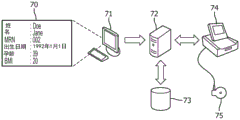
The inventors realized that fat in particular has a very negative impact on the measurability of all physiological parameters measured during labour and delivery. For adults, the range of overweight and obesity is determined by calculating a value called the "body mass index" (BMI) using weight and height. BMI is used because it is associated with body fat mass for most people. The weight and height of the patient and the number of weeks of pregnancy are important components of the electronic health record. By combining this information, classification of three types such as obesity, normal, and slim is easily achieved. With this classification of BMI coupling, all sensors or transducers connected to the fetal monitoring system can be forced to optimal performance. The gist of this method is to automate the setting adjustment for each physiological parameter by using body classification categories. The classification category may be obtained, for example, by: relevant data is automatically read from the patient health record at the time of admission, manually entered at the monitor through a touch screen or keyboard at the time of admission, and data entry is performed by reading a barcode tag or a wireless ID tag at the time of admission.
Automatically adjusting the key measurement settings for each physiological parameter allows the following advantages: due to the optimized signal-to-noise ratio, measurement performance can be (even significantly) improved, monitor setup time is reduced, operating point is optimized, settings that are not normally known by the operator can be automatically changed, exposure to energy fields (e.g., ultrasound) is improved, and operating time of battery-powered equipment is increased.
In a preferred embodiment, wherein the processing unit is arranged to be coupled with and to receive said patient-related data from a database, the processing unit is provided with a reading part arranged for reading out a data carrier having patient-related data stored therein and/or an interface part arranged to allow a user of the processing device to input patient-related data.
The seventh to ninth aspects of the invention discussed above and discussed with respect to the exemplary embodiments with reference to the drawings are preferably provided in the context of the first to third aspects and/or the fourth to sixth aspects of the invention, respectively. Nevertheless, it is also envisaged to provide the seventh to ninth aspects discussed herein and embodiments thereof separately (i.e. independently of the first to third aspects of the invention or in combination with the fourth to sixth aspects only).
In a further aspect of the present invention a computer program for determining a fetal heart rate from ultrasound doppler echo signals is presented, the software product comprising program code means for causing a processing device according to the first, fourth or seventh aspect to carry out the steps of the method according to the third, sixth or ninth aspect, respectively, when said software product is run on said processing device.
It shall be understood that the processing device according to claim 1, the system according to claim 3, the method for determining a fetal heart rate according to claim 5 and the computer program according to claim 6 have similar and/or identical preferred embodiments, in particular as defined in the dependent claims.
It shall be understood that preferred embodiments of the invention can also be any combination of the dependent claims or the above embodiments with the respective independent claims.
These and other aspects of the invention are apparent from and will be elucidated with reference to the embodiments described hereinafter.
Drawings
In the following drawings:
figure 1 shows a number of diagrams illustrating the concept of autocorrelation,
figure 2 shows a graphical representation of the autocorrelation of non-equidistant simplified cardiac activity,
figure 3 shows a graphical representation of an autocorrelation of equidistant simplified cardiac activity,
figure 4 shows a diagram of phase-shifted demodulation of (artificial) ultrasound doppler echo signals,
figure 5 shows an example of the original symbol chain corresponding to the phase shift demodulation shown in figure 4,
figure 6 shows an example of a processed symbol chain derived from the original symbol chain illustrated in figure 5,
figure 7 shows a typical example of an expected velocity profile of heart activity,
figure 8 shows the basic signal processing path for avoiding doubling of elements according to the present disclosure,
figure 9 allows comparing the conventional rectified and filtered signal processed by the elements according to the present disclosure with the gating signal,
figure 10 shows signal processing of a second element according to the present disclosure,
figure 11 shows the signal processing of the third element according to the present disclosure,
figure 12 shows the signal processing of a fourth element according to the present disclosure,
figure 13 shows an example of a cardiac labor diagram with a fetal heart rate trace and a maternal heart rate trace,
figure 14 shows a combined ultrasound doppler signal resulting from the superposition of a fetal heart signal and a maternal heart signal,
figure 15 illustrates signal processing according to an embodiment of the present invention,
figure 16 schematically illustrates further elements of the present disclosure,
figure 17 schematically illustrates another element of the present disclosure,
figure 18 schematically illustrates yet another element of the present disclosure,
figure 19 schematically shows a processing apparatus according to another embodiment of the invention,
figure 20 shows a schematic flow chart illustrating a method for determining a fetal heart rate according to an embodiment of the present invention,
figure 21 schematically shows a processing device according to a further embodiment of the invention in a channel mode,
fig. 22 schematically shows the processing device of fig. 21 in a phase shift mode.
Detailed Description
Fig. 1 shows a number of diagrams illustrating the concept of autocorrelation.
First, as a non-limiting introduction to the discussion of autocorrelation, a discussion of an exemplary implementation of the determination of fetal heart beats is given.
The ultrasonic doppler transducer was placed on the abdomen of the mother and held in place by an elastic band fitted around the waist. The doppler effect is based on the following principle: the sound waves reflected from the moving object undergo a frequency shift depending on the direction and speed of movement. Based on this principle, the mechanical contraction of the fetal myocardium induces a periodic signal pattern in the ultrasound reflection. The fetal monitor uses the periodicity of the pattern to determine the current heart rate of the fetus. Most fetal heart rate transducers use the pulse wave principle. An array of piezoelectric elements is used as an electromechanical transducer. This array operates bi-directionally as a transmitter and receiver. The sequencer controls the time switching between the transmit and receive phases. During the transmit phase, the piezoelectric array is repeatedly excited to generate ultrasonic packets that travel toward the fetal heart. These traveling wave packets are reflected and frequency shifted, for example, at the fetal heart, due to the doppler effect on moving layers in the body of the pregnant woman. The received reflected signal is demodulated by a synchronous demodulator using exactly the same frequency as used for the transmitted burst. After demodulation, integration, amplification and bandpass filtering, the doppler frequency is available for signal processing. The received weak doppler shifted echo signal embeds noise and signals of artifacts caused by human body or transducer movement.
To extract the periodic signal from the noise, an autocorrelation method is applied. Autocorrelation is a mathematical method for finding repeating patterns (e.g., the presence of periodic signals that have been buried under noise). The result of the correlation is a function that allows an accurate calculation of the heart rate.
Fig. 1 shows a simplified illustration of how the output of the autocorrelation is calculated. The signal is multiplied point by point with a time-shifted copy of itself. The results of each multiplication are added.
In fig. 1, the shaded rectangles show the positions where the product is not zero and thus contributes to the autocorrelation function.
Fig. 1a) shows the case of a time shift of 0(τ ═ 0), where the similarity corresponds to the entire pulse. Fig. 1b) shows the case of 128 delayed time shifts (τ — 128), wherein the similarity corresponds to half of the pulse (more than 3 times). Fig. 1c) shows the case of 256 delayed time shifts (τ ═ 256), in which there is no similarity at all. Fig. 1d) shows the case of a 384 delayed time shift (τ 384), wherein the similarity corresponds to half (more than 2 times) of the pulse. The result of the autocorrelation is given in fig. 1e), where the arrows point from fig. 1a) to fig. 1d) to the corresponding parts of the result.
The abscissa of fig. 1 shows the lapse of time (hysteresis), and the ordinate is provided with arbitrary units.
At time shift 0, the autocorrelation result (fig. 1e) has a maximum value representing the signal energy. At time shift 512 (not shown in fig. 1a) to 1 d)), the autocorrelation function has a first maximum (τ 512). This peak represents the first repetition of the signal periodicity. When the value τ of the peak is obtained, the signal frequency can be calculated.
Fig. 2 shows a graphical representation of the autocorrelation of non-equidistant simplified cardiac activity.
The incoming doppler echo signal is typically extracted from the carrier wave by means of synchronous demodulation, wherein this method is rather simple and does not require a high technical effort. The inventors have realized that this approach has an important drawback in that the direction information (respectively the sign) of the doppler shift is irretrievably lost, since the demodulation is done at the same frequency as the carrier.
If such a technique is applied to heartbeat detection, it is no longer possible to distinguish between systolic and diastolic heart activity. Both activities appear as two peaks in the time diagram (see fig. 2 a)). Under normal conditions, the time between the systolic and diastolic actions (t1) is significantly lower than the time between relaxation and contraction of the subsequent cardiac action (t 2).
The input signal shows (fig. 2a) two active peaks, one representing systole (S ═ systole) and the other cardiac relaxation (D ═ diastole). According to the doppler principle, contraction causes a negative frequency shift, while relaxation causes a positive frequency shift. The actual unwanted first or second peak of the heart activity cannot be eliminated due to the loss of symbols during demodulation, thereby creating unwanted peaks of the autocorrelation function at higher frequencies.
The assumed condition is that the periodic heartbeat includes contraction (S) and relaxation (D), where the frequency is 2Hz — 120 bpm. The time between systole and diastole is 100ms (abscissa of fig. 2a), time is shown in seconds, and the ordinate is normalized). The ACF results (fig. 2b) (where the abscissa shows hysteresis and the ordinate is arbitrarily provided)) show two peaks with an amplitude of 100, one at τ value 200 and the other at τ 1024.
The two peaks have the same amplitude. When the frequency of interest is calculated to be 200, the frequency is 614bpm, while the second peak at 1024 τ still shows 120 bpm. To avoid false HR evaluations, the evaluation range of CTGs using autocorrelation is limited to a range of about 240(τ 512). Line L illustrates the peak check limit.
The peak evaluation and peak selection is done in the second part of the signal processing, the so-called post-processing. This function typically simply cuts off the peak at frequencies above 240 bpm. The setting of the peak check limit has a significant impact on the doubling behavior of the monitor. If set at 240bpm, then all heart rates above 120bpm are "double-preserved" because if the heart rate is doubled, the results are always above the evaluation limit. Most fetal heart rate traces cover a region from 140bpm to 180bpm, while maternal heart rates are mostly below 100 bpm.
In addition to the evaluation limit setting, the perhaps most important doubling factor is the ratio between t1 and t 2. If the t1/t2 ratio reaches 1, the probability of doubling the heart rate value is high. Unfortunately, ultrasonic doppler sensors are sensitive to all moving structures within the sensitive volume. The signal containing fetal heart information in many cases represents only a small portion of the entire received signal. Signal contributions from other moving structures (such as the maternal artery located behind the fetal heart) often overlap, especially if the drug increases the maternal pulse rate and blood pressure.
The time ratio t1/t2 is important for doubling. Due to the physiology of the human heart, the time ratio t1/t2 is not constant over the range of heart rates. It can be said that the probability of a heart rate between 80bpm and 120bpm reaching a ratio of 1 is high. A frequency doubling of 80bpm is recorded as 160 bmp. This may be exactly the range of heart rates expected for the fetus. Sometimes a condition of maternal (doubled) heart rate recorded within a few hours due to absence of fetal heart rate is reported.
Fig. 3 shows a diagram of an autocorrelation of equidistant simplified cardiac activity in an arrangement similar to that of fig. 2.
Fig. 3 illustrates in particular the effect of the doubling. In the input signal, t1 is elongated to about the same value as t 2. As a result, the maximum peak (P1) that previously exceeded the evaluation limit is now well on the limit line, thereby yielding an HR value of 4Hz to 240bpm (τ to 512Hz yields 4 Hz). The peak merit function must select P1 instead of P2.
Double counting is mainly observed in cases where the fetus is bradycardia or the maternal pulse is measured inadvertently. If the maternal heart rate is recorded with a separate heart rate measurement channel (e.g. maternal pulse is measured with an infrared sensor), the sequence of double counts caused by fetal bradycardia can usually be easily identified without risk of erroneous trace interpretation. However, inadvertently measuring a doubled maternal heart rate is a serious risk, since the maternal heart rate pattern can mimic the fetal heart rate pattern. Since common cross-channel validation methods cannot detect multiples of heart rate with the same source, they may not be found doubled for a long time.
Figure 4 shows a diagram of phase-shifted demodulation of (artificial) ultrasound doppler echo signals.
In particular, fig. 4 shows a reference signal (original artificial doppler signal) and an artificial doppler signal demodulated with a 90-degree phase shift. The first signal 1 is demodulated without phase shift, while the second waveform 2 is demodulated with a 90 degree phase shift. In the first half of the waveform diagram, the first signal 1 clearly precedes the second signal 2. At the maximum of the signal amplitude, the phase changes abruptly, from where the second signal 2 oscillates three times before the first signal 1. For the rest of the oscillation, the phase switches back to the initial condition.
From a viewpoint, the first part and the tail can be regarded as moving forward, while the central part with three oscillations can be moving backward. Measuring the time difference between the zero crossing of the first signal 1 and the zero crossing of the second signal 2 results in a negative or positive value, respectively. For reconstructing the direction of movement, substantially only symbols without values are of interest. Evaluating the zero crossings of the two signals 1, 2 allows to obtain a succession of symbols 3.
Fig. 5 shows an example of an original symbol chain corresponding to phase shift demodulation as shown in fig. 4.
Fig. 5 shows a typical symbol sequence in its original form. The noise in the measurement zone and the superimposed inversely moving part are the cause of strong symbol fluctuations, as shown in fig. 5 (a value of 0 indicates no difference between zero crossings or the result is indeterminate).
Fig. 6 shows an example of a processed symbol chain derived from the original symbol chain illustrated in fig. 5.
Applying known and appropriate signal processing methods to the original symbol chain shown in fig. 5 results in a smoothed symbol signal as shown in fig. 6, which can be used for further signal processing.
The symbols shown in fig. 6 correlate well with the expected velocity profile of the heart activity as shown in fig. 7.
Considering the different scaling of the abscissa, it can be seen that the smoothed sign signal shown in fig. 6 represents well the velocity direction of the heart activity, so it can be used to clip out unwanted signal portions. In other words, the inventors have recognized that binary symbol values may be used to gate the reference signal.
Fig. 8 shows a basic signal processing path avoiding doubling of elements according to the present disclosure.
This element provides for the respective symbolization by clipping undesired signal segments identified by the direction of movement of the signal. The reference signal (reference demodulation signal) 10 demodulated with a phase shift of 0 degrees and the second signal (phase-shift demodulation signal) 15 demodulated with a phase shift of 90 degrees from the same source are fed to respective high- pass filters 20, 21 to remove the DC offset. The reference signal 10 is passed through a conventional signal processing chain (i.e. amplitude processing 22, low pass filter 23 and delay 24) required to prepare the autocorrelation.
The two signals are tapped (tap) after the high pass filters 20, 21. The high pass signal is delayed (blocks 25, 26) to compensate for the smoothing filter delay of the approximate envelope. These signals are then passed through envelope controlled noise gates 27, 28, respectively, to avoid symbol detection on the ordinary noise signal, for example during mechanical pause periods. The next stage for both signals are respective zero crossing detectors 29, 30. By comparing the time relationship of the two outputs of the zero-crossing point detectors 29, 30, the sign detector 31 determines whether the signal is positive or negative. The positive, negative or indeterminate polarity (no zero crossings or noise) symbol stream is filtered, delayed and smoothed (blocks 32, 33) to obtain the signal as shown in figure 6. The resulting binary symbol signal 15' (see fig. 6) is used to clip out unwanted signal segments by using the switch 34. After the switch 34, the processed reference signal 10' has only a signal portion of the heart activity. By tailoring the systolic or diastolic heart activity, the underlying autocorrelation 35 no longer has a chance of doubling.
Fig. 9 allows comparing conventional rectified and filtered signals processed by elements according to the present disclosure with gating signals.
The effect of cropping segments of cardiac activity as discussed above is shown in fig. 9. The rectified and filtered signal (fig. 9a) normally used for correlation must pass through a gate controlled by the sign signal 15'. In this example, signal activity with a negative sign is prevented. The result is an adjusted signal 10', which is "not doubled".
Here, a signal processing sequence as shown in fig. 8 is used to avoid doubling the count. An advantage of this embodiment is that no additional autocorrelation is required. The enhancements provided compared to the conventional implementation include a second hardware channel for quadrature demodulation and zero crossing detection and symbol evaluation including filtering.
However, when the signal loses information, it may suffer from a loss of beat to beat accuracy even if the result of the autocorrelation is not actually doubled.
Fig. 10 illustrates signal processing of a second element according to the present disclosure.
To avoid loss of beat accuracy, a preliminary fetal heart rate (undipped heart rate) is additionally calculated using a somewhat conventional method 40 (i.e. including reference signal pre-processing 41 followed by autocorrelation 42). In parallel, the second heart rate is calculated according to the processing scheme of fig. 8. The second heart rate (clipped heart rate) is used to prevent the first autocorrelation heart rate selector 43 from erroneously acquiring a doubled heart rate.
In a variation of the above example, the second heart rate may be obtained directly from the symbol stream 15', as shown in fig. 11.
According to the elements of the disclosure in fig. 11, autocorrelation (and post-processing) 35' is provided on the symbol stream or signal 15' (rather than on the clipped ultrasound echo signal 10 ').
Fig. 12 shows the signal processing of a fourth element according to the present disclosure, wherein a heart rate obtained from the symbol stream itself is used. Here additionally a sign selection 36 for positive or negative sign is provided.
It should be noted that these two signs can also be used in correlation, either in combination or as a process of independent evaluation of positive and negative signs.
Corresponding elements in fig. 8, 10, 11 and 12 are denoted by corresponding or similar reference numerals, and thus additional explanation thereof is omitted.
Fig. 13 shows an example of a cardiac labor diagram with a fetal heart rate trace and a maternal heart rate trace.
The shear trace of the cardiac labor diagram illustrated in fig. 13 shows two traces F, M. The upper trace F represents a fetal heart rate trace derived from an ultrasonic doppler transducer. The lower trace M shows the maternal heart rate derived with an infrared sensor built into the transducer that measures uterine activity.
For this example, two spatially separated independent heart rate channels are considered. In the first half of fig. 13, the two traces are clearly separated. The correctness of the fetal heart rate recording is doubtful. In the second half of the figure, the two traces F, M are nearly congruent. Without the information of the maternal trace M, the fetal trace F would be interpreted as a fetal trace, but in this example, the ultrasound algorithm is in fact inadvertently switching to the maternal heart rate M, which has a better score due to the scoring and selection algorithm than a lower scoring fetal heart rate value. A modified selection algorithm according to the invention will select a lower scoring fetal heart rate for printing, or suppress (blank) printing if no surrogate heart rate is available.
The current state of printing both heart rate values (both black, since commonly used thermal printers do not allow color printing) is unsatisfactory, since the user has to decide which printed heart rate is a valid fetal heart rate, if the heart rates are almost congruent, the superimposition may obscure the fetal heart rate and suppress the alternative heart rates that may be available.
The cross-comparison provided herein improves the reliability of fetal heart rate traces recorded with ultrasonic doppler transducers. It reduces the likelihood of inadvertent switching to maternal heart rate. Heart rate variations caused by the switching effect may mislead the trace interpretation. Misinterpreted erroneous heart rate traces can lead to unnecessary motion, unnecessary surgery, and delayed delivery of fetal injuries and even fetal death.
Conventional fetal monitors use ultrasonic doppler technology for non-invasive acquisition and recording of fetal heart rate during pregnancy and childbirth. The mechanical contraction of the fetal myocardium causes a periodic signal pattern in the ultrasound reflection. The fetal monitor uses the pattern of cycles to determine the current heart rate of the fetus.
The main problem with this technique is that it does not take into account the physiological signal source that generates the ultrasound reflections. All periodic movements of tissue or blood flow within the range of the ultrasound beam used are capable of generating a heart rate pattern. Especially maternal abdominal artery pulsation is a known cause of this problem.
The inventors have realized that the different signal sources are spatially separated.
The ultrasound transducer is placed on the abdomen of the pregnant woman. During the launch phase, the piezoelectric array is repeatedly excited so as to generate ultrasonic packets that travel towards the fetal heart. These traveling wave packets are reflected and frequency shifted, for example, from the fetal heart and maternal arteries due to the doppler effect on various moving layers in the body of pregnant women and children. Because the fetal heart and the maternal artery are at different distances from the transducer surface, the wave packets require different travel times to reach the reflection point and return to the transducer.
An array of piezoelectric elements is used in both directions. When transmission is complete, the transducer switches from a transmit mode to a receive mode. As illustrated in fig. 14, there is only one receive window covering the entire measurement depth, which results in signal overlap of the signals from the fetal and maternal sources.
Such signals make it difficult for the signal processing unit to extract the correct heart rate.
For this reason, multiple receive channels are employed that are active at different times of the measurement period. As described above, echo signals from different depths require different travel times until they are received at the surface of the transducer. Time-interleaved signal acquisition with multiple receive channels allows for proper signal separation.
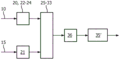
Fig. 15 illustrates signal processing according to an embodiment of the present invention.
Fig. 15 shows the time-hashed signals of fig. 14 as input signals 51, 52 for respective autocorrelations 53, 54. For time (depth) separation of the overlapping ultrasound doppler signals, discrete demodulation and filtering channels are used, but are not shown here, since the skilled person is familiar with ultrasound signal demodulation and filtering.
The implementation can vary depending on the availability of the demodulation and filtering paths provided by the hardware.
At least two (independent) demodulation and signal processing channels are required to implement the basic depth division, as provided by the present embodiment. A well implemented trade-off between signal processing power and hardware complexity is a four channel system.
To keep the complexity low, the embodiments described herein use only two channels.
The digitized ultrasound doppler signals are then processed in a conventional manner by autocorrelation 53, 54 and post-processing 55, 56 of the autocorrelation results. The output of each signal processing chain is a heart rate value.
Assume that the first depth channel is 145bpm ( upper branches 51, 53, 55). This example also assumes that the depth range of the channel is closer to the surface than another channel producing a heart rate of 68 bpm.
In a simplified setup, these two heart rate values would be available for comparison. The values 58, 59 of the ultrasound depth channel are compared with the values of the independent source 62 in a next step 57.
The independent source uses for example an ECG subjected to processing 61 and determination 62 of the heart rate.
If one of the values is close to the value of the independent source, the following algorithm 63 can exclude this value and output 64 the fetal heart rate.
To improve the decision algorithm, depth information of the ultrasound can be used to improve the accuracy of the exclusion. For example, if the transducer is conventionally positioned to steer the ultrasound beam relative to the backbone, the heart rate calculated from the channel with the higher travel time has a signal source that is further away from the transducer surface. If in this example the heart rate that matches the value of the independent source and the depth range falls behind another heart rate layer, then the source of this heart rate is clearly maternal. This is always true for typical transducer positioning, since the maternal blood vessel is spatially located behind the fetal heart, but this heuristic approach does not work if the beam is steered from behind or even laterally.
Fig. 16 schematically illustrates additional elements of the present disclosure.
Current fetal monitors encompass a wide variety of measurable physiological parameters. First is the set of parameters for fetal heart beat and uterine activity. This primary parameter pair may be supplemented by various maternal parameters such as blood pressure, oxygen saturation, pulse and temperature.
A conventional non-invasive method for externally monitoring a fetus in a pregnant woman includes an ultrasonic doppler transducer for measuring the Fetal Heart Rate (FHR) and a pressure transducer called a tocodynamometer (Toco) for measuring uterine activity/contractions. The two transducers are held in place on the abdomen of the mother by an elastic band fitted around the waist.
Ultrasonic doppler transducers are based on the following principles: the high frequency sound waves reflect the mechanical action of the fetal heart. The mechanical contraction of the fetal myocardium causes a periodic signal pattern in the ultrasound reflection. The fetal monitor uses the pattern of cycles to determine the current heart rate of the fetus. Ultrasonic transducers use an array of piezoelectric elements as an electromechanical transducer. The array of piezoelectric elements operates bi-directionally as a transmitter and a receiver. The sequencer controls the time switching between transmit and receive phases. During the launch phase, the piezoelectric array is repeatedly excited so as to generate ultrasonic packets that travel towards the fetal heart. These traveling wave packets are reflected and frequency shifted, for example, at the fetal heart, due to the doppler effect on moving layers in the body of the pregnant woman. In traveling to the reflection point and returning to the transducer, the wave packet must pass through different organic structures, such as muscle tissue, fat layers or amniotic fluid. These structures have different sizes and different damping ratios. For example, fat has an absorption factor 3 to 4 times higher than water. It is clear that a large amount of fat can seriously affect the performance of the ultrasound transducer. To facilitate handling of the monitor and its attached transducer, the manufacturer avoids control elements for adjustment. The transducer is designed to have optimal performance for a patient of normal weight. With increasing numbers of extremely overweight and underweight patients, transducers following the "one fits all" principle simply exhibit poor or poor performance if applied to women who are not fit in the "normal" range. Another aspect of transducers designed for normal conditions that exhibit disappointing performance is the use of such transducers outside recommended limits. The recommended starting point for examining the fetal heart rate using pulsed ultrasound doppler is approximately the 25 th week of pregnancy. Some countries start CTG examinations more and more at the 20 th week of pregnancy and earlier. In this condition the depth position and size of the fetal heart is significantly different compared to week 30 of pregnancy. In order to adequately record the fetal heart early in pregnancy, narrow ultrasound beams with a depth range of sensitivity between 3 and 8 centimeters are generally recommended. If early pregnancy is combined with overweight, the depth range and ultrasound beam energy must be increased. In order to allow individual adaptation to cover extreme ranges of use, the ultrasound beam energy, beam shape, sensitivity range, etc. can be adjusted for the user. An experienced and trained operator can use the available adjustments in a meaningful way, but a standard operator may be confused. As mentioned previously, adding adjustable settings may be desirable for research people, but not suitable for standard users. A CTG is a device that must work reliably under all conditions, with a minimum level of user interaction and a minimum level of knowledge about the mode of operation. For this reason, simply adding additional control and adjustment elements is not a way to expand the operating range. Based on the surrounding information available in many cases on electronically stored records or barcodes or other media, the device can automatically change its configuration for that session to achieve optimal performance. Today, CTG monitors provide an activation menu in which patient data is required to be entered. The so-called admission/discharge (ADT admission, discharge, transfer) menu requires patient demographics. The content of the patient demographic data is a defined topic. The patient is generally asked for his name, week of pregnancy, etc. when admitted to the hospital. This information is displayed in part on the monitor screen and printed on paper tape. With the monitor connected to an archiving and surveillance system, the data required for admission is retrieved from the database and automatically loaded to the monitor if the patient is assigned to a particular bed. The scope of the ADT data set is a freely defined subject. In the context of the present invention, it should preferably contain at least information about the number of weeks of pregnancy, BMI or alternatively height and weight, country (e.g. usa or japan), twin, triplet (which information may also be derived from a plurality of connected transducers).
By reading this information, the monitor can classify the data (e.g., BMI) into two or more groups. Based on these categories, the monitor is able to independently configure various parameters of the data acquisition, signal amplification, filtering and signal processing chains to achieve optimal CTG recording results. For example, for patients with a high BMI (< obesity category) and an early number of weeks of pregnancy (< 25 th week premature category), the monitor would adjust the ultrasound beam shape to narrow the beam intensity high, reducing the expected depth range to medium/far. All this is done automatically in the context of no user interaction. Furthermore, possible variations of the ultrasound transducer are exemplary. The same adaptive changes of the data acquisition and signal processing paths are possible for all other parameters (e.g., Toco, maternal pulse). With the availability of ADT data, the patient realizes the advantage that the transducer control has optimal signal acquisition and processing of all parameters, requires no or little operator interaction, reduces exposure to the energy field (ultrasound), does not increase complexity of use and is at least partially retrospectively adapted by software upgrades to already installed transducer bases.
In this context, a fetal monitor 74 with a connected transducer 75 is connected to a centralized supervision and archiving system 72 with a database 73. The patient is admitted to a monitor or bed and a local terminal or PC 71 is also connected to the supervision system 72. An application running on the PC or terminal provides a mask 70 for patient data as part of the admission process. When a patient is admitted, the mask must be completely filled. The relevant data contains information for the transducer 75 and signal processing controls (internal to 74 or 75) and is stored in the database 20. The data set is populated to the monitor 74 to which the patient is assigned. For example, if signal processing or portions of signal processing are done in the monitor 74, the monitor 74 may use the data. If the signal processing is done in the active transducers 75 (which may be wired or wireless), the monitor 74 ensures that relevant information is distributed to each connected transducer 75. The admission/distribution process ensures that the data acquisition and signal processing processes are automatically adapted to the settings defined by the ADT data.
Fig. 17 schematically illustrates another element of the present disclosure.
In the context of this element, a near field communication path is provided for data exchange. The near field communication device can be part of the monitor 74 'or a separate device connected to the monitor 74' by a wire. The relevant ADT data is stored in an independently programmed ID tag or ID card 76, along with possibly other information. The admission process is initiated by bringing the tag into proximity of the NFC device 74'. In the next step, the ADT data stored on the card 76 is read and populated into the relevant parts of the monitoring system 74', 75.
In addition (or as an alternative), the barcode label containing ADT data can be read by an attached barcode reader 77.
Fig. 18 schematically illustrates yet another element of the present disclosure.
This element is already available in many monitors 74 "for direct ADT data entry by filling out the form 78 during patient admission. Data is manually entered through the touch screen and then the keyboard or mouse is populated into the relevant portion of the monitor 74 "or transducer 75 as discussed above.
Fig. 19 schematically shows a processing apparatus 100 according to another embodiment of the invention.
The processing device 100 comprises a reference demodulation unit 101, a phase shift demodulation unit 102, a comparison unit 103 and a processing unit 104.
Two reference demodulation units 101, phase shift demodulation units 102 and comparison units 103 are provided, respectively, to provide two channels.
The reference demodulation units 101 are supplied with ultrasonic doppler echo signals from the respective channels, wherein each reference demodulation unit demodulates a received echo signal using a carrier frequency of an ultrasonic signal used to generate the echo signal as a demodulation frequency, and outputs a corresponding reference demodulation signal to the comparison unit 103 and the processing unit 104. Similarly, the phase shift demodulation units 102 respectively receive the corresponding ultrasonic doppler echo signals and demodulate the echo signals using demodulation frequencies shifted by 90 degrees compared to the case of the reference demodulation unit, and output the phase shift demodulated signals to the corresponding comparison units 103.
As indicated above, each comparison unit 103 is provided with the reference demodulation signal and the phase-shifted demodulation signal for one of the channels, and obtains information about the time relationship between corresponding individual signal points of the reference demodulation signal and the phase-shifted demodulation signal, which are the zero-crossing points of the reference demodulation signal and the phase-shifted demodulation signal in the present embodiment (see fig. 4). As mentioned above, although the signal points considered in this context are zero crossings of the signal, other points (e.g. points of maximum positive amplitude and/or points of maximum negative amplitude) may be considered as an alternative or an addition.
Information about the time relation between the respective zero crossings for the first and second channels and the reference demodulated signals for the first and second channels is provided to the processing unit 104.
The processing unit 104 comprises two processing parts 105, 105', an input part 107, a selection part 108 and a setting unit 109. Each processing section 105, 105' is used for three different methods of determining the fetal heart rate, wherein one processing section is provided for a first channel and the other processing section is provided for a second channel.
According to a first method, information about the time relation between the individual signal points is directly used to obtain the fetal heart rate, since this information already includes a periodicity indicative of the fetal heart rate.
According to a second method, information about the time relation is used to clip portions of the reference demodulated signal, so that the fetal heart rate is obtained from such clipped demodulated signal by means of autocorrelation.
In a third method, the autocorrelation is provided on the reference demodulated signal substantially according to a conventional method, wherein the heart rate obtained by the second method is compared with the result, and the selection part 106 of the processing part 105, 105' is arranged to determine the fetal heart rate by selecting a conventionally determined heart rate or a clipped heart rate.
The processing unit 104 is arranged such that for each channel a respective method may be selected, thereby providing an output for the respective channel. The input section receives external information about the fetal heart rate to be determined or a heart rate other than the fetal heart rate to be determined (i.e., maternal heart rate), and is connected to the selection section 108. The selection section is arranged to select, based on the external information, a heart rate determined from the first channel or a heart rate determined from the second channel as the fetal heart rate to be determined.
Furthermore, as indicated above, the processing unit 104 comprises a setting unit 109, wherein the setting unit is arranged to receive data relating to the patient and to adjust settings of the processing unit based on the received data relating to the patient.
In the present embodiment, the processing device 100 is provided for processing two separate channels, but it is also possible to process more than two channels. Furthermore, it is not necessary to solve all three methods discussed above in the processing unit either, since only one or two or different ones of the methods discussed can be employed in the processing unit.
The output of the demodulation unit 101 shown in fig. 19 may be subjected to a process of an integrator and high-pass band-pass filter combination (not shown), and these elements may also be included in the demodulation unit.
Fig. 20 shows a schematic flow chart illustrating a method for determining a fetal heart rate according to an embodiment of the present invention.
Similar to the embodiment discussed above with respect to fig. 19, the discussion of fig. 20 provides for obtaining information in parallel by means of two channels, but the invention is not limited thereto and more than two channels may be used.
After the receiving step 200 of receiving patient related data, an adjusting step 201 is provided, in which adjusting step 201 setting data relating to the operation of the embodiment is adjusted based on the received patient related data. A channel heart rate determination step 202 follows. The channel heart rate determining step 202 comprises determining a first heart rate from a first channel of the ultrasound doppler echo signal and determining a second heart rate from a second channel of the ultrasound doppler echo signal. The determination of the first heart rate comprises a reference demodulation step 203, a phase shift demodulation step 204, a comparison step 205 and a processing step 206. Similarly, the determination of the second heart rate also comprises a reference demodulation step 207, a phase shift demodulation step 208, a comparison step 209 and a processing step 210.
In a reference demodulation step 203, the ultrasonic doppler echo signal is demodulated using the carrier frequency of the ultrasonic signal used to generate the echo signal as a demodulation frequency, while the echo signal is correspondingly demodulated with a shift of 90 degrees in a phase shift demodulation step 204.
In a comparison step 205, the obtained reference demodulated signal is compared with the phase shifted demodulated signal and information about the time relation between corresponding individual signal points, which are also zero crossings of the reference demodulated signal and the phase shifted demodulated signal, is obtained. This information is used in a processing step 206 in the process of determining the fetal heart rate.
In an input step 211, external information about the fetal heart rate is received, in this case information about the maternal heart rate obtained separately and independently. In a selection step 212, one of the heart rates determined by using either channel is selected as the fetal heart rate to be determined and the selected heart rate is output correspondingly.
Fig. 21 schematically shows a processing device 100' according to a further embodiment of the invention in a channel mode.
The processing apparatus 100' includes a high frequency amplifier 110 and a divider 111. The divider provides the signal as a plurality of sub-signals that are provided to a processing chain of the processing device 100'. Only two chains are shown in the figure, but a higher number of chains may be provided. Each chain comprises a synchronous demodulation unit 101', 102' with a switchable demodulation clock 112, 112 '.
The processing device 100' further comprises a timing section 114 providing channel selection information in the form of a gating signal (timing signal for depth selection/volume selection). A respective gating signal is provided for gating the demodulated signal by means of a respective and gate 115, 115'.
Each chain further comprises an integrating and band-pass filter unit 116, 116 'receiving the signal from the demodulation and providing a rectified and smoothed signal to a processor 117, 117', which is provided for autocorrelation and heart rate evaluation.
The output of the chain is provided to a control unit 118, which control unit 118 comprises an input part 107', a selection part 108' and a setting part 109 '.
The input portion 107' is coupled to an external source 120 of information about, for example, the maternal heart rate or depth range of other ultrasound transducers, or a combination of both. It should be noted that the external information may also relate to other heart rates to be excluded (e.g. an independently determined fetal heart rate of another twin). It is even conceivable that the external information obtained independently is independent of the heart rate to be ignored, but is related to the fetal heart rate of interest.
The setup portion 109' is coupled to an external database 119 that provides data related to the patient.
Similar to the embodiments discussed above, the selection section 108' is arranged to select the channel (i.e. processing chain) that provides the fetal heart rate.
In the mode shown in fig. 21, no phase shift is provided for demodulation, and basically each processing chain addresses a different channel.
Fig. 22 schematically illustrates the processing device 101' of fig. 21 in a phase shift mode.
In contrast to the situation of fig. 21, for the demodulation unit 102', the demodulation frequency is provided by the demodulation clock 112', which demodulation clock 112' is phase shifted by 90 degrees compared to the situation of the demodulation clock 112.
Furthermore, the resulting phase shifted demodulated signal is provided to a processor 117, which processor 117 compares the reference demodulated signal provided by the demodulation unit 101' with the phase shifted demodulated signal to obtain information about the time relation between the corresponding individual signal points of the reference demodulated signal and the phase shifted demodulated signal, as already discussed above for other embodiments and elements of the present disclosure.
It is advantageous to divide the sensitivity volume covered by the ultrasound doppler sensor into several sub-volumes. It is also desirable, as will become apparent from the present application, to avoid possible doubling of counts by using quadrature demodulation in parallel with standard synchronous demodulation. Combining both methods in one signal processing unit with multiple channels allows to reduce the number of required channels, since the channel functions can be adapted independently to different situations. At least a first part of the channel comprising the demodulator, integrator, filter and AD converter has up to now been established using hardware. For this reason, keeping the number of channels low is not only a cost advantage. Even if everything in the future is done digitally, keeping the complexity of the integrated circuit low has a positive impact on power consumption, cost and chip size.
As indicated above, in the illustrations of fig. 21 and 22, only two channels are shown in order to keep the example simple, but it should be noted that the number of channels is not limited. A suitable number of channels is for example four. As shown in fig. 21 and 22, the received ultrasonic signal is amplified by the high-frequency amplifier 110. The divider 111 divides the signal into two to N sub-signals. In this case, each sub-signal is fed to the same signal processing chain. The chain starts with synchronous demodulation units 101', 102' with switchable demodulation clocks 112, 112 '. The controllable switch selects either a 0-degree phase-shifted or a 90-degree phase-shifted reference clock for demodulation. In addition, the demodulated signal is gated by using an and gate 115, 115' with a gating signal. The gating signal defines the depth sensitivity, respectively the sensitivity volume. The start and end points with respect to the end of the transmission period are freely definable for each channel. This allows the volume segments to be separated or superimposed. After demodulation, the signal is integrated, bandpass filtered (elements 116, 116'), rectified and smoothed in order to prepare the signal for autocorrelation in the processors 117, 117'.
The control unit 118 at the end of the multiple signal processing chains is responsible for selecting the channel that is most likely to measure the fetal heart rate. A reliable method for selecting a heart rate from a plurality of heart rates is to compare the heart rate with heart rates from known sources, e.g. derived from an ECG as an example of the external source 120. Channels with heart rate values similar to heart rates from known sources are excluded. The excluded channels can be said to be free and do not contribute to the measurement accuracy.
This approach allows for a flexible channel that can be reconfigured to improve accuracy and reliability if the control unit 118 recognizes that the channel is in an idle state without meaningful contribution. In this case, the control unit 118 may, for example, set the depth range to the range of the channel that actually measures the fetal heart rate. The 90 degree phase shifted signal may then be selected for demodulation, allowing quadrature demodulation to obtain the direction of the velocity. The sign of the velocity can then be used to clip unwanted signal parts to avoid doubling the count. If more than two channels are available, another channel that has not measured heart rate or has measured an incorrect heart rate may be reprogrammed, for example, so that the channel now additionally covers the volume segment previously covered by the channel being quadrature demodulated. The process of depth volume and function adjustment may be static or may be a dynamically controlled process performed by the control unit.
The volume to which the demodulation method and the sensitivity are assigned preferably takes into account patient-related data. For example, in the early pregnancy and low BMI cases, the algorithm will first choose a narrow sensitivity volume. Initially, all channels are used for depth segmentation to find the active region. If an active region has been identified, the control unit 118 may change the function of the channel by, for example, changing the demodulation method. The control unit may also influence the amplification and filtering and, as a factor, the set of rules for extracting the heart rate from the correlation result. Once the active region is identified, the control unit 118 may decide to leave one or more channels for later use as measurement channels. The parallel operating channels do not necessarily have different demodulation methods. Changing the parameters, in particular the rule set for determining the heart rate, can lead to a higher beat accuracy, in particular in the upper range of the beat frequency.
As healthy people are accustomed to a certain shape of the printed heart rate trace, where high minor variability may cause confusion and rejection. The parallel operating channels can provide data that is different from the usual way of presentation. These data can be used for background calculations, for example to increase the confidence level that the ultrasound transducer picks up the fetal signal rather than the maternal signal.
It should be noted that not all channels necessarily have to have a switchable demodulation source. One channel may be sufficient even in the case where the segment with QAM channels is a measurement and sound output channel, when the measurement channel is moved to a different channel, crackle noise may be heard.
The invention provides, among other things, for eliminating a double-counted heart rate by clipping unwanted signal contributions, wherein especially the heart rate selection may be further improved by comparison with a second heart rate calculated based on such signals with reduced information and/or by providing an autocorrelation of a (binary) symbol stream indicative of the phase information of the ultrasound doppler echo signal.
Another aspect of the invention provides for excluding heart rates from multiple heart rates originating from depth-divided ultrasound doppler signals by comparison with heart rates extracted from a second independent source and/or mutual heart rate exclusion when multiple heart rate signals are measured with multiple ultrasound doppler sensors.
Yet another aspect of the invention provides adaptive signal processing and data acquisition controlled by patient related data, allowing for optimized energy emission, e.g. in case of ultrasound transducers, optimized run time in case of battery powered devices, and transmission and distribution of patient related data to decentralized signal processing and data acquisition units.
The present disclosure provides, inter alia, an ultrasound doppler system having a plurality of depth segments, wherein the heart rate of each depth segment is calculated separately and independently, while a decision unit is provided for excluding a segment or heart rate value equal to the heart rate from a further separate or independent source. However, a separate or independent heart rate source may be integrated into the same housing as the (one of the) ultrasound transducers and may be, for example, an IR sensor, an accelerometer, etc. Furthermore, the separate or independent source may be located in a spatially separate second unit (e.g. an ECG unit or a blood pressure measurement unit). Preferably, the plurality of ultrasound doppler transducers exchange heart rate values and/or depth segments with each other via a cable or wireless connection.
An ultrasound doppler system having a plurality of depth segments is also provided, wherein at least one depth segment is alternately supplied with a 90 degree phase shift demodulation signal for IQ demodulation. The phase-shift demodulation channel can be permanently set aside for the heart rate calculation channel that measures the fetal heart rate. In addition, any channel classified as not having an effective heart rate can be set aside as a channel for measuring the fetal heart rate.
In accordance with the present disclosure, a system having multiple ultrasound doppler transducers measuring multiple heart rates (e.g., twins or triplets) may be in a self-organizing manner such that the heart activity measured by one transducer is not measured by the other transducers. This can be done by exchanging heart rate and depth segment values using a broadcast message or inter-transducer data exchange.
While the invention has been illustrated and described in detail in the drawings and foregoing description, such illustration and description are to be considered illustrative or exemplary and not restrictive; the invention is not limited to the disclosed embodiments.
Other variations to the disclosed embodiments can be understood and effected by those skilled in the art in practicing the claimed invention, from a study of the drawings, the disclosure, and the appended claims.
In the claims, the word "comprising" does not exclude other elements or steps, and the word "a" or "an" does not exclude a plurality.
A single processor, device or other unit may fulfill the functions of several items recited in the claims. Although some measures are recited in mutually different dependent claims, this does not indicate that a combination of these measures cannot be used to advantage.
Operations such as demodulation, comparison, (information) processing, (signal) clipping or masking, performing autocorrelation, selecting, adjusting, and providing settings, receiving, and transmitting can be implemented as program code modules of a computer program and/or as dedicated hardware.
A computer program may be stored and/or distributed on a suitable medium, such as an optical storage medium or a solid-state medium supplied together with or as part of other hardware, but may also be distributed in other forms, such as via the internet or other wired or wireless telecommunication systems.
Any reference signs in the claims shall not be construed as limiting the scope.
Claims (6)
1. A processing device (100, 100') for determining a fetal heart rate from an ultrasonic Doppler echo signal,
wherein the ultrasound Doppler echo signal comprises at least two channels including a first channel obtained for a first depth or first depth range and a second channel obtained for a second depth or second depth range different from the first depth or first depth range,
wherein the processing device (100, 100') comprises a first processing section and a second processing section,
the first processing part (105) is arranged to determine a first heart rate from the first channel of the echo signal, and
the second processing section (105') is arranged to determine a second heart rate from the second channel of the echo signal,
wherein the processing device (100, 100') further comprises:
an input part (107) arranged to receive external information about the fetal heart rate to be determined and/or heart rates other than the fetal heart rate to be determined, and
a selecting part (108) arranged to select one of the determined first heart rate and the determined second heart rate as the fetal heart rate to be determined based on the external information.
2. The processing device (100') according to claim 1, comprising:
a first demodulation unit (101') arranged to demodulate the echo signal using the first channel selection information and using a first input frequency based on a carrier frequency of an ultrasound signal used for generating the echo signal as a demodulation frequency, thereby providing a first demodulation signal, and
a second demodulation unit (102') arranged to demodulate the echo signal using second channel selection information and using a second input frequency based on a carrier frequency of an ultrasound signal used for generating the echo signal as a demodulation frequency, thereby providing a second demodulation signal,
wherein the processing device (100') is arranged to selectively operate in one of a channel mode and a phase shift mode,
wherein, in the channel mode,
the first channel selection information indicates the first channel, the second channel information indicates the second channel, and the first input frequency is the same as the second input frequency, and
wherein, in the phase shift mode,
the first channel selection information and the second channel selection information indicate the same channel, wherein there is a 90 degree shift between the first input frequency and the second input frequency, and
the first demodulation unit (101') and the second demodulation unit (102') function as a reference demodulation unit and a phase shift demodulation unit, respectively, such that a comparison unit is arranged to compare the first demodulated signal with the second demodulated signal to obtain information about a time relation,
wherein the processing device (100') is arranged to switch from the channel mode to the phase shift mode in dependence of the selection portion (108') such that the channel indicated by the first channel information and the second channel information is the channel providing the determined heart rate selected as the fetal heart rate.
3. A system for determining a fetal heart rate, comprising:
an ultrasound Doppler device (75, 75') arranged to transmit ultrasound signals and to detect ultrasound Doppler echo signals,
the processing device (100, 100') according to claim 1, coupled to the ultrasound Doppler device for receiving detected ultrasound Doppler echo signals,
an additional heart rate determination device (61, 62, 120) arranged to determine an additional heart rate independent of the detected ultrasound Doppler echo signal, the additional heart rate being the fetal heart rate to be determined and/or a heart rate other than the fetal heart rate to be determined,
wherein the input part of the processing device (100, 100') is arranged to receive the external information from the additional heart rate determining device.
4. The system of claim 3, wherein the first and second sensors are arranged in a single unit,
wherein the additional heart rate determination device (61, 62, 120) comprises one or more of:
an accelerometer unit arranged to measure maternal heart movement,
an electrocardiogram unit arranged to measure maternal electrocardiogram activity,
a light sensor unit arranged to measure light absorption indicative of pulsatile maternal oxygen saturation,
a blood pressure sensor arranged to measure maternal blood pressure, an
An additional ultrasound Doppler unit arranged to determine a heart rate other than the fetal heart rate to be determined.
5. A method for determining a fetal heart rate from an ultrasonic Doppler echo signal,
wherein the ultrasound Doppler echo signal comprises at least two channels including a first channel obtained for a first depth or first depth range and a second channel obtained for a second depth or second depth range different from the first depth or first depth range,
the method comprises the following steps:
channel heart rate determination (202): determining a first heart rate from the first channel of the echo signal and a second heart rate from the second channel of the echo signal,
an input step (211): receiving external information about the fetal heart rate to be determined and/or heart rates other than the fetal heart rate to be determined, and
a selection step (212): selecting one of the determined first heart rate and the determined second heart rate as the fetal heart rate to be determined based on the external information.
6. A computer readable medium storing a computer program for determining a fetal heart rate from ultrasonic doppler echo signals, the computer program comprising program code means for causing a processing device as defined in claim 1 to carry out the steps of the method as defined in claim 5, when the computer program is run on the processing device.
Applications Claiming Priority (5)
| Application Number | Priority Date | Filing Date | Title |
|---|---|---|---|
| EP15185290 | 2015-09-15 | ||
| EP15185290.2 | 2015-09-15 | ||
| EP16185458.3 | 2016-08-24 | ||
| EP16185458 | 2016-08-24 | ||
| PCT/EP2016/071530 WO2017046070A1 (en) | 2015-09-15 | 2016-09-13 | Device and method for determining fetal heart rate |
Publications (2)
| Publication Number | Publication Date |
|---|---|
| CN108024788A CN108024788A (en) | 2018-05-11 |
| CN108024788B true CN108024788B (en) | 2021-04-27 |
Family
ID=56940040
Family Applications (1)
| Application Number | Title | Priority Date | Filing Date |
|---|---|---|---|
| CN201680053530.XA Active CN108024788B (en) | 2015-09-15 | 2016-09-13 | Device and method for determining a fetal heart rate |
Country Status (5)
| Country | Link |
|---|---|
| US (1) | US11382598B2 (en) |
| EP (1) | EP3349662B1 (en) |
| JP (1) | JP6574521B2 (en) |
| CN (1) | CN108024788B (en) |
| WO (1) | WO2017046070A1 (en) |
Families Citing this family (10)
| Publication number | Priority date | Publication date | Assignee | Title |
|---|---|---|---|---|
| CN106994025B (en) * | 2016-01-22 | 2019-11-15 | 深圳市理邦精密仪器股份有限公司 | Obtain method, system and the equipment of Fetal Heart Rate |
| JP6483910B1 (en) * | 2018-11-28 | 2019-03-13 | メロディ・インターナショナル株式会社 | Ultrasonic inspection equipment |
| EP3730058A1 (en) * | 2019-04-24 | 2020-10-28 | Koninklijke Philips N.V. | Fetal ultrasound processing unit for separating heart rate signals |
| EP3730060A1 (en) * | 2019-04-24 | 2020-10-28 | Koninklijke Philips N.V. | Fetal ultrasound processing unit for separating heart rate signals |
| CN110074816A (en) * | 2019-04-26 | 2019-08-02 | 深圳市理邦精密仪器股份有限公司 | Fetal Heart Rate determines method, apparatus and terminal device |
| US20210059538A1 (en) * | 2019-08-28 | 2021-03-04 | GE Precision Healthcare LLC | Apparatus and methods of monitoring maternal and fetal heart rate |
| US11826129B2 (en) | 2019-10-07 | 2023-11-28 | Owlet Baby Care, Inc. | Heart rate prediction from a photoplethysmogram |
| CN112137645A (en) * | 2020-08-17 | 2020-12-29 | 安徽医科大学第一附属医院 | Ultrasonic imaging method based on heart rate difference |
| CN113116379B (en) * | 2021-04-22 | 2021-11-02 | 深圳市景新浩科技有限公司 | Fetal heart monitor integrating pregnancy monitoring and working method thereof |
| CN114366529B (en) * | 2022-02-22 | 2023-03-21 | 中国人民解放军总医院第八医学中心 | Fetal heart instrument for obstetrics and gynecology department |
Family Cites Families (21)
| Publication number | Priority date | Publication date | Assignee | Title |
|---|---|---|---|---|
| DE3518967A1 (en) | 1985-05-25 | 1986-11-27 | Hewlett-Packard GmbH, 7030 Böblingen | CIRCUIT ARRANGEMENT FOR DETECTING THE HEART BEAT MOTION |
| US4890624A (en) * | 1988-03-22 | 1990-01-02 | Air-Shields, Inc. | Fetal heart rate counting system using digital signal processing |
| GB8816198D0 (en) * | 1988-07-07 | 1988-08-10 | Kirk D L | Apparatus for monitoring fetal heart rate |
| US5123420A (en) | 1991-03-25 | 1992-06-23 | Hewlett-Packard Company | Method and apparatus for processing heart rate traces in a fetal monitor |
| US5170791A (en) | 1991-03-28 | 1992-12-15 | Hewlett-Packard Company | Method and apparatus for calculating the fetal heart rate |
| US20060024142A1 (en) | 2004-08-02 | 2006-02-02 | Ducret Lucien C | Torque-limiting stud |
| CN101790346B (en) | 2007-07-24 | 2012-08-29 | 皇家飞利浦电子股份有限公司 | Method of monitoring a fetal heart rate |
| GB0817389D0 (en) * | 2008-09-23 | 2008-10-29 | Huntleigh Technology Ltd | Fetal heart monitoring |
| US20100235782A1 (en) | 2009-03-11 | 2010-09-16 | Airstrip Development, L.P. | Systems and Methods For Viewing Patient Data |
| US20130245436A1 (en) * | 2009-04-22 | 2013-09-19 | Joe Paul Tupin, Jr. | Fetal monitoring device and methods |
| EP2421442B1 (en) * | 2009-04-22 | 2014-10-08 | Lifewave, Inc. | Fetal monitoring device |
| US8790261B2 (en) | 2009-12-22 | 2014-07-29 | General Electric Company | Manual ultrasound power control to monitor fetal heart rate depending on the size of the patient |
| US20110160591A1 (en) | 2009-12-30 | 2011-06-30 | General Electric Company | Fetal heart rate monitor with wide search area |
| BR112013002284A2 (en) * | 2010-08-02 | 2016-05-24 | Koninkl Philips Electronics Nv | fetal cardiac monitoring method, computer program and device for ultrasonic fetal cardiac monitoring |
| US9218452B2 (en) | 2010-12-27 | 2015-12-22 | General Electric Company | Method and system to automatically load user settings to wireless ultrasound probe |
| US8696578B2 (en) * | 2011-12-20 | 2014-04-15 | General Electric Company | Fetal heart monitoring range |
| US8617076B2 (en) * | 2011-12-20 | 2013-12-31 | General Electric Company | Maternal contribution detection during fetal heart monitoring |
| CA2866089C (en) | 2012-03-01 | 2020-12-29 | Syracuse University | Enhanced electronic external fetal monitoring system |
| US20130261464A1 (en) | 2012-03-30 | 2013-10-03 | Rahul Singh | Conformal fetal heart monitor and system for use therewith |
| WO2014009854A1 (en) * | 2012-07-12 | 2014-01-16 | Koninklijke Philips N.V. | Method for improved determination of maternal heart rate and fetal monitoring system thereto |
| US9232929B2 (en) * | 2013-03-13 | 2016-01-12 | General Electric Company | Method and device for fetal heart rate monitoring with maternal contribution detection |
-
2016
- 2016-09-13 CN CN201680053530.XA patent/CN108024788B/en active Active
- 2016-09-13 JP JP2018513420A patent/JP6574521B2/en active Active
- 2016-09-13 WO PCT/EP2016/071530 patent/WO2017046070A1/en active Application Filing
- 2016-09-13 US US15/756,999 patent/US11382598B2/en active Active
- 2016-09-13 EP EP16766531.4A patent/EP3349662B1/en active Active
Also Published As
| Publication number | Publication date |
|---|---|
| US20180317878A1 (en) | 2018-11-08 |
| EP3349662A1 (en) | 2018-07-25 |
| JP2018527101A (en) | 2018-09-20 |
| JP6574521B2 (en) | 2019-09-11 |
| WO2017046070A1 (en) | 2017-03-23 |
| CN108024788A (en) | 2018-05-11 |
| EP3349662B1 (en) | 2019-06-19 |
| US11382598B2 (en) | 2022-07-12 |
Similar Documents
| Publication | Publication Date | Title |
|---|---|---|
| CN108289653B (en) | Device and method for determining a fetal heart rate | |
| CN108024788B (en) | Device and method for determining a fetal heart rate | |
| US20200163644A1 (en) | Fetal heart rate monitoring system | |
| EP2600772B1 (en) | Method of reducing ultrasound signal ambiguity during fetal monitoring | |
| CN105208938B (en) | For the method and apparatus for the fetal heart frequency monitoring that detection is contributed using pregnant woman | |
| EP2790588B1 (en) | Automated doppler pulse cycle selection | |
| EP3363366B1 (en) | A device that detects vascular condition based on point fluctuation conduction characteristics of the heart | |
| US10736522B2 (en) | Method and terminal for obtaining fetal heart | |
| EP2624746B1 (en) | Processing of periodic physiological signals | |
| CN104873186A (en) | Wearable artery detection device and data processing method thereof | |
| EP2535005A1 (en) | Ultrasonic diagnostic device, and method for measuring intima-media complex thickness | |
| US20140128754A1 (en) | Multimodal physiological sensing for wearable devices or mobile devices | |
| JP2008534228A (en) | System and method for gating cardiac signals | |
| EP2654572A2 (en) | Automated doppler velocimetry using a low-cost transducer | |
| US20210059538A1 (en) | Apparatus and methods of monitoring maternal and fetal heart rate | |
| JP2015159934A (en) | Ultrasonic measurement apparatus and ultrasonic measurement method | |
| JP2011212364A (en) | Cardiac sound measuring device | |
| KR20120057813A (en) | Heart rate measurement method using optical pulse wave | |
| RU75293U1 (en) | TELEMETRIC SYSTEM FOR MONITORING PHYSIOLOGICAL PARAMETERS | |
| JP2003235820A (en) | Hemodynamics measuring instrument | |
| WO2020029255A1 (en) | Method and device for combined measurement of changes in mechanical vibration parameters at multiple points on human body over time | |
| CN114027868A (en) | Ultrasonic diagnostic apparatus with physiological signal detection function | |
| KR20120057797A (en) | Heart rate measurement device using optical pulse wave |
Legal Events
| Date | Code | Title | Description |
|---|---|---|---|
| PB01 | Publication | ||
| PB01 | Publication | ||
| SE01 | Entry into force of request for substantive examination | ||
| SE01 | Entry into force of request for substantive examination | ||
| GR01 | Patent grant | ||
| GR01 | Patent grant |