CN107922924B - Megakaryocyte progenitor cells and use thereof to produce MK and/or platelets with pre-platelets - Google Patents
Megakaryocyte progenitor cells and use thereof to produce MK and/or platelets with pre-platelets Download PDFInfo
- Publication number
- CN107922924B CN107922924B CN201680047758.8A CN201680047758A CN107922924B CN 107922924 B CN107922924 B CN 107922924B CN 201680047758 A CN201680047758 A CN 201680047758A CN 107922924 B CN107922924 B CN 107922924B
- Authority
- CN
- China
- Prior art keywords
- cells
- platelets
- darkness
- cell
- population
- Prior art date
- Legal status (The legal status is an assumption and is not a legal conclusion. Google has not performed a legal analysis and makes no representation as to the accuracy of the status listed.)
- Active
Links
Images


Classifications
-
- C—CHEMISTRY; METALLURGY
- C12—BIOCHEMISTRY; BEER; SPIRITS; WINE; VINEGAR; MICROBIOLOGY; ENZYMOLOGY; MUTATION OR GENETIC ENGINEERING
- C12N—MICROORGANISMS OR ENZYMES; COMPOSITIONS THEREOF; PROPAGATING, PRESERVING, OR MAINTAINING MICROORGANISMS; MUTATION OR GENETIC ENGINEERING; CULTURE MEDIA
- C12N5/00—Undifferentiated human, animal or plant cells, e.g. cell lines; Tissues; Cultivation or maintenance thereof; Culture media therefor
- C12N5/06—Animal cells or tissues; Human cells or tissues
- C12N5/0602—Vertebrate cells
- C12N5/0634—Cells from the blood or the immune system
- C12N5/0644—Platelets; Megakaryocytes
-
- A—HUMAN NECESSITIES
- A61—MEDICAL OR VETERINARY SCIENCE; HYGIENE
- A61P—SPECIFIC THERAPEUTIC ACTIVITY OF CHEMICAL COMPOUNDS OR MEDICINAL PREPARATIONS
- A61P7/00—Drugs for disorders of the blood or the extracellular fluid
- A61P7/08—Plasma substitutes; Perfusion solutions; Dialytics or haemodialytics; Drugs for electrolytic or acid-base disorders, e.g. hypovolemic shock
-
- A—HUMAN NECESSITIES
- A61—MEDICAL OR VETERINARY SCIENCE; HYGIENE
- A61K—PREPARATIONS FOR MEDICAL, DENTAL OR TOILETRY PURPOSES
- A61K35/00—Medicinal preparations containing materials or reaction products thereof with undetermined constitution
- A61K35/12—Materials from mammals; Compositions comprising non-specified tissues or cells; Compositions comprising non-embryonic stem cells; Genetically modified cells
- A61K35/14—Blood; Artificial blood
- A61K35/19—Platelets; Megacaryocytes
-
- C—CHEMISTRY; METALLURGY
- C12—BIOCHEMISTRY; BEER; SPIRITS; WINE; VINEGAR; MICROBIOLOGY; ENZYMOLOGY; MUTATION OR GENETIC ENGINEERING
- C12N—MICROORGANISMS OR ENZYMES; COMPOSITIONS THEREOF; PROPAGATING, PRESERVING, OR MAINTAINING MICROORGANISMS; MUTATION OR GENETIC ENGINEERING; CULTURE MEDIA
- C12N2500/00—Specific components of cell culture medium
- C12N2500/05—Inorganic components
- C12N2500/10—Metals; Metal chelators
- C12N2500/20—Transition metals
- C12N2500/24—Iron; Fe chelators; Transferrin
- C12N2500/25—Insulin-transferrin; Insulin-transferrin-selenium
-
- C—CHEMISTRY; METALLURGY
- C12—BIOCHEMISTRY; BEER; SPIRITS; WINE; VINEGAR; MICROBIOLOGY; ENZYMOLOGY; MUTATION OR GENETIC ENGINEERING
- C12N—MICROORGANISMS OR ENZYMES; COMPOSITIONS THEREOF; PROPAGATING, PRESERVING, OR MAINTAINING MICROORGANISMS; MUTATION OR GENETIC ENGINEERING; CULTURE MEDIA
- C12N2500/00—Specific components of cell culture medium
- C12N2500/30—Organic components
- C12N2500/36—Lipids
-
- C—CHEMISTRY; METALLURGY
- C12—BIOCHEMISTRY; BEER; SPIRITS; WINE; VINEGAR; MICROBIOLOGY; ENZYMOLOGY; MUTATION OR GENETIC ENGINEERING
- C12N—MICROORGANISMS OR ENZYMES; COMPOSITIONS THEREOF; PROPAGATING, PRESERVING, OR MAINTAINING MICROORGANISMS; MUTATION OR GENETIC ENGINEERING; CULTURE MEDIA
- C12N2500/00—Specific components of cell culture medium
- C12N2500/90—Serum-free medium, which may still contain naturally-sourced components
-
- C—CHEMISTRY; METALLURGY
- C12—BIOCHEMISTRY; BEER; SPIRITS; WINE; VINEGAR; MICROBIOLOGY; ENZYMOLOGY; MUTATION OR GENETIC ENGINEERING
- C12N—MICROORGANISMS OR ENZYMES; COMPOSITIONS THEREOF; PROPAGATING, PRESERVING, OR MAINTAINING MICROORGANISMS; MUTATION OR GENETIC ENGINEERING; CULTURE MEDIA
- C12N2501/00—Active agents used in cell culture processes, e.g. differentation
- C12N2501/10—Growth factors
- C12N2501/125—Stem cell factor [SCF], c-kit ligand [KL]
-
- C—CHEMISTRY; METALLURGY
- C12—BIOCHEMISTRY; BEER; SPIRITS; WINE; VINEGAR; MICROBIOLOGY; ENZYMOLOGY; MUTATION OR GENETIC ENGINEERING
- C12N—MICROORGANISMS OR ENZYMES; COMPOSITIONS THEREOF; PROPAGATING, PRESERVING, OR MAINTAINING MICROORGANISMS; MUTATION OR GENETIC ENGINEERING; CULTURE MEDIA
- C12N2501/00—Active agents used in cell culture processes, e.g. differentation
- C12N2501/10—Growth factors
- C12N2501/145—Thrombopoietin [TPO]
-
- C—CHEMISTRY; METALLURGY
- C12—BIOCHEMISTRY; BEER; SPIRITS; WINE; VINEGAR; MICROBIOLOGY; ENZYMOLOGY; MUTATION OR GENETIC ENGINEERING
- C12N—MICROORGANISMS OR ENZYMES; COMPOSITIONS THEREOF; PROPAGATING, PRESERVING, OR MAINTAINING MICROORGANISMS; MUTATION OR GENETIC ENGINEERING; CULTURE MEDIA
- C12N2501/00—Active agents used in cell culture processes, e.g. differentation
- C12N2501/20—Cytokines; Chemokines
- C12N2501/23—Interleukins [IL]
- C12N2501/2306—Interleukin-6 (IL-6)
-
- C—CHEMISTRY; METALLURGY
- C12—BIOCHEMISTRY; BEER; SPIRITS; WINE; VINEGAR; MICROBIOLOGY; ENZYMOLOGY; MUTATION OR GENETIC ENGINEERING
- C12N—MICROORGANISMS OR ENZYMES; COMPOSITIONS THEREOF; PROPAGATING, PRESERVING, OR MAINTAINING MICROORGANISMS; MUTATION OR GENETIC ENGINEERING; CULTURE MEDIA
- C12N2501/00—Active agents used in cell culture processes, e.g. differentation
- C12N2501/20—Cytokines; Chemokines
- C12N2501/23—Interleukins [IL]
- C12N2501/2309—Interleukin-9 (IL-9)
-
- C—CHEMISTRY; METALLURGY
- C12—BIOCHEMISTRY; BEER; SPIRITS; WINE; VINEGAR; MICROBIOLOGY; ENZYMOLOGY; MUTATION OR GENETIC ENGINEERING
- C12N—MICROORGANISMS OR ENZYMES; COMPOSITIONS THEREOF; PROPAGATING, PRESERVING, OR MAINTAINING MICROORGANISMS; MUTATION OR GENETIC ENGINEERING; CULTURE MEDIA
- C12N2501/00—Active agents used in cell culture processes, e.g. differentation
- C12N2501/60—Transcription factors
-
- C—CHEMISTRY; METALLURGY
- C12—BIOCHEMISTRY; BEER; SPIRITS; WINE; VINEGAR; MICROBIOLOGY; ENZYMOLOGY; MUTATION OR GENETIC ENGINEERING
- C12N—MICROORGANISMS OR ENZYMES; COMPOSITIONS THEREOF; PROPAGATING, PRESERVING, OR MAINTAINING MICROORGANISMS; MUTATION OR GENETIC ENGINEERING; CULTURE MEDIA
- C12N2501/00—Active agents used in cell culture processes, e.g. differentation
- C12N2501/999—Small molecules not provided for elsewhere
-
- C—CHEMISTRY; METALLURGY
- C12—BIOCHEMISTRY; BEER; SPIRITS; WINE; VINEGAR; MICROBIOLOGY; ENZYMOLOGY; MUTATION OR GENETIC ENGINEERING
- C12N—MICROORGANISMS OR ENZYMES; COMPOSITIONS THEREOF; PROPAGATING, PRESERVING, OR MAINTAINING MICROORGANISMS; MUTATION OR GENETIC ENGINEERING; CULTURE MEDIA
- C12N2502/00—Coculture with; Conditioned medium produced by
- C12N2502/13—Coculture with; Conditioned medium produced by connective tissue cells; generic mesenchyme cells, e.g. so-called "embryonic fibroblasts"
- C12N2502/1352—Mesenchymal stem cells
- C12N2502/1358—Bone marrow mesenchymal stem cells (BM-MSC)
-
- C—CHEMISTRY; METALLURGY
- C12—BIOCHEMISTRY; BEER; SPIRITS; WINE; VINEGAR; MICROBIOLOGY; ENZYMOLOGY; MUTATION OR GENETIC ENGINEERING
- C12N—MICROORGANISMS OR ENZYMES; COMPOSITIONS THEREOF; PROPAGATING, PRESERVING, OR MAINTAINING MICROORGANISMS; MUTATION OR GENETIC ENGINEERING; CULTURE MEDIA
- C12N2506/00—Differentiation of animal cells from one lineage to another; Differentiation of pluripotent cells
- C12N2506/11—Differentiation of animal cells from one lineage to another; Differentiation of pluripotent cells from blood or immune system cells
Landscapes
- Health & Medical Sciences (AREA)
- Engineering & Computer Science (AREA)
- Life Sciences & Earth Sciences (AREA)
- Biomedical Technology (AREA)
- Bioinformatics & Cheminformatics (AREA)
- Hematology (AREA)
- Organic Chemistry (AREA)
- Chemical & Material Sciences (AREA)
- Biotechnology (AREA)
- Genetics & Genomics (AREA)
- Wood Science & Technology (AREA)
- Zoology (AREA)
- General Health & Medical Sciences (AREA)
- Biochemistry (AREA)
- Microbiology (AREA)
- General Engineering & Computer Science (AREA)
- Cell Biology (AREA)
- Immunology (AREA)
- General Chemical & Material Sciences (AREA)
- Chemical Kinetics & Catalysis (AREA)
- Diabetes (AREA)
- Medicinal Chemistry (AREA)
- Nuclear Medicine, Radiotherapy & Molecular Imaging (AREA)
- Pharmacology & Pharmacy (AREA)
- Animal Behavior & Ethology (AREA)
- Public Health (AREA)
- Veterinary Medicine (AREA)
- Micro-Organisms Or Cultivation Processes Thereof (AREA)
- Medicines Containing Material From Animals Or Micro-Organisms (AREA)
Abstract
The invention relates to a method for generating CD34+ CD41DarknessMethods of Megakaryocyte (MK) progenitor cells, and substantially pure populations of megakaryocyte precursor cells obtained by the methods. The invention also relates to a method for using CD34+ CD41DarknessMethods for producing MK and/or platelets with pre-platelets by cells.
Description
Technical Field
The invention relates to a method for generating CD34+CD41DarknessMethods of Megakaryocyte (MK) progenitor cells, and substantially pure populations of megakaryocyte precursor cells obtained by the methods. The invention also relates to a method for using CD34+CD41DarknessMethods for producing MK and/or platelets with pre-platelets by cells.
Background
Platelets play a critical role in both physiology and pathology, and it is therefore important to understand the mechanisms controlling their production. In adults, platelets are produced by bone marrow Megakaryocytes (MK), which themselves originate from hematopoietic stem and progenitor cells. The production of platelets for transfusion in vitro has been the subject of much research in recent years. Continuous improvement of culture conditions makes it possible to achieve this goal, as in the recent generation of transfusible human red blood cells. However, we still cannot effectively reproduce the natural process of producing more than 1000 platelets per MK (Reems JA et al, transfer media reviews). 2010;24:33-43)。
One way to improve the in vitro production of platelets is to isolate and expand MK progenitor cells with an increased capacity to mature to the pre-platelet stage. In approved hematology grading charts, MK differs from the common bi-potent MK/erythroid progenitor cells (MEP) (Chen L et al, Science.2014, 345: 1251033). Even though the presence of independent MK progenitors is implicated, such well-defined progenitors have not been specifically identified or expanded from human adult hematopoietic cells. It has been observed that a small percentage of the population appears to have enhanced capacity to produce MK's with pre-platelets, but these cells have not been expanded or evaluated for in vitro platelet production (Debili N. et al., Blood). 1992;80:3022-3035; Dercksen MW et al., Blood. 1995;85:3313-3319; Norol F. et al., Blood.1998, 91: 830-. In particular, human CD34 isolated directly from bone marrow or after culturing under MK promoting conditions has been described+CellsCD41 positive cells of (Debili N. et al., Blood). 1992;80:3022-3035; Dercksen MW et al., Blood.1995;86: 3771-3782). However, these cell populations are highly polyploid and cannot proliferate (Dercksen MW et al, Blood).1995;86: 3771-3782). Bone marrow-derived CD34 without TPO+CD34 was also observed after co-culturing cells on hMSCs+CD41+Cells, but no evidence of significant CD41DarknessSubgroup (Cheng L. et al, Journal of cellular physiology).2000, 184: 58-69). It has recently been reported that in peripheral blood-derived cultures, there is a population with CD34 representing a very small population+CD41Is low inPhenotypic cells, but without further characterization (Debili et al, Blood, 2001, 97(7), 2023-.
Typically, human MK is provided from CD34+Differentiation in culture of cells in which CD34 is present+Cells are a population containing a mixture of hematopoietic stem cells and progenitor cells with various potentials. Because of the availability of TPO, numerous protocols have been devised to perfect MK differentiation using stepwise combinations of cytokines and growth factors, with or without stromal cells (Sullenbarger b. et al, Experimental hematology. 2009;37:101-110; Panuganti S. et al. Tissue engineering Part A. 2013;19:998-1014; Pineault N. et al., Cytotherapy.2011, 13:467- > 480). As a result, the input CD34 was reported to be enlarged+The number of cells and the ability to differentiate MKs as evidenced by their increased size and ploidy, the appearance of platelet-specific markers (CD 41 and CD 42) and their ability to produce pre-platelets. Despite such advances, the percentage of MK reaching the pre-platelet stage is still low and the platelets produced are much lower than those produced by MK differentiation in situ in the bone marrow.
Transcriptomics databases revealed that the aromatic receptor (AhR) was consistently well expressed throughout the pathway to MK (HSC, CMP, MEP and MK) (Smith BW et al, Blood).2013, 122: 376-. Further reported is StemRegenin 1(SR1), a recently developed high affinity AhR antagonist, promoted the expansion of Hematopoietic Progenitor Cells (HPC) (Boitano AE et al, science 2010;329: 1345-1348).
Methods for producing MK or platelets from hematopoietic stem cells in vitro have been disclosed, the methods comprising the step of contacting with an AhR modulator.
WO 2012/129109 discloses an ex vivo three-step method for producing platelets, comprising a first step: generating a population of megakaryocyte progenitor cells by culturing the stem cells in the presence of a plurality of growth factors comprising SR1 selected from the group of 29 growth factors or growth factor families, and under conditions of co-culture with the mesenchymal stem cells. The method further comprises the following steps: maturing the expanded megakaryocyte progenitor cells under conditions of elevated oxygen concentration and in the presence of a plurality of growth factors; and, culturing mature megakaryocytes in a three-dimensional matrix under conditions of elevated oxygen concentration and in the presence of a plurality of growth factors to produce platelets.
WO 2014/028749 discloses a method for producing megakaryocyte-erythroid progenitor cells (MEPs), the method comprising: MEP precursor cells are differentiated into MEPs by culturing in the presence of an AhR regulatory factor. The method comprises in particular: MEP precursor cells are cultured in the presence of AhR antagonist, and then MEP precursor cells are cultured in the presence of AhR agonist.
WO 2014/138485 discloses an ex vivo two-step process comprising: a first step of committed differentiation of Hematopoietic Stem and Progenitor Cells (HSPCs) to generate megakaryocytes by using a Platelet Derived Growth Factor Receptor (PDGFR) antagonist in combination with the cytokines TPO, IL-6, Flt3-L and SCF; and, in a second step, promoting platelet biogenesis from megakaryocytes using an AhR antagonist with TPO, IL-6, Flt3-L and SCF, or an AhR antagonist with TPO, and optionally an additional Matrix Metalloproteinase (MMP) inhibitor.
Disclosure of Invention
The present invention relates to an ex vivo method for producing pre-platelet bearing Megakaryocytes (MK) and/or platelets, comprising:
a) isolated CD34 of MK progenitor cells in the presence of an aromatic hydrocarbon receptor (AhR) antagonist or by co-culture with human mesenchymal stromal cells (hMSC)+CD41DarknessCulturing the population of cells in a serum-free medium comprising Thrombopoietin (TPO) for a time sufficient to obtain a population of cells comprising MK and/or platelets that carry pre-platelets; and
b) collecting the population of cells comprising MK and/or platelets that carry pre-platelets.
The present invention further provides a method of producing Megakaryocyte (MK) progenitor cells, the method comprising:
a0) culturing Hematopoietic Stem Cells (HSCs) in serum-free medium containing density lipoprotein (LDL), Stem Cell Factor (SCF), TPO, IL-6 and IL-9 in the presence of an aromatic hydrocarbon receptor (AhR) antagonist or by co-culturing with human mesenchymal stromal cells (hMSC) sufficient to obtain a composition comprising CD34+CD41DarknessThe time of the cell population of cells; and
a1) isolating said CD34 from said cell population+CD41DarknessA cell.
In another aspect, the invention relates to a substantially pure population of Megakaryocyte (MK) progenitor cells, wherein at least 80% of the cells in the population are CD34+CD41DarknessA cell.
The present invention also provides a composition for transfusion application, the composition comprising a cell population of pre-platelet-bearing Megakaryocytes (MK) and/or platelets and an infusion buffer (infusion buffer), wherein the application comprises: by the method of the present invention, a Megakaryocyte (MK) and/or a platelet with a preprepalatelet is prepared.
Detailed description of the invention
The inventors have found that SR1, an AhR antagonist, significantly improves the production of pre-platelet-bearing MK and platelet-like substances in a two-step culture of peripheral blood Hematopoietic Stem Cells (HSCs). More importantly, the culture with SR1 yielded CD34+CD41DarknessEnrichment of the population, the CD34+CD41DarknessGroup communicationOver-cell sorting showed unprecedented capacity to mature to the pre-platelet stage.
In HSCs co-cultured with human mesenchymal stem cells (hmscs), CD34 showing similar megakaryocytic potential was observed+CD41DarknessSimilar enrichment of cells. As with SR1 treatment, co-culture with hMSC also resulted in inhibition of AhR.
Furthermore, the effects of both SR1 and hMSC were prevented by AhR agonists, suggesting that CD34+CD41DarknessMegakaryocyte precursor cells are expanded by inhibiting the AhR pathway.
Generation of Megakaryocyte (MK) progenitors
The present invention provides a method of producing Megakaryocyte (MK) progenitor cells, the method comprising:
a0) culturing Hematopoietic Stem Cells (HSCs) in serum-free medium containing density lipoprotein (LDL), Stem Cell Factor (SCF), TPO, IL-6 and IL-9 in the presence of an aromatic hydrocarbon receptor (AhR) antagonist or by co-culturing with human mesenchymal stromal cells (hMSC) sufficient to obtain a composition comprising CD34+CD41DarknessThe time of the cell population of cells; and
a1) isolating said CD34 from said cell population+CD41DarknessA cell.
As used herein "Hematopoietic stem cells"(HSC) refers to immature blood cells that have the ability to self-renew and differentiate into more mature blood cells including granulocytes, erythrocytes, platelets and monocytes. Throughout the specification, HSCs are interchangeably described as stem cells. In embodiments, the HSC are CD34+ cells. CD34+ cells are believed to include subpopulations of cells having the characteristics of stem cells described above. HSCs include pluripotent stem cells (pluripotent stem cells), multipotent stem cells (e.g., lymphoid stem cells), and/or stem cells that are committed to differentiate into a particular hematopoietic lineage, as is well known in the art. Sources of HSCs include unfractionated bone marrow, umbilical cord, and peripheral blood. For example, G-CSF [ leukocyte isolated (LK) cells ] can be used]Activate normal peripheral blood cells, andand CD34+ cells were isolated from LK cells by cell screening. HSCs also include Induced Pluripotent Stem (iPS) cells that are committed to differentiate into hematopoietic lineages. iPS cells are well known to those skilled in the art. For example, it can be according to Takahashi&Yamanaka (2006) Cell 126:663-676 and Yamanaka et al (2007) Nature 448:313-317 to obtain iPS cells. Preferably, the HSCs are human cells.
The starting HSC population may preferably comprise at least 60% CD34+ cells, in some embodiments, greater than 80% CD34+ cells, or even greater than 90% CD34+ cells. The starting HSC population may comprise 105~109The nucleated cell of (1).
For culturing, HSC are usually 1-10X 10 per mL of medium4E.g. 2 to 6 x 10⁴Seeding at a cell density of/mL.
As used throughout this application,' A "Culture medium"means" basal medium "supplemented with a mixture of: cytokines, growth factors and AhR antagonists as defined in each step of the above method. Preferably, the basal medium is not supplemented, i.e. the medium does not comprise any further components than the mixture of cytokines, growth factors and AhR antagonists as defined in each step. Preferably, human cytokines and growth factors are used in the framework of the present invention.
“Basic culture medium"is a synthetic serum-free medium that typically comprises amino acids, a carbon source, vitamins, serum proteins (e.g., albumin), inorganic salts, divalent cations, buffers, and any other substance suitable for cell (particularly HSC) culture. The basal medium may typically contain or be supplemented with antibiotics to prevent contamination during cell culture, and glutamine. Growth factors and cytokines are typically not present in basal media.
Examples of such base media suitable for use in HSC cultivation methods include, but are not limited to, StemSpan ™ serum-free expansion medium (SFEM) (StemCell Technologies, Vancouver, Canada), StemSpan H3000-specified medium (StemCell Technologies, Vancouver, Canada), CellGro SCGM.
StemBan ™ serum-free expansion medium (SFEM) was developed for the in vitro culture and expansion of human hematopoietic cells. The medium contained pre-tested bovine serum albumin, insulin and transferrin, as well as supplements in Iscove's MDM. Recombinant hematopoietic growth factors required for optimal growth and expansion of hematopoietic cells are not present in the StemBan SFEM.
To produce MK progenitor cells, basal media was supplemented with Low Density Lipoprotein (LDL), Stem Cell Factor (SCF), TPO, IL-6, and IL-9.
In an embodiment, the medium comprises SCF, TPO, IL-6 and IL-9, each present at a concentration of 1 to 100ng/mL, for example 25 to 100ng/mL, particularly 10 to 50ng/mL, 40 to 50ng/mL or 20 to 30 ng/mL.
The basal medium is preferably supplemented with 1-40 mug/mL, for example 10-30 mug/mL or 15-25 mug/mL of LDL.
The culture medium particularly comprises 10-30 mug/mL LDL, 25-100 ng/mL SCF, 40-50 ng/mL TPO, 20-30 ng/mL IL-6 and 20-30 ng/mL IL-9.
In embodiments, SCF, TPO, IL-6, and IL-9 are added to the basal medium by the addition of a StemBan ™ megakaryocyte expansion supplement (previously designated CC 220) (StemShell Technologies, Vancouver, Canada). The StemBan ™ megakaryocyte expansion supplement is supplied as a 100 × concentrate and contains a combination of recombinant human cytokines (SCF, IL-6, IL-9 and TPO) formulated to selectively promote expansion and differentiation of human megakaryocyte progenitors in liquid culture initiated with CD34+ Cord Blood (CB) cells or Bone Marrow (BM) cells.
Further culture of HSCs in the presence of an aromatic hydrocarbon receptor (AhR) antagonist or by co-culture with human mesenchymal stromal cells (hmscs) for the production of CD 34-containing+CD41DarknessA cell population of cells.
In an embodiment, the AhR antagonist is a synthetic compound, or a pharmaceutically acceptable salt or stereoisomer thereof, added to basal media, the compound having formula (I)

Wherein
L is selected from-NR5a(CH2)2-3-、-NR5a(CH2)2NR5b-、-NR5a(CH2)2S-、-NR5aCH2CH (OH) -and-NR5aCH(CH3)CH2-; wherein R is5aAnd R5bIndependently selected from hydrogen and C1-4An alkyl group;
R1selected from the group consisting of thienyl, furyl, benzimidazolyl, isoquinolinyl, imidazopyridinyl, benzothienyl, pyrimidinyl, pyrazolyl, pyridyl, imidazolyl, pyrrolidinyl, pyrazinyl, pyridazinyl, pyrrolyl and thiazolyl; wherein R is1Said thienyl, furyl, benzimidazolyl, isoquinolyl, imidazopyridinyl, benzothienyl, pyrimidinyl, pyrazolyl, pyridinyl, imidazolyl, pyrrolidinyl, pyrazinyl, pyridazinyl, pyrrolyl and thiazolyl of (a) can optionally be independently selected from halogen, cyano, C1-4Alkyl, halogen-substituted C1-4Alkyl radical, C1-4Alkoxy, -S (O)0-2R8aand-C (O) OR8a1 to 3 groups of (a) wherein R is8aSelected from hydrogen and C1-4An alkyl group;
R2selected from-S (O)2NR6aR6b、-NR6aC(O)NR6bR6cPhenyl, pyrrolopyridyl, indolyl, thienyl, pyridyl, triazolyl, 2-oxoimidazolidinyl, pyrazolyl, and indazolyl; wherein
R6a、R6bAnd R6cIndependently selected from hydrogen and C1-4An alkyl group; and
R2the phenyl, pyrrolopyridyl, indolyl, thienyl, pyridyl, triazolyl, oxoimidazolidinyl, pyrazoleOptionally, the group or indazolyl group is independently selected from hydroxy, halogen, methyl, methoxy, amino, -O (CH)2)nNR7aR7b、-OS(O)2NR7aR7band-NR7aS(O)2R7b1-3 groups of (a); wherein R is7aAnd R7bIndependently selected from hydrogen and C1-4An alkyl group;
R3selected from hydrogen, C1-4Alkyl and biphenyl radicals; and
R4is selected from C1-10Alkyl radical, C1-4Alkenyl, oxetanyl, tetrahydrofuranyl, cyclohexyl, (oxopyrrolidinyl) ethyl, tetrahydropyranyl, phenyl and benzyl, wherein R is4Said C of1-10Alkyl radical, C1-4Alkenyl, oxetanyl, tetrahydrofuranyl, cyclohexyl, (oxopyrrolidinyl) ethyl, tetrahydropyranyl, phenyl and benzyl may optionally be independently selected from hydroxy, C1-4Alkyl and halogen substituted C1-41 to 3 groups of alkyl groups.
In embodiments, the AhR antagonist of formula (I) is StemRegenin 1(SR1), i.e., 4- (2- (2- (benzo [ b ] thiophen-3-yl) -9-isopropyl-9H-purin-6-ylamino) ethyl) phenol.
During step a0), the AhR antagonist is typically present in the culture medium at a concentration of 10 nM-10 μ M, such as 100 nM-7.5 μ M, especially 1-5 μ M.
In another embodiment, the HSCs are co-cultured with human mesenchymal stromal cells (hMSCs) in a medium comprising LDL, SCF, TPO, IL-6, and IL-9. As shown by the inventors in the examples below, the effect of co-culturing with hMSC was reversed by the addition of AhR agonist FICZ, and co-culturing with hMSC greatly reduced transcription of AhR downstream target CYP1B 1. Thus, these results demonstrate that co-culture with hMSC antagonizes the AhR pathway.
According to an embodiment, the hmscs are obtained by a method comprising the steps of:
i) isolating bone marrow mononuclear cells (BM-MNC) from healthy human individuals by Ficoll density gradient;
ii) inoculating the isolated BM-MNC into a culture medium containing 5-15% fetal bovine serum and 0.5-5 ng/mL fibroblast growth factor 2 (FGF-2), e.g., 1 ng/mL FGF-2;
iii) culturing the seeded cells for two days, discarding non-adherent cells, and seeding the collected adherent cells;
iv) culturing adherent cells in a medium containing 10% fetal bovine serum and 0.5-5 ng/mL FGF-2 (e.g., 4 ng/mL), the medium being replaced twice a week with fresh medium until the cells are confluent (confluence); and
v) harvesting hMSCs, inoculating and culturing the harvested cells in a medium containing 10% fetal bovine serum and 0.5-5 ng/mL FGF-2 until the cells are confluent.
At step ii), BM-MNC operates at, for example, 104Cells/cm2Is seeded at a cell density of (a).
Harvesting of hmscs is typically performed using trypsin. The cells are then typically at 500 cells/cm2And cultured until the cells are confluent (first generation, P1). hmscs were shown to lack expression of CD45, CD14, CD34 and CD31, while showing strong expression of CD73, CD90 and CD 105.
hMSCs can be maintained in a medium containing 10% fetal bovine serum and 0.5-5 ng/mL FGF-2, e.g., 2 ng/mL FGF 2.
hMSC was used as a layer of confluent cells, CD34+Cells are typically added at the cell densities specified above.
The medium used to produce and maintain the hmscs is any medium suitable for mesenchymal cell culture, such as alpha-MEM.
For the production of MK progenitor cells, in step a0), HSC culture is carried out for 6-8 days, preferably 7 days.
Cultures were typically incubated at 37 ℃ under normoxic conditions (i.e., 20-21% O)2) And 5% CO2Under the conditions of (a).
At the end of the culture period, the culture included CD34+CD41DarknessCell, the CD34+CD41DarknessCells are removed from the cells by any suitable method known to the skilled personIsolated from the cell culture.
Typically, the suspended cells are harvested and washed using a suitable buffer, such as PBS.
Isolation of CD34 based on CD34 and CD41 markers+CD41DarknessMethods of cell population use flow cytometry, more specifically Fluorescence Activated Cell Sorting (FACS) techniques. To achieve this goal, suspended cells harvested from cell culture were incubated with a mixture of labeled anti-CD 34 and anti-CD 41 antibodies. The incubation may be carried out at 4 ℃ for 20 to 40 minutes. The cells were then washed, followed by cell sorting by FACS.
Selection of viable CD34 from harvested cell populations alone+CD41DarknessA cell. According to this embodiment, the washed cells are further incubated with a fluorescent marker for cell viability, such as 7-amino actinomycin D (7-AAD) or Hoechst, which is a DNA marker. The incubation is typically carried out at 4 ℃ for 20 to 40 minutes. The cells were then washed, followed by cell sorting by FACS.
The morphology and sorting gates (gates) of the FACS are typically placed as follows:
-CD34:100,2to 101,2;
-CD41:log 101To log102。
Can easily read CD34+CD41DarknessThe cell population identified as cultures in the presence of AhR antagonists such as SR1, resulting in a CD34+ CD41+ cell population that shows a lower mean fluorescence intensity than the control condition and can be identified as shown in figure 10.
Selected CD34+CD41DarknessThe cell has any one of, or a combination of, the following characteristics:
small size, typically FSC: 200 to 400, SSC: 200 of a carrier;
-hypoploidy, typically 2 n-4 n;
high capacity to mature to pure MK, usually one CD34+CD41DarknessThe cells can produce 2-3 MK.
In the methods described hereinEach inoculated HSC, especially CD34+ cell, produces at least 150,000 CD34+CD41DarknessA cell.
The invention further relates to a substantially pure population of MK progenitor cells, wherein at least 80%, preferably at least 85%, 90%, 95% of the cells in the population are CD34+CD41DarknessA cell.
In embodiments, the substantially pure population of MK progenitor cells comprises at least 150,000 CD34+CD41DarknessA cell.
The substantially pure population of MK progenitor cells is obtained by, or is obtained by, a method for producing Megakaryocyte (MK) progenitor cells.
The inventors further used CD9-Sorting and separating out CD34+CD41DarknessSubpopulation of cells, called CD34+CD9-CD41+. This subgroup CD34+CD9-CD41+CD34 from peripheral blood cultured for 10 days in the presence of SR1+Obtaining cells from which CD34 has been gated by cell sorting+CD9-A progenitor cell. Then, as further explained in example 2, according to FSC/CD41+Expressing CD34+CD9-The progenitor cell population is fractionated into MK progenitor cells. Thus, the resulting subpopulation was identified as CD34+CD9-CD41+A population of cells.
Those skilled in the art will appreciate, particularly in view of FIG. 11, that the gating CD9-The cells are CD41 depletedHeight ofA cell. Accordingly, the remainder passes through FSC/CD41+Expression of selected CD41+The cell is CD41DarknessA cell. Thus, those skilled in the art will further understand that the inventors identified CD34+CD9-CD41+The cell population may also be referred to as CD34+CD9-CD41DarknessA population of cells.
Accordingly, hereinafter, CD34+CD9-CD41+The subpopulation may be indifferently referred to as CD34+CD9-CD41Darkness。
And use of CD34+CD41DarknessIn contrast to cells, the CD34 was used+CD9-CD41DarknessThe cell subpopulation allowed a further 1.8-fold increase in platelet release.
In one embodiment, step a0) of the method for producing Megakaryocyte (MK) progenitor cells comprises CD34, as described above+CD41DarknessThe cell population of cells is CD34+CD9-CD41DarknessAnd in step a1), isolating the CD34+CD9-CD41DarknessA population of cells.
Accordingly, in one embodiment, the invention relates to a method of producing Megakaryocyte (MK) progenitor cells, the method comprising:
a0) culturing Hematopoietic Stem Cells (HSCs) in serum-free medium containing density lipoprotein (LDL), Stem Cell Factor (SCF), TPO, IL-6 and IL-9 in the presence of an aromatic hydrocarbon receptor (AhR) antagonist or by co-culturing with human mesenchymal stromal cells (hMSC) sufficient to obtain a composition comprising CD34+CD9-CD41DarknessThe time of the cell population of (a); and
a1) isolating said CD34 from said cell population+CD9-CD41DarknessA cell.
And CD34+CD41DarknessCellular similarity, CD34+CD9-CD41DarknessThe cell has any one or a combination of the following characteristics:
small size, typically FSC: 200 to 400, SSC: 200 of a carrier;
-hypoploidy, typically 2 n-4 n;
high capacity to mature to pure MK, usually one CD34+CD41DarknessThe cells can produce 2-3 MK.
Isolation of CD34 based on CD34 and CD41 markers+CD9-CD41DarknessMethods of cell population use flow cytometry, more specifically Fluorescence Activated Cell Sorting (FACS) techniques. To achieve this goal, the cells will be harvested from the cell cultureIs incubated with a mixture of labeled anti-CD 34 and anti-CD 9 antibodies. The incubation may be carried out at 4 ℃ for 20 to 40 minutes. The cells were then washed, followed by cell sorting by FACS.
Selection of viable CD34 from harvested cell populations alone+CD9-A cell. According to this embodiment, the washed cells are further incubated with a fluorescent marker of cell viability, such as 7-amino-actinomycin D (7-AAD) or Hoechst, which is a DNA marker. The incubation is typically carried out at 4 ℃ for 20 to 40 minutes. The cells were then washed, followed by cell sorting by FACS. Using CD9 for sorting-The cell surface marker excludes CD41 high cells. Then according to FSC/CD41+Expressing the resulting viable CD34+CD9-Sorting into MK progenitors that result in CD34+CD9-CD41DarknessA population of cells.
In one embodiment, CD34+CD9-CD41DarknessCell population CD34+ CD41Darkness40-80%, preferably 45-75%, e.g. 50-70%, 55-65%, more preferably 60% of the cell population.
The invention further relates to a substantially pure population of MK progenitor cells, wherein at least 50%, preferably at least 55%, 60%, more preferably 80% of the cells in the population are CD34, e.g., 85%, 90%, 95%+CD9-CD41DarknessA cell.
In embodiments, the substantially pure population of MK progenitor cells comprises at least 150,000 CD34+CD9-CD41DarknessA cell.
The substantially pure population of MK progenitor cells can be obtained by a method for producing Megakaryocyte (MK) progenitor cells, or by a method for producing Megakaryocyte (MK) progenitor cells.
Production of Megakaryocytes (MK) and/or platelets with pre-platelets
The present invention further provides a method of producing a pre-platelet-bearing Megakaryocyte (MK) and/or platelet, the method comprising:
a) isolated CD34 of MK progenitor cells in the presence of an aromatic hydrocarbon receptor (AhR) antagonist or by co-culture with human mesenchymal stromal cells (hMSC)+CD41DarknessCulturing the population of cells in a serum-free medium comprising Thrombopoietin (TPO) for a time sufficient to obtain a population of cells comprising MK and/or platelets that carry pre-platelets; and
b) collecting the population of cells comprising MK and/or platelets that carry pre-platelets.
For culture, CD34+CD41DarknessThe cells are usually cultured in a serum-free medium per mL of 1-10X 104E.g. 2 to 6 x 10⁴Cell density inoculation per mL.
For MK with pre-platelets and/or platelet production, basal media was supplemented with TPO and culture was performed in the presence of AhR antagonists.
The terms "serum-free medium", "basal medium" and "aromatic hydrocarbon receptor (AhR) antagonist" are as defined hereinbefore.
In an embodiment, the medium comprises 20 to 100ng/ml TPO, preferably 25 to 65 ng/ml, more preferably 40 to 60 ng/ml.
In step a), AhR antagonists are used, in particular compounds of formula (I) as defined above

Or co-culture with hMSCs, independently of the culture used to produce CD34+CD41DarknessThe AhR antagonist used in step a0) of the cells or co-cultured with hmscs.
Thus, the following embodiments are also encompassed by the present invention:
-in both steps a0) and a), using AhR antagonists, in particular compounds of formula (I), or co-culturing with hmscs;
-in step a0), using AhR antagonists, in particular compounds of formula (I), while co-culturing with hmscs in step a); and
-in step a0), co-culturing with hmscs is performed, and in step a), AhR antagonists, in particular compounds of formula (I), are used.
Cultures were typically incubated at 37 ℃ under normoxic conditions (i.e., 20-21% O)2) And 5% CO2Under the conditions of (a).
In embodiments, step a) of culturing is performed for 5 to 9 days, preferably 6 to 8 days, more preferably about 7 days.
At the end of the culture period, the suspended cells are harvested from the culture, thereby collecting the pre-platelet-bearing MK and/or platelets present in the cell culture.
By detecting round and pre-platelet-bearing cells by phase contrast microscopy, MK's with pre-platelets can be identified.
In one embodiment, CD34 of MK progenitor cells+CD41DarknessCD34 wherein the cell population is MK progenitor cells+CD9-CD41DarknessA population of cells.
Accordingly, in one embodiment, the invention relates to an ex vivo method of producing pre-platelet bearing Megakaryocytes (MK) and/or platelets, the method comprising:
a) isolated CD34 of MK progenitor cells in the presence of an aromatic hydrocarbon receptor (AhR) antagonist or by co-culture with human mesenchymal stromal cells (hMSC)+CD9-CD41DarknessCulturing the population of cells in a serum-free medium comprising Thrombopoietin (TPO) for a time sufficient to obtain a population of cells comprising MK and/or platelets that carry pre-platelets; and
b) collecting the population of cells comprising MK and/or platelets that carry pre-platelets.
In an embodiment, the above method further comprises: from the collected cell population containing MK and/or platelets with pre-platelets, CD41/CD61+ and CD42c + cells were selected. Platelets or platelet-like particles were identified as double positive events for CD41 and CD42c, with the same scattering properties as human platelets.
After cell sorting based on CD41 and CD42c markers, a cell population comprising pre-platelet-bearing MKs and/or platelets typically comprises at least 75%, preferably 80%, 85%, 90%, 92%, or 95% of pre-platelet-bearing MKs and/or platelets.
Preferably, the population of cells comprising MK and/or platelets with pre-platelets comprises at least 50,000 CD41+ CD42c + cells obtained from 20,000 CD34+CD41DarknessThe inoculated cells. About 90% of the MK produced using AhR antagonists, such as SR1, and about 50% of the MK produced using co-culture with hmscs, is prothrombin-bearing MK.
Thus, the methods described herein produce MK/inoculated CD34 with at least 2, preferably at least 2.5, e.g., 2.7, thrombocytes+CD41DarknessA cell. From 20,000 CDs 34+CD41DarknessThe seeded cells obtained about 1.106Platelets, thus about 50 platelets/CD 34+CD41DarknessThe inoculated cells.
In one embodiment, the methods described herein produce at least 3.6, preferably at least 4.5, e.g., 4.8 MK/inoculated CD34+CD9-CD41DarknessA cell. From 20,000 CDs 34+CD9-CD41DarknessThe inoculated cells obtained 1.8X 10⁶Individual platelets, thus about 90 platelets/CD 34+CD9-CD41DarknessThe inoculated cells.
The method of producing proplatelet-bearing MK and/or platelets may further comprise: the collected MK and/or platelets with pre-platelets are washed, and the washed cells are suspended in infusion buffer.
This can be readily achieved by centrifuging, for example at 1000g for 10 minutes, allowing the cells to settle, and subjecting the cells to centrifugation at 107~1010MK with pre-platelets and/or concentrations of platelets per ml are resuspended in an infusion buffer, such as 5% HSA (Baxter).
The methods of the invention may be practiced by using HLA matched CD34+Cells that produce platelets in a patient-specific manner.
Compositions and therapeutic treatments
The present invention further relates to a composition obtained or obtainable by the process of the present invention, comprising: proplatelet-bearing Megakaryocytes (MK) and/or cell populations of platelets, in particular CD41 and CD42c+A cell; and an infusion buffer.
The invention also relates to said composition for its use, for allogeneic or autologous blood transfusion. In an embodiment, in a composition for transfusion use, the use comprises preparing pre-platelet bearing Megakaryocytes (MK) and/or platelets by the methods of the invention.
Further provided is a method of transfusing blood to a patient in need thereof, the method comprising:
a) preparing a composition comprising a population of pre-platelet-bearing Megakaryocytes (MK) and/or platelets and an infusion buffer by the method of the invention;
b) infusing the composition into a patient in need thereof.
The subject according to the invention is a mammal, e.g. a rodent, canine, feline or primate. The subject is preferably a human.
The number of cells to be infused will generally take into account factors such as sex, age, weight, type of disease or disorder, stage of disorder, percentage of cells desired in the cell population, and the amount of cells required to produce a therapeutic benefit. In a specific embodiment, the composition is administered by intravenous infusion and comprises at least 108Platelet/kg, 109~1010Individual platelets per kg or higher if desired. The blood transfusion dose is usually about 3 to 5X 10¹¹And (4) platelets.
The invention will be further elucidated with reference to the following figures and examples.
Drawings
FIG. 1: maintenance of CD34 expression in MK cultured in the presence of SR 1. (A)An MK differentiation protocol.CC-containing from day 0 to day 7 in the absence (Ctrl) or presence SR1 (1 μ M)220 cytokine cocktail and serum-free Medium with TPO (30 ng/mL) from day 7 to day 14, peripheral blood CD34 was cultured+A cell. (B)Level of proliferation. Viable cells were counted on days 7 and 10 of culture using an automatic cell counter, and CD34 was calculated relative to day 0+Fold increase in cell input. (C) CD34 + The proportion of cells.CD34 was determined by flow cytometry on the indicated day, after labeling with R-PE-Cy 5-conjugated anti-CD 34 mAb+The proportion of cells. The experiments were performed at least three times (mean. + -. SEM; two-way ANOVA) and Ponferroni post-test, n.s. P > 0.05, ***P < 0.001)。
FIG. 2: in the presence of SR1, production of pre-platelets and platelet-like material is increased. CD34 as shown in FIG. 1A+Cells, and analyzed at day 14. (A) Quantification of the percentage of MK that extend beyond the pre-platelets (34.6. + -. 2.1% with SR1 vs. 11.5. + -. 4.5% of control; mean. + -. SEM in 3 experiments; Student's t-test (Student's t-test).)P < 0.05)。(B)And (4) releasing the platelets.The cell suspension was removed multiple times, platelet-like material was detected and counted by flow cytometry. Representative gating strategies based on the forward and side scatter properties of cells and the expression of CD41/CD 42. (C) The number of platelet-like substances per cell seeded on day 7 (7.92 ± 3.25 vs of control with 20.72 ± 5.19 vs of 1 μ M SR1 with 0.20 ± 0.04 of 0.2 μ M AhR agonist FICZ) mean ± SEM in 3-5 experiments; two-factor ANOVA and Ponfulnery post-hoc test, n.s. P> 0.05).
FIG. 3: CD34 in the Presence of SR1+CD41DarknessThe appearance of the population. Culturing of CD34 as shown in FIG. 1A+Cells, and analysis was performed on day 7 and day 10. (A)Evolution of CD34 and CD41 expression.Representative flow cytogram in absence (Ctrl) or presence SR 1. On day 7, three major populations of CD34 were observed+CD41-(purple), CD34+CD41+(Red) and CD34-CD41+(blue), 23.1 + -1.3%, 59.9 + -2.3% and 9.7 + -1.1% of the total population in the control, respectively, and 22.4 + -1.5%, 68.9 + -1.8% and 3.6 + -0.3% in the presence of SR1, respectively. On day 10, the two main populations were CD34-CD41+And CD34+CD41+。CD34-CD41+The cells in the control corresponded to 51.6. + -. 4.9% of the cells relative to 26.7. + -. 4.5% in the presence of SR 1. CD34 compared to control conditions (32.1. + -. 0.7%)+CD41+The population (red) was more abundant in SR 1-treated cultures (55.1 ± 4.9%). CD34 with moderate levels of CD41 expression+CD41+The subgroup (region R2) was defined as CD34+CD41DarknessPredominance in the SR1 culture. (B)CD34 + CD41 Darkness Cells The ratio of (a) to (b).Representative of CD34+CD41DarknessThe histogram of the percentage of cells was selected in R2, corresponding to 16.8 ± 1.4% of total cells in the control, relative to 36.8 ± 1.9% after SR1 treatment (mean ± SEM in 8 experiments; student's t-test;) of total cellsP <0.001). (C) Dot plot of forward light scattering versus CD41 expression showing CD34 from SR1 treated cultures on day 10+CD41DarknessPopulation (Red) and CD34-CD41+Population (blue).
FIG. 4: from day 10, the SR 1-treated cultures had a ploidy distribution of CD34+ CD41 dark and CD34-CD41+ cells (student's t-test;)P < 0.05, **P < 0.005)。
FIG. 5: CD34+CD41DarknessHigh capacity of cells to produce pre-platelets and platelet-like material. (A) Using FACS Asia II flow cytometer, according to their CD34+CD41DarknessAnd CD34-CD41+Expression, sorting of CD34 as shown in FIG. 1A, cultured for 10 days in the presence of SR1+Cells were then cultured for 7 days in TPO-containing medium with or without SR1 (5 μ M). (B) After inoculation with CD34+CD41DarknessAfter the cell7 days, the percentage of MK extending out of the pre-platelets was quantified (91.0. + -. 2.4% with SR1 vs 10.0. + -. 6.6% of control; mean. + -. SEM in 5 experiments; student's t-test;.;)P <0.001). (C) Inoculated with CD34+CD41DarknessNumber of platelet-like substances 7 days after cells (52.1. + -. 8.8 with SR1 vs 7.7. + -. 0.8 with control; mean. + -. SEM in 5-8 experiments; student's t-test;.;)P < 0.005)。
FIG. 6: CD34+Co-culture of cells with hMSCs promotes platelet production and CD34+CD41DarknessThe appearance of the population. (A) Culturing of CD34 in the absence or presence of a confluent layer of hMSCs, as shown in FIG. 1A+Cells were up to 14 days long. (B)Increase The level of colonization.Viable cells were counted on days 7 and 10 of culture using an automatic cell counter and counted against CD34+Fold increase in cell input (mean. + -. SEM in 3 experiments; student's t-test, n.s.P > 0.05)。(C)Quantification of culture-derived platelets.On day 14 of culture, cell suspensions were removed multiple times, platelet-like material was detected and counted by flow cytometry (mean ± SEM in 3 experiments; student's t-test;)P < 0.05)。
FIG. 7: (A)effect of FICZ on platelet production.Co-culture of CD34 on MSCs as shown in FIG. 5A in the presence or absence of the AhR agonist FICZ (0.2 μ M)+A cell. On day 14, platelet-like material was counted by flow cytometry (20.6. + -. 1.3 vs 4.5. + -. 1.9 per cell seeded on day 7, with or without FICZ, respectively; mean. + -. SEM in 3 experiments; student's t-test;. P< 0.001 (B)CYP1B1 expression.qPCR analysis was performed on CYP1B1 mRNA on day 10 with or without FICZ in MK co-cultured with or without MSC or SR 1. Data are mean ± SEM of 3 experiments.
FIG. 8: (A)evolution of CD34 and CD41 expression.Representative flow cytogram of CD34 and CD41 expression in cell suspension at day 10, revealing in MSC co-cultureCD34 (1)+CD41DarknessAnd (4) clustering. (B)CD34 + CD41 Darkness The proportion of cells.Is represented in CD34+CD41DarknessHistogram of the proportion of cells in the area (mean. + -. SEM in 3 experiments; student's t-test;)P <0.05). (C) CD34 from day 10 MSC coculture+CD41DarknessPloidy distribution of cells.
FIG. 9: CD34 obtained after treatment with MSC or SR1+CD41DarknessComparable properties of cells. (A) In the presence of an MSC monolayer, CD34 was incorporated as shown in FIG. 1A+The cells were cultured for 10 days. CD34 at day 10+CD41DarknessCells were sorted and re-cultured for 7 days using TPO, TPO +5 μ M SR1, or TPO + msc.FIG. i:quantification of MK percentage of pre-extended platelets.FIG. ii: quantification of culture-derived platelets. (B) In the presence of 5 μ M SR1, as shown in FIG. 1A, CD34+The cells were cultured for 10 days. Sorting CD34 on day 10+CD41DarknessCells and cultured for 7 more days using TPO, TPO +5 μ M SR1 or TPO + MSC.FIG. i:quantification of MK percentage of pre-extended platelets.FIG. ii: quantification of culture-derived platelets. Mean. + -. SEM in 3-4 experiments.
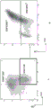
FIG. 10: allowing visualization of cell population CD34+CD41DarknessDensity map of (a).
FIG. 11: visualization of CD41 population in CD9 population. A) Representative flow cytometric dot plots of CD34 and CD9 expression in cell suspensions at day 10. The area in the lower right box represents the CD34 gated in example 2+CD9-A population of cells. B) Representative flow cytometric dot plots of CD41 and CD9 expression in cell suspensions at day 10. Representative of CD34+CD9-The region in the bottom right box of A) of the cell population corresponds to CD9-CD41DarknessThe lower frame of the dot diagram of CD41/CD 9. It can be concluded that CD9-Cell population and CD9-CD41DarknessThe cell population was the same.
FIG. 12: CD34+CD9-CD41DarknessCell population and CD34+CD41DarknessVisualization of cell population comparisons.A)Representative flow cytometric dot plots of CD34 and CD9 expression in cell suspensions at day 10. The lower right box represents the CD34 gated in example 2+CD9-A population of cells. B) CD34+CD9-FSC/CD41 of cell population+Representative flow cytogram of expression. B) The box in (1) represents CD34+CD9-CD41+A population of cells. CD9-Deselecting CD41Height of. Thus, by FSC/CD41+Expressing remaining CD41 gated+The cell is CD41Darkness. Thus, CD34+CD9-CD41+The cell population may be referred to as CD34+CD9-CD41DarknessA population of cells. C) Day 10 on the CD34/CD41 chart in cell suspension (as used e.g. in FIG. 3 to obtain CD 34)+CD41DarknessOf cell population) transposed CD34+CD9-CD41DarknessAnd (4) clustering. C) The circle in (A) indicates the cell population CD34 in the CD34/CD41 chart+CD9-CD41+ . The CD34 was demonstrated compared to, for example, FIG. 3A (day 10)+CD9-CD41+The cell population corresponds to CD34 as designated by region R2 in FIG. 3+CD9- A population of cells.
Example 1
Materials and methods
CD34+Isolation of cells
By adaptation from Ivanovic et al (Transfusion).2006;46:118-+A cell. Briefly, after 15 minutes of incubation with RosetteSeep human granule cell depleted cocktail (StemShell Technologies, Vancouver, Canada), separated by Histopaque-1077 (Sigma-Aldrich) density gradient at 400 ℃ CgFor 30 minutes, to isolate the mononuclear cells. Then, using an immunomagnetic bead cell sorting system (AutoMacs, Miltenyi, Bergisch Galdbach, Germany), by positiveSelected to separate CD34+A cell. Viability of 83.30 + -1.96% and CD34 of 82.80 + -2.25% were routinely obtained+Purity (n = 6).
MK differentiation in culture
Will CD34+Cells were cultured at 4X 10⁴The density of/mL was seeded in 48-well plates, StemBan SFEM medium supplemented with 20 ng/mL human LDL and CC220 (1 ×), cytokine cocktail with SCF, TPO, IL-6 and IL-9 (all from Stemcell Technologies), with or without 1 μ M SR1 (Cellagen Technology, San Diego, Calif.) (FIG. 1A). On day 7, cells were harvested, washed at 5X 10⁴The density at/mL was inoculated in StemBan SFEM medium with 30ng/mL TPO with or without 1 μ M SR1 for an additional 7 days. Culture in normoxic and 5% CO2Incubate at 37 ℃ under conditions. On days 7 and 10 of culture, cells were counted in an automatic cell counter (ADAM, Digital-Bio, Korea), their viability was measured by propidium iodide exclusion, and expression of CD34, CD41 and CD42b was analyzed in a galios flow cytometer using Kaluza software (Beckman Coulter, villapinte, france). In some experiments, SR1 was replaced with the AhR agonist FICZ (Enzo life sciences, villelurbane, France), which was added at 0.2 μ M.
In the second protocol, Mesenchymal Stromal Cells (MSCs) isolated from human bone marrow (Guilloton F et al, Blood).2012, 119:2556-+A cell. MSCs were maintained in α -MEM medium supplemented with 10% fetal bovine serum (Invitrogen, Cergy Pontoise, france) and 2 ng/mL recombinant human (rh) FGF2 (Peprotech, Rocky Hill, NJ). Will CD34+Cells were cultured at 4X 10⁴Density of/mL added to 48-well plates, MSC confluent layer in StemSpan SFEM medium supplemented with 20 ng/mL human LDL and CC 220. On day 7, the cell suspension was harvested, washed, and washed at 5 × 10⁴Density of/mL was co-cultured for an additional 7 days on a confluent layer of new MSCs in StemSpan SFEM medium with 30ng/mL TPO (fig. 5A).
Cell sorting
Cells recovered on day 10 were incubated with a mixture of Alexa-488 conjugated anti-CD 41 mAb (alma.17) and PE-Cy7 conjugated anti-CD 34 mAb (bd biosciences) for 30 minutes at 4 ℃. Then, they were washed in PBS-EDTA and incubated in PBS containing 7-AAD (1/50) for 30 minutes to select for viable cells. Morphology and sorting gates (gates) were determined by FMO (fluorescence subtraction one) analysis and megakaryocyte precursors were sorted at a rate of 500 cells/s according to their CD34/CD41 expression using a FACS Aria II flow cytometer (Becton Dickinson, Mountain View, CA) equipped with a 50 μm nozzle and two argon lasers (Coherent Radiation, Palo Alto, CA) running at 500 mW and adjusted to 488 mW and 360 nm, respectively. Then, sorting CD34+CD41DarknessAnd CD34-CD41+Cells were counted and counted at 4X 10⁴The density of/mL was seeded in 48-well plates in TPO-containing StemBan medium with or without SR1 for 7 days (FIG. 5A).
Assay for MK maturation
A surface marker.Cells were analyzed by flow cytometry (Gallios, Beckman Coulter, France) after labeling with anti-CD 34-PE-Cy7 mAb (Beckman Coulter, Fullerton, Calif.), anti-CD 41-Alexa-488 mAb (ALMA.17), anti-CD 42C-PE (RAM.1) mAb, and anti-CD 42d-Alexa-647 (V.1) mAb at 4 ℃ for 30 minutes. The cells were then washed and resuspended in PBS containing 7-AAD (1/50). The data obtained were analyzed using Kaluza software.
Ploidy.Cells were incubated with 10. mu.g/mL Hoechst 33342 (Sigma-Aldrich, Saint Quentin Fallavier, France) for 2 h at 37 ℃ and then stained with anti-CD 34-PE-Cy7 mAb and anti-CD 41-PE mAb. The washed cells were resuspended in PBS containing 7-AAD and CD41 was determined by two-color flow cytometry (Fortessa, BD Biosciences, Rungis, France)+Ploidy distribution in the population. The obtained data were analyzed using BD FACSDiva software (BD Biosciences).
An ultra-micro structure.Cell channels containing 2% of sugarcaneSugar in 0.1M cacodylate buffer (pH 7.2) 2.5% glutaraldehyde fixation and treatment as described previously (Eckly A et al, Blood).2014, 123, 921 and 930). The ultrathin sections were examined under a Philips CM120 Biotwin transmission electron microscope (FEI, Heindhoven, the Netherlands) at 120 kV.
Quantification of MK with pre-platelets
The percentage of MK in the culture wells that extended out of the pre-platelets was determined by phase contrast microscopy. In each culture, at least 100 MKs were analyzed and images were taken with a Zeiss Axio vert. a1 microscope with a 20 × objective (Marly-le-Roi, france).
Determining the number of platelets produced per seeded cell
CD34 cultured for 7 days in the presence of CC220+Cells (FIGS. 1A and 6A) or CD34/CD41 sorted cells (FIGS. 5A and 7A) were seeded in TPO-containing medium. On day 7, 1 μ M PGE1And 0.02U/mL apyrase were added to the medium and the cells were gently passed through a P1000 pipette tip 5 times. The resulting suspension (200 μ L) was incubated with anti-CD 41-Alexa-647 mAb and anti-CD 42c-Alexa-488 mAb at room temperature for 15 minutes before analysis in a Gallios flow cytometer. The CD41/CD42c double positive events, having the same scattering properties as the washed platelets, were counted as platelet-like particles and the number of particles per seeded cell was determined on day 7 or day 10 after the experiment.
RNA extraction
CD41/61 cells were obtained on day 7 or day 10 of culture using antibody ALMA.17 and magnetic beads (EasySep "kinetic by oneself" Selection Kit ("Do-It-Yourself" Selection Kit), StemShell Technologies). Following the manufacturer's instructions, total RNA was extracted using RNeasy mini kit (QIAGEN). The amount and quality of total RNA of all samples was assessed by measuring the OD at 260nm and the concentration was adjusted to 50 ng/ml. The RNA samples were stored at-80 ℃ until use. Under standard conditions, using SYBR Green Master Mix kit, in ABI Prism 7900 Sequence Detection System (PerkinElmer-Cetus, Courtabo)euf, france) was used. Primers for genes were selected with the help of the Oligo 6.0 program (National Biosciences, Plymouth, MN) and have been described previously (Bieche I. et al, Pharmacogenetics and genetics).2007;17:731-742)。
Statistics of
Statistical significance was determined using student's t-test or two-way analysis of variance, followed by a banoforn post-hoc test. Data were analyzed using Graphpad Prism 5 software.
Results
CD34 in peripheral blood+Among the differentiated MK cells, SR1 maintained CD34 expression
We evaluated the effect of AhR antagonist SR1 on MK precursor amplification. On days 0 and 7, SR1 (50 μ M) was added as a two-step culture protocol in which peripheral blood CD34+Cells (Peytour Y et al, Transfusion).2010;50: 2152-.
Using this protocol, more than 90% of the control cells (cultured without SR1) were double positive at day 12 for the platelet markers CD41 and CD42 and showed a marker signature of fully mature MK in morphological and phenotypic analysis.
Cell proliferation was estimated on days 7 and 10 before the onset of pre-platelet extension. On day 7, the total number of nucleated cells (mean ± SEM, n = 8) increased 6.7 ± 1.6-fold and 4.2 ± 1.2-fold, respectively, in the absence and presence of SR1 (fig. 1B). From day 7 to day 10, cell numbers similarly increased 2.3-fold and 2.5-fold in untreated and SR 1-treated cultures, respectively. Therefore, SR1 did not promote cell proliferation under our culture conditions.
We then assessed the effect of SR1 on progenitor cell retention by tracking the evolution of CD34 expression. During the expansion step, CD34 positivity was retained in control and SR1 treated cells with only 16.8% and 8.3% positive reductions at day 7, respectively (fig. 1C). On day 10, transmission was made in the presence of TPO aloneCD34 in control cultures after passage+The proportion of cells decreased to 40.7% while 71.6% remained positive after SR1 treatment. Thus, SR1 maintained a more progenitor-like phenotype after transfer of the cells into medium containing only added TPO.
SR1 increases MK and platelet-like material production with pre-platelets
In control cultures, pre-platelet elongation was first observed on day 10, and peaked at day 14 when 11.5 ± 4.5% MK showed pre-platelets (fig. 2A). Notably, this ratio increased by a factor of three (34.6 ± 2.1%, mean ± SEM, n = 4) after SR1 treatment, which resulted in an increase in the production of platelet-like substances. Whereas under control conditions, 7.92 ± 3.25 platelet-sized particles per cell seeded at day 7 were counted (fig. 2B), with an approximately 3-fold increase in platelet particle number (20.72 ± 5.19) upon addition of SR 1. These results indicate that SR1 not only maintains progenitor cell potential, but also greatly improves MK maturation. In contrast, when SR1 was replaced with the strong AhR agonist FICZ, the pre-platelet extension of MK and platelet-like material production decreased dramatically (0.20 ± 0.04 platelets/seeded cells). Such results strongly suggest that AhR blockade is the origin of increased platelet production in the presence of SR 1. Antagonist activity of SR1 to inhibit the expression of its downstream target CYP1B1 was confirmed in day 7 cultures as measured by qPCR (579.8 ± 40.8 vs 2.5 ± 0.8 arbitrary units in control and SR1 treated cells, respectively; mean ± SEM, n = 3).
SR1 promotes CD34+CD41DarknessAmplification of populations
The above findings indicate a dual role for SR1, as it maintains CD34 expression and also improves MK maturation. Since CD41 is a specific marker for MK, we evaluated its evolution parallel to CD 34. At day 7, a similarly high proportion of CD34+The cells had acquired positive for CD41, with 60% and 69% of the cells in the control and SR 1-treated cultures being CD34, respectively+ CD41 +(FIG. 3A). Passage in the presence of TPO alone resulted in a significant loss of CD34 positivity in control culturesOnly 32% of the cells on day 10 were CD34+CD41+. In contrast, a high proportion (55%) of the double positives for CD34 and CD41 remained in SR 1-treated cultures. Notably, the majority of these cells (17% of the total cells in the 37% vs control) exhibited CD41DarknessPhenotype (region R2) (fig. 3B). As evidenced by their FSC properties (fig. 3C), CD41 compared to cells with higher levels of CD41 (R1)DarknessThe population (R2) included cells of reduced size, indicating that MK was less differentiated. This was confirmed by ploidy analysis, since CD34+CD41DarknessThe majority of cells were 2 n-4 n (FIG. 4).
CD34+CD41DarknessThe cells have high ability to produce pre-platelets and platelet-like particles
Addition of SR1 in a two-step culture protocol resulted in increased production of MK and platelet-like material with pre-platelets (FIGS. 2A-2B). Therefore, we investigated whether this is in line with CD34+CD41DarknessThe amplification of the population is related to a specific property. CD34 from day 10 of culture with SR1 by flow cytometry+CD41DarknessCells were sorted and cultured for 7 days in TPO-containing medium supplemented with SR1 or without SR1 (fig. 5A). When CD34+CD41DarknessWhen cells were grown in the presence of SR1, an unprecedented high proportion of MK reached the pre-platelet stage (91.0. + -. 2.4%) (FIG. 5B). When these same cells were cultured without SR1, a much lower frequency (10.0 ± 6.6%) was observed (fig. 5B). Increased production of pre-platelets leads to CD34 cultured with SR1+CD41DarknessPlatelet-like material production in cells was 6.8-fold increased compared to no SR1 (52.06 ± 8.79 vs 7.68 ± 0.81 platelets/seeded cell, respectively) (fig. 5C). These results indicate that CD34 amplified in the presence of SR1+CD41DarknessThe population has a strong potential to produce pre-platelet-bearing MKs that readily release platelets.
Co-culture with MSC also promoted CD34+CD41DarknessOccurrence of clusters
Bone marrowThe derived stromal cells can maintain hematopoietic stem cell characteristics, secrete cytokines and facilitate MK maturation (Pallotta I et al, plosone. 2009;4: e8359; Cheng L et al, Journal of cellular physiology. 2000;184: 58-69) and can provide a favorable environment for the appearance of MK precursors. Through a two-step protocol, CD34 was synthesized+Cells were cultured on monolayers of human mesenchymal stromal cells (hmscs) isolated from human bone marrow (fig. 6A). Co-culture with hmscs did not significantly alter cell proliferation (fig. 6B), but caused increased MK with pre-platelets (data not shown) and platelet-like particle production at day 14 (7.9 ± 4.5 vs 18.2 ± 4.9 platelets per day 7 seeded cell) (fig. 6C).
We investigated whether the role of MSC might be mediated by pathways downstream of AhR. The AhR agonist FICZ (Boitano AE et al, Science) was added.2010, 329: 1345-1348) reduced CD34+CD41DarknessThe proportion of cells (data not shown) and prevented an increase in platelet production (fig. 7A). In addition, CD34+Co-culture of cells with MSCs results in a substantial reduction in CYP1B1 transcript: (>90%), which reproduces the effect of SR1, reversed by the addition of FICZ (fig. 7B). These results indicate that, like SR1, MSCs promote MK maturation and platelet production by acting on AhR.
This response was similar to that obtained with SR1, which prompted us to determine the CD34/CD41 phenotype of the cells. At day 10 of co-culture, there was CD34 compared to 18.95. + -. 1.75% in control cultures without MSCs (FIG. 8B)+CD41DarknessThe population of the pattern was clearly visible (FIG. 8A) and accounted for 30.37. + -. 1.98% of the total cells. This population had hypoploidy (fig. 8C).
Then, we aimed to determine i) MSC-and SR 1-derived CD34+CD41DarknessWhether the cells have the same potential for producing mature MK, and ii) whether co-culture with MSC or in the presence of SR1 is equally beneficial for such maturation. Will CD34+Cells were cultured with SR1 or on MSCs and the corresponding CD34 was sorted on day 10+CD41DarknessCells (fig. 9). These cells were then incubated with TPO, with TPO and SR1Or cultured with TPO and MSC for another 7 days. The results show that MSC-derived CD34+CD41DarknessCells when cultured in the presence of SR1 showed an increased ability to produce MK with pre-platelets (i in fig. 9A), but less efficient than SR 1-derived cells (i in fig. 9B) (49.5 ± 10.5% and 91.0 ± 2.4%, respectively, n = 4). In addition, co-culture with MSCs enhanced MSC-derived (fig. 9A) and SR 1-derived (fig. 9B) CD34 compared to culture with TPO alone+CD41DarknessMK maturation of cells (53.3. + -. 10.7% vs 67.5. + -. 12.6%, respectively). A similar pattern was observed for the ability to release platelet-like particles (ii in FIGS. 9A-9B). Thus, the co-culture phenotype with MSCs mimics the response obtained by adding SR1 to the cell culture.
In summary, it is reported therein that the identification and enrichment of discrete populations of adult hematopoietic progenitor cells prepared for MK differentiation can be efficiently matured into prothrombock-bearing MKs. When adult CD34+The cells are cultured in the presence of SR1, an antagonist of AhR, or a monolayer of MSCs, and are allowed to use their CD34+CD41DarknessThis population of cells identified by the label is expanded. Culturing with SR1 or MSC greatly enhances production of preprepalatelet-producing MK and platelet-like material release, in addition to promoting the appearance of such MK progenitors.
CD34 identified herein in the human system+CD41DarknessSeveral characteristics of the population, such as the small size and hypoploidy of the cells, and their high capacity to mature into pure MK that can effectively extend pre-platelets, appear to correspond to the definition of platelet-biased progenitor cells. Its unique phenotype combines CD34+Moderate or low expression of the progenitor cell marker and the megakaryocyte marker CD 41. Human CD34 has been described as isolated directly from bone marrow or after culturing under MK promoting conditions+CD41 positive cells among cells (Debili N. et al., Blood). 1992;80:3022-3035; Dercksen MW et al., Blood.1995;86: 3771-3782). However, these populations did not fully reproduce CD34+CD41DarknessPhenotypes, as they appear to express higher levels of CD41, are highly polyploid and fail to proliferate (Derc)ksen MW et al., Blood.1995;86: 3771-3782). Bone marrow-derived CD34 without TPO+CD34 was also observed after co-culturing cells on hMSCs+CD41+Cells, but for CD41 which is overtDarknessThe subgroup provided no evidence (Cheng L. et al, Journal of cellular physiology).2000, 184: 58-69). Recently CD34 with CD34 representing a very small population was reported in cultures derived from peripheral blood+CD41Is low inPhenotypic cells, but not further characterized. CD34+CD41DarknessPopulations have a similarly low frequency in our standard cultures (fig. 3A) and become apparent only after addition of SR1 or co-culture with MSCs. In recently reprogrammed iPS cells cultured in a three-step serum-free system, CD31 similar to the cells described herein was observed+CD34+CD41+A megakaryocyte population. This population appeared to express low levels of CD41 and was CD42 negative.
Example 2
Materials and methods
As above in "CD 34+Isolation of cells As described in the section "isolation of cells", peripheral blood CD34 was isolated+Cells, and cultured in the presence of SR1 (1 μ M) as described in the section "MK differentiation in culture" of example 1 above.
The cells recovered on day 10 were incubated with a mixture of Alexa-488 conjugated anti-CD 41 (ALMA.17) monoclonal antibody, Phycoerythrin (PE) -Cy 7-conjugated anti-CD 34 monoclonal antibody and Phycoerythrin (PE) -CD9 (mAb; BD Biosciences) for 30 minutes at 4 ℃. Then, they were incubated in phosphate buffered saline containing 7-amino actinomycin D (2.5. mu.g/mL) for 2 minutes to select viable cells.
Cells were first subdivided into CD34+CD9−A progenitor cell. Using CD9−Cell sorting of (3) excludes CD41Height ofCells, since only CD9 is present as shown in FIG. 11+The cell is CD41Height of. Then according to FSC/CD41+Expressing CD34+ CD9-Progenitor cell population (thus not comprising CD 4)1Height ofCells) into MK progenitor cells. The only CD41 present in the cell population+Is CD41DarknessThus allowing for the CD34 of interest+CD9-CD41DarknessThe cell population is gated. Megakaryocyte precursors were then sorted at 500 cells/sec using a Fluorescence Activated Cell Sorter (FACS) Aria II flow cytometer (Becton Dickinson, Mountain View, CA). Then, the sorted CD34 is processed+CD9-CD41DarknessCells were counted and counted at 4X 10⁴The density of/mL was seeded in 48-well plates in StemBan medium with TPO (with or without SR1) for 7 days.
Results
Previously described as CD34+CD41DarknessCan also be characterized as CD34+CD9-CD41Darkness. In particular, CD34+CD9-CD41DarknessRepresentative of CD34+CD41DarknessSubgroup of which CD34+CD9-CD41DarknessGroup occupation CD34+CD41Darkness60% of the total number of cells. And CD34+CD9-CD41DarknessIn contrast to cells, CD34 was functionally detected+CD41DarknessThe differentiation potential of the cell. And CD34+CD41DarknessCell comparison, CD34+CD9-CD41DarknessPlatelet release from the cells was increased 1.8 fold.
Claims (19)
1. A method of producing, ex vivo, pre-platelet bearing Megakaryocytes (MK) and/or platelets, comprising:
a) isolated CD34 of MK progenitor cells in the presence of an aromatic hydrocarbon receptor (AhR) antagonist+CD9-CD41DarknessCulturing the population of cells in a serum-free medium comprising Thrombopoietin (TPO) for a time sufficient to obtain a population of cells comprising MK and/or platelets that carry pre-platelets; and
b) collecting the population of cells comprising MK and/or platelets that carry pre-platelets.
2. The method according to claim 1, wherein the culturing in step a) is performed for 5 to 9 days.
3. The method according to claim 1 or 2, wherein the serum-free medium comprises 20-100 ng/ml TPO.
4. The method according to claim 1, wherein the method comprises, before step a):
a0) culturing Hematopoietic Stem Cells (HSCs) in serum-free medium containing density lipoprotein (LDL), Stem Cell Factor (SCF), TPO, IL-6 and IL-9 in the presence of an aromatic hydrocarbon receptor (AhR) antagonist sufficient to obtain a composition comprising CD34+CD9-CD41DarknessThe time of the cell population of cells; and
a1) isolating said CD34 from said cell population+CD9-CD41DarknessA cell.
5. The method according to claim 4, wherein the serum-free medium in step a0) comprises 10-30 μ g/ml LDL, 25-100 ng/ml SCF, 40-50 ng/ml TPO, 20-30 ng/ml IL-6 and 20-30 ng/ml IL-9.
6. The method according to claim 4, wherein the culturing in step a0) is performed for 6-8 days.
7. The method according to claim 1 or 4, wherein, in step a) and step a0), the AhR antagonist is independently a compound of formula (I)

L is selected from-NR5a(CH2)2-3-、-NR5a(CH2)2NR5b-、-NR5a(CH2)2S-、-NR5aCH2CH (OH) -or-NR5aCH(CH3)CH2-; wherein R is5aAnd R5bIndependently selected from hydrogen or C1-4An alkyl group;
R1selected from thienyl, furyl, benzimidazolyl, isoquinolinyl, imidazopyridinyl, benzothienyl, pyrimidinyl, pyrazolyl, pyridyl, imidazolyl, pyrrolidinyl, pyrazinyl, pyridazinyl, pyrrolyl or thiazolyl;
R2selected from-S (O)2NR6aR6b、-NR6aC(O)NR6bR6cPhenyl, pyrrolopyridyl, indolyl, thienyl, pyridyl, triazolyl, 2-oxoimidazolidinyl, pyrazolyl, or indazolyl; wherein
R6a、R6bAnd R6cIndependently selected from hydrogen or C1-4An alkyl group; and
R3selected from hydrogen, C1-4Alkyl or biphenyl; and
R4is selected from C1-10Alkyl radical, C1-4Alkenyl, oxetanyl, tetrahydrofuryl, cyclohexyl, (oxopyrrolidinyl) ethyl, tetrahydropyranyl, phenyl or benzyl.
8. The method according to claim 1 or 4, wherein, in step a) and/or step a0), the AhR antagonist is StemRegenin 1(SR 1).
9. The method of any one of claims 1, 2, and 4-6, further comprising: from the collected cell population containing MK and/or platelets with pre-platelets, CD41/CD61+ and CD42c + cells were selected.
10. The method of any one of claims 1, 2, and 4-6, further comprising: washing the pre-platelet-bearing Megakaryocytes (MK) and/or platelets, and suspending the washed cells in an infusion buffer.
11. The method of claim 7, wherein R1Said thienyl, furylBenzimidazolyl, isoquinolyl, imidazopyridinyl, benzothienyl, pyrimidinyl, pyrazolyl, pyridyl, imidazolyl, pyrrolidinyl, pyrazinyl, pyridazinyl, pyrrolyl and thiazolyl are independently selected from halogen, cyano, C1-4Alkyl, halogen-substituted C1-4Alkyl radical, C1-4Alkoxy, -S (O)0-2R8aand-C (O) OR8aWherein R is substituted by 1 to 3 groups8aSelected from hydrogen or C1-4An alkyl group.
12. The method of claim 7, R2Said phenyl, pyrrolopyridyl, indolyl, thienyl, pyridyl, triazolyl, oxoimidazolidinyl, pyrazolyl, or indazolyl of (a) is independently selected from hydroxy, halogen, methyl, methoxy, amino, -O (CH)2)nNR7aR7b、-OS(O)2NR7aR7band-NR7aS(O)2R7b1-3 groups in the (A); wherein R is7aAnd R7bIndependently selected from hydrogen or C1-4An alkyl group.
13. The method of claim 7, wherein R4Said C of1-10Alkyl radical, C1-4Alkenyl, oxetanyl, tetrahydrofuryl, cyclohexyl, (oxopyrrolidinyl) ethyl, tetrahydropyranyl, phenyl and benzyl are independently selected from hydroxy, C1-4Alkyl and halogen substituted C1-41-3 groups in the alkyl group.
14. A method of producing Megakaryocyte (MK) progenitor cells, comprising:
a0) culturing Hematopoietic Stem Cells (HSCs) in serum-free medium containing density lipoprotein (LDL), Stem Cell Factor (SCF), TPO, IL-6 and IL-9 in the presence of an aromatic hydrocarbon receptor (AhR) antagonist sufficient to obtain a composition comprising CD34+CD9-CD41DarknessThe time of the cell population of cells; and
a1) isolating said CD34 from said cell population+CD9-CD41DarknessA cell.
15. A method according to claim 14, wherein step a0) is carried out as defined in any one of claims 4 to 8 and 11 to 13.
16. A population of Megakaryocyte (MK) progenitor cells, wherein at least 80% of the cells in the population are CD34+CD9-CD41DarknessA cell.
17. The cell population of claim 16, wherein the cell population comprises at least 150,000 CD34+CD9-CD41DarknessA cell.
18. The cell population according to any one of claims 16 to 17, wherein the cell population is obtainable by a method according to any one of claims 14 to 15.
19. A composition for use in transfusion, the composition comprising a cell population of pre-platelet-bearing Megakaryocytes (MK) and/or platelets and an infusion buffer, wherein the use comprises: by the method of claim 10, a pre-platelet-bearing Megakaryocyte (MK) and/or platelet is prepared.
Applications Claiming Priority (3)
| Application Number | Priority Date | Filing Date | Title |
|---|---|---|---|
| FR1557020 | 2015-07-23 | ||
| FR1557020A FR3039166B1 (en) | 2015-07-23 | 2015-07-23 | CD34 + CD41DIM MEGACARYOCYTE PROGENITORS AND USES THEREOF TO PRODUCE PLAQUETTES AND / OR MK CARRIING PROPLAQUETS |
| PCT/EP2016/067594 WO2017013262A1 (en) | 2015-07-23 | 2016-07-22 | Cd34+cd41dim megakaryocytes progenitors and uses thereof for producing proplatelet-bearing mks and/or platelets |
Publications (2)
| Publication Number | Publication Date |
|---|---|
| CN107922924A CN107922924A (en) | 2018-04-17 |
| CN107922924B true CN107922924B (en) | 2022-01-11 |
Family
ID=55072774
Family Applications (1)
| Application Number | Title | Priority Date | Filing Date |
|---|---|---|---|
| CN201680047758.8A Active CN107922924B (en) | 2015-07-23 | 2016-07-22 | Megakaryocyte progenitor cells and use thereof to produce MK and/or platelets with pre-platelets |
Country Status (8)
| Country | Link |
|---|---|
| US (2) | US11236303B2 (en) |
| EP (1) | EP3334822A1 (en) |
| JP (2) | JP7068162B6 (en) |
| CN (1) | CN107922924B (en) |
| AU (1) | AU2016296920B2 (en) |
| CA (1) | CA2993596A1 (en) |
| FR (1) | FR3039166B1 (en) |
| WO (1) | WO2017013262A1 (en) |
Families Citing this family (3)
| Publication number | Priority date | Publication date | Assignee | Title |
|---|---|---|---|---|
| FR3039166B1 (en) * | 2015-07-23 | 2018-10-26 | Institut National De La Sante Et De La Recherche Medicale | CD34 + CD41DIM MEGACARYOCYTE PROGENITORS AND USES THEREOF TO PRODUCE PLAQUETTES AND / OR MK CARRIING PROPLAQUETS |
| CA3087344A1 (en) | 2018-01-05 | 2019-07-11 | Platelet Biogenesis, Inc. | Compositions and methods for producing megakaryocytes |
| FR3101885A1 (en) | 2019-10-11 | 2021-04-16 | Etablissement Français Du Sang | SYSTEM FOR THE RELEASE OF PLATES AND METHOD FOR RELEASING PLATES |
Citations (1)
| Publication number | Priority date | Publication date | Assignee | Title |
|---|---|---|---|---|
| WO2014138485A1 (en) * | 2013-03-08 | 2014-09-12 | Irm Llc | Ex vivo production of platelets from hematopoietic stem cells and the product thereof |
Family Cites Families (7)
| Publication number | Priority date | Publication date | Assignee | Title |
|---|---|---|---|---|
| EP1536688B1 (en) | 2002-09-13 | 2011-12-28 | The Board Of Trustees Of The Leland Stanford Junior University | Mammalian megakaryocyte progenitor cell |
| JPWO2009119105A1 (en) | 2008-03-28 | 2011-07-21 | 国立大学法人 東京大学 | GPIbα + GPV + GPVI + platelet in vitro preparation method |
| CA2828015C (en) | 2011-03-18 | 2020-06-16 | New York Blood Center, Inc. | Megakaryocyte and platelet production from stem cells |
| US9920295B2 (en) | 2012-02-21 | 2018-03-20 | The Trustees Of The University Of Pennsylvania | Bioreactor for isolation of rare cells and methods of use |
| US9074186B2 (en) | 2012-08-15 | 2015-07-07 | Boston Medical Center Corporation | Production of red blood cells and platelets from stem cells |
| GB201220937D0 (en) * | 2012-11-21 | 2013-01-02 | Airbus Uk Ltd | Modular structural assembly |
| FR3039166B1 (en) * | 2015-07-23 | 2018-10-26 | Institut National De La Sante Et De La Recherche Medicale | CD34 + CD41DIM MEGACARYOCYTE PROGENITORS AND USES THEREOF TO PRODUCE PLAQUETTES AND / OR MK CARRIING PROPLAQUETS |
-
2015
- 2015-07-23 FR FR1557020A patent/FR3039166B1/en active Active
-
2016
- 2016-07-22 CN CN201680047758.8A patent/CN107922924B/en active Active
- 2016-07-22 JP JP2018503184A patent/JP7068162B6/en active Active
- 2016-07-22 CA CA2993596A patent/CA2993596A1/en active Pending
- 2016-07-22 WO PCT/EP2016/067594 patent/WO2017013262A1/en active Application Filing
- 2016-07-22 US US15/747,023 patent/US11236303B2/en active Active
- 2016-07-22 EP EP16742302.9A patent/EP3334822A1/en active Pending
- 2016-07-22 AU AU2016296920A patent/AU2016296920B2/en active Active
-
2021
- 2021-07-26 JP JP2021121618A patent/JP2021177774A/en active Pending
- 2021-12-10 US US17/547,444 patent/US20220135946A1/en active Pending
Patent Citations (1)
| Publication number | Priority date | Publication date | Assignee | Title |
|---|---|---|---|---|
| WO2014138485A1 (en) * | 2013-03-08 | 2014-09-12 | Irm Llc | Ex vivo production of platelets from hematopoietic stem cells and the product thereof |
Non-Patent Citations (3)
| Title |
|---|
| A potential activity of valproic acid in the stimulation of interleukin-3 mediated megakaryopoiesis and erythropoiesis;Bing Liu等;《Experimental Hematology》;20101231;第38卷(第8期);第685-695页 * |
| Human Mesenchymal Stem Cells Support Megakaryocyte and Pro-Platelet Formation From CD341 Hematopoietic Progenitor Cells;LINZHAO CHENG等;《JOURNAL OF CELLULAR PHYSIOLOGY》;20001231;第184卷;摘要、第59页右栏第2段、第63页左栏第2段、第65页右栏第1段 * |
| In Vitro And In Vivo Megakaryocyte Differentiation Of Fresh And Ex-Vivo Expanded Cord Blood Cells: Rapid And Transient Megakaryocyte Reconstitution;S Bruno等;《Haematologica》;20030131;第88卷(第4期);第381页右栏倒数第2段、图1D、图2 * |
Also Published As
| Publication number | Publication date |
|---|---|
| FR3039166B1 (en) | 2018-10-26 |
| JP2021177774A (en) | 2021-11-18 |
| WO2017013262A1 (en) | 2017-01-26 |
| CN107922924A (en) | 2018-04-17 |
| JP7068162B2 (en) | 2022-05-16 |
| CA2993596A1 (en) | 2017-01-26 |
| AU2016296920B2 (en) | 2022-05-26 |
| EP3334822A1 (en) | 2018-06-20 |
| JP7068162B6 (en) | 2023-12-20 |
| US11236303B2 (en) | 2022-02-01 |
| FR3039166A1 (en) | 2017-01-27 |
| JP2018519845A (en) | 2018-07-26 |
| US20220135946A1 (en) | 2022-05-05 |
| AU2016296920A1 (en) | 2018-02-15 |
| US20180216068A1 (en) | 2018-08-02 |
Similar Documents
| Publication | Publication Date | Title |
|---|---|---|
| Jing et al. | Hematopoietic stem cells in co-culture with mesenchymal stromal cells-modeling the niche compartments in vitro | |
| Zhang et al. | Isolation and characterization of mesenchymal stem cells from human umbilical cord blood: reevaluation of critical factors for successful isolation and high ability to proliferate and differentiate to chondrocytes as compared to mesenchymal stem cells from bone marrow and adipose tissue | |
| US20220135946A1 (en) | Cd34+cd41dim megakaryocytes progenitors and uses thereof for producing proplatelet-bearing mks and/or platelets | |
| JP7522800B2 (en) | Methods for improving hematopoietic grafts | |
| CN105713879B (en) | Culture system for umbilical cord blood hematopoietic stem cell amplification and application thereof | |
| CA2271758A1 (en) | Msc-megakaryocyte precursor composition and method of isolating mscs associated with isolated megakaryocytes by isolating megakaryocytes | |
| Goedhart et al. | Interferon-gamma impairs maintenance and alters hematopoietic support of bone marrow mesenchymal stromal cells | |
| EP4029566A1 (en) | Serum-free medium not containing albumin and suited for culturing human hematopoietic stem cells, and albumin-free culturing method | |
| KR20100091192A (en) | Methods for co-culturing cord blood derived cells with menstrual stem cells | |
| JP6954844B2 (en) | Methods and Uses for Culturing Hematopoietic Stem Cells and / or Differentiating Hematopoietic Stem Cells into Precursors | |
| US20220127577A1 (en) | Xeno-free generation of tissue-specific progenitor cells | |
| JPH10136978A (en) | Culture of hematopoietic stem cell | |
| Mizokami et al. | Preferential expansion of human umbilical cord blood-derived CD34-positive cells on major histocompatibility complex-matched amnion-derived mesenchymal stem cells | |
| Ookura et al. | Adipocyte differentiation of human marrow mesenchymal stem cells reduces the supporting capacity for hematopoietic progenitors but not for severe combined immunodeficiency repopulating cells | |
| WO2023032945A1 (en) | Therapeutic agent for arthropathy, and method for producing therapeutic agent for arthropathy | |
| KR101149007B1 (en) | Method for Preparing Hematopoietic Stem Cells Having Improved Engraftment | |
| KR100999905B1 (en) | Method for Preparing Hematopoietic Stem Cells Having Improved Engraftment | |
| Giebel | Human multipotent hematopoietic progenitor cell expansion is neither supported in endothelial and endothelial/mesenchymal co-cultures nor in nSG mice | |
| EA026459B1 (en) | Method of ex vitro human hemopoietic cells expansion |
Legal Events
| Date | Code | Title | Description |
|---|---|---|---|
| PB01 | Publication | ||
| PB01 | Publication | ||
| SE01 | Entry into force of request for substantive examination | ||
| SE01 | Entry into force of request for substantive examination | ||
| GR01 | Patent grant | ||
| GR01 | Patent grant |