CN107580628B - Targeted cytotoxic cells with chimeric receptors for adoptive immunotherapy - Google Patents
Targeted cytotoxic cells with chimeric receptors for adoptive immunotherapy Download PDFInfo
- Publication number
- CN107580628B CN107580628B CN201580060960.XA CN201580060960A CN107580628B CN 107580628 B CN107580628 B CN 107580628B CN 201580060960 A CN201580060960 A CN 201580060960A CN 107580628 B CN107580628 B CN 107580628B
- Authority
- CN
- China
- Prior art keywords
- domain
- car
- acid sequence
- amino acid
- kir
- Prior art date
- Legal status (The legal status is an assumption and is not a legal conclusion. Google has not performed a legal analysis and makes no representation as to the accuracy of the status listed.)
- Active
Links
- 230000001472 cytotoxic effect Effects 0.000 title claims description 69
- 231100000433 cytotoxic Toxicity 0.000 title claims description 64
- 238000009169 immunotherapy Methods 0.000 title abstract description 4
- 108700010039 chimeric receptor Proteins 0.000 title description 3
- 210000004027 cell Anatomy 0.000 claims abstract description 387
- 108010043610 KIR Receptors Proteins 0.000 claims abstract description 174
- 102000002698 KIR Receptors Human genes 0.000 claims abstract description 163
- 210000000822 natural killer cell Anatomy 0.000 claims abstract description 94
- 239000012642 immune effector Substances 0.000 claims abstract description 26
- 229940121354 immunomodulator Drugs 0.000 claims abstract description 26
- 239000000427 antigen Substances 0.000 claims description 299
- 108091007433 antigens Proteins 0.000 claims description 298
- 102000036639 antigens Human genes 0.000 claims description 298
- 230000027455 binding Effects 0.000 claims description 262
- 150000007523 nucleic acids Chemical class 0.000 claims description 175
- 125000003275 alpha amino acid group Chemical group 0.000 claims description 153
- 230000001086 cytosolic effect Effects 0.000 claims description 132
- 102000039446 nucleic acids Human genes 0.000 claims description 131
- 108020004707 nucleic acids Proteins 0.000 claims description 131
- 241000282414 Homo sapiens Species 0.000 claims description 113
- 108090000765 processed proteins & peptides Proteins 0.000 claims description 108
- 102000003735 Mesothelin Human genes 0.000 claims description 97
- 108090000015 Mesothelin Proteins 0.000 claims description 97
- 239000013598 vector Substances 0.000 claims description 96
- 102000004196 processed proteins & peptides Human genes 0.000 claims description 93
- 229920001184 polypeptide Polymers 0.000 claims description 85
- 230000014509 gene expression Effects 0.000 claims description 66
- 108091028043 Nucleic acid sequence Proteins 0.000 claims description 60
- 230000011664 signaling Effects 0.000 claims description 54
- 230000004048 modification Effects 0.000 claims description 52
- 238000012986 modification Methods 0.000 claims description 52
- 108010047041 Complementarity Determining Regions Proteins 0.000 claims description 49
- 150000001413 amino acids Chemical class 0.000 claims description 47
- 208000037265 diseases, disorders, signs and symptoms Diseases 0.000 claims description 43
- 239000002773 nucleotide Substances 0.000 claims description 41
- 125000003729 nucleotide group Chemical group 0.000 claims description 41
- 239000012082 adaptor molecule Substances 0.000 claims description 37
- 201000010099 disease Diseases 0.000 claims description 37
- FVRDYQYEVDDKCR-DBRKOABJSA-N tiazofurine Chemical compound NC(=O)C1=CSC([C@H]2[C@@H]([C@H](O)[C@@H](CO)O2)O)=N1 FVRDYQYEVDDKCR-DBRKOABJSA-N 0.000 claims description 35
- 101000945339 Homo sapiens Killer cell immunoglobulin-like receptor 2DS2 Proteins 0.000 claims description 27
- 102100033630 Killer cell immunoglobulin-like receptor 2DS2 Human genes 0.000 claims description 27
- 238000011282 treatment Methods 0.000 claims description 25
- 241000124008 Mammalia Species 0.000 claims description 24
- 108020004414 DNA Proteins 0.000 claims description 21
- 102000053602 DNA Human genes 0.000 claims description 20
- 206010033128 Ovarian cancer Diseases 0.000 claims description 20
- 206010027406 Mesothelioma Diseases 0.000 claims description 16
- 206010061535 Ovarian neoplasm Diseases 0.000 claims description 16
- 238000004519 manufacturing process Methods 0.000 claims description 16
- 230000036737 immune function Effects 0.000 claims description 14
- 201000008129 pancreatic ductal adenocarcinoma Diseases 0.000 claims description 14
- 208000006178 malignant mesothelioma Diseases 0.000 claims description 11
- 201000005282 malignant pleural mesothelioma Diseases 0.000 claims description 11
- 102000006306 Antigen Receptors Human genes 0.000 claims description 10
- 108010083359 Antigen Receptors Proteins 0.000 claims description 10
- 239000013612 plasmid Substances 0.000 claims description 10
- 241001529936 Murinae Species 0.000 claims description 9
- 230000005809 anti-tumor immunity Effects 0.000 claims description 9
- 206010009944 Colon cancer Diseases 0.000 claims description 7
- 208000001333 Colorectal Neoplasms Diseases 0.000 claims description 7
- 239000003814 drug Substances 0.000 claims description 7
- 206010005003 Bladder cancer Diseases 0.000 claims description 6
- 206010023774 Large cell lung cancer Diseases 0.000 claims description 6
- 208000007097 Urinary Bladder Neoplasms Diseases 0.000 claims description 6
- 208000035475 disorder Diseases 0.000 claims description 6
- 201000009546 lung large cell carcinoma Diseases 0.000 claims description 6
- 201000005243 lung squamous cell carcinoma Diseases 0.000 claims description 6
- 208000002154 non-small cell lung carcinoma Diseases 0.000 claims description 6
- 230000007030 peptide scission Effects 0.000 claims description 6
- 208000029729 tumor suppressor gene on chromosome 11 Diseases 0.000 claims description 6
- 201000005112 urinary bladder cancer Diseases 0.000 claims description 6
- 108010073816 IgE Receptors Proteins 0.000 claims description 5
- 102000009438 IgE Receptors Human genes 0.000 claims description 5
- 230000001177 retroviral effect Effects 0.000 claims description 4
- 206010027476 Metastases Diseases 0.000 claims description 2
- 101000809875 Homo sapiens TYRO protein tyrosine kinase-binding protein Proteins 0.000 claims 2
- 102100038717 TYRO protein tyrosine kinase-binding protein Human genes 0.000 claims 2
- 108010019670 Chimeric Antigen Receptors Proteins 0.000 abstract description 157
- 108020003175 receptors Proteins 0.000 abstract description 69
- 238000000034 method Methods 0.000 abstract description 61
- 230000003213 activating effect Effects 0.000 abstract description 34
- 230000000694 effects Effects 0.000 abstract description 24
- 239000000203 mixture Substances 0.000 abstract description 20
- 108091008042 inhibitory receptors Proteins 0.000 abstract description 4
- 238000013461 design Methods 0.000 abstract description 2
- 210000001744 T-lymphocyte Anatomy 0.000 description 134
- 108090000623 proteins and genes Proteins 0.000 description 114
- 206010028980 Neoplasm Diseases 0.000 description 110
- 235000018102 proteins Nutrition 0.000 description 71
- 102000004169 proteins and genes Human genes 0.000 description 71
- 102000005962 receptors Human genes 0.000 description 67
- 230000002401 inhibitory effect Effects 0.000 description 55
- 230000004068 intracellular signaling Effects 0.000 description 54
- 201000011510 cancer Diseases 0.000 description 51
- -1 NTB-a Proteins 0.000 description 48
- 235000001014 amino acid Nutrition 0.000 description 45
- 108091008877 NK cell receptors Proteins 0.000 description 44
- 102100021462 Natural killer cells antigen CD94 Human genes 0.000 description 44
- 229940024606 amino acid Drugs 0.000 description 43
- 230000004913 activation Effects 0.000 description 39
- 125000000539 amino acid group Chemical group 0.000 description 36
- 239000012634 fragment Substances 0.000 description 30
- 108020004999 messenger RNA Proteins 0.000 description 28
- 241000699666 Mus <mouse, genus> Species 0.000 description 27
- 108091008874 T cell receptors Proteins 0.000 description 27
- 102000016266 T-Cell Antigen Receptors Human genes 0.000 description 27
- 239000003446 ligand Substances 0.000 description 27
- 102100039694 Death-associated protein 1 Human genes 0.000 description 26
- 108010041872 Islet Amyloid Polypeptide Proteins 0.000 description 26
- 108010087819 Fc receptors Proteins 0.000 description 25
- 102000009109 Fc receptors Human genes 0.000 description 25
- HKVAMNSJSFKALM-GKUWKFKPSA-N Everolimus Chemical compound C1C[C@@H](OCCO)[C@H](OC)C[C@@H]1C[C@@H](C)[C@H]1OC(=O)[C@@H]2CCCCN2C(=O)C(=O)[C@](O)(O2)[C@H](C)CC[C@H]2C[C@H](OC)/C(C)=C/C=C/C=C/[C@@H](C)C[C@@H](C)C(=O)[C@H](OC)[C@H](O)/C(C)=C/[C@@H](C)C(=O)C1 HKVAMNSJSFKALM-GKUWKFKPSA-N 0.000 description 24
- 101100181099 Mus musculus Klra1 gene Proteins 0.000 description 24
- 102100024222 B-lymphocyte antigen CD19 Human genes 0.000 description 23
- 101000980825 Homo sapiens B-lymphocyte antigen CD19 Proteins 0.000 description 23
- 108060003951 Immunoglobulin Proteins 0.000 description 23
- 102000018358 immunoglobulin Human genes 0.000 description 23
- 230000006870 function Effects 0.000 description 22
- 241000282832 Camelidae Species 0.000 description 21
- 230000004044 response Effects 0.000 description 20
- 230000004936 stimulating effect Effects 0.000 description 20
- 102100040678 Programmed cell death protein 1 Human genes 0.000 description 19
- 101710089372 Programmed cell death protein 1 Proteins 0.000 description 19
- 241000251730 Chondrichthyes Species 0.000 description 18
- 102000002090 Fibronectin type III Human genes 0.000 description 18
- 108050009401 Fibronectin type III Proteins 0.000 description 18
- 102000040430 polynucleotide Human genes 0.000 description 18
- 108091033319 polynucleotide Proteins 0.000 description 18
- 239000002157 polynucleotide Substances 0.000 description 18
- 239000013603 viral vector Substances 0.000 description 18
- 101000945333 Homo sapiens Killer cell immunoglobulin-like receptor 2DL3 Proteins 0.000 description 17
- 241000235789 Hyperoartia Species 0.000 description 17
- 102100033634 Killer cell immunoglobulin-like receptor 2DL3 Human genes 0.000 description 17
- 108700018351 Major Histocompatibility Complex Proteins 0.000 description 17
- 150000001875 compounds Chemical class 0.000 description 17
- 229920002477 rna polymer Polymers 0.000 description 17
- 230000020382 suppression by virus of host antigen processing and presentation of peptide antigen via MHC class I Effects 0.000 description 17
- 102000008394 Immunoglobulin Fragments Human genes 0.000 description 16
- 230000000139 costimulatory effect Effects 0.000 description 16
- 230000003834 intracellular effect Effects 0.000 description 16
- 230000035897 transcription Effects 0.000 description 15
- 238000013518 transcription Methods 0.000 description 15
- 102000004127 Cytokines Human genes 0.000 description 14
- 108090000695 Cytokines Proteins 0.000 description 14
- 101000945331 Homo sapiens Killer cell immunoglobulin-like receptor 2DL4 Proteins 0.000 description 14
- 108010021625 Immunoglobulin Fragments Proteins 0.000 description 14
- 102100022339 Integrin alpha-L Human genes 0.000 description 14
- 102100033633 Killer cell immunoglobulin-like receptor 2DL4 Human genes 0.000 description 14
- 102100032870 Natural cytotoxicity triggering receptor 1 Human genes 0.000 description 14
- 238000000338 in vitro Methods 0.000 description 14
- 230000003993 interaction Effects 0.000 description 14
- 101001046668 Homo sapiens Integrin alpha-X Proteins 0.000 description 13
- 102100022297 Integrin alpha-X Human genes 0.000 description 13
- 108010002350 Interleukin-2 Proteins 0.000 description 13
- 102000000588 Interleukin-2 Human genes 0.000 description 13
- 108010004217 Natural Cytotoxicity Triggering Receptor 1 Proteins 0.000 description 13
- 206010061902 Pancreatic neoplasm Diseases 0.000 description 13
- 229960005167 everolimus Drugs 0.000 description 13
- 208000015486 malignant pancreatic neoplasm Diseases 0.000 description 13
- 201000002528 pancreatic cancer Diseases 0.000 description 13
- 208000008443 pancreatic carcinoma Diseases 0.000 description 13
- 230000001105 regulatory effect Effects 0.000 description 13
- 101001027081 Homo sapiens Killer cell immunoglobulin-like receptor 2DL1 Proteins 0.000 description 12
- 102100037363 Killer cell immunoglobulin-like receptor 2DL1 Human genes 0.000 description 12
- 210000005220 cytoplasmic tail Anatomy 0.000 description 12
- 238000000684 flow cytometry Methods 0.000 description 12
- 241000283707 Capra Species 0.000 description 11
- 102100027208 T-cell antigen CD7 Human genes 0.000 description 11
- 230000003013 cytotoxicity Effects 0.000 description 11
- 231100000135 cytotoxicity Toxicity 0.000 description 11
- 239000012636 effector Substances 0.000 description 11
- 230000005764 inhibitory process Effects 0.000 description 11
- 239000000463 material Substances 0.000 description 11
- 239000000047 product Substances 0.000 description 11
- 230000035755 proliferation Effects 0.000 description 11
- 230000002829 reductive effect Effects 0.000 description 11
- 230000000638 stimulation Effects 0.000 description 11
- 108700028369 Alleles Proteins 0.000 description 10
- 102100028971 HLA class I histocompatibility antigen, C alpha chain Human genes 0.000 description 10
- 101000945371 Homo sapiens Killer cell immunoglobulin-like receptor 2DL2 Proteins 0.000 description 10
- 101000945490 Homo sapiens Killer cell immunoglobulin-like receptor 3DL2 Proteins 0.000 description 10
- 101000917858 Homo sapiens Low affinity immunoglobulin gamma Fc region receptor III-A Proteins 0.000 description 10
- 101000917839 Homo sapiens Low affinity immunoglobulin gamma Fc region receptor III-B Proteins 0.000 description 10
- 108010064593 Intercellular Adhesion Molecule-1 Proteins 0.000 description 10
- 102100037877 Intercellular adhesion molecule 1 Human genes 0.000 description 10
- 102100033599 Killer cell immunoglobulin-like receptor 2DL2 Human genes 0.000 description 10
- 102100034840 Killer cell immunoglobulin-like receptor 3DL2 Human genes 0.000 description 10
- 108010064548 Lymphocyte Function-Associated Antigen-1 Proteins 0.000 description 10
- 108700015968 Slam family Proteins 0.000 description 10
- 108060008683 Tumor Necrosis Factor Receptor Proteins 0.000 description 10
- 239000011325 microbead Substances 0.000 description 10
- 102000003298 tumor necrosis factor receptor Human genes 0.000 description 10
- 102000008949 Histocompatibility Antigens Class I Human genes 0.000 description 9
- 101000945493 Homo sapiens Killer cell immunoglobulin-like receptor 3DL3 Proteins 0.000 description 9
- 102100029193 Low affinity immunoglobulin gamma Fc region receptor III-A Human genes 0.000 description 9
- 102100022682 NKG2-A/NKG2-B type II integral membrane protein Human genes 0.000 description 9
- 108010025832 RANK Ligand Proteins 0.000 description 9
- 102000014128 RANK Ligand Human genes 0.000 description 9
- 210000001151 cytotoxic T lymphocyte Anatomy 0.000 description 9
- 230000028993 immune response Effects 0.000 description 9
- 125000005647 linker group Chemical group 0.000 description 9
- 229940124302 mTOR inhibitor Drugs 0.000 description 9
- 239000003628 mammalian target of rapamycin inhibitor Substances 0.000 description 9
- 238000002560 therapeutic procedure Methods 0.000 description 9
- 238000010361 transduction Methods 0.000 description 9
- 230000026683 transduction Effects 0.000 description 9
- 210000004881 tumor cell Anatomy 0.000 description 9
- 101001046687 Homo sapiens Integrin alpha-E Proteins 0.000 description 8
- 101000945351 Homo sapiens Killer cell immunoglobulin-like receptor 3DL1 Proteins 0.000 description 8
- 101000945492 Homo sapiens Killer cell immunoglobulin-like receptor 3DS1 Proteins 0.000 description 8
- 101001109508 Homo sapiens NKG2-A/NKG2-B type II integral membrane protein Proteins 0.000 description 8
- 102100022341 Integrin alpha-E Human genes 0.000 description 8
- 102100033627 Killer cell immunoglobulin-like receptor 3DL1 Human genes 0.000 description 8
- 102100034834 Killer cell immunoglobulin-like receptor 3DL3 Human genes 0.000 description 8
- 102100034833 Killer cell immunoglobulin-like receptor 3DS1 Human genes 0.000 description 8
- OUYCCCASQSFEME-QMMMGPOBSA-N L-tyrosine Chemical compound OC(=O)[C@@H](N)CC1=CC=C(O)C=C1 OUYCCCASQSFEME-QMMMGPOBSA-N 0.000 description 8
- 108091054437 MHC class I family Proteins 0.000 description 8
- 241000699670 Mus sp. Species 0.000 description 8
- 239000011324 bead Substances 0.000 description 8
- 230000012010 growth Effects 0.000 description 8
- 210000004698 lymphocyte Anatomy 0.000 description 8
- 239000012071 phase Substances 0.000 description 8
- 238000012546 transfer Methods 0.000 description 8
- OUYCCCASQSFEME-UHFFFAOYSA-N tyrosine Natural products OC(=O)C(N)CC1=CC=C(O)C=C1 OUYCCCASQSFEME-UHFFFAOYSA-N 0.000 description 8
- 241000282693 Cercopithecidae Species 0.000 description 7
- 108010052199 HLA-C Antigens Proteins 0.000 description 7
- 101000945337 Homo sapiens Killer cell immunoglobulin-like receptor 2DL5A Proteins 0.000 description 7
- 101000945335 Homo sapiens Killer cell immunoglobulin-like receptor 2DL5B Proteins 0.000 description 7
- 102100033628 Killer cell immunoglobulin-like receptor 2DL5B Human genes 0.000 description 7
- 206010058467 Lung neoplasm malignant Diseases 0.000 description 7
- 241001465754 Metazoa Species 0.000 description 7
- 101710167259 Neuromedin-K receptor Proteins 0.000 description 7
- 102100029409 Neuromedin-K receptor Human genes 0.000 description 7
- 241000283984 Rodentia Species 0.000 description 7
- 230000000875 corresponding effect Effects 0.000 description 7
- 230000009089 cytolysis Effects 0.000 description 7
- 210000003162 effector t lymphocyte Anatomy 0.000 description 7
- 201000005202 lung cancer Diseases 0.000 description 7
- 208000020816 lung neoplasm Diseases 0.000 description 7
- 230000002062 proliferating effect Effects 0.000 description 7
- 239000000523 sample Substances 0.000 description 7
- 238000010186 staining Methods 0.000 description 7
- 210000001519 tissue Anatomy 0.000 description 7
- 102100034579 Desmoglein-1 Human genes 0.000 description 6
- 102100037362 Fibronectin Human genes 0.000 description 6
- 108010067306 Fibronectins Proteins 0.000 description 6
- DHMQDGOQFOQNFH-UHFFFAOYSA-N Glycine Chemical compound NCC(O)=O DHMQDGOQFOQNFH-UHFFFAOYSA-N 0.000 description 6
- 101000945340 Homo sapiens Killer cell immunoglobulin-like receptor 2DS1 Proteins 0.000 description 6
- 101000945343 Homo sapiens Killer cell immunoglobulin-like receptor 2DS3 Proteins 0.000 description 6
- 101000945342 Homo sapiens Killer cell immunoglobulin-like receptor 2DS4 Proteins 0.000 description 6
- 101000945346 Homo sapiens Killer cell immunoglobulin-like receptor 2DS5 Proteins 0.000 description 6
- 101000633778 Homo sapiens SLAM family member 5 Proteins 0.000 description 6
- 101000633786 Homo sapiens SLAM family member 6 Proteins 0.000 description 6
- 101000633784 Homo sapiens SLAM family member 7 Proteins 0.000 description 6
- 101000633782 Homo sapiens SLAM family member 8 Proteins 0.000 description 6
- 101000946843 Homo sapiens T-cell surface glycoprotein CD8 alpha chain Proteins 0.000 description 6
- 101000914514 Homo sapiens T-cell-specific surface glycoprotein CD28 Proteins 0.000 description 6
- 102100033631 Killer cell immunoglobulin-like receptor 2DS1 Human genes 0.000 description 6
- 102100033625 Killer cell immunoglobulin-like receptor 2DS3 Human genes 0.000 description 6
- 102100033624 Killer cell immunoglobulin-like receptor 2DS4 Human genes 0.000 description 6
- 102100033626 Killer cell immunoglobulin-like receptor 2DS5 Human genes 0.000 description 6
- 241000713666 Lentivirus Species 0.000 description 6
- 108010004222 Natural Cytotoxicity Triggering Receptor 3 Proteins 0.000 description 6
- 102100032852 Natural cytotoxicity triggering receptor 3 Human genes 0.000 description 6
- 102100029216 SLAM family member 5 Human genes 0.000 description 6
- 102100029197 SLAM family member 6 Human genes 0.000 description 6
- 102100029198 SLAM family member 7 Human genes 0.000 description 6
- 102100029214 SLAM family member 8 Human genes 0.000 description 6
- 102100034922 T-cell surface glycoprotein CD8 alpha chain Human genes 0.000 description 6
- 102100027213 T-cell-specific surface glycoprotein CD28 Human genes 0.000 description 6
- 241000700605 Viruses Species 0.000 description 6
- 230000000735 allogeneic effect Effects 0.000 description 6
- 230000006037 cell lysis Effects 0.000 description 6
- 230000001461 cytolytic effect Effects 0.000 description 6
- 230000000447 dimerizing effect Effects 0.000 description 6
- 239000005090 green fluorescent protein Substances 0.000 description 6
- 238000001727 in vivo Methods 0.000 description 6
- 208000032839 leukemia Diseases 0.000 description 6
- 230000001404 mediated effect Effects 0.000 description 6
- 230000008488 polyadenylation Effects 0.000 description 6
- 230000009467 reduction Effects 0.000 description 6
- 230000028327 secretion Effects 0.000 description 6
- 239000006228 supernatant Substances 0.000 description 6
- 238000013519 translation Methods 0.000 description 6
- 102100029822 B- and T-lymphocyte attenuator Human genes 0.000 description 5
- 102100036008 CD48 antigen Human genes 0.000 description 5
- 102000001301 EGF receptor Human genes 0.000 description 5
- 108060006698 EGF receptor Proteins 0.000 description 5
- 108700024394 Exon Proteins 0.000 description 5
- 102000050627 Glucocorticoid-Induced TNFR-Related Human genes 0.000 description 5
- 102100026122 High affinity immunoglobulin gamma Fc receptor I Human genes 0.000 description 5
- 101000864344 Homo sapiens B- and T-lymphocyte attenuator Proteins 0.000 description 5
- 101000716130 Homo sapiens CD48 antigen Proteins 0.000 description 5
- 101000913074 Homo sapiens High affinity immunoglobulin gamma Fc receptor I Proteins 0.000 description 5
- 101000795169 Homo sapiens Tumor necrosis factor receptor superfamily member 13C Proteins 0.000 description 5
- 102100033629 Killer cell immunoglobulin-like receptor 2DL5A Human genes 0.000 description 5
- 108010061593 Member 14 Tumor Necrosis Factor Receptors Proteins 0.000 description 5
- 102100038082 Natural killer cell receptor 2B4 Human genes 0.000 description 5
- 108010003723 Single-Domain Antibodies Proteins 0.000 description 5
- 108091081024 Start codon Proteins 0.000 description 5
- 230000006044 T cell activation Effects 0.000 description 5
- 102100029690 Tumor necrosis factor receptor superfamily member 13C Human genes 0.000 description 5
- 102100028785 Tumor necrosis factor receptor superfamily member 14 Human genes 0.000 description 5
- 101710187882 Tumor necrosis factor receptor superfamily member 18 Proteins 0.000 description 5
- 239000004480 active ingredient Substances 0.000 description 5
- 230000000259 anti-tumor effect Effects 0.000 description 5
- 210000004369 blood Anatomy 0.000 description 5
- 239000008280 blood Substances 0.000 description 5
- 239000003795 chemical substances by application Substances 0.000 description 5
- 239000002299 complementary DNA Substances 0.000 description 5
- 102000003675 cytokine receptors Human genes 0.000 description 5
- 108010057085 cytokine receptors Proteins 0.000 description 5
- 238000012217 deletion Methods 0.000 description 5
- 230000037430 deletion Effects 0.000 description 5
- 230000001419 dependent effect Effects 0.000 description 5
- 239000003112 inhibitor Substances 0.000 description 5
- 102000006495 integrins Human genes 0.000 description 5
- 108010044426 integrins Proteins 0.000 description 5
- 239000012528 membrane Substances 0.000 description 5
- 210000004379 membrane Anatomy 0.000 description 5
- 210000003071 memory t lymphocyte Anatomy 0.000 description 5
- 241000894007 species Species 0.000 description 5
- 239000000126 substance Substances 0.000 description 5
- 238000006467 substitution reaction Methods 0.000 description 5
- 208000024891 symptom Diseases 0.000 description 5
- 230000003612 virological effect Effects 0.000 description 5
- NFGXHKASABOEEW-UHFFFAOYSA-N 1-methylethyl 11-methoxy-3,7,11-trimethyl-2,4-dodecadienoate Chemical compound COC(C)(C)CCCC(C)CC=CC(C)=CC(=O)OC(C)C NFGXHKASABOEEW-UHFFFAOYSA-N 0.000 description 4
- 108010013238 70-kDa Ribosomal Protein S6 Kinases Proteins 0.000 description 4
- 108010017009 CD11b Antigen Proteins 0.000 description 4
- 102100027207 CD27 antigen Human genes 0.000 description 4
- 102100039498 Cytotoxic T-lymphocyte protein 4 Human genes 0.000 description 4
- 108010088652 Histocompatibility Antigens Class I Proteins 0.000 description 4
- 101000914511 Homo sapiens CD27 antigen Proteins 0.000 description 4
- 101000924316 Homo sapiens Desmoglein-1 Proteins 0.000 description 4
- 101001035237 Homo sapiens Integrin alpha-D Proteins 0.000 description 4
- 101001046683 Homo sapiens Integrin alpha-L Proteins 0.000 description 4
- 102100032816 Integrin alpha-6 Human genes 0.000 description 4
- 102100039904 Integrin alpha-D Human genes 0.000 description 4
- 102100022338 Integrin alpha-M Human genes 0.000 description 4
- 108010041100 Integrin alpha6 Proteins 0.000 description 4
- 108010030465 Integrin alpha6beta1 Proteins 0.000 description 4
- 108010002586 Interleukin-7 Proteins 0.000 description 4
- 101100181101 Mus musculus Klra3 gene Proteins 0.000 description 4
- 101100181106 Mus musculus Klra8 gene Proteins 0.000 description 4
- 208000007571 Ovarian Epithelial Carcinoma Diseases 0.000 description 4
- 108010076504 Protein Sorting Signals Proteins 0.000 description 4
- 230000004071 biological effect Effects 0.000 description 4
- 230000001413 cellular effect Effects 0.000 description 4
- 230000004186 co-expression Effects 0.000 description 4
- 238000006471 dimerization reaction Methods 0.000 description 4
- 238000002474 experimental method Methods 0.000 description 4
- 239000013604 expression vector Substances 0.000 description 4
- 230000004927 fusion Effects 0.000 description 4
- 108020001507 fusion proteins Proteins 0.000 description 4
- 102000037865 fusion proteins Human genes 0.000 description 4
- 230000001965 increasing effect Effects 0.000 description 4
- 238000002955 isolation Methods 0.000 description 4
- 230000001394 metastastic effect Effects 0.000 description 4
- 206010061289 metastatic neoplasm Diseases 0.000 description 4
- 239000013642 negative control Substances 0.000 description 4
- 229920000642 polymer Polymers 0.000 description 4
- 230000008569 process Effects 0.000 description 4
- 208000016691 refractory malignant neoplasm Diseases 0.000 description 4
- 230000009870 specific binding Effects 0.000 description 4
- 101000878595 Arabidopsis thaliana Squalene synthase 1 Proteins 0.000 description 3
- 208000023275 Autoimmune disease Diseases 0.000 description 3
- 102100022443 CXADR-like membrane protein Human genes 0.000 description 3
- 102000000844 Cell Surface Receptors Human genes 0.000 description 3
- 108010001857 Cell Surface Receptors Proteins 0.000 description 3
- 108091007741 Chimeric antigen receptor T cells Proteins 0.000 description 3
- 108091026890 Coding region Proteins 0.000 description 3
- 108020004705 Codon Proteins 0.000 description 3
- 108090000626 DNA-directed RNA polymerases Proteins 0.000 description 3
- 102000004163 DNA-directed RNA polymerases Human genes 0.000 description 3
- 102100029360 Hematopoietic cell signal transducer Human genes 0.000 description 3
- 241000282412 Homo Species 0.000 description 3
- 101000901723 Homo sapiens CXADR-like membrane protein Proteins 0.000 description 3
- 101000889276 Homo sapiens Cytotoxic T-lymphocyte protein 4 Proteins 0.000 description 3
- 101000986084 Homo sapiens HLA class I histocompatibility antigen, C alpha chain Proteins 0.000 description 3
- 101000990188 Homo sapiens Hematopoietic cell signal transducer Proteins 0.000 description 3
- 108091092195 Intron Proteins 0.000 description 3
- 102000043129 MHC class I family Human genes 0.000 description 3
- 102000018697 Membrane Proteins Human genes 0.000 description 3
- 108010052285 Membrane Proteins Proteins 0.000 description 3
- 101100226902 Mus musculus Fcrlb gene Proteins 0.000 description 3
- 108091036407 Polyadenylation Proteins 0.000 description 3
- 208000006994 Precancerous Conditions Diseases 0.000 description 3
- 206010060862 Prostate cancer Diseases 0.000 description 3
- 208000000236 Prostatic Neoplasms Diseases 0.000 description 3
- 108091008109 Pseudogenes Proteins 0.000 description 3
- 102000057361 Pseudogenes Human genes 0.000 description 3
- 206010070308 Refractory cancer Diseases 0.000 description 3
- 108010047827 Sialic Acid Binding Immunoglobulin-like Lectins Proteins 0.000 description 3
- 102000007073 Sialic Acid Binding Immunoglobulin-like Lectins Human genes 0.000 description 3
- 102000013530 TOR Serine-Threonine Kinases Human genes 0.000 description 3
- 108010065917 TOR Serine-Threonine Kinases Proteins 0.000 description 3
- 102100022153 Tumor necrosis factor receptor superfamily member 4 Human genes 0.000 description 3
- 230000009471 action Effects 0.000 description 3
- 238000007792 addition Methods 0.000 description 3
- OIRDTQYFTABQOQ-KQYNXXCUSA-N adenosine Chemical compound C1=NC=2C(N)=NC=NC=2N1[C@@H]1O[C@H](CO)[C@@H](O)[C@H]1O OIRDTQYFTABQOQ-KQYNXXCUSA-N 0.000 description 3
- 210000000612 antigen-presenting cell Anatomy 0.000 description 3
- 230000000890 antigenic effect Effects 0.000 description 3
- 238000002617 apheresis Methods 0.000 description 3
- 238000003556 assay Methods 0.000 description 3
- 210000003719 b-lymphocyte Anatomy 0.000 description 3
- 210000000170 cell membrane Anatomy 0.000 description 3
- 238000003776 cleavage reaction Methods 0.000 description 3
- 230000016396 cytokine production Effects 0.000 description 3
- 238000010586 diagram Methods 0.000 description 3
- 239000010432 diamond Substances 0.000 description 3
- 210000000987 immune system Anatomy 0.000 description 3
- 229940072221 immunoglobulins Drugs 0.000 description 3
- 230000006872 improvement Effects 0.000 description 3
- 238000011534 incubation Methods 0.000 description 3
- 238000001802 infusion Methods 0.000 description 3
- 238000002347 injection Methods 0.000 description 3
- 239000007924 injection Substances 0.000 description 3
- 230000002147 killing effect Effects 0.000 description 3
- 210000000265 leukocyte Anatomy 0.000 description 3
- 239000002502 liposome Substances 0.000 description 3
- 230000003211 malignant effect Effects 0.000 description 3
- 239000003550 marker Substances 0.000 description 3
- 238000005259 measurement Methods 0.000 description 3
- 230000007246 mechanism Effects 0.000 description 3
- 230000035772 mutation Effects 0.000 description 3
- 239000002243 precursor Substances 0.000 description 3
- 231100000628 reference dose Toxicity 0.000 description 3
- 230000007017 scission Effects 0.000 description 3
- 230000006641 stabilisation Effects 0.000 description 3
- 238000011105 stabilization Methods 0.000 description 3
- 238000003786 synthesis reaction Methods 0.000 description 3
- 238000012360 testing method Methods 0.000 description 3
- 241001430294 unidentified retrovirus Species 0.000 description 3
- YPSXFMHXRZAGTG-UHFFFAOYSA-N 4-methoxy-2-[2-(5-methoxy-2-nitrosophenyl)ethyl]-1-nitrosobenzene Chemical compound COC1=CC=C(N=O)C(CCC=2C(=CC=C(OC)C=2)N=O)=C1 YPSXFMHXRZAGTG-UHFFFAOYSA-N 0.000 description 2
- GFFGJBXGBJISGV-UHFFFAOYSA-N Adenine Chemical compound NC1=NC=NC2=C1N=CN2 GFFGJBXGBJISGV-UHFFFAOYSA-N 0.000 description 2
- 102100032187 Androgen receptor Human genes 0.000 description 2
- 102100021569 Apoptosis regulator Bcl-2 Human genes 0.000 description 2
- 239000004475 Arginine Substances 0.000 description 2
- 102100038080 B-cell receptor CD22 Human genes 0.000 description 2
- 102100024263 CD160 antigen Human genes 0.000 description 2
- 101100463133 Caenorhabditis elegans pdl-1 gene Proteins 0.000 description 2
- 102100025466 Carcinoembryonic antigen-related cell adhesion molecule 3 Human genes 0.000 description 2
- 102100038449 Claudin-6 Human genes 0.000 description 2
- 108090000229 Claudin-6 Proteins 0.000 description 2
- 108091033380 Coding strand Proteins 0.000 description 2
- 102100035167 Coiled-coil domain-containing protein 54 Human genes 0.000 description 2
- 108010045579 Desmoglein 1 Proteins 0.000 description 2
- 102000007577 Desmoglein 3 Human genes 0.000 description 2
- 108010032035 Desmoglein 3 Proteins 0.000 description 2
- 238000002965 ELISA Methods 0.000 description 2
- 102000004190 Enzymes Human genes 0.000 description 2
- 108090000790 Enzymes Proteins 0.000 description 2
- 241000206602 Eukaryota Species 0.000 description 2
- 102000010451 Folate receptor alpha Human genes 0.000 description 2
- 108050001931 Folate receptor alpha Proteins 0.000 description 2
- 102100041003 Glutamate carboxypeptidase 2 Human genes 0.000 description 2
- 239000004471 Glycine Substances 0.000 description 2
- NYHBQMYGNKIUIF-UUOKFMHZSA-N Guanosine Chemical compound C1=NC=2C(=O)NC(N)=NC=2N1[C@@H]1O[C@H](CO)[C@@H](O)[C@H]1O NYHBQMYGNKIUIF-UUOKFMHZSA-N 0.000 description 2
- 108010070768 HLA-C*03 antigen Proteins 0.000 description 2
- 101000971171 Homo sapiens Apoptosis regulator Bcl-2 Proteins 0.000 description 2
- 101000884305 Homo sapiens B-cell receptor CD22 Proteins 0.000 description 2
- 101000761938 Homo sapiens CD160 antigen Proteins 0.000 description 2
- 101000914337 Homo sapiens Carcinoembryonic antigen-related cell adhesion molecule 3 Proteins 0.000 description 2
- 101000737052 Homo sapiens Coiled-coil domain-containing protein 54 Proteins 0.000 description 2
- 101000892862 Homo sapiens Glutamate carboxypeptidase 2 Proteins 0.000 description 2
- 101000971533 Homo sapiens Killer cell lectin-like receptor subfamily G member 1 Proteins 0.000 description 2
- 101000589305 Homo sapiens Natural cytotoxicity triggering receptor 2 Proteins 0.000 description 2
- 101000721757 Homo sapiens Olfactory receptor 51E2 Proteins 0.000 description 2
- 101001124867 Homo sapiens Peroxiredoxin-1 Proteins 0.000 description 2
- 101000692259 Homo sapiens Phosphoprotein associated with glycosphingolipid-enriched microdomains 1 Proteins 0.000 description 2
- 101001136592 Homo sapiens Prostate stem cell antigen Proteins 0.000 description 2
- 101000738771 Homo sapiens Receptor-type tyrosine-protein phosphatase C Proteins 0.000 description 2
- 101000633780 Homo sapiens Signaling lymphocytic activation molecule Proteins 0.000 description 2
- 101000824971 Homo sapiens Sperm surface protein Sp17 Proteins 0.000 description 2
- 101000980827 Homo sapiens T-cell surface glycoprotein CD1a Proteins 0.000 description 2
- 101000716149 Homo sapiens T-cell surface glycoprotein CD1b Proteins 0.000 description 2
- 101000716124 Homo sapiens T-cell surface glycoprotein CD1c Proteins 0.000 description 2
- 101000714168 Homo sapiens Testisin Proteins 0.000 description 2
- 102000000589 Interleukin-1 Human genes 0.000 description 2
- 108010002352 Interleukin-1 Proteins 0.000 description 2
- 102100021457 Killer cell lectin-like receptor subfamily G member 1 Human genes 0.000 description 2
- DCXYFEDJOCDNAF-REOHCLBHSA-N L-asparagine Chemical compound OC(=O)[C@@H](N)CC(N)=O DCXYFEDJOCDNAF-REOHCLBHSA-N 0.000 description 2
- HNDVDQJCIGZPNO-YFKPBYRVSA-N L-histidine Chemical compound OC(=O)[C@@H](N)CC1=CN=CN1 HNDVDQJCIGZPNO-YFKPBYRVSA-N 0.000 description 2
- AGPKZVBTJJNPAG-WHFBIAKZSA-N L-isoleucine Chemical compound CC[C@H](C)[C@H](N)C(O)=O AGPKZVBTJJNPAG-WHFBIAKZSA-N 0.000 description 2
- COLNVLDHVKWLRT-QMMMGPOBSA-N L-phenylalanine Chemical compound OC(=O)[C@@H](N)CC1=CC=CC=C1 COLNVLDHVKWLRT-QMMMGPOBSA-N 0.000 description 2
- AYFVYJQAPQTCCC-GBXIJSLDSA-N L-threonine Chemical compound C[C@@H](O)[C@H](N)C(O)=O AYFVYJQAPQTCCC-GBXIJSLDSA-N 0.000 description 2
- QIVBCDIJIAJPQS-VIFPVBQESA-N L-tryptophane Chemical compound C1=CC=C2C(C[C@H](N)C(O)=O)=CNC2=C1 QIVBCDIJIAJPQS-VIFPVBQESA-N 0.000 description 2
- KZSNJWFQEVHDMF-BYPYZUCNSA-N L-valine Chemical compound CC(C)[C@H](N)C(O)=O KZSNJWFQEVHDMF-BYPYZUCNSA-N 0.000 description 2
- 206010025323 Lymphomas Diseases 0.000 description 2
- 102100032851 Natural cytotoxicity triggering receptor 2 Human genes 0.000 description 2
- 102100024964 Neural cell adhesion molecule L1 Human genes 0.000 description 2
- 102100025128 Olfactory receptor 51E2 Human genes 0.000 description 2
- 102000015636 Oligopeptides Human genes 0.000 description 2
- 108010038807 Oligopeptides Proteins 0.000 description 2
- 102100026066 Phosphoprotein associated with glycosphingolipid-enriched microdomains 1 Human genes 0.000 description 2
- 108010039918 Polylysine Proteins 0.000 description 2
- 102100023832 Prolyl endopeptidase FAP Human genes 0.000 description 2
- 102100036735 Prostate stem cell antigen Human genes 0.000 description 2
- 108091034057 RNA (poly(A)) Proteins 0.000 description 2
- 102100037422 Receptor-type tyrosine-protein phosphatase C Human genes 0.000 description 2
- 208000007660 Residual Neoplasm Diseases 0.000 description 2
- 102100027744 Semaphorin-4D Human genes 0.000 description 2
- 108010074687 Signaling Lymphocytic Activation Molecule Family Member 1 Proteins 0.000 description 2
- 102000008115 Signaling Lymphocytic Activation Molecule Family Member 1 Human genes 0.000 description 2
- 102100029215 Signaling lymphocytic activation molecule Human genes 0.000 description 2
- 108091027967 Small hairpin RNA Proteins 0.000 description 2
- 102100024219 T-cell surface glycoprotein CD1a Human genes 0.000 description 2
- 102100036494 Testisin Human genes 0.000 description 2
- AYFVYJQAPQTCCC-UHFFFAOYSA-N Threonine Natural products CC(O)C(N)C(O)=O AYFVYJQAPQTCCC-UHFFFAOYSA-N 0.000 description 2
- 239000004473 Threonine Substances 0.000 description 2
- IQFYYKKMVGJFEH-XLPZGREQSA-N Thymidine Chemical compound O=C1NC(=O)C(C)=CN1[C@@H]1O[C@H](CO)[C@@H](O)C1 IQFYYKKMVGJFEH-XLPZGREQSA-N 0.000 description 2
- QIVBCDIJIAJPQS-UHFFFAOYSA-N Tryptophan Natural products C1=CC=C2C(CC(N)C(O)=O)=CNC2=C1 QIVBCDIJIAJPQS-UHFFFAOYSA-N 0.000 description 2
- DRTQHJPVMGBUCF-XVFCMESISA-N Uridine Chemical compound O[C@@H]1[C@H](O)[C@@H](CO)O[C@H]1N1C(=O)NC(=O)C=C1 DRTQHJPVMGBUCF-XVFCMESISA-N 0.000 description 2
- KZSNJWFQEVHDMF-UHFFFAOYSA-N Valine Natural products CC(C)C(N)C(O)=O KZSNJWFQEVHDMF-UHFFFAOYSA-N 0.000 description 2
- SAZUGELZHZOXHB-UHFFFAOYSA-N acecarbromal Chemical compound CCC(Br)(CC)C(=O)NC(=O)NC(C)=O SAZUGELZHZOXHB-UHFFFAOYSA-N 0.000 description 2
- 102000035181 adaptor proteins Human genes 0.000 description 2
- 108091005764 adaptor proteins Proteins 0.000 description 2
- 230000004075 alteration Effects 0.000 description 2
- 108010080146 androgen receptors Proteins 0.000 description 2
- 239000005557 antagonist Substances 0.000 description 2
- ODKSFYDXXFIFQN-UHFFFAOYSA-N arginine Natural products OC(=O)C(N)CCCNC(N)=N ODKSFYDXXFIFQN-UHFFFAOYSA-N 0.000 description 2
- 230000009286 beneficial effect Effects 0.000 description 2
- 230000008901 benefit Effects 0.000 description 2
- 239000012472 biological sample Substances 0.000 description 2
- 230000015572 biosynthetic process Effects 0.000 description 2
- 210000001185 bone marrow Anatomy 0.000 description 2
- 102220359803 c.10G>A Human genes 0.000 description 2
- 230000022534 cell killing Effects 0.000 description 2
- 230000008859 change Effects 0.000 description 2
- 230000001276 controlling effect Effects 0.000 description 2
- 210000000805 cytoplasm Anatomy 0.000 description 2
- OPTASPLRGRRNAP-UHFFFAOYSA-N cytosine Chemical compound NC=1C=CNC(=O)N=1 OPTASPLRGRRNAP-UHFFFAOYSA-N 0.000 description 2
- 230000034994 death Effects 0.000 description 2
- 229940079593 drug Drugs 0.000 description 2
- 238000005516 engineering process Methods 0.000 description 2
- 229940088598 enzyme Drugs 0.000 description 2
- 108010072257 fibroblast activation protein alpha Proteins 0.000 description 2
- 230000004907 flux Effects 0.000 description 2
- 238000001476 gene delivery Methods 0.000 description 2
- HNDVDQJCIGZPNO-UHFFFAOYSA-N histidine Natural products OC(=O)C(N)CC1=CN=CN1 HNDVDQJCIGZPNO-UHFFFAOYSA-N 0.000 description 2
- 238000010253 intravenous injection Methods 0.000 description 2
- 229960000310 isoleucine Drugs 0.000 description 2
- AGPKZVBTJJNPAG-UHFFFAOYSA-N isoleucine Natural products CCC(C)C(N)C(O)=O AGPKZVBTJJNPAG-UHFFFAOYSA-N 0.000 description 2
- 230000000670 limiting effect Effects 0.000 description 2
- 150000002632 lipids Chemical class 0.000 description 2
- 239000007788 liquid Substances 0.000 description 2
- 229920002521 macromolecule Polymers 0.000 description 2
- 238000002156 mixing Methods 0.000 description 2
- 210000004940 nucleus Anatomy 0.000 description 2
- 230000009437 off-target effect Effects 0.000 description 2
- 230000036961 partial effect Effects 0.000 description 2
- COLNVLDHVKWLRT-UHFFFAOYSA-N phenylalanine Natural products OC(=O)C(N)CC1=CC=CC=C1 COLNVLDHVKWLRT-UHFFFAOYSA-N 0.000 description 2
- 229920000656 polylysine Polymers 0.000 description 2
- 238000003752 polymerase chain reaction Methods 0.000 description 2
- 239000013641 positive control Substances 0.000 description 2
- 230000002265 prevention Effects 0.000 description 2
- 125000002924 primary amino group Chemical group [H]N([H])* 0.000 description 2
- ZAHRKKWIAAJSAO-UHFFFAOYSA-N rapamycin Natural products COCC(O)C(=C/C(C)C(=O)CC(OC(=O)C1CCCCN1C(=O)C(=O)C2(O)OC(CC(OC)C(=CC=CC=CC(C)CC(C)C(=O)C)C)CCC2C)C(C)CC3CCC(O)C(C3)OC)C ZAHRKKWIAAJSAO-UHFFFAOYSA-N 0.000 description 2
- 230000003362 replicative effect Effects 0.000 description 2
- 230000004043 responsiveness Effects 0.000 description 2
- 238000012216 screening Methods 0.000 description 2
- 230000019491 signal transduction Effects 0.000 description 2
- 230000007781 signaling event Effects 0.000 description 2
- QFJCIRLUMZQUOT-HPLJOQBZSA-N sirolimus Chemical compound C1C[C@@H](O)[C@H](OC)C[C@@H]1C[C@@H](C)[C@H]1OC(=O)[C@@H]2CCCCN2C(=O)C(=O)[C@](O)(O2)[C@H](C)CC[C@H]2C[C@H](OC)/C(C)=C/C=C/C=C/[C@@H](C)C[C@@H](C)C(=O)[C@H](OC)[C@H](O)/C(C)=C/[C@@H](C)C(=O)C1 QFJCIRLUMZQUOT-HPLJOQBZSA-N 0.000 description 2
- 229960002930 sirolimus Drugs 0.000 description 2
- 239000004055 small Interfering RNA Substances 0.000 description 2
- 150000003384 small molecules Chemical class 0.000 description 2
- 210000000952 spleen Anatomy 0.000 description 2
- 238000010561 standard procedure Methods 0.000 description 2
- 238000007920 subcutaneous administration Methods 0.000 description 2
- 230000002195 synergetic effect Effects 0.000 description 2
- 230000008685 targeting Effects 0.000 description 2
- 229940124597 therapeutic agent Drugs 0.000 description 2
- 230000001225 therapeutic effect Effects 0.000 description 2
- 230000005026 transcription initiation Effects 0.000 description 2
- 238000001890 transfection Methods 0.000 description 2
- 102000035160 transmembrane proteins Human genes 0.000 description 2
- 108091005703 transmembrane proteins Proteins 0.000 description 2
- 229960004799 tryptophan Drugs 0.000 description 2
- 230000004614 tumor growth Effects 0.000 description 2
- 210000003171 tumor-infiltrating lymphocyte Anatomy 0.000 description 2
- 239000004474 valine Substances 0.000 description 2
- MTCFGRXMJLQNBG-REOHCLBHSA-N (2S)-2-Amino-3-hydroxypropansäure Chemical compound OC[C@H](N)C(O)=O MTCFGRXMJLQNBG-REOHCLBHSA-N 0.000 description 1
- 108091032973 (ribonucleotides)n+m Proteins 0.000 description 1
- 102000040650 (ribonucleotides)n+m Human genes 0.000 description 1
- VGONTNSXDCQUGY-RRKCRQDMSA-N 2'-deoxyinosine Chemical group C1[C@H](O)[C@@H](CO)O[C@H]1N1C(N=CNC2=O)=C2N=C1 VGONTNSXDCQUGY-RRKCRQDMSA-N 0.000 description 1
- OGHAROSJZRTIOK-KQYNXXCUSA-O 7-methylguanosine Chemical compound C1=2N=C(N)NC(=O)C=2[N+](C)=CN1[C@@H]1O[C@H](CO)[C@@H](O)[C@H]1O OGHAROSJZRTIOK-KQYNXXCUSA-O 0.000 description 1
- 206010001052 Acute respiratory distress syndrome Diseases 0.000 description 1
- 208000026872 Addison Disease Diseases 0.000 description 1
- 229930024421 Adenine Natural products 0.000 description 1
- 206010001167 Adenocarcinoma of colon Diseases 0.000 description 1
- 206010002556 Ankylosing Spondylitis Diseases 0.000 description 1
- 108010031480 Artificial Receptors Proteins 0.000 description 1
- DCXYFEDJOCDNAF-UHFFFAOYSA-N Asparagine Natural products OC(=O)C(N)CC(N)=O DCXYFEDJOCDNAF-UHFFFAOYSA-N 0.000 description 1
- 206010003827 Autoimmune hepatitis Diseases 0.000 description 1
- 208000025321 B-lymphoblastic leukemia/lymphoma Diseases 0.000 description 1
- 108010074708 B7-H1 Antigen Proteins 0.000 description 1
- 208000023328 Basedow disease Diseases 0.000 description 1
- DWRXFEITVBNRMK-UHFFFAOYSA-N Beta-D-1-Arabinofuranosylthymine Natural products O=C1NC(=O)C(C)=CN1C1C(O)C(O)C(CO)O1 DWRXFEITVBNRMK-UHFFFAOYSA-N 0.000 description 1
- 208000003174 Brain Neoplasms Diseases 0.000 description 1
- 208000014644 Brain disease Diseases 0.000 description 1
- 206010006187 Breast cancer Diseases 0.000 description 1
- 208000026310 Breast neoplasm Diseases 0.000 description 1
- 239000002126 C01EB10 - Adenosine Substances 0.000 description 1
- 108010056102 CD100 antigen Proteins 0.000 description 1
- 102100038077 CD226 antigen Human genes 0.000 description 1
- 101150013553 CD40 gene Proteins 0.000 description 1
- 108010062802 CD66 antigens Proteins 0.000 description 1
- 108010021064 CTLA-4 Antigen Proteins 0.000 description 1
- 229940045513 CTLA4 antagonist Drugs 0.000 description 1
- 241000282836 Camelus dromedarius Species 0.000 description 1
- 102100024533 Carcinoembryonic antigen-related cell adhesion molecule 1 Human genes 0.000 description 1
- 208000005623 Carcinogenesis Diseases 0.000 description 1
- 206010057248 Cell death Diseases 0.000 description 1
- 206010008342 Cervix carcinoma Diseases 0.000 description 1
- 108091062157 Cis-regulatory element Proteins 0.000 description 1
- 102000002029 Claudin Human genes 0.000 description 1
- 108050009302 Claudin Proteins 0.000 description 1
- 206010009900 Colitis ulcerative Diseases 0.000 description 1
- 108091035707 Consensus sequence Proteins 0.000 description 1
- 208000011231 Crohn disease Diseases 0.000 description 1
- MIKUYHXYGGJMLM-GIMIYPNGSA-N Crotonoside Natural products C1=NC2=C(N)NC(=O)N=C2N1[C@H]1O[C@@H](CO)[C@H](O)[C@@H]1O MIKUYHXYGGJMLM-GIMIYPNGSA-N 0.000 description 1
- 102100027816 Cytotoxic and regulatory T-cell molecule Human genes 0.000 description 1
- NYHBQMYGNKIUIF-UHFFFAOYSA-N D-guanosine Natural products C1=2NC(N)=NC(=O)C=2N=CN1C1OC(CO)C(O)C1O NYHBQMYGNKIUIF-UHFFFAOYSA-N 0.000 description 1
- 241000702421 Dependoparvovirus Species 0.000 description 1
- 208000010975 Dystrophic epidermolysis bullosa Diseases 0.000 description 1
- 102100025137 Early activation antigen CD69 Human genes 0.000 description 1
- 208000032274 Encephalopathy Diseases 0.000 description 1
- 102100031780 Endonuclease Human genes 0.000 description 1
- 108010042407 Endonucleases Proteins 0.000 description 1
- 102000050554 Eph Family Receptors Human genes 0.000 description 1
- 108091008815 Eph receptors Proteins 0.000 description 1
- 208000024658 Epilepsy syndrome Diseases 0.000 description 1
- 208000002877 Epileptic Syndromes Diseases 0.000 description 1
- 101000585551 Equus caballus Pregnancy-associated glycoprotein Proteins 0.000 description 1
- 108010008165 Etanercept Proteins 0.000 description 1
- 108060002716 Exonuclease Proteins 0.000 description 1
- 208000002633 Febrile Neutropenia Diseases 0.000 description 1
- 108090000331 Firefly luciferases Proteins 0.000 description 1
- 102100022086 GRB2-related adapter protein 2 Human genes 0.000 description 1
- 108700028146 Genetic Enhancer Elements Proteins 0.000 description 1
- 208000032612 Glial tumor Diseases 0.000 description 1
- 206010018338 Glioma Diseases 0.000 description 1
- 206010018364 Glomerulonephritis Diseases 0.000 description 1
- WHUUTDBJXJRKMK-UHFFFAOYSA-N Glutamic acid Natural products OC(=O)C(N)CCC(O)=O WHUUTDBJXJRKMK-UHFFFAOYSA-N 0.000 description 1
- BCCRXDTUTZHDEU-VKHMYHEASA-N Gly-Ser Chemical compound NCC(=O)N[C@@H](CO)C(O)=O BCCRXDTUTZHDEU-VKHMYHEASA-N 0.000 description 1
- 102000003886 Glycoproteins Human genes 0.000 description 1
- 108090000288 Glycoproteins Proteins 0.000 description 1
- 102000001398 Granzyme Human genes 0.000 description 1
- 108060005986 Granzyme Proteins 0.000 description 1
- 208000015023 Graves' disease Diseases 0.000 description 1
- 208000035895 Guillain-Barré syndrome Diseases 0.000 description 1
- 102100035943 HERV-H LTR-associating protein 2 Human genes 0.000 description 1
- 108010074032 HLA-A2 Antigen Proteins 0.000 description 1
- 102000025850 HLA-A2 Antigen Human genes 0.000 description 1
- 108010086377 HLA-A3 Antigen Proteins 0.000 description 1
- 108010002634 HLA-Bw4 antigen Proteins 0.000 description 1
- 108010016670 HLA-C*02 antigen Proteins 0.000 description 1
- 108010068567 HLA-Cw1 antigen Proteins 0.000 description 1
- 208000001204 Hashimoto Disease Diseases 0.000 description 1
- 208000030836 Hashimoto thyroiditis Diseases 0.000 description 1
- 208000032672 Histiocytosis haematophagic Diseases 0.000 description 1
- 101000884298 Homo sapiens CD226 antigen Proteins 0.000 description 1
- 101000911952 Homo sapiens Cyclin-dependent kinase 7 Proteins 0.000 description 1
- 101000934374 Homo sapiens Early activation antigen CD69 Proteins 0.000 description 1
- 101000900690 Homo sapiens GRB2-related adapter protein 2 Proteins 0.000 description 1
- 101000935040 Homo sapiens Integrin beta-2 Proteins 0.000 description 1
- 101001018097 Homo sapiens L-selectin Proteins 0.000 description 1
- 101001047640 Homo sapiens Linker for activation of T-cells family member 1 Proteins 0.000 description 1
- 101001090688 Homo sapiens Lymphocyte cytosolic protein 2 Proteins 0.000 description 1
- 101000576802 Homo sapiens Mesothelin Proteins 0.000 description 1
- 101000589301 Homo sapiens Natural cytotoxicity triggering receptor 1 Proteins 0.000 description 1
- 101000971513 Homo sapiens Natural killer cells antigen CD94 Proteins 0.000 description 1
- 101000581981 Homo sapiens Neural cell adhesion molecule 1 Proteins 0.000 description 1
- 101001125071 Homo sapiens Neuromedin-K receptor Proteins 0.000 description 1
- 101000873418 Homo sapiens P-selectin glycoprotein ligand 1 Proteins 0.000 description 1
- 101000702132 Homo sapiens Protein spinster homolog 1 Proteins 0.000 description 1
- 101000633792 Homo sapiens SLAM family member 9 Proteins 0.000 description 1
- 101000914496 Homo sapiens T-cell antigen CD7 Proteins 0.000 description 1
- 101000934346 Homo sapiens T-cell surface antigen CD2 Proteins 0.000 description 1
- 101000716102 Homo sapiens T-cell surface glycoprotein CD4 Proteins 0.000 description 1
- 101000596234 Homo sapiens T-cell surface protein tactile Proteins 0.000 description 1
- 101000679857 Homo sapiens Tumor necrosis factor receptor superfamily member 3 Proteins 0.000 description 1
- 101000851376 Homo sapiens Tumor necrosis factor receptor superfamily member 8 Proteins 0.000 description 1
- 101000818543 Homo sapiens Tyrosine-protein kinase ZAP-70 Proteins 0.000 description 1
- 241000725303 Human immunodeficiency virus Species 0.000 description 1
- 208000001953 Hypotension Diseases 0.000 description 1
- 102000009786 Immunoglobulin Constant Regions Human genes 0.000 description 1
- 108010009817 Immunoglobulin Constant Regions Proteins 0.000 description 1
- 108010054477 Immunoglobulin Fab Fragments Proteins 0.000 description 1
- 102000001706 Immunoglobulin Fab Fragments Human genes 0.000 description 1
- 102000012745 Immunoglobulin Subunits Human genes 0.000 description 1
- 108010079585 Immunoglobulin Subunits Proteins 0.000 description 1
- 108010067060 Immunoglobulin Variable Region Proteins 0.000 description 1
- 102000017727 Immunoglobulin Variable Region Human genes 0.000 description 1
- 206010062016 Immunosuppression Diseases 0.000 description 1
- 108020005350 Initiator Codon Proteins 0.000 description 1
- 102100025390 Integrin beta-2 Human genes 0.000 description 1
- 108010074328 Interferon-gamma Proteins 0.000 description 1
- 102000008070 Interferon-gamma Human genes 0.000 description 1
- 108010050904 Interferons Proteins 0.000 description 1
- 102000014150 Interferons Human genes 0.000 description 1
- 102000004889 Interleukin-6 Human genes 0.000 description 1
- 108090001005 Interleukin-6 Proteins 0.000 description 1
- 102000010781 Interleukin-6 Receptors Human genes 0.000 description 1
- 108010038501 Interleukin-6 Receptors Proteins 0.000 description 1
- 102000015696 Interleukins Human genes 0.000 description 1
- 108010063738 Interleukins Proteins 0.000 description 1
- 101150069255 KLRC1 gene Proteins 0.000 description 1
- 208000008839 Kidney Neoplasms Diseases 0.000 description 1
- XUJNEKJLAYXESH-REOHCLBHSA-N L-Cysteine Chemical compound SC[C@H](N)C(O)=O XUJNEKJLAYXESH-REOHCLBHSA-N 0.000 description 1
- ONIBWKKTOPOVIA-BYPYZUCNSA-N L-Proline Chemical compound OC(=O)[C@@H]1CCCN1 ONIBWKKTOPOVIA-BYPYZUCNSA-N 0.000 description 1
- QNAYBMKLOCPYGJ-REOHCLBHSA-N L-alanine Chemical compound C[C@H](N)C(O)=O QNAYBMKLOCPYGJ-REOHCLBHSA-N 0.000 description 1
- ODKSFYDXXFIFQN-BYPYZUCNSA-P L-argininium(2+) Chemical compound NC(=[NH2+])NCCC[C@H]([NH3+])C(O)=O ODKSFYDXXFIFQN-BYPYZUCNSA-P 0.000 description 1
- CKLJMWTZIZZHCS-REOHCLBHSA-N L-aspartic acid Chemical compound OC(=O)[C@@H](N)CC(O)=O CKLJMWTZIZZHCS-REOHCLBHSA-N 0.000 description 1
- WHUUTDBJXJRKMK-VKHMYHEASA-N L-glutamic acid Chemical compound OC(=O)[C@@H](N)CCC(O)=O WHUUTDBJXJRKMK-VKHMYHEASA-N 0.000 description 1
- ZDXPYRJPNDTMRX-VKHMYHEASA-N L-glutamine Chemical compound OC(=O)[C@@H](N)CCC(N)=O ZDXPYRJPNDTMRX-VKHMYHEASA-N 0.000 description 1
- ROHFNLRQFUQHCH-YFKPBYRVSA-N L-leucine Chemical compound CC(C)C[C@H](N)C(O)=O ROHFNLRQFUQHCH-YFKPBYRVSA-N 0.000 description 1
- KDXKERNSBIXSRK-YFKPBYRVSA-N L-lysine Chemical compound NCCCC[C@H](N)C(O)=O KDXKERNSBIXSRK-YFKPBYRVSA-N 0.000 description 1
- FFEARJCKVFRZRR-BYPYZUCNSA-N L-methionine Chemical compound CSCC[C@H](N)C(O)=O FFEARJCKVFRZRR-BYPYZUCNSA-N 0.000 description 1
- 102100033467 L-selectin Human genes 0.000 description 1
- ROHFNLRQFUQHCH-UHFFFAOYSA-N Leucine Natural products CC(C)CC(N)C(O)=O ROHFNLRQFUQHCH-UHFFFAOYSA-N 0.000 description 1
- 102100024032 Linker for activation of T-cells family member 1 Human genes 0.000 description 1
- 102100034709 Lymphocyte cytosolic protein 2 Human genes 0.000 description 1
- KDXKERNSBIXSRK-UHFFFAOYSA-N Lysine Natural products NCCCCC(N)C(O)=O KDXKERNSBIXSRK-UHFFFAOYSA-N 0.000 description 1
- 239000004472 Lysine Substances 0.000 description 1
- 101100404845 Macaca mulatta NKG2A gene Proteins 0.000 description 1
- 208000004987 Macrophage activation syndrome Diseases 0.000 description 1
- 102100025096 Mesothelin Human genes 0.000 description 1
- 206010049567 Miller Fisher syndrome Diseases 0.000 description 1
- 208000005647 Mumps Diseases 0.000 description 1
- 101100236305 Mus musculus Ly9 gene Proteins 0.000 description 1
- 206010028665 Myxoedema Diseases 0.000 description 1
- 108010001880 NK Cell Lectin-Like Receptor Subfamily C Proteins 0.000 description 1
- 102000000834 NK Cell Lectin-Like Receptor Subfamily C Human genes 0.000 description 1
- 108010001657 NK Cell Lectin-Like Receptor Subfamily K Proteins 0.000 description 1
- 102000000812 NK Cell Lectin-Like Receptor Subfamily K Human genes 0.000 description 1
- 102000010648 Natural Killer Cell Receptors Human genes 0.000 description 1
- 101710141230 Natural killer cell receptor 2B4 Proteins 0.000 description 1
- 102100027347 Neural cell adhesion molecule 1 Human genes 0.000 description 1
- 108020004485 Nonsense Codon Proteins 0.000 description 1
- 108091005461 Nucleic proteins Proteins 0.000 description 1
- 102220487048 Olfactory receptor 8H1_G2S_mutation Human genes 0.000 description 1
- 108700026244 Open Reading Frames Proteins 0.000 description 1
- 241000283973 Oryctolagus cuniculus Species 0.000 description 1
- 102100034925 P-selectin glycoprotein ligand 1 Human genes 0.000 description 1
- 201000011152 Pemphigus Diseases 0.000 description 1
- 108010077524 Peptide Elongation Factor 1 Proteins 0.000 description 1
- 102000010292 Peptide Elongation Factor 1 Human genes 0.000 description 1
- 102000004503 Perforin Human genes 0.000 description 1
- 108010056995 Perforin Proteins 0.000 description 1
- KHGNFPUMBJSZSM-UHFFFAOYSA-N Perforine Natural products COC1=C2CCC(O)C(CCC(C)(C)O)(OC)C2=NC2=C1C=CO2 KHGNFPUMBJSZSM-UHFFFAOYSA-N 0.000 description 1
- 208000031845 Pernicious anaemia Diseases 0.000 description 1
- 241000577979 Peromyscus spicilegus Species 0.000 description 1
- 102000007982 Phosphoproteins Human genes 0.000 description 1
- 108010089430 Phosphoproteins Proteins 0.000 description 1
- 102100024216 Programmed cell death 1 ligand 1 Human genes 0.000 description 1
- ONIBWKKTOPOVIA-UHFFFAOYSA-N Proline Natural products OC(=O)C1CCCN1 ONIBWKKTOPOVIA-UHFFFAOYSA-N 0.000 description 1
- 102000001253 Protein Kinase Human genes 0.000 description 1
- 201000004681 Psoriasis Diseases 0.000 description 1
- 108020005161 RNA Caps Proteins 0.000 description 1
- 241000700159 Rattus Species 0.000 description 1
- 230000010799 Receptor Interactions Effects 0.000 description 1
- 108020004511 Recombinant DNA Proteins 0.000 description 1
- 206010038111 Recurrent cancer Diseases 0.000 description 1
- 206010038389 Renal cancer Diseases 0.000 description 1
- 208000013616 Respiratory Distress Syndrome Diseases 0.000 description 1
- 102100029196 SLAM family member 9 Human genes 0.000 description 1
- 206010039710 Scleroderma Diseases 0.000 description 1
- MTCFGRXMJLQNBG-UHFFFAOYSA-N Serine Natural products OCC(N)C(O)=O MTCFGRXMJLQNBG-UHFFFAOYSA-N 0.000 description 1
- 208000021386 Sjogren Syndrome Diseases 0.000 description 1
- 208000000453 Skin Neoplasms Diseases 0.000 description 1
- 208000006045 Spondylarthropathies Diseases 0.000 description 1
- 108010090804 Streptavidin Proteins 0.000 description 1
- 230000006052 T cell proliferation Effects 0.000 description 1
- 230000005867 T cell response Effects 0.000 description 1
- 102100025237 T-cell surface antigen CD2 Human genes 0.000 description 1
- 102100036011 T-cell surface glycoprotein CD4 Human genes 0.000 description 1
- 102100035268 T-cell surface protein tactile Human genes 0.000 description 1
- 108010027179 Tacrolimus Binding Proteins Proteins 0.000 description 1
- 102000018679 Tacrolimus Binding Proteins Human genes 0.000 description 1
- 102000003929 Transaminases Human genes 0.000 description 1
- 108090000340 Transaminases Proteins 0.000 description 1
- 108700019146 Transgenes Proteins 0.000 description 1
- 108060008682 Tumor Necrosis Factor Proteins 0.000 description 1
- 102000000852 Tumor Necrosis Factor-alpha Human genes 0.000 description 1
- 102100022156 Tumor necrosis factor receptor superfamily member 3 Human genes 0.000 description 1
- 101710165473 Tumor necrosis factor receptor superfamily member 4 Proteins 0.000 description 1
- 102100040245 Tumor necrosis factor receptor superfamily member 5 Human genes 0.000 description 1
- 102100036857 Tumor necrosis factor receptor superfamily member 8 Human genes 0.000 description 1
- 102220627115 Tyrosine-protein kinase HCK_G3S_mutation Human genes 0.000 description 1
- 102100021125 Tyrosine-protein kinase ZAP-70 Human genes 0.000 description 1
- 201000006704 Ulcerative Colitis Diseases 0.000 description 1
- 208000006105 Uterine Cervical Neoplasms Diseases 0.000 description 1
- 206010047115 Vasculitis Diseases 0.000 description 1
- 208000036142 Viral infection Diseases 0.000 description 1
- 206010047642 Vitiligo Diseases 0.000 description 1
- 101001038499 Yarrowia lipolytica (strain CLIB 122 / E 150) Lysine acetyltransferase Proteins 0.000 description 1
- 230000002159 abnormal effect Effects 0.000 description 1
- 230000005856 abnormality Effects 0.000 description 1
- 230000002378 acidificating effect Effects 0.000 description 1
- 239000008186 active pharmaceutical agent Substances 0.000 description 1
- 239000013543 active substance Substances 0.000 description 1
- 239000000654 additive Substances 0.000 description 1
- 230000000996 additive effect Effects 0.000 description 1
- 229960000643 adenine Drugs 0.000 description 1
- 229960005305 adenosine Drugs 0.000 description 1
- 150000003838 adenosines Chemical class 0.000 description 1
- 201000000028 adult respiratory distress syndrome Diseases 0.000 description 1
- 239000000556 agonist Substances 0.000 description 1
- 235000004279 alanine Nutrition 0.000 description 1
- 230000003281 allosteric effect Effects 0.000 description 1
- 208000004631 alopecia areata Diseases 0.000 description 1
- 230000003321 amplification Effects 0.000 description 1
- 238000000540 analysis of variance Methods 0.000 description 1
- 238000004458 analytical method Methods 0.000 description 1
- 230000001093 anti-cancer Effects 0.000 description 1
- 230000003092 anti-cytokine Effects 0.000 description 1
- 238000003782 apoptosis assay Methods 0.000 description 1
- 230000006907 apoptotic process Effects 0.000 description 1
- 238000013459 approach Methods 0.000 description 1
- 125000003118 aryl group Chemical group 0.000 description 1
- 235000009582 asparagine Nutrition 0.000 description 1
- 229960001230 asparagine Drugs 0.000 description 1
- 235000003704 aspartic acid Nutrition 0.000 description 1
- 230000001363 autoimmune Effects 0.000 description 1
- 230000006472 autoimmune response Effects 0.000 description 1
- 210000000270 basal cell Anatomy 0.000 description 1
- IQFYYKKMVGJFEH-UHFFFAOYSA-N beta-L-thymidine Natural products O=C1NC(=O)C(C)=CN1C1OC(CO)C(O)C1 IQFYYKKMVGJFEH-UHFFFAOYSA-N 0.000 description 1
- DRTQHJPVMGBUCF-PSQAKQOGSA-N beta-L-uridine Natural products O[C@H]1[C@@H](O)[C@H](CO)O[C@@H]1N1C(=O)NC(=O)C=C1 DRTQHJPVMGBUCF-PSQAKQOGSA-N 0.000 description 1
- OQFSQFPPLPISGP-UHFFFAOYSA-N beta-carboxyaspartic acid Natural products OC(=O)C(N)C(C(O)=O)C(O)=O OQFSQFPPLPISGP-UHFFFAOYSA-N 0.000 description 1
- 238000005842 biochemical reaction Methods 0.000 description 1
- 239000013060 biological fluid Substances 0.000 description 1
- 230000031018 biological processes and functions Effects 0.000 description 1
- 230000033228 biological regulation Effects 0.000 description 1
- 238000005415 bioluminescence Methods 0.000 description 1
- 230000029918 bioluminescence Effects 0.000 description 1
- 108091006004 biotinylated proteins Proteins 0.000 description 1
- 210000003969 blast cell Anatomy 0.000 description 1
- 230000000981 bystander Effects 0.000 description 1
- 210000004899 c-terminal region Anatomy 0.000 description 1
- BQRGNLJZBFXNCZ-UHFFFAOYSA-N calcein am Chemical compound O1C(=O)C2=CC=CC=C2C21C1=CC(CN(CC(=O)OCOC(C)=O)CC(=O)OCOC(C)=O)=C(OC(C)=O)C=C1OC1=C2C=C(CN(CC(=O)OCOC(C)=O)CC(=O)OCOC(=O)C)C(OC(C)=O)=C1 BQRGNLJZBFXNCZ-UHFFFAOYSA-N 0.000 description 1
- 230000036952 cancer formation Effects 0.000 description 1
- 150000001720 carbohydrates Chemical class 0.000 description 1
- 235000014633 carbohydrates Nutrition 0.000 description 1
- 231100000504 carcinogenesis Toxicity 0.000 description 1
- 230000015556 catabolic process Effects 0.000 description 1
- 238000004113 cell culture Methods 0.000 description 1
- 230000032823 cell division Effects 0.000 description 1
- 230000004663 cell proliferation Effects 0.000 description 1
- 230000005754 cellular signaling Effects 0.000 description 1
- 201000010881 cervical cancer Diseases 0.000 description 1
- 238000012512 characterization method Methods 0.000 description 1
- 238000006243 chemical reaction Methods 0.000 description 1
- 210000000349 chromosome Anatomy 0.000 description 1
- 208000025302 chronic primary adrenal insufficiency Diseases 0.000 description 1
- 108010072917 class-I restricted T cell-associated molecule Proteins 0.000 description 1
- 238000012411 cloning technique Methods 0.000 description 1
- 238000011260 co-administration Methods 0.000 description 1
- 238000003501 co-culture Methods 0.000 description 1
- 210000001072 colon Anatomy 0.000 description 1
- 201000010897 colon adenocarcinoma Diseases 0.000 description 1
- 230000000295 complement effect Effects 0.000 description 1
- 238000010276 construction Methods 0.000 description 1
- 239000003246 corticosteroid Substances 0.000 description 1
- 108091008034 costimulatory receptors Proteins 0.000 description 1
- 230000008878 coupling Effects 0.000 description 1
- 238000010168 coupling process Methods 0.000 description 1
- 238000005859 coupling reaction Methods 0.000 description 1
- 238000012258 culturing Methods 0.000 description 1
- XUJNEKJLAYXESH-UHFFFAOYSA-N cysteine Natural products SCC(N)C(O)=O XUJNEKJLAYXESH-UHFFFAOYSA-N 0.000 description 1
- 235000018417 cysteine Nutrition 0.000 description 1
- 238000004163 cytometry Methods 0.000 description 1
- 229940104302 cytosine Drugs 0.000 description 1
- 230000003436 cytoskeletal effect Effects 0.000 description 1
- 230000007423 decrease Effects 0.000 description 1
- 230000003247 decreasing effect Effects 0.000 description 1
- 230000007547 defect Effects 0.000 description 1
- 230000007812 deficiency Effects 0.000 description 1
- 238000006731 degradation reaction Methods 0.000 description 1
- 210000004443 dendritic cell Anatomy 0.000 description 1
- 238000001514 detection method Methods 0.000 description 1
- 206010012601 diabetes mellitus Diseases 0.000 description 1
- 230000004069 differentiation Effects 0.000 description 1
- 238000010790 dilution Methods 0.000 description 1
- 239000012895 dilution Substances 0.000 description 1
- 230000003828 downregulation Effects 0.000 description 1
- 239000000975 dye Substances 0.000 description 1
- 238000004520 electroporation Methods 0.000 description 1
- 208000004298 epidermolysis bullosa dystrophica Diseases 0.000 description 1
- 201000010063 epididymitis Diseases 0.000 description 1
- 206010015037 epilepsy Diseases 0.000 description 1
- 230000008029 eradication Effects 0.000 description 1
- 229960000403 etanercept Drugs 0.000 description 1
- 102000013165 exonuclease Human genes 0.000 description 1
- 210000003754 fetus Anatomy 0.000 description 1
- MHMNJMPURVTYEJ-UHFFFAOYSA-N fluorescein-5-isothiocyanate Chemical compound O1C(=O)C2=CC(N=C=S)=CC=C2C21C1=CC=C(O)C=C1OC1=CC(O)=CC=C21 MHMNJMPURVTYEJ-UHFFFAOYSA-N 0.000 description 1
- 238000001943 fluorescence-activated cell sorting Methods 0.000 description 1
- 238000009472 formulation Methods 0.000 description 1
- 238000002825 functional assay Methods 0.000 description 1
- 230000005021 gait Effects 0.000 description 1
- 210000001035 gastrointestinal tract Anatomy 0.000 description 1
- 230000007274 generation of a signal involved in cell-cell signaling Effects 0.000 description 1
- 230000002068 genetic effect Effects 0.000 description 1
- 235000013922 glutamic acid Nutrition 0.000 description 1
- 239000004220 glutamic acid Substances 0.000 description 1
- ZDXPYRJPNDTMRX-UHFFFAOYSA-N glutamine Natural products OC(=O)C(N)CCC(N)=O ZDXPYRJPNDTMRX-UHFFFAOYSA-N 0.000 description 1
- 125000003630 glycyl group Chemical group [H]N([H])C([H])([H])C(*)=O 0.000 description 1
- 108010033706 glycylserine Proteins 0.000 description 1
- 239000008187 granular material Substances 0.000 description 1
- UYTPUPDQBNUYGX-UHFFFAOYSA-N guanine Chemical class O=C1NC(N)=NC2=C1N=CN2 UYTPUPDQBNUYGX-UHFFFAOYSA-N 0.000 description 1
- 229940029575 guanosine Drugs 0.000 description 1
- 230000036541 health Effects 0.000 description 1
- 210000002443 helper t lymphocyte Anatomy 0.000 description 1
- 208000007475 hemolytic anemia Diseases 0.000 description 1
- 230000002440 hepatic effect Effects 0.000 description 1
- 238000005734 heterodimerization reaction Methods 0.000 description 1
- 210000003630 histaminocyte Anatomy 0.000 description 1
- 239000000710 homodimer Substances 0.000 description 1
- 230000036543 hypotension Effects 0.000 description 1
- 238000003384 imaging method Methods 0.000 description 1
- 230000008004 immune attack Effects 0.000 description 1
- 230000001900 immune effect Effects 0.000 description 1
- 238000002649 immunization Methods 0.000 description 1
- 230000003053 immunization Effects 0.000 description 1
- 238000003018 immunoassay Methods 0.000 description 1
- 230000002163 immunogen Effects 0.000 description 1
- 230000016784 immunoglobulin production Effects 0.000 description 1
- 230000001506 immunosuppresive effect Effects 0.000 description 1
- 238000002513 implantation Methods 0.000 description 1
- 238000000099 in vitro assay Methods 0.000 description 1
- 230000005917 in vivo anti-tumor Effects 0.000 description 1
- 238000005462 in vivo assay Methods 0.000 description 1
- 230000002779 inactivation Effects 0.000 description 1
- 239000000411 inducer Substances 0.000 description 1
- 230000001939 inductive effect Effects 0.000 description 1
- 230000000977 initiatory effect Effects 0.000 description 1
- 229960003130 interferon gamma Drugs 0.000 description 1
- 229940047124 interferons Drugs 0.000 description 1
- 230000004073 interleukin-2 production Effects 0.000 description 1
- 230000031146 intracellular signal transduction Effects 0.000 description 1
- 238000007918 intramuscular administration Methods 0.000 description 1
- 238000001990 intravenous administration Methods 0.000 description 1
- 201000010982 kidney cancer Diseases 0.000 description 1
- 230000003902 lesion Effects 0.000 description 1
- 108020001756 ligand binding domains Proteins 0.000 description 1
- 201000007270 liver cancer Diseases 0.000 description 1
- 208000014018 liver neoplasm Diseases 0.000 description 1
- 230000004807 localization Effects 0.000 description 1
- 230000007774 longterm Effects 0.000 description 1
- 201000005249 lung adenocarcinoma Diseases 0.000 description 1
- 210000004324 lymphatic system Anatomy 0.000 description 1
- 230000002101 lytic effect Effects 0.000 description 1
- 230000005291 magnetic effect Effects 0.000 description 1
- 239000002609 medium Substances 0.000 description 1
- 229930182817 methionine Natural products 0.000 description 1
- 230000009149 molecular binding Effects 0.000 description 1
- 210000001616 monocyte Anatomy 0.000 description 1
- 230000000877 morphologic effect Effects 0.000 description 1
- 210000002200 mouth mucosa Anatomy 0.000 description 1
- 201000006417 multiple sclerosis Diseases 0.000 description 1
- 208000010805 mumps infectious disease Diseases 0.000 description 1
- 238000002703 mutagenesis Methods 0.000 description 1
- 231100000350 mutagenesis Toxicity 0.000 description 1
- 206010028417 myasthenia gravis Diseases 0.000 description 1
- 125000001421 myristyl group Chemical group [H]C([*])([H])C([H])([H])C([H])([H])C([H])([H])C([H])([H])C([H])([H])C([H])([H])C([H])([H])C([H])([H])C([H])([H])C([H])([H])C([H])([H])C([H])([H])C([H])([H])[H] 0.000 description 1
- 208000037916 non-allergic rhinitis Diseases 0.000 description 1
- 238000003199 nucleic acid amplification method Methods 0.000 description 1
- 239000002777 nucleoside Substances 0.000 description 1
- 125000003835 nucleoside group Chemical group 0.000 description 1
- 238000011275 oncology therapy Methods 0.000 description 1
- 238000007427 paired t-test Methods 0.000 description 1
- 239000002245 particle Substances 0.000 description 1
- 244000052769 pathogen Species 0.000 description 1
- 230000037361 pathway Effects 0.000 description 1
- 201000001976 pemphigus vulgaris Diseases 0.000 description 1
- 229930192851 perforin Natural products 0.000 description 1
- 238000002823 phage display Methods 0.000 description 1
- 210000001539 phagocyte Anatomy 0.000 description 1
- 230000004962 physiological condition Effects 0.000 description 1
- 230000003389 potentiating effect Effects 0.000 description 1
- 239000000843 powder Substances 0.000 description 1
- 208000017426 precursor B-cell acute lymphoblastic leukemia Diseases 0.000 description 1
- 210000004986 primary T-cell Anatomy 0.000 description 1
- 230000005522 programmed cell death Effects 0.000 description 1
- 230000000069 prophylactic effect Effects 0.000 description 1
- 230000001681 protective effect Effects 0.000 description 1
- 230000005180 public health Effects 0.000 description 1
- 229940044551 receptor antagonist Drugs 0.000 description 1
- 239000002464 receptor antagonist Substances 0.000 description 1
- 238000010188 recombinant method Methods 0.000 description 1
- 230000006798 recombination Effects 0.000 description 1
- 238000005215 recombination Methods 0.000 description 1
- 230000022532 regulation of transcription, DNA-dependent Effects 0.000 description 1
- 238000012552 review Methods 0.000 description 1
- 201000003068 rheumatic fever Diseases 0.000 description 1
- 206010039073 rheumatoid arthritis Diseases 0.000 description 1
- 210000003705 ribosome Anatomy 0.000 description 1
- 201000000306 sarcoidosis Diseases 0.000 description 1
- 229960001153 serine Drugs 0.000 description 1
- 125000003607 serino group Chemical group [H]N([H])[C@]([H])(C(=O)[*])C(O[H])([H])[H] 0.000 description 1
- 238000002741 site-directed mutagenesis Methods 0.000 description 1
- 201000000849 skin cancer Diseases 0.000 description 1
- 210000004927 skin cell Anatomy 0.000 description 1
- 239000007787 solid Substances 0.000 description 1
- 201000005671 spondyloarthropathy Diseases 0.000 description 1
- 230000002269 spontaneous effect Effects 0.000 description 1
- 238000011272 standard treatment Methods 0.000 description 1
- 210000002536 stromal cell Anatomy 0.000 description 1
- 230000004083 survival effect Effects 0.000 description 1
- 230000002194 synthesizing effect Effects 0.000 description 1
- 238000010189 synthetic method Methods 0.000 description 1
- 201000000596 systemic lupus erythematosus Diseases 0.000 description 1
- 238000012353 t test Methods 0.000 description 1
- 229940104230 thymidine Drugs 0.000 description 1
- 206010043778 thyroiditis Diseases 0.000 description 1
- 230000005030 transcription termination Effects 0.000 description 1
- 230000001052 transient effect Effects 0.000 description 1
- 230000010474 transient expression Effects 0.000 description 1
- 238000011269 treatment regimen Methods 0.000 description 1
- 230000004222 uncontrolled growth Effects 0.000 description 1
- 241000701161 unidentified adenovirus Species 0.000 description 1
- 241001515965 unidentified phage Species 0.000 description 1
- 238000011144 upstream manufacturing Methods 0.000 description 1
- DRTQHJPVMGBUCF-UHFFFAOYSA-N uracil arabinoside Natural products OC1C(O)C(CO)OC1N1C(=O)NC(=O)C=C1 DRTQHJPVMGBUCF-UHFFFAOYSA-N 0.000 description 1
- 229940045145 uridine Drugs 0.000 description 1
- 239000003981 vehicle Substances 0.000 description 1
- 210000003462 vein Anatomy 0.000 description 1
- 230000035899 viability Effects 0.000 description 1
- 230000009385 viral infection Effects 0.000 description 1
- 238000001262 western blot Methods 0.000 description 1
- 108010015889 zeta receptor Proteins 0.000 description 1
Images


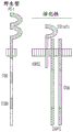


Classifications
-
- C—CHEMISTRY; METALLURGY
- C07—ORGANIC CHEMISTRY
- C07K—PEPTIDES
- C07K16/00—Immunoglobulins [IGs], e.g. monoclonal or polyclonal antibodies
- C07K16/18—Immunoglobulins [IGs], e.g. monoclonal or polyclonal antibodies against material from animals or humans
- C07K16/28—Immunoglobulins [IGs], e.g. monoclonal or polyclonal antibodies against material from animals or humans against receptors, cell surface antigens or cell surface determinants
-
- C—CHEMISTRY; METALLURGY
- C07—ORGANIC CHEMISTRY
- C07K—PEPTIDES
- C07K14/00—Peptides having more than 20 amino acids; Gastrins; Somatostatins; Melanotropins; Derivatives thereof
- C07K14/435—Peptides having more than 20 amino acids; Gastrins; Somatostatins; Melanotropins; Derivatives thereof from animals; from humans
- C07K14/705—Receptors; Cell surface antigens; Cell surface determinants
-
- A—HUMAN NECESSITIES
- A61—MEDICAL OR VETERINARY SCIENCE; HYGIENE
- A61K—PREPARATIONS FOR MEDICAL, DENTAL OR TOILETRY PURPOSES
- A61K35/00—Medicinal preparations containing materials or reaction products thereof with undetermined constitution
- A61K35/12—Materials from mammals; Compositions comprising non-specified tissues or cells; Compositions comprising non-embryonic stem cells; Genetically modified cells
- A61K35/14—Blood; Artificial blood
- A61K35/17—Lymphocytes; B-cells; T-cells; Natural killer cells; Interferon-activated or cytokine-activated lymphocytes
-
- A—HUMAN NECESSITIES
- A61—MEDICAL OR VETERINARY SCIENCE; HYGIENE
- A61K—PREPARATIONS FOR MEDICAL, DENTAL OR TOILETRY PURPOSES
- A61K39/00—Medicinal preparations containing antigens or antibodies
- A61K39/46—Cellular immunotherapy
- A61K39/461—Cellular immunotherapy characterised by the cell type used
- A61K39/4611—T-cells, e.g. tumor infiltrating lymphocytes [TIL], lymphokine-activated killer cells [LAK] or regulatory T cells [Treg]
-
- A—HUMAN NECESSITIES
- A61—MEDICAL OR VETERINARY SCIENCE; HYGIENE
- A61K—PREPARATIONS FOR MEDICAL, DENTAL OR TOILETRY PURPOSES
- A61K39/00—Medicinal preparations containing antigens or antibodies
- A61K39/46—Cellular immunotherapy
- A61K39/463—Cellular immunotherapy characterised by recombinant expression
- A61K39/4631—Chimeric Antigen Receptors [CAR]
-
- A—HUMAN NECESSITIES
- A61—MEDICAL OR VETERINARY SCIENCE; HYGIENE
- A61K—PREPARATIONS FOR MEDICAL, DENTAL OR TOILETRY PURPOSES
- A61K39/00—Medicinal preparations containing antigens or antibodies
- A61K39/46—Cellular immunotherapy
- A61K39/464—Cellular immunotherapy characterised by the antigen targeted or presented
- A61K39/4643—Vertebrate antigens
- A61K39/4644—Cancer antigens
- A61K39/464402—Receptors, cell surface antigens or cell surface determinants
- A61K39/464411—Immunoglobulin superfamily
- A61K39/464412—CD19 or B4
-
- A—HUMAN NECESSITIES
- A61—MEDICAL OR VETERINARY SCIENCE; HYGIENE
- A61K—PREPARATIONS FOR MEDICAL, DENTAL OR TOILETRY PURPOSES
- A61K39/00—Medicinal preparations containing antigens or antibodies
- A61K39/46—Cellular immunotherapy
- A61K39/464—Cellular immunotherapy characterised by the antigen targeted or presented
- A61K39/4643—Vertebrate antigens
- A61K39/4644—Cancer antigens
- A61K39/464466—Adhesion molecules, e.g. NRCAM, EpCAM or cadherins
- A61K39/464468—Mesothelin [MSLN]
-
- A—HUMAN NECESSITIES
- A61—MEDICAL OR VETERINARY SCIENCE; HYGIENE
- A61P—SPECIFIC THERAPEUTIC ACTIVITY OF CHEMICAL COMPOUNDS OR MEDICINAL PREPARATIONS
- A61P35/00—Antineoplastic agents
-
- A—HUMAN NECESSITIES
- A61—MEDICAL OR VETERINARY SCIENCE; HYGIENE
- A61P—SPECIFIC THERAPEUTIC ACTIVITY OF CHEMICAL COMPOUNDS OR MEDICINAL PREPARATIONS
- A61P37/00—Drugs for immunological or allergic disorders
- A61P37/02—Immunomodulators
- A61P37/04—Immunostimulants
-
- A—HUMAN NECESSITIES
- A61—MEDICAL OR VETERINARY SCIENCE; HYGIENE
- A61P—SPECIFIC THERAPEUTIC ACTIVITY OF CHEMICAL COMPOUNDS OR MEDICINAL PREPARATIONS
- A61P43/00—Drugs for specific purposes, not provided for in groups A61P1/00-A61P41/00
-
- C—CHEMISTRY; METALLURGY
- C07—ORGANIC CHEMISTRY
- C07K—PEPTIDES
- C07K14/00—Peptides having more than 20 amino acids; Gastrins; Somatostatins; Melanotropins; Derivatives thereof
- C07K14/435—Peptides having more than 20 amino acids; Gastrins; Somatostatins; Melanotropins; Derivatives thereof from animals; from humans
- C07K14/705—Receptors; Cell surface antigens; Cell surface determinants
- C07K14/70503—Immunoglobulin superfamily
- C07K14/7051—T-cell receptor (TcR)-CD3 complex
-
- C—CHEMISTRY; METALLURGY
- C07—ORGANIC CHEMISTRY
- C07K—PEPTIDES
- C07K14/00—Peptides having more than 20 amino acids; Gastrins; Somatostatins; Melanotropins; Derivatives thereof
- C07K14/435—Peptides having more than 20 amino acids; Gastrins; Somatostatins; Melanotropins; Derivatives thereof from animals; from humans
- C07K14/705—Receptors; Cell surface antigens; Cell surface determinants
- C07K14/70503—Immunoglobulin superfamily
- C07K14/70535—Fc-receptors, e.g. CD16, CD32, CD64 (CD2314/705F)
-
- C—CHEMISTRY; METALLURGY
- C07—ORGANIC CHEMISTRY
- C07K—PEPTIDES
- C07K14/00—Peptides having more than 20 amino acids; Gastrins; Somatostatins; Melanotropins; Derivatives thereof
- C07K14/435—Peptides having more than 20 amino acids; Gastrins; Somatostatins; Melanotropins; Derivatives thereof from animals; from humans
- C07K14/705—Receptors; Cell surface antigens; Cell surface determinants
- C07K14/70503—Immunoglobulin superfamily
- C07K14/70539—MHC-molecules, e.g. HLA-molecules
-
- C—CHEMISTRY; METALLURGY
- C07—ORGANIC CHEMISTRY
- C07K—PEPTIDES
- C07K14/00—Peptides having more than 20 amino acids; Gastrins; Somatostatins; Melanotropins; Derivatives thereof
- C07K14/435—Peptides having more than 20 amino acids; Gastrins; Somatostatins; Melanotropins; Derivatives thereof from animals; from humans
- C07K14/705—Receptors; Cell surface antigens; Cell surface determinants
- C07K14/7056—Lectin superfamily, e.g. CD23, CD72
-
- C—CHEMISTRY; METALLURGY
- C07—ORGANIC CHEMISTRY
- C07K—PEPTIDES
- C07K14/00—Peptides having more than 20 amino acids; Gastrins; Somatostatins; Melanotropins; Derivatives thereof
- C07K14/435—Peptides having more than 20 amino acids; Gastrins; Somatostatins; Melanotropins; Derivatives thereof from animals; from humans
- C07K14/705—Receptors; Cell surface antigens; Cell surface determinants
- C07K14/70596—Molecules with a "CD"-designation not provided for elsewhere
-
- C—CHEMISTRY; METALLURGY
- C07—ORGANIC CHEMISTRY
- C07K—PEPTIDES
- C07K16/00—Immunoglobulins [IGs], e.g. monoclonal or polyclonal antibodies
- C07K16/18—Immunoglobulins [IGs], e.g. monoclonal or polyclonal antibodies against material from animals or humans
- C07K16/28—Immunoglobulins [IGs], e.g. monoclonal or polyclonal antibodies against material from animals or humans against receptors, cell surface antigens or cell surface determinants
- C07K16/30—Immunoglobulins [IGs], e.g. monoclonal or polyclonal antibodies against material from animals or humans against receptors, cell surface antigens or cell surface determinants from tumour cells
-
- C—CHEMISTRY; METALLURGY
- C12—BIOCHEMISTRY; BEER; SPIRITS; WINE; VINEGAR; MICROBIOLOGY; ENZYMOLOGY; MUTATION OR GENETIC ENGINEERING
- C12N—MICROORGANISMS OR ENZYMES; COMPOSITIONS THEREOF; PROPAGATING, PRESERVING, OR MAINTAINING MICROORGANISMS; MUTATION OR GENETIC ENGINEERING; CULTURE MEDIA
- C12N5/00—Undifferentiated human, animal or plant cells, e.g. cell lines; Tissues; Cultivation or maintenance thereof; Culture media therefor
- C12N5/06—Animal cells or tissues; Human cells or tissues
- C12N5/0602—Vertebrate cells
- C12N5/0634—Cells from the blood or the immune system
- C12N5/0636—T lymphocytes
-
- A—HUMAN NECESSITIES
- A61—MEDICAL OR VETERINARY SCIENCE; HYGIENE
- A61K—PREPARATIONS FOR MEDICAL, DENTAL OR TOILETRY PURPOSES
- A61K39/00—Medicinal preparations containing antigens or antibodies
- A61K2039/51—Medicinal preparations containing antigens or antibodies comprising whole cells, viruses or DNA/RNA
- A61K2039/515—Animal cells
- A61K2039/5156—Animal cells expressing foreign proteins
-
- A—HUMAN NECESSITIES
- A61—MEDICAL OR VETERINARY SCIENCE; HYGIENE
- A61K—PREPARATIONS FOR MEDICAL, DENTAL OR TOILETRY PURPOSES
- A61K39/00—Medicinal preparations containing antigens or antibodies
- A61K2039/51—Medicinal preparations containing antigens or antibodies comprising whole cells, viruses or DNA/RNA
- A61K2039/515—Animal cells
- A61K2039/5158—Antigen-pulsed cells, e.g. T-cells
-
- A—HUMAN NECESSITIES
- A61—MEDICAL OR VETERINARY SCIENCE; HYGIENE
- A61K—PREPARATIONS FOR MEDICAL, DENTAL OR TOILETRY PURPOSES
- A61K2239/00—Indexing codes associated with cellular immunotherapy of group A61K39/46
- A61K2239/27—Indexing codes associated with cellular immunotherapy of group A61K39/46 characterized by targeting or presenting multiple antigens
- A61K2239/28—Expressing multiple CARs, TCRs or antigens
-
- A—HUMAN NECESSITIES
- A61—MEDICAL OR VETERINARY SCIENCE; HYGIENE
- A61K—PREPARATIONS FOR MEDICAL, DENTAL OR TOILETRY PURPOSES
- A61K2239/00—Indexing codes associated with cellular immunotherapy of group A61K39/46
- A61K2239/31—Indexing codes associated with cellular immunotherapy of group A61K39/46 characterized by the route of administration
-
- A—HUMAN NECESSITIES
- A61—MEDICAL OR VETERINARY SCIENCE; HYGIENE
- A61K—PREPARATIONS FOR MEDICAL, DENTAL OR TOILETRY PURPOSES
- A61K2239/00—Indexing codes associated with cellular immunotherapy of group A61K39/46
- A61K2239/38—Indexing codes associated with cellular immunotherapy of group A61K39/46 characterised by the dose, timing or administration schedule
-
- A—HUMAN NECESSITIES
- A61—MEDICAL OR VETERINARY SCIENCE; HYGIENE
- A61K—PREPARATIONS FOR MEDICAL, DENTAL OR TOILETRY PURPOSES
- A61K2239/00—Indexing codes associated with cellular immunotherapy of group A61K39/46
- A61K2239/46—Indexing codes associated with cellular immunotherapy of group A61K39/46 characterised by the cancer treated
- A61K2239/48—Blood cells, e.g. leukemia or lymphoma
-
- C—CHEMISTRY; METALLURGY
- C07—ORGANIC CHEMISTRY
- C07K—PEPTIDES
- C07K2317/00—Immunoglobulins specific features
- C07K2317/20—Immunoglobulins specific features characterized by taxonomic origin
- C07K2317/21—Immunoglobulins specific features characterized by taxonomic origin from primates, e.g. man
-
- C—CHEMISTRY; METALLURGY
- C07—ORGANIC CHEMISTRY
- C07K—PEPTIDES
- C07K2317/00—Immunoglobulins specific features
- C07K2317/50—Immunoglobulins specific features characterized by immunoglobulin fragments
- C07K2317/56—Immunoglobulins specific features characterized by immunoglobulin fragments variable (Fv) region, i.e. VH and/or VL
- C07K2317/565—Complementarity determining region [CDR]
-
- C—CHEMISTRY; METALLURGY
- C07—ORGANIC CHEMISTRY
- C07K—PEPTIDES
- C07K2317/00—Immunoglobulins specific features
- C07K2317/60—Immunoglobulins specific features characterized by non-natural combinations of immunoglobulin fragments
- C07K2317/62—Immunoglobulins specific features characterized by non-natural combinations of immunoglobulin fragments comprising only variable region components
- C07K2317/622—Single chain antibody (scFv)
-
- C—CHEMISTRY; METALLURGY
- C07—ORGANIC CHEMISTRY
- C07K—PEPTIDES
- C07K2317/00—Immunoglobulins specific features
- C07K2317/70—Immunoglobulins specific features characterized by effect upon binding to a cell or to an antigen
- C07K2317/74—Inducing cell proliferation
-
- C—CHEMISTRY; METALLURGY
- C07—ORGANIC CHEMISTRY
- C07K—PEPTIDES
- C07K2319/00—Fusion polypeptide
- C07K2319/01—Fusion polypeptide containing a localisation/targetting motif
- C07K2319/02—Fusion polypeptide containing a localisation/targetting motif containing a signal sequence
-
- C—CHEMISTRY; METALLURGY
- C07—ORGANIC CHEMISTRY
- C07K—PEPTIDES
- C07K2319/00—Fusion polypeptide
- C07K2319/01—Fusion polypeptide containing a localisation/targetting motif
- C07K2319/03—Fusion polypeptide containing a localisation/targetting motif containing a transmembrane segment
-
- C—CHEMISTRY; METALLURGY
- C07—ORGANIC CHEMISTRY
- C07K—PEPTIDES
- C07K2319/00—Fusion polypeptide
- C07K2319/70—Fusion polypeptide containing domain for protein-protein interaction
-
- C—CHEMISTRY; METALLURGY
- C07—ORGANIC CHEMISTRY
- C07K—PEPTIDES
- C07K2319/00—Fusion polypeptide
- C07K2319/70—Fusion polypeptide containing domain for protein-protein interaction
- C07K2319/74—Fusion polypeptide containing domain for protein-protein interaction containing a fusion for binding to a cell surface receptor
-
- C—CHEMISTRY; METALLURGY
- C12—BIOCHEMISTRY; BEER; SPIRITS; WINE; VINEGAR; MICROBIOLOGY; ENZYMOLOGY; MUTATION OR GENETIC ENGINEERING
- C12N—MICROORGANISMS OR ENZYMES; COMPOSITIONS THEREOF; PROPAGATING, PRESERVING, OR MAINTAINING MICROORGANISMS; MUTATION OR GENETIC ENGINEERING; CULTURE MEDIA
- C12N2510/00—Genetically modified cells
Abstract
The present invention provides compositions and methods for modulating the specificity and activity of immune effector cells for immunotherapy. In one embodiment, the invention provides a Chimeric Antigen Receptor (CAR), wherein the CAR is referred to as an "NKR-CAR," which is a CAR design comprising a receptor component naturally occurring on Natural Killer (NK) cells. In one embodiment, NK receptors include, but are not limited to, the naturally occurring activating and inhibitory receptors of NK cells known as killer cell immunoglobulin-like receptors (KIR).
Description
RELATED APPLICATIONS
This application claims priority to PCT application number PCT/CN2014/086694 filed on 9/17 of 2014 and PCT application number PCT/CN2014/090578 filed on 11/7 of 2014. The entire contents of each of these applications are incorporated herein by reference.
Background
Using gene transfer techniques, T cells can be genetically modified to stably express on their surface an antibody binding domain that confers T cell specificity that is independent of the constraints imposed by the Major Histocompatibility Complex (MHC). Chimeric Antigen Receptors (CARs) represent synthetic proteins expressed on T cells (CART cells) that fuse antigen-recognition fragments (e.g., scFv or single chain variable fragment) of antibodies to the intracellular domain of the CD 3-zeta chain. Upon interaction with a target cell expressing a cognate antigen of the scFv, the CAR expressed on the T cell can trigger T cell activation leading to target cell killing (also referred to as target cell lysis). These receptors are also capable of producing proliferation when combined with additional costimulatory signals, such as the intracellular domain of CD137 or CD 28. However, unlike normal T Cell Receptor (TCR) responses, some of this proliferation appears to be antigen independent (Milone et al, 2009, Mol Ther 17(8): 1453-64). Artificial receptors do not fully reproduce the intracellular signal transduction resulting from native TCR binding to antigenic peptides complexed with MHC molecules (Brocker,2000, Blood96(5): 1999) -2001). Defects in signaling can limit long-term survival of CART cells when adoptively transferred in the absence of high levels of cytokines such as IL-2 (Lo et al, 2010, Clin Cancer Res 16(10): 2769-80). They also have altered regulation that may be beneficial in some anticancer applications (Loskog et al, 2006, Leukemia 20(10):1819-28), but these regulatory deficiencies also lead to potential challenges in controlling their "off-target" activity on normal tissues that also express antigen, even at very low levels. These "off-target" effects are serious limitations on CAR-based therapies and have resulted in possible death during early phase I assessment of CAR-modified T cells (Morgan et al, 2010, Mol Ther 18(4): 843-51).
Thus, there is a need in the art for alternative methods for constructing CARs that overcome the limitations of current CAR-based therapeutics. The present invention addresses this unmet need in the art.
SUMMARY
In a first aspect, the invention features a purified or non-naturally occurring natural killer cell immune function receptor-chimeric antigen receptor (NKR-CAR) polypeptide comprising one, two, or all extracellular antigen binding domains, a transmembrane domain, e.g., an NKR transmembrane domain, and a cytoplasmic domain, e.g., an NKR cytoplasmic domain. In one embodiment, the NKR-CAR polypeptide comprises an extracellular antigen-binding domain, and one or both of a transmembrane domain, e.g., an NKR transmembrane domain; or a cytoplasmic domain, such as the NKR cytoplasmic domain. In one embodiment, the NKR-CAR polypeptide comprises an extracellular antigen-binding domain; a transmembrane domain and an NKR cytoplasmic domain. In one embodiment, the NKR-CAR polypeptide comprises an extracellular antigen-binding domain, an NKR transmembrane domain and a cytoplasmic domain. In one embodiment, the NKR-CAR polypeptide comprises an extracellular antigen-binding domain, an NKR transmembrane domain and a cytoplasmic domain.
In one embodiment, the NKR-CAR polypeptide comprises a KIR-CAR, e.g. an actKIR-CAR or an inhKIR-CAR, an NCR-CAR, e.g. an actNCR-CAR, a SLAMF-CAR, e.g. an inhSLAMF-CAR, an FcR-CAR, e.g. a CD16-CAR, e.g. an actCD16-CAR or a CD64-CAR, e.g. an actCD64-CAR, or a Ly49-CAR, e.g. an actLy49-CAR or an inhLy 49-CAR.
In one embodiment, the NKR-CAR polypeptide comprises a killer cell immunoglobulin-like receptor chimeric antigen receptor (KIR-CAR), wherein the KIR-CAR comprises one or both of a transmembrane domain from a KIR (KIR transmembrane domain) or a cytoplasmic domain comprising a functional signaling domain from a KIR (KIR cytoplasmic domain). In one embodiment, the KIR transmembrane domain comprises the transmembrane domains of a protein selected from the group consisting of KIR2DS2, KIR2DL3, KIR2DL1, KIR2DL2, KIR2DL4, KIR2DL5A, KIR2DL5B, KIR2DS1, KIR2DS3, KIR2DS4, KIR2DS5, KIR3DL1, KIR3DS1, KIR3DL2, KIR3DL3, KIR2DP1, and KIR3DP 1. In one embodiment, the KIR cytoplasmic domain comprises a functional signaling domain of a protein selected from the group consisting of KIR2DS2, KIR2DL3, KIR2DL1, KIR2DL2, KIR2DL4, KIR2DL5A, KIR2DL5B, KIR2DS1, KIR2DS3, KIR2DS4, KIR2DS5, KIR3DL1, KIR3DS1, KIR3DL2, KIR3DL3, KIR2DP1, and KIR3DP 1. In one embodiment, the KIR-CAR further comprises one or more of a KIR D0 domain, a KIR D1 domain, and/or a KIR D2 domain.
In one embodiment, the NKR CAR polypeptide comprises a native cytotoxic receptor chimeric antigen receptor (NCR-CAR), wherein the NCR-CAR comprises one or both of a transmembrane domain from an NCR (NCR transmembrane domain) or a cytoplasmic domain comprising a functional signaling domain from an NCR (NCR cytoplasmic domain). In one embodiment, the NCR transmembrane domain comprises the transmembrane domain of a protein selected from the group consisting of NKp46, NKp30 and NKp 44. In one embodiment, wherein the NCR cytoplasmic domain comprises a functional signaling domain of a protein selected from the group consisting of NKp46, NKp30, and NKp 44.
In one embodiment, the NKR CAR polypeptide comprises a signaling lymphocyte activation molecule family chimeric antigen receptor (SLAMF-CAR), wherein the SLAMF-CAR comprises a transmembrane domain from SLAMF (SLAMF transmembrane domain) or a cytoplasmic domain comprising a functional signaling domain from SLAMF (SLAMF cytoplasmic domain). In one embodiment, the SLAMF transmembrane domain comprises the transmembrane domain of a protein selected from the group consisting of SLAM, CD48, CD229,2B4, CD84, NTB-A, CRACC, BLAME, and CD 2F-10. In one embodiment, the SLAMF cytoplasmic domain comprises a functional signaling domain of a protein selected from the group consisting of SLAM, CD48, CD229,2B4, CD84, NTB-a, CRACC, BLAME, and CD 2F-10.
In one embodiment, the NKR CAR polypeptide comprises an Fc receptor chimeric antigen receptor (FcR-CAR), wherein the FcR-CAR comprises one or both of a transmembrane domain from an FcR selected from CD16 or CD64 or a cytoplasmic domain from a functional signaling domain of an FcR selected from CD16 or CD 64.
In one embodiment, the NKR CAR polypeptide comprises a Ly49 receptor chimeric antigen receptor (Ly49-CAR), wherein the Ly49-CAR comprises one or both of a transmembrane domain from Ly49 (Ly49 transmembrane domain) or a cytoplasmic domain comprising a functional signaling domain from Ly49 (Ly49 cytoplasmic domain). In one embodiment, the Ly49 transmembrane domain comprises a transmembrane domain of a protein selected from Ly49A, Ly49C, Ly49H, and Ly 49D. In one embodiment, the Ly49 cytoplasmic domain comprises a functional signaling domain of a protein selected from Ly49A, Ly49C, Ly49H, and Ly 49D.
In one embodiment, the transmembrane domain of the NKR-CAR polypeptide is an NKR transmembrane domain, wherein the NKR transmembrane domain comprises a transmembrane domain of a protein selected from the group consisting of KIR2DS2, KIR2DL3, NKp46, KIR, NCR, SLAMF, FcR, and Ly 49. In one embodiment, the transmembrane domain comprises i) the amino acid sequence of SEQ ID NO 357,358 or 359; ii) an amino acid sequence comprising at least one, two or three modifications but NO more than 5 modifications of the amino acid sequence of SEQ ID NO 357,358 or 359; or iii) an amino acid sequence having 95-99% sequence identity to SEQ ID NO:357,358 or 359.
In one embodiment, the transmembrane domain encoded by the NKR-CAR polypeptide is a transmembrane domain, e.g., a transmembrane domain of a TCAR as described herein, a transmembrane domain comprising a protein selected from the group consisting of TCR ζ, FcR γ, FcR β, CD γ, CD δ, CD ε, CD, CD, CD79, CD79, CD278 (also referred to as "ICOS"), Fc ε RI, CD66, DAP, DAP, the α, β, or ζ chain of a T cell receptor, an MHC class I molecule, a TNF receptor protein, an immunoglobulin-like protein, a cytokine receptor, an integrin, an activating NK cell receptor, BTLA, Toll ligand receptor, CD, CD ε, CD, CD, CD, CD, CD (CD α or CD β), CD, CD, CD, CD, CD, CD134, CD137, CD154, OX, CD137, CD, CD, CD137, CD CDS, ICAM-1, LFA-1(CD 11/BB), BB 4-BB), B-H, CDS, ICAM-1, ICOS (CD278), GITR, BAFFR, LIGHT, HVEM (LIGHT TR), KIRDS, SLAMF, NKp (KLRF), NKp, NKp, NKp, CD, CD, CD α, CD β, IL2 β, IL2 γ, IL7 α, ITGA, VLA, CD49, ITGA, IA, CD49, ITGA, VLA-6, CD49, ITGAD, CD11, ITGAE, CD103, ITGAL, CD11, LFA-1, ITGAM, CD11, ITGAX, CD11, ITGB, CD, ITGB, CD, CD, TAA-1, ITGB, NKG2, NKG2, TNFR, TRANCE/RANKL, DNAM (CD226), SLAMF (CD244, 2B), CD, CD (TaYLE), ACAM, TACM, SLSP 160, SLAG-229, SLAG-CD-150, CD-14, CD-1, ITGAX, CD11, CD-1, ITGAX, CD-1, ITGAX, NKG-CD-2, CD-SLAMG, CD-SLAMB, CD-.
In one embodiment, the cytoplasmic domain of the NKR-CAR polypeptide is an NKR cytoplasmic domain comprising one or more functional signaling domains of a protein selected from the group consisting of KIR2DS2, KIR2DL3, NKp46, KIR, NCR, SLAMF, FcR, and Ly 49. In one embodiment, the cytoplasmic domain comprises i) the amino acid sequence of SEQ ID NO 360,361 or 362; ii) an amino acid sequence comprising at least one, two or three modifications but not more than 20, 10 or 5 modifications of the amino acid sequence of SEQ ID NO 360,361 or 362; or iii) an amino acid sequence having 95-99% sequence identity to SEQ ID NO 360,361.
In one embodiment, the cytoplasmic domain of the NKR-CAR polypeptide comprises an intracellular signaling domain or adaptor molecule, such as DAP 12. In one embodiment, the encoded cytoplasmic domain of the NKR-CAR polypeptide comprises an adaptor molecule, such as DAP12 or fcepsilonr γ. In one embodiment, the cytoplasmic domain comprises i) the encoded adaptor molecule comprises an amino acid sequence of amino acids 1-113 of SEQ ID NO. 333 or amino acids 1-86 of SEQ ID NO. 335; ii) an amino acid sequence having at least one, two or three modifications but not more than 20, 10 or 5 modifications to amino acids 1-113 of SEQ ID NO. 333 or amino acids 1-86 of SEQ ID NO. 335; or iii) an amino acid sequence having 95-99% identity to amino acids 1-113 of SEQ ID NO. 333 or amino acids 1-86 of SEQ ID NO. 335. In one embodiment, the NKR-CAR comprises an antigen binding domain as described herein, a CD8 transmembrane domain and a cytoplasmic domain comprising DAP 12.
In one embodiment, the cytoplasmic domain of the NKR-CAR polypeptide is, e.g., the cytoplasmic domain of a TCAR as described herein, which comprises one or more functional signaling domains of a protein selected from the group consisting of TCR ζ, FcRy, FcRβ, CD γ, CD δ, CD ε, CD, CD79, CD79, CD278 (also referred to as "ICOS"), Fε RI, CD66, DAP, DAP, the α, β, or ζ chain of a T-cell receptor, an MHC class I molecule, a TNF receptor protein, an immunoglobulin-like protein, a cytokine receptor, an integrin, an activating NK cell receptor, BTLA, Toll ligand receptor, CD, CD ε, CD, CD, CD, CD, CD (CD α or CD β), CD, CD, CD, CD, CD, CD, CD, CD, CD137, CD154, OX, CD, CD, CD137, CD, CD1, ICAM-1, LFA-1(CD 11/4-BB), B-H, CDS, ICAM-1, ICOS (CD278), GITR, BAFFR, LIGHT, HVEM (LIGHT TR), KIRDS, SLAMF, NKp (KLRF), NKp, NKp, NKp, CD, CD, CD α, CD β, IL2 β, IL2 γ, IL7 α, ITGA, VLA, CD49, ITGA, IA, CD49, ITGA, VLA-6, CD49, ITGAD, CD11, ITGAE, CD103, ITGAL, CD11, LFA-1, ITGAM, CD11, ITGAX, CD11, ITGB, CD, ITGB, CD, CD, TAA-1, ITGB, NKG2, NKG2, TNFR, TRANCE/RANKL, DNAM (CD226), SLAMF (CD244, 2B), CD, CD (TaYLE), ACAM, TACM, SLSP 160, SLAG-229, SLAG-CD-150, CD-14, CD-1, ITGAX, CD11, CD-1, ITGAX, CD-1, ITGAX, NKG-CD-2, CD-SLAMG, CD-SLAMB, CD-.
In one embodiment, the NKR-CAR polypeptide further comprises a leader sequence or a signal sequence, e.g.comprising the amino acid sequence of SEQ ID NO. 1. In one embodiment, the NKR-CAR comprises a transmembrane domain and an extracellular antigen-binding domain, and further comprises a hinge domain disposed between the transmembrane domain and the extracellular antigen-binding domain. In one embodiment, the hinge domain is selected from the group consisting of a GS hinge, a CD8 hinge, an IgG4 hinge, an IgD hinge, a KIR2DS2 hinge, a KIR hinge, an NCR hinge, a SLAMF hinge, a CD16 hinge, a CD64 hinge, and a LY49 hinge. In one embodiment, the hinge domain comprises i) the amino acid sequence of SEQ ID NO 5,2,3 or 4; ii) an amino acid sequence comprising at least one, two or three modifications but not more than 5 modifications of the amino acid sequence of SEQ ID NO 5,2,3 or 4; or iii) an amino acid sequence having 95-99% identity to the amino acid sequence of SEQ ID NO 5,2,3 or 4.
In one embodiment, the transmembrane domain and cytoplasmic domain together comprise i) the amino acid sequence of amino acids 413-487 of SEQ ID NO:333 or amino acids 386-454 of SEQ ID NO:335 or the amino acid sequence of SEQ ID NO: 371; ii) an amino acid sequence having at least one, two or three modifications but not more than 30,20 or 10 modifications to amino acids 413-487 of SEQ ID NO:333 or amino acids 386-454 or SEQ ID NO:371 of SEQ ID NO: 335; or iii) an amino acid sequence having 95-99% identity with amino acids 413-487 of SEQ ID NO:333 or amino acids 386-454 of SEQ ID NO:335 or SEQ ID NO: 371.
In any of the preceding embodiments, the NKR-CAR is an activating NKR-CAR and the extracellular antigen-binding domain is an antigen-binding domain described herein, e.g., in table 4. In any of the preceding embodiments, the extracellular antigen-binding domain is a non-murine antigen-binding domain that binds mesothelin. In one embodiment, the extracellular non-murine antigen-binding domain that binds mesothelin comprises a human or humanized antigen-binding domain that binds mesothelin. In one embodiment, human or humanized antigen binding domains that bind mesothelin are provided in table 4.
In one embodiment, the mesothelin-binding human antigen-binding domain comprises heavy chain complementarity determining region 1(HC CDR1), heavy chain complementarity determining region 2(HC CDR2) and heavy chain complementarity determining region 3(HC CDR3) of any of the human anti-mesothelin heavy chain amino acid sequences listed in Table 4; and/or light chain complementarity determining region 1(LC CDR1), light chain complementarity determining region 2(LC CDR2), and light chain complementarity determining region 3(LC CDR3) of any of the human anti-mesothelin light chain amino acid sequences listed in table 4. In one embodiment, the mesothelin-binding human antigen-binding domain comprises a heavy chain variable region comprising i) the amino acid sequence of any of the human anti-mesothelin heavy chain variable regions listed in table 4; ii) an amino acid sequence having at least one, two or three modifications but no more than 30,20 or 10 modifications to the amino acid sequence of any of the human anti-mesothelin heavy chain variable regions listed in table 4; or iii) an amino acid sequence 95-99% identical to the amino acid sequence of any of the human anti-mesothelin heavy chain variable regions listed in Table 4; and/or a light chain variable region comprising i) the amino acid sequence of any of the human anti-mesothelin light chain variable regions listed in table 4; ii) an amino acid sequence having at least one, two or three modifications but no more than 30,20 or 10 modifications to the amino acid sequence of any of the human anti-mesothelin light chain variable regions listed in table 4; or iii) an amino acid sequence 95-99% identical to the amino acid sequence of any of the human anti-mesothelin light chain variable regions listed in Table 4. In one embodiment, the mesothelin-binding human antigen-binding domain comprises i) the amino acid sequence of any one of SEQ ID NO 230-253; ii) an amino acid sequence having at least one, two or three modifications but not more than 30,20 or 10 modifications to any of SEQ ID NO 230-253; or iii) an amino acid sequence having 95-99% identity to any one of SEQ ID NO 230-253. In such embodiments, the NKR-CAR further comprises, e.g., transmembrane and cytoplasmic domains, which together comprise the amino acid sequence of SEQ ID No. 371, an amino acid sequence with at least one, two or three modifications but NO more than 10 or 5 modifications to SEQ ID No. 371, or an amino acid sequence with at least 95-99% sequence identity to SEQ ID No. 371.
In a first aspect, the invention features a purified or non-naturally occurring nucleic acid molecule encoding a natural killer cell immune function receptor-chimeric antigen receptor (NKR-CAR) polypeptide described herein, e.g., comprising one, two, or all of an extracellular antigen-binding domain, a transmembrane domain (e.g., NKR transmembrane domain), and a cytoplasmic domain (e.g., NKR cytoplasmic domain). In one embodiment, the NKR-CAR encoding nucleic acid molecule comprises an extracellular antigen-binding domain and one or both of a transmembrane domain (e.g., NKR transmembrane domain); or a cytoplasmic domain (e.g., NKR cytoplasmic domain). In one embodiment, the NKR-CAR encoding nucleic acid molecule comprises an extracellular antigen-binding domain; a transmembrane domain and an NKR cytoplasmic domain. In one embodiment, the NKR-CAR encoding nucleic acid molecule comprises an extracellular antigen-binding domain, an NKR transmembrane domain and a cytoplasmic domain. In one embodiment, the NKR-CAR encoding nucleic acid molecule comprises an extracellular antigen-binding domain, an NKR transmembrane domain and a cytoplasmic domain.
In one embodiment, the nucleic acid molecule encodes an NKR-CAR comprising a KIR-CAR, e.g. an actKIR-CAR or an inhKIR-CAR, an NCR-CAR, e.g. an actNCR-CAR, a SLAMF-CAR, e.g. an inhSLAMF-CAR, an FcR-CAR, e.g. a CD16-CAR, e.g. an actCD16-CAR or a CD64-CAR, e.g. an actCD64-CAR or a Ly49-CAR, e.g. an actLy49-CAR or an inhLy 49-CAR.
In one embodiment, the encoded KIR-CAR comprises one or both of a transmembrane domain from the KIR (KIR transmembrane domain) or a cytoplasmic domain comprising a functional signaling domain from the KIR (KIR cytoplasmic domain). In one embodiment, the KIR transmembrane domain comprises the transmembrane domains of a protein selected from the group consisting of KIR2DS2, KIR2DL3, KIR2DL1, KIR2DL2, KIR2DL4, KIR2DL5A, KIR2DL5B, KIR2DS1, KIR2DS3, KIR2DS4, KIR2DS5, KIR3DL1, KIR3DS1, KIR3DL2, KIR3DL3, KIR2DP1, and KIR3DP 1. In one embodiment, the KIR cytoplasmic domain comprises a functional signaling domain of a protein selected from the group consisting of KIR2DS2, KIR2DL3, KIR2DL1, KIR2DL2, KIR2DL4, KIR2DL5A, KIR2DL5B, KIR2DS1, KIR2DS3, KIR2DS4, KIR2DS5, KIR3DL1, KIR3DS1, KIR3DL2, KIR3DL3, KIR2DP1, and KIR3DP 1. In one embodiment, the KIR-CAR further comprises one or more of a KIR D0 domain, a KIR D1 domain, and/or a KIR D2 domain.
In one embodiment, the encoded NCR-CAR comprises one or both of a transmembrane domain from an NCR (NCR transmembrane domain) or a cytoplasmic domain comprising a functional signaling domain from an NCR (NCR cytoplasmic domain). In one embodiment, the NCR transmembrane domain comprises the transmembrane domain of a protein selected from the group consisting of NKp46, NKp30 and NKp 44. In one embodiment, wherein the NCR cytoplasmic domain comprises a functional signaling domain of a protein selected from the group consisting of NKp46, NKp30, and NKp 44.
In one embodiment, the encoded SLAMF-CAR comprises one or both of a transmembrane domain from SLAMF (SLAMF transmembrane domain) or a cytoplasmic domain comprising a functional signaling domain from SLAMF (SLAMF cytoplasmic domain). In one embodiment, the SLAMF transmembrane domain comprises the transmembrane domain of a protein selected from the group consisting of SLAM, CD48, CD229, 2B4, CD84, NTB-A, CRACC, BLAME, and CD 2F-10. In one embodiment, the SLAMF cytoplasmic domain comprises a functional signaling domain of a protein selected from the group consisting of SLAM, CD48, CD229, 2B4, CD84, NTB-a, CRACC, BLAME, and CD 2F-10.
In one embodiment, the encoded FcR-CAR comprises one or both of a transmembrane domain from an FcR selected from CD16 or CD64 or a cytoplasmic domain comprising a functional signaling domain from an FcR selected from CD16 or CD 64.
In one embodiment, the encoded Ly49-CAR comprises one or both of a transmembrane domain from Ly49 (Ly49 transmembrane domain) or a cytoplasmic domain comprising a functional signaling domain from Ly49 (Ly49 cytoplasmic domain). In one embodiment, the Ly49 transmembrane domain comprises a transmembrane domain of a protein selected from Ly49A, Ly49C, Ly49H, and Ly 49D. In one embodiment, the Ly49 cytoplasmic domain comprises a functional signaling domain of a protein selected from Ly49A, Ly49C, Ly49H, and Ly 49D.
In one embodiment, the encoded transmembrane domain of the NKR-CAR is an NKR transmembrane domain, wherein the NKR transmembrane domain comprises a transmembrane domain of a protein selected from the group consisting of KIR2DS2, KIR2DL3, NKp46, KIR, NCR, SLAMF, FcR, and Ly 49. In one embodiment, the transmembrane domain encoding NKR-CAR comprises the amino acid sequence of SEQ ID NO 357,358 or 359; 357,358 or 359, and not more than 5 modifications of the amino acid sequence of SEQ ID NO; or an amino acid sequence having 95-99% sequence identity to SEQ ID NO 357,358 or 359. In one embodiment, the NKR-CAR-encoding nucleic acid molecule comprises a nucleic acid sequence comprising nucleotide 803-875 of SEQ ID NO:347 or a nucleic acid sequence having 95-99% sequence identity thereto, which encodes a transmembrane domain.
In one embodiment, the transmembrane domain encoded by the NKR-CAR polypeptide is a transmembrane domain of a TCAR, e.g., as described herein, comprising the transmembrane domains of a protein selected from the group consisting of TCR ζ, FcR γ, FcR β, CD γ, CD δ, CD ε, CD, CD, CD79, CD79, CD278 (also referred to as "ICOS"), Fcε RI, CD66, DAP, DAP, the α, β or ζ chain of a T-cell receptor, an MHC class I molecule, a TNF receptor protein, an immunoglobulin-like protein, a cytokine receptor, an integrin, an activating NK cell receptor, BTLA, Toll ligand receptor, CD, CD ε, CD, CD, CD, CD, CD (CD α or CD β), CD, CD, CD, CD, CD, CD134, CD137, CD154, OX, CD, CD, CD, CD, CD, CD, CD, CD, CDS, ICAM-1, LFA-1(CD 11/CD), 4-1B, BB), 4-BB, H-H, CDS, ICAM-1, ICOS (CD278), GITR, BAFFR, LIGHT, HVEM (LIGHT TR), KIRDS, SLAMF, NKp (KLRF), NKp, NKp, CD, CD, CD α, CD β, IL2 β, IL2 γ, IL7 α, ITGA, VLA, CD49, ITGA, IA, CD49, ITGA, VLA-6, CD49, ITGAD, CD11, ITGAE, CD103, ITGAL, CD11, LFA-1, ITGAM, CD11, ITGAX, CD11, ITGB, CD, LFA-1, ITGB, NKG2, NKG2, TNFR, TRANCE/RANGE, DNAM (CD226), SLAMF (CD244, 2B), CD, CD (Talle), CEM, CD TAM, LyACAT (CD229), SLBY (SLP), SLAG-160 (PAG), SLAG-150, SLAG-19, SLAF-CD-14, SLAM, CD-2, CD-7, CD-1, CD11, CD-TAG, CD-6, CD11, CD-TAG, CD-6, CD-1, CD-2, CD-CD, CD-CD, CD-CD, CD-CD, CD-.
In one embodiment, the encoded cytoplasmic domain of the NKR-CAR is an NKR cytoplasmic domain, wherein the NKR cytoplasmic domain comprises one or more functional signaling domains of a protein selected from the group consisting of KIR2DS2, KIR2DL3, NKp46, KIR, NCR, SLAMF, FcR, and Ly 49. In one embodiment, the encoded cytoplasmic domain comprises i) the amino acid sequence of SEQ ID NO 360,361 or 362; ii) an amino acid sequence comprising at least one, two or three modifications but not more than 20, 10 or 5 modifications of the amino acid sequence of SEQ ID NO 360,361 or 362; or iii) an amino acid sequence having 95-99% sequence identity to SEQ ID NO 360,361. In one embodiment, the nucleic acid molecule encoding the NKR-CAR comprises a nucleic acid sequence comprising nucleotides 831-947 of SEQ ID NO:343, 833-1060 of SEQ ID NO:345 or 876-949 of SEQ ID NO:347 or a nucleic acid sequence thereof with a sequence identity of 95 to 99% which encodes a cytoplasmic domain.
In one embodiment, the encoded cytoplasmic domain of the NKR-CAR polypeptide comprises an intracellular signaling domain or adaptor molecule, such as DAP 12. In one embodiment, the encoded cytoplasmic domain of the NKR-CAR polypeptide comprises an adaptor molecule, such as DAP12 or fcepsilonr γ. In one embodiment, the encoded cytoplasmic domain comprises i) the encoded adaptor molecule comprises the amino acid sequence of amino acids 1-113 of SEQ ID NO. 333 or amino acids 1-86 of SEQ ID NO. 335; ii) an amino acid sequence having at least one, two or three modifications but not more than 20, 10 or 5 modifications to amino acids 1-113 of SEQ ID NO. 333 or amino acids 1-86 of SEQ ID NO. 335; or iii) an amino acid sequence having 95-99% identity to amino acids 1-113 of SEQ ID NO. 333 or amino acids 1-86 of SEQ ID NO. 335. In one embodiment, the nucleic acid molecule encoding a NKR-CAR comprises a nucleic acid sequence comprising nucleotides 1-339 of SEQ ID NO:332 or nucleotides 1-258 of SEQ ID NO:334, or a nucleic acid sequence comprising 95-99% identity, said nucleic acid encoding an adapter molecule. In one embodiment, the NKR-CAR comprises an antigen binding domain as described herein, a CD8 transmembrane domain and a cytoplasmic domain comprising DAP 12.
In one embodiment, the encoded cytoplasmic domain of the NKR-CAR polypeptide is, e.g., the cytoplasmic domain of a TCAR as described herein, which comprises one or more functional signaling domains of a protein selected from the group consisting of TCR ζ, FcRy, FcRp, CD γ, CD δ, CD ε, CD, CD79, CD79, CD278 (also referred to as "ICOS"), Fε RI, CD66, DAP, DAP, the α, β, or ζ chain of a T-cell receptor, an MHC class I molecule, a TNF receptor protein, an immunoglobulin-like protein, a cytokine receptor, an integrin, an activating NK-cell receptor, BTLA, Toll ligand receptor, CD, CD ε, CD, CD, CD, CD, CD (CD α or CD β), CD, CD, CD, CD, CD137, CD154, OX, CD, CD137, CD, CD, CD, CD137, CD, CD1, ICAM-1, LFA-1(CD 11/4-BB), B-H, CDS, ICAM-1, ICOS (CD278), GITR, BAFFR, LIGHT, HVEM (LIGHT TR), KIRDS, SLAMF, NKp (KLRF), NKp, NKp, NKp, CD, CD, CD α, CD β, IL2 β, IL2 γ, IL7 α, ITGA, VLA, CD49, ITGA, IA, CD49, ITGA, VLA-6, CD49, ITGAD, CD11, ITGAE, CD103, ITGAL, CD11, LFA-1, ITGAM, CD11, ITGAX, CD11, ITGB, CD, ITGB, CD, CD, TAA-1, ITGB, NKG2, NKG2, TNFR, TRANCE/RANKL, DNAM (CD226), SLAMF (CD244, 2B), CD, CD (TaYLE), ACAM, TACM, SLSP 160, SLAG-229, SLAG-CD-150, CD-14, CD-1, ITGAX, CD11, CD-1, ITGAX, CD-1, ITGAX, NKG-CD-2, CD-SLAMG, CD-SLAMB, CD-.
In one embodiment, the nucleic acid molecule encoding the NKR-CAR further comprises a leader sequence or a signal sequence encoding the amino acid sequence of SEQ ID NO 1.
In one embodiment, the encoded NKR-CAR comprises an extracellular antigen-binding domain linked to a transmembrane domain by a hinge domain. In one embodiment, the hinge domain is selected from the group consisting of a GS hinge, a CD8 hinge, an IgG4 hinge, an IgD hinge, a KIR2DS2 hinge, a KIR hinge, an NCR hinge, a SLAMF hinge, a CD16 hinge, a CD64 hinge, and a LY49 hinge. In one embodiment, the encoded hinge domain comprises i) the amino acid sequence of SEQ ID NO 5,2,3 or 4; ii) an amino acid sequence comprising at least one, two or three modifications but not more than 5 modifications of the amino acid sequence of SEQ ID NO 5,2,3 or 4; or iii) an amino acid sequence having 95-99% identity to the amino acid sequence of SEQ ID NO 5,2,3 or 4. In one embodiment, the nucleic acid molecule encoding a NKR-CAR comprises a nucleic acid sequence comprising the nucleic acid sequence of SEQ ID NO:356,16,13,14 or 15 or a nucleic acid sequence thereof with 95-99% identity which encodes a hinge domain.
In one embodiment, the encoded transmembrane domain and the encoded cytoplasmic domain together comprise i) the amino acid sequence of amino acids 413-487 of SEQ ID NO:333 or amino acids 386-454 of SEQ ID NO:335 or SEQ ID NO: 371; ii) an amino acid sequence having at least one, two or three modifications but not more than 30,20 or 10 modifications to amino acids 413-487 of SEQ ID NO:333 or amino acids 386-454 or SEQ ID NO:371 of SEQ ID NO: 335; or iii) an amino acid sequence having 95-99% identity with amino acids 413-487 of SEQ ID NO:333 or amino acids 386-454 of SEQ ID NO:335 or SEQ ID NO: 371. In one embodiment, the nucleic acid molecule encoding an NKR-CAR comprises a nucleic acid sequence comprising nucleotides 1237-1464 of SEQ ID NO:332 or nucleotides 1156-1365 of SEQ ID NO:334 or a sequence thereof having 95-99% identity which together encode a transmembrane and a cytoplasmic domain.
In another aspect, the invention features a nucleic acid molecule, e.g., a purified or non-naturally occurring nucleic acid, e.g., a nucleic acid comprising a DNA or RNA sequence (e.g., mRNA), that includes a sequence encoding an NKR-CAR described herein.
In one embodiment, the nucleic acid molecule further comprises a nucleic acid sequence encoding an adaptor molecule or an intracellular signaling domain that interacts with said NKR-CAR. In one embodiment, the encoded adaptor molecule comprises the functional signaling domain of DAP12 or fcepsilonr γ. In one embodiment, the encoded adaptor molecule comprises the amino acid sequence of amino acids 1-113 of SEQ ID NO. 333 or amino acids 1-86 of SEQ ID NO. 335; an amino acid sequence having at least one, two or three modifications but NO more than 20, 10 or 5 modifications to amino acids 1-113 of SEQ ID NO. 333 or amino acids 1-86 of SEQ ID NO. 335; or an amino acid sequence having 95-99% identity to amino acids 1-113 of SEQ ID NO. 333 or amino acids 1-86 of SEQ ID NO. 335. In one embodiment, the nucleic acid molecule encoding the NKR-CAR comprises a nucleic acid sequence comprising nucleotides 1-339 of SEQ ID NO:332 or nucleotides 1-258 of SEQ ID NO:334, or a nucleic acid sequence comprising 95-99% identity which encodes an adapter molecule.
In one embodiment, the nucleic acid molecule further comprises a nucleic acid sequence encoding a TCAR or a second NKR-CAR. In one embodiment, the encoded TCAR or the encoded second NKR-CAR comprises an antigen binding domain that binds a target antigen that is not mesothelin.
In one embodiment, the NKR-CAR encoding nucleic acid molecule further comprises a nucleic acid sequence encoding a peptide cleavage site selected from the group consisting of T2A, P2A, E2A and F2A, wherein the nucleic acid encoding the peptide cleavage site links the NKR-CAR encoding nucleic acid sequence to a second nucleic acid sequence, e.g., an adapter molecule encoding nucleic acid sequence. In one embodiment, the nucleic acid sequence encodes a peptide cleavage site encoding the amino acid sequence of SEQ ID NO 57,58,59 or 60; or an amino acid sequence having 95-99% sequence identity thereto.
In one embodiment, the nucleic acid encodes an activating NKR-CAR and the encoded extracellular antigen-binding domain is an antigen-binding domain described herein, e.g., in table 4. In any of the preceding embodiments, the encoded extracellular antigen-binding domain is a non-murine antigen-binding domain that binds mesothelin. In one embodiment, the encoded extracellular non-murine antigen-binding domain that binds mesothelin comprises a human or humanized antigen-binding domain that binds mesothelin. In one embodiment, human or humanized antigen binding domains that bind mesothelin are provided in table 4.
In one embodiment, the mesothelin-binding encoded human antigen-binding domain comprises heavy chain complementarity determining region 1(HC CDR1), heavy chain complementarity determining region 2(HC CDR2) and heavy chain complementarity determining region 3(HC CDR3) of any of the human anti-mesothelin heavy chain amino acid sequences listed in Table 4; and/or light chain complementarity determining region 1(LC CDR1), light chain complementarity determining region 2(LC CDR2), and light chain complementarity determining region 3(LC CDR3) of any of the human anti-mesothelin light chain amino acid sequences listed in table 4. In one embodiment, the mesothelin-binding encoded human antigen-binding domain comprises a heavy chain variable region comprising i) the amino acid sequence of any of the human anti-mesothelin heavy chain variable regions listed in table 4; ii) an amino acid sequence having at least one, two or three modifications but no more than 30,20 or 10 modifications to the amino acid sequence of any of the human anti-mesothelin heavy chain variable regions listed in table 4; or iii) an amino acid sequence 95-99% identical to the amino acid sequence of any of the human anti-mesothelin heavy chain variable regions listed in Table 4; and/or a light chain variable region comprising i) the amino acid sequence of any of the human anti-mesothelin light chain variable regions listed in table 4; ii) an amino acid sequence having at least one, two or three modifications but no more than 30,20 or 10 modifications to the amino acid sequence of any of the human anti-mesothelin light chain variable regions listed in table 4; or iii) an amino acid sequence 95-99% identical to the amino acid sequence of any of the human anti-mesothelin light chain variable regions listed in Table 4. In one embodiment, the mesothelin-binding encoded human antigen-binding domain comprises i) the amino acid sequence of any one of SEQ ID NO 230-253; ii) an amino acid sequence having at least one, two or three modifications but not more than 30,20 or 10 modifications to any of SEQ ID NO 230-253; or iii) an amino acid sequence having 95-99% identity to any one of SEQ ID NO 230-253. In such embodiments, the NKR-CAR further comprises, e.g., transmembrane and cytoplasmic domains, which together comprise the amino acid sequence of SEQ ID No. 371, an amino acid sequence having at least one, two or three modifications to SEQ ID No. 371 but NO more than 10 or 5 modifications or an amino acid sequence having at least 95-99% sequence identity to SEQ ID No. 371.
In another aspect, the invention features a vector that includes a nucleic acid molecule encoding an NKR-CAR described herein. In one embodiment, the vector is a DNA vector or an RNA vector. In one embodiment, the vector is selected from the group consisting of a plasmid, a lentiviral vector, an adenoviral vector, and a retroviral vector. In one embodiment, the vector further comprises a nucleic acid sequence encoding an adaptor molecule or intracellular signaling domain described herein. In one embodiment, the vector further comprises a promoter described herein, e.g., an EF-1 promoter, e.g., a sequence comprising SEQ ID NO. 11.
In another aspect, the invention features a cell, e.g., an immune effector cell, e.g., a cytotoxic cell, e.g., a naturally or non-naturally occurring T cell, an NK cell or cytotoxic T cell or NK cell line, comprising an NKR-CAR described herein, a nucleic acid molecule encoding an NKR-CAR described herein, a vector described herein, or an NKR-CAR complex described herein.
In one embodiment, the cytotoxic cell further comprises an adaptor molecule or an intracellular signaling domain that interacts with said NKR-CAR.
In another aspect, the invention features a method of making a cell described herein, e.g., an immune effector cell, e.g., a cytotoxic cell, e.g., a naturally or non-naturally occurring T cell, NK cell, or cytotoxic T cell or NK cell line, comprising introducing into a cytotoxic cell a nucleic acid (e.g., mRNA) comprising a sequence encoding a NKR-CAR described herein. In one embodiment, the method further comprises making the NKR-CAR described herein in a cytotoxic cell. In one embodiment, the method comprises preparing a population of cells described herein, e.g., a population of immune effector cells, comprising an NKR-CAR described herein.
In another aspect, the invention features a method of treating a subject, e.g., a method of providing anti-tumor immunity in a mammal, comprising administering to the mammal an effective amount of a cell described herein or a population of cells described herein, e.g., a cytotoxic cell, e.g., a naturally or non-naturally occurring T cell, NK cell, or cytotoxic T cell or NK cell line, comprising an NKR-CAR described herein.
In another aspect, the invention features a method of treating a subject having a disease associated with expression of a tumor antigen (e.g., a tumor antigen described herein) (e.g., a proliferative disease, a precancerous condition, and a non-cancer related indication associated with expression of a tumor antigen), comprising administering to the subject an effective amount of a cell comprising an NKR-CAR, e.g., as described herein. In one embodiment, the NKR-CAR is an activating NKR-CAR and the extracellular antigen-binding domain is an antigen-binding domain described herein, e.g., in table 4. In one embodiment, the method further comprises administering a cell comprising a TCAR, e.g., as described herein.
In one embodiment, the disease associated with expression of a tumor antigen is a cancer, e.g., a cancer as described herein. In one embodiment, the cancer is a solid tumor, e.g., a solid tumor as described herein.
In one embodiment, the disease or disorder is associated with the expression of mesothelin, such as mesothelioma (e.g., malignant pleural mesothelioma), lung cancer (e.g., non-small cell lung cancer, squamous cell lung cancer or large cell lung cancer), pancreatic cancer (e.g., pancreatic ductal adenocarcinoma), ovarian cancer, colorectal cancer and bladder cancer, or any combination thereof. In one embodiment, the disease is pancreatic cancer, e.g., metastatic Pancreatic Ductal Adenocarcinoma (PDA), e.g., pancreatic cancer in a subject who progressed on at least one prior standard of treatment. In one embodiment, the disease is mesothelioma (e.g., malignant pleural mesothelioma), e.g., mesothelioma in a subject in which at least one prior standard of treatment has progressed. In one embodiment, the disease is ovarian cancer, e.g., serous epithelial ovarian cancer, e.g., ovarian cancer in a subject who has developed after at least one prior regimen of standard treatment.
Additional features and embodiments of the foregoing compositions and methods include one or more of the following:
in another aspect, the invention features a purified or non-naturally occurring KIR-CAR comprising an extracellular antigen-binding domain and a transmembrane domain, e.g., a KIR transmembrane domain or a cytoplasmic domain, e.g., an ITIM-containing cytoplasmic domain or a KIR-cytoplasmic domain. In one embodiment, the KIR-CAR comprises an extracellular antigen-binding domain, a transmembrane domain, and an ITIM-containing cytoplasmic domain or KIR cytoplasmic domain.
In one embodiment, the transmembrane domain may interact with, e.g., bind to, the transmembrane domain of DAP 12. In one embodiment, the transmembrane domain comprises a positively charged moiety, such as an amino acid residue comprising a positively charged moiety (e.g., a side chain). In one embodiment, the transmembrane domain comprises a KIR transmembrane domain.
In one embodiment, the KIR-CAR is an activated KIR-CAR. In one embodiment, the KIR-CAR comprises a KIR transmembrane domain. In one embodiment, the KIR-CAR is an inhibitory KIR-CAR. In one embodiment, the KIR-CAR comprises a KIR-cytoplasmic domain. In one embodiment, the KIR-CAR comprises an extracellular antigen-binding domain and a transmembrane domain, e.g., a transmembrane domain comprising a positively charged moiety, e.g., an amino acid residue comprising a positively charged moiety (e.g., a side chain), or a KIR transmembrane domain.
In one embodiment, a KIR-CAR described herein comprises an antigen-binding domain comprising an scFv. In one embodiment, the antigen binding domain comprises a single VH domain, for example a camel, shark or lamprey single VH domain, or a single VH domain derived from human or mouse sequences, or a non-antibody scaffold, for example fibronectin, for example a fibronectin type III antibody-like molecule. In one embodiment, the antigen binding domain comprises a nanobody. In one embodiment, the antigen binding domain comprises a camelid VHH domain.
In one embodiment, the KIR-CAR is an activated KIR-CAR and the extracellular antigen-binding domain is an antigen-binding domain described herein, e.g., in table 4.
In one embodiment, a KIR-CAR described herein comprises an extracellular hinge domain. In one embodiment, the extracellular hinge domain is different from a KIR hinge domain, e.g., different from a KIR2DS2 hinge domain. In one embodiment, the extracellular hinge domain is derived from a native molecule. In one embodiment, the extracellular hinge domain is derived from a native molecule other than KIR. In one embodiment, the extracellular hinge domain comprises a non-naturally occurring polypeptide sequence. In one embodiment, the extracellular hinge domain comprises an extracellular hinge from human CD 8-a. In one embodiment, the extracellular hinge domain comprises a synthetic extracellular hinge. In one embodiment, the extracellular hinge domain is less than 50,20 or 10 amino acids in length. In one embodiment, the extracellular hinge domain has fewer amino acids than the KIR2DS2 hinge domain.
In one embodiment, the KIR-CAR described herein is an actKIR-CAR. In one embodiment, the actKIR-CAR comprises a transmembrane domain comprising a positively charged moiety, e.g., an amino acid residue comprising a positively charged moiety, e.g., a positively charged side chain or an actKIR transmembrane domain. In one embodiment, the actKIR-CAR can interact with and facilitate signaling by an ITAM-containing polypeptide or adaptor molecule. In one embodiment, the actKIR-CAR can interact with and facilitate signaling by the DAP12 polypeptide. In one embodiment, the actKIR-CAR comprises a KIR D domain. In one embodiment, the actKIR-CAR comprises a KIR D1 domain. In one embodiment, the actKIR-CAR comprises a KIR D2 domain. In one embodiment, the actKIR-CAR does not comprise a KIR D domain. In one embodiment, the actKIR-CAR comprises a KIR2DS2 transmembrane domain. In one embodiment, the actKIR-CAR further comprises a KIR2DS2 cytoplasmic domain. In one embodiment, the actKIR-CAR does not comprise a KIR D domain.
In one embodiment, the antigen binding domain of a KIR-CAR described herein binds to an antigen present on a target cell, e.g., a cancer cell. In one embodiment, the antigen binding domain binds to an antigen that is expressed at a higher level on a target cell (e.g., a cancer cell) than on a non-target cell (e.g., a non-cancer cell, e.g., a non-cancer cell of the same type as the target cell). In one embodiment, the antigen binding domain binds to an antigen described herein, e.g., a tumor antigen described herein. In one embodiment, the tumor antigen is expressed on a solid tumor, e.g., a solid tumor described herein, e.g., mesothelioma (e.g., malignant pleural mesothelioma), lung cancer (e.g., non-small cell lung cancer, squamous cell lung cancer or large cell lung cancer), pancreatic cancer (e.g., pancreatic ductal adenocarcinoma), ovarian cancer, colorectal cancer and bladder cancer, or any combination thereof.
In one embodiment, a KIR-CAR described herein is an inhKIR-CAR. In one embodiment, the inhKIR-CAR comprises an inhKIR transmembrane domain. In one embodiment, the inhKIR-CAR comprises an ITIM-containing cytoplasmic domain, e.g., an inhKIR cytoplasmic domain, e.g., KIR2DL or KIR3DL cytoplasmic domain. In one embodiment, the inhKIR-CAR comprises a transmembrane domain other than the KIR transmembrane, e.g., a transmembrane domain of an ITIM-containing receptor, Siglec, LMIR (CD300) and/or SLAM gene family receptor from PD-1, CTLA4 or from IL-1(CD 85). In one embodiment, the inhKIR-CAR comprises a cytoplasmic domain from an inhibitory receptor other than KIR, such as an ITIMIL-containing receptor from PD-1, CTLA4 or from T (CD85), Siglec, LMIR (CD300) and/or SLAM gene family receptor. In one embodiment, the inhKIR-CAR comprises transmembrane and cytoplasmic domains from inhibitory receptors other than KIR, such as transmembrane and cytoplasmic domains from PD-1, CTLA4 or ITIM-containing receptors from IL-1(CD85), Siglec, LMIR (CD300) and/or SLAM gene family receptors, independently. In one embodiment, the cytoplasmic domain comprises ITIM. In one embodiment, the inhKIR-CAR comprises a KIR D domain. In one embodiment, the inhKIR-CAR comprises a KIR D0 domain. In one embodiment, the inhKIR-CAR comprises a KIR D1 domain. In one embodiment, the inhKIR-CAR comprises a KIR D2 domain. In one embodiment, the inhKIR-CAR does not comprise a KIR D domain.
In one embodiment, the antigen binding domain of an inhKIR-CAR described herein binds an antigen that is not present on a target cell, e.g., a cancer cell. In one embodiment, the antigen binding domain binds to an antigen that is expressed at a higher level on a non-target cell (e.g., a non-cancer cell) than on a target cell (e.g., a cancer cell, e.g., a cancer cell of the same type as the target cell). In one embodiment, the antigen binding domain binds desmoglein 1/3(DSG 1/3). In one embodiment, an inhCAR, e.g., inhTCAR or inhNKR-CAR, e.g., inhKIR-CAR, and an actCAR, e.g., actTCAR or actNKR-CAR, e.g., actKIR-CAR, are provided, wherein the inhCAR comprises an antigen binding domain that targets desmoglein 1/3(DSG1/3) and the actCAR comprises an antigen binding domain that targets an antigen other than DSG1/3 (e.g., EGFR). In one embodiment, the pair is used to treat an EGFR-expressing cancer, such as adenocarcinoma of the lung or colon. In one embodiment, the cancer cells express less DSG1/3 than non-cancer cells. In one embodiment, the combination can minimize CAR-mediated attack on skin cells or squamous cells of the gastrointestinal tract (i.e., oral mucosa). In one embodiment, the antigen binding domain binds to ephrin receptor or claudin.
In another aspect, the invention features a nucleic acid, e.g., a purified or non-naturally occurring nucleic acid, e.g., a nucleic acid comprising a DNA or RNA sequence (e.g., mRNA), that includes (a) a sequence encoding a KIR-CAR, e.g., a first KIR-CAR described herein. In one embodiment, the KIR-CAR, e.g., the first KIR-CAR, is an actKIR-CAR, e.g., an actKIR-CAR as described herein. In one embodiment, the nucleic acid encodes an actKIR-CAR and the encoded extracellular antigen-binding domain is an antigen-binding domain described herein, e.g., in table 4. In one embodiment, the KIR-CAR, e.g., the first KIR-CAR, is an inhKIR-CAR, e.g., an inhKIR-CAR described herein.
In one embodiment, the nucleic acid comprises a DNA sequence. In one embodiment, the nucleic acid comprises an RNA sequence, e.g., an mRNA sequence.
In one embodiment, the nucleic acid comprises a sequence encoding a KIR-CAR, e.g., an actKIR-CAR, and a sequence encoding an inhibitory molecule comprising an inhKIR cytoplasmic domain; a transmembrane domain, such as a KIR transmembrane domain; and an inhibitor cytoplasmic domain, e.g., an ITIM domain, e.g., an inhKIR ITIM domain. In one embodiment, the inhibitory molecule is a naturally occurring inhKIR, or has at least 50,60,70,80,85,90,95, or 99% homology or differs by no more than 1,2,3,4,5,6,7,8,9,10,15, or 20 residues from a naturally occurring inhKIR.
In one embodiment, the nucleic acid comprises a sequence encoding a KIR-CAR, e.g., an actKIR-CAR, and a sequence encoding an inhibitory molecule comprising a SLAM family cytoplasmic domain; transmembrane domains, such as SLAM family transmembrane domains; and inhibitor cytoplasmic domains, e.g., SLAM family ITIM domains. In one embodiment, the inhibitory molecule is a naturally occurring SLAM family member, or a sequence that is at least 50,60,70,80,85,90,95, or 99% homologous or differs by no more than 1,2,3,4,5,6,7,8,9,10,15, or 20 residues from a naturally occurring SLAM family member.
In one embodiment, the nucleic acids described herein further comprise (b) a sequence encoding a second KIR-CAR described herein, e.g., a second KIR-CAR different from the first KIR-CAR. In one embodiment, (a) and (b) are provided on the same nucleic acid molecule, e.g., the same vector, e.g., the same viral vector, e.g., a lentiviral vector. In one embodiment, one of (a) and (b) is disposed on a first nucleic acid molecule, e.g., a first vector, e.g., a viral vector, e.g., a lentiviral vector, and the other is disposed on a second nucleic acid molecule, e.g., a second vector, e.g., a viral vector, e.g., a lentiviral vector. In one embodiment, the first KIR-CAR and the second KIR-CAR are actKIR-CAR. In one embodiment, engagement of either KIR-CAR alone is insufficient to elicit a substantial level of activation. In one embodiment, engagement of the first and second actKIR-CAR gives an additive or synergistic level of activation. In one embodiment, the first KIR-CAR and the second KIR-CAR are inhKIR-CAR. In one embodiment, one of the first KIR-CAR and the second KIR-CAR is an actKIR-CAR and the other is an inhKIR-CAR. In one embodiment, the actKIR-CAR is an actKIR-CAR described herein. In one embodiment, the inhKIR-CAR is an inhKIR-CAR described herein. In one embodiment, the nucleic acid described herein comprises an actKIR-CAR described herein and an inhKIR-CAR described herein.
In one embodiment, the nucleic acid further comprises (c) a sequence encoding an intracellular signaling domain, such as an adaptor molecule, which can generate an activation signal. In one embodiment, the intracellular signaling domain comprises an ITAM motif. In one embodiment, the sequence encodes a DAP12 polypeptide comprising an intracellular signaling domain of DAP 12. In one embodiment, the DAP12 polypeptide further comprises a transmembrane domain. In one embodiment, the DAP12 polypeptide further comprises an extracellular domain. In one embodiment, each of (a), (b) and (c) is present on the same nucleic acid molecule, e.g., a vector, e.g., a viral vector, e.g., a lentiviral vector. In one embodiment, one of (a), (b) and (c) is encoded on a first nucleic acid molecule, e.g., a vector, e.g., a viral vector, e.g., a lentiviral vector and the second and third of (a), (b) and (c) are encoded on a second nucleic acid molecule, e.g., a vector, e.g., a viral vector, e.g., a lentiviral vector. In one embodiment, (a) is present on a first nucleic acid molecule, e.g., a vector, e.g., a viral vector, e.g., a lentiviral vector, and (b) and (c) are present on a second nucleic acid molecule, e.g., a vector, e.g., a viral vector, e.g., a lentiviral vector. In another embodiment, (b) is present on a first nucleic acid molecule, e.g., a vector, e.g., a viral vector, e.g., a lentiviral vector, and (a) and (c) are present on a second nucleic acid molecule, e.g., a vector, e.g., a viral vector, e.g., a lentiviral vector. In one embodiment, (c) is present on a first nucleic acid molecule, e.g., a vector, e.g., a viral vector, e.g., a lentiviral vector, and (b) and (a) are present on a second nucleic acid molecule, e.g., a vector, e.g., a viral vector, e.g., a lentiviral vector. In one embodiment, each of (a), (b) and (c) is present on a different nucleic acid molecule, e.g., a different vector, e.g., a viral vector, e.g., a lentiviral vector.
In one embodiment, (i) the antigen-binding domain of one of the first and second KIR-CARs does not comprise a light chain variable domain and a heavy chain variable domain, (ii) the antigen-binding domain of one of the first and second KIR-CARs is an scFv, and the other is different from an scFv, (iii) the antigen-binding domains of the first and second KIR-CARs bind to each other when present on the surface of a cell, less than when both are scFv antigen-binding domains, (iv) wherein, when present on the surface of a cell, the binding of the antigen-binding domain of the first KIR-CAR to its cognate antigen is not significantly reduced by the presence of the second KIR-CAR, (v) the antigen-binding domain of one of the first and second KIR-CARs comprises a single VH domain, for example camelids, shark or lamprey single VH domains or sequences of human or mouse origin, or a non-antibody scaffold, e.g., a single VH domain of a fibronectin, e.g., a fibronectin type III antibody-like molecule, (vi) the antigen-binding domain of one of the first and second KIR-CAR is an scFv, the other comprises a single VH domain, e.g., a camelid, shark or lamprey single VH domain, or a single VH domain derived from a human or mouse sequence or a non-antibody scaffold, e.g., a fibronectin type III antibody-like molecule, (vii) the antigen-binding domain of one of the first and second KIR-CAR is an scFv, and the other comprises a nanobody, or (viii) the antigen binding domain of one of said first KIR-CAR and said second KIR-CAR is an scFv and the other comprises a Camelidae VHH domain.
In one embodiment, the antigen binding domain of one of the first KIR-CAR and the second KIR-CAR does not comprise a light chain variable domain and a heavy chain variable domain. In one embodiment, the antigen-binding domain of one of the first KIR-CAR and the second KIR-CAR is an scFv, and the other is not an scFv. In one embodiment, the antigen-binding domains of the first and second KIR-CARs, when present at the cell surface, bind to each other to a lesser extent than when both are scFv antigen-binding domains. In one embodiment, the binding of the antigen-binding domain of the first KIR-CAR to its cognate antigen is not significantly reduced by the presence of the second KIR-CAR when present on the surface of a cell. In one embodiment, the antigen binding domain of one of the first KIR-CAR and the second KIR-CAR comprises a single VH domain, e.g., a camelidae, shark or lamprey single VH domain, or a single VH domain derived from a human or mouse sequence or a non-antibody scaffold, e.g., a fibronectin type III antibody-like molecule. In one embodiment, the antigen binding domain of one of the first KIR-CAR and the second KIR-CAR is an scFv and the other comprises a single VH domain, e.g., a camelidae, shark or lamprey single VH domain, or a single VH domain derived from a human or mouse sequence or a non-antibody scaffold, e.g., a fibronectin type III antibody-like molecule. In one embodiment, the antigen-binding domain of one of the first KIR-CAR and the second KIR-CAR is an scFv, and the other comprises a nanobody. In one embodiment, the antigen binding domain of one of said first KIR-CAR and said second KIR-CAR is an scFv and the other comprises a camelidae VHH domain.
In one embodiment, the nucleic acid comprises a sequence encoding a TCAR. In one embodiment, the TCAR comprises an antigen binding domain and an activation cytoplasmic domain from a T cell receptor complex with CD3 (e.g., CD3 zeta chain, CD3 epsilon chain, CD3 gamma chain, CD3 delta chain). In one embodiment, the TCAR comprises a costimulatory domain from a costimulatory receptor, e.g., CD28, CD137, CD27, ICOS, or OX 40.
In one embodiment, (i) the antigen binding domain of one of the KIR-CAR and the TCAR does not comprise a light chain variable domain and a heavy chain variable domain, (ii) the antigen binding domain of one of the KIR-CAR and the TCAR is an scFv and the other is different from an scFv, (iii) the antigen binding domains of the KIR-CAR and the TCAR bind to each other less when present on the cell surface than when both are scFv antigen binding domains, (iv) the binding of the antigen binding domain of the KIR-CAR to its cognate antigen is not significantly reduced by the presence of the second TCAR when present on the cell surface, (v) the antigen binding domain of one of the KIR-CAR and the TCAR comprises a single VH domain, e.g., a camelid, a shark or lamprey single VH domain or a sequence derived from a human or mouse, or a non-antibody scaffold, such as a single VH domain of a fibronectin, such as a fibronectin type III antibody-like molecule, (vi) the antigen binding domain of one of the KIR-CAR and the TCAR is an scFv, the other comprises a single VH domain, such as a camelidae, shark or lamprey single VH domain, or a single VH domain derived from a human or mouse sequence or a non-antibody scaffold, such as a fibronectin type III antibody-like molecule, (vii) the antigen binding domain of one of the KIR-CAR and the TCAR is an scFv, and the other comprises a nanobody, or (viii) wherein the antigen binding domain of one of the KIR-CAR and the TCAR is an scFv and the other comprises a camelidae VHH domain. In one embodiment, the antigen binding domain of one of the KIR-CAR and the TCAR does not comprise a light chain variable domain and a heavy chain variable domain.
In one embodiment, the antigen binding domain of one of the KIR-CAR and the TCAR is an scFv, and the other is not an scFv. In one embodiment, the antigen binding domains of the KIR-CAR and the TCAR, when present on the surface of a cell, bind to each other less than if both are scFv antigen binding domains. In one embodiment, the binding of the antigen binding domain of the KIR-CAR to its cognate antigen is not significantly reduced by the presence of the second TCAR when present on the surface of a cell. In one embodiment, the antigen binding domain of one of said KIR-CAR and said TCAR comprises a single VH domain, such as a camelidae, shark or lamprey single VH region, or a single VH domain derived from a human or mouse sequence, or a non-antibody scaffold, such as a fibronectin type III antibody-like molecule. In one embodiment, the antigen binding domain of one of the KIR-CAR and the TCAR is a scFv and the other comprises a single VH domain, e.g., a camelidae, shark or lamprey single VH domain, or a single VH domain derived from a human or mouse sequence or a non-antibody scaffold, e.g., a fibronectin type III antibody-like molecule. In one embodiment, the antigen binding domain of one of the KIR-CAR and the TCAR is a scFv, and the other comprises a nanobody. In one embodiment, the antigen binding domain of one of said KIR-CAR and said TCAR is a scFv and the other comprises a camelidae VHH domain.
In one embodiment, the antigen binding domain of one of the KIR-CAR and the TCAR binds to mesothelin and the other binds to a different antigen binding domain, e.g., binds to the same target (mesothelin) OR a different target (e.g., a target other than mesothelin on a basal cell, e.g., FAP; a target other than mesothelin on a prostate cancer cell, e.g., androgen receptor, OR51E2, PSMA, PSCA, pdgff- β, TARP, GloboH, MAD-CT-1 OR MAD-CT-2; a target other than mesothelin on an ovarian cancer cell, e.g., Tn, PRSS21, CD171, Lewis Y, folate receptor α, claudin6, GloboH, OR sperm protein 17).
In another aspect, the invention features a cytotoxic cell, e.g., a naturally or non-naturally occurring T cell, NK cell, or a cytotoxic T cell or a cell of an NK cell line (e.g., NK92), comprising (a) a first KIR-CAR described herein. In one embodiment, the cytotoxic cell is a T cell. In one embodiment, the cytotoxic cell is an NK cell. In one embodiment, the cytotoxic cell is from an NK cell line, such as NK92 cell. In one embodiment, the first KIR-CAR is an actKIR-CAR described herein. In one embodiment, the first KIR-CAR is an inhKIR-CAR described herein.
In one embodiment, the cytotoxic cell comprises a KIR-CAR, e.g., an actKIR-CAR, and an inhibitory molecule comprising an inhKIR cytoplasmic domain; a transmembrane domain, such as a KIR transmembrane domain; and an inhibitor cytoplasmic domain, e.g., an ITIM domain, e.g., an inhKIR ITIM domain. In one embodiment, the inhibitory molecule is a naturally occurring inhKIR, or a sequence that is at least 50,60,70,80,85,90,95, or 99% homologous or differs by no more than 1,2,3,4,5,6,7,8,9,10,15, or 20 residues from a naturally occurring inhKIR.
In one embodiment, the cytotoxic cell comprises a KIR-CAR, e.g., an actKIR-CAR, and an inhibitory molecule comprising a SLAM family cytoplasmic domain; transmembrane domains, such as SLAM family transmembrane domains; and inhibitor cytoplasmic domains, e.g., SLAM family ITIM domains. In one embodiment, the inhibitory molecule is a naturally occurring SLAM family member, or a sequence that is at least 50,60,70,80,85,90,95, or 99% homologous or differs by no more than 1,2,3,4,5,6,7,8,9,10,15, or 20 residues from a naturally occurring SLAM family member.
In one embodiment, the cytotoxic cell further comprises (b) a second KIR-CAR described herein, e.g., a second KIR-CAR that is different from the first KIR-CAR. In one embodiment, one of the KIR-CAR and the second KIR-CAR is an actKIR-CAR and the other is an inhKIR-CAR. In one embodiment, the actKIR-CAR is an actKIR-CAR described herein. In one embodiment, one of the inhKIR-CAR is an inhKIR-CAR described herein. In one embodiment, the cytotoxic cell described herein comprises an actKIR-CAR described herein and an inhKIR-CAR described herein.
In one embodiment, the cytotoxic cell further comprises an intracellular signaling domain, such as an adaptor molecule, which can generate an activation signal, e.g., exogenous to the cell, which can generate an activation signal. In one embodiment, the intracellular signaling domain comprises an ITAM motif. In one embodiment, the intracellular signaling domain comprises a DAP12 polypeptide comprising a DAP12 intracellular signaling domain. In one embodiment, the DAP12 polypeptide further comprises a transmembrane domain. In one embodiment, the DAP12 polypeptide further comprises an extracellular domain.
In one embodiment, the cytotoxic cell comprises a first and a second KIR-CAR described herein, wherein (i) the antigen-binding domain of one of the first KIR-CAR and the second KIR-CAR does not comprise a light chain variable domain and a heavy chain variable domain, (ii) the antigen-binding domain of one of the first KIR-CAR and the second KIR-CAR is an scFv, and the other is not an scFv, (iii) the antigen-binding domains of the first KIR-CAR and the second KIR-CAR bind to each other to a lesser extent than if both were an scFv antigen-binding domain when present on the surface of the cell, (iv) binding of the first KIR-CAR to its cognate antigen is not substantially reduced by the presence of the second KIR-CAR when present on the surface of the cell, (v) the antigen-binding domain of one of the first KIR-CAR and the second KIR-CAR comprises a single VH domain, e.g.camelidae, shark or lamprey single VH domains, or derived from human or mouse sequences or non-antibody scaffolds, e.g., a single VH domain of a fibronectin, e.g., a fibronectin type III antibody-like molecule, (vi) the antigen-binding domain of one of the first and second KIR-CAR comprises an scFv, and the other comprising a single VH domain, e.g., a camelid, shark or lamprey single VH domain, or a single VH domain derived from a human or mouse sequence or a non-antibody scaffold, e.g., a fibronectin type III antibody-like molecule, (vii) wherein, the antigen-binding domain of one of the first KIR-CAR and the second KIR-CAR comprises an scFv and the other comprises a nanobody, or (viii) the binding domain of one of said first KIR-CAR and said second KIR-CAR comprises an scFv and the other comprises a Camelidae VHH domain.
In one embodiment, the antigen binding domain of one of the first KIR-CAR and the second KIR-CAR does not comprise a light chain variable domain and a heavy chain variable domain. In one embodiment, the antigen-binding domain of one of the first KIR-CAR and the second KIR-CAR is an scFv, and the other is not an scFv. In one embodiment, the antigen-binding domains of the first and second KIR-CARs, when present at the cell surface, bind to each other to a lesser extent than if both were scFv antigen-binding domains. In one embodiment, the binding of the antigen-binding domain of the first KIR-CAR to its cognate antigen is not significantly reduced by the presence of the second KIR-CAR when present on the surface of a cell. In one embodiment, the antigen binding domain of one of the first KIR-CAR and the second KIR-CAR comprises a single VH domain, e.g., a camelidae, shark or lamprey single VH domain, or a single VH domain derived from a human or mouse sequence or a non-antibody scaffold, e.g., a fibronectin type III antibody-like molecule. In one embodiment, the antigen binding domain of one of the first KIR-CAR and the second KIR-CAR comprises an scFv and the other comprises a single VH domain, e.g., a camelidae, shark or lamprey single VH domain, or a single VH domain derived from a human or mouse sequence or a non-antibody scaffold, e.g., a fibronectin type III antibody-like molecule. In one embodiment, the antigen-binding domain of one of the first KIR-CAR and the second KIR-CAR comprises an scFv, and the other comprises a nanobody. In one embodiment, the antigen binding domain of one of said first KIR-CAR and said second KIR-CAR comprises an scFv and the other comprises a camelidae VHH domain.
In one embodiment, the antigen binding domain of one of the first KIR-CAR and the second KIR-CAR binds mesothelin and the other binds a different antigen binding domain, e.g., binds to the same target (mesothelin) OR a different target (e.g., a target other than mesothelin on stromal cells, e.g., FAP; a target other than mesothelin on prostate cancer cells, e.g., androgen receptor, OR51E2, PSMA, PSCA, pdgff- β, TARP, GloboH, MAD-CT-1, OR MAD-CT-2; a target other than mesothelin on ovarian cancer cells, e.g., Tn, PRSS21, CD171, Lewis Y, folate receptor α, claudin6, GloboH, OR sperm protein 17).
In one embodiment, the cytotoxic cell comprises a KIR-CAR as described herein and further comprises a TCAR. In one embodiment, the TCAR comprises an antigen binding domain and a major stimulatory domain. In one embodiment, the TCAR includes a costimulatory domain.
In one embodiment, a cytotoxic cell, e.g., a naturally or non-naturally occurring T cell, NK cell or cytotoxic T cell or NK cell line, e.g., NK92 cell, comprises a nucleic acid described herein; or a KIR-CAR encoded by a nucleic acid described herein. In one embodiment, the cytotoxic cell is a T cell. In one embodiment, the cytotoxic cell is an NK cell. In one embodiment, the cytotoxic cell is from an NK cell line, e.g., NK 92.
In another aspect, the invention features a method of making a cell described herein, comprising introducing a nucleic acid described herein into a cytotoxic cell. In one embodiment, the method comprises forming a KIR-CAR described herein in a cytotoxic cell.
In another aspect, the invention features a method of treating a subject, e.g., a method of providing anti-tumor immunity in a mammal, comprising administering to the mammal an effective amount of a cell described herein. In one embodiment, the cells are autologous. In one embodiment, the cells are allogeneic. In one embodiment, the cell is a T cell, e.g., an autologous T cell. In one embodiment, the cell is an allogeneic T cell. In one embodiment, the cell is an NK cell, e.g., an autologous NK cell. In one embodiment, the cell is an allogeneic NK cell. In one embodiment, the cell is a cell from an NK cell line, e.g., NK 92. In one embodiment, the mammal is a human. In one embodiment, the method further comprises evaluating the mammal, e.g., a human, for side effects of the treatment. In one embodiment, the side effects include acute respiratory distress syndrome, febrile neutropenia, hypotension, encephalopathy, hepatic transaminase abnormalities, epilepsy, or macrophage activation syndrome. In one embodiment, the method further comprises treating the human with an agent described herein, e.g., an anti-cytokine agent, e.g., a tumor necrosis factor antagonist, e.g., a TNF-Ig fusion, e.g., etanercept, an IL-6 antagonist, e.g., an IL-6 receptor antagonist, e.g., an anti-IL 6 receptor antibody, e.g., tollizumab, or a corticosteroid. In one embodiment, the treatment comprises administering to said human an anti-IL 6 receptor antibody.
In one embodiment, the disease associated with expression of a tumor antigen is a cancer, e.g., a cancer as described herein. In one embodiment, the cancer is a solid tumor, e.g., a solid tumor described herein, e.g., mesothelioma (e.g., malignant pleural mesothelioma), lung cancer (e.g., non-small cell lung cancer, squamous cell lung cancer or large cell lung cancer), pancreatic cancer (e.g., pancreatic ductal adenocarcinoma), ovarian cancer, colorectal cancer and bladder cancer, or any combination thereof. In one embodiment, the disease is pancreatic cancer, e.g., metastatic Pancreatic Ductal Adenocarcinoma (PDA), e.g., pancreatic cancer in a subject who progressed on at least one prior standard of treatment. In one embodiment, the disease is mesothelioma (e.g., malignant pleural mesothelioma), for example mesothelioma (e.g., malignant pleural mesothelioma) in a subject in which at least one prior standard of treatment has progressed. In one embodiment, the disease is ovarian cancer, e.g., serous epithelial ovarian cancer, e.g., ovarian cancer in a subject in whom at least one prior standard of treatment has progressed.
In one embodiment, the method comprises treating a mammal, such as a human, having a disease associated with the expression of mesothelin or CD 19. In one embodiment, the method comprises treating a mammal, such as a human, having a disorder associated with undesired cellular proliferation, such as cancer. In one embodiment, the disorder is pancreatic cancer, mesothelioma, lung cancer, ovarian cancer, leukemia or lymphoma.
In another aspect, the invention features a purified or non-naturally occurring NCR-CAR, e.g., an activated NCR-CAR, that includes an extracellular antigen-binding domain, a transmembrane domain, e.g., a transmembrane domain or an NCR transmembrane domain that includes a positively charged portion, e.g., amino acid residues, that include a positively charged portion (e.g., a positively charged side chain), and a cytoplasmic domain (e.g., an NCR cytoplasmic domain).
In one embodiment, the NCR-CAR comprises a transmembrane domain comprising a positively charged moiety, e.g., an amino acid residue comprising a positively charged moiety, e.g., a positively charged side chain, e.g., an NCR transmembrane domain, e.g., NKp30, NKp44, or NKp46 cytoplasmic domain. In one embodiment, the NCR-CAR comprises a cytoplasmic domain that can interact with an adaptor molecule or intracellular signaling molecule comprising, for example, DAP12, fcry, or CD3 ζ cytoplasmic domain. In one embodiment, the NCR-CAR, e.g., NKp30-CAR, comprises a transmembrane domain that can interact with an adaptor molecule or an intracellular signaling molecule, e.g., DAP 12. In one embodiment, the NCR-CAR comprises an NKp 46-CAR. In one embodiment, the NKp46-CAR comprises a transmembrane domain comprising a positively charged moiety, e.g. an amino acid residue comprising a positively charged moiety, e.g. a positively charged side chain or e.g. an NCR transmembrane domain, which can interact with an adaptor molecule or an intracellular signaling molecule, e.g. a signaling molecule with FcR γ or CD3 ζ cytoplasmic domain. In one embodiment, the NCR-CAR described herein further comprises a hinge domain located between the transmembrane domain and the extracellular antigen-binding domain.
In one embodiment, the NCR-CAR is an activating NCR-CAR and the extracellular antigen-binding domain is an antigen-binding domain described herein, e.g., in table 4.
In another aspect, the invention features a nucleic acid, e.g., a purified or non-naturally occurring nucleic acid, e.g., a nucleic acid comprising a DNA or RNA sequence (e.g., mRNA), that includes a sequence encoding an NCR-CAR described herein. In one embodiment, the nucleic acid comprises a sequence encoding an NKp30-CAR and optionally an adaptor molecule or intracellular signaling molecule (e.g., DAP 12). In one embodiment, the NCR-CAR, e.g., NKp46-CAR, comprises a transmembrane domain comprising a positively charged moiety, e.g., an amino acid residue comprising a positively charged moiety, e.g., a positively charged side chain or an NCR transmembrane domain, which can interact with an adaptor molecule or an intracellular signaling molecule, e.g., an FcR γ or CD3 ζ molecule. In one embodiment, the nucleic acid further comprises a sequence encoding an adaptor molecule or an intracellular signaling molecule, for example comprising DAP12, FcR γ or CD3 ζ.
In one embodiment, the nucleic acid encodes an actNCR-CAR and the encoded extracellular antigen-binding domain is an antigen-binding domain described herein, e.g., in table 4.
In another aspect, the invention features a cytotoxic cell, e.g., a naturally or non-naturally occurring T cell, NK cell, or cytotoxic T cell or NK cell line, comprising an NCR-CAR described herein. In one embodiment, the cytotoxic cell further comprises an adaptor molecule or an intracellular signaling molecule, for example comprising DAP12, FcR γ or CD3 ζ cytoplasmic domain. In one embodiment, the cytotoxic cell comprises NKp30-CAR and optionally an adaptor molecule or intracellular signaling molecule, such as DAP 12. In one embodiment, the NKp46-CAR comprises a transmembrane domain that can interact with an adaptor molecule or an intracellular signaling molecule (e.g., FcR γ or CD3 ζ molecule).
In another aspect, the invention features a method of making a cell described herein, comprising introducing into a cytotoxic cell a nucleic acid comprising a sequence encoding an NCR-CAR described herein. In one embodiment, the method comprises forming an NCR-CAR described herein in a cytotoxic cell.
In another aspect, the invention features a method of treating a subject, e.g., a method of providing anti-tumor immunity in a mammal, comprising administering to the mammal an effective amount of a cell described herein, e.g., a cell of claims described herein comprising an NCR-CAR described herein.
In another aspect, the invention features a method of treating a subject having a disease associated with expression of a tumor antigen, e.g., a tumor antigen described herein (e.g., a proliferative disease, a precancerous condition, and a non-cancer related indication associated with expression of a tumor antigen), comprising administering to the subject an effective amount of a cell comprising an NCR-CAR, e.g., as described herein. In one embodiment, the NCR-CAR is an activating NCR-CAR and the extracellular antigen-binding domain is an antigen-binding domain described herein, e.g., in table 4.
In one embodiment, the disease associated with expression of a tumor antigen is a cancer, e.g., a cancer as described herein. In one embodiment, the cancer is a solid tumor, e.g., a solid tumor described herein, e.g., mesothelioma (e.g., malignant pleural mesothelioma), lung cancer (e.g., non-small cell lung cancer, squamous cell lung cancer or large cell lung cancer), pancreatic cancer (e.g., pancreatic ductal adenocarcinoma), ovarian cancer, colorectal cancer and bladder cancer, or any combination thereof. In one embodiment, the disease is pancreatic cancer, e.g., metastatic Pancreatic Ductal Adenocarcinoma (PDA), e.g., in a subject who progressed on at least one prior standard of treatment. In one embodiment, the disease is mesothelioma (e.g., malignant pleural mesothelioma), e.g., in a subject in which at least one prior standard of treatment has progressed. In one embodiment, the disease is ovarian cancer, e.g., serous epithelial ovarian cancer, e.g., in a subject in whom at least one prior standard of treatment has progressed.
In another aspect, the invention features a purified or non-naturally occurring SLAMF-CAR, e.g., an inhibitory SLAMF-CAR, that includes an extracellular antigen-binding domain, a transmembrane domain, e.g., a transmembrane domain that includes a positively charged moiety, e.g., an amino acid residue that includes a positively charged moiety, e.g., a positively charged side chain, e.g., a SLAMF transmembrane domain and a SLAMF cytoplasmic domain. In one embodiment, the SLAMF-CAR comprises a SLAMF, CD48, CD229,2B4, CD84, NTB-A, CRACC, BLAME or CD2F-10 cytoplasmic domain. In one embodiment, the SLAMF-CAR further comprises a hinge domain located between the transmembrane domain and the extracellular antigen-binding domain.
In another aspect, the invention features a nucleic acid, e.g., a purified or non-naturally occurring nucleic acid, e.g., a nucleic acid comprising a DNA or RNA sequence (e.g., mRNA), that includes a sequence encoding a SLAMF-CAR described herein.
In another aspect, the invention features a cytotoxic cell, e.g., a naturally or non-naturally occurring T cell, NK cell, or cytotoxic T cell or NK cell line, comprising a SLAMF-CAR described herein.
In another aspect, the invention features a method of making a cytotoxic cell comprising a SLAMF-CAR described herein, comprising introducing into the cytotoxic cell a nucleic acid comprising a sequence encoding the SLAMF-CAR described herein. In one embodiment, the method comprises forming the SLAMF-CAR described herein in a cytotoxic cell.
In another aspect, the invention features a method of treating a subject, e.g., providing anti-tumor immunity in a mammal, comprising administering to the mammal an effective amount of a cell comprising a SLAMF-CAR described herein.
In another aspect, the invention features a purified or non-naturally occurring FcR-CAR, e.g., a CD16-CAR, e.g., an activating CD16-CAR or a CD64-CAR, e.g., an activating CD64-CAR, comprising an extracellular antigen binding domain, a transmembrane domain, and a CD16 or CD64 cytoplasmic domain. In one embodiment, the FcR-CAR is a CD 16-CAR. In one embodiment, the FcR-CAR is a CD 64-CAR. In one embodiment, the FcR-CAR may interact with an adaptor molecule or an intracellular signaling molecule, e.g., an FcR γ or CD3 zeta domain, e.g., via a transmembrane domain, e.g., a transmembrane domain comprising a positively charged moiety, e.g., an amino acid residue comprising a positively charged moiety, e.g., a positively charged side chain, or a transmembrane domain such as CD16 or CD 64. In one embodiment, the FcR-CAR further comprises a hinge domain located between the transmembrane domain and the extracellular antigen-binding domain.
In another aspect, the invention features a purified or non-naturally occurring nucleic acid, e.g., a nucleic acid comprising a DNA or RNA sequence, e.g., an mRNA comprising a sequence encoding an FcR-CAR described herein. In one embodiment, the nucleic acid further comprises an adaptor molecule or an intracellular signaling molecule comprising a cytoplasmic activation domain, such as an FcR γ or CD3 ζ cytoplasmic domain. In one embodiment, the FcR-CAR and the cytoplasmic activation domain are located on separate nucleic acid molecules, e.g., separate vectors, e.g., separate viral vectors, e.g., separate lentiviral vectors.
In another aspect, the invention features a cytotoxic cell, e.g., a naturally or non-naturally occurring T cell, NK cell, or cytotoxic T cell or NK cell line, comprising an FcR-CAR described herein. In one embodiment, the cytotoxic cell further comprises a cytoplasmic activation domain, such as an FcR γ or CD3 ζ cytoplasmic domain.
In another aspect, the invention features a method of making a cell comprising an FcR-CAR described herein, comprising introducing into a cytotoxic cell a nucleic acid comprising a sequence encoding an FcR-CAR described herein. In one embodiment, the method comprises forming an FcR-CAR described herein in a cytotoxic cell.
In another aspect, the invention features a method of treating a subject, e.g., a method of providing anti-tumor immunity in a mammal, comprising administering to the mammal an effective amount of a cell comprising an FcR-CAR described herein.
In another aspect, the invention features a purified or non-naturally occurring Ly49-CAR comprising an extracellular antigen-binding domain and a transmembrane domain, e.g., Ly49 transmembrane domain or cytoplasmic domain, e.g., an ITIM-containing cytoplasmic domain, e.g., Ly 49-cytoplasmic domain. In one embodiment, the Ly49-CAR comprises a transmembrane domain and a Ly 49-cytoplasmic domain. In one embodiment, the Ly49-CAR is an activating Ly49-CAR, e.g., Ly49D or Ly 49H. In one embodiment, the Ly49-CAR comprises a positively charged transmembrane domain, for example, a positively charged Ly49 transmembrane domain. In one embodiment, the Ly49-CAR can interact with an ITAM-containing cytoplasmic domain, such as DAP 12. In one embodiment, the Ly49-CAR comprises a Ly49 transmembrane domain. In one embodiment, the KIR-CAR is an inhibitory Ly49-CAR, e.g., Ly49A or Ly 49C. In one embodiment, the Ly49-CAR comprises an ITIM-containing cytoplasmic domain, e.g., Ly 49-cytoplasmic domain. In one embodiment, the Ly49-CAR comprises a Ly 49-transmembrane domain or a Ly 49-cytoplasmic domain selected independently of Ly49A-Ly 49W. In one embodiment, the Ly49-CAR further comprises a hinge domain located between the transmembrane domain and the extracellular antigen-binding domain.
In another aspect, the invention features a nucleic acid, e.g., a purified or non-naturally occurring nucleic acid, e.g., a nucleic acid comprising a DNA or RNA sequence (e.g., mRNA), comprising a sequence encoding a Ly49-CAR described herein. In one embodiment, the nucleic acid further comprises a cytoplasmic activation domain, such as the DAP12 cytoplasmic domain. In one embodiment, the Ly49-CAR and the cytoplasmic activation domain are disposed on separate nucleic acid molecules, e.g., separate vectors, e.g., separate viral vectors, e.g., separate lentiviral vectors.
In another aspect, the invention features a cytotoxic cell, e.g., a naturally or non-naturally occurring T cell, NK cell, or cytotoxic T cell or NK cell line, comprising the Ly49-CAR described herein. In one embodiment, the cytotoxic cell further comprises a cytoplasmic activation domain, such as the DAP12 cytoplasmic domain.
In another aspect, the invention features a method of making a cell comprising the Ly49-CAR described herein, comprising introducing into a cytotoxic cell a nucleic acid comprising a sequence encoding the Ly49-CAR described herein.
In another aspect, the invention features a method of making a cell comprising the Ly49-CAR described herein, comprising forming the Ly49-CAR described herein in a cytotoxic cell.
In another aspect, the invention features a method of treating a subject, e.g., a method of providing anti-tumor immunity in a mammal, including administering to the mammal an effective amount of a cell described herein, e.g., a cell comprising a Ly49-CAR described herein.
In another aspect, the invention features a cell, e.g., a cytotoxic cell, that includes a first non-naturally occurring chimeric membrane-embedded receptor that includes an antigen binding domain, and a second non-naturally occurring chimeric membrane-embedded receptor that includes an antigen binding domain, wherein (i) the antigen binding domain of one of the first and the second non-naturally occurring chimeric membrane-embedded receptors does not include a light chain variable domain and a heavy chain variable domain, (ii) the antigen binding domain of one of the first and the second non-naturally occurring chimeric membrane-embedded receptors includes an scFv, the other being different from the scFv, (iii) the antigen binding domains of the first and the second non-naturally occurring chimeric membrane-embedded receptors bind to each other less when present at the cell surface than if both are scFv antigen binding domains, (iv) when present at the cell surface, the binding of the antigen-binding domain of the first non-naturally occurring chimeric membrane-embedded receptor to its cognate antigen is not significantly reduced by the presence of the second non-naturally occurring chimeric membrane-embedded receptor, (v) the antigen-binding domain of one of the first and the second non-naturally occurring chimeric membrane-embedded receptors comprises a single VH domain, e.g., a camelidae, shark or lamprey single VH domain, or a single VH domain derived from a human or mouse sequence or a non-antibody scaffold, e.g., a fibronectin type III antibody-like molecule, (vi) the antigen-binding domain of one of the first and the second non-naturally occurring chimeric membrane-embedded receptors comprises a scFv, and the other comprises a single VH domain, e.g., a camelidae, shark or heptabranchi single VH domain, or derived from a human or mouse sequence or a non-antibody scaffold, e.g., fibronectin, for example, a single VH domain of a fibronectin type III antibody-like molecule, (vii) the antigen-binding domain of one of the first and the second non-naturally occurring chimeric membrane-embedded receptors comprises an scFv and the other comprises a nanobody, and (viii) the antigen-binding domain of one of the first and the second non-naturally occurring chimeric membrane-embedded receptors comprises an scFv and the other comprises a camelidae VHH domain. In one embodiment, the cell is a T cell. In one embodiment, the cell is an NK cell. In one embodiment, the cell is from an NK cell line, e.g., NK 92. In one embodiment, one of said first and said second non-naturally occurring chimeric membrane-embedded receptors is a TCAR. In one embodiment, said first and said second non-naturally occurring chimeric membrane-embedded receptors are both TCARs. In one embodiment, one of said first and said second non-naturally occurring chimeric membrane-embedded receptors is an NKR-CAR, such as a KIR-CAR. In one embodiment, both said first and said second non-naturally occurring chimeric membrane-embedded receptor are NKR-CAR, e.g., KIR-CAR. In one embodiment, the antigen binding domain of one of said first and said second non-naturally occurring chimeric membrane-embedded receptors does not comprise a light chain variable domain and a heavy chain variable domain. In one embodiment, the antigen binding domains of the first and the second non-naturally occurring chimeric membrane-embedded receptors bind to each other to a lesser extent when present on the surface of a cell than if both were scFv antigen binding domains. In one embodiment, the binding of the antigen binding domain of the first non-naturally occurring chimeric membrane-embedded receptor to its cognate antigen is not significantly reduced by the presence of the second non-naturally occurring chimeric membrane-embedded receptor when present on the surface of a cell. In one embodiment, the antigen binding domain of one of said first and said second non-naturally occurring chimeric membrane-embedded receptors comprises a single VH domain, such as a camelidae, shark or lamprey single VH domain, or a single VH domain derived from a human or mouse sequence or a non-antibody scaffold, such as a fibronectin type III antibody-like molecule. In one embodiment, the antigen binding domain of one of said first and said second non-naturally occurring chimeric membrane-embedded receptors comprises an scFv and the other comprises a single VH domain, e.g. a camelidae, shark or lamprey single VH domain, or a single VH domain derived from a human or mouse sequence or a non-antibody scaffold, e.g. a fibronectin type III antibody-like molecule. In one embodiment, the antigen binding domain of one of the first and the second non-naturally occurring chimeric membrane-embedded receptors comprises an scFv and the other comprises a nanobody. In one embodiment, the antigen binding domain of one of said first and said second non-naturally occurring chimeric membrane-embedded receptors comprises an scFv and the other comprises a camelidae VHH domain. In one embodiment, the invention encompasses a nucleic acid, e.g., a purified or non-naturally occurring nucleic acid, comprising a sequence encoding a first and a second non-naturally occurring chimeric membrane-embedded receptor comprising an antigen binding domain as described herein. In one embodiment, the invention includes a method of making a cell described herein, comprising introducing a nucleic acid described herein into the cell. In one embodiment, the invention includes a method of making a cell as described herein, comprising forming a first and said second non-naturally occurring chimeric membrane-embedded receptor as described herein in a cytotoxic cell. In one embodiment, the invention includes a method of treating a subject, e.g., a method of providing anti-tumor immunity in a mammal, comprising administering to the mammal an effective amount of a cell described herein.
In another aspect, the invention features a kit including a cell or nucleic acid described herein.
In another aspect, the invention features an isolated nucleic acid sequence encoding a KIR-CAR (killer cell immunoglobulin receptor-like chimeric antigen receptor), wherein the isolated nucleic acid sequence comprises an antigen binding domain and a nucleic acid sequence of the KIR or fragment thereof. In one embodiment, the antigen binding domain is selected from the group consisting of a murine antibody, a humanized antibody, a human antibody, a chimeric antibody and a fragment thereof. In one embodiment, the fragment is a Fab or scFv. In one embodiment, the antigen binding domain is an antigen binding domain described herein, e.g., in table 4. In one embodiment, the KIR is selected from the group consisting of an activating KIR, an inhibitory KIR, and any combination thereof. In one embodiment, at least one hinge region has been removed from the activated KIR.
In another aspect, the invention features an isolated KIR-CAR (killer cell immunoglobulin receptor-like chimeric antigen receptor) that includes an antigen binding domain and a KIR, or fragment thereof. In one embodiment, the antigen binding domain is selected from the group consisting of a murine antibody, a humanized antibody, a human antibody, a chimeric antibody and a fragment thereof. In one embodiment, the fragment is a Fab or scFv. In one embodiment, the antigen binding domain is an antigen binding domain described herein, e.g., in table 4. In one embodiment, the KIR is selected from the group consisting of an activating KIR, an inhibitory KIR, and any combination thereof. In one embodiment, at least one hinge region has been removed from the activated KIR.
In another aspect, the invention features a composition comprising at least two KIR-CARs, wherein a first KIR-CAR comprises an antigen-binding domain and an activating KIR, or fragment thereof, and a second KIR-CAR comprises an antigen-binding domain and an inhibitory KIR, or fragment thereof. In one embodiment, the antigen-binding domain in the first KIR-CAR is specific for an antigen present on a tumor (e.g., an antigen-binding domain described herein, e.g., in table 4) and the antigen-binding domain in the second KIR-CAR is specific for an antigen present on a normal cell.
In another aspect, the invention features a genetically modified T cell comprising at least two KIR-CARs, wherein the first KIR-CAR comprises an antigen-binding domain and an activating KIR, or fragment thereof, and the second KIR-CAR comprises an antigen-binding domain and an inhibitory KIR, or fragment thereof. In one embodiment, the antigen-binding domain in the first KIR-CAR is specific for an antigen present on a tumor (e.g., an antigen-binding domain described herein, e.g., in table 4) and the antigen-binding domain in the second KIR-CAR is specific for an antigen present on a normal cell. In one embodiment, the cell is a T cell.
In another aspect, the invention features a method of providing anti-tumor immunity in a mammal, the method including administering to the mammal an effective amount of a cell comprising at least two KIR-CARs, wherein the first KIR-CAR comprises an antigen binding domain and an activating KIR, or fragment thereof, and the second KIR-CAR comprises an antigen binding domain and an inhibitory KIR, or fragment thereof. In one embodiment, the antigen-binding domain in the first KIR-CAR is specific for an antigen present on a tumor (e.g., an antigen-binding domain described herein, e.g., in table 4) and the second KIR-CAR is specific for a second antigen present on a tumor (e.g., a second KIR-CAR)
The antigen binding domain in the KIR-CAR is specific for an antigen present on a normal cell, thereby controlling off-target activity of the cell. In one embodiment, the cell is a T cell.
In another aspect, the invention features a method of treating a subject having a disease associated with expression of a tumor antigen (e.g., a proliferative disease associated with expression of a tumor antigen, a precancerous condition, and a non-cancer related indication), comprising administering to a mammal an effective amount of a cell comprising at least two KIR-CARs, wherein the first KIR-CAR comprises an antigen binding domain and an activating KIR, or fragment thereof, and the second KIR-CAR comprises an antigen binding domain and an inhibitory KIR, or fragment thereof. In one embodiment, the first KIR-CAR comprises an antigen binding domain described herein, e.g., in table 4.
In one embodiment, the disease associated with expression of a tumor antigen is a cancer, e.g., a cancer as described herein. In one embodiment, the cancer is a solid tumor, e.g., a solid tumor described herein, e.g., mesothelioma (e.g., malignant pleural mesothelioma), lung cancer (e.g., non-small cell lung cancer, squamous cell lung cancer or large cell lung cancer), pancreatic cancer (e.g., pancreatic ductal adenocarcinoma), ovarian cancer, colorectal cancer and bladder cancer, or any combination thereof. In one embodiment, the disease is pancreatic cancer, e.g., metastatic Pancreatic Ductal Adenocarcinoma (PDA), e.g., in a subject who progressed on at least one prior standard of treatment. In one embodiment, the disease is mesothelioma (e.g., malignant pleural mesothelioma), e.g., in a subject in which at least one prior standard of treatment has progressed. In one embodiment, the disease is ovarian cancer, e.g., serous epithelial ovarian cancer, e.g., in a subject who has progressed following at least one prior standard of treatment.
Unless defined otherwise, all technical and scientific terms used herein have the same meaning as commonly understood by one of ordinary skill in the art to which this invention belongs. Although methods and materials similar or equivalent to those described herein can be used in the practice or testing of the present invention, suitable methods and materials are described below. All publications, patent applications, patents, and other references mentioned herein are incorporated by reference in their entirety. In addition, the materials, methods, and examples are illustrative only and not intended to be limiting. Headings, subheadings or numbering or letter elements such as (a), (b), (i), etc. are given for ease of reading only. The use of headings or numbers or alphabetical elements in this document does not require that the steps or elements be performed alphabetically or that the steps or elements be discrete from one another.
Other features, objects, and advantages of the invention will be apparent from the description and drawings, and from the claims.
Drawings
The following detailed description of the preferred embodiments of the present invention will be better understood when read in conjunction with the appended drawings. For the purpose of illustrating the invention, there are shown in the drawings embodiments which are presently preferred. It should be understood, however, that the invention is not limited to the precise arrangements and instrumentalities of the embodiments shown in the drawings.
FIG. 1, comprising FIGS. 1A and 1B, is a series of schematic diagrams showing the structure of naturally occurring inhibitory and activating KIRs (FIG. 1A) and scFv-based activated KIR-CAR (FIG. 1B).
Figure 2 is a schematic of a lentiviral vector for delivering KIR-activated CAR in combination with a DAP12 signaling molecule.
Figure 3 is an image showing that mesothelin-specific actKIR-CAR can be efficiently expressed on the surface of primary human T cells. Human T cells were stimulated with anti-CD 3/anti-CD 28 microbeads and transduced with the indicated CARs or mimics and expanded ex vivo. Expression was detected using biotinylated goat anti-mouse F (ab)2 specific polyclonal IgG (Jackson immunology) followed by staining with streptavidin-PE.
FIG. 4 shows that SS1actKIR-CAR expressing T cells exhibit cytotoxic activity against target K562 cells engineered to express mesothelin ligand (KT-meso). Human T cells were stimulated with anti-CD 3/anti-CD 28 microbeads, transduced with the CARs shown or mimics and expanded ex vivo. Will 10 5Mesothelin-expressing CFSE-labeled K562 cells (KT-meso) or wild-type control K562 cells with varying ratios of CAR-expressing T cells at 37 ℃, 5% CO2The mixture was incubated for 16 hours. K562 target cells were then counted by flow cytometry using countbright beads and viability stain (7 AAD). By the number of viable K562 cells remaining after overnight culture without effector T cellsThe number of viable target cells remaining after incubation with effector T cells was subtracted and then divided by the number of viable K562 cells remaining after overnight culture without effector T cells to calculate the percentage of K562 cells lysed (percent lysis).
FIG. 5, comprising FIGS. 5A and 5B, is a series of schematic diagrams showing an activated KIR CAR (KIR2S CAR) in which the KIR2DS2 hinge is removed. Based on the kinetic isolation model of TCR activation shown in fig. 5A, it was thought that the mesothelin-specific SS1KIR CAR had too long a hinge to allow proper isolation. Thus, making the mesothelin-specific KIR CAR hinge shorter is thought to improve function. FIG. 5B is a schematic showing SS1scFv fused to the KIR transmembrane domain without the two Ig-like domains from KIR2DS2 as a hinge.
Fig. 6, comprising fig. 6A and 6B, is a series of images demonstrating that SS1 scFv-based KIRs2 CARs exhibit enhanced cytolytic activity on mesothelin-expressing target cells compared to CARs formed by fusing SS1scFv to full-length wild-type KIR2DS 2. Primary human T cells were stimulated with CD3/28 microbeads, followed by lentiviral transduction with SS1-KIR2DS 2-activating KIR-CARs, SS1-KIRs 2-activating KIR CARs, SS1-zeta CARs. Mock non-transduced T cells (NTD) were used as controls. T cells were expanded until the end of log phase growth. The surface expression of SS 1-specific CARs was determined by flow cytometry using biotinylated goat anti-mouse f (ab) 2-specific polyclonal antibodies followed by streptavidin-PE detection, as shown in figure 6A. As shown in fig. 6B, K562 target cells with or without mesothelin and stained with CFSE were mixed with effector T cells characterized in fig. 6A as shown using varying effector T cell to target ratios ranging from 10:1 to 1: 1. Target K562 cell lysis was evaluated using flow cytometry to determine% of viable CFSE + cells as described for figure 4. Data shown are calculated% target cell lysis compared to target cells without effector cells.
FIG. 7, comprising FIGS. 7A and 7B, is a series of images showing the co-expression of CD19actKIR-CAR and SS1 inhKIR-CAR. Jurkat NFAT-GFP reporter cells were transduced with the indicated KIR CAR or non-transduced (NDT) and mixed 1:1 with target cells with or without the indicated CD19 and mesothelin antigens. The results show GFP expression 24 hours after mixing of Jurkat and target cells (fig. 7A). FIG. 7B shows the surface expression of the idiotypes of mesothelin and CD19 as determined by staining with mesothelin-Fc fusion protein and a monoclonal antibody specific for the idiotype of FMC63 anti-CD 19 scFv.
FIG. 8, comprising FIGS. 8A, 8B and 8C, is a series of images demonstrating the co-expression of wild-type PD-1 with an activated KIR-based CAR or a mesothelin-targeted TCR-zeta-based CAR. Primary human T cells were stimulated with CD3/28 microbeads, followed by lentiviral transduction with SS1-KIRS 2-activated KIR CAR or SS1-zeta CAR. Mock non-transduced cells (NTD) were used as negative controls. T cells were expanded for 9 days and surface CAR expression was determined by staining with mesothelin-Fc followed by goat anti-human Fc specific antibody conjugated to PE (fig. 8A). K562 cell lines (wild type [ wt ], mesothelin-expressing [ meso ] or mesothelin and PD-L1 co-expressing [ meso-PDL1]) were stained with CAK1 anti-mesothelin specific monoclonal antibody to confirm mesothelin expression on the target cells (fig. 8B). Primary human T cells transduced as shown in fig. 8A were electroporated with 10ug of in vitro transcribed RNA encoding wild type PD1 (PD1+) or mock transfection (PD1-) using a BTX ECM830 electroporator. Surface expression of PD-1 was indicated using an anti-PD 1 monoclonal antibody conjugated to APC (FIG. 8C).
FIG. 9 is an image demonstrating that co-expression of wild-type PD-1 in combination with an activating KIR-based CAR and a TCR-zeta-based CAR targeting mesothelin results in PD-1 ligand 1(PDL-1) dependent inhibition of mesothelin-specific activating KIR-CAR cytotoxicity. Primary human T cells were stimulated with CD3/28 microbeads, followed by lentiviral transduction with SS1-KIRS2 activation of KIR CAR, SS1-zeta CAR or mock transduction (NTD). T cells were expanded for 9 days and subsequently electroporated 5x10 using a BTX ECM830 electroporator with 10ug of in vitro transcribed RNA encoding wild type PD 1(PD 1+) or mock transfection (PD1-)6T cells. The surface expression of SS 1-specific CAR and PD-1 was determined as shown in FIG. 8. K562 target cells with or without PDL-1 without mesothelin or expressing mesothelin were mixed with the different T cell conditions shown, using varying effector T cell to target ratios from 30:1 to 1:1, as shown. Target K562 cell lysis was assessed using the calcein AM dye method to quantify remaining viable cells after 4 hours of incubation. Displayed dataIs the% target cell lysis calculated compared to target cells without effector cells.
FIG. 10 is a graph showing interferon- γ (IFN- γ) and interleukin-2 production from T cells from donors expressing either a KIR-based CAR with mesothelin specific activation (SS1-KIRS2) or a TCR-zeta-based CAR with or without a costimulatory domain (SS1-z, SS1-28z or SS 1-BBz). Primary human T cells were stimulated and then lentivirally transduced with the indicated activated KIR CARs or TCR-zeta based CARs. After amplification, the transduced T cells were mixed with K562(Kwt) or K562-mesothelin cells (Kmeso) in a 2:1 ratio. For the indicated cytokines in multiple independent donors, the cytokine concentration in the supernatant was determined by ELISA 24 hours after stimulation. Repeated measures ANOVA showed significant CAR effects on IFN- γ (p 0.002) and IL-2(p 0.0156). For IFN- γ, SS1-KIRS2/DAP12(SS1KIR) pairs of mimetics (posterior pair t test, p ═ 0.0162). For IL-2, SS1-KIR vs SS1-28z (posterior paired t-test, p ═ 0.0408).
FIG. 11 is a heat map of Cytokine concentrations in supernatants assessed by multiplex Luminex-based immunoassay (Cytokine Human 10-Plex Panel, Life Technologies). Using the heat map package tool in the R statistics software, a heat map of the relative concentrations normalized to the lowest concentration of each cytokine between donor and condition was generated.
FIG. 12, comprising FIGS. 12A-12B, depicts the construction of a mesothelin-specific KIR-based chimeric antigen receptor (KIR-CAR) engineered T cell with potent cytotoxic activity. Primary human T cells were stimulated with CD3/28 microbeads and then transduced with lentiviral vectors expressing GFP and dsRed (control) or DAP12 and dsRed (DAP 12). Cells were expanded ex vivo until log phase growth was complete. 5X10 from each transduced population was paired with 10. mu.g of in vitro transcribed RNA encoding SS1-KIRS2 using a BTX ECM830 electroporator6Individual T cells were electroporated. Expression of dsRed and SS1-KIRS2 was assessed by flow cytometry using biotinylated goat anti-mouse F (ab)2 specific polyclonal antibodies followed by SS1-KIRS2 detected with streptavidin-PE. FIG. 12A is a top panel showing the gating strategy for identifying dsRed expressing T cells, which is then as the bottom panel of the panel SS1-KIRS2 expression was analyzed as indicated. FIG. 12B shows the use time of 4 hours51Cr release assay assessed, the ability of cells characterized in figure 12A to mediate cytotoxicity against wild-type K562 cells (K562-wt) or K562 cells expressing mesothelin (K562-mesothelin).
FIG. 13 shows that expression of endogenous TCRs was not affected by the expression of SS1-KIRS2 and DAP 12. Electroporation of 5X10 with 10. mu.g of in vitro transcribed RNA encoding SS1-KIRS2 or transfected mock Using BTX ECM830 electroporator6Individual primary human T cells. After overnight incubation, transfected T cells were stained for expression of SS1-KIRS2 using biotinylated goat anti-mouse F (ab)2 specific polyclonal antibody followed by streptavidin-PE. The expression of V β 13.1 was assessed using a PE-conjugated monoclonal antibody specific for this V β chain of the TCR.
Figure 14 illustrates the ability of mesothelin-specific KIR-based CARs (SS1-KIRs2) to stimulate T cell proliferation that is antigen-dependent but independent of additional CD28 co-stimulation. Primary human T cells were stimulated with CD3/28 microbeads, followed by lentiviral transduction with SS1-KIRS2 and DAP12 or mesothelin-specific TCR-zeta CAR (SS 1-zeta). Mock non-transduced cells (NTD) were used as negative controls. K562 target cells without mesothelin (K562wt) or expressing mesothelin (K562-mesothelin) were mixed with different T cell conditions as shown, with a ratio of effector T cells to target cells of 2: 1. T cells stimulated with K562-mesothelin were further divided into conditions with or without 1ug/mL of monoclonal anti-CD 28 agonist antibody (clone 9.3). The number of live T cells was calculated by flow cytometry using bead-based counting at the indicated time points to calculate the number of population doublings after antigen stimulation.
FIG. 15, comprising FIGS. 15A, 15B, 15C, 15D and 15E, demonstrates that mesothelin-specific KIR-CAR modified T cells exhibit enhanced anti-tumor activity in vivo compared to second generation TCR-zeta based CARs with CD28 or CD137(4-1BB) costimulatory domains. FIG. 15A shows an experiment in which NOD-SCID-c -/-(NSG) mice subcutaneously implanted 2x10⁶Mesothelioma-derived cells (EM-meso cells) expressing mesothelin. The antigen was injected intravenously to each animal 20 days after tumor implantation-CD 3/anti-CD 28 stimulated bead-stimulated 5x10⁶T cells were then lentivirally transduced with a series of CD3 zeta based CARs (with or without costimulatory domains) (SS 1-zeta, SS1-BB zeta and SS1-28 zeta) or mesothelin specific KIR based CARs, SS1-KIRS2 and DAP 12. Mock-transduced T cells (NTD) were used as controls. Tumor volumes were measured by caliper at the indicated times (n ═ 7 mice per group). FIG. 15B shows that the in vivo activity of KIR-CAR is independent of T cell engraftment in blood, spleen or tumor. The frequency of human CD45+ T cells was assessed by flow cytometry at the end of the experiment and the data were expressed as the percentage of total viable cells in blood, spleen and tumor digests. FIG. 15C shows comparable frequency of CD3+ TIL observed in SS1-28 ζ and SS1-KIRS2/DAP12CAR T cell treated groups. The same model as shown in fig. 15A was used. On day 30 (10 days after CAR T infusion), the frequency of CD3+ human lymphocytes in the tumor was assessed by flow cytometry. Figure 15D shows that DAP 12-modified T cells require mesothelin-specific KIR-based CARs for tumor eradication. The same model as shown in fig. 15A was used. 4 million T cells expressing DAP12 and dsRed (DAP12), SS1-28z or SS1-KIRS2 and DAP12(SS1-KIRS2) were injected intravenously on day 20 and tumor volumes were assessed over time by caliper measurements. Arrows indicate the time of TIL isolation for functional and phenotypic analysis. FIG. 15E shows the antigen-specific cytotoxic activity of TIL isolated from the mouse depicted in FIG. 15D. Antigen-specific cytotoxicity was assessed by co-culturing for 18 hours with firefly luciferase expressing EM-meso cells or EMp cells (parental EM cells lacking mesothelin expression) at the E-T ratios indicated.
Fig. 16, including fig. 16A and 16B, demonstrates that KIR-based CARs with specificity for CD19 can trigger antigen-specific target cell cytotoxicity. Following activation of anti-CD 3/anti-CD 28 beads, with a bicistronic lentiviral vector expressing DAP12 and a CD 19-specific KIR-based CAR in which the FMC 63-derived scFv is fused to full-length KIR2DS2 (CD19-KIR2DS2) or by via a short linker [ Gly [, Gly ]]4KIR-based CARs (CD19-KIRs2) generated by fusion of FMC63scFv to the transmembrane and cytoplasmic domains of KIR2DS2 with Ser linker transduced T cells. The transduced T cells were cultured until the end of log phase growth, andflow cytometry Using biotinylated goat anti-mouse F (ab)2Polyclonal antibodies, followed by assessment of expression of CD 19-specific KIR-based CARs with SA-PE. Will have (K562-CD19) or not (K562-wt) CD19 expression51The Cr-labeled K562 target cells were mixed at different T cell to target cell ratios (E: T ratio). By measuring release into the supernatant over 4 hours51Fraction of Cr to determine cytotoxicity. Control T cells either mock-transduced (NTD) or transduced with a CD3 ζ -based CAR specific for CD19(CD19-z) were also included as negative and positive controls, respectively.
FIG. 17, comprising FIGS. 17A and 17B, shows CD19-KIRS2 activity in vivo. On day 0, 1 million Nalm-6CBG tumor cells (a CD19 expressing leukemia cell line) were injected via the tail vein and NOD-SCID- c -/-(NSG) mice. T cells were stimulated with anti-CD 3/anti-CD 28 stimulation beads and then lentivirally transduced on day 1 with a series of CD19 specific CD3 ζ -based CARs or CD19 specific KIR-based CARs with or without costimulatory domains (CD19z, 19BBz), CD19-KIRs2 with DAP12 (19KIRs 2). Mock non-transduced T cells (NTD) were used as controls. T cells were expanded until the ex vivo log phase growth was terminated and injected intravenously on day 5 post leukemia cell line injection, 2 million CAR T cells per mouse. Tumor burden was assessed by bioluminescence imaging. Each T cell condition was analyzed for 5 animals. Figure 17A shows individual bioluminescent photon flux in individual animals at day 5 (baseline prior to T cell injection) and at day 15 post leukemia cell implantation. Figure 17B shows the median total flux over time for each treatment group.
Figure 18 shows that NKp 46-based NCR CARs with mesothelin specificity trigger antigen-specific cytotoxicity. Following activation of anti-CD 3/anti-CD 28 beads, T cells were transduced with bicistronic lentiviral vectors expressing DAP12 and SS1-KIRS2 (control) or fcepsilonr gamma and mesothelin specific NKp 46-based CARs (SS1-NKp46) or fcepsilonr gamma and mesothelin specific NKp46 CARs in which the native NKp46 extracellular domain was truncated (SS1-TNKp 46). As shown in FIG. 18A, mesothelin was evaluated by flow cytometry using biotinylated goat anti-mouse F (ab)2 polyclonal antibody, followed by SA-PE Expression of a specific CAR. Combining T cells with mesothelin expressing51The Cr-labeled K562 target cells were mixed in different ratios of effector T cells to target K562 cells (E: T ratio). By measuring release into the supernatant at 4 hours compared to spontaneous release51Cr fraction, cytotoxicity was determined as shown in fig. 18B.
FIG. 19 shows a schematic representation of the receptors used in the experiments shown in FIGS. 21-23.
FIG. 20 shows the generation and characterization of K562-meso cell line expressing KIR2DL3 ligand HLA-Cw. K562 cells (K562) or K562 cells expressing mesothelin (K562-meso) were transduced with HLA-Cw3 alleles and subsequently HLA-Cw expressing K562 cells with mesothelin expression (K562-meso-HLACw) or no mesothelin expression (K562-HLACw) were obtained by fluorescence activated cell sorting. HLA-Cw3 expression was assessed by flow cytometry using APC-conjugated monoclonal antibodies (clone W6/32) that recognize HLA-a, B and C alleles.
FIG. 21 shows the co-expression of SS1-KIRS2 and KIR2DL3 in primary human T cells. Primary human T cells were stimulated with CD3/28 microbeads, followed by lentiviral transduction with SS1-KIRS2 and DAP12(SS1-KIRS2) or transduced mimetics (NTD) in combination with wild-type KIR2DL 3. T cells were expanded until the end of log phase growth. Surface expression of mesothelin-specific CARs and KIR2DL3 was determined by staining with mesothelin-Fc followed by PE-conjugated goat anti-human Fc and monoclonal antibody directed against KIR2DL3 extracellular domain.
FIG. 22 shows that KIR2DL3 co-expressed with KIR CAR can inhibit antigen-specific cytotoxicity in the presence of HLA-Cw on target cells. The T cells generated and characterized as described in figure 21 were mixed with 51-Cr labeled target K562 cells, and the 51-Cr labeled target K562 cells were generated and characterized as described in figure 22. By measuring the release into the supernatant at 4 hours compared to target cells that do not respond to the cells51The proportion of Cr is used to determine cytotoxicity.
FIG. 23 shows a schematic representation of the receptor used in the experiment shown in FIG. 24.
Figure 24 demonstrates that two scFv-based chimeric receptors cannot be co-expressed on the surface of T cells, while retaining the individual binding specificity of each receptor. Jurkat T cells were transduced with a lentiviral vector encoding SS1-KIR2DL 3. These cells were subsequently transduced with a second lentiviral vector encoding CD19-KIR2DS2 at various dilutions of the vector. Expression of SS 1-specific scFv was assessed using mesothelin-Fc followed by PE-conjugated goat anti-human Fc. CD19 specific scFv expression was assessed using PE-conjugated monoclonal antibodies specific for FMC63 idiotype.
Figure 25 is an image demonstrating that expression of CD 19-specific CARs also reduced the expression of the mesothelin binding site on the surface of cells co-expressing SS1-zeta-mCherry fusion CARs. Primary human T cells were stimulated with CD3/28 microbeads, followed by lentiviral transduction with SS1scFv zeta CAR with C-terminal mCherry fusion (SS1z-mCH) or FMC 63-derived CD 19-specific 41BB-zeta CAR (19bbz), alone or in combination. Mock-transduced cells were used as controls. T cells were expanded until the end of log phase growth, and dsRed and surface CAR expression were determined by flow cytometry after staining with mesothelin-Fc, followed by goat anti-human Fc-specific polyclonal antibody conjugated to FITC.
FIG. 26 is a graph demonstrating that mutually exclusive expression of the binding sites of SS1scFv is not unique to FMC63 scFv. Primary human T cells were stimulated with CD3/28 microbeads, followed by lentiviral transduction with SS1scFv zeta CAR or various CD19 specific 41BB-zeta CARs (19BBz [ FMC63scFv,214d scFv ] or BL22scFv CARs with alternating VH and VL orientations [ H2L and L2H ]). NTD represents mock-transduced cells used as staining controls. In addition, a separate set of T cells were co-transduced with SS1scFv zeta CAR and a different CD19 specific CAR as described above. T cells were expanded until log phase growth was complete and surface CAR expression was determined by staining with biotinylated protein L (recognizing kappa light chains) followed by streptavidin APC with mesothelin-Fc, followed by goat anti-human Fc specific polyclonal antibody conjugated with PE. Co-transduced cells showed that the mutually exclusive expression observed with FMC 63-based CARs was also observed with other scFv-CARs.
Figure 27, comprising figures 27A and 27B, describes the putative mechanism of loss of scFv binding when two scFv molecules are co-expressed on the cell surface (figure 27A) and the putative avoidance of such interaction when a camelid single VHH domain based CAR is expressed on the surface of a T cell in combination with a scFv based CAR.
Figure 28 demonstrates that camelid single VHH domain based CARs can be expressed on the T cell surface in combination with scFv based CARs without appreciable receptor interaction. Jurkat T cells expressing GFP under an NFAT-dependent promoter (NF-GFP) were transduced with mesothelin-specific activated CAR (SS1-CAR), CD 19-specific activated (19-CAR) or CAR generated using EGFR-specific camelid VHH domains (VHH-CAR). After transduction with an activating CAR, cells were then transduced with additional inhibitory CARs recognizing CD19(19-PD1) to generate cells that co-expressed the activating and inhibitory CARs (SS1+19PD1,19+19PD1 or VHH +19PD 1). Transduced Jurkat T cells were co-cultured for 24 hours with 1) different cell lines lacking all target antigens (K562), 2) expressing only mesothelin (K-meso), CD19(K-19) or EGFR (a431), 3) expressing a combination of EGFR and mesothelin (a 431-mesothelin) or CD19(a431-CD19) or 4) expressing a combination of CD19 and mesothelin (K-19/meso). Additional conditions including either non-stimulated cells (no stim) or K562 with 1ug/mL OKT3(OKT3) were also included as negative and positive controls for NFAT activation, respectively. GFP expression was assessed by flow cytometry as a marker of NFAT activation.
Fig. 29 shows KIR2DS2 sequence annotation. The nucleotide sequence provided in FIG. 29 is designated SEQ ID NO: 342. The amino acid sequence provided in FIG. 29 is designated SEQ ID NO 343.
Fig. 30 shows KIR2DL3 sequence annotation. The nucleotide sequence provided in FIG. 30 is designated SEQ ID NO 344. The amino acid sequence provided in FIG. 30 is designated SEQ ID NO 345.
Figure 31 shows NKp46 sequence annotation. The nucleotide sequence provided in FIG. 31 is designated SEQ ID NO 346. The amino acid sequence provided in FIG. 31 is designated SEQ ID NO: 347.
FIG. 32 shows SS1-KIRS2 sequence annotation. The nucleotide sequence provided in FIG. 32 is designated SEQ ID NO: 348. The amino acid sequence provided in FIG. 32 is designated SEQ ID NO: 349.
FIG. 33 shows the sequence annotation of SS1-KIR2DS 2. The nucleotide sequence provided in FIG. 33 is designated SEQ ID NO: 350. The amino acid sequence provided in FIG. 33 is designated SEQ ID NO 351.
FIG. 34 shows the sequence annotation of SS1-tNKp 46. The nucleotide sequence provided in FIG. 34 is designated SEQ ID NO: 352. The amino acid sequence provided in FIG. 34 is designated SEQ ID NO: 353.
FIG. 35 shows the SS1-KIRL3 sequence annotation. The nucleotide sequence provided in FIG. 35 is designated SEQ ID NO: 354. The amino acid sequence provided in FIG. 35 is designated SEQ ID NO: 355.
Figure 36 shows that mesothelin-specific CD3 ζ and KIR-based CARs have similar antigen-specific in vitro cytotoxicity against mesothelin-expressing mesothelioma-derived cells (EM-meso cells). Primary human T cells were stimulated with anti-CD 3/CD28 stimulator beads and transduced with lentiviral vectors expressing SS1-KIRS2 mesothelin-specific CARs. After expansion, the T cells are incubated with EM-meso expression 51Cr-labeled K562 cells were mixed at the indicated effector to target (E: T) ratio. The% lysis was determined.
Figure 37 shows that TILs from 28 ζ CART treated mice lost IFN γ secretion by stimulation with mesothelioma-derived cells expressing mesothelin (EM-meso cells). By 2X10⁶SCID-yc-/- (NSG) mice were injected subcutaneously with EM-meso cells. Intravenous injection of 5x10 transduced with the indicated CAR on day 166Individual primary human T cells. At 18 days post CAR T cell infusion, TILs were isolated with CD45 magnetic beads and mixed with EM-meso at the indicated effector to target (E: T) ratio. Cytokine concentrations were determined in the supernatants by ELISA.
FIG. 38 shows that SS1-KIRS2/DAP12T cells mediate strong anti-tumor activity in vivo. With 2x10 expressing mesothelin6Individual mesothelioma-derived cells (EM-meso cells) were injected subcutaneously into NOD-SCID-yc-/- (NSG) mice. Intravenous injection of 5x10 transduced with the indicated CAR on day 206Individual primary human T cells. Tumor volume was measured by caliper at the indicated times.
Figure 39, comprising figures 39A, 39B, and 39C, is a schematic showing the structure of a mesothelin-specific CAR. FIG. 39A shows mesothelin-specific multi-chain KIR-CAR. FIG. 39B shows a mesothelin-specific single-chain KIR-CAR containing DAP 12. Figure 39C shows a mesothelin-specific CAR containing CD28 and CD3 zeta signaling domain.
Figure 40 shows the in vivo anti-tumor activity of mesothelin-based CAR constructs (including multi-chain KIRS/DAP12 and single-chain DAP12 constructs).
FIG. 41 shows the surface expression of mesothelin-based KIR-CAR on human primary T cells as detected by flow cytometry analysis.
Fig. 42 includes fig. 42A and 42B, showing antigen-specific cytotoxic activity of mesothelin-based KIR-CARs on either target cells that do not express mesothelin (K562) (fig. 42A) or target cells engineered to express mesothelin (K562-meso) (fig. 42B).
FIG. 43 comprises FIGS. 43A and 43B, showing cytokine production by mesothelin-based KIR-CAR when cultured in the presence of mesothelin-expressing target cells (K562-meso, black bars) or control target cells that do not express mesothelin (K562, hollow bars). FIG. 43A shows IFN- γ production; FIG. 43B shows IL-2 cytokine production.
Figure 44 includes figures 44A, 44B, 44C, 44D, and 44E, showing various configurations on a single vector, e.g., where a U6-regulated shRNA is upstream or downstream of an EF1 α -regulated CAR-encoding element. In the exemplary constructs shown in fig. 44A and 44B, transcription was performed in the same direction by the U6 and EF1 a promoters. In the exemplary constructs shown in fig. 44C and 44D, transcription occurred in different directions by the U6 and EF1 a promoters. In fig. 44E, the shRNA (and corresponding U6 promoter) was on the first vector and the CAR (and corresponding EF1 a promoter) was on the second vector.
Fig. 45 depicts the structure of two exemplary RCAR configurations. The antigen binding member comprises an antigen binding domain, a transmembrane domain and a switch domain. The intracellular binding member comprises a switch domain, a costimulatory signaling domain, and a primary signaling domain. These two configurations demonstrate that the first and second switch domains described herein can be in different orientations relative to the antigen binding member and the intracellular binding member. Other RCAR configurations are further described herein.
Figure 46 shows enhancement of proliferation of CAR-expressing transduced T cells by low doses of RAD001 in a cell culture system. CART was co-cultured with Nalm-6 cells in the presence of different concentrations of RAD 001. The number of CAR-positive CD 3-positive T cells (black) and total T cells (grey) was assessed after 4 days of co-culture.
FIG. 47 depicts tumor growth measurements of NALM6-luc cells dosed daily with RAD001 or vehicle at 0.3,1,3, and 10mg/kg (mpk). Circles represent the vector; squares represent a 10mg/kg dose of RAD 001; triangles represent 3mg/kg doses of RAD001, inverted triangles represent 1mg/kg doses of RAD 001; diamonds represent RAD001 at a dose of 0.3 mg/kg.
Figure 48, comprising figures 48A and 48B, shows a pharmacokinetic profile showing the amount of RAD001 in the blood of NSG mice bearing NALM6 tumor. Fig. 48A shows day 0 PK after the first dose of RAD 001. Fig. 48B shows PK by day 14 after the final RAD001 dose. Diamonds represent RAD001 at a dose of 10 mg/kg; squares represent a 1mg/kg dose of RAD 001; triangles represent RAD001 at a dose of 3 mg/kg; x represents RAD001 at a dose of 10 mg/kg.
FIG. 49, comprising FIGS. 49A and 49B, shows in vivo proliferation of humanized CD19CART cells with and without RAD001 administration. Low doses of RAD001(0.003mg/kg) resulted in increased proliferation of CAR T cells daily, above the normal level of huCAR19 proliferation. FIG. 49A shows CD4+A CAR T cell; FIG. 49B shows CD8+CAR T cells. Circles represent PBS; squares represent huCTL 019; triangles represent huCTL019 with 3mg/kg RAD 001; the inverted triangle represents huCTL019 with 0.3mg/kg RAD 001; diamonds represent huCTL019 with 0.03mg/kg RAD 001; the circle represents huCTL019 with 0.003mg/kg RAD 001.
Detailed description of the invention
In one aspect, the invention provides compositions and methods for modulating the specificity and activity of T cells or other cytotoxic cells, such as NK cells. In one embodiment, Chimeric Antigen Receptors (CARs), such as NK cell receptor (NKR) -based NK cell receptor CARs (NKR-CARs), e.g., KIR-CARs, NCR-CARs, SLAMF-CARs, FcR-CARs, or Ly 49-CARs, are provided. In one embodiment, the invention provides a type of Chimeric Antigen Receptor (CAR), wherein the CAR is referred to as NKR, e.g., "KIR-CAR," which is a CAR design that comprises components of the receptor found in Natural Killer (NK) cells. In one embodiment, NK receptors include, but are not limited to, killer cell immunoglobulin-like receptors (KIR). KIR may function to activate KIR or inhibit KIR.
One advantage of the NKR-CARs, e.g., KIR-CARs, of the present invention is that NKR-CARs, e.g., KIR-CARs, provide a means to modulate the specificity of cytotoxic cells, e.g., T cells, to control off-target activity of engineered T cells. In some cases, the KIR-CARs of the invention proliferate without co-stimulation.
NKR-CARs can deliver a signal through an adapter protein (e.g., an adapter protein containing ITAM). In one embodiment, the KIR-CARs of the invention comprise activated KIRs, which deliver their signal through interaction with an immunotyrosine-based activation motif (ITAM) containing the membrane protein DAP12, DAP12 being mediated by residues within the transmembrane domain of these proteins.
In one embodiment, the NKR-CAR may deliver an inhibitory signal through an inhibitory motif. In one embodiment, the KIR-CARs of the invention comprise inhibitory KIRs that deliver their signal through interaction with an immunotyrosine-based inhibitory motif (ITIM). KIRs carrying the ITIM-containing cytoplasmic domain abrogate the activation signal, resulting in inhibition of NK cell lysis and cytokine production activity. However, the present invention should not be limited to inhibitory KIRs. Rather, any inhibitory protein having a cytoplasmic domain associated with an inhibitory signal can be used to construct the CARs of the invention.
Accordingly, the invention provides compositions comprising an NKR-CAR, e.g., a KIR-CAR, vectors comprising an NKR-CAR, compositions comprising an NKR-CAR, e.g., a KIR-CAR, vectors packaged in viral particles, and recombinant T cells or other cytotoxic cells comprising an NKR-CAR, e.g., a KIT-CAR. The invention also includes methods of making genetically modified T cells or other cytotoxic cells (e.g., NK cells) or cultured NK cells (e.g., NK92 cells) that express an NKR-CAR, e.g., a KIR-CAR (KIR-CART), wherein the expressed NKR-CAR, e.g., KIR-CAR, comprises an antigen recognition domain having a specific antibody from an intracellular signaling molecule of an NKR (e.g., KIR). For example, in some embodiments, intracellular signaling molecules include, but are not limited to, KIR ITAM, KIR ITIM, and the like.
Accordingly, the present invention provides compositions and methods for modulating the specificity and activity of T cells or other cytotoxic cells modified to express NKR-CARs, such as KIR-CARs. The invention also provides cells comprising multiple types of NKR-CARs, such as KIR-CARs (e.g., activating NKR-CARs, such as KIR-CARs, and inhibiting NKR-CARs, such as KIR-CARs), wherein multiple NKR-CAR types, such as KIR-CARs, are involved in modulating signaling of T cell activation. In this regard, it would be beneficial to effectively control and modulate NKR-CAR cytotoxic cells, such as KIR-CAR T cells, such that they kill tumor cells without affecting normal bystander cells. Thus, in one embodiment, the invention also provides methods of killing cancer cells while minimizing the depletion of normal non-cancerous cells, thereby improving the specificity of NKR-CAR, e.g., KIR-CAR therapy.
In one embodiment, the NKR-CAR, e.g., KIR-CAR, method comprises physical isolation of multiple types of CARs expressed on a cell, wherein binding of multiple types of NKR-CARs, e.g., KIR-CARs, to their target antigens is required for activation of NKR-CAR cytotoxic cells, e.g., KIR-CAR T cells. For example, in the KIR-CAR approach, each KIR-CAR from multiple types of KIR-CARs has a different intracellular signaling domain. For example, when multiple types of KIR-CARs are used to induce KIR-CAR T cell activation, a first type of KIR-CAR can comprise only the intracellular domain from an activating KIR, and a second type of CAR can comprise only the intracellular domain from an inhibitory KIR. In this way, conditional activation of T cells is produced by activating engagement of KIR-CARs (actKIR-CARs) with antigens on malignant cells of interest. When T cells encounter normal cells, inhibitory KIR-CARs (inhKIR-CARs) with antigen binding domains directed against antigens present on normal but non-malignant cells provide inhibition of activation from actKIR-CARs.
In one embodiment, the invention provides a T cell or other cytotoxic cell engineered to express at least two NKR-CARs, e.g., at least two KIR-CARs, wherein a first NKR-CAR, e.g., KIR-CAR, is an actNKR-CAR, e.g., an actKIR-CAR, and a second NKR-CAR, e.g., KIR-CAR, is an inhNKR-CAR, e.g., inhKIR-CAR. In one embodiment, the invention provides an inhNKR-CAR, e.g., an inhKIR-CAR, wherein binding of the inhNKR-CAR, e.g., the inhKIR-CAR, to a normal cell results in inhibition of cytotoxic cells, e.g., inhibition of KIR-CAR T cell activity. In one embodiment, binding of an inhNKR-CAR, e.g., an inhKIR-CAR, to a non-cancer cell-associated antigen results in death of NKR-CAR cytotoxic cells (e.g., KIR-CAR T cells).
In one embodiment, an inhNKR-CAR, e.g., an actKIR-CAR of the invention can be used in combination with an existing CAR to modulate the activity of the CAR. Exemplary CARs have been described in PCT/US11/64191, the entire contents of which are incorporated herein by reference.
It has also been found that in cells having multiple chimeric membrane embedded receptors comprising an antigen binding domain (CMER), interactions between the antigen binding domains of the CMER may be undesirable, for example because the interactions inhibit the ability of one or more of the antigen binding domains to bind their cognate antigen or may generate new binding sites with unknown cognate antigens. Thus, disclosed herein are cells having first and second non-naturally occurring CMERs, wherein the antigen binding domain minimizes this interaction. Also disclosed herein are nucleic acids encoding first and second non-naturally occurring such CMERs, and methods of making and using such cells and nucleic acids. In one embodiment, the antigen binding domain of one of the first and second non-naturally occurring CMERs comprises a scFv and the other comprises a single VH domain, e.g., a camelidae, shark or lamprey single VH domain, or a single VH domain from a human or mouse sequence or a non-antibody scaffold.
Definition of
Unless defined otherwise, all technical and scientific terms used herein have the same meaning as commonly understood by one of ordinary skill in the art to which this invention belongs. Although any methods and materials similar or equivalent to those described herein can be used in the practice and/or testing of the present invention, the preferred materials and methods are described herein. In describing and claiming the present invention, the following terminology will be used in accordance with how it is defined, where the definitions are provided.
It is also to be understood that the terminology used herein is for the purpose of describing particular embodiments only, and is not intended to be limiting.
The articles "a" and "an" are used herein to refer to one or to more than one (i.e., to at least one) of the grammatical object of the article. By way of example, "an element" means one element or more than one element.
As used herein, "about" when referring to a measurable value such as an amount, duration, etc., is intended to encompass variations of ± 20% or ± 10%, in some cases ± 5%, in some cases ± 1%, in some cases ± 0.1% of the particular value, as such variations are suitable for practicing the disclosed methods.
The term "adaptor molecule," as that term is used herein, refers to a polypeptide having a sequence that allows interaction with two or more molecules, and in embodiments, promotes the activation or inactivation of cytotoxic cells. For example, in the case of DAP12, this includes interaction with activated KIRs through charge interactions within the transmembrane domain, and interaction with signal molecules such as ZAP70 or Syk through phosphorylated ITAM sequences within the cytoplasmic domain.
The term "antigen" or "Ag" as used herein is defined as a molecule that elicits an immune response. Such an immune response may involve antibody production, or activation of specific immunocompetent cells, or both. One skilled in the art will appreciate that any macromolecule, including virtually all proteins or peptides, can be used as an antigen. Furthermore, the antigen may be derived from recombinant or genomic DNA. The skilled person will understand that any DNA comprising a nucleotide sequence or part of a nucleotide sequence encoding a protein that elicits an immune response thus encodes an "antigen" as that term is used herein. Furthermore, one skilled in the art will appreciate that an antigen need not be encoded only by the full-length nucleotide sequence of a gene. It is clear that the invention includes, but is not limited to, the use of partial nucleotide sequences of more than one gene and that these nucleotide sequences are arranged in various combinations to encode polypeptides that elicit the desired immune response. Furthermore, one skilled in the art will appreciate that an antigen need not be encoded by a "gene" at all. It will be apparent that the antigen may be synthetic or may be derived from a biological sample. Such biological samples may include, but are not limited to, tissue samples, tumor samples, cells, or biological fluids.
The term "anti-tumor effect" as used herein refers to a biological effect that can be manifested as a reduction in tumor volume, a reduction in the number of tumor cells, a reduction in the number of metastases, an increase in life expectancy, or an improvement in various physiological symptoms associated with cancerous conditions. An "anti-tumor effect" may also be manifested by the ability of the peptides, polynucleotides, cells and antibodies of the invention to prevent tumorigenesis in the first place.
The term "apheresis" as used herein refers to an extracorporeal procedure by which the blood of a donor or patient is removed from the donor or patient and passed through a device that separates selected specific components and returns the remainder to the circulatory system of the donor or patient, such as by transfusion from home. Thus, in the context of an "apheresis sample," a sample obtained using apheresis is meant.
According to the present invention, the term "autoantigen" refers to any autoantigen that is recognized by the immune system as if it were foreign. Autoantigens include, but are not limited to, cellular proteins, phosphoproteins, cell surface proteins, cellular lipids, nucleic acids, glycoproteins, including cell surface receptors.
The term "autoimmune disease" as used herein is defined as a condition caused by an autoimmune response. Autoimmune diseases are the result of inappropriate and excessive responses to self-antigens. Examples of autoimmune diseases include, but are not limited to, addison's disease, alopecia areata, ankylosing spondylitis, autoimmune hepatitis, autoimmune mumps, crohn's disease, diabetes (type I), dystrophic epidermolysis bullosa, epididymitis, glomerulonephritis, graves ' disease, guillain-barre syndrome, hashimoto's disease, hemolytic anemia, systemic lupus erythematosus, multiple sclerosis, myasthenia gravis, pemphigus vulgaris, psoriasis, rheumatic fever, rheumatoid arthritis, sarcoidosis, scleroderma, sjogren's syndrome, spondyloarthropathies, thyroiditis, vasculitis, vitiligo, myxoedema, pernicious anemia, ulcerative colitis, and the like.
As used herein, the term "autologous" means any substance derived from an individual that is subsequently reintroduced into the same individual.
The term "allogeneic" as used herein refers to any substance derived from a different animal of the same species as the individual into which it is introduced. When the genes at one or more loci are not identical, two or more individuals are considered allogeneic to each other. In some aspects, allogeneic material from individuals of the same species may be sufficiently genetically different to undergo antigenic interaction. In one embodiment, allogenic refers to a graft derived from a different animal of the same species.
A "chimeric antigen receptor" or "CAR," as that term is used herein, refers to a chimeric polypeptide that shares structural and functional properties with a cellular immune function receptor or adaptor molecule from, for example, a T cell or NK cell. CARs include TCARs and NKR-CARs. In embodiments, the CAR comprises an antigen binding domain that binds a cognate antigen (e.g., a tumor antigen described herein). Upon binding of the cognate antigen, the CAR can activate or inactivate a cytotoxic cell in which it is located, or modulate the anti-tumor activity of a cell or modulate the immune response of a cell.
In some embodiments, the domains in the CAR polypeptide construct are in the same polypeptide chain, e.g., comprise a chimeric fusion protein. In some embodiments, the domains in the CAR polypeptide construct are not contiguous with each other, e.g., in different polypeptide chains, e.g., as provided in the RCAR described herein.
The term "natural killer cell immune function receptor-chimeric antigen receptor" or "NKR-CAR" as used herein refers to a CAR that shares functional and structural properties with a natural killer cell immune function receptor (NKR) or an adaptor molecule from an NK cell. In embodiments, the NKR-CAR comprises two or all of an antigen binding domain, a transmembrane domain (e.g., NKR transmembrane domain) and/or a cytoplasmic domain (e.g., NKR cytoplasmic domain).
The term "Fc receptor-chimeric antigen receptor" or "FcR-CAR" as used herein refers to a CAR that shares functional and structural properties with an Fc receptor (FcR). The term "killer cell immunoglobulin-like receptor-chimeric antigen receptor" or "KIR-CAR" as used herein refers to a CAR that shares functional and structural properties with a killer cell immunoglobulin-like receptor (KIR).
The term "Ly 49 receptor-chimeric antigen receptor" or "Ly 49-CAR" as used herein refers to a CAR that shares functional and structural properties with the Ly49 receptor (Ly 49).
The term "natural cytotoxic receptor-chimeric antigen receptor" or "NCR-CAR" as used herein refers to a CAR that shares functional and structural properties with a Natural Cytotoxic Receptor (NCR).
The term "signaling lymphocyte activating molecule family-chimeric antigen receptor" or "SLAM-CAR" or "SLAMF-CAR" as used herein refers to a CAR that shares functional and structural properties with SLAM or SLAMF.
The term "T cell-based chimeric antigen receptor" or "TCAR" as used herein refers to a CAR that shares functional and structural properties with a cellular immune function receptor or adaptor molecule from a T cell. In embodiments, the TCAR comprises an antigenic domain, a primary intracellular signaling domain and optionally one or more costimulatory signaling domains.
The term "antibody" as used herein refers to a protein or polypeptide sequence derived from an immunoglobulin molecule that specifically binds to a target antigen. The antibody may be an intact immunoglobulin derived from a natural source or from a recombinant source, and may be an immunoreactive portion of an intact immunoglobulin. Antibodies may be polyclonal or monoclonal, multi-chain or single-chain or intact immunoglobulins and may be derived from natural sources or from recombinant sources. Antibodies are typically tetramers of immunoglobulin molecules. The antibody molecules described herein can exist in a variety of forms in which the antigen-binding portion of the antibody is expressed as part of a continuous polypeptide chain, including, for example, single domain antibody fragments (sdabs), single chain antibodies (scfvs), and humanized or human antibodies, e.g., as described herein.
The term "antibody fragment" refers to at least a portion of an intact antibody or a recombinant variant thereof, and refers to an epitope variable region of an intact antibody sufficient to confer recognition and specific binding of the antibody fragment to a target, e.g., an antigen. Examples of antibody fragments include, but are not limited to, single chain domain antibodies (sdabs), fabs, Fab ', F (ab') 2, and Fv fragments, linear antibodies, scFv antibody fragments, linear antibodies, single domain antibodies such as sdabs (VL or VH), camelid VHH domains, and multispecific antibodies formed from antibody fragments, e.g., bivalent fragments comprising two Fab fragments linked by a disulfide bond at the hinge region, and isolated CDRs or other epitope-binding fragments of an antibody. Antigen-binding fragments may also be incorporated into single domain antibodies, maxiantibodies, minibodies, nanobodies, intrabodies, diabodies, triabodies, tetrabodies, v-NARs, and bis-scFvs (see, e.g., Hollinger and Hudson, Nature Biotechnology 23: 1126-. Antigen-binding fragments may also be grafted into a scaffold based on polypeptides such as fibronectin type III (Fn3) (see U.S. patent No. 6,703,199, which describes fibronectin polypeptide miniantibodies).
The term "scFv" refers to a fusion protein comprising at least one antibody fragment comprising a light chain variable region and at least one antibody fragment comprising a heavy chain variable region, wherein the light and heavy chain variable regions are consecutively linked by a short flexible polypeptide linker and are capable of being expressed as a single chain polypeptide, and wherein the scFv retains the specificity of the intact antibody from which it is derived. Unless otherwise specified, as used herein, an scFv can have VL and VH variable regions in any order, e.g., the scFv can comprise a VL-linker-VH or can comprise a VH-linker-VL, relative to the N-terminus and C-terminus of the polypeptide.
The term "complementarity determining region" or "CDR" as used herein refers to an amino acid sequence within the variable region of an antibody that confers antigen specificity and binding affinity. For example, in general, there are three CDRs in each heavy chain variable region (e.g., HCDR1, HCDR2, and HCDR3) and three CDRs in each light chain variable region (LCDR1, LCDR2, and LCDR 3). The precise amino acid sequence boundaries of a given CDR can be determined using any of a variety of well-known protocols, including those described by Kabat et Al (1991), "Sequences of Proteins of Immunological Interest," 5th Ed.public Health Service, National Institutes of Health, Bethesda, MD ("Kabat" numbering scheme), Al-Lazikani et Al, (1997) JMB273,927 948 ("Chothia" numbering scheme), or combinations thereof. According to the Kabat numbering scheme, in some embodiments, the CDR amino acid residues in the heavy chain variable domain (VH) are numbered 31-35(HCDR1), 50-65(HCDR2) and 95-102(HCDR3) and the CDR amino acid residues in the light chain variable domain (VL) are numbered 24-34(LCDR1),50-56(LCDR2), and 89-97(LCDR 3). According to the Chothia numbering scheme, in some embodiments, the CDR amino acids in the VH are numbered 26-32(HCDR1), 52-56(HCDR2) and 95-102(HCDR 3); CDR amino acid residues in the VL are numbered 26-32(LCDR1), 50-52(LCDR2) and 91-96(LCDR 3). In the combined Kabat and Chothia numbering scheme, in some embodiments, the CDRs correspond to amino acid residues that are part of a Kabat CDR, a Chothia CDR, or both. For example, in some embodiments, the CDRs correspond to amino acid residues 26-35(HCDR1), 50-65(HCDR2) and 95-102(HCDR3) in a VH (e.g., a mammalian VH, e.g., a human VH); and amino acid residues 24-34(LCDR1),50-56(LCDR2), and 89-97(LCDR3) in a VL (e.g., a mammalian VL, e.g., a human VL).
Portions of the CAR compositions of the invention comprising an antibody or antibody fragment thereof may exist In a variety of forms In which the antigen binding domain is expressed as part of a continuous polypeptide chain, including, for example, single domain antibody fragments (sdabs), single chain Antibodies (scFv) and humanized or human Antibodies (Harlow et al, 1999, In: Using Antibodies: A Laboratory Manual, Cold Spring Harbor Laboratory Press, NY; Harlow et al, 1989, In: Antibodies: A Laboratory Manual, Cold Spring Harbor, New York; Houston et al, 1988, Proc. Natl.Acad.Sci.USA 85: 5879-. In one aspect, the antigen binding domain of the CAR composition of the invention comprises an antibody fragment. In another aspect, the CAR comprises an antibody fragment comprising an scFv.
As used herein, the term "binding domain" or "antibody molecule" (also referred to herein as "anti-target (e.g., mesothelin) binding domain") refers to a protein, such as an immunoglobulin chain or fragment thereof, that comprises at least one immunoglobulin variable domain sequence. The term "binding domain" or "antibody molecule" includes antibodies and antibody fragments. In one embodiment, the antibody molecule is a multispecific antibody molecule, e.g., comprising a plurality of immunoglobulin variable domain sequences, wherein a first immunoglobulin variable domain sequence of the plurality of sequences has binding specificity for a first epitope and a second immunoglobulin variable domain sequence of the plurality of sequences has binding specificity for a second epitope. In one embodiment, the multispecific antibody molecule is a bispecific antibody molecule. Bispecific antibodies are specific for no more than two antigens. The bispecific antibody molecule is characterized by a first immunoglobulin variable domain sequence having binding specificity for a first epitope and a second immunoglobulin variable domain sequence having binding specificity for a second epitope.
As used herein, "antibody heavy chain" refers to the larger of the two types of polypeptide chains present in all antibody molecules in their naturally occurring conformation.
As used herein, "antibody light chain" refers to the smaller of the two types of polypeptide chains present in all antibody molecules in their naturally occurring conformation. The kappa and lambda light chains refer to the two major antibody light chain isotypes.
The term "synthetic antibody" or "recombinant antibody" as used herein refers to an antibody molecule produced using recombinant DNA techniques, e.g., an antibody molecule expressed by a bacteriophage as described herein. The term is also understood to refer to antibody molecules produced by synthesizing DNA molecules encoding the antibody molecules that express the amino acid sequence of an antibody protein or a designated antibody, wherein the DNA or amino acid sequence has been obtained using synthetic DNA or amino acid sequence techniques available and well known in the art.
The term "cancer" as used herein is defined as a disease characterized by rapid and uncontrolled growth of abnormal cells. Cancer cells can spread locally or through the bloodstream and lymphatic system to other parts of the body. Examples of various cancers include, but are not limited to, breast cancer, prostate cancer, ovarian cancer, cervical cancer, skin cancer, pancreatic cancer, colorectal cancer, kidney cancer, liver cancer, brain cancer, lymphoma, leukemia, lung cancer, glioma, and the like. The terms "tumor" and "cancer" are used interchangeably herein, e.g., both terms encompass solid and liquid tumors, such as diffuse or circulating tumors. As used herein, the term "cancer" or "tumor" includes precancerous lesions as well as malignant cancers and tumors.
The term "combination" refers to a fixed combination in dosage unit form, or a combined administration, wherein a compound of the invention and a combination partner (e.g. another drug as described below, also referred to as "therapeutic agent" or "co-agent") may be administered separately at the same time or separately within time intervals, in particular wherein these time intervals allow the combination partners to show a common, e.g. synergistic effect. The individual components may be packaged in a kit or packaged separately. One or both components (e.g., powder or liquid) may be reconstituted or diluted to the desired dosage prior to administration. The terms "co-administration" or "combined administration" and the like as used herein are intended to include the administration of a selected combination partner to a single subject (e.g., patient) in need thereof, and are intended to include treatment regimens in which the agents are not necessarily administered by the same route of administration or administered simultaneously. The term "pharmaceutical combination" as used herein refers to a product resulting from the mixing or combination of more than one active ingredient and includes both fixed and non-fixed combinations of active ingredients. The term "fixed combination" means that the active ingredients, e.g. the compound of the invention and the combination partner, are administered to the patient simultaneously in the form of a single entity or dose. The term "non-fixed combination" means that the active ingredients, e.g. a compound of the invention and a combination partner, are administered to a patient as separate entities either simultaneously, concurrently or sequentially with no specific time limits, wherein the administration provides therapeutically effective levels of both compounds in the body of the patient. The latter also applies to cocktail therapies, e.g. the administration of three or more active ingredients.
As used herein, the term "conservative sequence modification" means an amino acid modification that does not significantly affect or alter the binding characteristics of an antibody containing the amino acid sequence. Such conservative modifications include amino acid substitutions, additions and deletions. Modifications can be introduced into the antibodies of the invention by standard techniques known in the art, such as site-directed mutagenesis and PCR-mediated mutagenesis. Conservative amino acid substitutions are those in which an amino acid residue is replaced with an amino acid residue having a similar side chain. Families of amino acid residues with similar side chains have been defined in the art. These families include amino acids with basic side chains (e.g., lysine, arginine, histidine), acidic side chains (e.g., aspartic acid, glutamic acid), uncharged polar side chains (e.g., glycine, asparagine, glutamine, serine, threonine, tyrosine, cysteine, tryptophan), nonpolar side chains (e.g., alanine, valine, leucine, isoleucine, proline, phenylalanine, methionine), β -branched side chains (e.g., threonine, valine, isoleucine) and aromatic side chains (e.g., tyrosine, phenylalanine, tryptophan, histidine). Thus, for example, one or more amino acid residues within a CDR region of an antibody of the invention can be replaced with other amino acid residues from the same side chain family, and the altered antibody can be tested for the ability to bind FR β using the functional assays described herein.
A "constitutive" promoter is a nucleotide sequence that, when operably linked to a polynucleotide that encodes or specifies a gene product, results in the production of the gene product in a cell under most or all of the physiological conditions of the cell.
Applied to the cytoplasm and inside the cell of the adapter molecule and the signaling domain, used interchangeably herein.
As the term is used herein, "derived from" means the relationship between the first and second molecules. It generally refers to structural similarity between a first molecule and a second molecule, and does not imply or include limitations on the process or source of the first molecule from the second molecule. For example, in the case of an intracellular signaling domain derived from a CD3 ζ molecule, the intracellular signaling domain retains sufficient CD3 ζ structure such that it has the desired function, i.e., the ability to generate a signal under appropriate conditions. It does not imply or include limitations on the specific process of generating the intracellular signaling domain, for example, it does not imply that in order to provide an intracellular signaling domain, it is necessary to start with the CD3 ζ sequence and delete an undesired sequence, or apply a mutation to obtain an intracellular signaling domain.
The term "stimulating" refers to mediating the primary response to a signaling event, such as, but not limited to, signaling via the TCR/CD3 complex, by binding of a stimulating molecule (e.g., the TCR/CD3 complex) to its cognate ligand. Stimulation may mediate altered expression of certain molecules, such as down-regulation of TGF- β, and/or recombination of cytoskeletal structures, and the like.
The term "stimulatory molecule" refers to a molecule expressed by a T cell that provides a primary cytoplasmic signaling sequence that modulates, in a stimulatory manner, primary activation of the TCR complex for at least some aspect of the T cell signaling pathway. In one aspect, the primary signal is initiated by, for example, binding of the TCR/CD3 complex to peptide-loaded MHC molecules, and it results in mediating T cell responses including, but not limited to, proliferation, activation, differentiation, and the like. The primary cytoplasmic signaling sequence (also referred to as the "primary signaling domain") that functions in a stimulatory manner may contain signaling motifs known as immunoreceptor tyrosine-based activation motifs or ITAMs. Examples of primary cytoplasmic signaling sequences containing ITAMs that are particularly useful in the present invention include, but are not limited to, those derived from TCR ζ, FcR γ, FcR β, CD3 γ, CD3 δ, CD3 ε, CD5, CD22, CD79a, CD79b, CD278 (also referred to as "ICOS"), Fc ε RI, CD66d, DAP10, and DAP 12. In a specific CAR of the invention, the intracellular signaling domain in any one or more CARs of the invention comprises an intracellular signaling sequence, for example the primary signaling sequence of CD 3-zeta. In specific CARs of the invention, the primary signaling sequence of CD 3-zeta is the sequence as provided in SEQ ID No. 9 or equivalent residues from non-human species such as mouse, rodent, monkey, ape, etc. In specific CARs of the invention, the primary signaling sequence of CD 3-zeta is the sequence as provided in SEQ ID No. 10 or equivalent residues from non-human species such as mouse, rodent, monkey, ape, etc.
The term "antigen presenting cell" or "APC" refers to an immune system cell, such as a helper cell (e.g., B-cell, dendritic cell, etc.), that presents on its surface a foreign antigen complexed with a Major Histocompatibility Complex (MHC). T-cells can recognize these complexes using their T-cell receptor (TCR). The APC processes the antigen and presents it to the T-cell.
The term "intracellular signaling domain" as used herein, refers to the intracellular portion of a molecule. The intracellular signaling domain can generate a signal that promotes immune effector function of a CAR-containing cell (e.g., a CART cell or a CAR-expressing NK cell). Examples of immune effector functions (e.g., in CART cells or CAR-expressing NK cells) include cytolytic and helper activities, including secretion of cytokines. In embodiments, the intracellular signaling domain transduces effector function signals and directs the cell to perform a specialized function. Although the entire intracellular signaling domain may be used, in many cases, the entire chain need not be used. To the extent that truncated portions of intracellular signaling domains are used, such truncated portions may be used in place of the entire chain, so long as they transduce effector function signals. The term intracellular signaling domain is therefore intended to include any truncated portion of an intracellular signaling domain sufficient to transduce an effector function signal.
In one embodiment, the intracellular signaling domain may comprise a primary intracellular signaling domain. Exemplary primary intracellular signaling domains include those derived from molecules responsible for primary or antigen-dependent stimulation. In one embodiment, the intracellular signaling domain may comprise a co-stimulatory intracellular domain. Exemplary costimulatory intracellular signaling domains include those derived from molecules responsible for costimulatory signals or antigen-independent stimulation. For example, in the case of an immune effector cell expressing a CAR, such as a CART cell or a NK cell expressing a CAR, the primary intracellular signaling domain may comprise a cytoplasmic sequence of a T cell receptor, and the costimulatory intracellular signaling domain may comprise a cytoplasmic sequence from a co-receptor or a co-stimulatory molecule.
The primary intracellular signaling domain may include signaling motifs known as immunoreceptor tyrosine-based activation motifs or ITAMs. Examples of primary cytoplasmic signaling sequences containing ITAMs include, but are not limited to, those derived from CD3 ζ, CD3 γ, CD3 δ, CD3 ε, CD5, CD22, CD79a, CD79b, CD278 ("ICOS"), Fc ε RI, CD66d, DAP10, and DAP 12.
The term "zeta" or alternatively "zeta chain", "CD 3-zeta" or "TCR zeta" is defined as a protein as provided by GenBan acc No. bag36664.1 or equivalent residues from non-human species (e.g. mouse, rodent, monkey, ape etc.) and the "zeta stimulating domain" or alternatively "CD 3-zeta stimulating domain" or "TCR-zeta stimulating domain" is defined as an amino acid residue from the cytoplasmic domain of the zeta chain sufficient to functionally convey the initiation signal required for T cell activation. In one aspect, the cytoplasmic domain of ζ comprises residues 52 to 164 of GenBank acc.no. bag36664.1 or equivalent residues from a non-human species (e.g., mouse, rodent, monkey, ape, etc.) as a functional homolog thereof. In one aspect, the "zeta stimulating domain" or "CD 3-zeta stimulating domain" is the sequence provided as SEQ ID NO 9. In one aspect, the "zeta stimulating domain" or "CD 3-zeta stimulating domain" is a sequence as provided in SEQ ID NO. 10.
The term "co-stimulatory molecule" refers to a cognate binding partner on a T cell that specifically binds to a co-stimulatory ligand to mediate a co-stimulatory response of the T cell, such as, but not limited to, proliferation. Costimulatory molecules are cell surface molecules other than antigen receptors or their ligands that are required for an effective immune response. Costimulatory molecules include, but are not limited to, MHC class I molecules, TNF receptor proteins, immunoglobulin-like proteins, cytokine receptors, integrins, signaling lymphocyte activating molecules (SLAM proteins), activating NK cell receptors, BTLA, Toll ligand receptors, OX40, CD2, CD7, CD27, CD28, CD30, CD40, CDS, ICAM-1, LFA-1(CD11a/CD18),4-1BB (CD137), B7-H7, CDS, ICAM-1, ICOS (CD278), GITR, BAFFR, LIGHT, HVEM (LIGHT TR), KIRDS 7, SLAMF7, NKP 7 (KLRF 7), NKp 7, NKp 7, NKp 7, CD7, CD7 α, CD7 β, IL 27 β, IL2 γ, VLITGB 7, VLITGA 7, GAITGB 11, GAITGB 7, GAITGB 11, GAITGB 7, GAITGA 7, CD7, GAITGB 72, CD7, GAITGB 7, CD7, GAITGB 72, GAIT11, CD7, GAITGB 7, GAIT11, CD7, GAITGB 72, GAITGB 7, GAIT11, GAITGB 7, GAIT11, GAITGB 7, GAIT11, CD7, GAITGB 72, GAITGB 7, GAITGB 72, GAITG 7, CD7, GAITG 7, GAIT, TRANCE/RANKL, DNAM1(CD226), SLAMF4(CD244,2B4), CD84, CD96(Tactile), CEACAM1, CRTAM, Ly9(CD229), CD160(BY55), PSGL1, CD100(SEMA4D), CD69, SLAMF6(NTB-A, Ly108), SLAM (SLAMF1, CD150, IPO-3), BLAME (SLAMF8), SELPLG (CD162), LTBR, LAT, GADS, SLP-76, PAG/Cbp, CD19a and a ligand that specifically binds to CD 83.
The costimulatory intracellular signaling domain refers to the intracellular portion of the costimulatory molecule. The intracellular signaling domain may comprise the entire intracellular portion of the molecule or the entire native intracellular signaling domain, or a functional fragment thereof.
The term "4-1 BB" refers to a member of the TNFR superfamily having an amino acid sequence as provided in GenBank acc.no. aaa62478.2, or equivalent residues from non-human species such as mouse, rodent, monkey, ape, and the like; and the "4-1 BB co-stimulatory domain" is defined as amino acid residue 214-255 of GenBank Acc.No. AAA62478.2, or equivalent residues from non-human species such as mouse, rodent, monkey, ape, etc. In one aspect, a "4-1 BB co-stimulatory domain" is a sequence as provided in SEQ ID No. 14 or equivalent residues from a non-human species such as mouse, rodent, monkey, ape and the like.
As used herein, the term "immune effector cell" refers to a cell that participates in an immune response, e.g., promotes an immune effector response. Examples of immune effector cells include T cells, e.g., α/β T cells and γ/δ T cells, B cells, Natural Killer (NK) cells, natural killer T (nkt) cells, mast cells, and myeloid-derived phagocytes.
As used herein, the term "immune effector function or immune effector response" refers to an immune effector cell, e.g., a function or response that enhances or facilitates immune attack of a target cell. For example, immune effector function or response refers to the property of a T cell or NK cell to promote killing of a target cell or to inhibit growth or proliferation. In the case of T cells, primary stimulation and co-stimulation are examples of immune effector functions or responses.
The term "effector function" refers to a specialized function of a cell. For example, the effector function of a T cell may be cytolytic activity or helper activity, including secretion of cytokines.
The term "encode" refers to the inherent property of a specific sequence of nucleotides in a polynucleotide, such as a gene, cDNA, or mRNA, as a template in biological processes for the synthesis of other polymers and macromolecules having defined nucleotide sequences (e.g., rRNA, tRNA, and mRNA) or defined amino acid sequences and biological properties derived therefrom. Thus, a gene encodes a protein if transcription and translation of the mRNA corresponding to the gene produces the protein in a cell or other biological system. Both the coding strand, the nucleotide sequence of which is identical to the mRNA sequence and is typically provided in the sequence listing, and the non-coding strand, which serves as a template for transcription of a gene or cDNA, may be referred to as encoding the protein or other product of the gene or cDNA.
Unless otherwise indicated, "nucleotide sequences encoding amino acid sequences" includes all nucleotide sequences that are degenerate versions of each other and that encode the same amino acid sequence. Nucleotide sequences encoding proteins and RNAs may include introns.
The terms "effective amount" or "therapeutically effective amount" are used interchangeably herein and refer to the amount of a compound, formulation, substance, or composition that is effective to achieve a particular biological result as described herein. Such results may include, but are not limited to, inhibition of viral infection as determined by any suitable method in the art.
"endogenous" as used herein refers to any substance derived from or produced within an organism, cell, tissue or system.
As used herein, "exogenous" refers to any substance introduced or produced from outside an organism, cell, tissue, or system.
The term "expression" as used herein is defined as the transcription and/or translation of a particular nucleotide sequence driven by its promoter.
The term "expression" refers to the transcription and/or translation of a particular nucleotide sequence driven by a promoter.
The term "transfer vector" refers to a composition of matter that includes an isolated nucleic acid and can be used to deliver the isolated nucleic acid to the interior of a cell. A wide variety of vectors are known in the art, including, but not limited to, linear polynucleotides, polynucleotides associated with ionic or amphiphilic compounds, plasmids, and viruses. Thus, the term "transfer vector" includes an autonomously replicating plasmid or virus. The term should also be construed to further include non-plasmid and non-viral compounds that facilitate transfer of nucleic acids into cells, such as, for example, polylysine compounds, liposomes, and the like. Examples of viral transfer vectors include, but are not limited to, adenoviral vectors, adeno-associated viral vectors, retroviral vectors, lentiviral (lentivirus) vectors, and the like.
"expression vector" refers to a vector comprising a recombinant polynucleotide comprising an expression control sequence operably linked to a nucleotide sequence to be expressed. The expression vector contains sufficient cis-acting elements for expression; other elements for expression may be provided by the host cell or in an in vitro expression system. Expression vectors include all those known in the art, such as cosmids, plasmids (e.g., naked or contained in liposomes) and viruses (e.g., lentiviruses, retroviruses, adenoviruses, and adeno-associated viruses) that are incorporated into recombinant polynucleotides.
The term "vector" as used herein refers to any vector that can be used to deliver and/or express a nucleic acid molecule. It may be a transfer vector or an expression vector as described herein.
The term "homologous" or "identity" as used herein refers to subunit sequence identity between two polymeric molecules, e.g., between two nucleic acid molecules, e.g., between two DNA molecules or two RNA molecules, or between two polypeptide molecules. When a subunit position in both molecules is occupied by the same monomeric subunit; for example, if a position in each of two DNA molecules is occupied by adenine, they are homologous at that position. Homology between two sequences is a direct function of the number of matching or homologous positions; for example, two sequences are 50% homologous if half of the positions in the sequences (e.g., 5 positions in a polymer of 10 subunits in length) are homologous; two sequences are 90% homologous if 90% of the positions (e.g., 9 out of 10) are matching or homologous.
"humanized" forms of non-human (e.g., murine) antibodies are chimeric immunoglobulins, immunoglobulin chains or fragments thereof (e.g., Fv, Fab, Fab ', F (ab') 2 or other antigen-binding subsequences of antibodies) which contain minimal sequence from non-human immunoglobulin. In most cases, humanized antibodies are human immunoglobulins (recipient antibody) in which residues from a Complementarity Determining Region (CDR) of the recipient are replaced by residues from a CDR of a non-human species (donor antibody) such as mouse, rat or rabbit having the desired specificity, affinity and capacity. In some cases, Fv Framework Region (FR) residues of the human immunoglobulin are replaced by corresponding non-human residues. In addition, humanized antibodies may contain residues that are found neither in the recipient antibody nor in the imported CDR or framework sequences. These modifications were made to further improve and optimize antibody performance. Typically, a humanized antibody will comprise substantially all of at least one, and typically two, variable domains, in which all or substantially all of the CDR regions correspond to those of a non-human immunoglobulin and all or substantially all of the FR regions are those of a human immunoglobulin sequence. The humanized antibody will optimally also comprise at least a portion of an immunoglobulin constant region (Fc) that is typically a human immunoglobulin. For more details see Jones et al, Nature,321:522-525, 1986; reichmann et al, Nature,332: 323-; presta, curr, Op, struct, biol.,2: 593-.
"fully human" refers to an immunoglobulin, such as an antibody or antibody fragment, in which the entire molecule is of human origin or consists of the same amino acid sequence as a human form of the antibody or immunoglobulin.
As used herein, "instructional material" includes publications, records, diagrams, or any other medium of expression that can be used to convey the usefulness of the compositions and methods of the present invention. The instructional material of the kit of the invention may, for example, be fixed to a container containing the nucleic acid, peptide and/or composition of the invention or shipped together with a container containing the nucleic acid, peptide and/or composition. Alternatively, the instructional material may be shipped separately from the container for the purpose of instructing the material and the compound to be used in conjunction with the recipient.
The term "isolated" means altered or removed from the native state. For example, a nucleic acid or peptide naturally occurring in a living animal is not "isolated," but the same nucleic acid or peptide partially or completely separated from the coexisting materials of its natural state is "isolated. An isolated nucleic acid or protein may be present in a substantially purified form, or may be present in a non-natural environment, such as a host cell.
In the context of the present invention, the following abbreviations for commonly occurring nucleic acid bases are used. "A" refers to adenosine, "C" refers to cytosine, "G" refers to guanosine, "T" refers to thymidine, and "U" refers to uridine.
Unless otherwise indicated, "nucleotide sequences encoding amino acid sequences" includes all nucleotide sequences that are degenerate versions of each other and that encode the same amino acid sequence. The phrase nucleotide sequence encoding a protein or RNA may also include introns to the extent that the nucleotide sequence encoding a protein may contain introns in some forms.
As used herein, "lentivirus" refers to a species of the retrovirus family. Lentiviruses are unique among retroviruses in that they are capable of infecting non-dividing cells; they can deliver large amounts of genetic information into the DNA of host cells, and thus they are one of the most efficient methods of gene delivery vectors. HIV, SIV and FIV are examples of lentiviruses. Vectors derived from lentiviruses provide a means to achieve significant levels of gene transfer in vivo.
The term "lentiviral vector" refers to a vector derived from at least a portion of the lentiviral genome, and specifically includes self-inactivating lentiviral vectors such as those provided in Milone et al, mol. ther.17(8): 1453-. Other examples of lentiviral vectors that can be used in the clinic include, but are not limited to, for example, those from Oxford BioMedica  Gene delivery technology, from Lentigen
 Vector systems, and the like. Non-clinical varieties of lentiviral vectors are also available and will be known to those skilled in the art.
The term "flexible polypeptide linker" or "linker" as used in the context of an scFv refers to a peptide linker consisting of amino acids, such as glycine and/or serine residues, used alone or in combination, to connect the variable heavy chain region and the variable light chain region. In one embodiment, the flexible polypeptide linker is a Gly/Ser linker and comprises the amino acid sequence (Gly-Gly-Gly-Ser) n (SEQ ID NO: 38). Wherein n is a positive integer equal to or greater than 1. For example, n is 1, n is 2, n is 3, n is 4, n is 5 and n is 6, n is 7, n is 8, n is 9 and n is 10. In one embodiment, the flexible polypeptide linker includes, but is not limited to, (Gly4Ser)4(SEQ ID NO:27) or (Gly4Ser)3(SEQ ID NO: 28). In another embodiment, the linker comprises multiple repeats of (Gly2Ser), (GlySer), or (Gly3Ser) (SEQ ID NO: 29). Also included within the scope of the invention are linkers described in WO2012/138475, which is incorporated herein by reference).
The term "NK cell immune function receptor" or "NKR" as used herein refers to an endogenous naturally occurring transmembrane protein expressed in NK cells that can engage with a ligand on an antigen presenting cell and modulate an NK cell immune function response, e.g., which can modulate the cytolytic activity or cytokine secretion of NK cells. NKRs can contribute to activation (activating NKR or actNKR) or inhibition (inhibitory NKR or inhNKR). Typically, NKRs comprise an extracellular ligand binding domain (ECD), a transmembrane domain (TM) and an Intracellular Cytoplasmic Domain (ICD). NKRs include the killer immunoglobulin-like receptor (KIR) family of receptors such as KIR2DS2, the NK Cell Receptor (NCR) family of receptors such as NKp46(NCR1), the signaling lymphocyte activation receptor (SLAM) family (SLAMF) receptors such as 2B4, and Fc binding receptors such as the IgG binding receptor CD16(Fc RIII). Examples of NK cell immune function responses regulated by NKRs include target cell killing (often referred to as cytotoxicity or cytolysis), cytokine secretion and/or proliferation. Typically, NKRs suitable for use in the methods and compositions described herein are human NKRs (or hnkrs). In one embodiment, also included is the Ly49 receptor family in mice that has emerged through convergent evolution to provide the same function as KIRs in murine NK and T cells.
The term "operably linked" refers to a functional linkage between a regulatory sequence and a heterologous nucleic acid sequence that results in the expression of the heterologous nucleic acid sequence. For example, a first nucleic acid sequence is operably linked to a second nucleic acid sequence when the first nucleic acid sequence is in a functional relationship with the second nucleic acid sequence. For example, a promoter is operably linked to a coding sequence if the promoter affects the transcription or expression of the coding sequence. Typically, operably linked DNA sequences are contiguous and must be in frame when binding two protein coding regions.
"parenteral" administration of immunogenic compositions includes, for example, subcutaneous (s.c.), intravenous (i.v.), intramuscular (i.m.), or intrasternal injection, or infusion techniques.
The terms "nucleic acid", "nucleic acid molecule" or "polynucleotide", used interchangeably herein, are defined as a chain of nucleotides. In addition, nucleic acids are polymers of nucleotides. Thus, nucleic acids and polynucleotides as used herein are interchangeable. The person skilled in the art has the general knowledge that nucleic acids are polynucleotides, which can be hydrolyzed into monomeric "nucleotides". Monomeric nucleotides can be hydrolyzed to nucleosides. As used herein, polynucleotides include, but are not limited to, all nucleic acid sequences obtained by any method available in the art, including, but not limited to, recombinant means, i.e., using common cloning techniques and PCR TMEtc. from recombinant libraries or cell genomes, and by synthetic methods. In embodiments, the nucleic acids herein are deoxyribonucleic acid (DNA) or ribonucleic acid (RNA), or combinations of DNA or RNA thereof, and polymers thereof in single-stranded or double-stranded form. The term "nucleic acid" includes a gene, cDNA or mRNA. In one embodiment, the nucleic acid molecule is synthetic (e.g., chemically synthesized) or recombinant. Unless specifically limited, the term includes nucleic acids containing analogs or derivatives of natural nucleotides that have similar binding properties as the reference nucleic acid and are metabolized in a manner similar to naturally occurring nucleotides. Unless otherwise stated, noA particular nucleic acid sequence also implicitly includes conservatively modified variants thereof (e.g., degenerate codon substitutions), alleles, orthologs, SNPs, and complementary sequences as well as the sequence explicitly specified. In particular, degenerate codon substitutions may be achieved by generating sequences in which three positions of one or more selected (or all) codons are substituted with mixed base and/or deoxyinosine residues (Batzer et al, Nucleic Acid Res).
5081 (1991); ohtsuka et al, J.biol.chem.260:2605-2608 (1985); and
rossolini et al, mol. cell. probes 8:91-98 (1994)).
As used herein, the terms "peptide," "polypeptide," and "protein" are used interchangeably and refer to a compound consisting of amino acid residues covalently linked by peptide bonds. The protein or peptide must contain at least two amino acids, and there is no limit to the maximum number of amino acids that can comprise the sequence of the protein or peptide. Polypeptides include any peptide or protein comprising two or more amino acids joined to each other by peptide bonds. As used herein, the term refers to both short chains (which are also commonly referred to in the art as, for example, peptides, oligopeptides, and oligomers) and longer chains (which are also commonly referred to in the art as proteins, of which there are various types). "polypeptide" includes, for example, biologically active fragments, substantially homologous polypeptides, oligopeptides, homodimers, heterodimers, variants of polypeptides, modified polypeptides, derivatives, analogs, fusion proteins, and the like. The polypeptides include natural peptides, recombinant peptides, synthetic fetuses, or combinations thereof.
The term "promoter" as used herein is defined as a DNA sequence recognized by the synthetic machinery of a cell or introduced into the synthetic machinery, required to initiate specific transcription of a polynucleotide sequence.
The term "promoter/regulatory sequence" as used herein refers to a nucleic acid sequence required for expression of a gene product operably linked to the promoter/regulatory sequence. In some cases, the sequence may be a core promoter sequence, and in other cases, the sequence may also include enhancer sequences and other regulatory elements required for expression of the gene product. The promoter/regulatory sequence may, for example, be a sequence which expresses the gene product in a tissue-specific manner.
An "inducible" promoter is a nucleotide sequence that, when operably linked to a polynucleotide that encodes or specifies a gene product, causes the gene product to be produced in a cell substantially only when an inducer corresponding to the promoter is present in the cell.
A "tissue-specific" promoter is a nucleotide sequence that, when operably linked to a polynucleotide encoding or specifying a gene, results in the production of the gene product in the cell substantially only when the cell is a tissue-type cell.
"Signal transduction pathway" refers to the biochemical relationship between a variety of signal transduction molecules that function in transmitting signals from one part of a cell to another. The phrase "cell surface receptor" includes molecules and complexes of molecules that are capable of receiving and transmitting signals across a cell membrane.
The term "subject" is intended to include living organisms (e.g., mammals) in which an immune response can be elicited.
As used herein, a "substantially purified" cell is a cell that is substantially free of other cell types. Substantially purified cells also refer to cells that have been separated from other cell types that are normally associated with their naturally occurring state. In some cases, a substantially purified cell population refers to a homogenous cell population. In other cases, the term simply refers to a cell that has been separated from a cell with which it is naturally associated in its native state. In some embodiments, the cells are cultured in vitro. In other embodiments, the cells are not cultured in vitro.
As used herein, a 5 'cap (also referred to as an RNA cap, RNA 7-methylguanosine cap or RNA m7G cap) is a modified guanine nucleotide that has been added to the "front" or 5' end of eukaryotic messenger RNA shortly after transcription begins. The 5' cap consists of a terminal group attached to the nucleotide that is transcribed first. Its presence is crucial for recognition of ribosomes and protection from the action of rnases. The cap addition is coupled to transcription and occurs co-transcriptionally, such that each affects the other. Shortly after transcription begins, the 5' end of the synthesized mRNA is bound by a cap-synthesis complex that binds RNA polymerase. This enzyme complex catalyzes the chemical reaction required for mRNA capping. The synthesis is performed as a multi-step biochemical reaction. The capping moiety may be modified to modulate the functionality of the mRNA, for example its stability or translation efficiency.
As used herein, "in vitro transcribed RNA" refers to RNA, preferably mRNA, that has been synthesized in vitro. Typically, the in vitro transcribed RNA is produced from an in vitro transcription vector. The in vitro transcription vector comprises a template for generating in vitro transcribed RNA.
As used herein, "poly (a)" is a series of adenosines linked to mRNA by polyadenylation. In a preferred embodiment of the construct for transient expression, the poly A is between 50 and 5000 (SEQ ID NO:30), preferably greater than 64, more preferably greater than 100, most preferably greater than 300 or 400. The poly (a) sequence may be chemically or enzymatically modified to modulate mRNA function, such as localization, stability, or efficiency of translation.
As used herein, "polyadenylation" refers to the covalent attachment of a polyadenylation moiety or modified variant thereof to a messenger RNA molecule. In eukaryotes, most messenger rna (mrna) molecules are polyadenylated at the 3' end. The 3' poly (a) tail is a long adenine nucleotide sequence (typically several hundred) that is added to the pre-mRNA by the action of the enzyme polya polymerase. In higher eukaryotes, a poly (a) tail is added to the transcript containing the specific sequence (polyadenylation signal). The poly (a) tail and the protein to which it is bound help protect the mRNA from exonuclease degradation. Polyadenylation is also important for transcription termination, export of mRNA from the nucleus and translation. Polyadenylation occurs in the nucleus immediately after transcription of DNA into RNA, but may alternatively occur later in the cytoplasm. After termination of transcription, the mRNA strand is cleaved by the action of an endonuclease complex bound to RNA polymerase. The cleavage site is generally characterized by the presence of the base sequence AAUAAA in the vicinity of the cleavage site. After the mRNA has been cleaved, an adenosine residue is added to the free 3' end at the cleavage site.
As used herein, "transient" refers to expression of a non-integrated transgene over a period of hours, days, or weeks, wherein the period of expression is less than the period of expression of the gene within a stable plasmid replicon if integrated into the genome or contained in a host cell.
The term "therapeutic" as used herein refers to treatment and/or prevention. Therapeutic effects are obtained by inhibiting, alleviating or eradicating the disease state.
The term "transfected" or "transformed" or "transduced" as used herein refers to the process of transferring or introducing an exogenous nucleic acid into a host cell. A "transfected" or "transformed" or "transduced" cell is a cell that has been transfected, transformed or transduced with an exogenous nucleic acid. Cells include primary test cells and their progeny.
As used herein, the term "treatment" refers to a reduction or amelioration in the progression, severity, and/or duration of a proliferative disease, or an improvement in one or more symptoms (preferably, one or more discernible symptoms) of a proliferative disease caused by administration of one or more therapies (e.g., one or more therapeutic agents, such as a CAR of the invention). In particular embodiments, the term "treating" refers to ameliorating at least one measurable physiological parameter of a proliferative disease, such as tumor growth, not necessarily discernible by the patient. In other embodiments, the term "treating" refers to inhibiting the progression of a proliferative disease by physical means, such as by stabilization of, for example, discernible symptoms, physiologically, such as by stabilization of physical parameters, or both. In other embodiments, the term "treatment" refers to a reduction or stabilization of tumor size or cancer cell count.
The term "prevention" as used herein refers to the prophylactic or protective treatment of a disease or condition.
The term "tumor antigen" as used herein refers to a molecule (typically a protein, carbohydrate or lipid) that is expressed, either entirely or in fragments (e.g. MHC/peptide), on the surface of a cancer or tumor cell and which is used to preferentially target a pharmacologically active agent to the cancer or tumor cell. In certain embodiments, the tumor antigen is a marker expressed by normal and cancer cells, such as a lineage marker, e.g., CD19 or CD123 on B cells. In certain embodiments, the tumor antigen is a cell surface molecule that is overexpressed in cancer cells compared to normal cells, e.g., 1-fold overexpressed, 2-fold overexpressed, 3-fold or more overexpressed compared to normal cells. In certain embodiments, the tumor antigen is a cell surface molecule that is inappropriately synthesized in cancer cells, e.g., a molecule containing deletions, additions, or mutations compared to the molecule expressed on normal cells. In certain embodiments, the tumor antigen should be specifically expressed, in whole or in fragments (e.g., MHC/peptide), on the cell surface of cancer cells, rather than synthesized or expressed on the surface of normal cells.
In certain embodiments, the CARs of the invention include CARs comprising an antigen binding domain (e.g., an antibody or antibody fragment) that binds an MHC presentation peptide. Typically, peptides derived from endogenous proteins fill the pocket of class I molecules of the Major Histocompatibility Complex (MHC) and are recognized by T Cell Receptors (TCRs) on CD8+ T lymphocytes. MHC class I complexes are constitutively expressed by all nucleated cells. In cancer, virus-specific and/or tumor-specific peptide/MHC complexes represent a unique type of cell surface target for immunotherapy. TCR-like antibody targeting peptides derived from viral or tumor antigens in the case of Human Leukocyte Antigen (HLA) -A1 or HLA-A2 have been described (see, e.g., Sastry et al, J Virol.201185 (5): 1935-. For example, TCR-like antibodies can be identified from a screening library such as a human scFv phage display library.
The phrase "under transcriptional control" or "operably linked" as used herein refers to a promoter in the correct position and orientation relative to a polynucleotide to control transcription initiation by RNA polymerase and expression of the polynucleotide.
A "vector" is a composition of matter that comprises an isolated nucleic acid and can be used to deliver the isolated nucleic acid to the interior of a cell. Many vectors are known in the art, including but not limited to linear polynucleotides, polynucleotides conjugated to ionic or amphiphilic compounds, plasmids, and viruses. Thus, the term "vector" includes an autonomously replicating plasmid or virus. The term should also be construed to include non-plasmid and non-viral compounds that facilitate transfer of nucleic acids into cells, such as polylysine compounds, liposomes, and the like. Examples of viral vectors include, but are not limited to, adenoviral vectors, adeno-associated viral vectors, retroviral vectors, lentiviral vectors, and the like.
The term "specific binding" as used herein refers to an antibody or ligand that recognizes and binds to a cognate binding partner protein present in a sample, but which antibody or ligand does not substantially recognize or bind to other molecules in the sample.
The term "stimulation" refers to the primary response induced by a stimulatory molecule binding to its cognate ligand, thereby mediating a signaling event, such as, but not limited to, signaling via the appropriate NK receptor.
"xenogeneic" refers to grafts derived from animals of different species.
The term "specific binding" refers to an antibody or ligand that recognizes and binds to a cognate binding partner (e.g., a stimulatory and/or co-stimulatory molecule present on a T cell) protein present in a sample, but which antibody or ligand does not substantially recognize or bind to other molecules in the sample.
As used herein, "Regulatable Chimeric Antigen Receptor (RCAR)" refers to a group of polypeptides, typically two polypeptides in the simplest embodiment, which, when in an immune effector cell, provide specificity for a target cell, typically a cancer cell, and have regulatable intracellular signal production. In some embodiments, the RCAR comprises at least one extracellular antigen binding domain, transmembrane and cytoplasmic signaling domain (also referred to herein as "intracellular signaling domain") comprising a functional signaling domain derived from a stimulatory molecule and/or a co-stimulatory molecule as defined herein in the context of a CAR molecule. In some embodiments, the sets of polypeptides in the RCAR are not contiguous with each other, e.g., in different polypeptide chains. In some embodiments, the RCAR includes a dimerization switch that can couple polypeptides to each other, e.g., can couple an antigen binding domain to an intracellular signaling domain, in the presence of a dimerization molecule. In some embodiments, the RCAR is expressed in a cell as described herein (e.g., an immune effector cell), e.g., an RCAR expressing cell (also referred to herein as an "RCARX cell"). In one embodiment, the RCARX cells are T cells and are referred to as RCART cells. In one embodiment, the RCARX cells are NK cells and are referred to as RCARN cells. The RCARs can provide specificity to target cells (typically cancer cells) for RCAR expressing cells, and have regulatable intracellular signal generation or proliferation that can optimize immune effector properties of the RCAR expressing cells. In embodiments, the RCAR cell is dependent at least in part on the antigen binding domain to provide specificity to a target cell comprising an antigen bound by the antigen binding domain.
As used herein, the term "membrane anchor" or "membrane tethering domain" refers to a polypeptide or moiety, such as a myristyl group, sufficient to anchor an extracellular or intracellular domain to the plasma membrane.
As used herein, the term "switch domain", e.g., when referring to RCAR, refers to an entity, typically a polypeptide-based entity, that associates with another switch domain in the presence of a dimerizing molecule. This association results in a functional coupling of a first entity linked (e.g., fused) to a first switch domain and a second entity linked (e.g., fused) to a second switch domain. The first and second switch domains together are referred to as a dimerization switch. In embodiments, the first and second switch domains are identical to each other, e.g., they are polypeptides having the same primary amino acid sequence, and together are referred to as a homodimerization switch. In embodiments, the first and second switch domains are different from each other, e.g., they are polypeptides having different primary amino acid sequences, and together are referred to as a heterodimerization switch. In embodiments, the switch is within a cell. In embodiments, the switch is extracellular. In embodiments, the switch domain is a polypeptide-based entity, e.g., based on FKBP or FRB, and the dimerizing molecule is a small molecule, e.g., a rapalog. In embodiments, the switch domain is a polypeptide-based entity, such as an scFv that binds a myc peptide, and the dimerizing molecule is a polypeptide, a fragment thereof, or a multimer of polypeptides, such as a myc ligand or multimer of myc ligands that binds one or more myc scFvs. In embodiments, the switch domain is a polypeptide-based entity, such as a myc receptor, and the dimerizing molecule is an antibody or fragment thereof, such as a myc antibody.
As used herein, the term "dimerized molecule," e.g., when referring to an RCAR, refers to a molecule that facilitates binding of a first switch domain to a second switch domain. In embodiments, the dimerizing molecule is not naturally present in the subject, or is not present in a concentration that results in significant dimerization. In embodiments, the dimerizing molecule is a small molecule, e.g., rapamycin or rapar, e.g., RAD 001.
The term "biologically equivalent" refers to an amount of an agent other than a reference compound (e.g., RAD001) that is required to produce an effect equivalent to that produced by the reference dose or reference amount of the reference compound (e.g., RAD 001). In one embodiment, the effect is the level of mTOR inhibition, e.g. as measured by P70S6 kinase inhibition, e.g. as assessed in an in vivo or in vitro assay, e.g. as measured by an assay described herein, e.g. the Boulay assay or by western blot measurement of phosphorylated S6 levels. In one embodiment, the effect is a change in the ratio of PD-1 positive/PD-1 negative immune effector cells, e.g., T cells or NK cells, as measured by cell sorting. In one embodiment, the bioequivalent amount or dose of an mTOR inhibitor is an amount or dose that achieves the same level of P70S6 kinase inhibition as a reference dose or reference amount of a reference compound. In one embodiment, the bioequivalent amount or dose of an mTOR inhibitor is an amount or dose that achieves the same level of change in the ratio of PD-1 positive/PD-1 negative immune effector cells, e.g., T cells or NK cells, as a reference dose or reference amount of a reference compound.
The term "low, immunity-enhancing dose" when used in conjunction with an mTOR inhibitor, e.g., an allosteric mTOR inhibitor (e.g., RAD001 or rapamycin) or a catalytically active mTOR inhibitor, refers to a dose of an mTOR inhibitor that partially (but not completely) inhibits mTOR activity, e.g., as measured by inhibition of P70S6 kinase activity. Methods for evaluating mTOR activity, for example, by inhibiting P70S6 kinase, are discussed herein. This dose is insufficient to result in complete immunosuppression, but sufficient to enhance the immune response. In one embodiment, a low, immune-enhancing dose of an mTOR inhibitor results in a decrease in the number of PD-1 positive immune effector cells, e.g., T cells or NK cells, and/or an increase in the number of PD-1 negative immune effector cells, e.g., T cells or NK cells, or an increase in the ratio of PD-1 negative T cells/PD-1 positive immune effector cells, e.g., T cells or NK cells.
In one embodiment, a low immune-enhancing dose of an mTOR inhibitor results in an increase in the number of naive immune effector cells, e.g., T cells or NK cells. In one embodiment, a low, immune-enhancing dose of an mTOR inhibitor results in one or more of:
for example, increased expression of one or more of the following markers on memory T cells, e.g., memory T cell precursors, CD62L Height of,CD127Height of,CD27+And BCL 2;
e.g., decreased expression of KLRG1 on memory T cells, e.g., memory T cell precursors; and
memory T cell precursors, e.g., an increase in the number of cells having any one or a combination of the following characteristics increased CD62LHeight ofIncreased CD127Height ofIncreased CD27+Reduced KLRG1 and increased BCL 2;
wherein any of the above-mentioned changes in presence, e.g., as compared to a non-treated subject, occur, e.g., at least transiently.
As used herein, "refractory" refers to a disease that does not respond to treatment, such as cancer. In embodiments, the refractory cancer may be resistant to treatment prior to or at the beginning of treatment. In other embodiments, refractory cancer may become resistant during treatment. Refractory cancers are also referred to as drug resistant cancers.
As used herein, "relapses" or "relapse" refers to the return or reappearance of a disease (e.g., cancer) or signs and symptoms of a disease (e.g., cancer) after an improvement or response, e.g., after prior treatment of a therapy, e.g., cancer therapy. The initial stage of responsiveness may involve a reduction in the level of cancer cells below a certain threshold, e.g. below 20%, 1%, 10%, 5%, 4%, 3%, 2% or 1%. Reoccurrence may involve an increase in the level of cancer cells above a certain threshold, e.g. above 20%, 1%, 10%, 5%, 4%, 3%, 2% or 1%. For example, in the case of B-ALL, re-emergence may involve blast cell reproduction in blood, bone marrow (> 5%) or any extra-medullary site, e.g., after a complete response. In this case, a complete response may involve < 5% BM blasts. More generally, in one embodiment, a response (e.g., a complete response or a partial response) may involve the absence of detectable MRD (minimal residual disease). In one embodiment, the initial period of responsiveness lasts at least 1,2,3,4,5, or 6 days; at least 1 week, 2 weeks, 3 weeks, or 4 weeks; at least 1,2,3,4, 6, 8, 10, or 12 months; or at least 1 year, 2 years, 3 years, 4 years, or 5 years.
In some embodiments, therapies comprising CD19 inhibitors (e.g., CD19CAR therapies) may relapse or be refractory to treatment. Relapse or resistance may result from loss of CD19 (e.g., antigen loss mutations) or other CD19 alterations that reduce CD19 levels (e.g., caused by clonal selection of CD 19-negative clones). Cancers comprising such a loss or alteration of CD19 are referred to herein as "CD 19-negative cancers" or "CD 19-negative recurrent cancers"). It is understood that CD 19-negative cancers do not require 100% loss of CD19, but are reduced enough to reduce the effectiveness of CD19 therapy such that the cancer relapses or becomes refractory. In some embodiments, the CD 19-negative cancer is derived from a CD19CAR therapy.
All aspects of the invention may exist in ranges throughout this disclosure. It is to be understood that the description in range format is merely for convenience and brevity and should not be construed as an inflexible limitation on the scope of the invention. Thus, the description of a range should be considered to have specifically disclosed all the possible sub-ranges as well as individual numerical values within that range. For example, a description of a range, such as from 1 to 6, should be considered to specifically disclose sub-ranges such as 1 to 3, 1 to 4, 1 to 5, 2 to 4, 2 to 6, 3 to 6, etc., as well as individual values within that range, such as 1, 2, 2.7, 3, 4, 5, 5.3, and 6. This applies regardless of the breadth of the range.
NKR-CARS
Disclosed herein are compositions and methods for modulating the specificity and activity of cytotoxic cells, such as T cells or NK cells, such as cytotoxic cells having a non-naturally occurring Chimeric Antigen Receptor (CAR). In one embodiment, the CAR is an NKR-CAR. NKR-CARs are CARs that share functional and structural properties with NK cell immune function receptors (or NKRs). NKR and NKR-CAR are described herein, e.g., in the sections below. As discussed below, a variety of NKRs may be the basis for the NKR-CAR.
NK cell immune function receptor (NKRS) and NK cell
As discussed herein, an NK cell immune function receptor (or NKR) refers to an endogenous naturally occurring transmembrane protein expressed in NK cells that can engage with a ligand on an antigen presenting cell and modulate an NK cell immune function response, e.g., it can modulate the cytolytic activity or cytokine secretion of NK cells.
NK cells are monocytes that develop in bone marrow from lymphoid progenitors, and morphological features and biological properties typically include expression of Cluster Determinants (CD) CD16, CD56, and/or CD 57; the absence of α/β or γ/δ TCR complexes on the cell surface; the ability to bind to and kill target cells that are incapable of expressing "self" Major Histocompatibility Complex (MHC)/Human Leukocyte Antigen (HLA) proteins; and the ability to kill tumor cells or other diseased cells that express ligands that activate NK receptors. NK cells are characterized by their ability to bind and kill several types of tumor cell lines without the need for prior immunization or activation. NK cells can also release soluble proteins and cytokines that exert a regulatory effect on the immune system; and can undergo multiple rounds of cell division and produce daughter cells with similar biological properties as the parent cell. Upon activation by interferons and/or cytokines, NK cells mediate the lysis of tumor cells and cells infected with intracellular pathogens by mechanisms that require direct physical contact between the NK cell and the target cell. Lysis of target cells involves the release of cytotoxic granules from NK cells onto the surface of the bound target, as well as effector proteins such as perforin and granzyme B that penetrate the target cell membrane and induce apoptosis or programmed cell death. Normal, healthy cells are protected from lysis by NK cells. NK cell activity is regulated by complex mechanisms involving stimulatory and inhibitory signals.
Briefly, the lytic activity of NK cells is regulated by various cell surface receptors that transduce either positive or negative intracellular signals upon interaction with ligands on target cells. The balance between positive and negative signals transmitted through these receptors determines whether the target cells are lysed (killed) by NK cells. NK cell stimulation signals may be generated by Natural Cytotoxic Receptors (NCRs) such as NKp30, NKp44 and NKp 46; and NKG2C receptor, NKG2D receptor, certain activated killer cell immunoglobulin-like receptors (KIRs) and other activated NK receptor mediated (Lanier, Annual Review of Immunology 2005; 23: 225-74). NK cell inhibitory signals can be mediated by receptors such as Ly49, CD94/NKG2A, and certain inhibitory KIRs, which recognize Major Histocompatibility Complex (MHC) class I molecules (Karre et al, Nature 1986; 319: 675-8; Ohlen et al, Science 1989; 246: 666-8). These inhibitory receptors bind to polymorphic determinants of MHC class I molecules (including HLA class I) present on other cells and inhibit NK cell-mediated lysis.
KIR-CARs
Disclosed herein are Chimeric Antigen Receptor (CAR) molecules comprising an antigen binding domain and a killer cell immunoglobulin-like receptor domain (KIR-CAR). In one embodiment, the KIR-CARs of the invention are expressed on the surface of immune effector cells (e.g., T cells or NK cells).
KIR-CAR-BASED NKCARS
KIRs, known as killer cell immunoglobulin-like receptors, have been characterized in humans and non-human primates and are polymorphic type 1 transmembrane molecules present on certain subsets of lymphocytes, including NK cells and some T cells. KIRs interact with determinants in the α 1 and 2 domains of MHC class I molecules, and as described elsewhere herein, different KIRs are stimulatory or inhibitory to NK cells.
The NKR-CAR described herein includes KIR-CAR, which shares functional and structural properties with KIR.
KIRs are a family of cell surface proteins found on NK cells. They modulate the killing function of these cells by interacting with MHC class I molecules expressed on all cell types. This interaction allows them to detect virus-infected cells or tumor cells. Most KIRs are inhibitory, meaning that their recognition of MHC inhibits the cytotoxic activity of NK cells expressing them. Only a limited number of KIRs have the ability to activate cells.
The KIR gene family has at least 15 loci (KIR2DL1, KIR2DL2/L3, KIR2DL4, KIR2DL5A, KIR2DL5B, KIR2DS1, KIR2DS2, KIR2DS3, KIR2DS4, KIR2DS5, KIR3DL1/S1, KIR3DL2, KIR3DL3 and two pseudogenes KIR2DP1 and KIR3DP1) encoded within the 100-membered-200 Kb region of the Leukocyte Receptor Complex (LRC) located on chromosome 19(19q 13.4). LRCs constitute large, 1Mb and dense clusters of rapidly evolving immune genes containing genes encoding other cell surface molecules with unique Ig-like extracellular domains. In addition, the extended LRCs contain genes encoding the transmembrane adapter molecules DAP10 and DAP 12.
KIR genes vary in length from 4 to 16Kb (whole genome sequence) and may contain 4 to 9 exons. KIR genes are classified as belonging to one of three groups based on their structural features (1) type I KIR2D gene, which encodes two extracellular domain proteins with D1 and D2 conformations; (2) a structurally divergent type II KIR2D gene encoding two extracellular domain proteins with D0 and D2 conformations; and finally (3) the KIR3D gene encoding a protein with three extracellular Ig-like domains (D0, D1, and D2).
Type I KIR2D genes, including the pseudogene KIR2DP1 and KIR2DL1-3 and KIR2DS1-5 genes, have 8 exons as well as a pseudoexon 3 sequence. This pseudoexon is inactivated in type I KIR 2D. In some cases, this is due to nucleotide substitutions located on the intron 2-exon 3 splice site, wherein its nucleotide sequence shows high identity to KIR3D exon 3 sequence and has a characteristic three base pair deletion. In other cases, a premature stop codon initiates differential splicing of exon 3. In the genes of the type I KIR2D group, KIR2DL1 and KIR2DL2 were deleted together in exon 7, distinguishing them from all other KIRs in that exon, which resulted in a shorter exon 7 sequence. Similarly, within type I KIR2D, KIR2DL1-3 differs from KIR2DS1-5 only in the length of their cytoplasmic tail coding region in exon 9. The KIR2DP1 pseudogene structure differs from KIR2DL1-3 in that the former has a shorter exon 4 sequence due to a single base pair deletion.
Type II KIR2D genes include KIR2DL4 and KIR2DL 5. Unlike KIR3D and KIR2D form I, KIR2D form II characteristically lacks the region corresponding to exon 4 in all other KIRs. Furthermore, the type II KIR2D gene differs from the type I KIR2D gene in that the former has a translated exon 3, whereas the type I KIR2D gene has an untranslated, pseudo-exon 3 sequence in its place. Within the type II KIR2D gene, KIR2DL4 is further different from KIR2DL5 (and other KIR genes) by the length of its exon 1 sequence. In KIR2DL4, exon 1 was found to be 6 nucleotides long and to have a start codon that is different from the start codons present in other KIR genes. This initiation codon is more consistent with the "Kozak transcription initiation consensus sequence" than the second potential initiation codon in KIR2DL4, which corresponds to initiation codons present in other KIR genes.
The KIR3D gene has 9 exons and includes structurally related KIR3DL1, KIR3DS1, KIR3DL2, and KIR3DL3 genes. The KIR3DL2 nucleotide sequence was the longest of all KIR genes, spanning 16,256bp in whole genome sequence and 1,368bp in cDNA. Within the KIR3D group, the four KIR genes differ in length in the region of exon 9 that encodes the cytoplasmic tail. The length of the cytoplasmic tail of KIR proteins can range from 14 amino acid residues long (in some KIR3DS1 alleles) to 108 amino acid residues long (in KIR2DL4 proteins). In addition, KIR3DS1 differs from KIR3DL1 or KIR3DL2 in that the former has a shorter exon 8 sequence. KIR3DL3 differs from other KIR sequences in that it lacks exon 6 completely. The most extreme structural difference observed for KIR genes is that of KIR3DP 1. This gene fragment lacks exons 6 to 9 completely, and occasionally exon 2. The remainder of the genes present ( exons 1,3,4 and 5) share a high level of sequence identity with other KIR3D sequences, particularly KIR3DL3 sequences.
KIR proteins have characteristic Ig-like domains on their extracellular regions, which are involved in HLA class I ligand binding in some KIR proteins. They also have functionally related transmembrane and cytoplasmic regions, as they define the type of signal transduced into NK cells. KIR proteins may have two or three Ig-like domains (thus KIR2D or KIR3D) and a short or long cytoplasmic tail (denoted KIR2DS or KIR2 DL). The two-domain KIR proteins are subdivided into two groups, depending on the origin of the Ig-like domains distal to the membrane present. Type I KIR2D proteins (KIR2DL1, KIR2DL2, KIR2DL3, KIR2DS1, KIR2DS2, KIR2DS3, KIR2DS4, and KIR2DS5) have membrane-distal Ig-like domains derived from KIR3D D1 Ig-like domain but lacking the D0 domain. This D1 Ig-like domain is encoded primarily by the fourth exon of the corresponding KIR gene. Type II KIR2D proteins KIR2DL4 and KIR2DL5 have membrane distal Ig-like domains with similar sequences to the D0 domain present in KIR3D protein, however, type II KIR2D lacks the D1 domain. The long cytoplasmic tail usually contains two immunotyrosine-based inhibitory motifs (ITIMs), which transduce inhibitory signals to NK cells. The short cytoplasmic tail has positively charged amino acid residues in its transmembrane region, which allows them to associate with DAP12 signaling molecules capable of generating activation signals. The exception was KIR2DL4, which contained only one N-terminal ITIM. In addition, KIR2DL4 also has a charged residue (arginine) in its transmembrane domain, a feature that allows the receptor to elicit inhibitory and activating signals. KIRs control the response of human NK cells by delivering inhibitory or activating signals upon recognition of MHC class I ligands on the surface of potential target cells.
KIR proteins vary in length between 306 and 456 amino acid residues. Although the difference in protein length is primarily a result of the number of Ig-like domains present, the diversity in cytoplasmic domain length is also a contributing factor. The leader peptide of most KIR proteins is 21 amino acid residues in length. However, the presence of a different initiation codon produces a correspondingly longer leader peptide in KIR2DL4 protein.
The D0 Ig-like domain present in type II KIR2D and KIR3D proteins is about 96 amino acid residues in length. The D1 domain of KIR2D and KIR3D proteins of type I are 102 amino acid residues in length, while the D2 domain of all KIR proteins are 98 amino acid residues in length. The length of the stalk region varies from 24 amino acid residues present in most KIR proteins to only 7 amino acid residues in divergent KIR3DL3 proteins. For most KIR proteins, the transmembrane region is 20 amino acid residues long, but one residue on KIR2DL1 and KIR2DL2 proteins is shorter due to a three base pair deletion in exon 7. Finally, the cytoplasmic region of KIR proteins showed greater length variation, from 23 amino acid residues in some KIR3DS1 alleles to 96 amino acid residues present in KIR3DL2 proteins.
Amino acid sequences of human KIR polypeptides (Homo sapiens) can be obtained in the NCBI database, see, e.g., accession numbers NP-037421.2 (GI:134268644), NP-703144.2 (GI:46488946), NP-001229796.1 (GI:338968852), NP-001229796.1 (GI:338968852), NP-006728.2 (GI:134268642), NP-065396.1 (GI:11968154), NP-001018091.1 (GI:66267727), NP-001077008.1 (GI:134133244), NP-036444.1 (GI:6912472), NP-055327.1 (GI:7657277), NP-056952.2 (GI:71143139), NP-036446.3 (GI:116517309), NP-001074239.1 (GI:124107610), NP-002246.5 (GI:124107606), NP-001074241.1 (GI:124107604), NP-036445.1 (GI: 6912474).
KIR nomenclature is based on the number of extracellular domains (KIR2D and KIR3D with two and three extracellular Ig domains, respectively) and whether the cytoplasmic tail is long (KIR2DL or KIR3DL) or short (KIR2DS or KIR3 DS). Within the NK population present in a single individual, the presence or absence of a given KIR varies from one NK cell to another. In humans, there are also relatively high levels of polymorphism in KIR genes, some of which are present in some, but not all, individuals. The expression of KIR alleles on NK cells is randomly regulated, which means that in a given individual, a given lymphocyte can express one, two or more different KIRs, depending on the genotype of the individual. NK cells of a single individual typically express different combinations of KIRs, providing all the components of NK cells with different specificity for MHC class I molecules.
Certain KIR gene products, when bound to appropriate ligands, cause stimulation of lymphocyte activity. Activating KIRs all have a short cytoplasmic tail with charged transmembrane residues associated with adaptor molecules with immunoreceptor tyrosine-based activation motifs (ITAMs) that transduce stimulatory signals to NK cells. In contrast, inhibitory KIRs have a long cytoplasmic tail containing an immunoreceptor tyrosine-based inhibitory motif (ITIM), which transduces inhibitory signals to NK cells upon its MHC class I ligand engagement. Known inhibitory KIRs include members of the KIR2DL and KIR3DL subfamilies. Inhibitory KIR with two Ig domains (KIR2DL) recognizes HLA-C allo-xeno
Type KIR2DL2 (formerly p58.2) and the closely related allele product KIR2DL3 both recognize "group 1" HLA-C allotypes (including HLA-Cw1, -3, -7 and-8), while KIR2DL1(p58.1) recognizes "group 2" HLA-C allotypes (e.g., HLA-Cw2, -4, -5 and-6). The recognition of KIR2DL1 is determined by the presence of a Lys residue at position 80 in the HLA-C allele. KIR2DL2 and KIR2DL3 recognition is determined by the presence of an Asn residue at position 80 in HLA-C. Importantly, the vast majority of HLA-C alleles have an Asn or Lys residue at position 80. Thus, KIR2DL1, -2 and-3 recognize essentially all HLA-C allotypes found in humans. A KIR with three Ig domains, KIR3DL1(p70), recognizes an epitope common to the HLA-Bw4 allele. Finally, KIR3DL2(p140), a homodimer of molecules with three Ig domains, recognized HLA-A3 and-A11.
However, the present invention should not be limited to inhibitory KIRs comprising an ITIM-containing cytoplasmic tail. Rather, any inhibitory protein having a cytoplasmic domain associated with an inhibitory signal can be used to construct the CARs of the invention. Non-limiting examples of inhibitory proteins include, but are not limited to, CTLA-4, PD-1, and the like. These proteins are known to inhibit T cell activation.
Accordingly, the invention provides KIR-CARs comprising an extracellular domain comprising a target-specific binding element (alternatively referred to as an antigen-binding domain) fused to a KIR, or fragment thereof. In one embodiment, the KIR is an activated KIR comprising a short cytoplasmic tail associated with an adaptor molecule having an Immunoreceptor Tyrosine Activation Motif (ITAM) (referred to elsewhere herein as actKIR-CAR) that transduces stimulatory signals to NK cells. In one embodiment, the KIR is an inhibitory KIR comprising a long cytoplasmic tail containing an immunoreceptor tyrosine-based inhibitory motif (ITIM), which transduces inhibitory signals (referred to herein elsewhere as inhKIR-CAR). In some cases, it is desirable to remove the hinge region for activation of KIR when constructing an actKIR-CAR. This is because the invention is based, in part, on the discovery that an activated KIR CAR in which the KIR2DS2 hinge is removed to produce a KIR2S CAR exhibits enhanced cytolytic activity compared to an actKIR-CAR comprising full-length wild-type KIR2DS 2.
Nucleic acid sequences encoding the desired molecules of the invention can be obtained using recombinant methods known in the art, e.g., by screening libraries from cells expressing the gene, by deriving the gene from vectors known to include it, or by isolating it directly from cells and tissues containing it using standard techniques. Alternatively, the gene of interest may be produced synthetically rather than cloned.
The invention includes retroviral and lentiviral vector constructs that express KIR-CARs that can be directly transduced into cells. The invention also includes RNA constructs that can be transfected directly into cells. Methods for generating mRNA for transfection involve In Vitro Transcription (IVT) templates using specially designed primers, followed by poly-a addition to generate constructs containing 3 ' and 5 ' untranslated sequences ("UTRs"), a 5 ' cap and/or an Internal Ribosome Entry Site (IRES), the gene to be expressed and a poly-a tail (typically 50-2000 bases in length). The RNA thus produced can efficiently transfect different kinds of cells. In one embodiment, the template comprises the sequence of a KIR-CAR.
In one embodiment, the KIR-CAR comprises an antigen binding domain and a KIR transmembrane domain. In one embodiment, the KIR-CAR comprises an antigen binding domain and a KIR intracellular domain, e.g., an inhKIR intracellular domain.
The term KIR D domain, as used herein, refers to the D0, D1, or D2 domain of KIR.
The term KIR D domain, as used herein, refers to a polypeptide domain that has the structural and functional properties of the D domain of KIR.
The KIR D0 domain, as that term is used herein, refers to the D0 domain of KIR. In one embodiment, the KIR D0 domain of the KIR-CAR has at least 70,80,85,90,95, or 99% homology to a reference sequence, e.g., a naturally occurring KIR D0 domain or a KIR D0 domain described herein. In embodiments, the KIR D0 domain of the KIR-CAR differs from a reference sequence (e.g., a naturally occurring KIR D0 domain or a KIR D0 domain described herein) by no more than 15%, 10%, 5%, 2%, or 1% of its residues. In embodiments, the KIR D0 domain of the KIR-CAR differs by no more than 5,4,3,2, or 1 residues from a reference sequence (e.g., a naturally occurring KIR D0 domain or a KIR D0 domain described herein). In embodiments, the KIR D0 domain of the KIR-CAR is not distinct or has 100% homology to a reference sequence (e.g., a naturally occurring KIR D0 domain or a KIR D0 domain described herein).
The term KIR D1 domain as used herein refers to a polypeptide domain that has the structural and functional properties of the D1 domain of KIR. In one embodiment, the KIR D1 domain of the KIR-CAR has at least 70,80,85,90,95, or 99% homology to a reference sequence, e.g., a naturally occurring KIR D1 domain or a KIR D1 domain described herein. In embodiments, the KIR D1 domain of a KIR-CAR differs from a reference sequence (e.g., a naturally occurring KIR D1 domain or a KIR D1 domain described herein) by no more than 15%, 10%, 5%, 2%, or 1% of its residues. In embodiments, the KIR D1 domain of the KIR-CAR differs from a reference sequence (e.g., a naturally occurring KIR D0 domain described herein or KIR D1 domain) by no more than 5,4,3,2, or 1 residues. In embodiments, the KIR D1 domain of the KIR-CAR is not distinct or has 100% homology to a reference sequence (e.g., a naturally occurring KIR D1 domain or a KIR D1 domain described herein).
The term KIR D2 domain, as used herein, refers to a polypeptide domain that has the structural and functional properties of the D2 domain of KIR. In one embodiment, the KIR D2 domain of the KIR-CAR has at least 70,80,85,90,95, or 99% homology to a reference sequence, e.g., a naturally occurring KIR D2 domain or a KIR D2 domain described herein. In embodiments, the KIR D2 domain of a KIR-CAR differs from a reference sequence (e.g., a naturally occurring KIR D2 domain or a KIR D2 domain described herein) by no more than 15%, 10%, 5%, 2%, or 1% of its residues. In embodiments, the KIR D2 domain of the KIR-CAR differs by no more than 5,4,3,2, or 1 residues from a reference sequence (e.g., a naturally occurring KIR D2 domain or a KIR D2 domain described herein). In embodiments, the KIR D2 domain of the KIR-CAR is not distinct or has 100% homology to a reference sequence (e.g., a naturally occurring KIR D2 domain or a KIR D2 domain described herein).
The term KIR hinge or stalk domain, as used herein, refers to a polypeptide domain that has the structural and functional properties of the hinge or stalk domain of KIR. In one embodiment, the KIR hinge or stalk domain of a KIR-CAR has at least 70,80,85,90,95, or 99% homology to a reference sequence (e.g., a naturally occurring KIR hinge or stalk domain or a KIR hinge or stalk domain described herein). In embodiments, the KIR hinge or stalk domain of a KIR-CAR differs from a reference sequence (e.g., a naturally occurring KIR hinge or stalk domain or a KIR hinge or stalk domain described herein) by no more than 15%, 10%, 5%, 2%, or 1% of its residues. In embodiments, the KIR hinge or stalk domain of a KIR-CAR differs from a reference sequence (e.g., a naturally occurring KIR hinge or stalk domain or a KIR hinge or stalk domain described herein) by no more than 5,4,3,2, or 1 residues. In embodiments, the KIR hinge or stalk domain of a KIR-CAR is not distinct or has 100% homology to a reference sequence (e.g., a naturally occurring KIR hinge or stalk domain or a KIR hinge or stalk domain described herein).
The term KIR transmembrane domain, as used herein, refers to a polypeptide domain that has the structural and functional properties of the transmembrane domain of KIR. In one embodiment, the KIR transmembrane domain of the KIR-CAR has at least 70,80,85,90,95, or 99% homology to a reference sequence (e.g., a naturally occurring KIR transmembrane domain or a KIR transmembrane domain described herein). In embodiments, the KIR transmembrane domain of a KIR-CAR differs from a reference sequence (e.g., a naturally occurring KIR transmembrane domain or a KIR transmembrane domain described herein) by no more than 15%, 10%, 5%, 2%, or 1% of its residues. In embodiments, the KIR transmembrane domain of the KIR-CAR differs from a reference sequence (e.g., a naturally occurring KIR transmembrane domain or a KIR transmembrane domain described herein) by no more than 5,4,3,2, or 1 residues. In embodiments, the KIR transmembrane domain of a KIR-CAR is not distinct or has 100% homology to a reference sequence (e.g., a naturally occurring KIR transmembrane domain or a KIR transmembrane domain described herein).
The term KIR intracellular domain, as used herein, refers to a polypeptide domain that has the structural and functional properties of the intracellular domain of KIR. The KIR intracellular domain comprises an inhibitory KIR intracellular domain (referred to herein as the inhKIR intracellular domain) and an activating KIR intracellular domain (referred to herein as the actKIR intracellular domain). In one embodiment, the inhKIR intracellular domain comprises an ITIM sequence. In one embodiment, the KIR intracellular domain of a KIR-CAR has at least 70,80,85,90,95, or 99% homology to a reference sequence (e.g., a naturally occurring KIR intracellular domain or a KIR intracellular domain described herein). In embodiments, the KIR intracellular domain of a KIR-CAR differs from a reference sequence (e.g., a naturally occurring KIR intracellular domain or a KIR intracellular domain described herein) by no more than 15%, 10%, 5%, 2%, or 1% of its residues. In embodiments, the KIR intracellular domain of a KIR-CAR differs from a reference sequence (e.g., a naturally occurring KIR intracellular domain or a KIR intracellular domain described herein) by no more than 5,4,3,2, or 1 residues. In embodiments, the KIR intracellular domain of a KIR-CAR is not distinct or has 100% homology to a reference sequence (e.g., a naturally occurring KIR intracellular domain or a KIR intracellular domain described herein).
NCRs
The NKR-CAR described herein includes NCR-CAR, which shares functional and structural properties with NCR.
Natural Killer (NK) cells are cytotoxic lymphocytes that specifically destroy tumor and virus-infected cells. Unlike cytotoxic T lymphocytes, NK cells do not express antigen-specific receptors. Recognition of transformed cells occurs through the binding of a number of cell surface receptors to surface markers on target cells. NK cell surface receptors can be distinguished according to whether they activate or inhibit NK cell-mediated cytotoxicity. Many interactions between different receptors appear to lead to the formation of synapses between NK and target cells. The integration of activation and inhibition signals at the synapses determines whether the NK cells exert their cytolytic function on the target cells. Among the activating receptors, the family of Ig-like molecules is called the Natural Cytotoxic Receptor (NCR). These natural cytotoxic receptors include NKp30, NKp44 and NKp46 molecules. NCR is a key activating receptor for NK cells in tumor cell recognition. All three NCRs are involved in the clearance of tumor and virus infected cells. In the latter, the antiviral activity is initiated by the interaction of NKp44 with the hemagglutinin of influenza virus or sendai virus. NKp46 targets virus-infected cells by binding to influenza virus hemagglutinin or sendai virus hemagglutinin-neuraminidase. In contrast, NK cell-mediated cytotoxicity has been shown to be inhibited by the binding of NKp30 to the human cytomegalovirus protein pp65 (see, e.g., Arnon, et al., nat. Immunol. (2005)6: 515-523).
The amino acid sequence of human NCR polypeptides (homo sapiens) can be obtained in the NCBI database, see, e.g., accession numbers NP-004819.2 (GI:153945782), O14931.1(GI:47605770), O95944.2(GI:251757303), O76036.1(GI:47605775), NP-001138939.1 (GI:224586865), and/or NP-001138938.1 (GI: 224586860).
In one embodiment, the NCR-CAR comprises an antigen binding domain and an NCR transmembrane domain. In one embodiment, the KIR-CAR comprises an antigen binding domain and an NCR intracellular domain.
The term NCR extracellular domain, as used herein, refers to a polypeptide domain that has the structural and functional properties of the extracellular domain of NCR. In one embodiment, the NCR extracellular domain of the NCR-CAR has at least 70,80,85,90,95, or 99% homology to a reference sequence (e.g., a naturally occurring NCR extracellular domain or an NCR extracellular domain described herein). In embodiments, the NCR extracellular domain of the NCR-CAR differs from a reference sequence (e.g., a naturally occurring NCR extracellular domain or an NCR extracellular domain described herein) by no more than 15%, 10%, 5%, 2%, or 1% of its residues. In embodiments, the NCR extracellular domain of the NCR-CAR differs from a reference sequence (e.g., a naturally occurring NCR extracellular domain or an NCR extracellular domain described herein) by no more than 5,4,3,2, or 1 residues. In embodiments, the NCR extracellular domain of the NCR-CAR is not distinct or has 100% homology to a reference sequence (e.g., a naturally occurring NCR extracellular domain or an NCR extracellular domain described herein).
The term NCR hinge or stem domain, as used herein, refers to a polypeptide domain that has the structural and functional properties of the hinge or stem domain of NCR. In one embodiment, the NCR hinge or stem domain of the NCR-CAR has at least 70,80,85,90,95, or 99% homology to a reference sequence (e.g., a naturally occurring NCR hinge or stem domain or an NCR hinge or stem domain described herein). In embodiments, the NCR hinge or stem domain of the NCR-CAR differs from a reference sequence (e.g., a naturally occurring NCR hinge or stem domain or an NCR hinge or stem domain described herein) by no more than 15%, 10%, 5%, 2% or 1% of its residues. In embodiments, the NCR hinge or stem domain of the NCR-CAR differs from a reference sequence (e.g., a naturally occurring NCR hinge or stem domain or an NCR hinge or stem domain described herein) by no more than 5,4,3,2, or 1 residues. In embodiments, the NCR hinge or stem domain of the NCR-CAR is not distinct or has 100% homology to a reference sequence (e.g., a naturally occurring NCR hinge or stem domain or an NCR hinge or stem domain described herein).
The term NCR transmembrane domain, as used herein, refers to a polypeptide domain that has the structural and functional properties of the transmembrane domain of NCR. In one embodiment, the NCR transmembrane domain of the NCR-CAR has at least 70,80,85,90,95, or 99% homology to a reference sequence (e.g., a naturally occurring NCR transmembrane domain or an NCR transmembrane domain described herein). In embodiments, the NCR transmembrane domain of the NCR-CAR differs from a reference sequence (e.g., a naturally occurring NCR transmembrane domain or an NCR transmembrane domain described herein) by no more than 15%, 10%, 5%, 2%, or 1% of its residues. In embodiments, the NCR transmembrane domain of the NCR-CAR differs from a reference sequence (e.g., a naturally occurring NCR transmembrane domain or an NCR transmembrane domain described herein) by no more than 5,4,3,2, or 1 residues. In embodiments, the NCR transmembrane domain of the NCR-CAR is not distinct or has 100% homology to a reference sequence (e.g., a naturally occurring NCR transmembrane domain or an NCR transmembrane domain described herein).
The term NCR intracellular domain, as used herein, refers to a polypeptide domain that has the structural and functional properties of the intracellular domain of NCR. In one embodiment, the NCR intracellular domain of the NCR-CAR has at least 70,80,85,90,95, or 99% homology to a reference sequence (e.g., a naturally occurring NCR intracellular domain or an NCR intracellular domain described herein). In embodiments, the NCR intracellular domain of an NCR-CAR differs from a reference sequence (e.g., a naturally occurring NCR intracellular domain or an NCR intracellular domain described herein) by no more than 15%, 10%, 5%, 2%, or 1% of its residues. In embodiments, the NCR intracellular domain of the NCR-CAR differs from a reference sequence (e.g., a naturally occurring NCR intracellular domain or an NCR intracellular domain described herein) by no more than 5,4,3,2, or 1 residues. In embodiments, the NCR intracellular domain of the NCR-CAR is not distinct or has 100% homology to a reference sequence (e.g., a naturally occurring NCR intracellular domain or an NCR intracellular domain described herein).
SLAM receptors
The NKR-CAR described herein includes SLAMF-CAR, which shares functional and structural properties with SLAMF.
The Signaling Lymphocyte Activation Molecule (SLAM) family of immune cell receptors is closely related to the CD2 family of molecules of the immunoglobulin (Ig) superfamily. The SLAM family (SLAMF) currently includes nine members known as SLAM, CD48, CD229, 2B4, CD84, NTB-A, CRACC, BLAME and CD 2F-10. In general, SLAM molecules have 2 to 4 extracellular Ig domains, transmembrane segments and intracellular tyrosine-rich regions. The molecules are differentially expressed on a variety of immune cell types. Several are self-ligands, SLAM has been identified as a human morbillivirus receptor. Several small SH 2-containing adaptor proteins are known to bind to the intracellular domain of SLAM family members and modulate receptor signaling, including SH2D1A (also known as SLAM-related protein [ SAP ]) and SH2D1B (also known as EAT 2). For example, in T and NK cells, activated SLAM family receptors become tyrosine phosphorylated and recruit adaptor SAP and subsequent Src kinase Fyn. Subsequent signal transduction cascades affect the outcome of T cell-antigen presenting cell and NK cell-target cell interactions.
The amino acid sequence of the human SLAM receptor polypeptide (homo sapiens) can be obtained in the NCBI database, see, e.g., accession numbers NP-057466.1 (GI:7706529), NP-067004.3 (GI:19923572), NP-003028.1 (GI:4506969), NP-001171808.1 (GI:296434285), NP-001171643.1 (GI:296040491), NP-001769.2 (GI:21361571), NP-254273.2 (GI:226342990), NP-064510.1 (GI:9910342) and/or NP-002339.2 (GI: 55925578).
In one embodiment, the SLAMF-CAR comprises an antigen binding domain and a SLAMF transmembrane domain. In one embodiment, the SLAMF-CAR comprises an antigen binding domain and a SLAMF endodomain.
The term SLAMF extracellular domain as used herein refers to a polypeptide domain that has the structural and functional properties of the SLAMF extracellular domain. In one embodiment, the SLAMF extracellular domain of the SLAMF-CAR has at least 70,80,85,90,95, or 99% homology to a reference sequence (e.g., a naturally occurring SLAMF extracellular domain or a SLAMF extracellular domain described herein). In embodiments, the SLAMF extracellular domain of the SLAMF-CAR differs from a reference sequence (e.g., a naturally occurring SLAMF extracellular domain or a SLAMF extracellular domain described herein) by no more than 15%, 10%, 5%, 2%, or 1% of its residues. In embodiments, the SLAMF extracellular domain of the SLAMF-CAR differs from a reference sequence (e.g., a naturally occurring SLAMF extracellular domain or a SLAMF extracellular domain described herein) by no more than 5,4,3,2, or 1 residues. In embodiments, the SLAMF extracellular domain of the SLAMF-CAR is not different or has 100% homology to a reference sequence (e.g., a naturally occurring SLAMF extracellular domain or a SLAMF extracellular domain described herein).
The term SLAMF hinge or stem domain, as used herein, refers to a polypeptide domain that has the structural and functional properties of the SLAMF hinge or stem domain. In one embodiment, the SLAMF hinge or stem domain of the SLAMF-CAR has at least 70,80,85,90,95, or 99% homology to a reference sequence (e.g., a naturally occurring SLAMF hinge or stem domain or a SLAMF hinge or stem domain described herein). In embodiments, the SLAMF hinge or stem domain of the SLAMF-CAR is no more than 15%, 10%, 5%, 2%, or 1% of its residues from the reference sequence (e.g., a naturally occurring SLAMF hinge or stem domain or a SLAMF hinge or stem domain described herein). In embodiments, the SLAMF hinge or stem domain of the SLAMF-CAR differs from a reference sequence (e.g., a naturally occurring SLAMF hinge or stem domain or a SLAMF hinge or stem domain described herein) by no more than 5,4,3,2, or 1 residues. In embodiments, the SLAMF hinge or stem domain of the SLAMF-CAR is not distinct or has 100% homology to a reference sequence (e.g., a naturally occurring SLAMF hinge or stem domain or a SLAMF hinge or stem domain described herein).
The term SLAMF transmembrane domain, as used herein, refers to a polypeptide domain that has the structural and functional properties of the SLAMF transmembrane domain. In one embodiment, the SLAMF transmembrane domain of the SLAMF-CAR has at least 70,80,85,90,95, or 99% homology to a reference sequence (e.g., a naturally occurring SLAMF transmembrane domain or a SLAMF transmembrane domain described herein). In embodiments, the SLAMF transmembrane domain of the SLAMF-CAR differs from a reference sequence (e.g., a naturally occurring SLAMF transmembrane domain or a SLAMF transmembrane domain described herein) by no more than 15%, 10%, 5%, 2%, or 1% of its residues. In embodiments, the SLAMF transmembrane domain of the SLAMF-CAR differs from a reference sequence (e.g., a naturally occurring SLAMF transmembrane domain or a SLAMF transmembrane domain described herein) by no more than 5,4,3,2, or 1 residues. In embodiments, the SLAMF transmembrane domain of the SLAMF-CAR is not different or has 100% homology to a reference sequence (e.g., a naturally occurring SLAMF transmembrane domain or a SLAMF transmembrane domain described herein).
The term SLAMF intracellular domain, as used herein, refers to a polypeptide domain that has the structural and functional properties of the SLAMF intracellular domain. In one embodiment, the SLAMF intracellular domain of the SLAMF-CAR has at least 70,80,85,90,95, or 99% homology to a reference sequence (e.g., a naturally occurring SLAMF intracellular domain or a SLAMF intracellular domain described herein). In embodiments, the SLAMF intracellular domain of the SLAMF-CAR differs from a reference sequence (e.g., a naturally occurring SLAMF intracellular domain or a SLAMF intracellular domain described herein) by no more than 15%, 10%, 5%, 2%, or 1% of its residues. In embodiments, the SLAMF intracellular domain of the SLAMF-CAR differs from a reference sequence (e.g., a naturally occurring SLAMF intracellular domain or a SLAMF intracellular domain described herein) by no more than 5,4,3,2, or 1 residues. In embodiments, the SLAMF intracellular domain of the SLAMF-CAR is not distinct or has 100% homology to a reference sequence (e.g., a naturally occurring SLAMF intracellular domain or a SLAMF intracellular domain described herein).
Fc binding receptor
The NKR-CAR described herein includes Fc receptor-based CAR, FcR-CAR, such as CD16-CA and CD64-CAR, which share functional and structural properties with CD16 and CD 64.
Upon activation, NK cells produce cytokines and chemokines in large quantities while exhibiting strong cytolytic activity. Activation of NK cells can occur by direct binding of NK cell receptors to ligands on target cells, as seen in direct tumor cell killing, or by cross-linking of Fc receptors (CD 16; Fc γ RIII) through the Fc portion of antibodies that bind to antigen-bearing cells. This CD16 engagement (CD16 cross-linking) initiates NK cell responses by intracellular signaling through either or both of the CD 16-associated adaptor strands FcR γ or CD3 ζ. Triggering CD16 leads to phosphorylation of the gamma or zeta chain, which in turn recruits the tyrosine kinases syk and ZAP-70, initiating a cascade of signal transduction leading to rapid and efficient effector functions. The best known effector function is to release cytoplasmic granules carrying toxic proteins to kill nearby target cells through the process of antibody-dependent cellular cytotoxicity. CD16 cross-linking also leads to the production of cytokines and chemokines, which in turn activate and coordinate a range of immune responses.
However, unlike T and B lymphocytes, NK cells are thought to have only limited ability to recognize targets using germline-encoded activating receptors (Bottino et al, Curr Top Microbiol Immunol.298:175-182 (2006); Stewart et al, Curr Top Microbiol Immunol.298:1-21 (2006)). NK cells express the activated Fc receptor CD16, which recognizes IgG-coated target cells, thereby broadening target recognition (ravech & Bolland, Annu Rev Immunol.19:275-290 (2001); Lanier nat. Immunol.9(5):495-502 (2008); Bryceson & Long, Curr Opin Immunol.20(3):344-352 (2008)). Expression and signal transduction activity of several NK cell activation receptors require physically bound adaptors, which transduce signals through immunoreceptor tyrosine-based activation motifs (ITAMs). In these adaptors, the FcR γ and CD3 ζ chains can associate with CD16 and the Natural Cytotoxic Receptor (NCR) as disulfide-linked homodimers or heterodimers, and these chains are thought to be expressed by all mature NK cells.
The amino acid sequence of CD16 (human) is available in the NCBI database, see, e.g., accession numbers NP-000560.5 (GI:50726979), NP-001231682.1 (GI: 348041254).
In one embodiment, the FcR-CAR comprises an antigen binding domain and an FcR transmembrane domain. In one embodiment, the FcR-CAR comprises an antigen binding domain and an FcR intracellular domain.
The term "CD 16 extracellular domain" as used herein refers to a polypeptide domain that has the structural and functional properties of the extracellular domain of CD 16. In one embodiment, the CD16 extracellular domain of the CD16-CAR has at least 70,80,85,90,95, or 99% homology to a reference sequence (e.g., a naturally occurring CD16 extracellular domain or a CD16 extracellular domain described herein). In embodiments, the CD16 extracellular domain of the CD16-CAR differs from a residue of a reference sequence (e.g., a naturally occurring CD16 extracellular domain or a CD16 extracellular domain described herein) by no more than 15%, 10%, 5%, 2%, or 1% of its residues. In embodiments, the CD16 extracellular domain of the CD16-CAR differs by no more than 5,4,3,2, or 1 residues from a reference sequence (e.g., a naturally occurring CD16 extracellular domain or a CD16 extracellular domain described herein). In embodiments, the CD16 extracellular domain of the CD16-CAR is not different or has 100% homology to a reference sequence (e.g., a naturally occurring CD16 extracellular domain or a CD16 extracellular domain described herein).
The term CD16 hinge or stem domain as used herein refers to a polypeptide domain that has the structural and functional properties of the hinge or stem domain of CD 16. In one embodiment, the CD16 hinge or stem domain of the CD16-CAR has at least 70,80,85,90,95, or 99% homology to a reference sequence (e.g., a naturally occurring CD16 hinge or stem domain or a CD16 hinge or stem domain described herein). In embodiments, the CD16 hinge or stem domain of the CD16-CAR differs from a reference sequence (e.g., a naturally occurring CD16 hinge or stem domain or a CD16 hinge or stem domain described herein) by no more than 15%, 10%, 5%, 2%, or 1% of its residues. In embodiments, the CD16 hinge or stem domain of the CD16-CAR differs by no more than 5,4,3,2, or 1 residues from the reference sequence (e.g., the naturally occurring CD16 hinge or stem domain or the CD16 hinge or stem domain described herein). In embodiments, the CD16 hinge or stem domain of the CD16-CAR is not distinct or has 100% homology to a reference sequence (e.g., a naturally occurring CD16 hinge or stem domain or a CD16 hinge or stem domain described herein).
The term CD16 transmembrane domain as used herein refers to a polypeptide domain that has the structural and functional properties of the transmembrane domain of CD 16. In one embodiment, the CD16 transmembrane domain of the CD16-CAR has at least 70,80,85,90,95, or 99% homology to a reference sequence (e.g., a naturally occurring CD16 transmembrane domain or a CD16 transmembrane domain described herein). In embodiments, the CD16 transmembrane domain of the CD16-CAR differs by no more than 15%, 10%, 5%, 2%, or 1% from a residue of a reference sequence (e.g., a naturally occurring CD16 transmembrane domain or a CD16 transmembrane domain described herein). In embodiments, the CD16 transmembrane domain of the CD16-CAR differs by no more than 5,4,3,2, or 1 residues from the reference sequence (e.g., the naturally occurring CD16 transmembrane domain or the CD16 transmembrane domain described herein). In embodiments, the CD16 transmembrane domain of the CD16-CAR is not distinct or has 100% homology to a reference sequence (e.g., a naturally occurring CD16 transmembrane domain or a CD16 transmembrane domain described herein).
The term CD16 intracellular domain as used herein refers to a polypeptide domain that has the structural and functional properties of the intracellular domain of CD 16. In one embodiment, the CD16 intracellular domain of the CD16-CAR has at least 70,80,85,90,95, or 99% homology to a reference sequence (e.g., a naturally occurring CD16 intracellular domain or a CD16 intracellular domain described herein). In embodiments, the CD16 intracellular domain of a CD16-CAR differs from a residue of a reference sequence (e.g., a naturally occurring CD16 intracellular domain or a CD16 intracellular domain described herein) by no more than 15%, 10%, 5%, 2%, or 1% of its residues. In embodiments, the CD16 intracellular domain of a CD16-CAR differs by no more than 5,4,3,2, or 1 residues from a reference sequence (e.g., a naturally occurring CD16 intracellular domain or a CD16 intracellular domain described herein). In embodiments, the CD16 intracellular domain of the CD16-CAR is not distinct or has 100% homology to a reference sequence (e.g., a naturally occurring CD16 intracellular domain or a CD16 intracellular domain described herein).
The term CD64 extracellular domain as used herein refers to a polypeptide domain that has the structural and functional properties of the extracellular domain of CD 64. In one embodiment, the CD64 extracellular domain of the CD64-CAR has at least 70,80,85,90,95, or 99% homology to a reference sequence (e.g., a naturally occurring CD64 extracellular domain or a CD64 extracellular domain as described herein). In embodiments, the CD64 extracellular domain of the CD64-CAR differs by no more than 15%, 10%, 5%, 2%, or 1% of its residues from a reference sequence (e.g., a naturally occurring CD64 extracellular domain or a CD64 extracellular domain as described herein). In embodiments, the CD64 extracellular domain of the CD64-CAR differs by no more than 5,4,3,2, or 1 residues from a reference sequence (e.g., a naturally occurring CD64 extracellular domain or a CD64 extracellular domain as described herein). In embodiments, the CD64 extracellular domain of the CD64-CAR is not distinct or has 100% homology to a reference sequence (e.g., a naturally occurring CD64 extracellular domain or a CD64 extracellular domain as described herein).
The term CD64 hinge or stem domain as used herein refers to a polypeptide domain that has the structural and functional properties of the hinge or stem domain of CD 64. In one embodiment, the CD64 hinge or stem domain of the CD64-CAR has at least 70,80,85,90,95, or 99% homology to a reference sequence (e.g., a naturally occurring CD64 hinge or stem domain or a CD64 hinge or stem domain described herein). In embodiments, the CD64 hinge or stem domain of the CD64-CAR differs from a reference sequence (e.g., a naturally occurring CD64 hinge or stem domain or a CD64 hinge or stem domain described herein) by no more than 15%, 10%, 5%, 2%, or 1% of its residues. In embodiments, the CD64 hinge or stem domain of the CD64-CAR differs by no more than 5,4,3,2, or 1 residues from the reference sequence (e.g., the naturally occurring CD64 hinge or stem domain or the CD64 hinge or stem domain described herein). In embodiments, the CD64 hinge or stem domain of the CD64-CAR is not distinct or has 100% homology to a reference sequence (e.g., a naturally occurring CD64 hinge or stem domain or a CD64 hinge or stem domain described herein).
The term CD64 transmembrane domain as used herein refers to a polypeptide domain that has the structural and functional properties of the transmembrane domain of CD 64. In one embodiment, the CD64 transmembrane domain of the CD64-CAR has at least 70,80,85,90,95, or 99% homology to a reference sequence (e.g., a naturally occurring CD64 transmembrane domain or a CD64 transmembrane domain described herein). In embodiments, the CD64 transmembrane domain of the CD64-CAR differs from a reference sequence (e.g., a naturally occurring CD64 transmembrane domain or a CD64 transmembrane domain described herein) by no more than 15%, 10%, 5%, 2%, or 1% of its residues. In embodiments, the CD64 transmembrane domain of the CD64-CAR differs by no more than 5,4,3,2, or 1 residues from the reference sequence (e.g., the naturally occurring CD64 transmembrane domain or the CD64 transmembrane domain described herein). In embodiments, the CD64 transmembrane domain of the CD64-CAR is not distinct or has 100% homology to a reference sequence (e.g., a naturally occurring CD64 transmembrane domain or a CD64 transmembrane domain described herein).
The term CD64 intracellular domain as used herein refers to a polypeptide domain that has the structural and functional properties of the intracellular domain of CD 64. In one embodiment, the CD64 intracellular domain of the CD64-CAR has at least 70,80,85,90,95, or 99% homology to a reference sequence (e.g., a naturally occurring CD64 intracellular domain or a CD64 intracellular domain described herein). In embodiments, the CD64 intracellular domain of a CD64-CAR differs from a reference sequence (e.g., a naturally occurring CD64 intracellular domain or a CD64 intracellular domain described herein) by no more than 15%, 10%, 5%, 2%, or 1% of its residues. In embodiments, the CD64 intracellular domain of a CD64-CAR differs by no more than 5,4,3,2, or 1 residues from a reference sequence (e.g., a naturally occurring CD64 intracellular domain or a CD64 intracellular domain described herein). In embodiments, the CD64 intracellular domain of the CD64-CAR is not distinct or has 100% homology to a reference sequence (e.g., a naturally occurring CD64 intracellular domain or a CD64 intracellular domain described herein).
Ly49 and related killer cell lectin-like receptor
The NKR-CAR described herein includes Ly49-CAR, which shares functional and structural properties with Ly 49.
The Ly49 receptor was derived from at least 23 identified genes in mice (Ly 49A-W). These receptors share many of the same roles in humans as KIRs in mouse NK and T cells, although they have different structures (type II integral membrane proteins of the C-type lectin superfamily), and they also contain considerable genetic variation, like human KIRs. The significant functional similarity between Ly49 and KIR receptors suggests that these receptor groups independently, yet converge to evolve, performing the same physiological functions in NK cells and T cells.
Like KIR in humans, different Ly49 receptors recognize different MHC class I alleles and are differentially expressed on NK cell subsets. The original prototype Ly49 receptors Ly49A and Ly49C have cytoplasmic domains with two immunotyrosine-based inhibitory motifs (ITIMs) similar to inhibitory KIRs such as KIR2DL 3. These domains have been identified to recruit the phosphatase SHP-1 and, like inhibitory KIRs, serve to limit the activation of NK and T cells. In addition to the inhibitory Ly49 molecule, several family members such as Ly49D and Ly49H have lost the ITIM-containing domain and instead acquired the ability to interact with the signal transduction adaptor molecule DAP12, similar to activating KIRs in humans such as KIR2DS 2.
The amino acid sequence of a Ly49 family member can be obtained in the NCBI database, see, e.g., accession No. AAF82184.1(GI:9230810), AAF99547.1(GI:9801837), NP-034778.2 (GI:133922593), NP-034779.1 (GI:6754462), NP-001095090.1 (GI:197333718), NP-034776.1 (GI:21327665), AAK11559.1(GI:13021834) and/or NP-038822.3 (GI: 9256549).
In one embodiment, the Ly49-CAR comprises an antigen binding domain and a Ly49 transmembrane domain. In one embodiment, the Ly49-CAR comprises an antigen binding domain and a Ly49 intracellular domain.
The term LY49 extracellular domain as used herein refers to a polypeptide domain having the structural and functional properties of the extracellular domain of LY 49. In one embodiment, the LY49 extracellular domain of LY49-CAR has at least 70,80,85,90,95, or 99% homology to a reference sequence (e.g., a naturally occurring LY49 extracellular domain or a LY49 extracellular domain described herein). In embodiments, the LY49 extracellular domain of LY49-CAR differs from a reference sequence (e.g., a naturally occurring LY49 extracellular domain or a LY49 extracellular domain described herein) by no more than 15%, 10%, 5%, 2%, or 1% of its residues. In embodiments, the LY49 extracellular domain of LY49-CAR differs by no more than 5,4,3,2, or 1 residues from the reference sequence (e.g., the naturally occurring LY49 extracellular domain or the LY49 extracellular domain described herein). In embodiments, the LY49 extracellular domain of LY49-CAR is not different or has 100% homology to a reference sequence (e.g., a naturally occurring LY49 extracellular domain or a LY49 extracellular domain described herein).
The term LY49 hinge or stem domain as used herein refers to a polypeptide domain having the structural and functional properties of the hinge or stem domain of LY 49. In one embodiment, the LY49 hinge or stem domain of LY49-CAR has at least 70,80,85,90,95, or 99% homology to a reference sequence (e.g., the naturally occurring LY49 hinge or stem domain or the LY49 hinge or stem domain described herein). In embodiments, the LY49 hinge or stem domain of LY49-CAR differs by no more than 15%, 10%, 5%, 2%, or 1% of its residues from the reference sequence (e.g., the naturally occurring LY49 hinge or stem domain or the LY49 hinge or stem domain described herein). In embodiments, the LY49 hinge or stem domain of LY49-CAR differs by no more than 5,4,3,2, or 1 residues from the reference sequence (e.g., the naturally occurring LY49 hinge or stem domain or the LY49 hinge or stem domain described herein). In embodiments, the LY49 hinge or stem domain of LY49-CAR is not distinct or has 100% homology to a reference sequence (e.g., a naturally occurring LY49 hinge or stem domain or a LY49 hinge or stem domain described herein).
The term LY49 transmembrane domain as used herein refers to a polypeptide domain having the structural and functional properties of the transmembrane domain of LY 49. In one embodiment, the LY49 transmembrane domain of LY49-CAR has at least 70,80,85,90,95, or 99% homology to a reference sequence (e.g., a naturally occurring LY49 transmembrane domain or a LY49 transmembrane domain described herein). In embodiments, the LY49 transmembrane domain of LY49-CAR differs from a reference sequence (e.g., a naturally occurring LY49 transmembrane domain or a LY49 transmembrane domain described herein) by no more than 15%, 10%, 5%, 2%, or 1% of its residues. In embodiments, the LY49 transmembrane domain of LY49-CAR differs by no more than 5,4,3,2, or 1 residues from the reference sequence (e.g., the naturally occurring LY49 transmembrane domain or the LY49 transmembrane domain described herein). In embodiments, the LY49 transmembrane domain of LY49-CAR is not distinct or has 100% homology to a reference sequence (e.g., a naturally occurring LY49 transmembrane domain or a LY49 transmembrane domain described herein).
The term LY49 intracellular domain as used herein refers to a polypeptide domain having the structural and functional properties of the intracellular domain of LY 49. In one embodiment, the LY49 intracellular domain of LY49-CAR has at least 70,80,85,90,95, or 99% homology to a reference sequence (e.g., the naturally occurring LY49 intracellular domain or the LY49 intracellular domain described herein). In embodiments, the LY49 intracellular domain of LY49-CAR differs from a reference sequence (e.g., a naturally occurring LY49 intracellular domain or a LY49 intracellular domain described herein) by no more than 15%, 10%, 5%, 2%, or 1% of its residues. In embodiments, the LY49 intracellular domain of LY49-CAR differs by no more than 5,4,3,2, or 1 residues from the reference sequence (e.g., the naturally occurring LY49 intracellular domain or the LY49 intracellular domain described herein). In embodiments, the LY49 intracellular domain of LY49-CAR is not distinct or has 100% homology to a reference sequence (e.g., a naturally occurring LY49 intracellular domain or a LY49 intracellular domain described herein).
Intracellular signaling domains or adapter molecules, e.g. DAP12
Some NKR-CARs interact with other molecules, such as molecules comprising intracellular signaling domains, e.g., ITAMs. In one embodiment, the intracellular signaling domain is DAP 12.
DAP12 is so named for its structural features and presumed function. Some cell surface receptors lack intrinsic function, assuming it is likely to interact with another protein partner (suggested to be a 12kD protein). The mechanism of signaling may involve ITAM signaling.
DAP12 was identified from sequence databases based on hypothetical relationships to CD3 (see Olcese, et al (1997) J.Immunol.158: 5083-. In particular, it is hypothesized that the transmembrane domain comprises charged residues that will allow salt-bridging with the corresponding transmembrane fragments of its putative receptor partner KIR CD94 protein and possibly other similar proteins. See Daeron, et al (1995) Immunity 3: 635-.
In fact, many of the known KIR, MIR, ILT and CD94/NKG2 receptor molecules may actually function with accessory proteins that are part of a functional receptor complex. See Olcese, et al (1997) J.Immunol.158: 5083-5086; and Takase, et al (1997) J.Immunol.159: 741-747.
The term DAP12 domain as used herein refers to a polypeptide domain having the structural and functional properties of the cytoplasmic domain of DAP12, and typically includes an ITAM domain. In one embodiment, the DAP12 domain of the KIR-CAR has at least 70,80,85,90,95, or 99% homology to a reference sequence (e.g., a naturally occurring DAP12 or a DAP12 described herein). In embodiments, the DAP12 domain of the KIR-CAR differs by no more than 15%, 10%, 5%, 2%, or 1% from the residues of a reference sequence (e.g., naturally occurring DAP12 or DAP12 described herein). In embodiments, the DAP12 domain of the KIR-CAR differs from a reference sequence (e.g., naturally occurring DAP12 or DAP12 described herein) by no more than 5,4,3,2, or 1 residues. In embodiments, the DAP12 domain of the KIR-CAR is not different or has 100% homology to a reference sequence (e.g., a naturally occurring DAP12 or a DAP12 described herein).
DAP10 was identified in part by its homology and other characteristics with DAP 12. In particular, in contrast to DAP12, which displays ITAM activation motifs, DAP10 displays an ITIM inhibitory motif. MDL-1 is identified by its functional association with DAP 12.
Functional interactions between DAP12 or DAP10, for example, and their co-receptors may allow for the use of structural combinations in receptors that are not normally found in truncated receptor forms. Thus, the mechanism of signaling through accessory proteins such as DAP12 and DAP10 allows for interesting engineering of other KIR-like receptor complexes, for example, with KIR, MIR, ILT, and CD94NKG 2-type receptors. Truncated forms of the intact receptor can be constructed that interact with DAP12 or DAP10 to form a functional signaling complex.
The primate nucleotide sequence of DAP12 corresponds to nucleotides 1 to 339 of SEQ ID NO. 332; the amino acid sequence corresponds to amino acids 1 to 113 of SEQ ID NO 333. The signal sequence appears to extend from met (-26) to gln (-1) or ala 1; the mature protein should be from about ala1 (or gln2), the extracellular domain from about ala1 to pro 14; the extracellular domain contains two cysteines at 7 and 9, which may allow disulfide bonding with additional homo-or hetero-accessory proteins; the transmembrane region extends from about gly15 or val16 to about gly 39; and the ITAM motif is from tyr65 to leu79(YxxL-6/8x-YxxL) (SEQ ID NO: 341). LVA03A EST was identified and used to extract additional overlapping sequences. See Genbank human EST as part of human DAP 12; some, but not all, include Genbank accession No. AA 481924; h39980; w60940; n41026; r49793; w60864; w92376; h12338; t52100; AA 480109; h12392; w74783; and T55959.
Inhibitory NKR-CARS
The present invention provides compositions and methods for limiting non-cancer cell depletion by a class of CAR T cell therapy. As disclosed herein, one type of CAR T cell therapy includes the use of NK receptors, including but not limited to activating and inhibitory receptors of NK cells known as killer cell immunoglobulin-like receptors (KIR). Thus, the invention provides compositions and methods of using NKR-CAR, e.g., KIR-CAR, including but not limited to activated NKR-CAR (actNKR-CAR), e.g., activated KIR-CAR (actKIR-CAR) and inhibitory NKR-CAR (inhNKR-CAR), e.g., inhibitory KIR-CAR (inhKIR-CAR).
In some embodiments, the KIR of the inhKIR-CAR is an inhibitory KIR, which comprises a long cytoplasmic tail containing an immunoreceptor tyrosine-based inhibitory motif (ITIM), which transduces an inhibitory signal (referred to herein as an inhKIR-CAR).
In some embodiments, the inhKIR-CAR comprises a cytoplasmic domain of an inhibitory molecule other than KIR. In some embodiments, these inhibitory molecules can reduce the ability of a cell to generate an immune effector response. The cytoplasmic domain of the inhibitory molecule can be coupled to the transmembrane domain of the KIR, for example, by fusion. Exemplary inhibitory molecules are shown in table 1:

In some embodiments, the inhKIR-CAR comprises a PD1 cytoplasmic domain. The term PD1 cytoplasmic domain as used herein refers to a polypeptide domain that has the structural and functional properties of the cytoplasmic domain of PD 1. In one embodiment, the PD1 cytoplasmic domain of the KIR-CAR has at least 70,80,85,90,95, or 99% homology to a reference sequence (e.g., the naturally occurring PD1 cytoplasmic domain or the PD1 cytoplasmic domain described herein) (SEQ ID NO: 338). In embodiments, the PD1 cytoplasmic domain of the KIR-CAR differs by no more than 15%, 10%, 5%, 2%, or 1% from a residue of a reference sequence (e.g., a naturally occurring PD1 cytoplasmic domain or a PD1 cytoplasmic domain described herein). In embodiments, the PD1 cytoplasmic domain of the KIR-CAR differs by no more than 5,4,3,2, or 1 residues from a reference sequence (e.g., a naturally occurring PD1 cytoplasmic domain or a PD1 cytoplasmic domain described herein). In embodiments, the PD1 cytoplasmic domain of the KIR-CAR is not distinct or has 100% homology to a reference sequence (e.g., a naturally occurring PD1 cytoplasmic domain or a PD1 cytoplasmic domain described herein).
In some embodiments, the inhKIR-CAR comprises a CTLA-4 cytoplasmic domain. As used herein, the term CTLA-4 cytoplasmic domain refers to a polypeptide domain that has the structural and functional properties of the cytoplasmic domain of CTLA-4. In one embodiment, the CTLA-4 cytoplasmic domain of the KIR-CAR has at least 70,80,85,90,95, or 99% homology to a reference sequence (e.g., a naturally occurring CTLA-4 cytoplasmic domain or a CTLA-4 cytoplasmic domain described herein) (SEQ ID NO: 339). In embodiments, the CTLA-4 cytoplasmic domain of the KIR-CAR differs by no more than 15%, 10%, 5%, 2%, or 1% from a residue of a reference sequence (e.g., a naturally occurring CTLA-4 cytoplasmic domain or a CTLA-4 cytoplasmic domain described herein). In embodiments, the CTLA-4 cytoplasmic domain of the KIR-CAR differs from a reference sequence (e.g., a naturally occurring CTLA-4 cytoplasmic domain or CTLA-4 cytoplasmic domain described herein) by no more than 5,4,3,2, or 1 residues. In embodiments, the CTLA-4 cytoplasmic domain of the KIR-CAR is not different or has 100% homology to a reference sequence (e.g., a naturally occurring CTLA-4 cytoplasmic domain or CTLA-4 cytoplasmic domain described herein).
In one embodiment, an inhNKR-CAR (e.g., an inhKIR-CAR) inactivates cytotoxic cells comprising the inhNKR-CAR when conjugated to an antigen on a non-target or bystander cell. Although much of the description below refers to an inhKIR-CAR, the invention includes similar applications for other inhNKR-CAR.
In one embodiment, the actKIR-CAR-expressing T cell exhibits anti-tumor properties when bound to its target, and the inhKIR-CAR-expressing T cell causes inhibition of cellular activity when the inhKIR-CAR binds to its target.
Regardless of the type of KIR-CAR, the KIR-CAR is engineered to comprise an extracellular domain having an antigen binding domain fused to a cytoplasmic domain. In one embodiment, the KIR-CAR is capable of redirecting antigen recognition based on antigen specificity when expressed in a T cell. An exemplary antigen is CD19, as this antigen is expressed on B cell lymphomas. However, CD19 is also expressed on normal B cells, so CARs comprising an anti-CD 19 domain can lead to depletion of normal B cells. Depletion of normal B cells can predispose a subject being treated to infection, as B cells generally help T cells in infection control. The present invention provides compositions and methods for limiting normal tissue depletion during KIR-CAR T cell therapy. In one embodiment, the invention provides methods of using KIR-CAR T cell therapy to treat cancer and other disorders while limiting the depletion of healthy bystander cells.
In one embodiment, the invention includes controlling or modulating KIR-CAR T cell activity. In one embodiment, the invention includes compositions and methods relating to genetically modifying T cells to express multiple types of KIR-CARs, wherein KIR-CAR T cell activation is dependent on the binding of multiple types of KIR-CARs to their target receptors. Dependence on binding of multiple types of KIR-CARs improves the specificity of lytic activity of KIR-CAR T cells, thereby reducing the potential for depleting normal healthy tissue.
In another embodiment, the invention includes compositions and methods relating to the genetic modification of T cells with inhibitory KIR-CAR. In one embodiment, the inhibitory KIR-CAR comprises an extracellular antigen-binding domain that recognizes an antigen associated with a normal, non-cancerous cell and an inhibitory cytoplasmic domain.
In one embodiment, the invention provides a dual KIR-CAR, wherein the T cell is genetically modified to express an inhKIR-CAR and an actKIR-CAR. In one embodiment, binding of inhKIR-CAR to normal non-cancerous cells results in inhibition of dual KIR-CAR T cells. For example, in one embodiment, binding of inhKIR-CAR to normal non-cancerous cells results in death of dual KIR-CAR T cells. In another embodiment, binding of inhKIR-CAR to normal, non-cancerous cells results in inhibition of signaling by actKIR-CAR. In another embodiment, binding of inhKIR-CAR to normal non-cancerous cells results in the induction of signaling signals that prevent actKIR-CAR T cells from exhibiting their anti-tumor activity. Thus, a dual KIR-CAR comprising at least one inhKIR-CAR and at least one actKIR-CAR of the invention provides a mechanism to modulate the activity of dual KIR-CAR T cells.
In one embodiment, the invention provides methods of treating cancer and other disorders using KIR-CAR T cell therapy, while minimizing the consumption of normal healthy tissue. The cancer may be a hematologic malignancy, a solid tumor, a primary or metastatic tumor. Other diseases that can be treated using the compositions and methods of the invention include viral, bacterial and parasitic infections and autoimmune diseases.
Extracellular hinge domain
The term extracellular hinge domain as used herein refers to the polypeptide sequence of NKR-CAR disposed between the transmembrane domain and the antigen binding domain. In one embodiment, the extracellular hinge domain allows for sufficient distance from the cell outer surface and the antigen binding domain and flexibility to minimize steric hindrance between the cell and the antigen binding domain. In one embodiment, the extracellular hinge domain is short or flexible enough such that it does not interfere with the engagement of a cell comprising the NKR-CAR with a cell having an antigen (e.g., a target cell). In one embodiment, the extracellular hinge domain is 2 to 20,5 to 15,7 to 12 or 8 to 10 amino acids in length. In one embodiment, the hinge domain comprises at least 50,20 or 10 residues. In embodiments, the hinge is 10 to 300, 10 to 250, or 10 to 200 residues in length. In one embodiment, the distance that the hinge extends from the cell is short enough that the hinge does not interfere with engagement with the surface of the target cell. In one embodiment, the hinge extends less than 20, 15, or 10 nanometers from the surface of the cytotoxic cell. Thus, the suitability of a hinge can be influenced by the linear length, the number of amino acid residues and the flexibility of the hinge. The length of the IgG4 hinge can be as long as 200 amino acids, but its distance extending from the surface of cytotoxic cells is small due to the folding of the Ig domain. The CD8 a hinge of about 43 amino acids is fairly linear over a length of about 8 nm. In contrast, the IgG4C2 and C3 hinges are about 200 amino acids in length, but are comparable in distance to the surface of cytotoxic cells to the CD8 α hinge. While not wishing to be bound by theory, the similarity of extension is affected by flexibility.
In some cases, the extracellular hinge domain is a hinge, e.g., from a human protein, a fragment thereof, or a short oligopeptide or polypeptide linker.
In some embodiments, the hinge is an artificial sequence. In one embodiment, the hinge is a short oligopeptide linker comprising a glycine-serine doublet.
In some embodiments, the hinge is a naturally occurring sequence. In some embodiments, the hinge may be a human Ig (immunoglobulin) hinge or a fragment thereof. In one embodiment, for example, the hinge comprises (e.g., consists of) the amino acid sequence of an IgG4 hinge (SEQ ID NO: 3). In one embodiment, for example, the hinge comprises (e.g., consists of) the amino acid sequence of an IgD hinge (SEQ ID NO: 4). In some embodiments, the hinge may be a human CD8 hinge or a fragment thereof. In one embodiment, for example, the hinge comprises (e.g., consists of) the amino acid sequence of the CD8 hinge (SEQ ID NO: 2). Additional sequences of exemplary hinge domains are provided in table 5.
TCARS
In some embodiments, the CAR cell therapy of the invention comprises an NKR-CAR in combination with a TCAR. In embodiments, the CAR cell therapy of the invention comprises a CAR cell as described herein
An NKR-CAR expressing cell, further comprising, e.g., expressing a TCAR. In alternative embodiments, the CAR cell therapy of the invention comprises a first cell expressing an NKR-CAR as described herein and a second cell expressing a TCAR.
In one embodiment, the TCAR comprises an antigen binding domain fused to an intracellular domain, such as a cytoplasmic domain. In embodiments, the cytoplasmic domain comprises an intracellular signaling domain and generates an intracellular signal when its fused extracellular domain (e.g., antigen binding domain) binds a counter-ligand. The intracellular signaling domain may include a primary intracellular signaling domain and a costimulatory signaling domain. In one embodiment, a TCAR molecule can be constructed for expression in an immune effector cell, such as a T cell or NK cell, such that the TCAR molecule comprises a domain, such as a primary intracellular signaling domain, a costimulatory signaling domain, an inhibitory domain, and the like, derived from a polypeptide normally associated with an immune cell. For example, a TCAR for expression in immune effector cells such as T cells or NK cells may comprise a 4-1BB domain and a CD3 zeta domain. In this case, both the 4-1BB and CD3 zeta domains are derived from polypeptides associated with immune effector cells such as T cells or NK cells. In another embodiment, a TCAR molecule for expression in an immune effector cell, such as a T cell or NK cell, can be constructed such that the TCAR molecule comprises a domain derived from a polypeptide not normally associated with an immune effector cell. Alternatively, TCARs for expression in NK cells may comprise a 4-1BB domain and a CD3 zeta domain derived from T cells (see, e.g., WO2013/033626, incorporated herein by reference).
The following sections relate to the construction and expression of NKR-CAR and TCAR and methods of use thereof as described herein.
Antigen binding domains
The CARs described herein, e.g., KIR-CARs and TCARs described herein, include an antigen binding domain in the extracellular region. The term "antigen binding domain" as used herein refers to a molecule having affinity for a target antigen (typically an antigen on a target cell, e.g., a cancer cell). Exemplary antigen binding domains include polypeptides, such as antibody molecules (which include antibodies and antigen binding fragments thereof, such as immunoglobulins, single domain antibodies (sdabs) and scfvs) or non-antibody scaffolds, such as fibronectin and the like. In embodiments, the antigen binding domain is a single polypeptide. In embodiments, the antigen binding domain comprises one, two or more polypeptides.
The choice of antigen binding domain may depend on the type and number of ligands or receptors that define the surface of the target cell. For example, the antigen binding domain can be selected to recognize a ligand or receptor that is a cell surface marker on target cells associated with a particular disease state. Examples of cell surface markers that may act as ligands or receptors include cell surface markers associated with particular disease states, such as those used for viral diseases, bacterial diseases, parasitic infections, autoimmune diseases and diseases associated with unwanted cellular proliferation such as cancer, for example cancer as described herein.
In the context of the present disclosure, "tumor antigen" or "proliferative disorder antigen" or "antigen associated with a proliferative disorder" refers to an antigen common to specific proliferative disorders. In certain aspects, the proliferative disorder antigens of the invention are derived from cancers, including but not limited to primary or metastatic melanoma, thymoma, lymphoma, sarcoma, lung cancer (e.g., NSCLC or SCLC), liver cancer, non-hodgkin's lymphoma, leukemia, multiple myeloma, glioblastoma, neuroblastoma, uterine cancer, cervical cancer, kidney cancer, thyroid cancer, bladder cancer, renal cancer and adenocarcinomas such as breast cancer, prostate cancer, ovarian cancer, pancreatic cancer, colon cancer, and the like. In some embodiments, the cancer is B-cell acute lymphocytic leukemia ("BALL"), T-cell acute lymphocytic leukemia ("TALL"), Acute Lymphocytic Leukemia (ALL), Acute Myelogenous Leukemia (AML); one or more chronic leukemias, including but not limited to Chronic Myelogenous Leukemia (CML), Chronic Lymphocytic Leukemia (CLL); additional hematologic cancers or hematologic disorders, including but not limited to B cell prolymphocytic leukemia, blastocytic plasmacytoid dendritic cell tumors, burkitt lymphoma, diffuse large B cell lymphoma, follicular lymphoma, hairy cell leukemia, small or large cell follicular lymphoma, malignant lymphoproliferative disorders, MALT lymphoma, mantle cell lymphoma, marginal zone lymphoma, multiple myeloma, myelodysplasia and myelodysplastic syndrome, non-hodgkin lymphoma, plasmablast lymphoma, plasmacytoid dendritic cell tumors, waldenstrom's macroglobulinemia.
In one embodiment, the tumor antigen comprises one or more antigenic cancer epitopes immunologically recognized by Tumor Infiltrating Lymphocytes (TILs) derived from a mammalian cancer tumor.
Tumor antigens are proteins produced by tumor cells that elicit an immune response, particularly a T cell-mediated immune response. The choice of antigen binding domain of the invention will depend on the particular type of cancer to be treated. Tumor antigens are well known in the art and include, for example, the tumor antigens described in international application PCT/US 2015/020606. In embodiments, the tumor antigen is selected from glioma-associated antigen, carcinoembryonic antigen (CEA), EGFRvIII, interleukin-11 receptor alpha (IL-11Ra), interleukin-13 receptor subunit alpha-2 (IL-13Ra or CD213A2), Epidermal Growth Factor Receptor (EGFR), B7H3(CD276), Kit (CD117), carbonic anhydrase (CA-IX), CS-1 (also known as CD2 subset 1), cell surface-associated mucin 1(MUC1), BCMA, oncogene fusion protein BCR-Abl consisting of the Breakpoint Cluster Region (BCR) and Abelson murine leukemia virus oncogene homolog 1(Abl), receptor tyrosine protein kinase ERBB2(HER2/neu), beta-human chorionic gonadotropin, alpha fetoprotein, Anaplastic Lymphoma Kinase (ALK), CD19, CD123, cyclin B1, lectin-reactive AFP, fos-related antigen 1, adrenoceptor β 3(ADRB3), thyroglobulin, tyrosinase; ephrin type a receptor 2(EphA2), advanced glycation end product receptor (RAGE-1), renal ubiquitous 1(RU1), renal ubiquitous2(RU2), synovial sarcoma, X breakpoint 2(SSX2), AKAP-4 (AKAP-4), lymphocyte-specific protein tyrosine kinase (LCK), acrosin-binding protein sp32(OY-TES1), boxcar-pairing protein Pax-5(Pax5), T-cell-recognized squamous cell carcinoma antigen 3(SART3), C-type lectin-like molecule-1 (CLL-1 or CLECL1), fucosyl GM1, the hexasaccharide moiety of globoH glycoceramide (globoH), MN-CA IX, epithelial cell adhesion molecule (epep), EVT6-AML, transamidase 5(TGS5), human telomerase reverse transcriptase (hTERT), placental peptide esterase 56 (lac-carboxyl lac 56), lac1, sialyl Lewis adhesion molecule (sLe), lymphocyte antigen 6 complex, locus K9(LY6K), mutant heat shock protein 70-2(mut hsp70-2), M-CSF, v-myc avian myeloblastoma virus oncogene neuroblastoma derived homolog (MYCN), Ras homolog family member C (RhoC), tyrosinase related protein 2(TRP-2), cytochrome P4501B 1(CYP1B1), CCCTC-binding factor (zinc finger protein) -like (BORIS or Brother of Regulator of Imprinted Sites), prostase, Prostate Specific Antigen (PSA), paired box protein Pax-3(PAX3), Prostate Acid Phosphatase (PAP), cancer/testis antigen 1(NY-ESO-1), cancer/testis antigen 2(LAGE-1a), LMP2, Neural Cell Adhesion Molecule (NCAM), tumor protein p53(p53), p53 mutant, rat sarcoma (Ras) mutant, glycoprotein 100(gp100), prostein, OR51E2, pannexin3(PANX3), Prostate Specific Membrane Antigen (PSMA), prostate stem cell antigen, High Molecular Weight Melanoma Associated Antigen (HMWMAA), hepatitis A virus cell receptor 1(HAVCR1), vascular endothelial growth factor receptor 2(VEGFR2), platelet derived growth factor receptor beta (PDGFR-beta), legumain, human papilloma virus E6(HPV E6), human papilloma virus E7(HPV E7), survivin, telomerase, sperm protein 17(SPA17), stage specific embryonic antigen-4 (SSEA-4), tyrosinase, TCR gamma replacement open reading frame protein (TARP), wilms tumor protein (WT1), prostate cancer tumor antigen-1 (PCTA-1), melanoma cell inhibitor (apoptosis-IAP), MAGE, melanoma-associated antigen 1(MAGE-A1), melanoma cancer testis antigen-1 (MAD-CT-1), melanoma cancer testis antigen-2 (MAD-CT-2), melanoma antigen 1 recognized by T cells (Melana/MART1), the X antigen family, member 1A (XAGE1), mutant elongation factor 2(ELF2M), ERG (TMPRSS2ETS fusion gene), N-acetylglucosaminyltransferase V (NA17), neutrophil elastase, sarcoma translocation breakpoint, mammary gland differentiation antigen (NY-BR-1), ephrin B2, CD22, CD24, CD30, CD33, CD38, CD44V6, CD97, CD171, CD 39179, CD a, androgen receptor, Insulin Growth Factor (IGF) -I, IGF-II, IGF-I receptor, ganglioside, O-acetyl-glucoside (CGGD 2), ganglioside GD3(aNeu5Ac (2-8) aNeu5Ac (2-3) bDGalp (1-4) bDGlcp (1-1) Cer), ganglioside GM3(aNeu5Ac (2-3) bDGalp (1-4) bDGlcp (1-1) Cer), G protein coupled receptor class C group 5 member D (GPRC5D), G protein coupled receptor 20(GPR20), X chromosome open reading frame 61(CXORF61), folate receptor (FR α), folate receptor β, receptor tyrosine kinase-like orphan receptor 1(ROR1), Fms-like tyrosine kinase 3(Flt3), tumor-related glycoprotein 72 (72), Tn antigen (TAG Ag or (GalNAc α -Ser/Thr)), (angiopoietin-bound cell surface receptor 2(Tie 2), tumor endothelial marker 1(TEM1 or CD 637), tumor-related endothelial marker 3556 (TEM 7), tumor-related protein 7R N7 (N6), thyroid Stimulating Hormone Receptor (TSHR), uroplakin2(UPK2), mesothelin, proteinase serine 21(Testisin OR PRSS21), Epidermal Growth Factor Receptor (EGFR), fibroblast activation protein alpha (FAP), olfactory receptor 51E2(OR51E2), ETS translocation variant 6(ETV6-AML) located on chromosome 12p, CD79 a; CD79 b; CD 72; leukocyte-associated immunoglobulin-like receptor 1(LAIR 1); an Fc fragment of IgA receptor (FCAR or CD 89); leukocyte immunoglobulin-like receptor subfamily a member 2(LILRA 2); CD300 molecular-like family member f (CD300 LF); c-type lectin domain family 12 member a (CLEC 12A); bone marrow stromal cell antigen 2(BST 2); the EGF-like module contains mucin-like hormone receptor-like 2(EMR 2); lymphocyte antigen 75(LY 75); glypican-3 (GPC 3); fc receptor like 5(FCRL 5); and immunoglobulin lambda-like polypeptide 1(IGLL 1). In a preferred embodiment, the tumor antigen is selected from the group consisting of folate receptor (FRa), mesothelin, EGFRvIII, IL-13Ra, CD123, CD19, CD33, BCMA, GD2, CLL-1, CA-IX, MUC1, HER2, and any combination thereof.
In one embodiment, the tumor antigen comprises one or more antigenic cancer epitopes associated with a malignancy. Malignant tumors express a number of proteins that can serve as target antigens for immune attack. These molecules include, but are not limited to, tissue-specific antigens such as MART-1, tyrosinase and GP100 in melanoma and Prostatic Acid Phosphatase (PAP) and prostate-specific antigen (PSA) in prostate cancer. Other target antigens include transformation-associated molecules, such as the oncogene HER-2/Neu/ErbB-2. Another group of target antigens are carcinoembryonic antigens, such as carcinoembryonic antigen (CEA). In B-cell lymphomas, tumor-specific idiotype immunoglobulins constitute a true tumor-specific immunoglobulin antigen that is unique to an individual tumor. B cell differentiation antigens such as CD19, CD20 and CD37 are other candidates as target antigens in B cell lymphomas.
Non-limiting examples of tumor antigens include differentiation antigens such as MART-1/MelanA (MART-I), gp100(Pmel 17), tyrosinase, TRP-1, TRP-2 and tumor-specific multiple lineage antigens such as MAGE-1, MAGE-3, BAGE, GAGE-1, GAGE-2, p 15; overexpressed embryonic antigens such as CEA; overexpressed oncogenes and mutated tumor suppressor genes such as p53, Ras, HER-2/neu; a unique tumor antigen resulting from a chromosomal translocation; such as BCR-ABL, E2A-PRL, H4-RET, IGH-IGK, MYL-RAR; and viral antigens such as the epstein-barr virus antigen EBVA and the Human Papilloma Virus (HPV) antigens E6 and E7. Other large protein-based antigens include TSP-180, MAGE-4, MAGE-5, MAGE-6, RAGE, NY-ESO, P185erbB2, P180erbB-3, C-met, nm-23H1, PSA, TAG-72, CA 19-9, CA72-4, CAM 17.1, NuMa, K-ras, β -catenin, CDK4, Mum-1, P15, P16, 43-9F,5T4,791Tgp72, alpha fetoprotein, β -HCG, BCA225, BTA, CA 125, CA 15-3\ CA27.29 BCAA, CA 195, CA 242, CA-50, CAM43, CD68\ P1, CO-029, G-5, G250, Ga733\ CAM, gp HTMA 175-CAM, MG 344, MG-50, MG 28, MOV-28, SDC-3, CD68\ GCS-C-493-3, mK-13, mC-3, mK-3, mK-3, mK-3, mK-3, mK-M, TAAL6, TAG72, TLP, and TPS.
Depending on the antigen to be targeted, the CARs of the invention may be engineered to include appropriate antigen binding domains specific for the desired antigen target.
Antigen binding domains derived from antibody molecules
The antigen binding domain may be derived from an antibody molecule, such as one or more monoclonal antibodies, polyclonal antibodies, recombinant antibodies, human antibodies, humanized antibodies, single domain antibodies, such as heavy chain variable domains (VH), light chain variable domains (VL) and variable domains (VHH) from, for example, human or camelid origin. In some cases, it is advantageous for the antigen-binding domain to be derived from the same species as the species from which the CAR will ultimately be used, e.g., for use in a human, it being advantageous for the antigen-binding domain of a CAR (e.g., a KIR-CAR described herein) to comprise a human or humanized antigen-binding domain. Antibodies can be obtained using techniques known in the art.
In certain aspects, the scFv is adjacent to the leader sequence and in the same open reading frame. In one aspect, the leader sequence is the amino acid sequence provided as SEQ ID NO. 1.
In one aspect, the antigen binding domain is a fragment, such as a single chain variable fragment (scFv). In one aspect, the antigen binding domain is Fv, Fab, (Fab') 2 or a bifunctional (e.g., bispecific) hybrid antibody (e.g., Lanzavecchia et al, eur.j. immunol.17,105 (1987)). In one aspect, the antibodies and fragments thereof of the invention bind to tumor antigen proteins or fragments thereof with wild-type or enhanced affinity.
In some cases, scFv can be prepared according to methods known in the art (see, e.g., Bird et al, (1988) Science 242: 423-. As described above and elsewhere, scFv molecules can be produced by linking the VH and VL regions together using a flexible polypeptide linker. In some embodiments, the scFv molecule comprises a flexible polypeptide linker having an optimized length and/or amino acid composition. The flexible polypeptide linker length can greatly influence how the variable regions of the scFv fold and interact. Indeed, if short polypeptide linkers are used (e.g., between 5-10 amino acids), intra-chain folding is prevented. Interchain folding is also required so that the two variable regions together form a functional epitope binding site, e.g., examples of linker orientation and size are described in, e.g., Hollinger et al 1993Proc Natl Acad. Sci. U.S. A.90: 6444-.
The scFv may comprise a linker of at least 1,2,3,4,5,6,7,8,9,10,11,12,13,14,15,16,17,18,19,20,25,30,35,40,45,50 or more amino acid residues located between its VL and VH regions. The linker sequence may comprise any naturally occurring amino acid. In one embodiment, the peptide linker of the scFv consists of amino acids such as glycine and/or serine residues, used alone or in combination, to link the variable heavy and variable light chain regions together. In one embodiment, the flexible polypeptide linker is a Gly/Ser linker and, for example, comprises the amino acid sequence (Gly-Gly-Gly-Ser) n, wherein n is a positive integer equal to or greater than 1 (SEQ ID NO: 40). For example, n is 1, n is 2, n is 3, n is 4, n is 5 and n is 6, n is 7, n is 8, n is 9 and n is 10. In one embodiment, the flexible polypeptide linker includes, but is not limited to, (Gly4Ser)4(SEQ ID NO:27) or (Gly4Ser)3(SEQ ID NO: 28). In another embodiment, the linker comprises multiple repeats of (Gly2Ser), (GlySer), or (Gly3Ser) (SEQ ID NO: 29).
In some embodiments, the antigen binding domain is a Single Domain Antigen Binding (SDAB) molecule. SDAB molecules include molecules whose complementarity determining regions are part of a single domain polypeptide. Examples include, but are not limited to, heavy chain variable domains, binding molecules naturally devoid of light chains, single domains derived from conventional 4-chain antibodies, engineered domains and single domain scaffolds other than those derived from antibodies (e.g., described in more detail below). The SDAB molecule may be any SDAB molecule of the art, or any future single domain molecule. The SDAB molecule may be from any species, including but not limited to mouse, human, camel, llama, fish, shark, goat, rabbit, and cow. The term also includes naturally occurring single domain antibody molecules from species other than camelidae and sharks.
In one aspect, the SDAB molecule may be derived from the variable region of an immunoglobulin found in fish, for example, from an SDAB molecule derived from an immunoglobulin isotype known as a Novel Antigen Receptor (NAR) found in shark serum. Methods for generating single domain molecules ("IgNARs") derived from the NAR variable region are described in WO 03/014161 and Streltsov (2005) Protein Sci.14: 2901-2909.
According to another aspect, the SDAB molecule is a naturally occurring single domain antigen binding molecule known as a heavy chain lacking a light chain. Such single domain molecules are disclosed in, for example, WO 9404678 and Hamers-Casterman, C.et al, (1993) Nature 363: 446-. For clarity, such variable domains from heavy chain molecules that naturally lack a light chain are referred to herein as VHHs or nanobodies to distinguish them from the conventional VH of a four-chain immunoglobulin. Such VHH molecules may be derived from camelidae species, for example camel, llama, dromedary, alpaca and llama. Other species than camelidae may produce heavy chain molecules that naturally lack a light chain; such VHHs are within the scope of the invention.
In certain embodiments, the SDAB molecule is a single chain fusion polypeptide comprising one or more single domain molecules (e.g., nanobodies) lacking a complementary variable domain or immunoglobulin constant region (e.g., an Fc region), which binds to one or more target antigens.
SDAB molecules may be recombinant, CDR grafted, humanized, camelized, de-immunized and/or generated in vitro (e.g., selected by phage display).
In one embodiment, the antigen binding domain portion comprises a human antibody or fragment thereof.
In some embodiments, the non-human antibodies are humanized, wherein specific sequences or regions of the antibodies are modified to increase similarity to naturally occurring antibodies in humans. In one embodiment, the antigen binding domain is humanized.
Non-human antibodies can be humanized using a variety of techniques known in the art, such as CDR-grafting (see, e.g., European patent No. EP 239,400; International publication No. WO 91/09967; and U.S. Pat. Nos. 5,225,539,5,530,101, and 5,585,089, each of which is incorporated herein by reference in its entirety), veneering or resurfacing (see, e.g., European patent Nos. EP 592,106 and EP 519,596; Padlan,1991, Molecular Immunology,28(4/5):489- > 498; studnika et al, 1994, Protein Engineering,7(6):805- > 814; and Roguska., 1994, PNAS,91: 969-: U.S. patent application publication No. US2005/0042664, U.S. patent application publication No. US2005/0048617, U.S. patent No. 6,407,213, U.S. patent No. 5,766,886, international publication No. WO 9317105, Tan et al, 2002, j.immunol.,169: 1119-25; caldas et al, 2000, Protein Eng, 13(5): 353-60; morea et al, 2000, Methods,20: 267-79; baca et al, 1997, J.biol.chem.,272: 10678-84; roguska et al, 1996, Protein Eng, 9(10): 895-904; couto et al, 1995, Cancer Res.,55:5973 s-5977; couto et al, 1995, Cancer res, 55(8) 1717-22; sandhu1994Gene,150(2): 409-10; and Pedersen et al, 1994, j.mol.biol.,235(3):959-73, each of which is incorporated herein by reference in its entirety. Typically, framework residues in the framework regions will be substituted with corresponding residues from the CDR donor antibody to alter, e.g., improve, antigen binding. These framework substitutions are identified by methods well known in the art, for example by modeling the interaction of the CDRs and framework residues to identify framework residues that are important for antigen binding and sequence, to identify framework residues that are unusual at a particular position. (see, e.g., Queen et al, U.S. Pat. No. 5,585,089; and Riechmann et al, 1988, Nature, 332:323, which are incorporated herein by reference in their entirety). In preferred embodiments, the humanized antibody molecule comprises a sequence described herein, e.g., a variable light chain and/or a variable heavy chain described herein, e.g., a humanized variable light chain and/or a variable heavy chain described in table 4.
Humanized antibodies have one or more amino acid residues introduced into them from a non-human source. These non-human amino acid residues are often referred to as "import" residues, which are typically taken from an "import" variable domain. Thus, a humanized antibody comprises one or more CDRs from a non-human immunoglobulin molecule and framework regions from a human. Humanization of antibodies is well known in the art and may be substantially by the method of Winter and co-workers (Jones et al, Nature,321:522-525 (1986); Riechmann et al, Nature,332:323-327 (1988); Verhoeyen et al, Science,239:1534-1536(1988)) by substituting rodent CDRs or CDR sequences with the corresponding sequences of a human antibody, i.e., CDR-grafting (EP239,400; PCT publication No. WO 91/09967; and U.S. Pat. Nos. 4,816,567; 6,331,415; 5,225,539; 5,530,101; 5,585,089; 6,548,640; the contents of which are incorporated herein by reference in their entirety). In such humanized chimeric antibodies, substantially less than an entire human variable domain has been substituted with the corresponding sequence from a non-human species. In practice, humanized antibodies are typically human antibodies in which some CDR residues and possibly some Framework (FR) residues are substituted by residues from analogous sites in rodent antibodies. Humanization of antibodies can also be achieved by veneering or resurfacing (EP 592,106; EP 519,596; Padlan,1991, Molecular Immunology,28(4/5): 489. sup. 498; Studnicka et al, Protein Engineering,7(6): 805. sup. 814 (1994); and Roguska et al, PNAS,91: 969. sup. 973(1994)) or chain shuffling (U.S. Pat. No. 5,565,332), the contents of which are incorporated herein by reference in their entirety.
In some embodiments, antibodies of the invention further prepared using antibodies having one or more of the VH and/or VL sequences disclosed herein can be used as starting materials for engineering modified antibodies, which can have altered properties compared to the starting antibody. In various embodiments, antibodies are engineered by modifying one or more amino acids within one or both variable regions (i.e., VH and/or VL), e.g., within one or more CDR regions and/or within one or more framework regions.
In another aspect, the antigen binding domain is a T cell receptor ("TCR") or a fragment thereof, e.g., a single chain TCR (sctcr). Methods of making such TCRs are known in the art. See, e.g., Willemsen RA et al, Gene Therapy 7: 1369-; zhang T et al, Cancer Gene Ther 11: 487-496 (2004); aggen et al, Gene Ther.19(4):365-74(2012) (the references are incorporated herein in their entirety). For example, sctcrs can be engineered that contain V α and V β genes from T cell clones linked by a linker (e.g., a flexible peptide). This approach is very useful for cancer-associated targets that are themselves intracellular, however, fragments of this antigen (peptide) are presented on the surface of cancer cells via MHC.
In embodiments, the NKR-CAR or TCAR molecule described herein comprises an antigen binding domain comprising a scFv that specifically binds a tumor antigen described herein. The amino acid sequences of the scFv that specifically binds the tumor antigens described herein are provided in table 4. In some embodiments, the CDRs of the scFv sequences provided in table 4 are underlined. The precise amino acid sequence boundaries of a given CDR can be determined using any of a number of well-known numbering schemes, including those described by Kabat or Chothia, or a combination of Kabat and Chothia numbering schemes.
The scFv sequences provided in table 4 comprise a linker sequence linking the variable heavy and variable light chains of the scFv. The linker sequence may be any linker sequence described herein, for example, the linker sequences provided in table 5.
It is also noted that some of the scFv sequences provided in table 4 further comprise a leader sequence, such as the amino acid sequence of SEQ ID No. 1, while some of the scFv sequences provided in table 4 do not comprise a leader sequence, such as the amino acid sequence of SEQ ID No. 1. scFv sequences provided in Table 4 with or without a leader sequence, e.g., SEQ ID NO:1, are also included in the invention. The sequences provided in Table 4 can be readily used by the ordinarily skilled artisan to generate scFv or antigen binding domains of a CAR with or without a leader sequence (e.g., the amino acid sequence of SEQ ID NO: 1).
TABLE 4 exemplary antigen binding domains


In one embodiment, the anti-mesothelin antigen binding domain is an antigen binding portion of an anti-mesothelin binding domain described in table 4, e.g. a CDR, such as ss1, M1, M2, M3, M4, M5, M6, M7, M8, M9,10, M11, M12, M13, M14, M15, M16, M17, M18, M19, M20, M21, M22, M23, or M24, such as ss1, M5 or M11. In one embodiment, the anti-mesothelin antigen binding domain is a human antigen binding portion of a human anti-mesothelin binding domain described in table 4, e.g., a CDR, e.g., M1, M2, M3, M4, M5, M6, M7, M8, M9,10, M11, M12, M13, M14, M15, M16, M17, M18, M19, M20, M21, M22, M23, or M24, e.g., M5 or M11. In one embodiment, the antigen binding domain that binds mesothelin is an antigen binding domain according to tables 2-3 of WO 2015/090230, incorporated herein by reference.
In one embodiment, the mesothelin-binding human antigen-binding domain binds to the same epitope as the epitope to which murine antigen-binding domain SS1 binds (provided in table 4). In one embodiment, the mesothelin-binding human antigen-binding domain binds to an epitope that is different from the epitope to which the murine antigen-binding domain SS1 binds (provided in table 4). As described in WO 2015/090230 (incorporated herein by reference), the M-5 and M-11 human anti-mesothelin binding domains described in table 4 bind an epitope different from that of SS 1.
In one embodiment, the antigen binding domain of anti-CLL-1 is an antigen binding portion, e.g., a CDR, of an antibody obtainable from R & D, ebiosciences, Abcam; for example PE-CLL1-hu Cat #353604(BioLegend), and PE-CLL1(CLEC12A) Cat #562566 (BD). In one embodiment, the antigen binding portion directed against CLL-1 is an antigen binding portion, e.g., a CDR, e.g., 139115,139116,139117,139118,139119,139120,139121,139121,139122,146259,146261,146262,146263,146264 or 181286, of an anti-CLL-1 binding domain as described in Table 4.
In one embodiment, the antigen binding domain of anti-CD 123 is an antigen binding portion of an anti-CD 123 binding domain described in table 4, e.g., a CDR, such as CD123-1, CD123-2, CD123-3, CD123-4, or hzCAR 1-32. In one embodiment, the antigen binding domain against CD123 is an antigen binding domain according to tables 1-2 of WO 2014/130635 (incorporated herein by reference). In one embodiment, the antigen binding domain against CD33 is an antigen binding domain such as described in, for example, Bross et al, Clin Cancer Res 7(6), 1490-. In one embodiment, the antigen binding portion directed against CD33 is an antigen binding portion, e.g., a CDR, e.g., 141643,141644,141645,141646,141647,141648,141649,141650,141651,2213 or My96, of an anti-CD 33 binding domain described in table 4.
In one embodiment, the antigen binding domain against GD2 is described, for example, in Mujoo et al, Cancer Res.47(4):1098-1104 (1987); antigen binding portions of antibodies, such as CDRs, described in Cheung et al, Cancer Res45(6):2642-2649(1985), Cheung et al, J Clin Oncol 5(9):1430-1440(1987), Cheung et al, J Clin Oncol 16(9):3053-3060(1998), Handgrettinger et al, Cancer immunological immunothers 35(3):199-204 (1992). In some embodiments, the antigen binding domain against GD2 is an antigen binding portion of an antibody selected from mAb 14.18,14G2a, ch14.18, hu14.18,3F8, hu3F8,3G6,8B6,60C3,10B8, ME36.1, and 8H9, see, e.g., WO2012033885, WO2013040371, WO2013192294, WO2013061273, WO2013123061, WO2013074916, and WO 201385552. In some embodiments, the antigen binding domain against GD2 is an antigen binding portion of an antibody described in U.S. publication No. 20100150910 or PCT publication No. WO 2011160119.
In one embodiment, the antigen binding domain directed against BCMA is an antigen binding portion, e.g. a CDR, of an antibody described in e.g. WO2012163805, WO200112812 and WO 2003062401. In one embodiment, the antigen-binding portion directed against bcma is an antigen-binding portion of an anti-bcma binding domain as described in Table 4, e.g., a CDR, e.g., 139100,139101,139102,139103,139104,139105,139106,139107,139108,139109,139110,139111,139112,139113,139114,149362,149363,149364,149365,149366,149367,149368,149369, EB C1978-A4, EB C1978-G1, EBB C1978-C7, EBB C1978-D10, EBB C1978-A10, EBB C1978-D4, EBB C1978-G4, EBB C1979-C1, EBB C1979-C12, EBB C1980-G4, EBB C1980-D2, EBB C1980-A2, EBB C1981-C3, huscFvBCMA1 or huscFvBCMA 2.
In one embodiment, the antigen binding domain directed against WT-1 is, e.g., Dao et al, Sci Transl Med 5(176):176ra33 (2013); or an antigen binding portion, e.g. a CDR, of an antibody as described in WO 2012/135854. In one embodiment, the antigen binding domain directed against WT1 is an antigen binding portion of an anti-WT 1 binding domain described in table 4, e.g., a CDR, e.g., ESK1, WT1-2, WT1-3, WT1-4, WT1-5, WT1-6 or WT 1-7.
In one embodiment, the antigen binding domain of anti-CLDN 6 is an antigen binding portion, e.g., a CDR, of the antibody IMAB027 (secreted Pharmaceuticals), see, e.g., clinical. In one embodiment, the antigen binding domain of anti-CLDN 6 is an antigen binding portion of an anti-CLDN 6 binding domain described in table 4, e.g., a CDR, e.g., muMAB64A, mAb206-LCC or mAb 206-SUBG.
In one embodiment, the antigen binding domain comprises one, two, three (e.g., all three) heavy chain CDRs HC CDR1, HC CDR2 and HC CDR3 from the antibodies listed above, and/or one, two, three (e.g., all three) light chain CDRs LC CDR1, LC CDR2 and LC CDR3 from the antibodies listed above. In one embodiment, the antigen binding domain comprises the heavy chain variable region and/or the variable light chain region of an antibody listed above.
Non-antibody scaffolds
In embodiments, the antigen binding domain comprises a non-antibody scaffold, such as fibronectin, ankyrin, domain antibodies, lipocalin, small modular immunopharmaceuticals, maxybody, protein a, or affilin. Non-antibody scaffolds have the ability to bind to target antigens on cells. In embodiments, the antigen binding domain is a polypeptide of a naturally occurring protein expressed on a cell or a fragment thereof. In some embodiments, the antigen binding domain comprises a non-antibody scaffold. A variety of non-antibody scaffolds may be used, so long as the resulting polypeptide includes at least one binding region that specifically binds a target antigen on a target cell.
Non-antibody scaffolds include fibronectin (Novartis, MA), ankyrin (Molecular Partners AG, Zurich, Switzerland), domain antibodies (Domantis, Ltd., Cambridge, MA and Ablynx nv, Zwijnaarde, Belgium), lipocalin (Pieris Proteolab AG, Freying, Germany), small modular immunopharmaceuticals (Trubion Pharmaceuticals Inc., Seattle, WA), maxybodes (Avidia, Inc., Moutain View, CA), protein A (Affordy AG, Sweden) and affilin (gamma-crystallin or ubiquitin) (Scil Proteins GmbH, Halle, Germany).
Fibronectin scaffolds may be based on fibronectin type III domains (e.g., the tenth module of fibronectin type III: (10Fn3 domain)). The fibronectin type III domain has 7 or 8 beta chains distributed between two beta sheets, which themselves package with each other to form the core of the protein, and also contains loops (similar to CDRs) that link the beta chains to each other and are exposed to the solvent. At least three such loops are present at each edge of the beta sheet sandwich, where the edge is the border of the protein perpendicular to the beta chain direction (see US 6,818,418). Due to this structure, the non-antibody scaffold mimics antigen binding properties similar to the properties and affinity of antibodies. These scaffolds can be used for in vitro loop randomization and shuffling strategies, which are similar to the affinity of antibodies in vivoAnd (5) cooking.
Ankyrin technology is based on the use of proteins with ankyrin-derived repeat modules as scaffolds with variable regions that can be used to bind different targets. The ankyrin repeat module is a 33 amino acid polypeptide consisting of two antiparallel alpha-helices and a beta-turn. Binding of the variable regions is typically optimized by using ribosome display.
Avimer is derived from a protein containing the native a domain such as HER 3. These domains are naturally used for protein-protein interactions, and in humans more than 250 proteins are structurally based on the a domain. Avimers consist of a number of different "A-domain" monomers (2-10) connected by amino acid linkers. May be used, for example, in U.S. patent application publication numbers 20040175756; 20050053973, respectively; 20050048512, respectively; and 20060008844 to produce Avimer that can bind to the target antigen.
Affibody (Affibody) affinity ligands are small, simple proteins consisting of a triple helix bundle based on a scaffold of one of the IgG binding domains of protein a. Protein a is a surface protein from the bacterium staphylococcus aureus. The scaffold domain consists of 58 amino acids, 13 of which are randomized to generate an affibody library with a large number of ligand variants (see, e.g., US 5,831,012). The affibody molecules mimic antibodies, which have a molecular weight of 6kDa compared to the molecular weight of 150kD antibodies. Despite its small size, the binding site of the affibody molecule is similar to that of an antibody.
Protein Epitope Mimetics (PEMs) are medium-sized, circular, peptide-like molecules (MW 1-2kDa), mimicking the beta-hairpin secondary structure of proteins, with the primary secondary structure involved in protein-protein interactions.
Mismatched antigen binding domains
It has been found that cells having a plurality of chimeric membrane-embedded receptors (CMERs) each comprising an antigen-binding domain, interaction between the antigen-binding domains of the CMERs may be undesirable, for example, because it inhibits one or more antigen-binding domains from binding to their cognate antigen. Thus, disclosed herein are first and second non-naturally occurring CMERs comprising antigen binding domains that minimize this interaction when expressed in the same cell. In one embodiment, the plurality of CMERs comprises two TCARs. In one embodiment, the plurality of CMERs comprises a TCAR and another CMER. In one embodiment, the plurality of CMERs comprises two NKR-CARs. In one embodiment, the plurality of CMERs comprises a NKR-CAR and another CMER. In embodiments, the plurality of CMERs comprises a TCAR and a NKR-CAR.
In some embodiments, the claimed invention comprises first and second CMERs, wherein the antigen binding domain of one of the first and second CMERs does not comprise a variable light chain domain and a variable heavy chain domain. In some embodiments, the antigen binding domain of one of the first and second CMERs is a scFv, and the other is not a scFv. In some embodiments, the antigen binding domain of one of the first and second CMERs comprises a single VH domain, e.g., a camelidae, shark or lamprey single VH domain, or a single VH domain derived from a human or mouse sequence or a non-antibody scaffold. In some embodiments, the antigen binding domain of one of the first and second CMERs comprises a nanobody. In some embodiments, the antigen binding domain of one of the first and second CMERs comprises a camelid VHH domain.
In some embodiments, the antigen binding domain of one of the first and second CMERs comprises a scFv and the other comprises a single VH domain, e.g., a camelidae, shark or lamprey single VH domain, or a single VH domain derived from a human or mouse sequence. In some embodiments, the antigen binding domain of one of the first and second CMERs comprises a scFv and the other comprises a nanobody. In some embodiments, the antigen binding domain of one of the first and second CMERs comprises a scFv and the other comprises a camelid VHH domain.
In some embodiments, the binding of the antigen binding domain of the first CMER to its cognate antigen is not significantly reduced by the presence of the second CMER when present on the surface of a cell. In some embodiments, the binding of the antigen-binding domain of the first CMER to its cognate antigen in the presence of the second CMER is 85%, 90%, 95%, 96%, 97%, 98%, or 99% of the binding of the antigen-binding domain of the first CMER to its cognate antigen in the absence of the second CMER.
In some embodiments, the antigen binding domains of the first and second CMERs, when present on the surface of a cell, bind less to each other than when both are scFv antigen binding domains. In some embodiments, the antigen binding domains of the first and second CMERs bind to each other less than 85%, 90%, 95%, 96%, 97%, 98% or 99% if both are scFv antigen binding domains.
In some embodiments, the claimed invention comprises a first and a second KIR-CAR, wherein the antigen-binding domain of one of the first KIR-CAR and the second KIR-CAR does not comprise a variable light domain and a variable heavy domain. In some embodiments, the antigen-binding domain of one of the first KIR-CAR and the second KIR-CAR is an scFv, and the other is not an scFv. In some embodiments, the antigen binding domain of one of the first KIR-CAR and the second KIR-CAR comprises a single VH domain, e.g., a camelidae, shark or lamprey single VH domain, or a single VH domain derived from a human or mouse sequence or a non-antibody scaffold. In some embodiments, the antigen-binding domain of one of the first KIR-CAR and the second KIR-CAR comprises a nanobody. In some embodiments, the antigen binding domain of one of the first KIR-CAR and the second KIR-CAR comprises a camelidae VHH domain.
In some embodiments, the antigen binding domain of one of the first KIR-CAR and the second KIR-CAR comprises an scFv and the other comprises a single VH domain, e.g., a camelidae, shark or lamprey single VH domain, or a single VH domain derived from a human or mouse sequence. In some embodiments, the antigen-binding domain of one of the first KIR-CAR and the second KIR-CAR comprises an scFv, and the other comprises a nanobody. In some embodiments, the antigen binding domain of one of the first KIR-CAR and the second KIR-CAR comprises an scFv and the other comprises a camelidae VHH domain.
In some embodiments, the binding of the antigen-binding domain of the first KIR-CAR to its cognate antigen is not significantly reduced by the presence of the second KIR-CAR when present on the surface of a cell. In some embodiments, the binding of the antigen-binding domain of the first KIR-CAR to its cognate antigen in the presence of the second KIR-CAR is 85%, 90%, 95%, 96%, 97%, 98%, or 99% of the binding of the antigen-binding domain of the first KIR-CAR to its cognate antigen in the absence of the second KIR-CAR.
In some embodiments, the antigen-binding domains of the first and second KIR-CARs bind to each other less when present on the surface of a cell than when both are scFv antigen-binding domains. In some embodiments, the antigen-binding domains of the first and second KIR-CARs bind 85%, 90%, 95%, 96%, 97%, 98%, or 99% less to each other than if both were scFv.
In some embodiments, the claimed invention comprises first and second TCARs, wherein the antigen binding domain of one of the first TCAR and the second TCAR does not comprise a variable light domain and a variable heavy domain. In some embodiments, the antigen binding domain of one of the first TCAR and the second TCAR is an scFv, and the other is not an scFv. In some embodiments, the antigen binding domain of one of the first TCAR and the second TCAR comprises a single VH domain, for example a camelidae, shark or lamprey single VH domain, or a single VH domain derived from a human or mouse sequence or a non-antibody scaffold. In some embodiments, the antigen binding domain of one of the first TCAR and the second TCAR comprises a nanobody. In some embodiments, the antigen binding domain of one of the first TCAR and the second TCAR comprises a camelidae VHH domain.
In some embodiments, the antigen binding domain of one of the first TCAR and the second TCAR comprises a scFv and the other comprises a single VH domain, e.g., a camelidae, shark or lamprey single VH domain, or a single VH domain derived from a human or mouse sequence. In some embodiments, the antigen binding domain of one of the first TCAR and the second TCAR comprises a scFv and the other comprises a nanobody. In some embodiments, the antigen binding domain of one of the first TCAR and the second TCAR comprises a scFv and the other comprises a camelidae VHH domain.
In some embodiments, the binding of the antigen binding domain of the first TCAR to its cognate antigen is not significantly reduced by the presence of the second TCAR when present on the surface of a cell. In some embodiments, the binding of the antigen-binding domain of the first TCAR to its cognate antigen in the presence of the second TCAR is 85%, 90%, 95%, 96%, 97%, 98%, or 99% of the binding of the antigen-binding domain of the first TCAR to its cognate antigen in the absence of the second TCAR.
In some embodiments, the antigen binding domains of the first and second TCARs bind to each other less than if both are scFv antigen binding domains when present on the surface of a cell. In some embodiments, the antigen binding domains of the first and second TCARs bind to each other less than 85%, 90%, 95%, 96%, 97%, 98%, or 99% of the binding if both are scFv antigen binding domains.
Bispecific CAR
In one embodiment, an antigen binding domain described herein, e.g., an NKR-CAR or a TCAR described herein, comprises a multispecific antibody molecule, e.g., a bispecific antibody molecule. Bispecific antibodies have specificity for no more than two antigens. The bispecific antibody molecule is characterized by a first immunoglobulin variable domain sequence having binding specificity for a first epitope and a second immunoglobulin variable domain sequence having binding specificity for a second epitope. In one embodiment, the first and second epitopes are on the same antigen, e.g., the same protein (or subunit of a multimeric protein). In one embodiment, the first and second epitopes overlap. In one embodiment, the first and second epitopes are non-overlapping. In one embodiment, the first and second epitopes are on different antigens, e.g., different proteins (or different subunits of a multimeric protein). In one embodiment, the bispecific antibody molecule comprises a heavy chain variable domain sequence and a light chain variable domain sequence having binding specificity for a first epitope and a heavy chain variable domain sequence and a light chain variable domain sequence having binding specificity for a second epitope. In one embodiment, the bispecific antibody molecule comprises a half-antibody having binding specificity for a first epitope and a half-antibody having binding specificity for a second epitope. In one embodiment, the bispecific antibody molecule comprises a half-antibody, or fragment thereof, having binding specificity for a first epitope and a half-antibody, or fragment thereof, having binding specificity for a second epitope. In one embodiment, the bispecific antibody molecule comprises a scFv or fragment thereof having binding specificity for a first epitope and a scFv or fragment thereof having binding specificity for a second epitope.
In certain embodiments, the antibody molecule is a multispecific (e.g., bispecific or trispecific) antibody molecule. Protocols for producing bispecific or heterodimeric antibody molecules are known in the art; including, but not limited to, the "knob in hole" method described in, for example, US 5731168; for example, electrostatic control Fc pairing as described in WO 09/089004, WO 06/106905 and WO 2010/129304; for example, Strand Exchange Engineered Domain (SEED) heterodimer formation as described in WO 07/110205; fab arm exchange as described, for example, in WO 08/119353, WO 2011/131746, and WO 2013/060867; for example, diabody conjugates described in US 4433059, e.g. using heterobifunctional reagents with amine-reactive and thiol-reactive groups, produce bispecific structures by antibody cross-linking; for example, the bispecific antibody determinants are generated by recombining half antibodies (heavy-light chain pairs or fabs) from different antibodies through cycles of reduction and oxidation of the disulfide bond between the two heavy chains as described in US 4444878; for example, trifunctional antibodies as described in US5273743, e.g. a tri-Fab' fragment cross-linked by a thiol reactive group; for example, biosynthetic binding proteins as described in US5534254, e.g., scFv pairs cross-linked by a C-terminal tail, preferably by disulfide or amine reactive chemical cross-linking; bifunctional antibodies as described, for example, in US5582996, e.g., Fab fragments with different binding specificities dimerized by leucine zippers (e.g., c-fos and c-jun) that have replaced the constant domains; bispecific and oligospecific monovalent and oligovalent receptors as described, for example, in US5591828, e.g., the VH-CH1 regions of two antibodies (two Fab fragments) linked by a polypeptide spacer region between the CH1 region of one antibody and the VH region of the other antibody (typically with a bound light chain); for example, bispecific DNA antibody conjugates described in US 565602, e.g., antibodies or Fab fragments crosslinked by double stranded fragments of DNA; bispecific fusion proteins as described, for example, in US 567481, e.g., expression constructs comprising two scfvs with a hydrophilic helical peptide linker between them and an intact constant region; multivalent and multispecific binding proteins as described in, for example, US5837242, e.g., dimers of polypeptides having a first domain with a binding region for an Ig heavy chain variable region and a second domain with a binding region for an Ig light chain variable region, commonly referred to as diabodies (higher order structures are also included for the production of bispecific, trispecific, or tetraspecific molecules); a minibody construct with linked VL and VH chains further linked to an antibody hinge region and a CH3 region with a peptide spacer, which can be dimerized to form a bispecific/multivalent molecule, as described, for example, in US 5837821; VH and VL domains linked in either direction with a short peptide linker (e.g., 5 or 10 amino acids) or without a linker, which can form a dimer to form a bispecific diabody; trimers and tetramers as described, for example, in US 5844094; a string of VH domains (or VL domains in family members) linked by peptide bonds, with a cross-linking group at the C-terminus further linked to the VL domains to form a series of Fvs (or scfvs), as described in, for example, US 5864019; and single chain binding polypeptides, VH and VL domains linked by a peptide linker are formed into multivalent structures by non-covalent or chemical cross-linking, using scFv or diabody type formats, e.g., homobivalent, heterobivalent, trivalent, and tetravalent structures, as described, e.g., in US 5869620. Further exemplary multispecific and bispecific molecules and methods for making them are described in, e.g., US5932448, US5959083, US5989830, US6005079, US6239259, US6294353, US6333396, US6476198, US6511663, US6670453, US6743896, US6809185, US6833441, US7129330, US7183076, US7521056, US7527787, US7534866, US7612181, US a, WO a, US a, WO a, US a, WO a, US a, WO a, US a, US a, WO a, US a, US a, WO a, US a, US a, US a, US a, US a, US a, US a, US a, US a, US a, US a, US a, US a. The contents of the above-referenced applications are incorporated herein by reference in their entirety.
Within each antibody or antibody fragment (e.g., scFv) of a bispecific antibody molecule, the VH can be upstream or downstream of the VL. In some embodiments, the upstream antibody or antibody fragment (e.g., scFv) is conjugated to its VL (VL)1) Upstream VH (VH)1) Arranged together, the downstream antibody or antibody fragment (e.g., scFv) with its VH (VH)2) Upstream VL (VL)2) Arranged together such that the total bispecific antibody molecule has the arrangement VH1-VL1-VL2-VH2. In other embodiments, the upstream antibody or antibody fragment (e.g., scFv) is conjugated to its VH (VH)1) Upstream VL (VL)1) Arranged together, the downstream antibody or antibody fragment (e.g., scFv) with its VL (VL)2) Upstream VH (VH)2) Are arranged together such that the total bispecific antibody molecule has the arrangement VL1-VH1-VH2-VL2. Optionally, a linker is disposed between two antibodies or antibody fragments (e.g., scFv), e.g., if the construct is arranged as a VH1-VL1-VL2-VH2Then in VL1And VL2If the construct is arranged as VL1-VH1-VH2-VL2Then in VH1And VH2In the meantime. The linker may be a linker as described herein, e.g., (Gly)4-Ser) n linker, wherein n is 1, 2, 3, 4, 5, or 6, preferably 4(SEQ ID NO: 63). In general, the linker between the two scfvs should be long enough to avoid mismatches between the domains of the two scfvs. Optionally, a linker is disposed between the VL and VH of the first scFv. Optionally, a linker is disposed between the VL and VH of the second scFv. In constructs having multiple linkers, any two or more linkers can be the same or different. Thus, in some embodiments, a bispecific CAR comprises VLs, VHs, and optionally one or more linkers in an arrangement as described herein.
In one aspect, the bispecific antibody molecule is characterized by a first immunoglobulin variable domain sequence, e.g., an scFv, having binding specificity for a tumor antigen described herein, e.g., comprising an scFv as described herein, e.g., as described in table 4, or comprising a light chain CDR and/or a heavy chain CDR from an scFv described herein and a second immunoglobulin variable domain sequence having binding specificity for a second epitope on a different antigen. In some aspects, the second immunoglobulin variable domain sequence has binding specificity for an antigen expressed on a tumor (e.g., a tumor antigen described herein).
Exemplary CAR sequences
In embodiments, the NKR-CAR or TCAR described herein may comprise one or more sequences provided in table 5.
TABLE 5 sequences of the various components of the CAR (aa-amino acid sequence, na-nucleic acid sequence)


Further provided herein are exemplary sequences of NKR-CARs and components of NKR-CARs.
The nucleic acid sequence of a KIR-CAR comprising an antigen binding domain that binds mesothelin and a cytoplasmic domain comprising a KIR2DS2 domain is provided below. Any of the antigen binding domains described herein may be substituted for the mesothelin antigen binding domain provided below.
SS1KIR2DS2 gene sequence (SEQ ID NO:327)
gtgcacgagtgggttacatcgaactggatctcaacagcggtaagatccttgagagttttcgccccgaagaacgttttccaatgatgagcacttttaaagttctgctatgtggcgcggtattatcccgtattgacgccgggcaagagcaactcggtcgccgcatacactattctcagaatgacttggttgagtactcaccagtcacagaaaagcatcttacggatggcatgacagtaagagaattatgcagtgctgccataaccatgagtgataacactgcggccaacttacttctgacaacgatcggaggaccgaaggagctaaccgcttttttgcacaacatgggggatcatgtaactcgccttgatcgttgggaaccggagctgaatgaagccataccaaacgacgagcgtgacaccacgatgcctgtagcaatggcaacaacgttgcgcaaactattaactggcgaactacttactctagcttcccggcaacaattaatagactggatggaggcggataaagttgcaggaccacttctgcgctcggcccttccggctggctggtttattgctgataaatctggagccggtgagcgtgggtctcgcggtatcattgcagcactggggccagatggtaagccctcccgtatcgtagttatctacacgacggggagtcaggcaactatggatgaacgaaatagacagatcgctgagataggtgcctcactgattaagcattggtaactgtcagaccaagtttactcatatatactttagattgatttaaaacttcatttttaatttaaaaggatctaggtgaagatcctttttgataatctcatgaccaaaatcccttaacgtgagttttcgttccactgagcgtcagaccccgtagaaaagatcaaaggatcttcttgagatcctttttttctgcgcgtaatctgctgcttgcaaacaaaaaaaccaccgctaccagcggtggtttgtttgccggatcaagagctaccaactctttttccgaaggtaactggcttcagcagagcgcagataccaaatactgtccttctagtgtagccgtagttaggccaccacttcaagaactctgtagcaccgcctacatacctcgctctgctaatcctgttaccagtggctgctgccagtggcgataagtcgtgtcttaccgggttggactcaagacgatagttaccggataaggcgcagcggtcgggctgaacggggggttcgtgcacacagcccagcttggagcgaacgacctacaccgaactgagatacctacagcgtgagctatgagaaagcgccacgcttcccgaagggagaaaggcggacaggtatccggtaagcggcagggtcggaacaggagagcgcacgagggagcttccagggggaaacgcctggtatctttatagtcctgtcgggtttcgccacctctgacttgagcgtcgatttttgtgatgctcgtcaggggggcggagcctatggaaaaacgccagcaacgcggcctttttacggttcctggccttttgctggccttttgctcacatgttctttcctgcgttatcccctgattctgtggataaccgtattaccgcctttgagtgagctgataccgctcgccgcagccgaacgaccgagcgcagcgagtcagtgagcgaggaagcggaagagcgcccaatacgcaaaccgcctctccccgcgcgttggccgattcattaatgcagctggcacgacaggtttcccgactggaaagcgggcagtgagcgcaacgcaattaatgtgagttagctcactcattaggcaccccaggctttacactttatgcttccggctcgtatgttgtgtggaattgtgagcggataacaatttcacacaggaaacagctatgaccatgattacgccaagcgcgcaattaaccctcactaaagggaacaaaagctggagctgcaagcttaatgtagtcttatgcaatactcttgtagtcttgcaacatggtaacgatgagttagcaacatgccttacaaggagagaaaaagcaccgtgcatgccgattggtggaagtaaggtggtacgatcgtgccttattaggaaggcaacagacgggtctgacatggattggacgaaccactgaattgccgcattgcagagatattgtatttaagtgcctagctcgatacaataaacgggtctctctggttagaccagatctgagcctgggagctctctggctaactagggaacccactgcttaagcctcaataaagcttgccttgagtgcttcaagtagtgtgtgcccgtctgttgtgtgactctggtaactagagatccctcagacccttttagtcagtgtggaaaatctctagcagtggcgcccgaacagggacctgaaagcgaaagggaaaccagagctctctcgacgcaggactcggcttgctgaagcgcgcacggcaagaggcgaggggcggcgactggtgagtacgccaaaaattttgactagcggaggctagaaggagagagatgggtgcgagagcgtcagtattaagcgggggagaattagatcgcgatgggaaaaaattcggttaaggccagggggaaagaaaaaatataaattaaaacatatagtatgggcaagcagggagctagaacgattcgcagttaatcctggcctgttagaaacatcagaaggctgtagacaaatactgggacagctacaaccatcccttcagacaggatcagaagaacttagatcattatataatacagtagcaaccctctattgtgtgcatcaaaggatagagataaaagacaccaaggaagctttagacaagatagaggaagagcaaaacaaaagtaagaccaccgcacagcaagcggccgctgatcttcagacctggaggaggagatatgagggacaattggagaagtgaattatataaatataaagtagtaaaaattgaaccattaggagtagcacccaccaaggcaaagagaagagtggtgcagagagaaaaaagagcagtgggaataggagctttgttccttgggttcttgggagcagcaggaagcactatgggcgcagcctcaatgacgctgacggtacaggccagacaattattgtctggtatagtgcagcagcagaacaatttgctgagggctattgaggcgcaacagcatctgttgcaactcacagtctggggcatcaagcagctccaggcaagaatcctggctgtggaaagatacctaaaggatcaacagctcctggggatttggggttgctctggaaaactcatttgcaccactgctgtgccttggaatgctagttggagtaataaatctctggaacagattggaatcacacgacctggatggagtgggacagagaaattaacaattacacaagcttaatacactccttaattgaagaatcgcaaaaccagcaagaaaagaatgaacaagaattattggaattagataaatgggcaagtttgtggaattggtttaacataacaaattggctgtggtatataaaattattcataatgatagtaggaggcttggtaggtttaagaatagtttttgctgtactttctatagtgaatagagttaggcagggatattcaccattatcgtttcagacccacctcccaaccccgaggggacccgacaggcccgaaggaatagaagaagaaggtggagagagagacagagacagatccattcgattagtgaacggatctcgacggtatcgattagactgtagcccaggaatatggcagctagattgtacacatttagaaggaaaagttatcttggtagcagttcatgtagccagtggatatatagaagcagaagtaattccagcagagacagggcaagaaacagcatacttcctcttaaaattagcaggaagatggccagtaaaaacagtacatacagacaatggcagcaatttcaccagtactacagttaaggccgcctgttggtgggcggggatcaagcaggaatttggcattccctacaatccccaaagtcaaggagtaatagaatctatgaataaagaattaaagaaaattataggacaggtaagagatcaggctgaacatcttaagacagcagtacaaatggcagtattcatccacaattttaaaagaaaaggggggattggggggtacagtgcaggggaaagaatagtagacataatagcaacagacatacaaactaaagaattacaaaaacaaattacaaaaattcaaaattttcgggtttattacagggacagcagagatccagtttggctgcatacgcgtcgtgaggctccggtgcccgtcagtgggcagagcgcacatcgcccacagtccccgagaagttggggggaggggtcggcaattgaaccggtgcctagagaaggtggcgcggggtaaactgggaaagtgatgtcgtgtactggctccgcctttttcccgagggtgggggagaaccgtatataagtgcagtagtcgccgtgaacgttctttttcgcaacgggtttgccgccagaacacaggtaagtgccgtgtgtggttcccgcgggcctggcctctttacgggttatggcccttgcgtgccttgaattacttccacctggctgcagtacgtgattcttgatcccgagcttcgggttggaagtgggtgggagagttcgaggccttgcgcttaaggagccccttcgcctcgtgcttgagttgaggcctggcctgggcgctggggccgccgcgtgcgaatctggtggcaccttcgcgcctgtctcgctgctttcgataagtctctagccatttaaaatttttgatgacctgctgcgacgctttttttctggcaagatagtcttgtaaatgcgggccaagatctgcacactggtatttcggtttttggggccgcgggcggcgacggggcccgtgcgtcccagcgcacatgttcggcgaggcggggcctgcgagcgcggccaccgagaatcggacgggggtagtctcaagctggccggcctgctctggtgcctggcctcgcgccgccgtgtatcgccccgccctgggcggcaaggctggcccggtcggcaccagttgcgtgagcggaaagatggccgcttcccggccctgctgcagggagctcaaaatggaggacgcggcgctcgggagagcgggcgggtgagtcacccacacaaaggaaaagggcctttccgtcctcagccgtcgcttcatgtgactccacggagtaccgggcgccgtccaggcacctcgattagttctcgtgcttttggagtacgtcgtctttaggttggggggaggggttttatgcgatggagtttccccacactgagtgggtggagactgaagttaggccagcttggcacttgatgtaattctccttggaatttgccctttttgagtttggatcttggttcattctcaagcctcagacagtggttcaaagtttttttcttccatttcaggtgtcgtgagctagaATGGGGGGACTTGAACCCTGCAGCAGGCTCCTGCTCCTGCCTCTCCTGCTGGCTGTAAGTGGTCTCCGTCCTGTCCAGGCCCAGGCCCAGAGCGATTGCAGTTGCTCTACGGTGAGCCCGGGCGTGCTGGCAGGGATCGTGATGGGAGACCTGGTGCTGACAGTGCTCATTGCCCTGGCCGTGTACTTCCTGGGCCGGCTGGTCCCTCGGGGGCGAGGGGCTGCGGAGGCAGCGACCCGGAAACAGCGTATCACTGAGACCGAGTCGCCTTATCAGGAGCTCCAGGGTCAGAGGTCGGATGTCTACAGCGACCTCAACACACAGAGGCCGTATTACAAAgTCGAGGGCGGCGGAGAGGGCAGAGGAAGTCTTCTAACATGCGGTGACGTGGAGGAGAATCCCGGCCCTAGGatggccttaccagtgaccgccttgctcctgccgctggccttgctgctccacgccgccaggccgggatcccaggtacaactgcagcagtctgggcctgagctggagaagcctggcgcttcagtgaagatatcctgcaaggcttctggttactcattcactggctacaccatgaactgggtgaagcagagccatggaaagagccttgagtggattggacttattactccttacaatggtgcttctagctacaaccagaagttcaggggcaaggccacattaactgtagacaagtcatccagcacagcctacatggacctcctcagtctgacatctgaagactctgcagtctatttctgtgcaagggggggttacgacgggaggggttttgactactggggccaagggaccacggtcaccgtctcctcaggtggaggcggttcaggcggcggtggctctagcggtggtggatcggacatcgagctcactcagtctccagcaatcatgtctgcatctccaggggagaaggtcaccatgacctgcagtgccagctcaagtgtaagttacatgcactggtaccagcagaagtcaggcacctcccccaaaagatggatttatgacacatccaaactggcttctggagtcccaggtcgcttcagtggcagtgggtctggaaactcttactctctcacaatcagcagcgtggaggctgaagatgatgcaacttattactgccagcagtggagtaagcaccctctcacgtacggtgctgggacaaagttggaaatcaaagctagcACGCGTggtggcggaggttctggaggtgggggttcccagggggcctggccacatgagggagtccacagaaaaccttccctcctggcccacccaggtcccctggtgaaatcagaagagacagtcatcctgcaatgttggtcagatgtcaggtttgagcacttccttctgcacagagaggggaagtataaggacactttgcacctcattggagagcaccatgatggggtctccaaggccaacttctccatcggtcccatgatgcaagaccttgcagggacctacagatgctacggttctgttactcactccccctatcagttgtcagctcccagtgaccctctggacatcgtcatcacaggtctatatgagaaaccttctctctcagcccagccgggccccacggttttggcaggagagagcgtgaccttgtcctgcagctcccggagctcctatgacatgtaccatctatccagggagggggaggcccatgaacgtaggttctctgcagggcccaaggtcaacggaacattccaggccgactttcctctgggccctgccacccacggaggaacctacagatgcttcggctctttccgtgactctccctatgagtggtcaaactcgagtgacccactgcttgtttctgtcacaggaaacccttcaaatagttggccttcacccactgaaccaagctccaaaaccggtaaccccagacacctgcatgttctgattgggacctcagtggtcaaaatccctttcaccatcctcctcttctttctccttcatcgctggtgctccaacaaaaaaaatgctgctgtaatggaccaagagcctgcagggaacagaacagtgaacagcgaggattctgatgaacaagaccatcaggaggtgtcatacgcataaGtcgacaatcaacctctggattacaaaatttgtgaaagattgactggtattcttaactatgttgctccttttacgctatgtggatacgctgctttaatgcctttgtatcatgctattgcttcccgtatggctttcattttctcctccttgtataaatcctggttgctgtctctttatgaggagttgtggcccgttgtcaggcaacgtggcgtggtgtgcactgtgtttgctgacgcaacccccactggttggggcattgccaccacctgtcagctcctttccgggactttcgctttccccctccctattgccacggcggaactcatcgccgcctgccttgcccgctgctggacaggggctcggctgttgggcactgacaattccgtggtgttgtcggggaagctgacgtcctttccatggctgctcgcctgtgttgccacctggattctgcgcgggacgtccttctgctacgtcccttcggccctcaatccagcggaccttccttcccgcggcctgctgccggctctgcggcctcttccgcgtcttcgccttcgccctcagacgagtcggatctccctttgggccgcctccccgcctggaattcgagctcggtacctttaagaccaatgacttacaaggcagctgtagatcttagccactttttaaaagaaaaggggggactggaagggctaattcactcccaacgaagacaagatctgctttttgcttgtactgggtctctctggttagaccagatctgagcctgggagctctctggctaactagggaacccactgcttaagcctcaataaagcttgccttgagtgcttcaagtagtgtgtgcccgtctgttgtgtgactctggtaactagagatccctcagacccttttagtcagtgtggaaaatctctagcagtagtagttcatgtcatcttattattcagtatttataacttgcaaagaaatgaatatcagagagtgagaggaacttgtttattgcagcttataatggttacaaataaagcaatagcatcacaaatttcacaaataaagcatttttttcactgcattctagttgtggtttgtccaaactcatcaatgtatcttatcatgtctggctctagctatcccgcccctaactccgcccagttccgcccattctccgccccatggctgactaattttttttatttatgcagaggccgaggccgcctcggcctctgagctattccagaagtagtgaggaggcttttttggaggcctacgcttttgcgtcgagacgtacccaattcgccctatagtgagtcgtattacgcgcgctcactggccgtcgttttacaacgtcgtgactgggaaaaccctggcgttacccaacttaatcgccttgcagcacatccccctttcgccagctggcgtaatagcgaagaggcccgcaccgatcgcccttcccaacagttgcgcagcctgaatggcgaatggcgcgacgcgccctgtagcggcgcattaagcgcggcgggtgtggtggttacgcgcagcgtgaccgctacacttgccagcgccctagcgcccgctcctttcgctttcttcccttcctttctcgccacgttcgccggctttccccgtcaagctctaaatcgggggctccctttagggttccgatttagtgctttacggcacctcgaccccaaaaaacttgattagggtgatggttcacgtagtgggccatcgccctgatagacggtttttcgccctttgacgttggagtccacgttctttaatagtggactcttgttccaaactggaacaacactcaaccctatctcggtctattcttttgatttataagggattttgccgatttcggcctattggttaaaaaatgagctgatttaacaaaaatttaacgcgaattttaacaaaatattaacgtttacaatttcccaggtggcacttttcggggaaatgtgcgcggaacccctatttgtttatttttctaaatacattcaaatatgtatccgctcatgagacaataaccctgataaatgcttcaataatattgaaaaaggaagagtatgagtattcaacatttccgtgtcgcccttattcccttttttgcggcattttgccttcctgtttttgctcacccagaaacgctggtgaaagtaaaagatgctgaagatcagttgg
The nucleic acid sequence of a KIR-CAR comprising an antigen-binding domain that binds mesothelin and a cytoplasmic domain comprising a KIRS2 domain is provided below. Any of the antigen binding domains described herein may be substituted for the mesothelin antigen binding domain provided below.
SS1KIRS2 gene sequence (SEQ ID NO:328)
gtgcacgagtgggttacatcgaactggatctcaacagcggtaagatccttgagagttttcgccccgaagaacgttttccaatgatgagcacttttaaagttctgctatgtggcgcggtattatcccgtattgacgccgggcaagagcaactcggtcgccgcatacactattctcagaatgacttggttgagtactcaccagtcacagaaaagcatcttacggatggcatgacagtaagagaattatgcagtgctgccataaccatgagtgataacactgcggccaacttacttctgacaacgatcggaggaccgaaggagctaaccgcttttttgcacaacatgggggatcatgtaactcgccttgatcgttgggaaccggagctgaatgaagccataccaaacgacgagcgtgacaccacgatgcctgtagcaatggcaacaacgttgcgcaaactattaactggcgaactacttactctagcttcccggcaacaattaatagactggatggaggcggataaagttgcaggaccacttctgcgctcggcccttccggctggctggtttattgctgataaatctggagccggtgagcgtgggtctcgcggtatcattgcagcactggggccagatggtaagccctcccgtatcgtagttatctacacgacggggagtcaggcaactatggatgaacgaaatagacagatcgctgagataggtgcctcactgattaagcattggtaactgtcagaccaagtttactcatatatactttagattgatttaaaacttcatttttaatttaaaaggatctaggtgaagatcctttttgataatctcatgaccaaaatcccttaacgtgagttttcgttccactgagcgtcagaccccgtagaaaagatcaaaggatcttcttgagatcctttttttctgcgcgtaatctgctgcttgcaaacaaaaaaaccaccgctaccagcggtggtttgtttgccggatcaagagctaccaactctttttccgaaggtaactggcttcagcagagcgcagataccaaatactgtccttctagtgtagccgtagttaggccaccacttcaagaactctgtagcaccgcctacatacctcgctctgctaatcctgttaccagtggctgctgccagtggcgataagtcgtgtcttaccgggttggactcaagacgatagttaccggataaggcgcagcggtcgggctgaacggggggttcgtgcacacagcccagcttggagcgaacgacctacaccgaactgagatacctacagcgtgagctatgagaaagcgccacgcttcccgaagggagaaaggcggacaggtatccggtaagcggcagggtcggaacaggagagcgcacgagggagcttccagggggaaacgcctggtatctttatagtcctgtcgggtttcgccacctctgacttgagcgtcgatttttgtgatgctcgtcaggggggcggagcctatggaaaaacgccagcaacgcggcctttttacggttcctggccttttgctggccttttgctcacatgttctttcctgcgttatcccctgattctgtggataaccgtattaccgcctttgagtgagctgataccgctcgccgcagccgaacgaccgagcgcagcgagtcagtgagcgaggaagcggaagagcgcccaatacgcaaaccgcctctccccgcgcgttggccgattcattaatgcagctggcacgacaggtttcccgactggaaagcgggcagtgagcgcaacgcaattaatgtgagttagctcactcattaggcaccccaggctttacactttatgcttccggctcgtatgttgtgtggaattgtgagcggataacaatttcacacaggaaacagctatgaccatgattacgccaagcgcgcaattaaccctcactaaagggaacaaaagctggagctgcaagcttaatgtagtcttatgcaatactcttgtagtcttgcaacatggtaacgatgagttagcaacatgccttacaaggagagaaaaagcaccgtgcatgccgattggtggaagtaaggtggtacgatcgtgccttattaggaaggcaacagacgggtctgacatggattggacgaaccactgaattgccgcattgcagagatattgtatttaagtgcctagctcgatacaataaacgggtctctctggttagaccagatctgagcctgggagctctctggctaactagggaacccactgcttaagcctcaataaagcttgccttgagtgcttcaagtagtgtgtgcccgtctgttgtgtgactctggtaactagagatccctcagacccttttagtcagtgtggaaaatctctagcagtggcgcccgaacagggacctgaaagcgaaagggaaaccagagctctctcgacgcaggactcggcttgctgaagcgcgcacggcaagaggcgaggggcggcgactggtgagtacgccaaaaattttgactagcggaggctagaaggagagagatgggtgcgagagcgtcagtattaagcgggggagaattagatcgcgatgggaaaaaattcggttaaggccagggggaaagaaaaaatataaattaaaacatatagtatgggcaagcagggagctagaacgattcgcagttaatcctggcctgttagaaacatcagaaggctgtagacaaatactgggacagctacaaccatcccttcagacaggatcagaagaacttagatcattatataatacagtagcaaccctctattgtgtgcatcaaaggatagagataaaagacaccaaggaagctttagacaagatagaggaagagcaaaacaaaagtaagaccaccgcacagcaagcggccgctgatcttcagacctggaggaggagatatgagggacaattggagaagtgaattatataaatataaagtagtaaaaattgaaccattaggagtagcacccaccaaggcaaagagaagagtggtgcagagagaaaaaagagcagtgggaataggagctttgttccttgggttcttgggagcagcaggaagcactatgggcgcagcctcaatgacgctgacggtacaggccagacaattattgtctggtatagtgcagcagcagaacaatttgctgagggctattgaggcgcaacagcatctgttgcaactcacagtctggggcatcaagcagctccaggcaagaatcctggctgtggaaagatacctaaaggatcaacagctcctggggatttggggttgctctggaaaactcatttgcaccactgctgtgccttggaatgctagttggagtaataaatctctggaacagattggaatcacacgacctggatggagtgggacagagaaattaacaattacacaagcttaatacactccttaattgaagaatcgcaaaaccagcaagaaaagaatgaacaagaattattggaattagataaatgggcaagtttgtggaattggtttaacataacaaattggctgtggtatataaaattattcataatgatagtaggaggcttggtaggtttaagaatagtttttgctgtactttctatagtgaatagagttaggcagggatattcaccattatcgtttcagacccacctcccaaccccgaggggacccgacaggcccgaaggaatagaagaagaaggtggagagagagacagagacagatccattcgattagtgaacggatctcgacggtatcgattagactgtagcccaggaatatggcagctagattgtacacatttagaaggaaaagttatcttggtagcagttcatgtagccagtggatatatagaagcagaagtaattccagcagagacagggcaagaaacagcatacttcctcttaaaattagcaggaagatggccagtaaaaacagtacatacagacaatggcagcaatttcaccagtactacagttaaggccgcctgttggtgggcggggatcaagcaggaatttggcattccctacaatccccaaagtcaaggagtaatagaatctatgaataaagaattaaagaaaattataggacaggtaagagatcaggctgaacatcttaagacagcagtacaaatggcagtattcatccacaattttaaaagaaaaggggggattggggggtacagtgcaggggaaagaatagtagacataatagcaacagacatacaaactaaagaattacaaaaacaaattacaaaaattcaaaattttcgggtttattacagggacagcagagatccagtttggctgcatacgcgtcgtgaggctccggtgcccgtcagtgggcagagcgcacatcgcccacagtccccgagaagttggggggaggggtcggcaattgaaccggtgcctagagaaggtggcgcggggtaaactgggaaagtgatgtcgtgtactggctccgcctttttcccgagggtgggggagaaccgtatataagtgcagtagtcgccgtgaacgttctttttcgcaacgggtttgccgccagaacacaggtaagtgccgtgtgtggttcccgcgggcctggcctctttacgggttatggcccttgcgtgccttgaattacttccacctggctgcagtacgtgattcttgatcccgagcttcgggttggaagtgggtgggagagttcgaggccttgcgcttaaggagccccttcgcctcgtgcttgagttgaggcctggcctgggcgctggggccgccgcgtgcgaatctggtggcaccttcgcgcctgtctcgctgctttcgataagtctctagccatttaaaatttttgatgacctgctgcgacgctttttttctggcaagatagtcttgtaaatgcgggccaagatctgcacactggtatttcggtttttggggccgcgggcggcgacggggcccgtgcgtcccagcgcacatgttcggcgaggcggggcctgcgagcgcggccaccgagaatcggacgggggtagtctcaagctggccggcctgctctggtgcctggcctcgcgccgccgtgtatcgccccgccctgggcggcaaggctggcccggtcggcaccagttgcgtgagcggaaagatggccgcttcccggccctgctgcagggagctcaaaatggaggacgcggcgctcgggagagcgggcgggtgagtcacccacacaaaggaaaagggcctttccgtcctcagccgtcgcttcatgtgactccacggagtaccgggcgccgtccaggcacctcgattagttctcgtgcttttggagtacgtcgtctttaggttggggggaggggttttatgcgatggagtttccccacactgagtgggtggagactgaagttaggccagcttggcacttgatgtaattctccttggaatttgccctttttgagtttggatcttggttcattctcaagcctcagacagtggttcaaagtttttttcttccatttcaggtgtcgtgagctagaATGGGGGGACTTGAACCCTGCAGCAGGCTCCTGCTCCTGCCTCTCCTGCTGGCTGTAAGTGGTCTCCGTCCTGTCCAGGCCCAGGCCCAGAGCGATTGCAGTTGCTCTACGGTGAGCCCGGGCGTGCTGGCAGGGATCGTGATGGGAGACCTGGTGCTGACAGTGCTCATTGCCCTGGCCGTGTACTTCCTGGGCCGGCTGGTCCCTCGGGGGCGAGGGGCTGCGGAGGCAGCGACCCGGAAACAGCGTATCACTGAGACCGAGTCGCCTTATCAGGAGCTCCAGGGTCAGAGGTCGGATGTCTACAGCGACCTCAACACACAGAGGCCGTATTACAAAgTCGAGGGCGGCGGAGAGGGCAGAGGAAGTCTTCTAACATGCGGTGACGTGGAGGAGAATCCCGGCCCTAGGatggccttaccagtgaccgccttgctcctgccgctggccttgctgctccacgccgccaggccgggatcccaggtacaactgcagcagtctgggcctgagctggagaagcctggcgcttcagtgaagatatcctgcaaggcttctggttactcattcactggctacaccatgaactgggtgaagcagagccatggaaagagccttgagtggattggacttattactccttacaatggtgcttctagctacaaccagaagttcaggggcaaggccacattaactgtagacaagtcatccagcacagcctacatggacctcctcagtctgacatctgaagactctgcagtctatttctgtgcaagggggggttacgacgggaggggttttgactactggggccaagggaccacggtcaccgtctcctcaggtggaggcggttcaggcggcggtggctctagcggtggtggatcggacatcgagctcactcagtctccagcaatcatgtctgcatctccaggggagaaggtcaccatgacctgcagtgccagctcaagtgtaagttacatgcactggtaccagcagaagtcaggcacctcccccaaaagatggatttatgacacatccaaactggcttctggagtcccaggtcgcttcagtggcagtgggtctggaaactcttactctctcacaatcagcagcgtggaggctgaagatgatgcaacttattactgccagcagtggagtaagcaccctctcacgtacggtgctgggacaaagttggaaatcaaagctagcggtggcggaggttctggaggtgggggttcctcacccactgaaccaagctccaaaaccggtaaccccagacacctgcatgttctgattgggacctcagtggtcaaaatccctttcaccatcctcctcttctttctccttcatcgctggtgctccaacaaaaaaaatgctgctgtaatggaccaagagcctgcagggaacagaacagtgaacagcgaggattctgatgaacaagaccatcaggaggtgtcatacgcataaGtcgacaatcaacctctggattacaaaatttgtgaaagattgactggtattcttaactatgttgctccttttacgctatgtggatacgctgctttaatgcctttgtatcatgctattgcttcccgtatggctttcattttctcctccttgtataaatcctggttgctgtctctttatgaggagttgtggcccgttgtcaggcaacgtggcgtggtgtgcactgtgtttgctgacgcaacccccactggttggggcattgccaccacctgtcagctcctttccgggactttcgctttccccctccctattgccacggcggaactcatcgccgcctgccttgcccgctgctggacaggggctcggctgttgggcactgacaattccgtggtgttgtcggggaagctgacgtcctttccatggctgctcgcctgtgttgccacctggattctgcgcgggacgtccttctgctacgtcccttcggccctcaatccagcggaccttccttcccgcggcctgctgccggctctgcggcctcttccgcgtcttcgccttcgccctcagacgagtcggatctccctttgggccgcctccccgcctggaattcgagctcggtacctttaagaccaatgacttacaaggcagctgtagatcttagccactttttaaaagaaaaggggggactggaagggctaattcactcccaacgaagacaagatctgctttttgcttgtactgggtctctctggttagaccagatctgagcctgggagctctctggctaactagggaacccactgcttaagcctcaataaagcttgccttgagtgcttcaagtagtgtgtgcccgtctgttgtgtgactctggtaactagagatccctcagacccttttagtcagtgtggaaaatctctagcagtagtagttcatgtcatcttattattcagtatttataacttgcaaagaaatgaatatcagagagtgagaggaacttgtttattgcagcttataatggttacaaataaagcaatagcatcacaaatttcacaaataaagcatttttttcactgcattctagttgtggtttgtccaaactcatcaatgtatcttatcatgtctggctctagctatcccgcccctaactccgcccagttccgcccattctccgccccatggctgactaattttttttatttatgcagaggccgaggccgcctcggcctctgagctattccagaagtagtgaggaggcttttttggaggcctacgcttttgcgtcgagacgtacccaattcgccctatagtgagtcgtattacgcgcgctcactggccgtcgttttacaacgtcgtgactgggaaaaccctggcgttacccaacttaatcgccttgcagcacatccccctttcgccagctggcgtaatagcgaagaggcccgcaccgatcgcccttcccaacagttgcgcagcctgaatggcgaatggcgcgacgcgccctgtagcggcgcattaagcgcggcgggtgtggtggttacgcgcagcgtgaccgctacacttgccagcgccctagcgcccgctcctttcgctttcttcccttcctttctcgccacgttcgccggctttccccgtcaagctctaaatcgggggctccctttagggttccgatttagtgctttacggcacctcgaccccaaaaaacttgattagggtgatggttcacgtagtgggccatcgccctgatagacggtttttcgccctttgacgttggagtccacgttctttaatagtggactcttgttccaaactggaacaacactcaaccctatctcggtctattcttttgatttataagggattttgccgatttcggcctattggttaaaaaatgagctgatttaacaaaaatttaacgcgaattttaacaaaatattaacgtttacaatttcccaggtggcacttttcggggaaatgtgcgcggaacccctatttgtttatttttctaaatacattcaaatatgtatccgctcatgagacaataaccctgataaatgcttcaataatattgaaaaaggaagagtatgagtattcaacatttccgtgtcgcccttattcccttttttgcggcattttgccttcctgtttttgctcacccagaaacgctggtgaaagtaaaagatgctgaagatcagttgg
The nucleic acid sequence of a KIR-CAR comprising an antigen binding domain that binds mesothelin and a cytoplasmic domain comprising a KIR2DL3 domain is provided below. Any of the antigen binding domains described herein may be substituted for the mesothelin antigen binding domain provided below.
SS1KIR2DL3 gene sequence (SEQ ID NO:329)
gtgcacgagtgggttacatcgaactggatctcaacagcggtaagatccttgagagttttcgccccgaagaacgttttccaatgatgagcacttttaaagttctgctatgtggcgcggtattatcccgtattgacgccgggcaagagcaactcggtcgccgcatacactattctcagaatgacttggttgagtactcaccagtcacagaaaagcatcttacggatggcatgacagtaagagaattatgcagtgctgccataaccatgagtgataacactgcggccaacttacttctgacaacgatcggaggaccgaaggagctaaccgcttttttgcacaacatgggggatcatgtaactcgccttgatcgttgggaaccggagctgaatgaagccataccaaacgacgagcgtgacaccacgatgcctgtagcaatggcaacaacgttgcgcaaactattaactggcgaactacttactctagcttcccggcaacaattaatagactggatggaggcggataaagttgcaggaccacttctgcgctcggcccttccggctggctggtttattgctgataaatctggagccggtgagcgtgggtctcgcggtatcattgcagcactggggccagatggtaagccctcccgtatcgtagttatctacacgacggggagtcaggcaactatggatgaacgaaatagacagatcgctgagataggtgcctcactgattaagcattggtaactgtcagaccaagtttactcatatatactttagattgatttaaaacttcatttttaatttaaaaggatctaggtgaagatcctttttgataatctcatgaccaaaatcccttaacgtgagttttcgttccactgagcgtcagaccccgtagaaaagatcaaaggatcttcttgagatcctttttttctgcgcgtaatctgctgcttgcaaacaaaaaaaccaccgctaccagcggtggtttgtttgccggatcaagagctaccaactctttttccgaaggtaactggcttcagcagagcgcagataccaaatactgtccttctagtgtagccgtagttaggccaccacttcaagaactctgtagcaccgcctacatacctcgctctgctaatcctgttaccagtggctgctgccagtggcgataagtcgtgtcttaccgggttggactcaagacgatagttaccggataaggcgcagcggtcgggctgaacggggggttcgtgcacacagcccagcttggagcgaacgacctacaccgaactgagatacctacagcgtgagctatgagaaagcgccacgcttcccgaagggagaaaggcggacaggtatccggtaagcggcagggtcggaacaggagagcgcacgagggagcttccagggggaaacgcctggtatctttatagtcctgtcgggtttcgccacctctgacttgagcgtcgatttttgtgatgctcgtcaggggggcggagcctatggaaaaacgccagcaacgcggcctttttacggttcctggccttttgctggccttttgctcacatgttctttcctgcgttatcccctgattctgtggataaccgtattaccgcctttgagtgagctgataccgctcgccgcagccgaacgaccgagcgcagcgagtcagtgagcgaggaagcggaagagcgcccaatacgcaaaccgcctctccccgcgcgttggccgattcattaatgcagctggcacgacaggtttcccgactggaaagcgggcagtgagcgcaacgcaattaatgtgagttagctcactcattaggcaccccaggctttacactttatgcttccggctcgtatgttgtgtggaattgtgagcggataacaatttcacacaggaaacagctatgaccatgattacgccaagcgcgcaattaaccctcactaaagggaacaaaagctggagctgcaagcttaatgtagtcttatgcaatactcttgtagtcttgcaacatggtaacgatgagttagcaacatgccttacaaggagagaaaaagcaccgtgcatgccgattggtggaagtaaggtggtacgatcgtgccttattaggaaggcaacagacgggtctgacatggattggacgaaccactgaattgccgcattgcagagatattgtatttaagtgcctagctcgatacaataaacgggtctctctggttagaccagatctgagcctgggagctctctggctaactagggaacccactgcttaagcctcaataaagcttgccttgagtgcttcaagtagtgtgtgcccgtctgttgtgtgactctggtaactagagatccctcagacccttttagtcagtgtggaaaatctctagcagtggcgcccgaacagggacctgaaagcgaaagggaaaccagagctctctcgacgcaggactcggcttgctgaagcgcgcacggcaagaggcgaggggcggcgactggtgagtacgccaaaaattttgactagcggaggctagaaggagagagatgggtgcgagagcgtcagtattaagcgggggagaattagatcgcgatgggaaaaaattcggttaaggccagggggaaagaaaaaatataaattaaaacatatagtatgggcaagcagggagctagaacgattcgcagttaatcctggcctgttagaaacatcagaaggctgtagacaaatactgggacagctacaaccatcccttcagacaggatcagaagaacttagatcattatataatacagtagcaaccctctattgtgtgcatcaaaggatagagataaaagacaccaaggaagctttagacaagatagaggaagagcaaaacaaaagtaagaccaccgcacagcaagcggccgctgatcttcagacctggaggaggagatatgagggacaattggagaagtgaattatataaatataaagtagtaaaaattgaaccattaggagtagcacccaccaaggcaaagagaagagtggtgcagagagaaaaaagagcagtgggaataggagctttgttccttgggttcttgggagcagcaggaagcactatgggcgcagcctcaatgacgctgacggtacaggccagacaattattgtctggtatagtgcagcagcagaacaatttgctgagggctattgaggcgcaacagcatctgttgcaactcacagtctggggcatcaagcagctccaggcaagaatcctggctgtggaaagatacctaaaggatcaacagctcctggggatttggggttgctctggaaaactcatttgcaccactgctgtgccttggaatgctagttggagtaataaatctctggaacagattggaatcacacgacctggatggagtgggacagagaaattaacaattacacaagcttaatacactccttaattgaagaatcgcaaaaccagcaagaaaagaatgaacaagaattattggaattagataaatgggcaagtttgtggaattggtttaacataacaaattggctgtggtatataaaattattcataatgatagtaggaggcttggtaggtttaagaatagtttttgctgtactttctatagtgaatagagttaggcagggatattcaccattatcgtttcagacccacctcccaaccccgaggggacccgacaggcccgaaggaatagaagaagaaggtggagagagagacagagacagatccattcgattagtgaacggatctcgacggtatcgattagactgtagcccaggaatatggcagctagattgtacacatttagaaggaaaagttatcttggtagcagttcatgtagccagtggatatatagaagcagaagtaattccagcagagacagggcaagaaacagcatacttcctcttaaaattagcaggaagatggccagtaaaaacagtacatacagacaatggcagcaatttcaccagtactacagttaaggccgcctgttggtgggcggggatcaagcaggaatttggcattccctacaatccccaaagtcaaggagtaatagaatctatgaataaagaattaaagaaaattataggacaggtaagagatcaggctgaacatcttaagacagcagtacaaatggcagtattcatccacaattttaaaagaaaaggggggattggggggtacagtgcaggggaaagaatagtagacataatagcaacagacatacaaactaaagaattacaaaaacaaattacaaaaattcaaaattttcgggtttattacagggacagcagagatccagtttggctgcatacgcgtcgtgaggctccggtgcccgtcagtgggcagagcgcacatcgcccacagtccccgagaagttggggggaggggtcggcaattgaaccggtgcctagagaaggtggcgcggggtaaactgggaaagtgatgtcgtgtactggctccgcctttttcccgagggtgggggagaaccgtatataagtgcagtagtcgccgtgaacgttctttttcgcaacgggtttgccgccagaacacaggtaagtgccgtgtgtggttcccgcgggcctggcctctttacgggttatggcccttgcgtgccttgaattacttccacctggctgcagtacgtgattcttgatcccgagcttcgggttggaagtgggtgggagagttcgaggccttgcgcttaaggagccccttcgcctcgtgcttgagttgaggcctggcctgggcgctggggccgccgcgtgcgaatctggtggcaccttcgcgcctgtctcgctgctttcgataagtctctagccatttaaaatttttgatgacctgctgcgacgctttttttctggcaagatagtcttgtaaatgcgggccaagatctgcacactggtatttcggtttttggggccgcgggcggcgacggggcccgtgcgtcccagcgcacatgttcggcgaggcggggcctgcgagcgcggccaccgagaatcggacgggggtagtctcaagctggccggcctgctctggtgcctggcctcgcgccgccgtgtatcgccccgccctgggcggcaaggctggcccggtcggcaccagttgcgtgagcggaaagatggccgcttcccggccctgctgcagggagctcaaaatggaggacgcggcgctcgggagagcgggcgggtgagtcacccacacaaaggaaaagggcctttccgtcctcagccgtcgcttcatgtgactccacggagtaccgggcgccgtccaggcacctcgattagttctcgtgcttttggagtacgtcgtctttaggttggggggaggggttttatgcgatggagtttccccacactgagtgggtggagactgaagttaggccagcttggcacttgatgtaattctccttggaatttgccctttttgagtttggatcttggttcattctcaagcctcagacagtggttcaaagtttttttcttccatttcaggtgtcgtgagctagaATGGGGGGACTTGAACCCTGCAGCAGGCTCCTGCTCCTGCCTCTCCTGCTGGCTGTAAGTGGTCTCCGTCCTGTCCAGGCCCAGGCCCAGAGCGATTGCAGTTGCTCTACGGTGAGCCCGGGCGTGCTGGCAGGGATCGTGATGGGAGACCTGGTGCTGACAGTGCTCATTGCCCTGGCCGTGTACTTCCTGGGCCGGCTGGTCCCTCGGGGGCGAGGGGCTGCGGAGGCAGCGACCCGGAAACAGCGTATCACTGAGACCGAGTCGCCTTATCAGGAGCTCCAGGGTCAGAGGTCGGATGTCTACAGCGACCTCAACACACAGAGGCCGTATTACAAAgTCGAGGGCGGCGGAGAGGGCAGAGGAAGTCTTCTAACATGCGGTGACGTGGAGGAGAATCCCGGCCCTAGGatggccttaccagtgaccgccttgctcctgccgctggccttgctgctccacgccgccaggccgggatcccaggtacaactgcagcagtctgggcctgagctggagaagcctggcgcttcagtgaagatatcctgcaaggcttctggttactcattcactggctacaccatgaactgggtgaagcagagccatggaaagagccttgagtggattggacttattactccttacaatggtgcttctagctacaaccagaagttcaggggcaaggccacattaactgtagacaagtcatccagcacagcctacatggacctcctcagtctgacatctgaagactctgcagtctatttctgtgcaagggggggttacgacgggaggggttttgactactggggccaagggaccacggtcaccgtctcctcaggtggaggcggttcaggcggcggtggctctagcggtggtggatcggacatcgagctcactcagtctccagcaatcatgtctgcatctccaggggagaaggtcaccatgacctgcagtgccagctcaagtgtaagttacatgcactggtaccagcagaagtcaggcacctcccccaaaagatggatttatgacacatccaaactggcttctggagtcccaggtcgcttcagtggcagtgggtctggaaactcttactctctcacaatcagcagcgtggaggctgaagatgatgcaacttattactgccagcagtggagtaagcaccctctcacgtacggtgctgggacaaagttggaaatcaaagCTAGCggtggcggaggttctggaggtgggggttccCAGGGGGCCTGGCCACATGAGGGAGTCCACAGAAAACCTTCCCTCCTGGCCCACCCAGGTCCCCTGGTGAAATCAGAAGAGACAGTCATCCTGCAATGTTGGTCAGATGTCAGGTTTCAGCACTTCCTTCTGCACAGAGAAGGGAAGTTTAAGGACACTTTGCACCTCATTGGAGAGCACCATGATGGGGTCTCCAAGGCCAACTTCTCCATCGGTCCCATGATGCAAGACCTTGCAGGGACCTACAGATGCTACGGTTCTGTTACTCACTCCCCCTATCAGTTGTCAGCTCCCAGTGACCCTCTGGACATCGTCATCACAGGTCTATATGAGAAACCTTCTCTCTCAGCCCAGCCGGGCCCCACGGTTCTGGCAGGAGAGAGCGTGACCTTGTCCTGCAGCTCCCGGAGCTCCTATGACATGTACCATCTATCCAGGGAGGGGGAGGCCCATGAACGTAGGTTCTCTGCAGGGCCCAAGGTCAACGGAACATTCCAGGCCGACTTTCCTCTGGGCCCTGCCACCCACGGAGGAACCTACAGATGCTTCGGCTCTTTCCGTGACTCTCCATACGAGTGGTCAAACTCGAGTGACCCACTGCTTGTTTCTGTCACAGGAAACCCTTCAAATAGTTGGCTTTCACCCACTGAACCAAGCTCCGAAACCGGTAACCCCAGACACCTGCATGTTCTGATTGGGACCTCAGTGGTCATCATCCTCTTCATCCTCCTCCTCTTCTTTCTCCTTCATCGCTGGTGCTGCAACAAAAAAAATGCTGTTGTAATGGACCAAGAGCCTGCAGGGAACAGAACAGTGAACAGGGAGGACTCTGATGAACAAGACCCTCAGGAGGTGACATATGCACAGTTGAATCACTGCGTTTTCACACAGAGAAAAATCACTCACCCTTCTCAGAGGCCCAAGACACCCCCAACAGATATCATCGTGTACACGGAACTTCCAAATGCTGAGCCCTGAGtcgacaatcaacctctggattacaaaatttgtgaaagattgactggtattcttaactatgttgctccttttacgctatgtggatacgctgctttaatgcctttgtatcatgctattgcttcccgtatggctttcattttctcctccttgtataaatcctggttgctgtctctttatgaggagttgtggcccgttgtcaggcaacgtggcgtggtgtgcactgtgtttgctgacgcaacccccactggttggggcattgccaccacctgtcagctcctttccgggactttcgctttccccctccctattgccacggcggaactcatcgccgcctgccttgcccgctgctggacaggggctcggctgttgggcactgacaattccgtggtgttgtcggggaagctgacgtcctttccatggctgctcgcctgtgttgccacctggattctgcgcgggacgtccttctgctacgtcccttcggccctcaatccagcggaccttccttcccgcggcctgctgccggctctgcggcctcttccgcgtcttcgccttcgccctcagacgagtcggatctccctttgggccgcctccccgcctggaattcgagctcggtacctttaagaccaatgacttacaaggcagctgtagatcttagccactttttaaaagaaaaggggggactggaagggctaattcactcccaacgaagacaagatctgctttttgcttgtactgggtctctctggttagaccagatctgagcctgggagctctctggctaactagggaacccactgcttaagcctcaataaagcttgccttgagtgcttcaagtagtgtgtgcccgtctgttgtgtgactctggtaactagagatccctcagacccttttagtcagtgtggaaaatctctagcagtagtagttcatgtcatcttattattcagtatttataacttgcaaagaaatgaatatcagagagtgagaggaacttgtttattgcagcttataatggttacaaataaagcaatagcatcacaaatttcacaaataaagcatttttttcactgcattctagttgtggtttgtccaaactcatcaatgtatcttatcatgtctggctctagctatcccgcccctaactccgcccagttccgcccattctccgccccatggctgactaattttttttatttatgcagaggccgaggccgcctcggcctctgagctattccagaagtagtgaggaggcttttttggaggcctacgcttttgcgtcgagacgtacccaattcgccctatagtgagtcgtattacgcgcgctcactggccgtcgttttacaacgtcgtgactgggaaaaccctggcgttacccaacttaatcgccttgcagcacatccccctttcgccagctggcgtaatagcgaagaggcccgcaccgatcgcccttcccaacagttgcgcagcctgaatggcgaatggcgcgacgcgccctgtagcggcgcattaagcgcggcgggtgtggtggttacgcgcagcgtgaccgctacacttgccagcgccctagcgcccgctcctttcgctttcttcccttcctttctcgccacgttcgccggctttccccgtcaagctctaaatcgggggctccctttagggttccgatttagtgctttacggcacctcgaccccaaaaaacttgattagggtgatggttcacgtagtgggccatcgccctgatagacggtttttcgccctttgacgttggagtccacgttctttaatagtggactcttgttccaaactggaacaacactcaaccctatctcggtctattcttttgatttataagggattttgccgatttcggcctattggttaaaaaatgagctgatttaacaaaaatttaacgcgaattttaacaaaatattaacgtttacaatttcccaggtggcacttttcggggaaatgtgcgcggaacccctatttgtttatttttctaaatacattcaaatatgtatccgctcatgagacaataaccctgataaatgcttcaataatattgaaaaaggaagagtatgagtattcaacatttccgtgtcgcccttattcccttttttgcggcattttgccttcctgtttttgctcacccagaaacgctggtgaaagtaaaagatgctgaagatcagttgg
The nucleic acid sequence of a KIR-CAR comprising an antigen binding domain that binds CD19 and a cytoplasmic domain comprising a KIR2DS2 domain is provided below.
CD19KIR2DS2 construct sequence (SEQ ID NO:330)
gtgcacgagtgggttacatcgaactggatctcaacagcggtaagatccttgagagttttcgccccgaagaacgttttccaatgatgagcacttttaaagttctgctatgtggcgcggtattatcccgtattgacgccgggcaagagcaactcggtcgccgcatacactattctcagaatgacttggttgagtactcaccagtcacagaaaagcatcttacggatggcatgacagtaagagaattatgcagtgctgccataaccatgagtgataacactgcggccaacttacttctgacaacgatcggaggaccgaaggagctaaccgcttttttgcacaacatgggggatcatgtaactcgccttgatcgttgggaaccggagctgaatgaagccataccaaacgacgagcgtgacaccacgatgcctgtagcaatggcaacaacgttgcgcaaactattaactggcgaactacttactctagcttcccggcaacaattaatagactggatggaggcggataaagttgcaggaccacttctgcgctcggcccttccggctggctggtttattgctgataaatctggagccggtgagcgtgggtctcgcggtatcattgcagcactggggccagatggtaagccctcccgtatcgtagttatctacacgacggggagtcaggcaactatggatgaacgaaatagacagatcgctgagataggtgcctcactgattaagcattggtaactgtcagaccaagtttactcatatatactttagattgatttaaaacttcatttttaatttaaaaggatctaggtgaagatcctttttgataatctcatgaccaaaatcccttaacgtgagttttcgttccactgagcgtcagaccccgtagaaaagatcaaaggatcttcttgagatcctttttttctgcgcgtaatctgctgcttgcaaacaaaaaaaccaccgctaccagcggtggtttgtttgccggatcaagagctaccaactctttttccgaaggtaactggcttcagcagagcgcagataccaaatactgtccttctagtgtagccgtagttaggccaccacttcaagaactctgtagcaccgcctacatacctcgctctgctaatcctgttaccagtggctgctgccagtggcgataagtcgtgtcttaccgggttggactcaagacgatagttaccggataaggcgcagcggtcgggctgaacggggggttcgtgcacacagcccagcttggagcgaacgacctacaccgaactgagatacctacagcgtgagctatgagaaagcgccacgcttcccgaagggagaaaggcggacaggtatccggtaagcggcagggtcggaacaggagagcgcacgagggagcttccagggggaaacgcctggtatctttatagtcctgtcgggtttcgccacctctgacttgagcgtcgatttttgtgatgctcgtcaggggggcggagcctatggaaaaacgccagcaacgcggcctttttacggttcctggccttttgctggccttttgctcacatgttctttcctgcgttatcccctgattctgtggataaccgtattaccgcctttgagtgagctgataccgctcgccgcagccgaacgaccgagcgcagcgagtcagtgagcgaggaagcggaagagcgcccaatacgcaaaccgcctctccccgcgcgttggccgattcattaatgcagctggcacgacaggtttcccgactggaaagcgggcagtgagcgcaacgcaattaatgtgagttagctcactcattaggcaccccaggctttacactttatgcttccggctcgtatgttgtgtggaattgtgagcggataacaatttcacacaggaaacagctatgaccatgattacgccaagcgcgcaattaaccctcactaaagggaacaaaagctggagctgcaagcttaatgtagtcttatgcaatactcttgtagtcttgcaacatggtaacgatgagttagcaacatgccttacaaggagagaaaaagcaccgtgcatgccgattggtggaagtaaggtggtacgatcgtgccttattaggaaggcaacagacgggtctgacatggattggacgaaccactgaattgccgcattgcagagatattgtatttaagtgcctagctcgatacaataaacgggtctctctggttagaccagatctgagcctgggagctctctggctaactagggaacccactgcttaagcctcaataaagcttgccttgagtgcttcaagtagtgtgtgcccgtctgttgtgtgactctggtaactagagatccctcagacccttttagtcagtgtggaaaatctctagcagtggcgcccgaacagggacctgaaagcgaaagggaaaccagagctctctcgacgcaggactcggcttgctgaagcgcgcacggcaagaggcgaggggcggcgactggtgagtacgccaaaaattttgactagcggaggctagaaggagagagatgggtgcgagagcgtcagtattaagcgggggagaattagatcgcgatgggaaaaaattcggttaaggccagggggaaagaaaaaatataaattaaaacatatagtatgggcaagcagggagctagaacgattcgcagttaatcctggcctgttagaaacatcagaaggctgtagacaaatactgggacagctacaaccatcccttcagacaggatcagaagaacttagatcattatataatacagtagcaaccctctattgtgtgcatcaaaggatagagataaaagacaccaaggaagctttagacaagatagaggaagagcaaaacaaaagtaagaccaccgcacagcaagcggccgctgatcttcagacctggaggaggagatatgagggacaattggagaagtgaattatataaatataaagtagtaaaaattgaaccattaggagtagcacccaccaaggcaaagagaagagtggtgcagagagaaaaaagagcagtgggaataggagctttgttccttgggttcttgggagcagcaggaagcactatgggcgcagcctcaatgacgctgacggtacaggccagacaattattgtctggtatagtgcagcagcagaacaatttgctgagggctattgaggcgcaacagcatctgttgcaactcacagtctggggcatcaagcagctccaggcaagaatcctggctgtggaaagatacctaaaggatcaacagctcctggggatttggggttgctctggaaaactcatttgcaccactgctgtgccttggaatgctagttggagtaataaatctctggaacagattggaatcacacgacctggatggagtgggacagagaaattaacaattacacaagcttaatacactccttaattgaagaatcgcaaaaccagcaagaaaagaatgaacaagaattattggaattagataaatgggcaagtttgtggaattggtttaacataacaaattggctgtggtatataaaattattcataatgatagtaggaggcttggtaggtttaagaatagtttttgctgtactttctatagtgaatagagttaggcagggatattcaccattatcgtttcagacccacctcccaaccccgaggggacccgacaggcccgaaggaatagaagaagaaggtggagagagagacagagacagatccattcgattagtgaacggatctcgacggtatcgattagactgtagcccaggaatatggcagctagattgtacacatttagaaggaaaagttatcttggtagcagttcatgtagccagtggatatatagaagcagaagtaattccagcagagacagggcaagaaacagcatacttcctcttaaaattagcaggaagatggccagtaaaaacagtacatacagacaatggcagcaatttcaccagtactacagttaaggccgcctgttggtgggcggggatcaagcaggaatttggcattccctacaatccccaaagtcaaggagtaatagaatctatgaataaagaattaaagaaaattataggacaggtaagagatcaggctgaacatcttaagacagcagtacaaatggcagtattcatccacaattttaaaagaaaaggggggattggggggtacagtgcaggggaaagaatagtagacataatagcaacagacatacaaactaaagaattacaaaaacaaattacaaaaattcaaaattttcgggtttattacagggacagcagagatccagtttggctgcatacgcgtcgtgaggctccggtgcccgtcagtgggcagagcgcacatcgcccacagtccccgagaagttggggggaggggtcggcaattgaaccggtgcctagagaaggtggcgcggggtaaactgggaaagtgatgtcgtgtactggctccgcctttttcccgagggtgggggagaaccgtatataagtgcagtagtcgccgtgaacgttctttttcgcaacgggtttgccgccagaacacaggtaagtgccgtgtgtggttcccgcgggcctggcctctttacgggttatggcccttgcgtgccttgaattacttccacctggctgcagtacgtgattcttgatcccgagcttcgggttggaagtgggtgggagagttcgaggccttgcgcttaaggagccccttcgcctcgtgcttgagttgaggcctggcctgggcgctggggccgccgcgtgcgaatctggtggcaccttcgcgcctgtctcgctgctttcgataagtctctagccatttaaaatttttgatgacctgctgcgacgctttttttctggcaagatagtcttgtaaatgcgggccaagatctgcacactggtatttcggtttttggggccgcgggcggcgacggggcccgtgcgtcccagcgcacatgttcggcgaggcggggcctgcgagcgcggccaccgagaatcggacgggggtagtctcaagctggccggcctgctctggtgcctggcctcgcgccgccgtgtatcgccccgccctgggcggcaaggctggcccggtcggcaccagttgcgtgagcggaaagatggccgcttcccggccctgctgcagggagctcaaaatggaggacgcggcgctcgggagagcgggcgggtgagtcacccacacaaaggaaaagggcctttccgtcctcagccgtcgcttcatgtgactccacggagtaccgggcgccgtccaggcacctcgattagttctcgtgcttttggagtacgtcgtctttaggttggggggaggggttttatgcgatggagtttccccacactgagtgggtggagactgaagttaggccagcttggcacttgatgtaattctccttggaatttgccctttttgagtttggatcttggttcattctcaagcctcagacagtggttcaaagtttttttcttccatttcaggtgtcgtgagctagaATGGGGGGACTTGAACCCTGCAGCAGGCTCCTGCTCCTGCCTCTCCTGCTGGCTGTAAGTGGTCTCCGTCCTGTCCAGGCCCAGGCCCAGAGCGATTGCAGTTGCTCTACGGTGAGCCCGGGCGTGCTGGCAGGGATCGTGATGGGAGACCTGGTGCTGACAGTGCTCATTGCCCTGGCCGTGTACTTCCTGGGCCGGCTGGTCCCTCGGGGGCGAGGGGCTGCGGAGGCAGCGACCCGGAAACAGCGTATCACTGAGACCGAGTCGCCTTATCAGGAGCTCCAGGGTCAGAGGTCGGATGTCTACAGCGACCTCAACACACAGAGGCCGTATTACAAAgTCGAGGGCGGCGGAGAGGGCAGAGGAAGTCTTCTAACATGCGGTGACGTGGAGGAGAATCCCGGCCCTAGGatggccttaccagtgaccgccttgctcctgccgctggccttgctgctccacgccgccaggccgggatccGACATCCAGATGACACAGACTACATCCTCCCTGTCTGCCTCTCTGGGAGACAGAGTCACCATCAGTTGCAGGGCAAGTCAGGACATTAGTAAATATTTAAATTGGTATCAGCAGAAACCAGATGGAACTGTTAAACTCCTGATCTACCATACATCAAGATTACACTCAGGAGTCCCATCAAGGTTCAGTGGCAGTGGGTCTGGAACAGATTATTCTCTCACCATTAGCAACCTGGAGCAAGAAGATATTGCCACTTACTTTTGCCAACAGGGTAATACGCTTCCGTACACGTTCGGAGGGGGGACTAAGTTGGAAATAACAGGTGGCGGTGGCTCGGGCGGTGGTGGGTCGGGTGGCGGCGGATCTGAGGTGAAACTGCAGGAGTCAGGACCTGGCCTGGTGGCGCCCTCACAGAGCCTGTCCGTCACATGCACTGTCTCAGGGGTCTCATTACCCGACTATGGTGTAAGCTGGATTCGCCAGCCTCCACGAAAGGGTCTGGAGTGGCTGGGAGTAATATGGGGTAGTGAAACCACATACTATAATTCAGCTCTCAAATCCAGACTGACCATCATCAAGGACAACTCCAAGAGCCAAGTTTTCTTAAAAATGAACAGTCTGCAAACTGATGACACAGCCATTTACTACTGTGCCAAACATTATTACTACGGTGGTAGCTATGCTATGGACTACTGGGGTCAAGGAACCTCAGTCACCGTCTCCTCAgctagcACGCGTggtggcggaggttctggaggtgggggttcccagggggcctggccacatgagggagtccacagaaaaccttccctcctggcccacccaggtcccctggtgaaatcagaagagacagtcatcctgcaatgttggtcagatgtcaggtttgagcacttccttctgcacagagaggggaagtataaggacactttgcacctcattggagagcaccatgatggggtctccaaggccaacttctccatcggtcccatgatgcaagaccttgcagggacctacagatgctacggttctgttactcactccccctatcagttgtcagctcccagtgaccctctggacatcgtcatcacaggtctatatgagaaaccttctctctcagcccagccgggccccacggttttggcaggagagagcgtgaccttgtcctgcagctcccggagctcctatgacatgtaccatctatccagggagggggaggcccatgaacgtaggttctctgcagggcccaaggtcaacggaacattccaggccgactttcctctgggccctgccacccacggaggaacctacagatgcttcggctctttccgtgactctccctatgagtggtcaaactcgagtgacccactgcttgtttctgtcacaggaaacccttcaaatagttggccttcacccactgaaccaagctccaaaaccggtaaccccagacacctgcatgttctgattgggacctcagtggtcaaaatccctttcaccatcctcctcttctttctccttcatcgctggtgctccaacaaaaaaaatgctgctgtaatggaccaagagcctgcagggaacagaacagtgaacagcgaggattctgatgaacaagaccatcaggaggtgtcatacgcataaGtcgacaatcaacctctggattacaaaatttgtgaaagattgactggtattcttaactatgttgctccttttacgctatgtggatacgctgctttaatgcctttgtatcatgctattgcttcccgtatggctttcattttctcctccttgtataaatcctggttgctgtctctttatgaggagttgtggcccgttgtcaggcaacgtggcgtggtgtgcactgtgtttgctgacgcaacccccactggttggggcattgccaccacctgtcagctcctttccgggactttcgctttccccctccctattgccacggcggaactcatcgccgcctgccttgcccgctgctggacaggggctcggctgttgggcactgacaattccgtggtgttgtcggggaagctgacgtcctttccatggctgctcgcctgtgttgccacctggattctgcgcgggacgtccttctgctacgtcccttcggccctcaatccagcggaccttccttcccgcggcctgctgccggctctgcggcctcttccgcgtcttcgccttcgccctcagacgagtcggatctccctttgggccgcctccccgcctggaattcgagctcggtacctttaagaccaatgacttacaaggcagctgtagatcttagccactttttaaaagaaaaggggggactggaagggctaattcactcccaacgaagacaagatctgctttttgcttgtactgggtctctctggttagaccagatctgagcctgggagctctctggctaactagggaacccactgcttaagcctcaataaagcttgccttgagtgcttcaagtagtgtgtgcccgtctgttgtgtgactctggtaactagagatccctcagacccttttagtcagtgtggaaaatctctagcagtagtagttcatgtcatcttattattcagtatttataacttgcaaagaaatgaatatcagagagtgagaggaacttgtttattgcagcttataatggttacaaataaagcaatagcatcacaaatttcacaaataaagcatttttttcactgcattctagttgtggtttgtccaaactcatcaatgtatcttatcatgtctggctctagctatcccgcccctaactccgcccagttccgcccattctccgccccatggctgactaattttttttatttatgcagaggccgaggccgcctcggcctctgagctattccagaagtagtgaggaggcttttttggaggcctacgcttttgcgtcgagacgtacccaattcgccctatagtgagtcgtattacgcgcgctcactggccgtcgttttacaacgtcgtgactgggaaaaccctggcgttacccaacttaatcgccttgcagcacatccccctttcgccagctggcgtaatagcgaagaggcccgcaccgatcgcccttcccaacagttgcgcagcctgaatggcgaatggcgcgacgcgccctgtagcggcgcattaagcgcggcgggtgtggtggttacgcgcagcgtgaccgctacacttgccagcgccctagcgcccgctcctttcgctttcttcccttcctttctcgccacgttcgccggctttccccgtcaagctctaaatcgggggctccctttagggttccgatttagtgctttacggcacctcgaccccaaaaaacttgattagggtgatggttcacgtagtgggccatcgccctgatagacggtttttcgccctttgacgttggagtccacgttctttaatagtggactcttgttccaaactggaacaacactcaaccctatctcggtctattcttttgatttataagggattttgccgatttcggcctattggttaaaaaatgagctgatttaacaaaaatttaacgcgaattttaacaaaatattaacgtttacaatttcccaggtggcacttttcggggaaatgtgcgcggaacccctatttgtttatttttctaaatacattcaaatatgtatccgctcatgagacaataaccctgataaatgcttcaataatattgaaaaaggaagagtatgagtattcaacatttccgtgtcgcccttattcccttttttgcggcattttgccttcctgtttttgctcacccagaaacgctggtgaaagtaaaagatgctgaagatcagttgg
The nucleic acid sequence of an inhibitory CAR comprising an antigen binding domain that binds CD19 and a cytoplasmic domain comprising a PD1 domain is provided below.
CD19-PD1 chimeric CAR sequence (SEQ ID NO:331)
TGATGCGGTATTTTCTCCTTACGCATCTGTGCGGTATTTCACACCGCATATGGTGCACTCTCAGTACAATCTGCTCTGATGCCGCATAGTTAAGCCAGCCCCGACACCCGCCAACACCCGCTGACGCGCCCTGACGGGCTTGTCTGCTCCCGGCATCCGCTTACAGACAAGCTGTGACCGTCTCCGGGAGCTGCATGTGTCAGAGGTTTTCACCGTCATCACCGAAACGCGCGAGACGAAAGGGCCTCGTGATACGCCTATTTTTATAGGTTAATGTCATGATAATAATGGTTTCTTAGACGTCAGGTGGCACTTTTCGGGGAAATGTGCGCGGAACCCCTATTTGTTTATTTTTCTAAATACATTCAAATATGTATCCGCTCATGAGACAATAACCCTGATAAATGCTTCAATAATATTGAAAAAGGAAGAGTATGAGTATTCAACATTTCCGTGTCGCCCTTATTCCCTTTTTTGCGGCATTTTGCCTTCCTGTTTTTGCTCACCCAGAAACGCTGGTGAAAGTAAAAGATGCTGAAGATCAGTTGGGTGCACGAGTGGGTTACATCGAACTGGATCTCAACAGCGGTAAGATCCTTGAGAGTTTTCGCCCCGAAGAACGTTTTCCAATGATGAGCACTTTTAAAGTTCTGCTATGTGGCGCGGTATTATCCCGTATTGACGCCGGGCAAGAGCAACTCGGTCGCCGCATACACTATTCTCAGAATGACTTGGTTGAGTACTCACCAGTCACAGAAAAGCATCTTACGGATGGCATGACAGTAAGAGAATTATGCAGTGCTGCCATAACCATGAGTGATAACACTGCGGCCAACTTACTTCTGACAACGATCGGAGGACCGAAGGAGCTAACCGCTTTTTTGCACAACATGGGGGATCATGTAACTCGCCTTGATCGTTGGGAACCGGAGCTGAATGAAGCCATACCAAACGACGAGCGTGACACCACGATGCCTGTAGCAATGGCAACAACGTTGCGCAAACTATTAACTGGCGAACTACTTACTCTAGCTTCCCGGCAACAATTAATAGACTGGATGGAGGCGGATAAAGTTGCAGGACCACTTCTGCGCTCGGCCCTTCCGGCTGGCTGGTTTATTGCTGATAAATCTGGAGCCGGTGAGCGTGGGTCTCGCGGTATCATTGCAGCACTGGGGCCAGATGGTAAGCCCTCCCGTATCGTAGTTATCTACACGACGGGGAGTCAGGCAACTATGGATGAACGAAATAGACAGATCGCTGAGATAGGTGCCTCACTGATTAAGCATTGGTAACTGTCAGACCAAGTTTACTCATATATACTTTAGATTGATTTAAAACTTCATTTTTAATTTAAAAGGATCTAGGTGAAGATCCTTTTTGATAATCTCATGACCAAAATCCCTTAACGTGAGTTTTCGTTCCACTGAGCGTCAGACCCCGTAGAAAAGATCAAAGGATCTTCTTGAGATCCTTTTTTTCTGCGCGTAATCTGCTGCTTGCAAACAAAAAAACCACCGCTACCAGCGGTGGTTTGTTTGCCGGATCAAGAGCTACCAACTCTTTTTCCGAAGGTAACTGGCTTCAGCAGAGCGCAGATACCAAATACTGTTCTTCTAGTGTAGCCGTAGTTAGGCCACCACTTCAAGAACTCTGTAGCACCGCCTACATACCTCGCTCTGCTAATCCTGTTACCAGTGGCTGCTGCCAGTGGCGATAAGTCGTGTCTTACCGGGTTGGACTCAAGACGATAGTTACCGGATAAGGCGCAGCGGTCGGGCTGAACGGGGGGTTCGTGCACACAGCCCAGCTTGGAGCGAACGACCTACACCGAACTGAGATACCTACAGCGTGAGCTATGAGAAAGCGCCACGCTTCCCGAAGGGAGAAAGGCGGACAGGTATCCGGTAAGCGGCAGGGTCGGAACAGGAGAGCGCACGAGGGAGCTTCCAGGGGGAAACGCCTGGTATCTTTATAGTCCTGTCGGGTTTCGCCACCTCTGACTTGAGCGTCGATTTTTGTGATGCTCGTCAGGGGGGCGGAGCCTATGGAAAAACGCCAGCAACGCGGCCTTTTTACGGTTCCTGGCCTTTTGCTGGCCTTTTGCTCACATGTTCTTTCCTGCGTTATCCCCTGATTCTGTGGATAACCGTATTACCGCCTTTGAGTGAGCTGATACCGCTCGCCGCAGCCGAACGACCGAGCGCAGCGAGTCAGTGAGCGAGGAAGCGGAAGAGCGCCCAATACGCAAACCGCCTCTCCCCGCGCGTTGGCCGATTCATTAATGCAGCTGGCACGACAGGTTTCCCGACTGGAAAGCGGGCAGTGAGCGCAACGCAATTAATGTGAGTTAGCTCACTCATTAGGCACCCCAGGCTTTACACTTTATGCTTCCGGCTCGTATGTTGTGTGGAATTGTGAGCGGATAACAATTTCACACAGGAAACAGCTATGACCATGATTACGCCAAGCTCTAATACGACTCACTATAGGGAGACAAGCTTGCATGCCTGCAGGTCGACATGGCCTTACCAGTGACCGCCTTGCTCCTGCCGCTGGCCTTGCTGCTCCACGCCGCCAGGCCGGACATCCAGATGACACAGACTACATCCTCCCTGTCTGCCTCTCTGGGAGACAGAGTCACCATCAGTTGCAGGGCAAGTCAGGACATTAGTAAATATTTAAATTGGTATCAGCAGAAACCAGATGGAACTGTTAAACTCCTGATCTACCATACATCAAGATTACACTCAGGAGTCCCATCAAGGTTCAGTGGCAGTGGGTCTGGAACAGATTATTCTCTCACCATTAGCAACCTGGAGCAAGAAGATATTGCCACTTACTTTTGCCAACAGGGTAATACGCTTCCGTACACGTTCGGAGGGGGGACTAAGTTGGAAATAACAGGTGGCGGTGGCTCGGGCGGTGGTGGGTCGGGTGGCGGCGGATCTGAGGTGAAACTGCAGGAGTCAGGACCTGGCCTGGTGGCGCCCTCACAGAGCCTGTCCGTCACATGCACTGTCTCAGGGGTCTCATTACCCGACTATGGTGTAAGCTGGATTCGCCAGCCTCCACGAAAGGGTCTGGAGTGGCTGGGAGTAATATGGGGTAGTGAAACCACATACTATAATTCAGCTCTCAAATCCAGACTGACCATCATCAAGGACAACTCCAAGAGCCAAGTTTTCTTAAAAATGAACAGTCTGCAAACTGATGACACAGCCATTTACTACTGTGCCAAACATTATTACTACGGTGGTAGCTATGCTATGGACTACTGGGGTCAAGGAACCTCAGTCACCGTCTCCTCAgctagcACGCGTggtggcggaggttctggaggtgggggttccaccctggtggttggtgtcgtgggcggcctgctgggcagcctggtgctgctagtctgggtcctggccgtcatctgctcccgggccgcacgagggacaataggagccaggcgcaccggccagcccctgaaggaggacccctcagccgtgcctgtgttctctgtggactatggggagctggatttccagtggcgagagaagaccccggagccccccgtgccctgtgtccctgagcagacggagtatgccaccattgtctttcctagcggaatgggcacctcatcccccgcccgcaggggctcagctgacggccctcggagtgcccagccactgaggcctgaggatggacactgctcttggcccctctgaGGATCCCCGGGTACCGAGCTCGAATTCAAAAAAAAAAAAAAAAAAAAAAAAAAAAAAAAAAAAAAAAAAAAAAAAAAAAAAAAAAAAAAAACTAGTGGCGCC
The invention also provides nucleic acid molecules and encoded amino acid sequences comprising NKR-CARs, e.g., KIR-CARs, described herein, and further comprises adaptor molecules that interact with NKR-CARs to facilitate signal transduction to activate CAR-expressing cells, e.g., to increase the proliferation or cytotoxic activity of the CAR-expressing cells. Any of the antigen binding domains described herein may be substituted for the antigen binding domain in the NKR-CAR construct provided below.
The nucleic acid sequence of DAP12 is as follows:
ATGGGGGGACTTGAACCCTGCAGCAGGTTCCTGCTCCTGCCTCTCCTGCTGGCTGTAAGTGGTCTCCGTCCTGTCCAGGTCCAGGCCCAGAGCGATTGCAGTTGCTCTACGGTGAGCCCGGGCGTGCTGGCAGGGATCGTGATGGGAGACCTGGTGCTGACAGTGCTCATTGCCCTGGCCGTGTACTTCCTGGGCCGGCTGGTCCCTCGGGGGCGAGGGGCTGCGGAGGCAGCGACCCGGAAACAGCGTATCACTGAGACCGAGTCGCCTTATCAGGAGCTCCAGGGTCAGAGGTCGGATGTCTACAGCGACCTCAACACACAGAGGCCGTATTACAAATGA
(SEQ ID NO:367)
the amino acid sequence of DAP12 is as follows:
MGGLEPCSRFLLLPLLLAVSGLRPVQVQAQSDCSCSTVSPGVLAGIVMGDLVLTVLIALA
VYFLGRLVPRGRGAAEAATRKQRITETESPYQELQGQRSDVYSDLNTQRPYYK
(SEQ ID NO:368)
the nucleic acid sequence of fcerrg is as follows:
ATGATTCCAGCAGTGGTCTTGCTCTTACTCCTTTTGGTTGAACAAGCAGCGGCCCTGGGAGAGCCTCAGCTCTGCTATATCCTGGATGCCATCCTGTTTCTGTATGGAATTGTCCTCACCCTCCTCTACTGCCGACTGAAGATCCAAGTGCGAAAGGCAGCTATAACCAGCTATGAGAAATCAGATGGTGTTTACACGGGCCTGAGCACCAGGAACCAGGAGACTTACGAGACTCTGAAGCATGAGAAACCACCACAGTAG
(SEQ ID NO:369)
the amino acid sequence of fcerrg is as follows:
MIPAVVLLLLLLVEQAAALGEPQLCYILDAILFLYGIVLTLLYCRLKIQVRKAAITSYEK
SDGVYTGLSTRNQETYETLKHEKPPQ
(SEQ ID NO:370)
also provided herein are nucleic acid sequences of multi-stranded NKR-CARs comprising an NKR-CAR and an adapter molecule. In such constructs, the nucleic acid sequence of the NKR-CAR and the adapter molecule are linked by a nucleic acid sequence encoding a peptide cleavage site (e.g., T2A). These nucleic acid molecules are translated into single-chain polypeptides prior to cleavage, and the amino acid sequences of the single-chain polypeptides are also provided herein.
The nucleic acid and amino acid sequences of NKR-CARs comprising an antigen binding domain that binds mesothelin (e.g., SS1) and KIRS2 cytoplasmic domain and a DAP12 adaptor molecule linked by a peptide cleavage site T2A are provided as follows:
DAP12-T2A-SS1-KIRS2(SEQ ID NO:332)
1464bp DNA


DAP12-T2A-SS1-KIRS2(SEQ ID NO:333)
488aa protein

Sequence of


The nucleic acid and amino acid sequences of NKR-CARs comprising an antigen binding domain that binds mesothelin (e.g., SS1) and TNKp46 cytoplasmic domain and an fcepsilonr γ adaptor molecule linked by a peptide cleavage site T2A are provided as follows:
FCERG-T2A-SS1-TNKp46(SEQ ID NO:334)
1365bp DNA


sequence of


FCERG-T2A-SS1-TNKp46(SEQ ID NO:335)
455aa protein


Sequence of

The nucleic acid and amino acid sequences of NKR-CARs comprising an antigen binding domain that binds CD19 and KIRS2 cytoplasmic domain and a DAP1 adaptor molecule linked by a peptide cleavage site T2A are provided as follows:
DAP12-T2A-CD19-KIRS2(SEQ ID NO:336)
1470bp DNA

Sequence of


DAP12-T2A-CD19-KIRS2(SEQ ID NO:337)
489aa protein

Sequence of


In one embodiment, the transmembrane domain is a KIR2DS2 transmembrane domain. The amino acid sequence of the transmembrane domain of KIR2DS2 is as follows:
VLIGTSVVKIPFTILLFFLL(SEQ ID NO:357)
in one embodiment, the transmembrane domain is a KIR2DL3 transmembrane domain. The amino acid sequence of the transmembrane domain of KIR2DL3 is as follows:
VLIGTSVVIILFILLLFFLL(SEQ ID NO:358)
in one embodiment, the transmembrane domain is the NKp46 transmembrane domain. The amino acid sequence of the NKp46 transmembrane domain is as follows:
LLRMGLAFLVLVALVWFLVEDWLS(SEQ ID NO:359)
in one embodiment, the cytoplasmic domain is derived from KIR2DS2, e.g., naturally occurring KIRs2DS2, as shown in fig. 29. The amino acid sequence of KIR2DS 2-derived cytoplasmic domain (also referred to herein as KIRs2 domain) is as follows:
HRWCSNKKNAAVMDQEPAGNRTVNSEDSDEQDHQEVSYA(SEQ ID NO:360)
in one embodiment, the cytoplasmic domain is derived from KIR2DL3, e.g., naturally occurring KIR2DL3, as shown in fig. 30. The amino acid sequence of KIR2DL 3-derived cytoplasmic domain (also referred to herein as KIRL3 domain) is as follows:
HRWCCNKKNAVVMDQEPAGNRTVNREDSDEQDPQEVTYAQL NHCVFTQRKITHPSQRPKTPPTDIIVYTELPNAEP(SEQ ID NO:361)
in one embodiment, the cytoplasmic domain is derived from NKp46, e.g., naturally occurring NKp46, as shown in figure 31. The amino acid sequence of the NKp 46-derived cytoplasmic domain (also referred to herein as tNKp46 or tNKp46 domain) is as follows:
RKRTRERASRASTWEGRRRLNTQTL(SEQ ID NO:362)
in one embodiment, the transmembrane and cytoplasmic domains together comprise the amino acid sequence provided below:
VLIGTSVVKIPFTILLFFLLHRWCSNKKNAAVMDQEPAGNRTVNSEDSDEQDHQEVSYA(SEQ ID NO:371)
Also provided herein are nucleic acid and amino acid sequences of KIR-CARs comprising a human antigen-binding domain that binds mesothelin. For example, provided below is a nucleic acid sequence of a KIR-CAR comprising a leader sequence, human mesothelin-specific antigen binding domain M5 and KIRS2 cytoplasmic domain. Underlined sequence indicates the sequence of the M5scFv, which can be readily interchanged with any other antigen binding domain described herein (e.g., human anti-mesothelin scFv).
Atggccttaccagtgaccgccttgctcctgccgctggccttgctgctccacgccgccaggccgggatcccaagtccaactcgttcaatcaggcgcagaagtcgaaaagcccggagcatcagtcaaagtctcttgcaaggcttcc ggctacaccttcacggactactacatgcactgggtgcgccaggctccaggccagggactggagtggatgggatgga tcaacccgaattccgggggaactaactacgcccagaagtttcagggccgggtgactatgactcgcgatacctcgat ctcgactgcgtacatggagctcagccgcctccggtcggacgataccgccgtgtactattgtgcgtcgggatgggac ttcgactactgggggcagggcactctggtcactgtgtcaagcggaggaggtggatcaggtggaggtggaagcgggg gaggaggttccggcggcggaggatcagatatcgtgatgacgcaatcgccttcctcgttgtccgcatccgtgggaga cagggtgaccattacttgcagagcgtcccagtccattcggtactacctgtcgtggtaccagcagaagccggggaaa gccccaaaactgcttatctatactgcctcgatcctccaaaacggcgtgccatcaagattcagcggttcgggcagcg ggaccgactttaccctgactatcagcagcctgcagccggaagatttcgccacgtactactgcctgcaaacctacac caccccggacttcggacctggaaccaaggtggagatcaaggctagcggtggcggaggttctggaggtgggggttcctcacccactgaaccaagctccaaaaccggtaaccccagacacctgcatgttctgattgggacctcagtggtcaaaatccctttcaccatcctcctcttctttctccttcatcgctggtgctccaacaaaaaaaatgctgctgtaatggaccaagagcctgcagggaacagaacagtgaacagcgaggattctgatgaacaagaccatcaggaggtgtcatacgcataa
(SEQ ID NO:363)
Provided below is the amino acid sequence of KIR-CAR comprising a leader sequence, human mesothelin-specific antigen-binding domain M5 and KIRS2 cytoplasmic domain. Underlined sequence indicates the sequence of the M5scFv, which can be readily interchanged with any other antigen binding domain described herein (e.g., human anti-mesothelin scFv).
MALPVTALLLPLALLLHAARPGSQVQLVQSGAEVEKPGASVKVSCKASGYTFTDYYMHWVRQAPGQGLE WMGWINPNSGGTNYAQKFQGRVTMTRDTSISTAYMELSRLRSDDTAVYYCASGWDFDYWGQGTLVTVSSGGGGSGGG GSGGGGSGGGGSDIVMTQSPSSLSASVGDRVTITCRASQSIRYYLSWYQQKPGKAPKLLIYTASILQNGVPSRFSGS GSGTDFTLTISSLQPEDFATYYCLQTYTTPDFGPGTKVEIKASGGGGSGGGGSSPTEPSSKTGNPRHLHVLIGTSVVKIPFTILLFFLLHRWCSNKKNAAVMDQEPAGNRTVNSEDSDEQDHQEVSYA
(SEQ ID NO:364)
In another example, provided below is a nucleic acid sequence of a multi-stranded KIR-CAR comprising a leader sequence, human mesothelin-specific antigen-binding domain M5 and KIRS2 cytoplasmic domain and further comprising a peptide cleavage site and an adaptor molecule DAP12 sequence. Underlined sequence indicates the sequence of the M5scFv, which can be readily interchanged with any other antigen binding domain described herein (e.g., human anti-mesothelin scFv). The bold sequence indicates the DAP12 sequence, while the italicized sequence indicates the peptide cleavage site T2A.


Provided below are amino acid sequences of multi-chain KIR-CARs comprising a leader sequence, human mesothelin-specific antigen-binding domain M5 and KIRS2 cytoplasmic domain and further comprising a peptide cleavage site and an adaptor molecule DAP12 sequence. Underlined sequence indicates the sequence of the M5scFv, which can be readily interchanged with any other antigen binding domain described herein (e.g., human anti-mesothelin scFv). The bold sequence indicates the DAP12 sequence, while the italicized sequence indicates the peptide cleavage site T2A.

Cytoplasmic Domain
The cytoplasmic domain of a CAR described herein, e.g., NKR-CAR or TCAR, includes an intracellular signaling domain. The intracellular signaling domain is capable of activating at least one normal effector function of an immune effector cell into which the CAR has been introduced.
Examples of intracellular transduction domains for the CARs of the invention include the cytoplasmic sequences of the T Cell Receptor (TCR) and co-receptors that act synergistically to initiate signal transduction upon antigen receptor engagement, as well as any derivatives or variants of these sequences and any recombinant sequences with the same functional capacity.
It is known that the signal generated by the TCR alone is not sufficient to sufficiently activate immune effector cells, such as T cells or NK cells, and that secondary and/or co-stimulatory signals are also required. Thus, immune effector cell activation (e.g., T cell activation or NK cell activation) can be said to be mediated by two distinct types of cytoplasmic signaling sequences, those that elicit antigen-dependent primary activation through the TCR (primary intracellular signaling domain), and those that act in an antigen-independent manner to provide secondary or costimulatory signals (secondary cytoplasmic domains, e.g., costimulatory domains).
Primary intracellular signaling domain
In some embodiments, the primary intracellular signaling domain generates an intracellular signal when the extracellular domain (e.g., its fused antigen binding domain) binds to a cognate antigen. It is derived from the primary stimulatory molecule, e.g., it comprises the intracellular sequence of the primary stimulatory molecule. It includes enough of the primary stimulatory molecule sequence to generate an intracellular signal, for example when its fused antigen-binding domain binds to a cognate antigen.
The primary stimulatory molecule is one that mediates an immune effector response upon binding of a cognate ligand, e.g., in the cell in which it is expressed. Generally, it produces an intracellular signal that is dependent on binding to a cognate ligand comprising the antigen. The TCR/CD3 complex is an exemplary primary stimulatory molecule; it generates an intracellular signal upon binding to a cognate ligand, such as a peptide-loaded MHC molecule. Typically, for example in the case of the TCR/CD3 primary stimulatory molecule, the generation of an intracellular signal by the primary intracellular signaling domain is dependent on the binding of the primary stimulatory molecule to the antigen.
The primary stimulus may mediate altered expression of certain molecules, such as down-regulation of TGF- β and/or recombination of cytoskeletal structures, and the like. Stimulation, e.g. in the case of co-stimulation, may result in optimization, e.g. increase, of immune effector function of the T-cells. Stimulation, e.g., in the context of T cells, may mediate T cell responses, e.g., proliferation, activation, differentiation, etc.
In one embodiment, the primary intracellular signaling domain comprises a signaling motif, such as an immunoreceptor tyrosine-based activation motif or ITAM. The primary intracellular signaling domain may comprise ITAMs comprising cytoplasmic signaling sequences from TCR ζ (CD3 ζ), FcR γ, FcR β, CD3 γ, CD3 δ, CD3 ε, CD5, CD22, CD79a, CD79b, CD278 (also referred to as "ICOS"), fceri, DAP10, DAP12, and CD66 d.
Exemplary ITAM-containing primary intracellular signaling domains are provided in table 2.


In one embodiment, the primary intracellular signaling domain comprises a modified ITAM domain, e.g., a mutated ITAM domain having altered (e.g., increased or decreased) activity compared to a native ITAM domain. In one embodiment, the primary intracellular signaling domain comprises a modified ITAM-containing primary intracellular signaling domain, e.g., an optimized and/or truncated ITAM-containing primary intracellular signaling domain. In one embodiment, the primary intracellular signaling domain comprises one, two, three, four or more ITAM motifs.
The primary intracellular signaling domain comprises a functional fragment or analog of a primary stimulatory molecule (e.g., CD3zeta-GenBank Acc number BAG 36664.1). It may comprise the entire intracellular region or a fragment of the intracellular region sufficient to generate an intracellular signal when the antigen binding domain fused thereto or coupled to the antigen binding domain by a dimerization switch binds to the cognate antigen. In embodiments, the primary intracellular signaling domain has at least 70,75,80,85,90,95,98 or 99% sequence identity to a naturally occurring primary stimulatory molecule, such as the primary stimulatory molecules disclosed in table 2 (e.g., human (GenBank acc. No. bag36664.1) or other mammalian, e.g., non-human species, such as rodent, monkey, simian or murine intracellular primary stimulatory molecules). In embodiments, the primary intracellular signaling domain has at least 70,75,80,85,90,95,98 or 99% sequence identity to SEQ ID No. 9 or SEQ ID No. 10.
In embodiments, the primary intracellular signaling domain has at least 70,75,80,85,90,95,96,97,98, or 99% identity, or differs by no more than 30,25,20,15,10,5,4,3,2, or 1 amino acid residue, to a corresponding residue of a naturally occurring human primary stimulatory molecule, e.g., a naturally occurring human primary stimulatory molecule disclosed herein.
The intracellular signaling domain of a TCAR may itself comprise a primary intracellular signaling domain, such as the CD 3-zeta signaling domain, or may be combined with any other desired intracellular signaling domain useful in the context of TCARs. For example, the intracellular signaling domain of a TCAR may comprise a primary intracellular signaling domain, such as a CD3 zeta chain portion and one or more costimulatory signaling domains. Examples of co-stimulatory signaling domains are provided below.
Co-stimulatory signaling domains
In one embodiment, the costimulatory signaling domain generates an intracellular signal when the extracellular domain, e.g., the antigen binding domain fused thereto or an antigen binding domain coupled via a dimerization switch, binds to a cognate ligand. It is derived from a costimulatory molecule. It includes sufficient primary costimulatory molecule sequences to generate intracellular signals, for example when an extracellular domain, such as an antigen binding domain fused thereto or an antigen binding domain coupled via a dimerization switch, binds a cognate ligand.
Costimulatory molecules are cell surface molecules other than antigen receptors or their counter-ligands that promote immune effector responses. In some cases, they require an effective or enhanced immune response. Generally, the costimulatory molecule produces an intracellular signal that, in embodiments, is dependent on binding to a cognate ligand other than an antigen (e.g., an antigen recognized by the antigen binding domain of a T cell). Generally, signaling from the primary and co-stimulatory molecules contributes to the immune effector response, and in some cases, both are required for effective or enhanced immune effector response generation.
The costimulatory signaling domain comprises a functional fragment or analog of a costimulatory molecule (e.g., 4-1BB, CD28, CD27, and ICOS). It may comprise the entire intracellular domain or a fragment of the intracellular domain of a costimulatory molecule sufficient to generate intracellular signals, e.g., when its fused antigen-binding domain or an antigen-binding domain coupled via a dimerization switch binds to a homologous antigen. In embodiments, the co-stimulatory signaling domain has at least 70,75,80,85,90,95,98 or 99% sequence identity to a naturally occurring co-stimulatory molecule described herein (e.g., a human or other mammalian, e.g., non-human species, e.g., a rodent, monkey, simian or murine, intracellular co-stimulatory molecule). In embodiments, the co-stimulatory domain has at least 70,75,80,85,90,95,98 or 99% sequence identity to SEQ ID No. 7, SEQ ID No. 8, SEQ ID No. 36, or SEQ ID No. 38.
Exemplary costimulatory signaling domains (intracellular signaling domains) are provided in table 3.


In embodiments, the co-stimulatory signaling domain has at least 70,75,80,85,90,95,96,97,98, or 99% identity, or no more than 30,25,20,15,10,5,4,3,2, or 1 amino acid residue, to the corresponding residue of a naturally occurring human primary stimulatory molecule, e.g., a naturally occurring human co-stimulatory molecule disclosed herein (e.g., provided in table 3).
Intracellular signaling sequences within the TCAR cytoplasmic domain can be linked to each other in random or designated order. Optionally, short oligomeric or polypeptide linkers, such as linkers between 2 to 10 amino acids in length (e.g., 2,3,4,5,6,7,8,9, or 10 amino acids), can form links between intracellular signaling sequences. In one embodiment, a glycine-serine doublet may be used as a suitable linker. In one embodiment, a single amino acid, such as alanine, glycine, may be used as a suitable linker.
In one aspect, the intracellular signaling domain is designed to comprise two or more, e.g., 2,3,4,5 or more, costimulatory signaling domains. In one embodiment, two or more, e.g., 2,3,4,5 or more co-stimulatory signaling domains are separated by a linker molecule, e.g., a linker molecule as described herein. In one embodiment, the intracellular signaling domain comprises two co-stimulatory signaling domains. In some embodiments, the linker molecule is a glycine residue. In some embodiments, the linker is an alanine residue.
In one aspect, the intracellular signaling domain is designed to comprise the signaling domain of CD 3-zeta and the signaling domain of 4-1 BB. In one aspect, the signaling domain of 4-1BB is a signaling domain comprising the amino acid sequence of SEQ ID NO. 7. In one aspect, the signaling domain of 4-1BB is encoded by the nucleic acid sequence of SEQ ID NO. 18. In one aspect, the signaling domain of CD 3-zeta is a signaling domain comprising the amino acid sequence of SEQ ID NO 9 (mutant CD 3-zeta) or SEQ ID NO 10 (wild type human CD 3-zeta).
In one aspect, the intracellular signaling domain is designed to comprise a signaling domain of CD 3-zeta and a signaling domain of CD 27. In one aspect, the signaling domain of CD27 comprises the amino acid sequence of SEQ ID NO 8. In one aspect, the signaling domain of CD27 is encoded by the nucleic acid sequence of SEQ ID NO 19. In one aspect, the signaling domain of CD 3-zeta is a signaling domain comprising the amino acid sequence of SEQ ID NO 9 (mutant CD 3-zeta) or SEQ ID NO 10 (wild type human CD 3-zeta).
In one aspect, the intracellular signaling domain is designed to comprise a signaling domain of CD 3-zeta and a signaling domain of CD 28. In one aspect, the signaling domain of CD28 comprises the amino acid sequence of SEQ ID NO 36. In one aspect, the signaling domain of CD28 is encoded by the nucleic acid sequence of SEQ ID NO 37. In one aspect, the signaling domain of CD 3-zeta is a signaling domain comprising the amino acid sequence of SEQ ID NO 9 (mutant CD 3-zeta) or SEQ ID NO 10 (wild type human CD 3-zeta).
In one aspect, the intracellular signaling domain is designed to comprise the signaling domain of CD 3-zeta and the signaling domain of ICOS. In one aspect, the signaling domain of ICOS comprises the amino acid sequence of SEQ ID NO 38. In one aspect, the signaling domain of ICOS is encoded by the nucleic acid sequence of SEQ ID NO: 39. In one aspect, the signaling domain of CD 3-zeta is a signaling domain comprising the amino acid sequence of SEQ ID NO 9 (mutant CD 3-zeta) or SEQ ID NO 10 (wild type human CD 3-zeta).
Transmembrane domain
With respect to the transmembrane domain, in various embodiments, the CAR can be designed to comprise a transmembrane domain attached to the extracellular domain of the CAR. As previously described, the transmembrane domain of an NKR-CAR described herein may be a transmembrane domain of an NKR, such as a KIR transmembrane domain, an NCR transmembrane domain, a SLAMF transmembrane domain, an FcR transmembrane domain, such as a CD16 transmembrane domain or a CD64 transmembrane domain or a Ly49 transmembrane domain. Alternatively, the transmembrane domain of the NKR-CAR or TCAR described herein may be any of the transmembrane domains described below.
The transmembrane domain may be derived from natural or recombinant sources. If the source is natural, the domain can be derived from any membrane-bound or transmembrane protein. In one aspect, the transmembrane domain is capable of signaling the intracellular domain whenever the CAR binds to a target. Transmembrane domains particularly useful in the invention may include at least transmembrane regions such as the α, β or ζ chains of T cell receptors, CD28, CD3 epsilon, CD45, CD4, CD5, CD8 (e.g., CD8 α, CD8 β), CD9, CD16, CD22, CD33, CD37, CD64, CD80, CD86, CD134, CD137, CD 154. In some embodiments, the transmembrane domain may include at least one of KIRDS, OX, CD, CD, LFA-1(CD11, CD), ICOS (CD278),4-1BB (CD137), GITR, CD, BAFFR, HVEM (LIGHT TR), SLAMF, NKp (KLRF), NKp, NKp, NKp, CD160, CD, IL2 β, IL2 γ, IL7 α, ITGA, VLA, CD49, ITGA, IA, CD49, ITGA, VLA-6, CD49, ITGAD, CD11, ITGAE, CD103, ITGAL, CD11, LFA-1, ITGAM, CD11, ITGAX, CD11, ITGB, CD, ITGB, CD, CD-1, ITGB, TNFR, DNAM (CD226), SLAMF (CD244, 2B), CD, CD (Tactim), ACAM, ACA 229, CD160, CD, CD, CRAFE-1, ITGA, CD100, CD-100, CD-CD, CD-2, CD-TAGB, CD-TARG, CD-2, CD-TARG, CD, CD-2, CD-TARG, CD-2, CD-TARG, CD, CD-TARG, TAG, TARG, TAG, TARG, TAG, TARG, TAG, TARG, TAG, TARG, TAG, TARG.
The transmembrane domain may comprise one or more additional amino acids adjacent to the transmembrane region, for example one or more amino acids associated with an extracellular region of the transmembrane derived protein (e.g. an extracellular region of 1,2,3,4,5,6,7,8,9,10 up to 15 amino acids) and/or one or more additional amino acids associated with an intracellular region of the transmembrane derived protein (e.g. an intracellular region of 1,2,3,4,5,6,7,8,9,10 up to 15 amino acids). In one aspect, the transmembrane domain is a transmembrane domain associated with one of the other domains in which the CAR is used. In some cases, the transmembrane domains may be selected or modified by amino acid substitutions to avoid binding of these domains to the transmembrane domains of the same or different surface membrane proteins, e.g., to minimize interaction with other members of the receptor complex. In one aspect, the transmembrane domain is capable of homodimerizing with another CAR on the surface of a CAR-expressing cell, such as a CART cell. In a different aspect, the amino acid sequence of the transmembrane domain can be modified or substituted so as to minimize interaction with the binding domain of a native binding partner present in the same CAR-expressing cell, e.g., a CART cell.
In some cases, the transmembrane domain can be connected to an extracellular region of the CAR, e.g., an antigen-binding domain of the CAR, by a hinge (e.g., a hinge from a human protein). For example, in one embodiment, the hinge may be a human Ig (immunoglobulin) hinge, such as an IgG4 hinge or a CD8a hinge. In one embodiment, the hinge or spacer comprises (e.g., consists of) the amino acid sequence of SEQ ID NO: 2. In one aspect, the transmembrane domain comprises (e.g., consists of) the transmembrane domain of SEQ ID NO. 6.
In one aspect, the hinge or spacer comprises an IgG4 hinge. For example, in one embodiment, the hinge or spacer comprises the hinge of amino acid sequence SEQ ID NO 3. In some embodiments, the hinge or spacer comprises a hinge encoded by the nucleotide sequence of SEQ ID NO 14.
In one aspect, the hinge or spacer comprises an IgD hinge. For example, in one embodiment, the hinge or spacer comprises the hinge of amino acid sequence SEQ ID NO. 4. In some embodiments, the hinge or spacer comprises a hinge encoded by the nucleotide sequence of SEQ ID NO. 15.
In one aspect, the transmembrane domain may be recombinant, in which case it will contain predominantly hydrophobic residues, such as leucine and valine. In one aspect, triplets of phenylalanine, tryptophan, and valine can be found at each end of the recombinant transmembrane domain.
Optionally, a short oligopeptide or polypeptide linker of 2 to 10 amino acids in length may form a link between the transmembrane domain and the cytoplasmic domain of the CAR. The glycine-serine doublet provides a particularly suitable linker. For example, in one aspect, the linker comprises the amino acid sequence of SEQ ID NO 5. In some embodiments, the linker is encoded by the nucleotide sequence of SEQ ID NO 16.
In one aspect, the hinge or spacer comprises a KIR2DS2 hinge.
Chimeric TCR
In one aspect, an antigen binding domain described herein, e.g., an antibody or antibody fragment described herein (e.g., as provided in table 4), can be grafted to one or more constant domains of a T cell receptor ("TCR") chain, e.g., a TCR α or TCR β chain, to generate a chimeric TCR that specifically binds a desired tumor antigen. Without being bound by theory, it is believed that the chimeric TCR will signal through the TCR complex upon antigen binding. Such chimeric TCRs can be generated by methods known in the art (e.g., Willemsen RA et al, Gene Therapy 2000; 7: 1369-.
In one embodiment, an NKR-CAR expressing cell described herein, e.g., a KIR-CAR expressing cell described herein, further comprises a chimeric TCR. In alternative embodiments, the NKR-CAR expressing cells described herein are administered in combination with cells expressing a chimeric TCR. In such embodiments, the antigen binding domain of the chimeric TCR can bind to the same tumor antigen as, or can bind to a different tumor antigen than, the NKR-CAR described herein, e.g., a KIR-CAR.
Split CAR
In some embodiments, the CAR-expressing cell uses a dividing CAR. The split CAR approach is described in more detail in publications WO2014/055442 and WO2014/055657, which are incorporated herein by reference. Briefly, the split CAR system comprises a cell expressing a first CAR having a first antigen binding domain and a costimulatory domain (e.g., 4-1BB), and the cell also expresses a second CAR having a second antigen binding domain and an intracellular signaling domain (e.g., CD3 ζ). When the cell encounters the first antigen, the co-stimulatory domain is activated and the cell proliferates. When the cell encounters a second antigen, the intracellular signaling domain is activated and cell killing activity begins. Thus, CAR-expressing cells are fully activated in the presence of only two antigens. In embodiments, the first antigen binding domain recognizes a tumor antigen described herein and the second antigen binding domain recognizes a different tumor antigen described herein.
Strategies for modulating chimeric antigen receptors
There are a variety of ways in which CAR activity can be modulated. In some embodiments, a modulatable CAR that can control CAR activity is desirable to optimize the safety and efficacy of CAR therapy. For example, the use of caspases, e.g., fused to a dimerization domain, to induce apoptosis (see, e.g., Di et al, N Engl. J. Med. 2011Nov.3; 365(18): 1673-. In another example, the CAR-expressing cell can also express an inducible caspase-9 (iCaspase-9) molecule, which leads to activation of caspase-9 and apoptosis upon administration of a dimeric drug (e.g., rimiducid (also known as AP1903(Bellicum Pharmaceuticals) or AP20187 (Ariad)). The iCaspase-9 molecule is encoded by a nucleic acid molecule separate from the CAR-encoding vector, in some cases, the iCaspase-9 molecule is encoded by the same nucleic acid molecule as the CAR-encoding vector the iCaspase-9 can provide a safety switch, to avoid any toxicity of the CAR-expressing cells see, e.g., Song et al cancer Gene ther.2008; 15(10): 667-75; clinical Trial Id.No. NCT02107963; and Di Stasi et al.n.engl.j.med.2011; 365:1673-83.
Alternative strategies for modulating the CAR therapy of the invention include the use of small molecules or antibodies that inactivate or turn off CAR activation, e.g., by deleting CAR-expressing cells, e.g., by inducing antibody-dependent cell-mediated cytotoxicity (ADCC). For example, the CAR-expressing cells described herein can also express an antigen recognized by a molecule capable of inducing cell death, such as ADCC or complement-induced cell death. For example, the CAR-expressing cells described herein can also express a receptor that can be targeted by an antibody or antibody fragment. Examples of such receptors include EpCAM, VEGFR, integrins (e.g., integrins α v β 3, α 4, α Ι β 3, α 4 β 7, α 5 β 1, α v β 3, α v), TNF receptor superfamily members (e.g., TRAIL-R), PDGF receptors, interferon receptors, folate receptors, GPNMB, ICAM-1, HLA-DR, CEA, CA-125, MUC, TAG-72, IL-6 receptor, 5T, GD, CD 11/LFA-1, CD/ITGB, CD/lgE receptor, CD62, CD125, CD truncation/design in, CD 147/CTLA-4, CD154/CD40, CD195/CCR, CD 319/slf, and EGFR and forms thereof (e.g., a form that retains one or more extracellular epitopes but lacks one or more regions within the cytoplasmic domain).
For example, the CAR-expressing cells described herein may also express a truncated Epidermal Growth Factor Receptor (EGFR) that lacks signaling capability, but retains ADCC-inducing molecules (e.g., cetuximab) ) An epitope recognized such that administration of cetuximab induces ADCC and subsequent depletion of CAR-expressing cells (see, e.g., WO2011/056894 and jonnagladdada et al, Gene ther.2013; 20(8)853-860). Another strategy involves the expression of a highly compact marker/suicide gene that combines the target epitopes of CD32 and CD20 antigens in the herein described CAR-expressing cells that bind rituximab, resulting in selective depletion of the CAR-expressing cells, e.g., by ADCC (see, e.g., Philip et al, blood.2014; 124(8) 1277-. Other methods for depleting the CAR-expressing cells described herein include administering CAMPATH, which is a monoclonal anti-CD 52 antibody that selectively binds to and targets mature lymphocytes, such as CAR-expressing cells, for example, for disruption by inducing ADCC. In other embodiments, CAR-expressing cells can be selectively targeted using CAR ligands, such as anti-idiotype antibodies. In some embodiments, the anti-idiotype antibody can elicit effector cell activity, e.g. ADCC or ADC activity, thereby reducing the number of CAR-expressing cells. In other embodiments, the CAR ligand, e.g., an anti-idiotype antibody, can be coupled to an agent that induces cell killing, e.g., a toxin, thereby reducing the number of CAR-expressing cells. Alternatively, the CAR molecule itself can be configured such that activity, e.g., on and off activity, can be modulated, as described below.
In other embodiments, the CAR-expressing cells described herein can also express a target protein recognized by a T cell depleting agent. In one embodiment, the target protein is CD20 and the T cell depleting agent is an anti-CD 20 antibody, such as rituximab. In such embodiments, the T cell depleting agent is administered once it is desired to reduce or eliminate CAR expressing cells, e.g., to mitigate CAR induced toxicity. In other embodiments, the T cell depleting agent is an anti-CD 52 antibody, e.g., alemtuzumab, as described in the examples herein.
In other embodiments, a modulatable CAR (rcar) comprises a set of polypeptides, typically two in the simplest embodiment, in which components of a standard CAR described herein, such as an antigen binding domain and an intracellular signaling domain, are distributed on separate polypeptides or members. In some embodiments, the set of polypeptides includes a dimerization switch that can couple the polypeptides to each other in the presence of a dimerization molecule, e.g., can couple an antigen binding domain to an intracellular signaling domain. Additional illustrations and exemplary configurations of such adjustable CARs are provided herein and in international publication number WO 2015/090229 (which is incorporated herein by reference in its entirety).
In one aspect, the RCAR comprises two polypeptides or members: 1) an intracellular signaling member comprising an intracellular signaling domain, e.g., a primary intracellular signaling domain and a first switch domain as described herein; 2) an antigen binding member comprising, for example, an antigen binding domain that specifically binds a tumor antigen described herein, and a second switch domain. Optionally, the RCAR comprises a transmembrane domain as described herein. In one embodiment, the transmembrane domain may be disposed on an intracellular signaling member, an antigen binding member, or both. (unless otherwise indicated, when members or elements of an RCAR are described herein, the order may be as provided, but other orders are also included.
In one embodiment, the first and second switch domains may form an intracellular or extracellular dimerization switch. In one embodiment, the dimerization switch may be a homo-dimerization switch, e.g., wherein the first and second switch domains are the same, or a hetero-dimerization switch, e.g., wherein the first and second switch domains are different from each other.
In an embodiment, the RCAR may comprise a "multiple switch. The multiple switches may include heterodimerization switch domains or homodimerization switch domains. The multiplex switch independently comprises a plurality of switch domains, e.g., 2, 3, 4, 5, 6, 7, 8, 9, or 10, on a first member, e.g., an antigen binding member, and a second member, e.g., an intracellular signaling member. In one embodiment, the first member may comprise a plurality of first switch domains, e.g., FKBP-based switch domains, and the second member may comprise a plurality of second switch domains, e.g., FRB-based switch domains. In one embodiment, the first member may comprise first and second switch domains, e.g., an FKBP-based switch domain and an FRB-based switch domain, and the second member may comprise first and second switch domains, e.g., an FKBP-based switch domain and an FRB-based switch domain.
In one embodiment, the intracellular signaling member comprises one or more intracellular signaling domains, e.g., a primary intracellular signaling domain and one or more costimulatory signaling domains.
In one embodiment, the antigen binding member may comprise one or more intracellular signaling domains, for example one or more costimulatory signaling domains. In one embodiment, the antigen binding member comprises a plurality, e.g. 2 or 3, of the costimulatory signaling domains described herein, e.g. selected from 4-1BB, CD28, CD27, ICOS and OX40, and in embodiments, no primary intracellular signaling domain. In one embodiment, the antigen binding member comprises, in an extracellular to intracellular direction, a co-stimulatory signaling domain of 4-1BB-CD 27; 4-1BB-CD 27; CD27-4-1 BB; 4-1BB-CD 28; CD28-4-1 BB; OX40-CD 28; CD28-OX 40; CD28-4-1 BB; or 4-1BB-CD 28. In such embodiments, the intracellular binding member comprises a CD3 zeta domain. In one such embodiment, the RCAR includes (1) an antigen binding member including, for example, an antigen binding domain, a transmembrane domain, and two costimulatory domains and a first switch domain; and (2) an intracellular signaling domain comprising a transmembrane domain or a membrane lineage chain domain and at least one primary intracellular signaling domain and a second switch domain.
One embodiment provides the RCAR, wherein the antigen binding member is not tethered to the surface of the CAR cell. This allows cells having an intracellular signaling member to be conveniently paired with one or more antigen binding domains without transforming the cell with a sequence encoding the antigen binding member. In such embodiments, the RCAR comprises 1) an intracellular signaling member comprising a first switch domain, a transmembrane domain, an intracellular signaling domain, e.g., a primary intracellular signaling domain, and a first switch domain; and 2) an antigen binding member comprising, for example, an antigen binding domain, and a second switch domain, wherein the antigen binding member does not comprise a transmembrane domain or a membrane lineage chain domain, and optionally does not comprise an intracellular signaling domain. In certain embodiments, the RCAR may further comprise 3) a second antigen binding member, including; for example, a second antigen-binding domain that binds to an antigen that is different from the antigen-binding domain; and a second switch domain.
Also provided herein are RCARs, wherein the antigen binding member comprises bispecific activation and targeting capabilities. In this embodiment, the antigen binding member may comprise a plurality, e.g. 2, 3, 4, or 5 antigen binding domains, e.g. scFvs, wherein each antigen binding domain binds to a target antigen (e.g. a different antigen or the same antigen, e.g. the same or different epitopes on the same antigen). In one embodiment, multiple antigen binding domains are in tandem, optionally with a linker or hinge region disposed between each antigen binding domain. Suitable linkers and hinge regions are described herein.
One embodiment provides an RCAR having a configuration that allows for switch proliferation. In this embodiment, the RCAR includes: 1) an intracellular signaling member comprising an optional transmembrane domain or membrane lineage chain domain; one or more costimulatory signaling domains, e.g., selected from the group consisting of 4-1BB, CD28, CD27, ICOS, and OX40, and a switch domain; and 2) an antigen binding member comprising an antigen binding domain, a transmembrane domain, and a primary intracellular signaling domain, such as a CD3 zeta domain, wherein the antigen binding member does not comprise a switch domain or does not comprise a switch domain that dimerizes with a switch domain on an intracellular signaling member. In one embodiment, the antigen binding member does not comprise a costimulatory signaling domain. In one embodiment, the intracellular signaling member comprises a switch domain from a homodimerization switch. In one embodiment, the intracellular signaling member comprises a first switch domain of a heterodimerization switch and the RCAR comprises a second intracellular signaling member comprising a second switch domain of a heterodimerization switch. In such embodiments, the second intracellular signaling member comprises the same intracellular signaling domain as the intracellular signaling member. In one embodiment, the dimerization switch is intracellular. In one embodiment, the dimerization switch is extracellular.
In any of the RCAR configurations described herein, the first and second switch domains comprise an FKBP-FRB based switch as described herein.
Also provided herein are cells comprising the RCARs described herein. Any cell engineered to express RCAR may be used as an RCARX cell. In one embodiment, the RCARX cells are T cells and are referred to as RCART cells. In one embodiment, the RCARX cells are NK cells and are referred to as RCARN cells.
Also provided herein are nucleic acids and vectors comprising sequences encoding RCAR. Sequences encoding multiple RCAR elements may be disposed on the same nucleic acid molecule, e.g., the same plasmid or vector, e.g., a viral vector, e.g., a lentiviral vector. In one embodiment, (i) the sequence encoding the antigen binding member and (ii) the sequence encoding the intracellular signaling member may be present on the same nucleic acid, e.g., a vector. Production of the corresponding protein can be achieved by using a separate promoter or by using a bicistronic transcription product, which can result in the production of two proteins by cleavage of a single translation product or translation of two different protein products. In one embodiment, a sequence encoding a cleavable peptide, such as a P2A or F2A sequence, is disposed between (i) and (ii). In one embodiment, a sequence encoding an IRES (e.g., EMCV or EV71IRES) is disposed between (i) and (ii). In these embodiments, (i) and (ii) are transcribed as a single RNA. In one embodiment, a first promoter is operably linked to (i) and a second promoter is operably linked to (ii) such that (i) and (ii) are transcribed as separate mrnas.
Alternatively, the sequences encoding the various elements of the RCAR may be disposed on different nucleic acid molecules, such as different plasmids or vectors, e.g., viral vectors, such as lentiviral vectors. For example, (i) a sequence encoding an antigen-binding member can be present on a first nucleic acid, e.g., a first vector, and (ii) a sequence encoding an intracellular signaling member can be present on a second nucleic acid, e.g., a second vector.
Dimerization switch
The dimerization switch may be non-covalent or covalent. In a non-covalent dimerization switch, the dimerization molecules facilitate non-covalent interactions between switch domains. In covalent dimerization switches, dimerization molecules facilitate covalent interactions between switch domains.
In one embodiment, the RCAR comprises a dimerization switch based on FKBP/FRAP or FKBP/FRB. FKBP12(FKBP or FK506 binding protein) is an abundant cytoplasmic protein that serves as the initial intracellular target of the natural product immunosuppressive drug rapamycin. Rapamycin binds to FKBP and the large PI3K homolog FRAP (RAFT, mTOR). FRB is a 93 amino acid portion of FRAP sufficient to bind the FKBP-rapamycin complex (Chen, J., ZHEN, X.F., Brown, E.J. & Schreiber, S.L. (1995) Identification of an11-kDa FKBP12-rapamycin-binding domain with the 289-kDa FKBP12-rapamycin-associated protein and catalysis of a critical server reactivity. Proc Natl Acad Sci U S A92: 4947-51.).
In embodiments, FKBP/FRAP based, e.g., FKBP/FRB switches may use dimerizing molecules, e.g., rapamycin or rapamycin analogs.
The amino acid sequence of FKBP is as follows
D V P D Y A S L G G P S S P K K K R K V S R G V Q V E T I S P G D G R T F P K R G Q T C V V H Y T G M L E D G K K F D S S R D R N K P F K F M L G K Q E V I R G W E E G V A Q M S V G Q R A K L T I S P D Y A Y G A T G H P G I I P P H A T L V F D V E L L K L E T S Y(SEQ ID NO:43)
In embodiments, the FKBP switch domain may comprise a fragment of FKBP that has the ability to bind FRB or a fragment or analog thereof in the presence of rapamycin or rapalog, e.g., the underlined portion of SEQ ID NO:43 that is:
V Q V E T I S P G D G R T F P K R G Q T C V V H Y T G M L E D G K K F D S S R D R N K P F K F M L G K Q E V I R G W E E G V A Q M S V G Q R A K L T I S P D Y A Y G A T G H P G I I P P H A T L V F D V E L L K L E T S(SEQ ID NO:44)
the amino acid sequence of FKBP is as follows
ILWHEMWHEG LEEASRLYFG ERNVKGMFEV LEPLHAMMER GPQTLKETSF NQAYGRDLME AQEWCRKYMK SGNVKDLTQA WDLYYHVFRR ISK(SEQ ID NO:45)
The term "FKBP/FRAP, e.g., FKBP/FRB-based switch" as used herein refers to a dimerization switch comprising a first switch domain and a second switch domain, the first switch domain comprising an FKBP fragment or analog thereof having the ability to bind to FRB or a fragment or analog thereof in the presence of rapamycin or rapalogs, e.g., RAD001, and having at least 70,75,80,85,90,95,96,97,98, or 99% identity, or differ by NO more than 30,25,20,15,10,5,4,3,2, or 1 amino acid residue, to the FKBP sequence of SEQ ID No. 54 or 55; the second switch domain comprises an FRB fragment or analog thereof, having the ability to bind to FRB or a fragment or analog thereof in the presence of rapamycin or a rapad line, and having at least 70,75,80,85,90,95,96,97,98, or 99% identity, or differ by NO more than 30,25,20,15,10,5,4,3,2, or 1 amino acid residue, to the FRB sequence of SEQ ID No. 45. In one embodiment, the RCAR described herein comprises a switch domain comprising the amino acid residues disclosed in SEQ ID NO:43 (or SEQ ID NO:44) and a switch domain comprising the amino acid residues disclosed in SEQ ID NO: 45.
In embodiments, the FKBP/FRB dimerization switch comprises a modified FRB switch domain that exhibits altered, e.g., enhanced, complex formation between an FRB-based switch domain, e.g., a modified FRB switch domain, an FKBP-based switch domain, and a dimerization molecule, e.g., rapamycin or a rapar series, e.g., RAD 001. In one embodiment, the modified FRB switch domain comprises one or more mutations, e.g., 2, 3, 4, 5, 6, 7, 8, 9, or 10 or more mutations selected from the group consisting of mutations at amino acid positions L2031, E2032, S2035, R2036, F2039, G2040, T2098, W2101, D2102, Y2105, and F2108, wherein the wild-type amino acid is mutated to any other naturally occurring amino acid. In one embodiment, mutant FRBs comprise a mutation at E2032, wherein E2032 is mutated to phenylalanine (E2032F), methionine (E2032M), arginine (E2032R), valine (E2032V), tyrosine (E2032Y), isoleucine (E2032I), e.g., SEQ ID NO:46 or leucine (E2032L), e.g., SEQ ID NO: 47. In one embodiment, the mutant FRB comprises a mutation at T2098, wherein the T2098 mutation is to phenylalanine (T2098F) or leucine (T2098L), such as SEQ ID NO: 48. In one embodiment, the mutant FRB includes mutations at E2032 and T2098, wherein E2032 is mutated to any amino acid, and wherein T2098 is mutated to any amino acid, e.g., SEQ ID NO: 49. In one embodiment, the mutant FRB includes the E2032I and T2098L mutations, such as SEQ ID NO: 50. In one embodiment, the mutant FRB comprises the E2032L and T2098L mutations, such as SEQ ID NO: 51.
TABLE 6 exemplary mutant FRBs with increased affinity for dimerized molecules


Other suitable dimerization switches include GyrB-GyrB based dimerization switches, gibberellin based dimerization switches, label/binder dimerization switches, and halo-label/snap-label dimerization switches. Such switches and related dimerization molecules will be apparent to those skilled in the art in light of the guidance provided herein.
Dimerized molecules
The dimerizing molecules facilitate association between switch domains. Signal transduction between a polypeptide associated (e.g., fused) with a first switch domain and a polypeptide associated (e.g., fused) with a second switch domain is permitted in the presence of dimerization molecular interactions or associations between switch domains. For example, signal transduction is increased by 1.1, 1.2, 1.3, 1.4, 1.5, 1.6, 1.7, 1.8, 1.9, 2, 5, 10, 50, 100 fold in the presence of non-limiting levels of dimerized molecules as measured in the systems described herein.
Rapamycin and rapamycin analogs (sometimes referred to as rapalogs), such as RAD001, are useful as dimerizing molecules in the FKBP/FRB-based dimerization switches described herein. In one embodiment, the dimerizing molecule may be selected from rapamycin (sirolimus), RAD001 (everolimus), zotarolimus, temsirolimus, AP-23573 (radar sirolimus), biolimus, and AP 21967. Another rapamycin analog suitable for use in an FKBP/FRB dimerization switch is further described in the section entitled "combination therapy" or the subsection entitled "combination with low dose mTOR inhibitors".
Co-expression of CAR with chemokine receptor
In embodiments, a CAR-expressing cell, e.g., NKR-CAR-expressing cell, e.g., KIR-CAR-expressing cell, described herein further comprises a chemokine receptor molecule. Transgene expression of chemokine receptors CCR2b or CXCR2 in T cells enhances trafficking to solid tumors secreting CCL 2-or CXCL1, including melanoma and neuroblastoma (Craddock et al, J immunother.2010Oct; 33(8):780-8 and Kershaw et al, Hum Gene ther.2002Nov 1; 13(16): 1971-80). Thus, without wishing to be bound by theory, it is believed that chemokine receptors expressed in CAR-expressing cells that recognize chemokines secreted by tumors, e.g., solid tumors, can improve homing of the CAR-expressing cells into the tumor, promote infiltration of the CAR-expressing cells into the tumor, and enhance the anti-tumor efficacy of the CAR-expressing cells. The chemokine receptor molecule can comprise a naturally occurring or recombinant chemokine receptor or chemokine-binding fragment thereof. Chemokine receptor molecules suitable for expression in the CAR-expressing cells described herein include CXC chemokine receptors (e.g., CXCR1, CXCR2, CXCR3, CXCR4, CXCR5, CXCR6, or CXCR7), CC chemokine receptors (e.g., CCR1, CCR2, CCR3, CCR4, CCR5, CCR6, CCR7, CCR8, CCR9, CCR10, or CCR11), CX3C chemokine receptors (e.g., CX3CR1), XC chemokine receptors (e.g., XCR1) or chemokine-binding fragments thereof. In one embodiment, the chemokine receptor molecule to be expressed with a CAR described herein is selected based on the chemokine secreted by the tumor. In one embodiment, the CAR-expressing cell described herein further comprises, e.g., expresses a CCR2b receptor or a CXCR2 receptor. In one embodiment, the CAR and chemokine receptor molecules described herein are on the same vector or on two different vectors. In embodiments where the CAR and chemokine receptor molecules described herein are on the same vector, the CAR and chemokine receptor molecules are each under the control of two different promoters, or under the control of the same promoter.
Nucleic acid constructs encoding CAR
The invention also provides nucleic acid molecules encoding one or more CAR constructs described herein. In one aspect, the nucleic acid molecule is provided as a messenger RNA transcript. In one aspect, a nucleic acid molecule is provided as a DNA construct.
In one embodiment, the nucleic acid molecule encoding an NKR-CAR described herein further comprises a nucleic acid sequence encoding an intracellular signaling domain or adapter molecule.
The present invention also provides a vector into which the DNA of the present invention is inserted. Vectors derived from retroviruses, such as lentiviruses, are suitable tools for achieving long-term gene transfer, since they allow long-term stable integration of transgenes and their propagation in daughter cells. Lentiviral vectors have an added advantage over vectors derived from cancer retroviruses, such as murine leukemia virus, in that they can transduce non-proliferating cells, such as hepatocytes. They also have the additional advantage of low immunogenicity. The retroviral vector may also be, for example, a gamma retroviral vector. The gamma retroviral vector may include, for example, a promoter, a packaging signal (ψ), a Primer Binding Site (PBS), one or more (e.g., two) Long Terminal Repeats (LTRs) and a transgene of interest, e.g., a gene encoding a CAR. Gamma retroviral vectors may lack viral structural genes such as gag, pol and env. Exemplary gamma retroviral vectors include Murine Leukemia Virus (MLV), Spleen Foci Forming Virus (SFFV) and myeloproliferative sarcoma virus (MPSV), and vectors derived therefrom. Other gamma retroviral Vectors are described, for example, in Tobias Maetzig et al, "Gamma rotate Vectors: Biology, Technology and Application" Virus.2011Jun; 3(6):677-713.
In another embodiment, the vector comprising a nucleic acid encoding a CAR of interest according to the invention is an adenoviral vector (A5/35). In another embodiment, expression of the nucleic acid encoding the CAR can be accomplished using transposons such as sleeping beauty, cripper, CAS9, and zinc finger nucleases. See June et al 2009Nature Reviews Immunology 9.10:704-716 below, which is incorporated herein by reference.
In general, expression of a natural or synthetic nucleic acid encoding a CAR is typically achieved by operably linking a nucleic acid encoding the CAR polypeptide, or portion thereof, to a promoter, and incorporating the construct into an expression vector. The vector may be suitable for replication and integration into eukaryotes. Typical cloning vectors contain transcription and translation terminators, initiation sequences, and promoters useful for regulating the expression of the desired nucleic acid sequence.
The expression constructs of the invention may also be used for nucleic acid immunisation and gene therapy using standard gene delivery protocols. Methods for gene delivery are known in the art. See, for example, U.S. Pat. nos. 5,399,346,5,580,859,5,589,466, which are incorporated herein by reference in their entirety. In another embodiment, the invention provides a gene therapy vector.
Nucleic acids can be cloned into various types of vectors. For example, the nucleic acid can be cloned into vectors, including but not limited to plasmids, phagemids, phage derivatives, animal viruses, and cosmids. Vectors of particular interest include expression vectors, replication vectors, probe generation vectors and sequencing vectors.
In addition, the expression vector may be provided to the cell in the form of a viral vector. Viral vector technology is well known in the art and is described, for example, in Sambrook et al, 2012, Molecula clone: A Laboratory Manual, volumes 1-4, Cold Spring Harbor Press, NY) and other virology and MOLECULAR biology MANUALs. Viruses that can be used as vectors include, but are not limited to, retroviruses, adenoviruses, adeno-associated viruses, herpes viruses, and lentiviruses. Generally, suitable vectors comprise an origin of replication functional in at least one organism, a promoter sequence, a convenient restriction endonuclease site and one or more selectable markers (e.g., WO 01/96584; WO 01/29058 and U.S. Pat. No. 6,326,193).
Many virus-based systems have been developed for gene transfer into mammalian cells. For example, retroviruses provide a convenient platform for gene delivery systems. The selected gene can be inserted into a vector and packaged into a retroviral particle using techniques known in the art. The recombinant virus can then be isolated and delivered to cells of a subject in vivo or ex vivo. Many retroviral systems are known in the art. In some embodiments, an adenoviral vector is used. Many adenoviral vectors are known in the art. In one embodiment, a lentiviral vector is used.
Additional promoter elements, such as enhancers, regulate the frequency of transcription initiation. Typically, they are located in a region 30-110bp upstream of the start site, although many promoters have recently been shown to contain functional elements downstream of the start site. The spacing between promoter elements is typically flexible, so that promoter function is retained when the elements are inverted or moved relative to one another. In the thymidine kinase (tk) promoter, the spacing between promoter elements can increase to 50bp before activity begins to decline. Depending on the promoter, it appears that the individual elements may act synergistically or independently to activate transcription.
An example of a promoter capable of expressing the CAR transgene in mammalian T cells is the EF1a promoter. The native EF1a promoter drives expression of the alpha subunit of the elongation factor-1 complex, which is responsible for enzymatic delivery of aminoacyl tRNA to the ribosome. The EF1a promoter has been widely used in mammalian expression plasmids and has been shown to be effective in driving expression of transgenic CARs cloned into lentiviral vectors. See, e.g., Milone et al, mol. Ther.17(8): 1453-. In one aspect, the EF1a promoter comprises the sequence provided as SEQ ID NO 11.
An example of a promoter is the immediate early Cytomegalovirus (CMV) promoter sequence. The promoter sequence is a strong constitutive promoter sequence capable of driving high level expression of any polynucleotide sequence to which it is operably linked. However, other constitutive promoter sequences may also be used, including, but not limited to, simian virus 40(SV40) early promoter, Mouse Mammary Tumor Virus (MMTV), Human Immunodeficiency Virus (HIV) Long Terminal Repeat (LTR) promoter, MoMuLV promoter, avian leukemia virus promoter, EB virus immediate early promoter, rous sarcoma virus promoter, elongation factor-1 α promoter, and human gene promoters, such as, but not limited to, actin promoter, myosin promoter, hemoglobin promoter, and creatine kinase promoter. Furthermore, the present invention should not be limited to the use of constitutive promoters. Inducible promoters are also contemplated as part of the invention. The use of an inducible promoter provides a molecular switch that can either initiate expression of an operably linked polynucleotide sequence when such expression is desired, or shut down expression when expression is not desired. Examples of inducible promoters include, but are not limited to, the metallothionein promoter, the glucocorticoid promoter, the progesterone promoter, and the tetracycline promoter.
In one embodiment, the vector is a lentiviral vector. In one embodiment, the vector further comprises a promoter. In one embodiment, the promoter is the EF-1 promoter.
In one embodiment, the vector is an in vitro transcribed vector, e.g., a vector that transcribes RNA of a nucleic acid molecule described herein. In one embodiment, the nucleic acid sequence in the vector further comprises a poly (a) tail, e.g., a poly a tail as described herein, e.g., comprising about 150 adenosine bases. In one embodiment, the nucleic acid sequence in the vector further comprises a 3' UTR.
Another example of a promoter is the phosphoglycerate kinase (PGK) promoter. In embodiments, a truncated PGK promoter (e.g., a PGK promoter having a deletion of one or more, e.g., 1,2,5,10,100,200,300, or 400 nucleotides when compared to a wild-type PGK promoter sequence) may be desirable. The nucleotide sequence of an exemplary PGK promoter is provided below.
WT PGK promoter
ACCCCTCTCTCCAGCCACTAAGCCAGTTGCTCCCTCGGCTGACGGCTGCACGCGAGGCCTCCGAACGTCTTACGCCTTGTGGCGCGCCCGTCCTTGTCCCGGGTGTGATGGCGGGGTGTGGGGCGGAGGGCGTGGCGGGGAAGGGCCGGCGACGAGAGCCGCGCGGGACGACTCGTCGGCGATAACCGGTGTCGGGTAGCGCCAGCCGCGCGACGGTAACGAGGGACCGCGACAGGCAGACGCTCCCATGATCACTCTGCACGCCGAAGGCAAATAGTGCAGGCCGTGCGGCGCTTGGCGTTCCTTGGAAGGGCTGAATCCCCGCCTCGTCCTTCGCAGCGGCCCCCCGGGTGTTCCCATCGCCGCTTCTAGGCCCACTGCGACGCTTGCCTGCACTTCTTACACGCTCTGGGTCCCAGCCGCGGCGACGCAAAGGGCCTTGGTGCGGGTCTCGTCGGCGCAGGGACGCGTTTGGGTCCCGACGGAACCTTTTCCGCGTTGGGGTTGGGGCACCATAAGCT
(SEQ ID NO:52)
Exemplary truncated PGK promoters:
PGK100:
ACCCCTCTCTCCAGCCACTAAGCCAGTTGCTCCCTCGGCTGACGGCTGCACGCGAGGCCTCCGAACGTCTTACGCCTTGTGGCGCGCCCGTCCTTGTCCCGGGTGTGATGGCGGGGTG
(SEQ ID NO:53)
PGK200:
ACCCCTCTCTCCAGCCACTAAGCCAGTTGCTCCCTCGGCTGACGGCTGCACGCGAGGCCTCCGAACGTCTTACGCCTTGTGGCGCGCCCGTCCTTGTCCCGGGTGTGATGGCGGGGTGTGGGGCGGAGGGCGTGGCGGGGAAGGGCCGGCGACGAGAGCCGCGCGGGACGACTCGTCGGCGATAACCGGTGTCGGGTAGCGCCAGCCGCGCGACGGTAACG
(SEQ ID NO:54)
PGK300:
ACCCCTCTCTCCAGCCACTAAGCCAGTTGCTCCCTCGGCTGACGGCTGCACGCGAGGCCTCCGAACGTCTTACGCCTTGTGGCGCGCCCGTCCTTGTCCCGGGTGTGATGGCGGGGTGTGGGGCGGAGGGCGTGGCGGGGAAGGGCCGGCGACGAGAGCCGCGCGGGACGACTCGTCGGCGATAACCGGTGTCGGGTAGCGCCAGCCGCGCGACGGTAACGAGGGACCGCGACAGGCAGACGCTCCCATGATCACTCTGCACGCCGAAGGCAAATAGTGCAGGCCGTGCGGCGCTTGGCGTTCCTTGGAAGGGCTGAATCCCCG
(SEQ ID NO:55)
PGK400:
ACCCCTCTCTCCAGCCACTAAGCCAGTTGCTCCCTCGGCTGACGGCTGCACGCGAGGCCTCCGAACGTCTTACGCCTTGTGGCGCGCCCGTCCTTGTCCCGGGTGTGATGGCGGGGTGTGGGGCGGAGGGCGTGGCGGGGAAGGGCCGGCGACGAGAGCCGCGCGGGACGACTCGTCGGCGATAACCGGTGTCGGGTAGCGCCAGCCGCGCGACGGTAACGAGGGACCGCGACAGGCAGACGCTCCCATGATCACTCTGCACGCCGAAGGCAAATAGTGCAGGCCGTGCGGCGCTTGGCGTTCCTTGGAAGGGCTGAATCCCCGCCTCGTCCTTCGCAGCGGCCCCCCGGGTGTTCCCATCGCCGCTTCTAGGCCCACTGCGACGCTTGCCTGCACTTCTTACACGCTCTGGGTCCCAGCCG
(SEQ ID NO:56)
the vector may also include, for example, signal sequences to facilitate secretion, polyadenylation signals and transcription terminators (e.g., from the Bovine Growth Hormone (BGH) gene)), elements that permit additional replication and replication in prokaryotes (e.g., the SV40 origin and ColE1 or other elements known in the art), and/or elements that permit selection (e.g., the ampicillin resistance gene and/or zeocin marker).
To assess the expression of the CAR polypeptide or portion thereof, the expression vector to be introduced into the cells can also contain a selectable marker gene or a reporter gene or both to facilitate the identification and selection of expressing cells from a population of cells that are intended to be transfected or infected by the viral vector. In other aspects, the selectable marker may be carried on a separate DNA fragment and used in the co-transfection process. Both the selectable marker and the reporter gene may be flanked by appropriate regulatory sequences to enable expression in a host cell. Useful selectable markers include, for example, antibiotic resistance genes, such as neo and the like.
The reporter gene is used to identify potentially transfected cells and to evaluate the function of the regulatory sequences. Typically, a reporter gene is a gene that is not present in or expressed by a recipient organism or tissue and that encodes a polypeptide whose expression is manifested by some easily detectable property (e.g., enzymatic activity). Expression of the reporter gene is determined at a suitable time after the DNA is introduced into the recipient cells. Suitable reporter genes may include genes encoding luciferase, beta-galactosidase, chloramphenicol acetyltransferase, secreted alkaline phosphatase, or green fluorescent protein (e.g., Ui-Tei et al, 2000FEBS Letters 479: 79-82). Suitable expression systems are well known and can be prepared using known techniques or obtained commercially. Typically, a construct with a minimal 5' flanking region showing the highest level of reporter gene expression is identified as a promoter. Such promoter regions may be linked to reporter genes and used to evaluate agents that modulate the ability of a promoter to drive transcription.
In one embodiment, the vector may further comprise a nucleic acid encoding a second CAR, e.g., a second NKR-CAR, a TCAR or an inhibitory CAR. In one embodiment, the second CAR comprises an antigen binding domain that binds a target antigen (e.g., a tumor antigen described herein). In embodiments, the antigen bound by the first CAR is the same as the antigen bound by the second CAR. Alternatively, the antigen bound by the first CAR is a different antigen to the antigen bound by the second CAR. For example, a first CAR binds to mesothelin, while a second CAR binds to a different tumor antigen or antigen present on normal cells.
In one embodiment, the vector comprises a nucleic acid encoding an NKR-CAR as described herein and a nucleic acid encoding a TCAR. In one embodiment, the TCAR comprises an antigen binding domain that binds a tumor antigen, e.g., a different tumor antigen that binds to an NKR-CAR. In one embodiment, a TCAR comprises an antigen binding domain, a transmembrane domain and an intracellular signaling domain, wherein the intracellular signaling domain comprises a primary intracellular signaling domain described herein, e.g., CD3 ζ or provided in table 2, and optionally one or more costimulatory signaling domains described herein, e.g., 4-1BB, CD28, CD27, ICOS, or provided in table 3.
In one embodiment, the vector comprises a nucleic acid encoding an NKR-CAR as described herein and a nucleic acid encoding an inhibitory CAR. In one embodiment, the inhibitory CAR comprises an antigen binding domain that binds an antigen found on a normal cell (e.g., a normal cell that also expresses an antigen recognized by the NKR-CAR) but not a cancer cell. In one embodiment, the inhibitory CAR comprises an antigen binding domain, a transmembrane domain, and an intracellular domain of an inhibitory molecule. For example, the intracellular domain of an inhibitory CAR may be an intracellular domain of PD1, PD-L1, PD-L2, CTLA4, TIM3, CEACAM (e.g., CEACAM-1, CEACAM-3 and/or CEACAM-5), LAG3, VISTA, BTLA, TIGIT, LAIR1, CD160,2B4, CD80, CD86, B7-H3(CD276), B7-H4(VTCN1), HVEM (TNFRSF14 or CD270), KIR, A2aR, class I MHC, class II MHC, GAL9, adenosine, and TGFR β.
In embodiments, the vector may comprise two or more nucleic acid sequences encoding a CAR, e.g., an NKR-CAR as described herein and a second CAR, e.g., a second NKR-CAR, an inhibitory CAR or a TCAR that specifically binds to an antigen other than that recognized by the first NKR-CAR. In such embodiments, the two or more nucleic acid sequences encoding the CAR are encoded by a single nucleic acid molecule in the same framework and as a single polypeptide chain. In this regard, two or more CARs can be separated, for example, by one or more peptide cleavage sites (e.g., a self-cleavage site or a substrate for an intracellular protease).
In embodiments, the vector comprising the nucleic acid sequence encoding the NKR-CAR may further comprise a nucleic acid sequence encoding an adaptor molecule. In such embodiments, the two or more nucleic acid sequences are encoded by a single nucleic acid molecule in the same framework and as a single polypeptide chain. In this embodiment, the NKR-CAR and the adaptor molecule may be separated by one or more peptide cleavage sites (e.g., self cleavage sites or substrates of intracellular proteases), wherein the NKR-CAR, the adaptor molecule and the peptide cleavage site are in the same framework and are encoded as a single polypeptide chain.
Examples of peptide cleavage sites include the following, wherein the GSG residue is optional:
T2A:(GSG)E G R G S L L T C G D V E E N P G P(SEQ ID NO:57)
P2A:(GSG)A T N F S L L K Q A G D V E E N P G P(SEQ ID NO:58)
E2A:(GSG)Q C T N Y A L L K L A G D V E S N P G P(SEQ ID NO:59)
F2A:(GSG)V K Q T L N F D L L K L A G D V E S N P G P(SEQ ID NO:60)
methods for introducing and expressing genes into cells are known in the art. In the context of expression vectors, the vectors can be readily introduced into host cells, such as mammalian, bacterial, yeast or insect cells, by any method known in the art. For example, the expression vector may be transferred into a host cell by physical, chemical or biological means.
Polynucleotides can be introduced into target cells using any of a number of different methods, such as commercially available methods, including, but not limited to, electroporation (Amaxa Nucleofector-II (Amaxa Biosystems, collagen, Germany)), (ECM 830(BTX) (Harvard Instruments, Boston, Mass) or Gene Pulser II (Amaxa Nucleofector-II (Amaxa Biosystems, collagen, Germany)), (ECM 830(BTX) (Harvard Instruments, Boston, Mass.), multiperator (Eppendort, Hamburg Germany), cationic liposome-mediated transfection using lipofection, polymer encapsulation, peptide-mediated transfection or biolistic particle delivery systems such as "Gene gun" (see, e.g., Nishika, Gene Therm, 12-8, 70: 861).
Physical methods for introducing polynucleotides into host cells include calcium phosphate precipitation, lipofection, particle bombardment, microinjection, electroporation, and the like. Methods for producing cells comprising vectors and/or exogenous nucleic acids are well known in the art. See, e.g., Sambrook et al, 2012, Molecular CLONING: A LABORATORY MANUAL, volumes 1-4, Cold Spring Harbor Press, NY).
Biological methods for introducing a polynucleotide of interest into a host cell include the use of DNA and RNA vectors. Viral vectors, particularly retroviral vectors, have become the most widely used method for inserting genes into mammalian, e.g., human, cells. Other viral vectors can be derived from lentiviruses, poxviruses, herpes simplex virus I, adenoviruses, adeno-associated viruses, and the like. See, for example, U.S. patent nos. 5,350,674 and 5,585,362.
Chemical methods for introducing polynucleotides into host cells include colloidally dispersed systems such as macromolecular complexes, nanocapsules, microspheres, beads and lipid-based systems, including oil-in-water emulsions, micelles, mixed micelles and liposomes. Exemplary colloidal systems for use as delivery vehicles in vitro and in vivo are liposomes (e.g., artificial membrane vesicles).
In the case of using a non-viral delivery system, an exemplary delivery vehicle is a liposome. Introduction of nucleic acids into host cells (in vitro, ex vivo or in vivo) using lipid formulations is contemplated. In another aspect, the nucleic acid can be bound to a lipid. The nucleic acid associated with the lipid may be encapsulated within the aqueous interior of the liposome, dispersed within the lipid bilayer of the liposome, linked to the liposome by a linker molecule associated with the liposome and the oligonucleotide, entrapped in the liposome, complexed with the liposome, dispersed in a solution containing the lipid, mixed with the lipid, combined with the lipid, contained as a suspension in the lipid, contained in or complexed with micelles, or otherwise associated with the lipid. Lipid, lipid/DNA or lipid/expression vector-conjugated compositions are not limited to any particular structure in solution. For example, they may be present in a bilayer structure, as micelles or have a "collapsed" structure. They may also simply be dispersed in solution, possibly forming aggregates that are not uniform in size or shape. Lipids are fatty substances that may be naturally occurring or synthetic lipids. For example, lipids include fatty droplets that naturally occur in the cytoplasm and classes of compounds that contain long-chain aliphatic hydrocarbons and their derivatives such as fatty acids, alcohols, amines, amino alcohols, and aldehydes. In the case of using a non-viral delivery system, an exemplary delivery vehicle is a liposome. Introduction of nucleic acids into host cells (in vitro, ex vivo or in vivo) using lipid formulations is contemplated. In another aspect, the nucleic acid can be bound to a lipid. The nucleic acid associated with the lipid may be encapsulated within the aqueous interior of the liposome, dispersed within the lipid bilayer of the liposome, attached to the liposome by a linker molecule associated with the liposome and the oligonucleotide, entrapped in the liposome, complexed with the liposome, dispersed in a solution containing the lipid, mixed with the lipid, combined with the lipid, contained as a suspension in the lipid, contained in or complexed with micelles, or otherwise associated with the lipid. The lipid, lipid/DNA or lipid/expression vector related composition is not limited to any particular structure in solution. For example, they may be present in a bilayer structure, as micelles or have a "collapsed" structure. They may also simply be dispersed in solution, possibly forming aggregates that are not uniform in size or shape. Lipids are fatty substances that may be naturally occurring or synthetic lipids. For example, lipids include fatty droplets that naturally occur in the cytoplasm and classes of compounds that contain long-chain aliphatic hydrocarbons and their derivatives such as fatty acids, alcohols, amines, amino alcohols, and aldehydes.
Lipids suitable for use can be obtained from commercial sources. For example, dimyristoylphosphatidylcholine ("DMPC") is available from Sigma, st. Dicetyl phosphate ("DCP") is available from K & K Laboratories (Plainview, N.Y.); cholesterol ("Choi") is available from Calbiochem-Behring; dimyristoyl phosphatidylglycerol ("DMPG") and other Lipids are available from Avanti Polar Lipids, Inc. Stock solutions of lipids in chloroform or chloroform/methanol can be stored at about-20 ℃. Chloroform was used as the only solvent because it evaporates more readily than methanol. "liposomes" is a generic term encompassing a variety of mono-and multilamellar lipid carriers formed by the creation of a closed lipid bilayer or aggregate. Liposomes can be characterized as vesicular structures having a phospholipid bilayer membrane and an internal aqueous medium. Multilamellar liposomes have multiple lipid layers separated by an aqueous medium. When phospholipids are suspended in an excess of aqueous solution, they form spontaneously. The lipid component undergoes self-rearrangement prior to the formation of the closed structure and traps water and dissolved solutes between lipid bilayers (Ghosh et al, 1991Glycobiology 5: 505-10). However, compositions having a structure different from the normal vesicle structure in solution are also included. For example, lipids may exhibit a micellar structure, or exist only as heterogeneous aggregates of lipid molecules. Lipofectamine-nucleic acid complexes are also contemplated.
Regardless of the method used to introduce the exogenous nucleic acid into the host cell or otherwise expose the cell to the inhibitor of the present invention, a variety of assays may be performed in order to confirm the presence of the recombinant DNA sequence in the host cell. Such assays include, for example, "molecular biology" assays well known to those skilled in the art, such as Southern and Northern blots, RT-PCR and PCR; for example, "biochemical" assays that detect the presence or absence of a particular peptide by immunological means (ELISA and Western blot), or that identify agents within the scope of the invention by the assays described herein.
The invention also provides vectors comprising the CAR-encoding nucleic acid molecules. In one aspect, the CAR vector can be directly transduced into a cell, e.g., an immune effector cell, e.g., a T cell or NK cell. In one aspect, the vector is a cloning or expression vector, such as a vector, including but not limited to one or more plasmids (e.g., expression plasmids, cloning vectors, minicircles, minivectors, double minichromosomes), retroviral and lentiviral vector constructs. In one aspect, the vector is capable of expressing the CAR construct in a mammalian immune effector cell, such as a mammalian T cell or a mammalian NK cell. In one aspect, the mammalian T cell is a human T cell.
RNA transfection
Disclosed herein are methods for generating in vitro transcribed RNA CARs, e.g., RNA NKR-CARs. The invention also includes RNA constructs encoding NKR-CARs that can be directly transfected into cells. In one embodiment, the RNA construct encoding the NKR-CAR further comprises a nucleic acid sequence encoding a TCAR as described herein. Methods for generating mRNA for transfection may involve In Vitro Transcription (IVT) of a template with specially designed primers, followed by poly a addition to generate constructs containing 3 ' and 5 ' untranslated sequences ("UTRs"), a 5 ' cap and/or an Internal Ribosome Entry Site (IRES), the nucleic acid to be expressed and a poly a tail, typically 50-2000 bases in length (SEQ ID NO: 35). The RNA so produced can transfect different kinds of cells efficiently. In one aspect, the template comprises the sequence of an NKR-CAR.
In one aspect, the NKR-CAR is encoded by messenger rna (mrna). In one aspect, an mRNA encoding an NKR-CAR is introduced into a T cell used to generate an NKR-CAR cell. In one aspect, an mRNA encoding an NKR-CAR is introduced into an NK cell to generate an NKR-CAR cell.
In one embodiment, the in vitro transcribed RNA NKR-CAR can be introduced into the cell as a form of transient transfection. RNA is produced by in vitro transcription using a template generated by the Polymerase Chain Reaction (PCR). Using appropriate primers and RNA polymerase, target DNA from any source can be directly converted by PCR into a template for in vitro mRNA synthesis. The source of DNA may be, for example, genomic DNA, plasmid DNA, phage DNA, cDNA, synthetic DNA sequences or any other suitable source of DNA. In one embodiment, the desired template for in vitro transcription is an NKR-CAR of the invention. For example, the template for RNA NKR-CAR comprises an extracellular region containing a single-stranded variable domain of an anti-tumor antibody; a hinge region, a transmembrane domain (e.g., the transmembrane domain of KIR). In one embodiment, the desired templates for in vitro transcription include KIR-CAR and DAP12 on separate templates. In one embodiment, the desired template for in vitro transcription comprises KIR-CAR and DAP12 on the same template. The template of DAP12 comprises a transmembrane domain and an intracellular domain.
In one embodiment, the DNA used for PCR contains an open reading frame. The DNA may be derived from a naturally occurring DNA sequence of the genome of the organism. In one embodiment, the nucleic acid may include some or all of the 5 'and/or 3' untranslated regions (UTRs). Nucleic acids may include exons and introns. In one embodiment, the DNA used for PCR is a human nucleic acid sequence. In another embodiment, the DNA used for PCR is a human nucleic acid sequence comprising 5 'and 3' UTRs. The DNA may alternatively be an artificial DNA sequence that is not normally expressed in naturally occurring organisms. An exemplary artificial DNA sequence is a sequence containing portions of a gene linked together to form an open reading frame encoding a fusion protein. The DNA portions that are linked together may be from a single organism or more than one organism.
PCR was used to generate templates for in vitro transcription of transfected mRNA. Methods for performing PCR are well known in the art. The primers used for PCR are designed to have a region substantially complementary to a region of DNA used as a template for PCR. "substantially complementary" as used herein refers to a sequence of nucleotides in which most or all of the bases in the primer sequence are complementary or one or more bases are non-complementary or mismatched. The substantially complementary sequence is capable of annealing or hybridizing to a DNA target of interest under the annealing conditions used for PCR. The primer may be designed to be substantially complementary to any portion of the DNA template. For example, primers can be designed to amplify portions of nucleic acids (open reading frames) that are normally transcribed in a cell, including the 5 'and 3' UTRs. Primers can also be designed to amplify portions of the nucleic acid encoding a particular domain of interest. In one embodiment, primers are designed to amplify coding regions of human cDNA, including all or part of the 5 'and 3' UTRs. Primers for PCR can be generated by synthetic methods well known in the art. A "forward primer" is a primer that contains a region of nucleotides that is substantially complementary to nucleotides on a DNA template upstream of the DNA sequence to be amplified. "upstream" is used herein to refer to the 5' position of the DNA sequence to be amplified relative to the coding strand. A "reverse primer" is a primer that contains a region of nucleotides that is substantially complementary to a double-stranded DNA template downstream of the DNA sequence to be amplified. "downstream" is used herein to refer to the 3-position of the DNA sequence to be amplified relative to the coding strand.
Any DNA polymerase used for PCR can be used in the methods disclosed herein. Reagents and polymerases are commercially available from a variety of sources.
Chemical structures with the ability to promote stability and/or translation efficiency may also be used. The RNA preferably has 5 'and 3' UTRs. In one embodiment, the 5' UTR is between 1 and 3000 nucleotides in length. The length of the 5 'and 3' UTR sequences added to the coding region can be varied by different methods including, but not limited to, designing PCR primers for annealing to different regions of the UTR. Using this method, one of ordinary skill in the art can alter the length of the 5 'and 3' UTRs required to obtain optimal translational efficiency after transfection of the transcribed RNA.
The 5 'and 3' UTRs can be endogenous 5 'and 3' UTRs that are naturally occurring with respect to the nucleic acid of interest. Alternatively, UTR sequences that are not endogenous to the nucleic acid of interest can be added by incorporating the UTR sequences into the forward and reverse primers or by any other modification of the template. The use of UTR sequences that are not endogenous to the nucleic acid of interest can be used to alter the stability and/or translational efficiency of the RNA. For example, it is well known that AU-rich elements in the 3' UTR sequence can reduce mRNA stability. Thus, the 3' UTR may be selected or designed to improve the stability of the transcribed RNA based on the properties of UTRs well known in the art.
In one embodiment, the 5' UTR may contain a Kozak sequence of an endogenous nucleic acid. Alternatively, when a 5 'UTR that is not endogenous to the nucleic acid of interest is added by PCR as described above, the consensus Kozak sequence can be redesigned by adding a 5' UTR sequence. Kozak sequences may improve the translation efficiency of some RNA transcripts, but do not appear to be required to ensure efficient translation of all RNAs. It is known in the art that Kozak sequences are required for many mrnas. In other embodiments, the 5 'UTR may be a 5' UTR of an RNA virus whose RNA genome is stable in the cell. In other embodiments, a variety of nucleotide analogs can be used in the 3 'or 5' UTRs to prevent exonuclease degradation of the mRNA.
In order to be able to synthesize RNA from a DNA template without gene cloning, a transcriptional promoter should be ligated upstream of the DNA template of the sequence to be transcribed. When a sequence acting as an RNA polymerase promoter is added to the 5' end of the forward primer, the RNA polymerase promoter becomes incorporated into the PCR product upstream of the open reading frame to be transcribed. In a preferred embodiment, the promoter is a T7 polymerase promoter as described elsewhere herein. Other useful promoters include, but are not limited to, the T3 and SP6RNA polymerase promoters. Consensus nucleotide sequences for the T7, T3, and SP6 promoters are known in the art.
In a preferred embodiment, the mRNA has a 5 'end and a cap on the 3' poly (a) tail that determine ribosome binding, translation initiation, and mRNA stability in the cell. On circular DNA templates, such as plasmid DNA, RNA polymerase produces concatemer products that are not suitable for expression in eukaryotic cells. Transcription of plasmid DNA linearized at the end of the 3' UTR produces normal size mRNA, which is not effective in eukaryotic transfection even though it is posttranscriptionally polyadenylated.
On a linear DNA template, phage T7RNA polymerase can extend the 3' end of the transcript beyond the last base of the template (Schenborn and Mierendorf, Nuc Acids Res.,13:6223-36 (1985); Nacheva and Berzal-Herranz, Eur. J. biochem.,270:1485-65 (2003)).
A common method for incorporating a poly A/T sequence into a DNA template is molecular cloning. However, the poly A/T sequence integrated into the plasmid DNA can cause plasmid instability, which is why plasmid DNA templates obtained from bacterial cells are often highly contaminated, with deletions and other aberrations. This makes the cloning process not only laborious and time consuming, but often unreliable. This is why a method allowing the construction of DNA templates with a poly A/T3 sequence rather than cloning is highly desirable.
The poly A/T segment of the transcribed DNA template can be generated during PCR by using a reverse primer (which can be 50-5000T in size (SEQ ID NO:32)) containing a poly T tail (e.g., 100T tail) (SEQ ID NO:31), or by any other method after PCR including, but not limited to, DNA ligation or in vitro recombination. The poly (a) tail also provides stability to the RNA and reduces its degradation. In general, the length of the poly (A) tail is positively correlated with the stability of the transcribed RNA. In one embodiment, the poly (A) tail is between 100 and 5000 adenosine (SEQ ID NO:33)
The poly (A) tail of the RNA may be further extended after in vitro transcription using a poly (A) polymerase such as E.coli poly (A) polymerase (E-PAP). In one embodiment, the poly (A) tail is increased in length from 100 nucleotides to between 300 and 400 nucleotides (SEQ ID NO:34), resulting in an approximately two-fold increase in the translation efficiency of the RNA. In addition, attaching different chemical groups to the 3' end can improve mRNA stability. Such linkages may contain modified/artificial nucleotides, aptamers, and other compounds. For example, ATP analogs can be incorporated into poly (a) tails using poly (a) polymerase. ATP analogs can further improve the stability of RNA.
The 5' cap also provides stability to the RNA molecule. In a preferred embodiment, the RNA produced by the methods disclosed herein comprises a 5' cap. The 5' caps are provided using techniques known in the art (Cougot, et al, Trends in biochem. Sci.,29:436- & 444 (2001); Stepinski, et al, RNA,7:1468-95 (2001); Elango, et al, Biochim. Biophys. Res. Commun.,330:958- & 966(2005)) and described herein.
The RNA produced by the methods disclosed herein may also contain an Internal Ribosome Entry Site (IRES) sequence. The IRES sequence can be any viral, chromosomal, or artificially designed sequence that triggers cap-independent ribosome binding to mRNA and promotes initiation of translation. Any solute suitable for electroporation of cells may be included, which may contain factors contributing to cell permeability and viability, such as sugars, peptides, lipids, proteins, antioxidants, and surfactants.
RNA can be introduced into target cells using any of a number of different methods, such as, for example, commercially available methods including, but not limited to, electroporation (Amaxa Nucleofector-II (Amaxa Biosystems, colongene, Germany)), (ECM 830(BTX) (Harvard Instruments, Boston, Mass.) or Gene Pulser II (BioRad, Denver, Colo.), multipolator (Eppendort, Hamburg Germany), cationic liposome-mediated transfection using lipofection, polymer encapsulation, peptide-mediated transfection, or a biolistic particle delivery system such as a "Gene gun " (see, for example, Nishikawa, et al Hum Gene ther.,12(8):861-70 (2001)).
Nucleic acid sequences encoding the desired molecule can be obtained using recombinant methods known in the art, e.g., by screening libraries from cells expressing the gene, by deriving the gene from vectors known to contain the gene, or by direct isolation from cells and tissues containing the gene using standard techniques. Alternatively, the gene of interest may be produced synthetically, rather than by cloning.
Non-viral delivery method
In some aspects, non-viral methods can be used to deliver a nucleic acid encoding a CAR described herein, e.g., an NKR-CAR or a TCAR, into a cell or tissue or a subject.
In some embodiments, the non-viral method comprises the use of a transposon (also referred to as a transposable element). In some embodiments, the transposon is a piece of DNA that can insert itself at a location in the genome, e.g., a piece of DNA that is capable of self-replication and inserting its copy into the genome, or a piece of DNA that can be spliced out of a longer nucleic acid and inserted at another location in the genome. For example, transposons comprise DNA sequences consisting of inverted repeats flanking a gene for transposition.
Exemplary methods of nucleic acid delivery using transposons include the Sleeping Beauty Transposon System (SBTS) and piggybac (pb) transposon system. See, e.g., Aronovich et al. hum.mol.Genet.20.R1(2011): R14-20; singh et al, cancer Res.15(2008): 2961-; huang et al mol Ther.16(2008): 580-589; grabundzija et al. mol. Ther.18(2010): 1200-1209; 166 in Kebriaiei et al.blood.122.21 (2013); molecular Therapy 16.9(2008): 1515-16; bell et al. nat. protoc.2.12(2007): 3153-65; and Ding et al, cell.122.3(2005):473-83, all of which are incorporated herein by reference.
The SBTS comprises two components, 1) a transposon containing the transgene and 2) a source of transposase. Transposases can transpose transposons from vector plasmids (or other donor DNA) to target DNA, such as a host cell chromosome/genome. For example, transposases bind to the vector plasmid/donor DNA, excise the transposon (including the transgene) from the plasmid, and insert it into the genome of the host cell. See, e.g., Aronovich et al, supra.
Exemplary transposons include pT 2-based transposons. See, for example, Grabundzija et al. nucleic Acids Res.41.3(2013): 1829-47; and Singh et al cancer Res.68.8(2008): 2961-. Exemplary transposases include Tc 1/mariner-type transposases, e.g., SB10 transposase or SB11 transposase (hyperactive transposase, which can be expressed, e.g., from a cytomegalovirus promoter). See, e.g., Aronovich et al; kebriaiei et al; and Grabundzija et al, all of which are incorporated herein by reference.
Use of the SBTS allows for efficient integration and expression of transgenes, e.g., nucleic acids encoding the CARs described herein. Provided herein are methods of generating cells, e.g., T cells or NK cells, that stably express a CAR described herein, e.g., using a transposon system such as SBTS.
According to the methods described herein, in some embodiments, one or more nucleic acids, e.g., plasmids, containing SBTS components are delivered to a cell (e.g., a T cell or NK cell). For example, the nucleic acid is delivered by standard nucleic acid (e.g., plasmid DNA) delivery methods, e.g., the methods described herein, e.g., electroporation, transfection, or lipofection. In some embodiments, the nucleic acid contains a transposon that comprises a transgene, e.g., a nucleic acid encoding a CAR described herein. In some embodiments, the nucleic acid contains a transposon (e.g., a nucleic acid encoding a CAR described herein) comprising a transgene and a nucleic acid sequence encoding a transposase. In other embodiments, a two nucleic acid system is provided, e.g., a two plasmid system, e.g., wherein a first plasmid contains a transposon comprising the transgene and a second plasmid contains a nucleic acid sequence encoding the transposase. For example, the first and second nucleic acids are co-delivered into the host cell.
In some embodiments, cells, e.g., T or NK cells, expressing a CAR described herein are generated by a combination of gene insertion using SBTS and gene editing using nucleases (e.g., Zinc Finger Nucleases (ZFNs), transcription activator-like effector nucleases (TALENS), CRISPR/Cas systems, or engineered meganucleases re-engineered homing endonucleases).
In some embodiments, non-viral methods of delivery are used to allow reprogramming of cells, e.g., T cells or NK cells, and to direct infusion of the cells into a subject. Advantages of non-viral vectors include, but are not limited to, ease and relatively low cost of production in sufficient quantities to meet the needs of a patient population, stability during storage, and lack of immunogenicity.
In other embodiments where a non-viral delivery system is used, an exemplary delivery vehicle is a liposome. It is contemplated that the nucleic acid is introduced into the host cell (in vitro, ex vivo or in vivo) using a lipid formulation. In another aspect, the nucleic acid may be associated with a lipid. The nucleic acid associated with the lipid may be encapsulated within the aqueous interior of the liposome, dispersed within the lipid bilayer of the liposome, attached to the liposome via a linker molecule associated with both the liposome and the oligonucleotide, entrapped in the liposome, complexed with the liposome, dispersed in a solution containing the lipid, mixed with the lipid, combined with the lipid, contained in a suspension in the lipid, contained or complexed with micelles, or otherwise associated with the lipid. The lipid, lipid/DNA, or composition associated with the lipid/expression vector is not limited to any particular structure in solution. For example, they may be present in bilayer structures, in micelles, or have a "collapsed" structure. They may also simply be dispersed in solution, possibly forming aggregates that are not uniform in size or shape. Lipids are fatty substances, which may be naturally occurring or synthetic lipids. For example, lipids include fat droplets naturally occurring in the cytoplasm and classes of compounds containing long chain aliphatic hydrocarbons and their derivatives, such as fatty acids, alcohols, amines, amino alcohols, and aldehydes.
Lipids suitable for use may be obtained from commercial sources. For example, dimyristylphosphatidylcholine ("DMPC") can be obtained from Sigma, st.louis, MO; tridodecyl phosphate ("DCP") is available from K & K Laboratories (Plainview, N.Y.); cholesterol ("Choi") may be obtained from Calbiochem-Behring; dimyristylphosphatidylglycerol ("DMPG") and other Lipids may be obtained from Avanti Polar Lipids, Inc. Stock solutions of lipids in chloroform or chloroform/methanol may be stored at about-20 ℃. Chloroform was used as the only solvent because it evaporates more readily than methanol. "liposomes" is a generic term that encompasses a variety of mono-or multilamellar lipid carriers formed by the production of closed lipid bilayers or aggregates. Liposomes can be characterized as vesicular structures having a phospholipid bilayer membrane and an internal aqueous medium. Multilamellar liposomes have multiple lipid layers separated by an aqueous medium. When phospholipids are suspended in an excess of aqueous solution, they form spontaneously. The lipid component rearranges itself before forming a closed structure and traps water and dissolved solutes between lipid bilayers (Ghosh et al, 1991Glycobiology 5: 505-10). However, compositions having a structure in solution different from the normal capsular structure are also included. For example, lipids may adopt a micellar structure or simply exist as non-uniform aggregates of lipid molecules. Lipofectamine-nucleic acid complexes are also contemplated.
Cell source
Prior to expansion and genetic modification, a cell source, e.g., immune effector cells, e.g., T cells or NK cells, are obtained from the subject. The term "subject" is meant to include living organisms (e.g., mammals) in which an immune response can be elicited. Examples of subjects include humans, dogs, cats, mice, rats, and transgenic species thereof. T cells can be obtained from a number of sources, including peripheral blood mononuclear cells, bone marrow, lymph node tissue, cord blood, thymus tissue, tissue from the site of infection, ascites, pleural effusion, spleen tissue, and tumors.
In certain embodiments of the invention, any number of immune effector cell (e.g., T cell or NK cell) lines available in the art may be used. In certain embodiments of the invention, any number of techniques known to those skilled in the art may be used, such as FicollTMIsolated, and T cells obtained from a blood unit collected from the subject. In a preferred embodiment, the cells from the circulating blood of the individual are obtained by apheresis. Apheresis preparations typically contain lymphocytes, including T cells, monocytes, granulocytes, B cells, other nucleated leukocytes, erythrocytes, and platelets. In one embodiment, fines collected by apheresis may be washed The cells are removed to remove the plasma fraction and the cells are placed in a suitable buffer or culture medium for subsequent processing steps. In one embodiment of the invention, cells are washed with Phosphate Buffered Saline (PBS). In alternative embodiments, the wash solution lacks calcium and may lack magnesium, or may lack many, if not all, divalent cations. The initial activation step in the absence of calcium results in amplified activation. As one of ordinary skill in the art will readily appreciate, the washing step may be accomplished by methods known to those skilled in the art, such as by using a semi-automated "flow-through" centrifuge (e.g., Cobe 2991 Cell processor, Baxter CytoMate, or Haemonetics Cell Saver 5) according to the manufacturer's instructions. After washing, the cells can be resuspended in various biocompatible buffers, such as, for example, Ca-free, Mg-free PBS, PlasmaLyte a, or other salt solutions with or without buffers. Alternatively, the apheresis sample may be removed of undesired components and the cells resuspended directly in culture.
It is recognized that the methods of the present application can utilize media conditions comprising 5% or less, e.g., 2% human AB Serum, and employ well known media conditions and compositions, e.g., as described in Smith et al, "Ex vivo expansion of human T cells for adaptive immunological use of the novel Xeno-free CTS Immune Cell Serum" Clinical & Translational immunological (2015)4, e 31; doi: 10.1038/ct.2014.31.
In another embodiment, the monocytes are depleted by lysing the red blood cells, e.g., by PERCOLLTT cells are isolated from peripheral blood lymphocytes by gradient centrifugation or by reverse-flow centrifugal elutriation. Specific subsets of T cells, such as CD3, can be further isolated by positive or negative selection techniques+,CD28+,CD4+,CD8+,CD45RA+And CD45RO+T cells. For example, in one embodiment, by beads conjugated with anti-CD 3/anti-CD 28 (e.g., 3X 28) M-450 CD3/CD28T incubation for a period of time sufficient to perform positive selection of the desired T cells, T cells are isolated. In one embodiment, the time period is about 30 minutes. In another embodiment, the time period ranges from 30 minutes to 36 hours or more, and all integer values therebetween. In another embodiment, the period of time is at least 1,2,3,4,5, or 6 hours. In another preferred embodiment, the period of time is from 10 to 24 hours. In a preferred embodiment, the incubation period is 24 hours. To isolate T cells from patients with leukemia, cell yield can be increased using longer incubation times (e.g., 24 hours). In any case where there are fewer T cells, such as the isolation of Tumor Infiltrating Lymphocytes (TILs) from tumor tissue or immunocompromised individuals, a longer incubation time can be used to isolate T cells than other cell types. Furthermore, the capture efficiency of CD8+ T cells can be improved using longer incubation times. Thus, by simply shortening or extending the time for T cells to bind to CD3/CD28 beads and/or by increasing or decreasing the ratio of beads to T cells (as described further herein), a subpopulation of T cells can be preferentially selected or sorted at the beginning of the culture or at other time points in the process. In addition, by increasing or decreasing the proportion of anti-CD 3 and/or anti-CD 28 antibodies on the beads or other surface, a subset of T cells can be preferentially selected in or out at the start of culture or at other desired time points. Those skilled in the art will recognize that multiple rounds of selection may also be used in the context of the present invention. In certain embodiments, it may be desirable to perform a selection procedure and use "unselected" cells during activation and expansion. "unselected" cells may also be subjected to further rounds of selection.
Enrichment of the T cell population by negative selection can be accomplished with a combination of antibodies directed against surface markers specific to the negative selection cells. One approach is cell sorting and/or selection by negative magnetic immunoadhesion or flow cytometry using a mixture of monoclonal antibodies directed against cell surface markers present on negatively selected cells. For example, to enrich for CD4 by negative selection+The cell, monoclonal antibody mixture usually includes antibodies against CD14, CD20, CD11b, CD16, HLAAntibodies to DR and CD 8. In certain embodiments, it may be desirable to enrich for or positively select for expression of CD4 in general+,CD25+,CD62Lhi,GITR+And FoxP3+A regulatory T cell of (a). Alternatively, in certain embodiments, T regulatory cells are depleted by anti-C25 conjugated beads or other similar selection methods.
The methods described herein can include, for example, selecting immune effector cells, e.g., a particular subpopulation of T cells that is a T regulatory cell depleted population, CD25+ depleted cells, using, for example, the negative selection techniques described herein. Preferably, the population of regulatory T depleted cells comprises less than 30%, 25%, 20%, 15%, 10%, 5%, 4%, 3%, 2%, 1% CD25+ cells.
In one embodiment, regulatory T cells, e.g., CD25+ T cells, are removed from the population using an anti-CD 25 antibody or fragment thereof, or CD25 binding ligand IL-2. In one embodiment, the anti-CD 25 antibody or fragment thereof, or CD25 binding ligand, is conjugated to or otherwise coated on a substrate, e.g., a bead. In one embodiment, the anti-CD 25 antibody or fragment thereof is conjugated to a substrate as described herein.
In one embodiment, the compound from Miltenyi is usedTMThe CD25 depleting agent removes the regulatory T cells, e.g., CD25+ T cells, from the population. In one embodiment, the ratio of cells to CD25 depleting agent is 1e7 cells to 20uL, or 1e7 cells to 15uL, or 1e7 cells to 10uL, or 1e7 cells to 5uL, or 1e7 cells to 2.5uL, or 1e7 cells to 1.25 uL. In one embodiment, for example, for regulatory T cell, e.g., CD25+ depletion, greater than 500 ten thousand cells/ml are used. In further aspects, cell concentrations of 600, 700, 800, or 900 million cells/ml are used.
In one embodiment, the population of immune effector cells to be depleted comprises about 6x 10⁹And (3) CD25+ T cells. In other aspects, the population of immune effector cells to be depleted comprises about 1x 10⁹To 1x 10¹⁰CD25+ T cells, and any integer value in between. In one embodiment, the result isHas 2x 10 cells9T regulatory cells, e.g., CD25+ cells, or less (e.g., 1X 10)9,5x 108,1x 108,5x 107,1x 107Or fewer CD25+ cells).
In one embodiment, the regulatory T cells, e.g., CD25+ cells, are removed from the population using a depletion line set up, e.g., line 162-01, using the CliniMAC system. In one embodiment, the CliniMAC system runs on a DEPLETION setting, e.g., DEPLETION 2.1.
Without wishing to be bound by a particular theory, reducing the negative regulators of immune cells (e.g., reducing undesired immune cells such as T) in a subject prior to apheresis or during the manufacture of a CAR-expressing cell productREGNumber of cells) can reduce the risk of relapse in the subject. For example, depletion of TREGMethods of cell culture are well known in the art. Reduction of TREGMethods of cell include, but are not limited to, cyclophosphamide, anti-GITR antibodies (described herein), CD 25-depleted, and combinations thereof.
In some embodiments, the method of making comprises reducing T prior to making the CAR-expressing cellREGNumber of cells (e.g., depletion). For example, the manufacturing method comprises contacting a sample, e.g., an apheresis sample, with an anti-GITR antibody and/or an anti-CD 25 antibody (or fragment thereof, or CD 25-binding ligand), e.g., to deplete T cells prior to manufacture of a CAR-expressing cell (e.g., T cell, NK cell) productREGA cell.
In one embodiment, the subject is treated with reduced T prior to harvesting the cells for CAR expressing cell product manufactureREGOne or more therapies for the cells are pre-treated, thereby reducing the risk of the subject relapsing with the CAR-expressing cell therapy. In one embodiment, T is decreased REGMethods of the cells include, but are not limited to, administering to the subject one or more of cyclophosphamide, an anti-GITR antibody, CD 25-depleted, or a combination thereof. Administration of one or more of cyclophosphamide, anti-GITR antibodies, CD 25-depleted, or a combination thereof can be on the CAR tableBefore, during or after infusion of the cell product.
In one embodiment, the subject is pre-treated with cyclophosphamide prior to collecting the cells for CAR-expressing cell product manufacture, thereby reducing the risk of relapse of the subject to CAR-expressing cell therapy. In one embodiment, the subject is pre-treated with an anti-GITR antibody prior to collecting the cells for CAR-expressing cell product manufacture, thereby reducing the risk of relapse of the subject to CAR-expressing cell therapy.
In one embodiment, the population of cells to be removed is not regulatory T cells or tumor cells, but rather cells that otherwise adversely affect the expansion and/or function of CART cells, such as cells expressing CD14, CD11b, CD33, CD15, or other markers expressed by potential immunosuppressive cells. In one embodiment, such cells can be envisioned as being removed simultaneously with regulatory T cells and/or tumor cells, or after such depletion, or in another order.
The methods described herein may include more than one selection step, e.g., more than one depletion step. Enrichment of the T cell population by negative selection can be achieved, for example, using a combination of antibodies against cell-specific surface markers of the negative selection. One approach is cell sorting and/or selection by negative magnetic immunoadhesion or flow cytometry using a mixture of monoclonal antibodies directed against cell surface markers present on negatively selected cells. For example, to enrich for CD4+ cells by negative selection, the monoclonal antibody cocktail can include antibodies against CD14, CD20, CD11b, CD16, HLA-DR, and CD 8.
The methods described herein can further include removing cells expressing a tumor antigen, e.g., a tumor antigen that does not include CD25, e.g., CD19, CD30, CD38, CD123, CD20, CD14, or CD11b, from the population, thereby providing a population of T regulatory-depleted, e.g., CD25+ depleted and tumor antigen depleted cells, suitable for expressing a CAR, e.g., a CAR described herein. In one embodiment, the tumor antigen expressing cells are removed simultaneously with T regulatory, e.g., CD25+ cells. For example, anti-CD 25 antibodies or fragments thereof, and anti-tumor antigen antibodies, or fragments thereof, can be attached to the same substrate, e.g., beads, which can be used to remove cells or anti-CD 25 antibodies or fragments thereof, or anti-tumor antigen antibodies, or fragments thereof, can be attached to separate beads, which mixtures can be used to remove cells. In other embodiments, the removal of T regulatory cells, e.g., CD25+ cells, and the removal of tumor antigen expressing cells are sequential, and may occur, e.g., in either order.
Also provided are methods comprising removing cells from a population that express a checkpoint inhibitor, e.g., a checkpoint inhibitor described herein, e.g., one or more of PD1+ cells, LAG3+ cells, and TIM3+ cells, to thereby provide T regulatory depleted, e.g., CD25+ depleted cells, and checkpoint inhibition depleted cells, e.g., PD1+, LAG3+, and/or TIM3+ depleted cells. Exemplary checkpoint inhibitors include B7-H1, B7-1, CD160, P1H,2B4, PD1, TIM3, CEACAM (e.g., CEACAM-1, CEACAM-3, and/or CEACAM-5), LAG3, TIGIT, CTLA-4, BTLA, and LAIR 1. In one embodiment, checkpoint inhibitor expressing cells are removed simultaneously with T regulatory, e.g., CD25+ cells. For example, an anti-CD 25 antibody or fragment thereof, and an anti-checkpoint inhibitor antibody, or fragment thereof, may be attached to the same beads, which may be used to remove cells, or an anti-CD 25 antibody, or fragment thereof, and an anti-checkpoint inhibitor antibody, or fragment thereof, may be attached to separate beads, and a mixture of the beads may be used to remove cells. In other embodiments, the removal of T regulatory cells, e.g., CD25+ cells, and the removal of checkpoint inhibitor expressing cells are sequential, and may occur, e.g., in either order.
In one embodiment, a population of T cells expressing one or more of IFN- γ, TNF α, IL-17A, IL-2, IL-3, IL-4, GM-CSF, IL-10, IL-13, granzyme B, and perforin or other suitable molecules such as other cytokines may be selected. Methods for screening for cell expression can be determined, for example, by the methods described in PCT publication No. WO 2013/126712.
To isolate a desired cell population by positive or negative selection, the concentration of cells and surfaces (e.g., particles such as beads) can be varied. In certain embodiments, it may be desirable to significantly reduce the volume in which the beads and cells are mixed together (i.e., increase the concentration of cells) to ensure maximum contact of the cells and beads. For example, in one embodiment, a concentration of 20 hundred million cells/ml is used. In one embodiment, a concentration of 10 hundred million cells/ml is used. In another embodiment, greater than 1 hundred million cells/ml are used. In another embodiment, a cell concentration of 1,1.5,2,2.5,3,3.5,4,4.5 or 5 million cells/ml is used. In another embodiment, a cell concentration of 75,80,85,90,95 or 100 million cells/ml is used. In other embodiments, concentrations of 1.25 or 1.50 billion cells/ml may be used. The use of high concentrations can result in increased cell productivity, cell activation and cell expansion. Further, the use of high cell concentrations enables more efficient capture of cells that may weakly express the target antigen of interest, such as CD 28-negative T cells, or from samples in which many tumor cells are present (i.e., leukemia blood, tumor tissue, etc.). Such cell populations may have therapeutic value and would be desirable. For example, the use of high concentrations of cells enables more efficient selection of CD8+ T cells that typically have weaker CD28 expression.
In a related embodiment, it may be desirable to use a lower cell concentration. By significantly diluting the mixture of T cells and surfaces (e.g., particles such as beads), particle-cell interactions are minimized. This selects for cells expressing a large number of antigens to which particles are desired to bind. For example, CD4+ T cells express higher levels of CD28 and are more efficiently captured than CD8+ T cells in dilute concentrations. In one embodiment, the cell concentration used is 5X 10⁶And/ml. In other embodiments, the concentration used may be about 1X 10⁵From ml to 1X 10⁶And any integer value therein.
In other embodiments, cells may be cultured at variable rates in a spinner at 2-10 ℃ or at room temperature for variable lengths of time.
T cells for stimulation may also be frozen after the washing step. Without wishing to be bound by theory, the freezing and subsequent thawing steps provide a more uniform product by removing granulocytes and to some extent monocytes in the cell population. After a washing step to remove plasma and platelets, the cells are suspended in a freezing fluid. Although many freezing fluids and parameters are known in the art and will be suitable for this situation, one method involves using PBS containing 20% DMSO and 8% human serum albumin, or a medium containing 10% dextran and 5% glucose, 20% human serum albumin and 7.5% DMSO, or 31.25% Plasmalyte-a, 31.25% glucose 5%, 0.45% NaCl, 10 % dextran 40 and 5% glucose, 20% human serum albumin and 7.5% DMSO, or other suitable cell freezing media containing, for example, Hespan and Plasmalyte, and then freezing the cells to-80 ℃ at a rate of 1 °/minute and storing in the vapor phase of a liquid nitrogen reservoir. Other methods of controlled freezing may be used as well as immediate uncontrolled freezing at-20 ℃ or in liquid nitrogen.
In certain embodiments, cryopreserved cells are thawed and washed as described herein, and allowed to stand at room temperature for 1 hour prior to activation using the methods of the invention.
The present invention also relates in the context of collecting a blood sample or apheresis product from a subject at a time prior to the time at which expansion of cells as described herein may be required. Thus, the source of cells to be expanded can be collected at any necessary point in time, and the desired cells, such as T cells, isolated and frozen for subsequent use in T cell therapy for a number of diseases or conditions (such as those described herein) that would benefit from T cell therapy. In one embodiment, a blood sample or apheresis is collected from a generally healthy subject. In certain embodiments, a blood sample or apheresis is taken from a generally healthy subject at risk of developing a disease but who has not yet developed a disease, and the cells of interest are isolated and frozen for subsequent use. In certain embodiments, T cells may be expanded, frozen, and used at a later time. In certain embodiments, a sample may be taken from a patient shortly after diagnosis of a particular disease as described herein, but before any treatment. In a further embodiment, the cells are isolated from a subject's blood sample or apheresis prior to a number of relevant treatment modalities, including, but not limited to, treatment with agents such as natalizumab, efalizumab, antiviral agents, chemotherapy, radiation, immunosuppressive agents such as cyclosporine, azathioprine, methotrexate, mycophenolate, and FK506, antibodies, or other immunoablative agents such as CAMPATH, anti-CD 3 antibodies, cyclophosphamide, fludarabine, cyclosporine, FK506, rapamycin, mycophenolic acid, steroids, FR901228, and irradiation. These drugs inhibit the calcium-dependent phosphatase calcineurin (cyclosporin and FK506) or inhibit the p70S6 kinase (rapamycin) important for growth factor-induced signaling (Liu et al, Cell66:807-815, 1991; Henderson et al, Immun.73:316-321, 1991; Bierer et al, curr. Opin. Immun.5:763-773, 1993). In another embodiment, cells are isolated from a patient and frozen for later use in conjunction with (e.g., prior to, concurrent with, or subsequent to) bone marrow or stem cell transplantation, T cell ablation therapy (using chemotherapeutic agents such as fludarabine, external light radiation therapy (XRT), cyclophosphamide, or antibodies such as OKT3 or CAMPATH). In another embodiment, after B cell ablation therapy, e.g., agents that react with CD20 such as Rituxan, the cells are isolated beforehand and can be frozen for subsequent therapy.
In a further embodiment of the invention, the T cells are obtained directly from the patient after leaving the subject with a treatment of functional T cells. In this regard, it has been observed that after some cancer treatments, in particular treatments with drugs that damage the immune system, the quality of the T cells obtained may be optimal or improved in their ability to expand ex vivo shortly after the treatment during which the patient usually recovers from the treatment. Likewise, these cells may be in a preferred state for enhanced transplantation and in vivo expansion after ex vivo treatment using the methods described herein. Thus, it is contemplated within the context of the present invention that blood cells, including T cells, dendritic cells or other cells of the hematopoietic lineage, are collected during this convalescent phase. Further, in certain embodiments, conditions in a subject can be created using metastasis (e.g., with GM-CSF metastasis) and regulatory regimens, wherein, in particular, during a defined time window after treatment, re-proliferation, recirculation, regeneration, and/or expansion of specific cell types is advantageous. Exemplary cell types include T cells, B cells, dendritic cells, and other cells of the immune system.
In one embodiment, the immune effector cells expressing a CAR molecule, e.g., a CAR molecule described herein, are obtained from a subject that has received a low immunopotentiating dose of an mTOR inhibitor. In one embodiment, the population of immune effector cells, e.g., T cells, is engineered to express the CAR, harvested after a sufficient time, or harvested after sufficient administration of a low immunopotentiating dose of an mTOR inhibitor, such that the level of PD1 negative immune effector cells, e.g., T cells, or the ratio of PD1 negative immune effector cells (e.g., T cells)/PD 1 positive immune effector cells (e.g., T cells) in or harvested from the subject is at least temporarily increased.
In other embodiments, immune effector cells, e.g., a population of T cells, that have been or will be engineered to express a CAR may be treated ex vivo by contacting with an amount of an mTOR inhibitor that increases the number of PD1 negative immune effector cells, e.g., T cells, or increases the ratio of PD1 negative immune effector cells (e.g., T cells)/PD 1 positive immune effector cells (e.g., T cells).
In one embodiment, the population of T cells is deficient in diaglycrol kinase (DGK). DGK-deficient cells include cells that do not express DGK RNA or protein, or have reduced or inhibited DGK activity. DGK-deficient cells can be generated by genetic means, e.g., administration of RNA interfering agents, e.g., siRNA, shRNA, miRNA, to reduce or prevent DGK expression. Alternatively, DGK-deficient cells can be generated by treatment with a DGK inhibitor as described herein.
In one embodiment, the population of T cells is ikros deficient. Ikros deficient cells include cells that do not express ikros RNA or protein, or cells with reduced or inhibited ikros activity, and ikros deficient cells can be generated by genetic means, e.g., administration of RNA interference agents, e.g., siRNA, shRNA, miRNA, to reduce or prevent ikros expression. Alternatively, ikros deficient cells can be generated by treatment with an ikros inhibitor, e.g., lenalidomide.
In embodiments, the population of T cells is DGK-deficient and ikros-deficient, e.g., does not express DGK and ikros, or has reduced or inhibited DGK and ikros activity. Such DGK and Ikaros deficient cells can be produced by any of the methods described herein.
In one embodiment, the NK cell is obtained from a subject. In another embodiment, the NK cell is an NK cell line, e.g., the NK-92 cell line (Conkwest).
Allogeneic CAR immune effector cells
In embodiments described herein, the immune effector cell may be an allogeneic immune effector cell, such as a T cell or NK cell. For example, the cells may be allogeneic T cells, such as allogeneic T cells that are under-expressed in functional T Cell Receptors (TCRs) and/or Human Leukocyte Antigens (HLAs), such as HLA class I and/or HLA class II.
A T cell lacking a functional TCR may, for example, be engineered such that it does not express any functional TCR on its surface, engineered such that it does not express one or more subunits comprising a functional TCR (e.g., engineered such that it does not express (or exhibits reduced expression of) TCR α, TCR β, TCR γ, TCR δ, TCR ∈, and/or TCR ζ) or engineered such that it produces very little functional TCR on its surface. Alternatively, the T cell may express a substantially impaired TCR, for example by expressing a mutated or truncated form of one or more subunits of the TCR. The term "substantially impaired TCR" means that the TCR does not elicit an adverse immune response in the host.
The T cells described herein can, for example, be engineered such that they do not express functional HLA on the surface. For example, T cells described herein can be engineered such that cell surface expression of HLA (e.g., HLA class 1 and/or HLA class II) is down-regulated. In some aspects, down-regulation of HLA can be achieved by reducing or eliminating the expression of β -2 microglobulin (B2M).
In certain embodiments, the T cell can lack a functional TCR and a functional HLA (e.g., a class I HLA and/or a class II HLA).
Modified T cells lacking functional TCR and/or HLA expression may be obtained by any suitable method, including gene knock-out or gene knock-down of one or more subunits of the TCR or HLA. For example, T cells can include TCR and/or HLA gene knockdown using siRNA, shRNA, Clustered Regularly Interspaced Short Palindromic Repeats (CRISPR), transcriptional activator-like effector nucleases (TALENs), or zinc finger endonucleases (ZFNs).
In certain embodiments, the allogeneic cells may be cells that do not express or express low levels of the inhibitory molecule, e.g., by any of the methods described herein. For example, the cell can be a cell that does not express or expresses at low levels an inhibitory molecule, e.g., the ability of a CAR-expressing cell to establish an immune effector response can be reduced. Examples of inhibitory molecules include PD1, PD-L1, CTLA4, TIM3, CEACAM (e.g., CEACAM-1, CEACAM-3 and/or CEACAM-5), LAG3, VISTA, BTLA, TIGIT, LAIR1, CD160, 2B4, CD80, CD86, B7-H3(CD276), B7-H4(VTCN1), HVEM (TNFRSF14 or CD270), KIR, A2aR, MHC class I, MHC class II, GAL9, adenosine, and TGFR β. Inhibition of the inhibitory molecule, for example, by inhibition at the DNA, RNA, or protein level, can optimize the performance of the cell expressing the CAR. In embodiments, inhibitory nucleic acids, e.g., dsrnas such as sirnas or shrnas, Clustered Regularly Interspaced Short Palindromic Repeats (CRISPRs), transcriptional activator-like effector nucleases (TALENs), or zinc finger endonucleases (ZFNs), as described herein, may be used.
siRNA and shRNA for inhibiting TCR or HLA
In some embodiments, siRNA or shRNA targeting nucleic acids encoding TCR and/or HLA and/or inhibitory molecules described herein (e.g., PD1, PD-L1, PD-L2, CTLA4, TIM3, CEACAM (e.g., CEACAM-1, CEACAM-3, and/or CEACAM-5), LAG3, VISTA, BTLA, TIGIT, LAIR1, CD160,2B4, CD80, CD86, B7-H3(CD276), B7-H4(VTCN1), HVEM (TNFRSF14 or CD270), KIR, A2aR, MHC class I, MHC class II, GAL9, adenosine, and TGFR β) can be used in a cell to inhibit TCR expression and/or HLA expression.
Expression of sirnas and shRNAs in T cells can be obtained using any conventional expression system, such as, for example, a lentiviral expression system.
Exemplary shRNAs that down-regulate the expression of TCR components are described, for example, in US publication No. 2012/0321667. Exemplary siRNAs and shRNAs that down-regulate the expression of HLA class I and/or HLA class II genes are described, for example, in U.S. publication No. US 2007/0036773.
CRISPR inhibiting TCR or HLA
As used herein, "CRISPR" or "CRISPR of TCR and/or HLA" or "CRISPR inhibiting TCR and/or HLA" refers to a clustered regularly interspaced group of short palindromic repeats or a system comprising the same. As used herein, "Cas" refers to a CRISPR-associated protein. The "CRISPR/Cas" system refers to a system derived from CRISPR and Cas, which can be used to silence or mutate TCR and/or HLA genes, and/or inhibitory molecules described herein (e.g., PD1, PD-L1, PD-L2, CTLA4, TIM3, CEACAM (e.g., CEACAM-1, CEACAM-3, and/or CEACAM-5), LAG3, VISTA, BTLA, TIGIT, LAIR1, CD160,2B4, CD80, CD86, B7-H3(CD276), B7-H4(VTCN1), HVEM (TNFRSF14 or CD270), KIR, A2aR, MHC class I, MHC class II, GAL9, adenosine, and TGFR β).
The naturally occurring CRISPR/Cas system is present in about 40% of sequenced eukaryotic genomes and 90% of sequenced archaea. Grissa et al (2007) BMC Bioinformatics 8:172. The system is a type of prokaryotic immune system that confers resistance to foreign genetic elements (such as plasmids and phages) and provides a form of adaptive immunity. Barrangou et al (2007) Science 315: 1709-; marragini et al (2008) Science 322: 1843-1845.
CRISPR/Cas systems have been modified for gene editing (silencing, enhancing or altering specific genes) in eukaryotes such as mice or primates. Wiedenheft et al (2012) Nature 482: 331-8. This is achieved by introducing into a eukaryotic cell a composition comprising a specifically designed CRISPR and one or more suitable Cas.
CRISPR sequences, sometimes referred to as CRISPR loci, include alternating repeats and spacers. In a naturally occurring CRISPR, the spacer typically comprises a bacterial exogenous sequence, such as a plasmid or phage sequence; in the TCR and/or HLA CRISPR/Cas systems, the spacer is derived from a TCR or HLA gene sequence.
RNA from the CRISPR locus is constitutively expressed and processed by Cas protein into small RNAs. These include spacers flanked by repetitive sequences. RNA-guided other Cas proteins silence exogenous genetic elements at the RNA or DNA level. Horvath et al (2010) Science327: 167-; makarova et al (2006) Biology Direct 1: 7. Thus, like siRNA, the spacer acts as a template for the RNA molecule. Pennisi (2013) Science 341: 833-836.
As these occur naturally in many different types of bacteria, the precise arrangement and structure of CRISPR, the function and number of Cas genes, and their products vary somewhat from species to species. Haft et al (2005) PLoS Compout.biol.1: e 60; kunin et al (2007) Genome biol.8: R61; mojica et al (2005) J.mol.Evol.60: 174-182; bolotin et al (2005) Microbiol.151: 2551-2561; pourcel et al (2005) Microbiol.151: 653-; and Stern et al (2010) trends, Genet.28: 335-340. For example, Cse (Cas subtype, e.g., e.coli) proteins (e.g., CasA) form a functionally complex cascade that processes the CRISPR RNA transcript into spacer-repeat units that are retained by the cascade. Brouns et al (2008) Science 321: 960-. In other prokaryotes, Cas6 processes CRISPR transcripts. Inactivation of CRISPR-based phages in e.coli requires a cascade and Cas3, but not Cas1 or Cas 2. Cmr (Cas RAMP module) proteins in Pyrococcus furiosus and other prokaryotes form functional complexes with small CRISPR RNA that recognize and cleave complementary target RNAs. A simpler CRISPR system relies on the protein Cas9, a nuclease with two active cleavage sites, one for each strand of the duplex. The combination Cas9 and modified CRISPR locus RNA can be used in a system for gene editing. Pennisi (2013) Science 341: 833-836.
Thus, the CRISPR/Cas system can be used to edit TCR and/or HLA genes (add or delete base pairs), or introduce premature termination, which thereby reduces expression of TCR and/or HLA. The CRISPR/Cas system can alternatively be used, such as RNA interference, to turn off TCR and/or HLA genes in a reversible manner. In mammalian cells, for example, RNA can direct Cas protein to TCR and/or HLA promoters, spatially blocking RNA polymerase.
The artificial CRISPR/Cas system that inhibits TCR and/or HLA can be generated using techniques known in the art, for example, as described in U.S. publication No. 20140068797 and Cong (2013) Science 339: 819-. Other artificial CRISPR/Cas systems known in the art to inhibit TCR and/or HLA can also be generated, for example as described in Tsai (2014) Nature biotechnol, 32: 6569-; 8,865,406, respectively; 8,795,965, respectively; 8,771,945, respectively; and 8,697,359.
TALEN inhibiting TCR and/or HLA
"TALEN" or "TALEN of HLA and/or TCR" or "TALEN inhibiting HLA and/or TCR" refers to a transcriptional activator-like effector nuclease-an artificial nucleic acid that can be used to edit HLA and/or TCR genes, and/or inhibitory molecules described herein (e.g., PD1, PD-L1, PD-L2, CTLA4, TIM3, CEACAM (e.g., CEACAM-1, CEACAM-3, and/or CEACAM-5), LAG3, VISTA, BTLA, TIGIT, LAIR1, CD160,2B4, CD80, CD86, B7-H3(CD276), B7-H4(VTCN1), HVEM (tnfrf 14 or CD270), KIR, A2aR, MHC class I, MHC class II, GAL fr 9, adenosine, and tgfp).
TALENs are artificially generated by fusing a TAL effector DNA binding domain to a DNA cleavage domain. Transcription activator-like effects (TALEs) can be engineered to bind to any desired DNA sequence, including a portion of an HLA or TCR gene. By combining engineered TALEs with DNA cleavage domains, restriction enzymes specific for any desired DNA sequence (including HLA or TCR sequences) can be generated. These can then be introduced into cells where they can be used for genome editing. Boch (2011) Nature biotech.29: 135-6; and Boch et al (2009) Science 326: 1509-12; moscou et al (2009) Science 326: 3501.
TALEs are proteins secreted by bacteria of the genus Xanthomonas (Xanthomonas). The DNA binding domain contains a repetitive, highly conserved sequence of 33-34 amino acids, with the exception of amino acids 12 and 13. These two positions are highly variable, showing a strong correlation with specific nucleotide recognition. Thus, they can be engineered to bind to a desired DNA sequence.
To generate TALENs, TALE proteins were fused to nucleases (N), which are either wild-type or mutated fokl endonucleases. Some mutations have been made to FokI for use in TALENs; these improve, for example, the specificity or activity of the cleavage. Cerak et al (2011) Nucl. acids Res.39: e 82; miller et al (2011) Nature Biotech.29: 143-8; hockemeyer et al (2011) Nature Biotech.29: 731-734; wood et al (2011) Science 333: 307; doyon et al (2010) Nature Methods 8: 74-79; szczepek et al (2007) Nature Biotech.25: 786-; and Guo et al (2010) j.mol.biol.200: 96.
The FokI domain functions as a dimer, requiring two constructs with unique DNA binding domains for sites in the target genome with appropriate orientation and spacing. The number of amino acid residues between the TALE DNA binding domain and the fokl cleavage domain and the number of bases between two individual TALEN binding sites appear to be important parameters to obtain high levels of activity. Miller et al (2011) Nature Biotech.29: 143-8.
HLA or TCR TALENs can be used intracellularly to generate Double Strand Breaks (DSBs). Mutations can be introduced at the break site if the repair mechanism improperly repairs the break via non-homologous end binding. For example, inappropriate repair may introduce frame shift mutations. Alternatively, exogenous DNA can be introduced into the cell along with the TALEN; depending on the sequence of the foreign DNA and the sequence of the chromosome, the method can be used to correct a defect in the HLA or TCR gene or to introduce such a defect into the wt HLA or TCR gene, thereby reducing expression of HLA or TCR.
TALENs specific for sequences in HLA or TCR can be constructed using any method known in the art, including various protocols using modular components. Zhang et al (2011) Nature Biotech.29: 149-53; geibler et al (2011) PLoS ONE 6: e 19509.
Zinc finger nucleases for inhibition of HLA and/or TCR
"ZFN" or "zinc finger nuclease" or "ZFN of HLA and/or TCR" or "ZFN that inhibits HLA and/or TCR" refers to a zinc finger nuclease-an artificial nuclease that can be used to edit HLA and/or TCR genes, and/or inhibitory molecules described herein (e.g., PD1, PD-L1, PD-L2, CTLA4, TIM3, CEACAM (e.g., CEACAM-1, CEACAM-3, and/or CEACAM-5), LAG3, VISTA, BTLA, TIGIT, LAIR1, CD160,2B4, CD80, CD86, B7-H3(CD276), B7-H4(VTCN1), HVEM (TNFRSF14 or CD270), KIR, A2aR, MHC class I, MHC class II, GAL fr 9, adenosine, and tgp).
Similar to TALENs, ZFNs include a FokI nuclease domain (or derivative thereof) fused to a DNA-binding domain. In the case of ZFNs, the DNA-binding domain comprises one or more zinc fingers. Carroll et al (2011) Genetics Society of America 188: 773-782; and Kim et al (1996) Proc. Natl. Acad. Sci. USA 93: 1156-1160.
Zinc fingers are small protein structural motifs stabilized by one or more zinc ions. The zinc fingers may include, for example, Cys2His2, and may recognize an approximately 3-bp sequence. Various zinc fingers of known specificity can be combined to produce a multi-finger polypeptide that recognizes an approximately 6, 9, 12, 15, or 18-bp sequence. A variety of selection and modular assembly techniques can be used to generate zinc fingers (and combinations thereof) that recognize specific sequences, including phage display, yeast single hybrid systems, bacterial single hybrid systems, and bacterial two hybrid systems, and mammalian cells.
Like TALENs, ZFNs must dimerize to cleave DNA. Thus, a pair of ZFNs is required to target a non-palindromic DNA site. Two individual ZFNs must bind to opposite strands of DNA and their nucleases are appropriately spaced apart. Bitinaite et al (1998) Proc. Natl. Acad. Sci. USA 95: 10570-5.
Also similar to TALENs, ZFNs can create double-strand breaks in DNA that, if not properly repaired, can create frameshift mutations that result in decreased expression and number of HLA and/or TCR in the cell. ZFNs can also be used for homologous recombination to mutate in the genes of HLA or TCR.
ZFNs specific for sequences in HLA and/or TCR can be constructed using any method known in the art. Cathomen et al (2008) mol. Ther.16: 1200-7; and Guo et al (2010) j.mol.biol.400: 96; U.S. patent publication 2011/0158957; and U.S. patent publication 2012/0060230.
Telomerase expression
While not wishing to be bound by any particular theory, in some embodiments, the therapeutic T cells have short-term persistence in the patient due to shortened telomeres at the T cells; accordingly, transfection with a telomerase gene can extend telomeres of T cells and improve the persistence of T cells in a patient. See Carl June, "adaptive T cell therapy for cancer in the Clinical", Journal of Clinical Investigation,117: 1466-. Thus, in one embodiment, an immune effector cell, e.g., a T cell, ectopically expresses a telomerase subunit, e.g., a telomerase catalytic subunit, e.g., TERT, e.g., hTERT. In some aspects, the disclosure provides methods of producing a CAR-expressing cell comprising contacting a cell with a nucleic acid encoding a telomerase subunit, e.g., a telomerase catalytic subunit, e.g., TERT, e.g., hTERT. The cell can be contacted with the nucleic acid prior to, simultaneously with, or after contact with the construct encoding the CAR.
In one aspect, the disclosure describes methods of preparing a population of immune effector cells (e.g., T cells, NK cells). In one embodiment, the method comprises providing a population of immune effector cells (e.g., T cells or NK cells), contacting the population of immune effector cells with a nucleic acid encoding a CAR; and contacting the population of immune effector cells with a nucleic acid encoding a telomerase subunit, e.g., hTERT, under conditions that allow for expression of the CAR and telomerase.
In one embodiment, the nucleic acid encoding a telomerase subunit is DNA. In one embodiment, the nucleic acid encoding a telomerase subunit comprises a promoter capable of driving expression of the telomerase subunit.
In embodiments, hTERT has the amino acid sequence of GenBank protein ID AAC51724.1 ((Meyerson et al, "hEST 2, the functional Human temporal Catalytic Subunit Gene, Is Up-Regulated in Tumor Cells and during immobilization" Cell Volume 90, Issue 4,22 Autogust 1997, Pages 785-:
MPRAPRCRAVRSLLRSHYREVLPLATFVRRLGPQGWRLVQRGDPAAFRALVAQCLVCVPWDARPPPAAPSFRQVSCLKELVARVLQRLCERGAKNVLAFGFALLDGARGGPPEAFTTSVRSYLPNTVTDALRGSGAWGLLLRRVGDDVLVHLLARCALFVLVAPSCAYQVCGPPLYQLGAATQARPPPHASGPRRRLGCERAWNHSVREAGVPLGLPAPGARRRGGSASRSLPLPKRPRRGAAPEPERTPVGQGSWAHPGRTRGPSDRGFCVVSPARPAEEATSLEGALSGTRHSHPSVGRQHHAGPPSTSRPPRPWDTPCPPVYAETKHFLYSSGDKEQLRPSFLLSSLRPSLTGARRLVETIFLGSRPWMPGTPRRLPRLPQRYWQMRPLFLELLGNHAQCPYGVLLKTHCPLRAAVTPAAGVCAREKPQGSVAAPEEEDTDPRRLVQLLRQHSSPWQVYGFVRACLRRLVPPGLWGSRHNERRFLRNTKKFISLGKHAKLSLQELTWKMSVRGCAWLRRSPGVGCVPAAEHRLREEILAKFLHWLMSVYVVELLRSFFYVTETTFQKNRLFFYRKSVWSKLQSIGIRQHLKRVQLRELSEAEVRQHREARPALLTSRLRFIPKPDGLRPIVNMDYVVGARTFRREKRAERLTSRVKALFSVLNYERARRPGLLGASVLGLDDIHRAWRTFVLRVRAQDPPPELYFVKVDVTGAYDTIPQDRLTEVIASIIKPQNTYCVRRYAVVQKAAHGHVRKAFKSHVSTLTDLQPYMRQFVAHLQETSPLRDAVVIEQSSSLNEASSGLFDVFLRFMCHHAVRIRGKSYVQCQGIPQGSILSTLLCSLCYGDMENKLFAGIRRDGLLLRLVDDFLLVTPHLTHAKTFLRTLVRGVPEYGCVVNLRKTVVNFPVEDEALGGTAFVQMPAHGLFPWCGLLLDTRTLEVQSDYSSYARTSIRASLTFNRGFKAGRNMRRKLFGVLRLKCHSLFLDLQVNSLQTVCTNIYKILLLQAYRFHACVLQLPFHQQVWKNPTFFLRVISDTASLCYSILKAKNAGMSLGAKGAAGPLPSEAVQWLCHQAFLLKLTRHRVTYVPLLGSLRTAQTQLSRKLPGTTLTALEAAANPALPSDFKTILD(SEQ ID NO:61)
in one embodiment, the hTERT has a sequence that is at least 80%, 85%, 90%, 95%, 96%, 97%, 98%, or 99% identical to the sequence of SEQ ID No. 61. In one embodiment, hTERT has the sequence of SEQ ID NO 61. In one embodiment, hTERT comprises a deletion of the N-terminus, C-terminus, or both (e.g., a deletion of no more than 5, 10, 15, 20, or 30 amino acids). In one embodiment, hTERT comprises a transgenic amino acid sequence (e.g., no more than 5, 10, 15, 20, or 30 amino acids) at the N-terminus, C-terminus, or both.
In one embodiment, hTERT Is encoded by a nucleic acid sequence of GenBank accession No. AF018167 (Meyerson et al, "hEST 2, the pure Human temporal Catalytic Subunit Gene, Is Up-Regulated in Tumor Cells and during ionization" Cell Volume 90, Issue 4,22August 1997, Pages 785-795):


in one embodiment, the hTERT is encoded by a nucleic acid having a sequence that is at least 80%, 85%, 90%, 95%, 96%, 97%, 98%, or 99% identical to the sequence of SEQ ID NO: 62. In one embodiment, hTERT is encoded by the nucleic acid of SEQ ID NO 62.
Activation and expansion of cells
For example, U.S. patent 6,352,694; 6,534,055, respectively; 6,905,680, respectively; 6,692,964, respectively; 5,858,358, respectively; 6,887,466, respectively; 6,905,681, respectively; 7,144,575, respectively; 7,067,318, respectively; 7,172,869, respectively; 7,232,566, respectively; 7,175,843, respectively; 5,883,223, respectively; 6,905,874, respectively; 6,797,514, respectively; 6,867,041, respectively; and activation and expansion of cells by the method described in U.S. patent publication No. 20060121005.
Generally, the T cells of the invention are expanded by surface contact with an agent linked to a signal associated with stimulation of the CD3/TCR complex and a ligand that stimulates a co-stimulatory molecule on the surface of the T cell. In particular, a population of T cells can be stimulated as described herein, such as by contact with an anti-CD 3 antibody or antigen-binding fragment thereof, or an anti-CD 2 antibody immobilized on a surface, or by contact with a protein kinase C activator (e.g., bryostatin) in synergy with a calcium ionophore. To co-stimulate accessory molecules on the surface of T cells, ligands that bind the accessory molecules are used. For example, a population of T cells can be contacted with an anti-CD 3 antibody and an anti-CD 28 antibody under conditions suitable to stimulate T cell proliferation. To stimulate CD4+ T cells or CD8+ T For proliferation of cells, anti-CD 3 antibodies and anti-CD 28 antibodies can be used. Examples of anti-CD 28 antibodies that can be used as other common methods known in the art include 9.3, B-T3, XR-CD28(Diaclone, france) (Berg et al, transplantation Proc.30(8): 3975-; haanen et al, J.exp.Med.190(9):13191328,1999; garland et al, J.Immunol meth.227(1-2):53-63,1999).
In certain embodiments, the primary and costimulatory signals for T cells can be provided by different protocols. For example, the agent providing each signal may be in solution or coupled to a surface. When coupled to a surface, the agents may be coupled to the same surface (i.e., the "cis" form) or to a separate surface (i.e., the "trans" form). Alternatively, one agent may be coupled to the surface and the other agent may be in solution. In one embodiment, the agent that provides the co-stimulatory signal is bound to the cell surface, and the agent that provides the primary activation signal is in solution or coupled to the surface. In certain embodiments, both agents may be in solution. In another embodiment, the agent may be in a soluble form and then cross-linked to a surface, such as a cell expressing an Fc receptor or antibody or other binding agent that will bind to the agent. In this regard, for artificial antigen presenting cells (aAPCs), see, e.g., U.S. patent application publication Nos. 20040101519 and 20060034810, which are contemplated for use in the present invention for activating and expanding T cells.
In one embodiment, both agents are immobilized on the same bead, i.e., "cis", or on different beads, i.e., "trans". For example, the agent that provides the primary activation signal is an anti-CD 3 antibody or antigen-binding fragment thereof, and the agent that provides the co-stimulatory signal is an anti-CD 28 antibody or antigen-binding fragment thereof; and both agents are co-immobilized on the same bead in equal molecular numbers. In one embodiment, a 1:1 ratio of each antibody bound to the beads was used for CD4+ T cell expansion and T cell growth. In certain embodiments of the invention, the ratio of anti-CD 3: CD28 antibody bound to the beads is used such that an increase in T cell expansion is observed compared to the expansion observed using the ratio of 1: 1. In a particular embodiment, an increase of about 1 to about 3 fold is observed compared to the amplification observed using a 1:1 ratio. In one embodiment, the ratio of CD3 to CD28 antibody bound to the beads ranges from 100:1 to 1:100 and all integer values therein. In one aspect of the invention, there is more anti-CD 28 antibody bound to the particles than anti-CD 3 antibody, i.e., the ratio of CD3 to CD28 is less than 1. In certain embodiments, the ratio of anti-CD 28 antibody to anti-CD 3 antibody bound to the beads is greater than 2: 1. In a particular embodiment, a 1:100 ratio of CD3 to CD28 of the antibody bound to the beads is used. In another embodiment, a 1:75 ratio of CD3 to CD28 of the antibody bound to the bead is used. In a further embodiment, a 1:50 ratio of bead bound antibody CD3: CD28 was used. In another embodiment, a 1:30 ratio of CD3 to CD28 of the antibody bound to the beads is used. In a preferred embodiment, a 1:10 ratio of CD3 to CD28 of the antibody bound to the beads is used. In another embodiment, the 1:3CD3: CD28 ratio of the antibody bound to the beads is used. In yet another embodiment, a 3:1 ratio of bead bound antibody CD3: CD28 is used.
T cells or other target cells can be stimulated using particle-to-cell ratios of 1:500 to 500:1 and any integer value therebetween. As one of ordinary skill in the art can readily appreciate, the particle to cell ratio can depend on the size of the particles relative to the target cells. For example, small size beads may bind only a few cells, while larger beads may bind many cells. In certain embodiments, the ratio of cells to particles ranges from 1:100 to 100:1 and any integer value therein, and in further embodiments, T cells may also be stimulated using ratios including from 1:9 to 9:1 and any integer value therein. The ratio of anti-CD 3-and anti-CD 28-conjugated particles to T cells that result in T cell stimulation can vary as described above, however certain preferred values include 1:100, 1:50, 1:40, 1:30, 1:20, 1:10, 1:9, 1:8, 1:7, 1:6, 1:5, 1:4, 1:3, 1:2, 1:1, 2:1, 3:1, 4:1, 5:1, 6:1, 7:1, 8:1, 9:1, 10:1 and 15:1, one preferred ratio being at least 1:1 particles/T cells. In one embodiment, a particle to cell ratio of 1:1 or less is used. In a particular embodiment, a preferred particle to cell ratio is 1: 5. In further embodiments, the ratio of particles to cells may vary depending on the number of days of stimulation. For example, in one embodiment, on the first day, the particle to cell ratio is from 1:1 to 10:1, and thereafter additional particles are added to the cells daily or every other day until 10 days, with a final ratio of from 1:1 to 1:10 (based on cell counts on the day of addition). In a particular embodiment, the particle to cell ratio is 1:1 on the first day of stimulation and is adjusted to 1:5 on the third and fifth days of stimulation. In another embodiment, the particles are added on a daily or every other day basis to a final ratio of 1:1 on the first day of stimulation and 1:5 on the third and fifth days of stimulation. In a particular embodiment, the particle to cell ratio is 2:1 on the first day of stimulation and is adjusted to 1:10 on the third and fifth days of stimulation. In another embodiment, the particles are added on a daily or every other day basis to a final ratio of 1:1 on the first day of stimulation and 1:10 on the third and fifth days of stimulation. Those skilled in the art will appreciate that a variety of other ratios may be suitable for use with the present invention. In particular, the ratio will vary depending on the particle size and cell size and type. In one embodiment, the most typical use ratios on the first day are around 1:1, 2:1 and 3: 1.
In a further embodiment of the invention, cells, such as T cells, are mixed with agent-coated beads, followed by separation of the beads and cells, and then culturing of the cells. In an alternative embodiment, the agent-coated beads and cells are not isolated prior to culturing, but are cultured together. In a further embodiment, the beads and cells are first concentrated by applying a force (such as a magnetic force) resulting in increased attachment of cell surface markers, thereby inducing cell stimulation.
For example, cell surface proteins can be linked by allowing paramagnetic beads (3x28 beads) to which anti-CD 3 and anti-CD 28 are attached to contact with T cells. In one embodimentCells (e.g., 10)4To 109T cells) and beads (e.g., ratio 1: 1) M-CD3/CD28T paramagnetic beads) are mixed in a buffer, for example PBS (without divalent cations, such as calcium and magnesium). Furthermore, one of ordinary skill in the art will readily appreciate that any cell concentration may be used. For example, the target cells in the sample may be very small and comprise only 0.01% of the sample, or the entire sample (i.e., 100%) may contain the target cells of interest. Thus, any number of cells is within the context of the present invention. In certain embodiments, it may be desirable to significantly reduce the volume in which the particles and cells are mixed together (i.e., increase the cell concentration) to ensure maximum contact of the cells and particles. For example, in one embodiment, a concentration of 20 hundred million cells/ml is used. In one embodiment, concentrations greater than 1 hundred million cells/ml are used. In another embodiment, a concentration of 1.0, 1.5, 2.0, 2.5, 3.0, 3.5, 4.0, 4.5 or 5.0 million cells/ml is used. In yet another embodiment, cell concentrations from 7.5, 8, 8.5, 9, 9.5 or 10 million cells/ml are used. In further embodiments, concentrations of 1.25 or 1.50 billion cells/ml may be used. The use of high concentrations can result in increased cell productivity, cell activation and cell expansion. Further, the use of high cell concentrations allows for more efficient capture of cells that may weakly express the target antigen of interest, such as CD 28-negative T cells. Such cell populations may have therapeutic value, and in certain embodiments would be desirable. For example, the use of high concentrations of cells enables more efficient selection of CD8+ T cells that typically have weak CD28 expression.
In one embodiment, cells transduced with a nucleic acid encoding a CAR, e.g., an NKR-CAR described herein, are amplified by, e.g., the methods described herein. In one embodiment, the cells are expanded in culture for several hours (e.g., about 2, 3, 4, 5, 6, 7, 8, 9, 10, 15, 18, 21 hours) to about 14 days (e.g., 1, 2, 3, 4, 5, 6, 7, 8, 9, 10, 11, 12, 13, or 14 days). In one embodiment, the cells are expanded for 4 to 9 days. In one embodiment, the cells are expanded for 8 days or less, e.g., 7,6, or 5 days. In one embodiment, the cells, e.g., NKR-CAR cells described herein, are expanded in culture for 5 days, and the resulting cells are more potent than the same cells expanded in culture for 9 days under the same culture conditions. Efficacy can be defined, for example, by the function of the different T cells, e.g., proliferation, target cell killing, cytokine production, activation, migration, or a combination thereof. In one embodiment, cells that are expanded for 5 days, e.g., NKR-CAR cells described herein, exhibit at least a 1, 2, 3, or 4 fold increase in cell doubling as compared to the same cells expanded for 9 days under the same culture conditions. In one embodiment, the cell, e.g., a cell expressing an NKR-CAR as described herein, is expanded in culture for 5 days and the resulting cell exhibits a higher level of pro-inflammatory cytokine production, e.g., IFN- γ and/or GM-CSF, as compared to the same cell expanded under the same culture conditions for 9 days. In one embodiment, cells expanded for 5 days, e.g., NKR-CAR cells described herein, exhibit pro-inflammatory cytokine production, e.g., at least a 1, 2, 3, 4, 5, 10-fold or more increase in the level of IFN- γ and/or GM-CSF in pg/ml, compared to the same cells expanded for 9 days under the same culture conditions.
In one embodiment of the invention, the mixture may be incubated for a period of hours (about 3 hours) to about 14 days, or any integer value of hours in between. In another embodiment, the mixture may be cultured for 21 days. In one embodiment of the invention, the beads are cultured with the T cells for about eight days. In another embodiment, the beads are cultured with the T cells for 2-3 days. Several stimulation cycles may also be required so that the T cells may be cultured for 60 days or more. Suitable conditions for T cell culture include suitable media (e.g., minimal essential medium or RPMI medium 1640 or X-vivo 15, (Lonza)) which may contain factors necessary for proliferation and survival, including serum (e.g., fetal bovine serum or human serum), interleukin-1 (IL-2), insulin, IFN- γ, IL-4, IL-7, GM-CSF, IL-10, IL-12, IL-15, TGF β, and TNF α or as known to those of skill in the art for cellsAny other additive that grows. Other additives for cell growth include, but are not limited to, surfactants, plasmanates, and reducing agents such as N-acetyl cysteine and 2-mercaptoethanol. The culture medium may include RPMI 1640, AIM-V, DMEM, MEM, alpha-MEM, F-12, X-Vivo 15 and X-Vivo 20, Optimizer, with added amino acids, sodium pyruvate and vitamins, serum (or plasma) or defined hormone sets in serum-free or supplemented in appropriate amounts, and/or cytokines in amounts sufficient to grow and expand T cells. Antibiotics, such as penicillin and streptomycin, are included only in experimental cultures, not in cell cultures infused into subjects. The target cells are maintained under conditions necessary for growth of the carrier, such as a suitable temperature (e.g., 37 ℃) and atmosphere (e.g., air plus 5% CO) 2)。
In one embodiment, the cells are expanded in a suitable medium (e.g., a medium described herein) comprising one or more interleukins that result in at least a 200-fold (e.g., 200-fold, 250-fold, 300-fold, 350-fold) increase in the cells over a 14 day expansion period, e.g., as determined by a method described herein, such as flow cytometry. In one embodiment, the cells are expanded in the presence of IL-15 and/or IL-7 (e.g., IL-15 and IL-7).
In embodiments, a method described herein, e.g., a method of making a CAR-expressing cell, comprises removing regulatory T cells, e.g., CD25+ T cells, from a population of cells, e.g., with an anti-CD 25 antibody or fragment thereof, or CD 25-binding ligand IL-2. Methods of removing regulatory T cells, e.g., CD25+ T cells, from a cell population are described herein. In embodiments, the methods, e.g., methods of manufacture, further comprise contacting a population of cells (e.g., a population of cells in which regulatory T cells, e.g., CD25+ T cells, have been depleted; or a population of cells that have been previously contacted with an anti-CD 25 antibody, or fragment thereof, or a CD 25-binding ligand) with IL-15 and/or IL-7. For example, a population of cells (e.g., a population of cells that have been previously exposed to an anti-CD 25 antibody, fragment thereof, or CD 25-binding ligand) is expanded in the presence of IL-15 and/or IL-7.
In some embodiments, during manufacture of the CAR-expressing cells, the CAR-expressing cells described herein are contacted ex vivo with a composition comprising an interleukin-15 (IL-15) polypeptide, an interleukin 15 receptor alpha (IL-15Ra) polypeptide, or a combination of an IL-15 polypeptide and an IL-15Ra polypeptide, e.g., hetIL-15. In embodiments, the CAR-expressing cells described herein are contacted with a composition comprising an IL-15 polypeptide, e.g., ex vivo, during the manufacture of the CAR-expressing cells. In embodiments, the CAR-expressing cells described herein are contacted with a composition comprising a combination of an IL-15 polypeptide and an IL-15Ra polypeptide during manufacture of the CAR-expressing cells, e.g., ex vivo. In embodiments, the CAR-expressing cells described herein are contacted with a composition comprising hetIL-15 during the manufacture of the CAR-expressing cells, e.g., ex vivo.
In one embodiment, a CAR-expressing cell described herein is contacted with a composition comprising hetIL-15 during ex vivo expansion. In one embodiment, a CAR-expressing cell described herein is contacted with a composition comprising an IL-15 polypeptide during ex vivo expansion. In one embodiment, a CAR-expressing cell described herein is contacted with a composition comprising an IL-15 polypeptide and an IL-15Ra polypeptide during ex vivo expansion. In one embodiment, the contacting results in survival and proliferation of a subpopulation of lymphocytes, e.g., CD8+ T cells.
T cells that have been exposed to different stimulation times may exhibit different characteristics. For example, a typical blood or apheresis peripheral blood mononuclear cell product has a helper T cell population (TH, CD4+) that is greater than the cytotoxic or suppressor T cell population (TC, CD8 +). Ex vivo T cell expansion by stimulation of CD3 and CD28 receptors produces a population of T cells that is composed primarily of TH cells by about day 8-9, while after about day 8-9, the population of T cells includes an increasingly larger population of TC cells. Thus, depending on the purpose of the treatment, it may be advantageous to infuse the subject with a population of T cells comprising predominantly TH cells. Similarly, if an antigen-specific subset of TC cells has been isolated, it may be beneficial to have that subset expanded to a greater extent.
Further, in addition to the CD4 and CD8 markers, other phenotypic markers vary significantly, but most are reproducible during cell expansion. Thus, such reproducibility enables tailoring of the activated T cell product to a specific purpose.
Alternatively or in combination with the methods disclosed herein, methods and compositions are disclosed for one or more of detecting and/or quantifying CAR-expressing cells (e.g., in vitro or in vivo (e.g., clinical monitoring)); immune cell expansion and/or activation; and/or CAR-specific selection, which involves the use of a CAR ligand. In an exemplary embodiment, the CAR ligand is an antibody that binds the CAR molecule, e.g., an antibody that binds the extracellular antigen-binding domain of the CAR (e.g., an antibody that binds the antigen-binding domain, e.g., an anti-idiotypic antibody; or an antibody that binds a constant region of the extracellular binding domain). In other embodiments, the CAR ligand is a CAR antigen molecule (e.g., a CAR antigen molecule described herein).
In one aspect, a method for detecting and/or quantifying CAR-expressing cells is disclosed. For example, the CAR ligand can be used to detect and/or quantify CAR-expressing cells in vitro or in vivo (e.g., to clinically monitor or administer CAR-expressing cells to a patient). The method comprises the following steps:
providing a CAR ligand (optionally, a labeled CAR ligand, e.g., a CAR ligand comprising a tag, a bead, a radioactive or fluorescent label);
obtaining a CAR-expressing cell (e.g., obtaining a sample containing a CAR-expressing cell, e.g., a manufacturing sample or a clinical sample);
contacting the CAR-expressing cell with a CAR ligand under conditions in which binding occurs, thereby detecting the level (e.g., amount) of CAR-expressing cell present. Binding of CAR-expressing cells to CAR ligands can be detected using standard techniques such as FACS, ELISA, and the like.
In another aspect, methods of expanding and/or activating cells (e.g., immune effector cells) are disclosed. The method comprises the following steps:
providing a CAR-expressing cell (e.g., a first CAR-expressing cell or a transiently expressing CAR cell);
contacting the CAR-expressing cell with a CAR ligand, e.g., a CAR ligand described herein, under conditions in which immune cell expansion and/or proliferation occurs, thereby generating an activated and/or expanded cell population.
In certain embodiments, the CAR ligand is present (e.g., immobilized or attached) on a matrix, e.g., a non-naturally occurring matrix. In some embodiments, the matrix is a non-cellular matrix. The acellular matrix may be a solid support selected from, for example, a plate (e.g., a microtiter plate), a membrane (e.g., a nitrocellulose membrane), a matrix, a chip, or a bead. In embodiments, the CAR ligand is present on the matrix (e.g., on the surface of the matrix). The CAR ligand may be immobilized, attached or covalently or non-covalently bound (e.g., cross-linked) to a matrix. In one embodiment, the CAR ligand is linked (e.g., covalently linked) to the bead. In the above embodiments, the immune cell population may be expanded in vitro or ex vivo. The method can further comprise culturing the population of immune cells in the presence of the ligand of the CAR molecule, e.g., using any of the methods described herein.
In other embodiments, the method of expanding and/or activating cells further comprises adding a second stimulatory molecule, such as CD 28. For example, the CAR ligand and the second stimulatory molecule can be immobilized on a matrix, such as one or more beads, thereby providing increased cell expansion and/or activation.
In another aspect, a method for selecting or enriching for CAR-expressing cells is provided. The method comprises contacting a CAR-expressing cell with a CAR ligand described herein; and selecting the cell based on the binding of the CAR ligand.
In other embodiments, methods are provided for depleting, reducing, and/or killing a CAR-expressing cell. The method comprises contacting a CAR-expressing cell with a CAR ligand described herein; and targeting the cells based on binding of the CAR ligand, thereby reducing the number of CAR-expressing cells and/or killing the CAR-expressing cells. In one embodiment, the CAR ligand is conjugated to a toxic agent (e.g., a toxin or a cytoablative drug). In another embodiment, the anti-idiotype antibody may elicit effector cell activity, such as ADCC or ADC activity.
Exemplary anti-CAR antibodies that can be used in the methods disclosed herein are described in, for example, WO2014/190273 and J Jena et al, "Chimeric Antibody Receptor (CAR) -Specific Monoclonal Antibody to Detect CD19-Specific T cells in Clinical Trials," PLOS March 20138: 3e57838, the contents of which are incorporated by reference. In one embodiment, the anti-idiotype antibody molecule recognizes an anti-CD 19 antibody molecule, such as an anti-CD 19 scFv. For example, an anti-idiotype antibody molecule can compete for binding to CD19-specific CAR mAb clone number 136.20.1 (described in Jena et al, PLOS March 20138: 3e 57838); can have the same CDRs as CD19-specific CAR mAb clone number 136.20.1 (e.g., one or more of all VH CDR1, VH CDR2, CH CDR3, VL CDR1, VL CDR2, and VL CDR3 using Kabat definitions, Chothia definitions, or a combination of Kabat definitions and Chothia definitions); may have one or more (e.g., 2) variable regions as CD19-specific CAR mAb clone number 136.20.1, or may comprise CD19-specific CAR mAb clone number 136.20.1. In some embodiments, the anti-idiotype antibody is prepared according to the methods described by Jena et al. In another embodiment, the anti-idiotype antibody molecule is an anti-idiotype antibody molecule as described in WO 2014/190273. In some embodiments, the anti-idiotype antibody molecule has the same CDRs (e.g., one or more, e.g., all, of VH CDR1, VH CDR2, CH CDR3, VL CDR1, VL CDR2, and VL CDR 3) as the antibody molecule of WO2014/190273, e.g., 136.20.1; may have one or more (e.g. 2) variable regions of the antibody molecule of WO2014/190273, or may comprise the antibody molecule of WO 2014/0273, e.g. 136.20.1. In other embodiments, the anti-CAR antibody binds to a constant region of the extracellular binding domain of the CAR molecule, e.g., as described in WO 2014/190273. In some embodiments, the anti-CAR antibody binds to a constant region of the extracellular binding domain of the CAR molecule, such as a heavy chain constant region (e.g., a CH 2-CH 3 hinge region) or a light chain constant region. For example, in some embodiments, the anti-CAR antibody competes for binding with the 2D3 monoclonal antibody described in WO2014/190273, has the same CDRs as 2D3 (e.g., one or more, e.g., all, of VH CDR1, VH CDR2, CH CDR3, VL CDR1, VL CDR2 and VL CDR 3), or has one or more (e.g., 2) variable regions of 2D3, or comprises 2D3 as described in WO 2014/190273.
In some aspects and embodiments, the compositions and methods herein are optimized for a particular subset of T cells, e.g., the us lineage as filed 7/31/2014As described in column No. 62/031,699, the contents of which are incorporated herein by reference in their entirety. In some embodiments, the T cell is a different type of T cell (e.g., CD 8) that expresses the same construct as a control T cell (e.g., a T cell expressing the same construct)+Or CD4+) In comparison, an optimized subset of T cells showed enhanced persistence.
In some embodiments, CD4+The T cell comprises a CAR described herein comprising a suitable CD4+An intracellular signaling domain, e.g., an ICOS domain, of a T cell (e.g., optimized for, e.g., resulting in enhanced persistence). In some embodiments, CD8+The T cell comprises a CAR described herein comprising a suitable CD8+An intracellular signaling domain of a T cell (e.g., optimized for, e.g., leading to enhanced persistence), such as the 4-1BB domain, the CD28 domain, or another costimulatory domain other than the ICOS domain. In some embodiments, a CAR described herein comprises an antigen binding domain described herein, e.g., a CAR comprising an antigen binding domain that specifically binds to a tumor antigen described herein.
In one aspect, described herein are methods of treating a subject, e.g., a subject having cancer. The method comprises administering to the subject an effective amount of:
1) CD4 comprising CAR+T Cells (CAR)CD4+)
Which comprises the following steps:
an antigen binding domain, e.g., an antigen binding domain described herein, e.g., an antigen binding domain that specifically binds a tumor antigen described herein;
a transmembrane domain; and
an intracellular signaling domain, e.g., a first costimulatory domain, e.g., an ICOS domain; and
2) CD8 comprising CAR+T Cells (CAR)CD8+) Which comprises the following steps:
an antigen binding domain, e.g., an antigen binding domain described herein, e.g., an antigen binding domain that specifically binds a tumor antigen described herein;
a transmembrane domain; and
an intracellular signaling domain, e.g., a second costimulatory domain, e.g., a 4-1BB domain, a CD28 domain, or another costimulatory domain other than an ICOS domain;
wherein CARCD4+And CARCD8+Are different from each other.
Optionally, the method further comprises administering:
3) second CD8+ T cell comprising CAR (second CAR)CD8+) Which comprises the following steps:
an antigen binding domain, e.g., an antigen binding domain described herein, e.g., an antigen binding domain that specifically binds a tumor antigen described herein;
A transmembrane domain; and
an intracellular signalling domain, wherein the second CARCD8+Comprising absence in CARCD8+And optionally does not comprise an ICOS signaling domain.
CAR Activity assay
Once a CAR described herein, e.g., an NKR-CAR, is constructed, various assays can be used to assess the activity of the molecule, such as, but not limited to, the ability to expand T cells following antigen stimulation, maintain T cell expansion in the absence of restimulation, and anti-cancer activity in suitable in vitro and animal models. Assays for evaluating the effects of NKR-CARs are described in further detail below.
Western blot analysis of NKR-CAR expression in primary T cells can be used to detect the presence of monomers and dimers. See, e.g., Milone et al, Molecular Therapy 17(8):1453-1464 (2009). Very briefly, T cells expressing CAR (CD 4)+And CD8+1:1 mixture of T cells) was expanded in vitro for more than 10 days, followed by cell lysis and SDS-PAGE under reducing conditions. CAR is detected by western blot using an antibody specific for a CAR component (e.g., an NKR component, e.g., a KIR component). The same subpopulation of T cells was analyzed by SDS-PAGE under non-reducing conditions to allow assessment of covalent dimer formation.
Can be measured by flow cytometryCAR + T cell expansion in vitro following antigen stimulation. For example, stimulation of CD4 with α CD3/α CD28aAPC+And CD8+A mixture of T cells is then transduced with a GFP-expressing lentiviral vector under the control of the promoter to be analyzed. Exemplary promoters include the CMV IE gene, EF-1 α, ubiquitin C, or phosphoglycerate kinase (PGK) promoter. On day 6, CD4 was measured by flow cytometry+And/or CD8+GFP fluorescence of cultures in T cell subsets. See, e.g., Milone et al, Molecular Therapy 17(8):1453-1464 (2009). Alternatively, on day 0, CD4 was stimulated with α CD3/α CD28 coated magnetic beads+And CD8+A mixture of T cells and transduced with CAR on day 1 using a CAR-expressing bicistronic lentiviral vector and eGFP using a 2A ribosome-hopping sequence. The cultures were re-stimulated with K562 cells expressing the tumor antigen, wild type K562 cells (K562 wild type) or negative control cells not expressing the tumor antigen. Every other day, 100IU/ml of exogenous IL-2 was added to the culture. GFP + T cells were counted by flow cytometry using bead-based counting. See, e.g., Milone et al, Molecular Therapy 17(8):1453-1464 (2009).
CAR + T cell expansion, maintained in the absence of restimulation, can also be measured. See, e.g., Milone et al, Molecular Therapy 17(8):1453-1464 (2009). Briefly, on day 0, stimulated with α CD3/α CD28 coated magnetic beads and transduced with the indicated CARs on day 1, and on day 8 of culture, mean T cell volume (fl) was measured using a Coulter Multisizer III particle counter, nexcell cell cytometer Vision, or Millipore scanner.
Animal models can also be used to measure the activity of CAR-expressing cells. For example, a xenograft model can be used in immunodeficient mice that treats mesothelin-expressing primary human cancers, such as mesothelioma or ovarian cancer, with human mesothelin-specific KIR-CAR + T cells. See, e.g., Milone et al, Molecular Therapy 17(8) 1453-1464 (2009). Briefly, after tumor establishment, mice were randomized into treatment groups. Different numbers of CAR-expressing T cells were administered. CAR expression and proliferation/persistence of the CAR-expressing cells can be monitored at various time points. Animals were evaluated for tumor progression at weekly intervals. The survival curves of the groups were compared using the log rank test. Dose-dependent CAR treatment responses can be assessed. See, e.g., Milone et al, Molecular Therapy 17(8):1453-1464 (2009).
Evaluation of cellular proliferation and cytokine production in CAR-expressing cells has been described previously, e.g., Milone et al, Molecular Therapy 17(8):1453-1464 (2009). Briefly, CAR-mediated proliferation was assessed in microtiter plates by mixing washed T cells with K562 cells expressing CD19(K19) or CD32 and CD137(KT32-BBL) until the final T cell: K562 ratio was 2:1, the K562 cells being irradiated with gamma irradiation prior to use. anti-CD 3 (clone OKT3) and anti-CD 28 (clone 9.3) monoclonal antibodies were added to cultures containing KT 32-cells as positive controls for stimulating T-cell proliferation, as these signals support long-term CD8+T cells are expanded ex vivo. CountBright was used as described by the manufacturerTMFluorescent beads (Invitrogen, Carlsbad, CA) and flow cytometry counted T cells in culture. CAR + T cells were identified by GFP expression using CAR-expressing lentiviral vector engineered T cells linked with eGFP-2A. For CAR + T cells that do not express GFP, CAR + T cells were detected with biotinylated recombinant tumor antigen and secondary avidin-PE conjugate. CD4 on T cells was also detected simultaneously with specific monoclonal antibodies (BD Biosciences) +And CD8+And (4) expressing. Cytokine measurements were performed on supernatants collected 24 hours after restimulation using the human TH1/TH2 cytokine cytometric bead array kit (BD Biosciences, San Diego, CA), or using the Luminex 30-plex kit (Invitrogen) according to the manufacturer's instructions. Fluorescence was assessed using a BD Fortessa flow cytometer and the data was analyzed according to the manufacturer's instructions. Similar experiments can be performed with CD123 cats.
Cytotoxicity can be assessed by a standard 51 Cr-release assay. See, e.g., Milone et al, Molecular Therapy 17(8):1453-1464 (2009). Briefly, target cells (e.g., K562 lines and primary tumor cells expressing tumor antigens) were loaded with 51Cr (e.g., NaCrO4, New England Nuclear, Boston, MA)2 small at 37 ℃ with frequent agitationAt time, wash 2 times in complete RPMI and plate into microtiter plates. In each well, effector T cells were mixed with target cells in a variable effector to target (E: T) ratio in full RPMI. Additional wells containing either media only (spontaneous release, SR) or a 1% solution of tritium-X100 detergent (total release, TR) were also prepared. After 4 hours of incubation at 37 ℃, the supernatant from each well was collected. The released was then measured using a gamma particle counter (Packard Instrument co., Waltham, MA) 51And Cr. Each condition was performed in at least triplicate and the percent dissolution: (%)% dissolved (ER-SR)/(TR-SR) was calculated using the formula where ER represents the average 51Cr released under each experimental condition.
Imaging techniques can be used to assess the specific transport and proliferation of CARs in tumor-bearing animal models. Such assays have been described, for example, in Barrett et al, Human Gene Therapy22:1575-1586 (2011). Briefly, NOD/SCID/gammac–/–(NSG) mice were injected intravenously with Nalm-6 cells, 7 days later with T cells 4 hours after electroporation with the CAR construct. The T cells were stably transfected with lentiviral constructs to express firefly luciferase, and mice were imaged for bioluminescence. Alternatively, CAR in the NALM-6 xenograft model can be determined as follows+Therapeutic Effect and specificity of Single injection of T cells NSG mice were injected with NALM-6 transduced to stably express firefly luciferase, 7 days later followed by a single tail vein injection of T cells electroporated with CAR. Animals were imaged at various time points post injection. For example, production may occur on days 5 (2 days before treatment) and 8 (CAR)+24 hours after PBL) photon density heatmap of firefly luciferase-positive leukemia in representative mice.
Other assays, including those described in the examples section herein and those known in the art, can also be used to evaluate the CAR constructs of the invention.
Therapeutic applications
The invention includes cells (e.g., T cells or NK cells) modified to express an NKR-CAR, e.g., a KIR-CAR, or multiple types of NKR-CARs (e.g., KIR-CARs), wherein each NKR-CAR, e.g., KIR-CAR, combines the antigen recognition domain of a specific antibody with a component of an NKR (e.g., KIR).
In one embodiment, the KIR-CARs of the invention comprise activated KIRs, which signal them through interactions with the membrane protein DAP12 containing an immunotyrosine-based activation motif (ITAM), which is mediated by residues within the transmembrane domain of these proteins.
In one embodiment, the KIR-CARs of the invention comprise inhibitory KIRs that signal through one or more immunotyrosine-based inhibitory motifs (ITIMs) that interact directly or indirectly with cytoplasmic signaling proteins such as SHP-1, SHP-2 and Vav family proteins. KIR with ITIM-containing cytoplasmic domain abrogates activation signals, resulting in NK cell lysis and inhibition of cytokine production activity. In some cases, a modified T cell expressing a KIR-CAR of the invention can elicit a KIR-CAR-mediated T cell response. In one embodiment, the dependence of binding to more than one type of antigen allows the modified T cells to exhibit greater specificity to elicit a response upon binding to tumor cells rather than normal bystander cells.
The present invention provides the use of multiple types of KIR-CARs to redirect the specificity of primary T cells to tumor antigens. Accordingly, the invention also provides a method of stimulating a T cell-mediated immune response to a target cell population or tissue in a mammal, comprising the step of administering to the mammal T cells expressing multiple types of KIR-CARs, wherein each type of KIR-CAR comprises a binding moiety that specifically interacts with a predetermined target. In one embodiment, the cell comprises a first KIR-CAR comprising an activated KIR (actKIR-CAR) and a second KIR-CAR comprising an inhibitory KIR (inhKIR-CAR).
Without wishing to be bound by any particular theory, the anti-tumor immune response elicited by KIR-CAR modified T cells may be an active or passive immune response. Furthermore, a KIR-CAR-mediated immune response may be part of an adoptive immunotherapy approach, wherein a KIR-CAR modified T cell induces an immune response specific for an antigen-binding domain in the KIR-CAR.
Cancers that may be treated include tumors that are not vascularized or not yet substantially vascularized, as well as vascularized tumors. The cancer may comprise a non-solid tumor (e.g., hematological tumors, such as leukemias and lymphomas), or may comprise a solid tumor. Types of cancer treated with the CAR of the invention include, but are not limited to, carcinoma, blastoma and sarcoma, as well as certain leukemias or lymphoid malignancies, benign and malignant tumors, and malignancies such as sarcomas, carcinomas and melanomas. Also included are adult tumors/cancers and pediatric tumors/cancers. In one embodiment, the cancer to be treated is a solid tumor, such as a solid tumor described herein.
Hematological cancers are cancers of the blood or bone marrow. Examples of hematologic (or blood-borne) cancers include leukemias, including acute leukemias (e.g., acute lymphocytic leukemia, acute myelogenous leukemia and myeloblastic, promyelocytic, myelomonocytic, monocytic and erythroleukemia), chronic leukemias (e.g., chronic myelogenous (granulocytic) leukemia, chronic myelogenous leukemia and chronic lymphocytic leukemia), polycythemia vera, lymphoma, hodgkin's disease, non-hodgkin's lymphoma (indolent and high grade forms), multiple myeloma, waldenstrom's macroglobulinemia, heavy chain disease, myelodysplastic syndrome, hairy cell leukemia and myelodysplasia.
A solid tumor is an abnormal tissue mass that generally does not contain cysts or fluid areas. Solid tumors can be benign or malignant. Different types of solid tumors are named according to the type of cell in which they are formed (e.g., sarcomas, carcinomas, and lymphomas). Examples of solid tumors such as sarcomas and carcinomas include fibrosarcoma, myxosarcoma, liposarcoma, chondrosarcoma, osteosarcoma and other sarcomas, synovioma, mesothelioma, ewing's tumor, leiomyosarcoma, rhabdomyosarcoma, colon carcinoma, lymphoid malignancies, pancreatic cancer, breast cancer, lung cancer, ovarian cancer, prostate cancer, hepatocellular carcinoma, squamous cell carcinoma, basal cell carcinoma, adenocarcinoma, sweat gland carcinoma, medullary thyroid carcinoma, papillary thyroid carcinoma, pheochromocytoma, sebaceous gland carcinoma, papillary adenocarcinomas, medullary carcinoma, bronchogenic carcinoma, renal cell carcinoma, liver cancer, bile duct carcinoma, choriocarcinoma, wilms' tumor, cervical cancer, testicular tumor, seminoma, bladder cancer, melanoma and CNS tumors (e.g., gliomas (e.g., brain stem glioma and mixed gliomas)), glioblastoma multiforme (also known as glioblastoma multiforme), Astrocytoma, CNS lymphoma, germ cell tumor, medulloblastoma, schwannoma, craniopharyngioma, ependymoma, pinealoma, hemangioblastoma, acoustic neuroma, oligodendroglioma, meningioma, neuroblastoma, retinoblastoma, and brain metastasis).
In one embodiment, the present disclosure provides a method of treating cancer by providing an immune effector cell (e.g., T cell, NK cell) engineered to express a CD19CAR (e.g., having an anti-CD 19 binding domain as described herein) to a subject in need thereof, wherein the cancer cell expresses CD 19. In one embodiment, the cancer to be treated is ALL (acute lymphocytic leukemia), CLL (chronic lymphocytic leukemia), DLBCL (diffuse large B-cell lymphoma), MCL (mantle cell lymphoma) or MM (multiple myeloma).
In one embodiment, the disclosure provides a method of treating cancer by providing to a subject in need thereof an immune effector cell (e.g., T cell, NK cell) engineered to express an EGFRvIII car, e.g., having an anti-EGRFvIII binding domain described herein, wherein the cancer cell expresses EGFRvIII. In one embodiment, the cancer to be treated is glioblastoma.
In one embodiment, the disclosure provides a method of treating cancer by providing an immune effector cell (e.g., T cell, NK cell) engineered to express a mesothelin CAR (e.g., having an anti-mesothelin binding domain as described herein) to a subject in need thereof, wherein the cancer cell expresses mesothelin. In one embodiment, the cancer to be treated is a solid tumor, e.g., a solid tumor as described herein, e.g., mesothelioma, pancreatic cancer, lung cancer or ovarian cancer.
In one embodiment, the present disclosure provides a method of treating cancer by providing an immune effector cell (e.g., T cell, NK cell) engineered to express, for example, a CD123CAR having an anti-CD 123 binding domain as described herein, to a subject in need thereof, wherein the cancer cell expresses CD 123. In one embodiment, the cancer to be treated is AML.
In one embodiment, the disclosure provides a method of treating cancer by providing to a subject in need thereof an immune effector cell (e.g., T cell, NK cell) engineered to express, for example, a CLL-1CAR having an anti-CLL-1 binding domain as described herein, wherein the cancer cell expresses CLL-1. In one embodiment, the cancer to be treated is AML.
In one embodiment, the present disclosure provides a method of treating cancer by providing an immune effector cell (e.g., T cell, NK cell) engineered to express, for example, a CD33CAR having an anti-CD 33 binding domain as described herein, to a subject in need thereof, wherein the cancer cell expresses CD 33. In one embodiment, the cancer to be treated is AML.
In one embodiment, the present disclosure provides a method of treating cancer by providing to a subject in need thereof an immune effector cell (e.g., T cell, NK cell) engineered to express, for example, a GD2CAR having an anti-GD 2 binding domain as described herein, wherein the cancer cell expresses GD 2. In one embodiment, the cancer to be treated is neuroblastoma.
In one embodiment, the present disclosure provides a method of treating cancer by providing an immune effector cell (e.g., T cell, NK cell) engineered to express an orthoacetyl-GD 2CAR to a subject in need thereof, wherein the cancer cell expresses orthoacetyl-GD 2. In one embodiment, the cancer to be treated is neuroblastoma or melanoma.
In one embodiment, the present disclosure provides a method of treating cancer by providing to a subject in need thereof an immune effector cell (e.g., T cell, NK cell) engineered to express, for example, a BCMA CAR having an anti-BCMA binding domain as described herein, wherein the cancer cell expresses BCMA. In one embodiment, the cancer to be treated is multiple myeloma.
In one embodiment, the present disclosure provides a method of treating cancer by providing to a subject in need thereof immune effector cells (e.g., T cells, NK cells) engineered to express, for example, a CLDN6CAR having an anti-CLDN 6 binding domain as described herein, wherein the cancer cells express CLDN 6. In one embodiment, the cancer to be treated is a solid tumor, e.g., a solid tumor as described herein, e.g., ovarian, lung or breast cancer.
In one embodiment, the disclosure provides a method of treating cancer by providing an immune effector cell (e.g., T cell, NK cell) engineered to express, e.g., a WT1CAR having an anti-WT 1 binding domain as described herein, to a subject in need thereof, wherein the cancer cell expresses WT 1.
In one embodiment, the antigen binding domain of a KIR-CAR cell, e.g., a T cell or NK cell, of the invention is designed to treat a particular cancer. In one embodiment, a KIR-CAR cell of the invention, e.g., a T cell or NK cell, is modified to express a first actKIR-CAR targeted to a first antigen and a second inhIR-CAR targeted to a second antigen, wherein the first antigen is expressed on a particular tumor or cancer and the second antigen is not expressed on the particular tumor or cancer. In this way, conditional activation of T cells generated by engagement of actKIR-CAR (or a standard TCR-zeta CAR carrying an scFv against an antigen on a malignant cell of interest) with an inhKIR-CAR carrying, for example, an scFv against an antigen present in normal but not malignant tissue provides for inhibition of activation signals from actKIR-CAR when the KIR-CAR T cells encounter normal cells. Examples of antigens that are useful targets of inhibitory CARs include ephrin receptor (Pasquale,2010, Nat Rev Cancer 10(3):165-80) and claudin (Singh et al, 2010, J Oncol,2010:541957), which is expressed by epithelial cells of normal tissues, but is typically selectively lost by Cancer (e.g., EPHA 7).
Therapeutic use of mesothelin expressing diseases and disorders
The present invention provides compositions and methods for treating diseases and disorders associated with mesothelin expression. Examples of diseases or disorders associated with mesothelin expression include mesothelioma (e.g., pleural mesothelioma, peritoneal mesothelioma, pericardial mesothelioma), pancreatic cancer or ovarian cancer. The disease or disorder associated with mesothelin expression is a solid tumor as described herein.
Malignant mesothelioma is a type of cancer that occurs in a thin layer of cells lining internal organs of the body, called the mesothelium. There are three recognized types of mesothelioma. Pleural mesothelioma (e.g., malignant pleural mesothelioma or MPM) is the most common form of disease, accounting for approximately 70% of cases, and occurs in the inner lining of the lung known as the pleura. Peritoneal mesothelioma occurs inside the peritoneal cavity, called the peritoneum. Pericardial mesothelioma originates in the pericardium, which lines the heart.
If the subject is exposed to asbestos, the subject may be at risk for mesothelioma. Exposure to asbestos and inhalation of asbestos particles can cause mesothelioma. In most cases, mesothelioma symptoms do not develop in subjects exposed to asbestos until many years after exposure has occurred.
Symptoms of pleural mesothelioma include, for example, low back pain or lateral chest pain and shortness of breath. Other symptoms include dysphagia, persistent cough, fever, weight loss or fatigue. Additional symptoms experienced by some patients are muscle weakness, loss of sensory ability, hemoptysis, swelling of the face and arms, and hoarseness. In early stages of the disease, such as dermatomas during 1, the symptoms may be very mild. Patients usually report pain, weight loss and fever in one area of the chest that appears to never disappear.
Peritoneal mesothelioma originates in the abdomen, so symptoms typically include abdominal pain, weight loss, nausea and vomiting. Fluid accumulation may occur in the abdomen and be the result of cancer. Peritoneal mesothelioma originates in the abdomen and frequently spreads to other organs including the liver, spleen or intestine. Severe abdominal pain is the most common complaint experienced by patients for the first time. There may also be levels of discomfort, accumulating fluid in the abdomen. Other symptoms of peritoneal mesothelioma may include difficulty defecation, nausea and vomiting, fever, and foot swelling.
Pericardial mesothelioma is the most uncommon form of mesothelioma. Pericardial mesothelioma, as the name implies, relates to the heart. This rare type of mesothelioma cancer invades the pericardium, which is the capsule that surrounds the heart. As cancer progresses, the heart fails to efficiently deliver oxygen to the body, resulting in a further decline in health and speed. The most common symptoms of pericardial mesothelioma mimic those of a heart attack, nausea, chest pain, and shortness of breath.
Subjects benefiting from treatment according to the invention include subjects having mesothelioma or suspected to have mesothelioma, e.g. as evidenced by the presence of one or more symptoms described herein and/or exposure to asbestos. In a specific embodiment, the mesothelioma is pleural mesothelioma (e.g., malignant pleural mesothelioma). In other aspects, subjects having a precancerous condition, such as pleural plaque, benign mesothelioma, or mesothelial cell hyperplasia, may be treated.
Another example of a mesothelin-associated disease or disorder is pancreatic cancer. Pancreatic cancers that can be treated by the methods described herein include, but are not limited to, exocrine pancreatic cancers and endocrine pancreatic cancers. Exocrine pancreatic cancers include, but are not limited to, adenocarcinoma, acinar cell carcinoma, adenosquamous carcinoma, glioma, undifferentiated carcinoma with osteoclastoid giant cells, hepatocellular carcinoma, intraductal papillary mucinous tumors, mucinous cystic tumors, pancreatic blastoma, serous cystadenoma, signet ring cell carcinoma, solid and pseudopapillary tumors, pancreatic ductal carcinoma, and undifferentiated carcinoma. In some embodiments, the exocrine pancreatic cancer is pancreatic ductal carcinoma. Endocrine pancreatic cancers include, but are not limited to, insulinomas and glucagonoma.
In some embodiments, the pancreatic cancer is any one of early stage pancreatic cancer, non-metastatic pancreatic cancer, primary pancreatic cancer, resected pancreatic cancer, advanced pancreatic cancer, locally advanced pancreatic cancer, metastatic pancreatic cancer, non-resectable pancreatic cancer, remission pancreatic cancer, recurrent pancreatic cancer, pancreatic cancer in adjuvant therapy or pancreatic cancer in neoadjuvant therapy. In some embodiments, the pancreatic cancer is locally advanced pancreatic cancer, unresectable pancreatic cancer, or metastatic pancreatic ductal carcinoma. In some embodiments, the pancreatic cancer is resistant to gemcitabine-based therapy. In some embodiments, the pancreatic cancer is refractory to gemcitabine-based therapy.
In other aspects, the disorder associated with mesothelin expression is ovarian cancer. Ovarian cancer is classified according to the histology of the tumor. Superficial epithelial stromal tumors, also known as ovarian epithelial cancers, are the most common type of ovarian cancer. It includes serous tumors (including serous papillary cystadenocarcinoma), endometrioid tumors and mucinous cystadenocarcinoma.
The methods described herein may be used to treat various stages of ovarian cancer, such as stage I, stage II, stage III, or stage IV. Staging may be performed, for example, when ovarian cancer is removed. Ovarian cancer stages were as follows:
Stage I cancer is limited to only one or two ovaries. If one or both of the ovaries are involved and spread to the uterus and/or fallopian tubes or other sites in the pelvis, the cancer is stage II. A cancer is stage III if one or both ovaries are involved and have spread to lymph nodes or other sites outside the pelvis but still within the abdominal cavity, such as the surface of the intestine or liver. If one or both ovaries are involved and the cancer spreads outside the abdomen or inside the liver, the cancer is stage IV cancer.
In some embodiments, the ovarian cancer is resistant to one or more chemotherapeutic agents. In some embodiments, the ovarian cancer is refractory to one or more chemotherapeutic agents.
Other cancers that may be treated with the CAR compositions described herein include, for example, brain cancer, bladder cancer, breast cancer, cervical cancer, colorectal cancer, liver cancer, kidney cancer, lymphoma, leukemia, lung cancer (e.g., lung adenocarcinoma), melanoma, metastatic melanoma, mesothelioma, neuroblastoma, ovarian cancer, prostate cancer, pancreatic cancer, kidney cancer, skin cancer, thymoma, sarcoma, non-hodgkin's lymphoma, uterine cancer, and any combination thereof.
The invention provides a method of inhibiting the proliferation or reducing the expression of a cell population of mesothelin, the method comprising contacting a cell population comprising mesothelin-expressing cells with a mesothelin CAR-expressing cell of the invention that binds to mesothelin-expressing cells. In a specific embodiment, the invention provides a method of inhibiting proliferation of a population of mesothelin-expressing cancer cells or reducing a population of mesothelin-expressing cancer cells, the method comprising contacting a population of mesothelin-expressing cancer cells with a mesothelin CAR-expressing cell of the invention that binds to a mesothelin-expressing cell. In another embodiment, the invention provides a method of inhibiting proliferation of or reducing a population of mesothelin-expressing cancer cells, the method comprising contacting a population of mesothelin-expressing cancer cells with a mesothelin CAR-expressing cell of the invention that binds to a mesothelin-expressing cell. In certain embodiments, a mesothelin CAR-expressing cell of the invention reduces the number, amount or percentage of cells and/or cancer cells in a subject or animal model having a mesothelioma or another cancer associated with mesothelin-expressing cells by at least 25%, at least 30%, at least 40%, at least 50%, at least 65%, at least 75%, at least 85%, at least 95% or at least 99% relative to a negative control. In one aspect, the subject is a human.
The invention also provides methods for preventing, treating and/or managing a disorder associated with a mesothelin-expressing cell (e.g., mesothelioma), the method comprising administering to a subject in need thereof a mesothelioma CAR-expressing cell of the invention in association with the mesothelin-expressing cell. In one aspect, the subject is a human.
The invention provides methods for preventing recurrence of cancer associated with mesothelin-expressing cells, comprising administering to a subject in need thereof a mesothelin CAR-expressing cell of the invention in association with a mesothelin-expressing cell. In another embodiment, the method comprises administering to a subject in need thereof an effective amount of a mesothelin CAR-expressing cell of the invention that binds to a mesothelin-expressing cell in combination with an effective amount of another therapy.
In one aspect, the invention relates to a vector comprising a mesothelin CAR-encoding sequence operably linked to a promoter for expression in a mammalian immune effector cell. In one aspect, the invention provides a recombinant immune effector cell expressing a mesothelin CAR for use in treating a mesothelin-expressing tumor. In one aspect, the mesothelin CAR-expressing cells of the invention are capable of contacting a tumor cell with at least one mesothelin CAR of the invention expressed on its surface such that the mesothelin CAR-expressing cells are activated in response to an antigen and the CAR-expressing cells target the cancer cells and inhibit the growth of the cancer.
In one aspect, the invention relates to a method of inhibiting growth of a mesothelin-expressing cancer cell, comprising contacting a tumor cell with a mesothelin CAR-expressing cell, such that the CAR-expressing cell is activated in response to an antigen and targets the cancer cell, wherein growth of the cancer is inhibited. In one aspect, activated CART targets and kills cancer cells.
In one aspect, the invention relates to a method of treating cancer in a subject. The method comprises administering to the subject a mesothelin CAR-expressing cell such that the cancer in the subject is treated. An example of a cancer that can be treated by the mesothelin CAR-expressing cells of the invention is a cancer associated with the expression of mesothelin. In one aspect, the cancer associated with mesothelin expression is selected from mesothelioma, pancreatic cancer, ovarian cancer and lung cancer.
The invention includes a type of cell therapy in which immune effector cells, such as T cells or NK cells, are genetically modified to express a Chimeric Antigen Receptor (CAR), and the CAR-expressing cells are infused to a recipient in need thereof. The infused cells are capable of killing tumor cells of the recipient. Unlike antibody therapy, CAR-modified immune effector cells are able to replicate in vivo, resulting in long-term persistence that may lead to persistent tumor control. In various aspects, the cells administered to the patient or progeny thereof remain in the patient for at least four months, five months, six months, seven months, eight months, nine months, ten months, eleven months, twelve months, thirteen months, fourteen months, fifteen months, sixteen months, seventeen months, eighteen months, nineteen months, twenty-one months, twenty-two months, twenty-three months, two years, three years, four years, or five years after administration of the cells to the patient.
The invention also includes a type of cell therapy in which immune effector cells (e.g., RNA transcribed in vitro) are modified to transiently express a Chimeric Antigen Receptor (CAR), and the CAR-expressing cells are infused to a recipient in need thereof. The infused cells are capable of killing cancer cells of the recipient. Thus, in various aspects, the cells administered to a patient are present for less than one month, e.g., three weeks, two weeks, one week, after the cells are administered to the patient.
Without wishing to be bound by any particular theory, the anti-cancer immune response elicited by the CAR-modified immune effector cells may be an active or passive immune response, or may be due to a direct versus indirect immune response. In one aspect, CAR-transduced T cells exhibit specific pro-inflammatory cytokine secretion and potent cytolytic activity in response to human cancer cells expressing mesothelin, and mediate bystander killing and mediate regression of established human tumors. For example, antigen-poor tumor cells within heterogeneous regions of mesothelin-expressing tumors may be susceptible to indirect destruction by mesothelin-redirected T cells that have previously responded to adjacent antigen-positive cancer cells.
In one aspect, a fully human scFv-carrying CAR-modified immune effector cell of the invention can be a type of vaccine for ex vivo immunization and/or in vivo therapy in a mammal. In one aspect, the mammal is a human.
For ex vivo immunization, one of i) cell expansion, ii) introduction of a nucleic acid encoding a KIR-CAR into the cell, and/or iii) cryopreservation of the cell occurs in vitro prior to administration of the cell to a mammal.
Ex vivo processes are well known in the art and are discussed more fully below. Briefly, cells are isolated from a mammal (preferably a human) and genetically modified (i.e., transduced or transfected in vitro) with a vector that expresses a KIR-CAR disclosed herein. The KIR-CAR-modified cells can be administered to a mammalian recipient to provide a therapeutic benefit. The mammalian recipient may be a human, and the KIR-CAR-modified cells may be autologous to the recipient. Alternatively, the cells may be allogeneic, syngeneic, or xenogeneic with respect to the recipient.
Methods for ex vivo expansion of hematopoietic stem and progenitor cells are described in U.S. Pat. No. 5,199,942, incorporated herein by reference, and can be applied to the cells of the present invention. Other suitable methods are known in the art, and thus the present invention is not limited to any particular method of ex vivo expansion of cells. Briefly, ex vivo culture and expansion of T cells comprises (1) collecting peripheral blood or bone marrow explants, collecting CD34+ hematopoietic stem and progenitor cells from mammals; and (2) ex vivo expansion of such cells. In addition to the cell growth factors described in U.S. Pat. No. 5,199,942, other factors such as flt3-L, IL-1, IL-3, and c-kit ligands can be used in the culture and expansion of cells.
In addition to using cell-based vaccines in ex vivo immunization, the present invention also provides compositions and methods for in vivo immunization to elicit an immune response against a patient antigen.
In general, cells activated and expanded as described herein can be used to treat and prevent diseases that arise in immunocompromised individuals. In particular, KIR-CAR modified T cells of the invention are useful for treating cancer. In certain embodiments, the cells of the invention are used to treat a patient at risk of developing cancer. Accordingly, the invention provides a method for treating or preventing cancer, comprising administering to a subject in need thereof a therapeutically effective amount of a KIR-CAR modified T cell of the invention.
KIR-CAR modified T cells of the invention can be administered alone or as a pharmaceutical composition in combination with diluents and/or other ingredients such as IL-2 or other cytokines or cell populations. Briefly, the pharmaceutical compositions of the present invention may comprise a target cell population as described herein, in combination with one or more pharmaceutically or physiologically acceptable carriers, diluents, or excipients. Such compositions may comprise buffers, such as neutral buffered saline, phosphate buffered saline, and the like; carbohydrates such as glucose, mannose, sucrose or dextran, mannitol; a protein; polypeptides or amino acids, such as glycine; an antioxidant; chelating agents such as EDTA or glutathione; adjuvants (e.g., aluminum hydroxide); and a preservative. The compositions of the present invention are preferably formulated for intravenous administration.
The invention also provides methods for inhibiting the proliferation of a mesothelin-expressing cell population or reducing a mesothelin-expressing cell population, the method comprising contacting a cell population comprising mesothelin-expressing cells with mesothelin CAR-expressing cells (e.g., the NKR-CARs of the invention that bind mesothelin-expressing cells). In a particular aspect, the invention provides a method of inhibiting proliferation of or reducing a population of mesothelin-expressing cancer cells, the method comprising contacting a population of mesothelin-expressing cancer cells with a mesothelin CAR-expressing cell of the invention that binds to mesothelin-expressing cells. In one aspect, the invention provides a method of inhibiting the proliferation of or reducing a population of mesothelin-expressing cancer cells, the method comprising contacting a population of mesothelin-expressing cancer cells with a mesothelin CAR-expressing cell of the invention that binds to mesothelin-expressing cancer cells. In certain aspects, a mesothelin CAR-expressing cell of the invention reduces the number, amount or percentage of cells and/or cancer cells in a subject or animal model having a mesothelioma or another cancer associated with mesothelin-expressing cells by at least 25%, at least 30%, at least 40%, at least 50%, at least 65%, at least 75%, at least 85%, at least 95%, or at least 99% relative to a negative control. In one aspect, the subject is a human.
The invention also provides methods for preventing, treating and/or managing a disease associated with a mesothelin-expressing cell (e.g., mesothelioma), the method comprising administering to a subject in need thereof a mesothelioma CAR-expressing cell of the invention in association with the mesothelin-expressing cell. In one aspect, the subject is a human.
The invention provides methods of preventing relapse of cancer associated with mesothelin-expressing cells, comprising administering to a subject in need thereof a mesothelin CAR-expressing cell of the invention in combination with a mesothelin-expressing cell. In one aspect, the method comprises administering to a subject in need thereof an effective amount of a mesothelin CAR-expressing cell of the invention in combination with an effective amount of another therapy.
Combination therapy
The CAR-expressing cells described herein can be used in combination with other known agents and therapies. As used herein, "administering in combination" refers to delivering two (or more) different treatments to a subject during the course of the subject being afflicted with a disorder, e.g., after the subject has been diagnosed with the disorder and before the disorder has cured or eliminated or the treatment has otherwise ceased. In certain embodiments, when delivery of the second therapy is initiated, delivery of one therapy is still present, and thus there is overlap in administration. This is sometimes referred to herein as "simultaneous" or "simultaneous delivery". In other embodiments, the delivery of one therapy ends before the delivery of another therapy begins. In certain embodiments of either case, the treatment is more effective because of the combined administration. For example, the second treatment is more effective, e.g., an equivalent effect can be seen with less of the second treatment, or the second treatment alleviates symptoms to a greater extent, than would be seen if the second treatment were administered in the absence of, or similar to, the first treatment. In certain embodiments, the delivery results in a reduction in symptoms, or other parameter associated with the condition, that is greater than the parameter observed with one treatment in the absence of the other treatment. The effects of the two treatments may be partially additive, fully additive or greater than additive. The delivery may be such that the effect of delivering the first treatment when the second is delivered is still detectable.
The CAR-expressing cell and the at least one additional therapeutic agent described herein can be administered simultaneously or sequentially in the same or different compositions. For sequential administration, the CAR-expressing cells described herein can be administered first, followed by administration of another agent, or the order of administration can be reversed.
The therapy and/or other therapeutic agent, procedure or form of the CAR can be administered during the cycle of the active disorder or during remission of the disease or less active disease. CAR therapy can be administered prior to, concurrently with, after, or during remission of the disorder other therapy.
When administered in combination, the CAR therapy and additional agent (e.g., second or third agent), or both, are administered in amounts or doses that are higher, lower, or equal to the amount or dose of each agent used alone, e.g., as monotherapy. In certain embodiments, the CAR therapy, additional agent(s) (e.g., second or third agent), or all are administered in an amount or dose that is less than (e.g., at least 20%, at least 30%, at least 40%, or at least 50%) the amount or dose of each agent used alone, e.g., as a monotherapy. In other embodiments, the CAR therapy, additional agent(s) (e.g., second or third agent (s)), or all results in a desired effect (e.g., treatment of cancer) in an amount or dose that is less (e.g., at least 20%, at least 30%, at least 40%, or at least 50% less) than the amount or dose required for each agent to achieve the same therapeutic effect, e.g., as a monotherapy, used alone.
In further aspects, the CAR-expressing cells described herein can be used in combination with surgery, cytokines, radiation or chemotherapy such as cyclophosphamide, fludarabine, histone deacetylase inhibitors, demethylating agents or peptide vaccines for therapeutic regimens, as described in Izumoto et al 2008J neurourg 108: 963-.
In certain instances, the compounds of the present invention are combined with other therapeutic agents such as other anti-cancer agents, anti-allergic agents, anti-nausea agents (or antiemetics), analgesics, cytoprotective agents, and combinations thereof.
In one embodiment, the CAR-expressing cells described herein can be used in combination with a chemotherapeutic agent. Exemplary chemotherapeutic agents include anthracyclines (e.g., doxorubicin (e.g., liposomal doxorubicin)), vinca alkaloids (e.g., vinblastine, vincristine, vindesine, vinorelbine), alkylating agents (e.g., cyclophosphamide, dacarbazine, melphalan, ifosfamide, temozolomide), immunocytoantibodies (e.g., alemtuzumab, gemumab, rituximab, ofatumumab, tositumomab, brentuximab), antimetabolites (including, e.g., folic acid antagonists, pyrimidine analogs, purine analogs, and adenosine deaminase inhibitors (e.g., fludarabine)), mTOR inhibitors, TNFR glucocorticoid-induced TNFR-related protein (GITR) agonists, proteasome inhibitors (e.g., aclacinomycin a, gliotoxin, or bortezomib), immunomodulators such as thalidomide or thalidomide derivatives (e.g., lenalidomide).
Considered for combination therapy oneThe general chemotherapeutic agent comprises anastrozole Bicalutamide (bicalutamide)
 Bleomycin sulfate salt (bleomycin sulfate)
 Busulfan (Busulfan)
 Busulfan injection
 Capecitabine
 N4-Pentyloxycarbonyl-5-deoxy-5-fluorocytosine nucleosides, carboplatin
 Carmustine
 Chlorambucil
 Cis-platinum
 Cladribine
 Cyclophosphamide (b)
 Or
Or ) Cytarabine, Cytarabine
 Cytarabine liposome injection
 Dacarbazine
 Dactinomycin (Actinomycin D, Cosmegan), daunorubicin hydrochloride
 Daunorubicin citrate liposome injection
 Dexamethasone and docetaxel
 Doxorubicin hydrochloride salt
 Etoposide
 Fludarabine phosphate
 5-Fluorouracil
 Flutamide
 tezacitibine, gemcitabine (difluorodeoxycytidine), hydroxyurea
 Idarubicin (Idarubicin)
 Isocyclophosphamide (ACS)
 Irinotecan
 L-asparaginase
 Formyl tetrahydrofolic acid calcium, melphalan
 6-mercaptopurine
 Methotrexate (MTX)
 Mitoxantrone
 Mylotarg, taxol
 Phoenix (Yttrium90/MX-DTPA), pentostatin, polifeprosan (polifeprosan)20 and carmustine implants
 Tamoxifen citrate
 Teniposide
 6-thioguanine, thiotepa, tirapazamine
  Topotecan hydrochloride for injection
 Vinblastine
 Vincristine
 And vinorelbine
Particularly interesting anticancer agents in combination with the compounds of the present invention include anthracyclines; an alkylating agent; an antimetabolite; agents that inhibit calcineurin, the p70S6 kinase FK506 or inhibit the p70S6 kinase; an mTOR inhibitor; an immunomodulator; anthracyclines; a vinca alkaloid; a proteosome inhibitor; a GITR agonist; protein tyrosine phosphatase inhibitors; inhibitors of CDK4 kinase; (ii) a A BTK inhibitor; an MKN kinase inhibitor; inhibitors of DGK kinase; or an oncolytic viral drug.
Exemplary antimetabolites include, but are not limited to, pyrimidine analogs, purine analogs, and adenosine deaminase inhibitors methotrexate 5-Fluorouracil
 Floxuridine
 Cytarabine (b)
 Tarabine PFS), 6-mercaptopurine
 ) 6-Thioguanine (Thioguanine)
 ) Fludarabine phosphate
 Pentostatin
 Pemetrexed
 Raltitrexed
 Cladribine
 Clofarabine
 Azacytidine
 Decitabine and gemcitabine
 Preferred antimetabolites include cytarabine, clofarabine and fludarabine.
Exemplary alkylating agents include, without limitation, nitrogen mustards (nitrogen mustards), ethylenimine derivatives, alkyl sulfonates, nitrosoureas, and triazenes 
 Uracil nitrogen
 ) Nitrogen mustard (chlormethine)
 Cyclophosphamide (b)
 RevimmuneTM) Ifosfamide (I) and (II)
 Melphalan
 Chlorambucil
 Pipobromane (pipobroman)
 Triethylene melamine (triethylenemelamine)
 Triethylenethiophosphoramide (triethylenethiophosphoramide), temozolomide
 Thiotepa
 Busulfan medicine
 Carmustine
 Lomustine
 Chain zoxing (streptozocin)
 And dacarbazine
 Additional exemplary alkylating agents include, without limitation, oxaliplatin
 Temozolomide (A)
 And
And ) (ii) a Dactinomycin (also known as actinomycin-D,
 ) (ii) a Melphalan (also known as L-PAM, L-sarcolysin and phenylalanine mustard,
 ) (ii) a Altretamine (also known as altretamine (HMM),
 ) (ii) a Carmustine
 Bendamustine (Bendamustine)
 Busulfan (Busulfan)
 And
And ) (ii) a Carboplatin
 Lomustine (also known as CCNU,
 ) (ii) a Cisplatin (also known as CDDP,
 And
And -AQ); chlorambucil
 Cyclophosphamide (b)
 and
and ) (ii) a Dacarbazine (also known as DTIC, DIC and Imidazamide),
 ) (ii) a Altretamine (also known as altretamine (HMM),
 ) (ii) a Isocyclophosphamide (ACS)
 Prednumustine; procarbazine
 Mechlorethamine (also known as nitrogen mustard), Mechlorethamine (mustine) and Mechlorethamine hydrochloride,
 ) (ii) a Chain zotard
 Thiotepa (also known as sulfur) Phosphoramides (thiophosphamides), TESPA and TSPA,
 ) (ii) a Cyclophosphamide
 And bendamustine HCl
In embodiments, the CAR-expressing cells described herein are administered to a subject in combination with fludarabine, cyclophosphamide, and/or rituximab. In embodiments, the CAR-expressing cells described herein are administered to the subject in combination with fludarabine, cyclophosphamide, rituximab (FCR). In embodiments, the subject has CLL. For example, the subject has a deletion in the short arm of chromosome 17 (del (17p), e.g., in leukemia cells). In other examples, the subject does not have del (17 p). In embodiments, the subject comprises an immunoglobulin heavy chain variable region (IgV)H) A leukemia cell that is mutated in a gene. In other embodiments, the subject does not include an immunoglobulin heavy chain variable region (IgV)H) A leukemia cell that is mutated in a gene. In embodiments, the fludarabine is present at about 10-50mg/m2(e.g., about 10-15,15-20,20-25,25-30,30-35,35-40,40-45, or 45-50mg/m2) For example, intravenous administration. In embodiments, cyclophosphamide is present at about 200-300mg/m2For example, about 200- 2) For example, intravenous administration. In embodiments, rituximab is at about 400-600mg/m2(e.g., 400-450,450-500,500-550, or 550-600 mg/m)2) For example, intravenous administration.
In embodiments, the CAR-expressing cells described herein are administered to a subject in combination with bendamustine and rituximab. In the implementation ofIn the regimen, the subject has CLL. For example, the subject has a deletion in the short arm of chromosome 17 (del (17p), e.g., in leukemia cells). In other examples, the subject does not have del (17 p). In embodiments, the subject comprises an immunoglobulin heavy chain variable region (IgV)H) A leukemia cell that is mutated in a gene. In other embodiments, the subject does not include an immunoglobulin heavy chain variable region (IgV)H) A leukemia cell that is mutated in a gene. In embodiments, bendamustine is administered, e.g., intravenously, at a dose of about 70-110mg/m2 (e.g., 70-80,80-90,90-100, or 100-110mg/m 2). In embodiments, rituximab is at about 400-600mg/m2, e.g., 400-450,450-500,500-550, or 550-600mg/m2) For example, intravenous administration.
In embodiments, the CAR-expressing cells described herein are administered to a subject in combination with rituximab, cyclophosphamide, doxorubicin, vincristine, and/or a corticosteroid (e.g., prednisone). In embodiments, the CAR-expressing cells described herein are administered to a subject in combination with rituximab, cyclophosphamide, doxorubicin, vincristine, and prednisone (R-CHOP). In embodiments, the subject has diffuse large B-cell lymphoma (DLBCL). In embodiments, the subject has a nonbulky limited-stage (nonbulky limited-stage) DLBCL (e.g., comprising a tumor having a size/diameter of less than 7 cm). In embodiments, the subject is treated with radiation in combination with R-CHOP. For example, the subject is administered R-CHOP (e.g., 1-6 cycles, e.g., 1, 2, 3, 4, 5, or 6 cycles of R-CHOP), followed by irradiation. In some cases, the subject is administered R-CHOP (e.g., 1-6 cycles, e.g., 1, 2, 3, 4, 5, or 6 cycles of R-CHOP), followed by radiation.
In embodiments, the CAR-expressing cell described herein is administered to the subject in combination with etoposide, prednisone, vincristine, cyclophosphamide, doxorubicin, and/or rituximab. In embodiments, the CAR-expressing cells described herein are administered to a subject in combination with etoposide, prednisone, vincristine, cyclophosphamide, doxorubicin, and rituximab (EPOCH-R). In embodiments, the CAR-expressing cells described herein are administered to a subject in combination with a dose-adjusted EPOCH-R (DA-EPOCH-R). In embodiments, the subject has a B cell lymphoma, e.g., a Myc rearranged aggressive B cell lymphoma.
In embodiments, the CAR-expressing cells described herein are administered to a subject in combination with rituximab and/or lenalidomide. Lenalidomide ((RS) -3- (4-amino-1-oxo-1, 3-dihydro-2H-isoindol-2-yl) piperidine-2, 6-dione) is an immunomodulator. In embodiments, the CAR-expressing cells described herein are administered to a subject in combination with rituximab and lenalidomide. In embodiments, the subject has Follicular Lymphoma (FL) or Mantle Cell Lymphoma (MCL). In embodiments, the subject has FL and has not been previously treated with a cancer therapy. In embodiments, lenalidomide is administered daily in a dose of about 10-20 mg (e.g., 10-15 or 15-20 mg). In embodiments, rituximab is present at about 350-550mg/m 2(e.g.,350-375,375-400,400-425,425-450,450-475, or 475-500 mg/m)2) For example, intravenous administration.
Exemplary mTOR inhibitors include temsirolimus; radanolimus (ridaforolimus) (formally known as deferolimus, (1R,2R,4S) -4- [ (2R) -2[ (1R,9S,12S,15R,16E,18R,19R,21R,23S,24E,26E,28Z,30S,32S,35R) -1, 18-dihydroxy-19, 30-dimethoxy-15, 17,21,23,29, 35-hexamethyl-2, 3,10,14, 20-pentaoxo-11, 36-dioxa-4-azatricyclo [30.3.1.04,9]Hexa (triacontane) -16,24,26, 28-tetraen-12-yl]Propyl radical]2-methoxycyclohexyl dimethylphosphite, also known as AP23573 and MK8669, and described in PCT publication No. WO 03/064383); everolimus (A) Or RAD 001); rapamycin (AY22989,
 ) (ii) a simapimod (CAS 164301-51-3); emirolimus, (5- {2, 4-bis [ (3S) -3-methylmorpholin-4-yl)]Pyrido [2,3-d]Pyrimidin-7-yl } -2-methoxyphenyl) methanol (AZD 8055);2-amino-8- [ trans-4- (2-hydroxyethoxy) cyclohexyl]-6- (6-methoxy-3-pyridyl) -4-methylpyrido [2,3-d]Pyrimidin-7 (8H) -one (PF04691502, CAS 1013101-36-4); and N2- [1, 4-dioxo-4- [ [4- (4-oxo-8-phenyl-4H-1-benzopyran-2-yl) morpholinium-4-yl]Methoxy radical]Butyl radical ]-L-arginylglycyl-L-alpha-aspartyl L-serine- (SEQ ID NO:64), inner salts (SF1126, CAS 936487-67-1) and XL 765.
Exemplary immunomodulators include, for example, aftuzumab (ex. afutuzumab) ) (ii) a Ethylene glycol filgrastim (pegfilgrastim)
 Lenalidomide (CC-5013,
 ) (ii) a Thalidomide
 actimid (CC 4047); and IRX-2 (a mixture of human
 cytokines including interleukin 1, interleukin 2 and gamma interferon, CAS 951209-71-5, available from IRX Therapeutics).
Exemplary anthracyclines include, for example, doxorubicin (doxorubicin) (ii) And
And ) (ii) a Bleomycin
 Daunorubicin (daunorubicin) (daunorubicin hydrochloride, daunorubicin, and rubicin hydrochloride)
 ) (ii) a Daunorubicin liposome (citric acid daunorubicin liposome),
 ) (ii) a Mitoxantrone (DHAD,
 ) (ii) a Epirubicin (Ellence)TM) (ii) a Idarubicin (A)
 Idamycin
 ) (ii) a Mitomycin C
 Geldanamycin (geldanamycin); herbimycin (herbimycin); ravidomycin and desacetylravidomycin.
Exemplary vinca alkaloids include, for example, vinorelbine tartrate Vincristine
 And vindesine
 ) (ii) a Vinblastine (also known as vinblastine sulfate, vinblastine and VLB,
 and
and ) (ii) a And vinorelbine
Exemplary proteosome inhibitors include bortezomib (bortezomib) Carfilzomib (PX-171-; marizomib (NPI-0052); ixazomib citrate (MLN-9708); delanzomib (CEP-18770); and O-methyl-N- [ (2-methyl-5-thiazolyl) carbonyl]-L-seryl-O-methyl-N- [ (1S) -2- [ (2R) -2-methyl-2-oxiranyl]-2-oxo-1- (phenylmethyl) ethyl]-L-serine amide (ONX-0912).
In embodiments, the CAR-expressing cells described herein are administered to the subject in combination with brentuximab. Brentuximab is an antibody-drug conjugate of an anti-CD 30 antibody and monomethyl auristatin E. In embodiments, the subject has Hodgkin's Lymphoma (HL), e.g., relapsed or refractory HL. In embodiments, the subject comprises CD30+ HL. In embodiments, the subject has undergone Autologous Stem Cell Transplantation (ASCT). In embodiments, the subject has not undergone ASCT. In embodiments, brentuximab is administered at a dose of about 1-3mg/kg (e.g., about 1-1.5,1.5-2,2-2.5, or 2.5-3mg/kg), e.g., intravenously, e.g., every 3 weeks.
In embodiments, the CAR-expressing cells described herein are administered to a subject in combination with brentuximab and dacarbazine or with brentuximab and bendamustine. Dacarbazine is an alkylating agent known by the chemical name 5- (3, 3-dimethyl-1-triazenyl) imidazole-4-carboxamide. Bendamustine is an alkylating agent, chemically named 4- [5- [ bis (2-chloroethyl) amino]-1-methylbenzimidazol-2-yl]Butyric acid. In embodiments, the subject has Hodgkin's Lymphoma (HL). In embodiments, the subject has not been previously treated with a cancer therapy. In embodiments, the subject is at least 60 years of age, e.g., 60,65,70,75,80,85 years of age or older. In embodiments, the dacarbazine is present at about 300-450mg/m2(e.g., about 300-2) For example, intravenous administration. In embodiments, bendamustine is present at about 75-125mg/m2 (e.g., 75-100 or 100-125 mg/m)2For example, about 90mg/m2) For example, intravenous administration. In embodiments, brentuximab is administered at a dose of about 1-3mg/kg (e.g., about 1-1.5, 1.5-2, 2-2.5, or 2.5-3mg/kg), e.g., intravenously, e.g., every 3 weeks.
In some embodiments, the CAR-expressing cells described herein are administered to a subject in combination with a CD20 inhibitor, e.g., an anti-CD 20 antibody (e.g., an anti-CD 20 mono-or bispecific antibody) or fragment thereof. Exemplary anti-CD 20 antibodies include, but are not limited to, rituximab, ofatumumab, ocrelizumab, veltuzumab, obinutuzumab, TRU-015(Trubion Pharmaceuticals), ocatatuzumab, and Pro131921 (Genentech). See, for example, Lim et al Haematologica.95.1(2010): 135-43.
In some embodiments, the anti-CD 20 antibody comprises rituximab. Rituximab is a chimeric mouse/human monoclonal antibody IgG1 κ that binds to CD20 and lyses CD20 expressing cells as described in www.accessdata.fda.gov/drug atfda _ docs/label/2010/103705s5311lbl. In embodiments, the CAR-expressing cells described herein are administered to a subject in combination with rituximab. In embodiments, the subject has CLL or SLL.
In some embodiments, rituximab is administered intravenously, e.g., as an intravenous infusion. For example, about 500-2000mg (e.g., about 500-550,550-600,600-650,650-700,700-750,750-800,800-850,850-900,900-950,950-1000,1000-1100,1100-1200,1200-1300,1300-1400,1400-1500,1500-1600,1600-1700,1700-1800,1800-1900, or 1900-2000mg) rituximab is provided for each infusion. In some embodiments, rituximab at 150mg/m 2To 750mg/m2E.g., about 150-175mg/m2,175-200mg/m2,200-225mg/m2,225-250mg/m2,250-300mg/m2,300-325mg/m2,325-350mg/m2,350-375mg/m2,375-400mg/m2,400-425mg/m2,425-450mg/m2,450-475mg/m2,475-500mg/m2,500-525mg/m2,525-550mg/m2,550-575mg/m2,575-600mg/m2,600-625mg/m2,625-650mg/m2,650-675mg/m2Or 675-700mg/m2Is administered in a dosage of wherein m2Representing the body surface area of the subject. In some embodiments, rituximab is administered at an interval of at least 4 days, e.g., 4,7,14,21,28,35 days, or longer. For example, rituximab is administered at a dosing interval of at least 0.5 weeks, e.g., 0.5,1,2,3,4,5,6,7,8 or longer. In some embodiments, rituximab is administered at a dose and dosing interval described herein, e.g., for at least 2 weeks, e.g., for at least 2,3,4,5,6,7,8,9,10,11,12,13,14,15,16,17,18,19,20 weeks, or longer. For example, rituximab is administered at a dose and dosing interval as described herein for a total of at least 4 doses per treatment cycle (e.g., at least 4,5,6,7,8,9,10,11,12,13,14,15,16, or more doses per treatment cycle).
In some embodiments, the anti-CD 20 antibody comprises ofatumumab. Ofatumumab is an anti-CD 20IgG1 kappa human monoclonal antibody having a molecular weight of about 149 kDa. For example, ofatumumab was produced using transgenic mouse and hybridoma technology and expressed and purified from a recombinant murine cell line (NS 0). See, e.g., www.accessdata.fda.gov/drug atfda _ docs/label/2009/125326 lbl.pdf; and clinical trial identifier numbers NCT01363128, NCT01515176, NCT01626352, and NCT 0139591. In embodiments, the CAR-expressing cells described herein are administered to the subject in combination with ofatumumab. In embodiments, the subject has CLL or SLL.
In some embodiments, ofatumumab is administered as an intravenous infusion. For example, about 150-. In embodiments, ofatumumab is administered at a starting dose of about 300 mg, followed by 2000 mg, e.g., about 11 doses, e.g., 24 weeks. In some embodiments, ofatumumab is administered at a dosing interval of at least 4 days, e.g., 4,7,14,21,28,35 days, or longer. For example, ofatumumab is administered at a dosing interval of at least 1 week, e.g., 1,2,3,4,5,6,7,8,9,10,11,12, 24,26,28, 20,22,24,26,28,30 weeks, or longer. In some embodiments, ofatumumab is administered at the dosages and dosing intervals described herein for a period of time, e.g., at least 1 week, e.g., 1,2,3,4,5,6,7,8,9,10,11,12,13,14,15,16,17,18,19,20,22,24,26,28,30,40,50,60 weeks or more, or 1,2,3,4,5,6,7,8,9,10,11,12 months or more, or 1,2,3,4,5 years or more. For example, ofatumumab is administered at the doses and dosing intervals described herein for a total of at least 2 doses per treatment cycle (e.g., at least 2,3,4,5,6,7,8,9,10,11,12,13,14,15,16,18,20 or more doses per treatment cycle).
In some cases, the anti-CD 20 antibody comprises ocrelizumab. Ocrilizumab is a humanized anti-CD 20 monoclonal antibody, e.g., as described in clinical trial markers NCT00077870, NCT01412333, NCT00779220, NCT00673920, NCT01194570, and Kappos et al Lancet.19.378(2011): 1779-87.
In some cases, the anti-CD 20 antibody comprises veltuzumab. Veltuzumab is a humanized monoclonal antibody directed against CD 20. See, for example, clinical trial identification numbers NCT00547066, NCT00546793, NCT01101581, and Goldenberg et al, Leuk Lymphoma.51(5) (2010): 747-55.
In some cases, the anti-CD 20 antibody comprises GA 101. GA101 (also known as obinutuzumab or RO5072759) is a humanized and glycoengineered anti-CD 20 monoclonal antibody. See, e.g., Robak. curr. Opin. Investig. drugs.10.6(2009): 588-96; clinical trial identification numbers NCT01995669, NCT01889797, NCT02229422, and NCT 01414205; and www.accessdata.fda.gov/drug atfda _ docs/label/2013/125486s000lbl.
In some cases, the anti-CD 20 antibody comprises AME-133 v. AME-133v (also known as LY2469298 or ocatatuzumab) is a humanized IgG1 monoclonal antibody directed against CD20, with increased affinity for the Fc γ RIIIa receptor and enhanced antibody-dependent cellular cytotoxicity (ADCC) activity compared to rituximab. See, e.g., Robak et al.BioDrugs25.1(2011): 13-25; and Forero-Torres et al. Clin Cancer Res.18.5(2012):1395- > 403.
In some cases, the anti-CD 20 antibody comprises PRO 131921. PRO131921 is a humanized anti-CD 20 monoclonal antibody engineered to have better binding to Fc γ RIIIa and enhanced ADCC as compared to rituximab. See, e.g., Robak et al.BioDrugs25.1(2011): 13-25; and Casulo et al. Clin Immunol.154.1(2014): 37-46; and clinical trial identification number NCT 00452127.
In some cases, the anti-CD 20 antibody comprises TRU-015. TRU-015 is an anti-CD 20 fusion protein derived from the domain of an antibody directed against CD 20. TRU-015 was smaller than the monoclonal antibody, but retained Fc-mediated effector functions. See, e.g., Robak et al.BioDrugs25.1(2011): 13-25. TRU-015 comprises an anti-CD 20 single-chain variable fragment (scFv) linked to the hinge, CH2 and CH3 domains of human IgG1 but lacking the CH1 and CL domains.
In some embodiments, the anti-CD 20 antibodies described herein are conjugated or otherwise bound to a therapeutic agent, e.g., a chemotherapeutic agent described herein (e.g., cyclophosphamide, fludarabine, a histone deacetylase inhibitor, a demethylating agent, a peptide vaccine, an antitumor antibiotic, a tyrosine kinase inhibitor, an alkylating agent, an anti-microtubule or anti-mitotic agent), an anti-allergic agent, an anti-nausea agent (or anti-emesis), an analgesic, or a cytoprotective agent.
In embodiments, the CAR-expressing cells described herein are administered to a subject in combination with a B-cell lymphoma 2(BCL-2) inhibitor (e.g., venetoclax, also known as ABT-199 or GDC-0199) and/or rituximab. In embodiments, the CAR-expressing cells described herein are administered to a subject in combination with venetoclax and rituximab. Venetocalax is a small molecule that inhibits the anti-apoptotic protein BCL-2. The structure of venetocalax (4- (4- { [2- (4-chlorophenyl) -4, 4-dimethylcyclohex-1-en-1-yl ] methyl } piperazin-1-yl) -N- ({ 3-nitro-4- [ (tetrahydro-2H-pyran-4-ylmethyl) amino ] phenyl } sulfonyl) -2- (1H-pyrrolo [2,3-b ] pyridin-5-yloxy) benzamide) is shown below.

In embodiments, the subject has CLL. In embodiments, the subject has relapsed CLL, e.g., the subject has previously been administered a cancer therapy. In embodiments, the venetochlax is administered at a dose of about 15-600mg (e.g., 15-20,20-50,50-75,75-100, 200, 300, 400, 500, or 500, 600mg), e.g., daily. In embodiments, rituximab is administered, e.g., intravenously, e.g., monthly, at a dosage of about 350-550mg/m2 (e.g., 350-375,375-400,400-425,425-450, 450-450, 475-475).
In some embodiments, the CAR-expressing cells described herein are administered in combination with an oncolytic virus. In embodiments, the oncolytic virus is capable of selectively replicating and triggering death of, or slowing growth of, the cancer cell. In some cases, the oncolytic virus has no or minimal effect on non-cancer cells. Oncolytic viruses include, but are not limited to, oncolytic adenovirus, oncolytic herpes simplex virus, oncolytic retrovirus, oncolytic parvovirus, oncolytic vaccinia virus, oncolytic octabis virus, oncolytic influenza virus, or oncolytic RNA virus (e.g., oncolytic reovirus, oncolytic Newcastle Disease Virus (NDV), oncolytic measles virus, or oncolytic Vesicular Stomatitis Virus (VSV)).
In some embodiments, the oncolytic virus is a virus described in US2010/0178684a1 (the entire contents of which are incorporated herein by reference), such as a recombinant oncolytic virus. In some embodiments, the recombinant oncolytic virus comprises a nucleic acid sequence (e.g., a heterologous nucleic acid sequence) encoding an inhibitor of an immune or inflammatory response, such as described in US2010/0178684a1, the entire contents of which are incorporated herein by reference. In embodiments, a recombinant oncolytic virus, such as an oncolytic NDV, comprises a pro-apoptotic protein (e.g., an apoptotic protein), a cytokine (e.g., GM-CSF, interferon- γ, interleukin-2 (IL-2), tumor necrosis factor α), an immunoglobulin (e.g., an antibody against ED-B fibronectin), a tumor associated antigen, a bispecific adaptor protein (e.g., a bispecific antibody or antibody fragment against NDV HN protein and a T cell co-stimulatory receptor, such as CD3 or CD 28; or a fusion protein between human IL-2 and a single chain antibody against NDV HN protein). See, e.g., Zamaran et al future Microbiol.7.3(2012):347-67, the entire contents of which are incorporated herein by reference. In some embodiments, the oncolytic virus is a chimeric oncolytic NDV described in US 8591881B 2, US 2012/0122185 a1, or US 2014/0271677 a1 (each of which is incorporated herein by reference in its entirety).
In some embodiments, the oncolytic virus comprises a conditionally replicating adenovirus (CRAd) designed to replicate only in cancer cells. See, e.g., Alemany et al Nature Biotechnol.18(2000): 723-27. In some embodiments, the oncolytic adenovirus comprises one described in table 1 on page 725 of Alemany et al (the entire contents of which are incorporated herein by reference).
Exemplary oncolytic viruses include, but are not limited to, the following:
group B oncolytic adenovirus (ColoAd1) (PsiOxus Therapeutics Ltd.) (see, e.g., Clinical Trial Identifier: NCT 02053220);
ONCOS-102 (formerly CGTG-102), an adenovirus containing granulocyte-macrophage colony-stimulating factor (GM-CSF) (Oncos Therapeutics) (see, e.g., Clinical triple Identifier: NCT 01598129);
VCN-01, which is a genetically modified oncolytic human adenovirus encoding human PH20 hyaluronidase (VCN Biosciences, S.L.) (see, e.g., Clinical Trial Identifiers: NCT02045602 and NCT 02045589);
conditionally replicating adenovirus ICOVIR-5, which is a virus derived from wild-type human adenovirus serotype 5(Had5), has been modified to selectively replicate in cancer cells with a deregulated retinoblastoma/E2F pathway (see, e.g., clinical trial identification: NCT 01864759);
Celyvir, comprising bone marrow-derived autologous Mesenchymal Stem Cells (MSC) infected with an oncolytic adenovirus ICOVIR5 (Hospital Infantil Universal) Jes U.S., Madrid, Spain/Ramon Alemany) (see, e.g., clinical trial identification: NCT 01844661);
CG0070, a conditionally replicating oncolytic serotype 5 adenovirus (Ad5) in which the human E2F-1 promoter drives expression of essential E1a viral genes, thereby limiting viral replication and cytotoxicity to Rb pathway deficient tumor cells (Cold genes, Inc.) (see, e.g., clinical trial identity: NCT 02143804); or
DNX-2401 (formerly Delta-24-RGD), an adenovirus that has been engineered to selectively replicate in retinoblastoma (Rb) pathway defective cells and to more efficiently infect cells expressing certain RGD binding integrins (Clinica Universal de Navarra, Universal de Navarra/DNATrix, Inc.) (see, e.g., clinical trial identification: NCT 01956734).
In some embodiments, the oncolytic virus described herein is administered by injection, e.g., subcutaneous, intra-arterial, intravenous, intramuscular, intrathecal, or intraperitoneal injection. In embodiments, the oncolytic virus described herein is administered intratumorally, transdermally, transmucosally, orally, intranasally or by pulmonary administration.
In one embodiment, the CAR-expressing cells described herein are administered to a subject in combination with a molecule that reduces a population of Treg cells. Methods of reducing the number (e.g., depletion) of Treg cells are known in the art and include, for example, CD25 depletion, cyclophosphamide administration, modulation of GITR function. Without wishing to be bound by theory, it is believed that reducing the number of Treg cells in the subject prior to apheresis or prior to administration of the CAR-expressing cells reduces the number of unwanted immune cells (e.g., Treg cells) in the tumor microenvironment and reduces the risk of relapse in the subject.
In one embodiment, the CAR-expressing cells described herein are administered to a subject in combination with a molecule that targets GITR and/or modulates GITR function, such as a GITR agonist and/or a GITR antibody that depletes regulatory T cells (Tregs). In embodiments, a CAR cell expressing as described herein is administered to a subject in combination with cyclophosphamide. In one embodiment, the GITR binding molecule and/or the molecule that modulates GITR function (e.g., a GITR agonist and/or a Treg-depleted GITR antibody) is administered prior to administration of the CAR-expressing cells. For example, in one embodiment, a GITR agonist may be administered prior to apheresis of the cells. In embodiments, cyclophosphamide is administered to the subject prior to administration (e.g., infusion or re-infusion) of the CAR-expressing cells or prior to apheresis of the cells. In embodiments, cyclophosphamide and an anti-GITR antibody are administered to the subject prior to administration (e.g., infusion or re-infusion) of the CAR-expressing cells or prior to apheresis of the cells. In one embodiment, the subject has cancer (e.g., a solid cancer or hematologic cancer such as ALL or CLL). In one embodiment, the subject has CLL. In embodiments, the subject has ALL. In embodiments, the subject has a solid cancer, e.g., a solid cancer as described herein. Exemplary GITR agonists include, for example, GITR fusion proteins and anti-GITR antibodies (e.g., bivalent anti-GITR antibodies), such as, for example, the GITR fusion proteins described in U.S. Pat. No. 6,111,090, European patent No. 090505B1, U.S. Pat. No. 8,586,023, PCT publication Nos. WO 2010/003118 and 2011/090754, or, for example, the GITR fusion proteins described in U.S. Pat. No. 7,025,962, European patent No. 1947183B1, U.S. Pat. No. 7,812,135, U.S. Pat. No. 8,388,967, U.S. Pat. No. 8,591,886, European patent No. EP 1866339, PCT publication No. WO 2011/028683, PCT publication No. WO 2013/039954, PCT publication No. WO2005/007190, PCT publication No. WO 2007/133822, PCT publication No. WO2005/055808, PCT publication No. WO 99/40196, PCT publication No. WO2001/03720, PCT publication No. WO 99/58, PCT publication No. WO2006/083289, PCT publication No. WO 2006/2005/115451, anti-GITR antibodies as described in U.S. patent No. 7,618,632 and PCT publication No. WO 2011/051726.
In one embodiment, the CAR-expressing cells described herein are administered to a subject in combination with a GITR agonist (e.g., a GITR agonist described herein). In one embodiment, the GITR agonist is administered prior to the CAR-expressing cell. For example, in one embodiment, a GITR agonist can be administered prior to apheresis of the cells.
In one embodiment, the CAR-expressing cells described herein are administered to a subject in combination with an mTOR inhibitor (e.g., an mTOR inhibitor described herein, e.g., rapalog, such as everolimus). In one embodiment, the mTOR inhibitor is administered prior to the CAR-expressing cells. For example, in one embodiment, the mTOR inhibitor may be administered prior to apheresis of the cells.
In one embodiment, the CAR-expressing cells described herein are administered to a subject in combination with a protein tyrosine phosphatase inhibitor, e.g., a protein tyrosine phosphatase inhibitor described herein. In one embodiment, the protein tyrosine phosphatase inhibitor is an SHP-1 inhibitor, such as an SHP-1 inhibitor described herein, e.g., sodium antimony gluconate. In one embodiment, the protein tyrosine phosphatase inhibitor is a SHP-2 inhibitor.
In one embodiment, the CAR-expressing cells described herein can be used in combination with a kinase inhibitor. In one embodiment, the kinase inhibitor is a CDK4 inhibitor, e.g., a CDK4 inhibitor described herein, e.g., a CD4/6 inhibitor, e.g., 6-acetyl-8-cyclopentyl-5-methyl-2- (5-piperazin-1-yl-pyridin-2-ylamino) -8H-pyrido [2,3-d]Pyrimidin-7-one hydrochloride (also known as palbociclib or PD 0332991). In one embodiment, the kinase inhibitor is a BTK inhibitor, e.g., a BTK inhibitor described herein, e.g., ibrutinib. In one embodiment, the kinase inhibitor is an mTOR inhibitor, e.g., an mTOR inhibitor as described herein, e.g., rapamycin, the rapamycin analog OSI-027. The mTOR inhibitor may be, for example, a mTORC1 inhibitor and/or a mTORC2 inhibitor, such as a mTORC1 inhibitor and/or a mTORC2 inhibitor described herein. In one embodiment, the kinase inhibitor is an MNK inhibitor, e.g., an MNK inhibitor described herein, e.g., 4-amino-5- (4-fluoroanilino) -pyrazolo [3,4-d]A pyrimidine. The MNK inhibitor may be, for example, an MNK1a, MNK1b, MNK2a and/or MNK2b inhibitor. In one embodiment, the kinase inhibitor is a DGK inhibitor, e.g., a DGK inhibitor described herein, e.g., DGKinh1(D5919) or DGKinh2 (D5794). In one embodiment, the kinase inhibitor is a CDK4 inhibitor selected from alloxin a (aloisine a); (flavopiridol) or HMR-1275,2- (2-chlorophenyl) -5, 7-dihydroxy-8- [ (3S,4R) -3-hydroxy-1-methyl-4-piperidinyl ]-4-colorKetones (chromenone); crizotiib (PF-02341066; 2- (2-chlorophenyl) -5, 7-dihydroxy-8- [ (2R,3S) -2- (hydroxymethyl) -1-methyl-3-pyrrolidinyl)]-4H-1-benzopyran-4-one hydrochloride (P276-00); 1-methyl-5- [ [2- [5- (trifluoromethyl) -1H-imidazol-2-yl]-4-pyridinyl]Oxy radical]-N- [4- (trifluoromethyl) phenyl]-1H-benzimidazol-2-amine (RAF 265); indesulam (E7070); roscovitine (CYC 202); palbociclib (PD 0332991); dinaciclib (SCH 727965); n- [5- [ [ (5-tert-butyl) Oxazol-2-yl) methyl]Thio group]Thiazol-2-yl]Piperidine-4-carboxamide (BMS 387032); 4- [ [ 9-chloro-7- (2, 6-difluorophenyl) -5H-pyrimido [5,4-d ]][2]Benzazepine compounds
 -2-yl]Amino group]-benzoic acid (MLN 8054); 5- [3- (4, 6-difluoro-1H-benzimidazol-2-yl) -1H-indazol-5-yl]-N-ethyl-4-methyl-3-pyridinemethanamine (AG-024322); 4- (2, 6-dichlorobenzamido) -1H-pyrazole-3-carboxylic acid N- (piperidin-4-yl) amide (AT 7519); 4- [ 2-methyl-1- (1-methylethyl) -1H-imidazol-5-yl]-N- [4- (methylsulfonyl) phenyl]-2-pyrimidinamine (AZD 5438); and XL281(BMS 908662).
In one embodiment, the kinase inhibitor is a CDK4 inhibitor, e.g., palbociclib (PD0332991), and palbociclib is administered at a dose of about 50mg, 60mg, 70mg, 75mg, 80mg, 90mg, 100mg, 105mg, 110mg, 115mg, 120mg, 125mg, 130mg, 135mg (e.g., 75mg, 100mg, or 125mg) daily for a period of time, e.g., daily for days 14-21 of a 28-day cycle, or daily for days 7-12 of a 21-day cycle. In one embodiment, 1, 2, 3, 4, 5, 6, 7, 8, 9, 10, 11, 12 or more cycles of palbociclib are administered.
In embodiments, the CAR-expressing cell described herein is administered to the subject in combination with a Cyclin Dependent Kinase (CDK)4 or 6 inhibitor, e.g., a CDK4 inhibitor or a CDK6 inhibitor described herein. In embodiments, the CAR-expressing cells described herein are administered to the subject in combination with a CDK4/6 inhibitor (e.g., an inhibitor targeting CDK4 and CDK 6), e.g., a CDK4/6 inhibitor described herein. In one embodiment, the subject has MCL. MCL is an invasive cancer that responds poorly, i.e., is essentially incurable, to currently available treatments. In many cases of MCL, cyclin D1 (a regulator of CDK 4/6) is expressed in MCL cells (e.g., due to chromosomal translocations involving immunoglobulin and cyclin D1 genes). Thus, without being bound by theory, it is believed that MCL cells are highly sensitive to CDK4/6 inhibition, with high specificity (i.e., minimal effect on normal immune cells). CDK4/6 inhibitor alone had some efficacy in treating MCL, but only partial remission was achieved with a very high recurrence rate. An exemplary CDK4/6 inhibitor is LEE011 (also known as ribociclib), the structure of which is shown below.

Without being bound by theory, it is believed that administration of the CAR-expressing cells described herein with a CDK4/6 inhibitor (e.g., LEE011 or other CDK4/6 inhibitors described herein) can achieve, for example, higher reactivity, e.g., higher remission rates and/or lower relapse rates, as compared to the CDK4/6 inhibitor alone.
In one embodiment, the kinase inhibitor is a BTK inhibitor selected from ibrutinib (PCI-32765); GDC-0834; RN-486; CGI-560; CGI-1764; HM-71224; CC-292; ONO-4059; CNX-774; and LFM-A13. In a preferred embodiment, the BTK inhibitor does not reduce or inhibit the kinase activity of interleukin-2 induced kinase (ITK)) and is selected from GDC-0834; RN-486; CGI-560; CGI-1764; HM-71224; CC-292; ONO-4059; CNX-774; and LFM-A13.
In one embodiment, the kinase inhibitor is a BTK inhibitor, such as ibrutinib (PCI-32765). In embodiments, the CAR-expressing cells described herein are administered to a subject in combination with a BTK inhibitor (e.g., ibrutinib). In embodiments, the CAR-expressing cells described herein are administered to a subject in combination with ibrutinib (also known as PCI-32765). The structure of ibrutinib (1- [ (3R) -3- [ 4-amino-3- (4-phenoxyphenyl) -1H-pyrazolo [3,4-d ] pyrimidin-1-yl ] piperidin-1-yl ] prop-2-en-1-one) is shown below.

In embodiments, the subject has CLL, Mantle Cell Lymphoma (MCL), or Small Lymphocytic Lymphoma (SLL). For example, the subject has a deletion in the short arm of chromosome 17 (del (17p), e.g., in leukemia cells). In other examples, the subject does not have del (17 p). In embodiments, the subject has relapsed CLL or SLL, e.g., the subject has previously been administered a cancer treatment (e.g., has previously been administered 1, 2, 3, or 4 existing cancer therapies). In embodiments, the subject has refractory CLL or SLL. In other embodiments, the subject has follicular lymphoma, e.g., relapsed or refractory follicular lymphoma. In some embodiments, ibrutinib is administered at a dose of about 300-. In embodiments, the ibrutinib is administered daily at a dose of about 250mg, 300mg, 350mg, 400mg, 420mg, 440mg, 460mg, 480mg, 500mg, 520mg, 540mg, 560mg, 580mg, 600mg (e.g., 250mg, 420mg, or 560mg) for a period of time, e.g., daily for a 21-day cycle, or daily for a 28-day cycle. In one embodiment, 1, 2, 3, 4, 5, 6, 7, 8, 9, 10, 11, 12 or more cycles of ibrutinib are administered.
In some embodiments, ibrutinib is administered in combination with rituximab. See, e.g., Burger et al (2013) Ibrutinib In Combination With Rituximab (iR) Is Well-toll-branched and indexes a High Rate Of Dual reliable dependencies In Patients With High-Risk-Nuclear textual existence (CLL): New, Updated Results Of a Phase II Trial In 40Patients, Abstracted 675presented at 55thASH annular Meeting and exposure, New Orleans, LA 7-10 Dec. Without being bound by theory, it is believed that the addition of irrutinib enhances the proliferation of T cellsReproductive responses, and can shift from T helper 2(Th2) to T helper 1(Th1) phenotype. Th1 and Th2 are phenotypes of helper T cells, with Th1 targeting a different immune response pathway than Th2 cells. The Th1 phenotype is associated with an inflammatory response, e.g., to kill cells, such as intracellular pathogens/viruses or cancerous cells, perpetuating the autoimmune response. The Th2 phenotype is associated with eosinophil accumulation and an anti-inflammatory response.
In some embodiments of the methods, uses and compositions herein, the BTK inhibitor is a BTK inhibitor described in international application WO/2015/079417, the entire contents of which are incorporated herein by reference. For example, in some embodiments, the BTK inhibitor is a compound of formula (I) or a pharmaceutically acceptable salt thereof;

Wherein the content of the first and second substances,
r1 is hydrogen, C1-C6 alkyl optionally substituted with hydroxy;
r2 is hydrogen or halogen;
r3 is hydrogen or halogen;
r4 is hydrogen;
r5 is hydrogen or halogen;
or R4 and R5 are linked to each other and represent a bond, -CH2-, -CH2-CH2-, -CH ═ CH-CH 2-; -CH2-CH ═ CH-; or-CH 2-CH2-CH 2-;
r6 and R7 independently of one another represent H, C1-C6 alkyl optionally substituted by hydroxy, C3-C6 cycloalkyl optionally substituted by halogen or hydroxy or halogen;
r8, R9, R, R', R10 and R11 independently of one another represent H or C1-C6 alkyl optionally substituted by C1-C6 alkoxy; or any two of R8, R9, R', R10 and R11 together with the carbon atoms to which they are bound may form a 3-6 membered saturated carbocyclic ring;
r12 is hydrogen or C1-C6 alkyl optionally substituted with halogen or C1-C6 alkoxy;
or any of R12 and R8, R9, R', R10 or R11 together with the atoms to which they are bound may form a 4,5,6 or 7 membered nitrogen heterocycle which may be optionally substituted by halogen, cyano, hydroxy, C1-C6 alkyl or C1-C6 alkoxy;
n is 0 or 1; and
r13 is C2-C6 alkenyl optionally substituted with C1-C6 alkyl, C1-C6 alkoxy or N, N-di-C1-C6 alkylamino; C2-C6 alkynyl optionally substituted with C1-C6 alkyl or C1-C6 alkoxy; or C2-C6 alkylene oxide (alkenyl oxide) optionally substituted with C1-C6 alkyl.
In some embodiments, the BTK inhibitor of formula I is selected from N- (3- (5- ((1-acryloylazetidin-3-yl) oxy) -6-aminopyrimidin-4-yl) -5-fluoro-2-methylphenyl) -4-cyclopropyl-2-fluorobenzamide; (E) -N- (3- (6-amino-5- ((1- (but-2-enoyl) azetidin-3-yl) oxy) pyrimidin-4-yl) -5-fluoro-2-methylphenyl) -4-cyclopropyl-2-fluorobenzamide; n- (3- (6-amino-5- ((1-propynylazetidin-3-yl) oxy) pyrimidin-4-yl) -5-fluoro-2-methylphenyl) -4-cyclopropyl-2-fluorobenzamide; n- (3- (6-amino-5- ((1- (but-2-ynoyl) azetidin-3-yl) oxy) pyrimidin-4-yl) -5-fluoro-2-methylphenyl) -4-cyclopropyl-2-fluorobenzamide; n- (3- (5- ((1-acryloylpiperidin-4-yl) oxy) -6-aminopyrimidin-4-yl) -5-fluoro-2-methylphenyl) -4-cyclopropyl-2-fluorobenzamide; n- (3- (6-amino-5- (2- (N-methacrylamide) ethoxy) pyrimidin-4-yl) -5-fluoro-2-methylphenyl) -4-cyclopropyl-2-fluorobenzamide; (E) -N- (3- (6-amino-5- (2- (N-methylbut-2-enoylamino) ethoxy) pyrimidin-4-yl) -5-fluoro-2-methylphenyl) -4-cyclopropyl-2-fluorobenzamide; n- (3- (6-amino-5- (2- (N-methylpropynylamido) ethoxy) pyrimidin-4-yl) -5-fluoro-2-methylphenyl) -4-cyclopropyl-2-fluorobenzamide; (E) -N- (3- (6-amino-5- (2- (4-methoxy-N-methylbut-2-enoylamino) ethoxy) pyrimidin-4-yl) -5-fluoro-2-methylphenyl) -4-cyclopropyl-2-fluorobenzamide; n- (3- (6-amino-5- (2- (N-methylbut-2-ynylamino) ethoxy) pyrimidin-4-yl) -5-fluoro-2-methylphenyl) -4-cyclopropyl-2-fluorobenzamide; n- (2- ((4-amino-6- (3- (4-cyclopropyl-2-fluorobenzamido) -5-fluoro-2-methylphenyl) pyrimidin-5-yl) oxy) ethyl) -N-methyloxirane-2-carboxamide; n- (2- ((4-amino-6- (3- (6-cyclopropyl-8-fluoro-1-oxoisoquinolin-2 (1H) -yl) phenyl) pyrimidin-5-yl) oxy) ethyl) -N-methacrylamide; n- (3- (5- (2-acrylamidoethoxy) -6-aminopyrimidin-4-yl) -5-fluoro-2-methylphenyl) -4-cyclopropyl-2-fluorobenzamide; n- (3- (6-amino-5- (2- (N-ethylacrylamide) ethoxy) pyrimidin-4-yl) -5-fluoro-2-methylphenyl) -4-cyclopropyl-2-fluorobenzamide; n- (3- (6-amino-5- (2- (N- (2-fluoroethyl) acrylamide) ethoxy) pyrimidin-4-yl) -5-fluoro-2-methylphenyl) -4-cyclopropyl-2-fluorobenzamide; n- (3- (5- ((1-acrylamidocyclopropyl) methoxy) -6-aminopyrimidin-4-yl) -5-fluoro-2-methylphenyl) -4-cyclopropyl-2-fluorobenzamide; (S) -N- (3- (5- (2-acrylamidopropoxy) -6-aminopyrimidin-4-yl) -5-fluoro-2-methylphenyl) -4-cyclopropyl-2-fluorobenzamide; (S) -N- (3- (6-amino-5- (2- (but-2-ynylamino) propoxy) pyrimidin-4-yl) -5-fluoro-2-methylphenyl) -4-cyclopropyl-2-fluorobenzamide; (S) -N- (3- (6-amino-5- (2- (N-methacrylamide) propoxy) pyrimidin-4-yl) -5-fluoro-2-methylphenyl) -4-cyclopropyl-2-fluorobenzamide; (S) -N- (3- (6-amino-5- (2- (N-methylbut-2-ynylamino) propoxy) pyrimidin-4-yl) -5-fluoro-2-methylphenyl) -4-cyclopropyl-2-fluorobenzamide; n- (3- (6-amino-5- (3- (N-methacrylamide) propoxy) pyrimidin-4-yl) -5-fluoro-2-methylphenyl) -4-cyclopropyl-2-fluorobenzamide; (S) -N- (3- (5- ((1-acryloylpyrrolidin-2-yl) methoxy) -6-aminopyrimidin-4-yl) -5-fluoro-2-methylphenyl) -4-cyclopropyl-2-fluorobenzamide; (S) -N- (3- (6-amino-5- ((1- (but-2-ynoyl) pyrrolidin-2-yl) methoxy) pyrimidin-4-yl) -5-fluoro-2-methylphenyl) -4-cyclopropyl-2-fluorobenzamide; (S) -2- (3- (5- ((1-acryloylpyrrolidin-2-yl) methoxy) -6-aminopyrimidin-4-yl) -5-fluoro-2- (hydroxymethyl) phenyl) -6-cyclopropyl-3, 4-dihydroisoquinolin-1 (2H) -one; n- (2- ((4-amino-6- (3- (6-cyclopropyl-1-oxo-3, 4-dihydroisoquinolin-2 (1H) -yl) -5-fluoro-2- (hydroxymethyl) phenyl) pyrimidin-5-yl) oxy) ethyl) -N-methacrylamide; n- (3- (5- (((2S,4R) -1-acryloyl-4-methoxypyrrolidin-2-yl) methoxy) -6-aminopyrimidin-4-yl) -5-fluoro-2-methylphenyl) -4-cyclopropyl-2-fluorobenzamide; n- (3- (6-amino-5- (((2S,4R) -1- (but-2-ynoyl) -4-methoxypyrrolidin-2-yl) methoxy) pyrimidin-4-yl) -5-fluoro-2-methylphenyl) -4-cyclopropyl-2-fluorobenzamide; 2- (3- (5- (((2S,4R) -1-acryloyl-4-methoxypyrrolidin-2-yl) methoxy) -6-aminopyrimidin-4-yl) -5-fluoro-2- (hydroxymethyl) phenyl) -6-cyclopropyl-3, 4-dihydroisoquinolin-1 (2H) -one; n- (3- (5- (((2S,4S) -1-acryloyl-4-methoxypyrrolidin-2-yl) methoxy) -6-aminopyrimidin-4-yl) -5-fluoro-2-methylphenyl) -4-cyclopropyl-2-fluorobenzamide; n- (3- (6-amino-5- (((2S,4S) -1- (but-2-ynoyl) -4-methoxypyrrolidin-2-yl) methoxy) pyrimidin-4-yl) -5-fluoro-2-methylphenyl) -4-cyclopropyl-2-fluorobenzamide; n- (3- (5- (((2S,4R) -1-acryloyl-4-fluoropyrrolidin-2-yl) methoxy) -6-aminopyrimidin-4-yl) -5-fluoro-2-methylphenyl) -4-cyclopropyl-2-fluorobenzamide; n- (3- (6-amino-5- (((2S,4R) -1- (but-2-ynoyl) -4-fluoropyrrolidin-2-yl) methoxy) pyrimidin-4-yl) -5-fluoro-2-methylphenyl) -4-cyclopropyl-2-fluorobenzamide; (S) -N- (3- (5- ((1-acryloylazetidin-2-yl) methoxy) -6-aminopyrimidin-4-yl) -5-fluoro-2-methylphenyl) -4-cyclopropyl-2-fluorobenzamide; (S) -N- (3- (6-amino-5- ((1-propynylazetidin-2-yl) methoxy) pyrimidin-4-yl) -5-fluoro-2-methylphenyl) -4-cyclopropyl-2-fluorobenzamide; (S) -2- (3- (5- ((1-acryloylazetidin-2-yl) methoxy) -6-aminopyrimidin-4-yl) -5-fluoro-2- (hydroxymethyl) phenyl) -6-cyclopropyl-3, 4-dihydroisoquinolin-1 (2H) -one; (R) -N- (3- (5- ((1-acryloylazetidin-2-yl) methoxy) -6-aminopyrimidin-4-yl) -5-fluoro-2-methylphenyl) -4-cyclopropyl-2-fluorobenzamide; (R) -N- (3- (5- ((1-acryloylpiperidin-3-yl) methoxy) -6-aminopyrimidin-4-yl) -5-fluoro-2-methylphenyl) -4-cyclopropyl-2-fluorobenzamide; n- (3- (5- (((2R,3S) -1-acryloyl-3-methoxypyrrolidin-2-yl) methoxy) -6-aminopyrimidin-4-yl) -5-fluoro-2-methylphenyl) -4-cyclopropyl-2-fluorobenzamide; n- (3- (5- (((2S,4R) -1-acryloyl-4-cyanopyrrolidin-2-yl) methoxy) -6-aminopyrimidin-4-yl) -5-fluoro-2-methylphenyl) -4-cyclopropyl-2-fluorobenzamide; or N- (3- (5- (((2S,4S) -1-acryloyl-4-cyanopyrrolidin-2-yl) methoxy) -6-aminopyrimidin-4-yl) -5-fluoro-2-methylphenyl) -4-cyclopropyl-2-fluorobenzamide.
Unless otherwise specified, the chemical terms used above to describe the BTK inhibitors of formula I are used according to their meaning as described in international application WO/2015/079417, the entire contents of which are incorporated herein by reference.
In one embodiment, the composition is prepared byThe enzyme inhibitor is mTOR inhibitor selected from temsirolimus; radalolimus (1R,2R,4S) -4- [ (2R) -2[ (1R,9S,12S,15R,16E,18R,19R,21R,23S,24E,26E,28Z,30S,32S,35R) -1, 18-dihydroxy-19, 30-dimethoxy-15, 17,21,23,29, 35-hexamethyl-2, 3,10,14, 20-pentaoxo-11, 36-dioxa-4-azatricyclo [30.3.1.0 [ -84,9]Trihexadeca-16, 24,26, 28-tetraen-12-yl]Propyl radical]-2-methoxycyclohexyl dimethylphosphite, also known as AP23573 and MK 8669; everolimus (RAD 001); rapamycin (AY 22989); simapimod (5- {2, 4-bis [ (3S) -3-methylmorpholin-4-yl)]Pyrido [2,3-d]Pyrimidin-7-yl } -2-methoxyphenyl) methanol (AZD 8055); 2-amino-8- [ trans-4- (2-hydroxyethoxy) cyclohexyl]-6- (6-methoxy-3-pyridyl) -4-methylpyrido [2,3-d]Pyrimidin-7 (8H) -one (PF 04691502); and N2- [1, 4-dioxo-4- [ [4- (4-oxo-8-phenyl-4H-1-benzopyran-2-yl) morpholinium-4-yl]Methoxy radical]Butyl radical]-L-arginylglycyl-L-alpha-aspartyl L-serine (SEQ ID NO:64), inner salt (SF 1126); and XL 765.
In one embodiment, the kinase inhibitor is an mTOR inhibitor, e.g., rapamycin, and rapamycin is administered daily at a dose of about 3mg, 4mg, 5mg, 6mg, 7mg, 8mg, 9mg, 10mg (e.g., 6mg) for a period of time, e.g., daily for a 21 day cycle, or daily for a 28 day cycle. In one embodiment, 1, 2, 3,4, 5, 6, 7, 8, 9, 10, 11, 12 or more cycles of rapamycin are administered. In one embodiment, the kinase inhibitor is an mTOR inhibitor, such as everolimus, and everolimus is administered at a dose of about 2mg, 2.5mg, 3mg, 4mg, 5mg, 6mg, 7mg, 8mg, 9mg, 10mg, 11mg, 12mg, 13mg, 14mg, 15mg (e.g., 10mg) daily for a period of time, such as a 28 day cycle. In one embodiment, 1, 2, 3,4, 5, 6, 7, 8, 9, 10, 11, 12 or more cycles of everolimus are administered.
In one embodiment, the kinase inhibitor is an MNK inhibitor selected from CGP 052088; 4-amino-3- (p-fluorophenylamino) -pyrazolo [3,4-d ] pyrimidine (CGP 57380); (ii) a cercosporimide; ETC-1780445-2; and 4-amino-5- (4-fluorophenylamino) -pyrazolo [3,4-d ] pyrimidine.
In embodiments, the CAR-expressing cells described herein are administered to a subject in combination with a phosphoinositide 3-kinase (PI3K) inhibitor (e.g., a PI3K inhibitor described herein, e.g., idelalisib or duvelisib) and/or rituximab. In embodiments, the CAR-expressing cells described herein are administered to a subject in combination with idelalisib and rituximab. In embodiments, the CAR-expressing cells described herein are administered to a subject in combination with duvelisib and rituximab. Idelalisib (also known as GS-1101 or CAL-101; Gilead) is a small molecule that blocks the delta isoform of PI 3K. The structure of idelalisib (5-fluoro-3-phenyl-2- [ (1S) -1- (7H-purin-6-ylamino) propyl ] -4(3H) -quinazolinone) is shown below.

Duvelisib (also known as IPI-145; Infinity Pharmaceuticals and Abbvie) is a small molecule that blocks PI3K- δ, γ. The duvelisib (8-chloro-2-phenyl-3- [ (1S) -1- (9H-purin-6-ylamino) ethyl ] -1- (2H) -isoquinolinone) structure is shown below.

In embodiments, the subject has CLL. In embodiments, the subject has relapsed CLL, e.g., the subject has previously been administered a cancer treatment (e.g., has previously been administered an anti-CD 20 antibody or has previously been administered ibrutinib). For example, the subject has a deletion in the short arm of chromosome 17 (del (17p), e.g., in leukemia cells). In other examples, the subject does not have del (17 p). In embodiments, the subject comprises an immunoglobulin heavy chain variable region ((IgV)H) Leukemia cells with mutations in the gene. In other embodiments, the subject does not include an immunoglobulin heavy chain variable region (IgV)H) Leukemia cells with mutations in the gene. In embodiments, the subject has a deletion in the long arm of chromosome 11 ((del (11 q)). in the other parenchymaIn embodiments, the subject does not have (del (11 q). in embodiments, idelalisib is administered at a dose of about 100-400mg (e.g., 100-125,125-150, 175-200,200-225,225-250,250-275,275-300,325-350,350-375, or 375-400mg), e.g., BID. in embodiments, duvelisib is administered at a dose of about 15-100mg (e.g., about 15-25,25-50,50-75, or 75-100mg), e.g., twice daily 2(e.g., 350-2) E.g., intravenously.
In one embodiment, the kinase inhibitor is a dual phosphatidylinositol 3-kinase (PI3K) and mTOR inhibitor selected from 2-amino-8- [ trans-4- (2-hydroxyethoxy) cyclohexyl ] -6- (6-methoxy 3-pyridyl) -4-methylpyrido [2,3-d ] pyrimidin-7 (8H) -one (PF-04691502); n- [4- [ [4- (dimethylamino) -1-piperidinyl ] carbonyl ] phenyl ] -N' - [4- (4, 6-di-4-morpholinyl-1, 3, 5-triazin-2-yl) phenyl ] urea (PF-05212384, PKI-587); 2-methyl-2- {4- [ 3-methyl-2-oxo-8- (quinolin-3-yl) -2, 3-dihydro-1H-imidazo [4,5-c ] quinolin-1-yl ] phenyl } propane nitrile (BEZ-235); apitolisib (GDC-0980, RG 7422); 2, 4-difluoro-N- {2- (methoxy) -5- [4- (4-pyridazinyl) -6-quinolinyl ] -3-pyridinyl } benzenesulfonamide (GSK 2126458); 8- (6-methoxypyridin-3-yl) -3-methyl-1- (4- (piperazin-1-yl) -3- (trifluoromethyl) phenyl) -1H-imidazo [4,5-c ] quinolin-2 (3H) -one maleic acid (NVP-BGT 226); 3- [4- (4-morpholinopyrido [3 ', 2': 4,5] furo [3,2-d ] pyrimidin-2-yl ] phenol (PI-103), 5- (9-isopropyl-8-methyl-2-morpholino-9H-purin-6-yl) pyrimidin-2-amine (VS-5584, SB2343), and N- [2- [ (3, 5-dimethoxyphenyl) amino ] quinoxalin-3-yl ] -4- [ (4-methyl-3-methoxyphenyl) carbonyl ] aminophenylsulfonamide (XL 765).
In embodiments, the CAR-expressing cells described herein are administered to a subject in combination with an Anaplastic Lymphoma Kinase (ALK) inhibitor. Exemplary ALK kinases include, but are not limited to, crizotinib (Pfizer), certinib (novartis), alectinib (chugai), brigitinib (also known as AP 26113; Ariad), entretinib (ignyta), PF-06463922(Pfizer), TSR-011(Tesaro) (see, e.g., clinical trial identifier NCT02048488), CEP-37440(Teva), and X-396 (Xcovery). In some embodiments, the subject has a solid cancer, e.g., a solid cancer described herein, e.g., lung cancer.
The chemical name of crizotinib is 3- [ (1R) -1- (2, 6-dichloro-3-fluorophenyl) ethoxy]-5- (1-piperidin-4-ylpyrazol-4-yl) pyridin-2-amine. The chemical name of ceritinib is 5-chloro-N2- [ 2-isopropoxy-5-methyl-4- (4-piperidinyl) phenyl group]-N4- [2- (isopropylsulfonyl) phenyl]-2, 4-pyrimidinediamine. The chemical name of alectinib is 9-ethyl-6, 6-dimethyl-8- (4-morpholinopiperidin-1-yl) -11-oxo-6, 11-dihydro-5H-benzo [ b]Carbazole-3-carbonitrile. The chemical name of brigitanib is 5-chloro-N2- {4- [4- (dimethylamino) -1-piperidinyl group]-2-methoxyphenyl } -N4- [2- (dimethylphosphoryl) phenyl group ]-2, 4-pyrimidinediamine. The chemical name of entretinib is N- (5- (3, 5-difluorobenzyl) -1H-indazol-3-yl) -4- (4-methylpiperazin-1-yl) -2- ((tetrahydro-2H-pyran-4-yl) amino) benzamide. PF-06463922 is chemically (10R) -7-amino-12-fluoro-2, 10, 16-trimethyl-15-oxo-10, 15,16, 17-tetrahydro-2H-8, 4- (metheno) pyrazolo [4,3-H][2,5,11]-benzoxaazacyclotetradecane-3-carbonitrile. The chemical structure of CEP-37440 is (S) -2- ((5-chloro-2- ((6- (4- (2-hydroxyethyl) piperazin-1-yl) -1-methoxy-6, 7,8, 9-tetrahydro-5H-benzo [ 7)]Rotalen-2-yl) amino) pyrimidin-4-yl) amino) -N-methylbenzamide. The chemical name of X-396 is (R) -6-amino-5- (1- (2, 6-dichloro-3-fluorophenyl) ethoxy) -N- (4- (4-methylpiperazine-1-carbonyl) phenyl) pyridazine-3-carboxamide.
Drugs that inhibit the calcium-dependent phosphatase calcineurin (cyclosporin and FK506) or the p70S6 kinase important for growth factor-induced signaling (rapamycin) can also be used (Liu et al, Cell 66:807-815, 1991; Henderson et al, Immun.73: 316-771; Bierer et al, curr. Opin. Immun.5:763-773, 1993). In a further aspect, the cellular compositions of the invention may be administered to a patient with (e.g., prior to, concurrently with, or subsequent to) bone marrow transplantation, T-cell ablation therapy with chemotherapeutic agents such as fludarabine, external-beam radiation therapy (XRT), cyclophosphamide, and/or antibodies such as OKT3 or CAMPATH. In one aspect, the cell compositions of the invention are administered after a B cell ablation therapy such as an agent that reacts with CD20, e.g., Rituxan. For example, in one embodiment, the subject may undergo standard therapy with high dose chemotherapy followed by peripheral blood stem cell transplantation. In certain embodiments, following transplantation, the subject receives an infusion of the expanded immune cells of the invention. In a further embodiment, the expanded cells are administered before or after surgery.
In embodiments, the CAR-expressing cells described herein are administered to a subject in combination with an indoleamine 2, 3-dioxygenase (IDO) inhibitor. IDO is an enzyme that catalyzes the decomposition of the amino acid L-tryptophan into kynurenine. Many cancers overexpress IDO, for example, prostate, colorectal, pancreatic, cervical, gastric, ovarian, head and lung cancers. pDC cells, macrophages and Dendritic Cells (DCs) can express IDO. Without being bound by theory, it is believed that the reduction of L-tryptophan (e.g., catalyzed by IDO) results in an immunosuppressive environment by inducing T cell anergy and apoptosis. Thus, without being bound by theory, it is believed that IDO inhibitors may improve the efficacy of the CAR-expressing cells described herein, for example by reducing the inhibition or death of CAR-expressing immune cells. In embodiments, the subject has a solid tumor, e.g., a solid tumor as described herein, e.g., prostate cancer, colorectal cancer, pancreatic cancer, cervical cancer, gastric cancer, ovarian cancer, head cancer, or lung cancer. Exemplary inhibitors of IDO include, but are not limited to, 1-methyl-tryptophan, indoximod (New Link genetics) (see, e.g., clinical trial identification numbers NCT 01191216; NCT01792050) and INCB024360(Incyte Corp.) (see, e.g., clinical trial identification numbers NCT 01604889; NCT 01685255).
In embodiments, the CAR-expressing cells described herein are administered to a subject in combination with a modulator of Myeloid Derived Suppressor Cells (MDSCs). MDSCs accumulate in the periphery and at the tumor site of many solid tumors. These cells suppress the T cell response, thereby impeding the efficacy of CAR expressing cell therapy. Without being bound by theory, it is believed that administration of an MDSC modulator enhances the therapeutic effect of the CAR-expressing cells described herein. In one embodiment, the subject has a solid tumor, e.g., a solid tumor described herein, e.g., a glioblastoma. Exemplary modulators of MDSCs include, but are not limited to, MCS110 and BLZ 945. MCS110 is a monoclonal antibody (mAb) against macrophage colony stimulating factor (M-CSF). See, for example, clinical trial identification number NCT 00757757. BLZ945 is a small molecule inhibitor of colony stimulating factor 1 receptor (CSF 1R). See, for example, Pyonteck et al. nat. Med.19(2013): 1264-72. The structure of BLZ945 is shown below.

In embodiments, the CAR-expressing cells described herein are administered to a subject in combination with an agent that inhibits or reduces the activity of immunosuppressive plasma cells. Immunosuppressive plasma cells have been shown to prevent T cell-dependent immunogenic chemotherapy, such as oxaliplatin (Shalapour et al, Nature2015,521: 94-101). In one embodiment, the immunosuppressive plasma cells can express one or more of IgA, Interleukin (IL) -10, and PD-L1. In one embodiment, the agent is a cell expressing a CD19CAR or a cell expressing a BCMA CAR.
In some embodiments, the CAR-expressing cells described herein are administered to a subject in combination with an interleukin-15 (IL-15) polypeptide, an interleukin 15 receptor alpha (IL-15Ra) polypeptide, or a combination of an IL-15 polypeptide and an IL-15Ra polypeptide, e.g., hetIL-15 (admone Therapeutics, LLC). hetIL-15 is a heterodimeric, non-covalent complex of IL-15 and IL-15 Ra. hetIL-15 is described, for example, in u.s.8,124,084, u.s.2012/0177598, u.s.2009/0082299, u.s.2012/0141413, and u.s.2011/0081311, which are incorporated herein by reference. In embodiments, het-IL-15 is administered subcutaneously. In embodiments, the subject has a cancer, e.g., a solid cancer, e.g., melanoma or colon cancer. In embodiments, the subject has metastatic cancer.
In embodiments, a CAR-expressing cell described herein is administered in combination with an active agent, e.g., a cytotoxic or chemotherapeutic agent, biotherapeutic (e.g., to a subject having a disease described herein, e.g., a hematological disorder, e.g., AML or MDS)Such as antibodies, e.g., monoclonal antibodies or cell therapy) or inhibitors (e.g., kinase inhibitors). In embodiments, the CAR-expressing cells described herein are administered to a subject in combination with a cytotoxic agent, e.g., CPX-351 (cell Pharmaceuticals), cytarabine, daunorubicin, vosaroxin (Sunesis Pharmaceuticals)), sapacitabine (Cyclel Pharmaceuticals), idarubicin, or mitoxantrone. CPX-351 is a liposomal formulation containing cytarabine and daunorubicin in a 5:1 molar ratio. In embodiments, the CAR-expressing cells described herein are administered to a subject in combination with a hypomethylating agent (e.g., a DNA methyltransferase inhibitor, e.g., azacytidine or decitabine). In embodiments, the CAR-expressing cells described herein are administered to a subject in combination with a biological therapy (e.g., an antibody or cell therapy, such as 225 Ac-linotuzumab (Actimab-A; Actinium Pharmaceuticals), IPH2102(Innate Pharma/Bristol Myers Squibb), SGN-CD33A (Seattle Genetics) or gemtuzumab ozogamicin (Mylotarg; Pfizer). SGN-CD33A is a cell line comprising a pyrrolobenzodiazepine linked to an anti-CD 33 antibody  Dimeric antibody-drug conjugates (ADCs). Actimab-A is an actinium-labeled anti-CD 33 antibody (lintuzumab). IPH2102 is a monoclonal antibody targeting killer immunoglobulin-like receptors (KIRs). In embodiments, the CAR-expressing cells described herein are administered to a subject in combination with an FLT3 inhibitor (e.g., sorafenib (Bayer)), midostaurin (Novartis), quizartinib (Daiichi Sankyo), crenolanib (arog Pharmaceuticals), PLX3397(Daiichi Sankyo), AKN-028(Akinion Pharmaceuticals), or ASP2215 (astella). In embodiments, the CAR-expressing cells described herein are administered to a subject in combination with an Isocitrate Dehydrogenase (IDH) inhibitor, e.g., AG-221(Celgene/Agios) or AG-120 (Agios/Celgene). In embodiments, the CAR-expressing cells described herein are combined with a cell cycle modulator (e.g., polo-like kinase 1(Plk1) inhibitor, such as volasertib (Boehringer Ingelheim), or an inhibitor of cyclin-dependent kinase 9(Cdk9), such as Avenus (Tolero Pharmaceuticals/Sanofi Aventis)The subject is administered. In embodiments, the CAR-expressing cells described herein are contacted with an inhibitor of a B cell receptor signaling network, e.g., an inhibitor of B cell lymphoma 2(Bcl-2), e.g., an inhibitor of venetoclax (Abbvie/Roche) or Bruton's tyrosine kinase (Btk), e.g., ibrutinib (pharmaceuticals/Johnson) &Johnson Janssen Pharmaceutical) is co-administered to the subject. In embodiments, the CAR-expressing cells described herein are contacted with an M1 aminopeptidase inhibitor, e.g., tosedostat (CTI BioPharma/Vernalis)); inhibitors of Histone Deacetylase (HDAC), such as pranopiptin (MEI Pharma); multikinase inhibitors, such as rigosertib (Onconova Therapeutics/Baxter/SymBio); or a peptide CXCR4 inverse agonist, e.g., BL-8040(BioLineRx), in combination.
In another embodiment, the subject receives an infusion of a CAR-expressing cell composition of the invention prior to transplantation, e.g., allogeneic stem cell transplantation of the cells. In a preferred embodiment, the CAR-expressing cells transiently express the NKR-CAR, e.g., KIR-CAR, e.g., by electroporation of mRNA encoding the NKR-CAR, e.g., KIR-CAR, whereby expression of the tumor antigen is terminated prior to infusion of the donor stem cells to avoid implantation failure.
During or after administration, some patients may develop allergic reactions to the compounds of the present invention and/or other anticancer drugs; therefore, anti-allergic agents are usually administered to minimize the risk of allergic reactions. Suitable anti-allergic agents include corticosteroids, such as dexamethasone (e.g.,  ) Beclomethasone (e.g. beclomethasone)
 ) Hydrocortisone (also known as cortisone, sodium hydrocortisone succinate, sodium hydrocortisone sodium phosphate, and sold under the trade name hydrocortisone sodium succinate)
 (ii) a cortisol phosphate salt of hydrocortisone,
 Hydrocort
 and
and sold), prednisolone (trade name)
 And
And sold under the trade name prednisone)
 Liquid
 And
And sold), methylprednisolone (also known as 6-methylprednisolone, methylprednisolone acetate, methylprednisolone sodium succinate, tradename
 And
And sales); antihistamines, e.g. diphenhydramine (e.g. diphenhydramine)
 ) Hydroxyzine and cyproheptadine; and bronchodilators, e.g. beta-adrenergic receptor agonists, salbutamol (e.g. albuterol)
 ) And terbutaline
Some patients may experience nausea during and after administration of the compounds of the present invention and/or other anti-cancer agents; thus, antiemetics can be used to prevent nausea (epigastric) and vomiting. Suitable antiemetics include aprepitant Ondansetron
 Granisetron HCl
 Lorazepam (a)
 Dexamethasone
 Prochlorperazine
 casopitant(
 And
And ) And combinations thereof.
Medications that relieve pain experienced during treatment are often prescribed to make patients more comfortable. Commonly used over-the-counter analgesics, e.g. However, opioid analgesics such as hydrocodone/acetaminophen or hydrocodone/acetaminophen (e.g., hydrocodone/acetaminophen)
  ) Morphine (e.g. morphine)
 Or
Or ) Oxycodone (e.g. alpha-oxycodone)
 Or
Or ) Oxymorphone hydrochloride
 And fentanyl (e.g. fentanyl)
 ) It is also suitable for moderate or severe pain.
In order to protect normal cells from therapeutic toxicity and limiting organ toxicity, cytoprotective agents (such as neuroprotective agents, free radical scavengers, cardioprotective agents, anthracycline neutralizing agents, nutrients, etc.) can be used as adjunctive therapies. Suitable cytoprotective agents include amifostine Glutamine, dimesna
 Mesna sodium
 Dexrazoxane (A)
 Or
Or ) Zaliloden
 And folinic acid (also known as leucovorin calcium, aureophaga factor and folinic acid).
The structure of The active compounds identified by their code number, common or trade name can be obtained from The actual version of The standard short manual "The Merck Index" or from databases, for example, The Patents International (e.g., IMS World Publications).
The above compounds which may be used in combination with the compounds of the present invention may be prepared and administered as described in the art, for example, in the above-mentioned documents.
In one embodiment, the present invention provides a pharmaceutical composition comprising at least one compound of the present invention (e.g., a compound of the present invention), or a pharmaceutically acceptable salt thereof, and a pharmaceutically acceptable carrier suitable for administration to a human or animal subject, either alone or in combination with other anti-cancer agents.
In one embodiment, the invention provides a method of treating a human or animal subject suffering from a cell proliferative disease such as cancer. The present invention provides a method of treating a human or animal subject in need of such treatment, comprising administering to the subject a therapeutically effective amount of a compound of the invention (e.g., a compound of the invention), or a pharmaceutically acceptable salt thereof, alone or in combination with other anti-cancer agents.
In particular, the compositions will be formulated together or administered separately as a combination therapy.
In combination therapy, the compound of the present invention and the other anticancer agent may be administered simultaneously, concurrently or sequentially, without specific time constraints, wherein such administration provides therapeutically effective levels of both compounds in the patient.
In preferred embodiments, the compound of the invention and the other anti-cancer agent are administered sequentially, in any order, typically by infusion or oral administration. The dosage regimen may vary depending on the stage of the disease, physical fitness of the patient, safety characteristics of the various drugs and tolerability of the various drugs as well as other criteria well known to the attending physician and medical practitioner administering the combination. Depending on the particular cycle used for treatment, the compounds of the invention and other anticancer agents may be administered within minutes, hours, days or even weeks of each other. In addition, the cycle may include administering more of one drug than another drug during the treatment cycle and at different doses of each administered drug.
In another aspect of the invention, there is provided a kit comprising one or more compounds of the invention and a combination partner disclosed herein. A representative kit comprises (a) a compound of the invention, or a pharmaceutically acceptable salt thereof, (b) at least one combination partner, e.g., as described above, wherein the kit can comprise a package insert or other label including instructions for administration.
The compounds of the invention may also be used in combination with known therapeutic procedures, for example, administration of hormones or, in particular, radiation. The compounds of the invention are particularly useful as radiosensitizers, particularly for the treatment of tumors that are poorly sensitive to radiotherapy.
In one embodiment, an agent that reduces or ameliorates side effects associated with administration of a CAR-expressing cell can be administered to a subject. Side effects associated with administration of cells expressing the CAR include, but are not limited to, CRS and Hemophagocytic Lymphohistiocytosis (HLH), also known as Macrophage Activation Syndrome (MAS). Symptoms of CRS include high fever, nausea, transient hypotension, hypoxia, and the like. CRS may include clinical component signs and symptoms such as fever, fatigue, anorexia, myalgia, joint pain (arthalaas), nausea, vomiting, and headache. CRS may include clinical skin signs and symptoms, such as skin rash. CRS may include clinical gastrointestinal signs and symptoms such as nausea, vomiting, and diarrhea. CRS may include clinical respiratory signs and symptoms such as shortness of breath and hypoxemia. CRS may include clinical cardiovascular bodies and symptoms such as tachycardia, widened pulse pressure, hypotension, increased cardiac output (early) and potentially reduced cardiac output (late). CRS may include clinical coagulation signs and symptoms such as elevated d-dimers, hypofibrinogenemia with or without bleeding. CRS may include clinical renal signs and symptoms, such as azotemia. CRS may include clinical liver signs and symptoms such as elevated transaminases (transaminitis) and hyperbilirubinemia. CRS may include clinical neurological signs and symptoms such as headache, mental state changes, confusion, rave forgetting, difficulty finding or frank aphasia, hallucinations, tremors, dementia (dymetria), gait changes and attacks.
Thus, the methods described herein can include administering a CAR-expressing cell described herein to a subject, and further administering one or more agents for controlling the increase in the level of soluble factors resulting from treatment with the CAR-expressing cell. In one embodiment, the elevated soluble factor in the subject is one or more of IFN- γ, TNF α, IL-2, and IL-6. In one embodiment, the elevated factor in the subject is one or more of IL-1, GM-CSF, IL-10, IL-8, IL-5, and fraktalk. Thus, the agent administered to treat the side effect may be one that neutralizes one or more of the soluble factors. In one embodiment, the agent that neutralizes one or more of these soluble forms is an antibody or antigen-binding fragment thereof. Examples of such agents include, but are not limited to, steroids (e.g., corticosteroids), inhibitors of TNF α, and inhibitors of IL-6. Examples of TNF α inhibitors are anti-TNF α antibody molecules such as infliximab (infliximab), adalimumab (adalimumab), certolizumab pegol (certolizumab pegol), and golimumab (golimumab). Another example of a TNF α inhibitor is a fusion protein, such as etanercept (entanercept). Small molecule inhibitors of TNF α include, but are not limited to, xanthine derivatives (e.g., pentoxifylline) and bupropion. Examples of IL-6 inhibitors are anti-IL-6 antibody molecules such as tocilizumab (toc), sarilumab, elsilimomab, CNTO 328, ALD518/BMS-945429, CNTO 136, CPSI-2364, CDP6038, VX30, ARGX-109, FE301, and FM 101. In one embodiment, the anti-IL-6 antibody molecule is tocilizumab. An example of an inhibitor based on IL-1R is anakinra (anakinra).
In some embodiments, a corticosteroid, such as methylprednisolone, hydrocortisone, and the like, is administered to the subject.
In some embodiments, a vasopressor agent, such as norepinephrine, dopamine, phenylephrine, epinephrine, vasopressin, or a combination thereof, is administered to the subject.
In one embodiment, an antipyretic may be administered to a subject. In one embodiment, an analgesic may be administered to a subject.
In one embodiment, an agent that enhances the activity of a CAR-expressing cell can be administered to a subject. For example, in one embodiment, the agent may be an agent that inhibits an inhibitory molecule, e.g., the agent is a checkpoint inhibitor. In certain embodiments, an inhibitory molecule, such as programmed death 1(PD1), can reduce the ability of a CAR-expressing cell to establish an immune effector response. Examples of inhibitory molecules include PD1, PD-L1, CTLA4, TIM3, LAG3, VISTA, BTLA, TIGIT, LAIR1, CD160, 2B4, and TGFR β. The function of a CAR-expressing cell can be optimized by inhibiting an inhibitory molecule, e.g., a molecule that modulates or modulates (e.g., inhibits) T cell function by inhibiting DNA, RNA, or protein levels. In embodiments, an inhibitory nucleic acid, e.g., an inhibitory nucleic acid as described herein, e.g., a dsRNA, e.g., an siRNA or shRNA; clustered Regularly Interspaced Short Palindromic Repeats (CRISPRs), transcription activators such as effector nucleases (TALENs), or zinc finger endonucleases (ZFNs), can be used to inhibit the expression of inhibitory molecules in cells expressing the CARs. In one embodiment, the inhibitor is an shRNA. In one embodiment, the inhibitory molecule is inhibited within a cell expressing the CAR. In these embodiments, the dsRNA molecule that inhibits expression of the inhibitory molecule is linked to a nucleic acid that encodes a component (e.g., all components) of the CAR.
In one embodiment, a nucleic acid molecule encoding a dsRNA molecule that inhibits the expression of a molecule that modulates or modulates, e.g., inhibits, T cell function is operably linked to a promoter, e.g., an H1-or U6-derived promoter, such that the dsRNA molecule that inhibits the expression of a molecule that modulates or modulates, e.g., inhibits, T cell function is expressed, e.g., expressed within a cell that expresses the CAR. See, e.g., Tiscornia G., "Development of Lentiviral Vectors Expressing siRNA," Chapter 3, in Gene Transfer: Delivery and Expression of DNA and RNA (eds. Friedmann and Ross.). Cold Spring Harbor Laboratory Press, Cold Spring Harbor, NY, USA, 2007; brummelkamp TR, et al (2002) Science 296: 550-553; miyagishi M, et al (2002) nat. Biotechnol.19: 497-500. In one embodiment, the nucleic acid molecule encoding a dsRNA molecule that inhibits the expression of a molecule that modulates or modulates, e.g., inhibits, T cell function is present on the same vector, e.g., a lentiviral vector comprising a nucleic acid molecule encoding a component, e.g., all of the components, of a CAR. In such embodiments, the nucleic acid molecule encoding a dsRNA molecule that inhibits the expression of a molecule that modulates or modulates, e.g., inhibits, T cell function is located on a vector, e.g., a lentiviral vector, 5 '-or 3' of the nucleic acid encoding a component of the CAR, e.g., all of the components. A nucleic acid molecule encoding a dsRNA molecule that inhibits the expression of a molecule that modulates or modulates, e.g., inhibits, T cell function can be transcribed in the same or different direction as the nucleic acid encoding a component, e.g., all components, of the CAR.
In one embodiment, the nucleic acid molecule encoding a dsRNA molecule that inhibits the expression of a molecule that modulates or modulates, e.g., inhibits, T cell function is present on a vector other than a vector comprising the nucleic acid molecule encoding the components of the CAR, e.g., all of the components. In one embodiment, a nucleic acid molecule encoding a dsRNA molecule that inhibits expression of a molecule that modulates or modulates, e.g., inhibits, T cell function is transiently expressed in a CAR-expressing cell. In one embodiment, the nucleic acid molecule encoding a dsRNA molecule that inhibits the expression of a molecule that modulates or modulates, e.g., inhibits, T cell function is stably integrated into the genome of the CAR-expressing cell. Figures 44A-44E depict examples of vectors for expressing components, e.g., all components, of a CAR with dsRNA molecules that inhibit expression of molecules that modulate or modulate, e.g., inhibit T cell function.
Examples of dsRNA molecules that can be used to inhibit expression of a molecule that modulates or modulates, e.g., inhibits, T cell function are provided below, wherein the molecule that modulates or modulates, e.g., inhibits, T cell function is PD-1.
The names of PDCD1(PD1) RNAi agents derived from their positions in the mouse PDCD1 gene sequence NM-008798.2, and SEQ ID NO:216-263, which represent the DNA sequence, are provided in Table 7 below. Sense (S) and Antisense (AS) sequences are presented AS 19mer, 21mer sequences in the table. Also note that position (PoS, e.g., 176) is derived from the position number in the mouse PDCD1 gene sequence NM _ 008798.2. SEQ ID NO to correspond to "sense 19" SEQ ID NOs 65-76; "sense 21" SEQ ID NOs 77-88; "asense 21" SEQ ID NOs 89-100; "asense 19" SEQ ID NOs: 101-112.
TABLE 7 mouse PDCD1(PD1) shRNA sequences


The names of PDCD1(PD1) RNAi agents (derived from their position in the human PDCD1 gene sequence, along with SEQ ID NO:264-311 representing the DNA sequence) are provided in Table 8 below. Sense (S) and Antisense (AS) sequences are represented by 19mer and 21mer sequences, and SEQ ID NO is represented by a sequence corresponding to "sense 19" SEQ ID NOs: 113-124; "sense 21" SEQ ID NOs: 125-136; "asense 21" SEQ ID NOs: 137-148; "asense 19" SEQ ID NOs: 149-160.
TABLE 8 human PDCD1(PD1) shRNA sequences


In one embodiment, the inhibitor of the inhibitory signal may be, for example, an antibody or antibody fragment that binds to the inhibitory molecule. For example, the agent may be one that binds to PD1, PD-L1, PD-L2 or CTLA4(e.g., ipilimumab (also known as MDX-010 and MDX-101, and as Selling; Bristol-Myers Squibb; teximumab (Tremelimumab) (IgG2 monoclonal antibody from Pfizer, previously known as ticilimumab, CP-675,206))). In one embodiment, the agent is an antibody or antibody fragment that binds
 TIM 3. In one embodiment, the agent is an antibody or antibody fragment that binds to LAG 3. In embodiments, the agent that enhances the activity of the CAR-expressing cell (e.g., an inhibitor of an inhibitory molecule) is administered in combination with an allogeneic CAR, e.g., an allogeneic CAR described herein (e.g., described in the allogeneic CAR section herein).
PD-1 is an inhibitory member of the CD28 receptor family, which also includes CD28, CTLA-4, ICOS and BTLA. PD-1 is expressed on activated B cells, T cells and bone marrow cells (Agata et al 1996int. Immunol 8: 765-75). Two ligands of PD-1, PD-L1 and PD-L2, have been shown to down-regulate T cell activation when bound to PD-1 (Freeman et a.2000J Exp Med 192: 1027-34; Latchman et al 2001Nat Immunol 2: 261-8; Carter et al 2002Eur J Immunol 32: 634-43). PD-L1 is abundantly present in human cancers (Dong et al 2003J Mol Med 81: 281-7; Blank et al 2005Cancer Immunol. Immunother 54: 307-314; Konishi et al 2004Clin Cancer Res 10: 5094). Immunosuppression can be reversed by inhibiting the local interaction of PD-1 with PD-L1.
Antibodies, antibody fragments, and other inhibitors of PD-1, PD-L1, and PD-L2 are available in the art and can be used in combination with the inventive CARs described herein. For example, nivolumab (also known as BMS-936558 or MDX 1106; Bristol-Myers) is a fully human IgG4 monoclonal antibody that specifically blocks PD-1. Nivolumab (clone 5C4) and other human monoclonal antibodies that specifically bind PD-1 are disclosed in U.S. Pat. No. 8,008,449 and WO 2006/121168. Pidilizumab (CT-011; Cure Tech) is a humanized IgG1k monoclonal antibody that binds PD-1. Pidilizumab and other humanized anti-PD-1 monoclonal antibodies are disclosed in WO 2009/101611. Pembrolizumab (previously known as lambrolizumab, also known as MK 03475; Merck) is a humanized IgG4 monoclonal antibody that binds PD-1. pembrolizumab and other humanized anti-PD-1 antibodies are disclosed in US 8,354,509 and WO 2009/114335. MEDI4736(Medimmune) is a human monoclonal antibody that binds to PDL1 and inhibits the interaction of ligand with PD 1. MDPL3280A (Genentech/Roche) is a human Fc optimized IgG1 monoclonal antibody that binds PD-L1. MDPL3280A and other human monoclonal antibodies to PD-1 are disclosed in U.S. Pat. No. 7,943,743 and U.S. publication No. 20120039906. Other anti-PD-L1 binders include yw243.55.s70 (the heavy and light chain variable regions are shown in SEQ ID NOs 20 and 21 in WO 2010/077634) and MDX-1105 (also known as BMS-936559, and anti-PD-L1 binders disclosed in, e.g., WO 2007/005874). AMP-224 (B7-DCIg; Amplimmune; as disclosed, for example, in WO2010/027827 and WO 2011/066342) is a PD-L2Fc fusion soluble receptor that blocks the interaction between PD-1 and B7-H1. Other anti-PD-1 antibodies include AMP 514 (amplimune), such as, inter alia, anti-PD-1 antibodies disclosed in US 8,609,089, US2010028330, and/or US 20120114649.
In one embodiment, the anti-PD-1 Antibody or fragment Thereof is an anti-PD-1 Antibody molecule described in US 2015/0210769 entitled "Antibody Molecules to PD-1and Uses Thereof" (the entire contents of which are incorporated herein by reference). In one embodiment, the anti-PD-1 antibody molecule comprises at least one, two, three, four, five or six CDRs (or collectively all CDRs) from the heavy and light chain variable regions of an antibody selected from any one of BAP049-hum01, BAP049-hum02, BAP049-hum03, BAP049-hum04, BAP049-hum05, BAP049-hum06, BAP049-hum07, BAP049-hum08, BAP049-hum09, BAP049-hum10, BAP049-hum11, BAP049-hum12, BAP049-hum13, BAP049-hum14, BAP049-hum15, BAP049-hum16, BAP049-Clone-A, BAP 049-Clone-049, CloP 049-Clone 049, CloC-BAP 049-BAP-049, CloC-049, or CloC-BAP-049; or a sequence as set forth in table 1 of US 2015/0210769, or encoded by a nucleotide sequence in table 1, or substantially identical to any of the preceding sequences (e.g., at least 80%, 85%, 90%, 92%, 95%, 97%, 98%, 99% or more identity); or closely related CDRs, e.g., CDRs that are the same or have at least one amino acid change but no more than two, three, or four changes (e.g., substitutions, deletions, or insertions, e.g., conservative substitutions).
In another embodiment, the anti-PD-1 antibody molecule comprises at least one antibody from an antibody described herein, e.g., at least one antibody selected from the group consisting of BAP049-hum01, BAP049-hum02, BAP049-hum03, BAP049-hum04, BAP049-hum05, BAP049-hum06, BAP049-hum07, BAP049-hum08, BAP049-hum09, BAP049-hum10, BAP049-hum11, BAP049-hum12, BAP049-hum13, BAP049-hum14, BAP049-hum15, BAP049-hum16, BAP049-Clone-a, BAP049-Clone-B, BAP049-Clone-C, BAP 049-Clone-049, or a-C-Clone-049-or a-variable region; or as described in table 1 of US 2015/0210769, or encoded by a nucleotide sequence in table 1; or a sequence that is substantially identical (e.g., at least 80%, 85%, 90%, 92%, 95%, 97%, 98%, 99% or more identical) to any of the sequences described above.
TIM-3(T cell immunoglobulin-3) also negatively regulates T cell function, particularly in IFN-g-secreting CD4+T cofactor 1 and CD8+ T cell toxin 1 cells, and plays a key role in T cell depletion. Inhibition of the interaction between TIM3 and its ligands (e.g., galectin-9 (Gal9), Phosphatidylserine (PS), and HMGB1) may enhance the immune response. Antibodies, antibody fragments, and other inhibitors of TIM3 and its ligands are available in the art and may be used in combination with the CD19 CARs described herein. For example, antibodies, antibody fragments, small molecules, or peptide inhibitors that target TIM3 bind to the IgV domain of TIM3 to inhibit interaction with its ligands. Antibodies and peptides that inhibit TIM3 are disclosed in WO2013/006490 and US 20100247521. Other anti-TIM 3 antibodies include the humanized form of RMT3-23 (disclosed in Ngiow et al, 2011, Cancer Res,71: 3540-. Bispecific antibodies that inhibit TIM3 and PD-1 are disclosed in US 20130156774.
In one embodiment, the anti-TIM 3 Antibody or fragment Thereof is an anti-TIM 3 Antibody molecule described in US2015/0218274 entitled "Antibody Molecules to TIM3and Uses Thereof (the entire contents of which are incorporated herein by reference). In one embodiment, the anti-TIM 3 antibody molecule comprises at least one, two, three, four, five or six CDRs (or collectively all CDRs) from the heavy and light chain variable regions of an antibody selected from any one of ABTIM3, ABTIM3-hum01, ABTIM 01-01, abhum 01, ABTIM 01-01, abhum 01, ABTIM 01, abhum 01-01, abhum 01, ABTIM 01, abhum 01, ABTIM 01, abhum 01, ABTIM 01, abhum 01, ABTIM 01, abhum 01, ABTIM 01, abhum 01, ABTIM 01, abhum 01, ABTIM 01, abhum 01, ABTIM 01, abhum 01, ABTIM 01, abhum 01, ab; or a sequence as described in tables 1-4 of US2015/0218274, or encoded by a nucleotide sequence in tables 1-4, or substantially identical to any one of the preceding sequences (e.g., at least 80%, 85%, 90%, 92%, 95%, 97%, 98%, 99% or more identity); or closely related CDRs, e.g., CDRs that are the same or have at least one amino acid change but no more than two, three, or four changes (e.g., substitutions, deletions, or insertions, e.g., conservative substitutions).
In another embodiment, an anti-TIM 3 antibody molecule comprises at least one of the four antibodies from the antibodies described herein, e.g., one antibody selected from ABTIM3, ABTIM3-hum01, ABTIM 01-hum 01, ABTIM 01, or a 01-01, ABTIM 01; or a sequence as described in tables 1-4 of US 2015/0218274, or encoded by a nucleotide sequence in tables 1-4, or substantially identical to any of the preceding sequences (e.g., at least 80%, 85%, 90%, 92%, 95%, 97%, 98%, 99% or more identity).
In other embodiments, the agent that increases the activity of the CAR-expressing cell is a CEACAM inhibitor (e.g., a CEACAM-1, CEACAM-3, and/or CEACAM-5 inhibitor). In one embodiment, the inhibitor of CEACAM is an anti-CEACAM antibody molecule. Exemplary anti-CEACAM antibody molecules are described in WO 2010/125571, WO2013/082366, WO 2014/059251 and WO 2014/022332, e.g., monoclonal antibodies 34B1, 26H7 and 5F 4; or a recombinant form thereof, as described, for example, in US 2004/0047858, US 7,132,255 and WO 99/052552. In other embodiments, the anti-CEACAM antibody binds CEACAM-5, as described, for example, in Zheng et al PLoS one.2010sep 2; pii: e12529(DOI:10:1371/journal. pane.0021146), or cross-reacting with CEACAM-1 and CEACAM-5, as described in, for example, WO 2013/054331 and US 2014/0271618.
Without wishing to be bound by theory, carcinoembryonic antigen cell adhesion molecules (CEACAM) such as CEACAM-1 and CEACAM-5 are believed to mediate, at least in part, inhibition of anti-tumor immune responses (see, e.g., Markel et al J Immunol.2002Mar 15; 168(6): 2803-10; Markel et al J Immunol.2006Nov 1; 177(9): 6062-71; Markel et al Immunogloy.200Feb; 126(2): 186-200; Markel et al Cancer Immunol.2010Feb; 59(2): 215-30; Orberg et al Mol Cancer Ther.201Jun; 11(6): 1300-10; Ste et al J munol.2005Jun 1; 11) Heteng et al Zone No. 92; Z2019: 1259: Sep et al; Sep 5: 1259). For example, CEACAM-1 has been described as an heterotropic ligand of TIM-3 and plays a role in TIM-3-mediated T cell tolerance and depletion (see, e.g., WO 2014/022332; Huang, et al (2014) Nature doi:10.1038/Nature 13848). In embodiments, co-blockade of CEACAM-1 and TIM-3 has been shown to enhance anti-tumor immune responses in a xenograft colorectal cancer model (see, e.g., WO 2014/022332; Huang, et al (2014), supra). In other embodiments, co-blocking of CEACAM-1 and PD-1 reduces T cell tolerance, as described, for example, in WO 2014/059251. Thus, CEACAM inhibitors may be used with other immunomodulatory agents described herein (e.g., anti-PD-1 and/or anti-TIM-3 inhibitors) to enhance immune responses against cancers, e.g., melanoma, lung cancer (e.g., NSCLC), bladder cancer, colon cancer, ovarian cancer, and other cancers as described herein.
LAG-3 (lymphocyte activation gene-3 or CD223) is a cell surface molecule expressed on activated T and B cells, which has been shown in CD8+Plays a role in T cell depletion. Antibodies, antibody fragments, and other inhibitors of LAG-3 and its ligands are available in the art and may be used in combination with the CD19 CARs described herein. For example, BMS-986016(Bristol-Myers Squib) is a monoclonal antibody targeting LAG-3. IMP701(Immutep) is an antagonist LAG3 antibody, and IMP731(Immutep and GlaxoSmithKline) is an antibody that depletes LAG-3. Other LAG-3 inhibitors include IMP321(Immutep), which is a recombinant fusion protein of LAG3 and the soluble portion of Ig that binds to MHC class II molecules and activates Antigen Presenting Cells (APCs). Other antibodies are disclosed in, for example, WO 2010/019570.
In certain embodiments, the agent that increases the activity of a cell expressing the CAR can be, for example, a fusion protein comprising a first domain and a second domain, wherein the first domain is an inhibitory molecule or fragment thereof and the second domain is a polypeptide that associates with a positive signal, e.g., a polypeptide comprising an intracellular signaling domain as described herein. In certain embodiments, the polypeptide associated with a positive signal may comprise a co-stimulatory domain of CD28, CD27, ICOS, e.g., the intracellular signaling domain of CD28, CD27, and/or ICOS, and/or a primary signaling domain, e.g., CD3 ζ as described herein. In one embodiment, the fusion protein is expressed by the same cell that expresses the CAR. In another embodiment, the fusion protein is expressed by a cell (e.g., a T cell) that does not express the CAR.
In one embodiment, the agent that increases the activity of a CAR-expressing cell described herein is miR-17-92.
In one embodiment, the agent that enhances the activity of a CAR described herein is a cytokine. Cytokines have important functions related to T cell expansion, differentiation, survival and homeostasis. Cytokines that can be administered to a subject receiving a CAR-expressing cell as described herein include IL-2, IL-4, IL-7, IL-9, IL-15, IL-18, and IL-21, or a combination thereof. In a preferred embodiment, the cytokine administered is IL-7, IL-15, or IL-21, or a combination thereof. The cytokine may be administered once a day or more than once a day, e.g., twice a day, three times a day, or four times a day. The cytokine may be administered for more than one day, e.g., 2 days, 3 days, 4 days, 5 days, 6 days, 1 week, 2 weeks, 3 weeks, or 4 weeks. For example, the cytokine is administered once daily for 7 consecutive days.
In embodiments, the cytokine is administered in combination with a CAR-expressing T cell. The cytokine may be administered concurrently with the CAR-expressing T cell, e.g., on the same day. The cytokine may be prepared in the same pharmaceutical composition as the CAR-expressing T cell, or may be prepared in a separate pharmaceutical composition. Alternatively, the cytokine may be administered shortly after administration of the CAR-expressing T cell, e.g., 1 day, 2 days, 3 days, 4 days, 5 days, 6 days, or 7 days after administration of the CAR-expressing T cell. In embodiments where the cytokine is administered in a dosing regimen that occurs for more than one day, the first day of the cytokine dosing regimen may be the same day as the administration of the CAR-expressing T cells, or the first day of the cytokine dosing regimen may be 1 day, 2 days, 3 days, 4 days, 5 days, 6 days, or 7 days after the administration of the CAR-expressing T cells. In one embodiment, on the first day, CAR-expressing T cells are administered to the subject, and on the second day, cytokines are administered once a day on the next 7 days. In a preferred embodiment, the cytokine to be administered in combination with the CAR-expressing T cells is IL-7, IL-15, or IL-21.
In other embodiments, the cytokine is administered for a period of time after administration of the CAR-expressing cells, e.g., at least 2 weeks, 3 weeks, 4 weeks, 6 weeks, 8 weeks, 10 weeks, 12 weeks, 4 months, 5 months, 6 months, 7 months, 8 months, 9 months, 10 months, 11 months, or 1 year or more. In one embodiment, the cytokine is administered after assessment of the subject's response to the CAR-expressing cell. For example, the CAR-expressing cells are administered to the subject according to the dosages and regimens described herein. The subject's response to CAR-expressing cell therapy is assessed at 2 weeks, 3 weeks, 4 weeks, 6 weeks, 8 weeks, 10 weeks, 12 weeks, 4 months, 5 months, 6 months, 7 months, 8 months, 9 months, 10 months, 11 months, or 1 year or more after administration of the CAR-expressing cells using any of the methods described herein, including inhibition of tumor growth, reduction of circulating tumor cells, or tumor regression. A subject that does not exhibit an adequate response to CAR-expressing cell therapy can be administered a cytokine. Administration of cytokines to subjects with suboptimal response to CAR-expressing cell therapy improves CAR-expressing cell potency or anti-cancer activity. In a preferred embodiment, the cytokine administered after administration of the CAR-expressing cells is IL-7.
In combination with CD19 inhibitors
The methods and compositions disclosed herein may be used in combination with a CD19 inhibitor. In some embodiments, a CAR-containing cell described herein, e.g., an NKR-CAR-containing cell, and a CD19 inhibitor (e.g., one or more cells expressing a CAR molecule that binds CD19, e.g., a CAR molecule that binds CD19 described herein) are administered simultaneously or concurrently or sequentially.
In some embodiments, the CAR-containing cells and CD19 inhibitor described herein are infused into the subject simultaneously or concurrently, e.g., mixed in the same infusion volume. For example, a population of CAR-containing cells and CD19 CAR-containing cells are mixed together. Alternatively, a population of cells that co-express a CAR described herein and a CD19CAR is administered. In other embodiments, the simultaneous administration comprises administering the CAR-containing cells and the CD19 inhibitor separately, e.g., within a predetermined time interval (e.g., within 15,30, or 45 minutes of each other).
In some embodiments, the initiation of the CAR-containing cell and the initiation of the CD19 inhibitor are within 1,2,3,4,6,12,18, or 24 hours of each other, or within 1,2,3,4,5,10,15,20,25,30,35,40,60,80, or 100 days of each other. In some embodiments, the end of CAR-containing cell delivery and the end of CD19 inhibitor delivery are within 1,2,3,4,6,12,18, or 24 hours of each other, or within 1,2,3,4,5,10,15,20,25,30,35,40,60,80, or 100 days of each other. In some embodiments, the overlap in administration between the end of CAR-containing cell delivery (e.g., infusion) and CD19 inhibitor delivery (e.g., infusion) is at least 1,2,3,4,5,10,15,20,25,30 minutes. In one embodiment, the CD19 inhibitor is administered prior to the CAR-containing cell. In other embodiments, the CAR-containing cell is administered prior to the CD19 inhibitor.
In some embodiments, the CAR-containing cell is administered in the presence of a CD19 inhibitor (e.g., one or more cells expressing a CD19CAR molecule) (e.g., a cell undergoing expansion) in the subject. In other embodiments, the CD19 inhibitor (e.g., one or more cells expressing a CD19CAR molecule) is administered in the presence of CAR-containing cells (e.g., cells undergoing expansion) in the subject.
CD19 inhibitors include, but are not limited to, cells expressing a CD19CAR, such as CD19CART cells or anti-CD 19 antibodies (e.g., anti-CD 19 monoclonal or bispecific antibodies) or fragments or conjugates thereof.
In one embodiment, the CAR-expressing cells described herein are administered to a subject in combination with CD19CAR cells (e.g., CART cells) (e.g., CTL019, described in WO2012/079000, incorporated herein by reference).
In other embodiments, the CAR-expressing cells described herein are administered to a subject in combination with CD19CAR cells (e.g., CART cells) comprising a humanized antigen binding domain as described in WO2014/153270 (which is incorporated herein by reference), e.g., table 3 of WO 2014/153270.
The CD19 inhibitor (e.g., the first CD19 CAR-expressing cell) and the second CAR-expressing cell can be expressed by the same cell type or different types. For example, in some embodiments, the cell expressing the CD19CAR is CD4 +T cells, and the cell expressing the CAR described herein is CD8+The T cell, or cell expressing a CD19CAR is CD8+T cells, and the cell expressing the CAR described herein is CD4+T cells. In other embodiments, the cell expressing a CD19CAR is a T cell and the cell expressing a CAR described herein is an NK cell, or the cell expressing a CD19CAR is an NK cell and the cell expressing a CAR described herein is a T cell. In other embodiments, both the cell expressing the CD19CAR and the cell expressing the CAR described herein are NK cells or both T cells, e.g., both CD4+T cells, or both CD8+T cells. In other embodiments, a single cell expresses a CD19CAR and a CAR described herein, and the cell is, e.g., an NK cell or a T cell such as CD4+T cells or CD8+T cells.
In embodiments, the subject has Acute Myeloid Leukemia (AML), e.g., CD 19-positive AML or CD 19-negative AML. In embodiments, the subject has a CD19+ lymphoma, e.g., CD19+ non-hodgkin's lymphoma (NHL), CD19+ FL, or CD19+ DLBCL. In an embodiment, the compound isThe subjects had relapsed or refractory CD19+ lymphoma. In embodiments, lymphoablative chemotherapy is administered to the subject prior to, concurrently with, or after administration (e.g., infusion) of the CD19CART cells. In one example, lymphoablative chemotherapy is administered to a subject prior to administration of CD19CART cells. For example, lymphofiltration chemotherapy is terminated 1-4 days (e.g., 1,2,3, or 4 days) prior to CD19CART cell infusion. In embodiments, multiple doses of CD19CART cells are administered, e.g., as described herein. For example, a single dose comprises about 5x 10 8And a CD19CART cell. In embodiments, lymphodepleting chemotherapy is administered to the subject prior to, concurrently with, or after administration (e.g., infusion) of the CAR-expressing cells (e.g., non-CD 19 CAR-expressing cells) described herein. In embodiments, the CD19CART is administered to the subject prior to, concurrently with, or after administration (e.g., infusion) of a non-CD 19 CAR-expressing cell, e.g., a non-CD 19 CAR-expressing cell described herein.
In some embodiments, the CAR-expressing cells described herein are administered to a subject in combination with CD19 CAR-expressing cells, e.g., CTL019 as described in WO2012/079000 (which is incorporated herein by reference), for treating a disease associated with expression of a tumor antigen described herein, e.g., a cancer described herein. Without being bound by theory, it is believed that administering CD19 CAR-expressing cells in combination with CAR-expressing cells improves the efficacy of the CAR-expressing cells described herein by targeting early lineage cancer cells (e.g., cancer stem cells), modulating immune responses, depleting regulatory B cells, and/or improving the tumor microenvironment. For example, CD19 CAR-expressing cells target cancer cells that express early lineage markers, such as cancer stem cells and cells that express CD19, while the CAR-expressing cells described herein target cancer cells that express late lineage markers, such as CD 123. This pretreatment approach can improve the efficacy of the CAR-expressing cells described herein. In such embodiments, the CD19 CAR-expressing cells are administered prior to, concurrently with, or after administration (e.g., infusion) of the CAR-expressing cells described herein.
In embodiments, the CAR-expressing cells described herein further express a CAR that targets CD19, e.g., a CD19 CAR. In one embodiment, cells expressing a CAR described herein and a CD19CAR are administered to a subject to treat a cancer described herein, e.g., AML. In one embodiment, the configuration of one or both CAR molecules comprises a primary intracellular signaling domain and a costimulatory signaling domain. In another embodiment, the configuration of one or both CAR molecules comprises a primary intracellular signaling domain and two or more, e.g., 2,3,4 or 5 or more, costimulatory signaling domains. In such embodiments, the CAR molecule and the CD19CAR described herein may have the same or different primary intracellular signaling domain, the same or different costimulatory signaling domain, or the same number or different number of costimulatory signaling domains. Alternatively, the CARs and CD19 CARs described herein are configured as split CARs, wherein one of the CAR molecules comprises an antigen binding domain and a costimulatory domain (e.g., 4-1BB) and the other CAR molecule comprises an antigen binding domain and a primary intracellular signaling domain (e.g., CD3 ζ).
In one embodiment, the CAR described herein and the second CAR (e.g., CD19CAR) are on the same vector or on two different vectors. In embodiments where the CAR described herein and the second CAR (e.g., CD19CAR) are located on the same vector, the nucleic acid sequences encoding the CAR described herein and the second CAR (e.g., CD19CAR) are in the same box and are separated by one or more peptide cleavage sites, e.g., P2A.
In other embodiments, the CAR-expressing cells disclosed herein are administered in combination with an anti-CD 19 antibody inhibitor. In one embodiment, the anti-CD 19 antibody is a humanized antigen-binding domain described in WO2014/153270 (e.g., table 3 of WO2014/153270, which is incorporated herein by reference) or a conjugate thereof. Other exemplary anti-CD 19 antibodies or fragments or conjugates thereof include, but are not limited to, blinatumomab, SAR3419(Sanofi), MEDI-551(MedImmune LLC), Combotox, DT2219ARL (Masonic Cancer Center), MOR-208 (also known as XmAb-5574; MorphoSys), XmAb-5871(Xencor), MDX-1342(Bristol-Myers Squibb), SGN-CD19A (Seattle Genetics), and AFM11 (affected Therapeutics). See, e.g., hammer. mabs.4.5(2012): 571-77. Blitomumab is a bispecific antibody consisting of two scfvs — one that binds CD19 and one that binds CD 3. Blitomomab directs T cells to attack cancer cells. See, e.g., Hammer et al; clinical trial identification numbers NCT00274742 and NCT 01209286. MEDI-551 is a humanized anti-CD 19 antibody with an engineered Fc to have enhanced antibody-dependent cell-mediated cytotoxicity (ADCC). See, e.g., Hammer et al; and clinical trial identification number NCT 0195757579. Combotox is a mixture of immunotoxins that bind CD19 and CD 22. Immunotoxins consist of scFv antibody fragments fused to deglycosylated ricin a chains. See, e.g., Hammer et al; and Herrera et al.J.Pediatr.Hematol.Oncol.31.12(2009): 936-41; schindler et al Br.J.Haematol.154.4(2011): 471-6. DT2219ARL is a bispecific immunotoxin targeting CD19 and CD22, comprising two scfvs and a truncated diphtheria toxin. See, e.g., Hammer et al; and clinical trial identification number NCT 00889408. SGN-CD19A is an antibody-drug conjugate (ADC) consisting of an anti-CD 19 humanized monoclonal antibody linked to the synthetic cytotoxic agent monomethyl auristatin F (MMAF). See, e.g., Hammer et al; and clinical trial identification numbers NCT01786096 and NCT 01786135. SAR3419 is an anti-CD 19 antibody-drug conjugate (ADC) comprising an anti-CD 19 humanized monoclonal antibody conjugated to a maytansine derivative through a cleavable linker. See, e.g., Younes et al.j.clin.oncol.30.2(2012): 2776-82; hammer et al; clinical trial identification number NCT 00549185; and blank et al. clin Cancer res.2011; 17:6448-58. XmAb-5871 is an Fc engineered humanized anti-CD 19 antibody. See, for example, Hammer et al. MDX-1342 is a human Fc engineered anti-CD 19 antibody with enhanced ADCC. See, for example, Hammer et al. In embodiments, the antibody molecule is a bispecific anti-CD 19 and anti-CD 3 molecule. For example, AFM11 is a bispecific antibody targeting CD19 and CD 3. See, e.g., Hammer et al; and clinical trial identification number NCT 02106091. In some embodiments, the anti-CD 19 antibodies described herein are conjugated or otherwise bound to a therapeutic agent, such as a chemotherapeutic agent, a peptide vaccine (such as those described in Izumoto et al 2008J Neurosurg 108: 963-.
In combination with a low dose of an mTOR inhibitor
The methods described herein use low immunoenhancing doses of mTOR inhibitors, e.g., allosteric mTOR inhibitors, including rapalogs such as RAD 001. Administration of a low immunopotentiating dose of an mTOR inhibitor (e.g., a dose insufficient to completely suppress the immune system but sufficient to improve immune function) can optimize the performance of immune effector cells (e.g., T cells or CAR-expressing cells) in a subject. Methods for measuring mTOR inhibition, dosage, treatment regimen and suitable pharmaceutical compositions are described in U.S. patent application No. 2005/01240036, which is incorporated herein by reference.
In one embodiment, administration of a low immunoenhancing dose of an mTOR inhibitor may result in one or more of:
i) a reduction in the number of PD-1 positive immune effector cells;
ii) an increase in the number of PD-1 negative immune effector cells;
iii) an increased ratio of PD-1 negative immune effector cells/PD-1 positive immune effector cells;
iv) an increase in the number of naive T cells;
v) an increase in the expression of one or more of the following markers, e.g.on memory T cells, e.g.memory T cell precursors, CD62LHeight of,CD127Height of,CD27+And BCL 2;
vi) a reduction in expression of KLRG1, e.g., on memory T cells, e.g., memory T cell precursors; or
vii) increased number of memory T cell precursors, e.g., cells having any one or a combination of the following characteristics, increased CD62LHeight ofIncreased CD127Height ofIncreased CD27+, decreased KLRG1 and increased BCL 2;
and wherein any of the foregoing, e.g., i), ii), iii), iv), v), vi) or vii) occurs, e.g., at least transiently, e.g., as compared to an untreated subject.
In another embodiment, administration of a low immunoenhancing dose of an mTOR inhibitor results in increased or prolonged proliferation of the CAR expressing cells, e.g., in culture or in a subject, e.g., as compared to untreated CAR expressing cells or untreated subjects. In embodiments, the increased proliferation is associated with an increase in the number of CAR-expressing cells. Methods for measuring increased or prolonged proliferation are described in examples 9 and 10. In another embodiment, for example, administration of a low immunopotentiating dose of an mTOR inhibitor results in increased killing of cancer cells by CAR-expressing cells, e.g., in culture or in a subject, as compared to untreated CAR-expressing cells or untreated subjects. In embodiments, increased killing of cancer cells is associated with a decrease in tumor volume. A method for measuring increased cancer cell killing is described in example 2.
In one embodiment, cells expressing a CAR molecule (e.g., a CAR molecule described herein) are administered in combination with a low, immune-enhancing dose of an mTOR inhibitor (e.g., an allosteric mTOR inhibitor, e.g., RAD001, or a catalytic mTOR inhibitor). For example, administration of a low immunoenhancing dose of an mTOR inhibitor may begin prior to administration of the CAR-expressing cells described herein; is done prior to administration of the CAR-expressing cells described herein; is initiated at the same time as administration of the CAR-expressing cells described herein; overlapping with administration of a CAR-expressing cell described herein; or continued after administration of the CAR-expressing cells described herein.
Alternatively or additionally, administration of a low immunoenhancing dose of an mTOR inhibitor may optimize the immune effector cells to be engineered to express the CAR molecules described herein. In such embodiments, administration of a low immunoenhancing dose of an mTOR inhibitor, e.g., an allosteric inhibitor, e.g., RAD001, or a catalytic inhibitor, is initiated or completed prior to harvesting from a subject an immune effector cell, e.g., a T cell or NK cell, to be engineered to express a CAR molecule described herein.
In another embodiment, an immune effector cell, e.g., a T cell or NK cell, or an immune effector cell expressing a CAR, e.g., a T cell or NK cell, to be engineered to express a CAR molecule described herein (e.g., after harvesting from a subject), e.g., prior to administration to a subject, can be cultured in the presence of a low immunopotentiating dose of an mTOR inhibitor.
In one embodiment, administering to the subject a low immunopotentiating dose of an mTOR inhibitor includes administering RAD001 or a bioequivalent dose thereof, e.g., once per week, e.g., in an immediate release dosage form of 0.1 to 20,0.5 to 10,2.5 to 7.5,3 to 6, or about 5 mg. In one embodiment, administering to the subject a low immunopotentiating dose of an mTOR inhibitor includes administering 0.3 to 60,1.5 to 30,7.5 to 22.5,9 to 18, or about 15mg of RAD001, or a bioequivalent dose thereof, e.g., once per week, e.g., in a sustained release dosage form.
In one embodiment, the dosage of the mTOR inhibitor is in combination with at least 5% but not more than 90%, at least 10%, but not more than 90%, at least 15%, but not more than 90%, at least 20% but not more than 90%, at least 30% but not more than 90%, at least 40% but not more than 90%, at least 50% but not more than 90%, at least 60% but not more than 90%, at least 70% but not more than 90%, at least 5% but not more than 80%, at least 10% but not more than 80%, at least 15% but not more than 80%, at least 20% but not more than 80%, at least 30% but not more than 80%, at least 40% but not more than 80%, at least 50% but not more than 80%, at least 60% but not more than 80%, at least 5% but not more than 70%, at least 10% but not more than 70%, at least 15% but not more than 70%, at least 20% but not more than 70%, at least 30% but not more than 70%, at least 40% but not more than 70%, at least 50% but not more than 70%, at least 5% but not more than 60%, at least 10% but not more than 60%, at least 15% but not less than 60%, at least 20% but not more than 60%, at least 30% but not more than 60%, at least 40% but not more than 60%, at least 5% but not more than 50%, at least 10% but more than 50%, at least 15% but not more than 50%, at least 20% but not more than 50%, at least 30% but not more than 50%, at least 40% but not more than 50%, at least 5% but not more than 40%, at least 10% but not more than 40%, at least 15% but not more than 40%, at least 20% but not more than 40%, at least 30% but not more than 40%, at least 35% but not more than 40%, at least 5% but not more than 30%, at least 10% but not more than 30%, at least 15% but not more than 30%, at least 20% but not more than 30%, or at least 25% but not more than 30% of an mTOR inhibitor is associated with or provides the mTOR inhibitor.
The degree of mTOR inhibition may convey or correspond to the degree of P70S6 kinase inhibition, e.g. the degree of mTOR inhibition may be determined by a reduced level of P70S6 kinase activity, e.g. by a reduced level of phosphorylation of a P70S6 kinase substrate. mTOR inhibition levels can be assessed by various methods, for example by measuring P70S6 kinase activity by the Boulay assay, as described in U.S. patent application No. 2005/01240036, which is incorporated herein by reference, or as described in U.S. patent No. 7,727,950, which is incorporated herein by reference; determining the level of phosphorylated S6 by western blotting; or to assess a change in the ratio of PD 1-negative versus PD 1-positive immune effector cells.
As used herein, the term "mTOR inhibitor" refers to a compound or ligand, or a pharmaceutically acceptable salt thereof, that inhibits the mTOR kinase in a cell. In one embodiment, the mTOR inhibitor is an allosteric inhibitor. Allosteric mTOR inhibitors include the neutral tricyclic compound rapamycin (sirolimus), compounds related to rapamycin, i.e., compounds having structural and functional similarities to rapamycin, including, for example, rapamycin derivatives, rapamycin analogs (also known as rapalogs), and other macrolide compounds that inhibit mTOR activity. In one embodiment, the mTOR inhibitor is a catalytic inhibitor.
Rapamycin is known to be a macrolide antibiotic produced by streptomyces hygroscopicus, which has the structure shown in formula a.

See, e.g., McAlpine, J.B. et al, J.antibiotics (1991)44: 688; schreiber, s.l. et al, j.am.chem.soc. (1991)113: 7433; U.S. patent No.3,929,992. There are many numbering schemes proposed for rapamycin. To avoid confusion, when a particular rapamycin analog is named herein, the name is given using the numbering scheme for formula a with reference to rapamycin.
Rapamycin analogues useful in the present invention are, for example, O-substituted analogues in which the hydroxy group on the cyclohexyl ring of rapamycin is OR1In which R is1Is hydroxyalkyl, hydroxyalkoxyalkyl, acylaminoalkyl or aminoalkyl; for example RAD001, also known as everolimus, as described in US5,665,772 and WO94/09010, the contents of which are incorporated by reference. Other suitable rapamycin analogues include those substituted at the 26-or 28-position. The rapamycin analogue may be an epimer of the aforementioned analogue, particularly an epimer of an analogue which is substituted at position 40,28 or 26 and which may optionally be further hydrogenated, for example as described in US6,015,815, WO95/14023 and WO99/15530, the contents of which are incorporated by reference, for example ABT578 also known as zotarolimus or a rapamycin analogue described in US 7,091,213, WO98/02441 and WO01/14387, the contents of which are incorporated by reference, for example AP23573, also known as ridaforolimus.
Examples of rapamycin analogues from U.S. Pat. No. 5,665,772 suitable for use in the present invention include, but are not limited to, 40-O-benzyl-rapamycin, 40-O- (4 ' -hydroxymethyl) benzyl-rapamycin, 40-O- [4 ' - (1, 2-dihydroxyethyl) ] benzyl-rapamycin, 40-O-allyl-rapamycin, 40-O- [3 ' - (2, 2-dimethyl-1, 3-dioxolan-4 (S) -yl) -prop-2 ' -en-1 ' -yl ] -rapamycin, (2 ' E,4 ' S) -40-O- (4 ', 5 ' -dihydroxypent-2 ' -en-1 ' -yl) -rapamycin, 40-O- (2-hydroxy) ethoxycarbonylmethyl-rapamycin, 40-O- (2-hydroxy) ethyl-rapamycin, 40-O- (3-hydroxy) propyl-rapamycin, 40-O- (6-hydroxy) hexyl-rapamycin, 40-O- [2- (2-hydroxy) ethoxy ] ethyl-rapamycin, 40-O- [ (3S) -2, 2-dimethyldioxolan-3-yl ] methyl-rapamycin, 40-O- [ (2S) -2, 3-dihydroxypropan-1-yl ] -rapamycin, 40-O- (2-acetoxy) ethyl-rapamycin, 40-O- (2-nicotinoyloxy) ethyl-rapamycin, 40-O- [2- (N-morpholino) acetoxy ] ethyl-rapamycin, 40-O- (2-N-imidazolylacetoxy) ethyl-rapamycin, 40-O- [2- (N-methyl-N' -piperazinyl) acetoxy ] ethyl-rapamycin, 39-O-desmethyl-39, 40-O, O-ethylene-rapamycin, (26R) -26-dihydro-40-O- (2-hydroxy) ethyl-rapamycin, 40-O- (2-aminoethyl) -rapamycin, 40-O- (2-acetamidoethyl) -rapamycin, 40-O- (2-nicotinoylaminoethyl) -rapamycin, 40-O- (2- (N-methyl-imidazo-2 ' -ylethoxycarbonylamino) ethyl) -rapamycin, 40-O- (2-ethoxycarbonylaminoethyl) -rapamycin, 40-O- (2-toluenesulfonylaminoethyl) -rapamycin and 40-O- [2- (4 ', 5 ' -diethoxycarbonyl-1 ', 2 ', 3 ' -triazol-1 ' -yl) -ethyl ] -rapamycin.
Other rapamycin analogues useful in the present invention are analogues in which the hydroxy group on the cyclohexyl ring of rapamycin and/or the hydroxy group at position 28 is replaced by a hydroxy ester group, for example, rapamycin analogues found in US RE44,768, such as temsirolimus.
Other rapamycin analogues useful in the present invention include those in which the methoxy group at position 16 is replaced by another substituent, preferably an (optionally hydroxy-substituted) alkynyloxy, benzyl, n-methoxybenzyl or chlorobenzyl group and/or those in which the methoxy group at position 39 is deleted together with the carbon at said position 39 such that the cyclohexyl ring of rapamycin is changed to a cyclopentyl ring which lacks said methoxy group at position 39; for example as described in WO95/16691 and WO96/41807, the contents of which are incorporated by reference. The analog may be further modified such that the hydroxyl group at position 40 of rapamycin is alkylated and/or the carbonyl group at position 32 is reduced.
Rapamycin analogues from WO95/16691 include, but are not limited to, 16-desmethoxy-16- (pent-2-ynyl) oxy-rapamycin, 16-desmethoxy-16- (but-2-ynyl) oxy-rapamycin, 16-desmethoxy-16- (propargyl) oxy-rapamycin, 16-desmethoxy-16- (4-hydroxy-but-2-ynyl) oxy-rapamycin, 16-desmethoxy-16-benzyloxy-40-O- (2-hydroxyethyl) -rapamycin, 16-desmethoxy-16-benzyloxy-rapamycin, 16-desmethoxy-16-n-methoxybenzyl-rapamycin, 16-desmethoxy-40-O- (2-methoxyethyl) -16-pent-2-ynyl) oxy-rapamycin, 39-desmethoxy-40-deoxo-39-formyl-42-desmethyl-rapamycin, 39-desmethoxy-40-deoxo-39-hydroxymethyl-42-desmethyl-rapamycin, 39-desmethoxy-40-deoxo-39-carboxy-42-desmethyl-rapamycin, 39-desmethoxy-40-deoxo-39- (4-methyl-piperazin-1-yl) carbonyl-42-desmethyl-rapamycin, 39-desmethoxy-40-deoxo-39- (morpholin-4-yl) ) Carbonyl-42-nor-rapamycin, 39-desmethoxy-40-deoxo-39- [ N-methyl, N- (2-pyridin-2-yl-ethyl) ] carbamoyl-42-nor-rapamycin and 39-desmethoxy-40-deoxo-39- (p-toluenesulfonylhydrazonomethyl) -42-nor-rapamycin.
Rapamycin analogues from WO96/41807 include, but are not limited to: 32-deoxo-rapamycin, 16-O-pent-2-ynyl-32-deoxo-40-O- (2-hydroxy-ethyl) -rapamycin, 16-O-pent-2-ynyl-32- (S) -dihydro-40-O- (2-hydroxyethyl) -rapamycin, 32(S) -dihydro-40-O- (2-methoxy) ethyl-rapamycin and 32(S) -dihydro-40-O- (2-hydroxyethyl) -rapamycin.
Another suitable rapamycin analogue is umirolimus as described in US 2005/0101624, which is incorporated herein by reference.
RAD001 otherwise known as everolimus Having the chemical name (1R,9S,12S,15R,16E,18R,19R,21R,23S,24E,26E,28E,30S,32S,35R) -1, 18-dihydroxy-12- { (1R) -2- [ (1S,3R,4R) -4- (2-hydroxyethoxy) -3-methoxycyclohexyl]-1-methylethyl } -19, 30-dimethoxy-15, 17,21,23,29, 35-hexamethyl-11, 36-dioxa-4-aza-tricyclo [30.3.1.04,9]Hexadecane-16, 24,26, 28-tetraene-2, 3,10,14, 20-pentaketone.
Additional examples of allosteric mTOR inhibitors include sirolimus (rapamycin, AY-22989),40- [ 3-hydroxy-2- (hydroxymethyl) -2-methylpropionate ] -rapamycin (also known as temsirolimus or CCI-779) and ridaforolimus (AP-23573/MK-8669). Examples of other allosteric mTOR inhibitors include zotarolimus (ABT578) and umirolimus.
Alternatively or in addition, it has been found that catalytic, ATP-competitive mTOR inhibitors directly target the mTOR kinase domain and target both mTORC1 and mTORC 2. These are also more potent inhibitors of mTORC1 than allosteric mTOR inhibitors such as rapamycin, as they modulate rapamycin-resistant mTORC1 output, e.g., 4EBP1-T37/46 phosphorylation and cap-dependent translation.
The catalytic inhibitor comprises BEZ235 or 2-methyl-2- [4- (3-methyl-2-oxo-8-quinolin-3-yl-2, 3-dihydro-imidazo [4,5-c ] quinolin-1-yl) -phenyl ] -propionitrile, or the monotosylate salt form. The synthesis of BEZ235 is described in WO 2006/122806; CCG168 (otherwise known as AZD-8055, Chresta, c.m. et al, Cancer Res,2010,70(1), 288-; 3- [2, 4-bis [ (3S) -3-methylmorpholin-4-yl ] pyrido [2,3-d ] pyrimidin-7-yl ] -N-methylbenzamide (WO 09104019); 3- (2-aminobenzo [ d ] oxazol-5-yl) -1-isopropyl-1H-pyrazolo [3,4-d ] pyrimidin-4-amine (WO10051043 and WO 2013023184); n- (3- (N- (3- ((3, 5-dimethoxyphenyl) amino) quinoxalin-2-yl) sulfamoyl) phenyl) -3-methoxy-4-methylbenzamide (WO07044729 and WO 12006552); PKI-587(Venkatesan, A.M., J.Med.chem.,2010,53,2636-2645) having the chemical name 1- [4- [4- (dimethylamino) piperidine-1-carbonyl ] phenyl ] -3- [4- (4, 6-dimorpholino-1, 3, 5-triazin-2-yl) phenyl ] urea; GSK-2126458(ACS med. chem. lett.,2010,1,39-43) having the chemical name 2, 4-difluoro-N- { 2-methoxy-5- [4- (4-pyridazinyl) -6-quinolinyl ] -3-pyridinyl } benzenesulfonamide; 5- (9-isopropyl-8-methyl-2-morpholino-9H-purin-6-yl) pyrimidin-2-amine (WO 10114484); (E) -N- (8- (6-amino-5- (trifluoromethyl) pyridin-3-yl) -1- (6- (2-cyanopropan-2-yl) pyridin-3-yl) -3-methyl-1H-imidazo [4,5-c ] quinolin-2 (3H) -ylidene) cyanamide (WO 12007926).
Further examples of catalytic mTOR inhibitors include 8- (6-methoxy-pyridin-3-yl) -3-methyl-1- (4-piperazin-1-yl-3-trifluoromethyl-phenyl) -1, 3-dihydro-imidazo [4,5-c ] quinolin-2-one (WO2006/122806) and Ku-0063794(Garcia-Martinez JM et al, Biochem j.,2009,421(1), 29-42. Ku-0063794 are specific inhibitors of mammalian target of rapamycin (mTOR). WYE-354 is another example of a catalytic mTOR inhibitor (Yu K et al (2009), Biochemical, Cellular, and In vivo Activity of Novel ATP-reactive and Selective Inhibitors of the Mammalian Target of rapamycin. cancer Res.69(15): 6232-6240).
Useful mTOR inhibitors according to the invention also include prodrugs, derivatives, pharmaceutically acceptable salts, or analogs of any of the foregoing.
mTOR inhibitors, such as RAD001, can be formulated for delivery based on methods well established in the art, based on the specific dosages described herein. In particular, U.S. patent 6,004,973 (incorporated herein by reference) provides examples of formulations that can be used with the mTOR inhibitors described herein.
Pharmaceutical compositions and treatments
The pharmaceutical compositions of the invention may comprise CAR-expressing cells, e.g., a plurality of CAR-expressing cells (as described herein), in combination with one or more pharmaceutically or physiologically acceptable carriers, diluents, or excipients. Such compositions may comprise buffers such as neutral buffered saline, phosphate buffered saline, and the like; carbohydrates such as glucose, mannose, sucrose or dextran, mannitol; a protein; polypeptides or amino acids such as glycine; an antioxidant; chelating agents such as EDTA or glutathione; adjuvants (e.g., aluminum hydroxide); and a preservative. In one aspect, the compositions of the invention are formulated for intravenous administration.
The pharmaceutical composition of the present invention can be administered to a disease to be treated (or prevented) in an appropriate manner. The amount and frequency of administration will be determined by such factors as the condition of the patient and the type and severity of the disease in the patient, however, the appropriate dosage may be determined by clinical trials.
In one embodiment, the pharmaceutical composition is substantially free of contaminants, e.g., is free of detectable levels of contaminants, e.g., selected from the group consisting of endotoxin, mycoplasma, Replication Competent Lentivirus (RCL), p24, VSV-G nucleic acid, HIV gag, residual anti-CD 3/anti-CD 28 coated beads, mouse antibodies, pooled human serum, bovine serum albumin, bovine serum, media components, vector-packaging cell or plasmid components, bacteria, and fungi. In one embodiment, the bacterium is at least one selected from the group consisting of Alcaligenes faecalis (Alcaligenes faecalis), Candida albicans (Candida albicans), Escherichia coli (Escherichia coli), Haemophilus influenzae (Haemophilus influenza), Neisseria meningitidis (Neisseria meningitidis), Pseudomonas aeruginosa (Pseudomonas aeruginosa), Staphylococcus aureus (Staphylococcus aureus), Streptococcus pneumoniae (Streptococcus pneumoniae), and Streptococcus pyogenes group A (Streptococcus pyogenes group A).
When an "immunologically effective amount", "anti-tumor effective amount", "tumor-inhibiting effective amount", or "therapeutically effective amount" is indicated, the precise amount of the composition of the invention to be administered can be determined by a physician, taking into account the age, body weight, tumor size, extent of infection or metastasis of the individual, and the condition of the patient (subject). It can be stated that in general, 10 can be stated4To 109One cell/kg body weight (preferably 10)5To 106Individual cells per kg body weight, including all integer values within these ranges), a pharmaceutical composition comprising T cells described herein is administered. Multiple administrations of the T cell composition at these doses are also possible. The cells can be administered by using infusion techniques commonly known in immunotherapy (see, e.g., Rosenberg et al, New eng.j.of med.319:1676,1988). Optimal dosages and treatment regimens for a particular patient can be readily determined by those skilled in the medical arts by monitoring the patient for signs of disease and adjusting the treatment accordingly.
In some embodiments, the dose of CAR cells (e.g., NKR-CAR cells, e.g., KIR-CAR cells) comprises about 1x 106,1.1x 106,2x 106,3.6x 106,5x 106,1x 107,1.8x107,2x 107,5x 107,1x 108,2x 108Or 5x 10⁸Individual cells/kg. In some embodiments, the dose of CAR cells (e.g., NKR-CAR cells, e.g., KIR-CAR cells) comprises at least about 1x 10 6,1.1x 106,2x 106,3.6x 106,5x 106,1x 107,1.8x 107,2x 107,5x 107,1x 108,2x 108Or 5x 10⁸Individual cells/kg. In some embodiments, the dose of CAR cells (e.g., NKR-CAR cells, e.g., KIR-CAR cells) comprises at most about 1x 106,1.1x 106,2x 106,3.6x 106,5x 106,1x 107,1.8x 107,2x 107,5x 107,1x 108,2x 108Or 5x 10⁸Individual cells/kg. In some embodiments, the dose of CAR cells (e.g., NKR-CAR cells, e.g., KIR-CAR cells) comprises about 1.1x 10⁶–1.8x 10⁷Cells/kg. In some embodiments, the dose of CAR cells (e.g., NKR-CAR cells, e.g., KIR-CAR cells) comprises about 1x 107,2x 107,5x 107,1x 108,2x 108,5x108,1x 109,2x 109Or 5x 10⁹And (4) cells. In some embodiments, the dose of CAR cells (e.g., NKR-CAR cells, e.g., KIR-CAR cells) comprises at least about 1x 107,2x 107,5x 107,1x 108,2x 108,5x 108,1x 109,2x 109Or 5x 10⁹And (4) cells. In some embodiments, the dose of CAR cells (e.g., NKR-CAR cells, e.g., KIR-CAR cells) comprises at most about 1x 107,2x 107,5x 107,1x 108,2x 108,5x 108,1x 109,2x 109Or 5x 10⁹And (4) cells.
The cells can be administered by using infusion techniques commonly known in immunotherapy (see, e.g., Rosenberg et al, New eng.j.of med.319:1676,1988).
In certain embodiments, it may be desirable to administer activated CAR-expressing cells to a subject, then subsequently re-draw blood (or perform apheresis), activate cells therein according to the invention, and re-infuse the patient with these activated and expanded cells. This process may be performed many times every few weeks. In certain embodiments, T cells may be activated from 10cc to 400cc of blood draw. In certain embodiments, the T cells are activated from a 20cc, 30cc, 40cc, 50cc, 60cc, 70cc, 80cc, 90cc, or 100cc draw. Without being bound by theory, the use of this multiple blood draw/multiple re-infusion protocol can be used to select certain T cell populations.
Administration of the subject compositions may be carried out in any convenient manner, including by aerosol inhalation, injection, ingestion, infusion, implantation or transplantation. The compositions described herein may be administered subcutaneously, intradermally, intratumorally, intranodal, intramedullary, intramuscularly, by intravenous (i.v.) injection or intraperitoneally. In one embodiment, the CAR-expressing cell (e.g., T cell or NK cell) composition of the invention is administered to the patient by intradermal or subcutaneous injection. In another embodiment, the CAR-expressing cell (e.g., T cell or NK cell) composition of the invention is preferably administered by intravenous injection. The composition of CAR-expressing cells (e.g., T cells or NK cells) can be injected directly into the tumor, lymph node, or site of infection.
In certain embodiments of the invention, the cells are activated and expanded using the methods described herein or other methods known in the art, wherein T cells are expanded to therapeutic levels for administration to a patient in conjunction with any number of related treatment modalities (e.g., prior, concurrent, or subsequent) including, but not limited to, treatment with an agent, such as antiviral treatment, cidofovir and interleukin-2, cytarabine (also known as ARA-C) or natalizumab treatment for MS patients or epilizumab treatment for psoriasis patients or other treatment for PML patients. In additional embodiments, the T cells of the invention may be used in combination with chemotherapy, radiation, immunosuppressive agents such as cyclosporine, azathioprine, methotrexate, mycophenolate mofetil and FK506, antibodies or other immunoetching agents such as camp ath, anti-CD 3 antibodies or other antibody therapies, cytotoxins, fludarabine, cyclosporine, FK506, rapamycin, mycophenolic acid, steroids, FR901228, cytokines and irradiation. These drugs inhibit the calcium-dependent phosphatase calcineurin (cyclosporin and FK506) or inhibit the p70S6 kinase (rapamycin) important for growth factor-induced signaling (Liu et al, Cell 66: 807-77815, 1991; Henderson et al, Immun.73:316-321, 1991; Bierer et al, curr. Opin. Immun.5:763-773, 1993). In another embodiment, the cell compositions of the invention are administered to a patient in conjunction with (e.g., prior to, simultaneously with, or subsequent to) bone marrow transplantation, T cell ablation therapy, use of a chemotherapeutic agent such as fludarabine, external beam radiation therapy (XRT), cyclophosphamide, or an antibody such as OKT3 or CAMPATH. In another embodiment, the cell composition of the invention is administered after B-cell ablation therapy (e.g., an agent that reacts with CD20, e.g., Rituxan). For example, in one embodiment, the subject may be treated with standard therapy by high dose chemotherapy followed by peripheral blood stem cell transplantation. In certain embodiments, following transplantation, the subject receives an infusion of the expanded immune cells of the invention. In further embodiments, the expanded cells are administered before or after surgery.
In certain exemplary aspects, the subject can undergo leukapheresis, wherein leukocytes are collected, enriched, or depleted ex vivo to select and/or isolate cells of interest, such as immune effector cells (e.g., T cells or NK cells). These immune effector cell (e.g., T cell or NK cell) isolates can be expanded and treated by methods known in the art such that one or more CAR constructs of the invention can be introduced, thereby generating a CAR-expressing cell of the invention (e.g., a CAR T cell or a CAR-expressing NK cell). Subjects in need thereof can then be treated with standard therapy with high dose chemotherapy followed by peripheral blood stem cell transplantation. In certain aspects, following or concurrent with transplantation, the subject receives an infusion of expanded CAR-expressing cells of the invention (e.g., CAR T cells or CAR-expressing NK cells). In another aspect, the expanded cells are administered before or after surgery.
The dosage of such treatment to be administered to a patient will vary with the exact nature of the condition to be treated and the recipient of the treatment. Scaling of the human administration dosage may be performed according to art-recognized practices. Strategies for CAR T cell administration and scheduling have been discussed (Ertl et al,2011, Cancer Res,71: 3175-81; Junghans,2010, Journal of relative Medicine,8: 55).
In one embodiment, the CAR is introduced into an immune effector cell (e.g., a T cell or an NK cell), e.g., using in vitro transcription, and the subject (e.g., a human) receives an initial administration of a CAR-expressing cell of the invention (e.g., a CAR T cell or an NK cell expressing a CAR), and one or more subsequent administrations of a CAR-expressing cell of the invention (e.g., a CAR T cell or an NK cell expressing a CAR), wherein the one or more subsequent administrations are for less than 15 days, e.g., 14,13,12,11,10,9,6,7,6,5,4,3, or 2 days after the previous administration. In one embodiment, more than one administration of a CAR-expressing cell of the invention (e.g., a CART cell or a CAR-expressing NK cell) is administered to a subject (e.g., a human) weekly, e.g., 2,3, or 4 times weekly, of a CAR-expressing cell of the invention (e.g., a CART cell or a CAR-expressing NK cell). In one embodiment, a subject (e.g., a human subject) receives more than one administration (e.g., 2,3, or 4 administrations per week) (also referred to herein as cycles) of CAR-expressing cells (e.g., CAR T cells or CAR-expressing NK cells) per week, followed by one week of administration of CAR-expressing cells (e.g., CAR T cells or CAR-expressing NK cells), followed by one or more additional administrations of CAR-expressing cells (e.g., CAR T cells or CAR-expressing NK cells) (e.g., more than one administration of CAR-expressing cells (e.g., CAR T cells or CAR-expressing NK cells) per week) to the subject. In another embodiment, the subject (e.g., a human subject) receives CAR-expressing cells (e.g., CAR T cells or CAR-expressing NK cells) for more than one cycle, and the time between each cycle is less than 10,9,8,7,6,5,4, or 3 days. In one embodiment, CAR-expressing cells (e.g., CAR T cells or CAR-expressing NK cells) are administered every other day, 3 times per week. In one embodiment, the CAR-expressing cells of the invention (e.g., CART cells or CAR-expressing NK cells) are administered for at least two, three, four, five, six, seven, eight or more weeks.
In one aspect, a lentiviral vector (e.g., lentivirus) is used to generate cells (e.g., CAR T cells or NK cells expressing a CAR) that express a CD123 CAR. CAR-expressing cells (e.g., CAR T cells or NK cells expressing a CAR) produced in this manner will have stable CAR expression.
In one aspect, a CAR-expressing cell, e.g., a CART or a CAR-expressing NK cell, is generated using a viral vector, e.g., a gamma retroviral vector described herein. CAR-expressing cells, such as CART or CAR-expressing NK cells, produced using these vectors can have stable CAR expression.
In one aspect, the CAR-expressing cell (e.g., a CAR T cell or a CAR-expressing NK cell) transiently expresses the CAR vector 4,5,6,7,8,9,10,11,12,13,14,15 days after transduction. Transient expression of the CAR can be achieved by RNA CAR vector delivery. In one aspect, the CAR RNA is transduced into a cell (e.g., a T cell or NK cell) by electroporation.
A potential problem that may arise in patients treated with cells that transiently express the CAR (e.g., CAR T cells or CAR-expressing NK cells), particularly with murine scFv carrying the CAR, is allergic reactions after multiple treatments.
Without being bound by this theory, it is believed that such allergic reactions may be caused by patients who are developing a humoral anti-CAR response, i.e. anti-CAR antibodies of anti-IgE isotype. It is believed that when there is a 10 to 14 day break in exposure to antigen, the patient's antibody-producing cells undergo a class switch from the IgG isotype (which does not elicit an allergic reaction) to the IgE isotype.
If the patient is at high risk for developing an anti-CAR antibody response during transient CAR therapy (such as those produced by RNA transduction), the CAR-expressing cells (e.g., CAR T cells or CAR-expressing NK cells) should be infused no more than ten to fourteen days apart.
Experimental examples
The present invention is described in further detail by referring to the following experimental examples. These examples are provided for illustrative purposes only and are not intended to be limiting unless otherwise specified. Accordingly, the present invention should not be construed as limited in any way to the following examples, but rather should be construed to include any and all variations which become apparent as a result of the teachings provided herein.
Without further description, it is believed that one of ordinary skill in the art can, using the preceding description and the following illustrative examples, make and use the compounds of the present invention and practice the claimed methods. The following working examples therefore particularly point out preferred embodiments of the invention and are not to be construed as limiting the remainder of the disclosure in any way.
Example 1 chimeric NK receptors
The results presented herein demonstrate that alternative methods of constructing T cell CARs can be more finely tuned than current CAR designs. Experiments were designed to develop novel regulated CAR systems comprising at least two or three chimeric fusion proteins. The major T cell activation and inhibition signals are based on the naturally occurring activating and inhibitory receptors of NK cells known as killer cell immunoglobulin-like receptors (KIR).
KIRs exist as receptor-dependent activated and inhibited forms of the intracellular domain. Activated KIRs transmit signals by interaction with the membrane protein DAP12, which contains an immunotyrosine-based activation motif (ITAM), DAP12 being recruited by residues within the transmembrane domain of these proteins. Inhibitory KIRs have a cytoplasmic domain containing an immunotyrosine-based inhibitory motif (ITIM) that eliminates activation signals, resulting in inhibition of NK cell lysis and cytokine production activity. Like TCRs, KIRs belong to the immunoglobulin family of protein receptors, and many bind invariant MHC and MHC-like ligands. Without wishing to be bound by any particular theory, it is believed that these interactions are used to naturally distinguish malignant or virally infected cells (typically with low or absent MHC class I) from normal cells (typically expressing high density MHC class I).
KIR-like chimeric antigen receptors (KIR-CARs) have been constructed that fuse scfv to a target antigen of interest by activating an inhibitory KIR, as shown in figure 1. Conditional activation of T cells occurs by the engagement of an antigen on malignant cells of interest by activating KIR-CAR (actKIR-CAR) or a standard TCR-zeta CAR with scfv. When T cells encounter normal cells, inhibitory CARs (inccar) carrying scfv against antigens present on normal but not malignant tissues will provide attenuation of the primary signal of an activating CAR. Examples of antigens that are useful targets of inhibitory CARs include ephrin receptor (Pasquale,2010, Nat Rev Cancer 10(3):165-80) and claudin (Singh et al, 2010, J Oncol,2010:541957), which is expressed by epithelial cells from normal tissues, but is typically selectively lost by Cancer (e.g., EPHA 7).
Example 2 construction and Activity of activating KIR-CAR
Experiments were designed to construct activating KIR-CARs based on the fusion of anti-CD 19 or anti-mesothelin scFv (SS-1) previously incorporated into TCR-zeta cytoplasmic domain based CARs, which are currently undergoing clinical trials. Human KIR2DS2 was selected to activate the KIR receptor as the primary base receptor for actKIR-CAR. To deliver the activation signal, actKIR-CAR needs to co-express DAP12, which is not normally expressed in T cells. Thus, lentiviral vectors were constructed that expressed both actKIR-CAR and human DAP12 using a 2A ribosome skipping peptide-based "bicistronic" gene cassette. A schematic representation of a lentiviral vector is shown in FIG. 2. Preliminary studies showed that actKIR-CARs are efficiently expressed in primary human T cells and SS1actKIR-CAR binds to mesothelin (FIG. 3). Similar to previously developed and published SS1scFv CD3 ζ (SS1- ζ) CARs (carpenteito et al, 2009, Proc Natl Acad Sci USA106(9):3360-5), T cells expressing SS1actKIR-CAR exhibited cytotoxic activity against target K562 cells engineered to express mesothelin ligand (KT-meso), as shown in fig. 4. Neither receptor showed killing of wild-type K562, which lacks the mesothelin target.
Since the cytotoxic activity of SS1KIR CARs on mesothelin-positive target cells was lower than standard TCRzeta-based CARs with comparable CAR surface expression targeting the same antigen, it is believed that mesothelin CARs may have an extracellular hinge (based on wild-type KIR2DS2) that is not optimal to separate from CD45 due to its length. It is believed that the kinetic separation of the activating ITAM-based receptor from CD45 is a key mechanism of TCR activation and is dependent on the interaction between T cells and the target cell membrane  A length scale of 14-15nm (Choudhuri et al, 2005, Nature436(7050): 578-82). It is estimated that the KIR2DS 2-based SS1KIR-CAR has a length scale greater than 20nm based on the partial crystal structure of mesothelin, demonstrating that the SS1 epitope may be finely spaced from the targetDistance of the cell membrane of about 10nm (Ma et al, 2012, J Biol Chem 287(40):33123-31) and a CAR estimated to be about 10nm, assuming each Ig-like domain in the KIR2DS2 hinge is, in addition to the scFv, a
 3.5 nm. Thus, an activated KIR CAR (KIRs2CAR) with KIR2DS2 hinge removed was constructed, as shown in fig. 5. KIRs2 CARs based on SS1scFv showed enhanced cytolytic activity on mesothelin-expressing target cells compared to CARs formed by fusing SS1scFv to full-length wild-type KIR2DS2 (fig. 6). This optimized KIRS2CAR also showed enhanced activity compared to SS1 scFv-based TCRzeta CARs with CD8 a extracellular hinge.
Example 3 InhKIR-CAR construction and Activity
Inhibitory KIR-CARs were constructed based on the fusion of anti-mesothelin SS1scFv to the inhibitory KIR2DL3 receptor base. Preliminary studies indicate that inhKIR-CARs are efficiently expressed in primary human T cells. CD19actKIR-CAR, SS1actKIR-CAR and SS1inhKIR-CAR, alone or in combination, have been introduced into Jurkat T cells with dsGFP reporters under the control of NFAT driven promoters to monitor activation of this critical T cell signaling pathway. While Jurkat T cells expressing only CD19actKIR-CAR or SS1actKIR-CAR were efficiently activated by K562 expressing CD19 and mesothelin (KT-meso/CD19), Jurkat T cells co-expressing CD19actKIR-CAR and SS1inhKIR-CAR showed significantly reduced activation by the same KT-meso/CD19 target cells (fig. 7A); however, analysis of surface expression and mesothelin scFv binding of CD19 using idiotype specific reagents surprisingly demonstrated that expression of different scFv target specificities were mutually exclusive (fig. 7B).
Example 4 sensitivity of the design of an activating KIR-CAR to the Natural inhibitory receptor System
Since co-expression of two scFv CARs is limited, strategies were employed to assess the sensitivity of KIR-based activated CARs to inhibitory signals from the PD-1 receptor. PD-1 is a natural receptor in T cells that uses ITIMs in the cytoplasmic domain similar to inhibitory KIRs to recruit phosphatases that negatively regulate TCR signaling. The schematic view is shown in fig. 19. The results presented herein indicate that wild-type PD-1 can be overexpressed with activated KIR-based CARs and mesothelin-targeted TCR-zeta-based CARs (fig. 8A and 8C). The results also indicate that this combination results in PD-1 ligand 1(PDL-1) dependent inhibition of mesothelin-specific activation of KIR-CAR cytotoxicity (fig. 9). In the case of normal PD-1 expression by T cells (i.e., T cells without PD-1 transfection), KIR-CARs showed less inhibition when encountering PD-L1-overexpressing target cells, compared to TCR-zeta based CARs. Without wishing to be bound by any particular theory, it is believed that this may be an advantage of KIR-CARs when encountering tumors that typically express inhibitory receptor ligands.
Example 5 Co-stimulus dependent activation of KIR CAR
Experiments were designed to evaluate the effect of chimeric co-stimulatory receptors (CCR) in the KIR-CAR system compared to the effect described with standard CAR as described by Kloss et al (Kloss et al, 2013, Nat Biotechnol31(1): 71-5). Experiments were also designed to evaluate costimulatory-dependent activation requirements of KIRs by engaging the endogenous CD28 receptor in T cells using agonist antibody clone 9.3. As shown in figure 14, KIRS2CAR showed strong proliferation in response to mesothelin positive target in the absence of CD28 co-stimulation. This proliferation is superior to that observed with TCR-zeta CARs, where co-stimulation has been shown to be critical for proliferation. This data suggests that KIR-based CARs may not have the same co-stimulation requirements for antigen-specific proliferation as TCR-zeta CARs (Milone et al, 2009, Mol Ther 17(8): 1453-64; carpenterito et al, 2009, Proc Natl Acad Sci USA 106(9):3360-5), and that this co-stimulation independence may be a significant advantage of KIR-based CARs over current TCR-zeta based CARs. Experiments have been designed to evaluate KIR-based CARs in humanized mice to test KIR-based CARs in an in vivo preclinical setup versus CARs with and without co-stimulatory domains (data and experiments in example 5 are also given in example 6).
Example 6 Chimeric Antigen Receptors (CAR) based on killer cell immunoglobulin-like receptors (KIR) trigger robust cytotoxic activity in solid tumors
Chimeric Antigen Receptors (CAR) carrying an antigen binding domain linked in cis to CD 3-zeta and The cytoplasmic domain of The costimulatory receptor provide an effective method for engineering T cell cytotoxicity against tumors (Grupp et al, The New England journal of media, 368(16): 1509-. Described herein are alternative chimeric receptors in which a single-chain variable fragment (scFv) targeting mesothelin (SS1) is fused to the transmembrane and cytoplasmic domains of KIR2DS2, KIR2DS2 being a stimulatory killer immunoglobulin-like receptor (KIR) normally expressed by Natural Killer (NK) cells. This SS1-KIRS2KIR based CAR, when introduced into human T cells in combination with the adaptor molecule DAP12, triggers robust antigen-specific cytotoxic activity and in vitro effector functions, such as cytokine secretion and proliferation. T cells modified to express KIR-CAR and DAP12 showed significantly enhanced anti-tumor activity in a resistant tumor xenograft model compared to T cells transduced with standard CD3 ζ -based CARs, suggesting that KIR-based CARs can overcome the inhibitory signals within tumors that limit second and third generation CD3 ζ -based CARs. The data provided herein support future clinical evaluation of KIR-based CARs in cancers, including solid tumors.
"first generation" CARs were designed by incorporating cytoplasmic domains containing an immunotyrosine-based activation motif (ITAM) into a single chimeric receptor that uses single-chain variable fragments (scFv) of antibodies for specific antigen targeting (Sadelain et al, Cancer discovery,3(4):388-98, 2013). A number of different additional signaling domains from co-stimulatory receptors such as CD28, ICOS, 4-1BB and OX-40 were then incorporated in tandem in these receptors to enhance proliferation, survival and function of T cells (Finney HM et al.J Immunol.1998; 161: 2791-2797; Maher J.et al.Nat Biotech 2002; 20: 70-75; Finney HM et al.J Immunol.2004; 18: 676-684; Millone et al, 2009, Mol Ther 17(8): 1453-64; Carpento et al, 2009, Proc Natl Acad Sci USA 106(9): 3360-5). These "second generation" (one co-stimulatory domain) and "third generation" (2 co-stimulatory domains) CARs show enhanced function in preclinical animal models of cancer, and several co-stimulatory enhanced CARs are currently in early stage human cancer clinical trials (reviewed in Barrett DM et al.
Although single-chain CARs elicit potent antigen-specific cytotoxic activity, native receptors using the highly conserved ITAM domain are typically constructed as multi-chain complexes consisting of a single ligand binding and an ITAM-containing signaling chain, such as the T Cell Receptor (TCR) -CD3 complex, the B Cell Receptor (BCR) -Ig α/β complex, and the Fc receptor (FcR) complex. The potential benefits of multi-chain immunoreceptor complexes are diverse, including greater diversity in signals available through ligand binding and multiple interactions between signaling molecules, and sustained ITAM signaling that can be separated from internalization of the ligand-binding chain (Sigalov et al, advanced in experimental media and biology,640: ix-xi, 2008). The consequences of combining several receptor components, commonly found in heterologous receptors, into a CAR have not been fully elucidated; however, ineffective and antigen-dependent signaling has been observed with some designs (Brocker, Blood,96(5): 1999-.
The invention claimed herein describes CARs constructed on a more "natural" multi-chain immune receptor design that have greater potency in activating T cells due to naturally selected interactions between subunits within the receptor complex and other receptors within the immune cell. The killer immunoglobulin-like receptor (KIR) and DAP12 multi-chain immunoreceptor complexes were chosen as the basis for CAR (Thielens et al, Current opinion in immunology,24(2): 239-. Although expressed by natural killer cells (where they contribute to their natural cytotoxicity), they are described in CD4+ and CD8+KIR expression is also observed in T cells (Moretta et al, Immunological reviews,155: 105-. Activating KIRs, such as KIR2DS2, have short cytoplasmic domains, with no known endogenousSignaling activity. However, the dimer of KIR and DAP12 forms a non-covalent complex, DAP12 is an ITAM-containing adaptor molecule capable of binding Syk and Zap70 kinase in NK cells (Lanier et al, Nature,391(6668): 703-containing 7, 1998). In addition to stimulating cytotoxicity on ligand binding, KIRs also showed co-stimulatory effects in T cells in the absence of DAP12, suggesting that these molecules may be able to provide primary trigger activity and co-stimulatory effects in T cells (Snyder et al, Journal of immunology,173(6):3725-31, 2004).
KIR-based CAR, KIR2DS2(SS1-KIRS2) was constructed by splicing mesothelin-specific SS1scFv to the transmembrane and short cytoplasmic domains of an activating KIR, as shown in FIG. 1 (Hassan et al, Clinical Cancer Research: an of the American Association for Cancer Research,8(11): 3520-. The ITAM-containing adaptor molecule DAP12 is constitutively expressed in Natural Killer (NK) cells, but only in a subset of human T cells (Moretta et al). Thus, a bicistronic lentiviral vector was generated encoding mesothelin-specific KIR-based CAR (SS1-KIRs2) and DAP12 molecules separated by the thoseasigna virus 2A (T2A) sequence to achieve co-expression of both molecules (fig. 2). Transfection of primary human T cells with SS1-KIRS2 and DAP12 bicistronic lentivirus demonstrated robust surface expression of SS1-KIRS2 after activation of anti-CD 3 and anti-CD 28, which is comparable to CD3 ζ based SS1 ζ CAR (fig. 6). SS1-KIRS2/DAP12 co-transduced T cells expanded upon stimulation with polyclonal anti-CD 3/anti-CD 28, with kinetics similar to those observed with mock-transduced T cells or with mesothelin-specific CARs containing the CD3 zeta cytoplasmic domain (data not shown). The cytotoxic activity of KIR-based versus CD3 ζ (SS1-z) -based CAR T cells was compared. SS1-KIRS2/DAP12 transduced T cells showed potent cytotoxic activity against K562 cells expressing human mesothelin (K-meso), with similar magnitude as the SS1 zeta construct. None of the engineered T cells showed lytic activity against wild-type K562(Kwt), supporting specific activation of SS1-KIRS2 receptor by the cognate mesothelin target antigen (fig. 6).
Since expression of KIR2DS2 has been described in T cells in the absence of detectable DAP12 expression, expression and function of SS1-KIRs2 receptor with or without DAP12 co-delivery was evaluated. T cells were transduced with lentiviral DAP12 or a control vector using either a lentiviral vector co-expressing DAP12 with red fluorescent protein dsRed (DAP12-dsRed) or a dsRed expression control vector (dsRed), and subsequently transfected with in vitro transcribed RNA expressing SS1-KIRS 2. SS1-KIRS2 was expressed on the surface of T cells without DAP 12; however, the surface expression of SS1-KIRS2 increased by about 1-log after addition of DAP12 (FIG. 12A). Despite SS1-KIRS2 expression, T cells without DAP12 did not lyse mesothelin-expressing target cells, demonstrating the need for DAP12 in SS1-KIRS 2-triggered T cytotoxic activity (fig. 12B). These data do not exclude the possibility that a chimeric KIR may provide a signal independently of its binding to DAP12, as previously reported for the native KIR2DS2 receptor in T cell clones (Snyder et al).
Non-covalent binding of native KIR2DS2 and DAP12 relies on electrostatic interactions between aspartic acid residues in the KIR Transmembrane (TM) domain and lysine residues in the DAP12 TM domain (Feng et al, PLoS biology,4(5): e142,2006). Although the configuration of these ionizable amino acid residues in the TM domain and CD3 subunit of the TCR is believed to provide some specificity for the interaction, unlike KIRs and DAP12, the possibility that SS1-KIRs2 might interact with components of the CD3 complex instead of co-delivered DAP12 was investigated. Since binding between the CD3 complex and the TCR chain is essential for TCR expression on the cell surface, competition for KIR of the CD3 component is expected to interfere with normal TCR expression, as previously observed with expression of cloned TCRs. The introduction of ectopic V.beta.chains into T cells has been shown to reduce the surface expression of endogenous TCRV. beta. due to competition during complex assembly (Varela-Rohena et al, Nature media, 14(12):1390-5, 2008). The similar frequency and intensity of TCRV β 13.1+ in KIRS 2-transduced polyclonal T cells compared to mock-transduced control T cells supported the absence of significant interaction between SS1-KIRS2 and endogenous CD3 complex members (fig. 13).
Although cytotoxic activity is an important effector function of T cells for their in vivo anti-tumor activity, the ability of antigen-receptors to trigger cytokine secretion and T cell proliferation is also an important feature often associated with potent anti-tumor activity in vivo. Thus, antigen-triggered secretion of interferon gamma (IFN-. gamma.) and interleukin-2 (IL-2) by T cells modified by SS1-KIRS2/DAP12 was compared to T cells carrying a CD3 zeta-based CAR and having no costimulatory domain (SS 1-zeta) or having CD28 or 4-1BB costimulatory domains (SS1-28 zeta and SS1-BB zeta), respectively. The SS 1-zeta construct stimulated minimal secretion of IFN-. gamma.and IL-2 (FIGS. 10, 11). Interferon gamma production was increased and comparable in T cells expressing SS1-KIRS2/DAP12 or SS1-BB zeta, while T cells expressing SS1-28 zeta CAR showed significantly greater IL-2 and IFN-gamma production (figure 10). Analysis of a larger panel of cytokines and chemokines suggested that SS1-KIRS2/DAP12 stimulated a qualitatively similar expression pattern among CD3 ζ -based CARs with antigen-induced cytokine and chemokine secretion in comparable amounts to SS1- ζ and SS1-BB ζ CARs (fig. 11).
The SS1-KIRS2/DAP12 receptor is also an effective stimulator of T cell proliferation in response to cognate antigens (FIG. 14). Unlike the standard SS 1-zeta CAR, which relies on an additional costimulatory signal for strong proliferation, SS1-KIRS2/DAP12T cells showed comparable proliferation to SS1 zeta when added with anti-CD 28 agonist antibody. Without DAP12, the mechanism of co-stimulation provided by SS1-KIR2 could be related to the interaction of KIR with other adaptor molecules (Synder et al). Additional receptors naturally expressed by T cells may also be able to utilize co-delivered DAP12, which further promotes activation and proliferation of T cells. In particular, integrins can provide costimulatory signals to T cells (Brunark et al (1996) PNAS USA 93(25): 14736-41; Zuckerman et al (1998) J. Immunol.160(7): 3259-68). DAP12 appears to be critical for external-to-internal signaling of integrins in macrophages and neutrophils (Jakus et al (2007) Trends in Cell biol.17(10):493 @ 501; Mocsai et al (2006) Nature Immunol.7(12):1326-33) and may have unique signaling activity against LFA-1 and other integrins in T-cells, promoting SS1-KIRS2/DAP12 activity.
CD28 and 4-1BB have been incorporated into CARs to enhance CART cell activity in vivo (carpenteito et al (2009) PNAS USA 106(9): 3360-65); however, co-stimulation is not always able to overcome immunosuppressionA restrictive tumor microenvironment. Recently, it was reported that immunodeficient NOD-SCID-gamma injected into xenografts with EM-meso cells (cell lines from pleural effusion of malignant mesothelioma patients)c -/-(NSG) (NSG) SS1-BB ζ CAR T cells in mice) expanded in vivo, but became dysfunctional in the tumor microenvironment associated with failure to clear tumors (Moon et al (2014) Clinical Cancer Res.20: 4262-4273). The activity of SS1-KIRS2/DAP12 modified T cells was evaluated in this highly resistant mesothelioma model. SS1-KIRS2/DAP12 and CD3 zeta-based CARs with or without co-stimulation are able to lyse EM-meso cells in vitro with comparable efficacy. Mock-transduced and DAP 12-dsRed-transduced T cells showed minimal lytic activity on EM-meso cells (FIG. 36). Single intravenous injections of the mock, SS1 ζ and SS1BB ζ transduced T cells had no observable anti-tumor effect on the established EM-meso xenografts (fig. 15A). Tumor growth was significantly delayed by SS1-28 ζ CAR T cells; however, only SS1-KIRS2/DAP12 modified T cells induced tumor regression at day 52 and EM-meso tumor growth was significantly inhibited (p <0.001, ANOVA using post-hoc Scheffe F test). A second experiment comparing SS1-KIRS2/DAP 12-expressing T cells to DAP12 or SS1-28 ζ engineered T cells alone showed similar enhanced antitumor activity of SS1-KIRS2/DAP12T cells (FIG. 38). The robust activity of KIR-based CARs is not unique to mesothelin specificity. Also constructed were CARs based on CD 19-specific KIR, which had comparable in vitro activity to the second generation CD3 ζ CAR (fig. 16B). Testing in the NALM-6 leukemia xenograft model also showed a KIR-CAR efficacy superior to the first generation CAR, comparable to the second generation CAR with a 4-1BB co-stimulatory domain.
T cell transplantation and analysis of Tumor Infiltrating Lymphocytes (TILs) were performed to explore the mechanism of the enhanced antitumor activity of SS1-KIRS2/DAP12T cells in the EM-meso xenograft model. Only mice that received SS1-BB ζ CAR T cells had detectable human CD45(hCD45) positive cells in blood and spleen, consistent with the previously observed effect of the 4-1BB co-stimulatory domain on CAR + T cell persistence in vivo (Milone et al). Small amounts of hCD45+ tumor-infiltrating drenches were detected in mock or SS1 ζ -treated miceBlast cells (TIL). In contrast, tumors treated with SS1-KIRS2/DAP12, SS1-28 ζ, and SS1-41BB ζ CAR T cells had hCD45+ TIL, which was 2-4% of total viable cells, with comparable frequency in each group (FIG. 15B). Immunohistochemical staining revealed CD8+ and CD4 in tumors of SS1-KIRS2/DAP12, SS1-28 ζ and SS1-41BB ζ CAR T cell treated mice +TILs (data not shown), confirmed flow cytometry analysis. Thus, the increased efficacy of SS1-KIRS2/DAP12T cells was not associated with TIL frequency in tumors advanced in tumor growth. Since comparisons of TILs were limited by large differences in late tumor volumes, earlier time points after T cell injection were evaluated. A comparable frequency of hCD45+ TIL was observed in the SS1-28 ζ and SS1-KIRS2/DAP12CAR T cell treated groups 10 days after T cell injection, but a very small amount of CD45+ TIL was present in the SS1-BB ζ CAR T cell group (FIG. 15B). Limited analysis of these isolated TILs showed that only SS1-KIRS2/DAP12CART cells were able to have lytic activity in vitro against EM-meso cells (data not shown). These results indicate that, although SS1-BB ζ T cells have a high frequency at the late stage of tumor development, the delayed accumulation of SS1-BB ζ T cells into tumors and the low tumor-induced function underlie the poor antitumor activity of these cells. Repeated experiments comparing SS1-28 ζ and SS1-KIRS2/DAP12CAR T cells with TIL isolation were performed 18 days after T cell injection to obtain a greater number of TILs for phenotypic and functional analysis. Isolated SS1-28 ζ TIL showed significantly reduced cytotoxic activity and antigen-specific IFN- γ production compared to cryopreserved T cells for treatment. In contrast, TIL from SS1-KIRS2/DAP12CAR T cell treated tumors showed comparable in vitro cytotoxicity and IFN- γ production to cryopreserved cells (fig. 15E and fig. 37). Immunoblots of protein lysates from cryopreserved cells of TILs and CAR proteins showed loss of CAR expression (data not shown). The absence of CAR in the TIL may be due to down-regulation or poor survival of SS1-28 ζ CAR T cells relative to the expression of non-transduced T cells within the tumor microenvironment.
In summary, the data provided herein demonstrate that the combination of KIR-based CARs and DAP12 provides a highly efficient antigen-specific receptor system for conferring artificial antigen specificity to T cells. Despite relatively comparable in vitro activity, KIR-based CARs have also been shown to have significantly improved anti-tumor efficacy in the model tumor systems used herein, compared to CD3 ζ -based CARs with one or more costimulatory domains, possibly due to increased resistance to inactivation. Further exploration of this increased efficacy and mechanism of chimeric receptor design based on other DAP 12-related ligand binding receptors as well as additional receptor systems containing native ITAMs, such as FcR γ, will be pursued.
Example 7 KIR-based CARs can be co-expressed with naturally inhibitory KIRs, allowing modulation by HLA expression on target cells
Generation and characterization of K562-meso cell line expressing KIR2DL3 ligand HLA-Cw
Materials and methods wild-type K562 cells or K562 lines (K562-meso) pre-engineered to express mesothelin were transduced with lentiviral vectors encoding HLA-Cw3 alleles. Cells were sorted by fluorescence activated cell sorting to allow uniform expression of mesothelin and HLA-Cw 3. HLA-Cw3 expression was confirmed by flow cytometry after staining with W6/32 anti-HLA A, B, C antibodies conjugated to APC.
As a result, K562 cell lines expressing mesothelin or HLA-Cw3, alone or in combination, could be generated (FIG. 20).
Co-expression of SS1-KIRS2 and KIR2DL3 in primary human T cells
Materials and methods primary human T cells were stimulated with anti-CD 3/28 microbeads and subsequently transduced 1 day after activation with bicistronic lentiviral vectors expressing DAP12 and SS1-KIRS2 alone or in combination with lentiviral vectors expressing KIR2DL 3. The expression of SS1-KIRS2CAR was assessed by flow cytometry using biotinylated goat anti-mouse f (ab)2 polyclonal antibodies, followed by SA-APC. KIR2DL3 expression was determined using a KIR 2D-specific monoclonal antibody.
Results primary human T cells expressing mesothelin-specific KIR-based CARs could be generated using DAP12 alone (KIRS2), KIR2DL3 alone or a combination of the two receptors (fig. 21).
KIR2DL3 co-expressed with KIR CAR can inhibit antigen-specific cytotoxicity in the presence of HLA-Cw on target cells
Materials and methods primary human T cells were stimulated with anti-CD 3/28 microbeads and subsequently transduced with bicistronic lentiviral vectors expressing DAP12 and SS1-KIRS 2. After 10 days ex vivo expansion, 5 μ g of in vitro transcribed mRNA encoding KIR2DL3 was introduced into lentivirus-transduced T cells by electroporation. These T cell populations were compared to those shown 51Cr-labeled K562 target cells (K562, K562-meso, K562-HLACw and K562-meso/HLACw) were mixed in different ratios of effector T cells to target K562 cells (E: T ratio). By measuring release into the supernatant at 4 hours51Fraction of Cr to determine cytotoxicity.
As a result, T cells expressing SS1-KIRS2/DAP12 were able to kill mesothelin-expressing target K562 cells regardless of HLA-Cw3 expression. In contrast, T cells co-expressing the SS1-KIRS2/DAP12 receptor complex and the inhibitory KIR-KIR 2DL3 failed to exhibit strong cytotoxicity against K562 expressing mesothelin and HLA-Cw 3; however, these cells showed comparable cytotoxic activity against K562 cells expressing mesothelin only to SS1-KIRS2/DAP12 modified T cells. These results demonstrate the ability of inhibitory KIR receptors to modulate the functional activity of activating KIR-based CARs (fig. 22).
Example 8 KIR-based CAR WITH CD19 SPECIFICATION can trigger antigen-specific target cell cytotoxicity in vitro and in vivo
KIR-based CARs with CD19 specificity can elicit antigen-specific target cell cytotoxicity in vitro
Materials and methods following anti-CD 3/anti-CD 28 bead activation, T cells were transduced with a bicistronic lentiviral vector expressing DAP12 and a CD19 specific KIR-based CAR, where the FMC 63-derived scFv was fused to the full-length KIR2DS2(CD19-KIR2S2) or KIR-based CAR by passing the FMC63scFv through a short linker [ Gly ]4-Ser linker fused to the transmembrane and cytoplasmic domains of KIR2DS2 to generate the KIR-based CAR (CD19-KIRS 2). The transduced T cells were cultured until the end of the logarithmic growth phase and biotinylated goat anti-mouse F (ab)2Polyclonal antibodies the expression of CD 19-specific KIR-based CARs was then assessed by flow cytometry by SA-PE. Will have (K562-CD19) or not (K562-wt) CD19 expression51The Cr-labeled K562 target cells were mixed at different ratios of T cells to target cells (E: T ratio). By measuring release into the supernatant at 4 hours51Fraction of Cr to determine cytotoxicity. Control T cells transduced with mock-transduced (NTD) or with CD3 ζ -specific CD-based CARs specific to CD19(CD19-z) were also included as negative and positive controls, respectively.
As a result, flow cytometry analysis confirmed that CD 19-specific scFv was expressed on the surface of T cells transduced with CD19-KIR2DS2, CD19-KIRS2 and CD19-z (FIG. 16A). T cells expressing DAP12 with CD19-KIR2DS2 or CD19-KIRS2 were able to kill target cells in an antigen-specific manner (FIG. 16B). KIR-based CAR-modified T cells showed cytotoxicity comparable to or higher than T cells expressing CD 19-specific CD3 ζ -based CARs.
T cells transduced with CD19-KIRS2/DAP12 induced tumor regression in human leukemia xenografts
Materials and methods NOD-SCID-gammac-/-(NSG) mice were transplanted with 100 million Nalm-6CBG tumor cells, a leukemia cell line expressing CD19, intravenously via the tail vein on day 0. In the experiment, T cells were stimulated with anti-CD 3/anti-CD 28 stimulation beads followed by lentiviral transduction on day 1 with a series of CD 3-based CARs with or without co-stimulatory domains (CD19-z, CD19-BBz) or CD 19-specific KIR-based CARs, CD19-KIRs2 with DAP12, as shown. Mock-transduced T cells (NTD) were used as controls. T cells were expanded ex vivo to the end of log phase growth and injected intravenously on day 5 post leukemia cell line injection, with 200 million CAR T cells per mouse. Tumor burden was assessed by bioluminescence imaging. Each of the 5 animals was analyzed for T cell status (fig. 17).
Results in the in vivo experiments given (FIG. 17), NTD T cells had no effect on tumor growth, whereas CD19z, CD19BBz and CD19-KIRS2 transduced T cells showed multiple anti-tumor effects. Mice infused with CD19z T cells showed a slight decrease in tumor burden, but retained detectable levels of luminescence. In contrast, tumor cell luminescence in mice infused with CD19BBz or CD19KIRS2T cells decreased to the lower detection limit only 7 days after T cell injection (fig. 17B, dashed line), showing that T cells were inaccessible Complete removal of the root of (a) outside of the small reservoir of leukemia cells. Tumor burden in the mock T cell group exceeded the endpoint at day 15 (2x 10)10Photons/sec) and sacrificed while the luminescence of the CD19BBz and CD19KIRS2 groups remained at the lower detection limit.
Example 9 Camelidae Single VHH Domain based CAR can be expressed on the surface of T cells in combination with scFv based CAR without significant receptor interaction
Materials and methods Jurkat T cells expressing GFP under an NFAT-dependent promoter (NF-GFP) were transduced with mesothelin-specific activating CARs (SS 1-CARs), CD 19-specific activating CARs (19-CARs) or CARs generated using EGFR-specific camelid VHH domains (VHH-CARs). After transduction with an activating CAR, cells were then transduced with additional inhibitory CARs recognizing CD19(19-PD1) to generate cells that co-expressed the activating and inhibitory CARs (SS1+19PD1,19+19PD1 or VHH +19PD 1). Transduced Jurkat T cells were co-cultured for 24 hours with different cell lines that either 1) lacked all of the target antigen (K562), 2) expressed mesothelin (K-meso) only, CD19(K-19) or EGFR (a431), 3) expressed a combination of EGFR and mesothelin (a 431-mesothelin) or CD19(a431-CD19), or 4) expressed a combination of CD19 and mesothelin (K-19/meso). Additional conditions of K562 without stimulating cells (no stim) or with 1. mu.g/mL OKT3(OKT3) were also included as negative and positive controls for NFAT activation, respectively. GFP expression was assessed by flow cytometry as a marker of NFAT activation.
The result is that camels and related species (e.g. vicuna) naturally produce antibodies with a single heavy chain-like variable domain. This domain, termed a camelid VHH domain, has evolved to exist without pairing with a light chain variable domain. Figure 27A schematically shows the possibility that two heterologous scFv molecules can dissociate from each other and re-bind when displayed on the cell surface, as evidenced by the disruption of scFv binding to cognate ligand observed during receptor co-expression (figures 25 and 26). Figure 27B shows a schematic diagram of the expected reduced interactions between scFv CARs displayed on the cell surface and VHH domain-based CAR combinations. Figure 28 shows that co-expression of two scFv-based CARs (SS1-z activated CAR and CD19-PD1 inhibitory CAR) on the surface of Jurkat resulted in an activated CAR (SS1-z) that was unable to recognize a cognate ligand on the target cell and trigger T cell activation, despite the absence of a ligand for the inhibitory receptor. This is consistent with the reduced ligand binding observed on the surface (figure 25). In contrast, co-expression of the same inhibitory CAR (CD19-PD1) with a camelid VHH-based activated CAR (VHH-z) had no effect on the ability of the VHH-based activated CAR to recognize its cognate EGFR ligand. These data support the model shown in figure 27B, that VHH-based activated CARs can be expressed with scFv-based CARs without significant interaction between the receptors due to the reduced ability of scFv and VHH domains to interact.
Example 10 NKp 46-based NCR CAR with mesothelin specificity triggers antigen-specific cytotoxicity
Materials and methods cells were transduced with bicistronic lentiviral vectors expressing DAP12 and SS1-KIRS2 (control) or fcepsilonr gamma and mesothelin specific NKp46 based CARs (SS1-NKp46) or fcepsilonr gamma and mesothelin specific NKp46 CARs (where the native NKp46 extracellular domain is truncated) (SS1-TNKp46) following activation of anti-CD 3/anti-CD 28 beads. The expression of mesothelin-specific CAR was assessed by flow cytometry using biotinylated goat anti-mouse f (ab)2 polyclonal antibody followed by SA-PE (figure 18). Combining T cells with mesothelin expressing51The Cr-labeled K562 target cells were mixed in different ratios of effector T cells to target K562 cells (E: T ratio). By measuring release into the supernatant at 4 hours compared to spontaneous release51Fraction of Cr to determine cytotoxicity.
As a result, both the SS1-NKp46 and the SS1-NKp46 receptors were expressed on the surface of T cells. SS1-NKp46 transduced T cells showed robust target cell lysis comparable to KIR-based SS1-KIRs2 CARs. SS1-NKp46 showed weaker cytotoxic activity, which was only evident at high effector to target cell ratios (fig. 18). These data indicate that antigen-specific chimeric immunoreceptors for redirecting T-cell cytolytic activity can be generated from Natural Cytotoxic Receptors (NCRs) using a design similar to that used to generate KIR-based CARs.
Example 11 interaction of scFv domains
Materials and methods in figure 24, Jurkat T cells were transduced with a lentiviral vector encoding a mesothelin-specific inhibitory KIR-based CAR (SS1-KIR2DL 3). These transduced cells were then transduced with different dilutions of lentiviral vector encoding a KIR-based CAR (CD19-KIR2DS2) specifically activating CD 19. These KIR-CARs are shown schematically in FIG. 23. After transduction with both CARs, the frequency of cell surface expression of CARs with intact scFv capable of binding to their target ligands was assessed by flow cytometry after staining with two mesothelin-Fc fusion proteins, followed by a PE-labeled secondary anti-Fc antibody and an APC-labeled anti-CD 19 specific (clone FMC63) anti-idiotypic monoclonal antibody. In fig. 25, anti-CD 3/28-activated primary human T cells were transduced with different lentiviral vectors encoding either a mesothelin-specific CD3 z-based CAR (SS1z-mCh) with fusion to the C-terminal mCherry, a CD 19-specific CAR with CD3z and 4-1BB cytoplasmic domains (19bbz) or a combination of SS1z-mCh and 19 bbz. The expression of mCherry and functional SS1scFv was assessed by flow cytometry after staining with mesothelin-Fc fusion protein, followed by a second anti-Fc antibody labeled with FITC. In fig. 26, anti-CD 3/28-activated primary human T cells were transduced using different lentiviral vectors encoding a mesothelin-specific CD3 z-based CAR (SS1z), a CD 19-specific CAR carrying FMC63scFv (19bbz) or a CD 19-specific CAR carrying 21d4scFv (21d4bbz) or a CD 19-specific CAR carrying BL22scFv (BL22bbz), wherein the scFv consisted of either the heavy chain variable domain (VH) 5 'to the light chain variable domain (VL) in the scFv (H2L) or the VL at VH 5' (L2H). After transduction with each CD 19-specific CAR, T cells were co-transduced with SS1 z. Binding of SS1z to mesothelin and surface expression of anti-CD 19scFv was assessed by flow cytometry after staining with mesothelin-Fc fusion protein, followed by a second anti-Fc antibody labeled with FITC or biotinylated protein L, and then with streptavidin-conjugated APC.
FIG. 24 shows that co-expression of two intact ligand-binding scFv-based CARs (SS1-KIR2DL3 and CD19-KIR2DS2) on the cell surface are mutually exclusive. Figure 26 shows that despite CAR expression in cells, loss of ligand binding occurred as illustrated by the presence of mCherry expressing cells with reduced mesothelin binding in cells co-transduced with SS1z-mCh and 19 bbz. Figure 26 demonstrates that interactions between scfvs leading to loss of scFv binding function can be observed using different scFv-based CARs, supporting the general nature of this effect. These observations are consistent with the model shown in panel a of figure 27, where the variable domain of one scFv can undergo intermolecular pairing with a heterologous scFv-based chimeric receptor, resulting in loss of binding of the scFv within a single CAR.
Example 12 Chimeric Antigen Receptors (CAR) based on killer immunoglobulin-like receptors (KIRs) trigger cytotoxic activity in solid tumors
A single chimeric molecule based Chimeric Antigen Receptor (CAR) with a cytoplasmic domain linked in cis to CD3 ζ and an antigen binding domain of the co-stimulatory receptor CD28 or 4-1BB provides an effective approach to engineered T cell cytotoxicity against tumors. Chimeric multi-chain receptors based on the killer immunoglobulin-like receptor (KIR) that is typically expressed by Natural Killer (NK) cells and T cells are used. Construct by fusing single-chain variable fragments (scFv) to the transmembrane and cytoplasmic domains of KIR, it was shown that a mesothelin-targeting KIR-based CAR (SS1-KIR) triggers antigen-specific cytotoxic activity and cytokine production, which is comparable to a CD3 ζ -based CAR with antigen-induced proliferation independent of additional co-stimulation. Using a xenograft model of mesothelioma for anti-T cell immunotherapy, it was further demonstrated that mesothelin-targeted KIR-based CARs had stronger anti-tumor activity than costimulatory T cells with mesothelin-specific CD3 ζ -based CARs, despite the continued presence of CD3 ζ -based CAR T cells in vivo. Assessment of tumor infiltrating lymphocytes shows that KIR-based CAR + T cells show resistance to acquired hypofunction within the tumor microenvironment, compared to CD3 ζ -based CARs with co-stimulatory receptor domains. The ability of KIR-based CARs to induce tumor regression (with second generation CD3 ζ -based CARs exhibiting limited activity) supports future clinical evaluation of KIR-based CARs in mesothelioma and other tumors, such as other solid tumors.
Example 13 in vivo anti-tumor Activity of mesothelin-specific CAR
Similar to the xenograft experiments described in example 6, a series of mesothelin-based CAR constructs were tested in the mesothelioma model to assess in vivo anti-tumor activity. Three mesothelin-specific CAR constructs (as shown in FIG. 39) were tested, a second generation CAR construct containing the SS1 antigen-binding domain and a cytoplasmic domain containing the intracellular signaling domains of CD28 and CD3z (SS1-28 ζ, FIG. 39C), a multi-chain KIR-CAR comprising the SS1 antigen-binding domain and the KIR2DS2 cytoplasmic domain on one chain and the adapter molecule DAP12 on the other chain (SS1-KIRS2/DAP12, FIG. 39A), and a single chain KIR-CAR containing the SS1 antigen-binding domain and the cytoplasmic domain containing DAP12 (SS1-DAP12, FIG. 39B). The transmembrane domain of the SS1-DAP12 construct used was the CD8 transmembrane domain.
Adult NSG mice were injected subcutaneously with 2x10⁶An EM-meso cell as described previously and in example 6. Transduction of primary human T cells with SS1-28 ζ, SS1-KIRS2/DAP12, SS1-DAP12 achieved 90% transduction efficiency, or with mimetics. Intravenous injection of 4.3x10 on day 20 post tumor implantation6Individual CAR-transduced or mock-transduced primary human T cells. Using the formula (π/6) x (length) x (width) 2Tumor volumes were measured at the indicated times (n-5 mice per group). Analysis by one-way anova (p) at day 40 (lowest point of tumor regression) and day 52 (end of experiment)<0.001) tumor volumes were compared between CAR T cell treatment groups, and between groups by post hoc Scheffe F test. Indicates that the mock control was statistically different at two time points (p)<0.001)。
As shown in figure 40, SS1-DAP12 showed anti-tumor activity, resulting in a reduction in tumor volume, compared to mock-transduced and T cell-free controls. Similar to the results described in example 6, SS1-KIRS2/DAP12 showed comparable strong antitumor activity to that achieved with the SS1-28 ζ construct.
Example 14 in vitro Activity of NKR-CAR containing a human antigen-binding Domain that binds mesothelin
Generating an NKR-CAR containing a human antigen-binding domain that binds mesothelin. As provided in Table 4, human scFv sequences that bind mesothelin, M-5, M-11, M-12, M-14, M-16, M-17, M-21 and M-23 were fused to the transmembrane and cytoplasmic domains derived from KIR2DS2, hereinafter referred to as huMeso-KIRS 2. Lentiviral constructs were generated as described in the previous examples, e.g., a bicistronic lentiviral vector further comprising DAP12 linked to NKR-CAR via peptide cleavage site T2A. Various in vitro assays were performed to assess the CAR activity of mesothelin-specific NKR-CARs containing human anti-mesothelin scFv.
Surface expression
Assays were performed to determine the surface expression of the huMeso-KIRS2 construct on primary human T cells. Activation of beads with anti-CD 3/anti-CD 28T cells ( CD3/CD28CTSTMLife Technologies) stimulate primary human T cells. After 24 hours of stimulation, T cells were transduced with lentiviral vectors encoding huMeso-KIRS2CARs: M-5, M-11, M-12, M-14, M-16, M-17, M-21, and M23, or control SS1-PLENS and SS 1-PTRPE. Cells were expanded for 7-8 days and the expression of the indicated CARs was analyzed by flow cytometry using soluble mesothelin-V5-
Hisx 12, followed by FITC conjugated antibody against the V5 epitope or biotinylated goat anti-human antibody followed by PE conjugated SA. Surface expression of the huMeso-KIRS2 construct was quantified by flow cytometry analysis, as shown in figure 41. The huMeso-KIRS2 construct exhibited expression on the surface of primary human T cells.
Cytotoxicity
Target-specific cytotoxic activity of huMeso-KIRS 2-expressing CAR cells was also determined. The primary human T cells expressing huMeso-KIRS2 were combined with51Cr-labeled mesothelin-expressing target cells (K562 cells engineered to express mesothelin, K562-meso) or control target cells that do not express mesothelin (K562 cells, K562) were mixed at different ratios of effector T cells to target cells (0:1, 10:1, 20:1 and 30: 1). By measuring release into the supernatant after 4 hours 51Fraction of Cr to determine cytotoxicity. As expected in control experiments using control cells that do not express mesothelin (K562), none of the CAR-expressing cells exhibited significant amounts of cytotoxicity. In fig. 42B, untransduced (NTD) cells showed no antigen-specific cytotoxicity. Humeso-KIRS2 containing M-23 and M-12scFv was shownLittle antigen-specific cytotoxicity. However, huMeso-KIRS2 containing M-5, M-11, M-16, M-17 and M-21 showed significant antigen-specific cytotoxicity at levels similar to the SS 1-containing control.
Cytokine production
Cytokine production by CAR cells expressing huMeso-KIRS2 was also assessed. Primary human T cells were stimulated, transduced with huMes-KIRS2 and expanded as described previously. After amplification, the transduced T cells were mixed with K562 (mesothelin negative cells, open bars) or K562 cells engineered to express mesothelin (black bars) in a 2:1 ratio. Cytokine concentrations (IFN. gamma. and IL-2) in the supernatants were determined by ELISA for the indicated cytokines 24 hours after stimulation. As shown in FIG. 43A, HUmeso-KIRS 2-expressing CAR cells containing M-5, M-11, M-14, M-16, M-17 and M-23 produced IFN γ. As shown in FIG. 43B, the huMes-KIRS 2-expressing cells containing M-5, M-11, M16 and M17 produced IL-2.
Example 15 Low dose RAD001 stimulation of CART proliferation in cell culture models
The effect of low dose RAD001 on CAR T cell proliferation in vitro was evaluated by co-culturing CART expressing cells with target cells in the presence of different concentrations of RAD 001.
Materials and methods
Generation of CAR-transduced T cells
A humanized anti-human CD19CAR (huCART19) lentiviral transfer vector was used to generate genomic material packaged into VSVg-pseudotyped packaged lentiviral particles. Humanized anti-human CD19
The amino acid and nucleotide sequences of CAR (huCART19) were submitted at 3, 15 months 2014
Lenti-X293T cells were transfected by mixing lentiviral transfer vector DNA with a combination of three packaging components, VSVg env, gag/pol and rev, and lipofectamine reagent. The medium was changed after 24h and 30h, after which the virus-containing medium was collected, filtered and stored at-80 ℃. CART is produced by transduction of fresh or frozen naive T cells obtained by negative magnetic selection of healthy donor blood or leukopak. T cells were activated by incubation with anti-CD 3/anti-CD 28 beads for 24 hours before adding virus supernatant or concentrated virus ( MOI 2 or 10, respectively) to the culture. The modified T cells were allowed to expand for about 10 days. The percentage of transduced cells (CAR expressed on the cell surface) and CAR expression level (relative fluorescence intensity, Geo Mean) were determined by flow cytometry analysis between day 7 and day 9. The combination of the slow growth rate and the approach of T cell size to-350 fL determines the state of the T cells to be cryopreserved for later analysis.
Assessing proliferation of CART
To evaluate CART function, T cells were thawed and counted and viability assessed by Cellometer. The number of CAR positive cells in each culture was normalized using non-transduced T cells (UTD). The effect of RAD001 on CART was tested in RAD001 titration starting at 50 nM. The target cell line used in ALL co-culture experiments was Nalm-6, a human pre-B cell Acute Lymphoblastic Leukemia (ALL) cell line that expresses CD19 and is transduced to express luciferase.
To measure the proliferation of CART, T cells were cultured with target cells at a ratio of 1: 1. The assay was performed for 4 days, at which time the cells stained for CD3, CD4, CD8, and CAR expression. The number of T cells was assessed by flow cytometry using counting beads as a reference.
Results
The proliferative capacity of CART cells was tested in a 4-day co-culture assay. After CAR-transduced and non-transduced T cells were cultured with Nalm-6, the number of CAR-positive CD 3-positive T cells (dark bars) and total CD 3-positive T cells (light bars) were assessed (fig. 39). huCART19 cells expanded when cultured in the presence of RAD001 at less than 0.016nM, to a lesser extent at higher concentrations of compound. Importantly, at 0.0032 and 0.016nM RAD001, proliferation was higher than that observed without addition of RAD 001. Non-transduced T cells (UTD) showed no detectable expansion.
Example 16 Low dose RAD001 stimulates CART expansion in vivo
This example evaluates the ability of huCAR19 cells to proliferate in vivo with different concentrations of RAD 001.
Materials and methods:
NALM6-luc cells NALM6 human Acute Lymphoblastic Leukemia (ALL) cell line was developed from peripheral blood of patients with relapsed ALL. Cells were then labeled with firefly luciferase. These suspension cells were grown in RPMI supplemented with 10% heat-inactivated fetal bovine serum.
Mice received 6-week-old NSG (NOD. Cg-Prkdc) from Jackson Laboratory (cat # 005557)scidIl2rgtm1Wjl/SzJ) mice.
Tumor transplantation NALM6-luc cells were grown and expanded in vitro in RPMI supplemented with 10% heat-inactivated fetal bovine serum. The cells were then transferred to 15ml conical tubes and washed twice with cold sterile PBS. NALM6-luc cells were then counted and counted at 10X106Individual cells/ml PBS concentration were resuspended. Cells were placed on ice and immediately (within 1 hour) implanted into mice. NALM6-luc cells were injected intravenously via the tail vein in 100 microliter volumes, totaling 1X10 per mouse6And (4) cells.
CAR T cell dosing mice were administered 5x10 7 days after tumor implantation6CAR T cells. The cells were partially thawed in a 37 ℃ water bath and then completely thawed by adding 1ml of cold sterile PBS to the tube containing the cells. Thawed cells were transferred to 15ml falcon tubes and adjusted to a final volume of 10ml with PBS. Cells were washed twice at 1000rpm for 10 minutes each and then counted on a hemocytometer. T cells were then plated at 50x10 6CAR T cells/ml concentration of cold PBS resuspended and kept on ice until mice are dosed. Mice were injected intravenously with 100 μ l CAR T cells via tail vein, 5x10 per mouse6Dose of CAR T cells. Each group of 8 mice were treated with 100 μ l PBS alone (PBS) or humanized CD19CAR T cells.
RAD001 dosing 50mg of concentrated microemulsion equal to 1mg of RAD001 were prepared and then resuspended in D5W (5% aqueous glucose solution) at dosing. Mice were orally administered (by oral gavage) 200 microliters of the desired dose of RAD001 daily.
PK analysis mice were dosed daily with RAD001 starting 7 days after tumor implantation. The administration groups were 0.3mg/kg, 1mg/kg, 3mg/kg and 10 mg/kg. Mice were bled for PK analysis at time points 15 min, 30 min, 1 hr, 2 hr, 4 hr, 8 hr, 12 hr and 24 hr after the first and last dose of RAD001 on days 0 and 14.
As a result:
amplification and pharmacokinetics of RAD001 were tested in NSG mice bearing NALM6-luc tumors. Daily oral administration of RAD001 alone had no effect on the growth of NALM6-luc tumors (figure 40). Pharmacokinetic analysis of RAD001 showed it to be fairly stable in the blood of tumor-bearing mice (fig. 41A and 41B). PK analysis at day 0 and 14 showed that at the lowest dose tested (0.3mg/kg), RAD001 concentrations in the blood were above 10nm even at 24 hours post-dose.
Based on these doses, huCAR19CAR T cells RAD001 and no RAD001 were administered to determine the proliferative capacity of these cells. The highest dose used was 3mg/kg based on the level of RAD001 in the blood 24 hours after dosing. Since the concentration of RAD001 was higher than 10nM 24 hours after the final dose of RAD001, several lower doses of RAD001 were used in vivo studies with CAR T cells. CAR T cells were administered intravenously one day before the start of daily oral RAD001 dosing. The mice were monitored for T cell expansion by FACS.
RAD001 at the lowest dose showed enhanced proliferation of CAR T cells (figure 42). And CD8+CAR T cells compared with CD4+CAR T cells, this enhanced proliferation was more pronounced and prolonged. However, for CD8+CAR T cells, enhanced proliferation seen at early time points after CAR T cell dose. In embodiments, RNA CART cells can also be used in combination with checkpoint inhibitors.
Each of the patents, patent applications, and publications cited herein is hereby incorporated by reference in its entirety. While the invention has been disclosed with reference to specific embodiments, it is apparent that other embodiments and variations of the invention may be devised by others skilled in the art without departing from the true spirit and scope of the invention. It is intended that the following claims be interpreted to embrace all such embodiments and equivalent variations.


Claims (33)
1. An isolated nucleic acid molecule encoding a natural killer cell immune function receptor-chimeric antigen receptor (NKR-CAR), wherein the encoded NKR-CAR comprises
An extracellular human antigen-binding domain that binds mesothelin,
the transmembrane domain of KIR2DS 2; and
the cytoplasmic domain of KIR2DS2,
wherein the extracellular human antigen-binding domain that binds mesothelin comprises:
heavy chain complementarity determining region 1(HC CDR1) amino acid sequence GYTFTDYYMH, heavy chain complementarity determining region 2(HC CDR2) amino acid sequence WINPNSGGTNYAQKFQG, heavy chain complementarity determining region 3(HC CDR3) amino acid sequence GWDFDY, light chain complementarity determining region 1(LC CDR1) amino acid sequence RASQSIRYYLS, light chain complementarity determining region 2(LC CDR2) amino acid sequence TASILQN, and light chain complementarity determining region 3(LC CDR3) amino acid sequence LQTYTTPD; or
Heavy chain complementarity determining region 1(HC CDR1) amino acid sequence GYTFTGYYMH, heavy chain complementarity determining region 2(HC CDR2) amino acid sequence WINPNSGGTNYAQNFQG, heavy chain complementarity determining region 3(HC CDR3) amino acid sequence GWDDFDY, light chain complementarity determining region 1(LC CDR1) amino acid sequence RASQSIRYYLS, light chain complementarity determining region 2(LC CDR2) amino acid sequence TASILQN, and light chain complementarity determining region 3(LC CDR3) amino acid sequence LQTYTTPD, and
wherein the extracellular human antigen-binding domain is linked to the transmembrane domain by a hinge domain, wherein the hinge domain is a CD8 hinge, a GS hinge, an IgG4 hinge, an IgD hinge, a KIR2DS2 hinge, a KIR hinge, an NCR hinge, a SLAMF hinge, an FcR hinge, or a LY49 hinge.
2. The isolated nucleic acid molecule of claim 1, wherein the mesothelin-binding extracellular human antigen-binding domain comprises:
i) 234 or 240 amino acid sequence of SEQ ID NO;
ii) an amino acid sequence having at least one, two or three modifications but NO more than 30,20 or 10 modifications to the amino acid sequence of SEQ ID NO 234 or 240; or
iii) an amino acid sequence having 95-99% identity to the amino acid sequence of SEQ ID NO 234 or 240.
3. The isolated nucleic acid molecule of claim 1, wherein the NKR-CAR further comprises one or more of the D0 domain, D1 domain and/or D2 domain of a killer cell immunoglobulin-like receptor (KIR).
4. The isolated nucleic acid molecule of claim 1, further comprising a leader sequence encoding the amino acid sequence of SEQ ID NO 1.
5. The isolated nucleic acid molecule of claim 1, wherein:
i) the hinge domain comprises the amino acid sequence of SEQ ID NO 5,2,3 or 4; an amino acid sequence comprising at least one, two or three modifications but NO more than 5 modifications of the amino acid sequence of SEQ ID NO 5,2,3 or 4; or an amino acid sequence having 95-99% identity to the amino acid sequence of SEQ ID NO 5,2,3 or 4; or
ii) the nucleic acid sequence encoding the hinge domain comprises the nucleic acid sequence of SEQ ID NO 356,16,13,14 or 15 or a nucleic acid sequence thereof having 95-99% identity.
6. The isolated nucleic acid molecule of claim 1, further comprising a nucleic acid sequence encoding an adaptor molecule.
7. The isolated nucleic acid molecule of claim 6, wherein the encoded adaptor molecule comprises DAP12 or a functional signaling domain of the Fc epsilon receptor γ (Fc epsilon R γ).
8. The isolated nucleic acid molecule according to claim 6, wherein:
i) the encoded adaptor molecule comprises the amino acid sequence of amino acids 1-113 of SEQ ID NO. 333 or amino acids 1-86 of SEQ ID NO. 335;
an amino acid sequence having at least one, two or three modifications but NO more than 20,10 or 5 modifications to amino acids 1-113 of SEQ ID NO. 333 or amino acids 1-86 of SEQ ID NO. 335; or
An amino acid sequence having 95-99% identity to amino acids 1-113 of SEQ ID NO. 333 or amino acids 1-86 of SEQ ID NO. 335; or
ii) the nucleic acid sequence encoding the adapter molecule comprises nucleotides 1-339 of SEQ ID NO 332 or nucleotides 1-258 of SEQ ID NO 334.
9. The isolated nucleic acid molecule according to claim 1, further comprising a nucleic acid sequence encoding a peptide cleavage site selected from the group consisting of T2A, P2A, E2A and F2A, and wherein the nucleic acid sequence encoding said peptide cleavage site links the nucleic acid sequence encoding the NKR-CAR to the nucleic acid sequence encoding the adaptor molecule.
10. The isolated nucleic acid molecule of claim 9, wherein the nucleic acid sequence encoding the peptide cleavage site encodes the amino acid sequence of SEQ ID NO 57,58,59, or 60; or an amino acid sequence having 95-99% sequence identity thereto.
11. The isolated nucleic acid molecule of claim 1, further comprising a nucleic acid sequence encoding a TCAR or a second NKR-CAR.
12. The isolated nucleic acid molecule of claim 11, wherein the encoded TCAR or the second NKR-CAR comprises an antigen binding domain that binds to a target antigen that is not mesothelin.
13. A purified or non-naturally occurring polypeptide encoded by the nucleic acid molecule of any one of claims 1-12.
14. An isolated NKR-CAR polypeptide comprising
An extracellular human antigen-binding domain that binds mesothelin,
the transmembrane domain of KIR2DS 2; and
the cytoplasmic domain of KIR2DS2,
wherein the extracellular human antigen-binding domain that binds mesothelin comprises:
heavy chain complementarity determining region 1(HC CDR1) amino acid sequence GYTFTDYYMH, heavy chain complementarity determining region 2(HC CDR2) amino acid sequence WINPNSGGTNYAQKFQG, heavy chain complementarity determining region 3(HC CDR3) amino acid sequence GWDFDY, light chain complementarity determining region 1(LC CDR1) amino acid sequence RASQSIRYYLS, light chain complementarity determining region 2(LC CDR2) amino acid sequence TASILQN, and light chain complementarity determining region 3(LC CDR3) amino acid sequence LQTYTTPD; or
Heavy chain complementarity determining region 1(HC CDR1) amino acid sequence GYTFTGYYMH, heavy chain complementarity determining region 2(HC CDR2) amino acid sequence WINPNSGGTNYAQNFQG, heavy chain complementarity determining region 3(HC CDR3) amino acid sequence GWDDFDY, light chain complementarity determining region 1(LC CDR1) amino acid sequence RASQSIRYYLS, light chain complementarity determining region 2(LC CDR2) amino acid sequence TASILQN, and light chain complementarity determining region 3(LC CDR3) amino acid sequence LQTYTTPD,
wherein the extracellular human antigen-binding domain that binds mesothelin is linked to the transmembrane domain by a hinge domain,
wherein the hinge domain is a CD8 hinge, a GS hinge, an IgG4 hinge, an IgD hinge, a KIR2DS2 hinge, a KIR hinge, an NCR hinge, a SLAMF hinge, an FcR hinge, or a LY49 hinge.
15. The isolated NKR-CAR polypeptide of claim 14, wherein said mesothelin-binding extracellular human antigen-binding domain comprises:
i) 234 or 240 amino acid sequence of SEQ ID NO;
ii) an amino acid sequence having at least one, two or three modifications but NO more than 30,20 or 10 modifications to the amino acid sequence of SEQ ID NO 234 or 240; or
iii) an amino acid sequence having 95-99% identity to the amino acid sequence of SEQ ID NO 234 or 240.
16. The isolated NKR-CAR polypeptide of claim 14, wherein the NKR-CAR further comprises one or more of the D0 domain, the D1 domain and/or the D2 domain of a killer cell immunoglobulin-like receptor (KIR).
17. The isolated NKR-CAR polypeptide according to claim 14, further comprising a leader sequence comprising the amino acid sequence of SEQ ID NO 1.
18. The isolated NKR-CAR polypeptide of claim 17, wherein said hinge domain comprises:
i) 5,2,3 or 4;
ii) an amino acid sequence comprising at least one, two or three modifications but NO more than 5 modifications of the amino acid sequence of SEQ ID NO 5,2,3 or 4; or
iii) an amino acid sequence having 95-99% identity to the amino acid sequence of SEQ ID NO 5,2,3 or 4.
19. An NKR-CAR complex comprising an NKR-CAR encoded by the nucleic acid molecule of any one of claims 1-12 or the NKR-CAR polypeptide of any one of claims 14-18 and an adapter molecule.
20. The NKR-CAR complex of claim 19, wherein the adaptor molecule is DAP12 or fcepsilonr γ.
21. The NKR-CAR complex of claim 19, wherein the NKR-CAR interacts with an adaptor molecule when the extracellular non-murine antigen-binding domain of the NKR-CAR binds to mesothelin.
22. The nucleic acid molecule of any one of claims 1-12, wherein the nucleic acid molecule is a DNA molecule, an RNA molecule, or a combination thereof.
23. A vector comprising the nucleic acid molecule of any one of claims 1-12, wherein the vector is a DNA vector or an RNA vector.
24. The vector of claim 23, which is selected from the group consisting of a plasmid, a lentiviral vector, an adenoviral vector, and a retroviral vector.
25. The vector of claim 23, further comprising an EF-1 promoter comprising the sequence of SEQ ID NO 11.
26. A cell comprising the nucleic acid molecule of any one of claims 1 to 12, the NKR-CAR polypeptide of any one of claims 14 to 18, the vector of claim 23 or the NKR-CAR complex of any one of claims 19 to 21.
27. The cell of claim 26, wherein the cell is a cytotoxic cell.
28. A method of making a cell comprising introducing the nucleic acid molecule of any one of claims 1-12 or the vector of claim 23 into an immune effector cell.
29. Use of a cell comprising the nucleic acid molecule of any one of claims 1 to 12, the NKR-CAR polypeptide of any one of claims 14 to 18, the vector of claim 23 or the NKR-CAR complex of any one of claims 19 to 21 for the manufacture of a medicament for providing anti-tumor immunity in a mammal.
30. Use of a cell comprising the nucleic acid molecule of any one of claims 1 to 12, the NKR-CAR polypeptide of any one of claims 14 to 18, the vector of claim 23 or the NKR-CAR complex of any one of claims 19 to 21 in the manufacture of a medicament for the treatment of a mammal having a disease or disorder.
31. The use of claim 30, wherein the disease or disorder is associated with the expression of mesothelin.
32. The use of claim 30, wherein the disease or disorder is selected from mesothelioma, malignant pleural mesothelioma, non-small cell lung cancer, squamous cell lung cancer, large cell lung cancer, pancreatic ductal adenocarcinoma, pancreatic metastases, ovarian cancer, colorectal cancer and bladder cancer, or any combination thereof.
33. The isolated nucleic acid molecule of any one of claims 1-12, wherein the encoded antigen binding domain that binds to mesothelin is an scFv.
Priority Applications (1)
| Application Number | Priority Date | Filing Date | Title |
|---|---|---|---|
| CN202111418360.XA CN114621969A (en) | 2014-09-17 | 2015-09-17 | Targeted cytotoxic cells with chimeric receptors for adoptive immunotherapy |
Applications Claiming Priority (5)
| Application Number | Priority Date | Filing Date | Title |
|---|---|---|---|
| CNPCT/CN2014/086694 | 2014-09-17 | ||
| CN2014086694 | 2014-09-17 | ||
| CN2014090578 | 2014-11-07 | ||
| CNPCT/CN2014/090578 | 2014-11-07 | ||
| PCT/US2015/050715 WO2016044605A1 (en) | 2014-09-17 | 2015-09-17 | Targeting cytotoxic cells with chimeric receptors for adoptive immunotherapy |
Related Child Applications (1)
| Application Number | Title | Priority Date | Filing Date |
|---|---|---|---|
| CN202111418360.XA Division CN114621969A (en) | 2014-09-17 | 2015-09-17 | Targeted cytotoxic cells with chimeric receptors for adoptive immunotherapy |
Publications (2)
| Publication Number | Publication Date |
|---|---|
| CN107580628A CN107580628A (en) | 2018-01-12 |
| CN107580628B true CN107580628B (en) | 2022-01-07 |
Family
ID=54249616
Family Applications (2)
| Application Number | Title | Priority Date | Filing Date |
|---|---|---|---|
| CN201580060960.XA Active CN107580628B (en) | 2014-09-17 | 2015-09-17 | Targeted cytotoxic cells with chimeric receptors for adoptive immunotherapy |
| CN202111418360.XA Pending CN114621969A (en) | 2014-09-17 | 2015-09-17 | Targeted cytotoxic cells with chimeric receptors for adoptive immunotherapy |
Family Applications After (1)
| Application Number | Title | Priority Date | Filing Date |
|---|---|---|---|
| CN202111418360.XA Pending CN114621969A (en) | 2014-09-17 | 2015-09-17 | Targeted cytotoxic cells with chimeric receptors for adoptive immunotherapy |
Country Status (14)
| Country | Link |
|---|---|
| US (2) | US10577417B2 (en) |
| EP (2) | EP3967709A1 (en) |
| JP (3) | JP6839074B2 (en) |
| KR (3) | KR20210149228A (en) |
| CN (2) | CN107580628B (en) |
| AU (2) | AU2015317608B2 (en) |
| BR (1) | BR112017005390A2 (en) |
| CA (1) | CA2961636A1 (en) |
| DK (1) | DK3194443T3 (en) |
| EA (1) | EA201790624A1 (en) |
| ES (1) | ES2891332T3 (en) |
| MX (2) | MX2017003645A (en) |
| PL (1) | PL3194443T3 (en) |
| WO (1) | WO2016044605A1 (en) |
Families Citing this family (96)
| Publication number | Priority date | Publication date | Assignee | Title |
|---|---|---|---|---|
| US9394368B2 (en) | 2013-02-20 | 2016-07-19 | Novartis Ag | Treatment of cancer using humanized anti-EGFRvIII chimeric antigen receptor |
| EP2958942B1 (en) | 2013-02-20 | 2020-06-03 | Novartis AG | Effective targeting of primary human leukemia using anti-cd123 chimeric antigen receptor engineered t cells |
| EP2970426B1 (en) | 2013-03-15 | 2019-08-28 | Michael C. Milone | Targeting cytotoxic cells with chimeric receptors for adoptive immunotherapy |
| TWI654206B (en) | 2013-03-16 | 2019-03-21 | 諾華公司 | Treatment of cancer with a humanized anti-CD19 chimeric antigen receptor |
| TWI672379B (en) * | 2013-12-19 | 2019-09-21 | 瑞士商諾華公司 | Human mesothelin chimeric antigen receptors and uses thereof |
| JP6793902B2 (en) | 2013-12-20 | 2020-12-02 | ノバルティス アーゲー | Adjustable chimeric antigen receptor |
| ES2963718T3 (en) | 2014-01-21 | 2024-04-01 | Novartis Ag | Antigen-presenting capacity of CAR-T cells enhanced by co-introduction of co-stimulatory molecules |
| ES2876263T3 (en) | 2014-04-07 | 2021-11-12 | Novartis Ag | Cancer treatment using anti-cd19 chimeric antigen receptor |
| AU2015292811B2 (en) | 2014-07-21 | 2019-12-19 | Novartis Ag | Treatment of cancer using a CLL-1 chimeric antigen receptor |
| SG11201700416TA (en) | 2014-07-21 | 2017-02-27 | Novartis Ag | Treatment of cancer using a cd33 chimeric antigen receptor |
| WO2016014553A1 (en) | 2014-07-21 | 2016-01-28 | Novartis Ag | Sortase synthesized chimeric antigen receptors |
| CN106687483B (en) | 2014-07-21 | 2020-12-04 | 诺华股份有限公司 | Treatment of cancer using humanized anti-BCMA chimeric antigen receptors |
| EP3712171A1 (en) | 2014-08-19 | 2020-09-23 | Novartis AG | Treatment of cancer using a cd123 chimeric antigen receptor |
| DK3194443T3 (en) | 2014-09-17 | 2021-09-27 | Novartis Ag | TARGETING OF CYTOTOXIC CELLS WITH CHIMARY RECEPTORS IN CONNECTION WITH ADOPTIVE IMMUNTERAPHY |
| CN106973568B (en) | 2014-10-08 | 2021-07-23 | 诺华股份有限公司 | Biomarkers predictive of therapeutic responsiveness to chimeric antigen receptor therapy and uses thereof |
| CA3197849A1 (en) | 2014-12-29 | 2016-07-07 | Novartis Ag | Methods of making chimeric antigen receptor-expressing cells |
| WO2016115482A1 (en) | 2015-01-16 | 2016-07-21 | Novartis Pharma Ag | Phosphoglycerate kinase 1 (pgk) promoters and methods of use for expressing chimeric antigen receptor |
| WO2016126608A1 (en) | 2015-02-02 | 2016-08-11 | Novartis Ag | Car-expressing cells against multiple tumor antigens and uses thereof |
| JP6961490B2 (en) | 2015-04-08 | 2021-11-05 | ノバルティス アーゲー | CD20 therapy, CD22 therapy, and combination therapy with CD19 chimeric antigen receptor (CAR) expressing cells |
| EP4234685A3 (en) | 2015-04-17 | 2023-09-06 | Novartis AG | Methods for improving the efficacy and expansion of chimeric antigen receptor-expressing cells |
| CA2982928A1 (en) | 2015-04-20 | 2016-10-27 | Tolero Pharmaceuticals, Inc. | Predicting response to alvocidib by mitochondrial profiling |
| EP4086264B1 (en) | 2015-05-18 | 2023-10-25 | Sumitomo Pharma Oncology, Inc. | Alvocidib prodrugs having increased bioavailability |
| TW201708538A (en) | 2015-07-21 | 2017-03-01 | 諾華公司 | Methods for improving the efficacy and expansion of immune cells |
| BR112018001660A2 (en) * | 2015-07-29 | 2018-09-18 | Onkimmune Limited | natural killer cells and modified natural killer cell lines having increased cytotoxicity |
| AU2016301315C1 (en) | 2015-08-03 | 2022-07-07 | Sumitomo Pharma Oncology, Inc. | Combination therapies for treatment of cancer |
| EP3331913A1 (en) | 2015-08-07 | 2018-06-13 | Novartis AG | Treatment of cancer using chimeric cd3 receptor proteins |
| WO2017040930A2 (en) | 2015-09-03 | 2017-03-09 | The Trustees Of The University Of Pennsylvania | Biomarkers predictive of cytokine release syndrome |
| JP7082055B2 (en) | 2015-12-22 | 2022-06-07 | ノバルティス アーゲー | Antibodies to Mesothelin Chimeric Antigen Receptor (CAR) and PD-L1 Inhibitors for Combined Use in Anticancer Treatment |
| US11549099B2 (en) | 2016-03-23 | 2023-01-10 | Novartis Ag | Cell secreted minibodies and uses thereof |
| US11446398B2 (en) | 2016-04-11 | 2022-09-20 | Obsidian Therapeutics, Inc. | Regulated biocircuit systems |
| WO2017184534A1 (en) * | 2016-04-18 | 2017-10-26 | The Gorlin Companies | Methods and compositions for improving safety and efficacy of natural killer cell immunotherapy |
| EP3474867A4 (en) * | 2016-06-24 | 2020-05-20 | iCell Gene Therapeutics LLC | Chimeric antigen receptors (cars), compositions and methods thereof |
| CN109715171A (en) * | 2016-08-19 | 2019-05-03 | 蓝鸟生物公司 | Genome editor's enhancer |
| WO2018064076A1 (en) | 2016-09-27 | 2018-04-05 | Cero Therapeutics, Inc. | Chimeric engulfment receptor molecules |
| AU2017341047A1 (en) | 2016-10-07 | 2019-05-02 | Novartis Ag | Chimeric antigen receptors for the treatment of cancer |
| US11279694B2 (en) | 2016-11-18 | 2022-03-22 | Sumitomo Dainippon Pharma Oncology, Inc. | Alvocidib prodrugs and their use as protein kinase inhibitors |
| AU2017379847B2 (en) | 2016-12-19 | 2022-05-26 | Sumitomo Pharma Oncology, Inc. | Profiling peptides and methods for sensitivity profiling |
| WO2018129199A1 (en) * | 2017-01-04 | 2018-07-12 | Nova Southeastern University | Natural killer (nk) cells expressing an antigen-specific functional t cell receptor (tcr) complex, methods for production thereof, and methods for therapeutic use thereof |
| EP4043485A1 (en) | 2017-01-26 | 2022-08-17 | Novartis AG | Cd28 compositions and methods for chimeric antigen receptor therapy |
| US20200046696A1 (en) * | 2017-02-14 | 2020-02-13 | Celgene Corporation | Treatment of cancer with smg1-inhibitors |
| CN110832075A (en) | 2017-03-22 | 2020-02-21 | 诺华股份有限公司 | Compositions and methods for immunooncology |
| US11596655B2 (en) | 2017-04-27 | 2023-03-07 | Washington University | Activation of natural cytotoxicity receptor 2 (NCR2) |
| CN110612109A (en) | 2017-04-28 | 2019-12-24 | 锕医药有限责任公司 | Methods of treating cancer using BCL-2 inhibitors in conjunction with alpha-emitting radioimmunotherapy |
| CN108409840B (en) * | 2017-05-18 | 2020-06-23 | 重庆精准生物技术有限公司 | anti-CD 123 single-chain antibody, chimeric antigen receptor combined with same and application |
| CN111328287A (en) * | 2017-07-04 | 2020-06-23 | 库瑞瓦格股份公司 | Novel nucleic acid molecules |
| WO2019014684A1 (en) * | 2017-07-14 | 2019-01-17 | Ohio State Innovation Foundation | Expansion of immune cells with interleukin-2 inducible t cell kinase inhibiting compounds |
| CN109456943A (en) * | 2017-09-06 | 2019-03-12 | 亘喜生物科技(上海)有限公司 | Universal Chimeric antigen receptor T cell technology of preparing |
| JP7196160B2 (en) | 2017-09-12 | 2022-12-26 | スミトモ ファーマ オンコロジー, インコーポレイテッド | Treatment Regimens for Cancers Insensitive to BCL-2 Inhibitors Using the MCL-1 Inhibitor Albocidib |
| MX2020007266A (en) | 2017-09-26 | 2020-09-07 | Cero Therapeutics Inc | Chimeric engulfment receptor molecules and methods of use. |
| GB201715918D0 (en) | 2017-09-29 | 2017-11-15 | Tc Biopharm Ltd | Modified CAR-T |
| EP3724339A2 (en) * | 2017-12-14 | 2020-10-21 | Celyad S.A. | Pooling signaling and costimulatory domains in flexible car design |
| CN110157678A (en) * | 2018-02-12 | 2019-08-23 | 深圳宾德生物技术有限公司 | A kind of targeting T lymphocyte and its preparation method and application |
| US20220267420A1 (en) * | 2018-02-15 | 2022-08-25 | Memorial Sloan Kettering Cancer Center | Foxp3 targeting agent compositions and methods of use for adoptive cell therapy |
| CN108752482B (en) * | 2018-06-12 | 2019-04-30 | 南京卡提医学科技有限公司 | Carry the Chimeric antigen receptor and its application of truncation or not truncated myeloid cell triggering property receptor signal structure |
| TW202016139A (en) | 2018-06-13 | 2020-05-01 | 瑞士商諾華公司 | Bcma chimeric antigen receptors and uses thereof |
| CA3101642A1 (en) * | 2018-06-18 | 2019-12-26 | Anwita Biosciences, Inc. | Anti-mesothelin constructs and uses thereof |
| US20210277120A1 (en) * | 2018-07-18 | 2021-09-09 | The General Hospital Corporation | Compositions and methods for treatment of t cell malignancies |
| EP3831936A4 (en) | 2018-07-31 | 2022-04-13 | Thyas Co. Ltd. | Method for producing regenerated t cell population via ips cells |
| US20210324042A1 (en) * | 2018-08-01 | 2021-10-21 | Nantkwest, Inc. | Chemokine responsive activated natural killer cells with secondary homing activation for verified targets |
| KR102653878B1 (en) * | 2018-08-01 | 2024-04-01 | 난트퀘스트, 인크. | A QUADRICISTRONIC SYSTEM COMPRISING A HOMING RECEPTOR OR A CYTOKINE, AND CHIMERIC ANTIGEN RECEPTOR FOR GENETIC MODIFICATION OF IMMUNOTHERAPIES |
| SG11202101773WA (en) | 2018-08-29 | 2021-03-30 | Nanjing Legend Biotech Co Ltd | Anti-mesothelin chimeric antigen receptor (car) constructs and uses thereof |
| CA3112195A1 (en) * | 2018-09-13 | 2020-03-19 | Nkarta, Inc. | Natural killer cell compositions and immunotherapy methods for treating tumors |
| TWI694083B (en) * | 2018-09-17 | 2020-05-21 | 中國醫藥大學附設醫院 | Chimeric antigen receptor, nucleic acid, chimeric antigen receptor expression plasmid, chimeric antigen receptor expressing cell, use thereof and pharmaceutical composition for treating cancer |
| CN109182470A (en) * | 2018-09-20 | 2019-01-11 | 南京卡提医学科技有限公司 | It is a kind of to detect using DAP12 as the method and its kit of intracellular signal structure C AR-T cell |
| EP3632461A1 (en) | 2018-10-05 | 2020-04-08 | St. Anna Kinderkrebsforschung | A group of chimeric antigen receptors (cars) |
| EP3860643A1 (en) | 2018-10-05 | 2021-08-11 | St. Anna Kinderkrebsforschung | A group of chimeric antigen receptors (cars) |
| CN109400713B (en) * | 2018-10-25 | 2020-08-18 | 南京卡提医学科技有限公司 | Use of novel chimeric antigen receptor modified T cells for the treatment of cancer |
| KR102520488B1 (en) * | 2018-11-06 | 2023-04-10 | 난트퀘스트, 인크. | CHIMERIC ANTIGEN RECEPTOR-MODIFIED NK-92 CELLS |
| CN109517070A (en) * | 2018-11-07 | 2019-03-26 | 南京卡提医学科技有限公司 | Chimeric antigen receptor DAP12-T2A-CD8 α-MSLN scFv-TREM1 and application thereof |
| MX2021006544A (en) | 2018-12-04 | 2021-07-07 | Sumitomo Pharma Oncology Inc | Cdk9 inhibitors and polymorphs thereof for use as agents for treatment of cancer. |
| KR102070016B1 (en) * | 2019-01-21 | 2020-01-29 | (주)녹십자셀 | Mesothelin-specific chimeric antigen receptor and T lymphocyte expressing this chimeric antigen receptor |
| MX2021009277A (en) * | 2019-02-01 | 2021-11-17 | Univ Basel | Calcineurin inhibitor resistant immune cells for use in adoptive cell transfer therapy. |
| JP2022525149A (en) | 2019-03-20 | 2022-05-11 | スミトモ ダイニッポン ファーマ オンコロジー, インコーポレイテッド | Treatment of Acute Myeloid Leukemia (AML) with Venetoclax Failure |
| WO2020228825A1 (en) * | 2019-05-16 | 2020-11-19 | Nanjing Legend Biotech Co., Ltd. | Engineered immune cells comprsing a recognition molecule |
| JPWO2020251046A1 (en) | 2019-06-14 | 2020-12-17 | ||
| US20220267405A1 (en) * | 2019-07-23 | 2022-08-25 | Wen Yang | Composition and method for adoptive immunotherapy |
| US20220298257A1 (en) * | 2019-08-21 | 2022-09-22 | Elpis Biopharmaceuticals | Anti-cd22 antibodies and uses thereof |
| SG10201908256RA (en) * | 2019-09-06 | 2021-04-29 | Nat Univ Singapore | T cell modified with a synthetic receptor containing a single ITAM signaling motif |
| US20220326244A1 (en) * | 2019-09-09 | 2022-10-13 | The Boardof Trustees of the Leland Stanford Junort University | Methods for diagnosis, prognosis, and treatment of cancer |
| US20210106619A1 (en) * | 2019-10-10 | 2021-04-15 | Nantkwest, Inc. | NK-92 Bone and Brain targeting |
| US20230172982A1 (en) * | 2020-01-30 | 2023-06-08 | Immunitybio, Inc. | Elimination of BCMA-positive malignancies by CAR expressing NK cells |
| CN111378625A (en) * | 2020-03-18 | 2020-07-07 | 华东师范大学 | Preparation and application of CXCL13 chemotactic CAR-T cell |
| WO2021207290A1 (en) * | 2020-04-06 | 2021-10-14 | Synthekine, Inc. | Engineered immune cells |
| CN113493525B (en) * | 2020-04-07 | 2023-07-11 | 南京卡提医学科技有限公司 | Synergistic and depletion resistant chimeric antigen receptor T cell and application thereof in preparation of tumor treatment drugs |
| US20220401485A1 (en) * | 2020-08-11 | 2022-12-22 | Immunitybio, Inc. | Chimeric Antigen Receptor-Modified NK-92 Cells |
| WO2022047419A1 (en) | 2020-08-31 | 2022-03-03 | City Of Hope | Novel cell lines, methods of producing natural killer cells and uses thereof |
| US20240000937A1 (en) * | 2020-09-22 | 2024-01-04 | World Biotech Regenerative Medical Group Limited | Methods and compositions of car-expressing natural killer cells with bispecific antigen-binding molecules as cancer therapeutic agents |
| CN114369168A (en) * | 2020-10-19 | 2022-04-19 | 南京卡提医学科技有限公司 | Chimeric receptors comprising DAP12 and costimulatory signaling molecule signaling domains and methods of use thereof |
| WO2022083590A1 (en) * | 2020-10-19 | 2022-04-28 | 南京卡提医学科技有限公司 | Chimeric receptor containing dap 12 and co-stimulatory signal molecule signal domain, and method for using same |
| WO2022098797A1 (en) * | 2020-11-04 | 2022-05-12 | Fred Hutchinson Cancer Research Center | Therapeutic targeting of mesothelin in acute myeloid leukemia with chimeric antigen receptor t cell therapy |
| JP2024504372A (en) * | 2021-01-22 | 2024-01-31 | エルピス・バイオファーマシューティカルズ | Anti-PD-L1 monoclonal antibody and fusion protein with interleukin 15 (IL-15), interleukin 15 receptor 15alpha or interleukin 2 |
| CN113122555B (en) * | 2021-04-14 | 2022-04-08 | 河北森朗生物科技有限公司 | Preparation method of Chimeric Antigen Receptor (CAR) -modified gamma delta T cell, product, antitumor drug and application thereof |
| WO2023020472A1 (en) * | 2021-08-16 | 2023-02-23 | Utc Therapeutics (Shanghai) Co., Ltd. | Mesothelin-targetting antibodies and uses thereof in cancer therapies |
| WO2023076523A1 (en) * | 2021-10-27 | 2023-05-04 | Adicet Therapeutics, Inc. | Chimeric adaptor polypeptides |
| WO2023215560A1 (en) | 2022-05-05 | 2023-11-09 | Atoosa Corporation | Tumor cell/immune cell multivalent receptor engager – bio-nanoparticle (timre-bnp) |
| CN116854825B (en) * | 2023-06-29 | 2024-03-01 | 徐州医科大学 | Humanized chimeric antigen receptor targeting BCMA and uses thereof |
Citations (1)
| Publication number | Priority date | Publication date | Assignee | Title |
|---|---|---|---|---|
| WO2015090230A1 (en) * | 2013-12-19 | 2015-06-25 | Novartis Ag | Human mesothelin chimeric antigen receptors and uses thereof |
Family Cites Families (384)
| Publication number | Priority date | Publication date | Assignee | Title |
|---|---|---|---|---|
| FR901228A (en) | 1943-01-16 | 1945-07-20 | Deutsche Edelstahlwerke Ag | Ring gap magnet system |
| ZA737247B (en) | 1972-09-29 | 1975-04-30 | Ayerst Mckenna & Harrison | Rapamycin and process of preparation |
| US4433059A (en) | 1981-09-08 | 1984-02-21 | Ortho Diagnostic Systems Inc. | Double antibody conjugate |
| US4444878A (en) | 1981-12-21 | 1984-04-24 | Boston Biomedical Research Institute, Inc. | Bispecific antibody determinants |
| EP0090505B1 (en) | 1982-03-03 | 1990-08-08 | Genentech, Inc. | Human antithrombin iii, dna sequences therefor, expression vehicles and cloning vectors containing such sequences and cell cultures transformed thereby, a process for expressing human antithrombin iii, and pharmaceutical compositions comprising it |
| US4816567A (en) | 1983-04-08 | 1989-03-28 | Genentech, Inc. | Recombinant immunoglobin preparations |
| US5225539A (en) | 1986-03-27 | 1993-07-06 | Medical Research Council | Recombinant altered antibodies and methods of making altered antibodies |
| GB8607679D0 (en) | 1986-03-27 | 1986-04-30 | Winter G P | Recombinant dna product |
| US6548640B1 (en) | 1986-03-27 | 2003-04-15 | Btg International Limited | Altered antibodies |
| US5869620A (en) | 1986-09-02 | 1999-02-09 | Enzon, Inc. | Multivalent antigen-binding proteins |
| US5906936A (en) | 1988-05-04 | 1999-05-25 | Yeda Research And Development Co. Ltd. | Endowing lymphocytes with antibody specificity |
| JPH021556A (en) | 1988-06-09 | 1990-01-05 | Snow Brand Milk Prod Co Ltd | Hybrid antibody and production thereof |
| US6303121B1 (en) | 1992-07-30 | 2001-10-16 | Advanced Research And Technology | Method of using human receptor protein 4-1BB |
| US5858358A (en) | 1992-04-07 | 1999-01-12 | The United States Of America As Represented By The Secretary Of The Navy | Methods for selectively stimulating proliferation of T cells |
| US6352694B1 (en) | 1994-06-03 | 2002-03-05 | Genetics Institute, Inc. | Methods for inducing a population of T cells to proliferate using agents which recognize TCR/CD3 and ligands which stimulate an accessory molecule on the surface of the T cells |
| US6905680B2 (en) | 1988-11-23 | 2005-06-14 | Genetics Institute, Inc. | Methods of treating HIV infected subjects |
| US6534055B1 (en) | 1988-11-23 | 2003-03-18 | Genetics Institute, Inc. | Methods for selectively stimulating proliferation of T cells |
| US5530101A (en) | 1988-12-28 | 1996-06-25 | Protein Design Labs, Inc. | Humanized immunoglobulins |
| US5703055A (en) | 1989-03-21 | 1997-12-30 | Wisconsin Alumni Research Foundation | Generation of antibodies through lipid mediated DNA delivery |
| US5399346A (en) | 1989-06-14 | 1995-03-21 | The United States Of America As Represented By The Department Of Health And Human Services | Gene therapy |
| DE3920358A1 (en) | 1989-06-22 | 1991-01-17 | Behringwerke Ag | BISPECIFIC AND OLIGO-SPECIFIC, MONO- AND OLIGOVALENT ANTI-BODY CONSTRUCTS, THEIR PRODUCTION AND USE |
| US5585362A (en) | 1989-08-22 | 1996-12-17 | The Regents Of The University Of Michigan | Adenovirus vectors for gene therapy |
| WO1991003493A1 (en) | 1989-08-29 | 1991-03-21 | The University Of Southampton | Bi-or trispecific (fab)3 or (fab)4 conjugates |
| GB8928874D0 (en) | 1989-12-21 | 1990-02-28 | Celltech Ltd | Humanised antibodies |
| US5273743A (en) | 1990-03-09 | 1993-12-28 | Hybritech Incorporated | Trifunctional antibody-like compounds as a combined diagnostic and therapeutic agent |
| GB9012995D0 (en) | 1990-06-11 | 1990-08-01 | Celltech Ltd | Multivalent antigen-binding proteins |
| US5582996A (en) | 1990-12-04 | 1996-12-10 | The Wistar Institute Of Anatomy & Biology | Bifunctional antibodies and method of preparing same |
| US6319494B1 (en) | 1990-12-14 | 2001-11-20 | Cell Genesys, Inc. | Chimeric chains for receptor-associated signal transduction pathways |
| DE69123241T2 (en) | 1990-12-14 | 1997-04-17 | Cell Genesys Inc | CHIMERIC CHAINS FOR TRANSDUCTING RECEPTOR-RELATED SIGNAL PATHS |
| US6004811A (en) | 1991-03-07 | 1999-12-21 | The Massachussetts General Hospital | Redirection of cellular immunity by protein tyrosine kinase chimeras |
| IE920716A1 (en) | 1991-03-07 | 1992-09-09 | Gen Hospital Corp | Redirection of cellular immunity by receptor chimeras |
| US7049136B2 (en) | 1991-03-07 | 2006-05-23 | The General Hospital Corporation | Redirection of cellular immunity by receptor chimeras |
| DE69233482T2 (en) | 1991-05-17 | 2006-01-12 | Merck & Co., Inc. | Method for reducing the immunogenicity of antibody variable domains |
| DE4118120A1 (en) | 1991-06-03 | 1992-12-10 | Behringwerke Ag | TETRAVALENT BISPECIFIC RECEPTORS, THEIR PRODUCTION AND USE |
| US5199942A (en) | 1991-06-07 | 1993-04-06 | Immunex Corporation | Method for improving autologous transplantation |
| US6511663B1 (en) | 1991-06-11 | 2003-01-28 | Celltech R&D Limited | Tri- and tetra-valent monospecific antigen-binding proteins |
| LU91067I2 (en) | 1991-06-14 | 2004-04-02 | Genentech Inc | Trastuzumab and its variants and immunochemical derivatives including immotoxins |
| US5637481A (en) | 1993-02-01 | 1997-06-10 | Bristol-Myers Squibb Company | Expression vectors encoding bispecific fusion proteins and methods of producing biologically active bispecific fusion proteins in a mammalian cell |
| ES2136092T3 (en) | 1991-09-23 | 1999-11-16 | Medical Res Council | PROCEDURES FOR THE PRODUCTION OF HUMANIZED ANTIBODIES. |
| US5932448A (en) | 1991-11-29 | 1999-08-03 | Protein Design Labs., Inc. | Bispecific antibody heterodimers |
| US5766886A (en) | 1991-12-13 | 1998-06-16 | Xoma Corporation | Modified antibody variable domains |
| JP3490437B2 (en) | 1992-01-23 | 2004-01-26 | メルク パテント ゲゼルシャフト ミット ベシュレンクテル ハフトング | Monomeric and dimeric antibody fragment fusion proteins |
| CA2372813A1 (en) | 1992-02-06 | 1993-08-19 | L.L. Houston | Biosynthetic binding protein for cancer marker |
| GB9203459D0 (en) | 1992-02-19 | 1992-04-08 | Scotgen Ltd | Antibodies with germ-line variable regions |
| IL104570A0 (en) | 1992-03-18 | 1993-05-13 | Yeda Res & Dev | Chimeric genes and cells transformed therewith |
| US8211422B2 (en) | 1992-03-18 | 2012-07-03 | The United States Of America, As Represented By The Secretary, Department Of Health And Human Services | Chimeric receptor genes and cells transformed therewith |
| ATE165113T1 (en) | 1992-05-08 | 1998-05-15 | Creative Biomolecules Inc | MULTI-VALUE CHIMERIC PROTEINS ANALOGUE AND METHOD FOR THE APPLICATION THEREOF |
| ATE427968T1 (en) | 1992-08-21 | 2009-04-15 | Univ Bruxelles | IMMUNOGLOBULINS WITHOUT LIGHT CHAIN |
| US6005079A (en) | 1992-08-21 | 1999-12-21 | Vrije Universiteit Brussels | Immunoglobulins devoid of light chains |
| US5350674A (en) | 1992-09-04 | 1994-09-27 | Becton, Dickinson And Company | Intrinsic factor - horse peroxidase conjugates and a method for increasing the stability thereof |
| US5639641A (en) | 1992-09-09 | 1997-06-17 | Immunogen Inc. | Resurfacing of rodent antibodies |
| WO1994007921A1 (en) | 1992-09-25 | 1994-04-14 | Commonwealth Scientific And Industrial Research Organisation | Target binding polypeptide |
| GB9221220D0 (en) | 1992-10-09 | 1992-11-25 | Sandoz Ag | Organic componds |
| GB9221657D0 (en) | 1992-10-15 | 1992-11-25 | Scotgen Ltd | Recombinant bispecific antibodies |
| US5837821A (en) | 1992-11-04 | 1998-11-17 | City Of Hope | Antibody construct |
| GB9323648D0 (en) | 1992-11-23 | 1994-01-05 | Zeneca Ltd | Proteins |
| US5837242A (en) | 1992-12-04 | 1998-11-17 | Medical Research Council | Multivalent and multispecific binding proteins, their manufacture and use |
| PL310327A1 (en) | 1993-02-12 | 1995-12-11 | Univ Leland Stanford Junior | Adjustable transcription of target genes and other biological processes |
| US6972193B1 (en) | 1993-02-12 | 2005-12-06 | Board Of Trustees Of Leland Stanford Junior University | Regulated transcription of targeted genes and other biological events |
| US7211259B1 (en) | 1993-05-07 | 2007-05-01 | Immunex Corporation | 4-1BB polypeptides and DNA encoding 4-1BB polypeptides |
| US6476198B1 (en) | 1993-07-13 | 2002-11-05 | The Scripps Research Institute | Multispecific and multivalent antigen-binding polypeptide molecules |
| US5635602A (en) | 1993-08-13 | 1997-06-03 | The Regents Of The University Of California | Design and synthesis of bispecific DNA-antibody conjugates |
| WO1995009917A1 (en) | 1993-10-07 | 1995-04-13 | The Regents Of The University Of California | Genetically engineered bispecific tetravalent antibodies |
| CA2175215C (en) | 1993-11-19 | 2008-06-03 | Yat Sun Or | Semisynthetic analogs of rapamycin (macrolides) being immunomodulators |
| SK78196A3 (en) | 1993-12-17 | 1997-02-05 | Sandoz Ag | Rapamycin demethoxy-derivatives, preparation method thereof and pharmaceutical agent containing them |
| SE9400088D0 (en) | 1994-01-14 | 1994-01-14 | Kabi Pharmacia Ab | Bacterial receptor structures |
| US5362718A (en) | 1994-04-18 | 1994-11-08 | American Home Products Corporation | Rapamycin hydroxyesters |
| PT758394E (en) | 1994-05-02 | 2003-04-30 | Bernd Groner | Bifunctional protein prepares and uses |
| US7175843B2 (en) | 1994-06-03 | 2007-02-13 | Genetics Institute, Llc | Methods for selectively stimulating proliferation of T cells |
| WO1996013583A2 (en) | 1994-10-20 | 1996-05-09 | Morphosys Gesellschaft Für Proteinoptimierung Mbh | Targeted hetero-association of recombinant proteins to multi-functional complexes |
| US5712149A (en) | 1995-02-03 | 1998-01-27 | Cell Genesys, Inc. | Chimeric receptor molecules for delivery of co-stimulatory signals |
| US6103521A (en) | 1995-02-06 | 2000-08-15 | Cell Genesys, Inc. | Multispecific chimeric receptors |
| HU225688B1 (en) | 1995-02-24 | 2007-06-28 | Gen Hospital Corp | Redirection of cellular immunity by receptor chimeras |
| US5731168A (en) | 1995-03-01 | 1998-03-24 | Genentech, Inc. | Method for making heteromultimeric polypeptides |
| US6692964B1 (en) | 1995-05-04 | 2004-02-17 | The United States Of America As Represented By The Secretary Of The Navy | Methods for transfecting T cells |
| US7067318B2 (en) | 1995-06-07 | 2006-06-27 | The Regents Of The University Of Michigan | Methods for transfecting T cells |
| ATE274064T1 (en) | 1995-05-23 | 2004-09-15 | Morphosys Ag | MULTIMER PROTEINS |
| US5696135A (en) | 1995-06-07 | 1997-12-09 | Gpi Nil Holdings, Inc. | Inhibitors of rotamase enzyme activity effective at stimulating neuronal growth |
| ES2187660T3 (en) | 1995-06-09 | 2003-06-16 | Novartis Ag | RAPAMYCIN DERIVATIVES |
| FR2736550B1 (en) | 1995-07-14 | 1998-07-24 | Sandoz Sa | PHARMACEUTICAL COMPOSITION IN THE FORM OF A SOLID DISPERSION COMPRISING A MACROLIDE AND A VEHICLE |
| WO1997014719A1 (en) | 1995-10-16 | 1997-04-24 | Unilever N.V. | A bifunctional or bivalent antibody fragment analogue |
| GB9526131D0 (en) | 1995-12-21 | 1996-02-21 | Celltech Therapeutics Ltd | Recombinant chimeric receptors |
| US5874240A (en) | 1996-03-15 | 1999-02-23 | Human Genome Sciences, Inc. | Human 4-1BB receptor splicing variant |
| US6239259B1 (en) | 1996-04-04 | 2001-05-29 | Unilever Patent Holdings B.V. | Multivalent and multispecific antigen-binding protein |
| WO1998002441A2 (en) | 1996-07-12 | 1998-01-22 | Ariad Pharmaceuticals, Inc. | Non immunosuppressive antifungal rapalogs |
| JP4440344B2 (en) | 1996-08-16 | 2010-03-24 | シェーリング コーポレイション | Mammalian cell surface antigens; related reagents |
| US6111090A (en) | 1996-08-16 | 2000-08-29 | Schering Corporation | Mammalian cell surface antigens; related reagents |
| EP0937095A4 (en) | 1996-10-25 | 1999-12-22 | Cell Genesys Inc | Targeted cytolysis of cancer cells |
| EP0981548A4 (en) | 1997-04-30 | 2005-11-23 | Enzon Inc | Single-chain antigen-binding proteins capable of glycosylation, production and uses thereof |
| US20030207346A1 (en) | 1997-05-02 | 2003-11-06 | William R. Arathoon | Method for making multispecific antibodies having heteromultimeric and common components |
| US20020062010A1 (en) | 1997-05-02 | 2002-05-23 | Genentech, Inc. | Method for making multispecific antibodies having heteromultimeric and common components |
| AU736707B2 (en) | 1997-06-11 | 2001-08-02 | Anaphore, Inc. | Trimerising module |
| US6673901B2 (en) | 1997-06-12 | 2004-01-06 | Research Corporation Technologies, Inc. | Artificial antibody polypeptides |
| US20030060444A1 (en) | 1997-06-25 | 2003-03-27 | Celltech Therapeutics, Ltd. | Cell activation process and reagents therefor |
| GB9713473D0 (en) | 1997-06-25 | 1997-09-03 | Celltech Therapeutics Ltd | Biological products |
| EP1798286B1 (en) | 1997-08-01 | 2011-09-07 | Schering Corporation | Mammalian cell membrane proteins and related reagents |
| US6015815A (en) | 1997-09-26 | 2000-01-18 | Abbott Laboratories | Tetrazole-containing rapamycin analogs with shortened half-lives |
| TW557297B (en) | 1997-09-26 | 2003-10-11 | Abbott Lab | Rapamycin analogs having immunomodulatory activity, and pharmaceutical compositions containing same |
| AU1102399A (en) | 1997-10-21 | 1999-05-10 | Human Genome Sciences, Inc. | Human tumor necrosis factor receptor-like proteins tr11, tr11sv1, and tr11sv2 |
| US6670453B2 (en) | 1997-10-27 | 2003-12-30 | Unilever Patent Holdings B.V. | Multivalent antigen-binding proteins |
| GB9725764D0 (en) | 1997-12-04 | 1998-02-04 | Isis Innovation | HLA-E binding |
| CA2317727C (en) | 1998-01-23 | 2013-01-08 | Vlaams Interuniversitair Instituut Voor Biotechnologie Vzw | Multipurpose antibody derivatives |
| CA2319236A1 (en) | 1998-02-09 | 1999-08-12 | Genentech, Inc. | Novel tumor necrosis factor receptor homolog and nucleic acids encoding the same |
| CZ121599A3 (en) | 1998-04-09 | 1999-10-13 | Aventis Pharma Deutschland Gmbh | Single-chain molecule binding several antigens, process of its preparation and medicament in which the molecule is comprised |
| WO1999052552A1 (en) | 1998-04-15 | 1999-10-21 | Brigham & Women's Hospital, Inc. | T cell inhibitory receptor compositions and uses thereof |
| DE19819846B4 (en) | 1998-05-05 | 2016-11-24 | Deutsches Krebsforschungszentrum Stiftung des öffentlichen Rechts | Multivalent antibody constructs |
| GB9809658D0 (en) | 1998-05-06 | 1998-07-01 | Celltech Therapeutics Ltd | Biological products |
| GB9812545D0 (en) | 1998-06-10 | 1998-08-05 | Celltech Therapeutics Ltd | Biological products |
| WO2000006605A2 (en) | 1998-07-28 | 2000-02-10 | Micromet Ag | Heterominibodies |
| EP1109921A4 (en) | 1998-09-04 | 2002-08-28 | Sloan Kettering Inst Cancer | Fusion receptors specific for prostate-specific membrane antigen and uses thereof |
| WO2000023573A2 (en) | 1998-10-20 | 2000-04-27 | City Of Hope | Cd20-specific redirected t cells and their use in cellular immunotherapy of cd20+ malignancies |
| US6333396B1 (en) | 1998-10-20 | 2001-12-25 | Enzon, Inc. | Method for targeted delivery of nucleic acids |
| US6818418B1 (en) | 1998-12-10 | 2004-11-16 | Compound Therapeutics, Inc. | Protein scaffolds for antibody mimics and other binding proteins |
| US7052906B1 (en) | 1999-04-16 | 2006-05-30 | Celltech R & D Limited | Synthetic transmembrane components |
| US7534866B2 (en) | 2005-10-19 | 2009-05-19 | Ibc Pharmaceuticals, Inc. | Methods and compositions for generating bioactive assemblies of increased complexity and uses |
| US7527787B2 (en) | 2005-10-19 | 2009-05-05 | Ibc Pharmaceuticals, Inc. | Multivalent immunoglobulin-based bioactive assemblies |
| EP1196186B1 (en) | 1999-07-12 | 2007-10-31 | Genentech, Inc. | Promotion or inhibition of angiogenesis and cardiovascularization by tumor necrosis factor ligand/receptor homologs |
| WO2001012812A2 (en) | 1999-08-17 | 2001-02-22 | Biogen, Inc. | Baff receptor (bcma), an immunoregulatory agent |
| ES2219388T3 (en) | 1999-08-24 | 2004-12-01 | Ariad Gene Therapeutics, Inc. | 28-EPI-RAPALOGOS. |
| CA2386270A1 (en) | 1999-10-15 | 2001-04-26 | University Of Massachusetts | Rna interference pathway genes as tools for targeted genetic interference |
| GB9925848D0 (en) | 1999-11-01 | 1999-12-29 | Celltech Therapeutics Ltd | Biological products |
| US6326193B1 (en) | 1999-11-05 | 2001-12-04 | Cambria Biosciences, Llc | Insect control agent |
| ES2302726T3 (en) | 2000-02-24 | 2008-08-01 | Invitrogen Corporation | STIMULATION AND SIMULTANEOUS CONCENTRATION OF CELLS. |
| US7572631B2 (en) | 2000-02-24 | 2009-08-11 | Invitrogen Corporation | Activation and expansion of T cells |
| US6797514B2 (en) | 2000-02-24 | 2004-09-28 | Xcyte Therapies, Inc. | Simultaneous stimulation and concentration of cells |
| US6867041B2 (en) | 2000-02-24 | 2005-03-15 | Xcyte Therapies, Inc. | Simultaneous stimulation and concentration of cells |
| US20040002068A1 (en) | 2000-03-01 | 2004-01-01 | Corixa Corporation | Compositions and methods for the detection, diagnosis and therapy of hematological malignancies |
| CN101289511A (en) | 2000-04-11 | 2008-10-22 | 杰南技术公司 | Multivalent antibodies and uses therefore |
| JP2004511430A (en) | 2000-05-24 | 2004-04-15 | イムクローン システムズ インコーポレイティド | Bispecific immunoglobulin-like antigen binding protein and production method |
| IL136511A0 (en) | 2000-06-01 | 2001-06-14 | Gavish Galilee Bio Appl Ltd | Genetically engineered mhc molecules |
| WO2001096584A2 (en) | 2000-06-12 | 2001-12-20 | Akkadix Corporation | Materials and methods for the control of nematodes |
| US20040220388A1 (en) | 2000-06-30 | 2004-11-04 | Nico Mertens | Novel heterodimeric fusion proteins |
| CN1461344A (en) | 2000-07-25 | 2003-12-10 | 免疫医疗公司 | Multivalent target binding protein |
| GB0025307D0 (en) | 2000-10-16 | 2000-11-29 | Celltech Chiroscience Ltd | Biological products |
| KR100870123B1 (en) | 2000-10-20 | 2008-11-25 | 츄가이 세이야꾸 가부시키가이샤 | Degraded agonist antibody |
| AU2002220265A1 (en) * | 2000-11-03 | 2002-05-15 | University Of Vermont And State Agricultural College | Compositions for inhibiting grb7 |
| EP1334188B1 (en) | 2000-11-07 | 2006-08-30 | City of Hope | Cd19-specific redirected immune cells |
| US7829084B2 (en) | 2001-01-17 | 2010-11-09 | Trubion Pharmaceuticals, Inc. | Binding constructs and methods for use thereof |
| AU2002247826A1 (en) | 2001-03-13 | 2002-09-24 | University College London | Specific binding members |
| US7070995B2 (en) | 2001-04-11 | 2006-07-04 | City Of Hope | CE7-specific redirected immune cells |
| CN1294148C (en) | 2001-04-11 | 2007-01-10 | 中国科学院遗传与发育生物学研究所 | Single-stranded cyctic trispecific antibody |
| US20040175756A1 (en) | 2001-04-26 | 2004-09-09 | Avidia Research Institute | Methods for using combinatorial libraries of monomer domains |
| US20050053973A1 (en) | 2001-04-26 | 2005-03-10 | Avidia Research Institute | Novel proteins with targeted binding |
| US20050048512A1 (en) | 2001-04-26 | 2005-03-03 | Avidia Research Institute | Combinatorial libraries of monomer domains |
| US20090257994A1 (en) | 2001-04-30 | 2009-10-15 | City Of Hope | Chimeric immunoreceptor useful in treating human cancers |
| US7514537B2 (en) | 2001-04-30 | 2009-04-07 | City Of Hope | Chimeric immunoreceptor useful in treating human gliomas |
| WO2002088334A1 (en) | 2001-04-30 | 2002-11-07 | City Of Hope | Chimeric immunoreceptor useful in treating human cancers |
| EP1399484B1 (en) | 2001-06-28 | 2010-08-11 | Domantis Limited | Dual-specific ligand and its use |
| US6833441B2 (en) | 2001-08-01 | 2004-12-21 | Abmaxis, Inc. | Compositions and methods for generating chimeric heteromultimers |
| EP2277914A3 (en) | 2001-08-10 | 2012-08-08 | Aberdeen University | Antigen binding domains from fish |
| EP1293514B1 (en) | 2001-09-14 | 2006-11-29 | Affimed Therapeutics AG | Multimeric single chain tandem Fv-antibodies |
| US20030148982A1 (en) | 2001-11-13 | 2003-08-07 | Brenner Malcolm K. | Bi-spcific chimeric T cells |
| WO2003049684A2 (en) | 2001-12-07 | 2003-06-19 | Centocor, Inc. | Pseudo-antibody constructs |
| US7638325B2 (en) | 2002-01-03 | 2009-12-29 | The Trustees Of The University Of Pennsylvania | Activation and expansion of T-cells using an engineered multivalent signaling platform |
| US7745140B2 (en) | 2002-01-03 | 2010-06-29 | The Trustees Of The University Of Pennsylvania | Activation and expansion of T-cells using an engineered multivalent signaling platform as a research tool |
| US7638326B2 (en) | 2002-01-03 | 2009-12-29 | The Trustees Of The University Of Pennsylvania | Activation and expansion of T-cells using an engineered multivalent signaling platform |
| ES2485841T3 (en) | 2002-02-01 | 2014-08-14 | Ariad Pharmaceuticals, Inc | Compounds containing phosphorus and uses thereof |
| AU2003209446B2 (en) | 2002-03-01 | 2008-09-25 | Immunomedics, Inc. | Bispecific antibody point mutations for enhancing rate of clearance |
| GB0208104D0 (en) | 2002-04-09 | 2002-05-22 | Univ Dundee | Method |
| US8030461B2 (en) | 2002-04-15 | 2011-10-04 | Chugai Seiyaku Kabushiki Kaisha | Methods for constructing scDb libraries |
| US7446190B2 (en) | 2002-05-28 | 2008-11-04 | Sloan-Kettering Institute For Cancer Research | Nucleic acids encoding chimeric T cell receptors |
| US20040047858A1 (en) | 2002-09-11 | 2004-03-11 | Blumberg Richard S. | Therapeutic anti-BGP(C-CAM1) antibodies and uses thereof |
| AU2003288675B2 (en) | 2002-12-23 | 2010-07-22 | Medimmune Limited | Antibodies against PD-1 and uses therefor |
| GB0230203D0 (en) | 2002-12-27 | 2003-02-05 | Domantis Ltd | Fc fusion |
| US20050129671A1 (en) | 2003-03-11 | 2005-06-16 | City Of Hope | Mammalian antigen-presenting T cells and bi-specific T cells |
| GB0305702D0 (en) | 2003-03-12 | 2003-04-16 | Univ Birmingham | Bispecific antibodies |
| EP1618181B1 (en) | 2003-04-22 | 2014-10-15 | IBC Pharmaceuticals | Polyvalent protein complex |
| EP1631588A2 (en) | 2003-05-23 | 2006-03-08 | Wyeth | Gitr ligand and gitr ligand-related molecules and antibodies and uses thereof |
| US20050163782A1 (en) | 2003-06-27 | 2005-07-28 | Biogen Idec Ma Inc. | Modified binding molecules comprising connecting peptides |
| AU2004255216B2 (en) | 2003-07-01 | 2010-08-19 | Immunomedics, Inc. | Multivalent carriers of bi-specific antibodies |
| WO2005007190A1 (en) | 2003-07-11 | 2005-01-27 | Schering Corporation | Agonists or antagonists of the clucocorticoid-induced tumour necrosis factor receptor (gitr) or its ligand for the treatment of immune disorders, infections and cancer |
| US7696322B2 (en) | 2003-07-28 | 2010-04-13 | Catalent Pharma Solutions, Inc. | Fusion antibodies |
| ES2458636T3 (en) | 2003-08-18 | 2014-05-06 | Medimmune, Llc | Humanization of antibodies |
| WO2005019429A2 (en) | 2003-08-22 | 2005-03-03 | Potentia Pharmaceuticals, Inc. | Compositions and methods for enhancing phagocytosis or phagocyte activity |
| JP2007528723A (en) | 2003-08-22 | 2007-10-18 | メディミューン,インコーポレーテッド | Antibody humanization |
| EP1688439A4 (en) | 2003-10-08 | 2007-12-19 | Kyowa Hakko Kogyo Kk | Fused protein composition |
| US20050113564A1 (en) | 2003-11-05 | 2005-05-26 | St. Jude Children's Research Hospital | Chimeric receptors with 4-1BB stimulatory signaling domain |
| US7435596B2 (en) | 2004-11-04 | 2008-10-14 | St. Jude Children's Research Hospital, Inc. | Modified cell line and method for expansion of NK cell |
| US7220755B2 (en) | 2003-11-12 | 2007-05-22 | Biosensors International Group, Ltd. | 42-O-alkoxyalkyl rapamycin derivatives and compositions comprising same |
| EP1692318A4 (en) | 2003-12-02 | 2008-04-02 | Genzyme Corp | Compositions and methods to diagnose and treat lung cancer |
| EP1697748A4 (en) | 2003-12-22 | 2007-07-04 | Centocor Inc | Methods for generating multimeric molecules |
| GB0329825D0 (en) | 2003-12-23 | 2004-01-28 | Celltech R&D Ltd | Biological products |
| US20050266425A1 (en) | 2003-12-31 | 2005-12-01 | Vaccinex, Inc. | Methods for producing and identifying multispecific antibodies |
| US8383575B2 (en) | 2004-01-30 | 2013-02-26 | Paul Scherrer Institut | (DI)barnase-barstar complexes |
| GB0409799D0 (en) | 2004-04-30 | 2004-06-09 | Isis Innovation | Method of generating improved immune response |
| ES2653570T3 (en) | 2004-05-27 | 2018-02-07 | The Trustees Of The University Of Pennsylvania | Innovative artificial antigen presenting cells and uses thereof |
| EP1765402A2 (en) | 2004-06-04 | 2007-03-28 | Duke University | Methods and compositions for enhancement of immunity by in vivo depletion of immunosuppressive cell activity |
| US20060008844A1 (en) | 2004-06-17 | 2006-01-12 | Avidia Research Institute | c-Met kinase binding proteins |
| EP1786918A4 (en) | 2004-07-17 | 2009-02-11 | Imclone Systems Inc | Novel tetravalent bispecific antibody |
| CA2577082A1 (en) | 2004-09-02 | 2006-03-16 | Genentech, Inc. | Heteromultimeric molecules |
| WO2006036445A2 (en) * | 2004-09-24 | 2006-04-06 | Trustees Of Dartmouth College | Chimeric nk receptor and methods for treating cancer |
| EP1827604B1 (en) | 2004-12-10 | 2019-11-20 | Peter MacCallum Cancer Institute | Methods and compositions for adoptive immunotherapy |
| SI1836225T1 (en) * | 2005-01-06 | 2012-06-29 | Novo Nordisk As | Kir-binding agents and methods of use thereof |
| PT2343320T (en) | 2005-03-25 | 2018-01-23 | Gitr Inc | Anti-gitr antibodies and uses thereof |
| EP3050963B1 (en) | 2005-03-31 | 2019-09-18 | Chugai Seiyaku Kabushiki Kaisha | Process for production of polypeptide by regulation of assembly |
| CN101484182B (en) | 2005-04-06 | 2014-06-11 | Ibc药品公司 | Methods for generating stably linked complexes composed of homodimers, homotetramers or dimers of dimers and uses |
| EP1868650B1 (en) | 2005-04-15 | 2018-10-03 | MacroGenics, Inc. | Covalent diabodies and uses thereof |
| KR101498834B1 (en) | 2005-05-09 | 2015-03-05 | 오노 야꾸힝 고교 가부시키가이샤 | Human monoclonal antibodies to programmed death 1 (pd-1) and methods for treating cancer using anti-pd-1 antibodies alone or in combination with other immunotherapeutics |
| NZ563263A (en) | 2005-05-17 | 2010-04-30 | Univ Connecticut | Composition and methods for immunomodulation in an organism |
| GB0510390D0 (en) | 2005-05-20 | 2005-06-29 | Novartis Ag | Organic compounds |
| US20060263367A1 (en) | 2005-05-23 | 2006-11-23 | Fey Georg H | Bispecific antibody devoid of Fc region and method of treatment using same |
| EP1909832A4 (en) | 2005-06-29 | 2010-01-13 | Univ Miami | Antibody-immune cell ligand fusion protein for cancer therapy |
| HUE026039T2 (en) | 2005-07-01 | 2016-05-30 | Squibb & Sons Llc | Human monoclonal antibodies to programmed death ligand 1 (pd-l1) |
| US20070036773A1 (en) | 2005-08-09 | 2007-02-15 | City Of Hope | Generation and application of universal T cells for B-ALL |
| US7612181B2 (en) | 2005-08-19 | 2009-11-03 | Abbott Laboratories | Dual variable domain immunoglobulin and uses thereof |
| KR20140053410A (en) | 2005-08-19 | 2014-05-07 | 아보트 러보러터리즈 | Dual variable domain immunoglobulin and uses thereof |
| DE602005018477D1 (en) | 2005-08-26 | 2010-02-04 | Pls Design Gmbh | Bivalent IgY antibody constructs for diagnostic and therapeutic applications |
| WO2007041067A2 (en) * | 2005-09-29 | 2007-04-12 | Children's Hospital & Research Center At Oakland | Methods and compositions for kir genotyping |
| EA019255B1 (en) | 2005-10-07 | 2014-02-28 | Экселиксис, Инк. | Phosphatidylinositol 3-kinase inhibitors and pharmaceutical compositions containing them |
| WO2007044887A2 (en) | 2005-10-11 | 2007-04-19 | Transtarget, Inc. | Method for producing a population of homogenous tetravalent bispecific antibodies |
| US8623356B2 (en) | 2005-11-29 | 2014-01-07 | The University Of Sydney | Demibodies: dimerization-activated therapeutic agents |
| PL2251034T3 (en) | 2005-12-02 | 2018-07-31 | Icahn School Of Medicine At Mount Sinai | Chimeric newcastle disease viruses presenting non-native surface proteins and uses thereof |
| BRPI0707106B1 (en) | 2006-01-13 | 2022-06-07 | The Government Of The United States, As Represented By The Secretary Of The Department Of Health And Human Services, National Institutes Of Health | Interleukin-15 polynucleotide expression vector and pharmaceutical composition |
| EP1981969A4 (en) | 2006-01-19 | 2009-06-03 | Genzyme Corp | Gitr antibodies for the treatment of cancer |
| BRPI0707824A2 (en) | 2006-02-15 | 2011-05-10 | Imclone Systems Inc | antigen-binding protein, and methods of neutralizing tyrosine kinase receptor activation, inhibiting angiogenesis, reducing tumor growth and producing an antigen-binding protein |
| CN103073639A (en) | 2006-03-17 | 2013-05-01 | 比奥根艾迪克Ma公司 | Stabilized polypeptide compositions |
| AR060070A1 (en) | 2006-03-24 | 2008-05-21 | Merck Patent Gmbh | HETERODYMERIC PROTEIN DOMAINS OBTAINED BY ENGINEERING |
| US8946391B2 (en) | 2006-03-24 | 2015-02-03 | The Regents Of The University Of California | Construction of a multivalent scFv through alkyne-azide 1,3-dipolar cycloaddition |
| EP3345616A1 (en) | 2006-03-31 | 2018-07-11 | Chugai Seiyaku Kabushiki Kaisha | Antibody modification method for purifying bispecific antibody |
| JP5189082B2 (en) | 2006-05-25 | 2013-04-24 | バイエル・ファルマ・アクチェンゲゼルシャフト | Dimeric molecular complex |
| US20070274985A1 (en) | 2006-05-26 | 2007-11-29 | Stefan Dubel | Antibody |
| SG172698A1 (en) | 2006-06-12 | 2011-07-28 | Trubion Pharmaceuticals Inc | Single-chain multivalent binding proteins with effector function |
| US7741446B2 (en) | 2006-08-18 | 2010-06-22 | Armagen Technologies, Inc. | Fusion antibodies that cross the blood-brain barrier in both directions |
| DK2059533T3 (en) | 2006-08-30 | 2013-02-25 | Genentech Inc | MULTI-SPECIFIC ANTIBODIES |
| US20100105136A1 (en) | 2006-10-09 | 2010-04-29 | The General Hospital Corporation | Chimeric t-cell receptors and t-cells targeting egfrviii on tumors |
| FR2906808B1 (en) | 2006-10-10 | 2012-10-05 | Univ Nantes | USE OF MONOCLONAL ANTIBODIES SPECIFIC TO THE O-ACETYLATED FORMS OF GANGLIOSIDE GD2 IN THE TREATMENT OF CERTAIN CANCERS |
| SG176476A1 (en) | 2006-11-02 | 2011-12-29 | Daniel J Capon | Hybrid immunoglobulins with moving parts |
| US20080131415A1 (en) | 2006-11-30 | 2008-06-05 | Riddell Stanley R | Adoptive transfer of cd8 + t cell clones derived from central memory cells |
| WO2008140621A2 (en) | 2006-12-21 | 2008-11-20 | Mount Sinai School Of Medicine Of New York University | Transgenic oncolytic viruses and uses thereof |
| AU2008234248C1 (en) | 2007-03-29 | 2015-01-22 | Genmab A/S | Bispecific antibodies and methods for production thereof |
| ES2663323T3 (en) | 2007-03-30 | 2018-04-12 | Memorial Sloan-Kettering Cancer Center | Constitutive expression of costimulatory ligands in T lymphocytes transferred in an adoptive manner |
| EP2144930A1 (en) | 2007-04-18 | 2010-01-20 | ZymoGenetics, Inc. | Single chain fc, methods of making and methods of treatment |
| DK2170959T3 (en) | 2007-06-18 | 2014-01-13 | Merck Sharp & Dohme | ANTIBODIES AGAINST HUMAN PROGRAMMED DEATH RECEPTOR PD-1 |
| EP2173378B1 (en) | 2007-06-27 | 2014-03-19 | Admune Therapeutics LLC | Complexes of il-15 and il-15ralpha and uses thereof |
| CN101801413A (en) | 2007-07-12 | 2010-08-11 | 托勒克斯股份有限公司 | Combination therapies employing GITR binding molecules |
| CN101952312A (en) | 2007-07-31 | 2011-01-19 | 米迪缪尼有限公司 | Multispecific epitope binding proteins and uses thereof |
| JP5485152B2 (en) | 2007-08-15 | 2014-05-07 | バイエル・インテレクチュアル・プロパティ・ゲゼルシャフト・ミット・ベシュレンクテル・ハフツング | Monospecific and multispecific antibodies and methods of use |
| US9416165B2 (en) | 2007-10-26 | 2016-08-16 | The Regents Of The University Of California | Methods of inhibiting viral replication and improving T cell function employing soluble Tim-3 inhibitors |
| EP2215123A1 (en) | 2007-11-27 | 2010-08-11 | Ablynx N.V. | Immunoglobulin constructs |
| UY31504A1 (en) | 2007-11-30 | 2009-07-17 | ANTIGEN UNION CONSTRUCTIONS | |
| US20090162359A1 (en) | 2007-12-21 | 2009-06-25 | Christian Klein | Bivalent, bispecific antibodies |
| US9266967B2 (en) | 2007-12-21 | 2016-02-23 | Hoffmann-La Roche, Inc. | Bivalent, bispecific antibodies |
| US8242247B2 (en) | 2007-12-21 | 2012-08-14 | Hoffmann-La Roche Inc. | Bivalent, bispecific antibodies |
| US8227577B2 (en) | 2007-12-21 | 2012-07-24 | Hoffman-La Roche Inc. | Bivalent, bispecific antibodies |
| DK2235064T3 (en) | 2008-01-07 | 2016-01-11 | Amgen Inc | A process for the preparation of heterodimeric Fc molecules using electrostatic control effects |
| WO2009097140A1 (en) | 2008-01-30 | 2009-08-06 | Memorial Sloan-Kettering Cancer Center | Methods for off -the -shelf tumor immunotherapy using allogeneic t-cell precursors |
| ES2639857T3 (en) | 2008-02-11 | 2017-10-30 | Cure Tech Ltd. | Monoclonal antibodies for tumor treatment |
| AU2009215375A1 (en) | 2008-02-21 | 2009-08-27 | Astrazeneca Ab | Combination therapy 238 |
| US8379824B2 (en) | 2008-03-06 | 2013-02-19 | At&T Intellectual Property I, Lp | Methods and apparatus to provide a network-based caller identification service in a voice over internet protocol network |
| WO2009114335A2 (en) | 2008-03-12 | 2009-09-17 | Merck & Co., Inc. | Pd-1 binding proteins |
| CA2729810A1 (en) | 2008-07-02 | 2010-01-07 | Emergent Product Development Seattle, Llc | Tgf-.beta. antagonist multi-target binding proteins |
| AR072999A1 (en) | 2008-08-11 | 2010-10-06 | Medarex Inc | HUMAN ANTIBODIES THAT JOIN GEN 3 OF LYMPHOCYTARY ACTIVATION (LAG-3) AND THE USES OF THESE |
| WO2010098788A2 (en) | 2008-08-25 | 2010-09-02 | Amplimmune, Inc. | Pd-i antagonists and methods for treating infectious disease |
| DK2350129T3 (en) | 2008-08-25 | 2015-08-31 | Amplimmune Inc | PREPARATIONS WITH PD-1 ANTAGONISTS AND PROCEDURES FOR USE THEREOF |
| ES2961498T3 (en) | 2008-08-26 | 2024-03-12 | Hope City | Method and compositions for enhanced performance of anti-tumor effect of T cells |
| WO2010030002A1 (en) | 2008-09-12 | 2010-03-18 | 国立大学法人三重大学 | Cell capable of expressing exogenous gitr ligand |
| US8476282B2 (en) | 2008-11-03 | 2013-07-02 | Intellikine Llc | Benzoxazole kinase inhibitors and methods of use |
| TWI686405B (en) | 2008-12-09 | 2020-03-01 | 建南德克公司 | Anti-pd-l1 antibodies and their use to enhance t-cell function |
| US9206440B2 (en) | 2009-01-23 | 2015-12-08 | Roger Williams Hospital | Viral vectors encoding multiple highly homologus non-viral polypeptides and the use of same |
| US8591881B2 (en) | 2009-02-05 | 2013-11-26 | Mount Sinai School Of Medicine | Chimeric Newcastle disease viruses and uses thereof |
| WO2010089411A2 (en) | 2009-02-09 | 2010-08-12 | Universite De La Mediterranee | Pd-1 antibodies and pd-l1 antibodies and uses thereof |
| JP5465318B2 (en) | 2009-04-03 | 2014-04-09 | ベラステム・インコーポレーテッド | Pyrimidine substituted purine compounds as kinase inhibitors |
| EP2424567B1 (en) | 2009-04-27 | 2018-11-21 | OncoMed Pharmaceuticals, Inc. | Method for making heteromultimeric molecules |
| EP2990421B1 (en) | 2009-04-30 | 2018-02-21 | Tel HaShomer Medical Research Infrastructure and Services Ltd. | Anti ceacam1 antibodies and methods of using same |
| US8871191B2 (en) | 2009-08-14 | 2014-10-28 | The United States Of America As Represented By The Secretary Of The Department Of Health And Human Services | Use of IL-15 preparations to treat lymphopenia |
| AU2010289677B2 (en) | 2009-09-03 | 2014-07-31 | Merck Sharp & Dohme Llc | Anti-GITR antibodies |
| WO2011041093A1 (en) | 2009-10-01 | 2011-04-07 | The United States Of America, As Represented By The Secretary, Department Of Health And Human Services | Anti-vascular endothelial growth factor receptor-2 chimeric antigen receptors and use of same for the treatment of cancer |
| US9181527B2 (en) | 2009-10-29 | 2015-11-10 | The Trustees Of Dartmouth College | T cell receptor-deficient T cell compositions |
| US9273283B2 (en) | 2009-10-29 | 2016-03-01 | The Trustees Of Dartmouth College | Method of producing T cell receptor-deficient T cells expressing a chimeric receptor |
| GB0919054D0 (en) | 2009-10-30 | 2009-12-16 | Isis Innovation | Treatment of obesity |
| ES2911246T3 (en) | 2009-11-03 | 2022-05-18 | Hope City | Epidermal growth factor receptor truncation (EGFRT) for selection of transduced T cells |
| US8956828B2 (en) | 2009-11-10 | 2015-02-17 | Sangamo Biosciences, Inc. | Targeted disruption of T cell receptor genes using engineered zinc finger protein nucleases |
| US20130017199A1 (en) | 2009-11-24 | 2013-01-17 | AMPLIMMUNE ,Inc. a corporation | Simultaneous inhibition of pd-l1/pd-l2 |
| CN103124743A (en) | 2009-12-29 | 2013-05-29 | 新兴产品开发西雅图有限公司 | RON binding constructs and methods of use thereof |
| ES2724451T3 (en) | 2010-02-04 | 2019-09-11 | Univ Pennsylvania | ICOS fundamentally regulates the expansion and function of inflammatory human Th17 lymphocytes |
| AU2011232435B2 (en) | 2010-03-23 | 2016-01-28 | Intrexon Corporation | Vectors conditionally expressing therapeutic proteins, host cells comprising the vectors, and uses thereof |
| KR101930964B1 (en) | 2010-04-20 | 2018-12-19 | 젠맵 에이/에스 | Heterodimeric antibody fc-containing proteins and methods for production thereof |
| WO2011159877A2 (en) | 2010-06-18 | 2011-12-22 | The Brigham And Women's Hospital, Inc. | Bi-specific antibodies against tim-3 and pd-1 for immunotherapy in chronic immune conditions |
| EP4269563A3 (en) | 2010-06-19 | 2024-01-10 | Memorial Sloan-Kettering Cancer Center | Anti-gd2 antibodies |
| WO2011163029A2 (en) | 2010-06-21 | 2011-12-29 | The Board Of Trustees Of The Leland Stanford Junior University | Alkenyl substituted cycloaliphatic compounds as chemical inducers of proximity |
| WO2012006552A1 (en) | 2010-07-09 | 2012-01-12 | Exelixis, Inc. | Combinations of kinase inhibitors for the treatment of cancer |
| BR112013001088A2 (en) | 2010-07-16 | 2016-05-24 | Piramal Entpr Ltd | "substituted derivatives as imidazoquinoline kinase inhibitors" |
| CA2805442C (en) | 2010-07-21 | 2020-05-12 | Sangamo Biosciences, Inc. | Methods and compositions for modification of an hla locus |
| US9493740B2 (en) | 2010-09-08 | 2016-11-15 | Baylor College Of Medicine | Immunotherapy of cancer using genetically engineered GD2-specific T cells |
| EP3115373B1 (en) | 2010-09-08 | 2019-08-28 | Chemotherapeutisches Forschungsinstitut Georg-Speyer-Haus | Chimeric antigen receptors with an optimized hinge region |
| EP3372244A3 (en) | 2010-10-27 | 2018-10-24 | Baylor College of Medicine | Chimeric cd27 receptors for redirecting t cells to cd70-positive malignancies |
| DK3214091T3 (en) | 2010-12-09 | 2019-01-07 | Univ Pennsylvania | USE OF CHEMICAL ANTIGEN RECEPTOR MODIFIED T CELLS FOR TREATMENT OF CANCER |
| US9233125B2 (en) | 2010-12-14 | 2016-01-12 | University Of Maryland, Baltimore | Universal anti-tag chimeric antigen receptor-expressing T cells and methods of treating cancer |
| EP2665521A4 (en) | 2011-01-18 | 2014-09-03 | Univ Pennsylvania | Compositions and methods for treating cancer |
| US9500506B2 (en) | 2011-03-21 | 2016-11-22 | Watersign Ltd | Method and system for stamping and marking fluid in a pipe network for smart monitoring systems |
| RU2688185C2 (en) | 2011-03-23 | 2019-05-21 | Фред Хатчинсон Кэнсер Рисерч Сентер | Method and compositions for cellular immunotherapy |
| WO2012127464A2 (en) | 2011-03-23 | 2012-09-27 | Gavish-Galilee Bio Applications Ltd | Constitutively activated t cells for use in adoptive cell therapy |
| KR20140033029A (en) | 2011-04-01 | 2014-03-17 | 메모리얼 슬로안-케터링 캔서 센터 | T cell receptor-like antibodies specific for a wt1 peptide presented by hla-a2 |
| ES2649967T3 (en) | 2011-04-08 | 2018-01-16 | Baylor College Of Medicine | Reversal of the effects of the tumor microenvironment using cytokine chimeric receptors |
| CA2832540C (en) | 2011-04-08 | 2020-09-15 | The United States Of America, As Represented By The Secretary, Department Of Health And Human Services | Anti-epidermal growth factor receptor variant iii chimeric antigen receptors and use of same for the treatment of cancer |
| US20130071414A1 (en) | 2011-04-27 | 2013-03-21 | Gianpietro Dotti | Engineered cd19-specific t lymphocytes that coexpress il-15 and an inducible caspase-9 based suicide gene for the treatment of b-cell malignancies |
| PE20141045A1 (en) | 2011-05-27 | 2014-09-10 | Glaxo Group Ltd | BCMA BINDING PROTEINS (CD269 / TNFRSF17) |
| WO2013006490A2 (en) | 2011-07-01 | 2013-01-10 | Cellerant Therapeutics, Inc. | Antibodies that specifically bind to tim3 |
| EP3290055A3 (en) | 2011-07-25 | 2018-05-30 | Nationwide Children's Hospital, Inc. | Recombinant virus products and methods for inhibition of expression of dux4 |
| ES2888651T3 (en) | 2011-07-29 | 2022-01-05 | Univ Pennsylvania | costimulatory switching receptors |
| CN103957918A (en) | 2011-08-11 | 2014-07-30 | 因特利凯有限责任公司 | Kinase inhibitor polymorphs |
| WO2013033626A2 (en) | 2011-08-31 | 2013-03-07 | Trustees Of Dartmouth College | Nkp30 receptor targeted therapeutics |
| WO2013039954A1 (en) | 2011-09-14 | 2013-03-21 | Sanofi | Anti-gitr antibodies |
| MX2014003176A (en) | 2011-09-16 | 2015-08-05 | Univ Pennsylvania | Rna engineered t cells for the treatment of cancer. |
| EP3326467B1 (en) | 2011-09-16 | 2020-03-11 | Baylor College of Medicine | Targeting the tumor microenvironment using manipulated nkt cells |
| EP2765193B1 (en) | 2011-10-07 | 2017-08-09 | Mie University | Chimeric antigen receptor |
| WO2013054320A1 (en) | 2011-10-11 | 2013-04-18 | Tel Hashomer Medical Research Infrastructure And Services Ltd. | Antibodies to carcinoembryonic antigen-related cell adhesion molecule (ceacam) |
| EP2768863B1 (en) | 2011-10-20 | 2017-09-27 | The U.S.A. as represented by the Secretary, Department of Health and Human Services | Anti-cd22 chimeric antigen receptors |
| ITMO20110270A1 (en) | 2011-10-25 | 2013-04-26 | Sara Caldrer | A MODELED EFFECTIVE CELL FOR THE TREATMENT OF NEOPLASIES EXPRESSING THE DISIALONGANGLIOSIDE GD2 |
| CN104114579B (en) | 2011-10-27 | 2020-01-24 | 健玛保 | Production of heterodimeric proteins |
| WO2013063419A2 (en) * | 2011-10-28 | 2013-05-02 | The Trustees Of The University Of Pennsylvania | A fully human, anti-mesothelin specific chimeric immune receptor for redirected mesothelin-expressing cell targeting |
| US10391126B2 (en) | 2011-11-18 | 2019-08-27 | Board Of Regents, The University Of Texas System | CAR+ T cells genetically modified to eliminate expression of T-cell receptor and/or HLA |
| JP5786681B2 (en) | 2011-11-28 | 2015-09-30 | ブラザー工業株式会社 | Inkjet recording device |
| US9556271B2 (en) | 2011-12-01 | 2017-01-31 | The Brigham And Women's Hospital, Inc. | Anti-CEACAM1 recombinant antibodies for cancer therapy |
| US9439768B2 (en) | 2011-12-08 | 2016-09-13 | Imds Llc | Glenoid vault fixation |
| ES2774160T3 (en) | 2012-02-13 | 2020-07-17 | Seattle Childrens Hospital D/B/A Seattle Childrens Res Institute | Bispecific chimeric antigen receptors and therapeutic uses thereof |
| CA2864489C (en) | 2012-02-22 | 2023-08-08 | The Trustees Of The University Of Pennsylvania | Use of the cd2 signaling domain in second-generation chimeric antigen receptors |
| EP3747898B1 (en) | 2012-02-22 | 2023-03-15 | The Trustees of the University of Pennsylvania | Use of icos-based cars to enhance antitumor activity and car persistence |
| EA201491572A1 (en) | 2012-02-22 | 2014-12-30 | Дзе Трастиз Оф Дзе Юниверсити Оф Пенсильвания | COMPOSITIONS AND METHODS OF OBTAINING SUSTAINABLE POPULATION OF T-CELLS USED FOR THE TREATMENT OF MALIGNANT NORMATION |
| CA2868121C (en) | 2012-03-23 | 2021-06-01 | The United States Of America, As Represented By The Secretary, Department Of Health And Human Services | Anti-mesothelin chimeric antigen receptors |
| US20130271355A1 (en) | 2012-04-13 | 2013-10-17 | Nokia Corporation | Multi-segment wearable accessory |
| PL2800811T3 (en) | 2012-05-25 | 2017-11-30 | Emmanuelle Charpentier | Methods and compositions for rna-directed target dna modification and for rna-directed modulation of transcription |
| WO2013192294A1 (en) | 2012-06-20 | 2013-12-27 | Boston 3T Biotechnologies, Inc. | Cellular therapies for treating and preventing cancers and other immune system disorders |
| BR112015000638A2 (en) | 2012-07-13 | 2017-08-08 | Univ Pennsylvania | isolated nucleic acid sequence, cell, methods for stimulating a cell-mediated immune response to a target cell or tissue population in a mammal, for providing antitumor immunity in a mammal, for treating a mammal having a disease, disorder or condition associated with high expression of a tumor antigen |
| BR112015000505A2 (en) | 2012-07-13 | 2017-06-27 | Univ Pennsylvania | method of analyzing a genetically modified t-cell to detect a contaminant |
| BR112015000660A8 (en) | 2012-07-13 | 2022-03-15 | Childrens Hospital Philadelphia | use of a first-line therapy and a second-line therapy for treating a patient having a disease, disorder or condition associated with elevated tumor antigen expression, and a composition for reducing or preventing an adverse effect associated with administering a genetically modified cell to express a car. |
| CA2876734A1 (en) | 2012-07-13 | 2014-01-16 | The Trustees Of The University Of Pennsylvania | Use of cart19 to deplete normal b cells to induce tolerance |
| IN2014DN10889A (en) | 2012-07-13 | 2015-09-11 | Univ Pennsylvania | |
| EP2872617A4 (en) | 2012-07-13 | 2015-12-09 | Univ Pennsylvania | Epitope spreading associated with car t-cells |
| WO2014022332A1 (en) | 2012-07-31 | 2014-02-06 | The Brigham And Women's Hospital, Inc. | Modulation of the immune response |
| HUE053439T2 (en) | 2012-08-20 | 2021-06-28 | Hutchinson Fred Cancer Res | Method and compositions for cellular immunotherapy |
| WO2014039513A2 (en) | 2012-09-04 | 2014-03-13 | The Trustees Of The University Of Pennsylvania | Inhibition of diacylglycerol kinase to augment adoptive t cell transfer |
| WO2014055442A2 (en) | 2012-10-01 | 2014-04-10 | The Trustees Of The University Of Pennsylvania | Compositions and methods for targeting stromal cells for the treatment of cancer |
| WO2014055657A1 (en) | 2012-10-05 | 2014-04-10 | The Trustees Of The University Of Pennsylvania | Use of a trans-signaling approach in chimeric antigen receptors |
| EP3679950A1 (en) | 2012-10-12 | 2020-07-15 | The Brigham and Women's Hospital, Inc. | Enhancement of the immune response |
| KR20150105635A (en) | 2012-12-12 | 2015-09-17 | 더 브로드 인스티튜트, 인코퍼레이티드 | Crispr-cas component systems, methods and compositions for sequence manipulation |
| JP6552965B2 (en) | 2012-12-12 | 2019-07-31 | ザ・ブロード・インスティテュート・インコーポレイテッド | Engineering and optimization of improved systems, methods and enzyme compositions for sequence manipulation |
| US8697359B1 (en) | 2012-12-12 | 2014-04-15 | The Broad Institute, Inc. | CRISPR-Cas systems and methods for altering expression of gene products |
| CA2901115A1 (en) | 2013-02-15 | 2014-08-21 | The Regents Of The University Of California | Heterodimeric conditionally active chimeric antigen receptor and methods of use thereof |
| EP2958942B1 (en) | 2013-02-20 | 2020-06-03 | Novartis AG | Effective targeting of primary human leukemia using anti-cd123 chimeric antigen receptor engineered t cells |
| US9394368B2 (en) | 2013-02-20 | 2016-07-19 | Novartis Ag | Treatment of cancer using humanized anti-EGFRvIII chimeric antigen receptor |
| MA38406B1 (en) | 2013-03-14 | 2020-03-31 | Icahn School Med Mount Sinai | Newcastle disease viruses and their uses |
| EP2970426B1 (en) | 2013-03-15 | 2019-08-28 | Michael C. Milone | Targeting cytotoxic cells with chimeric receptors for adoptive immunotherapy |
| TWI654206B (en) | 2013-03-16 | 2019-03-21 | 諾華公司 | Treatment of cancer with a humanized anti-CD19 chimeric antigen receptor |
| SG11201509609SA (en) | 2013-05-24 | 2015-12-30 | Univ Texas | Chimeric antigen receptor-targeting monoclonal antibodies |
| US9512084B2 (en) | 2013-11-29 | 2016-12-06 | Novartis Ag | Amino pyrimidine derivatives |
| JP6793902B2 (en) | 2013-12-20 | 2020-12-02 | ノバルティス アーゲー | Adjustable chimeric antigen receptor |
| ES2963718T3 (en) | 2014-01-21 | 2024-04-01 | Novartis Ag | Antigen-presenting capacity of CAR-T cells enhanced by co-introduction of co-stimulatory molecules |
| JOP20200094A1 (en) | 2014-01-24 | 2017-06-16 | Dana Farber Cancer Inst Inc | Antibody molecules to pd-1 and uses thereof |
| JOP20200096A1 (en) | 2014-01-31 | 2017-06-16 | Children’S Medical Center Corp | Antibody molecules to tim-3 and uses thereof |
| EP3811970A1 (en) | 2014-03-15 | 2021-04-28 | Novartis AG | Regulatable chimeric antigen receptor |
| EP3593812A3 (en) | 2014-03-15 | 2020-05-27 | Novartis AG | Treatment of cancer using chimeric antigen receptor |
| ES2876263T3 (en) | 2014-04-07 | 2021-11-12 | Novartis Ag | Cancer treatment using anti-cd19 chimeric antigen receptor |
| EP3137498A1 (en) * | 2014-05-02 | 2017-03-08 | Cellectis | Cs1 specific multi-chain chimeric antigen receptor |
| US20170274014A1 (en) | 2014-07-21 | 2017-09-28 | Jennifer Brogdon | Combinations of low, immune enhancing, doses of mtor inhibitors and cars |
| WO2016014553A1 (en) | 2014-07-21 | 2016-01-28 | Novartis Ag | Sortase synthesized chimeric antigen receptors |
| US20170226495A1 (en) | 2014-07-21 | 2017-08-10 | Novartis Ag | Sortase molecules and uses thereof |
| CN106687483B (en) | 2014-07-21 | 2020-12-04 | 诺华股份有限公司 | Treatment of cancer using humanized anti-BCMA chimeric antigen receptors |
| EP3193915A1 (en) | 2014-07-21 | 2017-07-26 | Novartis AG | Combinations of low, immune enhancing. doses of mtor inhibitors and cars |
| AU2015292811B2 (en) | 2014-07-21 | 2019-12-19 | Novartis Ag | Treatment of cancer using a CLL-1 chimeric antigen receptor |
| SG11201700416TA (en) | 2014-07-21 | 2017-02-27 | Novartis Ag | Treatment of cancer using a cd33 chimeric antigen receptor |
| EP3660042B1 (en) | 2014-07-31 | 2023-01-11 | Novartis AG | Subset-optimized chimeric antigen receptor-containing t-cells |
| WO2016025880A1 (en) | 2014-08-14 | 2016-02-18 | Novartis Ag | Treatment of cancer using gfr alpha-4 chimeric antigen receptor |
| EP3712171A1 (en) | 2014-08-19 | 2020-09-23 | Novartis AG | Treatment of cancer using a cd123 chimeric antigen receptor |
| DK3194443T3 (en) | 2014-09-17 | 2021-09-27 | Novartis Ag | TARGETING OF CYTOTOXIC CELLS WITH CHIMARY RECEPTORS IN CONNECTION WITH ADOPTIVE IMMUNTERAPHY |
| CN106973568B (en) | 2014-10-08 | 2021-07-23 | 诺华股份有限公司 | Biomarkers predictive of therapeutic responsiveness to chimeric antigen receptor therapy and uses thereof |
| US20170239294A1 (en) | 2014-10-15 | 2017-08-24 | Novartis Ag | Compositions and methods for treating b-lymphoid malignancies |
| CA3197849A1 (en) | 2014-12-29 | 2016-07-07 | Novartis Ag | Methods of making chimeric antigen receptor-expressing cells |
| WO2016115482A1 (en) | 2015-01-16 | 2016-07-21 | Novartis Pharma Ag | Phosphoglycerate kinase 1 (pgk) promoters and methods of use for expressing chimeric antigen receptor |
| WO2016126608A1 (en) | 2015-02-02 | 2016-08-11 | Novartis Ag | Car-expressing cells against multiple tumor antigens and uses thereof |
| ES2876974T3 (en) | 2015-04-07 | 2021-11-15 | Novartis Ag | Combination therapy with chimeric antigen receptor and amino pyrimidine derivatives |
| JP6961490B2 (en) | 2015-04-08 | 2021-11-05 | ノバルティス アーゲー | CD20 therapy, CD22 therapy, and combination therapy with CD19 chimeric antigen receptor (CAR) expressing cells |
| EP4234685A3 (en) | 2015-04-17 | 2023-09-06 | Novartis AG | Methods for improving the efficacy and expansion of chimeric antigen receptor-expressing cells |
| US20180298068A1 (en) | 2015-04-23 | 2018-10-18 | Novartis Ag | Treatment of cancer using chimeric antigen receptor and protein kinase a blocker |
| TW201708538A (en) | 2015-07-21 | 2017-03-01 | 諾華公司 | Methods for improving the efficacy and expansion of immune cells |
| EP3331913A1 (en) | 2015-08-07 | 2018-06-13 | Novartis AG | Treatment of cancer using chimeric cd3 receptor proteins |
| WO2017040930A2 (en) | 2015-09-03 | 2017-03-09 | The Trustees Of The University Of Pennsylvania | Biomarkers predictive of cytokine release syndrome |
| BR112018005295A2 (en) | 2015-09-17 | 2018-12-11 | Novartis Ag | T-cell therapies with increased efficacy |
| JP7082055B2 (en) | 2015-12-22 | 2022-06-07 | ノバルティス アーゲー | Antibodies to Mesothelin Chimeric Antigen Receptor (CAR) and PD-L1 Inhibitors for Combined Use in Anticancer Treatment |
| CA3009852A1 (en) | 2015-12-28 | 2017-07-06 | Novartis Ag | Methods of making chimeric antigen receptor-expressing cells |
| ES2944597T3 (en) | 2015-12-30 | 2023-06-22 | Novartis Ag | Enhanced Efficacy Immune Effector Cell Therapies |
| CN110214150A (en) | 2016-07-28 | 2019-09-06 | 诺华股份有限公司 | The combination treatment of Chimeric antigen receptor and PD-1 inhibitor |
| CA3032581A1 (en) | 2016-08-01 | 2018-02-08 | Novartis Ag | Treatment of cancer using a chimeric antigen receptor in combination with an inhibitor of a pro-m2 macrophage molecule |
| AU2017341047A1 (en) | 2016-10-07 | 2019-05-02 | Novartis Ag | Chimeric antigen receptors for the treatment of cancer |
-
2015
- 2015-09-17 DK DK15774791.6T patent/DK3194443T3/en active
- 2015-09-17 CN CN201580060960.XA patent/CN107580628B/en active Active
- 2015-09-17 BR BR112017005390A patent/BR112017005390A2/en active Search and Examination
- 2015-09-17 AU AU2015317608A patent/AU2015317608B2/en active Active
- 2015-09-17 KR KR1020217038936A patent/KR20210149228A/en not_active Application Discontinuation
- 2015-09-17 EP EP21183416.3A patent/EP3967709A1/en active Pending
- 2015-09-17 EP EP15774791.6A patent/EP3194443B1/en active Active
- 2015-09-17 KR KR1020177009915A patent/KR102590396B1/en active IP Right Grant
- 2015-09-17 PL PL15774791T patent/PL3194443T3/en unknown
- 2015-09-17 MX MX2017003645A patent/MX2017003645A/en unknown
- 2015-09-17 JP JP2017514849A patent/JP6839074B2/en active Active
- 2015-09-17 CA CA2961636A patent/CA2961636A1/en active Pending
- 2015-09-17 WO PCT/US2015/050715 patent/WO2016044605A1/en active Application Filing
- 2015-09-17 CN CN202111418360.XA patent/CN114621969A/en active Pending
- 2015-09-17 US US15/511,973 patent/US10577417B2/en active Active
- 2015-09-17 EA EA201790624A patent/EA201790624A1/en unknown
- 2015-09-17 ES ES15774791T patent/ES2891332T3/en active Active
- 2015-09-17 KR KR1020237034921A patent/KR20230149327A/en not_active Application Discontinuation
-
2017
- 2017-03-17 MX MX2022012082A patent/MX2022012082A/en unknown
-
2020
- 2020-01-15 US US16/743,772 patent/US20200377589A1/en active Pending
-
2021
- 2021-02-12 JP JP2021020412A patent/JP2021097676A/en active Pending
- 2021-06-09 AU AU2021203790A patent/AU2021203790B2/en active Active
-
2023
- 2023-01-20 JP JP2023007343A patent/JP2023061944A/en active Pending
Patent Citations (1)
| Publication number | Priority date | Publication date | Assignee | Title |
|---|---|---|---|---|
| WO2015090230A1 (en) * | 2013-12-19 | 2015-06-25 | Novartis Ag | Human mesothelin chimeric antigen receptors and uses thereof |
Also Published As
| Publication number | Publication date |
|---|---|
| KR102590396B1 (en) | 2023-10-19 |
| AU2021203790A1 (en) | 2021-07-08 |
| EP3194443B1 (en) | 2021-07-14 |
| DK3194443T3 (en) | 2021-09-27 |
| EP3967709A1 (en) | 2022-03-16 |
| KR20210149228A (en) | 2021-12-08 |
| AU2015317608A1 (en) | 2017-04-06 |
| US10577417B2 (en) | 2020-03-03 |
| KR20230149327A (en) | 2023-10-26 |
| WO2016044605A8 (en) | 2016-05-06 |
| JP2017534261A (en) | 2017-11-24 |
| US20200377589A1 (en) | 2020-12-03 |
| JP6839074B2 (en) | 2021-03-03 |
| US20170260268A1 (en) | 2017-09-14 |
| AU2015317608B2 (en) | 2021-03-11 |
| WO2016044605A1 (en) | 2016-03-24 |
| CN114621969A (en) | 2022-06-14 |
| JP2023061944A (en) | 2023-05-02 |
| EP3194443A1 (en) | 2017-07-26 |
| CN107580628A (en) | 2018-01-12 |
| BR112017005390A2 (en) | 2017-12-12 |
| EA201790624A1 (en) | 2017-08-31 |
| AU2021203790B2 (en) | 2024-02-08 |
| JP2021097676A (en) | 2021-07-01 |
| PL3194443T3 (en) | 2022-01-31 |
| KR20170056622A (en) | 2017-05-23 |
| CA2961636A1 (en) | 2016-03-24 |
| MX2022012082A (en) | 2022-10-31 |
| ES2891332T3 (en) | 2022-01-27 |
| MX2017003645A (en) | 2017-05-30 |
Similar Documents
| Publication | Publication Date | Title |
|---|---|---|
| AU2021203790B2 (en) | Targeting cytotoxic cells with chimeric receptors for adoptive immunotherapy | |
| US11084880B2 (en) | Anti-BCMA chimeric antigen receptor | |
| US20230000964A1 (en) | Mesothelin chimeric antigen receptor (car) and antibody against pd-l1 inhibitor for combined use in anticancer therapy | |
| US20210139595A1 (en) | Treatment of cancer using a cd33 chimeric antigen receptor | |
| CN107108744B (en) | anti-CD 123 Chimeric Antigen Receptor (CAR) for cancer therapy | |
| US20200055948A1 (en) | Cells expressing a bcma-targeting chimeric antigen receptor, and combination therapy with a gamma secretase inhibitor | |
| CN112203725A (en) | BCMA chimeric antigen receptors and uses thereof | |
| EA040145B1 (en) | Targeting Cytotoxic Cells with Chimeric Receptors for Adoptive Immunotherapy |
Legal Events
| Date | Code | Title | Description |
|---|---|---|---|
| PB01 | Publication | ||
| PB01 | Publication | ||
| SE01 | Entry into force of request for substantive examination | ||
| SE01 | Entry into force of request for substantive examination | ||
| GR01 | Patent grant | ||
| GR01 | Patent grant |